Modified Antibodies With Enhanced Biological Activities
Masuho; Yasuhiko ; et al.
U.S. patent application number 16/538482 was filed with the patent office on 2019-12-05 for modified antibodies with enhanced biological activities. This patent application is currently assigned to KM Biologics Co., Ltd.. The applicant listed for this patent is KM Biologics Co., Ltd., Teijin Pharma Limited. Invention is credited to Yasuhiko Masuho, Hiroaki Nagashima.
| Application Number | 20190367630 16/538482 |
| Document ID | / |
| Family ID | 38459178 |
| Filed Date | 2019-12-05 |
View All Diagrams
| United States Patent Application | 20190367630 |
| Kind Code | A1 |
| Masuho; Yasuhiko ; et al. | December 5, 2019 |
Modified Antibodies With Enhanced Biological Activities
Abstract
The present inventors generated modified antibodies in which several Fc domains are linked in tandem to the C terminus of the heavy chain, and modified antibodies in which Fc domains are linked in tandem via spacers, and measured the affinity for Fc receptors, CDC activity, and ADCC activity. A previous report indicated that CDC activity is not enhanced by linking multiple Fcs. However, the modified antibodies of the preset invention exhibited enhanced ADCC activity. The methods of the present invention enable provision of antibody pharmaceuticals having a marked therapeutic effect.
| Inventors: | Masuho; Yasuhiko; (Tokyo, JP) ; Nagashima; Hiroaki; (Tokyo, JP) | ||||||||||
| Applicant: |
|
||||||||||
|---|---|---|---|---|---|---|---|---|---|---|---|
| Assignee: | KM Biologics Co., Ltd. Kumamoto JP Teijin Pharma Limited Tokyo JP |
||||||||||
| Family ID: | 38459178 | ||||||||||
| Appl. No.: | 16/538482 | ||||||||||
| Filed: | August 12, 2019 |
Related U.S. Patent Documents
| Application Number | Filing Date | Patent Number | ||
|---|---|---|---|---|
| 12281437 | May 26, 2009 | |||
| 16538482 | ||||
| Current U.S. Class: | 1/1 |
| Current CPC Class: | C07K 16/2887 20130101; C07K 2317/732 20130101; C07K 2317/72 20130101; A61P 37/04 20180101; C07K 2319/30 20130101 |
| International Class: | C07K 16/28 20060101 C07K016/28 |
Foreign Application Data
| Date | Code | Application Number |
|---|---|---|
| Mar 3, 2006 | JP | 2006057475 |
Claims
1.-14. (canceled)
15. A method of changing the effector activity of an antibody, whereby the antibody-dependent cellular toxicity (ADCC) is increased and the compliment-dependent cytotoxicity (CDC) remains unchanged or is decreased, comprising: modifying the antibody by linking one or more Fc domains in tandem to the C terminus of a heavy chain of the antibody.
16. The method of claim 15, wherein the CDC is decreased relative to the CDC of the antibody prior to the modifying.
17. The method of claim 15, wherein the CDC remains unchanged relative to the CDC of the antibody prior to the modifying.
18. The method of claim 15, wherein two Fc domains are linked in tandem to the C terminus of the heavy chain of the antibody.
19. A modified antibody comprising one or more Fc domains linked in tandem to a C terminus of a heavy chain of the antibody, wherein the antibody-dependent cellular toxicity (ADCC) of the modified antibody is increased and the compliment-dependent cytotoxicity (CDC) of the modified antibody remains unchanged or is decreased compared to the antibody prior to the modification in which the one or more Fc domains have not been linked.
20. The modified antibody of claim 19, wherein two Fc domains are linked in tandem to the C terminus of the heavy chain of the antibody.
21. The method of claim 15, wherein one or two Fc domains are linked to the C terminus of the heavy chain of the antibody, and wherein zero to three spacer polypeptides are present between the Fc domains.
22. The method of claim 21, wherein the spacer polypeptides is (GGGGS)n, where n is an integer of 0 to 3.
23. The method of claim 21, wherein two Fc domains are linked to the C terminus of the heavy chain of the antibody.
24. The modified antibody of claim 19, wherein one or two Fc domains are linked to the C terminus of the heavy chain of the antibody, and wherein zero to three spacer polypeptides are present between the Fc domains.
25. The modified antibody of claim 24, wherein the spacer polypeptides is (GGGGS)n, where n is an integer of 0 to 3.
26. The modified antibody of claim 24, wherein two Fc domains are linked to the C terminus of the heavy chain of the antibody.
Description
TECHNICAL FIELD
[0001] The present invention relates to methods for enhancing the effector activity of antibodies, modified antibodies with strong effector activity, and methods for producing the antibodies. More specifically, the present invention relates to methods for enhancing ADCC activity, which is a major effector activity, modified antibodies having a strong ADCC activity, and methods fob producing the antibodies.
[0002] Antibodies are now being commonly used as therapeutic agents (Non-Patent Document 1). They have become applicable as therapeutic agents solely due to the development of various antibody-related techniques. The method for producing antibodies on a large scale was established based on the cell fusion technique developed by G. Kohler and C. Milstein (Non-Patent Document 2). Alternatively, with the advancement of genetic recombination techniques, large scale antibody production has become possible by inserting antibody genes into expression vectors and introducing them into host cells (Non-Patent Document 3).
[0003] Furthermore, antibodies have been improved to become closer to human-derived antibody molecules so that they will have no immunogenicity when administered to humans. Chimeric antibodies consisting of mouse variable regions and human constant regions (Non-Patent Document 4) and humanized antibodies consisting of mouse hypervariable regions, and human framework and constant regions (Non-Patent Document 5) have been developed, for instance. With the development of these techniques, antibodies have been put into practical use as therapeutic agents for cancers, autoimmune diseases, thrombosis, inflammation, infection, and so on. Clinical trials are underway for many more antibodies (Non-Patent Document 6).
[0004] While expectations on antibody pharmaceuticals are high, there are cases where, because of low antibody activity, sufficient therapeutic effects on cancers, autoimmune diseases, inflammation, infection, and such cannot be obtained, and cases where increased dose has increased patients' share of cost. Under these circumstances, enhancement of the therapeutic activity is an important objective for antibody therapeutic agents.
[0005] The effects of antibody pharmaceuticals include therapeutic effects that are exerted by the binding of their two Fab domains to disease-associated antigen molecules. For example, antibodies against tumor necrosis factor (TNF) Inhibit the activity of TNF by binding to TNF, suppress inflammation, and thus exert a therapeutic effect on rheumatoid arthritis (Non-Patent Document 7). Since they produce a therapeutic effect by binding to antigen molecules and inhibiting the activity of the antigens, the higher the affinity against the antigen, the more the antibodies are expected to produce a stronger effect with a small dose. The method of selecting clones having high affinity to a same antigen from a number of monoclonal antibodies is commonly used to improve the antigen-binding affinity. In a possible alternative method, modified antibodies are prepared by genetic recombination and those exhibiting high affinity are selected there from.
[0006] On the other hand, when antibody pharmaceuticals aim at treating cancers, it is important that they exert cytotoxic effects against their target cancer cells. Antibodies bound to antigens on the surface of target cells bind, via their Fc domain, to Fc receptors on the surface of effector cells such as NK cells and macrophages, thereby exerting damage on target cells. This is called antibody-dependent cellular cytotoxicity (hereinafter abbreviated as ADCC). Alternatively, antibodies damage cells by activating complements via the Fc domain. This is called complement-dependent cytotoxicity (hereinafter abbreviated as CDC). In addition to the cytotoxic activity, antibodies that bind to infecting microorganisms also have the activity of binding to Fc receptors on effector cells and mediating phagocytosis or impairment of the infecting microorganism by the effector cells. Such antibody activities exerted via Fc domains are called effector activities.
[0007] There are a few different molecular species of Fc receptor to which human IgG binds. Fc.gamma.RIA is present on the cell surface of macrophages, monocytes, and such, and exhibits high affinity for human IgG. Fc.gamma.RIIA is present on macrophages, neutrophils, and such, and shows weak affinity for IgG. Fc.gamma.IIB is present on B lymphocytes, mast cells, macrophages, and such, exhibits weak affinity for IgG, and transduces suppressive signal. Fc.gamma.RIIIA is present on natural killer (NK) cells, macrophages, and so on, has weak affinity for IgG, and plays an important role in exerting ADCC activity. Fc.gamma.IIIB is present on neutrophils, and has the same extracellular domain as Fc.gamma.RIIIA but is bound on the cell surface via a GPI anchor. In addition to these, there also exists FcRn which is present in the small intestine and placenta and is involved in IgG metabolism. These Fc receptors are described in a review (Non-Patent Document 8).
[0008] There have been various attempts to enhance the effector function of antibodies with the aim of enhancing their cancer therapeutic activity. R. L. Shields et al. have generated multiple modified human IgG1 antibodies in which amino acids have been substituted in the CH2 and CH3 domains, which constitute the Fc domain, and have measured their Fc receptor-binding activity and ADCC activity (Non-Patent Document 9). As a result, many modified antibodies exhibited lower binding activities as compared to natural IgG1 antibodies; however, a slightly enhanced ADCC activity was observed for some of the modified antibodies. There is a report on an attempt to enhance CDC activity by substituting amino acids in the CH2 domain to which complements bind; however, although the binding of complement C1q was enhanced, CDC activity was rather attenuated (Non-Patent Document 10).
[0009] It is known that a sugar chain is linked to asparagine at position 297 in the Fc domain of IgG1 antibodies and that this difference in sugar chain influences the effector activity of antibodies. R. L. Shields at al. have reported that the absence of .alpha.1,6-fucose in the sugar chain of IgG1 antibodies has no significant influence on the binding to Fc.gamma.RI, Fc.gamma.RIIA, Fc.gamma.RIIB, and complement C1q, but enhances the Fc.gamma.IIIA-binding activity by 50 time (Non-Patent Document 11). There are genetic variants of Fc.gamma.RIIIA, which are the types in which the amino acid at position 158 is valine (Val) or phenylalanine (Phe). With both of these variants, the binding activity of .alpha.1,6-fucose-deficient IgG1 antibodies to Fc.gamma.IIIA was enhanced. Furthermore, .alpha.1,6-fucose-deficient antibodies were also reported to exhibit enhanced ADCC activity (Non-Patent Document 11). T. Shinkawa et al. have also reported similar results (Non-Patent Document 12).
[0010] S. G. Telford asserts that antibodies with multiple Fc regions have an improved Fc activity (Patent Document 1). Telford prepared modified antibodies that comprise hetero-divalent Fab consisting of anti-.mu. chain Fab and anti-CD19 Fab and in which two Fc regions are covalently linked in parallel via a synthetic linker, and measured their ADCC activity. However, when considering that target cells express both CD19 and .mu. chain on their cell surface, the enhancement of ADCC activity observed by Telford can be thought to be due to not only the effect from the presence of multiple Fc regions, but also to the effect m efficient binding of the modified antibodies to the target cells through the hetero-divalent Fab. Because Telford has carried out no assessment to rule out the effect of hetero-divalent Fab as a cause of the enhanced ADCC activity, it is not clear whether the increase in the Fc regions is a cause for the enhanced Fc activity. Thus, when modified antibodies having multiple Fc regions but having a structure that differs from that of the modified antibodies of Telford are generated, whether these modified antibodies have an improved Fc activity or not is totally unpredictable.
[0011] Meanwhile, J. Greenwood generated modified antibodies with Fc portions linked in tandem and compared their CDC activities (Non-Patent Document 13). Contrary to expectations, all of the modified antibodies had a decreased CDC activity. Greenwood has not assessed the ADCC activity of the various types of modified antibodies.
[0012] As described above, there have been continued attempts to enhance the effector activities of antibodies; however, none of those attempts has provided satisfactory results. [Patent Document 1] Japanese Patent No. 2907474, Japanese Patent Kohyo Publication No. (JP-A) H04-504147 (unexamined Japanese national phase publication corresponding to a non-Japanese international publication), WO90/04413. [0013] [Non-Patent Document 1] Brekke O H. et al., Nature Review Drug Discovery, 2, 52(2003). [0014] [Non-Patent Document 2] Kohler G. et al., Nature, 256, 495 (1975). [0015] [Non-Patent Document 3] Carter P. et al., Nucleic Acid Research, 13, 4431(1985). [0016] [Non-Patent Document 4] Boulianne G L et al, Nature, 312, 643 (1984). [0017] [Non-Patent Document 5] Jones P T. et al., Nature, 321, 522(1986). [0018] [Non-Patent Document 6] Reichert J M. et al., Nature Biotechnology, 23, 1073 (2005). [0019] [Non-Patent Document 7] Lipsky P. et al., New England Journal of Medicine, 343, 1594 (2000). [0020] [Non-Patent Document 8] Takai T. Nature Review Immunology, 2, 580(2002). [0021] [Non-Patent Document 9] Shields R L. et al., Journal of Biological Chemistry, 276, 6591(2001). [0022] [Non-Patent Document 10] Idusogte B B. et al., Journal of Immunology, 166, 2571(2001). [0023] [Non-Patent Document 11] Shields R L. at al., Journal of Biological Chemistry, 277, 26733 [0024] (2002). [0025] [Non-Patent Document 12] Shinkawa T. et al., Journal of Biological Chemistry, 278, 3466, (2003). [0026] [Non-Patent Document 13] Greenwood J. et al., Therapeutic Immunology, 1, 247(1994). [0027] [Non-Patent Document 14] Ocettgen H C. et al., Hybridoma, 2, 17(1983). [0028] [Non-Patent Document 15] Hubn D. et al, Blood, 98, 1326(2001). [0029] [Non-Patent Document 16] Press O W. et al., Blood, 69, 584, (1987).
DISCLOSURE OF THE INVENTION
Problems to be Solved by the Invention
[0030] The present invention was achieved in view of the circumstances described above. An objective of the present invention is to provide methods for enhancing effector activities by altering the structure of antibody molecules, in particular methods for enhancing the ADCC activity. Another objective of the present invention is to provide methods for producing modified antibodies with enhanced activity, and such modified antibodies.
Means for Solving the Problems
[0031] The present inventors conducted dedicated studies to achieve the objectives described above. Despite the above findings, the present inventors generated modified antibodies with tandemly linked Fc portions and assessed the effector activity of the modified antibodies. Surprisingly, the modified antibodies with tandemly linked Fc portions were confirmed to have significantly enhanced ADCC activity as compared with natural antibodies. According to the previous findings, the possibility that the enhanced ADCC activity of modified antibodies with parallelly-linked Fc is an effect of the hetero-divalent Fab could not be ruled out. Furthermore, considering that tandemly linked modified antibodies had a decreased CDC activity, the effect of the modified antibodies of the present invention was unexpected. Furthermore, modified antibodies having three Fc regions exhibited further enhanced ADCC activity than modified antibodies with two Fc regions. The enhanced ADCC activity of the modified antibodies of the present invention is inferred to be correlated with the number of Fc regions linked in tandem. The present inventors demonstrated that the effector activity of antibodies could be enhanced by tandemly linking Fc domains to antibodies, and thus completed the present invention.
[0032] Thus, the present invention relates to methods for enhancing the effector activity of antibodies by linking Fc domains in tandem. More specifically, the present invention provides the following:
[1] a method for enhancing an effector activity of an antibody, wherein one or more structures comprising an Fc domain are linked in tandem to the C terminus of an antibody heavy chain; [2] the method of [1], wherein the structure(s) comprises a spacer polypeptide at the N-terminal side of the Fc domain; [3] the method of [1] or [2], wherein the number of structures is two; [4] the method of any one of [1] to [3], wherein the effector activity is antibody-dependent cellular cytotoxicity activity (ADCC activity); [5] a method for producing a modified antibody with enhanced effector activity, wherein one or more structures comprising an Fc domain are linked in tandem to the C terminus of an antibody heavy chain; [6] a method for producing a modified antibody with enhanced effector activity, which comprises the steps of [0033] (a) expressing a polynucleotide encoding an L chain and a polynucleotide encoding an altered heavy chain in which one or more structures comprising an Fc domain are linked in tandem to the C terminus of an antibody heavy chain; and [0034] (b) collecting expression products of the polynucleotides; [7] the method of [5] or [6], wherein the structure(s) comprises a spacer polypeptide at the N-terminal side of the Fc domain; [8] the method of any one of [S] to [7], wherein the number of structures is two; [9] the method of any one of [5] to [8], wherein the effector activity is antibody-dependent cellular cytotoxicity activity (ADCC activity); [10] a modified antibody with enhanced effector activity, which is produced by the method of any one of [5] to [9]; [11] a modified antibody, wherein one or more structures comprising an Fc domain are linked in tandem to the C terminus of an antibody heavy chain; [12] a method for enhancing cellular immunity, which comprises administering the modified antibody of [10] or [11]; [13] the method of any one of [1] to [9], wherein the antibody is an antibody against the B cell-specific differentiation antigen CD20; and [14] the modified antibody of [10] or [11], which is an antibody against the B cell-specific differentiation antigen CD20.
BRIEF DESCRIPTION OF THE DRAWINGS
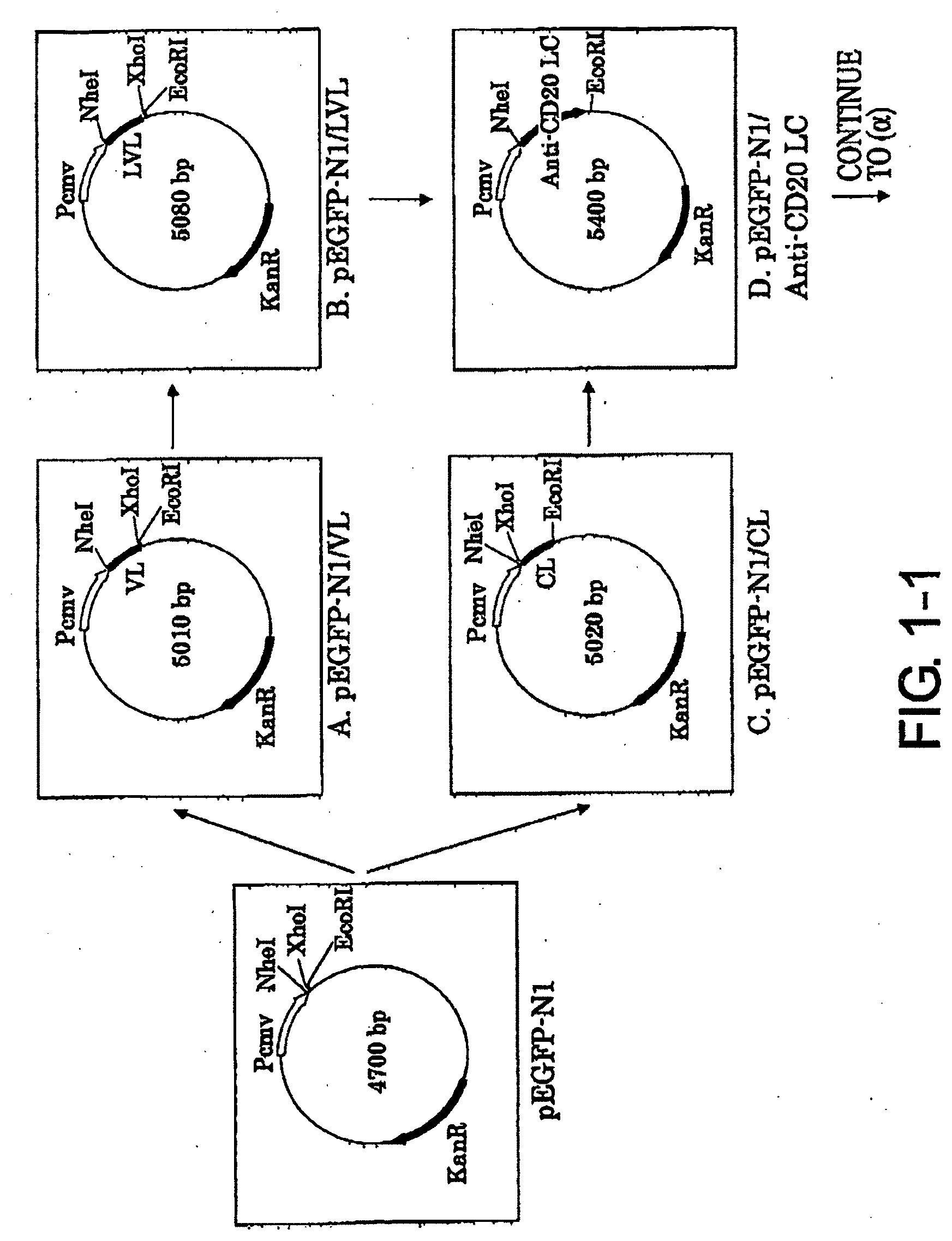
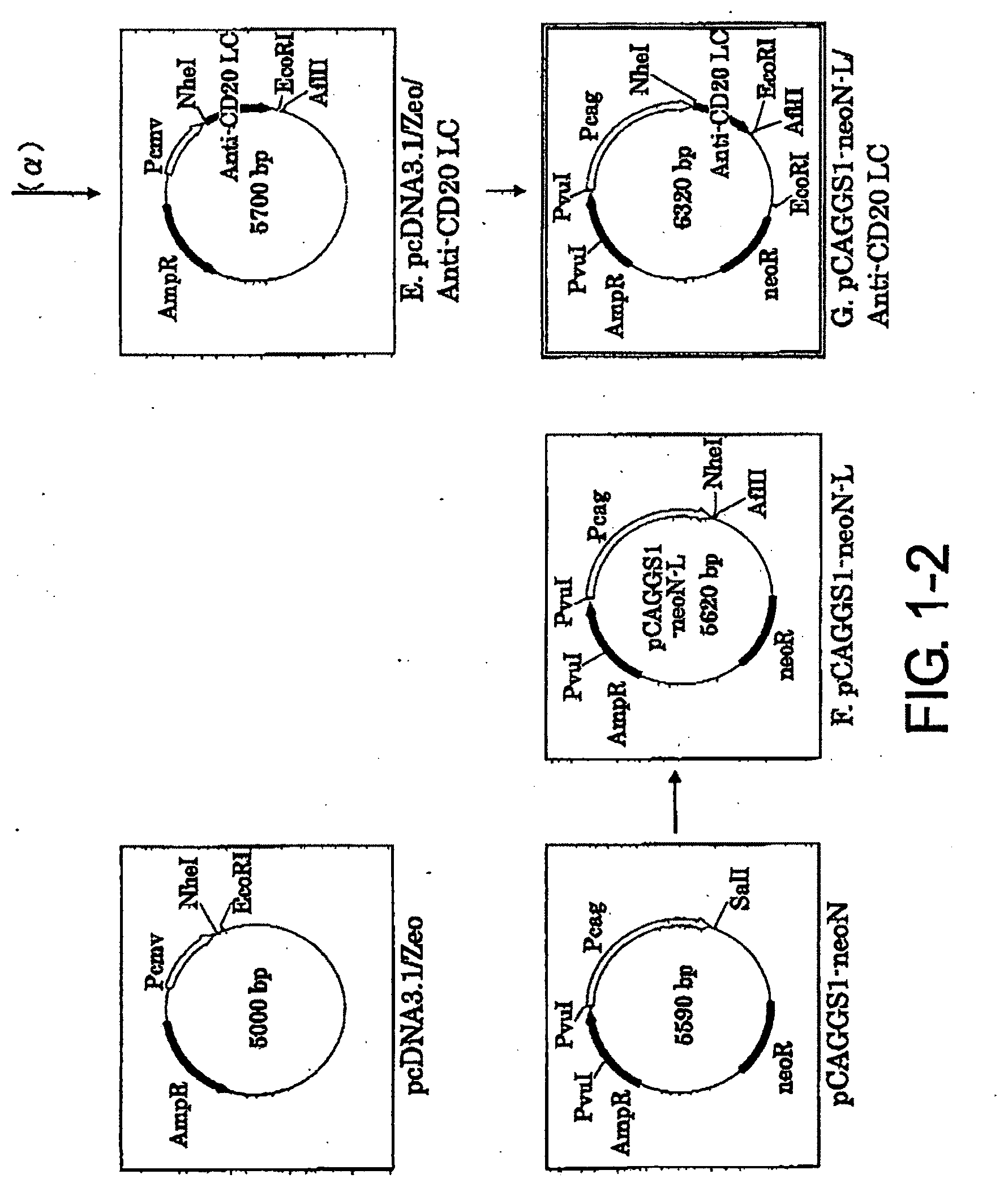
[0035] FIG. 1-1 shows the process for constructing the expression vector pCAGGGS1-neoN-L/Anti-CD20 L Chain described in Example 1.
[0036] FIG. 1-2 is a continuation of FIG. 1-1.
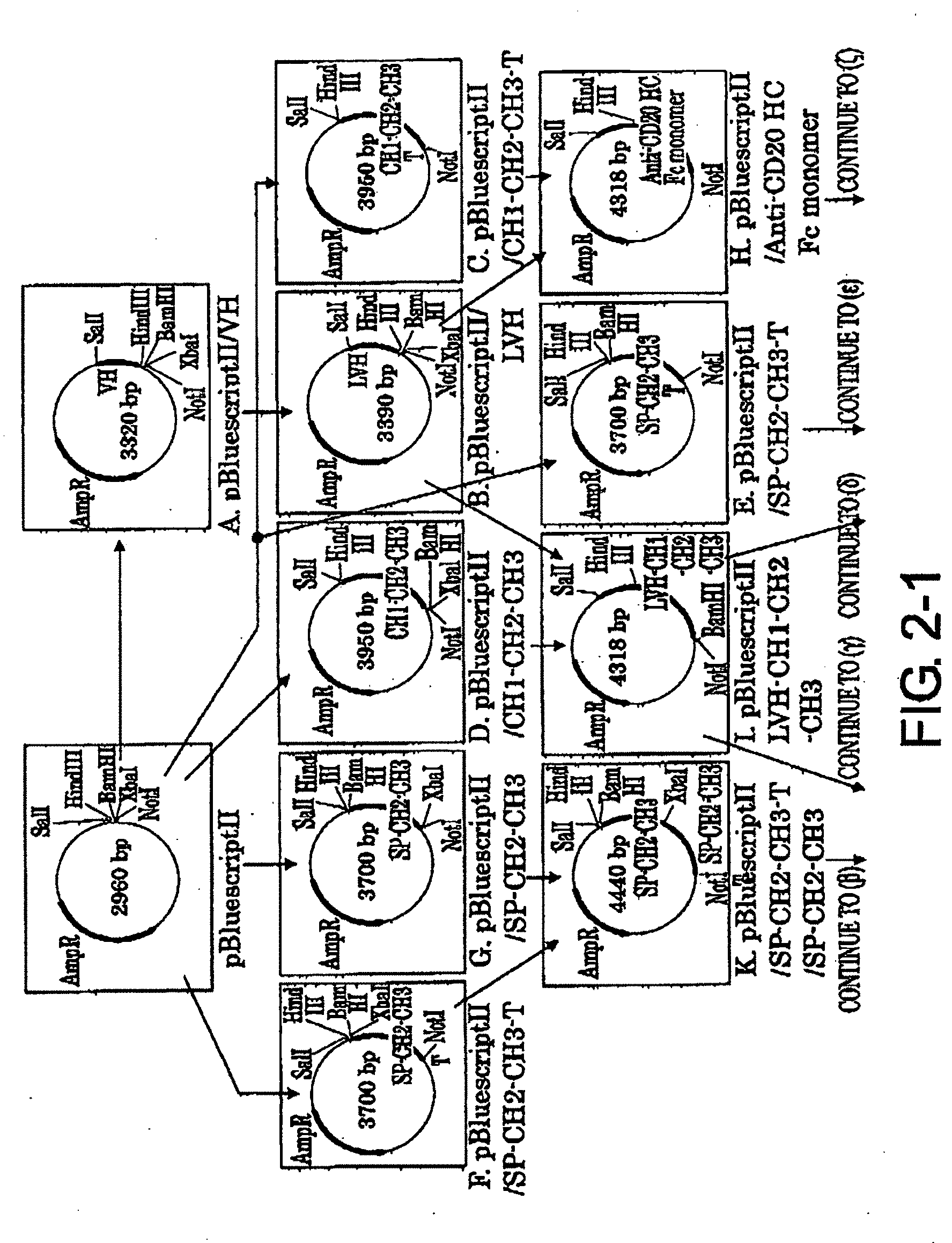
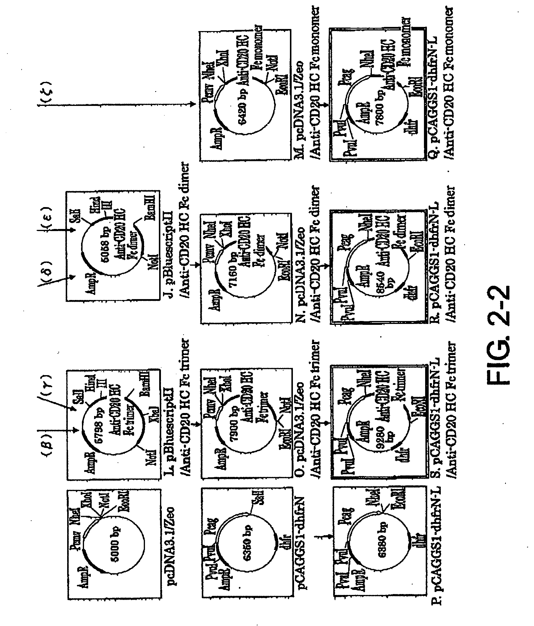
[0037] FIG. 2-1 shows the process for constructing the expression vector pCAGGS1-dhfrN-L/Anti-CD20 H Chain described in Example 2.
[0038] FIG. 2-2 is a continuation of FIG. 2-1.
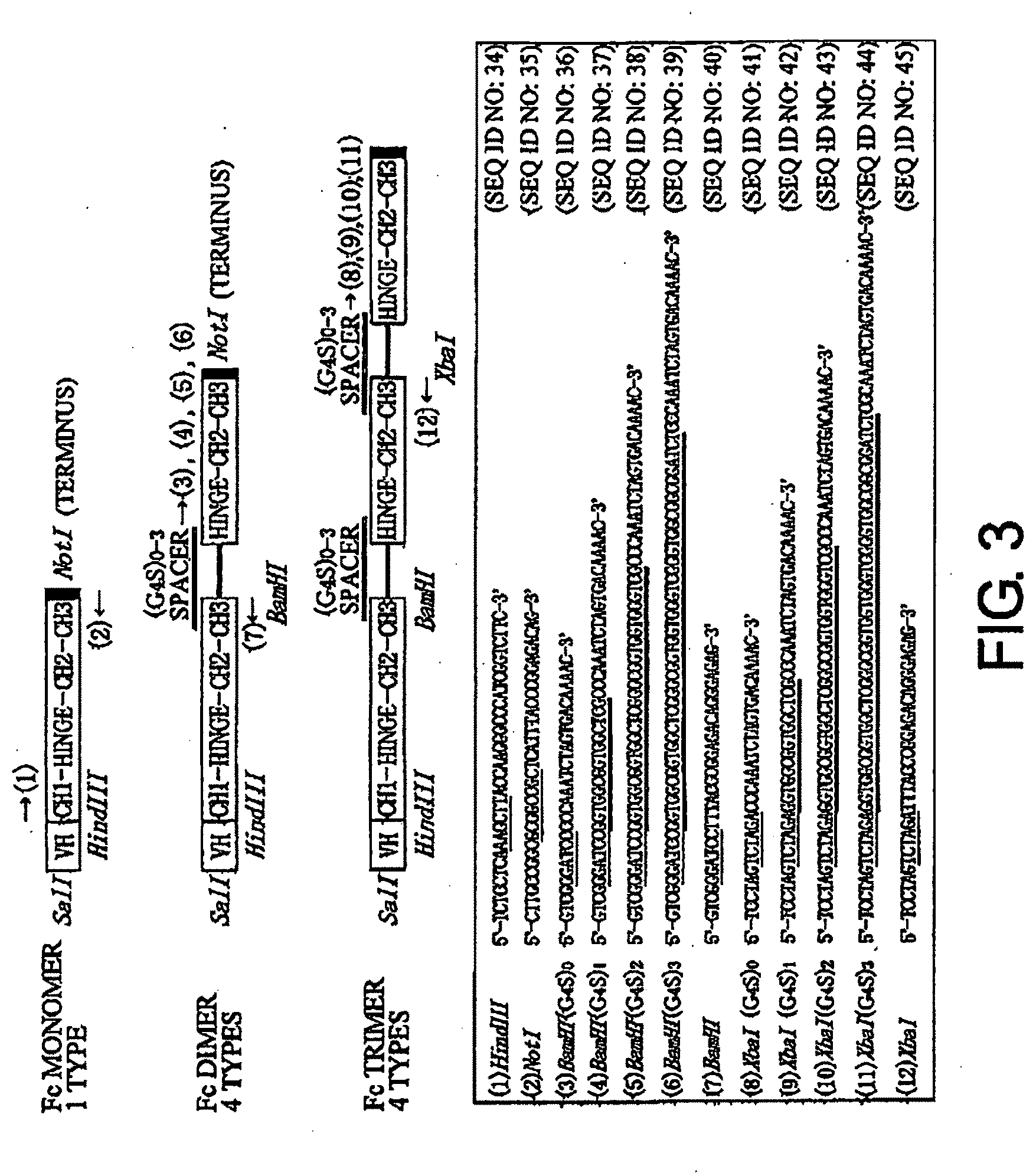
[0039] FIG. 3 shows the gene structure of the final H chain described in Example 2 and the corresponding primers (SEQ ID NOs: 34 to 45). In (1) to (12) of this figure, single underlines indicate restriction enzyme sites and double underlines indicate spacer sequences.
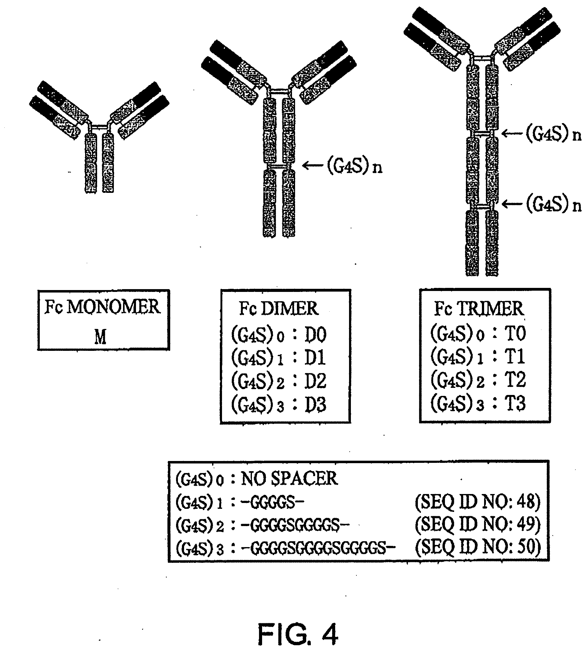
[0040] FIG. 4 is a schematic diagram of the antibodies generated in Example 3.
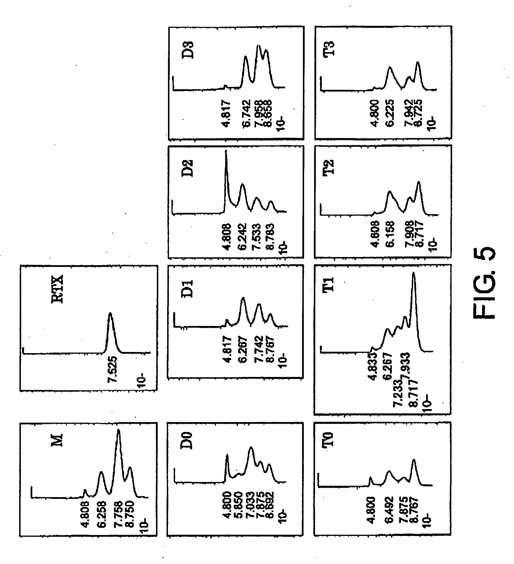
[0041] FIG. 5 shows gel filtration chromatograms after affinity purification with Protein A, described in Example 4. These are chromatograms obtained by gel filtration of M, RTX (Rituximab), D0, D1, D2 D3, T0, T1, T2, and T3 products.
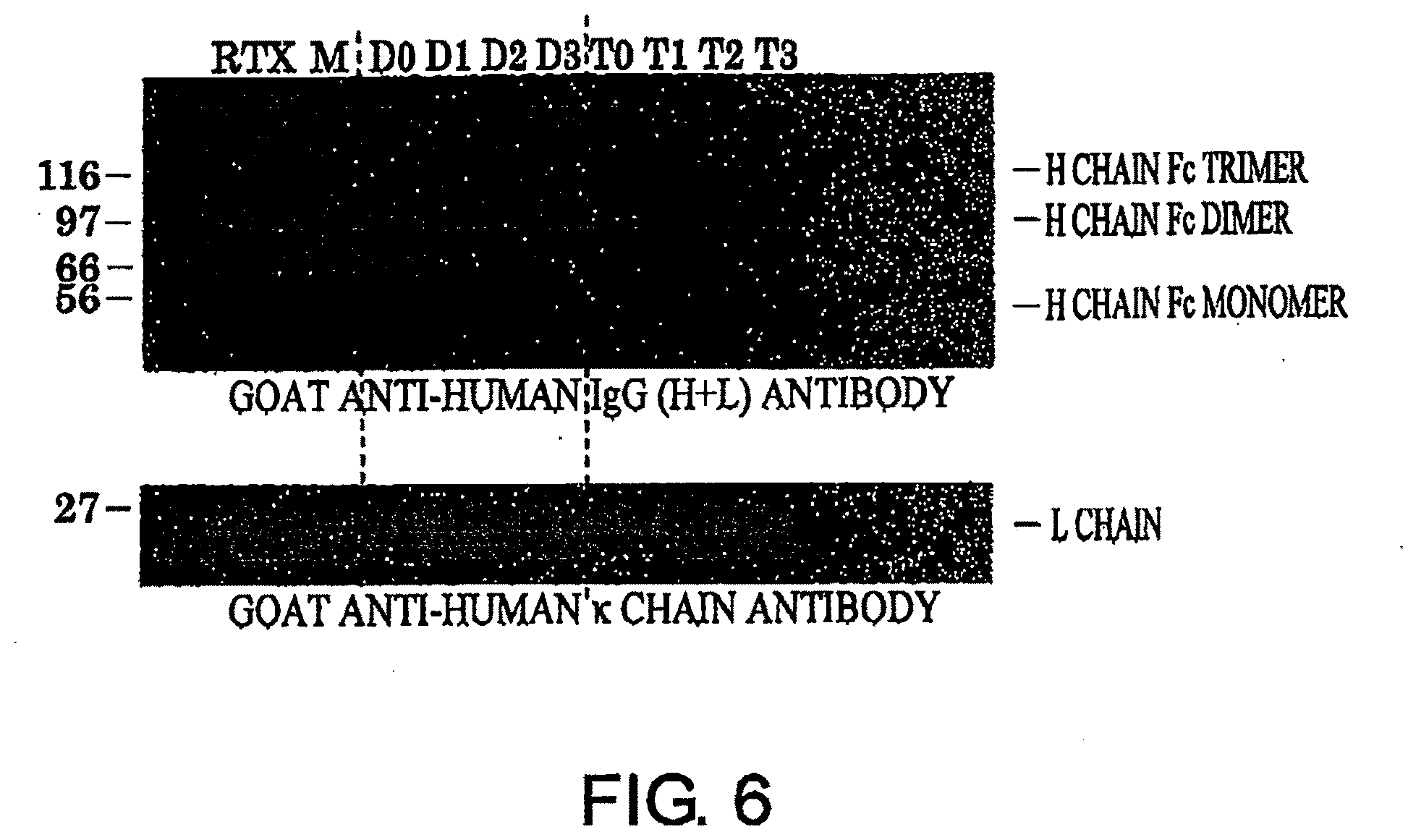
[0042] FIG. 6 shows a result of PAGE analysis of antibodies after gel filtration described in Example 5. The result was obtained by carrying out SDS-PAGE under reducing conditions and Western blotting with horseradish peroxidase-labeled anti-goat antihuman IgG (H+L) antibody or goat anti-human .kappa. chain antibody.
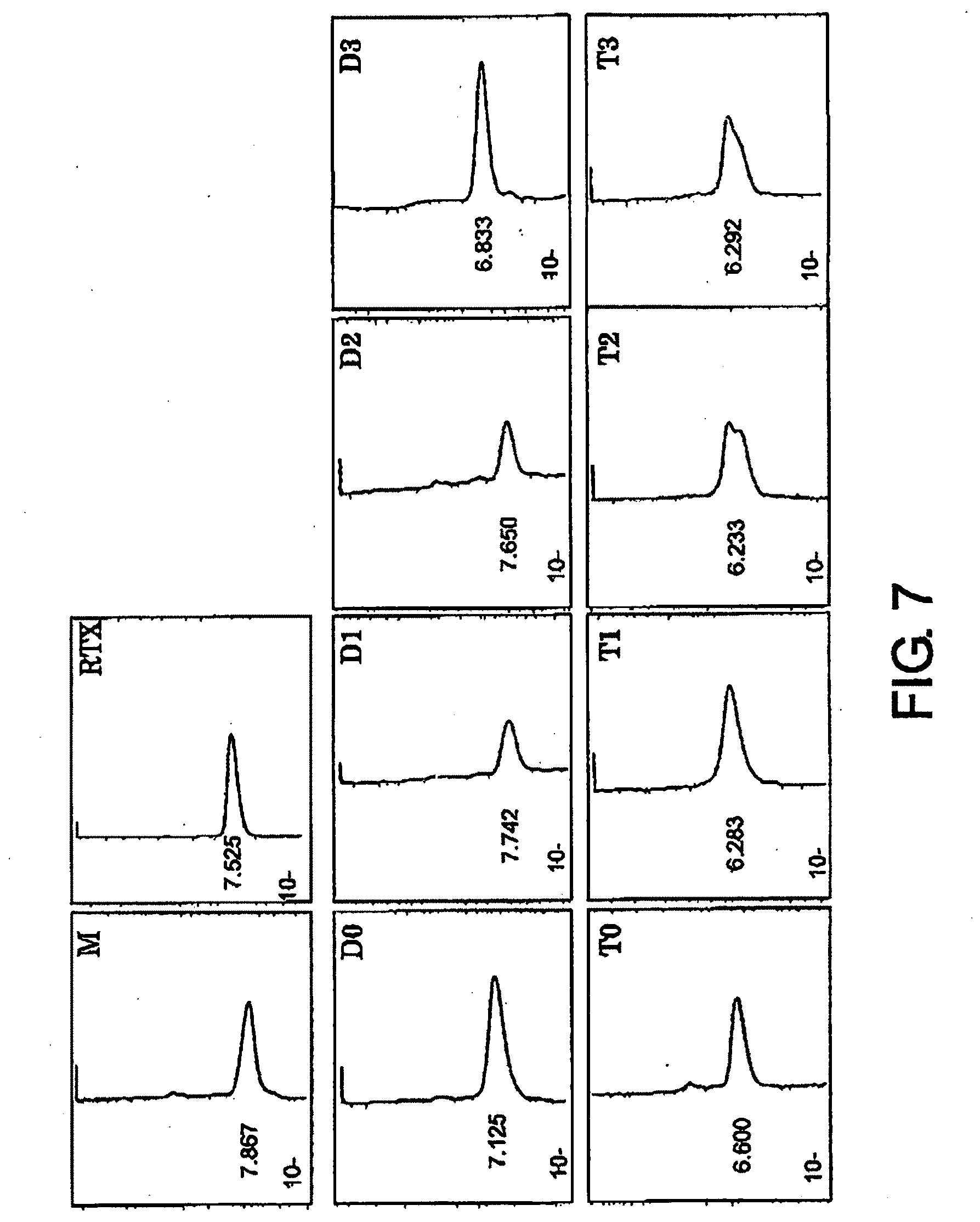
[0043] FIG. 7 shows results of HPLC analysis of antibodies after gel filtration described in Example 5. HPLCs (gel filtrations) of purified M, RTX, D0, D1/D2, D3, T0, T1, T2, and T3 are shown.
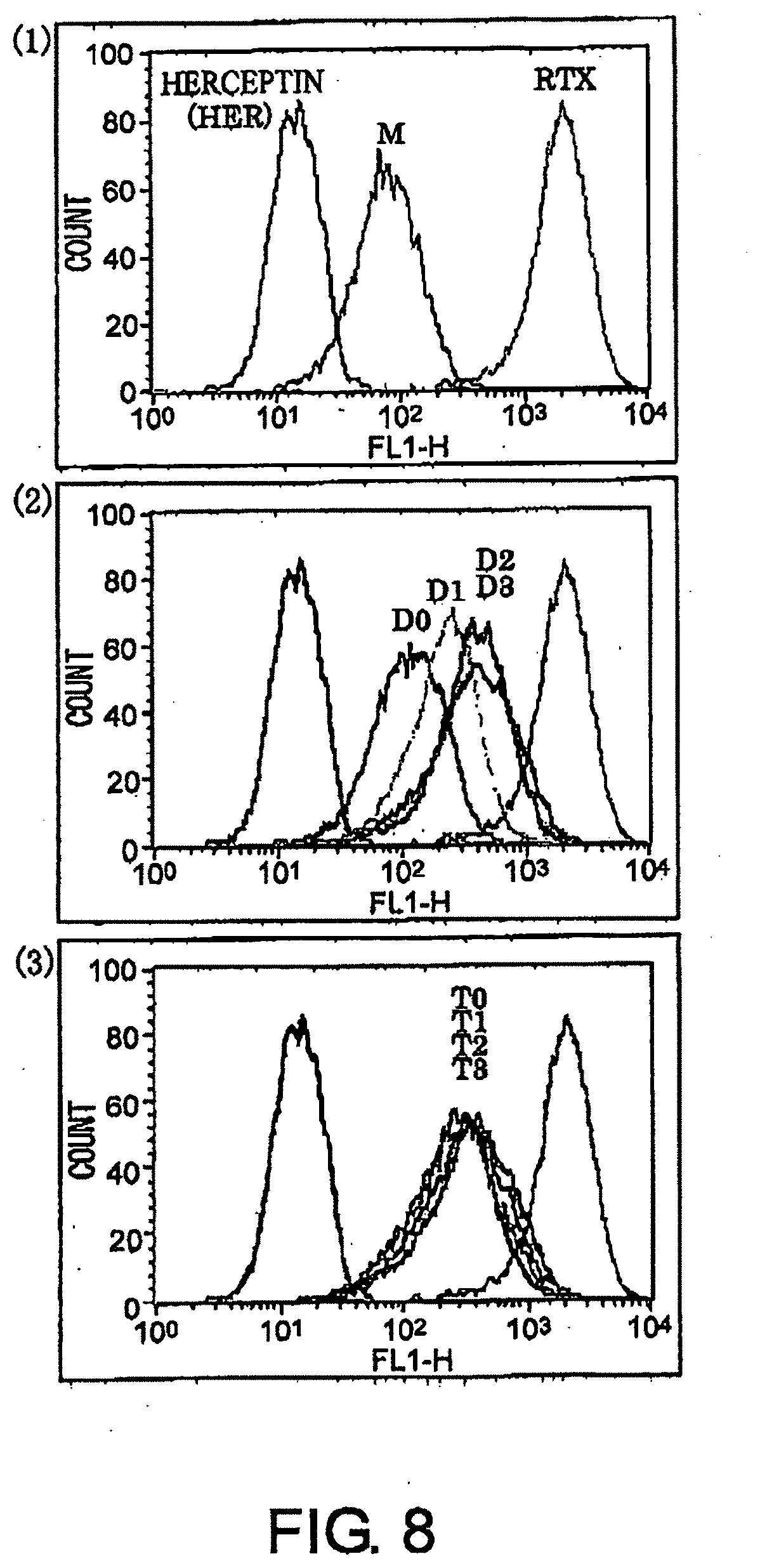
[0044] FIG. 8 shows results of CD20-binding assay of antibodies by flow cytometry described in Example 6. (1): M, negative control trastuzumab, and positive control RTX. (2): D0, D1, D2, and D3, (3): T0, T1, T2, and T3.
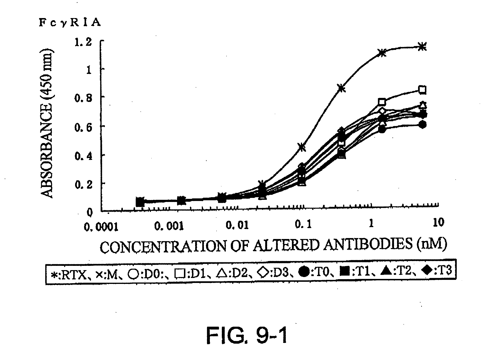
[0045] FIG. 9-1 shows a result of receptor-binding assay by ELISA using recombinant Fc.gamma.R1A described in Example 7.
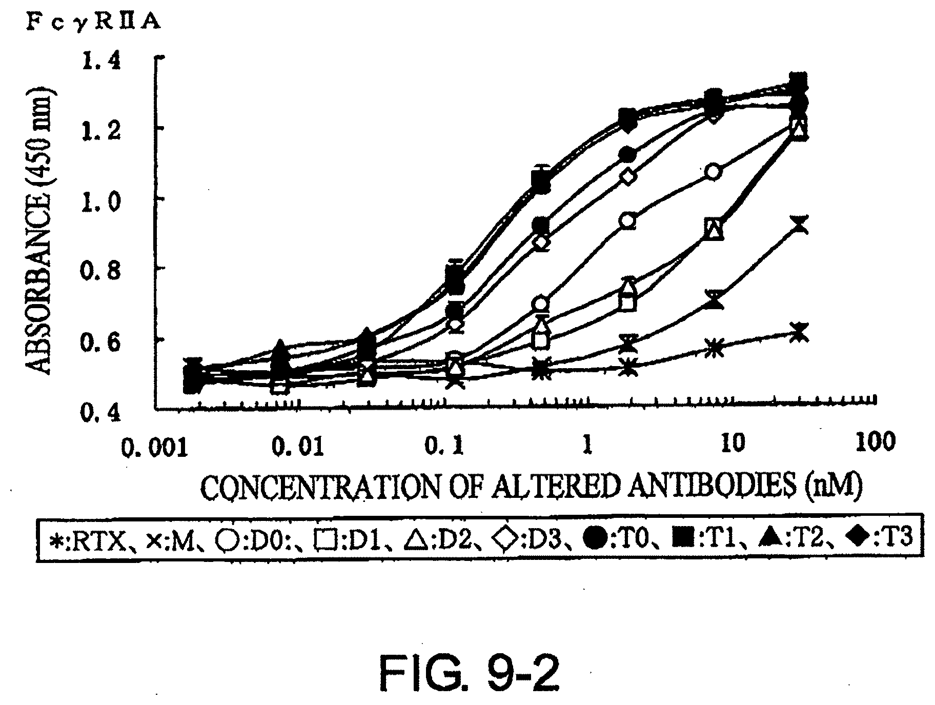
[0046] FIG. 9-2 shows a result of receptor-binding assay by ELISA using recombinant Fc.gamma.R2A described in Example 7.
[0047] FIG. 9-3 shows a result of receptor-binding assay by ELISA using recombinant Fc.gamma.R2B described in Example 7.
[0048] FIG. 9-4 shows a result of receptor-binding assay by ELISA using recombinant Fc.gamma.R3A (Val.sup.158 type) described in Example 7.
[0049] FIG. 9-5 shows a result of receptor-binding assay by ELISA using recombinant Fc.gamma.R3A (Phe.sup.158 type) described in Example 7.
[0050] FIG. 10 shows a result of ADCC activity assay described in Example 8.
[0051] FIG. 11 shows a result of CDC activity assay described in Example 9.
BEST MODE FOR CARRYING OUT THE INVENTION
[0052] As a novel method for enhancing the effector activity of antibodies, the present invention provides a "method for enhancing the effector activity of antibodies, which comprises linking in tandem one or more structures comprising an Fc domain to the C terminus of an antibody heavy chain". The method of the present invention enables one to obtain modified antibodies with enhanced effector activity as compared to the original antibodies (hereinafter also referred to as "modified antibodies of the present invention"), while the affinity of the original antibodies against antigens is maintained.
[0053] The origin of the antibodies of the present invention is not particularly limited. The antibodies may be derived, for example, from any of: primates such as human, monkey, and chimpanzee; rodents such as mouse, rat, and guinea pig; mammals such as rabbit, horse, sheep, donkey, cattle, goat, dog, and cat; or chicken. However, they are preferably derived from human. The antibodies of the present invention may be natural antibodies or antibodies into which some artificial mutations have been introduced. Furthermore, they may be so-called chimeric antibodies or humanized antibodies. The antibodies of the present invention may be immunoglobulins belonging to any class or any subclass. However, they preferably are from the IgG class, and more preferably from the IgG1 subclass.
[0054] The "structure comprising an Fc domain" of the present invention (hereinafter also referred to as the "structure of the present invention") may be the Fc domain itself of an antibody, or an appropriate oligopeptide may be linked as a spacer at the N terminus of the Fc domain.
[0055] In general, the Fc domain of an antibody is a fragment that is obtained after digesting an immunoglobulin molecule with papain. An Fc domain is constituted, from the N terminus of the heavy chain constant region, by the binge region, the CH2 domain, and the CH3 domain. The two heavy chains are linked together via S--S bonds in the binge region. The antibodies can bend in the hinge region. The two heavy chains of IgG1 are linked together via non-covalent bonds in the CH3 domains and disulfide bonds in the hinge regions. Fc domains have sugar chains; however, the sugar chains may contain mutations as long as the Fc domains have the ability to enhance the effector activity when linked to the C terminus of an antibody heavy chain. For example, the antibodies may lack .alpha.1,6-fucose in the sugar chains.
[0056] The origin of the Fc domains in the structure of the present invention may be the same as or different from that of the antibody to which the structure of the present invention is to be linked. From the viewpoint of immunogenicity, however, the origin is preferably the same as that of the antibody to which the structure of the present invention is to be linked, when the antibody is used as an antibody pharmaceutical. For example, when an antibody to which the structure of the present invention is linked is a human-derived antibody or a humanized antibody, the Fc domain in the structure of the present invention is preferably a human Fc domain. Many antibody heavy chain (H chain) sequences have been registered in public databases as genes for H chains of IgG1 including V regions. Examples include GenBank Accession No. BC019337 (a DNA sequence of human constant region). The nucleotide sequence of IgG1 heavy chain (leader sequence-CD20-derived V region (amino acid sequence of Accession No. AAL27650)-CH1-hinge-CH2-CH3) used in the Examples is shown in SEQ ID NO: 3 and the amino acid sequence is shown in SEQ ID NO: 4. The nucleotide sequence encoding human Fc domain corresponds to position 721 to 1413 in SEQ ID NQ: 3. Fc domain cDNAs can be prepared by methods known to those skilled in the art. Fc domain cDNAs can be prepared, for example, by known nucleic acid amplification methods using primers designed based on the sequence from position 721 to 1413 in SEQ ID NO: 3 and, as template, mRNAs prepared from antibody-expressing cells. Alternatively, they may be prepared by using as a probe a portion of the sequence of SEQ ID NO: 3 and selecting sequences that hybridize to the probe from a cDNA library prepared from antibody-expressing cells.
[0057] Furthermore, the Fc domains in the structure of the present invention may comprise spontaneous or artificial mutations as long as they have the Fc receptor-binding activity. For example, polypeptides encoded by sequences that hybridize under stringent conditions to the complementary strand of the nucleotide sequence from position 721 to 1413 in SEQ ID NO: 3, and polypeptides comprising an amino acid sequence with a substitution, deletion, addition, and/or insertion of one or more amino acids in the sequence from position 241 to 471 in the amino acid sequence of SEQ ID NO: 4 are also included in the Fc domain of the structures of the present invention, as long as they have Fc receptor-binding activity. Such Fc domain variants can also be prepared by methods known to those skilled in the art.
[0058] Those skilled in the art can appropriately select the above stringent hybridization conditions. For example, pre-hybridization is carried out in a hybridization solution containing 25% formamide, or 50% formamide under more stringent conditions, and 4.times.SSC, 50 nM Hepes (pH7.0), 10.times.Denhardt's solution, and 20 .mu.g/ml denatured salmon sperm DNA at 42.degree. C. overnight. Labeled probes are then added and hybridization is carried out by incubation at 42.degree. C. overnight. Post-hybridization washes are carried out at different levels of stringency, including the moderately stringent "1.times.SSC, 0.1% SDS, 37.degree. C.", highly stringent "0.5.times.SSC, 0.1% SDS, 42.degree. C.", and more highly stringent "0.2.times.SSC, 0.1% SDS, 65.degree. C." conditions. As the stringency of the post-hybridization washes increases, polynucleotides with greater homology to the probe sequence are expected to be isolated. The above-described combinations of SSC. SDS, and temperature conditions are mere examples. Those skilled in the art can achieve the same stringencies as those described above by appropriately combining the above factors or others (such as probe concentration, probe length, or hybridization period) that affect hybridization stringency.
[0059] Polypeptides encoded by polynucleotides isolated using such hybridization techniques will usually comprise amino acid sequences with high homology to the Fc domains described above. "High homology" refers to sequence homology of at least 40% or more, preferably 60% or more, further preferably 80% or more, further preferably 90% or more, further preferably at least 95% or more, and further preferably at least 97% or more (for example, 98% to 99%). Amino acid sequence identity can be determined, for example, using the BLAST algorithm of Karlin and Altschul (Proc. Natl. Acad. Sci. USA (1990) 87, 2264-2268; Proc. Natl. Acad. Sci. USA (1993) 90, 5873-5877). A program called BLASTX has been developed based on this algorithm (Altsahul et al., J. Mol. Biol. (1990) 215, 403-410). When using BLASTX to analyze amino acid sequences, the parameters are, for example, a score of 50 and a word length of 3. When using the BLAST and Gapped BLAST programs, the default parameters for each program are used. Specific methodology for these analysis methods is well known (http://www.ncbi.nlm.nib.gov).
[0060] Furthermore, techniques for artificially preparing Fc domain variants by artificially introducing mutations into Fc domains are also known to those skilled in the art. Such Fc domain variants can be artificially prepared, for example, by introducing site-specific or random mutations into the nucleotide sequence of SEQ ID NO: 3 by genetic modification methods, such as PCR-based mutagenesis or cassette mutagenesis. Alternatively, sequences with mutations introduced into the nucleotide sequence of SEQ ID NO: 3 can be synthesized using commercially available nucleic acid synthesizers.
[0061] The structures of the present invention may not have any spacer oligopeptide. However, the structures preferably contain such oligopeptides. Combinations of glycine and serine are often used as the spacer (Journal of Immunology, 162, 6589 (1999)). As described in the Examples, a spacer having a combination of four glycines and one serine (SEQ ID NO: 48), or a spacer in which the above sequence is linked twice (SEQ ID NO: 49) or three times (SEQ ID NO: 50) can be used as the spacer of the present invention. However, the spacer is not limited to these sequences. The spacer may have any structure as long as it allows bending of the hinge region where the spacer is linked. Preferably, a spacer is a peptide sequence that is not readily cleaved by proteases or peptidases. Regarding such sequences, a desired peptide sequence can be obtained, for example, by entering various conditions such as sequence length into LINKER (Xue F, Gu Z, and Feng J A., LINKER: a web server for generating peptide sequences with extended conformation, Nucleic Acids Res. 2004 Jul. 1; 32 (Web Server issue): W562-5), a program that assists designing of linker sequences. LINKER can be accessed at http://astro.temple.edu/.about.feng/Servers/BioinformaticServer.htm.
[0062] When multiple nucleotide sequences are linked together by genetic engineering techniques, one to several residues in the amino acid sequence at the junction are often substituted, deleted, added, and/or inserted, for example, because of the sequences of the restriction enzyme sites. Such mutations are known to those skilled in the art. Such mutations may also occur when the structures of the present invention are constructed or when they are linked to antibodies. Such mutations may occur, for example, at the junctions between the V and C regions, and the junctions between the C terminus of Fc and the N terminus of the structures of the present invention (the N terminus of Fc or spacer). Even with such mutations, they are included in the structures or modified antibodies of the present invention as long as they have an Fc receptor-binding activity.
[0063] The structures of the present invention can enhance the effector activity of an antibody when linked to the C terminus of the antibody heavy chain. An arbitrary number of structures of the present invention, for example, one, two, three, four, or five structures, may be linked; however, one or two structures are preferably linked, Therefore, modified antibodies onto which the structures of the present invention have been linked can comprise two or more arbitrary Fc domains, and the number of Fc domains in a modified antibody is two or three. In the Examples, modified antibodies into which two structures of the present invention have been linked were confirmed to show a stronger ADCC activity than modified antibodies into which one structure of the present invention has been linked.
[0064] There is no limitation on the type of antigen that is recognized by an antibody to which the structures of the present invention are to be linked. The antigen may be any antigen. Specifically, the variable region of a modified antibody of the present invention may recognize any antigen. The H chain and L chain variable regions of the modified antibodies used in the Examples described below are the variable regions of 1F5, which is a mouse monoclonal antibody against CD20, a differentiation antigen of human B lymphocytes (Non-Patent Document 14). CD20 is a protein of 297 amino acids, and its molecular weight is 33 to 37 kDa. CD20 is highly expressed in B lymphocytes. Rituximab, a chimeric antibody against CD20, is widely used as an effective therapeutic agent for Non-Hodgkin's lymphoma (Non-Patent Document 15). Known anti-CD20 antibodies include mouse monoclonal antibodies B1 and 2H7, in addition to rituximab and 1F5 (Non-Patent Document 16). However, the variable regions of the modified antibodies of the present invention are not limited to the variable regions of 1F5. The Fc domains are physically distant from the Fab domains; therefore, it is thought that the Fab domain type has almost no influence on the Fc domain activity. Thus, the variable regions of any antibody other than 1F5 may be used as the variable regions of the modified antibodies of the present invention, and the variable regions of antibodies directed to any antigen other than CD20 may be used as the variable regions of the modified antibodies of the present invention.
[0065] Regardless of the antigen-binding activity, the methods of the present invention can increase the therapeutic effect of an antibody by enhancing the effector activity exhibited by the antibody Fc domain. The enhancement of the binding activity to Fc receptors is required to increase the effector activity, in particular the ADCC activity, of an antibody. In general, the intensity of binding between two molecules is considered to be as follows. The binding intensity between one antibody molecule and one Fc receptor molecule is represented by: affinity.times.binding valency=avidity. Natural IgG antibodies have one Fc; thus, the binding valency is 1 even when there are many Fc receptors on the surface of effector cells. However, when antibodies and antigens form complexes, the immune complexes bind to the effector cells in a multivalent manner. The binding valency varies depending on the structure of immune complexes. Cancer cells have many antigens on the cell surface; thus, antibodies bind to these antigens and bind to Fc receptors on the effector cells in a multivalent manner. However, the density of antigens on cancer cells is often low; thus, antibodies bound to the antigens bind with lower valency to Fc receptors. For this reason, ADCC activity cannot be sufficiently exerted and the therapeutic effect is also insufficient (Golay, J. et al., Blood, 95, 3900, (2000)). However, by tandemly linking multiple Fc domains to an antibody molecule, binding to Fc receptors in a multivalent manner is possible. This enhances the binding activity between an antibody and Fc receptors, i.e. the avidity. In addition, the effector activity is enhanced.
[0066] The present invention also provides methods for producing modified antibodies with enhanced effector activity. The methods of the present invention not only enhance the effector activity of naturally obtained antibodies and existing chimeric antibodies, but also enable the production of novel altered chimeric antibodies from novel combination of antibody variable and constant regions of different origins. Furthermore, the modified antibodies may also comprise a novel constant region from the combination of CH1 domain and two or more Fc domain variants described above. The nucleotide sequence of human CH1 domain is shown under positions 430 to 720 in the heavy chain nucleotide sequence of SEQ ID NO: 3.
[0067] The methods of the present invention can be conducted using an appropriate combination of methods known to those skilled in the art. An example of expression of the modified antibodies of the present invention is described below, in which DNAs for the heavy chain (H chain) and light chain (L chain) variable and constant regions are prepared and linked using genetic engineering techniques.
[0068] The variable region sequences can be prepared, for example, by the following procedure. First, a cDNA library is generated from hybridomas expressing the antibody of interest or cells introduced with the antibody gene, and DNA for the variable region of interest is cloned. An antibody leader sequence L is linked upstream of the H chain variable region VH and L chain variable region VL to construct the DNA structures [LVH] and [LVL].
[0069] For the antibody constant region, first, a cDNA library is generated from human myeloma cells or human lymphatic tissues such as tonsil. cDNA fragments for the H chain constant region [CH1-Fc] and for the L chain constant region [CL] are obtained by amplification by known nucleic acid amplification methods such as polymerase chain reaction (PCR) using primers designed based on partial sequences of the 5' and 3' ends of H chain and L chain constant regions. The fragments are then inserted into vectors and cloned.
[0070] For the L chain, the DNA structure [LVL-CL] is constructed in which LVL and CL are linked. As an example, the DNA sequence of the DNA structure [LVL-CL] (leader sequence, V region of 1F5, and C region of human L.kappa. chain) prepared in the Examples is shown in SEQ ID NO: 1, while the amino acid sequence encoded by the DNA structure is shown in SEQ ID NO: 2. The H chain linked with a structure of the present invention (altered H chain) and the H chain without a structure of the present invention linked are constructed by the procedure described below. (i) The DNA structure of H chain having a single Fc (H chain without a structure of the present invention linked) can be constructed by linking together the DNA structure [LVH] and DNA structure [CH1-Fc domain-stop codon]. The DNA sequence of the DNA structure for the H chain with a single Fc prepared in the Examples is shown in SEQ ID NO: 3, while the amino acid sequence encoded by the DNA structure is shown in SEQ ID NO: 4. (ii) The DNA structure of H chain having two Fc linked in tandem (H chain linked with one structure of the present invention) can be constructed by linking together the DNA structure [LVH], DNA structure [CH1-Fc (without stop codon)], and DNA structure [spacer-Fc-stop codon]. The DNA sequences of the DNA structures for the H chain having two Fc linked in tandem prepared in the Examples are shown in SEQ ID NOs: 5 (with no spacer), 7 (with a single spacer: GGGGS (represented as G4S; SEQ ID NO: 48)), 9 (with two G4S as spacer), and 11 (with three G4S as spacer). The amino acid sequences encoded by these DNA structures are shown in SEQ ID NOs: 6 (with no spacer), 8 (with one G4S as spacer), 10 (with two G4S as spacer), and 12 (with three G4S as spacer). (iii) The DNA structure of H chain having three Fcs linked in tandem (H chain linked with two structures of the present invention) can be constructed by linking together the DNA structure [LVH]. DNA structure [CH1-Fc (without stop codon)], DNA structure [spacer-Fc- (without stop codon)], and DNA structure [spacer-Fc-stop codon]. The DNA sequences of the DNA structures for the H chain having three Fcs linked in tandem prepared in the Examples are shown in SEQ ID NOs: 13 (with no spacer), 15 (with one G4S as spacer), 17 (with two G48 as spacer), and 19 (with three G4S as spacer). The amino acid sequences encoded by these DNA structures are shown in SEQ ID NOs: 14 (with no spacer), 16 (with one G4S as spacer), 18 (with two G4S as spacer), and 20 (with three G4S as spacer). (iv) The DNA constructs of H chain having four or more Fc linked in tandem can be prepared similarly as in the case with three Fcs, by increasing the number of the DNA structure [spacer-Fc-(without stop codon)].
[0071] The L chain DNA structure and altered H chain DNA structure prepared as described above are cloned, and then, together with regulatory regions such as promoter and enhancer, inserted into expression vectors. Alternatively, they may be inserted into expression vectors that already have regulatory regions. Expression vectors that can be used include vectors having the CAG promoter (Gene, 108, 193 (1991)) and pcDNA vector (Immunology and Cell Biology, 75, 515 (1997)). Any expression vectors may be used as long as they are compatible with host cells to be used.
[0072] Host cells can be appropriately selected from those that can express glycoproteins. Such host cells can be selected, for example, from animal calls, insect cells, yeast, and the like. Specific examples include CH-DG44 cells (Cytotechnology, 9, 237 (1992)), COS-1 cells, COS-7 cells, mouse myeloma NS0 cells, and rat myaloma YB2/0 cells which can produce antibody molecules having sugar chains lacking fucose, but the cells are not limited thereto.
[0073] The recombinant host cells are cultured and modified antibodies are purified from the culture supernatants. Various types of culture media can be used for culture, however, serum-free media are convenient for purifying antibodies. Modified antibodies of interest are purified from the culture supernatants by removing fragments and aggregates of the modified antibodies, and protein other than the modified antibodies with known purification methods, such as ion exchange chromatography, hydrophobic chromatography, gel filtration chromatography, affinity chromatography with immobilized Protein A having selective binding activity to antibodies or the like, and high performance liquid chromatography (HPLC).
[0074] Whether the modified antibodies obtained as described above have enhanced effector activity can be assessed by methods known to those skilled in the art. The binding activity to various Fc receptors can be determined, for example, by enzyme antibody techniques using the extracellular domains of recombinant Fc receptors. A specific example is described below. First, the extracellular domains of Fc.gamma.RIA, Fc.gamma.RIIA, Fc.gamma.RIIB, and Fc.gamma.RIIIA, are produced as receptors for human IgG. The sequences of these receptors are known, and are available from GenBank under the following Accession Nos: human Fc.gamma.RIA: NM_000566, human Fc.gamma.RIIA: NM_021642, human Fc.gamma.RIIB: NM_001002273, human Fc.gamma.RIIIA: NM_000569. These receptors are immobilized onto 96-wall plates for enzyme antibody techniques. Modified antibodies with varied concentrations are reacted, and labeled anti-human IgG antibodies or such are reacted as a secondary antibody. The amounts of modified antibodies bound to the receptors are measured based on the signal from the label. There are known genetic variants of Fc.gamma.RIIIA (Journal of Clinical Investigation, 100, 1059 (1997)), and the receptors in which the amino acid at position 158 is valine or phenylalanine are used in the Examples herein.
[0075] The ADCC activity of modified antibodies can be measured using effector and target cells. For example, monocytes separated from peripheral blood of healthy individuals can be used as the effector cells. Calls expressing the CD20 antigen, such as Ramos cells and Raji cells, can be used as the target cells. After the target cells are reacted with serially diluted modified antibodies, the effector cells are added. The ratio of the effector to target cell numbers can be in a range of 10:1 to 100:1, and the ratio is preferably 25:1. When the target cells are damaged by the APCC activity of the modified antibodies, lactate dehydrogenase (LDH) in the cells is released into the culture supernatant. Therefore, the ADCC activity can be determined by collecting the released LDH and measuring its enzymatic activity.
[0076] As for the CDC activity, the cytotoxic activity can be assessed, for example, by reacting the target cells with serially diluted modified antibodies and then adding fresh baby rabbit serum as a source of complements, a described in the Examples. Since serum containing LDH is used, the cytotoxic activity is assessed by measuring the viable cell number using Alamar Blue or such methods.
[0077] Modified antibodies obtained by the methods of the present invention not only enhance the in vitro effector activity described above but also exert the cellular immunity-enhancing effect in vivo based on enhanced effector activity. Thus, the antibodies are thought to contribute to the treatment of diseases that can be expected to be improved by cellular immunity. The modified antibodies of the present invention can be administered in an appropriate dosage form via an appropriate administration route, depending on the type of disease, patient's age, symptoms, and such. Furthermore, the modified antibodies of the present invention can be formulated by known formulation methods and supplied as pharmaceuticals along with instructions indicating the efficacy and effects, cautions for use, and so on. When formulating, appropriate additives, such as pharmaceutically acceptable excipients, stabilizers, preservatives, buffers, suspending agents, emulsifiers, and solubilizing agents can be appropriately added depending on the purpose, such as securing their properties and quality. For example, the antibodies can be combined with Polysorbate 80, sodium chloride, sodium citrate, anhydrous citric acid, or such when formulating as injections, prepared with physiological saline or glucose solution injection at the time of use, and administered by intravenous drip infusion or such method. The dose can be adjusted depending on the patient's age and weight, and such factors. A single dose in such intravenous drip infusion is, for example, 10 to 10000 mg/m.sup.2, preferably 50 to 5000 mg/m.sup.2, and more preferably 100 to 1000 mg/m.sup.2, but is not limited thereto.
[0078] All prior art references cited herein are incorporated by reference into this description.
EXAMPLES
[0079] Hereinbelow, the present invention is specifically described in the context of Examples; however, it is not to be construed as being limited thereto.
[Example 1] Construction of Expression Vector pCAGGS1-neoN-L/Anti-CD20 LC (Light Chain)
[0080] 1-1. Preparation of pEGFP-N1/VL vector (FIG. 1-1-A)
[0081] The mouse anti-CD20 IgG2a VL region gene was cloned by the following procedure. The mouse hybridoma 1F5 was cultured using RPMI1640 containing 10% inactivated fetal bovine serum, 100 U/ml penicillin, and 100 .mu.g/ml streptomycin (Sigma Aldrich) at 37.degree. C. under 5% CO.sub.2, and then total RNA was extracted from the cells using ISOGEN (NIPPON GENE CO.). 10 pmol of oligo dT primer (5'-CGAGCTCGAGCGGCCGCTTTTTTTTTTTTTTTTTTT-3' (SEQ ID NO: 21)) was added to 10 .mu.g of the total RNA, and the total volume was adjusted to 12 .mu.l by adding diethylpyrocarbonate (DEPC)-treated water. After two minutes of incubation at 72.degree. C. to destroy its higher order structure, the RNA was quickly transferred onto ice and incubated for three minutes. The RNA was added with 2 .mu.l of the appended 10.times. Reaction Buffer (Wako Pure Chemical Industries, Ltd.), 1 .mu.l of 100 mM DTT (Wako Pure Chemical Industries, Ltd.), 1 .mu.l of 20 mM dNTP (Wako Pure Chemical Industries, Ltd.), and 1 .mu.l of 20 U/.mu.l RNase Inhibitor (Wako Pure Chemical Industries, Ltd.). The total volume was adjusted to 19 .mu.l with DEPC-treated water. After heating up to 42.degree. C., 1 .mu.l of 200 U/.mu.l ReverscriptII (Wako Pure Chemical Industries, Ltd.) was added and the resulting mixture was incubated at 42.degree. C. for 50 minutes without further treatment. After reaction, 80 .mu.l of TE (1 mM EDTA, 10 mM Tris-HCl (pH 8.0)) was added. The resulting 100-.mu.l mixture was used as a cDNA solution.
[0082] 7.8 .mu.l of sterile MilliQ water, 4 .mu.l of the appended 5.times. Buffer, 2 .mu.l of 2.5 mM dNTP, 2 .mu.l of 10 .mu.M forward primer (5'-TCGTCTAGGCTAGCATTGTTCTCTCCCAGTCTCCA-3' (SEQ ID NO: 22) having an NheI site (underlined)), 2 .mu.l of 10 .mu.M reverse primer (5'-GCTTGAGACTCGAGCAGCTTGGTCCCAGCAC CGAA-3' (SEQ ID NO: 23) having an XhoI site (underlined)), 2 .mu.l of 1F5-derived cDNA as a template, and 0.2 .mu.l of 5 U/.mu.l Expand High Fidelity.sup.PLUS PCR system (Roche) were combined together on ice. PCR was carried out under the following conditions: heat treatment at 95.degree. C. for ten minutes, followed by 30 cycles of 95.degree. C. for 30 seconds, 60.degree. C. for 30 seconds, and 72.degree. C. for 60 seconds. The reaction solution was subjected to electrophoresis using 1% agarose STANDARD 01 (Solana) gel and a band of about 0.31 kbp was collected using RECOCHIP (TaKaRa Bio Inc.). The DNA fragment was purified by phenol/chloroform extraction and isopropyl alcohol precipitation. The DNA fragment was treated with NheI (TOYOBO) and XhoI (TaKaRa Bio Inc.) at the final concentrations of 0.8 U/.mu.l and 0.5 U/.mu.l, respectively, and ligated with 50 ng of pEGFP-N1 (BD Biosciences) treated in the same way. The ligation was carried out using 1.5 U of T4 DNA ligase (Promega) at room temperature for 30 minutes. 100 .mu.l of competent cells of E. coli DH5.alpha., which had been prepared by the potassium chloride method, was added to the reaction mixture. After reacting for 30 minutes on ice, the cells were heat-shocked at 42.degree. C. for 45 seconds. This was then rested for two minutes on ice, after which 1 ml of SOC medium (2% Bacto tryptone (BD Biosciences), 0.5% Bacto yeast extract (BD Biosciences), and 1% sodium chloride (Wako Pure Chemical Industries, Ltd.)) was added. The resulting mixture was transferred into a test tube and the bacteria were cultured with shaking at 37.degree. C. for two hours. After shaking, 100 .mu.l of the bacterial suspension was plated onto an LB medium plate containing 100 .mu.g/ml of kanamycin (Wako Pure Chemical Industries, Ltd.). The plate was incubated at 37.degree. C. overnight. From the formed colonies, those into which the VL region gene has been inserted were selected, and the vector was named pEGFP-N1/VL.
1-2. Preparation of pEGFP-N1/LVL Vector (FIG. 1-1-B)
[0083] A leader sequence was added to the VL gene by the following procedure. 4 .mu.l of the appended 5.times. Buffer, 2 .mu.l of 2.5 mM dNTP, 2 .mu.l of 10 .mu.M forward primer (5'-GAGTTT GCTAGCGCCGCCATGGATTTTCAAGTGCAGATTTTCAGCTTCCTGCTAATCAGTGCTT CAGTCATAATGTCCAGAGGCAAATTGTTCTCTCCCAGTCCAGCA-3' (SEQ ID NO: 24) having an NheI site (underlined); the leader sequence corresponds to positions 13 to 57 in SEQ ID NO: 24), 2 .mu.l of 10 .mu.M reverse primer (5'-GCTTGAGACTCGAGCAGCTTGGTCCCAGCACCGAA-3' (SEQ ID NO: 25) having an XhoI site (underlined)), 100 ng of pEGFP-N1/VL as a template, which had been prepared as described in (1), and 0.2 .mu.l of 5 U/.mu.l Expand High Fidelity.sup.PLUS PCR system were combined together on ice. The total volume was adjusted to 20 .mu.l by adding sterile MilliQ water. PCR was carried out under the following conditions: heat treatment at 95.degree. C. for ten minutes, followed by 30 cycles of 95.degree. C. for 30 seconds, 60.degree. C. for 30 seconds, and 72.degree. C. for 60 seconds. The reaction solution was subjected to electrophoresis using 1% agarose STANDARD 01 gel. A band of about 0.38 kbp was recollected using RECOCHIP. The DNA fragment was purified by phenol/chloroform extraction and isopropyl alcohol precipitation.
[0084] The DNA fragment was treated with NheI and XhoI at final concentrations of 0.8 and 0.5 U/.mu.l, respectively, and ligated with 50 ng of pEGFP-N1 treated in the same way. The ligation was carried out using 1.5 U of T4 DNA ligase at room temperature for 30 minutes. 100 .mu.l of competent cells of E. coli DH5.alpha. was added to the reaction mixture. After reacting for 30 minutes on ice, the cells were heat-shocked at 42.degree. C. for 45 seconds. This was then rested for two minutes on ice, after which 1 ml of SOC medium was added. The resulting mixture was transferred into a test tube and the bacteria were cultured with shaking at 37.degree. C. for two hours. After shaking, 100 .mu.l of the bacterial suspension was plated onto an LB medium plate containing 100 .mu.g/ml of kanamycin. The plate was incubated at 37.degree. C. overnight. From the formed colonies, those into which a DNA having the VI, region gene with an added leader sequence has been inserted were selected, and the vector was named pEGFP-N1/LVL.
1-3. Preparation of pEGFP-N1/CL vector (FIG. 1-1-C)
[0085] The human .kappa. chain C region gene was cloned by the following procedure. The human myeloma RPMI8226 was cultured using RPMI1640 containing 10% inactivated fetal bovine serum, 100 U/ml penicillin, and 100 .mu.g/ml streptomycin at 37.degree. C. under 5% CO.sub.2, and then total RNA was extracted from the cells wing ISOGEN. 10 pmol of oligo dT primer was added to 10 .mu.g of the total RNA, and the total volume was adjusted to 12 .mu.l by adding DEPC-treated water. After two minutes of incubation at 72.degree. C., the RNA was quickly transferred onto ice and incubated for three minutes. The RNA was added with 2 .mu.l of the appended 10.times. Reaction Buffer, 1 .mu.l of 100 mM DTT, 1 .mu.l of 20 mM dNTP, and 1 .mu.l of 20 U/.mu.l RNase Inhibitor, and the total volume was adjusted to 19 .mu.l with DEPC-treated water. After heating up to 42.degree. C., 1 .mu.l of 200 U/.mu.l ReverscriptII was added and the resulting mixture was incubated at 42.degree. C. for 50 minutes without further treatment. After reaction, 80 .mu.l of TE was added. The resulting 100-.mu.l mixture was used as a cDNA solution. 4 .mu.l of the appended 5.times. Buffer, 2 .mu.l of 2.5 mM dNTP, 2 .mu.l of 10 .mu.M forward primer (5'-ACCTCTAACTCGAGACTGTGGCTGCACC ATCTGT-3' (SEQ ID NO: 26) having an XhoI site (underlined)), 2 .mu.l of 10 .mu.M reverse primer (5'-ACTTGAATTCCTAACACTCT CCCCTGTTGA-3' (SEQ ID NO: 27) having an EcoRI site (underlined)). 2 .mu.l of RPMI8226-derived cDNA am a template, and 0.2 .mu.l of 5 U/.mu.l Expand High Fidelity.sup.PLUS PCR system were combined together on ice. The total volume was adjusted to 20 .mu.l by adding sterile MilliQ water. PCR was carried out under the following conditions: heat treatment at 95.degree. C. for ten minutes, followed by 30 cycles of 95.degree. C. for 30 seconds, 55.degree. C. for 30 seconds, and 72.degree. C. for 30 seconds. The reaction solution was subjected to electrophoresis using 1% agarose STANDARD 01 gel and a band of about 0.32 kbp was collected using RECOCHIP. The DNA fragment was purified by phenol/chloroform extraction and isopropyl alcohol precipitation. This was then treated with XhoI and EcoRI (TOYOBO), both at a final concentration of 0.5 U/.mu.l, and ligated with 50 ng of pEGFP-N1 treated in the same way. The ligation was carried out using 1.5 U of T4 DNA ligase at room temperature for 30 minutes. 100 .mu.l of competent cells of E. coli DH5.alpha. was added to the reaction mixture. After reacting for 30 minutes on ice, the cells were heat-shocked at 42.degree. C. for 45 seconds. This was then rested for two minutes on ice, and 1 ml of SOC medium was added. The resulting mixture was transferred into a test tube and the bacteria were cultured with shaking at 37.degree. C. for two hours. After shaking, 100 .mu.l of the bacterial suspension was plated onto an LB medium plate containing 100 .mu.g/ml of kanamycin. The plate was incubated at 37.degree. C. overnight. From the formed colonies, those into which the CL region gene has been inserted were selected, and the vector was named pEGFP-N1/CL.
1-4. Preparation of pEGFP-N1/Anti-CD20 LC Vector (FIG. 1-1-D)
[0086] The mouse/human chimeric anti-CD20 L chain gene (the DNA sequence is shown in SEQ ID NO: 1, and the amino acid sequence is shown in SEQ ID NO: 2) was constructed by the following procedure. 0.7 .mu.g of pGFP-N1/CL was treated with XhoI and EcoRI, both at a final concentration of 0.5 U/.mu.l. After the whole reaction mixture was subjected to electrophoresis using 1% agarose STANDARD 01 gel, the insert DNA fragment of about 0.32 kbp was collected using RECOCHIP with considerable care not to contaminate it with vector fragments. Separately, pEGFP-N1/LVL vector was treated with XhoI and EcoRI under the same conditions. Both DNA fragments were purified by phenol/chloroform extraction and isopropyl alcohol precipitation. 50 ng of pEGFP-N1/LVL treated with the restriction enzymes was mixed and ligated with the excised human CL region gene. The ligation was carried out using 1.5 V of T4 DNA ligase at room temperature for 30 minutes. 100 .mu.l of competent cells of E. coli DH5.alpha. was added to the reaction mixture. After reacting for 30 minutes on ice, the cells were heat-shocked at 42.degree. C. for 45 seconds. This was then rested for two minutes on ice, and combined with 1 ml of SOC medium. The resulting mixture was transferred into a test tube and the bacteria were cultured with shaking at 37.degree. C. for two hours. After shaking, 100 .mu.l of the bacterial suspension was plated onto an LB medium plate containing 100 .mu.l/m of kanamycin. The plate was incubated at 37.degree. C. overnight. From the formed colonies, those into which a DNA for the VL region gene added with the leader sequence and the human CL region gene has been inserted were selected, and the vector was named pEGFP-N1/Anti-CD20 LC.
1-5. Preparation of pcDNA3.1/Zeo/Anti-CD20 LC Vector (FIG. 1-2-B)
[0087] The mouse/human chimeric anti-CD20 L chain gene was transferred from pEGFP-N1 vector to pcDNA3.1/Zoo by the following procedure. 0.5 .mu.g of pEGFP-N1/Anti-CD20 L. Chain was treated with NheI and EcoRI at final concentrations of 0.8 U/.mu.l and 0.5 U/.mu.l, respectively. After the whole reaction mixture was subjected to electrophoresis using 1% agarose STANDARD 01 gel, the insert DNA fragment of about 0.70 kbp was collected using RECOCHIP with considerable care not to contaminate it with vector fragments. Separately, pcDNA3.1/Zeo vector (Invitrogen) was treated with NheI and EcoRI under the same conditions. Both DNA fragments were purified by phenol/chloroform extraction and isopropyl alcohol precipitation. 50 ng of pcDNA3.1/Zeo treated with the restriction enzymes was mixed and ligated with the excised Anti-CD20 LC gene. The ligation was carried out using 1.5 U of T4 DNA ligase at room temperature for 30 minutes. 100 .mu.l of competent cells of E. coli DH5.alpha. was added to the reaction mixture. After reacting for 30 minutes on ice, the cells were heat-shocked at 42.degree. C. for 45 seconds. This was then rested for two minutes on ice, and the whole mixture was plated onto an LB medium plate containing 100 .mu.g/ml of ampicillin (Sigma Aldrich). The plate was incubated at 37.degree. C. overnight. From the formed colonies, those into which a DNA for the anti-C=20 LC gene has been inserted were selected, and the vector was named pcDNA3.1/Zeo/Anti-CD20 LC.
1-6. Preparation of pCAGGS1-neoN-L (FIG. 1-2-F)
[0088] A spacer was inserted into the expression vector pCAGGS1-neoN containing the CAG promoter and neomycin resistance gene by the following procedure. pCAGGS1-neoN was treated with SalI at a final concentration of 0.5 U/ml. The resulting DNA fragment was purified by phenol/chloroform extraction and isopropyl alcohol precipitation. Separately, two DNA strands (sense DNA: GTCGACGCTAGCAAGGATCCTTGAATTCCTTAAGG (SEQ ID NO: 28); antisense DNA: GTCGACCTTAAGGAATTCAAGGATCCTTGCTAGCG (SEQ ID NO: 29)) were synthesized. These DNAs were mixed together at a final concentration of 1 .mu.M, and the total volume was adjusted to 10 .mu.l with MilliQ water. After five minutes of heating at 75.degree. C., the mixture was rested at room temperature to gradually cool it. 1 .mu.l of this solution was mixed and ligated with 50 ng of SalI-treated pCAGGS1-neoN. The ligation was carried out using 1.5 U of T4 DNA ligase at room temperature for 30 minutes. 100 .mu.l of competent cells of E. coli DH5.alpha. was added to the reaction mixture. After reacting for 30 minutes on ice, the cells were heat-shocked at 42.degree. C. for 45 seconds. This was then rested for two minutes on ice, and the whole mixture was plated onto an L medium plate containing 100 .mu.g/ml of ampicillin. The plate was incubated at 37.degree. C. overnight A pCAGGS1-neoN vector having an inserted spacer was selected from the formed colonies. The resulting vector was named pCAGGS1-neoN-L. As a result of the spacer insertion, two SalI sites were newly generated in pCAGGS1-neoN, and the sequence of restriction enzyme sites became 5'-SalI-NheI-BamHI-EcoRI-AflII-SalI-3'.
1-7. Preparation of pCAGGS1-neoN-L/Anti-CD20 LC Vector (FIG. 1-2-G)
[0089] The mouse/human chimeric anti-CD20 L chain gene was transferred from pcDNA3.1/Zeo vector to pCAGGS1-neoN-L vector by the following procedure. 0.5 .mu.g of pcDNA3.1/Zeo/Anti-CD20 LC was treated with NheI and AflI (New England Biolabs) at final concentrations of 0.8 U/.mu.l and 1.0 U/.mu.l, respectively. After the whole reaction mixture was subjected to electrophoresis using 1% agarose STANDARD 01 gel, the insert DNA fragment of about 0.70 kbp was collected using RECOCHIP with considerable care not to contaminate it with vector fragments. Separately, pCAGGS1-neoN-L was treated with NheI and AflI under the same conditions, Both DNA fragments were purified by phenol/chloroform extraction and isopropyl alcohol precipitation. 50 ng of pCAGGS1-neoN-b treated with the restriction enzymes was mixed and ligated with the excised Anti-CD20 L Chain gene. The ligation was carried out using 1.5 U of T4 DNA ligase at room temperature for 30 minutes. 100 .mu.l of competent cells of E. coli DH5.alpha. was added to the reaction mixture. After reacting for 30 minutes on ice, the cells were heat-shocked at 42.degree. C. for 45 seconds. This was than rested for two minutes on ice, and the whole mixture was plated onto an LB medium plate containing 100 .mu.g/ml of ampicillin. The plate was incubated at 37.degree. C. overnight. From the formed colonies, those into which a DNA for the anti-CD20 LC gene has been inserted were selected, and the vector was named pCAGGS1-neoN-L/Anti-CD20 LC.
[Example 2] Construction of Expression Vector pCAGGS1-DhfrN-L/Anti-CD20 HC (Heavy Chain)
[0090] 2-1. Preparation of pBluescriptII/VH Vector (FIG. 2-1-A)
[0091] The mouse anti-CD20 IgG2a VH region gene was cloned by the following procedure. 7.8 .mu.l of sterile Milli-Q, 4 .mu.l of the appended 5.times. Buffer, 2 .mu.l of 2.5 mM dNTP, 2 .mu.l of 10 .mu.M forward primer (5'-CACGCGTCGACGCCGCCATGGCCCAGGTGCAACTG-3' (SEQ ID NO: 30) having a SalI site (underlined)), 2 .mu.l of 10 .mu.M reverse primer (5'-GCGGCCAAGCTTAGAGGAGACTGTGAGAGTGGTGC-3' (SEQ ID NO: 31) having a HindIII site (underlined)), 2 .mu.l of 1F5-derived cDNA as a template, and 0.2 .mu.l of 5 U/.mu.l Expand High Fidelity.sup.PLUS PCR system ware combined together on ice. PCR was carried out under the following conditions: heat treatment at 95.degree. C. for ten minutes, followed by 30 cycles of 95.degree. C. for 30 seconds, 60.degree. C. for 30 seconds, and 72.degree. C. for 60 seconds. The reaction mixture was subjected to electrophoresis using 1% agarose STANDARD 01 gel and a band of about 0.36 kbp was collected using RECOCHIP. This DNA fragment was purified by phenol/chloroform extraction and isopropyl alcohol precipitation. The DNA fragment was treated with SalI (TOYOBO) and HindIII (New England Biolabs) at final concentrations of 0.5 U/.mu.l and 1.0 U/.mu.l, respectively. The fragment was ligated with 50 ng of pBluescriptII treated in the same way. The ligation was carried out using 1.5 U of T4 DNA ligase at room temperature for 30 minutes. 100 .mu.l of competent cells of E. coli DH5.alpha. was added to the reaction mixture. After reacting for 30 minutes on ice, the cells were heat-shocked at 42.degree. C. for 45 seconds. This was then rested for two minutes on ice, the whole mixture was plated onto LB medium plate containing 100 .mu.g/ml of ampicillin. The plate was incubated at 37.degree. C. overnight. From the formed colonies, those into which a DNA for the VH region gene has been inserted were selected, and the vector was named pBluescriptII/VH.
2-2. Preparation of pBluescriptII/LVH Vector (FIG. 2-1-B)
[0092] A leader sequence was added to the VH gene by the following procedure. 4 .mu.l of the appended 5.times. Buffer, 2 .mu.l of 2.5 mM dNTP, 2 .mu.l of 10 .mu.M forward primer (5'-CACGCGTCGAC GCCGCCATGGGATGGAGCTGTATCATCTTCTTTTT GGTAGCAACAGCTACAGGTGTCCACTCCCAGGTGCAACTGCGGCAGCCTGGG-3' (SEQ ID NO: 32) having a SalI site (underlined)), 2 .mu.l of 10 .mu.M reverse primer (5'-GCGGCCAAGCTTAGAGGAGACTGTAGAGTGGTGC-3' (SEQ ID NO: 33) having a HindIII site (underlined)), 100 ng of pBluescriptII/VH as a template, and 0.2 .mu.l of 5 U/.mu.l Expand High Fidelity.sup.PLUS PCR system were combined together on ice. The total volume was adjusted to 20 .mu.l by adding sterile MilliQ. PCR was carried out under the following conditions: heat treatment at 95.degree. C. for ten minutes, followed by 12 cycles of 95.degree. C. for 30 seconds, 60.degree. C. for 30 seconds, and 72.degree. C. for 60 seconds. The reaction mixture was subjected to electrophoresis using 1% agarose STANDARD 01 gel and a band of about 0.43 kbp was recollected using RECOCHIP. This DNA fragment was purified by phenol/chloroform extraction and isopropyl alcohol precipitation. The DNA fragment was treated with SalI and HindIII at final concentrations of 0.5 U/.mu.l and 1.0 U/.mu.l, respectively, and ligated with 50 ng of pBluescriptII treated in the same way. The ligation was carried out using 1.5 U of T4 DNA ligase at room temperature for 30 minutes. 100 .mu.l of competent cells of E. coli DH5.alpha. was added to the reaction mixture. After reacting for 30 minutes on ice, the cells were heat-shocked at 42.degree. C. for 45 seconds. After resting for two minutes on ice, and the whole mixture was plated onto an LB medium plate containing 100 .mu.g/ml of ampicillin. The plate was incubated at 37.degree. C. overnight. From the formed colonies, those into which a DNA having the VH region gene with an added leader sequence has been inserted were selected, and the vector was named pBluescriptII/LVH.
2-3. Preparation of pBluescriptII/CH1-CH2-CH3-T Vector (FIG. 2-1-C)
[0093] The gene for human IgG1 C region, namely, CH1 domain to CH3 domain up to the stop codon (-T), was cloned by the following procedure. Total RNA was extracted from tonsillar cells from a healthy human using ISOGEN. 10 pmol of oligo dT primer was added to 5 .mu.g of the total RNA. The total volume was adjusted to 12 .mu.l by adding DEPC-treated water. After two minutes of incubation at 72.degree. C., the RNA was quickly transferred onto ice and incubated for three minutes. 2 .mu.l of the appended 10.times. Reaction Buffer, 1 .mu.l of 100 mM OTT, 1 .mu.l of 20 mM dNTP, and 1 .mu.l of 20 U/.mu.l RNase inhibitor were added, and the total volume was adjusted to 19 .mu.l with DEPC-treated water. After heating up to 42.degree. C., 1 .mu.l of 200 U/.mu.l ReverscriptII was added and the resulting mixture was incubated at 42.degree. C. for 50 minutes without further treatment. After reaction, 30 .mu.l of TE was added. The resulting 50-.mu.l mixture was used as a cDNA solution. 4 .mu.l of the appended 5.times. Buffer, 2 .mu.l of 2.5 mM dNTP, 2 .mu.l of 10 .mu.M forward primer (FIG. 3-(1)), 2 .mu.l of 10 .mu.M reverse primer (FIG. 3-(2)), 2 .mu.l of human tonsillar cell-derived cDNA as a template, and 0.2 .mu.l of 5 U/.mu.l Expand High Fidelity.sup.PLUS PCR system were combined together on ice. After the total volume was adjusted to 20 .mu.l by adding sterile MilliQ, PCR was carried out under the following conditions: heat treatment at 95.degree. C. for ten minutes, followed by 30 cycles of 95@C for 30 seconds, 55.degree. C. for 30 seconds, and 72.degree. C. for 60 seconds. The reaction mixture was subjected to electrophoresis using 1% agarose STANDARD 01 gel and a band of about 0.99 kbp was collected using RECOCHIP. This DNA fragment was purified by phenol/chloroform extraction and isopropyl alcohol precipitation. The DNA fragment was treated with HindIII and NotI (New England Biolabs) at final concentrations of 1.0 U/.mu.l and 0.5 U/.mu.l, respectively, and ligated with 50 ng of pBluescriptII treated in the same way. The ligation was carried out using 1.5 U of T4 DNA ligase at room temperature for 30 minutes. 100 .mu.l of competent cells of E. coli DH5.alpha. was added to the reaction mixture. After reacting for 30 minutes on ice, the cells were heat-shocked at 42.degree. C. for 45 seconds. This was then rested for two minutes on ice, and the whole mixture was plated onto LB an medium plate containing 100 .mu.g/ml of ampicillin. The plate was incubated at 37.degree. C. overnight. From the formed colonies, those into which the human IgG1 C region gene has been inserted were selected, and the vector was named pBluescriptII/CH1-CH2-CH3-T.
2-4. Preparation of pBluescriptII/CH1-CH2-CH3 Vector (FIG. 2-1-D), pBluescriptII/SP-CH2-CH3-T Vector (FIGS. 2-1-E and 2-1-F), and pBluescriptII/SP-CH2-CH3 Vector (FIG. 2-1-G)
[0094] The gene covering CH1 domain to CH3 domain without the stop codon, the gene covering CH2 domain to CH3 domain containing the hinge and stop codon, and the gene covering CH2 domain to CH3 domain containing the hinge but not stop codon, all of which were derived from the C region of human IgG1, were cloned by the following procedure. 4 .mu.l of the appended 5.times. Buffer, 2 .mu.l of 2.5 mM dNTP, 2 .mu.l each of 10 .mu.M forward and reverse primers, 0.1 .mu.g of pBluescriptII/CH1-CH2-CH3-T as a template, and 0.2 .mu.l of 5 U/.mu.l Expand High Fidelity.sup.PLUS PCR system were combined together on ice. The total volume was adjusted to 20 .mu.l by adding sterile MilliQ. The thirteen pairs of primers used were: primers shown in FIGS. 3-(1) and (7) to amplify CH1-CH2-CH3; those shown in FIGS. 3-(3) to (6) and (2), or (8) to (11) and (2), to amplify SP-CH2-CH3-T; and those shown in FIGS. (3) to (6) and (12) to amplify SP-CH2-CH3. Spacers used in this study were flexible glycine/serine spacers of 0, 5, 10, or 15 amino acid residues (a.a.), in which the basic unit consists of four glycines and one serine, five amino acids in total. However, the type of spacer is not particularly limited. Any conventional peptide spacers (SPs) may be used. Such conventional SPs include, for example, A(EAAAK)nA (SEQ ID NO: 63; the sequence in the parenthesis is a repeating sequence and n represents the repetition number; Arai R et al., Protein Engineering 14, 529-532 (2001)). PCR was carried out under the following conditions: heat treatment at 95.degree. C. for ten minutes, followed by 12 cycles of 95.degree. C. for 30 seconds, 60.degree. C. for 30 seconds, and 72.degree. C. for 60 seconds. After the reaction mixture was subjected to electrophoresis using 1% agarose STANDARD 01 gel, the DNA fragments of about 0.99 and 0.74 kbp, covering CH1 domain to CH3 domain and CH2 domain to CH3 domain containing the peptide spacer sequence, respectively, were collected from the bands using RECOCHIP. These DNA fragments were purified by phenol/chloroform extraction and isopropyl alcohol precipitation. The DNA fragments were treated with corresponding restriction enzymes (final concentrations: 1.0 U/.mu.l HindIII, 0.75 U/.mu.l BamHI (TaKaRA Bio Inc.), 0.75 U/.mu.l XbaI (TaKaRa Bio Inc.), and 0.5 U/.mu.l NotI), and ligated with 50 ng of pBluescriptII treated in the same way. The ligation was carried out using 1.5 U of T4 DNA ligase at room temperature for 30 minutes, 100 .mu.l of competent cells of E. coli DH5.alpha. was added to the reaction mixture. After reacting for 30 minutes on ice, the cells were heat-shocked at 42.degree. C. for 45 seconds. This was then rested for two minutes on ice, and the whole mixture was plated onto an LB medium plate containing 100 .mu.g/ml of ampicillin. The plate was incubated at 37.degree. C. overnight. From the formed colonies, those into which a C region gene of interest has been inserted were selected, and the vectors were named pBluescriptII/CH1-CH2-CH3, pBluescriptII/SP-CH2-CH3-T, and pBluescriptII/SP-CH2-CH3.
2-5. Preparation of pBluescriptII/Anti-CD20 HC Fc Monomer vector (FIG. 2-1-H)
[0095] The mouse/human chimeric anti-CD20 Fc H chain monomer gene (the DNA sequence is shown in SEQ ID NO: 3, and the amino acid sequence is shown in SEQ ID NO: 4) was constructed by the following procedure. 0.5 .mu.g of pBluescriptII/LVH (FIG. 2-1-B) was treated with SalI and HindIII at final concentrations of 0.5 and 1.0 U/.mu.l, respectively. After the whole reaction mixture was subjected to electrophoresis using 1% agarose STANDARD 01 gel, the insert DNA fragment of about 0.43 kbp was collected using RECOCHIP with considerable care not to contaminate it with vector fragments. Separately, pBluescriptII/CH1-CH2-CH3-T vector (FIG. 2-1-C) was treated with SalI and HindIII under the same conditions. Both DNA fragments were purified by phenol/chloroform extraction and isopropyl alcohol precipitation. 50 ng of pBluescriptII/CH1-CH2-H2-CH3-T treated with the restriction enzymes was mixed and ligated with the excised LVH gene. The ligation was carried put using 1.5 U of T4 DNA ligase at room temperature for 30 minutes. 100 .mu.l of competent cells of E. coli DH5.alpha. was added to the reaction mixture. After reacting for 30 minutes on ice, the cells were heat-shocked at 42.degree. C. for 45 seconds. This was then rested for two minutes on ice, the whole mixture was plated onto an LB medium plate containing 100 .mu.g/ml of ampicillin. The plate was incubated at 37.degree. C. overnight. From the formed colonies, those into which a DNA for the VH region gene with an added leader sequence and the human IgG1 H chain CH1-CH2-CH3-T region gene has been inserted were selected, and the vector was named pBluescriptII/Anti-CD20 HC Fc Monomer.
2-6. Preparation of pBluescriptII/LVH-CH1-CH2-CH3 Vector (FIG. 2-1-I)
[0096] 0.5 .mu.g of pBluescriptII/LVH (FIG. 2-1-B) was treated with SalI and HindIII at final concentrations of 0.5 U/.mu.l and 1.0 U/.mu.l, respectively. After the whole reaction mixture was subjected to electrophoresis using 1% agarose STANDARD 01 gel, the insert DNA fragment of about 0.43 kbp was collected using RECOCHIP with considerable care not to contaminate it with vector fragments. Separately, pBluescriptII/CH1-CH2-CH3 (FIG. 2-1-D) vector was treated with SalI and HindIII under the same conditions. Both DNA fragments were purified by phenol/chloroform extraction and isopropyl alcohol precipitation. 50 ng of pBluescriptII/CH1-CH2-CH3 treated with the restriction enzymes was mixed and ligated with the excised LVH gene. The ligation was carried out using 1.5 U of T4 DNA ligase at room temperature for 30 minutes. 100 .mu.l of competent cells of E. coli DH5.alpha. was added to the reaction mixture. After reacting for 30 minutes on ice, the cells were heat-shocked at 42.degree. C. for 45 seconds. This was then rested for two minutes on ice, the whole mixture was plated onto LB medium plate containing 100 .mu.g/ml of ampicillin. The plate was incubated at 37.degree. C. overnight. From the formed colonies, those into which a DNA for the VH region gene with an added leader sequence and the human IgG1 H chain CH1-CH2-CH3 region gene has been inserted were selected, and the vector was named pBluescriptII/LVH-CH1-CH2-CH3.
2-7. Preparation of pBluescriptII/Anti-CD20 HC Fc Dimer vector (FIG. 2-2-J)
[0097] 0.5 .mu.g of pBluescriptII/LVH-CH1-CH2-CH3 (FIG. 2-1-I) was treated with SalI and BamHI at final concentrations of 0.5 U/.mu.l and 0.75 U/.mu.l, respectively. After the whole reaction mixture was subjected to electrophoresis using 1% agarose STANDARD 01 gel, the insert DNA fragment of about 1.42 kbp was collected using RECOCHIP with considerable care not to contaminate it with vector fragments. Separately, four types of pBluescriptII/SP-CH2-CH3-T vectors which are different in the length of glycine/serine spacer (FIG. 2-1-E) were treated with SalI and BamHI under the same conditions. These DNA fragments were purified by phenol/chloroform extraction and isopropyl alcohol precipitation. 50 ng of pBluescriptII/SP-CH2-CH3-T treated with the restriction enzymes was combined and ligated with the excised LVH-CH1-CH2-CH3 gene. The ligation was carried out using 1.5 U of T4 DNA ligase at room temperature for 30 minutes. 100 .mu.l of competent cells of E. coli DH5.alpha. was added to the reaction mixture. After reacting for 30 minutes on ice, the cells were heat-shocked at 42.degree. C. for 45 seconds. This was then rested for two minutes on ice, and the whole mixture was plated onto an LB medium plate containing 100 .mu.g/ml of ampicillin. The plate was incubated at 37.degree. C. overnight. A vector carrying an insert DNA having all of the VL region gene added with the leader sequence, and genes for human IgG1 H chain CH1-CH2-CH3 region and CH2-CH3-T region containing the peptide spacer was selected from the formed colonies. The resulting vector was named pBluescriptII/Anti-CD20 HC Fc Dimer. DNA sequences of the human IgG1 H chain CH1-CH2-CH3 region linked with the CH2-CH3-T region are shown in SEQ ID NOs: 5 (0 spacer), 7 (one spacer), 9 (two spacers), and 11 (three spacers). Furthermore, the corresponding amino acid sequences are shown in SEQ ID NOs: 6, 8, 10, and 12.
2-8. Preparation of pBluescriptII/SP-CH2-CH3-SP-CH2-CH3-T vector (FIG. 2-1-K)
[0098] 0.5 .mu.g each of four types of pBluescriptII/SP-CH2-CH3-T vectors which are different in the length of glycine/serine spacer (FIG. 2-1-F) were treated with XbaI and NotI at final concentrations of 0.75 U/.mu.l and 0.5 U/.mu.l, respectively. After the whole reaction mixture was subjected to electrophoresis using 1% agarose STANDARD 01 gel, the insert DNA fragment of about 0.74 kbp was collected using RECOCHIP with considerable cars not to contaminate it with vector fragments. Separately, four types of pBluescriptII/SP-CH2-CH3 vectors which are different in the length of glycine/serine spacer (FIG. 2-1-G) were treated with XbaI and NotI under the same conditions. These DNA fragments were purified by phenol/chloroform extraction and isopropyl alcohol precipitation. 50 ng of pBluescriptII/SP-CH2-CH3 treated with the restriction enzymes was mixed and ligated with the excised SP-CH2-CH3 gene having a peptide spacer of the same length. The ligation was carried out using 1.5 U of T4 DNA ligase at room temperature for 30 minutes. 100 .mu.l of competent cells of E. coli DH5.alpha. was added to the reaction mixture. Ater reacting for 30 minutes on ice, the cells were heat-shocked at 42.degree. C. for 45 seconds. This was then rested for two minutes on ice, and the whole mixture was plated onto an LB medium plate containing 100 .mu.g/ml of ampicillin. The plate was incubated at 37.degree. C. overnight. From the formed colonies, those into which two units of CH2-CH3 gene containing the peptide spacer have been inserted were selected, and the vector was named pBluescriptII/SP-CH2-CH3-SP-CH2-CH3-T.
2-9. Preparation of pBluescriptII/Anti-CD20 H Chain Fc Trimer Vector (FIG. 2-2-L)
[0099] 0.5 .mu.g of pBluescriptII/LVH-CH1-CH2-CH3 (FIG. 2-1-I) was treated with SalI and BamHI at final concentrations of 0.5 U/.mu.l and 0.75 U/.mu.l, respectively. After the whole reaction mixture was subjected to electrophoresis using 1% agarose STANDARD 01 gel, the insert DNA fragment of about 1.42 kbp was collected using RECOCHIP with considerable care not to contaminate it with vector fragments. Separately, four types of pBluescriptII/SP-CH2-CH3-SP-CH2-CH3-T vectors which are different in the length of glycine/serine spacer (FIG. 2-1-K) were treated with SalI and BamHI under the same conditions. These DNA fragments were purified by phenol/chloroform extraction and isopropyl alcohol precipitation. 50 ng of pBluescriptII/SP-CH2-CH3-SP-C1-CH2-CH3-T treated with the restriction enzymes was mixed and ligated with the excised LVH-CH1-CH2-CH3 gene. The ligation was carried out using 1.5 U of T4 DNA ligase at room temperature for 30 minutes 100 .mu.l of competent cells of E. coli DH5.alpha. was added to the reaction mixture. After reacting for 30 minutes on ice, the cells were heat-shocked at 42.degree. C. for 45 seconds. This was then rested for two minutes on ice, and the whole mixture was plated onto an LB medium plate containing 100 .mu.g/ml of ampicillin. The plate was incubated at 37.degree. C. overnight. A vector carrying an insert DNA having the VH region gene added with the leader sequence, human IgG1 H chain CH1-CH2-CH3 region gene, and two units of CH2-CH3 region gene containing the peptide spacer was selected from the formed colonies. The resulting vector was named pBluescriptII/Anti-CD20 HC Fc Trimer. DNA sequences of the human IgG1 H chain CH1-CH2-CH3 region linked with two units of CH2-CH3-T regions are shown in SEQ ID NOs: 13 (0 spacer), 15 (one spacer), 17 (two spacers), and 19 (three spacers). Furthermore, the corresponding amino acid sequences are shown in SEQ ID NOs: 14, 16, 18, and 20.
2-10. Preparation of pcDNA3.1/Anti-CD20 HC Vector (FIGS. 2-2-M, 2-2-N, and 2-2-O)
[0100] The mouse/human chimeric anti-CD20 H chain gene was transferred from pBluescriptII vector to pcDNA3.1/Zeo vector by the following procedure. 0.5 .mu.g each of pBluescriptII/Anti-CD20 HC Fc Monomer (FIG. 2-1-H), pBluescriptII/Anti-CD20 HC Fc Dimer (FIG. 2-2-J), and pBluescriptII/Anti-CD20 HC Fc Trimer (FIG. 2-2-L) were treated with SalI and Not, both at a final concentration of 0.5 U/.mu.l. After the whole reaction mixture was subjected to electrophoresis using 1% agarose STANDARD 01 gel, the insert DNA fragments of about 1.42 kbp, 2.16 kbp, and 2.90 kbp, covering Anti-CD20 HC Fc Monomer gene, Anti-CD20 HC Fc Dimer gene, and Anti-CD20 HC Fc Trimer gene, respectively, were collected using RECOCHIP. Separately, pcDNA3.1/Zeo vector was treated with XhoI and Not, both at a final concentration of 0.5 U/.mu.l. These DNA fragments were purified by phenol/chloroform extraction and isopropyl alcohol precipitation. 50 ng of pcPNA3.1/Zeo treated with the restriction enzymes was mixed and ligated with the excised Anti-CD20 HC genes. The ligation was carried out using 1.5 U of T4 DNA ligase at room temperature for 30 minutes. 100 .mu.l of competent cells of E. coli DH5.alpha. was added to the reaction mixtures. After reacting for 30 minutes on ice, the cells were heat-shocked at 42.degree. C. for 45 seconds. This was then rested for two minutes on ice, and the whole mixtures were plated onto an LB medium plates containing 100 .mu.g/ml of ampicillin. The plates were incubated at 37.degree. C. overnight. From the formed colonies, those into which the anti-CD20 HC gene has been inserted were selected, and the vectors were named pcDNA3.1/Zeo/Anti-CD20 HC Fc Monomer, pcDNA3.1/Zeo/Anti-CD20 HC Fc Dimer, and pcDNA3.1/Zeo/Anti-CD20 HC Fc Trimer.
2-11. Preparation of pCAGGS1-dhfrN-L Vector (FIG. 2-2-P)
[0101] A spacer was inserted into pCAGGS1-dhfrN, an expression vector carrying the CAG promoter and dihydrofolate reductase (dhfr) gene, by the following procedure. pCAGGS1-dhfrN was treated with SalI at a final concentration of 0.5 U/ml. This DNA fragment was purified by phenol/chloroform extraction and isopropyl alcohol precipitation. Separately, two DNA strands (sense DNA: GTCGACGCTAGCAAGGATCCTTGAA TTCCTTAAGG (SEQ ID NO: 46); antisense DNA: GTCGACCTTAAGGAATTCAAGGATCCTTGCTAGCG (SEQ ID NO: 47)) were synthesized, and combined together at a final concentration of 1 .mu.M. The total volume was adjusted to 10 .mu.l with MilliQ water. After five minutes of heating at 75.degree. C., the mixture was rested at room temperature to gradually cool it. 1 .mu.l of this solution was mixed and ligated with 50 ng of SalI-treated pCAGGS1-dhfrN. The ligation was carried out using 1.5 U of T4 DNA ligase at room temperature for 30 minutes. 100 .mu.l of competent cells of E. coli DH5.alpha. was added to the reaction mixture. After reacting for 30 minutes on ice, the cells were heat-shocked at 42.degree. C. for 45 seconds. After rested for two minutes on ice, and the whole mixture was plated onto an LB medium plate containing 100 .mu.g/ml of ampicillin. The plate was incubated at 37.degree. C. overnight. A pCAGGS1-dhfrN vector containing the spacer as an insert was selected from the formed colonies. The resulting vector was named pCAGGS1-dhfrN-L. As a result of the spacer insertion, two SalI sites were newly generated in pCAGGS1-dhfrN, and the sequence of restriction enzyme sites became 5'-SaI-NheI-BamHI-EcoRI-AflII-SalI-3'.
2-12. Preparation of pCAGGS1-dhfrN-L/Anti-CD20 HC vector (FIGS. 2-2-Q, 2-2-R, and 2-2-S)
[0102] The mouse/human chimeric anti-CD20 H chain gene was transferred from pcDNA3.1/Zeo vector to pCAGGS1-dhfrN-L vector by the following procedure. 0.5 .mu.g each of pcDNA3.1/Zeo/Anti-CD20 HC Fc Monomer, pcDNA3.1/Zeo/Anti-CD20 HC Fc Dimer, and pcDNA3.1/Zeo/Anti-CD20 HC Fc Trimer were treated with NheI and EcoRI at final concentrations of 0.8 U/.mu.l and 0.5 U/.mu.l, respectively. After the whole reaction mixtures were subjected to electrophoresis using 1% agarose STANDARD 01 gel, the insert DNA fragments of about 1.42, 2.16, and 2.90 kbp, covering Anti-CD20 HC Fc Monomer gene, Anti-CD20 HC Fc Dimer gene, and Anti-C20 HC Fc Trimer gene, respectively, were collected using RECOCHIP. Separately, pCAGGS1-dhfrN-L vector was treated with NheI and EcoRI under the same conditions. These DNA fragments were purified by phenol/chloroform extraction and isopropyl alcohol precipitation. 50 ng of pCAGGS1-dhfrN-L treated with the restriction enzymes was mixed and ligated with the excised Anti-CD20 HC genes. The ligation was carried out using 1.5 U of T4 DNA ligase at room temperature for 30 minutes. 100 .mu.l of competent cells of E. coli DH5.alpha. was added to the reaction mixtures. After reacting for 30 10 minutes on ice, the cells were heat-shocked at 42.degree. C. for 45 seconds. This was then rested for two minutes on ice, and the whole mixtures were plated onto an LB medium plates containing 100 .mu.g/ml of ampicillin. The plates were incubated at 37.degree. C. overnight. From the formed colonies, those into which the anti-CD20 HC gene has been inserted were selected, and the vectors were named pCAGGS1-dhfrN-J/Anti-CD20 HC Fc Monomer (FIG. 2-2-Q), pCAGGS1-dhfrN-L/Anti-CD20 He Fc Dimer (FIG. 2-2-R), and pCAGGS1-dhfrN-L/Anti-CD20 HC Fc Trimer (FIG. 2-2-S).
[Example 3] Selection of Cell Clones Expressing an Modified Antibody from G418- and MTX-Resistant Cells
3-1. Production of Transformants
[0103] The plasmid pCAGGS1-neoN-L/Anti-CD20 LC prepared as described in Example 1 and the plasmid pCAGGS1-dhfrN-L/Anti-CD20 HC prepared as described in Example 2 were linearized using PvuI (TOYOBO) at a final concentration of 1.0 U/.mu.l. Cells of CHO DG44 line were plated at 3.times.10.sup.5 cells/well in 6-well multiplate (FALCON353046) using IMDM (Sigma Aldrich) supplemented with 10% fetal bovine serum, 0.1 mM hypoxanthine (Wako Pure Chemical Industries, Ltd.), 0.016 mM thymidine (Wako Pure Chemical Industries, Ltd.), 100 U/ml penicillin, and 100 .mu.g/ml streptomycin. The cells were cultured at 37.degree. C. under 5% CO.sub.2 for 24 hours. Nine batches of cells of CHO DG44 line were transfected with 1.35 .mu.g of L chain expression vector and 1.35 .mu.g each of the nine types of H chain expression vectors using Trans Fast Transfection Reagent (Promega). The cells were cultured at 37.degree. C. under 5% CO.sub.2 for 48 hours. The nine types of antibodies produced as a result of introduction of these plasmids and altered forms of the antibodies are schematically illustrated in FIG. 4, where M, D, and T are schematic diagrams for Fc monomer, dimer, and trimer, respectively. D and T also include altered forms that were produced so as to have various numbers of spacers. D and T having zero, one (SEQ ID NO: 48), two (SEQ IP NO: 49), or three (SEQ ID NO: 50) unit(s) of the spacer were produced, where the sequence of glycine-glycine-glycine-glycine-glycine-serine (GGGGS) was defined as the unit spacer. The respective products were named D0, D1, D2, D3, T0, T1, T2, and T3 (hereinafter the abbreviations are used for the nine types of antibodies and altered forms thereof). After the culture supernatants of the transformed cells were discarded, the cells were washed with PBS and then 1 ml of Trypsin-EDTA Solution (Sigma Aldrich) was added thereto. The cells were incubated at 37.degree. C. for three minutes. After confirming cell detachment from the plate and spherical shape, cells were suspended in IMDM selection medium supplemented with 10% fetal bovine serum, 0.8 mg/ml G418, 500 nM methotrexate, 100 U/ml penicillin, and 100 pig/ml streptomycin. Cells of each transformant were plated and cultured in two 96-well flat-bottomed multiplates (FALCON353072) at 3.times.10.sup.3 cells/well (medium volume: 100 .mu.l/well).
3-2. Anti-CD20 and Determination of Concentrations
[0104] The concentrations of antibody in culture supernatants were determined by enzyme immunoassay (ELISA). A goat anti-human .gamma. chap antibody (Biosource) was diluted to 0.5 .mu.g/ml with PBS. 50 .mu.l of the immobilized antibody was aliquoted onto a 96-well plate (FALCON353912) and incubated at 4.degree. C. overnight. The antibody solution was discarded, and the plate was blocked by adding 150 .mu.l of PBS solution containing 0.1% BSA (Wako Pure Chemical Industries, Ltd.) and incubating at 37.degree. C. for two hours. The blocking solution was discarded and the cell culture supernatants ten-times diluted with PBS were aliquoted (100 .mu.l) into corresponding wells. The plate was then incubated at 37.degree. C. for two hours. After the diluted culture media were discarded, the wells were washed three times with 100 .mu.l of PBS solution containing 0.05% Tween20 (MP Biomedicals) (PBST). A peroxidase-labeled goat anti-human .gamma. chain antibody (Sigma Aldrich) was diluted to 0.5 .mu.g/ml with PBS containing 0.1% BSA, and then aliquoted (50 .mu.l) into each well. The plate was incubated at 37.degree. C. for one hour. After the solution was discarded and the wells were washed three times with PBST, 50 .mu.l of substrate solution (sodium citrate buffer (pH 5.0) containing 0.4% o-phenylene diamine dihydrochloride (Sigma Aldrich) and 0.003% H.sub.2O.sub.2 (Wako Pure Chemical Industries, Ltd.)) was aliquoted and A450 was measured with a microplate reader (BIO-RAD Model 550).
3-3. Cell Cloning
[0105] Four days after the transformants were plated onto 96-well plates, 100 .mu.l of a selection medium was added thereto. One week after addition of the medium, concentrations of antibody in culture supernatants were determined by the method described in Example 3(2). Of 192 wells, twelve wells exhibiting high A450 values were selected for each transformant. These cells were trypsinized, and then combined together using the selection medium. Again, the cells were plated onto one 96-well multiplate at 3 cells/well and two 96-well multiplates at 1 cell/well, in total three 96-well plates. The cells were cultured at 37.degree. C. under 5% CO.sub.2 (medium volume: 100 .mu.l/well). Sixteen days after plating, wells with a single colony were selected and concentrations of antibody in the culture supernatants were determined. Twelve wells exhibiting high A450 value were selected and treated with trypsin. Then, the cells were plated onto a 24-well multiplate using the selection medium (medium volume: 1 ml/well). Again, concentrations of antibody in the culture supernatants were determined when the cells were grown to confluency. Six wells exhibiting high A450 value were selected and treated with trypsin. Then, the cells were plated onto a 6-well multiplate (medium volume: 4 ml/well). Again, concentrations of antibody in the culture supernatants were determined when the cells were grown to confluency. Three wells exhibiting high A450 value were selected and treated with trypsin. Then, the cells were separately plated onto 10-cm dishes (FALCON353003) (medium volume: 10 ml). Of these, one clone was conditioned to serum-free medium, and the other two clones wore frozen and stored.
3-4. Conditioning to Serum-Free Medium
[0106] The cells grown to confluency in 10-cm dishes were trypsinizd, and then 2.times.10.sup.6 cells were suspended in 10 ml of a mixed medium consisting of 2.5 ml of CD CHO Medium (GIBCO) and 7.5 ml of IMDM supplemented with 10% fetal bovine serum, 100 U/ml penicillin, and 100 .mu.g/ml streptomycin. The cells were plated onto 10-cm dishes (mixing ratio: 25%:75%). The cells were assumed to be conditioned to the mixed medium when they grew to confluency. Subsequently, the cells were passaged while varying the mixing ratio between CD CHO Medium and IMDM supplemented with 10% fetal bovine serum to 50%:50%, 75%:25%, and 90%:10% in succession.
[Example 4] Large Scale Culture of Antibody-Producing Cells and Purification of Expressed Antibodies
4-1. Large Scale Culture of Antibody-Producing Cells
[0107] The antibody-expressing cells prepared as described in Example 3 were plated onto a total of seven 15-cm dishes at 10.sup.6 cells/dish (medium volume: 20 ml) using a mixed medium where the mixing ratio between CD CHO medium and IMDM supplemented with 10% fetal bovine serum was 90%:10%. When the cells were grown to confluency, the medium was discarded and the cells were washed three times with 20 ml of PBS. Then, 30 ml of CD CHO medium was added and the cells were cultured at 37.degree. C. at 8% CO.sub.2 for ten days.
4-2. Purification of Antibodies from Culture Supernatants Using Protein A Agarose
[0108] The culture supernatants of antibody-expressing cells were collected into four 50-ml conical tubes, and then centrifuged at 3,000 g for 30 minutes. The supernatants were collected into an Erlenmeyer flask with care not to contaminate them with the pellets. A column filled with 1 ml of Protein A agarose (Santa Cruz) was equilibrated with 5 ml of PBS. The whole culture supernatant was then loaded onto the column. The column was washed with 5 ml of PBS to remove non-specifically adsorbed materials, and then eluted with 3 ml of 0.1 M glycine (Wako Pure Chemical Industries, Ltd.)/hydrochloric acid elution solution (pH 2.7). 300-.mu.l factions were collected. Immediately, 30 .mu.l of neutralizing solution (pH 9.0) consisting of Tris (Wako Pure Chemical Industries, Ltd.) and hydrochloric acid was added to the collected fractions and the combined solutions were mixed by inversion. After sodium dodecyl sulfate-polyacrylamide gel electrophoresis (SDS-PAGE) and CBB staining (BIO-RAP; BIO-Safe Coomassie), quantitation was performed using Image J (National Institutes of Health, USA), an analytical program for electrophoresis. The result showed that the yields of the nine types of antibodies were 2 to 4 .mu.g/ml medium.
4-3. Secondary Purification Step Using Gel Filtration Chromatography
[0109] The antibodies underwent secondary purification using an HPLC system (JASCO CO.) with a column of Protein Pak 300SW (Waters). The flow rate of the elution solution (0.1 M phosphate buffer (pH 7.0) containing 0.15 M sodium chloride) was 1 ml/mm. A Rituxan (RTX; molecular weight, 145 kDa; Chugai Pharmaceutical Co.) qualitative test showed a retention time of 7.5 minutes. Based on this result, 100 .mu.l each of the nine types of antibodies (100 .mu.g/ml) was injected in quadruplicates, and peak fractions were collected: M (154 kDa) at a retention time of 7.8 min, D0 (208 kDa) at 7.0 min, D1 (154 kDa) at 7.7 min, D2 (154 kDa) at 7.5 min, D3 (208 kDa) at 6.7 min, T0 (262 kDa) at 6.5 min, T1 (262 kDa) at 6.3 min, T2 (262 kDa) at 6.2 min, and T3 (262 kDa) at 6.2 min. The volume of each collected antibody was about 4 ml. The chromatograms are shown in FIG. 5.
4-4. Concentration Using Ultrafiltration
[0110] 1 ml of 5% Tween20 was poured into filter units of Amicon Ultra-4 (Millipore) whose molecular cut-off is 50 kDa. The units were allowed to stand at room temperature for one hour. The aqueous solution of Tween20 was discarded and the filter units were washed three times with 1 ml of MilliQ water. 4 ml each of the solutions collected in the secondary purification step using gel filtration were loaded onto the filter units. The units were centrifuged at 3,000 g and 25.degree. C. for 25 minutes. The solutions that were concentrated to about 100 .mu.l were collected. The concentrations were determined by quantitative HPLC using RTX as standard substance.
[Example 5] Structural Analysis of Antibodies
5-1. SDS-PAGE Analysis
[0111] 10% polyacrylamide gel was prepared with the following composition. The separating gel was prepared by mixing 1.9 ml of MilliQ water, 1.7 ml of 30% acrylamide (29% acrylamide (Wako Pure Chemical Industries, Ltd.), 1% N,N'-methylene bis-acrylamide (Wako Pure Chemical Industries, Ltd.)), 1.3 ml of 0.5 M Tris-HCl buffer (pH 6.8), 50 .mu.l of 10% SDS (Wako Pure Chemical Industries, Ltd.), 50 .mu.l of APS (Wako Pure Chemical Industries, Ltd.), and 3 .mu.l of TEMED (Wako Pure Chemical Industries, Ltd.). This was immediately poured into a gel plate taking care not to introduce air bubbles. 0.5 ml of MilliQ water was laid on top, and this was allowed to stand at room temperature for 30 minutes. After polymerization, the laid Milli-Q water was discarded. The concentrating gel was prepared by mixing 1.4 ml of MilliQ water, 0.25 ml of 30% acrylamide, 0.33 ml of 1.5 M Tris-HCl buffer (pH 8.8), 20 .mu.l of 10 SDS, 20 .mu.l of APS, and 2 .mu.l of TEMED, and this was poured into the gel plate. A comb was placed and allowed to stand at room temperature for 30 minutes to let the gel polymerize. 300 ng of each antibody was adjusted to 10 .mu.l with the elution solution used for the HPLC, and then adjusted to 20 .mu.l by adding Laemmli Sample buffer (BIO-RAD) containing 10% 2-mercaptoethanol (Wako Pure Chemical Industries, Ltd.) thereto. After vortexing, the samples were heat-treated at 95.degree. C. for five minutes, and applied into the wells of the 10% acrylamide gel. SDS-PAGE was carried out at a constant current of 0.02 A according to the method of Laemmli. PVDF membrane (Pall Corporation) was soaked thoroughly in methanol, and then Transfer buffer (0.78% Tris, 3.6% glycine) was added thereto so that the methanol content was 20%. The gel after electrophoresis was placed on top and shaken at room temperature for 15 minutes. Two sheets of filter paper soaked with the Transfer buffer were placed onto the Trans-Blot SD SEMI-DRY TRANSFER CELL (BIO-RAD), and the PVDF membrane and gel were laid thereon in this order. Another two sheets of filter paper soaked with the Transfer buffer were placed, and Western blotting was carried out at a constant current of 0.2 A for 30 minutes. After blotting, the PVDF membrane was immersed in a PBST solution containing 5% skimmed milk (Snow Brand) and blocked at 4.degree. C. overnight. The PVDF membrane was sandwiched in between vinyl sheets and immersed in 1 ml of blocking solution containing 0.13 .mu.g/ml HRP-labeled goat anti-human IgG(H+L) (Chemicon) and 0.33 .mu.g/ml HPR-labeled goat anti-human Kappa chain (Sigma Aldrich) with shaking at room temperature for two hours. The PVDF membrane was removed from the vinyl sheets, and shaken in PBST for five minutes. This washing treatment of the PVDF membrane was repeated three times. The PVDF membrane was sandwiched in between vinyl sheets and 1 ml of "ECL Western blotting detection reagents and analysis system" (Amersham Biosciences) was added thereto. An X-ray film (Kodak) was exposed for one minute in a dark room. The film was immersed in RENDOL solution (Fujifilm) until bands could be confirmed, and than rinsed with tap water and fixed with RENFIX solution (Fujifilm). The result is shown in FIG. 6.
5-2. HPLC Analysis
[0112] Antibody molecules were analyzed by HPLC using Protein Pak 300SW. An elution solution (0.1 M phosphate buffer (pH 7.0) containing 0.15 M sodium chloride) was used at a rate of 1 ml/min. Chromatograms were obtained after injecting about 600 ng of each antibody (FIG. 7). The results of SDS-PAGE and HPLC analyses demonstrated that purified products of D0 and D3 had two of the hinge portion and Fc domain linked in tandem, while the major components of D1 and D2 were molecules with only one of these. Meanwhile, T0 contained three of the hinge portion and Fc domain in tandem, while T1, T2, and T3 were mixture; of molecules having three of these in tandem and molecules only having two of these.
[Example 6] CD20-Binding Assay of Antibodies Using Flow Cytometry
[0113] CD20-positive human Burkitt's lymphoma Ramos cells were cultured using RPMI1640 containing 10% heat-inactivated fetal bovine serum, 1 mM sodium pyruvate (Wako), 100 U/ml penicillin, and 100 .mu.g/ml streptomycin at 37.degree. C. under 5% CO.sub.2. The Ramos cell culture solution was centrifuged at 600 g for five minutes. After the medium was removed, the cells were suspended in an appropriate volume of medium. After another centrifugation at 600 g for five minutes, the medium was removed. 5 ml of FACS buffer (PBS containing 0.1% BSA and 0.02% NaN.sub.3) was added to suspend the cells, and this was left on ice and blocked for 30 minutes. The supernatant was removed after centrifugation at 600 g for five minutes, calls were suspended at 5.times.10.sup.6 cells/ml in FACS buffer, and 100 .mu.l were aliquoted per 1.5-ml tube. The prepared anti-CD20 antibody and RTX were added at a final concentration of 50 nM. Moreover, Herceptin (HER) (trastuzumab; 148 kDa; Chugai Pharmaceutical Co.) was added at a final concentration of 30 nM, this was allowed to stand on ice for 30 minutes to let the antibodies react with cells, and then centrifuged at 600 g for five minutes. The supernatants were removed, and cells were washed by adding 500 .mu.l of FACS buffer. This process was repeated twice to completely remove the non-reacted antibodies. Cells were suspended in 100 .mu.l of FACS buffer containing 20 .mu.g/ml FITC-labeled goat anti-human Kappa chain antibody (Biosource International), and allowed to stand on ice in the dark for 30 minutes. Cells were washed with the method described above, suspended in 100 .mu.l of FACS buffer, filtered through a mesh with a hole size of 59 .mu.m, and transferred into FACS tubes. The CD20 binding activity of each antibody was analyzed by fluorescence measurement using FACScan (Becton Dickinson) (FIG. 8).
[0114] All of these antibodies, which comprise the variable region of mouse monoclonal anti-CD20 antibody 1F5 and a human constant region, bound to Ramos cells. The amount of bound T0, T1, T2, T3, D1, D2, and D3 was slightly greater than that of D0. The amount of bound M was slightly lower than that of D0.
[Example 7] Receptor Binding Assay by ELISA Using Recombinant Fc.gamma.R
7-1. Preparation of Recombinant Fc.gamma.R
[0115] (i) Construction of pBluescriptII/Gly-His.sub.6-GST Vector
[0116] A GST gene sequence was inserted into pBluescriptII by the following procedure: 4 .mu.l of the appended 5.times. Buffer, 1.6 .mu.l of 2.5 mM 4NTP, 1 .mu.l of 10 .mu.M forward primer (5'-ATCTATCTAGAGGCCATCACCATCACCATCACATGTCCCCTATACTAGGTTATTG-3' (SEQ ID NO: 51) having an XbaI site (underlined) and a Gly-His.sub.6 sequence (position 12 to 32 in SEQ ID NO: 51)) and 1 .mu.l of 10 .mu.M reverse primer (5'-ATTAATCAGCGGCCGCTCACGGGGATCCAACAG AT-3' (SEQ ID NO: 52) having a NotI site (underlined)) to amplify the GST sequence, 100 ng of pGEX-2TK as a template, and 0.2 .mu.l of 5 U/.mu.l Expand High Fidelity.sup.PLUS PCR system were combined together on ice. The total volume was adjusted to 20 .mu.l by adding MilliQ water. PCR was carried out under the following conditions: heating at 95.degree. C. for 2 minutes, followed by 10 cycles of the three steps of 95.degree. C. for 30 seconds, 60.degree. C. for 30 seconds, and 72.degree. C. for 60 seconds. The reaction solution was subjected to electrophoresis using a 1% agarose STANDARD 01 gel. A band of about 0.70 kbp was recollected using RECOCHIP. This DNA fragment was purified by phenol/chloroform extraction and isopropyl alcohol precipitation. The DNA fragment was treated with XbaI and NotI at final concentrations of 0.75 U/.mu.l and 0.5 U/.mu.l, respectively, and ligated with 50 ng of similarly-treated pBluescriptII at room temperature for 30 minutes using 1.5 U of T4 DNA ligase. 100 .mu.l of E. coli DH5.alpha. competent cells was added to the reaction solution, this was left on ice for 30 minutes, and heat-shocked at 42.degree. C. for 45 seconds. After two minutes of incubation on ice, the whole amount was plated onto an LB culture plate containing 100 .mu.g/ml ampicillin. The plate was incubated at 37.degree. C. overnight. From the formed colonies, those into which the Gly-His.sub.6-GST sequence has been inserted were selected, and this vector was named pBluescriptII/Gly-His.sub.6-GST.
(ii) Construction of pBluescriptII/Fc.gamma.R/Gly-His.sub.6-GST Vector
[0117] The gene sequences for the extracellular domains of four types of Fc.gamma.R, namely Fc.gamma.RIA, Fc.gamma.RIIA, Fc.gamma.RIIB, and Fc.gamma.RIIIA, were inserted into pBluescriptII/Gly-His.sub.6-GST by the following procedure: 4 .mu.l of the appended 5.times. Buffer, 1.6 .mu.l of 2.5 mM dNTP, 1 .mu.l of 10 .mu.M forward primer (Fc.gamma.RIA: 5'-CCCCAAGCTTGCCGCCATGTGGTTCTTGACAACTC-3' (SEQ ID NO: 53) having a HindIII site (underlined); Fc.gamma.RIIA: 5'-AACAAAAGCTTGCCGCCATGGAGACCCAAATGTCT-3' (SEQ ID NO: 54) having a HindIII site (underlined); Fc.gamma.RIIB: 5'-CCCCAAGCTTGCCGCCATGGGAATCCTGTCATTCT-3' (SEQ ID NO: 55) having a HindIII site (underlined); and Fc.gamma.RIIA: 5'-ATATGAATTCGCCGCCATGTGGCAGCTGCTC-3' (SEQ ID NO: 56) having an EcoRI site (underlined)) and 1 .mu.l of 10 .mu.M reverse primer (Fc.gamma.RIA: 5'-GCGAATCTAGAATGAAACCAGACAGGAG-3' (SEQ ID NO: 57) having an XbaI site (underlined); Fc.gamma.RIIA: 5'-ACGATTCTAGACA TTGGTGAAGAGCTGCC-3' (SEQ ID NO: 58) having an XbaI site (underlined); Fc.gamma.RIIB: 5'-ACGATTCTAGACATCGGTGAAGAGCTGGG-3' (SEQ ID NO: 59) having an XbaI site (underlined); and Fc.gamma.RIIIA: 5'-CGGCATCTAGATTGGTACCCAGGTGGAAAG-3' (SEQ ID NO: 60) having an XbaI site (underlined)) to amplify the extracellular domain sequence of Fc.gamma.R, 100 ng of a vector into which the full-length Fc.gamma.R gene has been inserted as a template, and 0.2 .mu.l of 5 U/.mu.l Expend High Fidelity.sup.PLUS PCR system were combined together on ice. The total volume was adjusted to 20 .mu.l by adding MilliQ water. PCR was carried out under the following conditions: heating at 95.degree. C. for 2 minutes, followed by 10 cycles of the three steps of 95.degree. C. for 30 seconds, 60.degree. C. for 30 seconds, and 72.degree. C. for 60 seconds. The reaction solution was subjected to electrophoresis using a 1% agarose STANDARD 01 gel. Bands of about 0.88 kbp, 0.65 kbp, 0.65 kbp, and 0.73 kbp were recollected for Fc.gamma.RIA, Fc.gamma.RIIA, Fc.gamma.RIIB, and Fc.gamma.RIIIA using RECOCHIP, respectively. These DNA fragments were purified by phenol/chloroform extraction and isopropanol precipitation. The DNA fragments for Fc.gamma.RIA, Fc.gamma.RIIA, and Fc.gamma.RIIB were treated with HindIII and XbaI at final concentrations of 1.0 U/.mu.l and 0.75 U/.mu.l, respectively, while Fc.gamma.RIIA was treated with EcoRI and XbaI at final concentrations of 0.5 U/.mu.l and 0.75 U/.mu.l, respectively, and these were ligated with 50 ng of similarly-treated pBluescriptII/Gly-His.sub.6-GST at room temperature for 30 minutes using 1.5 U of T4 DNA ligase. 100 .mu.l of E. coli DH5.alpha. competent cells was added to the reaction solution, this was left on ice for 30 minutes, and heat-shocked at 42.degree. C. for 45 seconds. After two minutes of incubation on ice, the whole amount was plated onto an LB culture plate containing 100 .mu.g/ml ampicillin. The plate was incubated at 37*C overnight. From the formed colonies, those into which the gene for the extracellular domain of Fc.gamma.R has been inserted were selected, and the vector was named pBluescriptII/Fc.gamma.R/Gly-His.sub.6-GST.
(iii) Construction of pcDNA3.1/Zeo/Fc.gamma.R/Gly-His.sub.6-GST Vector
[0118] The Fc.gamma.R/Gly-His.sub.6-GST sequence was transferred from pBluescriptII/vector to pcDNA3.1/Zeo vector by the following procedure: 0.5 .mu.g of pBluescriptII/Fc.gamma.R/Gly-His.sub.6-GST was treated with ApaI and NotI, both at a final concentration of 0.5 U/ml. After electrophoresis using a 1% agarose STANDARD 01 gel, bands of about 1.58 kbp, 1.35 kbp, 1.35 kbp, and 1.43 kbp were collected for Fc.gamma.RIA, Fc.gamma.RIIA, Fc.gamma.RIIB, and Fc.gamma.RIIIA using RECOCHIP, respectively. Separately, pcDNA3.1/Zeo vector was treated with ApaI and Not, both at a final concentration of 0.5 U/.mu.l. Both of these DNA fragments were purified by phenol/chloroform extraction and isopropyl alcohol precipitation. 50 ng of the restriction enzyme-treated pcDNA3.1/Zeo was combined with the excised Fc.gamma.R/Gly-His.sub.6-GST gene and ligation was carried out using 1.5 U of T4 DNA ligase at room temperature for 30 minutes. 100 .mu.l of E. coli DH5.alpha. competent cells was added to the reaction solution, this was left on ice for 30 minutes, and heat-shocked at 42.degree. C. for 45 seconds. After two minutes of incubation on ice, the whole amount was plated onto LB culture plate containing 100 .mu.g/ml ampicillin. The plate was incubated at 37.degree. C. overnight. From the formed colonies, those into which the Fc.gamma.R/Gly-His.sub.6-GST gene has been inserted were selected, and the vector was named pcDNA3.1/Zeo/Fc.gamma.R/Gly-His.sub.6-GST.
(iv) Construction of pcDNA3.1/Zeo/Fc.gamma.RIIIA(F)/Gly-His.sub.6-GST Vector
[0119] pcDNA3.1/Zeo/Fc.gamma.IIIA/Gly-His.sub.6-GST vector constructed in (iii) was a type-V Fc.gamma.RIIIA. pcDNA3.1/Zeo/Fc.gamma.RIIIA(F)/Gly-His.sub.6-GST vector was prepared by site-directed mutagenesis using the following procedure: 2 .mu.l of the appended 10.times. Buffer, 1.6 .mu.l of 2.5 mM dNTP, 0.5 .mu.l of 10 .mu.M sense primer (5'-TCTGCAGGGGGCTTTTTGGGAGTAAAAAT-3' (SEQ ID NO: 61)) and 0.5 .mu.l of 10 .mu.M antisense primer (5'-ATTTTTACTCCCAAAAAGCCCCCTGCAGA-3' (SEQ ID NO: 62)) for mutagenesis, 10 ng of pcDNA3.1/Zeo/Fc.gamma.RIIIA(157V)/Gly-His.sub.6-GST as a template, and 0.4 .mu.l of 2.5 U/.mu.l Pfu Polymerase were combined together on ice. The total volume was adjusted to 20 .mu.l by adding MilliQ water. PCR was carried out under the following conditions: heating at 95.degree. C. for 2 minutes, followed by 14 cycles of the three steps of 95.degree. C. for 30 seconds, 55.degree. C. for 30 seconds, and 68.degree. C. for 8 minutes. After reaction, 0.3 .mu.l of 20 U/.mu.l DpnI was added to the reaction solution, and this was incubated at 37.degree. C. for one hour. 100 .mu.l of E. coli DH5.alpha. competent cells was added to the reaction solution, this was left on ice for 30 minutes, and heat-shocked at 42.degree. C. for 45 seconds. After two minutes of incubation on ice, the whole amount was plated onto an LB culture plate containing 100 .mu.g/ml ampicillin. The plate was incubated at 37.degree. C. overnight. From the formed colonies, those into which the Fc.gamma.RIIIA(F)/Gly-His.sub.6-GST sequence has been inserted were selected, and the vector was named pcDNA3.1/Zeo/Fc.gamma.RIIIA(F)/Gly-His.sub.6-GST.
(v) Gene Transfer into 293T and Culture 1.times.10.sup.7 cells of 293T were plated onto a 150-mm cell culture dish and cultured at 37.degree. C. under 5% CO.sub.2 for 24 hours. The cells were transfected with 48 .mu.g of pcDNA3.1/Zeo/Fc.gamma.R/Gly-His.sub.6-GST using TransFast Transfection Reagent, and cultured at 37.degree. C. under 5% CO.sub.2 for 24 hours. The medium was discarded. The trypsinized cells were suspended in 120 ml of DMDM selection medium containing 10% fetal bovine serum, 50 .mu.g/ml zeocin, 100 U/ml penicillin, and 100 .mu.g/ml streptomycin, and plated onto four 150-mm cell culture dishes. The cells were then cultured at 37.degree. C. under 5% CO.sub.2 for seven days.
(vi) Purification of Fc.gamma. Receptor
[0120] The culture supernatants were collected into 50-ml conical tubes, centrifuged at 3000 g for 20 minutes, and collected into an Erlenmeyer flask taking care not to take in the pellets. A column packed with 1 ml of Ni-NTA agarose was equilibrated by loading 5 ml of Native Binding buffer, then the total culture supernatant was loaded thereon. The column was washed by loading 5 ml of Native Wash buffer to remove non-specifically adsorbed materials. Then, Native Elution buffer was loaded and 400-.mu.l were collected per faction. After SDS-PAGE, staining with BIO-Safe Coomassie and quantification using Image J, an electrophoretic analysis program, were carried out.
7-2. Measurement of Fc Receptor-Binding
[0121] The prepared Fc.gamma.R (Fc.gamma.RIA, Fc.gamma.RIIA, Fc.gamma.RIIB, Fc.gamma.IIA.sup.Val, and Fc.gamma.RIIIA.sup.Phe) were adjusted to 4 .mu.g/ml with PBS, aliquoted into 96-well plates at 50 .mu.l per well, and allowed to stand at 4.degree. C. overnight. Then be solutions were discarded and 180 .mu.l of ELISA Assay buffer (0.5% BSA, 2 mM EDTA, 0.05% Tween20, 25 mM TBS (pH 7.4)) was added to each well. The plates were allowed to stand and blocked at 37.degree. C. for 2 hours. The solutions were discarded, 50 .mu.l of antibody solutions serially diluted with ELISA Assay buffer were added to each well and allowed to react at 37.degree. C. for two hours. The antibody solutions were discarded and wells were washed three times with 150 .mu.l of ELISA Assay buffer. 50 .mu.l of ELISA Assay buffer containing 0.33 .mu.g/ml HRP-labeled goat anti-human Kappa chain antibody was aliquoted into each well and allowed to stand at 37.degree. C. for one hour. After the antibody solution was removed, wells were washed three times with 150 .mu.l of LISA Assay buffer. 50 .mu.l of a substrate solution (sodium citrate buffer (pH 5.0) containing 0.4% o-phenylenediamine dihydrochloride and 0.003% H.sub.2O.sub.2) was aliquoted and allowed to stand at room temperature in the dark for ten minutes. Then measurements at A450 were carried out using a microplate reader (FIG. 9).
[0122] As shown in FIG. 9-1, all of the antibodies had comparable affinity for Fc.gamma.RIA. Ab50, the antibody concentration showing 50% of the maximal antibody binding value, was in the range of 0.1 nM to 0.3 nM for all modified antibodies.
[0123] As shown in FIG. 9-2, regarding Fc.gamma.RIIA, the binding intensity of the modified antibodies were in the following order: T1, T2, T3>T0, D3>D0>D1, D2>M. The Trimers were the strongest, the Dimers were next, and M was the weakest. The Ab50 of the Trimers differed from that of M by about 100 times. The Ab50 of D3 was also about 60 times lower than that of M. When the Trimers were compared, the binding activity of T1, T2, and T3, which have spacers, were found to be stronger than that of T0, which has no spacer. With Dimers also, the activity of D3 was stronger than that of D0. The reason why D1 and D2 were weak is probably because there were more Monomers than Dimers contained in the samples.
[0124] As shown in FIG. 9-3, regarding Fc.gamma.RIIB as well, the receptor-binding activities of the modified antibodies were in the following order: Trimers>Dimers>M. With this receptor also, T1, 12, and T3, which have spacers, were stronger than T0, which has no spacer. There was no difference between D3 and D0. The Ab50 of T1, T2, and T3 were about 0.5 nM, while that of D3 was about 5 nM and that of M was about 50 nM.
[0125] There are genetic variants of Fc.gamma.RIIIA, which are the types in which the amino acid at position 158 is valine or phenylalanine. As shown in FIG. 9-4, regarding Fc.gamma.RIIIA.sup.Val as well, the binding intensity shown was as follows: T1, T2, T3>T0, D3, D0>D2, D1, M (FIG. 9-4). The Ab50 of T1, T2, and T3 were about 0.5 nM; the Ab50 of T0, D3, and D0 were about 2 nM; and the Ab50 of D1, D2, and M were about 30 nM. The affinity for the other receptor type, Fc.gamma.RIIIA.sup.Phe, was weaker than that for Fc.gamma.RIIIA.sup.Val; however, the order Trimers>Dimers>M was the same (FIG. 9-5).
[Example 8] ADCC Activity Assay
8-1. Preparation of PBMC Effector Cells
[0126] A suspension of peripheral blood mononuclear cells (PBMC) was prepared as effector cells by the following procedure: 100 ml of blood was collected from healthy adults. 80 ml of 3.5% Dextran 200000 (Wako Pure Chemical Industries, Ltd.) and 0.9% sodium chloride aqueous solution was added to 10 ml of the blood, mixed by inversion, and allowed to stand at room temperature for 20 minutes so that most of the erythrocytes were precipitated. 25 ml of the supernatant was transferred into a 50-ml conical tube, and 25 ml of the RPMI1640 medium was added thereto. Centrifugation was carried out at 400 g for 10 minutes to pellet the cells, then the supernatant was discarded. 20 ml of RPMI1640 medium was added to resuspend the cells. Centrifugation at 400 g for ten minutes was carried out again to pellet the cells, then the supernatant was discarded. The cells were suspended in 30 ml of RPMI1640 medium and were overlaid onto 15 ml of Ficoll-Paque Plus (Amersham) taking care not to disturb the interface. After centrifugation at 400 g for 30 minutes, the white opaque band-like layer formed between the plasma and separation solution was transferred into a different 50-ml conical tube. 20 ml of RPMI1640 medium was added and centrifuged at 400 g for 10 minutes. After confirming the pellet, the supernatant was discarded. 15 ml of ADCC Assay buffer (RPMI 1640 not containing Phenol Red, 1% fetal bovine serum, 2 mM L-glutamine, 10 mM HEPES (pH 7.2), 100 U/ml penicillin, and 100 mg/ml streptomycin) was added thereto, and centrifugation was again carried out at 400 g for ten minutes. The pellet was confirmed and suspended at 5.times.10.sup.6 cells/ml in ADCC Assay buffer, and this was used as PBMC suspension.
8-2. Measurement of ADCC Activity
[0127] As target cells, Ramos cells were suspended at 2.times.10.sup.5 cells/ml in ADCC Assay buffer, and 50 .mu.l was aliquoted per well into 96-well round-bottomed multiplates (FALCON 353077) (10.sup.4 cells/well). Each antibody was serially diluted with ADCC Assay buffer, 50 .mu.l was added to plates, and incubated at 37.degree. C. under 5% CO.sub.2 for 30 minutes. 50 .mu.l of the PBMC suspension prepared in (1) was added to each well, and incubated at 37.degree. C. under 5% CO.sub.2 for four hours (2.5.times.10.sup.5 cells/well; effector:target=25:1). Centrifugation was carried out at 300 g for 10 minutes and 50 .mu.l of the supernatant was transferred onto a 96-well plate. A reaction solution of the Cytotoxic Detection kit (Roche) was prepared and 50 .mu.l was aliquoted onto the 96-well plate. After letting this react for 30 minutes at room temperature, the absorbance at 450 nm was measured. Based on the obtained A450, calculation was carried out using the following formula: "cytotoxicity (%)=100.times.(test sample-target control-effector control)/(2% Tween control-target control)". The target control is a sample to which ADCC Assay buffer was added instead at the step of adding the effector cells. The effector control is a sample to which ADCC Assay buffer was added instead at the step of adding the target cells.
[0128] As shown in FIG. 10, T1, T2, and T3 exhibited the strongest ADCC activity, and T0 and D3 came next. D1, D2, and M exhibited only weak cell-killing activity even at the highest antibody concentration. Trastuzumab, which does not bind to the target cells, showed no cytotoxicity.
[Example 9] CDC Activity Assay
[0129] To use as target cells, CD20-positive human Burkitt's lymphoma Ramos cells were cultured in RPMI1640 containing 10% heat-inactivated fetal bovine serum, 1 mM sodium pyruvate, 100 U/ml penicillin, and 100 .mu.g/ml streptomycin at 37.degree. C. under 5% CO.sub.2. Ramos cells were washed with RHB buffer (RPMI 1640 (Sigma Aldrich) not containing Phenol Red, 20 mM HEPES (pH 7.2) (Dojindo Laboratories), 2 mM L-glutamine (Wako Pure Chemical Industries, Ltd.), 0.1% BSA. 100 U/ml penicillin, and 100 mg/ml streptomycin), adjusted to 10.sup.6 cells/ml, and 50 .mu.l was aliquoted onto a 96-well flat-bottomed multiplate (5.times.10.sup.4 cells/well). 50 .mu.l of antibodies serially diluted with RHB buffer and 50 .mu.l of fresh baby rabbit serum (Cedarlane Laboratories) 12 times-diluted also with RHB buffer were added and incubated at 37.degree. C. under 5% CO.sub.2 for two hours. 50 .mu.l of Alamar Blue (AccuMed international) was added to each well and incubated at 37.degree. C. under 5% CO.sub.2 overnight. On the next day, the cover was removed from the plate and, using a fluorescence plate reader (CYTOFLUOR Series 4000; PerSeptive Biosystems), an excitation light of 530 nm was irradiated and fluorescence at 590 nm was measured. From the data obtained as RFU (Relative Fluorescent Unit), calculations were carried out according to the following formula: cytotoxicity (%)=100.times. (RFU background-RFU test sample)/RFU background. The RFU background corresponds to RFU obtained from a well into which RHB buffer was added instead of antibodies at the step of adding the antibodies.
[0130] As shown in FIG. 11, no change in the CDC activity was observed by the alterations. Meanwhile, trastuzumab used as a control showed no cytotoxicity.
INDUSTRIAL APPLICABILITY
[0131] The present invention provides novel methods for enhancing the effector activity of antibodies. By using the methods of the present invention, antibody pharmaceuticals that are more effective even at low doses due to enhanced effector activity can be provided, regardless of the antibody-antigen binding activity. It is expected that antibody pharmaceuticals with a remarkable therapeutic effect can be obtained by selecting antibodies with high affinity for antigens that are specific to target cells, such as cancer cells, and modifying the antibodies by the present methods. Furthermore, existing antibody pharmaceuticals already known to be therapeutically effective can be modified to be more effective.
Sequence CWU 1
1
631702DNAArtificial SequenceSynthetic polynucleotide 1atggattttc
aagtgcagat tttcagcttc ctgctaatca gtgcttcagt cataatgtcc 60agaggacaaa
ttgttctctc ccagtctcca gcaatccttt ctgcatctcc aggggagaag
120gtcacaatga cttgcagggc cagctcaagt ttaagtttca tgcactggta
ccagcagaag 180ccaggatcct cccccaaacc ctggatttat gccacatcca
acctggcttc tggagtccct 240gctcgcttca gtggcagtgg gtctgggacc
tcttactctc tcacaatcag cagagtggag 300gctgaagatg ctgccactta
tttctgccat cagtggagta gtaacccgct cacgttcggt 360gctgggacca
agctgctcga gactgtggct gcaccatctg tcttcatctt cccgccatct
420gatgagcagt tgaaatctgg aactgcctct gttgtgtgcc tgctgaataa
cttctatccc 480agagaggcta aagtacagtg gaaggtggat aacgccctcc
aatcgggtaa ctcccaggag 540agtgtcacag agcaggacag caaggacagc
acctacagcc tcagcagcac cctgacgctg 600agcaaagcag actacgagaa
acacaaagtc tacgcctgcg aagtcaccca tcagggcctg 660agctcgcccg
tcacaaagag cttcaacagg ggagagtgtt ag 7022233PRTArtificial
SequenceSynthetic polypeptide 2Met Asp Phe Gln Val Gln Ile Phe Ser
Phe Leu Leu Ile Ser Ala Ser1 5 10 15Val Ile Met Ser Arg Gly Gln Ile
Val Leu Ser Gln Ser Pro Ala Ile 20 25 30Leu Ser Ala Ser Pro Gly Glu
Lys Val Thr Met Thr Cys Arg Ala Ser 35 40 45Ser Ser Leu Ser Phe Met
His Trp Tyr Gln Gln Lys Pro Gly Ser Ser 50 55 60Pro Lys Pro Trp Ile
Tyr Ala Thr Ser Asn Leu Ala Ser Gly Val Pro65 70 75 80Ala Arg Phe
Ser Gly Ser Gly Ser Gly Thr Ser Tyr Ser Leu Thr Ile 85 90 95Ser Arg
Val Glu Ala Glu Asp Ala Ala Thr Tyr Phe Cys His Gln Trp 100 105
110Ser Ser Asn Pro Leu Thr Phe Gly Ala Gly Thr Lys Leu Leu Glu Thr
115 120 125Val Ala Ala Pro Ser Val Phe Ile Phe Pro Pro Ser Asp Glu
Gln Leu 130 135 140Lys Ser Gly Thr Ala Ser Val Val Cys Leu Leu Asn
Asn Phe Tyr Pro145 150 155 160Arg Glu Ala Lys Val Gln Trp Lys Val
Asp Asn Ala Leu Gln Ser Gly 165 170 175Asn Ser Gln Glu Ser Val Thr
Glu Gln Asp Ser Lys Asp Ser Thr Tyr 180 185 190Ser Leu Ser Ser Thr
Leu Thr Leu Ser Lys Ala Asp Tyr Glu Lys His 195 200 205Lys Val Tyr
Ala Cys Glu Val Thr His Gln Gly Leu Ser Ser Pro Val 210 215 220Thr
Lys Ser Phe Asn Arg Gly Glu Cys225 23031416DNAArtificial
SequenceSynthetic polynucleotide 3atgggatgga gctgtatcat cttctttttg
gtagcaacag ctacaggtgt ccactcccag 60gtgcaactgc ggcagcctgg ggctgagctg
gtgaagcctg gggcctcagt gaagatgtcc 120tgcaaggctt ctggctacac
atttaccagt tacaatatgc actgggtaaa gcagacacct 180ggacagggcc
tggaatggat tggagctatt tatccaggaa atggtgatac ttcctacaat
240cagaagttca aaggcaaggc cacattgact gcagacaaat cctccagcac
agcctacatg 300cagctcagca gtctgacatc tgaggactct gcggtctatt
actgtgcaag atcgcactac 360ggtagtaact acgtagacta ctttgactac
tggggccaag gcaccactct cacagtctcc 420tctaagctta ccaagggccc
atcggtcttc cccctggcac cctcctccaa gagcacctct 480gggggcacag
cggccctggg ctgcctggtc aaggactact tccccgaacc ggtgacggtg
540tcgtggaact caggcgccct gaccagcggc gtgcacacct tcccggctgt
cctacagtcc 600tcaggactct actccctcag cagcgtggtg accgtgccct
ccagcagctt gggcacccag 660acctacatct gcaacgtgaa tcacaagccc
agcaacacca aggtggacaa gaaggttgag 720cccaaatctt gtgacaaaac
tcacacatgc ccaccgtgcc cagcacctga actcctgggg 780ggaccgtcag
tcttcctctt ccccccaaaa cccaaggaca ccctcatgat ctcccggacc
840cctgaggtca catgcgtggt ggtggacgtg agccacgaag accctgaggt
caagttcaac 900tggtacgtgg acggcgtgga ggtgcataat gccaagacaa
agccgcggga ggagcagtac 960aacagcacgt accgtgtggt cagcgtcctc
accgtcctgc accaggactg gctgaatggc 1020aaggagtaca agtgcaaggt
ctccaacaaa gccctcccag cccccatcga gaaaaccatc 1080tccaaagcca
aagggcagcc ccgagaacca caggtgtaca ccctgccccc atcccgggat
1140gagctgacca agaaccaggt cagcctgacc tgcctggtca aaggcttcta
tcccagcgac 1200atcgccgtgg agtgggagag caatgggcag ccggagaaca
actacaagac cacgcctccc 1260gtgctggact ccgacggctc cttcttcctc
tacagcaagc tcaccgtgga caagagcagg 1320tggcagcagg ggaacgtctt
ctcatgctcc gtgatgcatg aggctctgca caaccactac 1380acgcagaaga
gcctctccct gtctccgggt aaatga 14164471PRTArtificial
SequenceSynthetic polypeptide 4Met Gly Trp Ser Cys Ile Ile Phe Phe
Leu Val Ala Thr Ala Thr Gly1 5 10 15Val His Ser Gln Val Gln Leu Arg
Gln Pro Gly Ala Glu Leu Val Lys 20 25 30Pro Gly Ala Ser Val Lys Met
Ser Cys Lys Ala Ser Gly Tyr Thr Phe 35 40 45Thr Ser Tyr Asn Met His
Trp Val Lys Gln Thr Pro Gly Gln Gly Leu 50 55 60Glu Trp Ile Gly Ala
Ile Tyr Pro Gly Asn Gly Asp Thr Ser Tyr Asn65 70 75 80Gln Lys Phe
Lys Gly Lys Ala Thr Leu Thr Ala Asp Lys Ser Ser Ser 85 90 95Thr Ala
Tyr Met Gln Leu Ser Ser Leu Thr Ser Glu Asp Ser Ala Val 100 105
110Tyr Tyr Cys Ala Arg Ser His Tyr Gly Ser Asn Tyr Val Asp Tyr Phe
115 120 125Asp Tyr Trp Gly Gln Gly Thr Thr Leu Thr Val Ser Ser Lys
Leu Thr 130 135 140Lys Gly Pro Ser Val Phe Pro Leu Ala Pro Ser Ser
Lys Ser Thr Ser145 150 155 160Gly Gly Thr Ala Ala Leu Gly Cys Leu
Val Lys Asp Tyr Phe Pro Glu 165 170 175Pro Val Thr Val Ser Trp Asn
Ser Gly Ala Leu Thr Ser Gly Val His 180 185 190Thr Phe Pro Ala Val
Leu Gln Ser Ser Gly Leu Tyr Ser Leu Ser Ser 195 200 205Val Val Thr
Val Pro Ser Ser Ser Leu Gly Thr Gln Thr Tyr Ile Cys 210 215 220Asn
Val Asn His Lys Pro Ser Asn Thr Lys Val Asp Lys Lys Val Glu225 230
235 240Pro Lys Ser Cys Asp Lys Thr His Thr Cys Pro Pro Cys Pro Ala
Pro 245 250 255Glu Leu Leu Gly Gly Pro Ser Val Phe Leu Phe Pro Pro
Lys Pro Lys 260 265 270Asp Thr Leu Met Ile Ser Arg Thr Pro Glu Val
Thr Cys Val Val Val 275 280 285Asp Val Ser His Glu Asp Pro Glu Val
Lys Phe Asn Trp Tyr Val Asp 290 295 300Gly Val Glu Val His Asn Ala
Lys Thr Lys Pro Arg Glu Glu Gln Tyr305 310 315 320Asn Ser Thr Tyr
Arg Val Val Ser Val Leu Thr Val Leu His Gln Asp 325 330 335Trp Leu
Asn Gly Lys Glu Tyr Lys Cys Lys Val Ser Asn Lys Ala Leu 340 345
350Pro Ala Pro Ile Glu Lys Thr Ile Ser Lys Ala Lys Gly Gln Pro Arg
355 360 365Glu Pro Gln Val Tyr Thr Leu Pro Pro Ser Arg Asp Glu Leu
Thr Lys 370 375 380Asn Gln Val Ser Leu Thr Cys Leu Val Lys Gly Phe
Tyr Pro Ser Asp385 390 395 400Ile Ala Val Glu Trp Glu Ser Asn Gly
Gln Pro Glu Asn Asn Tyr Lys 405 410 415Thr Thr Pro Pro Val Leu Asp
Ser Asp Gly Ser Phe Phe Leu Tyr Ser 420 425 430Lys Leu Thr Val Asp
Lys Ser Arg Trp Gln Gln Gly Asn Val Phe Ser 435 440 445Cys Ser Val
Met His Glu Ala Leu His Asn His Tyr Thr Gln Lys Ser 450 455 460Leu
Ser Leu Ser Pro Gly Lys465 47052115DNAArtificial SequenceSynthetic
polynucleotide 5atgggatgga gctgtatcat cttctttttg gtagcaacag
ctacaggtgt ccactcccag 60gtgcaactgc ggcagcctgg ggctgagctg gtgaagcctg
gggcctcagt gaagatgtcc 120tgcaaggctt ctggctacac atttaccagt
tacaatatgc actgggtaaa gcagacacct 180ggacagggcc tggaatggat
tggagctatt tatccaggaa atggtgatac ttcctacaat 240cagaagttca
aaggcaaggc cacattgact gcagacaaat cctccagcac agcctacatg
300cagctcagca gtctgacatc tgaggactct gcggtctatt actgtgcaag
atcgcactac 360ggtagtaact acgtagacta ctttgactac tggggccaag
gcaccactct cacagtctcc 420tctaagctta ccaagggccc atcggtcttc
cccctggcac cctcctccaa gagcacctct 480gggggcacag cggccctggg
ctgcctggtc aaggactact tccccgaacc ggtgacggtg 540tcgtggaact
caggcgccct gaccagcggc gtgcacacct tcccggctgt cctacagtcc
600tcaggactct actccctcag cagcgtggtg accgtgccct ccagcagctt
gggcacccag 660acctacatct gcaacgtgaa tcacaagccc agcaacacca
aggtggacaa gaaggttgag 720cccaaatctt gtgacaaaac tcacacatgc
ccaccgtgcc cagcacctga actcctgggg 780ggaccgtcag tcttcctctt
ccccccaaaa cccaaggaca ccctcatgat ctcccggacc 840cctgaggtca
catgcgtggt ggtggacgtg agccacgaag accctgaggt caagttcaac
900tggtacgtgg acggcgtgga ggtgcataat gccaagacaa agccgcggga
ggagcagtac 960aacagcacgt accgtgtggt cagcgtcctc accgtcctgc
accaggactg gctgaatggc 1020aaggagtaca agtgcaaggt ctccaacaaa
gccctcccag cccccatcga gaaaaccatc 1080tccaaagcca aagggcagcc
ccgagaacca caggtgtaca ccctgccccc atcccgggat 1140gagctgacca
agaaccaggt cagcctgacc tgcctggtca aaggcttcta tcccagcgac
1200atcgccgtgg agtgggagag caatgggcag ccggagaaca actacaagac
cacgcctccc 1260gtgctggact ccgacggctc cttcttcctc tacagcaagc
tcaccgtgga caagagcagg 1320tggcagcagg ggaacgtctt ctcatgctcc
gtgatgcatg aggctctgca caaccactac 1380acgcagaaga gcctctccct
gtctccgggt aaaggatccc ccaaatctag tgacaaaact 1440cacacatgcc
caccgtgccc agcacctgaa ctcctggggg gaccgtcagt cttcctcttc
1500cccccaaaac ccaaggacac cctcatgatc tcccggaccc ctgaggtcac
atgcgtggtg 1560gtggacgtga gccacgaaga ccctgaggtc aagttcaact
ggtacgtgga cggcgtggag 1620gtgcataatg ccaagacaaa gccgcgggag
gagcagtaca acagcacgta ccgtgtggtc 1680agcgtcctca ccgtcctgca
ccaggactgg ctgaatggca aggagtacaa gtgcaaggtc 1740tccaacaaag
ccctcccagc ccccatcgag aaaaccatct ccaaagccaa agggcagccc
1800cgagaaccac aggtgtacac cctgccccca tcccgggatg agctgaccaa
gaaccaggtc 1860agcctgacct gcctggtcaa aggcttctat cccagcgaca
tcgccgtgga gtgggagagc 1920aatgggcagc cggagaacaa ctacaagacc
acgcctcccg tgctggactc cgacggctcc 1980ttcttcctct acagcaagct
caccgtggac aagagcaggt ggcagcaggg gaacgtcttc 2040tcatgctccg
tgatgcatga ggctctgcac aaccactaca cgcagaagag cctctccctg
2100tctccgggta aatga 21156704PRTArtificial SequenceSynthetic
polypeptide 6Met Gly Trp Ser Cys Ile Ile Phe Phe Leu Val Ala Thr
Ala Thr Gly1 5 10 15Val His Ser Gln Val Gln Leu Arg Gln Pro Gly Ala
Glu Leu Val Lys 20 25 30Pro Gly Ala Ser Val Lys Met Ser Cys Lys Ala
Ser Gly Tyr Thr Phe 35 40 45Thr Ser Tyr Asn Met His Trp Val Lys Gln
Thr Pro Gly Gln Gly Leu 50 55 60Glu Trp Ile Gly Ala Ile Tyr Pro Gly
Asn Gly Asp Thr Ser Tyr Asn65 70 75 80Gln Lys Phe Lys Gly Lys Ala
Thr Leu Thr Ala Asp Lys Ser Ser Ser 85 90 95Thr Ala Tyr Met Gln Leu
Ser Ser Leu Thr Ser Glu Asp Ser Ala Val 100 105 110Tyr Tyr Cys Ala
Arg Ser His Tyr Gly Ser Asn Tyr Val Asp Tyr Phe 115 120 125Asp Tyr
Trp Gly Gln Gly Thr Thr Leu Thr Val Ser Ser Lys Leu Thr 130 135
140Lys Gly Pro Ser Val Phe Pro Leu Ala Pro Ser Ser Lys Ser Thr
Ser145 150 155 160Gly Gly Thr Ala Ala Leu Gly Cys Leu Val Lys Asp
Tyr Phe Pro Glu 165 170 175Pro Val Thr Val Ser Trp Asn Ser Gly Ala
Leu Thr Ser Gly Val His 180 185 190Thr Phe Pro Ala Val Leu Gln Ser
Ser Gly Leu Tyr Ser Leu Ser Ser 195 200 205Val Val Thr Val Pro Ser
Ser Ser Leu Gly Thr Gln Thr Tyr Ile Cys 210 215 220Asn Val Asn His
Lys Pro Ser Asn Thr Lys Val Asp Lys Lys Val Glu225 230 235 240Pro
Lys Ser Cys Asp Lys Thr His Thr Cys Pro Pro Cys Pro Ala Pro 245 250
255Glu Leu Leu Gly Gly Pro Ser Val Phe Leu Phe Pro Pro Lys Pro Lys
260 265 270Asp Thr Leu Met Ile Ser Arg Thr Pro Glu Val Thr Cys Val
Val Val 275 280 285Asp Val Ser His Glu Asp Pro Glu Val Lys Phe Asn
Trp Tyr Val Asp 290 295 300Gly Val Glu Val His Asn Ala Lys Thr Lys
Pro Arg Glu Glu Gln Tyr305 310 315 320Asn Ser Thr Tyr Arg Val Val
Ser Val Leu Thr Val Leu His Gln Asp 325 330 335Trp Leu Asn Gly Lys
Glu Tyr Lys Cys Lys Val Ser Asn Lys Ala Leu 340 345 350Pro Ala Pro
Ile Glu Lys Thr Ile Ser Lys Ala Lys Gly Gln Pro Arg 355 360 365Glu
Pro Gln Val Tyr Thr Leu Pro Pro Ser Arg Asp Glu Leu Thr Lys 370 375
380Asn Gln Val Ser Leu Thr Cys Leu Val Lys Gly Phe Tyr Pro Ser
Asp385 390 395 400Ile Ala Val Glu Trp Glu Ser Asn Gly Gln Pro Glu
Asn Asn Tyr Lys 405 410 415Thr Thr Pro Pro Val Leu Asp Ser Asp Gly
Ser Phe Phe Leu Tyr Ser 420 425 430Lys Leu Thr Val Asp Lys Ser Arg
Trp Gln Gln Gly Asn Val Phe Ser 435 440 445Cys Ser Val Met His Glu
Ala Leu His Asn His Tyr Thr Gln Lys Ser 450 455 460Leu Ser Leu Ser
Pro Gly Lys Gly Ser Pro Lys Ser Ser Asp Lys Thr465 470 475 480His
Thr Cys Pro Pro Cys Pro Ala Pro Glu Leu Leu Gly Gly Pro Ser 485 490
495Val Phe Leu Phe Pro Pro Lys Pro Lys Asp Thr Leu Met Ile Ser Arg
500 505 510Thr Pro Glu Val Thr Cys Val Val Val Asp Val Ser His Glu
Asp Pro 515 520 525Glu Val Lys Phe Asn Trp Tyr Val Asp Gly Val Glu
Val His Asn Ala 530 535 540Lys Thr Lys Pro Arg Glu Glu Gln Tyr Asn
Ser Thr Tyr Arg Val Val545 550 555 560Ser Val Leu Thr Val Leu His
Gln Asp Trp Leu Asn Gly Lys Glu Tyr 565 570 575Lys Cys Lys Val Ser
Asn Lys Ala Leu Pro Ala Pro Ile Glu Lys Thr 580 585 590Ile Ser Lys
Ala Lys Gly Gln Pro Arg Glu Pro Gln Val Tyr Thr Leu 595 600 605Pro
Pro Ser Arg Asp Glu Leu Thr Lys Asn Gln Val Ser Leu Thr Cys 610 615
620Leu Val Lys Gly Phe Tyr Pro Ser Asp Ile Ala Val Glu Trp Glu
Ser625 630 635 640Asn Gly Gln Pro Glu Asn Asn Tyr Lys Thr Thr Pro
Pro Val Leu Asp 645 650 655Ser Asp Gly Ser Phe Phe Leu Tyr Ser Lys
Leu Thr Val Asp Lys Ser 660 665 670Arg Trp Gln Gln Gly Asn Val Phe
Ser Cys Ser Val Met His Glu Ala 675 680 685Leu His Asn His Tyr Thr
Gln Lys Ser Leu Ser Leu Ser Pro Gly Lys 690 695
70072130DNAArtificial SequenceSynthetic polynucleotide 7atgggatgga
gctgtatcat cttctttttg gtagcaacag ctacaggtgt ccactcccag 60gtgcaactgc
ggcagcctgg ggctgagctg gtgaagcctg gggcctcagt gaagatgtcc
120tgcaaggctt ctggctacac atttaccagt tacaatatgc actgggtaaa
gcagacacct 180ggacagggcc tggaatggat tggagctatt tatccaggaa
atggtgatac ttcctacaat 240cagaagttca aaggcaaggc cacattgact
gcagacaaat cctccagcac agcctacatg 300cagctcagca gtctgacatc
tgaggactct gcggtctatt actgtgcaag atcgcactac 360ggtagtaact
acgtagacta ctttgactac tggggccaag gcaccactct cacagtctcc
420tctaagctta ccaagggccc atcggtcttc cccctggcac cctcctccaa
gagcacctct 480gggggcacag cggccctggg ctgcctggtc aaggactact
tccccgaacc ggtgacggtg 540tcgtggaact caggcgccct gaccagcggc
gtgcacacct tcccggctgt cctacagtcc 600tcaggactct actccctcag
cagcgtggtg accgtgccct ccagcagctt gggcacccag 660acctacatct
gcaacgtgaa tcacaagccc agcaacacca aggtggacaa gaaggttgag
720cccaaatctt gtgacaaaac tcacacatgc ccaccgtgcc cagcacctga
actcctgggg 780ggaccgtcag tcttcctctt ccccccaaaa cccaaggaca
ccctcatgat ctcccggacc 840cctgaggtca catgcgtggt ggtggacgtg
agccacgaag accctgaggt caagttcaac 900tggtacgtgg acggcgtgga
ggtgcataat gccaagacaa agccgcggga ggagcagtac 960aacagcacgt
accgtgtggt cagcgtcctc accgtcctgc accaggactg gctgaatggc
1020aaggagtaca agtgcaaggt ctccaacaaa gccctcccag cccccatcga
gaaaaccatc 1080tccaaagcca aagggcagcc ccgagaacca caggtgtaca
ccctgccccc atcccgggat 1140gagctgacca agaaccaggt cagcctgacc
tgcctggtca aaggcttcta tcccagcgac 1200atcgccgtgg agtgggagag
caatgggcag ccggagaaca actacaagac cacgcctccc 1260gtgctggact
ccgacggctc cttcttcctc tacagcaagc tcaccgtgga caagagcagg
1320tggcagcagg ggaacgtctt ctcatgctcc gtgatgcatg aggctctgca
caaccactac 1380acgcagaaga gcctctccct gtctccgggt aaaggatccg
gtggcggtgg ctcgcccaaa 1440tctagtgaca aaactcacac atgcccaccg
tgcccagcac ctgaactcct ggggggaccg 1500tcagtcttcc tcttcccccc
aaaacccaag gacaccctca tgatctcccg gacccctgag 1560gtcacatgcg
tggtggtgga cgtgagccac gaagaccctg aggtcaagtt caactggtac
1620gtggacggcg tggaggtgca taatgccaag acaaagccgc gggaggagca
gtacaacagc 1680acgtaccgtg tggtcagcgt cctcaccgtc ctgcaccagg
actggctgaa tggcaaggag 1740tacaagtgca aggtctccaa caaagccctc
ccagccccca tcgagaaaac catctccaaa 1800gccaaagggc agccccgaga
accacaggtg tacaccctgc ccccatcccg ggatgagctg 1860accaagaacc
aggtcagcct gacctgcctg gtcaaaggct tctatcccag cgacatcgcc
1920gtggagtggg agagcaatgg gcagccggag aacaactaca agaccacgcc
tcccgtgctg 1980gactccgacg gctccttctt cctctacagc aagctcaccg
tggacaagag caggtggcag 2040caggggaacg tcttctcatg ctccgtgatg
catgaggctc tgcacaacca ctacacgcag 2100aagagcctct ccctgtctcc
gggtaaatga 21308709PRTArtificial SequenceSynthetic polypeptide 8Met
Gly Trp Ser Cys Ile Ile Phe Phe Leu Val Ala Thr Ala Thr Gly1 5 10
15Val His Ser Gln Val Gln Leu Arg Gln Pro Gly Ala Glu Leu Val Lys
20 25 30Pro Gly Ala Ser Val Lys Met Ser Cys Lys Ala Ser Gly Tyr Thr
Phe 35 40 45Thr Ser Tyr Asn Met His Trp Val Lys Gln Thr Pro Gly Gln
Gly Leu 50 55 60Glu Trp Ile Gly Ala Ile Tyr Pro Gly Asn Gly Asp Thr
Ser Tyr Asn65 70 75 80Gln Lys Phe Lys Gly Lys Ala Thr Leu Thr Ala
Asp Lys Ser Ser Ser 85 90 95Thr Ala Tyr Met Gln Leu Ser Ser Leu Thr
Ser Glu Asp Ser Ala Val 100 105 110Tyr Tyr Cys Ala Arg Ser His Tyr
Gly Ser Asn Tyr Val Asp Tyr Phe 115 120 125Asp Tyr Trp Gly Gln Gly
Thr Thr Leu Thr Val Ser Ser Lys Leu Thr 130 135 140Lys Gly Pro Ser
Val Phe Pro Leu Ala Pro Ser Ser Lys Ser Thr Ser145 150 155 160Gly
Gly Thr Ala Ala Leu Gly Cys Leu Val Lys Asp Tyr Phe Pro Glu 165 170
175Pro Val Thr Val Ser Trp Asn Ser Gly Ala Leu Thr Ser Gly Val His
180 185 190Thr Phe Pro Ala Val Leu Gln Ser Ser Gly Leu Tyr Ser Leu
Ser Ser 195 200 205Val Val Thr Val Pro Ser Ser Ser Leu Gly Thr Gln
Thr Tyr Ile Cys 210 215 220Asn Val Asn His Lys Pro Ser Asn Thr Lys
Val Asp Lys Lys Val Glu225 230 235 240Pro Lys Ser Cys Asp Lys Thr
His Thr Cys Pro Pro Cys Pro Ala Pro 245 250 255Glu Leu Leu Gly Gly
Pro Ser Val Phe Leu Phe Pro Pro Lys Pro Lys 260 265 270Asp Thr Leu
Met Ile Ser Arg Thr Pro Glu Val Thr Cys Val Val Val 275 280 285Asp
Val Ser His Glu Asp Pro Glu Val Lys Phe Asn Trp Tyr Val Asp 290 295
300Gly Val Glu Val His Asn Ala Lys Thr Lys Pro Arg Glu Glu Gln
Tyr305 310 315 320Asn Ser Thr Tyr Arg Val Val Ser Val Leu Thr Val
Leu His Gln Asp 325 330 335Trp Leu Asn Gly Lys Glu Tyr Lys Cys Lys
Val Ser Asn Lys Ala Leu 340 345 350Pro Ala Pro Ile Glu Lys Thr Ile
Ser Lys Ala Lys Gly Gln Pro Arg 355 360 365Glu Pro Gln Val Tyr Thr
Leu Pro Pro Ser Arg Asp Glu Leu Thr Lys 370 375 380Asn Gln Val Ser
Leu Thr Cys Leu Val Lys Gly Phe Tyr Pro Ser Asp385 390 395 400Ile
Ala Val Glu Trp Glu Ser Asn Gly Gln Pro Glu Asn Asn Tyr Lys 405 410
415Thr Thr Pro Pro Val Leu Asp Ser Asp Gly Ser Phe Phe Leu Tyr Ser
420 425 430Lys Leu Thr Val Asp Lys Ser Arg Trp Gln Gln Gly Asn Val
Phe Ser 435 440 445Cys Ser Val Met His Glu Ala Leu His Asn His Tyr
Thr Gln Lys Ser 450 455 460Leu Ser Leu Ser Pro Gly Lys Gly Ser Gly
Gly Gly Gly Ser Pro Lys465 470 475 480Ser Ser Asp Lys Thr His Thr
Cys Pro Pro Cys Pro Ala Pro Glu Leu 485 490 495Leu Gly Gly Pro Ser
Val Phe Leu Phe Pro Pro Lys Pro Lys Asp Thr 500 505 510Leu Met Ile
Ser Arg Thr Pro Glu Val Thr Cys Val Val Val Asp Val 515 520 525Ser
His Glu Asp Pro Glu Val Lys Phe Asn Trp Tyr Val Asp Gly Val 530 535
540Glu Val His Asn Ala Lys Thr Lys Pro Arg Glu Glu Gln Tyr Asn
Ser545 550 555 560Thr Tyr Arg Val Val Ser Val Leu Thr Val Leu His
Gln Asp Trp Leu 565 570 575Asn Gly Lys Glu Tyr Lys Cys Lys Val Ser
Asn Lys Ala Leu Pro Ala 580 585 590Pro Ile Glu Lys Thr Ile Ser Lys
Ala Lys Gly Gln Pro Arg Glu Pro 595 600 605Gln Val Tyr Thr Leu Pro
Pro Ser Arg Asp Glu Leu Thr Lys Asn Gln 610 615 620Val Ser Leu Thr
Cys Leu Val Lys Gly Phe Tyr Pro Ser Asp Ile Ala625 630 635 640Val
Glu Trp Glu Ser Asn Gly Gln Pro Glu Asn Asn Tyr Lys Thr Thr 645 650
655Pro Pro Val Leu Asp Ser Asp Gly Ser Phe Phe Leu Tyr Ser Lys Leu
660 665 670Thr Val Asp Lys Ser Arg Trp Gln Gln Gly Asn Val Phe Ser
Cys Ser 675 680 685Val Met His Glu Ala Leu His Asn His Tyr Thr Gln
Lys Ser Leu Ser 690 695 700Leu Ser Pro Gly Lys70592145DNAArtificial
SequenceSynthetic polynucleotide 9atgggatgga gctgtatcat cttctttttg
gtagcaacag ctacaggtgt ccactcccag 60gtgcaactgc ggcagcctgg ggctgagctg
gtgaagcctg gggcctcagt gaagatgtcc 120tgcaaggctt ctggctacac
atttaccagt tacaatatgc actgggtaaa gcagacacct 180ggacagggcc
tggaatggat tggagctatt tatccaggaa atggtgatac ttcctacaat
240cagaagttca aaggcaaggc cacattgact gcagacaaat cctccagcac
agcctacatg 300cagctcagca gtctgacatc tgaggactct gcggtctatt
actgtgcaag atcgcactac 360ggtagtaact acgtagacta ctttgactac
tggggccaag gcaccactct cacagtctcc 420tctaagctta ccaagggccc
atcggtcttc cccctggcac cctcctccaa gagcacctct 480gggggcacag
cggccctggg ctgcctggtc aaggactact tccccgaacc ggtgacggtg
540tcgtggaact caggcgccct gaccagcggc gtgcacacct tcccggctgt
cctacagtcc 600tcaggactct actccctcag cagcgtggtg accgtgccct
ccagcagctt gggcacccag 660acctacatct gcaacgtgaa tcacaagccc
agcaacacca aggtggacaa gaaggttgag 720cccaaatctt gtgacaaaac
tcacacatgc ccaccgtgcc cagcacctga actcctgggg 780ggaccgtcag
tcttcctctt ccccccaaaa cccaaggaca ccctcatgat ctcccggacc
840cctgaggtca catgcgtggt ggtggacgtg agccacgaag accctgaggt
caagttcaac 900tggtacgtgg acggcgtgga ggtgcataat gccaagacaa
agccgcggga ggagcagtac 960aacagcacgt accgtgtggt cagcgtcctc
accgtcctgc accaggactg gctgaatggc 1020aaggagtaca agtgcaaggt
ctccaacaaa gccctcccag cccccatcga gaaaaccatc 1080tccaaagcca
aagggcagcc ccgagaacca caggtgtaca ccctgccccc atcccgggat
1140gagctgacca agaaccaggt cagcctgacc tgcctggtca aaggcttcta
tcccagcgac 1200atcgccgtgg agtgggagag caatgggcag ccggagaaca
actacaagac cacgcctccc 1260gtgctggact ccgacggctc cttcttcctc
tacagcaagc tcaccgtgga caagagcagg 1320tggcagcagg ggaacgtctt
ctcatgctcc gtgatgcatg aggctctgca caaccactac 1380acgcagaaga
gcctctccct gtctccgggt aaaggatccg gtggcggtgg ctcgggcggt
1440ggtgggtcgc ccaaatctag tgacaaaact cacacatgcc caccgtgccc
agcacctgaa 1500ctcctggggg gaccgtcagt cttcctcttc cccccaaaac
ccaaggacac cctcatgatc 1560tcccggaccc ctgaggtcac atgcgtggtg
gtggacgtga gccacgaaga ccctgaggtc 1620aagttcaact ggtacgtgga
cggcgtggag gtgcataatg ccaagacaaa gccgcgggag 1680gagcagtaca
acagcacgta ccgtgtggtc agcgtcctca ccgtcctgca ccaggactgg
1740ctgaatggca aggagtacaa gtgcaaggtc tccaacaaag ccctcccagc
ccccatcgag 1800aaaaccatct ccaaagccaa agggcagccc cgagaaccac
aggtgtacac cctgccccca 1860tcccgggatg agctgaccaa gaaccaggtc
agcctgacct gcctggtcaa aggcttctat 1920cccagcgaca tcgccgtgga
gtgggagagc aatgggcagc cggagaacaa ctacaagacc 1980acgcctcccg
tgctggactc cgacggctcc ttcttcctct acagcaagct caccgtggac
2040aagagcaggt ggcagcaggg gaacgtcttc tcatgctccg tgatgcatga
ggctctgcac 2100aaccactaca cgcagaagag cctctccctg tctccgggta aatga
214510714PRTArtificial SequenceSynthetic polypeptide 10Met Gly Trp
Ser Cys Ile Ile Phe Phe Leu Val Ala Thr Ala Thr Gly1 5 10 15Val His
Ser Gln Val Gln Leu Arg Gln Pro Gly Ala Glu Leu Val Lys 20 25 30Pro
Gly Ala Ser Val Lys Met Ser Cys Lys Ala Ser Gly Tyr Thr Phe 35 40
45Thr Ser Tyr Asn Met His Trp Val Lys Gln Thr Pro Gly Gln Gly Leu
50 55 60Glu Trp Ile Gly Ala Ile Tyr Pro Gly Asn Gly Asp Thr Ser Tyr
Asn65 70 75 80Gln Lys Phe Lys Gly Lys Ala Thr Leu Thr Ala Asp Lys
Ser Ser Ser 85 90 95Thr Ala Tyr Met Gln Leu Ser Ser Leu Thr Ser Glu
Asp Ser Ala Val 100 105 110Tyr Tyr Cys Ala Arg Ser His Tyr Gly Ser
Asn Tyr Val Asp Tyr Phe 115 120 125Asp Tyr Trp Gly Gln Gly Thr Thr
Leu Thr Val Ser Ser Lys Leu Thr 130 135 140Lys Gly Pro Ser Val Phe
Pro Leu Ala Pro Ser Ser Lys Ser Thr Ser145 150 155 160Gly Gly Thr
Ala Ala Leu Gly Cys Leu Val Lys Asp Tyr Phe Pro Glu 165 170 175Pro
Val Thr Val Ser Trp Asn Ser Gly Ala Leu Thr Ser Gly Val His 180 185
190Thr Phe Pro Ala Val Leu Gln Ser Ser Gly Leu Tyr Ser Leu Ser Ser
195 200 205Val Val Thr Val Pro Ser Ser Ser Leu Gly Thr Gln Thr Tyr
Ile Cys 210 215 220Asn Val Asn His Lys Pro Ser Asn Thr Lys Val Asp
Lys Lys Val Glu225 230 235 240Pro Lys Ser Cys Asp Lys Thr His Thr
Cys Pro Pro Cys Pro Ala Pro 245 250 255Glu Leu Leu Gly Gly Pro Ser
Val Phe Leu Phe Pro Pro Lys Pro Lys 260 265 270Asp Thr Leu Met Ile
Ser Arg Thr Pro Glu Val Thr Cys Val Val Val 275 280 285Asp Val Ser
His Glu Asp Pro Glu Val Lys Phe Asn Trp Tyr Val Asp 290 295 300Gly
Val Glu Val His Asn Ala Lys Thr Lys Pro Arg Glu Glu Gln Tyr305 310
315 320Asn Ser Thr Tyr Arg Val Val Ser Val Leu Thr Val Leu His Gln
Asp 325 330 335Trp Leu Asn Gly Lys Glu Tyr Lys Cys Lys Val Ser Asn
Lys Ala Leu 340 345 350Pro Ala Pro Ile Glu Lys Thr Ile Ser Lys Ala
Lys Gly Gln Pro Arg 355 360 365Glu Pro Gln Val Tyr Thr Leu Pro Pro
Ser Arg Asp Glu Leu Thr Lys 370 375 380Asn Gln Val Ser Leu Thr Cys
Leu Val Lys Gly Phe Tyr Pro Ser Asp385 390 395 400Ile Ala Val Glu
Trp Glu Ser Asn Gly Gln Pro Glu Asn Asn Tyr Lys 405 410 415Thr Thr
Pro Pro Val Leu Asp Ser Asp Gly Ser Phe Phe Leu Tyr Ser 420 425
430Lys Leu Thr Val Asp Lys Ser Arg Trp Gln Gln Gly Asn Val Phe Ser
435 440 445Cys Ser Val Met His Glu Ala Leu His Asn His Tyr Thr Gln
Lys Ser 450 455 460Leu Ser Leu Ser Pro Gly Lys Gly Ser Gly Gly Gly
Gly Ser Gly Gly465 470 475 480Gly Gly Ser Pro Lys Ser Ser Asp Lys
Thr His Thr Cys Pro Pro Cys 485 490 495Pro Ala Pro Glu Leu Leu Gly
Gly Pro Ser Val Phe Leu Phe Pro Pro 500 505 510Lys Pro Lys Asp Thr
Leu Met Ile Ser Arg Thr Pro Glu Val Thr Cys 515 520 525Val Val Val
Asp Val Ser His Glu Asp Pro Glu Val Lys Phe Asn Trp 530 535 540Tyr
Val Asp Gly Val Glu Val His Asn Ala Lys Thr Lys Pro Arg Glu545 550
555 560Glu Gln Tyr Asn Ser Thr Tyr Arg Val Val Ser Val Leu Thr Val
Leu 565 570 575His Gln Asp Trp Leu Asn Gly Lys Glu Tyr Lys Cys Lys
Val Ser Asn 580 585 590Lys Ala Leu Pro Ala Pro Ile Glu Lys Thr Ile
Ser Lys Ala Lys Gly 595 600 605Gln Pro Arg Glu Pro Gln Val Tyr Thr
Leu Pro Pro Ser Arg Asp Glu 610 615 620Leu Thr Lys Asn Gln Val Ser
Leu Thr Cys Leu Val Lys Gly Phe Tyr625 630 635 640Pro Ser Asp Ile
Ala Val Glu Trp Glu Ser Asn Gly Gln Pro Glu Asn 645 650 655Asn Tyr
Lys Thr Thr Pro Pro Val Leu Asp Ser Asp Gly Ser Phe Phe 660 665
670Leu Tyr Ser Lys Leu Thr Val Asp Lys Ser Arg Trp Gln Gln Gly Asn
675 680 685Val Phe Ser Cys Ser Val Met His Glu Ala Leu His Asn His
Tyr Thr 690 695 700Gln Lys Ser Leu Ser Leu Ser Pro Gly Lys705
710112160DNAArtificial SequenceSynthetic polynucleotide
11atgggatgga gctgtatcat cttctttttg gtagcaacag ctacaggtgt ccactcccag
60gtgcaactgc ggcagcctgg ggctgagctg gtgaagcctg gggcctcagt gaagatgtcc
120tgcaaggctt ctggctacac atttaccagt tacaatatgc actgggtaaa
gcagacacct 180ggacagggcc tggaatggat tggagctatt tatccaggaa
atggtgatac ttcctacaat 240cagaagttca aaggcaaggc cacattgact
gcagacaaat cctccagcac agcctacatg 300cagctcagca gtctgacatc
tgaggactct gcggtctatt actgtgcaag atcgcactac 360ggtagtaact
acgtagacta ctttgactac tggggccaag gcaccactct cacagtctcc
420tctaagctta ccaagggccc atcggtcttc cccctggcac cctcctccaa
gagcacctct 480gggggcacag cggccctggg ctgcctggtc aaggactact
tccccgaacc ggtgacggtg 540tcgtggaact caggcgccct gaccagcggc
gtgcacacct tcccggctgt cctacagtcc 600tcaggactct actccctcag
cagcgtggtg accgtgccct ccagcagctt gggcacccag 660acctacatct
gcaacgtgaa tcacaagccc agcaacacca aggtggacaa gaaggttgag
720cccaaatctt gtgacaaaac tcacacatgc ccaccgtgcc cagcacctga
actcctgggg 780ggaccgtcag tcttcctctt ccccccaaaa cccaaggaca
ccctcatgat ctcccggacc 840cctgaggtca catgcgtggt ggtggacgtg
agccacgaag accctgaggt caagttcaac 900tggtacgtgg acggcgtgga
ggtgcataat gccaagacaa agccgcggga ggagcagtac 960aacagcacgt
accgtgtggt cagcgtcctc accgtcctgc accaggactg gctgaatggc
1020aaggagtaca agtgcaaggt ctccaacaaa gccctcccag cccccatcga
gaaaaccatc 1080tccaaagcca aagggcagcc ccgagaacca caggtgtaca
ccctgccccc atcccgggat 1140gagctgacca agaaccaggt cagcctgacc
tgcctggtca aaggcttcta tcccagcgac 1200atcgccgtgg agtgggagag
caatgggcag ccggagaaca actacaagac cacgcctccc 1260gtgctggact
ccgacggctc cttcttcctc tacagcaagc tcaccgtgga caagagcagg
1320tggcagcagg ggaacgtctt ctcatgctcc gtgatgcatg aggctctgca
caaccactac 1380acgcagaaga gcctctccct gtctccgggt aaaggatccg
gtggcggtgg ctcgggcggt 1440ggtgggtcgg gtggcggcgg atctcccaaa
tctagtgaca aaactcacac atgcccaccg 1500tgcccagcac ctgaactcct
ggggggaccg tcagtcttcc tcttcccccc aaaacccaag 1560gacaccctca
tgatctcccg gacccctgag gtcacatgcg tggtggtgga cgtgagccac
1620gaagaccctg aggtcaagtt caactggtac gtggacggcg tggaggtgca
taatgccaag 1680acaaagccgc gggaggagca gtacaacagc acgtaccgtg
tggtcagcgt cctcaccgtc 1740ctgcaccagg actggctgaa tggcaaggag
tacaagtgca aggtctccaa caaagccctc 1800ccagccccca tcgagaaaac
catctccaaa gccaaagggc agccccgaga accacaggtg 1860tacaccctgc
ccccatcccg ggatgagctg accaagaacc aggtcagcct gacctgcctg
1920gtcaaaggct tctatcccag cgacatcgcc gtggagtggg agagcaatgg
gcagccggag 1980aacaactaca agaccacgcc tcccgtgctg gactccgacg
gctccttctt cctctacagc 2040aagctcaccg tggacaagag caggtggcag
caggggaacg tcttctcatg ctccgtgatg 2100catgaggctc tgcacaacca
ctacacgcag aagagcctct ccctgtctcc gggtaaatga 216012719PRTArtificial
SequenceSynthetic polypeptide 12Met Gly Trp Ser Cys Ile Ile Phe Phe
Leu Val Ala Thr Ala Thr Gly1 5 10 15Val His Ser Gln Val Gln Leu Arg
Gln Pro Gly Ala Glu Leu Val Lys 20 25 30Pro Gly Ala Ser Val Lys Met
Ser Cys Lys Ala Ser Gly Tyr Thr Phe 35 40 45Thr Ser Tyr Asn Met His
Trp Val Lys Gln Thr Pro Gly Gln Gly Leu 50 55 60Glu Trp Ile Gly Ala
Ile Tyr Pro Gly Asn Gly Asp Thr Ser Tyr Asn65 70 75 80Gln Lys Phe
Lys Gly Lys Ala Thr Leu Thr Ala Asp Lys Ser Ser Ser 85 90 95Thr Ala
Tyr Met Gln Leu Ser Ser Leu Thr Ser Glu Asp Ser Ala Val 100 105
110Tyr Tyr Cys Ala Arg Ser His Tyr Gly Ser Asn Tyr Val Asp Tyr Phe
115 120 125Asp Tyr Trp Gly Gln Gly Thr Thr Leu Thr Val Ser Ser Lys
Leu Thr 130 135 140Lys Gly Pro Ser Val Phe Pro Leu Ala Pro Ser Ser
Lys Ser Thr Ser145 150 155 160Gly Gly Thr Ala Ala Leu Gly Cys Leu
Val Lys Asp Tyr Phe Pro Glu 165 170 175Pro Val Thr Val Ser Trp Asn
Ser Gly Ala Leu Thr Ser Gly Val His 180 185 190Thr Phe Pro Ala Val
Leu Gln Ser Ser Gly Leu Tyr Ser Leu Ser Ser 195 200 205Val Val Thr
Val Pro Ser Ser Ser Leu Gly Thr Gln Thr Tyr Ile Cys 210 215 220Asn
Val Asn His Lys Pro Ser Asn Thr Lys Val Asp Lys Lys Val Glu225 230
235 240Pro Lys Ser Cys Asp Lys Thr His Thr Cys Pro Pro Cys Pro Ala
Pro 245 250 255Glu Leu Leu Gly Gly
Pro Ser Val Phe Leu Phe Pro Pro Lys Pro Lys 260 265 270Asp Thr Leu
Met Ile Ser Arg Thr Pro Glu Val Thr Cys Val Val Val 275 280 285Asp
Val Ser His Glu Asp Pro Glu Val Lys Phe Asn Trp Tyr Val Asp 290 295
300Gly Val Glu Val His Asn Ala Lys Thr Lys Pro Arg Glu Glu Gln
Tyr305 310 315 320Asn Ser Thr Tyr Arg Val Val Ser Val Leu Thr Val
Leu His Gln Asp 325 330 335Trp Leu Asn Gly Lys Glu Tyr Lys Cys Lys
Val Ser Asn Lys Ala Leu 340 345 350Pro Ala Pro Ile Glu Lys Thr Ile
Ser Lys Ala Lys Gly Gln Pro Arg 355 360 365Glu Pro Gln Val Tyr Thr
Leu Pro Pro Ser Arg Asp Glu Leu Thr Lys 370 375 380Asn Gln Val Ser
Leu Thr Cys Leu Val Lys Gly Phe Tyr Pro Ser Asp385 390 395 400Ile
Ala Val Glu Trp Glu Ser Asn Gly Gln Pro Glu Asn Asn Tyr Lys 405 410
415Thr Thr Pro Pro Val Leu Asp Ser Asp Gly Ser Phe Phe Leu Tyr Ser
420 425 430Lys Leu Thr Val Asp Lys Ser Arg Trp Gln Gln Gly Asn Val
Phe Ser 435 440 445Cys Ser Val Met His Glu Ala Leu His Asn His Tyr
Thr Gln Lys Ser 450 455 460Leu Ser Leu Ser Pro Gly Lys Gly Ser Gly
Gly Gly Gly Ser Gly Gly465 470 475 480Gly Gly Ser Gly Gly Gly Gly
Ser Pro Lys Ser Ser Asp Lys Thr His 485 490 495Thr Cys Pro Pro Cys
Pro Ala Pro Glu Leu Leu Gly Gly Pro Ser Val 500 505 510Phe Leu Phe
Pro Pro Lys Pro Lys Asp Thr Leu Met Ile Ser Arg Thr 515 520 525Pro
Glu Val Thr Cys Val Val Val Asp Val Ser His Glu Asp Pro Glu 530 535
540Val Lys Phe Asn Trp Tyr Val Asp Gly Val Glu Val His Asn Ala
Lys545 550 555 560Thr Lys Pro Arg Glu Glu Gln Tyr Asn Ser Thr Tyr
Arg Val Val Ser 565 570 575Val Leu Thr Val Leu His Gln Asp Trp Leu
Asn Gly Lys Glu Tyr Lys 580 585 590Cys Lys Val Ser Asn Lys Ala Leu
Pro Ala Pro Ile Glu Lys Thr Ile 595 600 605Ser Lys Ala Lys Gly Gln
Pro Arg Glu Pro Gln Val Tyr Thr Leu Pro 610 615 620Pro Ser Arg Asp
Glu Leu Thr Lys Asn Gln Val Ser Leu Thr Cys Leu625 630 635 640Val
Lys Gly Phe Tyr Pro Ser Asp Ile Ala Val Glu Trp Glu Ser Asn 645 650
655Gly Gln Pro Glu Asn Asn Tyr Lys Thr Thr Pro Pro Val Leu Asp Ser
660 665 670Asp Gly Ser Phe Phe Leu Tyr Ser Lys Leu Thr Val Asp Lys
Ser Arg 675 680 685Trp Gln Gln Gly Asn Val Phe Ser Cys Ser Val Met
His Glu Ala Leu 690 695 700His Asn His Tyr Thr Gln Lys Ser Leu Ser
Leu Ser Pro Gly Lys705 710 715132814DNAArtificial SequenceSynthetic
polynucleotide 13atgggatgga gctgtatcat cttctttttg gtagcaacag
ctacaggtgt ccactcccag 60gtgcaactgc ggcagcctgg ggctgagctg gtgaagcctg
gggcctcagt gaagatgtcc 120tgcaaggctt ctggctacac atttaccagt
tacaatatgc actgggtaaa gcagacacct 180ggacagggcc tggaatggat
tggagctatt tatccaggaa atggtgatac ttcctacaat 240cagaagttca
aaggcaaggc cacattgact gcagacaaat cctccagcac agcctacatg
300cagctcagca gtctgacatc tgaggactct gcggtctatt actgtgcaag
atcgcactac 360ggtagtaact acgtagacta ctttgactac tggggccaag
gcaccactct cacagtctcc 420tctaagctta ccaagggccc atcggtcttc
cccctggcac cctcctccaa gagcacctct 480gggggcacag cggccctggg
ctgcctggtc aaggactact tccccgaacc ggtgacggtg 540tcgtggaact
caggcgccct gaccagcggc gtgcacacct tcccggctgt cctacagtcc
600tcaggactct actccctcag cagcgtggtg accgtgccct ccagcagctt
gggcacccag 660acctacatct gcaacgtgaa tcacaagccc agcaacacca
aggtggacaa gaaggttgag 720cccaaatctt gtgacaaaac tcacacatgc
ccaccgtgcc cagcacctga actcctgggg 780ggaccgtcag tcttcctctt
ccccccaaaa cccaaggaca ccctcatgat ctcccggacc 840cctgaggtca
catgcgtggt ggtggacgtg agccacgaag accctgaggt caagttcaac
900tggtacgtgg acggcgtgga ggtgcataat gccaagacaa agccgcggga
ggagcagtac 960aacagcacgt accgtgtggt cagcgtcctc accgtcctgc
accaggactg gctgaatggc 1020aaggagtaca agtgcaaggt ctccaacaaa
gccctcccag cccccatcga gaaaaccatc 1080tccaaagcca aagggcagcc
ccgagaacca caggtgtaca ccctgccccc atcccgggat 1140gagctgacca
agaaccaggt cagcctgacc tgcctggtca aaggcttcta tcccagcgac
1200atcgccgtgg agtgggagag caatgggcag ccggagaaca actacaagac
cacgcctccc 1260gtgctggact ccgacggctc cttcttcctc tacagcaagc
tcaccgtgga caagagcagg 1320tggcagcagg ggaacgtctt ctcatgctcc
gtgatgcatg aggctctgca caaccactac 1380acgcagaaga gcctctccct
gtctccgggt aaaggatccc ccaaatctag tgacaaaact 1440cacacatgcc
caccgtgccc agcacctgaa ctcctggggg gaccgtcagt cttcctcttc
1500cccccaaaac ccaaggacac cctcatgatc tcccggaccc ctgaggtcac
atgcgtggtg 1560gtggacgtga gccacgaaga ccctgaggtc aagttcaact
ggtacgtgga cggcgtggag 1620gtgcataatg ccaagacaaa gccgcgggag
gagcagtaca acagcacgta ccgtgtggtc 1680agcgtcctca ccgtcctgca
ccaggactgg ctgaatggca aggagtacaa gtgcaaggtc 1740tccaacaaag
ccctcccagc ccccatcgag aaaaccatct ccaaagccaa agggcagccc
1800cgagaaccac aggtgtacac cctgccccca tcccgggatg agctgaccaa
gaaccaggtc 1860agcctgacct gcctggtcaa aggcttctat cccagcgaca
tcgccgtgga gtgggagagc 1920aatgggcagc cggagaacaa ctacaagacc
acgcctcccg tgctggactc cgacggctcc 1980ttcttcctct acagcaagct
caccgtggac aagagcaggt ggcagcaggg gaacgtcttc 2040tcatgctccg
tgatgcatga ggctctgcac aaccactaca cgcagaagag cctctccctg
2100tctccgggta aatctagacc caaatctagt gacaaaactc acacatgccc
accgtgccca 2160gcacctgaac tcctgggggg accgtcagtc ttcctcttcc
ccccaaaacc caaggacacc 2220ctcatgatct cccggacccc tgaggtcaca
tgcgtggtgg tggacgtgag ccacgaagac 2280cctgaggtca agttcaactg
gtacgtggac ggcgtggagg tgcataatgc caagacaaag 2340ccgcgggagg
agcagtacaa cagcacgtac cgtgtggtca gcgtcctcac cgtcctgcac
2400caggactggc tgaatggcaa ggagtacaag tgcaaggtct ccaacaaagc
cctcccagcc 2460cccatcgaga aaaccatctc caaagccaaa gggcagcccc
gagaaccaca ggtgtacacc 2520ctgcccccat cccgggatga gctgaccaag
aaccaggtca gcctgacctg cctggtcaaa 2580ggcttctatc ccagcgacat
cgccgtggag tgggagagca atgggcagcc ggagaacaac 2640tacaagacca
cgcctcccgt gctggactcc gacggctcct tcttcctcta cagcaagctc
2700accgtggaca agagcaggtg gcagcagggg aacgtcttct catgctccgt
gatgcatgag 2760gctctgcaca accactacac gcagaagagc ctctccctgt
ctccgggtaa atga 281414937PRTArtificial SequenceSynthetic
polypeptide 14Met Gly Trp Ser Cys Ile Ile Phe Phe Leu Val Ala Thr
Ala Thr Gly1 5 10 15Val His Ser Gln Val Gln Leu Arg Gln Pro Gly Ala
Glu Leu Val Lys 20 25 30Pro Gly Ala Ser Val Lys Met Ser Cys Lys Ala
Ser Gly Tyr Thr Phe 35 40 45Thr Ser Tyr Asn Met His Trp Val Lys Gln
Thr Pro Gly Gln Gly Leu 50 55 60Glu Trp Ile Gly Ala Ile Tyr Pro Gly
Asn Gly Asp Thr Ser Tyr Asn65 70 75 80Gln Lys Phe Lys Gly Lys Ala
Thr Leu Thr Ala Asp Lys Ser Ser Ser 85 90 95Thr Ala Tyr Met Gln Leu
Ser Ser Leu Thr Ser Glu Asp Ser Ala Val 100 105 110Tyr Tyr Cys Ala
Arg Ser His Tyr Gly Ser Asn Tyr Val Asp Tyr Phe 115 120 125Asp Tyr
Trp Gly Gln Gly Thr Thr Leu Thr Val Ser Ser Lys Leu Thr 130 135
140Lys Gly Pro Ser Val Phe Pro Leu Ala Pro Ser Ser Lys Ser Thr
Ser145 150 155 160Gly Gly Thr Ala Ala Leu Gly Cys Leu Val Lys Asp
Tyr Phe Pro Glu 165 170 175Pro Val Thr Val Ser Trp Asn Ser Gly Ala
Leu Thr Ser Gly Val His 180 185 190Thr Phe Pro Ala Val Leu Gln Ser
Ser Gly Leu Tyr Ser Leu Ser Ser 195 200 205Val Val Thr Val Pro Ser
Ser Ser Leu Gly Thr Gln Thr Tyr Ile Cys 210 215 220Asn Val Asn His
Lys Pro Ser Asn Thr Lys Val Asp Lys Lys Val Glu225 230 235 240Pro
Lys Ser Cys Asp Lys Thr His Thr Cys Pro Pro Cys Pro Ala Pro 245 250
255Glu Leu Leu Gly Gly Pro Ser Val Phe Leu Phe Pro Pro Lys Pro Lys
260 265 270Asp Thr Leu Met Ile Ser Arg Thr Pro Glu Val Thr Cys Val
Val Val 275 280 285Asp Val Ser His Glu Asp Pro Glu Val Lys Phe Asn
Trp Tyr Val Asp 290 295 300Gly Val Glu Val His Asn Ala Lys Thr Lys
Pro Arg Glu Glu Gln Tyr305 310 315 320Asn Ser Thr Tyr Arg Val Val
Ser Val Leu Thr Val Leu His Gln Asp 325 330 335Trp Leu Asn Gly Lys
Glu Tyr Lys Cys Lys Val Ser Asn Lys Ala Leu 340 345 350Pro Ala Pro
Ile Glu Lys Thr Ile Ser Lys Ala Lys Gly Gln Pro Arg 355 360 365Glu
Pro Gln Val Tyr Thr Leu Pro Pro Ser Arg Asp Glu Leu Thr Lys 370 375
380Asn Gln Val Ser Leu Thr Cys Leu Val Lys Gly Phe Tyr Pro Ser
Asp385 390 395 400Ile Ala Val Glu Trp Glu Ser Asn Gly Gln Pro Glu
Asn Asn Tyr Lys 405 410 415Thr Thr Pro Pro Val Leu Asp Ser Asp Gly
Ser Phe Phe Leu Tyr Ser 420 425 430Lys Leu Thr Val Asp Lys Ser Arg
Trp Gln Gln Gly Asn Val Phe Ser 435 440 445Cys Ser Val Met His Glu
Ala Leu His Asn His Tyr Thr Gln Lys Ser 450 455 460Leu Ser Leu Ser
Pro Gly Lys Gly Ser Pro Lys Ser Ser Asp Lys Thr465 470 475 480His
Thr Cys Pro Pro Cys Pro Ala Pro Glu Leu Leu Gly Gly Pro Ser 485 490
495Val Phe Leu Phe Pro Pro Lys Pro Lys Asp Thr Leu Met Ile Ser Arg
500 505 510Thr Pro Glu Val Thr Cys Val Val Val Asp Val Ser His Glu
Asp Pro 515 520 525Glu Val Lys Phe Asn Trp Tyr Val Asp Gly Val Glu
Val His Asn Ala 530 535 540Lys Thr Lys Pro Arg Glu Glu Gln Tyr Asn
Ser Thr Tyr Arg Val Val545 550 555 560Ser Val Leu Thr Val Leu His
Gln Asp Trp Leu Asn Gly Lys Glu Tyr 565 570 575Lys Cys Lys Val Ser
Asn Lys Ala Leu Pro Ala Pro Ile Glu Lys Thr 580 585 590Ile Ser Lys
Ala Lys Gly Gln Pro Arg Glu Pro Gln Val Tyr Thr Leu 595 600 605Pro
Pro Ser Arg Asp Glu Leu Thr Lys Asn Gln Val Ser Leu Thr Cys 610 615
620Leu Val Lys Gly Phe Tyr Pro Ser Asp Ile Ala Val Glu Trp Glu
Ser625 630 635 640Asn Gly Gln Pro Glu Asn Asn Tyr Lys Thr Thr Pro
Pro Val Leu Asp 645 650 655Ser Asp Gly Ser Phe Phe Leu Tyr Ser Lys
Leu Thr Val Asp Lys Ser 660 665 670Arg Trp Gln Gln Gly Asn Val Phe
Ser Cys Ser Val Met His Glu Ala 675 680 685Leu His Asn His Tyr Thr
Gln Lys Ser Leu Ser Leu Ser Pro Gly Lys 690 695 700Ser Arg Pro Lys
Ser Ser Asp Lys Thr His Thr Cys Pro Pro Cys Pro705 710 715 720Ala
Pro Glu Leu Leu Gly Gly Pro Ser Val Phe Leu Phe Pro Pro Lys 725 730
735Pro Lys Asp Thr Leu Met Ile Ser Arg Thr Pro Glu Val Thr Cys Val
740 745 750Val Val Asp Val Ser His Glu Asp Pro Glu Val Lys Phe Asn
Trp Tyr 755 760 765Val Asp Gly Val Glu Val His Asn Ala Lys Thr Lys
Pro Arg Glu Glu 770 775 780Gln Tyr Asn Ser Thr Tyr Arg Val Val Ser
Val Leu Thr Val Leu His785 790 795 800Gln Asp Trp Leu Asn Gly Lys
Glu Tyr Lys Cys Lys Val Ser Asn Lys 805 810 815Ala Leu Pro Ala Pro
Ile Glu Lys Thr Ile Ser Lys Ala Lys Gly Gln 820 825 830Pro Arg Glu
Pro Gln Val Tyr Thr Leu Pro Pro Ser Arg Asp Glu Leu 835 840 845Thr
Lys Asn Gln Val Ser Leu Thr Cys Leu Val Lys Gly Phe Tyr Pro 850 855
860Ser Asp Ile Ala Val Glu Trp Glu Ser Asn Gly Gln Pro Glu Asn
Asn865 870 875 880Tyr Lys Thr Thr Pro Pro Val Leu Asp Ser Asp Gly
Ser Phe Phe Leu 885 890 895Tyr Ser Lys Leu Thr Val Asp Lys Ser Arg
Trp Gln Gln Gly Asn Val 900 905 910Phe Ser Cys Ser Val Met His Glu
Ala Leu His Asn His Tyr Thr Gln 915 920 925Lys Ser Leu Ser Leu Ser
Pro Gly Lys 930 935152844DNAArtificial SequenceSynthetic
polynucleotide 15atgggatgga gctgtatcat cttctttttg gtagcaacag
ctacaggtgt ccactcccag 60gtgcaactgc ggcagcctgg ggctgagctg gtgaagcctg
gggcctcagt gaagatgtcc 120tgcaaggctt ctggctacac atttaccagt
tacaatatgc actgggtaaa gcagacacct 180ggacagggcc tggaatggat
tggagctatt tatccaggaa atggtgatac ttcctacaat 240cagaagttca
aaggcaaggc cacattgact gcagacaaat cctccagcac agcctacatg
300cagctcagca gtctgacatc tgaggactct gcggtctatt actgtgcaag
atcgcactac 360ggtagtaact acgtagacta ctttgactac tggggccaag
gcaccactct cacagtctcc 420tctaagctta ccaagggccc atcggtcttc
cccctggcac cctcctccaa gagcacctct 480gggggcacag cggccctggg
ctgcctggtc aaggactact tccccgaacc ggtgacggtg 540tcgtggaact
caggcgccct gaccagcggc gtgcacacct tcccggctgt cctacagtcc
600tcaggactct actccctcag cagcgtggtg accgtgccct ccagcagctt
gggcacccag 660acctacatct gcaacgtgaa tcacaagccc agcaacacca
aggtggacaa gaaggttgag 720cccaaatctt gtgacaaaac tcacacatgc
ccaccgtgcc cagcacctga actcctgggg 780ggaccgtcag tcttcctctt
ccccccaaaa cccaaggaca ccctcatgat ctcccggacc 840cctgaggtca
catgcgtggt ggtggacgtg agccacgaag accctgaggt caagttcaac
900tggtacgtgg acggcgtgga ggtgcataat gccaagacaa agccgcggga
ggagcagtac 960aacagcacgt accgtgtggt cagcgtcctc accgtcctgc
accaggactg gctgaatggc 1020aaggagtaca agtgcaaggt ctccaacaaa
gccctcccag cccccatcga gaaaaccatc 1080tccaaagcca aagggcagcc
ccgagaacca caggtgtaca ccctgccccc atcccgggat 1140gagctgacca
agaaccaggt cagcctgacc tgcctggtca aaggcttcta tcccagcgac
1200atcgccgtgg agtgggagag caatgggcag ccggagaaca actacaagac
cacgcctccc 1260gtgctggact ccgacggctc cttcttcctc tacagcaagc
tcaccgtgga caagagcagg 1320tggcagcagg ggaacgtctt ctcatgctcc
gtgatgcatg aggctctgca caaccactac 1380acgcagaaga gcctctccct
gtctccgggt aaaggatccg gtggcggtgg ctcgcccaaa 1440tctagtgaca
aaactcacac atgcccaccg tgcccagcac ctgaactcct ggggggaccg
1500tcagtcttcc tcttcccccc aaaacccaag gacaccctca tgatctcccg
gacccctgag 1560gtcacatgcg tggtggtgga cgtgagccac gaagaccctg
aggtcaagtt caactggtac 1620gtggacggcg tggaggtgca taatgccaag
acaaagccgc gggaggagca gtacaacagc 1680acgtaccgtg tggtcagcgt
cctcaccgtc ctgcaccagg actggctgaa tggcaaggag 1740tacaagtgca
aggtctccaa caaagccctc ccagccccca tcgagaaaac catctccaaa
1800gccaaagggc agccccgaga accacaggtg tacaccctgc ccccatcccg
ggatgagctg 1860accaagaacc aggtcagcct gacctgcctg gtcaaaggct
tctatcccag cgacatcgcc 1920gtggagtggg agagcaatgg gcagccggag
aacaactaca agaccacgcc tcccgtgctg 1980gactccgacg gctccttctt
cctctacagc aagctcaccg tggacaagag caggtggcag 2040caggggaacg
tcttctcatg ctccgtgatg catgaggctc tgcacaacca ctacacgcag
2100aagagcctct ccctgtctcc gggtaaatct agaggtggcg gtggctcgcc
caaatctagt 2160gacaaaactc acacatgccc accgtgccca gcacctgaac
tcctgggggg accgtcagtc 2220ttcctcttcc ccccaaaacc caaggacacc
ctcatgatct cccggacccc tgaggtcaca 2280tgcgtggtgg tggacgtgag
ccacgaagac cctgaggtca agttcaactg gtacgtggac 2340ggcgtggagg
tgcataatgc caagacaaag ccgcgggagg agcagtacaa cagcacgtac
2400cgtgtggtca gcgtcctcac cgtcctgcac caggactggc tgaatggcaa
ggagtacaag 2460tgcaaggtct ccaacaaagc cctcccagcc cccatcgaga
aaaccatctc caaagccaaa 2520gggcagcccc gagaaccaca ggtgtacacc
ctgcccccat cccgggatga gctgaccaag 2580aaccaggtca gcctgacctg
cctggtcaaa ggcttctatc ccagcgacat cgccgtggag 2640tgggagagca
atgggcagcc ggagaacaac tacaagacca cgcctcccgt gctggactcc
2700gacggctcct tcttcctcta cagcaagctc accgtggaca agagcaggtg
gcagcagggg 2760aacgtcttct catgctccgt gatgcatgag gctctgcaca
accactacac gcagaagagc 2820ctctccctgt ctccgggtaa atga
284416947PRTArtificial SequenceSynthetic polypeptide 16Met Gly Trp
Ser Cys Ile Ile Phe Phe Leu Val Ala Thr Ala Thr Gly1 5 10 15Val His
Ser Gln Val Gln Leu Arg Gln Pro Gly Ala Glu Leu Val Lys 20 25 30Pro
Gly Ala Ser Val Lys Met Ser Cys Lys Ala Ser Gly Tyr Thr Phe 35 40
45Thr Ser Tyr Asn Met His Trp Val Lys Gln Thr Pro Gly Gln Gly Leu
50 55 60Glu Trp Ile Gly Ala Ile Tyr Pro Gly Asn Gly Asp Thr Ser Tyr
Asn65 70 75 80Gln Lys Phe Lys Gly Lys Ala Thr Leu Thr Ala Asp Lys
Ser Ser Ser 85 90 95Thr Ala Tyr Met Gln Leu Ser Ser Leu Thr Ser Glu
Asp Ser Ala Val 100 105
110Tyr Tyr Cys Ala Arg Ser His Tyr Gly Ser Asn Tyr Val Asp Tyr Phe
115 120 125Asp Tyr Trp Gly Gln Gly Thr Thr Leu Thr Val Ser Ser Lys
Leu Thr 130 135 140Lys Gly Pro Ser Val Phe Pro Leu Ala Pro Ser Ser
Lys Ser Thr Ser145 150 155 160Gly Gly Thr Ala Ala Leu Gly Cys Leu
Val Lys Asp Tyr Phe Pro Glu 165 170 175Pro Val Thr Val Ser Trp Asn
Ser Gly Ala Leu Thr Ser Gly Val His 180 185 190Thr Phe Pro Ala Val
Leu Gln Ser Ser Gly Leu Tyr Ser Leu Ser Ser 195 200 205Val Val Thr
Val Pro Ser Ser Ser Leu Gly Thr Gln Thr Tyr Ile Cys 210 215 220Asn
Val Asn His Lys Pro Ser Asn Thr Lys Val Asp Lys Lys Val Glu225 230
235 240Pro Lys Ser Cys Asp Lys Thr His Thr Cys Pro Pro Cys Pro Ala
Pro 245 250 255Glu Leu Leu Gly Gly Pro Ser Val Phe Leu Phe Pro Pro
Lys Pro Lys 260 265 270Asp Thr Leu Met Ile Ser Arg Thr Pro Glu Val
Thr Cys Val Val Val 275 280 285Asp Val Ser His Glu Asp Pro Glu Val
Lys Phe Asn Trp Tyr Val Asp 290 295 300Gly Val Glu Val His Asn Ala
Lys Thr Lys Pro Arg Glu Glu Gln Tyr305 310 315 320Asn Ser Thr Tyr
Arg Val Val Ser Val Leu Thr Val Leu His Gln Asp 325 330 335Trp Leu
Asn Gly Lys Glu Tyr Lys Cys Lys Val Ser Asn Lys Ala Leu 340 345
350Pro Ala Pro Ile Glu Lys Thr Ile Ser Lys Ala Lys Gly Gln Pro Arg
355 360 365Glu Pro Gln Val Tyr Thr Leu Pro Pro Ser Arg Asp Glu Leu
Thr Lys 370 375 380Asn Gln Val Ser Leu Thr Cys Leu Val Lys Gly Phe
Tyr Pro Ser Asp385 390 395 400Ile Ala Val Glu Trp Glu Ser Asn Gly
Gln Pro Glu Asn Asn Tyr Lys 405 410 415Thr Thr Pro Pro Val Leu Asp
Ser Asp Gly Ser Phe Phe Leu Tyr Ser 420 425 430Lys Leu Thr Val Asp
Lys Ser Arg Trp Gln Gln Gly Asn Val Phe Ser 435 440 445Cys Ser Val
Met His Glu Ala Leu His Asn His Tyr Thr Gln Lys Ser 450 455 460Leu
Ser Leu Ser Pro Gly Lys Gly Ser Gly Gly Gly Gly Ser Pro Lys465 470
475 480Ser Ser Asp Lys Thr His Thr Cys Pro Pro Cys Pro Ala Pro Glu
Leu 485 490 495Leu Gly Gly Pro Ser Val Phe Leu Phe Pro Pro Lys Pro
Lys Asp Thr 500 505 510Leu Met Ile Ser Arg Thr Pro Glu Val Thr Cys
Val Val Val Asp Val 515 520 525Ser His Glu Asp Pro Glu Val Lys Phe
Asn Trp Tyr Val Asp Gly Val 530 535 540Glu Val His Asn Ala Lys Thr
Lys Pro Arg Glu Glu Gln Tyr Asn Ser545 550 555 560Thr Tyr Arg Val
Val Ser Val Leu Thr Val Leu His Gln Asp Trp Leu 565 570 575Asn Gly
Lys Glu Tyr Lys Cys Lys Val Ser Asn Lys Ala Leu Pro Ala 580 585
590Pro Ile Glu Lys Thr Ile Ser Lys Ala Lys Gly Gln Pro Arg Glu Pro
595 600 605Gln Val Tyr Thr Leu Pro Pro Ser Arg Asp Glu Leu Thr Lys
Asn Gln 610 615 620Val Ser Leu Thr Cys Leu Val Lys Gly Phe Tyr Pro
Ser Asp Ile Ala625 630 635 640Val Glu Trp Glu Ser Asn Gly Gln Pro
Glu Asn Asn Tyr Lys Thr Thr 645 650 655Pro Pro Val Leu Asp Ser Asp
Gly Ser Phe Phe Leu Tyr Ser Lys Leu 660 665 670Thr Val Asp Lys Ser
Arg Trp Gln Gln Gly Asn Val Phe Ser Cys Ser 675 680 685Val Met His
Glu Ala Leu His Asn His Tyr Thr Gln Lys Ser Leu Ser 690 695 700Leu
Ser Pro Gly Lys Ser Arg Gly Gly Gly Gly Ser Pro Lys Ser Ser705 710
715 720Asp Lys Thr His Thr Cys Pro Pro Cys Pro Ala Pro Glu Leu Leu
Gly 725 730 735Gly Pro Ser Val Phe Leu Phe Pro Pro Lys Pro Lys Asp
Thr Leu Met 740 745 750Ile Ser Arg Thr Pro Glu Val Thr Cys Val Val
Val Asp Val Ser His 755 760 765Glu Asp Pro Glu Val Lys Phe Asn Trp
Tyr Val Asp Gly Val Glu Val 770 775 780His Asn Ala Lys Thr Lys Pro
Arg Glu Glu Gln Tyr Asn Ser Thr Tyr785 790 795 800Arg Val Val Ser
Val Leu Thr Val Leu His Gln Asp Trp Leu Asn Gly 805 810 815Lys Glu
Tyr Lys Cys Lys Val Ser Asn Lys Ala Leu Pro Ala Pro Ile 820 825
830Glu Lys Thr Ile Ser Lys Ala Lys Gly Gln Pro Arg Glu Pro Gln Val
835 840 845Tyr Thr Leu Pro Pro Ser Arg Asp Glu Leu Thr Lys Asn Gln
Val Ser 850 855 860Leu Thr Cys Leu Val Lys Gly Phe Tyr Pro Ser Asp
Ile Ala Val Glu865 870 875 880Trp Glu Ser Asn Gly Gln Pro Glu Asn
Asn Tyr Lys Thr Thr Pro Pro 885 890 895Val Leu Asp Ser Asp Gly Ser
Phe Phe Leu Tyr Ser Lys Leu Thr Val 900 905 910Asp Lys Ser Arg Trp
Gln Gln Gly Asn Val Phe Ser Cys Ser Val Met 915 920 925His Glu Ala
Leu His Asn His Tyr Thr Gln Lys Ser Leu Ser Leu Ser 930 935 940Pro
Gly Lys945172874DNAArtificial SequenceSynthetic polynucleotide
17atgggatgga gctgtatcat cttctttttg gtagcaacag ctacaggtgt ccactcccag
60gtgcaactgc ggcagcctgg ggctgagctg gtgaagcctg gggcctcagt gaagatgtcc
120tgcaaggctt ctggctacac atttaccagt tacaatatgc actgggtaaa
gcagacacct 180ggacagggcc tggaatggat tggagctatt tatccaggaa
atggtgatac ttcctacaat 240cagaagttca aaggcaaggc cacattgact
gcagacaaat cctccagcac agcctacatg 300cagctcagca gtctgacatc
tgaggactct gcggtctatt actgtgcaag atcgcactac 360ggtagtaact
acgtagacta ctttgactac tggggccaag gcaccactct cacagtctcc
420tctaagctta ccaagggccc atcggtcttc cccctggcac cctcctccaa
gagcacctct 480gggggcacag cggccctggg ctgcctggtc aaggactact
tccccgaacc ggtgacggtg 540tcgtggaact caggcgccct gaccagcggc
gtgcacacct tcccggctgt cctacagtcc 600tcaggactct actccctcag
cagcgtggtg accgtgccct ccagcagctt gggcacccag 660acctacatct
gcaacgtgaa tcacaagccc agcaacacca aggtggacaa gaaggttgag
720cccaaatctt gtgacaaaac tcacacatgc ccaccgtgcc cagcacctga
actcctgggg 780ggaccgtcag tcttcctctt ccccccaaaa cccaaggaca
ccctcatgat ctcccggacc 840cctgaggtca catgcgtggt ggtggacgtg
agccacgaag accctgaggt caagttcaac 900tggtacgtgg acggcgtgga
ggtgcataat gccaagacaa agccgcggga ggagcagtac 960aacagcacgt
accgtgtggt cagcgtcctc accgtcctgc accaggactg gctgaatggc
1020aaggagtaca agtgcaaggt ctccaacaaa gccctcccag cccccatcga
gaaaaccatc 1080tccaaagcca aagggcagcc ccgagaacca caggtgtaca
ccctgccccc atcccgggat 1140gagctgacca agaaccaggt cagcctgacc
tgcctggtca aaggcttcta tcccagcgac 1200atcgccgtgg agtgggagag
caatgggcag ccggagaaca actacaagac cacgcctccc 1260gtgctggact
ccgacggctc cttcttcctc tacagcaagc tcaccgtgga caagagcagg
1320tggcagcagg ggaacgtctt ctcatgctcc gtgatgcatg aggctctgca
caaccactac 1380acgcagaaga gcctctccct gtctccgggt aaaggatccg
gtggcggtgg ctcgggcggt 1440ggtgggtcgc ccaaatctag tgacaaaact
cacacatgcc caccgtgccc agcacctgaa 1500ctcctggggg gaccgtcagt
cttcctcttc cccccaaaac ccaaggacac cctcatgatc 1560tcccggaccc
ctgaggtcac atgcgtggtg gtggacgtga gccacgaaga ccctgaggtc
1620aagttcaact ggtacgtgga cggcgtggag gtgcataatg ccaagacaaa
gccgcgggag 1680gagcagtaca acagcacgta ccgtgtggtc agcgtcctca
ccgtcctgca ccaggactgg 1740ctgaatggca aggagtacaa gtgcaaggtc
tccaacaaag ccctcccagc ccccatcgag 1800aaaaccatct ccaaagccaa
agggcagccc cgagaaccac aggtgtacac cctgccccca 1860tcccgggatg
agctgaccaa gaaccaggtc agcctgacct gcctggtcaa aggcttctat
1920cccagcgaca tcgccgtgga gtgggagagc aatgggcagc cggagaacaa
ctacaagacc 1980acgcctcccg tgctggactc cgacggctcc ttcttcctct
acagcaagct caccgtggac 2040aagagcaggt ggcagcaggg gaacgtcttc
tcatgctccg tgatgcatga ggctctgcac 2100aaccactaca cgcagaagag
cctctccctg tctccgggta aatctagagg tggcggtggc 2160tcgggcggtg
gtgggtcgcc caaatctagt gacaaaactc acacatgccc accgtgccca
2220gcacctgaac tcctgggggg accgtcagtc ttcctcttcc ccccaaaacc
caaggacacc 2280ctcatgatct cccggacccc tgaggtcaca tgcgtggtgg
tggacgtgag ccacgaagac 2340cctgaggtca agttcaactg gtacgtggac
ggcgtggagg tgcataatgc caagacaaag 2400ccgcgggagg agcagtacaa
cagcacgtac cgtgtggtca gcgtcctcac cgtcctgcac 2460caggactggc
tgaatggcaa ggagtacaag tgcaaggtct ccaacaaagc cctcccagcc
2520cccatcgaga aaaccatctc caaagccaaa gggcagcccc gagaaccaca
ggtgtacacc 2580ctgcccccat cccgggatga gctgaccaag aaccaggtca
gcctgacctg cctggtcaaa 2640ggcttctatc ccagcgacat cgccgtggag
tgggagagca atgggcagcc ggagaacaac 2700tacaagacca cgcctcccgt
gctggactcc gacggctcct tcttcctcta cagcaagctc 2760accgtggaca
agagcaggtg gcagcagggg aacgtcttct catgctccgt gatgcatgag
2820gctctgcaca accactacac gcagaagagc ctctccctgt ctccgggtaa atga
287418957PRTArtificial SequenceSynthetic polypeptide 18Met Gly Trp
Ser Cys Ile Ile Phe Phe Leu Val Ala Thr Ala Thr Gly1 5 10 15Val His
Ser Gln Val Gln Leu Arg Gln Pro Gly Ala Glu Leu Val Lys 20 25 30Pro
Gly Ala Ser Val Lys Met Ser Cys Lys Ala Ser Gly Tyr Thr Phe 35 40
45Thr Ser Tyr Asn Met His Trp Val Lys Gln Thr Pro Gly Gln Gly Leu
50 55 60Glu Trp Ile Gly Ala Ile Tyr Pro Gly Asn Gly Asp Thr Ser Tyr
Asn65 70 75 80Gln Lys Phe Lys Gly Lys Ala Thr Leu Thr Ala Asp Lys
Ser Ser Ser 85 90 95Thr Ala Tyr Met Gln Leu Ser Ser Leu Thr Ser Glu
Asp Ser Ala Val 100 105 110Tyr Tyr Cys Ala Arg Ser His Tyr Gly Ser
Asn Tyr Val Asp Tyr Phe 115 120 125Asp Tyr Trp Gly Gln Gly Thr Thr
Leu Thr Val Ser Ser Lys Leu Thr 130 135 140Lys Gly Pro Ser Val Phe
Pro Leu Ala Pro Ser Ser Lys Ser Thr Ser145 150 155 160Gly Gly Thr
Ala Ala Leu Gly Cys Leu Val Lys Asp Tyr Phe Pro Glu 165 170 175Pro
Val Thr Val Ser Trp Asn Ser Gly Ala Leu Thr Ser Gly Val His 180 185
190Thr Phe Pro Ala Val Leu Gln Ser Ser Gly Leu Tyr Ser Leu Ser Ser
195 200 205Val Val Thr Val Pro Ser Ser Ser Leu Gly Thr Gln Thr Tyr
Ile Cys 210 215 220Asn Val Asn His Lys Pro Ser Asn Thr Lys Val Asp
Lys Lys Val Glu225 230 235 240Pro Lys Ser Cys Asp Lys Thr His Thr
Cys Pro Pro Cys Pro Ala Pro 245 250 255Glu Leu Leu Gly Gly Pro Ser
Val Phe Leu Phe Pro Pro Lys Pro Lys 260 265 270Asp Thr Leu Met Ile
Ser Arg Thr Pro Glu Val Thr Cys Val Val Val 275 280 285Asp Val Ser
His Glu Asp Pro Glu Val Lys Phe Asn Trp Tyr Val Asp 290 295 300Gly
Val Glu Val His Asn Ala Lys Thr Lys Pro Arg Glu Glu Gln Tyr305 310
315 320Asn Ser Thr Tyr Arg Val Val Ser Val Leu Thr Val Leu His Gln
Asp 325 330 335Trp Leu Asn Gly Lys Glu Tyr Lys Cys Lys Val Ser Asn
Lys Ala Leu 340 345 350Pro Ala Pro Ile Glu Lys Thr Ile Ser Lys Ala
Lys Gly Gln Pro Arg 355 360 365Glu Pro Gln Val Tyr Thr Leu Pro Pro
Ser Arg Asp Glu Leu Thr Lys 370 375 380Asn Gln Val Ser Leu Thr Cys
Leu Val Lys Gly Phe Tyr Pro Ser Asp385 390 395 400Ile Ala Val Glu
Trp Glu Ser Asn Gly Gln Pro Glu Asn Asn Tyr Lys 405 410 415Thr Thr
Pro Pro Val Leu Asp Ser Asp Gly Ser Phe Phe Leu Tyr Ser 420 425
430Lys Leu Thr Val Asp Lys Ser Arg Trp Gln Gln Gly Asn Val Phe Ser
435 440 445Cys Ser Val Met His Glu Ala Leu His Asn His Tyr Thr Gln
Lys Ser 450 455 460Leu Ser Leu Ser Pro Gly Lys Gly Ser Gly Gly Gly
Gly Ser Gly Gly465 470 475 480Gly Gly Ser Pro Lys Ser Ser Asp Lys
Thr His Thr Cys Pro Pro Cys 485 490 495Pro Ala Pro Glu Leu Leu Gly
Gly Pro Ser Val Phe Leu Phe Pro Pro 500 505 510Lys Pro Lys Asp Thr
Leu Met Ile Ser Arg Thr Pro Glu Val Thr Cys 515 520 525Val Val Val
Asp Val Ser His Glu Asp Pro Glu Val Lys Phe Asn Trp 530 535 540Tyr
Val Asp Gly Val Glu Val His Asn Ala Lys Thr Lys Pro Arg Glu545 550
555 560Glu Gln Tyr Asn Ser Thr Tyr Arg Val Val Ser Val Leu Thr Val
Leu 565 570 575His Gln Asp Trp Leu Asn Gly Lys Glu Tyr Lys Cys Lys
Val Ser Asn 580 585 590Lys Ala Leu Pro Ala Pro Ile Glu Lys Thr Ile
Ser Lys Ala Lys Gly 595 600 605Gln Pro Arg Glu Pro Gln Val Tyr Thr
Leu Pro Pro Ser Arg Asp Glu 610 615 620Leu Thr Lys Asn Gln Val Ser
Leu Thr Cys Leu Val Lys Gly Phe Tyr625 630 635 640Pro Ser Asp Ile
Ala Val Glu Trp Glu Ser Asn Gly Gln Pro Glu Asn 645 650 655Asn Tyr
Lys Thr Thr Pro Pro Val Leu Asp Ser Asp Gly Ser Phe Phe 660 665
670Leu Tyr Ser Lys Leu Thr Val Asp Lys Ser Arg Trp Gln Gln Gly Asn
675 680 685Val Phe Ser Cys Ser Val Met His Glu Ala Leu His Asn His
Tyr Thr 690 695 700Gln Lys Ser Leu Ser Leu Ser Pro Gly Lys Ser Arg
Gly Gly Gly Gly705 710 715 720Ser Gly Gly Gly Gly Ser Pro Lys Ser
Ser Asp Lys Thr His Thr Cys 725 730 735Pro Pro Cys Pro Ala Pro Glu
Leu Leu Gly Gly Pro Ser Val Phe Leu 740 745 750Phe Pro Pro Lys Pro
Lys Asp Thr Leu Met Ile Ser Arg Thr Pro Glu 755 760 765Val Thr Cys
Val Val Val Asp Val Ser His Glu Asp Pro Glu Val Lys 770 775 780Phe
Asn Trp Tyr Val Asp Gly Val Glu Val His Asn Ala Lys Thr Lys785 790
795 800Pro Arg Glu Glu Gln Tyr Asn Ser Thr Tyr Arg Val Val Ser Val
Leu 805 810 815Thr Val Leu His Gln Asp Trp Leu Asn Gly Lys Glu Tyr
Lys Cys Lys 820 825 830Val Ser Asn Lys Ala Leu Pro Ala Pro Ile Glu
Lys Thr Ile Ser Lys 835 840 845Ala Lys Gly Gln Pro Arg Glu Pro Gln
Val Tyr Thr Leu Pro Pro Ser 850 855 860Arg Asp Glu Leu Thr Lys Asn
Gln Val Ser Leu Thr Cys Leu Val Lys865 870 875 880Gly Phe Tyr Pro
Ser Asp Ile Ala Val Glu Trp Glu Ser Asn Gly Gln 885 890 895Pro Glu
Asn Asn Tyr Lys Thr Thr Pro Pro Val Leu Asp Ser Asp Gly 900 905
910Ser Phe Phe Leu Tyr Ser Lys Leu Thr Val Asp Lys Ser Arg Trp Gln
915 920 925Gln Gly Asn Val Phe Ser Cys Ser Val Met His Glu Ala Leu
His Asn 930 935 940His Tyr Thr Gln Lys Ser Leu Ser Leu Ser Pro Gly
Lys945 950 955192904DNAArtificial SequenceSynthetic polynucleotide
19atgggatgga gctgtatcat cttctttttg gtagcaacag ctacaggtgt ccactcccag
60gtgcaactgc ggcagcctgg ggctgagctg gtgaagcctg gggcctcagt gaagatgtcc
120tgcaaggctt ctggctacac atttaccagt tacaatatgc actgggtaaa
gcagacacct 180ggacagggcc tggaatggat tggagctatt tatccaggaa
atggtgatac ttcctacaat 240cagaagttca aaggcaaggc cacattgact
gcagacaaat cctccagcac agcctacatg 300cagctcagca gtctgacatc
tgaggactct gcggtctatt actgtgcaag atcgcactac 360ggtagtaact
acgtagacta ctttgactac tggggccaag gcaccactct cacagtctcc
420tctaagctta ccaagggccc atcggtcttc cccctggcac cctcctccaa
gagcacctct 480gggggcacag cggccctggg ctgcctggtc aaggactact
tccccgaacc ggtgacggtg 540tcgtggaact caggcgccct gaccagcggc
gtgcacacct tcccggctgt cctacagtcc 600tcaggactct actccctcag
cagcgtggtg accgtgccct ccagcagctt gggcacccag 660acctacatct
gcaacgtgaa tcacaagccc agcaacacca aggtggacaa gaaggttgag
720cccaaatctt gtgacaaaac tcacacatgc ccaccgtgcc cagcacctga
actcctgggg 780ggaccgtcag tcttcctctt ccccccaaaa cccaaggaca
ccctcatgat ctcccggacc 840cctgaggtca catgcgtggt ggtggacgtg
agccacgaag accctgaggt caagttcaac 900tggtacgtgg acggcgtgga
ggtgcataat gccaagacaa agccgcggga ggagcag
References
D00001
D00002
D00003
D00004
D00005
D00006
D00007
D00008
D00009
D00010
D00011
D00012
D00013
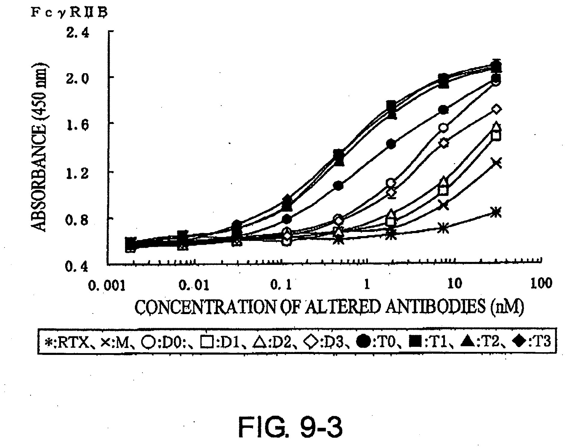
D00014
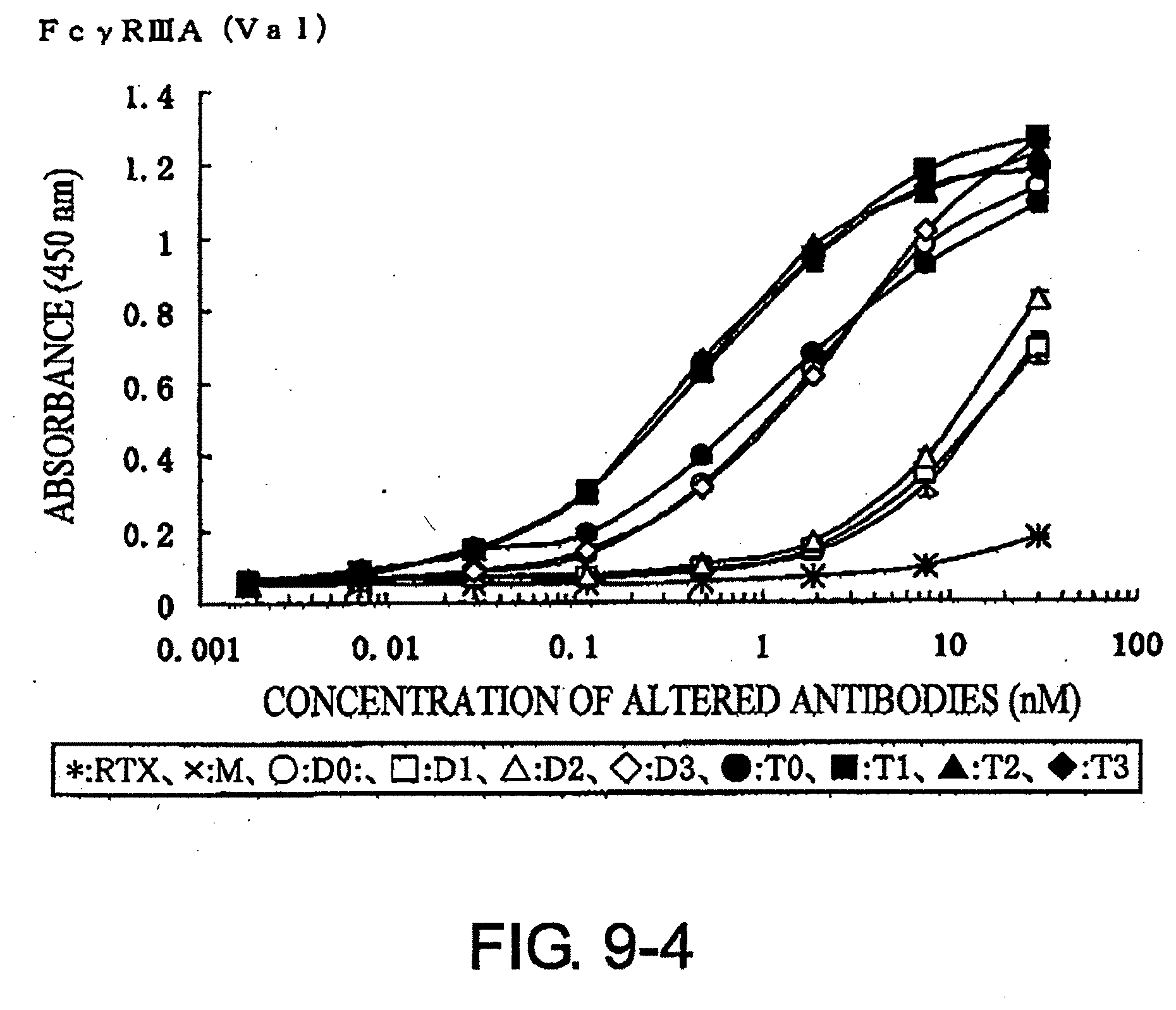
D00015
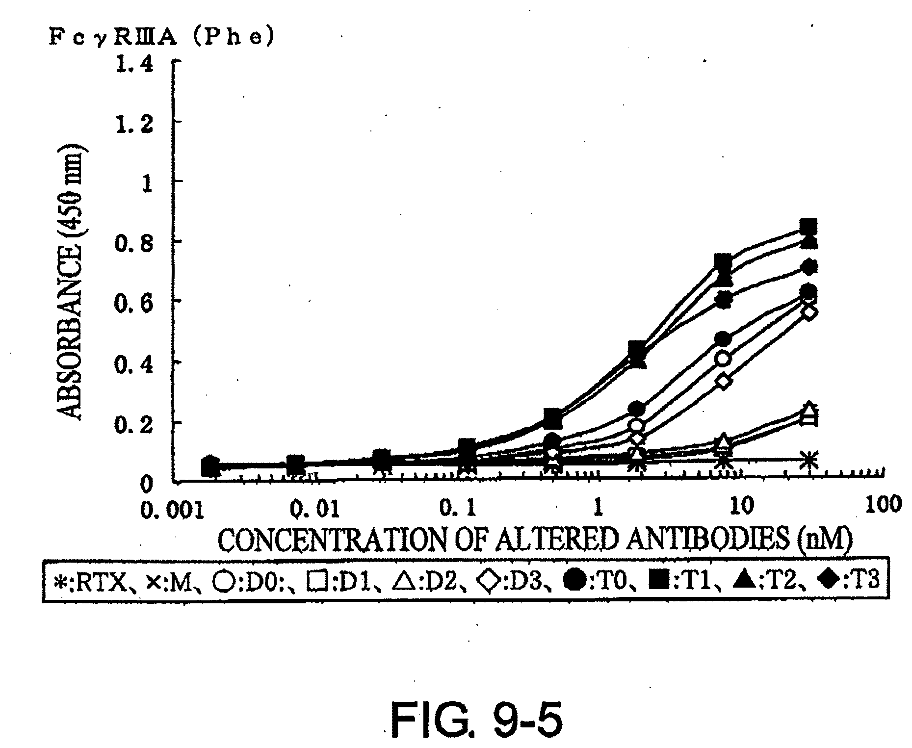
D00016
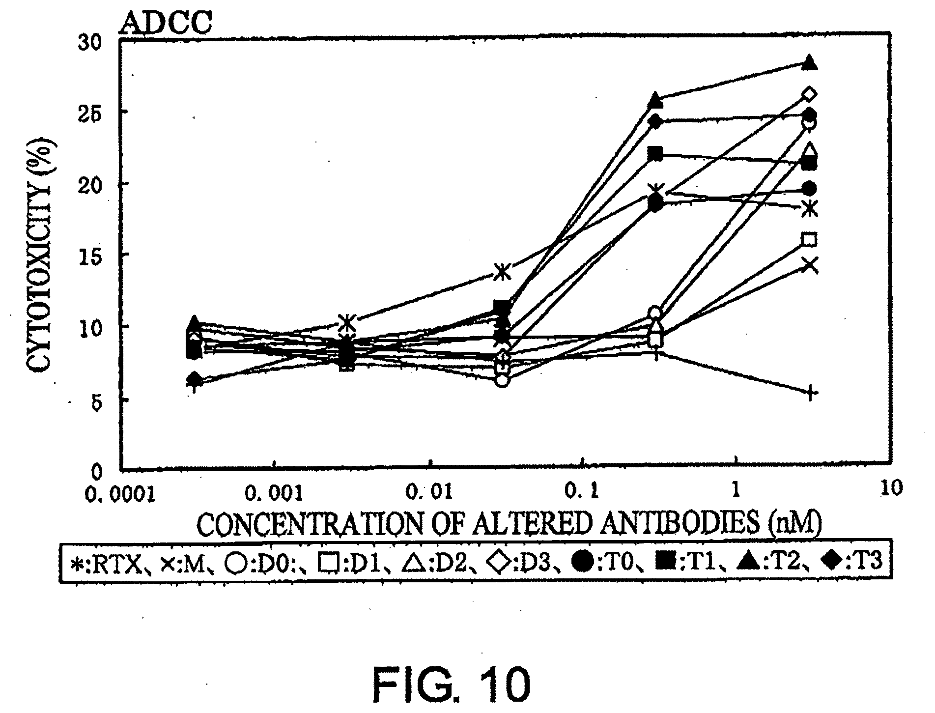
D00017
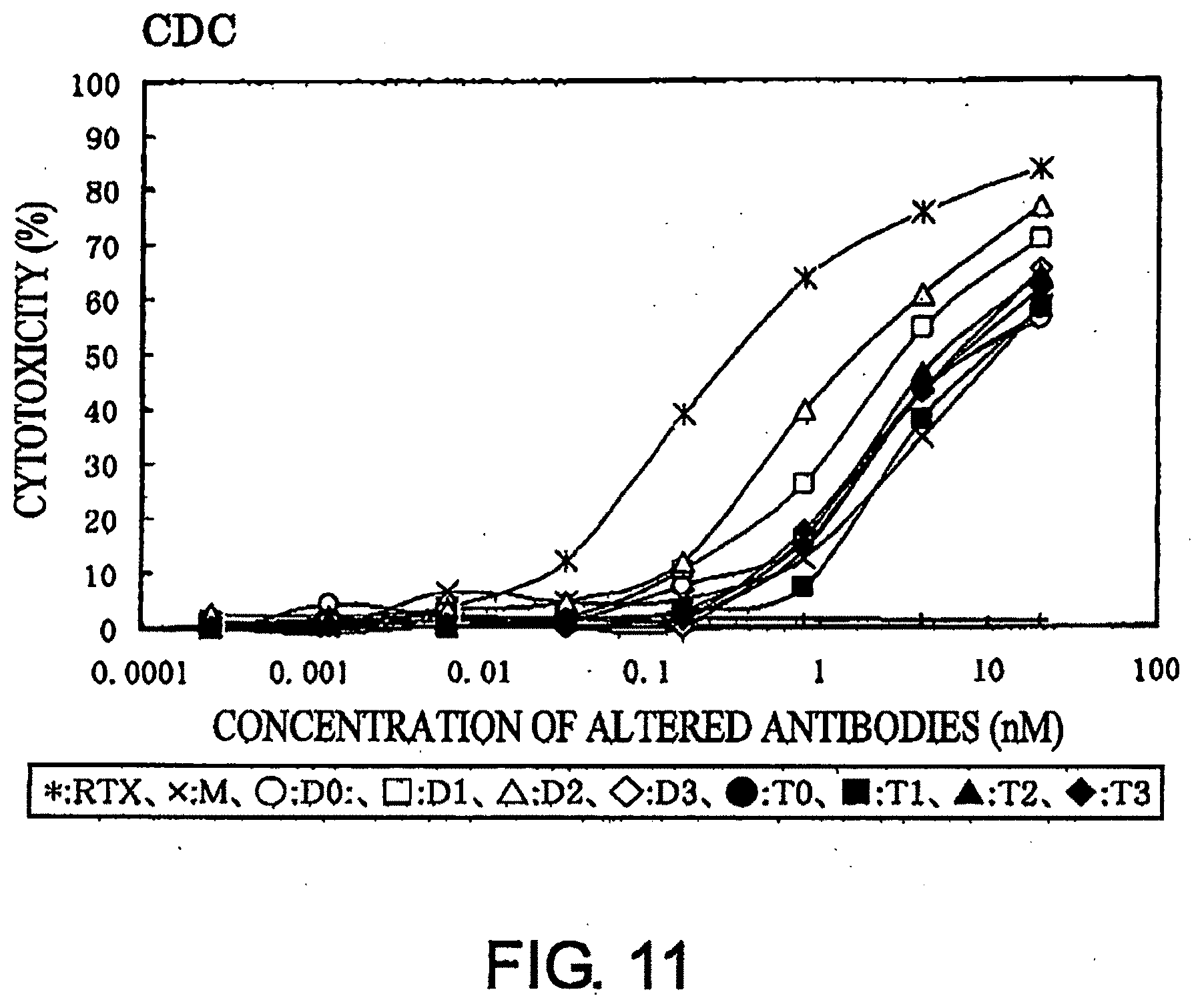
S00001
XML
uspto.report is an independent third-party trademark research tool that is not affiliated, endorsed, or sponsored by the United States Patent and Trademark Office (USPTO) or any other governmental organization. The information provided by uspto.report is based on publicly available data at the time of writing and is intended for informational purposes only.
While we strive to provide accurate and up-to-date information, we do not guarantee the accuracy, completeness, reliability, or suitability of the information displayed on this site. The use of this site is at your own risk. Any reliance you place on such information is therefore strictly at your own risk.
All official trademark data, including owner information, should be verified by visiting the official USPTO website at www.uspto.gov. This site is not intended to replace professional legal advice and should not be used as a substitute for consulting with a legal professional who is knowledgeable about trademark law.