Specific Antibody Drug Conjugates (adcs) Having Ksp Inhibitors
LERCHEN; Hans-Georg ; et al.
U.S. patent application number 16/472682 was filed with the patent office on 2019-12-05 for specific antibody drug conjugates (adcs) having ksp inhibitors. This patent application is currently assigned to Bayer Pharma Aktiengesellschaft. The applicant listed for this patent is Bayer Pharma Aktiengesellschaft. Invention is credited to Sandra BERNDT, Lisa DIETZ, Stefanie HAMMER, Dennis KIRCHHOFF, Hans-Georg LERCHEN, Stephan MARSCH, Anne-Sophie REBSTOCK, Beatrix STELTE-LUDWIG.
| Application Number | 20190365916 16/472682 |
| Document ID | / |
| Family ID | 57614198 |
| Filed Date | 2019-12-05 |


View All Diagrams
| United States Patent Application | 20190365916 |
| Kind Code | A1 |
| LERCHEN; Hans-Georg ; et al. | December 5, 2019 |
SPECIFIC ANTIBODY DRUG CONJUGATES (ADCS) HAVING KSP INHIBITORS
Abstract
Specific binder-drug conjugates (ADCs) of kinesin spindle protein inhibitors, effective metabolites of these ADCs, processes for preparing these ADCs, the use of these ADCs for the treatment and/or prevention of diseases and to the use of these ADCs for preparing medicaments for treatment and/or prevention of diseases, in particular hyperproliferative and/or angiogenic disorders such as, for example, cancer diseases, are described.
| Inventors: | LERCHEN; Hans-Georg; (Leverkusen, DE) ; REBSTOCK; Anne-Sophie; (Champagne au Mont d'Or, FR) ; STELTE-LUDWIG; Beatrix; (Wulfrath, DE) ; KIRCHHOFF; Dennis; (Berlin, DE) ; BERNDT; Sandra; (Hohen Neuendorf, DE) ; DIETZ; Lisa; (Wuppertal, DE) ; MARSCH; Stephan; (Koln, DE) ; HAMMER; Stefanie; (Berlin, DE) | ||||||||||
| Applicant: |
|
||||||||||
|---|---|---|---|---|---|---|---|---|---|---|---|
| Assignee: | Bayer Pharma
Aktiengesellschaft Berlin DE |
||||||||||
| Family ID: | 57614198 | ||||||||||
| Appl. No.: | 16/472682 | ||||||||||
| Filed: | December 18, 2017 | ||||||||||
| PCT Filed: | December 18, 2017 | ||||||||||
| PCT NO: | PCT/EP2017/083313 | ||||||||||
| 371 Date: | June 21, 2019 |
| Current U.S. Class: | 1/1 |
| Current CPC Class: | A61K 47/6849 20170801; C07K 2317/73 20130101; A61K 45/06 20130101; C07K 16/2866 20130101; A61K 47/65 20170801; A61P 35/00 20180101; C07K 2317/24 20130101; A61K 47/6889 20170801; C07K 2317/76 20130101; A61K 47/6803 20170801; A61P 35/02 20180101; A61K 39/3955 20130101; A61K 2039/505 20130101; C07K 2317/565 20130101; A61P 43/00 20180101 |
| International Class: | A61K 47/65 20060101 A61K047/65; A61K 47/68 20060101 A61K047/68; C07K 16/28 20060101 C07K016/28; A61K 45/06 20060101 A61K045/06; A61K 39/395 20060101 A61K039/395; A61P 35/00 20060101 A61P035/00; A61P 35/02 20060101 A61P035/02 |
Foreign Application Data
| Date | Code | Application Number |
|---|---|---|
| Dec 21, 2016 | EP | 16205870.5 |
Claims
1. An antibody-drug conjugate (ADC) of formula (I): ##STR00021## wherein: n is 1 to 8; AK is an anti-CD123 antibody selected from the group consisting of TPP-8987, TPP-9476 and TPP-8988, or AK is an anti-CXCR5 antibody, or AK is an antigen-binding fragment of these antibodies, wherein the antibody or the antigen-binding fragment is attached via a sulfur atom of a cysteine side group, or a salt, a solvate, or a salt of the solvate thereof.
2. The antibody-drug conjugate according to claim 1, wherein n is 2 to 8.
3. The antibody-drug conjugate according to claim 1, wherein n is 4 to 8.
4. The antibody-drug conjugate according to claim 1, wherein AK (i) is an anti-CD123 antibody comprising a variable region of the heavy chain (VH) comprising the variable CDR1 sequence of the heavy chain (H-CDR1), as shown by SEQ ID NO: 2, the variable CDR2 sequence of the heavy chain (H-CDR2), as shown by SEQ ID NO: 3 and the variable CDR3 sequence of the heavy chain (H-CDR3), as shown by SEQ ID NO: 4, and a variable region of the light chain (VL) comprising the variable CDR1 sequence of the light chain (L-CDR1), as shown by SEQ ID NO: 6, the variable CDR2 sequence of the light chain (L-CDR2), as shown by SEQ ID NO: 7 and the variable CDR3 sequence of the light chain (L-CDR3), as shown by SEQ ID NO: 8, (ii) is an anti-CD123 antibody comprising a variable region of the heavy chain (VH) comprising the variable CDR1 sequence of the heavy chain (H-CDR1), as shown by SEQ ID NO: 12, the variable CDR2 sequence of the heavy chain (H-CDR2), as shown by SEQ ID NO: 13 and the variable CDR3 sequence of the heavy chain (H-CDR3), as shown by SEQ ID NO: 14, and a variable region of the light chain (VL) comprising the variable CDR1 sequence of the light chain (L-CDR1), as shown by SEQ ID NO: 16, the variable CDR2 sequence of the light chain (L-CDR2), as shown by SEQ ID NO: 17 and the variable CDR3 sequence of the light chain (L-CDR3), as shown by SEQ ID NO: 18, (iii) is an anti-CXCR5 antibody comprising a variable region of the heavy chain (VH) comprising the variable CDR1 sequence of the heavy chain (H-CDR1), as shown by SEQ ID NO: 22, the variable CDR2 sequence of the heavy chain (H-CDR2), as shown by SEQ ID NO: 23 and the variable CDR3 sequence of the heavy chain (H-CDR3), as shown by SEQ ID NO: 24, and a variable region of the light chain (VL) comprising the variable CDR1 sequence of the light chain (L-CDR1), as shown by SEQ ID NO: 26, the variable CDR2 sequence of the light chain (L-CDR2), as shown by SEQ ID NO: 27 and the variable CDR3 sequence of the light chain (L-CDR3), as shown by SEQ ID NO: 28, (vi) is an anti-CD123 antibody comprising a variable region of the heavy chain (VH) comprising the variable CDR1 sequence of the heavy chain (H-CDR1), as shown by SEQ ID NO: 32, the variable CDR2 sequence of the heavy chain (H-CDR2), as shown by SEQ ID NO: 33 and the variable CDR3 sequence of the heavy chain (H-CDR3), as shown by SEQ ID NO: 34, and a variable region of the light chain (VL) comprising the variable CDR1 sequence of the light chain (L-CDR1), as shown by SEQ ID NO: 36, the variable CDR2 sequence of the light chain (L-CDR2), as shown by SEQ ID NO: 37 and the variable CDR3 sequence of the light chain (L-CDR3), as shown by SEQ ID NO: 38, (v) is an anti-CXCR5 antibody comprising a variable region of the heavy chain (VH) comprising the variable CDR1 sequence of the heavy chain (H-CDR1), as shown by SEQ ID NO: 42, the variable CDR2 sequence of the heavy chain (H-CDR2), as shown by SEQ ID NO: 43 and the variable CDR3 sequence of the heavy chain (H-CDR3), as shown by SEQ ID NO: 44, and a variable region of the light chain (VL) comprising the variable CDR1 sequence of the light chain (L-CDR1), as shown by SEQ ID NO: 46, the variable CDR2 sequence of the light chain (L-CDR2), as shown by SEQ ID NO: 47 and the variable CDR3 sequence of the light chain (L-CDR3), as shown by SEQ ID NO: 48, or (vi) is an anti-CXCR5 antibody comprising a variable region of the heavy chain (VH) comprising the variable CDR1 sequence of the heavy chain (H-CDR1), as shown by SEQ ID NO: 52, the variable CDR2 sequence of the heavy chain (H-CDR2), as shown by SEQ ID NO: 53 and the variable CDR3 sequence of the heavy chain (H-CDR3), as shown by SEQ ID NO: 54, and a variable region of the light chain (VL) comprising the variable CDR1 sequence of the light chain (L-CDR1), as shown by SEQ ID NO: 56, the variable CDR2 sequence of the light chain (L-CDR2), as shown by SEQ ID NO: 57 and the variable CDR3 sequence of the light chain (L-CDR3), as shown by SEQ ID NO: 58, or is an antigen-binding fragment of these antibodies.
5. The antibody-drug conjugate according to claim 1, wherein AK (i) is an anti-CD123 antibody comprising a variable region of the heavy chain (VH) as shown in SEQ ID NO: 1 and a variable region of the light chain (VL) as shown in SEQ ID NO: 5, (ii) is an anti-CD123 antibody comprising a variable region of the heavy chain (VH) as shown in SEQ ID NO: 11 and a variable region of the light chain (VL) as shown in SEQ ID NO: 15, (iii) is an anti-CXCR5 antibody comprising a variable region of the heavy chain (VH) as shown in SEQ ID NO: 21 and a variable region of the light chain (VL) as shown in SEQ ID NO: 25, (iv) is an anti-CD123 antibody comprising a variable region of the heavy chain (VH) as shown in SEQ ID NO: 31 and a variable region of the light chain (VL) as shown in SEQ ID NO: 35, (v) is an anti-CXCR5 antibody comprising a variable region of the heavy chain (VH) as shown in SEQ ID NO: 41 and a variable region of the light chain (VL) as shown in SEQ ID NO: 45, or (vi) is an anti-CXCR5 antibody comprising a variable region of the heavy chain (VH) as shown in SEQ ID NO: 51 and a variable region of the light chain (VL) as shown in SEQ ID NO: 55, or is an antigen-binding fragment of these antibodies.
6. The antibody-drug conjugate according to claim 1, wherein AK (i) is an anti-CD123 antibody comprising a region of the heavy chain as shown in SEQ ID NO: 9 and a region of the light chain as shown in SEQ ID NO: 10, (ii) is an anti-CD123 antibody comprising a region of the heavy chain as shown in SEQ ID NO: 19 and a region of the light chain as shown in SEQ ID NO: 20, (iii) is an anti-CXCR5 antibody comprising a region of the heavy chain as shown in SEQ ID NO: 29 and a region of the light chain as shown in SEQ ID NO: 30, (iv) is an anti-CD123 antibody comprising a region of the heavy chain as shown in SEQ ID NO: 39 and a region of the light chain as shown in SEQ ID NO: 40, (v) is an anti-CXCR5 antibody comprising a region of the heavy chain as shown in SEQ ID NO: 49 and a region of the light chain as shown in SEQ ID NO: 50, or (vi) is an anti-CXCR5 antibody comprising a region of the heavy chain as shown in SEQ ID NO: 59 and a region of the light chain as shown in SEQ ID NO: 60, or is an antigen-binding fragment of these antibodies.
7. A pharmaceutical composition comprising at least one antibody-drug conjugate (ADC) according to claim 1 in combination with an inert, non-toxic, pharmaceutically suitable auxiliary.
8. (canceled)
9. A method for treatment of hyperproliferative and/or angiogenic disorders, comprising administering to a patient in need thereof an effective amount of an antibody-drug conjugate according to claim 1.
10. A method for treatment of cancer and tumours, comprising administering to a human in need thereof an effective amount of an antibody-drug conjugate according to claim 1.
11. A method for treatment of cancer and tumours, comprising administering to a human in need thereof an effective amount of an antibody-drug conjugate according to claim 1 in combination with one or more therapeutic approaches for cancer immunotherapy or with one or more active compounds directed against a molecular target of cancer immunotherapy.
12. The antibody-drug conjugate according to claim 1, wherein the anti-CXCR5 antibody is selected from the group consisting of TPP-9574, TPP-9580, and TPP-9024.
Description
INTRODUCTION AND STATE OF THE ART
[0001] The invention relates to specific binder-drug conjugates (ADCs) of kinesin spindle protein inhibitors, to effective metabolites of these ADCs, to processes for preparing these ADCs, to the use of these ADCs for the treatment and/or prevention of diseases and to the use of these ADCs for preparing medicaments for treatment and/or prevention of diseases, in particular hyperproliferative and/or angiogenic disorders such as, for example, cancer diseases. Such treatments can be effected as monotherapy or else in combination with other medicaments or further therapeutic measures.
[0002] Cancers are the consequence of uncontrolled cell growth of the most diverse tissues. In many cases the new cells penetrate into existing tissue (invasive growth), or they metastasize into remote organs. Cancers occur in a wide variety of different organs and often have tissue-specific courses. The term "cancer" as a generic term therefore describes a large group of defined diseases of different organs, tissue and cell types.
[0003] Some tumours at early stages can be removed by surgical and radiotherapy measures. Metastasized tumours as a rule can only be treated palliatively by chemotherapeutics. The aim here is to achieve the optimum combination of an improvement in the quality of life and prolonging of life.
[0004] Conjugates of binder proteins with one or more drug molecules are known, in particular in the form of antibody drug conjugates (ADCs) in which an internalizing antibody directed against a tumour-associated antigen is covalently attached via a linker to a cytotoxic agent. Following introduction of the ADCs into the tumour cell and subsequent dissociation of the conjugate, either the cytotoxic agent itself or a cytotoxic metabolite formed therefrom is released within the tumour cell and can unfold its action therein directly and selectively. In this manner, in contrast to conventional cancer chemotherapy, damage to normal tissue is contained in significantly narrower limits [see, for example, J. M. Lambert, Curr. Opin. Pharmacol. 5, 543-549 (2005); A. M. Wu and P. D. Senter, Nat. Biotechnol. 23, 1137-1146 (2005); P. D. Senter, Curr. Opin. Chem. Biol. 13, 235-244 (2009); L. Ducry and B. Stump, Bioconjugate Chem. 21, 5-13 (2010)]. Thus, WO2012/171020 describes ADCs in which a plurality of toxophore molecules are attached via a polymeric linker to an antibody. As possible toxophores, WO2012/171020 mentions, among others, the substances SB 743921, SB 715992 (Ispinesib), MK-0371, AZD8477, AZ3146 and ARRY-520.
[0005] The substances mentioned last are kinesin spindle protein inhibitors. Kinesin spindle protein (KSP, also known as Eg5, HsEg5, KNSL1 or KIF11) is a kinesin-like motorprotein which is essential for the bipolar mitotic spindle to function. Inhibition of KSP leads to mitotic arrest and, over a relatively long term, to apoptosis (Tao et al., Cancer Cell 2005 Jul. 8(1), 39-59). After the discovery of the first cell-penetrating KSP inhibitor, Monastrol, KSP inhibitors have established themselves as a class of novel chemotherapeutics (Mayer et al., Science 286: 971-974, 1999), and they are subject matter of a number of patent applications (e.g. WO2006/044825; WO2006/002236; WO2005/051922; WO2006/060737; WO03/060064; WO03/040979; and WO03/049527). However, since KSP is active only during a relatively short period of time during the mitosis phase, KSP inhibitors have to be present in a sufficiently high concentration during this phase. WO2014/151030 discloses ADCs including certain KSP inhibitors.
[0006] Further ADCs with KSP inhibitors have been disclosed in the patent applications WO2015/096982 and WO2016/096610.
SUMMARY OF THE INVENTION
[0007] Despite various disclosures of antibody-drug conjugates, it is an object of the present invention to provide substances which, after administration at a relatively low concentration, exhibit long-lasting apoptotic action and may therefore be of benefit for cancer therapy. Here, the profile of the metabolites released intracellularly from the ADCs plays an important role. Frequently, the metabolites formed from ADCs are substrates of efflux pumps and/or have high cell membrane permeability. Both phenomena may contribute to a short residence time and thus suboptimal apoptotic action in the tumour cell.
[0008] The present invention provides ADCs having a specific toxophor linker composition which in particular have an improved activity profile both in association with a specific anti-CD123 antibody and with an anti-CXCR5 antibody.
[0009] The antibody is preferably a humanized or chimeric monoclonal anti-CD123 antibody or an anti-CXCR5 antibody. Particular preference is given to the humanized anti-CD123 antibodies TPP-8987, TPP-8988 and TPP-9476 and to the humanized or chimeric anti-CXCR5 antibodies TPP-9024, TPP-9574 and TPP-9580.
[0010] It has now been found that antibody-drug conjugates (ADCs) of the formula (I)
##STR00001##
in which [0011] n represents 1 to 8, [0012] AK represents an anti-CD123 antibody selected from the group consisting of TPP-8987, TPP-9476 and TPP-8988 [0013] or [0014] AK represents an anti-CXCR5 antibody, preferably selected from the group consisting of TPP-9574, TPP-9580 and TPP-9024, [0015] or [0016] AK represents an antigen-binding fragment of these antibodies, where the antibody or the antigen-binding fragment is attached via a sulfur atom of a cysteine side group, and their salts, solvates and salts of these solvates, have superior properties compared to the known conjugates.
[0017] Preference is given to those antibody-drug conjugates (ADCs) of the formula (I), in which n represents 4 to 8.
[0018] Preference is given to those antibody-drug conjugates (ADCs) of the formula (I), in which AK represents an anti-CD123 antibody selected from the group consisting of TPP-8987, TPP-9476 and TPP-8988 and an antigen-binding fragment of these antibodies; particularly preferably AK represents TPP-9476, and an antigen-binding fragment of this antibody.
DESCRIPTION OF THE FIGURES
[0019] FIG. 1: Annotated sequences of preferred antibodies for binder-drug conjugates. What are shown are the protein sequences of the heavy and light chains of the IgGs, and the VH and VL regions of these antibodies. Below the sequences, important regions are annotated (VH and VL regions in IgGs, and the CDR regions (H-CDR1, H-CDR2, H-CDR3, L-CDR1, L-CDR2, L-CDR3)).
[0020] FIG. 2: Sequence listing of sequences of the preferred antibodies for binder-drug conjugates and of sequences of the target molecules.
DETAILED DESCRIPTION OF THE INVENTION
[0021] The invention provides conjugates of a humanized anti-CD123 antibody or a humanized or chimeric monoclonal anti-CXCR5 antibody, the drug molecule being a kinesin spindle protein inhibitor (KSP inhibitor) which is attached to the antibody via a linker L. Particular preference is given here to the humanized anti-CD123 antibodies TPP-8987, TPP-8988 and TPP-9476 and the humanized or chimeric anti-CXCR5 antibodies TPP-9024, TPP-9574 and TPP-9580.
Binders
[0022] In the broadest sense, the term "binder" is understood to mean a molecule which binds to a target molecule present at a certain target cell population to be addressed by the binder-drug conjugate. The term binder is to be understood in its broadest meaning and also comprises, for example, lectins, proteins capable of binding to certain sugar chains, or phospholipid-binding proteins. Such binders include, for example, high-molecular weight proteins (binding proteins), polypeptides or peptides (binding peptides), non-peptidic (e.g. aptamers (U.S. Pat. No. 5,270,163) review by Keefe AD., et al., Nat. Rev. Drug Discov. 2010; 9:537-550), or vitamins) and all other cell-binding molecules or substances. Binding proteins are, for example, antibodies and antibody fragments or antibody mimetics, for example affibodies, adnectins, anticalins, DARPins, avimers, nanobodies (review by Gebauer M. et al., Curr. Opinion in Chem. Biol. 2009; 13:245-255; Nuttall S. D. et al., Curr. Opinion in Pharmacology 2008; 8:608-617). Binding peptides are, for example, ligands of a ligand/receptor pair such as, for example, VEGF of the ligand/receptor pair VEGF/KDR, such as transferrin of the ligand/receptor pair transferrin/transferrin receptor or cytokine/cytokine receptor, such as TNFalpha of the ligand/receptor pair TNFalpha/TNFalpha receptor.
[0023] The binder may be a binding protein. Preferred embodiments of the binders are an antibody, an antigen-binding antibody fragment, a multispecific antibody or an antibody mimetic.
[0024] The literature also discloses various options of covalent coupling (conjugation) of organic molecules to binders and in particular antibodies. Preference according to the invention is given to the conjugation of the toxophores to the antibody via one or more sulfur atoms of cysteine residues of the antibody and/or via one or more NH groups of lysine residues of the antibody. However, it is also possible to bind the toxophore to the antibody via free carboxyl groups or via sugar residues of the antibody.
[0025] A "target molecule" in the broadest sense is understood to mean a molecule which is present in the target cell population and which may be a protein (for example a receptor of a growth factor) or a non-peptidic molecule (for example a sugar or phospholipid). It is preferably a receptor or an antigen.
[0026] The term "extracellular" target molecule describes a target molecule, attached to the cell, which is located at the outside of a cell, or the part of a target molecule which is located at the outside of a cell, i.e. a binder may bind on an intact cell to its extracellular target molecule. An extracellular target molecule may be anchored in the cell membrane or be a component of the cell membrane. The person skilled in the art is aware of methods for identifying extracellular target molecules. For proteins, this may be by determining the transmembrane domain(s) and the orientation of the protein in the membrane. These data are usually deposited in protein databases (e.g. SwissProt).
[0027] The term "cancer target molecule" describes a target molecule which is more abundantly present on one or more cancer cell species than on non-cancer cells of the same tissue type. Preferably, the cancer target molecule is selectively present on one or more cancer cell species compared with non-cancer cells of the same tissue type, where selectively describes an at least two-fold enrichment on cancer cells compared to non-cancer cells of the same tissue type (a "selective cancer target molecule"). The use of cancer target molecules allows the selective therapy of cancer cells using the conjugates according to the invention.
[0028] The binder can be attached to the linker via a bond. The binder can be linked by means of a heteroatom of the binder. Heteroatoms according to the invention of the binder which can be used for attachment are sulfur (in one embodiment via a sulfhydryl group of the binder), oxygen (according to the invention by means of a carboxyl or hydroxyl group of the binder) and nitrogen (in one embodiment via a primary or secondary amine group or amide group of the binder). These heteroatoms may be present in the natural binder or are introduced by chemical methods or methods of molecular biology. According to the invention, the attachment of the binder to the toxophore has only a minor effect on the binding activity of the binder with respect to the target molecule. In a preferred embodiment, the linkage has no effect on the binding activity of the binder with respect to the target molecule.
[0029] In accordance with the present invention, the term "antibody" is to be understood in its broadest meaning and comprises immunoglobulin molecules, for example intact or modified monoclonal antibodies, polyclonal antibodies or multispecific antibodies (e.g. bispecific antibodies). An immunoglobulin molecule preferably comprises a molecule having four polypeptide chains, two heavy chains (H chains) and two light chains (L chains) which are typically linked by disulfide bridges. Each heavy chain comprises a variable domain of the heavy chain (abbreviated VH) and a constant domain of the heavy chain. The constant domain of the heavy chain may, for example, comprise three domains CH1, CH2 and CH3. Each light chain comprises a variable domain (abbreviated VL) and a constant domain. The constant domain of the light chain comprises a domain (abbreviated CL). The VH and VL domains may be subdivided further into regions having hypervariability, also referred to as complementarity determining regions (abbreviated CDR) and regions having low sequence variability (framework region, abbreviated FR). Typically, each VH and VL region is composed of three CDRs and up to four FRs. For example from the amino terminus to the carboxy terminus in the following order: FR1, CDR1, FR2, CDR2, FR3, CDR3, FR4. An antibody may be obtained from any suitable species, e.g. rabbit, llama, camel, mouse or rat. In one embodiment, the antibody is of human or murine origin. An antibody may, for example, be human, humanized or chimeric.
[0030] The term "monoclonal" antibody refers to antibodies obtained from a population of substantially homogeneous antibodies, i.e. individual antibodies of the population are identical except for naturally occurring mutations, of which there may be a small number. Monoclonal antibodies recognize a single antigenic binding site with high specificity. The term monoclonal antibody does not refer to a particular preparation process.
[0031] The term "intact" antibody refers to antibodies comprising both an antigen-binding domain and the constant domain of the light and heavy chain. The constant domain may be a naturally occurring domain or a variant thereof having a number of modified amino acid positions, and may also be aglycosylated.
[0032] The term "modified intact" antibody refers to intact antibodies fused via their amino terminus or carboxy terminus by means of a covalent bond (e.g. a peptide bond) with a further polypeptide or protein not originating from an antibody. Furthermore, antibodies may be modified such that, at defined positions, reactive cysteines are introduced to facilitate coupling to a toxophore (see Junutula et al. Nat Biotechnol. 2008 August; 26(8):925-32).
[0033] "Amino acid modification" or "mutation" here means an amino acid substitution, insertion and/or deletion in a polypeptide sequence. The preferred amino acid modification here is a substitution. "Amino acid substitution" or "substitution" here means an exchange of an amino acid at a given position in a protein sequence for another amino acid. For example, the substitution Y50W describes a variant of a parent polypeptide in which the tyrosine at position 50 has been exchanged for a tryptophan. A "variant" of a polypeptide describes a polypeptide having an amino acid sequence substantially identical to a reference polypeptide, typically a native or "parent" polypeptide. The polypeptide variant may have one or more amino acid exchanges, deletions and/or insertions at particular positions in the native amino acid sequence.
[0034] The term "human" antibody refers to antibodies which can be obtained from a human or which are synthetic human antibodies. A "synthetic" human antibody is an antibody which is partially or entirely obtainable in silico from synthetic sequences based on the analysis of human antibody sequences. A human antibody can be encoded, for example, by a nucleic acid isolated from a library of antibody sequences of human origin. An example of such an antibody can be found in Soderlind et al., Nature Biotech. 2000, 18:853-856. Such "human" and "synthetic" antibodies also include aglycosylated variants which have been produced either by deglycosylation by PNGaseF or by mutation of N297 (Kabat numbering) of the heavy chain to any other amino acid.
[0035] The term "humanized" or "chimeric" antibody describes antibodies consisting of a non-human and a human portion of the sequence. In these antibodies, part of the sequences of the human immunoglobulin (recipient) is replaced by sequence portions of a non-human immunoglobulin (donor). In many cases, the donor is a murine immunoglobulin. In the case of humanized antibodies, amino acids of the CDR of the recipient are replaced by amino acids of the donor. Sometimes, amino acids of the framework, too, are replaced by corresponding amino acids of the donor. In some cases the humanized antibody contains amino acids present neither in the recipient nor in the donor, which were introduced during the optimization of the antibody. In the case of chimeric antibodies, the variable domains of the donor immunoglobulin are fused with the constant regions of a human antibody. Such "humanized" and "chimeric" antibodies also include aglycosylated variants which have been produced either by deglycosylation by PNGaseF or by mutation of N297 (Kabat numbering) of the heavy chain to any other amino acid.
[0036] The term complementarity determining region (CDR) as used herein refers to those amino acids of a variable antibody domain which are required for binding to the antigen. Typically, each variable region has three CDR regions referred to as CDR1, CDR2 and CDR3. Each CDR region may embrace amino acids according to the definition of Kabat and/or amino acids of a hypervariable loop defined according to Chotia. The definition according to Kabat comprises, for example, the region from about amino acid position 24-34 (CDR1), 50-56 (CDR2) and 89-97 (CDR3) of the variable light chain/domain (VL) and 31-35 (CDR1), 50-65 (CDR2) and 95-102 (CDR3) of the variable heavy chain/domain (VH) (Kabat et al., Sequences of Proteins of Immunological Interest, 5th Ed. Public Health Service, National Institutes of Health, Bethesda, Md. (1991)). The definition according to Chotia comprises, for example, the region from about amino acid position 26-32 (CDR1), 50-52 (CDR2) and 91-96 (CDR3) of the variable light chain (VL) and 26-32 (CDR1), 53-55 (CDR2) and 96-101 (CDR3) of the variable heavy chain (VH) (Chothia and Lesk; J Mol Biol 196: 901-917 (1987)). In some cases, a CDR may comprise amino acids from a CDR region defined according to Kabat and Chotia.
[0037] Depending on the amino acid sequence of the constant domain of the heavy chain, antibodies may be categorized into different classes. There are five main classes of intact antibodies: IgA, IgD, IgE, IgG and IgM, and several of these can be divided into further subclasses. (Isotypes), e.g. IgG1, IgG2, IgG3, IgG4, IgA1 and IgA2. The constant domains of the heavy chain, which correspond to the different classes, are referred to as [alpha/.alpha.], [delta/.delta.], [epsilon/.epsilon.], [gamma/.gamma.] and [my/.mu.]. Both the three-dimensional structure and the subunit structure of antibodies are known.
[0038] The term "functional fragment" or "antigen-binding antibody fragment" of an antibody/immunoglobulin is defined as a fragment of an antibody/immunoglobulin (e.g. the variable domains of an IgG) which still comprise the antigen binding domains of the antibody/immunoglobulin. The "antigen binding domain" of an antibody typically comprises one or more hypervariable regions of an antibody, for example the CDR, CDR2 and/or CDR3 region. However, the "framework" or "skeleton" region of an antibody may also play a role during binding of the antibody to the antigen. The framework region forms the skeleton of the CDRs. Preferably, the antigen binding domain comprises at least amino acids 4 to 103 of the variable light chain and amino acids 5 to 109 of the variable heavy chain, more preferably amino acids 3 to 107 of the variable light chain and 4 to 111 of the variable heavy chain, especially preferably the complete variable light and heavy chains, i.e. amino acids 1-109 of the VL and 1 to 113 of the VH (numbering according to WO97/08320).
[0039] "Functional fragments" or "antigen-binding antibody fragments" of the invention encompass, non-conclusively, Fab, Fab', F(ab').sub.2 and Fv fragments, diabodies, Single Domain Antibodies (DAbs), linear antibodies, individual chains of antibodies (single-chain Fv, abbreviated to scFv); and multispecific antibodies, such as bi- and tri-specific antibodies, for example, formed from antibody fragments C. A. K Borrebaeck, editor (1995) Antibody Engineering (Breakthroughs in Molecular Biology), Oxford University Press; R. Kontermann & S. Duebel, editors (2001) Antibody Engineering (Springer Laboratory Manual), Springer Verlag. Antibodies other than "multispecific" or "multifunctional" antibodies are those having identical binding sites. Multispecific antibodies may be specific for different epitopes of an antigen or may be specific for epitopes of more than one antigen (see, for example, WO 93/17715; WO 92/08802; WO 91/00360; WO 92/05793; Tutt, et al., 1991, J. Immunol. 14760 69; U.S. Pat. Nos. 4,474,893; 4,714,681; 4,925,648; 5,573,920; 5,601,819; or Kostelny et al., 1992, J. Immunol. 148 1547 1553). An F(ab').sub.2 or Fab molecule may be constructed such that the number of intermolecular disulfide interactions occurring between the Ch1 and the CL domains can be reduced or else completely prevented.
[0040] "Epitopes" refer to protein determinants capable of binding specifically to an immunoglobulin or T cell receptors. Epitopic determinants usually consist of chemically active surface groups of molecules such as amino acids or sugar side chains or combinations thereof, and usually have specific 3-dimensional structural properties and also specific charge properties.
[0041] "Functional fragments" or "antigen-binding antibody fragments" may be fused with another polypeptide or protein, not originating from an antibody, via the amino terminus or carboxyl terminus thereof, by means of a covalent bond (e.g. a peptide linkage). Furthermore, antibodies and antigen-binding fragments may be modified by introducing reactive cysteines at defined locations, in order to facilitate coupling to a toxophore (see Junutula et al. Nat Biotechnol. 2008 August; 26(8):925-32).
[0042] Polyclonal antibodies can be prepared by methods known to a person of ordinary skill in the art. Monoclonal antibodies may be prepared by methods known to a person of ordinary skill in the art (Kohler and Milstein, Nature, 256, 495-497, 1975). Human and humanized monoclonal antibodies may be prepared by methods known to a person of ordinary skill in the art (Olsson et al., Meth Enzymol. 92, 3-16 or Cabilly et al U.S. Pat. No. 4,816,567 or Boss et al U.S. Pat. No. 4,816,397).
[0043] A person of ordinary skill in the art is aware of diverse methods for preparing human antibodies and fragments thereof, such as, for example, by means of transgenic mice (N Lonberg and D Huszar, Int Rev Immunol. 1995; 13(1):65-93) or Phage Display Technologien (Clackson et al., Nature. 1991 Aug. 15; 352(6336):624-8). Antibodies of the invention may be obtained from recombinant antibody libraries consisting for example on the amino acid sequences of a multiplicity of antibodies compiled from a large number of healthy volunteers. Antibodies may also be produced by means of known recombinant DNA technologies. The nucleic acid sequence of an antibody can be obtained by routine sequencing or is available from publically accessible databases.
[0044] An "isolated" antibody or binder has been purified to remove other constituents of the cell. Contaminating constituents of a cell which may interfere with a diagnostic or therapeutic use are, for example, enzymes, hormones, or other peptidic or non-peptidic constituents of a cell. A preferred antibody or binder is one which has been purified to an extent of more than 95% by weight, relative to the antibody or binder (determined for example by Lowry method, UV-Vis spectroscopy or by SDS capillary gel electrophoresis). Moreover an antibody which has been purified to such an extent that it is possible to determine at least 15 amino acids of the amino terminus or of an internal amino acid sequence, or which has been purified to homogeneity, the homogeneity being determined by SDS-PAGE under reducing or non-reducing conditions (detection may be determined by means of Coomassie Blue staining or preferably by silver coloration). However, an antibody is normally prepared by one or more purification steps.
[0045] The term "specific binding" or "binds specifically" refers to an antibody or binder which binds to a predetermined antigen/target molecule. Specific binding of an antibody or binder typically describes an antibody or binder having an affinity of at least 10.sup.-7 M (as Kd value; i.e. preferably those with Kd values smaller than 10.sup.-7 M), with the antibody or binder having an at least two times higher affinity for the predetermined antigen/target molecule than for a non-specific antigen/target molecule (e.g. bovine serum albumin, or casein) which is not the predetermined antigen/target molecule or a closely related antigen/target molecule. Specific binding of an antibody or binder does not exclude the antibody or binder binding to a plurality of antigens/target molecules (e.g. orthologs of different species). The antibodies preferably have an affinity of at least 10.sup.-7 M (as Kd value; in other words preferably those with smaller Kd values than 10.sup.-7 M), preferably of at least 10.sup.-8 M, more preferably in the range from 10.sup.-9 M to 10.sup.-11 M. The Kd values may be determined, for example, by means of surface plasmon resonance spectroscopy.
[0046] The antibody-drug conjugates of the invention likewise exhibit affinities in these ranges. The affinity is preferably not substantially affected by the conjugation of the drugs (in general, the affinity is reduced by less than one order of magnitude, in other words, for example, at most from 10.sup.-8 M to 10.sup.-7 M).
[0047] The antibodies used in accordance with the invention are also notable preferably for a high selectivity. A high selectivity exists when the antibody of the invention exhibits an affinity for the target protein which is better by a factor of at least 2, preferably by a factor of 5 or more preferably by a factor of 10, than for an independent other antigen, e.g. human serum albumin (the affinity may be determined, for example, by means of surface plasmon resonance spectroscopy).
[0048] Furthermore, the antibodies of the invention that are used are preferably cross-reactive. In order to be able to facilitate and better interpret preclinical studies, for example toxicological or activity studies (e.g. in xenograft mice), it is advantageous if the antibody used in accordance with the invention not only binds the human target protein but also binds the species target protein in the species used for the studies. In one embodiment the antibody used in accordance with the invention, in addition to the human target protein, is cross-reactive to the target protein of at least one further species. For toxicological and activity studies it is preferred to use species of the families of rodents, dogs and non-human primates. Preferred rodent species are mouse and rat. Preferred non-human primates are rhesus monkeys, chimpanzees and long-tailed macaques.
[0049] In one embodiment the antibody used in accordance with the invention, in addition to the human target protein, is cross-reactive to the target protein of at least one further species selected from the group of species consisting of mouse, rat and long-tailed macaque (Macaca fascicularis). Especially preferred are antibodies used in accordance with the invention which in addition to the human target protein are at least cross-reactive to the mouse target protein. Preference is given to cross-reactive antibodies whose affinity for the target protein of the further non-human species differs by a factor of not more than 50, more particularly by a factor of not more than ten, from the affinity for the human target protein.
Antibodies Directed Against a Cancer Target Molecule
[0050] The target molecule towards which the binder, for example an antibody or an antigen-binding fragment thereof, is directed is preferably a cancer target molecule. The term "cancer target molecule" describes a target molecule which is more abundantly present on one or more cancer cell species than on non-cancer cells of the same tissue type. Preferably, the cancer target molecule is selectively present on one or more cancer cell species compared with non-cancer cells of the same tissue type, where selectively describes an at least two-fold enrichment on cancer cells compared to non-cancer cells of the same tissue type (a "selective cancer target molecule"). The use of cancer target molecules allows the selective therapy of cancer cells using the conjugates according to the invention.
[0051] Antibodies which are specific against an antigen, for example cancer cell antigen, can be prepared by a person of ordinary skill in the art by means of methods with which he or she is familiar (such as recombinant expression, for example) or may be acquired commercially (as for example from Merck KGaA, Germany). Examples of known commercially available antibodies in cancer therapy are Erbitux.RTM. (cetuximab, Merck KGaA), Avastin.RTM. (bevacizumab, Roche) and Herceptin.RTM. (trastuzumab, Genentech). Trastuzumab is a recombinant humanized monoclonal antibody of the IgG1kappa type which in a cell-based assay (Kd=5 nM) binds the extracellular domains of the human epidermal growth receptor with high affinity. The antibody is produced recombinantly in CHO cells. All these antibodies can also be produced as aglycosylated variants of these antibodies, either by deglycosylation by means of PNGase F or by mutation of N297 (Kabat numbering) of the heavy chain to any amino acid.
[0052] In the present invention, the cancer target molecules are
(1) the receptor protein CXCR5 (CD185; SwissProt: P32302; NCBI Gene ID 643, NCBI Reference Sequence: NP_001707.1; SEQ ID NO: 61) (2) the surface receptor CD123 (IL3RA; NCBI Gene ID: 3563; NCBI Reference Sequence: NP_002174.1; Swiss-Prot: P26951; SEQ ID NO: 62)
[0053] In particularly preferred subject of the invention, the binder binds specifically to an extracellular cancer target molecule selected from the group consisting of the cancer target molecules CXCR5 and CD123. In a preferred embodiment, the binder, after binding to its extracellular target molecule on the target cell, is internalized by the target cell through the binding. This causes the binder-drug conjugate, which may be an immuno-conjugate or an ADC, to be taken up by the target cell. The binder is then processed, preferably intracellularly, with preference lysosomally.
[0054] In one embodiment the binder is a binding protein. In a preferred embodiment the binder is an antibody, an antigen-binding antibody fragment, a multispecific antibody or an antibody mimetic.
[0055] Preferred antibody mimetics are affibodies, adnectins, anticalins, DARPins, avimers, or nanobodies. Preferred multispecific antibodies are bispecific and trispecific antibodies.
[0056] In a preferred embodiment the binder is an antibody or an antigen-binding antibody fragment, more preferably an isolated antibody or an isolated antigen-binding antibody fragment.
[0057] Preferred antigen-binding antibody fragments are Fab, Fab', F(ab')2 and Fv fragments, diabodies, DAbs, linear antibodies and scFv. Particularly preferred are Fab, diabodies and scFv.
[0058] In a particularly preferred embodiment the binder is an antibody. Particularly preferred are monoclonal antibodies or antigen-binding antibody fragments thereof. Further particularly preferred are human, humanized or chimeric antibodies or antigen-binding antibody fragments thereof.
[0059] Antibodies or antigen-binding antibody fragments which bind cancer target molecules may be prepared by a person of ordinary skill in the art using known processes, such as, for example, chemical synthesis or recombinant expression. Binders for cancer target molecules may be acquired commercially or may be prepared by a person of ordinary skill in the art using known processes, such as, for example, chemical synthesis or recombinant expression. Further processes for preparing antibodies or antigen-binding antibody fragments are described in WO 2007/070538 (see page 22 "Antibodies"). The person skilled in the art knows how processes such as phage display libraries (e.g. Morphosys HuCAL Gold) can be compiled and used for discovering antibodies or antigen-binding antibody fragments (see WO 2007/070538, page 24 ff and AK Example 1 on page 70, AK Example 2 on page 72). Further processes for preparing antibodies that use DNA libraries from B cells are described for example on page 26 (WO 2007/070538). Processes for humanizing antibodies are described on page 30-32 of WO2007070538 and in detail in Queen, et al., Pros. Natl. Acad. Sci. USA 8610029-10033, 1989 or in WO 90/0786. Furthermore, processes for recombinant expression of proteins in general and of antibodies in particular are known to the person skilled in the art (see, for example, in Berger and Kimmel (Guide to Molecular Cloning Techniques, Methods in Enzymology, Vol. 152, Academic Press, Inc.); Sambrook, et al., (Molecular Cloning A Laboratory Manual, (Second Edition, Cold Spring Harbor Laboratory Press; Cold Spring Harbor, N.Y.; 1989) Vol. 1-3); Current Protocols in Molecular Biology, (F. M. Ausabel et al. [Eds.], Current Protocols, Green Publishing Associates, Inc./John Wiley & Sons, Inc.); Harlow et al., (Monoclonal Antibodies A Laboratory Manual, Cold Spring Harbor Laboratory Press (19881, Paul [Ed.]); Fundamental Immunology, (Lippincott Williams & Wilkins (1998)); and Harlow, et al., (Using Antibodies A Laboratory Manual, Cold Spring Harbor Laboratory Press (1998)). The person skilled in the art knows the corresponding vectors, promoters and signal peptides which are necessary for the expression of a protein/antibody. Commonplace processes are also described in WO 2007/070538 on pages 41-45. Processes for preparing an IgG1 antibody are described for example in WO 2007/070538 in Example 6 on page 74 ff. Processes which allow the determination of the internalization of an antibody after binding to its antigen are known to the skilled person and are described for example in WO 2007/070538 on page 80. The person skilled in the art is able to use the processes described in WO 2007/070538 that have been used for preparing carboanhydrase IX (Mn) antibodies in analogy for the preparation of antibodies with different target molecule specificity.
Bacterial Expression
[0060] The person skilled in the art is aware of the way in which antibodies, antigen-binding fragments thereof or variants thereof can be produced with the aid of bacterial expression.
[0061] Suitable expression vectors for bacterial expression of desired proteins are constructed by insertion of a DNA sequence which encodes the desired protein within the functional reading frame together with suitable translation initiation and translation termination signals and with a functional promoter. The vector comprises one or more phenotypically selectable markers and a replication origin in order to enable the retention of the vector and, if desired, the amplification thereof within the host. Suitable prokaryotic hosts for transformation include but are not limited to E. coli, Bacillus subtilis, Salmonella typhimurium and various species from the genus Pseudomonas, Streptomyces, and Staphylococcus. Bacterial vectors may be based, for example, on bacteriophages, plasmids, or phagemids. These vectors may contain selectable markers and a bacterial replication origin, which are derived from commercially available plasmids. Many commercially available plasmids typically contain elements of the well-known cloning vector pBR322 (ATCC 37017). In bacterial systems, a number of advantageous expression vectors can be selected on the basis of the intended use of the protein to be expressed.
[0062] After transformation of a suitable host strain and growth of the host strain to an appropriate cell density, the selected promoter is de-reprimed/induced by suitable means (for example a change in temperature or chemical induction), and the cells are cultivated for an additional period. The cells are typically harvested by centrifugation and if necessary digested in a physical manner or by chemical means, and the resulting raw extract is retained for further purification.
[0063] Therefore, a further embodiment of the present invention is an expression vector comprising a nucleic acid which encodes a novel antibody of the present invention.
[0064] Antibodies of the present invention or antigen-binding fragments thereof include naturally purified products, products which originate from chemical syntheses, and products which are produced by recombinant technologies in prokaryotic hosts, for example E. coli, Bacillus subtilis, Salmonella typhimurium and various species from the genus Pseudomonas, Streptomyces, and Staphylococcus, preferably E. coli.
Mammalian Cell Expression
[0065] The person skilled in the art is aware of the way in which antibodies, antigen-binding fragments thereof or variants thereof can be produced with the aid of mammalian cell expression.
[0066] Preferred regulatory sequences for expression in mammalian cell hosts include viral elements which lead to high expression in mammalian cells, such as promoters and/or expression amplifiers derived from cytomegalovirus (CMV) (such as the CMV promoter/enhancer), simian virus 40 (SV40) (such as the SV40 promoter/enhancer), from adenovirus, (for example the adenovirus major late promoter (AdMLP)) and from polyoma. The expression of the antibodies may be constitutive or regulated (for example induced by addition or removal of small molecule inductors such as tetracycline in combination with the Tet system).
[0067] For further description of viral regulatory elements and sequences thereof, reference is made, for example, to U.S. Pat. No. 5,168,062 by Stinski, U.S. Pat. No. 4,510,245 by Bell et al. and U.S. Pat. No. 4,968,615 by Schaffner et al. The recombinant expression vectors may likewise include a replication origin and selectable markers (see, for example, U.S. Pat. Nos. 4,399,216, 4,634,665 and 5,179,017). Suitable selectable markers include genes which impart resistance to substances such as G418, puromycin, hygromycin, blasticidin, zeocin/bleomycin, or methotrexate, or selectable markers which lead to auxotrophy of a host cell, such as glutamine synthetase (Bebbington et al., Biotechnology (N Y). 1992 February; 10(2):169-75), when the vector has been introduced into the cell.
[0068] For example, the dihydrofolate reductase (DHFR) gene imparts resistance to methotrexate, the neo gene imparts resistance to G418, the bsd gene from Aspergillus terreus imparts resistance to blasticidin, puromycin N-acetyltransferase imparts resistance to puromycin, the Sh ble gene product imparts resistance to zeocin, and resistance to hygromycin is imparted by the E. coli hygromycin resistance gene (hyg or hph). Selectable markers such as DHFR or glutamine synthetase are also helpful for amplification techniques in conjunction with MTX and MSX.
[0069] The transfection of an expression vector into a host cell can be executed with the aid of standard techniques, including by electroporation, nucleofection, calcium phosphate precipitation, lipofection, polycation-based transfection such as polyethyleneimine (PEI)-based transfection and DEAE-dextran transfection.
[0070] Suitable mammalian host cells for the expression of antibodies, antigen-binding fragments thereof, or variants thereof include Chinese hamster ovary (CHO) cells such as CHO-K1, CHO-S, CHO-K1SV [including DHFR-CHO cells, described in Urlaub and Chasin, (1980) Proc. Natl. Acad. Sci. USA 77:4216-4220 and Urlaub et al., Cell. 1983 June; 33(2):405-12, used with a DHFR-selectable marker, as described in R. J. Kaufman and P. A. Sharp (1982) Mol. Biol. 159:601-621, and other knockout cells, as detailed in Fan et al., Biotechnol Bioeng. 2012 April; 109(4):1007-15), NS0 myeloma cells, COS cells, HEK293 cells, HKB11 cells, BHK21 cells, CAP cells, EB66 cells, and SP2 cells.
[0071] The expression of antibodies, antigen-binding fragments thereof, or variants thereof can also be effected in a transient or semi-stable manner in expression systems such as HEK293, HEK293T, HEK293-EBNA, HEK293E, HEK293-6E, HEK293 Freestyle, HKB11, Expi293F, 293EBNALT75, CHO Freestyle, CHO-S, CHO-K1, CHO-K1SV, CHOEBNALT85, CHOS-XE, CHO-3E7 or CAP-T cells (for example like Durocher et al., Nucleic Acids Res. 2002 Jan. 15; 30(2):E9)
[0072] In some embodiments, the expression vector is constructed in such a way that the protein to be expressed is secreted into the cell culture medium in which the host cells are growing. The antibodies, the antigen-binding fragments thereof, or the variants thereof can be obtained from the cell culture medium with the aid of protein purification methods known to those skilled in the art.
Purification
[0073] The antibodies, the antigen-binding fragments thereof, or the variants thereof can be obtained and purified from recombinant cell cultures with the aid of well-known methods, examples of which include ammonium sulfate or ethanol precipitation, acid extraction, protein A chromatography, protein G chromatography, anion or cation exchange chromatography, phosphocellulose chromatography, hydrophobic interaction chromatography (HIC), affinity chromatography, hydroxyapatite chromatography and lectin chromatography. High-performance liquid chromatography ("HPLC") can likewise be employed for purification. See, for example, Colligan, Current Protocols in Immunology, or Current Protocols in Protein Science, John Wiley & Sons, NY, N.Y., (1997-2001), e.g., Chapters 1, 4, 6, 8, 9, 10.
[0074] Antibodies of the present invention or antigen-binding fragments thereof, or variants thereof include naturally purified products, products from chemical synthesis methods and products which are produced with the aid of recombinant techniques in prokaryotic or eukaryotic host cells. Eukaryotic hosts include, for example, yeast cells, higher plant cells, insect cells and mammalian cells. Depending on the host cell chosen for the recombinant expression, the protein expressed may be in glycosylated or non-glycosylated form.
[0075] In a preferred embodiment, the antibody is purified (1) to an extent of more than 95% by weight, measured, for example, by the Lowry method, by UV-vis spectroscopy or by SDS capillary gel electrophoresis (for example with a Caliper LabChip GXII, GX 90 or Biorad Bioanalyzer instrument), and in more preferred embodiments more than 99% by weight, (2) to a degree suitable for determination of at least 15 residues of the N-terminal or internal amino acid sequence, or (3) to homogeneity determined by SDS-PAGE under reducing or non-reducing conditions with the aid of Coomassie blue or preferably silver staining.
[0076] Usually, an isolated antibody is obtained with the aid of at least one protein purification step.
Anti-CD123 Antibodies
[0077] According to the invention, it is possible to use anti-CD123 antibodies.
[0078] The expression "anti-CD123 antibody" or "an antibody which binds specifically to CD123" relates to an antibody which binds the cancer target molecule CD123 (NCBI Reference sequence: NP_002174.1; SEQ ID NO: 62), preferably with an affinity sufficient for a diagnostic and/or therapeutic application. In particular embodiments, the antibody binds CD123 with a dissociation constant (K.sub.D) of .ltoreq.1 .mu.M, .ltoreq.100 nM, .ltoreq.10 nM, .ltoreq.1 nM, .ltoreq.0.1 nM, .ltoreq.0.01 nM, or .ltoreq.0.001 nM.
[0079] Sun et al. (Sun et al., 1996, Blood 87(1)83-92) describe the generation and properties of the monoclonal antibody 7G3, which binds the N-terminal domain of IL-3R.alpha., CD123. U.S. Pat. No. 6,177,078 (Lopez) relates to the anti-CD123 antibody 7G3. A chimeric variant of this antibody (CSL360) is described in WO 2009/070844, and a humanized version (CSL362) in WO 2012/021934. The sequence of the 7G3 antibody is disclosed in EP2426148. This sequence constitutes the starting point for the humanized antibodies obtained by CDR grafting.
[0080] An antibody which, after cell surface antigen binding, is internalized particularly well is the anti-CD123 antibody 12F1 disclosed by Kuo et al. (Kuo et al., 2009, Bioconjug Chem. 20(10):1975-82). The antibody 12F1 binds with higher affinity to CD123 than the antibody 7G3 and, after cell surface antigen binding, is internalized markedly faster than 7G3. Bispecific scFv immunofusion proteins based on 12F1 are disclosed in WO 2013/173820. Antibody TPP-6013 is a chimeric variant of 12F1.
[0081] The invention relates in particular to conjugates with antibodies or antigen-binding antibody fragments thereof or variants thereof derived from the antibodies 7G3 (Sun et al., 1996, Blood 87(1):83-92) and 12F1 (Kuo et al., 2009, Bioconjug Chem. 20(10):1975-82) originating from the mouse, or to conjugates with antibodies or antigen-binding antibody fragments thereof or variants thereof derived from the antibody 12F1 (Kuo et al., 2009, Bioconjug Chem. 20(10):1975-82) originating from the mouse.
[0082] Particular preference is given in the context of the present invention to the anti-CD123 antibodies TPP-9476, TPP-8988 and TPP-8987.
Anti-CXCR5 Antibodies
[0083] According to the invention, it is possible to use anti-CXCR5 antibodies.
[0084] The expression "anti-CXCR5 antibody" or "an antibody which binds specifically to CXCR5" relates to an antibody which binds the cancer target molecule CXCR5 (NCBI Reference Sequence: NP_001707.1; SEQ ID NO: 61), preferably with an affinity sufficient for a diagnostic and/or therapeutic application. In particular embodiments, the antibody binds CXCR5 with a dissociation constant (K.sub.D) of .ltoreq.1 .mu.M, .ltoreq.100 nM, .ltoreq.10 nM, .ltoreq.1 nM, .ltoreq.0.1 nM, .ltoreq.0.01 nM, or .ltoreq.0.001 nM.
[0085] Examples of antibodies and antigen-binding fragments which bind to CXCR5 are known to those skilled in the art and are described, for example, in EP2195023.
[0086] The hybridoma cells for the rat antibody RF8B2 (ACC2153) were purchased from DSMZ and the sequence of the antibody was identified by standard methods. TPP-9024 a chimeric variant of this antibody with a point mutation at position 67 (S67F) was prepared.
[0087] Furthermore, the rat antibody sequence constituted the starting point for the humanized antibodies obtained by CDR grafting into human framework.
[0088] These antibodies and antigen-binding fragments can be used in the context of this invention.
[0089] Particular preference is given in the context of the present invention to the humanized anti-CXCR5 antibodies TPP-9574, TPP-9580 and the chimeric antibody TPP-9024.
Preferred Antibodies and Antigen-Binding Antibody Fragments for Binder-Drug Conjugates According to the Invention
[0090] In this application, in the context of the binder-drug conjugates, reference is made to the following preferred antibodies as shown in the following table: anti-CD123 antibodies TPP-8987, TPP-8988 and TPP-9476 and the anti-CXCR5 antibodies TPP-9024, TPP-9574 and TPP-9580.
TABLE-US-00001 TABLE Protein sequences of the preferred antibodies: SEQ SEQ SEQ SEQ SEQ SEQ SEQ SEQ SEQ SEQ ID NO: ID NO: Antibody ID NO: ID NO: ID NO: ID NO: ID NO: ID NO: ID NO: ID NO: IgG heavy IgG light TPP-XXX Antigen VH H-CDR1 H-CDR2 H-CDR3 VL L-CDR1 L-CDR2 L-CDR3 chain chain TPP-8987 CD123 1 2 3 4 5 6 7 8 9 10 TPP-8988 CD123 11 12 13 14 15 16 17 18 19 20 TPP-9024 CXCR5 21 22 23 24 25 26 27 28 29 30 TPP-9476 CD123 31 32 33 34 35 36 37 38 39 40 TPP-9574 CXCR5 41 42 43 44 45 46 47 48 49 50 TPP-9580 CXCR5 51 52 53 54 55 56 57 58 59 60
[0091] TPP-8987, TPP-8988, TPP-9476, TPP-9024, TPP-9574 and TPP-9580 are antibodies comprising one or more of the CDR sequences specified in the above table (H-CDR1, H-CDR2, H-CDR3, L-CDR1, L-CDR2, L-CDR3) in the variable region of the heavy chain (VH) or the variable region of the light chain (VL). Preferably, the antibodies comprise the specified variable region of the heavy chain (VH) and/or the variable region of the light chain (VL). Preferably, the antibodies comprise the specified region of the heavy chain (IgG heavy chain) and/or the specified region of the light chain (IgG light chain).
[0092] TPP-8987 is an anti-CD123 antibody comprising a variable region of the heavy chain (VH) comprising the variable CDR1 sequence of the heavy chain (H-CDR1), as shown by SEQ ID NO: 2, the variable CDR2 sequence of the heavy chain (H-CDR2), as shown by SEQ ID NO: 3 and the variable CDR3 sequence of the heavy chain (H-CDR3), as shown by SEQ ID NO: 4, and a variable region of the light chain (VL) comprising the variable CDR1 sequence of the light chain (L-CDR1), as shown by SEQ ID NO: 6, the variable CDR2 sequence of the light chain (L-CDR2), as shown by SEQ ID NO: 7 and the variable CDR3 sequence of the light chain (L-CDR3), as shown by SEQ ID NO: 8.
[0093] TPP-8988 is an anti-CD123 antibody comprising a variable region of the heavy chain (VH) comprising the variable CDR1 sequence of the heavy chain (H-CDR1), as shown by SEQ ID NO: 12, the variable CDR2 sequence of the heavy chain (H-CDR2), as shown by SEQ ID NO: 13 and the variable CDR3 sequence of the heavy chain (H-CDR3), as shown by SEQ ID NO: 14, and a variable region of the light chain (VL) comprising the variable CDR1 sequence of the light chain (L-CDR1), as shown by SEQ ID NO: 16, the variable CDR2 sequence of the light chain (L-CDR2), as shown by SEQ ID NO: 17 and the variable CDR3 sequence of the light chain (L-CDR3), as shown by SEQ ID NO: 18.
[0094] TPP-9024 is an anti-CXCR5 antibody comprising a variable region of the heavy chain
[0095] (VH) comprising the variable CDR1 sequence of the heavy chain (H-CDR1), as shown by SEQ ID NO: 22, the variable CDR2 sequence of the heavy chain (H-CDR2), as shown by SEQ ID NO: 23 and the variable CDR3 sequence of the heavy chain (H-CDR3), as shown by SEQ ID NO: 24, and a variable region of the light chain (VL) comprising the variable CDR1 sequence of the light chain (L-CDR1), as shown by SEQ ID NO: 26, the variable CDR2 sequence of the light chain (L-CDR2), as shown by SEQ ID NO: 27 and the variable CDR3 sequence of the light chain (L-CDR3), as shown by SEQ ID NO: 28.
[0096] TPP-9476 is an anti-CD123 antibody comprising a variable region of the heavy chain (VH) comprising the variable CDR1 sequence of the heavy chain (H-CDR1), as shown by SEQ ID NO: 32, the variable CDR2 sequence of the heavy chain (H-CDR2), as shown by SEQ ID NO: 33 and the variable CDR3 sequence of the heavy chain (H-CDR3), as shown by SEQ ID NO: 34, and a variable region of the light chain (VL) comprising the variable CDR1 sequence of the light chain (L-CDR1), as shown by SEQ ID NO: 36, the variable CDR2 sequence of the light chain (L-CDR2), as shown by SEQ ID NO: 37 and the variable CDR3 sequence of the light chain (L-CDR3), as shown by SEQ ID NO: 38.
[0097] TPP-9574 is an anti-CXCR5 antibody comprising a variable region of the heavy chain (VH) comprising the variable CDR1 sequence of the heavy chain (H-CDR1), as shown by SEQ ID NO: 42, the variable CDR2 sequence of the heavy chain (H-CDR2), as shown by SEQ ID NO: 43 and the variable CDR3 sequence of the heavy chain (H-CDR3), as shown by SEQ ID NO: 44, and a variable region of the light chain (VL) comprising the variable CDR1 sequence of the light chain (L-CDR1), as shown by SEQ ID NO: 46, the variable CDR2 sequence of the light chain (L-CDR2), as shown by SEQ ID NO: 47 and the variable CDR3 sequence of the light chain (L-CDR3), as shown by SEQ ID NO: 48.
[0098] TPP-9580 is an anti-CXCR5 antibody comprising a variable region of the heavy chain (VH) comprising the variable CDR1 sequence of the heavy chain (H-CDR1), as shown by SEQ ID NO: 52, the variable CDR2 sequence of the heavy chain (H-CDR2), as shown by SEQ ID NO: 53 and the variable CDR3 sequence of the heavy chain (H-CDR3), as shown by SEQ ID NO: 54, and a variable region of the light chain (VL) comprising the variable CDR1 sequence of the light chain (L-CDR1), as shown by SEQ ID NO: 56, the variable CDR2 sequence of the light chain (L-CDR2), as shown by SEQ ID NO: 57 and the variable CDR3 sequence of the light chain (L-CDR3), as shown by SEQ ID NO: 58.
[0099] TPP-8987 is an anti-CD123 antibody comprising preferably a variable region of the heavy chain (VH) as shown in SEQ ID NO: 1 and a variable region of the light chain (VL) as shown in SEQ ID NO: 5.
[0100] TPP-8988 is an anti-CD123 antibody comprising preferably a variable region of the heavy chain (VH) as shown in SEQ ID NO: 11 and a variable region of the light chain (VL) as shown in SEQ ID NO: 15.
[0101] TPP-9024 is an anti-CXCR5 antibody comprising preferably a variable region of the heavy chain (VH) as shown in SEQ ID NO: 21 and a variable region of the light chain (VL) as shown in SEQ ID NO: 25.
[0102] TPP-9476 is an anti-CD123 antibody comprising preferably a variable region of the heavy chain (VH) as shown in SEQ ID NO: 31 and a variable region of the light chain (VL) as shown in SEQ ID NO: 35.
[0103] TPP-9574 is an anti-CXCR5 antibody comprising preferably a variable region of the heavy chain (VH) as shown in SEQ ID NO: 41 and a variable region of the light chain (VL) as shown in SEQ ID NO: 45.
[0104] TPP-9580 is an anti-CXCR5 antibody comprising preferably a variable region of the heavy chain (VH) as shown in SEQ ID NO: 51 and a variable region of the light chain (VL) as shown in SEQ ID NO: 55.
[0105] TPP-8987 is an anti-CD123 antibody comprising preferably a region of the heavy chain as shown in SEQ ID NO: 9 and a region of the light chain as shown in SEQ ID NO: 10.
[0106] TPP-8988 is an anti-CD123 antibody comprising preferably a region of the heavy chain as shown in SEQ ID NO: 19 and a region of the light chain as shown in SEQ ID NO: 20.
[0107] TPP-9024 is an anti-CXCR5 antibody comprising preferably a region of the heavy chain as shown in SEQ ID NO: 29 and a region of the light chain as shown in SEQ ID NO: 30.
[0108] TPP-9476 is an anti-CD123 antibody comprising preferably a region of the heavy chain as shown in SEQ ID NO: 39 and a region of the light chain as shown in SEQ ID NO: 40.
[0109] TPP-9574 is an anti-CXCR5 antibody comprising preferably a region of the heavy chain as shown in SEQ ID NO: 49 and a region of the light chain as shown in SEQ ID NO: 50.
[0110] TPP-9580 is an anti-CXCR5 antibody comprising preferably a region of the heavy chain as shown in SEQ ID NO: 59 and a region of the light chain as shown in SEQ ID NO: 60.
Therapeutic Use
[0111] The hyper-proliferative diseases, for the treatment of which the compounds according to the invention may be employed, include in particular the group of cancer and tumour diseases. In the context of the present invention, these are understood to mean especially the following diseases, but without any limitation thereto: mammary carcinomas and mammary tumours (mammary carcinomas including ductal and lobular forms, also in situ), tumours of the respiratory tract (small-cell and non-small cell carcinoma, bronchial carcinoma), cerebral tumours (e.g. of the brain stem and of the hypothalamus, astrocytoma, ependymoma, glioblastoma, glioma, medulloblastoma, meningioma and neuro-ectodermal and pineal tumours), tumours of the digestive organs (carcinomas of the oesophagus, stomach, gall bladder, small intestine, large intestine, rectum and anal carcinomas), liver tumours (inter alia hepatocellular carcinoma, cholangiocarcinoma and mixed hepatocellular cholangiocarcinoma), tumours of the head and neck region (larynx, hypopharynx, nasopharynx, oropharynx, lips and oral cavity carcinomas, oral melanomas), skin tumours (basaliomas, spinaliomas, squamous cell carcinomas, Kaposi's sarcoma, malignant melanoma, non-melanomatous skin cancer, Merkel cell skin cancer, mast cell tumours), tumours of connective tissue (inter alia soft tissue sarcomas, osteosarcomas, malignant fibrous histiocytomas, chondrosarcomas, fibrosarcomas, haemangiosarcomas, leiomyosarcomas, liposarcomas, lymphosarcomas and rhabdomyosarcomas), tumours of the eyes (inter alia intraocular melanoma and retinoblastoma), tumours of the endocrine and exocrine glands (e.g. of the thyroid and parathyroid glands, pancreas and salivary gland carcinomas, adenocarcinomas), tumours of the urinary tract (tumours of the bladder, penis, kidney, renal pelvis and ureter) and tumours of the reproductive organs (carcinomas of the endometrium, cervix, ovary, vagina, vulva and uterus in women and carcinomas of the prostate and testes in men). These also include proliferative diseases of the blood, the lymph system and the spinal cord, in solid form and as circulating cells, such as leukaemias, lymphomas and myeloproliferative diseases, for example acute myeloid, acute lymphoblastic, chronic lymphocytic, chronic myelogenous and hairy cell leukaemia, and AIDS-correlated lymphomas, Hodgkin's lymphomas, non-Hodgkin's lymphomas, cutaneous T cell lymphomas, Burkitt's lymphomas and lymphomas in the central nervous system.
[0112] These well-characterized diseases in humans can also occur with a comparable aetiology in other mammals and can likewise be treated there with the compounds of the present invention.
[0113] The antibody-drug conjugates (ADCs) described herein and directed against CD123 can be used for the treatment of CD123-expressing disorders, such as CD123-expressing cancer diseases. Typically, such cancer cells exhibit measurable amounts of CD123 measured at the protein (e.g. using an immunoassay) or RNA level. Some of these cancer tissues show an elevated level of CD123 compared to non-cancerogenous tissue of the same type, preferably measured in the same patient. Optionally, the CD123 content is measured prior to the start of the cancer treatment with an antibody-drug conjugate (ADC) according to the invention (patient stratification). The antibody-drug conjugates (ADCs) directed against CD123 can be used for the treatment of CD123-expressing disorders, such as CD123-expressing cancer diseases, such as tumours of the haematopoietic and lymphatic tissue or haematopoietic and lymphatic malignant tumours. Examples of cancer diseases associated with CD123 expression include myeloid diseases such as acute myeloid leukaemia (AML) and myelodysplastic syndrome (MDS). Other types of cancer include B-cell acute lymphoblastic leukaemia (B-ALL), hairy cell leukaemia, blastic plasmacytoid dendritic cell neoplasm (BPDCN), Hodgkin's lymphoma, immature T-cell acute lymphoblastic leukaemia (immature T-ALL), Burkitt's lymphoma, follicular lymphoma, chronic lymphocytic leukaemia (CLL), mantle cell lymphoma (MCL). Methods of the described invention comprise the treatment of patients suffering from CD123-expressing cancer, the method comprising the administration of an antibody-drug conjugate (ADC) according to the invention.
[0114] The treatment of the cancer diseases mentioned above with the compounds according to the invention comprises both a treatment of the solid tumours and a treatment of metastasizing or circulating forms thereof.
[0115] In the context of this invention, the term "treatment" or "treat" is used in the conventional sense and means attending to, caring for and nursing a patient with the aim of combating, reducing, attenuating or alleviating a disease or health abnormality, and improving the living conditions impaired by this disease, as, for example, in the event of a cancer.
[0116] The present invention thus further provides for the use of the compounds of the invention for treatment and/or prevention of disorders, especially of the aforementioned disorders.
[0117] The present invention further provides for the use of the compounds of the invention for production of a medicament for treatment and/or prevention of disorders, especially of the aforementioned disorders.
[0118] The present invention further provides for the use of the compounds of the invention in a method for treatment and/or prevention of disorders, especially of the aforementioned disorders.
[0119] The present invention further provides a method of treatment and/or prevention of disorders, especially of the aforementioned disorders, using an effective amount of at least one of the compounds of the invention.
[0120] The compounds of the invention can be used alone or, if required, in combination with one or more other pharmacologically active substances, provided that this combination does not lead to undesirable and unacceptable side effects. The present invention therefore further provides medicaments comprising at least one of the compounds of the invention and one or more further drugs, especially for treatment and/or prevention of the aforementioned disorders.
[0121] For example, the compounds of the present invention can be combined with known anti-hyper-proliferative, cytostatic, cytotoxic or immunotherapeutic substances for the treatment of cancer diseases. Examples of suitable combination drugs include:
[0122] 131I-chTNT, abarelix, abiraterone, aclarubicin, adalimumab, ado-trastuzumab emtansine, afatinib, aflibercept, aldesleukin, alemtuzumab, alendronic acid, alitretinoin, altretamine, amifostine, aminoglutethimide, hexyl-5-aminolevulinate, amrubicin, amsacrine, anastrozole, ancestim, anethole dithiolethione, anetumab ravtansine, angiotensin II, antithrombin III, aprepitant, arcitumomab, arglabin, arsenic trioxide, asparaginase, atezolizumab, avelumab, axitinib, azacitidine, belotecan, bendamustine, besilesomab, belinostat, bevacizumab, bexarotene, bicalutamide, bisantrene, bleomycin, blinatumomab, bortezomib, buserelin, bosutinib, brentuximab vedotin, busulfan, cabazitaxel, cabozantinib, calcitonin, calcium folinate, calcium levofolinate, capecitabine, capromab, carbomazepine, carboplatin, carboquon, carfilzomib, carmofur, carmustine, catumaxomab, celecoxib, celmoleukin, ceritinib, cetuximab, chlorambucil, chlormadinone, chlormethine, cidofovir, cinacalcet, cisplatin, cladribine, clodronic acid, clofarabine, cobimetinib, copanlisib, crisantaspase, crizotinib, cyclophosphamide, cyproterone, cytarabine, dacarbazine, dactinomycin, daratumumab, dabrafenib, darolutamide, dasatinib, daunorubicin, decitabine, degarelix, denileukin-diftitox, denosumab, depreotide, deslorelin, dexrazoxane, dibrospidium chloride, dianhydrogalactitol, diclofenac, docetaxel, dolasetron, doxifluridine, doxorubicin, doxorubicin+estrone, dronabinol, durvalumab, edrecolomab, elliptinium acetate, endostatin, enocitabine, enzalutamide, epacadostat, epirubicin, epitiostanol, epoetin-alfa, epoetin-beta, epoetin-zeta, eptaplatin, eribulin, erlotinib, esomeprazole, estramustine, etoposide, ethylnyl oestradiol, everolimus, exemestane, fadrozole, fentanyl, fluoxymesterone, floxuridine, fludarabine, fluorouracil, flutamide, folic acid, formestane, fosaprepitant, fotemustine, fulvestrant, gadobutrol, gadoteridol, gadoteric acid meglumine salt, gadoversetamide, gadoxetic acid disodium salt (gd-EOB-DTPA disodium salt), gallium nitrate, ganirelix, gefitinib, gemcitabine, gemtuzumab, glucarpidase, glutoxim, goserelin, granisetron, granulocyte colony stimulating factor (G-CSF), granulocyte macrophage colony stimulating factor (GM-CSF), histamine dihydrochloride, histrelin, hydroxycarbamide, 1-125 seeds, ibandronic acid, ibritumomab-tiuxetan, ibrutinib, idarubicin, ifosfamide, imatinib, imiquimod, improsulfan, indisetron, incadronic acid, ingenol mebutate, interferon-alfa, interferon-beta, interferon-gamma, iobitridol, iobenguane (123I), iomeprol, ipilimumab, irinotecan, itraconazole, ixabepilone, ixazomib, lanreotide, lansoprazole, lapatinib, lasocholine, lenalidomide, lenvatinib, lenograstim, lentinan, letrozole, leuprorelin, levamisole, levonorgestrel, levothyroxin-sodium, lipegfilgrastim, lisuride, lobaplatin, lomustine, lonidamine, masoprocol, medroxyprogesteron, megestrol, melarsoprol, melphalan, mepitiostan, mercaptopurine, mesna, methadone, methotrexate, methoxsalen, methylaminolevulinate, methylprednisolone, methyltestosterone, metirosine, mifamurtide, miltefosine, miriplatin, mitobronitol, mitoguazone, mitolactol, mitomycin, mitotan, mitoxantrone, mogamulizumab, molgramostim, mopidamol, morphine hydrochloride, morphine sulfate, nabilone, nabiximols, nafarelin, naloxone+pentazocine, naltrexone, nartograstim, necitumumab, nedaplatin, nelarabine, neridronic acid, netupitant/palonosetrone, nivolumab, nivolumab, pentetreotide, nilotinib, nilutamide, nimorazole, nimotuzumab, nimustine, nintedanib, nitracrine, nivolumab, obinutuzumab, octreotide, ofatumumab, olaparib, olaratumab, omacetaxin mepesuccinate, omeprazole, ondansetron, orgotein, orilotimod, osimertinib, oxaliplatin, oxycodone, oxymetholone, ozogamicin, p53 gene therapy, paclitaxel, palbociclib, palifermine, palladium-103 seed, palonosetron, pamidronic acid, panitumumab, panobinostat, pantoprazole, pazopanib, pegaspargase, pembrolizumab, Peg-interferon alfa-2b, pembrolizumab, pemetrexed, pentostatin, peplomycin, perflubutane, perfosfamide, pertuzumab, picibanil, pilocarpine, pirarubicin, pixantron, plerixafor, plicamycin, poliglusam, polyoestradiol phosphate, polyvinylpyrrolidone+sodium hyaluronate, polysaccharide-K, pomalidomide, ponatinib, porfimer-sodium, pralatrexate, prednimustine, prednisone, procarbazine, procodazole, propranolol, quinagolide, rabeprazole, racotumomab, radium-223 chloride, radotinib, raloxifene, raltitrexed, ramosetron, ramucirumab, ranimustine, rasburicase, razoxan, refametinib, regorafenib, risedronic acid, rhenium-186 etidronate, rituximab, rogaratinib, rolapitant, romidepsin, romurtid, roniciclib, samarium (153Sm) lexidronam, satumomab, secretin, siltuximab, sipuleucel-T, sizofiran, sobuzoxane, sodium glycididazole, sonidegib, sorafenib, stanozolol, streptozocin, sunitinib, talaporf in, talimogen laherparepvec, tamibarotene, tamoxifen, tapentadol, tasonermin, teceleukin, technetium (99mTc) nofetumomab merpentan, 99mTc-HYNIC-[Tyr3]-octreotide, tegafur, tegafur+gimeracil+oteracil, temoporfin, temozolomide, temsirolimus, teniposide, testosterone, tetrofosmin, thalidomide, thiotepa, thymalfasin, thyrotropin alfa, tioguanine, tocilizumab, topotecan, toremifene, tositumomab, trabectedin, trametinib, tramadol, trastuzumab, treosulfan, tretinoin, trifluridine+tipiracil, trametinib, trilostane, triptorelin, trofosfamide, thrombopoietin, ubenimex, valrubicin, vandetanib, vapreotide, vatalanib, vemurafenib, vinblastine, vincristine, vindesine, vinflunine, vinorelbine, vismodegib, vorinostat, yttrium-90 glass microbeads, zinostatin, zinostatin stimalamer, zoledronic acid, zorubicin
[0123] In addition, the compounds of the present invention can be combined, for example, with binders (e.g. antibodies) which, by way of example, can bind to the following targets: OX-40, CD137/4-1BB, DR3, IDO1/1DO2, LAG-3, CD40.
[0124] Since a non-cell-permeable toxophore metabolite of an antibody-drug conjugate (ADC) should have no damaging effect on the cells of the adaptive immune system, the invention furthermore provides the combination of an antibody-drug conjugate (ADC) according to the invention with a cancer immunotherapy for use in the treatment of cancer or tumours. The intrinsic mechanism of action of cytotoxic antibody-drug conjugates comprises the direct triggering of cell death of the tumour cells and thus the release of tumour antigens which may stimulate an immune response. Furthermore, there are indications that the KSP inhibitor toxophore class induces markers of immunogenic cell death (ICD) in vitro. Thus, the combination of the binder-drug conjugates (ADCs) of the present invention with one or more therapeutic approaches of cancer immunotherapy or with one or more active compounds, preferably antibodies, directed against a molecular target of cancer immunotherapy represents a preferred method for treating cancer or tumours. i) Examples of therapeutic approaches of cancer immunotherapy comprise immunomodulatory monoclonal antibodies and low-molecular weight substances directed against targets of cancer immunotherapy, vaccines, CAR T cells, bispecific T-cell-recruiting antibodies, oncolytical viruses, cell-based vaccination approaches. ii) Examples of selected targets of cancer immunotherapy suitable for immunemodulatory monoclonal antibodies comprise CTLA-4, PD-1/PDL-1, OX-40, CD137, DR3, IDO1, IDO2, TDO2, LAG-3, TIM-3 CD40., ICOS/ICOSL, TIGIT; GITR/GITRL, VISTA, CD70, CD27, HVEM/BTLA, CEACAM1, CEACAM6, ILDR2, CD73, CD47, B7H3, TLRs. Accordingly, combination of an antibody-drug conjugate (ADC) according to the invention with cancer immunotherapy could, on the one hand, render tumours with weak immunogenic properties more immunogenic and enhance the effectiveness of cancer immunotherapy, and furthermore unfold long-lasting therapeutic action.
[0125] In addition, the compounds according to the invention can also be used in combination with radiotherapy and/or surgical intervention.
[0126] Generally, the following aims can be pursued with the combination of compounds of the present invention with other cytostatically, cytotoxically or immunotherapeutically active agents: [0127] improved efficacy in slowing the growth of a tumour, in reducing its size or even in completely eliminating it, compared with treatment with an individual active ingredient; [0128] the possibility of using the chemotherapeutics used in a lower dosage than in the case of monotherapy; [0129] the possibility of a more tolerable therapy with fewer side effects compared with individual administration; [0130] the possibility of treatment of a broader spectrum of neoplastic disorders; [0131] the achievement of a higher rate of response to the therapy; [0132] a longer survival time of the patient compared with present-day standard therapy.
[0133] In addition, the compounds according to the invention can also be used in combination with radiotherapy and/or surgical intervention.
[0134] The present invention further provides medicaments which comprise at least one compound of the invention, typically together with one or more inert, non-toxic, pharmaceutically suitable excipients, and for the use thereof for the aforementioned purposes.
[0135] The compounds of the invention can act systemically and/or locally. For this purpose, they can be administered in a suitable manner, for example parenterally, possibly inhalatively or as implants or stents.
[0136] The compounds of the invention can be administered in administration forms suitable for these administration routes.
[0137] Parenteral administration can bypass an absorption step (for example intravenously, intraarterially, intracardially, intraspinally or intralumbally) or include an absorption (for example intramuscularly, subcutaneously, intracutaneously, percutaneously or intraperitoneally). Administration forms suitable for parenteral administration include preparations for injection and infusion in the form of solutions, suspensions, emulsions or lyophilizates. Preference is given to parenteral administration, especially intravenous administration.
[0138] In general, it has been found to be advantageous in the case of parenteral administration to administer amounts of about 0.1 to 20 mg/kg, preferably about 0.3 to 7 mg/kg, of body weight to achieve effective results.
[0139] It may nevertheless be necessary in some cases to deviate from the stated amounts, and specifically as a function of body weight, route of administration, individual response to the active ingredient, nature of the preparation and time at which or interval over which administration takes place. Thus in some cases it may be sufficient to manage with less than the abovementioned minimum amount, while in other cases the upper limit mentioned must be exceeded. In the case of administration of greater amounts, it may be advisable to divide them into several individual doses over the day.
EXAMPLES
[0140] The examples which follow illustrate the invention. The invention is not restricted to the examples.
[0141] Unless stated otherwise, the percentages in the tests and examples which follow are percentages by weight; parts are parts by weight. Solvent ratios, dilution ratios and concentration data for the liquid/liquid solutions are based in each case on volume.
[0142] If no information with respect to a temperature at which the reaction is carried out is given in the experimental descriptions, room temperature is to be assumed.
Synthesis Routes:
[0143] By way of example for the working examples, the following schemes show illustrative synthesis routes leading to the working examples: Both the synthesis sequence and the protective group strategy can be varied on the route to the target compounds.
##STR00002##
[0144] [a): for example sodium triacetoxy borohydride, acetic acid, DCM, RT; b) for example acetoxyacetyl chloride, NEt3, DCM, RT; c) for example LiOH, THF/water, RT; d) for example H2, Pd--C, EtOH, RT; e) for example Teoc-OSu, NEt3, dioxane, RT; f) for example Fmoc-Cl, diisopropylethylamine, dioxane/water 2:1, RT]
##STR00003##
[0145] [a): HATU, DMF, N,N-diisopropylethylamine, RT; b): H2, 10% Pd--C, methanol, RT c): 1,1'-[(1,5-dioxopyrrolidin-1-yl)oxy]-2-oxoethyl}-1H-pyrrole-2,5-dione, N, N-diisopropylethylamine, DMF, stirring at RT; d) 2,2,2-trifluoroethanol, 4-8 equivalents of zinc chloride, 2-6 h at 50.degree. C.; e) AK dissolved in PBS, under argon addition of 3-4 equivalents of TCEP in PBS buffer and about 30 min stirring at RT, then addition of 5-10 equivalents of compound D dissolved in DMSO, about 90 min of stirring at RT, then rebuffering to pH 8 by means of PD 10 columns equilibrated with PBS buffer (pH 8) (Sephadex.RTM. G-25, GE Healthcare), then stirring at RT overnight, then optionally purification by means of PD 10 columns equilibrated with PBS buffer (pH 7.2) (Sephadex.RTM. G-25, GE Healthcare) and subsequent concentration by means of ultracentrifugation and setting of the concentration desired with PBS buffer (pH 7.2)]. In the case of in vivo batches, this is optionally followed by sterile filtration.
A. Examples
Abbreviations and Acronyms
[0146] ABCB1 ATP-binding cassette sub-family B member 1 (synonym for P-gp and MDR1) [0147] abs. absolute [0148] Ac acetyl [0149] ACN acetonitrile [0150] aq. aqueous, aqueous solution [0151] ATP adenosine triphosphate [0152] BCRP breast cancer resistance protein, an efflux transporter [0153] BEP 2-bromo-1-ethylpyridinium tetrafluoroborate [0154] Boc tert-butoxycarbonyl [0155] br. broad (in NMR) [0156] Ex. Example [0157] C concentration [0158] ca. circa, about [0159] CI chemical ionization (in MS) [0160] d doublet (in NMR) [0161] d day(s) [0162] DAR drug-to-antibody ratio [0163] TLC thin layer chromatography [0164] DCI direct chemical ionization (in MS) [0165] DCM dichloromethane [0166] dd doublet of doublets (in NMR) [0167] DMAP 4-N,N-dimethylaminopyridine [0168] DME 1,2-dimethoxyethane [0169] DMEM Dulbecco's Modified Eagle Medium (standardized nutrient medium for cell culture) [0170] DMF N,N-dimethylformamide [0171] DMSO dimethyl sulfoxide [0172] D/P dye (fluorescent dye)/protein ratio [0173] DPBS, D-PBS, Dulbecco's phosphate-buffered salt solution [0174] DSMZ Deutsche Sammlung von Mikroorganismen and Zellkulturen (German Collection of Microorganisms and Cell Cultures) [0175] PBS PBS=DPBS=D-PBS, pH 7.4, from Sigma, No D8537 [0176] Composition: [0177] 0.2 g KCl [0178] 0.2 g KH.sub.2PO.sub.4 (anhyd) [0179] 8.0 g NaCl [0180] 1.15 g Na.sub.2HPO.sub.4 (anhyd) [0181] made up ad 1 l with H.sub.2O [0182] dt doublet of triplets (in NMR) [0183] DTT DL-dithiothreitol [0184] EDC N'-(3-dimethylaminopropyl)-N-ethylcarbodiimide hydrochloride [0185] EGFR epidermal growth factor receptor [0186] EI electron impact ionization (in MS) [0187] ELISA enzyme-linked immunosorbent assay [0188] eq. equivalent(s) [0189] ESI electrospray ionization (in MS) [0190] ESI-MicroTofq ESI-MicroTofq (name of the mass spectrometer with Tof=time of flight and q=quadrupole) [0191] FCS foetal calf serum [0192] Fmoc (9H-fluoren-9-ylmethoxy)carbonyl [0193] sat. saturated [0194] GTP guanosine-5'-triphosphate [0195] H hour(s) [0196] HATU O-(7-azabenzotriazol-1-yl)-N,N,N',N'-tetramethyluronium hexafluorophosphate [0197] HEPES 4-(2-hydroxyethyl)piperazine-1-ethanesulfonic acid [0198] HOAc acetic acid [0199] HOAt 1-hydroxy-7-azabenzotriazole [0200] HOBt 1-hydroxy-1H-benzotriazole hydrate [0201] HOSu N-hydroxysuccinimide [0202] HPLC high-pressure, high-performance liquid chromatography [0203] IC.sub.50 half-maximal inhibitory concentration [0204] i.m. intramuscularly, administration into the muscle [0205] i.v. intravenously, administration into the vein [0206] KPL-4 human tumour cell line [0207] conc. concentrated [0208] LC-MS liquid chromatography-coupled mass spectrometry [0209] LLC-PK1 cells Lewis lung carcinoma pork kidney cell line [0210] L-MDR human MDR1 transfected LLC-PK1 cells [0211] M multiplet (in NMR) [0212] MDR1 Multidrug resistance protein 1 [0213] MeCN acetonitrile [0214] Me methyl [0215] min minute(s) [0216] MOLM-13 human tumour cell line [0217] MS mass spectrometry [0218] MTT 3-(4,5-dimethylthiazol-2-yl)-2,5-diphenyl-2H-tetrazolium bromide [0219] MV-4-11 human tumour cell line [0220] NCI-H292 human tumour cell line [0221] NMM N-methylmorpholine [0222] NMP N-methyl-2-pyrrolidinone [0223] NMR nuclear magnetic resonance spectrometry [0224] NMRI mouse strain originating from the Naval Medical Research Institute (NMRI) [0225] Nude mice experimental animals [0226] NSCLC non small cell lung cancer [0227] PBS phosphate-buffered salt solution [0228] Pd/C palladium on activated charcoal [0229] P-gp P-glycoprotein, a transporter protein [0230] PNGaseF enzyme for cleaving sugar [0231] quant. quantitative (in yield) [0232] quart quartet (in NMR) [0233] quint quintet (in NMR) [0234] Rec-1 human tumour cell line [0235] R.sub.f retention index (in TLC) [0236] RT room temperature [0237] R.sub.t retention time (in HPLC) [0238] s singlet (in NMR) [0239] s.c. subcutaneously, administration under the skin [0240] SCID mice test mice with severe combined immunodeficiency [0241] SK-HEP-1 human tumour cell line [0242] triplet (in NMR) [0243] TBAF tetra-n-butylammonium fluoride [0244] TEMPO (2,2,6,6-tetramethylpiperidin-1-yl)oxyl [0245] Teoc trimethylsilylethoxycarbonyl [0246] Teoc-OSu 1-({[2-(trimethylsilyl)ethoxy]carbonyl}oxy)pyrrolidine-2,5-dione [0247] tert tertiary [0248] TFA trifluoroacetic acid [0249] THF tetrahydrofuran [0250] T3P.RTM. 2,4,6-tripropyl-1,3,5,2,4,6-trioxatriphosphinane 2,4,6-trioxide [0251] UV ultraviolet spectrometry [0252] v/v volume to volume ratio (of a solution) [0253] Z benzyloxycarbonyl
HPLC and LC-MS Methods:
Method 1 (LC-MS):
[0254] Instrument: Waters ACQUITY SQD UPLC System; column: Waters Acquity UPLC HSS T3 1.8.mu. 50.times.1 mm; mobile phase A: 1 l of water+0.25 ml of 99% strength formic acid; mobile phase B: 1 l of acetonitrile+0.25 ml of 99% strength formic acid; gradient: 0.0 min 90% A.fwdarw.1.2 min 5% A.fwdarw.2.0 min 5% A; oven: 50.degree. C.; flow rate: 0.40 ml/min; UV detection: 208-400 nm.
Method 2 (LC-MS):
[0255] MS instrument type: Waters Synapt G2S; UPLC instrument type: Waters Acquity I-CLASS; column: Waters, BEH300, 2.1.times.150 mm, C18 1.7 .mu.m; mobile phase A: 1 l of water+0.01% formic acid; mobile phase B: 1 l of acetonitrile+0.01% formic acid; gradient: 0.0 min 2% B.fwdarw.1.5 min 2% B.fwdarw.8.5 min 95% B.fwdarw.10.0 min 95% B; oven: 50.degree. C.; flow rate: 0.50 ml/min; UV detection: 220 nm
Method 3 (LC-MS):
[0256] MS instrument: Waters (Micromass) QM; HPLC instrument: Agilent 1100 series; column: Agilent ZORBAX Extend-C18 3.0.times.50 mm 3.5 micron; mobile phase A: 1 l of water+0.01 mol of ammonium carbonate, mobile phase B: 1 l of acetonitrile; gradient: 0.0 min 98% A.fwdarw.0.2 min 98% A.fwdarw.3.0 min 5% A.fwdarw.4.5 min 5% A; oven: 40.degree. C.; flow rate: 1.75 ml/min; UV detection: 210 nm
Method 4 (LC-MS):
[0257] MS instrument type: Waters Synapt G2S; UPLC instrument type: Waters Acquity I-CLASS; column: Waters, HSST3, 2.1.times.50 mm, C18 1.8 .mu.m; mobile phase A: 1 l of water+0.01% formic acid; mobile phase B: 1 l of acetonitrile+0.01% formic acid; gradient: 0.0 min 10% B.fwdarw.0.3 min 10% B.fwdarw.1.7 min 95% B.fwdarw.2.5 min 95% B; oven: 50.degree. C.; flow rate: 1.20 ml/min; UV detection: 210 nm
Method 5 (LC-MS):
[0258] Instrument: Waters ACQUITY SQD UPLC System; column: Waters Acquity UPLC HSS T3 1.8.mu. 50.times.1 mm; mobile phase A: 1 l of water+0.25 ml of 99% strength formic acid; mobile phase B: 1 l of acetonitrile+0.25 ml of 99% strength formic acid; gradient: 0.0 min 95% A.fwdarw.6.0 min 5% A.fwdarw.7.5 min 5% A; oven: 50.degree. C.; flow rate: 0.35 ml/min; UV detection: 210-400 nm.
Method 6 (LC-MS):
[0259] Instrument: Micromass Quattro Premier with Waters UPLC Acquity; column: Thermo Hypersil GOLD 1.9.mu., 50.times.1 mm; mobile phase A: 1 l of water+0.5 ml of 50% strength formic acid; mobile phase B: 1 l of acetonitrile+0.5 ml of 50% strength formic acid; gradient: 0.0 min 97% A.fwdarw.0.5 min 97% A.fwdarw.3.2 min 5% A.fwdarw.4.0 min 5% A oven: 50.degree. C.; flow rate: 0.3 ml/min; UV detection: 210 nm.
Method 7 (LC-MS):
[0260] Instrument: Agilent MS Quad 6150; HPLC: Agilent 1290; column: Waters Acquity UPLC HSS T3 1.8.mu. 50.times.2.1 mm; mobile phase A: 1 l of water+0.25 ml of 99% strength formic acid; mobile phase B: 1 l of acetonitrile+0.25 ml of 99% strength formic acid; gradient: 0.0 min 90% A.fwdarw.0.3 min 90% A.fwdarw.1.7 min 5% A.fwdarw.3.0 min 5% A oven: 50.degree. C.; flow rate: 1.20 ml/min; UV detection: 205-305 nm.
Method 8 (LC-MS):
[0261] MS instrument type: Waters Synapt G2S; UPLC instrument type: Waters Acquity !-CLASS; column: Waters, HSST3, 2.1.times.50 mm, C18 1.8 .mu.m; mobile phase A: 1 l of water+0.01% formic acid; mobile phase B: 1 l of acetonitrile+0.01% formic acid; gradient: 0.0 min 2% B.fwdarw.2.0 min 2% B.fwdarw.13.0 min 90% B.fwdarw.15.0 min 90% B; oven: 50.degree. C.; flow rate: 1.20 ml/min; UV detection: 210 nm.
Method 9: (LC-MS prep. purification method)
[0262] MS instrument: Waters, HPLC instrument: Waters (column Waters X-Bridge C18, 19 mm.times.50 mm, 5 .mu.m, eluent A: water+0.05% ammonia, mobile phase B: acetonitrile (ULC) with gradient; flow rate: 40 ml/min; UV detection: DAD; 210-400 nm).
or
[0263] MS instrument: Waters, HPLC instrument: Waters (column Phenomenex Luna 5.mu. C18(2) 100A, AXIA Tech. 50.times.21.2 mm, eluent A: water+0.05% formic acid, eluent B: acetonitrile (ULC) with gradient; flow rate: 40 ml/min; UV detection: DAD; 210-400 nm).
Method 10: (LC-MS Analysis Method)
[0264] MS instrument: Waters SQD; HPLC instrument: Waters UPLC; column: Zorbax SB-Aq (Agilent), 50 mm.times.2.1 mm, 1.8 .mu.m; mobile phase A: water+0.025% formic acid, eluent B: acetonitrile (ULC)+0.025% formic acid; gradient: 0.0 min 98% A--0.9 min 25% A--1.0 min 5% A--1.4 min 5% A--1.41 min 98% A--1.5 min 98% A; oven: 40.degree. C.; flow rate: 0,600 ml/min; UV detection: DAD; 210 nm.
Method 11 (HPLC):
[0265] Instrument: HP1100 Series, column: Merck Chromolith SpeedROD RP-18e, 50-4.6 mm, Cat. No. 1.51450.0001, precolumn Chromolith Guard Cartridge Kit, RP-18e, 5-4.6 mm, Cat. No. 1.51470.0001; gradient: flow rate 5 ml/min; injection volume 5 .mu.l; solvent A: HClO4 (70%) in water (4 ml/1), solvent B: acetonitrile start 20% B, 0.50 min 20% B, 3.00 min 90% B, 3.50 min 90% B, 3.51 min 20% B, 4.00 min 20% B, column temperature: 40.degree. C., wavelength: 210 nm
Method 12 (LC-MS):
[0266] MS instrument type: Thermo Scientific FT-MS; instrument type UHPLC+: Thermo Scientific UltiMate 3000; column: Waters, HSST3, 2.1.times.75 mm, C18 1.8 .mu.m; mobile phase A: 1 l of water+0.01% formic acid; mobile phase B: 1 l of acetonitrile+0.01% formic acid; gradient: 0.0 min 10% B.fwdarw.2.5 min 95% B.fwdarw.3.5 min 95% B; oven: 50.degree. C.; flow rate: 0.90 ml/min; UV detection: 210 nm/optimum integration path 210-300 nm
Method 13: (LC-MS):
[0267] MS instrument: Waters (Micromass) Quattro Micro; instrument Waters UPLC Acquity; column: Waters BEH C18 1.7.mu. 50.times.2.1 mm; mobile phase A: 1 l of water+0.01 mol of ammonium formate, mobile phase B: 1 l of acetonitrile; gradient: 0.0 min 95% A.fwdarw.0.1 min 95% A.fwdarw.2.0 min 15% A.fwdarw.2.5 min 15% A.fwdarw.2.51 min 10% A.fwdarw.3.0 min 10% A; oven: 40.degree. C.; flow rate: 0.5 ml/min; UV detection: 210 nm.
Method 14: (LC-MS):
[0268] MS instrument type: ThermoFisherScientific LTQ-Orbitrap-XL; HPLC instrument type: Agilent 1200SL; column: Agilent, POROSHELL 120, 3.times.150 mm, SB--C18 2.7 .mu.m; mobile phase A: 1 l of water+0.1% trifluoroacetic acid; mobile phase B: 1 l of acetonitrile+0.1% trifluoroacetic acid; gradient: 0.0 min 2% B.fwdarw.0.3 min 2% B.fwdarw.5.0 min 95% B 10.0 min 95% B; oven: 40.degree. C.; flow rate: 0.75 ml/min; UV detection: 210 nm
[0269] All reactants or reagents whose preparation is not described explicitly hereinafter were purchased commercially from generally accessible sources. For all other reactants or reagents whose preparation likewise is not described hereinafter and which were not commercially obtainable or were obtained from sources which are not generally accessible, a reference is given to the published literature in which their preparation is described.
Starting Compounds and Intermediates
Intermediate C52
(1R)-1-[1-Benzyl-4-(2,5-difluorophenyl)-1H-pyrol-2-yl]-2,2-dimethylpropan-- 1-amine
##STR00004##
[0271] 10.00 g (49.01 mmol) of methyl 4-bromo-1H-pyrrole-2-carboxylate were initially charged in 100.0 ml of DMF, and 20.76 g (63.72 mmol) of caesium carbonate and 9.22 g (53.91 mmol) of benzyl bromide were added. The reaction mixture was stirred at RT overnight. The reaction mixture was partitioned between water and ethyl acetate and the aqueous phase was extracted with ethyl acetate. The combined organic phases were dried over magnesium sulfate and the solvent was evaporated under reduced pressure. The reaction was repeated with 90.0 g of methyl 4-bromo-1H-pyrrole-2-carboxylate.
[0272] The two combined reactions were purified by preparative RP-HPLC (column: Daiso 300.times.100; 10.mu., flow rate: 250 ml/min, MeCN/water). The solvents were evaporated under reduced pressure and the residue was dried under high vacuum. This gave 125.15 g (87% of theory) of the compound methyl 1-benzyl-4-bromo-1H-pyrrole-2-carboxylate.
[0273] LC-MS (Method 1): R.sub.t=1.18 min; MS (ESIpos): m/z=295 [M+H].sup.+.
[0274] Under argon, 4.80 g (16.32 mmol) of methyl 1-benzyl-4-bromo-1H-pyrrole-2-carboxylate were initially charged in DMF, and 3.61 g (22.85 mmol) of (2,5-difluorophenyl)boronic acid, 19.20 ml of saturated sodium carbonate solution and 1.33 g (1.63 mmol) of [1,1'-bis(diphenylphosphino)ferrocene]-dichloropalladium(II):dichlorometh- ane were added. The reaction mixture was stirred at 85.degree. C. overnight. The reaction mixture was filtered through Celite and the filter cake was washed with ethyl acetate. The organic phase was extracted with water and then washed with saturated NaCl solution. The organic phase was dried over magnesium sulfate and the solvent was evaporated under reduced pressure. The residue was purified on silica gel (mobile phase: cyclohexane/ethyl acetate 100:3). The solvents were evaporated under reduced pressure and the residue was dried under high vacuum. This gave 3.60 g (67% of theory) of the compound methyl 1-benzyl-4-(2,5-difluorophenyl)-1H-pyrrole-2-carboxylate.
[0275] LC-MS (Method 7): R.sub.t=1.59 min; MS (ESIpos): m/z=328 [M+H].sup.+.
[0276] 3.60 g (11.00 mmol) of methyl 1-benzyl-4-(2,5-difluorophenyl)-1H-pyrrole-2-carboxylate were initially charged in 90.0 ml of THF, and 1.04 g (27.50 mmol) of lithium aluminium hydride (2.4 M in THF) were added at 0.degree. C. The reaction mixture was stirred at 0.degree. C. for 30 minutes. Saturated potassium sodium tartrate solution was added at 0.degree. C. and the reaction mixture was admixed with ethyl acetate. The organic phase was extracted three times with sat. potassium sodium tartrate solution. The organic phase was washed once with saturated NaCl solution and dried over magnesium sulfate. The solvent was evaporated under reduced pressure and the residue was dissolved in 30.0 ml of dichloromethane. 3.38 g (32.99 mmol) of manganese(IV) oxide were added, and the mixture was stirred at RT for 48 h. Another 2.20 g (21.47 mmol) of manganese(IV) oxide were added, and the mixture was stirred at RT overnight. The reaction mixture was filtered through Celite and the filter cake was washed with dichloromethane. The solvent was evaporated under reduced pressure and the residue 2.80 g of (1-benzyl-4-(2,5-difluorophenyl)-1H-pyrrole-2-carbaldehyde) was used without further purification in the next step of the synthesis.
[0277] LC-MS (Method 7): R.sub.t=1.48 min; MS (ESIpos): m/z=298 [M+H].sup.+.
[0278] 28.21 g (94.88 mmol) of 1-benzyl-4-(2,5-difluorophenyl)-1H-pyrrole-2-carbaldehyde together with 23.00 g (189.77 mmol) of (R)-2-methylpropane-2-sulfinamide were initially charged in 403.0 ml of absolute THF, and 67.42 g (237.21 mmol) of titanium(IV) isopropoxide were added and the mixture was stirred at RT overnight. 500 ml of saturated NaCl solution and 1000.0 ml of ethyl acetate were added, and the mixture was stirred at RT for 1 h. The mixture was filtered through kieselguhr and the filtrate was washed twice with saturated NaCl solution. The organic phase was dried over magnesium sulfate, the solvent was evaporated under reduced pressure and the residue was purified using Biotage Isolera (silica gel, column 1500+340 g SNAP, flow rate 200 ml/min, ethyl acetate/cyclohexane 1:10).
[0279] LC-MS (Method 7): R.sub.t=1.63 min; MS (ESIpos): m/z=401 [M+H].sup.+.
[0280] 25.00 g (62.42 mmol) of (R)--N-{(E/Z)-[1-benzyl-4-(2,5-difluorophenyl)-1H-pyrrol-2-yl]-methylene}- -2-methylpropane-2-sulfinamide were initially charged in absolute THF under argon and cooled to -78.degree. C. 12.00 g (187.27 mmol) of tert-butyllithium (1.7 M solution in pentane) were then added at -78.degree. C. and the mixture was stirred at this temperature for 3 h. At -78.degree. C., 71.4 ml of methanol and 214.3 ml of saturated ammonium chloride solution were then added successively and the reaction mixture was allowed to warm to RT and stirred at RT for 1 h. The mixture was diluted with ethyl acetate and washed with water. The organic phase was dried over magnesium sulfate and the solvent was evaporated under reduced pressure. The residue (R)--N-{(1R)-1-[1-benzyl-4-(2,5-difluorophenyl)-1H-pyrrol-2-yl]-2,2-dimet- hylpropyl}-2-methylpropane-2-sulfinamide was used without further purification in the next step of the synthesis.
[0281] LC-MS (Method 6): R.sub.t=2.97 min; MS (ESIpos): m/z=459 [M+H].sup.+.
[0282] 28.00 g (61.05 mmol) of (R)--N-{(1R)-1-[1-benzyl-4-(2,5-difluorophenyl)-1H-pyrrol-2-yl]-2,2-dimet- hylpropyl}-2-methylpropane-2-sulfinamide were initially charged in 186.7 ml of 1,4-dioxane, and 45.8 ml of HCl in 1,4-dioxane solution (4.0 M) were then added. The reaction mixture was stirred at RT for 2 h and the solvent was evaporated under reduced pressure. The residue was purified by preparative HPLC (column: Kinetix 100.times.30; flow rate: 60 ml/min, MeCN/water). The acetonitrile was evaporated under reduced pressure and dichloromethane was added to the aqueous residue. The organic phase was washed with sodium bicarbonate solution and dried over magnesium sulfate. The solvent was evaporated under reduced pressure and the residue was dried under high vacuum. This gave 16.2 g (75% of theory) of the title compound.
[0283] LC-MS (Method 6): R.sub.t=2.10 min; MS (ESIpos): m/z=338 [M-NH2].sup.+, 709 [2M+H].sup.+.
[0284] .sup.1H-NMR (400 MHz, DMSO-d.sub.6): .delta. [ppm]=0.87 (s, 9H), 1.53 (s, 2H), 3.59 (s, 1H), 5.24 (d, 2H), 6.56 (s, 1H), 6.94 (m, 1H), 7.10 (d, 2H), 7.20 (m, 1H), 7.26 (m, 2H), 7.34 (m, 2H), 7.46 (m, 1H).
Intermediate C58
(2S)-4-[{(1R)-1-[1-Benzyl-4-(2,5-difluorophenyl)-1H-pyrrol-2-yl]-2,2-dimet- hylpropyl}(glycoloyl)amino]-2-({[2-(trimethylsilyl)ethoxy]carbonyl}amino)b- utanoic acid
##STR00005##
[0286] 4.3 g (12.2 mmol) of Intermediate C52 were dissolved in 525 ml of DCM, and 3.63 g (17.12 mmol) of sodium triacetoxyborohydride and 8.4 ml of acetic acid were added. After 5 min of stirring at RT, 8.99 g (24.5 mmol) of Intermediate L57 dissolved in 175 ml of DCM were added and the reaction was stirred at RT for a further 45 min. The reaction was then diluted with 300 ml of DCM and washed twice with 100 ml of sodium bicarbonate solution and once with saturated NaCl solution. The organic phase was dried over magnesium sulfate, the solvent was evaporated under reduced pressure and the residue was dried under high vacuum. The residue was then purified by preparative RP-HPLC (column: Chromatorex C18). After combination of the appropriate fractions, the solvent was evaporated under reduced pressure and the residue was dried under high vacuum. This gave 4.6 g (61% of theory) of methyl (2S)-4-({(1R)-1-[1-benzyl-4-(2,5-difluorophenyl)-1H-pyrrol-2-yl]-2,2-dime- thylpropyl}amino)-2-({[2-(trimethylsilyl)ethoxy]carbonyl}amino)butanoate.
[0287] LC-MS (Method 12): R.sub.t=1.97 min; MS (ESIpos): m/z=614 (M+H).sup.+.
[0288] 2.06 g (3.36 mmol) of this intermediate were initially charged in 76 ml of DCM and acylated with 0.81 ml (7.17 mmol) of 2-chloro-2-oxoethyl acetate in the presence of 2.1 ml of triethylamine. After 20 h of stirring at RT, 0.36 ml of 2-chlor-2-oxoethyl acetate and 0.94 ml of triethylamine were added and the reaction was stirred at RT for a further 15 min. The mixture was then diluted with 500 ml of ethyl acetate and extracted successively twice with 300 ml of 5% citric acid, twice with 300 ml of saturated sodium hydrogencarbonate solution and once with 100 ml of saturated sodium chloride solution and then dried over magnesium sulfate and concentrated. Drying under high vacuum gave 2.17 g (79% of theory) of the protected intermediate.
[0289] LC-MS (Method 1): R.sub.t=1.48 min; MS (ESIpos): m/z=714 (M+H).sup.+.
[0290] 2.17 mg (2.64 mmol) of this intermediate were dissolved in 54 ml of THF and 27 ml of water, and 26 ml of a 2-molar lithium hydroxide solution were added. The mixture was stirred at RT for 30 min and then adjusted to a pH between 3 and 4 using 1.4 ml of TFA. The mixture was concentrated under reduced pressure. Once most of the THF had been distilled off, the aqueous solution was extracted twice with DCM and then concentrated to dryness under reduced pressure. The residue was purified by preparative HPLC (column: Chromatorex C18). After combination of the appropriate fractions, the solvent was evaporated under reduced pressure and the residue was lyophilized from acetonitrile/water. This gave 1.1 g (63% of theory) of the title compound.
[0291] LC-MS (Method 1): R.sub.t=1.34 min; MS (ESIpos): m/z=656 (M-H).sup.-.
[0292] .sup.1H-NMR (400 MHz, DMSO-d.sub.6): .delta. [ppm]=0.03 (s, 9H), 0.58 (m, 1H), 0.74-0.92 (m, 11H), 1.40 (m, 1H), 3.3 (m, 2H), 3.7 (m, 1H), 3.8-4.0 (m, 2H), 4.15 (q, 2H), 4.9 and 5.2 (2d, 2H), 5.61 (s, 1H), 6.94 (m, 2H), 7.13-7.38 (m, 7H), 7.48 (s, 1H), 7.60 (m, 1H), 12.35 (s, 1H).
Intermediate C66
2-(Trimethylsilyl)ethyl [(2S)-4-[{(1R)-1-[1-benzyl-4-(2,5-difluorophenyl)-1H-pyrrol-2-yl]-2,2-dim- ethylpropyl}(glycoloyl)amino]-1-{[2-(glycylamino)ethyl]amino}-1-oxobutan-2- -yl]-carbamate
##STR00006##
[0294] First, trifluoroacetic acid benzyl {2-[(2-aminoethyl)amino]-2-oxoethyl}carbamate was prepared from N-[(benzyloxy)carbonyl]glycine and tert-butyl (2-aminoethyl)carbamate by conventional methods of peptide chemistry (HATU coupling and Boc cleavage).
[0295] 13 mg (0.036 mmol) of this intermediate and 25 mg (0.033 mmol) of intermediate C58 were taken up in 3 mL of DMF, and 19 mg (0.05 mmol) of HATU and 17 .mu.l of N,N-diisopropylethylamine were added. After 10 min of stirring at RT, the mixture was concentrated and the residue was purified by preparative HPLC. This gave 17.8 mg (60% of theory) of the intermediate.
[0296] LC-MS (Method 1): R.sub.t=1.36 min; MS (ESIpos): m/z=891 (M+H).sup.+.
[0297] 17 mg (0.019 mmol) of this intermediate were dissolved in 10 ml of ethanol, palladium on carbon (10%) was added and the mixture was hydrogenated at RT using hydrogen under standard pressure for 2 h. The catalyst was filtered off, the solvents were evaporated under reduced pressure and the residue was dried under high vacuum. This gave 9 mg (62% of theory) of the title compound.
[0298] LC-MS (Method 1): R.sub.t=1.03 min; MS (ESIpos): m/z=757 (M+H).sup.+.
Intermediate C118
tert-Butyl N-[(8S)-8-{2-[{(1R)-1-[1-benzyl-4-(2,5-difluorophenyl)-1H-pyrro- l-2-yl]-2,2-dimethylpropyl}(glycoloyl)amino]ethyl}-2,2-dimethyl-6,9-dioxo-- 5-oxa-7,10-diaza-2-siladodecan-12-yl]-D-alpha-glutaminate
##STR00007##
[0300] The title compound was prepared by conventional methods of peptide chemistry by coupling intermediate L119 and intermediate C58 in the presence of HATU and subsequent hydrogenolytical cleavage of the Z protective group.
[0301] LC-MS (Method 1): R.sub.t=1.05 min; MS (ESIpos): m/z=885 (M+H).sup.+.
Intermediate C119
tert-Butyl glycyl-N-[(8S)-8-{2-[{(1R)-1-[1-benzyl-4-(2,5-difluorophenyl)-1- H-pyrrol-2-yl]-2,2-dim ethylpropyl}(glycoloyl)amino]ethyl}-2,2-dimethyl-6,9-dioxo-5-oxa-7,10-dia- za-2-siladodecan-12-yl]D-alpha-glutaminate
##STR00008##
[0303] Intermediate C119 was prepared by conventional methods of peptide chemistry by coupling 2,5-dioxopyrrolidin-1-yl N-[(benzyloxy)carbonyl]glycinate and intermediate C118 in the presence of HATU and subsequent removal of the Z protective group by hydrogenation over 10% palladium on activated carbon in methanol/dichloromethane at RT under hydrogen standard pressure.
[0304] LC-MS (Method 1): R.sub.t=1.03 min; MS (ESIpos): m/z=942 (M+H).sup.+.
Intermediate L1
Trifluoroacetic acid N-(2-aminoethyl)-2-(2,5-dioxo-2,5-dihydro-1H-pyrrol-1-yl)acetamide
##STR00009##
[0306] The title compound was prepared by conventional methods of peptide chemistry from commercial (2,5-dioxo-2,5-dihydro-1H-pyrrol-1-yl)acetic acid and tert-butyl (2-aminoethyl) carbamate.
[0307] LC-MS (Method 1): R.sub.t=0.17 min; MS (ESIpos): m/z=198 (M+H).sup.+.
Intermediate L57
Methyl (2S)-4-oxo-2-({[2-(trimethylsilyl)ethoxy]carbonyl}amino)butanoate
##STR00010##
[0309] 500.0 mg (2.72 mmol) of methyl L-asparaginate hydrochloride and 706.3 mg (2.72 mmol) of 2-(trimethylsilyl)ethyl 2,5-dioxopyrrolidine-1-carboxylate were initially charged in 5.0 ml of 1,4-dioxane, and 826.8 mg (8.17 mmol) of triethylamine were added. The reaction mixture was stirred at RT overnight. The reaction mixture was purified directly by preparative RP-HPLC (column: Reprosil 250.times.40; 10.mu., flow rate 50 ml/min, MeCN/water, 0.1% TFA). The solvents were then evaporated under reduced pressure and the residue was dried under high vacuum. This gave 583.9 mg (74% of theory) of the compound (3S)-4-methoxy-4-oxo-3-({[2-(trimethylsilyl)ethoxy]carbonyl}amino)butanoi- c acid. LC-MS (Method 1): R.sub.t=0.89 min; MS (ESIneg): m/z=290 (M-H).sup.-.
[0310] 592.9 mg of (3S)-4-methoxy-4-oxo-3-({[2-(trimethylsilyl)ethoxy]carbonyl}amino)butanoi- c acid were initially charged in 10.0 ml of 1,2-dimethoxyethane, the mixture was cooled to -15.degree. C. and 205.8 mg (2.04 mmol) of 4-methylmorpholine and 277.9 mg (2.04 mmol) of isobutyl chloroformate were added. The precipitate was filtered off with suction after 15 min and twice with in each case 10.0 ml of 1,2-dimethoxyethane. The filtrate was cooled to -10.degree. C., and 115.5 mg (3.05 mmol) of sodium borohydride dissolved in 10 ml of water were added with vigorous stirring. The phases were separated and the organic phase was washed once each with saturated sodium hydrogencarbonate solution and saturated NaCl solution. The organic phase was dried over magnesium sulfate, the solvent was evaporated under reduced pressure and the residue was dried under high vacuum. This gave 515.9 mg (91% of theory) of the compound methyl N-{[2-(trimethylsilyl)ethoxy]carbonyl}-L-homoserinate.
[0311] LC-MS (Method 1): R.sub.t=0.87 min; MS (ESIpos): m/z=278 (M+H).sup.+.
[0312] 554.9 mg (2.00 mmol) of methyl N-{[2-(trimethylsilyl)ethoxy]carbonyl}-L-homoserinate were initially charged in 30.0 ml of dichloromethane, and 1.27 g (3.0 mmol) of Dess-Martin periodinane and 474.7 mg (6.00 mmol) of pyridine were added. The mixture was stirred at RT overnight. After 4 h, the reaction was diluted with dichloromethane and the organic phase was washed in each case three times with 10% strength Na.sub.2S.sub.2O.sub.3 solution, 10% strength citric acid solution and saturated sodium hydrogencarbonate solution. The organic phase was dried over magnesium sulfate and the solvent was evaporated under reduced pressure. This gave 565.7 mg (97% of theory) of the title compound.
[0313] .sup.1H-NMR (400 MHz, DMSO-d.sub.6): .delta. [ppm]=0.03 (s, 9H), 0.91 (m, 2H), 2.70-2.79 (m, 1H), 2.88 (dd, 1H), 3.63 (s, 3H), 4.04 (m, 2H), 4.55 (m, 1H), 7.54 (d, 1H), 9.60 (t, 1H).
Intermediate L119
Trifluoroacetic Acid tert-butyl N-(2-aminoethyl)-N.sup.2-[(benzyloxy)carbonyl]-D-alpha-glutaminate Salt
##STR00011##
[0315] Intermediate L119 was prepared by conventional methods of peptide chemistry by coupling commercial (2R)-2-{[(benzyloxy)carbonyl]amino}-5-tert-butoxy-5-oxopentanoic acid (1.00 g, 2.96 mmol) and tert-butyl (2-aminoethyl)carbamate (560 .mu.l, 3.6 mmol) in the presence of HATU and subsequent acidic removal of the Boc protective group using 10% strength TFA in dichloromethane, with substantial preservation of the t-butyl ester protective group. Purification by preparative HPLC gave the title compound.
[0316] LC-MS (Method 1): Rt=0.62 min; MS (ESI-pos): m/z=380 (M+H).sup.+.
Intermediate L120
Benzyl N-(2-aminoethyl)-N2-[(benzyloxy)carbonyl]-D-alpha-glutaminate
##STR00012##
[0318] Intermediate L120 was prepared by conventional methods of peptide chemistry by coupling commercial (2R)-5-(benzyloxy)-2-{[(benzyloxy)carbonyl]amino}-5-oxopentanoic acid (830 mg, 2.23 mmol) and tert-butyl (2-aminoethyl)carbamate (420 .mu.l, 2.7 mmol) in the presence of HATU and subsequent acidic removal of the Boc protective group using TFA in dichloromethane.
Intermediate F104
Trifluoroacetic Acid (2S)-2-amino-4-[{(1R)-1-[1-benzyl-4-(2,5-difluorophenyl)-1H-pyrrol-2-yl]-- 2,2-dimethylpropyl}(glycoloyl)amino]-N-(2-{[(2,5-dioxo-2,5-dihydro-1H-pyrr- ol-1-yl)acetyl]amino}ethyl)butanamide Salt
##STR00013##
[0320] 300 mg (0.456 mmol) of intermediate C58 were dissolved in 38 ml of DMF, and 142 mg (0.456 mmol) of intermediate L1 and 260 mg (0.684 mmol) of HATU and 318 .mu.l of N,N-diisopropylethylamine were added. The mixture was stirred at RT for 60 min and then concentrated. The residue was purified by preparative HPLC, giving, after lyophilization, 338 mg (87% of theory) of the protected intermediate.
[0321] LC-MS (Method 1): R.sub.t=1.30 min; MS (ESIpos): m/z=837 (M+H).sup.+.
[0322] In the second step, 338 mg (0.404 mmol) of this intermediate were dissolved in 40 ml of 2,2,2-trifluoroethanol. 330.2 mg (2.42 mmol) of zinc chloride were added and the mixture was stirred at 50.degree. C. for 3 h. 708 mg (2.42 mmol) of ethylenediamine-N,N,N',N'-tetraacetic acid and 4 ml of a 0.1% strength aqueous trifluoroacetic acid solution were then added. The mixture was purified by preparative HPLC. Concentration of the appropriate fractions and lyophilization of the residue from acetonitrile/water gave 265 mg (81% of theory) of the title compound.
[0323] LC-MS (Method 1): R.sub.t=0.82 min; MS (ESIpos): m/z=693 (M+H).sup.+.
Intermediate F325
N-[2-({(2S)-2-Amino-4-[{(1R)-1-[1-benzyl-4-(2,5-difluorophenyl)-1H-pyrrol-- 2-yl]-2,2-dimethylpropyl}(glycoloyl)amino]butanoyl}amino)ethyl]-N.sup.2-[(- 2,5-dioxo-2,5-dihydro-1H-pyrrol-1-yl)acetyl]-D-alpha-glutamine trifluoroacetic Acid Salt
##STR00014##
[0325] 30 mg (0.046 mmol) of intermediate C58 were coupled with 29 mg (0.055 mmol) of trifluoroacetic acid benzyl N-(2-aminoethyl)-N2-[(benzyloxy)carbonyl]-D-alpha-glutaminate salt (intermediate L120) in the presence of 1.5 equiv. of HATU and 3 equiv. of N,N-diisopropylethylamine. Purification by preparative HPLC gave 39.5 mg (82% of theory) of the protected intermediate. From this intermediate, initially the benzyl ester groups were removed hydrogenolytically. Subsequently, coupling with 1-{2-[(2,5-dioxopyrrolidin-1-yl)-oxy]-2-oxoethyl}-1H-pyrrole-2,5-dione was carried out in DMF in the presence of 3 equiv. of N,N-diisopropylethylamine. In the last step, 13.5 mg (0.012 mmol) of this intermediate were dissolved in 5 ml of 2,2,2-trifluoroethanol. 13 mg (0.096 mmol) of zinc chloride were added and the mixture was stirred at 50.degree. C. for 3 h. Subsequently, 28 mg (0.096 mmol) of ethylenediamine-N,N,N',N'-tetraacetic acid were added. The mixture was purified by preparative HPLC. Concentration of the appropriate fractions and lyophilization of the residue from acetonitrile/water gave 9 mg (81% of theory) of the title compound.
[0326] LC-MS (Method 12): R.sub.t=1.44 min; MS (ESIpos): m/z=822 (M+H).sup.+.
B: Preparation of Antibody-Drug Conjugates (ADC)
B-1. General Method for Generation of Anti-CD123 and Anti-CXCR5 Antibodies and Chimeric and Humanized Variants of Anti-CD123 and Anti-CXCR5 Antibodies
[0327] The protein sequence (amino acid sequence) of the antibodies used, for example the anti-CD123 antibodies TPP-8987, TPP-8988 and TPP-9476 and the anti-CXCR5 antibodies TPP-9024, TPP-9574 and TPP-9580, was transformed into a DNA sequence that encodes the respective protein by a method known to those skilled in the art and inserted into an expression vector suitable for transient mammalian cell culture (as described by Tom et al., Chapter 12 in Methods Express: Expression Systems, edited by Michael R. Dyson and Yves Durocher, Scion Publishing Ltd, 2007).
B-2. General Method for Expression of Antibodies in Mammalian Cells
[0328] The antibodies, for example the anti-CD123 antibodies TPP-8987, TPP-8988 and TPP-9476 and the anti-CXCR5 antibodies TPP-9024, TPP-9574 and TPP-9580, were produced in transient mammalian cell cultures, as described by Tom et al., Chapter 12 in Methods Express: Expression Systems, edited by Michael R. Dyson and Yves Durocher, Scion Publishing Ltd, 2007.
B-3. General Method for Purification of Antibodies from Cell Supernatants
[0329] The antibodies, for example the anti-CD123 antibodies TPP-8987, TPP-8988 and TPP-9476 and the anti-CXCR5 antibodies TPP-9024, TPP-9574 and TPP-9580, were obtained from the cell culture supernatants. The cell supernatants were clarified by centrifugation of cells. The cell supernatant was then purified by affinity chromatography on a MabSelect Sure (GE Healthcare) chromatography column. To this end, the column was equilibrated in DPBS pH 7.4 (Sigma/Aldrich), the cell supernatant was applied and the column was washed with about 10 column volumes of DPBS pH 7.4+500 mM sodium chloride. The antibodies were eluted in 50 mM sodium acetate pH 3.5+500 mM sodium chloride and then purified further by gel filtration chromatography on a Superdex 200 column (GE Healthcare) in DPBS pH 7.4.
[0330] Commercially available antibodies were purified by standard chromatography methods (protein A chromatography, preparative gel filtration chromatography (SEC--size exclusion chromatography)) from the commercial products.
B-4. General Method for Coupling to Cysteine Side Chains
[0331] The following antibodies were used in the coupling reactions:
anti-CD123 AK TPP-8987 anti-CD123 AK TPP-8988 anti-CD123 AK TPP-9476 anti-CXCR5 AK TPP-9024 anti-CXCR5 AK TPP-9574 anti-CXCR5 AK TPP-9580
Small-Scale Coupling:
[0332] Between 2 and 5 equivalents of tris(2-carboxyethyl)phosphine hydrochloride (TCEP), dissolved in PBS buffer, were added to a solution of 2-5 mg of the appropriate antibody in PBS buffer in the concentration range between 1 mg/ml and 20 mg/ml, preferably in the range of about 5 mg/ml to 15 mg/ml, and the mixture was stirred at RT for 30 min to 1 h. Subsequently, depending on the intended loading, from 2 to 12 equivalents, preferably about 5-10 equivalents of the maleimide precursor compound to be coupled were added as a solution in DMSO. Here, the amount of DMSO should not exceed 10% of the total volume. The mixture was stirred at RT for 60-240 min and subsequently diluted with PBS buffer, which had been adjusted to pH 8 beforehand, to a volume of 2.5-7.5 ml and then passed through a PD 10 column (Sephadex.RTM. G-25, GE Healthcare) equilibrated with PBS buffer pH 8, and eluted with PBS buffer pH 8. The eluate was stirred at RT under argon overnight. Subsequently, the solution was concentrated by ultracentrifugation and rediluted with PBS buffer (pH 7.2).
Medium-Scale Coupling:
[0333] Under argon, a solution of 2-5 equivalents, preferably 3 equivalents, of TCEP in PBS buffer (c.about.0.2-0.8 mg/ml, preferably 0.5 mg/ml) was added to 20-200 mg of the antibody in question in PBS buffer (c.about.5-15 mg/ml). The mixture was stirred at RT for 30 min, and then 2-12, preferably 5-10, equivalents of a maleimide precursor compound dissolved in DMSO were added. After stirring at RT for a further 1.5 h-2 h, the mixture was diluted with PBS buffer which had been adjusted to pH 8 beforehand.
[0334] This solution was then applied to PD 10 columns (Sephadex.RTM. G-25, GE Healthcare) which had been equilibrated with PBS buffer pH 8 and was eluted with PBS buffer pH 8. The eluate was diluted with PBS buffer pH 8 to a concentration of 1-5 mg/ml. This solution was stirred at RT under argon overnight. If required, the solution was then rebuffered to pH 7.2. The ADC solution was concentrated by ultracentrifugation, rediluted with PBS buffer (pH 7.2) and then optionally concentrated again to a concentration of about 10 mg/ml.
[0335] In the structural formulae shown, AK can have the meaning taken from the tables according to the working examples:
anti-CD123 AK TPP-8987 (partially reduced)-S.sctn..sup.1 anti-CD123 AK TPP-8988 (partially reduced)-S.sctn..sup.1 anti-CD123 AK TPP-9476 (partially reduced)-S.sctn..sup.1 anti-CXCR5 AK TPP-9024 (partially reduced)-S.sctn..sup.1 anti-CXCR5 AK TPP-9574 (partially reduced)-S.sctn..sup.1 anti-CXCR5 AK TPP-9580 (partially reduced)-S.sctn..sup.1 wherein [0336] .sctn..sup.1 represents the linkage to the succinimide group or to any isomeric hydrolysed open-chain succinamides or the alkylene radical resulting therefrom, and [0337] S represents the sulfur atom of a cysteine residue of the partially reduced antibody.
Further Purification and Characterization of the Conjugates According to the Invention
[0338] After the reaction, in some instances the reaction mixture was concentrated, for example by ultrafiltration, and then desalted and purified by chromatography, for example using a Sephadex.RTM. G-25 column. Elution was carried out, for example, with phosphate-buffered saline (PBS). The solution was then sterile filtered and frozen. Alternatively, the conjugate can be lyophilized.
B-7. Determination of the Antibody, the Toxophore Loading and the Proportion of Open Cysteine Adducts
[0339] For protein identification in addition to molecular weight determination after deglycosylation and/or denaturing, a tryptic digestion was carried out which, after denaturing, reduction and derivatization, confirms the identity of the protein via the tryptic peptides found.
[0340] toxophore loading (in the tables referred to as DAR, drug-to-antibody ratio) of the PBS buffer solutions obtained of the conjugates described in the working examples was determined as follows:
[0341] Determination of toxophore loading of lysine-linked ADCs was carried out by mass spectrometry determination of the molecular weights of the individual conjugate species. Here, the antibody conjugates were first deglycosylated with PNGaseF, and the sample was acidified and, after HPLC separation/desalting, analysed by mass spectrometry using ESI-MicroTof.sub.Q (Bruker Daltonik). All spectra over the signal in the TIC (Total Ion Chromatogram) were added and the molecular weight of the different conjugate species was calculated based on MaxEnt deconvolution. The DAR (=drug/antibody ratio) was then calculated after signal integration of the different species. For this purpose, the sum total of the integration results for all species weighted by the toxophore count was divided by the sum total of the simply weighted integration results for all species.
[0342] The toxophore loading of cysteine-linked conjugates was determined by reversed-phase chromatography of the reduced and denatured ADCs. Guanidinium hydrochloride (GuHCl) (28.6 mg) and a solution of DL-dithiothreitol (DTT) (500 mM, 3 .mu.l) were added to the ADC solution (1 mg/ml, 50 .mu.l). The mixture was incubated at 55.degree. C. for one hour and analysed by HPLC.
[0343] HPLC analysis was carried out on an Agilent 1260 HPLC system with detection at 220 nm. A Polymer Laboratories PLRP-S polymeric reversed-phase column (catalogue number PL1912-3802) (2.1.times.150 mm, 8 .mu.m particle size, 1000 .ANG.) was used at a flow rate of 1 ml/min with the following gradient: 0 min, 25% B; 3 min, 25% B; 28 min, 50% B. Eluent
[0344] A consisted of 0.05% trifluoroacetic acid (TFA) in water, eluent B of 0.05% trifluoroacetic acid in acetonitrile.
[0345] The detected peaks were assigned by retention time comparison with the light chain (L0) and the heavy chain (H0) of the non-conjugated antibody. Peaks detected exclusively in the conjugated sample were assigned to the light chain with one toxophore (L1) and the heavy chains with one, two and three toxophores (H1, H2, H3).
[0346] Average loading of the antibody with toxophores (referred to as DAR, drug-to-antibody ratio) was calculated from the peak areas determined by integration as double the sum of HC load and LC load, where LC load is calculated from the sum of the toxophore number-average weighed integration results of all LC peaks divided by the sum of the singly weighed integration results of all LC peaks, and where the HC load is calculated from the sum of the toxophore number-average weighed integration results of all HC peaks divided by the sum of the singly weighed integration results of all HC peaks. In individual cases, it was be possible that, owing to co-elution of some peaks, it was not possible to determine toxophore loading accurately.
[0347] In the cases where light and heavy chains could not be separated sufficiently by HPLC, determination of toxophore loading of cysteine-linked conjugates was carried out by mass spectrometry determination of the molecular weights of the individual conjugate species at light and heavy chain.
[0348] For this purpose, guanidinium hydrochloride (GuHCl) (28.6 mg) and a solution of DL-dithiothreitol (DTT) (500 mM, 3 .mu.l) were added to the ADC solution (1 mg/ml, 50 .mu.l). The mixture was incubated for one hour at 55.degree. C. and analysed by mass spectrometry after online desalting using ESI-MicroTofQ (Bruker Daltonik).
[0349] For the DAR determination, all spectra were added over the signal in the TIC (Total Ion Chromatogram), and the molecular weight of the different conjugate species at light and heavy chain was calculated based on MaxEnt deconvolution. The average loading of the antibody with toxophores was determined from the peak areas determined by integration as twice the sum total of the HC loading and the LC loading. In this context, the LC loading is calculated from the sum total of the integration results for all LC peaks weighted by the toxophore count, divided by the sum total of the simply weighted integration results for all LC peaks, and the HC loading from the sum total of the integration results for all HC peaks weighted by the toxophore count, divided by the sum total of the simply weighted integration results for all HC peaks.
[0350] In the case of the open constructs, to determine the proportion of the open cysteine adduct, the molecular weight area ratio of closed to open cysteine adduct (molecular weight delta 18 daltons) of all singly conjugated light and heavy chain variants was determined. The mean of all variants yielded the proportion of the open cysteine adduct.
B-8. Verification of the Antigen Binding of the ADC
[0351] The capability of the binder of binding to the target molecule was checked after coupling had taken place. The person skilled in the art is familiar with various methods which can be used for this purpose; for example, the affinity of the conjugate can be checked using ELISA technology or surface plasmon resonance analysis (BIAcore.TM. measurement). The conjugate concentration can be measured by the person skilled in the art using customary methods, for example for antibody conjugates by protein determination. (see also Doronina et al.; Nature Biotechnol. 2003; 21:778-784 and Polson et al., Blood 2007; 1102:616-623).
Working Examples ADCs
[0352] The ADCs shown in the structural formulae of the working examples and the reference examples, which were coupled to the cysteine side chains of the antibodies via maleimide radicals, are, depending on the linker and the coupling procedure, mainly present in the ring-opened form shown. However, the preparation may comprise a small proportion of the ring-closed form.
Examples 1
##STR00015##
[0353] Exemplary Procedure A:
[0354] Under argon, a solution of 0.029 mg of TCEP in 0.05 ml of PBS buffer was added to 5 mg of the antibody in question in 0.5 ml of PBS (c=10 mg/ml). The mixture was stirred at RT for 30 min, and then 0.26 mg (0.00023 mmol) of Intermediate F325 dissolved in 50 .mu.l of DMSO was added. After stirring at RT for a further 90 min, the mixture was diluted to a volume of 2.5 ml PBS buffer which had been adjusted to pH 8 beforehand and then passed through a PD 10 column (Sephadex.RTM. G-25, GE Healthcare) equilibrated with PBS buffer pH 8, and eluted with PBS buffer pH 8. The eluate was then stirred at RT under argon overnight. This was followed by concentration by ultracentrifugation and redilution with PBS buffer (pH 7.2).
Exemplary Procedure B:
[0355] Under argon, a solution of 0.172 mg of TCEP in 0.3 ml of PBS buffer was added to 30 mg of the antibody in question in 3 ml of PBS (c=10 mg/ml). The mixture was stirred at RT for 30 min, and then 1.57 mg (0.0014 mmol) of Intermediate F325 dissolved in 300 .mu.l of DMSO was added. After stirring at RT for a further 90 min, the mixture was diluted to a volume of 5 ml PBS buffer which had been adjusted to pH 8 beforehand, then passed through a PD 10 column (Sephadex.RTM. G-25, GE Healthcare) equilibrated with PBS buffer pH 8, and eluted with PBS buffer pH 8. The eluate was then stirred at RT under argon overnight. This solution was then applied to a PD 10 column (Sephadex.RTM. G-25, GE Healthcare) which had been equilibrated with PBS buffer pH 7.2 and was eluted with PBS buffer pH 7.2. The eluate was then concentrated by ultracentrifugation, rediluted with PBS buffer (pH 7.2), reconcentrated and then sterile-filtered.
[0356] The following ADCs were prepared analogously to these procedures and characterized as indicated in the table:
TABLE-US-00002 Antibody Example Target (TPP-) Procedure C [mg/mL] DAR 1c-8987 CD123 8987 B 7.63 3.7 1c-8988 CD123 8988 B 9.10 3.5 1c-9476 CD123 9476 B 8.5 3.1 1x-9024 CXCR5 9024 B 8.76 3.8 1x-9574 CXCR5 9574 B 9.48 4.1 1x-9580 CXCR5 9580 B 9.25 4.2
Working Examples of Metabolites
Example M1
N-(3-{[(2R)-2-Amino-2-carboxyethyl]sulfanyl}-3-carboxypropanoyl)glycyl-N-[- 2-({(25)-2-amino-4-[{(1R)-1-[1-benzyl-4-(2,5-difluorophenyl)-1H-pyrrol-2-y- l]-2,2-dimethylpropyl}(glycoloyl)amino]butanoyl}amino)ethyl]-D-alpha-gluta- mine trifluoroacetic Acid Salt
Regioisomer 1, Epimer Mixture
##STR00016##
[0358] Triethylamine (10 ml, 73 mmol) and then 1-({[2-(trimethylsilyl)ethoxy] carbonyl}oxy)pyrrolidine-2,5-dione (8.31 g, 32.0 mmol) were added to a solution of methyl L-cysteinate hydrochloride (1:1) (5.00 g, 29.1 mmol) in 1,4-dioxane (200 ml). The reaction was stirred at room temperature for 20 h. The solid was then filtered off and the filtrate was concentrated under high vacuum. The residue was purified by preparative HPLC.
[0359] 210 .mu.l (1.4 mmol) of 1,8-diazabicyclo[5.4.0]undec-7-ene were added to a solution of the methyl N-{[2-(trimethylsilyl)ethoxy]carbonyl}-L-cysteinate obtained (130 mg, 465 .mu.mop and 3-bromo-4-methoxy-4-oxobutanoic acid (393 mg, 1.86 mmol) in DMF (6.5 ml), and the reaction was stirred at room temperature for 10 min. The reaction was then concentrated under reduced pressure and the residue was purified by preparative HPLC. The solvent was evaporated under reduced pressure and the residue was dried under high vacuum.
[0360] The resulting intermediate was coupled by conventional methods of peptide chemistry in the presence of HATU with intermediate C119. The methyl esters were then hydrolysed by treating with a solution of lithium hydroxide in THF/water (1:1).
[0361] In the last step, 22 mg of the intermediate obtained were dissolved in 10 ml of 2,2,2-trifluoroethanol. 34 mg (0.252 mmol) of zinc chloride were added and the mixture was stirred at 50.degree. C. for 1 h. 74 mg (0.252 mmol) of ethylenediamine-N,N,N',N'-tetraacetic acid, 10 ml of water and 500 .mu.l of TFA were then added. The mixture was filtered and the solvent was evaporated under reduced pressure. The residue was purified by preparative HPLC. Concentration of the appropriate fractions and lyophilization of the residue from acetonitrile/water gave 13 mg (72% of theory) of the title compound.
[0362] LC-MS (Method 5): Rt=2.44 min; MS (ESIneg): m/z=959 [M-H].sup.-
Example M2
N-(2-{[(2R)-2-Amino-2-carboxyethyl]sulfanyl}-3-carboxypropanoyl)glycyl-N-[- 2-({(2S)-2-amino-4-[{(1R)-1-[1-benzyl-4-(2,5-difluorophenyl)-1H-pyrrol-2-y- l]-2,2-dimethylpropyl}-(glycoloyl)amino]butanoyl}amino)ethyl]-D-alpha-glut- amine Trifluoroacetic Acid Salt
Regioisomer 2, Epimer Mixture
##STR00017##
[0364] The title compounds M2 were prepared as epimer mixture analogously to Example M1:
[0365] 801 .mu.l (5.4 mmol) of 1,8-diazabicyclo[5.4.0]undec-7-ene were added to a solution of methyl N-{[2-(trimethylsilyl)ethoxy]carbonyl}-L-cysteinate (1000 mg, 3.58 mmol) and 2-bromo-4-ethoxy-4-oxobutanoic acid (926 mg, 4.11 mmol) in DMF (40 ml), and the reaction was stirred at room temperature for 2 h. The reaction was then concentrated under reduced pressure and the residue was purified by preparative HPLC.
[0366] The intermediate obtained was coupled by conventional methods of peptide chemistry in the presence of HATU and methylmorpholine with intermediate C119. The methyl ester and the ethyl ester were then hydrolysed by treatment with a solution of lithium hydroxide in THF/water (1:1).
[0367] In the last step, 48 mg of this intermediate were dissolved in 5 ml of 2,2,2-trifluoroethanol. 75 mg (0.550 mmol) of zinc chloride were added and the mixture was stirred at 50.degree. C. for 3 h. 160 mg (0.550 mmol) of ethylenediamine-N,N,N',N'-tetraacetic acid, 2 ml of water and 20 .mu.l of TFA were then added. The solvent was concentrated under reduced pressure and the residue was purified by preparative HPLC. Concentration of the appropriate fractions and lyophilization of the residue from acetonitrile/water gave 14 mg (39% of theory) of the title compound.
[0368] LC-MS (Method 5): Rt=2.41 min; MS (ESIneg): m/z=959 [M-H].sup.-
[0369] For comparison, the reference-ADCs R1 were prepared. In section C, the superiority of the ADCs according to the invention from Example 1 over the corresponding reference-ADCs R1 is shown in an exemplary manner.
Reference Examples R1
##STR00018##
[0370] Exemplary Procedure
[0371] Under argon, a solution of 0.172 mg of TCEP in 300 .mu.l of PBS buffer was added to 30 mg of the appropriate AK in 3 ml of PBS (c=10 mg/mL). The mixture was stirred at RT for 30 min, and 1.291 mg (1.6 .mu.mop of intermediate F104, dissolved in 300 .mu.l of DMSO, were then added. After a further 90 min of stirring at RT, the mixture was diluted with 1.4 ml of PBS buffer which had been adjusted to pH 8 beforehand.
[0372] This solution was then passed over PD 10 columns (Sephadex.RTM. G-25, GE Healthcare) equilibrated with PBS buffer pH 8 and eluted with PBS buffer pH 8. The eluate was diluted with PBS buffer pH 8 to a total volume of 7.5 ml. This solution was stirred at RT under argon overnight and then, once more using PD-10 columns, rebuffered to pH 7.2. The eluate was diluted to a total volume of 14 ml. By ultracentrifugation, the mixture was then concentrated to 2 ml, rediluted to 14 ml with PBS buffer (pH 7.2) and re-concentrated to a volume of 3 ml. The sample is filtered through a centrifuge tube (Microsep Advance Centrifugal Device 0.2 .mu.m Supor Membrane/from PALL). The ADC batch obtained was characterized as follows:
[0373] The following ADCs were prepared analogously to these procedures and characterized as stated in the table:
TABLE-US-00003 Antibody Example Target (TPP-) C [mg/mL] DAR R1c-8987 CD123 8987 9.11 1.5 R1c-8988 CD123 8988 6.45 3.4 R1c-9476 CD123 9476 7.0 3.1 R1x-9024 CXCR5 9024 9.47 3.7 R1x-9574 CXCR5 9574 7.57 3.9 R1x-9580 CXCR5 9580 9.10 3.7
[0374] Also for comparison, the metabolites Rm1 and Rm2 formed from the Reference Examples R1 were prepared:
Reference Example Rm1
4-[(2-{[2-({(2S)-2-Amino-4-[{(1R)-1-[1-benzyl-4-(2,5-difluorophenyl)-1H-py- rrol-2-yl]-2,2-dimethylpropyl}(glycoloyl)amino]butanoyl}amino)ethyl]amino}- -2-oxoethyl)amino]-2-{[(2R)-2-amino-2-carboxyethyl]sulfanyl}-4-oxobutanoic Acid Trifluoroacetic Acid Salt
Regioisomer 1 as Epimer Mixture:
##STR00019##
[0376] First, methyl L-cysteinate hydrochloride (1:1) was converted into methyl N-{[2-(trimethylsilyl)ethoxy]carbonyl}-L-cysteinate using 1-({[2-(trimethylsilyl)ethoxy] carbonyl}oxy)pyrrolidine-2,5-dione in DMF in the presence of N,N-diisopropylethylamine.
[0377] A little at a time, 208 .mu.l (1.4 mmol) of 1,8-diazabicyclo[5.4.0]undec-7-ene were added to a solution of methyl N-{[2-(trimethylsilyl)ethoxy]carbonyl}-L-cysteinate (130 mg, 465 .mu.mop and 3-bromo-4-methoxy-4-oxobutanoic acid (393 mg, 1.86 mmol) in DMF (6.5 ml), and the reaction was stirred at room temperature for 10 min. The mixture was then concentrated under reduced pressure and the residue was purified by preparative HPLC. The solvent was evaporated under reduced pressure and the residue was dried under high vacuum.
[0378] The intermediate obtained was coupled with intermediate C66 by conventional methods of peptide chemistry in the presence of HATU. The methyl esters were then hydrolysed by treatment with a solution of lithium hydroxide in THF/water (1:1).
[0379] In the last step, 18 mg of this intermediate were dissolved in 10.6 ml of 2,2,2-trifluoroethanol. 22 mg (0.16 mmol) of zinc chloride were added and the mixture was stirred at 50.degree. C. for 2 h. 47 mg (0.16 mmol) of ethylenediamine-N,N,N',N'-tetraacetic acid and 2 ml of water and 2-3 drops of TFA were then added. The mixture was filtered and the solvent was evaporated under reduced pressure. The residue was purified by preparative HPLC. Concentration of the appropriate fractions and lyophilization of the residue from acetonitrile/water gave 10.5 mg (78.5% of theory) of the title compound (isomer 2) as regioisomer mixture.
[0380] LC-MS (Method 5): Rt=2.43 min; MS (ESI-pos): m/z=832 [M+H].sup.+
Reference Example Rm2
4-[(2-{[2-({(2S)-2-Amino-4-[{(1R)-1-[1-benzyl-4-(2,5-difluorophenyl)-1H-py- rrol-2-yl]-2,2-dimethylpropyl}(glycoloyl)amino]butanoyl}amino)ethyl]amino}- -2-oxoethyl)amino]-3-{[(2R)-2-amino-2-carboxyethyl]sulfanyl}-4-oxobutanoic Acid Trifluoroacetic Acid Salt
Isomer 2 as Epimer Mixture:
##STR00020##
[0382] First, methyl L-cysteinate hydrochloride (1:1) was converted into methyl N-{[2-(trimethylsilyl)ethoxy]carbonyl}-L-cysteinate using 1-({[2-(trimethylsilyl)ethoxy]carbonyl}-oxy)pyrrolidine-2,5-dione in DMF in the presence of N,N-diisopropylethylamine.
[0383] 801 .mu.l (5.4 mmol) of 1,8-diazabicyclo[5.4.0]undec-7-ene were added to a solution of methyl N-{[2-(trimethylsilyl)ethoxy]carbonyl}-L-cysteinate (1000 mg, 3.58 mmol) and 2-bromo-4-ethoxy-4-oxobutanoic acid (926 mg, 4.11 mmol) in DMF (40 ml), and the reaction was stirred at room temperature for 2 h. The mixture was then concentrated under reduced pressure and the residue was purified by preparative HPLC. The solvent was evaporated under reduced pressure and the residue was dried under high vacuum.
[0384] The intermediate obtained was coupled with intermediate C66 by conventional methods of peptide chemistry in the presence of HATU. The methyl ester and the ethyl ester were then hydrolysed by treatment with a solution of lithium hydroxide in THF/water (1:1).
[0385] In the last step, 24 mg of this intermediate were dissolved in 6.4 ml of 2,2,2-trifluoroethanol. 28.5 mg (0.21 mmol) of zinc chloride were added and the mixture was stirred at 50.degree. C. for 2 h. 61 mg (0.21 mmol) of ethylenediamine-N,N,N',N'-tetraacetic acid and 2 ml of water and 2-3 drops of TFA were then added. The mixture was filtered and the solvent was evaporated under reduced pressure. The residue was purified by preparative HPLC. Concentration of the appropriate fractions and lyophilization of the residue from acetonitrile/water gave 14.5 mg (71% of theory) of the title compound.
[0386] LC-MS (Method 5): Rt=2.41 min; MS (ESI-pos): m/z=832 [M+H].sup.+
C: Assessment of Biological Efficacy
[0387] The biological activity of the compounds according to the invention can be shown in the assays described below:
C-1a: Determination of the Cytotoxic Effect of the ADCs Directed Against CD123 and CXCR5
[0388] The analysis of the cytotoxic effect of the exemplary ADCs was carried out with various cell lines:
[0389] NCI-H292: human mucoepidermoid lung carcinoma cells, ATCC-CRL-1848, standard medium: RPMI 1640 (Biochrom; #FG1215, stab. glutamine)+10% FCS (Sigma; #F2442), TWEAKR-positive; EGFR-positive,
[0390] KPL4: human breast cancer cell line, Bayer Pharma AG (identity checked and confirmed on 19 Jul. 2012 at DSMZ), standard medium: RPMI 1640 (from Gibco; #21875-059, stab. L-glutamine)+10% heat inactivated FCS (Gibco, No. 10500-064); HER2-positive.
[0391] SK-HEP-1: human liver cell cancer line, ATCC No. HTB-52, standard medium: MEM with Earle's salts+Glutamax I (Invitrogen 41090)+10% heat inactivated FCS (from Gibco, No. 10500-064); TWEAKR-positive
[0392] MOLM-13: human acute monocytic leukaemia cells (AML-M5a), DSMZ, No. ACC 554, standard medium: RPMI 1640 (from Gibco; #21875-059, stab. L-glutamine)+20% heat inactivated FCS (Gibco, No. 10500-064); CD123-positive.
[0393] MV-4-11: human biphenotypic B myelomonocytic leukaemia cells obtained from peripheral blood, ATCC-CRL-9591, standard medium: IMDM (ATCC: 30-2005), +10% heat inactivated FCS (Gibco, No. 10500-064); CD123-positive
[0394] NB4: human acute promyelocytic leukaemia cells obtained from bone marrow, DSMZ, No. ACC 207, standard medium: RPMI 1640+GlutaMAX I (Invitrogen 61870)+10% heat inactivated FCS (Gibco, No. 10500-064)+2.5 g of glucose (20% glucose solution, Gibco, No. 19002)+10 mM Hepes (Invitrogen 15630)+1 mM sodium pyruvate (Invitrogen 11360); CD123-negative
[0395] Rec-1: human mantle cell lymphoma cells (B cell non-Hodgkin's lymphoma) ATCC CRL-3004, standard medium: RPMI 1640+GlutaMAX I (Invitrogen 61870)+10% heat inactivated FCS (Gibco, No. 10500-064) CXCR5-positive
[0396] The cells were cultivated by the standard method as stated by the American Tissue Culture Collection (ATCC) or the Leibniz-lnstitut DSMZ-Deutsche Sammlung von Mikroorganismen and Zellkulturen GmbH (DSMZ) for the cell lines in question.
MTT Assay
[0397] The cells were cultivated by the standard method, with the growth media specified in section C-1. The test was carried out by detaching the cells with a solution of Accutase in PBS (from Biochrom AG #L2143), pelletizing, resuspending in culture medium, counting and sowing into a 96-well culture plate with white bottom (from Costar #3610) (NCI H292: 2500 cells/well; SK-HEP-1: 1000 cells/well; KPL-4: 1200 cells/well; in total volume 100 .mu.l). The cells were then incubated in an incubator at 37.degree. C. and 5% carbon dioxide. After 48 h, the medium was replaced. The antibody drug conjugates in 10 .mu.l of culture medium in concentrations from 10.sup.-5M to 10.sup.-13M were then pipetted to the cells (in triplicate), and the assay was then incubated in an incubator at 37.degree. C. and 5% carbon dioxide. The suspension cells were counted and sown into a 96-well culture plate with white bottom (from Costar #3610) (#3610) (MOLM-13: 2000 cells/well; NB4: 7000 cells/well; MV-4-11: 5000 cells/well in a total volume of 100 .mu.l). After 6 hours of incubation at 37.degree. C. and 5% carbon dioxide, the medium was changed and the antibody-drug conjugates or metabolites were added by pipette in 10 .mu.l of culture medium in concentrations of 10.sup.-5M to 10.sup.-13M to the cells (triplicates) in 90 .mu.l. The batch was incubated in an incubator at 37.degree. C. and 5% carbon dioxide. After 96 h, the cell proliferation was detected using the MTT assay (ATCC, Manassas, Va., USA, catalogue No. 30-1010K). To this end, the MTT reagent was incubated with the cells for 4 h, followed by lysis of the cells overnight by addition of the detergent. The dye formed was detected at 570 nm (Infinite M1000 pro, Tecan). The measured data were used to calculate the IC.sub.50 of the growth inhibition using the DRC (dose response curve). The proliferation of cells which were not treated with test substance but were otherwise identically treated was defined as the 100% figure.
[0398] Table 1a below sets out the IC.sub.50 values for representative working examples from this assay:
TABLE-US-00004 TABLE 1a MV-4-11 MOLM-13 Rec-1 Example IC.sub.50 [M] IC.sub.50 [M] IC.sub.50 [M] 1c-8987 1.26E-09 2.10E-10 1c-8988 2.94E-08 3.02E-11 1c-9476 2.78E-09 2.85E-10 1x-9024 1.34E-10 1x-9574 2.94E-11 1x-9580 2.78E-09
[0399] Table 1b below lists the IC.sub.50 values for representative reference examples from this assay.
TABLE-US-00005 TABLE lb MV-4-11 MOLM-13 Rec-1 Example IC.sub.50 [M] IC.sub.50 [M] IC.sub.50 [M] R1c-8987 1.33E-07 7.57E-08 R1c-8988 3.27E-08 4.26E-09 R1c-9476 5.30E-09 3.04E-10 R1x-9024 1.84E-10 R1x-9574 2.62E-10 R1x-9580 7.16E-11
[0400] The activity data reported relate to the working examples described in the present experimental section, with the drug/mAB ratios indicated. The values may possibly deviate for different drug/mAB ratios. The IC50 values are means of several independent experiments or individual values. The action of the antibody drug conjugates was selective for the respective isotype control comprising the respective linker and toxophore. In addition, the target specificity of the antibody-drug conjugates directed against CD123 was demonstrated by testing with a CD123-negative cell (NB4).
C-1 b: Determination of the Inhibition of the Kinesin Spindle Protein KSP/Eg5 by Selected Examples
[0401] The motor domain of the human kinesin spindle protein KSP/Eg5 (tebu-bio/Cytoskeleton Inc, No. 027EG01-XL) was incubated in a concentration of 10 nM with microtubuli (bovine or porcine, tebu-bio/Cytoskeleton Inc) stabilized with 50 .mu.g/ml taxol (Sigma No. T7191-5MG) for 5 min at RT in 15 mM PIPES, pH 6.8 (5 mM MgCl.sub.2 and 10 mM DTT, Sigma). The freshly prepared mixture was aliquoted into a 384 MTP (from Corning). The inhibitors to be examined at concentrations of 1.0.times.10-6 M to 1.0.times.10-13 M and ATP (final concentration 500 .mu.M, Sigma) were then added. Incubation was at RT for 2 h. ATPase activity was detected by detecting the inorganic phosphate formed using malachite green (Biomol). After addition of the reagent, the assay was incubated at RT for 50 min prior to detection of the absorption at a wavelength of 620 nm. The positive controls used were monastrol (Sigma, M8515-1 mg) and ispinesib (AdooQ Bioscience A10486). The individual data of the dose-activity curve are eight-fold determinations. The IC.sub.50 values are means of two independent experiments. The 100% control was the sample which had not been treated with inhibitors.
[0402] Table 2 below lists the IC50 values of representative working examples from the assay described and summarizes the corresponding cytotoxicity data (MTT assay):
TABLE-US-00006 TABLE 2 KSP-Assay NCI-H292 SKHep-1 KPL-4 MV 4--11 MOLM-13 Example IC.sub.50 [M] IC.sub.50 [M] IC.sub.50 [M] IC.sub.50 [M] IC.sub.50 [M] IC.sub.50 [M] Rm1 9.44E-10 6.36E-08 1.90E-08 M1 2.13E-09 5.00E-07 9.88E-08 5.00E-07 3.02E-07 1.25E-07 Rm2 2.03E-09 2.76E-07 8.90E-08 M2 4.67E-10 1.85E-07 2.15E-07
[0403] The activity data reported relate to the working examples described in the present experimental section.
C-2a Internalization Assay
[0404] Internalization is a key process which enables specific and efficient provision of the cytotoxic payload in antigen-expressing cancer cells via antibody drug conjugates (ADC). This process is monitored via fluorescent labelling of specific antibodies and an isotype control antibody. First, the fluorescent dye was conjugated to lysines of the antibody. Conjugation was carried out using a two-fold molar excess of CypHer 5E mono NHS ester (Batch 357392, GE Healthcare) at pH 8.3. After the coupling, the reaction mixture was purified by gel chromatography (Zeba Spin Desalting Columns, 40K, Thermo Scientific, No. 87768; elution buffer: DULBECCO'S PBS, Sigma-Aldrich, No. D8537), to eliminate excess dye and to adjust the pH. The protein solution was concentrated using VIVASPIN 500 columns (Sartorius stedim biotec). The dye load of the antibody was determined by means of spectrophotometry analysis (from NanoDrop) and subsequent calculation (D/P=A.sub.dye .epsilon..sub.protein:(A.sub.280-0.16A.sub.dye).epsilon..sub.dye).
[0405] The dye load of the antibodies examined here and the isotype control were of a comparable order of magnitude. In cell binding assays, it was confirmed that the coupling did not lead to any change in the affinity of the antibodies.
[0406] The labelled antibodies were used for the internalization assay. Prior to the start of the treatment, cells (2.times.10.sup.4/well) were sown in 100 .mu.l medium in a 96-well MTP (fat, black, clear bottom No 4308776, from Applied Biosystems). After 18 h of incubation at 37.degree. C./5% CO.sub.2, the medium was replaced and labelled antibodies were added in different concentrations (10, 5, 2.5, 1, 0.1 .mu.g/ml). The same treatment protocol was applied to the labelled isotype control (negative control). The chosen incubation times were 0 h, 0.25 h, 0.5 h, 1 h, 1.5 h, 2 h, 3 h, 6 h and 24 h. The fluorescence measurement was carried out using the InCellAnalyzer 1000 (from GE Healthcare). This was followed by kinetic evaluation via measurement of the parameters granule counts/cell and total granule intensity/cell.
[0407] Following binding to the receptor, the antibodies were examined for their internalization capacity. For this purpose, tumour cells with different receptor expression levels were chosen. A target-mediated highly specific internalization was observed with the antibodies of the invention, whereas the isotype control showed no internalization.
C-2b: Internalization Assay with Suspended Cells
[0408] Coupling of the fluorescent dye was carried out as described in section C-2. The antigen to be examined is expressed by haematopoietic suspension cells; consequently, the internalization was examined in an FACS-based internalization assay.
[0409] Cells having different target expression levels were examined. The cells (5.times.10.sup.4/well) were sown in a 96-MTP (Greiner bio-one, CELLSTAR, 650 180, U-bottom) in a total volume of 100 .mu.l. After addition of the target-specific antibody in a final concentration of 10 .mu.g/ml, the batches were incubated at 37.degree. C. for different periods of time (1 h, 2 h, 6 h, in triplicate). The isotype control was treated under identical conditions. A parallel batch was treated and incubated constantly at 4.degree. C. (negative control). FACS analysis was carried out using the Guava flow cytometer (Millipore). Kinetic evaluation was carried out by measuring the fluorescence intensity, and evaluation took place using the guavaSoft 2.6 software (Millipore). For the targets and target-specific antibodies described here, a significant and specific internalization was detected in various cells; the isotype controls showed no internalization.
C-2c: Co-Localization: Assays of the Anti-CD123 Antibodies
[0410] Owing to the linker, the active metabolite of the antibody-drug conjugate is generated by lysosomal degradation. Accordingly, intracellular trafficking after internalization has taken place is of essential importance. Studies about the co-localization of the antibody using labels specific for the lysosomal organelle (e.g. surface molecules or small GTPases) allow the selection of antibodies having the desired profile. To this end, target-positive cells (5.times.10.sup.4/well) in a total volume of 100 .mu.l were sown into a 96-MTP (Greiner bio-one, CELLSTAR, 650 180, U-bottom). Following addition of the CypHer5E-labelled anti-target antibody (final concentration 20 .mu.g/ml), the batches (duplicates per point in time) were incubated at 37.degree. C. for 30 min, 2 h and 6 h in an incubator (5% CO.sub.2). 30 min prior to the end of the chosen incubation time, the lysosome-specific label was added to the batches to be examined. The lysosomes were stained with CytoPainter LysoGreen indicator reagent (final concentration 1:2000; abcam, ab176826). After incubation, 200 .mu.l of ice-cold FACS buffer (DULBECCO'S PBS, Sigma-Aldrich, No. D8537+3% FBS heat inactivated FBS, Gibco, No. 10500-064) were added and the cell suspension was centrifuged at 400.times.g and 4.degree. C. for 5 min. The cell pellet was resuspended in 300 .mu.l ice-cold FACS buffer and centrifuged again (4 min, 400.times.g at 4.degree. C.). After centrifugation, the supernatant was discarded and the cell pellet was taken up in 30 .mu.l of ice-cold FACS buffer. The samples were then immediately subjected to FACS/image analysis (FlowSight amnis, Millipore). Co-localization was evaluated using a special software (co-localization software IDEAS Application v6.1). Table 3 summarizes the results from this assay for examples of the anti-CD123 antibodies.
TABLE-US-00007 TABLE 3 Example Co-localization [%] TPP-9476 29 TPP-8997 28 TPP-8988 41 7G3 10 Isotype control 0.2
[0411] The antibodies TPP-8987 and TPP-9476 exhibit a markedly improved profile compared to the parental murine antibody.
C-3 In Vitro Tests for Determining Cell Permeability
[0412] The cell permeability of a substance can be investigated by means of in vitro testing in a flux assay using Caco-2 cells [M. D. Troutman and D. R. Thakker, Pharm. Res. 20 (8), 1210-1224 (2003)]. For this purpose, the cells were cultured for 15-16 days on 24-well filter plates. For the determination of permeation, the respective test substance was applied in a HEPES buffer to the cells either apically (A) or basally (B) and incubated for 2 hours. After 0 hours and after 2 hours, samples were taken from the cis and trans compartments. The samples were separated by HPLC (Agilent 1200, Boblingen, Germany) using reverse phase columns. The HPLC system was coupled via a Turbo Ion Spray Interface to a Triple Quadropole mass spectrometer API 4000 (AB SCIEX Deutschland GmbH, Darmstadt, Germany). The permeability was evaluated on the basis of a P.sub.app value, which was calculated using the formula published by Schwab et al. [D. Schwab et al., J. Med. Chem. 46, 1716-1725 (2003)]. A substance was classified as actively transported when the ratio of P.sub.app (B-A) to P.sub.app (A-B) (efflux ratio) was >2 or <0.5.
[0413] Of critical importance for toxophores which are released intracellularly is the permeability from B to A [P.sub.app (B-A)] and the ratio of P.sub.app (B-A) to P.sub.app (A-B) (efflux ratio): The lower this permeability, the slower the active and passive transport processes of the substance through the monolayer of Caco-2 cells. If the efflux ratio, moreover, gives no indications as to active transport, the substance, following intracellular release, can remain in the cell for longer. As a consequence, the time available for interaction with the biochemical target (here: kinesin spindle protein KSP/Eg5) also increases.
[0414] Table 4 below sets out permeability data for representative working examples from this assay:
TABLE-US-00008 TABLE 4 P.sub.app (B-A) Working Example [nm/s] Efflux ratio M1 1.2 0.8 M2 1.1 1.6 Rm1 13.0 9.6 Rm2 13.2 11.9
[0415] The metabolites M1 and M2, which can be formed from the ADCs according to the invention in Example 1, exhibit both reduced transport from the cell and a reduced efflux ratio compared with the reference metabolites RM1 and RM2, which are formed from the ADC in Reference Example 1.
C-4 In Vitro Tests for Determining the Substrate Properties for P-Glycoprotein (P-gp)
[0416] Many tumour cells express transporter proteins for drugs, and this frequently accompanies the development of resistance towards cytostatics. Substances which are not substrates of such transporter proteins, such as P-glycoprotein (P-gp) or BCRP, for example, could therefore exhibit an improved activity profile.
[0417] The substrate properties of a substance for P-gp (ABCB1) were determined by means of a flux assay using LLC-PK1 cells which overexpress P-gp (L-MDR1 cells) [A. H. Schinkel et al., J. Clin. Invest. 96, 1698-1705 (1995)]. For this purpose, the LLC-PK1 cells or L-MDR1 cells were cultured on 96-well filter plates for 3-4 days. For determination of the permeation, the respective test substance, alone or in the presence of an inhibitor (such as ivermectin or verapamil, for example), was applied in a HEPES buffer to the cells either apically (A) or basally (B) and incubated for 2 hours. After 0 hours and after 2 hours, samples were taken from the cis and trans compartments. The samples were separated by HPLC using reverse phase columns. The HPLC system was coupled via a Turbo Ion Spray Interface to an API 3000 triple quadropole mass spectrometer (Applied Biosystems Applera, Darmstadt, Germany). The permeability was evaluated on the basis of a P.sub.app value, which was calculated using the formula published by Schwab et al. [D. Schwab et al., J. Med. Chem. 46, 1716-1725 (2003)]. A substance was classified as P-gp substrate when the efflux ratio of P.sub.app (B-A) to P.sub.app (A-B) was >2.
[0418] As further criteria for the evaluation of the P-gp substrate properties, the efflux ratios in L-MDR1 and LLC-PK1 cells or the efflux ratio in the presence or absence of an inhibitor may be compared. If these values differ by a factor of more than 2, the substance in question is a P-gp substrate.
C-5a: Identification of the ADC Metabolites after Internalization In Vitro
Description of the Method:
[0419] Internalization studies with immunoconjugates were carried out to analyse metabolites formed intracellularly. To this end, human lung tumour cells NCI H292 (3.times.10.sup.5/well) were sown in 6-well plates and incubated overnight (37.degree. C., 5% CO.sub.2). The cells were treated with 10 .mu.g/ml (66 nM) of the ADC to be examined. Internalization was carried out at 37.degree. C. and 5% CO.sub.2. Cell samples were taken for further analysis at various times (0, 4, 24, 48, 72 h). First of all, the supernatants (about 5 ml) were harvested and, after centrifugation (2 min, RT, 1000 rpm Heraeus Variofuge 3.0R), stored at -80.degree. C. The cells were washed with PBS and detached with Accutase, and the cell number was determined. After another washing, a defined number of cells (2.times.10.sup.5) was treated with 100 ml of lysis buffer (Mammalian Cell Lysis Kit (Sigma MCL1) and incubated with continuous shaking (Thermomixer, 15 min, 4.degree. C., 650 rpm) in Protein LoBind tubes (Eppendorf Cat. No. 0030 108.116). After the incubation, the lysate was centrifuged (10 min, 4.degree. C., 12000 g, eppendorf 5415R) and the supernatant was harvested. The supernatant obtained was stored at -80.degree. C. All samples were then analysed as follows.
[0420] The compounds in the culture supernatant or cell lysate were analysed after precipitation of the proteins with methanol or acetonitrile by high-pressure liquid chromatography (HPLC) coupled to a triple-quadrupole mass spectrometer (MS).
[0421] For workup of 50 .mu.l of culture supernatant/cell lysate, 150 .mu.l of precipitation reagent (methanol) are added and the mixture is shaken for 10 seconds. The precipitation reagent contains an internal standard (ISTD) in a suitable concentration (generally in the range of 20-100 .mu.g/I). After centrifugation at 1881 g for 10 minutes, the supernatant is transferred into an autosampler vial, made up with 300 .mu.l of a buffer matched to the eluent and shaken again and centrifuged at 1881 g for 10 min.
[0422] The cell lysate and supernatant samples are finally analysed using the HPLC-coupled AP14200 triple-quadrupole mass spectrometer from AB SCIEX Deutschland GmbH.
[0423] For calibration, blank lysate or blank supernatant is admixed with appropriate concentrations (0.1-1000 .mu.g/I). The detection limit (LLOQ) is about 0.2 .mu.g/I.
[0424] Quality controls for testing validity contain 4 and 40 .mu.g/I.
C-5b: Identification of the ADC Metabolites In Vivo
Analysis for Quantification of the Potential Metabolites
[0425] After i.v. administration of 10 mg/kg of different conjugates according to the invention to xenograft mice, it is possible to measure the plasma, tumour, liver, spleen and kidney concentrations of the antibody and any metabolites occurring 24 h after administration of these conjugates. A more detailed description of the methods with respect to xenograft models can be found under C-6. Here, only the concentrations of the metabolites of the conjugates according to the invention are dealt with. The values measured for the metabolites in the matrices mentioned additionally indicate how pronounced the metabolite burden is in plasma, kidney, spleen and liver, compared to the burden in the tumour.
Analysis for Quantification of the Potential Metabolites
[0426] The analysis of the compounds in the plasma, tumour, liver, spleen and kidney follows after precipitation of the proteins with generally methanol by high-pressure liquid chromatography (HPLC) coupled to a triple-quadrupole mass spectrometer (MS).
[0427] For workup of 50 .mu.l of plasma, 150 .mu.l of precipitation reagent (generally methanol) are added and the mixture is shaken for 10 sec. The precipitation reagent contains an internal standard (ISTD) in a suitable concentration (generally in the range of 20-100 .mu.g/I). After centrifugation at 1881 g for 10 minutes, the supernatant is transferred into an autosampler vial, made up with 300 .mu.l of a buffer matched to the eluent and shaken again.
[0428] In the workup of tumour or organ material, the particular material is admixed with 3-20 times the amount of extraction buffer. The extraction buffer contains 50 ml of Tissue Protein Extraction Reagent (Pierce, Rockford, Ill.), two pellets of Complete-Protease-Inhibitor-Cocktail (Roche Diagnostics GmbH, Mannheim, Germany) and phenylmethylsulfonyl fluoride (Sigma, St. Louis, Mo.) in a final concentration of 1 mM. According to the tissue type (hard: tumour; soft: liver, kidney, spleen), the lysis and homogenization programme of the Prescellys 24 lysis and homogenization system (Bertin Technologies) is selected (www.prescellys.com). The homogenized samples are left to stand at 4.degree. C. overnight. 50 .mu.l of the homogenizate are transferred into an autosampler vial and made up with 150 .mu.l of methanol including ISTD, agitated for 10 sec and then left to stand for 5 min. After adding 300 .mu.l of ammonium acetate buffer (pH 6.8) and agitating briefly, the sample is centrifuged at 1881 g for 10 minutes.
[0429] For calibration, plasma for plasma samples and corresponding blank matrix for tissue samples is admixed with concentrations of 0.6-1000 .mu.g/I. According to the sample type or tissue type, the detection limit (LOQ) is between 1 and 20 .mu.g/I.
[0430] The plasma and matrix samples are finally analysed using the HPLC-coupled AP14200 triple-quadrupole mass spectrometer from AB SCIEX Deutschland GmbH.
[0431] Quality controls for testing validity contain 4, 40 and 400 .mu.g/I.
[0432] Table 5: Concentrations of metabolite M1 in tumour, liver, kidney, spleen and plasma 24 h after single 10 mg/kg i.v. administration of Example 1x-9024 in comparison to isotype control in REC-1 xenograft nunu mice.
TABLE-US-00009 TABLE 5 Example M1 LLOQ Example Tissue Mean (.mu.g/l) SD (.mu.g/l) (.mu.g/l) Isotype control Tumour 54.2 0.7 20.0 Liver 26.1 1.2 4.0 Kidney 93.3 9.3 10.0 Spleen 31.5 2.4 10.0 Plasma 4.2 0.8 1.0 lx-9024 Tumour 186.4 32.4 20.0 Liver 19.8 3.6 4.0 Kidney 28.0 7.0 10.0 Spleen 20.3 6.3 10.0 Plasma 2.9 0.9 1.0
C-6 Activity Test In Vivo
[0433] The activity of the conjugates according to the invention can be tested in vivo, for example, using xenograft models. The person skilled in the art is familiar with methods in the prior art which allow the activity of the compounds according to the invention to be tested (see, for example, WO 2005/081711; Polson et al., Cancer Res. 2009 Mar. 15; 69(6):2358-64).
[0434] Human tumour cells which express the antigen for the antibody-drug conjugate are inoculated subcutaneously into the flank of immunosuppressed mice, for example NMRi nude or SCID mice. 1-10 million cells are detached from the cell culture, centrifuged and resuspended in medium or medium/matrigel. The cell suspension is injected under the skin of the mouse.
[0435] Within a few days, a tumour grows. Treatment is commenced after the tumour is established, at a tumour size of approximately 40 mm.sup.2. To examine the effect on larger tumours, treatment may be initiated only at a tumour size of 50-100 mm.sup.2.
[0436] Treatment with the ADCs is carried out via the intravenous (i.v.) route into the tail vein of the mouse. The ADC is administered in a volume of 5 ml/kg.
[0437] The treatment protocol depends on the pharmacokinetics of the antibody conjugate. Treatment was effected three times in succession every seventh day as the standard. For a quick assessment, a protocol with a single treatment may also be suitable. However, the treatment may also be continued, or a second cycle of further treatment days may follow at a later time.
[0438] As standard, 8 animals are used per treatment group. In addition to the groups to which the active substances are administered, one group is treated as control group only with the buffer or isotonic salt solution, according to the same protocol.
[0439] During the experiment, the tumour area is measured regularly in two dimensions (length/width) using a caliper. The tumour area is determined as length.times.width. The ratio of the mean tumour area of the treatment group to that of the control group is stated as T/C area.
[0440] When, after the end of the treatment, all groups of the experiment are terminated at the same time, the tumours can be removed and weighed. The ratio of the mean tumour weights of the treatment group to that of the control group is stated as T/C weight.
C-6a. Growth Inhibition/Regression of Experimental Tumours in the Mouse
[0441] The tumour cells (REC-1, MOLM-13 or MV-4-11) are inoculated subcutaneously into the flank of female NMRI-nude mice (Janvier). At a tumour size of 40-50 mm.sup.2, intravenous treatment is effected with the antibody-drug conjugate once a week for two or three weeks.
[0442] The treatment with the ADCs according to the invention leads to a distinct inhibition of tumour growth compared to the control group. Table 6 shows the T/C values determined for tumour area on the respective day of the end of the experiment, calculated from the start of treatment.
TABLE-US-00010 TABLE 6 Example Tumour model Dose Dose scheme T/C area R1x-9024 REC1 10 mg/kg Q7d .times. 3 0.48 (day 23) 1x-9024 0.19 (day 23) 1x-9024 REC1 10 mg/kg Q7d .times. 3 0.22 (day 24) 1x-9574 0.20 (day 24) 1c-8988 MV-4-11 5 mg/kg Q7d .times. 2 0.16 (day 20) 1c-9476 0.21 (day 20) 1c-8988 MOLM-13 5 mg/kg Q7d .times. 2 0.33 (day 14) 1c-9476 0.5 (day 14) 1c-8987 0.35 (day 14)
Sequence CWU 1
1
621120PRTArtificial SequenceAntibody Sequence 1Gln Val Gln Leu Val
Gln Ser Gly Ala Glu Val Lys Lys Pro Gly Ser1 5 10 15Ser Val Lys Val
Ser Cys Lys Ala Ser Gly Gly Thr Phe Ser Asp Tyr 20 25 30Tyr Met Lys
Trp Val Arg Gln Ala Pro Gly Gln Gly Leu Glu Trp Met 35 40 45Gly Asp
Ile Ile Pro Ser Asn Gly Ala Thr Phe Tyr Asn Gln Lys Phe 50 55 60Lys
Gly Gln Val Thr Ile Thr Ala Asp Glu Ser Thr Ser Thr Ala Tyr65 70 75
80Met Glu Leu Ser Ser Leu Arg Ser Glu Asp Thr Ala Val Tyr Tyr Cys
85 90 95Ala Arg Ser His Leu Leu Arg Ala Ser Trp Phe Ala Tyr Trp Gly
Gln 100 105 110Gly Thr Leu Val Thr Val Ser Ser 115
12025PRTArtificial SequenceAntibody Sequence 2Asp Tyr Tyr Met Lys1
5317PRTArtificial SequenceAntibody Sequence 3Asp Ile Ile Pro Ser
Asn Gly Ala Thr Phe Tyr Asn Gln Lys Phe Lys1 5 10
15Gly411PRTArtificial SequenceAntibody Sequence 4Ser His Leu Leu
Arg Ala Ser Trp Phe Ala Tyr1 5 105113PRTArtificial SequenceAntibody
Sequence 5Asp Ile Val Met Thr Gln Ser Pro Asp Ser Leu Ala Val Ser
Leu Gly1 5 10 15Glu Arg Ala Thr Ile Asn Cys Glu Ser Ser Gln Ser Val
Leu Asn Ser 20 25 30Gly Asn Gln Lys Asn Tyr Leu Thr Trp Tyr Gln Gln
Lys Pro Gly Gln 35 40 45Pro Pro Lys Leu Leu Ile Tyr Trp Ala Ser Thr
Arg Glu Ser Gly Val 50 55 60Pro Asp Arg Phe Ser Gly Ser Gly Ser Gly
Thr Asp Phe Thr Leu Thr65 70 75 80Ile Ser Ser Leu Gln Ala Glu Asp
Val Ala Val Tyr Tyr Cys Gln Asn 85 90 95Asp Tyr Ser Tyr Pro Tyr Thr
Phe Gly Gln Gly Thr Lys Leu Glu Ile 100 105 110Lys617PRTArtificial
SequenceAntibody Sequence 6Glu Ser Ser Gln Ser Val Leu Asn Ser Gly
Asn Gln Lys Asn Tyr Leu1 5 10 15Thr77PRTArtificial SequenceAntibody
Sequence 7Trp Ala Ser Thr Arg Glu Ser1 589PRTArtificial
SequenceAntibody Sequence 8Gln Asn Asp Tyr Ser Tyr Pro Tyr Thr1
59449PRTArtificial SequenceAntibody Sequence 9Gln Val Gln Leu Val
Gln Ser Gly Ala Glu Val Lys Lys Pro Gly Ser1 5 10 15Ser Val Lys Val
Ser Cys Lys Ala Ser Gly Gly Thr Phe Ser Asp Tyr 20 25 30Tyr Met Lys
Trp Val Arg Gln Ala Pro Gly Gln Gly Leu Glu Trp Met 35 40 45Gly Asp
Ile Ile Pro Ser Asn Gly Ala Thr Phe Tyr Asn Gln Lys Phe 50 55 60Lys
Gly Gln Val Thr Ile Thr Ala Asp Glu Ser Thr Ser Thr Ala Tyr65 70 75
80Met Glu Leu Ser Ser Leu Arg Ser Glu Asp Thr Ala Val Tyr Tyr Cys
85 90 95Ala Arg Ser His Leu Leu Arg Ala Ser Trp Phe Ala Tyr Trp Gly
Gln 100 105 110Gly Thr Leu Val Thr Val Ser Ser Ala Ser Thr Lys Gly
Pro Ser Val 115 120 125Phe Pro Leu Ala Pro Ser Ser Lys Ser Thr Ser
Gly Gly Thr Ala Ala 130 135 140Leu Gly Cys Leu Val Lys Asp Tyr Phe
Pro Glu Pro Val Thr Val Ser145 150 155 160Trp Asn Ser Gly Ala Leu
Thr Ser Gly Val His Thr Phe Pro Ala Val 165 170 175Leu Gln Ser Ser
Gly Leu Tyr Ser Leu Ser Ser Val Val Thr Val Pro 180 185 190Ser Ser
Ser Leu Gly Thr Gln Thr Tyr Ile Cys Asn Val Asn His Lys 195 200
205Pro Ser Asn Thr Lys Val Asp Lys Lys Val Glu Pro Lys Ser Cys Asp
210 215 220Lys Thr His Thr Cys Pro Pro Cys Pro Ala Pro Glu Leu Leu
Gly Gly225 230 235 240Pro Ser Val Phe Leu Phe Pro Pro Lys Pro Lys
Asp Thr Leu Met Ile 245 250 255Ser Arg Thr Pro Glu Val Thr Cys Val
Val Val Asp Val Ser His Glu 260 265 270Asp Pro Glu Val Lys Phe Asn
Trp Tyr Val Asp Gly Val Glu Val His 275 280 285Asn Ala Lys Thr Lys
Pro Arg Glu Glu Gln Tyr Asn Ser Thr Tyr Arg 290 295 300Val Val Ser
Val Leu Thr Val Leu His Gln Asp Trp Leu Asn Gly Lys305 310 315
320Glu Tyr Lys Cys Lys Val Ser Asn Lys Ala Leu Pro Ala Pro Ile Glu
325 330 335Lys Thr Ile Ser Lys Ala Lys Gly Gln Pro Arg Glu Pro Gln
Val Tyr 340 345 350Thr Leu Pro Pro Ser Arg Asp Glu Leu Thr Lys Asn
Gln Val Ser Leu 355 360 365Thr Cys Leu Val Lys Gly Phe Tyr Pro Ser
Asp Ile Ala Val Glu Trp 370 375 380Glu Ser Asn Gly Gln Pro Glu Asn
Asn Tyr Lys Thr Thr Pro Pro Val385 390 395 400Leu Asp Ser Asp Gly
Ser Phe Phe Leu Tyr Ser Lys Leu Thr Val Asp 405 410 415Lys Ser Arg
Trp Gln Gln Gly Asn Val Phe Ser Cys Ser Val Met His 420 425 430Glu
Ala Leu His Asn His Tyr Thr Gln Lys Ser Leu Ser Leu Ser Pro 435 440
445Gly10220PRTArtificial SequenceAntibody Sequence 10Asp Ile Val
Met Thr Gln Ser Pro Asp Ser Leu Ala Val Ser Leu Gly1 5 10 15Glu Arg
Ala Thr Ile Asn Cys Glu Ser Ser Gln Ser Val Leu Asn Ser 20 25 30Gly
Asn Gln Lys Asn Tyr Leu Thr Trp Tyr Gln Gln Lys Pro Gly Gln 35 40
45Pro Pro Lys Leu Leu Ile Tyr Trp Ala Ser Thr Arg Glu Ser Gly Val
50 55 60Pro Asp Arg Phe Ser Gly Ser Gly Ser Gly Thr Asp Phe Thr Leu
Thr65 70 75 80Ile Ser Ser Leu Gln Ala Glu Asp Val Ala Val Tyr Tyr
Cys Gln Asn 85 90 95Asp Tyr Ser Tyr Pro Tyr Thr Phe Gly Gln Gly Thr
Lys Leu Glu Ile 100 105 110Lys Arg Thr Val Ala Ala Pro Ser Val Phe
Ile Phe Pro Pro Ser Asp 115 120 125Glu Gln Leu Lys Ser Gly Thr Ala
Ser Val Val Cys Leu Leu Asn Asn 130 135 140Phe Tyr Pro Arg Glu Ala
Lys Val Gln Trp Lys Val Asp Asn Ala Leu145 150 155 160Gln Ser Gly
Asn Ser Gln Glu Ser Val Thr Glu Gln Asp Ser Lys Asp 165 170 175Ser
Thr Tyr Ser Leu Ser Ser Thr Leu Thr Leu Ser Lys Ala Asp Tyr 180 185
190Glu Lys His Lys Val Tyr Ala Cys Glu Val Thr His Gln Gly Leu Ser
195 200 205Ser Pro Val Thr Lys Ser Phe Asn Arg Gly Glu Cys 210 215
22011118PRTArtificial SequenceAntibody Sequence 11Glu Val Gln Leu
Leu Glu Ser Gly Gly Gly Leu Val Gln Pro Gly Gly1 5 10 15Ser Leu Arg
Leu Ser Cys Ala Val Ser Asp Tyr Ser Ile Thr Ser Gly 20 25 30Tyr Tyr
Trp Asn Trp Ile Arg Gln Ala Pro Gly Lys Lys Leu Glu Trp 35 40 45Met
Gly Tyr Ile Ser Tyr Asp Gly Ser Asn Asn Tyr Asn Pro Ser Leu 50 55
60Lys Asn Gly Arg Ile Thr Ile Ser Arg Asp Thr Ser Lys Asn Thr Phe65
70 75 80Tyr Leu Gln Met Asn Ser Leu Arg Ala Glu Asp Thr Ala Val Tyr
Tyr 85 90 95Cys Ala Arg Gly Glu Gly Phe Tyr Phe Asp Ser Trp Gly Gln
Gly Thr 100 105 110Leu Val Thr Val Ser Ser 115126PRTArtificial
SequenceAntibody Sequence 12Ser Gly Tyr Tyr Trp Asn1
51317PRTArtificial SequenceAntibody Sequence 13Tyr Ile Ser Tyr Asp
Gly Ser Asn Asn Tyr Asn Pro Ser Leu Lys Asn1 5 10
15Gly148PRTArtificial SequenceAntibody Sequence 14Gly Glu Gly Phe
Tyr Phe Asp Ser1 515113PRTArtificial SequenceAntibody Sequence
15Glu Ile Val Leu Thr Gln Ser Pro Gly Thr Leu Ser Leu Ser Pro Gly1
5 10 15Glu Arg Ala Thr Leu Ser Cys Lys Ser Ser Gln Ser Leu Phe Phe
Gly 20 25 30Ser Thr Gln Lys Asn Tyr Leu Ala Trp Tyr Gln Gln Lys Pro
Gly Gln 35 40 45Ala Pro Arg Leu Leu Ile Tyr Trp Ala Ser Thr Arg Glu
Ser Gly Ile 50 55 60Pro Asp Arg Phe Ser Gly Ser Gly Ser Gly Thr Asp
Phe Thr Leu Thr65 70 75 80Ile Ser Arg Leu Glu Pro Glu Asp Phe Ala
Val Tyr Tyr Cys Gln Gln 85 90 95Tyr Tyr Asn Tyr Pro Trp Thr Phe Gly
Gln Gly Thr Lys Leu Glu Ile 100 105 110Lys1617PRTArtificial
SequenceAntibody Sequence 16Lys Ser Ser Gln Ser Leu Phe Phe Gly Ser
Thr Gln Lys Asn Tyr Leu1 5 10 15Ala177PRTArtificial
SequenceAntibody Sequence 17Trp Ala Ser Thr Arg Glu Ser1
5189PRTArtificial SequenceAntibody Sequence 18Gln Gln Tyr Tyr Asn
Tyr Pro Trp Thr1 519447PRTArtificial SequenceAntibody Sequence
19Glu Val Gln Leu Leu Glu Ser Gly Gly Gly Leu Val Gln Pro Gly Gly1
5 10 15Ser Leu Arg Leu Ser Cys Ala Val Ser Asp Tyr Ser Ile Thr Ser
Gly 20 25 30Tyr Tyr Trp Asn Trp Ile Arg Gln Ala Pro Gly Lys Lys Leu
Glu Trp 35 40 45Met Gly Tyr Ile Ser Tyr Asp Gly Ser Asn Asn Tyr Asn
Pro Ser Leu 50 55 60Lys Asn Gly Arg Ile Thr Ile Ser Arg Asp Thr Ser
Lys Asn Thr Phe65 70 75 80Tyr Leu Gln Met Asn Ser Leu Arg Ala Glu
Asp Thr Ala Val Tyr Tyr 85 90 95Cys Ala Arg Gly Glu Gly Phe Tyr Phe
Asp Ser Trp Gly Gln Gly Thr 100 105 110Leu Val Thr Val Ser Ser Ala
Ser Thr Lys Gly Pro Ser Val Phe Pro 115 120 125Leu Ala Pro Ser Ser
Lys Ser Thr Ser Gly Gly Thr Ala Ala Leu Gly 130 135 140Cys Leu Val
Lys Asp Tyr Phe Pro Glu Pro Val Thr Val Ser Trp Asn145 150 155
160Ser Gly Ala Leu Thr Ser Gly Val His Thr Phe Pro Ala Val Leu Gln
165 170 175Ser Ser Gly Leu Tyr Ser Leu Ser Ser Val Val Thr Val Pro
Ser Ser 180 185 190Ser Leu Gly Thr Gln Thr Tyr Ile Cys Asn Val Asn
His Lys Pro Ser 195 200 205Asn Thr Lys Val Asp Lys Lys Val Glu Pro
Lys Ser Cys Asp Lys Thr 210 215 220His Thr Cys Pro Pro Cys Pro Ala
Pro Glu Leu Leu Gly Gly Pro Ser225 230 235 240Val Phe Leu Phe Pro
Pro Lys Pro Lys Asp Thr Leu Met Ile Ser Arg 245 250 255Thr Pro Glu
Val Thr Cys Val Val Val Asp Val Ser His Glu Asp Pro 260 265 270Glu
Val Lys Phe Asn Trp Tyr Val Asp Gly Val Glu Val His Asn Ala 275 280
285Lys Thr Lys Pro Arg Glu Glu Gln Tyr Asn Ser Thr Tyr Arg Val Val
290 295 300Ser Val Leu Thr Val Leu His Gln Asp Trp Leu Asn Gly Lys
Glu Tyr305 310 315 320Lys Cys Lys Val Ser Asn Lys Ala Leu Pro Ala
Pro Ile Glu Lys Thr 325 330 335Ile Ser Lys Ala Lys Gly Gln Pro Arg
Glu Pro Gln Val Tyr Thr Leu 340 345 350Pro Pro Ser Arg Asp Glu Leu
Thr Lys Asn Gln Val Ser Leu Thr Cys 355 360 365Leu Val Lys Gly Phe
Tyr Pro Ser Asp Ile Ala Val Glu Trp Glu Ser 370 375 380Asn Gly Gln
Pro Glu Asn Asn Tyr Lys Thr Thr Pro Pro Val Leu Asp385 390 395
400Ser Asp Gly Ser Phe Phe Leu Tyr Ser Lys Leu Thr Val Asp Lys Ser
405 410 415Arg Trp Gln Gln Gly Asn Val Phe Ser Cys Ser Val Met His
Glu Ala 420 425 430Leu His Asn His Tyr Thr Gln Lys Ser Leu Ser Leu
Ser Pro Gly 435 440 44520220PRTArtificial SequenceAntibody Sequence
20Glu Ile Val Leu Thr Gln Ser Pro Gly Thr Leu Ser Leu Ser Pro Gly1
5 10 15Glu Arg Ala Thr Leu Ser Cys Lys Ser Ser Gln Ser Leu Phe Phe
Gly 20 25 30Ser Thr Gln Lys Asn Tyr Leu Ala Trp Tyr Gln Gln Lys Pro
Gly Gln 35 40 45Ala Pro Arg Leu Leu Ile Tyr Trp Ala Ser Thr Arg Glu
Ser Gly Ile 50 55 60Pro Asp Arg Phe Ser Gly Ser Gly Ser Gly Thr Asp
Phe Thr Leu Thr65 70 75 80Ile Ser Arg Leu Glu Pro Glu Asp Phe Ala
Val Tyr Tyr Cys Gln Gln 85 90 95Tyr Tyr Asn Tyr Pro Trp Thr Phe Gly
Gln Gly Thr Lys Leu Glu Ile 100 105 110Lys Arg Thr Val Ala Ala Pro
Ser Val Phe Ile Phe Pro Pro Ser Asp 115 120 125Glu Gln Leu Lys Ser
Gly Thr Ala Ser Val Val Cys Leu Leu Asn Asn 130 135 140Phe Tyr Pro
Arg Glu Ala Lys Val Gln Trp Lys Val Asp Asn Ala Leu145 150 155
160Gln Ser Gly Asn Ser Gln Glu Ser Val Thr Glu Gln Asp Ser Lys Asp
165 170 175Ser Thr Tyr Ser Leu Ser Ser Thr Leu Thr Leu Ser Lys Ala
Asp Tyr 180 185 190Glu Lys His Lys Val Tyr Ala Cys Glu Val Thr His
Gln Gly Leu Ser 195 200 205Ser Pro Val Thr Lys Ser Phe Asn Arg Gly
Glu Cys 210 215 22021113PRTArtificial SequenceAntibody Sequence
21Glu Val Gln Leu Val Glu Ser Gly Gly Gly Leu Val Gln Pro Gly Lys1
5 10 15Ser Leu Lys Leu Ser Cys Ser Ala Ser Gly Phe Thr Phe Ser Thr
Ser 20 25 30Gly Met His Trp Phe Arg Gln Ala Pro Gly Lys Gly Leu Asp
Trp Val 35 40 45Ala Tyr Ile Ser Ser Ser Ser Gly Phe Val Tyr Ala Asp
Ala Val Lys 50 55 60Gly Arg Phe Thr Ile Ser Arg Asp Asn Ala Gln Asn
Thr Leu Tyr Leu65 70 75 80Gln Leu Asn Ser Leu Lys Ser Glu Asp Thr
Ala Ile Tyr Tyr Cys Ala 85 90 95Arg Ser Glu Ala Ala Phe Trp Gly Gln
Gly Thr Leu Val Thr Val Ser 100 105 110Ser225PRTArtificial
SequenceAntibody Sequence 22Thr Ser Gly Met His1 52316PRTArtificial
SequenceAntibody Sequence 23Tyr Ile Ser Ser Ser Ser Gly Phe Val Tyr
Ala Asp Ala Val Lys Gly1 5 10 15245PRTArtificial SequenceAntibody
Sequence 24Ser Glu Ala Ala Phe1 525112PRTArtificial
SequenceAntibody Sequence 25Asp Ile Val Leu Thr Gln Ala Pro Arg Ser
Val Ser Val Thr Pro Gly1 5 10 15Glu Ser Ala Ser Ile Ser Cys Arg Ser
Asn Lys Ser Arg Leu Ser Arg 20 25 30Met Gly Ile Thr Pro Leu Asn Trp
Tyr Leu Gln Lys Pro Gly Lys Ser 35 40 45Pro Gln Leu Leu Ile Tyr Arg
Met Ser Asn Leu Ala Ser Gly Val Pro 50 55 60Asp Arg Phe Ser Gly Ser
Gly Ser Glu Thr Asp Phe Thr Leu Lys Ile65 70 75 80Ser Lys Val Glu
Thr Glu Asp Val Gly Val Tyr Tyr Cys Ala Gln Phe 85 90 95Leu Glu Tyr
Pro Pro Thr Phe Gly Ser Gly Thr Lys Leu Glu Ile Lys 100 105
1102616PRTArtificial SequenceAntibody Sequence 26Arg Ser Asn Lys
Ser Arg Leu Ser Arg Met Gly Ile Thr Pro Leu Asn1 5 10
15277PRTArtificial SequenceAntibody Sequence 27Arg Met Ser Asn Leu
Ala Ser1 5289PRTArtificial SequenceAntibody Sequence 28Ala Gln Phe
Leu Glu Tyr Pro Pro Thr1 529442PRTArtificial SequenceAntibody
Sequence 29Glu Val Gln Leu Val Glu Ser Gly Gly Gly Leu Val Gln Pro
Gly Lys1 5 10 15Ser Leu Lys Leu Ser Cys Ser Ala Ser Gly Phe Thr Phe
Ser Thr Ser 20 25 30Gly Met His Trp Phe Arg Gln Ala Pro Gly Lys Gly
Leu Asp Trp Val 35 40 45Ala Tyr Ile Ser Ser Ser Ser Gly Phe Val Tyr
Ala Asp Ala Val Lys 50 55 60Gly Arg Phe Thr Ile Ser Arg Asp Asn Ala
Gln Asn Thr Leu Tyr Leu65 70 75 80Gln Leu Asn Ser Leu Lys Ser Glu
Asp Thr Ala Ile Tyr Tyr Cys Ala 85 90
95Arg Ser Glu Ala Ala Phe Trp Gly Gln Gly Thr Leu Val Thr Val Ser
100 105 110Ser Ala Ser Thr Lys Gly Pro Ser Val Phe Pro Leu Ala Pro
Ser Ser 115 120 125Lys Ser Thr Ser Gly Gly Thr Ala Ala Leu Gly Cys
Leu Val Lys Asp 130 135 140Tyr Phe Pro Glu Pro Val Thr Val Ser Trp
Asn Ser Gly Ala Leu Thr145 150 155 160Ser Gly Val His Thr Phe Pro
Ala Val Leu Gln Ser Ser Gly Leu Tyr 165 170 175Ser Leu Ser Ser Val
Val Thr Val Pro Ser Ser Ser Leu Gly Thr Gln 180 185 190Thr Tyr Ile
Cys Asn Val Asn His Lys Pro Ser Asn Thr Lys Val Asp 195 200 205Lys
Lys Val Glu Pro Lys Ser Cys Asp Lys Thr His Thr Cys Pro Pro 210 215
220Cys Pro Ala Pro Glu Leu Leu Gly Gly Pro Ser Val Phe Leu Phe
Pro225 230 235 240Pro Lys Pro Lys Asp Thr Leu Met Ile Ser Arg Thr
Pro Glu Val Thr 245 250 255Cys Val Val Val Asp Val Ser His Glu Asp
Pro Glu Val Lys Phe Asn 260 265 270Trp Tyr Val Asp Gly Val Glu Val
His Asn Ala Lys Thr Lys Pro Arg 275 280 285Glu Glu Gln Tyr Asn Ser
Thr Tyr Arg Val Val Ser Val Leu Thr Val 290 295 300Leu His Gln Asp
Trp Leu Asn Gly Lys Glu Tyr Lys Cys Lys Val Ser305 310 315 320Asn
Lys Ala Leu Pro Ala Pro Ile Glu Lys Thr Ile Ser Lys Ala Lys 325 330
335Gly Gln Pro Arg Glu Pro Gln Val Tyr Thr Leu Pro Pro Ser Arg Asp
340 345 350Glu Leu Thr Lys Asn Gln Val Ser Leu Thr Cys Leu Val Lys
Gly Phe 355 360 365Tyr Pro Ser Asp Ile Ala Val Glu Trp Glu Ser Asn
Gly Gln Pro Glu 370 375 380Asn Asn Tyr Lys Thr Thr Pro Pro Val Leu
Asp Ser Asp Gly Ser Phe385 390 395 400Phe Leu Tyr Ser Lys Leu Thr
Val Asp Lys Ser Arg Trp Gln Gln Gly 405 410 415Asn Val Phe Ser Cys
Ser Val Met His Glu Ala Leu His Asn His Tyr 420 425 430Thr Gln Lys
Ser Leu Ser Leu Ser Pro Gly 435 44030219PRTArtificial
SequenceAntibody Sequence 30Asp Ile Val Leu Thr Gln Ala Pro Arg Ser
Val Ser Val Thr Pro Gly1 5 10 15Glu Ser Ala Ser Ile Ser Cys Arg Ser
Asn Lys Ser Arg Leu Ser Arg 20 25 30Met Gly Ile Thr Pro Leu Asn Trp
Tyr Leu Gln Lys Pro Gly Lys Ser 35 40 45Pro Gln Leu Leu Ile Tyr Arg
Met Ser Asn Leu Ala Ser Gly Val Pro 50 55 60Asp Arg Phe Ser Gly Ser
Gly Ser Glu Thr Asp Phe Thr Leu Lys Ile65 70 75 80Ser Lys Val Glu
Thr Glu Asp Val Gly Val Tyr Tyr Cys Ala Gln Phe 85 90 95Leu Glu Tyr
Pro Pro Thr Phe Gly Ser Gly Thr Lys Leu Glu Ile Lys 100 105 110Arg
Thr Val Ala Ala Pro Ser Val Phe Ile Phe Pro Pro Ser Asp Glu 115 120
125Gln Leu Lys Ser Gly Thr Ala Ser Val Val Cys Leu Leu Asn Asn Phe
130 135 140Tyr Pro Arg Glu Ala Lys Val Gln Trp Lys Val Asp Asn Ala
Leu Gln145 150 155 160Ser Gly Asn Ser Gln Glu Ser Val Thr Glu Gln
Asp Ser Lys Asp Ser 165 170 175Thr Tyr Ser Leu Ser Ser Thr Leu Thr
Leu Ser Lys Ala Asp Tyr Glu 180 185 190Lys His Lys Val Tyr Ala Cys
Glu Val Thr His Gln Gly Leu Ser Ser 195 200 205Pro Val Thr Lys Ser
Phe Asn Arg Gly Glu Cys 210 21531120PRTArtificial SequenceAntibody
Sequence 31Gln Val Gln Leu Val Gln Ser Gly Ala Glu Val Lys Lys Pro
Gly Ser1 5 10 15Ser Val Lys Val Ser Cys Lys Ala Ser Gly Gly Thr Phe
Ser Asp Tyr 20 25 30Tyr Met Lys Trp Val Arg Gln Ala Pro Gly Gln Gly
Leu Glu Trp Met 35 40 45Gly Asp Ile Ile Pro Ser Asn Gly Ala Thr Phe
Tyr Asn Gln Lys Phe 50 55 60Lys Gly Arg Val Thr Ile Thr Ala Asp Glu
Ser Thr Ser Thr Ala Tyr65 70 75 80Met Glu Leu Ser Ser Leu Arg Ser
Glu Asp Thr Ala Val Tyr Tyr Cys 85 90 95Ala Arg Ser His Leu Leu Arg
Ala Ser Trp Phe Ala Tyr Trp Gly Gln 100 105 110Gly Thr Leu Val Thr
Val Ser Ser 115 120325PRTArtificial SequenceAntibody Sequence 32Asp
Tyr Tyr Met Lys1 53317PRTArtificial SequenceAntibody Sequence 33Asp
Ile Ile Pro Ser Asn Gly Ala Thr Phe Tyr Asn Gln Lys Phe Lys1 5 10
15Gly3411PRTArtificial SequenceAntibody Sequence 34Ser His Leu Leu
Arg Ala Ser Trp Phe Ala Tyr1 5 1035113PRTArtificial
SequenceAntibody Sequence 35Asp Ile Val Met Thr Gln Ser Pro Asp Ser
Leu Ala Val Ser Leu Gly1 5 10 15Glu Arg Ala Thr Ile Asn Cys Glu Ser
Ser Gln Ser Val Leu Asn Ser 20 25 30Gly Asn Gln Lys Asn Tyr Leu Thr
Trp Tyr Gln Gln Lys Pro Gly Gln 35 40 45Pro Pro Lys Leu Leu Ile Tyr
Trp Ala Ser Thr Arg Glu Ser Gly Val 50 55 60Pro Asp Arg Phe Ser Gly
Ser Gly Ser Gly Thr Asp Phe Thr Leu Thr65 70 75 80Ile Ser Ser Leu
Gln Ala Glu Asp Val Ala Val Tyr Tyr Cys Gln Asn 85 90 95Asp Tyr Ser
Tyr Pro Tyr Thr Phe Gly Gln Gly Thr Lys Leu Glu Ile 100 105
110Lys3617PRTArtificial SequenceAntibody Sequence 36Glu Ser Ser Gln
Ser Val Leu Asn Ser Gly Asn Gln Lys Asn Tyr Leu1 5 10
15Thr377PRTArtificial SequenceAntibody Sequence 37Trp Ala Ser Thr
Arg Glu Ser1 5389PRTArtificial SequenceAntibody Sequence 38Gln Asn
Asp Tyr Ser Tyr Pro Tyr Thr1 539449PRTArtificial SequenceAntibody
Sequence 39Gln Val Gln Leu Val Gln Ser Gly Ala Glu Val Lys Lys Pro
Gly Ser1 5 10 15Ser Val Lys Val Ser Cys Lys Ala Ser Gly Gly Thr Phe
Ser Asp Tyr 20 25 30Tyr Met Lys Trp Val Arg Gln Ala Pro Gly Gln Gly
Leu Glu Trp Met 35 40 45Gly Asp Ile Ile Pro Ser Asn Gly Ala Thr Phe
Tyr Asn Gln Lys Phe 50 55 60Lys Gly Arg Val Thr Ile Thr Ala Asp Glu
Ser Thr Ser Thr Ala Tyr65 70 75 80Met Glu Leu Ser Ser Leu Arg Ser
Glu Asp Thr Ala Val Tyr Tyr Cys 85 90 95Ala Arg Ser His Leu Leu Arg
Ala Ser Trp Phe Ala Tyr Trp Gly Gln 100 105 110Gly Thr Leu Val Thr
Val Ser Ser Ala Ser Thr Lys Gly Pro Ser Val 115 120 125Phe Pro Leu
Ala Pro Ser Ser Lys Ser Thr Ser Gly Gly Thr Ala Ala 130 135 140Leu
Gly Cys Leu Val Lys Asp Tyr Phe Pro Glu Pro Val Thr Val Ser145 150
155 160Trp Asn Ser Gly Ala Leu Thr Ser Gly Val His Thr Phe Pro Ala
Val 165 170 175Leu Gln Ser Ser Gly Leu Tyr Ser Leu Ser Ser Val Val
Thr Val Pro 180 185 190Ser Ser Ser Leu Gly Thr Gln Thr Tyr Ile Cys
Asn Val Asn His Lys 195 200 205Pro Ser Asn Thr Lys Val Asp Lys Lys
Val Glu Pro Lys Ser Cys Asp 210 215 220Lys Thr His Thr Cys Pro Pro
Cys Pro Ala Pro Glu Leu Leu Gly Gly225 230 235 240Pro Ser Val Phe
Leu Phe Pro Pro Lys Pro Lys Asp Thr Leu Met Ile 245 250 255Ser Arg
Thr Pro Glu Val Thr Cys Val Val Val Asp Val Ser His Glu 260 265
270Asp Pro Glu Val Lys Phe Asn Trp Tyr Val Asp Gly Val Glu Val His
275 280 285Asn Ala Lys Thr Lys Pro Arg Glu Glu Gln Tyr Asn Ser Thr
Tyr Arg 290 295 300Val Val Ser Val Leu Thr Val Leu His Gln Asp Trp
Leu Asn Gly Lys305 310 315 320Glu Tyr Lys Cys Lys Val Ser Asn Lys
Ala Leu Pro Ala Pro Ile Glu 325 330 335Lys Thr Ile Ser Lys Ala Lys
Gly Gln Pro Arg Glu Pro Gln Val Tyr 340 345 350Thr Leu Pro Pro Ser
Arg Asp Glu Leu Thr Lys Asn Gln Val Ser Leu 355 360 365Thr Cys Leu
Val Lys Gly Phe Tyr Pro Ser Asp Ile Ala Val Glu Trp 370 375 380Glu
Ser Asn Gly Gln Pro Glu Asn Asn Tyr Lys Thr Thr Pro Pro Val385 390
395 400Leu Asp Ser Asp Gly Ser Phe Phe Leu Tyr Ser Lys Leu Thr Val
Asp 405 410 415Lys Ser Arg Trp Gln Gln Gly Asn Val Phe Ser Cys Ser
Val Met His 420 425 430Glu Ala Leu His Asn His Tyr Thr Gln Lys Ser
Leu Ser Leu Ser Pro 435 440 445Gly40220PRTArtificial
SequenceAntibody Sequence 40Asp Ile Val Met Thr Gln Ser Pro Asp Ser
Leu Ala Val Ser Leu Gly1 5 10 15Glu Arg Ala Thr Ile Asn Cys Glu Ser
Ser Gln Ser Val Leu Asn Ser 20 25 30Gly Asn Gln Lys Asn Tyr Leu Thr
Trp Tyr Gln Gln Lys Pro Gly Gln 35 40 45Pro Pro Lys Leu Leu Ile Tyr
Trp Ala Ser Thr Arg Glu Ser Gly Val 50 55 60Pro Asp Arg Phe Ser Gly
Ser Gly Ser Gly Thr Asp Phe Thr Leu Thr65 70 75 80Ile Ser Ser Leu
Gln Ala Glu Asp Val Ala Val Tyr Tyr Cys Gln Asn 85 90 95Asp Tyr Ser
Tyr Pro Tyr Thr Phe Gly Gln Gly Thr Lys Leu Glu Ile 100 105 110Lys
Arg Thr Val Ala Ala Pro Ser Val Phe Ile Phe Pro Pro Ser Asp 115 120
125Glu Gln Leu Lys Ser Gly Thr Ala Ser Val Val Cys Leu Leu Asn Asn
130 135 140Phe Tyr Pro Arg Glu Ala Lys Val Gln Trp Lys Val Asp Asn
Ala Leu145 150 155 160Gln Ser Gly Asn Ser Gln Glu Ser Val Thr Glu
Gln Asp Ser Lys Asp 165 170 175Ser Thr Tyr Ser Leu Ser Ser Thr Leu
Thr Leu Ser Lys Ala Asp Tyr 180 185 190Glu Lys His Lys Val Tyr Ala
Cys Glu Val Thr His Gln Gly Leu Ser 195 200 205Ser Pro Val Thr Lys
Ser Phe Asn Arg Gly Glu Cys 210 215 22041113PRTArtificial
SequenceAntibody Sequence 41Glu Val Gln Leu Val Glu Ser Gly Gly Gly
Leu Ile Gln Pro Gly Gly1 5 10 15Ser Leu Arg Leu Ser Cys Ala Ala Ser
Gly Phe Thr Phe Ser Thr Ser 20 25 30Gly Met His Trp Phe Arg Gln Ala
Pro Gly Lys Gly Leu Glu Trp Val 35 40 45Ala Tyr Ile Ser Ser Ser Ser
Gly Phe Val Tyr Ala Asp Ala Val Lys 50 55 60Gly Arg Phe Thr Ile Ser
Arg Asp Asn Ser Lys Asn Thr Leu Tyr Leu65 70 75 80Gln Met Asn Ser
Leu Arg Ala Glu Asp Thr Ala Val Tyr Tyr Cys Ala 85 90 95Arg Ser Glu
Ala Ala Phe Trp Gly Gln Gly Thr Leu Val Thr Val Ser 100 105
110Ser425PRTArtificial SequenceAntibody Sequence 42Thr Ser Gly Met
His1 54316PRTArtificial SequenceAntibody Sequence 43Tyr Ile Ser Ser
Ser Ser Gly Phe Val Tyr Ala Asp Ala Val Lys Gly1 5 10
15445PRTArtificial SequenceAntibody Sequence 44Ser Glu Ala Ala Phe1
545112PRTArtificial SequenceAntibody Sequence 45Asp Ile Val Met Thr
Gln Ser Pro Leu Ser Leu Pro Val Thr Pro Gly1 5 10 15Glu Pro Ala Ser
Ile Ser Cys Arg Ser Gln Lys Ser Arg Leu Ser Arg 20 25 30Met Gly Ile
Thr Pro Leu Asn Trp Tyr Leu Gln Lys Pro Gly Gln Ser 35 40 45Pro Gln
Leu Leu Ile Tyr Arg Met Ser Asn Leu Ala Ser Gly Val Pro 50 55 60Asp
Arg Phe Ser Gly Ser Gly Ser Gly Thr Asp Phe Thr Leu Lys Ile65 70 75
80Ser Arg Val Glu Ala Glu Asp Val Gly Val Tyr Tyr Cys Ala Gln Phe
85 90 95Leu Glu Tyr Pro Pro Thr Phe Gly Gln Gly Thr Lys Leu Glu Ile
Lys 100 105 1104616PRTArtificial SequenceAntibody Sequence 46Arg
Ser Gln Lys Ser Arg Leu Ser Arg Met Gly Ile Thr Pro Leu Asn1 5 10
15477PRTArtificial SequenceAntibody Sequence 47Arg Met Ser Asn Leu
Ala Ser1 5489PRTArtificial SequenceAntibody Sequence 48Ala Gln Phe
Leu Glu Tyr Pro Pro Thr1 549442PRTArtificial SequenceAntibody
Sequence 49Glu Val Gln Leu Val Glu Ser Gly Gly Gly Leu Ile Gln Pro
Gly Gly1 5 10 15Ser Leu Arg Leu Ser Cys Ala Ala Ser Gly Phe Thr Phe
Ser Thr Ser 20 25 30Gly Met His Trp Phe Arg Gln Ala Pro Gly Lys Gly
Leu Glu Trp Val 35 40 45Ala Tyr Ile Ser Ser Ser Ser Gly Phe Val Tyr
Ala Asp Ala Val Lys 50 55 60Gly Arg Phe Thr Ile Ser Arg Asp Asn Ser
Lys Asn Thr Leu Tyr Leu65 70 75 80Gln Met Asn Ser Leu Arg Ala Glu
Asp Thr Ala Val Tyr Tyr Cys Ala 85 90 95Arg Ser Glu Ala Ala Phe Trp
Gly Gln Gly Thr Leu Val Thr Val Ser 100 105 110Ser Ala Ser Thr Lys
Gly Pro Ser Val Phe Pro Leu Ala Pro Ser Ser 115 120 125Lys Ser Thr
Ser Gly Gly Thr Ala Ala Leu Gly Cys Leu Val Lys Asp 130 135 140Tyr
Phe Pro Glu Pro Val Thr Val Ser Trp Asn Ser Gly Ala Leu Thr145 150
155 160Ser Gly Val His Thr Phe Pro Ala Val Leu Gln Ser Ser Gly Leu
Tyr 165 170 175Ser Leu Ser Ser Val Val Thr Val Pro Ser Ser Ser Leu
Gly Thr Gln 180 185 190Thr Tyr Ile Cys Asn Val Asn His Lys Pro Ser
Asn Thr Lys Val Asp 195 200 205Lys Lys Val Glu Pro Lys Ser Cys Asp
Lys Thr His Thr Cys Pro Pro 210 215 220Cys Pro Ala Pro Glu Leu Leu
Gly Gly Pro Ser Val Phe Leu Phe Pro225 230 235 240Pro Lys Pro Lys
Asp Thr Leu Met Ile Ser Arg Thr Pro Glu Val Thr 245 250 255Cys Val
Val Val Asp Val Ser His Glu Asp Pro Glu Val Lys Phe Asn 260 265
270Trp Tyr Val Asp Gly Val Glu Val His Asn Ala Lys Thr Lys Pro Arg
275 280 285Glu Glu Gln Tyr Asn Ser Thr Tyr Arg Val Val Ser Val Leu
Thr Val 290 295 300Leu His Gln Asp Trp Leu Asn Gly Lys Glu Tyr Lys
Cys Lys Val Ser305 310 315 320Asn Lys Ala Leu Pro Ala Pro Ile Glu
Lys Thr Ile Ser Lys Ala Lys 325 330 335Gly Gln Pro Arg Glu Pro Gln
Val Tyr Thr Leu Pro Pro Ser Arg Asp 340 345 350Glu Leu Thr Lys Asn
Gln Val Ser Leu Thr Cys Leu Val Lys Gly Phe 355 360 365Tyr Pro Ser
Asp Ile Ala Val Glu Trp Glu Ser Asn Gly Gln Pro Glu 370 375 380Asn
Asn Tyr Lys Thr Thr Pro Pro Val Leu Asp Ser Asp Gly Ser Phe385 390
395 400Phe Leu Tyr Ser Lys Leu Thr Val Asp Lys Ser Arg Trp Gln Gln
Gly 405 410 415Asn Val Phe Ser Cys Ser Val Met His Glu Ala Leu His
Asn His Tyr 420 425 430Thr Gln Lys Ser Leu Ser Leu Ser Pro Gly 435
44050219PRTArtificial SequenceAntibody Sequence 50Asp Ile Val Met
Thr Gln Ser Pro Leu Ser Leu Pro Val Thr Pro Gly1 5 10 15Glu Pro Ala
Ser Ile Ser Cys Arg Ser Gln Lys Ser Arg Leu Ser Arg 20 25 30Met Gly
Ile Thr Pro Leu Asn Trp Tyr Leu Gln Lys Pro Gly Gln Ser 35 40 45Pro
Gln Leu Leu Ile Tyr Arg Met Ser Asn Leu Ala Ser Gly Val Pro 50 55
60Asp Arg Phe Ser Gly Ser Gly Ser Gly Thr Asp Phe Thr Leu Lys Ile65
70 75 80Ser Arg Val Glu Ala Glu Asp Val Gly Val Tyr Tyr Cys Ala Gln
Phe 85
90 95Leu Glu Tyr Pro Pro Thr Phe Gly Gln Gly Thr Lys Leu Glu Ile
Lys 100 105 110Arg Thr Val Ala Ala Pro Ser Val Phe Ile Phe Pro Pro
Ser Asp Glu 115 120 125Gln Leu Lys Ser Gly Thr Ala Ser Val Val Cys
Leu Leu Asn Asn Phe 130 135 140Tyr Pro Arg Glu Ala Lys Val Gln Trp
Lys Val Asp Asn Ala Leu Gln145 150 155 160Ser Gly Asn Ser Gln Glu
Ser Val Thr Glu Gln Asp Ser Lys Asp Ser 165 170 175Thr Tyr Ser Leu
Ser Ser Thr Leu Thr Leu Ser Lys Ala Asp Tyr Glu 180 185 190Lys His
Lys Val Tyr Ala Cys Glu Val Thr His Gln Gly Leu Ser Ser 195 200
205Pro Val Thr Lys Ser Phe Asn Arg Gly Glu Cys 210
21551113PRTArtificial SequenceAntibody Sequence 51Glu Val Gln Leu
Val Glu Ser Gly Gly Gly Leu Ile Gln Pro Gly Gly1 5 10 15Ser Leu Arg
Leu Ser Cys Ala Ala Ser Gly Phe Thr Phe Ser Thr Ser 20 25 30Gly Met
His Trp Val Arg Gln Ala Pro Gly Lys Gly Leu Glu Trp Val 35 40 45Ser
Tyr Ile Ser Ser Ser Ser Gly Phe Val Tyr Ala Asp Ala Val Lys 50 55
60Gly Arg Phe Thr Ile Ser Arg Asp Asn Ser Lys Asn Thr Leu Tyr Leu65
70 75 80Gln Met Asn Ser Leu Arg Ala Glu Asp Thr Ala Val Tyr Tyr Cys
Ala 85 90 95Arg Ser Glu Ala Ala Phe Trp Gly Gln Gly Thr Leu Val Thr
Val Ser 100 105 110Ser525PRTArtificial SequenceAntibody Sequence
52Thr Ser Gly Met His1 55316PRTArtificial SequenceAntibody Sequence
53Tyr Ile Ser Ser Ser Ser Gly Phe Val Tyr Ala Asp Ala Val Lys Gly1
5 10 15545PRTArtificial SequenceAntibody Sequence 54Ser Glu Ala Ala
Phe1 555112PRTArtificial SequenceAntibody Sequence 55Asp Ile Val
Met Thr Gln Ser Pro Leu Ser Leu Pro Val Thr Pro Gly1 5 10 15Glu Pro
Ala Ser Ile Ser Cys Arg Ser Gln Lys Ser Arg Leu Ser Arg 20 25 30Met
Gly Ile Thr Pro Leu Asn Trp Tyr Leu Gln Lys Pro Gly Gln Ser 35 40
45Pro Gln Leu Leu Ile Tyr Arg Met Ser Asn Leu Ala Ser Gly Val Pro
50 55 60Asp Arg Phe Ser Gly Ser Gly Ser Gly Thr Asp Phe Thr Leu Lys
Ile65 70 75 80Ser Arg Val Glu Ala Glu Asp Val Gly Val Tyr Tyr Cys
Ala Gln Phe 85 90 95Leu Glu Tyr Pro Pro Thr Phe Gly Gln Gly Thr Lys
Leu Glu Ile Lys 100 105 1105616PRTArtificial SequenceAntibody
Sequence 56Arg Ser Gln Lys Ser Arg Leu Ser Arg Met Gly Ile Thr Pro
Leu Asn1 5 10 15577PRTArtificial SequenceAntibody Sequence 57Arg
Met Ser Asn Leu Ala Ser1 5589PRTArtificial SequenceAntibody
Sequence 58Ala Gln Phe Leu Glu Tyr Pro Pro Thr1 559442PRTArtificial
SequenceAntibody Sequence 59Glu Val Gln Leu Val Glu Ser Gly Gly Gly
Leu Ile Gln Pro Gly Gly1 5 10 15Ser Leu Arg Leu Ser Cys Ala Ala Ser
Gly Phe Thr Phe Ser Thr Ser 20 25 30Gly Met His Trp Val Arg Gln Ala
Pro Gly Lys Gly Leu Glu Trp Val 35 40 45Ser Tyr Ile Ser Ser Ser Ser
Gly Phe Val Tyr Ala Asp Ala Val Lys 50 55 60Gly Arg Phe Thr Ile Ser
Arg Asp Asn Ser Lys Asn Thr Leu Tyr Leu65 70 75 80Gln Met Asn Ser
Leu Arg Ala Glu Asp Thr Ala Val Tyr Tyr Cys Ala 85 90 95Arg Ser Glu
Ala Ala Phe Trp Gly Gln Gly Thr Leu Val Thr Val Ser 100 105 110Ser
Ala Ser Thr Lys Gly Pro Ser Val Phe Pro Leu Ala Pro Ser Ser 115 120
125Lys Ser Thr Ser Gly Gly Thr Ala Ala Leu Gly Cys Leu Val Lys Asp
130 135 140Tyr Phe Pro Glu Pro Val Thr Val Ser Trp Asn Ser Gly Ala
Leu Thr145 150 155 160Ser Gly Val His Thr Phe Pro Ala Val Leu Gln
Ser Ser Gly Leu Tyr 165 170 175Ser Leu Ser Ser Val Val Thr Val Pro
Ser Ser Ser Leu Gly Thr Gln 180 185 190Thr Tyr Ile Cys Asn Val Asn
His Lys Pro Ser Asn Thr Lys Val Asp 195 200 205Lys Lys Val Glu Pro
Lys Ser Cys Asp Lys Thr His Thr Cys Pro Pro 210 215 220Cys Pro Ala
Pro Glu Leu Leu Gly Gly Pro Ser Val Phe Leu Phe Pro225 230 235
240Pro Lys Pro Lys Asp Thr Leu Met Ile Ser Arg Thr Pro Glu Val Thr
245 250 255Cys Val Val Val Asp Val Ser His Glu Asp Pro Glu Val Lys
Phe Asn 260 265 270Trp Tyr Val Asp Gly Val Glu Val His Asn Ala Lys
Thr Lys Pro Arg 275 280 285Glu Glu Gln Tyr Asn Ser Thr Tyr Arg Val
Val Ser Val Leu Thr Val 290 295 300Leu His Gln Asp Trp Leu Asn Gly
Lys Glu Tyr Lys Cys Lys Val Ser305 310 315 320Asn Lys Ala Leu Pro
Ala Pro Ile Glu Lys Thr Ile Ser Lys Ala Lys 325 330 335Gly Gln Pro
Arg Glu Pro Gln Val Tyr Thr Leu Pro Pro Ser Arg Asp 340 345 350Glu
Leu Thr Lys Asn Gln Val Ser Leu Thr Cys Leu Val Lys Gly Phe 355 360
365Tyr Pro Ser Asp Ile Ala Val Glu Trp Glu Ser Asn Gly Gln Pro Glu
370 375 380Asn Asn Tyr Lys Thr Thr Pro Pro Val Leu Asp Ser Asp Gly
Ser Phe385 390 395 400Phe Leu Tyr Ser Lys Leu Thr Val Asp Lys Ser
Arg Trp Gln Gln Gly 405 410 415Asn Val Phe Ser Cys Ser Val Met His
Glu Ala Leu His Asn His Tyr 420 425 430Thr Gln Lys Ser Leu Ser Leu
Ser Pro Gly 435 44060219PRTArtificial SequenceAntibody Sequence
60Asp Ile Val Met Thr Gln Ser Pro Leu Ser Leu Pro Val Thr Pro Gly1
5 10 15Glu Pro Ala Ser Ile Ser Cys Arg Ser Gln Lys Ser Arg Leu Ser
Arg 20 25 30Met Gly Ile Thr Pro Leu Asn Trp Tyr Leu Gln Lys Pro Gly
Gln Ser 35 40 45Pro Gln Leu Leu Ile Tyr Arg Met Ser Asn Leu Ala Ser
Gly Val Pro 50 55 60Asp Arg Phe Ser Gly Ser Gly Ser Gly Thr Asp Phe
Thr Leu Lys Ile65 70 75 80Ser Arg Val Glu Ala Glu Asp Val Gly Val
Tyr Tyr Cys Ala Gln Phe 85 90 95Leu Glu Tyr Pro Pro Thr Phe Gly Gln
Gly Thr Lys Leu Glu Ile Lys 100 105 110Arg Thr Val Ala Ala Pro Ser
Val Phe Ile Phe Pro Pro Ser Asp Glu 115 120 125Gln Leu Lys Ser Gly
Thr Ala Ser Val Val Cys Leu Leu Asn Asn Phe 130 135 140Tyr Pro Arg
Glu Ala Lys Val Gln Trp Lys Val Asp Asn Ala Leu Gln145 150 155
160Ser Gly Asn Ser Gln Glu Ser Val Thr Glu Gln Asp Ser Lys Asp Ser
165 170 175Thr Tyr Ser Leu Ser Ser Thr Leu Thr Leu Ser Lys Ala Asp
Tyr Glu 180 185 190Lys His Lys Val Tyr Ala Cys Glu Val Thr His Gln
Gly Leu Ser Ser 195 200 205Pro Val Thr Lys Ser Phe Asn Arg Gly Glu
Cys 210 21561372PRTHomo sapiens 61Met Asn Tyr Pro Leu Thr Leu Glu
Met Asp Leu Glu Asn Leu Glu Asp1 5 10 15Leu Phe Trp Glu Leu Asp Arg
Leu Asp Asn Tyr Asn Asp Thr Ser Leu 20 25 30Val Glu Asn His Leu Cys
Pro Ala Thr Glu Gly Pro Leu Met Ala Ser 35 40 45Phe Lys Ala Val Phe
Val Pro Val Ala Tyr Ser Leu Ile Phe Leu Leu 50 55 60Gly Val Ile Gly
Asn Val Leu Val Leu Val Ile Leu Glu Arg His Arg65 70 75 80Gln Thr
Arg Ser Ser Thr Glu Thr Phe Leu Phe His Leu Ala Val Ala 85 90 95Asp
Leu Leu Leu Val Phe Ile Leu Pro Phe Ala Val Ala Glu Gly Ser 100 105
110Val Gly Trp Val Leu Gly Thr Phe Leu Cys Lys Thr Val Ile Ala Leu
115 120 125His Lys Val Asn Phe Tyr Cys Ser Ser Leu Leu Leu Ala Cys
Ile Ala 130 135 140Val Asp Arg Tyr Leu Ala Ile Val His Ala Val His
Ala Tyr Arg His145 150 155 160Arg Arg Leu Leu Ser Ile His Ile Thr
Cys Gly Thr Ile Trp Leu Val 165 170 175Gly Phe Leu Leu Ala Leu Pro
Glu Ile Leu Phe Ala Lys Val Ser Gln 180 185 190Gly His His Asn Asn
Ser Leu Pro Arg Cys Thr Phe Ser Gln Glu Asn 195 200 205Gln Ala Glu
Thr His Ala Trp Phe Thr Ser Arg Phe Leu Tyr His Val 210 215 220Ala
Gly Phe Leu Leu Pro Met Leu Val Met Gly Trp Cys Tyr Val Gly225 230
235 240Val Val His Arg Leu Arg Gln Ala Gln Arg Arg Pro Gln Arg Gln
Lys 245 250 255Ala Val Arg Val Ala Ile Leu Val Thr Ser Ile Phe Phe
Leu Cys Trp 260 265 270Ser Pro Tyr His Ile Val Ile Phe Leu Asp Thr
Leu Ala Arg Leu Lys 275 280 285Ala Val Asp Asn Thr Cys Lys Leu Asn
Gly Ser Leu Pro Val Ala Ile 290 295 300Thr Met Cys Glu Phe Leu Gly
Leu Ala His Cys Cys Leu Asn Pro Met305 310 315 320Leu Tyr Thr Phe
Ala Gly Val Lys Phe Arg Ser Asp Leu Ser Arg Leu 325 330 335Leu Thr
Lys Leu Gly Cys Thr Gly Pro Ala Ser Leu Cys Gln Leu Phe 340 345
350Pro Ser Trp Arg Arg Ser Ser Leu Ser Glu Ser Glu Asn Ala Thr Ser
355 360 365Leu Thr Thr Phe 37062378PRTHomo sapiens 62Met Val Leu
Leu Trp Leu Thr Leu Leu Leu Ile Ala Leu Pro Cys Leu1 5 10 15Leu Gln
Thr Lys Glu Asp Pro Asn Pro Pro Ile Thr Asn Leu Arg Met 20 25 30Lys
Ala Lys Ala Gln Gln Leu Thr Trp Asp Leu Asn Arg Asn Val Thr 35 40
45Asp Ile Glu Cys Val Lys Asp Ala Asp Tyr Ser Met Pro Ala Val Asn
50 55 60Asn Ser Tyr Cys Gln Phe Gly Ala Ile Ser Leu Cys Glu Val Thr
Asn65 70 75 80Tyr Thr Val Arg Val Ala Asn Pro Pro Phe Ser Thr Trp
Ile Leu Phe 85 90 95Pro Glu Asn Ser Gly Lys Pro Trp Ala Gly Ala Glu
Asn Leu Thr Cys 100 105 110Trp Ile His Asp Val Asp Phe Leu Ser Cys
Ser Trp Ala Val Gly Pro 115 120 125Gly Ala Pro Ala Asp Val Gln Tyr
Asp Leu Tyr Leu Asn Val Ala Asn 130 135 140Arg Arg Gln Gln Tyr Glu
Cys Leu His Tyr Lys Thr Asp Ala Gln Gly145 150 155 160Thr Arg Ile
Gly Cys Arg Phe Asp Asp Ile Ser Arg Leu Ser Ser Gly 165 170 175Ser
Gln Ser Ser His Ile Leu Val Arg Gly Arg Ser Ala Ala Phe Gly 180 185
190Ile Pro Cys Thr Asp Lys Phe Val Val Phe Ser Gln Ile Glu Ile Leu
195 200 205Thr Pro Pro Asn Met Thr Ala Lys Cys Asn Lys Thr His Ser
Phe Met 210 215 220His Trp Lys Met Arg Ser His Phe Asn Arg Lys Phe
Arg Tyr Glu Leu225 230 235 240Gln Ile Gln Lys Arg Met Gln Pro Val
Ile Thr Glu Gln Val Arg Asp 245 250 255Arg Thr Ser Phe Gln Leu Leu
Asn Pro Gly Thr Tyr Thr Val Gln Ile 260 265 270Arg Ala Arg Glu Arg
Val Tyr Glu Phe Leu Ser Ala Trp Ser Thr Pro 275 280 285Gln Arg Phe
Glu Cys Asp Gln Glu Glu Gly Ala Asn Thr Arg Ala Trp 290 295 300Arg
Thr Ser Leu Leu Ile Ala Leu Gly Thr Leu Leu Ala Leu Val Cys305 310
315 320Val Phe Val Ile Cys Arg Arg Tyr Leu Val Met Gln Arg Leu Phe
Pro 325 330 335Arg Ile Pro His Met Lys Asp Pro Ile Gly Asp Ser Phe
Gln Asn Asp 340 345 350Lys Leu Val Val Trp Glu Ala Gly Lys Ala Gly
Leu Glu Glu Cys Leu 355 360 365Val Thr Glu Val Gln Val Val Gln Lys
Thr 370 375


D00001

D00002

D00003
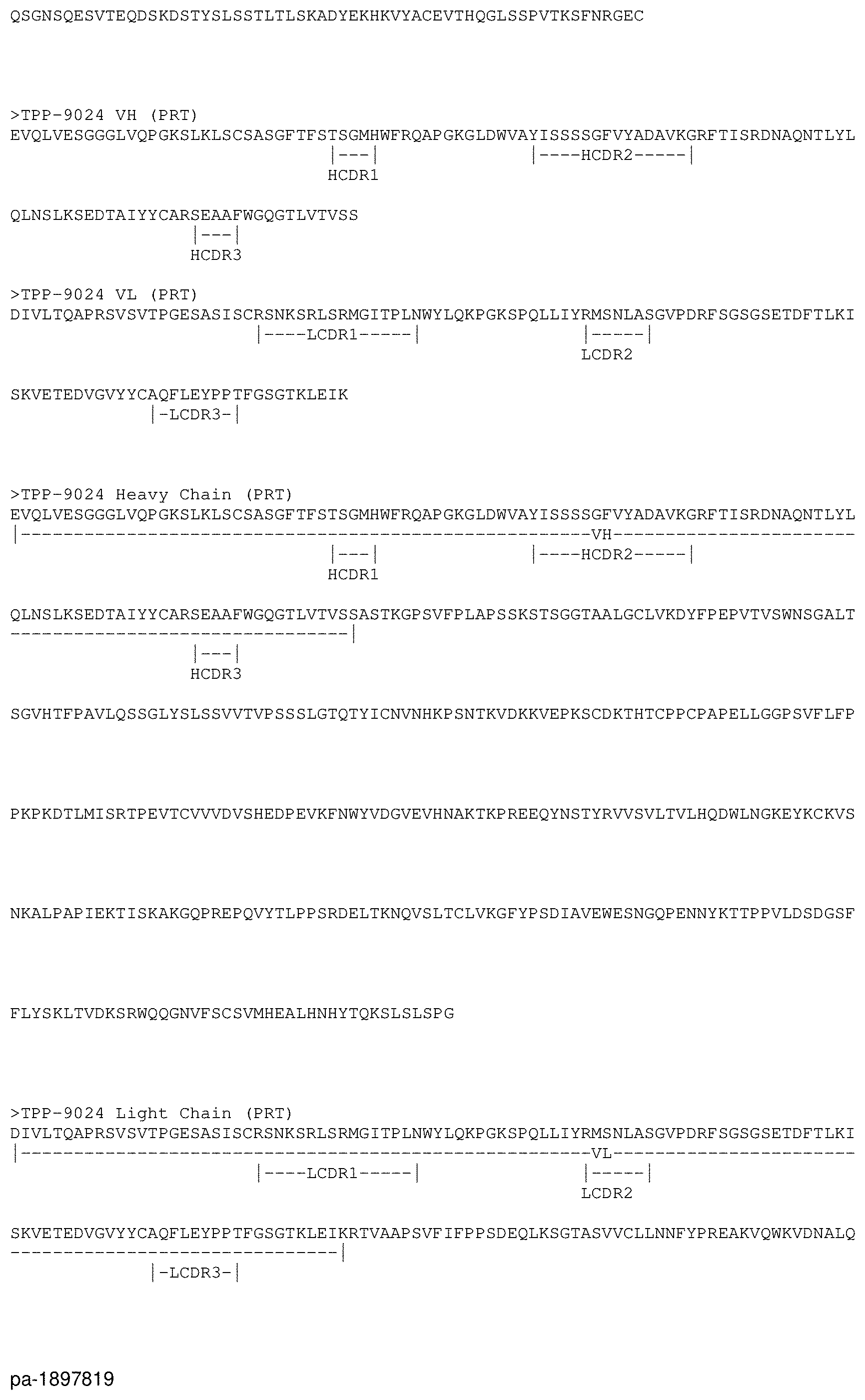
D00004

D00005

D00006

D00007

D00008
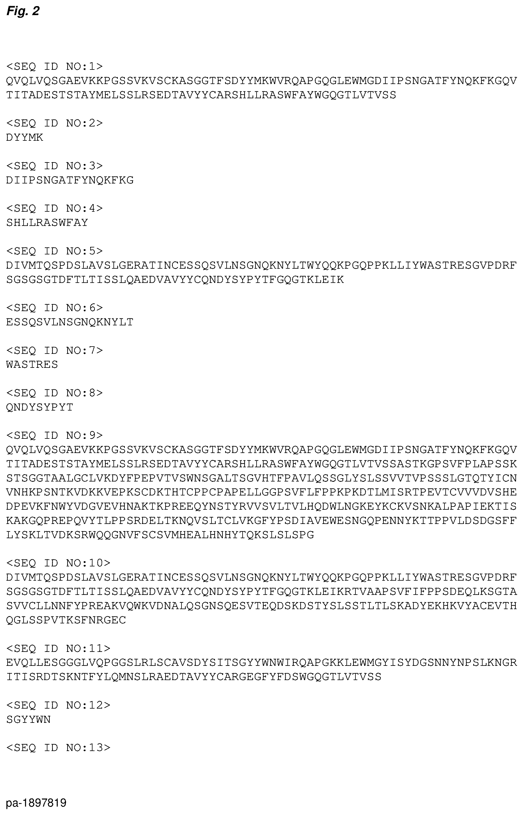
D00009

D00010

D00011
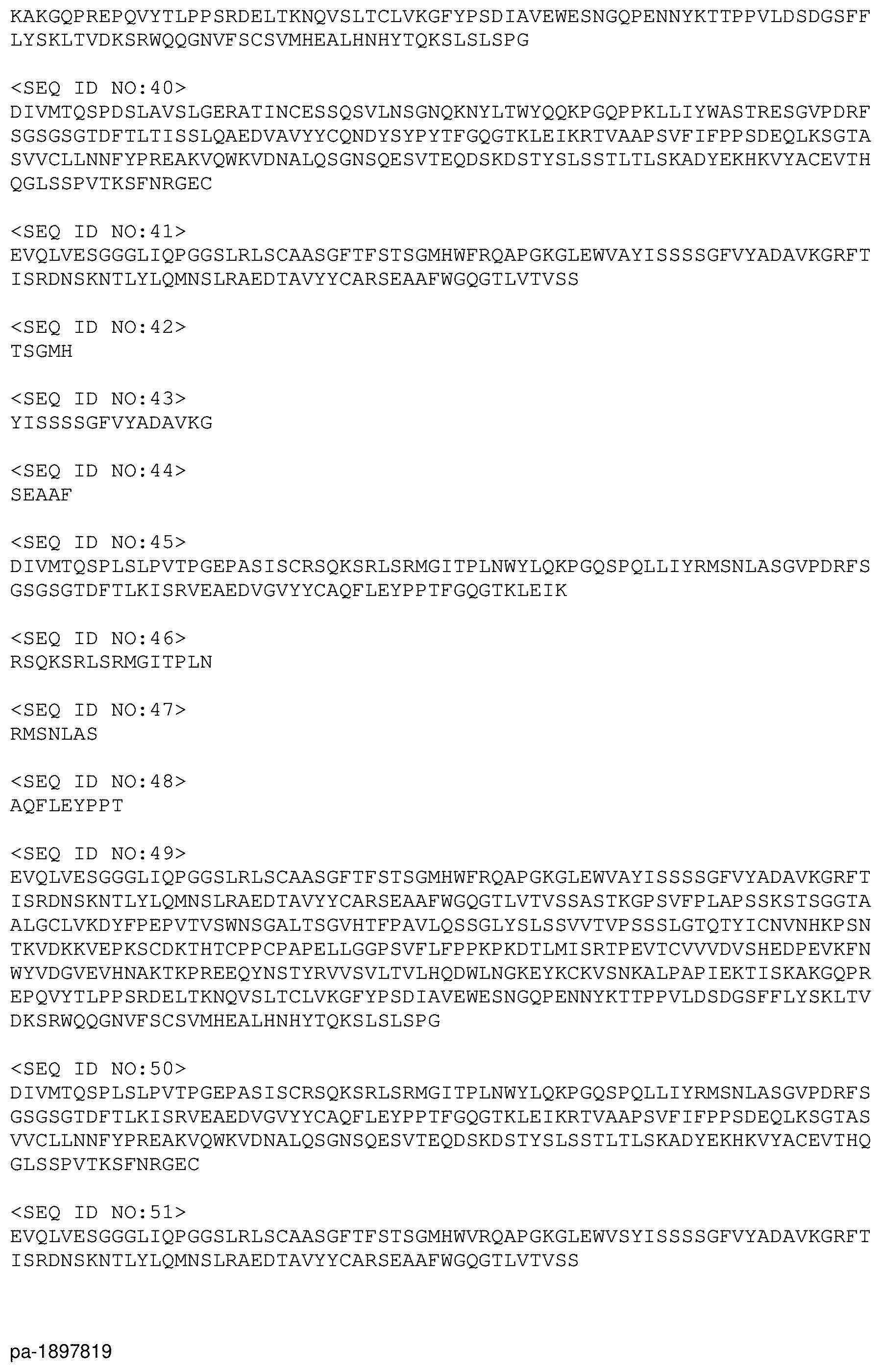
D00012

D00013

S00001
XML
uspto.report is an independent third-party trademark research tool that is not affiliated, endorsed, or sponsored by the United States Patent and Trademark Office (USPTO) or any other governmental organization. The information provided by uspto.report is based on publicly available data at the time of writing and is intended for informational purposes only.
While we strive to provide accurate and up-to-date information, we do not guarantee the accuracy, completeness, reliability, or suitability of the information displayed on this site. The use of this site is at your own risk. Any reliance you place on such information is therefore strictly at your own risk.
All official trademark data, including owner information, should be verified by visiting the official USPTO website at www.uspto.gov. This site is not intended to replace professional legal advice and should not be used as a substitute for consulting with a legal professional who is knowledgeable about trademark law.