Drug Carrier Having Self-assembled 3-d Nucleic Acid Nanostructure
AHN; Dae Ro ; et al.
U.S. patent application number 16/538109 was filed with the patent office on 2019-12-05 for drug carrier having self-assembled 3-d nucleic acid nanostructure. The applicant listed for this patent is KOREA INSTITUTE OF SCIENCE AND TECHNOLOGY. Invention is credited to Dae Ro AHN, Hyo Young KIM, Kyoung Ran KIM, Se Hoon KIM, Yong Deok LEE.
| Application Number | 20190365659 16/538109 |
| Document ID | / |
| Family ID | 53938061 |
| Filed Date | 2019-12-05 |


View All Diagrams
| United States Patent Application | 20190365659 |
| Kind Code | A1 |
| AHN; Dae Ro ; et al. | December 5, 2019 |
DRUG CARRIER HAVING SELF-ASSEMBLED 3-D NUCLEIC ACID NANOSTRUCTURE
Abstract
The present invention relates to a molecule delivery technology and a carrier technology, which may selectively deliver a material to a desired specific cell and living tissue. The present invention may be utilized in the field of a drug carrier which effectively delivers an imaging probe and a therapeutic agent to an affected part.
| Inventors: | AHN; Dae Ro; (Seoul, KR) ; KIM; Se Hoon; (Seoul, KR) ; KIM; Kyoung Ran; (Seoul, KR) ; KIM; Hyo Young; (Seoul, KR) ; LEE; Yong Deok; (Seoul, KR) | ||||||||||
| Applicant: |
|
||||||||||
|---|---|---|---|---|---|---|---|---|---|---|---|
| Family ID: | 53938061 | ||||||||||
| Appl. No.: | 16/538109 | ||||||||||
| Filed: | August 12, 2019 |
Related U.S. Patent Documents
| Application Number | Filing Date | Patent Number | ||
|---|---|---|---|---|
| 14803343 | Jul 20, 2015 | |||
| 16538109 | ||||
| Current U.S. Class: | 1/1 |
| Current CPC Class: | A61K 31/704 20130101; A61K 49/0032 20130101; A61K 49/0043 20130101; A61K 31/713 20130101; A61P 35/00 20180101; A61K 9/0019 20130101; A61K 49/0054 20130101; A61K 49/0002 20130101; A61K 31/7088 20130101; A61K 9/513 20130101; A61K 47/549 20170801; A61K 38/02 20130101 |
| International Class: | A61K 9/51 20060101 A61K009/51; A61K 31/7088 20060101 A61K031/7088; A61K 31/713 20060101 A61K031/713; A61K 38/02 20060101 A61K038/02; A61K 49/00 20060101 A61K049/00 |
Foreign Application Data
| Date | Code | Application Number |
|---|---|---|
| Jul 23, 2014 | KR | 10-2014-0093458 |
Claims
1. A method of in vivo delivering a pharmaceutically active ingredient selectively to cancer tissue, comprising administering a self-assembled 3-D nucleic acid nanostructure as a drug carrier to a subject in need thereof, wherein the self-assembled 3-D nucleic acid nanostructure comprises double strand nucleic acids including a hybridization region in which a single strand and a single strand are hybridized; the single strands form the sides of the self-assembled 3-D nucleic acid nanostructure; and the number of the single strands is 4 or more, wherein the drug carrier has an L-DNA tetrahedron structure composed of 4 strands of L-DNA 55 mer, wherein the drug carrier does not comprise a targeting ligand for the tissue, and wherein each of the single strands consists of an L-DNA nucleotide sequence selected from the group consisting of SEQ ID NOs: 15 to 19.
2. The method of claim 1, wherein the pharmaceutically active ingredient is an anticancer agent.
3. The method of claim 1, wherein the pharmaceutically active ingredient encapsulated within the nucleic acid nanostructure or bonded to the backbone of the nucleic acid nanostructure, and thereby delivered to the tissue.
4. A method of in vivo selective delivering an anticancer agent comprising administering a pharmaceutical composition comprising a self-assembled 3-D nucleic acid nanostructure drug carrier; and the anticancer agent to a subject in need thereof, wherein the self-assembled 3-D nucleic acid nanostructure comprises double strand nucleic acids including a hybridization region in which a single strand and a single strand are hybridized; the single strands form the sides of the self-assembled 3-D nucleic acid nanostructure; and the number of the single strands is 4, wherein the drug carrier has an L-DNA tetrahedron structure composed of 4 strands of L-DNA 55 mer, wherein the drug carrier does not comprise a targeting ligand for a tissue, and wherein each of the single strands consists of an L-DNA nucleotide sequence selected from the group consisting of SEQ ID NOs: 15 to 19.
5. The method of claim 1, wherein the each single strand is connected by linker.
6. The method of claim 1, wherein the 5' end and/or 3' end of the each single strand is disposed on the sides or vertices of the self-assembled 3-D nucleic acid nanostructure.
7. The method of claim 2, wherein the anticancer agent is doxorubicin.
8. The method of claim 4, wherein the anticancer agent is doxorubicin.
9. The method of claim 1, the self-assembled 3-D nucleic acid nanostructure is mixed with a pharmaceutical acceptable carrier wherein the pharmaceutical acceptable carrier is selected from the group consisting of gum acacia, methyl hydroxybenzoate, and propyl hydroxybenzoate.
10. The method of claim 4, the self-assembled 3-D nucleic acid nanostructure is mixed with a pharmaceutical acceptable carrier wherein the pharmaceutical acceptable carrier is selected from the group consisting of gum acacia, methyl hydroxybenzoate, and propyl hydroxybenzoate.
Description
CROSS-REFERENCE TO RELATED APPLICATION
[0001] This application claims priority from Korean Patent Application No. 10-2014-0093458, filed on Jul. 23, 2014, contents of which are incorporated by reference herein in its entirety.
BACKGROUND OF INVENTION
1. Field of Invention
[0002] The present invention relates to a molecule delivery technology and a carrier technology, which can selectively deliver a material to a desired specific cell and living tissue.
[0003] The field for which the technology of the present invention may be utilized is largely the field of a drug carrier which effectively delivers an imaging probe and a therapeutic agent to an affected part.
2. Background of Invention
[0004] Examples of the existing drug delivery technology include a liposome carrier, a polymer-based carrier, a peptide carrier, and the like. However, the liposome carrier has low vesicle stability, relatively low drug-encapsulation rate and limited size reproducibility, and the polymer-based carrier is disadvantageous in that it has low biodegradability and limited size reproducibility, its in vivo drug metabolism is unclear, as well as it shows immunogenicity. The peptide carrier is also disadvantageous in having low thermal stability, and limited modification possibility and immunogenicity.
[0005] On the other hand, unlike the existing carriers, a carrier composed of nucleic acids is advantageous in that it is completely biodegradable into non-toxic small molecules, the size and shape thereof can be easily adjusted to a suitable level, it can be self-assembled and has monodispersity and non-immunogenicity, as well as it can be easily conjugated with functional molecules. Accordingly, DNA nanocarriers are an emerging class in drug delivery technologies.
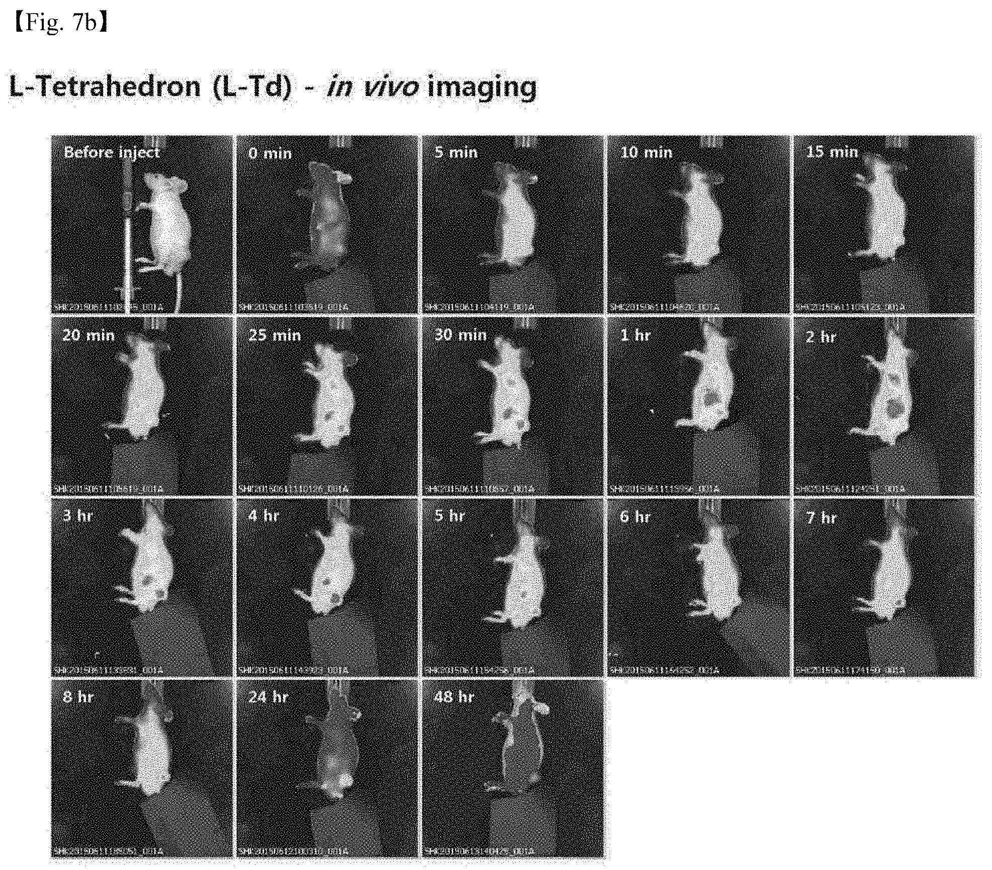
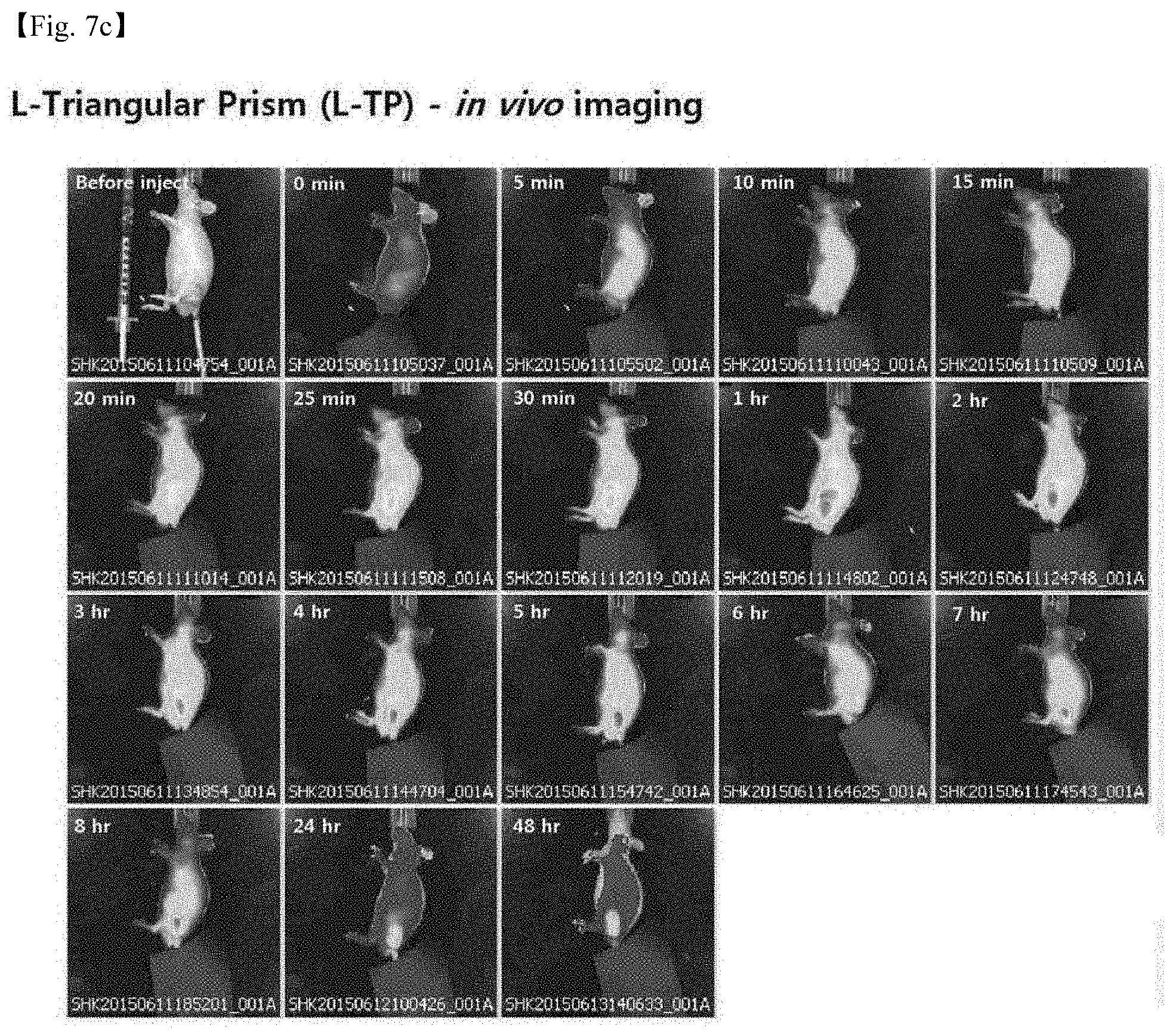
[0006] Previously, it has been shown that biocompatible DNA could self-assemble to construct various three dimensional (3D) DNA nanostructures including tetrahedron, pentahedron, cube, double pyramid, octahedron, dodecahedron, and fullerene-like structures. Among them, the DNA tetrahedron has been considered one of the most practical DNA nanostructures since it can be assembled simply from four DNA strands and prepared in high yield. A principle in which the DNA tetrahedron is assembled from 4 DNA strands is by mutual hybridization, and a schematic view explaining the principle is illustrated in FIG. 1.
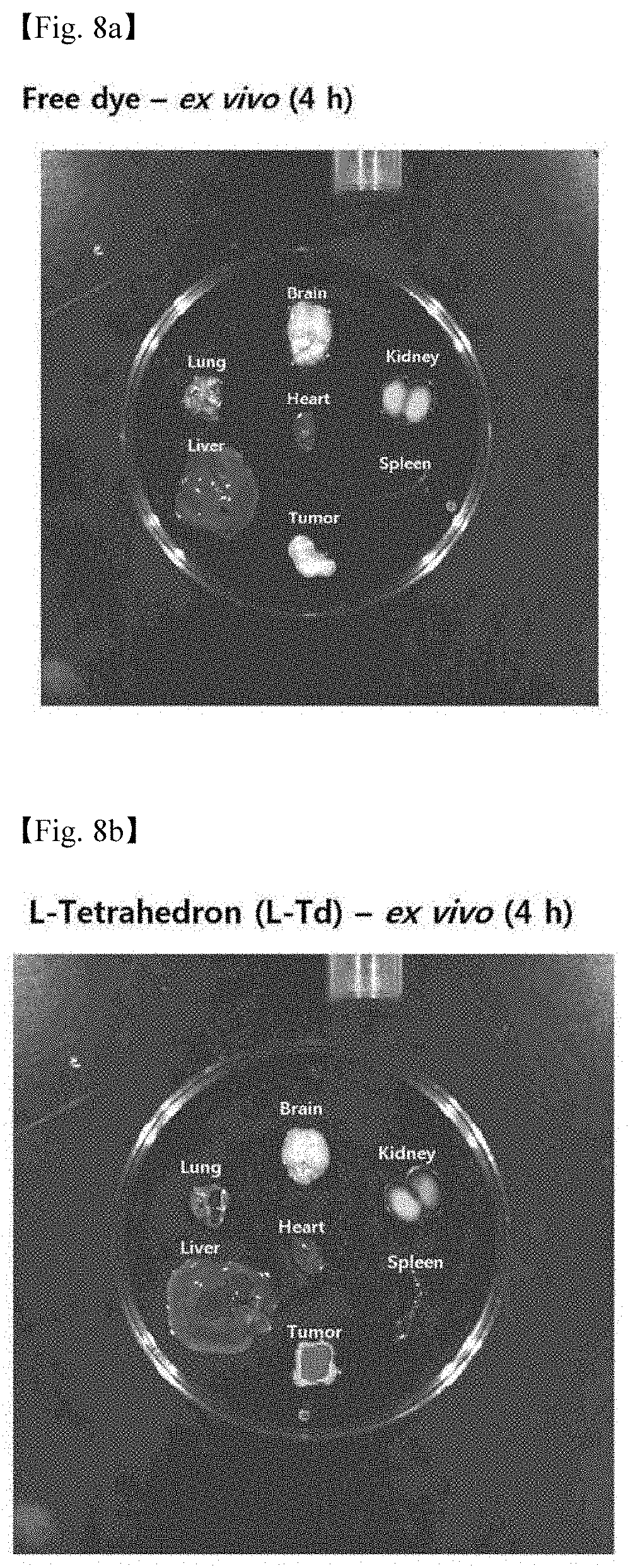
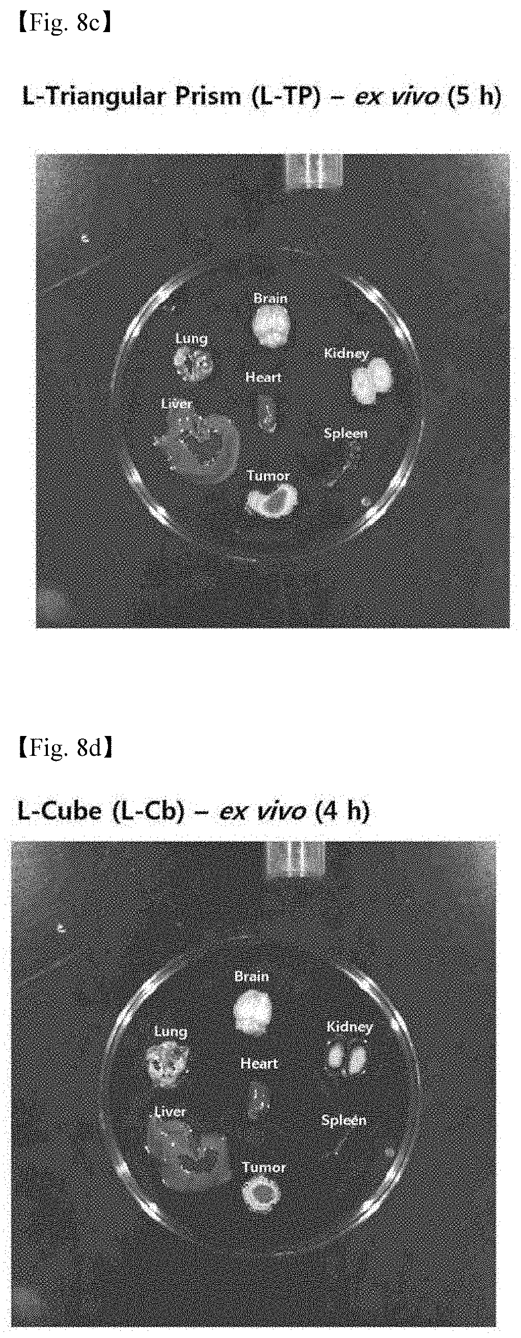
[0007] A recent report in which DNA nanostructures are taken up into mammalian cells has opened an opportunity that the nanostructures may serve an important role in being applied to biomedicine. They are often introduced into mammalian cells even without transfection agents. Further, it is known that DNA nanostructures are significantly resistant to nucleases.
[0008] Due to these characteristics, recently, DNA nanostructures have been utilized in intracellular delivery of bioactive molecules such as anticancer agents, aptamers, antisenses, immunogenic molecules, and siRNA.
[0009] Throughout the present specification, a plurality of documents are referenced, and citations thereof are indicated. The disclosure of each of the cited documents is incorporated herein by reference in its entirety to describe the level of the technical field to which the present invention pertains and the content of the present invention more apparently.
[0010] The file of this patent contains at least one drawing executed in color. Copies of this patent with color drawing(s) will be provided by the Office upon request and payment of the necessary fee.
SUMMARY OF INVENTION
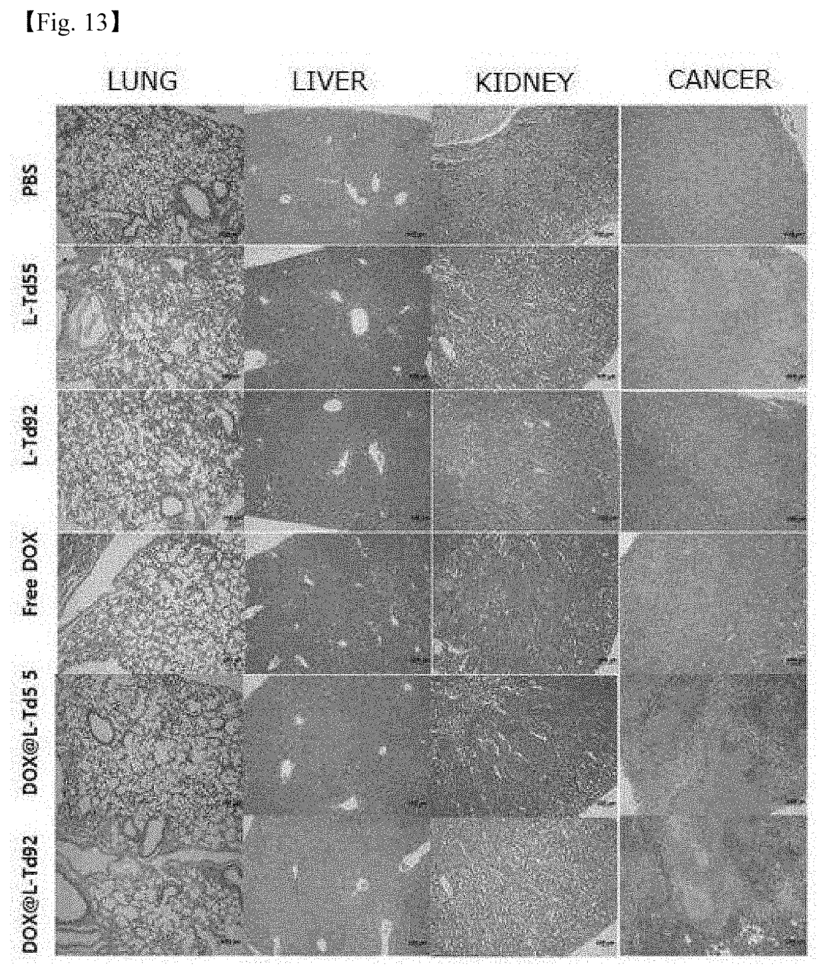
[0011] The present inventors have studied and tried to develop a drug carrier for systemic administration, which may specifically deliver a pharmaceutically active ingredient only to a specific tissue by in vivo administration. As a result, the present inventors have found that a self-assembled 3-D nucleic acid nanostructure not only has specificity for specific cells and tissues, but also can be specifically delivered only to the corresponding tissue by in vivo administration, particularly without targeting ligands, thereby completing the present invention.
[0012] Accordingly, an object of the present invention is to provide a self-assembled 3-D nucleic acid nanoparticle having selectivity for specific tissues to cells.
[0013] Another object of the present invention is to provide a drug carrier for systemic administration having the nucleic acid nanostructure, which is specific for a specific tissue.
[0014] The other objects and advantages of the present invention will be more apparent from the following detailed description, claims and drawings of the invention.
[0015] An aspect of the present invention is to provide a method of in vivo delivering a pharmaceutically active ingredient to a specific tissue, using a self-assembled 3-D nucleic acid nanostructure as a drug carrier.
[0016] In the method of present invention, the 3-D nucleic acid nanostructure drug earner can deliver a pharmaceutically active ingredient specifically only to a target tissue by in vivo administration, and has cell or tissue specificity particularly without targeting ligands.
[0017] In order to specifically deliver nucleic acid to a specific cell such as a tumor cell in the related art, there has been an attempt to deliver nucleic acid using a ligand such as antibody or folate. However, it was impossible to achieve in vivo targeting without an additional process of binding the ligand, and also there was a problem that the ligand could cause an immune reaction and the like in an organism.
[0018] Under these circumstances, the present inventors provide an in vivo drug delivery technology in which only a target tissue is specifically targeted by administration of nucleic acid nanostructures only which are completely biodegraded into non-toxic small molecules in the organism.
[0019] In an exemplary embodiment, a drug carrier of the present invention has an L-DNA nanostructure, and has in vivo cancer tissue specificity.
[0020] The L-DNA nanoparticle may have various polyhedron structures such as, for example, a tetrahedron, a pentahedron, a cube, an octahedron, a dodecahedron, a cuboctahedron, and a dodecicosahedron, a double pyramid or fullerene structure, and the like, but is not necessarily limited thereto, and may preferably have a tetrahedron structure composed of 4 strands of L-DNA 55 mer or 4 strands of L-DNA 92 mer, but is not necessarily limited thereto.
[0021] The L-DNA polyhedron structure may be formed when a plurality of single stranded nucleic acid molecules, for example, 4 to 100, 4 to 50, or 4 to 20 stranded nucleic acid molecules are self-assembled by a hybridization principle.
[0022] In another exemplary embodiment, the drug carrier of the present invention has a D-DNA nanostructure, and has specificity for liver, skin, or kidney tissue during in vivo administration.
[0023] The carrier preferably may have in vivo liver tissue specificity as a tetrahedron nanostructure composed of 4 strands of D-DNA 55 mer, or have in vivo kidney tissue specificity as a tetrahedron nanostructure composed of 4 strands of D-DNA 92 mer, but is not necessarily limited thereto.
[0024] Meanwhile, a pharmaceutically active ingredient to be delivered specifically to an organism tissue or cell by the drug carrier of the present invention may be delivered while being encapsulated in a nucleic acid nanoparticle having a polyhedron structure of the present invention, or may also be delivered while being bonded to the backbone of nucleic acid.
[0025] The pharmaceutically active ingredient may be encapsulated in a polyhedron structure, or be allowed to be bonded to the backbone of nucleic acid by an action in which linear nucleic acid strands are assembled into a 3-D polyhedron nanostructure, for example, in a state where the linear nucleic strands are mixed with the active ingredient.
[0026] The pharmaceutically active ingredient, which may be delivered into cells using the drug carrier of the present invention, is not limited to a specific kind of ingredient, and examples thereof include an anticancer agent, a contrast medium, a hormone drug, an anti-hormone drug, a vitamin preparation, a calcium agent, an inorganic preparation, a sugar preparation, an organic acid preparation, a protein amino acid preparation, an antidote, an enzyme preparation, a metabolic preparation, a diabetes combination drug, a drug for tissue repair, a chlorophyll preparation, a pigment preparation, a tumor drug, a tumor therapeutic agent, a radioactive medicine, a tissue cell diagnostic agent, a tissue cell therapeutic agent, an antibiotic preparation, an antiviral drug, a complex antibiotic preparation, a chemotherapeutic agent, a vaccine, a toxin, a toxoid, an antitoxin, leptospira serum, a blood preparation, a biological preparation, an analgesic, an immunogenic molecule, an antihistamine, an allergy drug, a non-specific immunogenic preparation, an anesthetic, an antihypnotic, a neuropsychiatric solvent, nucleic acid, an aptamer, an antisense nucleic acid, an oligonucleotide, peptide, siRNA, micro RNA, and the like, but are not necessarily limited thereto.
[0027] Another aspect of the present invention relates to a method of in vivo delivering an anticancer agent specifically to cancer cells, comprising administering a pharmaceutical composition including the aforementioned 3-D nucleic acid nanostructure drug carrier and an anticancer agent.
[0028] According to an exemplary embodiment of the present invention, the anticancer agent may be encapsulated in the nucleic acid drug carrier of the present invention or be bonded to the backbone of nucleic acid, and examples of the anticancer which may be used include doxorubicin, DNA aptamer, RNA aptamer, acivicin, aclarubicin, acodazole, acronycine, adozelesin, alanosine, aldesleukin, allopurinol sodium, altretamine, aminogl utethimide, amonafide, ampl igen, amsacrine, androgens, anguidine, aphidicolin glycinate, asaley, asparaginase, 5-azacitidine, azathioprine, Bacillus calmette-guerin (BCG), Baker's Antifol, beta-2-deoxythioguanosine, bisantrene HCl, bleomycin sulfate, busulfan, buthionine sulfoximine, BWA 773U82, BW 502U83/HCl, BW 7U85 mesylate, ceracemide, carbetimer, carboplatin, carmustine, chlorambucil, chloroquinoxaline-sulfonamide, chlorozotocin, chromomycin A3, cisplatin, cladribine, corticosteroids, Corynebacterium parvum, CPT-11, crisnatol, cyclocytidine, cyclophosphamide, cytarabine, cytembena, dabismaleate, dacarbazine, dactinomycin, daunorubicin HCl, deazauridine, dexrazoxane, dianhydrogalactitol, diaziquone, dibromodulcitol, didemnin B, diethyldithiocarbamate, diglycoaldehyde, dihydro-5-azacytidine, echinomycin, dedatrexate, edelfosine, eplolnitin, Elliott's solution, elsamitrucin, epirubicin, esorubicin, estramustine phosphate, estrogens, etanidazole, ethiofos, etoposide, fadrazole, fazarabine, fenretinide, filgrastim, finasteride, flavone acetic acid, floxuridine, fludarabine phosphate, 5-fluorouracil, Fluosol.TM., flutamide, gallium nitrate, gemcitabine, goserelin acetate, hepsulfam, hexamethylene bisacetamide, homoharringtonine, hydrazine sulfate, 4-hydroxyandrostenedione, hydrozyurea, idarubicin HCl, ifosfamide, interferon alpha, interferon beta, interferon gamma, interleukin-I alpha and beta, interleukin-3, interleukin-4, interleukin-6, 4-ipomeanol, iproplatin, isotretinoin, leucovorin calcium, leuprolide acetate, levamisole, liposomal daunorubicin, liposome encapsulated doxorubicin, lomustine, lonidamine, maytansine, mechlorethamine hydrochloride, melphalan, menogaril, merbarone, 6-mercaptopurine, mesna, methanol extract of Bacillus calmette-guerin, methotrexate, N-methylformamide, mifepristone, mitoguazone, mitomycin-C, mitotane, mitoxantrone hydrochloride, monocyte/macrophage colony-stimulating factor, nabilone, nafoxidine, neocarzinostatin, octreotide acetate, ormaplatin, oxaliplatin, paclitaxel, pala, pentostatin, piperazinedione, pipobroman, pirarubicin, piritrexim, piroxantrone hydrochloride, PIXY-321, plicamycin, porfimer sodium, prednimustine, procarbazine, progestins, pyrazofurin, razoxane, sargramostim, semustine, spirogermanium, spiromustine, streptonigrin, streptozocin, sulofenur, suramin sodium, tamoxifen, taxotere, tegafur, teniposide, terephthalamidine, teroxirone, thioguanine, thiotepa, thymidine injection, tiazofurin, topotecan, toremifene, tretinoin, trifluoperazine hydrochloride, trifluridine, trimetrexate, tumor necrosis factor (TNF), uracil mustard, vinblastine sulfate, vincristine sulfate, vindesine, vinorelbine, vinzolidine, Yoshi 864, zorubicin, mixtures thereof, and the like, but are not necessarily limited thereto.
[0029] The pharmaceutical composition may further include a pharmaceutically acceptable carrier, and the pharmaceutically acceptable earner is typically used in formulations and includes lactose, dextrose, sucrose, sorbitol, mannitol, starch, gum acacia, calcium phosphate, alginate, gelatin, calcium silicate, microcrystalline cellulose, polyvinylpyrrolidone, cellulose, water, syrup, methyl cellulose, methyl hydroxybenzoate, propyl hydroxybenzoate, talc, magnesium stearate, mineral oil, and the like, but is not limited thereto. Suitable pharmaceutically acceptable carriers and formulations are described in detail in Remington's Pharmaceutical Sciences (19th ed., 1995).
[0030] In the method of present invention, the pharmaceutical composition 1s systemically administered, and may be administered parenterally or orally. In case of parenteral administration, it may be administered through intravenous injection, intramuscular injection, and the like. An adequate administration amount of the pharmaceutical composition of the present invention may vary depending on factors, such as formulation method, administration method, age, weight, sex or disease condition of the patient, diet, administration time, administration route, elimination rate and response sensitivity.
[0031] Still another aspect of the present invention provides a method of in vivo delivering a contrast medium to a specific tissue for imaging, comprising administering a pharmaceutical composition including the aforementioned 3-D nucleic acid nanostructure drug carrier and a contrast medium.
[0032] In an exemplary embodiment, an inorganic or organic dye, a fluorescent substance, an isotope, a magnetic substance, a paramagnetic or superparamagnetic nanoparticle may be used as the contrast medium.
[0033] Herein, the inorganic or organic dyes are used for enhancing image contrast in fluorescent or optical image, radiation imaging such as computed tomography (CT) photography, and non-radiation image such as sonogram or MRI including the existing dyes, and may include, for example, diatrizoate, metrizoate, ioxaglate, iopamidol, iohexyl, ioxilan, iopromide, iodixanol, barium-based dye such as barium sulfate, Gastrografin.RTM., and the like, but are not necessarily limited thereto.
[0034] The fluorescent probe may include, for example, indocyanine, NIR (near infrared) dye, fluorescein, phycoerythrin, rhodamine, lissamine, Cy3, Cy5 (Pharmacia), chromophore, chemical luminophore, mass labeling, electron dense particle, enzymes (alkaline phosphatase or horseradish peroxidase), but is not necessarily limited thereto.
[0035] In the contrast medium composition of the present invention, the composition may be a magnetic substance, and a paramagnetic or superparamagnetic nanoparticle, and in this case, may be usefully utilized particularly in imaging using MRI.
[0036] The paramagnetic nanoparticle is a material which may induce spin-lattice relaxation, and it is possible to use, for example, Magnevist (Schering, Germany), Gd chelating compounds such as Gd-DTPA (Gd-diethylene triamine pentaacetic acid), and a material such as Gd.sub.2O.sub.3 (C. Riviere et al. J. Am. Chem. Soc. 2007, 129, 5076.) and MnO (T. Hyeon et al. Angew. Chem. Int. Ed. 2007, 46, 5397.).
[0037] Further, the superparamagnetic nanoparticle is a material which is magnetized by an externally given magnetic field, affects the spin-spin process of a hydrogen nuclear spin in surrounding water molecules by generating an induced magnetic field, and shows a dark or negative contrast effect typically compared with water by amplifying the magnetic resonance imaging signal, and for example, Feridex, Resovist, Combidex, MEIO (magnetism engineered iron oxide) and the like including oxidized iron components.
[0038] As described above, depending on what contrast medium is encapsulated in the nucleic acid nanoparticle drug carrier having a polyhedron structure of the present invention or is bonded to the backbone of nucleic acid, the contrast medium composition of the present invention may be used appropriately for fluorescence imaging, optical imaging, radiation imaging, computed tomography (CT) photography or MRI.
[0039] In a preferred exemplary embodiment of the present invention, the contrast medium composition may be for imaging of liver, kidney, or cancer tissues.
[0040] The 3-D nucleic nanoparticle drug carrier of the present invention an in vivo drug delivery technology which targets only a specific tissue even without including a target ligand during the administration in an organism.
[0041] The drug carrier of the present invention as described above may be completely biodegraded into non-toxic small molecules, and the size and shape thereof may be easily adjusted to a suitable level, and the carrier is advantageous in being capable of being self-assembled while having monodispersity and non-immunogenicity, and being easily conjugated with functional molecules, and simultaneously has specific cell or biotissue selectivity. Accordingly, the drug carrier of the present invention may be usefully utilized as a drug carrier.
BRIEF DESCRIPTION OF THE DRAWINGS
[0042] 1. FIG. 1 is a schematic view illustrating a principle in which a DNA polyhedron is assembled from 4 DNA strands by hybridization.
[0043] 2. FIG. 2a is a schematic view illustrating a process in which a library of DNA nanostructures is prepared by changing the sugar backbone and size, and the cell and biotissue specificity thereof is confirmed, and FIG. 2b illustrates a result in which 4 DNA tetrahedrons (DSS, LSS, D92, and L92) are prepared and confirmed by PAGE.
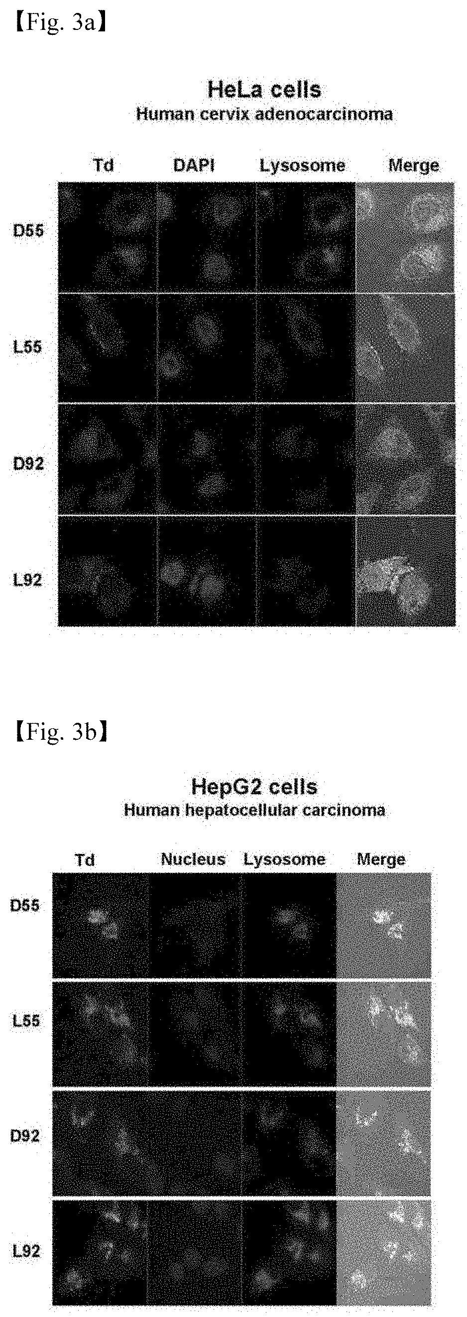
[0044] 3. FIGS. 3a to 3d illustrate the results in which cells transfected with 4 DNA tetrahedrons (DSS, LSS, D92, and L92) are observed through fluorescent microscopy (FIG. 3a: Hela cells, FIG. 3b: HepG2 cells, FIG. 3c: AS49 cells, and FIG. 3d: MCF7 cells).
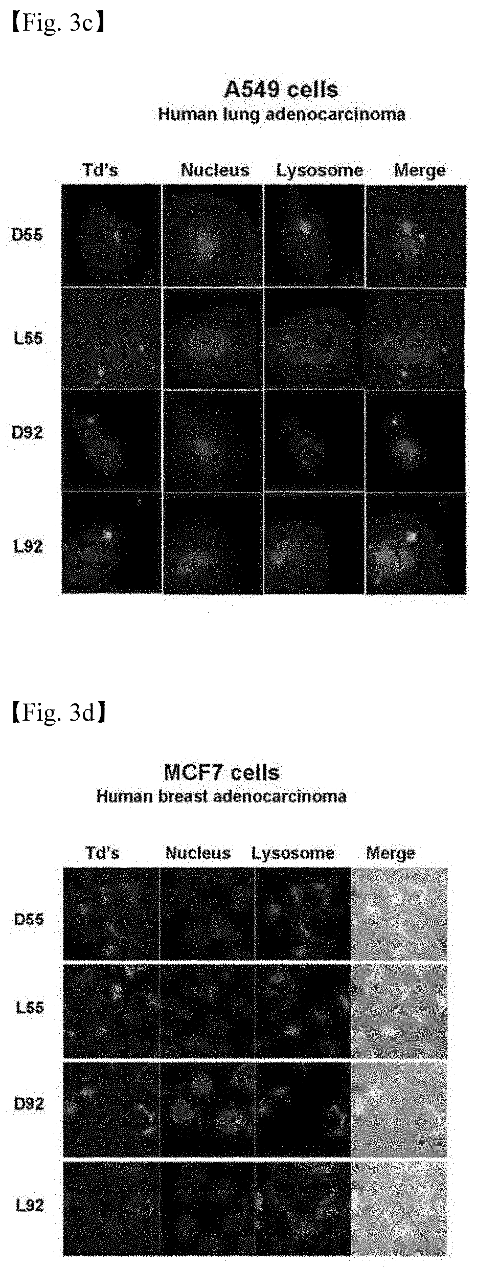
[0045] 4. FIG. 4 illustrates the results in which cells transfected with 4 DNA tetrahedrons (DSS, LSS, D92, and L92) are subjected to flow cytometry.
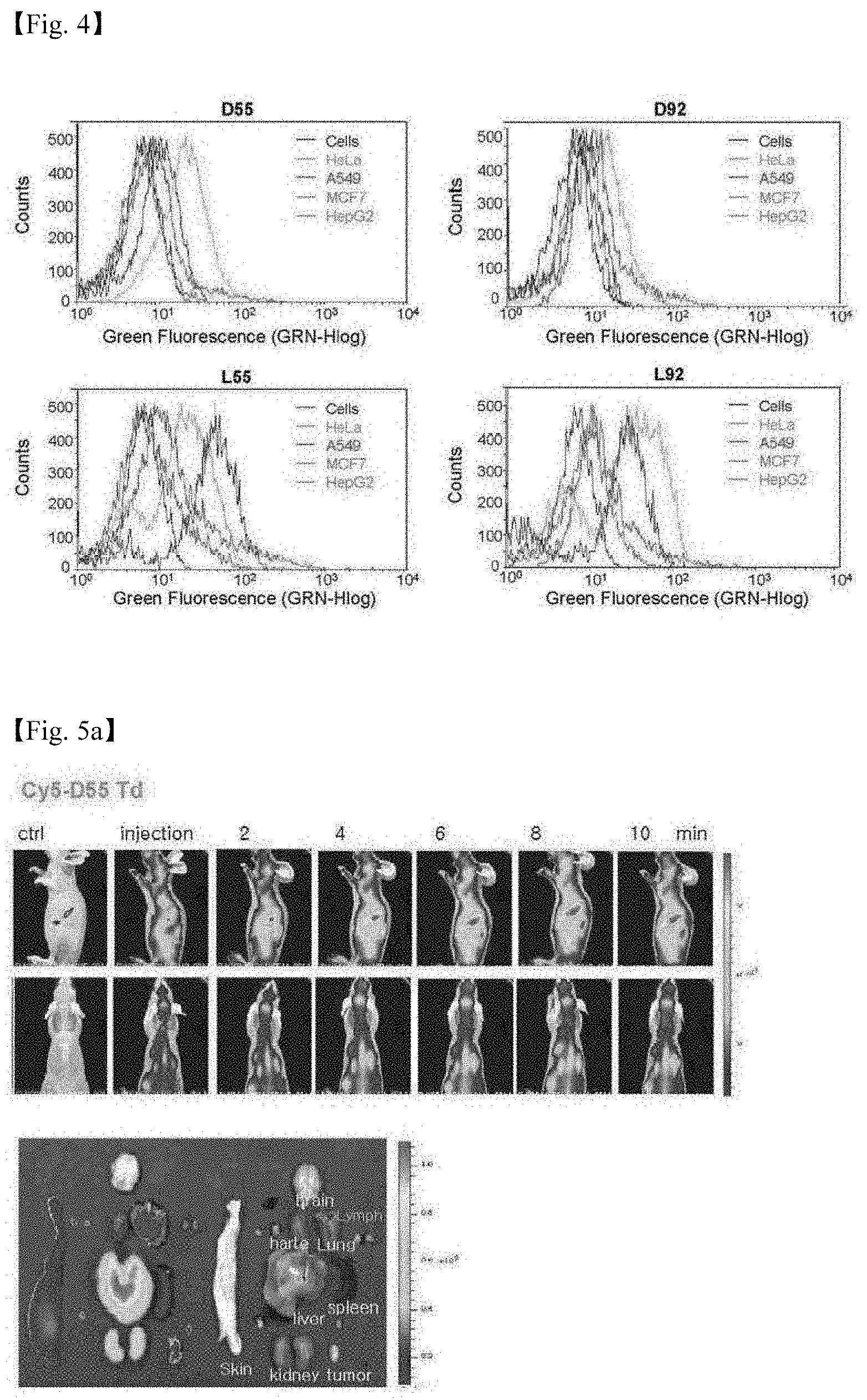
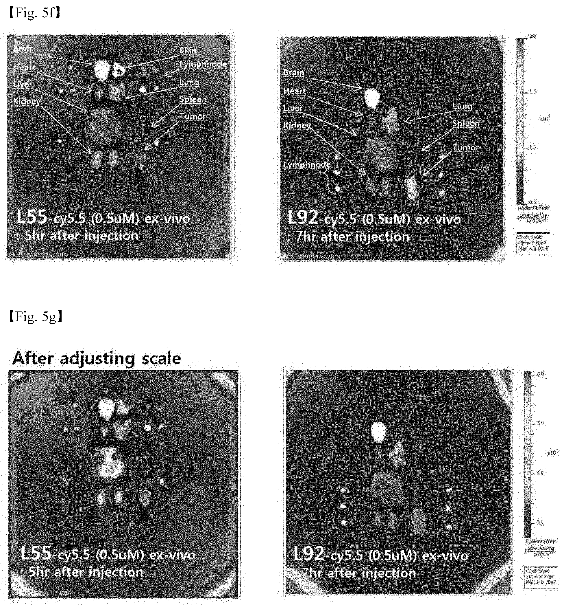
[0046] 5. FIGS. 5a to 5g illustrates fluorescent images obtained by injecting 4 DNA polyhedrons (DSS, LSS, D92, and L92) into mice. Specifically,
[0047] FIG. 5a: Fluorescent image obtained by injecting SS Td,
[0048] FIG. 5b: Fluorescent image obtained by injecting D92 Td,
[0049] FIG. 5c: Comparison of in vivo fluorescent images obtained by injecting LSS and L92 and leaving the mice to stand for 0 minute to 24 hours after the injection,
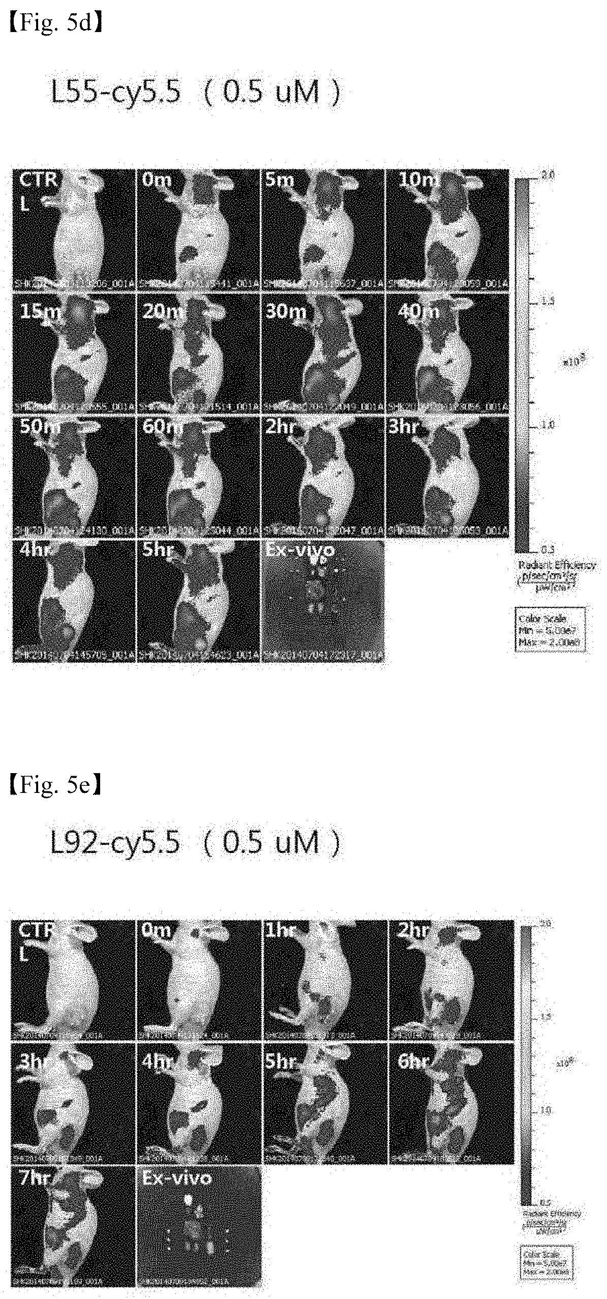
[0050] FIG. 5d: Fluorescent image obtained by injecting LSS and leaving the mouse to stand for 0 minute to 5 hours (in vivo+ex vivo 5 hours after injection),
[0051] FIG. 5e: Fluorescent image obtained by injecting L92 and leaving the mouse to stand for 0 minute to 7 hours (in vivo+ex vivo 7 hours after injection),
[0052] FIG. 5f: Comparison of ex vivo fluorescent images obtained by injecting LSS and L92 (L55 and L92 are obtained 5 hours after injection and 7 hours after injection, respectively), and
[0053] FIG. 5g: Comparison of ex vivo fluorescent images obtained by injecting L55 and L92 (after adjusting scale).
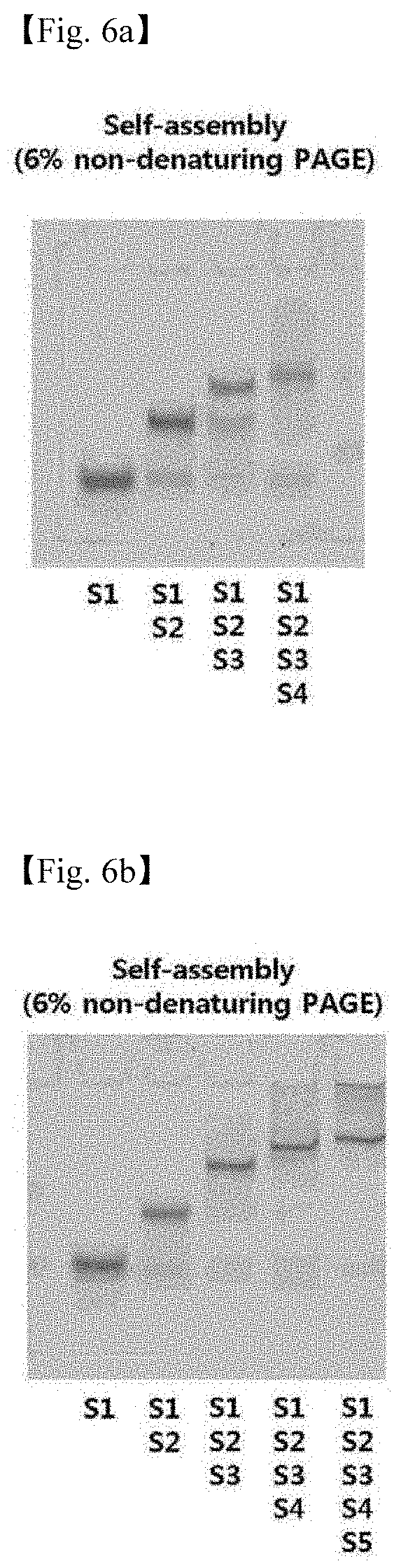
[0054] 6. FIGS. 6a to 6d illustrate the results in which the formation of the 4 (L-Td, L-TP, L-Cb, and L-Od) structures are confirmed by PAGE (FIG. 6a: L-Td, FIG. 6b: L-TP, FIG. 6c: L-Cb, and FIG. 6d: L-Od).
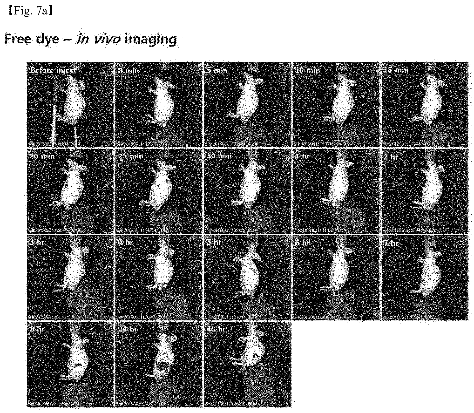
[0055] 7. FIGS. 7a to 7f illustrate the results in which L-DNA nanostructures fluorescently labeled are injected into a mouse tumor model, and a change in distribution of nanostructures in the organism is observed (FIG. 7a: Free dye, FIG. 7b: L-Td, FIG. 7c: L-TP, FIG. 7d: L-Cb, FIG. 7e: L-Od, and FIG. 7f: Comparison of in vivo images of 4 structures on the same scale).
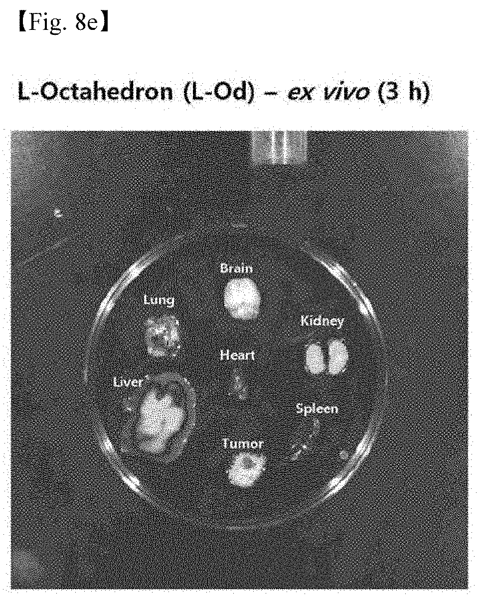
[0056] 8. FIGS. 8a to 8f illustrate the results in which L-DNA nanostructures fluorescently labeled are injected into a tumor model, the mouse was sacrificed, and 6 organs of brain, heart, lung, liver, kidney, and spleen and tumor were removed and observed (FIG. 8a: Free dye, FIG. 8b: L-Td, FIG. 8c: L-TP, FIG. 8d: L-Cb, FIG. 8e: L-Od, and FIG. 8f: Comparison of ex vivo images of 4 structures on the same scale).
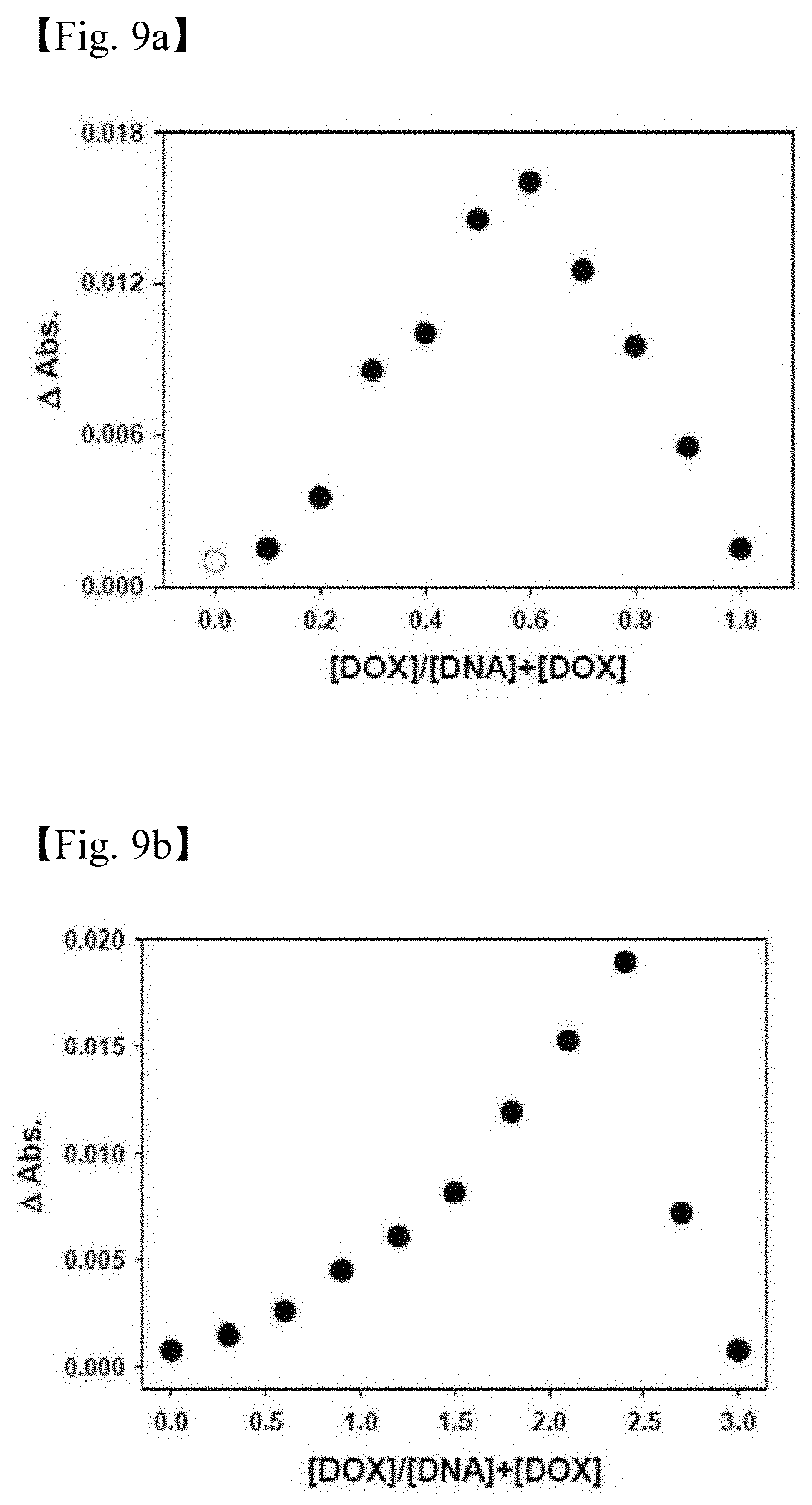
[0057] 9. FIGS. 9a and 9b illustrate the results in which an optimal binding ratio is searched using a Job plot in order to load a doxorubicin (DOX) drug into L-Td55 (FIG. 9a) and L-Td92 (FIG. 9b).
[0058] 10. FIG. 10 illustrates a schematic view of selective DOX delivery and therapy of a cancer tissue using a Xenograft cancer mouse animal model.
[0059] 11. FIG. 11 illustrates photographs of external parts of mice which were classified into total 6 groups (PBS, L-Td55, L-Td92, free DOX, DOX@L-Td55, and DOX@L-Td92) and treated for 18 days, and a photograph of tumors removed at the final day of therapy.
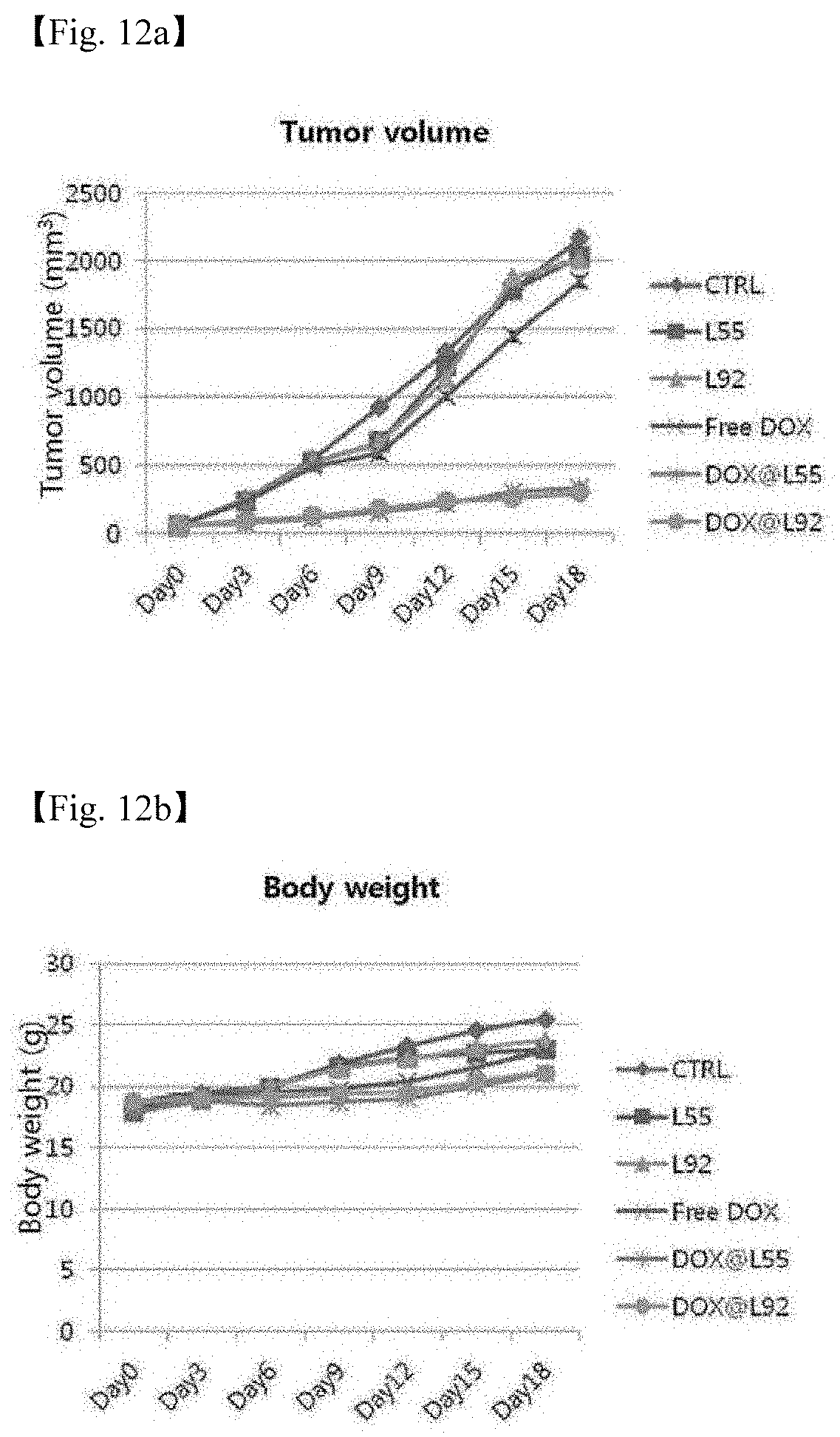
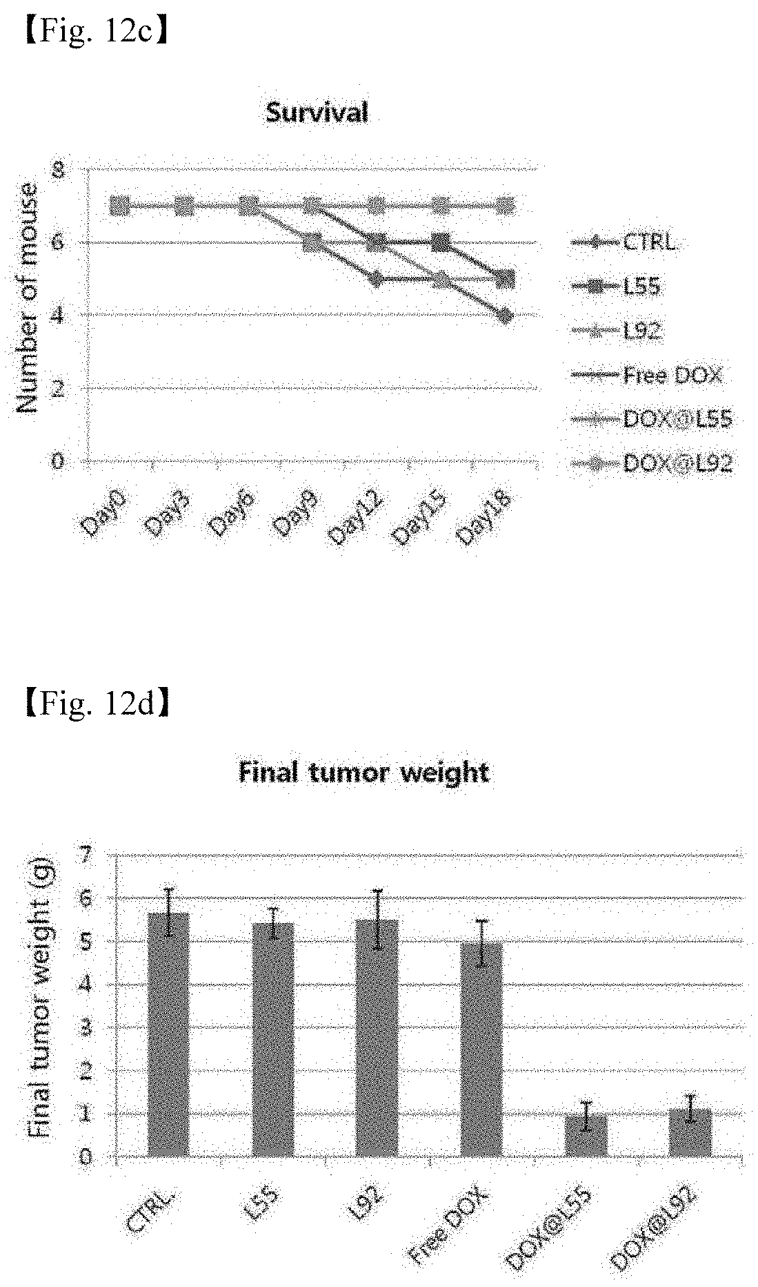
[0060] 12. FIGS. 12(a-d) are graphs illustrating changes in mice which were classified into total 6 groups (PBS, L-Td55, L-Td92, free DOX, DOX@L-Td55, and DOX@L-Td92) and treated for 18 days during the therapy period (FIG. I2a: Change in volume of tumor, FIG. 12b: Change in body weight, FIG. 12c: Number of survived mice, and FIG. 12d: Final tumor weight).
[0061] 13. FIG. 13. illustrates the results in which mice were classified into total 6 groups (PBS, L-Td55, L-Td92, free DOX, DOX@L-Td55, and DOX@L-Td92), treated for 18 days, and sacrificed at the final day of therapy, and lung, liver, kidney, and cancer removed were observed through optical microscopy.
DETAILED DESCRIPTION OF INVENTION
[0062] Hereinafter, the present invention will be described in more detail through the Examples. These Examples are provided only for more specifically describing the present invention, and it will be obvious to a person with ordinary skill in the art to which the present invention pertains that the scope of the present invention is not limited by these Examples.
EXAMPLES
[0063] I. In Vivo Biodistribution of D/L-DNA Nanostructures
[0064] 1. Preparation of DNA Nanostructures Through Synthesis and Self-Assembly of D/L-DNA Oligonucleotides 13
[0065] DNA oligonucleotides required for the Tds structure were synthesized by using a DNA synthesis standard protocol. The base sequence of the 55mer DNA standard was adopted from the Tuberfield Td, and the base sequence of the 92mer DNA standard was adopted from the Anderson (Lee et al. Nat. Nanotechnol. 2012, 7, 389-393). The oligonucleotide sequences used to constitute total 4 DNA nanostructures (D-DNA 55mer Td, L-DNA 55mer Td, D-DNA 92mer Td, and L-DNA 92mer Td) are shown in the following Table 1.
TABLE-US-00001 TABLE 1 Sequence (5' to 3') Cy5.5- ACATTCCTAAGTCTGAAACATTACAGCTTGC SEQ ID labeled TACACGAGAAGAGCCGCCATAGTA-Cy5.5 NO: 1 D/L 55_S1 Fluores- ACATTCCTAAGTCTGAAACATTACAGCTTGC SEQ ID cein- TACACGAGAAGAGCCGCCATAGTA- NO: 2 labeled fluorescein D/L 55_S1 D/L TATCACCAGGCAGTTGACAGTGTAGCAAGC SEQ ID 55_S2 TGTAATAGATGCGAGGGTCCAATAC-NH.sub.2 NO: 3 D/L TCAACTGCCTGGTGATAAAACGACACTACGT SEQ ID 55_S3 GGAATCTACTATGGCGGCTCTTC-NH.sub.2 NO: 4 D/L TTCAGACTTAGGAATGTGCTTCCCACGTAG SEQ ID 55_S4 TGTCGTTTGTATTGGACCCTCGCAT-NH.sub.2 NO: 5 Cy5.5- CTCAACTGCCTCAGACGGACAGGTGATACGA SEQ ID labeled GAGCCGGATGGGCATGCTCTTCCCGTAGAGA NO: 6 D/L TAGTACGGTATTGGACCGAGTCCTCGCATG- 92_S1 Cy5.5 Fluores- CTCAACTGCCTCAGACGGACAGGTGATACGA SEQ ID cein- GCCGGATGGGCATGCTCTTCCCGTAGAGATG NO: 7 labeled AAGTACGGTATTGGACCGAGTCCTCGCATG- D/L fluorecein 92_S1 D/L CGTATCACCTGTCCGTCTGAGGCAGTTGAGAG SEQ ID 92_S2 ATCTCGAACATTCCTAAGTCTGAAGATCCATT NO: 8 TATCACCAGCTGCTGCACGCCATAGTAG-NH.sub.2 D/L GGATCTTCAGACTTAGGAATGTTCGAGATCAC SEQ ID 92_S3 ATGCGAGGACTCGCTCCAATACCGTACTAACG NO: 9 ATTACAGATCAAAGCTACTTGCTACACG-NH.sub.2 D/L CTCTACGGGAAGAGCATGCCCATCCGGCTCAC SEQ ID 92_S4 TACTATGGCGTGCAGCAGCTGGTGATAAAACG NO: 10 TGTAGCAAGTAGCTTTGATCTGTAATCG-NH.sub.2
[0066] A library of DNA nanostructures was prepared by performing the Td assembly as described in the document (Kim et al. Chem. Sci., 2014, 5, 1533-1537) to change the sugar backbone and size (see FIG. 2a), and then the self-assembled D and L-Td (D-DNA 55mer Td, L-DNA 55mer Td, D-DNA 92mer Td, and L-DNA 92mer Td) were confirmed by performing 6% non-denaturing polyacrylamide gel electrophoresis (PAGE) (FIG. 2b).
[0067] 2. Cell Selectivity of DNA Nanostructures
[0068] (1) Transfection of DNA Nanostructures into HeLa, HepG2, A549 and MCF7 Cells
[0069] In order to confirm the cell selectivity of each DNA nanostructure, cancer cells were treated with the DNA nanostructures prepared above.
[0070] Specifically, HeLa, HepG2, A549 and MCF7 cells were each inoculated into a glass-bottom 35-mm petri dish including a DMEM medium (Gibco, USA) containing 10% fetal bovine serum inactivated with heat, 1% penicillin, and streptomycin, and then the dish was cultured in a wet atmosphere including 5% CO2 at 37.degree. C. The growth medium was removed from each cell sample and washed twice with PBS (Gibco, USA), and the DNA nanostructures prepared were subjected to transfection treatment in each cell.
[0071] (2) Microscopic Images of DNA Nanostructures in Cell
[0072] Cells transfected with the DNA nanostructures were observed under a fluorescent microscope (DeltaVision, Applied Precision, USA) and living cells were imaged, and the results are each shown in FIG. 3 (FIGS. 3a to 3d).
[0073] As a result of the experiment, it could be seen that the DNA nanostructures were entered into the cells, and it could be confirmed that the DNA nanostructures were in the cytoplasm region without being delivered to the cell nucleus.
[0074] (3) Flow Cytometry
[0075] The HeLa, HepG2, A549 and MCF7 cells were cultured with DNA molecules fluorescently labeled using the method which is the same as that adopted in the transfection experiment, the fluorescence intensity of the cells was evaluated using a flow cytometer (FC500, Beckman coulter, USA), and then, the result is shown in FIG. 4.
[0076] Based on the result that the amount of each nanostructure delivered into the cells was quantified through a flow cytometry, the amounts delivered for each cell could be compared with each other, and as a result of the experiment, it was confirmed that D55 had HepG2 cell selectivity, L55 simultaneously had strong HeLa cell selectivity and considerable HepG2 cell selectivity, and L92 had HeLa and HepG2 cell selectivity (FIG. 4).
[0077] As described above, since the kind of cell delivered in a large amount for each structure is present, it was confirmed that a structure having cell selectivity could be discovered through the construction of a library composed of various DNA nanostructures.
[0078] 3. Tissue Selectivity of DNA Nanostructures
[0079] (1) In Vivo Imaging
[0080] The animal experiment was approved by the Institutional Animal Care and Use Committee of Korean Institute of Science and Technology, and all the mice were treated according to the regulations of the committee. For in vivo imaging and establishment of a disease model, a mouse was anesthetized by intraperitoneally injecting 0.5% pentobarbital sodium (0.01 m L/g). An animal disease model was established by using a BALB/c nude mouse (5 weeks old, male, Orient Bio Inc., Korea). A tumor was produced by subcutaneously inoculating SCC7 cells (1.0.times.10.sup.6 cells suspended in a culture medium) into the thigh of the mouse.
[0081] The DNA structures prepared were injected into the caudal vein of the mouse, and fluorescent images obtained by using a CCD camera performed in a highly sensitive imaging system (IVIS-spectrum, Perkin-Elmer, USA) are shown in FIG. 5 (FIGS. 5a to 5e).
[0082] (2) Ex Vivo Imaging and Histological Analysis After an in vivo imaging study, ex vivo near-infrared fluorescence images for excised organs and the other sites of the body were obtained by using an IVIS-spectrum imaging system including the same obtaining set as that used for in vivo imaging, and the results are shown in FIG. 5 (the second photograph in FIGS. 5a and 5b, the last photograph in FIGS. 5d and 5e, FIG. 5f, and FIG. 5g).
[0083] As a result of the experiment, it was confirmed that D55 was delivered more selectively to the liver, and distributed even in the skin tissue. On the contrary, D92 was delivered more selectively to the kidney. It was shown that L55 and L92 were accumulated, exhibiting high selectivity for the cancer tissue In addition, it was observed that as time elapsed, L-Td's had accumulated been more selectively in cancer instead of being distributed throughout the tissue than D-Td (after about 6 to 7 hours), and had escaped after 24 hours.
[0084] II. Evaluation of In Vivo Biodistribution of L-DNA Nanostructures
[0085] 1. Formation of Structure Labeled with Fluorescent Dye
[0086] (1) Formation of 4 Structures (L-Td, L-TP, L-Cb, and L-Od)
[0087] The oligonucleotide sequences used to self-assemble total four L-DNA nanostructures having tetrahedron (L-Td), triangular prism (L-TP), cube (L-Cb) and octahedron (L-Od) shapes as an L-DNA nanostructure are shown in the following Tables 2 to 5.
TABLE-US-00002 TABLE 2 Structure Sequence (5' to 3') Tetra- S1 CGATGTCTAAGCTGACCG/iSp18/GGAC SEQ ID hedron CGTGATTCCATGAC/iSp18/CTTAGAGT NO: 11 (L-Td) TGCCACCAGG S2 GTCATGGAATCACGGTCC/iSp18/GGCT SEQ ID CACATTGGCTACAG/iSp18/CTATCCGA NO: 12 TCGAGGCATG S3 CATGCCTCGATCGGATAG/iSp18/CGG SEQ ID TCAGCTTAGACATCG/iSp18/GCAAGT NO: 13 GCTGCGTCATAC S4 CCTGGTGGCAACTCTAAG/iSp18/GTA SEQ ID TGACGCAGCACTTGC/iSp18/CTGTAG NO: 14 CCAATGTGAGCC
TABLE-US-00003 TABLE 3 Structure Sequence (5' to 3') Trian- S1 CGATGTCTAAGCTGACCG/iSp18/ SEQ ID gular GGACCGTGATTCCATGAC/iSp18/ NO: 15 prism CTTAGAGTTGCCACCAGG/iSp18/ (L-TP) GAATCCTATGCTCGGACG S2 CGGTCAGCTTAGACATCG/iSp18/ SEQ ID GGCTCACATTGGCTACAG/iSp18/ NO: 16 CTATCCGATCGAGGCATG/iSp18/ CATACTGAGAGCGTTCCG S3 CCTGGTGGCAACTCTAAG/iSp18/ SEQ ID GCGTATCTGAACTGCGAC/iSp18/ NO: 17 CATGCCTCGATCGGATAG/iSp18/ CCACCGAATGGTGTATCG S4 CTGTAGCCAATGTGAGCC/iSp18/ SEQ ID CGTCCGAGCATAGGATTC/iSp18/ NO: 18 CGATACACCATTCGGTGG S5 GTCGCAGTTCAGATACGC/iSp18/ SEQ ID GTCATGGAATCACGGTCC/iSp18/ NO: 19 CGGAACGCTCTCAGTATG
TABLE-US-00004 TABLE 4 Struc- ture Sequence (5' to 3') Cube S1 CGATGTCTAAGCTGACCG/iSp18/ SEQ ID (L-Cb) GGACCGTGATTCCATGAC/iSp18/ NO: 20 CTTAGAGTTGCCACCAGG/iSp18/ GAATCCTATGCTCGGACG S2 CCTGGTGGCAACTCTAAG/iSp18/ SEQ ID GGCTCACATTGGCTACAG/iSp18/ NO: 21 CTATCCGATCGAGGCATG/iSp18/ CATACTGAGAGCGTTCCG S3 CATGCCTCGATCGGATAG/iSp18/ SEQ ID GCGTATCTGAACTGCGAC/iSp18/ NO: 22 GCAAGTGCTGCGTCATAC/iSp18/ CCACCGAATGGTGTATCG S4 GGCATTGTACCGTAACCG/iSp18/ SEQ ID CGGTCAGCTTAGACATCG/iSp18/ NO: 23 CGCAAGACGTTAGTGTCC/iSp18/ GTATGACGCAGCACTTGC S5 GTCATGGAATCACGGTCC/iSp18/ SEQ ID CGGTTACGGTACAATGCC/iSp18/ NO: 24 GTCGCAGTTCAGATACGC/iSp18/ CTGTAGCCAATGTGAGCC S6 GGACACTAACGTCTTGCC/iSp18/ SEQ ID CGTCCGAGCATAGGATTC/iSp18/ NO:25 CGGAACGCTCTCAGTATG/iSp18/ CGATACACCATTCGGTGG
TABLE-US-00005 TABLE 5 Struc- ture Sequence (5' to 3') Octa- S1 CGATGTCTAAGCTGACCG/iSp18/ SEQ ID hedron GGACCGTGATTCCATGAC/iSp18/ NO: 26 (L-Od) CTTAGAGTTGCCACCAGG S2 GTCATGGAATCACGGTCC/iSp18/ SEQ ID GGCTCACATTGGCTACAG/iSp18/ NO: 27 CTATCCGATCGAGGCATG S3 CTGTAGCCAATGTGAGCC/iSp18/ SEQ ID GCGTATCTGAACTGCGAC/iSp18/ NO: 28 GCAAGTGCTGCGTCATAC S4 GTCGCAGTTCAGATACGC/iSp18/ SEQ. ID CGGTCAGCTTAGACATCG/iSp18/ NO: 29 CGCAAGACGTTAGTGTCC S5 GAATCCTATGCTCGGACG/iSp18/ SEQ ID CATACTGAGAGCGTTCCG/iSp18/ NO: 30 CCTGGTGGCAACTCTAAG S6 CCACCGAATGGTGTATCG/iSp18/ SEQ ID CGTCCGAGCATAGGATTC/iSp18/ NO: 31 CATGCCTCGATCGGATAG S7 GGCATTGTACCGTAACCG/iSp18/ SEQ ID CGATACACCATTCGGTGG/iSp18/ NO: 32 GTATGACGCAGCACTTGC S8 CGGAACGCTCTCAGTATG/iSp18/ SEQ ID CGGTTACGGTACAATGCC/iSp18/ NO: 33 GGACACTAACGTCTTGCG
[0088] The strands constituting the structure were mixed so as to have a concentration of 1 .mu.M based on each strand. In this case, a TM buffer (10 mM Tris-HCl, 5 mM MgCl.sub.2, pH 8.0) was used as a buffer. The mixture was denatured through heating at 95.degree. C. by using a RT-PCR machine, and was slowly cooled at 4.degree. C. to be annealed.
[0089] As a result, it was confirmed by a non-denaturing PAGE that the resulting four (L-Td, L-TP, L-Cb, and L-Od) structures had been formed, and the results are each shown in FIGS. 6a to 6d (FIG. 6a: L-Td, FIG. 6b: L-TP, FIG. 6c: L-Cb, and FIG. 6d: L-Od).
[0090] (2) Labeling with Fluorescent Dye (Red Fluorescence)
[0091] A red fluorescent dye (SYTO.RTM. 62 Red Fluorescent Nucleic Acid Stain, S11344, life technologies) having binding properties with DNA was mixed so as to have a final concentration of 1 .mu.M. Herein, the DNA structure and the dye were bonded to each other at a concentration ratio of 1:1. The red fluorescent dye used in the present experiment has properties which do not show fluorescence when being present alone, but show fluorescence while being bonded to DNA.
[0092] 2. Evaluation of In Vivo Distribution
[0093] (1) Preparation of Animal Tumor Model
[0094] A balb/c nude mouse (5 weeks old, male) was used as an experimental animal group, and a tumor was formed by subcutaneously inoculating SCC7 cells (1.0.times.10.sup.6 cells suspended in the culture medium) into the left thigh of the mouse. When the tumor volume became 50 mm.sup.3 or more, the tumor was used for the experiment.
[0095] (2) In Vivo and Ex Vivo Imaging
[0096] A sample of the L-DNA nanostructure (a final concentration of 1 .mu.M, 200 .mu.L) labeled with fluorescence prepared in 1 was injected into the caudal vein of the mouse tumor model (I. V. injection). When compared to the case before the sample was injected, a change in in vivo distribution of the nanostructure was observed by using an IVIS imaging system apparatus at immediately after injection (0 min), 5, 10, 15, 20, 25, and 30 minutes, and 1, 2, 3, 4, 5, 6, 7, 8, 24, and 48 hours (filter set: Ex=640 nm, Em=680 nm), and the results are each shown in FIGS. 7a to 7f (FIG. 7a: Free dye, FIG. 7b: L-Td, FIG. 7c: L-TP, FIG. 7d: L-Cb, FIG. 7e: L-Od, and FIG. 7f: Comparison of in vivo images of four structures on the same scale).
[0097] Based on the in vivo imaging results, ex vivo images were observed by sacrificing the mouse at the time when the fluorescent intensity of each structure in the tumor was highest and removing 6 organs of brain, heart, lung, liver, kidney, and spleen and tumor, and are each shown in FIGS. 8a to 8f (FIG. 8a: Free dye, FIG. 8b: L-Td, FIG. 8c: L-TP, FIG. 8d: L-Cb, FIG. 8e: L-Od, and FIG. 8f: Comparison of ex vivo images of four structures on the same scale).
[0098] As a result of the experiment, it could be confirmed that all the four L-DNA nanostructures used in the experiment showed cancer tissue selectivity.
[0099] Among them, the best cancer tissue selectivity was found in L-Td, and the next best selectivity was found in this order of L-TP, L-Cb, and L-Od. In the tissue other than the cancer tissue, it was found that L-structures were usually found from the kidney.
[0100] III. Evaluation of Selective Delivery of Drug to Cancer Tissue In Vivo
[0101] (1) Optimal Binding Ratio Using Job Plot
[0102] In order to confirm the optimal binding ratio of Td and doxorubicin (DOX), the Job's plot method was used as in FIG. 9 (see analytical chemistry, 1971, 43, 1265, FIG. 9a: L-Td55 and FIG. 9b: L-Td92). Td and DOX were prepared at a concentration of 1 .mu.M, respectively, in the case of L-Td55 and at a concentration of 1 .mu.M and 3 .mu.M, respectively, in the case of L-Td92, mixed at each volume ratio (see the following Table 6, and the total volume was fixed at 100 .mu.l), and then incubated at normal temperature for 1 hour.
TABLE-US-00006 TABLE 6 Sample Td 10 9 8 7 6 5 4 3 2 1 0 (Td-DOX Complex) DOX 0 1 2 3 4 5 6 7 8 9 10 Total: 100 .mu.l Control TM buffer 10 9 8 7 6 5 4 3 2 1 0 (free DOX) DOX 0 1 2 3 4 5 6 7 8 9 10 Total: 100 .mu.l
[0103] And then, the samples were scanned at a wavelength of 200 to 800 nm by using an UV-visible spectrophotometer, and free DOX to which a TM buffer was added instead of the Td sample was used as a control. At 480 nm where the highest absorbance value of DOX was shown, it was judged that the binding ratio used in the sample in which the difference in absorbance values of free DOX and Dox loaded Td was highest formed a composite best, and at a ratio thus determined, DOX was loaded into Td.
[0104] (2) Establishment of Xenograft Tumor Model
[0105] A balb/c nude mouse (5 weeks old, male) was used as an experimental animal group, and a tumor was formed by subcutaneously inoculating SCC7 cells (1.0.times.10.sup.6 cells suspended in the culture medium) into the left thigh of the mouse. When the tumor volume became approximately 50 mm.sup.3, the tumor was used for the experiment.
[0106] (3) Cancer Tissue Selective DOX and Therapy
[0107] For the selective therapy of cancer tissue, DOX was each loaded into L-Td55 and L-Td92, which are a carrier having cancer tissue selectivity. A sample was prepared by mixing the nanostructure with DOX at the optimal binding ratio obtained through the Job plot method of the slide #1 (L-Td55: DOX=1:24, L-Td92: DOX=1:48).
[0108] The therapy experiment was performed by classifying the mice into total six groups (PBS, L-Td55, L-Td92, free DOX, DOX@L-Td55, and DOX@L-Td92), and 7 mice per group were used. Before the therapy was performed each time, the tumor volume and the mouse body weight were measured. In this case, the tumor volume was calculated by a method of (minor axis.sup.2*major axis)/2. Each sample was injected into the caudal vein of the mice completely subjected to measurement (see Slide #2 for concentration), the therapy was performed once every three days, and the therapy was performed total 6 times. On day 18 which was 3 days after the last 6th therapy, the final tumor weight was measured by measuring the tumor volume and the mouse body weight, and then measuring the mouse body weight to remove the tumor, and the results are shown in FIGS. 11 and 12 (FIG. 11: Photographs of external parts of mice and a photograph of tumors removed, FIG. 12a: Change in volume of tumor, FIG. 12b: Change in body weight, FIG. 12c: Number of survived m ice, and FIG. 12d: Final tumor weight).
[0109] (4) Tissue Fragment Experiment (Histological Analysis)
[0110] The therapy was finished, and 6 organs of brain, heart, lung, liver, kidney, and spleen and tumor were removed by sacrificing the survived mouse for each group, thereby observing the presence of organ damage and the tumor state. All the organs and tumor were fixed in 4% formaldehyde (4.degree. C., overnight), and then embedded in paraffin after the tissue dehydration process. The paraffin block thus prepared was cut into a thickness of 5 .mu.m, stained with hematoxylin and eosin, and observed through optical microscopy, and the results are shown in FIG. 13.
[0111] As a result of the experiment, it was confirmed that tumors were increased 4 to 5 times in the case of DOX@L55 and DOX@L92, while tumor volumes were increased up to about 40 times for the other groups, and thus the growth in tumor was delayed when DOX was loaded into the carrier.
[0112] Due to the tumor growth rate of the tumor bearing mice using SCC7 known to be rapidly growing, it was judged that an increase in tumor volume had been observed in all the cases. When a tumor model is established using human-derived cells, a much better therapeutic effect is expected.
[0113] For the control which was not subjected to therapy, the L55 treatment group, the L92 treatment group, and the free DOX treatment group showing slight therapeutic effects, the animal groups of individual 1 to 3 mice died during the therapy period.
[0114] When the graph of change in body weight is observed, it is judged that the increase in body weight is due to an increase in tumor volume. When the weights of tumors obtained through ex vivo imaging were compared with each other, the tumor weights of the groups other than DOX@L55 or DOX@L92 exhibited values about 5 times higher than the tumor weights of DOX@L55 or DOX@L92.
[0115] When the tissue fragments were observed, no part particularly damaged by DOX had been found in the tissues other than cancer tissues.
Sequence CWU 1
1
33155DNAArtificial SequenceD/L 55_S1 oligonucleotide for
D-DNA/L-DNAmisc_feature(55)Cy5.5-labeled 1acattcctaa gtctgaaaca
ttacagcttg ctacacgaga agagccgcca tagta 55255DNAArtificial
SequenceD/L 55_S1 oligonucleotide for
D-DNA/L-DNAmisc_feature(55)fluorescein-labeled 2acattcctaa
gtctgaaaca ttacagcttg ctacacgaga agagccgcca tagta
55355DNAArtificial SequenceD/L 55_S2 oligonucleotide for
D-DNA/L-DNA 3tatcaccagg cagttgacag tgtagcaagc tgtaatagat gcgagggtcc
aatac 55455DNAArtificial SequenceD/L 55_S3 oligonucleotide for
D-DNA/L-DNA 4tcaactgcct ggtgataaaa cgacactacg tgggaatcta ctatggcggc
tcttc 55555DNAArtificial SequenceD/L 55_S4 oligonucleotide for
D-DNA/L-DNA 5ttcagactta ggaatgtgct tcccacgtag tgtcgtttgt attggaccct
cgcat 55692DNAArtificial SequenceD/L 92_S1 oligonucleotide for
D-DNA/L-DNAmisc_feature(92)Cy5.5-labeled 6ctcaactgcc tcagacggac
aggtgatacg agagccggat gggcatgctc ttcccgtaga 60gatagtacgg tattggaccg
agtcctcgca tg 92792DNAArtificial SequenceD/L 92_S1 oligonucleotide
for D-DNA/L-DNAmisc_feature(92)fluorescein-labeled 7ctcaactgcc
tcagacggac aggtgatacg agagccggat gggcatgctc ttcccgtaga 60gatagtacgg
tattggaccg agtcctcgca tg 92892DNAArtificial SequenceD/L 92_S2
oligonucleotide for D-DNA/L-DNA 8cgtatcacct gtccgtctga ggcagttgag
agatctcgaa cattcctaag tctgaagatc 60catttatcac cagctgctgc acgccatagt
ag 92992DNAArtificial SequenceD/L 92_S3 oligonucleotide for
D-DNA/L-DNA 9ggatcttcag acttaggaat gttcgagatc acatgcgagg actcggtcca
ataccgtact 60aacgattaca gatcaaagct acttgctaca cg
921092DNAArtificial SequenceD/L 92_S4 oligonucleotide for
D-DNA/L-DNA 10ctctacggga agagcatgcc catccggctc actactatgg
cgtgcagcag ctggtgataa 60aacgtgtagc aagtagcttt gatctgtaat cg
921154DNAArtificial SequenceTetrahedron(L-Td) S1
oligonucleotidemisc_feature(18)..(19)iSp (internal spacer) 18
insertionmisc_feature(36)..(37)iSp (internal spacer) 18 insertion
11cgatgtctaa gctgaccggg accgtgattc catgacctta gagttgccac cagg
541254DNAArtificial SequenceTetrahedron(L-Td) S2
oligonucleotidemisc_feature(18)..(19)iSp (internal spacer) 18
insertionmisc_feature(36)..(37)iSp (internal spacer) 18 insertion
12gtcatggaat cacggtccgg ctcacattgg ctacagctat ccgatcgagg catg
541354DNAArtificial SequenceTetrahedron(L-Td) S3
oligonucleotidemisc_feature(18)..(19)iSp (internal spacer) 18
insertionmisc_feature(36)..(37)iSp (internal spacer) 18 insertion
13catgcctcga tcggatagcg gtcagcttag acatcggcaa gtgctgcgtc atac
541454DNAArtificial SequenceTetrahedron(L-Td) S4
oligonucleotidemisc_feature(18)..(19)iSp (internal spacer) 18
insertionmisc_feature(36)..(37)iSp (internal spacer) 18 insertion
14cctggtggca actctaaggt atgacgcagc acttgcctgt agccaatgtg agcc
541572DNAArtificial SequenceTriangular prism(L-TP) S1
oligonucleotidemisc_feature(18)..(19)iSp (internal spacer) 18
insertionmisc_feature(36)..(37)iSp (internal spacer) 18
insertionmisc_feature(54)..(55)iSp (internal spacer) 18 insertion
15cgatgtctaa gctgaccggg accgtgattc catgacctta gagttgccac cagggaatcc
60tatgctcgga cg 721672DNAArtificial SequenceTriangular prism(L-TP)
S2 oligonucleotidemisc_feature(18)..(19)iSp (internal spacer) 18
insertionmisc_feature(36)..(37)iSp (internal spacer) 18
insertionmisc_feature(54)..(55)iSp (internal spacer) 18 insertion
16cggtcagctt agacatcggg ctcacattgg ctacagctat ccgatcgagg catgcatact
60gagagcgttc cg 721772DNAArtificial SequenceTriangular prism(L-TP)
S3 oligonucleotidemisc_feature(18)..(19)iSp (internal spacer) 18
insertionmisc_feature(36)..(37)iSp (internal spacer) 18
insertionmisc_feature(54)..(55)iSp (internal spacer) 18 insertion
17cctggtggca actctaaggc gtatctgaac tgcgaccatg cctcgatcgg atagccaccg
60aatggtgtat cg 721854DNAArtificial SequenceTriangular prism(L-TP)
S4 oligonucleotidemisc_feature(18)..(19)iSp (internal spacer) 18
insertionmisc_feature(36)..(37)iSp (internal spacer) 18 insertion
18ctgtagccaa tgtgagcccg tccgagcata ggattccgat acaccattcg gtgg
541954DNAArtificial SequenceTriangular prism(L-TP) S5
oligonucleotidemisc_feature(18)..(19)iSp (internal spacer) 18
insertionmisc_feature(36)..(37)iSp (internal spacer) 18 insertion
19gtcgcagttc agatacgcgt catggaatca cggtcccgga acgctctcag tatg
542072DNAArtificial SequenceCube(L-Cb) S1
oligonucleotidemisc_feature(18)..(19)iSp (internal spacer) 18
insertionmisc_feature(36)..(37)iSp (internal spacer) 18
insertionmisc_feature(54)..(55)iSp (internal spacer) 18 insertion
20cgatgtctaa gctgaccggg accgtgattc catgacctta gagttgccac cagggaatcc
60tatgctcgga cg 722172DNAArtificial SequenceCube(L-Cb) S2
oligonucleotidemisc_feature(18)..(19)iSp (internal spacer) 18
insertionmisc_feature(36)..(37)iSp (internal spacer) 18
insertionmisc_feature(54)..(55)iSp (internal spacer) 18 insertion
21cctggtggca actctaaggg ctcacattgg ctacagctat ccgatcgagg catgcatact
60gagagcgttc cg 722272DNAArtificial SequenceCube(L-Cb) S3
oligonucleotidemisc_feature(18)..(19)iSp (internal spacer) 18
insertionmisc_feature(36)..(37)iSp (internal spacer) 18
insertionmisc_feature(54)..(55)iSp (internal spacer) 18 insertion
22catgcctcga tcggataggc gtatctgaac tgcgacgcaa gtgctgcgtc atacccaccg
60aatggtgtat cg 722372DNAArtificial SequenceCube(L-Cb) S4
oligonucleotidemisc_feature(18)..(19)iSp (internal spacer) 18
insertionmisc_feature(36)..(37)iSp (internal spacer) 18
insertionmisc_feature(54)..(55)iSp (internal spacer) 18 insertion
23ggcattgtac cgtaaccgcg gtcagcttag acatcgcgca agacgttagt gtccgtatga
60cgcagcactt gc 722472DNAArtificial SequenceCube(L-Cb) S5
oligonucleotidemisc_feature(18)..(19)iSp (internal spacer) 18
insertionmisc_feature(36)..(37)iSp (internal spacer) 18
insertionmisc_feature(54)..(55)iSp (internal spacer) 18 insertion
24gtcatggaat cacggtcccg gttacggtac aatgccgtcg cagttcagat acgcctgtag
60ccaatgtgag cc 722572DNAArtificial SequenceCube(L-Cb) S6
oligonucleotidemisc_feature(18)..(19)iSp (internal spacer) 18
insertionmisc_feature(36)..(37)iSp (internal spacer) 18
insertionmisc_feature(54)..(55)iSp (internal spacer) 18 insertion
25ggacactaac gtcttgcgcg tccgagcata ggattccgga acgctctcag tatgcgatac
60accattcggt gg 722654DNAArtificial SequenceOctahedron(L-Od) S1
oligonucleotidemisc_feature(18)..(19)iSp (internal spacer) 18
insertionmisc_feature(36)..(37)iSp (internal spacer) 18 insertion
26cgatgtctaa gctgaccggg accgtgattc catgacctta gagttgccac cagg
542754DNAArtificial SequenceOctahedron(L-Od) S2
oligonucleotidemisc_feature(18)..(19)iSp (internal spacer) 18
insertionmisc_feature(36)..(37)iSp (internal spacer) 18 insertion
27gtcatggaat cacggtccgg ctcacattgg ctacagctat ccgatcgagg catg
542854DNAArtificial SequenceOctahedron(L-Od) S3
oligonucleotidemisc_feature(18)..(19)iSp (internal spacer) 18
insertionmisc_feature(36)..(37)iSp (internal spacer) 18 insertion
28ctgtagccaa tgtgagccgc gtatctgaac tgcgacgcaa gtgctgcgtc atac
542954DNAArtificial SequenceOctahedron(L-Od) S4
oligonucleotidemisc_feature(18)..(19)iSp (internal spacer) 18
insertionmisc_feature(36)..(37)iSp (internal spacer) 18 insertion
29gtcgcagttc agatacgccg gtcagcttag acatcgcgca agacgttagt gtcc
543054DNAArtificial SequenceOctahedron(L-Od) S5
oligonucleotidemisc_feature(18)..(19)iSp (internal spacer) 18
insertionmisc_feature(36)..(37)iSp (internal spacer) 18 insertion
30gaatcctatg ctcggacgca tactgagagc gttccgcctg gtggcaactc taag
543154DNAArtificial SequenceOctahedron(L-Od) S6
oligonucleotidemisc_feature(18)..(19)iSp (internal spacer) 18
insertionmisc_feature(36)..(37)iSp (internal spacer) 18 insertion
31ccaccgaatg gtgtatcgcg tccgagcata ggattccatg cctcgatcgg atag
543254DNAArtificial SequenceOctahedron(L-Od) S7
oligonucleotidemisc_feature(18)..(19)iSp (internal spacer) 18
insertionmisc_feature(36)..(37)iSp (internal spacer) 18 insertion
32ggcattgtac cgtaaccgcg atacaccatt cggtgggtat gacgcagcac ttgc
543354DNAArtificial SequenceOctahedron(L-Od) S8
oligonucleotidemisc_feature(18)..(19)iSp (internal spacer) 18
insertionmisc_feature(36)..(37)iSp (internal spacer) 18 insertion
33cggaacgctc tcagtatgcg gttacggtac aatgccggac actaacgtct tgcg
54
D00001
D00002
D00003
D00004
D00005
D00006
D00007
D00008
D00009
D00010
D00011
D00012
D00013

D00014

D00015

D00016
D00017
D00018
D00019

D00020
D00021

D00022
D00023
D00024
S00001
XML
uspto.report is an independent third-party trademark research tool that is not affiliated, endorsed, or sponsored by the United States Patent and Trademark Office (USPTO) or any other governmental organization. The information provided by uspto.report is based on publicly available data at the time of writing and is intended for informational purposes only.
While we strive to provide accurate and up-to-date information, we do not guarantee the accuracy, completeness, reliability, or suitability of the information displayed on this site. The use of this site is at your own risk. Any reliance you place on such information is therefore strictly at your own risk.
All official trademark data, including owner information, should be verified by visiting the official USPTO website at www.uspto.gov. This site is not intended to replace professional legal advice and should not be used as a substitute for consulting with a legal professional who is knowledgeable about trademark law.