Treating Respiratory Conditions
Winqvist; Ola ; et al.
U.S. patent application number 16/537224 was filed with the patent office on 2019-11-28 for treating respiratory conditions. The applicant listed for this patent is TLA TARGETED IMMUNOTHERAPIES AB. Invention is credited to Graham Cotton, Ola Winqvist.
| Application Number | 20190361020 16/537224 |
| Document ID | / |
| Family ID | 50973441 |
| Filed Date | 2019-11-28 |


View All Diagrams
| United States Patent Application | 20190361020 |
| Kind Code | A1 |
| Winqvist; Ola ; et al. | November 28, 2019 |
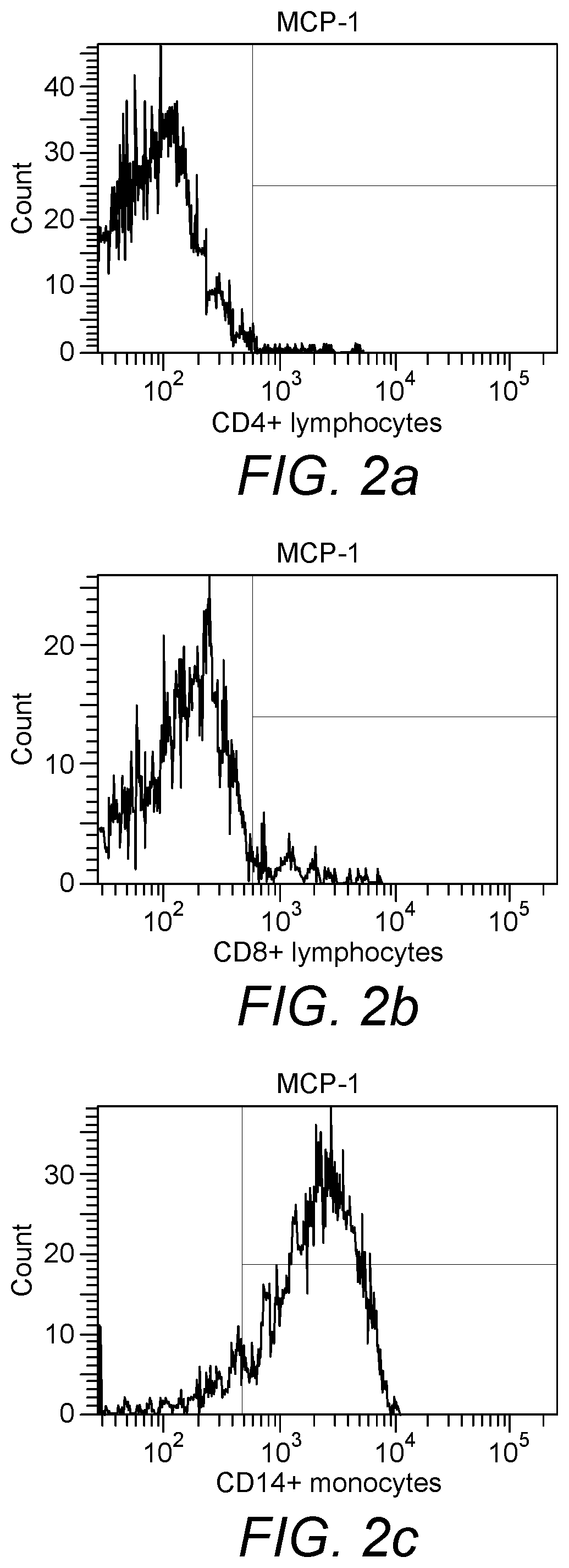
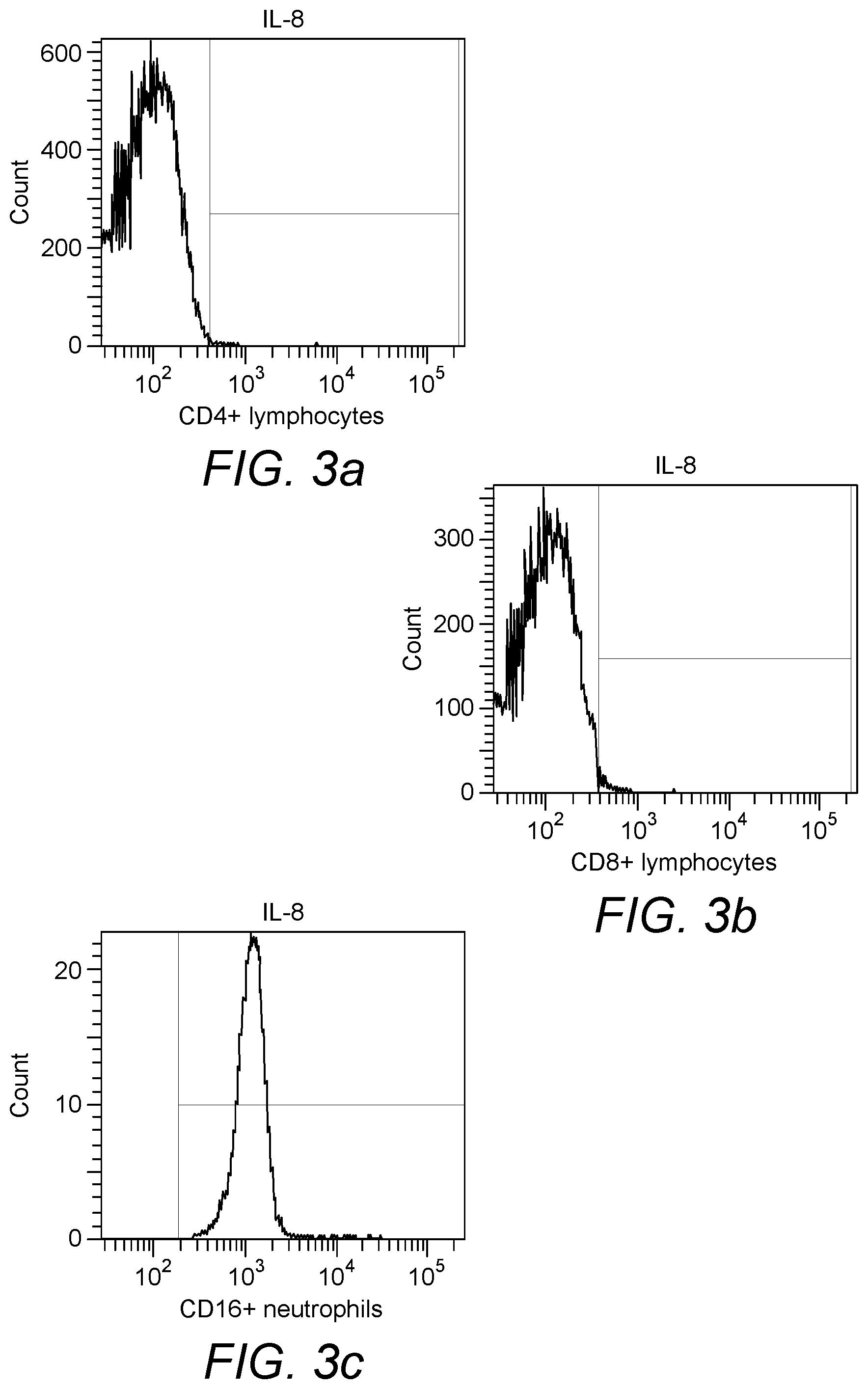
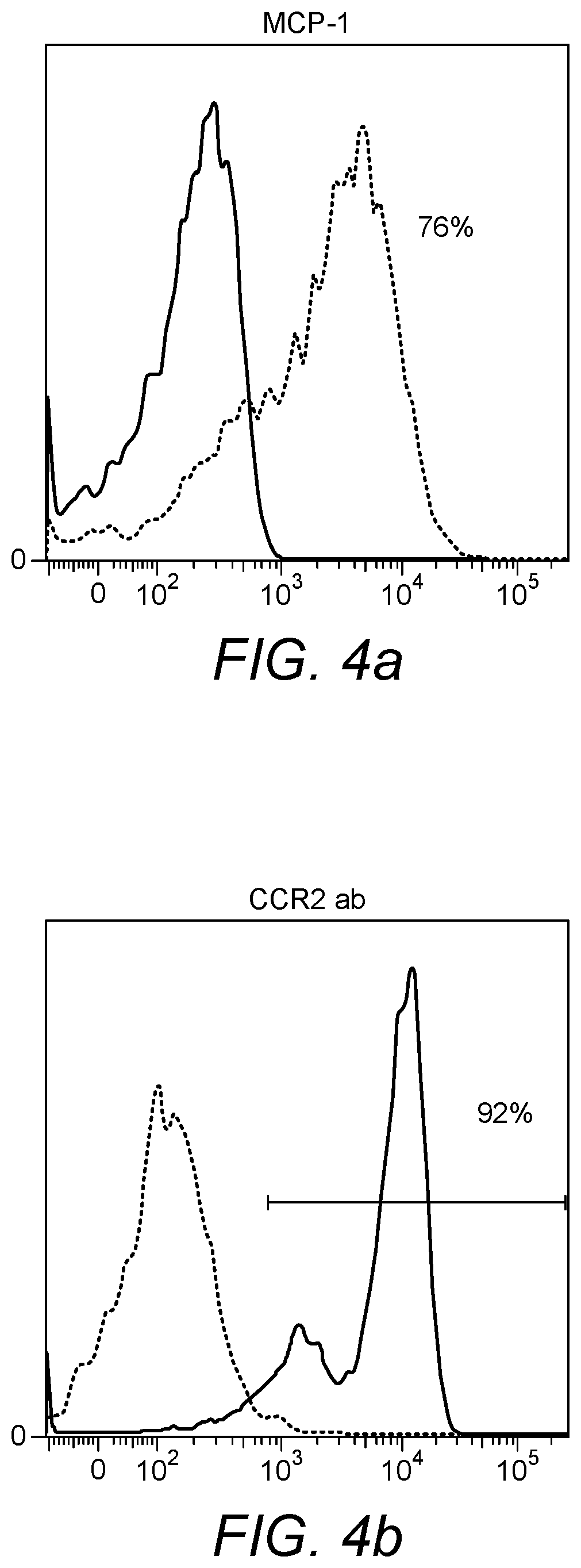
TREATING RESPIRATORY CONDITIONS
Abstract
A method for treating a respiratory conditions comprises applying peripheral blood from a patient or subject to an apheresis column loaded with a solid support comprising one or more binding reagents capable of specifically binding to a chemokine receptor, optionally the chemokine receptor CCR2, CCR1, CCR3, CCR5, CXCR1, CXCR2 and/or CCR7 immobilized directly or indirectly on the support thus removing one or more chemokine receptor, optionally CCR2, CCR1, CCR3, CCR5, CXCR1, CXCR2 and/or CCR7 expressing cells from the peripheral blood of the patient or subject. Various companion diagnostic methods and useful binding reagents are also described.
| Inventors: | Winqvist; Ola; (Uppsala, SE) ; Cotton; Graham; (Edinburgh, GB) | ||||||||||
| Applicant: |
|
||||||||||
|---|---|---|---|---|---|---|---|---|---|---|---|
| Family ID: | 50973441 | ||||||||||
| Appl. No.: | 16/537224 | ||||||||||
| Filed: | August 9, 2019 |
Related U.S. Patent Documents
| Application Number | Filing Date | Patent Number | ||
|---|---|---|---|---|
| 15629700 | Jun 21, 2017 | 10422800 | ||
| 16537224 | ||||
| 14105628 | Dec 13, 2013 | 9726666 | ||
| 15629700 | ||||
| PCT/GB2012/051357 | Jun 13, 2012 | |||
| 14105628 | ||||
| PCT/GB2012/051349 | Jun 13, 2012 | |||
| 14105628 | ||||
| PCT/GB2012/051348 | Jun 13, 2012 | |||
| 14105628 | ||||
| PCT/GB2012/051351 | Jun 13, 2012 | |||
| 14105628 | ||||
| PCT/GB2012/051350 | Jun 13, 2012 | |||
| 14105628 | ||||
| PCT/GB2012/051355 | Jun 13, 2012 | |||
| 14105628 | ||||
| PCT/GB2012/051345 | Jun 13, 2012 | |||
| 14105628 | ||||
| PCT/GB2012/051352 | Jun 13, 2012 | |||
| 14105628 | ||||
| PCT/GB2012/051346 | Jun 13, 2012 | |||
| 14105628 | ||||
| PCT/GB2012/051353 | Jun 13, 2012 | |||
| 14105628 | ||||
| PCT/GB2012/051356 | Jun 13, 2012 | |||
| 14105628 | ||||
| PCT/GB2012/051354 | Jun 13, 2012 | |||
| 14105628 | ||||
| 61496442 | Jun 13, 2011 | |||
| 61496167 | Jun 13, 2011 | |||
| 61496288 | Jun 13, 2011 | |||
| 61496242 | Jun 13, 2011 | |||
| 61496209 | Jun 13, 2011 | |||
| 61496195 | Jun 13, 2011 | |||
| 61496228 | Jun 13, 2011 | |||
| 61496264 | Jun 13, 2011 | |||
| 61496184 | Jun 13, 2011 | |||
| 61496329 | Jun 13, 2011 | |||
| 61496377 | Jun 13, 2011 | |||
| 61496352 | Jun 13, 2011 | |||
| Current U.S. Class: | 1/1 |
| Current CPC Class: | A61M 1/3618 20140204; G01N 33/56972 20130101; G01N 2333/7158 20130101; G01N 2333/70596 20130101; G01N 33/566 20130101; G01N 2800/065 20130101; A61M 1/3679 20130101 |
| International Class: | G01N 33/566 20060101 G01N033/566; G01N 33/569 20060101 G01N033/569; A61M 1/36 20060101 A61M001/36 |
Claims
1. A method for treating a respiratory condition in a subject in need thereof, the method comprising applying peripheral blood from the subject to an apheresis column loaded with a solid support comprising one or more binding reagents capable of specifically binding to a chemokine receptor CCR1 immobilized directly or indirectly on the support, whereby one or more cells expressing chemokine receptor CCR1 are removed from the peripheral blood of the subject, wherein the applied blood is returned to the subject, and whereby the respiratory condition is treated.
2. The method of claim 1, wherein the respiratory condition is sarcoidosis or Chronic Obstructive Pulmonary Disease (COPD).
3. The method of claim 1, wherein the binding reagent is an agonist or an antagonist of CCR1.
4. The method of claim 1, wherein the binding reagent is an antibody or a chemokine.
5. The method of claim 4, wherein: (a) the chemokine is selected from CCL2 (MCP-1), MCP-2, MCP-3, MCP-4 (CCL12), MCP-5, CCL5 (RANTES), CCL9, MRP-2 (CCL10), CCL14, CCL15, CCL16, CCL23, CCL11 (Eotaxin), CCL28, CCL24 (Eotaxin 2), CCL26, CCL3, CCL4, CCL5, CCL8, CXCL1, CXCL2, CXCL3, CXCL5, CXCL6, CXCL7, and CXCL8; (b) the chemokine is RANTES; and/or (c) the chemokine is selected from CCL9, MRP-2 (CCL10), CCL14, CCL15, CCL16, and CCL23.
6. The method of claim 5, wherein the chemokine is MCP-1 or MCP-5.
7. The method of claim 1, wherein the one or more cells are monocytes, eosinophils, or T lymphocytes.
8. The method of claim 1, wherein the subject has increased levels of expression of CCR1 as compared to a subject that does not have a respiratory condition.
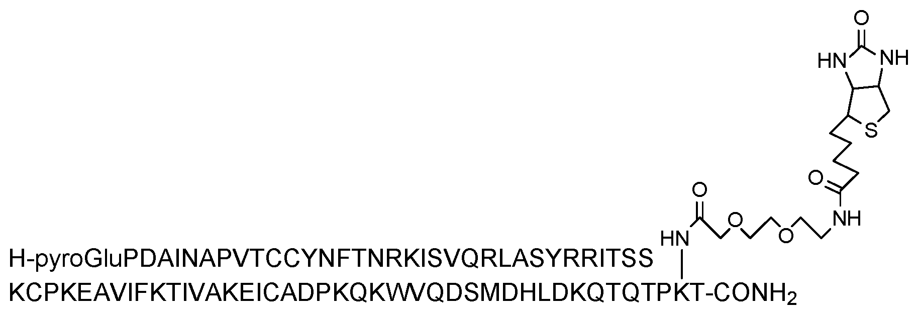
9. The method of claim 1, wherein 20-50% of the subject's blood is applied to the column in a single treatment.
Description
CROSS-REFERENCE TO RELATED APPLICATION(S)
[0001] This application is a divisional of U.S. patent application Ser. No. 15/629,700, filed on Jun. 21, 2017, which is a divisional of U.S. patent application Ser. No. 14/105,628, filed on Dec. 13, 2013, now issued as U.S. Pat. No. 9,726,666, which is a continuation-in-part of International Patent Application No. PCT/GB2012/051357, filed on Jun. 13, 2012, which claims priority to U.S. Provisional Patent Application No. 61/496,442, filed on Jun. 13, 2011. U.S. patent application Ser. No. 14/105,628 is also a continuation-in-part of International Patent Application No. PCT/GB2012/051349, filed on Jun. 13, 2012, which claims priority to U.S. Provisional Patent Application No. 61/496,167, filed on Jun. 13, 2011. U.S. patent application Ser. No. 14/105,628 is also a continuation-in-part of International Patent Application No. PCT/GB2012/051348, filed on Jun. 13, 2012, which claims priority to U.S. Provisional Patent Application No. 61/496,288, filed on Jun. 13, 2011. U.S. patent application Ser. No. 14/105,628 is also a continuation-in-part of International Patent Application No. PCT/GB2012/051351, filed on Jun. 13, 2012, which claims priority to U.S. Provisional Patent Application No. 61/496,242, filed on Jun. 13, 2011. U.S. patent application Ser. No. 14/105,628 is also a continuation-in-part of International Patent Application No. PCT/GB2012/051350, filed on Jun. 13, 2012, which claims priority to U.S. Provisional Patent Application No. 61/496,209, filed on Jun. 13, 2011. U.S. patent application Ser. No. 14/105,628 is also a continuation-in-part of International Patent Application No. PCT/GB2012/051355, filed on Jun. 13, 2012, which claims priority to U.S. Provisional Patent Application No. 61/496,195, filed on Jun. 13, 2011. U.S. patent application Ser. No. 14/105,628 is also a continuation-in-part of International Patent Application No. PCT/GB2012/051345, filed on Jun. 13, 2012, which claims priority to U.S. Provisional Patent Application No. 61/496,228, filed on Jun. 13, 2011. U.S. patent application Ser. No. 14/105,628 is also a continuation-in-part of International Patent Application No. PCT/GB2012/051352, filed on Jun. 13, 2012, which claims priority to U.S. Provisional Patent Application No. 61/496,264, filed on Jun. 13, 2011. U.S. patent application Ser. No. 14/105,628 is also a continuation-in-part of International Patent Application No. PCT/GB2012/051346, filed on Jun. 13, 2012, which claims priority to U.S. Provisional Patent Application No. 61/496,184, filed on Jun. 13, 2011. U.S. patent application Ser. No. 14/105,628 is also a continuation-in-part of International Patent Application No. PCT/GB2012/051353, filed on Jun. 13, 2012, which claims priority to U.S. Provisional Patent Application No. 61/496,329, filed on Jun. 13, 2011. U.S. patent application Ser. No. 14/105,628 is also a continuation-in-part of International Patent Application No. PCT/GB2012/051356, filed on Jun. 13, 2012, which claims priority to U.S. Provisional Patent Application No. 61/496,377, filed on Jun. 13, 2011. U.S. patent application Ser. No. 14/105,628 is also a continuation-in-part of International Patent Application No. PCT/GB2012/051354, filed on Jun. 13, 2012, which claims priority to U.S. Provisional Patent Application No. 61/496,352, filed on Jun. 13, 2011. The entire contents of each of these applications are fully incorporated herein by reference.
INCORPORATION-BY-REFERENCE OF MATERIAL SUBMITTED ELECTRONICALLY
[0002] The instant application contains a Sequence Listing which has been submitted in ASCII format via EFS-Web and is hereby incorporated by reference in its entirety. Said ASCII copy, created on Jun. 31, 2019, is named "P81602729US01-211226-9003-US02-SEQ-LIST-08-09-19.txt", and is 26,527 bytes in size.
FIELD OF THE INVENTION
[0003] The various embodiments of the present invention relate to products for and methods of treating inflammatory conditions, such as respiratory conditions, in particular sarcoidosis and Chronic Obstructive Pulmonary Disease (COPD). Companion diagnostics are also described.
BACKGROUND OF THE INVENTION
[0004] Respiratory tract disorders such as asthma and chronic obstructive pulmonary disease (COPD) are typically characterised by breathing difficulties as a result of reduced airflow to the lungs. These symptoms are often caused by inflammation of the airways; for example, chronic bronchitis is a form of COPD associated with excessive inflammation of the bronchi.
[0005] In addition to breathing difficulties, airway obstruction and associated inflammation can cause progressive lung damage. In the case of patients with COPD, permanent narrowing of the airways can lead to complications such as chest infections, heart failure and ultimately pulmonary failure.
[0006] COPD is a very common respiratory disease and in the majority of cases, smoking is the cause.
[0007] Patients are typically treated using inhalers containing "bronchodilator" drugs. However, these are of limited use for patients with permanently-constricted airways and/or end-stage disease characterised by extensive damage to the lungs.
[0008] Apheresis is a treatment used for depletion of blood components, such as antibodies, low-density lipoproteins (LDL) and blood cells. Leukapheresis is the apheresis treatment used for removal of white blood cells, leukocytes. The patient is connected to an extracorporeal blood circulating system; the blood is drawn from a vein in one arm, passed through a column device and returned into the other arm of the patient. WO2010/029317 describes apheresis columns useful for treating inflammatory conditions including a chemokine immobilised on a solid support.
SUMMARY OF THE INVENTION
[0009] Chemokines are a class of cytokine molecules involved in cell recruitment and activation in inflammation. Chemokines cause chemotaxis and activation of various subpopulations of cells in the immune system. The activity of chemokines is mediated primarily through tight binding to their receptors on the surface of leukocytes. In certain embodiments the present invention is based on the realisation that the interaction between chemokines and cells expressing their receptors may be exploited for the treatment of respiratory conditions, in particular sarcoidosis and Chronic Obstructive Pulmonary Disease (COPD). In particular, various respiratory conditions, in particular sarcoidosis and Chronic Obstructive Pulmonary Disease (COPD) include an inflammatory component. Whilst sarcoidosis is predominantly considered a respiratory condition, general treatment of sarcoidosis affecting any organ of the body may be possible within the scope of the various embodiments of the present invention. The inventors have determined that targeting increased recruitment of specific chemokine receptor-expressing cells to the site of inflammation presents a new therapeutic approach to treat such conditions. Moreover, in such conditions, chemokine receptor expression on each cell may be increased again providing a therapeutic approach to treat such conditions. It is shown herein that subjects suffering from respiratory conditions such as sarcoidosis exhibit increased frequency of chemokine receptor expressing cells in the peripheral blood. Subjects with sarcoidosis exhibit increased frequency of CCR1 expressing cells such as CCR1 expressing monocytes, compared to healthy controls. It is also shown herein that the CCR1 expressing cells can be removed using a suitable binding reagent, in particular RANTES (in biotinylated form) immobilized on a suitable matrix. Similarly, it is shown herein that the monocytes also express CCR2. The CCR2 expressing monocytes can be depleted in sarcoidosis patients using a suitable binding reagent, in particular MCP-1, in biotinylated form, immobilized on a suitable matrix. It is also shown herein that subjects suffering from respiratory conditions such as sarcoidosis exhibit increased frequency of CCR7 expressing cells such as CCR7 expressing lymphocytes, and also central memory T cells,compared to healthy controls. It is also shown herein that the CCR7 expressing cells can be removed using a suitable binding reagent, in particular MIP3b (in biotinylated form) immobilized on a suitable matrix.
[0010] Thus, in certain embodiments the invention serves to reduce the recruitment of inflammatory leukocytes, which express characteristic chemokine receptors, and possibly express characteristic chemokine receptors at increased levels, to sites of inflammation linked to respiratory conditions, in particular sarcoidosis and Chronic Obstructive Pulmonary Disease (COPD). This is achieved using specific binding reagents to capture specific chemokine receptor-expressing inflammatory leukocytes from the patient. Accordingly, in certain embodiments the invention provides in a first aspect a method for treating respiratory conditions, in particular sarcoidosis and Chronic Obstructive Pulmonary Disease (COPD) comprising applying peripheral blood from a patient to an apheresis column loaded with a solid support comprising one or more binding reagents capable of specifically binding to one or more chemokine receptors, in particular the chemokine receptors CCR2, CCR1, CCR3, CCR5, CXCR1, CXCR2 and/or CCR7, immobilized directly or indirectly on the support thus removing one or more chemokine receptor, in particular one or more of CCR2, CCR1, CCR3, CCR5, CXCR1, CXCR2 and/or CCR7, expressing cells from the peripheral blood of the patient. The peripheral blood from which the chemokine receptor expressing cells have been removed may then be returned to the patient in order to complete the treatment. The invention may thus rely on a continuous extracorporeal circuit in some embodiments. Alternatively, in certain embodiments the invention may comprise steps of obtaining peripheral blood from the patient, applying the peripheral blood to the column and subsequently returning the peripheral blood from which the chemokine receptor expressing cells have been removed to the patient.
[0011] As shown herein, suitable binding reagents can be immobilized onto a solid support, either directly or indirectly, to generate an apheresis column suitable for capturing relevant chemokine receptor-expressing cells. Where increased levels of chemokine receptor expression are observed, such cells may be preferably removed from the peripheral blood using the columns of the various embodiments of the invention. Thus, the methods of the various embodiments of the invention may preferably target one or more of CCR2hi, CCR1hi, CCR3hi, CCR5hi, CXCR1hi, CXCR2hi and/or CCR7hi cells as defined herein for removal from the peripheral blood. "High" expression may be determined according to standard flow cytometry techniques. The level is measured relative to levels of expression of the chemokine receptor in cells taken from a healthy subject. The attached FIG. 17 provides an example of a gating strategy.
[0012] Herein, reference to CCR2, CCR1, CCR3, CCR5, CXCR1, CXCR2 and/or CCR7 is intended to encompass selection of any one or more, up to all, of the chemokine receptors listed. In addition, the combination of CCR2, CCR1, CCR3, CCR5, CXCR1 and/or CXCR2 is explicitly contemplated as a separate grouping, to include any one or more of CCR2, CCR1, CCR3, CCR5, CXCR1 and CXCR2.
[0013] In other embodiments the invention further provides a binding reagent capable of specifically binding to one or more chemokine receptors, in particular to a chemokine receptor/the chemokine receptor CCR2, CCR1, CCR3, CCR5, CXCR1, CXCR2 and/or CCR7, for use in the treatment of respiratory conditions, in particular sarcoidosis and Chronic Obstructive Pulmonary Disease (COPD), wherein the one or more binding reagents is immobilized, directly or indirectly, on a solid support contained within an apheresis column, to which is applied peripheral blood from a patient thus removing one or more chemokine receptor/CCR2, CCR1, CCR3, CCR5, CXCR1, CXCR2 and/or CCR7 expressing cells from the peripheral blood of the patient. In certain embodiments the invention also provides for use of one or more binding reagents capable of specifically binding to a chemokine receptor/the chemokine receptor CCR2, CCR1, CCR3, CCR5, CXCR1, CXCR2 and/or CCR7 for use in the manufacture of an apheresis column for treatment of respiratory conditions, in particular sarcoidosis and Chronic Obstructive Pulmonary Disease (COPD), wherein the one or more binding reagents is immobilized on a solid support contained within the apheresis column, to which is applied peripheral blood from a patient thus removing one or more of chemokine receptor/CCR2, CCR1, CCR3, CCR5, CXCR1, CXCR2 and/or CCR7 expressing cells from the peripheral blood of the patient.
[0014] All embodiments described in respect of the methods of treatment of the various embodiments of the invention apply to these aspects mutatis mutandis and are not repeated for reasons of conciseness. Thus, the following discussion made with reference to the various embodiments of the methods of treatment is also applicable to the medical use aspects of the invention.
[0015] In certain embodiments the invention aims to treat a range of respiratory conditions, in particular sarcoidosis and Chronic Obstructive Pulmonary Disease (COPD). By treatment is meant a reduction in the specific chemokine receptor expressing cells in the peripheral blood of the patient. The reduction may comprise a reduction in cells that express chemokine receptors, in particular one or more of CCR2, CCR1, CCR3, CCR5, CXCR1, CXCR2 and/or CCR7, at increased levels in diseased patients. The patient is typically a human patient but the term patient may include both human and non-human animal subjects in some embodiments. In the context of the various embodiments of the present invention, this typically involves a reduction in one or more of CCR2, CCR1, CCR3, CCR5, CXCR1, CXCR2 and/or CCR7 expressing cells, such as one or more of CCR2.sup.hi, CCR1.sup.hi, CCR3.sup.hi, CCR5.sup.hi, CXCR1.sup.hi, CXCR2.sup.hi and/or CCR7.sup.hi expressing cells, in the peripheral blood of the patient. The CCR2, CCR1, CCR3, CCR5, CXCR1, CXCR2 and/or CCR7 expressing cells comprise, consist essentially of or consist of monocytes, macrophages, lymphocytes, in particular T-lymphocytes, and/or eosinophils in certain embodiments. In specific embodiments, the cells removed in order to treat respiratory conditions such as sarcoidosis comprise monocytes, in particular CCR1 and/or CCR2 expressing monocytes. In other embodiments cells removed in order to treat respiratory conditions such as sarcoidosis comprise lymphocytes, specifically T lymphocytes such as central memory T cells, in particular CCR7 expressing T cells.
[0016] Monocytes are produced by the bone marrow from haematopoietic stem cell precursors called monoblasts. Monocytes may differentiate into macrophages or dendritic cells. Monocytes and their macrophage and dendritic cell progeny serve a number of functions in the immune system including phagocytosis, antigen presentation and cytokine production. Monocytes may be characterized with reference to expression of the cell surface marker CD14, optionally together with CD16. Classical monocytes may be characterized by high level expression of the CD14 cell surface receptor (CD14++ CD16- monocyte). Non-classical monocytes may be characterized by low level expression of CD14 and with additional co-expression of the CD16 receptor (CD14+CD16++ monocyte). Intermediate monocytes may be characterized by high level expression of CD14 and low level expression of CD16 (CD14++CD16+ monocytes). Macrophages are derived from monocytes and are responsible for protecting tissues from foreign substances. They are cells that possess a large smooth nucleus, a large area of cytoplasm and internal vesicles for processing foreign material. The term "macrophage" may refer to a monocyte-derived cell expressing one or more of the following cell surface markers CD14, CD11b, Lysozyme M, MAC-1/MAC-3 and CD68. The term macrophage includes both activated and un-activated macrophages. Activated macrophages may be characterized by expression of one or more of CD69, ENG, FCER2 and IL2RA, HLA-DR, CD86. Un-activated macrophages have not yet received activating signals through for example TLR receptors and therefore they express less activation markers on the cell surface which correlates with lesser maturation. M1 macrophages may be characterized by expression of one or more of CD16.sup.+CD32.sup.+CD64.sup.+and secrete mainly IL-23 and IL-1, TNF, IL-6 and high levels of IL-12 and in addition effector molecules such as iNOS and ROI. M1 macrophages have cytotoxic features as opposed to M2 macrophages. M2 macrophages may be characterized by expression of one or more of SRA/B.sup.+CD163.sup.+MR.sup.+CD14.sup.+ and express TGF.beta., IL-10 and IL-1Ra. Tumour associated macrophages (TAMs) share many characteristics with the M2 macrophages and are considered as M2 polarised macrophages. The M1/M2 paradigm can also be found in monocyte subsets where CD14.sup.+CD16.sup.-CXC3R1.sup.low monocytes are considered the "inflammatory" subset and the CD14.sup.lowCD16.sup.+CXC3R1.sup.high are "resident" monocytes.
[0017] The three major types of lymphocyte are T cells, B cells and natural killer (NK) cells. The term "T-lymphocyte" includes CD4.sup.+ T cells such as T helper cells (Th1 cells and Th2 cells), and CD8.sup.+ T cells such as cytotoxic T cells. Th1 cells may be characterized by expression of CCR5 and/or by production of IFN-.gamma.. Th2 cells may be characterized by expression of CCR3 and/or by production of IL-4.
[0018] The claimed methods may, in particular, target eosinophils. Eosinophilia is an important component of certain respiratory conditions and may be defined as the presence of more than 500 eosinophils/microlitre of blood. Thus, reducing numbers of circulating eosinophils represents an important therapeutic approach. Eosinophils, or eosinophil granulocytes, are white blood cells and represent an important immune system component. Along with mast cells, they also control mechanisms associated with allergy and asthma. They are granulocytes that develop during haematopoiesis in the bone marrow before migrating into blood.
[0019] The name "eosinophil" derives from the eosinophilic "acid-loving" properties of the cell. Normally transparent, it is this affinity that causes them to appear brick-red after staining with eosin, a red dye, using the Romanowsky method. The staining is concentrated in small granules within the cellular cytoplasm, which contain many chemical mediators, such as histamines and proteins such as eosinophil peroxidase, ribonuclease (RNase), deoxyribonucleases, lipase, plasminogen, and major basic protein. These mediators are released by a process called degranulation following activation of the eosinophil, and are toxic to both parasite and host tissues.
[0020] Eosinophils develop and mature in bone marrow. They differentiate from myeloid precursor cells in response to the cytokines interleukin 3 (IL-3), interleukin 5 (IL-5), and granulocyte macrophage colony-stimulating factor (GM-CSF). Eosinophils produce and store many secondary granule proteins prior to their exit from the bone marrow. After maturation, eosinophils circulate in blood and migrate to inflammatory sites in tissues in response to chemokines such as CCL11 (eotaxin-1), CCL24 (eotaxin-2), CCLS (RANTES) and MCP1/4. Eosinophils may be activated by Type 2 cytokines released from a specific subset of helper T cells (Th2); IL-5, GM-CSF, and IL-3 are important for eosinophil activation as well as maturation. CD44 and CD69 have been shown to represent different types of cell-surface activation markers for human eosinophils. CD69 is absent from "fresh" eosinophils but expressed following activation (using cytokines). CD44 on the other hand is constitutively expressed but expression is significantly up-regulated in response to activation (Matsumoto et al., Am. J. Respir. Cell Mol. Biol., Volume 18, Number 6, June, 1998 860-866). Cell specific markers for eosinophils include CD9 and CDw125.
[0021] CCR2, CCR1, CCR3, CCR5, CXCR1, CXCR2 and/or CCR7 expressed on these aforementioned cells bind to chemokines such as CCL2 (binds CCR2), CCL3, CCL5, CCL9 (binds CCR1), CCL11 (binds CCR3), CCL12 (binds CCR2), CCL-14 (binds CCR1), CCL16 (binds CCR1), CCL28 (binds CCR3), CCL24 (binds CCR3), CCL26 (binds CCR3) and/or CXCL8 (binds CXCR1 and CXCR2). Chemokines MIP1.gamma. (CCL9), MRP-2 (CCL10), Mlp-1.delta. (CCL15) and CCL23 appear to bind CCR1 only. Chemokines Eotaxin (CCL11) and CCL24 (Eotaxin-2) only bind CCR3. Chemokine MIP1.beta. (CCL4) only binds CCR5.
[0022] CXCR1 binds CXCL6, CXCL7, CXCL8.
[0023] CXCR2 binds CXCL1, 2, 3, 5, 6, 7, 8.
[0024] CCR1 is the gene symbol approved by the HUGO Gene Nomenclature Committee for chemokine (C-C motif) receptor 1. The HGNC ID for this gene is 1602. The gene is located at chromosome position 3p21. The previous symbol and name CMKBR1, SCYAR1. Synonyms for this gene include CD191, CKR-1, MIP1.alpha. R. The Entrez Gene reference sequence for CCR1 is 1230 as available on 13 Jun. 2011, which is incorporated herein by reference in its entirety.
[0025] CCR2 is the gene symbol approved by the HUGO Gene Nomenclature Committee for chemokine (C-C motif) receptor 2. The HGNC ID for this gene is 1603. The gene is located at chromosome position 3p21. The previous symbol and name for the gene is CMKBR2. Synonyms for this gene include CC-CKR-2, CD192, CKR2, FLJ78302, MCP-1-R. The NCBI Reference Sequence is NM_001123041.2 as available on 13 Jun. 2011, which is incorporated herein by reference in its entirety.
[0026] CCR3 is the gene symbol approved by the HUGO Gene Nomenclature Committee for chemokine (C-C motif) receptor 3. The HGNC ID for this gene is 1604. The gene is located at chromosome position 3p21.3. The previous symbol and name for the gene is CMKBR3. Synonyms for this gene include CC-CKR-3, CD193 and CKR3. The Genbank reference sequence for CCR3 is AF247361.1 as available on 13 Jun. 2011, which is incorporated herein by reference in its entirety.
[0027] CCR5 is the gene symbol approved by the HUGO Gene Nomenclature Committee for chemokine (C-C motif) receptor 5. The HGNC ID for this gene is 1605. The gene is located at chromosome position 3p21. The previous symbol and name for the gene is CMKBRS. Synonyms for this gene include CC-CKR-5, CD195 CKR-5, IDDM22 and CKRS. The Entrez Gene reference sequence for CCR5 is 1234 as available on 13 Jun. 2011, which is incorporated herein by reference in its entirety.
[0028] CXCR1 is the gene symbol approved by the HUGO Gene Nomenclature Committee for chemokine (C-X-C motif) receptor 1. The HGNC ID for this gene is 6026. The gene is located at chromosome position 2q35. The previous symbol and name for the gene is CMKAR1, IL8RA, "interleukin 8 receptor, alpha". Synonyms for this gene include CD181, CDw128a, CKR-1. The Genbank reference sequence for CXCR1 is U11870.1 as available on 13 Jun. 2011, which is incorporated herein by reference in its entirety.
[0029] CXCR2 is the gene symbol approved by the HUGO Gene Nomenclature Committee for chemokine (C-X-C motif) receptor 2. The HGNC ID for this gene is 6027. The gene is located at chromosome position 2q35. The previous symbol and name for the gene is IL8RB, "interleukin 8 receptor, beta". Synonyms for this gene include CD182, CMKAR2. The Genbank reference sequence for CXCR2 is U11869.1 as available on 13 Jun. 2011, which is incorporated herein by reference in its entirety.
[0030] CCR7 is the gene symbol approved by the HUGO Gene Nomenclature Committee for chemokine (C-C motif) receptor 7. The HGNC ID for this gene is 1608. The gene is located at chromosome position 17q12-q21.2. The previous symbol and name for the gene is CMKBR7, EBI1. Synonyms for this gene include BLR2, CD197 and CDw197. The RefSeq reference sequence for CCR7 is NM_001838.3 as available on 13 Jun. 2011, which is incorporated herein by reference in its entirety.
[0031] The various embodiments of the methods of the invention may involve specific binding interactions with any one or more of these further cell-surface markers in addition to the removal based upon binding to CCR2, CCR1, CCR3, CCR5, CXCR1, CXCR2 and/or CCR7. Suitable binding reagents can be prepared to specifically bind to these cell-surface markers. The discussion of CCR2, CCR1, CCR3, CCR5, CXCR1, CXCR2 and/or CCR7 specific binding reagents thus applies mutatis mutandis.
[0032] Treatment according to the various embodiments of the invention may result in alleviation or amelioration of symptoms, prevention of progression, regression of the condition, or complete recovery. Measurable parameters of successful treatment include one or more, up to all, of lungfunction with spirometry, PEF, lung biopsy. Pulsoxymeter. paO2, paCO2. In specific embodiments, a single treatment is sufficient to cause a depletion of around 10%, 20%, 30%, 40%, 50%, 60% or 70%, or higher up to 80%, 90%, 95% or more, or any range of values between and including these amounts, of one or more of a specific chemokine receptor, in particular one or more of CCR2, CCR1, CCR3, CCR5, CXCR1, CXCR2 and/or CCR7, expressing cells from the peripheral blood of the patient. In specific embodiments, at least around 50% depletion is achieved in a single treatment. Thus, successful treatment may be defined with reference to depletion of one or more of CCR2, CCR1, CCR3, CCR5, CXCR1, CXCR2 and/or CCR7 expressing cells. Treatment may lead to depletion of between approximately 100 and 500 million CCR2, CCR1, CCR3, CCR5, CXCR1, CXCR2 and/or CCR7 expressing cells, such as monocytes, in certain embodiments and more particularly to about 100, 150, 200, 250, 300, 350, 400, 450, or 500 million CCR2, CCR1, CCR3, CCR5, CXCR1, CXCR2 and/or CCR7 expressing cells.
[0033] By binding to the column through the binding reagent-chemokine receptor interaction, chemokine receptor expressing cells are immobilized. These immobilized cells express further unoccupied chemokine receptors, which may be of the same or different type to those used for capture. These additional chemokine receptors may permit circulating chemokines which contribute to the inflammatory condition to be captured from the peripheral blood. Thus, a reduction in circulating (specific) chemokine levels may provide a measure of successful treatment.
[0034] The duration of treatment may be readily determined by one skilled in the art and will depend upon factors such as the flow rate of the peripheral blood. Duration of treatment may be tied into monitoring of the treatment itself, with the treatment considered complete once a measurable parameter of treatment has reached a defined threshold. Any suitable parameter may be employed as discussed herein. Thus, for example, treatment may be considered complete when a reduction in one or more of CCR2, CCR1, CCR3, CCR5, CXCR1, CXCR2 and/or CCR7 expressing cells, such as a 50% reduction in one or more of CCR2, CCR1, CCR3, CCR5, CXCR1, CXCR2 and/or CCR7 expressing cells, has been achieved. The apheresis system may be operated ata flow rate of around 10-80 mL/min, or more specifically between around 20-70 mL/min, or between around 30-60 mL/min. In specific embodiments, the treatment is performed for a period of around 10, 20, 30, 40, 50, 60, 70, 80, 90, 100, 110,120 etc., or any range of values between and including these amounts, minutes. The treatment is typically not aimed to remove all of the cells expressing the chemokine receptor in the peripheral blood, as a basal level of those cells is required in healthy subjects. However, it has been found that only low blood volumes need to be applied to the columns of the various embodiments of the invention in order to achieve effective levels of depletion of the chemokine receptor-expressing cells. Thus, in certain embodiments, around 10-80% or more specifically around 20, 30, 40 or 50%, or any range of values between and including these amounts, of the patient's blood is applied to the column in a single treatment. The volume of blood circulated through the apheresis column or system may be in the region of around 1000-3000 ml, such as around 1000, 1200, 1400, 1600, 1800 or 2000 ml or any range of values between and including these amounts. The treatment may be considered complete once this volume of blood has been circulated. The patient may be administered anticoagulants prior to each treatment session. A suitable solution, such as a sterile saline solution, optionally including an anticoagulant such as Heparin, may be used for priming the apheresis (extracorporeal) system. An additional volume of anticoagulant may be added to the circuit at the start of each treatment session, for example as a bolus injection.
[0035] In certain embodiments the invention relies upon a binding reagent which is capable of specifically binding to a chemokine receptor. This specific binding reaction permits cells expressing the chemokine receptor to be removed from the peripheral blood of the patient when the blood is passed over the solid support upon or within which the binding reagent is immobilized. Specific chemokine receptors of interest include CCR2, CCR1, CCR3, CCR5, CXCR1, CXCR2 and/or CCR7. The binding reagent can be any binding reagent capable of specifically binding to the receptor in question. By "specific binding" is meant that the binding reagent displays sufficient specificity of binding and appropriate binding affinity/kinetics to permit removal of cells expressing one or more of CCR2, CCR1, CCR3, CCR5, CXCR1, CXCR2 and/or CCR7 from the peripheral blood. Whilst it is not precluded that the binding reagent is capable of binding to other molecules, such as other chemokine receptors, the binding reagent will preferentially bind to cells expressing one or more of CCR2, CCR1, CCR3, CCR5, CXCR1, CXCR2 and/or CCR7 and in particular to cells expressing increased levels of one or more of CCR2, CCR1, CCR3, CCR5, CXCR1, CXCR2 and/or CCR7 (as defined further herein). The binding reagent capable of specifically binding to CCR2, CCR1, CCR3, CCR5, CXCR1, CXCR2 and/or CCR7 may be either an agonist or an antagonist of CCR2, CCR1, CCR3, CCR5, CXCR1, CXCR2 and/or CCR7, respectively. As the disease condition relies upon up-regulation of expression of or signaling via CCR2, CCR1, CCR3, CCR5, CXCR1, CXCR2 and/or CCR7, in certain embodiments the binding reagent capable of specifically binding to CCR2, CCR1, CCR3, CCR5, CXCR1, CXCR2 and/or CCR7 is an antagonist of CCR2, CCR1, CCR3, CCR5, CXCR1, CXCR2 and/or CCR7, respectively. Chemokines are typically, although not necessarily exclusively (particularly in the case of truncated or modified forms) agonists of their cognate receptor and serve to activate the cells expressing the relevant receptor, as would be appreciated by one skilled in the art. Antibodies against the relevant chemokine receptor are generally considered to be antagonists, as would be appreciated by one skilled in the art. Specific examples of binding reagents include proteins or polypeptides, such as antibodies and receptor ligands, in particular chemokines. The binding reagent may be a nucleic acid molecule in certain embodiments. In some embodiments, the nucleic acid is an aptamer. Nucleic acid aptamers are polynucleotides of approximately 15-40 nucleotides long. Nucleic acid aptamers can be made using the SELEX process (systemic evolution of ligands by exponential enrichment) or any other process known to those of skill in the art.
[0036] In other embodiments, the binding reagent may be a peptide, and in certain instances, a peptide aptamer. Peptide aptamers are artificial recognition molecules that consist of a variable peptide sequence inserted into a constant scaffold protein (Baines I C, Colas P. Peptide aptamers as guides for small molecule drug discovery. Drug Discov Today. 2006;11:334-341, incorporated herein by reference). A number of methodologies, such as phage display, ribosome display and yeast two-hybrid screening systems are available for screening a library of potential peptide-based binding agents. Similarly protein scaffolds based on domains such as fibronectins, ankyrin repeats, protein A, SH3 domains, lipocalins and ubiquitin can be used as the binding agent. Again a number of technologies such as phage display and ribosome display are available for screening a library of protein--based binding agents. Similarly, libraries of candidate chemical compounds can be screened for specific binding to the relevant chemokine receptor using suitable screening techniques known in the art, which may be high throughput screens in certain embodiments. The candidate binding agent may be immobilized on a solid support and the ability of the agent to specifically retain cells expressing the chemokine receptor of interest or labelled chemokine receptors determined. A range of cell types may be applied to the solid supports to confirm specificity of binding, or alternatively a mixed sample (such as peripheral blood) may be applied to the solid support. Retention of the cell type of interest (expressing the appropriate chemokine receptor) can be confirmed to identify suitable binding agents. A range of small-molecule antagonists of CCR-2 are discussed by Xia M and Sui Z in Expert Opin Ther Pat. 2009 March; 19(3):295-303 --Recent developments in CCR2 antagonists, and incorporated herein by reference.
[0037] In the context of the various embodiments of the present invention the term "chemokine" also comprises biotinylated or otherwise labelled chemokines. The term "chemokine" also comprises modified and truncated versions of the chemokine and chemokine fragments with the proviso that the modified or truncated form retains its ability to bind to its cognate receptor (and thus remains functional in the context of the various embodiments of the invention). The chemokine does not necessarily need to retain biological activity as it is specific binding affinity for CCR2, CCR1, CCR3, CCR5, CXCR1, CXCR2 and/or CCR7 that is required. In certain embodiments, the chemokine lacks biological activity, for example in terms of activation of the (CCR2, CCR1, CCR3, CCR5, CXCR1, CXCR2 and/or CCR7) receptor. Modifications may be made to improve protein synthesis, for example uniformity of product and yield. As known to those skilled in the art, exemplary modifications may comprise amino acid additions, substitutions, deletions or other modifications to one or more amino acids in the chemokine. Modifications may comprise substitution of the wild type amino acid with non-natural amino acids such as norleucine (NLeu) and derivatized amino acids such as pyroglutamic acid (pyroGlu). Such modifications may be made to minimize side-product formation during storage and use of the columns of the various embodiments of the invention. Modifications may be made to improve labelling, for example inclusion of a polyethylene glycol (PEG) spacer to facilitate biotinylation. The biotinylation and/or conjugation with fluorochromes or other labelling groups of the chemokine is performed in a manner which does not substantially affect the receptor binding capacity. Site specific biotinylation or other labelling is preferred as non-selective labelling of chemokines may compromise receptor binding activity. Biotinylation or other labelling is generally preferred at or towards the C-terminus of the protein as the inventors have found that modifications in this area are generally well tolerated (in terms of a minimal effect on receptor binding capability). Biotinylation may be carried out site-specifically at any suitable amino acid. Examples of suitable amino acids include lysine, diaminopropionic acid and ornithine. Generally, reference may be made to Natarajan S et al, Int. J. Pept. Protein Res., 1992, 40, 567-74; Baumeister B, Int. J. Peptide Res. And Therapeutics, 2005, 11, 139-141; Bioconjugate techniques 2.sup.nd edition, Greg T. Hermanson, incorporated by reference herein in its entirety.
[0038] Truncations may involve deletion of either N or C terminal amino acids as appropriate, or both. Typically, the truncated version will retain the residues required for the chemokine to fold correctly, for example to retain a chemokine fold structure, consistent with the requirement that a truncated version must retain the ability to bind to the relevant receptor (expressed by (on the surface of) a leukocyte). Chemokine molecules typically include disulphide bonds between the 1.sup.st and 3.sup.rd and 2.sup.nd and 4.sup.th cysteine residues respectively, as would be understood by one skilled in the art. Where sequences are presented herein, it is assumed that these disulphide bonds will form in the folded protein (unless otherwise stated). Truncated versions may comprise anywhere between 1 and 100 less amino acids, such as 1, 2, 3, 4, 5 etc amino acids, than the wild type amino acid sequence in certain embodiments. Of course, truncated versions may comprise further modification as detailed herein. The modified or truncated version may have 40%, 45%, 50%, 55%, 60%, 65%, 70%, 75%, 80%, 85%, 90%, 95% or more overall amino acid sequence identity with the full length wild type chemokine (where a deletion is counted as a difference in amino acid sequence) in certain embodiments. Over the common sequence between the molecules (i.e the amino acids that have not been deleted), there may be 80%, 85%, 90%, 91%, 92%, 93%, 94%, 95%, 96%, 97%, 98% or 99% amino acid sequence identity in certain embodiments. Sequence identity may be determined using known algorithms, such as BLAST or GAP analysis (GCG Program) (applying default settings), which are freely available. Chemokines may lack the N-terminal signal peptide which is cleaved off during synthesis in vivo.
[0039] Specific chemokines useful in the various embodiments of the present invention for binding to CCR2 include CCL2 (MCP-1), MCP-2, MCP-3, MCP-4 (CCL12) and MCP-5. Both MCP-1 and MCP-5 bind solely to the chemokine receptor CCR2 and so these chemokines may be preferred in some embodiments. Each chemokine is able to bind to a chemokine receptor implicated in respiratory conditions, in particular sarcoidosis and Chronic Obstructive Pulmonary Disease (COPD). More specifically, each of MCP-1, MCP-2, MCP-3, MCP-4 and MCP-5 are useful for removing CCR2 expressing cells from the blood of a patient. Specific chemokines useful in the various embodiments of the present invention for binding to CCR1, CCR3 and/or CCR5 include CCL5 (RANTES). RANTES is able to bind to chemokine receptors implicated in respiratory conditions, in particular sarcoidosis and Chronic Obstructive Pulmonary Disease (COPD). More specifically, RANTES is useful for removing CCR1, CCR3 and/or CCR5 expressing cells from the blood of a patient.
[0040] Specific chemokines useful for binding to CCR1 include CCL9, MRP-2 (CCL10), CCL14, CCL15, CCL16 and CCL23 Specific chemokines useful for binding to CCR3 include CCL11 (Eotaxin), CCL28, CCL24 (Eotaxin 2) and CCL26. Specific chemokines useful for binding to CCR5 include CCL3, CCL4, CCL5, CCL8 with CCL4 binding only to CCR5. Specific chemokines useful in the various embodiments of the present invention for binding to CXCR1 and/or CXCR2 include CXCL8 (IL-8). IL-8 is able to bind to chemokine receptors implicated in respiratory conditions, in particular sarcoidosis and Chronic Obstructive Pulmonary Disease (COPD). Specific chemokines useful in the various embodiments of the present invention for binding to CXCR1 include CXCL6, CXCL7, CXCL8. Specific chemokines useful in the various embodiments of the present invention for binding to CXCR2 include CXCL1, 2, 3, 5, 6, 7, 8. CCL19 is useful for the removal of CCR7 expressing cells.
[0041] The chemokines described in greater detail herein (with reference to the relevant figures and amino acid sequences, as set forth in the SEQ ID NOs) may each be applied according to the present invention.
[0042] The modified and truncated chemokines described in greater detail herein (with reference to the relevant amino acid sequences, as set forth in the SEQ ID NOs and accompanying experimental examples) may each be applied according to the present invention. Such modified forms may instruct the skilled person regarding additional modified forms of the same and other chemokines which may be suitable for use in the invention. Chemokines show variable sequence homology varying from less than 20% to over 90% but all share very similar tertiary structures consisting of a disordered N-terminus, followed by a long loop (the N-loop) that ends in a 3.sub.10 helix, a 3-stranded .beta.-sheet and a C-terminal helix. The overlall topology is stabilsed by disulphide bonds. This common tertiary structure is a common feature of the chemokine protein family (Fernandez E J annd Lolis E., Annu. Rev. Pharmacol. Toxicol., 202, 42, 469-99; Allen S J et al, Annu. Rev. Immunol., 2007, 25, 787-820, incorporated herein by reference).
[0043] Truncations within this N-terminal region can maintain binding to the receptor but can lead to a change or loss of function (for examples Zhang Y J et al, J. Biol. Chem., 1994, 269, 15918; Gong J-H and Clark-Lewis I., J. Exp. Med., 1995, 181, 631-640; Fernandez E J annd Lolis E., Annu. Rev. Pharmacol. Toxicol., 202, 42, 469-99; Allen S J et al, Annu. Rev. Immunol., 2007, 25, 787-820, each of which is incorporated herein by reference). Truncations at the C-terminus of the chemokine can also be made and maintain receptor binding activity (Treating Inflammatory Disorders, Ola Winqvist and Graham Cotton, WO2010/029317, incorporated herein by reference in its entirety).
[0044] In other embodiments, fragments and variants of chemokines are used in the devices and methods as disclosed herein. More particularly, such fragments and variants retain the ability to specifically bind to their cognate chemokine receptor. Chemokines are known by those skilled in the art to share specific receptor binding domains, including a similar monomeric fold, characterized, for example, by a disordered amino-terminal domain, followed by a conserved core region, consisting of the so called "N-loop," three anti-parallel .beta.-strands, and a carboxyl-terminal a-helix. While not being bound by theory, it is believed that the chemokine-chemokine receptor interaction is a two-step mechanism, in which the core of the chemokine interacts first with a binding site formed by the extracellular domains of the receptor, while another interaction is formed between the chemokine N terminus and a second binding site on the receptor in order to trigger receptor activation. Thus, a "fragment," such as a functional fragment of a chemokine is intended to mean a portion of the amino acid sequence of the protein that retains binding for its cognate receptor. The fragment may include, for example, the monomeric fold region, or portions thereof such as the amino-terminal domain, the conserved core region and/or the "N-loop," the anti-parallel .beta.-strands, and/or the carboxyl-terminal a-helix or combinations and portions thereof.
[0045] Further, it is recognized that a polypeptide can be considerably mutated without materially altering one or more of the polypeptide's functions, for example, without altering specific binding and/or the folding of the protein. The genetic code is well known to be degenerate, and thus different codons encode the same amino acids. Even where an amino acid substitution is introduced, the mutation can be conservative and have no material impact on the essential functions of a protein (see for example, Stryer, Biochemistry 4th Ed., W. Freeman & Co., New York, N.Y., 1995). This includes, for example, the ability of the protein to bind and interact with other proteins, such as a truncated chemokine binding to its cognate receptor.
[0046] In some examples, part of a polypeptide chain can be deleted without impairing or eliminating all of its functions. For example, the deletion of between about 1 and about 20 amino acids on the C- and/or N-terminus, such as deletions of about 1, 2, 3, 4, 5, 6, 7, 8, 9, 10, 11, 12, 13, 14, 15, 16, 17, 18, 19, or 20 amino acids at the C- and/or N-terminus, can result in a chemokine that retains function, such as specific binding of its cognate receptor. Such truncations can retain the full function of an entire protein, and/or can allow for retained functions such as protein-protein interactions as in the case of ligand-receptor interactions. Chemokines having deletions of a small number of amino acids, for example, less than about 20% (such as less than about 18%, less than about 15%, less than about 10%, less than about 8%, less than about 5%, less than about 2%, or less than about 1%) of the total number of amino acids in the wild type chemokine can also be used in the methods and devices disclosed herein. Moreover, insertions or additions can be made in the polypeptide chain for example, adding epitope tags, without impairing or eliminating its functions (Ausubel et al., Current Protocols in Molecular Biology, Greene Publ. Assoc. and Wiley-Intersciences, 1998). Other modifications that can be made without materially impairing one or more functions of a polypeptide include, for example, in vivo or in vitro chemical and biochemical modifications or the incorporation of unusual amino acids. In some examples, a functional fragment of a chemokine may consist of about 10 or more, about 25 or more, about 50 or more, about 75 or more, about 100 or more, about 125 or more, about 150, about 175 or more, or about more or 200 or more amino acid residues of a chemokine amino acid sequence.
[0047] In some examples, the chemokine or a functional fragment thereof has an amino acid that has at least about 60% or 65% sequence identity, about 70% or 75% sequence identity, about 80% or 85% sequence identity, about 90%, 91%, 92%, 93%, 94%, 95%, 96%, 97%, 98% or 99% sequence identity over its full length as compared to a reference sequence, such as those detailed herein, for example using the NCBI Blast 2.0 gapped BLAST set to default parameters. Alignment may also be performed manually by inspection. One or more conservative amino acid modifications can also be made in the chemokine amino acid sequence, whether an addition, deletion or modification, that does not substantially alter the 3-dimensional structure of the polypeptide or its ability to bind to the cognate receptor. For example, a conservative amino acid substitution does not affect the ability of the chemokine to specifically bind its cognate receptor. Conservative substitution tables providing functionally similar amino acids are well known in the art. The following six groups each contain amino acids that are conservative substitutions for one another: 1) Alanine (A), Serine (S), Threonine (T); 2) Aspartic acid (D), Glutamic acid (E); 3) Asparagine (N), Glutamine (Q); 4) Arginine (R), Lysine (K); 5) Isoleucine (I), Leucine (L), Methionine (M), Valine (V); and 6) Phenylalanine (F), Tyrosine (Y), Tryptophan (VV).
[0048] Peptides, such as chemokines and fragments thereof, can be modified by a variety of chemical techniques to produce derivatives having essentially the same activity or function--such as binding to a cognate receptor--as the unmodified peptides, and optionally having other desirable properties. For example, carboxylic acid groups of the protein, whether carboxyl-terminal or side chain, may be provided in the form of a salt of a pharmaceutically-acceptable cation or esterified to form a C1-C16 ester, or converted to an amide of formula NR1R2 wherein R1 and R2 are each independently H or C1-C16 alkyl, or combined to form a heterocyclic ring, such as a 5- or 6-membered ring. Amino groups of the peptide, whether amino-terminal or side chain, may be in the form of a pharmaceutically-acceptable acid addition salt, such as the HCl, HBr, acetic, benzoic, toluene sulfonic, maleic, tartaric and other organic salts, or may be modified to C1-C16 alkyl or dialkyl amino or further converted to an amide.
[0049] Hydroxyl groups of the peptide side chains can be converted to C1-C16 alkoxy or to a C1-C16 ester using well-recognized techniques. Phenyl and phenolic rings of the peptide side chains can be substituted with one or more halogen atoms, such as F, Cl, Br or I, or with C1-C16 alkyl, C1-C16 alkoxy, carboxylic acids and esters thereof, or amides of such carboxylic acids. Methylene groups of the peptide side chains can be extended to homologous C2-C4 alkylenes. Thiols can be protected with any one of a number of well-recognized protecting groups, such as acetamide groups. Those skilled in the art will also recognize methods for introducing cyclic structures into the peptides of this disclosure to select and provide conformational constraints to the structure that result in enhanced stability. For example, a C- or N-terminal cysteine can be added to the peptide, so that when oxidized the peptide will contain a disulfide bond, generating a cyclic peptide. Other peptide cyclizing methods include the formation of thioethers and carboxyl- and amino-terminal amides and esters.
[0050] Peptidomimetic and organomimetic embodiments are also within the scope of the present disclosure, whereby the three-dimensional arrangement of the chemical constituents of such peptido- and organomimetics mimic the three-dimensional arrangement of the peptide backbone and component amino acid side chains, resulting in such peptido- and organomimetics of the proteins of this disclosure. For computer modeling applications, a pharmacophore is an idealized, three-dimensional definition of the structural requirements for biological activity. Peptido- and organomimetics can be designed to fit each pharmacophore with current computer modeling software (using computer assisted drug design or CADD). See Walters, "Computer-Assisted Modeling of Drugs", in Klegerman & Groves, eds., 1993, Pharmaceutical Biotechnology, Interpharm Press: Buffalo Grove, Ill., pp. 165 174 and Principles of Pharmacology Munson (ed.) 1995, Ch. 102, for descriptions of techniques used in CADD. Also included within the scope of the disclosure are mimetics prepared using such techniques.
[0051] Amino acids in a peptide, polypeptide, or protein generally are chemically bound together via amide linkages (CONN). Additionally, amino acids may be bound together by other chemical bonds. For example, linkages for amino acids or amino acid analogs can include CH2NH--, --CH2S--, --CH2-CH2 --CH.dbd.CH-- (cis and trans), --COCH2--, --CH(OH)CH2--, and --CHH2SO-- (These and others can be found in Spatola, in Chemistry and Biochemistry of Amino Acids, Peptides, and Proteins, B. Weinstein, eds., Marcel Dekker, New York, p. 267 (1983); Spatola, A. F., Vega Data (March 1983), Vol. 1, Issue 3, Peptide Backbone Modifications (general review); Morley, Trends Pharm Sci pp. 463-468, 1980; Hudson, et al., Int J Pept Prot Res 14:177-185, 1979; Spatola et al. Life Sci 38:1243-1249, 1986; Harm J. Chem. Soc Perkin Trans. 1307-314, 1982; Almquist et al. J. Med. Chem. 23:1392-1398, 1980; Jennings-White et al. Tetrahedron Lett 23:2533, 1982; Holladay et al. Tetrahedron. Lett 24:4401-4404, 1983; and Hruby Life Sci 31:189-199, 1982.
[0052] CCL2 is the gene symbol approved by the HUGO Gene Nomenclature Committee for chemokine (C-C motif) ligand 2, also known as MCP-1. The HGNC ID for this gene is 10618. The gene is located at chromosome position 17q11.2-q21.1. The previous symbol and name for the gene is SCYA2 "small inducible cytokine A2 (monocyte chemotatic protein 1, homologus to mouse Sig-je)". Synonyms for this gene include GDCF-2, HC11, MCP1, MGC9434, SMC-CF, "monocyte chemoattractant protein-1", "monocyte chemotactic and activating factor", "monocyte chemotactic protein 1, homologous to mouse Sig-je", "monocyte secretory protein JE", "small inducible cytokine subfamily A (Cys-Cys), member 2". The Genbank reference sequence for CCL2 is BC009716.1 as available on 13 Jun. 2011, which is incorporated herein by reference in its entirety.
[0053] CCL4 is the gene symbol approved by the HUGO Gene Nomenclature Committee for chemokine (C-C motif) ligand 4. The HGNC ID for this gene is 10630. The gene is located at chromosome position 17q12-q23. The previous symbol and name for the gene is LAG1, SCYA4, "small inducible cytokine A4 (homologous to mouse Mip-1b)". Synonyms for this gene include Act-2, AT744.1, MIP-1-beta. The Genbank reference sequence for CCL4 is M23502.1 as available on 13 Jun. 2011, which is incorporated herein by reference in its entirety.
[0054] CCL8 is the gene symbol approved by the HUGO Gene Nomenclature Committee for chemokine (C-C motif) ligand 8, also known as MCP-2. The HGNC ID for this gene is 10635. The gene is located at chromosome position 17q11.2. The previous symbol and name for the gene is SCYA8, "small inducible cytokine subfamily A (Cys-Cys), member 8 (monocyte chemotactic protein 2)". Another synonym for this gene is HC14. The Genbank reference sequence for CCL8 is X99886.1 as available on 13 Jun. 2011, which is incorporated herein by reference in its entirety.
[0055] CCL7 is the gene symbol approved by the HUGO Gene Nomenclature Committee for chemokine (C-C motif) ligand 7, also known as MCP-3. The HGNC ID for this gene is 10634. The gene is located at chromosome position 17q11.2-q12. The previous symbol and name for the gene is SCYA6, SCYA7, "small inducible cytokine A7 (monocyte chemotactic protein 3)". Synonyms for this gene include FIC, MARC, MCP-3, MCP3, NC28, "monocyte chemoattractant protein 3", "monocyte chemotactic protein 3". The Genbank reference sequence for CCL7 is AF043338 as available on 13 Jun. 2011, which is incorporated herein by reference in its entirety.
[0056] CCL13 is the gene symbol approved by the HUGO Gene Nomenclature Committee for chemokine (C-C motif) ligand 13, also known as MCP-4. The HGNC ID for this gene is 10611. The gene is located at chromosome position 17q11.2. The previous symbol and name for the gene is SCYA13, "small inducible cytokine subfamily A (Cys-Cys), member 13". Synonyms for this gene include CKb10, MCP-4, MGC17134, NCC-1, SCYL1. The Genbank reference sequence for CCL13 is AJ001634 as available on 13 Jun. 2011, which is incorporated herein by reference in its entirety.
[0057] MCP-5 is a mouse chemokine in the CC chemokine family. It is also known as Chemokine (C-C motif) ligand 12 (CCL12) and, due to its similarity with the human chemokine MCP-1, sometimes it is called MCP-1-related chemokine. The gene for MCP-5 is found in a cluster of CC chemokines on mouse chromosome 11. The NCBI reference sequence for CCL12 is NC_000077.5. Previous symbol SCYA12 as available on 13 Jun. 2011, which is incorporated herein by reference in its entirety.
[0058] CCL3 is the gene symbol approved by the HUGO Gene Nomenclature Committee for chemokine (C-C motif) ligand 3, also known as MIP-1a. The HGNC ID for this gene is 10627. The gene is located at chromosome position 17q12. The previous symbol and name for the gene is SCYA3, "small inducible cytokine A3 (homologous to mouse Mip-1a)". Synonyms for this gene include G0S19-1, LD78ALPHA, MIP-1-alpha. The Genbank reference sequence for CCL3 is M23178.1 as available on 13 Jun. 2011, which is incorporated herein by reference in its entirety.
[0059] CCL5 is the gene symbol approved by the HUGO Gene Nomenclature Committee for chemokine (C-C motif) ligand 5, also known as RANTES. The HGNC ID for this gene is 10632. The gene is located at chromosome position 17q11.2-q12. The previous symbol and name for the gene is D17S136E, SCYA5, "small inducible cytokine A5 (RANTES)". Synonyms for this gene include "beta-chemokine RANTES", MGC17164, RANTES, "regulated upon activation, normally T-expressed, and presumably secreted", "SIS-delta", SISd, "small inducible cytokine subfamily A (Cys-Cys), member 5", "T-cell specific protein p288", "T-cell specific RANTES protein", TCP228. The Genbank reference sequence for CCL5 is AF043341.1 as available on 13 Jun. 2011, which is incorporated herein by reference in its entirety.
[0060] IL8 is the gene symbol approved by the HUGO Gene Nomenclature Committee for interleukin 8, also known as CXCL8. The HGNC ID for this gene is 6025. The gene is located at chromosome position 4q13-q21. Synonyms for this gene include 3-10C, AMCF-I, b-ENAP, "chemokine (C-X-C motif) ligand 8", CXCL8, GCP-1, IL-8, K60, LECT, LUCT, LYNAP, MDNCF, MONAP, NAF, NAP-1, SCYB8, TSG-1. The Genbank reference sequence for CXCL8 is Y00787.1 as available on 13 Jun. 2011, which is incorporated herein by reference in its entirety.
[0061] CCL11 is the gene symbol approved by the HUGO Gene Nomenclature Committee for chemokine (C-C motif) ligand 11, also known as eotaxin. The HGNC ID for this gene is 10610. The gene is located at chromosome position 17q21.1-q21.2. The previous symbol and name for the gene is SCYA11, "small inducible cytokine subfamily A (Cys-Cys), member 11 (eotaxin)". Synonyms for this gene include MGC22554 and "eotaxin-1". The Genbank reference sequence for CCL11 is AB063614.1 as available on 13 Jun. 2011, which is incorporated herein by reference in its entirety.
[0062] CCL15 is the gene symbol approved by the HUGO Gene Nomenclature Committee for chemokine (C-C motif) ligand 15, also known as HCC-2 and Lkn-1. The HGNC ID for this gene is 10613. The gene is located at chromosome position 17.sub.811.2. The previous symbol and name for the gene is SCYA15, "small inducible cytokine subfamily A (Cys-Cys), member 15". Synonyms for this gene include "CC chemokine 3", "chemokine CC-2", HCC-2, HMRP-2B, "leukotactin 1", Lkn-1, "macrophage inflammatory protein 5", "MIP-1 delta", MIP-1d, MIP-5, NCC-3, SCYL3. The Genbank reference sequence for CCL15 is AF031587.1 as available on 13 Jun. 2011, which is incorporated herein by reference in its entirety.
[0063] CCL14 is the gene symbol approved by the HUGO Gene Nomenclature Committee for chemokine (C-C motif) ligand 14. The HGNC ID for this gene is 10612. The gene is located at chromosome position 17811.2. The previous symbol and name for the gene is SCYA14, "small inducible cytokine subfamily A (Cys-Cys), member 14". Synonyms for this gene include CKb1, HCC-1, HCC-3, MCIF, NCC-2, SCYL2. The Genbank reference sequence for CCL14 is Z49270.1 as available on 13 Jun. 2011, which is incorporated herein by reference in its entirety.
[0064] CCL16 is the gene symbol approved by the HUGO Gene Nomenclature Committee for chemokine (C-C motif) ligand 16. The HGNC ID for this gene is 10614. The gene is located at chromosome position 17.sub.811.2. The previous symbol and name for the gene is SCYA16, "small inducible cytokine subfamily A (Cys-Cys), member 16". Synonyms for this gene include CKb12, HCC-4, LCC-1, LEC, LMC, Mtn-1, NCC-4, SCYL4. The Genbank reference sequence for CCL16 is AB007454.1 as available on 13 Jun. 2011, which is incorporated herein by reference in its entirety.
[0065] CCL18 is the gene symbol approved by the HUGO Gene Nomenclature Committee for chemokine (C-C motif) ligand 18 (pulmonary and activation-regulated). The HGNC ID for this gene is 10616. The gene is located at chromosome position 17q11.2. The previous symbol and name for the gene is SCYA18, "small inducible cytokine subfamily A (Cys-Cys), member 18, pulmonary and activation-regulated". Synonyms for this gene include AMAC-1, CKb7, DC-CK1, DCCK1, MIP-4, PARC. The Genbank reference sequence for CCL18 is Y13710.1 as available on 13 Jun. 2011, which is incorporated herein by reference in its entirety.
[0066] CCL23 is the gene symbol approved by the HUGO Gene Nomenclature Committee for chemokine (C-C motif) ligand 23. The HGNC ID for this gene is 10622. The gene is located at chromosome position 17q11.2. The previous symbol and name for the gene is SCYA23, "small inducible cytokine subfamily A (Cys-Cys), member 23". Synonyms for this gene include Ckb-8, CKb8, MIP-3, MPIF-1. The Genbank reference sequence for CCL23 is U58913.1 as available on 13 Jun. 2011, which is incorporated herein by reference in its entirety.
[0067] CCL24 is the gene symbol approved by the HUGO Gene Nomenclature Committee for chemokine (C-C motif) ligand 24, also known as eotaxin-2 and MPIF-2. The HGNC ID for this gene is 10623. The gene is located at chromosome position 7q11.23. The previous symbol and name for the gene is SCYA24, "small inducible cytokine subfamily A (Cys-Cys), member 24". Synonyms for this gene include "CK-beta-6", Ckb-6, MPIF-2, MPIF2, "eotaxin-2", "myeloid progenitor inhibitory factor 2". The Genbank reference sequence for CCL24 is U85768.1 as available on 13 Jun. 2011, which is incorporated herein by reference in its entirety.
[0068] CCL26 is the gene symbol approved by the HUGO Gene Nomenclature Committee for chemokine (C-C motif) ligand 26, also known as eotaxin-3. The HGNC ID for this gene is 10625. The gene is located at chromosome position 7q11.22. The previous symbol and name for the gene is SCYA26, "small inducible cytokine subfamily A (Cys-Cys), member 26". Synonyms for this gene include "CC chemokine IMAC", IMAC, MIP-4a, MIP-4alpha, TSC-1, "chemokine N1," "eotaxin-3", "macrophage inflammatory protein 4-alpha", "small inducible cytokine A26", "thymic stroma chennokine-1". The Genbank reference sequence for CCL26 is AF124601.1 as available on 13 Jun. 2011, which is incorporated herein by reference in its entirety.
[0069] CXCL1 is the gene symbol approved by the HUGO Gene Nomenclature Committee for chemokine (C-X-C motif) ligand 1 (melanoma growth stimulating activity, alpha). The HGNC ID for this gene is 4602. The gene is located at chromosome position 4q13.3. The previous symbol and name for the gene is "fibroblast secretory protein", FSP, GRO1, "GRO1 oncogene (melanoma growth stimulating activity, alpha)", MGSA. Synonyms for this gene include GROa, MGSA-a, NAP-3, SCYB1. The Genbank reference sequence for CXCL1 is J03561.1 as available on 13 Jun. 2011, which is incorporated herein by reference in its entirety.
[0070] CXCL2 is the gene symbol approved by the HUGO Gene Nomenclature Committee for chemokine (C-X-C motif) ligand 2. The HGNC ID for this gene is 4603. The gene is located at chromosome position 4q13.3. The previous symbol and name for the gene is GRO2, "GRO2 oncogene". Synonyms for this gene include CINC-2a, GROb, MGSA-b, MIP-2a, SCYB2. The Genbank reference sequence for CXCL2 is M36820.1 as available on 13 Jun. 2011, which is incorporated herein by reference in its entirety.
[0071] CXCL3 is the gene symbol approved by the HUGO Gene Nomenclature Committee for chemokine (C-X-C motif) ligand 3. The HGNC ID for this gene is 4604. The gene is located at chromosome position 4q21. The previous symbol and name for the gene is GRO3, "GRO3 oncogene". Synonyms for this gene include CINC-2b, GROg, MIP-2b, SCYB3. The Genbank reference sequence for CXCL3 is M36821.1 as available on 13 Jun. 2011, which is incorporated herein by reference in its entirety.
[0072] CXCL5 is the gene symbol approved by the HUGO Gene Nomenclature Committee for chemokine (C-X-C motif) ligand 5. The HGNC ID for this gene is 10642. The gene is located at chromosome position 4q13.3. The previous symbol and name for the gene is SCYB5, "small inducible cytokine subfamily B (Cys-X-Cys), member 5 (epithelial-derived neutrophil-activating peptide 78)". A synonym for this gene is ENA-78. The Genbank reference sequence for CXCL5 is X78686.1 as available on 13 Jun. 2011, which is incorporated herein by reference in its entirety.
[0073] CXCL6 is the gene symbol approved by the HUGO Gene Nomenclature Committee for chemokine (C-X-C motif) ligand 6. The HGNC ID for this gene is 10643. The gene is located at chromosome position 4q13.3. The previous symbol and name for the gene is chemokine (C-X-C motif) ligand 6 (granulocyte chemotactic protein 2). Synonyms for this gene include CKA-3, GCP-2. The Genbank reference sequence for CXCL6 is U83303.1 as available on 13 Jun. 2011, which is incorporated herein by reference in its entirety.
[0074] PPBP is the gene symbol approved by the HUGO Gene Nomenclature Committee for pro-platelet basic protein (chemokine (C-X-C motif) ligand 7), also known as CXCL7. The HGNC ID for this gene is 9240. The gene is located at chromosome position 412-q13. The previous symbol and name for the gene is THBGB1. Synonyms for this gene include b-TG1, Beta-TG, "beta-thromboglobulin", "connective tissue-activating peptide III", CTAP3, CTAPIII, CXCL7, LA-PF4, LDGF, MDGF, NAP-2, NAP-2-L1, "neutrophil-activating peptide-2", PBP, "platelet basic protein", SCYB7, TGB, TGB1. The Genbank reference sequence for PPBP is M54995.1 as available on 13 Jun. 2011, which is incorporated herein by reference in its entirety.
[0075] CXCL11 is the gene symbol approved by the HUGO Gene Nomenclature Committee for chemokine (C-X-C motif) ligand 11. The HGNC ID for this gene is 10638. The gene is located at chromosome position 4q21. The previous symbol and name for the gene is SCYB9B, SCYB11, "small inducible cytokine subfamily B (Cys-X-Cys), member 11". Synonyms for this gene include b-R1, H174, I-TAC, IP-9. The Genbank reference sequence for CXCL11 is U66096.1 as available on 13 Jun. 2011, which is incorporated herein by reference in its entirety.
[0076] CCL19 is the gene symbol approved by the HUGO Gene Nomenclature Committee for chemokine (C-C motif) ligand 19, also known as MIP-3b. The HGNC ID for this gene is 10617. The gene is located at chromosome position 9p13. The previous symbol and name for the gene is SCYA19, "small inducible cytokine subfamily A (Cys-Cys), member 19". Synonyms for this gene include "beta chemokine exodus-3", "CC chemokine ligand 19", "CK beta-11", CKb11, "EB11-ligand chemokine", ELC, exodus-3, "macrophage inflammatory protein 3-beta", MIP-3b. The Genbank reference sequence for CCL19 is AB000887.1 as available on 13 Jun. 2011, which is incorporated herein by reference in its entirety.
[0077] Examples of suitable modified chemokines of the various embodiments of the invention containing modifications and/or truncations and specifically adapted for use in the invention are described in detail herein. MCP-1 has been produced with residue 75, which may be a lysine, as the site of biotinylation on the chemokine (numbering based upon the mature protein having the amino acid sequence of SEQ ID NO: 2). Biotinylation permits immobilization of MCP-1 on a solid support (via a biotin-avidin interaction). The basic amino acid sequence of MCP-1, including a 23 amino acid leader sequence is set forth as SEQ ID NO: 1. The amino acid sequence of the mature protein is set forth as SEQ ID NO: 2. The inventors have determined that chemokines may display improved binding properties where the chemokine is biotinylated via a spacer group. The spacer may prevent the biotin group from impacting on the binding affinity of the chemokine. Any suitable spacer group may be employed. Further modifications may provide the molecule with advantageous properties. The invention also relates to derivatives of truncated MCP-1 chemokines. The amino acid sequence of the truncated version is set forth as SED ID NO: 3.
[0078] Accordingly, in certain embodiments the invention also provides a modified MCP-1 chemokine comprising, consisting essentially of or consisting of the amino acid sequence set forth as SEQ ID NO: 1, SEQ ID NO: 2 or SEQ ID NO: 3 in which one or more of the following modifications have been made:
[0079] a) the glutamine residue 1 of SEQ ID NO: 2 has been replaced with pyroglutamine
[0080] b) the C terminus is produced as an amide derivative (this may be achieved by synthesis on an amide linker)
[0081] c) the (C terminal region) residue at position 98 of SEQ ID NO: 1 or position 75 of SEQ ID NO:2 or position 67 of SEQ ID NO: 3, which may be a lysine or ornithine residue or other residue suitable for labelling, is biotinylated, optionally via a spacer group, in order to permit immobilization of the chemokine on a solid support; and/or
[0082] d) the methionine residue at position 87 of SEQ ID NO: 1 or position 64 of SEQ ID NO: 2 or position 56 of SEQ ID NO: 3 has been replaced with norleucine.
[0083] The (C terminal region) amino acid, which may be a lysine residue or a functional equivalent, at position 98 of SEQ ID NO: 1 or position 75 of SEQ ID NO:2 or position 67 of SEQ ID NO: 3 may be biotinylated via a suitable spacer group, such as a polyethylene glycol (PEG) spacer group, in order to permit immobilization of the chemokine on a solid support. In specific embodiments, the PEG spacer is 3,6-dioxo aminooctanoic acid. The sequence and biotinylation of the modified MCP-1 chemokines of the invention are shown in FIGS. 7 to 9 respectively. The modified MCP-1 chemokines may be agonists or antagonists of CCR2 activity. They can be tested for activity in a suitable assay, such as cell-based assays. In particular, agonist and antagonist properties may be determined in functional cell-based assay on human CCR2 receptor.
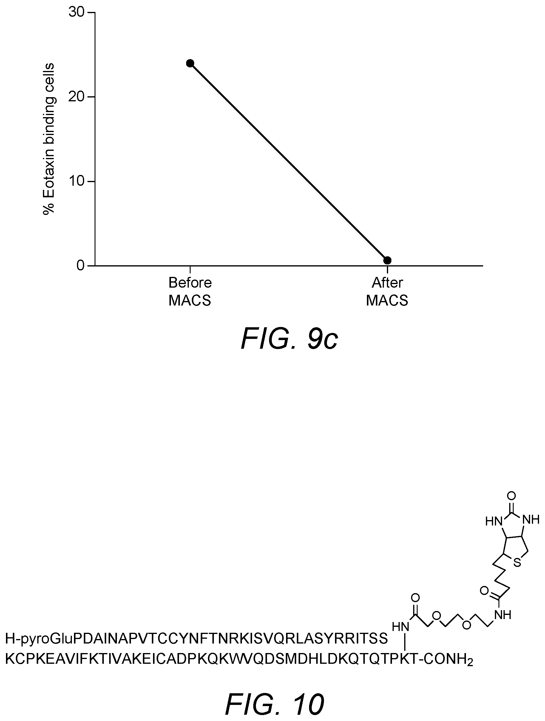
[0084] MCP-5 only binds CCR2 and should be selective in its removal of CCR2 expressing cells. The full length amino acid sequence, including the signal peptide, is set forth as SEQ ID NO: 4. The amino acid sequence of N-terminal processed MCP-5 chemokine is 82 amino acids long and is set forth as SEQ ID NO: 5. An amino acid sequence alignment suggests that MCP-5 has a C-terminal extension when compared to the amino acid sequence of MCP-1. The results of this alignment are shown in FIG. 10. C-terminal truncated versions of MCP-5 can thus be synthesised. This truncated version will comprise, consist essentially of or consist of MCP-5 residues 1-76, set forth as SEQ ID NO: 6.
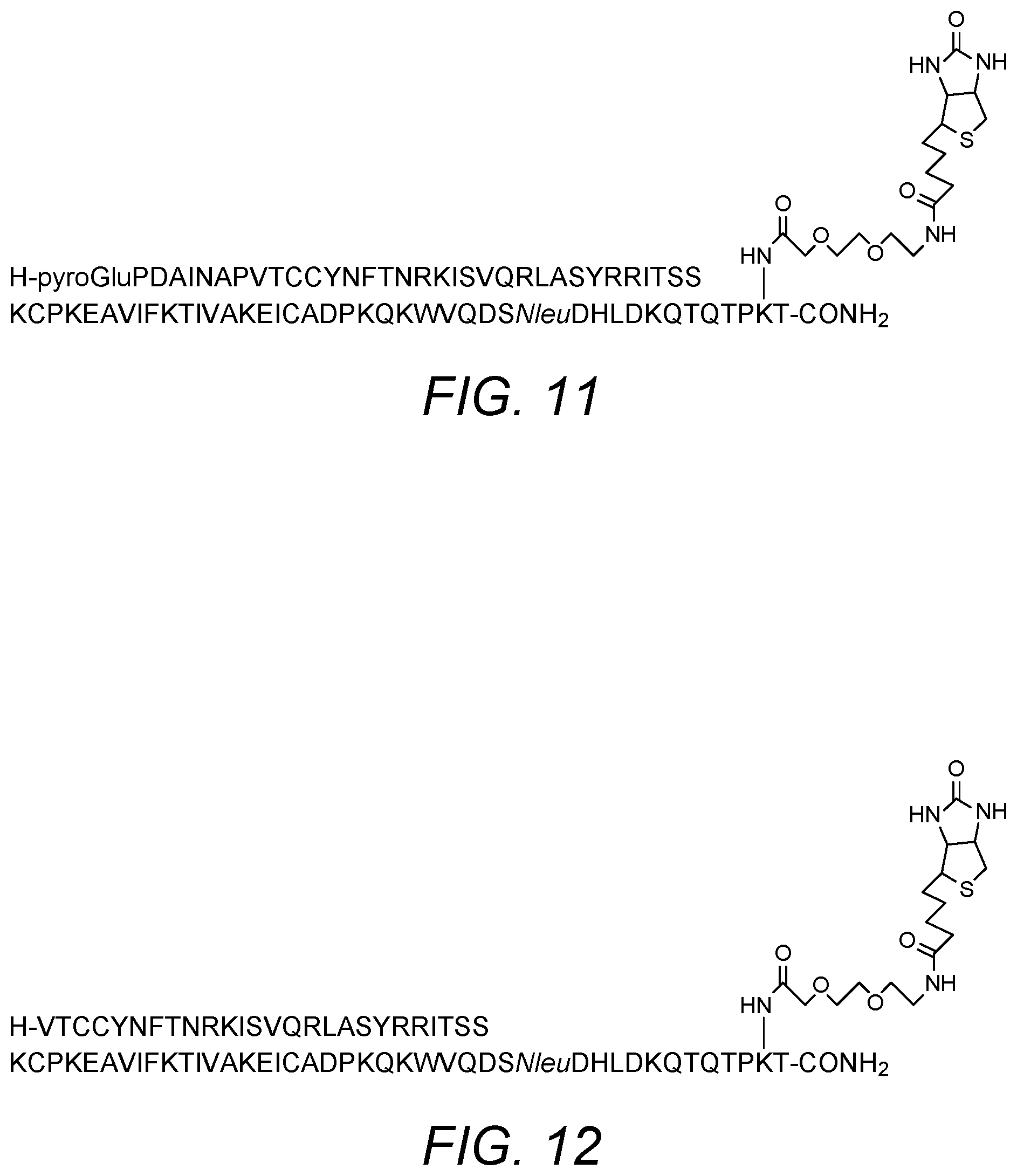
[0085] Accordingly, in certain embodiments the invention also provides a modified MCP-5 chemokine comprising the amino acid sequence set forth as SEQ ID NO: 4, SEQ ID NO: 5 or SEQ ID NO: 6 in which the isoleucine residue at position 97 of SEQ ID NO: 4 or at position 75 of SEQ ID NO: 5 or SEQ ID NO: 6 has been replaced with lysine. In certain embodiments, the modified MCP-5 chemokine comprises, consists essentially of or consists of the amino acid sequence of SEQ ID NO: 7. The modified MCP-5 chemokine may be biotinylated at the lysine (or a functional equivalent) residue at position 97 of SEQ ID NO: 4 or at position 75 of SEQ ID NO: 5 or SEQ ID NO: 6. Biotinylation may be via a suitable spacer group. Specific examples of the spacer group include a PEG spacer, optionally 3,6-dioxo aminooctanoic acid. In some embodiments, the C terminus is produced as an amide derivative. This may be achieved by synthesis on an amide linker. In certain embodiments, the modified MCP-5 chemokine comprises, consists essentially of or consists of the sequence and biotinylation shown in FIG. 11. The modified MCP-5 chemokine may be an agonist or an antagonist of CCR2 activity. They can be tested for activity in a suitable assay, such as cell-based assays. In particular, agonist and antagonist properties may be determined in a functional cell-based assay on human CCR2 receptor.
[0086] Eotaxin has been produced with Lys73 as the site of biotinylation on the chemokine (numbering based upon the mature protein having the amino acid sequence of SEQ ID NO: 9). Biotinylation permits immobilization of eotaxin on a solid support (via a biotin-avidin interaction). The basic amino acid sequence of eotaxin, including a 23 amino acid leader sequence is set forth as SEQ ID NO: 8. The amino acid sequence of the mature protein is set forth as SEQ ID NO: 9. The inventors have determined that chemokines may display improved binding properties where the chemokine is biotinylated via a spacer group. The spacer may prevent the biotin group from impacting on the binding affinity of the chemokine. Any suitable spacer group may be employed. Further modifications may provide the molecule with advantageous properties.
[0087] Accordingly, in certain embodiments the invention also provides a modified eoxtaxin chemokine comprising the amino acid sequence set forth as SEQ ID NO: 8 or SEQ ID NO: 9 in which one or more of the following modifications have been made:
[0088] a) the C terminus is produced as an amide derivative
[0089] c) the (C terminal region) lysine residue at position 96 of SEQ ID NO: 8 or position 73 of SEQ ID NO: 9 is biotinylated, optionally via a spacer group, in order to permit immobilization of the chemokine on a solid support
[0090] d) the methionine residue at position 85 of SEQ ID NO: 8 or position 62 of SEQ ID NO: 9 has been replaced with norleucine
[0091] e) the Lysine at position 96 of SEQ ID NO: 8 or position 73 of SEQ ID NO: 9 s replaced with another amino acid that can enable biotinylation such as ornithine
[0092] The (C terminal region) lysine residue at position 96 of SEQ ID NO: 8 or position 73 of SEQ ID NO: 9 may be biotinylated via a suitable spacer group, such as a polyethylene glycol (PEG) spacer group, in order to permit immobilization of the chemokine on a solid support. In specific embodiments, the PEG spacer is 3,6-dioxo aminooctanoic acid. The sequence and biotinylation of a modified eotaxin chemokine of the invention is shown in FIG. 7. The modified eoxtaxin chemokines may be agonists or antagonists of CCR3 activity. They can be tested for activity in a suitable assay, such as cell-based assays. In particular, agonist and antagonist properties may be determined in aequorin functional cell-based assay on human CCR3 receptor.
[0093] An example of a chemokine of the various embodiments of the invention containing modifications and specifically adapted for use in the invention is described in detail herein (see Example 3 below). The modified CCL8 (MCP-2) corresponds to residues 1 to 76 of the full length mature protein (and lacks the N-terminal signal peptide of 23 amino acids, which is cleaved off) and thus retains the chemokine fold. The Gln at the N-terminus of the protein is subject to pyroGlu formation under physiological conditions. Thus Gln1 of the sequence may thus be substituted with pyroglutamine to prevent mixed species of N-terminal Gln and pyroGlu being generated (SEQ ID NO: 13). This improves the yield of synthesis and ensures a homogeneous chemokine preparation through column manufacture and use. FmocLys(ivDde)-OH is incorporated as residue 75 to facilitate site-specific labelling at this position of the protein (SEQ ID NO: 14). The naturally occurring lysine at position 75 is modified through biotinylation. A PEG spacer may be incorporated between the &amino functionality and the biotin (SEQ ID NO: 15).
[0094] Thus, in other embodiments the invention also relates to a modified chemokine comprising, consisting essentially of or consisting of the amino acid sequence of SEQ ID NO: 13:
TABLE-US-00001 XPDSVSIPITCCFNVINRKIPIQRLESYTRITNIQCPKEAVIFKTKRGKE VCADPKERWVRDSMKHLDQIFQNLXP
[0095] X1=pyroGlu (but may remain as Gln in some embodiments)
[0096] X75=an amino acid residue that can be biotinylated, such as lysine, ornithine or diaminopropionic acid and optionally is biotinylated, optionally via a spacer molecule such as PEG, e.g. K(PEG-Biotin).
[0097] Or
TABLE-US-00002 SEQ ID NO: 15 XPDSVSIPITCCFNVINRKIPIQRLESYTRITNIQCPKEAVIFKTKRGKE VCADPKERWVRDSMKHLDQIFQNLXP
[0098] X1=pyroGlu (but may remain as Gln in some embodiments)
[0099] X75=K(PEG-Biotin).
[0100] A further example of a chemokine of the various embodiments of the invention containing modifications and specifically adapted for use in the invention is described in detail herein (see Example 4 below). The modified CCL5 (RANTES) corresponds to residues 1 to 68 of the full length mature protein (and lacks the N-terminal signal peptide of 23 amino acids, which is cleaved off) and thus retains the chemokine fold. The single methionine (Met67) within the sequence is mutated to lysine, to mitigate against oxidation of this residue during the chain assembly (SEQ ID NO: 16). This Met to Lys substitution provides a lysine at position 67 which can be modified through biotinylation. FmocLys(ivDde)-OH is incorporated as residue 67 to facilitate site-specific labelling at this position of the protein (SEQ ID NO: 17). The biotinylated version comprises, consists essentially of or consists of the amino acid sequence of SEQ ID NO: 18.
[0101] Thus, in other embodiments the invention also relates to a modified chemokine comprising, consisting essentially of or consisting of the amino acid sequence of SEQ ID NO: 18:
TABLE-US-00003 SPYSSDTTPCCFAYIARPLPRAHIKEYFYTSGKCSNPAVVFVTRKNRQVC ANPEKKWVREYINSLEXS
[0102] X is an amino acid residue that can be biotinylated, such as lysine, ornithine or diaminopropionic acid and optionally is biotinylated, optionally via a spacer molecule such as PEG (e.g. K(Biotin))
[0103] A further example of a chemokine of the various embodiments of the invention containing modifications and specifically adapted for use in the invention is described in detail herein (see Example below). The modified CCL2 (MCP-1) corresponds to residues 1 to 76 of the full length mature protein (and lacks the N-terminal signal peptide of 23 amino acids, which is cleaved off) and thus retains the chemokine fold (SEQ ID NO: 10). The Gln at the N-terminus of the protein (Gln1) is substituted with pyroglutamine to prevent mixed species of N-terminal Gln and pyroGlu being generated. This improves the yield of synthesis and ensures a homogeneous chemokine preparation through column manufacture and use. FmocLys(ivDde)-OH is incorporated as residue 75 to facilitate site-specific labelling at this position of the protein (SEQ ID NO: 11). A suitable spacer, such as a PEG spacer, may be incorporated between the &amino functionality and the biotin. The biotinylated version comprises, consists essentially of or consists of the amino acid sequence of SEQ ID NO: 12.
[0104] Thus, in other embodiments the invention also relates to a modified chemokine comprising, consisting essentially of or consisting of the amino acid sequence of:
TABLE-US-00004 SEQ ID NO: 10: XPDAINAPVTCCYNFTNRKISVQRLASYRRITSSKCPKEAVIFKTIVAKE ICADPKQKWVQDSMDHLDKQTQTPKT
[0105] X=pyroGlu or Gln
[0106] And/or
TABLE-US-00005 SEQ ID NO: 12: XPDAINAPVTCCYNFTNRKISVQRLASYRRITSSKCPKEAVIFKTIVAKE ICADPKQKWVQDSMDHLDKQTQTPXT
[0107] X1=pyroGlu or Gln
[0108] X75 is an amino acid residue that can be biotinylated, such as lysine, ornithine or diaminopropionic acid and optionally is biotinylated, optionally via a spacer molecule such as PEG, optionally K(PEG-Biotin)
[0109] A further example of a chemokine of the various embodiments of the invention containing modifications and specifically adapted for use in the invention is described in detail herein (see Example 9 below). The modified CCL19 (MIP-3.beta.) corresponds to residues 1 to 77 of the full length mature protein (and lacks the N-terminal signal peptide of 21 amino acids, which is cleaved off) and thus retains the chemokine fold. An additional lysine is inserted at the C-terminus, at position 78. The chemokine may thus comprise, consist essentially of or consist of the amino acid sequence of SEQ ID NO: 19. FmocLys(ivDde)-OH is incorporated as residue 78 to facilitate site-specific labelling at this position of the protein (SEQ ID NO: 20). The .epsilon.-amino side chain functionality of Lys(78) is modified through biotinylation. The final protein may thus comprise, consist essentially of or consist of the amino acid sequence of SEQ ID NO: 21.
[0110] Thus, in other embodiments the invention also relates to a modified chemokine comprising, consisting essentially of or consisting of the amino acid sequence of SEQ ID NO: 19 or 21:
TABLE-US-00006 SEQ ID NO: 19 GTNDAEDCCLSVTQKPIPGYIVRNFHYLLIKDGCRVPAVVFTTLRGRQLC APPDQPWVERIIQRLQRTSAKMKRRSSX
[0111] X is an amino acid residue that can be biotinylated, such as lysine, ornithine or diaminopropionic acid and optionally is biotinylated (e.g. K-biotin), optionally via a spacer molecule such as PEG, in particular K(PEG-Biotin)
TABLE-US-00007 SEQ ID NO: 21 GTNDAEDCCLSVTQKPIPGYIVRNFHYLLIKDGCRVPAVVFTTLRGRQLC APPDQPWVERIIQRLQRTSAKMKRRSSX
[0112] X is K(Biotin)
[0113] A further example of a chemokine of the various embodiments of the invention containing modifications and specifically adapted for use in the invention is described in detail herein (see Example 3 below). The modified CXCL8 (IL-8) corresponds to residues 1 to 77 of the full length mature protein (and lacks the N-terminal signal peptide of 22 amino acids, which is cleaved off) and thus retains the chemokine fold. An amino acid residue capable of biotinylation, such as lysine or ornithine, is added as residue 78 (SEQ ID NO: 22). FmocLys(ivDde)-OH may be incorporated as residue 78 to facilitate site-specific labelling at this position of the protein (SEQ ID NO: 23). The additional amino acid, in particular lysine or ornithine, at position 78 is modified through biotinylation. A suitable spacer, such as a PEG spacer, may be incorporated between the &amino functionality and the biotin (SEQ ID NO: 24).
[0114] Thus, in other embodiments the invention also relates to a modified chemokine comprising, consisting essentially of or consisting of the amino acid sequence of SEQ ID NO: 22 or 24:
TABLE-US-00008 SEQ ID NO: 22 AVLPRSAKELRCQCIKTYSKPFHPKFIKELRVIESGPHCANTEIIVKL SDGRELCLDPKENWVQRVVEKFLKRAENSX
[0115] X is an amino acid residue that can be biotinylated, such as lysine, ornithine or diaminopropionic acid and optionally is biotinylated, optionally via a spacer molecule such as PEG
TABLE-US-00009 SEQ ID NO: 24 AVLPRSAKELRCQCIKTYSKPFHPKFIKELRVIESGPHCANTEIIVKL SDGRELCLDPKENWVQRVVEKFLKRAENSK(PEG-Biotin)
[0116] A further example of a chemokine of the various embodiments of the invention containing truncations and modifications and specifically adapted for use in the invention is described in detail herein (see Example 3 below). The modified CXCL8 (IL-8) corresponds to residues 6 to 77 of the full length mature protein, with the first 5 N-terminal amino acids removed, (and lacks the N-terminal signal peptide of 22 amino acids, which is cleaved off) and thus retains the chemokine fold. An amino acid residue capable of biotinylation, such as lysine or ornithine, is added as residue 78 (SEQ ID NO: 25). FmocLys(ivDde)-OH may be incorporated as residue 78 to facilitate site-specific labelling at this position of the protein (SEQ ID NO: 26). The additional amino acid, in particular lysine or ornithine, at position 78 is modified through biotinylation. A suitable spacer, such as a PEG spacer, may be incorporated between the &amino functionality and the biotin (SEQ ID NO: 27).
[0117] Thus, in other embodiments the invention also relates to a modified chemokine comprising, consisting essentially of or consisting of the amino acid sequence of SEQ ID NO: 25 or 27:
TABLE-US-00010 SEQ ID NO: 25 SAKELRCQCIKTYSKPFHPKFIKELRVIESGPHCANTEIIVKLSDGREL CLDPKENWVQRVVEKFLKRAENSX
[0118] X is an amino acid residue that can be biotinylated, such as lysine, ornithine or diaminopropionic acid and optionally is biotinylated, optionally via a spacer molecule such as PEG
TABLE-US-00011 SEQ ID NO: 27 SAKELRCQCIKTYSKPFHPKFIKELRVIESGPHCANTEIIVKLSDGREL CLDPKENWVQRVVEKFLKRAENSX
[0119] X is K(PEG-Biotin)
[0120] A further example of a chemokine of the various embodiments of the invention containing modifications and specifically adapted for use in the invention is described in detail herein (see Example 9 below). The modified CCL11 (Eotaxin) corresponds to residues 1 to 74 of the full length mature protein (and lacks the N-terminal signal peptide of 23 amino acids, which is cleaved off) and thus retains the chemokine fold (SEQ ID NO: 28). The lysine at position 73 may be modified through biotinylation. FmocLys(ivDde)-OH is incorporated as residue 73 to facilitate site-specific labelling at this position of the protein (SEQ ID NO: 29). A suitable spacer, such as a PEG spacer, may be incorporated between the &amino functionality and the biotin. The biotinylated version comprises, consists essentially of or consists of the amino acid sequence of SEQ ID NO: 30.
[0121] Thus, in other embodiments the invention also relates to a modified chemokine comprising, consisting essentially of or consisting of the amino acid sequence of SEQ ID NO: 28 or SEQ ID NO: 30:
TABLE-US-00012 SEQ ID NO: 28 GPASVPTTCCFNLANRKIPLQRLESYRRITSGKCPQKAVIFKTKLAKDIC ADPKKKWVQDSMKYLDQKSPTPXP
[0122] X is an amino acid residue that can be biotinylated, such as lysine, ornithine or diaminopropionic acid and optionally is biotinylated, optionally via a spacer molecule such as PEG, e.g. K(PEG-Biotin).
TABLE-US-00013 SEQ ID NO: 30 H-GPASVPTTCCFNLANRKIPLQRLESYRRITSGKCPQKAVIFKTKLAKD ICADPKKKWVQDSMKYLDQKSPTPXP-NH.sub.2
[0123] X is K(PEG-Biotin)
[0124] Chemokines useful in the various embodiments of the invention may be synthesised through any suitable means known in the art. Preferably, the chemokines are chemically synthesised as this facilitates modification and labelling etc. However, recombinant DNA based approaches may also be employed in combination with appropriate labelling and modification technologies as required. Thus, in certain embodiments the invention also provides a nucleic acid molecule encoding the chemokines of the various embodiments of the invention. In certain embodiments the invention also relates to a vector containing such a nucleic acid molecule and a host cell containing the vector. The vector may additionally comprise a suitable promoter operably linked to the nucleic acid molecule, to facilitate transcription of the corresponding mRNA molecule. The host cell may be capable of expressing the protein by transcription and translation of the nucleic acid molecule encoding a chemokine of the various embodiments of the invention.
[0125] The chemokines useful in the various embodiments of the invention can be biotinylated by methods known in the art such as described in WO 00/50088 A2, which is incorporated herein by reference in its entirety. As indicated above, site-specific labelling of the chemokines of the various embodiments of the invention is preferable, although any labelling technique which does not significantly affect the receptor-binding capacity of the chemokine may be employed. Various site-specifically biotinylated chemokines and native chemokines are available commercially, for instance from Almac, Craigavon, UK. In specific embodiments the one or more chemokines are biotinylated via a spacer group. The spacer may be employed to prevent the biotin group from impacting on the activity of the chemokine, in particular binding of the chemokine to its cognate receptor. Any suitable spacer that facilitates retention of receptor binding properties of the chemokine may be employed in the various embodiments of the invention. Thus, in the specific embodiments described above, spacers other than PEG spacers may be employed as appropriate. In specific embodiments, the spacer is a polyethylene glycol (PEG) spacer. PEG has been shown to be an effective spacer permitting attachment of biotin to the chemokine (which can then be immobilized on the solid support through interaction with streptavidin) without compromising receptor binding capability.
[0126] In the context of the various embodiments of the present invention the term "antibody" includes all immunoglobulins or immunoglobulin-like molecules with specific binding affinity for the relevant chemokine receptor (including by way of example and without limitation, IgA, IgD, IgE, IgG and IgM, combinations thereof, and similar molecules produced during an immune response in any vertebrate, for example, in mammals such as humans, goats, rabbits and mice). Specific immunoglobulins useful in the various embodiments of the invention include IgG isotypes. The antibodies useful in the various embodiments of the invention may be monoclonal or polyclonal in origin, but are typically monoclonal antibodies. Antibodies may be human antibodies, non-human antibodies, or humanized versions of non-human antibodies, or chimeric antibodies. Various techniques for antibody humanization are well established and any suitable technique may be employed. The term "antibody" also refers to a polypeptide ligand comprising at least a light chain or heavy chain immunoglobulin variable region which specifically recognizes and binds an epitope of an antigen, and it extends to all antibody derivatives and fragments that retain the ability to specifically bind to the relevant chemokine receptor. These derivative and fragments may include Fab fragments, F(ab').sub.2 fragments, Fv fragments, single chain antibodies, single domain antibodies, Fc fragments etc. The term antibody encompasses antibodies comprised of both heavy and light chains, but also heavy chain (only) antibodies. In specific embodiments, the antibodies may be engineered so as to be specific for more than one chemokine receptor, for example bi-specific to permit binding to two different chemokine receptors. Suitable commercially available antibodies which bind to the chemokine receptors of interest are listed in table 1 below. They may or may not be labelled. General reference may be made to "Antibodies a laboratory manual: By E Harlow and D Lane. pp 726. Cold Spring Harbor Laboratory. 1988", which reference is incorporated herein in its entirety.
TABLE-US-00014 TABLE 1 Commercially available fluorophore labelled antibodies against specific chemokine receptors Antibody Fluorophore Supplier CCR2 PerCP Cy5.5 Biolegend CCR1 Alexa Fluor 647 Biolegend CCR3 PE Biolegend CCR5 PE Biolegend CXCR1 APC Biolegend CXCR2 PE Biolegend CCR7 PerCP Cy5.5 Biolegend
[0127] Anti-CCR2 antibodies are described for example in WO 2010/021697, incorporated herein by reference. Further examples of potentially useful antibodies include MLN-1202, an anti-CCR2 monoclonal antibody currently undergoing clinical trials (Millennium Pharmaceuticals).
[0128] The chemokine receptor expressing cells may thus be targeted using alternative binding agents, such as antibodies or other chemical compounds, as defined herein, rather than the natural chemokine binding partner. This approach is a new approach to treating inflammatory conditions.
[0129] Accordingly, in certain embodiments the invention also provides an apheresis column loaded with a solid support comprising a binding reagent capable of specifically binding to a chemokine receptor immobilized directly or indirectly on the support to permit removal of a cell expressing the chemokine receptor from the peripheral blood of a patient, wherein the binding reagent is not a chemokine. The binding reagent capable of specifically binding to the chemokine receptor may be an agonist or an antagonist of the chemokine receptor. In specific embodiments, the binding reagent capable of specifically binding to the chemokine receptor is selected from an antibody and a chemical compound.
[0130] In other embodiments the invention thus also provides a method for treating an inflammatory condition comprising applying peripheral blood from a patient/subject to an apheresis column as defined above (an apheresis column loaded with a solid support comprising a binding reagent capable of specifically binding to a chemokine receptor immobilized directly or indirectly on the support to permit removal of a cell expressing the chemokine receptor from the peripheral blood of a patient, wherein the binding reagent is not a chemokine) thus removing chemokine receptor expressing cells from the peripheral blood of the patient/subject. The method may comprise returning the blood depleted of the chemokine receptor expressing cells to the patient/subject.
[0131] Similarly, in other embodiments the invention provides a binding reagent capable of specifically binding to a chemokine receptor for use in the treatment of an inflammatory condition, wherein the binding reagent is immobilized on a solid support contained within an apheresis column as defined above (an apheresis column loaded with a solid support comprising a binding reagent capable of specifically binding to a chemokine receptor immobilized directly or indirectly on the support to permit removal of a cell expressing the chemokine receptor from the peripheral blood of a patient/subject, wherein the binding reagent is not a chemokine), to which is applied peripheral blood from a patient thus removing chemokine receptor expressing cells from the peripheral blood of the patient.
[0132] These aspects of the various embodiments of the invention may be integrated into the more focused therapeutic aspects of the various embodiments of the invention (i.e. treating respiratory conditions such as sarcoidosis and COPD and various aspects thereof) and thus, the remainder of the disclosure, including all specific embodiments applies mutatis mutandis.
[0133] Solid support materials for immobilizing the binding reagents of the various embodiments of the invention are known in the art. "Solid support" refers to, for example, materials having a rigid or semi-rigid surface or surfaces, and may take the form of beads, resins, gels, microspheres, or other geometric configurations. A useful support material is one that does not activate blood cells so as to make them coagulate or adhere to the support as peripheral whole blood is applied to the device. In certain embodiments, a support treated with an agent to provide it with anti-coagulation properties, in particular a heparinized support is employed. Alternatively, the blood of the patient may be treated with an anti-coagulant such as heparin prior to application to the support. Useful support materials comprise high molecular weight carbohydrates, in particular carbohydrates having a molecular weight of 100 kDa or more, such as agarose, in particulate form, optionally cross-linked, and cellulose. Other preferred support materials are polymers, such as carboxylated polystyrene, and glass. The support of the various embodiments of the invention may be provided in the form of particles or fibres. The support particles may have regular form, such as spheres or beads, or irregular form. They may be porous or non-porous. A preferred average particle size of the support is from 50 .mu.m to 2 mm. In certain embodiments Sepharose.TM., a cross linked, beaded-form of agarose, is used as column matrix. It is chosen for its optimal distribution capacity and can provide a large available area for affinity binding. Solid supports may be provided in the form of magnetic beads, with the specific binding reagent immobilized on the magnetic beads. Following capture of the (CCR2, CCR1, CCR3, CCR5, CXCR1, CXCR2 and/or CCR7) chemokine receptor expressing cells from the blood, the beads can be removed from the blood with the aid of an appropriate magnetic separator.
[0134] Methods for immobilizing binding reagents on a solid support are known in the art. A binding reagent, such as a chemokine, antibody, peptide, nucleic acid or chemical compound, can be immobilized on the support in a direct or indirect manner. Immobilization can be by means of a suitable linker in some embodiments. A preferred method of indirect immobilization of a binding reagent, such as a chemokine, relies upon the interaction between biotin and avidin molecules. "Avidin" or "avidin molecule" refers to any type of protein that specifically binds biotin to the substantial exclusion of other (small) molecules that might be present in a biological sample. Examples of avidin include avidins that are naturally present in egg white, oilseed protein (e.g., soybean meal), and grain (e.g., corn/maize), and streptavidin, which is a protein of bacterial origin Thus, biotinylation of the binding reagent and use of an avidin molecule such as streptavidin immobilized on the solid support allows reliable attachment of the binding reagent to the solid support according to methods known in the art. Specifically, such a method may comprise providing the binding reagent in biotinylated form, providing a solid support having streptavidin immobilized on its surface, contacting the support with an aqueous solution of the biotinylated binding reagent, and rinsing the support with an aqueous solvent. In addition, binding pair interactions, such as antibody--antigen interactions, may also be utilised for indirect immobilisation of binding reagent onto a support. In such embodiments the support may be derivatised with one member of a binding pair, such as an antibody or fragment or derivative thereof, as defined herein, which has known affinity for a particular peptide sequence or small molecule hapten. Incorporating the other member of the binding pair, such as an antigen, peptide sequence or the hapten onto or into the binding reagent facilitates immobilisation onto a solid support coated with the corresponding antibody or fragment or derivative thereof. Thus, the binding reagent may be modified to include the peptide sequence or hapten into the linear molecule or may be added as a side chain or label. Any suitable antibody-antigen pair may be employed. The antibody fragment or derivative may be any fragment or derivative that retains specific binding affinity for the appropriate antigen. Examples include Fab, F(ab').sub.2 fragments, scFV, VH domains, single domain antibodies (such as nanobodies), heavy chain antibodies and humanized version of non-human antibodies etc. Other high affinity interactions can be utilised for immobilisation of binding reagents, as long as the binding reagent can be synthesised or derivatised with one of the interacting partners and the solid support synthesised or derivatised with the other interacting partner without loss of binding activity (i.e. binding of the binding reagent to the appropriate chemokine receptor). Immobilization may occur via essentially the same interaction in reverse in some embodiments. Thus, the binding reagent which may be a chemokine for example, may be attached to an antibody as defined herein, and the solid support derivatised with the antigen. The chemokine may be produced as a fusion protein with the antibody.
[0135] Alternatively binding reagents, such as chemokines and antibodies, can be immobilised directly onto a solid support using bioconjugation techniques established in the field. For example direct immobilisation of proteins onto cyanogen bromide activated solid supports via amino functionalities within the primary sequence of the protein. Alternatively, sulphydryl functionalities within proteins can be used to directly immobilise the protein to alkyl halide derivatised supports or supports containing free thiol functionalities. In further embodiments, proteins containing a-thioester functionalities can be directly immobilised on supports containing 1,2 amino thiol moieties (eg N-terminal cysteine) using the native chemical ligation reaction. Alternatively proteins modified with ketones and aldehydes can be immobilised on solid supports derivatised with hydrazinyl, hydrazide and aminoxy functionalities using hydrazone/oxime bond forming ligation reactions (and vice versa). Alternatively `Click` chemistry can be used to immobilise proteins onto solid supports, whereby the protein and the support are derivatised with the appropriate mutually reactive chemical functionalities (azides and alkynes). In other embodiments Staudinger ligation chemistry can be used to immobilise appropriately derivatised proteins onto the appropriately derivatised solid supports.
[0136] The solid support is contained within or carried by the apheresis column. Thus, by "loaded" is meant that the column carries or contains the solid support in a manner such that (peripheral) blood can flow through the column in contact with the solid support. Thus, the solid support provides a matrix within the column through which blood flows, in continuous fashion in certain embodiments. This permits cells expressing the specific chemokine receptor to be removed from the blood passing through the column, such that blood exiting the column is depleted of the specific chemokine receptor-expressing cells. In specific embodiments, the apheresis column is loaded with a support comprising streptavidin immobilized on the support and one or more biotinylated binding reagents, such as chemokines, bound to the streptavidin on the support. The solid support may be comprised of a high-molecular weight carbohydrate, optionally cross-linked, such as agarose.
[0137] As discussed above, the binding reagent is coupled to the solid support. The relative amounts of binding reagent may be controlled to ensure that coupling between the solid support and the binding reagent will be immediate, minimising the risk of binding reagent decoupling from the solid support. Thus, it may be ensured that there is a relative excess of immobilization sites for the binding reagent on the solid support. Alternatively, or additionally, following immobilization of the binding reagent on the solid support, the solid support may be washed to remove any unbound binding reagent.
[0138] The apheresis column utilised in the various embodiments of the present invention acts as a leukapheresis treatment for respiratory conditions, in particular sarcoidosis and Chronic Obstructive Pulmonary Disease (COPD). The column acts to specifically remove one or more of CCR2, CCR1, CCR3, CCR5, CXCR1, CXCR2 and/or CCR7-expressing monocytes or leukocytes by exploiting the interaction between CCR2, CCR1, CCR3, CCR5, CXCR1, CXCR2 and/or CCR7 expressed on the cell surface and a specific binding reagent immobilized on a solid support contained within or carried by the column. The overall column typically comprises, consists of, or consists essentially of three combined components; 1) a housing which contains or carries 2) the solid support and 3) one or more binding reagents (immobilized thereon) which specifically bind one or more chemokine receptors. The housing may be manufactured from any suitable material for clinical use. In certain embodiments the housing is composed of a plastic material. The housing includes an in flow site for entry of blood and an out flow site for blood (depleted of target cells) to exit the column. The housing may be designed to maintain a continuous blood flow through the solid support matrix. The housing (as shown for example in FIG. 3) may include a top portion which comprises a distribution plate (2) at the inflow site (1) to spread the blood evenly over the entire matrix area. The distribution plate may act as a first safety barrier preventing larger particles flowing through the column and into the patient. However, the distribution plate is not essential and may be removed in some embodiments to decrease the overall resistance in the system. The column may contain one or more safety filter units (3 and 4) placed at the inflow (1) and/or outflow (5) sites of the plastic housing. Such filter units may act to prevent particles larger than blood cells passing in and/or out of the column. The safety filter units may contain a plurality of filters, such as two, three or four filters designed to be a robust barrier and stop all particles larger than blood cells passing through the column. Inclusion of safety filters (3 and 4) at both ends of the column serves to minimize the risk of leakage of particles into the patient, including in the event that the device is incorrectly connected resulting in blood flow in the opposite direction to that intended. The safety filters may comprise of any suitable pore size to prevent particles larger than blood cells from passing through the column, as would be readily understood by one skilled in the art. Suitable filters are commercially available. In specific embodiments, the pore size of the filter(s) is between approximately 60 .mu.m and 100 .mu.m, more specifically approximately 80 .mu.m. The solid support and binding reagent components are discussed in further detail herein.
[0139] The volume of the housing may be varied depending upon the blood volumes intended to pass through the column. Typically, the volume of the housing is between approximately 40 ml and 200 ml, more specifically 50 ml to 150 ml or 60 ml to 120 ml.
[0140] The column is generally applied in the form of an apheresis circuit. In this context, the overall system includes the apheresis column, tubing and an appropriate pump to pump the blood around the circuit. The system is illustrated in FIG. 4. The patient (1) is connected to the extracorporeal circuit via sterile needles to veins in the right and the left arms. A saline bag (3) is also connected and the saline solution is pumped with a suitable pump (2). Blood is drawn from one arm of the patient through the sterile tubing system by the blood pump (4) and passed through the column (6) and back to the patient. The tubing system may be connected to the column via any suitable coupling, such as standard dialysis luer-lock couplings. The couplings on the column may be colour-coded for correct assembly. For example, red tubing for inflow to the red column top and blue tubing for outflow back to the patient. An air detector (8) may be present in the circuit. Inlet pressure (5) and/or Pven sensors (7) may additionally be employed to monitor the pressure in the circuit.
[0141] An apheresis pump, such as the 4008 ADS pump manufactured by Fresenius Medical Care or the Adamonitor pump, may monitor the patient's inflow and outflow. The pump may also monitor the pressure in the extracorporeal circulation. The pump may be able to discriminate air by a bubble catcher and air detector. A clot catcher filter may be positioned inside the bubble catcher. The pump may also incorporate an optical detector to distinguish between light, e.g. saline solution or air present in the tubing system and dark e.g. blood present in the tubing system.
[0142] A schematic diagram of a suitable pump, showing the air detector and optical filter is shown in FIG. 8. If the pump system detects air bubbles and optical fluctuations or if extracorporeal pressure values are out of the set range, then the pump may stop immediately. Alternatively or additionally a visual/audible alarm may be emitted.
[0143] The treatment methods of the various embodiments of the invention may thus rely upon an extracorporeal circuit. The methods may be considered as ex vivo or in vitro methods and be defined solely with reference to steps performed outside of the patient. In some embodiments, however, the method further comprises, prior to application of the blood to the column, collecting peripheral blood from the patient. In a further embodiment, the method further comprises, following the application of the blood to the column, infusing the blood depleted of (CCR2, CCR1, CCR3, CCR5, CXCR1, CXCR2 and/or CCR7) chemokine receptor expressing cells to the patient. This is then a complete leukapheresis treatment method. Thus, a leukaphereis method, for treating respiratory conditions, in particular sarcoidosis and Chronic Obstructive Pulmonary Disease (COPD), comprises collecting peripheral blood from the patient; applying the peripheral blood to an apheresis column loaded with a solid support comprising one or more binding reagents capable of specifically binding to one or more chemokine receptors, in particular the chemokine receptor CCR2, CCR1, CCR3, CCR5, CXCR1, CXCR2 and/or CCR7, immobilized directly or indirectly on the support thus removing one or more of CCR2, CCR1, CCR3, CCR5, CXCR1, CXCR2 and/or CCR7 expressing cells from the peripheral blood of the patient; and infusing the depleted blood (of chemokine receptor expressing cells) to the patient.
[0144] The peripheral blood may be continuously collected from the patient. Similarly, the depleted blood may be continuously infused to the patient, through use of an appropriate circuit as described herein. Thus, the support may be disposed in a column through which the blood is made to flow. This may be achieved using a suitable pump for example, as also described herein. Blood flow through the column enables the binding reagent(s) immobilized on the solid support to capture the cells expressing the chemokine receptor, thus depleting them from the blood and preventing their contribution to the inflammatory respiratory conditions, in particular sarcoidosis and Chronic Obstructive Pulmonary Disease (COPD).
[0145] The methods of the various embodiments of the invention and binding reagents for use in the methods of the various embodiments of the invention may require that the patient has been selected for treatment on the basis of detecting an increase in the level of chemokine receptor, in particular, one or more of CCR2, CCR1, CCR3, CCR5, CXCR1, CXCR2 and/or CCR7 expressing cells in a sample obtained from the patient. Such companion diagnostic methods are described in greater detail herein and are based, for example, on the observation that CCR2, CCR1, CCR3, CCR5, CXCR1, CXCR2 and/or CCR7 expression may be elevated in patients with respiratory conditions, in particular sarcoidosis and Chronic Obstructive Pulmonary Disease (COPD). It is shown herein that subjects suffering from respiratory conditions such as sarcoidosis exhibit increased frequency of chemokine receptor expressing cells in the peripheral blood. Subjects with sarcoidosis exhibit increased frequency of CCR1 expressing cells such as CCR1 expressing monocytes, compared to healthy controls. Similarly, it is shown herein that the monocytes also express CCR2. It is also shown herein that subjects suffering from respiratory conditions such as sarcoidosis exhibit increased frequency of CCR7 expressing cells such as CCR7 expressing lymphocytes, and also central memory T cells, compared to healthy controls.
[0146] Thus, (in this context) in certain embodiments the invention also provides a method of diagnosing, monitoring progression of, or monitoring treatment of respiratory conditions, in particular sarcoidosis and Chronic Obstructive Pulmonary Disease (COPD) comprising determining:
[0147] a) the levels of one or more of the chemokine receptor CCR2, CCR1, CCR3, CCR5, CXCR1, CXCR2 and/or CCR7 expressing cells
[0148] b) levels of expression of one or more of CCR2, CCR1, CCR3, CCR5, CXCR1, CXCR2 and/or CCR7; and/or
[0149] c) levels of cells with high expression of one or more of CCR2, CCR1, CCR3, CCR5, CXCR1, CXCR2 and/or CCR7 in a sample obtained from a subject, wherein high levels of one or more ofCCR2, CCR1, CCR3, CCR5, CXCR1, CXCR2 and/or CCR7 expressing cells, high levels of expression of one or more of CCR2, CCR1, CCR3, CCR5, CXCR1, CXCR2 and/or CCR7 or high levels of cells with high expression of one or more of CCR2, CCR1, CCR3, CCR5, CXCR1, CXCR2 and/or CCR7 or increased levels of one or more of CCR2, CCR1, CCR3, CCR5, CXCR1, CXCR2 and/or CCR7 expressing cells compared to control, increased levels of expression of one or more of CCR2, CCR1, CCR3, CCR5, CXCR1, CXCR2 and/or CCR7 compared to a control or increased levels of cells with high expression of one or more of CCR2, CCR1, CCR3, CCR5, CXCR1, CXCR2 and/or CCR7 compared to a control indicate the presence or progression of respiratory conditions, in particular sarcoidosis and Chronic Obstructive Pulmonary Disease (COPD). Levels of chemokine receptor expression, as opposed to cell numbers, may also be investigated as increased levels of chemokine receptor expression per cell may also be diagnostically relevant. In specific embodiments, the cells relevant to diagnosis etc. of respiratory conditions such as sarcoidosis comprise monocytes, in particular CCR1 and/or CCR2 expressing monocytes. In other embodiments cells relevant to diagnosis etc. of respiratory conditions such as sarcoidosis comprise lymphocytes, specifically T lymphocytes such as central memory T cells, in particular CCR7 expressing T cells.
[0150] "Diagnosing" is defined herein to include screening for a disease/condition or pre-indication of a disease/condition, identifying a disease/condition or pre-indication of a disease/condition and checking for recurrence of disease/condition following treatment. The methods of the various embodiments of the invention may also have prognostic value, and this is included within the definition of the term "diagnosis". The prognostic value of the methods of the various embodiments of the invention may be used as a marker of potential susceptibility to respiratory conditions, in particular sarcoidosis and Chronic Obstructive Pulmonary Disease (COPD) by identifying levels of one or more of CCR2, CCR1, CCR3, CCR5, CXCR1, CXCR2 and/or CCR7 expression linked to the respiratory condition. Thus patients at risk may be identified before the disease has a chance to manifest itself in terms of symptoms identifiable in the patient. In certain embodiments, diagnosis may be made in conjunction with other objective indicators of respiratory conditions, in particular sarcoidosis and Chronic Obstructive Pulmonary Disease (COPD): The diagnosis of sarcoidosis may be made based upon clionical findings; x-ray, biopsy, s-Ca, ACE activity. COPD diagnosis may also be made by clinical findings; X-ray spirometry and blood gas analysis.
[0151] "Monitoring progression of includes performing the methods to monitor the stage and/or the state and progression of the respiratory conditions, in particular sarcoidosis and Chronic Obstructive Pulmonary Disease (COPD). Monitoring progression may involve performing the diagnostic methods multiple times on the same patient to determine whether the levels of one or more of CCR2, CCR1, CCR3, CCR5, CXCR1, CXCR2 and/or CCR7 expressing cells are increasing, decreasing or remaining stable over a certain time period. This may be in the context of a treatment regime.
[0152] "Monitoring the success of a particular treatment" is defined to include determining the levels of one or more of CCR2, CCR1, CCR3, CCR5, CXCR1, CXCR2 and/or CCR7 expressing cells before and after a treatment. The treatment is generally one aimed at treating respiratory conditions, in particular sarcoidosis and Chronic Obstructive Pulmonary Disease (COPD) and may be a treatment according to one of the methods of the various embodiments of the invention as defined herein. Successful treatment may be determined with reference to a decrease in one or more of CCR2, CCR1, CCR3, CCR5, CXCR1, CXCR2 and/or CCR7 expressing cells as a result of, or following, the treatment. Thus, in such methods a level of one or more of CCR2, CCR1, CCR3, CCR5, CXCR1, CXCR2 and/or CCR7 expressing cells is determined prior to treatment. This level is recorded and a further assessment made at a predetermined time following the treatment. The comparison of levels of one or more of CCR2, CCR1, CCR3, CCR5, CXCR1, CXCR2 and/or CCR7 expressing cells permits the success of the treatment to be monitored. In specific embodiments, a single treatment is sufficient to cause a depletion of around 10%, 20%, 30%, 40%, 50%, 60% or 70%, or higher, up to 80%, 90%, 95% or more, or any range of values between and including these amounts, of one or more specific chemokine receptors, in particular one or more of CCR2, CCR1, CCR3, CCR5, CXCR1, CXCR2 and/or CCR7, expressing cells from the peripheral blood of the patient. In specific embodiments, at least around 50% depletion is achieved in a single treatment. Thus, successful treatment may be defined with reference to depletion of one or more of CCR2, CCR1, CCR3, CCR5, CXCR1, CXCR2 and/or CCR7 expressing cells. Treatment may lead to depletion of between approximately 100 and 500 million of one or more of CCR2, CCR1, CCR3, CCR5, CXCR1, CXCR2 and/or CCR7 expressing cells, such as monocytes, in certain embodiments. Additional factors may be included to determine successful treatment. For example, a lack of increase in one or more of CCR2, CCR1, CCR3, CCR5, CXCR1, CXCR2 and/or CCR7 expressing cells following treatment may indicate successful treatment in terms of preventing further progression of the condition, optionally combined with an improvement in other markers or staging of the respiratory conditions, in particular sarcoidosis and Chronic Obstructive Pulmonary Disease (COPD).
[0153] By binding to the column through the binding reagent-chemokine receptor interaction, chemokine receptor expressing cells are immobilized. These immobilized cells express further unoccupied chemokine receptors, which may be of the same or different type to those used for capture. These additional chemokine receptors may permit circulating chemokines which contribute to the inflammatory condition to be captured from the peripheral blood. Thus, a reduction in circulating (specific) chemokine levels may provide a measure of successful treatment. In specific embodiments, the cells relevant to diagnosis etc. of respiratory conditions such as sarcoidosis comprise monocytes, in particular CCR1 and/or CCR2 expressing monocytes. In other embodiments cells relevant to diagnosis etc. of respiratory conditions such as sarcoidosis comprise lymphocytes, specifically T lymphocytes such as central memory T cells, in particular CCR7 expressing T cells.
[0154] In specific embodiments, the respiratory conditions are selected from sarcoidosis and Chronic Obstructive Pulmonary Disease (COPD).
[0155] The sample in which one or more of CCR2, CCR1, CCR3, CCR5, CXCR1, CXCR2 and/or CCR7 expressing cell levels, levels of expression of one or more of CCR2, CCR1, CCR3, CCR5, CXCR1, CXCR2 and/or CCR7 and/or levels of cells with high expression of one or more of CCR2, CCR1, CCR3, CCR5, CXCR1, CXCR2 and/or CCR7 (defined as CCR2.sup.hi, CCR1.sup.hi, CCR3.sup.hi, CCR5.sup.hi, CXCR1.sup.hi, CXCR2.sup.hi or CCR7.sup.hi) are determined may comprise any suitable tissue sample or body fluid sample. Generally, the test sample is obtained from a human subject. Typically, the sample is a blood sample, in particular a peripheral blood sample. The sample may comprise an adipose tissue biopsy in certain embodiments. The methods may involve determining levels of one or more of CCR2, CCR1, CCR3, CCR5, CXCR1, CXCR2 and/or CCR7 expressing monocytes, macrophages or lymphocytes in certain embodiments. In specific embodiments, the cells relevant to diagnosis etc. of respiratory conditions such as sarcoidosis comprise monocytes, in particular CCR1 and/or CCR2 expressing monocytes. In other embodiments cells relevant to diagnosis etc. of respiratory conditions such as sarcoidosis comprise lymphocytes, specifically T lymphocytes such as central memory T cells, in particular CCR7 expressing T cells.
[0156] Levels of CCR2, CCR1, CCR3 or CCR5 expressing cells, levels of expression of CCR2, CCR1, CCR3, CCR5, CXCR1, CXCR2 and/or CCR7 and/or levels of cells with high expression of CCR2, CCR1, CCR3, CCR5, CXCR1, CXCR2 and/or CCR7 (defined as CCR2.sup.hi, CCR1.sup.hi, CCR3.sup.hi, CCR5.sup.hi, CXCR1.sup.hi, CXCR2.sup.hi o and/or CCR7.sup.hi) may be determined according to any suitable method. For example, flow cytometry may be employed in order to determine the number of cells expressing CCR2, CCR1, CCR3, CCR5, CXCR1, CXCR2 and/or CCR7 in the sample, to determine levels of CCR2, CCR1, CCR3, CCR5, CXCR1, CXCR2 and/or CCR7 expression and/or to identify levels of CCR2.sup.hi, CCR1.sup.hi, CCR3.sup.hi, CCR5.sup.hi, CXCR1.sup.hi, CXCR2.sup.hi and/or CCR7.sup.hi cells. Flow cytometric techniques are described herein and examples of commercially available antibodies suitably labelled for use in flow cytometry are set out in Table 1 for example. Alternatively, the method may involve steps of collecting and fixing the cells in the sample, followed by incubation with a suitable binding reagent that binds specifically to the CCR2, CCR1, CCR3, CCR5, CXCR1, CXCR2 and/or CCR7 chemokine receptor expressing cells in the sample. Any suitable binding reagent, as defined herein, may be employed. For example, a CCR2, CCR1, CCR3, CCR5, CXCR1 or CXCR2 specific antibody may be employed. A wash step may be adopted following an incubation period to remove any unbound reagent. Suitable wash steps and incubation conditions would be well known to one skilled in the art. The binding reagent may be directly labeled in order to permit antibody binding to be directly determined. Alternatively a secondary binding reagent, such as an antibody, may be employed which binds to the first binding reagent and carries a label. Again, suitable incubation conditions and wash steps would be apparent to one skilled in the art. The primary and secondary binding reagents may form two halves of a binding pair. The binding interaction should not prevent the primary binding reagent binding to the CCR2, CCR1, CCR3, CCR5, CXCR1, CXCR2 and/or CCR7 receptor expressing cells, unless a competition assay is being employed. The two halves of a binding pair may comprise an antigen-antibody, antibody-antibody, receptor-ligand, biotin-streptavidin pair etc. in certain embodiments. Other techniques used to quantify chemokine (CCR2, CCR1, CCR3, CCR5, CXCR1, CXCR2 and/or CCR7) receptor expressing cell levels, to quantify levels of CCR2, CCR1, CCR3, CCR5, CXCR1, CXCR2 and/or CCR7 expression and/or to quantify levels of CCR2.sup.hi, CCR1.sup.hi, CCR3.sup.hi, CCR5.sup.hi, CXCR1.sup.hi, CXCR2.sup.hi and/or CCR7.sup.hi cells include PCR-based techniques such as QT-PCR and protein based methods such as western blot. Quantitation may be achieved with reference to fixed cell lines carrying known numbers of various receptor expressing cells and/or known levels of receptor expression per cell. Such fixed cell lines are available commercially (for example ChemiScreen.TM. cell lines from Millipore). Methods analogous to the treatment methods of the various embodiments of the invention may also be employed, with binding of CCR2, CCR1, CCR3, CCR5, CXCR1, CXCR2 and/or CCR7 expressing cells to the solid support being determined following peripheral blood being passed through the column.
[0157] The levels of CCR2, CCR1, CCR3, CCR5, CXCR1, CXCR2 and/or CCR7 expressing cells, levels of expression of CCR2, CCR1, CCR3, CCR5, CXCR1, CXCR2 and/or CCR7 and/or levels of cells with high expression of CCR2, CCR1, CCR3, CCR5, CXCR1, CXCR2 and/or CCR7 (defined as CCR2.sup.hi, CCR1.sup.hi, CCR3.sup.hi, CCR5.sup.hi, CXCR1.sup.hi, CXCR2.sup.hu or CCR7.sup.hi) may be determined relative to a suitable control. When diagnosing a respiratory condition, a threshold level of cells, level of expression of CCR2, CCR1, CCR3, CCR5, CXCR1, CXCR2 and/or CCR7 and/or level of cells with high expression of CCR2, CCR1, CCR3, CCR5, CXCR1, CXCR2 and/or CCR7 (defined as CCR2.sup.hi, CCR1.sup.hi, CCR3.sup.hi, CCR5.sup.hi, CXCR1.sup.hi, CXCR2.sup.hi or CCR7.sup.hi) may be set at or over which a positive diagnosis is made. This threshold may be determined based upon measuring levels of CCR2, CCR1, CCR3, CCR5, CXCR1, CXCR2 and/or CCR7 expressing cells, levels of expression of CCR2, CCR1, CCR3, CCR5, CXCR1, CXCR2 and/or CCR7 and/or levels of cells with high expression of CCR2, CCR1, CCR3, CCR5, CXCR1, CXCR2 and/or CCR7 (defined as CCR2.sup.hi, CCR1.sup.hi, CCR3.sup.hi, CCR5.sup.hi, CXCR1.sup.hi, CXCR2.sup.hi or CCR7.sup.hi) in samples obtained from diseased patients and comparing these levels with levels of CCR2, CCR1, CCR3, CCR5, CXCR1, CXCR2 and/or CCR7 expressing cells, levels of expression of CCR2, CCR1, CCR3, CCR5, CXCR1, CXCR2 and/or CCR7 and/or levels of cells with high expression of CCR2, CCR1, CCR3, CCR5, CXCR1, CXCR2 and/or CCR7 (defined as CCR2.sup.hi, CCR1.sup.hi, CCR3.sup.hi, CCR5.sup.hi, CXCR1.sup.hi, CXCR2.sup.hi or CCR7.sup.hi) in samples obtained from healthy subjects.
[0158] In certain embodiments, a respiratory disorder is diagnosed on the basis of levels of chemokine receptor expressing cells. A positive diagnosis may be made in subjects based upon the presence of greater than about 10%, greater than about 20%, greater than about 30%, greater than about 40%, greater than about 50%, greater than about 55%, greater than about 60%, greater than about 65%, greater than about 70%, greater than about 75%, greater than about 80%, greater than about 85%, greater than about 90%, greater than about 95%, or more chemokine receptor expressing cells in the sample, as a percentage of total cells in the sample. In other embodiments, a respiratory disorder is diagnosed on the basis of the presence of a about a 1.2 fold or greater increase, such as about a 1.5, 2, 2.5, 3, 3.5, 4, 4.5, 5, 10, 20, 50 or 100 or greater fold increase in chemokine receptor expressing cells, relative to healthy controls.
[0159] In specific embodiments, sarcoidosis is diagnosed on the basis of levels of CCR1 or CCR7 expressing cells. A positive diagnosis may be made in subjects based upon the presence of greater than about 10%, greater than about 15% or greater than about 20% CCR7 expressing central memory T cells in the sample, as a percentage of total cells in the sample. A positive diagnosis may be made in subjects based upon the presence of greater than about 40%, greater than about 45%, greater than about 50%, greater than about 55%, greater than about 60%, greater than about 65%, greater than about 70%, greater than about 75%, greater than about 80% or greater than about 85% or more CCR7 expressing T cells in the sample, as a percentage of total cells in the sample. A positive diagnosis may be made in subjects based upon the presence of greater than about 60%, greater than about 65%, greater than about 70%, greater than about 75%, greater than about 80%, greater than about 85% or greater than about 90% CCR1 expressing monocytes in the sample, as a percentage of total cells in the sample. Sarcoidosis may also be diagnosed on the basis of the presence of a about a 1.2 fold or greater increase, such as about a 1.5, 2, 2.5, 3, 3.5, 4, 4.5, 5, 10, 20, 50 or 100 or greater fold increase in the specific chemokine receptor expressing cells, relative to healthy controls.
[0160] In certain embodiments, progression of a respiratory disorder, which may be in the context of a treatment regime, is monitored on the basis of levels of chemokine receptor expressing cells at different time points. Progression of a respiratory disorder may be indicated in subjects based upon an increase of greater than about 10%, such as an increase of greater than about 15%, greater than about 20%, greater than about 25%, greater than about 30%, greater than about 35%, greater than about 40%, greater than about 45%, greater than about 50%, greater than about 55%, greater than about 60%, greater than about 65%, greater than about 70%, greater than about 75% or more chemokine receptor expressing cells in the sample, as a percentage of total cells in the sample, compared to a sample taken from the same subject at an earlier time point. In other embodiments, progression of a respiratory disorder is confirmed on the basis of the presence of a about a 1.2 fold or greater increase, such as about a 1.5, 2, 2.5, 3, 3.5, 4, 4.5, 5, 10, 20, 50 or 100 or greater fold increase in chemokine receptor expressing cells, relative to a sample taken from the same subject at an earlier time point.
[0161] In specific embodiments, sarcoidosis is monitored on the basis of levels of CCR1 or CCR7 expressing cells such as CCR7 expressing central memory T, CCR7 expressing T cells or CCR1 expressing monocytes. Progression of the disease, which may be in the context of a treatment regime, may be indicated in subjects based upon the presence of an increase of greater than about 10%, such as greater than about 15%, greater than about 20%, greater than about 25%, greater than about 30%, greater than about 35%, greater than about 40%, greater than about 45%, greater than about 50%, greater than about 55%, greater than about 60%, greater than about 65%, greater than about 70%, greater than about 75% or more chemokine receptor expressing cells in the sample, as a percentage of total cells in the sample, compared to a sample taken from the same subject at an earlier time point.
[0162] Regression or successful treatment may be monitored based upon similar decreases over various time points. For example, regression or successful treatment may be indicated in subjects based upon a decrease of about 10%, such as a decrease of about 15%, about 20%, about 25%, about 30%, about 35%, about 40%, about 45%, about 50%, about 55%, about 60%, about 65%, about 70%, about 75% or more chemokine receptor expressing cells in the sample, as a percentage of total cells in the sample, compared to a sample taken from the same subject at an earlier time point. In other embodiments, regression of respiratory disorders is confirmed on the basis of the presence of a about a 1.2 fold or greater decrease, such as about a 1.5, 2, 2.5, 3, 3.5, 4, 4.5, 5, 10, 20, 50 or 100 or greater fold decrease in chemokine receptor expressing cells, relative to a sample taken from the same subject at an earlier time point.
[0163] In specific embodiments, sarcoidosis is monitored on the basis of levels of CCR1 or CCR7 expressing cells such as CCR7 expressing central memory T, CCR7 expressing T cells or CCR1 expressing monocytes. Regression or successful treatment of the disease may be made in subjects based upon a decrase of about 50%, such as such as a decrease of about 55%, about 60%, about 65%, about 70%, about 75%, about 80%, about 85%, about 90%, about 95% or more CCR1 or CCR7 expressing cells in the sample, as a percentage of total cells in the sample or by a decrease of about 10%, such as a decrease of about 15%, about 20%, about 25%, about 30%, about 35%, about 40%, about 45%, about 50%, about 55%, about 60%, about 65%, about 70%, about 75% or more chemokine receptor expressing cells in the sample, as a percentage of total cells in the sample, compared to a sample taken from the same subject at an earlier time point.
[0164] Suitable software is freely available (such as the R project for statistical computing) to perform the necessary statistical analysis of the data obtained to calculate a useful threshold. The threshold may be set to maximize sensitivity and/or specificity of the test. Performance of the test in these respects may be measured by plotting a receiver operating characteristics (ROC) curve (sensitivity versus specificity). The area under the curve provides an indication of the overall performance of the test. Thus, once thresholds have been set for diagnosing the condition, a separate control experiment does not necessarily have to be run each time a sample is tested. Rather reference can simply be made to the pre-existing thresholds to determine the diagnosis. However, in certain embodiments, the sample is tested together with a control sample taken from a healthy subject to provide a comparator based upon essentially identical experimental conditions. The test sample is generally tested in parallel with the control sample. The test sample level of CCR2, CCR1, CCR3, CCR5, CXCR1, CXCR2 and/or CCR7 expressing cells, levels of expression of CCR2, CCR1, CCR3, CCR5, CXCR1, CXCR2 and/or CCR7 and/or levels of cells with high expression of CCR2, CCR1, CCR3, CCR5, CXCR1, CXCR2 and/or CCR7 (defined as CCR2.sup.hi, CCR1.sup.hi, CCR3.sup.hi, CCR5.sup.hi, CXCR1.sup.hi, CXCR2.sup.hi or CCR7.sup.hi) can then be compared with that of the control sample to make the diagnosis. A control sample from a disease patient may also be tested in certain embodiments. Reference to controls permits relative levels ("high", "low" etc.) of CCR2, CCR1, CCR3, CCR5, CXCR1, CXCR2 and/or CCR7 expressing cells in the test sample to be readily identified and the significance thereof interpreted. Reference to controls also permits relative levels of CCR2, CCR1, CCR3, CCR5, CXCR1, CXCR2 and/or CCR7 expression ("high", "low" etc.) within the cell population to be determined and the significance thereof interpreted. Such determination may, for example, indicate the average levels of CCR2, CCR1, CCR3, CCR5, CXCR1, CXCR2 and/or CCR7 expression per cell in the test sample.
[0165] Thus, in specific embodiments, high or higher levels of one or more of CCR2, CCR1, CCR3, CCR5, CXCR1, CXCR2 and/or CCR7 expressing cells or high or higher levels of one or more of CCR2, CCR1, CCR3, CCR5, CXCR1, CXCR2 and/or CCR7 expression, for example average CCR2, CCR1, CCR3, CCR5, CXCR1, CXCR2 and/or CCR7 expression per cell, or high or higher levels of one or more of CCR2.sup.hi, CCR1.sup.hi, CCR3.sup.hi, CCR5.sup.hi, CXCR1.sup.hi, CXCR2.sup.hi and/or CCR7.sup.hi cells correlate with active disease or more active disease. Similarly, lower or low levels of one or more of CCR2, CCR1, CCR3, CCR5, CXCR1, CXCR2 and/or CCR7 expressing cells, or low or lower levels of one or more of CCR2, CCR1, CCR3, CCR5, CXCR1, CXCR2 and/or CCR7 expression, for example average CCR2, CCR1, CCR3, CCR5, CXCR1, CXCR2 and/or CCR7 expression per cell, or low or lower levels of one or more of CCR2.sup.hi, CCR1.sup.hi, CCR3.sup.hi, CCR5.sup.hi, CXCR1.sup.hi, CXCR2.sup.hi and/or CCR7.sup.hi cells may correlate with a lack of active inflammation or disease. This may be defined as "less active disease". It can readily be envisaged that control samples may be assessed and levels of CCR2, CCR1, CCR3, CCR5, CXCR1, CXCR2 and/or CCR7 expressing cells, levels of expression of CCR2, CCR1, CCR3, CCR5, CXCR1, CXCR2 and/or CCR7 and/or levels of cells with high expression of CCR2, CCR1, CCR3, CCR5, CXCR1, CXCR2 and/or CCR7 (defined as CCR2.sup.hi, CCR1.sup.hi, CCR3.sup.hi, CCR5.sup.hi, CXCR1.sup.hi, CXCR2.sup.hi or CCR7.sup.hi) determined across the range of severities of respiratory conditions. This may assist in correlating the levels of CCR2, CCR1, CCR3, CCR5, CXCR1, CXCR2 and/or CCR7 expressing cells, levels of expression of CCR2, CCR1, CCR3, CCR5, CXCR1, CXCR2 and/or CCR7 and/or levels of cells with high expression of CCR2, CCR1, CCR3, CCR5, CXCR1, CXCR2 and/or CCR7 (defined as CCR2.sup.hi, CCR1.sup.hi, CCR3.sup.hi, CCR5.sup.hi, CXCR1.sup.hi, CXCR2.sup.hi or CCR7.sup.hi) in the test sample with the relative severity of the condition.
[0166] When monitoring progression of, or monitoring treatment of respiratory conditions, in particular sarcoidosis and Chronic Obstructive Pulmonary Disease (COPD), the control samples may be taken from the subject at an earlier time point. They may, however, be based upon known reference values as discussed above. Thus, relative levels of CCR2, CCR1, CCR3, CCR5, CXCR1, CXCR2 and/or CCR7 expressing cells, relative levels of CCR2, CCR1, CCR3, CCR5, CXCR1, CXCR2 and/or CCR7 expression including relative levels of average CCR2, CCR1, CCR3, CCR5, CXCR1, CXCR2 and/or CCR7 expression per cell or relative levels of CCR2.sup.hi, CCR1.sup.hi, CCR3.sup.hi, CCR5.sup.hi, CXCR1.sup.hi, CXCR2.sup.hi and/or CCR7.sup.hi cells may be with reference to samples taken from the same subject at a different point in time. In certain embodiments, decreased levels of one or more of CCR2, CCR1, CCR3, CCR5, CXCR1, CXCR2 and/or CCR7 expressing cells, decreased relative levels of one or more of CCR2, CCR1, CCR3, CCR5, CXCR1, CXCR2 and/or CCR7 expression including decreased relative levels of average CCR2, CCR1, CCR3, CCR5, CXCR1, CXCR2 and/or CCR7 expression per cell, or decreased relative levels of one or more of CCR2.sup.hi, CCR1.sup.hi, CCR3.sup.hi, CCR5.sup.hi, CXCR1.sup.hi, CXCR2.sup.hi and/or CCR7.sup.hi cells correlate with successful treatment. The treatment may be any suitable treatment, but in specific embodiments is a treatment according to the various embodiments of the invention.
[0167] When monitoring progression of respiratory conditions, in particular sarcoidosis and Chronic Obstructive Pulmonary Disease (COPD), increased levels of one or more of CCR2, CCR1, CCR3, CCR5, CXCR1, CXCR2 and/or CCR7 expressing cells increased relative levels of one or more of CCR2, CCR1, CCR3, CCR5, CXCR1, CXCR2 and/or CCR7 expression including increased relative levels of average CCR2, CCR1, CCR3, CCR5, CXCR1, CXCR2 and/or CCR7 expression per cell or increased relative levels of one or more of CCR2.sup.hi, CCR1.sup.hi, CCR3.sup.hi, CCR5.sup.hi, CXCR1.sup.hi, CXCR2.sup.hi and/or CCR7.sup.hi cells may indicate the progression of condition or disease. Thus, if levels of one or more of CCR2, CCR1, CCR3, CCR5, CXCR1, CXCR2 and/or CCR7 expressing cells, levels of expression of one or more of CCR2, CCR1, CCR3, CCR5, CXCR1, CXCR2 and/or CCR7 and/or levels of cells with high expression of one or more of CCR2, CCR1, CCR3, CCR5, CXCR1, CXCR2 and/or CCR7 (defined as CCR2.sup.hi, CCR1.sup.hi, CCR3.sup.hi, CCR5.sup.hi, CXCR1.sup.hi, CXCR2.sup.hi or CCR7.sup.hi) are increased in a sample taken later than a sample from the same patient this may indicate progression of the condition.
[0168] Since the levels of one or more of CCR2, CCR1, CCR3, CCR5, CXCR1, CXCR2 and/or CCR7 expressing cells, levels of one or more of CCR2, CCR1, CCR3, CCR5, CXCR1, CXCR2 and/or CCR7 expression or levels of one or more of CCR2.sup.hi, CCR1.sup.hi, CCR3.sup.hi, CCR5.sup.hi, CXCR1.sup.hi, CXCR2.sup.hi and/or CCR7.sup.hi cells are diagnostically relevant, determining such levels in a sample obtained from a subject may influence treatment selection for that subject. Accordingly, in certain embodiments the invention provides a method of selecting a suitable treatment for respiratory conditions, in particular sarcoidosis and Chronic Obstructive Pulmonary Disease (COPD) comprising determining:
[0169] a) the levels of one or more of the chemokine receptor CCR2, CCR1, CCR3, CCR5, CXCR1, CXCR2 and/or CCR7 expressing cells
[0170] b) levels of expression of one or more of CCR2, CCR1, CCR3, CCR5, CXCR1, CXCR2 and/or CCR7; and/or
[0171] c) levels of cells with high expression of one or more of CCR2, CCR1, CCR3, CCR5, CXCR1, CXCR2 and/or CCR7
[0172] in a sample obtained from a subject, wherein high levels of one or more of CCR2, CCR1, CCR3, CCR5, CXCR1, CXCR2 and/or CCR7 expressing cells, high levels of expression of one or more of CCR2, CCR1, CCR3, CCR5, CXCR1, CXCR2 and/or CCR7 or high levels of cells with high expression of CCR2, CCR1, CCR3, CCR5, CXCR1, CXCR2 and/or CCR7 or increased levels of one or more of CCR2, CCR1, CCR3, CCR5, CXCR1, CXCR2 and/or CCR7 expressing cells compared to control, increased levels of expression of one or more of CCR2, CCR1, CCR3, CCR5, CXCR1, CXCR2 and/or CCR7 compared to a control or increased levels of cells with high expression of one or more of CCR2, CCR1, CCR3, CCR5, CXCR1, CXCR2 and/or CCR7 compared to a control, result in selection of a treatment as defined herein for treatment of the respiratory conditions, in particular sarcoidosis and Chronic Obstructive Pulmonary Disease (COPD). In certain embodiments, the chemokine receptor expressing cells are high chemokine receptor expressing cells, in particular, high CCR2, CCR1, CCR3, CCR5, CXCR1, CXCR2 and/or CCR7 expressing cells. In specific embodiments, the cells comprise monocytes, in particular CCR1 and/or CCR2 expressing monocytes. In other embodiments the cells comprise lymphocytes, specifically T lymphocytes such as central memory T cells, in particular CCR7 expressing T cells.
[0173] In specific embodiments, a respiratory disorder is treated on the basis of measuring levels of chemokine receptor expressing cells. Thus, a treatment according to the various embodiments of the invention may be applied based upon the presence of greater than about 10%, greater than about 20%, greater than about 30%, greater than about 40%, greater than about 50%, greater than about 55%, greater than about 60%, greater than about 65%, greater than about 70%, greater than about 75%, greater than about 80%, greater than about 85%, greater than about 90%, greater than about 95%, or more chemokine receptor expressing cells in the sample, as a percentage of total cells in the sample. In other embodiments, a respiratory disorder is treated according to the various embodiments of the invention on the basis of the presence of a about a 1.5 fold or greater increase, such as about a 2, 2.5, 3, 3.5, 4, 4.5, 5, 10, 20, 50 or 100 or greater fold increase in chemokine receptor expressing cells, relative to healthy controls.
[0174] In specific embodiments, sarcoidosis is treated on the basis of levels of CCR1 or CCR7 expressing cells. Thus, a treatment according to the various embodiments of the invention may be applied based upon the presence of greater than about 10%, greater than about 15% or greater than about 20% CCR7 expressing central memory T cells in the sample, as a percentage of total cells in the sample. Thus, a treatment according to the various embodiments of the invention may be applied based upon the presence of greater than about 40%, greater than about 45%, greater than about 50%, greater than about 55%, greater than about 60%, greater than about 65%, greater than about 70%, greater than about 75%, greater than about 80% or greater than about 85% or more CCR7 expressing T cells in the sample, as a percentage of total cells in the sample. Thus, a treatment according to the various embodiments of the invention may be applied based upon the presence of greater than about 60%, greater than about 65%, greater than about 70%, greater than about 75%, greater than about 80%, greater than about 85% or greater than about 90% CCR1 expressing monocytes in the sample, as a percentage of total cells in the sample. Sarcoidosis may also be treated on the basis of the presence of a about a 1.2 fold or greater increase, such as about a 1.5, 2, 2.5, 3, 3.5, 4, 4.5, 5, 10, 20, 50 or 100 or greater fold increase in the specific chemokine receptor expressing cells, relative to healthy controls.
[0175] For the avoidance of doubt, all embodiments described in respect of the methods of the various embodiments of the invention apply to these aspects mutatis mutandis and are not repeated for reasons of conciseness. Specifically, respiratory conditions, in particular sarcoidosis and Chronic Obstructive Pulmonary Disease (COPD) may be indicated in conjunction with one or more of the following indicators: The diagnosis of sarcoidosis may be made based upon clionical findings; x-ray, biopsy, s-Ca, ACE activity. COPD diagnosis may also be made by clinical findings; X-ray spirometry and blood gas analysis. In specific embodiments, the sample is a peripheral blood sample.
[0176] The methods and medical uses of the various embodiments of the invention thus can be tailored to the need of individual patients or groups of patients on the basis of the various diagnostic methods of the various embodiments of the invention. By removing from the circulation one or more of CCR2, CCR1, CCR3, CCR5, CXCR1, CXCR2 and/or CCR7 expressing cells, such as monocytes, macrophages and lymphocytes, in particular monocytes, upregulated in various inflammatory conditions associated with respiratory conditions, in particular sarcoidosis and Chronic Obstructive Pulmonary Disease (COPD), an important factor in the inflammatory process of such conditions can be controlled.
[0177] The various embodiments of the invention will now be described in more detail by reference to the following non-limiting embodiments and examples:
DESCRIPTION OF THE FIGURES
[0178] FIG. 1a, 1b & 1c--the binding of biotinylized MIP-1.alpha. by CD4+, CD8+T-cells and CD14+ monocytes respectively, obtained from peripheral blood of a healthy donor;
[0179] FIGS. 2a, 2b & 2c--the binding of biotinylized MCP-1 by CD4+, CD8+ T-cells and CD14+ monocytes respectively, obtained from peripheral blood of a healthy donor;
[0180] FIGS. 3a, 3b & 3c--the binding of IL-8 by by CD4+, CD8+ T-cells and CD16+ monocytes respectively, obtained from peripheral blood of a healthy donor
[0181] FIG. 4a--binding of MCP-1 to monocytes (dashed line) in peripheral blood taken from IBD patients. The graph represents a summary of four tests.
[0182] FIG. 4b--binding of CCR2-antibody to monocytes (line) in peripheral blood taken from IBD patients. The graph represents a summary of four tests.
[0183] FIG. 5a--binding of eotaxin to neutrophils/eosinophils (dashed line) in peripheral blood. The graph represents a summary of four tests.
[0184] FIG. 5b--binding of CCR3-antibody to neutrophils/eosinophils (line) in peripheral blood. The graph represents a summary of four tests.
[0185] FIG. 6--The plastic house and top showing the distribution plate (2) and safety filter units (3 and 4).
[0186] FIG. 7--The overall leukapheresis system.
[0187] FIG. 8--The pump with air detector and optical detector (4).
[0188] FIG. 9a--Results of in vitro depletion tests performed on the bMCP-1 coupled matrix showing ability to eliminate CCR2-expressing cells from blood from three healthy donors.
[0189] FIG. 9b--Results of in vitro depletion tests performed on the biotinylated RANTES coupled matrix showing ability to eliminate chemokine receptor-expressing cells from peripheral blood taken from a healthy donor.
[0190] FIG. 9c--Results of in vitro depletion tests performed on the biotinylated eotaxin coupled matrix showing ability to eliminate CCR3-expressing cells from blood from a healthy donor.
[0191] FIG. 10--Sequence and biotinylation, via a spacer group, of mature protein MCP-1 derivative containing Gln to pyroGlu modification.
[0192] FIG. 11--Sequence and biotinylation, via a spacer group, of mature protein MCP-1 derivative containing Gln to pyroGlu modification and Met to Norleu substitution.
[0193] FIG. 12--Sequence and biotinylation, via a spacer group, of truncated MCP-1 derivative containing Met to Norleu substitution.
[0194] FIG. 13--Alignment of MCP-1 and MCP-5 amino acid sequences.
[0195] FIG. 14--Sequence and biotinylation, via a spacer group, of (C-terminal) truncated MCP-5 derivative containing Ile to Lys modification.
[0196] FIG. 15--Sequence and biotinylation, of RANTES derivative.
[0197] FIG. 16--Sequence and biotinylation, via a spacer group, of mature protein eotaxin derivative containing C-terminal amide.
[0198] FIG. 17--Example of gating criteria for CCR2 expressing monocytes
[0199] FIG. 18--Frequency of CCR1 expressing monocytes in 20 healthy controls and 2 patients with sarcoidosis. The expression of chemokine receptors and specific cell markers were analysed with flow cytometry.
[0200] FIG. 19--Expression of CCR1 compared to binding of bRANTES to blood monocytes from a patient with sarcoidosis. The expression of chemokine receptors, binding of chemokine, and specific cell markers were analysed with flow cytometry.
[0201] FIG. 20--Depletion of CCR1 expressing monocytes with Sepharose Streptavidin-matrix conjugated with bRANTES. Blood cells from a healthy control were incubated with biotinylated chemokine-Sepharose Streptavidin-matrix. Unbound cells were retrieved by washing the matrix. The cells (After Depletion) were then analysed with flow cytometry and compared with cells that had not been incubated with biotinylated-chemokine-matrix (Before Depletion).
[0202] FIG. 21--Expression of CCR2 on monocytes from two patients with sarcoidosis. The expression of chemokine receptors, binding of chemokine and specific cell markers were analysed with flow cytometry.
[0203] FIG. 22--Binding of the chemokine bMCP-1 to monocytes. Bars represent frequency of MCP-1 binding monocytes and CCR2 expressing monocytes in blood from a patient with sarcoidosis. Blood was incubated with biotinylated chemokine and analysed with flow cytometry.
[0204] FIG. 23--Depletion of CCR2 expressing monocytes with Sepharose Streptavidin-matrix conjugated with bMCP-1. Blood cells from a healthy control were incubated with biotinylated chemokine-Sepharose Streptavidin-matrix. Unbound cells were retrieved by washing the matrix. The cells (After Depletion) were then analysed with flow cytometry and compared with cells that had not been incubated with bchemokine-matrix (Before Depletion).
[0205] FIG. 24a--Frequency of CCR7 expressing T cells. Bars represent frequency of T cells that express CCR7 in 2 patients and 20 healthy controls. The expression of chemokine receptors and specific cell markers were analysed with flow cytometry. The T cells were characterized as CD3 positive.
[0206] FIG. 24b--Frequency of central memory T cells in one patient with sarcoidosis. The central memory T cells were characterized as CD3 positive, CD4 positive, CD45RA negative, CCR7 positive cells.
[0207] FIG. 25--Depletion of CCR7 expressing T cells with Sepharose Streptavidin-matrix conjugated with bMIP3b. Blood cells from a patient with Sarciodosis were incubated with biotinylated chemokine-Sepharose Streptavidin-matrix. Unbound cells were retrieved by washing the matrix. The cells (After Depletion) were then analysed with flow cytometry and compared with cells that had not been incubated with bchemokine-matrix (Before Depletion).
DESCRIPTION OF PREFERRED EMBODIMENTS
[0208] Plasma levels of MCP-1 and MIP-1.alpha. were monitored in 26 patients with active pulmonary sarcoidosis over a two year period. During this period, the authors show that levels of these cytokines were closely related to the clinical course of the disease. The authors conclude that plasma MCP-1 and MIP-1.alpha. levels are useful indicators of clinical severity of sarcoidosis, and that levels "may reflect subclinical evidence of extrathoracic sarcoidosis and may play a role in initiating monocyte migration into the tissue".Hashimoto S et al, Clin Exp Immunol, 1998
[0209] Serum MCP-1 levels were measured in 47 sarcoidosis patients and 10 healthy controls. Chemokine levels were significantly higher in the patient group, and more specifically, correlated positively with patients in early disease stages. Furthermore, MCP-1 was shown to be specifically expressed by macrophages associated with sarcoid lymph nodes. lyonaga K et al, Sarcoidosis Vasc Diffuse Lung Dis, 1998
[0210] The inflammatory cytokines TNF-a, IL-8, MCP-1, MMP9 and GRO-a were measured in 100 COPD patients and 50 matched healthy smokers. These values were subsequently correlated to the BODE index of COPD disease severity. The largest difference in these biomarkers what observed in serum levels of MCP-1 which were significantly increased in the COPD group. The authors conclude that serum MCP-1 levels may be a clinical candidate for distinguishing between healthy smokers and patients with stable COPD. Liu S F et al, Respirology, 2009
[0211] TNF-alpha, IL-8, MMP-9, MCP-1, TIMP-1 and TIMP-2 were measured in 20 COPD patients, 10 asymptomatic smokers and 10 non-smoker healthy controls. The authors found highly reproducible and statistically significant elevations of plasma IL-8 among the COPD patients compared to the other groups. No other correlations were observed. Shaker S B et al, Clin Respir J, 2008.
[0212] It is shown herein that subjects suffering from respiratory conditions such as sarcoidosis exhibit increased frequency of chemokine receptor expressing cells in the peripheral blood. Subjects with sarcoidosis exhibit increased frequency of CCR1 expressing cells such as CCR1 expressing monocytes, compared to healthy controls. It is also shown herein that the CCR1 expressing cells can be removed using a suitable binding reagent, in particular RANTES (in biotinylated form) immobilized on a suitable matrix. Similarly, it is shown herein that the monocytes also express CCR2. The CCR2 expressing monocytes can be depleted in sarcoidosis patients using a suitable binding reagent, in particular MCP-1, in biotinylated form, immobilized on a suitable matrix. It is also shown herein that subjects suffering from respiratory conditions such as sarcoidosis exhibit increased frequency of CCR7 expressing cells such as CCR7 expressing lymphocytes, and also central memory T cells,compared to healthy controls. It is also shown herein that the CCR7 expressing cells can be removed using a suitable binding reagent, in particular MIP3b (in biotinylated form) immobilized on a suitable matrix.
[0213] On this basis the inventors have selected a range of chemokine receptors to use as targets for treatment according to the methods of the invention.
EXAMPLES 1 to 9
[0214] Materials and Methods
[0215] Isolation of peripheral blood leukocytes. Heparinized peripheral blood from healthy blood donors or inflammatory bowel disease (IBD) patients was fixed with 4% paraformaldehyde for 4 minutes, hemolyzed for 15 minutes with a 0.83% ammonium chloride solution and washed twice in FACS buffer to obtain a suspension of blood leukocytes.
[0216] Chemokines. The leukocytes were incubated for 30 min in the dark at 4.degree. C. with biotinylated and Alexa647 Fluor.RTM. labelled chemokine (CCLS, CCL2, CXCL8) (in concentrations 10 ng/.mu.L and 50 ng/.mu.L). The cells were then washed with FACS-buffer and analyzed by flow cytometry. All chemokines used in the Examples were provided by Almac Sciences Scotland Ltd, Edinburgh, Scotland.
[0217] Flow cytometry assay. The flow cytometry assay was performed on a two laser FACS Calibur cytometer (BD Immunocytometry systems, San Jose, Calif., USA). Ten thousand cells were counted and analysed in each sample. For data analyses, Cell Quest Pro software from Becton Dickinson was used.
Example 1
[0218] Binding of monocytes to MIP-1.alpha.. In the experiment with biotinylated MIP-1.alpha. it was found that about 90% of the monocytes obtained from peripheral blood of healthy donors had bound to the cytokine after 30 min of incubation (FIG. 1c), whereas CD4+ and CD8+ lymphocytes had not bound (FIG. 1a and 1b).
Example 2
[0219] Binding of monocytes to MCP-1. In the experiment with biotinylated MCP-1 it was found that about 90% of the monocytes obtained from peripheral blood of healthy donors had bound to the cytokine after 30 min of incubation (FIG. 2a), whereas CD4+ and CD8+ lymphocytes had not bound (FIGS. 2b and 2c).
Example 3
[0220] Affinity of blood cells to biotinylated IL-8. In FIG. 3 the binding to biotinylated IL-8 (CXCL8) of CD4+ lymphocytes (FIG. 3a), CD8+ lymphocytes (FIG. 3b) and CD16+ neutrophils (FIG. 3c) obtained from healthy donors is shown. After 30 min of incubation all CD16+ neutrophils bound to IL-8. In contrast no binding was observed with CD4+ lymphocytes and CD8+ lymphocytes.
Example 4
[0221] Monocytes were investigated for their expression of CCR2 (FIG. 4b) and their ability to bind MCP-1 (FIG. 4a). CCR2 expression was noted an all monocytes with the majority of monocytes expressing high levels, using an anti-CCR2 antibody (FIG. 4b). The MCP-1 binding to monocytes shown in FIG. 2a corresponds to the CCR2.sup.hi expressing population shown in FIG. 4b. Thus, MCP-1 binds favourably to CCR2.sup.hi expressing cells.
Example 5
[0222] Neutrophils/eosinophils were investigated for their expression of CCR3, (FIG. 1b) and their ability to bind eotaxin (FIG. 5a). CCR3, expression was noted in all neutrophils/eosinophils with the majority of neutrophils/eosinophils expressing high levels, using an anti-CCR3, antibody (FIG. 5b). The eotaxin binding to neutrophils/eosinophils shown in FIG. 5a corresponds to the CCR3.sup.hi expressing population shown in FIG. 5b. Thus, eotaxin binds favourably to CCR3.sup.hi expressing cells.
Example 6
[0223] Preparation of a chemokine column for blood cell apheresis. To streptavidin cross-linked agarose (ProZyme, San Leandro, Calif., U.S.A.) beads in the range from 75 .mu.m to 300 .mu. suspended (200 ml, .about.50%, v/v) in an aqueous solution of 25 mM sodium phosphate (pH 7.0) and 150 mM NaCl was added a solution of 75 .mu.g biotinylated MIP-1.alpha. (Almac Sciences) in the same buffer at 22.degree. C. and slowly stirred by hand for 3 min. After standing for another 20 min, the support was filtered off, washed thrice with neutral aqueous sodium phosphate/sodium chloride and filled into a glass column (i.d. 25 mm, length 12 cm).
Example 7
[0224] Separation of monocytes from peripheral blood of a healthy donor with the chemokine column of Example 5. Heparinized peripheral blood from a healthy male donor was analyzed by flow cytometry for CD4+ lymphocytes, CD8+ lymphocytes and CD14 monocytes. 100 ml of the blood was filtered through the column at a rate of about 8 ml per min and washed with FACS buffer. The filtered blood was analyzed for the same cells. It was found that about 95% of the monocytes had been retained by the column whereas more than 90% each of CD4+ and CD8+ lymphocytes had been recovered.
Example 8
[0225] Preparation of streptavidin conjugated magnetic beads complexed with biotinylated MIP-la. An aqueous suspension of streptavidin conjugated magnetic beads (MagCellect Streptavidin Ferrofluid, 1 ml; R&D Systems, Minneapolis, Minn., U.S.A.) was mixed with 30 .mu.g of MIP-1.alpha. (Almac Sciences) in 50 ml of 25 mM sodium phosphate (pH 7.0) and 150 mM NaCl and slowly stirred for 1 hour. The particles were washed thrice with 20 ml portions the same solvent and stored in suspension at 4.degree. C.
Example 9
[0226] Separation of CD14+ monocytes from peripheral blood of a healthy donor with the streptavidin magnetic beads of Example 7. 100 ml of heparinized blood from the healthy donor of Example 7 was mixed with the streptavidin conjugated magnetic beads complexed with biotinylated MIP-1.alpha. and slowly stirred for 40 min. The particles were separated from the blood by a magnetic separator, and the blood analyzed for CD14+ monocytes and CD4+ and CD8+ lymphocytes. While essentially no CD14+ monocytes could be detected, CD4+ and CD8+ lymphocytes were present in roughly the original amounts.
Example 10`
Tailored leukapheresis
[0227] Column Design and Properties
[0228] Introduction
[0229] Apheresis is an established treatment used for depletion of blood components, such as antibodies, low-density lipoproteins (LDL) and blood cells. Leukapheresis is the apheresis treatment used for removal of white blood cells, leukocytes. The patient is connected to an extracorporeal blood circulating system; the blood is drawn from a vein in one arm, passed through a column device and returned into the other arm of the patient. Side effects of leukapheresis treatments are varying from mild events like headache, dizziness, hypotension, palpitation and flush seen in 0.1 to 5% of treated patients.
[0230] The Column
[0231] The column is intended to be used as a leukapheresis treatment for respiratory conditions, in particular sarcoidosis and Chronic Obstructive Pulmonary Disease (COPD). It will specifically remove CCR2, CCR1, CCR3, CCR5, CXCR1, CXCR2 and/or CCR7-expressing leukocytes, in particular monocytes, through the use of a binding reagent containing resin, exploiting the CCR2, CCR1, CCR3, CCR5, CXCR1, CXCR2 and/or CCR7-chemokine interaction. The column consists of three combined components, the plastic house, the streptavidin (SA) Sepharose.TM. BigBeads matrix and one or more biotinylated chemokine bound to the matrix. The treatment is conducted using the same techniques as a standard apheresis procedure.
[0232] The plastic house (FIG. 6)
[0233] The plastic house, designed to keep a continuous blood flow through the matrix, consists of a transparent body and red-coloured top. The top has a distribution plate (2) at the inflow site (1) to spread the blood evenly over the entire matrix area. The plate is the first safety barrier preventing larger particles flowing through the column and into the patient. Safety filter units (3 and 4) are placed at the inflow (1) and outflow (5) sites of the plastic housing. The safety filter unit contains three filters designed to be a robust barrier and stop all particles larger than blood cells passing through the column. The plastic housing design is shown in FIG. 4. The design with safety filters (3 and 4) at both ends of the column device will minimize the risk of leakage of particles into the patient, including in the event that the device is placed up side down with the blood flow in the opposite direction to that anticipated.
[0234] Streptavidin Sepharose.TM. BigBeads
[0235] The second component in the device is the affinity matrix called streptavidin Sepharose.TM. BigBeads (Sepharose.TM. GE Healthcare, Sweden). Sepharose.TM. is a cross linked, beaded-form of agarose, which is a polysaccharide extracted from seaweed. Sepharose.TM. and agarose are commonly used as column matrices in biomedical affinity techniques. It is chosen for its optimal distribution capacity and can provide a large available area for affinity binding.
[0236] Binding Reagent
[0237] Coupled to the matrix is the third component of the device, one or more binding reagents that bind specifically to CCR2, CCR1, CCR3, CCR5, CXCR1, CXCR2 and/or CCR7. One or more chemokines may be employed. These peptides may be synthetic, engineered versions of the human chemokine, which are truncated and biotinylated, but retain binding activity to the CCR2, CCR1, CCR3, CCR5, CXCR1, CXCR2 and/or CCR7 receptor. By biotinylating the engineered chemokine, it is able to bind to the streptavidin molecules in the Sepharose.TM. matrix. The biotin-streptavidin binding is known be one of the strongest biological interactions with a Kd in the order of 4.times.10.sup.-14 M. The calculated ratio of streptavidin:biotin binding sites in the column is 10:1. Therefore, the coupling between the matrix and chemokine will be immediate, minimising the risk of chemokine decoupling from the matrix.
[0238] The Apheresis System
[0239] To conduct the leukapheresis the following components are needed; the column, tubing system, and a 4008 ADS pump (Fresenius Medical Care).
[0240] The Circuit
[0241] The system is illustrated in FIG. 7. The patient (1) is connected to the extracorporeal circuit via sterile Venflon needles to veins in the right and the left arms. A saline bag (3) is also connected and the saline solution is pumped with an ACD pump (2). Blood is drawn from one arm of the patient through the sterile tubing system by the blood pump (4) and passed through the column (6) and back to the patient. The tubing system is connected to the column via standard dialysis luer-lock couplings. The couplings on the column are colour-coded for correct assembly; red tubing for inflow to the red column top and blue tubing for outflow back to the patient. An air detector (8) is present. Inlet pressure(5) and Pven sensors (7) are employed to monitor the pressure in the circuit.
[0242] The 4008 ADS pump
[0243] An apheresis pump, from Fresenius Medical Care, monitors the patient's inflow and outflow, the pressure in the extracorporeal circulation and can discriminate air by a bubble catcher and air detector. A clot catcher filter is placed inside the bubble catcher. The pump also has an optical detector to distinguish between light, e.g. saline solution or air present in the tubing system and dark e.g. blood present in the tubing system.
[0244] A schematic diagram of the pump, showing the air detector and optical filter is shown in FIG. 8. If the pump system detects air bubbles and optical fluctuations or if extracorporeal pressure values are out of the set range, then the pump stops immediately and a visual/audible alarm are emitted.
[0245] Legend for FIG. 8: [0246] 1. Monitor [0247] 2. Holder for waste bag [0248] 3. Modules (left to right--Blood pump, ACD pump, Air detector) [0249] 4. Reserve places for further modules [0250] 5. Absorber holder [0251] 6. Drip detector [0252] 7. IV pole
[0253] Preparation of the Patient
[0254] The patient will be administered anticoagulants prior to each treatment session. A sterile saline solution with 5000 IE Heparin will be used for priming the extracorporeal system, thereafter a bolus injection with 4000 IE Heparin will be added into the circuit at the start of each treatment session.
[0255] Leukapheresis Time and Flow Rate
[0256] The apheresis system should be operated at a flow rate of 30-60 mL/min. A treatment is finalised after 1800 mL of blood has been circulated.
[0257] Storage Conditions
[0258] The column devices should be stored between 1 and 25.degree. C. avoiding freezing and more elevated temperatures. Stability data >3 months indicate no difference in functionality over time or by temperature (room temperature and refrigerated). The columns will be kept in refrigerated conditions until use. Mechanical damage as those resulting from violent vibrations and trauma should be avoided. Column stored outside of these recommendations should not be used.
[0259] Transport Conditions
[0260] The column devices will be transported under refrigerated condition, avoiding freezing and more elevated temperatures. Mechanical damage such as those resulting from violent vibrations and trauma should be avoided.
[0261] In-Vitro Depletion of Target Cell Populations
[0262] To investigate the ability to eliminate CCR2-expressing cells, in vitro tests have been performed on the bMCP-1 coupled matrix. Blood was collected from blood donors and passed through the column device containing bMCP-1 coupled matrix. Blood samples were taken before and after column passage and analyzed by flow cytometry (FACS) for the depletion of CCR2-expressing cells.
[0263] The results demonstrate significant depletion of the target population CCR2-expressing monocytes post matrix perfusion. Depletion tests were performed on blood from three healthy donors. The results are shown in FIG. 9a.
[0264] The in-vitro results demonstrate a specific reduction of up to 80% of the CCR2-expressing cells by the column. Notably, individuals with fewer CCR2 expressing cells initially achieved lower depletion. The remaining levels of monocytes were around 20-30% in each case, irrespective of the starting point. Non-CCR2-expressing cells remained unaffected (data not shown).
[0265] To investigate the ability to eliminate CCR1, 3 and 5-expressing cells, in vitro tests have been performed on the biotinylated RANTES coupled matrix. Blood was collected from blood donors and passed through the column device containing biotinylated RANTES coupled matrix. Blood samples were taken before and after column passage and analyzed by flow cytometry (FACS) for the depletion of CCR1, 3 or 5-expressing cells.
[0266] The RANTES molecule was synthesized by Almac. The amino acid sequence of the biotinylated RANTES molecule is set forth as SEQ ID NO: 16:
TABLE-US-00015 H2N- SPYSSDTTPCCFAYIARPLPRAHIKEYFYTSGKCSNPAVVFVTRKNRQVC ANPEKKWVREYINSLEKS-CO2H
[0267] This molecule has the naturally occurring methionine at position 67 replaced with lysine to facilitate biotinylation at position 67.
[0268] The side-chain of Lys 67 was directly biotinylated to given the protein primary structure shown in FIG. 15. The protein was folded and disulphide bonds formed between the first and third cysteine in the sequence and between the 2nd and 4th cysteines.
[0269] The results demonstrate significant depletion of the target population chemokine receptor-expressing cells post matrix perfusion. Depletion tests were performed on blood from a healthy donor. The results are shown in FIG. 9b.
[0270] The in-vitro results demonstrate a specific reduction of around 20% of the chemokine receptor-expressing cells by the column. Non-CCR1, 3 and 5-expressing cells remained unaffected (data not shown).
[0271] In-Vitro Depletion of Target Cell Populations
[0272] To investigate the ability to eliminate CCR3-expressing cells, in vitro tests have been performed on the eotaxin coupled matrix. Blood was collected from blood donors and passed through the column device (including a magnetic separator) containing eotaxin coupled matrix (MACS beads). Blood samples were taken before and after column passage and analyzed by flow cytometry (FACS) for the depletion of CCR3-expressing cells. The results demonstrate significant depletion of the target population CCR3-expressing neutrophils/eosinophils post matrix perfusion. Depletion tests were performed on blood from a healthy donor. The results are shown in FIG. 9a.
[0273] In conclusion, the in-vitro results demonstrate a specific reduction of around 25% of the CCR3-expressing cells by the column. Non-CCR3-expressing cells remained unaffected (data not shown).
Example 11
[0274] MCP1 Derivatives
[0275] MCP-1 has been produced with residue 75 as the site of biotinylation on the chemokine (numbering based upon the mature protein having the amino acid sequence of SEQ ID NO: 2). Biotinylation permits immobilization of MCP-1 on a solid support (via a biotin-avidin interaction). The basic amino acid sequence of MCP-1, including a 23 amino acid leader sequence is set forth as SEQ ID NO: 1,
TABLE-US-00016 MKVSAALLCL LLIAATFIPQ GLAQPDAINA PVTCCYNFTN RKISVQRLAS YRRITSSKCP KEAVIFKTIV AKEICADPKQ KWVQDSMDHL DKQTQTPKT
[0276] The amino acid sequence of the mature protein is set forth as SEQ ID NO: 2,
TABLE-US-00017 QPDAINA PVTCCYNFTN RKISVQRLAS YRRITSSKCP KEAVIFKTIV AKEICADPKQ KWVQDSMDHL DKQTQTPKT
[0277] The inventors have determined that chemokines may display improved binding properties where the chemokine is biotinylated via a spacer group. The spacer may prevent the biotin group from impacting on the binding affinity of the chemokine.
[0278] Thus, MCP-1 derivatised at the &amino side chain functionality of Lys75 with PEG-Biotin (TFA salt) will be synthesised. The PEG spacer will be 3,6, -dioxoaminooctanoic acid. The Gln at the N-terminus of the proteins is subject to pyroGlu formation under physiological conditions. Thus the first glutamine (Gln1) of the sequence will be substituted with pyroglutamine. The molecule will be synthesised as a C-terminal amide (via synthesis on an amide linker). The molecule is shown schematically in FIG. 10.
[0279] A biotinMCP-1 Met to Nleu analogue will also be synthesised. The single methionine within the sequence will be altered to Norleucine, to mitigate against oxidation of this residue during the chain assembly and improve stability of the final product. This molecule is shown schematically in FIG. 11.
[0280] Once synthesised, the activity of the various biotinMCP-1 derivatives will be determined in cell-based assays. In particular, agonist and antagonist properties will be determined in aequorin functional cell-based assay on human CCR2 receptor.
Example 12
[0281] Synthesis of a CCR2 Antagonist Biotin MCP-1 which Binds to the Receptor without Activation
[0282] Antagonist Activity (J-H Gong and I. Clark-Lewis, J. Exp. Med., 1995, 181, 63) has been shown for an MCP-1 derivative truncated at the N-terminus. In particular, deletion of residues 1-8, results in binding to CCR2 with Kd 8.3 nM. This protein was unable to cause chemotaxis of CCR2 positive cells. (inhibition of chemotaxis IC50 20 nM)
[0283] The amino acid sequence of the truncated version is set forth as SED ID NO:3:
TABLE-US-00018 VTCCYNFTN RKISVQRLAS YRRITSSKCP KEAVIFKTIV AKEICADPKQ KWVQDSMDHL DKQTQTPKT
[0284] A derivative of this truncated version will be synthesised comprising residues 9 to 76 of the mature protein (MCP-1 9-76) with Met64 to Nleu substitution and derivatised at the .epsilon.-amino side chain functionality of Lys75 with PEG-Biotin (TFA salt). This molecule is shown schematically in FIG. 12. The PEG spacer will be 3, 6, -dioxoaminooctanoic acid.
[0285] Once synthesised, the activity of the various biotinMCP-1 derivatives will be determined in cell-based assays. In particular, agonist and antagonist properties will be determined in aequorin functional cell-based assay on human CCR2 receptor.
Example 13
Demonstrate Removal of CCR2 Expressing Cells using an Alternative Chemokine Ligand To MCP-1
[0286] CCR2 also binds chemokines MCP-2, MCP-3, MCP-4, MCP-5, and HCC-4 in addition to MCP-1. MCP-5 only binds CCR2 and should be selective in its removal of CCR2 expressing cells. MCP5 is a mouse chemokine shown to chemotact human CCR2 cells with EC50 <3 nM.
[0287] The full length amino acid sequence, including the signal peptide, is set forth as SEQ ID NO: 4
TABLE-US-00019 MKISTLLCLL LIATTISPQV LAGPDAVSTP VTCCYNVVKQ KIHVRKLKSY RRITSSQCPR EAVIFRTILD KEICADPKEK WVKNSINHLD KTSQTFILEP SCLG
[0288] The amino acid sequence of N-terminal processed MCP-5 chemokine is 82 amino acids long and is set forth as SEQ ID NO: 5
TABLE-US-00020 GPDAVSTP VTCCYNVVKQ KIHVRKLKSY RRITSSQCPR EAVIFRTILD KEICADPKEK WVKNSINHLD KTSQTFILEP SCLG
[0289] An amino acid sequence alignment suggests that MCP-5 has a C-terminal extension when compared to the amino acid sequence of MCP-1. The results of this alignment are shown in FIG. 13. On this basis a C-terminal truncated version of MCP-5 will be synthesised. This truncated version will comprise MCP-5 residues 1-76, set forth as SEQ ID NO: 6:
TABLE-US-00021 GPDAVSTP VTCCYNVVKQ KIHVRKLKSY RRITSSQCPR EAVIFRTILD KEICADPKEK WVKNSINHLD KTSQTFIL
[0290] In the truncated version, Ile75 to be substituted with Lys, set forth as SEQ ID NO: 7:
TABLE-US-00022 GPDAVSTP VTCCYNVVKQ KIHVRKLKSY RRITSSQCPR EAVIFRTILD KEICADPKEK WVKNSINHLD KTSQTFKL
[0291] Following substitution, the substituted version will be biotinylated at position 75, a lysine or other suitable residue such as ornithine or diaminopropanoic acid via A PEG spacer (3,6, -dioxoaminooctanoic acid). The protein will be synthesised on an amide linker to yield a C-terminal amide derivative. This molecule is shown schematically in FIG. 14.
EXAMPLE 14
[0292] Eotaxin Derivatives
[0293] Eotaxin has been produced with residue 73 (thought to be a lysine) as the site of biotinylation on the chemokine (numbering based upon the mature protein having the amino acid sequence of SEQ ID NO: 9). Biotinylation permits immobilization of eotaxin on a solid support (via a biotin-avidin interaction). The basic amino acid sequence of eoxtaxin, including a 23 amino acid leader sequence (signal peptide) is set forth as SEQ ID NO: 8,
TABLE-US-00023 MKVSAALLWL LLIAAAFSPQ GLAGPASVPT TCCFNLANRK IPLQRLESYR RITSGKCPQK AVIFKTKLAK DICADPKKKW VQDSMKYLDQ KSPTPKP
[0294] The amino acid sequence of the mature protein is set forth as SEQ ID NO: 9,
TABLE-US-00024 GPASVPT TCCFNLANRK IPLQRLESYR RITSGKCPQK AVIFKTKLAK DICADPKKKW VQDSMKYLDQ KSPTPKP
[0295] The inventors have determined that chemokines may display improved binding properties where the chemokine is biotinylated via a spacer group. The spacer may prevent the biotin group from impacting on the binding affinity of the chemokine.
[0296] Thus, eotaxin derivatised at the .epsilon.-amino side chain functionality of Lys73 with PEG-Biotin (TFA salt) will be synthesised. The PEG spacer will be 3,6, -dioxoaminooctanoic acid. The molecule will be synthesised as a C-terminal amide (via synthesis on an amide linker) to avoid diketopiperazine formation during the synthesis.. The molecule is shown schematically in FIG. 16.
[0297] A biotin eotaxin Met to Nleu analogue will also be synthesised. The single methionine within the sequence will be altered to Norleucine, to mitigate against oxidation of this residue during the chain assembly and improve stability of the final product.
[0298] Once synthesised, the activity of the various eoxtaxin derivatives will be determined in cell-based assays. In particular, agonist and antagonist properties will be determined in functional cell-based assay on human CCR3 receptor.
[0299] Once synthesised, the activity of the various biotinMCP-5 derivatives will be determined in cell-based assays. In particular, agonist and antagonist properties will be determined in functional cell-based assay on human CCR2 receptor.
Example 15 to 21
[0300] Chemokine Synthesis--General Protocols
[0301] Assembly:
[0302] Chemical synthesis of chemokines was performed using standard Fmoc solid phase peptides synthesis (SPPS) techniques on an ABI 433 peptide synthesiser. DIC (0.5 M in DMF) and OxymaPure (0.5 M in DMF) were used for activation, acetic anhydride (0.5 M in DMF) for capping, and 20% piperidine in DMF for Fmoc deprotection. Rink Amide resin was utilised for the generation of C-terminal amide chemokines and Wang resin for C-terminal acid chemokines. After assembly, the resin was washed with DMF and DCM and then dried in vacuo.
[0303] Removal of Dde Protection:
[0304] The Dde protecting group was removed by treatment of resin with a solution of 2.5% hydrazine in DMF (200 ml) over a 2 hour period. The resin was then washed with DMF.
[0305] Labelling Steps:
[0306] 1. Couple Fmoc-8-amino-3,6-dioctanoic acid (PEG) Resin was swollen in DMF and then a solution of Fmoc-8-amino-3,6-dioctanoic acid (0.38 g, 1 mmol), DIC solution (2 ml, 0.5 M in DMF) and OxymaPure solution (2 ml, 0.5 M in DMF) was added. The mixture was sonicated for 3 hours and then washed with DMF.
[0307] 2. Capping
[0308] The resin was capped with acetic anhydride solution (0.5 M in DMF, 10 ml) for 5 minutes and then washed with DMF.
[0309] 3. Fmoc Deprotection
[0310] Fmoc deprotection was carried out by treatment with 20% piperidine in DMF solution (2.times.50 ml) for 15 minutes each. The resin was washed with DMF.
[0311] 4. Couple Biotin-OSu
[0312] A solution of Biotin-OSu (341 mg, 1 mmol) and DIPEA (348 .mu.l) in DMF (10 ml) was added to the resin and the mixture was sonicated for 3 hours. The resin was washed thoroughly with DMF and DCM then dried in vacuo.
[0313] Cleavage:
[0314] Dry resin was treated with TFA (10 ml) containing a scavenger cocktail consisting of TIS (500 thioanisole (500 .mu.l), water (500 .mu.l), DMS (500 .mu.l), EDT (250 .mu.l), NH.sub.4I (500 .mu.g) and phenol (500 .mu.g) and the mixture was stirred at room temperature for 5 hours. The solution was filtered into cold ether and the resin rinsed with TFA. The precipitated peptide was centrifuged, washed with ether, centrifuged and lyophilised.
[0315] Purification Protocol:
[0316] The crude peptide was purified by reverse phase HPLC (RP-HPLC) using a Jupiter C18, 250.times.21 mm column, 9 ml/min, eluting with an optimised gradient [Buffer A: water containing 0.1% TFA, Buffer B: acetonitrile containing 0.1% TFA].
[0317] Folding Protocol:
[0318] Pure peptide (10mg) was dissolved into 6M GnHCl (16 ml) and then rapidly diluted to 2M GnHCl concentration by the addition of 50 mM TRIS pH 8.5 (84 ml) containing 0.3 mM GSSG and 3 mM GSH. The mixture was stirred at room temperature for 24 hours and then analysed by RP-HPLC (Jupiter C18, 250.times.4.6 mm column, 10-60% B over 30 minutes. Purification by RP-HPLC using an optimised gradient afforded the desired product.
Example 15
BiotinMCP-1 (CCL2)
[0319] Target Molecule: MCP-1 derivatised at the .epsilon.-amino side chain functionality of Lys(75) with PEG-Biotin (TFA salt)
[0320] Modifications: Human MCP-1 corresponding to residues 1-76, is initially expressed as 99 amino acids comprising the chemokine fold, and a 23 amino acid signal peptide which is cleaved off. The Gln at the N-terminus of the protein is subject to pyroGlu formation under physiological conditions. Thus Gln1 of the sequence was substituted with pyroglutamine to prevent mixed species of N-terminal Gln and pyroGlu being generated. This improves the yield of synthesis and ensures a homogeneous chemokine preparation through column manufacture and use. The naturally occurring lysine at position 75 was modified through biotinylation on the resin. A PEG spacer was incorporated between the .epsilon.-amino functionality and the biotin.
[0321] The linear amino acid sequence (SEQ ID NO: 10) is shown, prior to attachment of the PEG spacer and biotin molecules at amino acid 75 (K):
TABLE-US-00025 H-XPDAINAPVTCCYNFTNRKISVQRLASYRRITSSKCPKEAVIFKTIV AKEICADPKQKWVQDSMDHLDKQTQTPKT-NH.sub.2
[0322] X=pyroGlu or Gln
[0323] The engineered MCP-1 sequence was assembled on a solid support (Rink Amide resin), using Fmoc protocols for solid-phase peptide synthesis as described in the general protocols section:
TABLE-US-00026 SEQ ID NO: 11 H-XPDAINAPVTCCYNFTNRKISVQRLASYRRITSSKCPKEAVIFKTIVA KEICADPKQKWVQDSMDHLDKQTQTPXT-RESIN
[0324] X1=pyroGlu or Gln
[0325] X75=K(ivDde)
[0326] FmocLys(ivDde)-OH was incorporated as residue 75 to facilitate site-specific labelling at this position of the protein. Subsequent removal of the ivDde protecting group, followed by coupling of the PEG spacer and Biotin, was carried out as described in the general protocol section. Cleavage, purification and folding protocols were carried out as described to furnish the desired active chemokine.
TABLE-US-00027 SEQ ID NO: 12 H-XPDAINAPVTCCYNFTNRKISVQRLASYRRITSSKCPKEAVIFKTIVA KEICADPKQKWVQDSMDHLDKQTQTPXT-NH.sub.2
[0327] X1=pyroGlu or Gln
[0328] X75 is an amino acid residue that can be biotinylated, such as lysine, ornithine or diaminopropionic acid and optionally is biotinylated, optionally via a spacer molecule such as PEG, optionally K(PEG-Biotin)
[0329] Electrospray ionisation with tandem mass spectrometry (ESI-TOF-MS) data of purified folded biotinMCP-1: obtained=9032.8 Da; expected 9034.4 Da.
[0330] Functional Assay Data:
[0331] biotinMCP-1 was tested for agonist activity in an Aequorin assay against hCCR2b, (Euroscreen) and an EC50 value of 9.6 nM was reported. c.f. EC50 for recombinant native MCP-1 is 3.1 nM.
Example 16
biotinRANTES (CCL5)
[0332] Target Molecule: RANTES derivatised at the .epsilon.-amino side chain functionality of Lys(67) with Biotin (TFA salt)
[0333] Modifications: Human RANTES corresponding to residues 1-68, is initially expressed as 91 amino acids comprising the chemokine fold, and a 23 amino acid signal peptide which is cleaved off. The single methionine (Met67) within the sequence was mutated to lysine, to mitigate against oxidation of this residue during the chain assembly, which was observed during the synthesis of the natural sequence derivative. This Met to Lys substitution provided a lysine at position 67 which was modified through biotinylation on the resin.
[0334] The linear amino acid sequence (SEQ ID NO: 16) is shown, prior to attachment of the biotin molecule at amino acid 67 (K):
TABLE-US-00028 H-SPYSSDTTPCCFAYIARPLPRAHIKEYFYTSGKCSNPAVVFVTRKNRQ VCANPEKKWVREYINSLEKS-OH
[0335] The engineered RANTES sequence was assembled on a solid support (Wang resin), using Fmoc protocols for solid-phase peptide synthesis as described in the general protocols section:
TABLE-US-00029 H-SPYSSDTTPCCFAYIARPLPRAHIKEYFYTSGKCSNPAVVFVTRKNRQ VCANPEKKWVREYINSLEXS-RESIN
[0336] X is K(ivDde)
[0337] FmocLys(ivDde)-OH was incorporated as residue 67 to facilitate site-specific labelling at this position of the protein (SEQ ID NO: 17). Subsequent removal of the ivDde protecting group, followed by coupling of the Biotin, was carried out as described in the general protocol section. Cleavage, purification and folding protocols were carried out as described to furnish the desired active chemokine (SEQ ID NO: 18).
TABLE-US-00030 H-SPYSSDTTPCCFAYIARPLPRAHIKEYFYTSGKCSNPAVVFVTRKNRQ VCANPEKKWVREYINSLEXS-OH
[0338] X is an amino acid residue that can be biotinylated, such as lysine, ornithine or diaminopropionic acid and optionally is biotinylated, optionally via a spacer molecule such as PEG (e.g. K(Biotin))
[0339] Electrospray ionisation with tandem mass spectrometry (ESI-TOF-MS) data of purified folded biotinRANTES: obtained=8068.9 Da; expected 8070.2 Da.
[0340] Functional Assay Data:
[0341] BiotinRANTES was tested for agonist activity in an Aequorin assay against hCCR5, (Euroscreen) and an EC50 value of 0.5 nM was reported.
Example 17
BiotinMCP-2 (CCL8)
[0342] Target Molecule: MCP-2 derivatised at the s-amino side chain functionality of Lys(75) with PEG-Biotin (TFA salt)
[0343] Modifications: Human MCP-2 corresponding to residues 1-76, is initially expressed as 99 amino acids comprising the chemokine fold, and a 23 amino acid signal peptide which is cleaved off. The Gln at the N-terminus of the protein is subject to pyroGlu formation under physiological conditions. Thus Gln1 of the sequence was substituted with pyroglutamine to prevent mixed species of N-terminal Gln and pyroGlu being generated. This improves the yield of synthesis and ensures a homogeneous chemokine preparation through column manufacture and use. The naturally occurring lysine at position 75 was modified through biotinylation on the resin. A PEG spacer was incorporated between the &amino functionality and the biotin.
[0344] The linear amino acid sequence (SEQ ID NO: 13) is shown, prior to attachment of the PEG spacer and biotin molecules at amino acid 75 (K):
TABLE-US-00031 H-XPDSVSIPITCCFNVINRKIPIQRLESYTRITNIQCPKEAVIFKTKRG KEVCADPKERWVRDSMKHLDQIFQNLKP-NH.sub.2
[0345] X=pyroGlu or Gln
[0346] The engineered MCP-2 sequence was assembled on a solid support (Rink Amide resin), using Fmoc protocols for solid-phase peptide synthesis as described in the general protocols section:
TABLE-US-00032 H-XPDSVSIPITCCFNVINRKIPIQRLESYTRITNIQCPKEAVIFKTKRG KEVCADPKERWVRDSMKHLDQIFQNLXP-NH.sub.2
[0347] X1=pyroGlu or Gln
[0348] X75=K(ivDde)
[0349] FmocLys(ivDde)-OH was incorporated as residue 75 to facilitate site-specific labelling at this position of the protein (SEQ ID NO: 14). Subsequent removal of the ivDde protecting group, followed by coupling of the PEG spacer and Biotin, was carried out as described in the general protocol section. Cleavage, purification and folding protocols were carried out as described to furnish the desired active chemokine (SEQ ID NO: 15):
TABLE-US-00033 H-XPDSVSIPITCCFNVINRKIPIQRLESYTRITNIQCPKEAVIFKTKRG KEVCADPKERWVRDSMKHLDQIFQNLXP-NH.sub.2
[0350] X1=pyroGlu or Gln
[0351] X75=an amino acid residue that can be biotinylated, such as lysine, ornithine or diaminopropionic acid and optionally is biotinylated, optionally via a spacer molecule such as PEG, e.g. K(PEG-Biotin).
[0352] Electrospray ionisation with tandem mass spectrometry (ESI-TOF-MS) data of purified folded biotinMCP-2: obtained=9263.6 Da; expected 9263.8 Da.
[0353] Functional Assay Data:
[0354] biotinMCP-2 was tested for activity in an Aequorin assay against hCCR2b, (Euroscreen) and was shown to be a partial agonist with an EC50 value of 50.9 nM. c.f. EC50 for recombinant native MCP-2 is 23.5 nM (partial agonist).
Example 18
BiotinMIP-3.beta. (CCL19)
[0355] Target Molecule: MIP-3.beta. derivatised at the .epsilon.-amino side chain functionality of Lys(78) with Biotin (TFA salt)
[0356] Modifications: Human MIP-3.beta. corresponding to residues 1-77, is initially expressed as 98 amino acids comprising the chemokine fold, and a 21 amino acid signal peptide which is cleaved off. An additional lysine was inserted at the C-terminus, at position 78, and modified through biotinylation on the resin.
[0357] The linear amino acid sequence (SEQ ID NO: 19) is shown, prior to attachment of the biotin molecule at amino acid 78 (K):
TABLE-US-00034 H-GTNDAEDCCLSVTQKPIPGYIVRNFHYLLIKDGCRVPAVVFTTLRGRQ LCAPPDQPWVERIIQRLQRTSAKMKRRSSX-NH.sub.2
[0358] X is an amino acid residue that can be biotinylated, such as lysine, ornithine or diaminopropionic acid and optionally is biotinylated (e.g. K-biotin), optionally via a spacer molecule such as PEG, in particular K(PEG-Biotin)
[0359] The engineered MIP-3.beta. sequence was assembled on a solid support (Rink Amide resin), using Fmoc protocols for solid-phase peptide synthesis as described in the general protocols section:
TABLE-US-00035 H-GTNDAEDCCLSVTQKPIPGYIVRNFHYLLIKDGCRVPAVVFTTLRGRQ LCAPPDQPWVERIIQRLQRTSAKMKRRSSX-RESIN
[0360] X is FmocLys(ivDde)
[0361] FmocLys(ivDde)-OH was incorporated as residue 78 to facilitate site-specific labelling at this position of the protein (SEQ ID NO: 20). Subsequent removal of the ivDde protecting group, followed by coupling of the Biotin, was carried out as described in the general protocol section. Cleavage, purification and folding protocols were carried out as described to furnish the desired active chemokine (SEQ ID NO: 21).
TABLE-US-00036 H-GTNDAEDCCLSVTQKPIPGYIVRNFHYLLIKDGCRVPAVVFTTLRGRQ LCAPPDQPWVERIIQRLQRTSAKMKRRSSX-NH.sub.2
[0362] X is K(Biotin)
[0363] Electrospray ionisation with tandem mass spectrometry (ESI-TOF-MS) data of purified folded biotinMlP-3.beta.: obtained 32 9148.8 Da; expected 9149.7 Da.
[0364] Functional Assay Data:
[0365] biotinMip-3.beta. was tested for agonist activity in an Aequorin assay against hCCR7 , (Euroscreen) and an EC50 value of 11.0 nM was reported. c.f. EC50 for recombinant native MIP-3.beta. is 1.6 nM.
Example 19
biotinlL-8 (CXCL8)
[0366] Target Molecule: IL-8 derivatised at the s-amino side chain functionality of Lys(78) with PEG-Biotin (TFA salt)
[0367] Modifications: Human IL-8 corresponding to residues 1-77, is initially expressed as 99 amino acids comprising the chemokine fold, and a 22 amino acid signal peptide which is cleaved off. An additional lysine was inserted at the C-terminus at position 78, and modified through biotinylation on the resin. A PEG spacer was incorporated between the s-amino functionality and the biotin.
[0368] The linear amino acid sequence (SEQ ID NO: 22) is shown, prior to attachment of the PEG spacer and biotin molecules:
TABLE-US-00037 H-AVLPRSAKELRCQCIKTYSKPFHPKFIKELRVIESGPHCANTEIIVKL SDGRELCLDPKENWVQRVVEKFLKRAENSX-NH.sub.2
[0369] X is an amino acid residue that can be biotinylated, such as lysine, ornithine or diaminopropionic acid and optionally is biotinylated, optionally via a spacer molecule such as PEG, e.g. K(PEG-Biotin)
[0370] The engineered IL-8 sequence was assembled on a solid support (Rink Amide resin), using Fmoc protocols for solid-phase peptide synthesis as described in the general protocols section:
TABLE-US-00038 H-AVLPRSAKELRCQCIKTYSKPFHPKFIKELRVIESGPHCANTEIIVKL SDGRELCLDPKENWVQRVVEKFLKRAENSX-RESIN
[0371] X is K(ivDde)
[0372] FmocLys(ivDde)-OH was incorporated as residue 78 to facilitate site-specific labelling at this position of the protein (SEQ ID NO: 23). Subsequent removal of the ivDde protecting group, followed by coupling of the PEG spacer and Biotin, was carried out as described in the general protocol section. Cleavage, purification and folding protocols were carried out as described to furnish the desired active chemokine (SEQ ID NO: 24):
TABLE-US-00039 H-AVLPRSAKELRCQCIKTYSKPFHPKFIKELRVIESGPHCANTEIIVKL SDGRELCLDPKENWVQRVVEKFLKRAENSX-NH.sub.2
[0373] X is K(PEG-Biotin)
[0374] Electrospray ionisation with tandem mass spectrometry (ESI-TOF-MS) data of purified folded biotinlL-8: obtained=9416.9 Da; expected 9417.0 Da.
[0375] Functional Assay Data:
[0376] BiotinlL-8 was tested for agonist activity in an Aequorin assay against hCXCR1, (Euroscreen) and an EC50 value of 18.9 nM was reported. c.f. EC50 for recombinant native IL-8 is 4.2 nM.
Example 20
[0377] BiotinlL-8 (6-78)
[0378] Target Molecule: IL-8 (6-78) derivatised at the s-amino side chain functionality of Lys(78) with PEG-Biotin (TFA salt)
[0379] Modifications: Truncated form of IL-8 corresponding to residues 6-77, the first five N-terminal residues have been removed and an additional lysine was inserted at the C-terminus at position 78, and modified through biotinylation on the resin. A PEG spacer was incorporated between the s-amino functionality and the biotin.
[0380] The linear amino acid sequence (SEQ ID NO: 25) is shown, prior to attachment of the PEG spacer and biotin molecules:
TABLE-US-00040 H-SAKELRCQCIKTYSKPFHPKFIKELRVIESGPHCANTEIIVKLSDGRE LCLDPKENWVQRVVEKFLKRAENSX-NH.sub.2
[0381] X is an amino acid residue that can be biotinylated, such as lysine, ornithine or diaminopropionic acid and optionally is biotinylated, optionally via a spacer molecule such as PEG
[0382] The engineered IL-8 sequence was assembled on a solid support (Rink Amide resin), using Fmoc protocols for solid-phase peptide synthesis as described in the general protocols section:
TABLE-US-00041 H-SAKELRCQCIKTYSKPFHPKFIKELRVIESGPHCANTEIIVKLSDGRE LCLDPKENWVQRVVEKFLKRAENSX-RESIN
[0383] X is K(ivDde)
[0384] FmocLys(ivDde)-OH was incorporated as residue 78 to facilitate site-specific labelling at this position of the protein (SEQ ID NO: 26). Subsequent removal of the ivDde protecting group, followed by coupling of the PEG spacer and Biotin, was carried out as described in the general protocol section. Cleavage, purification and folding protocols were carried out as described to furnish the desired active chemokine (SEQ ID NO: 27):
TABLE-US-00042 H-SAKELRCQCIKTYSKPFHPKFIKELRVIESGPHCANTEIIVKLSDGRE LCLDPKENWVQRVVEKFLKRAENSX-NH.sub.2
[0385] X is K(PEG-Biotin)
[0386] Electrospray ionisation with tandem mass spectrometry (ESI-TOF-MS) data of purified folded biotinlL-8 (6-78): obtained=8880.50 Da; expected 8880.4 Da.
[0387] Functional Assay Data:
[0388] BiotinlL-8 (6-78) was tested for agonist activity in an Aequorin assay against hCXCR1, (Euroscreen) and an EC50 value of 6.1 nM was reported. c.f. EC50 for recombinant native IL-8 is 4.2 nM.
Example 21
[0389] BiotinEotaxin (CCL11)
[0390] Target Molecule: Eotaxin derivatised at the s-amino side chain functionality of Lys(73) with PEG-Biotin (TFA salt)
[0391] Modifications: Human eotaxin corresponding to residues 1-74, is initially expressed as 97 amino acids comprising the chemokine fold, and a 23 amino acid signal peptide which is cleaved off. The naturally occurring lysine at position 73 was modified through biotinylation on the resin. A PEG spacer was incorporated between the &amino functionality and the biotin.
[0392] The linear amino acid sequence (SEQ ID NO: 28) is shown, prior to attachment of the PEG spacer and biotin molecules at amino acid 73 (K):
TABLE-US-00043 H-GPASVPTTCCFNLANRKIPLQRLESYRRITSGKCPQKAVIFKTKLAKD ICADPKKKWVQDSMKYLDQKSPTPXP-NH.sub.2
[0393] X is an amino acid residue that can be biotinylated, such as lysine, ornithine or diaminopropionic acid and optionally is biotinylated, optionally via a spacer molecule such as PEG, e.g. K(PEG-Biotin).
[0394] The engineered eotaxin sequence was assembled on a solid support (Rink Amide resin), using Fmoc protocols for solid-phase peptide synthesis as described in the general protocols section:
TABLE-US-00044 H-GPASVPTTCCFNLANRKIPLQRLESYRRITSGKCPQKAVIFKTKLAKD ICADPKKKWVQDSMKYLDQKSPTPXP-NH.sub.2
[0395] X is K(ivDde)
[0396] FmocLys(ivDde)-OH was incorporated as residue 73 to facilitate site-specific labelling at this position of the protein (SEQ ID NO: 29). Subsequent removal of the ivDde protecting group, followed by coupling of the PEG spacer and Biotin, was carried out as described in the general protocol section. Cleavage, purification and folding protocols were carried out as described to furnish the desired active chemokine (SEQ ID NO: 30):
TABLE-US-00045 H-GPASVPTTCCFNLANRKIPLQRLESYRRITSGKCPQKAVIFKTKLAKD ICADPKKKWVQDSMKYLDQKSPTPXP-NH.sub.2
[0397] X is K(PEG-Biotin)
[0398] Electrospray ionisation with tandem mass spectrometry (ESi-TOF-MS) data of purified folded biotinEotaxin: obtained=8731.3 Da; expected 8731.3 Da.
[0399] Functional Assay Data:
[0400] biotinEotaxin was tested for activity in an Aequorin assay against hCCR3, (Euroscreen) and was shown to be an antagonist with an EC50 value of 211.8 nM. c.f. EC50 for recombinant native eotaxin is 10.7 nM (agonist).
Example 22
[0401] Diagnosis and Treatment of Sarcoidosis
[0402] Materials and Methods
[0403] 1. Flow cytometric analysis of peripheral blood
[0404] Peripheral blood from patients and healthy controls was collected in heparin tubes. The red blood cells were lysed using Fix Buffer (Phosphate Buffer Saline (PBS) citrate with 4% paraformaldehyde) for four minutes at 37.degree. C. and Lysing buffer (PBS with 10 mM Tris and 160 mM NH4Cl, pH 7.5) for 15 min at 37.degree. C. The cells were washed in PBS with 2% Bovine Growth Serum, incubated with 10% human serum for 15min at room temperature (RT) and stained with antibodies (Table 2) at 4.degree. C. for 30 min. The cells were analysed by flow cytometry on a FACS Canto flow cytometer with the FACSDiva software (BD Biosciences).
TABLE-US-00046 TABLE 2 List of antibodies for flow cytometric analysis. Antibody Fluorophore Supplier CCR1 Alexa flour 647 Biolegend CCR2 PerCPCy5.5 Biolegend CCR7 PerCpCy5.5 Biolegend CD4 V500 BD CD3 Horizon V450 BD Streptavdin APC BD CD14 FITC Beckman Coulter CD45RA PECy7 BD
[0405] 2. Chemokine Binding Test
[0406] Peripheral blood from patients and healthy controls was collected in heparin tubes. The red blood cells were lysed using Fix Buffer (Phosphate Buffer Saline (PBS) citrate with 4% paraformaldehyde) for four minutes at 37.degree. C. and Lysing buffer (PBS with 10 mM Tris and 160 mM NH4Cl, pH 7.5) for 15 min at 37.degree. C. The cells were washed in PBS with 2% Bovine Growth Serum, incubated with 10% human serum 15 min at room temperature (RT) and stained with cell specific antibodies together with biotinylated chemokine (1 .mu.M) or the corresponding chemokine receptor antibody at 4.degree. C. for 30 min (Table 1). The biotinylated chemokine was detected via the interaction between biotin and a fluorophore conjugated Streptavidin. The samples were analysed by flow cytometry on a FACS Canto flow cytometer with the FACSDiva software (BD Biosciences).
[0407] 3. Cell depletion by matrix conjugated with biotinylated chemokine
[0408] Cells were prepared from peripheral blood (section 1). 1 mL Sepharose BigBeads matrix conjugated with 0.4 mg/mL Streptavidin (GE Healthcare) was washed in 50 mL PBS and added to a 5 mL polystyrene tube (BD Falcon.TM.). Biotinylated chemokine was added to the tube and incubated for 20 min at RT to enable immobilization of the chemokine on the matrix via the biotin-streptavidin interaction. Next, the cells were added to the chemokine-matrix and incubated for 20 min at RT. The cells that did not bind to the matrix were removed by washing the matrix with PBS in a sterile 40 um nylon filter (BD Falcon.TM. Cell Strainer). The flow through cells were stained with antibodies (Table 2), analysed by flow cytometry and compared with cells from peripheral blood that had not been incubated with the chemokine-matrix.
[0409] Results and Discussion
[0410] White blood cells from 2 patients with sarcoidosis were analysed for the expression of chemokine receptors with flow cytometry. The patients exhibited increased frequency of monocytes that expressed the receptor CCR1 based upon flow cytometry data and binding of an anti-CCR1 antibody (FIG. 18).
[0411] The ligand for CCR1 is the chemokine RANTES that also binds to CCR5 expressed on T cells. RANTES is expressed in the lungs where it mediates migration of inflammatory cells. The monocytes bind biotinylated RANTES to the same extent as the chemokine receptor expression (FIG. 19).
[0412] The CCR1 expressing monocytes could be efficiently depleted with bRANTES-conjugated Sepharose Streptavidin Matrix (FIG. 20).
[0413] In addition to CCR1, the monocytes express the chemokine receptor CCR2 (FIG. 21), based upon flow cytometry data and binding of an anti-CCR2 antibody.
[0414] The ligand for CCR2 is MCP-1 that mediate migration of monocytes in inflammation. In accordance with the CCR2 expression, biotinylated MCP-1 (bMCP-1) could bind to blood monocytes from a sarcoidosis patient (FIG. 22).
[0415] The CCR2 expressing monocytes could be depleted with bMCP1-conjugated Sepharose Streptavidin Matrix (FIG. 23).
[0416] The sarcoidosis patients exhibit an increased frequency of circulating T cells that express the chemokine receptor CCR7 (FIG. 24a), based upon flow cytometry data and binding by an anti-CCR7 antibody. Furthermore, the frequency of central memory T cells, which are characterized as CCR7 positive, is increased in sarcoidosis. (FIG. 24b). Central memory T cells contribute to inflammation by mounting a fast and strong immune response the second time the inflammation is triggered, and may be responsible for relapsing sarcoidosis.
[0417] The ligand for CCR7 is MIP3b. The CCR7 expressing T cells could be efficiently depleted with bMIP3b-conjugated Sepharose Streptavidin Matrix (FIG. 25)
[0418] We conclude that the frequency of CCR1 expressing monocytes and T cells that express CCR7 is enhanced in Sarcoidosis. The CCR2 receptor is expressed on monocytes from sarcoidosis patients to the same extent as in the healthy controls, but the CCR2 expressing cells could differ in their pro-inflammatory profile in the patients compared to healthy controls. Both monocytes and T cells bind the chemokines that corresponded with the expression of the chemokine receptor, and could be efficiently depleted with the corresponding biotinylated chemokine-Sepharose Streptavidin-matrix.
[0419] The present various embodiments of the invention is not to be limited in scope by the specific embodiments described herein. Indeed, various modifications of the various embodiments of the invention in addition to those described herein will become apparent to those skilled in the art from the foregoing description and accompanying figures. Such modifications are intended to fall within the scope of the appended claims. Moreover, all embodiments described herein are considered to be broadly applicable and combinable with any and all other consistent embodiments, as appropriate.
[0420] Various publications are cited herein, the disclosures of which are incorporated by reference in their entireties.
Sequence CWU 1
1
30199PRTArtificial sequenceSynthetic 1Met Lys Val Ser Ala Ala Leu
Leu Cys Leu Leu Leu Ile Ala Ala Thr1 5 10 15Phe Ile Pro Gln Gly Leu
Ala Gln Pro Asp Ala Ile Asn Ala Pro Val 20 25 30Thr Cys Cys Tyr Asn
Phe Thr Asn Arg Lys Ile Ser Val Gln Arg Leu 35 40 45Ala Ser Tyr Arg
Arg Ile Thr Ser Ser Lys Cys Pro Lys Glu Ala Val 50 55 60Ile Phe Lys
Thr Ile Val Ala Lys Glu Ile Cys Ala Asp Pro Lys Gln65 70 75 80Lys
Trp Val Gln Asp Ser Met Asp His Leu Asp Lys Gln Thr Gln Thr 85 90
95Pro Lys Thr276PRTArtificial sequenceSynthetic 2Gln Pro Asp Ala
Ile Asn Ala Pro Val Thr Cys Cys Tyr Asn Phe Thr1 5 10 15Asn Arg Lys
Ile Ser Val Gln Arg Leu Ala Ser Tyr Arg Arg Ile Thr 20 25 30Ser Ser
Lys Cys Pro Lys Glu Ala Val Ile Phe Lys Thr Ile Val Ala 35 40 45Lys
Glu Ile Cys Ala Asp Pro Lys Gln Lys Trp Val Gln Asp Ser Met 50 55
60Asp His Leu Asp Lys Gln Thr Gln Thr Pro Lys Thr65 70
75368PRTArtificial sequenceSynthetic 3Val Thr Cys Cys Tyr Asn Phe
Thr Asn Arg Lys Ile Ser Val Gln Arg1 5 10 15Leu Ala Ser Tyr Arg Arg
Ile Thr Ser Ser Lys Cys Pro Lys Glu Ala 20 25 30Val Ile Phe Lys Thr
Ile Val Ala Lys Glu Ile Cys Ala Asp Pro Lys 35 40 45Gln Lys Trp Val
Gln Asp Ser Met Asp His Leu Asp Lys Gln Thr Gln 50 55 60Thr Pro Lys
Thr654104PRTArtificial sequenceSynthetic 4Met Lys Ile Ser Thr Leu
Leu Cys Leu Leu Leu Ile Ala Thr Thr Ile1 5 10 15Ser Pro Gln Val Leu
Ala Gly Pro Asp Ala Val Ser Thr Pro Val Thr 20 25 30Cys Cys Tyr Asn
Val Val Lys Gln Lys Ile His Val Arg Lys Leu Lys 35 40 45Ser Tyr Arg
Arg Ile Thr Ser Ser Gln Cys Pro Arg Glu Ala Val Ile 50 55 60Phe Arg
Thr Ile Leu Asp Lys Glu Ile Cys Ala Asp Pro Lys Glu Lys65 70 75
80Trp Val Lys Asn Ser Ile Asn His Leu Asp Lys Thr Ser Gln Thr Phe
85 90 95Ile Leu Glu Pro Ser Cys Leu Gly 100582PRTArtificial
sequenceSynthetic 5Gly Pro Asp Ala Val Ser Thr Pro Val Thr Cys Cys
Tyr Asn Val Val1 5 10 15Lys Gln Lys Ile His Val Arg Lys Leu Lys Ser
Tyr Arg Arg Ile Thr 20 25 30Ser Ser Gln Cys Pro Arg Glu Ala Val Ile
Phe Arg Thr Ile Leu Asp 35 40 45Lys Glu Ile Cys Ala Asp Pro Lys Glu
Lys Trp Val Lys Asn Ser Ile 50 55 60Asn His Leu Asp Lys Thr Ser Gln
Thr Phe Ile Leu Glu Pro Ser Cys65 70 75 80Leu Gly676PRTArtificial
sequenceSynthetic 6Gly Pro Asp Ala Val Ser Thr Pro Val Thr Cys Cys
Tyr Asn Val Val1 5 10 15Lys Gln Lys Ile His Val Arg Lys Leu Lys Ser
Tyr Arg Arg Ile Thr 20 25 30Ser Ser Gln Cys Pro Arg Glu Ala Val Ile
Phe Arg Thr Ile Leu Asp 35 40 45Lys Glu Ile Cys Ala Asp Pro Lys Glu
Lys Trp Val Lys Asn Ser Ile 50 55 60Asn His Leu Asp Lys Thr Ser Gln
Thr Phe Ile Leu65 70 75776PRTArtificial sequenceSynthetic 7Gly Pro
Asp Ala Val Ser Thr Pro Val Thr Cys Cys Tyr Asn Val Val1 5 10 15Lys
Gln Lys Ile His Val Arg Lys Leu Lys Ser Tyr Arg Arg Ile Thr 20 25
30Ser Ser Gln Cys Pro Arg Glu Ala Val Ile Phe Arg Thr Ile Leu Asp
35 40 45Lys Glu Ile Cys Ala Asp Pro Lys Glu Lys Trp Val Lys Asn Ser
Ile 50 55 60Asn His Leu Asp Lys Thr Ser Gln Thr Phe Lys Leu65 70
75897PRTArtificial sequenceSynthetic 8Met Lys Val Ser Ala Ala Leu
Leu Trp Leu Leu Leu Ile Ala Ala Ala1 5 10 15Phe Ser Pro Gln Gly Leu
Ala Gly Pro Ala Ser Val Pro Thr Thr Cys 20 25 30Cys Phe Asn Leu Ala
Asn Arg Lys Ile Pro Leu Gln Arg Leu Glu Ser 35 40 45Tyr Arg Arg Ile
Thr Ser Gly Lys Cys Pro Gln Lys Ala Val Ile Phe 50 55 60Lys Thr Lys
Leu Ala Lys Asp Ile Cys Ala Asp Pro Lys Lys Lys Trp65 70 75 80Val
Gln Asp Ser Met Lys Tyr Leu Asp Gln Lys Ser Pro Thr Pro Lys 85 90
95Pro974PRTArtificial sequenceSynthetic 9Gly Pro Ala Ser Val Pro
Thr Thr Cys Cys Phe Asn Leu Ala Asn Arg1 5 10 15Lys Ile Pro Leu Gln
Arg Leu Glu Ser Tyr Arg Arg Ile Thr Ser Gly 20 25 30Lys Cys Pro Gln
Lys Ala Val Ile Phe Lys Thr Lys Leu Ala Lys Asp 35 40 45Ile Cys Ala
Asp Pro Lys Lys Lys Trp Val Gln Asp Ser Met Lys Tyr 50 55 60Leu Asp
Gln Lys Ser Pro Thr Pro Lys Pro65 701076PRTArtificial
sequenceSyntheticmisc_feature(1)..(1)Xaa is pyroGlu or Gln 10Xaa
Pro Asp Ala Ile Asn Ala Pro Val Thr Cys Cys Tyr Asn Phe Thr1 5 10
15Asn Arg Lys Ile Ser Val Gln Arg Leu Ala Ser Tyr Arg Arg Ile Thr
20 25 30Ser Ser Lys Cys Pro Lys Glu Ala Val Ile Phe Lys Thr Ile Val
Ala 35 40 45Lys Glu Ile Cys Ala Asp Pro Lys Gln Lys Trp Val Gln Asp
Ser Met 50 55 60Asp His Leu Asp Lys Gln Thr Gln Thr Pro Lys Thr65
70 751176PRTArtificial sequenceSyntheticmisc_feature(1)..(1)Xaa is
pyroGlu or Glnmisc_feature(75)..(75)Xaa is K(ivDde) 11Xaa Pro Asp
Ala Ile Asn Ala Pro Val Thr Cys Cys Tyr Asn Phe Thr1 5 10 15Asn Arg
Lys Ile Ser Val Gln Arg Leu Ala Ser Tyr Arg Arg Ile Thr 20 25 30Ser
Ser Lys Cys Pro Lys Glu Ala Val Ile Phe Lys Thr Ile Val Ala 35 40
45Lys Glu Ile Cys Ala Asp Pro Lys Gln Lys Trp Val Gln Asp Ser Met
50 55 60Asp His Leu Asp Lys Gln Thr Gln Thr Pro Xaa Thr65 70
751276PRTArtificial sequenceSyntheticmisc_feature(1)..(1)Xaa is
pyroGlu or Glnmisc_feature(75)..(75)Xaa is an amino acid residue
that can be biotinylated, such as lysine, ornithine or
diaminopropionic acid and optionally is biotinylated, optionally
via a spacer molecule such as PEG, e.g. K(PEG-Biotin) 12Xaa Pro Asp
Ala Ile Asn Ala Pro Val Thr Cys Cys Tyr Asn Phe Thr1 5 10 15Asn Arg
Lys Ile Ser Val Gln Arg Leu Ala Ser Tyr Arg Arg Ile Thr 20 25 30Ser
Ser Lys Cys Pro Lys Glu Ala Val Ile Phe Lys Thr Ile Val Ala 35 40
45Lys Glu Ile Cys Ala Asp Pro Lys Gln Lys Trp Val Gln Asp Ser Met
50 55 60Asp His Leu Asp Lys Gln Thr Gln Thr Pro Xaa Thr65 70
751376PRTArtificial sequenceSyntheticmisc_feature(1)..(1)Xaa is
pyroGlu or Gln 13Xaa Pro Asp Ser Val Ser Ile Pro Ile Thr Cys Cys
Phe Asn Val Ile1 5 10 15Asn Arg Lys Ile Pro Ile Gln Arg Leu Glu Ser
Tyr Thr Arg Ile Thr 20 25 30Asn Ile Gln Cys Pro Lys Glu Ala Val Ile
Phe Lys Thr Lys Arg Gly 35 40 45Lys Glu Val Cys Ala Asp Pro Lys Glu
Arg Trp Val Arg Asp Ser Met 50 55 60Lys His Leu Asp Gln Ile Phe Gln
Asn Leu Lys Pro65 70 751476PRTArtificial
sequenceSyntheticmisc_feature(1)..(1)Xaa is pyroGlu or
Glnmisc_feature(75)..(75)Xaa is K(ivDde) 14Xaa Pro Asp Ser Val Ser
Ile Pro Ile Thr Cys Cys Phe Asn Val Ile1 5 10 15Asn Arg Lys Ile Pro
Ile Gln Arg Leu Glu Ser Tyr Thr Arg Ile Thr 20 25 30Asn Ile Gln Cys
Pro Lys Glu Ala Val Ile Phe Lys Thr Lys Arg Gly 35 40 45Lys Glu Val
Cys Ala Asp Pro Lys Glu Arg Trp Val Arg Asp Ser Met 50 55 60Lys His
Leu Asp Gln Ile Phe Gln Asn Leu Xaa Pro65 70 751576PRTArtificial
sequenceSyntheticmisc_feature(1)..(1)Xaa is pyroGlu or
Glnmisc_feature(75)..(75)Xaa is an amino acid residue that can be
biotinylated, such as lysine, ornithine or diaminopropionic acid
and optionally is biotinylated, optionally via a spacer molecule
such as PEG, e.g. K(PEG-Biotin) 15Xaa Pro Asp Ser Val Ser Ile Pro
Ile Thr Cys Cys Phe Asn Val Ile1 5 10 15Asn Arg Lys Ile Pro Ile Gln
Arg Leu Glu Ser Tyr Thr Arg Ile Thr 20 25 30Asn Ile Gln Cys Pro Lys
Glu Ala Val Ile Phe Lys Thr Lys Arg Gly 35 40 45Lys Glu Val Cys Ala
Asp Pro Lys Glu Arg Trp Val Arg Asp Ser Met 50 55 60Lys His Leu Asp
Gln Ile Phe Gln Asn Leu Xaa Pro65 70 751668PRTArtificial
sequenceSynthetic 16Ser Pro Tyr Ser Ser Asp Thr Thr Pro Cys Cys Phe
Ala Tyr Ile Ala1 5 10 15Arg Pro Leu Pro Arg Ala His Ile Lys Glu Tyr
Phe Tyr Thr Ser Gly 20 25 30Lys Cys Ser Asn Pro Ala Val Val Phe Val
Thr Arg Lys Asn Arg Gln 35 40 45Val Cys Ala Asn Pro Glu Lys Lys Trp
Val Arg Glu Tyr Ile Asn Ser 50 55 60Leu Glu Lys
Ser651768PRTArtificial sequenceSyntheticmisc_feature(67)..(67)Xaa
is K(ivDde) 17Ser Pro Tyr Ser Ser Asp Thr Thr Pro Cys Cys Phe Ala
Tyr Ile Ala1 5 10 15Arg Pro Leu Pro Arg Ala His Ile Lys Glu Tyr Phe
Tyr Thr Ser Gly 20 25 30Lys Cys Ser Asn Pro Ala Val Val Phe Val Thr
Arg Lys Asn Arg Gln 35 40 45Val Cys Ala Asn Pro Glu Lys Lys Trp Val
Arg Glu Tyr Ile Asn Ser 50 55 60Leu Glu Xaa Ser651868PRTArtificial
sequenceSyntheticmisc_feature(67)..(67)Xaa is an amino acid residue
that can be biotinylated, such as lysine, ornithine or
diaminopropionic acid and optionally is biotinylated, optionally
via a spacer molecule such as PEG (e.g. K(Biotin)) 18Ser Pro Tyr
Ser Ser Asp Thr Thr Pro Cys Cys Phe Ala Tyr Ile Ala1 5 10 15Arg Pro
Leu Pro Arg Ala His Ile Lys Glu Tyr Phe Tyr Thr Ser Gly 20 25 30Lys
Cys Ser Asn Pro Ala Val Val Phe Val Thr Arg Lys Asn Arg Gln 35 40
45Val Cys Ala Asn Pro Glu Lys Lys Trp Val Arg Glu Tyr Ile Asn Ser
50 55 60Leu Glu Xaa Ser651978PRTArtificial
sequenceSyntheticmisc_feature(78)..(78)Xaa is an amino acid residue
that can be biotinylated, such as lysine, ornithine or
diaminopropionic acid and optionally is biotinylated (e.g.
K-biotin), optionally via a spacer molecule such as PEG, in
particular K(PEG-Biotin) 19Gly Thr Asn Asp Ala Glu Asp Cys Cys Leu
Ser Val Thr Gln Lys Pro1 5 10 15Ile Pro Gly Tyr Ile Val Arg Asn Phe
His Tyr Leu Leu Ile Lys Asp 20 25 30Gly Cys Arg Val Pro Ala Val Val
Phe Thr Thr Leu Arg Gly Arg Gln 35 40 45Leu Cys Ala Pro Pro Asp Gln
Pro Trp Val Glu Arg Ile Ile Gln Arg 50 55 60Leu Gln Arg Thr Ser Ala
Lys Met Lys Arg Arg Ser Ser Xaa65 70 752078PRTArtificial
sequenceSyntheticmisc_feature(78)..(78)Xaa is K(ivDde) 20Gly Thr
Asn Asp Ala Glu Asp Cys Cys Leu Ser Val Thr Gln Lys Pro1 5 10 15Ile
Pro Gly Tyr Ile Val Arg Asn Phe His Tyr Leu Leu Ile Lys Asp 20 25
30Gly Cys Arg Val Pro Ala Val Val Phe Thr Thr Leu Arg Gly Arg Gln
35 40 45Leu Cys Ala Pro Pro Asp Gln Pro Trp Val Glu Arg Ile Ile Gln
Arg 50 55 60Leu Gln Arg Thr Ser Ala Lys Met Lys Arg Arg Ser Ser
Xaa65 70 752178PRTArtificial
sequenceSyntheticmisc_feature(78)..(78)Xaa is K(Biotin) 21Gly Thr
Asn Asp Ala Glu Asp Cys Cys Leu Ser Val Thr Gln Lys Pro1 5 10 15Ile
Pro Gly Tyr Ile Val Arg Asn Phe His Tyr Leu Leu Ile Lys Asp 20 25
30Gly Cys Arg Val Pro Ala Val Val Phe Thr Thr Leu Arg Gly Arg Gln
35 40 45Leu Cys Ala Pro Pro Asp Gln Pro Trp Val Glu Arg Ile Ile Gln
Arg 50 55 60Leu Gln Arg Thr Ser Ala Lys Met Lys Arg Arg Ser Ser
Xaa65 70 752278PRTArtificial
sequenceSyntheticmisc_feature(78)..(78)Xaa is an amino acid residue
that can be biotinylated, such as lysine, ornithine or
diaminopropionic acid and optionally is biotinylated, optionally
via a spacer molecule such as PEG, e.g. K(PEG-Biotin) 22Ala Val Leu
Pro Arg Ser Ala Lys Glu Leu Arg Cys Gln Cys Ile Lys1 5 10 15Thr Tyr
Ser Lys Pro Phe His Pro Lys Phe Ile Lys Glu Leu Arg Val 20 25 30Ile
Glu Ser Gly Pro His Cys Ala Asn Thr Glu Ile Ile Val Lys Leu 35 40
45Ser Asp Gly Arg Glu Leu Cys Leu Asp Pro Lys Glu Asn Trp Val Gln
50 55 60Arg Val Val Glu Lys Phe Leu Lys Arg Ala Glu Asn Ser Xaa65
70 752378PRTArtificial sequenceSyntheticmisc_feature(78)..(78)Xaa
is K(ivDde) 23Ala Val Leu Pro Arg Ser Ala Lys Glu Leu Arg Cys Gln
Cys Ile Lys1 5 10 15Thr Tyr Ser Lys Pro Phe His Pro Lys Phe Ile Lys
Glu Leu Arg Val 20 25 30Ile Glu Ser Gly Pro His Cys Ala Asn Thr Glu
Ile Ile Val Lys Leu 35 40 45Ser Asp Gly Arg Glu Leu Cys Leu Asp Pro
Lys Glu Asn Trp Val Gln 50 55 60Arg Val Val Glu Lys Phe Leu Lys Arg
Ala Glu Asn Ser Xaa65 70 752478PRTArtificial
sequenceSyntheticmisc_feature(78)..(78)Xaa is K(PEG-Biotin) 24Ala
Val Leu Pro Arg Ser Ala Lys Glu Leu Arg Cys Gln Cys Ile Lys1 5 10
15Thr Tyr Ser Lys Pro Phe His Pro Lys Phe Ile Lys Glu Leu Arg Val
20 25 30Ile Glu Ser Gly Pro His Cys Ala Asn Thr Glu Ile Ile Val Lys
Leu 35 40 45Ser Asp Gly Arg Glu Leu Cys Leu Asp Pro Lys Glu Asn Trp
Val Gln 50 55 60Arg Val Val Glu Lys Phe Leu Lys Arg Ala Glu Asn Ser
Xaa65 70 752573PRTArtificial
sequenceSyntheticmisc_feature(73)..(73)Xaa is an amino acid residue
that can be biotinylated, such as lysine, ornithine or
diaminopropionic acid and optionally is biotinylated, optionally
via a spacer molecule such as PEG, e.g. K(PEG-Biotin) 25Ser Ala Lys
Glu Leu Arg Cys Gln Cys Ile Lys Thr Tyr Ser Lys Pro1 5 10 15Phe His
Pro Lys Phe Ile Lys Glu Leu Arg Val Ile Glu Ser Gly Pro 20 25 30His
Cys Ala Asn Thr Glu Ile Ile Val Lys Leu Ser Asp Gly Arg Glu 35 40
45Leu Cys Leu Asp Pro Lys Glu Asn Trp Val Gln Arg Val Val Glu Lys
50 55 60Phe Leu Lys Arg Ala Glu Asn Ser Xaa65 702673PRTArtificial
sequenceSyntheticmisc_feature(73)..(73)Xaa is K(ivDde) 26Ser Ala
Lys Glu Leu Arg Cys Gln Cys Ile Lys Thr Tyr Ser Lys Pro1 5 10 15Phe
His Pro Lys Phe Ile Lys Glu Leu Arg Val Ile Glu Ser Gly Pro 20 25
30His Cys Ala Asn Thr Glu Ile Ile Val Lys Leu Ser Asp Gly Arg Glu
35 40 45Leu Cys Leu Asp Pro Lys Glu Asn Trp Val Gln Arg Val Val Glu
Lys 50 55 60Phe Leu Lys Arg Ala Glu Asn Ser Xaa65
702773PRTArtificial sequenceSyntheticmisc_feature(73)..(73)Xaa is
K(PEG-Biotin) 27Ser Ala Lys Glu Leu Arg Cys Gln Cys Ile Lys Thr Tyr
Ser Lys Pro1 5 10 15Phe His Pro Lys Phe Ile Lys Glu Leu Arg Val Ile
Glu Ser Gly Pro 20 25 30His Cys Ala Asn Thr Glu Ile Ile Val Lys Leu
Ser Asp Gly Arg Glu 35 40 45Leu Cys Leu Asp Pro Lys Glu Asn Trp Val
Gln Arg Val Val Glu Lys 50 55
60Phe Leu Lys Arg Ala Glu Asn Ser Xaa65 702874PRTArtificial
sequenceSyntheticmisc_feature(73)..(73)Xaa is an amino acid residue
that can be biotinylated, such as lysine, ornithine or
diaminopropionic acid and optionally is biotinylated, optionally
via a spacer molecule such as PEG, e.g. K(PEG-Biotin) 28Gly Pro Ala
Ser Val Pro Thr Thr Cys Cys Phe Asn Leu Ala Asn Arg1 5 10 15Lys Ile
Pro Leu Gln Arg Leu Glu Ser Tyr Arg Arg Ile Thr Ser Gly 20 25 30Lys
Cys Pro Gln Lys Ala Val Ile Phe Lys Thr Lys Leu Ala Lys Asp 35 40
45Ile Cys Ala Asp Pro Lys Lys Lys Trp Val Gln Asp Ser Met Lys Tyr
50 55 60Leu Asp Gln Lys Ser Pro Thr Pro Xaa Pro65
702974PRTArtificial sequenceSyntheticmisc_feature(73)..(73)Xaa is
K(ivDde) 29Gly Pro Ala Ser Val Pro Thr Thr Cys Cys Phe Asn Leu Ala
Asn Arg1 5 10 15Lys Ile Pro Leu Gln Arg Leu Glu Ser Tyr Arg Arg Ile
Thr Ser Gly 20 25 30Lys Cys Pro Gln Lys Ala Val Ile Phe Lys Thr Lys
Leu Ala Lys Asp 35 40 45Ile Cys Ala Asp Pro Lys Lys Lys Trp Val Gln
Asp Ser Met Lys Tyr 50 55 60Leu Asp Gln Lys Ser Pro Thr Pro Xaa
Pro65 703074PRTArtificial
sequenceSyntheticmisc_feature(73)..(73)Xaa is K(PEG-Biotin) 30Gly
Pro Ala Ser Val Pro Thr Thr Cys Cys Phe Asn Leu Ala Asn Arg1 5 10
15Lys Ile Pro Leu Gln Arg Leu Glu Ser Tyr Arg Arg Ile Thr Ser Gly
20 25 30Lys Cys Pro Gln Lys Ala Val Ile Phe Lys Thr Lys Leu Ala Lys
Asp 35 40 45Ile Cys Ala Asp Pro Lys Lys Lys Trp Val Gln Asp Ser Met
Lys Tyr 50 55 60Leu Asp Gln Lys Ser Pro Thr Pro Xaa Pro65 70
D00000
D00001
D00002
D00003
D00004
D00005
D00006
D00007
D00008
D00009
D00010
D00011
D00012
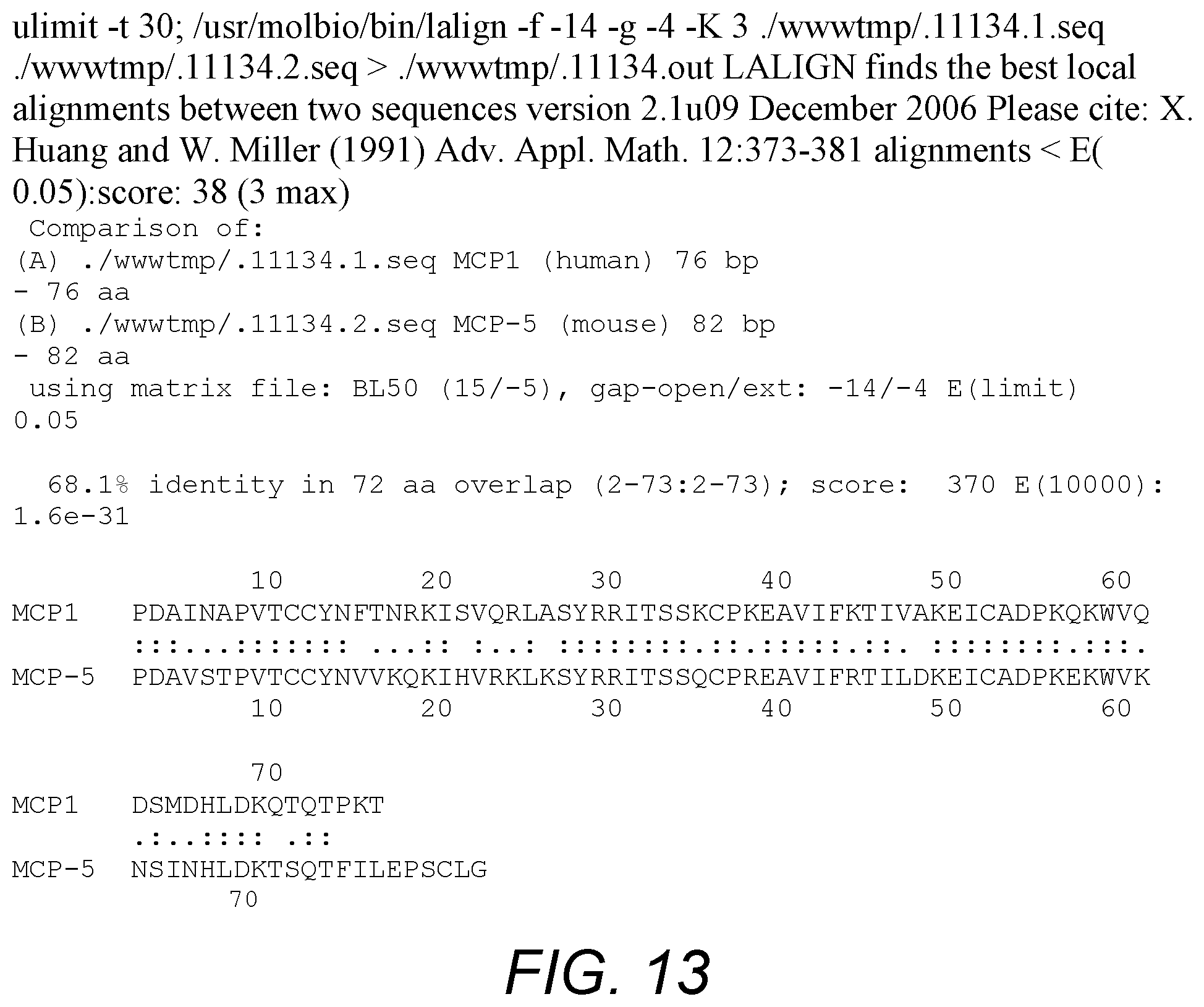
D00013
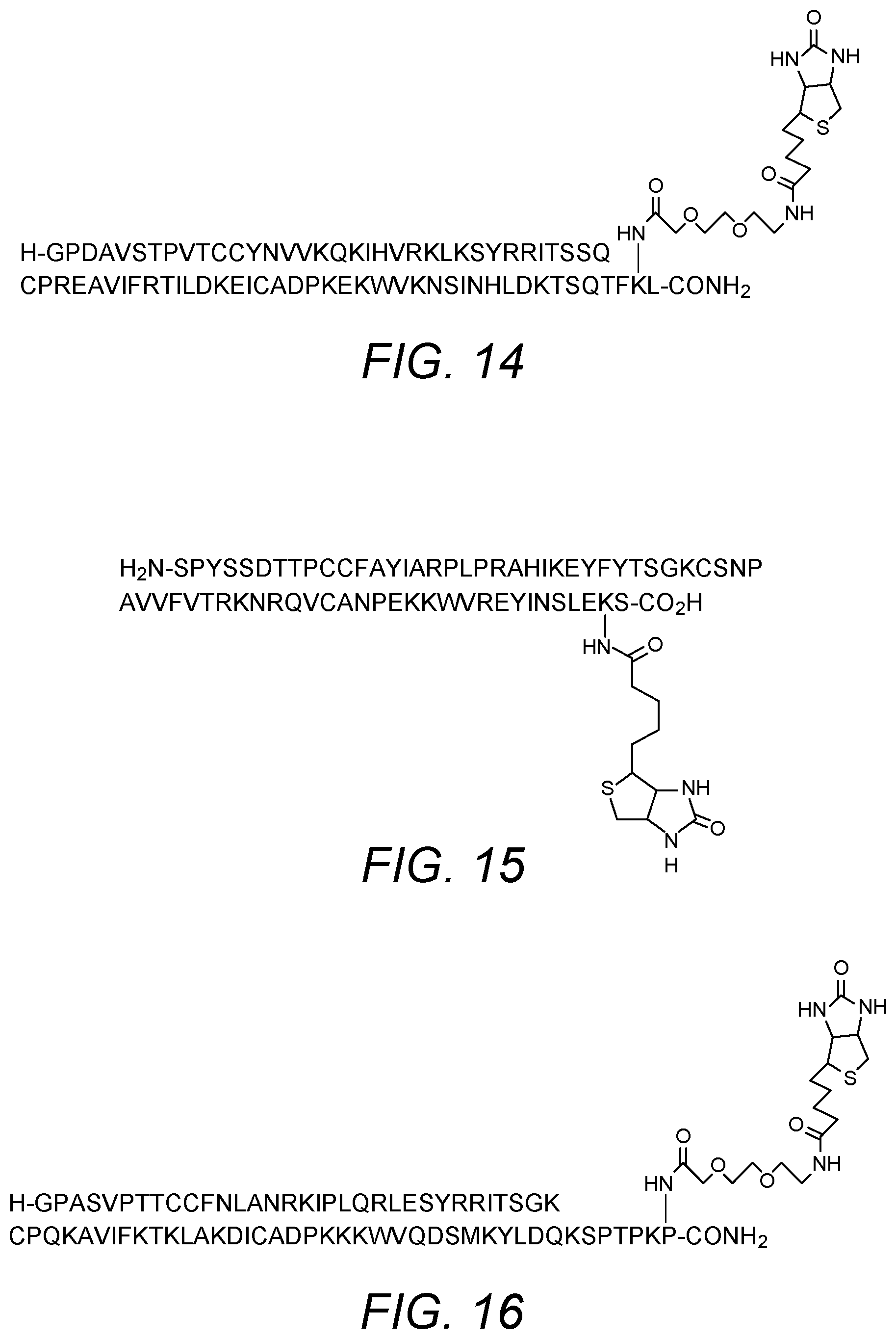
D00014

D00015

D00016

D00017
D00018

D00019
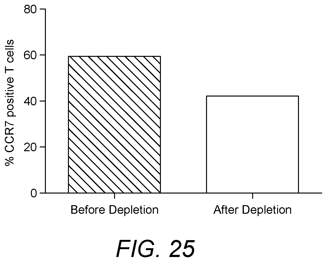
S00001
XML
uspto.report is an independent third-party trademark research tool that is not affiliated, endorsed, or sponsored by the United States Patent and Trademark Office (USPTO) or any other governmental organization. The information provided by uspto.report is based on publicly available data at the time of writing and is intended for informational purposes only.
While we strive to provide accurate and up-to-date information, we do not guarantee the accuracy, completeness, reliability, or suitability of the information displayed on this site. The use of this site is at your own risk. Any reliance you place on such information is therefore strictly at your own risk.
All official trademark data, including owner information, should be verified by visiting the official USPTO website at www.uspto.gov. This site is not intended to replace professional legal advice and should not be used as a substitute for consulting with a legal professional who is knowledgeable about trademark law.