Combination Therapy For A Stable And Long Term Engraftment Using Specific Protocols For T/b Cell Depletion
Reisner; Yair ; et al.
U.S. patent application number 16/538954 was filed with the patent office on 2019-11-28 for combination therapy for a stable and long term engraftment using specific protocols for t/b cell depletion. This patent application is currently assigned to Yeda Research And Development Co. Ltd.. The applicant listed for this patent is Yeda Research And Development Co. Ltd.. Invention is credited to Esther Bachar-Lustig, Yair Reisner.
| Application Number | 20190358269 16/538954 |
| Document ID | / |
| Family ID | 48669650 |
| Filed Date | 2019-11-28 |


| United States Patent Application | 20190358269 |
| Kind Code | A1 |
| Reisner; Yair ; et al. | November 28, 2019 |
COMBINATION THERAPY FOR A STABLE AND LONG TERM ENGRAFTMENT USING SPECIFIC PROTOCOLS FOR T/B CELL DEPLETION
Abstract
A method of treating a subject in need of a non-syngeneic cell or tissue graft is disclosed. The methods comprising: (a) transplanting into a subject a dose of T cell depleted immature hematopoetic cells, wherein the T cell depleted immature hematopoetic cells comprise less than 5.times.105 CD3+ T cells per kilogram body weight of the subject, and wherein the dose comprises at least about 5.times.106 CD34+ cells per kilogram body weight of the subject, and wherein the T cell depleted immature hematopoetic cells are obtained by separating the T cells from from the immature hematopoetic cells by magnetic cell sorting, and (b) administering to the subject a therapeutically effective amount of cyclophosphamide, wherein the therapeutically effective amount comprises 25-200 mg per body weight, thereby treating the subject.
| Inventors: | Reisner; Yair; (Houston, TX) ; Bachar-Lustig; Esther; (Rehovot, IL) | ||||||||||
| Applicant: |
|
||||||||||
|---|---|---|---|---|---|---|---|---|---|---|---|
| Assignee: | Yeda Research And Development Co.
Ltd. Rehovot IL |
||||||||||
| Family ID: | 48669650 | ||||||||||
| Appl. No.: | 16/538954 | ||||||||||
| Filed: | August 13, 2019 |
Related U.S. Patent Documents
| Application Number | Filing Date | Patent Number | ||
|---|---|---|---|---|
| 14367923 | Jun 22, 2014 | 10434121 | ||
| PCT/IL2012/050541 | Dec 20, 2012 | |||
| 16538954 | ||||
| 61578917 | Dec 22, 2011 | |||
| Current U.S. Class: | 1/1 |
| Current CPC Class: | A61K 2039/507 20130101; A61P 19/02 20180101; A61K 31/7076 20130101; C12N 5/0087 20130101; A61P 29/00 20180101; A61K 31/675 20130101; A61K 39/3955 20130101; A61K 35/22 20130101; A61P 35/02 20180101; A61K 2035/124 20130101; A61P 17/00 20180101; A61K 2039/515 20130101; A61K 35/407 20130101; A61K 31/661 20130101; A61K 35/36 20130101; A61K 31/198 20130101; A61P 21/04 20180101; A61P 7/06 20180101; A61K 35/34 20130101; A61P 17/06 20180101; A61K 35/42 20130101; A61P 37/06 20180101; A61K 31/255 20130101; A61P 37/02 20180101; A61P 1/16 20180101; A61P 3/10 20180101; A61K 35/28 20130101; A61K 35/39 20130101; A61K 35/38 20130101; A61P 7/00 20180101; A61P 35/00 20180101; A61P 7/04 20180101; A61K 35/22 20130101; A61K 2300/00 20130101; A61K 35/28 20130101; A61K 2300/00 20130101; A61K 35/34 20130101; A61K 2300/00 20130101; A61K 35/36 20130101; A61K 2300/00 20130101; A61K 35/38 20130101; A61K 2300/00 20130101; A61K 35/39 20130101; A61K 2300/00 20130101; A61K 35/407 20130101; A61K 2300/00 20130101; A61K 35/42 20130101; A61K 2300/00 20130101; A61K 31/675 20130101; A61K 2300/00 20130101; A61K 31/255 20130101; A61K 2300/00 20130101; A61K 31/7076 20130101; A61K 2300/00 20130101; A61K 31/198 20130101; A61K 2300/00 20130101 |
| International Class: | A61K 35/28 20060101 A61K035/28; A61K 39/395 20060101 A61K039/395; A61K 31/661 20060101 A61K031/661; A61K 31/7076 20060101 A61K031/7076; A61K 31/255 20060101 A61K031/255; A61K 31/198 20060101 A61K031/198; C12N 5/00 20060101 C12N005/00; A61K 35/42 20060101 A61K035/42; A61K 35/407 20060101 A61K035/407; A61K 35/39 20060101 A61K035/39; A61K 35/38 20060101 A61K035/38; A61K 35/36 20060101 A61K035/36; A61K 35/34 20060101 A61K035/34; A61K 35/22 20060101 A61K035/22; A61K 31/675 20060101 A61K031/675 |
Claims
1. A method of reducing graft rejection and/or graft versus host disease (GVHD) and/or inducing donor specific tolerance in a subject in need thereof, the method comprising: (a) transplanting into a subject conditioned under reduced intensity conditioning wherein said reduced intensity conditioning comprises a chemotherapeutic agent, a dose of T cell depleted immature hematopoietic cells obtained from a non-syngeneic donor, wherein said dose of said T cell depleted immature hematopoietic cells comprises less than 5.times.10.sup.5 CD3.sup.+ T cells per kilogram body weight of the subject, and wherein said dose comprises at least 5.times.10.sup.6 CD34+ cells per kilogram body weight of the subject, and wherein said dose of said T cell depleted immature hematopoietic cells are obtained by separating T cells from immature hematopoietic cells, by a method selected from the group consisting of: (I) a method comprising: (i) adding an antibody that specifically binds to a surface marker to the immature hematopoietic cells obtained from the non-syngeneic donor, wherein said antibody is labeled with a magnetically responsive agent; (ii) immobilizing said immature hematopoietic cells specifically bound to said antibody labeled with said magnetically responsive agent in a matrix through a magnetic field; (iii) washing said matrix to remove unbound cells; and (iv) removing said magnetic field to elute bound cells from said matrix; and (II) based on an expression of at least one surface marker of T cells selected from the group consisting of CD2, CD3, CD4, CD8, and TCR.alpha./.beta.; and subsequently (b) administering to the subject a therapeutically effective amount of cyclophosphamide, wherein said therapeutically effective amount comprises 25-200 mg cyclophosphamide per kilogram body weight of the subject, and wherein said cyclophosphamide is administered to the subject following transplantation, and wherein said subject is not treated with GVHD prophylaxis for more than 10 days post transplant, thereby reducing graft rejection and/or GVHD and/or inducing donor specific tolerance in the subject.
2. The method of claim 1, wherein the reduced intensity conditioning further comprises in-vivo T cell debulking.
3. The method of claim 2, wherein said in-vivo T cell debulking is effected by antibodies, and optionally wherein said said antibodies comprise at least one of an anti-thymocyte globulin (ATG) antibody, an anti-CD52 antibody, and anti-CD3 (OKT3) antibody.
4. The method of claim 1, wherein the reduced intensity conditioning further comprises a total body irradiation (TBI) or a total lymphoid irradiation (TLI), and optionally wherein said TBI or said TLI comprises a single or fractionated irradiation dose within the range of 1-7.5 Gy.
5. The method of claim 1, wherein said reduced intensity conditioning: (i) is a non-myeloablative conditioning; and/or (ii) is effected 1-10 days prior to said transplanting.
6. The method of claim 1, wherein said chemotherapeutic agent comprises at least one of Fludarabine, Busulfan, Myleran, Busulfex, Melphalan, Thiotepa and cyclophosphamide.
7. The method of claim 1, wherein said dose of said T cell depleted immature hematopoietic cells: (i) comprises 5-40.times.10.sup.6 CD34+ cells per kilogram body weight of the subject; and/or (ii) comprises less than 0.5.times.10.sup.6 CD8.sup.+ TCR.alpha./.beta..sup.- cells per kilogram body weight of the subject; and/or (iii) comprises T cell depleted G-CSF mobilized peripheral blood progenitor cells; and/or (iv) are obtained by T cell debulking; and/or (v) are treated by B cell debulking.
8. The method of claim 1, wherein said amount of said cyclophosphamide is about 100-200 mg per kg body weight.
9. The method of claim 1, wherein said cyclophosphamide is administered in a single dose or in two doses, and optionally wherein each of said two doses: (i) comprises an amount of about 50 mg per kg body weight; and/or (ii) is administered on days 3 and 4 following step (a).
10. The method of claim 1, wherein the subject: (I) is a human subject; and/or (II) has a malignant disease and optionally wherein said malignant disease is a hematopoietic cancer; or (III) has a non-malignant disease and optionally wherein said non-malignant disease is a genetic disease or disorder, a hematopoietic abnormality, an autoimmune disease or a metabolic disorder; or (IV) is in need of a cell or tissue graft, and optionally: (i) wherein cell or tissue graft is selected from the group consisting of a liver, a pancreas, a spleen, a kidney, a heart, a lung, a skin, an intestine and a lymphoid tissue or organ; and/or (ii) wherein said cell or tissue graft is transplanted into the subject prior to, concomitantly with or following said transplanting said dose of said T cell depleted immature hematopoietic cells into said subject; and/or (iii) wherein said cell or tissue graft and said T cell depleted immature hematopoietic cells are obtained from the same donor; and/or (iv) comprises a co-transplantation of several organs.
11. The method of claim 1, wherein: (i) wherein when said surface marker in step (a)(I)(i) is a T cell surface marker, said unbound cells comprise T cell depleted immature hematopoietic cells; or is an immature hematopoietic cell surface marker, said bound cells comprise T cell depleted immature hematopoietic cells; and/or (ii) wherein said matrix in (I) is a ferromagnetic matrix or comprises spheres of magnetically susceptible or ferromagnetic material; and/or (iii) wherein said magnetically responsive agent in step (a)(I)(ii) comprises a superparamagnetic particle; and/or (iv) wherein said antibody in (I) is selected from the group consisting of an anti-CD8 antibody, an anti-CD4 antibody, an anti-CD3 antibody, an anti-CD2 antibody, an anti-TCR.alpha./.beta. antibody, an anti-CD34 antibody, an anti-CD33 antibody and an anti-CD131 antibody. (v) wherein said separating in (I) is effected using a high gradient magnetic separation (HGMS) or using a separation column; and/or (vi) wherein said separating in (II) is effected using an antibody and optionally wherein said antibody is coupled to a fluorescent dye, a hapten or a magnetic particle; and/or (vii) wherein said separating in (II) is performed using flow-cytometry or magnetic cell sorting; and/or (viii) wherein said (I) or (II) further comprises separating B cells from said dose of said T cell depleted immature hematopoietic cells by the use of an antibody that specifically binds to a B cell surface marker.
12. A method of reducing graft rejection and/or graft versus host disease (GVHD) and/or inducing donor specific tolerance in a subject in need thereof, the method comprising: (a) conditioning a subject under reduced intensity conditioning, wherein said reduced intensity conditioning comprises a chemotherapeutic agent; (b) transplanting into the subject a dose of T cell depleted immature hematopoietic cells obtained from a non-syngeneic donor, wherein said dose of said T cell depleted immature hematopoietic cells comprises less than 5.times.10.sup.5 CD3+ T cells per kilogram body weight of the subject, and wherein said dose comprises at least 5.times.10.sup.6 CD34+ cells per kilogram body weight of the subject, and wherein said dose of said T cell depleted immature hematopoietic cells are obtained by separating T cells from immature hematopoietic cells, by a method selected from the group consisting of: (I) a method comprising: (i) adding an antibody that specifically binds to a surface marker to the immature hematopoietic cells obtained from the non-syngeneic donor, wherein said antibody is labeled with a magnetically responsive agent; (ii) immobilizing said immature hematopoietic cells specifically bound to said antibody labeled with said magnetically responsive agent in a matrix through a magnetic field; (iii) washing said matrix to remove unbound cells; and (iv) removing said magnetic field to elute bound cells from said matrix; and (II) based on an expression of at least one surface marker of T cells selected from the group consisting of CD2, CD3, CD4, CD8, and TCR.alpha./.beta.; and subsequently (c) administering to the subject a therapeutically effective amount of cyclophosphamide, wherein said therapeutically effective amount comprises 25-200 mg cyclophosphamide per kilogram body weight of the subject, and wherein said cyclophosphamide is administered to the subject following transplantation, and wherein said subject is not treated with GVHD prophylaxis for more than 10 days post transplant, thereby reducing graft rejection and/or GVHD.
13. The method of claim 12, wherein said conditioning further comprises conditioning the subject with in-vivo T cell debulking.
14. The method of claim 13, wherein said in-vivo T cell debulking is effected by antibodies, and optionally wherein said said antibodies comprise at least one of an anti-thymocyte globulin (ATG) antibody, an anti-CD52 antibody, and anti-CD3 (OKT3) antibody.
15. The method of claim 12, wherein said conditioning further comprises a total body irradiation (TBI) or a total lymphoid irradiation (TLI), and optionally wherein said TBI or said TLI comprises a single or fractionated irradiation dose within the range of 1-7.5 Gy.
16. The method of claim 12, wherein said reduced intensity conditioning: (i) is a non-myeloablative conditioning; and/or (ii) is effected 1-10 days prior to said transplanting.
17. The method of claim 12, wherein said chemotherapeutic agent comprises at least one of Fludarabine, Busulfan, Myleran, Busulfex, Melphalan, Thiotepa and cyclophosphamide.
18. The method of claim 12, wherein said dose of said T cell depleted immature hematopoietic cells: (i) comprises 5-40.times.10.sup.6 CD34+ cells per kilogram body weight of the subject; and/or (ii) comprises less than 0.5.times.10.sup.6 CD8.sup.+ TCR.alpha./.beta..sup.- cells per kilogram body weight of the subject; and/or (iii) comprises T cell depleted G-CSF mobilized peripheral blood progenitor cells; and/or (iv) are obtained by T cell debulking; and/or (v) are treated by B cell debulking.
19. The method of claim 12, wherein said amount of said cyclophosphamide is about 100-200 mg per kg body weight.
20. The method of claim 12, wherein said cyclophosphamide is administered in a single dose or in two doses, and optionally wherein each of said two doses: (i) comprises an amountof about 50 mg per kg body weight; or (ii) is administered on days 3 and 4 following step (b).
21. The method of claim 12, wherein the subject: (I) is a human subject; and/or (II) has a malignant disease and optionally wherein said malignant disease is a hematopoietic cancer; or (III) has a non-malignant disease and optionally wherein said non-malignant disease is a genetic disease or disorder, a hematopoietic abnormality, an autoimmune disease or a metabolic disorder; or (IV) is in need of a cell or tissue graft, and optionally: (i) wherein cell or tissue graft is selected from the group consisting of a liver, a pancreas, a spleen, a kidney, a heart, a lung, a skin, an intestine and a lymphoid tissue or organ; and/or (ii) wherein said cell or tissue graft is transplanted into the subject prior to, concomitantly with or following said transplanting said dose of said T cell depleted immature hematopoietic cells into said subject; and/or (iii) wherein said cell or tissue graft and said T cell depleted immature hematopoietic cells are obtained from the same donor; and/or (iv) comprises a co-transplantation of several organs.
22. The method of claim 12, wherein: (i) wherein when said surface marker in step (a)(I)(i) is a T cell surface marker, said unbound cells comprise T cell depleted immature hematopoietic cells; or is an immature hematopoietic cell surface marker, said bound cells comprise T cell depleted immature hematopoietic cells; and/or (ii) wherein said matrix in (I) is a ferromagnetic matrix or comprises spheres of magnetically susceptible or ferromagnetic material; and/or (iii) wherein said magnetically responsive agent in step (a)(I)(ii) comprises a superparamagnetic particle; and/or (iv) wherein said antibody in (I) is selected from the group consisting of an anti-CD8 antibody, an anti-CD4 antibody, an anti-CD3 antibody, an anti-CD2 antibody, an anti-TCR.alpha./.beta. antibody, an anti-CD34 antibody, an anti-CD33 antibody and an anti-CD131 antibody. (v) wherein said separating in (I) is effected using a high gradient magnetic separation (HGMS) or using a separation column; and/or (vi) wherein said separating in (II) is effected using an antibody and optionally wherein said antibody is coupled to a fluorescent dye, a hapten or a magnetic particle; and/or (vii) wherein said separating in (II) is performed using flow-cytometry or magnetic cell sorting; and/or (viii) wherein said (I) or (II) further comprises separating B cells from said dose of said T cell depleted immature hematopoietic cells by the use of an antibody that specifically binds to a B cell surface marker.
Description
RELATED APPLICATIONS
[0001] This application is a continuation of U.S. patent application Ser. No. 14/367,923 filed on Jun. 22, 2014, which is a National Phase of PCT Patent Application No. PCT/IL2012/050541 having International filing date of Dec. 20, 2012, which claims the benefit of priority under 35 USC .sctn. 119(e) of U.S. Provisional Patent Application No. 61/578,917 filed on Dec. 22, 2011. The contents of the above applications are all incorporated by reference as if fully set forth herein in their entirety.
FIELD AND BACKGROUND OF THE INVENTION
[0002] The present invention, in some embodiments thereof, relates to a combination therapy for attaining a stable and long term cell or tissue transplantation.
[0003] The use of full-haplotype mismatched haploidentical donors as an alternative source for hematopoietic stem cell transplantation (HSCT) is highly attractive since virtually all patients have a readily available haploidentical family member that can serve as an HSCT donor. Early attempts to avoid fatal graft versus host disease (GVHD) risk and to apply haploidentical rigorously T cell depleted bone marrow transplantation (TDBMT) in leukemia patients revealed that the absence of donor T cells within the graft leads to a high rate of graft rejection, mediated by residual radiotherapy and chemotherapy resistant host-derived T cells (HTC). To overcome this obstacle, a `mega dose` of TDBM cells was contemplated which can overcome this HTC mediated immune barrier and be engrafted successfully even when using fully mismatched murine strain combinations [Bachar-Lustig E et al., Nat Med. (1995) 1:1268-1273]. Subsequently, it was demonstrated that in humans, as in rodents, CD34.sup.+ hematopoietic stem cell dose escalation may be used to overcome genetic barriers, enabling satisfactory survival rates following purified haploidentical HSCT [Reisner Y and Martelli M F. Immunol Today. (1995) 16:437-440 and U.S. Pat. No. 5,806,529].
[0004] While the use of a purified `mega dose` of CD34.sup.+ HSCT has enabled haploidentical transplantation in leukemia patients, one major drawback, common to all T cell depleted transplants, is the slow recovery rate of the recipient's immune system. This is attributed to extensive immune ablating conditioning protocols prior to transplantation, the low numbers of donor T cells infused within the graft and to the decreased thymic function of adult recipients. Thus, in adult recipients of a haploidentical CD34.sup.+ stem cell graft, a significant rate of transplant related mortality (TRM) is caused by opportunistic infections.
[0005] Several approaches are being developed to address this challenge. This includes novel modalities to improve thymic function, post-transplant adoptive transfer of anti-viral specific T cells, transfer of partially polyclonal host-non-reactive allo-depleted T cells or transfer of fully polyclonal T cells transfected with inducible suicide genes. An alternative and additional approach to preserve host immunity is the use of reduced intensity conditioning (RIC). This non-myeloablative approach spares a substantial level of host immune cells and thus may reduce TRM by both improving post-transplant immune reconstitution and reducing the toxicity associated with the conditioning agents. Haploidentical transplantation under RIC is even more intricate due to the substantial immunological barrier presented by the surviving host T cells. Recent attempts to overcome this barrier, largely made use of non-T cell depleted grafts, which enable a high rate of engraftment, but in the expanse of increased rates of GVHD. Another approach for applying haploidentical transplantation under RIC uses CD3/CD19 depleted grafts, which not only contain CD34.sup.+ stem cells but also CD34 negative progenitors, NK, graft facilitating cells and dendritic cells, however, this too is at the expanse of increased rates of GVHD and TRM.
[0006] In the 1970's George Santos demonstrated in rodents that a short course of high-dose cyclophosphamide (CY) soon after bone marrow transplant (BMT) targeted activated donor or host alloreactive T cells [Owens A H Jr and G W. S. Transplantation. (1971) 11:378-382]. Cyclophosphamide was observed to be non-toxic to hematopoietic stem cells because of their high expression of the detoxifying enzyme aldehyde dehydrogenase, and Slavin et al. further demonstrated that administration of high dose cyclophosphamide can reduce GVHD and graft rejection in mice, without adverse effects on stem cell engraftment [Brodsky R A and R J. J. Lancet. (2005) 365:1647-1656]. Clinical trials by the John Hopkins and Fred Hutchinson Cancer Research Center groups, evaluated a non-myeloablative protocol of cyclophosphamide, fludarabine and 2Gy TBI, and post-transplant GVHD prophylaxis with cyclophosphamide (50 mg/kg days +3 and +4), MMF (days +5 to +35) and tacrolimus (days +5 to +180) [Luznik L et al., Biology of blood and marrow transplantation: journal of the American Society for Blood and Marrow Transplantation. (2008) 14:641]. According evident from their teachings, this protocol resulted in a high relapse rate, which was probably due to poor disease debulking by the non-myeloablative conditioning and to lack of GVHD related graft versus leukemia (GVL) effect [Munchel A et al., Pediatric Reports (2011) 3:43-47].
[0007] Additional approaches for achieving stable engraftment of allogeneic hematopoietic stem cells have been attempted, some are described in U.S. Patent Application No. 20110110909, U.S. Patent Application No. 20050118142, U.S. Patent Application No. 20070098693, U.S. Pat. Nos. 5,876,692, 5,514,364, 6,217,867, 5,635,156, U.S. Patent Application No. 20060140912, U.S. Patent Application No. 20040005300, U.S. Patent Application No. 20070141027, U.S. Patent Application No. 20030017152, U.S. Patent Application No. 20030165475 and U.S. Patent Application No. 20010009663.
SUMMARY OF THE INVENTION
[0008] According to an aspect of some embodiments of the present invention there is provided a method of treating a subject in need of a non-syngeneic cell or tissue graft, the method comprising: (a) transplanting into a subject a dose of T cell depleted immature hematopoietic cells, wherein the T cell depleted immature hematopoietic cells comprise less than 5.times.10.sup.5 CD3.sup.+ T cells per kilogram body weight of the subject, and wherein the dose comprises at least about 5.times.10.sup.6 CD34+ cells per kilogram body weight of the subject, and wherein the T cell depleted immature hematopoietic cells are obtained by separating the T cells from the immature hematopoietic cells, by a method comprising: (i) adding an antibody to the immature hematopoietic cells that specifically binds to a surface marker, wherein the antibody is labeled with a magnetically responsive agent; (ii) immobilizing the immature hematopoietic cells specifically bound to the antibody labeled with the magnetically responsive agent in a matrix through a magnetic field; (iii) washing the matrix to remove unbound cells; and (iv) removing the magnetic field to elute bound cells from the matrix; and subsequently (b) administering to the subject a therapeutically effective amount of cyclophosphamide, wherein the therapeutically effective amount comprises 25-200 mg per kilogram body weight, thereby treating the subject.
[0009] According to an aspect of some embodiments of the present invention there is provided a method of treating a subject in need of an immature hematopoietic cell transplantation, the method comprising: (a) transplanting into a conditioned subject a dose of T cell depleted immature hematopoietic cells, wherein the T cell depleted immature hematopoietic cells comprise less than 5.times.10.sup.5 CD3+ T cells per kilogram body weight of the subject, and wherein the dose comprises at least about 5.times.10.sup.6 CD34+ cells per kilogram body weight of the subject, and wherein the T cell depleted immature hematopoietic cells are obtained by separating the T cells from the immature hematopoietic cells, by a method comprising: (i) adding an antibody to the immature hematopoietic cells that specifically binds to a surface marker, wherein the antibody is labeled with a magnetically responsive agent; (ii) immobilizing the immature hematopoietic cells specifically bound to the antibody labeled with the magnetically responsive agent in a matrix through a magnetic field; (iii) washing the matrix to remove unbound cells; and (iv) removing the magnetic field to elute bound cells from the matrix; and subsequently (b) administering to the subject a therapeutically effective amount of cyclophosphamide, wherein the therapeutically effective amount comprises 25-200 mg per kilogram body weight, thereby treating the subject.
[0010] According to an aspect of some embodiments of the present invention there is provided a method of treating a subject in need of an immature hematopoietic cell transplantation, the method comprising: (a) conditioning a subject under a reduced intensity conditioning protocol, wherein the reduced intensity conditioning comprises a total body irradiation (TBI) and a chemotherapeutic agent; (b) transplanting into the subject a dose of T cell depleted immature hematopoietic cells, wherein the T cell depleted immature hematopoietic cells comprise less than 5.times.10.sup.5 CD3+ T cells per kilogram body weight of the subject, and wherein the dose comprises at least about 5.times.10.sup.6 CD34+ cells per kilogram body weight of the subject, and wherein the T cell depleted immature hematopoietic cells are obtained by separating the T cells from the immature hematopoietic cells, by a method comprising: (i) adding an antibody to the immature hematopoietic cells that specifically binds to a surface marker, wherein the antibody is labeled with a magnetically responsive agent; (ii) immobilizing the immature hematopoietic cells specifically bound to the antibody labeled with the magnetically responsive agent in a matrix through a magnetic field; (iii) washing the matrix to remove unbound cells; and (iv) removing the magnetic field to elute bound cells from the matrix; and subsequently (c) administering to the subject a therapeutically effective amount of cyclophosphamide, wherein the therapeutically effective amount comprises 25-200 mg per kilogram body weight, thereby treating the subject.
[0011] According to an aspect of some embodiments of the present invention there is provided a method of inducing donor specific tolerance in a subject in need of a non-syngeneic cell or tissue graft, the method comprising: (a) transplanting into a subject a dose of T cell depleted immature hematopoietic cells obtained from a non-syngeneic donor, wherein the T cell depleted immature hematopoietic cells comprise less than 5.times.10.sup.5 CD3.sup.+ T cells per kilogram body weight of the subject, and wherein the dose comprises at least about 5.times.10.sup.6 CD34+ cells per kilogram body weight of the subject, and wherein the T cell depleted immature hematopoietic cells are obtained by separating the T cells from the immature hematopoietic cells, by a method comprising: (i) adding an antibody to the immature hematopoietic cells that specifically binds to a surface marker, wherein the antibody is labeled with a magnetically responsive agent; (ii) immobilizing the immature hematopoietic cells specifically bound to the antibody labeled with the magnetically responsive agent in a matrix through a magnetic field; (iii) washing the matrix to remove unbound cells; and (iv) removing the magnetic field to elute bound cells from the matrix; and subsequently (b) administering to the subject a therapeutically effective amount of cyclophosphamide, wherein the therapeutically effective amount comprises 25-200 mg per kilogram body weight, thereby inducing donor specific tolerance in the subject.
[0012] According to an aspect of some embodiments of the present invention there is provided a method of treating a subject in need of a non-syngeneic cell or tissue graft, the method comprising: (a) transplanting into a subject a dose of T cell depleted immature hematopoietic cells, wherein the T cell depleted immature hematopoietic cells comprise less than 5.times.10.sup.5 CD3.sup.+ T cells per kilogram body weight of the subject, and wherein the dose comprises at least about 5.times.10.sup.6 CD34+ cells per kilogram body weight of the subject, and wherein the separating the T cells from the immature hematopoietic cells is effected based on a product secreted by the T cells; and subsequently (b) administering to the subject a therapeutically effective amount of cyclophosphamide, wherein the therapeutically effective amount comprises 25-200 mg per kilogram body weight, thereby treating the subject.
[0013] According to an aspect of some embodiments of the present invention there is provided a method of treating a subject in need of a non-syngeneic cell or tissue graft, the method comprising: (a) transplanting into a subject a dose of T cell depleted immature hematopoietic cells, wherein the T cell depleted immature hematopoietic cells comprise less than 5.times.10.sup.5 CD3.sup.+ T cells per kilogram body weight of the subject, and wherein the dose comprises at least about 5.times.10.sup.6 CD34+ cells per kilogram body weight of the subject, and wherein the T cell depleted immature hematopoietic cells are obtained by separating the T cells from the immature hematopoietic cells based on an expression of at least one surface marker of T cells selected from the group consisting of 4-1BB, FoxP3, CD154, CD4, CD8, CD25, GITR CD137, latent TGF-beta (LAP), GARP (LRRC32) and CD121a/b; and subsequently (b) administering to the subject a therapeutically effective amount of cyclophosphamide, wherein the therapeutically effective amount comprises 25-200 mg per kilogram body weight, thereby treating the subject.
[0014] According to an aspect of some embodiments of the present invention there is provided a method of treating a subject in need of an immature hematopoietic cell transplantation, the method comprising: (a) transplanting into a conditioned subject a dose of T cell depleted immature hematopoietic cells, wherein the T cell depleted immature hematopoietic cells comprise less than 5.times.10.sup.5 CD3+ T cells per kilogram body weight of the subject, and wherein the dose comprises at least about 5.times.10.sup.6 CD34+ cells per kilogram body weight of the subject, and wherein the separating the T cells from the immature hematopoietic cells is effected based on a product secreted by the T cells; and subsequently (b) administering to the subject a therapeutically effective amount of cyclophosphamide, wherein the therapeutically effective amount comprises 25-200 mg per kilogram body weight, thereby treating the subject.
[0015] According to an aspect of some embodiments of the present invention there is provided a method of treating a subject in need of an immature hematopoietic cell transplantation, the method comprising: (a) transplanting into a conditioned subject a dose of T cell depleted immature hematopoietic cells, wherein the T cell depleted immature hematopoietic cells comprise less than 5.times.10.sup.5 CD3+ T cells per kilogram body weight of the subject, and wherein the dose comprises at least about 5.times.10.sup.6 CD34+ cells per kilogram body weight of the subject, and wherein the T cell depleted immature hematopoietic cells are obtained by separating the T cells from the immature hematopoietic cells based on an expression of at least one surface marker of T cells selected from the group consisting of 4-1BB, FoxP3, CD154, CD4, CD8, CD25, GITR CD137, latent TGF-beta (LAP), GARP (LRRC32) and CD121a/b; and subsequently (b) administering to the subject a therapeutically effective amount of cyclophosphamide, wherein the therapeutically effective amount comprises 25-200 mg per kilogram body weight, thereby treating the subject.
[0016] According to an aspect of some embodiments of the present invention there is provided a method of treating a subject in need of an immature hematopoietic cell transplantation, the method comprising: (a) conditioning a subject under a reduced intensity conditioning protocol, wherein the reduced intensity conditioning comprises a total body irradiation (TBI) and a chemotherapeutic agent; (b) transplanting into the subject a dose of T cell depleted immature hematopoietic cells, wherein the T cell depleted immature hematopoietic cells comprise less than 5.times.10.sup.5 CD3+ T cells per kilogram body weight of the subject, and wherein the dose comprises at least about 5.times.10.sup.6 CD34+ cells per kilogram body weight of the subject, and wherein the separating the T cells from the immature hematopoietic cells is effected based on a product secreted by the T cells; and subsequently (c) administering to the subject a therapeutically effective amount of cyclophosphamide, wherein the therapeutically effective amount comprises 25-200 mg per kilogram body weight, thereby treating the subject.
[0017] According to an aspect of some embodiments of the present invention there is provided a method of treating a subject in need of an immature hematopoietic cell transplantation, the method comprising: (a) conditioning a subject under a reduced intensity conditioning protocol, wherein the reduced intensity conditioning comprises a total body irradiation (TBI) and a chemotherapeutic agent; (b) transplanting into the subject a dose of T cell depleted immature hematopoietic cells, wherein the T cell depleted immature hematopoietic cells comprise less than 5.times.10.sup.5 CD3+ T cells per kilogram body weight of the subject, and wherein the dose comprises at least about 5.times.10.sup.6 CD34+ cells per kilogram body weight of the subject, and wherein the T cell depleted immature hematopoietic cells are obtained by separating the T cells from the immature hematopoietic cells based on an expression of at least one surface marker of T cells selected from the group consisting of 4-1BB, FoxP3, CD154, CD4, CD8, CD25, GITR CD137, latent TGF-beta (LAP), GARP (LRRC32) and CD121a/b; and subsequently (c) administering to the subject a therapeutically effective amount of cyclophosphamide, wherein the therapeutically effective amount comprises 25-200 mg per kilogram body weight, thereby treating the subject.
[0018] According to an aspect of some embodiments of the present invention there is provided a method of inducing donor specific tolerance in a subject in need of a non-syngeneic cell or tissue graft, the method comprising: (a) transplanting into a subject a dose of T cell depleted immature hematopoietic cells obtained from a non-syngeneic donor, wherein the T cell depleted immature hematopoietic cells comprise less than 5.times.10.sup.5 CD3.sup.+ T cells per kilogram body weight of the subject, and wherein the dose comprises at least about 5.times.10.sup.6 CD34+ cells per kilogram body weight of the subject, and wherein the separating the T cells from the immature hematopoietic cells is effected based on a product secreted by the T cells; and subsequently (b) administering to the subject a therapeutically effective amount of cyclophosphamide, wherein the therapeutically effective amount comprises 25-200 mg per kilogram body weight, thereby inducing donor specific tolerance in the subject.
[0019] According to an aspect of some embodiments of the present invention there is provided a method of inducing donor specific tolerance in a subject in need of a non-syngeneic cell or tissue graft, the method comprising: (a) transplanting into a subject a dose of T cell depleted immature hematopoietic cells obtained from a non-syngeneic donor, wherein the T cell depleted immature hematopoietic cells comprise less than 5.times.10.sup.5 CD3.sup.+ T cells per kilogram body weight of the subject, and wherein the dose comprises at least about 5.times.10.sup.6 CD34+ cells per kilogram body weight of the subject, and wherein the T cell depleted immature hematopoietic cells are obtained by separating the T cells from the immature hematopoietic cells based on an expression of at least one surface marker of T cells selected from the group consisting of 4-1BB, FoxP3, CD154, CD4, CD8, CD25, GITR CD137, latent TGF-beta (LAP), GARP (LRRC32) and CD121a/b; and subsequently (b) administering to the subject a therapeutically effective amount of cyclophosphamide, wherein the therapeutically effective amount comprises 25-200 mg per kilogram body weight, thereby inducing donor specific tolerance in the subject.
[0020] According to some embodiments of the invention, the method further comprises conditioning the subject under reduced intensity conditioning prior to step (a).
[0021] According to some embodiments of the invention, the method further comprises conditioning the subject with in-vivo T cell debulking prior to step (a).
[0022] According to some embodiments of the invention, the dose of the T cell depleted immature hematopoietic cells comprises 5-40.times.10.sup.6 CD34+ cells per kilogram body weight of the subject.
[0023] According to some embodiments of the invention, the dose of the T cell depleted immature hematopoietic cells comprises at least about 10.times.10.sup.6 CD34+ cells per kilogram body weight of the subject.
[0024] According to some embodiments of the invention, the T cell depleted immature hematopoietic cells are selected from the group consisting of T cell depleted bone marrow cells, T cell depleted G-CSF mobilized peripheral blood progenitor cells, T cell depleted cord blood, purified CD34+ cells attained by positive selection from bone marrow and/or from G-CSF mobilized peripheral blood progenitor cells, and ex-vivo expanded CD34.sup.+ cells.
[0025] According to some embodiments of the invention, the T cell depleted immature hematopoietic cells comprise less than 1.times.10.sup.6 CD8.sup.+ TCR.alpha./.beta..sup.- cells per kilogram body weight of the subject.
[0026] According to some embodiments of the invention, the T cell depleted immature hematopoietic cells are obtained by MACS.
[0027] According to some embodiments of the invention, when the surface marker is a T cell surface marker, the unbound cells comprise T cell depleted immature hematopoietic cells.
[0028] According to some embodiments of the invention, the T cell surface marker is selected from the group consisting of CD2, CD3, CD4, CD8 and TCR.alpha./.beta..
[0029] According to some embodiments of the invention, when the surface marker is an immature hematopoietic cell surface marker, the bound cells comprise T cell depleted immature hematopoietic cells.
[0030] According to some embodiments of the invention, the immature hematopoietic cell surface marker is selected from the group consisting of CD34, CD33 and CD131.
[0031] According to some embodiments of the invention, the method further comprises separating B cells from the T cell depleted immature hematopoietic cells by the use of an antibody that specifically binds to a B cell surface marker.
[0032] According to some embodiments of the invention, the B cell surface marker is selected from the group consisting of CD19 and CD20.
[0033] According to some embodiments of the invention, the matrix is a ferromagnetic matrix.
[0034] According to some embodiments of the invention, the matrix comprises spheres of magnetically susceptible or ferromagnetic material.
[0035] According to some embodiments of the invention, the magnetically responsive agent comprises a superparamagnetic particle.
[0036] According to some embodiments of the invention, the superparamagnetic particle is conjugated to the antibody in combination with an anti-immunoglobulin, an avidin and or anti-hapten-specific microbead.
[0037] According to some embodiments of the invention, the separating the T cells from the immature hematopoietic cells is effected using a high gradient magnetic separation (HGMS).
[0038] According to some embodiments of the invention, the separating the T cells from the immature hematopoietic cells is effected using a separation column.
[0039] According to some embodiments of the invention, the antibody is selected from the group consisting of an anti-CD8 antibody, an anti-CD4 antibody, an anti-CD3 antibody, an anti-CD2 antibody, an anti-TCR.alpha./.beta. antibody, an anti-CD19 antibody, and an anti-CD20 antibody, an anti-CD21 antibody an anti-CD34 antibody, an anti-CD33 antibody and an anti-CD131 antibody.
[0040] According to some embodiments of the invention, the T cell depleted immature hematopoietic cells are obtained from a non-syngeneic donor.
[0041] According to some embodiments of the invention, the non-syngeneic donor is allogeneic or xenogeneic with respect to the subject.
[0042] According to some embodiments of the invention, the allogeneic donor is selected from the group consisting of an HLA matched sibling, an HLA matched unrelated donor, an HLA haploidentical related donor and a donor displaying one or more disparate HLA determinants.
[0043] According to some embodiments of the invention, the subject is a human subject.
[0044] According to some embodiments of the invention, the in-vivo T cell debulking is effected by antibodies.
[0045] According to some embodiments of the invention, the antibodies comprise at least one of an anti-CD8 antibody, an anti-CD4 antibody, an anti-thymocyte globulin (ATG) antibody, an anti-CD52 antibody and an anti-CD3 (OKT3) antibody.
[0046] According to some embodiments of the invention, the reduced intensity conditioning comprises a non-myeloablative conditioning.
[0047] According to some embodiments of the invention, the non-myeloablative conditioning comprises at least one of a total body irradiation (TBI), a total lymphoid irradiation (TLI), a chemotherapeutic agent and/or an antibody immunotherapy.
[0048] According to some embodiments of the invention, the TBI comprises a single or fractionated irradiation dose within the range selected from the group consisting of 1-7.5 Gy and 1-3.5 Gy.
[0049] According to some embodiments of the invention, the chemotherapeutic agent comprises at least one of Busulfan, Fludarabine, Melphalan and Thiotepa.
[0050] According to some embodiments of the invention, the antibody comprises at least one of an anti-CD52 antibody, an anti-thymocyte globulin (ATG) antibody and anti-CD3 (OKT3) antibody.
[0051] According to some embodiments of the invention, the concentration of the cyclophosphamide is about 100-200 or about 100 mg per kg body weight.
[0052] According to some embodiments of the invention, the cyclophosphamide is administered in a single dose or in two doses.
[0053] According to some embodiments of the invention, each of the two doses comprises a concentration of about 50 mg per kg body weight.
[0054] According to some embodiments of the invention, each of the two doses is administered on days 3 and 4 following step (a).
[0055] According to some embodiments of the invention, the subject has a malignant disease.
[0056] According to some embodiments of the invention, the malignant disease is a hematopoietic cancer.
[0057] According to some embodiments of the invention, the subject has a non-malignant disease.
[0058] According to some embodiments of the invention, the cell or tissue graft comprises immature hematopoietic cells.
[0059] According to some embodiments of the invention, the cell or tissue graft is selected from the group consisting of a liver, a pancreas, a spleen, a kidney, a heart, a lung, a skin, an intestine and a lymphoid/hematopoietic tissue or organ.
[0060] According to some embodiments of the invention, the cell or tissue graft is transplanted into the subject prior to, concomitantly with or following the transplanting the dose of T cell depleted immature hematopoietic cells into the subject.
[0061] According to some embodiments of the invention, the cell or tissue graft comprises a co-transplantation of several organs.
[0062] According to some embodiments of the invention, the cell or tissue graft and the T cell depleted immature hematopoietic cells are obtained from the same donor.
[0063] According to some embodiments of the invention, the separating is effected using an antibody.
[0064] According to some embodiments of the invention, the antibody is coupled to a fluorescent dye, a hapten or a magnetic particle.
[0065] According to some embodiments of the invention, the separating is performed using flow-cytometry or magnetic cell sorting.
[0066] According to some embodiments of the invention, the magnetic cell sorting comprises MACS.TM..
[0067] According to some embodiments of the invention, the separating the T cells from the immature hematopoietic cells based on the product secreted by the T cells comprises separating T cells labeled with a secretion product, wherein the T cells have been coupled to a capture moiety that specifically binds a product secreted by the T cells and wherein the T cells have been cultured under conditions wherein the product is secreted and bound to the capture moiety, thereby producing T cells labeled with the secretion product, wherein the T cells are not lysed by the method and wherein the secretion product is labeled with a label moiety.
[0068] According to some embodiments of the invention, the method further comprises separating B cells from the T cell depleted immature hematopoietic cells based on a product secreted by the B cells comprising separating B cells labeled with a secretion product, wherein the B cells have been coupled to a capture moiety that specifically binds a product secreted by the B cells and wherein the B cells have been cultured under conditions wherein the product is secreted and bound to the capture moiety, thereby producing B cells labeled with the secretion product, wherein the B cells are not lysed by the method and wherein the secretion product is labeled with a label moiety.
[0069] According to some embodiments of the invention, the secretion product is a cytokine, antibody or hormone.
[0070] According to some embodiments of the invention, the secretion product is selected from the group consisting of IFN-.gamma., IL1, IL2, IL4, IL10, IL12, TGF-.beta., TNF, GM-CSF and SCF.
[0071] According to some embodiments of the invention, the capture moiety is coupled to the T cells or B cells through an anchoring moiety.
[0072] According to some embodiments of the invention, the capture moiety is an antibody or an antigen-binding fragment thereof.
[0073] According to some embodiments of the invention, the antibody is bispecific.
[0074] According to some embodiments of the invention, the antibody is against a T cell or a B cell surface marker.
[0075] According to some embodiments of the invention, the label moiety is an antibody specific for the secretion product.
[0076] According to some embodiments of the invention, the label moiety is fluorochromated, magnetizable or comprises magnetic particles.
[0077] According to some embodiments of the invention, the antibody is selected from the group consisting of an anti-CD8 antibody, an anti-CD4 antibody, an anti-CD3 antibody, an anti-CD2 antibody, an anti-TCR.alpha./.beta. antibody, an anti-CD19 antibody, an anti-CD20 antibody, an anti-CD21 antibody an anti-CD34 antibody, an anti-CD33 antibody and an anti-CD131 antibody.
[0078] It will be appreciated that the present teachings can be used with other tolerance inducing protocols such as described in PCT publication Nos. WO 2001/49243, WO 2007/023491 and WO 2010/049935, which are herein incorporated by reference in their entirety.
[0079] Unless otherwise defined, all technical and/or scientific terms used herein have the same meaning as commonly understood by one of ordinary skill in the art to which the invention pertains. Although methods and materials similar or equivalent to those described herein can be used in the practice or testing of embodiments of the invention, exemplary methods and/or materials are described below. In case of conflict, the patent specification, including definitions, will control. In addition, the materials, methods, and examples are illustrative only and are not intended to be necessarily limiting.
BRIEF DESCRIPTION OF THE DRAWINGS
[0080] The patent or application file contains at least one drawing executed in color. Copies of this patent or patent application publication with color drawing(s) will be provided by the Office upon request and payment of the necessary fee.
[0081] Some embodiments of the invention are herein described, by way of example only, with reference to the accompanying drawings. With specific reference now to the drawings in detail, it is stressed that the particulars shown are by way of example and for purposes of illustrative discussion of embodiments of the invention. In this regard, the description taken with the drawings makes apparent to those skilled in the art how embodiments of the invention may be practiced.
[0082] In the drawings:
[0083] FIGS. 1A-1B are graphs illustrating durable engraftment of mismatched donor bone marrow (BM) following transplantation of `mega dose` rigorously T cell depleted BM and posttranplantation cyclophosphamide. Mice were conditioned with T cell debulking (TCD), using anti-CD4 and anti-CD8 antibodies, on day -6, and by exposure to 2.0 Gy total body irradiation (TBI) on day -1. High dose Cyclophosphamide (CY, 100 mg/kg) was administered on days +3 and +4 post transplant. Donor type chimerism was evaluated 35 days (FIG. 1A) and 95 days (FIG. 1B) post transplant.
[0084] FIGS. 2A-2C are dot plot graphs illustrating a typical FACS chimerism analysis. FIG. 2C depicts that mixed chimerism was achieved in recipients that were transplanted with `mega dose` (25.times.10.sup.6) rigorously T cell depleted BM and were treated with high dose CY. In contrast, recipient mice that received only the conditioning protocol (FIG. 2A) or which were inoculated with only 5.times.10.sup.6 BM cells and CY did not exhibit donor type chimerism (FIG. 2B).
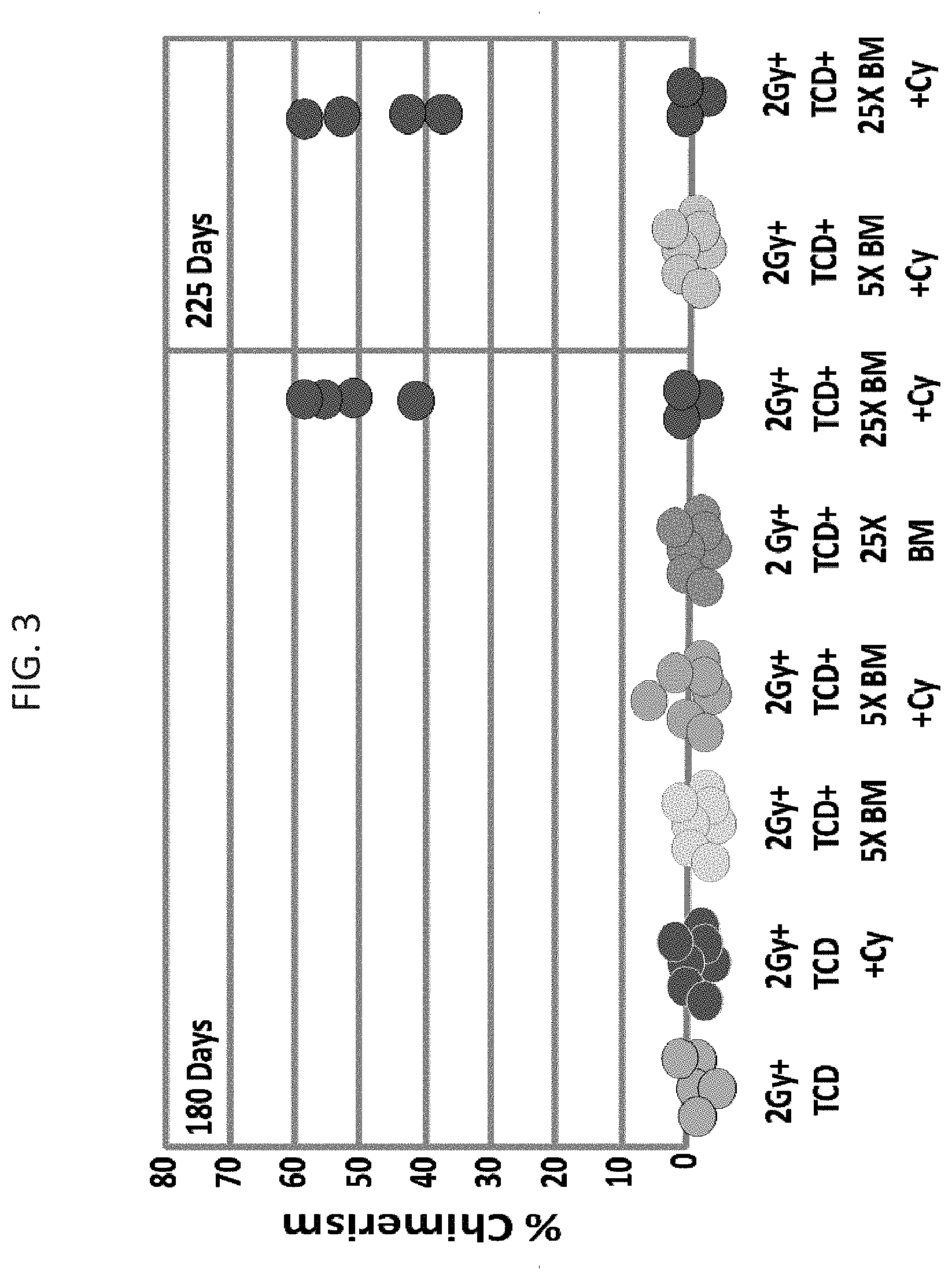
[0085] FIG. 3 is a graph illustrating durable mixed chimerism 180 and 225 days post transplant in recipient mice that were transplanted with `mega dose` (25.times.10.sup.6) T cell depleted BM and were treated with high dose CY. Of note, mice which were inoculated with 5.times.10.sup.6 T cell depleted BM and CY did not exhibit mixed chimerism.
[0086] FIGS. 4A-4B illustrate transplantation of donor type or 3.sup.rd party skin grafts in chimeric mice. FIG. 4A is a graft illustrating acceptance (marked by "+") or rejection (marked by "-") of donor type (Balb/c) or 3.sup.rd party (C57BL/6) skin grafts in recipients of regular dose (5.times.10.sup.6) or `mega dose` (25.times.10.sup.6) T depleted BM, treated with high dose CY on days +3 and +4 post transplant. FIG. 4B is a photograph of donor type (Balb/c) skin graft (white fur) or 3.sup.rd party (C57BL/6) skin graft (black fur) in recipients of `mega dose` (25.times.10.sup.6) T depleted BM, treated with high dose CY on days +3 and +4 post transplant.
[0087] FIG. 5 is a graph illustrating the effect of different doses of irradiation on donor type chimerism in recipient mice of `mega dose` (25.times.10.sup.6) T depleted BM and treated with high dose CY post transplant.
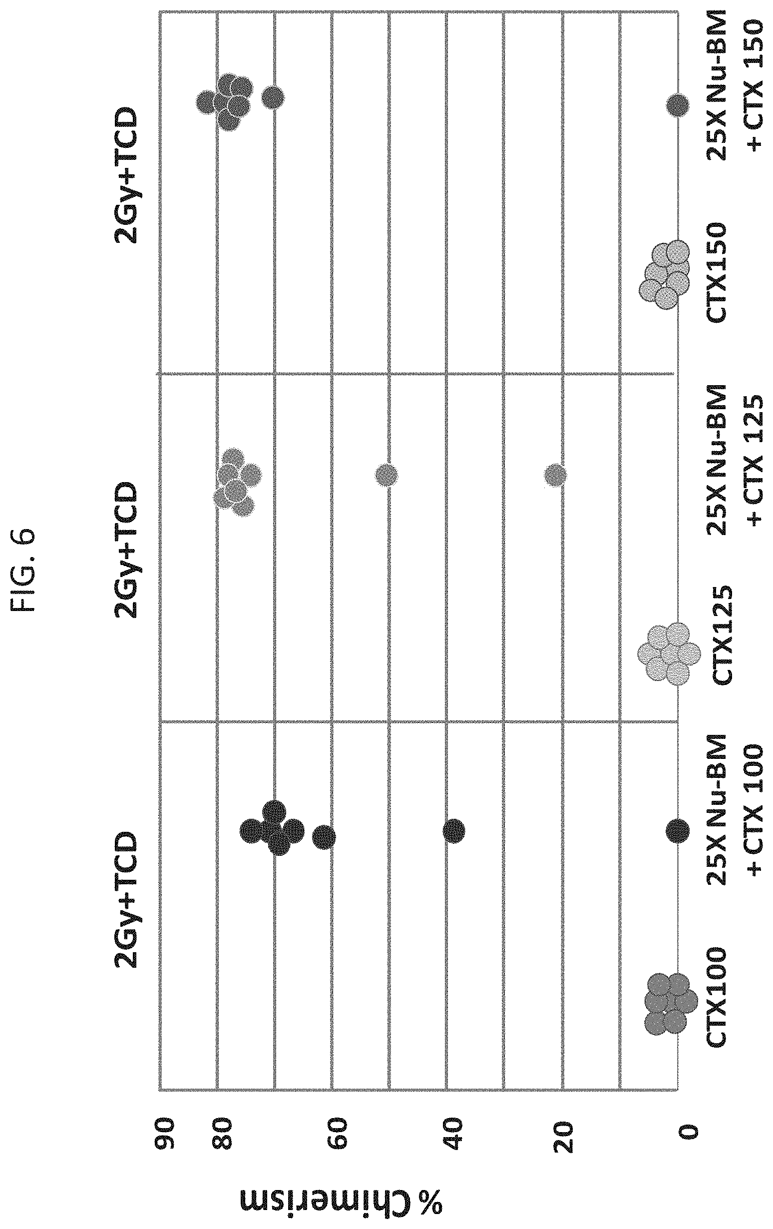
[0088] FIG. 6 is a graph illustrating the effect of increased doses of Cyclophosphamide (CY) on donor type chimerism in recipients of `mega dose` (25.times.10.sup.6) T depleted BM and 2 Gy TBI.
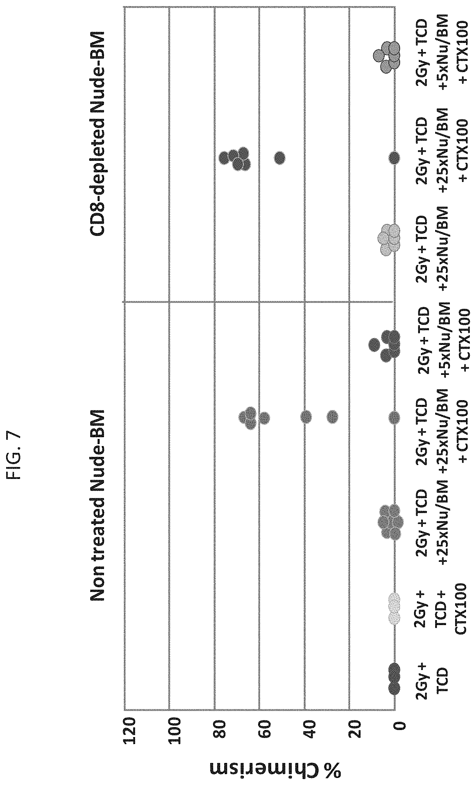
[0089] FIG. 7 is a graph illustrating engraftment of mismatched donor BM achieved by combining `mega dose` CD8.sup.+ T cell depleted BM and post transplant CY. Of note, the depletion of residual CD8.sup.+ T cells from the BM preparation did not have any adverse impact on the level of chimerism achieved when combing `mega dose` T cell depleted BM cells with post transplant CY.
DESCRIPTION OF SPECIFIC EMBODIMENTS OF THE INVENTION
[0090] The present invention, in some embodiments thereof, relates to a combination therapy for attaining a stable and long term cell or tissue transplantation.
[0091] The principles and operation of the present invention may be better understood with reference to the drawings and accompanying descriptions.
[0092] Before explaining at least one embodiment of the invention in detail, it is to be understood that the invention is not necessarily limited in its application to the details set forth in the following description or exemplified by the Examples. The invention is capable of other embodiments or of being practiced or carried out in various ways. Also, it is to be understood that the phraseology and terminology employed herein is for the purpose of description and should not be regarded as limiting.
[0093] Application of allogeneic hematopoietic stem cell transplantation (HSCT) has been limited by the lack of available HLA-matched donors within the family or in the international registries of unrelated volunteer donors. Conversely, virtually all patients in need for a transplant have a full-haplotype mismatched family donor.
[0094] The major obstacles to bone marrow transplantation from full-haplotype mismatched related donors were graft versus host disease (GVHD) and graft rejection. The use of very large numbers of hematopoietic stem cells with minimal residual T cell contamination and an aggressive immunosuppressive and myeloablative regimen has resulted in high rates of engraftment with little severe GVHD. However, immune reconstitution has been delayed and incomplete after this approach and a significant rate of transplant related mortality (TRM) is caused by opportunistic infections.
[0095] While reducing the present invention to practice, the present inventors have uncovered that a successful engraftment of mismatched bone marrow can be achieved by transplantation of rigorously T cell depleted `mega dose` bone marrow and subsequently administering to the subject a high-dose cyclophospamide early after transplantation. The present inventors have shown that such a regimen requires only a short immunomyeloablative conditioning regimen. The present inventors have further shown that such a transplantation procedure leads to a long and stable chimerism and that tolerance has been achieved.
[0096] As is shown hereinbelow and in the Examples section which follows, the present inventors have uncovered through laborious experimentation that the combination of `mega dose` T cell depleted bone marrow transplantation (TDBMT) and post transplant high dose cyclophosphamide (CY) allows for a durable engraftment of mismatched donor bone marrow (see FIGS. 1A-1B and 2A-2C). Durable mixed chimerism was exhibited for prolonged periods of time after transplantation (180 and 225 days post transplant in mice, see FIG. 3). Importantly, the combination of `mega dose` TDBMT and high dose CY following transplantation allowed hematopoietic stem cell engraftment under reduced intensity conditioning (see FIG. 5) and resulted in tolerance induction, as indicated by acceptance of donor skin grafts (see FIG. 4B).
[0097] Thus, according to one aspect of the present invention there is provided a method of treating a subject in need of a non-syngeneic cell or tissue graft, the method comprising: (a) transplanting into a subject a dose of T cell depleted immature hematopoietic cells, wherein the T cell depleted immature hematopoietic cells comprise less than 5.times.10.sup.5 CD3.sup.+ T cells per kilogram body weight of the subject, and wherein the dose comprises at least about 5.times.10.sup.6 CD34+ cells per kilogram body weight of the subject; and subsequently (b) administering to the subject a therapeutically effective amount of cyclophosphamide, wherein the therapeutically effective amount comprises 25-200 mg per kilogram body weight, thereby treating the subject.
[0098] As used herein, the term "treating" includes abrogating, substantially inhibiting, slowing or reversing the progression of a condition, substantially ameliorating clinical or aesthetical symptoms of a condition or substantially preventing the appearance of clinical or aesthetical symptoms of a condition.
[0099] As used herein, the term "subject" or "subject in need thereof" refers to a mammal, preferably a human being, male or female at any age that is in need of a cell or tissue transplantation. Typically the subject is in need of cell or tissue transplantation (also referred to herein as recipient) due to a disorder or a pathological or undesired condition, state, or syndrome, or a physical, morphological or physiological abnormality which is amenable to treatment via cell or tissue transplantation.
[0100] According to an embodiment the subject is in need of tissue regeneration (solid or soft tissue) such as due to aging, trauma, wound or any pathological condition which results in loss of organ functionality.
[0101] According to one embodiment the subject has a malignant disease.
[0102] According to one embodiment the malignant disease is a hematopoietic cancer.
[0103] Exemplary hematopoietic cancers include, but are not limited to, acute lymphoblastic leukemia (ALL), T-cell acute lymphocytic leukemia (T-ALL), acute myelocytic leukemia (AML), acute nonlymphoblastic leukemia (ANLL), chronic lymphocytic leukemia (CLL), chronic myelocytic leukemia (CML), T-cell prolymphocytic leukemia, B-cell prolymphocytic leukemia, Juvenile myelomonocytic leukemia, Hodgkin's Lymphoma, non-Hodgkin's Lymphoma, Extranodal natural killer/T-cell lymphoma, Cutaneous T-cell lymphoma, Enteropathy type T-cell lymphoma, Angioimmunoblastic T-cell lymphoma, Anaplastic large T/null-cell lymphoma, Subcutaneous panniculitis-like T-cell lymphoma, Unspecified T-cell lymphoma, Diffuse large B-cell lymphoma (DLBCL), B-cell chronic lymphocytic leukemia (B-CLL)/chronic lymphoid leukemia (CLL), Chronic lymphocytic leukemia/small lymphocytic lymphoma, Extranodal marginal zone B-cell lymphomas-mucosa-associated lymphoid tissue lymphomas, Follicular lymphoma, Mantle cell lymphoma, Nodal marginal zone B-cell lymphoma, Burkitt lymphoma, Hairy cell leukemia, Primary central nervous system lymphoma, Splenic marginal zone B-cell lymphoma, Lymphoplasmocytic lymphoma, Primary mediastinal B-cell lymphoma, precursor T-cell leukemia/lymphoma, MALT lymphoma, Mycosis fungoides and multiple myeloma.
[0104] According to one embodiment the hematopoietic cancer comprises a leukemia or a lymphoma.
[0105] According to one embodiment the subject has a non-malignant disease.
[0106] According to one embodiment the non-malignant disease is a genetic disease or disorder, an autoimmune disease or a metabolic disorder.
[0107] Exemplary non-malignant diseases include, but are not limited to, severe combined immunodeficiency syndromes (SCID), sickle cell disease (sickle cell anemia), congenital neutropenia, thrombocytopenia, aplastic anemia (e.g. severe aplastic anemia), myelodysplastic syndrome, monosomy 7, osteopetrosis, Gaucher's disease, Hurler's disease, metachromatic leukodystrophy, adrenal leukodystrophy, thalassemia, congenital or genetically-determined hematopoietic abnormality, adenosine deaminase (ADA), lupus, autoimmune hepatitis, celiac disease, type I diabetes mellitus, Grave's disease, Guillain-Barr syndrome, Myasthenia gravis, Rheumatoid arthritis, scleroderma and psoriasis.
[0108] According to one embodiment the subject of the present invention may suffer from any of a cardiovascular disease, a rheumatoid disease, a glandular disease, a gastrointestinal disease, a cutaneous disease, a hepatic disease, a neurological disease, a muscular disease, a nephric disease, a connective tissue disease, a systemic disease and/or a disease related to reproduction, treatable by cell or tissue transplantation.
[0109] As used herein, the phrase "cell or tissue graft" refers to a bodily cell (e.g. a single cell or a group of cells) or tissue (e.g. solid tissues or soft tissues, which may be transplanted in full or in part). Exemplary tissues which may be transplanted according to the present teachings include, but are not limited to, liver, pancreas, spleen, kidney, heart, lung, skin, intestine and lymphoid/hematopoietic tissues (e.g. lymph node, Peyer' s patches, thymus or bone marrow). Exemplary cells which may be transplanted according to the present teachings include, but are not limited to, immature hematopoietic cells including stem cells. The present invention also contemplates transplantation of whole organs, such as for example, kidney, heart, lung, liver, pancreas or spleen.
[0110] According to one embodiment, the cell or tissue graft comprises immature hematopoietic cells.
[0111] According to one embodiment, the method is effected using a cell or tissue, which is non-syngeneic with the subject.
[0112] Depending on the application, the method may be effected using a cell or tissue graft which is allogeneic or xenogeneic with the subject.
[0113] As used herein, the term "allogeneic" refers to a cell or tissue which is derived from a donor who is of the same species as the subject, but which is substantially non-clonal with the subject. Typically, outbred, non-zygotic twin mammals of the same species are allogeneic with each other. It will be appreciated that an allogeneic donor may be HLA identical or HLA non-identical (i.e. displaying one or more disparate HLA determinants) with respect to the subject.
[0114] According to one embodiment, the allogeneic donor is an HLA matched sibling, an HLA matched unrelated donor, an HLA haploidentical related donor or a donor displaying one or more disparate HLA determinants.
[0115] As used herein, the term "xenogeneic" refers to a cell or tissue which substantially expresses antigens of a different species relative to the species of a substantial proportion of the lymphocytes of the subject. Typically, outbred mammals of different species are xenogeneic with each other.
[0116] The present invention envisages that xenogeneic cells or tissues are derived from a variety of species such as, but not limited to, bovines (e.g., cow), equines (e.g., horse), porcines (e.g. pig), ovids (e.g., goat, sheep), felines (e.g., Felis domestica), canines (e.g., Canis domestica), rodents (e.g., mouse, rat, rabbit, guinea pig, gerbil, hamster) or primates (e.g., chimpanzee, rhesus monkey, macaque monkey, marmoset).
[0117] Cells or tissues of xenogeneic origin (e.g. porcine origin) are preferably obtained from a source which is known to be free of zoonoses, such as porcine endogenous retroviruses. Similarly, human-derived cells or tissues are preferably obtained from substantially pathogen-free sources.
[0118] According to an embodiment of the present invention, both the subject and the donor are humans.
[0119] Depending on the application and available sources, the cell or tissue graft of the present invention may be obtained from a prenatal organism, postnatal organism, an adult or a cadaver donor. Moreover, depending on the application needed, the cell or tissue graft may be naive or genetically modified. Determination of the type of cell or tissue graft to be used is well within the ability of one of ordinary skill in the art. Furthermore, any method known in the art may be employed to obtain a cell or tissue graft (e.g. for transplantation).
[0120] As mentioned, a dose of T cell depleted hematopoietic cell or tissue comprising immature hematopoietic cells (including e.g. CD34.sup.+), are transplanted into a subject.
[0121] According to one embodiment, the T cell depleted immature hematopoietic cells are non-syngeneic (e.g. allogeneic or xenogeneic) with the subject.
[0122] According to one embodiment, the T cell depleted immature hematopoietic cells and the cell or tissue graft are syngeneic (e.g. obtained from the same donor).
[0123] As used herein the phrase "immature hematopoietic cells" refers to a hematopoietic tissue or cell preparation comprising precursor hematopoietic cells. Such tissue/cell preparation includes or is derived from a biological sample, for example, bone marrow, mobilized peripheral blood (e.g. mobilization of CD34 cells to enhance their concentration), cord blood (e.g. umbilical cord), fetal liver, yolk sac and/or placenta. Additionally, purified CD34+ cells or other hematopoietic stem cells such as CD131+ cells can be used in accordance with the present teachings, either with or without ex-vivo expansion.
[0124] According to one embodiment, the immature hematopoietic cells comprise T cell depleted immature hematopoietic cells.
[0125] As used herein the phrase "T cell depleted immature hematopoietic cells" refers to a population of hematopoietic cells which are depleted of T lymphocytes. The T cell depleted immature hematopoietic cells, may include e.g. CD34+, CD33+ and/or CD56+ cells. The T cell depleted immature hematopoietic cells may be depleted of CD3+ cells, CD2+ cells, CD8+ cells, CD4+ cells, .alpha./.beta. T cells and/or .gamma./.delta. T cells.
[0126] According to one embodiment, the immature hematopoietic cells comprise T cell depleted G-CSF mobilized blood cells enriched for CD34.sup.+ immature hematopoietic cells.
[0127] According to one embodiment, the immature hematopoietic cells are depleted of CD3+ T cells.
[0128] According to an embodiment, the T cell depleted immature hematopoietic cells comprise less than 50.times.10.sup.5 CD3.sup.+ T cells, 40.times.10.sup.5 CD3.sup.+ T cells, 30.times.10.sup.5 CD3.sup.+ T cells, 20.times.10.sup.5 CD3.sup.+ T cells, 15.times.10.sup.5 CD3.sup.+ T cells, 10.times.10.sup.5 CD3.sup.+ T cells, 9.times.10.sup.5 CD3.sup.+ T cells, 8.times.10.sup.5 CD3.sup.+ T cells, 7.times.10.sup.5 CD3.sup.+ T cells, 6.times.10.sup.5 CD3.sup.+ T cells, 5.times.10.sup.5 CD3.sup.+ T cells, 4.times.10.sup.5 CD3.sup.+ T cells, 3.times.10.sup.5 CD3.sup.+ T cells, 2.times.10.sup.5 CD3.sup.+ T cells or 1.times.10.sup.5 CD3.sup.+ T cells per kilogram body weight of the subject.
[0129] According to a specific embodiment, the T cell depleted immature hematopoietic cells comprise less than 5.times.10.sup.5 CD3.sup.+ T cells per kilogram body weight of the subject.
[0130] According to a specific embodiment the T cell depleted immature hematopoietic cells comprise less than 20.times.10.sup.5 CD3.sup.+ T cells but more than 10 CD3+ T cells.
[0131] According to an embodiment, the T cell depleted immature hematopoietic cells comprise at least 1.times.10.sup.3-1.times.10.sup.5 CD3.sup.+ T cells.
[0132] According to one embodiment, the immature hematopoietic cells are depleted of CD8+ cells.
[0133] According to an embodiment, the T cell depleted immature hematopoietic cells comprise less than 1.times.10.sup.4-4.times.10.sup.5 CD8.sup.+ cells per kilogram body weight of the subject.
[0134] According to an embodiment, the T cell depleted immature hematopoietic cells comprise less than 50.times.10.sup.5 CD8.sup.+ cells, 25.times.10.sup.5 CD8.sup.+ cells, 15.times.10.sup.5 CD8.sup.+ cells, 10.times.10.sup.5 CD8.sup.+ cells, 9.times.10.sup.5 CD8.sup.+ cells, 8.times.10.sup.5 CD8.sup.+ cells, 7.times.10.sup.5 CD8.sup.+ cells, 6.times.10.sup.5 CD8.sup.+ cells, 5.times.10.sup.5 CD8.sup.+ cells, 4.times.10.sup.5 CD8.sup.+ cells, 3.times.10.sup.5 CD8.sup.+ cells, 2.times.10.sup.5 CD8+ cells, 1.times.10.sup.5 CD8.sup.+ cells, 9.times.10.sup.4 CD8.sup.+ cells, 8.times.10.sup.4 CD8.sup.+ cells, 7.times.10.sup.4 CD8.sup.+ cells, 6.times.10.sup.4 CD8.sup.+ cells, 5.times.10.sup.4 CD8.sup.+ cells, 4.times.10.sup.4 CD8.sup.+ cells, 3.times.10.sup.4 CD8.sup.+ cells, 2.times.10.sup.4 CD8.sup.+ cells or 1.times.10.sup.4 CD8.sup.+ cells per kilogram body weight of the subject.
[0135] According to a specific embodiment, the T cell depleted immature hematopoietic cells comprise less than 4.times.10.sup.5 CD8.sup.+ cells per kilogram body weight of the subject.
[0136] According to a specific embodiment the T cell depleted immature hematopoietic cells comprise less than 4.times.10.sup.5 CD8.sup.+ cells but more than 10 CD8+ cells.
[0137] According to an embodiment, the T cell depleted immature hematopoietic cells comprise less than 1.times.10.sup.6 CD8.sup.+ TCR.alpha./.beta.- cells per kilogram body weight of the subject.
[0138] According to an embodiment, the T cell depleted immature hematopoietic cells comprise less than 1.times.10.sup.6 CD8.sup.+ TCR.alpha./.beta..sup.- cells, 0.5.times.10.sup.6 CD8.sup.+ TCR.alpha./.beta..sup.- cells, 1.times.10.sup.5 CD8.sup.+ TCR.alpha./.beta..sup.- cells, 0.5.times.10.sup.5 CD8.sup.+ TCR.alpha./.beta..sup.- cells, 1.times.10.sup.4 CD8.sup.+ TCR.alpha./.beta..sup.- cells, 0.5.times.10.sup.4 CD8.sup.+ TCR.alpha./.beta..sup.- cells, 1.times.10.sup.3 CD8.sup.+ TCR.alpha./.beta..sup.- cells or 0.5.times.10.sup.3 CD8.sup.+ TCR.alpha./.beta..sup.- cells per kilogram body weight of the subject.
[0139] According to a specific embodiment, the T cell depleted immature hematopoietic cells comprise less than 1.times.10.sup.6 CD8.sup.+ TCR.alpha./.beta..sup.- cells per kilogram body weight of the subject.
[0140] According to a specific embodiment the T cell depleted immature hematopoietic cells comprise less than 1.times.10.sup.6 CD8.sup.+ TCR.alpha./.beta..sup.- cells but more than 10 CD8.sup.+ TCR.alpha./.beta..sup.- cells.
[0141] According to one embodiment, the immature hematopoietic cells are depleted of B cells.
[0142] According to an embodiment, the immature hematopoietic cells are depleted of B cells (CD19.sup.+ and/or CD20.sup.+ B cells).
[0143] According to an embodiment, the immature hematopoietic cells comprise less than 50.times.10.sup.5 B cells, 40.times.10.sup.5 B cells, 30.times.10.sup.5 B cells, 20.times.10.sup.5 B cells, 10.times.10.sup.5 B cells, 9.times.10.sup.5 B cells, 8.times.10.sup.5 B cells, 7.times.10.sup.5 B cells, 6.times.10.sup.5 B cells, 5.times.10.sup.5 B cells, 4.times.10.sup.5 B cells, 3.times.10.sup.5 B cells, 2.times.10.sup.5 B cells or 1.times.10.sup.5 B cells per kilogram body weight of the subject.
[0144] According to a specific embodiment, the immature hematopoietic cells comprise less than 4.times.10.sup.5 B cells per kilogram body weight of the subject. According to a specific embodiment the immature hematopoietic cells comprise less than 50.times.10.sup.5 B cells but more than 10 B cells.
[0145] Depletion of T cells, e.g. CD3+, CD2+, TCR.alpha./.beta.+, CD4+ and/or CD8.sup.+ cells, or B cells, e.g. CD19+ and/or CD20+ cells, may be carried out using any method known in the art, such as by eradication (e.g. killing) with specific antibodies or by affinity based purification such as by the use of magnetic-activated cell sorting (MACS) available from Miltenyi Biotec (depicted in further detail hereinbelow), FACS sorter and/or capture ELISA labeling.
[0146] Such methods are described herein and in THE HANDBOOK OF EXPERIMENTAL IMMUNOLOGY, Volumes 1 to 4, (D. N. Weir, editor) and FLOW CYTOMETRY AND CELL SORTING (A. Radbruch, editor, Springer Verlag, 1992). For example, cells can be sorted by, for example, flow cytometry or FACS. Thus, fluorescence activated cell sorting (FACS) may be used and may have varying degrees of color channels, low angle and obtuse light scattering detecting channels, and impedance channels. Any ligand-dependent separation techniques known in the art may be used in conjunction with both positive and negative separation techniques that rely on the physical properties of the cells rather than antibody affinity, including but not limited to elutriation and density gradient centrifugation.
[0147] Other methods for cell sorting include, for example, panning and separation using affinity techniques, including those techniques using solid supports such as plates, beads and columns. Thus, biological samples may be separated by "panning" with an antibody attached to a solid matrix, e.g. to a plate.
[0148] Alternatively, cells may be sorted/separated by magnetic separation techniques, and some of these methods utilize magnetic beads. Different magnetic beads are available from a number of sources, including for example, Dynal (Norway), Advanced Magnetics (Cambridge, Mass., U.S.A.), Immuncon (Philadelphia, U.S.A.), Immunotec (Marseille, France), Invitrogen, Stem cell Technologies (U.S.A), Cellpro (U.S.A) and Miltenyi Biotec GmbH (Germany). Alternatively, antibodies can be biotinylated or conjugated with digoxigenin and used in conjunction with avidin or anti-digoxigenin coated affinity columns.
[0149] According to an embodiment, different depletion/separation methods can be combined, for example, magnetic cell sorting can be combined with FACS, to increase the separation quality or to allow sorting by multiple parameters.
[0150] According to one embodiment, the T cell depleted immature hematopoietic cells are obtained by T cell debulking (TCD).
[0151] T cell debulking may be effected using antibodies, including e.g. anti-CD8 antibodies, anti-CD4 antibodies, anti-CD3 antibodies, anti-CD2 antibodies, anti-TCR.alpha./.beta. antibodies and/or anti-TCR.gamma./.delta. antibodies.
[0152] According to one embodiment, depletion of B cells is effected by B cell debulking.
[0153] B cell debulking may be effected using antibodies, including e.g. anti-CD19 or anti-CD20 antibodies. Alternatively, debulking in-vivo of B cells can be attained by infusion of anti-CD20 antibodies.
[0154] Alternatively, positive selection of CD34+ or CD131+ stem cells may be carried out using e.g. magnetic cell separation techniques (e.g. MACS.TM.), FACS sorter and/or capture ELISA labeling as described in further detail above.
[0155] Following are a number of non-limiting examples for depleting populations of cells of interest (e.g., T and/or B cells) from the immature hematopoietic cells according to the present invention prior to transplantation thereof: [0156] I. Separation of activated regulatory T helper cells following the teachings of PCT publication no. WO 2007/110249 and U.S. Pat. No. 8,129,126 which are hereby incorporated by reference in their entirety:
[0157] According to one embodiment, there is provided a method of separating activated regulatory CD4+ CD25+ T helper (Th) cells from a cell preparation (e.g. immature hematopoietic cells) by use of the 4-1BB receptor.
[0158] The regulatory cells can be identified and/or separated through the expression of one and/or more markers. It is possible to employ for this purpose any marker known to the skilled worker for identification and/or separation. Exemplary markers are 4-1BB, CD25, CTLA-4 (cytotoxic T lymphocyte antigen-4), GITR (glucocorticoid-induced TNF receptor), FoxP3, IL-10, CD69, CD40L, ICOS, OX40 and TGFbeta, which are employed singly and/or in combination. It is possible for this purpose also to employ all markers known to the skilled worker for exclusion or depletion of non-regulatory cells in combination. However, in a particular embodiment, the 4-1BB receptor (CD137) is used as a marker of live, activated regulatory cells. [0159] II. Separation of T regulatory cells or non-regulatory T cells following the teachings of U.S. Patent Application No. 20110097313 which is hereby incorporated by reference in its entirety:
[0160] According to one embodiment, there is provided a method of separating regulatory T cells (Tregs) or non-regulatory T-cells (conventional T-cells) from a cell preparation (e.g. immature hematopoietic cells) by the use of the CD154 molecule [CD40 ligand (CD40L)].
[0161] As activated Treg cells (CD4+CD25+ Treg) do no express CD154, the present invention contemplates separation between activated (e.g. antigen activated) conventional T cells (CD154+ cells) and Treg (CD154- cells) using an agent capable of recognizing CD154 (e.g. anti-CD154 antibody).
[0162] Thus, according to one embodiment, the present invention provides a method of separating activated Treg cells from a cell preparation (e.g. immature hematopoietic cells) using an agent capable of recognizing CD154. The present invention further contemplates the use of additional markers that are specific for regulatory T cells, such as for example, CD25, GITR, CTLA4, or markers which are specific for activated regulatory T cells, such as, for example, CD137, "latent TGF-beta (LAP)", GARP (LRRC32), CD121a/b for positive selection of Tregs.
[0163] According to another embodiment, there is provided a method of obtaining activated conventional T helper (Th) cells from a cell preparation (e.g. immature hematopoietic cells) using the CD154 marker. Specifically, using the CD154 marker, activated conventional Th cells (CD154 expressing cells) can be removed from a cell preparation (comprising regulatory T cells), in particular, when additional selection methods using markers for activated/non-activated regulatory cells (CD137 and CD25, respectively) are used simultaneously or subsequently, which allows for use of the invention for identifying isolating antigen-specific regulatory Th cells.
[0164] Thus, after obtaining a cell preparation (e.g. immature hematopoietic cells) from a donor and/or a subject, regulatory T-cells can be enriched using, for example, CD25. These cells can be subsequently stimulated with a particular antigen. For this purpose, antibodies, peptides, proteins, chemicals substances (or mixtures thereof), or pathogens or cells may be used. After a particular activation time, the regulatory Th cells are identified and/or separated, for example, through the use of the CD137 marker. Thereby, antigen-specific regulatory Th cells are preferably identified and/or separated. [0165] III. Separation of antigen-specific T cells following the teachings of U.S. Pat. No. 7,659,084 which is hereby incorporated by reference in its entirety:
[0166] According to one embodiment, there is provided a use of CD154 [CD40 ligand (CD40L)] for the isolation of antigen-specific T cells wherein the cell preparation (e.g. immature hematopoietic cells) is contacted with a CD40/CD154 system inhibitor.
[0167] According to an embodiment of the invention, the T cells are T helper (Th) CD4+ or CD8+ Th lymphocytes.
[0168] According to an embodiment of the invention, inflammatory, anti-inflammatory, regulatory and/or suppressive T cells are detected and/or obtained.
[0169] The invention also relates to a method for the isolation of antigen-specific T cells in a cell preparation (e.g. immature hematopoietic cells) following activation with an antigen, in which method the cell preparation (e.g. immature hematopoietic cells) is contacted with a CD40/CD154 system inhibitor, intra- and/or extracellular determination of CD154 is effected, and the cells having CD154 are isolated.
[0170] It will be appreciated that such "contacting" with a CD40/CD154 system inhibitor of the cell preparation (e.g. immature hematopoietic cells) allows intra- and/or extracellular determination of CD154 and thus, isolation or separation of cells having CD154, the cells in particular representing the entire antigen-specific CD4+ Th lymphocytes.
[0171] Specifically, addition of a CD40/CD154 system inhibitor impairs or inhibits the interaction and signaling between CD40 and CD154. In the meaning of the invention, CD40/CD154 system inhibitors can be any of molecules or even physical exposures capable of blocking or inhibiting the interaction between CD40 and CD154.
[0172] Accordingly, the inhibiting agent can be an antibody, e.g. one directed against CD40, or directed against CD154, a molecule, a cesium or lithium ion having an effect on the interaction between CD40 and CD154. The agent can also be a substance inhibiting the secretion or endocytosis in the cell, such as brefeldin A (Bref-A) and/or monsensin. Brefeldin A is a metabolite of the fungus Penicillium brefeldianum and, being a carboxylated ionophor, blocks the transport of newly synthesized proteins from the endoplasmatic reticulum into the Golgi apparatus and impairs the exchange between endosomes and lysomes, while the circulation between cell membrane and endosomes advantageously remains undisturbed.
[0173] These substances ensure that CD40, CD154, the interaction between the two of them, or the CD40/CD154 system are modified in such a way that CD154 either is no longer downregulated and/or degraded on the cell surface, or, provided it is still within the cell, no longer transported therein. Such interruption of the transport within the cell prevents degradation of CD154. Consequently, CD154 is stabilized inside or outside the cell as an external receptor, thereby allowing detection and subsequent isolation using detection methods well-known to those skilled in the art (explained in further detail herein). [0174] IV. Separation of cells following the teachings of PCT publication no. WO 94/09117 and U.S. Pat. No. 7,166,423 which are hereby incorporated by reference in their entirety:
[0175] According to one embodiment, there is provided a method for separation of cells (e.g. B cells or T cells) from a cell preparation (e.g. immature hematopoietic cells) based on one or more products secreted from these cells (e.g. cytokines, including but not limited to, IFN.gamma., IL1, IL2, IL4, IL10, IL12, TGF-beta, TNF, GM-CSF, and SCF, antibodies, hormones, enzymes and proteins). The method permits products secreted by cells (e.g. B cells or T cells) to be captured at the surface of the cell (e.g. B cells or T cells). The captured product permits the cell (e.g. B cells or T cells) to be sorted according to the presence, absence, or amount of the product present. The means of capture comprise a capture moiety which has been anchored to the cell surface by a means suitable for the cell to be sorted.
[0176] The capture moiety may be coupled to the anchoring means (the "anchor moiety") optionally through a linking moiety, and may also include a linking moiety which multiplies the number of capture moieties available and thus the potential for capture of product, such as branched polymers, including, for example, modified dextran molecules, polyethylene glycol, polypropylene glycol, polyvinyl alcohol, and polyvinylpyrrolidone.
[0177] Suitable anchor moieties to the cell surface include lipophilic molecules such as fatty acids. Examples of suitable cell surface molecules include, but are not limited to, any molecule associated with the cell surface. Suitable molecules include, but are not limited to, cell surface markers such as CD45 (pan leukocyte), CD3 (T cells (activating)), CD4, CD8, CD19, CD20, CD14, CD16, CD15, class I MHC and Class II MHC molecules, CD34, CD38, CD33, CD56 T cell receptor, Fc receptor, beta2microglobulin or immunoglobulin, and other CD markers or cell adhesion molecules. Alternatively, antibodies or other agents which specifically bind to cell surface molecules such as the MHC antigens or glycoproteins, could also be used.
[0178] Specific binding partners include capture moieties and label moieties. The capture moieties are those which attach both to the cell, either directly or indirectly, and the product. The label moieties are those which attach to the product and may be directly or indirectly labeled. Specific binding partners include any moiety for which there is a relatively high affinity and specificity between product and its binding partner, and in which the dissociation of the product:partner complex is relatively slow so that the product:partner complex is detected during the labeling or cell separation technique.
[0179] Specific label moieties may include, but are not limited to, fluorochromated anti-product antibodies, which may include, magnetic bead conjugated, colloidal bead conjugated, FITC, Phycoerythrin, PerCP, AMCA, fluorescent particle or liposome conjugated antibodies.
[0180] Specific binding partners may include, but are not limited to, substrates or substrate analogs to which a product will bind. These substrates include, but are not limited to, peptides, polysaccharides, steroids, biotin, digitoxin, digitonin, and other molecules able to bind the secreted product, and in a specific embodiment will include antibodies. When the capture moiety is an antibody it may be referred to as the "capture antibody" or "catch antibody." As used herein, the term "antibody" is intended to include polyclonal and monoclonal.
[0181] As used herein, the term "antibody" is intended to include polyclonal and monoclonal antibodies, chimeric antibodies, haptens and antibody fragments, bispecific antibodies and molecules which are antibody equivalents in that they specifically bind to an epitope on the product antigen. Bispecific antibodies, also known as bifunctional antibodies, have at least one antigen recognition site for a first antigen and at least one antigen recognition site for a second antigen. Such antibodies can be produced by recombinant DNA methods or chemically by methods known in the art. Chemically created bispecific antibodies include but are not limited to antibodies that have been reduced and reformed so as to retain their bivalent characteristics and antibodies that have been chemically coupled so that they have at least two antigen recognition sites for each antigen. Bispecific antibodies include all antibodies or conjugates of antibodies, or polymeric forms of antibodies which are capable of recognizing two different antigens. Antibodies can be immobilized on a polymer or particle.
[0182] According to an embodiment, the capture moiety can be attached to a cell membrane by a variety of methods. Suitable methods include, but are not limited to, direct chemical coupling to amino groups of the protein components; coupling to thiols (formed after reduction of disulfide bridges) of the protein components; indirect coupling through antibodies (including pairs of antibodies); anchoring in the lipid bilayer by means of a hydrophobic anchor moiety; and binding to the negatively charged cell surface by polycations.
[0183] In other embodiments of the invention, the capture moiety is introduced using two or more steps, e.g., by labeling the cells with at least one anchor moiety which allows the coupling of the capture moiety to the anchor moiety either directly for instance by a biotin/avidin complex or indirectly through a suitable linking moiety or moieties.
[0184] Methods for direct chemical coupling of antibodies to the cell surface are known in the art, and include, for example, coupling using glutaraldehyde or maleimide activated antibodies. Methods for chemical coupling using multiple step procedures include, for example, biotinylation, coupling of TNP or digoxigenin using, for example, succinimide esters of these compounds. Biotinylation may be accomplished by, for example, the use of D-biotinyl-N-hydroxysuccinimide. Succinimide groups react effectively with amino groups at pH values above 7, and preferentially between about pH 8.0 and about pH 8.5. Biotinylation may also be accomplished by, for example, treating the cells with dithiothreitol (DTT) followed by the addition of biotin maleimide.
[0185] Coupling to the cells may also be accomplished using antibodies against cell surface antigens ("markers"). Antibodies generally directed to surface antigens may be required in the range of about 0.1 to 1 .mu.g of antibody per 10.sup.7 cells, however, this requirement will vary widely in response to the affinity of the antibody to the product and will need to be determined empirically. Such a determination is well within the skill of one in the art. Thus, the appropriate amount of antibody must be determined empirically and is within the skill of one in the art. This allows coupling to specific cells on cell type specific marker expression. For instance, classes of cells based such as T cells or subsets thereof can be specifically labeled. As a capture moiety, a bispecific antibody may be used which has an antigen recognition site for the cell or an anchor moiety placed thereon, and the product.
[0186] A capture moiety, particularly capture antibodies should be selected based on the amount of secreted product. For example, for cells which secrete only a few molecules, a high affinity antibody should be chosen so as to catch most of the secreted molecules. Alternatively, in the case where the cell secretes many molecules during the incubation time, a lower affinity antibody may be preferred to prevent too early saturation of the catching matrix. Determination of suitable affinities for the level of proteins secreted is determined empirically and is within the skill of one in the art.
[0187] In some embodiments of the invention, the capture moiety is coupled to the cell by hydrophobic anchor moieties to the cell membrane. Suitable hydrophobic anchor moieties that will interact with the lipid bilayer of the membrane are known in the art, and include, but are not limited to, fatty acids and non-ionic detergents (including, e.g., Tween-80). A drawback to attachment of the capture moiety to the cell via the insertion of an anchor moiety is that the rate of integration of the anchor moiety into the cell is low. Thus, high concentrations of the anchor moiety often are required. This latter situation is often uneconomical when the capture moiety is a relatively limited or expensive substance, for example, an antibody. The low yield of hydrophobic anchor moieties that embed themselves in the membrane is relevant only when these molecules are available in relatively limited quantities. This problem may be overcome by using a bridging system that includes an anchor moiety and a capture moiety, wherein one of the moieties is of higher availability, and wherein the two parts of the bridging system have a high degree of specificity and affinity for each other. For example, in one embodiment, avidin or streptavidin is attached to the cell surface via a hydrophobic anchor moiety, while the capture moiety is a biotinylated anti-product antibody.
[0188] In another embodiment, the cell surface is labeled with digoxigenin followed by bispecific antibodies having specificity for both digoxigenin and the product. This approach can be used with other pairs of molecules able to form a link, including, for example, hapten with antihapten antibodies or NTA with polyhistidine residues. A specific embodiment is one which allows "amplification" of the system by increasing the number of capture moieties per anchor moiety.
[0189] In one illustrative embodiment, a branched dextran is bound to palmitic acid, thus providing a multiplicity of available binding sites. The dextran is in turn coupled to biotin and treated with avidin-conjugated antibody specific for the product. It is of course contemplated within the embodiments of the invention that linker moieties may be used between the anchor moiety and the capture moiety when the anchor moiety is coupled in any fashion to the cell surface. Thus, for example, an avidin (or streptavidin) biotin linker moiety may link an antibody anchor moiety with a capture moiety. Bispecific antibody systems may also act as linker moieties.
[0190] In order to select cells that have the capability of secreting the product of interest, cells modified as above to contain the capture moiety are incubated under conditions that allow the production and secretion of the product in a sufficient amount to allow binding to and detection of the cells that contain the captured product. These conditions are known to those of skill in the art and include, inter alia, appropriate temperature, pH, and concentrations of salts, growth factors and substrates in the incubation medium, as well as the appropriate concentrations of gas in the gaseous phase. When it is desirable to distinguish between high and low producer cells, the time of incubation is such that product secretion by the cells is still in a linear phase. The appropriate conditions can be determined empirically and such a determination is within the skill of one in the art.
[0191] Additionally, secretion by the cells can be modified, that is upregulated, induced, or reduced using a biological modifier. Suitable biological modifiers include, but are not limited to, molecules and other cells. Suitable molecules include, but are not limited to, drugs, cytokines, small molecules, hormones, combinations of interleukins and other stimulating agents e.g. bispecific antibodies and other agents which modify cellular functions or protein expression. Other cells include, but are not limited to, direct cell to cell interactions such as between a tumor and T cell and indirect cell to cell interactions such as those induced by the proximity of other cells which secrete a biological modifier. Suitable cells include, but are not limited to, blood cells, peripheral bone marrow cells (PBMC) and various cell lines. The biological modifiers can be added at any time but are preferably added to the incubation medium.
[0192] Alternatively, the cells can be pretreated with these agents or cells prior to the incubation step. The incubation conditions are also such that product secreted by a producer cells is essentially not captured by another cell, so distinguishing non-producing cells from product producing cells, or high producers from low producers is possible. Generally the incubation time is between 5 minutes and ten hours, and more usually is between 1 and 5 hours. The incubation medium may optionally include a substance which slows diffusion of the secreted product from the producer cell. Substances which inhibit product diffusion in liquid media and that are non-toxic to cells are known in the art, and include, for example, a variety of substances that partially or completely gel, including, for example, alginate, low melting agarose and gelatin.
[0193] By varying the viscosity or permeability of the medium, the local capture by a producing cell of certain sizes of secreted products can be modulated. The molecular weight size exclusion of the medium can be adjusted to optimize the reaction. The optimal composition of the medium can be empirically determined and is influenced by the cell concentration, the level of secretion and molecular weight of the product and the affinity of the capture antibodies for the product. Such a determination is within the skill of one in the art.
[0194] Preferably, the gels are solubilized after the incubation to allow for the isolation of the cells or groups of cells from the media by cell sorting techniques. Thus, for example, the gels may be linked by disulfide bonds that can be dissociated by sulfhydryl reducing agents such as /3-mercaptoethanol or DTT or the gels may contain ionic cross-linkings, including for example, calcium ions, that are solubilized by the addition of a chelating agent such as EDTA.
[0195] In a specific embodiment, during the secretion phase, the cells are incubated in a gelatinous medium, and preferentially the size limitation of penetration into the gel prevents the product from substantially entering the gel.
[0196] An alternative or addition to using a viscous or gelatinous medium to prevent unspecific cell cross-contamination is to provide a capture system for capturing products not captured by the cell surface capture system on the secreting cell. For example, this technique can be used in the case where many cell types produce a product or if no sufficient diffusion barrier can be created between the cells. This can be accomplished by adding to the medium surrounding the cells beads (e.g. latex beads) conjugated to an antibody product from the supernatant. Alternatively, gels with immobilized antibodies or other moieties being able to remove unbound product from the medium might be employed. These trapping moieties are capable of retaining these unwanted products or preventing them from binding to the non-secreting cells by binding to the non-retained products. This "junk capture system" might consist of immobilized into the gel matrix or it may be attached to magnetic or other types of particles. The location and catching characteristics of the junk capture system should be adjusted so that sufficient product molecules are specifically bound to the secreting cells thus minimizing background on non-producing cells.
[0197] At the end of the secretion phase the gel matrix (if any) is solubilized. The cells containing the captured product are then labeled with a label moiety. Labeling may be accomplished by any method known to those of skill in the art. For example, anti-product antibodies may be used to directly or indirectly label the cells containing the product. The labels used are those which are suitable for use in systems in which cells are to be analyzed or sorted based upon the attachment of the label moiety to the product.
[0198] In other embodiments, capture moieties that do not contain captured product may be detected. This allows, for example, the isolation of cells that secrete high amounts of product by employing a negative separation method, i.e., detection of cells not highly saturated with product. The cells can be labeled with other substances recognizing, including, but not limited to, cell surface markers, cell type, cellular parameters such as DNA content, cell status, or number of capture moieties.
[0199] The enumeration of actual capture moieties can be important to compensate for varying amounts of these molecules due to, for example, different conjugation potentials of the cells. It may be especially important for the isolation of rare cells to exclude cells with decreased or increased capability for binding the product capture system, including the anchor and capture moieties.
[0200] Analysis of the cell population and cell sorting based upon the presence of the label may be accomplished by a number of techniques known in the art and are described in further detail herein. [0201] V. Separation of antigen-specific T cells following the teachings of U.S. Pat. No. 6,576,428 which is hereby incorporated by reference in its entirety:
[0202] According to one embodiment, there is provided a method for cell separation of antigen-specific T cells from a cell preparation (e.g. immature hematopoietic cells) based on one or more products secreted by these T cells (e.g. cytokines or growth factors, including but not limited to, IL-3, GM-CSF, IL-2, IFN-gamma, TNF-alpha, IL-4, IL-5, IL-10 and/or IL-13) in response to antigen stimulation. The T cells are provided with a capture moiety (e.g. antibody) for the product, which can then be used directly as a label in some instances, or the bound product can be further labeled via label moieties (i.e. detectable moiety) that bind specifically to the product and that are labeled with traditional labeling materials such as fluorophores, radioactive isotopes, chromophores or magnetic particles. The labeled cells are then separated using standard cell sorting techniques based on these labels. Such techniques include flow cytometry, magnetic gradient separation, centrifugation, and the like (as described in further detail herein).
[0203] According to one embodiment, antigen stimulation is achieved by exposing the cell preparation (e.g. immature hematopoietic cells) to at least one antigen under conditions effective to elicit antigen-specific stimulation of at least one T cell. Labeling with the product is achieved by modifying the surface of the cells to contain at least one capture moiety, culturing the cells under conditions in which the product is secreted, released and specifically bound ("captured" or "entrapped") to the capture moiety; and labeling the captured product with a label moiety, where the labeled cells are not lysed as part of the labeling procedure or as part of the separation procedure.
[0204] According to another embodiment, cell preparation (e.g. immature hematopoietic cells) can be further subjected to one or more separation protocols based on the expression of cell surface markers. For example, the cells can be subjected to positive selection on the basis of expression of one or more cell surface polypeptides, including, but not limited to, "cluster of differentiation (CD)" cell surface markers such as CD2, CD3, CD4, CD8, TCR, CD45, CD45RO, CD45RA, CD11b, CD26, CD27, CD28, CD29, CD30, CD31, CD40L; other markers associated with lymphocyte activation, such as the lymphocyte activation gene 3 product (LAG3), signaling lymphocyte activation molecule (SLAM), T1/ST2; chemokine receptors such as CCR3, CCR4, CXCR3, CCR5; homing receptors such as CD62L, CD44, CLA, CD146, alpha.4.beta.7, alpha.E.beta.7; activation markers such as CD25, CD69 and OX40; and lipoglycans presented by CD1. Alternatively, the cell preparation (e.g. immature hematopoietic cells) can be further subjected to negative selection for depletion of non-T cells and/or particular T cell subsets. Negative selection can be performed on the basis of cell surface expression of a variety of molecules, including, but not limited to, B cell markers such as CD19, and CD20; monocyte marker CD14; the NK cell marker CD56.
[0205] As mentioned, T cell or B cell debulking may be effected in-vitro or in-vivo (e.g. in a donor prior to acquiring immature hematopoietic cells therefrom).
[0206] Following are a number of non-limiting examples for carrying T/B cell debulking according to the present invention. [0207] I. MACS.TM. following the teachings of PCT publication no. WO 2009/066180 which is hereby incorporated by reference in its entirety.
[0208] According to one embodiment, debulking in vitro of T or B cells (TCD and/or BCD) is effected by magnetic-activated cell sorting (MACS). Thus, MACS.TM. provides means for the separation of a particular living cell from a population of living cells.
[0209] The cell separation is typically performed by adding an antibody to the population of cells that specifically binds to an extracellular epitope of a surface marker (in this case T/B cell marker e.g. cluster of differentiation, i.e. CD, polypeptide). It is advantageous that the antibody is labeled with a detectable agent suitable for cell sorting (cell separation), such as a fluorescent dye (e.g. FITC) or a magnetically responsive agent (e.g. bead).
[0210] Thus, the antibody may be coupled to a magnetically responsive agent, whereupon the antibody can be used to separate the cell bound to it under conditions sufficient to specifically bind the antibodies to the epitope (antigen).
[0211] According to an embodiment, and as taught in WO/2009/066180, the method further comprises the following steps: immobilizing the cell expressing the surface marker that is specifically bound to the antibody labeled with a magnetically responsive agent in a ferromagnetic matrix (described in further detail hereinbelow) through a magnetic field; washing the matrix to remove unbound cells; and removing the magnetic field to elute the cell from the matrix. Thereby, a cell sample enriched in or consisting of cells expressing the surface marker is provided.
[0212] The elution of the ferromagnetic matrix can be performed using gravity flow, centrifugation, vacuum filtration or by positive pressure, e.g. using a plunger.
[0213] The term "magnetic cell sorting" as used herein refers to procedures for cell separation (cell sorting) including, but not limited to, magnetic separation using antibodies linked to colloidal magnetic particles, affinity chromatography and cytotoxic agents joined to a monoclonal antibody or used in conjunction with any antibody-dependent separation technique known in the art.
[0214] In an exemplary embodiment, monoclonal antibodies are used in conjunction with colloidal superparamagnetic microparticles having an organic coating by e.g. polysaccharides (Miltenyi et al. (1990) Cytometry 11:231-238). These particles can be used having a size of 10 to 200 nm, preferably between 40 and 100 nm, and can be either directly conjugated to antibodies or used in combination with anti-immunoglobulin, avidin or anti-hapten-specific microbeads. Polysaccharide-coated superparamagnetic particles are commercially available from Miltenyi Biotec GmbH, Germany.
[0215] Methods to prepare superparamagnetic particles as described in U.S. Pat. No. 4,770,183 can be combined with the present teachings.
[0216] With respect to terminology, as is the general usage in the art:
[0217] As used herein, the term "ferromagnetic" relates to materials which are strongly susceptible to magnetic fields and are capable of retaining magnetic properties when the field is removed. Ferromagnetism occurs only when unpaired electrons in the material are contained in a crystalline lattice thus permitting coupling of the unpaired electrons.
[0218] As used herein, the term "superparamagnetic" relates to materials which are highly magnetically susceptible, i.e., they become strongly magnetic when placed in a magnetic field, but, rapidly lose their magnetism. Superparamagnetism occurs in ferromagnetic materials when the crystal diameter is decreased to less than a critical value.
[0219] It will be appreciated that the extent of magnetization which is acquired by a particle is a function of its magnetic susceptibility and the applied magnetic field. The magnetization is a function of the resulting magnetic moment and of the volume of the particle. The higher the magnetic moment and the smaller the volume, the higher the magnetization. [0220] II. MACS.TM. following the teachings of U.S. Pat. No. 5,411,863 which is hereby incorporated by reference in its entirety.
[0221] According to one embodiment, and as taught in U.S. Pat. No. 5,411,863, the cell separation [debulking in vitro of T or B cells (TCD and/or BCD)] may be carried out in a high gradient magnetic separation (HGMS), namely by a procedure for selectively retaining magnetic materials or non-magnetic targets labeled with magnetic particles in a chamber or column disposed in a magnetic field. Thus, for example, the fluid containing the magnetic particles is passed through a vessel or column which is disposed in a magnetic gradient. In desirable ways to conduct cell separations, the vessel is filled with a matrix which is capable of creating high magnetic gradients in the neighborhood of its surface. While the strength of the magnetic field imposed on the particles determines their magnetization, their retention is a function of the strength of the magnetic gradient. Magnetized particles are retained by high magnetic gradients. Typical matrices are filamentous or particulate metals such as steel wool, wires or filaments or particulates or grids.
[0222] The present invention further provides a method of coating such matrices which both efficiently and effectively protects biological materials subjected to passage through the matrix from damage which would be caused by exposure of these materials to the metallic surface. The coating on the matrix effectively prevents the corrosion of the metallic surfaces and prevents the passage of any ions which might form at the surface into the surrounding fluid. Furthermore, the impermeable coating provided by the invention adds physical stability to the matrix.
[0223] Additional publications describing a variety of HGMS systems are known in the art, and include, for example, U.S. Pat. Nos. 4,452,773, 4,230,685, PCT application WO 85/04330, U.S. Pat. No. 4,770,183, and PCT/EP89/01602; systems are also described in U.S. Ser. No. 07/291,177 and in U.S. Ser. No. 07/291,176, all of which are incorporated herein by reference. [0224] III. Magnetic separation (MACS.TM.) apparatus following the teachings of U.S. Pat. Nos. 5,786,161 and 5,711,871 which are hereby incorporated by reference in their entirety.
[0225] According to one embodiment, a magnetic separation device may used in line with the present invention.
[0226] According to one embodiment, a magnetic separation device for magnetic separation procedures contains matrices which provide uniform pores or channels that reduce the entrapment of air or non-target substances, and decrease the loss of target substances due to mechanical disruption. Target cells (e.g. B or T cells), from various biological samples are labeled in conjunction with a suitable specific binding member (e.g. T/B cell specific antibody, as described above), and isolated using the devices and methods of the present invention.
[0227] The separation system is able to specifically select and separate a defined population of cells (target cells) from a mixed cell population, such as peripheral blood, bone marrow, blood from the umbilical cord or placenta, fetal blood or a leukapheresis product.
[0228] According to one embodiment, a high gradient magnetic separation (HGMS) device is used in line with the present invention.
[0229] Conventional high gradient magnetic separation matrices are typically prepared from materials such as wires, metal-coated fiber or steel wool. In the improved magnetic separation device of the present invention, the gradient-intensifying matrix of the high gradient magnetic separator is formed from small spheres of magnetically susceptible or ferromagnetic material. Such materials include, but are not limited to iron, steel, cobalt nickel, and other ferromagnetic rare earth metals or alloys thereof. For example, the matrix material may include ferromagnetic metal spheres such as iron spheres (e.g. MARABU Balls, Kugelfabrik Schulte & Co., Wermelskirchen, Germany).
[0230] Many different methods of manufacturing spheres are known. Usually the spheres have an average diameter ranging from about 0.2 to 1.5 mm for the separation of large cells or cell complexes, and about 0.05 to 0.2 mm diameter for subcellular material. Specifically, the spheres have an average diameter ranging from about 0.2 to 0.5 mm, and most specifically, the spheres are selected to have an average diameter ranging from about 0.2 to 0.3 mm. It is desirable that the size of spheres be relatively homogeneous, usually varying not more than about 15% from the average size, more usually by not more than about 10%, and preferably by not more than about 5%.
[0231] The spheres are composed of a ferromagnetic material (e.g. iron, steel, etc.), which may be coated with an impermeable coating to prevent the contact of cells with metal. By impermeable coating it is meant a polymeric coating which contains substantially less than 30% water by weight, which does not permit the passage of ions, and which is formed on the sphere as a result of passive application, cross-linking or polymerization of a relatively hydrophobic polymer or co-polymer. Suitable polymers include polystyrenes, polyacrylamides, polyetherurethanes, polysulfones, fluorinated or chlorinated polymers such as polyvinyl chloride, polyethylenes and polypropylenes, polycarbonates and polyesters, etc.
[0232] The matrix of spheres should have adequate surface area to create sufficient magnetic field gradients in the separation device to permit efficient retention of magnetically labeled cells. The volume necessary for a given separation may be empirically determined, and will vary with the cell size, antigen density on the cell surface, antibody affinity, etc. The flow rate will be determined by the size of the column, but will generally not require a cannula or valve to regulate the flow.
[0233] The labeled cells are retained in the magnetic separation device in the presence of a magnetic field, usually at least about 100 mT, more usually at least about 500 mT, usually not more than about 2 T, more usually not more than about 1 T. The source of the magnetic field may be a permanent or electromagnet. After the initial binding, the device may be washed with any suitable physiological buffer to remove unbound cells.
[0234] The bound cells are released from the magnetic separation means by removing the magnetic field, and eluting in a suitable buffer. The cells may be collected in any appropriate medium, preferably one that maintains the viability of the cells. Various media are commercially available and may be used according to the nature of the cells, including dMEM, HBSS, dPBS, RPMI, PBS-EDTA, PBS, Iscove's medium, etc., which may be supplemented with fetal calf serum, BSA, HSA, etc. The cells may then be used as appropriate and as taught herein.
[0235] In its simplest form, the cell separation system of the present invention has two main components: a magnetic separator and a cell separation reagent. A more complex separation device includes fluid passages, collection and storage containers and the separation column. The fluid circuitry can be constructed with integrated valves, or the valves may be applied externally to the fluid pathways.
[0236] Additional magnetic separation devices which may used in line with the present invention are described in WO/90/07380, PCT/US96/00953 and EP 438,520, U.S. Pat. Nos. 5,779,892, 6,417,011 all of which are incorporated herein by reference. [0237] IV. MACS.TM. following the teachings of U.S. Pat. No. 6,900,029 which is hereby incorporated by reference in its entirety.
[0238] According to another embodiment of the present invention, a population of cells may be selected by utilizing particles and gravity sedimentation as taught in U.S. Pat. No. 6,900,029. Thus, T/B cell debulking may be carried out by removal of an undesired population or subpopulation of cells (e.g. cells expressing CD20, CD19, CD2, CD4, CD8) by separating thereof from a biological sample (e.g. fluid sample), such as whole blood or bone marrow, quickly and with a high yield using a plurality of dense, relatively heavy particles having one or more reactants, such as monoclonal or polyclonal antibodies, bound thereto mixed with the biological sample (e.g. fluid sample). The antibodies bound to the particles can be directed at the T or B cells which are to be removed from the biological sample. The particles with the cells bound thereto are allowed to differentially settle by gravity and then the remaining sample (comprising the T/B cell depleted sample e.g. immature hematopoietic cells) is removed for further use. This enhances the number of remaining cells of interest (e.g. immature hematopoietic cells) in the sample which were not targeted by the particles. Accordingly there is provided a high yield of the cells of interest even after multiple removal steps. The antibodies bound to the particles also can be directed at the cells of interest (e.g. immature hematopoietic cells expressing, for example, CD34). The targeted cells (e.g. CD34+ immature hematopoietic cells) can then be removed from the particles for further use.
[0239] According to one embodiment, the particle material can be nickel. The nickel particle can be heated to sterilize the particle where desired. If the sample has been purged and is to be transplanted into a human, a magnetic field and washing procedure can be utilized (as described in detail above) to remove further undesired cells (e.g. red blood cells) and to further ensure that all the dense particles have been removed from the biological sample.
[0240] According to one embodiment, the T cell depleted immature hematopoietic cells (e.g. comprising CD34+ cells) comprise T cell depleted bone marrow cells, T cell depleted mobilized peripheral blood progenitor cells (e.g. mobilized by G-CSF), T cell depleted cord blood/fetal liver/yolk sac and/or, purified CD34.sup.+ cells (harvested from all the sources mentioned above e.g. from bone marrow and/or from G-CSF mobilized peripheral blood progenitor cells) and selected by positive selection (e.g. with magnetic beads using an anti-CD34 antibody, e.g. as described above using MACS.TM.). In addition purified CD34.sup.+ cells expanded ex-vivo to increase cell numbers are also contemplated by the present methods.
[0241] According to an embodiment of the present invention, the subject is administered with a dose of T cell depleted immature hematopoietic cells comprising at least about, 4.times.10.sup.6, 4.5.times.10.sup.6, 5.times.10.sup.6, 5.5.times.10.sup.6, 6.times.10.sup.6, 6.5.times.10.sup.6, 7.times.10.sup.6, 7.5.times.10.sup.6, 8.times.10.sup.6, 8.5.times.10.sup.6, 9.times.10.sup.6, 9.5.times.10.sup.6, 10.times.10.sup.6, 12.5.times.10.sup.6, 15.times.10.sup.6, 20.times.10.sup.6, 25.times.10.sup.6, 30.times.10.sup.6, 35.times.10.sup.6, 40.times.10.sup.6, 45.times.10.sup.6, 50.times.10.sup.6, 60.times.10.sup.6, 70.times.10.sup.6, 80.times.10.sup.6, 90.times.10.sup.6 CD34+ cells per kilogram body weight.
[0242] According to a specific embodiment, the subject is administered a dose of T cell depleted immature hematopoietic cells comprising at least about 10.times.10.sup.6 CD34+ cells per kilogram body weight.
[0243] According to a specific embodiment, the subject is administered a dose of T cell depleted immature hematopoietic cells comprising at least about 5.times.10.sup.6 CD34+ cells per kilogram body weight.
[0244] According to one embodiment, the subject is administered a dose of T cell depleted immature hematopoietic cells comprising a range of about 4-30.times.10.sup.6, 4-40.times.10.sup.6, 4-50.times.10.sup.6, 4-60.times.10.sup.6, 4-70.times.10.sup.6, 4-80.times.10.sup.6, 4-90.times.10.sup.6, 4-100.times.10.sup.6, 5-10.times.10.sup.6, 5-20.times.10.sup.6, 5-30.times.10.sup.6, 5-40.times.10.sup.6, 5-50.times.10.sup.6, 5-60.times.10.sup.6, 5-70.times.10.sup.6, 5-80.times.10.sup.6, 5-90.times.10.sup.6, 5-100.times.10.sup.6, 10-20.times.10.sup.6, 10-30.times.10.sup.6, 10-40.times.10.sup.6, 10-50.times.10.sup.6, 10-60.times.10.sup.6, 10-70.times.10.sup.6, 10-80.times.10.sup.6, 10-90.times.10.sup.6, 10-100.times.10.sup.6, 20-30.times.10.sup.6, 20-40.times.10.sup.6, 20-50.times.10.sup.6, 20-60.times.10.sup.6, 20-70.times.10.sup.6, 20-80.times.10.sup.6, 20-90.times.10.sup.6, 20-100.times.10.sup.6, 30-40.times.10.sup.6, 30-50.times.10.sup.6, 30-60.times.10.sup.6, 30-70.times.10.sup.6, 30-80.times.10.sup.6, 30-90.times.10.sup.6, 30-100.times.10.sup.6, 40-50.times.10.sup.6, 40-60.times.10.sup.6, 40-70.times.10.sup.6, 40-80.times.10.sup.6, 40-90.times.10.sup.6, 40-100.times.10.sup.6, 50-60.times.10.sup.6, 50-70.times.10.sup.6, 50-80.times.10.sup.6, 50-90.times.10.sup.6, 50-100.times.10.sup.6, 60-70.times.10.sup.6, 60-80.times.10.sup.6, 60-90.times.10.sup.6, 60-100.times.10.sup.6, 70-80.times.10.sup.6, 70-90.times.10.sup.6, 70-100.times.10.sup.6, 80-90.times.10.sup.6, 80-100.times.10.sup.6 CD34+ cells per kilogram body weight of the subject.
[0245] According to a specific embodiment, the subject is administered a dose of T cell depleted immature hematopoietic cells comprising a range of about 5-40.times.10.sup.6 CD34+ cells per kilogram body weight.
[0246] The T cell depleted immature hematopoietic cells of the present invention may be transplanted into a recipient using any method known in the art for cell transplantation, such as but not limited to, cell infusion (e.g. I.V.), via an intraperitoneal route or via an intrabone route.
[0247] As mentioned, the subject of the instant invention may further be transplanted with a cell or tissue graft (e.g. liver, pancreas, spleen, kidney, heart, lung, skin, intestine and/or lymphoid/hematopoietic tissues).
[0248] Transplanting the cell or tissue into the subject may be effected in numerous ways, depending on various parameters, such as, for example, the cell or tissue type; the type, stage or severity of the recipient's disease (e.g. organ failure); the physical or physiological parameters specific to the subject; and/or the desired therapeutic outcome.
[0249] Transplanting a cell or tissue graft of the present invention may be effected by transplanting the cell or tissue graft into any one of various anatomical locations, depending on the application. The cell or tissue graft may be transplanted into a homotopic anatomical location (a normal anatomical location for the transplant), or into an ectopic anatomical location (an abnormal anatomical location for the transplant). Depending on the application, the cell or tissue graft may be advantageously implanted under the renal capsule, or into the kidney, the testicular fat, the sub cutis, the omentum, the portal vein, the liver, the spleen, the heart cavity, the heart, the chest cavity, the lung, the skin, the pancreas and/or the intra abdominal space.
[0250] For example, a liver tissue according to the present teachings may be transplanted into the liver, the portal vein, the renal capsule, the sub-cutis, the omentum, the spleen, and the intra-abdominal space. Transplantation of a liver into various anatomical locations such as these is commonly practiced in the art to treat diseases amenable to treatment via hepatic transplantation (e.g. hepatic failure). Similarly, transplanting a pancreatic tissue according to the present invention may be advantageously effected by transplanting the tissue into the portal vein, the liver, the pancreas, the testicular fat, the sub-cutis, the omentum, an intestinal loop (the subserosa of a U loop of the small intestine) and/or the intra-abdominal space. Transplantation of pancreatic tissue may be used to treat diseases amenable to treatment via pancreatic transplantation (e.g. diabetes). Likewise, transplantation of tissues such as a kidney, a heart, a lung or skin tissue may be carried out into any anatomical location described above for the purpose of treating recipients suffering from, for example, renal failure, heart failure, lung failure or skin damage (e.g., burns).
[0251] Optionally, when transplanting a cell or tissue graft of the present invention into a subject having a defective organ, it may be advantageous to first at least partially remove the failed organ from the subject so as to enable optimal development of the transplant, and structural/functional integration thereof with the anatomy/physiology of the subject.
[0252] The method of the present invention also envisions co-transplantation of several organs (e.g. heart and lung, liver and spleen, pancreas and bone marrow e.g. hematopoietic stem cells, kidney and bone marrow e.g. hematopoietic stem cells, etc.) in case the subject may be beneficially effected by such a procedure.
[0253] According to one embodiment, the co-transplantation comprises transplantation of immature hematopoietic cells and a solid tissue/organ or a number of solid organs/tissues.
[0254] According to one embodiment, the immature hematopoietic cells and the solid organ are obtained from the same donor.
[0255] According to one embodiment, the cell or tissue graft (e.g. solid organ) is transplanted into the subject prior to, concomitantly with or following transplanting of the T cell depleted immature hematopoietic cells (e.g. comprising CD34+ cells) into the subject.
[0256] Following transplantation of the cell or tissue graft into the subject, it is advisable, according to standard medical practice, to monitor the growth functionality and immuno-compatability of the organ according to any one of various standard art techniques. For example, the functionality of a pancreatic tissue transplant may be monitored following transplantation by standard pancreas function tests (e.g. analysis of serum levels of insulin). Likewise, a liver tissue transplant may be monitored following transplantation by standard liver function tests (e.g. analysis of serum levels of albumin, total protein, ALT, AST, and bilirubin, and analysis of blood-clotting time). Structural development of the cell or tissue graft may be monitored via computerized tomography, or ultrasound imaging.
[0257] Regardless of the transplant type, in order to reduce, by at least about 30%, 40%, 50%, 60%, 70%, 80%, 90% or 95%, or preferably avoid graft rejection and/or graft versus host disease (GVHD), the present invention contemplates post transplant administration of cyclophosphamide.
[0258] According to one embodiment, the present invention further contemplates administration of cyclophosphamide prior to transplantation (e.g. on days 4, 3 or 2 prior to transplantation, i.e. T-4, -3 or -2) in addition to the administration following transplantation as described herein.
[0259] Of note, the date of transplantation (of the cell or tissue graft) is considered T=zero.
[0260] As used herein, the term "cyclophosphamide" refers to the nitrogen mustard alkylating agent which specifically adds an alkyl group (CnH.sub.2n+1) to DNA (also known as cytophosphane). In a specific embodiment, the cyclophosphamide refers to the molecular formula C.sub.7H.sub.15C.sub.12N.sub.2O.sub.2P.H.sub.2O and the chemical name 2-[bis(2-chloroethyl)amino]tetrahydro-2H-1,3,2-oxazaphosphorine 2-oxide monohydrate. Cyclophosphamide is commercially available from e.g. Zydus (German Remedies), Roxane Laboratories Inc-Boehringer Ingelheim, Bristol-Myers Squibb Co--Mead Johnson and Co, and Pfizer--Pharmacia & Upjohn, under the brand names of Endoxan, Cytoxan, Neosar, Procytox and Revimmune.
[0261] A therapeutically effective amount of cyclophosphamide is typically administered to the subject following transplantation of the cell or tissue graft.
[0262] Without being bound to theory, a therapeutically effective amount is an amount of cyclophosphamide efficient for killing activated donor or host alloreactive T cells without being toxic to the subject.
[0263] For example, in case of cell or tissue graft, the therapeutic effective amount of cyclophosphamide comprises about 1-25 mg, 1-50 mg, 1-75 mg, 1-100 mg, 1-250 mg, 1-500 mg, 1-750 mg, 1-1000 mg, 5-50 mg, 5-75 mg, 5-100 mg, 5-250 mg, 5-500 mg, 5-750 mg, 5-1000 mg, 10-50 mg, 10-75 mg, 10-100 mg, 10-250 mg, 10-500 mg, 10-750 mg, 10-1000 mg, 25-50 mg, 25-75 mg, 25-100 mg, 25-125 mg, 25-200 mg, 25-300 mg, 25-400 mg, 25-500 mg, 25-750 mg, 25-1000 mg, 50-75 mg, 50-100 mg, 50-125 mg, 50-150 mg, 50-175 mg, 50-200 mg, 50-250 mg, 50-500 mg, 50-1000 mg, 75-100 mg, 75-125 mg, 75-150 mg, 75-250 mg, 75-500 mg, 75-1000 mg, 100-125 mg, 100-150 mg, 100-200 mg, 100-300 mg, 100-400 mg, 100-500 mg, 100-1000 mg, 125-150 mg, 125-250 mg, 125-500 mg, 125-1000 mg, 150-200 mg, 150-300 mg, 150-500 mg, 150-1000 mg, 200-300 mg, 200-400 mg, 200-500 mg, 200-750 mg, 200-1000 mg, 250-500 mg, 250-750 mg, 250-1000 mg per kilogram body weight of the subject.
[0264] According to a specific embodiment, the therapeutic effective amount of cyclophosphamide is about 25-200 mg per kilogram body weight of the subject.
[0265] As illustrated in the Examples section which follows, the present inventors have shown that administration of two doses of cyclophosphamide post transplant (on days 3 and 4 post transplant) allows for a durable engraftment and tolerance of `mega dose` T cell depleted mismatched donor bone marrow.
[0266] According to one embodiment, cyclophosphamide is administered in a single dose.
[0267] According to one embodiment, cyclophosphamide is administered in multiple doses, e.g. in 2, 3, 4, 5 doses or more.
[0268] According to a specific embodiment, cyclophosphamide is administered in two doses.
[0269] According to one embodiment, cyclophosphamide is administered daily such as once a day or twice a day.
[0270] The dose of each cyclophosphamide administration may comprise about 5 mg, 7.5 mg, 10 mg, 20 mg, 30 mg, 40 mg, 50 mg, 60 mg, 70 mg, 80 mg, 90 mg, 100 mg, 110 mg, 120 mg, 130 mg, 140 mg, 150 mg, 160 mg, 170 mg, 180 mg, 190 mg, 200 mg, 210 mg, 220 mg, 230 mg, 240 mg, 250 mg, 260 mg, 270 mg, 280 mg, 290 mg, 300 mg, 350 mg, 400 mg, 450 mg or 500 mg per kilogram body weight of the subject.
[0271] According to a specific embodiment, the dose of cyclophosphamide is 50 mg per kilogram body weight of the subject.
[0272] As mentioned, cyclophosphamide is administered post transplantation. Thus, for example, cyclophosphamide may be administered to the subject 1, 2, 3, 4, 5, 6, 7, 8, 9, 10 days or more post transplant (i.e., T+1, +2, +3, +4, +5, +6, +7, +8, +9, +10). According to a specific embodiment, cyclophosphamide is administered to the subject in two doses 3 and 4 days post transplant.
[0273] According to an embodiment, cyclophosphamide is administered prior to transplantation and post transplantation. Thus, for example, cyclophosphamide may be administered to the subject 3 days prior to transplantation (T-3) and then post transplantation (e.g. on days T+3, +4, etc.).
[0274] The number of administrations and the therapeutically effective amount of cyclophosphamide may be adjusted as needed taking into account the type of transplantation and the subject's response to the regimen. Determination of the number of administrations and the therapeutically effective amount is well within the capability of those skilled in the art, especially in light of the detailed disclosure provided herein.
[0275] In order to facilitate engraftment of the cell or tissue graft, the method may further advantageously comprise conditioning the subject with an additional immunosuppressive drug and/or immunosuppressive irradiation prior to, concomitantly with or following transplantation of the cell or tissue graft.
[0276] It will be appreciated that in situations in which the cell or tissue graft (e.g. solid organ) is transplanted prior to the T cell depleted immature hematopoietic cells, it is advisable to use general immune suppressive agents (e.g. cyclosporine A, as described in further detail below) in order to avoid organ rejection. Once the T cell depleted immature hematopoietic cells are transplanted and chimerism is achieved, the general immune suppression agents may be tapered down and subsequently stopped. In contrast in situations in which the cell or tissue graft (e.g. solid organ) is transplanted subsequent to the T cell depleted immature hematopoietic cells, after chimerism induction, the use of general immune suppression may not required.
[0277] Ample guidance for selecting and administering suitable immunosuppressive regimens for transplantation is provided in the literature of the art (for example, refer to: Kirkpatrick C H. and Rowlands D T Jr., 1992. JAMA. 268, 2952; Higgins R M. et al., 1996. Lancet 348, 1208; Suthanthiran M. and Strom T B., 1996. New Engl. J. Med. 331, 365; Midthun D E. et al., 1997. Mayo Clin Proc. 72, 175; Morrison V A. et al., 1994. Am J Med. 97, 14; Hanto D W., 1995. Annu Rev Med. 46, 381; Senderowicz A M. et al., 1997. Ann Intern Med. 126, 882; Vincenti F. et al., 1998. New Engl. J. Med. 338, 161; Dantal J. et al. 1998. Lancet 351, 623).
[0278] Thus, according to an embodiment of the present invention, the subject is conditioned under reduced intensity conditioning prior to transplantation of a cell or tissue graft.
[0279] According to an embodiment, the reduced intensity conditioning is effected for up to 2 weeks (e.g. 1-10 or 1-7 days) prior to transplantation of the cell or tissue graft.
[0280] Thus, for example, the subject may be treated with a myeloablative or non-myeloablative conditioning. Such conditioning may comprise, for example and as described in detail in the Examples section which follows, in-vivo T cell debulking e.g. by anti-CD4 antibody, anti-CD8 antibody, anti-CD3 (OKT3) antibodies, anti-CD52 antibodies (e.g. CAMPATH) and/or anti-thymocyte globulin (ATG) antibody (e.g. 6 days prior to transplantation at a therapeutic effective dose of about 300 .mu.g each).
[0281] The conditioning may additionally or alternatively comprise total body irradiation (TBI), total lymphoid irradiation (TLI, i.e. exposure of all lymph nodes, the thymus, and spleen), a chemotherapeutic agent and/or an antibody immunotherapy.
[0282] Thus, according to one embodiment, the TBI comprises a single or fractionated irradiation dose within the range of 0.5-1 Gy, 0.5-1.5 Gy, 0.5-2.5 Gy, 0.5-5 Gy, 0.5-7.5 Gy, 0.5-10 Gy, 0.5-15 Gy, 1-1.5 Gy, 1-2 Gy, 1-2.5 Gy, 1-3 Gy, 1-3.5 Gy, 1-4 Gy, 1-4.5 Gy, 1-1.5 Gy, 1-7.5 Gy, 1-10 Gy, 2-3 Gy, 2-4 Gy, 2-5 Gy, 2-6 Gy, 2-7 Gy, 2-8 Gy, 2-9 Gy, 2-10 Gy, 3-4 Gy, 3-5 Gy, 3-6 Gy, 3-7 Gy, 3-8 Gy, 3-9 Gy, 3-10 Gy, 4-5 Gy, 4-6 Gy, 4-7 Gy, 4-8 Gy, 4-9 Gy, 4-10 Gy, 5-6 Gy, 5-7 Gy, 5-8 Gy, 5-9 Gy, 5-10 Gy, 6-7 Gy, 6-8 Gy, 6-9 Gy, 6-10 Gy, 7-8 Gy, 7-9 Gy, 7-10 Gy, 8-9 Gy, 8-10 Gy, 10-12 Gy or 10-15 Gy.
[0283] According to a specific embodiment, the TBI comprises a single or fractionated irradiation dose within the range of 1-3.5 Gy.
[0284] According to an embodiment, TBI treatment is administered to the subject 1-10 days (e.g. 1-3 days) prior to transplantation. According to one embodiment, the subject is conditioned once with TBI 1 or 2 days prior to transplantation.
[0285] According to a specific embodiment, the TLI comprises an irradiation dose within the range of 0.5-1 Gy, 0.5-1.5 Gy, 0.5-2.5 Gy, 0.5-5 Gy, 0.5-7.5 Gy, 0.5-10 Gy, 0.5-15 Gy, 1-1.5 Gy, 1-2 Gy, 1-2.5 Gy, 1-3 Gy, 1-3.5 Gy, 1-4 Gy, 1-4.5 Gy, 1-1.5 Gy, 1-7.5 Gy, 1-10 Gy, 2-3 Gy, 2-4 Gy, 2-5 Gy, 2-6 Gy, 2-7 Gy, 2-8 Gy, 2-9 Gy, 2-10 Gy, 3-4 Gy, 3-5 Gy, 3-6 Gy, 3-7 Gy, 3-8 Gy, 3-9 Gy, 3-10 Gy, 4-5 Gy, 4-6 Gy, 4-7 Gy, 4-8 Gy, 4-9 Gy, 4-10 Gy, 5-6 Gy, 5-7 Gy, 5-8 Gy, 5-9 Gy, 5-10 Gy, 6-7 Gy, 6-8 Gy, 6-9 Gy, 6-10 Gy, 7-8 Gy, 7-9 Gy, 7-10 Gy, 8-9 Gy, 8-10 Gy, 10-12 Gy, 10-15 Gy, 10-20 Gy, 10-30 Gy, 10-40 Gy, 10-50 Gy, 0.5-20 Gy, 0.5-30 Gy, 0.5-40 Gy or 0.5-50 Gy.
[0286] According to a specific embodiment, the TLI comprises a single or fractionated irradiation dose within the range of 1-3.5 Gy.
[0287] According to an embodiment, TLI treatment is administered to the subject 1-10 days (e.g. 1-3 days) prior to transplantation. According to one embodiment, the subject is conditioned once with TLI 2-7 days prior to transplantation.
[0288] According to one embodiment, the conditioning comprises a chemotherapeutic agent. Exemplary chemotherapeutic agents include, but are not limited to, Busulfan, Myleran, Busulfex, Fludarabine, Melphalan and Thiotepa and cyclophosphamide. The chemotherapeutic agent/s may be administered to the subject in a single dose or in several doses e.g. 2, 3, 4, 5, 6, 7, 8, 9, 10 or more doses (e.g. daily doses) prior to transplantation. According to one embodiment, the subject is administered a chemotherapeutic agent (e.g. Fludarabine e.g. at a dose of about 30 mg/m.sup.2/day) for 5 consecutive days prior to transplantation (e.g. on days -7 to -3).
[0289] According to one embodiment, the conditioning comprises an antibody immunotherapy. Exemplary antibodies include, but are not limited to, an anti-CD52 antibody (e.g. Alemtuzumab sold under the brand names of e.g. Campath, MabCampath, Campath-1H and Lemtrada) and an anti-thymocyte globulin (ATG) agent [e.g. Thymoglobulin (rabbit ATG, rATG, available from Genzyme) and Atgam (equine ATG, eATG, available from Pfizer)]. Additional antibody immunotherapy may comprise anti-CD3 (OKT3), anti-CD4 or anti-CD8 agents. According to one embodiment, the antibody is administered to the subject in a single dose or in several doses e.g. 2, 3, 4, 5, 6, 7, 8, 9, 10 or more doses (e.g. daily doses) prior to transplantation (e.g. 6 days prior to transplantation).
[0290] According to one embodiment, the subject is not treated chronically (e.g. for a prolonged period of time, e.g. for more than 10 days) with GVHD prophylaxis post transplant.
[0291] According to one embodiment, in case of relapse after hematopoietic stem cell transplantation, the subject may be further treated by donor lymphocyte infusions (DLIs). For example, the subject may be administered with graded doses of T-cells as previously described by Dazzi et al [Dazzi, Szydlo et al., Blood, (2000) 96: 2712-6] fully incorporated herein by reference. According to one embodiment, the subject may be treated by infusion of about 0.5-5.times.10.sup.4 CD3.sup.+ lymphocytes per kg recipient body weight (e.g. 1.times.10.sup.4 CD3+ lymphocytes, e.g. unmanipulated CD3.sup.+ lymphocytes, per kg recipient body weight) for the treatment of relapse following T cell depleted haploidentical transplantation.
[0292] According to one embodiment, a patient with early molecular and/or hematological relapse will further be treated with a first dose of about 1.times.10.sup.4 CD3+ cells per Kg recipient body weight. In the absence of GVHD, the second infusion of about 1.times.10.sup.5 CD3.sup.+ cells per kg recipient body weight will typically be given about 45 days later followed 2 months later by a third dose of about 1.times.10.sup.6 CD3.sup.+ cells per kg recipient body weight. It will be appreciated that donors typically undergo a leukoapheresis to collect lymphocytes prior to mobilization of hematopoietic cells (e.g. for transplantation). The frozen products are thawed as needed and infused quickly over a period of 5-10 minutes. Patients exhibiting acute GVHD or who fail to demonstrate hematological engraftment typically will not receive any DLI.
[0293] According to one embodiment, a patient with relapsing B cell non-Hodgkin lymphoma will typically be further treated with rituximab (e.g. 375 mg/m.sup.2 weekly for about 4 weeks) with DLI concomitant with the second rituximab dose.
[0294] According to one embodiment, a patient with relapsing multiple myeloma will be further treated with bortezomib (e.g. 1.3 mg/sqm on days 1, 4, 8 and 11) before starting DLI.
[0295] According to one embodiment, no post-DLI immunosuppressive agents will be used along with the present methods.
[0296] According to an aspect of the present invention, there is provided a method of treating a subject in need of a T cell depleted immature hematopoietic cell transplantation, the method comprising: (a) transplanting into a conditioned subject a dose of T cell depleted immature hematopoietic cells, wherein the T cell depleted immature hematopoietic cells comprise less than 5.times.10.sup.5 CD3+ cells per kilogram body weight of the subject, and wherein the dose comprises at least about 5.times.10.sup.6 CD34+ cells per kilogram body weight of the subject; and subsequently (b) administering to the subject a therapeutically effective amount of cyclophosphamide, wherein the therapeutically effective amount comprises 25-200 mg per kilogram body weight, thereby treating the subject.
[0297] According to an aspect of the present invention, there is provided a method of treating a subject in need of an immature hematopoietic cell transplantation, the method comprising: (a) conditioning a subject under a reduced intensity conditioning protocol, wherein the reduced intensity conditioning comprises a total body irradiation (TBI) and a chemotherapeutic agent; (b) transplanting into the subject a dose of T cell depleted immature hematopoietic cells, wherein the T cell depleted immature hematopoietic cells comprise less than 5.times.10.sup.5 CD3+ cells per kilogram body weight of the subject, and wherein the dose comprises at least about 5.times.10.sup.6 CD34+ cells per kilogram body weight of the subject; and subsequently (c) administering to the subject a therapeutically effective amount of cyclophosphamide, wherein the therapeutically effective amount comprises 25-200 mg per kilogram body weight, thereby treating the subject.
[0298] According to an aspect of the present invention, there is provided a method of inducing donor specific tolerance in a subject in need of a non-syngeneic cell or tissue graft, the method comprising: (a) transplanting into a subject a dose of T cell depleted immature hematopoietic cells obtained from a non-syngeneic donor, wherein the T cell depleted immature hematopoietic cells comprise less than 5.times.10.sup.5 CD3.sup.+ cells per kilogram body weight of the subject, and wherein the dose comprises at least about 5.times.10.sup.6 CD34+ cells per kilogram body weight of the subject; and subsequently (b) administering to the subject a therapeutically effective amount of cyclophosphamide, wherein the therapeutically effective amount comprises 25-200 mg per kilogram body weight, thereby treating the subject.
[0299] As used herein, the term "donor specific tolerance" as used herein refers to a condition in which there is a decreased responsiveness of the recipient's cells (e.g. recipient's T cells) when they come in contact with the donor's cells (e.g. donor hematopoietic cells) as compared to the responsiveness of the recipient's cells in the absence of such a treatment method.
[0300] Tolerance induction enables transplantation of a cell or tissue graft (as described in further detail hereinabove) with reduced risk of graft rejection or GVHD.
[0301] According to one embodiment of the present invention, patients with early molecular and/or hematological relapse may receive donor lymphocyte infusions (DLI).
[0302] According to one embodiment of the present invention, DLI may comprise 1.times.10.sup.3-1.times.10.sup.6 CD3.sup.+ T cell/Kg recipient body weight.
[0303] According to one embodiment, patients with early molecular and/or hematological relapse may receive a single dose or several doses (two, three, four, five or more doses) of DLI.
[0304] Thus, for example, patients with early molecular and/or hematological relapse may receive a first dose of 1.times.10.sup.4 CD3.sup.+ T cell/Kg recipient body weight. In the absence of graft versus host disease (GVHD), a second infusion of 1.times.10.sup.5 CD3.sup.+ T cell/kg recipient body weight may be given e.g. 45 days later followed e.g. 2 months later by a third dose of 1.times.10.sup.6 CD3.sup.+ T cell/kg recipient body weight.
[0305] According to one embodiment, patients with early molecular and/or hematological relapse may receive total body irradiation (TBI), total lymphoid irradiation (TLI), a chemotherapeutic agent and/or an antibody immunotherapy.
[0306] Thus, for example, patients with relapsing B cell non-Hodgkin lymphoma may receive rituximab (e.g. at a dose of 375 mg/m.sup.2 weekly) for about 4 weeks with DLI concomitant with the second rituximab dose.
[0307] Thus, for example, patients with relapsing multiple myeloma may be treated with bortezomib (e.g. at a dose of 1.3 mg/sqm on days 1, 4, 8 and 11) before starting DLI.
[0308] As used herein the term "about" refers to .+-.10%.
[0309] The terms "comprises", "comprising", "includes", "including", "having" and their conjugates mean "including but not limited to".
[0310] The term "consisting of" means "including and limited to".
[0311] The term "consisting essentially of" means that the composition, method or structure may include additional ingredients, steps and/or parts, but only if the additional ingredients, steps and/or parts do not materially alter the basic and novel characteristics of the claimed composition, method or structure.
[0312] As used herein, the singular form "a", "an" and "the" include plural references unless the context clearly dictates otherwise. For example, the term "a compound" or "at least one compound" may include a plurality of compounds, including mixtures thereof.
[0313] Throughout this application, various embodiments of this invention may be presented in a range format. It should be understood that the description in range format is merely for convenience and brevity and should not be construed as an inflexible limitation on the scope of the invention. Accordingly, the description of a range should be considered to have specifically disclosed all the possible subranges as well as individual numerical values within that range. For example, description of a range such as from 1 to 6 should be considered to have specifically disclosed subranges such as from 1 to 3, from 1 to 4, from 1 to 5, from 2 to 4, from 2 to 6, from 3 to 6 etc., as well as individual numbers within that range, for example, 1, 2, 3, 4, 5, and 6. This applies regardless of the breadth of the range.
[0314] Whenever a numerical range is indicated herein, it is meant to include any cited numeral (fractional or integral) within the indicated range. The phrases "ranging/ranges between" a first indicate number and a second indicate number and "ranging/ranges from" a first indicate number "to" a second indicate number are used herein interchangeably and are meant to include the first and second indicated numbers and all the fractional and integral numerals therebetween.
[0315] As used herein the term "method" refers to manners, means, techniques and procedures for accomplishing a given task including, but not limited to, those manners, means, techniques and procedures either known to, or readily developed from known manners, means, techniques and procedures by practitioners of the chemical, pharmacological, biological, biochemical and medical arts.
[0316] It is appreciated that certain features of the invention, which are, for clarity, described in the context of separate embodiments, may also be provided in combination in a single embodiment. Conversely, various features of the invention, which are, for brevity, described in the context of a single embodiment, may also be provided separately or in any suitable subcombination or as suitable in any other described embodiment of the invention. Certain features described in the context of various embodiments are not to be considered essential features of those embodiments, unless the embodiment is inoperative without those elements.
[0317] Various embodiments and aspects of the present invention as delineated hereinabove and as claimed in the claims section below find experimental support in the following examples.
EXAMPLES
[0318] Reference is now made to the following examples, which together with the above descriptions, illustrate the invention in a non limiting fashion.
[0319] Generally, the nomenclature used herein and the laboratory procedures utilized in the present invention include molecular, biochemical, microbiological and recombinant DNA techniques. Such techniques are thoroughly explained in the literature. See, for example, "Molecular Cloning: A laboratory Manual" Sambrook et al., (1989); "Current Protocols in Molecular Biology" Volumes I-III Ausubel, R. M., ed. (1994); Ausubel et al., "Current Protocols in Molecular Biology", John Wiley and Sons, Baltimore, Maryland (1989); Perbal, "A Practical Guide to Molecular Cloning", John Wiley & Sons, New York (1988); Watson et al., "Recombinant DNA", Scientific American Books, New York; Birren et al. (eds) "Genome Analysis: A Laboratory Manual Series", Vols. 1-4, Cold Spring Harbor Laboratory Press, New York (1998); methodologies as set forth in U.S. Pat. Nos. 4,666,828; 4,683,202; 4,801,531; 5,192,659 and 5,272,057; "Cell Biology: A Laboratory Handbook", Volumes I-III Cellis, J. E., ed. (1994); "Current Protocols in Immunology" Volumes I-III Coligan J. E., ed. (1994); Stites et al. (eds), "Basic and Clinical Immunology" (8th Edition), Appleton & Lange, Norwalk, Conn. (1994); Mishell and Shiigi (eds), "Selected Methods in Cellular Immunology", W. H. Freeman and Co., New York (1980); available immunoassays are extensively described in the patent and scientific literature, see, for example, U.S. Pat. Nos. 3,791,932; 3,839,153; 3,850,752; 3,850,578; 3,853,987; 3,867,517; 3,879,262; 3,901,654; 3,935,074; 3,984,533; 3,996,345; 4,034,074; 4,098,876; 4,879,219; 5,011,771 and 5,281,521; "Oligonucleotide Synthesis" Gait, M. J., ed. (1984); "Nucleic Acid Hybridization" Hames, B. D., and Higgins S. J., eds. (1985); "Transcription and Translation" Hames, B. D., and Higgins S. J., Eds. (1984); "Animal Cell Culture" Freshney, R. I., ed. (1986); "Immobilized Cells and Enzymes" IRL Press, (1986); "A Practical Guide to Molecular Cloning" Perbal, B., (1984) and "Methods in Enzymology" Vol. 1-317, Academic Press; "PCR Protocols: A Guide To Methods And Applications", Academic Press, San Diego, Calif. (1990); Marshak et al., "Strategies for Protein Purification and Characterization--A Laboratory Course Manual" CSHL Press (1996); all of which are incorporated by reference as if fully set forth herein. Other general references are provided throughout this document. The procedures therein are believed to be well known in the art and are provided for the convenience of the reader. All the information contained therein is incorporated herein by reference.
Example 1
Stable Engraftment of HLA Mismatched Bone Marrow Following Transplantation of `Mega Dose` Bone Marrow and Post Transplantation Cyclophosphamide
[0320] Materials and Experimental Procedures
[0321] Animals
[0322] Mice used in these studies were 6-12 week old female mice. Balb/c-Nude (H-2d) and C3H/Hen (H-2k) were purchased from Harlan Israel (Rehovot, Israel). All mice were kept in small cages (5 animals in each cage) and fed sterile food and acid water. These studies were approved by the Weizmann Institute of Science, Institutional Animal Care and Use Committee.
[0323] Transplantation Protocol
[0324] Low (5.times.10.sup.6) and high dose (25.times.10.sup.6) Balb/c-Nude BM cells (providing a source of BM depleted of T cells) were transplanted into allogeneic recipients (C3H/Hen) on day 0 following in-vivo T cell debulking (TCD) with anti-CD4 (clone GK1.5) and anti-CD8 (clone 53.6.72) antibodies (300 .mu.g each; Bio X Cell, NH, USA) delivered on day -6, and exposure to 2.0 Gy total body irradiation (TBI) on day -1. High dose Cyclophosphamide (CY, 100 mg/kg, Baxter Oncology, Germany) was administered on days +3 and +4 post transplant and donor type chimerism was evaluated 35 and 95 days post transplant using fluorescein anti-host and donor H-2 antibodies (e.g. FlTC labeled anti-H-2D.sup.d antibody specific for donor type cells and PE labeled anti-H-2K.sup.k antibody specific for host type cells).
[0325] Skin Graft Protocol
[0326] Donor (Balb/c) and 3.sup.rd party (C57BL/6) skin grafts were transplanted to the mixed chimeric recipients as described above [i.e. to those mice which were previously transplanted with mega dose (25.times.10.sup.6) T cell depleted BM and were treated with high dose CY] and to the recipient mice that were inoculated with a regular (5.times.10.sup.6) T cell depleted BM cell dose and were treated with high dose CY.
[0327] Results
[0328] To test the potential synergy between `mega dose` T cell depleted bone marrow transplant (BMT) and high dose cyclophosphamide (CY) post transplant, after reduced intensity conditioning (RIC) of the recipient mice, the following experiments were carried out.
[0329] Recipient mice (C3H/Hen) were treated with a conditioning protocol prior to transplantation of a T cell depleted bone marrow transplant. Specifically, mice were in-vivo treated with T cell debulking (TCD) using anti-CD4 and anti-CD8 antibodies delivered on day -6 and by exposure to 2.0 Gy total body irradiation (TBI) on day -1. Next, low (5.times.10.sup.6) or high dose (25.times.10.sup.6) Balb/c-Nude BM cells (providing a source of BM depleted of T cells, as Nude mice have miniscule numbers of mature T cells) were transplanted into allogeneic recipients (C3H/Hen) on day 0. High dose cyclophosphamide (CY, 100 mg/kg) was administered on days +3 and +4 post transplant. Evaluation of bone marrow cell engraftment was evaluated by donor type chimerism at 35 and 95 days post transplant.
[0330] As shown in FIGS. 1A-1B and FIGS. 2A-2B, chimerism analysis on day 35 and day 95 revealed that none of the control mice (conditioned with TCD, 2 Gy TBI and optionally CY, but did not receive BM) expressed donor type chimerism. Similarly, none of the BM mice recipients that were transplanted with a regular dose of 5.times.10.sup.6 T cell depleted BM, in the presence or absence of cyclophosphamide treatment, expressed donor type chimerism. However when the dose of T cell depleted BM was increased to 25.times.10.sup.6 cells, durable mix chimerism was achieved in 4 out of 7 mice that were also treated with cyclophosphamide on days +3 and +4 post transplant (see FIGS. 1A-1B and FIG. 2C).
[0331] Further follow-up of these recipient mice at 180 and 225 days post transplant revealed that the chimerism induced was stable and durable (FIG. 3). As illustrated in FIG. 3, the number of donor type chimeric recipients and the level of donor chimerism remained unchanged 225 days post transplant, suggesting that tolerance has been achieved.
[0332] Tolerance induction was measured by transplantation of donor (Balb/c) and 3.sup.rd party (C57BL/6) skin grafts to the mixed chimeric recipients that were transplanted with mega dose (25.times.10.sup.6) T cell depleted BM and were treated with high dose CY (as described above), in comparison to the recipients that were inoculated with a regular (5.times.10.sup.6) T cell depleted BM cell dose (as described above).
[0333] As shown in FIGS. 4A-4B, three out of 4 chimeric mice that were transplanted with 25.times.10.sup.6 T cell depleted BM accepted the donor graft and rejected the 3.sup.rd party skin grafts. In contrast, recipient mice that were inoculated with 5.times.10.sup.6 T cell depleted BM cells and CY rejected both the donor and 3.sup.rd party skin grafts (FIG. 4A).
[0334] These results illustrate that the combination of mega dose T cell depleted BM and high dose Cyclophosphamide treatment allows the successful engraftment of hematopoietic stem cells, under reduced intensity conditioning, along with tolerance induction.
[0335] Encouraged by these results a set of calibration experiments were initiated in order to determine the optimal irradiation and Cyclophosphamide dose to improve chimerism induction by this approach.
Example 2
[0336] The Effect of Different Doses of Total Body Irradiation (TBI) on Chimerism
[0337] Materials and Experimental Procedures
[0338] Animals
[0339] As described in Example 1, hereinabove.
[0340] Transplantation Protocol
[0341] High dose (25.times.10.sup.6) Balb/c-Nude BM cells (providing a source of BM depleted of T cells) were transplanted into allogeneic recipients (C3H/Hen) on day 0 following in-vivo T cell debulking (TCD) with anti-CD4 (clone GK1.5) and anti-CD8 (clone 53.6.72) antibodies (300 .mu.g each; Bio X Cell, NH, USA) delivered on day -6, and exposure to different doses of irradiation ranging from 1 to 3.5 Gy TBI on day -1. High dose Cyclophosphamide (CY, 100 mg/kg, Baxter Oncology, Germany) was administered on days +3 and +4 post transplant and donor type chimerism was evaluated 30 days post transplant using fluorescein anti-host and donor H-2 antibodies (e.g. FlTC labeled anti-H-2D.sup.d antibody specific for donor type cells and PE labeled anti-H-2K.sup.k antibody specific for host type cells).
[0342] Results
[0343] In this experiment, the minimal irradiation dose was defined. `Mega dose` (25.times.10.sup.6) Balb/c-Nude T cell depleted BM was transplanted into 5 groups of allogeneic recipients (C3H/Hen) on day 0 following T cell debulking (with anti-CD4 and anti-CD8 antibodies) on day -6, and different doses of irradiation (ranging from 1 to 3.5 Gy TBI) on day -1. High dose Cyclophosphamide (CY) was administered on days +3 and +4 post transplant and donor type chimerism was evaluated at 30 days post transplant.
[0344] As can be seen in FIG. 5, all the recipient mice that were irradiated with 2.5, 3 or 3.5 Gy TBI (6/6) were chimeric, exhibiting donor type chimerism ranging between 58-83%. Similarly, 87% (13/15) of the mice treated with 2 Gy TBI exhibited donor type chimerism ranging between 56-85%.
[0345] Further reduction of the irradiation dose to 1.0 Gy caused a small reduction in the percentage of chimeric mice, namely 83% (5/6), however the donor type chimerism range was significantly reduced to 14.5-58%.
Example 3
[0346] The Effect of Different Cyclophosphamide (CY) Doses on Chimerism
[0347] Materials and Experimental Procedures
[0348] Animals
[0349] As described in Example 1, hereinabove.
[0350] Transplantation Protocol
[0351] High dose (25.times.10.sup.6) Balb/c-Nude BM cells (providing a source of BM depleted of T cells) were transplanted into allogeneic recipients (C3H/Hen) on day 0 following in-vivo T cell debulking (TCD) with anti-CD4 (clone GK1.5) and anti-CD8 (clone 53.6.72) antibodies (300 .mu.g each; Bio X Cell, NH, USA) delivered on day -6, and exposure to 2.0 Gy total body irradiation (TBI) on day -1. Different doses of Cyclophosphamide (CY, 100 mg/kg, 125 mg/kg or 150 mg/kg, Baxter Oncology, Germany) were administered on days +3 and +4 post transplant and donor type chimerism was evaluated 30 days post transplant using fluorescein anti-host and donor H-2 antibodies (e.g. FlTC labeled anti-H-2D.sup.d antibody specific for donor type cells and PE labeled anti-H-2K.sup.k antibody specific for host type cells).
[0352] Results
[0353] In this experiment, the optimal dose of CY post transplant was defined. `Mega dose` (25.times.10.sup.6) Balb/c-Nude BM cells were transplanted into 3 groups of allogeneic recipients (C3H/Hen) on day 0 following T cell debulking (TCD) with anti-CD4 and anti-CD8 antibodies on day -6, and 2 Gy TBI on day -1. Different doses of Cyclophosphamide (CY), 100 mg/kg, 125 mg/kg or 150 mg/kg, were administered on days +3 and +4 post transplant and donor type chimerism was performed 30 days post transplant.
[0354] As can be seen in FIG. 6, increasing CY dose to 125 mg/kg or 150 mg/kg did not provide a significant enhancement of chimerism. Thus, the recipient mice that were treated with 100 mg/kg, 125 mg/kg or 150 mg/kg CY exhibited an average of 57.5.+-.25.8, 66.5.+-.20.6 or 67.4.+-.27.4 donor type chimerism, respectively. No statistical significance was found when the recipients treated with 100 mg/kg were compared to those treated with 125 mg/kg or 150 mg/kg (P=0.5 and p=0.469 respectively).
Example 4
[0355] CD8.sup.+ Non-T Cells are not Important for Attaining Chimerism by Combining `Mega Dose` T Cell Depleted BM with CY Post Transplant
[0356] Materials and Experimental Procedures
[0357] Animals
[0358] As described in Example 1, hereinabove.
[0359] Transplantation Protocol
[0360] High dose (25.times.10.sup.6) of CD8 depleted and non-depleted Balb/c-Nude BM cells were transplanted into 2 cohorts of allogeneic recipients (C3H/Hen) on day 0 following in-vivo T cell debulking (TCD) with anti-CD4 (clone GK1.5) and anti-CD8 (clone 53.6.72) antibodies (300 .mu.g each; Bio X Cell, NH, USA) delivered on day -6, and exposure to 2.0 Gy total body irradiation (TBI) on day -1. High dose Cyclophosphamide (CY, 100 mg/kg, Baxter Oncology, Germany) was administered on days +3 and +4 post transplant and donor type chimerism was evaluated 30 days post transplant using fluorescein anti-host and donor H-2 antibodies (e.g. FlTC labeled anti-H-2D.sup.d antibody specific for donor type cells and PE labeled anti-H-2K.sup.k antibody specific for host type cells).
[0361] The BM source in these experiments was Balb/c-Nude mice. Moreover, the transplanted mice in these experiments were athymic and as such they lacked T cells. However in order to refute the possibility that the effect was a contribution of residual non-T CD8 cells, the BM preparation from Balb/c-Nude mice was negatively sorted for CD8 cells using a cell sorting system (e.g. anti-CD8 magnetic beads or FACS sorter).
[0362] Results
[0363] As Ildstad et al. previously taught that a subset of CD8.sup.+ TCR.sup.- BM cells are critical for achieving donor type chimerism [Fugier-Vivier L T et al., J Exp Med (2005) 201:373-383; Grimes H L et al., Exp Hematol. (2004) 32:946-954; Huang Y et al., Blood (2011) 117:2494-2505; Kaufman C L et al., Blood (1994) 84:2436-2446; Leventhal J et al., BMC Med (2012) 10:48; Leventhal J et al., Sci Transl Med. (2012) 4:124ra1281, the present inventors depleted residual CD8.sup.+ cells from the Balb/c-Nude `mega dose` BM preparation, and measured chimerism induction compared to control non-CD8.sup.+ depleted Nude BM cells.
[0364] As can be seen in FIG. 7, depletion of CD8.sup.+ T cells from the BM preparation did not have any adverse impact on the level of chimerism achieved when combing `mega dose` T cell depleted BM cells with post transplant CY.
Example 5
[0365] Clinical Protocol
Study Design
[0366] This is a prospective, observational, phase I/II multicenter study. Ten patients with hematological disorders will be enrolled over a one year period.
[0367] The primary endpoint of the study is engraftment and 10 evaluable patients (i.e. patients surviving beyond day 28) will be entered. An acceptable primary graft failure or rejection rate is approximately 10%.
[0368] Study Duration
[0369] The primary analysis will be conducted using 6 and 12 months follow-up data. Patients will be followed-up until 48 months after transplantation.
[0370] Definitions
[0371] Stable sustained engraftment is defined as neutrophils, more than 1000.mu.l for three consecutive days, and platelets, more than 20000/.mu.l for three consecutive days, without transfusion.
[0372] Graft rejection is defined as rapid decline of neutrophils, less than 100/.mu.l after documented neutrophil engraftment, with or without increase of lymphocytes. Graft failure is defined as failure to reach more than 1000/.mu.l neutrophils for three consecutive days and more than 20000/.mu.l platelets for three consecutive days without transfusion at day +28.
[0373] The secondary endpoint of the study is the incidence of grade II-IV acute GVHD. An acceptable incidence of grade II-IV acute GVHD is approximately 10%.
[0374] For acute GVHD grading criteria is indicated in Tables 1A-B, below.
TABLE-US-00001 TABLE 1A Clinical staging of acute GVHD Stage SKIN LIVER GUT + Rash more than 25% Bilirubin = 2-3 mg/dl Diarrhea 500-1000 ml ++ Rash 25-50% Bilirubin = 3-6 mg/dl Diarrhea 1000-1500 ml +++ Generalized erythroderma Bilirubin = 6-15 mg/dl Diarrhea more than 1500 ml ++++ Desquamation and bullae Bilirubin more than 15 mg/dl Pain or ileus
TABLE-US-00002 TABLE 1B Clinical grading of acute GVHD Functional GRADE SKIN LIVER GUT impairment 0 none 0 0 0 0 I mild + to ++ 0 0 0 II moderate + to +++ + + + III severe ++ to +++ ++ to +++ ++ to +++ ++ IV/life ++ to ++++ ++ to ++++ ++ to ++++ +++ threatening
[0375] Statistical Considerations
[0376] The time intervals for engraftment, survival, disease-free survival, relapse rate and risk of transplant-related mortality will be calculated from the day of stem cell transplantation. Actuarial curves will be calculated according to the Kaplan-Meier method.
[0377] Eligibility Criteria
[0378] Inclusion Criteria--Patient [0379] Age--more or equal to 18 and less or equal to 70 years old [0380] CLL patients with refractoriness to fludarabine or other chemotherapy due to the p53 loss by 17p deletion and/or TP53 mutation [0381] Follicular lymphoma with either unfavorable cytogenetics such as complex karyotype, del17p, del 6q23-26, mutations in TP53, minus 1p [0382] Hodgkin's Lymphoma relapsed after autologous transplantation, not eligible for immunotherapy with anti-CD30 [0383] Multiple myeloma relapsing after autologous transplantation, with unfavorable cytogenetics in either partial or complete remission [0384] Severe Aplastic Anemia relapsing after immunotherapy [0385] Absence of fully HLA-matched or one locus HLA-mismatched family donor [0386] Absence of matched unrelated donor or ineligibility for donor search in the donor registry (IBMDR) [0387] Presence of haploidentical family donor and a back-up of patient autologous stem cells [0388] Stable clinical conditions and life expectancy of more than 12 weeks Karnofsky--more than 70% [0389] Written informed consent
[0390] Pre-treatment evaluation [0391] complete clinical history and examination and determination of performance status and body surface area. [0392] complete blood count [0393] blood group, red blood cells subgroups, anti-A and/or anti-B agglutinin titration [0394] creatinine clearance, uric acid, ferritin, LDH, beta 2 microglobulin, protein electrophoresis, SGOT, SGPT, urine test, blood glucose, blood nitrogen, immunoglobulin levels, Coombs tests. [0395] pregnancy test [0396] HIV-ab, HBsAg, HBVDNA, HCV-ab, HCVRNA, CMV-ab, Toxoplasma-ab, HSVab [0397] ECG and measurement of ejection fraction by ultrasound or scintigraphic test. [0398] chest X ray. [0399] lung CT scan, brain CT scan, maxillary sinus CT scan. [0400] dental X ray and examination. [0401] biopsy and bone marrow aspirate for morphologic and cytogenetic analysis, search for a molecular marker (if not known) and FACS analysis (according to underlying disease). [0402] neurologic examination and lumbar puncture in patient at risk. [0403] radiologic scan (CT, NMR) of the known disease localization. [0404] complete serologic and molecular HLA typing, ML cultures and cytotoxicity test with the selected donors. [0405] cytotoxic anti HLA antibodies. [0406] Abdominal echography
[0407] Exclusion Criteria--Patient [0408] History of central nervous system disease localization [0409] Positivity for HIV, HCV, HCVRNA, HBsAg, HBVDNA [0410] Active and documented pneumonia of any kind, fungal tissue infection, viral positive cultures of respiratory secretion or blood [0411] bilirubin of more than 2 times normal [0412] blood creatinine clearance less than 50 ml/min [0413] DLCO less than 50% of the predicted value [0414] ejection fraction less than 45% (or myocardial stroke in the last year) [0415] pregnancy or lactation [0416] psychiatric disorders
[0417] Eligibility Criteria--Donor [0418] Absence of hematopoietic or marrow function related disease that interferes with the collection of sufficient numbers of normal progenitor cells. [0419] Absence of any medical condition that would pose a serious health risk by undergoing peripheral blood stem cell harvest [0420] Negative HIV, HTLV-1 tests [0421] Any healthy family member will be considered for hematopoietic stem cell donation. Selection of a donor will be based on typing of HLA-A, B, C, DR loci to be carried out on the recipient, siblings, parents and possibly other family members such as aunts, uncles and cousins. A prospective related donor must be at least genotypically HLA-A, B, C, DR haploidentical to the patient, but can differ for 2-3 HLA alleles on the unshared haplotype. [0422] Donor will be selected preferentially on the basis of the donor-versus-recipient NK alloreactivity.
[0423] Donor Evaluation [0424] Complete history, physical examination and examination of physical veins by the pheresis service for determination of suitability for pheresis via peripheral veins. [0425] Blood tests: WBC, PLT, Hb, PT, PTT, total protein, albumin, electrolytes, glucose, SGOT/SGPT, alkaline phosphatase, bilirubin, LDH, acid uric, creatinine. [0426] CMV, EBV, HSV, VZV, Hepatitis B+C, HIV, Toxoplasma serology. [0427] Complete red blood cell typing [0428] Serology for Syphilis, CMV, EBV, HSV, VZV, Hepatitis B+C, HTLV-1, HIV, Toxoplasmosis. [0429] Transfusion transmitted disease testing must be performed between 30 and 7 days prior to stem cell collection [0430] Chest X-ray [0431] EKG [0432] VNTR analysis by PCR [0433] Donors will be prioritized on the basis of younger age, better health, and being CMV-negative for CMV-negative recipients.
[0434] Exclusion Criteria--Donor [0435] A positive HIV or HTLV-1 test or evidence of active/persistent viral hepatitis infection will exclude the donor from participation in this study. [0436] Presence of any medical condition that would pose a serious health risk by undergoing peripheral blood stem cell harvest (i.e. insulin-dependent diabetes, cardiovascular disorders, chronic inflammatory diseases).
[0437] Treatment Procedures
[0438] Mobilization of Donor HSC and Graft Processing
[0439] Patients are required to have a family donor (aged 18 to 60 yrs), willing and capable of donating filgrastim/lenogastrim-stimulated peripheral blood hematopoietic cells. Donors will be screened according to Blood Bank general rules. It is advisable to perform an exercise EKG testing in donors above 50 yrs of age. Normal donors will receive filgrastim or lenogastrim 5 mcg/kg subcutaneously every 12 hours; on day 5 the leukapheresis will be started. Filgrastim/Lenogastrim dosage will be adjusted to maintain white blood cells below 60.times.10.sup.9/L. On the 4.sup.th day of filgrastim/lenograstim treatment, if the circulating CD34+ cell count is more than 40/.mu.L, the donor will start leukapheresis. Daily leukapheresis will be continued for a planned 3 days, with a maximum of 4 days, to collect a target cell dose of more than 10.times.10.sup.6 CD34+ cells/kg. If the target is reached early, collection can continue for 3 total days in order to give the largest possible dose. If the donor does not tolerate the procedure in any of its component parts, an alternative donor may be used if available. If a site is unable to collect more than 10.times.10.sup.6 CD34+ cells/kg from an appropriate donor, patients may not proceed on study. PBPCs will be depleted of donor T and B cells by selection of CD3+ and/or CD19+ cells using a cell sorting system (e.g. anti-CD3/19 magnetic beads or FACS sorter). Target value of CD34- positive cells will be at least 10.times.10.sup.6/kg of the recipient body weight (b.w.).
[0440] The apheresis will be performed through the antecubital veins.
TABLE-US-00003 TABLE 2 Conditioning regimen day -7 Fludarabine 30 mg/sqm day -6 Fludarabine 30 mg/sqm day -5 Fludarabine 30 mg/sqm day -4 Fludarabine 30 mg/sqm day -3 Fludarabine 30 mg/sqm day -2 TBI 2 Gy single fraction day -1 Rest day 0 Graft day +1 Rest day +2 Rest day +3 CY 50 mg/kg day +4 CY 50 mg/kg
[0441] As described in Table 2, above, fludarabine will be administered intravenously daily on 5 sequential days, -7, -6, -5, -4, and -3, at a dose of 30 mg/m.sup.2. Each dose will be infused over 30 minutes. TBI 200 cGy will be given on day -1 in a single fraction.
[0442] On day 0, CD3.sup.-/CD19.sup.- immunoselected HSCs will be thawed, washed and infused through a central access.
[0443] CY will be administered intravenously in one hour on days +3 and +4 post-transplantation at 50 mg/kg/day.
[0444] Special Management Orders
[0445] a. a double-lumen central venous line will be placed before conditioning regimen;
[0446] b. for urate prophylaxis allopurinol 300 mg per os will be given;
[0447] c. antiemetic therapy will be given according to single center guidelines;
[0448] d. transfusion of filtered and irradiated blood products. Keep hemoglobin level more than 8 g/L and platelets more than 15000/.mu.L in absence of fever or bleeding signs;
[0449] Patient Monitoring During Treatment
[0450] a. daily full blood count and differential
[0451] b. serum creatinine, Na+, K+, Ca++, bilirubin daily during chemotherapy and hyper-hydration
[0452] c. liver function tests, albumin, coagulation tests with antitrombin III, cytomegalovirus antigenemia and PCR twice a week.
[0453] d. surveillance cultures according to center guidelines
[0454] Toxicity Evaluation
[0455] Toxicity will be evaluated according to WHO criteria, as indicated in Table 3, below.
TABLE-US-00004 TABLE 3 WHO toxicity criteria Grade 0 Grade 1 Grade 2 Grade 3 Grade 4 Hematological Hemoglobin .gtoreq.11.0 g/dl 9.5-10.9 g/dl 8.0-9.4 g/dl 6.5-7.9 g/dl <6.5 g/dl .gtoreq.6.8 mmol/l 5.6-6.7 mmol/l 4.9-5.6 mmol/l 4.0-4.9 mmol/l <4.0 mmol/l Leukocytes (1000/mm) .gtoreq.4.0 3.0-3.9 2.0-2.9 1.0-1.9 <1.0 Granulocytes (1000/mm) .gtoreq.2.0 1.5-1.9 1.0-1.4 0.5-0.9 <0.5 Platelets (1000/mm) .gtoreq.100 75-99 50-74 25-49 <25 Hemorrhage None Petechiae Mild blood loss Gross blood Debilitating loss blood loss Gastrointestinal Bilirubin .ltoreq.1.25 .times. N* 1.26-2.5 .times. N* 2.6-5 .times. N* 5.1-10 .times. N* >10 .times. N* Transaminases (SGOT .ltoreq.1.25 .times. N* 1.26-2.5 .times. N* 2.6-5 .times. N* 5.1-10 .times. N* >10 .times. N* SGPT) Alkaline phosphotase .ltoreq.1.25 .times. N* 1.26-2.5 .times. N* 2.6-5 .times. N* 5.1-10 .times. N* >10 .times. N* Oral No change Soreness/ Erythema, Ulcers: Alimentation erythema ulcers: can eat requires liquid not possible solids diet only Nausea/vomiting None Nausea Transient Vomiting Intractable vomiting requiring vomiting therapy Diarrhea None Transient < 2 Tolerable, but > 2 Intolerable, Hemorrhagic, days days requiring dehydration therapy Renal Blood urea or creatinine .ltoreq.1.25 .times. N* 1.26-2.5 .times. N* 2.6-5 .times. N* 5-10 .times. N* >10 .times. N* Proteinuria No change 1+ 2-3+ 4+ Nephrotic <0.3 g % 0.3-1.0 g % >1.0 g % syndrome <3 g/l 3-10 g/l >10 g/l Hematuria No change Microscopic Gross Gross + clots Obstructive uropathy Pulmonary No change Mild symptoms Exertional Dyspnoea at Complete bed dyspnoea rest rest required Fever with drug None Fever < 38.degree. C. Fever 38-40.degree. C. Fever >40.degree. C. Fever with hypotension Allergic No change Oedema Bronchospasm: Bronchospasm: Anaphylaxis no parenteral parenteral therapy needed therapy required Cutaneous No change Erythema Dry Most Exfoliative desquamation, desquamation, dermatitis: vesiculation, ulceration necrosis pruritus requiring surgical intervention Hair No change Minimal hair Moderate, Complete Non-reversible loss patchy alopecia alopecia but alopecia reversible Infection (specify site) None Minor infection Moderate Major Major infection infection infection with hypotension Cardiac Rhythm No change Sinus Unifocal PVC, Multifocal Ventricular tachycardia, atrial arrhythmia PVC tachycardia >110 at rest Function No change Asymptomatic, Transient Symptomatic Symptomatic but abnormal symptomatic dysfunction dysfunction cardiac sign dysfunction: no responsive to non-responsive therapy required therapy to therapy Pericarditis No change Asymptomatic Symptomatic: Tamponade: Tamponade: effusion no tap required tap required surgery required Neurotoxicity State of consciousness Alert Transient Somnolence < 50% Somnolence > 50% Coma therapy of waking of hours waking hours Peripheral None Paresthesias Severe Intolerable Paralysis and or paresthesias and paresthesias decreased or mild and or marked tendon reflexes weakness motor loss Constipation** None Mild Moderate Abdominal Distension and distension vomiting Pain None Mild Moderate Severe Intractable N* upper limit of normal value of population under study. **This does not include constipation resultant from narcotics +Only treatment-related pain is considered, not disease-related pain. Use of narcotics may be helpful in grading pain depending on the patient's tolerance.
[0456] Supportive Care
[0457] Monitoring and Treatment of Bacterial and Fungal Infections
[0458] Patients are cared for in isolation rooms with laminar airflow or high-efficiency air-particulate filtration. Liposomal Amphotericin is given at 1 mg/kg/day from day -5 to engraftment as antifungal prophylaxis. Bacterial infections are monitored by swabs and blood cultures weekly. Intravenous antibiotic therapy is started on the basis of clinical signs of infection (fever of unknown origin) or positive blood cultures. If the patient is still febrile after 72 hours, empiric antifungal therapy is started using either L-AMB 3 mg/kg/day or Voriconazole 8 mg/kg/day i.v. Vancomycin is added after an additional 72 hours of fever, or in the presence of Gram+ sepsis, or positive blood culture.
[0459] Prophylaxis, Monitoring and Treatment of Cytomegalovirus Infections
[0460] In recipients who are seropositive for CMV antibody, CMV prophylaxis consists of ganciclovir (10 mg/kg/day) between the tenth and second day before stem cell infusion. Ganciclovir is reintroduced as preemptive therapy from day +21 until day +360. CMV antigenemia/PCR is determined weekly in blood samples. If CMV antigenemia/PCR develops, patients will be treated with ganciclovir (10 mg/kg/day) or foscarnet (180 mg/kg/day).
[0461] The blood products are irradiated (30 Gy) before transfusion.
[0462] Post-Transplant Laboratory Evaluation:
[0463] 1. Daily complete hemograms until granulocytes and platelets are self-sustaining, then three times/week until discharge; at least every week post discharge to day 100 and then every 2 weeks to 12 months.
[0464] 2. Screening profile with liver and renal function tests twice weekly for the first 30 days, then weekly to discharge; more frequently if clinically indicated.
[0465] 3. Bone marrow aspirates for morphology analysis of chimerism by FISH (sex-mismatched grafts) or cytogenetics will be done at approximately 1, 3, 6, 12 months, and every 4 months thereafter for approximately 3 years. Additional analysis will be done as clinically indicated. Patients with CML will be also monitored for bcr/abl evidence of recurrence.
[0466] 4. Immunological reconstitution will be monitored by in vitro assays, including phenotypic analysis of circulating lymphocytes, assessment of natural killer and lymphokine activated killer cell function, lymphocyte transformation responses to T-cell and B-cell mitogens and immunoglobulin levels.
[0467] Follow-Up
[0468] Until day +90 complete blood counts, antigenemia and PCR for CMV, reactive protein C, complete liver and renal function will be assess twice a week.
[0469] Every two weeks until +90 peripheral blood phenotype (CD3, CD4, CD8, CD19, CD56, CD57, HLADR), chest Xray.
[0470] Every two weeks from +90 till +180:
[0471] complete blood counts, antigenemia and PCR for CMV, reactive protein C, complete liver and renal function.
[0472] Monthly:
[0473] immunoglobulin levels, protein electrophoresis,
[0474] after +90 peripheral blood phenotype (CD3, CD4, CD8, CD19, CD56, CD57, HLADR), chest Xray.
[0475] after +180 complete blood counts, antigenemia and PCR for CMV, reactive protein C, complete liver and renal function.
[0476] Complete restaging of disease will be performed 2, 4, 6, 8, 12, 18 and 24 months after transplantation then annually, this will include assessment of donor chimerism by PCR analysis of HLA on peripheral blood and bone marrow cells.
[0477] For Performance Status grading criteria see Table 4, below.
TABLE-US-00005 TABLE 4 Karnofsky Performance scale FUNCTIONAL STATUS RATING GROUP SCORES Normal. No complaints. No evidence of disease. 100 Rehabilitated Able to carry on normal activity. Minor signs or 90 (80+) symptoms of disease. Normal activity with effort. Some signs or symptoms 80 of disease. Cares for self. Unable to carry on normal activity or 70 Self-care only do active work. (70-90) Requires occasional assistance, but able to care for 60 Requires Caretaker most needs. (40-69) Requires considerable assistance and frequent medical 50 care. Disabled. Requires special care and assistance. 40 Severely disabled. Hospitalisation is indicated, 30 Requires although death is not imminent. institutionalisation Very sick. Hospitalisation necessary. 20 (1-39) Moribund. Fatal processes progressing. 10 Dead. 0
[0478] Programmed Infusions of Donor Lymphocytes
[0479] Donor lymphocyte infusions (DLIs) are effective to treat relapses after allogeneic HSCT. Nevertheless, the success of DLI has been limited to some extent by the morbidity and mortality associated with GVHD. Graded doses of T-cells are less likely to produce GVHD than a single large infusion and appear to be as effective to induce remission [Dazzi, Szydlo et al., Blood, (2000) 96: 2712-6]. A recent dose finding study has shown that 1.times.10.sup.4 unmanipulated CD3.sup.+ lymphocyte/kg recipient b.w. can be safely infused in patients who have received a T cell depleted haploidentical transplantation [Lewalle P. et al. Bone Marrow Transplant (2002) 29 (suppl 2): S26, 0164a].
[0480] Patients with early molecular and/or hematological relapse will receive a first dose of 1.times.10.sup.4 CD3.sup.+ cell/Kg recipient b.w.; in the absence of GvHD, the second infusion of 1.times.10.sup.5 CD3.sup.+ cell/kg will be given 45 days later followed 2 months later by a third dose of 1.times.10.sup.6 CD3.sup.+ cell/kg. Donors will undergo a leukoapheresis to collect lymphocytes prior to mobilization of hematopoietic cells because it has been shown that G-CSF has an immune-modulatory effect on some T lymphocyte subsets, decreasing their responsiveness to allogeneic stimuli. The frozen products will be thawed and infused quickly over a period of 5-10 minutes. Patients with acute GvHD or who fail to demonstrate hematological engraftment will not receive any DLI.
[0481] Patients with relapsing B cell non-Hodgkin lymphoma will receive rituximab 375 mg/m.sup.2 weekly for 4 weeks with DLI concomitant with the second rituximab dose. Patients with relapsing multiple myeloma will be treated with bortezomib (1.3 mg/sqm on days 1, 4, 8 and 11) before starting DLI.
[0482] No post-DLI immunosuppressive agents will be used.
[0483] Although the invention has been described in conjunction with specific embodiments thereof, it is evident that many alternatives, modifications and variations will be apparent to those skilled in the art. Accordingly, it is intended to embrace all such alternatives, modifications and variations that fall within the spirit and broad scope of the appended claims.
[0484] All publications, patents and patent applications mentioned in this specification are herein incorporated in their entirety by reference into the specification, to the same extent as if each individual publication, patent or patent application was specifically and individually indicated to be incorporated herein by reference. In addition, citation or identification of any reference in this application shall not be construed as an admission that such reference is available as prior art to the present invention. To the extent that section headings are used, they should not be construed as necessarily limiting. In addition, any priority document(s) of this application is/are hereby incorporated herein by reference in its/their entirety.
* * * * *
D00001
D00002
D00003
D00004
D00005
D00006
D00007
XML
uspto.report is an independent third-party trademark research tool that is not affiliated, endorsed, or sponsored by the United States Patent and Trademark Office (USPTO) or any other governmental organization. The information provided by uspto.report is based on publicly available data at the time of writing and is intended for informational purposes only.
While we strive to provide accurate and up-to-date information, we do not guarantee the accuracy, completeness, reliability, or suitability of the information displayed on this site. The use of this site is at your own risk. Any reliance you place on such information is therefore strictly at your own risk.
All official trademark data, including owner information, should be verified by visiting the official USPTO website at www.uspto.gov. This site is not intended to replace professional legal advice and should not be used as a substitute for consulting with a legal professional who is knowledgeable about trademark law.