Methods And Systems For Characterizing Analytes Using Nanopores
Clarke; James ; et al.
U.S. patent application number 16/465987 was filed with the patent office on 2019-11-21 for methods and systems for characterizing analytes using nanopores. This patent application is currently assigned to Oxford Nanopore Technologies Limited. The applicant listed for this patent is Oxford Nanopore Technologies Limited. Invention is credited to Rebecca Victoria Bowen, Mark John Bruce, Domenico Caprotti, James Clarke, Andrew John Heron, Lakmal Nishantha Jayasinghe, Jessica Mary May Knott, Paul Richard Moody, Richard Muscat, Ramiz Iqbal Nathani, David Jackson Stoddart, James White, Christopher James Wright.
| Application Number | 20190352709 16/465987 |
| Document ID | / |
| Family ID | 58159754 |
| Filed Date | 2019-11-21 |


View All Diagrams
| United States Patent Application | 20190352709 |
| Kind Code | A1 |
| Clarke; James ; et al. | November 21, 2019 |
METHODS AND SYSTEMS FOR CHARACTERIZING ANALYTES USING NANOPORES
Abstract
Methods of characterizing an analyte using a nanopore. One aspect features methods for characterizing a double-stranded polynucleotide using a nanopore, e.g., without using a hairpin connecting a template and a complement of the double-stranded polynucleotide. Another aspect features methods for characterizing an analyte using a tag-modified nanopore with increased sensitivity and/or higher throughput. Compositions and systems including, e.g., adaptors for attachment to double-stranded polynucleotides and tag-modified nanopores, which can be used in the methods are also provided.
| Inventors: | Clarke; James; (Oxford, GB) ; White; James; (Oxford, GB) ; Muscat; Richard; (Oxford, GB) ; Knott; Jessica Mary May; (Oxford, GB) ; Nathani; Ramiz Iqbal; (Oxford, GB) ; Heron; Andrew John; (Oxford, GB) ; Bruce; Mark John; (Oxford, GB) ; Jayasinghe; Lakmal Nishantha; (Oxford, GB) ; Caprotti; Domenico; (Oxford, GB) ; Stoddart; David Jackson; (Oxford, GB) ; Bowen; Rebecca Victoria; (Oxford, GB) ; Wright; Christopher James; (Oxford, GB) ; Moody; Paul Richard; (Oxford, GB) | ||||||||||
| Applicant: |
|
||||||||||
|---|---|---|---|---|---|---|---|---|---|---|---|
| Assignee: | Oxford Nanopore Technologies
Limited Oxford GB |
||||||||||
| Family ID: | 58159754 | ||||||||||
| Appl. No.: | 16/465987 | ||||||||||
| Filed: | November 29, 2017 | ||||||||||
| PCT Filed: | November 29, 2017 | ||||||||||
| PCT NO: | PCT/GB2017/053603 | ||||||||||
| 371 Date: | May 31, 2019 |
Related U.S. Patent Documents
| Application Number | Filing Date | Patent Number | ||
|---|---|---|---|---|
| 62471338 | Mar 14, 2017 | |||
| Current U.S. Class: | 1/1 |
| Current CPC Class: | C12Q 1/6869 20130101; C12Q 1/6869 20130101; G01N 33/48721 20130101; C12Q 2565/631 20130101 |
| International Class: | C12Q 1/6869 20060101 C12Q001/6869; G01N 33/487 20060101 G01N033/487 |
Foreign Application Data
| Date | Code | Application Number |
|---|---|---|
| Dec 1, 2016 | GB | 1620450.5 |
Claims
1. A method of characterizing a polynucleotide, the method comprising: (i) combining in a solution: a) a construct comprising a double-stranded polynucleotide, having a template strand and a complement strand, wherein the template strand and the complement strand are not covalently linked, with b) a nanopore, wherein one or more tags that bind to a portion of the construct is conjugated to the nanopore, wherein the construct and the nanopore are combined under conditions in which the construct binds to the nanopore; (ii) providing a condition so as to permit the template strand of the construct to enter the nanopore, so as to permit separation of the template strand and translocation of at least a portion of the template strand through the nanopore; (iii) measuring a change in a property indicative of translocation of the template strand through the nanopore; and (iv) characterizing the polynucleotide based on the measured change in the property as the template strand translocates through the nanopore.
2. The method of claim 1,wherein the solution is ionic and the measured property is ion current flow through the nanopore.
3. The method of claim 1 or 2, wherein an adaptor is attached to one or both of the two ends of the double-stranded polynucleotide, each adaptor comprising a duplex stem and a first single strand extending from the duplex stem, wherein the first single strand of one adaptor is contiguous with the template strand and the first single strand of the other adaptor is contiguous with the complement strand.
4. The method of any one of the preceding claims, wherein the one or more tags is conjugated to an outer rim of the nanopore.
5. The method of any one of the preceding claims, wherein the condition is a potential difference across the nanopore.
6. The method of any one of the preceding claims, wherein the nanopore is disposed in a membrane.
7. The method of any one of claims 3 to 6, wherein one or more tags that bind to a portion of the adaptor is conjugated to the nanopore.
8. The method of claim 7, wherein the one or more tags are conjugated to the outer rim of the nanopore.
9. The method of any one of the claims 3 to 8, wherein step ii) comprises: applying a potential difference across the membrane so as to permit the first single strand contiguous with the template strand of the construct to enter the nanopore, maintaining the potential difference across the nanopore for a sufficient period of time so as to permit separation of the template strand and translocation of at least a portion of the template strand through the nanopore.
10. The method of any one of the preceding claims wherein step iii) comprises measuring a change in ionic current flow through the nanopore as the template strand translocates through the nanopore.
11. The method of claim 10, wherein step iv) comprises characterizing the polynucleotide based on the change in ionic current flow through the nanopore measured as the template strand translocates through the nanopore.
12. The method of any one of the preceding claims, wherein a polynucleotide unwinding enzyme is present in the solution on the cis-opening side of the nanopore.
13. The method of any one of the preceding claims, wherein one or more polynucleotide unwinding enzymes is prebound to the polynucleotide.
14. The method of any one of claims 3 to 13, wherein a polynucleotide unwinding enzyme is prebound to one or each of both adaptors.
15. The method of any one of the preceding claims, wherein a polynucleotide unwinding enzyme is provided within the lumen of the nanopore.
16. The method of any one of the preceding claims, wherein the nanopore also functions to unwind the polynucleotide.
17. The method of any one of the preceding claims, wherein the nanopore is a motor protein nanopore, optionally which is phi29.
18. The method of any one of claims 3 to 17, wherein, for each adaptor, a polynucleotide unwinding enzyme is bound to the first single strand extending from the duplex stem.
19. The method of claim 18, wherein the unwinding of the template strand is facilitated by its corresponding bound polynucleotide unwinding enzyme.
20. The method of any one of the preceding claims, wherein at least one of the one or more tags that bind to a portion of the construct is a nucleic acid having sequence complementarity to the portion of the construct.
21. The method of any one of claims 3 to 20, wherein at least one of the one or more tags that bind to a portion of the adaptor is a nucleic acid having sequence complementarity to the portion of the adaptor.
22. The method of claim 20 or 21, wherein the nucleic acid is uncharged.
23. The method of claim 22, wherein the nucleic acid is PNA or morpholino.
24. The method of any one of the preceding claims, wherein the polynucleotide comprises RNA and/or DNA.
25. The method of any one of the preceding claims, wherein unwinding of the polynucleotide reveals a portion of the complement strand for hybridization with a tag.
26. The method of any one of claims 9 to 25, wherein the portion of the adaptor to which the oligonucleotide has complementarity is within the duplex stem on a strand contiguous with the first single strand, and wherein the potential difference is maintained for a sufficient time to permit unwinding of the polynucleotide to an extent that the portion of the adaptor that has its first single strand contiguous with the complement strand is available for hybridization with a tag.
27. The method of claim 25 or 26 further comprising maintaining the conditions for a sufficient time to permit the complement strand to enter and translocate the nanopore following translocation of the template strand through the nanopore.
28. The method of claim 26 further comprising maintaining the potential difference for a sufficient time to permit the first single strand contiguous with the complement strand to enter the nanopore and to permit translocation of the complement strand through the nanopore, following translocation of the template strand through the nanopore.
29. The method of claim 28 further comprising measuring a change in a property indicative of translocation of the complement strand through the nanopore.
30. The method of claim 29, wherein the property is ionic current flow through the nanopore as the complement strand translocates through the nanopore.
31. The method of claim 29 or 30 further comprising characterizing the polynucleotide based further on the change in a measured property indicative of translocation of the complement strand through the nanopore.
32. The method of claim 31, wherein data indicative of the measured properties indicative of translocation of both the complement and template strands through the nanopore is obtained and used to characterize the polynucleotide.
33. The method of claim 32, wherein the template strand data is compared or combined with the complement strand data to characterize the polynucleotide.
34. The method of any of any one of the preceding claims, wherein the nanopore comprises a first tag and a second tag, and the first tag and the second tag bind to a portion of the first single strand of the adaptor that is contiguous with the template strand and to a portion of the first single strand of the adaptor that is contiguous with the complement strand, respectively.
35. The method of any one of claims 3 to 34, wherein each adaptor comprises a second single strand extending from the duplex stem, wherein the second single strand of the one adaptor is contiguous with the complement strand and/or the second single strand of the other adaptor is contiguous with the template strand.
36. The method of claim 35, wherein at least one of the one or more tags that bind to a portion of the adaptor is an oligonucleotide having sequence complementarity to a portion of the adaptor within the second single strand.
37. The method of claim 36, wherein two or more of the one or more tags that bind to a portion of the adaptor are oligonucleotides having sequence complementarity to a portion of the adaptor within the second single strand.
38. The method of any one of claims 1 to 37, further comprising determining a sequence of the template strand based on measurements of changes in the measured property as the template strand translocates through the pore, determining a sequence of the complement strand based on measurements of changes in the measured property as the complement strand translocates through the pore, and comparing the sequence of the template strand with the sequence of the complement strand to establish a sequence of the polynucleotide.
39. A system for characterizing a polynucleotide, the system comprising: (i) a construct comprising a polynucleotide having a template strand and a complement strand, and (ii) a nanopore disposed in a membrane, the nanopore comprising an outer rim to which is conjugated at least one nucleic acid having sequence complementarity with a portion of the adaptor.
40. The system of claim 39,wherein the template strand and the complement strand are not covalently linked, wherein an adaptor is attached at each of two ends of the polynucleotide, each adaptor comprising a duplex stem and a first single strand extending from the duplex stem, wherein the first single strand of one adaptor is contiguous with the template strand and the first single strand of the other adaptor is contiguous with the complement strand, and wherein, for each adaptor, a polynucleotide unwinding enzyme is bound to the first single strand extending from the duplex stem,
41. The system of claim 40, wherein the portion of the adaptor is within the duplex stem on a strand contiguous with the first single strand.
42. The system of claim 40, wherein each adaptor comprises a second single strand extending from the duplex stem, wherein the second single strand of the one adaptor is contiguous with the complement strand and the second single strand of the other adaptor is contiguous with the template strand.
43. The system of claim 40, wherein the portion of the adaptor is within the second single strand.
44. The system of claim 42, wherein the at least one nucleic acid: (a) has sequence complementarity with a portion of the adaptor that is within the duplex stem on a strand contiguous with the first single strand, and (b) has further sequence complementarity with a portion of the adaptor that is within the second single strand.
45. The system of claim 42, wherein at least two nucleic acids are conjugated to the nanopore, wherein one of the at least two nucleic acids has sequence complementarity with a portion of the adaptor that is within the duplex stem on a strand contiguous with the first single strand, and wherein the other of the at least two nucleic acids has sequence complementarity with a portion of the adaptor that is within the second single strand.
46. The system of claim 45, wherein the at least two nucleic acids are conjugated to the outer rim of the nanopore.
47. A method for preparing a system for characterizing a polynucleotide, the method comprising: (i) obtaining a construct comprising a polynucleotide having a template strand and a complement strand, wherein the template strand and the complement strand are not covalently linked, and (ii) combining the construct with a nanopore disposed in a membrane under conditions in which the construct is exposed to an outer rim of the nanopore, wherein at least one nucleic acid having sequence complementarity with a portion of the adaptor is conjugated to the outer rim of the nanopore.
48. The method of claim 47, wherein an adaptor is attached at each of two ends of the polynucleotide, each adaptor comprising a duplex stem and a first single strand extending from the duplex stem, wherein the first single strand of one adaptor is contiguous with the template strand and the first single strand of the other adaptor is contiguous with the complement strand, wherein, for each adaptor, a polynucleotide unwinding enzyme is bound to the first single strand extending from the duplex stem.
49. A complex comprising: i. a nanopore having a tag, ii. a complement polynucleotide strand bound to the nanopore via the tag, and iii. a template polynucleotide strand partially hybridized with the complement polynucleotide strand, wherein the template polynucleotide strand is partially disposed within the lumen of the nanopore.
50. A complex comprising: i. a nanopore having two or more tags, and ii. a double stranded polynucleotide comprising template and complement strands wherein each strand is bound to one of the two or more tags.
51. A complex according to claim 49 or 50 wherein the tag is at an outer rim external to its lumen.
52. A system for characterizing analytes, the system comprising: a nanopore disposed in a membrane, the nanopore comprising an outer rim to which is present at least one common tag; and (ii) a plurality of different analytes, each different analyte being attached to a binding partner of the at least one tag.
53. The system of claim 52, wherein each analyte is a biopolymer.
54. The system of claim 52, wherein the biopolymer is selected from: a polynucleotide, a polypeptide, a polysaccharide, and a lipid.
55. The system of claim 54, wherein each analyte is a polynucleotide.
56. The system of claim 55, wherein the binding partner is a nucleotide sequence of an adaptor that is attached to the polynucleotide, and wherein the at least one common tag is a nucleic acid having sequence complementarity with the nucleotide sequence of the adaptor.
57. A method for determining a characteristic of an analyte using a nanopore, the method comprising: i. providing analyte; ii. causing one or more analytes to bind to a nanopore an outer rim of a nanopore external to the lumen of the nanopore; iii. obtaining measurements of the analyte that has bound to the nanopore while moving the analyte with respect to the nanopore, wherein the measurements are indicative of one or more characteristics of the analyte; and iv. characterizing the analyte based on the measurements obtained in step iii.
58. The method of claim 57, wherein the one or more analytes bind an outer rim of a nanopore external to the lumen of the nanopore.
59. The method of claim 58, wherein the more than one analytes bind to respectively more than one tags conjugated to the nanopore.
60. The method of claim 59, wherein the one or more tags are conjugated to an outer rim of the nanopore external to the lumen of the nanopore.
61. The method of claim any one of claims 57 to 60, wherein the first analyte is a polynucleotide.
62. The method according to any one of claims 57 to 61 further comprising: obtaining measurements of a second analyte that has bound to the nanopore while moving the second analyte with respect to the nanopore, wherein the measurements obtained of the second analyte are indicative of one or more characteristics of the second analyte, and characterizing the second analyte based on the obtained measurements of the second analyte.
63. The method of claim 57, wherein a second analyte is bound to the nanopore during movement of the first analyte with respect to the nanopore.
64. The method of claim 62 or 63, wherein the second analyte is a polynucleotide.
65. A complex comprising: a nanopore having a plurality of tags, wherein a first analyte that is partially within the lumen of the nanopore and a second analyte is bound to one of the capture moieties.
66. The complex of claim 65, wherein the plurality of tags are on an outer rim external to its lumen.
67. The complex of claim 65 or 66, wherein the first analyte and the second analyte are polynucleotides.
68. A method for sequentially translocating two non-covalently bound molecules through a nanopore, the method comprising: contacting a pair of non-covalently bound molecules to a nanopore under conditions that promote translocation of a first member of the pair of non-covalently bound molecules through the nanopore, wherein a binding site on a second member of the pair is exposed during translocation of the first member through the nanopore, and wherein the binding site reversibly binds to a tag that is present on the nanopore.
69. The method of claim 68, wherein the non-covalently bound molecules are complementary nucleic acid strands.
70. The method of claim 68, wherein the tag on the nanopore is an oligonucleotide, and the binding site on the second member is a portion of a nucleic acid that has a sequence that is complementary to the tag.
71. The method of claim 68, wherein the pair of non-covalently bound molecules comprise a target nucleic acid attached to an adaptor, and wherein the binding site is present on the adaptor.
72. A method of characterizing a polynucleotide comprising: contacting a pair of non-covalently bound molecules to a nanopore under conditions that promote translocation of a first member of the pair of non-covalently bound molecules through the nanopore sequentially followed by translocation of the second member of the pair of non-covalently bound molecules, measuring a property indicative of the translocation of the first and second members of the pair, and obtaining data indicative of the measured property, and determining the characteristic based upon the obtained data of both the first and second members.
73. A method of sequencing a target polynucleotide, comprising: (a) contacting a transmembrane pore with: (i) a double stranded polynucleotide comprising the target polynucleotide and a polynucleotide complementary to the target polynucleotide, wherein the target polynucleotide and the polynucleotide complementary to the target polynucleotide each comprise a single stranded leader sequence; and (ii) a polynucleotide binding protein capable of separating the strands of a double stranded polynucleotide and controlling the movement of a polynucleotide through a transmembrane pore; (b) detecting a signal corresponding to ion flow through the pore to detect polynucleotides translocating through the pore; (c) identifying a signal corresponding to translocation of the target polynucleotide and a sequential signal corresponding to the separate translocation of the polynucleotide complementary to the target polynucleotide; and (d) analyzing the signals identified in (c), thereby sequencing the target polynucleotide.
Description
FIELD
[0001] Provided herein relate to methods of characterizing an analyte using a nanopore. Compositions and systems including, e.g., adaptors for attachment to an analyte such as a double-stranded polynucleotide, and tag-modified nanopores, which can be used in the methods are also provided. In some embodiments, methods of sequencing one or more target polynucleotides using a transmembrane pore are provided herein.
BACKGROUND
[0002] There is currently a need for rapid and cheap polynucleotide (e.g. DNA or RNA) sequencing and identification technologies across a wide range of applications.
[0003] Transmembrane pores (e.g., nanopores) have been used to identify small molecules or folded proteins and to monitor chemical or enzymatic reactions at the single molecule level. Transmembrane pores (e.g., nanopores) have great potential as direct, electrical biosensors for polymers and a variety of small molecules. In particular, recent focus has been given to nanopores as a potential DNA sequencing technology and biomarker recognition.
[0004] Ion flow through the nanopore may be measured under a potential difference applied across the nanopore. Interaction of an analyte with the nanopore can give rise to a characteristic change in ion flow and measurement of the resultant signal can be used to characterize the analyte. For example the measured signal may be current and may for example be used to determine the sequence of a polynucleotide. A polynucleotide strand may be caused to translocate through the pore and the identities, such as sequence, of the nucleotides may be derived from the measured signal. Such sequencing methods are disclosed for example in WO0142782, WO2016034591, WO2013041878, WO2014064443 and WO2013153359.
[0005] Methods for sequencing a double-stranded polynucleotide have been developed, e.g., involving translocation of both the template and complement strands connected by a hairpin. Strand sequencing typically involves the use of a polynucleotide binding protein such as a helicase to control the movement of the polynucleotide through the nanopore. Such methods are disclosed for example in WO2013057495. The dimensions of a nanopore may be such that it only permits translocation of single stranded polynucleotides. Double stranded polynucleotides may be determined by separating the strands to provide single stranded polynucleotides prior to translocation through the nanopore. A polynucleotide binding protein such as a helicase may be used to simultaneously separate the double stranded polynucleotide and control the rate of translocation of the resultant single strand through the nanopore. The two strands of the double stranded polynucleotide may be linked by a bridging moiety such as a hairpin loop and methods for preparing such a construct are described for example in WO2013057495. This ensures that translocation of the forward (template) strand is followed by translocation of the reverse (complement) strand. Measurement of both strands in this way is advantageous as information from the two complementary linked strands can be combined and used to provide higher confidence observations than may be achieved from measurement of template strands only. However, preparation of such a hairpin linked polynucleotide can increase sample preparation time and result in a loss of valuable analyte. Further, translocation of a hairpin linked template and complement polynucleotide strands through a nanopore can give rise to rehybridization of the strands on the other (trans) side of the nanopore. This can alter the rate of translocation giving rise to a lower sequencing accuracy. Further, due to the differences in current-time data for the template and complement strands, two algorithms are used for computation, which makes the computation more complex and intensive.
[0006] Accordingly, there is a need for improved methods of characterizing an analyte, e.g., a double stranded polynucleotide, with increased accuracy and higher efficiency/throughput.
SUMMARY
[0007] The disclosure generally relates to methods for characterizing an analyte using a nanopore and compositions, e.g., adaptors and nanopores, that can be used in the methods described herein. The present disclosure is, in part, based on the unexpected discovery that both strands of a double stranded polynucleotide can be sequentially translocated through a nanopore to provide sequence information without the need to covalently link two strands via a bridging moiety such as a hairpin loop. For example, in some embodiments, an adaptor with a duplex stem comprising a capture sequence that is complementary to a pore tag conjugated to a nanopore, can be provided to each end of a double stranded polynucleotide, wherein the capture sequence is only revealed upon unwinding of the strand. Thus, as a first strand of a double-stranded polynucleotide passes through a tag-modified nanopore, it unzips the duplex stem of the adaptor to expose the capture sequence on a second strand of the double-stranded polynucleotide, which is then captured by the pore tag of the nanopore. Such method not only keeps the second strand to be held close to the nanopore, it also shortens the time delay between reading the first and second strands, thereby improving the overall accuracy and efficiency of the sequencing method. It was also discovered that capture of multiple analytes at the nanopore that are subsequently translocated through the nanopore can enhance sensitivity and/or throughput of characterizing the analytes.
[0008] The present inventors have also found that when a polynucleotide binding protein is used to separate the two strands of a double stranded polynucleotide whilst controlling the movement of one of the strands through a transmembrane pore, the second strand may remain in the vicinity of the pore and, subsequent to the translocation of the first strand through the pore, the second strand may be captured by the pore and a polynucleotide binding protein may be used to control the movement of the second strand through the pore.
[0009] Accordingly, one aspect of the present invention provides a method of sequencing a target polynucleotide, comprising: [0010] (a) contacting a transmembrane pore with: [0011] (i) a double stranded polynucleotide comprising the target polynucleotide and a polynucleotide complementary to the target polynucleotide, wherein the target polynucleotide and the polynucleotide complementary to the target polynucleotide each comprise a single stranded leader sequence; and [0012] (ii) a polynucleotide binding protein capable of separating the strands of a double stranded polynucleotide and controlling the movement of a polynucleotide through a transmembrane pore; [0013] (b) detecting a signal corresponding to ion flow through the pore to detect polynucleotides translocating through the pore; [0014] (c) identifying a signal corresponding to translocation of the target polynucleotide and a sequential signal corresponding to the separate translocation of the polynucleotide complementary to the target polynucleotide; [0015] (d) analyzing the signals identified in (c), [0016] thereby sequencing the target polynucleotide.
[0017] In this aspect, a double stranded barcode sequence may be attached to one or both ends of the target double stranded polynucleotide, the leader sequence may be comprised in an adaptor, the adaptor may comprise a double stranded region and at least one single stranded region, the adaptor may comprise a double stranded barcode sequence, the adaptor may comprise a membrane-tether or a pore-tether, the leader sequences attached to the two ends of the target double stranded polynucleotide may be different, the double stranded polynucleotide may have a different adaptor at each end thereof and/or the polynucleotide binding protein, such as a helicase or a polymerase, may be bound to the leader sequence. Where a polynucleotide binding protein is bound to the leader sequence, activity of the polynucleotide binding protein may be stalled until the polynucleotide contacts the transmembrane pore. Where a double stranded barcode sequence is attached to one or both ends of the target double stranded polynucleotide, a unique barcode sequence may be attached to each double stranded polynucleotide in a sample. In this aspect, the double stranded polynucleotides may be attached to microparticles and/or the pore may be modified to enhance capture of the polynucleotide. For example, one or more molecules that attract or bind the polynucleotide or adaptor may be linked to the pore. Such molecules may be selected from, for example, a PNA tag, a PEG linker, a short oligonucleotide, a positively charged amino acid and an aptamer. In this aspect, the transmembrane pore may be, for example, a protein pore, such as a pore derived from or based on Msp, .alpha.-hemolysin (.alpha.-HL), lysenin, CsgG, ClyA, Spl or FraC, or a solid state pore and/or the membrane may be an amphiphilic layer or a solid state layer.
[0018] The method is advantageous over the known methods of sequencing double stranded polynucleotides in which the two strands are linked using a bridging moiety such as a hairpin loop. The method is also advantageous over the known methods of measuring template polynucleotide strands only. In particular, the method of the invention combines the both the advantages the template strand only method and the hairpin loop method without the mentioned drawbacks of the hairpin loop method.
[0019] For example, the method disclosed in WO2013/014451 uses multiple adaptors and only some of the double stranded polynucleotides in a sample will have a Y adaptor added at one end and an adaptor comprising a bridging moiety at the other end, with the other polynucleotides in the sample being discarded. The method of the invention can be performed using a single leader sequence or adaptor that can be added to both ends of the double stranded polynucleotide. When using such a single leader sequence/adapter system, less of the sample, if any, needs to be discarded.
[0020] In the method, either end of the double stranded target polynucleotide can be captured by the pore. This improves sensitivity compared to the method disclosed in WO2013/014451, where only the end of the double stranded polynucleotide that does not comprise the bridging moiety can be captured by the pore.
[0021] The invention also provides a population of adaptors comprising a double stranded barcode sequence, a single stranded leader sequence and a polynucleotide binding protein capable of separating the strands of a double stranded polynucleotide and controlling the movement of a polynucleotide through a transmembrane pore, wherein the barcode sequence in each adaptor in the population is unique.
[0022] Another aspect provided herein relates to a method of characterizing a polynucleotide. The polynucleotide may comprise DNA or RNA. The method comprising:
[0023] (i) combining in a solution: [0024] (a) a construct comprising a double-stranded polynucleotide, having a template strand and a complement strand, wherein the template strand and the complement strand are not covalently linked, with [0025] (b) a nanopore, wherein one or more tags that bind to a portion of the construct is conjugated to the nanopore, [0026] wherein the construct and the nanopore are combined under conditions in which the construct binds to the nanopore;
[0027] (ii) providing a condition so as to permit the template strand of the construct to enter the nanopore, so as to permit separation of the template strand and translocation of at least a portion of the template strand through the nanopore;
[0028] (iii) measuring a change in a property indicative of translocation of the template strand through the nanopore; and
[0029] (iv) characterizing the polynucleotide based on the measured change in the property as the template strand translocates through the nanopore.
[0030] In some embodiments, the solution is ionic and the measured property is ion current flow through the nanopore.
[0031] In some embodiments, an adaptor is attached to one or both of the two ends of the double-stranded polynucleotide, each adaptor comprising a duplex stem and a first single strand extending from the duplex stem, wherein the first single strand of one adaptor is contiguous with the template strand and the first single strand of the other adaptor is contiguous with the complement strand.
[0032] In some embodiments, step (ii) comprises: applying a potential difference across the membrane so as to permit the first single strand contiguous with the template strand of the construct to enter the nanopore, maintaining the potential difference across the nanopore for a sufficient period of time so as to permit separation of the template strand and translocation of at least a portion of the template strand through the nanopore.
[0033] In some embodiments, a polynucleotide unwinding enzyme is prebound to one or each of both adaptors. The polynucleotide unwinding enzyme may be provided within the lumen of the nanopore.
[0034] In some embodiments, the portion of the adaptor to which the oligonucleotide has complementarity may be within the duplex stem on a strand contiguous with the first single strand. The potential difference can be maintained for a sufficient time to permit unwinding of the polynucleotide to an extent that the portion of the adaptor that has its first single strand contiguous with the complement strand is available for hybridization with a tag.
[0035] In some embodiments, one or more tags is conjugated to an outer rim of the nanopore. In some embodiments, one or more tags that bind to a portion of the adaptor is conjugated to the nanopore. In some embodiments, at least one of the one or more tags that bind to a portion of the construct is a nucleic acid having sequence complementarity to the portion of the construct. In some embodiments, at least one of the one or more tags that bind to a portion of the adaptor is a nucleic acid having sequence complementarity to the portion of the adaptor. The nucleic acid may be uncharged, including, e.g., but not limited to PNA or morpholino.
[0036] In some embodiments, the condition is a potential difference across the nanopore.
[0037] Any nanopore known in the art may be used in the methods described herein. In some embodiments, the nanopore also functions to unwind the polynucleotide. In some embodiments, the nanopore may be a motor protein nanopore, e.g., phi29 motor protein nanopore. In some embodiments, the nanopore is disposed in a membrane.
[0038] In some embodiments, step (iii) comprises measuring a change in ionic current flow through the nanopore as the template strand translocates through the nanopore. In some embodiments, step (iv) comprises characterizing the polynucleotide based on the change in ionic current flow through the nanopore measured as the template strand translocates through the nanopore.
[0039] In some embodiments of the methods described herein, a polynucleotide unwinding enzyme is present in the solution on the cis-opening side of the nanopore. In some embodiments, one or more polynucleotide unwinding enzymes is prebound to the polynucleotide.
[0040] In some embodiments, for each adaptor, a polynucleotide unwinding enzyme is bound to the first single strand extending from the duplex stem. In some embodiments, the unwinding of the template strand may be facilitated by its corresponding bound polynucleotide unwinding enzyme.
[0041] In some embodiments of the methods described herein, unwinding of the polynucleotide may reveal a portion of the complement strand for hybridization with a tag.
[0042] In some embodiments of various aspects described herein, the method can further comprise maintaining the conditions for a sufficient time to permit the complement strand to enter and translocate the nanopore following translocation of the template strand through the nanopore. For example, the method can further comprise maintaining the potential difference for a sufficient time to permit the first single strand contiguous with the complement strand to enter the nanopore and to permit translocation of the complement strand through the nanopore, following translocation of the template strand through the nanopore.
[0043] In some embodiments of various aspects described herein, the method can further comprise measuring a change in a property indicative of translocation of the complement strand through the nanopore. The property may be ionic current flow through the nanopore as the complement strand translocates through the nanopore. In some embodiments, the method can further comprise characterizing the polynucleotide based further on the change in a measured property indicative of translocation of the complement strand through the nanopore. In some embodiments, data indicative of the measured properties indicative of translocation of both the complement and template strands through the nanopore can be obtained and used to characterize the polynucleotide. The template strand data may be compared or combined with the complement strand data to characterize the polynucleotide.
[0044] In some embodiments of the methods described herein, the nanopore can comprise a first tag and a second tag. The first tag and the second tag can bind to a portion of the first single strand of the adaptor that is contiguous with the template strand and to a portion of the first single strand of the adaptor that is contiguous with the complement strand, respectively.
[0045] In some embodiments of the methods described herein, each adaptor can comprise a second single strand extending from the duplex stem. The second single strand of the one adaptor is contiguous with the complement strand and/or the second single strand of the other adaptor is contiguous with the template strand. In some embodiments, at least one of the one or more tags that bind to a portion of the adaptor may be an oligonucleotide having sequence complementarity to a portion of the adaptor within the second single strand. In some embodiments, two or more of the one or more tags that bind to a portion of the adaptor are oligonucleotides having sequence complementarity to a portion of the adaptor within the second single strand.
[0046] In some embodiments of the methods described herein, the method can further comprise: determining a sequence of the template strand based on measurements of changes in the measured property as the template strand translocates through the pore; determining a sequence of the complement strand based on measurements of changes in the measured property as the complement strand translocates through the pore; and comparing the sequence of the template strand with the sequence of the complement strand to establish a sequence of the polynucleotide.
[0047] A system for characterizing a polynucleotide, e.g., which can be used in any aspects of the methods described herein, is also provided. The system comprises: (i) a construct comprising a polynucleotide having a template strand and a complement strand, and (ii) a nanopore disposed in a membrane, the nanopore comprising an outer rim to which is conjugated at least one nucleic acid having sequence complementarity with a portion of the adaptor.
[0048] In some embodiments, the template strand and the complement strand are not covalently linked. An adaptor may be attached at each of two ends of the polynucleotide, wherein each adaptor comprises a duplex stem and a first single strand extending from the duplex stem. The first single strand of one adaptor may be contiguous with the template strand and the first single strand of the other adaptor may be contiguous with the complement strand. For each adaptor, a polynucleotide unwinding enzyme may be bound to the first single strand extending from the duplex stem. In some embodiments, the portion of the adaptor is within the duplex stem on a strand contiguous with the first single strand.
[0049] In some embodiments, each adaptor can comprise a second single strand extending from the duplex stem, wherein the second single strand of the one adaptor is contiguous with the complement strand and the second single strand of the other adaptor is contiguous with the template strand. In some embodiments, the portion of the adaptor may be within the second single strand.
[0050] In some embodiments, at least one nucleic acid that is conjugated to the nanopore (a) has sequence complementarity with a portion of the adaptor that is within the duplex stem on a strand contiguous with the first single strand, and (b) has further sequence complementarity with a portion of the adaptor that is within the second single strand.
[0051] In some embodiments, at least two nucleic acids are conjugated to the nanopore, wherein one of the at least two nucleic acids has sequence complementarity with a portion of the adaptor that is within the duplex stem on a strand contiguous with the first single strand, and wherein the other of the at least two nucleic acids has sequence complementarity with a portion of the adaptor that is within the second single strand. In some embodiments, at least two nucleic acids may be conjugated to the outer rim of the nanopore.
[0052] A further aspect relates to a method for preparing a system for characterizing a polynucleotide. The method comprises: (i) obtaining a construct comprising a polynucleotide having a template strand and a complement strand, wherein the template strand and the complement strand are not covalently linked, and (ii) combining the construct with a nanopore disposed in a membrane under conditions in which the construct is exposed to an outer rim of the nanopore, wherein at least one nucleic acid having sequence complementarity with a portion of the adaptor is conjugated to the outer rim of the nanopore.
[0053] In some embodiments, an adaptor may be attached at each of two ends of the polynucleotide, each adaptor comprising a duplex stem and a first single strand extending from the duplex stem, wherein the first single strand of one adaptor is contiguous with the template strand and the first single strand of the other adaptor is contiguous with the complement strand. For each adaptor, a polynucleotide unwinding enzyme may be bound to the first single strand extending from the duplex stem.
[0054] Complexes comprising two or more components formed in any aspects of the methods described herein are also within the scope of the disclosure. In some embodiments, a complex comprises: (i) a nanopore having a tag, (ii) a complement polynucleotide strand bound to the nanopore via the tag, and (iii) a template polynucleotide strand partially hybridized with the complement polynucleotide strand, wherein the template polynucleotide strand is partially disposed within the lumen of the nanopore. In other embodiments, a complex may comprise (i) a nanopore having two or more tags, and (ii) a double stranded polynucleotide comprising template and complement strands wherein each strand is bound to one of the two or more tags. In any embodiment of the complexes described herein, the tag can be at an outer rim external to its lumen.
[0055] Methods and systems for determining a characteristic of an analyte are also provided herein. In one aspect, a system comprises: (i) a nanopore disposed in a membrane, the nanopore comprising an outer rim to which is present at least one common tag; and (ii) a plurality of different analytes, each different analyte being attached to a binding partner of the at least one tag. In some embodiments, each analyte may be a biopolymer. Examples of such a biopolymer include, but are not limited to a polynucleotide, a polypeptide, a polysaccharide, and a lipid.
[0056] In some embodiments, each analyte is a polynucleotide. In some embodiments where each analyte is a polynucleotide, the binding partner may be a nucleotide sequence of an adaptor that is attached to the polynucleotide, and at least one common tag may be a nucleic acid having sequence complementarity with the nucleotide sequence of the adaptor.
[0057] In another aspect, a method for determining a characteristic of an analyte using a nanopore comprises: (i) providing analyte; (ii) causing one or more analytes to bind to a nanopore an outer rim of a nanopore external to the lumen of the nanopore, and (iii) obtaining measurements of the analyte that has bound to the nanopore while moving the analyte with respect to the nanopore, wherein the measurements are indicative of one or more characteristics of the analyte; and (iv) characterizing the analyte based on the measurements obtained in step (iii).
[0058] In some embodiments, the one or more analytes can bind an outer rim of a nanopore external to the lumen of the nanopore. In some embodiments, more than one analytes can bind to respectively more than one tags conjugated to the nanopore. In some embodiments, the one or more tags may be conjugated to an outer rim of the nanopore external to the lumen of the nanopore.
[0059] In some embodiments, the first analyte may be a polynucleotide.
[0060] In some embodiments, the method may further comprise: obtaining measurements of a second analyte that has bound to the nanopore while moving the second analyte with respect to the nanopore, wherein the measurements obtained of the second analyte are indicative of one or more characteristics of the second analyte, and characterizing the second analyte based on the obtained measurements of the second analyte. The second analyte may be a polynucleotide.
[0061] In some embodiments, a second analyte may be bound to the nanopore during movement of the first analyte with respect to the nanopore. The second analyte may be a polynucleotide.
[0062] Also provided herein is a complex comprising a nanopore having a plurality of tags, wherein a first analyte that is partially within the lumen of the nanopore and a second analyte is bound to one of the capture moieties.
[0063] In some embodiments, the plurality of tags may be on an outer rim external to its lumen. In some embodiments, the first analyte and the second analyte are polynucleotides.
[0064] A method for sequentially translocating two non-covalently bound molecules through a nanopore is also within the scope of the disclosure. The method comprises: contacting a pair of non-covalently bound molecules to a nanopore under conditions that promote translocation of a first member of the pair of non-covalently bound molecules through the nanopore, wherein a binding site on a second member of the pair is exposed during translocation of the first member through the nanopore, and wherein the binding site reversibly binds to a tag that is present on the nanopore.
[0065] In some embodiments, the non-covalently bound molecules are complementary nucleic acid strands. In some embodiments, the pair of non-covalently bound molecules may comprise a target nucleic acid attached to an adaptor, and the binding site may be present on the adaptor.
[0066] In some embodiments, the tag on the nanopore can be an oligonucleotide, and the binding site on the second member can be a portion of a nucleic acid that has a sequence that is complementary to the tag.
[0067] Also provided herein is a method of characterising a polynucleotide comprising: contacting a pair of non-covalently bound molecules to a nanopore under conditions that promote translocation of a first member of the pair of non-covalently bound molecules through the nanopore sequentially followed by translocation of the second member of the pair of non-covalently bound molecules; measuring a property indicative of the translocation of the first and second members of the pair, and obtaining data indicative of the measured property; and determining the characteristic based upon the obtained data of both the first and second members.
BRIEF DESCRIPTION OF THE DRAWINGS
[0068] The following drawings form part of the present specification and are included to further demonstrate certain aspects of the present disclosure, which can be better understood by reference to one or more of these drawings in combination with the detailed description of specific embodiments presented herein.
[0069] For illustration purposes only, the strands in the figures described herein are labeled as "Template" and "Complement" according to which ends are captured. The first strand that passes through the nanopore is labeled as the template and the complementary strand that follows the first strand is labeled as the complement. The actual template and complement of a double-stranded polynucleotide are determined after analyzing the sequence information obtained from the first strand and the second strand.
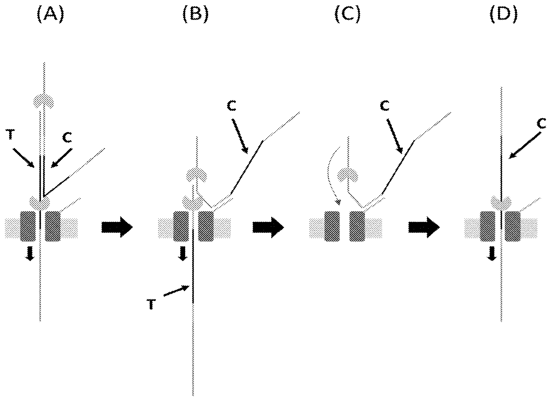
[0070] FIGS. 1A-1B illustrate a prior art method of sequencing a double stranded polynucleotide (e.g., DNA) construct, in which the template and complement strands are attached via a hairpin loop and the template strand comprises a 5' leader sequence, using a transmembrane pore. FIG. 1A is a schematic representation of the polynucleotide (e.g., DNA) construct translocating through a nanopore under the control of an enzyme. The template enters the nanopore and the same enzyme proceeds around the hairpin to control movement of the complement that follows the template. Once the hairpin region translocates through the nanopore, the hairpin may reform on the trans side of the nanopore. FIG. 1B shows peaks representing the accuracy of the sequence information obtained from translocation of the template, from translocation of the complement and when the sequence information obtained from translocation of the template and translocation of the complement was combined algorithmically.
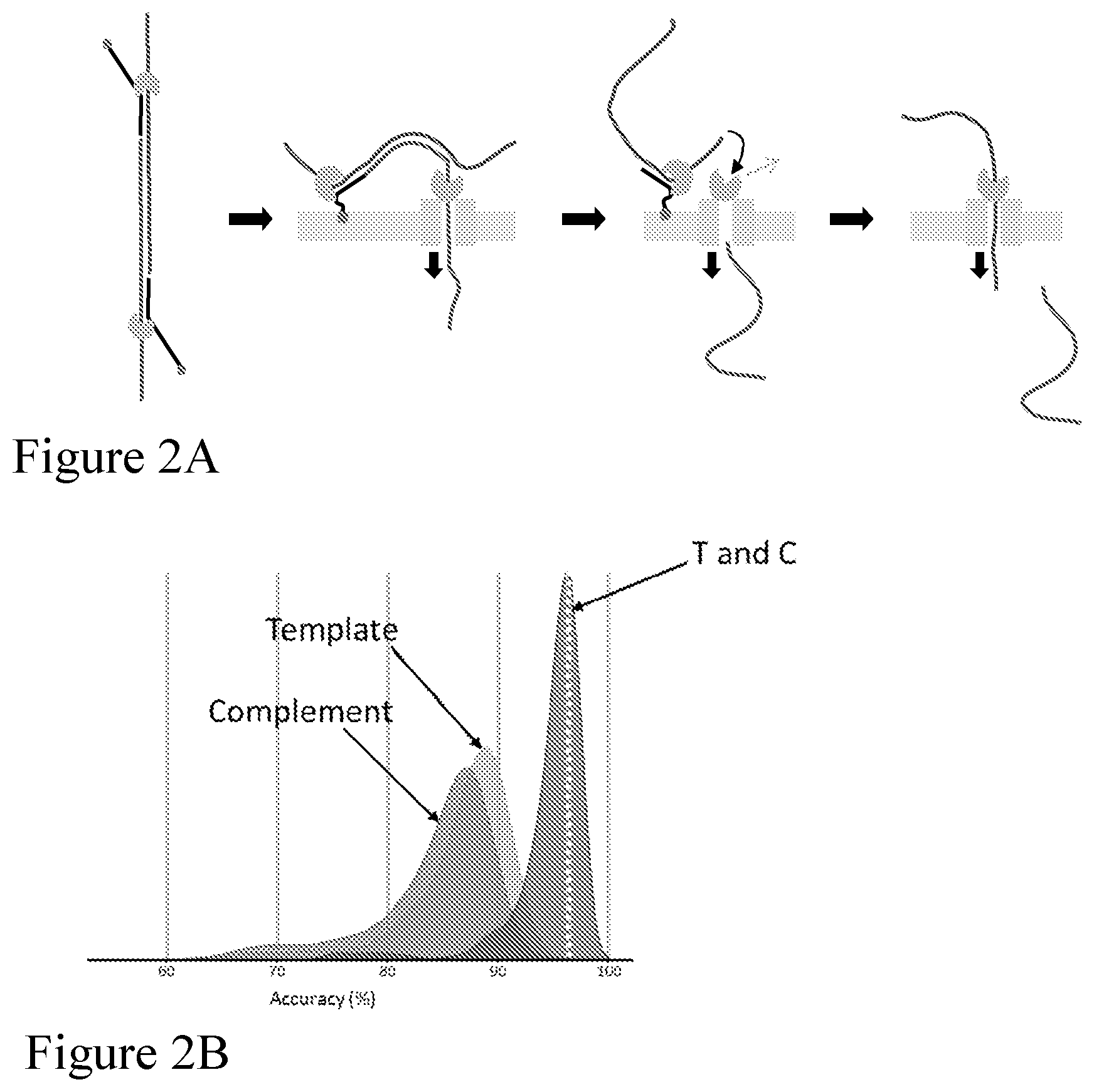
[0071] FIGS. 2A-2B illustrate a method of "follow-on" sequencing a double stranded polynucleotide (e.g., DNA) construct without the use of a hairpin according to one embodiment described herein. Both the template and complement polynucleotide (e.g., DNA) strands comprise an adaptor at each end, which adaptor comprises a leader sequence. FIG. 2A is a schematic representation of a double stranded polynucleotide (e.g., DNA) construct translocating through the nanopore under enzyme control. Template and complement of the double-stranded polynucleotide are not covalently linked, and each strand has an enzyme loaded on the adaptor. After the template strand has passed through the nanopore (and the enzyme is dissociated), the complement strand is separately captured by the pore and sequenced. In the absence of a hairpin joining the template to the complement there is little or no secondary hairpin structure formed on the trans side of the nanopore. FIG. 2B shows peaks representing the accuracy of the sequence information obtained from translocation of the template, from translocation of the complement and when the sequence information obtained from translocation of the template and translocation of the complement was combined algorithmically.
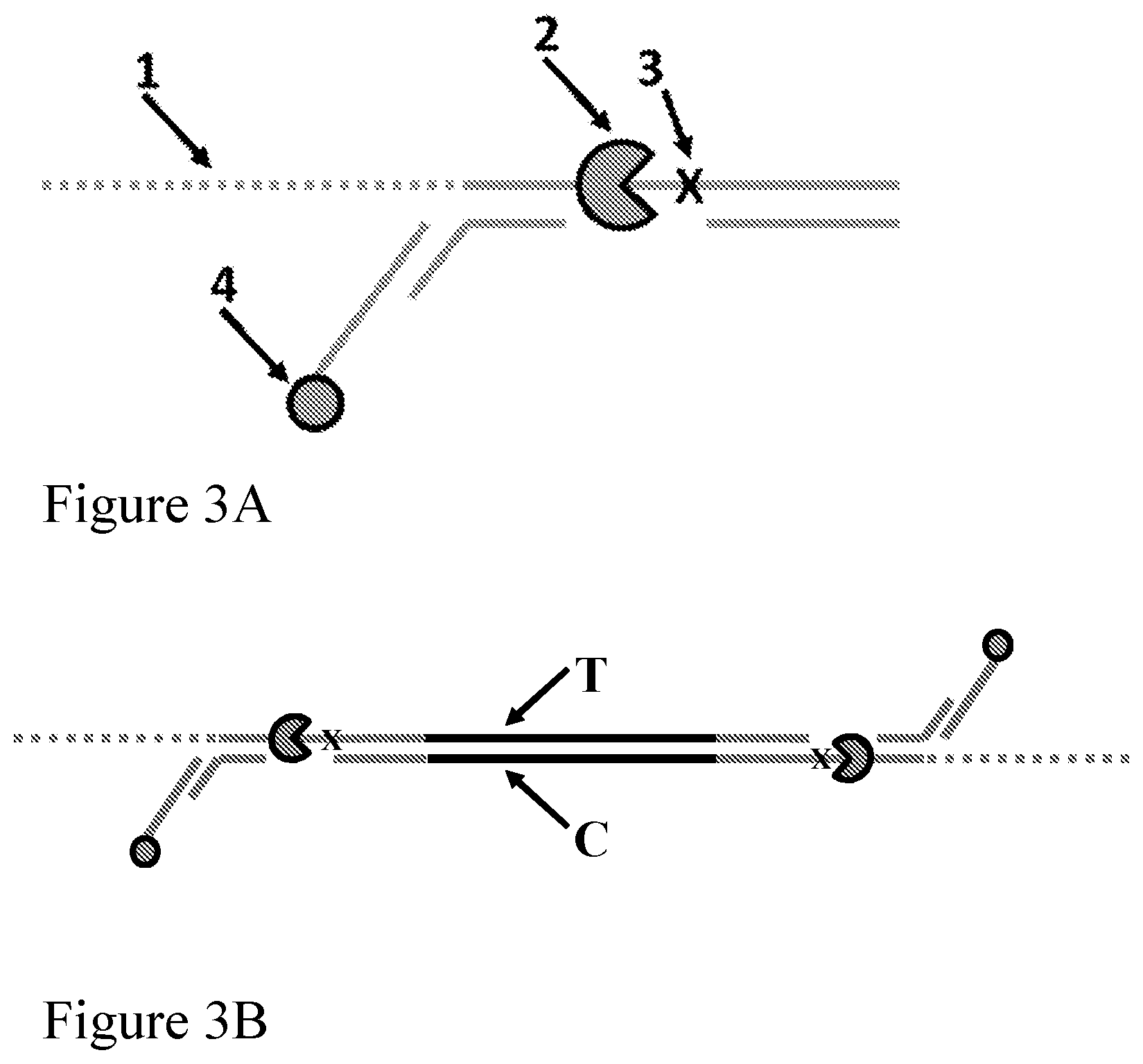
[0072] FIGS. 3A-3B illustrate the structure of an enzyme-loaded adaptor according to one embodiment described herein. FIG. 3A is a schematic representation of the enzyme-loaded adaptor. The labels represent the following: (1) spacers (e.g., a leader sequence); (2) a polynucleotide binding protein (e.g., polynucleotide unwinding enzyme) such as a helicase (e.g., a Dda helicase); (3) a spacer; and (4) an anchor such as a cholesterol anchor. The other solid lines represent polynucleotide sequences. FIG. 3B shows the adaptor attached to each end of a double-stranded polynucleotide such as a fragment of genomic DNA, with a polynucleotide binding protein (e.g., polynucleotide unwinding enzyme) loaded on each adaptor.
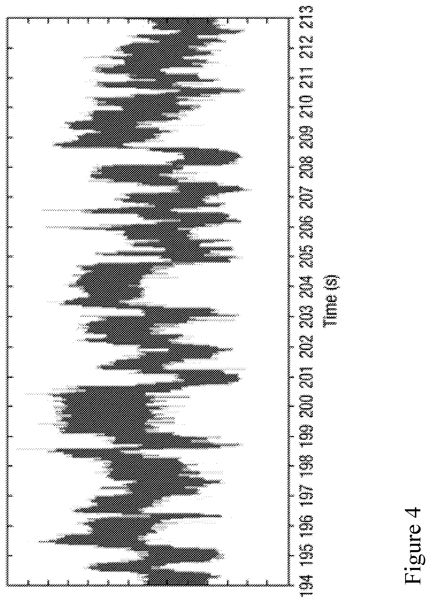
[0073] FIG. 4 is a schematic illustration of a current signal measured over time during translocation of a polynucleotide strand through a nanopore.
[0074] FIG. 5 is an illustration of the separation of a double stranded polynucleotide at the nanopore interface and the subsequent translocation of a single stranded polynucleotide through the nanopore.
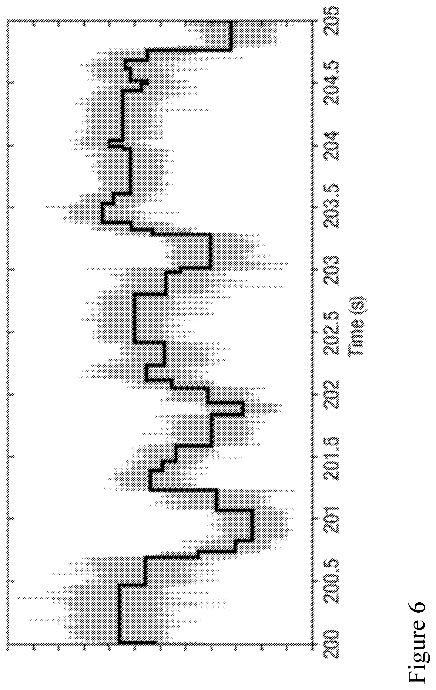
[0075] FIG. 6 is an illustration of event detection of a portion of the current time signal of FIG. 4 during sequencing a polynucleotide.
[0076] FIG. 7 is a schematic illustration of analysis of signal measurements using an recurrent neural network (RNN) model.
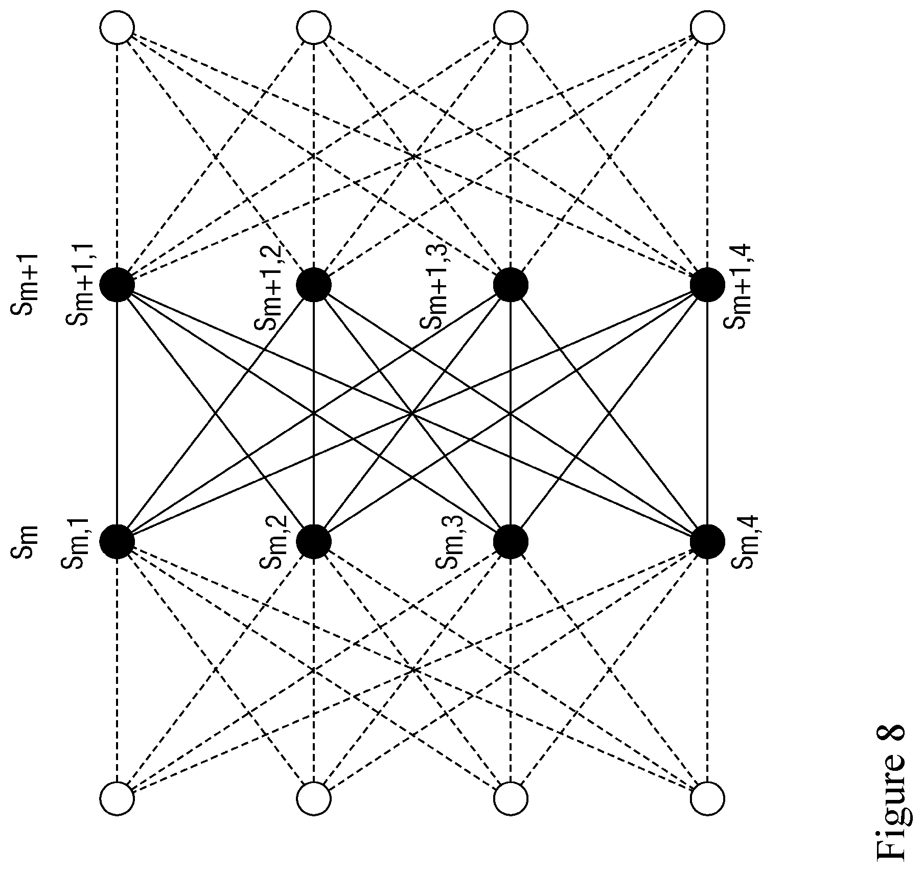
[0077] FIG. 8 is an illustration of how a Viterbi algorithm is employed to determine the path through the possible transitions with the highest likelihood.
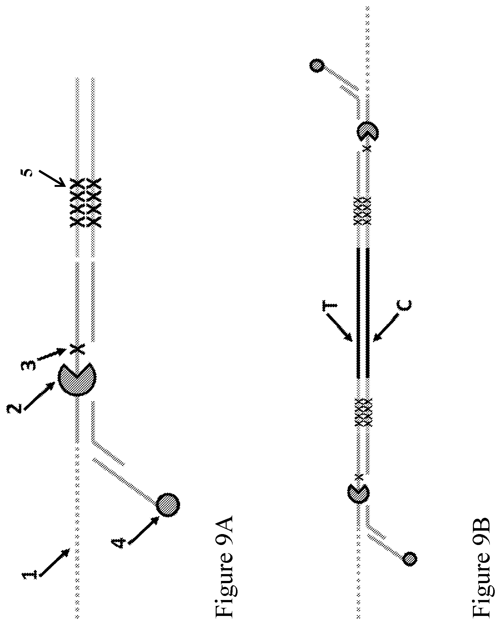
[0078] FIGS. 9A-9B illustrate the structure of an enzyme-loaded adaptor according to one embodiment described herein. FIG. 9A is a schematic representation of the enzyme-loaded adaptor. The labels represent the following: (1) spacers (e.g., a leader sequence); (2) a polynucleotide binding protein (e.g., polynucleotide unwinding enzyme) such as a helicase (e.g., a Dda helicase); (3) a spacer; (4) an anchor such as a cholesterol anchor, which is optional; and (5) a duplex stem positioned on the opposite end of the (1) spacers (e.g., a leader sequence), the duplex stem comprising a capture sequence on a strand that is aligned with the (1) spacers (e.g., a leader sequence), wherein the capture sequence is complementary to a tag (e.g., a capture polynucleotide) conjugated to an outer rim of a nanopore. The other solid lines represent polynucleotide sequences. FIG. 9B shows the construct when the adaptor is attached to each end of a double stranded polynucleotide, with a polynucleotide binding protein (e.g., polynucleotide unwinding enzyme) loaded on each adaptor.
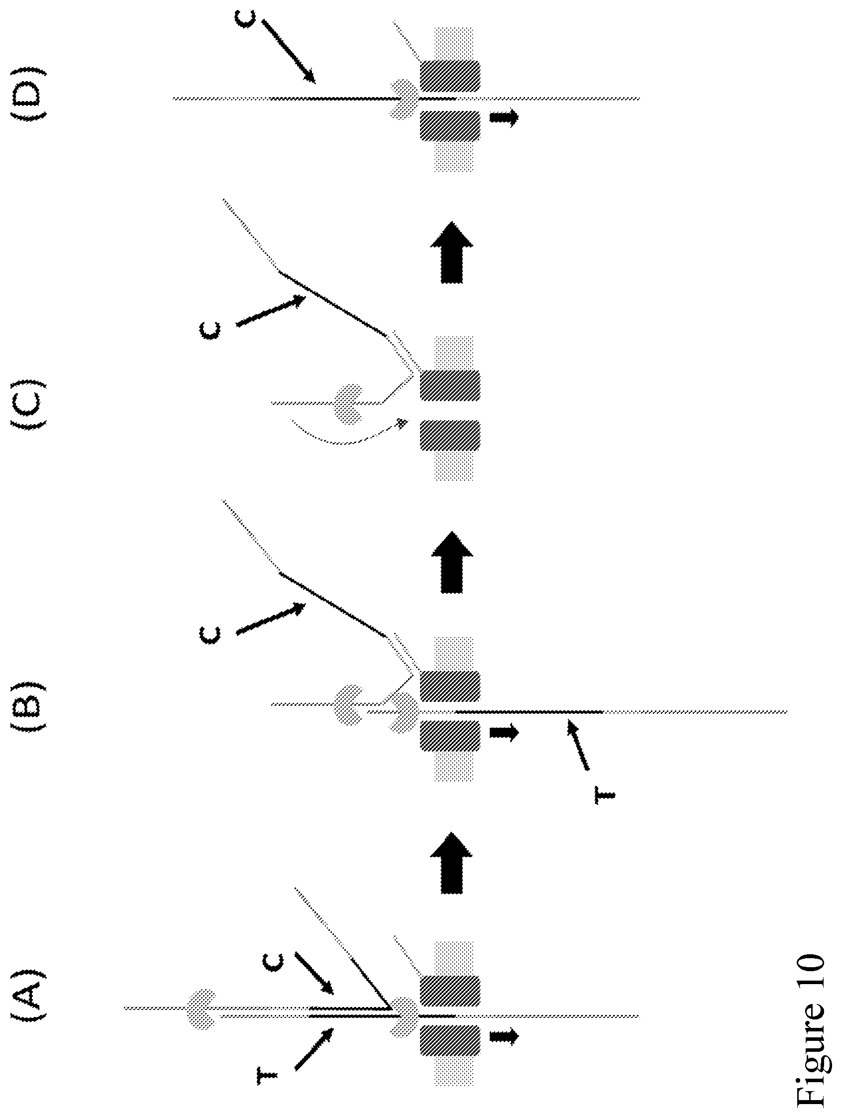
[0079] FIG. 10 (panels A-D) illustrates a schematic representation of a method of sequencing a double-stranded polynucleotide using a nanopore according to one embodiment described herein. The method involves providing (i) a double-stranded polynucleotide with each end attached to an adaptor (e.g., as illustrated in FIG. 9A without the anchor (4)) and a polynucleotide binding protein (e.g., polynucleotide unwinding enzyme) loaded on the adaptor, and (ii) a nanopore with a capture polynucleotide conjugated to an outer rim of the nanopore. The second strand (complement) of the double-stranded polynucleotide is coupled to the nanopore by binding the capture sequence of the adaptor that is attached to the second strand to the tag (e.g., a capture polynucleotide) conjugated to the outer rim of a nanopore.
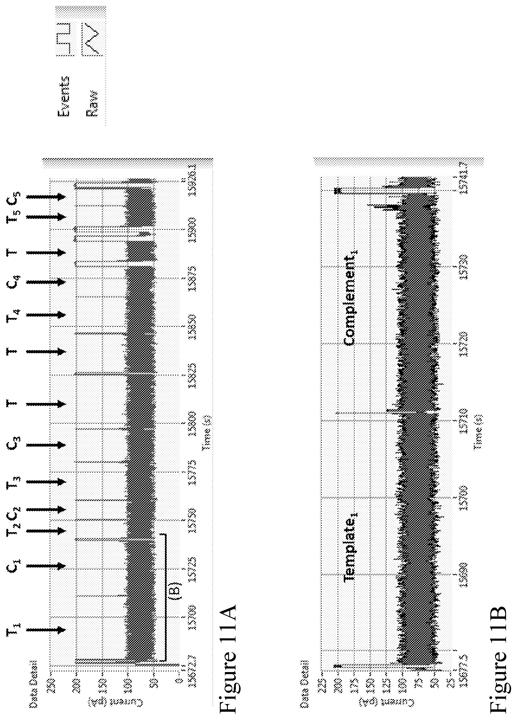
[0080] FIGS. 11A-11B show an example section of strand data acquired using the method according to one embodiment described herein. The strand data shows the current (pA) vs time (seconds) of electrical data of a single channel. FIG. 11A shows an example section of strand data, which shows that the open-pore level with no strand blocking the current is at approximately 200 pA. When strands are captured, the current is reduced to the 50-100 pA range, dependent on sequence composition. As strands finish passing through the pore the current returns to open-pore level at 200 pA. The separate strands are labeled as T.sub.n (e.g., T.sub.1, T.sub.2, . . . ) and C.sub.n (e.g., C.sub.1, C.sub.2, . . . ) for the pairs of template and complement strands, and as T for the strands where the T is not followed by its complement pair. The labels and data correspond to the data in Table 4. FIG. 11A illustrates that the complementary second strand of a pair typically immediately follows the template, with a very short time between strands. FIG. 11B shows a zoomed in section of the electrical trace, highlighting one of the follow-on pairs, labeled Template.sub.1 and Complement.sub.1.
[0081] FIGS. 12A-12D show an example section of strand data acquired using the method according to one embodiment described herein. The strand data shows the current (pA) vs time (seconds) of electrical data of a single channel on a MinION chip. FIG. 12A shows a further example electrical trace of a follow-on template-complement pair. FIGS. 12B, 12C, and 12D show zooms of the trace in FIG. 12A, with stars marking the sp18 spacers in the duplex stem (e.g., complementary tags sections) that were added to the strands to enable coupling to a pore-tag (e.g., a capture polynucleotide conjugated to the outer rim of a nanopore). FIG. 12B shows the sp18s at the start of the template strand. FIG. 12C shows the sp18s at the end of the template and start of the complement, and FIG. 12D shows the sp18 at the end of the complement. These markers can be used to demonstrate that the dsDNA substrate had enzyme-adapters attached to both ends of the dsDNA, and measure the efficiency of the attachment.
[0082] FIG. 13 shows a histogram of the distribution of open-pore times between subsequent strands (x-axis) as sequential strands pass through the pores (aggregated from all channels on a MinION chip). The top panel, Control Adapter (as described in Example 2), shows that the distribution of times is on average approximately 3 seconds between strands when sequencing without a capture sequence in the polynucleotide construct that can couple to the pore tag (e.g., a capture polynucleotide). The bottom panel, Follow-On Adapter 2 (as described in Example 4), shows that when the strands contain a capture sequence in the complement that can couple to the pore tag (e.g., a capture polynucleotide), a new population at approximately 50 milliseconds is observed. The short 50 ms population is from the fast capture of the complement strand soon after its template pair. Capture is fast because the complement strand is held very close to the pore via the binding of the complement to the pore tag and is thus not allowed to diffuse away.
[0083] FIG. 14 shows the histograms of the distribution of basecall accuracies of the sequence information (randomly fragmented E. coli) obtained from translocation of the template, from translocation of the complement and when the sequence information obtained from translocation of the template and translocation of the complement was combined algorithmically. The sequence information were obtained using the method as illustrated in FIG. 10.
[0084] FIG. 15 shows example embodiments of nanopores having two or more types of tags. For example, one tag can be provided to increase the sensitivity of a method for characterizing an analyte ("sensitivity tag"), while another tag can be provided to increase the likelihood of sequencing of a complement strand following a template strand of a double-stranded polynucleotide ("follow-on tag"). The pore tags can be configured in any number of ways. For example, each monomer of an oligomeric pore can have the same type of tag configuration (e.g., with multiple binding sites, as illustrated by Tag-A and Tag-B). Tag-A and Tag-B can be combined to form a single tag, and each monomer comprises the Tag-A/Tag-B combined tag. Alternatively, an oligomeric pore can comprise mixed monomers with different tags attached such that at least one monomer has a different tag configuration from the other monomers. In another example, Tag-A and Tag-B can remain as separate tags and each monomer can comprise both individual tags. Sensitivity and follow-on tags can be separately combined if they are complementary to unique sequences used in the adaptor as illustrated in the bottom panel of a schematic adaptor design.
[0085] FIG. 16 is a schematic illustration of how nanopores with two different tag types can be used to capture strands from solution (for improved sensitivity). The adaptor that is attached to an end of a double-stranded polynucleotide comprises a capture sequence (e.g., forming a non-complementary arm of a Y-adaptor) that is available to couple to a first pore tag, while a separate capture sequence within the duplex stem that is only revealed when unzipped permits the complement to bind to a second pore tag and thus enables complement capture for follow-on sequencing.
[0086] FIG. 17A is a schematic illustration of how the same capture sequence can be used in two locations of an adaptor, one revealed to allow strands to bind to a pore tag of a nanopore out of solution for improved sensitivity, and the other initially unrevealed and exposed when template unzips through the pore, becoming available to bind to another of the multiple tags on a pore (a pore with only one type of tag) to enable follow-on sequencing. FIG. 17B provide some example sequences that can be used for such purposes. The top construct shows a portion of an example Y adaptor. The "FO001/FO002" and "FO003/FO004" sequences are examples of a duplex stem that can be ligated to the example Y adapter to create a single adaptor construct that can enable the method according to one or more embodiments described herein. The light-blue sequences in the "FO001/FO002" and "FO003/FO004" sequences have the same sequence as the purple sequence, which is a binding sequence site for a nanopore. The same binding sequence site for a nanopore can be used more than once (e.g., twice) within the duplex stem of the adaptor, wherein the light-blue sequences are not exposed.
[0087] FIG. 18 shows a schematic of an adapter design that enables follow-on sequencing and increased sensitivity. The pore binding sequence (labeled as "Hyb splint for morpholino pore tag" in FIG. 18) is exposed to a surrounding solution and initially available for binding to the pore tag, so it improves sensitivity. The pore binding site is also contiguous with the complement strand when attached, so that when the template strand has passed through the pore the complement strand remains bound to pore. This process is shown schematically in FIG. 19.
[0088] FIG. 19 is a schematic illustration showing a double stranded polynucleotide with an adaptor of FIG. 18 attached to each end. A strand is coupled to a nanopore from solution via an exposed pore binding site, thus improving the sensitivity of subsequent capture of the nearby template strand. The binding site is also contiguous with the complement strand, so that when the template has passed through the nanopore the complement remains bound to the nanopore. The complement might proceed to a number of possible conformations as shown, before ultimate capture and sequencing to enable follow-on sequencing. In FIG. 19, the green and yellow complementary segments attached to both ends of a strand to be detected, respectively, could bind together to form a hairpin structure comprising the strand, facilitating the sequencing process by bringing the strand closer to the nanopore for increased sequencing efficiency. It can be particularly beneficial when the strand to be detected is a long strand.
[0089] FIG. 20 shows a schematic illustration of an adapter design where the same sequence (green) within a duplex stem is repeated at a different location of the adaptor as shown to enable the follow-on method shown in FIG. 21.
[0090] FIG. 21 is a schematic illustration showing a double stranded polynucleotide with an adaptor of FIG. 20 attached to each end. The dsDNA strand binds to a pore as shown via a binding site on a side-arm attached to the adapter as described in FIG. 20. When the template is captured in the pore, the side-arm sequence is unzipped and remains bound to the pore-tag as shown. Later into template unzipping the second site with the same sequence is revealed to bind to the side-arm (itself still bound to pore-tag). In this way, a single tag on a pore can be used to improve capture sensitivity, and can re-used to later enable follow-on of the complement of the substrate. At the end, the pore-tag retains the side-arm sequence, but the side-arm itself is captured by the pore and stripped from the pore-tag to free the pore-tag for another cycle.
[0091] FIG. 22 shows how the revealed sequences are exposed for coupling to the pore-tag as the template-enzyme nears the end of the template strand. Efficiency of the follow-on process can be increased, for example, by including spacers (e.g., 4 sp18 spacers, e.g., hexaethyleneglycol, in the sequence shown) or similar features that briefly pause the enzyme, which allows more time for coupling, or features with optimized geometry or flexibility. A double binding site in the revealed section also improves the chances of coupling to pore-tag.
[0092] FIG. 23 provides example adapters/sequences that can enable the method disclosed in FIG. 21.
[0093] FIG. 24 provides further adapters/sequences that can enable the method disclosed in FIG. 21 and are more optimized in terms of pausing the enzyme.
[0094] FIG. 25 provides example adapters/sequences that can enable the method disclosed in FIG. 19.
[0095] FIG. 26 provides example sequences of the components that make up the adapters described in the above figures.
[0096] FIG. 27A shows a SYPRO Ruby Protein Gel showing monomers and oligomeric nanopores of CsgG modified with or without morpholino pore tags. FIG. 27B shows a schematic representation of a nanopore modified with a pyridyl-dithio morpholino.
[0097] FIG. 28 shows a Cy3 Florescent gel showing hybridization of an analyte to pyridyl-dithio morpholino modified pore.
[0098] FIG. 29 shows a SYBR Gold Nucleic Acid Gel Stain showing hybridization of an analyte to pyridyl-dithio morpholino modified pore.
[0099] FIG. 30 shows a SYPRO Ruby Protein Gel showing hybridization of an analyte to pyridyl-dithio morpholino modified pore.
[0100] FIG. 31 illustrates a diagram showing a computer rendering of a nanopore (e.g., a CsgG nanopore) highlighted with positions at which a cysteine can be added for conjugation to a pore tag. The pore tag can be conjugated to the external surface of a nanopore, e.g., on cis-side or trans-side of a membrane, when the nanopore is disposed in a membrane.
[0101] FIG. 32A shows an embodiment of a Y adapter design, which includes two hybridization sites, one for the pore tether (red) and the other for the membrane or bead tether (blue). In this design the pore tether is next to the leader sequence. FIG. 32B shows the a ligated analyte, e.g., a double stranded polynucleotide, with a Y adapter on either end.
[0102] FIG. 33 is a schematic diagram showing example sequences of a Y adapter design illustrated in FIG. 32A.
[0103] FIG. 34A shows a different embodiment of a Y adapter design, which includes two hybridization sites, one for the pore tether (red) and the other for the membrane or bead tether (blue). In this design the membrane tether is next to the leader sequence. FIG. 34B shows the a ligated analyte, e.g., a double stranded polynucleotide, with a Y adapter on either end.
[0104] FIG. 35 is a schematic diagram showing example sequences of a Y adapter design illustrated in FIG. 34A.
[0105] FIG. 36 is a schematic diagram showing an alternative embodiment of a Y adapter design, which includes two hybridization sites, one for a bead tether and the other for the membrane tether. In this design the bead has two different tethers, one to the analyte (blue) and the other to the pore (red).
[0106] FIG. 37 is a schematic diagram showing example sequences of a Y adapter design illustrated in FIG. 36 and showing indirect attachment of the analyte to the pore.
[0107] FIG. 38 shows example traces of sequential strands translocating through a nanopore without a pore tag that can bind to a strand to allow follow-on sequencing. The time between strands is indicated by the red bars, in these examples the time between strands ranges from 2-5 seconds.
[0108] FIG. 39 shows example traces of sequential strands translocating a modified pore according to one embodiment described herein. The time between strands is indicated by the red bars, in these examples the time between strands ranges from 0.02-3 seconds.
[0109] FIG. 40 shows histograms illustrating the time between sequential strands on a log scale. The left graph shows a nanopore (e.g., a CsgG pore) with a single distribution with time between strands greater than 1 second. The right graph shows the time between strands translocating through a tethered pore. This shows two populations, a fast capture population and with a time between strands under 0.1 seconds.
[0110] FIG. 41 depicts a graph showing the number of bases sequenced per chip over a 6 hour period from 20 ng input DNA. The red line represents the tethered pore and the blue line shows the non-tethered nanopore.
[0111] FIG. 42 shows a data table from E. coli runs showing an increase in the number of follow on strands with the tethered pore. FIG. 43 shows a method that can be used for concatentating both single and double stranded nucleic acids.
[0112] FIG. 44 shows a method a method of characterising and concatenating many double stranded target polynucleotides, where the complement strand of a first double stranded target polynucleotide recruits a many other double stranded target polynucleotides and brings them into a local concentration of the pore. This provides a higher local concentration around the pore than in the general bulk solution and so double stranded target polynucleotides follow one another through the open pore with minimal time between strands. This is especially useful when the concentration of double strand target polynucleotides is low. A tether consisting of an oligo coupled to a single stranded binding protein is used. As the template strand of the first double strand target polynucleotide is sequenced the complement strand is released into solution as ssDNA. The single stranded binding proteins of the other double stranded target polynucleotides are able to bind to the ssDNA. As the complement strand is sequenced the 3' of the complement strand is drawn back towards the pore. The single stranded binding proteins on the ssDNA complement strand are displaced from the complement strand when they encounter the motor protein and so are deposited around the pore increasing the local concentration.
DETAILED DESCRIPTION OF THE INVENTION
[0113] While transmembrane pores (e.g., protein nanopores or solid state nanopores) are useful as sensors to detect or characterize a biopolymer, there are still challenges of increasing the accuracy and/or efficiency of the detection method using transmembrane pores. For example, there are various drawbacks of translocation of both the template and complement strands of a double stranded polynucleotide connected by a hairpin through a nanopore. While measurement of both strands in this way is advantageous as information from the two complementary linked strands can be combined and used to provide higher accuracy than may be achieved from measurement of template strands only, preparation of such a hairpin linked polynucleotide is more involved and time consuming and can result in a loss of valuable analyte. Further, translocation of a hairpin linked template and complement polynucleotide strands through a nanopore can give rise to rehybridization of the strands on the other (trans) side of the nanopore. This can alter the rate of translocation giving rise to a lower sequencing accuracy. A strand with a hairpin structure is also more difficult to translocate as fast as a single linear strand. Additionally, due to the differences in current-time data for the template and complement strands, two algorithms are used for computation, which makes the computation more complex and intensive.
[0114] For analyte detection, there is typically a time delay between translocation of one analyte and translocation of the next one. This delay can be of the order of seconds to minutes, which can result in slower characterization, in a higher pore open current which depletes the reference electrode more quickly, and/or in an increased likelihood of a nanopore getting blocked when the pore is open. Accordingly, there is a need to develop methods and compositions that improve the accuracy and/or efficiency or throughput of characterizing analytes using a nanopore.
[0115] The present disclosure is, in part, based on the unexpected discovery that both strands of a double stranded polynucleotide can be sequentially translocated through a nanopore to provide sequence information without the need to covalently link two strands via a bridging moiety such as a hairpin loop. For example, in one aspect, the present inventors have discovered that when a polynucleotide binding protein (e.g., a polynucleotide unwinding enzyme) is used to separate the two strands of a double stranded polynucleotide while controlling the movement of one of the strands through a transmembrane pore, the second strand may remain in the vicinity of the pore and, subsequent to the translocation of the first strand through the pore, the second strand may be captured by the pore and a polynucleotide binding protein may be used to control the movement of the second strand through the pore.
[0116] In another aspect, the present inventors have discovered that an adaptor with a duplex stem comprising a capture sequence that is complementary to a pore tag conjugated to a nanopore, can be provided to each end of a double stranded polynucleotide, wherein the capture sequence is only revealed upon separation or unwinding of the strand. Thus, as a first strand of a double-stranded polynucleotide passes through a tag-modified nanopore, it unzips the duplex stem of the adaptor to expose the capture sequence on a second strand of the double-stranded polynucleotide, which is then captured by the pore tag of the nanopore. Such method keeps the second strand, which would otherwise typically diffuse away, to be close to the nanopore for sequencing following the template being sequenced. In particular, the methods described herein can significantly increase the likelihood of a follow-on translocation of a complement after a template translocation to at least about 60% of the time, as compared to 0.1%-1% of the time that is typically observed in typical nanopore sequencing.
[0117] It was also discovered that modification of a nanopore to comprise multiple binding sites for multiple analytes such that one or more analytes can bind to the nanopore via the binding sites, while an analyte is being characterized by the nanopore, can enhance sensitivity and/or throughput of characterizing the analytes. Without wishing to be bound by theory, coupling or capture of analytes at the outer rim of the nanopore can enhance the local concentration of the analytes at the pore. Further, at least one or more analytes in the vicinity of the nanopore can readily enter the nanopore one following another for characterization, thus decreasing time delay and thus the open-pore current time between each analyte characterization.
[0118] Accordingly, various aspects herein relate to methods of characterizing one or more analytes using a nanopore, as well as composition and systems including, e.g., adaptors and nanopores, that can be used in the methods described herein. Some aspects feature methods and compositions for characterizing a double-stranded polynucleotide using a nanopore, e.g., without using a hairpin connecting a template and a complement of the double-stranded polynucleotide. Other aspects features methods and compositions for characterizing an analyte using a tag-modified nanopore with increased sensitivity and/or higher throughput.
Methods for Characterizing an Analyte (e.g., a Double Stranded Polynucleotide)
[0119] In one aspect, the disclosure provides a method of sequencing a target polynucleotide, comprising: [0120] (a) contacting a transmembrane pore with: [0121] (i) a double stranded polynucleotide comprising the target polynucleotide and a polynucleotide complementary to the target polynucleotide, wherein the target polynucleotide and the polynucleotide complementary to the target polynucleotide each comprise a single stranded leader sequence; and [0122] (ii) a polynucleotide binding protein (e.g., polynucleotide unwinding enzyme) capable of separating the strands of a double stranded polynucleotide and controlling the movement of a polynucleotide through a transmembrane pore; [0123] (b) detecting a signal corresponding to ion flow through the pore to detect polynucleotides translocating through the pore; [0124] (c) identifying a signal corresponding to translocation of the target polynucleotide and a sequential signal corresponding to the separate translocation of the polynucleotide complementary to the target polynucleotide; [0125] (d) analyzing the signals identified in (c), [0126] thereby sequencing the target polynucleotide.
[0127] The method may further comprise before step (a) a step of attaching single stranded leader sequences to the target and complementary polynucleotides. The method may further comprise before step (a) a step of digesting one end of the target polynucleotide to produce a leader sequence on the complementary strand and/or digesting one end of the complementary polynucleotide to produce a leader sequence on the target strand. The method may still further comprise binding a polynucleotide binding protein (e.g., polynucleotide unwinding enzyme) to the leader sequences. The polynucleotide binding protein (e.g., polynucleotide unwinding enzyme) in (a)(ii) may be bound to the leader sequences in (a)(i).
[0128] In this aspect, a first polynucleotide binding protein (e.g., polynucleotide unwinding enzyme) separates the target and complementary strands of the double stranded polynucleotide and controls the movement of either the target polynucleotide or the complementary polynucleotide through the transmembrane pore. A second polynucleotide binding protein (e.g., polynucleotide unwinding enzyme), which may be another protein of the same type as the first polynucleotide binding protein (e.g., polynucleotide unwinding enzyme), or may be a different type of polynucleotide binding protein (e.g., polynucleotide unwinding enzyme), controls the movement through the transmembrane pore of either the target polynucleotide, where the complementary polynucleotide has already translocated through the pore, or the complementary polynucleotide, where the target polynucleotide has already translocated through the pore. The second polynucleotide binding protein (e.g., polynucleotide unwinding enzyme) does not need to separate the target polynucleotide and the complementary polynucleotide because separation of the two strands of the double stranded polynucleotide will already have occurred (the first polynucleotide binding protein (e.g., polynucleotide unwinding enzyme) will have separated the two strands whilst passing one of the strands through the pore). The first polynucleotide binding protein (e.g., polynucleotide unwinding enzyme) is typically one that processes a double stranded polynucleotide. The second polynucleotide binding protein (e.g., polynucleotide unwinding enzyme) is typically one that processes a single stranded polynucleotide. The first and/or second polynucleotide binding protein (e.g., polynucleotide unwinding enzyme) may be capable of processing a double stranded polynucleotide and a single stranded polynucleotide.
[0129] Another aspect described herein features a method for sequentially translocating two non-covalently bound molecules through a nanopore. The method comprises: contacting a pair of non-covalently bound molecules to a nanopore under conditions that promote translocation of a first member of the pair of non-covalently bound molecules through the nanopore, wherein a binding site on a second member of the pair is exposed during translocation of the first member through the nanopore, and wherein the exposed binding site binds to a tag or tether that is present on the nanopore. The binding site on the second member is not exposed (or is shielded) prior to translocation of the first member through the nanopore.
[0130] As used herein, the term "non-covalently bound molecule" refers to a molecule comprising a first a member and a second member, wherein the first member and the second member are associated with each other by means of non-covalent attachment and can be separated from each other as individual entities. The separation and binding process between the first member and the second member are reversible. Examples of means of non-covalent attachment include, but are not limited to complementary base-pairing, ionic interaction, hydrophobic interaction, and/or Van der Waals' interaction.
[0131] In some embodiments, the non-covalently bound molecules comprise complementary polynucleotide strands.
[0132] In some embodiments, the tag on the nanopore is an oligonucleotide, and the binding site on the second member is a portion of a nucleic acid that has a sequence complementary to the tag.
[0133] In some embodiments, the pair of non-covalently bound molecules comprise a target nucleic acid (e.g., a target double stranded polynucleotide) coupled to an adaptor nucleic acid, and wherein the binding site is present on the adaptor nucleic acid.
[0134] As an example only, FIG. 9B shows a non-covalently bound molecule, which comprises complementary polynucleotide strands (e.g., a template strand and a complement strand) and an adaptor (e.g., an adaptor nucleic acid) attached to each end. The adaptor, as shown in FIG. 9A, comprises a duplex stem (5) and a first single stranded polynucleotide (1) extending from a template strand of the duplex stem. The duplex stem (5) comprises a capture sequence on a strand that is aligned with the first single stranded polynucleotide (1), wherein the capture sequence is complementary to a tag (e.g., a capture polynucleotide) conjugated to an outer rim of a nanopore. In some embodiments, the first single stranded polynucleotide (1) can further comprise a leader sequence. While FIG. 9A shows a second single stranded polynucleotide extending from a complementary strand (e.g., the complement strand) of the duplex stem, it is not required. However, in some embodiments, it may be desirable to have one or more second single stranded polynucleotides comprising one or more tethers for a solid substrate, e.g., a membrane or a bead, and/or a nanopore. When the second single stranded polynucleotide is not complementary to the first single strangled polynucleotide, a Y-adaptor is formed, e.g., as shown in FIG. 9A.
[0135] FIG. 9A shows an example adaptor comprising at least one anchor for a solid substrate, e.g., a membrane or a bead, while FIG. 15 shows an example adaptor comprising at least two anchors, wherein a first anchor is capable of tethering to a solid substrate, e.g., a membrane or a bead, and a second anchor is capable of tethering to a nanopore. The second anchor for the nanopore can be configured to bind to a tag conjugated to the nanopore. In some embodiments, the second anchor for the nanopore can be configured to directly bind to a tag conjugated to the nanopore. For example, the second anchor for the nanopore can comprise a sequence that is complementary to the capture polynucleotide tag conjugated to the nanopore. In alternative embodiments, the second tether for the nanopore can be configured to indirectly bind to a tag conjugated to the nanopore. For example, FIG. 36 shows that an adaptor attached to an analyte can be coupled to a tag on the nanopore via a microparticle, which is further described in detail in the "Microparticles" section below.
[0136] It should be noted that the adaptors described herein can be attached to either or both ends of a double stranded polynucleotide. In some embodiments, the same adaptors are attached to both ends of a double stranded polynucleotide. In some embodiments, different adaptors can be attached to the ends of a double stranded polynucleotide. Attachment of different adaptors to the ends of double stranded polynucleotides can be achieved, for example, by mixing two or more populations of different adaptors together with the double stranded polynucleotides. Typically, a mixture of double stranded polynucleotides attached with different adaptors is formed, but there are also methods to achieve a desired hetero-adapter mixture (e.g., through purification or by controlling the attachment of adaptors to the ends of double stranded polynucleotides).
[0137] In some embodiments, a double stranded polynucleotide can have an adaptor to its 3' end or 5' end.
[0138] Blunt ended double stranded polynucleotides can be captured into a nanopore and unzipped. Accordingly, in some embodiments, a blunt ended construct without an adaptor (e.g., one as described herein) can be used in any aspects of the methods described herein. While not necessary, in some embodiments, it is desirable to have a leader sequence coupled to at least one end of a double stranded polynucleotide, e.g., to increase the capture efficiency by a nanopore.
[0139] In some embodiments where the adaptor is attached to both ends of a double stranded polynucleotide, one of ordinary skill in the art will readily recognize that when the first single-stranded polynucleotide of an adaptor is coupled to a template strand at one end of the double-stranded polynucleotide, the first single-stranded polynucleotide of another adaptor is coupled to a complement strand at the opposite end of the double-stranded polynucleotide.
[0140] In some embodiments, the adaptors can have a pre-bound polynucleotide binding protein (e.g., polynucleotide unwinding enzyme) at each end of the target polynucleotide. In some embodiments, the method can further comprising adding a polynucleotide binding protein (e.g., polynucleotide unwinding enzyme) in solution such that it bounds to the adaptor at each end of the target polynucleotide. As illustrated in FIG. 10, each end of a double-stranded polynucleotide is attached to an adaptor as described herein and a polynucleotide binding protein (e.g., polynucleotide unwinding enzyme) is loaded on the adaptor, wherein the adaptor comprises a duplex stem with a capture sequence complementary to a tag conjugated to a nanopore (see, e.g., FIG. 9A as an example adaptor). A single-stranded polynucleotide (which may optionally comprise a leader sequence) extending from the construct that comprises a double-stranded polynucleotide enters the nanopore (Panel A). The first strand that enters the nanopore is labeled as template (T), and the reverse complement of the first strand captured is labeled as complement (C). As the first template strand passes through the pore under the control of the polynucleotide binding protein (e.g., polynucleotide unwinding enzyme such as a helicase motor), the complement is gradually unzipped. Panel B shows that towards the end of the template strand, the capture sequence within the duplex stem on the complement is exposed by the unzipping, and thus couples to the tag (e.g., capture polynucleotide) on the nanopore. In Panel C, when the template strand finally passes through the nanopore, and the enzyme dissociates, the complement strand remains coupled to the nanopore via the binding to the tag (e.g., capture polynucleotide) on the nanopore. At some time later the complement strand is captured by its leader sequence. In Panel D, the complement strand passes through the nanopore under the control of the second loaded polynucleotide binding protein, e.g., polynucleotide unwinding enzyme such as helicase motor. As the complement strand passes through the nanopore, the capture sequence will at some point be unzipped from the nanopore's tag (e.g., a capture polynucleotide), thus freeing up the nanopore's tag (e.g., a capture polynucleotide) so that it is available for the next strand. In this embodiment, both polynucleotide binding proteins (e.g., polynucleotide unwinding enzymes) are capable of processing double-stranded polynucleotides. In some embodiments, the polynucleotide binding proteins (e.g., polynucleotide unwinding enzymes) loaded at both ends can be the same or different.
[0141] While FIG. 10 (Panel B) illustrates that exposing the capture sequence (for binding to a tag on a nanopore) within the duplex occurs toward the end of translocation of the template strand (e.g., capturing far end adapter thus holding it local to pore), more genrerally, strands can be designed so that binding of a double stranded polynucleotide (e.g., via an end or tail portion) to a nanopore can occur right at the beginning of the unzipping process. In this instance, the capture sequence can be positioned in the adaptor (e.g., as a non-complementary arm of a Y-adaptor portion) such that it is exposed for binding to a nanopore for the entire duration of the unzipping or even before the unzipping process.
[0142] In some embodiments, instead of having a polynucleotide binding protein (e.g., polynucleotide unwinding enzyme) bound onto the adaptors attached to the target polynucleotide, a polynucleotide binding protein (e.g., polynucleotide unwinding enzyme) can be immobilized within the lumen of the nanopore such that a single polynucleotide binding protein (e.g., polynucleotide unwinding enzyme) can be used to control the movement of both strand through the nanopore.
[0143] In some embodiments, the unwinding or separation of the strands as one strand translocates through the nanopore is controlled by a polynucleotide binding protein (e.g., polynucleotide unwinding enzyme). In some embodiments, the unwinding or separation of the strands as one strand translocates through the nanopore can occur in the absence of a polynucleotide binding protein (e.g., polynucleotide unwinding enzyme). Such enzyme-free methods to control the movement and/or separation of both strands of a polynucleotide are known in the art. For example, certain nanopores themselves can provide the force to unwind a polynucleotide such as a motor protein nanopore, including, e.g., phi29 motor protein nanopore, e.g., as described in Wendell et al. "Translocation of double-stranded DNA through membrane-adapted phi29 motor protein nanopores" Nat Nanotechnol, 4 (2009), pp. 765-772, and/or nanopores as described in the U.S. Pat. No. 8,986,528, the contents of each which are incorporated herein by reference in their entireties.
[0144] As used herein, the term "translocate" or "translocation" refers to movement along at least a portion of a nanopore. In some embodiments, translocation is moving from a cis-side of a nanopore to a trans-side of a nanopore.
[0145] The target polynucleotide is typically present in a sample comprising multiple copies of the target polynucleotide and/or in a sample comprising multiple different polynucleotides. In some embodiments, the method of any aspects described herein may comprise determining the sequence of one or more target polynucleotides in a sample. The method may comprise contacting the pore with two or more double stranded polynucleotides. For example, the method may comprise contacting the pore with a sample in which substantially all the double stranded polynucleotides have a single stranded leader sequence on each of their two strands. In some embodiments, the double stranded polynucleotides are coupled to each other only via complementary base pairing. In these embodiments, the double stranded polynucleotides can have four free ends, wherein a free end is the end of a polynucleotide strand. The end of the polynucleotide strand may be single stranded, e.g. a single stranded overhang, or base paired to another polynucleotide strand. In some embodiments, the two strands of the double stranded polynucleotides being sequenced are not covalently attached (e.g., no hairpin or other covalent attachment). However, a moiety that does not bridge the template and complement polynucleotides may be added to one or more of the free ends.
[0146] In some embodiments of various aspects described herein, the method may further comprise a step of generating or attaching single stranded leader sequences to both strands of substantially all double stranded polynucleotides in a sample, prior to contacting with a nanopore. The leader sequences added may have one or more polynucleotide binding proteins (e.g., polynucleotide unwinding enzymes) attached thereto, such that a population of double stranded polynucleotides each comprises a leader sequence with a polynucleotide binding protein (e.g., polynucleotide unwinding enzyme) attached thereto at one end of each of its two strands.
[0147] The double stranded polynucleotide comprising the target polynucleotide (e.g., a template) and a polynucleotide complementary to the target polynucleotide (e.g., a complement) may have an adaptor comprising a single stranded leader sequence attached at each end thereof. In some embodiments where the method of various aspects involves a polynucleotide binding protein (e.g., a polynucleotide unwinding enzyme), the method may comprise contacting the pore with two or more polynucleotide binding proteins (e.g., polynucleotide unwinding enzymes) that may be the same or different. The different polynucleotide binding proteins (e.g., polynucleotide unwinding enzymes) may be bound to separate leader sequences, that may be the same or different. For example, a polynucleotide binding protein (e.g., polynucleotide unwinding enzyme) that works in the 5' to 3' direction may be bound to a leader sequence at the 5' end of the target polynucleotide and/or at the 5' end of a complementary polynucleotide. A polynucleotide binding protein (e.g., polynucleotide unwinding enzyme) that works in the 3' to 5' direction may be bound to a leader sequence at the 3' end of the target polynucleotide and/or at the 3' end of a complementary polynucleotide.
[0148] The adaptors used in the methods of various aspects described herein may be further configured to permit binding of an analyte (e.g., a target polynucleotide or a non-covalently bound molecule) to a nanopore for improved sensitivity and/or throughput of the characterization method. This is different in purpose from an embodiment in which a capture sequence within the duplex stem of an adaptor is exposed to allow binding of a second member of a non-covalently bound molecule (e.g., a complement strand of a double-stranded polynucleotide), e.g., as illustrated in FIG. 10 for increasing the likelihood of a complement translocation following a template translocation, thus increasing accuracy of sequencing information. As illustrated in FIG. 16, the adaptor is further configured to include a tether for binding to a nanopore, e.g., to facilitate capture of the analyte. Thus, the analyte binds to a first tag conjugated to a nanopore before a first member of the analyte (e.g., a template strand of a double stranded polynucleotide) enters the nanopore. As the first member translocates through the pore and unzips the duplex stem to expose a capture sequence on the second member, the second member binds to the a second tag conjugated to the nanopore such that the second member is held close to the nanopore for subsequent characterization after the first member. The first tag and the second tag on the nanopore can be different (e.g., as shown in FIG. 16), or they can be the same (e.g., as shown in FIG. 17A), depending on the design of the adaptors.
[0149] Accordingly, a further aspect provided herein relates to a method for determining a characteristic of an analyte using a nanopore comprising: (a) providing a nanopore that is modified to comprise at least two or more tags external to the lumen of the nanopore, wherein the tags provide binding sites for at least two or more analytes; and (b) contacting a plurality of analytes to the nanopore under conditions such that at least one or more (e.g., at least one, at least two, at least three, at least four, at least five, at least six, at least seven, at least eight, at least nine, or more) analytes bind to the tags on the nanopore, while an analyte from the plurality is translocating through the nanopore.
[0150] Unlike nanopores that are modified to improve interaction of a nanopore with a target analyte, e.g., by altering charges and/or hydrophobicity of the amino acids within the nanopore lumen, the nanopores described herein are modified to provide multiple tags for capture of a plurality of analytes while an analyte is translocating through the nanopore for characterization. This cuts down pore-open time between each analyte characterization and also increases the local concentration of the analytes, thereby increasing the sensitivity of the method. In some embodiments, a tag-modified nanopore as described in the Section "Tag- or Tether-modified nanopores (e.g., for enhancing analyte capture such as polynucleotide capture)" below can be used to achieve such a purpose.
[0151] In some embodiments, the analytes may be modified to bind to the tags on the nanopore. In some embodiments, the analyte comprises an adaptor as described herein, e.g., an adaptor comprising an anchor for the nanopore.
[0152] The interaction between a tag on a nanopore and the binding site on an analyte (e.g., the binding site present in an adaptor attached to an analyte, wherein the binding site can be provided by an anchor or a leader sequence of an adaptor or by a capture sequence within the duplex stem of an adaptor) may be reversible. For example, an analyte can bind to a tag on a nanopore, e.g., via its adaptor, and release at some point, e.g., during characterization of the analyte by the nanopore. A strong non-covalent bond (e.g., biotin/avidin) is still reversible and can be useful in some embodiments of the methods described herein. For example, to ensure translocation of a complement of a double-stranded polynucleotide following the translocation of a template, it may be desirable to design the pair of pore tag and analyte adaptor to provide a sufficient interaction between the complement of a double stranded polynucleotide (or a portion of an adaptor that is attached to the complement) and the nanopore such that the complement is held close to the nanopore (without detaching from the nanopore and diffusing away during the translocation of the template) but is able to release from the nanopore as it translocates through the nanopore.
[0153] Accordingly, in some embodiments, the pair of pore tag and analyte adaptor used in the methods described herein can be configured such that the binding strength or affinity of a binding site on an analyte (e.g., the binding site present in an adaptor attached to an analyte, wherein the binding site can be provided by an anchor or a leader sequence of an adaptor or by a capture sequence within the duplex stem of an adaptor) to a tag on a nanopore is sufficient to maintain the coupling between the nanopore and analyte for a period of time until an applied force is placed on it to release the bound analyte from the nanopore. In some embodiments where the analyte is a double stranded polynucleotide, the applied force may be translocation of an end of a complement strand through a nanopore.
[0154] In some embodiments of various aspects described herein, the method may further comprise, upon application of a potential across the membrane, detecting a signal in response to an analyte (e.g., polynucleotide) passing through the nanopore. In some embodiments, a potential difference can be driven by osmotic imbalance providing ion flow. In some embodiments, a potential difference may be applied across the nanopore between two electrodes positioned on either side the nanopore. The signal may be an electrical measurement and/or an optical measurement. Possible electrical measurements include: current measurements, impedance measurements, tunneling or electron tunneling measurements (Ivanov A P et al., Nano Lett. 2011 Jan. 12; 11(1):279-85), and FET measurements (International Application WO 2005/124888), e.g., voltage FET measurements. In some embodiments, the signal may be electron tunneling across a solid state nanopore or a voltage FET measurement across a solid state nanopore. Optical measurements may be combined with electrical measurements (Soni G V et al., Rev Sci Instrum. 2010 January; 81(1):014301). The measurement may be a transmembrane current measurement such as measurement of ionic current flowing through the pore. FIGS. 11A-11B show a typical current signal measured over time during sequencing double stranded polynucleotides through a nanopore under the control of a polynucleotide binding protein (e.g., polynucleotide unwinding enzyme) using the methods as described herein.
[0155] Alternatively the measurement may be a fluorescence measurement indicative of ion flow through the channel such as disclosed by Heron et al, J. Am. Chem. Soc., 2009, 131 (5), 1652-1653 or measurement of a voltage across the membrane using a FET. In some embodiments, the method may further comprise, upon application of a potential across the membrane, detecting an ionic current flow through the nanopore as a polynucleotide interacts and/or moves through the nanopore. In some embodiments, the methods may be carried out using a patch clamp or a voltage clamp. In some embodiments, the methods may be carried out using a voltage clamp. Electrical measurements may be made using standard single channel recording equipment as describe in Stoddart D et al., Proc Natl Acad Sci, 12; 106(19):7702-7, Lieberman K R et al, J Am Chem Soc. 2010; 132(50):17961-72, and International Application WO 2000/28312. Alternatively, electrical measurements may be made using a multi-channel system, for example as described in International Application WO 2009/077734 and International Application WO 2011/067559.
[0156] An array of nanopores may be provided to increase the throughput and therefore the measurement of polynucleotide strands, such as a disclosed in International Application WO2014/064443, the content of which is incorporated herein by reference.
Analysis of Signal Measurements
[0157] In some embodiments, the method comprises detecting a signal corresponding to ion flow through the pore indicative of the interaction, for example the translocation of polynucleotides through the pore. In some embodiments, a potential difference can be driven by osmotic imbalance providing ion flow. In some embodiments, a potential difference may be applied across the transmembrane pore between two electrodes positioned on either side of the pore. Alternatively the measurement may be a fluorescence measurement indicative of ion flow through the channel such as disclosed by Heron et al, J. Am. Chem. Soc., 2009, 131 (5), 1652-1653. An array of nanopores may be provided to increase the throughput and therefore the measurement of polynucleotide strands, such as a disclosed by WO2014064443. FIG. 4 shows a typical current signal measured over time during translocation of a polynucleotide through a nanopore under enzyme control. When the polynucleotide to be translocated is joined by a hairpin, non-nucleotides or modified nucleotides may be provided in the hairpin to provide a signal indicative of the hairpin. The current signal over reflects the sequence of the polynucleotide as it translocates the nanopore. Thus it is possible to determine which parts of the signal are indicative of the template and complement. Typically the enzyme ratchets the polynucleotide through the nanopore giving rise to characteristic current levels. The magnitude of the signal over time depends upon the nature of the nanopore, and more than one nucleotide can influence the current at any particular time.
[0158] In some embodiments, the number of nucleotides that influence the current at any particular time may be dependent on a group of k nucleotide units, where k is a plural integer, hereinafter referred to as a `k-mer`. This might be thought of conceptually as the nanopore having a "blunt reader head" that is bigger than the polymer unit being measured. In such a situation, the number of different k-mers to be resolved increases to the power of k. For example, if there are n possible polymer units, the number of different k-mers to be resolved is n.sup.k. With high numbers of k-mers, it can become difficult to resolve the measurements produced by different k-mers, to the detriment of estimating the underlying sequence of polymer units.
[0159] The current vs time sampled data points may be associated into contiguous groups by carrying out a known analytical technique such as a running `t-test` which attempts to find changes in the local mean of the signal. These groups are referred to as events. Events indicative of a particular k-mer may be determined as shown in FIG. 6. An event is represented with a few summary features (mean current of the data points within an associated group and a standard deviation about the mean current).
[0160] In order to determine a polynucleotide sequence, in some embodiments a model may be referred to which takes into account the number of possible transitions between k-mers and which also takes into the account the current level. Such analytical techniques are disclosed in WO2013041878 hereby incorporated by reference, wherein reference to a probabilistic analytical technique such as a hidden Markov model (HMM) is used to determine the total number of possible transitions and wherein the most likely transition is subsequently determined by an analysis technique such as a Viterbi algorithm. A recurrent neural network (RNN) may be employed as an alternative to an HMM and provides more freedom of mathematical expression over an HMM in for example describing the potential relationship between events and the number of nucleotides that give rise to an event. Such a method employing an RNN is illustrated by example in FIG. 7, wherein from the events, features are derived which incorporate information from other neighboring events. This provides extra information to the RNN, a mathematical model whose output depends upon previous calculations of a sequence of data. An example of how a Viterbi algorithm is employed to determine the path through the possible transitions with the highest likelihood is shown schematically and naively in FIG. 8.
[0161] The most likely transition between k-mers may be used to determine a k-mer sequence and thus a nucleotide sequence of a nucleotide strand. Due to the nature of the mathematical methods employed, nucleotide sequence is often stated in terms of % accuracy.
[0162] In the method involving measurement of template strands only, the above method may be used to determine the template sequence. However in the case whereby a template and its reverse complement are measured, the pairing relationship between template and its complement may provide added power to sequencing measurement. An example of a particular technique which takes advantage of this relation is disclosed in WO2013041878 whereby the two matrices of template events t.sub.i vs possible k-mer states sk and complement events c.sub.j vs possible k-mer states are combined to form a three dimensional matrix (2D) model. The 2D model finds the most likely alignment of the two series of events, together with the k-mers which explain these observations. This model considers the combination of the template and complement sequences.
[0163] In principle a 2D basecaller must examine all triples of (t.sub.i, c.sub.j, s.sub.k) and the number of possible associations between template events, complement paths and k-mers. In practice a full 2D calculation becomes computationally impractical at long read lengths and a useful approximation is therefore to constrain the 2D model, as disclosed in WO201514035.
[0164] Alternatively a determination of a nucleotide sequence could be made by comparing the template and complement event data or nucleotide sequences and determining the optimal association between the template and complement. However this method would not provide the improved accuracy of a 2D model as it does not consider the highest likelihood of the combination between the template and complement strands. Example of consensus methods that compare template and complement base-calls is disclosed
[0165] Unlike the case where the template and complement strands are connected by a hairpin, the complement strand may not always follow the template strand in sequential order into the nanopore. For example following the translocation of the template strand of the double stranded construct, there exists the possibility that a template strand from a second double-stranded construct may translocate the nanopore. Furthermore, following the translocation of the template (first) strand, there exists the possibility that the complement (second) strand may not be captured by the binding site of the nanopore. This may be due to for example that the one or more binding sites of the nanopore are already occupied by one or more complement strands and therefore a binding site is not available to a complement strand. Any complement strand not captured by the nanopore will likely diffuse away from the nanopore and not be captured by the nanopore. Thus in order to make use of the added power of template and complement strands it is first necessary to determine whether the signal measurements correspond to the template and its corresponding complement.
[0166] In some embodiments of various aspects described herein, the method may further comprising identifying a signal corresponding to translocation of the target polynucleotide and a sequential signal corresponding to the separate translocation of the polynucleotide complementary to the target polynucleotide and analyses the signals thus identified. For those signal thus identified the above mathematical methods may be used to determine the nucleotide sequence of the target wherein the methods make use of both the information of the target (template) and the complement with its associated advantages.
[0167] In order to identify whether the signals (e.g., sequential signals) corresponding to the target and its complement, the events may be aligned to each other in order to determine the degree of alignment. Depending on the degree of alignment a determination may be made whether the signals in fact correspond to the target and its complement. The % alignment used to make a positive correlation may be arbitrarily chosen and may for example be greater than 95%. A known pairwise alignment method such a Smith-Waterman or Needleman-Wunsch algorithm may be employed. Suitable example of methods of alignment which may be used are disclosed in WO2015/140535 or WO 2016/059427.
[0168] It has been observed that generally, the complement strand either follows its template strand into the nanopore in sequential order or diffuses away from the nanopore. There is a much lower chance that the complement strand follows its template strand after a further strand unassociated with that particular complement strand has entered the nanopore. There are unique characteristics of pairs of template and complement. For example, a template/complement pair generally tends to share the same length of nucleotides (number of events). In addition, a follow-on strand of the pair may enter a nanopore much quicker than a new strand, and/or sequencing of a complement tends to be faster, etc. One or more these characteristics can be used to identify template/complement pairs without complicated computational analysis.
[0169] In some embodiments, in order to reduce the computational demands, alignments may be restricted to neighboring measurements of strands. Once a template--complement relationship between strands has been established, determination of a sequence can either make use of the measurements of a template or complement strand or the measurements of the template and the complement strand. For example, the sequence of a template strand may be determined, wherein the sequencing accuracy is not considered sufficiently high enough. Under such circumstances, the method may choose determine the sequence from consideration of both the template and complement sequence data to provide a sequence accuracy which is higher than that obtained by determination of the template sequence alone. Alternatively, the sequence accuracy of a template strand may be considered to be sufficiently good enough such that consideration of both the template and complement sequence data isn't considered necessary. Factors determining whether the template sequence data is used or whether both the template and complement sequence data is used may be for example whether the underlying sequence has bases or groups of bases that are difficult to call precisely or for example whether a particular base is a single-nucleotide polymorphism variant.
[0170] In the event that no sequential relationship is determined, the sequence of that particular strand may be determined in the same way as would be carried for measurement of a template only strand. In the event that a sequential relationship is determined, the sequence of that particular strand may be determined in the same way as would be carried for measurement of template and complement strand. This information may be combined to provide an overall sequence determination.
[0171] In some embodiments, the methods described herein further comprises analyzing the signal produced when a first strand (e.g., a target polynucleotide) translocates through a transmembrane pore in conjunction with the signal produced when a second strand complementary to the first strand translocates through the same nanopore. The first strand (e.g., a target polynucleotide) and its complement (the second strand) are connected by base pairing. Therefore, once the first polynucleotide binding protein (e.g., polynucleotide unwinding enzyme) has moved along the length of the double stranded polynucleotide the first and the second strands are no longer connected. See, e.g., FIG. 10. The translocation of the second strand through the nanopore is therefore a separate event to the translocation of the first strand through the nanopore, e.g., as shown in FIG. 11, in which the open-pore current (with no strand blocking the current) of about 200 pA was observed between translocation of the first strand and the second strand. While not necessary, it is desirable that translocation of the second strand takes place as soon as, e.g., immediately (e.g., less than 1 second) following translocation of the first strand. See, for example, FIG. 13.
[0172] The method comprises a step of identifying signals corresponding to sequential translocation of the target polynucleotide and the polynucleotide complementary to the target polynucleotide. Sequential translocation includes where the complementary polynucleotide is translocated through the same pore as the target polynucleotide. The target polynucleotide and complementary polynucleotide may be translocated through the pore in either order. Other polynucleotides, such as 1, 2, 3, 4 or 5 to about 10 polynucleotides may pass through the pore between the target polynucleotide and the complementary polynucleotide. Preferably, the target polynucleotide and complementary polynucleotide pass through the pore consecutively, in either order. The pore preferably returns to an open state between the translocation of the first of the target polynucleotide and complementary polynucleotide through the pore and the translocation of the second of the target polynucleotide and complementary polynucleotide through the pore.
[0173] The consecutive passage of the target and complementary polynucleotides through the pore may be facilitated by tethering the target polynucleotide and/or the complementary polynucleotide to the membrane and/or to the pore. Other methods of facilitating consecutive translocation of the two strands of the double stranded polynucleotide include attaching the target polynucleotide and/or the complementary polynucleotide to microparticles and/or modifying the pore to increase/enhance polynucleotide capture.
[0174] Identification of a signal corresponding to translocation of the target polynucleotide and a sequential signal corresponding to the separate translocation of the polynucleotide complementary to the target polynucleotide may be facilitated using a barcode. Typically, a double stranded barcode is included in or attached to the double stranded polynucleotide. When the target polynucleotide and the complementary polynucleotide are separated (by the polynucleotide binding protein (e.g., polynucleotide unwinding enzyme)) the barcode is retained in both the target polynucleotide and the complementary polynucleotide. Translocation of the barcode through the transmembrane pore will create a signal characteristic of that barcode. The second and subsequent detection of the barcode translocating through a pore can be used to determine that a target polynucleotide and its complement have translocated sequentially through the pore. The signals corresponding to translocation of the target polynucleotide and to the separate translocation of the polynucleotide complementary to the target polynucleotide may thereby be identified.
Tag- or Tether-Modified Nanopores (e.g., for Enhancing Analyte Capture Such as Polynucleotide Capture)
[0175] The nanopores for use in the methods described herein are modified to comprise one or more binding sites for binding to one or more analytes. In some embodiments, the nanopores may be modified to comprise one or more binding sites for binding to an adaptor attached to the analytes. For example, in some embodiments, the nanopores may bind to a leader sequence of the adaptor attached to the analytes. In some embodiments, the nanopores may bind to a single stranded sequence in the adaptor attached to the analytes. In some embodiments, the nanopores may bind to a capture sequence within a duplex stem of the adaptor attached to the analytes, wherein the capture sequence is revealed only upon unwinding of the duplex stem.
[0176] In some embodiments, the nanopores may be modified to comprise one or more binding sites for binding to an adaptor attached to a first strand or second strand of a double stranded oligonucleotide, e.g., to facilitate the sequential translocation of the first strand and the second strand through the transmembrane pore.
[0177] In some embodiments, the nanopores are modified to comprise one or more tags or tethers, each tag or tether comprising a binding site for the analyte.
[0178] In some embodiments, the nanopores are modified to comprise two or more tags or tethers. For example, one tag or tether can be provided to increase the sensitivity of a method for characterizing an analyte such as a polynucleotide ("sensitivity tag"), while another tag or tether can be provided to increase the likelihood of sequencing of a complement strand following a template strand of a polynucleotide ("follow-on tag"). As shown in FIG. 15, the pore tags can be configured in any number of ways. By way of example only, in some embodiments, each monomer of an oligomeric pore can have the same type of tag configuration (e.g., with multiple binding sites, as illustrated by Tag-A and Tag-B). Tag-A and Tag-B can be combined to form a single tag, and at least one or more monomers comprises the Tag-A/Tag-B combined tag. Alternatively, an oligomeric pore can comprise mixed monomers with different tags attached such that at least one monomer has a different tag configuration from the other monomers. In another example, Tag-A and Tag-B can remain as separate tags and at least one or more monomers can comprise both individual tags. Sensitivity and follow-on tags can be separately combined if they are complementary to unique sequences used in the adaptors as described herein.
[0179] FIG. 16 is a schematic illustration of how nanopores with two different tag types can be used to capture strands from solution (for improved sensitivity). The adaptor that is attached to an end of a double-stranded polynucleotide comprises a capture sequence (e.g., forming a non-complementary arm of a Y-adaptor) that is available to couple to a first pore tag, while a separate capture sequence within the duplex stem that is only revealed when unzipped permits the complement to bind to a second pore tag and thus enables complement capture for follow-on sequencing.
[0180] The interaction between a tag on a nanopore and the binding site on an analyte (e.g., the binding site present in an adaptor attached to an analyte, wherein the binding site can be provided by an anchor or a leader sequence of an adaptor or by a capture sequence within the duplex stem of an adaptor) may be reversible. For example, an analyte can bind to a tag on a nanopore, e.g., via its adaptor, and release at some point, e.g., during characterization of the analyte by the nanopore. A strong non-covalent bond (e.g., biotin/avidin) is still reversible and can be useful in some embodiments of the methods described herein. For example, to ensure translocation of a complement of a double-stranded polynucleotide following the translocation of a template, it may be desirable to design the pair of pore tag and analyte adaptor to provide a sufficient interaction between the complement of a double stranded polynucleotide (or a portion of an adaptor that is attached to the complement) and the nanopore such that the complement is held close to the nanopore (without detaching from the nanopore and diffusing away during the translocation of the template) but is able to release from the nanopore as it translocates through the nanopore.
[0181] Accordingly, in some embodiments of various aspects described herein, the pair of pore tag and analyte adaptor can be configured such that the binding strength or affinity of a binding site on an analyte (e.g., the binding site present in an adaptor attached to an analyte, wherein the binding site can be provided by an anchor or a leader sequence of an adaptor or by a capture sequence within the duplex stem of an adaptor) to a tag on a nanopore is sufficient to maintain the coupling between the nanopore and analyte until an applied force is placed on it to release the bound analyte from the nanopore. In some embodiments where the analyte is a double stranded polynucleotide, the applied force may be translocation of an end of a complement strand through a nanopore.
[0182] In some embodiments, the tags or tethers are uncharged. This can ensure that the tags or tethers are not drawn into the nanopore under the influence of a potential difference.
[0183] One or more molecules that attract or bind the polynucleotide or adaptor may be linked to the pore. Any molecule that hybridizes to the adaptor and/or target polynucleotide may be used. The molecule attached to the pore may be selected from a PNA tag, a PEG linker, a short oligonucleotide, a positively charged amino acid and an aptamer. Pores having such molecules linked to them are known in the art. For example, pores having short oligonucleotides attached thereto are disclosed in Howarka et al (2001) Nature Biotech. 19: 636-639 and WO 2010/086620, and pores comprising PEG attached within the lumen of the pore are disclosed in Howarka et al (2000) J. Am. Chem. Soc. 122(11): 2411-2416.
[0184] A short oligonucleotide attached to the transmembrane pore, which oligonucleotide comprises a sequence complementary to a sequence in the leader sequence or another single stranded sequence in the adaptor may be used to enhance capture of the target polynucleotide and/or complementary polynucleotide in a method of any aspects described herein.
[0185] In some embodiments, the tag or tether may comprise or be an oligonucleotide (e.g., DNA, RNA, LNA, BNA, PNA, or morpholino). The oligonucleotide (e.g., DNA, RNA, LNA, BNA, PNA, or morpholino) can have about 10-30 nucleotides in length or about 10-20 nucleotides in length. An exemplary oligonucleotide (e.g., DNA, RNA, LNA, BNA, PNA, or morpholino) may comprise a sequence as set forth in SEQ ID NO: 8. In some embodiments, the oligonucleotide (e.g., DNA, RNA, LNA, BNA, PNA, or morpholino) for use in the tag or tether can have at least one end (e.g., 3'- or 5'-end) modified for conjugation to other modifications or to a solid substrate surface including, e.g., a bead. The end modifiers may add a reactive functional group which can be used for conjugation. Examples of functional groups that can be added include, but are not limited to amino, carboxyl, thiol, maleimide, aminooxy, and any combinations thereof. The functional groups can be combined with different length of spacers (e.g., C3, C9, C12, Spacer 9 and 18) to add physical distance of the functional group from the end of the oligonucleotide sequence. In some embodiments, the tag or tether may be an oligonucleotide (e.g., DNA, RNA, LNA, BNA, PNA, or morpholino) having a sequence as set forth in SEQ ID NO: 8 with a 5'-malemide modification. In some embodiments, the tag or tether may be an oligonucleotide (e.g., DNA, RNA, LNA, BNA, PNA, or morpholino) having a sequence as set forth in SEQ ID NO: 8 with a 3'-malemide modification. In some embodiments, the tag or tether may be an oligonucleotide (e.g., DNA, RNA, or PNA) having a sequence as set forth in SEQ ID NO: 8 with a 5'-C9-Thiol modification. In some embodiments, the tag or tether may be an oligonucleotide (e.g., DNA, RNA, LNA, BNA, PNA, or morpholino) having a sequence as set forth in SEQ ID NO: 8 with a 3'-C9-Thiol modification. In some embodiments, the tag or tether may be an oligonucleotide (e.g., DNA, RNA, LNA, BNA, PNA, or morpholino) having a sequence as set forth in SEQ ID NO: 8 with a 5'-Thiol modification. In some embodiments, the tag or tether may be an oligonucleotide (e.g., DNA, RNA, LNA, BNA, PNA, or morpholino) having a sequence as set forth in SEQ ID NO: 8 with a 3'-Thiol modification.
[0186] In some embodiments, the tag or tether may comprise or be a morpholino oligonucleotide. The morpholino oligonucleotide can have about 10-30 nucleotides in length or about 10-20 nucleotides in length. An exemplary morpholino oligonucleotide may comprise a sequence as set forth in SEQ ID NO: 8. The morpholino oligonucleotides can be modified or unmodified. For example, in some embodiments, the morpholino oligonucleotide can be modified on the 3' and/or 5' ends of the oligonucleotides. Examples of modifications on the 3' and/or 5' end of the morpholino oligonucleotides include, but are not limited to 3' affinity tag and functional groups for chemical linkage (including, e.g., 3'-biotin, 3'-primary amine, 3'-disulfide amide, 3'-pyridyl dithio, and any combinations thereof); 5' end modifications (including, e.g., 5'-primary ammine, and/or 5'-dabcyl), modifications for click chemistry (including, e.g., 3'-azide, 3'-alkyne, 5'-azide, 5'-alkyne), and any combinations thereof. In some embodiments, the tag or tether may be a morpholino oligonucleotide having a sequence as set forth in SEQ ID NO: 8 with a 5'-azide modification. In some embodiments, the tag or tether may be a morpholino oligonucleotide having a sequence as set forth in SEQ ID NO: 8 with a 3'-azide modification. In some embodiments, the tag or tether may be a morpholino oligonucleotide having a sequence as set forth in SEQ ID NO: 8 with a 5'-alkyne modification. In some embodiments, the tag or tether may be a morpholino oligonucleotide having a sequence as set forth in SEQ ID NO: 8 with a 3'-alkyne modification. In some embodiments, the tag or tether may be a morpholino oligonucleotide having a sequence as set forth in SEQ ID NO: 8 with a 3'-pyridyl dithio modification.
[0187] In some embodiments, the tag or tether may further comprise a polymeric linker, e.g., to facilitate coupling to a nanopore. An exemplary polymeric linker includes, but is not limited to polyethylene glycol (PEG). The polymeric linker may have a molecular weight of about 500 Da to about 10 kDa (inclusive), or about 1 kDa to about 5 kDa (inclusive). The polymeric linker (e.g., PEG) can be functionalized with different functional groups including, e.g., but not limited to maleimide, NHS ester, dibenzocyclooctyne (DBCO), azide, biotin, amine, alkyne, aldehyde, and any combinations thereof. In some embodiments, the tag or tether may further comprise a 1 kDa PEG with a 5'-maleimide group and a 3'-DBCO group. In some embodiments, the tag or tether may further comprise a 2 kDa PEG with a 5'-maleimide group and a 3'-DBCO group. In some embodiments, the tag or tether may further comprise a 3 kDa PEG with a 5'-maleimide group and a 3'-DBCO group. In some embodiments, the tag or tether may further comprise a 5 kDa PEG with a 5'-maleimide group and a 3'-DBCO group.
[0188] Other examples of a tag or tether include, but are not limited to His tags, biotin or streptavidin, antibodies that bind to analytes, aptamers that bind to analytes, analyte binding domains such as DNA binding domains (including, e.g., peptide zippers such as leucine zippers, single-stranded DNA binding proteins (SSB)), and any combinations thereof.
[0189] The tag or tether may be attached to the external surface of the nanopore, e.g., on the cis side of a membrane, using any methods known in the art. For example, one or more tags or tethers can be attached to the nanopore via one or more cysteines (cysteine linkage), one or more primary amines such as lysines, one or more non-natural amino acids, one or more histidines (His tags), one or more biotin or streptavidin, one or more antibody-based tags, one or more enzyme modification of an epitope (including, e.g., acetyl transferase), and any combinations thereof. Suitable methods for carrying out such modifications are well-known in the art. Suitable non-natural amino acids include, but are not limited to, 4-azido-L-phenylalanine (Faz) and any one of the amino acids numbered 1-71 in FIG. 1 of Liu C. C. and Schultz P. G., Annu. Rev. Biochem., 2010, 79, 413-444.
[0190] In some embodiments where one or more tags or tethers are attached to the nanopore via cysteine linkage(s), the one or more cysteines can be introduced to one or more monomers that form the nanopore by substitution. In some embodiments, the nanopore may be chemically modified by attachment of (i) Maleimides including diabromomaleimides such as: 4-phenylazomaleinanil, 1.N-(2-Hydroxyethyl)maleimide, N-Cyclohexylmaleimide, 1.3-Maleimidopropionic Acid, 1.1-4-Aminophenyl-1H-pyrrole,2,5,dione, 1.1-4-Hydroxyphenyl-1H-pyrrole,2,5,dione, N-Ethylmaleimide, N-Methoxycarbonylmaleimide, N-tert-Butylmaleimide, N-(2-Aminoethyl)maleimide, 3-Maleimido-PROXYL, N-(4-Chlorophenyl)maleimide, 1-[4-(dimethylamino)-3,5-dinitrophenyl]-1H-pyrrole-2,5-dione, N-[4-(2-Benzimidazolyl)phenyl]maleimide, N-[4-(2-benzoxazolyl)phenyl]maleimide, N-(1-NAPHTHYL)-MALEIMIDE, N-(2,4-XYLYL)MALEIMIDE, N-(2,4-DIFLUOROPHENYL)MALEIMIDE, N-(3-CHLORO-PARA-TOLYL)-MALEIMIDE, 1-(2-Amino-ethyl)-pyrrole-2,5-dione hydrochloride, 1-cyclopentyl-3-methyl-2,5-dihydro-1H-pyrrole-2,5-dione, 1-(3-aminopropyl)-2,5-dihydro-1H-pyrrole-2,5-dione hydrochloride, 3-methyl-1-[2-oxo-2-(piperazin-1-yl)ethyl]-2,5-dihydro-1H-pyrrole-2,5-dio- ne hydrochloride, 1-benzyl-2,5-dihydro-1H-pyrrole-2,5-dione, 3-methyl-1-(3,3,3-trifluropropyl)-2,5-dihydro-1H-pyrrole-2,5-dione, 1-[4-(methylamino)cyclohexyl]-2,5-dihydro-1H-pyrrole-2,5-dione trifluroacetic acid, SMILES O.dbd.C1C.dbd.CC(.dbd.O)N1CC=2C.dbd.CN.dbd.CC2, SMILES O.dbd.C1C.dbd.CC(.dbd.O)N1CN2CCNCC2, 1-benzyl-3-methyl-2,5-dihydro-1H-pyrrole-2,5-dione, 1-(2-fluorophenyl)-3-methyl -2,5-dihydro 1H-pyrrole-2,5-dione, N-(4-PHENOXYPHENYL)MALEIMIDE, N-(4-NITROPHENYL)MALEIMIDE (ii) Iodocetamides such as :3-(2-Iodoacetamido)-PROXYL, N-(cyclopropylmethyl)-2-iodoacetamide, 2-iodo-N-(2-phenylethyl)acetamide, 2-iodo-N-(2,2,2-trifluoroethyl)acetamide, N-(4-ACETYLPHENYL)-2-IODOACETAMIDE, N-(4-(AMINOSULFONYL)PHENYL)-2-IODOACETAMIDE, N-(1,3-BENZOTHIAZOL-2-YL)-2-IODOACETAMIDE, N-(2,6-DIETHYLPHENYL)-2-IODOACETAMIDE, N-(2-benzoyl-4-chlorophenyl)-2-iodoacetamide, (iii) Bromoacetamides: such as N-(4-(ACETYLAMINO)PHENYL)-2-BROMOACETAMIDE, N-(2-ACETYLPHENYL)-2-BROMOACETAMIDE, 2-BROMO-N-(2-CYANOPHENYL)ACETAMIDE, 2-BROMO-N-(3-(TRIFLUOROMETHYL)PHENYL)ACETAMIDE, N-(2-benzoylphenyl)-2-bromoacetamide, 2-bromo-N-(4-fluorophenyl)-3-methylbutanamide, N-Benzyl-2-bromo-N-phenylpropionamide, N-(2-BROMO-BUTYRYL)-4-CHLORO-BENZENESULFONAMIDE, 2-Bromo-N-methyl-N-phenylacetamide, 2-bromo-N-phenethyl-acetamide,2-ADAMANTAN-1-YL-2-BROMO-N-CYCLOHEXYL-ACETA- MIDE, 2-bromo-N-(2-methylphenyl)butanamide, Monobromoacetanilide, (iv) Disulphides such as: ALDRITHIOL-2, ALDRITHIOL-4, ISOPROPYL DISULFIDE, 1-(Isobutyldisulfanyl)-2-methylpropane, Dibenzyl disulfide, 4-AMINOPHENYL DISULFIDE, 3-(2-Pyridyldithio)propionic acid, 3-(2-Pyridyldithio)propionic acid hydrazide, 3-(2-Pyridyldithio)propionic acid N-succinimidyl ester, am6amPDP1-.beta.CD and (v) Thiols such as: 4-Phenylthiazole-2-thiol, Purpald, 5,6,7,8-TETRAHYDRO-QUINAZOLINE-2-THIOL.
[0191] In some embodiments, the tag or tether may be attached directly to the nanopore or via one or more linkers. The tag or tether may be attached to the nanopore using the hybridization linkers described in WO 2010/086602. Alternatively, peptide linkers may be used. Peptide linkers are amino acid sequences. The length, flexibility and hydrophilicity of the peptide linker are typically designed such that it does not to disturb the functions of the monomer and pore. Preferred flexible peptide linkers are stretches of 2 to 20, such as 4, 6, 8, 10 or 16, serine and/or glycine amino acids. More preferred flexible linkers include (SG).sub.1, (SG).sub.2, (SG).sub.3, (SG).sub.4, (SG).sub.5 and (SG).sub.8 wherein S is serine and G is glycine. Preferred rigid linkers are stretches of 2 to 30, such as 4, 6, 8, 16 or 24, proline amino acids. More preferred rigid linkers include (P).sub.12 wherein P is proline.
[0192] The transmembrane pore may be modified to enhance capture of polynucleotides. For example, the pore may be modified to increase the positive charges within the entrance to the pore and/or within the barrel of the pore. Such modifications are known in the art. For example, WO 2010/055307 discloses mutations in .alpha.-hemolysin that increase positive charge within the barrel of the pore.
[0193] Modified MspA, lysenin and CsgG pores comprising mutations that enhance polynucleotide capture are disclosed in WO 2012/107778, WO 2013/153359 and WO 2016/034591, respectively. Any of the modified pores disclosed in these publications may be used herein.
[0194] In some embodiments, a CsgG nanopore may be modified to comprise one or more tags or tethers as described herein. One or more tags or tether(s) can be attached to one or more monomers (e.g., 1, 2, 3, 4, 5, 6, 7, 8, 9, or more) of a CsgG nanopore by amino acid modifications at one or more of the following: T3, K7, R11, Q19, K22, A29, T31, R76, N102, G103, N108, R110, Q114, E170, C215, L216, D238, A243, D248, and H255 of SEQ ID NO: 7. In some embodiments, one or more tag(s) or tether(s) can be attached to one or more monomers (e.g., 1, 2, 3, 4, 5, 6, 7, 8, 9, or more) of a CsgG nanopore by amino acid substitutions at one or more of the following: T3C, K7C, R11C, Q19C, K22C, A29C, T31C, R76C, E170C, D238C, A243C, D248C, H255C, C215A/T/S/M/G/I/L, L216V of SEQ ID NO: 7.
[0195] In some embodiments, the CsgG nanopore can be further modified to improve capture and/or translocation of analyte through the nanopore, to improve analyte (e.g., polynucleotide) recognition or discrimination, to improve interaction with a polynucleotide unwinding enzyme, and/or to improve signal-to-noise ratios. For example, in some embodiments, at least one of the monomers that form a CsgG nanopore can comprise one or more mutations as disclosed in WO 2016/034591.
[0196] In some embodiments, the CsgG nanopore can comprise one of the following combinations of amino acid substitutions (referenced to SEQ ID NO: 7): (T3C); (K7C); (R11C); (Q19C); (K22C); (A29C); (T31C); (R76C); (E170C); (D238C); (A243C); (D248C); (H255C); (C215A); (C215T); (C215S); (C215M); (C215G); (C215I); (C215L); (C215A, L216V); (A29C, C215T); (T31C, C215T); (R76C, C215T); (T3C, C215A); (K7C, C215A); (R11C, C215A); (Q19C, C215A); (K22C, C215A); (A29C, C215A); (T31C, C215A); (R76C, C215A); (E170C, C215A); (C215A, D238C); (C215A, A243C); (C215A, D248C); (C215A, H255C); (R76C, N91R, C215A); (R76C, N91R, C215A); (R76C, C215A); and (R76C, C215T).
Polynucleotide
[0197] The polynucleotide can be a nucleic acid, such as deoxyribonucleic acid (DNA) or ribonucleic acid (RNA). The polynucleotide can comprise one strand of RNA hybridized to one strand of DNA. The polynucleotide may be any synthetic nucleic acid known in the art, such as peptide nucleic acid (PNA), glycerol nucleic acid (GNA), threose nucleic acid (TNA), locked nucleic acid (LNA) or other synthetic polymers with nucleotide side chains. The PNA backbone is composed of repeating N-(2-aminoethyl)-glycine units linked by peptide bonds. The GNA backbone is composed of repeating glycol units linked by phosphodiester bonds. The TNA backbone is composed of repeating threose sugars linked together by phosphodiester bonds. LNA is formed from ribonucleotides as discussed above having an extra bridge connecting the 2' oxygen and 4' carbon in the ribose moiety.
[0198] The polynucleotide is preferably DNA, RNA or a DNA or RNA hybrid, most preferably DNA. The target polynucleotide may be double stranded. The target polynucleotide may comprise single stranded regions and regions with other structures, such as hairpin loops, triplexes and/or quadruplexes. The DNA/RNA hybrid may comprise DNA and RNA on the same strand. Preferably, the DNA/RNA hybrid comprises one DNA strand hybridized to a RNA strand.
[0199] In some embodiments, the target polynucleotide does not comprise a hairpin structure or any covalent linkage to connect a template and a complement. In some embodiments, the target polynucleotide (e.g., template) and polynucleotide complementary to the target polynucleotide (e.g., complement) are not linked by a bridging moiety, such as a hairpin loop. However, in some embodiments, as a single strand (e.g., template or complement) translocates through a nanopore, the strand itself can form a hairpin structure due to the interaction of the adaptors on its both ends. See, e.g., FIG. 19. Such adaptor design can be beneficial for characterizing a long polynucleotide, e.g., by maintaining the other end of the strand close to the nanopore.
[0200] The target polynucleotide can be any length. For example, the polynucleotides can be at least 10, at least 50, at least 100, at least 150, at least 200, at least 250, at least 300, at least 400 or at least 500 nucleotides or nucleotide pairs in length. The target polynucleotide can be 1000 or more nucleotides or nucleotide pairs, 5000 or more nucleotides or nucleotide pairs in length or 100000 or more nucleotides or nucleotide pairs in length or 500,000 or more nucleotides or nucleotide pairs in length, or 1,000,000 or more nucleotides or nucleotide pairs in length, 10,000,000 or more nucleotides or nucleotide pairs in length, or 100,000,000 or more nucleotides or nucleotide pairs in length, or 200,000,000 or more nucleotides or nucleotide pairs in length, or the entire length of a chromosome. The target polynucleotide may be an oligonucleotide. Oligonucleotides are short nucleotide polymers which typically have 50 or fewer nucleotides, such 40 or fewer, 30 or fewer, 20 or fewer, 10 or fewer or 5 or fewer nucleotides. The target oligonucleotide is preferably from about 15 to about 30 nucleotides in length, such as from about 20 to about 25 nucleotides in length. For example, the oligonucleotide can be about 15, about 16, about 17, about 18, about 19, about 20, about 21, about 22, about 23, about 24, about 25, about 26, about 27, about 28, about 29 or about 30 nucleotides in length.
[0201] The target polynucleotide may be a fragment of a longer target polynucleotide. In this embodiment, the longer target polynucleotide is typically fragmented into multiple, such as two or more, shorter target polynucleotides. The method of the invention may be used to sequence one or more, such as 2, 3, 4, 5 or more of those shorter target polynucleotides.
[0202] In some embodiments, the method of various aspects described herein may be used to sample multiple target polynucleotides, such as 2, 3, 4 or 5 to 10, 15, 20 or more polynucleotides, within a sample.
[0203] In some embodiments, the method of various aspects described herein may be used to sequence polynucleotides that are present in double stranded form in a sample.
[0204] In some embodiments, the method of various aspects described herein may be used to sequence single stranded polynucleotides by first synthesizing the complement of the single stranded polynucleotide to produce a double stranded polynucleotide. For example, the single stranded polynucleotide may be a RNA, such as mRNA and the complementary cDNA strand may be synthesized to produce a double stranded polynucleotide for sequencing in a method of the invention. For example, the single stranded polynucleotide may be a DNA and the complementary strand may be synthesized to produce a double stranded DNA polynucleotide for sequencing in a method of the invention.
[0205] In some embodiments the polynucleotide may be a concatenated polynucleotide. Methods of concatenating polynucleotides are described in PCT/GB2017/051493. In one embodiment, the method of attachment used to join the polynucleotides together is click chemistry. In this embodiment, a template (first strand captured) and complement (reverse complement of the first strand) of a first double stranded polynucleotide are characterised using a nanopore when the template and complement are not covalently linked. As the template and complement are separated, a sequence complementary to a pore tether in a follow-on adapter ligated to the first double stranded polynucleotide is exposed in the complement and the complement binds to a pore tether attached to the nanopore. In this embodiment, a concatenation adapter is also ligated to the first double stranded polynucleotide so that the complementary strand can be concatenated to a second double stranded polynucleotide
[0206] A concatenation adapter complex that contains a motor protein and a release protein may be prepared. This concatenation adapter may be ligated to both ends of a target polynucleotide. Both the motor protein and the release protein may be stalled on the ligated adapter complex until a polynucleotide is captured by the pore. Once the first polynucleotide has been captured, the blocking chemistry is overcome by both proteins and the motor protein controls the interaction of the polynucleotide with the pore as previously. The release protein, which can translocate quicker than the motor protein, reaches the 3' of the first polynucleotide to release a hybridisation site, complementary to a 5' nucleic acid sequence of the leader strand of the concatenation adapter complex. With this hybridization site revealed, a second polynucleotide can then hybridise to the revealed site and covalent coupling of the 3' end of the first polynucleotide to the 5' of a second polynucleotide can occur (FIG. 43). This process then repeats for further concatenation of target polynucleotides.
[0207] In one embodiment, a method of characterising and concatenating double stranded target polynucleotides is provided, where the method of attachment is non-covalent. In this embodiment, the complement strand of the first double stranded target polynucleotide recruits a second double stranded target polynucleotide and brings it into a local concentration to the pore. In turn, as the first complement strand is sequenced the recruited second double stranded target polynucleotide becomes dehybridised from the complement strand and instead hybridises to a pore tether. This enables the first and second (and subsequent, third, fourth, fifth, etc, etc,) double stranded target polynucleotides to follow one another through the pore with minimal time between strands. This is especially useful when the concentration of double strand target polynucleotides is low as the second target polynucleotide can be recruited while the first is being sequenced.
[0208] In another embodiment, the method of characterising and concatenating double stranded target polynucleotides, e.g. where the method of attachment is non-covalent, may be carried out using a two component fishing tether that provides a second hybrisidation site for the follow-on sequences and for the pore tether, to increase the proportion of events seen.
[0209] In one embodiment, the method of characterising and concatenating many double stranded target polynucleotides, e.g. where the method of attachment is non-covalent, may be used to bring multiple, such as from 2 to 20, e.g. 4, 5, 6, 8, 10, 12 or 15, double stranded target polynucleotides to the pore. The complement strand of the first double stranded target polynucleotide may recruit other double stranded target polynucleotides and concentrate them in the vicinity of the pore. This provides a higher local concentration around the pore than in the general bulk solution and so double stranded target polynucleotides follow one another through the open pore with minimal time between strands. This is especially useful when the concentration of double strand target polynucleotides is low. In this embodiment, a tether consisting of an oligo coupled to a single stranded binding protein may be used. As the template strand of the first double strand target polynucleotide is sequenced the complement strand is released into solution as ssDNA. The single stranded binding proteins of the other double stranded target polynucleotides are able to bind to the ssDNA. As part of the follow-on process, as the complement strand is sequenced the 3' of the complement strand is drawn back towards the pore. The single stranded binding proteins on the ssDNA complement strand are displaced from the complement strand when they encounter the motor protein controlling movement of the complement through the pore and so are deposited around the pore increasing the local concentration. This is depicted in FIG. 44. If the sequence of the target polynucleotide is known, such analyte trawling can be carried out but with complementary sequences also added to the 3' of the pore tether, which can be used to tile sections of the complement strand.
Sample
[0210] The analytes (including, e.g., proteins, peptides, molecules, polypeptide, polynucleotides) may be present in a sample. The sample may be any suitable sample. The sample may be a biological sample. Any embodiment of the methods described herein may be carried out in vitro on a sample obtained from or extracted from any organism or microorganism. The organism or microorganism is typically archaean, prokaryotic or eukaryotic and typically belongs to one of the five kingdoms: plantae, animalia, fungi, monera and protista. In some embodiments, the methods of various aspects described herein may be carried out in vitro on a sample obtained from or extracted from any virus.
[0211] The sample is preferably a fluid sample. The sample typically comprises a body fluid. The body fluid may be obtained from a human or animal. The human or animal may have, be suspected of having or be at risk of a disease. The sample may be urine, lymph, saliva, mucus, seminal fluid or amniotic fluid, but is preferably whole blood, plasma or serum. Typically, the sample is human in origin, but alternatively it may be from another mammal such as from commercially farmed animals such as horses, cattle, sheep or pigs or may alternatively be pets such as cats or dogs.
[0212] Alternatively a sample of plant origin is typically obtained from a commercial crop, such as a cereal, legume, fruit or vegetable, for example wheat, barley, oats, canola, maize, soya, rice, bananas, apples, tomatoes, potatoes, grapes, tobacco, beans, lentils, sugar cane, cocoa, cotton, tea or coffee.
[0213] The sample may be a non-biological sample. The non-biological sample is preferably a fluid sample. Examples of non-biological samples include surgical fluids, water such as drinking water, sea water or river water, and reagents for laboratory tests.
[0214] The sample may be processed prior to being assayed, for example by centrifugation or by passage through a membrane that filters out unwanted molecules or cells, such as red blood cells. The sample may be measured immediately upon being taken. The sample may also be typically stored prior to assay, preferably below -70.degree. C.
[0215] In some embodiments, the sample may comprise genomic DNA. The genomic DNA may be fragmented or any of the methods described herein may further comprise fragmenting the genomic DNA. The DNA may be fragmented by any suitable method. For example, methods of fragmenting DNA are known in the art. Such methods may use a transposase, such as a MuA transposase or a commercially available G-tube.
Leader Sequence
[0216] The leader sequence typically comprises a polymer. The polymer is preferably negatively charged. The polymer is preferably a polynucleotide, such as DNA or RNA, a modified polynucleotide (such as a basic DNA), PNA, LNA, polyethylene glycol (PEG) or a polypeptide. The leader preferably comprises a polynucleotide and more preferably comprises a single stranded polynucleotide. The single stranded leader sequence most preferably comprises a single strand of DNA, such as a poly dT section. The leader sequence preferably comprises the one or more spacers.
[0217] The leader sequence can be any length, but is typically 10 to 150 nucleotides in length, such as from 20 to 150 nucleotides in length. The length of the leader typically depends on the transmembrane pore used in the method.
[0218] The leader sequence preferentially threads into the transmembrane pore and thereby facilitates the movement of polynucleotide through the pore. The leader sequence can also be used to link the polynucleotide to the one or more anchors as discussed herein.
[0219] Typically, a leader sequence is present at one end of the target polynucleotide and at one end of the polynucleotide complementary to the target polynucleotide. Leader sequences may be present at the 5'end of the target polynucleotide and at the 5' end of the complement of the target polynucleotide. Alternatively, leaders sequence may be present at the 3' end of the target polynucleotide and at the 3' end of the complement of the target polynucleotide. A leader sequence may be present at the 5' end of the target polynucleotide and at the 3' end of the complementary polynucleotide, or vice versa. In these latter embodiments, two different polynucleotide binding protein (e.g., polynucleotide unwinding enzyme)s are typically used, wherein a first polynucleotide binding protein (e.g., polynucleotide unwinding enzyme) moves along the polynucleotide in a 5' to 3' direction and a second polynucleotide binding protein (e.g., polynucleotide unwinding enzyme) moves along the polynucleotide in a 3' to 5' direction.
[0220] The leader sequence may be attached to the double stranded polynucleotide by any suitable method. For example, the leader sequence may be ligated to the target polynucleotide and/or to the complement thereof. Alternatively, the leader sequence may be generated by digesting one strand of the double stranded polynucleotide to produce a single stranded overhang on the other strand.
[0221] A polynucleotide binding protein (e.g., polynucleotide unwinding enzyme) may be bound to the leader sequence prior to its attachment to the target polynucleotide or complement thereof. A polynucleotide binding protein (e.g., polynucleotide unwinding enzyme) may be bound to a leader sequence present in the double stranded polynucleotide. The activity of the polynucleotide binding protein (e.g., polynucleotide unwinding enzyme) bound to the leader sequence may be stalled until the polynucleotide contacts the transmembrane pore. Methods of stalling polynucleotide binding protein (e.g., polynucleotide unwinding enzyme)s are known in the art, for example in WO 2014/135838.
Adaptor
[0222] The leader sequence may be present in an adaptor, wherein the adaptor comprises a double stranded region (e.g., a duplex stem) and at least one single stranded region. At least one of the single stranded regions may be a leader sequence. The adaptor may comprise at least one non-polynucleotide region. The adaptors attached to the two ends of the target double stranded polynucleotide may be the same or different. Preferably, the adaptors in the pair are the same.
[0223] The leader sequence is preferably present in a first single stranded region at the 5' end (or 3' end) of one strand of the adaptor. A second single stranded region may be present at the 3' end (or 5' end) of the other strand of the adaptor. The first and second single stranded regions are not complementary. In this embodiment, the adaptor may be referred to as a Y adaptor.
[0224] A Y adaptor typically comprises (a) a double stranded region (e.g., a duplex stem) and (b) a single stranded region or a region that is not complementary at the other end. A Y adaptor may be described as having an overhang if it comprises a single stranded region. The presence of a non-complementary region in the Y adaptor gives the adaptor its Y shape since the two strands typically do not hybridise to each other unlike the double stranded portion. The Y adaptor may comprise one or more anchors.
[0225] In some embodiments, the adaptor may comprise one or more (e.g., at least one, at least two, at least three or more) binding sites for one or more (e.g., at least one, at least two, at least three or more) tags on the nanopore. In some embodiments, the binding site for the tag on the nanopore may be within the double stranded region (e.g., a duplex stem) such that the binding site is exposed upon separation of the two strands of the double stranded region. See, e.g., FIG. 10. Additionally or alternatively, the binding site for the tag on the nanopore may be on a single stranded portion of the adaptor. By way of example only, FIG. 9A shows an example adaptor comprising at least one anchor for a solid substrate, e.g., a membrane or a bead, while FIG. 15 shows an example adaptor comprising at least two anchors, wherein a first anchor is capable of coupling to a solid substrate, e.g., a membrane or a bead, and a second anchor is capable of coupling to a nanopore. The second anchor for the nanopore can be configured to bind to a tag conjugated to the nanopore.
[0226] The Y adaptor comprises a leader sequence which preferentially threads into the pore.
[0227] The Y adaptor may be attached to the polynucleotide using any method known in the art. For example, one or both of the adaptors may be ligated using a ligase, such as T4 DNA ligase, E. coli DNA ligase, Taq DNA ligase, Tma DNA ligase and 9.degree. N DNA ligase.
[0228] In a preferred embodiment, the double stranded polynucleotide, for example the double stranded polynucleotides in the sample are modified so that they comprise Y adaptors at both ends. Any manner of modification can be used. The method may comprise modifying the double stranded target polynucleotide by adding the adaptors.
[0229] The double stranded polynucleotide may be provided with adaptors, such as Y adaptors, or anchors by contacting the polynucleotide with a MuA transposase and a population of double stranded MuA substrates. The transposase fragments the double stranded polynucleotide and ligates MuA substrates to one or both ends of the fragments. This produces a plurality of modified double stranded polynucleotides comprising an adaptor or anchor. The modified double stranded polynucleotides may then be investigated using the method of the invention.
[0230] These MuA based methods are disclosed in WO 2015/022544 and WO 2016/059363. They are also discussed in detail in WO2015/150786.
[0231] The adaptor may further comprise an anchor to tether the double stranded polynucleotide comprising the target polynucleotide and/or its complement to the transmembrane pore or to the membrane comprising the pore, i.e. the adaptor may further comprise a membrane-tether or a pore-tether. The anchor is preferably attached to the single stranded region that is not the leader sequence.
[0232] The polynucleotide binding protein (e.g., polynucleotide unwinding enzyme) may be bound to the leader sequence in the adaptor, or the polynucleotide binding protein (e.g., polynucleotide unwinding enzyme) may be added after the adaptor has been attached to the double stranded polynucleotide. The activity of the polynucleotide binding protein (e.g., polynucleotide unwinding enzyme) bound to the leader sequence may be stalled until the polynucleotide contacts the transmembrane pore.
[0233] The leader sequence or adaptor may be attached to the double stranded polynucleotide by any suitable method. For example, the leader sequence may be ligated to the target polynucleotide and/or to the complement thereof or the adaptor may be ligated to the double stranded polynucleotide.
[0234] In some embodiments, a double stranded barcode sequence may be ligated to one or both ends of the target double stranded polynucleotide. The barcode sequence may be added to the double stranded polynucleotide before the leader sequence or adaptor is added. For example, the barcode sequence may be located between the end of the target double stranded polynucleotide and the adaptor. Preferably, the barcode sequence is comprised in the adaptor.
[0235] A unique barcode sequence may be attached, for example ligated, to each double stranded polynucleotide in a sample. The barcode sequence may be used to identify signals corresponding to sequential translocation through the pore of the target polynucleotide and the polynucleotide complementary to the target polynucleotide.
[0236] In some embodiments, the adaptor described herein can comprise one or more spacers to prevent pre-bound polynucleotide binding protein (e.g., a polynucleotide unwinding enzyme) from moving along and unwinding a double stranded polynucleotide. These spacers prevent further movement of the polynucleotide binding protein (e.g., a polynucleotide unwinding enzyme) until the polynucleotide binding protein (e.g., a polynucleotide unwinding enzyme) is located at the pore and a potential difference is applied across the pore. The additional force provided by the potential difference pushes the polynucleotide binding protein (e.g., a polynucleotide unwinding enzyme) over the spacers and allows it to unwind and control movement of the polynucleotide through the nanopore. Thus movement by the polynucleotide binding protein (e.g., a polynucleotide unwinding enzyme) typically only occurs when the polynucleotide is in the nanopore and not before. Examples of spacers and methods for preventing pre-bound polynucleotide binding protein (e.g., a polynucleotide unwinding enzyme) from moving along and unwinding a double stranded polynucleotide until the polynucleotide is in a nanopore are described, for example, in WO2015/110813, the contents of which are incorporated herein by reference in their entireties.
Barcode
[0237] Polynucleotide barcodes are well-known in the art (Kozarewa, I. et al., (2011), Methods Mol. Biol. 733, p279-298). A barcode is a specific sequence of polynucleotide that affects the current flowing through the pore in a specific and known manner.
[0238] The barcode may comprise a nucleotide sequence. A nucleotide typically contains a nucleobase, a sugar and at least one phosphate group. The nucleobase is typically heterocyclic. Nucleobases include, but are not limited to, purines and pyrimidines and more specifically adenine, guanine, thymine, uracil and cytosine. The sugar is typically a pentose sugar. Nucleotide sugars include, but are not limited to, ribose and deoxyribose. The nucleotide is typically a ribonucleotide or deoxyribonucleotide. The nucleotide typically contains a monophosphate, diphosphate or triphosphate. Phosphates may be attached on the 5' or 3' side of a nucleotide.
[0239] Nucleotides include, but are not limited to, adenosine monophosphate (AMP), adenosine diphosphate (ADP), adenosine triphosphate (ATP), guanosine monophosphate (GMP), guanosine diphosphate (GDP), guanosine triphosphate (GTP), thymidine monophosphate (TMP), thymidine diphosphate (TDP), thymidine triphosphate (TTP), uridine monophosphate (UMP), uridine diphosphate (UDP), uridine triphosphate (UTP), cytidine monophosphate (CMP), cytidine diphosphate (CDP), cytidine triphosphate (CTP), 5-methylcytidine monophosphate, 5-methylcytidine diphosphate, 5-methylcytidine triphosphate, 5-hydroxymethylcytidine monophosphate, 5-hydroxymethylcytidine diphosphate, 5-hydroxymethylcytidine triphosphate, cyclic adenosine monophosphate (cAMP), cyclic guanosine monophosphate (cGMP), deoxyadenosine monophosphate (dAMP), deoxyadenosine diphosphate (dADP), deoxyadenosine triphosphate (dATP), deoxyguanosine monophosphate (dGMP), deoxyguanosine diphosphate (dGDP), deoxyguanosine triphosphate (dGTP), deoxythymidine monophosphate (dTMP), deoxythymidine diphosphate (dTDP), deoxythymidine triphosphate (dTTP), deoxyuridine monophosphate (dUMP), deoxyuridine diphosphate (dUDP), deoxyuridine triphosphate (dUTP), deoxycytidine monophosphate (dCMP), deoxycytidine diphosphate (dCDP) and deoxycytidine triphosphate (dCTP), 5-methyl-2'-deoxycytidine monophosphate, 5-methyl-2'-deoxycytidine diphosphate, 5-methyl-2'-deoxycytidine triphosphate, 5-hydroxymethyl-2'-deoxycytidine monophosphate, 5-hydroxymethyl-2'-deoxycytidine diphosphate and 5-hydroxymethyl-2'-deoxycytidine triphosphate. The nucleotides in the adaptor are preferably selected from AMP, TMP, GMP, UMP, dAMP, dTMP, dGMP or dCMP. The nucleotides may be abasic (i.e., lack a nucleobase). The nucleotides may contain additional modifications. In particular, suitable modified nucleotides include, but are not limited to, 2'amino pyrimidines (such as 2'-amino cytidine and 2'-amino uridine), 2'-hyrdroxyl purines (such as, 2'-fluoro pyrimidines (such as 2'-fluorocytidine and 2'fluoro uridine), hydroxyl pyrimidines (such as 5'-a-P-borano uridine), 2'-O-methyl nucleotides (such as 2'-O-methyl adenosine, 2'-O-methyl guanosine, 2'-O-methyl cytidine and 2'-O-methyl uridine), 4'-thio pyrimidines (such as 4'-thio uridine and 4'-thio cytidine) and nucleotides have modifications of the nucleobase (such as 5-pentynyl-2'-deoxy uridine, 5-(3-aminopropyl)-uridine and 1,6-diaminohexyl-N-5-carbamoylmethyl uridine).
[0240] The barcode may comprise one or more different nucleotide species. For instance, T k-mers (i.e. k-mers in which the central nucleotide is thymine-based, such as TTA, GTC, GTG and CTA) typically have the lowest current states. Modified versions of T nucleotides may be introduced into the modified polynucleotide to reduce the current states further and thereby increase the total current range seen when the barcode moves through the pore.
[0241] G k-mers (i.e. k-mers in which the central nucleotide is guanine-based, such as TGA, GGC, TGT and CGA) tend to be strongly influenced by other nucleotides in the k-mer and so modifying the G nucleotides in the modified polynucleotide may help them to have more independent current positions.
[0242] Including three copies of the same nucleotide species instead of three different species may facilitate characterization because it is then only necessary to map, for example, 3-nucleotide k-mers in the modified polynucleotide. However, such modifications do reduce the information provided by the barcode.
[0243] One or more abasic nucleotides may be included in the barcode. Using one or more abasic nucleotides results in characteristic current spikes. This allows the clear highlighting of the positions of the one or more nucleotide species in the barcode.
[0244] The nucleotide species in the barcode may comprise a chemical atom or group such as a propynyl group, a thio group, an oxo group, a methyl group, a hydroxymethyl group, a formyl group, a carboxy group, a carbonyl group, a benzyl group, a propargyl group or a propargylamine group. The chemical group or atom may be or may comprise a fluorescent molecule, biotin, digoxigenin, DNP (dinitrophenol), a photo-labile group, an alkyne, DBCO, azide, free amino group, a redox dye, a mercury atom or a selenium atom.
[0245] The barcode may comprise a nucleotide species comprising a halogen atom. The halogen atom may be attached to any position on the different nucleotide species, such as the nucleobase and/or the sugar. The halogen atom is preferably fluorine (F), chlorine (Cl), bromine (Br) or iodine (I). The halogen atom is most preferably F or I.
Anchor
[0246] The target polynucleotide may be coupled to the membrane using an anchor (membrane-tether). One or more anchors may be used to couple the target polynucleotide to the membrane. Typically, one or more anchors are attached to each strand of the target polynucleotide. The anchor may be part of the adaptor(s).
[0247] If the membrane is an amphiphilic layer, such as a triblock copolymer membrane, the one or more anchors preferably comprise a polypeptide anchor and/or a hydrophobic anchor that can be inserted into the membrane. The hydrophobic anchor is preferably a lipid, fatty acid, sterol, carbon nanotube, polypeptide, protein or amino acid, for example cholesterol, palmitate or tocopherol. In preferred embodiments, the one or more anchors are not the pore.
[0248] The components of the membrane, such as the amphiphilic molecules, copolymer or lipids, may be chemically-modified or functionalized to form the one or more anchors. Examples of suitable chemical modifications and suitable ways of functionalizing the components of the membrane are discussed in more detail below. Any proportion of the membrane components may be functionalized, for example at least 0.01%, at least 0.1%, at least 1%, at least 10%, at least 25%, at least 50% or 100%.
[0249] The one or more anchors preferably comprise a linker. The one or more anchors may comprise one or more, such as 2, 3, 4 or more, linkers.
[0250] Preferred linkers include, but are not limited to, polymers, such as polynucleotides, polyethylene glycols (PEGs), polysaccharides and polypeptides. These linkers may be linear, branched or circular. For instance, the linker may be a circular polynucleotide. The target polynucleotide may hybridize to a complementary sequence on the circular polynucleotide linker.
[0251] The one or more anchors or one or more linkers may comprise a component that can be cut or broken down, such as a restriction site or a photo-labile group.
[0252] Functionalized linkers and the ways in which they can couple molecules are known in the art. For instance, linkers functionalized with maleimide groups will react with and attach to cysteine residues in proteins.
[0253] Cross-linkage of polynucleotides can be avoided using a "lock and key" arrangement. Only one end of each linker may react together to form a longer linker and the other ends of the linker each react with the polynucleotide or membrane respectively. Such linkers are described in WO 2010/086602.
[0254] The use of a linker is preferred in the sequencing methods of the invention. If a polynucleotide is permanently coupled directly to the membrane in the sense that it does not uncouple when interacting with the pore, then some sequence data will be lost as the sequencing run cannot continue to the end of the polynucleotide due to the distance between the membrane and the pore. If a linker is used, then the polynucleotide can be processed to completion.
[0255] The coupling may be permanent or stable. In other words, the coupling may be such that the polynucleotide remains coupled to the membrane when interacting with the pore.
[0256] The coupling may be transient. In other words, the coupling may be such that the polynucleotide may decouple from the membrane when interacting with the pore. For polynucleotide sequencing, the transient nature of the coupling is preferred. If a permanent or stable linker is attached directly to either the 5' or 3' end of a polynucleotide and the linker is shorter than the distance between the membrane and the channel of the transmembrane pore, then some sequence data will be lost as the sequencing run cannot continue to the end of the polynucleotide. If the coupling is transient, then when the coupled end randomly becomes free of the membrane, then the polynucleotide can be processed to completion. Chemical groups that form permanent/stable or transient links are discussed in more detail below. The target polynucleotide and/or its complement may be transiently coupled to a membrane such as an amphiphilic layer e.g. triblock copolymer membrane or lipid membrane using cholesterol or a fatty acyl chain. Any fatty acyl chain having a length of from 6 to 30 carbon atom, such as hexadecanoic acid, may be used.
[0257] In preferred embodiments, anchor couples the target polynucleotide and/or its complement to an amphiphilic layer such as a triblock copolymer membrane or lipid bilayer. Coupling of nucleic acids to synthetic lipid bilayers has been carried out previously with various different tethering strategies. These are summarized in Table 1 below.
TABLE-US-00001 TABLE 1 Anchor comprising Type of coupling Reference Thiol Stable Yoshina-Ishii, C. and S. G. Boxer (2003). "Arrays of mobile tethered vesicles on supported lipid bilayers." J Am Chem Soc 125(13): 3696-7. Biotin Stable Nikolov, V., R. Lipowsky, et al. (2007). "Behavior of giant vesicles with anchored DNA molecules." Biophys J 92(12): 4356-68 Cholesterol Transient Pfeiffer, I. and F. Hook (2004). "Bivalent cholesterol-based coupling of oligonucleotides to lipid membrane assemblies." J Am Chem Soc 126(33): 10224-5 Surfactant (e.g. Stable van Lengerich, B., R. J. Rawle, et al. "Covalent Lipid, Palmitate, attachment of lipid vesicles to a fluid-supported etc.) bilayer allows observation of DNA-mediated vesicle interactions." Langmuir 26(11): 8666-72
[0258] Synthetic polynucleotides and/or linkers may be functionalized using a modified phosphoramidite in the synthesis reaction, which is easily compatible for the direct addition of suitable anchoring groups, such as cholesterol, tocopherol, palmitate, thiol, lipid and biotin groups. These different attachment chemistries give a suite of options for attachment to polynucleotides. Each different modification group couples the polynucleotide in a slightly different way and coupling is not always permanent so giving different dwell times for the polynucleotide to the membrane.
[0259] Coupling of polynucleotides to a linker or to a functionalized membrane can also be achieved by a number of other means provided that a complementary reactive group or an anchoring group can be added to the polynucleotide. The addition of reactive groups to either end of a polynucleotide has been reported previously. A thiol group can be added to the 5' of ssDNA or dsDNA using T4 polynucleotide kinase and ATPyS (Grant, G. P. and P. Z. Qin (2007). "A facile method for attaching nitroxide spin labels at the 5' terminus of nucleic acids." Nucleic Acids Res 35(10): e77). An azide group can be added to the 5'-phosphate of ssDNA or dsDNA using T4 polynucleotide kinase and .gamma.-[2-Azidoethyl]-ATP or .gamma.-[6-Azidohexyl]-ATP. Using thiol or Click chemistry a tether, containing either a thiol, iodoacetamide OPSS or maleimide group (reactive to thiols) or a DIBO (dibenzocyclooxtyne) or alkyne group (reactive to azides), can be covalently attached to the polynucleotide. A more diverse selection of chemical groups, such as biotin, thiols and fluorophores, can be added using terminal transferase to incorporate modified oligonucleotides to the 3' of ssDNA (Kumar, A., P. Tchen, et al. (1988). "Nonradioactive labeling of synthetic oligonucleotide probes with terminal deoxynucleotidyl transferase." Anal Biochem 169(2): 376-82). Streptavidin/biotin and/or streptavidin/desthiobiotin coupling may be used for any other polynucleotide. It may also be possible that anchors may be directly added to polynucleotides using terminal transferase with suitably modified nucleotides (e.g. cholesterol or palmitate).
[0260] The one or more anchors may couple the target polynucleotide and/or its complement to the membrane via hybridization. The hybridization may be between the one or more anchors and the target polynucleotide and/or its complement, within the one or more anchors or between the one or more anchors and the membrane. Hybridization in the one or more anchors allows coupling in a transient manner as discussed above. For instance, a linker may comprise two or more polynucleotides, such as 3, 4 or 5 polynucleotides, hybridized together. The one or more anchors may hybridize to the target polynucleotide or the polynucleotide complementary to the target polynucleotide. The one or more anchors may hybridize directly to a Y adaptor and/or leader sequence attached to the target polynucleotide and/or its complement. Alternatively, the one or more anchors may be hybridized to one or more, such as 2 or 3, intermediate polynucleotides (or "splints") which are hybridized to the polynucleotide, to a Y adaptor and/or leader sequence attached to the target polynucleotide and/or its complement.
[0261] The one or more anchors may comprise a single stranded or double stranded polynucleotide. One part of the anchor may be ligated to a single stranded or double stranded polynucleotide analyte. Ligation of short pieces of ssDNA have been reported using T4 RNA ligase I (Troutt, A. B., M. G. McHeyzer-Williams, et al. (1992). "Ligation-anchored PCR: a simple amplification technique with single-sided specificity." Proc Natl Acad Sci USA 89(20):
[0262] 9823-5). Alternatively, either a single stranded or double stranded polynucleotide can be ligated to a double stranded polynucleotide and then the two strands separated by thermal or chemical denaturation. To a double stranded polynucleotide, it is possible to add either a piece of single stranded polynucleotide to one or both of the ends of the duplex, or a double stranded polynucleotide to one or both ends. For addition of single stranded polynucleotides to the double stranded polynucleotide, this can be achieved using T4 RNA ligase I as for ligation to other regions of single stranded polynucleotides. For addition of double stranded polynucleotides to a double stranded polynucleotide then ligation can be "blunt-ended", with complementary 3' dA/dT tails on the polynucleotide and added polynucleotide respectively (as is routinely done for many sample preparation applications to prevent concatemer or dimer formation) or using "sticky-ends" generated by restriction digestion of the polynucleotide and ligation of compatible adapters. Then, when the duplex is melted, each single strand will have either a 5' or 3' modification if a single stranded polynucleotide was used for ligation or a modification at the 5' end, the 3' end or both if a double stranded polynucleotide was used for ligation.
[0263] If the adaptor, or the complement of the target polynucleotide, is a synthetic strand, the one or more anchors can be incorporated during the chemical synthesis of the adaptor or complement. For instance, the adaptor or complement can be synthesized using a primer having a reactive group attached to it.
[0264] Adenylated polynucleotides are intermediates in ligation reactions, where an adenosine-monophosphate is attached to the 5'-phosphate of the polynucleotide. Various kits are available for generation of this intermediate, such as the 5' DNA Adenylation Kit from NEB. By substituting ATP in the reaction for a modified nucleotide triphosphate, then addition of reactive groups (such as thiols, amines, biotin, azides, etc.) to the 5' of a polynucleotide can be possible. It may also be possible that anchors could be directly added to polynucleotides using a 5' DNA adenylation kit with suitably modified nucleotides (e.g. cholesterol or palmitate).
[0265] A common technique for the amplification of sections of genomic DNA is using polymerase chain reaction (PCR). Here, using two synthetic oligonucleotide primers, a number of copies of the same section of DNA can be generated, where for each copy the 5' of each strand in the duplex will be a synthetic polynucleotide. Single or multiple nucleotides can be added to 3' end of single or double stranded DNA by employing a polymerase. Examples of polymerases which could be used include, but are not limited to, Terminal Transferase, Klenow and E. coil Poly(A) polymerase). By substituting ATP in the reaction for a modified nucleotide triphosphate then anchors, such as a cholesterol, thiol, amine, azide, biotin or lipid, can be incorporated into double stranded polynucleotides. Therefore, each copy of the amplified polynucleotide will contain an anchor.
[0266] Ideally, the polynucleotide is coupled to the membrane without having to functionalize the polynucleotide. This can be achieved by coupling the one or more anchors, such as a polynucleotide binding protein (e.g., polynucleotide unwinding enzyme) or a chemical group, to the membrane and allowing the one or more anchors to interact with the polynucleotide or by functionalizing the membrane. The one or more anchors may be coupled to the membrane by any of the methods described herein. In particular, the one or more anchors may comprise one or more linkers, such as maleimide functionalized linkers. In this embodiment, the polynucleotide is typically RNA, DNA, PNA, TNA or LNA and may be double or single stranded. This embodiment is particularly suited to genomic DNA polynucleotides.
[0267] The one or more anchors can comprise any group that couples to, binds to or interacts with single or double stranded polynucleotides, specific nucleotide sequences within the polynucleotide or patterns of modified nucleotides within the polynucleotide, or any other ligand that is present on the polynucleotide.
[0268] Suitable binding proteins for use in anchors include, but are not limited to, E. coli single stranded binding protein, P5 single stranded binding protein, T4 gp32 single stranded binding protein, the TOPO V dsDNA binding region, human histone proteins, E. coli HU DNA binding protein and other archaeal, prokaryotic or eukaryotic single stranded or double stranded polynucleotide (or nucleic acid) binding proteins, including those listed below.
[0269] The specific nucleotide sequences could be sequences recognized by transcription factors, ribosomes, endonucleases, topoisomerases or replication initiation factors. The patterns of modified nucleotides could be patterns of methylation or damage.
[0270] The one or more anchors can comprise any group which couples to, binds to, intercalates with or interacts with a polynucleotide. The group may intercalate or interact with the polynucleotide via electrostatic, hydrogen bonding or Van der Waals interactions. Such groups include a lysine monomer, poly-lysine (which will interact with ssDNA or dsDNA), ethidium bromide (which will intercalate with dsDNA), universal bases or universal nucleotides (which can hybridize with any polynucleotide) and osmium complexes (which can react to methylated bases). A polynucleotide may therefore be coupled to the membrane using one or more universal nucleotides attached to the membrane. Each universal nucleotide may be coupled to the membrane using one or more linkers. The universal nucleotide preferably comprises one of the following nucleobases: hypoxanthine, 4-nitroindole, 5-nitroindole, 6-nitroindole, formylindole, 3-nitropyrrole, nitroimidazole, 4-nitropyrazole, 4-nitrobenzimidazole, 5-nitroindazole, 4-aminobenzimidazole or phenyl (C6-aromatic ring). The universal nucleotide more preferably comprises one of the following nucleosides: 2'-deoxyinosine, inosine, 7-deaza-2'-deoxyinosine, 7-deaza-inosine, 2-aza-deoxyinosine, 2-aza-inosine, 2-O'-methylinosine, 4-nitroindole 2'-deoxyribonucleoside, 4-nitroindole ribonucleoside, 5-nitroindole 2'-deoxyribonucleoside, 5-nitroindole ribonucleoside, 6-nitroindole 2'-deoxyribonucleoside, 6-nitroindole ribonucleoside, 3-nitropyrrole 2'-deoxyribonucleoside, 3-nitropyrrole ribonucleoside, an acyclic sugar analogue of hypoxanthine, nitroimidazole 2'-deoxyribonucleoside, nitroimidazole ribonucleoside, 4-nitropyrazole 2'-deoxyribonucleoside, 4-nitropyrazole ribonucleoside, 4-nitrobenzimidazole 2'-deoxyribonucleoside, 4-nitrobenzimidazole ribonucleoside, 5-nitroindazole 2'-deoxyribonucleoside, 5-nitroindazole ribonucleoside, 4-aminobenzimidazole 2'-deoxyribonucleoside, 4-aminobenzimidazole ribonucleoside, phenyl C-ribonucleoside, phenyl C-2'-deoxyribosyl nucleoside, 2'-deoxynebularine, 2'-deoxyisoguanosine, K-2'-deoxyribose, P-2'-deoxyribose and pyrrolidine. The universal nucleotide more preferably comprises 2'-deoxyinosine. The universal nucleotide is more preferably IMP or dIMP. The universal nucleotide is most preferably dPMP (2'-Deoxy-P-nucleoside monophosphate) or dKMP (N6-methoxy-2,6-diaminopurine monophosphate).
[0271] The one or more anchors may couple to (or bind to) the polynucleotide via Hoogsteen hydrogen bonds (where two nucleobases are held together by hydrogen bonds) or reversed Hoogsteen hydrogen bonds (where one nucleobase is rotated through 180.degree. with respect to the other nucleobase). For instance, the one or more anchors may comprise one or more nucleotides, one or more oligonucleotides or one or more polynucleotides which form Hoogsteen hydrogen bonds or reversed Hoogsteen hydrogen bonds with the polynucleotide. These types of hydrogen bonds allow a third polynucleotide strand to wind around a double stranded helix and form a triplex. The one or more anchors may couple to (or bind to) a double stranded polynucleotide by forming a triplex with the double stranded duplex.
[0272] In this embodiment at least 1%, at least 10%, at least 25%, at least 50% or 100% of the membrane components may be functionalized.
[0273] Where the one or more anchors comprise a protein, they may be able to anchor directly into the membrane without further functionalization, for example if it already has an external hydrophobic region which is compatible with the membrane. Examples of such proteins include, but are not limited to, transmembrane proteins, intramembrane proteins and membrane proteins. Alternatively the protein may be expressed with a genetically fused hydrophobic region which is compatible with the membrane. Such hydrophobic protein regions are known in the art.
[0274] The one or more anchors are preferably mixed with the polynucleotide before delivery to the membrane, but the one or more anchors may be contacted with the membrane and subsequently contacted with the polynucleotide.
[0275] In another aspect the polynucleotide may be functionalized, using methods described above, so that it can be recognized by a specific binding group. Specifically the polynucleotide may be functionalized with a ligand such as biotin (for binding to streptavidin), amylose (for binding to maltose binding protein or a fusion protein), Ni-NTA (for binding to poly-histidine or poly-histidine tagged proteins) or peptides (such as an antigen).
[0276] According to a preferred embodiment, the one or more anchors may be used to couple a polynucleotide to the membrane when the polynucleotide is attached to a leader sequence which preferentially threads into the pore. Preferably, the polynucleotide is attached (such as ligated) to a leader sequence which preferentially threads into the pore. Such a leader sequence may comprise a homopolymeric polynucleotide or an abasic region. The leader sequence is typically designed to hybridize to the one or more anchors either directly or via one or more intermediate polynucleotides (or splints). In such instances, the one or more anchors typically comprise a polynucleotide sequence which is complementary to a sequence in the leader sequence or a sequence in the one or more intermediate polynucleotides (or splints). In such instances, the one or more splints typically comprise a polynucleotide sequence which is complementary to a sequence in the leader sequence.
[0277] Any of the methods discussed above for coupling polynucleotides to membranes, such as amphiphilic layers, can of course be applied to other polynucleotide and membrane combinations. In some embodiments, an amino acid, peptide, polypeptide or protein is coupled to an amphiphilic layer, such as a triblock copolymer layer or lipid bilayer. Various methodologies for the chemical attachment of such polynucleotides are available. An example of a molecule used in chemical attachment is EDC (1-ethyl-3-[3-dimethylaminopropyl]carbodiimide hydrochloride). Reactive groups can also be added to the 5' of polynucleotides using commercially available kits (Thermo Pierce, Part No. 22980). Suitable methods include, but are not limited to, transient affinity attachment using histidine residues and Ni-NTA, as well as more robust covalent attachment by reactive cysteines, lysines or non-natural amino acids.
Microparticles
[0278] A microparticle, typically a bead, may be used to deliver an analyte (e.g., a polynucleotide or polypeptide) to the transmembrane pore. This is described in WO 2016/059375, the content of which is incorporated herein by reference in its entirety. Any number of microparticles can be used in the method of the invention. For instance, the method of the invention may use a single microparticle or 2, 3, 4, 5, 6, 7, 8, 9, 10, 20, 30, 50, 100, 1,000, 5,000, 10,000, 100,000, 500,000 or 1,000,000 or more microparticles. If two or more microparticles are used, the microparticles may be the same. Alternatively, a mixture of different microparticles may be used.
[0279] Each microparticle may have one analyte (e.g., a polynucleotide or polypeptide) attached. Alternatively, each microparticle may have two or more analytes (e.g., polynucleotides or polypeptides), such as 3 or more, 4 or more, 5 or more, 6 or more, 7 or more, 8 or more, 9 or more, 10 or more, 20 or more, 30 or more, 50 or more, 100 or more, 500 or more, 1,000 or more, 5,000 or more, 10,000 or more, 100,000 or more, 1000,000 or more or 5000,000 or more analytes (e.g., polynucleotides or polypeptides), attached. A microparticle may be substantially or completed coated or covered with analytes (e.g., polynucleotides or polypeptides). A microparticle may have an analyte (e.g., a polynucleotide or polypeptide) attached over substantially all of, or all of, its surface. A microparticle may be attached to an analyte (e.g., a polynucleotide or polypeptide) via an adaptor. The adaptor may be a Y-adaptor, e.g., as shown in FIG. 36.
[0280] Examples of suitable binding moieties include: protein binding tags (streptavidin tags, flag tags, etc.), conjugated attachments (polynucleotides, polymers, biotins, peptides) and amino acids (cysteines, Faz, etc.).
[0281] In some embodiments, a polynucleotide may be attached to two or more microparticles.
[0282] A microparticle is a microscopic particle whose size is typically measured in micrometers (.mu.m). Microparticles may also be known as microspheres or microbeads. The microparticle may be a nanoparticle. A nanoparticle is a microscopic particle whose size is typically measured in nanometers (nm).
[0283] A microparticle typically has a particle size of from about 0.001 .mu.m to about 500 .mu.m. For instance, a nanoparticle may have a particle size of from about 0.01 .mu.m to about 200 .mu.m or about 0.1 .mu.m to about 100 .mu.m. More often, a microparticle has a particle size of from about 0.5 .mu.m to about 100 .mu.m, or for instance from about 1.mu.m to about 50 .mu.m. The microparticle may have a particle size of from about 1 nm to about 1000 nm, such as from about 10 nm to about 500 nm, about 20 nm to about 200 nm or from about 30 nm to about 100 nm.
[0284] A microparticle may be spherical or non-spherical. Spherical microparticles may be called microspheres. Non-spherical particles may for instance be plate-shaped, needle-shaped, irregular or tubular. The term "particle size" as used herein means the diameter of the particle if the particle is spherical or, if the particle is non-spherical, the volume-based particle size. The volume-based particle size is the diameter of the sphere that has the same volume as the non-spherical particle in question.
[0285] If two or more microparticles are used in the method, the average particle size of the microparticles may be any of the sizes discussed above, such as from about 0.5 .mu.m to about 500 .mu.m. A population of two or more microparticles preferably has a coefficient of variation (ratio of the standard deviation to the mean) of 10% or less, such as 5% or less or 2% or less.
[0286] Any method may be used to determine the size of the microparticle. Suitable methods include, but are not limited to, flow cytometry (see, for example, Chandler et al., J Thromb Haemost. 2011 June; 9(6):1216-24).
[0287] The microparticle may be formed from any material. The microparticle is preferably formed from a ceramic, glass, silica, a polymer or a metal. The polymer may be a natural polymer, such as polyhydroxyalkanoate, dextran, polylactide, agarose, cellulose, starch or chitosan, or a synthetic polymer, such as polyurethane, polystyrene, poly(vinyl chloride), silane or methacrylate. Suitable microparticles are known in the art and are commercially available. Ceramic and glass microspheres are commercially available from 3M.RTM.. Silica and polymer microparticles are commercially available from EPRUI Nanoparticles & Microspheres Co. Ltd. Microparticles are also commercially available from Polysciences Inc., Bangs Laboratories Inc. and Life Technologies.
[0288] The microparticle may be solid. The microparticle may be hollow. The microparticle may be formed from polymer fibers.
[0289] The microparticle may be derived from the kit used to extract and isolate the analyte (e.g., polynucleotide or polypeptide).
[0290] The surface of the microparticle may interact with and attach the analyte. The surface may naturally interact with the analyte, such as the polynucleotide or polypeptide, without functionalization. The surface of the microparticle is typically functionalized to facilitate attachment of the analyte. Suitable functionalization's are known in the art. For instance, the surface of the microparticle may be functionalized with a polyhistidine-tag (hexa histidine-tag, 6xHis-tag, His6 tag or His-tag.RTM.), Ni-NTA, streptavidin, biotin, an oligonucleotide, a polynucleotide (such as DNA, RNA, PNA, GNA, TNA or LNA), carboxyl groups, quaternary amine groups, thiol groups, azide groups, alkyne groups, DIBO, lipid, FLAG-tag (FLAG octapeptide, polynucleotide binding protein (e.g., polynucleotide unwinding enzyme)s (including any of those discussed below), peptides, proteins, antibodies or antibody fragments. The microparticle may also be functionalized with any of the linkers or groups discussed herein.
[0291] The microparticle may be functionalized with a molecule or group which specifically binds to the polynucleotide. In this instance, the polynucleotide which will be attached to the microparticle and delivered to the transmembrane pore may be called the target polynucleotide. This allows the microparticle to select or capture the target polynucleotide from a sample containing other polynucleotides. A molecule or group specifically binds to the target polynucleotide if it binds to the target polynucleotide with preferential or high affinity, but does not bind or binds with only low affinity to other or different polynucleotides. A molecule or group binds with preferential or high affinity if it binds with a Kd of 1.times.10.sup.-6 M or less, more preferably 1.times.10.sup.-7 M or less, 5.times.10.sup.-8M or less, more preferably 1.times.10.sup.-8M or less or more preferably 5.times.10.sup.-9M or less. A molecule or group binds with low affinity if it binds with a Kd of 1.times.10.sup.-6 M or more, more preferably 1.times.10.sup.-5 M or more, more preferably 1.times.10.sup.-4 M or more, more preferably 1.times.10.sup.-3 M or more, even more preferably 1.times.10.sup.-2 M or more.
[0292] Preferably, the molecule or group binds to the target polynucleotide with an affinity that is at least 10 times, such as at least 50, at least 100, at least 200, at least 300, at least 400, at least 500, at least 1000 or at least 10,000 times, greater than its affinity for other polynucleotides. Affinity can be measured using known binding assays, such as those that make use of fluorescence and radioisotopes. Competitive binding assays are also known in the art. The strength of binding between peptides or proteins and polynucleotides can be measured using nanopore force spectroscopy as described in Hornblower et al., Nature Methods. 4: 315-317. (2007).
[0293] The microparticle may be functionalized with an oligonucleotide or a polynucleotide which specifically hybridizes to a target polynucleotide or adaptor or which comprises a portion or region which is complementary to a portion or region of the target polynucleotide or adaptor. This allows the microparticle to select or capture the target polynucleotide from a sample containing other polynucleotides.
[0294] An oligonucleotide or polynucleotide specifically hybridizes to a target polynucleotide when it hybridizes with preferential or high affinity to the target polynucleotide but does not substantially hybridize, does not hybridize or hybridizes with only low affinity to other polynucleotide. An oligonucleotide or polynucleotide specifically hybridizes if it hybridizes to the target polynucleotide with a melting temperature (T.sub.m) that is at least 2.degree. C., such as at least 3.degree. C., at least 4.degree. C., at least 5.degree. C., at least 6.degree. C., at least 7.degree. C., at least 8.degree. C., at least 9.degree. C. or at least 10.degree. C., greater than its T.sub.m for other sequences. More preferably, the oligonucleotide or polynucleotide hybridize to the target polynucleotide with a T.sub.m that is at least 2.degree. C., such as at least 3.degree. C., at least 4.degree. C., at least 5.degree. C., at least 6.degree. C., at least 7.degree. C., at least 8.degree. C., at least 9.degree. C., at least 10.degree. C., at least 20.degree. C., at least 30.degree. C. or at least 40.degree. C., greater than its T.sub.m for other nucleic acids. Preferably, the oligonucleotide or polynucleotide hybridizes to the target polynucleotide with a T.sub.m that is at least 2.degree. C., such as at least 3.degree. C., at least 4.degree. C., at least 5.degree. C., at least 6.degree. C., at least 7.degree. C., at least 8.degree. C., at least 9.degree. C., at least 10.degree. C., at least 20.degree. C., at least 30.degree. C. or at least 40.degree. C., greater than its T.sub.m for a sequence which differs from the target polynucleotide by one or more nucleotides, such as by 1, 2, 3, 4 or 5 or more nucleotides. The oligonucleotide or polynucleotide typically hybridizes to the target polynucleotide with a T.sub.m of at least 90.degree. C., such as at least 92.degree. C. or at least 95.degree. C. T.sub.m can be measured experimentally using known techniques, including the use of DNA microarrays, or can be calculated using publicly available T.sub.m calculators, such as those available over the internet.
[0295] Conditions that permit the hybridization are well-known in the art (for example, Sambrook et al., 2001, Molecular Cloning: a laboratory manual, 3rd edition, Cold Spring Harbor Laboratory Press; and Current Protocols in Molecular Biology, Chapter 2, Ausubel et al., Eds., Greene Publishing and Wiley-lnterscience, New York (1995)). Hybridization can be carried out under low stringency conditions, for example in the presence of a buffered solution of 30 to 35% formamide, 1 M NaCl and 1 SDS (sodium dodecyl sulfate) at 37.degree. C. followed by a 20 wash in from 1.times. (0.1650 M Na.sup.+) to 2.times. (0.33 M Na.sup.+) SSC (standard sodium citrate) at 50.degree. C. Hybridization can be carried out under moderate stringency conditions, for example in the presence of a buffer solution of 40 to 45% formamide, 1 M NaCl, and 1% SDS at 37.degree. C., followed by a wash in from 0.5.times. (0.0825 M Na.sup.+) to 1.times. (0.1650 M Na.sup.+) SSC at 55.degree. C. Hybridization can be carried out under high stringency conditions, for example in the presence of a buffered solution of 50% formamide, 1 M NaCl, 1% SDS at 37.degree. C., followed by a wash in 0.1.times. (0.0165 M Na.sup.+) SSC at 60.degree. C.
[0296] The polynucleotide may comprise a portion or region which is substantially complementary to a portion or region of the target polynucleotide. The region or portion of the polynucleotide may therefore have 1, 2, 3, 4, 5, 6, 7, 8, 9, 10 or more mismatches across a region of 5, 10, 15, 20, 21, 22, 30, 40 or 50 nucleotides compared with the portion or region in the target polynucleotide.
[0297] A portion of region is typically 50 nucleotides or fewer, such as 40 nucleotides or fewer, 30 nucleotides or fewer, 20 nucleotides or fewer, 10 nucleotides or fewer or 5 nucleotides or fewer.
[0298] The microparticle is preferably paramagnetic or magnetic. The microparticle preferably comprises a paramagnetic or a superparamagnetic material or a paramagnetic or a superparamagnetic metal, such as iron. Any suitable magnetic microparticle may be used. For instance, magnetic beads commercially available from, for instance, Clontech, Promega, Invitrogen ThermoFisher Scientific and NEB, may be used. In some embodiments, the microparticle comprises a magnetic particle with an organic group such as a metal-chelating group, such as nitrilotriacetic acid (NTA), attached. The organic component may, for instance, comprise a group selected from --C(.dbd.O)O--, --C--O--C--, --C(.dbd.O)--, --NH--, --C(.dbd.O)--NH, --C(.dbd.O)--CH.sub.2--I, --S(.dbd.O)2-- and --S--. The organic component may comprise a metal chelating group, such as NTA (nitrilotriacetic acid). Usually, a metal such as gold, iron, nickel or cobalt is also attached to the metal-chelating group. Magnetic beads of this sort are commonly used for capturing His-tagged proteins, but are also suitable for use in the invention.
[0299] The microparticle is most preferably a His-Tag Dynabead.RTM. which is commercially available from Life Technologies, Mag Strep beads from IBA, Streptavidin magnetic beads from NEB, Solid Phase Reversible Immobilization (SPRI) beads or Agencourt AMPure XP beads from Beckman Coulter or Dynabeads.RTM. MyOne.TM. Streptavidin C1 (ThermoFisher Scientific).
Polynucleotide Binding Protein (e.g., Polynucleotide Unwinding Enzyme)
[0300] The polynucleotide binding protein (e.g., polynucleotide unwinding enzyme) may be any protein that is capable of binding to the polynucleotide and controlling its movement through the pore. It is straightforward in the art to determine whether or not a protein binds to a polynucleotide. The protein typically interacts with and modifies at least one property of the polynucleotide. The protein may modify the polynucleotide by cleaving it to form individual nucleotides or shorter chains of nucleotides, such as di- or trinucleotides. The moiety may modify the polynucleotide by orienting it or moving it to a specific position, i.e. controlling its movement.
[0301] The polynucleotide binding protein (e.g., polynucleotide unwinding enzyme) is preferably derived from a polynucleotide handling enzyme. A polynucleotide handling enzyme is a polypeptide that is capable of interacting with and modifying at least one property of a polynucleotide. The enzyme may modify the polynucleotide by cleaving it to form individual nucleotides or shorter chains of nucleotides, such as di- or trinucleotides. The enzyme may modify the polynucleotide by orienting it or moving it to a specific position. The polynucleotide handling enzyme does not need to display enzymatic activity as long as it is capable of binding the polynucleotide and controlling its movement through the pore. For instance, the enzyme may be modified to remove its enzymatic activity or may be used under conditions which prevent it from acting as an enzyme. Such conditions are discussed in more detail below.
[0302] The polynucleotide handling enzyme is preferably derived from a nucleolytic enzyme. The polynucleotide handling enzyme used in the construct of the enzyme is more preferably derived from a member of any of the Enzyme Classification (EC) groups 3.1.11, 3.1.13, 3.1.14, 3.1.15, 3.1.16, 3.1.21, 3.1.22, 3.1.25, 3.1.26, 3.1.27, 3.1.30 and 3.1.31. The enzyme may be any of those disclosed in WO 2010/086603.
[0303] Preferred enzymes are polymerases, helicases, translocases and topoisomerases, such as gyrases. The polymerase may be PyroPhage.RTM. 3173 DNA Polymerase (which is commercially available from Lucigen.RTM. Corporation), SD Polymerase (commercially available from Bioron.RTM.) or variants thereof. The polymerase is preferably Phi29 DNA polymerase or a variant thereof. The topoisomerase is preferably a member of any of the Moiety Classification (EC) groups 5.99.1.2 and 5.99.1.3.
[0304] The enzyme is most preferably derived from a helicase. The helicase may be or be derived from a He1308 helicase, a RecD helicase, such as Tral helicase or a TrwC helicase, a XPD helicase or a Dda helicase. The helicase may be or be derived from He1308 Mbu, He1308 Csy He1308 Tga, He1308 Mhu, Tral Eco, XPD Mbu or a variant thereof.
[0305] The helicase may be any of the helicases, modified helicases or helicase constructs disclosed in WO 2013/057495, WO 2013/098562, WO2013098561, WO 2014/013260, WO 2014/013259, WO 2014/013262 and WO/2015/055981.
[0306] The Dda helicase preferably comprises any of the modifications disclosed in WO/2015/055981 and WO 2016/055777.
[0307] Any number of helicases may be used in accordance with the invention. For instance, 1, 2, 3, 4, 5, 6, 7, 8, 9, 10 or more helicases may be used. In some embodiments, different numbers of helicases may be used. Any combination of two or more of the helicases mentioned above may be used. The two or more helicases may be two or more Dda helicases. The two or more helicases may be one or more Dda helicases and one or more TrwC helicases. The two or more helicases may be different variants of the same helicase.
[0308] The two or more helicases are preferably attached to one another. The two or more helicases are more preferably covalently attached to one another. The helicases may be attached in any order and using any method. Preferred helicase constructs for use in the invention are described in WO 2014/013260, WO 2014/013259, WO 2014/013262 and WO2015/055981.
[0309] In some embodiments, the polynucleotide binding protein is a polynucleotide unwinding enzyme. A polynucleotide unwinding enzyme is an enzyme that is capable of unwinding a double-stranded polynucleotide into single stranded. In some embodiments, the polynucleotide unwinding enzyme is capable of unwinding a double stranded DNA into single strands. In some embodiments, a polynucleotide unwinding enzyme is an enzyme that possesses helicase activity. Examples of polynucleotide unwinding enzyme include, e.g., helicases described herein.
[0310] Polynucleotide binding ability can be measured using any method known in the art. For instance, the protein can be contacted with a polynucleotide and its ability to bind to and move along the polynucleotide can be measured. The protein may include modifications that facilitate binding of the polynucleotide and/or facilitate its activity at high salt concentrations and/or room temperature. Proteins may be modified such that they bind polynucleotides (i.e. retain polynucleotide binding ability) but do not function as a helicase (i.e. do not move along polynucleotides when provided with all the necessary components to facilitate movement, (e.g. ATP and Mg.sup.2+). Such modifications are known in the art. For instance, modification of the Mg.sup.2+ binding domain in helicases typically results in variants which do not function as helicases. These types of variants may act as molecular brakes.
[0311] The enzyme may be covalently attached to the pore. Any method may be used to covalently attach the enzyme to the pore.
[0312] In strand sequencing, the polynucleotide is translocated through the pore either with or against an applied potential. Exonucleases that act progressively or processively on double stranded polynucleotides can be used on the cis side of the pore to feed the remaining single strand through under an applied potential or the trans side under a reverse potential. Likewise, a helicase that unwinds the double stranded DNA can also be used in a similar manner. A polymerase may also be used. There are also possibilities for sequencing applications that require strand translocation against an applied potential, but the DNA must be first "caught" by the enzyme under a reverse or no potential. With the potential then switched back following binding the strand will pass cis to trans through the pore and be held in an extended conformation by the current flow. The single strand DNA exonucleases or single strand DNA dependent polymerases can act as molecular motors to pull the recently translocated single strand back through the pore in a controlled stepwise manner, trans to cis, against the applied potential.
[0313] Any helicase may be used in the invention. Helicases may work in two modes with respect to the pore. First, the method is preferably carried out using a helicase such that it moves the polynucleotide through the pore with the field resulting from the applied voltage. In this mode the 5' end of the polynucleotide is first captured in the pore, and the helicase moves the polynucleotide into the pore such that it is passed through the pore with the field until it finally translocates through to the trans side of the membrane. Alternatively, the method is preferably carried out such that a helicase moves the polynucleotide through the pore against the field resulting from the applied voltage. In this mode the 3' end of the polynucleotide is first captured in the pore, and the helicase moves the polynucleotide through the pore such that it is pulled out of the pore against the applied field until finally ejected back to the cis side of the membrane.
[0314] The method may also be carried out in the opposite direction. The 3' end of the polynucleotide may be first captured in the pore and the helicase may move the polynucleotide into the pore such that it is passed through the pore with the field until it finally translocates through to the trans side of the membrane.
[0315] When the helicase is not provided with the necessary components to facilitate movement or is modified to hinder or prevent its movement, it can bind to the polynucleotide and act as a brake slowing the movement of the polynucleotide when it is pulled into the pore by the applied field. In the inactive mode, it does not matter whether the polynucleotide is captured either 3' or 5' down, it is the applied field which pulls the polynucleotide into the pore towards the trans side with the enzyme acting as a brake. When in the inactive mode, the movement control of the polynucleotide by the helicase can be described in a number of ways including ratcheting, sliding and braking. Helicase variants which lack helicase activity can also be used in this way.
[0316] The polynucleotide may be contacted with the polynucleotide binding protein (e.g., polynucleotide unwinding enzyme) and the pore in any order. It is preferred that, when the polynucleotide is contacted with the polynucleotide binding protein (e.g., polynucleotide unwinding enzyme), such as a helicase, and the pore, the polynucleotide firstly forms a complex with the polynucleotide binding protein (e.g., polynucleotide unwinding enzyme). When the voltage is applied across the pore, the polynucleotide/polynucleotide binding protein (e.g., polynucleotide unwinding enzyme) complex then forms a complex with the pore and controls the movement of the polynucleotide through the pore.
[0317] Any steps in the method using a polynucleotide binding protein (e.g., polynucleotide unwinding enzyme) are typically carried out in the presence of free nucleotides or free nucleotide analogues and an enzyme cofactor that facilitates the action of the polynucleotide binding protein (e.g., polynucleotide unwinding enzyme). The free nucleotides may be one or more of any of the individual nucleotides discussed above. The free nucleotides include, but are not limited to, adenosine monophosphate (AMP), adenosine diphosphate (ADP), adenosine triphosphate (ATP), guanosine monophosphate (GMP), guanosine diphosphate (GDP), guanosine triphosphate (GTP), thymidine monophosphate (TMP), thymidine diphosphate (TDP), thymidine triphosphate (TTP), uridine monophosphate (UMP), uridine diphosphate (UDP), uridine triphosphate (UTP), cytidine monophosphate (CMP), cytidine diphosphate (CDP), cytidine triphosphate (CTP), cyclic adenosine monophosphate (cAMP), cyclic guanosine monophosphate (cGMP), deoxyadenosine monophosphate (dAMP), deoxyadenosine diphosphate (dADP), deoxyadenosine triphosphate (dATP), deoxyguanosine monophosphate (dGMP), deoxyguanosine diphosphate (dGDP), deoxyguanosine triphosphate (dGTP), deoxythymidine monophosphate (dTMP), deoxythymidine diphosphate (dTDP), deoxythymidine triphosphate (dTTP), deoxyuridine monophosphate (dUMP), deoxyuridine diphosphate (dUDP), deoxyuridine triphosphate (dUTP), deoxycytidine monophosphate (dCMP), deoxycytidine diphosphate (dCDP) and deoxycytidine triphosphate (dCTP). The free nucleotides are preferably selected from AMP, TMP, GMP, CMP, UMP, dAMP, dTMP, dGMP or dCMP. The free nucleotides are preferably adenosine triphosphate (ATP). The enzyme cofactor is a factor that allows the construct to function. The enzyme cofactor is preferably a divalent metal cation. The divalent metal cation is preferably Mg.sup.2+, Mn.sup.2+, Ca.sup.2+ or Co.sup.2+. The enzyme cofactor is most preferably Mg.sup.2+.
[0318] The molecular brakes may be any compound or molecule which binds to the polynucleotide and slows the movement of the polynucleotide through the pore. The molecular brake may be any of those discussed above. The molecular brake preferably comprises a compound which binds to the polynucleotide. The compound is preferably a macrocycle. Suitable macrocycles include, but are not limited to, cyclodextrins, calixarenes, cyclic peptides, crown ethers, cucurbiturils, pillararenes, derivatives thereof or a combination thereof. The cyclodextrin or derivative thereof may be any of those disclosed in Eliseev, A. V., and Schneider, H-J. (1994) J. Am. Chem. Soc. 116, 6081-6088. The cyclodextrin is more preferably heptakis-6-amino-.beta.-cyclodextrin (am.sub.7-.beta.CD), 6-monodeoxy-6-monoamino-.beta.-cyclodextrin (am.sub.1-.beta.CD) or heptakis-(6-deoxy-6-guanidino)-cyclodextrin (gu7-.beta.CD).
Membrane
[0319] Any membrane may be used in accordance with various aspects described herein Suitable membranes are well-known in the art. The membrane is preferably an amphiphilic layer or a solid state layer.
[0320] An amphiphilic layer is a layer formed from amphiphilic molecules, such as phospholipids, which have both hydrophilic and lipophilic properties. The amphiphilic molecules may be synthetic or naturally occurring. Non-naturally occurring amphiphiles and amphiphiles which form a monolayer are known in the art and include, for example, block copolymers (Gonzalez-Perez et al., Langmuir, 2009, 25, 10447-10450). Block copolymers are polymeric materials in which two or more monomer sub-units that are polymerized together to create a single polymer chain. Block copolymers typically have properties that are contributed by each monomer sub-unit. However, a block copolymer may have unique properties that polymers formed from the individual sub-units do not possess. Block copolymers can be engineered such that one of the monomer sub-units is hydrophobic (i.e. lipophilic), whilst the other sub-unit(s) are hydrophilic whilst in aqueous media. In this case, the block copolymer may possess amphiphilic properties and may form a structure that mimics a biological membrane. The block copolymer may be a diblock (consisting of two monomer sub-units), but may also be constructed from more than two monomer sub-units to form more complex arrangements that behave as amphiphiles. The copolymer may be a triblock, tetrablock or pentablock copolymer. The membrane is preferably a triblock copolymer membrane.
[0321] Archaebacterial bipolar tetraether lipids are naturally occurring lipids that are constructed such that the lipid forms a monolayer membrane. These lipids are generally found in extremophiles that survive in harsh biological environments, thermophiles, halophiles and acidophiles. Their stability is believed to derive from the fused nature of the final bilayer. It is straightforward to construct block copolymer materials that mimic these biological entities by creating a triblock polymer that has the general motif hydrophilic-hydrophobic-hydrophilic. This material may form monomeric membranes that behave similarly to lipid bilayers and encompass a range of phase behaviors from vesicles through to laminar membranes. Membranes formed from these triblock copolymers hold several advantages over biological lipid membranes. Because the triblock copolymer is synthesized, the exact construction can be carefully controlled to provide the correct chain lengths and properties required to form membranes and to interact with pores and other proteins.
[0322] Block copolymers may also be constructed from sub-units that are not classed as lipid sub-materials; for example a hydrophobic polymer may be made from siloxane or other non-hydrocarbon based monomers. The hydrophilic sub-section of block copolymer can also possess low protein binding properties, which allows the creation of a membrane that is highly resistant when exposed to raw biological samples. This head group unit may also be derived from non-classical lipid head-groups.
[0323] Triblock copolymer membranes also have increased mechanical and environmental stability compared with biological lipid membranes, for example a much higher operational temperature or pH range. The synthetic nature of the block copolymers provides a platform to customize polymer based membranes for a wide range of applications.
[0324] The membrane is most preferably one of the membranes disclosed in WO2014/064443 or WO2014/064444.
[0325] The amphiphilic molecules may be chemically-modified or functionalized to facilitate coupling of the polynucleotide.
[0326] The amphiphilic layer may be a monolayer or a bilayer. The amphiphilic layer is typically planar. The amphiphilic layer may be curved. The amphiphilic layer may be supported. The amphiphilic layer may be concave. The amphiphilic layer may be suspended from raised pillars such that the peripheral region of the amphiphilic layer (which is attached to the pillars) is higher than the amphiphilic layer region. This may allow the microparticle to travel, move, slide or roll along the membrane as described above.
[0327] Amphiphilic membranes are typically naturally mobile, essentially acting as two dimensional fluids with lipid diffusion rates of approximately 10.sup.-8 cm s-1. This means that the pore and coupled polynucleotide can typically move within an amphiphilic membrane.
[0328] The membrane may be a lipid bilayer. Lipid bilayers are models of cell membranes and serve as excellent platforms for a range of experimental studies. For example, lipid bilayers can be used for in vitro investigation of membrane proteins by single-channel recording. Alternatively, lipid bilayers can be used as biosensors to detect the presence of a range of substances. The lipid bilayer may be any lipid bilayer. Suitable lipid bilayers include, but are not limited to, a planar lipid bilayer, a supported bilayer or a liposome. The lipid bilayer is preferably a planar lipid bilayer. Suitable lipid bilayers are disclosed in WO 2008/102121, WO 2009/077734 and WO 2006/100484.
[0329] Methods for forming lipid bilayers are known in the art. Lipid bilayers are commonly formed by the method of Montal and Mueller (Proc. Natl. Acad. Sci. USA., 1972; 69: 3561-3566), in which a lipid monolayer is carried on aqueous solution/air interface past either side of an aperture which is perpendicular to that interface. The lipid is normally added to the surface of an aqueous electrolyte solution by first dissolving it in an organic solvent and then allowing a drop of the solvent to evaporate on the surface of the aqueous solution on either side of the aperture. Once the organic solvent has evaporated, the solution/air interfaces on either side of the aperture are physically moved up and down past the aperture until a bilayer is formed. Planar lipid bilayers may be formed across an aperture in a membrane or across an opening into a recess.
[0330] The method of Montal & Mueller is popular because it is a cost-effective and relatively straightforward method of forming good quality lipid bilayers that are suitable for protein pore insertion. Other common methods of bilayer formation include tip-dipping, painting bilayers and patch-clamping of liposome bilayers.
[0331] Tip-dipping bilayer formation entails touching the aperture surface (for example, a pipette tip) onto the surface of a test solution that is carrying a monolayer of lipid. Again, the lipid monolayer is first generated at the solution/air interface by allowing a drop of lipid dissolved in organic solvent to evaporate at the solution surface. The bilayer is then formed by the Langmuir-Schaefer process and requires mechanical automation to move the aperture relative to the solution surface.
[0332] For painted bilayers, a drop of lipid dissolved in organic solvent is applied directly to the aperture, which is submerged in an aqueous test solution. The lipid solution is spread thinly over the aperture using a paintbrush or an equivalent. Thinning of the solvent leads to formation of a lipid bilayer. However, complete removal of the solvent from the bilayer is difficult and consequently the bilayer formed by this method is less stable and more prone to noise during electrochemical measurement.
[0333] Patch-clamping is commonly used in the study of biological cell membranes. The cell membrane is clamped to the end of a pipette by suction and a patch of the membrane becomes attached over the aperture. The method has been adapted for producing lipid bilayers by clamping liposomes which then burst to leave a lipid bilayer sealing over the aperture of the pipette. The method requires stable, giant and unilamellar liposomes and the fabrication of small apertures in materials having a glass surface.
[0334] Liposomes can be formed by sonication, extrusion or the Mozafari method (Colas et al. (2007) Micron 38:841-847).
[0335] In a preferred embodiment, the lipid bilayer is formed as described in WO 2009/077734. Advantageously in this method, the lipid bilayer is formed from dried lipids. In a most preferred embodiment, the lipid bilayer is formed across an opening as described in WO2009/077734.
[0336] A lipid bilayer is formed from two opposing layers of lipids. The two layers of lipids are arranged such that their hydrophobic tail groups face towards each other to form a hydrophobic interior. The hydrophilic head groups of the lipids face outwards towards the aqueous environment on each side of the bilayer. The bilayer may be present in a number of lipid phases including, but not limited to, the liquid disordered phase (fluid lamellar), liquid ordered phase, solid ordered phase (lamellar gel phase, interdigitated gel phase) and planar bilayer crystals (lamellar sub-gel phase, lamellar crystalline phase).
[0337] Any lipid composition that forms a lipid bilayer may be used. The lipid composition is chosen such that a lipid bilayer having the required properties, such as surface charge, ability to support membrane proteins, packing density or mechanical properties, is formed. The lipid composition can comprise one or more different lipids. For instance, the lipid composition can contain up to 100 lipids. The lipid composition preferably contains 1 to 10 lipids. The lipid composition may comprise naturally-occurring lipids and/or artificial lipids.
[0338] The lipids typically comprise a head group, an interfacial moiety and two hydrophobic tail groups which may be the same or different. Suitable head groups include, but are not limited to, neutral head groups, such as diacylglycerides (DG) and ceramides (CM); zwitterionic head groups, such as phosphatidylcholine (PC), phosphatidylethanolamine (PE) and sphingomyelin (SM); negatively charged head groups, such as phosphatidylglycerol (PG); phosphatidylserine (PS), phosphatidylinositol (PI), phosphatic acid (PA) and cardiolipin (CA); and positively charged headgroups, such as trimethylammonium-Propane (TAP). Suitable interfacial moieties include, but are not limited to, naturally-occurring interfacial moieties, such as glycerol-based or ceramide-based moieties. Suitable hydrophobic tail groups include, but are not limited to, saturated hydrocarbon chains, such as lauric acid (n-Dodecanolic acid), myristic acid (n-Tetradecononic acid), palmitic acid (n-Hexadecanoic acid), stearic acid (n-Octadecanoic) and arachidic (n-Eicosanoic); unsaturated hydrocarbon chains, such as oleic acid (cis-9-Octadecanoic); and branched hydrocarbon chains, such as phytanoyl. The length of the chain and the position and number of the double bonds in the unsaturated hydrocarbon chains can vary. The length of the chains and the position and number of the branches, such as methyl groups, in the branched hydrocarbon chains can vary. The hydrophobic tail groups can be linked to the interfacial moiety as an ether or an ester. The lipids may be mycolic acid.
[0339] The lipids can also be chemically-modified. The head group or the tail group of the lipids may be chemically-modified. Suitable lipids whose head groups have been chemically-modified include, but are not limited to, PEG-modified lipids, such as 1,2-Diacyl-sn-Glycero-3-Phosphoethanolamine-N-[Methoxy(Polyethylene glycol)-2000]; functionalized PEG Lipids, such as 1,2-Distearoyl-sn-Glycero-3 Phosphoethanolamine-N-[Biotinyl(Polyethylene Glycol)2000]; and lipids modified for conjugation, such as 1,2-Dioleoyl-sn-Glycero-3-Phosphoethanolamine-N-(succinyl) and 1,2-Dipalmitoyl-sn-Glycero-3-Phosphoethanolamine-N-(Biotinyl). Suitable lipids whose tail groups have been chemically-modified include, but are not limited to, polymerisable lipids, such as 1,2-bis(10,12-tricosadiynoyl)-sn-Glycero-3-Phosphocholine; fluorinated lipids, such as 1-Palmitoyl-2-(16-Fluoropalmitoyl)-sn-Glycero-3-Phosphocholine; deuterated lipids, such as 1,2-Dipalmitoyl-D62-sn-Glycero-3-Phosphocholine; and ether linked lipids, such as 1,2-Di-O-phytanyl-sn-Glycero-3-Phosphocholine. The lipids may be chemically-modified or functionalised to facilitate coupling of the polynucleotide.
[0340] The amphiphilic layer, for example the lipid composition, typically comprises one or more additives that will affect the properties of the layer. Suitable additives include, but are not limited to, fatty acids, such as palmitic acid, myristic acid and oleic acid; fatty alcohols, such as palmitic alcohol, myristic alcohol and oleic alcohol; sterols, such as cholesterol, ergosterol, lanosterol, sitosterol and stigmasterol; lysophospholipids, such as 1-Acyl-2-Hydroxy-sn-Glycero-3-Phosphocholine; and ceramides.
[0341] Solid state layers can be formed from both organic and inorganic materials including, but not limited to, microelectronic materials, insulating materials such as Si.sub.3N.sub.4, Al.sub.2O.sub.3, and SiO, organic and inorganic polymers such as polyamide, plastics such as Teflon.RTM. or elastomers such as two-component addition-cure silicone rubber, and glasses. The solid state layer may be formed from graphene. Suitable graphene layers are disclosed in WO 2009/035647. Yusko et al., Nature Nanotechnology, 2011; 6: 253-260 and US Patent Application No. 2013/0048499 describe the delivery of proteins to transmembrane pores in solid state layers without the use of microparticles. The method of the invention may be used to improve the delivery in the methods disclosed in these documents.
[0342] The method is typically carried out using (i) an artificial amphiphilic layer comprising a pore, (ii) an isolated, naturally-occurring lipid bilayer comprising a pore, or (iii) a cell having a pore inserted therein. The method is typically carried out using an artificial amphiphilic layer, such as an artificial triblock copolymer layer. The layer may comprise other transmembrane and/or intramembrane proteins as well as other molecules in addition to the pore. Suitable apparatus and conditions are discussed below. The method of the invention is typically carried out in vitro.
[0343] The membrane to which the polynucleotide is delivered according to the method of the invention is contained in a liquid. The liquid keeps the membrane "wet" and stops it drying out. The liquid is typically an aqueous solution. The aqueous solution typically has the same density as water. The density of the aqueous solution is typically about 1 g/cm.sup.3. The density of the solution may vary depending on temperature and the exact composition of the solution. The aqueous solution typically has a density between about 0.97 and about 1.03 g/cm.sup.3.
[0344] The membrane typically separates two volumes of aqueous solution. The membrane resists the flow of electrical current between the volumes. The transmembrane pore inserted into the membrane selectively allows the passage of ions across the membrane, which can be recorded as an electrical signal detected by electrodes in the two volumes of aqueous solution. The presence of the target polynucleotide modulates the flow of ions and is detected by observing the resultant variations in the electrical signal.
Array
[0345] The membrane is typically part of an array of membranes, wherein each membrane preferably comprises a transmembrane pore. Therefore, the invention provides a method of detecting a target polynucleotide using an array of membranes.
[0346] The membrane may be comprised in an apparatus having an array of electrically isolated membranes, each individually addressed using its own electrode, such that the array is equivalent to many individual sensors measuring in parallel from a test sample. The membranes may be relatively densely packed, allowing a large number of membranes to be used for a given volume of test sample. Suitable arrays of membranes and apparatuses are described in the art, for example in WO 2009/077734 and WO2012/042226. WO 2009/077734, for example, discloses a plurality of individually addressable lipid bilayers formed across an array of microwell apertures, each microwell containing an electrode and an aqueous medium in contact with the lipid bilayer.
[0347] The apparatus is typically provided to the end user in a `ready to use` state wherein the membranes and transmembrane pores are pre-inserted. A typical apparatus provided in a `ready to use` state comprises an array of amphiphilic membranes, each membrane comprising a transmembrane pore and being provided across a well containing a liquid. Such an apparatus and method of making it are disclosed by WO2014/064443. Test liquid to be analyzed is applied to the upper surface of the amphiphilic membranes.
[0348] Providing an apparatus in a `ready to use` state however has additional considerations in that care needs to be taken that the sensor does not dry out, namely that liquid is not lost from the well by passage through the amphiphilic membrane, which may result in a loss of performance or damage the sensor. One solution to address the problem of drying out of the sensor is to provide the device with a buffer liquid over the surface of the amphiphilic membrane such that any evaporation through the surface of the membrane is minimized and the liquids provided on either side of the membrane may have the same ionic strength so as to reduce any osmotic effects. In use the buffer liquid may be removed from the surface of the amphiphilic membrane and a test liquid to be analyzed is introduced to contact the surface.
[0349] Some applications may use measurement of electrical properties across the membranes, for example ion current flow. To provide for such measurements, the apparatus may further comprise respective electrodes in each compartment making electrical contact with the volumes comprising polar medium. Other types of measurements may be carried out for example optical measurements such as fluorescence measurements and FET measurements. Optical measurements and electrical measurements may be carried out simultaneously (Heron A J et al., J Am Chem Soc. 2009; 131(5):1652-3).
[0350] The apparatus may further comprise a common electrode. The apparatus may further comprise an electrical circuit connected between the common electrode and the respective electrodes in each compartment, the electrical circuit being arranged to take electrical measurements. Such electrical measurements may be dependent on a process occurring at or through the membranes.
[0351] The apparatus may comprise a FET array for making measurements of the nanopore array.
Transmembrane Pore
[0352] A transmembrane pore is a structure that crosses the membrane to some degree. It permits hydrated ions driven by an applied potential to flow across or within the membrane. The transmembrane pore typically crosses the entire membrane so that hydrated ions may flow from one side of the membrane to the other side of the membrane. However, the transmembrane pore does not have to cross the membrane. It may be closed at one end. For instance, the pore may be a well, gap, channel, trench or slit in the membrane along which or into which hydrated ions may flow.
[0353] Any transmembrane pore may be used in the invention. The pore may be biological or artificial. Suitable pores include, but are not limited to, protein pores, polynucleotide pores and solid state pores. The pore may be a DNA origami pore (Langecker et al., Science, 2012; 338: 932-936). The pore may be a motor protein nanopore, e.g., a nanopore that allows the translocation of a double-stranded polynucleotide. In some embodiments, the motor protein nanopore is able to unwind a double stranded polynucleotide. An exemplary motor protein nanopore includes, but is not limited to, a phi29 motor protein nanopore, e.g., as described in Wendell et al. "Translocation of double-stranded DNA through membrane-adapted phi29 motor protein nanopores" Nat Nanotechnol, 4 (2009), pp. 765-772. In some embodiments, any nanopore as described or referenced in Feng et al. "Nanopore-based fourth-generation DNA sequencing technology" Genomics, Proteomics & Bioinformatics (2015) Volume 13, Issue 1, Pages 4-16, can be used in various aspects described herein.
[0354] The transmembrane pore is preferably a transmembrane protein pore. A transmembrane protein pore is a polypeptide or a collection of polypeptides that permits hydrated ions, such as polynucleotide, to flow from one side of a membrane to the other side of the membrane. In the present invention, the transmembrane protein pore is capable of forming a pore that permits hydrated ions driven by an applied potential to flow from one side of the membrane to the other. The transmembrane protein pore preferably permits polynucleotides to flow from one side of the membrane, such as a triblock copolymer membrane, to the other. The transmembrane protein pore allows a polynucleotide, such as DNA or RNA, to be moved through the pore.
[0355] The transmembrane protein pore may be a monomer or an oligomer. The pore is preferably made up of several repeating subunits, such as at least 6, at least 7, at least 8, at least 9, at least 10, at least 11, at least 12, at least 13, at least 14, at least 15, or at least 16 subunits. The pore is preferably a hexameric, heptameric, octameric or nonameric pore. The pore may be a homo-oligomer or a hetero-oligomer.
[0356] The transmembrane protein pore typically comprises a barrel or channel through which the ions may flow. The subunits of the pore typically surround a central axis and contribute strands to a transmembrane .beta. barrel or channel or a transmembrane .alpha.-helix bundle or channel.
[0357] The barrel or channel of the transmembrane protein pore typically comprises amino acids that facilitate interaction with nucleotides, polynucleotides or nucleic acids. These amino acids are preferably located near a constriction of the barrel or channel. The transmembrane protein pore typically comprises one or more positively charged amino acids, such as arginine, lysine or histidine, or aromatic amino acids, such as tyrosine or tryptophan. These amino acids typically facilitate the interaction between the pore and nucleotides, polynucleotides or nucleic acids.
[0358] Transmembrane protein pores for use in accordance with the invention can be derived from .beta.-barrel pores or .alpha.-helix bundle pores. .beta.-barrel pores comprise a barrel or channel that is formed from .beta.-strands. Suitable .beta.-barrel pores include, but are not limited to, .beta.-toxins, such as .alpha.-hemolysin, anthrax toxin and leukocidins, and outer membrane proteins/porins of bacteria, such as Mycobacterium smegmatis porin (Msp), for example MspA, MspB, MspC or MspD, CsgG, outer membrane porin F (OmpF), outer membrane porin G (OmpG), outer membrane phospholipase A and Neisseria autotransporter lipoprotein (NalP) and other pores, such as lysenin. .alpha.-helix bundle pores comprise a barrel or channel that is formed from .alpha.-helices. Suitable .alpha.-helix bundle pores include, but are not limited to, inner membrane proteins and .alpha. outer membrane proteins, such as WZA and ClyA toxin.
[0359] The transmembrane pore may be derived from or based on Msp, .alpha.-hemolysin (.alpha.-HL), lysenin, CsgG, ClyA, Spl and hemolytic protein fragaceatoxin C (FraC). The transmembrane protein pore is preferably derived from CsgG, more preferably from CsgG from E. coli Str. K-12 substr. MC4100. Suitable pores derived from CsgG are disclosed in WO 2016/034591. The transmembrane pore may be derived from lysenin. Suitable pores derived from lysenin are disclosed in WO 2013/153359.
[0360] The wild type .alpha.-hemolysin pore is formed of 7 identical monomers or sub-units (i.e., it is heptameric). The sequence of one monomer or sub-unit of .alpha.-hemolysin-NN is disclosed in, for example, WO2016/059375.
[0361] The transmembrane protein pore is preferably derived from Msp, more preferably from MspA. Suitable pores derived from MspA are disclosed in WO 2012/107778.
[0362] Any of the proteins described herein, such as the transmembrane protein pores, may be modified to assist their identification or purification, for example by the addition of histidine residues (a his tag), aspartic acid residues (an asp tag), a streptavidin tag, a flag tag, a SUMO tag, a GST tag or a MBP tag, or by the addition of a signal sequence to promote their secretion from a cell where the polypeptide does not naturally contain such a sequence. An alternative to introducing a genetic tag is to chemically react a tag onto a native or engineered position on the pore or construct. An example of this would be to react a gel-shift reagent to a cysteine engineered on the outside of the pore. This has been demonstrated as a method for separating hemolysin hetero-oligomers (Chem Biol. 1997 July; 4(7):497-505). The pore may be labelled with a revealing label. The revealing label may be any suitable label which allows the pore to be detected. Suitable labels include, but are not limited to, fluorescent molecules, radioisotopes, e.g. .sup.125I, .sup.35S, enzymes, antibodies, antigens, polynucleotides and ligands such as biotin.
[0363] Any of the proteins described herein, such as the transmembrane protein pores, may be made synthetically or by recombinant means. For example, the pore may be synthesized by in vitro translation and transcription (IVTT). The amino acid sequence of the pore may be modified to include non-naturally occurring amino acids or to increase the stability of the protein. When a protein is produced by synthetic means, such amino acids may be introduced during production. The pore may also be altered following either synthetic or recombinant production.
[0364] Any of the proteins described herein, such as the transmembrane protein pores, can be produced using standard methods known in the art. Polynucleotide sequences encoding a pore or construct may be derived and replicated using standard methods in the art. Polynucleotide sequences encoding a pore or construct may be expressed in a bacterial host cell using standard techniques in the art. The pore may be produced in a cell by in situ expression of the polypeptide from a recombinant expression vector. The expression vector optionally carries an inducible promoter to control the expression of the polypeptide. These methods are described in Sambrook, J. and Russell, D. (2001). Molecular Cloning: A Laboratory Manual, 3rd Edition. Cold Spring Harbor Laboratory Press, Cold Spring Harbor, NY.
[0365] The pore may be produced in large scale following purification by any protein liquid chromatography system from protein producing organisms or after recombinant expression. Typical protein liquid chromatography systems include FPLC, AKTA systems, the Bio-Cad system, the Bio-Rad BioLogic system and the Gilson HPLC system.
Measuring Ion Flow
[0366] Ion flow through the transmembrane pore may be monitored using an electrical measurement and/or an optical measurement.
[0367] The electrical measurement may be a current measurement, an impedance measurement, a tunneling measurement or a field effect transistor (FET) measurement.
[0368] The change in ion flow through the transmembrane pore when the polypeptide translocates through the pore may be detected as a change in current, resistance or an optical property. The effect measured may be electron tunneling across the transmembrane pore. The effect measured may be a change in potential due to the interaction of the polynucleotide with the transmembrane pore wherein the effect is monitored using localized potential sensor in a FET measurement.
[0369] A variety of different types of measurements may be made. This includes without limitation: electrical measurements and optical measurements. A suitable optical method involving the measurement of fluorescence is disclosed by J. Am. Chem. Soc. 2009, 131 1652-1653. Possible electrical measurements include: current measurements, impedance measurements, tunnelling measurements (Ivanov A P et al., Nano Lett. 2011 Jan. 12; 11(1):279-85), and FET measurements (International Application WO 2005/124888). Optical measurements may be combined with electrical measurements (Soni G V et al., Rev Sci Instrum. 2010 January; 81(1):014301). The measurement may be a transmembrane current measurement such as measurement of ionic current flowing through the pore.
[0370] Electrical measurements may be made using standard single channel recording equipment as describe in Stoddart D et al., Proc Natl Acad Sci, 12; 106(19):7702-7, Lieberman K R et al, J Am Chem Soc. 2010; 132(50):17961-72, and International Application WO 2000/28312. Alternatively, electrical measurements may be made using a multi-channel system, for example as described in WO 2009/077734 and WO 2011/067559.
[0371] The method is preferably carried out with a potential applied across the membrane. The applied potential may be a voltage potential. Alternatively, the applied potential may be a chemical potential. In some embodiments, the applied potential may be driven by osmotic imbalance. An example of this is using a salt gradient across a membrane, such as an amphiphilic layer. A salt gradient is disclosed in Holden et al., J Am Chem Soc. 2007 Jul. 11; 129(27):8650-5. In some instances, the current passing through the pore as a polynucleotide moves with respect to the pore is used to estimate or determine the sequence of the polynucleotide.
Polynucleotide Characterization
[0372] In some embodiments of various aspects described herein, the method may involve further characterizing the target polynucleotide. As the target polynucleotide is contacted with the pore, one or more measurements which are indicative of one or more characteristics of the target polynucleotide are taken as the polynucleotide moves with respect to the pore.
[0373] The method may involve determining whether or not the polynucleotide is modified. The presence or absence of any modification may be measured. The method preferably comprises determining whether or not the polynucleotide is modified by methylation, by oxidation, by damage, with one or more proteins or with one or more labels, tags or spacers. Specific modifications will result in specific interactions with the pore which can be measured using the methods described below. For instance, methylcytosine may be distinguished from cytosine on the basis of the ion flow through the pore during its interaction with each nucleotide.
Apparatus
[0374] The methods may be carried out using any apparatus that is suitable for investigating a membrane/pore system in which a pore is present in a membrane. The method may be carried out using any apparatus that is suitable for transmembrane pore sensing. For example, the apparatus comprises a chamber comprising an aqueous solution and a barrier that separates the chamber into two sections. The barrier typically has an aperture in which the membrane containing the pore is formed. Alternatively the barrier forms the membrane in which the pore is present.
[0375] The methods may be carried out using the apparatus described in WO 2008/102120. A variety of different types of measurements may be made. This includes without limitation: electrical measurements and optical measurements. A suitable optical method involving the measurement of fluorescence is disclosed by J. Am. Chem. Soc. 2009, 131 1652-1653. Possible electrical measurements include: current measurements, impedance measurements, tunnelling measurements (Ivanov A P et al., Nano Lett. 2011 Jan. 12; 11(1):279-85), and FET measurements (International Application WO 2005/124888). Optical measurements may be combined with electrical measurements (Soni G V et al., Rev Sci Instrum. 2010 January; 81(1):014301). The measurement may be a transmembrane current measurement such as measurement of ionic current flowing through the pore.
[0376] Electrical measurements may be made using standard single channel recording equipment as describe in Stoddart D et al., Proc Natl Acad Sci, 12; 106(19):7702-7, Lieberman K R et al, J Am Chem Soc. 2010; 132(50):17961-72, and International Application WO 2000/28312. Alternatively, electrical measurements may be made using a multi-channel system, for example as described in International Application WO 2009/077734 and International Application WO 2011/067559.
[0377] The method is preferably carried out with a potential applied across the membrane. The applied potential may be a voltage potential. Alternatively, the applied potential may be a chemical potential. An example of this is using a salt gradient across a membrane, such as an amphiphilic layer. A salt gradient is disclosed in Holden et al., J Am Chem Soc. 2007 Jul. 11; 129(27):8650-5. In some instances, the current passing through the pore as a polynucleotide moves with respect to the pore is used to estimate or determine the sequence of the polynucleotide.
[0378] The methods may involve measuring the current passing through the pore as the polynucleotide moves with respect to the pore. Therefore the apparatus may also comprise an electrical circuit capable of applying a potential and measuring an electrical signal across the membrane and pore. The methods may be carried out using a patch clamp or a voltage clamp. The methods preferably involve the use of a voltage clamp.
[0379] The methods of the invention may involve the measuring of a current passing through the pore as the polynucleotide moves with respect to the pore. Suitable conditions for measuring ionic currents through transmembrane protein pores are known in the art and disclosed in the Example. The method is typically carried out with a voltage applied across the membrane and pore. The voltage used is typically from +5 V to -5 V, such as from +4 V to -4 V, +3 V to -3 V or +2 V to -2 V. The voltage used is typically from -600 mV to +600mV or -400 mV to +400 mV. The voltage used is preferably in a range having a lower limit selected from -400 mV, -300 mV, -200 mV, -150 mV, -100 mV, -50 mV, -20mV and 0 mV and an upper limit independently selected from +10 mV, +20 mV, +50 mV, +100 mV, +150 mV, +200 mV, +300 mV and +400 mV. The voltage used is more preferably in the range 100 mV to 240 mV and most preferably in the range of 120 mV to 220 mV. It is possible to increase discrimination between different nucleotides by a pore by using an increased applied potential.
[0380] The methods are typically carried out in the presence of any charge carriers, such as metal salts, for example alkali metal salt, halide salts, for example chloride salts, such as alkali metal chloride salt. Charge carriers may include ionic liquids or organic salts, for example tetramethyl ammonium chloride, trimethylphenyl ammonium chloride, phenyltrimethyl ammonium chloride, or 1-ethyl-3-methyl imidazolium chloride. In the exemplary apparatus discussed above, the salt is present in the aqueous solution in the chamber. Potassium chloride (KCl), sodium chloride (NaCl), caesium chloride (CsCl) or a mixture of potassium ferrocyanide and potassium ferricyanide is typically used. KCl, NaCl and a mixture of potassium ferrocyanide and potassium ferricyanide are preferred. The charge carriers may be asymmetric across the membrane. For instance, the type and/or concentration of the charge carriers may be different on each side of the membrane.
[0381] The salt concentration may be at saturation. The salt concentration may be 3 M or lower and is typically from 0.1 to 2.5 M, from 0.3 to 1.9 M, from 0.5 to 1.8 M, from 0.7 to 1.7 M, from 0.9 to 1.6 M or from 1 M to 1.4 M. The salt concentration is preferably from 150 mM to 1 M. The method is preferably carried out using a salt concentration of at least 0.3 M, such as at least 0.4 M, at least 0.5 M, at least 0.6 M, at least 0.8 M, at least 1.0 M, at least 1.5 M, at least 2.0 M, at least 2.5 M or at least 3.0 M. High salt concentrations provide a high signal to noise ratio and allow for currents indicative of the presence of a nucleotide to be identified against the background of normal current fluctuations.
[0382] The methods are typically carried out in the presence of a buffer. In the exemplary apparatus discussed above, the buffer is present in the aqueous solution in the chamber. Any buffer may be used in the method of the invention. Typically, the buffer is phosphate buffer. Other suitable buffers are HEPES and Tris-HCl buffer. The methods are typically carried out at a pH of from 4.0 to 12.0, from 4.5 to 10.0, from 5.0 to 9.0, from 5.5 to 8.8, from 6.0 to 8.7 or from 7.0 to 8.8 or 7.5 to 8.5. The pH used is preferably about 7.5.
[0383] The methods may be carried out at from 0.degree. C. to 100.degree. C., from 15.degree. C. to 95.degree. C., from 16.degree. C. to 90.degree. C., from 17.degree. C. to 85.degree. C., from 18.degree. C. to 80.degree. C., 19.degree. C. to 70.degree. C., or from 20.degree. C. to 60.degree. C. The methods are typically carried out at room temperature. The methods are optionally carried out at a temperature that supports enzyme function, such as about 37.degree. C.
Free Nucleotides and Co-Factors
[0384] The method may be carried out in the presence of free nucleotides or free nucleotide analogues and/or an enzyme cofactor that facilitates the action of the polynucleotide binding protein (e.g., polynucleotide unwinding enzyme). The method may also be carried out in the absence of free nucleotides or free nucleotide analogues and in the absence of an enzyme cofactor. The free nucleotides may be one or more of any of the individual nucleotides discussed above. The free nucleotides include, but are not limited to, adenosine monophosphate (AMP), adenosine diphosphate (ADP), adenosine triphosphate (ATP), guanosine monophosphate (GMP), guanosine diphosphate (GDP), guanosine triphosphate (GTP), thymidine monophosphate (TMP), thymidine diphosphate (TDP), thymidine triphosphate (TTP), uridine monophosphate (UMP), uridine diphosphate (UDP), uridine triphosphate (UTP), cytidine monophosphate (CMP), cytidine diphosphate (CDP), cytidine triphosphate (CTP), cyclic adenosine monophosphate (cAMP), cyclic guanosine monophosphate (cGMP), deoxyadenosine monophosphate (dAMP), deoxyadenosine diphosphate (dADP), deoxyadenosine triphosphate (dATP), deoxyguanosine monophosphate (dGMP), deoxyguanosine diphosphate (dGDP), deoxyguanosine triphosphate (dGTP), deoxythymidine monophosphate (dTMP), deoxythymidine diphosphate (dTDP), deoxythymidine triphosphate (dTTP), deoxyuridine monophosphate (dUMP), deoxyuridine diphosphate (dUDP), deoxyuridine triphosphate (dUTP), deoxycytidine monophosphate (dCMP), deoxycytidine diphosphate (dCDP) and deoxycytidine triphosphate (dCTP). The free nucleotides are preferably selected from AMP, TMP, GMP, CMP, UMP, dAMP, dTMP, dGMP or dCMP. The free nucleotides are preferably adenosine triphosphate (ATP). The enzyme cofactor is a factor that allows the polynucleotide binding protein (e.g., polynucleotide unwinding enzyme) to function. The enzyme cofactor is preferably a divalent metal cation. The divalent metal cation is preferably Mg.sup.2+, Mn.sup.2-, Ca.sup.2+ or Co.sup.2+. The enzyme cofactor is most preferably Mg.sup.2+.
Kits
[0385] The invention also provides a population of adaptors comprising a double stranded barcode sequence, a single stranded leader sequence and a polynucleotide binding protein (e.g., polynucleotide unwinding enzyme) capable of separating the strands of a double stranded polynucleotide and controlling the movement of a polynucleotide through a transmembrane pore, wherein the barcode sequence in each adaptor in the population is unique.
[0386] The invention also provides a kit for use in a method of the invention. The kit typically comprises a population of adaptors according to the invention. The kit may additionally comprise one or more membrane anchor, a polynucleotide binding protein (e.g., polynucleotide unwinding enzyme) (which may be pre-bound to the adaptors), a ligase, a polymerase and/or free nucleotides or cofactors.
[0387] The kit may comprise components of any of the membranes disclosed above, such as an amphiphilic layer or a triblock copolymer membrane. The kit may further comprise a transmembrane pore. Any of the embodiments discussed above with reference to the method of the invention equally apply to the kits.
[0388] The kit of the invention may additionally comprise one or more other reagents or instruments which enable any of the embodiments mentioned above to be carried out. Such reagents or instruments include one or more of the following: suitable buffer(s) (aqueous solutions), means to obtain a sample from a subject (such as a vessel or an instrument comprising a needle), means to amplify and/or express polynucleotides, a membrane as defined above or voltage or patch clamp apparatus. Reagents may be present in the kit in a dry state such that a fluid sample is used to resuspend the reagents. The kit may also, optionally, comprise instructions to enable the kit to be used in the method of the invention. The kit may comprise a magnet or an electromagnet. The kit may, optionally, comprise nucleotides.
[0389] The following Examples illustrate non-limiting aspects of the application.
EXAMPLES
Example 1
[0390] This Example shows that when double-stranded genomic DNA which has single-stranded leader sequences attached to the template and complement strands is contacted with a CsgG nanopore, the template and complement strands translocate through the nanopore sequentially but separately (the template is not joined to complement via a hairpin) under the control of a Dda helicase. An increased sequencing accuracy was observed in comparison to translocation of the same template/complement sequences when joined together by a hairpin.
Materials and Methods
Sample Preparation
[0391] Genomic DNA was fragmented as follows. 1 .mu.g of genomic DNA, in 46 .mu.1, was transferred to a Covaris g-TUBE. The g-TUBE was then spun for 1 minute, at room temperature, at the speed for the fragment size required in accordance with the manufacturer's protocol recommendations. The g-TUBE was then inverted and spun for a further 1 minute to collect the fragmented DNA. The fragmented DNA was transferred to a clean 1.5 ml Eppendorf DNA LoBind tube. The success of the fragmentation process was assessed by analyzing 1 .mu.l of sample using an Agilent Bioanalyzer 12000 DNA chip, according to the manufacturer's protocol.
[0392] Recovered DNA was treated with NEB's FFPE repair kit, in a 62 .mu.l volume according to the manufacturers protocol, and DNA purified using 1.times. Agencourt AMPure XP beads before eluting in 46 .mu.l of nuclease free water. The FFPE repaired DNA was then treated with NEB's Ultra II End-prep module, to add a 5' phosphate and a single dA-nucleotide to each end of the fragmented DNA. The reaction was carried out in a 60 .mu.l volume using 45 .mu.l of FFPE repaired DNA, according to the manufacture's protocol, before being purified using 1.times. Agencourt AMPure XP beads and eluting in 31 .mu.l of nuclease free water. 1 .mu.l of the recovered end-prepped DNA was quantified using a QuBit fluorimeter.
[0393] Next, 30 .mu.l of end-prepped DNA was added to a clean 1.5 ml Eppendorf DNA LoBind tube. A solution containing adaptors as shown in FIG. 3A with a Dda helicase attached thereto (20 .mu.l of Oxford Nanopore's SQK-LSK108 Adapter Mix which had a Dda helicase attached to each adapter as shown in FIG. 3A (component of the Ligation Sequencing Kit 1D (R9.4) commercially available from Oxford Nanopore Technologies) was then added (this volume of Adapter Mix was optimised for .about.350 ng of DNA with a fragment size of greater or equal to 8 kb), followed by 50 .mu.l of NEB's Blunt/TA Ligation Master Mix, before mixing by inversion 5.times.. The reaction was then left for 10 minutes at room temperature.
[0394] To purify the DNA, 40 .mu.l of AMPure XP beads were added to the adapter ligation reaction from the previous step and mixed by 5.times. inversion. The tube was then incubated on a rotator mixer (Hula mixer) for 5 minutes at room temperature. The tube was then placed on a magnetic rack, and the beads allowed to pellet before the supernatant was removed by pipetting. The tube was removed from the rack before an adapter bead binding buffer (140 .mu.l of Oxford Nanopore's SQK-LSK108 Adapter Bead Binding buffer (component of the Ligation Sequencing Kit 1D (R9.4) commercially available from Oxford Nanopore Technologies) was added. The beads were then resuspended by gentle flicking of the tube. After this the tube was then returned to the magnetic rack and the beads allowed to pellet before the supernatant was removed by pipetting. This step was repeated a second time. After the washing steps the tube was removed from the magnetic rack and the pellet resuspended in an elution buffer (25 .mu.l of Oxford Nanopore's SQK-LSK108 Elution Buffer (component of the Ligation Sequencing Kit 1D (R9.4) commercially available from Oxford Nanopore Technologies). The tube was then incubated for 10 minutes at room temperature and returned to the magnetic rack to pellet the bead. The eluate was removed to a clean 1.5 ml Eppendorf DNA LoBind tube.
[0395] A sequencing reaction mix was then prepared to produce the genomic DNA construct depicted in FIG. 3B. The sequencing reaction mix was prepared by adding 37.5 .mu.l of RBF and 25.5 .mu.l of LLB, both from Oxford Nanopore's SQK-LSK108 kit (component of the Ligation Sequencing Kit 1D (R9.4) commercially available from Oxford Nanopore Technologies), to 12.0 .mu.l of recovered library.
[0396] In order to produce double-stranded genomic DNA with a hairpin joining the template to the complement (see schematic representation of construct in FIG. 1A), a similar procedure to that described above was followed. In the similar procedure, the Oxford Nanopore SQK-LSK102 Adapter Mix and SQK-LSK208 HP Adapter were used (components of the Ligation Sequencing Kit 2D (R9.4) commercially available from Oxford Nanopore Technologies) instead of the of the adapter mix mentioned above and all other steps and components used were the same as mentioned above.
Nanopore Sequencing
[0397] Electrical measurements were acquired using an Oxford Nanopore MinION R9.4 flowcell. Double-stranded genomic DNA (either with or without a hairpin joining the template to the complement) was added to the nanopore system. The experiment was run and helicase-controlled DNA movement monitored.
Analysis
Alignment of Events
[0398] Alignment of signals was carried out using the method disclosed in WO2016059427. An alignment value of 95% or greater was indicative of the respective events being indicative of the template and its respective complement
Analysis of Aligned Signals
[0399] Subsequent analysis of aligned signal was carried out using the 2D method as described above in order to determine a nucleotide sequence.
Results
[0400] FIG. 1 includes a schematic illustration of a DNA construct which comprises template and complement DNA sequences attached to a Y-adapter and a hairpin translocating through a nanopore (FIG. 1A) under the control of an enzyme, and a graph showing the sequencing accuracy obtained (FIG. 1B). FIGS. 2A and 2B includes a cartoon representation of a DNA construct which comprises template and complement DNA sequences both attached to leader sequences translocating through a nanopore (FIG. 2A) under the control of an enzyme, and a graph showing the sequencing accuracy obtained (FIG. 2B). FIGS. 1B and 2B show peaks illustrating the sequencing accuracy using the template alone, the complement alone and the combined information from the template and complement. By comparing FIGS. 1B and 2B it can clearly be seen that the accuracy of the complement alone increased (from .about.75% up to >85%) when the template and the complement were not joined via a hairpin loop, and that the accuracy when the information from the template and the complement was combined also increased when the template and complement were not joined.
Example 2 (Control: Modified Pore, No Binding Site Added to Analyte)
[0401] This example describes a method of characterizing a template (first strand captured) and complement (reverse complement of the first strand) of a double stranded polynucleotide when the template and complement are not covalently linked. Identification of the template and complement determined after data analysis as described below are referred in the Examples as "a follow-on pair." In some embodiments, a potential follow-on pair is identified when the pair have >80% overlap occurring within 1 min of each other. In some embodiments, a potential follow-on pair is identified when the follow-on pair occurs immediately, and has a 95-100% overlap.
[0402] Template only strands (i.e. those that were not classified as belonging to a follow-on pair) is referred herein to as `T`. Template strands belonging to a follow-on pair are referred herein to as "T.sub.n" and complement strands that belong to a follow-on pair are referred herein to as "C.sub.n." "n" can be used to identify the T.sub.n and C.sub.n constituents of a follow-on pair e.g., T.sub.1 and C.sub.1 are a follow-on pair.
[0403] In this example, a control adapter containing a polynucleotide sequence which is not complementary to a pore tag (e.g., a capture polynucleotide) attached to a modified nanopore is used to illustrate the frequency of detecting follow-on pairs using a modified nanopore.
Exemplary Materials and Methods
Ligation of Control Adapter to Genomic DNA
[0404] Control Adapter is NB01 (Native Barcode 1) comprises a barcode top strand (SEQ ID NO: 1) and a barcode bottom strand (SEQ ID NO: 2).
TABLE-US-00002 SEQ ID NO: 1 /5Phos/AAGGTTAACACAAAGACACCGACAACTTTCTTCAGCACCT SEQ ID NO: 2 /5Phos/GGTGCTGAAGAAAGTTGTCGGTGTCTTTGTGTTAACCTTA GCAAT
[0405] The ligation of a control adaptor to genomic DNA was carried out following manufactures guidelines using Oxford Nanopore Technologies sequencing kit. 1000 ng of end-repaired and dA-tailed E. coli genomic DNA was ligated for 20 minutes at room temperature in 100 .mu.L with 5.5 .mu.L of 640 nM Control Adapter from above in 1.times. Blunt/TA master mix (NEB M0367L). SPRI purification of the sample was carried out as follows: 40 .mu.L of Agencourt AMPure beads (Beckman Coulter) were added, the sample was mixed by pipette and was incubated for 5 mins at room temperature. 70% ethanol solution was prepared with nuclease free water (Ambion.TM.) and Absolute Ethanol (FisherScientific) and this solution was stored on ice.
[0406] The beads were pelleted on a magnetic rack and the supernatant removed. The pelleted beads were washed with 500 .mu.L of the 70% ethanol solution from above, without disturbing the pellet. The supernatant was removed and the pelleted beads were washed again with 500 .mu.L of the 70% ethanol solution. The 70% ethanol solution was removed and the pellet pulsed briefly in a centrifuge before returning to the magnetic rack, the last remnants of 70% ethanol solution were then removed.
[0407] The pellet was re-suspended by pipette mixing in 50 .mu.L of nuclease free water (Ambion.TM.) and the sample was left to elute from the beads for 10 minutes on ice. The beads were pelleted and the supernatant containing the sample was removed to a fresh DNA LoBind tube (Eppendorf), this sample will be referred to as the control-adapter-genomic-DNA.
Ligation of Enzyme-Adapter Complex to Control-Adapter-Genomic-DNA
[0408] An aliquot of BAM (Barcode Adapter Mix) commercially available from Oxford Nanopore Technologies sequencing kit was thawed on ice. 20 .mu.L of BAM was ligated for 10 minutes at room temperature in 100 .mu.L with 50 .mu.L of the control-adapter-genomic-DNA, 20 .mu.L of NEBNext Quick Ligation Reaction buffer and 10 .mu.L of Quick T4 DNA Ligase (E6056L). SPRI purification 2 was carried out as follows, 40 .mu.L of Agencourt AMPure beads (Beckman Coulter) were then added, the sample was mixed by pipette and was incubated for 5 mins at room temperature. The beads were pelleted on a magnetic rack and the supernatant removed. The pelleted beads were washed with 140 .mu.L of an adapter bead binding buffer, the beads were re-suspended in the adaptor bead binding buffer by two successive 180.degree. rotations of the Eppendorf tube on the magnetic rack. The beads were pelleted on a magnetic rack and the supernatant was removed. The pelleted beads were again washed with 140 .mu.L of the buffer, the beads were re-suspended in the buffer by two successive 180.degree. rotations of the Eppendorf tube on the magnetic rack. The beads were pelleted on a magnetic rack and the buffer was removed and the pellet was pulsed briefly in a centrifuge before returning to the magnetic rack, the last remnants of buffer were then removed.
[0409] The pellet was re-suspended by pipette mixing in 25 .mu.L of nuclease free water (Ambion.TM.) and this Library was left to elute from the beads for 10 minutes on ice.
Preparation of Tag-Modified Nanopore
[0410] Modified CsgG nanopores were prepared to allow conjugation of a pore tag. For example, a CsgG monomer was modified (e.g., by amino acid substitutions) such as a cysteine, a non-natural base, etc. is provided for conjugation of a pore tag. A modified CsgG monomer was prepared using PT7 vector containing the plasmid that encodes amino acid sequence SEQ ID NO: 7 with one or more amino acid substitutions as described herein.
TABLE-US-00003 SEQ ID NO: 7: amino acid sequence of wild-type E. coli CsgG without signal sequence (Uniprot accession number P0AEA2) CLTAPPKEAARPTLMPRAQSYKDLTHLPAPTGKIFVSVYNIQDETGQFKP YPASNFSTAVPQSATAMLVTALKDSRWFIPLERQGLQNLLNERKIIRAAQ ENGTVAINNRIPLQSLTAANIMVEGSIIGYESNVKSGGVGARYFGIGADT QYQLDQIAVNLRVVNVSTGEILSSVNTSKTILSYEVQAGVFRFIDYQRLL EGEVGYTSNEPVMLCLMSAIETGVIFLINDGIDRGLWDLQNKAERQNDIL VKYRHMSVPPES
[0411] The plasmid was transformed into BL21 derivative cell line, mutated to replace the endogenous CsgG gene with Kanamycin resistance. Cells were plated out on Agar plates containing Ampicillin (100 .mu.g/ml) and Kanamycin (30 .mu.g/ml) and incubated at 37.degree. C. for 16 hours. A single colony was used to inoculate 100 ml of LB media containing Carbenecillin (100 m/ml) and Kanamycin (30 .mu.g/ml) and the starter culture was then grown at 37.degree. C./250 rpm for 16 hours. 4.times.25 ml of the starter culture was used to inoculate 4.times.500 ml LB containing Carbenecillin (100 .mu.g/ml), Kanamycin (30 .mu.g/ml) 3 mM ATP, 15 mM MgSo4, and 0.5 mM Rhamnose. The culture was grown until the stationary phase was reached and then for an additional 2 hours at 37.degree. C./250rpm. Glucose was added to 0.2% and the temperature was reduced to 18.degree. C., once cultures were at 18.degree. C. protein expression was induced by the addition of 1% .alpha.-Lactose monohydrate. Cultures were incubated at 18.degree. C./250rpm for 16 hours.
[0412] Cells were harvested by centrifugation and subjected to detergent lysis (Bugbuster). Once lysed, the sample was carried forward for initial Strep purification (5 ml HP strep trap), eluted factions were heated to 60.degree. C., spun and supernatant carried forward for qIEX purification (1 ml Hi trap Q HP). Fractions containing the correct protein were pooled, concentrated and carried forward for a final polish on 24 ml Superdex.
[0413] An aliquot of the nanopore, above, was modified with a morpholino oligo (SEQ ID NO: 8) as follows:
TABLE-US-00004 SEQ ID NO: 8: Morpholino oligo supplied by GeneTools /5'/-GGAACCTCTCTGACAA/-3'-Pyridyl-Dithio/
1.3 .mu.L of 1M DTT (dithiothreitol) was added to 130 .mu.L the nanopore from above which contained approximately 9.75 .mu.g of nanopore, and was left to incubate for 1 hour at room temperature. This sample was buffer exchanged into Reaction Buffer (25 mM Tris, 150 mM NaCl, 2 mM EDTA, 0.1% SDS and 0.1% Brij58, pH 7) using a 0.5 mL 7 MWCO Zeba desalting column (Thermo Fisher Scientific) following the manufacturers guidelines. This sample was again buffer exchanged into Reaction Buffer using a 7 MWCO Zeba desalting column (Thermo Fisher Scientific) following the manufacturers guidelines. A 2 mM stock of Morpholino Oligo (SEQ ID NO: 8) was prepared by dissolving 300 nmol of morpholino oligo supplied by GeneTools in 150 .mu.L of Nuclease free water (Ambion.TM.). This was added to the buffer exchanged sample above to a final concentration of 500 .mu.M and left to incubate overnight at room temperature. This is known as modified nanopore.
Electrical Measurements
[0414] Electrical measurements were acquired from single modified nanopores inserted in block co-polymer in phosphate buffer (e.g., containing Potassium Ferrocyanide (II) and Potassium Ferricyanide (III), pH 8.0). After achieving a single modified nanopore inserted in the block co-polymer, any excess modified nanopores were removed by rinsing with 2 mL of buffer.
[0415] A priming buffer was flowed through the nanopore system. To prepare a sequencing mix, 400 nM Tether (SEQ ID NO: 9), the recovered bead purified Library and library loading beads were mixed in a buffer following the manufacturer's instructions. The sequencing mix was then added to the nanopore system. The experiment was run at -180 mV and helicase-controlled DNA movement monitored.
TABLE-US-00005 SEQ ID NO: 9 /5Chol-TEG/TT/iSp18//iSp18//iSp18//iSp18/TTGACCGCT CGCCTC
Data Analysis
[0416] As DNA strands passing through modified nanopores, changes in the current through the nanopore were measured and collected. The sequences of the strands were then determined using a basecall algorithm, e.g., recurrent neural network (RNN) algorithms, to yield fastq data. The fastq sequence data was subsequently aligned to the reference genome using a sequence alignment tool known in the art.
[0417] In order to identify pairs of strands which are mutually complementary (pairs of template and complement strands), the fractional overlaps between strands were calculated. The fractional overlap is defined as the length (in bases) of the contiguous section of bases in the genome that two strands share, normalised by the length (in bases) of the portion of the genome that the two strands straddle (without necessarily overlapping). The maximum fractional overlap for each strand was calculated as the maximum overlap between the strand and all others which passed through the pore within 1 minute of the strand (either before the strand or after it). A high maximum fractional thus indicates that a given strand belonged to a complementary pair (as either template or complement), while a low fractional overlap indicates that the strand did not belong to a complementary pair.
Table 2 Shows Percents of Strands with Different Maximum Fractional Overlap Values Using an Unmodified or Modified Nanopore as Described Herein
TABLE-US-00006 Maximum Fractional Overlap 0.0-0.02 0.02-0.8 0.8-0.9 0.9-0.98 0.98-1 % Strands using 98.4 0.8 0.0 0.0 0.8 unmodified pore as described in Example 5 % Strands using a 96.3 1.0 0.2 0.2 2.3 modified pore as described in Example 2 % Strands with a 33.9 7.1 2.5 3.5 52.9 modified pore and follow-on adaptor as described in Example 4
Results
[0418] A helicase, e.g., a Dda helicase such as one described in International PCT Publication No. WO2015/055981, the content of which is incorporated herein by reference in its entirety, was used to control the movement of the polynucleotide through the modified nanopore, e.g., a modified CsgG nanopore as described in International PCT Publication No. WO 2016/034591, the content of which is incorporated herein by reference in its entirety. Table 3 shows the data from Examples 2-5 as described herein. It lists the number of mapped strands (e.g., using the mapping method as known in the art), that were assigned to belong to T, T.sub.n and C.sub.n classifications as described in the data analysis section above. Row 2 of Table 3 below demonstrates the number of T, T.sub.n and C.sub.n strands assigned by the data analysis in the example using the control adapter from Example 2. 2.9% of strands have been classified as follow-on pairs.
[0419] In Example 5, where a non-modified nanopore was used, the frequency of follow-on pair was lower (Table 3, row 5), which was only 0.6%. This demonstrates that the use of a tag on a nanopore enhances follow-on events, even when the analyte does not contain the exact sequence. Without wishing to be bound by theory, this is probably because the pore-tag can bind at low efficiency to the exposed ssDNA of the complement (vs. at a defined site)
Example 3 (Modified Pore and Follow-On Adapter Embodiment 1)
[0420] This example describes a method of characterizing a template (first strand captured) and complement (reverse complement of the first strand) of a double stranded polynucleotide when the template and complement are not covalently linked. Identification of the template and complement determined after data analysis as described below are referred in the Examples as "a follow-on pair." In some embodiments, a potential follow-on pair is identified when the pair have >80% overlap occurring within 1 min of each other. In some embodiments, a potential follow-on pair is identified when the follow-on pair occurs immediately, and has a 95-100% overlap.
[0421] Template only strands (i.e. those that were not classified as belonging to a follow-on pair) is referred herein to as `T`. Template strands belonging to a follow-on pair are referred herein to as "T.sub.n" and complement strands that belong to a follow-on pair are referred herein to as "C.sub.n." "n" can be used to identify the T.sub.n and C.sub.n constituents of a follow-on pair e.g., T.sub.1 and C.sub.1 are a follow-on pair.
[0422] In this example, increased frequency of detecting follow-on pairs is achieved by ligation of a Follow-On Adapter according to one embodiment described herein. As shown in FIGS. 9-10, the adaptor contains a capture polynucleotide sequence within the duplex stem such that the capture polynucleotide sequence is revealed only upon unwinding of the strand. The capture polynucleotide sequence is complementary to a polynucleotide sequence attached to a modified nanopore. In this Example, the capture polynucleotide sequence does not contain spacers, e.g., sp18s, within the duplex stem and such an adaptor generated about 10% follow-on efficiency (i.e., follow-on % to about 10% of all strands).
Materials and Methods
Ligation of Follow-On Adapter to Genomic DNA
[0423] One embodiment of the Follow-On Adapter comprises a barcode top strand (SEQ ID NO: 3) and a barcode bottom strand (SEQ ID NO: 4) annealed together at 10 .mu.M and 11 .mu.M respectively in 50 mM HEPES pH 8, 100 mM potassium acetate from 95.degree. C. to 22.degree. C. at 2.degree. C. per minute. The hybridised DNA was known as barcode adapter 2. 6.4 .mu.L of Follow-On Adapter was added to 93.6 .mu.L of 50 mM Tris-HCl pH7.5, 20 mM sodium chloride to make a 640 nM dilution of Follow-On Adapter 1.
TABLE-US-00007 SEQ ID NO: 3 /5Phos/GGCGTCTGCTTGGGTGTTTAACCTTTTTGTCAGAGAGGTTCCA AGTCAGAGAGGTTCCT SEQ ID NO: 4 /5Phos/GGAACCTCTCTGACTTGGAACCTCTCTGACAAAAAGGTTAAAC ACCCAAGCAGACGCCAGCAAT
[0424] The ligation of the follow-on adaptor to genomic DNA was carried out following manufactures guidelines using Oxford Nanopore Technologies sequencing kit. 1000 ng of end-repaired and dA-tailed E. coli genomic DNA was ligated for 20 minutes at room temperature in 100 .mu.L with 5.5 .mu.L of 640 nM of Follow-On Adapter from above in 1.times. Blunt/TA master mix (NEB M0367L). SPRI purification was then carried out as described in Example 2. This sample will be referred to as the follow-on-adapter-genomic-DNA.
Ligation of Enzyme-Adapter Complex to Follow-On-Adapter-Genomic-DNA
[0425] An aliquot of BAM (Barcode Adapter Mix) commercially available from Oxford Nanopore Technologies sequencing kit was thawed on ice. 20 .mu.L of BAM was ligated for 10 minutes at room temperature in 100 .mu.L with 50 .mu.L of follow-on-adapter-genomic-DNA, 20 .mu.L of NEBNext Quick Ligation Reaction buffer and 10 .mu.L of Quick T4 DNA Ligase (E6056L). SPRI purification 2 was then carried out as described in Example 2.
Preparation of Tag-Modified Nanopore
[0426] The method to prepare a nanopore that is modified to include a polynucleotide sequence that is complementary to the capture polynucleotide sequence of the adaptor was carried out in a similar manner as described in Example 2 above.
Electrical Measurements
[0427] The method to measure and acquire electrical measurements as a strand pass through a nanopore was carried out in a similar manner as described in Example 2 above.
Data Analysis
[0428] Data processing and analysis of the collected electrical measurements were carried out in a similar manner as described in Example 2 above
Results
[0429] A helicase, e.g., a Dda helicase such as one described in the International PCT Publication No. WO2015/055981, the content of which is incorporated herein by reference in its entirety, was used to control the movement of the polynucleotide through the modified nanopore, e.g., a modified CsgG nanopore as described in the International PCT Publication No. WO 2016/034591, the content of which is incorporated herein by reference in its entirety. Row 3 of Table 3 demonstrates the number of T, T.sub.n and C.sub.n strands assigned by the data analysis in the example using the follow-on adapter as described in this Example. 7.7% of all strands have been classified as follow-on pairs.
Example 4 (Modified Pore and Follow-On Adapter Embodiment 2)
[0430] This example describes a method of characterizing a template (first strand captured) and complement (reverse complement of the first strand) of a double stranded polynucleotide when the template and complement are not covalently linked. Identification of the template and complement determined after data analysis as described below are referred in the Examples as "a follow-on pair." In some embodiments, a potential follow-on pair is identified when the pair have >80% overlap occurring within 1 min of each other. In some embodiments, a potential follow-on pair is identified when the follow-on pair occurs immediately, and has a 95-100% overlap.
[0431] Template only strands (i.e. those that were not classified as belonging to a follow-on pair) is referred herein to as `T`. Template strands belonging to a follow-on pair are referred herein to as "T.sub.n" and complement strands that belong to a follow-on pair are referred herein to as "C.sub.n." "n" can be used to identify the T.sub.n and C.sub.n constituents of a follow-on pair e.g., T.sub.1 and C.sub.1 are a follow-on pair.
[0432] In this example, increased frequency of detecting follow-on pairs is achieved by ligation of Follow-On adapter according to one embodiment described herein. The adaptor contains a capture polynucleotide sequence within the duplex stem such that the capture polynucleotide sequence is revealed only upon unwinding of the strand. The capture polynucleotide sequence is complementary to a polynucleotide sequence attached to a modified nanopore. This adapter also contains a chemistry that enhances the capture efficiency of C.sub.n. For example, in this Example, the capture polynucleotide sequence contains spacers, e.g., sp18s, within the duplex stem, and such an adaptor generated about 60% follow-on efficiency (i.e., follow-on % to .about.60% of all strands). Without wishing to be bound by theory, this improvement could be due a to a number of reasons, e.g., because enzyme pauses briefly when encountering sp18s giving more time for coupling and thus enhancing the efficiency of hybridization of the capture polynucleotide sequence to the complementary sequence tag on the modified nanopore, and/or because sp18s give more flexibility for coupling geometry, and/or because added length of sp18s is better geometry and displays sequence nearer to a pore-tag (e.g., the orientation of the C.sub.n is altered such that the capture efficiency is improved, and/or T is occluded/stopped from being captured).
Materials and Methods
Ligation of Follow-On Adapter to Genomic DNA
[0433] One embodiment of the Follow-On Adapter comprises a barcode top strand (SEQ ID NO: 5) and a barcode bottom strand (SEQ ID NO: 6) annealed together at 10 .mu.M and 11 .mu.M respectively in 50 mM HEPES pH 8, 100 mM potassium acetate from 95.degree. C. to 22.degree. C. at 2.degree. C. per minute. The hybridised DNA was known as barcode adapter 2. 6.4 .mu.L of the Follow-On Adapter was added to 93.6 .mu.L of 50 mM Tris-HCl pH7.5, 20 mM sodium chloride to make a 640 nM dilution of Follow-On Adapter 2.
TABLE-US-00008 SEQ ID NO: 5 /5Phos/GGCGTCTGCTTGGGTGTTTAACC/iSp18//iSp18// iSp18//iSp18/TTTTTGTCAGAGAGGTTCCAAGTCAGAGAGGTTCCT SEQ ID NO: 6 /5Phos/GGAACCTCTCTGACTTGGAACCTCTCTGACAAAAA/iSp18// iSp18//iSp18//iSp18/GGTTAAACACCCAAGCAGACGCCAGCAAT
[0434] The ligation of the follow-on adaptor to genomic DNA was carried out following manufactures guidelines using Oxford Nanopore Technologies sequencing kit. 1000 ng of end-repaired and dA-tailed E. coli genomic DNA was ligated for 20 minutes at room temperature in 100 .mu.L with 5.5 .mu.L of 640 nM of Follow-On Adapter from above in 1.times. Blunt/TA master mix (NEB M0367L). SPRI purification was then carried out as described in Example 2. This sample will be referred to as the follow-on-adapter-genomic-DNA.
Ligation of Enzyme-Adapter Complex to Follow-On-Adapter-Genomic-DNA
[0435] An aliquot of BAM (Barcode Adapter Mix) commercially available from Oxford Nanopore Technologies sequencing kit was thawed on ice. 20 .mu.L of BAM was ligated for 10 minutes at room temperature in 100 .mu.L with 50 .mu.L of follow-on-adapter-genomic-DNA, 20 .mu.L of NEBNext Quick Ligation Reaction buffer and 10 .mu.L of Quick T4 DNA Ligase (E6056L). SPRI purification 2 was then carried out as described in Example 2.
Preparation of Tag-Modified Nanopore
[0436] The method to prepare a nanopore that is modified to include a polynucleotide sequence that is complementary to the capture polynucleotide sequence of the adaptor was carried out in a similar manner as described in Example 2 above.
Electrical Measurements
[0437] The method to measure and acquire electrical measurements as a strand pass through a nanopore was carried out in a similar manner as described in Example 2 above.
Data Analysis
[0438] Data processing and analysis of the collected electrical measurements were carried out in a similar manner as described in Example 2 above.
Results
[0439] A helicase, e.g., a Dda helicase such as one described in International PCT Publication No. WO2015/055981, the content of which is incorporated herein by reference in its entirety, was used to control the movement of the polynucleotide through the modified nanopore, e.g., a modified CsgG nanopore as described in International PCT Publication No. WO 2016/034591, the content of which is incorporated herein by reference in its entirety. Row 4 of Table 3 demonstrates the number of T, T.sub.n and C.sub.n strands assigned by the data analysis in the example using the follow-on adapter as described in this Example. 45.1% of strands have been classified as follow-on pairs.
[0440] FIGS. 11A-11B show a current trace from the experiment described in this Example. The T, T.sub.n and C.sub.n are labelled to correspond with the data shown in Table 4 below. The data shown in Table 4 below was obtained as described in the data analysis section, and is an example portion of the data summarized in Table 3 for Examples 2, 3 and 4.
[0441] FIGS. 12A-12D show a current trace of the template (T.sub.n) and complement (C.sub.n) of a follow-on pair polynucleotide as it translocated through the modified nanopore. The nanopore returns to open pore in-between the template and complement being sequenced (see FIG. 12C) in FIG. 12A, illustrating the fact that the template and complement strands are not covalently joined.
[0442] SEQ ID NO: 10 and SEQ ID NO: 11 are the sequences derived from the Oxford Nanopore Technologies basecalling RNN algorithms of the polynucleotides from FIGS. 12A to 12D. These can be aligned with high fidelity to demonstrate that the template and complement from a randomly fragmented double stranded polynucleotide fragment were sequenced by the modified nanopore.
[0443] In FIGS. 12A-12D, the positions marked with * are the Sp18 spacers found in SEQ ID NO: 5 and SEQ ID NO: 6, the presence of this motif shows that the follow-on adapter as described in this Example was successfully ligated to the polynucleotide, and that the presence of this follow-on adapter greatly enhances the % of follow on pairs (as seen in Table 3).
[0444] FIG. 13 shows the "time between strands" in seconds on a logarithmic scale. The label "strand" is used to classify the electrical signal of helicase controlled movement of polynucleotide sequence through the nanopore. This is classified using methods known in the art. Strands are easily split by the return to open-pore. The arrow indicates a pronounced population within the data from Example 4, using the follow-on adapter as described in this Example. This population is indicative of the increased proportion of follow-on pairs, as the time between T.sub.n and C.sub.n is generally shorter than time between either T and T or T and T.sub.n and C.sub.n and T in Examples 2 and 4. This illustrates that follow-on adapter can also be used to increase throughput as time between strands is reduced.
Example 5
[0445] This example describes a method of characterizing a template (first strand captured) and complement (reverse complement of the first strand) of a double stranded polynucleotide when the template and complement are not covalently linked. Identification of the template and complement determined after data analysis as described below are referred in the Examples as "a follow-on pair." In some embodiments, a potential follow-on pair is identified when the pair have >80% overlap occurring within 1 min of each other. In some embodiments, a potential follow-on pair is identified when the follow-on pair occurs immediately, and has a 95-100% overlap.
[0446] Template only strands (i.e. those that were not classified as belonging to a follow-on pair) is referred herein to as `T`. Template strands belonging to a follow-on pair are referred herein to as "T.sub.n" and complement strands that belong to a follow-on pair are referred herein to as "C.sub.n." "n" can be used to identify the T.sub.n and C.sub.n constituents of a follow-on pair e.g., T.sub.1 and C.sub.1 are a follow-on pair.
[0447] In this example, standard components from Oxford Nanopore Technologies sequencing kit were used with the Minion and Flowcell (SpotON Flow Cell comprising an array of nanopores that are not modified to include a pore tag as described herein), to demonstrate the frequency of detecting follow-on pairs in the absence of the modified nanopores and follow-on adapter as described herein.
Materials and Methods
Ligation of Control Adaptor to Genomic DNA
[0448] The ligation of the control adaptor (e.g., as described in Example 2) to genomic DNA was carried out following manufactures guidelines using Oxford Nanopore Technologies sequencing kit. 1000 ng of end-repaired and dA-tailed E. coli genomic DNA was ligated for 20 minutes at room temperature in 100 .mu.L with 5.5 .mu.L of 640 nM of Follow-On Adapter from above in 1.times. Blunt/TA master mix (NEB M0367L). SPRI purification was then carried out as described in Example 2. This sample will be referred to as the follow-on-adapter-genomic-DNA.
Ligation of Enzyme-Adapter Complex to Follow-On-Adapter-Genomic-DNA
[0449] An aliquot of BAM (Barcode Adapter Mix) commercially available from Oxford Nanopore Technologies sequencing kit was thawed on ice. 20 .mu.L of BAM was ligated for 10 minutes at room temperature in 100 .mu.L with 50 .mu.L of control-adapter-genomic-DNA, 20 .mu.L of NEBNext Quick Ligation Reaction buffer and 10 .mu.L of Quick T4 DNA Ligase (E6056L). SPRI purification 2 was then carried out as described in Example 2.
Electrical Measurements
[0450] The method to measure and acquire electrical measurements as a strand pass through a nanopore was carried out in a similar manner as described in Example 2 above.
Data Analysis
[0451] Data processing and analysis of the collected electrical measurements were carried out in a similar manner as described in Example 2 above
Results
[0452] A helicase, e.g., a Dda helicase such as one described in the International PCT Publication No. WO2015/055981, the content of which is incorporated herein by reference in its entirety, was used to control the movement of the polynucleotide through the modified nanopore, e.g., a modified CsgG nanopore as described in International PCT Publication No. WO 2016/034591, the content of which is incorporated herein by reference in its entirety. Row 5 of Table 3 below demonstrates the number of T, T.sub.n and C.sub.n strands assigned by the data analysis in this example. 0.6% of all strands have been classified as follow-on pairs.
TABLE-US-00009 TABLE 3 T T.sub.n C.sub.n (Template Only) (Template) (Complement) Example 2 29835 448 448 (Control) Example 3 13179 546 546 (Follow-On Adapter 1) Example 4 23623 9707 9707 (Follow-On Adapter 2) Example 5 426384 1301 1301
Table 3 shows the numbers of strands determined to be template only (not followed by its complement pair), template-n (the first strand of a follow-on pair) and complement-n (the reverse complement of a follow-on pair) for Examples 2-5 herein.
TABLE-US-00010 TABLE 4 fast5 Start/s End/s Duration/s genome_start_pos genome_end_pos 90e5fe72_read_ch241_file335.fast5 15678.130 15710.975 32.846 4554265 4565926 90e5fe72_read_ch241_file336.fast5 15711.022 15739.518 28.497 4554266 4565390 90e5fe72_read_ch241_file337.fast5 15740.340 15750.546 10.206 2273577 2277115 90e5fe72_read_ch241_file338.fast5 15750.656 15760.139 9.483 2273617 2277156 90e5fe72_read_ch241_file339.fast5 15760.295 15780.086 19.791 919284 926128 90e5fe72_read_ch241_file340.fast5 15780.234 15796.935 16.702 919310 926054 90e5fe72_read_ch241_file341.fast5 15797.159 15824.821 27.662 1332188 1341764 90e5fe72_read_ch241_file342.fast5 15826.033 15845.976 19.944 4382073 4386574 90e5fe72_read_ch241_file343.fast5 15846.421 15865.318 18.897 3754934 3761744 90e5fe72_read_ch241_file344.fast5 15865.351 15881.103 15.752 3754952 3761745 90e5fe72_read_ch241_file345.fast5 15883.544 15893.891 10.347 1569939 1573232 90e5fe72_read_ch241_file346.fast5 15901.567 15912.124 10.557 3213966 3216413 90e5fe72_read_ch241_file347.fast5 15912.176 15921.117 8.941 3213966 3216419 fast5 Classification Time between/s Overlap ratio overlap_bases 90e5fe72_read_ch241_file335.fast5 Template1 0.047 0.954 11124 90e5fe72_read_ch241_file336.fast5 Complement1 90e5fe72_read_ch241_file337.fast5 Template2 0.110 0.977 3498 90e5fe72_read_ch241_file338.fast5 Complement2 90e5fe72_read_ch241_file339.fast5 Template3 0.148 0.985 6744 90e5fe72_read_ch241_file340.fast5 Complement3 90e5fe72_read_ch241_file341.fast5 Template-only N/A N/A N/A 90e5fe72_read_ch241_file342.fast5 Template-only N/A N/A N/A 90e5fe72_read_ch241_file343.fast5 Template4 0.033 0.997 6792 90e5fe72_read_ch241_file344.fast5 Complement4 90e5fe72_read_ch241_file345.fast5 Template-only N/A N/A N/A 90e5fe72_read_ch241_file346.fast5 Template5 0.052 0.998 2447 90e5fe72_read_ch241_file347.fast5 Complement5 Table 4 contains the analysis data for the strands shown in FIG. 11, from a single channel (channel 241) of a MinION follow-on run. The table contains columns of: Fast5 = unique filename of the strand, as saved to fast5 output during a MinION run Start = start time of the strand in seconds End = end time of the strand in seconds Duration = duration of strand in seconds Genome_start_pos = after aligning basecalled strand to E. coli reference, start location in genomic reference where the section of strand aligned. Genome_end_pos = after aligning basecalled strand to E. coli reference, end location in genomic reference where the section of strand aligned. Time_between = for pairs, the time between the strands in seconds Overlap ratio = for pairs, the overlap ratio comparing the alignment overlap (between genome_start_pos and genome_end_pos for each pair) Pairs are identified by the very short time between strands, and that they overlap when aligned to a reference.
Example 6
[0453] This example describes a method of increasing the number of sequenced DNA molecules when low concentrations of DNA are added to a sequencing device.
[0454] In this example, this is achieved by generation of a modified DNA-enzyme adapter, which contains a DNA sequence that is complementary to a polynucleotide sequence attached to a modified nanopore. Exemplary adaptors that were used in this Example are illustrated in FIGS. 32A-35.
Exemplary Materials and Methods
Adapter Preparation (Adapter Design A, e.g., as Illustrated in FIG. 32A)
[0455] A top strand (SEQ ID NO: 12), blocker strand (SEQ ID NO: 13) and a bottom strand (SEQ ID NO: 14) were annealed at 10 .mu.M, 11 .mu.M and 11 .mu.M respectively in 50 mM Hepes, 100 mM KOAc, pH8 (total volume 40 .mu.L), by adjusting the temperature from 95.degree. C. to 22.degree. C. at 2.degree. C. per minute. The annealed strands were mixed with 800 .mu.L of 2.8 .mu.M helicase (e.g., a Dda helicase including wild-type or a mutant thereof as known in the art) and incubated for 5 minutes at room temperature.10 .mu.L of 8.1 mM Tetramethylazodicarboxamide was added to the solution, and incubated for 35.degree. C. for 1 hour. NaCl was added to result in a final concentration of 500 mM, MgCl.sub.2 was added to a final concentration of 10 mM, ATP was added to a final concentration of 1 mM, and the solution was incubated at room temperature for 30 minutes. The sample was purified by HPLC.
TABLE-US-00011 SEQ ID NO: 12 /5SpC3//iSpC3//iSpC3//iSpC3//iSpC3//iSpC3//iSpC3// iSpC3//iSpC3//iSpC3//iSpC3//iSpC3//iSpC3//iSpC3// iSpC3//iSpC3//iSpC3//iSpC3//iSpC3//iSpC3//iSpC3// iSpC3//iSpC3//iSpC3//iSpC3//iSpC3//iSpC3//iSpC3// iSpC3//iSpC3/GGCGTCTGCTTGGGTGTTTAACC TTTTTTTTTT/iSp18/AATGTACTTCGTTCAGTTACGTATTGCT SEQ ID NO: 13 /5BNA-G//iBNA-G//iBNA-T//iBNA-T//iBNA-4-A/ AACACCCAAGCAGACGCCTAAGTCAGAGAGGTTCC SEQ ID NO: 14 /5Phos/GCAAT ACGTAACTGAACGAAGT/iBNA-A//iBNA-meC// iBNA-A//iBNA-T//iBNA-T/TTT GAGGCGAGCGGTCAA
Adapter Prep (Adapter Design B, e.g., as Illustrated in FIG. 34A)
[0456] A top strand (SEQ ID NO: 12), blocker strand (SEQ ID NO: 15) and a bottom strand (SEQ ID NO: 16) were annealed at 10 .mu.M, 11 .mu.M and 11 .mu.M respectively in 50 mM Hepes, 100 mM KOAc, pH8 (total volume 40 .mu.L), by adjusting the temperature from 95.degree. C. to 22.degree. C. at 2.degree. C. per minute. The annealed strands were mixed with 800 .mu.L of 2.8 .mu.M helicase (e.g., a Dda helicase including wild-type or a mutant thereof as known in the art) and incubated for 5 minutes at room temperature. 10 .mu.L of 8.1 mM Tetramethylazodicarboxamide was added to the solution, and incubate for 35.degree. C. for 1 hour. NaCl was added to a final concentration of 500 mM, MgCl.sub.2 was added to a final concentration of 10 mM, ATP was added to a final concentration of 1 mM, and the solution was incubated at room temperature for 30 minutes. The sample was purified by HPLC.
TABLE-US-00012 SEQ ID NO: 15 GGTTAAACACCCAAGCAGACGCC TTT GAGGCGAGCGGTCAA SEQ ID NO: 16 /5Phos/GCAAT ACGTAACTGAACGAAGT/iBNA-A//iBNA-meC// iBNA-A//iBNA-T//iBNA-T/TTT TAAGTCAGAGAGGTTCC
Ligation Preparation
[0457] Adapter design A was buffer exchanged into 50 mM Tris, 20 mM NaCl, pH8 using 75 .mu.L 7 k MWCO zeba spin columns, using 2 columns with 10 .mu.L per column, and following the manufacturers protocol. The sample was diluted to 2.65 ng/.mu.L, as measured using a Qubit.RTM. dsDNA HS Assay Kit. The following components were combined in a 1.5 mL DNA-low-bind tube (supplied by Eppendorf), mixed, and incubated for 10 minutes at room temperature: 11.3 adapter (in 50 mM Tris, 20 mM NaCl, pH8, 2.65 ng/.mu.L), 8.7 .mu.L 50 mM Tris, 20 mM NaCl, pH8, 0.9 .mu.L 3.6 kb dA-tailed DNA (40 ng/.mu.L; SEQ ID NO: 26)), 29.1 .mu.L distilled water, and 50 .mu.L TA ligase master mix (supplied by New England Biolabs (UK)).
[0458] 40 .mu.L of Agencourt AMPure beads (Beckman Coulter) were then added, the sample was mixed by pipette and was incubated for 5 mins at room temperature. The beads were pelleted on a magnetic rack and the supernatant removed. The pelleted beads were washed with 140 .mu.L of an adaptor bead binding buffer, the beads were re-suspended in the adaptor bead binding buffer by two successive 180.degree. rotations of the Eppendorf tube on the magnetic rack. The beads were pelleted on a magnetic rack and the supernatant removed. The pelleted beads were again washed with 140 .mu.L of the buffer, the beads were re-suspended in the buffer by two successive 180.degree. rotations of the Eppendorf tube on the magnetic rack. The beads were pelleted on a magnetic rack and the buffer was removed and the pellet pulsed briefly in a centrifuge before returning to the magnetic rack, the last remnants of buffer were then removed.
[0459] The pellet was re-suspended by pipette mixing in 25 .mu.L of an elution buffer containing Tether (SEQ ID NO: 9), and this Library was left to elute from the beads for 10 minutes on ice.
Pore Modification
[0460] Modified CsgG nanopores were prepared to allow conjugation of a pore tag. For example, a CsgG monomer was modified (e.g., by amino acid substitutions) such as a cysteine, a non-natural base, etc. is provided for conjugation of a pore tag. A modified CsgG monomer was prepared using PT7 vector containing the plasmid that encodes amino acid sequence SEQ ID NO: 7 with one or more amino acid substitutions as described herein. The plasmid was transformed into BL21 derivative cell line, mutated to replace the endogenous CsgG gene with Kanamycin resistance. Cells were plated out on Agar plates containing Ampicillin (100 .mu.g/ml) and Kanamycin (30 .mu.g/ml) and incubated at 37.degree. C. for 16 hours. A single colony was used to inoculate 100 ml of LB media containing Carbenecillin (100 m/ml) and Kanamycin (30 .mu.g/ml) and the starter culture was then grown at 37.degree. C./250rpm for 16 hours. 4.times.25 ml of the starter culture was used to inoculate 4.times.500 ml LB containing Carbenecillin (100 .mu.g/ml), Kanamycin (30 .mu.g/ml) 3 mM ATP, 15 mM MgSo4, 0.5 mM Rhamnose. The culture was grown until the stationary phase was reached and then for an additional 2 hours at 37.degree. C./250rpm. Glucose was added to 0.2% and the temperature was reduced to 18.degree. C., once cultures were at 18.degree. C. protein expression was induced by the addition of 1% a-Lactose monohydrate. Cultures were incubated at 18.degree. C./250rpm for 16 hours.
[0461] Cells were harvested by centrifugation and subjected to detergent lysis (Bugbuster). Once lysed, the sample was carried forward for initial Strep purification (5 ml HP strep trap), eluted factions were heated to 60.degree. C., spun and supernatant carried forward for qIEX purification (1 ml Hi trap Q HP). Fractions containing the correct protein were pooled, concentrated and carried forward for a final polish on 24 ml Superdex.
[0462] An aliquot of the Nanopore, above, was modified with a morpholino oligo (SEQ ID NO: 8) as follows. 1.3 .mu.L of 1M DTT (dithiothreitol) was added to 130 .mu.L the nanopore from above which contained approximately 9.75 .mu.g of nanopore, and was left to incubate for 1 hour at room temperature. This sample was buffer exchanged into Reaction Buffer (25 mM Tris, 150 mM NaCl, 2 mM EDTA, 0.1% SDS and 0.1% Brij58, pH 7) using a 0.5 mL 7 MWCO Zeba desalting column (Thermo Fisher Scientific) following the manufacturers guidelines. This sample was again buffer exchanged into Reaction Buffer using a 7 MWCO Zeba desalting column (Thermo Fisher Scientific) following the manufacturers guidelines. A 2 mM stock of morpholino oligonucleotide (SEQ ID NO: 8) was prepared by dissolving 300 nmol of morpholino oligo supplied by GeneTools in 150 .mu.L of Nuclease free water (Ambion.TM.). This was added to the buffer exchanged sample above to a final concentration of 500 .mu.M and left to incubate overnight at room temperature. This is known as modified Nanopore.
Electrical Measurements
[0463] Electrical measurements were acquired from single modified nanopores inserted in block co-polymer in buffer (25 mM K Phosphate buffer, 150 mM Potassium Ferrocyanide (II), 150 mM Potassium Ferricyanide (III), pH 8.0). After achieving a single modified nanopore inserted in the block co-polymer, 2 mL of buffer (25 mM K Phosphate buffer, 150 mM Potassium Ferrocyanide (II), and 150 mM Potassium Ferricyanide (III), pH 8.0) was flowed through the system to remove any excess modified nanopores.
[0464] A priming buffer was flowed through the nanopore system. To prepare a sequencing mix, a priming buffer, the library (as measured using a Qubit.RTM. dsDNA HS Assay Kit) and distilled water were mixed together. The sequencing mix was then added to the nanopore system.
Results
[0465] The helicase (e.g., Dda helicase including wild-type or a mutant thereof as known in the art) was used to control the movement of the polynucleotide through the modified nanopore. FIG. 38 shows current traces of polynucleotides as they are translocated through an unmodified nanopore, i.e. this is a system where there is no hybridization of oligonucleotides between the nanopore and the analyte. In this system, the time between strands is evenly distributed around 1 second (see FIG. 40).
[0466] FIG. 39 shows current traces of polynucleotides as they are translocated through a nanopore modified with SEQ ID NO: 8, i.e. this is a system that allows hybridization between the nanopore and the analyte. In this system, there are two populations of time between strands (a) evenly distributed around 1 second, and (b) rapid capture of an analyte (<0.1sec) (see FIG. 40). This indicates that an analyte is hybridized to the pore whilst the pore is sequencing another strand.
[0467] This difference between the two systems (no hybridization between pore and analyte, vs hybridization between pore and analyte) is summarized in FIG. 40, which shows a histogram of time-between-strands for the two systems. This demonstrates that in the absence of hybridization, only one capture type is observed, and when the analyte can hybridize to the pore, there is an additional capture type, where an analyte is rapidly captured (<0.1second) after the previous analyte. This reduced time between strands increased the total number of strands sequenced FIG. 41.
[0468] Both ends of the analyte are able to tether to the pore.
Example 7
[0469] Below is an example protocol for modification of a nanopore (e.g., a CsgG nanopore) to have a pore tag attached to the external surface of the nanopore using pyridyl dithio morpholino
Preparation of Tag-Modified Nanopore
[0470] Modified nanopores (e.g., CsgG nanopores) were prepared to allow conjugation of a pore tag. For example, a CsgG monomer was modified (e.g., by amino acid substitutions) such as a cysteine, a non-natural base, etc. is provided for conjugation of a pore tag. A modified CsgG monomer was prepared using PT7 vector containing the plasmid that encodes amino acid sequence SEQ ID NO: 7 with one or more amino acid substitutions as described herein. The plasmid was transformed into BL21 derivative cell line, mutated to replace the endogenous CsgG gene with Kanamycin resistance. Cells were plated out on Agar plates containing Ampicillin (100 m/ml) and Kanamycin (30 .mu.g/ml) and incubated at 37.degree. C. for 16 hours. A single colony was used to inoculate 100 ml of LB media containing Carbenecillin (100 .mu.g/ml) and Kanamycin (30 .mu.g/ml) and the starter culture was then grown at 37.degree. C./250rpm for 16 hours. 4.times.25 ml of the starter culture was used to inoculate 4.times.500 ml LB containing Carbenecillin (100 .mu.g/ml), Kanamycin (30 .mu.g/ml) 3 mM ATP, 15 mM MgSo4, and 0.5 mM Rhamnose. The culture was grown until the stationary phase was reached and then for an additional 2 hours at 37.degree. C./250rpm. Glucose was added to 0.2% and the temperature was reduced to 18.degree. C., once cultures were at 18.degree. C. protein expression was induced by the addition of 1% a-Lactose monohydrate. Cultures were incubated at 18.degree. C./250rpm for 16 hours.
[0471] Cells were harvested by centrifugation and subjected to detergent lysis (Bugbuster). Once lysed, the sample was carried forward for initial Strep purification (5 ml HP strep trap), eluted factions were heated to 60.degree. C., spun and supernatant carried forward for qIEX purification (1 ml Hi trap Q HP). Fractions containing the correct protein were pooled, concentrated and carried forward for a final polish on 24 ml Superdex.
[0472] An aliquot of the nanopore, above, was modified with a pore tag such as morpholino oligo (e.g., as shown in SEQ ID NO: 8) as follows. 1.3 .mu.L of 1M DTT (dithiothreitol) was added to 130 .mu.L the nanopore from above which contained approximately 9.75 .mu.g of nanopore, and was left to incubate for 1 hour at room temperature. This sample was buffer exchanged into Reaction Buffer (25 mM Tris, 150 mM NaCl, 2 mM EDTA, 0.1% SDS and 0.1% Brij58, pH 7) using a 0.5 mL 7 MWCO Zeba desalting column (Thermo Fisher Scientific) following the manufacturers guidelines. This sample was again buffer exchanged into Reaction Buffer using a 7 MWCO Zeba desalting column (Thermo Fisher Scientific) following the manufacturers guidelines. A 2 mM stock of pore tag such as morpholino oligonucleotide (e.g., as shown in SEQ ID NO: 8) was prepared in nuclease free water (Ambion.TM.). This was added to the buffer exchanged sample above to a final concentration of 500 .mu.M and left to incubate overnight at room temperature, resulting in modified nanopores.
Analysis and Quality Control
SDS-PAGE--Protocol
[0473] 2 uL of modified & unmodified nanopores were added to 8 uL reaction buffer. The sample was heated to 95.degree. C. for 4 minutes in PCR block to breakdown samples from oligomer into monomer. 10 uL of 2.times. Laemmli Sample Buffer (65.8 mM Tris-HCL, pH 6.8, 26.3% (w/v) glycerol, 2.1% SDS, 0.01% bromophenol blue) was added to each sample. The samples were run on 4-20% TGX gel at 300 mV for 23 minutes and stained using SYPRO Ruby Protein Gel. The results of the gel are shown in FIG. 27A.
Hybridization to Pyridyl-Dithio Morpholino Modified Pore--Protocol
[0474] 10 uL modified nanopore had corresponding fluorescent hybridization sequences added at a 2.times. excess over the modified nanopore. The sample was made up to a total volume of 20 uL. Each sample was left at room temperature for 1 hour. 5 uL of 5.times. dyeless loading (50 mM Tris-HCl, pH 8.0, 25% glycerol, 5 mM EDTA) was added. The samples were run on 4-20% TBE gel at 160 mV for 80 mins. The gel was run on a gel scanner for Cy3 fluorescent, was stained using
[0475] SYBR Gold Nucleic Acid Gel Stain, and was stained using SYPRO Ruby Protein Gel. The results of the gels are shown in FIGS. 28-30.
Example 7
[0476] This Example describes a method of characterising a concatenated polynucleotide where the method of attachment used to join the polynucleotides together is click chemistry. In this Example a template (first strand captured) and complement (reverse complement of the first strand) of a first double stranded polynucleotide are characterised using a nanopore when the template and complement are not covalently linked. As the template and complement are separated, a sequence complementary to a pore tether in a follow-on adapter ligated to the first double stranded polynucleotide is exposed in the complement and the complement binds to a pore tether attached to the nanopore. A concatenation adapter is also ligated to the first double stranded polynucleotide so that the complementary strand can be concatenated to a second double stranded polynucleotide.
Materials and Methods
Ligation of Follow-On Adapter to Genomic DNA
[0477] The Follow-On Adapter comprises a barcode top strand (SEQ ID NO: 17) and a barcode bottom strand (SEQ ID NO: 18) annealed together at 10 .mu.M and 11 .mu.M respectively in 50 mM HEPES pH 8, 100 mM potassium acetate from 95.degree. C. to 22.degree. C. at 2.degree. C. per minute. The hybridised DNA was known as Follow-On Adapter. 6.4 .mu.L of Follow-On Adapter was added to 93.6 .mu.L of 50 mM Tris-HCl pH7.5, 20 mM sodium chloride to make a 640 nM dilution of Follow-On Adapter.
TABLE-US-00013 SEQ ID NO: 17: follow-on adapter top strand /5Phos/TAACGAGGTTGTTTCTATCTCGGCGTCTGCTTGGGTGTTTAAC C/iSp18//iSp18//iSp18//iSp18/TTTTTGTCAGAGAGGTTCCAA GTCAGAGAGGTTCCT SEQ ID NO: 18: follow-on adapter bottom strand /5Phos/GGAACCTCTCTGACTTGGAACCTCTCTGACAAAAA/iSp18// iSp18//iSp18//iSp18/GGTTAAACACCCAAGCAGACGCCGAGATAG AAACAACCCATCAGATTGTGTTTGTTAGTCGCT/iSp18//iSp18// iSp18//iSp18/AGCGACTAACAAACACAATCTGATG/DBCO/
[0478] The ligation of the follow-on adaptor to genomic DNA was carried out following manufacturer's guidelines using Oxford Nanopore Technologies sequencing kit. 1000 ng of end-repaired and dA-tailed E. coli genomic DNA was ligated for 20 minutes at room temperature in 100 .mu.L with 5.5 .mu.L of 640 nM of Follow-On Adapter from above in 1.times. Blunt/TA master mix (NEB M0367L). SPRI purification of the sample was carried out as described in Example 2.
[0479] This sample will be referred to as the follow-on-adapter-genomic-DNA.
Ligation of Concatenation Enzyme-Adapter Complex to Follow-On-Adapter-Genomic-DNA
[0480] A leader strand (SEQ ID NO: 19), a blocker strand (SEQ ID NO: 20) and a bottom strand (SEQ ID NO: 21) were annealed at 5.5 uM, 6 uM and 6 uM respectively in 50 mM HEPES pH 8, 100 mM potassium acetate from 95.degree. C. to 22.degree. C. at 2.degree. C. per minute. The hybridised DNA was known as concatentation enzyme-adapter complex.
TABLE-US-00014 SEQ ID NO: 19: Concatenation sequencing adapter top strand /Azide/GGTTGTTTCTATCTC/iSpC3//iSpC3//iSpC3// iSpC3//iSpC3//iSpC3//iSpC3//iSpC3//iSpC3//iSpC3// iSpC3//iSpC3//iSpC3//iSpC3//iSpC3/GGCGTCTGCTTGGGT GTTTAACCTTTTTTTTTT/iSp18/AATGTACTTCGTTCAGTTACGT SEQ ID NO: 20: Concatenation sequencing adapter blocker strand GGTTAAACACCCAAGCAGACGCCTTTGAGGCGAGCGGTCAA SEQ ID NO: 21: Concatenation sequencing adapter bottom strand /5Phos/TCGTTAACGTAACTGAACGAAGT/iBNA-A//iBNA-meC// iBNA-A//iBNA-T///iBNA-T/
[0481] An aliquot of T4 Dda--(E94C/F98W/C109A/C136A/A360C) (SEQ ID NO: 24 with mutations E94C/F98W/C109A/C136A/A360C and then (.DELTA.M1)G1G2 (where (.DELTA.M1)G1G2=deletion of M1 and then addition G1 and G2) was thawed on ice before 50 .mu.l was buffer exchanged into 50 mM HEPES pH 8, 100 mM potassium acetate, 2 mM EDTA through a 0.5 ml Zeba column, according to the manufacturer's instructions. The recovered protein was quantified using the A280 nm value and adjusted to 0.25 mg ml-1 using the same buffer.
[0482] 27 .mu.l of buffer exchanged protein was mixed with 3 .mu.l of concatentation enzyme-adapter complex in a DNA low bind eppendorf and left to incubate for 10 mins at 35.degree. C. 0.37 .mu.l of 8.1 mM TMAD was then added and the sample was left to incubate for 60 mins at 35.degree. C. 30 .mu.l of 50 mM HEPES pH 8, 1 M NaCl, 2 mM MgCl2, 2 mM rATP was then added and left for a further 20 mins at room temperature.
[0483] 222 .mu.l of Agencourt AMPure beads (Beckman Coulter) were added and the sample incubated for 5 mins at room temperature on a rotator. The beads were pelleted on a magnetic rack and the supernatant removed. While still on the magnetic rack the beads were washed with 500 .mu.l of 50 mM Tris pH 7.5, 2.5 M NaCl, 20% PEG 8,000, turning through 360o to bathe the pellet on the rack. The wash buffer was removed and the pellet pulsed briefly in a centrifuge before returning to the magnetic rack to remove the last remnants of solution. The pellet was resuspended in 30 .mu.l of 50 mM Tris pH 7.5, 20 mM NaCl for 5 mins at room temperature before being placed on a magnetic rack to recover the purified adapter which was known as preloaded concatentation enzyme-adapter complex.
[0484] 20 .mu.L of preloaded concatentation enzyme-adapter complex was ligated for 10 minutes at room temperature in 100 .mu.L with 50 .mu.L of the follow-on-adapter-genomic-DNA, 20 .mu.L of NEBNext Quick Ligation Reaction buffer and 10 .mu.L of Quick T4 DNA Ligase (E6056L). SPRI purification was carried out as follows, 40 .mu.L of Agencourt AMPure beads (Beckman Coulter) were then added, the sample was mixed by pipette and was incubated for 5 mins at room temperature. The beads were pelleted on a magnetic rack and the supernatant removed. The pelleted beads were washed with 140 .mu.L of an adapter bead binding buffer, the beads were re-suspended in the adaptor bead binding buffer by two successive 180.degree. rotations of the Eppendorf tube on the magnetic rack. The beads were pelleted on a magnetic rack and the supernatant was removed. The pelleted beads were again washed with 140 .mu.L of the buffer, the beads were re-suspended in the buffer by two successive 180.degree. rotations of the Eppendorf tube on the magnetic rack. The beads were pelleted on a magnetic rack and the buffer was removed and the pellet was pulsed briefly in a centrifuge before returning to the magnetic rack, the last remnants of buffer were then removed.
[0485] The pellet was re-suspended by pipette mixing in 25 .mu.L of nuclease free water (Ambion.TM.) and this Library was left to elute from the beads for 10 minutes on ice.
Preparation of Tag-Modified Nanopore
[0486] The method to prepare a nanopore that is modified to include a polynucleotide sequence that is complementary to the capture polynucleotide sequence of the adaptor was carried out in a similar manner as described in Example 2 above.
Electrical Measurements
[0487] The method to measure and acquire electrical measurements as a strand pass through a nanopore was carried out in a similar manner as described in Example 2 above.
Data analysis
[0488] As DNA strands passing through modified nanopores, changes in the current through the nanopore were measured and collected. The sequences of the strands were then determined using a basecall algorithm, e.g., recurrent neural network (RNN) algorithms, to yield fastq data. The fastq sequence data was subsequently aligned to the reference genome using a sequence alignment tool known in the art.
Example 8
[0489] This Example describes a method of characterising and concatenating double stranded target polynucleotides, where the method of attachment is non-covalent. The complement strand of the first double stranded target polynucleotide recruits a second double stranded target polynucleotide and brings it into a local concentration to the pore. In turn, as the first complement strand is sequenced the recruited second double stranded target polynucleotide becomes dehybridised from the complement strand and instead hybridises to a pore tether, in a similar manner to that which occurs in the Example 2. This enables the first and second (and subsequent, third, fourth, fifth, etc, etc,) double stranded target polynucleotides to follow one another through the pore with minimal time between strands. This is especially useful when the concentration of double strand target polynucleotides is low as the second target polynucleotide can be recruited while the first is being sequenced.
[0490] The analyte is prepared in the same manner as described in Example 7, but using SEQ ID NOs: 5, 6, 22, 15 and 23, rather than SEQ ID NOs: 17-21. All other procedures, reagents and conditions are the same as described in Example 7.
TABLE-US-00015 SEQ ID NO: 5: Follow-on adapter top strand /5Phos/GGCGTCTGCTTGGGTGTTTAACC/iSp18//iSp18// iSp18//iSp18/TTTTTGTCAGAGAGGTTCCAAGTCAGAGAGGTTCCT SEQ ID NO: 6: Follow-on adapter bottom strand /5Phos/GGAACCTCTCTGACTTGGAACCTCTCTGACAAAAA/iSp18// iSp18//iSp18//i5p18/GGTTAAACACCCAAGCAGACGCCAGCAAT SEQ ID NO: 22: Fishing adapter top strand /5SpC3//iSpC3//iSpC3//iSpC3//iSpC3//iSpC3//iSpC3// iSpC3//iSpC3//iSpC3//iSpC3//iSpC3//iSpC3//iSpC3// iSpC3/TTGTCAGAGAGGTTCC/iSpC3//iSpC3//iSpC3// iSpC3//iSpC3//iSpC3//iSpC3//iSpC3//iSpC3//iSpC3// iSpC3//iSpC3//iSpC3//iSpC3//iSpC3/GGCGTCTGCTTGGGTG TTTAACCTTTTTTTTTT/iSp18/AATGTACTTCGTTCAGTTACGT SEQ ID NO: 15: Fishing adapter blocker strand GGTTAAACACCCAAGCAGACGCCTTTGAGGCGAGCGGTCAA SEQ ID NO: 23: Fishing adapter bottom strand /5Phos/ACGTAACTGAACGAAGT/iBNA-A//iBNA-meC// iBNA-A//iBNA-T//iBNA-T/
Example 9
[0491] This is another Example that describes a method of characterising and concatenating double stranded target polynucleotides, where the method of attachment is non-covalent. This method is carried out exactly as for Example 8 but uses a different tether to SEQ ID NO: 9. The two component fishing tether provides a second hybrisidation site for the follow-on sequences and for the pore tether, to increase the proportion of events seen.
[0492] When forming the sequencing mix, SEQ ID NO: 9 is replaced with 400 nM of annealed SEQ ID NO: 24 and SEQ ID NO: 25. All other procedures, reagents and conditions are the same as described in Example 8.
TABLE-US-00016 SEQ ID NO: 24: Fishing tether top strand TTGTCAGAGAGGTTCCTTTTTTTTTTTTTTTTTTTTTTTTTTTTTTTTTT TTTTGGTTGTTTCTGTTGGTGCTGATATTGCTTTTTTGACCGCTCGCCTC SEQ ID NO: 25: Fishing tether bottom strand GCAATATCAGCACCAACAGAAACAACCTT/iSp18//iSp18//iSp18// iSp18//iSp18//iSp18/TT/3CholTEG/
Example 10
[0493] This Example describes a method of characterising and concatenating many double stranded target polynucleotides, where the method of attachment is non-covalent. The complement strand of the first double stranded target polynucleotide recruits a many other double stranded target polynucleotides and concentrates them in the vicinity of the pore. This provides a higher local concentration around the pore than in the general bulk solution and so double stranded target polynucleotides follow one another through the open pore with minimal time between strands. This is especially useful when the concentration of double strand target polynucleotides is low. This Example is carried out as for Example 8. However, rather than annealing SEQ ID NO: 9 a tether consisting of an oligo coupled to a single stranded binding protein is used.
[0494] As the template strand of the first double strand target polynucleotide is sequenced the complement strand is released into solution as ssDNA. The single stranded binding proteins of the other double stranded target polynucleotides are able to bind to the ssDNA. As part of the follow-on process, as the complement strand is sequenced the 3' of the complement strand is drawn back towards the pore. The single stranded binding proteins on the ssDNA complement strand are displaced from the complement strand when they encounter the motor protein controlling movement of the complement through the pore and so are deposited around the pore increasing the local concentration.
Other Embodiments
[0495] All of the features disclosed in this specification may be combined in any combination. Each feature disclosed in this specification may be replaced by an alternative feature serving the same, equivalent, or similar purpose. Thus, unless expressly stated otherwise, each feature disclosed is only an example of a generic series of equivalent or similar features. From the above description, one skilled in the art can easily ascertain the essential characteristics of the present disclosure, and without departing from the spirit and scope thereof, can make various changes and modifications of the disclosure to adapt it to various usages and conditions. Thus, other embodiments are also within the claims.
Equivalents
[0496] While several inventive embodiments have been described and illustrated herein, those of ordinary skill in the art will readily envision a variety of other means and/or structures for performing the function and/or obtaining the results and/or one or more of the advantages described herein, and each of such variations and/or modifications is deemed to be within the scope of the inventive embodiments described herein. More generally, those skilled in the art will readily appreciate that all parameters, dimensions, materials, and configurations described herein are meant to be exemplary and that the actual parameters, dimensions, materials, and/or configurations will depend upon the specific application or applications for which the inventive teachings is/are used. Those skilled in the art will recognize, or be able to ascertain using no more than routine experimentation, many equivalents to the specific inventive embodiments described herein. It is, therefore, to be understood that the foregoing embodiments are presented by way of example only and that, within the scope of the appended claims and equivalents thereto, inventive embodiments may be practiced otherwise than as specifically described and claimed. Inventive embodiments of the present disclosure are directed to each individual feature, system, article, material, kit, and/or method described herein. In addition, any combination of two or more such features, systems, articles, materials, kits, and/or methods, if such features, systems, articles, materials, kits, and/or methods are not mutually inconsistent, is included within the inventive scope of the present disclosure.
[0497] All definitions, as defined and used herein, should be understood to control over dictionary definitions, definitions in documents incorporated by reference, and/or ordinary meanings of the defined terms.
[0498] All references, patents and patent applications disclosed herein are incorporated by reference with respect to the subject matter for which each is cited, which in some cases may encompass the entirety of the document.
[0499] The indefinite articles "a" and "an," as used herein in the specification and in the claims, unless clearly indicated to the contrary, should be understood to mean "at least one."
[0500] The phrase "and/or," as used herein in the specification and in the claims, should be understood to mean "either or both" of the elements so conjoined, i.e., elements that are conjunctively present in some cases and disjunctively present in other cases. Multiple elements listed with "and/or" should be construed in the same fashion, i.e., "one or more" of the elements so conjoined. Other elements may optionally be present other than the elements specifically identified by the "and/or" clause, whether related or unrelated to those elements specifically identified. Thus, as a non-limiting example, a reference to "A and/or B", when used in conjunction with open-ended language such as "comprising" can refer, in one embodiment, to A only (optionally including elements other than B); in another embodiment, to B only (optionally including elements other than A); in yet another embodiment, to both A and B (optionally including other elements); etc.
[0501] As used herein in the specification and in the claims, "or" should be understood to have the same meaning as "and/or" as defined above. For example, when separating items in a list, "or" or "and/or" shall be interpreted as being inclusive, i.e., the inclusion of at least one, but also including more than one, of a number or list of elements, and, optionally, additional unlisted items. Only terms clearly indicated to the contrary, such as "only one of" or "exactly one of," or, when used in the claims, "consisting of," will refer to the inclusion of exactly one element of a number or list of elements. In general, the term "or" as used herein shall only be interpreted as indicating exclusive alternatives (i.e., "one or the other but not both") when preceded by terms of exclusivity, such as "either," "one of," "only one of," or "exactly one of" "Consisting essentially of," when used in the claims, shall have its ordinary meaning as used in the field of patent law.
[0502] As used herein in the specification and in the claims, the phrase "at least one," in reference to a list of one or more elements, should be understood to mean at least one element selected from any one or more of the elements in the list of elements, but not necessarily including at least one of each and every element specifically listed within the list of elements and not excluding any combinations of elements in the list of elements. This definition also allows that elements may optionally be present other than the elements specifically identified within the list of elements to which the phrase "at least one" refers, whether related or unrelated to those elements specifically identified. Thus, as a non-limiting example, "at least one of A and B" (or, equivalently, "at least one of A or B," or, equivalently "at least one of A and/or B") can refer, in one embodiment, to at least one, optionally including more than one, A, with no B present (and optionally including elements other than B); in another embodiment, to at least one, optionally including more than one, B, with no A present (and optionally including elements other than A); in yet another embodiment, to at least one, optionally including more than one, A, and at least one, optionally including more than one, B (and optionally including other elements); etc. It should also be understood that, unless clearly indicated to the contrary, in any methods claimed herein that include more than one step or act, the order of the steps or acts of the method is not necessarily limited to the order in which the steps or acts of the method are recited.
Sequence CWU 1
1
46140DNAArtificial SequenceControl Adapter NB01 (barcode top
strand) 1aaggttaaca caaagacacc gacaactttc ttcagcacct
40245DNAArtificial SequenceControl adapter NB01 (barcode bottom
strand) 2ggtgctgaag aaagttgtcg gtgtctttgt gttaacctta gcaat
45359DNAArtificial SequenceFollow-On Adapter (barcode top strand)
3ggcgtctgct tgggtgttta acctttttgt cagagaggtt ccaagtcaga gaggttcct
59464DNAArtificial SequenceFollow-On Adapter (barcode bottom
strand) 4ggaacctctc tgacttggaa cctctctgac aaaaaggtta aacacccaag
cagacgccag 60caat 64559DNAArtificial SequenceFollow-On Adapter
(barcode top strand)misc_binding(23)..(24)nucleotides separated by
4x iSp18 5ggcgtctgct tgggtgttta acctttttgt cagagaggtt ccaagtcaga
gaggttcct 59664DNAArtificial SequenceFollow-On Adapter (barcode
bottom strand)misc_binding(35)..(36)nucleotides separated by 4x
iSp18 6ggaacctctc tgacttggaa cctctctgac aaaaaggtta aacacccaag
cagacgccag 60caat 647262PRTEscherichia coli 7Cys Leu Thr Ala Pro
Pro Lys Glu Ala Ala Arg Pro Thr Leu Met Pro1 5 10 15Arg Ala Gln Ser
Tyr Lys Asp Leu Thr His Leu Pro Ala Pro Thr Gly 20 25 30Lys Ile Phe
Val Ser Val Tyr Asn Ile Gln Asp Glu Thr Gly Gln Phe 35 40 45Lys Pro
Tyr Pro Ala Ser Asn Phe Ser Thr Ala Val Pro Gln Ser Ala 50 55 60Thr
Ala Met Leu Val Thr Ala Leu Lys Asp Ser Arg Trp Phe Ile Pro65 70 75
80Leu Glu Arg Gln Gly Leu Gln Asn Leu Leu Asn Glu Arg Lys Ile Ile
85 90 95Arg Ala Ala Gln Glu Asn Gly Thr Val Ala Ile Asn Asn Arg Ile
Pro 100 105 110Leu Gln Ser Leu Thr Ala Ala Asn Ile Met Val Glu Gly
Ser Ile Ile 115 120 125Gly Tyr Glu Ser Asn Val Lys Ser Gly Gly Val
Gly Ala Arg Tyr Phe 130 135 140Gly Ile Gly Ala Asp Thr Gln Tyr Gln
Leu Asp Gln Ile Ala Val Asn145 150 155 160Leu Arg Val Val Asn Val
Ser Thr Gly Glu Ile Leu Ser Ser Val Asn 165 170 175Thr Ser Lys Thr
Ile Leu Ser Tyr Glu Val Gln Ala Gly Val Phe Arg 180 185 190Phe Ile
Asp Tyr Gln Arg Leu Leu Glu Gly Glu Val Gly Tyr Thr Ser 195 200
205Asn Glu Pro Val Met Leu Cys Leu Met Ser Ala Ile Glu Thr Gly Val
210 215 220Ile Phe Leu Ile Asn Asp Gly Ile Asp Arg Gly Leu Trp Asp
Leu Gln225 230 235 240Asn Lys Ala Glu Arg Gln Asn Asp Ile Leu Val
Lys Tyr Arg His Met 245 250 255Ser Val Pro Pro Glu Ser
260816DNAArtificial SequenceMorpholino oligo 8ggaacctctc tgacaa
16917DNAArtificial SequenceTethermisc_binding(2)..(3)nucleotides
separated by 4x iSp18 9ttttgaccgc tcgcctc 171012068DNAArtificial
SequenceBasecall_Alignment_template 90e5fe72_read_ch2_file10
10tggtttgtca gagaggttca agtccagggt tccttgcctg tcatctatct tcggcgtctg
60cttgggtgtt aacgcctctg caccggacgg tcccctcgcc cctttgagga gggttaggaa
120gagaaaaccg cacctgtacc ggatcggtta tttcgcattc ttatcagacc
cactgcaacc 180gctgaatatg caggcataac aggtgcagga tcaccaggca
atgcagcagg taaatatcat 240tgccgtccca gactttggtg gtgaccttat
tgtccacgtc cagctcctgc caccgggaac 300catttcatag tccatccagg
tacttatgca gtactctccc accatgtttg ataccaggtt 360tccttggcga
tcgccagtat tgacggtgta aacgcgtagg ccgtacccat tgcttcgacg
420ataggccagc gtacacgttc gcggaccacc ggtttccttc ccagtcaacg
acataaacgg 480ctccgtccgc gccatcggtg cccaggcatc gcgcacggtg
gcgttaaacg aacctttggc 540atcttctagc aacctgctgg tggttgttcg
caacgggctt cccagggcca tatggatgta 600cggcggcatt aaacggcccc
attcgatccg gtggcctggt gcactcgggc gcgcggaagc 660gatgcgccgg
gttatctttg ttgtaatccc ggcagccgga ttccactggg tatcagtgtt
720cgtgctgccc gataatgatt atttctggcg acatcgtgga tgtcacggaa
gccacgcgaa 780tcgcgcgatc agccattttg tcggtgacgt cataaacaat
cgaagcttcc accgcgtgca 840tattggcatt gccgccgcgg tactcttcga
ttttctgaag gcttcgtccc gggattcagg 900catatcttct tctccagaaa
tatttctcga taatttcgta tgggccttaa tcagtagcgc 960gcttccaggt
gaccgtttgg cgacggcgct ggcggcaccc agcggaaaca aagtgttagt
1020aagccctgtt gcaggcatcc accacgccct ctgatgccgt tcgcacgcgg
cgtaccagcc 1080gccataattt tatccgcacc atcgccgttc atggctttga
tgccgtgatc aaccaacgat 1140ggcgccaggt cagcccatcg ccgcaaccag
cgatcgcgtg caacatacga gcggtgatcc 1200acagatgggt gcccatctct
tctttgattt gccctttgtg cctaaccagc catgcagtcg 1260gcactacgga
attttaccaa aatcaaagat gcggtcggtt tcctgttcag ccaacggttg
1320gcagcagggt gttaaaccat ttcattcata ccatctcatc ttggcggcgt
ttagccatca 1380tttcgtcgac gatatcgcca agttgttgta atttctgatt
acaaacgtca cgcagcatca 1440gctcgttgtc tggtaaaccg acgaccgtct
tgaactgcac ggcctgccag gaatccccga 1500taacccaccg tcgttactgc
cacgcgtacg acgcaggaac gatttttcgt cgacaccgaa 1560gagatctcac
ccatggcgcc attaatgctg tcattcagac gttgtgaagc acagaaagct
1620cttgttgcgg acctttgcca taaagcggca ttttcagctt tgtgaaggtc
agcgccactt 1680tctcgcccgg ctctttgagc acggcatcga tgatcagcaa
ccgcgatcga atttatgctc 1740acggcacaac ggtgggcgga cgctggttcg
tgatgcttac cagaccgtga gtggcacagt 1800tcgttgaact ctttcaccat
atcgaagacg ttgctgcgca tcttcatcac tacgccacag 1860caccagcagt
tttaggcttt gccaccgtct tgtttgatct tgtggattga ttttgcggtc
1920agcaaccacg ctatcgtgga gattcgttct gggaatgaac tttcatcggc
ggcgaccgca 1980atcatggcgc aactttgtaa tcgcattttg ctcaaccacc
tggcggtaac cgtgacgatc 2040taccaagtcc ttgagcataa ggcgaagacg
cgccgccggc gttaacacga aatccagtta 2100aaacgctact cctgagcttc
tggaagggcg gtgccccagc cgcggcaaac atcatgcacg 2160ctaaacttct
gcgctgatcg acctgctgcg tagcaaaacc gccgatgcgc gtaatgtcgt
2220tgatggtgtg cttattcatt catttattcc ttttatccga tcgttacatt
ttactttggc 2280agtcatccca gcgcgacacg gacctgttca agaatggcgg
accgatctgg cgacccgcga 2340cccgctgcgc gcctggctat aaacctccca
tcgggcagcc cagcggcatt ggcacatgca 2400gctggttggc gacatcaagg
gcgatgcaag attcttctgg gcaagatcga tcatgaaggc 2460gggaaaagat
ctccgctgag gactttgttt ggccaggaag tggtgaagtg acctttaccg
2520gcagcatttg ctttcatcgc gacggcaaca tcgaggaagt tagggcttcg
cacaaacggc 2580aacttctgcc gaaagcacgc caggcgcaat gcctcgccag
ttgttgatga gcaacgcgga 2640tccccatgcc cggaccgcca gcgttgatca
actcactgcc gctcaccatc ggatcggtgt 2700ggcacgtcca actgttcaac
ggtgccgcca gccagccagt aacagagtac cggtaatggc 2760atttgcagaa
gtacggccta ccggtgggtg tcatcatact gaaactgcgg caccatcggc
2820aatcaattcg gttttgcagc ggatggatgg tggacatatc aatgaccaac
acatggtgat 2880aaagccttcg caaaacgccg tttcaccgaa caacacgttg
cgcaccagat cgccattcgg 2940caacatggta tgataaattc ggcatcttta
gcggcctgcc ggttggcggc ggggtcacgc 3000tgtctaccag atactacacc
ttcggcgttc acatcaaaga cgcgaagttg atgcccttgc 3060tgcaataaat
tgctcaccgg tggcggacca tttgtcctaa accgataaac gcgattgctg
3120ccgcaaccct cctcctagaa tatggtgtgc tcacttttgt catttatgac
atgctttgct 3180tgtctgtttt gatcgtattt gtaatttatc gtcaagattg
acagccgtca ccccaaacaa 3240ttggtgaggt ggtagacgca tcccaaatca
ctgagaatga ccatgattca ttgttgccaa 3300tggccaagta ccaaccgtga
tggatcgcat ctattacgtg gaaggaatac cgacggagag 3360cgtgaaaccg
tggcgaggta tactggagag ttggtggcgg gccagcggcg acggcggcgg
3420ttgcggcagc aggctgcgag gcgcaaggtc atttattggt cacgtgggtg
atggcggctg 3480gtggcaacct gctggcagag ctggaatcct gggcgttaac
acccgttacg caaacgagca 3540taaccaggcg aaatattgca atcaccatca
tggtggatac aaggcgagcg gataatcatt 3600aactaccccc atgccggatc
ctgctgcctg acagagtggt tggagaattg atttctctcc 3660ggtgggatgt
tggctggcag atgtacgctg gcacgacgag cgctaaaagc cttcgccctg
3720gccgtcaggc gggtgtgatg accgttctgg acggggacga tgcgccgcag
gaccatccgg 3780tgaaactggt ggcattaagc gatcacgcgg cctttcagaa
ccggtctggc agcaacgggc 3840gtgaagatgg cttggtgcgc taaaacaggc
acaaacgctc taaatggtca tgtctatgtg 3900accagggtag ccgcgagctg
cgatagctgg aaaatggtgg gcgtcgcatc gtagccttca 3960aagttgatgt
ggtagatacc acaggtgcgg gtgatgtttt cacggcgcgc acagcggtgg
4020cactggctgg caacaagtgg ggatttagcg agtcggtcca actacgtaac
agtgtggcaa 4080cgttaaaata cgtcggtgga cgaccgggat ctgactgtga
tcaaacccga tctttgtcac 4140ttttgtatga aatgccaggg gtgatggttt
cgaggaattt tcgcaggcct taccgaacta 4200accggtaacc cgcagcgacc
aactcctcct gctgatcgcc gagcgggtat atgaatattg 4260atgagctggc
aaatcgctgg atgtctccac acagacggtc cgcgggatat tcgtagtcat
4320gagcaggctg attacgcact gcagctcacg gtggcgcggt cagctcgcat
cagtgtgacg 4380ttcggcagcg tgaggtttcg caagctcgag aaaaacgttg
ccgaagcggt ggcgaactat 4440attcctgatg gttcaacaat atttatcacc
agtggtacgg cttctattcg acgtatttgc 4500ccaggcgttg ccaaccataa
tcatttacgg ataatctacc agcttgaagc ctcgtgtggc 4560gcatattgct
acaacccgcg ctttgaagtg gtgcccggcg gtacgttgcg ctctcgccaa
4620tggcgggatc attggccttc cggcggcgtc gctgtggccg atttcgtgcg
cgattatcgg 4680taacaagcgt tgccggccgg tgagcgatgg cgcgttgatg
gagtttgata agcagaaact 4740aacggctggg tgaaaacgat gatggcgcac
ccgaaatatt ctggtcgccg atcacacaag 4800tatcatgcct tcggcagcgg
ttgaaattga taacgtggca caggtcactg cgctcttacc 4860gacgagctgc
cgcccgctgc gctaaatcac gcttacaaga caataggtaa atcattcttc
4920cccaggagag acgcgcgtta cgaccgttaa cctggccata ccttgccaca
acccaaaccc 4980atcctttcca ctacagttaa tttcttgtgg cgcgaaagac
acaaatactc tatatctttg 5040atttgagtaa tgtgattatc aatgtcaaca
ccaacgtgtg ctggggcctg ggcgattgcg 5100catgttactt cagcatcgct
taagaagagt ttattgctgg gagcgtttca tctggcatgg 5160gcattgaaat
tagcgacaag cgttcacaga gagcgctgta tgtctgaatg gctctaccac
5220taagctacga cagttctctc acggctggcg acagtgtttg ttgcgctacg
ccggaagtga 5280tcgccgtaat gcataaacta cctgcgtgac aggggcatcg
tggtggtgct ttccaatacc 5340aacctgcata ccaccttcta gcccggaaga
atacccgaaa ttcgtgatgc tgctattata 5400tctatctgtc gcagatctgg
ggatgcgcaa acctgaagca cgaatttacc agcatgtttt 5460gcaggcggaa
ggttttcacc cagcgatacg gtctttcgac gataacgccg ataatatgaa
5520gggcaatcaa ctgggcatta ccggtgttcg gtgaagataa accaccatcc
aagctatttc 5580gcagtgttat gtaaaaacca ttcaggacaa aactaaggca
ccatgccatc cactattggg 5640cctggcacaa aactactctg gcagcacgta
ttgaggacaa catgacaacc tggcggtaac 5700cttgcctatg catttatgct
ctcggctaat tcgccgctgg ttgccgttgt tttgcgcttt 5760tcgccgcttt
tcccatgttt tccgacgtca gcattcagtt acgtcacttt attattttgt
5820aactttctgc ctgctgcagc gatgttattc agcggtatat gaacaatttg
ttgccaattc 5880aacaagatga ccgccgttgg ggctgatggg ctgatcgtca
ccgcgttatt gttgatgtac 5940tccatccgat agcgcgttga ataccatctg
gcgcagtaag taagcgcgac caaaatttac 6000tcgttcaccg tcactggtgg
attttaacac taaagacccg cttctggcaa gggccagtct 6060ggcgatcagt
tcctgttgct ctccctgcgc tgggcgaaac gtctcgctct gtcatcgata
6120acgtgcgtat ttttcccgtt gctgttgtcg tggatctctc cttctggttg
ccagctggca 6180ttgttctacc atccgtacct aaccgcggcg cgattgtcag
cgtttgtcgc cacactcctg 6240ttcgaagcag gaaaggtttc gcgcatatct
acatgttccc gtctatcagc tcatttatgg 6300tgtgcggcgg tgatccccat
tctttgtttg ggtctactgg acatggtgta tcgtcttcgc 6360agcgccgaag
tactgtcact ctcgggaata ccgcaaaagc taaaacaagc agcttcgaca
6420agaagacgac gaaccatgat tgcattaatt caacgcgtaa cccgtgccag
cgtcaccgtg 6480gagagaagtg ggcgagggag tagctgggac tttgcggtgt
tattagcggt gctcaaaggt 6540gtgaacgaaa gcaaaccatc tgtgcgagca
ttaacagcta ccgcatcttt atgatgcgag 6600gcagatgaat ctccagcgat
gcaacgggcg gcggcagtgt gctggtggtt tcccagttta 6660ccctcgccac
cagatattct gaacagggga tacttaagtt tctctcaggt gcatcacgga
6720tcgcgcaggg cgttatatga ctatttcgtc gaacgctgcc gtcggcaaga
gatgaacaca 6780ccgacaggcc gcttcgctgc ggatatgcag gtatctggtc
agccggccag ccccatgtga 6840cattctgtga tttgcagtgc tgagccagct
tccggtttgt cacgggaaac gaaagtatcg 6900ctatgtatct gccaggttcc
acaaacagaa gaagaattga gagcgttact atcagtttcg 6960ctggaaatgt
tgcgcccttc atcaacaggt tcggaacacg cgcgtgggat gcgatggcgc
7020atcgcagatg gtcacgtcga cgacagggta atctcggtaa cggtaaactg
cgactgtata 7080ttgtctaccg acaatgaagc gtccattcaa catggccgtt
catcccggcg tgcgggacaa 7140gggttaggcg ctgatggcga tgaccacagg
tcggtggcgc gtcaggaaga gcgttaagcg 7200catgactaac agcgcccgtg
aagacgcggt gcggagtttt cgccagcggg tttgtgtcag 7260ggagaaatca
ccacataacc acgccgattc gccatttttg atgattaagc ccgtcgcctc
7320gttgacattc accgcatcgc ggcgactggt gcgcctggct gtgggcggta
cgaacatatc 7380gcgggtgaaa aatgggcgtg cgcattcagc aatataccgg
gcaaaattta tcactaccat 7440gccgaaaccg gcaatcaagt ccgcaccata
cgctgtttac cgaagacatt tctcggcgac 7500gctcactgga ctggggactt
atctgtgata cgcgtgaacg ccacctcggc gaacgattat 7560tcttgcggat
acgcatatcc acttcaacca gccgattggc ggtaaacctc gtgcggtacg
7620ccgacctcgg tatacgcaag cggcgatctc gaccgtcgcg cgcggacgaa
agcaggtgca 7680gatgcaggtc gaaatctttg gcaacagggc taccgggtgc
agtgtttgaa ggcacgtatg 7740tcgttctgct tgcccgcgaa agccatttgg
cccgtatgaa ggcgggcgag tagctgatgg 7800aaggcgaaca cctctctctc
ttcaggtgca ttatgataga tcgccaagct atttcagttg 7860ctgtaaataa
gaactctata agttacgcat tcttcttcgt tggctccatg cggtgtcttg
7920ccacatcact gttcaggagc atttaaataa tactttagcg aactggtggc
aattatcacg 7980ccgaatagcg cttattccat catcttagtt catgtttgag
cgatttttga tagtcaaaac 8040cccactctgg tgccatactt ttctcttaaa
tactttaaaa tctatagcta acaaacaata 8100tacaaagata atggatagct
cgtcagtatc aaccatttac cctttcttgt gttatgaata 8160ttatgttgta
tgtttgctct taaaccgtgg gaaggaggct ataatggttg ccaatataat
8220ctatttctgt gaaagttatt ctgttgttaa tttgaaaaca actttcccag
actgttacca 8280acaaaattat atgatgaaca agtaaagacc ggttggtggc
agtactacaa ccgatgcgat 8340gaagattaat ttcaccgcca aatcctggtg
ctgttaggat gctggcgttt tcggctacac 8400cattcaagat tatgcaaata
atatctcaga ccaacaatat attcttaaca cccataattc 8460atctgagcga
tactaaacca ttttgagtaa taagtaaaca gctacagcac tcttattatt
8520tctggaaaac gcctcgcaaa aacaaaacat aaaatccgct ttttaaatga
ttttccggcg 8580ccattttcgc gccacattaa tgagtaatat aatgagtatt
agtaatgtgt gtatgcggtt 8640ttcttcattt acatcgaaga accgttaaac
ggctgaacaa gcttcggggc tggcgtaagg 8700tttctgatca atttacgcga
agcggtagag caatatctgg aacgtcagca attcccgaat 8760acgtaaaaca
aaggagagca ggcaaaaggg aggtagttgg tgttgatgat cgttaggagc
8820ataagggaat gacaagaatt ttgttcttga atataaaaac agatgccctt
attctggtat 8880taatgaggct gtttgcgaac ttataataac tgcaactgtt
acatcatatc tggaaacgcc 8940tcgcaaataa aaatgatgca ataaatagag
ctttcttcaa gattcggcaa ggtacttttc 9000tctccatgtg gattcaaaat
gatattggcg gtgttatgaa ctctctggca gtacagatat 9060gggcgggaat
tttactcgat ttatcgaatg gggtgattca aagcaacttg atgatctcga
9120aggtgcagcg caatgtttcc tcgtgcggac ctattaaggg aagcggtaga
tcatacctga 9180taatcaaatc aaacaagcaa gaaccaatgt tcctggcatc
tggcaaggaa ggtgcagaag 9240atgattatcg aatatcggcg taagctgcgc
gagaatggtg tgggaaaatg atgtcaatca 9300gattgctgac aacgtgcccg
ttgttcgaaa tgccggatgc ggcgtgaacg ccttatccgg 9360cctacaaagt
cagtataaat tcaatgtatt gcagtttatt ggtaggcctg atggctggcg
9420catcaggcaa tcttgctgtc atcggtctca tgcccataac cttattgatg
tgggtggtgg 9480gtaaattggt aaaccggcac tggcggcatg gcggtgttaa
cgcggaatac gagatggtgt 9540cgtcaccacc taacgagctg taatccgggc
gacctgcgcc accggaaaac gtcggggtac 9600tcttcacccg gaaacaggaa
cgggttgatg gaatgcgaac tcttcttctt gtattcgtgc 9660gtccaaagta
ccaagaggcg aaggtcatct actacggctt cgttttggtc tttttcctga
9720taagaaggta ctggtagtgt cgctggtcat cgcagctttc tggctttaat
gcccggccta 9780tttcgtcatc agcagccagt attacggttt gccgctctct
gcggattgct gcattcccgc 9840gcactacgtg ccattcagtt tcacgggtta
cggtgcagta acaccttcgg tggtgccaat 9900ttgcaccatg ctggacttcg
gcagcagaac cacaaaccgg gcaatattga cgttgctcgc 9960caaacattca
gcgcgggctt tgccggatca ggtcggccat gcgcccggta ggcgacagtg
10020cagccagata aacggcgctt tatcgctgct gacggacgag aaaatcagga
gcaaacagcg 10080cgctggccat atcttccagc tcctgagtca ttaccttctc
cagattctca atcactgcca 10140ccagcgccgg gccgctcatt tcaggtttca
gctcggcaat cagcgccatc aagcagcttt 10200ccggtgctta tcgcacggca
ggcggaatat ccagcgggct tgccttgtgc gctggcttct 10260ttaatgcgcc
tgcagtcaga tccatctcca gcgcacgatt tgttcgtaca gcaccacttc
10320ctgggcggta aggcgtcagc tttgtaaagc gcaggtaatc gcagaggatt
tattttctgc 10380cagctcgcgc agacgctcgg cgcggcggtt gtataaattc
ttgagccgag gaacaataac 10440ggcggaatcg catatccacc gtacgtttct
cgcgaaccca gctcatcttg cggattatgc 10500gaatattcag ctctttccgt
tgtcttgcag ttccgcattt tctgctgccg ggtgatgttt 10560cttcgccctg
gcattgtatc cttccaccat ggcattgtcg tgcctttcac ccaagggtga
10620atgccaccat gataaccctg aataacgcca cctgcggcaa atagatgcaa
cacgtgctgt 10680agaatcggat caccggattg aaagcaaggc gcaaaataag
gacgccagat aatcacgcgc 10740tcgcagcagc agaaccggga aaataatcgc
accgcccgaa gaaacgcatt tcggccgaag 10800ttataacgcc cgtatactca
cctcctcgtt gacgacgatc ttacgaatat tcttcttcaa 10860aagatagcat
catccccgat tgattggttg tgatgcccag taacggaaaa acggtgatga
10920accctgagac aaaacataaa ccacgcaccg acaaacgggt gcagaattct
acatatacag 10980ctgcgaatac cgcggttgca tcaaccagtt gaaggacggg
gcaaaagccg ctcaccgccg 11040ccggggatga agcggccacg gtgatccagt
ggttgatacg ttccagcgcg gtgcagcgca 11100cggtggtgtc gtcgtttcat
ttgcgctctc gtcttctctt cgtgcagatt attctcttcc 11160tcatcaccac
ggttcaggcc gacaccccat agtagaagat actggccgca agggccagcc
11220gcaaaataac agccgcgagc ggtttcagat gcctttcaga atttgcggtt
tcttcccggt 11280tcgggttctc cggcaaacca tgatactgaa tttggcttgt
caacgtggtg caacacatta 11340cgtgtactac cgacataccg gatcgtacag
acccacattg tcgtaaccac caggttttca 11400gctcagcaac gcgctcgctc
tttccaacgt tttcatcaaa ctcgtacaag tgaatccgcg 11460cccgttgggc
aggtcttcac ctgagctgaa gttctaccaa ccaccgcacg gatcgacgca
11520gcgtacattg gcgaacacgg ttgtcagcgc aggtttgagc gcggggtaat
gaacggacag 11580cctggtacat gccaccaacc aatgcactgc tcggctgaag
tcgacattac cgttgacacc 11640tgaataattg cacgctacac caggcacgcc
ttcaggcagc ctggatggaa cagtacgcca 11700cagccgtctt tgcggatcaa
agccattcag tttgtctggc tgtccacttc gagcgcatta 11760ccgtccacga
tttggcgcta aatcattggg gttgtcgtac accccaatgt tattgccgac
11820attgtatcgc agtgtcgttc cctctgaaca cgccaccaca accgggcttt
acagccgata 11880caggtggtaa cgtcgatgag tttcgcccac ttcttcctgg
aagtcccgcg cctgaggcgc 11940ggggtcgaac attagtcgcg gaacgacgaa
taagttaaac atgacagacg ccgagataga 12000gcgacgagca aagtaaggaa
ccccgcctct ctgacttgga acctcgttcc agcagtacag 12060ccaacttg
120681111605DNAArtificial Sequence
Basecall_Alignment_template 90e5fe72_read_ch2_file11 11cactttgata
agaggttcca agtcattcct actgcctgtc gctctatctt cggcgtctgc 60ttggtgttta
acctattcgt cgttccgcga ctaacggtct gaccccgcgc ctccaggcgc
120ggaacttcag aaaagtggcg aaactcatcg acgttaccac cagctctggc
tgtaaacccc 180tgtcaaaggt ggcgtgttca agtggaacga cattcgcgac
cgtcgtaata acattggggt 240gtacgacaac cccaatgatt taaacgcacc
aaatcgtgga cggtaatgcg cgctatcggt 300gaacagaacg acaaactgga
atggctgatc caccaagacg gcagcgtact gttcgatcag 360gctgcctggg
cgtgccggcg gaagattacc agtgtcagta tggcctcgtc gacttcagtc
420ccaggcggtg cggtggcatg cggattgtgc gacaccgcgg gctgtccgtt
cgacattccg 480cgcggaagac aaccgcgtct acaatgtgcc gctgtgcgtc
gaccgcgtgg tggttgggcg 540aaccagcctg cgtgaagacc accgcgcggg
cgcgattcac ttcggtacga aagagtcggt 600aaacgctggc gaaacgagcg
ttgctgagct gaaaacccgc ggttacgaca atgcgggtct 660gtacgatcca
gcgagaacgt cggtggtacg tcatgtacac gtgccgcacc atgctgaaac
720tgaccaaatc tgtatcgcga agcacaggag actaatcagc gaaaccgcaa
ttctggaagg 780cctcctggaa gccgctcgcg gctgttggct tgcggctacc
cttgcggcca gtatgtcccg 840ccgtggtgtc ggtcgaaccg tgcggatgag
aagaaataat cctgcacgag aaagacgggc 900gcaaatgaac gacgtgacct
gtcgtgcgct actgcgcgcc ggaacgctat ccaaccactg 960gatcaccgcc
ttcttcatcc tggcggcggt gagcgggctg ggctttattt cccgtccctt
1020caactggttg atccaccaaa tcgccattta ccgcaactgg cgttctgcac
ccgtttgtca 1080gcgtggttca tttgcctcgt tcatcatcat gttttccgtt
gccagcatct ggcttcatca 1140atcgggtaat cttttggcga agaatattcg
tagatcgtcg tcaacgaagt aggtgacacc 1200gggcgttata actggtcaga
aatgcgtttt cctgggcggc gattattttt ctggttctgc 1260tgctggtgag
cggcgtgatt atcagcgatc atatttacgc ctgctttctc cgtagtgatc
1320gattcgcgtt aatgctgcca ttcatttacc agtggcgaca gccagcggat
tatcatggtg 1380catatctgcg ccgccctttg ggtgaaaggc acgatgccgc
catgaaggat gtaccagcgc 1440ctgtgggcga aacatcgcac ccgcgctgat
gccgtgaggt ccgcaagaca acggaaagcc 1500aatcagtatt cgcataatcc
tggaatgagc tgggtcaagc ggcttgtacg gcgagatatg 1560attccgccgt
tattgttcct cggctcaaaa ttaaccgcgc cgagctgtct gcgcgagctg
1620gcgaaataat cctctggtga ttacctgcgc tttgctgcgc ttatcgcccg
ccaggaaagt 1680ggtgctgtac gaccatccac tggagatggt ctgtgcgcgc
ataaaagcca gcgcacaagg 1740caagcccgca gatattcacg ttctgccgcg
tgataagcac tggcaaaaac tgctgatggc 1800gctgattgct ggcgctgaaa
ccacaaatag gcccggcgct ggcggtgaag tctggaggca 1860tcgactcagg
gctggaagac cagcccagcg cctcctgatt tctcgtccgc gtccgtgctg
1920ataaaacgcc gttgtatcct gggctacgca atcgctctac tggctgaatg
gccaatctca 1980gtcaacaaag cccgcgctga gccacgacga gcaacgtcag
ccttgcccgg tttgtggttt 2040atgccggtgt ccaaagcata agttaccgtg
gcaccactcg gaactacgac ctgcactgca 2100actattgtga aactgaatag
cacgtggtgc gcagccatca acaactgcga gcaggcgaca 2160aacccattac
cattcgctgg atgacgagca aacgcgataa agccaaagct gcgtgacgac
2220gcctgaaaat tctctatcgg aaaagtccag aaatcaagcc gtaacgggtg
gcctcctctg 2280gtactggacg cacgaatggg caagaggcta tgcccgcagt
tccatcaacc gttcacattt 2340ccgggtgaag gtaatctccc gacgttttcg
ggtaacaggt cgcccggatt acagcgctca 2400tggggtgtga cgacaccatc
tctcgtattc gttaccacct caaactgtgc cgccggtgcc 2460agtttgactc
aatttcacca catcaataag gaccttggcg gggcggtgac aagcgtagat
2520tgcctgatgc gctgcgcgct tatcaggcct accaataaac tgcaatacga
tgaatttata 2580cgactttggc aaggccagat ggcgttcgcc gcatccggca
tgaacaacgc gttgtcagca 2640actcgattga catcatttta ttgttcctcg
cgcaacccca cgctgatatt cgacaccatc 2700ttcacaccgc ctgccagatg
ccgggaacac tggttcttgc tgttgcgttg atttatggtg 2760atgatccagc
ttccttaatg ggtccgcacg gaagattaca tttgcctcga gatcatcgtt
2820gtaatcacct cattcgtcgt caggtaaaat tctgcccata tctatacctg
ccaaagttca 2880ccaatatcac ttcaatatca ttttgaatct ggaaaagtac
cctgccgaat ctttaaaaac 2940tcatttacac ttatcatttt attttgcgag
gcgttttcag atatggtcat ggcagttgcg 3000gattatattc aagtaaaaca
accttgtatt aataccagaa taagggcatc tgtactatgt 3060tcaaaattga
aaattttgtc attccttatg ctcttacact gatcatcata ccaactacct
3120ccccctttgc ctgctcctta cagcactggg aattactgac gttcagatat
tgctctacca 3180cttcgcatcc attgcagatc gtgaaaccca cgccagcaag
cgttccgtgt tagtgcctct 3240tctgataaat gaaaaaccgt atacatcgcc
attaatacct tgtattactc agccaatgct 3300gtgcgaaagt atggaaaatc
atttaagcgg attttgcatg ttgttttgtt gcgaggcgtt 3360ttccatatgc
tctatagagt gttgcgttgt ttcatttatt actcaaatgg ttagttatcg
3420ctaaagtgga ttatgattac tatgaatata ttatattcag aatgttgttg
cataatctga 3480atggtgtaac tgaaaaacat atcagcatcc taacagcacc
ggattttaaa ttgaaattaa 3540tctttcatct gaaccggtca gtactgccgc
aaccggtctt tacttgctct atattctata 3600tttgtggtaa tgattcagga
aggattgttt tctaaattaa caacagaata actttcttgt 3660ttaatgagat
taatgtggca accattataa taccacggtt tagagcaaac atacaacata
3720ttattcataa catacagagc agtaaatgaa tgatactgac aatgatgcta
gtccattatc 3780tttgcataat atttattgct ataatttaaa aatatttatg
agaaagtatg gcacagagaa 3840cgactatcaa gtatcagcaa actaagatga
tggtggcatt atctgcgata aaattgccac 3900tcagttctac aaataatatt
taaatatact gaacagtgat ctgtgaacgt aagacccaac 3960gagaataaaa
atttatagag ttcttgtttt aatataacga aaccgggacc agcaaatgac
4020ctgaataggg agaacgcttg cctatgccat cagctactct tcttcgttcc
tcctcttctt 4080accgggccaa atagctgcgc caggcgaacg atacgtgcct
tcaaacagct gcacccggcg 4140tctcgtcgcc aagattttcg acctgcatct
gccgcggcca gcgccgtcgc gcgccagacg 4200gtcgagatcg ccgctgagca
ccgaggtcgg ctaccgcatg agtttaccgc tatcggcctt 4260ggcggcggtt
aagccgcatc ccatcacgaa caagtcgttc cgccggggtg tggcgaccgc
4320agcatcggcc agataagtcc caaccagtga gcgtcgccag tgaataaact
cccgaaacaa 4380aaccgccgta tggtgcagtt cctgattacg gttctggcat
ggtagtgacc gtacgcccgg 4440tatgataccc gaatgcgcgc cattttcact
ggcgcggaat atgttcgtac cacgcctgtt 4500cgctgcgcgc accgccgcga
tgcggtcatc cagagtggcg acgggcttaa tcgtggcgaa 4560tcggcgtgtg
gttggcgtgg tgatttctcc cagtgctcgg caacggcgaa aaactcaccg
4620cgtcttcacg ggcgctacag gtcgcacgcc aacgccttcc tgacgccacc
gactccaggt 4680catcgccatc ggcgcccttc atcctgcgtc gggatgaacg
gccataaagc aaatggacgc 4740ttcattgtcg gcattaatat acggcctacc
gccagattac cctgctcgtc gacgaccatc 4800tggtgatgcg ccatcgcatc
ccacgcgtcg cgttcagaac cttttggttg atgcgagcac 4860gacatttcag
cagagcctga taacgcatac aattcttcct gcacggaacc gaaggtgata
4920cgccagcgta ctctcttgtt tcccgtgacg accctgaaac ttcagctcgc
ctgcaaccaa 4980gaatgtcacg ggccatcgtg ataacgatac cctgcatatc
ccgcagcgaa gcgtgtttgc 5040gtgttcatct cttgcggcgg cggcgttcga
cgaaatagtc atataacgcc tctgcgcgat 5100ccggtgatgc acctttgggc
ttgggctttg tcccgttcag tatcacggcg agggctgaaa 5160ccaccagcct
gccgccgcct gttgcgttga gattcatctt gcttcggcat cgctaaaatc
5220cggtatgagc acgtgattct cctttcgaca cccaataaca aggtccgcgc
caatttcgcc 5280cgtcgcctat cctctacatt tgggattacg cgttaattaa
tgcaatcatg gttcgtcatt 5340cttgttcttc ttgctggttg cggtattccc
cgagtggtaa tttcgcgcca agcagacgta 5400caccatgtca gtgttaaaac
gaagagaatg gtcaccgcca gcacaccata aatgagctga 5460tatgagcgga
acatgatgta aacgcaaacc tttcttctgc tgattaagtg cggcgacaaa
5520acgcgccaaa cgataaatcg cggttaggta cgccggatgg taggaacaat
gctgtacagc 5580aaccagagat ccccgacaac aggaaaaata cgcagcacgt
tgtcggtaac cgtattgatc 5640ctcgccagcg caggagaagg caatgaactg
atcgccagac tggcccctac cagcattcgc 5700aaatcatcca gtacgaacag
taaattttgg tcgcgctcgt ttaccacgcc agatggtatt 5760caacgctatc
gatggagtac atcagcaaca ataacgcggt gacgtcagcc cgcacgcccc
5820aacggcggtc atcttgttgg gtggcaacaa attgttcgat ataccgctgg
taacatcgcc 5880agtagcaggc gaagttgctt aaagtagtga cggcttgaat
gctgacgtcg aaaacatggg 5940aaaagcgacg ctttaaaagc aacggcagcc
agcggcacta atgaacaacg acccttgagc 6000aaggttacct gccagggttg
tcatgttgtc ctctcaatgc gttggtagtt tgctttcgac 6060cttggacggg
tacggtgcct ggctttgtct gaatggtttt aacaacacta gcgaaacggt
6120ccgggatcag gtggttgctc gctactgatg ctggtaatgc ccggctgatt
ggctccttct 6180atgacccatc ggcgttatcg tcgaaagacc ttatcgctgg
gtgaaaacgc cttccgccgc 6240aaacatgctg ttaattcagc cttttcggga
cgcatctaga tcttgcgaca gatagatatg 6300attgtagcat cacaattttc
cgggtattct ccggcagaag gtggtatgcg agcggttgtg 6360gaagcaccac
ccgcatgata cctgctcacg cagtttgtgc atgatgtggc gatcacttcc
6420gggcgcagcg cagcaaacac cgcctgccag ccgtgagaga gcgctgctcg
tagcttggct 6480gagccaaatc tcgtgacaca gcgcctctgc gaacgcttcg
tcataatttc cccacactca 6540tgctgatgaa acactaccat atgaaaactc
ttcttaagcg atgccagcgg aatacgcgtt 6600gaaatcatca ggctcccaac
acggttaaag tcgatatcga caacattacc taaatcaaga 6660tatagagcat
tttgcctcct ttcgcgccac aagaagtgct gtagcaggaa aggatgggtt
6720taactatgtg gcaagtgtaa accaggttaa cggtcacaaa atctacgcgt
cttcctggga 6780aaatgatttc gttggctgtc tcccaaccga aaacgtgatt
ttagcgccgg cgagcggcaa 6840ctcgtcggta agagcgcagt gacctgtgcc
acgttaccaa tttcaaaaac ctgccaagca 6900tgatgccgtg tgatcggcga
ccagcagggc tgttctcgcg tacgccatca tgtttcacca 6960cgccgctgaa
cacatcaact ccatcaacgc gccatcctct aatcgctaac cttacattca
7020ataatcggca caatcataca aaggacgcgc tgaagggcca atgatcccgc
tattatgagg 7080cgcaacgtac cgccgggcac tatcgcttta aacgcgggtt
gcggtaaaga ataatgcgcc 7140gcgcaggctt cggtggtgat tatccgcaaa
tgattatggt tcgtaacgcg gcaacatgct 7200caacagtcat gccaatggtg
ataatattgt tgaatataag acctggtctg ccaccgctgc 7260ggcaatcgct
ttttctcggt ttgcgaaacc tcgctgctcg aacgccgtat taacgacact
7320ggaagcccga cccgcgccac cgtgatggcg cgtaatcaga actgccgctc
atggtgaata 7380tccccagcgg accgtctgcg tggagacatc caacgaattt
gccaacaata tcataaatat 7440accacgctcg acacgatcaa cactggtggt
cgtgccgcgg gttaccggtt agttcagtgg 7500gctgtgaaaa attcctcgaa
aaccatcacc ctggcatttt gccaaaaggt gacaaaaaga 7560tcgggtttga
tcggtcactg gggatcccga agcaaatccc gccggatgat gtcgtttgcc
7620cgctacccgc tggcgaagcg gactgactcg cttcatccca ccacttgttg
ccagcgccac 7680cgccaaagcg ccgtgaaaac atcacccgca gtacatcacc
accacctcgg ctttgaaggc 7740cggttgatgc tggcgcccac caccagtagt
cgcagcctgc acgctaccct gggtcatagg 7800catgacaaat ttagccaggc
gacatgcctg ttttggcgct gtctcttttc gcccgttaag 7860cgcgccagac
ccggttctga ggccgcgcga tcgcgaaacg ctactacccg gctcacgccg
7920atatcctgcg gcgtaatccc cgtccagaac aggtcatcac cgcctgacgg
tggggtgaag 7980aacttttagc gccgtctgcc agcgtacgtc tgccagcaca
acatcccgca ggaaatcgat 8040ttcctcaacc gcctctgcat catcaggcag
cagatccggg ctggggtggt taatgattat 8100ccgctctgct agcggtatcc
gcatgatggc cggattgccg aagatttcgc ctggttatac 8160cgtttggtgt
aacgggtgtt aacgcccagg attccagctc tgccagcagg ctgttgccga
8220tgtctgtcat cacctacgta tgcatcgacc ctgcgcccac aacctttacc
cgccaccaac 8280cgctaccgtc gccgctggcc tgcaacttcc gtataatttc
tcgccacatg cccaccgctc 8340tcgtcgtgtc cttccacgta atagatgcga
tcctgtccac gtttatacct acacaagcag 8400atcatggtca ttctccttaa
acattgcggg atgcgttcat tttaattcac cagtgtttaa 8460aagtgacggc
tacatcaatt ttgactgtcg atacaaacca gtcaaacgaa caaagcaaag
8520catgtcataa atgacaaaaa gtgacataac tgtattcagg agggttatgg
caacaatcac 8580cgacctcgtt ttacggacaa tgggtccgcc aatggcgagc
aatttattgc agcaagaacg 8640taacttcgcg tcttgatgtg aacgccgaag
ctgccgacat ctggtgaaca aggtgcgact 8700cgccgccaac cggcgcagac
cactaaagat gaatttatca ttactgttct tgaatggcga 8760tctggtgcgc
aacattgttc tgattgaaaa cggcgtttgc gaaggcttat ctgttcgctg
8820gtcgttgata tgtccacctc atgcaaaccg ataaattcgt tgccgatata
cagcaaaggc 8880ttcagcatga tggatgttcc gacgaaaccg tacttctgca
aatgccatta ccggtactct 8940gttactgctg atggcggcct gaaccgttga
acgatgccac gcccgtccca ccggtggcga 9000tgggcggtgg ttgatcgcgc
tggcggtccg ggcatgggga tcacgttagc tcatcagcaa 9060ctacatgagc
atcgcgctca atgcgctctg acggaaaccg ccgtttgtga ccctggatct
9120tccgtgttgc cgcaaagtga tgagcgcacg ccgcaagtaa gatcttcacc
gcttcggcaa 9180acaagtctcg gcggagatct tctccgccat actcatgatc
gatcttgccg cagaatccca 9240gcatcgcctt gatgtcgcca accaaagctg
catcgtgcca atgctggggc cgcctcgcag 9300ggttgtaact gagcgcgcgg
cgatcgcgat cgccaggacg gtccgccatt ctggagcagg 9360tcgtgtcagg
tctgggatgg ctaccagtaa aaatgtaacg actggataaa ggaataaatg
9420aatgaataag tacaccatca acgacattac gcgcgcatcg ggcggttttg
ccatactggc 9480ggtcgatcag caagccatgc cgcaatgttt accacagctg
gggcacctgc tcggtagccg 9540tgggcacaac gatttcaaag ttaacactgc
aaggccctct cgccttgtcc tcggcgattc 9600tggtagatca gcaattcgcc
accgccaggt ggttgagcaa acgcgttata gcaggacgcg 9660ccatgattgt
catgccgatg agttcattct acagcagcag tattccggtc gatagcatgg
9720tgtcgaccgc aaaatcaatc gctacagatc aaacaagacg gtggcaagcg
gcaaaactgc 9780tggtgctgtg gcgtgatgaa gatgcgcagc agcgtctgga
tatggtgaag agttcggcga 9840actgtgccac tcacacggtg ttgagccagt
cattccgccc accgcgtcgt ggcgataaat 9900tcgatcgcga acaagcgatc
atcgataccg ccagagctgg gcgacggtgg cgctgacctc 9960tcaaagttga
aatgccgctt tatggcaggt ccgcaacaga acttctgctt cacaacgtca
10020gaccgagcca catcaatatg ccatgcgggt gatctctctt ccggtgtctc
gtaagctgtt 10080cccgcattgc cgtacgcgtg gcaatgacgg cgggcgcatc
ggacctggca ggccgtgcgg 10140tctgggcatc ggtcgtcggt ttaccagaca
acgagctgat gctgcgtgac gtttgcaccg 10200aagtacaaca acagcgacat
cgtcagcaaa ccaatggcta aacgccgcta tgaatgagga 10260taaagatggt
ttaacaccta agcttacaac cgttggctgg aacggaaacc gaccatcttt
10320gattttggta aaaattccgt agtgccgact ggtttggctg gttaggcaat
aaaggcaaat 10380caaagagatg ggcaccatct gtggatcacc gctcattgcg
tttattccgt tgccacggcg 10440tgggtcgacc tggcgcttac tcgttggttg
atcacggtat caagccatga gcgtgataaa 10500tatggcggct gagccatgcc
tgcacgtgaa tgacggggcg tggtggatgc ctcgagcaga 10560tatcaacgct
aacagcatgc cagcacgggt gcctgccagc gccgtcacgg cagattttcc
10620aggcgctcgt tacaccattg aaattatcca gaaatatttc tgaacgaaga
aggcagatgt 10680gctggagtcc tgggacgaag ccttccagca aaaccgaaga
gtaccgcgac ggcaatgcca 10740tatgcacgcg gtggaaactt ctcttgattg
tttatgacgt cactcacgac aaaaatgagc 10800tggatcgcgc gattcgcgtg
gcttccccgc gatatccggc aatcgccgaa ataatcatta 10860tcgcgttaac
gaacacttcg atacccgtgg aatccgctgc cggattacaa caaagatcgc
10920cggcacgtcg cttccgcgcg ttcgggtaca ccaggccact ggatcgaatg
gggccgttta 10980atgctgcaca tcatgctgcc tggaagcccg ttgcgaacaa
ccagcgtaac gctgaagata 11040ccaaggtctg tttaacgcca ccgtgcgcga
tgcctgggca cccgatggcg cggacgggat 11100tgcgacatac cgttgactgg
gaaggaaaac cggtggtccg cgaacgatgt acgttggcct 11160atcaagcaat
ggtacggcct acgcgctcca ccgtcactgg cgatcgccag tatgaaaccc
11220tggtatcaaa catggtgggg tgcattgagt acctgatgga ctatgaaaat
ggttcctggt 11280ggcggagctg gatgcagtta atggtcctac aaagtctggg
acgctaaaca ggatattatc 11340acctgctgca ttgcctgagt gatcccacgt
atcccgttag cgcccggcat gctccagcag 11400tttgcagcag attactggat
aataatgcga aataactgat ctgaaaatag gtgcgggttt 11460cctctaaccc
tctccccaaa ggggcgaggg gacgtccggt gcagaaggtt aaacgcaagc
11520agacgccgaa gatagagcat tgagcaagga acactctcgt ttggaacatc
tggcaaacgc 11580gaagcagacg ccagcaatac gtaac 116051261DNAArtificial
SequenceAdapter design A/B (top
strand)misc_binding(1)..(1)nucleotides preceded by 1x 5SpC3 and 29x
iSpC3misc_binding(33)..(34)nucleotides separated by 1x iSp18
12ggcgtctgct tgggtgttta accttttttt tttaatgtac ttcgttcagt tacgtattgc
60t 611340DNAArtificial SequenceAdapter design A (blocker
strand)modified_base(1)..(1)5BNA-Gmodified_base(2)..(2)iBNA-Gmodified_bas-
e(3)..(3)iBNA-Tmodified_base(4)..(4)iBNA-Tmodified_base(5)..(5)iBNA-A
13ggttaaacac ccaagcagac gcctaagtca gagaggttcc 401445DNAArtificial
SequenceAdapter design A (bottom
strand)modified_base(23)..(23)iBNA-Amodified_base(24)..(24)iBNA-meCmodifi-
ed_base(25)..(25)iBNA-Amodified_base(26)..(26)iBNA-Tmodified_base(27)..(27-
)iBNA-T 14gcaatacgta actgaacgaa gtacattttt gaggcgagcg gtcaa
451541DNAArtificial SequenceAdapter design B (blocker strand)
15ggttaaacac ccaagcagac gcctttgagg cgagcggtca a 411647DNAArtificial
SequenceAdapter design B (bottom
strand)modified_base(23)..(23)iBNA-Amodified_base(24)..(24)iBNA-meCmodifi-
ed_base(25)..(25)iBNA-Amodified_base(26)..(26)iBNA-Tmodified_base(27)..(27-
)iBNA-T 16gcaatacgta actgaacgaa gtacattttt taagtcagag aggttcc
471780DNAArtificial Sequencefollow-on adapter top
strandmisc_binding(44)..(45)nucleotides separated by 4x iSp18
17taacgaggtt gtttctatct cggcgtctgc ttgggtgttt aacctttttg tcagagaggt
60tccaagtcag agaggttcct 8018123DNAArtificial Sequencefollow-on
adapter bottom strandmisc_binding(35)..(36)nucletoides separated by
4x iSp18misc_binding(98)..(99)nucletoides separated by 4x iSp18
18ggaacctctc tgacttggaa cctctctgac aaaaaggtta aacacccaag cagacgccga
60gatagaaaca acccatcaga ttgtgtttgt tagtcgctag cgactaacaa acacaatctg
120atg 1231970DNAArtificial Sequenceconcatenation sequencing
adapter top strandmisc_binding(15)..(16)nucleotides separated by
15x iSpC3misc_binding(48)..(49)nucleotides separated by 1x iSp18
19ggttgtttct atctcggcgt ctgcttgggt gtttaacctt ttttttttaa tgtacttcgt
60tcagttacgt 702041DNAArtificial Sequenceconcatenation sequencing
adapter blocker strand 20ggttaaacac ccaagcagac gcctttgagg
cgagcggtca a 412128DNAArtificial Sequenceconcatenation sequencing
adapter bottom
strandmodified_base(24)..(24)iBNA-Amodified_base(25)..(25)iBNA-meCmodifie-
d_base(26)..(26)iBNA-Amodified_base(27)..(27)iBNA-Tmodified_base(28)..(28)-
iBNA-T 21tcgttaacgt aactgaacga agtacatt 282271DNAArtificial
Sequencefishing adapter top strandmisc_binding(1)..(1)nucleotides
preceded by 1 x 5SpC3 and 14 x
iSpC3misc_binding(16)..(17)nucleotides separated by 15 x
iSpC3misc_binding(49)..(50)nucleotides separated by 1 x iSp18
22ttgtcagaga ggttccggcg tctgcttggg tgtttaacct ttttttttta atgtacttcg
60ttcagttacg t 712322DNAArtificial Sequencefishing adapter bottom
strandmodified_base(18)..(18)iBNA-Amodified_base(19)..(19)iBNA-meCmodifie-
d_base(20)..(20)iBNA-Amodified_base(21)..(21)iBNA-Tmodified_base(22)..(22)-
iBNA-T 23acgtaactga acgaagtaca tt 2224100DNAArtificial
Sequencefishing tether top strand 24ttgtcagaga ggttcctttt
tttttttttt tttttttttt tttttttttt ttttggttgt 60ttctgttggt gctgatattg
cttttttgac cgctcgcctc 1002531DNAArtificial Sequencefishing tether
bottom strandmisc_binding(29)..(30)nucleotides separated by 6x
iSp18 25gcaatatcag caccaacaga aacaaccttt t
31263587DNAArtificial Sequence3.6kb dA-tailed DNA 26gccatcagat
tgtgtttgtt agtcgctgcc atcagattgt gtttgttagt cgcttttttt 60ttttggaatt
ttttttttgg aatttttttt ttgcgctaac aacctcctgc cgttttgccc
120gtgcatatcg gtcacgaaca aatctgatta ctaaacacag tagcctggat
ttgttctatc 180agtaatcgac cttattccta attaaataga gcaaatcccc
ttattggggg taagacatga 240agatgccaga aaaacatgac ctgttggccg
ccattctcgc ggcaaaggaa caaggcatcg 300gggcaatcct tgcgtttgca
atggcgtacc ttcgcggcag atataatggc ggtgcgttta 360caaaaacagt
aatcgacgca acgatgtgcg ccattatcgc ctagttcatt cgtgaccttc
420tcgacttcgc cggactaagt agcaatctcg cttatataac gagcgtgttt
atcggctaca 480tcggtactga ctcgattggt tcgcttatca aacgcttcgc
tgctaaaaaa gccggagtag 540aagatggtag aaatcaataa tcaacgtaag
gcgttcctcg atatgctggc gtggtcggag 600ggaactgata acggacgtca
gaaaaccaga aatcatggtt atgacgtcat tgtaggcgga 660gagctattta
ctgattactc cgatcaccct cgcaaacttg tcacgctaaa cccaaaactc
720aaatcaacag gcgccggacg ctaccagctt ctttcccgtt ggtgggatgc
ctaccgcaag 780cagcttggcc tgaaagactt ctctccgaaa agtcaggacg
ctgtggcatt gcagcagatt 840aaggagcgtg gcgctttacc tatgattgat
cgtggtgata tccgtcaggc aatcgaccgt 900tgcagcaata tctgggcttc
actgccgggc gctggttatg gtcagttcga gcataaggct 960gacagcctga
ttgcaaaatt caaagaagcg ggcggaacgg tcagagagat tgatgtatga
1020gcagagtcac cgcgattatc tccgctctgg ttatctgcat catcgtctgc
ctgtcatggg 1080ctgttaatca ttaccgtgat aacgccatta cctacaaagc
ccagcgcgac aaaaatgcca 1140gagaactgaa gctggcgaac gcggcaatta
ctgacatgca gatgcgtcag cgtgatgttg 1200ctgcgctcga tgcaaaatac
acgaaggagt tagctgatgc taaagctgaa aatgatgctc 1260tgcgtgatga
tgttgccgct ggtcgtcgtc ggttgcacat caaagcagtc tgtcagtcag
1320tgcgtgaagc caccaccgcc tccggcgtgg ataatgcagc ctccccccga
ctggcagaca 1380ccgctgaacg ggattatttc accctcagag agaggctgat
cactatgcaa aaacaactgg 1440aaggaaccca gaagtatatt aatgagcagt
gcagatagag ttgcccatat cgatgggcaa 1500ctcatgcaat tattgtgagc
aatacacacg cgcttccagc ggagtataaa tgcctaaagt 1560aataaaaccg
agcaatccat ttacgaatgt ttgctgggtt tctgttttaa caacattttc
1620tgcgccgcca caaattttgg ctgcatcgac agttttcttc tgcccaattc
cagaaacgaa 1680gaaatgatgg gtgatggttt cctttggtgc tactgctgcc
ggtttgtttt gaacagtaaa 1740cgtctgttga gcacatcctg taataagcag
ggccagcgca gtagcgagta gcattttttt 1800catggtgtta ttcccgatgc
tttttgaagt tcgcagaatc gtatgtgtag aaaattaaac 1860aaaccctaaa
caatgagttg aaatttcata ttgttaatat ttattaatgt atgtcaggtg
1920cgatgaatcg tcattgtatt cccggattaa ctatgtccac agccctgacg
gggaacttct 1980ctgcgggagt gtccgggaat aattaaaacg atgcacacag
ggtttagcgc gtacacgtat 2040tgcattatgc caacgccccg gtgctgacac
ggaagaaacc ggacgttatg atttagcgtg 2100gaaagatttg tgtagtgttc
tgaatgctct cagtaaatag taatgaatta tcaaaggtat 2160agtaatatct
tttatgttca tggatatttg taacccatcg gaaaactcct gctttagcaa
2220gattttccct gtattgctga aatgtgattt ctcttgattt caacctatca
taggacgttt 2280ctataagatg cgtgtttctt gagaatttaa catttacaac
ctttttaagt ccttttatta 2340acacggtgtt atcgttttct aacacgatgt
gaatattatc tgtggctaga tagtaaatat 2400aatgtgagac gttgtgacgt
tttagttcag aataaaacaa ttcacagtct aaatcttttc 2460gcacttgatc
gaatatttct ttaaaaatgg caacctgagc cattggtaaa accttccatg
2520tgatacgagg gcgcgtagtt tgcattatcg tttttatcgt ttcaatctgg
tctgacctcc 2580ttgtgttttg ttgatgattt atgtcaaata ttaggaatgt
tttcacttaa tagtattggt 2640tgcgtaacaa agtgcggtcc tgctggcatt
ctggagggaa atacaaccga cagatgtatg 2700taaggccaac gtgctcaaat
cttcatacag aaagatttga agtaatattt taaccgctag 2760atgaagagca
agcgcatgga gcgacaaaat gaataaagaa caatctgctg atgatccctc
2820cgtggatctg attcgtgtaa aaaatatgct taatagcacc atttctatga
gttaccctga 2880tgttgtaatt gcatgtatag aacataaggt gtctctggaa
gcattcagag caattgaggc 2940agcgttggtg aagcacgata ataatatgaa
ggattattcc ctggtggttg actgatcacc 3000ataactgcta atcattcaaa
ctatttagtc tgtgacagag ccaacacgca gtctgtcact 3060gtcaggaaag
tggtaaaact gcaactcaat tactgcaatg ccctcgtaat taagtgaatt
3120tacaatatcg tcctgttcgg agggaagaac gcgggatgtt cattcttcat
cacttttaat 3180tgatgtatat gctctctttt ctgacgttag tctccgacgg
caggcttcaa tgacccaggc 3240tgagaaattc ccggaccctt tttgctcaag
agcgatgtta atttgttcaa tcatttggtt 3300aggaaagcgg atgttgcggg
ttgttgttct gcgggttctg ttcttcgttg acatgaggtt 3360gccccgtatt
cagtgtcgct gatttgtatt gtctgaagtt gtttttacgt taagttgatg
3420cagatcaatt aatacgatac ctgcgtcata attgattatt tgacgtggtt
tgatggcctc 3480cacgcacgtt gtgatatgta gatgataatc attatcactt
tacgggtcct ttccggtgaa 3540aaaaaaggta ccaaaaaaaa catcgtcgtg
agtagtgaac cgtaagc 35872741DNAArtificial Sequencefollow-on adapter
bottom strand 27acgtaactga acgaagtaca tttttgaggc cgagcggtca a
412840DNAArtificial Sequencefollow-on adapter blocker strand
28ggttaaacac ccaagcagac gcctaagtca gagaggttcc 402938DNAArtificial
SequenceFO016_FOSEN_tailattach_TOP_A 29ggtgctgaag aaagttgtcg
gtgtctttgt gttaacct 383060DNAArtificial
SequenceFO017_FOSEN_tailattach_BOT_A 30ggttaacaca aagacaccga
caactttctt cagcaccagc aattaagtca gagaggttcc 603177DNAArtificial
SequenceFO018_FOSEN_tailattach_TOP_Bmisc_binding(23)..(24)nucleotides
separated by 4x iSp18 31ccaatttgtg ggttcgtctg cggtttttgg cgtctgcttg
ggtgtttaac caaggcgtct 60gcttgggtgt ttaacct 773299DNAArtificial
SequenceFO019_FOSEN_tailattach_BOT_Bmisc_binding(53)..(54)nucleotides
separated by iSp18 32ggttaaacac ccaagcagac gccttggtta aacacccaag
cagacgccaa aaaccgcaga 60cgaacccaca aattggagca attaagtcag agaggttcc
993382DNAArtificial
SequenceFO020_FOSEN_tailattach_BOT_B_nosidemisc_binding(53)..(54)nucleoti-
des separated by 4x iSp18 33ggttaaacac ccaagcagac gccttggtta
aacacccaag cagacgccaa aaaccgcaga 60cgaacccaca aattggagca at
823466DNAArtificial
SequenceFO021_FOSEN_tailattach_TOP_Cmisc_binding(23)..(24)nucleoitdes
separated by 4x iSp18misc_binding(23)..(24)nucleotides separated by
4x iSp18 34ggcgtctgct tgggtgttta acctttttgg tgctgaagaa agttgtcggt
gtctttgtgt 60taacct 663588DNAArtificial
SequenceFO022_FOSEN_tailattach_BOT_Cmisc_binding(42)..(43)nucleotides
separated by 4x iSp18 35ggttaacaca aagacaccga caactttctt cagcaccaaa
aaggttaaac acccaagcag 60acgccagcaa ttaagtcaga gaggttcc
883644DNAArtificial
SequenceFO023_FOSEN_splintagain_TOP_Amisc_binding(15)..(16)nucleotides
separated by 4x iSp18 36ccaatttgtg gttccttttt ggcgtctgct tgggtgttta
acct 443749DNAArtificial
SequenceFO024_FOSEN_splintagain_BOT_Amisc_binding(28)..(29)nucleotides
separated by 4x iSp18 37ggttaaacac ccaagcagac gccaaaaagg aaccacaaat
tggagcaat 493869DNAArtificial
SequenceFO025_FOSEN_splintagain_TOP_Bmisc_binding(15)..(16)nucleotides
separated by 4x iSp18 38ccaatttgtg gttccttttt ggcgtctgct tgggtgttta
accaaggcgt ctgcttgggt 60gtttaacct 693974DNAArtificial
SequenceFO026_FOSEN_splintagain_BOT_Bmisc_binding(53)..(54)nucleotides
separeted by 4x iSp18 39ggttaaacac ccaagcagac gccttggtta aacacccaag
cagacgccaa aaaggaacca 60caaattggag caat 744061DNAArtificial
SequenceFO027_TOP_1sp18misc_binding(23)..(24)nucleotides separated
by 1x iSp18 40ggcgtctgct tgggtgttta acctttttgt cagagaggtt
ccaagtcaga gaggttccaa 60t 614166DNAArtificial
SequenceFO028_BOT_1sp18misc_binding(37)..(38)nucleotides separated
by 1x iSp18 41ttggaacctc tctgacttgg aacctctctg acaaaaaggt
taaacaccca agcagacgcc 60agcaat 664245DNAArtificial
SequenceFO029_TOP_1hybsitemisc_binding(23)..(24)nucleotides
separated by 4x iSp18 42ggcgtctgct tgggtgttta acctttttgt cagagaggtt
ccaat 454350DNAArtificial
SequenceFO030_BOT_1hybsitemisc_binding(21)..(22)nucleotides
separated by 4x iSp18 43ttggaacctc tctgacaaaa aggttaaaca cccaagcaga
cgccagcaat 504440DNAArtificial SequenceBlocker strand ID2 (TT008)
44ggttaaacac ccaagcagac gcctaagtca gagaggttcc 404514DNAArtificial
SequencePore-tether 45ggaacctctc tgac 144647DNAArtificial
SequenceBottom strand ID5 (TT186) 46gcaatacgta actgaacgaa
gtacattttt taagtcagag aggttcc 47
D00000
D00001
D00002
D00003
D00004
D00005
D00006
D00007
D00008
D00009
D00010
D00011
D00012

D00013
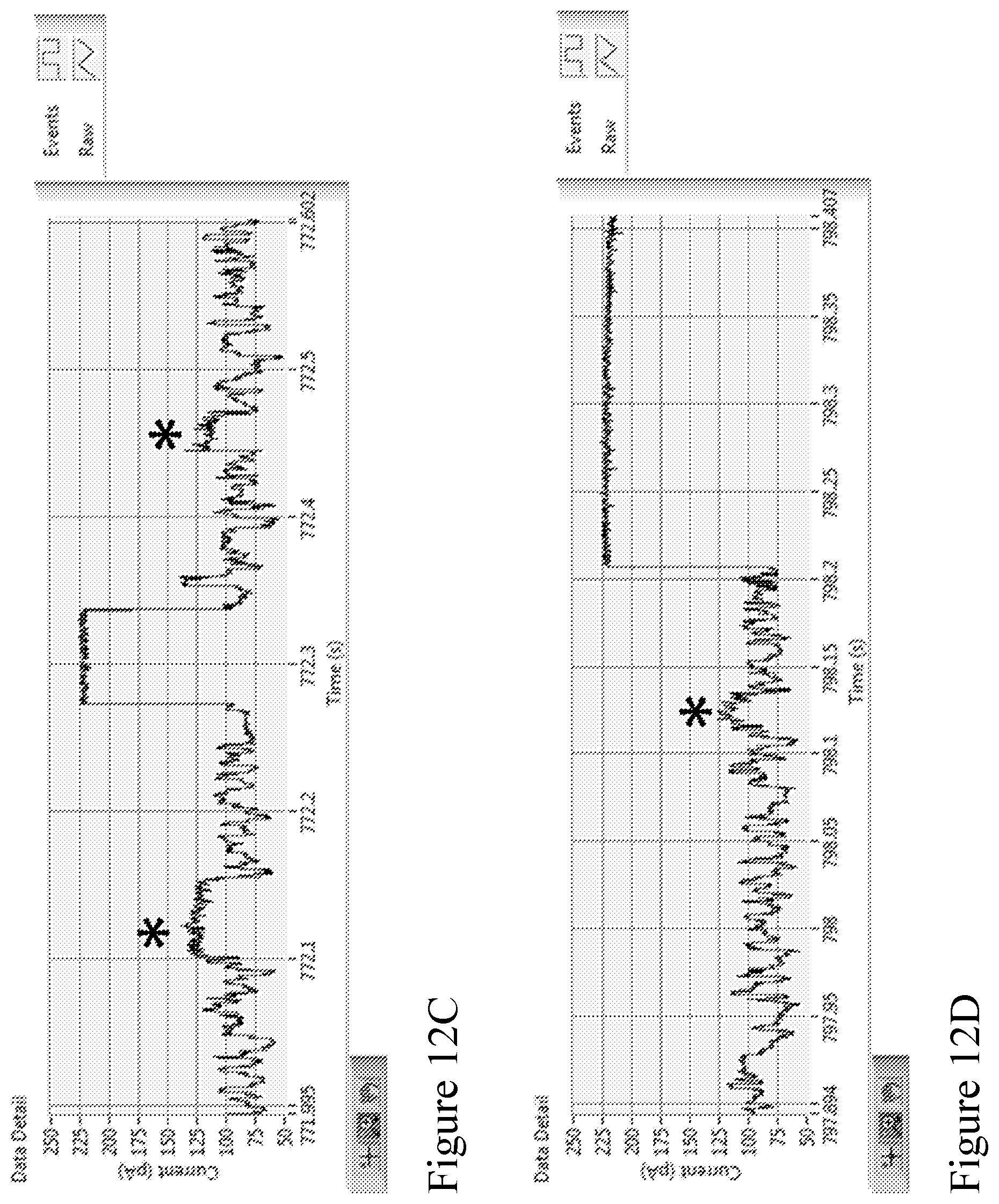
D00014
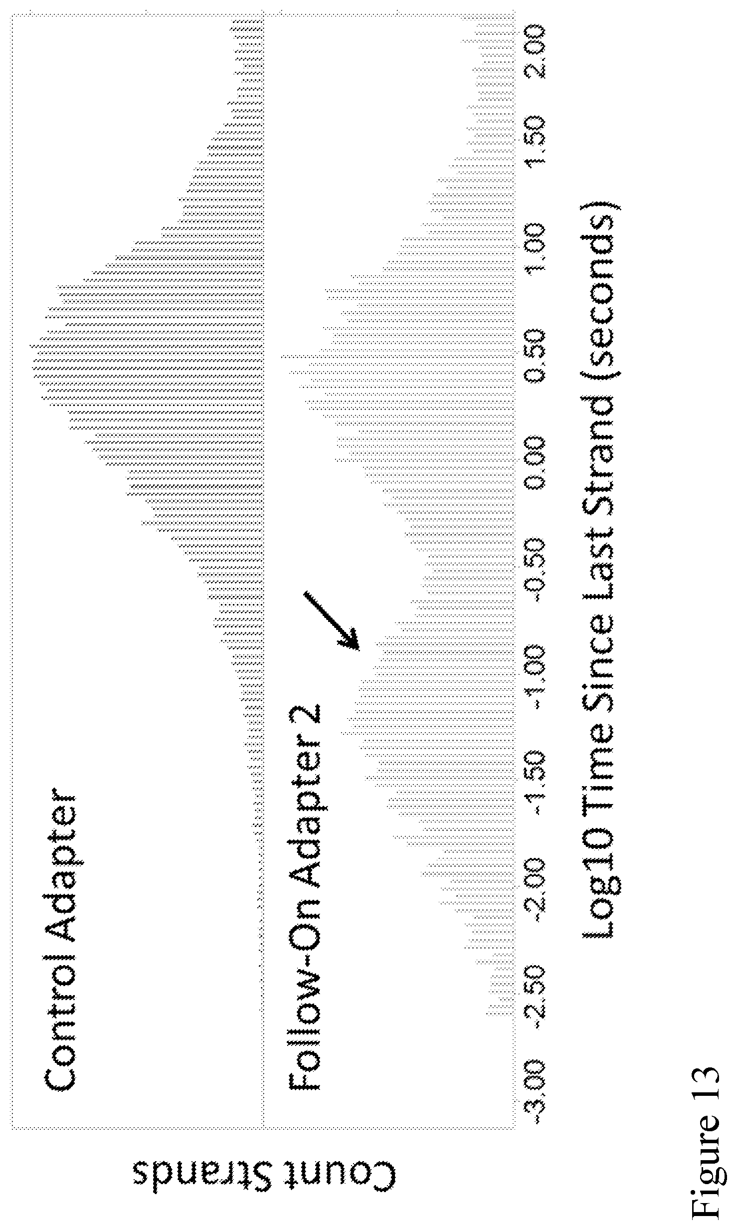
D00015
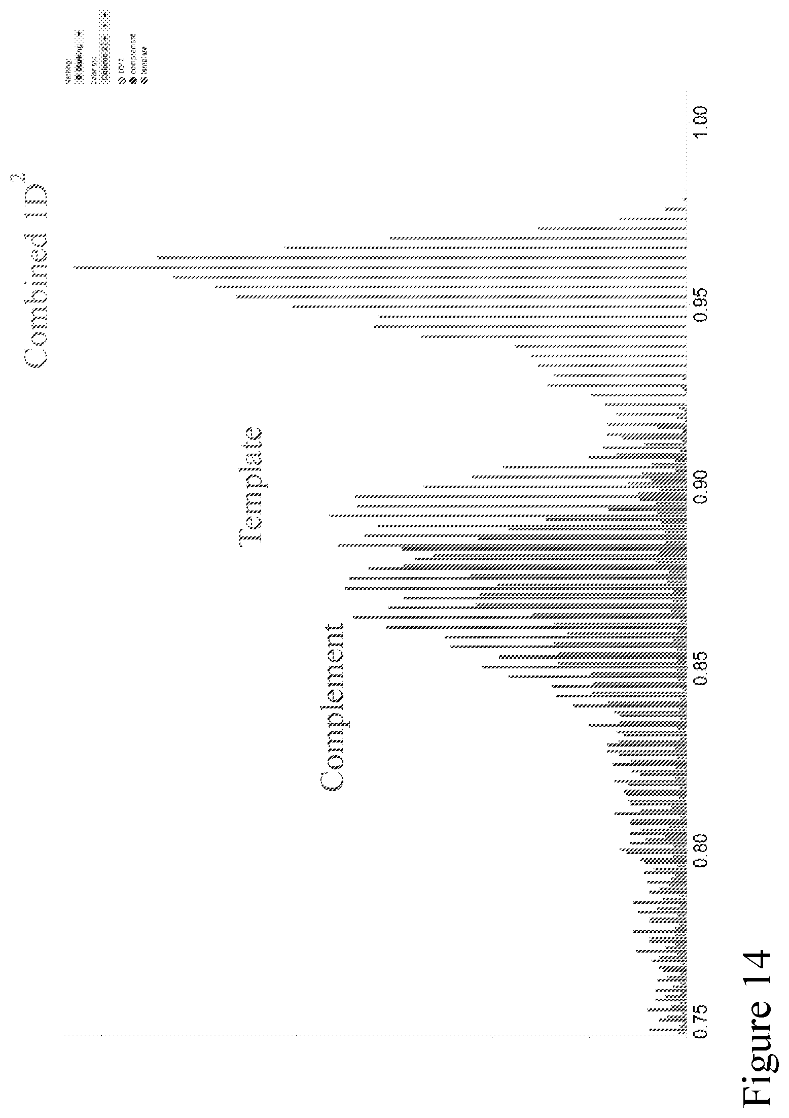
D00016

D00017
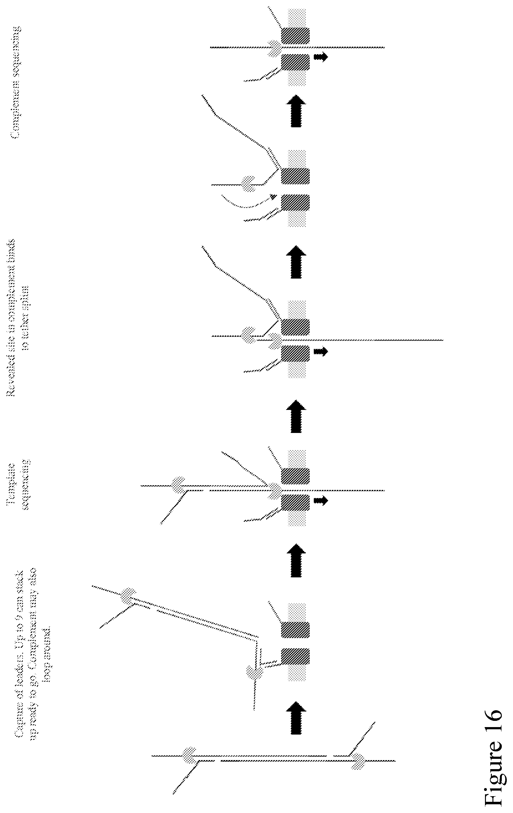
D00018
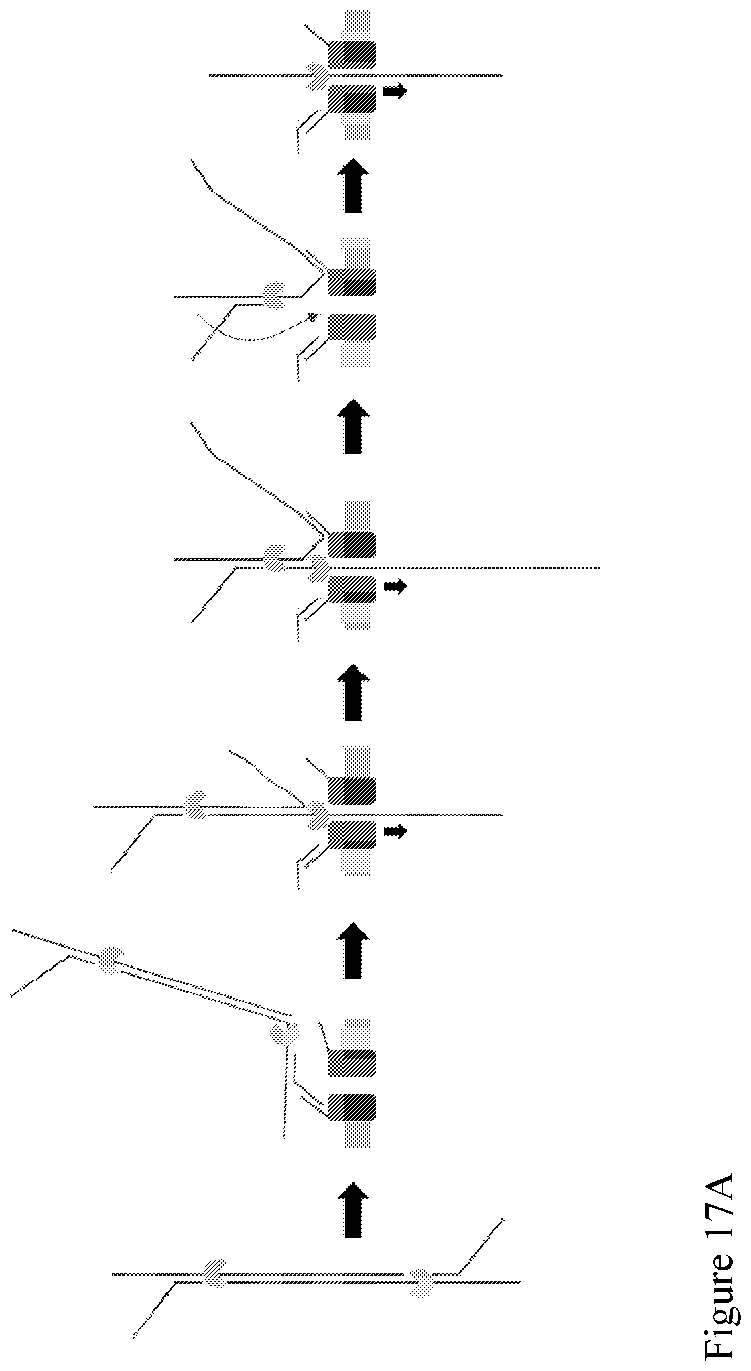
D00019

D00020
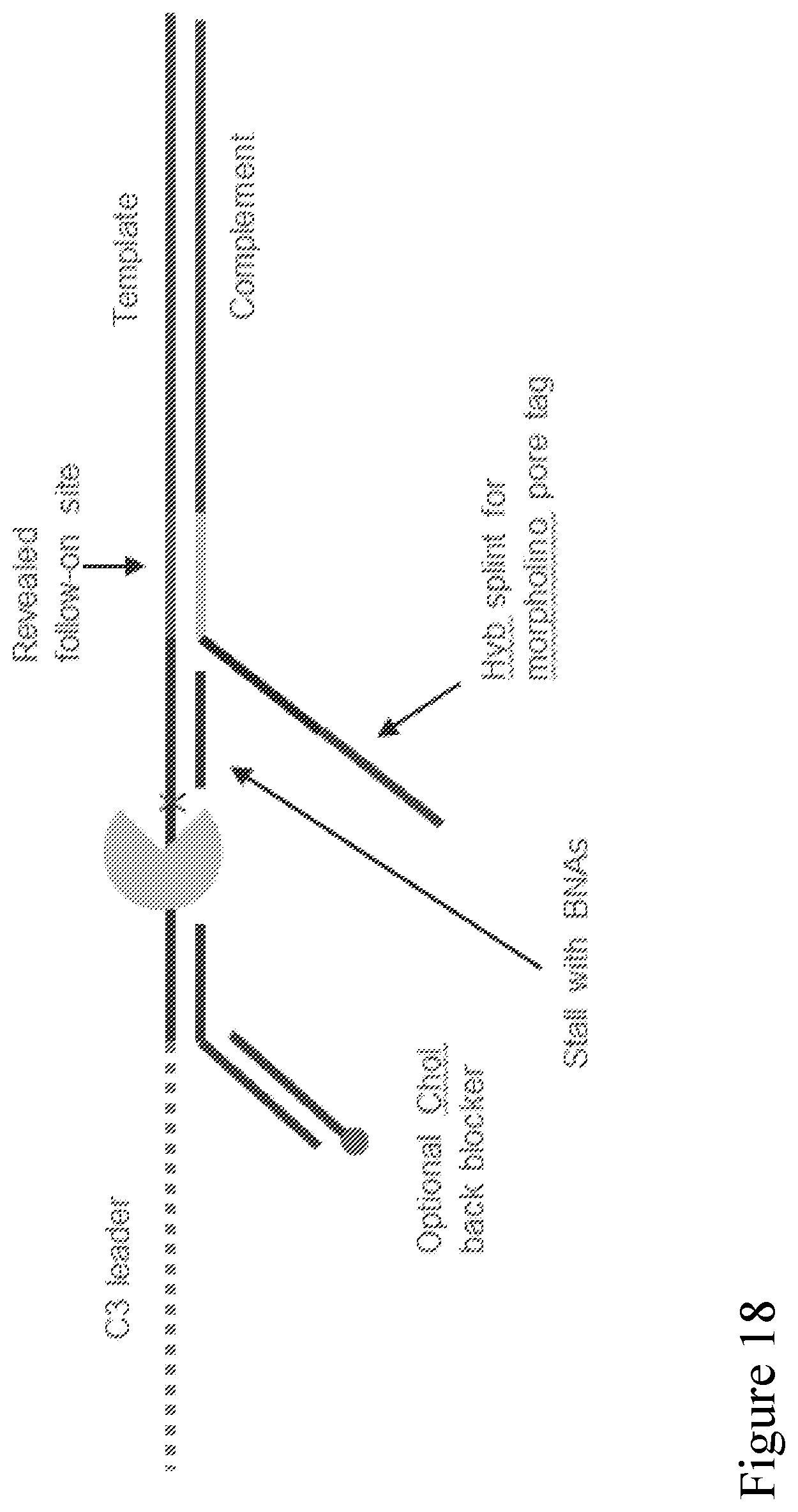
D00021
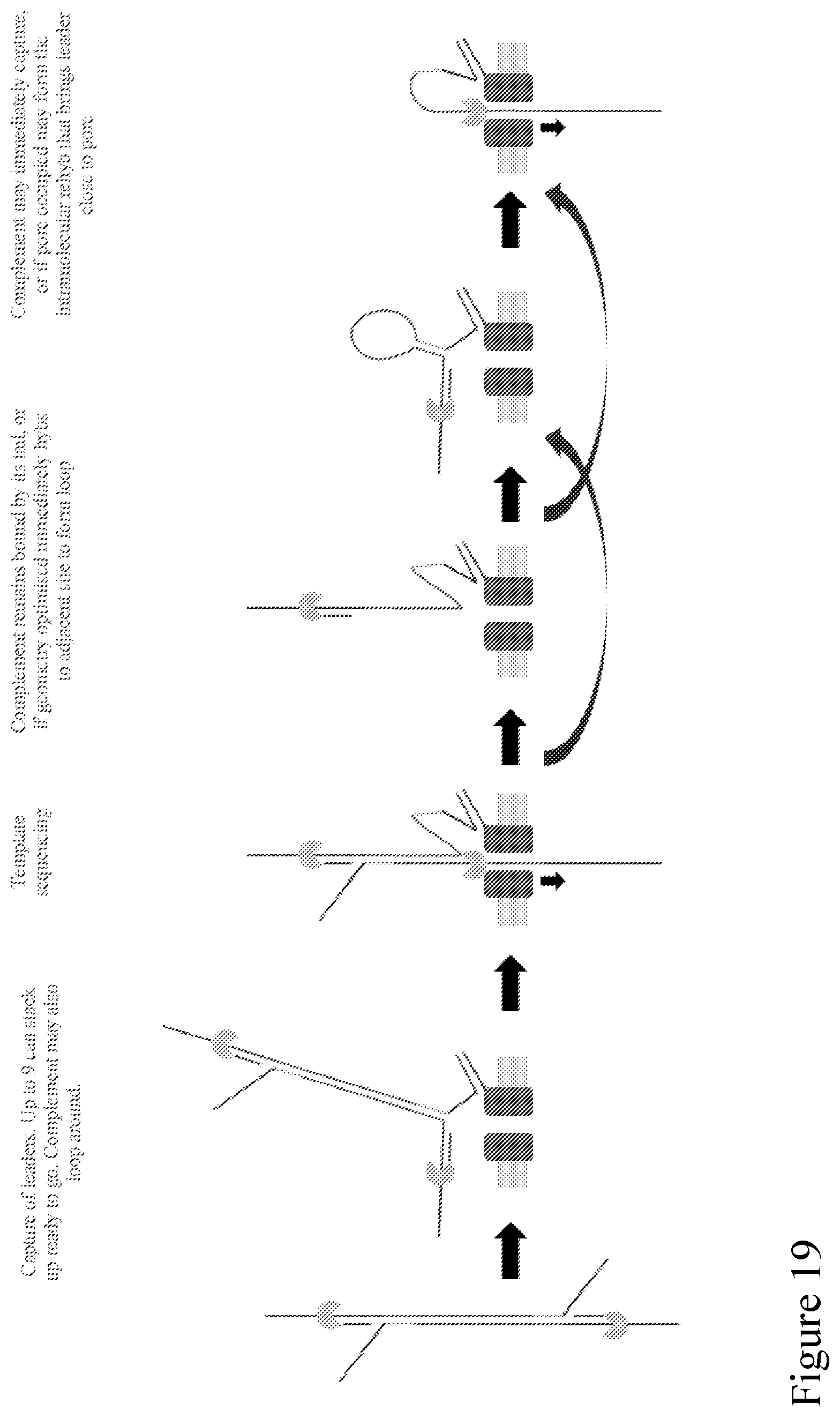
D00022
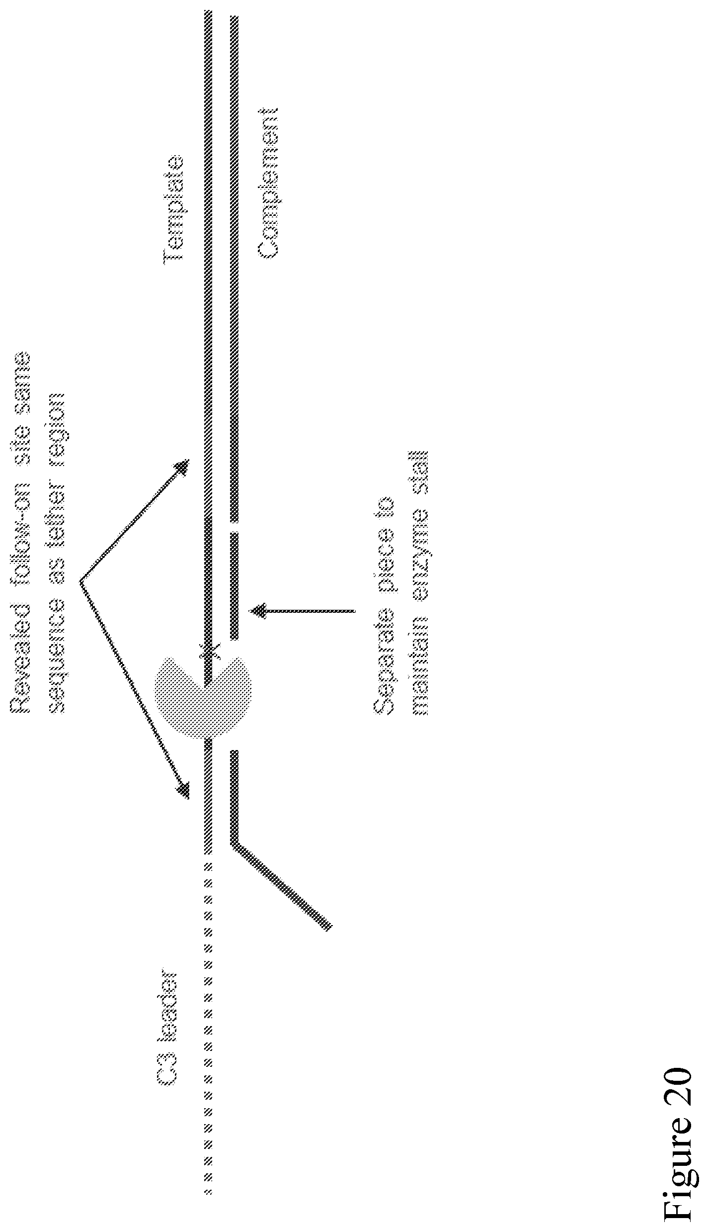
D00023
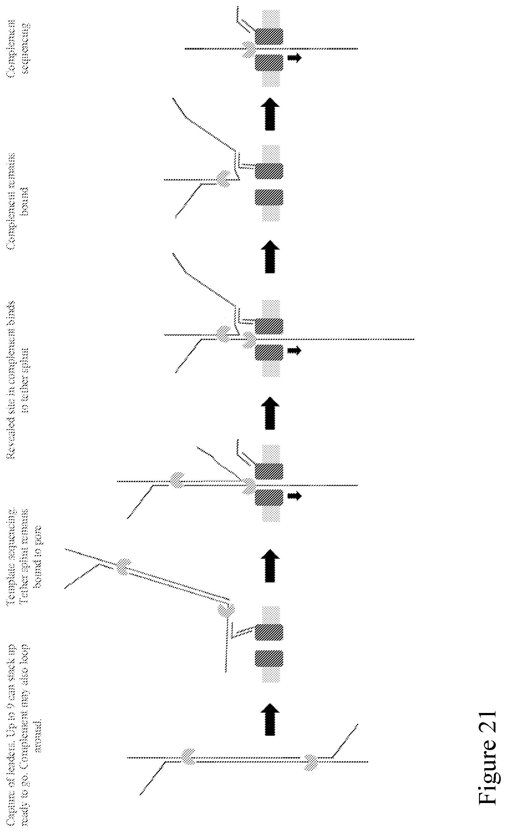
D00024

D00025
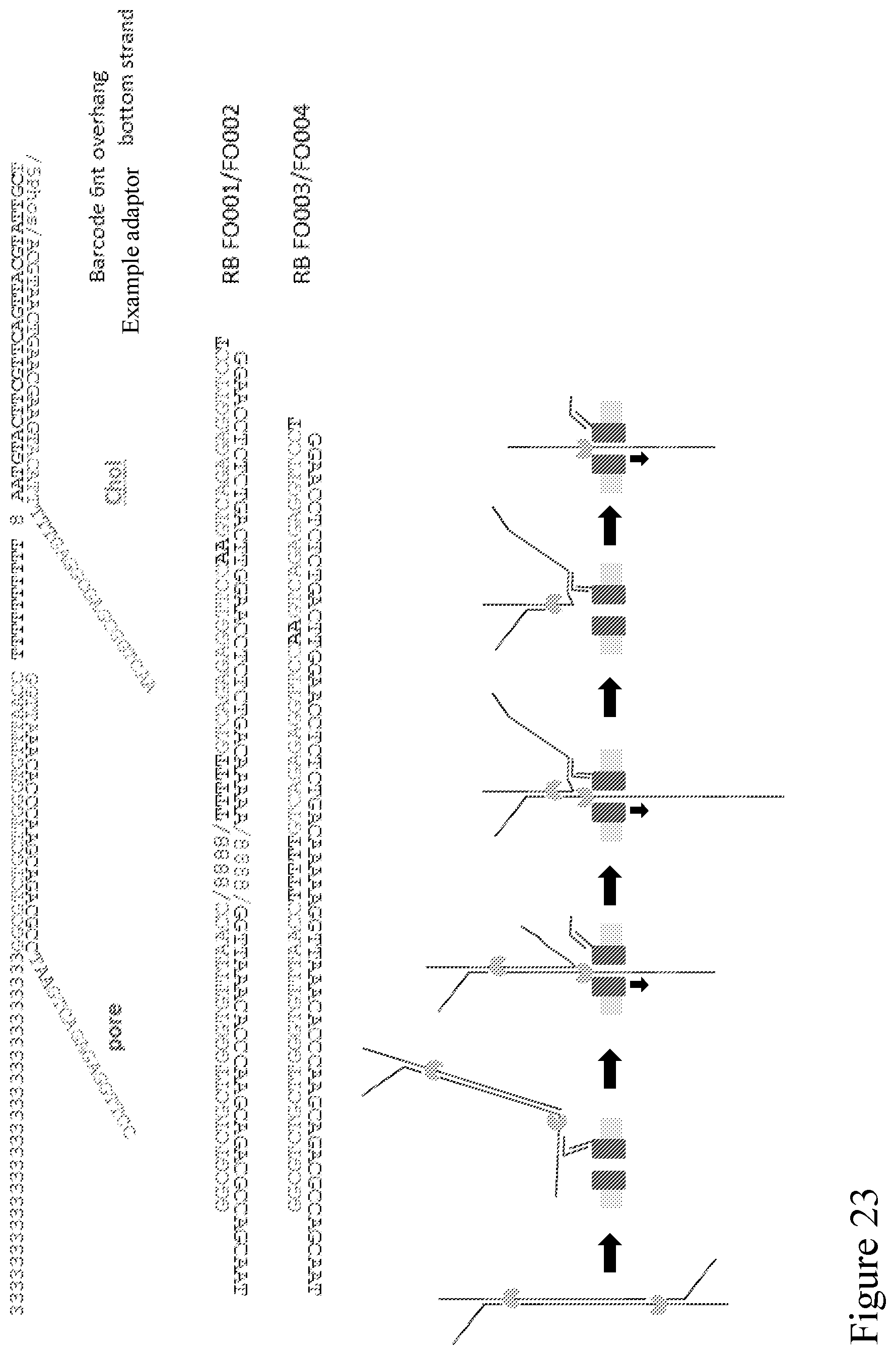
D00026
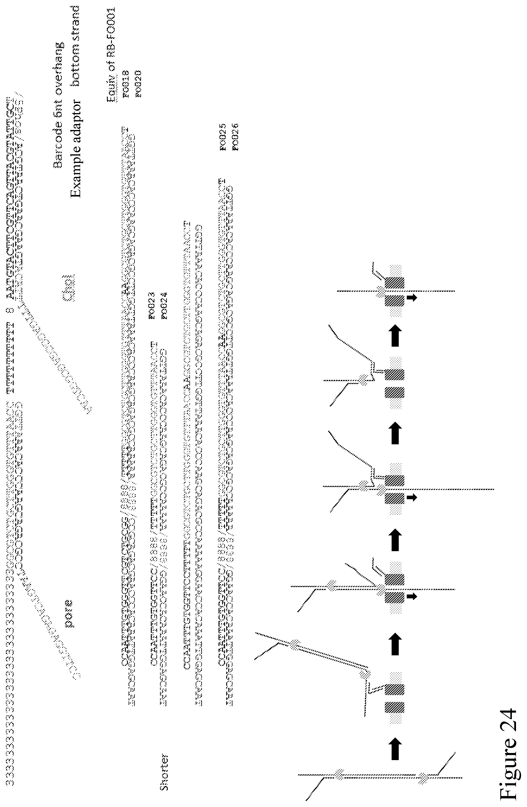
D00027
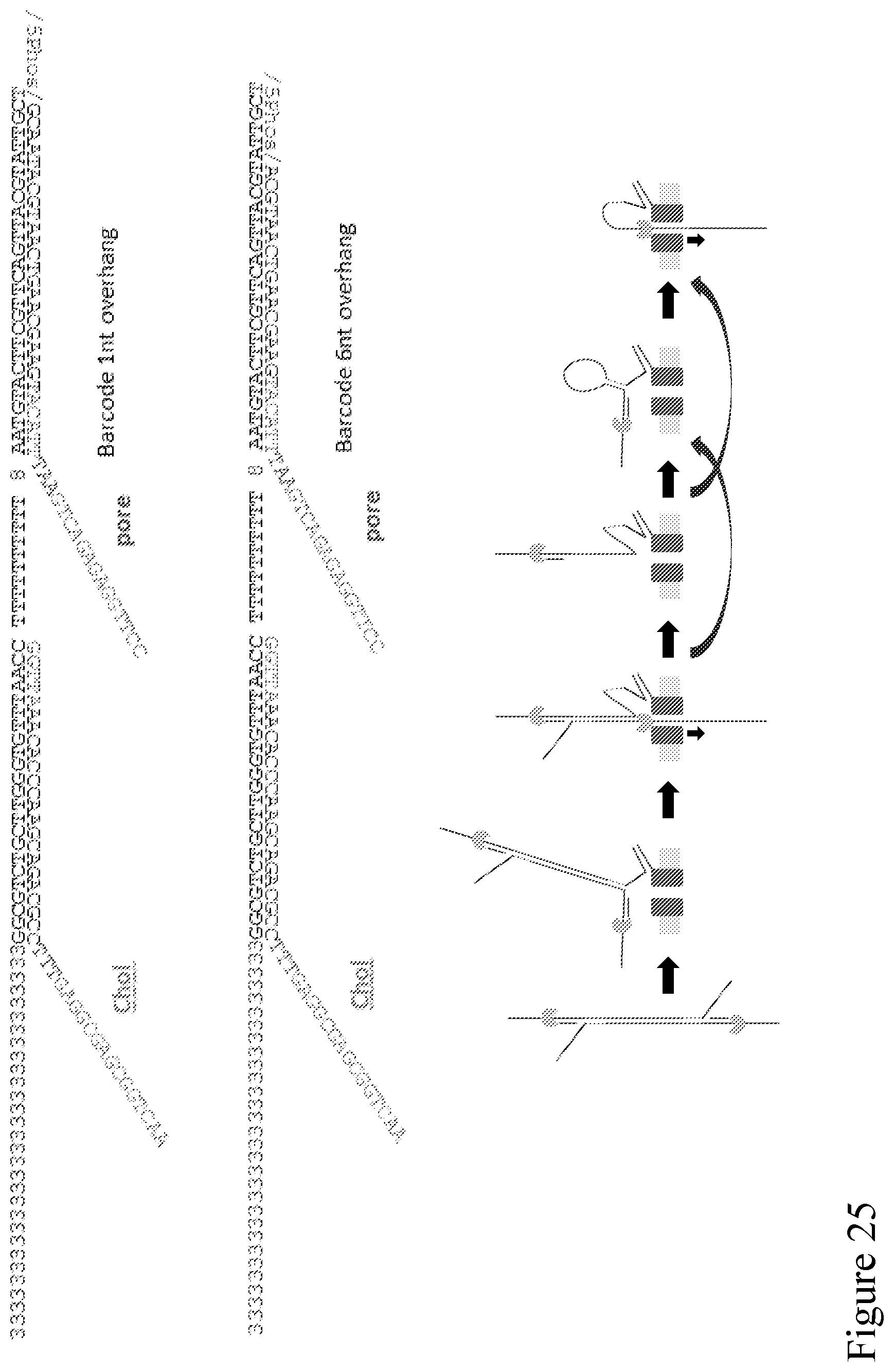
D00028
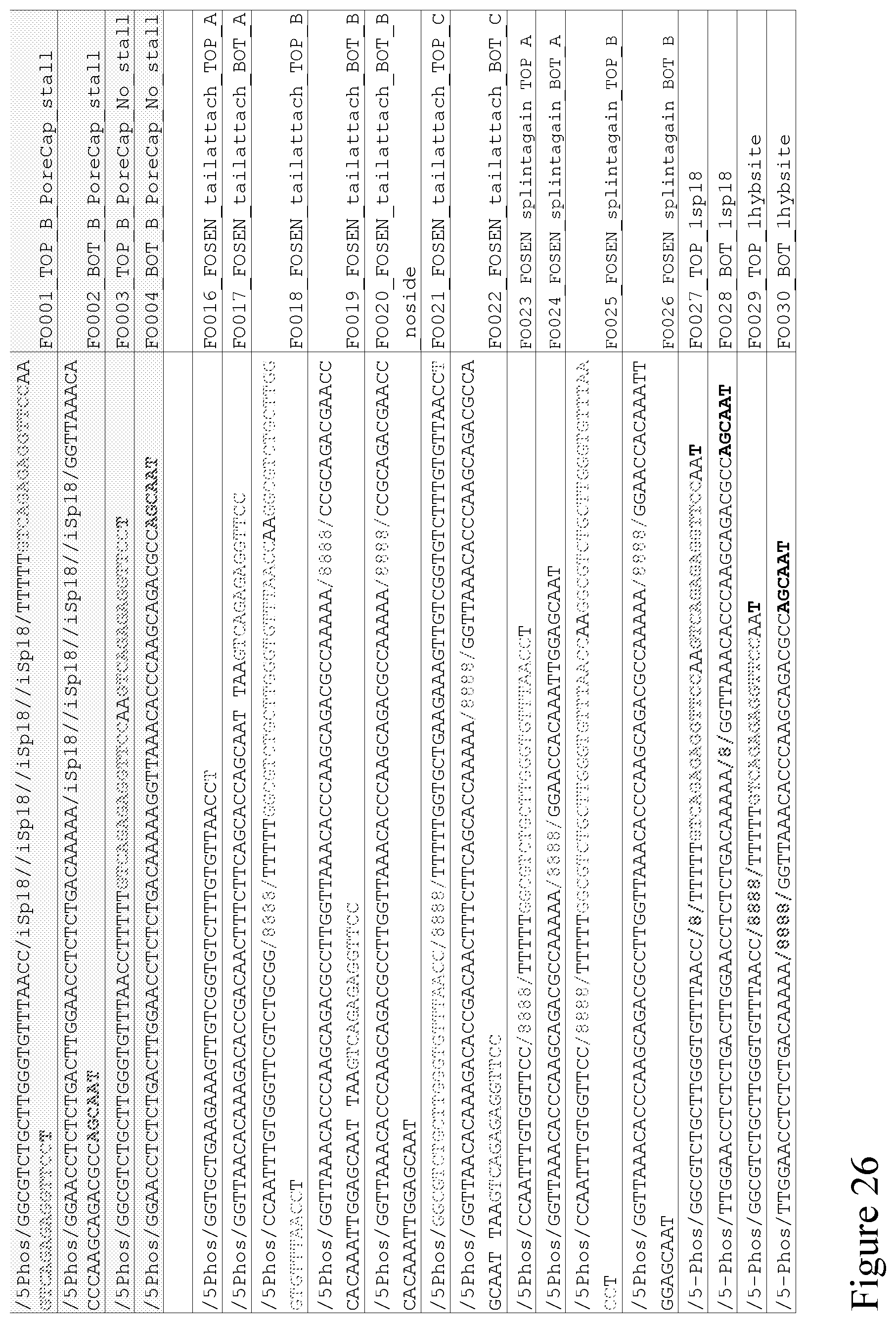
D00029

D00030
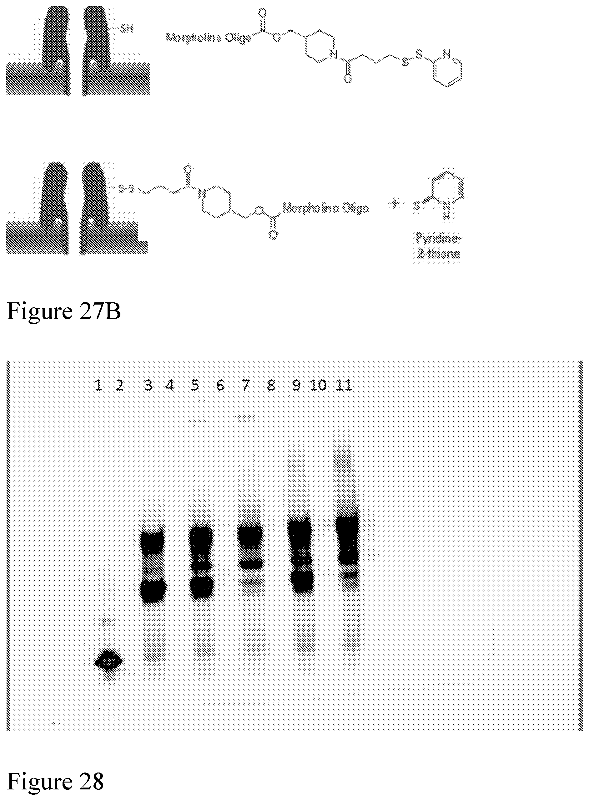
D00031
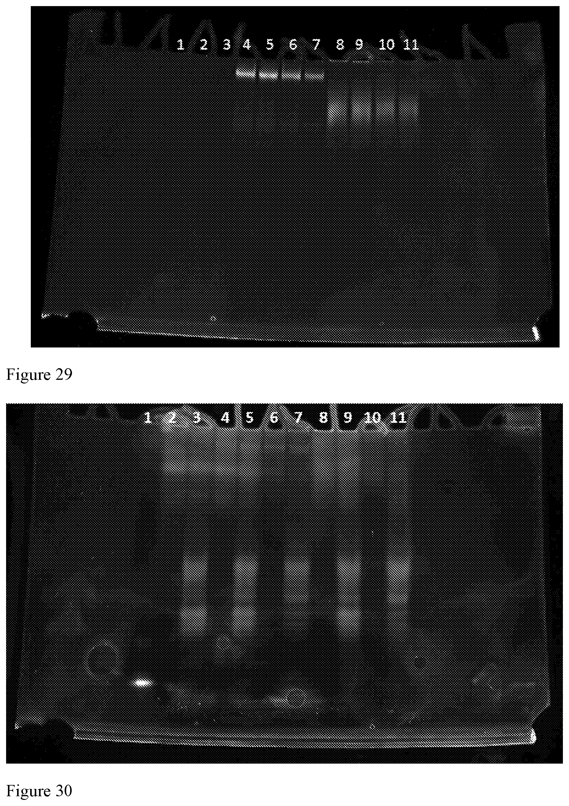
D00032
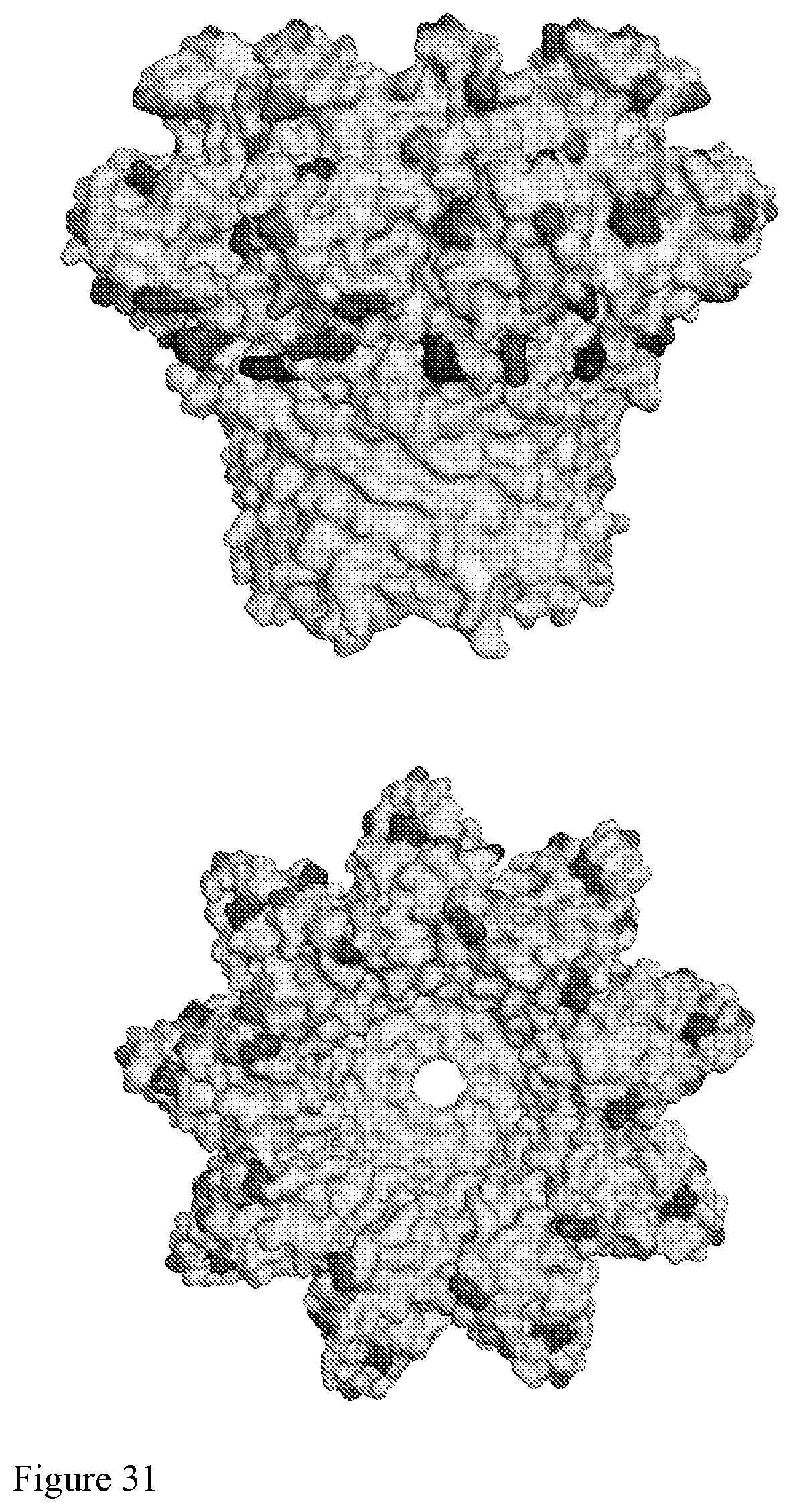
D00033
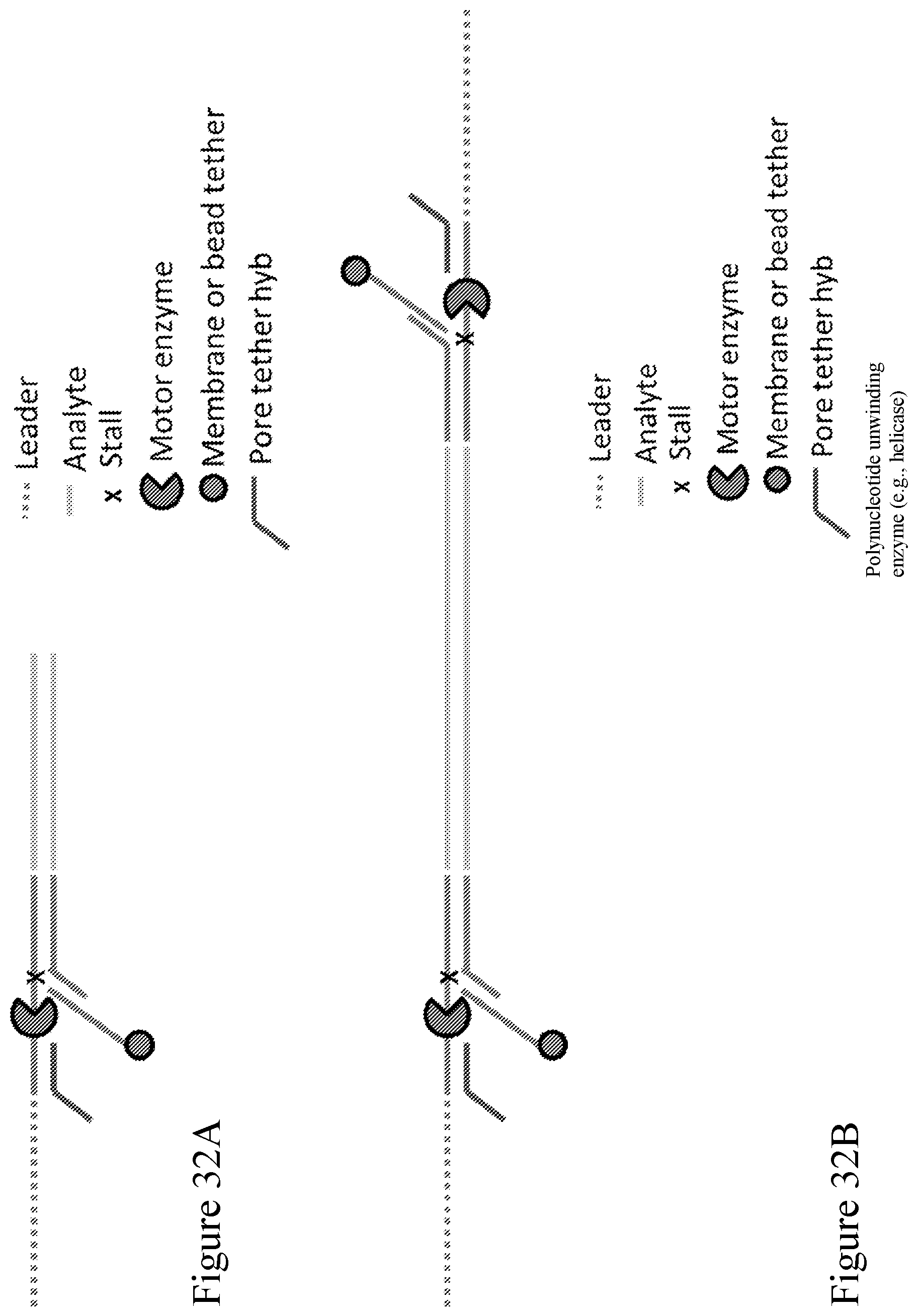
D00034
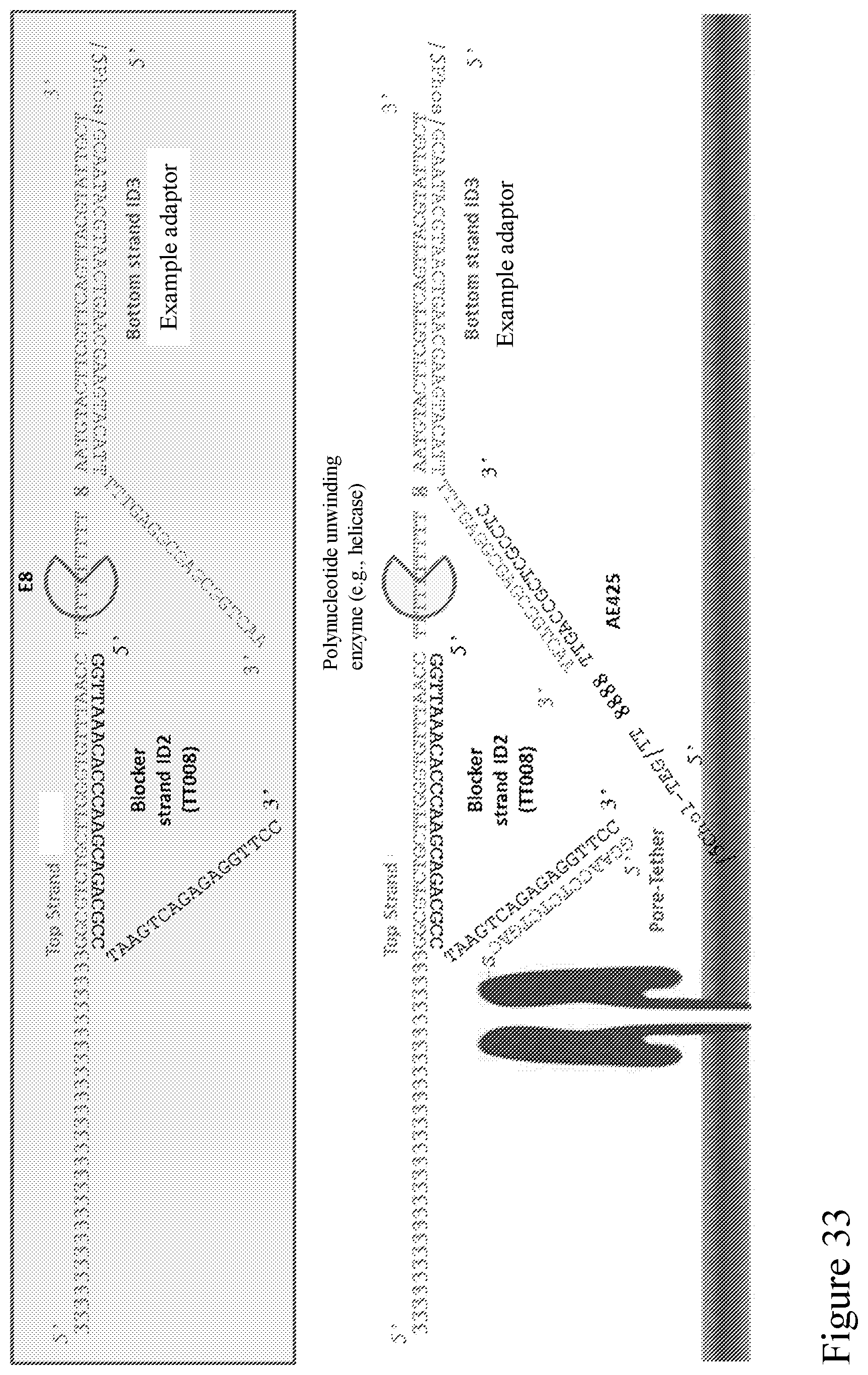
D00035
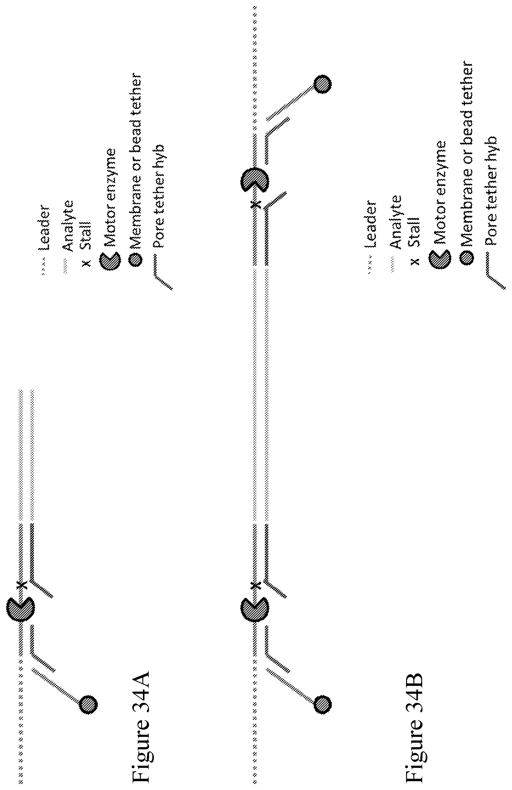
D00036
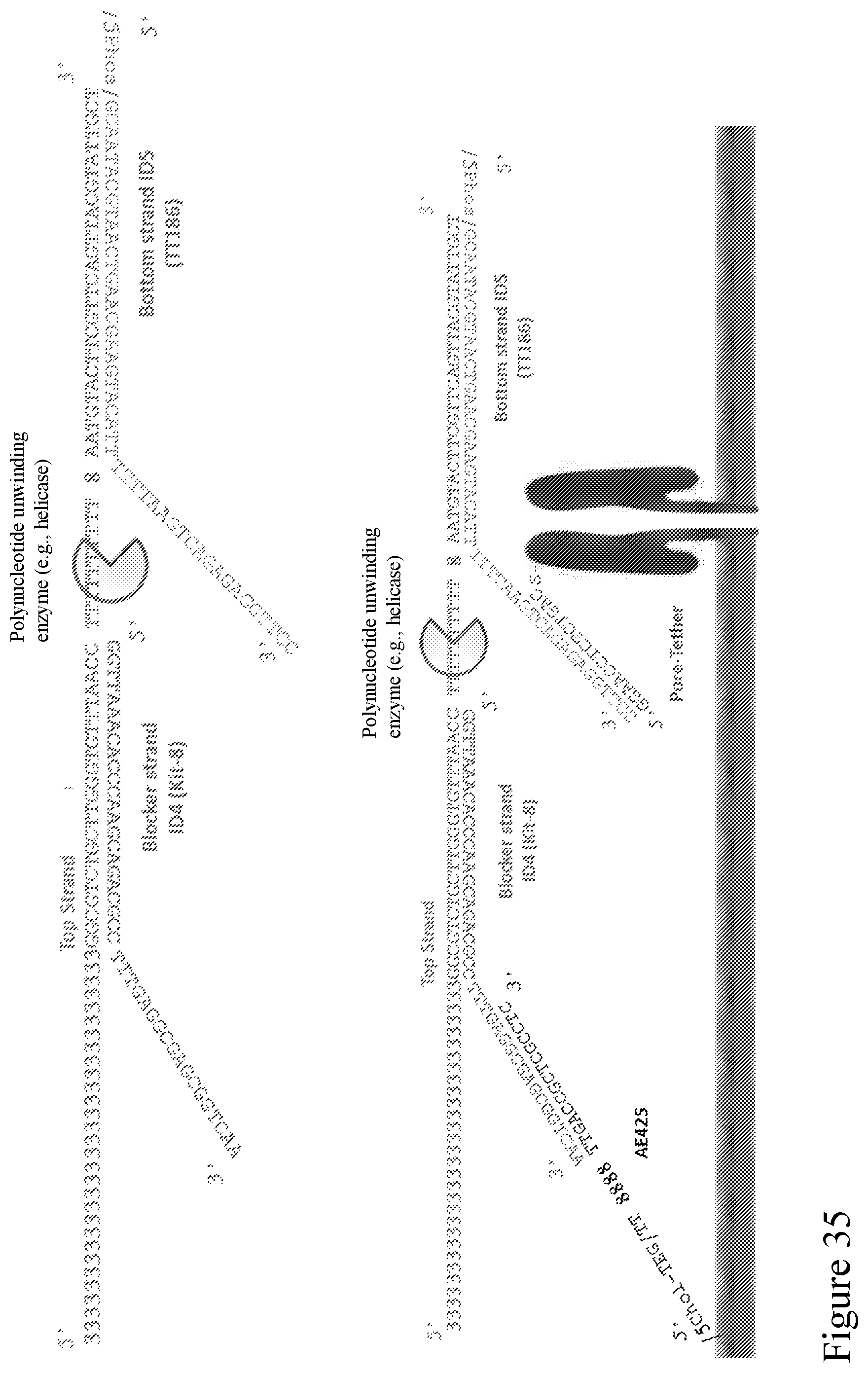
D00037
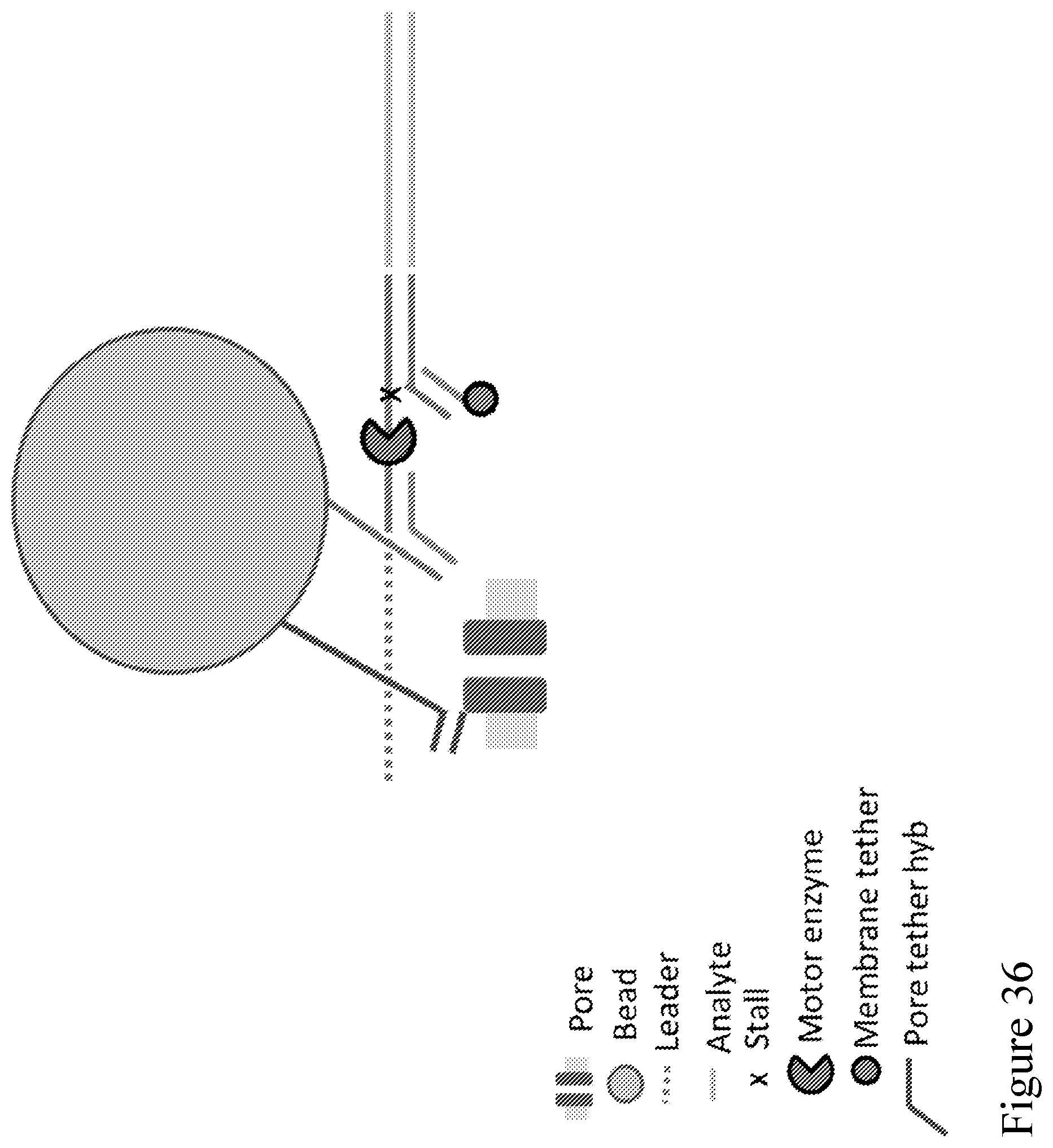
D00038
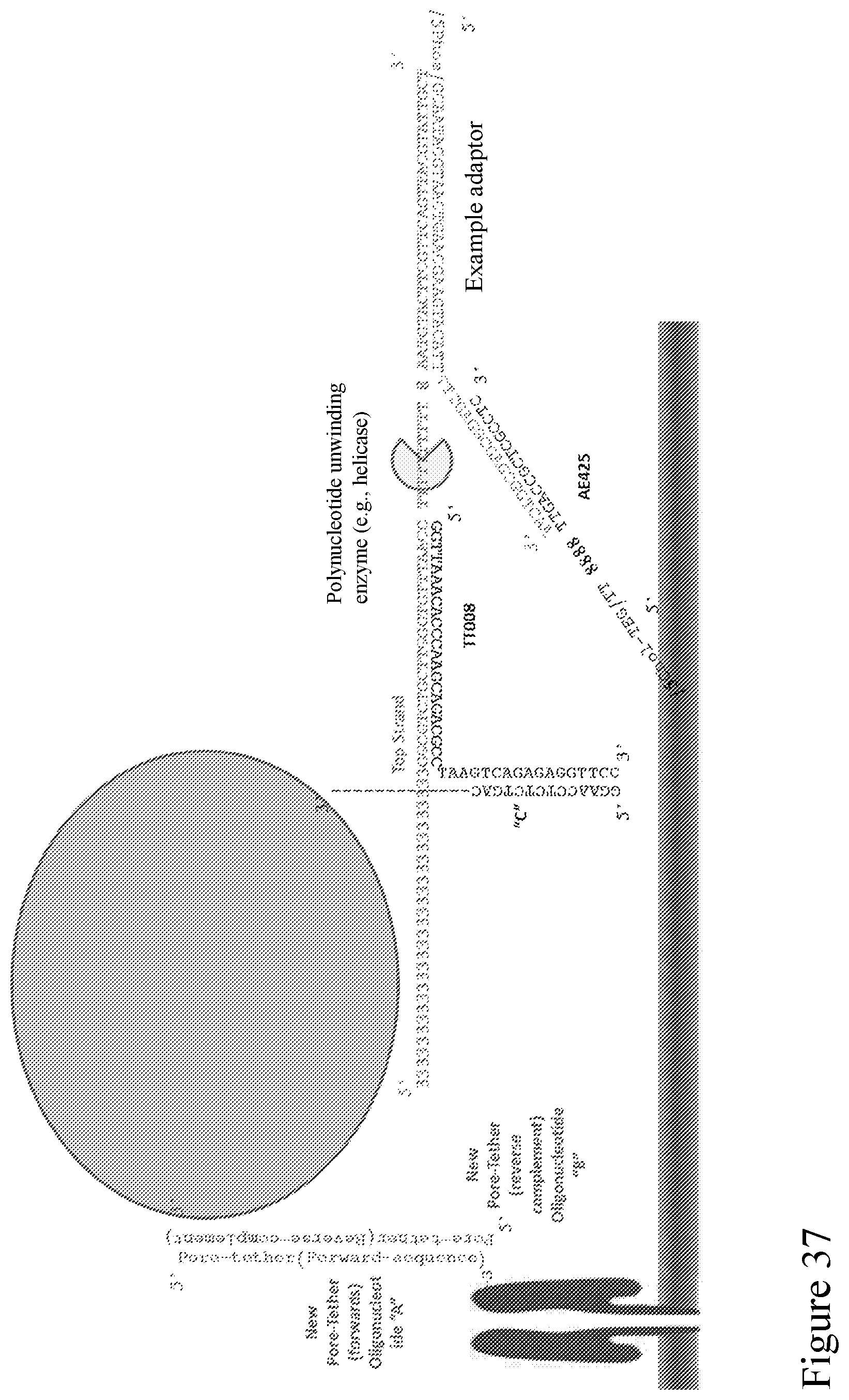
D00039
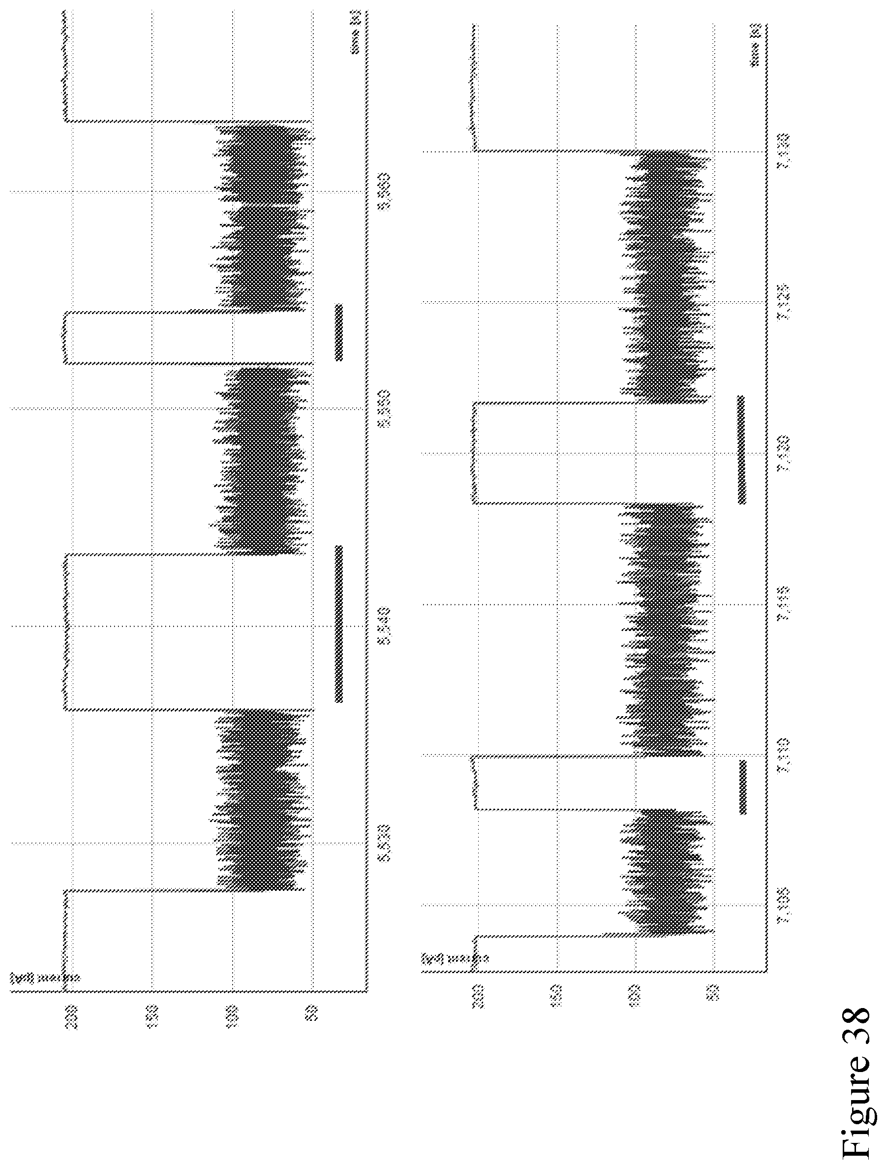
D00040
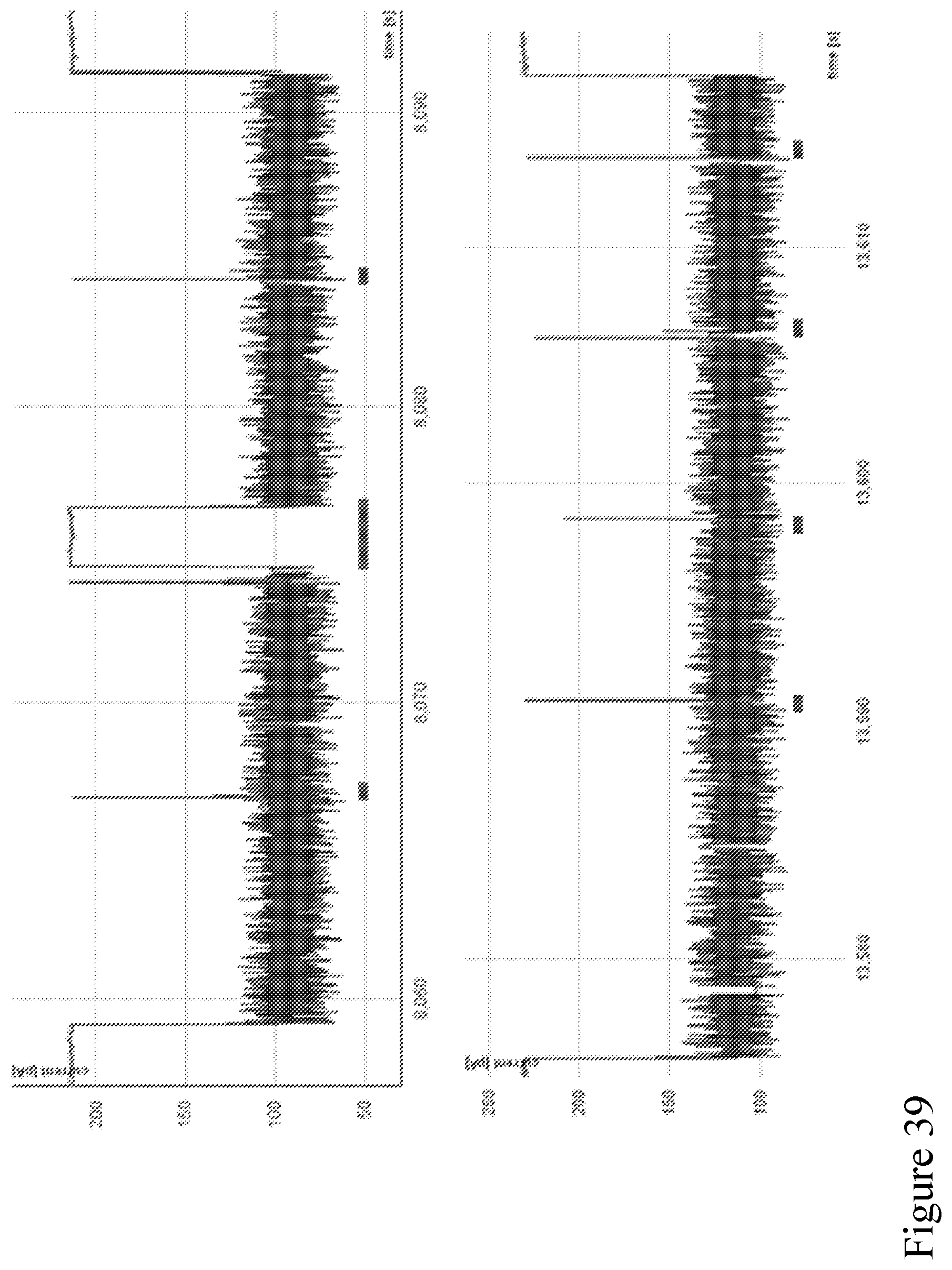
D00041
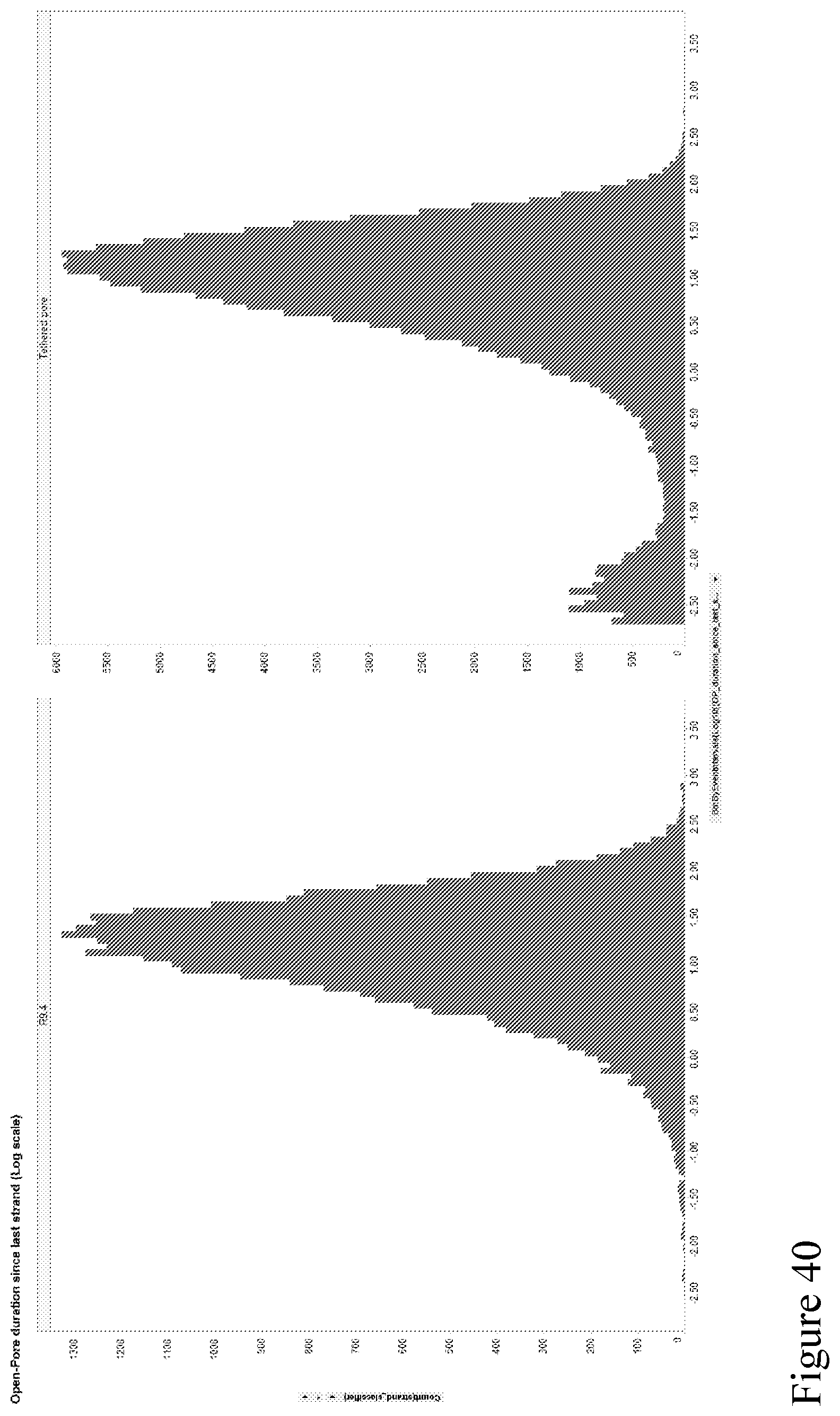
D00042
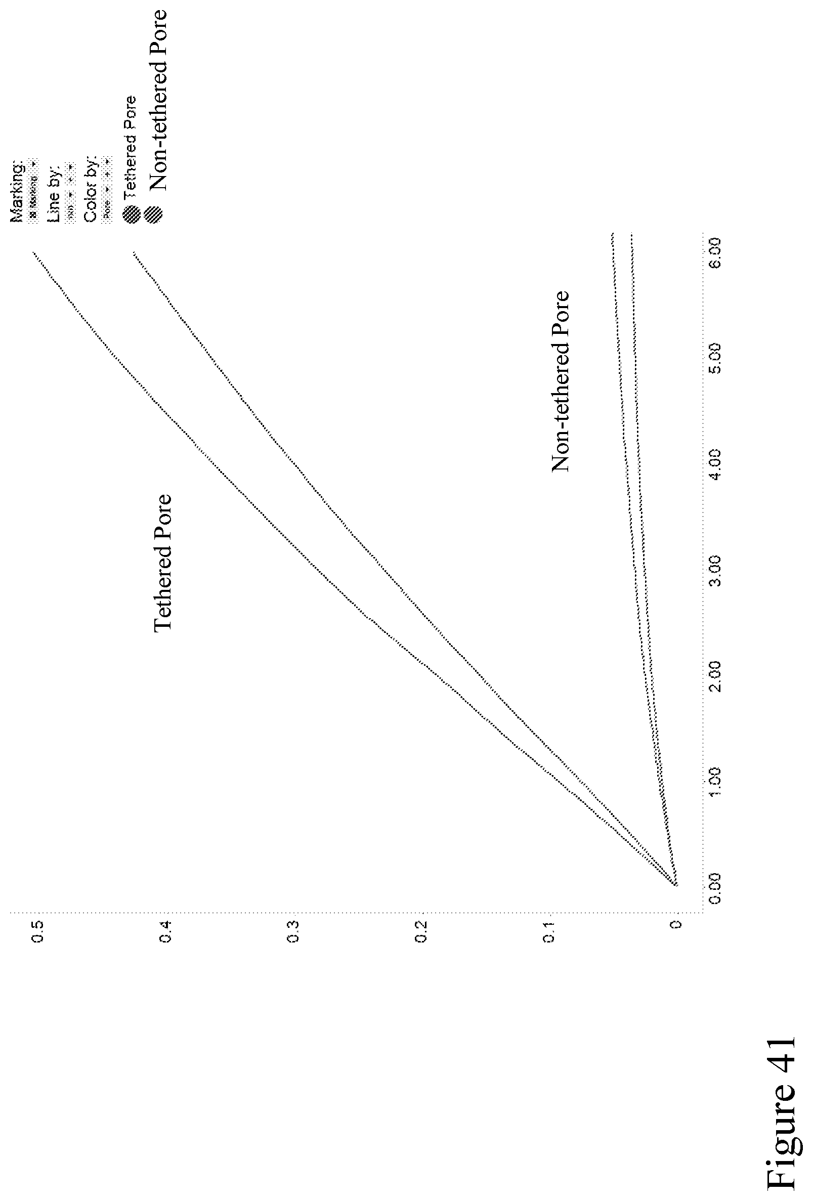
D00043
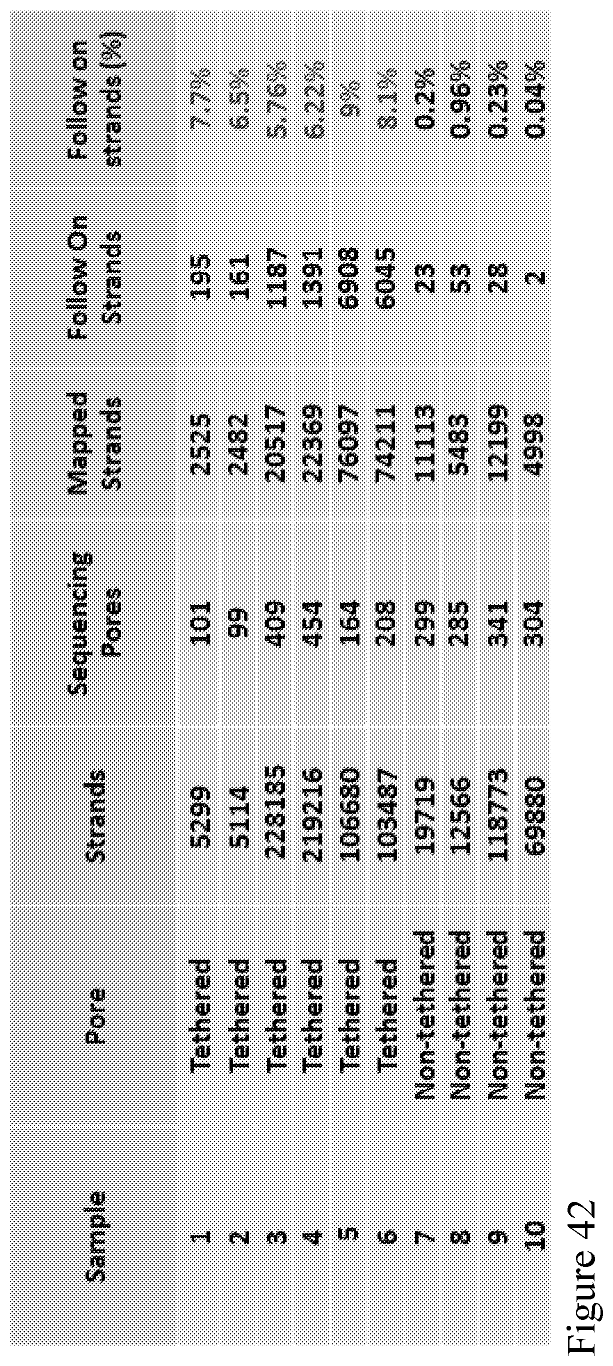
D00044
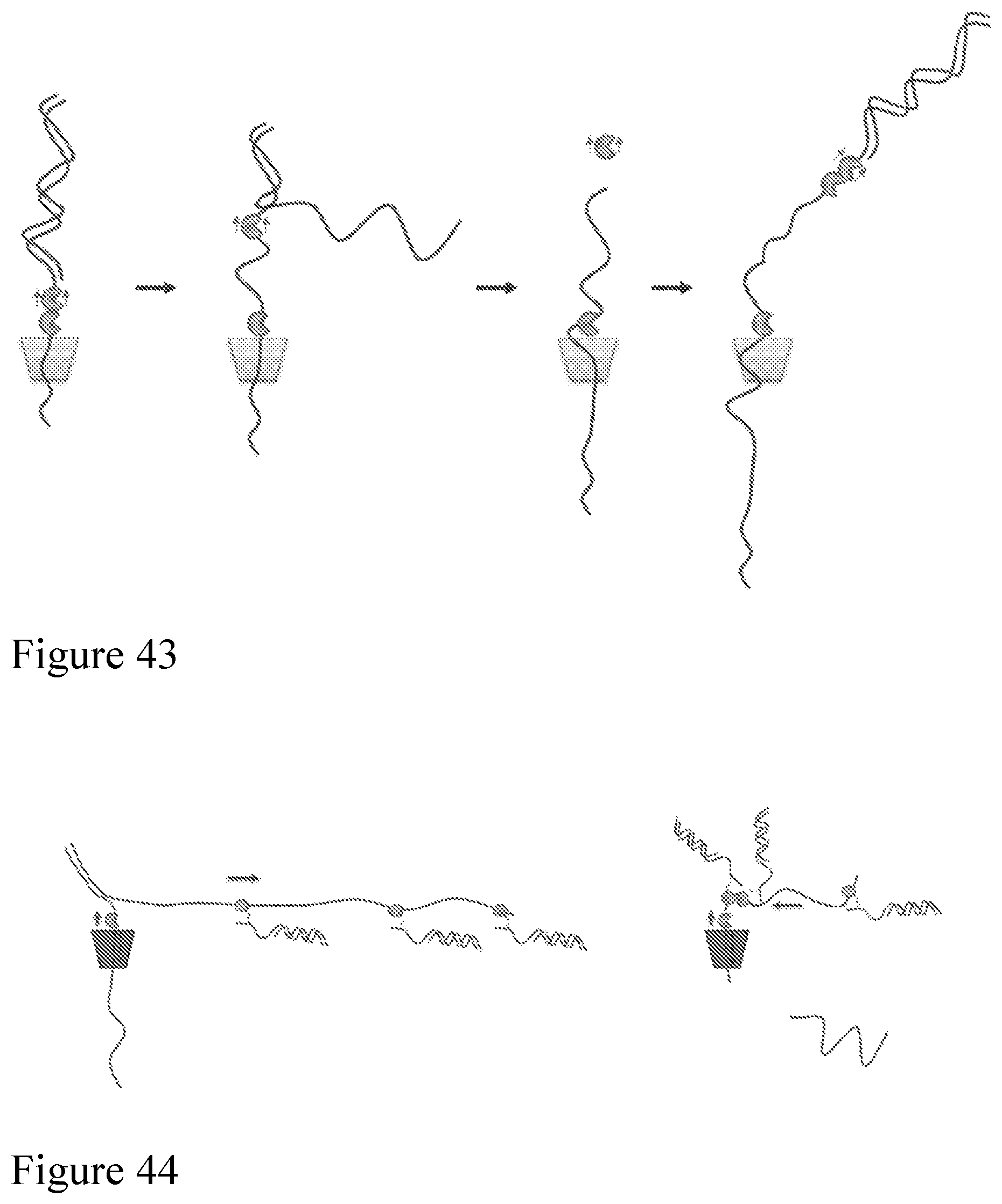
S00001
XML
uspto.report is an independent third-party trademark research tool that is not affiliated, endorsed, or sponsored by the United States Patent and Trademark Office (USPTO) or any other governmental organization. The information provided by uspto.report is based on publicly available data at the time of writing and is intended for informational purposes only.
While we strive to provide accurate and up-to-date information, we do not guarantee the accuracy, completeness, reliability, or suitability of the information displayed on this site. The use of this site is at your own risk. Any reliance you place on such information is therefore strictly at your own risk.
All official trademark data, including owner information, should be verified by visiting the official USPTO website at www.uspto.gov. This site is not intended to replace professional legal advice and should not be used as a substitute for consulting with a legal professional who is knowledgeable about trademark law.