Methods For Expanding And Differentiating B Cells For Producing Antibody
SUGA; Hiraku ; et al.
U.S. patent application number 15/998919 was filed with the patent office on 2019-11-21 for methods for expanding and differentiating b cells for producing antibody. This patent application is currently assigned to Duke University. The applicant listed for this patent is Duke University. Invention is credited to Kathleen M. CANDANDO, Masahiro KAMATA, Evgueni KOUNTIKOV, Tomomitsu MIYAGAKI, Hiraku SUGA, Thomas F. TEDDER, Ayumi YOSHIZAKI.
| Application Number | 20190352607 15/998919 |
| Document ID | / |
| Family ID | 59626268 |
| Filed Date | 2019-11-21 |


| United States Patent Application | 20190352607 |
| Kind Code | A1 |
| SUGA; Hiraku ; et al. | November 21, 2019 |
METHODS FOR EXPANDING AND DIFFERENTIATING B CELLS FOR PRODUCING ANTIBODY
Abstract
Provided are feeder cell lines that can be used to expand and differentiate B cells in vitro, a method for expanding B cells in vitro comprising culturing the B cells with the feeder cell line, and a method for producing monoclonal antibody in vitro comprising culturing a single B cell with the feeder cell line under sufficient conditions and for sufficient time to induce expansion and differentiation of the B cell into a B cell done secreting antibody.
| Inventors: | SUGA; Hiraku; (Durham, NC) ; CANDANDO; Kathleen M.; (Durham, NC) ; KOUNTIKOV; Evgueni; (Durham, NC) ; KAMATA; Masahiro; (Durham, NC) ; TEDDER; Thomas F.; (Durham, NC) ; YOSHIZAKI; Ayumi; (Durham, NC) ; MIYAGAKI; Tomomitsu; (Durham, NC) | ||||||||||
| Applicant: |
|
||||||||||
|---|---|---|---|---|---|---|---|---|---|---|---|
| Assignee: | Duke University Durham NC |
||||||||||
| Family ID: | 59626268 | ||||||||||
| Appl. No.: | 15/998919 | ||||||||||
| Filed: | February 16, 2017 | ||||||||||
| PCT Filed: | February 16, 2017 | ||||||||||
| PCT NO: | PCT/US2017/018155 | ||||||||||
| 371 Date: | August 16, 2018 |
Related U.S. Patent Documents
| Application Number | Filing Date | Patent Number | ||
|---|---|---|---|---|
| 62295728 | Feb 16, 2016 | |||
| Current U.S. Class: | 1/1 |
| Current CPC Class: | C12N 2501/231 20130101; C07K 16/00 20130101; C12N 2501/2321 20130101; C12N 2501/2304 20130101; C12N 2501/2302 20130101; C12N 2502/1323 20130101; C12N 5/0635 20130101; C12N 2502/1352 20130101; C07K 2317/21 20130101 |
| International Class: | C12N 5/0781 20060101 C12N005/0781; C07K 16/00 20060101 C07K016/00 |
Claims
1. A method for expanding B cells in vitro, the method comprising: (a) isolating at least one B cell, and (b) culturing the at least one B cell from step (a) with a feeder cell line comprising a stromal cell line modified to express a CD154 polypeptide (or CD40 agonist) and a BLyS polypeptide (or B cell survival factor), in combination with IL-21 and without additional exogenous IL-4, wherein the B cells are cultured with the feeder cell line under sufficient conditions and for a sufficient time to cause the human B cells to expand in number.
2. The method of claim 1, wherein the B cells are human B cells.
3. The method of claim 1, wherein the IL-21 is added exogenously to the culture.
4. The method of claim 1, wherein the feeder cells are modified to express an IL-21 polypeptide.
5. The method of claim 1, wherein the B cells are expanded at least an average of 10.sup.4-fold in number.
6. The method of claim 1, wherein the culturing of the at least one B cell with the feeder cell line in step (b) is performed in less than 2 weeks.
7. (canceled)
8. (canceled)
9. (canceled)
10. (canceled)
11. (canceled)
12. (canceled)
13. (canceled)
14. The method of claim 1, wherein the feeder cells comprise a stromal cell line modified to express a CD154 polypeptide, a BLyS polypeptide, and an IL-21 polypeptide.
15. A method for producing a monoclonal antibody comprising: (a) isolating B cells; (b) separating the B cells from step (a) into single B cells; (c) culturing the single B cells with a feeder cell line comprising a stromal cell line modified to express a CD154 polypeptide (or CD40 agonist) and a BLyS polypeptide (or B cell survival factor), in combination with IL-21 and without additional exogenous IL-4 to produce a plurality of B cell clones, wherein the single B cells are cultured with the feeder cell line under sufficient conditions and for a sufficient time to cause the single B cells to expand in number and to differentiate into a B cell clone producing a monoclonal antibody.
16. The method of claim 15, further comprising (d) assessing the antigen specificity of at least one of the monoclonal antibodies produced by the plurality of B cell clones.
17. The method of claim 15, further comprising (e) purifying at least one of the monoclonal antibodies produced by the plurality of B cell clones.
18. The method of claim 15, wherein the B cells have been exposed to an antigen prior to isolation in step (a).
19. The method of claim 15, wherein the B cells are human B cells.
20. (canceled)
21. (canceled)
22. (canceled)
23. (canceled)
24. (canceled)
25. A kit for expanding and differentiating mammalian B cells in vitro comprising feeder cells, at least one antibody for isolating B cells from a biological sample and reagents for collecting the B cells from a peripheral blood sample, wherein the feeder cells are the feeder cells of claim 28, wherein the antibody is a CD19 antibody.
26. The kit of claim 25, further comprising IL-21.
27. The kit of claim 25, further comprising an antibody specific for IgM, IgG, IgA, or IgE.
28. A feeder cell line comprising a stromal cell line modified to express a CD154 polypeptide, a BLyS polypeptide, and an IL-21 polypeptide.
29. The feeder cell line of claim 28, wherein the stromal cell line that is modified comprises a mesenchymal stromal cell line.
30. The feeder cell line of claim 28, wherein the stromal cell line that is modified comprises a thymic epithelial cell line.
31. (canceled)
32. The feeder cell line of claim 28, wherein the stromal cell line comprises an MS-5 cell line.
33. (canceled)
34. (canceled)
35. The feeder cell line of claim 28, wherein the IL-21 polypeptide comprises SEQ ID NO: 70 or a functional fragment or variant thereof.
Description
CROSS-REFERENCE TO RELATED PATENT APPLICATIONS
[0001] The present application claims the benefit of priority to U.S. Provisional Patent Application No. 62/295,728, filed on Feb. 16, 2016, the content of which is incorporated herein by reference in its entirety.
SEQUENCE LISTING
[0002] A Sequence Listing accompanies this application and is incorporated herein by reference in its entirety. The Sequence Listing was filed with the application as a text file.
TECHNICAL FIELD
[0003] The invention relates to a method for producing and supporting B cells, including human B cells, which can be used to produce antigen-specific antibodies, and more particularly, monoclonal antibodies. More specifically, the invention relates to culturing B cells to produce monoclonal antibodies without using hybridoma technology or EBV-transformed B cells.
BACKGROUND ART
[0004] Monoclonal antibodies have found particular utility in medicine, such as through the development of antibody-based biologicals or pharmaceuticals. Current methods for monoclonal antibody production involve the mouse hybridoma method; i.e., fusing antibody producing cells with myeloma cells. Other methods include EBV-transformed B cell lines and phage display. Each of the different methods used to produce human monoclonal antibodies suffers from technical limitations that render it difficult to use. For example, with hybridoma technology there is a need to "humanize" the antibody so as to reduce the frequency of promoting an immune response to non-human portions of the monoclonal antibody. Additionally, immune responses to a particular antigen or epitope may be species-specific. Other methods have drawbacks that limit their wide application for use in developing monoclonal antibodies for use in therapy and diagnosis for humans.
SUMMARY OF THE INVENTION
[0005] The invention is based on the discovery of a highly efficient method for expanding and differentiating B cells in culture that enables single cell cloning of B cells. In one aspect, the discovery relates to a highly efficient method for expanding and differentiating human B cells in culture that enables single cell cloning of human B cells and production of antibodies.
[0006] The invention is also based on the discovery of a highly efficient method for expanding and differentiating B cells in culture that enables single cell cloning of B cells (seeding a single B cell, and expanding that B cell into a done of B cells) in amount and time period sufficient to generate monoclonal antibody in an amount that facilitates characterization of the monoclonal antibody produced. In one aspect of this method, human B cells are expanded and differentiated as single clones to produce human monoclonal antibody. In another aspect of these methods, the method does not require and/or no antigen is added to the expansion culture. In still another aspect, the antigen-specificity of the B cells is unknown. In still another aspect, the B cells are not exposed to antigen in vitro.
[0007] In another aspect of the invention, an in vitro method for the production of monoclonal antibodies, particularly of the IgG type, is provided.
[0008] In another aspect of the invention, provided are feeder cell lines, comprising modified mesenchymal stromal cells or modified thymic epithelial cells, which can efficiently promote and support high numbers of B cells in culture, thereby allowing higher levels of monoclonal antibody production than that observed in prior attempts of culturing B cells with modified mouse 3T3 cells. Related to this aspect, the feeder cell lines comprising mesenchymal stromal cells or thymic epithelial cells, modified to express a combination comprising CD154 (also known as CD40L or CD40 Ligand) and BLyS (B Lymphocyte Stimulator, also known as BAFF (B-cell activating factor)), or a combination comprising CD154, BLyS, and IL-21. The feeder cells of the invention support significant proliferation and differentiation of B cells of mammalian species, including human B cells and murine B cells, without the need to add (i.e., in the absence of) exogenous antigen or exogenous IL-4 when B cells are cultured in the presence of the modified feeder cells in vitro.
[0009] In one aspect, provided is a method for producing monoclonal antibodies that bind to a particular antigen, typically a known antigen. The method includes the steps of isolating the B cells that are either naive to antigen, or have been exposed to an antigen (e.g., either by in vitro or in vivo exposure); separating the isolated B cells into single cells; culturing an isolated B cell (single B cell) in the presence of modified feeder cells (i.e. feeder cells expressing CD154 or another CD40 agonist and BLyS or a comparable B cell activating factor) according to the invention in vitro under conditions and for a sufficient time to achieve at least an average of 10.sup.4-fold expansion in number (e.g., expansion from a single cell to 10,000 cells, average taken over wells of a multiwell plate, such as a 96 well plate) in less than 2 weeks in culture. A monoclonal antibody is produced from the expanded B cell clone in culture. Further steps may comprise characterization of the monoclonal antibody such as one or more of (a) determining the antigen specificity of the monoclonal antibody using methods known in the art; and (b) isolating the monoclonal antibody comprising (i) purification of the monoclonal antibody from the culture medium using methods known in the art, and/or (ii) isolating the total RNA from the B cell done to produce cDNA encoding the variable-heavy (VH) antibody chains and cDNA encoding variable-light (VL) antibody chains which then can be cloned into a eukaryotic expression vector for recombinant production of the monoclonal antibody using methods known in the art. Alternatively, the selected B cell clones producing antigen-specific monoclonal antibodies can be immortalized by hybridoma technology, EBV-transformation, immortalized using the amphotropic retrovirus LXSN16E6E7 produced by the PA317 LXSN 16E6E7 cell line (American Type Culture Collection, CRL-2203.TM.), or other methods known in the art.
[0010] Another aspect of the invention provides a kit for expanding and differentiating mammalian B cells in culture. The kit may also be used for producing monoclonal antibodies from B cells expanded using the kit. The kit comprises modified feeder cells of the invention, reagents for isolating B cells from a biological sample, and packaging for holding the modified feeder cells and for holding the reagents. The kit may further comprise one or more of reagents necessary for culturing the modified feeder cells and B cells, and characterization of monoclonal antibodies produced from B cells co-cultured with the modified feeder cells using the kit.
BRIEF DESCRIPTION OF THE FIGURES
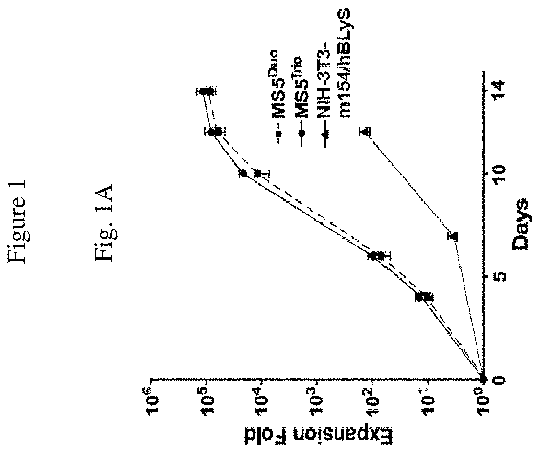
[0011] FIG. 1 is a set of graphs showing B cell expansion. FIG. 1A is a graph showing human B cell expansion, as measured by fold expansion relative to the number of days of culture, as a result of being cultured with either modified feeder cells of the invention comprising stromal cells expressing human BLyS and CD154 (with added IL-21) ("MS5.sup.DUO"), stromal cells expressing BLyS, CD154, and IL-21 ("MS5.sup.TRIO") as compared to feeder cells comprising NIH-3T3 cells expressing BLyS and CD154 (with added IL-21)("NIH-3T3-m154/hBLyS). These results are representative of those obtained in .gtoreq.3 experiments.
[0012] FIG. 1B is a graph showing the effects of exogenous human cytokines on human B cell expansion. Values represent mean B cell numbers from duplicate 6 and 10 day cultures on MS5.sup.Duo cell monolayers where the indicated cytokine combinations were present during the designated periods of time (a-h). All cytokines were present at 20 ng/mL except for IL-4 (10 ng/mL). Additional medium containing the appropriate cytokine(s) was added to the cultures on days 4 and 8. Values represent means (+s.e.m.) of 6 samples from two independent experiments. Mean values significantly different between (a) and other cultures or between cultures (e) and (g) are indicated; .sup.#p<0.05.
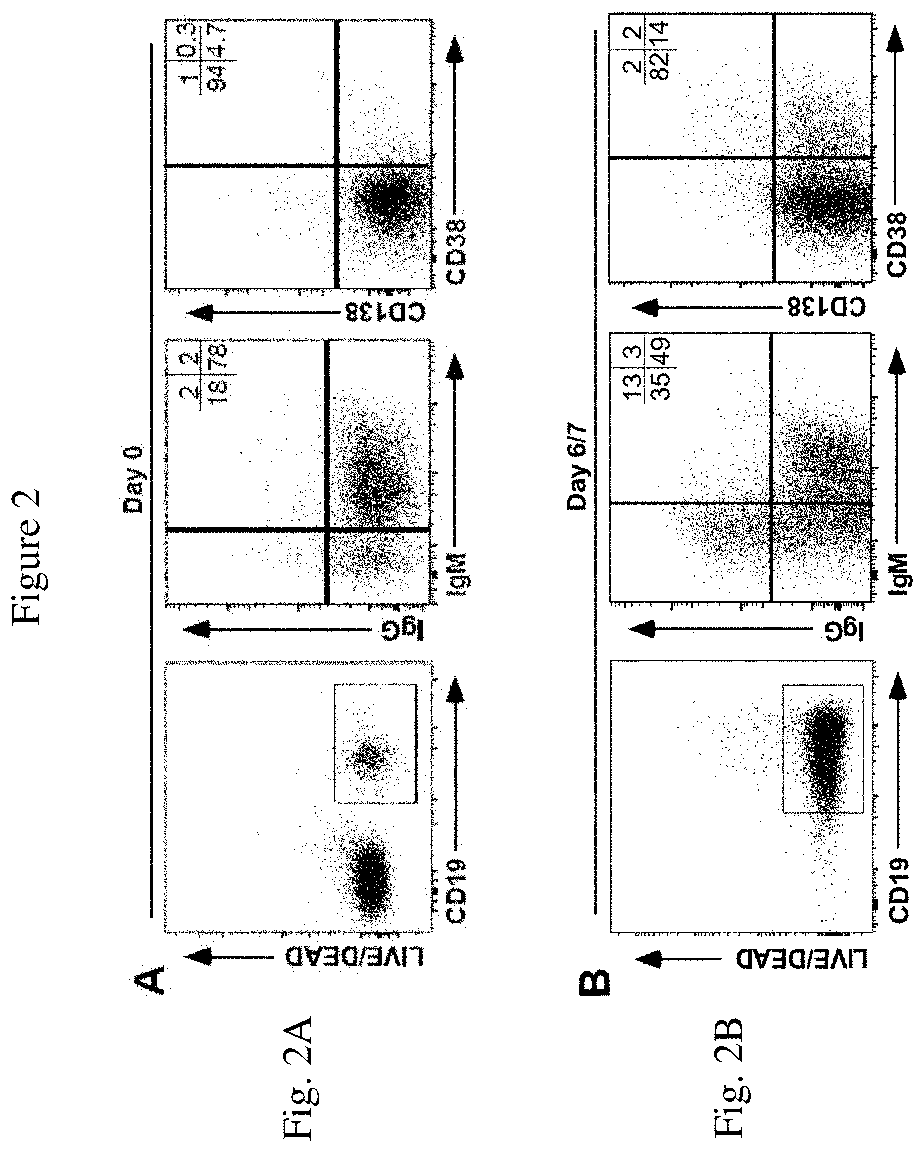
[0013] FIG. 2 is a set of FACS histograms showing the levels of the indicated cell surface markers. FIG. 2A is a graph showing cell surface phenotype (IgM, IgG, CD38, and CD138) of CD19.sup.+ human B cells after isolation from peripheral blood but prior to expansion in the method of the invention. Mean frequencies of viable CD20.sup.+ or CD19.sup.+ B cells expressing IgM, IgG, CD38 or CD138 within the indicated quadrants are indicated from two independent experiments with similar results.
[0014] FIG. 2B is a graph showing cell surface phenotype (IgM, IgG, CD38, and CD138) of human B cells during ex vivo expansion as a result of being cultured on a monolayer of MS5.sup.Trio cells after 6-7 days. These results are representative of those obtained in .gtoreq.3 experiments. Mean frequencies of viable CD20.sup.+ or CD19.sup.+ B cells expressing IgM, IgG, CD38 or CD138 within the indicated quadrants are indicated from two independent experiments with similar results.
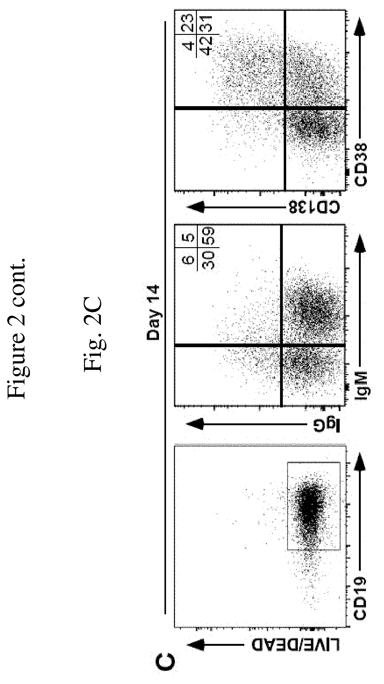
[0015] FIG. 2C is a graph showing cell surface phenotype (IgM, IgG, CD38, and CD138) of human B cells during ex vivo expansion as a result of being cultured on a monolayer of MS5.sup.Trio cells at 14 days of culture. Mean frequencies of viable CD20.sup.+ or CD19.sup.+ B cells expressing IgM, IgG, CD38 or CD138 within the indicated quadrants are indicated from two independent experiments with similar results.
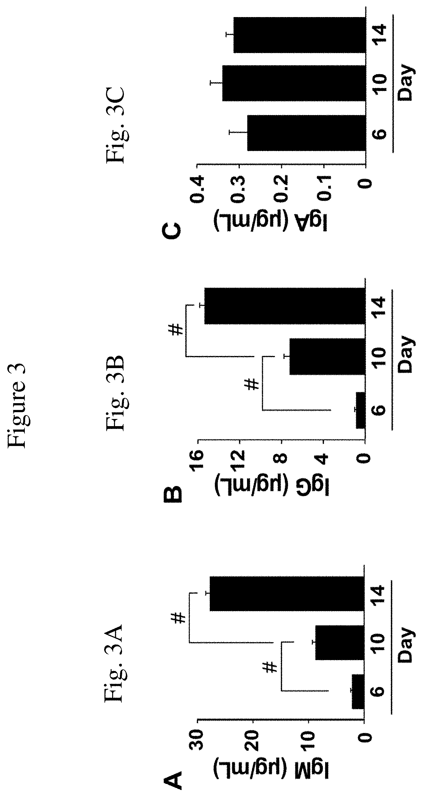
[0016] FIG. 3 is a set of graphs showing antibody production by isotype. FIG. 3A is a graph showing IgM production in the tissue culture supernatant fluid of human B cells during ex vivo expansion as a result of being cultured on monolayer of MS5.sup.Trio cells at days 6, 10, & 14 of culture. Bars represent means (+s.e.m.) antibody concentrations in 4 samples from two independent experiments as determined by ELISA.
[0017] FIG. 3B is a graph showing IgG production in the tissue culture supernatant fluid of human B cells during ex vivo expansion as a result of being cultured on monolayer of MS5.sup.Trio cells at days 6, 10, & 14 of culture. Bars represent means (+s.e.m.) antibody concentrations in 4 samples from two independent experiments as determined by ELISA.
[0018] FIG. 3C is a graph showing IgA production in the tissue culture supernatant fluid of human B cells during ex vivo expansion as a result of being cultured on monolayer of MS5.sup.Trio cells at days 6, 10, & 14 of culture. Bars represent means (+s.e.m.) antibody concentrations in 4 samples from two independent experiments as determined by ELISA.
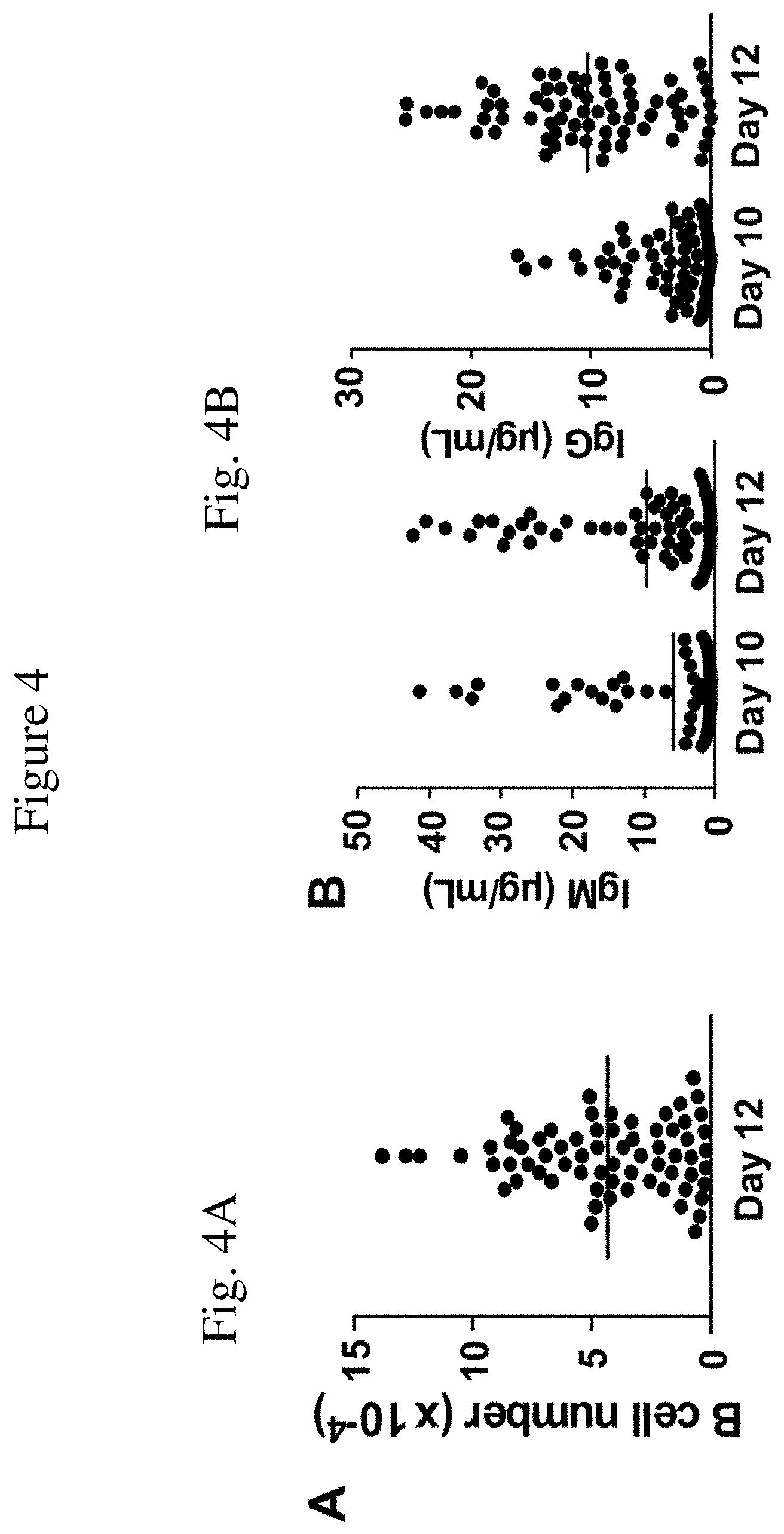
[0019] FIG. 4 is a set of graphs showing the expanded cells and production of various isotypes of antibodies. FIG. 4A is a graph representing single human B cell clones (each dot representing an individual clone) and their expansion in number relative to day 12 of culture with a monolayer of MS5.sup.Trio cells. These results are representative from two independent experiments.
[0020] FIG. 4B is a graph depicting concentrations of IgM and IgG in tissue culture supernatant fluid of the human B cell clones shown in FIG. 4A, as measured by ELISA, at days 10 and 12 of culture. These results are representative from two independent experiments.
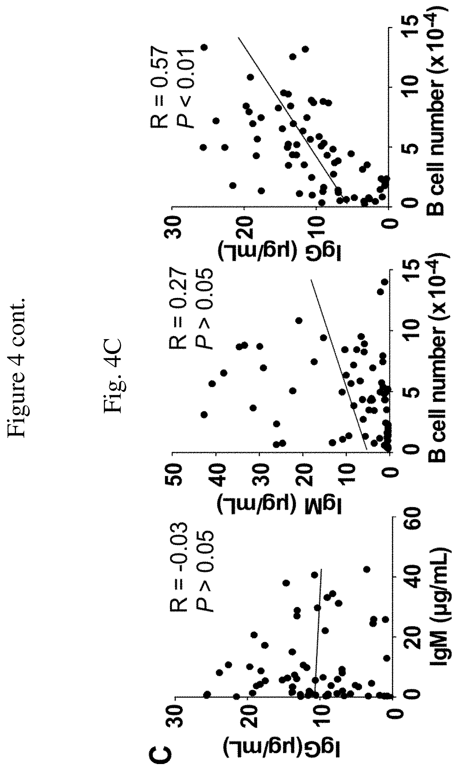
[0021] FIG. 4C is a graph depicting the correlation between concentrations of IgM and IgG in tissue culture supernatant fluid of the human B cell clones shown in FIG. 4A, as measured by ELISA, on day 12, or those between single B cell clonal expansion and IgM or IgG (n=69). These results are representative from two independent experiments.
[0022] FIG. 5 is a set of graphs showing production of specific antibody. FIG. 5A is a representative graph showing measurement of anti-desmoglein 1 (DSG1) IgG antibody by EUSA ("Absorbance") in relation to the well numbers of a 96 well plate ("Plate well number") containing isolated B cells, from a pemphigus foliaceus patient or a healthy control, and co-cultured with MS5.sup.Trio cells. The solid horizontal line shows the mean absorbance value for all 96 wells of the plate. The dashed horizontal line indicates the mean plus 2 standard deviations value for all 96 wells of the plate. A well was considered positive when its absorbance value exceeded the dashed line.
[0023] FIG. 5B is a bar graph showing the frequency of antigen-specific B cells (having specificity for DSG1 or desmoglein 3 (DSG3)) as calculated from circulating B cells from two individuals with pemphigus vulgaris (PV1 and PV2) or two individuals with pemphigus foliaceus (PF1 and PF2), as compared to circulating B cells from a healthy individual without measurable anti-DSG serum autoantibodies ("Healthy control", HC). Values shown represent the mean frequency of wells that were positive for DSG1- or DSG3-specific IgG antibody for each individual. Eleven individual 96 well ELISA plates were assessed in each assay for each individual.
DETAILED DESCRIPTION OF THE INVENTION
[0024] The invention is based on the discovery of an efficient method for expanding and differentiating B cells in culture, from single cell clones to an amount of B cells that produce sufficient quantities of monoclonal antibody enabling further characterization without relying on first cloning and expression of the immunoglobulin genes. Further characterization may comprise characterization of one or more of antigen specificity, function, binding, and or neutralization using one or more high throughput screening assays known in the art. The invention overcomes the deficiencies of feeder cell lines that were used previously to expand and differentiate murine B cells, such as the NIH-3T3 cell line (murine fibroblast cell line), in expanding human B cells in culture, particularly in an amount and state of differentiation to be useful in production of monoclonal antibodies.
Definitions
[0025] While the following terms are believed to be well understood by one of ordinary skill in the art of biotechnology, the following definitions are set forth to facilitate explanation of the invention.
[0026] The term "B cell" is used herein to mean a mammalian B lymphocyte including, but not limited to, human B cell, and murine B cell. Any B cell may be expanded in a method of the present invention, and for methods of the invention involving antibody production, such as (a) naive B cells, which comprise a diverse antibody repertoire generated by DNA rearrangements during B cell development, and (b) B cells that have been exposed to, or have memory of the exposure to, a specific antigen. B cells include B cell populations, subpopulations, or subsets thereof, including but not limited to, naive B cells, memory B cells, activated B cells, B1 cells, germinal center B cells, marginal zone B cells, regulatory B cells, and follicular B cells.
[0027] The term "monoclonal antibody" is used herein to mean an antibody that displays a single binding specificity and affinity for an epitope of an antigen. "Recombinant monoclonal antibodies" is a term that refers to monoclonal antibodies that are produced by recombinant means, such as by using a recombinant expression vector containing immunoglobulin gene sequences (e.g., VH and VL cDNA) and transfected into a host cell. Such a recombinant expression vector may comprise human immunoglobulin gene sequences (e.g., human VH and human VL cDNA), which are then transfected into a host cell. Recombinant monoclonal antibodies also comprise murine VH and VL cDNA, which are engineered to produce "humanized" antibodies that can be used for therapeutic, diagnostic, or theranostic applications in humans.
[0028] The term "antigen" is used herein to mean a substance that can stimulate the production of antibody, and can be bound by antibody specific for the substance (i.e., an antibody can specifically bind an antigen for which it has binding specificity). Antigens may be comprised of a substance comprising one or more of protein, peptide, lipid, carbohydrate, nucleic acid, and small molecule (organic or inorganic). Antigens may include: a substance foreign to the human body, viral antigens, bacterial antigens, parasite antigens, tumor antigens, toxin antigens, fungal antigens, self antigens, altered self antigens (self antigens that are altered or modified as the result of a disease state), modified antigens (misfolded or oxidized or with altered glycosylation or overexpression or mutated, as a result of a disease state and as compared to the antigen in a healthy or non-disease state).
[0029] The term "modified feeder cell" and "feeder cell" are used herein to mean mesenchymal stromal cells or thymic epithelial cells, modified to express a combination comprising a CD40 agonists such as CD154 (also known as CD40L or CD40 Ligand) and a B cell survival promoter such as BLyS (B Lymphocyte Stimulator, also known as BAFF (B-cell activating factor), or a combination comprising CD154, BLyS, and IL-21. Specific examples of such modified feeder cells are modified mesenchymal stromal cell lines (e.g., MS-5 cells available as catalog number ACC 441 from Deutsche Sammlung von Mikroorganismen und Zellkulturen (DSMZ), Braunschweig, Germany) and modified thymic epithelial cell lines (e.g., TEC cells). As an illustrative example, MS-5 is a murine mesenchymal stromal cell line that is commercially available. It is noted that mesenchymal stromal cell lines are distinguished from fibroblast cell lines, such as the NIH-3T3 cell line (murine fibroblast cell line NIH/3T3, deposited as ATCC CRL-1658), in that mesenchymal stromal cell lines secrete different factors and can support different functions (including vasculogenesis, angiogenesis) than fibroblast cell lines. For example, MS-5 cells secrete significantly higher levels of CXCL12 (SDF-1), CXCL-16, Angiopoetin-1, MCP-1 (monocyte chemoattractant protein-1, also known as CCL2), NOV (nephroblastoma overexpressed, also known as CCN3) and HGF (hepatocyte growth factor) than NIH 3T3 cells (Zhou et al., 2012, Br. J. Haematology, 157:297-311). TEC or thymic epithelial cell lines (e.g., TEC-84, TEC-D11) support human lymphopoiesis (Beaudette-Ziatanovo, Exp. Hematol., 39(5): 570-579). Thymic epithelial cell lines can be induced to undergo biological changes to assume a mesenchymal cell phenotype (epithelial-mesenchymal transition) such as by treatment with transforming growth factor in culture. The mesenchymal stromal cells or thymic epithelial cells are engineered to express a combination comprising a CD40 agonist such as a CD154 polypeptide and a B cell survival promoter such as BLyS, or a combination comprising these two polypeptides and IL-21, in forming the modified feeder cells of the invention. If the feeder cells are not engineered to express IL-21, then IL-21 must be added exogenously to the culture medium during the culture period. The culture period is suitably less than 3 weeks, suitably less than 2 weeks. The culture period may be 5, 6, 7, 8, 9, 10, 11, 12 or more days.
[0030] BLys may be expressed on the surface or may be soluble after cleaved from the cell surface. Alternatively the BLyS is replaced by a different factor(s) that promotes B cell survival in culture including BLyS fragments, APRIL, CD22 ligand, CD22 monoclonal antibody, or fragments thereof. BLyS is a 285 amino acid glycoprotein (SEQ ID NO: 69) encoded by a nucleic acid sequence comprising SEQ ID NO:01 or a similar nudeotide sequence encoding the same amino acids. BLyS, produced recombinantly by the modified feeder cells of the invention, may vary in length as long as it can bind and signal the BLyS receptor, and promote expansion of human B cells. In that regard, BLyS can be produced as a membrane bound type, or in soluble form. For example, a soluble form comprised of amino acids 136-285 has been found to be functionally active.
[0031] The CD154 polypeptide may be replaced by any CD40 agonist including but not limited to CD40 antibodies and fragments thereof, the CD40 ligand, CD154 polypeptide and polypeptide fragments thereof, small molecules, synthetic drugs, peptides (including cyclic peptides), polypeptides, proteins, nucleic acids, aptamers, synthetic or natural inorganic molecules, mimetic agents, and synthetic or natural organic molecules capable of activating CD40. In a certain embodiment, the CD40 agonist is a CD40 antibody. The CD40 antibodies can be of any form. Antibodies to CD40 are known in the art (see, e.g., Buhtoiarov et al., 2005, J. Immunol. 174:6013-22; Francisco et al., 2000, Cancer Res. 60:3225-31; Schwulst et al., 2006, 177:557-65, herein incorporated by reference in their entireties). The CD40 agonists may be CD154 polypeptides and may be expressed on the surface of feeder cells or expressed in soluble form. Human CD154 polypeptide was used in the Examples and can be found as a 261 amino membrane bound protein (SEQ ID NO: 68) or 149 amino acid soluble protein, and can be encoded by a nucleotide sequence comprising SEQ ID NO:02 or SEQ ID NO:03, or a similar nucleotide sequence encoding the same amino acids. In that regard, CD154 can be produced as a membrane bound type, or in soluble form, of varying lengths. It has been shown that amino acids important for CD40L and CD40 interactions include amino acids 128 to 258.
[0032] Human IL-21 (mature polypeptide) comprises 131 amino acids (SEQ ID NO: 70), and can be encoded by a nucleotide sequence comprising SEQ ID NO:04, or a similar nucleotide sequence encoding the same amino acids. In that regard, IL-21 can be produced in various lengths as long as binding and induction of signaling through the IL-21 receptor is maintained, and it has been shown that amino acids important for IL-21 binding and functional interactions include, but are not limited to, amino acids 33, 145, and 148. The feeder cells can be engineered to express IL-21 or the IL-21 can be added to the culture exogenously. The IL-21 can be added at between 2 ng/mL and 1000 ng/mL, suitably between 5 and 500 ng/mL, suitably between 10 and 100 ng/mL.
[0033] The term "exposed to an antigen" is used herein to mean that the antigen is contacted with a B cell in sufficient concentration and for a sufficient time for the B cell to become activated by the antigen (e.g., binds to B cell receptors (BCR), or to induce B cell differentiation into B cells capable of producing antibody specific for the triggering antigen ("specific antigen"). There are a number of ways by which B cells may be exposed to antigen in vivo including, but not limited to, infection, injection, vaccination, immunization, and circulation (e.g., tumor antigen, altered self antigen, self antigen). In another embodiment, B cells may be exposed to and activated by a specific antigen in vitro. For example, US published patent application US2015/0299655 discloses a method for in vitro immunization of human B cells by a specific (e.g., known) antigen comprising culturing a total population of human peripheral blood mononuclear cells in the presence of an antigenic composition comprising at least one known antigen that is coupled to a Tat protein and a ligand to a receptor of an antigen-presenting cell (the latter including saccharides that bind C-lectin type receptors, immunoglobulins or fragments thereof (e.g., containing Fc portion) that bind Fc receptors) for sufficient time and under sufficient conditions for B cells present in the human peripheral blood mononuclear cells to be immunized by the antigenic composition.
[0034] B cells, exposed to antigen in vivo, may be isolated from a biological sample comprising peripheral blood or other body fluids (e.g., synovial fluid, or exudates) or from tissues (e.g., any tissue from the body of an individual, including but not limited to bone marrow, cardiac tissue, nervous tissue, tumor tissue, diseased tissue, connective tissue, spleen tissue, lymph node tissue, connective tissue, thymus tissue, and other lymphoid tissues) using methods known in the art. Such methods include fractionation using antibody coated magnetic beads and fluorescence-activated cell sorting. In immunomagnetic sorting, positive and/or negative selections may be performed to isolate the B cells. Reagents for isolating B cells include one or more antibody preparations for isolating B cells. Antibodies which are specific for (can bind to) cell surface markers on B cells include antibodies specific for IgM, IgD, IgG, IgA, IgE, CD19, CD20, CD21, CD22, CD24, CD40, CD72, CD79a, CD79b, or combinations (particularly for isolating B cell subpopulations or subsets) of these with, or combinations including, additional cell surface molecules such as CD5, CD9, CD10, CD23, CD38, CD48, CD80, CD86, CD138 or CD148. Antibodies or an antibody combination may be bound to magnetic beads in forming reagents for isolating B cells by immunomagnetic separation or sorting. The antibodies may be bound to a fluorescent label, as well known in the art, to form reagents for isolating B cells by fluorescence-activated cell sorting. Isolating B cells for seeding single B cell cultures can be done using methods known in the art which include, but are not limited to, limiting dilution or dilution cloning, and fluorescence activated cell sorting.
[0035] The methods of expanding B cells in vitro or ex vivo provided herein include isolating at least one B cell from a subject. The B cells used in the methods may be harvested from various areas of the subject, including but not limited to the blood, spleen, peritoneal cavity, lymph nodes, bone marrow, site of autoimmune disease, site of inflammation or tissue undergoing transplant rejection in the subject. The cells may be harvested from the subject by any means available to those of skill in the art. The harvested population of cells should contain B cells, but may be a mixed cellular population. The subject may be any animal with B lymphocytes, suitably a mammal, suitably a domesticated animal such as a horse, cow, pig, cat, dog, or chicken, or suitably a human. Alternatively, the cells may be derived from stem cells, including but not limited to B cell stem cells, bone marrow stem cells, embryonic stem cells and induced pluripotent stem cells, which have been appropriately differentiated in vitro to develop into B cells or B cell progenitors prior to use in the methods described herein. See e.g., Carpenter et al., 2011, Blood 117: 4008-4011. The B cells may be naive or antigen-exposed cells.
[0036] The B cells may be isolated using the isolation and selection methods described above or other methods known to those of skill in the art and may include both positive and negative selection steps. The B cells may be less than 100% B cells. Isolating is used to indicate that a group of cells is separated from incubation media, feeder cells or other non-B cells. Isolating is not meant to convey that the resulting isolated cells have a certain level of purity or homogeneity. The cells may be harvested, isolated or selected using any means available to those of skill in the art. For example, B cells may be harvested from adherent cells by selecting for non-adherent cells after an appropriate incubation. Cells may also be selected for expression of cell surface markers by FACS sorting or by the differential ability to bind antibody coated magnetic beads. Means of selecting cells in a mixed population are well known to those skilled in the art.
[0037] The isolated B cells are then cultured with a feeder cell line comprising a stromal cell line modified to express a CD40 agonist and a B cell survival promoter and optionally IL-21. The IL-21 may be added exogenously to the culture media. The culturing step is carried out without adding additional exogenous IL-4. The term "without additional exogenous IL-4" indicates that the culture comprises less than 200 pg/mL IL-4, less than 100 pg/mL IL-4, less than 50 pg/mL IL-4 or suitably less than 5 pg/mL IL-4 added exogenously to the culture. This is distinct from the mouse B cell expansion methods reported by either U.S. Pat. No. 8,815,543 or U.S. Publication Number 2014/0065118, both of which are incorporated herein by reference. As used herein expansion of B cells includes stimulation of proliferation of the cells as well as prevention of apoptosis or other death of the cells. As used herein, "culturing" and "incubation" are used to indicate that the cells are maintained in cell culture medium at 37.degree. C. and 5% CO.sub.2 for a period of time with the indicated additives (feeder cells, cytokines, agonists, other stimulatory molecules or media, which may include buffers, salts, sugars, serum or various other constituents). Suitably, the incubation or culturing periods used herein is at least 48 hours, but may be for any amount of time up to eight or more days. Those of skill in the art will appreciate that the culturing or incubation time may be varied to allow proper expansion, to adjust for different cell densities or frequencies of individual subsets, and to allow an investigator to properly time use of the cells. Thus the precise culture length may be determined empirically by one of skill in the art. The culturing period may also be carried out without addition of any exogenous cytokines if the feeder cells are modified to express 11-21 or 11-21 may be the only exogenous cytokine added to the cultures.
[0038] The methods may allow from two fold to over 5.times.10.sup.6 fold expansion of B cells. The B cells are expanded at least an average of 10.sup.3, 10.sup.4, or 10.sup.5 fold in less than 2 weeks in culture. The cells may be selected after the culture period to remove any non-B cells or to positively select for B cells or for a particular B cell subset. The culturing step may include an antigen or may be completed without addition of an antigen. Without addition of an antigen is similar to without addition of IL-4 as defined above. The expanded B cells may have undergone class-switching during the culturing period. In one embodiment, the isolated B cells were positive for IgM and after expansion in the methods described herein at least 10%, 15%, 20%, 25% of the expanded B cells express IgG. In one embodiment, at least 1%, 2%, 3%, 4%, 5%, 7%, 10% or more of the expanded B cells express IgA. If the cells are separated into single B cells prior to the culturing step the resulting expanded B cells may be used to produce and isolate a monoclonal antibody. The monoclonal antigen specificity can be determined using methods available to those of skill in the art and the monoclonal antibodies can be purified from the B cells.
[0039] The present disclosure is not limited to the specific details of construction, arrangement of components, or method steps set forth herein. The compositions and methods disclosed herein are capable of being made, practiced, used, carried out and/or formed in various ways that will be apparent to one of skill in the art in light of the disclosure that follows. The phraseology and terminology used herein is for the purpose of description only and should not be regarded as limiting to the scope of the claims. Ordinal indicators, such as first, second, and third, as used in the description and the claims to refer to various structures or method steps, are not meant to be construed to indicate any specific structures or steps, or any particular order or configuration to such structures or steps. All methods described herein can be performed in any suitable order unless otherwise indicated herein or otherwise dearly contradicted by context. The use of any and all examples, or exemplary language (e.g., "such as") provided herein, is intended merely to facilitate the disclosure and does not imply any limitation on the scope of the disclosure unless otherwise claimed. No language in the specification, and no structures shown in the drawings, should be construed as indicating that any non-daimed element is essential to the practice of the disdosed subject matter. The use herein of the terms "including," "comprising," or "having," and variations thereof, is meant to encompass the elements listed thereafter and equivalents thereof, as well as additional elements. Embodiments recited as "including," "comprising," or "having" certain elements are also contemplated as "consisting essentially of" and "consisting of" those certain elements.
[0040] Recitation of ranges of values herein are merely intended to serve as a shorthand method of referring individually to each separate value falling within the range, unless otherwise indicated herein, and each separate value is incorporated into the specification as if it were individually recited herein. For example, if a concentration range is stated as 1% to 50%, it is intended that values such as 2% to 40%, 10% to 30%, or 1% to 3%, etc., are expressly enumerated in this specification. These are only examples of what is specifically intended, and all possible combinations of numerical values between and including the lowest value and the highest value enumerated are to be considered to be expressly stated in this disclosure. Use of the word "about" to describe a particular recited amount or range of amounts is meant to indicate that values very near to the recited amount are included in that amount, such as values that could or naturally would be accounted for due to manufacturing tolerances, instrument and human error in forming measurements, and the like. All percentages referring to amounts are by weight unless indicated otherwise.
[0041] No admission is made that any reference, including any non-patent or patent document cited in this specification, constitutes prior art. In particular, it will be understood that, unless otherwise stated, reference to any document herein does not constitute an admission that any of these documents forms part of the common general knowledge in the art in the United States or in any other country. Any discussion of the references states what their authors assert, and the applicant reserves the right to challenge the accuracy and pertinence of any of the documents cited herein. All references cited herein are fully incorporated by reference in their entirety, unless explicitly indicated otherwise. The present disclosure shall control in the event there are any disparities between any definitions and/or description found in the cited references.
[0042] Unless otherwise specified or indicated by context, the terms "a", "an", and "the" mean "one or more." For example, "a protein" or "an RNA" should be interpreted to mean "one or more proteins" or "one or more RNAs," respectively.
[0043] The following examples are meant only to be illustrative and are not meant as limitations on the scope of the invention or of the appended claims.
Example 1
[0044] In this example, provided is a method for the in vitro expansion of human B cells according to the invention. Heparinized blood was collected from healthy adult human donors. Blood mononuclear cells were isolated by centrifugation over a layer of ficoll-sodium metrizoate in a Sepmate50 tube at 300.times.g for 10 minutes. Peripheral blood mononuclear cells (PBMCs) were diluted 2-fold with buffer (PBS), and then centrifuged (300.times.g, 10 min) and resuspended to .sup..about.3.times.10.sup.7 cells/mL in MACS buffer (0.5% bovine serum albumin (BSA, w/v), 2.5 mM ETDA in PBS). CD19.sup.+ B cells were isolated by positive selection using CD19 magnetic beads according to the manufacturer's instructions. Briefly, isolated mononuclear cells were incubated with CD19 monoclonal antibody-coated microbeads (60 .mu.L microbeads/mL of PBMCs) at 4.degree. C. for 15 minutes, diluted 10-fold, pelleted again by centrifugation, and resuspended in 1 mL MACS buffer. The cells were loaded on a pre-wetted LS column (a column comprising a matrix composed of ferromagnetic spheres, which are covered with a coating allowing fast and gentle separation of cells) under a magnetic field. The column was then washed with an additional 6 mL of MACS buffer. CD19.sup.+ cells were eluted by removing the column from the magnetic field and adding 3 mL of RPMI 1640 medium containing 10% FCS (fetal calf serum) to the column and collecting the eluate in a separate conical tube. Viable cell concentrations were determined using a hemocytometer under a microscope with Trypan Blue staining of dead cells. B cell purities were determined by immunofluorescence staining and flow cytometric analysis with CD19.sup.+ cell frequencies of 298%. Additional rounds of selection can be used to enhance purification.
[0045] Expanding human B cells in vitro was first attempted using culture conditions that induced murine B cell expansion. Purified human B cells were cultured on stromal cell monolayers comprising NIH-3T3 cells expressing BLyS and CD154 (NIH-3T3-m154/hBLyS) with added human IL-21 under culture conditions mimicking mouse B cell cultures (patent application US20140065118). Expanding purified human B cells in vitro was subsequently attempted using 3T3-CD154.sup.EAT or 3T3-CD154.sup.BEAT cells under culture conditions mimicking mouse B cell cultures in the presence of exogenous recombinant cytokine mixtures, including IL-2, IL-4, IL-21, IL-10 and APRIL Briefly, NIH-3T3-m154/hBLyS cells were supertransfected with cDNAs encoding mouse CD154 and mouse BLyS to generate NIH-3T3-CD154.sup.EAT cells, or cDNAs encoding mouse CD154, mouse BLyS and mouse IL-21 to generate NIH-3T3-CD154.sup.BEAT cells. Stromal cell lines expressing these cDNAs were isolated, subcloned and assayed for their ability to support mouse and human B cell expansion. Stromal cells were seeded in 24-well plates at a density of 3.times.10.sup.5 cells/well in 1 mL of culture media for at least 12 hours prior to the addition of purified human B cells (3.times.10.sup.3/well) to the cultures. When human B cells were added to the cultures, the media from each well was replaced with fresh culture media containing IL-4 (10 ng/mL). After 4 days of culture, additional media (0.7 mL) containing IL-4 (10 ng/mL) was added to each well. On day 6 of culture, the B cells and stromal cells were collected after treatment with 0.4 mL of trypsin-EDTA. The cell suspension was pelleted by centrifugation and resuspended in 2 mL of culture medium. B cell numbers were quantified by microscopy (hemocytometer), and the frequency of B cells was determined by immunofluorescence staining with flow cytometry analysis. However, while significant mouse B cell expansion was observed under these conditions, significant human B cell survival or expansion was not observed in these attempts (e.g., expansion was less than 130 fold). Therefore, NIH-3T3 cells were transfected with human BLyS and human CD154 cDNAs, with CD154.sup.+BLyS.sup.+ cells isolated, expanded and subsequently subcloned. Under these conditions, maximal human B cell expansion within a 9 to 12 day culture period was not increased significantly beyond that observed using NIH-3T3-mCD154/hBLyS cell monolayers (patent application US20140065118) as human B cell expansion within a 12 day period was only 130.+-.17-fold (mean.+-.s.e.m.; FIG. 1A). Human B cell expansion was not significantly improved by the addition of exogenous human recombinant cytokine mixtures, including IL-2, IL-4, IL-21, IL-10 and APRIL Therefore, a series of additional human and mouse stromal cells were assessed for their ability to support human B cell expansion.
[0046] Diverse human stromal cells and mouse stromal cells were transfected with human BLyS and human CD154 using the following vectors and methods of clonal selection. cDNA encoding human BLyS was used to generate a BLyS-IRES-eGFP DNA construct in order to provide a (GFP--green fluorescent protein) selection marker independent of immunofluorescence staining for CD154 expression. Human CD154 cDNA and human BLyS-IRES-eGFP DNA were independently cloned into a retroviral-based expression vector having LTR as the promoter (pMSCVpuro vectors) and transfected into the stromal cell lines by retroviral transduction using a commercially available retroviral packaging cell line. After transfection, stable puromycin (5 .mu.g/mL)-resistant cells were subsequently selected during culture. Cells expressing CD154 and secreting BLyS were visualized by staining the cell surface of stromal cells with APC (Allophycocyanin)-conjugated CD154 monoclonal antibody, with APC.sup.+GFP.sup.+ (CD154.sup.+BLyS.sup.+) cells isolated using fluorescence-activated cell sorting. Isolated CD154.sup.+BLyS.sup.+ cells were expanded in culture, subcloned and tested for their ability to support and expand human B cells in vitro. Stromal cells tested for their ability to support and expand human B cells included: mouse MS-5 cells, a bone marrow stromal cell line; AFT024 cells, a mouse fetal liver stromal cell line; OP9 cells, a mouse bone marrow stromal cell line; human A549 cells, an adenocarcinomic alveolar basal epithelial cell line; human EA-Hy926 cells, a human endothelial cell line; HS-5 cells, a human fibroblastoid stromal cell line; TEC-84 cells, a human thymic stromal cell line; human foreskin fibroblasts immortalized using the amphotropic retrovirus LXSN16E6E7 produced by the PA317 LXSN 16E6E7 cell line (American Type Culture Collection, CRL-2203.TM.); and similarly-transformed human umbilical vein endothelial cells. The results showed that most stromal cell lines expressing human BLyS and CD154 were either unable to support B cell survival or expansion, or were less efficient in supporting expansion of human B cells than transfected mouse NIH-3T3 cells. Surprisingly, and unexpectedly, MS-5 cells and TEC-84 cells demonstrated an ability to support and expand human B cells in culture, with subsequent MS-5 cell clones having the most optimal growth characteristics for supporting human B cell expansion without a significant need to arrest stromal cell proliferation by treating the cells with mitomycin C to prevent cell division.
[0047] The MS-5 cells and TEC-84 cells, modified to express human CD154 and BLyS, were further and similarly modified to express IL-21. Human IL-21 cDNA was used to generate an IL-21-2A-peptide-mBFP2 DNA construct enabling expression of Blue Fluorescent Protein (mBFP2) via a cleavable 2A peptide sequence. IL-21-2A-peptide-mBFP2 DNA was inserted in place of the puromycin gene of the pMSCVpuro vector. Stable puromycin (5 .mu.g/mL)-resistant cells were subsequently selected during culture. Modified feeder cells that were APC.sup.+GFP.sup.+BFP.sup.+ (CD154.sup.+BLyS.sup.+IL-21.sup.+) were isolated using fluorescence-activated cell sorting, and single cell clones of each stromal cell line were subsequently isolated and tested for their ability to support human B cell proliferation.
[0048] Modified feeder cells of the invention were compared with NIH-3T3 cells transfected with human BLyS and mouse CD154 cDNAs (NIH-3T3-mCD154/hBLyS) for the ability, as feeder cells, to support and induce expansion of human B cells isolated from blood. In this comparison, viable human CD19.sup.+ B cells were added to the respective cultures of fresh feeder cell monolayers. Human B cells were cultured on NIH-3T3-mCD154/hBLyS cell monolayers with exogenous human IL-4 (2 ng/ml) for 7 days. Additional medium containing IL-4 (2 ng/ml) was added to the cultures on days 2 and 4. B cells were isolated on day 7 and transferred onto fresh NIH-3T3-mCD154/hBLyS cell monolayers with exogenous human IL-21 (10 ng/ml) for 5 days. In cultures using MS-5 cells transfected with human CD154 and BLyS (MS5.sup.Duo cells), B cells were cultured on MS5.sup.Duo cells with the addition of IL-4 plus IL-21 for 6 days, then cultured with exogenous IL-21 until day 14. In cultures using MS-5 cells transfected with human CD154, BLyS and IL-21 (MS5.sup.Trio cells), B cells were cultured on MS5.sup.Trio cells with the addition of IL-4 for 6 days, then cultured without exogenous cytokines until day 14. On days 12 or 14, the B cells and stromal cells were collected from each well and analyzed. FIG. 1 shows a comparison of modified feeder cells comprising MS-5 clones that expressed human CD154 and BLyS (MS5.sup.Duo) or MS-5 clones that expressed human CD154, BLyS and IL-21 (MS5.sup.Trio) with NIH-3T3-mCD154/hBLyS cells, in relation to the expansion of human CD19.sup.+ B cells in vitro (expansion fold is relative to numbers of B cells present in the cultures at the initiation of the cultures). With MS5.sup.Duo cells or MS5.sup.Trio cells as the modified feeder cells, human B cells expanded by 12,018.+-.5,523- and 21,611.+-.3,576-fold at day 10, and 61,547.+-.16,134- and 80,761.+-.28,979-fold by day 12, respectively (FIG. 1A). With NIH-3T3-mCD154/hBLyS feeder cells, human B cells expanded by 3.4-fold by day 7 and .sup..about.130-fold by day 12. It is also noted that cultures of B cell with either MS5.sup.Duo cells or MS5.sup.Trio cells can be performed in the absence of IL-4 with similar or better results for amplification and differentiation (FIGS. 1B and 2B-C). Thus, modified feeder cells of the invention can significantly promote expansion of human B cells as compared to NIH-3T3-mCD154/hBLyS cells as feeder cells. The modified feeder cells of the invention provide a significantly more optimal stromal cell monolayer for human B cell expansion in comparison with the majority of mouse and human cell lines that were tested for this capacity.
Example 2
[0049] Illustrated in this Example is the ability of the modified feeder cells of the invention to induce differentiation in human B cells expanded in vitro. Human B cells expanded in vitro in the MS5.sup.Duo or MS5.sup.Trio culture systems were analyzed for markers indicative of differentiation. Human B cell phenotypes were assessed using PerCP-, FITC-, PE/Cy7, FITC, and PE/Cy7-conjugated CD19 (HIB19), IgM (MHM-88), IgG (HP6017), CD38 (HIT2), and CD138 (M115) monoclonal antibodies. Viable cells were analyzed by flow cytometry. Human B cells that expanded in the MS5.sup.Duo or MS5.sup.Trio culture systems remained CD19.sup.+ and CD20.sup.+, confirming their B cell origin (FIG. 2). Approximately eighty percent of freshly isolated human blood B cells expressed IgM, with .sup..about.4% expressing IgG prior to being cultured with the modified feeder cells (FIG. 2A). On day 7 of culture with a modified feeder cell monolayer, half of the B cells expressed IgM and .sup..about.16% expressed IgG (FIG. 2B). By the end of culture, most B cells expanded in the MS5.sup.Trio culture system expressed cell surface IgM, but the frequency of IgM.sup.-IgG.sup.-IgA.sup.+CD19.sup.+ B cells had expanded (FIG. 2B-C). Less than 10% of freshly isolated B cells expressed CD38 or CD138 prior to being cultured with the modified feeder cells (FIG. 2A), which are activation markers expressed on activated B cells and expressed at high densities on plasma cells. By day 6-7 of culture, 16% of B cells expanded in the MS5.sup.Trio culture system expressed the CD38 activation marker (FIG. 2B). By day 14 of culture, up to 56% of these B cells expressed CD38, with 23% of the B cells expressing both CD38 and CD138 (FIG. 2C). Similar, if not identical results were obtained with B cells expanded in the MS5.sup.Duo culture system. Some of the human B cells cultured with modified feeder cells of the invention acquired a phenotype consistent with their activation and differentiation into antibody-secreting plasmablasts.
Example 3
[0050] Illustrated in this Example is the ability of the modified feeder cells of the invention to induce human B cells expanded in vitro to produce antibody. Human B cells expanded in vitro in the MS5.sup.Duo or MS5.sup.Trio culture systems were analyzed for the production of antibody. Human IgG, IgM, and IgA antibody levels were determined by enzyme-linked immunoassays (EUSA). Plates were blocked with tris-buffered saline containing 1% BSA before cell culture supernatant fluid (diluted 1:10 from bulk B cell cultures or undiluted if from single B cell cultures) was added to the plates. Alkaline-phosphatase-conjugated anti-IgG.sub.1 antibody was used to detect bound antibody, and 1 M diethanolamine/0.5 M MgCl.sub.2 with 4-nitrophenyl phosphate disodium salt hexahydrate was used as the detection reagent. Absorbance was read at 405 nm. Control plates were coated using 1% BSA in phosphate buffered saline (PBS), and blocked as above before the addition of culture supernatant fluid and detection reagents. Antibody concentrations were quantified based on standard curves obtained for each EUSA using commercially available human IgM, IgG and IgA standards.
[0051] Consistent with human B cell phenotypic changes during their in vitro expansion as described in Example 2 herein, secreted IgM was not detected in the tissue culture supernatant fluid of human B cells expanded in vitro in the MS5.sup.Trio culture system by day 6 (FIG. 3A). However, IgM concentrations increased to 7.8 .mu.g/mL by day 10, and 92 .mu.g/mL on day 14. Similarly, IgG levels increased significantly to 2.4 .mu.g/mL by day 14 (FIG. 3B). IgA levels also increased on days 10 and 14, but remained less than 0.3 .mu.g/mL (FIG. 3C). Similar, if not identical results were obtained with B cells expanded in the MS5.sup.u culture system. By contrast with human B cells expanded in vitro in the MS5.sup.Duo or MS5.sup.Trio culture systems, human B cells cultured on NIH-3T3-mCD154/hBLyS, NIH-3T3-CD154.sup.EAT, or 3T3-CD154.sup.BEAT stromal cell monolayers under similar conditions did not secrete measurable antibody into the tissue culture supernatant fluid. Thereby, as a result of expansion on the modified feeder cells of the invention, some human B cells differentiate during ex vivo expansion and predominantly secrete IgM and IgG antibodies.
[0052] In one aspect of the invention, provided is a method for producing human monoclonal antibodies that bind to a particular antigen, typically a known antigen. The method includes the steps of isolating human B cells, separating the isolated B cells into single cells, culturing an isolated B cell (single cell) in the presence of modified feeder cells according to the invention under conditions and for a sufficient time to achieve at least a 10.sup.4-fold expansion in number (e.g., expansion from a single cell to 10,000 cells, average expansion number over multiple wells of a multiwell plate) in less than 2 weeks in culture, wherein produced from the expanded B cell clone in culture is monoclonal antibody. This aspect excludes the addition of antigen, in attempts to guide antigen selection or further induce antigen-sensitization, directly to the coculture of isolated B cells with the modified feeder cells of the invention (i.e., exogenous antigen is absent when isolated B cells are cultured in the presence of modified feeder cells, as this is not necessary to produce monoclonal antibodies according to the method of the invention).
[0053] To illustrate this aspect, single human B cells were cultured on MS5.sup.Trio stromal cell monolayers in 96-well plates without the addition of exogenous cytokines. In these limiting dilution assays, human B cell colonies were observed in 60 to 70% of wells; reflecting cloning efficiencies of 60% to 70%. In the wells containing B cell colonies, single B cells expanded 46,689.+-.4,105-fold on average after 12 days (FIG. 4A). Tissue culture supernatant fluid IgM concentrations on days 10 and 12 of culture were 5.5.+-.1.2 .mu.g/mL and 9.3.+-.1.4 .mu.g/mL, respectively (FIG. 4B). IgG concentrations on days 10 and 12 of culture were 3.5.+-.0.5 .mu.g/mL and 10.5.+-.0.8 .mu.g/mL, respectively. Although there was no correlation between IgM and IgG secretion within individual B cell clones and no correlation between IgM secretion and B cell expansion, there was a significant positive correlation between IgG secretion and B cell expansion (FIG. 4C). Thus, some B cells within individual wells underwent isotype switching, with some of the B cells also differentiating into antibody-secreting cells. The significant concentrations of antibody produced for each B cell clone thereby enables the determination of their BCR specificity.
Example 4
[0054] Further steps of the method of the invention may comprise characterization of the monoclonal antibody produced by each B cell clone such as determining the specificity of the monoclonal antibody for antigen using methods known in the art. To determine the specificity of monoclonal antibodies produced according to the method of the invention, any one of several methods known in the art may be used. For example, antigen arrays are now available which permit the screening of up to 100 different antigens with small volumes of undiluted tissue culture supernatant fluid from B cell clones which significantly increases the efficiency of identification of antigens for which a monoclonal antibody has specificity in parallel to monoclonal antibody production number. Briefly, purified biotinylated antigens are spotted in streptavidin-coated 96-well microtiter plates by direct contact printing using 0.2 or 0.4 mm solid printing pins. Printed plates are left unwashed and plates are stored at 4.degree. C. until ready for use. For screening, B cell clone supernatant fluid is added directly to the wells, with an optional blocking step if desired (e.g., BSA). A biotinylated human antibody, specific for an antigen in the printed array, is used as a positive control and orientation marker. B cell done supernatants are added to the antigen arrays and incubated overnight at 4.degree. C. After washing with buffer (PBS+0.1% Tween), arrays are incubated for 2 hours with an anti-human antibody (for detecting human antibody), or an anti-murine antibody (for detecting murine antibody), labeled with a fluorophore for fluorescence detection, or an enzyme (e.g., alkaline phosphatase) and colorimetric substrate for colorimetric detection. Antigen arrays can then be analyzed with high throughput microscopy, with image capture and use of imaging software to optimize and quantitate detection.
[0055] Similar high throughput antigen microarrays have been described in which more than 25,000 antigen-antibody reactivity tests can be performed in less than a week. In one system, arrays of protein antigens are covalently bound to aldehyde-coated glass slides. Binding of the antigens onto aldehyde glass slides required 24 hours to become stable, and stability was demonstrated for at least six months. The antigen microarray chips were printed on glass slides using solid pin deposition technology and a commercially available robotic system. Using this system allows as little as 20 .mu.l of culture supernatant fluid to be incubated with antigen on the array surface. The presence of human antibodies bound to the array may be made with a mixture of anti-human IgG and anti-human IgM antibodies, conjugated to two different and distinguishable detection molecules if determination of the immunoglobulin class of the monoclonal antibody is desired simultaneously with determination of antigen specificity (e.g., laser scanning by an array reader which can incorporate three to four different lasers for differential detection).
[0056] Further steps of the method of the invention may comprise characterization of the monoclonal antibody such as isolating the monoclonal antibody comprising (i) purification of the monoclonal antibody from the culture medium using methods known in the art, and/or (ii) comprise isolating the total RNA from the B cell done to produce cDNA encoding the variable-heavy (VH) antibody chains and cDNA encoding variable-light (VL) antibody chains which then can be cloned into an eukaryotic expression vector for recombinant production of the monoclonal antibody using methods known in the art. In that regard, human B cell clones expanded and differentiated using the method of the invention may be harvested, followed by extraction of total RNA from cells of a B cell clone. Any one of a number of methods known in the art to isolate total RNA may be used, such as using a commercially available miniprep kit. Human VH and VL chain genes may be selectively amplified using primers known in the art (see, e.g., SEQ ID NOs: 5-12) and reverse transcriptase-polymerase chain reaction to make respective cDNA molecules. Human immunoglobulin (Ig) heavy (H) and light (L) chain transcripts can be amplified using nested PCR primers. Immunoglobulin-specific cDNA is then used as a template in a nested PCR amplification. Briefly, 1-2 .mu.l of cDNA from each monoclonal B cell expansion is used as a template with the external VH PCR primers and appropriate constant region primer (see, e.g., SEQ ID NOs: 42-67) to amplify isotype-specific BCR transcripts. If necessary, PCR products from the external amplification can be used as templates for a second round of internal amplification (see, e.g., SEQ ID NOs: 13-41). In either case, plasmid-specific sequences can be added to the forward and reverse primers to generate antibody transcripts that can be easily cloned into a vector of choice for re-expression analyses. Productive Ig rearrangements could be compared against germline Ig sequences using publicly available software, and analyzed using commercially available software to determine V(D)J gene family usage. Mutation frequencies could be determined using germline V, D, and J sequences. V.sub.H-D-J.sub.H, V.sub.K-J.sub.K and transcript alignments and phylogenetic trees based on average percent identity could be constructed using commercially available software. Once characterized, VH and VL sequences could be used to generate recombinant human or mouse monoclonal antibodies of the appropriate isotypes as needed, with subsequent expression and monoclonal antibody protein production in eukaryotic cells. For example, VH chain cDNA and VL cDNA may be cloned into expression vectors selected for the host cell to be used for expression, and the expression vectors are co-transfected into the host cell. Any number of host cells may be used to produce recombinant human or murine monoclonal antibody including, but not limited to mammalian cells (e.g., 293T cells, Chinese Hamster Ovary cells, and the like), baculovirus, insect cells, bacteria cells, and plant cells. The transfected host cells are then cloned and grown under sufficient conditions and for a sufficient time to produce antibodies. The production process can be scaled up to make large quantities of antibody, using methods known in the art and standard industrial systems. Monoclonal antibody may then be purified using standard immunopurification techniques known in the art, e.g. such as by using protein A and/or protein G chromatography (for IgG) or using anti-immunoglobulin antibody specific for IgA, IgM, or IgD as desired.
Example 5
[0057] In this Example, illustrated is a method for producing human monoclonal antibodies that bind to a particular antigen, typically a known antigen, wherein exposure to the antigen occurs in vivo, and wherein a human individual has B cells which can produce antibody having binding specificity for this antigen as the result of exposure to this antigen in vivo. In one illustrated application of this method, an individual may have a pathological condition, disorder, or disease process that results in exposure of that individual's B cells to one or more antigens associated with or caused by the pathological condition, disorder, or disease process. An antibody produced by such exposure may either contribute to the development or progression of the condition, disorder, or disease process ("pathological antibody") or, to the contrary, may be produced in an attempt to inhibit or prevent the development of the condition or disease ("therapeutic antibody"). The method comprises the steps of isolating B cells from such a human individual, separating the isolated B cells into single cells, culturing in vitro an isolated B cell (single cell) in the presence of modified feeder cells according to the invention under conditions and for a sufficient time to achieve at least a 10.sup.4-fold expansion in number in less than 2 weeks in culture, wherein produced from the expanded B cell clone in culture is monoclonal antibody. Each monoclonal antibody, produced from this co-culture, may then be assessed for antigenic specificity.
[0058] As an illustration of this method, produced were monoclonal antibodies from B cells isolated from individuals with pemphigus vulgaris (PV) or pemphigus foliaceus (PF). Individuals having either of these diseases were diagnosed by clinical presentation, histology and direct immunofluorescence findings. This study was conducted with Institutional Review Board approval, including written informed consent. Individuals enrolled in this study were pemphigus patients that were not receiving any treatment for internal or inflammatory disorders other than pemphigus. Using methods described herein, isolated B cells and the modified feeder cells were co-cultured in vitro. Briefly, circulating blood B cells from patients with pemphigus were isolated. MS5.sup.Trio cells were cultured in 96-well plates for at least 12 hours prior to the addition of the isolated and purified blood B cells from pemphigus patients. For limiting dilution, 0.2 mL of fresh tissue culture medium containing B cells (10 cells/mL) was added to each well without any addition of exogenous cytokines. Half of the culture medium was replaced with fresh medium on days 6 and 9. Culture supernatant fluid was collected on day 12 from each well, and the supernatant fluid was assessed for the presence of autoantibody associated with pemphigus. In that regard, both forms of pemphigus (PV and PF) are caused by autoantibodies to cell surface antigens of stratified epithelia or mucous membranes and skin. Typically, these pathological antibodies bind to calcium-dependent adhesion molecules on cell surface desmosomes, notably desmoglein 1 (DSG1) in PF, and desmoglein 3 (DSG3) and/or desmogelin 1 in PV. Thus, the supernatant fluid was tested for the presence of human anti-DSG1 and human anti-DSG3 antibody by enzyme linked immunosorbent assays (EUSA). As shown in FIG. 5A, in examination of single 96-well plates, on average there were three to four wells containing a B cell clone that produced human anti-DSG1 antibody as determined by ELISA. As eleven 96-well plates were used for each patient sample, theoretically 10,560 B cells were seeded. Based on the number of positive wells from all the eleven plates and the cloning efficiency of the single B cell culture system, the proportion of antigen-specific blood B cells was calculated. As shown in FIG. 5B, about 0.3% to 0.5% of circulating B cells have antigen specificity for DSG1 or DSG3 in for pemphigus patients (FIG. 5B, PV and PF). B cell clones reactive with DSG1 and DSG3 in ELISA were also isolated from a healthy individual ("Healthy Control", HC) without pemphigus who did not have measureable anti-DSG serum antibodies (FIG. 5B Thus, demonstrated is an in vitro method for expanding single B cells and inducing the B cells to produce measurable human monoclonal antibody that binds to a particular antigen, typically a known antigen, wherein exposure to the antigen occurs in vivo. Alternatively, naive B cells with the capacity to bind said antigen can be identified and their monoclonal antibody product isolated.
Sequence CWU 1
1
7011090DNAArtificial SequenceSynthetic BLyS 1taactctcct gaggggtgag
ccaagccctg ccatgtagtg cacgcaggac atcaacaaac 60acagataaca ggaaatgatc
cattccctgt ggtcacttat tctaaaggcc ccaaccttca 120aagttcaagt
agtgatatgg atgactccac agaaagggag cagtcacgcc ttacttcttg
180ccttaagaaa agagaagaaa tgaaactgaa ggagtgtgtt tccatcctcc
cacggaagga 240aagcccctct gtccgatcct ccaaagacgg aaagctgctg
gctgcaacct tgctgctggc 300actgctgtct tgctgcctca cggtggtgtc
tttctaccag gtggccgccc tgcaagggga 360cctggccagc ctccgggcag
agctgcaggg ccaccacgcg gagaagctgc cagcaggagc 420aggagccccc
aaggccggcc tggaggaagc tccagctgtc accgcgggac tgaaaatctt
480tgaaccacca gctccaggag aaggcaactc cagtcagaac agcagaaata
agcgtgccgt 540tcagggtcca gaagaaacag tcactcaaga ctgcttgcaa
ctgattgcag acagtgaaac 600accaactata caaaaaggat cttacacatt
tgttccatgg cttctcagct ttaaaagggg 660aagtgcccta gaagaaaaag
agaataaaat attggtcaaa gaaactggtt acttttttat 720atatggtcag
gttttatata ctgataagac ctacgccatg ggacatctaa ttcagaggaa
780gaaggtccat gtctttgggg atgaattgag tctggtgact ttgtttcgat
gtattcaaaa 840tatgcctgaa acactaccca ataattcctg ctattcagct
ggcattgcaa aactggaaga 900aggagatgaa ctccaacttg caataccaag
agaaaatgca caaatatcac tggatggaga 960tgtcacattt tttggtgcat
tgaaactgct gtgacctact tacaccatgt ctgtagctat 1020tttcctccct
ttctctgtac ctctaagaag aaagaatcta actgaaaata ccaaaaaaaa
1080aaaaaaaaaa 109021859DNAArtificial SequenceSynthetic CD154
2actttgacag tcttctcatg ctgcctctgc caccttctct gccagaagat accatttcaa
60ctttaacaca gcatgatcga aacatacaac caaacttctc cccgatctgc ggccactgga
120ctgcccatca gcatgaaaat ttttatgtat ttacttactg tttttcttat
cacccagatg 180attgggtcag cactttttgc tgtgtatctt catagaaggt
tggacaagat agaagatgaa 240aggaatcttc atgaagattt tgtattcatg
aaaacgatac agagatgcaa cacaggagaa 300agatccttat ccttactgaa
ctgtgaggag attaaaagcc agtttgaagg ctttgtgaag 360gatataatgt
taaacaaaga ggagacgaag aaagaaaaca gctttgaaat gcaaaaaggt
420gatcagaatc ctcaaattgc ggcacatgtc ataagtgagg ccagcagtaa
aacaacatct 480gtgttacagt gggctgaaaa aggatactac accatgagca
acaacttggt aaccctggaa 540aatgggaaac agctgaccgt taaaagacaa
ggactctatt atatctatgc ccaagtcacc 600ttctgttcca atcgggaagc
ttcgagtcaa gctccattta tagccagcct ctgcctaaag 660tcccccggta
gattcgagag aatcttactc agagctgcaa atacccacag ttccgccaaa
720ccttgcgggc aacaatccat tcacttggga ggagtatttg aattgcaacc
aggtgcttcg 780gtgtttgtca atgtgactga tccaagccaa gtgagccatg
gcactggctt cacgtccttt 840ggcttactca aactctgaac agtgtcacct
tgcaggctgt ggtggagctg acgctgggag 900tcttcataat acagcacagc
ggttaagccc accccctgtt aactgcctat ttataaccct 960aggatcctcc
ttatggagaa ctatttatta tacactccaa ggcatgtaga actgtaataa
1020gtgaattaca ggtcacatga aaccaaaacg ggccctgctc cataagagct
tatatatctg 1080aagcagcaac cccactgatg cagacatcca gagagtccta
tgaaaagaca aggccattat 1140gcacaggttg aattctgagt aaacagcaga
taacttgcca agttcagttt tgtttctttg 1200cgtgcagtgt ctttccatgg
ataatgcatt tgatttatca gtgaagatgc agaagggaaa 1260tggggagcct
cagctcacat tcagttatgg ttgactctgg gttcctatgg ccttgttgga
1320gggggccagg ctctagaacg tctaacacag tggagaaccg aaaccccccc
cccccgccac 1380cctctcggac agttattcat tctctttcaa tctctctctc
tccatctctc tctttcagtc 1440tctctctctc aacctctttc ttccaatctc
tctttctcaa tctctctgtt tccctttgtc 1500agtctcttcc ctcccccagt
ctctcttctc aatccccctt tctaacacac acacacacac 1560acacacacac
acacacacac acacacacac acacacagag tcaggccgtt gctagtcagt
1620tctcttcttt ccaccctgtc cctatctcta ccactataga tgagggtgag
gagtagggag 1680tgcagccctg agcctgccca ctcctcatta cgaaatgact
gtatttaaag gaaatctatt 1740gtatctacct gcagtctcca ttgtttccag
agtgaacttg taattatctt gttatttatt 1800ttttgaataa taaagacctc
ttaacattaa gaaaaaaaaa aaaaaaaaaa aaaaaaaaa 18593870DNAArtificial
SequenceSynthetic CD154 3tctgccagaa gataccattt caactttaac
acagcatgat cgaaacatac aaccaaactt 60ctccccgatc tgcggccact ggactgccca
tcagcatgaa aatttttatg tatttactta 120ctgtttttct tatcacccag
atgattgggt cagcactttt tgctgtgtat cttcatagaa 180ggttggacaa
gatagaagat gaaaggaatc ttcatgaaga ttttgtattc atgaaaacga
240tacagagatg caacacagga gaaagatcct tatccttact gaactgtgag
gagattaaaa 300gccagtttga aggctttgtg aaggatataa tgttaaacaa
agaggagacg aagaaagaaa 360acagctttga aatgcaaaaa ggtgatcaga
atcctcaaat tgcggcacat gtcataagtg 420aggccagcag taaaacaaca
tctgtgttac agtgggctga aaaaggatac tacaccatga 480gcaacaactt
ggtaaccctg gaaaatggga aacagctgac cgttaaaaga caaggactct
540attatatcta tgcccaagtc accttctgtt ccaatcggga agcttcgagt
caagctccat 600ttatagccag cctctgccta aagtcccccg gtagattcga
gagaatctta ctcagagctg 660caaataccca cagttccgcc aaaccttgcg
ggcaacaatc cattcacttg ggaggagtat 720ttgaattgca accaggtgct
tcggtgtttg tcaatgtgac tgatccaagc caagtgagcc 780atggcactgg
cttcacgtcc tttggcttac tcaaactctg aacagtgtca ccttgcaggc
840tgtggtggag ctgacgctgg gagtcttcat 8704564DNAArtificial
SequenceSynthetic IL-21 4ctgaagtgaa aacgagacca aggtccagct
ctactgttgg tacttatgag atccagtcct 60ggcaacatgg agaggattgt catctgtctg
atggtcatct tcttggggac actggtccac 120aaatcaagct cccaaggtca
agatcgccac atgattagaa tgcgtcaact tatagatatt 180gttgatcagc
tgaaaaatta tgtgaatgac ttggtccctg aatttctgcc agctccagaa
240gatgtagaga caaactgtga gtggtcagct ttttcctgtt ttcagaaggc
ccaactaaag 300tcagcaaata caggaaacaa tgaaaggata atcaatgtat
caattaaaaa gctgaagagg 360aaaccacctt ccacaaatgc agggagaaga
cagaaacaca gactaacatg cccttcatgt 420gattcttatg agaaaaaacc
acccaaagaa ttcctagaaa gattcaaatc acttctccaa 480aagatgattc
atcagcatct gtcctctaga acacacggaa gtgaagattc ctgaggatct
540aacttgcagt tggacattgt taca 564520DNAArtificial SequenceSynthetic
Reverse transcriptase (RT) primer IgM 5atggagtcgg gaaggaagtc
20619DNAArtificial SequenceSynthetic Reverse transcriptase (RT)
primer IgD 6tcacggacgt tgggtggta 19719DNAArtificial
SequenceSynthetic Reverse transcriptase (RT) primer IgE 7tcacggaggt
ggcattgga 19819DNAArtificial SequenceSynthetic Reverse
transcriptase (RT) primer IgA1 8caggcgatga ccacgttcc
19919DNAArtificial SequenceSynthetic Reverse transcriptase (RT)
primer IgA2 9catgcgacga ccacgttcc 191020DNAArtificial
SequenceSynthetic Reverse transcriptase (RT) primer IgG
10aggtgtgcac gccgctggtc 201120DNAArtificial SequenceSynthetic
Reverse transcriptase (RT) primer IgKappa 11gcaggcacac aacagaggca
201217DNAArtificial SequenceSynthetic Reverse transcriptase (RT)
primer IgLambda 12aggccactgt cacagct 171324DNAArtificial
SequenceSynthetic Forward Primer VH1-Internal 13caggtgcagc
tggtrcagtc tggg 241426DNAArtificial SequenceSynthetic Forward
Primer VH2-Internal 14cagrgcacct tgarggagtc tggtcc
261524DNAArtificial SequenceSynthetic Forward Primer VH3-Internal
15gaggtkcagc tggtggagtc tggg 241622DNAArtificial SequenceSynthetic
Forward Primer VH4-Internal 16caggtgcagc tgcaggagtc gg
221725DNAArtificial SequenceSynthetic Forward Primer VH5-Internal
17gargtgcagc tggtgcagtc tggag 251826DNAArtificial SequenceSynthetic
Forward Primer VH6-Internal 18caggtacagc tgcagcagtc aggtcc
261922DNAArtificial SequenceSynthetic Forward Primer
V-kappa-1-Internal 19gacatccagw tgacccagtc tc 222025DNAArtificial
SequenceSynthetic Forward Primer V-kappa-2-Internal 20gatattgtga
tgacccagwc tccac 252124DNAArtificial SequenceSynthetic Forward
Primer V-kappa-3-Internal 21gaaattgtgt tgacrcagtc tcca
242222DNAArtificial SequenceSynthetic Forward Primer
V-kappa-4-Internal 22gacatcgtga tgacccagtc tc 222322DNAArtificial
SequenceSynthetic Forward Primer V-kappa-5-Internal 23gaaacgacac
tcacgcagtc tc 222424DNAArtificial SequenceSynthetic Forward Primer
V-kappa-6-Internal 24gaaattgtgc tgacwcagtc tcca 242521DNAArtificial
SequenceSynthetic Forward Primer V-kappa-7-Internal 25gacattgtgc
tgacccagtc t 212620DNAArtificial SequenceSynthetic Forward Primer
V-lambda-1-Internal 26cagtctgtgy tgackcagcc 202720DNAArtificial
SequenceSynthetic Forward Primer V-lambda-2-Internal 27cagtctgccc
tgactcagcc 202822DNAArtificial SequenceSynthetic Forward Primer
V-lambda-3-Internal 28tcytatgagc tgacwcagcc ac 222923DNAArtificial
SequenceSynthetic Forward Primer V-lambda-31-Internal 29tcttctgagc
tgactcagga ccc 233020DNAArtificial SequenceSynthetic Forward Primer
V-lambda-4ab-Internal 30cagcytgtgc tgactcaatc 203119DNAArtificial
SequenceSynthetic Forward Primer V-lambda-4c-Internal 31ctgcctgtgc
tgactcagc 193220DNAArtificial SequenceSynthetic Forward Primer
V-lambda-5/9-Internal 32cagsctgtgc tgactcagcc 203325DNAArtificial
SequenceSynthetic Forward Primer V-lambda-6-Internal 33aattttatgc
tgactcagcc ccact 253421DNAArtificial SequenceSynthetic Forward
Primer V-lambda-7/8-Internal 34cagrctgtgg tgacycagga g
213518DNAArtificial SequenceSynthetic Forward Primer
V-lambda-10-Internal 35caggcagggc wgactcag 183621DNAArtificial
SequenceSynthetic Reverse Primer IgA-1-Internal 36gctggtgctg
cagaggctca g 213721DNAArtificial SequenceSynthetic Reverse Primer
IgA-2-Internal 37gctggtgctg tcgaggctca g 213823DNAArtificial
SequenceSynthetic Reverse Primer IgD-Internal 38gtgtctgcac
cctgatatga tgg 23399DNAArtificial SequenceSynthetic Reverse Primer
IgG-Internal 39gctcytgga 94022DNAArtificial SequenceSynthetic
Reverse Primer IgM-Internal 40ggaattctca caggagacga gg
224121DNAArtificial SequenceSynthetic Reverse Primer C-kappa-
Internal 41gggaagatga agacagatgg t 214220DNAArtificial
SequenceSynthetic Forward Primer VH1-External 42ccatggactg
gacctggagg 204319DNAArtificial SequenceSynthetic Forward Primer
VH2-External 43atggacatac tttgttcca 194420DNAArtificial
SequenceSynthetic Forward Primer VH3-External 44ccatggagtt
tgggctgagc 204520DNAArtificial SequenceSynthetic Forward Primer
VH4-External 45atgaaacacc tgtggttctt 204620DNAArtificial
SequenceSynthetic Forward Primer VH5-External 46atggggtcaa
ccgccatcct 204720DNAArtificial SequenceSynthetic Forward Primer
VH6-External 47atgtctgtct ccttcctcat 204817DNAArtificial
SequenceSynthetic Forward Primer V-kappa-1/2-External 48gctcagctcc
tggggct 174917DNAArtificial SequenceSynthetic Forward Primer
V-kappa-3-External 49ggaarcccca gcdcagc 175019DNAArtificial
SequenceSynthetic Forward Primer V-kappa-4/5-External 50ctsttsctyt
ggatctctg 195117DNAArtificial SequenceSynthetic Forward Primer
V-kappa-6/7-External 51ctsctgctct gggytcc 175217DNAArtificial
SequenceSynthetic Forward Primer V-lambda-1-External 52cctgggccca
gtctgtg 175318DNAArtificial SequenceSynthetic Forward Primer
V-lambda-2-External 53ctcctcasyc tcctcact 185418DNAArtificial
SequenceSynthetic Forward Primer V-lambda-3-External 54ggcctcctat
gwgctgac 185523DNAArtificial SequenceSynthetic Forward Primer
V-lambda-31-External 55gttctgtggt ttcttctgag ctg
235618DNAArtificial SequenceSynthetic Forward Primer
V-lambda-4ab-External 56acagggtctc tctcccag 185719DNAArtificial
SequenceSynthetic Forward Primer V-lambda-4c-External 57acaggtctct
gtgctctgc 195817DNAArtificial SequenceSynthetic Forward Primer
V-lambda-5/9-External 58ccctctcsca gsctgtg 175919DNAArtificial
SequenceSynthetic Forward Primer V-lambda-6-External 59tcttgggcca
attttatgc 196019DNAArtificial SequenceSynthetic Forward Primer
V-lambda-7/8-External 60attcycagrc tgtggtgac 196117DNAArtificial
SequenceSynthetic Forward Primer V-lambda-10-External 61cagtggtcca
ggcaggg 176220DNAArtificial SequenceSynthetic Reverse Primer
IgA-1-External 62cgaygaccac gttcccatct 206324DNAArtificial
SequenceSynthetic Reverse Primer IgD-External 63ctgttatcct
ttgggtgtct gcac 246420DNAArtificial SequenceSynthetic Reverse
Primer IgG-External 64cgcctgagtt ccacgacacc 206520DNAArtificial
SequenceSynthetic Reverse Primer IgM-External 65ccgacgggga
attctcacag 206620DNAArtificial SequenceSynthetic Reverse Primer
C-kappa- External 66gaggcagttc cagatttcaa 206717DNAArtificial
SequenceSynthetic Reverse Primer C-lambda- External 67aggccactgt
cacagct 1768261PRTArtificial SequenceSynthetic CD154 polypeptide
sequence 68Met Ile Glu Thr Tyr Asn Gln Thr Ser Pro Arg Ser Ala Ala
Thr Gly1 5 10 15Leu Pro Ile Ser Met Lys Ile Phe Met Tyr Leu Leu Thr
Val Phe Leu 20 25 30Ile Thr Gln Met Ile Gly Ser Ala Leu Phe Ala Val
Tyr Leu His Arg 35 40 45Arg Leu Asp Lys Ile Glu Asp Glu Arg Asn Leu
His Glu Asp Phe Val 50 55 60Phe Met Lys Thr Ile Gln Arg Cys Asn Thr
Gly Glu Arg Ser Leu Ser65 70 75 80Leu Leu Asn Cys Glu Glu Ile Lys
Ser Gln Phe Glu Gly Phe Val Lys 85 90 95Asp Ile Met Leu Asn Lys Glu
Glu Thr Lys Lys Glu Asn Ser Phe Glu 100 105 110Met Gln Lys Gly Asp
Gln Asn Pro Gln Ile Ala Ala His Val Ile Ser 115 120 125Glu Ala Ser
Ser Lys Thr Thr Ser Val Leu Gln Trp Ala Glu Lys Gly 130 135 140Tyr
Tyr Thr Met Ser Asn Asn Leu Val Thr Leu Glu Asn Gly Lys Gln145 150
155 160Leu Thr Val Lys Arg Gln Gly Leu Tyr Tyr Ile Tyr Ala Gln Val
Thr 165 170 175Phe Cys Ser Asn Arg Glu Ala Ser Ser Gln Ala Pro Phe
Ile Ala Ser 180 185 190Leu Cys Leu Lys Ser Pro Gly Arg Phe Glu Arg
Ile Leu Leu Arg Ala 195 200 205Ala Asn Thr His Ser Ser Ala Lys Pro
Cys Gly Gln Gln Ser Ile His 210 215 220Leu Gly Gly Val Phe Glu Leu
Gln Pro Gly Ala Ser Val Phe Val Asn225 230 235 240Val Thr Asp Pro
Ser Gln Val Ser His Gly Thr Gly Phe Thr Ser Phe 245 250 255Gly Leu
Leu Lys Leu 26069285PRTArtificial SequenceSynthetic BLyS
polypeptide sequence 69Met Asp Asp Ser Thr Glu Arg Glu Gln Ser Arg
Leu Thr Ser Cys Leu1 5 10 15Lys Lys Arg Glu Glu Met Lys Leu Lys Glu
Cys Val Ser Ile Leu Pro 20 25 30Arg Lys Glu Ser Pro Ser Val Arg Ser
Ser Lys Asp Gly Lys Leu Leu 35 40 45Ala Ala Thr Leu Leu Leu Ala Leu
Leu Ser Cys Cys Leu Thr Val Val 50 55 60Ser Phe Tyr Gln Val Ala Ala
Leu Gln Gly Asp Leu Ala Ser Leu Arg65 70 75 80Ala Glu Leu Gln Gly
His His Ala Glu Lys Leu Pro Ala Gly Ala Gly 85 90 95Ala Pro Lys Ala
Gly Leu Glu Glu Ala Pro Ala Val Thr Ala Gly Leu 100 105 110Lys Ile
Phe Glu Pro Pro Ala Pro Gly Glu Gly Asn Ser Ser Gln Asn 115 120
125Ser Arg Asn Lys Arg Ala Val Gln Gly Pro Glu Glu Thr Val Thr Gln
130 135 140Asp Cys Leu Gln Leu Ile Ala Asp Ser Glu Thr Pro Thr Ile
Gln Lys145 150 155 160Gly Ser Tyr Thr Phe Val Pro Trp Leu Leu Ser
Phe Lys Arg Gly Ser 165 170
175Ala Leu Glu Glu Lys Glu Asn Lys Ile Leu Val Lys Glu Thr Gly Tyr
180 185 190Phe Phe Ile Tyr Gly Gln Val Leu Tyr Thr Asp Lys Thr Tyr
Ala Met 195 200 205Gly His Leu Ile Gln Arg Lys Lys Val His Val Phe
Gly Asp Glu Leu 210 215 220Ser Leu Val Thr Leu Phe Arg Cys Ile Gln
Asn Met Pro Glu Thr Leu225 230 235 240Pro Asn Asn Ser Cys Tyr Ser
Ala Gly Ile Ala Lys Leu Glu Glu Gly 245 250 255Asp Glu Leu Gln Leu
Ala Ile Pro Arg Glu Asn Ala Gln Ile Ser Leu 260 265 270Asp Gly Asp
Val Thr Phe Phe Gly Ala Leu Lys Leu Leu 275 280
28570162PRTArtificial SequenceSynthetic IL-21 polypeptide sequence
70Met Arg Ser Ser Pro Gly Asn Met Glu Arg Ile Val Ile Cys Leu Met1
5 10 15Val Ile Phe Leu Gly Thr Leu Val His Lys Ser Ser Ser Gln Gly
Gln 20 25 30Asp Arg His Met Ile Arg Met Arg Gln Leu Ile Asp Ile Val
Asp Gln 35 40 45Leu Lys Asn Tyr Val Asn Asp Leu Val Pro Glu Phe Leu
Pro Ala Pro 50 55 60Glu Asp Val Glu Thr Asn Cys Glu Trp Ser Ala Phe
Ser Cys Phe Gln65 70 75 80Lys Ala Gln Leu Lys Ser Ala Asn Thr Gly
Asn Asn Glu Arg Ile Ile 85 90 95Asn Val Ser Ile Lys Lys Leu Lys Arg
Lys Pro Pro Ser Thr Asn Ala 100 105 110Gly Arg Arg Gln Lys His Arg
Leu Thr Cys Pro Ser Cys Asp Ser Tyr 115 120 125Glu Lys Lys Pro Pro
Lys Glu Phe Leu Glu Arg Phe Lys Ser Leu Leu 130 135 140Gln Lys Met
Ile His Gln His Leu Ser Ser Arg Thr His Gly Ser Glu145 150 155
160Asp Ser
D00000
D00001
D00002
D00003
D00004
D00005
D00006
D00007
D00008
P00001
S00001
XML
uspto.report is an independent third-party trademark research tool that is not affiliated, endorsed, or sponsored by the United States Patent and Trademark Office (USPTO) or any other governmental organization. The information provided by uspto.report is based on publicly available data at the time of writing and is intended for informational purposes only.
While we strive to provide accurate and up-to-date information, we do not guarantee the accuracy, completeness, reliability, or suitability of the information displayed on this site. The use of this site is at your own risk. Any reliance you place on such information is therefore strictly at your own risk.
All official trademark data, including owner information, should be verified by visiting the official USPTO website at www.uspto.gov. This site is not intended to replace professional legal advice and should not be used as a substitute for consulting with a legal professional who is knowledgeable about trademark law.