Skin Tissue
GOLDBERG; Martin ; et al.
U.S. patent application number 16/481771 was filed with the patent office on 2019-11-21 for skin tissue. This patent application is currently assigned to THE UNIVERSITY OF DURHAM. The applicant listed for this patent is THE UNIVERSITY OF DURHAM. Invention is credited to James CARTHEW, Martin GOLDBERG, Iakowos KARAKESISOGLOU.
| Application Number | 20190352605 16/481771 |
| Document ID | / |
| Family ID | 58462573 |
| Filed Date | 2019-11-21 |


View All Diagrams
| United States Patent Application | 20190352605 |
| Kind Code | A1 |
| GOLDBERG; Martin ; et al. | November 21, 2019 |
SKIN TISSUE
Abstract
A method of creating skin tissue is described, particularly, an in vitro or ex vivo method for creating skin tissue. The invention extends to the use of agents that disrupt the LINC complex in a skin cell to create the skin tissue, and to using the created tissue in an assay to identify or screen anti-ageing compounds. The invention further extends to model skin tissues per se, uses thereof and to kits for creating such model skin tissues.
| Inventors: | GOLDBERG; Martin; (Durham, GB) ; KARAKESISOGLOU; Iakowos; (Durham, GB) ; CARTHEW; James; (Victoria, AU) | ||||||||||
| Applicant: |
|
||||||||||
|---|---|---|---|---|---|---|---|---|---|---|---|
| Assignee: | THE UNIVERSITY OF DURHAM Durham GB |
||||||||||
| Family ID: | 58462573 | ||||||||||
| Appl. No.: | 16/481771 | ||||||||||
| Filed: | January 17, 2018 | ||||||||||
| PCT Filed: | January 17, 2018 | ||||||||||
| PCT NO: | PCT/GB2018/050120 | ||||||||||
| 371 Date: | July 29, 2019 |
| Current U.S. Class: | 1/1 |
| Current CPC Class: | C12N 2503/06 20130101; G01N 33/5082 20130101; C12N 2513/00 20130101; G01N 2500/02 20130101; C07K 14/47 20130101; C12N 5/0629 20130101 |
| International Class: | C12N 5/071 20060101 C12N005/071; G01N 33/50 20060101 G01N033/50 |
Foreign Application Data
| Date | Code | Application Number |
|---|---|---|
| Jan 30, 2017 | GB | 1701438.2 |
Claims
1. A method of preparing skin tissue, the method comprising: (i) contacting a skin cell with an agent that disrupts the LINC complex of the cell; (ii) culturing the cell on a substrate comprising culture media to induce proliferation of the cell into a plurality of cells; and (iii) removing a portion of culture media from the substrate such that the plurality of cells are disposed in an interface between culture media remaining on the substrate and air, to thereby induce differentiation of the cells into skin tissue.
2. (canceled)
3. The method according to claim 1, wherein the LINC complex comprises a KASH domain, wherein the KASH domain is encoded by a nucleotide sequence substantially as set out in SEQ ID No. 1, 3, 5 and/or 7, or a variant or fragment thereof, or wherein the KASH domain comprises an amino acid sequence substantially as set out in SEQ ID No. 2, 4, 6 and/or 8, or a variant or fragment thereof.
4. (canceled)
5. (canceled)
6. The method according to claim 1, wherein the LINC complex comprises a SUN domain, wherein the SUN domain is encoded by a nucleotide sequence substantially as set out in SEQ ID No. 9, 11, 13, 15 and/or 17, or a variant or fragment thereof, or wherein the SUN domain comprises an amino acid nucleotide sequence substantially as set out in SEQ ID No. 10, 12, 14, 16 and/or 18, or a variant or fragment thereof.
7. (canceled)
8. (canceled)
9. The method according to claim 1, wherein the LINC complex comprises a Nesprin and a SUN protein, preferably the Nesprin protein comprises a KASH domain and the SUN protein comprises a SUN domain.
10. (canceled)
11. (canceled)
12. (canceled)
13. The method according to claim 1, wherein step (ii) of the method comprises culturing the cell at 35 to 38.degree. C. for at least 6, 12, 18, 24, 36, 48, 96 or 168 hours.
14. (canceled)
15. (canceled)
16. (canceled)
17. The method according to claim 1, wherein step (iii) of the method comprises culturing the cells at 35 to 38.degree. C. for at least 6, 12, 18, 24, 36, 48, 96 or 168 hours.
18. (canceled)
19. The method according to claim 1, wherein the agent that disrupts the LINC complex is an agent that is (i) configured to reduce the concentration of a LINC complex protein compared to the concentration of the LINC complex protein in the absence of the agent, (ii) configured to inhibit the binding of one LINC complex protein to another LINC complex protein, and/or (iii) configured to promote degradation of the LINC complex or one or more of the LINC complex proteins.
20. The method according to claim 1, wherein the agent that disrupts the LINC complex is an agent that inhibits or is configured to inhibit binding of a Nesprin protein to a SUN protein, preferably by inhibiting binding of a KASH domain to a SUN domain.
21. The method according to claim 1, wherein the agent is a modified Nesprin protein that does not comprise a KASH domain, or a variant or a fragment thereof.
22. The method according to claim 1 wherein the agent is a modified Nesprin that consists of a KASH domain.
23. The method according to claim 1, wherein the agent is a modified SUN protein that consists of a SUN domain.
24. The method according to claim 1, wherein the agent comprises an amino acid sequence substantially as set out in SEQ ID No. 27, or a variant or fragment thereof.
25. The method according to claim 1, wherein the skin cell of step (i) is a keratinocyte.
26. The method according to claim 1, wherein the culture media and the cell are disposed on the surface of the substrate.
27. The method according to claim 26, wherein the substrate is an insert or a mesh that can be placed in a culture plate.
28. (canceled)
29. (canceled)
30. (canceled)
31. A method of testing the effects of a test compound on the properties of a skin tissue, the method comprising contacting a skin tissue created by the method claim 1--with a test compound, and measuring the effects of the test compound on the properties of the skin tissue.
32. The method according to claim 31, wherein the properties of the skin tissue that are measured are selected from cell identity, cell proliferation, cell differentiation, cell longevity, cell stratification, cell signalling, cell behaviour, cell architecture, barrier function, cell/tissue stiffness, skin corrosion, electrical resistance and cornified envelope formation.
33. A method of identifying a skin-softening compound, the method comprising: 1. contacting, in the presence of a test compound, a SUN domain with a KASH domain; and 2. detecting binding between the SUN domain and the KASH domain, wherein an alteration in binding as compared to a control is an indicator that the test compound is a candidate skin-softening compound that disrupts a LINC complex comprising SUN domain and a KASH domain.
34. The method according to claim 33, wherein the alteration is increased binding between the SUN domain and the KASH domain, or reduced binding between the SUN domain and the KASH domain.
35. (canceled)
36. (canceled)
Description
[0001] The present invention relates to a method of creating skin tissue, and particularly, although not exclusively, to an in vitro or ex vivo method for creating skin tissue. The invention extends to the use of agents that disrupt the LINC complex in a skin cell to create the skin tissue, and to using the created tissue in an assay to identify or screen anti-ageing compounds. The invention further extends to model skin tissues per se, uses thereof and to kits for creating such model skin tissues.
[0002] The foundation of tissue engineering is the generation of biomechanically, biochemically, structurally and topologically defined synthetic 3D biomaterials that mimic the native extracellular microenvironment of cells. Progress has been made in the development of organotypic equivalents. However, generating proper biomimetic scaffolds presents significant technical challenges. Tissue engineering is time consuming, involves high costs and there are major issues regarding biofunctionality, compatibility and variability, which severely impair their biomedical usage. A major limitation is that native microenvironments are highly variable and complex and their biochemical, biomechanical and structural compositions are difficult to replicate in the laboratory. Epidermal keratinocytes, for example, rest on a stiff (MPa) collagen IV/VII and laminin-rich 2D extracellular matrix (ECM) termed the basement membrane (BM), while dermal fibroblasts reside on considerably softer (0.1-10 kPa) collagen III/I and fibrillin/elastin-based 3D matrixes. ECM properties, including composition, porosity, stiffness, structure and importantly the immediate cellular surrounding, control cell properties, e.g. identity, proliferation, longevity, signalling, behaviour and architecture. Therefore, designing universal scaffolds that determine the correct properties of different cell/tissue types is not feasible because, for instance, epidermal keratinocytes will naturally exist in a very different physical environment to a dermal fibroblast.
[0003] There is therefore a need for an improved means of inducing in vitro or ex vivo cells, such as keratinocytes, in such a way so as to accurately model the structural, biomechanical and biochemical properties of corresponding cells that are found in the in vivo skin tissue.
[0004] The inventors have found that although in vivo cellular microenvironments vary in different tissues, the mechanisms by which cells sense and react to their environment are the same. Extracellular cues from the ECM and other factors are sensed by plasma membrane receptors that translate these signals into intracellular biochemical messengers and biomechanical forces that modulate chromatin organization and gene expression. The LINC (Linker of the Nucleoskeleton and Cytoskeleton) complex is a central switch of this molecular and structural circuit. Instead of engineering an appropriate extracellular environment for keratinocytes in vitro, that will ignite proper LINC complex signalling, the inventors modulated directly the LINC complex in keratinocytes using a dominant negative protein-based approach. In other words, instead of using signals from outside the cell to determine cellular properties, the inventors operated the "LINC switch" directly. By doing so, they were surprisingly able to affect cellular signalling (e.g. TGF-beta, wnt etc.), cell-cell adhesion, cell-ECM interactions, cellular biomechanics, cell architecture and importantly cellular organization.
[0005] Thus, according to a first aspect of the invention, there is provided a method of preparing skin tissue, the method comprising: [0006] (i) contacting a skin cell with an agent that disrupts the LINC complex of the cell; [0007] (ii) culturing the cell on a substrate comprising culture media to induce proliferation of the cell into a plurality of cells; and [0008] (iii) removing a portion of culture media from the substrate such that the plurality of cells are disposed in an interface between culture media remaining on the substrate and air, to thereby induce differentiation of the cells into skin tissue.
[0009] Advantageously, the method according to the first aspect can be used to create skin in vitro or ex vivo without the use of a biomimetic scaffold, i.e. a scaffold that mimics the extracellular microenvironment of cells found in normal skin tissue. Thus, the above-mentioned problems associated with the use of such a scaffold are avoided. Furthermore, the method of the invention can be used to create tissue comprising cells that exhibit biochemical, biomechanical and structural properties similar or even identical to that of corresponding cells in tissue, in vivo. Moreover, the inventors have found that disrupting the LINC complex in cells, such as keratinocytes, makes the cells significantly softer compared to their wild type counterparts. In addition, disrupting the LINC complex in cells causes the cells to organize themselves into compact colonies when grown in 2D. Importantly, when cells that have a disrupted LINC complex are grown in 3D, they form multi-layered and properly organized epidermal structures much faster than normal epithelial cells.
[0010] As shown in the method of the first aspect above, culturing cells, in which the LINC complex has been disrupted, under suitable conditions causes them to proliferate and differentiate into skin cells without the use of a biomimetic scaffold. In one embodiment, the method is an in vitro and/or ex vivo method.
[0011] The LINC complex is an intracellular network of multiple proteins found in eukaryotic cells. It spans the nuclear membrane and connects genetic material (and other nuclear components) to plasma membrane receptors via cytoskeletal structures (see FIGS. 1 and 2). Consequently, the LINC complex influences multiple cellular processes, including nuclear positioning, force transduction and cell signalling pathways. The core proteins of the LINC complex are Nesprins, SUN proteins and lamins (see FIGS. 1 and 2).
[0012] Nesprins (Nuclear envelope spectrin repeat proteins) are a family of spectrin proteins primarily situated in the outer nuclear membrane of cells. They function as adaptors that connect the nucleus of a cell to the cytoskeleton, microtubule associated motor proteins (e.g. dynein and kinesin-1) and the centrosome. The Nesprin family comprises four subtypes of nesprin (i.e. Nesprin-1, Nesprin-2, Nesprin-3 and Nesprin-4), each of which has multiple isoforms. The largest isoforms of Nesprin-1 and Nesprin-2 are over 800 kDa in weight, and referred to as giant Nesprin proteins. Giant Nesprin-1 and giant Nesprin-2 comprise an N-terminal F-actin binding domain (ABD), a long spectrin repeat containing region and a C-terminal transmembrane domain followed by a short region that projects into the nuclear envelope and connects to the SUN domain of SUN proteins (i.e. the KASH domain). All giant nesprins isoforms contain an ABD. Most, but not all isoforms of Nesprin contain a KASH domain.
[0013] Four genes, which are referred to as SYNE1, SYNE2, SYNE3 and SYNE4, encode Nesprin subtypes Nesprin-1, Nesprin-2, Nesprin-3 and Nesprin-4, respectively. Each subtype of Nesprin has multiple isoforms due to alternative transcriptional initiation, termination and splicing of the gene by which they are encoded.
[0014] The first gene, SYNE1, encodes the Nesprin-1 subtype. The nucleotide sequence that encodes one embodiment of SYNE1 can be found under genomic identifier ENSG00000131018.
[0015] The KASH domain of the Nesprin-1 subtype is encoded by a single exon, which also encodes a stop codon at the 3' end of the SYNE1 gene. In one embodiment, the nucleotide sequence of the exon that encodes the KASH domain of the Nesprin-1 subtype is referred to herein as SEQ ID No. 1 (previously referred to as SEQ ID No. 2 in patent application GB1701438.2), as follows:
TABLE-US-00001 [SEQ ID No. 1] GTCCACAAAAGGTGGCTCCGATTCCTCCCTTTCTGAGCCAGGGCCAGGTC GGTCCGGCCGCGGCTTCCTGTTCAGAGTCCTCCGAGCAGCTCTTCCCCTT CAGCTTCTCCTGCTCCTCCTCATCGGGCTTGCCTGCCTTGTACCAATGTC AGAGGAAGACTACAGCTGTGCCCTCTCCAACAACTTTGCCCGGTCATTCC ACCCCATGCTCAGATACACGAATGGCCCTCCTCCACTCTGA
[0016] In one embodiment, the amino acid sequence that encodes the KASH domain of the Nesprin-1 subtype is referred to herein as SEQ ID No. 2 (previously referred to as SEQ ID No. 3 in patent application GB1701438.2), as follows:
TABLE-US-00002 [SEQ ID No. 2] AALPLQLLLLLLIGLACLVPMSEEDYSCALSNNFARSFHPMLRYTNGPP PL
[0017] The second gene, SYNE2, encodes the Nesprin-2 subtype. The nucleotide sequence that encodes one embodiment of SYNE2 can be found under genomic identifier ENSG00000054654.
[0018] The KASH domain of the Nesprin-2 subtype is encoded by a single exon, which also encodes a stop codon at the 3' end of the SYNE2 gene. In one embodiment, the nucleotide sequence of the exon that encodes the KASH domain of the Nesprin-2 subtype is referred to herein as SEQ ID No. 3 (previously referred to as SEQ ID No. 5 in patent application GB1701438.2), as follows:
TABLE-US-00003 [SEQ ID No. 3] GGTCCCCGGCAGCACACGGCCACAGCGCTCCTTCCTCTCAAGGGTGGTCC GGGCAGCCCTACCCCTGCAGCTGCTCCTCCTGCTGCTGCTGCTCCTGGCC TGCCTGCTGCCCTCCTCCGAAGAAGACTACAGCTGCACTCAGGCCAACAA CTTTGCCCGGTCCTTTTACCCCATGCTGAGGTACACCAATGGGCCACCCC CCACATAG
[0019] In one embodiment, the amino acid sequence that encodes the KASH domain of the Nesprin-2 subtype is referred to herein as SEQ ID No. 4 (previously referred to as SEQ ID No. 6 in patent application GB1701438.2), as follows:
TABLE-US-00004 [SEQ ID No. 4] AALPLQLLLLLLLLLACLLPSSEEDYSCTQANNFARSFYPMLRYTNGPP PT
[0020] The third gene, SYNE3 encodes the Nesprin-3 subtype. The nucleotide sequence that encodes one embodiment of SYNE3 can be found under genomic identifier ENSG00000176438.
[0021] ENSG00000176438 encodes five different mRNA transcripts (i.e. SYNE3-001, -002, -003, -004 and -005). Only transcripts SYNE3-001, -003, -004, and -005 encode a Nesprin protein. Transcript SYNE3-005 encodes a Nesprin-3 protein, which is equivalent to the murine Nesprin-3a isoform. Nesprins encoded by SYNE3-004-003, -004 and -005 comprise a KASH domain.
[0022] The KASH domain of the Nesprin-3 subtype is encoded by a single exon, which also encodes a stop codon at the 3' end of the SYNE3 gene. In one embodiment, the nucleotide sequence of the exon that encodes the KASH domain of the Nesprin-3 subtype is referred to herein as SEQ ID No. 5 (previously referred to as SEQ ID No. 8 in patent application GB1701438.2), as follows:
TABLE-US-00005 [SEQ ID No. 5] ACTCGGCGGTGGCGAGGACTGGGCTCCCTCTTCCGGAGGGCGTGCTGTGT GGCGCTCCCACTGCAGCTGCTTCTGCTGCTGTTCCTCCTCCTGCTGTTCC TGCTCCCAATCAGGGAAGAGGACCGCAGCTGCACCCTGGCCAACAACTTC GCCCGCTCCTTCACGCTCATGCTGCGCTACAATGGCCCACCACCCACCTA A
[0023] In one embodiment, the amino acid sequence that encodes the KASH domain of Nesprin-3 is referred to herein as SEQ ID No. 6 (previously referred to as SEQ ID No. 9 in patent application GB1701438.2), as follows:
TABLE-US-00006 [SEQ ID No. 6] VALPLQLLLLLFLLLLFLLPIREEDRSCTLANNFARSFTLMLRYNGPPPT
[0024] The fourth gene, SYNE 4 encodes Nesprin-4. The nucleotide sequence that encodes one embodiment of Nesprin-4 can be found under genomic identifier ENSG00000181392.
[0025] SYNE4 encodes seven different mRNA transcripts (i.e. SYNE4-001, -002, -003, -004, -005, -006, and -007). Only transcripts SYNE4-001, -003, -004, -005 and -006 encode proteins. Transcript SYNE4-001 encodes Nesprin-4 protein. Transcripts SYNE4-001 and -004 encode isoforms comprising a KASH domain.
[0026] The KASH domain of the Nesprin-4 subtype is encoded by a single exon, which also encodes a stop codon at the 3' end of the SYNE4 gene. In one embodiment, the nucleotide sequence of the exon that encodes the KASH domain of the Nesprin-4 subtype is referred to herein as SEQ ID No. 7 (previously referred to as SEQ ID No. 11 in patent application GB1701438.2), as follows:
TABLE-US-00007 [SEQ ID No. 7] GGCCCCCGATCCTGCATCCAGGCAGCCTCTGACCTTCCTCCTTATCCTCT TCCTCCTCTTCCTCCTCCTGGTGGGTGCCATGTTTCTCCTGCCCGCGTCA GGAGGCCCCTGCTGCTCTCATGCCCGAATACCCAGGACACCCTACCTGGT GCTCAGCTATGTCAATGGTCTTCCCCCAGTCTGA
[0027] In one embodiment, the amino acid sequence that encodes the KASH domain of Nesprin-4 is referred to herein as SEQ ID No. 8 (previously referred to as SEQ ID No. 12 in patent application GB1701438.2), as follows:
TABLE-US-00008 [SEQ ID No. 8] FLLILFLLFLLLVGAMFLLPASGGPCCSHARIPRTPYLVLSYVNGLPPV
[0028] Thus, in one embodiment of the invention, the LINC complex that is disrupted in step (i) of the method of the invention may comprise a Nesprin protein. The Nesprin protein may be selected from Nesprin-1, Nesprin-2, Nesprin-3 and Nesprin-4. Preferably, the Nesprin protein is Nesprin-1, Nesprin-2 or Nesprin-4. Most preferably, the Nesprin protein comprises a KASH domain.
[0029] The LINC complex that is disrupted in step (i) of the method may comprise a protein encoded by SYNE1, SYNE2, SYNE3 and/or SYNE4, or a variant or fragment thereof. Preferably, the Nesprin protein is encoded by SYNE1, SYNE2 and/or SYNE4.
[0030] The LINC complex that is disrupted in step (i) of the method may comprise a Nesprin protein encoded by a nucleotide sequence substantially as set out in any one or more of the nucleotide sequences found under genomic identifiers ENSG00000131018, ENSG00000054654, ENSG00000176438 and/or ENSG00000181392, or a variant or fragment thereof. Preferably, the Nesprin protein is encoded by a nucleotide sequence substantially as the nucleotide sequences found under genomic identifiers ENSG00000131018, ENSG00000054654, ENSG00000176438 and/or ENSG00000181392, or a variant or fragment thereof.
[0031] The LINC complex that is disrupted in step (i) of the method may comprise a KASH domain. The KASH domain may be encoded by a nucleotide sequence substantially as set out in SEQ ID No. 1, 3, 5 and/or 7, or a variant or fragment thereof. Most preferably, the Nesprin protein comprises a KASH domain encoded by a nucleotide sequence substantially as set out in SEQ ID No. 1, 3, 5 and/or 7, or a variant or fragment thereof. The KASH domain may comprise an amino acid sequence substantially as set out in SEQ ID No. 2, 4, 6 and/or 8, or a variant or fragment thereof. Most preferably, the KASH domain comprises an amino acid sequence substantially as set out in SEQ ID No. 2, 4, 6 and/or 8, or a variant or fragment thereof.
[0032] SUN (Sad1 and UNC-84 homology) proteins are a family of proteins primarily located in the perinuclear space (i.e. between the outer and inner nuclear membrane) and nucleoplasm of cells (see FIG. 2). Consequently, the KASH domains and C-terminal domains of SUNs "bridge" the inner and outer nuclear membranes. However, SUN proteins also link the inner nuclear membranes (INM) to the underlying nuclear lamina and chromatin (see FIG. 2).
[0033] SUN proteins comprise an N-terminal domain, a transmembrane domain, a coiled-coil domain, and a conserved C-terminal domain (the SUN domain), which binds to the KASH domain of Nesprins, see, for example, FIGS. 2 and 3. The C-terminal domain and the coiled-coil domain are located in the perinuclear space, whereas the N-terminal domain is located in the nucleoplasm.
[0034] Five subtypes of SUN proteins are found in vertebrates, SUN1, SUN2, SUN3, SUN4 (sperm associated antigen 4, SPAG4) and SUN5 (SPAG4 like, SPAGL, Astrin). In mice, there are at least seven different isoforms of the SUN1 subtype, all of which contain identical C-terminal SUN domain sequences but vary in their nucleoplasmic N-terminal sequences. Like the full-length SUN2 protein, the majority of SUN1 isoforms are widely expressed.
[0035] The nucleotide sequence of one embodiment of SUN1 can be found under genomic identifier ENSG00000164828. There are 35 splice variants of SUN1, which are referred to herein as SUN1-001, SUN1-002 . . . to SUN1-035, respectively.
[0036] The nucleotide sequence that encodes one embodiment of the SUN1 SUN-domain is referred to herein as SEQ ID No. 9 (previously referred to as SEQ ID No. 14 in patent application GB1701438.2), as follows:
TABLE-US-00009 [SEQ ID No. 9] GTGGCAGCATCTTGAGTACTCGCTGTTCTGAAACTTACGAAACCAAAACG GCGCTGATGAGTCTGTTTGGGATCCCGCTGTGGTACTTCTCGCAGTCCCC GCGCGTGGTCATCCAGCCTGACATTTACCCCGGTAACTGCTGGGCATTTA AAGGCTCCCAGGGGTACCTGGTGGTGAGGCTCTCCATGATGATCCACCCA GCCGCCTTCACTCTGGAGCACATCCCTAAGACGCTGTCGCCAACAGGCAA CATCAGCAGCGCCCCCAAGGACTTCGCCGTCTATGGATTAGAAAATGAGT ATCAGGAAGAAGGGCAGCTTCTGGGACAGTTCACGTATGATCAGGATGGG GAGTCGCTCCAGATGTTCCAGGCCCTGAAAAGACCCGACGACACAGCTTT CCAAATAGTGGAACTTCGGATTTTTTCTAACTGGGGCCATCCTGAGTATA CCTGTCTGTATCGGTTCAGAGTTCATGGCGAACCTGTCAAGTGA
[0037] The amino acid sequence that encodes one embodiment of the SUN1 SUN-domain is referred to herein as SEQ ID No. 10, as follows:
TABLE-US-00010 [SEQ ID No. 10] GSILSTRCSETYETKTALMSLFGIPLWYFSQSPRVVIQPDIYPGNCWAFK GSQGYLVVRLSMMIHPAAFTLEHIPKTLSPTGNISSAPKDFAVYGLENEY QEEGQLLGQFTYDQDGESLQMFQALKRPDDTAFQIVELRIFSNWGHPEYT CLYRFRVHGEPVK
[0038] The nucleotide sequence that encodes one embodiment of SUN2 can be found under genomic identifier ENSG00000100242. There are 18 splice variants of SUN2, which are referred to herein as SUN2-001, SUN2-002 . . . to SUN2-018, respectively.
[0039] The nucleotide sequence that encodes one embodiment of the SUN2 SUN-domain is referred to herein as SEQ ID No. 11 (previously referred to as SEQ ID No. 17 in patent application GB1701438.2), as follows:
TABLE-US-00011 [SEQ ID No. 11] GGGCCAGCGTCATCAGCACCCGATGTTCTGAGACCTACGAGACCAAGACG GCCCTCCTCAGCCTCTTCGGCATCCCCCTGTGGTACCACTCCCAGTCACC CCGAGTCATCCTCCAGCCAGATGTGCACCCAGGCAACTGCTGGGCCTTCC AGGGGCCACAAGGCTTCGCCGTGGTCCGCCTCTCTGCCCGCATCCGCCCC ACAGCCGTTACCTTAGAGCATGTGCCCAAGGCCTTGTCACCCAACAGCAC TATCTCCAGTGCCCCCAAGGACTTCGCCATCTTTGGGTTTGACGAAGACC TGCAGCAGGAGGGGACACTCCTTGGCAAGTTCACTTACGATCAGGACGGC GAGCCTATTCAGACGTTTCACTTTCAGGCCCCTACGATGGCCACGTACCA GGTGGTGGAGCTGCGGATCCTGACTAACTGGGGCCACCCCGAGTACACCT GCATCTACCGCTTCAGAGTGCATGGGGAGCCCGCCCACTAG
[0040] The amino acid sequence that encodes one embodiment of the SUN2 SUN-domain is referred to herein as SEQ ID No. 12 (previously referred to as SEQ ID No. 18 in patent application GB1701438.2), as follows:
TABLE-US-00012 [SEQ ID No. 12] ASVISTRCSETYETKTALLSLFGIPLWYHSQSPRVILQPDVHPGNCWAFQ GPQGFAVVRLSARIRPTAVTLEHVPKALSPNSTISSAPKDFAIFGFDEDL QQEGTLLGKFTYDQDGEPIQTFHFQAPTMATYQVVELRILTNWGHPEYTC IYRFRVHGEPAH
[0041] The nucleotide sequence that encodes one embodiment of SUN3 can be found under genomic identifier ENSG00000164744. There are 10 splice variants of SUN3, which are referred to herein as SUN3-001, SUN3-002 . . . to SUN1-010, respectively.
[0042] The nucleotide sequence that encodes one embodiment of the SUN3 SUN-domain is referred to herein as SEQ ID No. 13 (previously referred to as SEQ ID No. 20 in patent application GB1701438.2), as follows:
TABLE-US-00013 [SEQ ID No. 13] GAGCCTCCATCATTGAAGCTGGGACCTCAGAAAGTTATAAAAATAATAAA GCAAAATTGTACTGGCATGGGATAGGTTTCCTAAATCATGAAATGCCTCC AGATATTATTCTTCAGCCGGATGTCTACCCTGGAAAGTGCTGGGCTTTTC CAGGTTCCCAGGGTCATACCCTAATCAAGCTTGCTACAAAGATCATACCA ACTGCTGTTACCATGGAGCACATCTCAGAGAAGGTGTCTCCGTCAGGAAA CATCTCCAGTGCACCCAAGGAATTTTCTGTCTATGGCATCACAAAAAAAT GTGAAGGAGAAGAAATTTTCCTAGGTCAGTTTATATATAACAAAACAGGA ACCACCGTTCAAACATTTGAACTCCAGCATGCAGTTTCTGAATATTTATT ATGTGTGAAACTTAATATCTTTAGCAACTGGGGACACCCGAAGTATACTT GTTTATATCGATTCAGGGTCCATGGCACACCAGGCAAGCACATCTAG
[0043] The amino acid sequence that encodes one embodiment of the SUN3 SUN-domain is referred to herein as SEQ ID No. 14 (previously referred to as SEQ ID No. 21 in patent application GB1701438.2), as follows:
TABLE-US-00014 [SEQ ID No. 14] ASIIEAGTSESYKNNKAKLYWHGIGFLNHEMPPDIILQPDVYPGKCWAFP GSQGHTLIKLATKIIPTAVTMEHISEKVSPSGNISSAPKEFSVYGITKKC EGEEIFLGQFIYNKTGTTVQTFELQHAVSEYLLCVKLNIFSNWGHPKYTC LYRFRVHGTPGKHI
[0044] The nucleotide sequence of one embodiment of SUN4 (SPAG4) can be found under genomic identifier ENSG00000061656. There are 7 splice variants of SUN1, which are referred to herein as SUN4-001, SUN4-002 . . . to SUN4-007, respectively.
[0045] The nucleotide sequence that encodes one embodiment of the SUN4 SUN-domain is referred to herein as SEQ ID No. 15 (previously referred to as SEQ ID No. 23 in patent application GB1701438.2), as follows:
TABLE-US-00015 [SEQ ID No. 15] GAGCCTCCATCGACCTGCAGAAGACATCCCACGATTACGCAGACAGGAAC ACTGCCTACTTCTGGAATCGCTTCAGCTTCTGGAACTACGCACGGCCGCC CACGGTTATCCTGGAGCCCCACGTGTTCCCTGGGAATTGCTGGGCTTTTG AAGGCGACCAAGGCCAGGTGGTGATCCAACTGCCGGGCCGAGTGCAGCTG AGCGACATCACTCTGCAGCATCCACCGCCCAGCGTGGAGCACACCGGAGG AGCCAACAGCGCCCCCCGCGATTTCGCGGTCTTTGGCCTCCAGGTTTATG ATGAAACTGAAGTTTCCTTGGGGAAATTCACCTTCGATGTTGAGAAATCG GAGATTCAGACTTTCCACCTGCAGAATGACCCCCCAGCTGCCTTTCCCAA GGTGAAGATCCAGATTCTAAGCAACTGGGGCCACCCCCGTTTCACGTGCT TGTATCGAGTCCGTGCCCACGGTGTGCGAACCTCAGAGGGGGCAGAGGGC AGTGCACAGGGGCCCCATTAA
[0046] The amino acid sequence that encodes one embodiment of the SUN4 SUN-domain is referred to herein as SEQ ID No. 16 (previously referred to as SEQ ID No. 24 in patent application GB1701438.2), as follows:
TABLE-US-00016 [SEQ ID No. 16] ASIDLQKTSHDYADRNTAYFWNRFSFWNYARPPTVILEPHVFPGNCWAFE GDQGQVVIQLPGRVQLSDITLQHPPPSVEHTGGANSAPRDFAVFGLQVYD ETEVSLGKFTFDVEKSEIQTFHLQNDPPAAFPKVKIQILSNWGHPRFTCL YRVRAHGVRTSEGAEGSAQGPH
[0047] The nucleotide sequence that encodes one embodiment of SUN5 can be found under genomic identifier ENSG00000167098. There are 4 splice variants of SUN5, which are referred to herein as SUN5-001, SUN5-002, SUN5-004 and SUN5-004, respectively.
[0048] The nucleotide sequence that encodes one embodiment of the SUN5 SUN-domain is referred to herein as SEQ ID No. 17 (previously referred to as SEQ ID No. 26 in patent application GB1701438.2), as follows:
TABLE-US-00017 [SEQ ID No. 17] GGGCCAGCATTGACTTTGAGCACACGTCAGTCACCTATAACCATGAGAAG GCCCACTCCTACTGGAACTGGATCCAGCTGTGGAACTACGCACAGCCCCC AGACGTGATCCTTGAGCCCAACGTGACACCTGGCAATTGCTGGGCCTTTG AGGGTGACCGCGGCCAGGTGACCATCCAATTGGCTCAGAAGGTTTACCTG TCCAACCTCACGCTGCAGCACATCCCCAAGACCATCTCATTGTCAGGCAG CCTGGACACCGCCCCCAAGGACTTCGTCATCTATGGCATGGAGGGCTCCC CCAAGGAGGAGGTGTTCCTGGGGGCATTTCAGTTTCAGCCAGAAAACATC ATCCAGATGTTCCCACTCCAGAACCAGCCGGCCCGGGCTTTCAGTGCGGT CAAGGTGAAGATCTCAAGCAACTGGGGGAACCCAGGCTTCACTTGCCTGT ACCGCGTGCGAGTGCATGGCTCTGTGGCCCCGCCCAGAGAGCAGCCTCAC CAGAACCCCTACCCTAAGAGAGATTAA
[0049] The amino acid sequence that encodes one embodiment of the SUN5 SUN-domain is referred to herein as SEQ ID No. 18 (previously referred to as SEQ ID No. 27 in patent application GB1701438.2) as follows:
TABLE-US-00018 [SEQ ID No. 18] ASIDFEHTSVTYNHEKAHSYWNWIQLWNYAQPPDVILEPNVTPGNCWAFE GDRGQVTIQLAQKVYLSNLTLQHIPKTISLSGSLDTAPKDFVIYGMEGSP KEEVFLGAFQFQPENIIQMFPLQNQPARAFSAVKVKISSNWGNPGFTCLY RVRVHGSVAPPREQPHQNPYPKRD
[0050] Thus, in one embodiment of the invention, the LINC complex that is disrupted in step (i) of the method of the invention may comprise a SUN protein. The SUN protein may be selected from SUN1, SUN2, SUN3, SUN4 and SUN5. Preferably, the SUN protein is SUN1 or SUN2. Most preferably, the SUN protein comprises a SUN domain. The SUN protein may be encoded by a nucleotide sequence substantially as set out in the nucleotide sequences found under genomic identifiers ENSG00000164828, ENSG00000100242, ENSG00000164744, ENSG00000061656 or ENSG00000167098, or a variant or fragment thereof. Preferably, the SUN protein is encoded by a nucleotide sequence substantially as set out in the nucleotide sequences found under genomic identifiers ENSG00000164828 or ENSG00000100242, or a variant or fragment thereof. The LINC complex which is disrupted in step (i) of the method of the invention may comprise a SUN domain.
[0051] The SUN domain may be encoded by a nucleotide sequence substantially as set out in SEQ ID No. 9, 11, 13, 15 and/or 17, or a variant or fragment thereof. Most preferably, the SUN protein comprises a SUN domain encoded by a nucleotide sequence substantially as set out in SEQ ID No. 9 or SEQ ID No. 11, or a variant or fragment thereof. The SUN domain comprises an amino acid nucleotide sequence substantially as set out in SEQ ID No.10, 12, 14, 16 and/or 18, or a variant or fragment thereof. Most preferably, the SUN domain comprises an amino acid sequence substantially as set out in SEQ ID No. 10 or SEQ ID No. 12, or a variant or fragment thereof.
[0052] Lamins are a family of proteins primarily located in the nuclear lamina (i.e. on the inner face of the inner nuclear membrane). They attach to SUN proteins and act as an anchor. It is believed that this attachment enables force exerted by the components of the cytoskeleton to be transmitted directly to the nucleus. Lamins comprise A-type Lamins and B-type Lamins.
[0053] A-type Lamins are encoded by a single gene, LMNA, which undergoes alternative splicing to generate Lamins A and C. The nucleotide sequence of LMNA can be found under genomic identifier ENSG00000160789
[0054] The nucleotide sequence that encodes of one embodiment of Lamin A (ENST00000368300) is referred to herein as SEQ ID No. 19 (previously referred to as SEQ ID No. 29 in patent application GB1701438.2), as follows:
TABLE-US-00019 [SEQ ID No. 19] ATGGAGACCCCGTCCCAGCGGCGCGCCACCCGCAGCGGGGCGCAGGCCAG CTCCACTCCGCTGTCGCCCACCCGCATCACCCGGCTGCAGGAGAAGGAGG ACCTGCAGGAGCTCAATGATCGCTTGGCGGTCTACATCGACCGTGTGCGC TCGCTGGAAACGGAGAACGCAGGGCTGCGCCTTCGCATCACCGAGTCTGA AGAGGTGGTCAGCCGCGAGGTGTCCGGCATCAAGGCCGCCTACGAGGCCG AGCTCGGGGATGCCCGCAAGACCCTTGACTCAGTAGCCAAGGAGCGCGCC CGCCTGCAGCTGGAGCTGAGCAAAGTGCGTGAGGAGTTTAAGGAGCTGAA AGCGCGCAATACCAAGAAGGAGGGTGACCTGATAGCTGCTCAGGCTCGGC TGAAGGACCTGGAGGCTCTGCTGAACTCCAAGGAGGCCGCACTGAGCACT GCTCTCAGTGAGAAGCGCACGCTGGAGGGCGAGCTGCATGATCTGCGGGG CCAGGTGGCCAAGCTTGAGGCAGCCCTAGGTGAGGCCAAGAAGCAACTTC AGGATGAGATGCTGCGGCGGGTGGATGCTGAGAACAGGCTGCAGACCATG AAGGAGGAACTGGACTTCCAGAAGAACATCTACAGTGAGGAGCTGCGTGA GACCAAGCGCCGTCATGAGACCCGACTGGTGGAGATTGACAATGGGAAGC AGCGTGAGTTTGAGAGCCGGCTGGCGGATGCGCTGCAGGAACTGCGGGCC CAGCATGAGGACCAGGTGGAGCAGTATAAGAAGGAGCTGGAGAAGACTTA TTCTGCCAAGCTGGACAATGCCAGGCAGTCTGCTGAGAGGAACAGCAACC TGGTGGGGGCTGCCCACGAGGAGCTGCAGCAGTCGCGCATCCGCATCGAC AGCCTCTCTGCCCAGCTCAGCCAGCTCCAGAAGCAGCTGGCAGCCAAGGA GGCGAAGCTTCGAGACCTGGAGGACTCACTGGCCCGTGAGCGGGACACCA GCCGGCGGCTGCTGGCGGAAAAGGAGCGGGAGATGGCCGAGATGCGGGCA AGGATGCAGCAGCAGCTGGACGAGTACCAGGAGCTTCTGGACATCAAGCT GGCCCTGGACATGGAGATCCACGCCTACCGCAAGCTCTTGGAGGGCGAGG AGGAGAGGCTACGCCTGTCCCCCAGCCCTACCTCGCAGCGCAGCCGTGGC CGTGCTTCCTCTCACTCATCCCAGACACAGGGTGGGGGCAGCGTCACCAA AAAGCGCAAACTGGAGTCCACTGAGAGCCGCAGCAGCTTCTCACAGCACG CACGCACTAGCGGGCGCGTGGCCGTGGAGGAGGTGGATGAGGAGGGCAAG TTTGTCCGGCTGCGCAACAAGTCCAATGAGGACCAGTCCATGGGCAATTG GCAGATCAAGCGCCAGAATGGAGATGATCCCTTGCTGACTTACCGGTTCC CACCAAAGTTCACCCTGAAGGCTGGGCAGGTGGTGACGATCTGGGCTGCA GGAGCTGGGGCCACCCACAGCCCCCCTACCGACCTGGTGTGGAAGGCACA GAACACCTGGGGCTGCGGGAACAGCCTGCGTACGGCTCTCATCAACTCCA CTGGGGAAGAAGTGGCCATGCGCAAGCTGGTGCGCTCAGTGACTGTGGTT GAGGACGACGAGGATGAGGATGGAGATGACCTGCTCCATCACCACCACGG CTCCCACTGCAGCAGCTCGGGGGACCCCGCTGAGTACAACCTGCGCTCGC GCACCGTGCTGTGCGGGACCTGCGGGCAGCCTGCCGACAAGGCATCTGCC AGCGGCTCAGGAGCCCAGGTGGGCGGACCCATCTCCTCTGGCTCTTCTGC CTCCAGTGTCACGGTCACTCGCAGCTACCGCAGTGTGGGGGGCAGTGGGG GTGGCAGCTTCGGGGACAATCTGGTCACCCGCTCCTACCTCCTGGGCAAC TCCAGCCCCCGAACCCAGAGCCCCCAGAACTGCAGCATCATGTAA
[0055] The amino acid sequence that encodes one embodiment of Lamin A (ENSP00000357283) is referred to herein as SEQ ID No. 20 (previously referred to as SEQ ID No. 30 in patent application GB1701438.2), as follows:
TABLE-US-00020 [SEQ ID No. 20] METPSQRRATRSGAQASSTPLSPTRITRLQEKEDLQELNDRLAVYIDRVR SLETENAGLRLRITESEEVVSREVSGIKAAYEAELGDARKTLDSVAKERA RLQLELSKVREEFKELKARNTKKEGDLIAAQARLKDLEALLNSKEAALST ALSEKRTLEGELHDLRGQVAKLEAALGEAKKQLQDEMLRRVDAENRLQTM KEELDFQKNIYSEELRETKRRHETRLVEIDNGKQREFESRLADALQELRA QHEDQVEQYKKELEKTYSAKLDNARQSAERNSNLVGAAHEELQQSRIRID SLSAQLSQLQKQLAAKEAKLRDLEDSLARERDTSRRLLAEKEREMAEMRA RMQQQLDEYQELLDIKLALDMEIHAYRKLLEGEEERLRLSPSPTSQRSRG RASSHSSQTQGGGSVTKKRKLESTESRSSFSQHARTSGRVAVEEVDEEGK FVRLRNKSNEDQSMGNWQIKRQNGDDPLLTYRFPPKFTLKAGQVVTIWAA GAGATHSPPTDLVWKAQNTWGCGNSLRTALINSTGEEVAMRKLVRSVTVV EDDEDEDGDDLLHHHHGSHCSSSGDPAEYNLRSRTVLCGTCGQPADKASA SGSGAQVGGPISSGSSASSVTVTRSYRSVGGSGGGSFGDNLVTRSYLLGN SSPRTQSPQNCSIM
[0056] The nucleotide sequence that encodes one embodiment of Lamin C (ENST00000368301) is referred to herein as SEQ ID No. 21 (previously referred to as SEQ ID No. 31 in patent application GB1701438.2), as follows:
TABLE-US-00021 [SEQ ID No. 21] ATGGAGACCCCGTCCCAGCGGCGCGCCACCCGCAGCGGGGCGCAGGCCAG CTCCACTCCGCTGTCGCCCACCCGCATCACCCGGCTGCAGGAGAAGGAGG ACCTGCAGGAGCTCAATGATCGCTTGGCGGTCTACATCGACCGTGTGCGC TCGCTGGAAACGGAGAACGCAGGGCTGCGCCTTCGCATCACCGAGTCTGA AGAGGTGGTCAGCCGCGAGGTGTCCGGCATCAAGGCCGCCTACGAGGCCG AGCTCGGGGATGCCCGCAAGACCCTTGACTCAGTAGCCAAGGAGCGCGCC CGCCTGCAGCTGGAGCTGAGCAAAGTGCGTGAGGAGTTTAAGGAGCTGAA AGCGCGCAATACCAAGAAGGAGGGTGACCTGATAGCTGCTCAGGCTCGGC TGAAGGACCTGGAGGCTCTGCTGAACTCCAAGGAGGCCGCACTGAGCACT GCTCTCAGTGAGAAGCGCACGCTGGAGGGCGAGCTGCATGATCTGCGGGG CCAGGTGGCCAAGCTTGAGGCAGCCCTAGGTGAGGCCAAGAAGCAACTTC AGGATGAGATGCTGCGGCGGGTGGATGCTGAGAACAGGCTGCAGACCATG AAGGAGGAACTGGACTTCCAGAAGAACATCTACAGTGAGGAGCTGCGTGA GACCAAGCGCCGTCATGAGACCCGACTGGTGGAGATTGACAATGGGAAGC AGCGTGAGTTTGAGAGCCGGCTGGCGGATGCGCTGCAGGAACTGCGGGCC CAGCATGAGGACCAGGTGGAGCAGTATAAGAAGGAGCTGGAGAAGACTTA TTCTGCCAAGCTGGACAATGCCAGGCAGTCTGCTGAGAGGAACAGCAACC TGGTGGGGGCTGCCCACGAGGAGCTGCAGCAGTCGCGCATCCGCATCGAC AGCCTCTCTGCCCAGCTCAGCCAGCTCCAGAAGCAGCTGGCAGCCAAGGA GGCGAAGCTTCGAGACCTGGAGGACTCACTGGCCCGTGAGCGGGACACCA GCCGGCGGCTGCTGGCGGAAAAGGAGCGGGAGATGGCCGAGATGCGGGCA AGGATGCAGCAGCAGCTGGACGAGTACCAGGAGCTTCTGGACATCAAGCT GGCCCTGGACATGGAGATCCACGCCTACCGCAAGCTCTTGGAGGGCGAGG AGGAGAGGCTACGCCTGTCCCCCAGCCCTACCTCGCAGCGCAGCCGTGGC CGTGCTTCCTCTCACTCATCCCAGACACAGGGTGGGGGCAGCGTCACCAA AAAGCGCAAACTGGAGTCCACTGAGAGCCGCAGCAGCTTCTCACAGCACG CACGCACTAGCGGGCGCGTGGCCGTGGAGGAGGTGGATGAGGAGGGCAAG TTTGTCCGGCTGCGCAACAAGTCCAATGAGGACCAGTCCATGGGCAATTG GCAGATCAAGCGCCAGAATGGAGATGATCCCTTGCTGACTTACCGGTTCC CACCAAAGTTCACCCTGAAGGCTGGGCAGGTGGTGACGATCTGGGCTGCA GGAGCTGGGGCCACCCACAGCCCCCCTACCGACCTGGTGTGGAAGGCACA GAACACCTGGGGCTGCGGGAACAGCCTGCGTACGGCTCTCATCAACTCCA CTGGGGAAGAAGTGGCCATGCGCAAGCTGGTGCGCTCAGTGACTGTGGTT GAGGACGACGAGGATGAGGATGGAGATGACCTGCTCCATCACCACCACGT GAGTGGTAGCCGCCGCTGA
[0057] The amino acid sequence that encodes one embodiment of Lamin C (ENSP00000357284) is referred to herein as SEQ ID No. 22 (previously referred to as SEQ ID No. 32 in patent application GB1701438.2), as follows:
TABLE-US-00022 [SEQ ID No. 22] METPSQRRATRSGAQASSTPLSPTRITRLQEKEDLQELNDRLAVYIDRVR SLETENAGLRLRITESEEVVSREVSGIKAAYEAELGDARKTLDSVAKERA RLQLELSKVREEFKELKARNTKKEGDLIAAQARLKDLEALLNSKEAALST ALSEKRTLEGELHDLRGQVAKLEAALGEAKKQLQDEMLRRVDAENRLQTM KEELDFQKNIYSEELRETKRRHETRLVEIDNGKQREFESRLADALQELRA QHEDQVEQYKKELEKTYSAKLDNARQSAERNSNLVGAAHEELQQSRIRID SLSAQLSQLQKQLAAKEAKLRDLEDSLARERDTSRRLLAEKEREMAEMRA RMQQQLDEYQELLDIKLALDMEIHAYRKLLEGEEERLRLSPSPTSQRSRG RASSHSSQTQGGGSVTKKRKLESTESRSSFSQHARTSGRVAVEEVDEEGK FVRLRNKSNEDQSMGNWQIKRQNGDDPLLTYRFPPKFTLKAGQVVTIWAA GAGATHSPPTDLVWKAQNTWGCGNSLRTALINSTGEEVAMRKLVRSVTVV EDDEDEDGDDLLHHHHVSGSRR
[0058] B-type Lamins are categorised as Lamin B1 or Lamin B2. Lamin B1 is encoded by LMNB1 and Lamin B2 is encoded by LMNB2.
[0059] The nucleotide sequence that encodes one embodiment of LMNB1 can be found under genomic identifier ENSG00000113368.
[0060] The nucleotide sequence that encodes another embodiment of LMNB1 can be found under genomic identifier ENST00000261366, and is referred to herein as SEQ ID No. 23 (previously referred to as SEQ ID No. 34 in patent application GB1701438.2), as follows:
TABLE-US-00023 [SEQ ID No. 23] ATGGCGACTGCGACCCCCGTGCCGCCGCGGATGGGCAGCCGCGCTGGCGG CCCCACCACGCCGCTGAGCCCCACGCGCCTGTCGCGGCTCCAGGAGAAGG AGGAGCTGCGCGAGCTCAATGACCGGCTGGCGGTGTACATCGACAAGGTG CGCAGCCTGGAGACGGAGAACAGCGCGCTGCAGCTGCAGGTGACGGAGCG CGAGGAGGTGCGCGGCCGTGAGCTCACCGGCCTCAAGGCGCTCTACGAGA CCGAGCTGGCCGACGCGCGACGCGCGCTCGACGACACGGCCCGCGAGCGC GCCAAGCTGCAGATCGAGCTGGGCAAGTGCAAGGCGGAACACGACCAGCT GCTCCTCAACTATGCTAAGAAGGAATCTGATCTTAATGGCGCCCAGATCA AGCTTCGAGAATATGAAGCAGCACTGAATTCGAAAGATGCAGCTCTTGCT ACTGCACTTGGTGACAAAAAAAGTTTAGAGGGAGATTTGGAGGATCTGAA GGATCAGATTGCCCAGTTGGAAGCCTCCTTAGCTGCAGCCAAAAAACAGT TAGCAGATGAAACTTTACTTAAAGTAGATTTGGAGAATCGTTGTCAGAGC CTTACTGAGGACTTGGAGTTTCGCAAAAGCATGTATGAAGAGGAGATTAA CGAGACCAGAAGGAAGCATGAAACGCGCTTGGTAGAGGTGGATTCTGGGC GTCAAATTGAGTATGAGTACAAGCTGGCGCAAGCCCTTCATGAGATGAGA GAGCAACATGATGCCCAAGTGAGGCTGTATAAGGAGGAGCTGGAGCAGAC TTACCATGCCAAACTTGAGAATGCCAGACTGTCATCAGAGATGAATACTT CTACTGTCAACAGTGCCAGGGAAGAACTGATGGAAAGCCGCATGAGAATT GAGAGCCTTTCATCCCAGCTTTCTAATCTACAGAAAGAGTCTAGAGCATG TTTGGAAAGGATTCAAGAATTAGAGGACTTGCTTGCTAAAGAAAAAGACA ACTCTCGTCGCATGCTGACAGACAAAGAGAGAGAGATGGCGGAAATAAGG GATCAAATGCAGCAACAGCTGAATGACTATGAACAGCTTCTTGATGTAAA GTTAGCCCTGGACATGGAAATCAGTGCTTACAGGAAACTCTTAGAAGGCG AAGAAGAGAGGTTGAAGCTGTCTCCAAGCCCTTCTTCCCGTGTGACAGTA TCCCGAGCATCCTCAAGTCGTAGTGTACGTACAACTAGAGGAAAGCGGAA GAGGGTTGATGTGGAAGAATCAGAGGCGAGTAGTAGTGTTAGCATCTCTC ATTCCGCCTCAGCCACTGGAAATGTTTGCATCGAAGAAATTGATGTTGAT GGGAAATTTATCCGCTTGAAGAACACTTCTGAACAGGATCAACCAATGGG AGGCTGGGAGATGATCAGAAAAATTGGAGACACATCAGTCAGTTATAAAT ATACCTCAAGATATGTGCTGAAGGCAGGCCAGACTGTTACAATTTGGGCT GCAAACGCTGGTGTCACAGCCAGCCCCCCAACTGACCTCATCTGGAAGAA CCAGAACTCGTGGGGCACTGGCGAAGATGTGAAGGTTATATTGAAAAATT CTCAGGGAGAGGAGGTTGCTCAAAGAAGTACAGTCTTTAAAACAACCATA CCTGAAGAAGAGGAGGAGGAGGAAGAAGCAGCTGGAGTGGTTGTTGAGGA AGAACTTTTCCACCAGCAGGGAACCCCAAGAGCATCCAATAGAAGCTGTG CAATTATGTAA
[0061] The amino acid sequence that encodes one embodiment of Lamin B1 (ENSP00000261366) is referred to herein as SEQ ID No. 24 (previously referred to as SEQ ID No. 35 in patent application GM701438.2), as follows:
TABLE-US-00024 [SEQ ID No. 24] MATATPVPPRMGSRAGGPTTPLSPTRLSRLQEKEELRELNDRLAVYIDKV RSLETENSALQLQVTEREEVRGRELTGLKALYETELADARRALDDTARER AKLQIELGKCKAEHDQLLLNYAKKESDLNGAQIKLREYEAALNSKDAALA TALGDKKSLEGDLEDLKDQIAQLEASLAAAKKQLADETLLKVDLENRCQS LTEDLEFRKSMYEEEINETRRKHETRLVEVDSGRQIEYEYKLAQALHEMR EQHDAQVRLYKEELEQTYHAKLENARLSSEMNTSTVNSAREELMESRMRI ESLSSQLSNLQKESRACLERIQELEDLLAKEKDNSRRMLTDKEREMAEIR DQMQQQLNDYEQLLDVKLALDMEISAYRKLLEGEEERLKLSPSPSSRVTV SRASSSRSVRTTRGKRKRVDVEESEASSSVSISHSASATGNVCIEEIDVD GKFIRLKNTSEQDQPMGGWEMIRKIGDTSVSYKYTSRYVLKAGQTVTIWA ANAGVTASPPTDLIWKNQNSWGTGEDVKVILKNSQGEEVAQRSTVFKTTI PEEEEEEEEAAGVVVEEELFHQQGTPRASNRSCAIM
[0062] The nucleotide sequence that encodes one embodiment of LMNB2 can be found under genomic identifier ENSG00000176619.
[0063] The nucleotide sequence that encodes another embodiment of LMNB2 (ENST00000325327) is referred to herein as SEQ ID No. 25 (previously referred to as SEQ ID No. 37 in patent application GM701438.2), as follows:
TABLE-US-00025 [SEQ ID No. 25] ATGAGCCCGCCGAGCCCGGGCCGCCGTCGGGAGCAGCGCAGGCCGCGAGC CGCCGCCACCATGGCCACGCCGCTGCCCGGCCGCGCGGGCGGGCCCGCCA CGCCGCTGTCGCCCACGCGCCTGTCGCGGCTGCAGGAGAAGGAGGAGCTG CGCGAGCTCAACGACCGCCTGGCGCACTACATCGACCGCGTCCGCGCGCT GGAGCTGGAGAACGACCGGCTCCTGCTCAAGATCTCAGAGAAGGAGGAGG TGACCACGCGCGAGGTGAGTGGCATCAAGGCGCTGTACGAGTCGGAGCTG GCCGATGCCCGGAGAGTCCTGGATGAGACGGCTCGAGAGCGTGCCCGGCT GCAGATAGAGATTGGGAAGCTGAGGGCAGAGTTGGACGAGGTCAACAAGA GCGCCAAGAAGAGGGAGGGCGAGCTTACGGTGGCCCAGGGCCGTGTGAAG GACCTGGAGTCCCTGTTCCACCGGAGCGAGGTGGAGCTGGCAGCTGCCCT CAGCGACAAGCGCGGCCTGGAGAGTGACGTGGCTGAGCTGCGGGCCCAGC TGGCCAAGGCCGAGGACGGTCATGCAGTGGCCAAAAAGCAGCTGGAGAAG GAGACGCTGATGCGTGTGGACCTGGAGAACCGCTGCCAGAGCCTGCAGGA GGAGCTGGACTTCCGGAAGAGTGTGTTCGAGGAGGAGGTGCGGGAGACGC GGCGGCGGCACGAGCGGCGCCTGGTGGAGGTGGACAGCAGCCGGCAGCAG GAGTACGACTTCAAGATGGCACAGGCGCTGGAGGAGCTGCGGAGCCAGCA CGACGAGCAAGTGCGGCTCTACAAGCTGGAGCTGGAGCAGACCTACCAGG CCAAGCTGGACAGCGCCAAGCTGAGCTCTGACCAGAACGACAAGGCGGCC AGTGCGGCTCGCGAGGAGCTGAAGGAGGCCCGCATGCGCCTGGAGTCCCT CAGCTACCAGCTCTCCGGCCTCCAGAAGCAGGCCAGTGCCGCTGAAGATC GCATTCGGGAGCTGGAGGAGGCCATGGCCGGGGAGCGGGACAAGTTCCGG AAGATGCTGGACGCCAAGGAGCAGGAGATGACGGAGATGCGGGACGTGAT GCAGCAGCAGCTGGCCGAGTACCAGGAGCTGCTGGACGTGAAGCTGGCCC TGGACATGGAGATCAACGCCTACCGGAAGCTCCTGGAGGGCGAGGAGGAG AGGCTGAAGCTGTCCCCCAGCCCATCCTCGCGCGTCACCGTCTCACGAGC CACCTCGAGCAGCAGCGGCAGCTTGTCCGCCACCGGGCGCCTGGGCCGCA GTAAGCGGAAGCGGCTGGAGGTGGAGGAGCCCTTGGGCAGCGGCCCAAGC GTCCTGGGCACGGGCACGGGTGGCAGCGGTGGCTTCCACCTGGCCCAGCA GGCCTCGGCCTCGGGTAGCGTCAGCATCGAGGAGATCGACCTGGAGGGCA AGTTTGTGCAGCTCAAGAACAACTCGGACAAGGATCAGTCTCTGGGGAAC TGGAGAATCAAGAGGCAGGTCTTGGAGGGGGAGGAGATCGCCTACAAGTT CACGCCCAAGTACATCCTGCGCGCCGGCCAGATGGTCACGGTGTGGGCAG CTGGTGCGGGGGTGGCCCACAGCCCCCCCTCGACGCTGGTGTGGAAGGGC CAGAGCAGCTGGGGCACGGGCGAGAGCTTCCGCACCGTCCTGGTTAACGC GGATGGCGAGGAAGTGGCCATGAGGACTGTGAAGAAGTCCTCGGTGATGC GTGAGAATGAGAATGGGGAGGAAGAGGAGGAGGAAGCCGAGTTTGGCGAG GAGGATCTTTTCCACCAACAGGGGGACCCGAGGACCACCTCAAGAGGCTG CTACGTGATGTGA
[0064] The amino acid sequence that encodes one embodiment of Lamin B2 (ENSP00000327054) is referred to herein as SEQ ID No. 26 (previously referred to as SEQ ID No. 38 in patent application GB1701438.2), as follows:
TABLE-US-00026 [SEQ ID No. 26] MSPPSPGRRREQRRPRAAATMATPLPGRAGGPATPLSPTRLSRLQEKEEL RELNDRLAHYIDRVRALELENDRLLLKISEKEEVTTREVSGIKALYESEL ADARRVLDETARERARLQIEIGKLRAELDEVNKSAKKREGELTVAQGRVK DLESLFHRSEVELAAALSDKRGLESDVAELRAQLAKAEDGHAVAKKQLEK ETLMRVDLENRCQSLQEELDFRKSVFEEEVRETRRRHERRLVEVDSSRQQ EYDFKMAQALEELRSQHDEQVRLYKLELEQTYQAKLDSAKLSSDQNDKAA SAAREELKEARMRLESLSYQLSGLQKQASAAEDRIRELEEAMAGERDKFR KMLDAKEQEMTEMRDVMQQQLAEYQELLDVKLALDMEINAYRKLLEGEEE RLKLSPSPSSRVTVSRATSSSSGSLSATGRLGRSKRKRLEVEEPLGSGPS VLGTGTGGSGGFHLAQQASASGSVSIEEIDLEGKFVQLKNNSDKDQSLGN WRIKRQVLEGEEIAYKFTPKYILRAGQMVTVWAAGAGVAHSPPSTLVWKG QSSWGTGESFRTVLVNADGEEVAMRTVKKSSVMRENENGEEEEEEAEFGE EDLFHQQGDPRTTSRGCYVM
[0065] Thus, in one embodiment of the invention, the LINC complex that is disrupted in step (i) of the method of the invention may comprise a Lamin protein. The Lamin protein may be an A-type Lamin and/or a B-type Lamin. The B-type Lamin may be Lamin B1 or Lamin B2. Preferably, the Lamin protein is a Lamin A, Lamin C and/or Lamin B1. The Lamin protein may be encoded by a nucleotide sequence substantially as set out under genomic identifiers ENSG00000160789, ENSG00000113368, ENSG00000176619 and/or in SEQ ID Nos. 19, 21, 23 and/or 25, or a variant or fragment thereof. Preferably, the Lamin protein is encoded by a nucleotide sequence substantially as set out under genomic identifiers ENSG000001160789, ENSG00000113368, ENSG00000176619 and/or in SEQ ID No.19, 21, 23 and/or 25 or a variant or fragment thereof. The Lamin protein comprises an amino acid sequence substantially as set out in SEQ ID Nos. 20, 22, 24 and/or 26, or a variant or fragment thereof. Preferably, the Lamin protein comprises an amino acid sequence substantially as set out in SEQ ID No. 20, 22, 24 and/or 26, or a variant or fragment thereof. The Lamin protein may be encoded by LMNA, LMNB1 and/or LMNB2, or a variant or fragment thereof. Preferably, the Lamin protein is encoded by LMNA and/or LMNB1, or a variant or fragment thereof.
[0066] Thus, step (i) of the method may comprise disrupting the translation or function of one or more of the above LINC proteins, or the transcription or function of one or more of the above nucleotides that encode a LINC complex protein, such as a SUN protein, a Nesprin protein or a Lamin protein.
[0067] Although a single protein complex (i.e. the LINC complex) acts as an intracellular cellular "switch" that physically links cellular nuclei to their extracellular environment, the constituents of the LINC complex may vary in different tissue or cell types. For example, in keratinocytes, giant Nesprin-2 is the dominant isoform of nesprin. Giant Nesprin-2 commonly binds to SUN1 and SUN2. Preferably, therefore, the LINC complex that is disrupted in step (i) comprises giant Nesprin-2 and SUN1 and/or SUN2.
[0068] The complexity of the LINC complex may also be increased by virtue of the fact that LINC complex proteins may bind to each other with a 3:3 stoichiometry. Preferably, the LINC complex that is disrupted in step (i) comprises a Nesprin and a SUN protein. Preferably the Nesprin protein comprises a KASH domain and the SUN protein comprises a SUN domain. Even more preferably, the Nesprins and SUN proteins are bound to each other with a 3:3 stoichiometry to form a hexameric complex. Most preferably, the Nesprins comprise a KASH domain and the SUN proteins comprise a SUN domain. Therefore, the LINC complex that is disrupted in step (i) may be a hexameric complex of three SUN proteins and three Nesprin proteins. Nesprin paralogues bind promiscuously to SUN-domains, which are essential for Nesprin recruitment to the outer nuclear membrane. SUN1 and SUN2 proteins play partially redundant roles and interact with each other via their luminal coiled-coil segments, forming heteromeric stable complexes. Therefore, it is believed that LINC hexamers may comprise mixed Nesprin and SUN-domain paralogue combinations. Thus, in one embodiment, the hexameric complex that is disrupted in step (i) of the method may be heteromeric (i.e. comprise two or three different subtypes of a SUN protein and/or two or three different subtypes of Nesprin protein).
[0069] In another embodiment, the hexameric LINC complex that is disrupted in step (i) of the method may be a homomeric complex of SUN proteins (i.e. comprise three identical subtypes of SUN) or a homomeric complex of Nesprin proteins (i.e. comprise three identical subtypes of Nesprin). Preferably, the LINC complex is a heteromeric or homomeric complex of Nesprins that each comprise a Nesprin with a KASH domain, and/or SUN proteins that each comprise a SUN domain.
[0070] The presence of multiple subtypes and isoforms of proteins that comprise a SUN-domain or a KASH-domain suggests that the LINC complex may be a diverse and multi-functional complex. Given that several core constituents of the LINC complex exhibit cell-type and tissue specificity, distinct LINC complexes can be assumed to form that may perform specific tasks to accommodate the physiology requirements of the respective cell and tissue.
[0071] Preferably, however, the LINC complex that is disrupted in step (i) of the method comprises (i) a Nesprin protein, which, via its KASH domain, is bound to the SUN domain of a SUN protein; and/or (2) a SUN protein, which is bound to a lamin protein. Most preferably, the LINC complex comprises a Nesprin protein, which, via its KASH domain, is bound to the SUN domain of a SUN protein.
[0072] Accordingly, therefore, the agent that disrupts the LINC complex may be an agent that: [0073] (i) reduces or is configured to reduce the concentration of a LINC complex protein compared to the concentration of the LINC complex protein in the absence of the agent (for example, by inhibiting the transcription of a nucleotide encoding a LINC complex protein and/or inhibiting translation of an RNA molecule encoding a LINC complex protein); [0074] (ii) inhibits or is configured to inhibit the binding of one LINC complex protein to another LINC complex protein; or [0075] (iii) promotes or is configured to promote degradation of the LINC complex or one or more of the LINC complex proteins.
[0076] In one embodiment, an agent that inhibits the concentration of a LINC complex protein is a Non-steroidal anti-inflammatory drug (NSAID). The NSAID may inhibit the concentration of Nesprin proteins. Preferably, the NSAID inhibits the concentration of Nesprin-2. The NSAID may be an agent that inhibits the enzyme activity of cyclooxygenase 1 or 2 (COX1 or COX2), such as ibuprofen, naproxen, indomethacin and aspirin. Preferably, the NSAID is an agent that inhibits the enzyme activity of COX1, such as sullindac sulphide.
[0077] In one embodiment, an agent that inhibits binding of one LINC complex protein to another LINC complex protein may be a pharmacological agent, such as a small molecule, a peptide, an antibody or an intrabody. An agent that disrupts the LINC complex may be an agent that inhibits or is configured to inhibit binding of a Nesprin protein to a SUN protein, preferably by inhibiting binding of a KASH domain to a SUN domain. Preferably the agent disrupts binding of KASH domain to a SUN domain. The agent may therefore be a modified Nesprin protein that does not comprise a KASH domain, or a variant or a fragment thereof. The modified Nesprin may comprise an N-terminal F-actin binding domain (ABD), a long spectrin repeat containing region and a C-terminal transmembrane domain, or a variant or a fragment thereof. The agent may be an antibody or an intrabody that binds to the KASH domain of a Nesprin. Preferably, the modified Nesprin protein is expressed in the cytoplasm.
[0078] In one embodiment, the agent may be a modified Nesprin that consists of a KASH domain. Therefore, the modified Nesprin may not comprise an N-terminal F-actin binding domain (ABD), a long spectrin repeat containing region and/or a C-terminal transmembrane domain. Preferably, the modified Nesprin is expressed or present in the nuclear envelope.
[0079] In one embodiment, the agent may be a modified SUN protein that comprises a SUN domain or the SUN domain together with other regions located in the perinuclear space, such as the coiled-coil domain. Therefore, the modified SUN protein does not comprise any domains located in the nucleoplasm, such as the N-terminal domain.
[0080] Thus, the modified SUN protein may comprise a SUN domain, or a SUN domain and coiled-coil domain, or a variant or a fragment thereof. Preferably, the modified SUN protein is expressed or present in the perinuclear envelope/space. Preferably, the agent or modified SUN protein disrupts binding of SUN proteins, which comprise a SUN domain, to the KASH domain of Nesprins. In a preferred embodiment, the modified SUN protein is referred to herein as SEQ ID No. 27 (previously referred to as SEQ ID No. 39 in patent application GB1701438.2), as follows:
TABLE-US-00027 [SEQ ID No. 27] VSLWGQGNFFSLLPVLNWTAMQPTQRVDDSKGMHRPGPLPPSPPPKVDHK ASQWPQESDMGQKVASLSAQCHNHDERLAELTVLLQKLQIRVDQVDDGRE GLSLWVKNVVGQHLQEMGTIEPPDAKTDFMTFHHDHEVRLSNLEDVLRKL TEKSEAIQKELEETKLKAGSRDEEQPLLDRVQHLELELNLLKSQLSDWQH LKTSCEQAGARIQETVQLMFSEDQQGGSLEWLLEKLSSRFVSKDELQVLL HDLELKLLQNITHHITVTGQAPTSEAIVSAVNQAGISGITEAQAHIIVNN ALKLYSQDKTGMVDFALESGGGSILSTRCSETYETKTALLSLFGVPLWYF SQSPRVVIQPDIYPGNCWAFKGSQGYLVVRLSMKIYPTTFTMEHIPKTLS PTGNISSAPKDFAVYGLETEYQEEGQPLGRFTYDQEGDSLQMFHTLERPD QAFQIVELRVLSNWGHPEYTCLYRFRVHGEPIQ
[0081] Thus, the agent may comprise an amino acid sequence substantially as set out in SEQ ID No. 26, or a variant or fragment thereof.
[0082] In one embodiment, the agent may be a modified SUN protein that consists of a SUN domain. Thus, the modified SUN protein may comprise a coiled-coil domain, a transmembrane domain and/or an N-terminal domain, or a variant or a fragment thereof. Preferably, the modified SUN protein is expressed or present in the perinuclear envelope and the nucleoplasm. Preferably, the agent or modified SUN protein disrupts binding of SUN proteins, which comprise a SUN domain, to the KASH domain of Nesprins.
[0083] In another embodiment, the agent that disrupts the LINC complex may be a gene-silencing molecule. A gene-silencing molecule is any molecule that interferes with the transcription a nucleotide (gene) encoding a protein of the LINC complex. Such molecules include, but are not limited to, RNAi molecules, including siNA, siRNA, miRNA, ribozymes and antisense molecules. Alternatively, the agent may be any molecule that interferes with the translation of an RNA encoding a protein of a LINC complex.
[0084] The skin cell used of step (i) of the method according to the invention may be a primary cell derived from skin tissue or a cell from ex vivo tissue. The skin cell of step (i) of the method according to the invention may be a mammalian cell or a human cell. The skin cell of step (i) of the method according to the invention may be an immortalised cell that has been derived from skin tissue. The skin cell of step (i) of the method according to the invention may be derived from a layer of skin. The layer of skin may be the epidermis, the dermis or the hypodermis. Preferably, the skin cell of step (i) of the method according to the invention is derived from the epidermis. The skin cell of step (i) of the method according to the invention may be a fibroblast, a keratinocyte, a melanocyte, a Merkel cell or a Langerhans cell. Most preferably, the cell is a keratinocyte.
[0085] The agent may be introduced into the cell by contacting the cell with the agent, intracellular injection, electroporation, transgenic expression or any other known suitable technique.
[0086] It will be appreciated that conditions for culturing cells in vitro or ex vivo so that they develop into a tissue will vary depending on the type of cell being cultured. The skilled person therefore would appreciate that if, for example, culturing a keratinocyte such that it develops into epidermal tissue, the keratinocytes may need to be cultured under conditions that promote proliferation and differentiation. In vitro or ex vitro culture conditions that promote proliferation of keratinocytes include 37.degree. C., 5% CO.sub.2 and culture media. In vitro or ex vitro culture conditions that promote differentiation of keratinocytes into skin tissue include culturing the keratinocytes on the surface of culture media at 37.degree. C. and 5% CO.sub.2.
[0087] Step (ii) of the method (i.e. culturing step (ii)), which is used to induce proliferation of the cell, may comprise culturing the cell at 34 to 39.degree. C. or 35 to 38.degree. C. Preferably, the culturing step comprises culturing the cell at about 37.degree. C. The cell may be cultured for at least 6, 12, 18, 24, 36, 48, 96 or 168 hours. Preferably, the cell is cultured for at least 48 hours. The cell may be cultured at 35 to 38.degree. C. for at least 6, 12, 18, 24, 36, 48, 96 or 168 hours. Most preferably, the cell is cultured at 35 to 38.degree. C. for at least 48 hours.
[0088] Step (iii) of the method (i.e. culturing step (iii)), which is used to induce differentiation of the cells, may comprise culturing the cells at 34 to 39.degree. C. or 35 to 38.degree. C. Preferably, the culturing step comprises culturing the cells at about 37.degree. C. The cells may be cultured for at least 6, 12, 18, 24, 36, 48, 96 or 168 hours. Preferably, the cells are cultured for at least 48 hours. The cells may be cultured at 35 to 38.degree. C. for at least 6, 12, 18, 24, 36, 48, 96 or 168 hours. Most preferably, the cells are cultured at 35 to 38.degree. C. for at least 48 hours.
[0089] Preferably, the culture media and the cell are disposed on the surface of the substrate. The substrate may be an insert or mesh that can be placed in a culture plate. The insert or mesh may be made of plastic. Preferably, the insert is a porous two-dimensional insert or a three-dimensional mesh. The advantage of using a porous insert or a three-dimensional mesh is that it enables the cell/cells to be fed with culture media disposed in the holes of the insert or mesh. Another advantage of the porous substrate or 3D mesh is that it enables the proliferating cells to form layers of cells.
[0090] Removing culture media from the cells such that they are disposed in the air-media interface, arrests proliferation and induces expression of proteins found in terminally differentiated cells. For example, keratinocytes stop expressing proteins that are responsible for attachment to the basement membrane (hemidesmosomes), they remodel their cell-to-cell junctions (desmosomes), they remodel their cytoskeleton (in particular, they stop expression keratin-5/-14 and start expressing keratin-1/-10) and they express proteins that will form a highly crosslinked protein-lipid structure (the cornified envelope). This whole process leads to the loss of cell nuclei and the formation of the stratum corneum, which is the uppermost layer of the skin. This layer of skin comprises dead cells that seal off the epidermis from the outside. The inventors have used keratin-10 as a biomarker of differentiated cells. As shown in FIG. 9, keratin-10 staining is more pronounced in the cells comprising a LINC mutant than wild type cells.
[0091] Hence, ex vivo or in vitro disruption of the LINC complex encourages cellular differentiation.
[0092] According to a second aspect, there is provided a skin tissue obtained or obtainable by the method according to the first aspect.
[0093] According to a third aspect, there is provided a kit for creating a skin tissue, the kit comprising a skin cell and an agent that disrupts the LINC complex.
[0094] In a fourth aspect, there is provided use of an agent that disrupts the LINC complex in a skin cell to create a skin tissue.
[0095] In a fifth aspect, there is provided a method of testing the effects of a test compound on the properties of a skin tissue, the method comprising contacting a skin tissue according to the second aspect or a skin tissue created by the method according to the first aspect with a test compound and measuring the effects of the test compound on the properties of the skin tissue.
[0096] The properties of the skin tissue that are measured in the method according to the fifth aspect may be cell identity, cell proliferation, cell differentiation, cell longevity, cell stratification (layers formed), cell signalling, cell behaviour, cell architecture, barrier function, cell/tissue stiffness, skin corrosion, electrical resistance and/or cornified envelope formation.
[0097] The way in which the properties of the skin tissue are measured, will be determined by the properties being measured. The skilled person would appreciate that cell identity, cell proliferation, cell differentiation, cell longevity, cell stratification (layers formed), cell signalling, cell behaviour, cell architecture, barrier function, cell/tissue stiffness, skin corrosion, electrical resistance and/or cornified envelope formation will be measured using techniques known in the art.
[0098] The LINC complex plays a key role in premature ageing because (i) mutations in Lamin A are known to cause premature ageing diseases, such as Hutchison-Gilford Progeria Syndrome (HGPS), and (ii) modulating levels of SUN1 and Nesprin ameliorates progeria phenotypes both in vitro and in vivo. Thus, LINC complex biology can be harnessed to develop potent anti-ageing and regenerative medicine technologies.
[0099] Hence, in a sixth aspect, there is provided a method of identifying a skin-softening compound, the method comprising: [0100] 1. contacting, in the presence of a test compound, a SUN domain with a KASH domain; and [0101] 2. detecting binding between the SUN domain and the KASH domain, wherein an alteration in binding as compared to a control is an indicator that the test compound is a candidate skin-softening compound that disrupts a LINC complex comprising SUN domain and a KASH domain.
[0102] The alteration may be increased binding or reduced binding. Preferably, the alteration is increased binding between the SUN domain and the KASH domain, or reduced binding between the SUN domain and the KASH domain. The increased binding may be at least a 10%, 15%, 25%, 35%, 45%, 50%, 55%, 60%, 65%, 70%, 75%, 80%, 85%, 90%, 95%, or 100% increase. The reduced binding may be at least a 10%, 15%, 25%, 35%, 45%, 50%, 55%, 60%, 65%, 70%, 75%, 80%, 85%, 90%, 95% or 100% reduction.
[0103] The skilled person would appreciate that the binding of a SUN domain to a KASH domain may be detected using a variety of techniques known in the art, which include, but are not limited to, protein complex immunoprecipitation, Bimolecular Fluorescence complementation, Affinity electrophoresis, Immunoelectrophoresis, chemical cross linking, Proximity ligation assay and FRET.
[0104] In a seventh aspect, there is provided a method of making cells softer, the method comprising contacting a skin cell with an agent that disrupts the LINC complex.
[0105] The cells are made softer by at least 10%, 15%, 25%, 35%, 45%, 50%, 55%, 60%, 65%, 70%, 75%, 80%, 85%, 90%, 95%, 100%, 150%, 200% or 300% compared to the softness of the wild-type cells.
[0106] Advantageously, softening cells improves their ability to form cellular layers.
[0107] In an eighth aspect, there is provided use of an agent that disrupts the LINC complex in an anti-ageing skin composition.
[0108] Preferably, the agent does not disrupt a LINC complex comprising Nesprin-2. Most preferably, the agent disrupts a LINC complex comprising SUN1.
[0109] The use may comprise contacting the agent with a pharmaceutical acceptable vehicle.
[0110] A "pharmaceutically acceptable vehicle" as referred to herein, is any known compound or combination of known compounds that are known to those skilled in the art to be useful in formulating pharmaceutical compositions.
[0111] In one embodiment, the pharmaceutically acceptable vehicle may be a solid, and the composition may be in the form of a powder or tablet. A solid pharmaceutically acceptable vehicle may include one or more substances which may also act as flavouring agents, lubricants, solubilisers, suspending agents, dyes, fillers, glidants, compression aids, inert binders, sweeteners, preservatives, dyes, coatings, or tablet-disintegrating agents. The vehicle may also be an encapsulating material. In powders, the vehicle is a finely divided solid that is in admixture with the finely divided active agents (i.e. the agent referred to in the first, third and fifth to seventh and eighth aspects) according to the invention. In tablets, the active compound may be mixed with a vehicle having the necessary compression properties in suitable proportions and compacted in the shape and size desired. The powders and tablets preferably contain up to 99% of the active compound. Suitable solid vehicles include, for example calcium phosphate, magnesium stearate, talc, sugars, lactose, dextrin, starch, gelatin, cellulose, polyvinylpyrrolidine, low melting waxes and ion exchange resins. In another embodiment, the pharmaceutical vehicle may be a gel and the composition may be in the form of a cream or the like.
[0112] However, the pharmaceutical vehicle may be a liquid, and the pharmaceutical composition is in the form of a solution. Liquid vehicles are used in preparing solutions, suspensions, emulsions, syrups, elixirs and pressurized compositions. The compound according to the invention may be dissolved or suspended in a pharmaceutically acceptable liquid vehicle such as water, an organic solvent, a mixture of both or pharmaceutically acceptable oils or fats. The liquid vehicle can contain other suitable pharmaceutical additives such as solubilisers, emulsifiers, buffers, preservatives, sweeteners, flavouring agents, suspending agents, thickening agents, colours, viscosity regulators, stabilizers or osmo-regulators. Suitable examples of liquid vehicles for oral and parenteral administration include water (partially containing additives as above, e.g. cellulose derivatives, preferably sodium carboxymethyl cellulose solution), alcohols (including monohydric alcohols and polyhydric alcohols, e.g. glycols) and their derivatives, and oils (e.g. fractionated coconut oil and arachis oil). For parenteral administration, the vehicle can also be an oily ester such as ethyl oleate and isopropyl myristate. Sterile liquid vehicles are useful in sterile liquid form compositions for parenteral administration. The liquid vehicle for pressurized compositions can be a halogenated hydrocarbon or other pharmaceutically acceptable propellant.
[0113] Liquid pharmaceutical compositions, which are sterile solutions or suspensions, can be utilized by, for example, intramuscular, intrathecal, epidural, intraperitoneal, intravenous and particularly subcutaneous injection. The compound may be prepared as a sterile solid composition that may be dissolved or suspended at the time of administration using sterile water, saline, or other appropriate sterile injectable medium.
[0114] The compound and compositions of the invention may be administered in the form of a sterile solution or suspension containing other solutes or suspending agents (for example, enough saline or glucose to make the solution isotonic), bile salts, acacia, gelatin, sorbitan monoleate, polysorbate 80 (oleate esters of sorbitol and its anhydrides copolymerized with ethylene oxide) and the like. The compounds used according to the invention can also be administered orally either in liquid or solid composition form. Compositions suitable for oral administration include solid forms, such as pills, capsules, granules, tablets, and powders, and liquid forms, such as solutions, syrups, elixirs, and suspensions. Forms useful for parenteral administration include sterile solutions, emulsions, and suspensions.
[0115] The cell in the kit of the third aspect or the method of the seventh aspect may be a cell as defined in the method according to the first aspect. The agent in the kit of the third aspect or the method according to the seventh aspect may be an agent as defined according to the method of the first aspect.
[0116] It will be appreciated that the invention extends to any nucleic acid or peptide or variant, derivative or analogue thereof, which comprises substantially the amino acid or nucleic acid sequences of any of the sequences referred to herein, including variants or fragments thereof. The terms "substantially the amino acid/nucleotide/peptide sequence", " variant" and "fragment", can be a sequence that has at least 40% sequence identity with the amino acid/nucleotide/peptide sequences of any one of the sequences referred to herein, for example 40% identity with the nucleic acids or polypeptides described herein.
[0117] Amino acid/polynucleotide/polypeptide sequences with a sequence identity which is greater than 50%, more preferably greater than 65%, 70%, 75%, and still more preferably greater than 80% sequence identity to any of the sequences referred to are also envisaged. Preferably, the amino acid/polynucleotide/polypeptide sequence has at least 85% identity with any of the sequences referred to, more preferably at least 90%, 92%, 95%, 97%, 98%, and most preferably at least 99% identity with any of the sequences referred to herein.
[0118] The skilled technician will appreciate how to calculate the percentage identity between two amino acid/polynucleotide/polypeptide sequences. In order to calculate the percentage identity between two amino acid/polynucleotide/polypeptide sequences, an alignment of the two sequences must first be prepared, followed by calculation of the sequence identity value. The percentage identity for two sequences may take different values depending on: (i) the method used to align the sequences, for example, ClustalW, BLAST, FASTA, Smith-Waterman (implemented in different programs), or structural alignment from 3D comparison; and (ii) the parameters used by the alignment method, for example, local vs global alignment, the pair-score matrix used (e.g. BLOSUM62, PAM250, Gonnet etc.), and gap-penalty, e.g. functional form and constants.
[0119] Having made the alignment, there are many different ways of calculating percentage identity between the two sequences. For example, one may divide the number of identities by: (i) the length of shortest sequence; (ii) the length of alignment; (iii) the mean length of sequence; (iv) the number of non-gap positions; or (iv) the number of equivalenced positions excluding overhangs. Furthermore, it will be appreciated that percentage identity is also strongly length dependent. Therefore, the shorter a pair of sequences is, the higher the sequence identity one may expect to occur by chance.
[0120] Hence, it will be appreciated that the accurate alignment of protein or DNA sequences is a complex process. The popular multiple alignment program ClustalW (Thompson et al., 1994, Nucleic Acids Research, 22, 4673-4680; Thompson et al., 1997, Nucleic Acids Research, 24, 4876-4882) is a preferred way for generating multiple alignments of proteins or DNA in accordance with the invention. Suitable parameters for ClustalW may be as follows: For DNA alignments: Gap Open Penalty=15.0, Gap Extension Penalty=6.66, and Matrix=Identity. For protein alignments: Gap Open Penalty=10.0, Gap Extension Penalty=0.2, and Matrix=Gonnet. For DNA and Protein alignments: ENDGAP=-1, and GAPDIST=4. Those skilled in the art will be aware that it may be necessary to vary these and other parameters for optimal sequence alignment.
[0121] Preferably, calculation of percentage identities between two amino acid/polynucleotide/polypeptide sequences may then be calculated from such an alignment as (N/T)*100, where N is the number of positions at which the sequences share an identical residue, and T is the total number of positions compared including gaps but excluding overhangs. Hence, a most preferred method for calculating percentage identity between two sequences comprises (i) preparing a sequence alignment using the ClustalW program using a suitable set of parameters, for example, as set out above; and (ii) inserting the values of N and T into the following formula: Sequence Identity=(N/T)*100.
[0122] Alternative methods for identifying similar sequences will be known to those skilled in the art. For example, a substantially similar nucleotide sequence will be encoded by a sequence which hybridizes to any sequences referred to herein or their complements under stringent conditions. By stringent conditions, we mean the nucleotide hybridises to filter-bound DNA or RNA in 3.times. sodium chloride/sodium citrate (SSC) at approximately 45.degree. C. followed by at least one wash in 0.2.times.SSC/0.1% SDS at approximately 20-65.degree. C. Alternatively, a substantially similar polypeptide may differ by at least 1, but less than 5, 10, 20, 50 or 100 amino acids from the polypeptide sequences described herein.
[0123] Due to the degeneracy of the genetic code, it is clear that any nucleic acid sequence described herein could be varied or changed without substantially affecting the sequence of the protein encoded thereby, to provide a variant thereof. Suitable nucleotide variants are those having a sequence altered by the substitution of different codons that encode the same amino acid within the sequence, thus producing a silent change. Other suitable variants are those having homologous nucleotide sequences but comprising all, or portions of, sequence, which are altered by the substitution of different codons that encode an amino acid with a side chain of similar biophysical properties to the amino acid it substitutes, to produce a conservative change. For example small non-polar, hydrophobic amino acids include glycine, alanine, leucine, isoleucine, valine, proline, and methionine.
[0124] Large non-polar, hydrophobic amino acids include phenylalanine, tryptophan and tyrosine. The polar neutral amino acids include serine, threonine, cysteine, asparagine and glutamine. The positively charged (basic) amino acids include lysine, arginine and histidine. The negatively charged (acidic) amino acids include aspartic acid and glutamic acid. It will therefore be appreciated which amino acids may be replaced with an amino acid having similar biophysical properties, and the skilled technician will know the nucleotide sequences encoding these amino acids.
[0125] All of the features described herein (including any accompanying claims, abstract and drawings), and/or all of the steps of any method or process so disclosed, may be combined with any of the above aspects in any combination, except combinations where at least some of such features and/or steps are mutually exclusive.
[0126] For a better understanding of the invention, and to show how embodiments of the same may be carried into effect, reference will now be made, by way of example, to the accompanying Figures, in which:
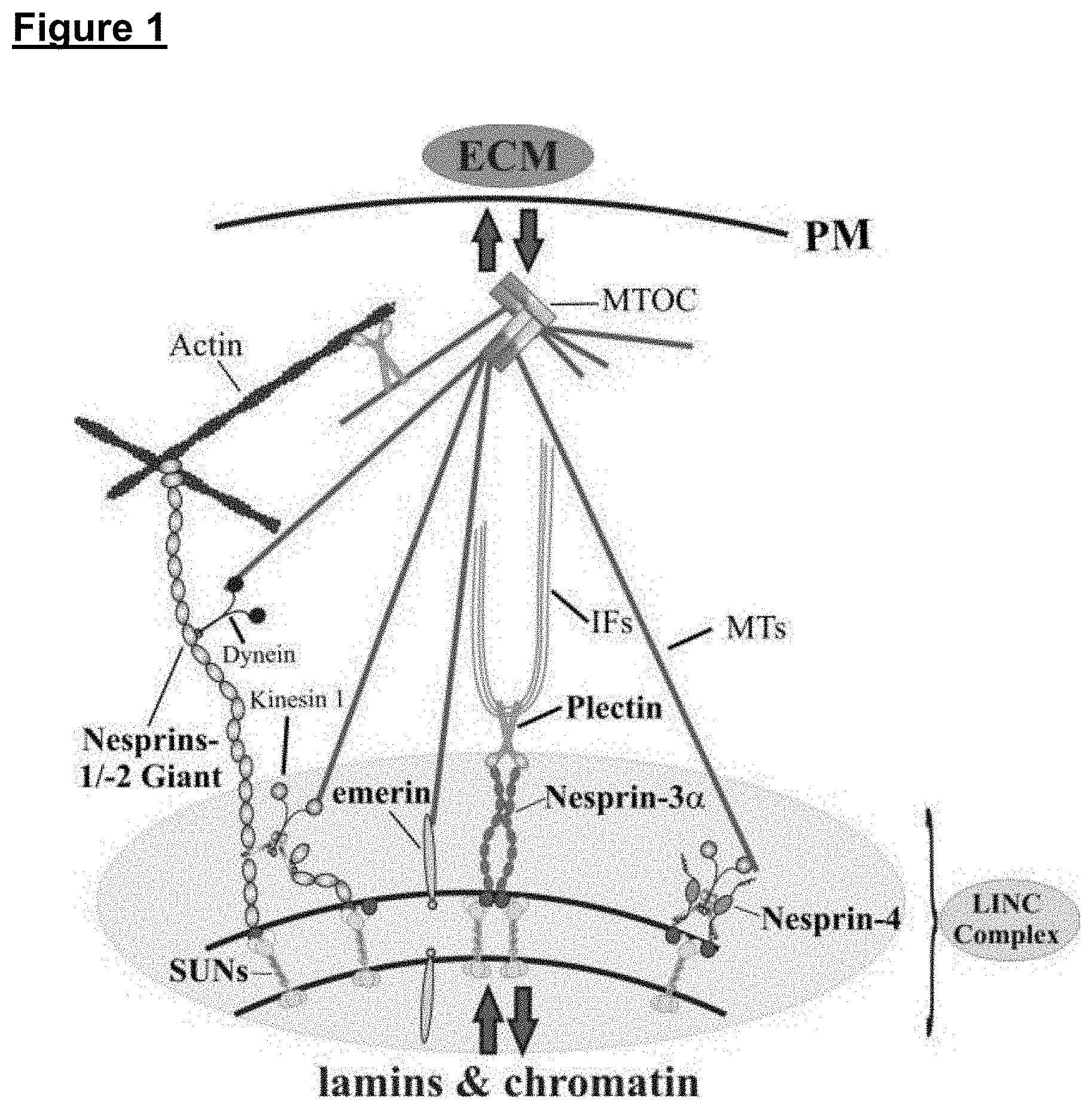
[0127] FIG. 1 is a schematic diagram, which shows that LINC complex is a conserved structure formed by Nesprin (i.e. nesprin-1, nesprin-2, nesprin-3 and nesprin-4) and SUN-domain proteins, which span the entire nuclear envelope and functionally link the nuclear interior (e.g. lamina, chromatin, telomeres, transcription factors) to the extracellular matrix (ECM) via associations to multiple cytoskeletal structures (e.g. microtubules [MTs], intermediate filaments [IFs], actin, dyneins, kinesins, Microtubule organising centre [MTOC]), cytolinker proteins (e.g. plectin) and plasma membrane [PM] receptors. High (termed giant) and low molecular weight Nesprin-1 and -2 isoforms are indicated. SUN-domain proteins are depicted as trimers;
[0128] FIG. 2 is a schematic diagram of the LINC complex bridge across the nuclear envelope. The LINC complex bridge comprises SUN and KASH interactions within the perinuclear space alongside associated cytoskeletal and nucleoskeletal networks. ONM: outer nuclear membrane, INM: inner nuclear membrane, PNS: perinuclear space;
[0129] FIG. 3 is a schematic diagram of the DN-SUNL construct, which encodes a dominant negative SUN1 luminal domain in relation to the full-length Sun1 protein. Major Sun1 protein domains and topologies within the nuclear envelope are indicated. The truncated SUN1 protein (lacks the N-terminus, which is found in the nucleoplasm, and the three hydrophobic domains) is fused to the Torsin A signal peptide (SP; the amino acid sequence is given), GFP (green fluorescent protein) and expressed in the endoplasmic reticulum (ER) and nuclear envelope lumen (Schneider et al., 2011, Cell. Mol. Life Sci. 68:1593-1610) where it binds and saturates all KASH-protein binding sites (see FIG. 10 for more details). Due to these associations all endogenous full-length KASH-proteins are dislodged from the nuclear envelope (hence dominant negative). SP-GFP is the relevant control (Schneider et al., 2011, Cell. Mol. Life Sci. 68:1593-1610) that lacks the SUN1 N-terminal nucleoplasmic and C-terminal luminal sequences but harbours the signal peptide sequences;
[0130] FIG. 4 is an indirect immunofluorescence analysis of wild type (WT) and DN-DUNL transiently transfected HaCaT cells that were counterstained with DAPI (blue) to stain nuclei. In panel (A) the DN-SUNL subcellular distribution (GFP channel) is highlighted. Note the distinct nuclear rim (red arrowheads) and reticular pattern of DN-SUN1 in the cytosol (blue arrows), which corresponds to the endoplasmic reticulum (ER) membranes. DN-SUNL expression affects nuclear shape (white arrows) and yields blebs at the nuclear surface (white arrowheads). Scale bar=10 .mu.m. (B) Statistical analysis (Student's t-test; P value<0.05 is indicated by *) demonstrates significant changes in nuclear morphology in DN-SUNL cells. Results are mean.+-.standard deviation;
[0131] FIG. 5 is an indirect immunofluorescence analysis of DN-DUNL (transient transfection) expressing HaCaT (A, C) and human dermal fibroblast cells (B). (A) Schematic representing the basic nesprin-2 giant domain architecture and the epitopes of three specific nesprin-2 antibodies (i.e. Nes2NT, Nes2K49 and Nes2CT). Lower panels depict an immunofluorescence analysis using nesprin-2 antibodies, which shows that DN-SUNL expression displaces all nesprin-2 isoforms from the nuclear envelope (arrows). These results highlight the dominant negative effects of the DN-SUNL construct on Nesprin-2. In contrast, control cells (asterisks) exhibit strong nesprin-2 nuclear staining. (B) DN-SUNL expression in fibroblasts shows that also endogenous nesprin-1 is efficiently displaced from the nucleus, while untransfected cells (asterisks) display pronounced nesprin-1 nuclear rim staining. (C) The expression of DN-SUNL affects specifically proteins of the KASH-domain family (i.e. nesprins) considering that the subcellular localisation of other key components of the nuclear envelope such as SUN1, emerin, LAP2.beta., and lamin A/C are largely unaffected. All scale bars=10 .mu.m. Nuclei are visualised by DAPI staining (blue);
[0132] FIG. 6 is a microscopic image analysis of WT (control) and stably transfected DN-SUNL HaCaT cells, which shows that LINC complex disruption affects drastically cell morphology, cell crowding and cell-cell contact protein expression on 2D surfaces. (A) Phase contrast and fluorescent staining's of nuclei (DAPI, blue) and the F-actin cytoskeleton (Phalloidin, red) of 70% confluent WT and DN HaCaT. Phalloidin denotes the cortical actin cytoskeleton in keratinocytes and therefore visualizes the cell boundaries. Scale bar=200 .mu.m (phase contrast), and 20 .mu.m (fluorescent panels). In contrast to DN cells, WT cells are flatter and less dense within the colonies; (B) DN HaCaT colonies contain twice as much cells compared to WT counterparts. Statistical analysis (Student's t-test; P value<0.05 is indicated by *) demonstrates a significant changes in the cell density of WT and DN-SUNL mutant colonies. Results are mean.+-.standard deviation. (C) DN cells express higher E-cadherin levels compared to WT cells grown on 2D surfaces;
[0133] FIG. 7 is an analysis, which shows LINC complex disruption affects cell-to-substrate adhesion on 2D substrates. A) WT and DN HaCaT cells 24 h post trypsinisation were examined using immunofluorescence. Cells are stained for DAPI (blue), phalloidin (cyan) and vinculin (red). Vinculin is a mechano-sensing protein that is enriched at peripheral cell-substratum contacts (termed focal contacts). Arrows display sites of focal adhesion formation across the cell periphery. Note that in contrast to DN cells, WT cells exhibit prominent enrichment of focal contacts at the cell periphery. Vinculin is diffusely distributed in DN cells. Scale bar=10 .mu.m. (B) Western blotting indicating that vinculin levels are not perturbed in DN cells. The localization of vinculin changes upon LINC complex disruption but not the levels of the protein. B-actin and tubulin indicates equal protein loading. Anti-GFP immunoblotting demonstrates expression of dominant negative LINC complex interfering proteins in DN cells;
[0134] FIG. 8 is an analysis, which shows that DN-SUNL HaCaT cells (DN) display enhanced stratification properties when grown in 3D scaffolds for 8 days at the air-to-liquid interface. (A) Single and co-cultured (with dermal fibroblasts) DN HaCaT cells grown in 3D form prominent multi-layered structures when grown at the air-liquid interface that favours differentiation of keratinocytes. The black lines indicate the scaffold surface. H&E stained sections are shown. (B) Fluorescence examination of single-cultured DN cells verifies that cell stacking (arrows) is indeed occurring above rather than within the scaffold. The scaffold boundaries are visualized using Nile Red, while the existence of DN cells is denoted using GFP. Scale bars=25 (C) Highlights that LINC disruption substantially favours the formation of epidermal-like (multi-layered cell assembly) tissue structures in vitro. Asterisks denote statistical significance (P value<0.05) using a Student's t-test;
[0135] FIG. 9 is an analysis, which shows that DN HaCaT cells display proper epidermal tissue architecture in vitro. (A) Fluorescence microscopic examination of single cultured WT and DN cells in 3D highlighting that DN HaCaT cells (GFP expressing) display shape changes similar to that found in skin. Note that WT cells grown on the surface of the scaffold (visualized with Nile Red) exhibit flattened nuclei (yellow arrows) whereas DN cells proximal to the scaffold surface (denoted by the white dotted line) display columnar shapes (asterisks). In skin, epithelial cell/nuclear flattening occurs only in differentiated cells. Therefore, DN cells at the scaffold/air interface mimic basal keratinocyte morphologies. In contrast, DN cells away from the scaffold surface exhibit pronounced cell flattening (white arrows) a feature common to terminally differentiated skin keratinocytes belonging to the spinous and granular layers of skin. (B) Immunofluorescence analysis of WT and DN cells co-cultured with human dermal fibroblast cells in 3D scaffolds for 8 days at the air-to-liquid interface. Two markers were examined; K14 (Keratin 14) is a marker for basal dividing keratinocytes, while K10 (Keratin 10) is a terminal differentiation marker, which is expressed by suprabasal keratinocytes in skin. Note that DN HaCaT's exhibit increased K10 expression, proper spatial arrangements and morphologies (namely flattening) at areas that are distal to the scaffold surface, which mimics the in vivo situation. Cells in proximity to the scaffold (dotted line) lack K10 staining. In contrast, WT cells exhibit sporadic K10 staining within the scaffold. Scale bars=25 .mu.m;
[0136] FIG. 10(A) is a schematic diagram showing that DN-SUNL expression saturates KASH-domain binding sites at the outer nuclear membrane (ONM) and prevents binding to full-length SUN-domain proteins. DN-SUNL expression consequently disrupts the linkage of the ONM to the inner nuclear membrane (INM) and dilates the perinuclear lumen (discontinuous double arrowed lines). (B) shows TEM micrographs of the nuclear membrane in WT (control) and DN-SUNL HaCaT cells. Arrowheads denote dilation of the nuclear envelope in DN-SUNL cells. C=cytoplasm, N=nucleus. Scale bar 500 nm. C) Histogram shows the width of the perinuclear lumen in WT and DN-SUNL expressing cells. Statistical analysis (Student's t test; P value<0.005 is indicated by **) demonstrates a significant dilation of the nuclear envelope in DN-SUNL mutants. Results are mean.+-.standard deviation;
[0137] FIG. 11 is an analysis of the cytoplasmic and nuclear organelle biomechanical properties of interphase WT and DN-SUNL HaCaT cells examined under biological conditions, showing that LINC complex disruption (DN-SUNL cells) yields significantly softer cells. (A) Phase contrast images of WT and DN-SUNL cells (top row). The position of the nuclei (white dotted lines) and nucleoli (purple dotted circles) is indicated. The corresponding atomic force microscopy (AFM) Young's modulus (kPa) analysis of the WT and DN-SUNL mutant colonies (middle row) and AFM height scans (.mu.m; lower row) is shown. (B) Schematic diagram indicating three distinct cellular areas where AFM Young's modulus measurements were taken. (C) Histogram showing that across the three selected cellular areas that DN-SUNL are significantly softer compared to their WT counterparts (n=15 for each cell type). (D) Histogram depicting the highest cantilever surface indentation (.mu.m) measured on WT and DN-SUNL cells using a cantilever force of 150 pN, indicates that DN cells are significantly more pliable compared to WT (n=35 for each cell type). Statistical analysis (Student's t test; P value<0.005 is indicated by **);
[0138] FIG. 12 is a Western blot analysis of HaCaT cells that have been transiently transfected with either sPGFP or DN-SUNL encoding plasmids. Three different experimental conditions were examined. Prior to Western blotting the cell homogenates were either left untreated (positive control) or treated with two different detergents: digitonin or Triton X-100 in the presence of the proteinase K enzyme. Digitonin permeabilises selectively the plasma membrane but leaves the ER and NE membranes intact. In contrast, Triton X-100 permeabilises all cellular membranes including the ER and the NE. Note that the GFP fusions and ER-lumen resident PDI (protein disulphate isomerase; control) proteins are detectable in the digitonin-treated samples but are completely absent in the Triton X-100-treated samples. Tubulin acts as a control for the cytoplasmic compartment, which is digested irrespectively of the detergents used; and
[0139] FIG. 13 is an indirect immunofluorescence analysis of SP-GFP (A) or DN-DUNL (B) transiently transfected NIH-3T3 fibroblasts using anti-Nesprin-2 giant (Nes2NT) antibodies, which shows the dominant negative effects of the DN-SUNL construct on the KASH-domain containing Nesprin-2 giant isoform. Note that the DN-SUNL expressing cell (asterisk) displays a GFP signal at the ER (red arrows) and the NE (blue arrows), but lacks nuclear nesprin-2 staining (white arrows). In contrast SP-GFP expressing control cells or DN-SUNL untransfected cells exhibit strong nesprin-2 staining at the nucleus (yellow arrows). Nuclei are visualised by DAPI staining (blue). Scale bars=10 .mu.m.
EXAMPLES
[0140] Cells of specific tissues have correspondingly specific properties, including biomechanical, biochemical and structural properties. Such specific properties are determined by signals received from the physical and biochemical environment surrounding the cell. The inventors have found that crucial intracellular signalling events occurs via a central "switch" (the LINC complex) that physically links, via the cytoskeleton, genetic material of a cell to the extracellular environment. As shown in the following examples, the inventors have interrupted the formation of the LINC complex using a construct that overexpresses a dominant negative SUN1 fusion protein (DN-SUNL). In doing so, the inventors have induced specific cellular properties without having to create a complex and highly specific external environment.
[0141] Materials and Methods
[0142] Plasmid Construction
[0143] All cloned fragments were sequenced in their entirety. pcDNA3.1 (-) (Invitrogen) was used to engineer the DN-SUNL and the specific control (SPGFP) constructs (see Schneider et al., 2011, Cell. Mol. Life Sci. 68:1593-1610). The DN-SUNL comprises torsin-A signal peptide (SP) sequence, sequences encoding GFP (Green Fluorescent Protein), the coiled-coil domain and the SUN-domain of a murine SUN1 protein (the SUN1 transgene protein lacks the N-terminal domain and the transmembrane domain). The full polypeptide sequence (819 amino acids) of one embodiment of the DN-SUNL is provided herein as SEQ ID No. 28 (previously referred to as SEQ ID No. 40 in patent application GB1701438.2), as follows:
TABLE-US-00028 [SEQ ID No. 28] MKLGRAVLGLLLLAPSVVQAVASVSKGEELFTGVVPILVELDGDVNGHKF SVSGEGEGDATYGKLTLKFICTTGKLPVPWPTLVTTLTYGVQCFSRYPDH MKQHDFFKSAMPEGYVQERTIFFKDDGNYKTRAEVKFEGDTLVNRIELKG IDFKEDGNILGHKLEYNYNSHNVYIMADKQKNGIKVNFKIRHNIEDGSVQ LADHYQQNTPIGDGPVLLPDNHYLSTQSALSKDPNEKRDHMVLLEFVTAA GITLGMDELYKEFVSLWGQGNFFSLLPVLNWTAMQPTQRVDDSKGMHRPG PLPPSPPPKVDHKASQWPQESDMGQKVASLSAQCHNHDERLAELTVLLQK LQIRVDQVDDGREGLSLWVKNVVGQHLQEMGTIEPPDAKTDFMTFHHDHE VRLSNLEDVLRKLTEKSEAIQKELEETKLKAGSRDEEQPLLDRVQHLELE LNLLKSQLSDWQHLKTSCEQAGARIQETVQLMFSEDQQGGSLEWLLEKLS SRFVSKDELQVLLHDLELKLLQNITHHITVTGQAPTSEAIVSAVNQAGIS GITEAQAHIIVNNALKLYSQDKTGMVDFALESGGGSILSTRCSETYETKT ALLSLFGVPLWYFSQSPRVVIQPDIYPGNCWAFKGSQGYLVVRLSMKIYP TTFTMEHIPKTLSPTGNISSAPKDFAVYGLETEYQEEGQPLGRFTYDQEG DSLQMFHTLERPDQAFQIVELRVLSNWGHPEYTCLYRFRVHGEPIQ
[0144] Thus, in one embodiment, the agent according to any aspect of the invention may comprise an amino acid sequence substantially as set out in SEQ ID No. 28, or a variant or fragment thereof.
[0145] The signal peptide has been included to ensure that the DN-SUNL peptide is transported to the endoplasmic reticulum and/or the nuclear envelope. The polypeptide sequence of the torsin-A signal peptide (SP) sequence used in the DN-SUNL is provided herein as SEQ ID No. 29 (previously referred to as SEQ ID No. 41 in patent application GB1701438.2), as follows:
TABLE-US-00029 [SEQ ID No. 29] MKLGRAVLGLLLLAPSVVQAV
[0146] The GFP protein has been included to ensure that the DN-SUNL peptide can be visualised under UV light. The polypeptide sequence of the GFP protein used in the DN-SUNL is provided herein as SEQ ID No. 30 (previously referred to as SEQ ID No. 42 in patent application GB1701438.2), as follows:
TABLE-US-00030 [SEQ ID No. 30] SKGEELFTGVVPILVELDGDVNGHKFSVSGEGEGDATYGKLTLKFICTTG KLPVPWPTLVTTLTYGVQCFSRYPDHMKQHDFFKSAMPEGYVQERTIFFK DDGNYKTRAEVKFEGDTLVNRIELKGIDFKEDGNILGHKLEYNYNSHNVY IMADKQKNGIKVNFKIRHNIEDGSVQLADHYQQNTPIGDGPVLLPDNHYL STQSALSKDPNEKRDHMVLLEFVTAAGITLGMDELYK
[0147] The polypeptide sequence of the murine C-terminal SUN1 luminal protein used in the DN-SUNL is provided herein as SEQ ID No. 31 (previously referred to as SEQ ID No. 43 in patent application GB1701438.2), as follows:
TABLE-US-00031 [SEQ ID No. 31] VSLWGQGNFFSLLPVLNWTAMQPTQRVDDSKGMHRPGPLPPSPPPKVDHK ASQWPQESDMGQKVASLSAQCHNHDERLAELTVLLQKLQIRVDQVDDGRE GLSLWVKNVVGQHLQEMGTIEPPDAKTDFMTFHHDHEVRLSNLEDVLRKL TEKSEAIQKELEETKLKAGSRDEEQPLLDRVQHLELELNLLKSQLSDWQH LKTSCEQAGARIQETVQLMFSEDQQGGSLEWLLEKLSSRFVSKDELQVLL HDLELKLLQNITHHITVTGQAPTSEAIVSAVNQAGISGITEAQAHIIVNN ALKLYSQDKTGMVDFALESGGGSILSTRCSETYETKTALLSLFGVPLWYF SQSPRVVIQPDIYPGNCWAFKGSQGYLVVRLSMKIYPTTFTMEHIPKTLS PTGNISSAPKDFAVYGLETEYQEEGQPLGRFTYDQEGDSLQMFHTLERPD QAFQIVELRVLSNWGHPEYTCLYRFRVHGEPIQ
[0148] 2D and 3D Cell Culture
[0149] WT and DN-SUNL HaCaT, NIH-3T3 and primary human dermal fibroblast (HDF; LifeTechnologies) cells were cultured at 37.degree. C., 5% CO2 in Dulbecco's Modified Eagles Medium, high glucose supplemented with 10% fetal calf serum, 2 mM penicillin, and 2 mM streptomycin (Sigma). For the cultivation of stable transfected DN-SUNL HaCaT cells, 0.5 mg/ml G418 disulphate (Sigma) was added to the media solution. 2D cell culture was performed on conventional poly-styrene flasks, 10 cm petri-dishes and 12/24 well dishes. For 3D culture, 12-well Alvetex.RTM. Strata inserts (Reinnervate) were employed. Materials were pre-treated with oxygen plasma for 5 min at 40 W using an Emitech K1050X Plasma Asher.
[0150] To seed the cells to Alvetex.RTM. Strata, 100 .mu.l of WT or DN-SUNL HaCaT cell suspension containing 250,000 cells each was applied directly to the centre of each pre-treated scaffold. The scaffolds were then placed in their respective culture dishes and moved into a cell culture incubator for 20 min to allow cell attachment to the scaffold surface. Complete culture media was added. For the co-culture observations, 500,000 HDF cells at a T-75 confluence of 60% were placed onto plasma-treated scaffolds alongside complete culture medium and cultured in 12 well plates for 7 days. The scaffolds were transferred into 6 well plates containing 4 mL complete culture media and were cultured for a further 7 days. Either WT or DN HaCaT cells were then seeded. After the seeding of 250,000 keratinocytes (applies for both single or co-culture experiments) the cells were grown submerged in media for 4 days, before the cells were grown at the air-liquid interface for another 8 days.
[0151] Paraffin Embedding and Immunofluorescence Microscopy
[0152] 3D cell cultures were rinsed once with cell culture grade phosphate-buffered saline (PBS) and carefully unclipped from the insert holder. Upon removal, scaffolds were washed a further two times in PBS for 5 min, submerged within 4% paraformaldehyde (PFA) in PBS, pH 7.4 and left at 4.degree. C. overnight. After fixation, scaffolds were washed another two times in PBS for 5 minutes each. Washing was then followed by 15 min incubations in varying ethanol concentrations of 30%, 50%, 70%, 80%, 90%, 95% and 100% v/v at room temperature. Scaffolds were subsequently removed from their housing inserts, cut in half across their diameter using sterilised surgical scissors and incubated for 15 min in Histoclear (Fisher, 12358637) at 60.degree. C. An equal volume of liquid paraffin wax (Fisher, 12624077) was added, and the scaffolds were incubated for 15 min at 60.degree. C. The Histoclear/liquid paraffin solution was then replaced with fresh paraffin wax, and the scaffolds were incubated for 1 h at 60.degree. C. Vertical embedding was then performed in which the scaffold sections were placed into embedding moulds (Cellpath Ltd, GAD-5302-02A) with the cut side facing down. These were then topped with a labelled embedding cassette (SLS, HIS0029), and filled with fresh paraffin wax. The resulting wax blocks were sectioned using a Leica RM2125RT microtome with MB Dynasharp microtome blades (Fisher, 12056679). For all cell lines, sections were cut to a thickness of 10 .mu.m for conventional haematoxylin and eosin (H&E) staining, with subsequent 7 .mu.m sections for antibody staining. Sections were then floated on a 42.degree. C. water bath, mounted onto Superfrost+ microscope slides (Fisher, 10149870) and left to dry overnight on a 32.degree. C. heated slide dryer. Sections were subsequently deparaffinised in Histoclear and hydrated through a series of 5 min incubations in 100%, 70% ethanol and PBS. Antigen retrieval was performed using microwave heating; samples were heated three times 5 min in citrate buffer, with cooling at RT for 30 sec between each heat treatment. Sections were cooled, treated with permeabilisation/blocking solution (20% normal goat serum [Sigma] in 0.4% Triton X-100 PBS) for 45 min before processing for indirect immunostaining.
[0153] 2D cell cultures were fixed in 4% paraformaldehyde/PBS for 15 min and permeabilized in 0.5% Triton X-100/PBS for 10 min before the samples were processed for indirect immunostaining. Focal adhesion sites were identified through vinculin staining, scaffolds were counterstained with NILE red (Sigma, nuclei were stained with 4,6-diamino-2-phenylindone (DAPI; Sigma) and F-actin with TRITC-Phalloidin (Sigma). All indirect immunofluorescence samples were analysed by confocal laser-scanning microscopy using a TCS-SP5 (Leica).
[0154] Antibodies
[0155] Primary antibodies used were directed against the C-terminus of nesprin-1 (specII), the N-terminus of Nesprin-2 (Nes2NT) mAb K56-386 [Luke et al., 2008, J. Cell Sci. 121, 1887-1898] and mAb K20-478 [Zhen et al., 2001, J. Cell Sci. 115, 3207-3222], the C-terminus of Nesprin-2 (Nes2CT) pAb K1 [Libotte et al., 2005, Mol. Biol. Cell 16, 3411-3424], .beta.-actin mAb AC-74 (Sigma), GFP mAb K3-184-2 [Schneider et al., 2011, Cell. Mol. Life Sci. 68:1593-1610], Sun1 (Padmakumar et al., 2005, J. Cell Sci. 118, 3419-3430), lamin A/C (Jol2), tubulin mAb WA3 (kind gift from Dr. U. Euteneuer), vinculin mAb V9131 (Sigma), E-Cadherin rtAb U3254(Sigma), keratin10 rbAb 76318 (Abcam) and keratin14 mAb 7800 (Abeam). For indirect immunofluorescence studies, Alexa 488, Alexa 568, and Alexa 647 fluorescently conjugated secondary antibodies (Invitrogen) were utilized. Peroxidase-coupled secondary antibodies (Sigma) were adopted in Western blot analysis.
[0156] H&E Histochemistry
[0157] Paraffin wax was cleared from the superfrost microscopy 3D scaffold-containing slides by washing with Histoclear for 5 min at room temperature. Gradual sample rehydration was conducted through washes in 100% ethanol for 2 min, 95% and 70% ethanol for 1 min, and distilled water for a further 1 min. Nuclei were then stained via a 5 minute incubation in Mayer's haematoxylin (Sigma, H1532) (0.1% v/v haematoxylin, 0.02% v/v sodium iodate, 5% v/v aluminium potassium sulphate, 5% v/v chloral hydrate and 0.1% v/v citric acid in dH.sub.2O), followed by a 1 min wash in distilled water and incubation in alkaline alcohol (3% ammonia in 70% ethanol) for 30 sec to stain nuclei. Samples were subsequently dehydrated by 30 sec incubations in 70% and 95% ethanol. Once dehydrated, cytoplasmic staining in 0.5% eosin (Sigma, E4009) in 95% ethanol for 1 min was carried out. The samples then underwent two 10 sec washes in 95% ethanol, followed by two washes in 100% ethanol, the first for 15 sec and second for 30 sec. Slides were then cleared via 2.times.3 min washes in Histoclear, prior to mounting with DPX mounting media (Fisher, 10050080) and covering with a 50.times.22 mm coverslip (Fisher, 12383138). Slides were left to dry at 4.degree. C. overnight and then imaged using a Leica DM500 light microscope with attached ICC50 HD camera at 10.times. and 20.times. magnifications utilising the LAS EZ software (Leica).
[0158] Western Blotting
[0159] Protein lysate preparation from 2D cultured dishes is covered in detail in Carthew and Karakesisoglou (2016; Methods Mol Biol. 2016; 1411: 221-32). To extract protein lysates from 3D cultured cells, scaffolds were washed three times in PBS, removed from their housing inserts and cut into small (.about.1 mm), square pieces with sterilised scissors. Scaffold sections were then incubated in 500 .mu.l RIPA [50 mM Tris/HCl (pH 7.5), 150 mM NaCl, 1% Nonidet-P40, 0.5% sodium desoxycholate] buffer supplemented with 1% protease inhibitor cocktail (Sigma) for 15 min at 4.degree. C., during which sonication every 3 min for 30 sec was performed using an MSE soniprep 150 sonicator. The scaffold/cell suspension was centrifuged at 4.degree. C. for 15 min at 12 000.times.g to pellet the remaining cell/scaffold debris, with resulting supernatant extracted, combined with 120 .mu.l of 5.times. concentrated Laemmli sample buffer and boiled at 99.degree. C. for 4 min. Samples were then stored at -20.degree. C. until use.
[0160] Selective Permeabilisation and Proteinase Digestions to Elucidate the SP-GFP and DN-SUNL Subcellular Distribution
[0161] Transiently transfected HaCaT cells that were transiently transfected with the SP-GFP and DN-SUNL transgenes were washed twice with ice-cold PBS buffer once the cell culture plates reached 60-70% confluency. Cells were collected from the plates using a cell scraper, transferred to a centrifuge tube and subjected to a 5 min centrifugation at 1,000 g. The supernatants were carefully removed and the cell pellets were re-suspended in either ice-cold hypotonic buffer [10 mM HEPES (pH 7.5), 1.5 mM KCl, 1.5 mM MgCl2, and 0.5 mM dithiothreitol] or protease inhibitor-containing (Roche) RIPA lysis buffer [50 mM Tris/HCl (pH 7.5), 150 mM NaCl, 1% Nonidet-P40, 0.5% sodium desoxycholate]. The former cell homogenate's were supplemented with either 5 .mu.g/ml digitonin or 1% Triton X-100, incubated on ice for 4 min before the addition of proteinase K (5 .mu.g/ml). After a 30 min incubation on ice the proteinase K-mediated digestion was terminated using 10 .mu.g/ml phenylsulfonyl fluoride (PMSF). The digested samples were subjected to a 20 min centrifugation at 12,000 g and the supernatants were supplemented with Laemmli sample buffer. The RIPA-containing cell homogenates were incubated on ice for 15 min, centrifuged at 12,000 g, and the supernatants were mixed with sample buffer. All cell extracts were passed at least 20 times through a 27-gauge needle, before the lysates were analysed by SDS-PAGE, and Western blotting.
[0162] Transmission Electron Microscopy
[0163] 2D cultured cells were fixed though 30 min incubations in 2% glutaraldehyde diluted in NaHCa buffer, washed two times in NaHCa buffer and incubated for 15 min in a solution of 1% tannic acid and 0.075% saponin at RT. Cells were then rinsed two times in NaHCa buffer, followed by two further washes in 0.1 M cacodylate buffer and transferred to a 1.5 ml centrifuge tube. Cell suspensions were centrifuged at 1000 g for 4 min, with resulting pellet incubated for 1 h in a solution of 0.5% osmium tetroxide in 0.1 M cacodylate buffer. Pellets were further washed twice in 0.1 M cacodylate buffer, dehydrated through three 5 min washes in 50%, 70%, 95% and 100% ethanol solutions, and infiltrated two times with a 1:1 mixture of 100% alcohol and propylene oxide for 10 min. Cells were then incubated in a 1:1 mixture of propylene oxide and epoxy resin (EPON.TM. 828) at 60.degree. C., twice in 100% epoxy resin at 60.degree. C. for 30 min, and finally in fresh epoxy resin at 60.degree. C. for 24-48 h to allow polymerisation. Ultra-thin sections were cut to 70 nm using a Leica EM UC6, and mounted onto Formvar coated copper grids. Grids were incubated in uranyl acetate for 10 min, rinsed twice in distilled water, and further incubated in lead citrate (0.4% lead citrate w/v, 0.5% sodium citrate in 0.1 N NaOH) for 10 mins. Subsequent image acquisition was performed using a Hitachi TEM-H7600 transmission electron microscope.
[0164] Atomic Force Microscopy
[0165] Cells were cultured to a confluence of 60% on 35 mm plastic petri dishes (TPP). 30 min before analysis, extensively PBS-washed cultures were placed into sterile filtered, CO2 independent culture media (Fisher). Subsequent examination was conducted using a Nanowizard.RTM. 3 Bioscience atomic force microscope (JPK) using a (D)NP silicon nitride probe cantilever, expressing a spring constant of 0.06-0.7 N/m (Bruker, DNP-10) over a 15.times.15 .mu.m grid. Cells were maintained at a constant temperature of 37.degree. C. throughout image collection. Young's modulus values were generated using the Gwyddion image analysis software.
[0166] Statistical Analysis
[0167] Statistical analysis was performed using Student's t test; 300 cells were used for every data set unless otherwise stated. Results were shown as mean.+-.SD. P values of <0.05 were considered significant. The mean.+-.SEM cell stacking from the 3D cell culture experiments and all the 2D morphometrics data sets were measured using the processing software ImageJ (v1.49).
Example 1
Components of the LINC Complex
[0168] The LINC complex is widely recognised as the major nuclear envelope (NE) component able to provide the mechanical links between the nucleus and cytoskeletal network, comprising an outer nuclear membrane (ONM) KASH domain proteins and inner nuclear membrane (INM) SUN proteins (see FIGS. 1 and 2). The KASH domain projects into the NE lumen, where it interacts with SUN domains. This SUN-KASH interaction helps to anchor KASH proteins to the NE, preventing them from diffusing into the adjoining endoplasmic reticulum (ER). The ONM KASH proteins interact with a range of cytoskeletal components, and thus, physically tether the nucleus to the cytoplasmic compartment, whereas the SUN proteins physically interact with INM proteins networks, such as the nuclear lamina and chromatin components. This complex of proteins therefore establishes a physical bridge between the cytoskeletal and nucleoskeletal networks. The formation of this two-membrane adhesive assembly of proteins is capable of transmitting force across the NE, providing function in maintaining centrosome-nuclear interactions, nuclear architecture, signal transduction, DNA repair and chromosome migration. This therefore suggests that the LINC complex must be a dynamic protein network of highly ordered protein interactions, allowing the transmission of multiple signal transductions from a variety of cytoskeletal components to the nuclear interior.
Example 2
SUN1 Luminal Domain Dominant Negative Construct (DN-SUNL)
[0169] In order to study the role that the LINC complex protein, SUN1, plays in cellular structure and function, the inventors developed a construct (see FIG. 3) that encodes and overexpresses a fusion protein comprising a GFP-tagged luminal domain of SUN1 attached to an ER signal peptide (SP). The signal peptide was incorporated to ensure the fusion protein is overexpressed in the endoplasmic reticulum (ER) and the perinuclear space. To confirm that the fusion protein had translocated into the ER lumen and the perinuclear space, cell homogenates of HaCaT keratinocytes were subjected to proteinase K degradation in the presence of Triton X-100 or digitonin (see FIG. 12). In contrast to Triton X-100, which permeabilizes all biological membranes, low concentrations of digitonin leave the internal ER (endoplasmic reticulum) and NE (nuclear envelope) membranes unperturbed. Digitonin permeabilizes only cholesterol-rich membranes (i.e. plasma membrane). Similar to the ER and perinuclear space resident disulphide isomerase (PDI) protein, the sPGFP and DN-SUNL molecules were susceptible to proteinase K degradation only when cell homogenates were permeabilized with Triton X-100 (see FIG. 12). FIGS. 4 and 5 show that the GFP-fusion DN-SUNL protein is expressed in the ER and the nuclear envelope.
Example 3
SUN1 Interacts with Nesprin-1 and Nesprin-2
[0170] The inventors elucidated the involvement of luminal KASH/SUN protein interactions with Nesprin-1 and Nesprin-2, the cytoplasmic binding partners of SUN1. Transiently transfected control DN-SUNL HaCaT and fibroblasts were immunostained for Nesprins-1 and Nesprin-2, respectively. Their decision to use these particular cellular models was based on the prevalence of Nesprin-1 and Nesprin-2 C-terminal KASH-domain isoforms (i.e. nuclear envelope associated isoforms) in fibroblasts and keratinocytes. In sharp contrast to untransfected cells (see FIGS. 5A and 5B asterisks), the DN-SUNL expression (see green panels in FIGS. 5A and 5B, arrows) dislodged Nesprin-1 and Nesprin-2 from the NE (see red panels in FIGS. 5a and 5b, arrows). Importantly, the localisation of other key nuclear proteins including SUN1 (FIG. 5C) and the Nesprin-2, vinculin, tubulin, and actin expression levels remained unaffected by DN-SUNL expression (FIG. 7B). Furthermore, in contrast to DN-SUNL, SP-GFP (control) does not affect the localisation of endogenous Nesprin-2. This highlights, that the SUN1 C-terminal sequences exert the dominant negative effects on KASH-domain proteins and not the SP and GFP segments of DN-SUNL (FIG. 13), considering that both (SP-GFP and DN-SUNL) are expressed in the same membrane-bound compartments (i.e. ER and NE; FIG. 12).
Example 4
LINC Disruption Affects Cell Shape, Cell-Cell Junction Protein Expression and Cellular Density
[0171] A microscopic comparison (i.e. phase contrast images and fluorescence examination of TRITC-coupled phalloidin [phalloidin binds filamentous actin]) of WT and stable DN-SUN HaCaT clones (FIG. 6A) grown at 60% confluency under standard 2D conditions indicates drastic changes in the overall appearance of the mutants. While both WT and DN-SUNL form colonies the latter appear to contain more cells, which exhibit strong cortical F-actin staining and a smaller circumference. Statistical analysis indicates that DN-SUNL HaCaT cells display a significantly smaller cellular area of 262 .mu.m.sup.2 compared to a calculated cell area of 473 .mu.m.sup.2 for WT cells. This drastic difference in the DN-SUNL cell area is reflected also in the number of cells that occupy a 100.times.100 .mu.m area. On average 7.4 WT cells are found within a 10000 .mu.m.sup.2 colony area, while for DN-SUNL 15.8 cells were observed (FIG. 6B). The pronounced cellular crowding effect of DN-SUNL may be the outcome of higher E-cadherin expression, which hinds the presence of enhanced cell-cell contacts when the cells are grown on 2D surfaces (FIG. 6C). Interestingly, the DN-SUNL effects on the expression of E-cadherin appear to be attenuated upon 3D cell culture (FIG. 6C).
Example 5
LINC Complex Disruption Alters Cell-Substratum Adhesion
[0172] Considering that DN-SUNL expressing cells display a drastically smaller cellular area, the inventors elucidated whether the adhesion of the cells to the surface has been altered. Examination of vinculin indeed indicates alterations in the localisation of the protein within the cytoplasm in DN-SUNL mutant cells. In WT cells the majority of vinculin is localised at focal contacts, which can be seen as distinct large clusters at the periphery of the cells (denoted by arrows, FIG. 7A). Moreover, WT cells appear very flat, which is evident not only by the presence of focal contacts but also from the large area that is occupied by the cytoplasm. In sharp contrast, DN-SUNL cells exhibit vinculin staining, which is confined in the cytoplasm and lack distinct focal contacts at the periphery (FIG. 7B). Western blot analysis of equal amounts of WT, and DN-SUNL cell lysates (two clones were examined) indicates that the levels of vinculin are not affected in the mutants. In summary, the data show that the subcellular distribution of vinculin is affected but not its expression levels upon LINC complex disruption in HaCaT keratinocytes. Moreover, the data suggest that a reduced cell-substratum attachment and an enhanced cell-cell adhesion may account for the cell density and morphological phenotype of the mutants.
Example 6
LINC Complex Disruption Enhances the Stratification Properties of HaCaT Keratinocytes in 3D Cell Culture Conditions
[0173] Upon exposure to the air-liquid interphase keratinocytes start to differentiate and form a multi-layered structure. Cellular division is restricted to the cells that are in immediate contact with the cell culture media, while differentiated cells significantly flatten and occupy the areas that are further away from the media surface. Irrespectively, of whether DN-SUN HaCaT cells are grown alone (single culture) or in the presence of fibroblasts (co-culture) in 3D their ability of forming multi-layered cell assemblies is profoundly enhanced when compared to WT cells. FIG. 8A indicates that DN-SUNL cells form more layers when compared to WT. In addition, FIG. 8A shows that the DN cell layers occupy the area above the scaffold and that the differentiation programme is executed properly considering the drastic cell shape changes that occur in the upper layers (note the significant cell flattening that occurs in the co-culture permutation). The ability of the DN cells to form pronounced structures above the scaffold is shown in FIG. 8B. As can be seen DN-cells (GFP-positive due to DN-SUNL expression) grow both into (GFP-panel) and above the scaffold (Nile red co-stained sample). Note the presence of GFP-positive structures above the Nile red stained 3D scaffold, which shows that cell stacking occurs above the scaffold (FIG. 8B) and is enhanced upon LINC disruption (FIG. 8C).
Example 7
LINC Complex Disruption Enhances Cell Stratification and Differentiation
[0174] To elucidate whether the morphological alterations (pronounced cell/nuclear flattening) exhibited by DN-SUNL cells (FIG. 9A, small arrows) correspond to differentiated cells the epidermal models were counterstained with Keratin 10. As can be seen in FIG. 9B, the anti-Keratin 10 immunofluorescence indicates that keratin 10 is present in the outermost cell layers of the DN-SUNL epidermal equivalent. In summary, FIG. 9 shows that DN-SUNL cells (relative to WT cells) have enhanced stratification properties, a proper spatial expression of the K10 differentiation marker when grown at the air-liquid interface in 3D and a cell morphology that mimics the shapes of keratinocytes of skin (e.g. columnar shape for cells in the direct vicinity of the scaffold [FIG. 9A, asterisks], and a squamous appearance for differentiated cells [FIG. 9A, small arrows]).
Example 8
DN-SUNL Expression Disrupts the Linkage of the INM to the ONM
[0175] To demonstrate that DN-SUNL disrupts the physical linkage of the inner nuclear membrane to the outer nuclear membrane (FIG. 10A) we performed an EM analysis of the DN-SUNL cells relative to WT. EM images indicate that the lumen of the nuclear envelope (NE) is even and narrow in WT, while DN-SUNL cells exhibit dilations (FIG. 10B). Measurements of the NE lumen indicate a significant dilation in DN-SUN expressing cells (FIG. 10C).
Example 9
LINC Disruption Yields Compact, Taller and Softer Cell Colonies
[0176] To examine the physical properties of WT and DN-SUNL cells the inventors performed an AFM analysis on living cells. The data in FIG. 11A verify that DN-SUNL colonies compared to WT are more compact and contain more cells (phase contrast). Moreover, Young's modulus measurements (FIG. 11A, middle panel) indicate that mutants are significantly softer irrespectively of the cellular regions that were assessed (Nuclear centre, nuclear rim and cytoplasm FIGS. 11B and 11C). Collectively, these data suggest that DN-SUNL cells exhibit softer nuclei and softer cytoplasms, suggesting that DN-SUNL expression yields softer cells, which is further substantiated by the pronounced cantilever indentation in the mutants (FIG. 11D). Finally, the height map in FIG. 11A (lower panel) shows that DN-SUNL cells are taller when compared to WT, which again underlines that LINC complex manipulation controls cell/colony architecture.
Sequence CWU 1
1
311241DNAHomo sapiens 1gtccacaaaa ggtggctccg attcctccct ttctgagcca
gggccaggtc ggtccggccg 60cggcttcctg ttcagagtcc tccgagcagc tcttcccctt
cagcttctcc tgctcctcct 120catcgggctt gcctgccttg taccaatgtc
agaggaagac tacagctgtg ccctctccaa 180caactttgcc cggtcattcc
accccatgct cagatacacg aatggccctc ctccactctg 240a 241251PRTHomo
sapiens 2Ala Ala Leu Pro Leu Gln Leu Leu Leu Leu Leu Leu Ile Gly
Leu Ala1 5 10 15Cys Leu Val Pro Met Ser Glu Glu Asp Tyr Ser Cys Ala
Leu Ser Asn 20 25 30Asn Phe Ala Arg Ser Phe His Pro Met Leu Arg Tyr
Thr Asn Gly Pro 35 40 45Pro Pro Leu 503208DNAHomo sapiens
3ggtccccggc agcacacggc cacagcgctc cttcctctca agggtggtcc gggcagccct
60acccctgcag ctgctcctcc tgctgctgct gctcctggcc tgcctgctgc cctcctccga
120agaagactac agctgcactc aggccaacaa ctttgcccgg tccttttacc
ccatgctgag 180gtacaccaat gggccacccc ccacatag 208451PRTHomo sapiens
4Ala Ala Leu Pro Leu Gln Leu Leu Leu Leu Leu Leu Leu Leu Leu Ala1 5
10 15Cys Leu Leu Pro Ser Ser Glu Glu Asp Tyr Ser Cys Thr Gln Ala
Asn 20 25 30Asn Phe Ala Arg Ser Phe Tyr Pro Met Leu Arg Tyr Thr Asn
Gly Pro 35 40 45Pro Pro Thr 505201DNAHomo sapiens 5actcggcggt
ggcgaggact gggctccctc ttccggaggg cgtgctgtgt ggcgctccca 60ctgcagctgc
ttctgctgct gttcctcctc ctgctgttcc tgctcccaat cagggaagag
120gaccgcagct gcaccctggc caacaacttc gcccgctcct tcacgctcat
gctgcgctac 180aatggcccac cacccaccta a 201650PRTHomo sapiens 6Val
Ala Leu Pro Leu Gln Leu Leu Leu Leu Leu Phe Leu Leu Leu Leu1 5 10
15Phe Leu Leu Pro Ile Arg Glu Glu Asp Arg Ser Cys Thr Leu Ala Asn
20 25 30Asn Phe Ala Arg Ser Phe Thr Leu Met Leu Arg Tyr Asn Gly Pro
Pro 35 40 45Pro Thr 507184DNAHomo sapiens 7ggcccccgat cctgcatcca
ggcagcctct gaccttcctc cttatcctct tcctcctctt 60cctcctcctg gtgggtgcca
tgtttctcct gcccgcgtca ggaggcccct gctgctctca 120tgcccgaata
cccaggacac cctacctggt gctcagctat gtcaatggtc ttcccccagt 180ctga
184849PRTHomo sapiens 8Phe Leu Leu Ile Leu Phe Leu Leu Phe Leu Leu
Leu Val Gly Ala Met1 5 10 15Phe Leu Leu Pro Ala Ser Gly Gly Pro Cys
Cys Ser His Ala Arg Ile 20 25 30Pro Arg Thr Pro Tyr Leu Val Leu Ser
Tyr Val Asn Gly Leu Pro Pro 35 40 45Val9494DNAHomo sapiens
9gtggcagcat cttgagtact cgctgttctg aaacttacga aaccaaaacg gcgctgatga
60gtctgtttgg gatcccgctg tggtacttct cgcagtcccc gcgcgtggtc atccagcctg
120acatttaccc cggtaactgc tgggcattta aaggctccca ggggtacctg
gtggtgaggc 180tctccatgat gatccaccca gccgccttca ctctggagca
catccctaag acgctgtcgc 240caacaggcaa catcagcagc gcccccaagg
acttcgccgt ctatggatta gaaaatgagt 300atcaggaaga agggcagctt
ctgggacagt tcacgtatga tcaggatggg gagtcgctcc 360agatgttcca
ggccctgaaa agacccgacg acacagcttt ccaaatagtg gaacttcgga
420ttttttctaa ctggggccat cctgagtata cctgtctgta tcggttcaga
gttcatggcg 480aacctgtcaa gtga 49410163PRTHomo sapiens 10Gly Ser Ile
Leu Ser Thr Arg Cys Ser Glu Thr Tyr Glu Thr Lys Thr1 5 10 15Ala Leu
Met Ser Leu Phe Gly Ile Pro Leu Trp Tyr Phe Ser Gln Ser 20 25 30Pro
Arg Val Val Ile Gln Pro Asp Ile Tyr Pro Gly Asn Cys Trp Ala 35 40
45Phe Lys Gly Ser Gln Gly Tyr Leu Val Val Arg Leu Ser Met Met Ile
50 55 60His Pro Ala Ala Phe Thr Leu Glu His Ile Pro Lys Thr Leu Ser
Pro65 70 75 80Thr Gly Asn Ile Ser Ser Ala Pro Lys Asp Phe Ala Val
Tyr Gly Leu 85 90 95Glu Asn Glu Tyr Gln Glu Glu Gly Gln Leu Leu Gly
Gln Phe Thr Tyr 100 105 110Asp Gln Asp Gly Glu Ser Leu Gln Met Phe
Gln Ala Leu Lys Arg Pro 115 120 125Asp Asp Thr Ala Phe Gln Ile Val
Glu Leu Arg Ile Phe Ser Asn Trp 130 135 140Gly His Pro Glu Tyr Thr
Cys Leu Tyr Arg Phe Arg Val His Gly Glu145 150 155 160Pro Val
Lys11490DNAHomo sapiens 11gggccagcgt catcagcacc cgatgttctg
agacctacga gaccaagacg gccctcctca 60gcctcttcgg catccccctg tggtaccact
cccagtcacc ccgagtcatc ctccagccag 120atgtgcaccc aggcaactgc
tgggccttcc aggggccaca aggcttcgcc gtggtccgcc 180tctctgcccg
catccgcccc acagccgtta ccttagagca tgtgcccaag gccttgtcac
240ccaacagcac tatctccagt gcccccaagg acttcgccat ctttgggttt
gacgaagacc 300tgcagcagga ggggacactc cttggcaagt tcacttacga
tcaggacggc gagcctattc 360agacgtttca ctttcaggcc cctacgatgg
ccacgtacca ggtggtggag ctgcggatcc 420tgactaactg gggccacccc
gagtacacct gcatctaccg cttcagagtg catggggagc 480ccgcccacta
49012162PRTHomo sapiens 12Ala Ser Val Ile Ser Thr Arg Cys Ser Glu
Thr Tyr Glu Thr Lys Thr1 5 10 15Ala Leu Leu Ser Leu Phe Gly Ile Pro
Leu Trp Tyr His Ser Gln Ser 20 25 30Pro Arg Val Ile Leu Gln Pro Asp
Val His Pro Gly Asn Cys Trp Ala 35 40 45Phe Gln Gly Pro Gln Gly Phe
Ala Val Val Arg Leu Ser Ala Arg Ile 50 55 60Arg Pro Thr Ala Val Thr
Leu Glu His Val Pro Lys Ala Leu Ser Pro65 70 75 80Asn Ser Thr Ile
Ser Ser Ala Pro Lys Asp Phe Ala Ile Phe Gly Phe 85 90 95Asp Glu Asp
Leu Gln Gln Glu Gly Thr Leu Leu Gly Lys Phe Thr Tyr 100 105 110Asp
Gln Asp Gly Glu Pro Ile Gln Thr Phe His Phe Gln Ala Pro Thr 115 120
125Met Ala Thr Tyr Gln Val Val Glu Leu Arg Ile Leu Thr Asn Trp Gly
130 135 140His Pro Glu Tyr Thr Cys Ile Tyr Arg Phe Arg Val His Gly
Glu Pro145 150 155 160Ala His13497DNAHomo sapiens 13gagcctccat
cattgaagct gggacctcag aaagttataa aaataataaa gcaaaattgt 60actggcatgg
gataggtttc ctaaatcatg aaatgcctcc agatattatt cttcagccgg
120atgtctaccc tggaaagtgc tgggcttttc caggttccca gggtcatacc
ctaatcaagc 180ttgctacaaa gatcatacca actgctgtta ccatggagca
catctcagag aaggtgtctc 240cgtcaggaaa catctccagt gcacccaagg
aattttctgt ctatggcatc acaaaaaaat 300gtgaaggaga agaaattttc
ctaggtcagt ttatatataa caaaacagga accaccgttc 360aaacatttga
actccagcat gcagtttctg aatatttatt atgtgtgaaa cttaatatct
420ttagcaactg gggacacccg aagtatactt gtttatatcg attcagggtc
catggcacac 480caggcaagca catctag 49714164PRTHomo sapiens 14Ala Ser
Ile Ile Glu Ala Gly Thr Ser Glu Ser Tyr Lys Asn Asn Lys1 5 10 15Ala
Lys Leu Tyr Trp His Gly Ile Gly Phe Leu Asn His Glu Met Pro 20 25
30Pro Asp Ile Ile Leu Gln Pro Asp Val Tyr Pro Gly Lys Cys Trp Ala
35 40 45Phe Pro Gly Ser Gln Gly His Thr Leu Ile Lys Leu Ala Thr Lys
Ile 50 55 60Ile Pro Thr Ala Val Thr Met Glu His Ile Ser Glu Lys Val
Ser Pro65 70 75 80Ser Gly Asn Ile Ser Ser Ala Pro Lys Glu Phe Ser
Val Tyr Gly Ile 85 90 95Thr Lys Lys Cys Glu Gly Glu Glu Ile Phe Leu
Gly Gln Phe Ile Tyr 100 105 110Asn Lys Thr Gly Thr Thr Val Gln Thr
Phe Glu Leu Gln His Ala Val 115 120 125Ser Glu Tyr Leu Leu Cys Val
Lys Leu Asn Ile Phe Ser Asn Trp Gly 130 135 140His Pro Lys Tyr Thr
Cys Leu Tyr Arg Phe Arg Val His Gly Thr Pro145 150 155 160Gly Lys
His Ile15521DNAHomo sapiens 15gagcctccat cgacctgcag aagacatccc
acgattacgc agacaggaac actgcctact 60tctggaatcg cttcagcttc tggaactacg
cacggccgcc cacggttatc ctggagcccc 120acgtgttccc tgggaattgc
tgggcttttg aaggcgacca aggccaggtg gtgatccaac 180tgccgggccg
agtgcagctg agcgacatca ctctgcagca tccaccgccc agcgtggagc
240acaccggagg agccaacagc gccccccgcg atttcgcggt ctttggcctc
caggtttatg 300atgaaactga agtttccttg gggaaattca ccttcgatgt
tgagaaatcg gagattcaga 360ctttccacct gcagaatgac cccccagctg
cctttcccaa ggtgaagatc cagattctaa 420gcaactgggg ccacccccgt
ttcacgtgct tgtatcgagt ccgtgcccac ggtgtgcgaa 480cctcagaggg
ggcagagggc agtgcacagg ggccccatta a 52116172PRTHomo sapiens 16Ala
Ser Ile Asp Leu Gln Lys Thr Ser His Asp Tyr Ala Asp Arg Asn1 5 10
15Thr Ala Tyr Phe Trp Asn Arg Phe Ser Phe Trp Asn Tyr Ala Arg Pro
20 25 30Pro Thr Val Ile Leu Glu Pro His Val Phe Pro Gly Asn Cys Trp
Ala 35 40 45Phe Glu Gly Asp Gln Gly Gln Val Val Ile Gln Leu Pro Gly
Arg Val 50 55 60Gln Leu Ser Asp Ile Thr Leu Gln His Pro Pro Pro Ser
Val Glu His65 70 75 80Thr Gly Gly Ala Asn Ser Ala Pro Arg Asp Phe
Ala Val Phe Gly Leu 85 90 95Gln Val Tyr Asp Glu Thr Glu Val Ser Leu
Gly Lys Phe Thr Phe Asp 100 105 110Val Glu Lys Ser Glu Ile Gln Thr
Phe His Leu Gln Asn Asp Pro Pro 115 120 125Ala Ala Phe Pro Lys Val
Lys Ile Gln Ile Leu Ser Asn Trp Gly His 130 135 140Pro Arg Phe Thr
Cys Leu Tyr Arg Val Arg Ala His Gly Val Arg Thr145 150 155 160Ser
Glu Gly Ala Glu Gly Ser Ala Gln Gly Pro His 165 17017527DNAHomo
sapiens 17gggccagcat tgactttgag cacacgtcag tcacctataa ccatgagaag
gcccactcct 60actggaactg gatccagctg tggaactacg cacagccccc agacgtgatc
cttgagccca 120acgtgacacc tggcaattgc tgggcctttg agggtgaccg
cggccaggtg accatccaat 180tggctcagaa ggtttacctg tccaacctca
cgctgcagca catccccaag accatctcat 240tgtcaggcag cctggacacc
gcccccaagg acttcgtcat ctatggcatg gagggctccc 300ccaaggagga
ggtgttcctg ggggcatttc agtttcagcc agaaaacatc atccagatgt
360tcccactcca gaaccagccg gcccgggctt tcagtgcggt caaggtgaag
atctcaagca 420actgggggaa cccaggcttc acttgcctgt accgcgtgcg
agtgcatggc tctgtggccc 480cgcccagaga gcagcctcac cagaacccct
accctaagag agattaa 52718174PRTHomo sapiens 18Ala Ser Ile Asp Phe
Glu His Thr Ser Val Thr Tyr Asn His Glu Lys1 5 10 15Ala His Ser Tyr
Trp Asn Trp Ile Gln Leu Trp Asn Tyr Ala Gln Pro 20 25 30Pro Asp Val
Ile Leu Glu Pro Asn Val Thr Pro Gly Asn Cys Trp Ala 35 40 45Phe Glu
Gly Asp Arg Gly Gln Val Thr Ile Gln Leu Ala Gln Lys Val 50 55 60Tyr
Leu Ser Asn Leu Thr Leu Gln His Ile Pro Lys Thr Ile Ser Leu65 70 75
80Ser Gly Ser Leu Asp Thr Ala Pro Lys Asp Phe Val Ile Tyr Gly Met
85 90 95Glu Gly Ser Pro Lys Glu Glu Val Phe Leu Gly Ala Phe Gln Phe
Gln 100 105 110Pro Glu Asn Ile Ile Gln Met Phe Pro Leu Gln Asn Gln
Pro Ala Arg 115 120 125Ala Phe Ser Ala Val Lys Val Lys Ile Ser Ser
Asn Trp Gly Asn Pro 130 135 140Gly Phe Thr Cys Leu Tyr Arg Val Arg
Val His Gly Ser Val Ala Pro145 150 155 160Pro Arg Glu Gln Pro His
Gln Asn Pro Tyr Pro Lys Arg Asp 165 170191995DNAHomo sapiens
19atggagaccc cgtcccagcg gcgcgccacc cgcagcgggg cgcaggccag ctccactccg
60ctgtcgccca cccgcatcac ccggctgcag gagaaggagg acctgcagga gctcaatgat
120cgcttggcgg tctacatcga ccgtgtgcgc tcgctggaaa cggagaacgc
agggctgcgc 180cttcgcatca ccgagtctga agaggtggtc agccgcgagg
tgtccggcat caaggccgcc 240tacgaggccg agctcgggga tgcccgcaag
acccttgact cagtagccaa ggagcgcgcc 300cgcctgcagc tggagctgag
caaagtgcgt gaggagttta aggagctgaa agcgcgcaat 360accaagaagg
agggtgacct gatagctgct caggctcggc tgaaggacct ggaggctctg
420ctgaactcca aggaggccgc actgagcact gctctcagtg agaagcgcac
gctggagggc 480gagctgcatg atctgcgggg ccaggtggcc aagcttgagg
cagccctagg tgaggccaag 540aagcaacttc aggatgagat gctgcggcgg
gtggatgctg agaacaggct gcagaccatg 600aaggaggaac tggacttcca
gaagaacatc tacagtgagg agctgcgtga gaccaagcgc 660cgtcatgaga
cccgactggt ggagattgac aatgggaagc agcgtgagtt tgagagccgg
720ctggcggatg cgctgcagga actgcgggcc cagcatgagg accaggtgga
gcagtataag 780aaggagctgg agaagactta ttctgccaag ctggacaatg
ccaggcagtc tgctgagagg 840aacagcaacc tggtgggggc tgcccacgag
gagctgcagc agtcgcgcat ccgcatcgac 900agcctctctg cccagctcag
ccagctccag aagcagctgg cagccaagga ggcgaagctt 960cgagacctgg
aggactcact ggcccgtgag cgggacacca gccggcggct gctggcggaa
1020aaggagcggg agatggccga gatgcgggca aggatgcagc agcagctgga
cgagtaccag 1080gagcttctgg acatcaagct ggccctggac atggagatcc
acgcctaccg caagctcttg 1140gagggcgagg aggagaggct acgcctgtcc
cccagcccta cctcgcagcg cagccgtggc 1200cgtgcttcct ctcactcatc
ccagacacag ggtgggggca gcgtcaccaa aaagcgcaaa 1260ctggagtcca
ctgagagccg cagcagcttc tcacagcacg cacgcactag cgggcgcgtg
1320gccgtggagg aggtggatga ggagggcaag tttgtccggc tgcgcaacaa
gtccaatgag 1380gaccagtcca tgggcaattg gcagatcaag cgccagaatg
gagatgatcc cttgctgact 1440taccggttcc caccaaagtt caccctgaag
gctgggcagg tggtgacgat ctgggctgca 1500ggagctgggg ccacccacag
cccccctacc gacctggtgt ggaaggcaca gaacacctgg 1560ggctgcggga
acagcctgcg tacggctctc atcaactcca ctggggaaga agtggccatg
1620cgcaagctgg tgcgctcagt gactgtggtt gaggacgacg aggatgagga
tggagatgac 1680ctgctccatc accaccacgg ctcccactgc agcagctcgg
gggaccccgc tgagtacaac 1740ctgcgctcgc gcaccgtgct gtgcgggacc
tgcgggcagc ctgccgacaa ggcatctgcc 1800agcggctcag gagcccaggt
gggcggaccc atctcctctg gctcttctgc ctccagtgtc 1860acggtcactc
gcagctaccg cagtgtgggg ggcagtgggg gtggcagctt cggggacaat
1920ctggtcaccc gctcctacct cctgggcaac tccagccccc gaacccagag
cccccagaac 1980tgcagcatca tgtaa 199520664PRTHomo sapiens 20Met Glu
Thr Pro Ser Gln Arg Arg Ala Thr Arg Ser Gly Ala Gln Ala1 5 10 15Ser
Ser Thr Pro Leu Ser Pro Thr Arg Ile Thr Arg Leu Gln Glu Lys 20 25
30Glu Asp Leu Gln Glu Leu Asn Asp Arg Leu Ala Val Tyr Ile Asp Arg
35 40 45Val Arg Ser Leu Glu Thr Glu Asn Ala Gly Leu Arg Leu Arg Ile
Thr 50 55 60Glu Ser Glu Glu Val Val Ser Arg Glu Val Ser Gly Ile Lys
Ala Ala65 70 75 80Tyr Glu Ala Glu Leu Gly Asp Ala Arg Lys Thr Leu
Asp Ser Val Ala 85 90 95Lys Glu Arg Ala Arg Leu Gln Leu Glu Leu Ser
Lys Val Arg Glu Glu 100 105 110Phe Lys Glu Leu Lys Ala Arg Asn Thr
Lys Lys Glu Gly Asp Leu Ile 115 120 125Ala Ala Gln Ala Arg Leu Lys
Asp Leu Glu Ala Leu Leu Asn Ser Lys 130 135 140Glu Ala Ala Leu Ser
Thr Ala Leu Ser Glu Lys Arg Thr Leu Glu Gly145 150 155 160Glu Leu
His Asp Leu Arg Gly Gln Val Ala Lys Leu Glu Ala Ala Leu 165 170
175Gly Glu Ala Lys Lys Gln Leu Gln Asp Glu Met Leu Arg Arg Val Asp
180 185 190Ala Glu Asn Arg Leu Gln Thr Met Lys Glu Glu Leu Asp Phe
Gln Lys 195 200 205Asn Ile Tyr Ser Glu Glu Leu Arg Glu Thr Lys Arg
Arg His Glu Thr 210 215 220Arg Leu Val Glu Ile Asp Asn Gly Lys Gln
Arg Glu Phe Glu Ser Arg225 230 235 240Leu Ala Asp Ala Leu Gln Glu
Leu Arg Ala Gln His Glu Asp Gln Val 245 250 255Glu Gln Tyr Lys Lys
Glu Leu Glu Lys Thr Tyr Ser Ala Lys Leu Asp 260 265 270Asn Ala Arg
Gln Ser Ala Glu Arg Asn Ser Asn Leu Val Gly Ala Ala 275 280 285His
Glu Glu Leu Gln Gln Ser Arg Ile Arg Ile Asp Ser Leu Ser Ala 290 295
300Gln Leu Ser Gln Leu Gln Lys Gln Leu Ala Ala Lys Glu Ala Lys
Leu305 310 315 320Arg Asp Leu Glu Asp Ser Leu Ala Arg Glu Arg Asp
Thr Ser Arg Arg 325 330 335Leu Leu Ala Glu Lys Glu Arg Glu Met Ala
Glu Met Arg Ala Arg Met 340 345 350Gln Gln Gln Leu Asp Glu Tyr Gln
Glu Leu Leu Asp Ile Lys Leu Ala 355 360 365Leu Asp Met Glu Ile His
Ala Tyr Arg Lys Leu Leu Glu Gly Glu Glu 370 375 380Glu Arg Leu Arg
Leu Ser Pro Ser Pro Thr Ser Gln Arg Ser Arg Gly385 390 395 400Arg
Ala Ser Ser His Ser Ser Gln Thr Gln Gly Gly Gly Ser Val Thr 405 410
415Lys Lys Arg Lys Leu Glu Ser Thr Glu Ser Arg Ser Ser Phe Ser Gln
420 425 430His Ala Arg Thr Ser Gly Arg Val Ala Val Glu Glu Val
Asp
Glu Glu 435 440 445Gly Lys Phe Val Arg Leu Arg Asn Lys Ser Asn Glu
Asp Gln Ser Met 450 455 460Gly Asn Trp Gln Ile Lys Arg Gln Asn Gly
Asp Asp Pro Leu Leu Thr465 470 475 480Tyr Arg Phe Pro Pro Lys Phe
Thr Leu Lys Ala Gly Gln Val Val Thr 485 490 495Ile Trp Ala Ala Gly
Ala Gly Ala Thr His Ser Pro Pro Thr Asp Leu 500 505 510Val Trp Lys
Ala Gln Asn Thr Trp Gly Cys Gly Asn Ser Leu Arg Thr 515 520 525Ala
Leu Ile Asn Ser Thr Gly Glu Glu Val Ala Met Arg Lys Leu Val 530 535
540Arg Ser Val Thr Val Val Glu Asp Asp Glu Asp Glu Asp Gly Asp
Asp545 550 555 560Leu Leu His His His His Gly Ser His Cys Ser Ser
Ser Gly Asp Pro 565 570 575Ala Glu Tyr Asn Leu Arg Ser Arg Thr Val
Leu Cys Gly Thr Cys Gly 580 585 590Gln Pro Ala Asp Lys Ala Ser Ala
Ser Gly Ser Gly Ala Gln Val Gly 595 600 605Gly Pro Ile Ser Ser Gly
Ser Ser Ala Ser Ser Val Thr Val Thr Arg 610 615 620Ser Tyr Arg Ser
Val Gly Gly Ser Gly Gly Gly Ser Phe Gly Asp Asn625 630 635 640Leu
Val Thr Arg Ser Tyr Leu Leu Gly Asn Ser Ser Pro Arg Thr Gln 645 650
655Ser Pro Gln Asn Cys Ser Ile Met 660211719DNAHomo sapiens
21atggagaccc cgtcccagcg gcgcgccacc cgcagcgggg cgcaggccag ctccactccg
60ctgtcgccca cccgcatcac ccggctgcag gagaaggagg acctgcagga gctcaatgat
120cgcttggcgg tctacatcga ccgtgtgcgc tcgctggaaa cggagaacgc
agggctgcgc 180cttcgcatca ccgagtctga agaggtggtc agccgcgagg
tgtccggcat caaggccgcc 240tacgaggccg agctcgggga tgcccgcaag
acccttgact cagtagccaa ggagcgcgcc 300cgcctgcagc tggagctgag
caaagtgcgt gaggagttta aggagctgaa agcgcgcaat 360accaagaagg
agggtgacct gatagctgct caggctcggc tgaaggacct ggaggctctg
420ctgaactcca aggaggccgc actgagcact gctctcagtg agaagcgcac
gctggagggc 480gagctgcatg atctgcgggg ccaggtggcc aagcttgagg
cagccctagg tgaggccaag 540aagcaacttc aggatgagat gctgcggcgg
gtggatgctg agaacaggct gcagaccatg 600aaggaggaac tggacttcca
gaagaacatc tacagtgagg agctgcgtga gaccaagcgc 660cgtcatgaga
cccgactggt ggagattgac aatgggaagc agcgtgagtt tgagagccgg
720ctggcggatg cgctgcagga actgcgggcc cagcatgagg accaggtgga
gcagtataag 780aaggagctgg agaagactta ttctgccaag ctggacaatg
ccaggcagtc tgctgagagg 840aacagcaacc tggtgggggc tgcccacgag
gagctgcagc agtcgcgcat ccgcatcgac 900agcctctctg cccagctcag
ccagctccag aagcagctgg cagccaagga ggcgaagctt 960cgagacctgg
aggactcact ggcccgtgag cgggacacca gccggcggct gctggcggaa
1020aaggagcggg agatggccga gatgcgggca aggatgcagc agcagctgga
cgagtaccag 1080gagcttctgg acatcaagct ggccctggac atggagatcc
acgcctaccg caagctcttg 1140gagggcgagg aggagaggct acgcctgtcc
cccagcccta cctcgcagcg cagccgtggc 1200cgtgcttcct ctcactcatc
ccagacacag ggtgggggca gcgtcaccaa aaagcgcaaa 1260ctggagtcca
ctgagagccg cagcagcttc tcacagcacg cacgcactag cgggcgcgtg
1320gccgtggagg aggtggatga ggagggcaag tttgtccggc tgcgcaacaa
gtccaatgag 1380gaccagtcca tgggcaattg gcagatcaag cgccagaatg
gagatgatcc cttgctgact 1440taccggttcc caccaaagtt caccctgaag
gctgggcagg tggtgacgat ctgggctgca 1500ggagctgggg ccacccacag
cccccctacc gacctggtgt ggaaggcaca gaacacctgg 1560ggctgcggga
acagcctgcg tacggctctc atcaactcca ctggggaaga agtggccatg
1620cgcaagctgg tgcgctcagt gactgtggtt gaggacgacg aggatgagga
tggagatgac 1680ctgctccatc accaccacgt gagtggtagc cgccgctga
171922572PRTHomo sapiens 22Met Glu Thr Pro Ser Gln Arg Arg Ala Thr
Arg Ser Gly Ala Gln Ala1 5 10 15Ser Ser Thr Pro Leu Ser Pro Thr Arg
Ile Thr Arg Leu Gln Glu Lys 20 25 30Glu Asp Leu Gln Glu Leu Asn Asp
Arg Leu Ala Val Tyr Ile Asp Arg 35 40 45Val Arg Ser Leu Glu Thr Glu
Asn Ala Gly Leu Arg Leu Arg Ile Thr 50 55 60Glu Ser Glu Glu Val Val
Ser Arg Glu Val Ser Gly Ile Lys Ala Ala65 70 75 80Tyr Glu Ala Glu
Leu Gly Asp Ala Arg Lys Thr Leu Asp Ser Val Ala 85 90 95Lys Glu Arg
Ala Arg Leu Gln Leu Glu Leu Ser Lys Val Arg Glu Glu 100 105 110Phe
Lys Glu Leu Lys Ala Arg Asn Thr Lys Lys Glu Gly Asp Leu Ile 115 120
125Ala Ala Gln Ala Arg Leu Lys Asp Leu Glu Ala Leu Leu Asn Ser Lys
130 135 140Glu Ala Ala Leu Ser Thr Ala Leu Ser Glu Lys Arg Thr Leu
Glu Gly145 150 155 160Glu Leu His Asp Leu Arg Gly Gln Val Ala Lys
Leu Glu Ala Ala Leu 165 170 175Gly Glu Ala Lys Lys Gln Leu Gln Asp
Glu Met Leu Arg Arg Val Asp 180 185 190Ala Glu Asn Arg Leu Gln Thr
Met Lys Glu Glu Leu Asp Phe Gln Lys 195 200 205Asn Ile Tyr Ser Glu
Glu Leu Arg Glu Thr Lys Arg Arg His Glu Thr 210 215 220Arg Leu Val
Glu Ile Asp Asn Gly Lys Gln Arg Glu Phe Glu Ser Arg225 230 235
240Leu Ala Asp Ala Leu Gln Glu Leu Arg Ala Gln His Glu Asp Gln Val
245 250 255Glu Gln Tyr Lys Lys Glu Leu Glu Lys Thr Tyr Ser Ala Lys
Leu Asp 260 265 270Asn Ala Arg Gln Ser Ala Glu Arg Asn Ser Asn Leu
Val Gly Ala Ala 275 280 285His Glu Glu Leu Gln Gln Ser Arg Ile Arg
Ile Asp Ser Leu Ser Ala 290 295 300Gln Leu Ser Gln Leu Gln Lys Gln
Leu Ala Ala Lys Glu Ala Lys Leu305 310 315 320Arg Asp Leu Glu Asp
Ser Leu Ala Arg Glu Arg Asp Thr Ser Arg Arg 325 330 335Leu Leu Ala
Glu Lys Glu Arg Glu Met Ala Glu Met Arg Ala Arg Met 340 345 350Gln
Gln Gln Leu Asp Glu Tyr Gln Glu Leu Leu Asp Ile Lys Leu Ala 355 360
365Leu Asp Met Glu Ile His Ala Tyr Arg Lys Leu Leu Glu Gly Glu Glu
370 375 380Glu Arg Leu Arg Leu Ser Pro Ser Pro Thr Ser Gln Arg Ser
Arg Gly385 390 395 400Arg Ala Ser Ser His Ser Ser Gln Thr Gln Gly
Gly Gly Ser Val Thr 405 410 415Lys Lys Arg Lys Leu Glu Ser Thr Glu
Ser Arg Ser Ser Phe Ser Gln 420 425 430His Ala Arg Thr Ser Gly Arg
Val Ala Val Glu Glu Val Asp Glu Glu 435 440 445Gly Lys Phe Val Arg
Leu Arg Asn Lys Ser Asn Glu Asp Gln Ser Met 450 455 460Gly Asn Trp
Gln Ile Lys Arg Gln Asn Gly Asp Asp Pro Leu Leu Thr465 470 475
480Tyr Arg Phe Pro Pro Lys Phe Thr Leu Lys Ala Gly Gln Val Val Thr
485 490 495Ile Trp Ala Ala Gly Ala Gly Ala Thr His Ser Pro Pro Thr
Asp Leu 500 505 510Val Trp Lys Ala Gln Asn Thr Trp Gly Cys Gly Asn
Ser Leu Arg Thr 515 520 525Ala Leu Ile Asn Ser Thr Gly Glu Glu Val
Ala Met Arg Lys Leu Val 530 535 540Arg Ser Val Thr Val Val Glu Asp
Asp Glu Asp Glu Asp Gly Asp Asp545 550 555 560Leu Leu His His His
His Val Ser Gly Ser Arg Arg 565 570231761DNAHomo sapiens
23atggcgactg cgacccccgt gccgccgcgg atgggcagcc gcgctggcgg ccccaccacg
60ccgctgagcc ccacgcgcct gtcgcggctc caggagaagg aggagctgcg cgagctcaat
120gaccggctgg cggtgtacat cgacaaggtg cgcagcctgg agacggagaa
cagcgcgctg 180cagctgcagg tgacggagcg cgaggaggtg cgcggccgtg
agctcaccgg cctcaaggcg 240ctctacgaga ccgagctggc cgacgcgcga
cgcgcgctcg acgacacggc ccgcgagcgc 300gccaagctgc agatcgagct
gggcaagtgc aaggcggaac acgaccagct gctcctcaac 360tatgctaaga
aggaatctga tcttaatggc gcccagatca agcttcgaga atatgaagca
420gcactgaatt cgaaagatgc agctcttgct actgcacttg gtgacaaaaa
aagtttagag 480ggagatttgg aggatctgaa ggatcagatt gcccagttgg
aagcctcctt agctgcagcc 540aaaaaacagt tagcagatga aactttactt
aaagtagatt tggagaatcg ttgtcagagc 600cttactgagg acttggagtt
tcgcaaaagc atgtatgaag aggagattaa cgagaccaga 660aggaagcatg
aaacgcgctt ggtagaggtg gattctgggc gtcaaattga gtatgagtac
720aagctggcgc aagcccttca tgagatgaga gagcaacatg atgcccaagt
gaggctgtat 780aaggaggagc tggagcagac ttaccatgcc aaacttgaga
atgccagact gtcatcagag 840atgaatactt ctactgtcaa cagtgccagg
gaagaactga tggaaagccg catgagaatt 900gagagccttt catcccagct
ttctaatcta cagaaagagt ctagagcatg tttggaaagg 960attcaagaat
tagaggactt gcttgctaaa gaaaaagaca actctcgtcg catgctgaca
1020gacaaagaga gagagatggc ggaaataagg gatcaaatgc agcaacagct
gaatgactat 1080gaacagcttc ttgatgtaaa gttagccctg gacatggaaa
tcagtgctta caggaaactc 1140ttagaaggcg aagaagagag gttgaagctg
tctccaagcc cttcttcccg tgtgacagta 1200tcccgagcat cctcaagtcg
tagtgtacgt acaactagag gaaagcggaa gagggttgat 1260gtggaagaat
cagaggcgag tagtagtgtt agcatctctc attccgcctc agccactgga
1320aatgtttgca tcgaagaaat tgatgttgat gggaaattta tccgcttgaa
gaacacttct 1380gaacaggatc aaccaatggg aggctgggag atgatcagaa
aaattggaga cacatcagtc 1440agttataaat atacctcaag atatgtgctg
aaggcaggcc agactgttac aatttgggct 1500gcaaacgctg gtgtcacagc
cagcccccca actgacctca tctggaagaa ccagaactcg 1560tggggcactg
gcgaagatgt gaaggttata ttgaaaaatt ctcagggaga ggaggttgct
1620caaagaagta cagtctttaa aacaaccata cctgaagaag aggaggagga
ggaagaagca 1680gctggagtgg ttgttgagga agaacttttc caccagcagg
gaaccccaag agcatccaat 1740agaagctgtg caattatgta a 176124586PRTHomo
sapiens 24Met Ala Thr Ala Thr Pro Val Pro Pro Arg Met Gly Ser Arg
Ala Gly1 5 10 15Gly Pro Thr Thr Pro Leu Ser Pro Thr Arg Leu Ser Arg
Leu Gln Glu 20 25 30Lys Glu Glu Leu Arg Glu Leu Asn Asp Arg Leu Ala
Val Tyr Ile Asp 35 40 45Lys Val Arg Ser Leu Glu Thr Glu Asn Ser Ala
Leu Gln Leu Gln Val 50 55 60Thr Glu Arg Glu Glu Val Arg Gly Arg Glu
Leu Thr Gly Leu Lys Ala65 70 75 80Leu Tyr Glu Thr Glu Leu Ala Asp
Ala Arg Arg Ala Leu Asp Asp Thr 85 90 95Ala Arg Glu Arg Ala Lys Leu
Gln Ile Glu Leu Gly Lys Cys Lys Ala 100 105 110Glu His Asp Gln Leu
Leu Leu Asn Tyr Ala Lys Lys Glu Ser Asp Leu 115 120 125Asn Gly Ala
Gln Ile Lys Leu Arg Glu Tyr Glu Ala Ala Leu Asn Ser 130 135 140Lys
Asp Ala Ala Leu Ala Thr Ala Leu Gly Asp Lys Lys Ser Leu Glu145 150
155 160Gly Asp Leu Glu Asp Leu Lys Asp Gln Ile Ala Gln Leu Glu Ala
Ser 165 170 175Leu Ala Ala Ala Lys Lys Gln Leu Ala Asp Glu Thr Leu
Leu Lys Val 180 185 190Asp Leu Glu Asn Arg Cys Gln Ser Leu Thr Glu
Asp Leu Glu Phe Arg 195 200 205Lys Ser Met Tyr Glu Glu Glu Ile Asn
Glu Thr Arg Arg Lys His Glu 210 215 220Thr Arg Leu Val Glu Val Asp
Ser Gly Arg Gln Ile Glu Tyr Glu Tyr225 230 235 240Lys Leu Ala Gln
Ala Leu His Glu Met Arg Glu Gln His Asp Ala Gln 245 250 255Val Arg
Leu Tyr Lys Glu Glu Leu Glu Gln Thr Tyr His Ala Lys Leu 260 265
270Glu Asn Ala Arg Leu Ser Ser Glu Met Asn Thr Ser Thr Val Asn Ser
275 280 285Ala Arg Glu Glu Leu Met Glu Ser Arg Met Arg Ile Glu Ser
Leu Ser 290 295 300Ser Gln Leu Ser Asn Leu Gln Lys Glu Ser Arg Ala
Cys Leu Glu Arg305 310 315 320Ile Gln Glu Leu Glu Asp Leu Leu Ala
Lys Glu Lys Asp Asn Ser Arg 325 330 335Arg Met Leu Thr Asp Lys Glu
Arg Glu Met Ala Glu Ile Arg Asp Gln 340 345 350Met Gln Gln Gln Leu
Asn Asp Tyr Glu Gln Leu Leu As
D00000
D00001
D00002
D00003
D00004
D00005
D00006
D00007
D00008
D00009
D00010
D00011
D00012

D00013

D00014

D00015

D00016

D00017

D00018

D00019

S00001
XML
uspto.report is an independent third-party trademark research tool that is not affiliated, endorsed, or sponsored by the United States Patent and Trademark Office (USPTO) or any other governmental organization. The information provided by uspto.report is based on publicly available data at the time of writing and is intended for informational purposes only.
While we strive to provide accurate and up-to-date information, we do not guarantee the accuracy, completeness, reliability, or suitability of the information displayed on this site. The use of this site is at your own risk. Any reliance you place on such information is therefore strictly at your own risk.
All official trademark data, including owner information, should be verified by visiting the official USPTO website at www.uspto.gov. This site is not intended to replace professional legal advice and should not be used as a substitute for consulting with a legal professional who is knowledgeable about trademark law.