Inflammatory Bowel Disease Polygenic Risk Score
KHERA; Amit V. ; et al.
U.S. patent application number 16/510834 was filed with the patent office on 2019-11-07 for inflammatory bowel disease polygenic risk score. The applicant listed for this patent is THE GENERAL HOSPITAL CORPORATION. Invention is credited to Sekar KATHIRESAN, Amit V. KHERA, Derek KLARIN.
| Application Number | 20190341125 16/510834 |
| Document ID | / |
| Family ID | 68385457 |
| Filed Date | 2019-11-07 |


| United States Patent Application | 20190341125 |
| Kind Code | A1 |
| KHERA; Amit V. ; et al. | November 7, 2019 |
INFLAMMATORY BOWEL DISEASE POLYGENIC RISK SCORE
Abstract
The present disclosure relates to a method of determining a risk of developing inflammatory bowel disease in a subject, the method comprising identifying whether at least 50 single nucleotide polymorphisms (SNPs) from Table A is present in a biological sample from the subject, wherein the presence of a risk allele of a SNP from Table A indicates that the subject has an increased risk of inflammatory bowel disease, and wherein the presence of an alternative allele indicates that the subject has a decreased risk of inflammatory bowel disease.
| Inventors: | KHERA; Amit V.; (Boston, MA) ; KLARIN; Derek; (Boston, MA) ; KATHIRESAN; Sekar; (Boston, MA) | ||||||||||
| Applicant: |
|
||||||||||
|---|---|---|---|---|---|---|---|---|---|---|---|
| Family ID: | 68385457 | ||||||||||
| Appl. No.: | 16/510834 | ||||||||||
| Filed: | July 12, 2019 |
Related U.S. Patent Documents
| Application Number | Filing Date | Patent Number | ||
|---|---|---|---|---|
| 16034260 | Jul 12, 2018 | |||
| 16510834 | ||||
| 62718370 | Aug 13, 2018 | |||
| 62585378 | Nov 13, 2017 | |||
| 62583997 | Nov 9, 2017 | |||
| 62531762 | Jul 12, 2017 | |||
| Current U.S. Class: | 1/1 |
| Current CPC Class: | C12Q 2600/118 20130101; C12Q 1/6883 20130101; G16B 20/20 20190201; C12Q 1/6827 20130101; C12Q 2600/156 20130101 |
| International Class: | G16B 20/20 20060101 G16B020/20; C12Q 1/6883 20060101 C12Q001/6883; C12Q 1/6827 20060101 C12Q001/6827 |
Goverment Interests
STATEMENT REGARDING FEDERALLY SPONSORED RESEARCH
[0002] This invention was made with government support under Grant Nos. HL127564 and HG008895 awarded by the National Institutes of Health. The government has certain rights in the invention.
Claims
1. A method of determining a risk of developing inflammatory bowel disease in a subject, the method comprising: identifying whether at least 50 single nucleotide polymorphisms (SNPs) from Table A are present in a biological sample from the subject; wherein the presence of a risk allele of a SNP from Table A indicates that the subject has an increased risk of inflammatory bowel disease, and wherein the presence of an alternative allele indicates that the subject has a decreased risk of inflammatory bowel disease.
2. The method of claim 1, further comprising calculating a polygenic risk score (PRS).
3. The method of claim 2, wherein the PRS is calculated by summing a weighted risk score associated with each SNP identified.
4. The method of claim 1, wherein identifying comprises measuring the presence of the at least 50 SNPs in the biological sample.
5. The method of claim 2, further comprising assigning the subject to a risk group based on the PRS.
6. The method of claim 1, further comprising an initial step of obtaining a biological sample from the subject.
7. The method of claim 1, wherein at least 100 SNPs are identified.
8. The method of claim 1, wherein at least 200 SNPs, or at least 500 SNPs, or at least 1000 SNPs, or at least 2000 SNPs, or at least 5000 SNPs, or at least 10,000 SNPs, or at least 20,000 SNPs, or at least 50,000 SNPs, or at least 75,000 SNPs, or at least 100,000 SNPs, or at least 500,000 SNPs, or at least 1,000,000 SNPs, or at least 2,000,000 SNPs, or at least 3,000,000 SNPs, or at least 4,000,000 SNPs, or at least 5,000,000 SNPs, or at least 6,000,000 SNPs, or all SNPs from Table A are identified.
9. The method of claim 1, wherein the identified SNPs comprise the highest risk SNPs.
10. The method of claim 1, further comprising initiating a treatment to the subject.
11. The method of claim 10, wherein the treatment is determined or adjusted according to the risk of inflammatory bowel disease.
12. The method of claim 1, wherein the treatment comprises antibiotics, corticosteroids, aminosalicylates, immunomodulators and/or biologics.
13. The method of claim 1, wherein identifying whether the SNP is present comprises sequencing at least part of a genome of one or more cells from the subject.
14. The method of claim 12, wherein the antibiotics, is ciprofloxacin (Cipro) or metronidazole (Flagyl).
15. The method of claim 12, wherein the corticosteroids is prednisone, budesonide, hydrocortisone, methylprednisone, and/or Cortenema.
16. The method of claim 12, wherein the aminosalicylate is selected from mesalamine (Asacol HD, Delzicol, others), balsalazide (Colazal) and olsalazine (Dipentum).
17. The method of claim 12, wherein the immunomodulator is a TNF-alpha inhibitor.
18. The method of claim 17, wherein the TNF-alpha inhibitor is infliximab (Remicade) and its biosimilars, adalimumab (Humira) and its biosimilars, golimumab (Simponi), and certolizumab pegol.
19. The method of claim 12, wherein the immunomodulator is Azathioprine and 6-mercaptopurine, cyclosporine A, tracolimus mercaptopurine (Purinethol, Purixan), and methotrexate (Trexall).
20. The method of claim 12, wherein the biologic is natalizumab (Tysabri), vedolizumab (Entyvio) or ustekinumab (Stelara).
21. The method of claim 12, wherein the treatment comprises a combination of one or more treatments.
22. The method of claim 1, wherein the subject is a human.
23. The method of claim 13, wherein sequencing comprises whole genome sequencing.
24. A method of identifying a risk of developing inflammatory bowel disease in a subject and providing a treatment to the subject, the method comprising: obtaining a biological sample from the subject; identifying whether at least one single nucleotide polymorphism (SNP) from Table A is present in the biological sample; wherein the presence of a risk allele of a SNP from Table A indicates that the subject has an increased risk of inflammatory bowel disease; and initiating a treatment to the subject, wherein the treatment comprises one or more antibiotics, corticosteroids, aminosalicylates, immunomodulators and/or biologics.
25. The method of claim 24, wherein the polygenic risk score is used to guide enhanced monitoring strategies.
26. The method of claim 24, wherein the polygenic risk score is used to guide intensive lifestyle interventions.
27. A method of detecting single nucleotide polymorphisms in a subject, said method comprising: detecting whether at least 50 single nucleotide polymorphisms (SNPS) from Table A are present in a biological sample from a subject by contacting the biological sample with a set of probes to each SNP and detecting binding of the probes, by amplifying genome regions comprising the SNPs using a set of amplification primers, or by sequencing genomic regions comprising or enriched for the SNPs.
28. The method of claim 27, wherein at least 100 SNPs are identified.
29. The method of claim 27, wherein at least 200 SNPs, or at least 500 SNPs, or at least 1000 SNPs, or at least 2000 SNPs, or at least 5000 SNPs, or at least 10,000 SNPs, or at least 20,000 SNPs, or at least 50,000 SNPs, or at least 75,000 SNPs, or at least 100,000 SNPs, or at least 500,000 SNPs, or at least 1,000,000 SNPs, or at least 2,000,000 SNPs, or at least 3,000,000 SNPs, or at least 4,000,000 SNPs, or at least 5,000,000 SNPs, or at least 6,000,000 SNPs, or all SNPs from Table A are detected.
30. The method of claim 27, wherein the detected SNPs comprise the highest risk SNPs.
31. The method of claim 1, which comprises initiating a treatment to the subject.
32. The method of claim 31, wherein the treatment is determined or adjusted according to the risk or location of risk of inflammatory bowel disease.
33. The method of claim 1, wherein the treatment comprises one or more antibiotics, corticosteroids, aminosalicylates, immunomodulators and/or biologics.
34. A method of detecting single nucleotide polymorphisms (SNPs) in a subject, said method comprising: detecting whether at least 50 SNPs from Table A are present in a biological sample from a subject by contacting the biological sample with a set of probes to each SNP and detecting binding of the probes, by amplifying genome regions comprising the SNPs using a set of amplification primers, or by sequencing genomic regions comprising or enriched for the SNPs.
35. The method of claim 34, wherein detecting whether at least 50 SNPs from Table A are present in the biological sample comprises detecting whether at least 500 SNPs are present in the biological sample.
36. The method of claim 34, wherein detecting whether at least 50 SNPs from Table A are present in the biological sample comprises detecting whether at least 5000 SNPs are present in the biological sample.
37. The method of claim 34, wherein detecting whether at least 50 SNPs from Table A are present in the biological sample comprises detecting whether at least 200 SNPs, or at least 500 SNPs, or at least 1000 SNPs, or at least 2000 SNPs, or at least 5000 SNPs, or at least 10,000 SNPs, or at least 20,000 SNPs, or at least 50,000 SNPs, or at least 75,000 SNPs, or at least 100,000 SNPs, or at least 500,000 SNPs, or at least 1,000,000 SNPs, or at least 2,000,000 SNPs, or at least 3,000,000 SNPs, or at least 4,000,000 SNPs, or at least 5,000,000 SNPs, at least 6,000,000 SNPs, or at least 7,000,000 SNPs are present in the biological sample.
38. A method of determining a polygenic risk score for (PRS) developing inflammatory bowel disease in a subject, the method comprising: selecting at least 50 single nucleotide polymorphisms (SNPs) from Table A; identifying whether the at least 50 SNPs are present in a biological sample from the subject; and calculating the polygenic risk score (PRS) based on the presence of the SNPs.
39. A method of reducing a risk of inflammatory bowel disease in a subject comprising administering to the subject a treatment which comprises one or more antibiotics, corticosteroids, aminosalicylates, immunomodulators and/or biologics wherein the subject has a polygenic risk score that corresponds to a high risk group, and wherein the polygenic risk score is calculated by a method according to claim 38.
Description
CROSS-REFERENCE TO RELATED APPLICATIONS
[0001] This application is a continuation-in-part of prior U.S. patent application Ser. No. 16/034,260, filed Jul. 12, 2018, which claims the benefit of U.S. Provisional Application No. 62/531,762, filed Jul. 12, 2017, U.S. Provisional Application No. 62/583,997, filed Nov. 9, 2017, and U.S. Provisional Application No. 62/585,378, filed Nov. 13, 2017. This application claims the benefit of U.S. Provisional Application No. 62/718,370, filed Aug. 13, 2018. The entire contents of the above-identified applications are hereby fully incorporated herein by reference.
REFERENCE TO AN ELECTRONIC SEQUENCE LISTING
[0003] The contents of the electronic sequence listing (BROD-3810US_ST25.txt"; Size is 4,683 bytes and it was created on Jul. 12, 2019) is herein incorporated by reference in its entirety.
TECHNICAL FIELD
[0004] The subject matter disclosed herein is generally directed to identifying individuals with a genetic predisposition to inflammatory bowel disease. In particular, the disclosure relates to a method for determining a risk of developing inflammatory bowel disease, in a subject, and in some instances, providing a treatment to those determined to have an increased genetic risk.
BACKGROUND
[0005] A key public health need is to identify individuals at high risk for a given disease to enable enhanced screening or preventive therapies. Because most common diseases have a genetic component, one important approach is to stratify individuals based on inherited DNA variation..sup.1
[0006] Proposed clinical applications have largely focused on finding carriers of rare monogenic mutations at several-fold increased risk. Although most disease risk is polygenic in nature,.sup.2-5 it has not yet been possible to use polygenic predictors to identify individuals at risk comparable to monogenic mutations. For most common diseases, polygenic inheritance, involving many common genetic variants of small effect, plays a greater role than rare monogenic mutations..sup.2-5 However, it has been unclear whether it is possible to create a genome-wide polygenic score (GPS) to identify individuals at clinically significantly increased risk--for example, comparable to levels conferred by rare monogenic mutations..sup.10-11
[0007] Previous studies to create GPS had only limited success, providing insufficient risk stratification for clinical utility (for example, identifying 20% of a population at 1.4-fold increased risk relative to the rest of the population)..sup.12 These initial efforts were hampered by three challenges: (i) the small size of initial genome-wide association studies (GWAS), which affected the precision of the estimated impact of individual variants on disease risk; (ii) limited computational methods for creating GPS; and (iii) lack of large datasets needed to validate and test GPS. A polygenic risk prediction in clinical care to identify individuals at risk would be a significant advancement in patient care.
[0008] Citation or identification of any document in this application is not an admission that such document is available as prior art to the present invention.
SUMMARY
[0009] The present disclosure is based upon Applicants' use of much larger studies and improved algorithms, to determine whether a GPS can identify subgroups of the population with risk approaching or exceeding that of a monogenic mutation. In a study of five common diseases with major public health impact, including inflammatory bowel disease, the inclusion of additional subthreshold variants in a polygenic risk score (PRS) confers increased predictive value.
[0010] In one aspect, the disclosure relates to a method of determining a risk of developing inflammatory bowel disease in a subject, the method comprising: identifying whether at least 95 single nucleotide polymorphisms (SNPs) from Table A is present in a biological sample from the subject; wherein the presence of a risk allele of a SNP from Table A indicates that the subject has an increased risk of inflammatory bowel disease, and wherein the presence of an alternative allele indicates that the subject has a decreased risk of inflammatory bowel disease. In another aspect, the invention relates to a method of determining the risk of developing inflammatory bowel disease comprising odds ratios that are improved over method in the prior art.
[0011] In some embodiments, the PRS is calculated by summing a weighted risk score associated with each SNP identified. In some embodiments, identifying comprises measuring the presence of the at least 50 SNPs in the biological sample. In some embodiments, the method further comprises assigning the subject to a risk group based on the PRS. In some embodiments, the method further comprises an initial step of obtaining a biological sample from the subject. In some embodiments, at least 100 SNPs are identified. In some embodiments, at least 200 SNPs, or at least 500 SNPs, or at least 1000 SNPs, or at least 2000 SNPs, or at least 5000 SNPs, or at least 10,000 SNPs, or at least 20,000 SNPs, or at least 50,000 SNPs, or at least 75,000 SNPs, or at least 100,000 SNPs, or at least 500,000 SNPs, or at least 1,000,000 SNPs, or at least 2,000,000 SNPs, or at least 3,000,000 SNPs, or at least 4,000,000 SNPs, or at least 5,000,000 SNPs, or at least 6,000,000 SNPs, or all SNPs from Table A are identified. In some embodiments, the identified SNPs comprise the highest risk SNPs. In some embodiments, the method comprises initiating a treatment to the subject. In some embodiments, the treatment is determined or adjusted according to the risk of inflammatory bowel disease. In some embodiments, the treatment comprises antibiotics, corticosteroids, aminosalicylates, immunomodulators and/or biologics. In some embodiments, identifying whether the SNP is present comprises sequencing at least part of a genome of one or more cells from the subject. In some embodiments, the antibiotics, is ciprofloxacin (Cipro) or metronidazole (Flagyl). In some embodiments, the corticosteroids is prednisone, budesonide, hydrocortisone, methylprednisone, and/or Cortenema. In some embodiments, the aminosalicylate is selected from mesalamine (Asacol HD, Delzicol, others), balsalazide (Colazal) and olsalazine (Dipentum). In some embodiments, the immunomodulator is a TNF-alpha inhibitor. In some embodiments, the TNF-alpha inhibitor is infliximab (Remicade) and its biosimilars, adalimumab (Humira) and its biosimilars, golimumab (Simponi), and certolizumab pegol. In some embodiments, the immunomodulator is Azathioprine and 6-mercaptopurine, cyclosporine A, tracolimus mercaptopurine (Purinethol, Purixan), and methotrexate (Trexall). In some embodiments, the biologic is natalizumab (Tysabri), vedolizumab (Entyvio) or ustekinumab (Stelara). In some embodiments, the treatment comprises a combination of one or more treatments. In some embodiments, wherein the subject is a human. In some embodiments, sequencing comprises whole genome sequencing.
[0012] The disclosure relates to a method of determining a polygenic risk score for (PRS) developing inflammatory bowel disease in a subject, the method comprising selecting at least 50 single nucleotide polymorphisms (SNPs) from Table A; identifying whether the at least 50 SNPs are present in a biological sample from the subject; and calculating the polygenic risk score (PRS) based on the presence of the SNPs.
[0013] The disclosure also relates to a method of identifying a risk of developing inflammatory bowel disease in a subject and providing a treatment to the subject, the method comprising obtaining a biological sample from the subject; identifying whether at least one single nucleotide polymorphism (SNP) from Table A is present in the biological sample; wherein the presence of a risk allele of a SNP from Table A, indicates that the subject has an increased risk of inflammatory bowel disease; and initiating a treatment to the subject, wherein the treatment comprises Antibiotics, for example can be used in addition to other medications, or when infection may arise, for example in perianal Crohn's disease. In some embodiments, the polygenic risk score is used to guide enhanced monitoring strategies. In some embodiments, the polygenic risk score is used to guide intensive lifestyle interventions.
[0014] A method of reducing a risk of inflammatory bowel disease in a subject is also provided herein comprising administering to the subject a treatment which comprises one or more of anti-inflammatory drugs, immune system suppressors, and antibiotics. In some embodiments, more than one drug can be used in a combination therapy. In some embodiments the subject has a polygenic risk score that corresponds to a high risk group, and wherein the polygenic risk score is calculated by a method comprising selecting at least 50 single nucleotide polymorphisms (SNPs) from Table A; identifying whether the at least 50 SNPs are present in a biological sample from the subject; and calculating the polygenic risk score (PRS) based on the presence of the SNPs.
[0015] The invention relates to a method of determining a risk of developing inflammatory bowel disease in a subject, the method comprising identifying whether at least 50 single nucleotide polymorphisms (SNPs) from Table A is present in a biological sample from the subject and calculating a polygenic risk score (PRS); wherein the presence of a risk allele of a SNP from Table A indicates that the subject has an increased risk of inflammatory bowel disease and wherein the presence of an alternative allele indicates that the subject has a decreased risk of inflammatory bowel disease.
[0016] The invention relates to a method of determining a risk of developing inflammatory bowel disease in a subject, the method comprising obtaining a biological sample from the subject; identifying whether at least 50 single nucleotide polymorphisms (SNPs) from Table A is present in the biological sample from the subject and, optionally, calculating a polygenic risk score (PRS); wherein the presence of a risk allele of a SNP from Table A indicates that the subject has an increased risk of inflammatory bowel disease and wherein the presence of an alternative allele indicates that the subject has a decreased risk of inflammatory bowel disease.
[0017] Also provided are methods of detecting single nucleotide polymorphisms in a subject, including detecting whether at least 50 single nucleotide polymorphisms (SNPS) from Table A are present in a biological sample from a subject. The method includes contacting the biological sample with a set of probes to each SNP and detecting binding of the probes, by amplifying genome regions comprising the SNPs using a set of amplification primers, or by sequencing genomic regions comprising or enriched for the SNPs.
[0018] In some embodiments, at least 100 SNPs are identified. In some embodiments, at least 200 SNPs, or at least 500 SNPs, or at least 1000 SNPs, or at least 2000 SNPs, or at least 5000 SNPs, or at least 10,000 SNPs, or at least 20,000 SNPs, or at least 50,000 SNPs, or at least 75,000 SNPs, or at least 100,000 SNPs, or at least 500,000 SNPs, or at least 1,000,000 SNPs, or at least 2,000,000 SNPs, or at least 3,000,000 SNPs, or at least 4,000,000 SNPs, or at least 5,000,000 SNPs, or at least 6,000,000 SNPs, or all SNPs from Table A are detected. In some embodiments, the detected SNPs comprise the highest risk SNPs. In some embodiments, the method further comprises initiating a treatment to the subject. In some embodiments, the treatment is determined or adjusted according to the risk or location of risk of inflammatory bowel disease. In some embodiments, the treatment comprises one or more antibiotics, corticosteroids, aminosalicylates, immunomodulators and/or biologics.
[0019] These and other aspects, objects, features, and advantages of the example embodiments will become apparent to those having ordinary skill in the art upon consideration of the following detailed description of illustrated example embodiments.
BRIEF DESCRIPTION OF THE DRAWINGS
[0020] An understanding of the features and advantages of the present invention will be obtained by reference to the following detailed description that sets forth illustrative embodiments, in which the principles of the invention may be utilized, and the accompanying drawings of which:
[0021] FIG. 1 shows exemplary methods for designing and generating GPS for predicting the risk of diseases. A genome-wide polygenic score (GPS) for each disease was derived by combining summary association statistics from a recent large GWAS and a linkage disequilibrium reference panel of 503 Europeans. 31 candidate GPS were derived using two strategies: 1. `pruning and thresholding`--aggregation of independent polymorphisms that exceed a specified level of significance in the discovery GWAS and 2. LDPred computational algorithm, a Bayesian approach to calculate a posterior mean effect for all variants based on a prior (effect size in the prior GWAS) and subsequent shrinkage based on linkage disequilibrium. The seven candidate LDPred scores vary with respect to the tuning parameter .rho., the proportion of variants assumed to be causal, as previously recommended. The optimal GPS for each disease was chosen based on area under the receiver-operator curve (AUC) in the UK Biobank Phase I validation dataset (N=120,280 Europeans) and subsequently calculated in an independent UK Biobank Phase II testing dataset (N=288,978 Europeans).
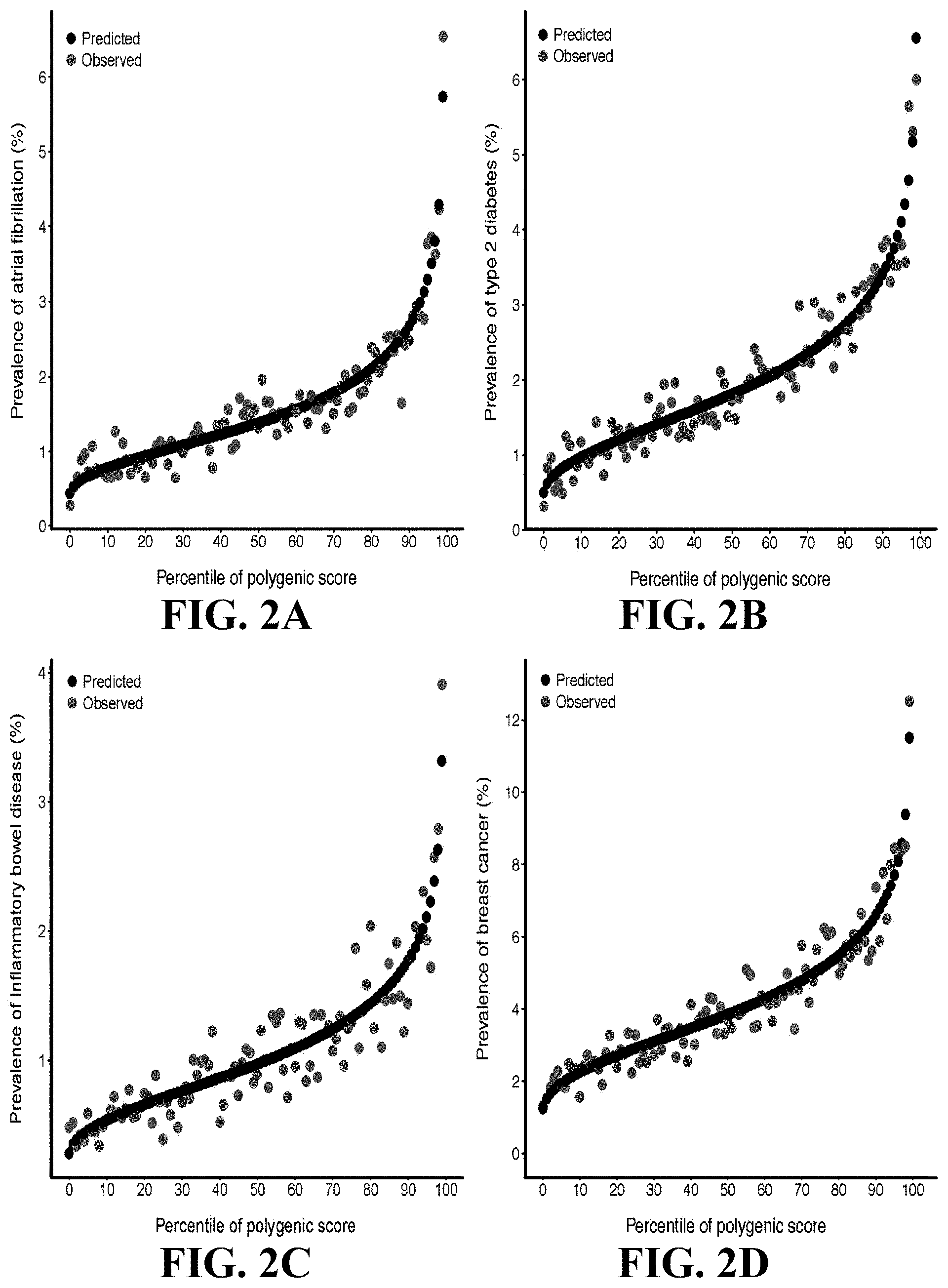
[0022] FIGS. 2A-2D charts predicted versus observed prevalence of four diseases according to genome wide polygenic score percentile. For each individual within the UK Biobank testing dataset, the predicted probability of disease was calculated using a logistic regression model with only the genome-wide polygenic score (GPS) as a predictor. The predicted prevalence of disease within each percentile bin of the GPS distribution was calculated as the average predicted probability of all individuals within that bin. The shape of the predicted risk gradient was consistent with the empirically observed risk gradient, reflected by black and blue dots, respectively, for each of four diseases: FIG. 2A atrial fibrillation, FIG. 2B type 2 diabetes, FIG. 2C inflammatory bowel disease, and FIG. 2D breast cancer. Breast cancer analysis was restricted to female participants.
[0023] The figures herein are for illustrative purposes only and are not necessarily drawn to scale.
DETAILED DESCRIPTION OF THE EXAMPLE EMBODIMENTS
General Definitions
[0024] Unless defined otherwise, technical and scientific terms used herein have the same meaning as commonly understood by one of ordinary skill in the art to which this disclosure pertains. Definitions of common terms and techniques in molecular biology may be found in Molecular Cloning: A Laboratory Manual, 2nd edition (1989) (Sambrook, Fritsch, and Maniatis); Molecular Cloning: A Laboratory Manual, 4th edition (2012) (Green and Sambrook); Current Protocols in Molecular Biology (1987) (F. M. Ausubel et al. eds.); the series Methods in Enzymology (Academic Press, Inc.): PCR 2: A Practical Approach (1995) (M. J. MacPherson, B. D. Hames, and G. R. Taylor eds.): Antibodies, A Laboratory Manual (1988) (Harlow and Lane, eds.): Antibodies A Laboratory Manual, 2nd edition 2013 (E. A. Greenfield ed.); Animal Cell Culture (1987) (R. I. Freshney, ed.); Benjamin Lewin, Genes IX, published by Jones and Bartlet, 2008 (ISBN 0763752223); Kendrew et al. (eds.), The Encyclopedia of Molecular Biology, published by Blackwell Science Ltd., 1994 (ISBN 0632021829); Robert A. Meyers (ed.), Molecular Biology and Biotechnology: a Comprehensive Desk Reference, published by VCH Publishers, Inc., 1995 (ISBN 9780471185710); Singleton et al., Dictionary of Microbiology and Molecular Biology 2nd ed., J. Wiley & Sons (New York, N.Y. 1994), March, Advanced Organic Chemistry Reactions, Mechanisms and Structure 4th ed., John Wiley & Sons (New York, N.Y. 1992); and Marten H. Hofker and Jan van Deursen, Transgenic Mouse Methods and Protocols, 2nd edition (2011).
[0025] As used herein, the singular forms "a", "an", and "the" include both singular and plural referents unless the context clearly dictates otherwise.
[0026] The term "optional" or "optionally" means that the subsequent described event, circumstance or substituent may or may not occur, and that the description includes instances where the event or circumstance occurs and instances where it does not.
[0027] The recitation of numerical ranges by endpoints includes all numbers and fractions subsumed within the respective ranges, as well as the recited endpoints.
[0028] The terms "about" or "approximately" as used herein when referring to a measurable value such as a parameter, an amount, a temporal duration, and the like, are meant to encompass variations of and from the specified value, such as variations of +/-10% or less, +1-5% or less, +/-1% or less, and +/-0.1% or less of and from the specified value, insofar such variations are appropriate to perform in the disclosed invention. It is to be understood that the value to which the modifier "about" or "approximately" refers is itself also specifically, and preferably, disclosed.
[0029] As used herein, a "biological sample" may contain whole cells and/or live cells and/or cell debris. The biological sample may contain (or be derived from) a "bodily fluid". The present invention encompasses embodiments wherein the bodily fluid is selected from amniotic fluid, aqueous humour, vitreous humour, bile, blood serum, breast milk, cerebrospinal fluid, cerumen (earwax), chyle, chyme, endolymph, perilymph, exudates, feces, female ejaculate, gastric acid, gastric juice, lymph, mucus (including nasal drainage and phlegm), pericardial fluid, peritoneal fluid, pleural fluid, pus, rheum, saliva, sebum (skin oil), semen, sputum, synovial fluid, sweat, tears, urine, vaginal secretion, vomit and mixtures of one or more thereof. Biological samples include cell cultures, bodily fluids, cell cultures from bodily fluids. Bodily fluids may be obtained from a mammal organism, for example by puncture, or other collecting or sampling procedures.
[0030] The terms "subject," "individual," and "patient" are used interchangeably herein to refer to a vertebrate, preferably a mammal, more preferably a human. Mammals include, but are not limited to, murines, simians, humans, farm animals, sport animals, and pets. Tissues, cells and their progeny of a biological entity obtained in vivo or cultured in vitro are also encompassed.
[0031] Various embodiments are described hereinafter. It should be noted that the specific embodiments are not intended as an exhaustive description or as a limitation to the broader aspects discussed herein. One aspect described in conjunction with a particular embodiment is not necessarily limited to that embodiment and can be practiced with any other embodiment(s). Reference throughout this specification to "one embodiment", "an embodiment," "an example embodiment," means that a particular feature, structure or characteristic described in connection with the embodiment is included in at least one embodiment of the present invention. Thus, appearances of the phrases "in one embodiment," "in an embodiment," or "an example embodiment" in various places throughout this specification are not necessarily all referring to the same embodiment, but may. Furthermore, the particular features, structures or characteristics may be combined in any suitable manner, as would be apparent to a person skilled in the art from this disclosure, in one or more embodiments. Furthermore, while some embodiments described herein include some but not other features included in other embodiments, combinations of features of different embodiments are meant to be within the scope of the invention. For example, in the appended claims, any of the claimed embodiments can be used in any combination.
[0032] All publications, published patent documents, and patent applications cited herein are hereby incorporated by reference to the same extent as though each individual publication, published patent document, or patent application was specifically and individually indicated as being incorporated by reference.
OVERVIEW
[0033] The present disclosure relates to Applicant's findings that lead to the development of a genetic predictor that can identify a subset of the population at higher risk for inflammatory bowel disease (IBD). This is among the strongest predictors ever developed for such an application. In certain embodiments, determination of the presence or absence of risk alleles is followed by calculating the polygenic risk score for the subject, wherein a high polygenic score indicates a higher risk for developing inflammatory bowel disease.
[0034] In one aspect, the present disclosure provides methods of determining a risk of developing inflammatory bowel disease in a subject. In general the method may comprise identifying whether a group of SNPs are present in a biological sample from the subject. In some embodiments, the group SNPs comprises at least 50 SNPs from Table A, which includes a list of variants and weighs comprising polygenic risk scores for inflammatory bowel disease, disclosed in Amit V. Khera, et al., Genome-wide polygenic scores for common diseases identify individuals with risk equivalent to monogenic mutations, Nature Genetics, 2018, 50:1219-1224 doi.org/10.1038/s41588-018-0183-z ("Khera"), which is incorporated herein by reference in its entirety. In regards to Table A, Applicant specifically references the data referred to on the seventh page of Khera under "Data Availability" as available at www.broadcvdi.org/informational/data ("Polygenic Risk Score Variant Weights"). Table A refers specifically to the Polygenic Risk Score Variant Weights table named "Inflammatory bowel disease" and having a size of 305.1 MB.
[0035] With the group of SNPs, a polygenic risk score (PRS) for developing IBD may be calculated. In some embodiments, the method further comprising administering a treatment (e.g., a treatment of IBD) to the subject. The treatment may be designed or planned based on the PSR.
Methods of Diagnosis and Risk Determination
[0036] The present disclosure provides methods for diagnosing a disease or condition (e.g., IBD or related diseases), and/or or determining the risk of developing the disease or condition.
[0037] Risk assessments using large numbers of SNPs offers the advantage of increased predictive power. In certain embodiments, the invention includes in the risk assessment large numbers of alleles, for example, at least 500,000, at least 1,000,000, at least 2,000,000, at least 3,000,000, at least 4,000,000, at least 5,000,000, or at least 6,000,000 SNPs, or all SNPs from Table A.
[0038] In some embodiments, the present disclosure provides a method of determining a risk of developing inflammatory bowel disease in a subject, the method comprising identifying whether at least 50, at least 95, at least 100, at least 200, at least 500, at least 1000, at least 2000, at least 5000, at least 10,000, at least 20,000, at least 50,000, at least 75,000, or at least 100,000 SNPs single nucleotide polymorphisms (SNPs) from Table A is present in a biological sample from the subject; wherein the presence of a risk allele of a SNP from Table A indicates that the subject has an increased risk of the disease, and wherein the presence of an alternative allele indicates that the subject has a decreased risk of inflammatory bowel disease.
[0039] In an embodiment, the invention provides a method of determining a risk of developing inflammatory bowel disease in a subject comprising identifying whether the SNPs from Table A is present in a biological sample from the subject and calculating a polygenic risk score (PRS) for the subject based on the identified SNPs. The number of identified SNPs can be at least 50, at least 95, at least 100, at least 200, at least 500, at least 1000, at least 2000, at least 5000, at least 10,000, at least 20,000, at least 50,000, at least 75,000, or at least 100,000.
[0040] In an embodiment, the invention provides a method of determining a risk of developing inflammatory bowel disease, in a subject, the method comprising identifying whether at least 50, at least 95, at least 100, at least 200, at least 500, at least 1000, at least 2000, at least 5000, at least 10,000, at least 20,000, at least 50,000, at least 75,000, or at least 100,000 single nucleotide polymorphisms (SNPs) from Table A is present in a biological sample from the subject and calculating a polygenic risk score (PRS); wherein the presence of a risk allele of a SNP from Table A indicates that the subject has an increased risk of inflammatory bowel disease, and wherein the presence of an alternative allele indicates that the subject has a decreased risk of inflammatory bowel disease.
[0041] In an embodiment, the invention provides a method of determining a risk of developing inflammatory bowel disease in a subject comprising identifying whether the SNPs from Table A is present in a biological sample from the subject and calculating a polygenic risk score (PRS) for the subject based on the identified SNPs, wherein the PRS is calculated by summing the weighted risk score associated with each SNP identified. The number of identified SNPs can be at least 50, at least 95, at least 100, at least 200, at least 500, at least 1000, at least 2000, at least 5000, at least 10,000, at least 20,000, at least 50,000, at least 75,000, or at least 100,000.
[0042] In an of the embodiment, the invention provides a method of determining a risk of developing inflammatory bowel disease in a subject comprising identifying whether the SNPs from Table A is present in a biological sample from the subject, wherein identifying comprises measuring the presence of the at least 95 SNPs in the biological sample. The number of identified SNPs can be at least 50, at least 95, at least 100, at least 200, at least 500, at least 1000, at least 2000, at least 5000, at least 10,000, at least 20,000, at least 50,000, at least 75,000, or at least 100,000.
[0043] The invention provides a method of determining a polygenic risk score for (PRS) developing inflammatory bowel disease in a subject, the method comprising selecting at least 50, at least 95, at least 100, at least 200, at least 500, at least 1000, at least 2000, at least 5000, at least 10,000, at least 20,000, at least 50,000, at least 75,000, or at least 100,000 single nucleotide polymorphisms (SNPs) from Table A; identifying whether the SNPs are present in a biological sample from the subject; and calculating the polygenic risk score (PRS) based on the presence of the SNPs.
[0044] In an embodiment, the invention provides a method of determining a risk of developing inflammatory bowel disease in a subject comprising identifying whether the SNPs from Table A is present in a biological sample from the subject, calculating a polygenic risk score (PRS) for the subject based on the identified SNPs, and assigning the subject to a risk group based on the PRS. The PRS may be divided into quintiles, e.g., top quintile, intermediate quintile, and bottom quintile, wherein the top quintile of polygenic scores correspond the highest genetic risk group and the bottom quintile of polygenic scores correspond to the lowest genetic risk group. The number of identified SNPs can be at least 50, at least 95, at least 100, at least 200, at least 500, at least 1000, at least 2000, at least 5000, at least 10,000, at least 20,000, at least 50,000, at least 75,000, or at least 100,000.
[0045] In an embodiment, the present disclosed subject matter provides a method for selecting subjects or candidates with a risk for developing inflammatory bowel disease comprising identifying whether at least 50, at least 95, at least 100, at least 200, at least 500, at least 1000, at least 2000, at least 5000, at least 10,000, at least 20,000, at least 50,000, at least 75,000, or at least 100,000 SNPs single nucleotide polymorphisms (SNPs) from Table A is present in a biological sample from each subject or candidate; calculating a polygenic risk score (PRS) for each subject or candidate based on the identified SNPs; and selecting the subjects or candidates with a desired risk group.
[0046] For all inflammatory bowel disease risk assessments, incorporation of large numbers of SNPs offers the advantage of increased predictive power. The invention further provides risk assessments outlined above incorporating for example, at least 500,000, at least 1,000,000, at least 2,000,000, at least 3,000,000, at least 4,000,000, at least 5,000,000, or at least 6,000,000 SNPs, or all SNPs from Table A.
[0047] In certain embodiments of the invention, risk assessments comprise the highest weighted polymorphisms, including, but not limited to the top 50%, 55%, 60%, 70%, 80%, 90%, or 95% of SNPs from Table A.
[0048] In an embodiment, the method is used to select a population of subjects or candidates for clinical trials, e.g., a clinical trial to determine whether a particular treatment or treatment plan is effective against inflammatory bowel disease. In an embodiment, the desired risk group is a population comprising high risk subjects or candidates. In an embodiment, the selected population of subjects or candidates are responders, i.e., the subjects or candidates are responsive to the treatment or treatment plan.
[0049] In an embodiment, the a method is provided for selecting a population of subjects or candidates with a high risk for developing artery disease comprising identifying whether at least 50, at least 95, at least 100, at least 200, at least 500, at least 1000, at least 2000, at least 5000, at least 10,000, at least 20,000, at least 50,000, at least 75,000, or at least 100,000 SNPs single nucleotide polymorphisms (SNPs) from Table A is present in a biological sample from each subject or candidate; calculating a polygenic risk score (PRS) for each subject or candidate based on the identified SNPs; and selecting the subjects or candidates in the high risk group. In an embodiment, the method is used to select a population of subjects or candidates for clinical trials, e.g., a clinical trial to determine whether a particular treatment or treatment plan is effective against inflammatory bowel disease. In an embodiment, the selected candidates or subjects are divided into subgroups based on the identified SNPs for each subject or candidate, and the method is used to determine whether a particular treatment or treatment plan is effective against a particular SNP or a particular group of SNPs. In other word, the method can be employed to determine susceptibility of a population of subjects to a particular treatment or treatment plan, wherein the population of subjects is selected based on the SNPs identified in the subjects.
[0050] Also provided are methods of detecting single nucleotide polymorphisms in a subject, including detecting whether at least 50 single nucleotide polymorphisms (SNPS) from Table A are present in a biological sample from a subject. The method includes contacting the biological sample with a set of probes to each SNP and detecting binding of the probes, by amplifying genome regions comprising the SNPs using a set of amplification primers, or by sequencing genomic regions comprising or enriched for the SNPs.
[0051] In any of the above embodiment, the method may further comprise an initial step of obtaining a biological sample from the subject.
[0052] In any of the above embodiment, the number of identified SNPs is at least 100 SNPs.
[0053] In any of the above embodiment, the number of identified SNPs is at least 200 SNPs.
[0054] In any of the above embodiment, the number of identified SNPs is at least 500 SNPs.
[0055] In any of the above embodiment, the number of identified SNPs is at least 1,000 SNPs.
[0056] In any of the above embodiment, the number of identified SNPs is at least 2,000 SNPs.
[0057] In any of the above embodiment, the number of identified SNPs is at least 5,000 SNPs.
[0058] In any of the above embodiment, the number of identified SNPs is at least 10,000 SNPs.
[0059] In any of the above embodiment, the number of identified SNPs is at least 20,000 SNPs.
[0060] In any of the above embodiment, the number of identified SNPs is at least 50,000 SNPs.
[0061] In any of the above embodiment, the number of identified SNPs is at least 75,000 SNPs.
[0062] In any of the above embodiment, the number of identified SNPs is at least 100,000 SNPs.
[0063] In any of the above embodiment, the identified SNPs comprise the highest risk SNPs or SNPs with a weight risk score in the top 10%, top 20%, top 30%, top 40%, or top 50% in Table A.
Detecting SNPs
[0064] In any of the above embodiments, identifying whether the SNP is present includes obtaining information regarding the identity (i.e., of a specific nucleotide), presence or absence of one or more specific SNP in a subject. Determining the presence of an SNP can, but need not, include obtaining a sample comprising DNA from a subject. The individual or organization who determines the presence of an SNPs need not actually carry out the physical analysis of a sample from a subject; the methods can include using information obtained by analysis of the sample by a third party. Thus the methods can include steps that occur at more than one site. For example, a sample can be obtained from a subject at a first site, such as at a health care provider, or at the subject's home in the case of a self-testing kit. The sample can be analyzed at the same or a second site, e.g., at a laboratory or other testing facility. Identifying the presence of a SNP can be done by any DNA detection method known in the art, including sequencing at least part of a genome of one or more cells from the subject.
[0065] In certain example embodiments, detection of SNPs can be done by sequencing.
[0066] Sequencing can be, for example, whole genome sequencing. In certain embodiments, the invention involves plate based single cell RNA sequencing (see, e.g., Picelli, S. et al., 2014, "Full-length RNA-seq from single cells using Smart-seq2" Nature protocols 9, 171-181, doi:10.1038/nprot.2014.006). In certain embodiments, the invention involves high-throughput single-cell RNA-seq and/or targeted nucleic acid profiling (for example, sequencing, quantitative reverse transcription polymerase chain reaction, and the like) where the RNAs from different cells are tagged individually, allowing a single library to be created while retaining the cell identity of each read. In this regard reference is made to Macosko et al., 2015, "Highly Parallel Genome-wide Expression Profiling of Individual Cells Using Nanoliter Droplets" Cell 161, 1202-1214; International patent application number PCT/US2015/049178, published as WO2016/040476 on Mar. 17, 2016; Klein et al., 2015, "Droplet Barcoding for Single-Cell Transcriptomics Applied to Embryonic Stem Cells" Cell 161, 1187-1201; International patent application number PCT/US2016/027734, published as WO2016168584A1 on Oct. 20, 2016; Zheng, et al., 2016, "Haplotyping germline and cancer genomes with high-throughput linked-read sequencing" Nature Biotechnology 34, 303-311; Zheng, et al., 2017, "Massively parallel digital transcriptional profiling of single cells" Nat. Commun. 8, 14049 doi: 10.1038/ncomms14049; International patent publication number WO2014210353A2; Zilionis, et al., 2017, "Single-cell barcoding and sequencing using droplet microfluidics" Nat Protoc. January; 12(1):44-73; Cao et al., 2017, "Comprehensive single cell transcriptional profiling of a multicellular organism by combinatorial indexing" bioRxiv preprint first posted online Feb. 2, 2017, doi: dx.doi.org/10.1101/104844; Rosenberg et al., 2017, "Scaling single cell transcriptomics through split pool barcoding" bioRxiv preprint first posted online Feb. 2, 2017, doi: dx.doi.org/10.1101/105163; Vitak, et al., "Sequencing thousands of single-cell genomes with combinatorial indexing" Nature Methods, 14(3):302-308, 2017; Cao, et al., Comprehensive single-cell transcriptional profiling of a multicellular organism. Science, 357(6352):661-667, 2017; and Gierahn et al., "Seq-Well: portable, low-cost RNA sequencing of single cells at high throughput" Nature Methods 14, 395-398 (2017), all the contents and disclosure of each of which are herein incorporated by reference in their entirety. In certain embodiments, the invention involves single nucleus RNA sequencing. In this regard reference is made to Swiech et al., 2014, "In vivo interrogation of gene function in the mammalian brain using CRISPR-Cas9" Nature Biotechnology Vol. 33, pp. 102-106; Habib et al., 2016, "Div-Seq: Single-nucleus RNA-Seq reveals dynamics of rare adult newborn neurons" Science, Vol. 353, Issue 6302, pp. 925-928; Habib et al., 2017, "Massively parallel single-nucleus RNA-seq with DroNc-seq" Nat Methods. 2017 October; 14(10):955-958; and International patent application number PCT/US2016/059239, published as WO2017164936 on Sep. 28, 2017, which are herein incorporated by reference in their entirety.
[0067] In certain example embodiments, target genomic regions of interest may be enriched from single cell sequencing libraries prior to sequencing analysis. Example enrichment methods are described, for example, in U.S. Provisional Application No. 62/576,031 entitled "Single Cell Cellular Component Enrichment from Barcoded Sequencing Libraries" filed Oct. 23, 2017.
[0068] SNPs may be detected through hybridization-based methods, including dynamic allele-specific hybridization (DASH), molecular beacons, and SNP microarrays, enzyme-based methods including RFLP, PCR-based, e.g., allelic-specific polymerase chain reaction (AS-PCR), polymerase chain reaction--restriction fragment length polymorphism (PCR-RFLP), multiplex PCR real-time invader assay (mPCR-RETINA), (amplification refractory mutation system (ARMS), Flap endonuclease, primer extension, 5' nuclease, e.g., Taqman or 5' nuclease allelic discrimination assay, and oligonucleotide ligation assay, and methods such as single strand conformation polymorphism, temperature gradient gel electrophoresis, denaturing high performance liquid chromatography, high-resolution melting of the entire amplicon, use of DNA mismatch-binding proteins, SNPlex, and Surveyor nuclease assay.
Methods of Treatment
[0069] In any of the above embodiments, the method further comprises initiating a treatment to the subject. The treatment can be determined or adjusted according to the risk of inflammatory bowel disease. The treatment can comprise antibiotics, immune system suppressors/modulators, biologics and/or anti-inflammatories. Antibiotics, for example can be used in addition to other medications, or when infection may arise, for example in perianal Crohn's disease. In some embodiments, the antibiotic is ciprofloxacin (Cipro) or metronidazole (Flagyl). Anti-inflammatories such as corticosteroids can be used in a variety of dosing regimens, and can include prednisone, budesonide, hydrocortisone, methylprednisone, and Cortenema. Aminosalicylates limit inflammation of the digestive tract and can be prescribed based on location of inflammation within the digestive tract. In some embodiments, the aminosalicylates are selected from, for example, mesalamine (Asacol HD, Delzicol, others), balsalazide (Colazal) and olsalazine (Dipentum). Immune system suppressors or modulators, or immunosuppressors/immunomodulators work to suppress the immune response and can include one or a combination of immunosuppressors. One class of immunosuppressors are TNF-alpha inhibitor biologics and can include infliximab (Remicade) and its biosimilars, adalimumab (Humira) adalimumab abdm, adalimumab-atto, golimumab (Simponi), and certolizumab pegol. Azathioprine and 6-mercaptopurine, cyclosporine A and tracolimus are often used with severe ulcerative colitis. Additional immunomodulators utilized in treatment include, for example, azathioprine (Azasan, Imuran), mercaptopurine (Purinethol, Purixan), cyclosporine (Gengraf, Neoral, Sandimmune) and methotrexate (Trexall). Biologics can include natalizumab (Tysabri), vedolizumab (Entyvio) and ustekinumab (Stelara).
[0070] Treatment may also include, alone, or in addition to prescription drug therapy, nutritional support, including parenteral or enteral nutrition to allow bowel rest. In some embodiments, the polygenic risk score is used to guide enhanced monitoring strategies. In some embodiments, the polygenic risk score is used to guide intensive lifestyle interventions. Diet and lifestyle modifications can also be included in treatment. Intensive lifestyle interventions including modifications to diet and exercise. Initiating a treatment can include devising a treatment plan based on the risk group, which corresponds to the PRS calculated for the subject.
[0071] In one embodiment, a treatment or a method of treatment can include gene therapy/genome editing and/or the nucleic acid vector used in a gene therapy vector known in the art. In one embodiment, one or more target locus within the subject's genomic DNA is targeted and modified. A treatment method comprises gene editing tools available in the art, e.g., CRISPR (Clustered Regularly Interspaced Short Palindromic Repeats), zinc finger nucleases, meganucleases, where a target DNA locus, e.g., a gene of interest, is modified to create a mutation in the gene product, e.g., a protein or enzyme, with reduced activity or no activity (loss-of-function mutation). In some embodiment, vectors can comprise viral vector, e.g., retroviruses, adenoviruses, adeno-associated viruses, and lentiviruses. Examples of a target locus of interest include the genes PCSK9, APOC3, ANGPTL8, LPL, CD36, HBB and NPC1L1.
[0072] The invention provides methods and models to establish causation of elements of alleles (e.g., chromosomal regions, genetic loci) identified as associated with increased disease risk. In an embodiment of the invention, a model animal, for example but not limited to a rat, a mouse, a dog, a pig, a non-human primate, or a chimeric animal comprising human cells can be employed. In an embodiment of the invention, an organ or organoid can be employed, which can be characterized as from a human or a non-human mammal. In an embodiment of the invention, a cell line from a human or non-human mammal can be employed.
Nucleases and Related Systems
[0073] The treatment may include administering one or more genetic modifying agents. In some embodiments, the genetic modifying agents may be nucleases or related systems. The genetic modifying agents may also be used to make one or more genetic modifications in a model organism. In certain embodiments, the nuclease is used for gene editing. Nuclease based therapy or therapeutics may involve target disruption, such as target mutation, such as leading to gene knockout. Nuclease activity, such as CRISPR-Cas system based therapy or therapeutics may involve replacement of particular target sites, such as leading to target correction. Nuclease based therapy or therapeutics may involve removal of particular target sites, such as leading to target deletion. Nuclease activity, such as CRISPR-Cas system based therapy or therapeutics may involve modulation of target site functionality, such as target site activity or accessibility, leading for instance to (transcriptional and/or epigenetic) gene or genomic region activation or gene or genomic region silencing. The skilled person will understand that modulation of target site functionality may involve nuclease mutation (such as for instance generation of a catalytically inactive CRISPR effector) and/or functionalization (such as for instance fusion of the CRISPR effector with a heterologous functional domain, such as a transcriptional activator or repressor), as described herein elsewhere.
[0074] Accordingly, in an aspect, the invention relates to a method as described herein, comprising selection of one or more (therapeutic) target, selecting one or more nuclease function, and optimization of selected parameters or variables associated with the nuclease system and/or its functionality. In a related aspect, the invention relates to a method as described herein, comprising (a) selecting one or more (therapeutic) target loci, (b) selecting one or more nuclease system functionalities, (c) optionally selecting one or more modes of delivery, and preparing, developing, or designing a CRISPR-Cas system selected based on steps (a)-(c). Method for selecting optimal Cas9 and Cas12 based systems are disclosed, for example, in International Patent Application Publication Nos. WO/2018/035388 and WO/2018/035387.
[0075] In certain embodiments, nuclease system functionality comprises genomic mutation. In certain embodiments, nuclease system functionality comprises single genomic mutation. In certain embodiments, nuclease system functionality comprises multiple genomic mutations. In certain embodiments, nuclease system functionality comprises gene knockout. In certain embodiments, nuclease system functionality comprises single gene knockout. In certain embodiments, nuclease system functionality comprises multiple gene knockout. In certain embodiments, nuclease system functionality comprises gene correction. In certain embodiments, nuclease system functionality comprises single gene correction. In certain embodiments, nuclease system functionality comprises multiple gene correction. In certain embodiments, nuclease system functionality comprises genomic region correction. In certain embodiments, nuclease system functionality comprises single genomic region correction. In certain embodiments, nuclease system functionality comprises multiple genomic region correction. In certain embodiments, nuclease system functionality comprises gene deletion. In certain embodiments, nuclease system functionality comprises single gene deletion. In certain embodiments, nuclease system functionality comprises multiple gene deletion. In certain embodiments, nuclease system functionality comprises genomic region deletion. In certain embodiments, nuclease system functionality comprises single genomic region deletion. In certain embodiments, nuclease system functionality comprises multiple genomic region deletion. In certain embodiments, nuclease system functionality comprises modulation of gene or genomic region functionality. In certain embodiments, nuclease system functionality comprises modulation of single gene or genomic region functionality. In certain embodiments, nuclease system functionality comprises modulation of multiple gene or genomic region functionality. In certain embodiments, nuclease system functionality comprises gene or genomic region functionality, such as gene or genomic region activity. In certain embodiments, nuclease system functionality comprises single gene or genomic region functionality, such as gene or genomic region activity. In certain embodiments, nuclease system functionality comprises multiple gene or genomic region functionality, such as gene or genomic region activity. In certain embodiments, nuclease system functionality comprises modulation gene activity or accessibility optionally leading to transcriptional and/or epigenetic gene or genomic region activation or gene or genomic region silencing. In certain embodiments, nuclease system functionality comprises modulation single gene activity or accessibility optionally leading to transcriptional and/or epigenetic gene or genomic region activation or gene or genomic region silencing. In certain embodiments, nuclease system functionality comprises modulation multiple gene activity or accessibility optionally leading to transcriptional and/or epigenetic gene or genomic region activation or gene or genomic region silencing.
[0076] In certain example embodiments, the one or more genetic elements may be modified using a nuclease. The term "nuclease" as used herein broadly refers to an agent, for example a protein or a small molecule, capable of cleaving a phosphodiester bond connecting nucleotide residues in a nucleic acid molecule. In some embodiments, a nuclease may be a protein, e.g., an enzyme that can bind a nucleic acid molecule and cleave a phosphodiester bond connecting nucleotide residues within the nucleic acid molecule. A nuclease may be an endonuclease, cleaving a phosphodiester bonds within a polynucleotide chain, or an exonuclease, cleaving a phosphodiester bond at the end of the polynucleotide chain. Preferably, the nuclease is an endonuclease. Preferably, the nuclease is a site-specific nuclease, binding and/or cleaving a specific phosphodiester bond within a specific nucleotide sequence, which may be referred to as "recognition sequence", "nuclease target site", or "target site". In some embodiments, a nuclease may recognize a single stranded target site, in other embodiments a nuclease may recognize a double-stranded target site, for example a double-stranded DNA target site. Some endonucleases cut a double-stranded nucleic acid target site symmetrically, i.e., cutting both strands at the same position so that the ends comprise base-paired nucleotides, also known as blunt ends. Other endonucleases cut a double-stranded nucleic acid target sites asymmetrically, i.e., cutting each strand at a different position so that the ends comprise unpaired nucleotides. Unpaired nucleotides at the end of a double-stranded DNA molecule are also referred to as "overhangs", e.g., "5'-overhang" or "3'-overhang", depending on whether the unpaired nucleotide(s) form(s) the 5' or the 5' end of the respective DNA strand.
[0077] The nuclease may introduce one or more single-strand nicks and/or double-strand breaks in the endogenous gene, whereupon the sequence of the endogenous gene may be modified or mutated via non-homologous end joining (NHEJ) or homology-directed repair (HDR).
[0078] In certain embodiments, the nuclease may comprise (i) a DNA-binding portion configured to specifically bind to the endogenous gene and (ii) a DNA cleavage portion. Generally, the DNA cleavage portion will cleave the nucleic acid within or in the vicinity of the sequence to which the DNA-binding portion is configured to bind.
[0079] In certain embodiments, the nuclease may be employed to mutate or regulate genetic elements singly or in combination in the organism. Thus by varying one or more genetic elements in a model organism, the invention provides a means for establishing or confirming causality between genetic changes and phenotypic effects. The genetic changes can be the SNPs or any variation in linkage disequilibrium with the SNP.
[0080] Similarly, the model organisms can be used to test effectiveness of therapeutic intervention. In an embodiment, the invention is used to define or establish subgroups of individuals (or models) at elevated risk for inflammatory bowel disease on the basis of different risk factors or combinations of risk factors. In one embodiment, the separate subgroups are used to characterize susceptibility to therapeutic interventions that may vary from subgroup to subgroup. In another embodiment, therapies are selected according the SNPs identified in a subject.
[0081] In an aspect of the invention, there is targeted genomic editing to modify one or more genomic sequences of interest to reduce disease risk. One or more targets may be selected, depending on the genotypic and/or phenotypic outcome. For instance, one or more therapeutic targets may be selected, depending on (genetic) disease etiology or the desired therapeutic outcome. The (therapeutic) target(s) may be a single gene, locus, or other genomic site, or may be multiple genes, loci or other genomic sites. As is known in the art, a single gene, locus, or other genomic site may be targeted more than once, such as by use of multiple gRNAs.
[0082] According to the invention, genomic sequences associated with disease risk are identified by single nucleotide polymorphisms (SNPs). The SNPs are linked to the genomic sequences of interest, i.e., close to or within the genomic sequences of interest, and may or may not be causative of the risk variation. That is, functional differences between alleles distinguished by the SNPs may result from sequence variation of an SNP or from one or more differences between alleles located near to the location of the SNP. In either case, the invention provides for gene editing in order to reduce disease risk. In general, a higher risk allele would be edited, for example, to a lower risk allele. Often such editing would involve individual base changes, but can also involve insertions and deletions. For example, trinucleotide repeat regions may be edited to change the number of trinucleotide repeats.
[0083] In any of the above embodiment, the subject can be animal which include mammal, human and non-human mammal.
[0084] In an embodiment, the invention provides a method of identifying a risk of developing inflammatory bowel disease in a subject and providing a treatment to the subject, the method comprising obtaining a biological sample from the subject; identifying whether at least one single nucleotide polymorphism (SNP) from Table A is present in the biological sample; wherein the presence of a risk allele of a SNP from Table A indicates that the subject has an increased risk of inflammatory bowel disease; and initiating a treatment to the subject, wherein the treatment comprises Antibiotics, for example can be used in addition to other medications, or when infection may arise, for example in perianal Crohn's disease. In some embodiments, the antibiotic is ciprofloxacin (Cipro) or metronidazole (Flagyl). Anti-inflammatories such as corticosteroids can be used in a variety of dosing regimens, and can include prednisone, budesonide, hydrocortisone, methylprednisone, and Cortenema. Aminosalicylates limit inflammation of the digestive tract and can be prescribed based on location of inflammation within the digestive tract. In some embodiments, the aminosalicylates are selected from, for example, mesalamine (Asacol HD, Delzicol, others), balsalazide (Colazal) and olsalazine (Dipentum). Immune system suppressors or modulators, or immunosuppressors/immunomodulators work to suppress the immune response and can include one or a combination of immunosuppressors. One class of immunosuppressors are TNF-alpha inhibitor biologics and can include infliximab (Remicade) and its biosimilars, adalimumab (Humira) adalimumab abdm, adalimumab-atto, golimumab (Simponi), and certolizumab pegol. Azathioprine and 6-mercaptopurine, cyclosporine A and tracolimus are often used with severe ulcerative colitis. Additional immunomodulators utilized in treatment include, for example, azathioprine (Azasan, Imuran), mercaptopurine (Purinethol, Purixan), cyclosporine (Gengraf, Neoral, Sandimmune) and methotrexate (Trexall). Biologics can include natalizumab (Tysabri), vedolizumab (Entyvio) and ustekinumab (Stelara).
[0085] In an embodiment, the invention provides a method of reducing a risk of inflammatory bowel disease in a subject comprising administering to the subject a treatment which comprises one or more of antibiotics, immune system suppressors/modulators, biologics and/or anti-inflammatories, wherein the subject has a polygenic risk score that corresponds to a high risk group. In some embodiments, more than one drug can be used in a combination therapy, in particular when the drugs act in different ways to treat IBD, or have different pharmacokinetics.
[0086] The polygenic risk score may be calculated by selecting at least 50, at least 95, at least 100, at least 200, at least 500, at least 1000, at least 2000, at least 5000, at least 10,000, at least 20,000, at least 50,000, at least 75,000, or at least 100,000 single nucleotide polymorphisms (SNPs) from Table A; identifying whether the at least 50, at least 95, at least 100, at least 200, at least 500, at least 1000, at least 2000, at least 5000, at least 10,000, at least 20,000, at least 50,000, at least 75,000, or at least 100,000, at least 500,000, at least 1,000,000, at least 2,000,000, at least 3,000,000, at least 4,000,000, at least 5,000,000, or at least 6,000,000 SNPs, or all SNPs from Table A are present in a biological sample from the subject; and calculating the polygenic risk score (PRS) based on the presence of the SNPs.
[0087] Although the present invention and its advantages have been described in detail, it should be understood that various changes, substitutions and alterations can be made herein without departing from the spirit and scope of the invention as defined in the appended claims.
[0088] As used herein, the term "inflammatory bowel disease", includes, e.g., Crohn's disease (CD) and ulceratidve colitis (UC).
[0089] As used herein, the term "biological sample" is used in its broadest sense. A biological sample may be obtained from a subject (e.g., a human) or from components (e.g., tissues) of a subject. The sample may be of any biological tissue or fluid with which biomarkers of the present invention may be assayed. Frequently, the sample will be a "clinical sample", i.e., a sample derived from a patient. Such samples include, but are not limited to, bodily fluids, e.g., urine, whole blood, blood plasma, saliva; tissue or fine needle biopsy samples; and archival samples with known diagnosis, treatment and/or outcome history. The term biological sample also encompasses any material derived by processing the biological sample. Derived materials include, but are not limited to, cells (or their progeny) isolated from the sample, proteins or nucleic acid molecules extracted from the sample. Processing of the biological sample may involve one or more of, filtration, distillation, extraction, concentration, inactivation of interfering components, addition of reagents, and the like. In some embodiments, the biological sample is a whole blood sample. In some embodiments, the biological sample includes peripheral blood mononuclear cells (PBMCs) obtained from a subject. PBMCs can be extracted from whole blood using ficoll, a hydrophilic polysaccharide that separates layers of blood, and gradient centrifugation, which will separate the blood into a top layer of plasma, followed by a layer of PBMCs and a bottom fraction of polymorphonuclear cells (such as neutrophils and eosinophils) and erythrocytes.
[0090] As used herein, an "allele" is one of a pair or series of genetic variants of a polymorphism at a specific genomic location. A "response allele" is an allele that is associated with altered response to a treatment. Where a SNP is biallelic, both alleles will be response alleles (e.g., one will be associated with a positive response, while the other allele is associated with no or a negative response, or some variation thereof).
[0091] As used herein, "genotype" refers to the diploid combination of alleles for a given genetic polymorphism. A homozygous subject carries two copies of the same allele and a heterozygous subject carries two different alleles.
[0092] As used herein, a "haplotype" is one or a set of signature genetic changes (polymorphisms) that are normally grouped closely together on the DNA strand, and are usually inherited as a group; the polymorphisms are also referred to herein as "markers." A "haplotype" as used herein is information regarding the presence or absence of one or more genetic markers in a given chromosomal region in a subject. A haplotype can consist of a variety of genetic markers, including indels (insertions or deletions of the DNA at particular locations on the chromosome); single nucleotide polymorphisms (SNPs) in which a particular nucleotide is changed; microsatellites; and minis satellites.
[0093] The term "chromosome" as used herein refers to a gene carrier of a cell that is derived from chromatin and comprises DNA and protein components (e.g., histones). The conventional internationally recognized individual human genome chromosome numbering identification system is employed herein. The size of an individual chromosome can vary from one type to another with a given multi-chromosomal genome and from one genome to another. In the case of the human genome, the entire DNA mass of a given chromosome is usually greater than about 100,000,000 base pairs.
[0094] The term "gene" refers to a DNA sequence in a chromosome that codes for a product (either RNA or its translation product, a polypeptide). A gene contains a coding region and includes regions preceding and following the coding region (termed respectively "leader" and "trailer"). The coding region is comprised of a plurality of coding segments ("exons") and intervening sequences ("introns") between individual coding segments.
[0095] As used herein, the terms "protein", "polypeptide", and "peptide" are used herein interchangeably, and refer to amino acid sequences of a variety of lengths, either in their neutral (uncharged) forms or as salts, and either unmodified or modified by glycosylation, side chain oxidation, or phosphorylation, or modified by deletion, insertion, or change in one or more amino acids.
[0096] As used herein, the terms "nucleic acid molecule" and "polynucleotide" are used herein interchangeably. They refer to a deoxyribonucleotide or ribonucleotide polymer in either single- or double-stranded form, and unless otherwise stated, encompass known analogs of natural nucleotides that can function in a similar manner as naturally occurring nucleotides. The terms encompass nucleic acid-like structures with synthetic backbones, as well as amplification products.
[0097] As used herein, the term "hybridizing" refers to the binding of two single stranded nucleic acids via complementary base pairing. The term "specific hybridization" refers to a process in which a nucleic acid molecule preferentially binds, duplexes, or hybridizes to a particular nucleic acid sequence under stringent conditions (e.g., in the presence of competitor nucleic acids with a lower degree of complementarity to the hybridizing strand). In certain embodiments of the present invention, these terms more specifically refer to a process in which a nucleic acid fragment (or segment) from a test sample preferentially binds to a particular probe and to a lesser extent or not at all, to other probes, for example, when these probes are immobilized on an array.
[0098] The term "probe" refers to an oligonucleotide. A probe can be single stranded at the time of hybridization to a target. As used herein, probes include primers, i.e., oligonucleotides that can be used to prime a reaction, e.g., a PCR reaction.
[0099] The term "label" or "label containing moiety" refers in a moiety capable of detection, such as a radioactive isotope or group containing same, and nonisotopic labels, such as enzymes, biotin, avidin, streptavidin, digoxygenin, luminescent agents, dyes, haptens, and the like. Luminescent agents, depending upon the source of exciting energy, can be classified as radioluminescent, chemiluminescent, bioluminescent, and photoluminescent (including fluorescent and phosphorescent). A probe described herein can be bound, e.g., chemically bound to label-containing moieties or can be suitable to be so bound. The probe can be directly or indirectly labeled.
[0100] The term "direct label probe" (or "directly labeled probe") refers to a nucleic acid probe whose label after hybrid formation with a target is detectable without further reactive processing of hybrid. The term "indirect label probe" (or "indirectly labeled probe") refers to a nucleic acid probe whose label after hybrid formation with a target is further reacted in subsequent processing with one or more reagents to associate therewith one or more moieties that finally result in a detectable entity.
[0101] The terms "target," "DNA target," or "DNA target locus" refers to a nucleotide sequence that occurs at a specific chromosomal location. Each such sequence or portion is preferably at least partially, single stranded (e.g., denatured) at the time of hybridization. When the target nucleotide sequences are located only in a single region or fraction of a given chromosome, the term "target region" is sometimes used. Targets for hybridization can be derived from specimens which include, but are not limited to, chromosomes or regions of chromosomes in normal, diseased or malignant human cells, either interphase or at any state of meiosis or mitosis, and either extracted or derived from living or postmortem tissues, organs or fluids; germinal cells including sperm and egg cells, or cells from zygotes, fetuses, or embryos, or chorionic or amniotic cells, or cells from any other germinating body; cells grown in vitro, from either long-term or short-term culture, and either normal, immortalized or transformed; inter- or intraspecific hybrids of different types of cells or differentiation states of these cells; individual chromosomes or portions of chromosomes, or translocated, deleted or other damaged chromosomes, isolated by any of a number of means known to those with skill in the art, including libraries of such chromosomes cloned and propagated in prokaryotic or other cloning vectors, or amplified in vitro by means well known to those with skill; or any forensic material, including but not limited to blood, or other samples.
[0102] As used herein, the terms "array", "micro-array", and "biochip" are used herein interchangeably. They refer to an arrangement, on a substrate surface, of hybridizable array elements, preferably, multiple nucleic acid molecules of known sequences. Each nucleic acid molecule is immobilized to a discrete spot (i.e., a defined location or assigned position) on the substrate surface. The term "micro-array" more specifically refers to an array that is miniaturized so as to require microscopic examination for visual evaluation.
[0103] Accordingly, in an aspect, the invention relates to a method as described herein, comprising selection of one or more (therapeutic) target, selecting nuclease system functionality, selecting nuclease system mode of delivery, and optimization of selected parameters or variables associated with the nuclease system and/or its functionality.
[0104] The methods as described herein may further involve selection of the nuclease system delivery vehicle and/or expression system. Delivery vehicles and expression systems are described herein elsewhere. By means of example, delivery vehicles of nucleic acids and/or proteins include nanoparticles, liposomes, etc. Delivery vehicles for DNA, such as DNA-based expression systems include for instance biolistics, viral based vector systems (e.g. adenoviral, AAV, lentiviral), etc. the skilled person will understand that selection of the mode of delivery, as well as delivery vehicle or expression system may depend on for instance the cell or tissues to be targeted. In certain embodiments, the delivery vehicle and/or expression system for delivering the nuclease systems or components thereof comprises liposomes, lipid particles, nanoparticles, biolistics, or viral-based expression/delivery systems.
Exemplary Genetic Modifying Agents
[0105] The genetic modifying agents may be programmable nucleic acid-modifying agents, which may be used to modify endogenous cell DNA or RNA sequences, including DNA and/or RNA sequences encoding the target genes and target gene products disclosed herein. In certain example embodiments, the programmable nucleic acid-modifying agents may be used to edit a target sequence to restore native or wild-type functionality. In certain other embodiments, the programmable nucleic-acid modifying agents may be used to insert a new gene or gene product to modify the phenotype of target cells. In certain other example embodiments, the programmable nucleic-acid modifying agents may be used to delete or otherwise silence the expression of a target gene or gene product. Programmable nucleic-acid modifying agents may be used in both in vivo an ex vivo applications disclosed herein.
[0106] Examples of genetic modifying agents are described below.
CRISPR/Cas Systems
[0107] In certain embodiments, the genetic modifying agents may be a CRISPR-Cas system or one or more components thereof. CRISPR-Cas system activity, such as CRISPR-Cas system based therapy or therapeutics may involve target disruption, such as target mutation, such as leading to gene knockout. CRISPR-Cas system activity, such as CRISPR-Cas system based therapy or therapeutics may involve replacement of particular target sites, such as leading to target correction. CRISPR-Cas system based therapy or therapeutics may involve removal of particular target sites, such as leading to target deletion. CRISPR-Cas system activity, such as CRISPR-Cas system based therapy or therapeutics may involve modulation of target site functionality, such as target site activity or accessibility, leading for instance to (transcriptional and/or epigenetic) gene or genomic region activation or gene or genomic region silencing. The skilled person will understand that modulation of target site functionality may involve CRISPR effector mutation (such as for instance generation of a catalytically inactive CRISPR effector) and/or functionalization (such as for instance fusion of the CRISPR effector with a heterologous functional domain, such as a transcriptional activator or repressor), as described herein elsewhere.
[0108] Optimization of selected parameters or variables in the methods as described herein may result in optimized or improved nuclease system, such as CISPR-Cas system based therapy or therapeutic, specificity, efficacy, and/or safety. In certain embodiments, one or more of the following parameters or variables are taken into account, are selected, or are optimized in the methods of the invention as described herein: CRISPR effector specificity, gRNA specificity, CRISPR-Cas complex specificity, PAM restrictiveness, PAM type (natural or modified), PAM nucleotide content, PAM length, CRISPR effector activity, gRNA activity, CRISPR-Cas complex activity, target cleavage efficiency, target site selection, target sequence length, ability of effector protein to access regions of high chromatin accessibility, degree of uniform enzyme activity across genomic targets, epigenetic tolerance, mismatch/budge tolerance, CRISPR effector stability, CRISPR effector mRNA stability, gRNA stability, CRISPR-Cas complex stability, CRISPR effector protein or mRNA immunogenicity or toxicity, gRNA immunogenicity or toxicity, CRISPR-Cas complex immunogenicity or toxicity, CRISPR effector protein or mRNA dose or titer, gRNA dose or titer, CRISPR-Cas complex dose or titer, CRISPR effector protein size, CRISPR effector expression level, gRNA expression level, CRISPR-Cas complex expression level, CRISPR effector spatiotemporal expression, gRNA spatiotemporal expression, CRISPR-Cas complex spatiotemporal expression.
[0109] In certain embodiments, selecting one or more CRISPR-Cas system functionalities comprises selecting one or more of an optimal effector protein, an optimal guide RNA, or both.
[0110] In an exemplary method for modifying a target polynucleotide by integrating an exogenous polynucleotide template, a double stranded break is introduced into the genome sequence by the CRISPR complex, the break is repaired via homologous recombination an exogenous polynucleotide template such that the template is integrated into the genome. The presence of a double-stranded break facilitates integration of the template.
[0111] In an exemplary method for modifying a target polynucleotide by integrating an exogenous polynucleotide template, a single stranded break is introduced into the genome sequence by the nuclease, for example wherein the CRISPR-Cas protein is a nickase. The break is repaired via homologous recombination an exogenous polynucleotide template such that the template is integrated into the genome. The presence of a single-stranded break facilitates integration of the template.
[0112] In certain embodiments, the therapeutic nuclease system is multiplexed for targeting multiple loci. In certain embodiments, this can be established by using multiple (tandem or multiplex) guide RNA (gRNA) sequences. In certain embodiments, said gRNA sequences are separated by a nucleotide sequence, such as a direct repeat (DR). In certain embodiments, said gRNA sequences are separated by a sequence cleavable by a host enzyme. In certain embodiments, a "self-inactivating" gRNA includes which targets an element of the CRISPR system.
[0113] In certain embodiments, selecting an optimal effector protein comprises optimizing one or more of effector protein type, size, PAM specificity, effector protein stability, immunogenicity or toxicity, functional specificity, and efficacy, or other CRISPR effector associated parameters or variables as described herein elsewhere.
[0114] The invention further provides for targeted delivery whereby a nuclease system is preferably delivered to a cell type of interest. In one embodiment, it may be preferable for a CRISPR system engineered to target certain genetic loci to a particular cell type wherein those loci are expressed and active. According to the invention, a CRISPR system can be preferentially targeted to, without limitation, to a liver cell, an epithelial cell, a hematopoietic cell, or an immune cell. In an embodiment of the invention, a cell type of interest is preferentially targeted by using viral vectors of a particular serotypes. In an embodiment of the invention, a cell type of interest is preferentially targeted by a vector particle displaying a target-specific ligand.
[0115] In certain embodiments, selecting an optimal effector protein comprises optimizing one or more of effector protein type, size, PAM specificity, effector protein stability, immunogenicity or toxicity, functional specificity, and efficacy, or other CRISPR effector associated parameters or variables as described herein elsewhere.
[0116] In general, a CRISPR-Cas or CRISPR system as used herein and in documents, such as WO 2014/093622 (PCT/US2013/074667), refers collectively to transcripts and other elements involved in the expression of or directing the activity of CRISPR-associated ("Cas") genes, including sequences encoding a Cas gene, a tracr (trans-activating CRISPR) sequence (e.g. tracrRNA or an active partial tracrRNA), a tracr-mate sequence (encompassing a "direct repeat" and a tracrRNA-processed partial direct repeat in the context of an endogenous CRISPR system), a guide sequence (also referred to as a "spacer" in the context of an endogenous CRISPR system), or "RNA(s)" as that term is herein used (e.g., RNA(s) to guide Cas, such as Cas9, e.g. CRISPR RNA and transactivating (tracr) RNA or a single guide RNA (sgRNA) (chimeric RNA)) or other sequences and transcripts from a CRISPR locus. In general, a CRISPR system is characterized by elements that promote the formation of a CRISPR complex at the site of a target sequence (also referred to as a protospacer in the context of an endogenous CRISPR system). See, e.g., Shmakov et al. (2015) "Discovery and Functional Characterization of Diverse Class 2 CRISPR-Cas Systems", Molecular Cell, DOI: dx.doi.org/10.1016/j.molce1.2015.10.008.
[0117] In certain embodiments, a protospacer adjacent motif (PAM) or PAM-like motif directs binding of the effector protein complex as disclosed herein to the target locus of interest. In some embodiments, the PAM may be a 5' PAM (i.e., located upstream of the 5' end of the protospacer). In other embodiments, the PAM may be a 3' PAM (i.e., located downstream of the 5' end of the protospacer). The term "PAM" may be used interchangeably with the term "PFS" or "protospacer flanking site" or "protospacer flanking sequence".
[0118] In a preferred embodiment, the CRISPR effector protein may recognize a 3' PAM. In certain embodiments, the CRISPR effector protein may recognize a 3' PAM which is 5'H, wherein H is A, C or U.
[0119] In the context of formation of a CRISPR complex, "target sequence" refers to a sequence to which a guide sequence is designed to have complementarity, where hybridization between a target sequence and a guide sequence promotes the formation of a CRISPR complex. A target sequence may comprise RNA polynucleotides. The term "target RNA" refers to a RNA polynucleotide being or comprising the target sequence. In other words, the target RNA may be a RNA polynucleotide or a part of a RNA polynucleotide to which a part of the gRNA, i.e. the guide sequence, is designed to have complementarity and to which the effector function mediated by the complex comprising CRISPR effector protein and a gRNA is to be directed. In some embodiments, a target sequence is located in the nucleus or cytoplasm of a cell.
[0120] In certain example embodiments, the CRISPR effector protein may be delivered using a nucleic acid molecule encoding the CRISPR effector protein. The nucleic acid molecule encoding a CRISPR effector protein, may advantageously be a codon optimized CRISPR effector protein. An example of a codon optimized sequence, is in this instance a sequence optimized for expression in eukaryote, e.g., humans (i.e. being optimized for expression in humans), or for another eukaryote, animal or mammal as herein discussed; see, e.g., SaCas9 human codon optimized sequence in WO 2014/093622 (PCT/US2013/074667). Whilst this is preferred, it will be appreciated that other examples are possible and codon optimization for a host species other than human, or for codon optimization for specific organs is known. In some embodiments, an enzyme coding sequence encoding a CRISPR effector protein is a codon optimized for expression in particular cells, such as eukaryotic cells. The eukaryotic cells may be those of or derived from a particular organism, such as a plant or a mammal, including but not limited to human, or non-human eukaryote or animal or mammal as herein discussed, e.g., mouse, rat, rabbit, dog, livestock, or non-human mammal or primate. In some embodiments, processes for modifying the germ line genetic identity of human beings and/or processes for modifying the genetic identity of animals which are likely to cause them suffering without any substantial medical benefit to man or animal, and also animals resulting from such processes, may be excluded. In general, codon optimization refers to a process of modifying a nucleic acid sequence for enhanced expression in the host cells of interest by replacing at least one codon (e.g. about or more than about 1, 2, 3, 4, 5, 10, 15, 20, 25, 50, or more codons) of the native sequence with codons that are more frequently or most frequently used in the genes of that host cell while maintaining the native amino acid sequence. Various species exhibit particular bias for certain codons of a particular amino acid. Codon bias (differences in codon usage between organisms) often correlates with the efficiency of translation of messenger RNA (mRNA), which is in turn believed to be dependent on, among other things, the properties of the codons being translated and the availability of particular transfer RNA (tRNA) molecules. The predominance of selected tRNAs in a cell is generally a reflection of the codons used most frequently in peptide synthesis. Accordingly, genes can be tailored for optimal gene expression in a given organism based on codon optimization. Codon usage tables are readily available, for example, at the "Codon Usage Database" available at kazusa.orjp/codon/ and these tables can be adapted in a number of ways. See Nakamura, Y., et al. "Codon usage tabulated from the international DNA sequence databases: status for the year 2000" Nucl. Acids Res. 28:292 (2000). Computer algorithms for codon optimizing a particular sequence for expression in a particular host cell are also available, such as Gene Forge (Aptagen; Jacobus, Pa.), are also available. In some embodiments, one or more codons (e.g. 1, 2, 3, 4, 5, 10, 15, 20, 25, 50, or more, or all codons) in a sequence encoding a Cas correspond to the most frequently used codon for a particular amino acid.
[0121] In certain embodiments, the methods as described herein may comprise providing a Cas transgenic cell in which one or more nucleic acids encoding one or more guide RNAs are provided or introduced operably connected in the cell with a regulatory element comprising a promoter of one or more gene of interest. As used herein, the term "Cas transgenic cell" refers to a cell, such as a eukaryotic cell, in which a Cas gene has been genomically integrated. The nature, type, or origin of the cell are not particularly limiting according to the present invention. Also the way the Cas transgene is introduced in the cell may vary and can be any method as is known in the art. In certain embodiments, the Cas transgenic cell is obtained by introducing the Cas transgene in an isolated cell. In certain other embodiments, the Cas transgenic cell is obtained by isolating cells from a Cas transgenic organism. By means of example, and without limitation, the Cas transgenic cell as referred to herein may be derived from a Cas transgenic eukaryote, such as a Cas knock-in eukaryote. Reference is made to WO 2014/093622 (PCT/US13/74667), incorporated herein by reference. Methods of US Patent Publication Nos. 20120017290 and 20110265198 assigned to Sangamo BioSciences, Inc. directed to targeting the Rosa locus may be modified to utilize the CRISPR Cas system of the present invention. Methods of US Patent Publication No. 20130236946 assigned to Cellectis directed to targeting the Rosa locus may also be modified to utilize the CRISPR Cas system of the present invention. By means of further example reference is made to Platt et. al. (Cell; 159(2):440-455 (2014)), describing a Cas9 knock-in mouse, which is incorporated herein by reference. The Cas transgene can further comprise a Lox-Stop-polyA-Lox(LSL) cassette thereby rendering Cas expression inducible by Cre recombinase. Alternatively, the Cas transgenic cell may be obtained by introducing the Cas transgene in an isolated cell. Delivery systems for transgenes are well known in the art. By means of example, the Cas transgene may be delivered in for instance eukaryotic cell by means of vector (e.g., AAV, adenovirus, lentivirus) and/or particle and/or nanoparticle delivery, as also described herein elsewhere.
[0122] It will be understood by the skilled person that the cell, such as the Cas transgenic cell, as referred to herein may comprise further genomic alterations besides having an integrated Cas gene or the mutations arising from the sequence specific action of Cas when complexed with RNA capable of guiding Cas to a target locus.
[0123] In certain aspects the invention involves vectors, e.g. for delivering or introducing in a cell Cas and/or RNA capable of guiding Cas to a target locus (i.e. guide RNA), but also for propagating these components (e.g. in prokaryotic cells). A used herein, a "vector" is a tool that allows or facilitates the transfer of an entity from one environment to another. It is a replicon, such as a plasmid, phage, or cosmid, into which another DNA segment may be inserted so as to bring about the replication of the inserted segment. Generally, a vector is capable of replication when associated with the proper control elements. In general, the term "vector" refers to a nucleic acid molecule capable of transporting another nucleic acid to which it has been linked. Vectors include, but are not limited to, nucleic acid molecules that are single-stranded, double-stranded, or partially double-stranded; nucleic acid molecules that comprise one or more free ends, no free ends (e.g. circular); nucleic acid molecules that comprise DNA, RNA, or both; and other varieties of polynucleotides known in the art. One type of vector is a "plasmid," which refers to a circular double stranded DNA loop into which additional DNA segments can be inserted, such as by standard molecular cloning techniques. Another type of vector is a viral vector, wherein virally-derived DNA or RNA sequences are present in the vector for packaging into a virus (e.g. retroviruses, replication defective retroviruses, adenoviruses, replication defective adenoviruses, and adeno-associated viruses (AAVs)). Viral vectors also include polynucleotides carried by a virus for transfection into a host cell. Certain vectors are capable of autonomous replication in a host cell into which they are introduced (e.g. bacterial vectors having a bacterial origin of replication and episomal mammalian vectors). Other vectors (e.g., non-episomal mammalian vectors) are integrated into the genome of a host cell upon introduction into the host cell, and thereby are replicated along with the host genome. Moreover, certain vectors are capable of directing the expression of genes to which they are operatively-linked. Such vectors are referred to herein as "expression vectors." Common expression vectors of utility in recombinant DNA techniques are often in the form of plasmids.
[0124] Recombinant expression vectors can comprise a nucleic acid of the invention in a form suitable for expression of the nucleic acid in a host cell, which means that the recombinant expression vectors include one or more regulatory elements, which may be selected on the basis of the host cells to be used for expression, that is operatively-linked to the nucleic acid sequence to be expressed. Within a recombinant expression vector, "operably linked" is intended to mean that the nucleotide sequence of interest is linked to the regulatory element(s) in a manner that allows for expression of the nucleotide sequence (e.g. in an in vitro transcription/translation system or in a host cell when the vector is introduced into the host cell). With regards to recombination and cloning methods, mention is made of U.S. patent application Ser. No. 10/815,730, published Sep. 2, 2004 as US 2004-0171156 A1, the contents of which are herein incorporated by reference in their entirety. Thus, the embodiments disclosed herein may also comprise transgenic cells comprising the CRISPR effector system. In certain example embodiments, the transgenic cell may function as an individual discrete volume. In other words samples comprising a masking construct may be delivered to a cell, for example in a suitable delivery vesicle and if the target is present in the delivery vesicle the CRISPR effector is activated and a detectable signal generated.
[0125] The vector(s) can include the regulatory element(s), e.g., promoter(s). The vector(s) can comprise Cas encoding sequences, and/or a single, but possibly also can comprise at least 3 or 8 or 16 or 32 or 48 or 50 guide RNA(s) (e.g., sgRNAs) encoding sequences, such as 1-2, 1-3, 1-4 1-5, 3-6, 3-7, 3-8, 3-9, 3-10, 3-8, 3-16, 3-30, 3-32, 3-48, 3-50 RNA(s) (e.g., sgRNAs). In a single vector there can be a promoter for each RNA (e.g., sgRNA), advantageously when there are up to about 16 RNA(s); and, when a single vector provides for more than 16 RNA(s), one or more promoter(s) can drive expression of more than one of the RNA(s), e.g., when there are 32 RNA(s), each promoter can drive expression of two RNA(s), and when there are 48 RNA(s), each promoter can drive expression of three RNA(s). By simple arithmetic and well established cloning protocols and the teachings in this disclosure one skilled in the art can readily practice the invention as to the RNA(s) for a suitable exemplary vector such as AAV, and a suitable promoter such as the U6 promoter. For example, the packaging limit of AAV is .about.4.7 kb. The length of a single U6-gRNA (plus restriction sites for cloning) is 361 bp. Therefore, the skilled person can readily fit about 12-16, e.g., 13 U6-gRNA cassettes in a single vector. This can be assembled by any suitable means, such as a golden gate strategy used for TALE assembly (genome-engineering.org/taleffectors/). The skilled person can also use a tandem guide strategy to increase the number of U6-gRNAs by approximately 1.5 times, e.g., to increase from 12-16, e.g., 13 to approximately 18-24, e.g., about 19 U6-gRNAs. Therefore, one skilled in the art can readily reach approximately 18-24, e.g., about 19 promoter-RNAs, e.g., U6-gRNAs in a single vector, e.g., an AAV vector. A further means for increasing the number of promoters and RNAs in a vector is to use a single promoter (e.g., U6) to express an array of RNAs separated by cleavable sequences. And an even further means for increasing the number of promoter-RNAs in a vector, is to express an array of promoter-RNAs separated by cleavable sequences in the intron of a coding sequence or gene; and, in this instance it is advantageous to use a polymerase II promoter, which can have increased expression and enable the transcription of long RNA in a tissue specific manner. (see, e.g., nar.oxfordjournals.org/content/34/7/e53. short and nature.com/mt/journal/v16/n9/abs/mt2008144a.html). In an advantageous embodiment, AAV may package U6 tandem gRNA targeting up to about 50 genes. Accordingly, from the knowledge in the art and the teachings in this disclosure the skilled person can readily make and use vector(s), e.g., a single vector, expressing multiple RNAs or guides under the control or operatively or functionally linked to one or more promoters--especially as to the numbers of RNAs or guides discussed herein, without any undue experimentation.
[0126] The guide RNA(s) encoding sequences and/or Cas encoding sequences, can be functionally or operatively linked to regulatory element(s) and hence the regulatory element(s) drive expression. The promoter(s) can be constitutive promoter(s) and/or conditional promoter(s) and/or inducible promoter(s) and/or tissue specific promoter(s). The promoter can be selected from the group consisting of RNA polymerases, pol I, pol II, pol III, T7, U6, H1, retroviral Rous sarcoma virus (RSV) LTR promoter, the cytomegalovirus (CMV) promoter, the SV40 promoter, the dihydrofolate reductase promoter, the .beta.-actin promoter, the phosphoglycerol kinase (PGK) promoter, and the EF1.alpha. promoter. An advantageous promoter is the promoter is U6.
[0127] Additional effectors for use according to the invention can be identified by their proximity to cas1 genes, for example, though not limited to, within the region 20 kb from the start of the cas1 gene and 20 kb from the end of the cas1 gene. In certain embodiments, the effector protein comprises at least one HEPN domain and at least 500 amino acids, and wherein the C2c2 effector protein is naturally present in a prokaryotic genome within 20 kb upstream or downstream of a Cas gene or a CRISPR array. Examples of Cas proteins include those of Class 1 (e.g., Type I, Type III, and Type IV) and Class 2 (e.g., Type II, Type V, and Type VI) Cas proteins, e.g., Cas9, Cas12 (e.g., Cas12a, Cas12b, Cas12c, Cas12d), Cas13 (e.g., Cas13a, Cas13b, Cas13c, Cas13d), CasX, CasY, Cas14, variants thereof (e.g., mutated forms, truncated forms), homologs thereof, and orthologs thereof. In some examples, the Cas effector protein is Cas9. In some examples, the Cas effector protein is Cas12. In some examples, the Cas effector protein is Cas13. Additional non-limiting examples of Cas proteins include Cas1, Cas1B, Cas2, Cas3, Cas4, Cas5, Cas6, Cas7, Cas8, Cas9 (also known as Csn1 and Csx12), Cas10, Csy1, Csy2, Csy3, Cse1, Cse2, Csc1, Csc2, Csa5, Csn2, Csm2, Csm3, Csm4, Csm5, Csm6, Cmr1, Cmr3, Cmr4, Cmr5, Cmr6, Csb1, Csb2, Csb3, Csx17, Csx14, Csx10, Csx16, CsaX, Csx3, Csx1, Csx15, Csf1, Csf2, Csf3, Csf4, homologues thereof, or modified versions thereof. In certain example embodiments, the C2c2 effector protein is naturally present in a prokaryotic genome within 20 kb upstream or downstream of a Cas 1 gene. The terms "orthologue" (also referred to as "ortholog" herein) and "homologue" (also referred to as "homolog" herein) are well known in the art. By means of further guidance, a "homologue" of a protein as used herein is a protein of the same species which performs the same or a similar function as the protein it is a homologue of. Homologous proteins may but need not be structurally related, or are only partially structurally related. An "orthologue" of a protein as used herein is a protein of a different species which performs the same or a similar function as the protein it is an orthologue of Orthologous proteins may but need not be structurally related, or are only partially structurally related.
[0128] The methods as described herein may further involve selection of the nuclease system mode of delivery. In certain embodiments, gRNA (and tracr, if and where needed, optionally provided as a sgRNA) and/or CRISPR effector protein are or are to be delivered. In certain embodiments, gRNA (and tracr, if and where needed, optionally provided as a sgRNA) and/or CRISPR effector mRNA are or are to be delivered. In certain embodiments, gRNA (and tracr, if and where needed, optionally provided as a sgRNA) and/or CRISPR effector provided in a DNA-based expression system are or are to be delivered. In certain embodiments, delivery of the individual CRISPR-Cas system components comprises a combination of the above modes of delivery. In certain embodiments, delivery comprises delivering gRNA and/or CRISPR effector protein, delivering gRNA and/or CRISPR effector mRNA, or delivering gRNA and/or CRISPR effector as a DNA based expression system.
DNA Repair and NHEJ
[0129] In certain embodiments, nuclease-induced non-homologous end-joining (NHEJ) can be used to target gene-specific knockouts. Nuclease-induced NHEJ can also be used to remove (e.g., delete) sequence in a gene of interest. Generally, NHEJ repairs a double-strand break in the DNA by joining together the two ends; however, generally, the original sequence is restored only if two compatible ends, exactly as they were formed by the double-strand break, are perfectly ligated. The DNA ends of the double-strand break are frequently the subject of enzymatic processing, resulting in the addition or removal of nucleotides, at one or both strands, prior to rejoining of the ends. This results in the presence of insertion and/or deletion (indel) mutations in the DNA sequence at the site of the NHEJ repair. Two-thirds of these mutations typically alter the reading frame and, therefore, produce a non-functional protein. Additionally, mutations that maintain the reading frame, but which insert or delete a significant amount of sequence, can destroy functionality of the protein. This is locus dependent as mutations in critical functional domains are likely less tolerable than mutations in non-critical regions of the protein. The indel mutations generated by NHEJ are unpredictable in nature; however, at a given break site certain indel sequences are favored and are over represented in the population, likely due to small regions of microhomology. The lengths of deletions can vary widely; most commonly in the 1-50 bp range, but they can easily be greater than 50 bp, e.g., they can easily reach greater than about 100-200 bp. Insertions tend to be shorter and often include short duplications of the sequence immediately surrounding the break site. However, it is possible to obtain large insertions, and in these cases, the inserted sequence has often been traced to other regions of the genome or to plasmid DNA present in the cells.
[0130] Because NHEJ is a mutagenic process, it may also be used to delete small sequence motifs as long as the generation of a specific final sequence is not required. If a double-strand break is targeted near to a short target sequence, the deletion mutations caused by the NHEJ repair often span, and therefore remove, the unwanted nucleotides. For the deletion of larger DNA segments, introducing two double-strand breaks, one on each side of the sequence, can result in NHEJ between the ends with removal of the entire intervening sequence. Both of these approaches can be used to delete specific DNA sequences; however, the error-prone nature of NHEJ may still produce indel mutations at the site of repair.
[0131] Both double strand cleaving by the CRISPR/Cas system can be used in the methods and compositions described herein to generate NHEJ-mediated indels. NHEJ-mediated indels targeted to the gene, e.g., a coding region, e.g., an early coding region of a gene of interest can be used to knockout (i.e., eliminate expression of) a gene of interest. For example, early coding region of a gene of interest includes sequence immediately following a transcription start site, within a first exon of the coding sequence, or within 500 bp of the transcription start site (e.g., less than 500, 450, 400, 350, 300, 250, 200, 150, 100 or 50 bp).
[0132] In an embodiment, in which the CRISPR/Cas system generates a double strand break for the purpose of inducing NHEJ-mediated indels, a guide RNA may be configured to position one double-strand break in close proximity to a nucleotide of the target position. In an embodiment, the cleavage site may be between 0-500 bp away from the target position (e.g., less than 500, 400, 300, 200, 100, 50, 40, 30, 25, 20, 15, 10, 9, 8, 7, 6, 5, 4, 3, 2 or 1 bp from the target position).
[0133] In an embodiment, in which two guide RNAs complexing with CRISPR/Cas system nickases induce two single strand breaks for the purpose of inducing NHEJ-mediated indels, two guide RNAs may be configured to position two single-strand breaks to provide for NHEJ repair a nucleotide of the target position.
dCas and Functional Effectors
[0134] Unlike CRISPR-Cas-mediated gene knockout, which permanently eliminates expression by mutating the gene at the DNA level, CRISPR-Cas knockdown allows for temporary reduction of gene expression through the use of artificial transcription factors. Mutating key residues in cleavage domains of the Cas protein results in the generation of a catalytically inactive Cas protein. A catalytically inactive Cas protein complexes with a guide RNA and localizes to the DNA sequence specified by that guide RNA's targeting domain, however, it does not cleave the target DNA. Fusion of the inactive Cas protein to an effector domain also referred to herein as a functional domain, e.g., a transcription repression domain, enables recruitment of the effector to any DNA site specified by the guide RNA.
[0135] In general, the positioning of the one or more functional domain on the inactivated CRISPR/Cas protein is one which allows for correct spatial orientation for the functional domain to affect the target with the attributed functional effect. For example, if the functional domain is a transcription activator (e.g., VP64 or p65), the transcription activator is placed in a spatial orientation which allows it to affect the transcription of the target. Likewise, a transcription repressor will be advantageously positioned to affect the transcription of the target, and a nuclease (e.g., Fok1) will be advantageously positioned to cleave or partially cleave the target. This may include positions other than the N-/C-terminus of the CRISPR protein.
[0136] In certain embodiments, Cas protein may be fused to a transcriptional repression domain and recruited to the promoter region of a gene. Especially for gene repression, it is contemplated herein that blocking the binding site of an endogenous transcription factor would aid in downregulating gene expression.
[0137] In an embodiment, a guide RNA molecule can be targeted to a known transcription response elements (e.g., promoters, enhancers, etc.), a known upstream activating sequences, and/or sequences of unknown or known function that are suspected of being able to control expression of the target DNA. Idem: adapt to refer to regions with the motifs of interest
[0138] In some methods, a target polynucleotide can be inactivated to effect the modification of the expression in a cell. For example, upon the binding of a CRISPR complex to a target sequence in a cell, the target polynucleotide is inactivated such that the sequence is not transcribed, the coded protein is not produced, or the sequence does not function as the wild-type sequence does. For example, a protein or microRNA coding sequence may be inactivated such that the protein is not produced.
Base Editing
[0139] The genetic modifying agents may be one or more components of a base editing system. In general, a base editor comprises a Cas protein or a variant thereof (e.g., an inactive or nuclease form of Cas protein) fused with a deaminase or a variant thereof. In some embodiments, compositions herein comprise nucleotide sequence comprising encoding sequences for one or more components of a base editing system. A base-editing system may comprise a deaminase (e.g., an adenosine deaminase or cytidine deaminase) fused with a Cas protein. The Cas protein may be a dead Cas protein or a Cas nickase protein. In certain examples, the system comprises a mutated form of an adenosine deaminase fused with a dead CRISPR-Cas or CRISPR-Cas nickase. The mutated form of the adenosine deaminase may have both adenosine deaminase and cytidine deaminase activities. In certain example embodiments, a dCas13b can be fused with an adenosine deaminase or cytidine deaminase for base editing purposes. In some cases, the dCas13b is dCas13b-t1, dCas13b-t2, or dCas13b-t3.
[0140] For example, the CRISPR-Cas system may comprise a dead Cas (dCas) fused or otherwise linked to a nucleotide deaminase. The nucleotide deaminase may be capable of nucleic acid editing, e.g., DNA editing or RNA editing. In certain examples, the nucleotide deaminase is capable of altering mRNA splicing by editing mRNA. In some cases, the nucleotide deaminase may be a cytidine deaminase. In certain cases, the nucleotide deaminase may be an adenosine deaminase. The dead Cas protein may be dCas9, dCas12, or dCas13. The nucleotide sequences may comprise encoding sequences for the nucleotide deaminase. The nucleotide sequences may comprise coding sequences for the dead Cas proteins.
[0141] In one aspect, the present disclosure provides an engineered adenosine deaminase. The engineered adenosine deaminase may comprise one or more mutations herein. In some embodiments, the engineered adenosine deaminase has cytidine deaminase activity. In certain examples, the engineered adenosine deaminase has both cytidine deaminase activity and adenosine deaminase.
Adenosine Deaminase
[0142] The term "adenosine deaminase" or "adenosine deaminase protein" as used herein refers to a protein, a polypeptide, or one or more functional domain(s) of a protein or a polypeptide that is capable of catalyzing a hydrolytic deamination reaction that converts an adenine (or an adenine moiety of a molecule) to a hypoxanthine (or a hypoxanthine moiety of a molecule), as shown below. In some embodiments, the adenine-containing molecule is an adenosine (A), and the hypoxanthine-containing molecule is an inosine (I). The adenine-containing molecule can be deoxyribonucleic acid (DNA) or ribonucleic acid (RNA).
##STR00001##
[0143] According to the present disclosure, adenosine deaminases that can be used in connection with the present disclosure include, but are not limited to, members of the enzyme family known as adenosine deaminases that act on RNA (ADARs), members of the enzyme family known as adenosine deaminases that act on tRNA (ADATs), and other adenosine deaminase domain-containing (ADAD) family members. According to the present disclosure, the adenosine deaminase is capable of targeting adenine in a RNA/DNA and RNA duplexes. Indeed, Zheng et al. (Nucleic Acids Res. 2017, 45(6): 3369-3377) demonstrate that ADARs can carry out adenosine to inosine editing reactions on RNA/DNA and RNA/RNA duplexes. In particular embodiments, the adenosine deaminase has been modified to increase its ability to edit DNA in a RNA/DNA heteroduplex of in an RNA duplex as detailed herein below.
[0144] In some embodiments, the adenosine deaminase is derived from one or more metazoa species, including but not limited to, mammals, birds, frogs, squids, fish, flies and worms. In some embodiments, the adenosine deaminase is a human, squid or Drosophila adenosine deaminase.
[0145] In some embodiments, the adenosine deaminase is a human ADAR, including hADAR1, hADAR2, hADAR3. In some embodiments, the adenosine deaminase is a Caenorhabditis elegans ADAR protein, including ADR-1 and ADR-2. In some embodiments, the adenosine deaminase is a Drosophila ADAR protein, including dAdar. In some embodiments, the adenosine deaminase is a squid Loligo pealeii ADAR protein, including sqADAR2a and sqADAR2b. In some embodiments, the adenosine deaminase is a human ADAT protein. In some embodiments, the adenosine deaminase is a Drosophila ADAT protein. In some embodiments, the adenosine deaminase is a human ADAD protein, including TENR (hADAD1) and TENRL (hADAD2).
[0146] In some embodiments, the adenosine deaminase is a TadA protein such as E. coli TadA. See Kim et al., Biochemistry 45:6407-6416 (2006); Wolf et al., EMBO J. 21:3841-3851 (2002). In some embodiments, the adenosine deaminase is mouse ADA. See Grunebaum et al., Curr. Opin. Allergy Clin. Immunol. 13:630-638 (2013). In some embodiments, the adenosine deaminase is human ADAT2. See Fukui et al., J. Nucleic Acids 2010:260512 (2010). In some embodiments, the deaminase (e.g., adenosine or cytidine deaminase) is one or more of those described in Cox et al., Science. 2017, Nov. 24; 358(6366): 1019-1027; Komore et al., Nature. 2016 May 19; 533(7603):420-4; and Gaudelli et al., Nature. 2017 Nov. 23; 551(7681):464-471.
[0147] In some embodiments, the adenosine deaminase protein recognizes and converts one or more target adenosine residue(s) in a double-stranded nucleic acid substrate into inosine residues (s). In some embodiments, the double-stranded nucleic acid substrate is a RNA-DNA hybrid duplex. In some embodiments, the adenosine deaminase protein recognizes a binding window on the double-stranded substrate. In some embodiments, the binding window contains at least one target adenosine residue(s). In some embodiments, the binding window is in the range of about 3 bp to about 100 bp. In some embodiments, the binding window is in the range of about 5 bp to about 50 bp. In some embodiments, the binding window is in the range of about 10 bp to about 30 bp. In some embodiments, the binding window is about 1 bp, 2 bp, 3 bp, 5 bp, 7 bp, 10 bp, 15 bp, 20 bp, 25 bp, 30 bp, 40 bp, 45 bp, 50 bp, 55 bp, 60 bp, 65 bp, 70 bp, 75 bp, 80 bp, 85 bp, 90 bp, 95 bp, or 100 bp.
[0148] In some embodiments, the adenosine deaminase protein comprises one or more deaminase domains. Not intended to be bound by a particular theory, it is contemplated that the deaminase domain functions to recognize and convert one or more target adenosine (A) residue(s) contained in a double-stranded nucleic acid substrate into inosine (I) residue(s). In some embodiments, the deaminase domain comprises an active center. In some embodiments, the active center comprises a zinc ion. In some embodiments, during the A-to-I editing process, base pairing at the target adenosine residue is disrupted, and the target adenosine residue is "flipped" out of the double helix to become accessible by the adenosine deaminase. In some embodiments, amino acid residues in or near the active center interact with one or more nucleotide(s) 5' to a target adenosine residue. In some embodiments, amino acid residues in or near the active center interact with one or more nucleotide(s) 3' to a target adenosine residue. In some embodiments, amino acid residues in or near the active center further interact with the nucleotide complementary to the target adenosine residue on the opposite strand. In some embodiments, the amino acid residues form hydrogen bonds with the 2' hydroxyl group of the nucleotides.
[0149] In some embodiments, the adenosine deaminase comprises human ADAR2 full protein (hADAR2) or the deaminase domain thereof (hADAR2-D). In some embodiments, the adenosine deaminase is an ADAR family member that is homologous to hADAR2 or hADAR2-D.
[0150] Particularly, in some embodiments, the homologous ADAR protein is human ADAR1 (hADAR1) or the deaminase domain thereof (hADAR1-D). In some embodiments, glycine 1007 of hADAR1-D corresponds to glycine 487 hADAR2-D, and glutamic Acid 1008 of hADAR1-D corresponds to glutamic acid 488 of hADAR2-D.
[0151] In some embodiments, the adenosine deaminase comprises the wild-type amino acid sequence of hADAR2-D. In some embodiments, the adenosine deaminase comprises one or more mutations in the hADAR2-D sequence, such that the editing efficiency, and/or substrate editing preference of hADAR2-D is changed according to specific needs. The engineered adenosine deaminase may be fused with a Cas protein, e.g., Cas9, Cas 12 (e.g., Cas12a, Cas12b, Cas12c, Cas12d, etc.), Cas13 (e.g., Cas13a, Cas13b (such as Cas13b-t1, Cas13b-t2, Cas13b-t3), Cas13c, Cas13d, etc.), Cas14, CasX, CasY, or an engineered form of the Cas protein (e.g., an invective, dead form, a nickase form). In some examples, provided herein include an engineered adenosine deaminase fused with a dead Cas13b protein or Cas13 nickase.
[0152] Certain mutations of hADAR1 and hADAR2 proteins have been described in Kuttan et al., Proc Natl Acad Sci USA. (2012) 109(48):E3295-304; Want et al. ACS Chem Biol. (2015) 10(11):2512-9; and Zheng et al. Nucleic Acids Res. (2017) 45(6):3369-337, each of which is incorporated herein by reference in its entirety.
[0153] In some embodiments, the adenosine deaminase comprises a mutation at glycine336 of the hADAR2-D amino acid sequence, or a corresponding position in a homologous ADAR protein. In some embodiments, the glycine residue at position 336 is replaced by an aspartic acid residue (G336D).
[0154] In some embodiments, the adenosine deaminase comprises a mutation at Glycine487 of the hADAR2-D amino acid sequence, or a corresponding position in a homologous ADAR protein. In some embodiments, the glycine residue at position 487 is replaced by a non-polar amino acid residue with relatively small side chains. For example, in some embodiments, the glycine residue at position 487 is replaced by an alanine residue (G487A). In some embodiments, the glycine residue at position 487 is replaced by a valine residue (G487V). In some embodiments, the glycine residue at position 487 is replaced by an amino acid residue with relatively large side chains. In some embodiments, the glycine residue at position 487 is replaced by a arginine residue (G487R). In some embodiments, the glycine residue at position 487 is replaced by a lysine residue (G487K). In some embodiments, the glycine residue at position 487 is replaced by a tryptophan residue (G487W). In some embodiments, the glycine residue at position 487 is replaced by a tyrosine residue (G487Y).
[0155] In some embodiments, the adenosine deaminase comprises a mutation at glutamic acid488 of the hADAR2-D amino acid sequence, or a corresponding position in a homologous ADAR protein. In some embodiments, the glutamic acid residue at position 488 is replaced by a glutamine residue (E488Q). In some embodiments, the glutamic acid residue at position 488 is replaced by a histidine residue (E488H). In some embodiments, the glutamic acid residue at position 488 is replace by an arginine residue (E488R). In some embodiments, the glutamic acid residue at position 488 is replace by a lysine residue (E488K). In some embodiments, the glutamic acid residue at position 488 is replace by an asparagine residue (E488N). In some embodiments, the glutamic acid residue at position 488 is replace by an alanine residue (E488A). In some embodiments, the glutamic acid residue at position 488 is replace by a Methionine residue (E488M). In some embodiments, the glutamic acid residue at position 488 is replace by a serine residue (E488S). In some embodiments, the glutamic acid residue at position 488 is replace by a phenylalanine residue (E488F). In some embodiments, the glutamic acid residue at position 488 is replace by a lysine residue (E488L). In some embodiments, the glutamic acid residue at position 488 is replace by a tryptophan residue (E488W).
[0156] In some embodiments, the adenosine deaminase comprises a mutation at threonine490 of the hADAR2-D amino acid sequence, or a corresponding position in a homologous ADAR protein. In some embodiments, the threonine residue at position 490 is replaced by a cysteine residue (T490C). In some embodiments, the threonine residue at position 490 is replaced by a serine residue (T490S). In some embodiments, the threonine residue at position 490 is replaced by an alanine residue (T490A). In some embodiments, the threonine residue at position 490 is replaced by a phenylalanine residue (T490F). In some embodiments, the threonine residue at position 490 is replaced by a tyrosine residue (T490Y). In some embodiments, the threonine residue at position 490 is replaced by a serine residue (T490R). In some embodiments, the threonine residue at position 490 is replaced by an alanine residue (T490K). In some embodiments, the threonine residue at position 490 is replaced by a phenylalanine residue (T490P). In some embodiments, the threonine residue at position 490 is replaced by a tyrosine residue (T490E).
[0157] In some embodiments, the adenosine deaminase comprises a mutation at valine493 of the hADAR2-D amino acid sequence, or a corresponding position in a homologous ADAR protein. In some embodiments, the valine residue at position 493 is replaced by an alanine residue (V493A). In some embodiments, the valine residue at position 493 is replaced by a serine residue (V493S). In some embodiments, the valine residue at position 493 is replaced by a threonine residue (V493T). In some embodiments, the valine residue at position 493 is replaced by an arginine residue (V493R). In some embodiments, the valine residue at position 493 is replaced by an aspartic acid residue (V493D). In some embodiments, the valine residue at position 493 is replaced by a proline residue (V493P). In some embodiments, the valine residue at position 493 is replaced by a glycine residue (V493G).
[0158] In some embodiments, the adenosine deaminase comprises a mutation at alanine589 of the hADAR2-D amino acid sequence, or a corresponding position in a homologous ADAR protein. In some embodiments, the alanine residue at position 589 is replaced by a valine residue (A589V).
[0159] In some embodiments, the adenosine deaminase comprises a mutation at asparagine597 of the hADAR2-D amino acid sequence, or a corresponding position in a homologous ADAR protein. In some embodiments, the asparagine residue at position 597 is replaced by a lysine residue (N597K). In some embodiments, the adenosine deaminase comprises a mutation at position 597 of the amino acid sequence, which has an asparagine residue in the wild type sequence. In some embodiments, the asparagine residue at position 597 is replaced by an arginine residue (N597R). In some embodiments, the adenosine deaminase comprises a mutation at position 597 of the amino acid sequence, which has an asparagine residue in the wild type sequence. In some embodiments, the asparagine residue at position 597 is replaced by an alanine residue (N597A). In some embodiments, the adenosine deaminase comprises a mutation at position 597 of the amino acid sequence, which has an asparagine residue in the wild type sequence. In some embodiments, the asparagine residue at position 597 is replaced by a glutamic acid residue (N597E). In some embodiments, the adenosine deaminase comprises a mutation at position 597 of the amino acid sequence, which has an asparagine residue in the wild type sequence. In some embodiments, the asparagine residue at position 597 is replaced by a histidine residue (N597H). In some embodiments, the adenosine deaminase comprises a mutation at position 597 of the amino acid sequence, which has an asparagine residue in the wild type sequence. In some embodiments, the asparagine residue at position 597 is replaced by a glycine residue (N597G). In some embodiments, the adenosine deaminase comprises a mutation at position 597 of the amino acid sequence, which has an asparagine residue in the wild type sequence. In some embodiments, the asparagine residue at position 597 is replaced by a tyrosine residue (N597Y). In some embodiments, the asparagine residue at position 597 is replaced by a phenylalanine residue (N597F). In some embodiments, the adenosine deaminase comprises mutation N597I. In some embodiments, the adenosine deaminase comprises mutation N597L. In some embodiments, the adenosine deaminase comprises mutation N597V. In some embodiments, the adenosine deaminase comprises mutation N597M. In some embodiments, the adenosine deaminase comprises mutation N597C. In some embodiments, the adenosine deaminase comprises mutation N597P. In some embodiments, the adenosine deaminase comprises mutation N597T. In some embodiments, the adenosine deaminase comprises mutation N597S. In some embodiments, the adenosine deaminase comprises mutation N597W. In some embodiments, the adenosine deaminase comprises mutation N597Q. In some embodiments, the adenosine deaminase comprises mutation N597D. In certain example embodiments, the mutations at N597 described above are further made in the context of an E488Q background
[0160] In some embodiments, the adenosine deaminase comprises a mutation at serine599 of the hADAR2-D amino acid sequence, or a corresponding position in a homologous ADAR protein. In some embodiments, the serine residue at position 599 is replaced by a threonine residue (S599T).
[0161] In some embodiments, the adenosine deaminase comprises a mutation at asparagine613 of the hADAR2-D amino acid sequence, or a corresponding position in a homologous ADAR protein. In some embodiments, the asparagine residue at position 613 is replaced by a lysine residue (N613K). In some embodiments, the adenosine deaminase comprises a mutation at position 613 of the amino acid sequence, which has an asparagine residue in the wild type sequence. In some embodiments, the asparagine residue at position 613 is replaced by an arginine residue (N613R). In some embodiments, the adenosine deaminase comprises a mutation at position 613 of the amino acid sequence, which has an asparagine residue in the wild type sequence. In some embodiments, the asparagine residue at position 613 is replaced by an alanine residue (N613A) In some embodiments, the adenosine deaminase comprises a mutation at position 613 of the amino acid sequence, which has an asparagine residue in the wild type sequence. In some embodiments, the asparagine residue at position 613 is replaced by a glutamic acid residue (N613E). In some embodiments, the adenosine deaminase comprises mutation N613I. In some embodiments, the adenosine deaminase comprises mutation N613L. In some embodiments, the adenosine deaminase comprises mutation N613V. In some embodiments, the adenosine deaminase comprises mutation N613F. In some embodiments, the adenosine deaminase comprises mutation N613M. In some embodiments, the adenosine deaminase comprises mutation N613C. In some embodiments, the adenosine deaminase comprises mutation N613G. In some embodiments, the adenosine deaminase comprises mutation N613P. In some embodiments, the adenosine deaminase comprises mutation N613T. In some embodiments, the adenosine deaminase comprises mutation N613S. In some embodiments, the adenosine deaminase comprises mutation N613Y. In some embodiments, the adenosine deaminase comprises mutation N613W. In some embodiments, the adenosine deaminase comprises mutation N613Q. In some embodiments, the adenosine deaminase comprises mutation N613H. In some embodiments, the adenosine deaminase comprises mutation N613D. In some embodiments, the mutations at N613 described above are further made in combination with a E488Q mutation.
[0162] In some embodiments, to improve editing efficiency, the adenosine deaminase may comprise one or more of the mutations: G336D, G487A, G487V, E488Q, E488H, E488R, E488N, E488A, E488S, E488M, T490C, T490S, V493T, V493S, V493A, V493R, V493D, V493P, V493G, N597K, N597R, N597A, N597E, N597H, N597G, N597Y, A589V, S599T, N613K, N613R, N613A, N613E, based on amino acid sequence positions of hADAR2-D, and mutations in a homologous ADAR protein corresponding to the above.
[0163] In some embodiments, to reduce editing efficiency, the adenosine deaminase may comprise one or more of the mutations: E488F, E488L, E488W, T490A, T490F, T490Y, T490R, T490K, T490P, T490E, N597F, based on amino acid sequence positions of hADAR2-D, and mutations in a homologous ADAR protein corresponding to the above. In particular embodiments, it can be of interest to use an adenosine deaminase enzyme with reduced efficacy to reduce off-target effects.
[0164] In some embodiments, to reduce off-target effects, the adenosine deaminase comprises one or more of mutations at R348, V351, T375, K376, E396, C451, R455, N473, R474, K475, R477, R481, S486, E488, T490, S495, R510, based on amino acid sequence positions of hADAR2-D, and mutations in a homologous ADAR protein corresponding to the above. In some embodiments, the adenosine deaminase comprises mutation at E488 and one or more additional positions selected from R348, V351, T375, K376, E396, C451, R455, N473, R474, K475, R477, R481, S486, T490, S495, R510. In some embodiments, the adenosine deaminase comprises mutation at T375, and optionally at one or more additional positions. In some embodiments, the adenosine deaminase comprises mutation at N473, and optionally at one or more additional positions. In some embodiments, the adenosine deaminase comprises mutation at V351, and optionally at one or more additional positions. In some embodiments, the adenosine deaminase comprises mutation at E488 and T375, and optionally at one or more additional positions. In some embodiments, the adenosine deaminase comprises mutation at E488 and N473, and optionally at one or more additional positions. In some embodiments, the adenosine deaminase comprises mutation E488 and V351, and optionally at one or more additional positions. In some embodiments, the adenosine deaminase comprises mutation at E488 and one or more of T375, N473, and V351.
[0165] In some embodiments, to reduce off-target effects, the adenosine deaminase comprises one or more of mutations selected from R348E, V351L, T375G, T375S, R455G, R455S, R455E, N473D, R474E, K475Q, R477E, R481E, S486T, E488Q, T490A, T490S, S495T, and R510E, based on amino acid sequence positions of hADAR2-D, and mutations in a homologous ADAR protein corresponding to the above. In some embodiments, the adenosine deaminase comprises mutation E488Q and one or more additional mutations selected from R348E, V351L, T375G, T375S, R455G, R455S, R455E, N473D, R474E, K475Q, R477E, R481E, S486T, T490A, T490S, S495T, and R510E. In some embodiments, the adenosine deaminase comprises mutation T375G or T375S, and optionally one or more additional mutations. In some embodiments, the adenosine deaminase comprises mutation N473D, and optionally one or more additional mutations. In some embodiments, the adenosine deaminase comprises mutation V351L, and optionally one or more additional mutations. In some embodiments, the adenosine deaminase comprises mutation E488Q, and T375G or T375G, and optionally one or more additional mutations. In some embodiments, the adenosine deaminase comprises mutation E488Q and N473D, and optionally one or more additional mutations. In some embodiments, the adenosine deaminase comprises mutation E488Q and V351L, and optionally one or more additional mutations. In some embodiments, the adenosine deaminase comprises mutation E488Q and one or more of T375G/S, N473D and V351L.
[0166] In certain examples, the adenosine deaminase protein or catalytic domain thereof has been modified to comprise a mutation at E488, preferably E488Q, of the hADAR2-D amino acid sequence, or a corresponding position in a homologous ADAR protein and/or wherein the adenosine deaminase protein or catalytic domain thereof has been modified to comprise a mutation at T375, preferably T375G of the hADAR2-D amino acid sequence, or a corresponding position in a homologous ADAR protein. In certain examples, the adenosine deaminase protein or catalytic domain thereof has been modified to comprise a mutation at E1008, preferably E1008Q, of the hADAR1d amino acid sequence, or a corresponding position in a homologous ADAR protein.
[0167] Crystal structures of the human ADAR2 deaminase domain bound to duplex RNA reveal a protein loop that binds the RNA on the 5' side of the modification site. This 5' binding loop is one contributor to substrate specificity differences between ADAR family members. See Wang et al., Nucleic Acids Res., 44(20):9872-9880 (2016), the content of which is incorporated herein by reference in its entirety. In addition, an ADAR2-specific RNA-binding loop was identified near the enzyme active site. See Mathews et al., Nat. Struct. Mol. Biol., 23(5):426-33 (2016), the content of which is incorporated herein by reference in its entirety. In some embodiments, the adenosine deaminase comprises one or more mutations in the RNA binding loop to improve editing specificity and/or efficiency.
[0168] In some embodiments, the adenosine deaminase comprises a mutation at alanine454 of the hADAR2-D amino acid sequence, or a corresponding position in a homologous ADAR protein. In some embodiments, the alanine residue at position 454 is replaced by a serine residue (A454S). In some embodiments, the alanine residue at position 454 is replaced by a cysteine residue (A454C). In some embodiments, the alanine residue at position 454 is replaced by an aspartic acid residue (A454D).
[0169] In some embodiments, the adenosine deaminase comprises a mutation at arginine455 of the hADAR2-D amino acid sequence, or a corresponding position in a homologous ADAR protein. In some embodiments, the arginine residue at position 455 is replaced by an alanine residue (R455A). In some embodiments, the arginine residue at position 455 is replaced by a valine residue (R455V). In some embodiments, the arginine residue at position 455 is replaced by a histidine residue (R455H). In some embodiments, the arginine residue at position 455 is replaced by a glycine residue (R455G). In some embodiments, the arginine residue at position 455 is replaced by a serine residue (R455S). In some embodiments, the arginine residue at position 455 is replaced by a glutamic acid residue (R455E). In some embodiments, the adenosine deaminase comprises mutation R455C. In some embodiments, the adenosine deaminase comprises mutation R455I. In some embodiments, the adenosine deaminase comprises mutation R455K. In some embodiments, the adenosine deaminase comprises mutation R455L. In some embodiments, the adenosine deaminase comprises mutation R455M. In some embodiments, the adenosine deaminase comprises mutation R455N. In some embodiments, the adenosine deaminase comprises mutation R455Q. In some embodiments, the adenosine deaminase comprises mutation R455F. In some embodiments, the adenosine deaminase comprises mutation R455W. In some embodiments, the adenosine deaminase comprises mutation R455P. In some embodiments, the adenosine deaminase comprises mutation R455Y. In some embodiments, the adenosine deaminase comprises mutation R455E. In some embodiments, the adenosine deaminase comprises mutation R455D. In some embodiments, the mutations at R455 described above are further made in combination with a E488Q mutation.
[0170] In some embodiments, the adenosine deaminase comprises a mutation at isoleucine456 of the hADAR2-D amino acid sequence, or a corresponding position in a homologous ADAR protein. In some embodiments, the isoleucine residue at position 456 is replaced by a valine residue (I456V). In some embodiments, the isoleucine residue at position 456 is replaced by a leucine residue (I456L). In some embodiments, the isoleucine residue at position 456 is replaced by an aspartic acid residue (I456D).
[0171] In some embodiments, the adenosine deaminase comprises a mutation at phenylalanine457 of the hADAR2-D amino acid sequence, or a corresponding position in a homologous ADAR protein. In some embodiments, the phenylalanine residue at position 457 is replaced by a tyrosine residue (F457Y). In some embodiments, the phenylalanine residue at position 457 is replaced by an arginine residue (F457R). In some embodiments, the phenylalanine residue at position 457 is replaced by a glutamic acid residue (F457E).
[0172] In some embodiments, the adenosine deaminase comprises a mutation at serine458 of the hADAR2-D amino acid sequence, or a corresponding position in a homologous ADAR protein. In some embodiments, the serine residue at position 458 is replaced by a valine residue (S458V). In some embodiments, the serine residue at position 458 is replaced by a phenylalanine residue (S458F). In some embodiments, the serine residue at position 458 is replaced by a proline residue (S458P). In some embodiments, the adenosine deaminase comprises mutation S458I. In some embodiments, the adenosine deaminase comprises mutation S458L. In some embodiments, the adenosine deaminase comprises mutation S458M. In some embodiments, the adenosine deaminase comprises mutation S458C. In some embodiments, the adenosine deaminase comprises mutation S458A. In some embodiments, the adenosine deaminase comprises mutation S458G. In some embodiments, the adenosine deaminase comprises mutation S458T. In some embodiments, the adenosine deaminase comprises mutation S458Y. In some embodiments, the adenosine deaminase comprises mutation S458W. In some embodiments, the adenosine deaminase comprises mutation S458Q. In some embodiments, the adenosine deaminase comprises mutation S458N. In some embodiments, the adenosine deaminase comprises mutation S458H. In some embodiments, the adenosine deaminase comprises mutation S458E. In some embodiments, the adenosine deaminase comprises mutation S458D. In some embodiments, the adenosine deaminase comprises mutation S458K. In some embodiments, the adenosine deaminase comprises mutation S458R. In some embodiments, the mutations at S458 described above are further made in combination with a E488Q mutation.
[0173] In some embodiments, the adenosine deaminase comprises a mutation at proline459 of the hADAR2-D amino acid sequence, or a corresponding position in a homologous ADAR protein. In some embodiments, the proline residue at position 459 is replaced by a cysteine residue (P459C). In some embodiments, the proline residue at position 459 is replaced by a histidine residue (P459H). In some embodiments, the proline residue at position 459 is replaced by a tryptophan residue (P459W).
[0174] In some embodiments, the adenosine deaminase comprises a mutation at histidine460 of the hADAR2-D amino acid sequence, or a corresponding position in a homologous ADAR protein. In some embodiments, the histidine residue at position 460 is replaced by an arginine residue (H460R). In some embodiments, the histidine residue at position 460 is replaced by an isoleucine residue (H460I). In some embodiments, the histidine residue at position 460 is replaced by a proline residue (H460P). In some embodiments, the adenosine deaminase comprises mutation H460L. In some embodiments, the adenosine deaminase comprises mutation H460V. In some embodiments, the adenosine deaminase comprises mutation H460F. In some embodiments, the adenosine deaminase comprises mutation H460M. In some embodiments, the adenosine deaminase comprises mutation H460C. In some embodiments, the adenosine deaminase comprises mutation H460A. In some embodiments, the adenosine deaminase comprises mutation H460G. In some embodiments, the adenosine deaminase comprises mutation H460T. In some embodiments, the adenosine deaminase comprises mutation H460S. In some embodiments, the adenosine deaminase comprises mutation H460Y. In some embodiments, the adenosine deaminase comprises mutation H460W. In some embodiments, the adenosine deaminase comprises mutation H460Q. In some embodiments, the adenosine deaminase comprises mutation H460N. In some embodiments, the adenosine deaminase comprises mutation H460E. In some embodiments, the adenosine deaminase comprises mutation H460D. In some embodiments, the adenosine deaminase comprises mutation H460K. In some embodiments, the mutations at H460 described above are further made in combination with a E488Q mutation.
[0175] In some embodiments, the adenosine deaminase comprises a mutation at proline462 of the hADAR2-D amino acid sequence, or a corresponding position in a homologous ADAR protein. In some embodiments, the proline residue at position 462 is replaced by a serine residue (P462S). In some embodiments, the proline residue at position 462 is replaced by a tryptophan residue (P462W). In some embodiments, the proline residue at position 462 is replaced by a glutamic acid residue (P462E).
[0176] In some embodiments, the adenosine deaminase comprises a mutation at aspartic acid469 of the hADAR2-D amino acid sequence, or a corresponding position in a homologous ADAR protein. In some embodiments, the aspartic acid residue at position 469 is replaced by a glutamine residue (D469Q). In some embodiments, the aspartic acid residue at position 469 is replaced by a serine residue (D469S). In some embodiments, the aspartic acid residue at position 469 is replaced by a tyrosine residue (D469Y).
[0177] In some embodiments, the adenosine deaminase comprises a mutation at arginine470 of the hADAR2-D amino acid sequence, or a corresponding position in a homologous ADAR protein. In some embodiments, the arginine residue at position 470 is replaced by an alanine residue (R470A). In some embodiments, the arginine residue at position 470 is replaced by an isoleucine residue (R470I). In some embodiments, the arginine residue at position 470 is replaced by an aspartic acid residue (R470D).
[0178] In some embodiments, the adenosine deaminase comprises a mutation at histidine471 of the hADAR2-D amino acid sequence, or a corresponding position in a homologous ADAR protein. In some embodiments, the histidine residue at position 471 is replaced by a lysine residue (H471K). In some embodiments, the histidine residue at position 471 is replaced by a threonine residue (H471T). In some embodiments, the histidine residue at position 471 is replaced by a valine residue (H471V).
[0179] In some embodiments, the adenosine deaminase comprises a mutation at proline472 of the hADAR2-D amino acid sequence, or a corresponding position in a homologous ADAR protein. In some embodiments, the proline residue at position 472 is replaced by a lysine residue (P472K). In some embodiments, the proline residue at position 472 is replaced by a threonine residue (P472T). In some embodiments, the proline residue at position 472 is replaced by an aspartic acid residue (P472D).
[0180] In some embodiments, the adenosine deaminase comprises a mutation at asparagine473 of the hADAR2-D amino acid sequence, or a corresponding position in a homologous ADAR protein. In some embodiments, the asparagine residue at position 473 is replaced by an arginine residue (N473R). In some embodiments, the asparagine residue at position 473 is replaced by a tryptophan residue (N473W). In some embodiments, the asparagine residue at position 473 is replaced by a proline residue (N473P). In some embodiments, the asparagine residue at position 473 is replaced by an aspartic acid residue (N473D).
[0181] In some embodiments, the adenosine deaminase comprises a mutation at arginine 474 of the hADAR2-D amino acid sequence, or a corresponding position in a homologous ADAR protein. In some embodiments, the arginine residue at position 474 is replaced by a lysine residue (R474K). In some embodiments, the arginine residue at position 474 is replaced by a glycine residue (R474G). In some embodiments, the arginine residue at position 474 is replaced by an aspartic acid residue (R474D). In some embodiments, the arginine residue at position 474 is replaced by a glutamic acid residue (R474E).
[0182] In some embodiments, the adenosine deaminase comprises a mutation at lysine475 of the hADAR2-D amino acid sequence, or a corresponding position in a homologous ADAR protein. In some embodiments, the lysine residue at position 475 is replaced by a glutamine residue (K475Q). In some embodiments, the lysine residue at position 475 is replaced by an asparagine residue (K475N). In some embodiments, the lysine residue at position 475 is replaced by an aspartic acid residue (K475D).
[0183] In some embodiments, the adenosine deaminase comprises a mutation at alanine476 of the hADAR2-D amino acid sequence, or a corresponding position in a homologous ADAR protein. In some embodiments, the alanine residue at position 476 is replaced by a serine residue (A476S). In some embodiments, the alanine residue at position 476 is replaced by an arginine residue (A476R). In some embodiments, the alanine residue at position 476 is replaced by a glutamic acid residue (A476E).
[0184] In some embodiments, the adenosine deaminase comprises a mutation at arginine477 of the hADAR2-D amino acid sequence, or a corresponding position in a homologous ADAR protein. In some embodiments, the arginine residue at position 477 is replaced by a lysine residue (R477K). In some embodiments, the arginine residue at position 477 is replaced by a threonine residue (R477T). In some embodiments, the arginine residue at position 477 is replaced by a phenylalanine residue (R477F). In some embodiments, the arginine residue at position 474 is replaced by a glutamic acid residue (R477E).
[0185] In some embodiments, the adenosine deaminase comprises a mutation at glycine478 of the hADAR2-D amino acid sequence, or a corresponding position in a homologous ADAR protein. In some embodiments, the glycine residue at position 478 is replaced by an alanine residue (G478A). In some embodiments, the glycine residue at position 478 is replaced by an arginine residue (G478R). In some embodiments, the glycine residue at position 478 is replaced by a tyrosine residue (G478Y). In some embodiments, the adenosine deaminase comprises mutation G478I. In some embodiments, the adenosine deaminase comprises mutation G478L. In some embodiments, the adenosine deaminase comprises mutation G478V. In some embodiments, the adenosine deaminase comprises mutation G478F. In some embodiments, the adenosine deaminase comprises mutation G478M. In some embodiments, the adenosine deaminase comprises mutation G478C. In some embodiments, the adenosine deaminase comprises mutation G478P. In some embodiments, the adenosine deaminase comprises mutation G478T. In some embodiments, the adenosine deaminase comprises mutation G478S. In some embodiments, the adenosine deaminase comprises mutation G478W. In some embodiments, the adenosine deaminase comprises mutation G478Q. In some embodiments, the adenosine deaminase comprises mutation G478N. In some embodiments, the adenosine deaminase comprises mutation G478H. In some embodiments, the adenosine deaminase comprises mutation G478E. In some embodiments, the adenosine deaminase comprises mutation G478D. In some embodiments, the adenosine deaminase comprises mutation G478K. In some embodiments, the mutations at G478 described above are further made in combination with a E488Q mutation.
[0186] In some embodiments, the adenosine deaminase comprises a mutation at glutamine479 of the hADAR2-D amino acid sequence, or a corresponding position in a homologous ADAR protein. In some embodiments, the glutamine residue at position 479 is replaced by an asparagine residue (Q479N). In some embodiments, the glutamine residue at position 479 is replaced by a serine residue (Q479S). In some embodiments, the glutamine residue at position 479 is replaced by a proline residue (Q479P).
[0187] In some embodiments, the adenosine deaminase comprises a mutation at arginine348 of the hADAR2-D amino acid sequence, or a corresponding position in a homologous ADAR protein. In some embodiments, the arginine residue at position 348 is replaced by an alanine residue (R348A). In some embodiments, the arginine residue at position 348 is replaced by a glutamic acid residue (R348E).
[0188] In some embodiments, the adenosine deaminase comprises a mutation at valine351 of the hADAR2-D amino acid sequence, or a corresponding position in a homologous ADAR protein. In some embodiments, the valine residue at position 351 is replaced by a leucine residue (V351L). In some embodiments, the adenosine deaminase comprises mutation V351Y. In some embodiments, the adenosine deaminase comprises mutation V351M. In some embodiments, the adenosine deaminase comprises mutation V351T. In some embodiments, the adenosine deaminase comprises mutation V351G. In some embodiments, the adenosine deaminase comprises mutation V351A. In some embodiments, the adenosine deaminase comprises mutation V351F. In some embodiments, the adenosine deaminase comprises mutation V351E. In some embodiments, the adenosine deaminase comprises mutation V351I. In some embodiments, the adenosine deaminase comprises mutation V351C. In some embodiments, the adenosine deaminase comprises mutation V351H. In some embodiments, the adenosine deaminase comprises mutation V351P. In some embodiments, the adenosine deaminase comprises mutation V351S. In some embodiments, the adenosine deaminase comprises mutation V351K. In some embodiments, the adenosine deaminase comprises mutation V351N. In some embodiments, the adenosine deaminase comprises mutation V351W. In some embodiments, the adenosine deaminase comprises mutation V351Q. In some embodiments, the adenosine deaminase comprises mutation V351D. In some embodiments, the adenosine deaminase comprises mutation V351R. In some embodiments, the mutations at V351 described above are further made in combination with a E488Q mutation.
[0189] In some embodiments, the adenosine deaminase comprises a mutation at threonine375 of the hADAR2-D amino acid sequence, or a corresponding position in a homologous ADAR protein. In some embodiments, the threonine residue at position 375 is replaced by a glycine residue (T375G). In some embodiments, the threonine residue at position 375 is replaced by a serine residue (T375S). In some embodiments, the adenosine deaminase comprises mutation T375H. In some embodiments, the adenosine deaminase comprises mutation T375Q. In some embodiments, the adenosine deaminase comprises mutation T375C. In some embodiments, the adenosine deaminase comprises mutation T375N. In some embodiments, the adenosine deaminase comprises mutation T375M. In some embodiments, the adenosine deaminase comprises mutation T375A. In some embodiments, the adenosine deaminase comprises mutation T375W. In some embodiments, the adenosine deaminase comprises mutation T375V. In some embodiments, the adenosine deaminase comprises mutation T375R. In some embodiments, the adenosine deaminase comprises mutation T375E. In some embodiments, the adenosine deaminase comprises mutation T375K. In some embodiments, the adenosine deaminase comprises mutation T375F. In some embodiments, the adenosine deaminase comprises mutation T375I. In some embodiments, the adenosine deaminase comprises mutation T375D. In some embodiments, the adenosine deaminase comprises mutation T375P. In some embodiments, the adenosine deaminase comprises mutation T375L. In some embodiments, the adenosine deaminase comprises mutation T375Y. In some embodiments, the mutations at T375Y described above are further made in combination with an E488Q mutation.
[0190] In some embodiments, the adenosine deaminase comprises a mutation at Arg481 of the hADAR2-D amino acid sequence, or a corresponding position in a homologous ADAR protein. In some embodiments, the arginine residue at position 481 is replaced by a glutamic acid residue (R481E).
[0191] In some embodiments, the adenosine deaminase comprises a mutation at Ser486 of the hADAR2-D amino acid sequence, or a corresponding position in a homologous ADAR protein. In some embodiments, the serine residue at position 486 is replaced by a threonine residue (S486T).
[0192] In some embodiments, the adenosine deaminase comprises a mutation at Thr490 of the hADAR2-D amino acid sequence, or a corresponding position in a homologous ADAR protein. In some embodiments, the threonine residue at position 490 is replaced by an alanine residue (T490A). In some embodiments, the threonine residue at position 490 is replaced by a serine residue (T490S).
[0193] In some embodiments, the adenosine deaminase comprises a mutation at Ser495 of the hADAR2-D amino acid sequence, or a corresponding position in a homologous ADAR protein. In some embodiments, the serine residue at position 495 is replaced by a threonine residue (S495T).
[0194] In some embodiments, the adenosine deaminase comprises a mutation at Arg510 of the hADAR2-D amino acid sequence, or a corresponding position in a homologous ADAR protein. In some embodiments, the arginine residue at position 510 is replaced by a glutamine residue (R510Q). In some embodiments, the arginine residue at position 510 is replaced by an alanine residue (R510A). In some embodiments, the arginine residue at position 510 is replaced by a glutamic acid residue (R510E).
[0195] In some embodiments, the adenosine deaminase comprises a mutation at Gly593 of the hADAR2-D amino acid sequence, or a corresponding position in a homologous ADAR protein. In some embodiments, the glycine residue at position 593 is replaced by an alanine residue (G593A). In some embodiments, the glycine residue at position 593 is replaced by a glutamic acid residue (G593E).
[0196] In some embodiments, the adenosine deaminase comprises a mutation at Lys594 of the hADAR2-D amino acid sequence, or a corresponding position in a homologous ADAR protein. In some embodiments, the lysine residue at position 594 is replaced by an alanine residue (K594A).
[0197] In some embodiments, the adenosine deaminase comprises a mutation at any one or more of positions A454, R455, I456, F457, S458, P459, H460, P462, D469, R470, H471, P472, N473, R474, K475, A476, R477, G478, Q479, R348, R510, G593, K594 of the hADAR2-D amino acid sequence, or a corresponding position in a homologous ADAR protein.
[0198] In some embodiments, the adenosine deaminase comprises any one or more of mutations A454S, A454C, A454D, R455A, R455V, R455H, I456V, I456L, I456D, F457Y, F457R, F457E, S458V, S458F, S458P, P459C, P459H, P459W, H460R, H460I, H460P, P462S, P462W, P462E, D469Q, D469S, D469Y, R470A, R470I, R470D, H471K, H471T, H471V, P472K, P472T, P472D, N473R, N473W, N473P, R474K, R474G, R474D, K475Q, K475N, K475D, A476S, A476R, A476E, R477K, R477T, R477F, G478A, G478R, G478Y, Q479N, Q479S, Q479P, R348A, R510Q, R510A, G593A, G593E, K594A of the hADAR2-D amino acid sequence, or a corresponding position in a homologous ADAR protein.
[0199] In some embodiments, the adenosine deaminase comprises a mutation at any one or more of positions T375, V351, G478, S458, H460 of the hADAR2-D amino acid sequence, or a corresponding position in a homologous ADAR protein, optionally in combination a mutation at E488. In some embodiments, the adenosine deaminase comprises one or more of mutations selected from T375G, T375C, T375H, T375Q, V351M, V351T, V351Y, G478R, S458F, H460I, optionally in combination with E488Q.
[0200] In some embodiments, the adenosine deaminase comprises one or more of mutations selected from T375H, T375Q, V351M, V351Y, H460P, optionally in combination with E488Q.
[0201] In some embodiments, the adenosine deaminase comprises mutations T375S and S458F, optionally in combination with E488Q.
[0202] In some embodiments, the adenosine deaminase comprises a mutation at two or more of positions T375, N473, R474, G478, S458, P459, V351, R455, R455, T490, R348, Q479 of the hADAR2-D amino acid sequence, or a corresponding position in a homologous ADAR protein, optionally in combination a mutation at E488. In some embodiments, the adenosine deaminase comprises two or more of mutations selected from T375G, T375S, N473D, R474E, G478R, S458F, P459W, V351L, R455G, R455S, T490A, R348E, Q479P, optionally in combination with E488Q.
[0203] In some embodiments, the adenosine deaminase comprises mutations T375G and V351L. In some embodiments, the adenosine deaminase comprises mutations T375G and R455G. In some embodiments, the adenosine deaminase comprises mutations T375G and R455S. In some embodiments, the adenosine deaminase comprises mutations T375G and T490A. In some embodiments, the adenosine deaminase comprises mutations T375G and R348E. In some embodiments, the adenosine deaminase comprises mutations T375S and V351L. In some embodiments, the adenosine deaminase comprises mutations T375S and R455G. In some embodiments, the adenosine deaminase comprises mutations T375S and R455 S. In some embodiments, the adenosine deaminase comprises mutations T375S and T490A. In some embodiments, the adenosine deaminase comprises mutations T375S and R348E. In some embodiments, the adenosine deaminase comprises mutations N473D and V351L. In some embodiments, the adenosine deaminase comprises mutations N473D and R455G. In some embodiments, the adenosine deaminase comprises mutations N473D and R455S. In some embodiments, the adenosine deaminase comprises mutations N473D and T490A. In some embodiments, the adenosine deaminase comprises mutations N473D and R348E. In some embodiments, the adenosine deaminase comprises mutations R474E and V351L. In some embodiments, the adenosine deaminase comprises mutations R474E and R455G. In some embodiments, the adenosine deaminase comprises mutations R474E and R455S. In some embodiments, the adenosine deaminase comprises mutations R474E and T490A. In some embodiments, the adenosine deaminase comprises mutations R474E and R348E. In some embodiments, the adenosine deaminase comprises mutations S458F and T375G. In some embodiments, the adenosine deaminase comprises mutations S458F and T375S. In some embodiments, the adenosine deaminase comprises mutations S458F and N473D. In some embodiments, the adenosine deaminase comprises mutations S458F and R474E. In some embodiments, the adenosine deaminase comprises mutations S458F and G478R. In some embodiments, the adenosine deaminase comprises mutations G478R and T375G. In some embodiments, the adenosine deaminase comprises mutations G478R and T375S. In some embodiments, the adenosine deaminase comprises mutations G478R and N473D. In some embodiments, the adenosine deaminase comprises mutations G478R and R474E. In some embodiments, the adenosine deaminase comprises mutations P459W and T375G. In some embodiments, the adenosine deaminase comprises mutations P459W and T375S. In some embodiments, the adenosine deaminase comprises mutations P459W and N473D. In some embodiments, the adenosine deaminase comprises mutations P459W and R474E. In some embodiments, the adenosine deaminase comprises mutations P459W and G478R. In some embodiments, the adenosine deaminase comprises mutations P459W and S458F. In some embodiments, the adenosine deaminase comprises mutations Q479P and T375G. In some embodiments, the adenosine deaminase comprises mutations Q479P and T375S. In some embodiments, the adenosine deaminase comprises mutations Q479P and N473D. In some embodiments, the adenosine deaminase comprises mutations Q479P and R474E. In some embodiments, the adenosine deaminase comprises mutations Q479P and G478R. In some embodiments, the adenosine deaminase comprises mutations Q479P and S458F. In some embodiments, the adenosine deaminase comprises mutations Q479P and P459W. All mutations described in this paragraph may also further be made in combination with a E488Q mutations.
[0204] In some embodiments, the adenosine deaminase comprises a mutation at any one or more of positions K475, Q479, P459, G478, S458 of the hADAR2-D amino acid sequence, or a corresponding position in a homologous ADAR protein, optionally in combination a mutation at E488. In some embodiments, the adenosine deaminase comprises one or more of mutations selected from K475N, Q479N, P459W, G478R, S458P, S458F, optionally in combination with E488Q.
[0205] In some embodiments, the adenosine deaminase comprises a mutation at any one or more of positions T375, V351, R455, H460, A476 of the hADAR2-D amino acid sequence, or a corresponding position in a homologous ADAR protein, optionally in combination a mutation at E488. In some embodiments, the adenosine deaminase comprises one or more of mutations selected from T375G, T375C, T375H, T375Q, V351M, V351T, V351Y, R455H, H460P, H460I, A476E, optionally in combination with E488Q.
[0206] In certain embodiments, improvement of editing and reduction of off-target modification is achieved by chemical modification of gRNAs. gRNAs which are chemically modified as exemplified in Vogel et al. (2014), Angew Chem Int Ed, 53:6267-6271, doi:10.1002/anie.201402634 (incorporated herein by reference in its entirety) reduce off-target activity and improve on-target efficiency. 2'-O-methyl and phosphothioate modified guide RNAs in general improve editing efficiency in cells.
[0207] ADAR has been known to demonstrate a preference for neighboring nucleotides on either side of the edited A (www.nature.com/nsmb/journal/v23/n5/full/nsmb.3203.html, Matthews et al. (2017), Nature Structural Mol Biol, 23(5): 426-433, incorporated herein by reference in its entirety). Accordingly, in certain embodiments, the gRNA, target, and/or ADAR is selected optimized for motif preference.
[0208] Intentional mismatches have been demonstrated in vitro to allow for editing of non-preferred motifs (academic.oup.com/nar/article-lookup/doi/10.1093/nar/gku272; Schneider et al (2014), Nucleic Acid Res, 42(10):e87); Fukuda et al. (2017), Scienticic Reports, 7, doi:10.1038/srep41478, incorporated herein by reference in its entirety). Accordingly, in certain embodiments, to enhance RNA editing efficiency on non-preferred 5' or 3' neighboring bases, intentional mismatches in neighboring bases are introduced.
[0209] In some embodiments, the adenosine deaminase may be a tRNA-specific adenosine deaminase or a variant thereof. In some embodiments, the adenosine deaminase may comprise one or more of the mutations: W23L, W23R, R26G, H36L, N37S, P48S, P48T, P48A, I49V, R51L, N72D, L84F, S97C, A106V, D108N, H123Y, G125A, A142N, S146C, D147Y, R152H, R152P, E155V, I156F, K157N, K161T, based on amino acid sequence positions of E. coli TadA, and mutations in a homologous deaminase protein corresponding to the above. In some embodiments, the adenosine deaminase may comprise one or more of the mutations: D108N based on amino acid sequence positions of E. coli TadA, and mutations in a homologous deaminase protein corresponding to the above. In some embodiments, the adenosine deaminase may comprise one or more of the mutations: A106V, D108N, based on amino acid sequence positions of E. coli TadA, and mutations in a homologous deaminase protein corresponding to the above. In some embodiments, the adenosine deaminase may comprise one or more of the mutations: A106V, D108N, D147Y, E155V, based on amino acid sequence positions of E. coli TadA, and mutations in a homologous deaminase protein corresponding to the above. In some embodiments, the adenosine deaminase may comprise one or more of the mutations: A106V, D108N, based on amino acid sequence positions of E. coli TadA, and mutations in a homologous deaminase protein corresponding to the above. In some embodiments, the adenosine deaminase may comprise one or more of the mutations: A106V, D108N, D147Y, E155V, L84F, H123Y, I156F, based on amino acid sequence positions of E. coli TadA, and mutations in a homologous deaminase protein corresponding to the above. In some embodiments, the adenosine deaminase may comprise one or more of the mutations: A106V, D108N, D147Y, E155V, L84F, H123Y, I156F, A142N, based on amino acid sequence positions of E. coli TadA, and mutations in a homologous deaminase protein corresponding to the above. In some embodiments, the adenosine deaminase may comprise one or more of the mutations: A106V, D108N, D147Y, E155V, L84F, H123Y, I156F, H36L, R51L, S146C, K157N, based on amino acid sequence positions of E. coli TadA, and mutations in a homologous deaminase protein corresponding to the above. In some embodiments, the adenosine deaminase may comprise one or more of the mutations: A106V, D108N, D147Y, E155V, L84F, H123Y, I156F, H36L, R51L, S146C, K157N, P48S, based on amino acid sequence positions of E. coli TadA, and mutations in a homologous deaminase protein corresponding to the above. In some embodiments, the adenosine deaminase may comprise one or more of the mutations: A106V, D108N, D147Y, E155V, L84F, H123Y, I156F, H36L, R51L, S146C, K157N, P48S, A142N, based on amino acid sequence positions of E. coli TadA, and mutations in a homologous deaminase protein corresponding to the above. In some embodiments, the adenosine deaminase may comprise one or more of the mutations: A106V, D108N, D147Y, E155V, L84F, H123Y, I156F, H36L, R51L, S146C, K157N, P48S, W23R, P48A, based on amino acid sequence positions of E. coli TadA, and mutations in a homologous deaminase protein corresponding to the above. In some embodiments, the adenosine deaminase may comprise one or more of the mutations: A106V, D108N, D147Y, E155V, L84F, H123Y, I156F, H36L, R51L, S146C, K157N, P48S, W23R, P48A, A142N, based on amino acid sequence positions of E. coli TadA, and mutations in a homologous deaminase protein corresponding to the above. In some embodiments, the adenosine deaminase may comprise one or more of the mutations: A106V, D108N, D147Y, E155V, L84F, H123Y, I156F, H36L, R51L, S146C, K157N, P48S, W23R, P48A, R152P, based on amino acid sequence positions of E. coli TadA, and mutations in a homologous deaminase protein corresponding to the above. In some embodiments, the adenosine deaminase may comprise one or more of the mutations: A106V, D108N, D147Y, E155V, L84F, H123Y, I156F, H36L, R51L, S146C, K157N, P48S, W23R, P48A, R152P, A142N, based on amino acid sequence positions of E. coli TadA, and mutations in a homologous deaminase protein corresponding to the above.
[0210] Results suggest that A's opposite C's in the targeting window of the ADAR deaminase domain are preferentially edited over other bases. Additionally, A's base-paired with U's within a few bases of the targeted base show low levels of editing by CRISPR-Cas-ADAR fusions, suggesting that there is flexibility for the enzyme to edit multiple A's. These two observations suggest that multiple A's in the activity window of CRISPR-Cas-ADAR fusions could be specified for editing by mismatching all A's to be edited with C's. Accordingly, in certain embodiments, multiple A:C mismatches in the activity window are designed to create multiple A:I edits. In certain embodiments, to suppress potential off-target editing in the activity window, non-target A's are paired with A's or G's.
[0211] The terms "editing specificity" and "editing preference" are used interchangeably herein to refer to the extent of A-to-I editing at a particular adenosine site in a double-stranded substrate. In some embodiment, the substrate editing preference is determined by the 5' nearest neighbor and/or the 3' nearest neighbor of the target adenosine residue. In some embodiments, the adenosine deaminase has preference for the 5' nearest neighbor of the substrate ranked as U>A>C>G (">" indicates greater preference). In some embodiments, the adenosine deaminase has preference for the 3' nearest neighbor of the substrate ranked as G>C.about.A>U (">" indicates greater preference; ".about." indicates similar preference). In some embodiments, the adenosine deaminase has preference for the 3' nearest neighbor of the substrate ranked as G>C>U.about.A (">" indicates greater preference; ".about." indicates similar preference). In some embodiments, the adenosine deaminase has preference for the 3' nearest neighbor of the substrate ranked as G>C>A>U (">" indicates greater preference). In some embodiments, the adenosine deaminase has preference for the 3' nearest neighbor of the substrate ranked as C.about.G.about.A>U (">" indicates greater preference; ".about." indicates similar preference). In some embodiments, the adenosine deaminase has preference for a triplet sequence containing the target adenosine residue ranked as TAG>AAG>CAC>AAT>GAA>GAC (">" indicates greater preference), the center A being the target adenosine residue.
[0212] In some embodiments, the substrate editing preference of an adenosine deaminase is affected by the presence or absence of a nucleic acid binding domain in the adenosine deaminase protein. In some embodiments, to modify substrate editing preference, the deaminase domain is connected with a double-strand RNA binding domain (dsRBD) or a double-strand RNA binding motif (dsRBM). In some embodiments, the dsRBD or dsRBM may be derived from an ADAR protein, such as hADAR1 or hADAR2. In some embodiments, a full length ADAR protein that comprises at least one dsRBD and a deaminase domain is used. In some embodiments, the one or more dsRBM or dsRBD is at the N-terminus of the deaminase domain. In other embodiments, the one or more dsRBM or dsRBD is at the C-terminus of the deaminase domain.
[0213] In some embodiments, the substrate editing preference of an adenosine deaminase is affected by amino acid residues near or in the active center of the enzyme. In some embodiments, to modify substrate editing preference, the adenosine deaminase may comprise one or more of the mutations: G336D, G487R, G487K, G487W, G487Y, E488Q, E488N, T490A, V493A, V493T, V493S, N597K, N597R, A589V, S599T, N613K, N613R, based on amino acid sequence positions of hADAR2-D, and mutations in a homologous ADAR protein corresponding to the above.
[0214] Particularly, in some embodiments, to reduce editing specificity, the adenosine deaminase can comprise one or more of mutations E488Q, V493A, N597K, N613K, based on amino acid sequence positions of hADAR2-D, and mutations in a homologous ADAR protein corresponding to the above. In some embodiments, to increase editing specificity, the adenosine deaminase can comprise mutation T490A.
[0215] In some embodiments, to increase editing preference for target adenosine (A) with an immediate 5' G, such as substrates comprising the triplet sequence GAC, the center A being the target adenosine residue, the adenosine deaminase can comprise one or more of mutations G336D, E488Q, E488N, V493T, V493S, V493A, A589V, N597K, N597R, S599T, N613K, N613R, based on amino acid sequence positions of hADAR2-D, and mutations in a homologous ADAR protein corresponding to the above.
[0216] Particularly, in some embodiments, the adenosine deaminase comprises mutation E488Q or a corresponding mutation in a homologous ADAR protein for editing substrates comprising the following triplet sequences: GAC, GAA, GAU, GAG, CAU, AAU, UAC, the center A being the target adenosine residue.
[0217] In some embodiments, the adenosine deaminase comprises the wild-type amino acid sequence of hADAR1-D. In some embodiments, the adenosine deaminase comprises one or more mutations in the hADAR1-D sequence, such that the editing efficiency, and/or substrate editing preference of hADAR1-D is changed according to specific needs.
[0218] In some embodiments, the adenosine deaminase comprises a mutation at Glycine1007 of the hADAR1-D amino acid sequence, or a corresponding position in a homologous ADAR protein. In some embodiments, the glycine residue at position 1007 is replaced by a non-polar amino acid residue with relatively small side chains. For example, in some embodiments, the glycine residue at position 1007 is replaced by an alanine residue (G1007A). In some embodiments, the glycine residue at position 1007 is replaced by a valine residue (G1007V). In some embodiments, the glycine residue at position 1007 is replaced by an amino acid residue with relatively large side chains. In some embodiments, the glycine residue at position 1007 is replaced by an arginine residue (G1007R). In some embodiments, the glycine residue at position 1007 is replaced by a lysine residue (G1007K). In some embodiments, the glycine residue at position 1007 is replaced by a tryptophan residue (G1007W). In some embodiments, the glycine residue at position 1007 is replaced by a tyrosine residue (G1007Y). Additionally, in other embodiments, the glycine residue at position 1007 is replaced by a leucine residue (G1007L). In other embodiments, the glycine residue at position 1007 is replaced by a threonine residue (G1007T). In other embodiments, the glycine residue at position 1007 is replaced by a serine residue (G1007S).
[0219] In some embodiments, the adenosine deaminase comprises a mutation at glutamic acid1008 of the hADAR1-D amino acid sequence, or a corresponding position in a homologous ADAR protein. In some embodiments, the glutamic acid residue at position 1008 is replaced by a polar amino acid residue having a relatively large side chain. In some embodiments, the glutamic acid residue at position 1008 is replaced by a glutamine residue (E1008Q). In some embodiments, the glutamic acid residue at position 1008 is replaced by a histidine residue (E1008H). In some embodiments, the glutamic acid residue at position 1008 is replaced by an arginine residue (E1008R). In some embodiments, the glutamic acid residue at position 1008 is replaced by a lysine residue (E1008K). In some embodiments, the glutamic acid residue at position 1008 is replaced by a nonpolar or small polar amino acid residue. In some embodiments, the glutamic acid residue at position 1008 is replaced by a phenylalanine residue (E1008F). In some embodiments, the glutamic acid residue at position 1008 is replaced by a tryptophan residue (E1008W). In some embodiments, the glutamic acid residue at position 1008 is replaced by a glycine residue (E1008G). In some embodiments, the glutamic acid residue at position 1008 is replaced by an isoleucine residue (E1008I). In some embodiments, the glutamic acid residue at position 1008 is replaced by a valine residue (E1008V). In some embodiments, the glutamic acid residue at position 1008 is replaced by a proline residue (E1008P). In some embodiments, the glutamic acid residue at position 1008 is replaced by a serine residue (E1008S). In other embodiments, the glutamic acid residue at position 1008 is replaced by an asparagine residue (E1008N). In other embodiments, the glutamic acid residue at position 1008 is replaced by an alanine residue (E1008A). In other embodiments, the glutamic acid residue at position 1008 is replaced by a Methionine residue (E1008M). In some embodiments, the glutamic acid residue at position 1008 is replaced by a leucine residue (E1008L).
[0220] In some embodiments, to improve editing efficiency, the adenosine deaminase may comprise one or more of the mutations: E1007S, E1007A, E1007V, E1008Q, E1008R, E1008H, E1008M, E1008N, E1008K, based on amino acid sequence positions of hADAR1-D, and mutations in a homologous ADAR protein corresponding to the above.
[0221] In some embodiments, to reduce editing efficiency, the adenosine deaminase may comprise one or more of the mutations: E1007R, E1007K, E1007Y, E1007L, E1007T, E1008G, E1008I, E1008P, E1008V, E1008F, E1008W, E1008S, E1008N, E1008K, based on amino acid sequence positions of hADAR1-D, and mutations in a homologous ADAR protein corresponding to the above.
[0222] In some embodiments, the substrate editing preference, efficiency and/or selectivity of an adenosine deaminase is affected by amino acid residues near or in the active center of the enzyme. In some embodiments, the adenosine deaminase comprises a mutation at the glutamic acid 1008 position in hADAR1-D sequence, or a corresponding position in a homologous ADAR protein. In some embodiments, the mutation is E1008R, or a corresponding mutation in a homologous ADAR protein. In some embodiments, the E1008R mutant has an increased editing efficiency for target adenosine residue that has a mismatched G residue on the opposite strand.
[0223] In some embodiments, the adenosine deaminase protein further comprises or is connected to one or more double-stranded RNA (dsRNA) binding motifs (dsRBMs) or domains (dsRBDs) for recognizing and binding to double-stranded nucleic acid substrates. In some embodiments, the interaction between the adenosine deaminase and the double-stranded substrate is mediated by one or more additional protein factor(s), including a CRISPR/CAS protein factor. In some embodiments, the interaction between the adenosine deaminase and the double-stranded substrate is further mediated by one or more nucleic acid component(s), including a guide RNA.
[0224] In certain example embodiments, directed evolution may be used to design modified ADAR proteins capable of catalyzing additional reactions besides deamination of a adenine to a hypoxanthine.
Modified Adenosine Deaminase Having C to U Deamination Activity
[0225] In certain example embodiments, directed evolution may be used to design modified ADAR proteins capable of catalyzing additional reactions besides deamination of an adenine to a hypoxanthine. For example, the modified ADAR protein may be capable of catalyzing deamination of a cytidine to a uracil. While not bound by a particular theory, mutations that improve C to U activity may alter the shape of the binding pocket to be more amenable to the smaller cytidine base.
[0226] In certain embodiments the adenosine deaminase is engineered to convert the activity to cytidine deaminase. Such engineered adenosine deaminase may also retain its adenosine deaminase activity, i.e., such mutated adenosine deaminase may have both adenosine deaminase and cytidine deaminase activities. Accordingly in some embodiments, the adenosine deaminase comprises one or more mutations in positions selected from E396, C451, V351, R455, T375, K376, S486, Q488, R510, K594, R348, G593, S397, H443, L444, Y445, F442, E438, T448, A353, V355, T339, P539, T339, P539, V525 I520, P462 and N579. In particular embodiments, the adenosine deaminase comprises one or more mutations in a position selected from V351, L444, V355, V525 and 1520. In some embodiments, the adenosine deaminase may comprise one or more of mutations at E488, V351, S486, T375, S370, P462, N597, based on amino acid sequence positions of hADAR2-D, and mutations in a homologous ADAR protein corresponding to the above.
[0227] In some embodiments, the adenosine deaminase may comprise one or more of the mutations: E488Q based on amino acid sequence positions of hADAR2-D, and mutations in a homologous ADAR protein corresponding to the above. In some embodiments, the adenosine deaminase may comprise one or more of the mutations: E488Q, V351G, based on amino acid sequence positions of hADAR2-D, and mutations in a homologous ADAR protein corresponding to the above. In some embodiments, the adenosine deaminase may comprise one or more of the mutations: E488Q, V351G, S486A, based on amino acid sequence positions of hADAR2-D, and mutations in a homologous ADAR protein corresponding to the above. In some embodiments, the adenosine deaminase may comprise one or more of the mutations: E488Q, V351G, S486A, T375S, based on amino acid sequence positions of hADAR2-D, and mutations in a homologous ADAR protein corresponding to the above. In some embodiments, the adenosine deaminase may comprise one or more of the mutations: E488Q, V351G, S486A, T375S, S370C, based on amino acid sequence positions of hADAR2-D, and mutations in a homologous ADAR protein corresponding to the above. In some embodiments, the adenosine deaminase may comprise one or more of the mutations: E488Q, V351G, S486A, T375S, S370C, P462A, based on amino acid sequence positions of hADAR2-D, and mutations in a homologous ADAR protein corresponding to the above. In some embodiments, the adenosine deaminase may comprise one or more of the mutations: E488Q, V351G, S486A, T375S, S370C, P462A, N597I, based on amino acid sequence positions of hADAR2-D, and mutations in a homologous ADAR protein corresponding to the above. In some embodiments, the adenosine deaminase may comprise one or more of the mutations: E488Q, V351G, S486A, T375S, S370C, P462A, N597I, L332I, based on amino acid sequence positions of hADAR2-D, and mutations in a homologous ADAR protein corresponding to the above. In some embodiments, the adenosine deaminase may comprise one or more of the mutations: E488Q, V351G, S486A, T375S, S370C, P462A, N597I, L332I, I398V, based on amino acid sequence positions of hADAR2-D, and mutations in a homologous ADAR protein corresponding to the above. In some embodiments, the adenosine deaminase may comprise one or more of the mutations: E488Q, V351G, S486A, T375S, S370C, P462A, N597I, L332I, I398V, K350I, based on amino acid sequence positions of hADAR2-D, and mutations in a homologous ADAR protein corresponding to the above. In some embodiments, the adenosine deaminase may comprise one or more of the mutations: E488Q, V351G, S486A, T375S, S370C, P462A, N597I, L332I, I398V, K350I, M383L, based on amino acid sequence positions of hADAR2-D, and mutations in a homologous ADAR protein corresponding to the above. In some embodiments, the adenosine deaminase may comprise one or more of the mutations: E488Q, V351G, S486A, T375S, S370C, P462A, N597I, L332I, I398V, K350I, M383L, D619G, based on amino acid sequence positions of hADAR2-D, and mutations in a homologous ADAR protein corresponding to the above. In some embodiments, the adenosine deaminase may comprise one or more of the mutations: E488Q, V351G, S486A, T375S, S370C, P462A, N597I, L332I, I398V, K350I, M383L, D619G, S582T, based on amino acid sequence positions of hADAR2-D, and mutations in a homologous ADAR protein corresponding to the above. In some embodiments, the adenosine deaminase may comprise one or more of the mutations: E488Q, V351G, S486A, T375S, S370C, P462A, N597I, L332I, I398V, K350I, M383L, D619G, S582T, V440I based on amino acid sequence positions of hADAR2-D, and mutations in a homologous ADAR protein corresponding to the above. In some embodiments, the adenosine deaminase may comprise one or more of the mutations: E488Q, V351G, S486A, T375S, S370C, P462A, N597I, L332I, I398V, K350I, M383L, D619G, S582T, V440I, S495N based on amino acid sequence positions of hADAR2-D, and mutations in a homologous ADAR protein corresponding to the above. In some embodiments, the adenosine deaminase may comprise one or more of the mutations: E488Q, V351G, S486A, T375S, S370C, P462A, N597I, L332I, I398V, K350I, M383L, D619G, S582T, V440I, S495N, K418E based on amino acid sequence positions of hADAR2-D, and mutations in a homologous ADAR protein corresponding to the above. In some embodiments, the adenosine deaminase may comprise one or more of the mutations: E488Q, V351G, S486A, T375S, S370C, P462A, N597I, L332I, I398V, K350I, M383L, D619G, S582T, V440I, S495N, K418E, S661T based on amino acid sequence positions of hADAR2-D, and mutations in a homologous ADAR protein corresponding to the above. In some examples, provided herein includes a mutated adenosine deaminase e.g., an adenosine deaminase comprising one or more mutations of E488Q, V351G, S486A, T375S, S370C, P462A, N597I, L332I, I398V, K350I, M383L, D619G, S582T, V440I, S495N, K418E, S661T, fused with a dead CRISPR-Cas protein or CRISPR-Cas nickase. In a particular example, provided herein includes a mutated adenosine deaminase e.g., an adenosine deaminase comprising E488Q, V351G, S486A, T375S, S370C, P462A, N597I, L332I, I398V, K350I, M383L, D619G, S582T, V440I, S495N, K418E, and S661T, fused with a dead CRISPR-Cas protein or a CRISPR-Cas nickase.
[0228] In some embodiments, the modified adenosine deaminase having C-to-U deamination activity comprises a mutation at any one or more of positions V351, T375, R455, and E488 of the hADAR2-D amino acid sequence, or a corresponding position in a homologous ADAR protein. In some embodiments, the adenosine deaminase comprises mutation E488Q. In some embodiments, the adenosine deaminase comprises one or more of mutations selected from V351I, V351L, V351F, V351M, V351C, V351A, V351G, V351P, V351T, V351S, V351Y, V351W, V351Q, V351N, V351H, V351E, V351D, V351K, V351R, T375I, T375L, T375V, T375F, T375M, T375C, T375A, T375G, T375P, T375S, T375Y, T375W, T375Q, T375N, T375H, T375E, T375D, T375K, T375R, R455I, R455L, R455V, R455F, R455M, R455C, R455A, R455G, R455P, R455T, R455S, R455Y, R455W, R455Q, R455N, R455H, R455E, R455D, R455K. In some embodiments, the adenosine deaminase comprises mutation E488Q, and further comprises one or more of mutations selected from V351I, V351L, V351F, V351M, V351C, V351A, V351G, V351P, V351T, V351S, V351Y, V351W, V351Q, V351N, V351H, V351E, V351D, V351K, V351R, T375I, T375L, T375V, T375F, T375M, T375C, T375A, T375G, T375P, T375S, T375Y, T375W, T375Q, T375N, T375H, T375E, T375D, T375K, T375R, R455I, R455L, R455V, R455F, R455M, R455C, R455A, R455G, R455P, R455T, R455S, R455Y, R455W, R455Q, R455N, R455H, R455E, R455D, R455K.
[0229] In connection with the aforementioned modified ADAR protein having C-to-U deamination activity, the invention described herein also relates to a method for deaminating a C in a target RNA sequence of interest, comprising delivering to a target RNA or DNA an AD-functionalized composition disclosed herein.
[0230] In certain example embodiments, the method for deaminating a C in a target RNA sequence comprising delivering to said target RNA: (a) a catalytically inactive (dead) Cas; (b) a guide molecule which comprises a guide sequence linked to a direct repeat sequence; and (c) a modified ADAR protein having C-to-U deamination activity or catalytic domain thereof; wherein said modified ADAR protein or catalytic domain thereof is covalently or non-covalently linked to said dead Cas protein or said guide molecule or is adapted to link thereto after delivery; wherein guide molecule forms a complex with said dead Cas protein and directs said complex to bind said target RNA sequence of interest; wherein said guide sequence is capable of hybridizing with a target sequence comprising said C to form an RNA duplex; wherein, optionally, said guide sequence comprises a non-pairing A or U at a position corresponding to said C resulting in a mismatch in the RNA duplex formed; and wherein said modified ADAR protein or catalytic domain thereof deaminates said C in said RNA duplex.
[0231] In connection with the aforementioned modified ADAR protein having C-to-U deamination activity, the invention described herein further relates to an engineered, non-naturally occurring system suitable for deaminating a C in a target locus of interest, comprising: (a) a guide molecule which comprises a guide sequence linked to a direct repeat sequence, or a nucleotide sequence encoding said guide molecule; (b) a catalytically inactive CRISPR-Cas protein, or a nucleotide sequence encoding said catalytically inactive CRISPR-Cas protein; (c) a modified ADAR protein having C-to-U deamination activity or catalytic domain thereof, or a nucleotide sequence encoding said modified ADAR protein or catalytic domain thereof; wherein said modified ADAR protein or catalytic domain thereof is covalently or non-covalently linked to said CRISPR-Cas protein or said guide molecule or is adapted to link thereto after delivery; wherein said guide sequence is capable of hybridizing with a target RNA sequence comprising a C to form an RNA duplex; wherein, optionally, said guide sequence comprises a non-pairing A or U at a position corresponding to said C resulting in a mismatch in the RNA duplex formed; wherein, optionally, the system is a vector system comprising one or more vectors comprising: (a) a first regulatory element operably linked to a nucleotide sequence encoding said guide molecule which comprises said guide sequence, (b) a second regulatory element operably linked to a nucleotide sequence encoding said catalytically inactive CRISPR-Cas protein; and (c) a nucleotide sequence encoding a modified ADAR protein having C-to-U deamination activity or catalytic domain thereof which is under control of said first or second regulatory element or operably linked to a third regulatory element; wherein, if said nucleotide sequence encoding a modified ADAR protein or catalytic domain thereof is operably linked to a third regulatory element, said modified ADAR protein or catalytic domain thereof is adapted to link to said guide molecule or said CRISPR-Cas protein after expression; wherein components (a), (b) and (c) are located on the same or different vectors of the system, optionally wherein said first, second, and/or third regulatory element is an inducible promoter.
[0232] In an embodiment, the substrate of the adenosine deaminase is an RNA/DNA heteroduplex formed upon binding of the guide molecule to its DNA target which then forms the CRISPR-Cas complex with the CRISPR-Cas enzyme. The RNA/DNA or DNA/RNA heteroduplex is also referred to herein as the "RNA/DNA hybrid", "DNA/RNA hybrid" or "double-stranded substrate".
[0233] According to the present disclosure, the substrate of the adenosine deaminase is an RNA/DNAn RNA duplex formed upon binding of the guide molecule to its DNA target which then forms the CRISPR-Cas complex with the CRISPR-Cas enzyme. The substrate of the adenosine deaminase can also be an RNA/RNA duplex formed upon binding of the guide molecule to its RNA target which then forms the CRISPR-Cas complex with the CRISPR-Cas enzyme. The RNA/DNA or DNA/RNAn RNA duplex is also referred to herein as the "RNA/DNA hybrid", "DNA/RNA hybrid" or "double-stranded substrate". The particular features of the guide molecule and CRISPR-Cas enzyme are detailed below.
[0234] The term "editing selectivity" as used herein refers to the fraction of all sites on a double-stranded substrate that is edited by an adenosine deaminase. Without being bound by theory, it is contemplated that editing selectivity of an adenosine deaminase is affected by the double-stranded substrate's length and secondary structures, such as the presence of mismatched bases, bulges and/or internal loops.
[0235] In some embodiments, when the substrate is a perfectly base-paired duplex longer than 50 bp, the adenosine deaminase may be able to deaminate multiple adenosine residues within the duplex (e.g., 50% of all adenosine residues). In some embodiments, when the substrate is shorter than 50 bp, the editing selectivity of an adenosine deaminase is affected by the presence of a mismatch at the target adenosine site. Particularly, in some embodiments, adenosine (A) residue having a mismatched cytidine (C) residue on the opposite strand is deaminated with high efficiency. In some embodiments, adenosine (A) residue having a mismatched guanosine (G) residue on the opposite strand is skipped without editing.
[0236] In particular embodiments, the adenosine deaminase protein or catalytic domain thereof is delivered to the cell or expressed within the cell as a separate protein, but is modified so as to be able to link to either the Cas protein or the guide molecule. In particular embodiments, this is ensured by the use of orthogonal RNA-binding protein or adaptor protein/aptamer combinations that exist within the diversity of bacteriophage coat proteins. Examples of such coat proteins include but are not limited to: MS2, Q.beta., F2, GA, fr, JP501, M12, R17, BZ13, JP34, JP500, KU1, M11, MX1, TW18, VK, SP, FI, ID2, NL95, TW19, AP205, .PHI.Cb5, .PHI.Cb8r, .PHI.Cb12r, .PHI.Cb23r, 7s and PRR1. Aptamers can be naturally occurring or synthetic oligonucleotides that have been engineered through repeated rounds of in vitro selection or SELEX (systematic evolution of ligands by exponential enrichment) to bind to a specific target.
[0237] In particular embodiments, the guide molecule is provided with one or more distinct RNA loop(s) or distinct sequence(s) that can recruit an adaptor protein. A guide molecule may be extended, without colliding with the Cas protein by the insertion of distinct RNA loop(s) or distinct sequence(s) that may recruit adaptor proteins that can bind to the distinct RNA loop(s) or distinct sequence(s). Examples of modified guides and their use in recruiting effector domains to the Cas complex are provided in Konermann (Nature 2015, 517(7536): 583-588). In particular embodiments, the aptamer is a minimal hairpin aptamer which selectively binds dimerized MS2 bacteriophage coat proteins in mammalian cells and is introduced into the guide molecule, such as in the stemloop and/or in a tetraloop. In these embodiments, the adenosine deaminase protein is fused to MS2. The adenosine deaminase protein is then co-delivered together with the Cas protein and corresponding guide RNA.
[0238] In some embodiments, the Cas-ADAR base editing system described herein comprises (a) a Cas protein, which is catalytically inactive or a nickase; (b) a guide molecule which comprises a guide sequence; and (c) an adenosine deaminase protein or catalytic domain thereof; wherein the adenosine deaminase protein or catalytic domain thereof is covalently or non-covalently linked to the Cas protein or the guide molecule or is adapted to link thereto after delivery; wherein the guide sequence is substantially complementary to the target sequence but comprises a non-pairing C corresponding to the A being targeted for deamination, resulting in a A-C mismatch in a DNA-RNA or RNA-RNA duplex formed by the guide sequence and the target sequence. For application in eukaryotic cells, the Cas protein and/or the adenosine deaminase are preferably NLS-tagged.
[0239] In some embodiments, the components (a), (b) and (c) are delivered to the cell as a ribonucleoprotein complex. The ribonucleoprotein complex can be delivered via one or more lipid nanoparticles.
[0240] In some embodiments, the components (a), (b) and (c) are delivered to the cell as one or more RNA molecules, such as one or more guide RNAs and one or more mRNA molecules encoding the Cas protein, the adenosine deaminase protein, and optionally the adaptor protein. The RNA molecules can be delivered via one or more lipid nanoparticles.
[0241] In some embodiments, the components (a), (b) and (c) are delivered to the cell as one or more DNA molecules. In some embodiments, the one or more DNA molecules are comprised within one or more vectors such as viral vectors (e.g., AAV). In some embodiments, the one or more DNA molecules comprise one or more regulatory elements operably configured to express the Cas protein, the guide molecule, and the adenosine deaminase protein or catalytic domain thereof, optionally wherein the one or more regulatory elements comprise inducible promoters.
[0242] In some embodiments of the guide molecule is capable of hybridizing with a target sequence comprising the Adenine to be deaminated within a first DNA strand or a RNA strand at the target locus to form a DNA-RNA or RNA-RNA duplex which comprises a non-pairing Cytosine opposite to said Adenine. Upon duplex formation, the guide molecule forms a complex with the Cas protein and directs the complex to bind said first DNA strand or said RNA strand at the target locus of interest. Details on the aspect of the guide of the Cas-ADAR base editing system are provided herein below.
[0243] In some embodiments, a Cas guide RNA having a canonical length (e.g., about 20 nt for AacCas) is used to form a DNA-RNA or RNA-RNA duplex with the target DNA or RNA. In some embodiments, a Cas guide molecule longer than the canonical length (e.g., >20 nt for AacCas) is used to form a DNA-RNA or RNA-RNA duplex with the target DNA or RNA including outside of the Cas-guide RNA-target DNA complex. In certain example embodiments, the guide sequence has a length of about 29-53 nt capable of forming a DNA-RNA or RNA-RNA duplex with said target sequence. In certain other example embodiments, the guide sequence has a length of about 40-50 nt capable of forming a DNA-RNA or RNA-RNA duplex with said target sequence. In certain example embodiments, the distance between said non-pairing C and the 5' end of said guide sequence is 20-30 nucleotides. In certain example embodiments, the distance between said non-pairing C and the 3' end of said guide sequence is 20-30 nucleotides.
[0244] In at least a first design, the Cas-ADAR system comprises (a) an adenosine deaminase fused or linked to a Cas protein, wherein the Cas protein is catalytically inactive or a nickase, and (b) a guide molecule comprising a guide sequence designed to introduce a A-C mismatch in a DNA-RNA or RNA-RNA duplex formed between the guide sequence and the target sequence. In some embodiments, the Cas protein and/or the adenosine deaminase are NLS-tagged, on either the N- or C-terminus or both.
[0245] In at least a second design, the Cas-ADAR system comprises (a) a Cas protein that is catalytically inactive or a nickase, (b) a guide molecule comprising a guide sequence designed to introduce a A-C mismatch in a DNA-RNA or RNA-RNA duplex formed between the guide sequence and the target sequence, and an aptamer sequence (e.g., MS2 RNA motif or PP7 RNA motif) capable of binding to an adaptor protein (e.g., MS2 coating protein or PP7 coat protein), and (c) an adenosine deaminase fused or linked to an adaptor protein, wherein the binding of the aptamer and the adaptor protein recruits the adenosine deaminase to the DNA-RNA or RNA-RNA duplex formed between the guide sequence and the target sequence for targeted deamination at the A of the A-C mismatch. In some embodiments, the adaptor protein and/or the adenosine deaminase are NLS-tagged, on either the N- or C-terminus or both. The Cas protein can also be NLS-tagged.
[0246] The use of different aptamers and corresponding adaptor proteins also allows orthogonal gene editing to be implemented. In one example in which adenosine deaminase are used in combination with cytidine deaminase for orthogonal gene editing/deamination, sgRNA targeting different loci are modified with distinct RNA loops in order to recruit MS2-adenosine deaminase and PP7-cytidine deaminase (or PP7-adenosine deaminase and MS2-cytidine deaminase), respectively, resulting in orthogonal deamination of A or C at the target loci of interested, respectively. PP7 is the RNA-binding coat protein of the bacteriophage Pseudomonas. Like MS2, it binds a specific RNA sequence and secondary structure. The PP7 RNA-recognition motif is distinct from that of MS2. Consequently, PP7 and MS2 can be multiplexed to mediate distinct effects at different genomic loci simultaneously. For example, an sgRNA targeting locus A can be modified with MS2 loops, recruiting MS2-adenosine deaminase, while another sgRNA targeting locus B can be modified with PP7 loops, recruiting PP7-cytidine deaminase. In the same cell, orthogonal, locus-specific modifications are thus realized. This principle can be extended to incorporate other orthogonal RNA-binding proteins.
[0247] In at least a third design, the Cas-ADAR CRISPR system comprises (a) an adenosine deaminase inserted into an internal loop or unstructured region of a Cas protein, wherein the Cas protein is catalytically inactive or a nickase, and (b) a guide molecule comprising a guide sequence designed to introduce a A-C mismatch in a DNA-RNA or RNA-RNA duplex formed between the guide sequence and the target sequence.
[0248] Cas protein split sites that are suitable for insertion of adenosine deaminase can be identified with the help of a crystal structure. For example, with respect to AacCas mutants, it should be readily apparent what the corresponding position for, for example, a sequence alignment. For other Cas protein one can use the crystal structure of an ortholog if a relatively high degree of homology exists between the ortholog and the intended Cas protein.
[0249] The split position may be located within a region or loop. Preferably, the split position occurs where an interruption of the amino acid sequence does not result in the partial or full destruction of a structural feature (e.g. alpha-helixes or (.beta.-sheets). Unstructured regions (regions that did not show up in the crystal structure because these regions are not structured enough to be "frozen" in a crystal) are often preferred options. Splits in all unstructured regions that are exposed on the surface of Cas are envisioned in the practice of the invention. The positions within the unstructured regions or outside loops may not need to be exactly the numbers provided above, but may vary by, for example 1, 2, 3, 4, 5, 6, 7, 8, 9, or even 10 amino acids either side of the position given above, depending on the size of the loop, so long as the split position still falls within an unstructured region of outside loop.
[0250] The Cas-ADAR system described herein can be used to target a specific Adenine within a DNA sequence for deamination. For example, the guide molecule can form a complex with the Cas protein and directs the complex to bind a target sequence at the target locus of interest. Because the guide sequence is designed to have a non-pairing C, the heteroduplex formed between the guide sequence and the target sequence comprises a A-C mismatch, which directs the adenosine deaminase to contact and deaminate the A opposite to the non-pairing C, converting it to a Inosine (I). Since Inosine (I) base pairs with C and functions like G in cellular process, the targeted deamination of A described herein are useful for correction of undesirable G-A and C-T mutations, as well as for obtaining desirable A-G and T-C mutations.
Base Excision Repair Inhibitor
[0251] In some embodiments, the AD-functionalized CRISPR system further comprises a base excision repair (BER) inhibitor. Without wishing to be bound by any particular theory, cellular DNA-repair response to the presence of I:T pairing may be responsible for a decrease in nucleobase editing efficiency in cells. Alkyladenine DNA glycosylase (also known as DNA-3-methyladenine glycosylase, 3-alkyladenine DNA glycosylase, or N-methylpurine DNA glycosylase) catalyzes removal of hypoxanthine from DNA in cells, which may initiate base excision repair, with reversion of the I:T pair to a A:T pair as outcome.
[0252] In some embodiments, the BER inhibitor is an inhibitor of alkyladenine DNA glycosylase. In some embodiments, the BER inhibitor is an inhibitor of human alkyladenine DNA glycosylase. In some embodiments, the BER inhibitor is a polypeptide inhibitor. In some embodiments, the BER inhibitor is a protein that binds hypoxanthine. In some embodiments, the BER inhibitor is a protein that binds hypoxanthine in DNA. In some embodiments, the BER inhibitor is a catalytically inactive alkyladenine DNA glycosylase protein or binding domain thereof. In some embodiments, the BER inhibitor is a catalytically inactive alkyladenine DNA glycosylase protein or binding domain thereof that does not excise hypoxanthine from the DNA. Other proteins that are capable of inhibiting (e.g., sterically blocking) an alkyladenine DNA glycosylase base-excision repair enzyme are within the scope of this disclosure. Additionally, any proteins that block or inhibit base-excision repair as also within the scope of this disclosure.
[0253] Without wishing to be bound by any particular theory, base excision repair may be inhibited by molecules that bind the edited strand, block the edited base, inhibit alkyladenine DNA glycosylase, inhibit base excision repair, protect the edited base, and/or promote fixing of the non-edited strand. It is believed that the use of the BER inhibitor described herein can increase the editing efficiency of an adenosine deaminase that is capable of catalyzing a A to I change.
[0254] Accordingly, in the first design of the AD-functionalized CRISPR system discussed above, the CRISPR-Cas protein or the adenosine deaminase can be fused to or linked to a BER inhibitor (e.g., an inhibitor of alkyladenine DNA glycosylase). In some embodiments, the BER inhibitor can be comprised in one of the following structures (nCas=Cas nickase; dCas=dead Cas): [AD]-[optional linker]-[nCas/dCas]-[optional linker]-[BER inhibitor]; [AD]-[optional linker]-[BER inhibitor]-[optional linker]-[nCas/dCas]; [BER inhibitor]-[ optional linker]-[AD]-[optional linker]-[nCas/dCas]; [BER inhibitor]-[ optional linker]-[nCas/dC as]-[optional linker]-[AD]; [nCas/dC as]-[optional linker]-[AD]-[optional linker]-[BER inhibitor]; [nCas/dCas]-[optional linker]-[BER inhibitor]-[optional linker]-[AD].
[0255] Similarly, in the second design of the AD-functionalized CRISPR system discussed above, the CRISPR-Cas protein, the adenosine deaminase, or the adaptor protein can be fused to or linked to a BER inhibitor (e.g., an inhibitor of alkyladenine DNA glycosylase). In some embodiments, the BER inhibitor can be comprised in one of the following structures (nCas=Cas nickase; dCas=dead Cas): [nCas/dC as]-[optional linker]-[BER inhibitor]; [BER inhibitor]-[optional linker]-[nCas/dCas]; [AD]-[optional linker]-[Adaptor]-[optional linker]-[BER inhibitor]; [AD]-[optional linker]-[BER inhibitor]-[ optional linker]-[Adaptor]; [BER inhibitor]-[optional linker]-[AD]-[optional linker]-[Adaptor]; [BER inhibitor]-[optional linker]-[Adaptor]-[ optional linker]-[AD]; [Adaptor]-[optional linker]-[AD]-[optional linker]-[BER inhibitor]; [Adaptor]-[optional linker]-[BER inhibitor]-[optional linker]-[AD].
[0256] In the third design of the AD-functionalized CRISPR system discussed above, the BER inhibitor can be inserted into an internal loop or unstructured region of a CRISPR-Cas protein.
Cytidine Deaminase
[0257] In some embodiments, the deaminase is a cytidine deaminase. The term "cytidine deaminase" or "cytidine deaminase protein" or "cytidine deaminase activity" as used herein refers to a protein, a polypeptide, or one or more functional domain(s) of a protein or a polypeptide that is capable of catalyzing a hydrolytic deamination reaction that converts an cytosine (or an cytosine moiety of a molecule) to an uracil (or a uracil moiety of a molecule), as shown below. In some embodiments, the cytosine-containing molecule is an cytidine (C), and the uracil-containing molecule is an uridine (U). The cytosine-containing molecule can be deoxyribonucleic acid (DNA) or ribonucleic acid (RNA). In certain examples, a cytidine deaminase may be a cytidine deaminase acting on RNA (CDAR).
##STR00002##
[0258] According to the present disclosure, cytidine deaminases that can be used in connection with the present disclosure include, but are not limited to, members of the enzyme family known as apolipoprotein B mRNA-editing complex (APOBEC) family deaminase, an activation-induced deaminase (AID), or a cytidine deaminase 1 (CDA1). In particular embodiments, the deaminase in an APOBEC1 deaminase, an APOBEC2 deaminase, an APOBEC3A deaminase, an APOBEC3B deaminase, an APOBEC3C deaminase, and APOBEC3D deaminase, an APOBEC3E deaminase, an APOBEC3F deaminase an APOBEC3G deaminase, an APOBEC3H deaminase, or an APOBEC4 deaminase.
[0259] In the methods and systems of the present invention, the cytidine deaminase or engineered adenosine deaminase with cytidine deaminase activity is capable of targeting Cytosine in a DNA single strand. In certain example embodiments the cytidine deaminase activity may edit on a single strand present outside of the binding component e.g. bound CRISPR-Cas. In other example embodiments, the cytidine deaminase may edit at a localized bubble, such as a localized bubble formed by a mismatch at the target edit site but the guide sequence. In certain example embodiments the cytidine deaminase may contain mutations that help focus the area of activity such as those disclosed in Kim et al., Nature Biotechnology (2017) 35(4):371-377 (doi:10.1038/nbt.3803.
[0260] In some embodiments, the cytidine deaminase is derived from one or more metazoa species, including but not limited to, mammals, birds, frogs, squids, fish, flies and worms. In some embodiments, the cytidine deaminase is a human, primate, cow, dog rat or mouse cytidine deaminase.
[0261] In some embodiments, the cytidine deaminase is a human APOBEC, including hAPOBEC1 or hAPOBEC3. In some embodiments, the cytidine deaminase is a human AID.
[0262] In some embodiments, the cytidine deaminase protein recognizes and converts one or more target cytosine residue(s) in a single-stranded bubble of a RNA duplex into uracil residues (s). In some embodiments, the cytidine deaminase protein recognizes a binding window on the single-stranded bubble of a RNA duplex. In some embodiments, the binding window contains at least one target cytosine residue(s). In some embodiments, the binding window is in the range of about 3 bp to about 100 bp. In some embodiments, the binding window is in the range of about 5 bp to about 50 bp. In some embodiments, the binding window is in the range of about 10 bp to about 30 bp. In some embodiments, the binding window is about 1 bp, 2 bp, 3 bp, 5 bp, 7 bp, 10 bp, 15 bp, 20 bp, 25 bp, 30 bp, 40 bp, 45 bp, 50 bp, 55 bp, 60 bp, 65 bp, 70 bp, 75 bp, 80 bp, 85 bp, 90 bp, 95 bp, or 100 bp.
[0263] In some embodiments, the cytidine deaminase protein comprises one or more deaminase domains. Not intended to be bound by theory, it is contemplated that the deaminase domain functions to recognize and convert one or more target cytosine (C) residue(s) contained in a single-stranded bubble of a RNA duplex into (an) uracil (U) residue (s). In some embodiments, the deaminase domain comprises an active center. In some embodiments, the active center comprises a zinc ion. In some embodiments, amino acid residues in or near the active center interact with one or more nucleotide(s) 5' to a target cytosine residue. In some embodiments, amino acid residues in or near the active center interact with one or more nucleotide(s) 3' to a target cytosine residue.
[0264] In some embodiments, the cytidine deaminase comprises human APOBEC1 full protein (hAPOBEC1) or the deaminase domain thereof (hAPOBEC1-D) or a C-terminally truncated version thereof (hAPOBEC-T). In some embodiments, the cytidine deaminase is an APOBEC family member that is homologous to hAPOBEC1, hAPOBEC-D or hAPOBEC-T. In some embodiments, the cytidine deaminase comprises human AID1 full protein (hAID) or the deaminase domain thereof (hAID-D) or a C-terminally truncated version thereof (hAID-T). In some embodiments, the cytidine deaminase is an AID family member that is homologous to hAID, hAID-D or hAID-T. In some embodiments, the hAID-T is a hAID which is C-terminally truncated by about 20 amino acids.
[0265] In some embodiments, the cytidine deaminase comprises the wild-type amino acid sequence of a cytosine deaminase. In some embodiments, the cytidine deaminase comprises one or more mutations in the cytosine deaminase sequence, such that the editing efficiency, and/or substrate editing preference of the cytosine deaminase is changed according to specific needs.
[0266] Certain mutations of APOBEC1 and APOBEC3 proteins have been described in Kim et al., Nature Biotechnology (2017) 35(4):371-377 (doi:10.1038/nbt.3803); and Harris et al. Mol. Cell (2002) 10:1247-1253, each of which is incorporated herein by reference in its entirety.
[0267] In some embodiments, the cytidine deaminase is an APOBEC1 deaminase comprising one or more mutations at amino acid positions corresponding to W90, R118, H121, H122, R126, or R132 in rat APOBEC1, or an APOBEC3G deaminase comprising one or more mutations at amino acid positions corresponding to W285, R313, D316, D317X, R320, or R326 in human APOBEC3G.
[0268] In some embodiments, the cytidine deaminase comprises a mutation at tryptophane90 of the rat APOBEC1 amino acid sequence, or a corresponding position in a homologous APOBEC protein, such as tryptophane285 of APOBEC3G. In some embodiments, the tryptophan residue at position 90 is replaced by an tyrosine or phenylalanine residue (W90Y or W90F).
[0269] In some embodiments, the cytidine deaminase comprises a mutation at Arginine118 of the rat APOBEC1 amino acid sequence, or a corresponding position in a homologous APOBEC protein. In some embodiments, the arginine residue at position 118 is replaced by an alanine residue (R118A).
[0270] In some embodiments, the cytidine deaminase comprises a mutation at Histidine121 of the rat APOBEC1 amino acid sequence, or a corresponding position in a homologous APOBEC protein. In some embodiments, the histidine residue at position 121 is replaced by an arginine residue (H121R).
[0271] In some embodiments, the cytidine deaminase comprises a mutation at Histidine122 of the rat APOBEC1 amino acid sequence, or a corresponding position in a homologous APOBEC protein. In some embodiments, the histidine residue at position 122 is replaced by an arginine residue (H122R).
[0272] In some embodiments, the cytidine deaminase comprises a mutation at Arginine126 of the rat APOBEC1 amino acid sequence, or a corresponding position in a homologous APOBEC protein, such as Arginine320 of APOBEC3G. In some embodiments, the arginine residue at position 126 is replaced by an alanine residue (R126A) or by a glutamic acid (R126E).
[0273] In some embodiments, the cytidine deaminase comprises a mutation at arginine132 of the APOBEC1 amino acid sequence, or a corresponding position in a homologous APOBEC protein. In some embodiments, the arginine residue at position 132 is replaced by a glutamic acid residue (R132E).
[0274] In some embodiments, to narrow the width of the editing window, the cytidine deaminase may comprise one or more of the mutations: W90Y, W90F, R126E and R132E, based on amino acid sequence positions of rat APOBEC1, and mutations in a homologous APOBEC protein corresponding to the above.
[0275] In some embodiments, to reduce editing efficiency, the cytidine deaminase may comprise one or more of the mutations: W90A, R118A, R132E, based on amino acid sequence positions of rat APOBEC1, and mutations in a homologous APOBEC protein corresponding to the above. In particular embodiments, it can be of interest to use a cytidine deaminase enzyme with reduced efficacy to reduce off-target effects.
[0276] In some embodiments, the cytidine deaminase is wild-type rat APOBEC1 (rAPOBEC1, or a catalytic domain thereof. In some embodiments, the cytidine deaminase comprises one or more mutations in the rAPOBEC1 sequence, such that the editing efficiency, and/or substrate editing preference of rAPOBEC1 is changed according to specific needs.
[0277] In some embodiments, the cytidine deaminase is wild-type human APOBEC1 (hAPOBEC1) or a catalytic domain thereof. In some embodiments, the cytidine deaminase comprises one or more mutations in the hAPOBEC1 sequence, such that the editing efficiency, and/or substrate editing preference of hAPOBEC1 is changed according to specific needs.
[0278] In some embodiments, the cytidine deaminase is wild-type human APOBEC3G (hAPOBEC3G) or a catalytic domain thereof. In some embodiments, the cytidine deaminase comprises one or more mutations in the hAPOBEC3G sequence, such that the editing efficiency, and/or substrate editing preference of hAPOBEC3G is changed according to specific needs.
[0279] In some embodiments, the cytidine deaminase is wild-type Petromyzon marinus CDA1 (pmCDA1) or a catalytic domain thereof. In some embodiments, the cytidine deaminase comprises one or more mutations in the pmCDA1 sequence, such that the editing efficiency, and/or substrate editing preference of pmCDA1 is changed according to specific needs.
[0280] In some embodiments, the cytidine deaminase is wild-type human AID (hAID) or a catalytic domain thereof. In some embodiments, the cytidine deaminase comprises one or more mutations in the pmCDA1 sequence, such that the editing efficiency, and/or substrate editing preference of pmCDA1 is changed according to specific needs.
[0281] In some embodiments, the cytidine deaminase is truncated version of hAID (hAID-DC) or a catalytic domain thereof. In some embodiments, the cytidine deaminase comprises one or more mutations in the hAID-DC sequence, such that the editing efficiency, and/or substrate editing preference of hAID-DC is changed according to specific needs.
[0282] Additional embodiments of the cytidine deaminase are disclosed in WO WO2017/070632, titled "Nucleobase Editor and Uses Thereof," which is incorporated herein by reference in its entirety.
[0283] In some embodiments, the cytidine deaminase has an efficient deamination window that encloses the nucleotides susceptible to deamination editing. Accordingly, in some embodiments, the "editing window width" refers to the number of nucleotide positions at a given target site for which editing efficiency of the cytidine deaminase exceeds the half-maximal value for that target site. In some embodiments, the cytidine deaminase has an editing window width in the range of about 1 to about 6 nucleotides. In some embodiments, the editing window width of the cytidine deaminase is 1, 2, 3, 4, 5, or 6 nucleotides.
[0284] Not intended to be bound by theory, it is contemplated that in some embodiments, the length of the linker sequence affects the editing window width. In some embodiments, the editing window width increases (e.g., from about 3 to about 6 nucleotides) as the linker length extends (e.g., from about 3 to about 21 amino acids). In a non-limiting example, a 16-residue linker offers an efficient deamination window of about 5 nucleotides. In some embodiments, the length of the guide RNA affects the editing window width. In some embodiments, shortening the guide RNA leads to a narrowed efficient deamination window of the cytidine deaminase.
[0285] In some embodiments, mutations to the cytidine deaminase affect the editing window width. In some embodiments, the cytidine deaminase component of the CD-functionalized CRISPR system comprises one or more mutations that reduce the catalytic efficiency of the cytidine deaminase, such that the deaminase is prevented from deamination of multiple cytidines per DNA binding event. In some embodiments, tryptophan at residue 90 (W90) of APOBEC1 or a corresponding tryptophan residue in a homologous sequence is mutated. In some embodiments, the catalytically inactive CRISPR-Cas is fused to or linked to an APOBEC1 mutant that comprises a W90Y or W90F mutation. In some embodiments, tryptophan at residue 285 (W285) of APOBEC3G, or a corresponding tryptophan residue in a homologous sequence is mutated. In some embodiments, the catalytically inactive CRISPR-Cas is fused to or linked to an APOBEC3G mutant that comprises a W285Y or W285F mutation.
[0286] In some embodiments, the cytidine deaminase component of CD-functionalized CRISPR system comprises one or more mutations that reduce tolerance for non-optimal presentation of a cytidine to the deaminase active site. In some embodiments, the cytidine deaminase comprises one or more mutations that alter substrate binding activity of the deaminase active site. In some embodiments, the cytidine deaminase comprises one or more mutations that alter the conformation of DNA to be recognized and bound by the deaminase active site. In some embodiments, the cytidine deaminase comprises one or more mutations that alter the substrate accessibility to the deaminase active site. In some embodiments, arginine at residue 126 (R126) of APOBEC1 or a corresponding arginine residue in a homologous sequence is mutated. In some embodiments, the catalytically inactive CRISPR-Cas is fused to or linked to an APOBEC1 that comprises a R126A or R126E mutation. In some embodiments, tryptophan at residue 320 (R320) of APOBEC3G, or a corresponding arginine residue in a homologous sequence is mutated. In some embodiments, the catalytically inactive CRISPR-Cas is fused to or linked to an APOBEC3G mutant that comprises a R320A or R320E mutation. In some embodiments, arginine at residue 132 (R132) of APOBEC1 or a corresponding arginine residue in a homologous sequence is mutated. In some embodiments, the catalytically inactive CRISPR-Cas is fused to or linked to an APOBEC1 mutant that comprises a R132E mutation.
[0287] In some embodiments, the APOBEC1 domain of the CD-functionalized CRISPR system comprises one, two, or three mutations selected from W90Y, W90F, R126A, R126E, and R132E. In some embodiments, the APOBEC1 domain comprises double mutations of W90Y and R126E. In some embodiments, the APOBEC1 domain comprises double mutations of W90Y and R132E. In some embodiments, the APOBEC1 domain comprises double mutations of R126E and R132E. In some embodiments, the APOBEC1 domain comprises three mutations of W90Y, R126E and R132E.
[0288] In some embodiments, one or more mutations in the cytidine deaminase as disclosed herein reduce the editing window width to about 2 nucleotides. In some embodiments, one or more mutations in the cytidine deaminase as disclosed herein reduce the editing window width to about 1 nucleotide. In some embodiments, one or more mutations in the cytidine deaminase as disclosed herein reduce the editing window width while only minimally or modestly affecting the editing efficiency of the enzyme. In some embodiments, one or more mutations in the cytidine deaminase as disclosed herein reduce the editing window width without reducing the editing efficiency of the enzyme. In some embodiments, one or more mutations in the cytidine deaminase as disclosed herein enable discrimination of neighboring cytidine nucleotides, which would be otherwise edited with similar efficiency by the cytidine deaminase.
[0289] In some embodiments, the cytidine deaminase protein further comprises or is connected to one or more double-stranded RNA (dsRNA) binding motifs (dsRBMs) or domains (dsRBDs) for recognizing and binding to double-stranded nucleic acid substrates. In some embodiments, the interaction between the cytidine deaminase and the substrate is mediated by one or more additional protein factor(s), including a CRISPR/CAS protein factor. In some embodiments, the interaction between the cytidine deaminase and the substrate is further mediated by one or more nucleic acid component(s), including a guide RNA.
[0290] According to the present invention, the substrate of the cytidine deaminase is an DNA single strand bubble of a RNA duplex comprising a Cytosine of interest, made accessible to the cytidine deaminase upon binding of the guide molecule to its DNA target which then forms the CRISPR-Cas complex with the CRISPR-Cas enzyme, whereby the cytosine deaminase is fused to or is capable of binding to one or more components of the CRISPR-Cas complex, i.e. the CRISPR-Cas enzyme and/or the guide molecule. The particular features of the guide molecule and CRISPR-Cas enzyme are detailed below.
[0291] The cytidine deaminase or catalytic domain thereof may be a human, a rat, or a lamprey cytidine deaminase protein or catalytic domain thereof.
[0292] The cytidine deaminase protein or catalytic domain thereof may be an apolipoprotein B mRNA-editing complex (APOBEC) family deaminase. The cytidine deaminase protein or catalytic domain thereof may be an activation-induced deaminase (AID). The cytidine deaminase protein or catalytic domain thereof may be a cytidine deaminase 1 (CDA1).
[0293] The cytidine deaminase protein or catalytic domain thereof may be an APOBEC1 deaminase. The APOBEC1 deaminase may comprise one or more mutations corresponding to W90A, W90Y, R118A, H121R, H122R, R126A, R126E, or R132E in rat APOBEC1, or an APOBEC3G deaminase comprising one or more mutations corresponding to W285A, W285Y, R313A, D316R, D317R, R320A, R320E, or R326E in human APOBEC3G.
[0294] The system may further comprise a uracil glycosylase inhibitor (UGI). Inn some embodiments, the cytidine deaminase protein or catalytic domain thereof is delivered together with a uracil glycosylase inhibitor (UGI). The GI may be linked (e.g., covalently linked) to the cytidine deaminase protein or catalytic domain thereof and/or a catalytically inactive CRISPR-Cas protein.
Regulation of Post-Translational Modification of Gene Products
[0295] In some cases, base editing may be used for regulating post-translational modification of a gene products. In some cases, an amino acid residue that is a post-translational modification site may be mutated by base editing to an amino residue that cannot be modified. Examples of such post-translational modifications include disulfide bond formation, glycosylation, lipidation, acetylation, phosphorylation, methylation, ubiquitination, sumoylation, or any combinations thereof.
Base Editing Guide Molecule Design Considerations
[0296] In some embodiments, the guide sequence is an RNA sequence of between 10 to 50 nt in length, but more particularly of about 20-30 nt advantageously about 20 nt, 23-25 nt or 24 nt. In base editing embodiments, the guide sequence is selected so as to ensure that it hybridizes to the target sequence comprising the adenosine to be deaminated. This is described more in detail below. Selection can encompass further steps which increase efficacy and specificity of deamination.
[0297] In some embodiments, the guide sequence is about 20 nt to about 30 nt long and hybridizes to the target DNA strand to form an almost perfectly matched duplex, except for having a dA-C mismatch at the target adenosine site. Particularly, in some embodiments, the dA-C mismatch is located close to the center of the target sequence (and thus the center of the duplex upon hybridization of the guide sequence to the target sequence), thereby restricting the adenosine deaminase to a narrow editing window (e.g., about 4 bp wide). In some embodiments, the target sequence may comprise more than one target adenosine to be deaminated. In further embodiments the target sequence may further comprise one or more dA-C mismatch 3' to the target adenosine site. In some embodiments, to avoid off-target editing at an unintended Adenine site in the target sequence, the guide sequence can be designed to comprise a non-pairing Guanine at a position corresponding to said unintended Adenine to introduce a dA-G mismatch, which is catalytically unfavorable for certain adenosine deaminases such as ADAR1 and ADAR2. See Wong et al., RNA 7:846-858 (2001), which is incorporated herein by reference in its entirety.
[0298] In some embodiments, a CRISPR-Cas guide sequence having a canonical length (e.g., about 20 nt for AacC2c1) is used to form a heteroduplex with the target DNA. In some embodiments, a CRISPR-Cas guide molecule longer than the canonical length (e.g., >20 nt for AacC2c1) is used to form a heteroduplex with the target DNA including outside of the CRISPR-Cas-guide RNA-target DNA complex. This can be of interest where deamination of more than one adenine within a given stretch of nucleotides is of interest. In alternative embodiments, it is of interest to maintain the limitation of the canonical guide sequence length. In some embodiments, the guide sequence is designed to introduce a dA-C mismatch outside of the canonical length of CRISPR-Cas guide, which may decrease steric hindrance by CRISPR-Cas and increase the frequency of contact between the adenosine deaminase and the dA-C mismatch.
[0299] In some base editing embodiments, the position of the mismatched nucleobase (e.g., cytidine) is calculated from where the PAM would be on a DNA target. In some embodiments, the mismatched nucleobase is positioned 12-21 nt from the PAM, or 13-21 nt from the PAM, or 14-21 nt from the PAM, or 14-20 nt from the PAM, or 15-20 nt from the PAM, or 16-20 nt from the PAM, or 14-19 nt from the PAM, or 15-19 nt from the PAM, or 16-19 nt from the PAM, or 17-19 nt from the PAM, or about 20 nt from the PAM, or about 19 nt from the PAM, or about 18 nt from the PAM, or about 17 nt from the PAM, or about 16 nt from the PAM, or about 15 nt from the PAM, or about 14 nt from the PAM. In a preferred embodiment, the mismatched nucleobase is positioned 17-19 nt or 18 nt from the PAM.
[0300] Mismatch distance is the number of bases between the 3' end of the CRISPR-Cas spacer and the mismatched nucleobase (e.g., cytidine), wherein the mismatched base is included as part of the mismatch distance calculation. In some embodiment, the mismatch distance is 1-10 nt, or 1-9 nt, or 1-8 nt, or 2-8 nt, or 2-7 nt, or 2-6 nt, or 3-8 nt, or 3-7 nt, or 3-6 nt, or 3-5 nt, or about 2 nt, or about 3 nt, or about 4 nt, or about 5 nt, or about 6 nt, or about 7 nt, or about 8 nt. In a preferred embodiment, the mismatch distance is 3-5 nt or 4 nt.
[0301] In some embodiment, the editing window of a CRISPR-Cas-ADAR system described herein is 12-21 nt from the PAM, or 13-21 nt from the PAM, or 14-21 nt from the PAM, or 14-20 nt from the PAM, or 15-20 nt from the PAM, or 16-20 nt from the PAM, or 14-19 nt from the PAM, or 15-19 nt from the PAM, or 16-19 nt from the PAM, or 17-19 nt from the PAM, or about 20 nt from the PAM, or about 19 nt from the PAM, or about 18 nt from the PAM, or about 17 nt from the PAM, or about 16 nt from the PAM, or about 15 nt from the PAM, or about 14 nt from the PAM. In some embodiment, the editing window of the CRISPR-Cas-ADAR system described herein is 1-10 nt from the 3' end of the CRISPR-Cas spacer, or 1-9 nt from the 3' end of the CRISPR-Cas spacer, or 1-8 nt from the 3' end of the CRISPR-Cas spacer, or 2-8 nt from the 3' end of the Cas spacer, or 2-7 nt from the 3' end of the CRISPR-Cas spacer, or 2-6 nt from the 3' end of the CRISPR-Cas spacer, or 3-8 nt from the 3' end of the CRISPR-Cas spacer, or 3-7 nt from the 3' end of the CRISPR-Cas spacer, or 3-6 nt from the 3' end of the CRISPR-Cas spacer, or 3-5 nt from the 3' end of the CRISPR-Cas spacer, or about 2 nt from the 3' end of the CRISPR-Cas spacer, or about 3 nt from the 3' end of the CRISPR-Cas spacer, or about 4 nt from the 3' end of the CRISPR-Cas spacer, or about 5 nt from the 3' end of the CRISPR-Cas spacer, or about 6 nt from the 3' end of the CRISPR-Cas spacer, or about 7 nt from the 3' end of the CRISPR-Cas spacer, or about 8 nt from the 3' end of the CRISPR-Cas spacer.
Linkers
[0302] The deaminase herein may be fused to a Cas protein via a linker. It is further envisaged that RNA adenosine methylase (N(6)-methyladenosine) can be fused to the RNA targeting effector proteins of the invention and targeted to a transcript of interest. This methylase causes reversible methylation, has regulatory roles and may affect gene expression and cell fate decisions by modulating multiple RNA-related cellular pathways (Fu et al Nat Rev Genet. 2014; 15(5):293-306).
[0303] ADAR or other RNA modification enzymes may be linked (e.g., fused) to CRISPR-Cas or a dead CRISPR-Cas protein via a linker, e.g., to the C terminus or the N-terminus of CRISPR-Cas or dead CRISPR-Cas.
[0304] The term "linker" as used in reference to a fusion protein refers to a molecule which joins the proteins to form a fusion protein. Generally, such molecules have no specific biological activity other than to join or to preserve some minimum distance or other spatial relationship between the proteins. However, in certain embodiments, the linker may be selected to influence some property of the linker and/or the fusion protein such as the folding, net charge, or hydrophobicity of the linker.
[0305] Suitable linkers for use in the methods of the present invention are well known to those of skill in the art and include, but are not limited to, straight or branched-chain carbon linkers, heterocyclic carbon linkers, or peptide linkers. However, as used herein the linker may also be a covalent bond (carbon-carbon bond or carbon-heteroatom bond). In particular embodiments, the linker is used to separate the CRISPR-Cas protein and the nucleotide deaminase by a distance sufficient to ensure that each protein retains its required functional property. Preferred peptide linker sequences adopt a flexible extended conformation and do not exhibit a propensity for developing an ordered secondary structure. In certain embodiments, the linker can be a chemical moiety which can be monomeric, dimeric, multimeric or polymeric. Preferably, the linker comprises amino acids. Typical amino acids in flexible linkers include Gly, Asn and Ser. Accordingly, in particular embodiments, the linker comprises a combination of one or more of Gly, Asn and Ser amino acids. Other near neutral amino acids, such as Thr and Ala, also may be used in the linker sequence. Exemplary linkers are disclosed in Maratea et al. (1985), Gene 40: 39-46; Murphy et al. (1986) Proc. Nat'l. Acad. Sci. USA 83: 8258-62; U.S. Pat. Nos. 4,935,233; 4,751,180; WO2019126709.
[0306] A nucleotide deaminase or other RNA modification enzyme may be linked to CRISPR-Cas or a dead CRISPR-Cas via one or more amino acids. In some cases, the nucleotide deaminase may be linked to the CRISPR-Cas or a dead CRISPR-Cas via one or more amino acids 411-429, 114-124, 197-241, and 607-624. The amino acid position may correspond to a CRISPR-Cas ortholog disclosed herein. In certain examples, the nucleotide deaminase may be is linked to the dead CRISPR-Cas via one or more amino acids corresponding to amino 411-429, 114-124, 197-241, and 607-624 of Prevotella buccae CRISPR-Cas.
Guide Molecules
[0307] As used herein, the term "guide sequence" and "guide molecule" in the context of a CRISPR-Cas system, comprises any polynucleotide sequence having sufficient complementarity with a target nucleic acid sequence to hybridize with the target nucleic acid sequence and direct sequence-specific binding of a nucleic acid-targeting complex to the target nucleic acid sequence. The guide sequences made using the methods disclosed herein may be a full-length guide sequence, a truncated guide sequence, a full-length sgRNA sequence, a truncated sgRNA sequence, or an E+F sgRNA sequence. In some embodiments, the degree of complementarity of the guide sequence to a given target sequence, when optimally aligned using a suitable alignment algorithm, is about or more than about 50%, 60%, 75%, 80%, 85%, 90%, 95%, 97.5%, 99%, or more. In certain example embodiments, the guide molecule comprises a guide sequence that may be designed to have at least one mismatch with the target sequence, such that a RNA duplex formed between the guide sequence and the target sequence. Accordingly, the degree of complementarity is preferably less than 99%. For instance, where the guide sequence consists of 24 nucleotides, the degree of complementarity is more particularly about 96% or less. In particular embodiments, the guide sequence is designed to have a stretch of two or more adjacent mismatching nucleotides, such that the degree of complementarity over the entire guide sequence is further reduced. For instance, where the guide sequence consists of 24 nucleotides, the degree of complementarity is more particularly about 96% or less, more particularly, about 92% or less, more particularly about 88% or less, more particularly about 84% or less, more particularly about 80% or less, more particularly about 76% or less, more particularly about 72% or less, depending on whether the stretch of two or more mismatching nucleotides encompasses 2, 3, 4, 5, 6 or 7 nucleotides, etc. In some embodiments, aside from the stretch of one or more mismatching nucleotides, the degree of complementarity, when optimally aligned using a suitable alignment algorithm, is about or more than about 50%, 60%, 75%, 80%, 85%, 90%, 95%, 97.5%, 99%, or more. Optimal alignment may be determined with the use of any suitable algorithm for aligning sequences, non-limiting example of which include the Smith-Waterman algorithm, the Needleman-Wunsch algorithm, algorithms based on the Burrows-Wheeler Transform (e.g., the Burrows Wheeler Aligner), ClustalW, Clustal X, BLAT, Novoalign (Novocraft Technologies; available at www.novocraft.com), ELAND (Illumina, San Diego, Calif.), SOAP (available at soap.genomics.org.cn), and Maq (available at maq.sourceforge.net). The ability of a guide sequence (within a nucleic acid-targeting guide RNA) to direct sequence-specific binding of a nucleic acid-targeting complex to a target nucleic acid sequence may be assessed by any suitable assay. For example, the components of a nucleic acid-targeting CRISPR system sufficient to form a nucleic acid-targeting complex, including the guide sequence to be tested, may be provided to a host cell having the corresponding target nucleic acid sequence, such as by transfection with vectors encoding the components of the nucleic acid-targeting complex, followed by an assessment of preferential targeting (e.g., cleavage) within the target nucleic acid sequence, such as by Surveyor assay as described herein. Similarly, cleavage of a target nucleic acid sequence (or a sequence in the vicinity thereof) may be evaluated in a test tube by providing the target nucleic acid sequence, components of a nucleic acid-targeting complex, including the guide sequence to be tested and a control guide sequence different from the test guide sequence, and comparing binding or rate of cleavage at or in the vicinity of the target sequence between the test and control guide sequence reactions. Other assays are possible, and will occur to those skilled in the art. A guide sequence, and hence a nucleic acid-targeting guide RNA may be selected to target any target nucleic acid sequence.
[0308] In certain embodiments, the guide sequence or spacer length of the guide molecules is from 15 to 50 nt. In certain embodiments, the spacer length of the guide RNA is at least 15 nucleotides. In certain embodiments, the spacer length is from 15 to 17 nt, e.g., 15, 16, or 17 nt, from 17 to 20 nt, e.g., 17, 18, 19, or 20 nt, from 20 to 24 nt, e.g., 20, 21, 22, 23, or 24 nt, from 23 to 25 nt, e.g., 23, 24, or 25 nt, from 24 to 27 nt, e.g., 24, 25, 26, or 27 nt, from 27-30 nt, e.g., 27, 28, 29, or 30 nt, from 30-35 nt, e.g., 30, 31, 32, 33, 34, or 35 nt, or 35 nt or longer. In certain example embodiment, the guide sequence is 15, 16, 17, 18, 19, 20, 21, 22, 23, 24, 25, 26, 27, 28, 29, 30, 31, 32, 33, 34, 35, 36, 37, 38, 39 40, 41, 42, 43, 44, 45, 46, 47 48, 49, 50, 51, 52, 53, 54, 55, 56, 57, 58, 59, 60, 61, 62, 63, 64, 65, 66, 67, 68, 69, 70, 71, 72, 73, 74, 75, 76, 77, 78, 79, 80, 81, 82, 83, 84, 85, 86, 87, 88, 89, 90, 91, 92, 93, 94, 95, 96, 97, 98, 99, or 100 nt.
[0309] In some embodiments, the guide sequence is an RNA sequence of between 10 to 50 nt in length, but more particularly of about 20-30 nt advantageously about 20 nt, 23-25 nt or 24 nt. The guide sequence is selected so as to ensure that it hybridizes to the target sequence. This is described more in detail below. Selection can encompass further steps which increase efficacy and specificity.
[0310] In some embodiments, the guide sequence has a canonical length (e.g., about 15-30 nt) is used to hybridize with the target RNA or DNA. In some embodiments, a guide molecule is longer than the canonical length (e.g., >30 nt) is used to hybridize with the target RNA or DNA, such that a region of the guide sequence hybridizes with a region of the RNA or DNA strand outside of the Cas-guide target complex. This can be of interest where additional modifications, such deamination of nucleotides is of interest. In alternative embodiments, it is of interest to maintain the limitation of the canonical guide sequence length.
[0311] In some embodiments, the sequence of the guide molecule (direct repeat and/or spacer) is selected to reduce the degree secondary structure within the guide molecule. In some embodiments, about or less than about 75%, 50%, 40%, 30%, 25%, 20%, 15%, 10%, 5%, 1%, or fewer of the nucleotides of the nucleic acid-targeting guide RNA participate in self-complementary base pairing when optimally folded. Optimal folding may be determined by any suitable polynucleotide folding algorithm. Some programs are based on calculating the minimal Gibbs free energy. An example of one such algorithm is mFold, as described by Zuker and Stiegler (Nucleic Acids Res. 9 (1981), 133-148). Another example folding algorithm is the online webserver RNAfold, developed at Institute for Theoretical Chemistry at the University of Vienna, using the centroid structure prediction algorithm (see e.g., A. R. Gruber et al., 2008, Cell 106(1): 23-24; and PA Carr and GM Church, 2009, Nature Biotechnology 27(12): 1151-62).
[0312] In some embodiments, it is of interest to reduce the susceptibility of the guide molecule to RNA cleavage, such as to cleavage by Cas13. Accordingly, in particular embodiments, the guide molecule is adjusted to avoid cleavage by Cas13 or other RNA-cleaving enzymes.
[0313] In certain embodiments, the guide molecule comprises non-naturally occurring nucleic acids and/or non-naturally occurring nucleotides and/or nucleotide analogs, and/or chemically modifications. Preferably, these non-naturally occurring nucleic acids and non-naturally occurring nucleotides are located outside the guide sequence. Non-naturally occurring nucleic acids can include, for example, mixtures of naturally and non-naturally occurring nucleotides. Non-naturally occurring nucleotides and/or nucleotide analogs may be modified at the ribose, phosphate, and/or base moiety. In an embodiment of the invention, a guide nucleic acid comprises ribonucleotides and non-ribonucleotides. In one such embodiment, a guide comprises one or more ribonucleotides and one or more deoxyribonucleotides. In an embodiment of the invention, the guide comprises one or more non-naturally occurring nucleotide or nucleotide analog such as a nucleotide with phosphorothioate linkage, a locked nucleic acid (LNA) nucleotides comprising a methylene bridge between the 2' and 4' carbons of the ribose ring, or bridged nucleic acids (BNA). Other examples of modified nucleotides include 2'-O-methyl analogs, 2'-deoxy analogs, or 2'-fluoro analogs. Further examples of modified bases include, but are not limited to, 2-aminopurine, 5-bromo-uridine, pseudouridine, inosine, 7-methylguanosine. Examples of guide RNA chemical modifications include, without limitation, incorporation of 2'-O-methyl (M), 2'-O-methyl 3' phosphorothioate (MS), S-constrained ethyl(cEt), or 2'-O-methyl 3'thioPACE (MSP) at one or more terminal nucleotides. Such chemically modified guides can comprise increased stability and increased activity as compared to unmodified guides, though on-target vs. off-target specificity is not predictable. (See, Hendel, 2015, Nat Biotechnol. 33(9):985-9, doi: 10.1038/nbt.3290, published online 29 Jun. 2015 Ragdarm et al., 0215, PNAS, E7110-E7111; Allerson et al., J. Med. Chem. 2005, 48:901-904; Bramsen et al., Front. Genet., 2012, 3:154; Deng et al., PNAS, 2015, 112:11870-11875; Sharma et al., MedChemComm., 2014, 5:1454-1471; Hendel et al., Nat. Biotechnol. (2015) 33(9): 985-989; Li et al., Nature Biomedical Engineering, 2017, 1, 0066 DOI:10.1038/s41551-017-0066). In some embodiments, the 5' and/or 3' end of a guide RNA is modified by a variety of functional moieties including fluorescent dyes, polyethylene glycol, cholesterol, proteins, or detection tags. (See Kelly et al., 2016, J. Biotech. 233:74-83). In certain embodiments, a guide comprises ribonucleotides in a region that binds to a target RNA and one or more deoxyribonucletides and/or nucleotide analogs in a region that binds to Cas13. In an embodiment of the invention, deoxyribonucleotides and/or nucleotide analogs are incorporated in engineered guide structures, such as, without limitation, stem-loop regions, and the seed region. For Cas13 guide, in certain embodiments, the modification is not in the 5'-handle of the stem-loop regions. Chemical modification in the 5'-handle of the stem-loop region of a guide may abolish its function (see Li, et al., Nature Biomedical Engineering, 2017, 1:0066). In certain embodiments, at least 1, 2, 3, 4, 5, 6, 7, 8, 9, 10, 11, 12, 13, 14, 15, 16, 17, 18, 19, 20, 21, 22, 23, 24, 25, 26, 27, 28, 29, 30, 35, 40, 45, 50, or 75 nucleotides of a guide is chemically modified. In some embodiments, 3-5 nucleotides at either the 3' or the 5' end of a guide is chemically modified. In some embodiments, only minor modifications are introduced in the seed region, such as 2'-F modifications. In some embodiments, 2'-F modification is introduced at the 3' end of a guide. In certain embodiments, three to five nucleotides at the 5' and/or the 3' end of the guide are chemically modified with 2'-O-methyl (M), 2'-O-methyl 3' phosphorothioate (MS), S-constrained ethyl(cEt), or 2'-O-methyl 3' thioPACE (MSP). Such modification can enhance genome editing efficiency (see Hendel et al., Nat. Biotechnol. (2015) 33(9): 985-989). In certain embodiments, all of the phosphodiester bonds of a guide are substituted with phosphorothioates (PS) for enhancing levels of gene disruption. In certain embodiments, more than five nucleotides at the 5' and/or the 3' end of the guide are chemically modified with 2'-O-Me, 2'-F or S-constrained ethyl(cEt). Such chemically modified guide can mediate enhanced levels of gene disruption (see Ragdarm et al., 0215, PNAS, E7110-E7111). In an embodiment of the invention, a guide is modified to comprise a chemical moiety at its 3' and/or 5' end. Such moieties include, but are not limited to amine, azide, alkyne, thio, dibenzocyclooctyne (DBCO), or Rhodamine. In certain embodiment, the chemical moiety is conjugated to the guide by a linker, such as an alkyl chain. In certain embodiments, the chemical moiety of the modified guide can be used to attach the guide to another molecule, such as DNA, RNA, protein, or nanoparticles. Such chemically modified guide can be used to identify or enrich cells generically edited by a CRISPR system (see Lee et al., eLife, 2017, 6:e25312, DOI:10.7554).
[0314] In some embodiments, the modification to the guide is a chemical modification, an insertion, a deletion or a split. In some embodiments, the chemical modification includes, but is not limited to, incorporation of 2'-O-methyl (M) analogs, 2'-deoxy analogs, 2-thiouridine analogs, N6-methyladenosine analogs, 2'-fluoro analogs, 2-aminopurine, 5-bromo-uridine, pseudouridine (.PSI.), N1-methylpseudouridine (mel.PSI.), 5-methoxyuridine(5moU), inosine, 7-methylguanosine, 2'-O-methyl 3'phosphorothioate (MS), S-constrained ethyl(cEt), phosphorothioate (PS), or 2'-O-methyl 3'thioPACE (MSP). In some embodiments, the guide comprises one or more of phosphorothioate modifications. In certain embodiments, at least 1, 2, 3, 4, 5, 6, 7, 8, 9, 10, 11, 12, 13, 14, 15, 16, 17, 18, 19, 20, or 25 nucleotides of the guide are chemically modified. In certain embodiments, one or more nucleotides in the seed region are chemically modified. In certain embodiments, one or more nucleotides in the 3'-terminus are chemically modified. In certain embodiments, none of the nucleotides in the 5'-handle is chemically modified. In some embodiments, the chemical modification in the seed region is a minor modification, such as incorporation of a 2'-fluoro analog. In a specific embodiment, one nucleotide of the seed region is replaced with a 2'-fluoro analog. In some embodiments, 5 to 10 nucleotides in the 3'-terminus are chemically modified. Such chemical modifications at the 3'-terminus of the Cas13 CrRNA may improve Cas13 activity. In a specific embodiment, 1, 2, 3, 4, 5, 6, 7, 8, 9 or 10 nucleotides in the 3'-terminus are replaced with 2'-fluoro analogues. In a specific embodiment, 1, 2, 3, 4, 5, 6, 7, 8, 9 or 10 nucleotides in the 3'-terminus are replaced with 2'-O-methyl (M) analogs.
[0315] In some embodiments, the loop of the 5'-handle of the guide is modified. In some embodiments, the loop of the 5'-handle of the guide is modified to have a deletion, an insertion, a split, or chemical modifications. In certain embodiments, the modified loop comprises 3, 4, or 5 nucleotides. In certain embodiments, the loop comprises the sequence of UCUU, UUUU, UAUU, or UGUU.
[0316] In some embodiments, the guide molecule forms a stemloop with a separate non-covalently linked sequence, which can be DNA or RNA. In particular embodiments, the sequences forming the guide are first synthesized using the standard phosphoramidite synthetic protocol (Herdewijn, P., ed., Methods in Molecular Biology Col 288, Oligonucleotide Synthesis: Methods and Applications, Humana Press, New Jersey (2012)). In some embodiments, these sequences can be functionalized to contain an appropriate functional group for ligation using the standard protocol known in the art (Hermanson, G. T., Bioconjugate Techniques, Academic Press (2013)). Examples of functional groups include, but are not limited to, hydroxyl, amine, carboxylic acid, carboxylic acid halide, carboxylic acid active ester, aldehyde, carbonyl, chlorocarbonyl, imidazolylcarbonyl, hydrozide, semicarbazide, thio semicarbazide, thiol, maleimide, haloalkyl, sufonyl, ally, propargyl, diene, alkyne, and azide. Once this sequence is functionalized, a covalent chemical bond or linkage can be formed between this sequence and the direct repeat sequence. Examples of chemical bonds include, but are not limited to, those based on carbamates, ethers, esters, amides, imines, amidines, aminotrizines, hydrozone, disulfides, thioethers, thioesters, phosphorothioates, phosphorodithioates, sulfonamides, sulfonates, fulfones, sulfoxides, ureas, thioureas, hydrazide, oxime, triazole, photolabile linkages, C--C bond forming groups such as Diels-Alder cyclo-addition pairs or ring-closing metathesis pairs, and Michael reaction pairs.
[0317] In some embodiments, these stem-loop forming sequences can be chemically synthesized. In some embodiments, the chemical synthesis uses automated, solid-phase oligonucleotide synthesis machines with 2'-acetoxyethyl orthoester (2'-ACE) (Scaringe et al., J. Am. Chem. Soc. (1998) 120: 11820-11821; Scaringe, Methods Enzymol. (2000) 317: 3-18) or 2'-thionocarbamate (2'-TC) chemistry (Dellinger et al., J. Am. Chem. Soc. (2011) 133: 11540-11546; Hendel et al., Nat. Biotechnol. (2015) 33:985-989).
[0318] In certain embodiments, the guide molecule comprises (1) a guide sequence capable of hybridizing to a target locus and (2) a tracr mate or direct repeat sequence whereby the direct repeat sequence is located upstream (i.e., 5') from the guide sequence. In a particular embodiment the seed sequence (i.e. the sequence essential critical for recognition and/or hybridization to the sequence at the target locus) of the guide sequence is approximately within the first 10 nucleotides of the guide sequence.
[0319] In a particular embodiment the guide molecule comprises a guide sequence linked to a direct repeat sequence, wherein the direct repeat sequence comprises one or more stem loops or optimized secondary structures. In particular embodiments, the direct repeat has a minimum length of 16 nts and a single stem loop. In further embodiments the direct repeat has a length longer than 16 nts, preferably more than 17 nts, and has more than one stem loops or optimized secondary structures. In particular embodiments the guide molecule comprises or consists of the guide sequence linked to all or part of the natural direct repeat sequence. A typical Type V or Type VI CRISPR-cas guide molecule comprises (in 3' to 5' direction or in 5' to 3' direction): a guide sequence a first complimentary stretch (the "repeat"), a loop (which is typically 4 or 5 nucleotides long), a second complimentary stretch (the "anti-repeat" being complimentary to the repeat), and a poly A (often poly U in RNA) tail (terminator). In certain embodiments, the direct repeat sequence retains its natural architecture and forms a single stem loop. In particular embodiments, certain aspects of the guide architecture can be modified, for example by addition, subtraction, or substitution of features, whereas certain other aspects of guide architecture are maintained. Preferred locations for engineered guide molecule modifications, including but not limited to insertions, deletions, and substitutions include guide termini and regions of the guide molecule that are exposed when complexed with the CRISPR-Cas protein and/or target, for example the stemloop of the direct repeat sequence.
[0320] In particular embodiments, the stem comprises at least about 4 bp comprising complementary X and Y sequences, although stems of more, e.g., 5, 6, 7, 8, 9, 10, 11 or 12 or fewer, e.g., 3, 2, base pairs are also contemplated. Thus, for example X2-10 and Y2-10 (wherein X and Y represent any complementary set of nucleotides) may be contemplated. In one aspect, the stem made of the X and Y nucleotides, together with the loop will form a complete hairpin in the overall secondary structure; and, this may be advantageous and the amount of base pairs can be any amount that forms a complete hairpin. In one aspect, any complementary X:Y basepairing sequence (e.g., as to length) is tolerated, so long as the secondary structure of the entire guide molecule is preserved. In one aspect, the loop that connects the stem made of X:Y basepairs can be any sequence of the same length (e.g., 4 or 5 nucleotides) or longer that does not interrupt the overall secondary structure of the guide molecule. In one aspect, the stemloop can further comprise, e.g. an MS2 aptamer. In one aspect, the stem comprises about 5-7 bp comprising complementary X and Y sequences, although stems of more or fewer basepairs are also contemplated. In one aspect, non-Watson Crick basepairing is contemplated, where such pairing otherwise generally preserves the architecture of the stemloop at that position.
[0321] In particular embodiments the natural hairpin or stemloop structure of the guide molecule is extended or replaced by an extended stemloop. It has been demonstrated that extension of the stem can enhance the assembly of the guide molecule with the CRISPR-Cas protein (Chen et al. Cell. (2013); 155(7): 1479-1491). In particular embodiments the stem of the stemloop is extended by at least 1, 2, 3, 4, 5 or more complementary basepairs (i.e. corresponding to the addition of 2, 4, 6, 8, 10 or more nucleotides in the guide molecule). In particular embodiments these are located at the end of the stem, adjacent to the loop of the stemloop.
[0322] In particular embodiments, the susceptibility of the guide molecule to RNAses or to decreased expression can be reduced by slight modifications of the sequence of the guide molecule which do not affect its function. For instance, in particular embodiments, premature termination of transcription, such as premature transcription of U6 Pol-III, can be removed by modifying a putative Pol-III terminator (4 consecutive U's) in the guide molecules sequence. Where such sequence modification is required in the stemloop of the guide molecule, it is preferably ensured by a basepair flip.
[0323] In a particular embodiment the direct repeat may be modified to comprise one or more protein-binding RNA aptamers. In a particular embodiment, one or more aptamers may be included such as part of optimized secondary structure. Such aptamers may be capable of binding a bacteriophage coat protein as detailed further herein.
[0324] In some embodiments, the guide molecule forms a duplex with a target RNA comprising at least one target cytosine residue to be edited. Upon hybridization of the guide RNA molecule to the target RNA, the cytidine deaminase binds to the single strand RNA in the duplex made accessible by the mismatch in the guide sequence and catalyzes deamination of one or more target cytosine residues comprised within the stretch of mismatching nucleotides.
[0325] A guide sequence, and hence a nucleic acid-targeting guide RNA may be selected to target any target nucleic acid sequence. The target sequence may be mRNA.
[0326] In certain embodiments, the target sequence should be associated with a PAM (protospacer adjacent motif) or PFS (protospacer flanking sequence or site); that is, a short sequence recognized by the CRISPR complex. Depending on the nature of the CRISPR-Cas protein, the target sequence should be selected such that its complementary sequence in the DNA duplex (also referred to herein as the non-target sequence) is upstream or downstream of the PAM. In the embodiments of the present invention where the CRISPR-Cas protein is a Cas13 protein, the complementary sequence of the target sequence is downstream or 3' of the PAM or upstream or 5' of the PAM. The precise sequence and length requirements for the PAM differ depending on the Cas13 protein used, but PAMs are typically 2-5 base pair sequences adjacent the protospacer (that is, the target sequence). Examples of the natural PAM sequences for different Cas13 orthologues are provided herein below and the skilled person will be able to identify further PAM sequences for use with a given Cas13 protein.
[0327] Further, engineering of the PAM Interacting (PI) domain may allow programing of PAM specificity, improve target site recognition fidelity, and increase the versatility of the CRISPR-Cas protein, for example as described for Cas9 in Kleinstiver B P et al. Engineered CRISPR-Cas9 nucleases with altered PAM specificities. Nature. 2015 Jul. 23; 523(7561):481-5. doi: 10.1038/nature14592. As further detailed herein, the skilled person will understand that Cas13 proteins may be modified analogously.
[0328] In particular embodiment, the guide is an escorted guide. By "escorted" is meant that the CRISPR-Cas system or complex or guide is delivered to a selected time or place within a cell, so that activity of the CRISPR-Cas system or complex or guide is spatially or temporally controlled. For example, the activity and destination of the 3 CRISPR-Cas system or complex or guide may be controlled by an escort RNA aptamer sequence that has binding affinity for an aptamer ligand, such as a cell surface protein or other localized cellular component. Alternatively, the escort aptamer may for example be responsive to an aptamer effector on or in the cell, such as a transient effector, such as an external energy source that is applied to the cell at a particular time.
[0329] The escorted CRISPR-Cas systems or complexes have a guide molecule with a functional structure designed to improve guide molecule structure, architecture, stability, genetic expression, or any combination thereof. Such a structure can include an aptamer.
[0330] Aptamers are biomolecules that can be designed or selected to bind tightly to other ligands, for example using a technique called systematic evolution of ligands by exponential enrichment (SELEX; Tuerk C, Gold L: "Systematic evolution of ligands by exponential enrichment: RNA ligands to bacteriophage T4 DNA polymerase." Science 1990, 249:505-510). Nucleic acid aptamers can for example be selected from pools of random-sequence oligonucleotides, with high binding affinities and specificities for a wide range of biomedically relevant targets, suggesting a wide range of therapeutic utilities for aptamers (Keefe, Anthony D., Supriya Pai, and Andrew Ellington. "Aptamers as therapeutics." Nature Reviews Drug Discovery 9.7 (2010): 537-550). These characteristics also suggest a wide range of uses for aptamers as drug delivery vehicles (Levy-Nissenbaum, Etgar, et al. "Nanotechnology and aptamers: applications in drug delivery." Trends in biotechnology 26.8 (2008): 442-449; and, Hicke B J, Stephens A W. "Escort aptamers: a delivery service for diagnosis and therapy." J Clin Invest 2000, 106:923-928.). Aptamers may also be constructed that function as molecular switches, responding to a que by changing properties, such as RNA aptamers that bind fluorophores to mimic the activity of green fluorescent protein (Paige, Jeremy S., Karen Y. Wu, and Samie R. Jaffrey. "RNA mimics of green fluorescent protein." Science 333.6042 (2011): 642-646). It has also been suggested that aptamers may be used as components of targeted siRNA therapeutic delivery systems, for example targeting cell surface proteins (Zhou, Jiehua, and John J. Rossi. "Aptamer-targeted cell-specific RNA interference." Silence 1.1 (2010): 4).
[0331] Accordingly, in particular embodiments, the guide molecule is modified, e.g., by one or more aptamer(s) designed to improve guide molecule delivery, including delivery across the cellular membrane, to intracellular compartments, or into the nucleus. Such a structure can include, either in addition to the one or more aptamer(s) or without such one or more aptamer(s), moiety(ies) so as to render the guide molecule deliverable, inducible or responsive to a selected effector. The invention accordingly comprehends an guide molecule that responds to normal or pathological physiological conditions, including without limitation pH, hypoxia, 02 concentration, temperature, protein concentration, enzymatic concentration, lipid structure, light exposure, mechanical disruption (e.g. ultrasound waves), magnetic fields, electric fields, or electromagnetic radiation.
[0332] Light responsiveness of an inducible system may be achieved via the activation and binding of cryptochrome-2 and CIB1. Blue light stimulation induces an activating conformational change in cryptochrome-2, resulting in recruitment of its binding partner CIB1. This binding is fast and reversible, achieving saturation in <15 sec following pulsed stimulation and returning to baseline <15 min after the end of stimulation. These rapid binding kinetics result in a system temporally bound only by the speed of transcription/translation and transcript/protein degradation, rather than uptake and clearance of inducing agents. Crytochrome-2 activation is also highly sensitive, allowing for the use of low light intensity stimulation and mitigating the risks of phototoxicity. Further, in a context such as the intact mammalian brain, variable light intensity may be used to control the size of a stimulated region, allowing for greater precision than vector delivery alone may offer.
[0333] The invention contemplates energy sources such as electromagnetic radiation, sound energy or thermal energy to induce the guide. Advantageously, the electromagnetic radiation is a component of visible light. In a preferred embodiment, the light is a blue light with a wavelength of about 450 to about 495 nm. In an especially preferred embodiment, the wavelength is about 488 nm. In another preferred embodiment, the light stimulation is via pulses. The light power may range from about 0-9 mW/cm2. In a preferred embodiment, a stimulation paradigm of as low as 0.25 sec every 15 sec should result in maximal activation.
[0334] The chemical or energy sensitive guide may undergo a conformational change upon induction by the binding of a chemical source or by the energy allowing it act as a guide and have the Cas13 CRISPR-Cas system or complex function. The invention can involve applying the chemical source or energy so as to have the guide function and the Cas13 CRISPR-Cas system or complex function; and optionally further determining that the expression of the genomic locus is altered.
[0335] There are several different designs of this chemical inducible system: 1. ABI-PYL based system inducible by Abscisic Acid (ABA) (see, e.g., stke.sciencemag.org/cgi/content/abstract/sigtrans; 4/164/rs2), 2. FKBP-FRB based system inducible by rapamycin (or related chemicals based on rapamycin) (see, e.g., www.nature.com/nmeth/journal/v2/n6/full/nmeth763.html), 3. GID1-GAI based system inducible by Gibberellin (GA) (see, e.g., www.nature.com/nchembio/journal/v8/n5/full/nchembio.922.html).
[0336] A chemical inducible system can be an estrogen receptor (ER) based system inducible by 4-hydroxytamoxifen (4OHT) (see, e.g., www.pnas.org/content/104/3/1027.abstract). A mutated ligand-binding domain of the estrogen receptor called ERT2 translocates into the nucleus of cells upon binding of 4-hydroxytamoxifen. In further embodiments of the invention any naturally occurring or engineered derivative of any nuclear receptor, thyroid hormone receptor, retinoic acid receptor, estrogen receptor, estrogen-related receptor, glucocorticoid receptor, progesterone receptor, androgen receptor may be used in inducible systems analogous to the ER based inducible system.
[0337] Another inducible system is based on the design using Transient receptor potential (TRP) ion channel based system inducible by energy, heat or radio-wave (see, e.g., www.sciencemag.org/content/336/6081/604). These TRP family proteins respond to different stimuli, including light and heat. When this protein is activated by light or heat, the ion channel will open and allow the entering of ions such as calcium into the plasma membrane. This influx of ions will bind to intracellular ion interacting partners linked to a polypeptide including the guide and the other components of the Cas13 CRISPR-Cas complex or system, and the binding will induce the change of sub-cellular localization of the polypeptide, leading to the entire polypeptide entering the nucleus of cells. Once inside the nucleus, the guide protein and the other components of the Cas13 CRISPR-Cas complex will be active and modulating target gene expression in cells.
[0338] While light activation may be an advantageous embodiment, sometimes it may be disadvantageous especially for in vivo applications in which the light may not penetrate the skin or other organs. In this instance, other methods of energy activation are contemplated, in particular, electric field energy and/or ultrasound which have a similar effect.
[0339] Electric field energy is preferably administered substantially as described in the art, using one or more electric pulses of from about 1 Volt/cm to about 10 kVolts/cm under in vivo conditions. Instead of or in addition to the pulses, the electric field may be delivered in a continuous manner. The electric pulse may be applied for between 1 .mu.s and 500 milliseconds, preferably between 1 .mu.s and 100 milliseconds. The electric field may be applied continuously or in a pulsed manner for 5 about minutes.
[0340] As used herein, `electric field energy` is the electrical energy to which a cell is exposed. Preferably the electric field has a strength of from about 1 Volt/cm to about 10 kVolts/cm or more under in vivo conditions (see WO97/49450).
[0341] As used herein, the term "electric field" includes one or more pulses at variable capacitance and voltage and including exponential and/or square wave and/or modulated wave and/or modulated square wave forms. References to electric fields and electricity should be taken to include reference the presence of an electric potential difference in the environment of a cell. Such an environment may be set up by way of static electricity, alternating current (AC), direct current (DC), etc, as known in the art. The electric field may be uniform, non-uniform or otherwise, and may vary in strength and/or direction in a time dependent manner.
[0342] Single or multiple applications of electric field, as well as single or multiple applications of ultrasound are also possible, in any order and in any combination. The ultrasound and/or the electric field may be delivered as single or multiple continuous applications, or as pulses (pulsatile delivery).
[0343] Electroporation has been used in both in vitro and in vivo procedures to introduce foreign material into living cells. Within vitro applications, a sample of live cells is first mixed with the agent of interest and placed between electrodes such as parallel plates. Then, the electrodes apply an electrical field to the cell/implant mixture. Examples of systems that perform in vitro electroporation include the Electro Cell Manipulator ECM600 product, and the Electro Square Porator T820, both made by the BTX Division of Genetronics, Inc (see U.S. Pat. No. 5,869,326).
[0344] The known electroporation techniques (both in vitro and in vivo) function by applying a brief high voltage pulse to electrodes positioned around the treatment region. The electric field generated between the electrodes causes the cell membranes to temporarily become porous, whereupon molecules of the agent of interest enter the cells. In known electroporation applications, this electric field comprises a single square wave pulse on the order of 1000 V/cm, of about 100.mu.s duration. Such a pulse may be generated, for example, in known applications of the Electro Square Porator T820.
[0345] Preferably, the electric field has a strength of from about 1 V/cm to about 10 kV/cm under in vitro conditions. Thus, the electric field may have a strength of 1 V/cm, 2 V/cm, 3 V/cm, 4 V/cm, 5 V/cm, 6 V/cm, 7 V/cm, 8 V/cm, 9 V/cm, 10 V/cm, 20 V/cm, 50 V/cm, 100 V/cm, 200 V/cm, 300 V/cm, 400 V/cm, 500 V/cm, 600 V/cm, 700 V/cm, 800 V/cm, 900 V/cm, 1 kV/cm, 2 kV/cm, 5 kV/cm, 10 kV/cm, 20 kV/cm, 50 kV/cm or more. More preferably from about 0.5 kV/cm to about 4.0 kV/cm under in vitro conditions. Preferably the electric field has a strength of from about 1 V/cm to about 10 kV/cm under in vivo conditions. However, the electric field strengths may be lowered where the number of pulses delivered to the target site are increased. Thus, pulsatile delivery of electric fields at lower field strengths is envisaged.
[0346] Preferably the application of the electric field is in the form of multiple pulses such as double pulses of the same strength and capacitance or sequential pulses of varying strength and/or capacitance. As used herein, the term "pulse" includes one or more electric pulses at variable capacitance and voltage and including exponential and/or square wave and/or modulated wave/square wave forms.
[0347] Preferably the electric pulse is delivered as a waveform selected from an exponential wave form, a square wave form, a modulated wave form and a modulated square wave form.
[0348] A preferred embodiment employs direct current at low voltage. Thus, Applicants disclose the use of an electric field which is applied to the cell, tissue or tissue mass at a field strength of between 1V/cm and 20V/cm, for a period of 100 milliseconds or more, preferably 15 minutes or more.
[0349] Ultrasound is advantageously administered at a power level of from about 0.05 W/cm2 to about 100 W/cm2. Diagnostic or therapeutic ultrasound may be used, or combinations thereof.
[0350] As used herein, the term "ultrasound" refers to a form of energy which consists of mechanical vibrations the frequencies of which are so high they are above the range of human hearing. Lower frequency limit of the ultrasonic spectrum may generally be taken as about 20 kHz. Most diagnostic applications of ultrasound employ frequencies in the range 1 and 15 MHz' (From Ultrasonics in Clinical Diagnosis, P. N. T. Wells, ed., 2nd. Edition, Publ. Churchill Livingstone [Edinburgh, London & NY, 1977]).
[0351] Ultrasound has been used in both diagnostic and therapeutic applications. When used as a diagnostic tool ("diagnostic ultrasound"), ultrasound is typically used in an energy density range of up to about 100 mW/cm2 (FDA recommendation), although energy densities of up to 750 mW/cm2 have been used. In physiotherapy, ultrasound is typically used as an energy source in a range up to about 3 to 4 W/cm2 (WHO recommendation). In other therapeutic applications, higher intensities of ultrasound may be employed, for example, HIFU at 100 W/cm up to 1 kW/cm2 (or even higher) for short periods of time. The term "ultrasound" as used in this specification is intended to encompass diagnostic, therapeutic and focused ultrasound.
[0352] Focused ultrasound (FUS) allows thermal energy to be delivered without an invasive probe (see Morocz et al 1998 Journal of Magnetic Resonance Imaging Vol. 8, No. 1, pp. 136-142. Another form of focused ultrasound is high intensity focused ultrasound (HIFU) which is reviewed by Moussatov et al in Ultrasonics (1998) Vol. 36, No. 8, pp. 893-900 and TranHuuHue et al in Acustica (1997) Vol. 83, No. 6, pp. 1103-1106.
[0353] Preferably, a combination of diagnostic ultrasound and a therapeutic ultrasound is employed. This combination is not intended to be limiting, however, and the skilled reader will appreciate that any variety of combinations of ultrasound may be used. Additionally, the energy density, frequency of ultrasound, and period of exposure may be varied.
[0354] Preferably the exposure to an ultrasound energy source is at a power density of from about 0.05 to about 100 Wcm-2. Even more preferably, the exposure to an ultrasound energy source is at a power density of from about 1 to about 15 Wcm-2.
[0355] Preferably the exposure to an ultrasound energy source is at a frequency of from about 0.015 to about 10.0 MHz. More preferably the exposure to an ultrasound energy source is at a frequency of from about 0.02 to about 5.0 MHz or about 6.0 MHz. Most preferably, the ultrasound is applied at a frequency of 3 MHz.
[0356] Preferably the exposure is for periods of from about 10 milliseconds to about 60 minutes. Preferably the exposure is for periods of from about 1 second to about 5 minutes. More preferably, the ultrasound is applied for about 2 minutes. Depending on the particular target cell to be disrupted, however, the exposure may be for a longer duration, for example, for 15 minutes.
[0357] Advantageously, the target tissue is exposed to an ultrasound energy source at an acoustic power density of from about 0.05 Wcm-2 to about 10 Wcm-2 with a frequency ranging from about 0.015 to about 10 MHz (see WO 98/52609). However, alternatives are also possible, for example, exposure to an ultrasound energy source at an acoustic power density of above 100 Wcm-2, but for reduced periods of time, for example, 1000 Wcm-2 for periods in the millisecond range or less.
[0358] Preferably the application of the ultrasound is in the form of multiple pulses; thus, both continuous wave and pulsed wave (pulsatile delivery of ultrasound) may be employed in any combination. For example, continuous wave ultrasound may be applied, followed by pulsed wave ultrasound, or vice versa. This may be repeated any number of times, in any order and combination. The pulsed wave ultrasound may be applied against a background of continuous wave ultrasound, and any number of pulses may be used in any number of groups.
[0359] Preferably, the ultrasound may comprise pulsed wave ultrasound. In a highly preferred embodiment, the ultrasound is applied at a power density of 0.7 Wcm-2 or 1.25 Wcm-2 as a continuous wave. Higher power densities may be employed if pulsed wave ultrasound is used.
[0360] Use of ultrasound is advantageous as, like light, it may be focused accurately on a target. Moreover, ultrasound is advantageous as it may be focused more deeply into tissues unlike light. It is therefore better suited to whole-tissue penetration (such as but not limited to a lobe of the liver) or whole organ (such as but not limited to the entire liver or an entire muscle, such as the heart) therapy. Another important advantage is that ultrasound is a non-invasive stimulus which is used in a wide variety of diagnostic and therapeutic applications. By way of example, ultrasound is well known in medical imaging techniques and, additionally, in orthopedic therapy. Furthermore, instruments suitable for the application of ultrasound to a subject vertebrate are widely available and their use is well known in the art.
[0361] In particular embodiments, the guide molecule is modified by a secondary structure to increase the specificity of the CRISPR-Cas system and the secondary structure can protect against exonuclease activity and allow for 5' additions to the guide sequence also referred to herein as a protected guide molecule.
[0362] In one aspect, the invention provides for hybridizing a "protector RNA" to a sequence of the guide molecule, wherein the "protector RNA" is an RNA strand complementary to the 3' end of the guide molecule to thereby generate a partially double-stranded guide RNA. In an embodiment of the invention, protecting mismatched bases (i.e. the bases of the guide molecule which do not form part of the guide sequence) with a perfectly complementary protector sequence decreases the likelihood of target RNA binding to the mismatched basepairs at the 3' end. In particular embodiments of the invention, additional sequences comprising an extended length may also be present within the guide molecule such that the guide comprises a protector sequence within the guide molecule. This "protector sequence" ensures that the guide molecule comprises a "protected sequence" in addition to an "exposed sequence" (comprising the part of the guide sequence hybridizing to the target sequence). In particular embodiments, the guide molecule is modified by the presence of the protector guide to comprise a secondary structure such as a hairpin. Advantageously there are three or four to thirty or more, e.g., about 10 or more, contiguous base pairs having complementarity to the protected sequence, the guide sequence or both. It is advantageous that the protected portion does not impede thermodynamics of the CRISPR-Cas system interacting with its target. By providing such an extension including a partially double stranded guide molecule, the guide molecule is considered protected and results in improved specific binding of the CRISPR-Cas complex, while maintaining specific activity.
[0363] In particular embodiments, use is made of a truncated guide (tru-guide), i.e. a guide molecule which comprises a guide sequence which is truncated in length with respect to the canonical guide sequence length. As described by Nowak et al. (Nucleic Acids Res (2016) 44 (20): 9555-9564), such guides may allow catalytically active CRISPR-Cas enzyme to bind its target without cleaving the target RNA. In particular embodiments, a truncated guide is used which allows the binding of the target but retains only nickase activity of the CRISPR-Cas enzyme.
[0364] The present invention may be further illustrated and extended based on aspects of CRISPR-Cas development and use as set forth in the following articles and particularly as relates to delivery of a CRISPR protein complex and uses of an RNA guided endonuclease in cells and organisms: [0365] Multiplex genome engineering using CRISPR-Cas systems. Cong, L., Ran, F. A., Cox, D., Lin, S., Barretto, R., Habib, N., Hsu, P. D., Wu, X., Jiang, W., Marraffini, L. A., & Zhang, F. Science February 15; 339(6121):819-23 (2013); [0366] RNA-guided editing of bacterial genomes using CRISPR-Cas systems. Jiang W., Bikard D., Cox D., Zhang F, Marraffini L A. Nat Biotechnol March; 31(3):233-9 (2013); [0367] One-Step Generation of Mice Carrying Mutations in Multiple Genes by CRISPR-Cas-Mediated Genome Engineering. Wang H., Yang H., Shivalila C S., Dawlaty M M., Cheng A W., Zhang F., Jaenisch R. Cell May 9; 153(4):910-8 (2013); [0368] Optical control of mammalian endogenous transcription and epigenetic states. Konermann S, Brigham M D, Trevino A E, Hsu P D, Heidenreich M, Cong L, Platt R J, Scott D A, Church G M, Zhang F. Nature. August 22; 500(7463):472-6. doi: 10.1038/Nature12466. Epub 2013 Aug. 23 (2013); [0369] Double Nicking by RNA-Guided CRISPR Cas9 for Enhanced Genome Editing Specificity. Ran, F A., Hsu, P D., Lin, C Y., Gootenberg, J S., Konermann, S., Trevino, A E., Scott, D A., Inoue, A., Matoba, S., Zhang, Y., & Zhang, F. Cell August 28. pii: S0092-8674(13)01015-5 (2013-A); [0370] DNA targeting specificity of RNA-guided Cas9 nucleases. Hsu, P., Scott, D., Weinstein, J., Ran, F A., Konermann, S., Agarwala, V., Li, Y., Fine, E., Wu, X., Shalem, O., Cradick, T J., Marraffini, L A., Bao, G., & Zhang, F. Nat Biotechnol doi:10.1038/nbt.2647 (2013); [0371] Genome engineering using the CRISPR-Cas9 system. Ran, F A., Hsu, P D., Wright, J., Agarwala, V., Scott, D A., Zhang, F. Nature Protocols November; 8(11):2281-308 (2013-B); [0372] Genome-Scale CRISPR-Cas9 Knockout Screening in Human Cells. Shalem, O., Sanjana, N E., Hartenian, E., Shi, X., Scott, D A., Mikkelson, T., Heckl, D., Ebert, B L., Root, D E., Doench, J G., Zhang, F. Science December 12. (2013); [0373] Crystal structure of cas9 in complex with guide RNA and target DNA. Nishimasu, H., Ran, F A., Hsu, P D., Konermann, S., Shehata, S I., Dohmae, N., Ishitani, R., Zhang, F., Nureki, O. Cell February 27, 156(5):935-49 (2014); [0374] Genome-wide binding of the CRISPR endonuclease Cas9 in mammalian cells. Wu X., Scott D A., Kriz A J., Chiu A C., Hsu P D., Dadon D B., Cheng A W., Trevino A E., Konermann S., Chen S., Jaenisch R., Zhang F., Sharp P A. Nat Biotechnol. April 20. doi: 10.1038/nbt.2889 (2014); [0375] CRISPR-Cas9 Knockin Mice for Genome Editing and Cancer Modeling. Platt R J, Chen S, Zhou Y, Yim M J, Swiech L, Kempton H R, Dahlman J E, Parnas O, Eisenhaure T M, Jovanovic M, Graham D B, Jhunjhunwala S, Heidenreich M, Xavier R J, Langer R, Anderson D G, Hacohen N, Regev A, Feng G, Sharp P A, Zhang F. Cell 159(2): 440-455 DOI: 10.1016/j.cell.2014.09.014(2014); [0376] Development and Applications of CRISPR-Cas9 for Genome Engineering, Hsu P D, Lander E S, Zhang F., Cell. June 5; 157(6):1262-78 (2014). [0377] Genetic screens in human cells using the CRISPR-Cas9 system, Wang T, Wei J J, Sabatini D M, Lander E S., Science. January 3; 343(6166): 80-84. doi:10.1126/science.1246981 (2014); [0378] Rational design of highly active sgRNAs for CRISPR-Cas9-mediated gene inactivation, Doench J G, Hartenian E, Graham D B, Tothova Z, Hegde M, Smith I, Sullender M, Ebert B L, Xavier R J, Root D E., (published online 3 Sep. 2014) Nat Biotechnol. December; 32(12):1262-7 (2014); [0379] In vivo interrogation of gene function in the mammalian brain using CRISPR-Cas9, Swiech L, Heidenreich M, Banerjee A, Habib N, Li Y, Trombetta J, Sur M, Zhang F., (published online 19 Oct. 2014) Nat Biotechnol. January; 33(1):102-6 (2015); [0380] Genome-scale transcriptional activation by an engineered CRISPR-Cas9 complex, Konermann S, Brigham M D, Trevino A E, Joung J, Abudayyeh O O, Barcena C, Hsu P D, Habib N, Gootenberg J S, Nishimasu H, Nureki O, Zhang F., Nature. January 29; 517(7536):583-8 (2015). [0381] A split-Cas9 architecture for inducible genome editing and transcription modulation, Zetsche B, Volz S E, Zhang F., (published online 2 Feb. 2015) Nat Biotechnol. February; 33(2):139-42 (2015); [0382] Genome-wide CRISPR Screen in a Mouse Model of Tumor Growth and Metastasis, Chen S, Sanjana N E, Zheng K, Shalem O, Lee K, Shi X, Scott D A, Song J, Pan J Q, Weissleder R, Lee H, Zhang F, Sharp P A. Cell 160, 1246-1260, Mar. 12, 2015 (multiplex screen in mouse), and [0383] In vivo genome editing using Staphylococcus aureus Cas9, Ran F A, Cong L, Yan W X, Scott D A, Gootenberg J S, Kriz A J, Zetsche B, Shalem O, Wu X, Makarova K S, Koonin E V, Sharp P A, Zhang F., (published online 1 Apr. 2015), Nature. April 9; 520(7546):186-91 (2015). [0384] Shalem et al., "High-throughput functional genomics using CRISPR-Cas9," Nature Reviews Genetics 16, 299-311 (May 2015). [0385] Xu et al., "Sequence determinants of improved CRISPR sgRNA design," Genome Research 25, 1147-1157 (August 2015). [0386] Parnas et al., "A Genome-wide CRISPR Screen in Primary Immune Cells to Dissect Regulatory Networks," Cell 162, 675-686 (Jul. 30, 2015). [0387] Ramanan et al., CRISPR-Cas9 cleavage of viral DNA efficiently suppresses hepatitis B virus," Scientific Reports 5:10833. doi: 10.1038/srep10833 (Jun. 2, 2015) [0388] Nishimasu et al., Crystal Structure of Staphylococcus aureus Cas9," Cell 162, 1113-1126 (Aug. 27, 2015) [0389] BCL11A enhancer dissection by Cas9-mediated in situ saturating mutagenesis, Canver et al., Nature 527(7577):192-7 (Nov. 12, 2015) doi: 10.1038/nature15521. Epub 2015 September 16. [0390] Cpf1 Is a Single RNA-Guided Endonuclease of a Class 2 CRISPR-Cas System, Zetsche et al., Cell 163, 759-71 (Sep. 25, 2015). [0391] Discovery and Functional Characterization of Diverse Class 2 CRISPR-Cas Systems, Shmakov et al., Molecular Cell, 60(3), 385-397 doi: 10.1016/j.molce1.2015.10.008 Epub Oct. 22, 2015. [0392] Rationally engineered Cas9 nucleases with improved specificity, Slaymaker et al., Science 2016 Jan. 1 351(6268): 84-88 doi: 10.1126/science.aad5227. Epub 2015 Dec. 1. [0393] Gao et al, "Engineered Cpf1 Enzymes with Altered PAM Specificities," bioRxiv 091611; doi: dx.doi.org/10.1101/091611 (Dec. 4, 2016). each of which is incorporated herein by reference, may be considered in the practice of the instant invention, and discussed briefly below: [0394] Cong et al. engineered type II CRISPR-Cas systems for use in eukaryotic cells based on both Streptococcus thermophilus Cas9 and also Streptococcus pyogenes Cas9 and demonstrated that Cas9 nucleases can be directed by short RNAs to induce precise cleavage of DNA in human and mouse cells. Their study further showed that Cas9 as converted into a nicking enzyme can be used to facilitate homology-directed repair in eukaryotic cells with minimal mutagenic activity. Additionally, their study demonstrated that multiple guide sequences can be encoded into a single CRISPR array to enable simultaneous editing of several at endogenous genomic loci sites within the mammalian genome, demonstrating easy programmability and wide applicability of the RNA-guided nuclease technology. This ability to use RNA to program sequence specific DNA cleavage in cells defined a new class of genome engineering tools. These studies further showed that other CRISPR loci are likely to be transplantable into mammalian cells and can also mediate mammalian genome cleavage. Importantly, it can be envisaged that several aspects of the CRISPR-Cas system can be further improved to increase its efficiency and versatility. [0395] Jiang et al. used the clustered, regularly interspaced, short palindromic repeats (CRISPR)-associated Cas9 endonuclease complexed with dual-RNAs to introduce precise mutations in the genomes of Streptococcus pneumoniae and Escherichia coli. The approach relied on dual-RNA:Cas9-directed cleavage at the targeted genomic site to kill unmutated cells and circumvents the need for selectable markers or counter-selection systems. The study reported reprogramming dual-RNA:Cas9 specificity by changing the sequence of short CRISPR RNA (crRNA) to make single- and multinucleotide changes carried on editing templates. The study showed that simultaneous use of two crRNAs enabled multiplex mutagenesis. Furthermore, when the approach was used in combination with recombineering, in S. pneumoniae, nearly 100% of cells that were recovered using the described approach contained the desired mutation, and in E. coli, 65% that were recovered contained the mutation. [0396] Wang et al. (2013) used the CRISPR-Cas system for the one-step generation of mice carrying mutations in multiple genes which were traditionally generated in multiple steps by sequential recombination in embryonic stem cells and/or time-consuming intercrossing of mice with a single mutation. The CRISPR-Cas system will greatly accelerate the in vivo study of functionally redundant genes and of epistatic gene interactions. [0397] Konermann et al. (2013) addressed the need in the art for versatile and robust technologies that enable optical and chemical modulation of DNA-binding domains based CRISPR Cas9 enzyme and also Transcriptional Activator Like Effectors [0398] Ran et al. (2013-A) described an approach that combined a Cas9 nickase mutant with paired guide RNAs to introduce targeted double-strand breaks. This addresses the issue of the Cas9 nuclease from the microbial CRISPR-Cas system being targeted to specific genomic loci by a guide sequence, which can tolerate certain mismatches to the DNA target and thereby promote undesired off-target mutagenesis. Because individual nicks in the genome are repaired with high fidelity, simultaneous nicking via appropriately offset guide RNAs is required for double-stranded breaks and extends the number of specifically recognized bases for target cleavage. The authors demonstrated that using paired nicking can reduce off-target activity by 50- to 1,500-fold in cell lines and to facilitate gene knockout in mouse zygotes without sacrificing on-target cleavage efficiency. This versatile strategy enables a wide variety of genome editing applications that require high specificity. [0399] Hsu et al. (2013) characterized SpCas9 targeting specificity in human cells to inform the selection of target sites and avoid off-target effects. The study evaluated >700 guide RNA variants and SpCas9-induced indel mutation levels at >100 predicted genomic off-target loci in 293T and 293FT cells. The authors that SpCas9 tolerates mismatches between guide RNA and target DNA at different positions in a sequence-dependent manner, sensitive to the number, position and distribution of mismatches. The authors further showed that SpCas9-mediated cleavage is unaffected by DNA methylation and that the dosage of SpCas9 and guide RNA can be titrated to minimize off-target modification. Additionally, to facilitate mammalian genome engineering applications, the authors reported providing a web-based software tool to guide the selection and validation of target sequences as well as off-target analyses. [0400] Ran et al. (2013-B) described a set of tools for Cas9-mediated genome editing via non-homologous end joining (NHEJ) or homology-directed repair (HDR) in mammalian cells, as well as generation of modified cell lines for downstream functional studies. To minimize off-target cleavage, the authors further described a double-nicking strategy using the Cas9 nickase mutant with paired guide RNAs. The protocol provided by the authors experimentally derived guidelines for the selection of target sites, evaluation of cleavage efficiency and analysis of off-target activity. The studies showed that beginning with target design, gene modifications can be achieved within as little as 1-2 weeks, and modified clonal cell lines can be derived within 2-3 weeks. [0401] Shalem et al. described a new way to interrogate gene function on a genome-wide scale. Their studies showed that delivery of a genome-scale CRISPR-Cas9 knockout (GeCKO) library targeted 18,080 genes with 64,751 unique guide sequences enabled both negative and positive selection screening in human cells. First, the authors showed use of the GeCKO library to identify genes essential for cell viability in cancer and pluripotent stem cells. Next, in a melanoma model, the authors screened for genes whose loss is involved in resistance to vemurafenib, a therapeutic that inhibits mutant protein kinase BRAF. Their studies showed that the highest-ranking candidates included previously validated genes NF1 and MED12 as well as novel hits NF2, CUL3, TADA2B, and TADA1. The authors observed a high level of consistency between independent guide RNAs targeting the same gene and a high rate of hit confirmation, and thus demonstrated the promise of genome-scale screening with Cas9. [0402] Nishimasu et al. reported the crystal structure of Streptococcus pyogenes Cas9 in complex with sgRNA and its target DNA at 2.5 A.degree. resolution. The structure revealed a bilobed architecture composed of target recognition and nuclease lobes, accommodating the sgRNA:DNA heteroduplex in a positively charged groove at their interface. Whereas the recognition lobe is essential for binding sgRNA and DNA, the nuclease lobe contains the HNH and RuvC nuclease domains, which are properly positioned for cleavage of the complementary and non-complementary strands of the target DNA, respectively. The nuclease lobe also contains a carboxyl-terminal domain responsible for the interaction with the protospacer adjacent motif (PAM). This high-resolution structure and accompanying functional analyses have revealed the molecular mechanism of RNA-guided DNA targeting by Cas9, thus paving the way for the rational design of new, versatile genome-editing technologies. [0403] Wu et al. mapped genome-wide binding sites of a catalytically inactive Cas9 (dCas9) from Streptococcus pyogenes loaded with single guide RNAs (sgRNAs) in mouse embryonic stem cells (mESCs). The authors showed that each of the four sgRNAs tested targets dCas9 to between tens and thousands of genomic sites, frequently characterized by a 5-nucleotide seed region in the sgRNA and an NGG protospacer adjacent motif (PAM). Chromatin inaccessibility decreases dCas9 binding to other sites with matching seed sequences; thus 70% of off-target sites are associated with genes. The authors showed that targeted sequencing of 295 dCas9 binding sites in mESCs transfected with catalytically active Cas9 identified only one site mutated above background levels. The authors proposed a two-state model for Cas9 binding and cleavage, in which a seed match triggers binding but extensive pairing with target DNA is required for cleavage. [0404] Platt et al. established a Cre-dependent Cas9 knockin mouse. The authors demonstrated in vivo as well as ex vivo genome editing using adeno-associated virus (AAV)-, lentivirus-, or particle-mediated delivery of guide RNA in neurons, immune cells, and endothelial cells.
[0405] Hsu et al. (2014) is a review article that discusses generally CRISPR-Cas9 history from yogurt to genome editing, including genetic screening of cells. [0406] Wang et al. (2014) relates to a pooled, loss-of-function genetic screening approach suitable for both positive and negative selection that uses a genome-scale lentiviral single guide RNA (sgRNA) library. [0407] Doench et al. created a pool of sgRNAs, tiling across all possible target sites of a panel of six endogenous mouse and three endogenous human genes and quantitatively assessed their ability to produce null alleles of their target gene by antibody staining and flow cytometry. The authors showed that optimization of the PAM improved activity and also provided an on-line tool for designing sgRNAs. [0408] Swiech et al. demonstrate that AAV-mediated SpCas9 genome editing can enable reverse genetic studies of gene function in the brain. [0409] Konermann et al. (2015) discusses the ability to attach multiple effector domains, e.g., transcriptional activator, functional and epigenomic regulators at appropriate positions on the guide such as stem or tetraloop with and without linkers. [0410] Zetsche et al. demonstrates that the Cas9 enzyme can be split into two and hence the assembly of Cas9 for activation can be controlled. [0411] Chen et al. relates to multiplex screening by demonstrating that a genome-wide in vivo CRISPR-Cas9 screen in mice reveals genes regulating lung metastasis. [0412] Ran et al. (2015) relates to SaCas9 and its ability to edit genomes and demonstrates that one cannot extrapolate from biochemical assays. [0413] Shalem et al. (2015) described ways in which catalytically inactive Cas9 (dCas9) fusions are used to synthetically repress (CRISPRi) or activate (CRISPRa) expression, showing. advances using Cas9 for genome-scale screens, including arrayed and pooled screens, knockout approaches that inactivate genomic loci and strategies that modulate transcriptional activity. [0414] Xu et al. (2015) assessed the DNA sequence features that contribute to single guide RNA (sgRNA) efficiency in CRISPR-based screens. The authors explored efficiency of CRISPR-Cas9 knockout and nucleotide preference at the cleavage site. The authors also found that the sequence preference for CRISPRi/a is substantially different from that for CRISPR-Cas9 knockout. [0415] Parnas et al. (2015) introduced genome-wide pooled CRISPR-Cas9 libraries into dendritic cells (DCs) to identify genes that control the induction of tumor necrosis factor (Tnf) by bacterial lipopolysaccharide (LPS). Known regulators of Tlr4 signaling and previously unknown candidates were identified and classified into three functional modules with distinct effects on the canonical responses to LPS. [0416] Ramanan et al (2015) demonstrated cleavage of viral episomal DNA (cccDNA) in infected cells. The HBV genome exists in the nuclei of infected hepatocytes as a 3.2 kb double-stranded episomal DNA species called covalently closed circular DNA (cccDNA), which is a key component in the HBV life cycle whose replication is not inhibited by current therapies. The authors showed that sgRNAs specifically targeting highly conserved regions of HBV robustly suppresses viral replication and depleted cccDNA. [0417] Nishimasu et al. (2015) reported the crystal structures of SaCas9 in complex with a single guide RNA (sgRNA) and its double-stranded DNA targets, containing the 5'-TTGAAT-3' PAM and the 5'-TTGGGT-3' PAM. A structural comparison of SaCas9 with SpCas9 highlighted both structural conservation and divergence, explaining their distinct PAM specificities and orthologous sgRNA recognition. [0418] Canver et al. (2015) demonstrated a CRISPR-Cas9-based functional investigation of non-coding genomic elements. The authors developed pooled CRISPR-Cas9 guide RNA libraries to perform in situ saturating mutagenesis of the human and mouse BCL11A enhancers which revealed critical features of the enhancers. [0419] Zetsche et al. (2015) reported characterization of Cpf1, a class 2 CRISPR nuclease from Francisella novicida U112 having features distinct from Cas9. Cpf1 is a single RNA-guided endonuclease lacking tracrRNA, utilizes a T-rich protospacer-adjacent motif, and cleaves DNA via a staggered DNA double-stranded break. [0420] Shmakov et al. (2015) reported three distinct Class 2 CRISPR-Cas systems. Two system CRISPR enzymes (C2c1 and C2c3) contain RuvC-like endonuclease domains distantly related to Cpf1. Unlike Cpf1, C2c1 depends on both crRNA and tracrRNA for DNA cleavage. The third enzyme (C2c2) contains two predicted HEPN RNase domains and is tracrRNA independent. [0421] Slaymaker et al (2016) reported the use of structure-guided protein engineering to improve the specificity of Streptococcus pyogenes Cas9 (SpCas9). The authors developed "enhanced specificity" SpCas9 (eSpCas9) variants which maintained robust on-target cleavage with reduced off-target effects.
[0422] The methods and tools provided herein are may be designed for use with or Cas13, a type II nuclease that does not make use of tracrRNA. Orthologs of Cas13 have been identified in different bacterial species as described herein. Further type II nucleases with similar properties can be identified using methods described in the art (Shmakov et al. 2015, 60:385-397; Abudayeh et al. 2016, Science, 5; 353(6299)). In particular embodiments, such methods for identifying novel CRISPR effector proteins may comprise the steps of selecting sequences from the database encoding a seed which identifies the presence of a CRISPR Cas locus, identifying loci located within 10 kb of the seed comprising Open Reading Frames (ORFs) in the selected sequences, selecting therefrom loci comprising ORFs of which only a single ORF encodes a novel CRISPR effector having greater than 700 amino acids and no more than 90% homology to a known CRISPR effector. In particular embodiments, the seed is a protein that is common to the CRISPR-Cas system, such as Cas1. In further embodiments, the CRISPR array is used as a seed to identify new effector proteins.
[0423] Also, "Dimeric CRISPR RNA-guided Fok1 nucleases for highly specific genome editing", Shengdar Q. Tsai, Nicolas Wyvekens, Cyd Khayter, Jennifer A. Foden, Vishal Thapar, Deepak Reyon, Mathew J. Goodwin, Martin J. Aryee, J. Keith Joung Nature Biotechnology 32(6): 569-77 (2014), relates to dimeric RNA-guided Fold Nucleases that recognize extended sequences and can edit endogenous genes with high efficiencies in human cells.
[0424] With respect to general information on CRISPR/Cas Systems, components thereof, and delivery of such components, including methods, materials, delivery vehicles, vectors, particles, and making and using thereof, including as to amounts and formulations, as well as CRISPR-Cas-expressing eukaryotic cells, CRISPR-Cas expressing eukaryotes, such as a mouse, reference is made to: U.S. Pat. Nos. 8,999,641, 8,993,233, 8,697,359, 8,771,945, 8,795,965, 8,865,406, 8,871,445, 8,889,356, 8,889,418, 8,895,308, 8,906,616, 8,932,814, and 8,945,839; US Patent Publications US 2014-0310830 (U.S. application Ser. No. 14/105,031), US 2014-0287938 A1 (U.S. application Ser. No. 14/213,991), US 2014-0273234 A1 (U.S. application Ser. No. 14/293,674), US2014-0273232 A1 (U.S. application Ser. No. 14/290,575), US 2014-0273231 (U.S. application Ser. No. 14/259,420), US 2014-0256046 A1 (U.S. application Ser. No. 14/226,274), US 2014-0248702 A1 (U.S. application Ser. No. 14/258,458), US 2014-0242700 A1 (U.S. application Ser. No. 14/222,930), US 2014-0242699 A1 (U.S. application Ser. No. 14/183,512), US 2014-0242664 A1 (U.S. application Ser. No. 14/104,990), US 2014-0234972 A1 (U.S. application Ser. No. 14/183,471), US 2014-0227787 A1 (U.S. application Ser. No. 14/256,912), US 2014-0189896 A1 (U.S. application Ser. No. 14/105,035), US 2014-0186958 (U.S. application Ser. No. 14/105,017), US 2014-0186919 A1 (U.S. application Ser. No. 14/104,977), US 2014-0186843 A1 (U.S. application Ser. No. 14/104,900), US 2014-0179770 A1 (U.S. application Ser. No. 14/104,837) and US 2014-0179006 A1 (U.S. application Ser. No. 14/183,486), US 2014-0170753 (U.S. application Ser. No. 14/183,429); US 2015-0184139 (U.S. application Ser. No. 14/324,960); Ser. No. 14/054,414 European Patent Applications EP 2 771 468 (EP13818570.7), EP 2 764 103 (EP13824232.6), and EP 2 784 162 (EP14170383.5); and PCT Patent Publications WO2014/093661 (PCT/US2013/074743), WO2014/093694 (PCT/US2013/074790), WO2014/093595 (PCT/US2013/074611), WO2014/093718 (PCT/US2013/074825), WO2014/093709 (PCT/US2013/074812), WO2014/093622 (PCT/US2013/074667), WO2014/093635 (PCT/US2013/074691), WO2014/093655 (PCT/US2013/074736), WO2014/093712 (PCT/US2013/074819), WO2014/093701 (PCT/US2013/074800), WO2014/018423 (PCT/US2013/051418), WO2014/204723 (PCT/US2014/041790), WO2014/204724 (PCT/US2014/041800), WO2014/204725 (PCT/US2014/041803), WO2014/204726 (PCT/US2014/041804), WO2014/204727 (PCT/US2014/041806), WO2014/204728 (PCT/US2014/041808), WO2014/204729 (PCT/US2014/041809), WO2015/089351 (PCT/US2014/069897), WO2015/089354 (PCT/US2014/069902), WO2015/089364 (PCT/US2014/069925), WO2015/089427 (PCT/US2014/070068), WO2015/089462 (PCT/US2014/070127), WO2015/089419 (PCT/US2014/070057), WO2015/089465 (PCT/US2014/070135), WO2015/089486 (PCT/US2014/070175), WO2015/058052 (PCT/US2014/061077), WO2015/070083 (PCT/US2014/064663), WO2015/089354 (PCT/US2014/069902), WO2015/089351 (PCT/US2014/069897), WO2015/089364 (PCT/US2014/069925), WO2015/089427 (PCT/US2014/070068), WO2015/089473 (PCT/US2014/070152), WO2015/089486 (PCT/US2014/070175), WO2016/049258 (PCT/US2015/051830), WO2016/094867 (PCT/US2015/065385), WO2016/094872 (PCT/US2015/065393), WO2016/094874 (PCT/US2015/065396), WO2016/106244 (PCT/US2015/067177).
[0425] Mention is also made of U.S. application 62/180,709, 17 Jun. 2015, PROTECTED GUIDE RNAS (PGRNAS); U.S. application 62/091,455, filed, 12 Dec. 2014, PROTECTED GUIDE RNAS (PGRNAS); U.S. application 62/096,708, 24 Dec. 2014, PROTECTED GUIDE RNAS (PGRNAS); U.S. applications 62/091,462, 12 Dec. 2014, 62/096,324, 23 Dec. 2014, 62/180,681, 17 Jun. 2015, and 62/237,496, 5 Oct. 2015, DEAD GUIDES FOR CRISPR TRANSCRIPTION FACTORS; U.S. application 62/091,456, 12 Dec. 2014 and 62/180,692, 17 Jun. 2015, ESCORTED AND FUNCTIONALIZED GUIDES FOR CRISPR-CAS SYSTEMS; U.S. application 62/091,461, 12 Dec. 2014, DELIVERY, USE AND THERAPEUTIC APPLICATIONS OF THE CRISPR-CAS SYSTEMS AND COMPOSITIONS FOR GENOME EDITING AS TO HEMATOPOETIC STEM CELLS (HSCs); U.S. application 62/094,903, 19 Dec. 2014, UNBIASED IDENTIFICATION OF DOUBLE-STRAND BREAKS AND GENOMIC REARRANGEMENT BY GENOME-WISE INSERT CAPTURE SEQUENCING; U.S. application 62/096,761, 24 Dec. 2014, ENGINEERING OF SYSTEMS, METHODS AND OPTIMIZED ENZYME AND GUIDE SCAFFOLDS FOR SEQUENCE MANIPULATION; U.S. application 62/098,059, 30 Dec. 2014, 62/181,641, 18 Jun. 2015, and 62/181,667, 18 Jun. 2015, RNA-TARGETING SYSTEM; U.S. application 62/096,656, 24 Dec. 2014 and 62/181,151, 17 Jun. 2015, CRISPR HAVING OR ASSOCIATED WITH DESTABILIZATION DOMAINS; U.S. application 62/096,697, 24 Dec. 2014, CRISPR HAVING OR ASSOCIATED WITH AAV; U.S. application 62/098,158, 30 Dec. 2014, ENGINEERED CRISPR COMPLEX INSERTIONAL TARGETING SYSTEMS; U.S. application 62/151,052, 22 Apr. 2015, CELLULAR TARGETING FOR EXTRACELLULAR EXOSOMAL REPORTING; U.S. application 62/054,490, 24 Sep. 2014, DELIVERY, USE AND THERAPEUTIC APPLICATIONS OF THE CRISPR-CAS SYSTEMS AND COMPOSITIONS FOR TARGETING DISORDERS AND DISEASES USING PARTICLE DELIVERY COMPONENTS; U.S. application 61/939,154, 12 Feb. 2014, SYSTEMS, METHODS AND COMPOSITIONS FOR SEQUENCE MANIPULATION WITH OPTIMIZED FUNCTIONAL CRISPR-CAS SYSTEMS; U.S. application 62/055,484, 25 Sep. 2014, SYSTEMS, METHODS AND COMPOSITIONS FOR SEQUENCE MANIPULATION WITH OPTIMIZED FUNCTIONAL CRISPR-CAS SYSTEMS; U.S. application 62/087,537, 4 Dec. 2014, SYSTEMS, METHODS AND COMPOSITIONS FOR SEQUENCE MANIPULATION WITH OPTIMIZED FUNCTIONAL CRISPR-CAS SYSTEMS; U.S. application 62/054,651, 24 Sep. 2014, DELIVERY, USE AND THERAPEUTIC APPLICATIONS OF THE CRISPR-CAS SYSTEMS AND COMPOSITIONS FOR MODELING COMPETITION OF MULTIPLE CANCER MUTATIONS IN VIVO; U.S. application 62/067,886, 23 Oct. 2014, DELIVERY, USE AND THERAPEUTIC APPLICATIONS OF THE CRISPR-CAS SYSTEMS AND COMPOSITIONS FOR MODELING COMPETITION OF MULTIPLE CANCER MUTATIONS IN VIVO; U.S. applications 62/054,675, 24 Sep. 2014 and 62/181,002, 17 Jun. 2015, DELIVERY, USE AND THERAPEUTIC APPLICATIONS OF THE CRISPR-CAS SYSTEMS AND COMPOSITIONS IN NEURONAL CELLS/TISSUES; U.S. application 62/054,528, 24 Sep. 2014, DELIVERY, USE AND THERAPEUTIC APPLICATIONS OF THE CRISPR-CAS SYSTEMS AND COMPOSITIONS IN IMMUNE DISEASES OR DISORDERS; U.S. application 62/055,454, 25 Sep. 2014, DELIVERY, USE AND THERAPEUTIC APPLICATIONS OF THE CRISPR-CAS SYSTEMS AND COMPOSITIONS FOR TARGETING DISORDERS AND DISEASES USING CELL PENETRATION PEPTIDES (CPP); U.S. application 62/055,460, 25 Sep. 2014, MULTIFUNCTIONAL-CRISPR COMPLEXES AND/OR OPTIMIZED ENZYME LINKED FUNCTIONAL-CRISPR COMPLEXES; U.S. application 62/087,475, 4 Dec. 2014 and 62/181,690, 18 Jun. 2015, FUNCTIONAL SCREENING WITH OPTIMIZED FUNCTIONAL CRISPR-CAS SYSTEMS; U.S. application 62/055,487, 25 Sep. 2014, FUNCTIONAL SCREENING WITH OPTIMIZED FUNCTIONAL CRISPR-CAS SYSTEMS; U.S. application 62/087,546, 4 Dec. 2014 and 62/181,687, 18 Jun. 2015, MULTIFUNCTIONAL CRISPR COMPLEXES AND/OR OPTIMIZED ENZYME LINKED FUNCTIONAL-CRISPR COMPLEXES; and U.S. application 62/098,285, 30 Dec. 2014, CRISPR MEDIATED IN VIVO MODELING AND GENETIC SCREENING OF TUMOR GROWTH AND METASTASIS.
[0426] Mention is made of U.S. applications 62/181,659, 18 Jun. 2015 and 62/207,318, 19 Aug. 2015, ENGINEERING AND OPTIMIZATION OF SYSTEMS, METHODS, ENZYME AND GUIDE SCAFFOLDS OF CAS9 ORTHOLOGS AND VARIANTS FOR SEQUENCE MANIPULATION. Mention is made of U.S. applications 62/181,663, 18 Jun. 2015 and 62/245,264, 22 Oct. 2015, NOVEL CRISPR ENZYMES AND SYSTEMS, U.S. applications 62/181,675, 18 Jun. 2015, 62/285,349, 22 Oct. 2015, 62/296,522, 17 Feb. 2016, and 62/320,231, 8 Apr. 2016, NOVEL CRISPR ENZYMES AND SYSTEMS, U.S. application 62/232,067, 24 Sep. 2015, U.S. application Ser. No. 14/975,085, 18 Dec. 2015, European application No. 16150428.7, U.S. application 62/205,733, 16 Aug. 2015, U.S. application 62/201,542, 5 Aug. 2015, U.S. application 62/193,507, 16 Jul. 2015, and U.S. application 62/181,739, 18 Jun. 2015, each entitled NOVEL CRISPR ENZYMES AND SYSTEMS and of U.S. application 62/245,270, 22 Oct. 2015, NOVEL CRISPR ENZYMES AND SYSTEMS. Mention is also made of U.S. application 61/939,256, 12 Feb. 2014, and WO 2015/089473 (PCT/US2014/070152), 12 Dec. 2014, each entitled ENGINEERING OF SYSTEMS, METHODS AND OPTIMIZED GUIDE COMPOSITIONS WITH NEW ARCHITECTURES FOR SEQUENCE MANIPULATION. Mention is also made of PCT/US2015/045504, 15 Aug. 2015, U.S. application 62/180,699, 17 Jun. 2015, and U.S. application 62/038,358, 17 Aug. 2014, each entitled GENOME EDITING USING CAS9 NICKASES.
TALE Systems
[0427] As disclosed herein editing can be made by way of the transcription activator-like effector nucleases (TALENs) system. Transcription activator-like effectors (TALEs) can be engineered to bind practically any desired DNA sequence. Exemplary methods of genome editing using the TALEN system can be found for example in Cermak T. Doyle E L. Christian M. Wang L. Zhang Y. Schmidt C, et al. Efficient design and assembly of custom TALEN and other TAL effector-based constructs for DNA targeting. Nucleic Acids Res. 2011; 39:e82; Zhang F. Cong L. Lodato S. Kosuri S. Church G M. Arlotta P Efficient construction of sequence-specific TAL effectors for modulating mammalian transcription. Nat Biotechnol. 2011; 29:149-153 and U.S. Pat. Nos. 8,450,471, 8,440,431 and 8,440,432, all of which are specifically incorporated by reference.
[0428] In advantageous embodiments of the invention, the methods provided herein use isolated, non-naturally occurring, recombinant or engineered DNA binding proteins that comprise TALE monomers as a part of their organizational structure that enable the targeting of nucleic acid sequences with improved efficiency and expanded specificity.
[0429] Naturally occurring TALEs or "wild type TALEs" are nucleic acid binding proteins secreted by numerous species of proteobacteria. TALE polypeptides contain a nucleic acid binding domain composed of tandem repeats of highly conserved monomer polypeptides that are predominantly 33, 34 or 35 amino acids in length and that differ from each other mainly in amino acid positions 12 and 13. In advantageous embodiments the nucleic acid is DNA. As used herein, the term "polypeptide monomers", or "TALE monomers" will be used to refer to the highly conserved repetitive polypeptide sequences within the TALE nucleic acid binding domain and the term "repeat variable di-residues" or "RVD" will be used to refer to the highly variable amino acids at positions 12 and 13 of the polypeptide monomers. As provided throughout the disclosure, the amino acid residues of the RVD are depicted using the IUPAC single letter code for amino acids. A general representation of a TALE monomer which is comprised within the DNA binding domain is X1-11-(X12X13)-X14-33 or 34 or 35, where the subscript indicates the amino acid position and X represents any amino acid. X12X13 indicate the RVDs. In some polypeptide monomers, the variable amino acid at position 13 is missing or absent and in such polypeptide monomers, the RVD consists of a single amino acid. In such cases the RVD may be alternatively represented as X*, where X represents X12 and (*) indicates that X13 is absent. The DNA binding domain comprises several repeats of TALE monomers and this may be represented as (X1-11-(X12X13)-X14-33 or 34 or 35)z, where in an advantageous embodiment, z is at least 5 to 40. In a further advantageous embodiment, z is at least 10 to 26.
[0430] The TALE monomers have a nucleotide binding affinity that is determined by the identity of the amino acids in its RVD. For example, polypeptide monomers with an RVD of NI preferentially bind to adenine (A), polypeptide monomers with an RVD of NG preferentially bind to thymine (T), polypeptide monomers with an RVD of HD preferentially bind to cytosine (C) and polypeptide monomers with an RVD of NN preferentially bind to both adenine (A) and guanine (G). In yet another embodiment of the invention, polypeptide monomers with an RVD of IG preferentially bind to T. Thus, the number and order of the polypeptide monomer repeats in the nucleic acid binding domain of a TALE determines its nucleic acid target specificity. In still further embodiments of the invention, polypeptide monomers with an RVD of NS recognize all four base pairs and may bind to A, T, G or C. The structure and function of TALEs is further described in, for example, Moscou et al., Science 326:1501 (2009); Boch et al., Science 326:1509-1512 (2009); and Zhang et al., Nature Biotechnology 29:149-153 (2011), each of which is incorporated by reference in its entirety.
[0431] The TALE polypeptides used in methods of the invention are isolated, non-naturally occurring, recombinant or engineered nucleic acid-binding proteins that have nucleic acid or DNA binding regions containing polypeptide monomer repeats that are designed to target specific nucleic acid sequences.
[0432] As described herein, polypeptide monomers having an RVD of HN or NH preferentially bind to guanine and thereby allow the generation of TALE polypeptides with high binding specificity for guanine containing target nucleic acid sequences. In a preferred embodiment of the invention, polypeptide monomers having RVDs RN, NN, NK, SN, NH, KN, HN, NQ, HH, RG, KH, RH and SS preferentially bind to guanine. In a much more advantageous embodiment of the invention, polypeptide monomers having RVDs RN, NK, NQ, HH, KH, RH, SS and SN preferentially bind to guanine and thereby allow the generation of TALE polypeptides with high binding specificity for guanine containing target nucleic acid sequences. In an even more advantageous embodiment of the invention, polypeptide monomers having RVDs HH, KH, NH, NK, NQ, RH, RN and SS preferentially bind to guanine and thereby allow the generation of TALE polypeptides with high binding specificity for guanine containing target nucleic acid sequences. In a further advantageous embodiment, the RVDs that have high binding specificity for guanine are RN, NH RH and KH. Furthermore, polypeptide monomers having an RVD of NV preferentially bind to adenine and guanine. In more preferred embodiments of the invention, polypeptide monomers having RVDs of H*, HA, KA, N*, NA, NC, NS, RA, and S* bind to adenine, guanine, cytosine and thymine with comparable affinity.
[0433] The predetermined N-terminal to C-terminal order of the one or more polypeptide monomers of the nucleic acid or DNA binding domain determines the corresponding predetermined target nucleic acid sequence to which the TALE polypeptides will bind. As used herein the polypeptide monomers and at least one or more half polypeptide monomers are "specifically ordered to target" the genomic locus or gene of interest. In plant genomes, the natural TALE-binding sites always begin with a thymine (T), which may be specified by a cryptic signal within the non-repetitive N-terminus of the TALE polypeptide; in some cases this region may be referred to as repeat 0. In animal genomes, TALE binding sites do not necessarily have to begin with a thymine (T) and TALE polypeptides may target DNA sequences that begin with T, A, G or C. The tandem repeat of TALE monomers always ends with a half-length repeat or a stretch of sequence that may share identity with only the first 20 amino acids of a repetitive full length TALE monomer and this half repeat may be referred to as a half-monomer (FIG. 8), which is included in the term "TALE monomer". Therefore, it follows that the length of the nucleic acid or DNA being targeted is equal to the number of full polypeptide monomers plus two.
[0434] As described in Zhang et al., Nature Biotechnology 29:149-153 (2011), TALE polypeptide binding efficiency may be increased by including amino acid sequences from the "capping regions" that are directly N-terminal or C-terminal of the DNA binding region of naturally occurring TALEs into the engineered TALEs at positions N-terminal or C-terminal of the engineered TALE DNA binding region. Thus, in certain embodiments, the TALE polypeptides described herein further comprise an N-terminal capping region and/or a C-terminal capping region.
[0435] An exemplary amino acid sequence of a N-terminal capping region is:
TABLE-US-00001 (SEQ ID NO: 1) MDPIRSRTPSPARELLSGPQPDGVQPTADRGVSP PAGGPLDGLPARRTMSRTRLPSPPAPSPAFSADS FSDLLRQFDPSLFNTSLFDSLPPFGAHHTEAATG EWDEVQSGLRAADAPPPTMRVAVTAARPPRAKPA PRRRAAQPSDASPAAQVDLRTLGYSQQQQEKIKP KVRSTVAQHHEALVGHGFTHAHIVALSQHPAALG TVAVKYQDMIAALPEATHEAIVGVGKQWSGARAL EALLTVAGELRGPPLQLDTGQLLKIAKRGGVTAV EAVHAWRNALTGAPLN
[0436] An exemplary amino acid sequence of a C-terminal capping region is:
TABLE-US-00002 (SEQ ID NO: 2) RPALESIVAQLSRPDPALAALTNDHLVALACLG GRPALDAVKKGLPHAPALIKRTNRRIPERTSHR VADHAQVVRVLGFFQCHSHPAQAFDDAMTQFGM SRHGLLQLFRRVGVTELEARSGTLPPASQRWDR ILQASGMKRAKPSPTSTQTPDQASLHAFADSLE RDLDAPSPMHEGDQTRAS
[0437] As used herein the predetermined "N-terminus" to "C terminus" orientation of the N-terminal capping region, the DNA binding domain comprising the repeat TALE monomers and the C-terminal capping region provide structural basis for the organization of different domains in the d-TALEs or polypeptides of the invention.
[0438] The entire N-terminal and/or C-terminal capping regions are not necessary to enhance the binding activity of the DNA binding region. Therefore, in certain embodiments, fragments of the N-terminal and/or C-terminal capping regions are included in the TALE polypeptides described herein.
[0439] In certain embodiments, the TALE polypeptides described herein contain a N-terminal capping region fragment that included at least 10, 20, 30, 40, 50, 54, 60, 70, 80, 87, 90, 94, 100, 102, 110, 117, 120, 130, 140, 147, 150, 160, 170, 180, 190, 200, 210, 220, 230, 240, 250, 260 or 270 amino acids of an N-terminal capping region. In certain embodiments, the N-terminal capping region fragment amino acids are of the C-terminus (the DNA-binding region proximal end) of an N-terminal capping region. As described in Zhang et al., Nature Biotechnology 29:149-153 (2011), N-terminal capping region fragments that include the C-terminal 240 amino acids enhance binding activity equal to the full length capping region, while fragments that include the C-terminal 147 amino acids retain greater than 80% of the efficacy of the full length capping region, and fragments that include the C-terminal 117 amino acids retain greater than 50% of the activity of the full-length capping region.
[0440] In some embodiments, the TALE polypeptides described herein contain a C-terminal capping region fragment that included at least 6, 10, 20, 30, 37, 40, 50, 60, 68, 70, 80, 90, 100, 110, 120, 127, 130, 140, 150, 155, 160, 170, 180 amino acids of a C-terminal capping region. In certain embodiments, the C-terminal capping region fragment amino acids are of the N-terminus (the DNA-binding region proximal end) of a C-terminal capping region. As described in Zhang et al., Nature Biotechnology 29:149-153 (2011), C-terminal capping region fragments that include the C-terminal 68 amino acids enhance binding activity equal to the full length capping region, while fragments that include the C-terminal 20 amino acids retain greater than 50% of the efficacy of the full length capping region.
[0441] In certain embodiments, the capping regions of the TALE polypeptides described herein do not need to have identical sequences to the capping region sequences provided herein. Thus, in some embodiments, the capping region of the TALE polypeptides described herein have sequences that are at least 50%, 60%, 70%, 80%, 85%, 90%, 91%, 92%, 93%, 94%, 95%, 96%, 97%, 98% or 99% identical or share identity to the capping region amino acid sequences provided herein. Sequence identity is related to sequence homology. Homology comparisons may be conducted by eye, or more usually, with the aid of readily available sequence comparison programs. These commercially available computer programs may calculate percent (%) homology between two or more sequences and may also calculate the sequence identity shared by two or more amino acid or nucleic acid sequences. In some preferred embodiments, the capping region of the TALE polypeptides described herein have sequences that are at least 95% identical or share identity to the capping region amino acid sequences provided herein.
[0442] Sequence homologies may be generated by any of a number of computer programs known in the art, which include but are not limited to BLAST or FASTA. Suitable computer program for carrying out alignments like the GCG Wisconsin Bestfit package may also be used. Once the software has produced an optimal alignment, it is possible to calculate % homology, preferably % sequence identity. The software typically does this as part of the sequence comparison and generates a numerical result.
[0443] In advantageous embodiments described herein, the TALE polypeptides of the invention include a nucleic acid binding domain linked to the one or more effector domains. The terms "effector domain" or "regulatory and functional domain" refer to a polypeptide sequence that has an activity other than binding to the nucleic acid sequence recognized by the nucleic acid binding domain. By combining a nucleic acid binding domain with one or more effector domains, the polypeptides of the invention may be used to target the one or more functions or activities mediated by the effector domain to a particular target DNA sequence to which the nucleic acid binding domain specifically binds.
[0444] In some embodiments of the TALE polypeptides described herein, the activity mediated by the effector domain is a biological activity. For example, in some embodiments the effector domain is a transcriptional inhibitor (i.e., a repressor domain), such as an mSin interaction domain (SID). SID4X domain or a Kruppel-associated box (KRAB) or fragments of the KRAB domain. In some embodiments the effector domain is an enhancer of transcription (i.e. an activation domain), such as the VP16, VP64 or p65 activation domain. In some embodiments, the nucleic acid binding is linked, for example, with an effector domain that includes but is not limited to a transposase, integrase, recombinase, resolvase, invertase, protease, DNA methyltransferase, DNA demethylase, histone acetylase, histone deacetylase, nuclease, transcriptional repressor, transcriptional activator, transcription factor recruiting, protein nuclear-localization signal or cellular uptake signal.
[0445] In some embodiments, the effector domain is a protein domain which exhibits activities which include but are not limited to transposase activity, integrase activity, recombinase activity, resolvase activity, invertase activity, protease activity, DNA methyltransferase activity, DNA demethylase activity, histone acetylase activity, histone deacetylase activity, nuclease activity, nuclear-localization signaling activity, transcriptional repressor activity, transcriptional activator activity, transcription factor recruiting activity, or cellular uptake signaling activity. Other preferred embodiments of the invention may include any combination the activities described herein.
ZN-Finger Nucleases
[0446] Other preferred tools for genome editing for use in the context of this invention include zinc finger systems and TALE systems. One type of programmable DNA-binding domain is provided by artificial zinc-finger (ZF) technology, which involves arrays of ZF modules to target new DNA-binding sites in the genome. Each finger module in a ZF array targets three DNA bases. A customized array of individual zinc finger domains is assembled into a ZF protein (ZFP).
[0447] ZFPs can comprise a functional domain. The first synthetic zinc finger nucleases (ZFNs) were developed by fusing a ZF protein to the catalytic domain of the Type IIS restriction enzyme Fok1. (Kim, Y. G. et al., 1994, Chimeric restriction endonuclease, Proc. Natl. Acad. Sci. U.S.A. 91, 883-887; Kim, Y. G. et al., 1996, Hybrid restriction enzymes: zinc finger fusions to Fok I cleavage domain. Proc. Natl. Acad. Sci. U.S.A. 93, 1156-1160). Increased cleavage specificity can be attained with decreased off target activity by use of paired ZFN heterodimers, each targeting different nucleotide sequences separated by a short spacer. (Doyon, Y. et al., 2011, Enhancing zinc-finger-nuclease activity with improved obligate heterodimeric architectures. Nat. Methods 8, 74-79). ZFPs can also be designed as transcription activators and repressors and have been used to target many genes in a wide variety of organisms. Exemplary methods of genome editing using ZFNs can be found for example in U.S. Pat. Nos. 6,534,261, 6,607,882, 6,746,838, 6,794,136, 6,824,978, 6,866,997, 6,933,113, 6,979,539, 7,013,219, 7,030,215, 7,220,719, 7,241,573, 7,241,574, 7,585,849, 7,595,376, 6,903,185, and 6,479,626, all of which are specifically incorporated by reference.
Meganucleases
[0448] As disclosed herein editing can be made by way of meganucleases, which are endodeoxyribonucleases characterized by a large recognition site (double-stranded DNA sequences of 12 to 40 base pairs). Exemplary method for using meganucleases can be found in U.S. Pat. Nos. 8,163,514; 8,133,697; 8,021,867; 8,119,361; 8,119,381; 8,124,369; and 8,129,134, which are specifically incorporated by reference.
[0449] The present invention will be further illustrated in the following Examples which are given for illustration purposes only and are not intended to limit the invention in any way.
EXAMPLES
Example 1
[0450] Population-based biobanks such as UK Biobank offer new potential for genetic analysis of common complex diseases. New opportunities include scale, a diverse range of traits, and the ability to explore a fuller spectrum of phenotypic consequences for identified DNA variants. Leveraging the UK Biobank resource, Applicants sought to: 1) perform a genetic discovery analysis; 2) explore the phenotypic consequences and tissue-specific effects associated with CAD risk alleles; and 3) characterize the functional consequences of a risk mutation in a promising pathway.
[0451] The identification of individuals at increased genetic risk for a common, complex disease can facilitate treatment or enhanced screening strategies to prevent disease manifestation. Beyond rare monogenic mutations, a decade of genome-wide association studies (GWAS) has demonstrated that common single nucleotide polymorphisms contribute to a range of complex diseases (P. M. Visscher, et al. 10 Years of GWAS discovery: biology, function, and translation. Am J Hum Genet. 101, 5-22 (2017)). However, because the effect size of such polymorphisms tends to be modest, any individual polymorphism has limited utility for risk prediction. Polygenic scores (PS) provide a mechanism for aggregating the cumulative impact of common polymorphisms by summing the number of risk variant alleles in each individual weighted by the impact of each allele on risk of disease (International Schizophrenia Consortium, et al. Common polygenic variation contributes to risk of schizophrenia and bipolar disorder. Nature. 460, 748-752 (2009)). Applicants recently demonstrated that a coronary disease PS consisting of 50 common variants that had achieved genome-wide levels of statistical significance in previous studies can stratify the population into varying trajectories of risk (H. Tada, et al. Risk prediction by genetic risk scores for coronary heart disease is independent of self-reported family history. Eur Heart J. 37, 561-567 (2016); A. V. Khera, et al. Genetic risk, adherence to a healthy lifestyle, and coronary disease. N Engl, Med. 375, 2349-2358 (2016)).
[0452] Simulated analyses based on GWAS effect size distributions suggest that the predictive power of such PSs may be markedly improved by considering a genome-wide set of common polymorphisms (N. Chatterjee, et al. Projecting the performance of risk prediction based on polygenic analyses of genome-wide association studies. Nat Genet. 45, 400-405 (2013); F. Dudbridge. Power and predictive accuracy of polygenic risk scores. PLoS Genet. 9, e1003348 (2013); Zhang, et al. doi.org/10.1101/175406 (2017)). But, it remains uncertain whether the extreme of a PS distribution can confer risk equivalent to a monogenic mutation (e.g., 4-fold increased risk). For three common diseases, Applicants have previously demonstrated that the incorporation of a genome-wide set of common polymorphisms into a PS can identify subsets of the population at substantially increased risk, see, U.S. Provisional Application No. 62/531,762, filed Jul. 12, 2017, U.S. Provisional Application No. 62/583,997, filed Nov. 9, 2017, and U.S. Provisional Application No. 62/585,378, filed Nov. 13, 2017. The results provided therein results permit several conclusions. First, Applicants provide empiric evidence that the cumulative impact of common polymorphisms on risk of disease can approach that of rare, monogenic mutations. The predictive capacity of PSs will likely continue to improve as larger discovery GWAS studies more precisely define the effect sizes for common polymorphisms across the genome (N. Chatterjee, et al. Projecting the performance of risk prediction based on polygenic analyses of genome-wide association studies. Nat Genet. 45, 400-405 (2013); F. Dudbridge. Power and predictive accuracy of polygenic risk scores. PLoS Genet. 9, e1003348 (2013); Y. Zhang, et al. doi.org/10.1101/175406 (2017)). Second, high PS.sub.GW seems operable in a much larger fraction of the population as compared to rare monogenic mutations. For coronary disease, the largest gene-sequencing study to date identified a monogenic driver mutation related to increased low-density lipoprotein cholesterol in 94 of 12,298 (0.76%) afflicted individuals (N. S. Abul-Husn, et al. Genetic identification of familial hypercholesterolemia within a single U.S. health care system. Science. 354 (2016)). Here, Applicants identify high PS.sub.GW in 7.6% of individuals with coronary disease, a prevalence an order of magnitude higher. Third, traditional risk factor differences of high PS.sub.GW individuals versus the remainder of the distribution are modest and these individuals would thus be difficult to identify without direct genotyping. Fourth, a key advantage of a DNA-based diagnostic such as PS.sub.GW is that it can be assessed from the time of birth, well before the discriminative capacity of most traditional risk factors emerges, and may thus facilitate intensive prevention efforts. For example, Applicants recently demonstrated that high polygenic risk for coronary disease may be offset by adherence to a healthy lifestyle or cholesterol-lowering therapy with statin medications (A. V. Khera, et al. Genetic risk, adherence to a healthy lifestyle, and coronary disease. N Engl J Med. 375, 2349-2358 (2016); J. L. Mega, et al. Genetic risk, coronary heart disease events, and the clinical benefit of statin therapy: an analysis of primary and secondary prevention trials. Lancet. 385, 2264-2271 (2015); P. Natarajan, et al. Polygenic risk score identifies subgroup with higher burden of atherosclerosis and greater relative benefit from statin therapy in the primary prevention setting. Circulation. 135, 2091-2101 (2017)). Finally, Applicants demonstrate similar patterns for two additional heritable diseases--breast cancer and severe obesity--suggesting that this approach will provide a generalizable framework for risk stratification across a range of common, complex diseases.
[0453] Because a key public health need is to identify individuals at high risk for a given disease to enable enhanced screening or preventive therapies, and because most common diseases have a genetic component, one important approach is to stratify individuals based on inherited DNA variation. Although most disease risk is polygenic in nature, it has not yet been possible to use polygenic predictors to identify individuals at risk comparable to monogenic mutations. This example shows exemplary methods for developing and validating genome-wide polygenic scores for five common diseases. The approach identified 8.0%, 6.1%, 3.5%, 3.2% and 1.5% of the population at greater than three-fold increased risk for coronary artery disease (CAD), atrial fibrillation, type 2 diabetes, inflammatory bowel disease, and breast cancer, respectively. For CAD, this prevalence was 20-fold higher than the carrier frequency of rare monogenic mutations conferring comparable risk.
[0454] For various common diseases, genes have been identified in which rare mutations confer several-fold increased risk in heterozygous carriers. An important example is the presence of a familial hypercholesterolemia mutation in 0.4% of the population, which confers an up to 3-fold increased risk for coronary artery disease (CAD). Aggressive treatment to lower circulating cholesterol levels among such carriers can significantly reduce risk. Another example is the p.E508K missense mutation in HNF1A, with carrier frequency of 0.1% of the general population and 0.7% of Latinos,.sup.8 which confers up to 5-fold increased risk for type 2 diabetes. Although ascertainment of monogenic mutations can be highly relevant for carriers and their families, the vast majority of disease occurs in those without such mutations.
[0455] For most common diseases, polygenic inheritance, involving many common genetic variants of small effect, plays a greater role than rare monogenic mutations. Previous studies to create GPS had only limited success, providing insufficient risk stratification for clinical utility (for example, identifying 20% of a population at 1.4-fold increased risk relative to the rest of the population)..sup.12 These initial efforts were hampered by three challenges: (i) the small size of initial genome-wide association studies (GWAS), which affected the precision of the estimated impact of individual variants on disease risk; (ii) limited computational methods for creating GPS; and (iii) lack of large datasets needed to validate and test GPS.
[0456] Using much larger studies and improved algorithms, this example shows that a GPS can identify subgroups of the population with risk approaching or exceeding that of a monogenic mutation. Applicants studied five common diseases with major public health impact--CAD, atrial fibrillation, type 2 diabetes, inflammatory bowel disease, and breast cancer.
[0457] For each of the diseases, Applicants created several candidate GPS based on summary statistics and imputation from recent large GWAS in participants of primarily European ancestry (Table 1). Specifically, Applicants derived 24 predictors based on a pruning and thresholding method and 7 additional predictors using the recently described LDPred algorithm (FIG. 1; Tables 2 and 3). The UK Biobank has genotype data and extensive phenotypic information on 409,258 participants of British ancestry (average age 57 years; 55% female).
TABLE-US-00003 TABLE 1 Genome-wide polygenic score derivation and testing for five common, complex diseases. GWAS--genome-wide association study; AUC--area under the receiver- operator curve; GPS--genome-wide polygenic score AUC was determined using a logistic regression model adjusted for age, sex, genotyping array, the first four principal components of ancestry. Breast cancer analysis was restricted to female participants. For the LDPred algorithm, the tuning parameter .rho. reflects the proportion of polymorphisms assumed to be causal for the disease. For the pruning and thresholding strategy, r.sup.2 reflects degree of independence from other variants in the linkage disequilibrium reference panel and p reflects the p-value noted for a given variant in the discovery GWAS. AUC AUC Prevalence (95% CI) (95% CI) N in in Prevalence in in discovery validation in testing Polymorphisms Tuning validation testing Disease GWAS.sup.Reference dataset dataset in GPS parameter dataset dataset Coronary 60,801 cases/ 3,963/ 8,676/ 6,630,150 LDPred (.rho. = 0.81 0.81 artery disease 123,504 120,280 288,978 0.001) (0.80-0.81) (0.81-0.81) controls.sup.16 (3.4%) (3.0%) Atrial 17,931 cases/ 2,024/ 4,576/ 6,730,541 LDPred (.rho. = 0.77 0.77 fibrillation 115,142 120,280 288,978 0.003) (0.76-0.78) (0.76-0.77) controls.sup.30 (1.7%) (1.6%) Type 2 26,676 cases/ 2,785/ 5,853/ 6,917,436 LDPred (.rho. = 0.72 0.73 diabetes 132,532 120,280 288,978 0.01) (0.72-0.73) (0.72-0.73) controls.sup.31 (2.4%) (2.0%) Inflammatory 12,882 cases/ 1,360/ 3,102/ 6,907,112 LDPred (.rho. = 0.63 0.63 bowel disease 21,770 120,280 288,978 0.1) (0.62-0.65) (0.62-0.64) controls.sup.32 (1.1%) (1.1%) Breast cancer 122,977 2,576/ 6,586/ 5,218 Pruning and 0.68 0.69 cases/ 63,347 157,895 thresholding (0.67-0.69) (0.68-0.69) 105,974 (4.1%) (4.2%) (r.sup.2 < 0.2, p < controls.sup.33 5 .times. 10.sup.-4)
TABLE-US-00004 TABLE 2 Association of candidate polygenic scores with prevalent inflammatory bowel disease. N Variants Available/ OR per SD Derivation Strategy Tuning Parameter N Variants in Score (%) (95% CI) AUC Genome-wide Significant p < 5 .times. 10.sup.-8 and r.sup.2 < 0.2 288/292 (98.6%) 1.40 (1.34-1.47) 0.614 Pruning & Thresholding p < 5 .times. 10.sup.-8 and r.sup.2 < 0.4 475/484 (98.1%) 1.31 (1.24-1.38) 0.582 Pruning & Thresholding p < 5 .times. 10.sup.-8 and r.sup.2 < 0.6 800/812 (98.5%) 1.23 (1.17-1.30) 0.567 Pruning & Thresholding p < 5 .times. 10.sup.-8 and r.sup.2 < 0.8 1529/1545 (99.0%) 1.18 (1.11-1.24) 0.557 Pruning & Thresholding p < 5 .times. 10.sup.-6 and r.sup.2 < 0.2 520/533 (97.6%) 1.43 (1.37-1.50) 0.625 Pruning & Thresholding p < 5 .times. 10.sup.-6 and r.sup.2 < 0.4 857/875 (97.9%) 1.36 (1.29-1.43) 0.591 Pruning & Thresholding p < 5 .times. 10.sup.-6 and r.sup.2 < 0.6 1334/1356 (98.4%) 1.26 (1.19-1.33) 0.572 Pruning & Thresholding p < 5 .times. 10.sup.-6 and r.sup.2 < 0.8 2391/2418 (98.9%) 1.19 (1.13-1.26) 0.560 Pruning & Thresholding p < 5 .times. 10.sup.-4 and r.sup.2 < 0.2 2979/3028 (98.4%) 1.54 (1.46-1.62) 0.631 Pruning & Thresholding p < 5 .times. 10.sup.-4 and r.sup.2 < 0.4 3817/3875 (98.5%) 1.45 (1.38-1.53) 0.610 Pruning & Thresholding p < 5 .times. 10.sup.-4 and r.sup.2 < 0.6 4949/5013 (98.7%) 1.34 (1.27-1.42) 0.587 Pruning & Thresholding p < 5 .times. 10.sup.-4 and r.sup.2 < 0.8 7111/7185 (99.0%) 1.24 (1.17-1.30) 0.569 Pruning & Thresholding p < 5 .times. 10.sup.-2 and r.sup.2 < 0.2 118775/121914 (97.4%) 1.53 (1.44-1.61) 0.616 Pruning & Thresholding p < 5 .times. 10.sup.-2 and r.sup.2 < 0.4 140825/144087 (97.7%) 1.58 (1.50-1.67) 0.629 Pruning & Thresholding p < 5 .times. 10.sup.-2 and r.sup.2 < 0.6 163967/167349 (98.0%) 1.54 (1.46-1.63) 0.623 Pruning & Thresholding p < 5 .times. 10.sup.-2 and r.sup.2 < 0.8 195815/199334 (98.2%) 1.39 (1.31-1.46) 0.597 Pruning & Thresholding p < 5 .times. 10.sup.-1 and r.sup.2 < 0.2 812741/842603 (96.5%) 1.46 (1.37-1.55) 0.598 Pruning & Thresholding p < 5 .times. 10.sup.-1 and r.sup.2 < 0.4 1066545/1098071 (97.1%) 1.50 (1.42-1.59) 0.608 Pruning & Thresholding p < 5 .times. 10.sup.-1 and r.sup.2 < 0.6 1308728/1341631 (97.5%) 1.53 (1.44-1.61) 0.616 Pruning & Thresholding p < 5 .times. 10.sup.-1 and r.sup.2 < 0.8 1602425/1636580 (97.9%) 1.46 (1.39-1.55) 0.610 Pruning & Thresholding p < 1 and r.sup.2 < 0.2 1291770/1349599 (95.7%) 1.45 (1.36-1.54) 0.597 Pruning & Thresholding p < 1 and r.sup.2 < 0.4 1783031/1844513 (96.7%) 1.49 (1.41-1.58) 0.607 Pruning & Thresholding p < 1 and r.sup.2 < 0.6 2291513/2356075 (97.3%) 1.52 (1.44-1.61) 0.615 Pruning & Thresholding p < 1 and r.sup.2 < 0.8 2917090/2984351 (97.7%) 1.47 (1.39-1.55) 0.610 LDPred Algorithm .rho. = 1 6882324/6907112 (99.6%) 1.58 (1.49-1.66) 0.628 LDPred Algorithm .rho. = 0.3 6882324/6907112 (99.6%) 1.58 (1.50-1.67) 0.629 LDPred Algorithm .rho. = 0.1 6882324/6907112 (99.6%) 1.61 (1.52-1.70) 0.633 LDPred Algorithm .rho. = 0.03 6882324/6907112 (99.6%) 1.55 (1.47-1.64) 0.625 LDPred Algorithm .rho. = 0.01 6882324/6907112 (99.6%) 1.28 (1.22-1.35) 0.580 LDPred Algorithm* .rho. = 0.003 6882324/6907112 (99.6%) 1.21 (1.15-1.27) 0.563 LDPred Algorithm* .rho. = 0.001 6882324/6907112 (99.6%) 1.16 (1.10-1.23) 0.556 *LDPred Algorithm failed to converge. Odds ratio (OR) per standard deviation (SD) and area under the receiver-operator curve (AUC) were calculated using logistic regression in a validation dataset of 120,280 participants in the UK Biobank (adjusted for age, sex, the first four principal components of ancestry and genotyping array) of which 1,360 had been diagnosed with inflammatory bowel disease. p--p-value in discovery GWAS study; r2--linkage disequilibrium pruning threshold; .rho.--tuning parameter to model the proportion of variants assumed to be causal; OR per SD--odds ratio per standard deviation increment; AUC--area under the receiver-operator curve.
TABLE-US-00005 TABLE 3 Genome-wide polygenic score characteristics for five diseases across derivation strategies. For each disease, characteristics of genome-wide polygenic scores (GPSs) are displayed according to derivation strategy of GW AS significant variants only (pruning and thresholding with p < 5 .times. 10-8 and r2 < 0.2), the best of the remaining 23 pruning and thresholding GPSs, and the best of 7 LDPred GPSs. The score with the highest area under the receiver- operator curve (denoted by bolded font) was carried forward to the testing dataset. N variants available/ Derivation N variants in Tuning AUC Disease strategy score (%) parameters (95% CI) Coronary artery disease GWAS significant 74/74 p < 5 .times. 10.sup.-8, r.sup.2 < 0.2 0.791 variants .sup. (100%) (0.785-0.798) Coronary artery disease Pruning and 105,942/105,595 p < 0.05, r.sup.2 < 0.8 0.799 thresholding (99.67%) (0.793-0.806) Coronary artery disease LDPred 6,629,369/6,630,150 .rho. = 0.001 0.806 (99.99%) (0.800-0.813) Atrial fibrillation GWAS significant 55/55 p < 5 .times. 10.sup.-8, r.sup.2 < 0.2 0.766 variants .sup. (100%) (0.757-0.776) Atrial fibrillation Pruning and 383/383 p < 5 .times. 10.sup.-6, r.sup.2 < 0.8 0.770 thresholding .sup. (100%) (0.760-0.780) Atrial fibrillation LDPred 6,705,798/6,730,541 .rho. = 0.003 0.773 (99.63%) (0.763-0.782) Type 2 diabetes GWAS significant 72/72 p < 5 .times. 10.sup.-8, r.sup.2 < 0.2 0.700 variants .sup. (100%) (0.690-0.709) Type 2 diabetes Pruning and 193,703/200,323 p < 0.05, r.sup.2 < 0.6 0.708 thresholding (96.7%) (0.699-0.717) Type 2 diabetes LDPred 6,893,037/6,917,436 .rho. = 0.01 0.725 (99.65%) (0.716-0.734) Inflammatory bowel GWAS significant 288/292 p < 5 .times. 10.sup.-8, r.sub.2 < 0.2 0.614 disease variants (98.6%) (0.600-0.629) Inflammatory bowel Pruning and 2979/3028 p < 5 .times. 10.sup.-4, r.sub.2 < 0.2 0.631 disease thresholding (98.4%) (0.619-0.645) Inflammatory bowel LDPred 6,882,324/6,907,112 .rho. = 0.1 0.633 disease (99.64%) (0.619-0.648) Breast cancer GWAS significant 572/577 p < 5 .times. 10.sup.-8, r.sub.2 < 0.2 0.677 variants (99.1%) (0.667-0.687) Breast cancer Pruning and 5158/5218 p < 5 .times. 10.sup.-4, r.sub.2 < 0.2 0.685 thresholding (98.85%) (0.675-0.695) Breast cancer LDPred 7,227,160/7,261,712 .rho. = 0.1 0.679 (99.5%) (0.669-0.689)
[0458] An initial validation dataset was used of the 120,280 participants in the UK Biobank Phase 1 genotype data release to select the GPS with the best performance, defined as the maximum area under the receiver-operator curve (AUC). Applicants then assessed the performance in an independent testing set comprised of the 288,978 participants in the UK Biobank Phase 2 genotype data release. For each disease, the discriminative capacity within the testing dataset was nearly identical to that observed in the validation dataset.
[0459] Taking CAD as an example, our polygenic predictors were derived from a GWAS involving 184,305 participants.sup.16 and evaluated based on their ability to detect the participants in the UK Biobank validation dataset diagnosed with CAD (Table 1). The predictors had AUC ranging from 0.79-0.81 in the validation set, with the best predictor (GPS.sub.CAD) involving 6,630,150 variants (results not shown). This predictor performed equivalently well in the testing dataset, with AUC of 0.81.
[0460] It was found that 3.2% the population had inherited a genetic predisposition that conferred .gtoreq.3-fold increased risk for IBD, and 0.8% had inherited a genetic predisposition that conferred .gtoreq.4-fold increased risk as provided below in Table 4.
TABLE-US-00006 TABLE 4 Proportion of population at 3, 4, and 5-fold increased risk for each of five common diseases. For each disease, progressively more extreme tails of the GPS distribution were compared to the remainder of the population in a logistic regression model with disease status as the outcome and age, sex, the first four principal components of ancestry, and genotyping array as predictors. Breast cancer analysis was restricted to female participants. N individuals in High GPS definition population % of population Odds ratio .gtoreq. 3.0 Coronary artery disease 23,119/288,978.sup. 8.0% Atrial fibrillation 17,627/288,978.sup. 6.1% Type 2 diabetes 10,099/288,978.sup. 3.5% Inflammatory bowel disease 9209/288,978 3.2% Breast cancer 2,369/157,895 1.5% Any of five diseases 57,115/288,978.sup. 19.8% Odds ratio .gtoreq. 4.0 Coronary artery disease 6631/288,978 2.3% Atrial fibrillation 4335/288,978 1.5% Type 2 diabetes 578/288,978 0.2% Inflammatory bowel disease 2297/288,978 0.8% Breast cancer 474/157,895 0.3% Any of five diseases 14,029/288,978.sup. 4.9% Odds ratio .gtoreq. 5.0 Coronary artery disease 1443/288,978 0.5% Atrial fibrillation 2020/288,978 0.7% Type 2 diabetes 144/288,978 0.05% Inflammatory bowel disease 571/288,978 0.2% Breast cancer 158/157,895 0.1% Any of five diseases 4305/288,978 1.5%
[0461] Strikingly, the polygenic score identified 20-fold more people than found by familial hypercholesterolemia mutations in previous studies,.sup.6,7 at comparable or greater risk. Moreover, 2.3% of the population (`carriers`) inherited >4-fold increased risk for CAD and 0.5% (`carriers`) had inherited .gtoreq.5-fold increased risk. GPS.sub.CAD performed substantially better than two previously published polygenic scores for coronary artery disease that included 50 and 49,310 variants, respectively (results not shown).
[0462] For example, conventional risk factors such as hypercholesterolemia was present in 20% of those with .gtoreq.3-fold risk based on GPS.sub.CAD versus 13% of those in the remainder of the distribution, hypertension in 32% versus 28%, and family history of heart disease in 44% versus 35%. Making high GPS.sub.CAD individuals aware of their inherited susceptibility may facilitate intensive prevention efforts. For example, Applicants previously showed that a high polygenic risk for CAD may be offset by either of two interventions: adherence to a healthy lifestyle or cholesterol-lowering therapy with statin medications.
[0463] Our results for CAD generalized to four other diseases: risk increased sharply in the right tail of the GPS distribution (FIG. 2). For inflammatory bowel disease, the shape of the observed risk gradient was consistent with predicted risk based only on the GPS.
TABLE-US-00007 TABLE 5 Prevalence and clinical impact of a high genome-wide polygenic score. 95% Confidence High GPS definition Reference group Odds ratio interval P-value Coronary artery disease Top 20% of distribution Remaining 80% 2.55 2.43-2.67 .sup. <1 .times. 10.sup.-300 Top 10% of distribution Remaining 90% 2.89 2.74-3.05 .sup. <1 .times. 10.sup.-300 Top 5% of distribution Remaining 95% 3.34 3.12-3.58 .sup. 6.5 .times. 10.sup.-264 Top 1% of distribution Remaining 99% 4.83 4.25-5.46 .sup. 1.0 .times. 10.sup.-132 Top 0.5% of distribution Remaining 99.5% 5.17 4.34-6.12 7.9 .times. 10.sup.-78 Atrial fibrillation Top 20% of distribution Remaining 80% 2.43 2.29-2.59 .sup. 2.1 .times. 10.sup.-177 Top 10% of distribution Remaining 90% 2.74 2.55-2.94 .sup. 7.0 .times. 10.sup.-169 Top 5% of distribution Remaining 95% 3.22 2.95-3.51 .sup. 1.1 .times. 10.sup.-152 Top 1% of distribution Remaining 99% 4.63 3.96-5.39 2.9 .times. 10.sup.-84 Top 0.5% of distribution Remaining 99.5% 5.23 4.24-6.39 3.5 .times. 10.sup.-56 Type 2 diabetes Top 20% of distribution Remaining 80% 2.33 2.20-2.46 .sup. 3.1 .times. 10.sup.-201 Top 10% of distribution Remaining 90% 2.49 2.34-2.66 .sup. 1.2 .times. 10.sup.-167 Top 5% of distribution Remaining 95% 2.75 2.53-2.98 .sup. 1.7 .times. 10.sup.-130 Top 1% of distribution Remaining 99% 3.30 2.81-3.85 1.4 .times. 10.sup.-49 Top 0.5% of distribution Remaining 99.5% 3.48 2.79-4.29 4.3 .times. 10.sup.-30 Inflammatory bowel disease Top 20% of distribution Remaining 80% 2.19 2.03-2.36 7.7 .times. 10.sup.-95 Top 10% of distribution Remaining 90% 2.43 2.22-2.65 8.8 .times. 10.sup.-88 Top 5% of distribution Remaining 95% 2.66 2.38-2.96 3.0 .times. 10.sup.-68 Top 1% of distribution Remaining 99% 3.87 3.18-4.66 1.4 .times. 10.sup.-43 Top 0.5% of distribution Remaining 99.5% 4.81 3.74-6.08 9.0 .times. 10.sup.-37 Breast cancer Top 20% of distribution Remaining 80% 2.07 1.97-2.19 .sup. 3.4 .times. 10.sup.-159 Top 10% of distribution Remaining 90% 2.32 2.18-2.48 .sup. 2.3 .times. 10.sup.-148 Top 5% of distribution Remaining 95% 2.55 2.35-2.76 .sup. 2.1 .times. 10.sup.-112 Top 1% of distribution Remaining 99% 3.36 2.88-3.91 1.3 .times. 10.sup.-54 Top 0.5% of distribution Remaining 99.5% 3.83 3.11-4.68 8.2 .times. 10.sup.-38 GPS--genome-wide polygenic score. Odds ratios calculated by comparing those with high GPS to the remainder of the population in a logistic regression model adjusted for age, sex, genotyping array, and the first four principal components of ancestry. Breast cancer analysis was restricted to female participants.
[0464] Inflammatory bowel disease involves chronic intestinal inflammation and often requires lifelong anti-inflammatory medications or surgery to remove afflicted segments of the intestines..sup.25 The polygenic predictor identified 3.2% of the population at .gtoreq.3-fold risk and the top 1% had 3.87-fold risk (Tables 4 & 5). Although no therapies to prevent inflammatory bowel disease are currently available, ascertainment of those with increased GPS.sub.IBD may enable enrichment of a clinical trial population to assess a novel preventive therapy.
[0465] The results show that, for a number of common diseases, including inflammatory bowel disease, polygenic risk scores can now identify a substantially larger fraction of the population than found by rare monogenic mutations, at comparable or greater disease risk. Our validation and testing were performed in the UK Biobank population. Individuals who volunteered for the UK Biobank tended to be more healthy than the general population; although this nonrandom ascertainment is likely to deflate disease prevalence, the relative impact of genetic risk strata can be generalizable across study populations. Additional studies are warranted to develop polygenic risk scores for many other common diseases with large GWAS data and validate risk estimates within population biobanks and clinical health systems.
[0466] Polygenic risk scores differ in important ways from the identification of rare monogenic risk factors. Whereas identifying carriers of rare monogenic mutations requires sequencing of specific genes and careful interpretation of the functional effects of mutations found, polygenic scores can be readily calculated for many diseases simultaneously, based on data from a single genotyping array. In our testing dataset, 19.8% of participants were at .gtoreq.3-fold increased risk for at least one of the five diseases studied (Table 4).
[0467] The potential to identify individuals at significantly higher genetic risk, across a wide range of common diseases and at any age, poses a number of opportunities for clinical medicine. Prevention and detection strategies may have utility regardless of underlying mechanism--as is the case for statin therapy for CAD, blood thinning-medications to prevent stroke in those with atrial fibrillation, or intensified mammography screening for breast cancer.
Methods
Polygenic Score Derivation
[0468] Polygenic scores provide a quantitative metric of an individuals inherited risk based on the cumulative impact of many common polymorphisms. Weights are generally assigned to each genetic variant according to the strength of their association with disease risk (effect estimate). Individuals are scored based on how many risk alleles they have for each variant (for example, 0, 1, or 2 copies) included in the polygenic score.
[0469] For our score derivation, Applicants used summary statistics from recent GWAS studies conducted primarily among participants of European ancestry for five diseases and a linkage disequilibrium reference panel of 503 European samples from 1000 Genomes phase 3 version 5. UK Biobank samples were not included in any of the five discovery GWAS studies. DNA polymorphisms with ambiguous strand (A/T or C/G) were removed from the score derivation. For each disease, Applicants computed a set of candidate genome-wide polygenic scores (GPS) using the LDPred algorithm and a pruning and threshold derivation strategies.
[0470] The LDPred computational algorithm was used to generate seven candidate GPSs for each disease. This Bayesian approach calculates a posterior mean effect size for each variant based on a prior and subsequent shrinkage based on the extent to which this variant is correlated with similarly associated variants in the reference population. The underlying Gaussian distribution additionally considers the fraction of causal (e.g. non-zero effect sizes) markers via a tuning parameter, .rho.. Because .rho. is unknown for any given disease, a range of .rho., the fraction of causal variants, was used--1, 0.3, 0.1, 0.03, 0.01, 0.003, 0.001.
[0471] A second approach, pruning and thresholding, was used to build an additional 24 candidate GPSs. Pruning and thresholding scores were built using a p-value and LD-driven clumping procedure in PLINK version 1.90b (clump). In brief, the algorithm forms clumps around SNPs with association p-values less than a provided threshold. Each clump contains all SNPs within 250 kb of the index SNP that are also in LD with the index SNP as determined by a provided r.sup.2 threshold in the LD reference. The algorithm iteratively cycles through all index SNPs, beginning with the smallest p-value, only allowing each SNP to appear in one clump. The final output should contain the most significantly disease-associated SNP for each LD-based clump across the genome. A GPS was built containing the index SNPs of each clump with association estimate betas (log odds) as weights. GPSs were created over a range of p-value (1, 0.5, 0.05, 5.times.10.sup.-4, 5.times.10.sup.-6, 5.times.10.sup.-8) and r.sup.2 (0.2, 0.4, 0.6, 0.8) thresholds, for a total of 24 pruning and thresholding-based candidate scores for each disease. The resulting GPS for a p-value threshold of 5.times.10.sup.-8 and r.sup.2 of <0.2 was denoted the `GWAS significant variant` derivation strategy.
Polygenic Score Calculation in the Validation Dataset
[0472] For each disease, the thirty-one candidate GPSs were calculated in a validation dataset of 120,280 participants of European ancestry derived from the UK Biobank Phase I release. The UK Biobank is a large prospective cohort study that enrolled individuals from across the United Kingdom, aged 40-69 years at time of recruitment, starting in 2006..sup.14 Individuals underwent a series of anthropometric measurements and surveys, including medical history review with a trained nurse.
[0473] Scores were generated by multiplying the genotype dosage of each risk allele for each variant by its respective weight, and then summing across all variants in the score using PLINK2 software..sup.35 Incorporating genotype dosages accounts for uncertainty in genotype imputation. The vast majority of variants in the GPSs were available for scoring purposes in the validation dataset with sufficient imputation quality (INFO>0.3).
[0474] For each of the five diseases, the score with the best discriminative capacity was determined based on maximal area under the receiver-operator curve (AUC) in a logistic regression model with the disease as the outcome and the disease-specific candidate GPS, age, sex, first four principal components of ancestry, and an indicator variable for genotyping array used. AUC confidence intervals were calculated using the "pROC" package within R.
Testing Cohort
[0475] The testing dataset was comprised of 288,978 UK Biobank Phase 2 participants distinct from those in the validation dataset described above. Individuals in the UK Biobank underwent genotyping with one of two closely related custom arrays (UK BiLEVE Axiom Array or UK Biobank Axiom Array) consisting of over 800,000 genetic markers scattered across the genome. Additional genotypes were imputed centrally using the Haplotype Reference Consortium resource, the UK10K panel, and the 1000 Genomes panel. In order to analyze individuals with a relatively homogenous ancestry and owing to small percentages of non-British individuals, the present analysis was restricted to the white British ancestry individuals. This subpopulation was constructed centrally using a combination of self-reported ancestry and genetically confirmed ancestry using principal components. Additional exclusion criteria included outliers for heterozygosity or genotype missing rates, discordant reported versus genotypic sex, putative sex chromosome aneuploidy, or withdrawal of informed consent, derived centrally as previously reported.
[0476] For each of the five diseases, proportion of variance explained was calculated for each disease using the Nagelkerke's pseudo-R2 metric (Table 6). The R2 was calculated for the full model inclusive of the genome-wide polygenic score plus the covariates minus R.sup.2 for the covariates alone, thus yielding an estimate of the explained variance. Covariates in the model included age, gender, genotyping array, and the first four principal components of ancestry.
TABLE-US-00008 TABLE 6 Assessment of genome-wide polygenic scores in the testing dataset. Proportion of variance explained was calculated for each disease using the Nagelkerke's pseudo-R2 metric. The R2 was calculated for the full model inclusive of the genome-wide polygenic score plus the covariates minus R2 for the covariates alone, thus yielding an estimate of the explained variance attributable to the polygenic score. Covariates in the model included age, gender, genotyping array, and the first four principal components of ancestry. Proportion of N variants available/ variance Disease N variants in score (%) explained (%) Coronary artery disease 6,630,100/6,630,150 4.0% (>99.9%) Atrial fibrillation 6,722,280/6,730,541 2.9% (99.9%) Type 2 diabetes 6,909,367/6,917,436 2.9% (99.9%) Inflammatory bowel disease 6,899,007/6,907,112 2.1% (99.9%) Breast cancer 5,186/5,218 2.7% (99.4%)
[0477] A sensitivity analysis was performed by removing one individual from each pair of related individuals (third-degree or closer; kinship coefficient >0.0442), confirming similar results within this subpopulation comprised of 222,529 of the 288,978 (77%) testing dataset participants (Table7).
TABLE-US-00009 TABLE 7 Prevalence and clinical impact of a high genome- wide polygenic score in unrelated individuals. 95% Confidence High GPS definition Reference group Odds ratio interval P-value Coronary artery disease Top 20% of distribution Remaining 80% 2.53 2.42-2.66 .sup. <1 .times. 10.sup.-300 Top 10% of distribution Remaining 90% 2.90 2.74-3.07 .sup. <1 .times. 10.sup.-300 Top 5% of distribution Remaining 95% 3.34 3.11-3.58 .sup. 1.6 .times. 10.sup.-244 Top 1% of distribution Remaining 99% 4.53 3.95-5.17 .sup. 5.2 .times. 10.sup.-108 Top 0.5% of distribution Remaining 99.5% 5.18 4.31-6.20 1.6 .times. 10.sup.-70 Atrial fibrillation Top 20% of distribution Remaining 80% 2.47 2.31-2.65 .sup. 6.7 .times. 10.sup.-150 Top 10% of distribution Remaining 90% 2.74 2.52-2.96 .sup. 7.2 .times. 10.sup.-136 Top 5% of distribution Remaining 95% 3.17 2.87-3.49 .sup. 5.4 .times. 10.sup.-119 Top 1% of distribution Remaining 99% 4.42 3.78-5.36 1.4 .times. 10.sup.-64 Top 0.5% of distribution Remaining 99.5% 5.27 4.15-6.60 4.4 .times. 10.sup.-45 Type 2 diabetes Top 20% of distribution Remaining 80% 2.37 2.23-2.52 .sup. 4.2 .times. 10.sup.-168 Top 10% of distribution Remaining 90% 2.52 2.35-2.71 .sup. 2.3 .times. 10.sup.-138 Top 5% of distribution Remaining 95% 2.77 2.53-3.03 .sup. 1.5 .times. 10.sup.-106 Top 1% of distribution Remaining 99% 3.36 2.81-3.99 1.8 .times. 10.sup.-41 Top 0.5% of distribution Remaining 99.5% 3.42 2.67-4.33 2.5 .times. 10.sup.-23 Inflammatory bowel disease Top 20% of distribution Remaining 80% 2.19 2.01-2.38 9.1 .times. 10.sup.-73 Top 10% of distribution Remaining 90% 2.51 2.27-2.77 4.1 .times. 10.sup.-74 Top 5% of distribution Remaining 95% 2.75 2.42-3.10 1.9 .times. 10.sup.-57 Top 1% of distribution Remaining 99% 3.72 2.96-4.62 8.4 .times. 10.sup.-31 Top 0.5% of distribution Remaining 99.5% 4.47 3.31-5.89 1.4 .times. 10.sup.-24 Breast cancer Top 20% of distribution Remaining 80% 2.08 1.96-2.21 .sup. 3.2 .times. 10.sup.-122 Top 10% of distribution Remaining 90% 2.36 2.20-2.54 .sup. 6.8 .times. 10.sup.-118 Top 5% of distribution Remaining 95% 2.59 2.36-2.84 1.5 .times. 10.sup.-89 Top 1% of distribution Remaining 99% 3.47 2.91-4.12 4.4 .times. 10.sup.-45 Top 0.5% of distribution Remaining 99.5% 3.78 2.97-4.75 9.7 .times. 10.sup.-29 GPS--genome-wide polygenic score. A sensitivity analysis was performed in 222,529 of 288,978 (77%) of the validation cohort after excluding one of each pair of related individuals (third-degree or closer). Odds ratios calculated by comparing those with high GPS to the remainder of the population in a logistic regression model adjusted for age, sex, genotyping array, and the first four principal components of ancestry. Breast cancer analysis was restricted to female participants.
[0478] Diagnosis of prevalent disease was based on a composite of data from self-report in an interview with a trained nurse, electronic health record (EHR) information including inpatient International Classification of Disease (ICD-10) diagnosis codes and Office of Population and Censuses Surveys (OPCS-4) procedure codes.
[0479] Coronary artery disease ascertainment was based on a composite of myocardial infarction or coronary revascularization. Myocardial infarction was based on self-report or hospital admission diagnosis, as performed centrally. This included individuals with ICD-9 codes of 410.X, 411.0, 412.X, 429.79 or ICD-10 codes of I21.X, I22.X, 123.X, 124.1, 125.2 in hospitalization records. Coronary revascularization was assessed based on an OPCS-4 coded procedure for coronary artery bypass grafting (K40.1-40.4, K41.1-41.4, K45.1-45.5) or coronary angioplasty with or without stenting (K49.1-49.2, K49.8-49.9, K50.2, K75.1-75.4, K75.8-75.9).
[0480] Atrial fibrillation ascertainment was based on self-report of atrial fibrillation, atrial flutter, or cardioversion in an interview with a trained nurse, ICD-9 codes of 427.3 or ICD-10 codes of I48.X in hospitalization records, or history of a percutaneous ablation or cardioversion based on OPCS-4 coded procedure (K57.1, K62.1, K62.2, K62.3, K 62.4) as performed previously.
[0481] Type 2 diabetes ascertainment was based on self-report in an interview with a trained nurse or ICD-10 codes of E11.X in hospitalization records. Inflammatory bowel disease ascertainment was based on report in an interview with a trained nurse, ICD-9 codes of 555.X or ICD-10 codes of K51.X in hospitalization records.
[0482] Breast cancer ascertainment was based on self-report in an interview with a trained nurse, ICD-9 codes (174, 174.9) or ICD-10 codes (C50.X) in hospitalization records, or a breast cancer diagnosis reported to the national registry prior to date of enrollment.
Statistical Analysis within the Testing Dataset
[0483] For each disease, the GPS with the best discriminative capacity in the testing dataset was calculated in the testing dataset of 288,278 participants using genotyped and imputed variants using the Hail software package.' The proportion of the population and of diseased individuals with a given magnitude of increased risk was determined by comparing progressively more extreme tails of the distribution to the remainder of the population in a logistic regression model predicting disease status and adjusted for age, gender, four principal components of ancestry, and genotyping array. Individuals were next binned into 100 groupings according to percentile of the GPS and unadjusted prevalence of disease within each bin determined. Applicants next compared the observed risk gradient across percentile bins to that which would be predicted by the GPS. For each individual, the predicted probability of disease was calculated using a logistic regression model with only the genome-wide polygenic score (GPS) as a predictor. The predicted prevalence of disease within each percentile bin of the GPS distribution was calculated as the average predicted probability of all individuals within that bin. The shape of the predicted risk gradient was consistent with the empirically observed risk gradient for each of the diseases (FIGS. 2A-D). Statistical analyses were conducted using R version 3.4.3 software (The R Foundation).
REFERENCES
[0484] Green E D, Guyer M S; National Human Genome Research Institute. Charting a course for genomic medicine from base pairs to bedside. Nature. 470, 204-213 (2011). [0485] Fisher, R. A. The correlation between relatives on the supposition of Mendelian inheritance. Proc. Roy. Soc. Edinburgh 52, 99-433 (1918). [0486] Gibson G. Rare and common variants: twenty arguments. Nat Rev Genet. 18, 135-45 (2012). [0487] Golan D, Lander E S, Rosset S. Measuring missing heritability: inferring the contribution of common variants. Proc Natl Acad Sci USA. 111, E5272-81 (2014). [0488] Fuchsberger C, et al. The genetic architecture of type 2 diabetes. Nature. 536, 41-47 (2016). [0489] Abul-Husn N. S., et al. Genetic identification of familial hypercholesterolemia within a single U.S. health care system. Science. 354 (2016). [0490] Nordestgaard, B. G., et al. Familial hypercholesterolaemia is underdiagnosed and undertreated in the general population: guidance for clinicians to prevent coronary heart disease: consensus statement of the European Atherosclerosis Society. Eur Heart J. 34, 3478-90a (2013). [0491] Lek M, et al. Analysis of protein-coding genetic variation in 60,706 humans. Nature. 536, 285-91 (2016). [0492] Estrada K, et al. Association of a low-frequency variant in HNF1A with type 2 diabetes in a Latino population. JAMA. 311, 2305-14 (2014). [0493] Chatterjee, N. et al. Projecting the performance of risk prediction based on polygenic analyses of genome-wide association studies. Nat Genet. 45, 400-405 (2013). [0494] Zhang Y., et al. Estimation of complex effect-size distributions using summary-level statistics from genome-wide association studies across 32 complex traits and implications for the future. Preprint at: www.biorxiv.org/content/early/2017/08/11/175406 (2017). [0495] Ripatti S, et al. A multilocus genetic risk score for coronary heart disease: case-control and prospective cohort analyses. Lancet. 327, 1393-400 (2010). [0496] Vilhjalmsson, B. J. et al. Modeling linkage disequilibrium increases accuracy of polygenic scores. Am J Hum Genet. 97, 576-592 (2015). [0497] Sudlow, C. et al. UK biobank: an open access resource for identifying the causes of a wide range of complex diseases of middle and old age. PLoS Med. 12, e1001779 (2015). [0498] Bycroft C, et al. Genome-wide genetic data on .about.500,000 UK Biobank participants. Preprint at: www.biorxiv.org/content/early/2017/07/20/166298 (2017). [0499] Nikpay, M. et al. A comprehensive 1,000 Genomes-based genome-wide association meta-analysis of coronary artery disease. Nat Genet. 47, 1121-1130 (2015). [0500] Tada H, et al. Risk prediction by genetic risk scores for coronary heart disease is independent of self-reported family history. Eur Heart J. 37, 561-7 (2016). [0501] Abraham G., et al. Genomic prediction of coronary heart disease. Eur Heart J. 37, 3267-3278 (2016). [0502] Khera, A. V., et al. Genetic risk, adherence to a healthy lifestyle, and coronary disease. N Engl J Med. 375, 2349-2358 (2016). [0503] Mega, J. L., et al. Genetic risk, coronary heart disease events, and the clinical benefit of statin therapy: an analysis of primary and secondary prevention trials. Lancet. 385, 2264-2271 (2015). [0504] Natarajan, P., et al. Polygenic risk score identifies subgroup with higher burden of atherosclerosis and greater relative benefit from statin therapy in the primary prevention setting. Circulation. 135, 2091-2101 (2017). [0505] January, C. T., et al. 2014 AHA/ACC/HRS guideline for the management of patients with atrial fibrillation: a report of the American College of Cardiology/American Heart Association Task Force on practice guidelines and the Heart Rhythm Society. Circulation. 130, e199-267 (2014). [0506] GBD 2015 Disease and Injury Incidence and Prevalence Collaborators. Global, regional, and national incidence, prevalence, and years live with disability for 310 diseases and injuries, 1990-2015: a systematic analysis for the Global Burden of Disease Study 2015. Lancet. 388, 1545-1602 (2016). [0507] Knowler W. C., et al. Reduction in the incidence of type 2 diabetes with lifestyle intervention or metformin. N Engl J Med. 346, 393-403 (2002). [0508] Abraham, C. & Cho, J. H. Inflammatory bowel disease. N Engl J Med. 361, 2066-78 (2009). [0509] Pharoah P D, Antoniou A C, Easton D F, Ponder B A. Polygenes, risk prediction, and targeted prevention of breast cancer. N Engl J Med. 358, 2796-803 (2008). [0510] Fry A., et al. Comparison of sociodemographic and health-related characteristics of UK Biobank participants with those of the general population. Am J Epidemiol. 186, 1026-34 (2017). [0511] Khera A. V. & Kathiresan S. Is coronary atherosclerosis one disease or many? Setting realistic expectations for precision medicine. Circulation. 135, 1005-07 (2017). [0512] Martin, A. R. et al. Human demographic history impacts genetic risk prediction across diverse populations. Am J Hum Genet. 100, 635-649 (2017). [0513] Christophersen, I. E., et al. Large-scale analyses of common and rare variants identify 12 new loci associated with atrial fibrillation. Nat Genet. 49, 946-952 (2017). [0514] Scott, R. A., et al. An Expanded Genome-Wide Association Study of Type 2 Diabetes in Europeans. Diabetes. 66, 2888-2902 (2017). [0515] Liu J Z, et al. Association analyses identify 38 susceptibility loci for inflammatory bowel disease and highlight shared genetic risk across populations. Nat Genet. 47, 979-986 (2015). [0516] Michailidou K, et al. Association analysis identifies 65 new breast cancer risk loci. Nature. 551, 92-94 (2017). [0517] The 1000 Genomes Project Consortium. A global reference for human genetic variation. Nature. 526, 68-74 (2015). [0518] Chang C C, et al. Second-generation PLINK: rising to the challenge of larger and richer datasets. GigaScience. 4, 7 (2015). [0519] Ganna A, et al. Ultra-rare disruptive and damaging mutations influence educational attainment in the general population. Nat Neurosci. 19, 1563-65 (2016).
[0520] Various modifications and variations of the described methods, pharmaceutical compositions, and kits of the invention will be apparent to those skilled in the art without departing from the scope and spirit of the invention. Although the invention has been described in connection with specific embodiments, it will be understood that it is capable of further modifications and that the invention as claimed should not be unduly limited to such specific embodiments. Indeed, various modifications of the described modes for carrying out the invention that are obvious to those skilled in the art are intended to be within the scope of the invention. This application is intended to cover any variations, uses, or adaptations of the invention following, in general, the principles of the invention and including such departures from the present disclosure come within known customary practice within the art to which the invention pertains and may be applied to the essential features herein before set forth.
Sequence CWU 1
1
21288PRTArtificial SequenceSynthetic Peptide 1Met Asp Pro Ile Arg
Ser Arg Thr Pro Ser Pro Ala Arg Glu Leu Leu1 5 10 15Ser Gly Pro Gln
Pro Asp Gly Val Gln Pro Thr Ala Asp Arg Gly Val 20 25 30Ser Pro Pro
Ala Gly Gly Pro Leu Asp Gly Leu Pro Ala Arg Arg Thr 35 40 45Met Ser
Arg Thr Arg Leu Pro Ser Pro Pro Ala Pro Ser Pro Ala Phe 50 55 60Ser
Ala Asp Ser Phe Ser Asp Leu Leu Arg Gln Phe Asp Pro Ser Leu65 70 75
80Phe Asn Thr Ser Leu Phe Asp Ser Leu Pro Pro Phe Gly Ala His His
85 90 95Thr Glu Ala Ala Thr Gly Glu Trp Asp Glu Val Gln Ser Gly Leu
Arg 100 105 110Ala Ala Asp Ala Pro Pro Pro Thr Met Arg Val Ala Val
Thr Ala Ala 115 120 125Arg Pro Pro Arg Ala Lys Pro Ala Pro Arg Arg
Arg Ala Ala Gln Pro 130 135 140Ser Asp Ala Ser Pro Ala Ala Gln Val
Asp Leu Arg Thr Leu Gly Tyr145 150 155 160Ser Gln Gln Gln Gln Glu
Lys Ile Lys Pro Lys Val Arg Ser Thr Val 165 170 175Ala Gln His His
Glu Ala Leu Val Gly His Gly Phe Thr His Ala His 180 185 190Ile Val
Ala Leu Ser Gln His Pro Ala Ala Leu Gly Thr Val Ala Val 195 200
205Lys Tyr Gln Asp Met Ile Ala Ala Leu Pro Glu Ala Thr His Glu Ala
210 215 220Ile Val Gly Val Gly Lys Gln Trp Ser Gly Ala Arg Ala Leu
Glu Ala225 230 235 240Leu Leu Thr Val Ala Gly Glu Leu Arg Gly Pro
Pro Leu Gln Leu Asp 245 250 255Thr Gly Gln Leu Leu Lys Ile Ala Lys
Arg Gly Gly Val Thr Ala Val 260 265 270Glu Ala Val His Ala Trp Arg
Asn Ala Leu Thr Gly Ala Pro Leu Asn 275 280 2852183PRTArtificial
SequenceSynthetic Peptide 2Arg Pro Ala Leu Glu Ser Ile Val Ala Gln
Leu Ser Arg Pro Asp Pro1 5 10 15Ala Leu Ala Ala Leu Thr Asn Asp His
Leu Val Ala Leu Ala Cys Leu 20 25 30Gly Gly Arg Pro Ala Leu Asp Ala
Val Lys Lys Gly Leu Pro His Ala 35 40 45Pro Ala Leu Ile Lys Arg Thr
Asn Arg Arg Ile Pro Glu Arg Thr Ser 50 55 60His Arg Val Ala Asp His
Ala Gln Val Val Arg Val Leu Gly Phe Phe65 70 75 80Gln Cys His Ser
His Pro Ala Gln Ala Phe Asp Asp Ala Met Thr Gln 85 90 95Phe Gly Met
Ser Arg His Gly Leu Leu Gln Leu Phe Arg Arg Val Gly 100 105 110Val
Thr Glu Leu Glu Ala Arg Ser Gly Thr Leu Pro Pro Ala Ser Gln 115 120
125Arg Trp Asp Arg Ile Leu Gln Ala Ser Gly Met Lys Arg Ala Lys Pro
130 135 140Ser Pro Thr Ser Thr Gln Thr Pro Asp Gln Ala Ser Leu His
Ala Phe145 150 155 160Ala Asp Ser Leu Glu Arg Asp Leu Asp Ala Pro
Ser Pro Met His Glu 165 170 175Gly Asp Gln Thr Arg Ala Ser 180
References
-
broadcvdi.org/informational/data
-
novocraft.com
-
nature.com/nmeth/journal/v2/n6/full/nmeth763.html
-
-
pnas.org/content/104/3/1027.abstract
-
sciencemag.org/content/336/6081/604
-
biorxiv.org/content/early/2017/08/11/175406
-
D00000
D00001
D00002
S00001
XML
uspto.report is an independent third-party trademark research tool that is not affiliated, endorsed, or sponsored by the United States Patent and Trademark Office (USPTO) or any other governmental organization. The information provided by uspto.report is based on publicly available data at the time of writing and is intended for informational purposes only.
While we strive to provide accurate and up-to-date information, we do not guarantee the accuracy, completeness, reliability, or suitability of the information displayed on this site. The use of this site is at your own risk. Any reliance you place on such information is therefore strictly at your own risk.
All official trademark data, including owner information, should be verified by visiting the official USPTO website at www.uspto.gov. This site is not intended to replace professional legal advice and should not be used as a substitute for consulting with a legal professional who is knowledgeable about trademark law.