Methods For Diagnosis, Differentiation And Monitoring Using Urine Proteins As Markers In Iga Nephropathy
MUCHA; Krzysztof ; et al.
U.S. patent application number 16/308591 was filed with the patent office on 2019-11-07 for methods for diagnosis, differentiation and monitoring using urine proteins as markers in iga nephropathy. The applicant listed for this patent is INSTYTUT BIOCHEMII I BIOFIZYKI POLSKIEJ AKADEMII NAUK, WARSZAWSKI UNIWERSYTET MEDYCZNY. Invention is credited to Magdalena BAKUN, Michal DADLEZ, Michal FLORCZAK, Bartosz FORONCEWICZ, Damian GARBICZ, Elzbieta GRZESIUK, Michal MARCINKOWSKI, Krzysztof MUCHA, Leszek PACZEK, Tomas PILZYS, Jan PIWOWARSKI, Radoslaw ZAGOZDZON.
| Application Number | 20190339284 16/308591 |
| Document ID | / |
| Family ID | 59295240 |
| Filed Date | 2019-11-07 |


| United States Patent Application | 20190339284 |
| Kind Code | A1 |
| MUCHA; Krzysztof ; et al. | November 7, 2019 |
METHODS FOR DIAGNOSIS, DIFFERENTIATION AND MONITORING USING URINE PROTEINS AS MARKERS IN IGA NEPHROPATHY
Abstract
The present invention relates to use of markers selected from a group consisting of alpha-IB-glycoprotein (A1BG), alpha-1-acid glycoprotein 1 (ORM1), Ig lambda-2 chain C regions (IGLC2) and serotransferrin (TF) in methods for diagnosis, monitoring and differentiation of IgA nephropathy. In addition corresponding diagnostic kits are provided.
| Inventors: | MUCHA; Krzysztof; (Warszawa, PL) ; PACZEK; Leszek; (Warszawa, PL) ; ZAGOZDZON; Radoslaw; (Truskaw, PL) ; FORONCEWICZ; Bartosz; (Warszawa, PL) ; DADLEZ; Michal; (Warszawa, PL) ; BAKUN; Magdalena; (Warszawa, PL) ; PIWOWARSKI; Jan; (Warszawa, PL) ; PILZYS; Tomas; (Vilnius, LT) ; MARCINKOWSKI; Michal; (Zagan, PL) ; GARBICZ; Damian; (Radomysl, PL) ; GRZESIUK; Elzbieta; (Warszawa, PL) ; FLORCZAK; Michal; (Warszawa, PL) | ||||||||||
| Applicant: |
|
||||||||||
|---|---|---|---|---|---|---|---|---|---|---|---|
| Family ID: | 59295240 | ||||||||||
| Appl. No.: | 16/308591 | ||||||||||
| Filed: | June 12, 2017 | ||||||||||
| PCT Filed: | June 12, 2017 | ||||||||||
| PCT NO: | PCT/IB2017/053479 | ||||||||||
| 371 Date: | December 10, 2018 |
| Current U.S. Class: | 1/1 |
| Current CPC Class: | G01N 33/6857 20130101; G01N 2333/79 20130101; G01N 2800/347 20130101; G01N 33/493 20130101; G01N 33/6827 20130101; G01N 2333/473 20130101; G01N 33/6848 20130101; G01N 2800/52 20130101; G01N 33/6893 20130101 |
| International Class: | G01N 33/68 20060101 G01N033/68; G01N 33/493 20060101 G01N033/493 |
Foreign Application Data
| Date | Code | Application Number |
|---|---|---|
| Jun 10, 2016 | PL | P-417515 |
Claims
1. A method of diagnosis of IgA nephropathy in a subject, comprising (a) a step of identification of the of at least two, three or four of the markers selected from a group consisting of AIBG, ORM1, IGLC2 and TF, wherein said markers also comprise the non-full-length fragments thereof, in a urine sample from said subject and (b) a step of quantitative or semi-quantitative comparison of the markers identified in step (a) with the markers identified in a healthy individual.
2. The method of claim 1, wherein a GP6 marker is further identified and compared.
3. The method of claim 1, wherein the markers are identified using techniques comprising mass spectrometry, or an antibody-based test, or a combination of said techniques.
4. The method of claim 3, wherein the antibody-based test is Western blot.
5. A method of monitoring a response to an IgA nephropathy treatment, wherein (a) in a first point in time the quantitative or semi-quantitative analysis of at least two, three or four of the markers selected from a group consisting of AIBG, ORM1, IGLC2 and TF, wherein said markers also comprise the non-full-length fragments thereof, in a urine sample of a subject is performed; (b) subsequently the same analysis is carried out at a later point of time, and (c) the response to the IgA nephropathy treatment is assessed based on comparison of the results obtained in step (a) and (b), wherein the lower marker expression is indicative of a response to the treatment.
6. The method of claim 5, wherein in steps (a)-(c) GP6 is used as a further marker.
7. The method of claim 5, wherein (a), (b) and (c) are repeated.
8. A kit for diagnosis, differentiation and monitoring of IgA nephropathy in a subject, which comprises at least two antibodies that specifically bind to at least two markers selected from the group consisting of AIBG, ORM1, IGLC2 and TF, wherein said markers also comprise the non-full-length fragments thereof, and wherein said kit is equipped with means of identification of markers that bind the antibodies in said kit.
9. The kit of claim 8, wherein it comprises a further antibody for detection of a GP6 marker.
10. The kit of claim 8, wherein the antibody or antibodies are conjugated to a label.
11. The kit of claim 8, wherein it comprises a microfluidic chip.
12-13. (canceled)
Description
[0001] The Sequence Listing in ASCII text file format of 34,922 bytes in size, created on Dec. 18, 2018, with the file name "2018-12-21SequenceListing_MUCHA1," filed in the U.S. Patent and Trademark Office on even date herewith, is hereby incorporated herein by reference.
TECHNICAL FIELD
[0002] The application relates the field of diagnostic methods of IgA nephropathy. The present invention is directed to methods for diagnosis, differentiation and monitoring using urine proteins as markers in IgA nephropathy. In addition corresponding diagnostic kits are provided.
BACKGROUND OF THE INVENTION
[0003] IgA nephropathy (IgAN) is the most common primary glomerulonephritis worldwide that may lead to a chronic kidney disease (CKD). CKD represents an increasing worldwide public health problem, which causes an extensive socio-economic burden for the society. The prevalence of CKD is up to 14.2% in the USA, 10.2% in Norway, and 11.9% in Poland. It is estimated that over 4 million people in Poland suffer from CKD and the number of patients with end stage renal disease (ESRD) on dialysis in Poland exceeds 18 000, in addition to 13 300 renal transplant recipients. Both, early stages of CKD and ESRD are associated with high morbidity and increased healthcare utilization. For example, in England, according to a recent report published by NHS Kidney Care, chronic kidney disease costs more than breast, lung, colon and skin cancer combined. Therefore, the IgAN focuses attention of researchers, clinicians and healthcare providers. The individuals affected by IgAN develop characteristic IgA-containing antibody complexes that deposit in the kidney producing tissue injury. Up to date, kidney biopsy with histopathologic evaluation is the best available method to diagnose IgAN. IgAN is a genetically complex trait, and not much is known about its pathogenesis and pathophysiology. Therefore, the treatment options are presently limited and empirics-based.
[0004] It is hoped that with an early diagnosis and treatment, it's possible to slow or even halt the progression of kidney diseases, such as IgAN. A pressing need also exists for personalizing the medical care and finding new molecularly targeted therapies in this disease. A great potential for new findings regarding diagnostic procedures for kidney disease lies in the `-omics` technologies, which can provide new data in regard to IgAN biology. Notably, however, while genetics-based methods can provide information correlated with the pathogenesis of the disease, the most directly related to its pathophysiology are the expression and production of proteins. The most accurate screening information as to the presence of specific proteins provides proteomics. Therefore, one of the most promising diagnostic tools is urine proteomics, particularly because the biological material can be obtained easily and comes directly from the diseased organ, the kidney. Indeed, it was previously reported that the presence of urinary proteins is indicative of glomerular damage and interstitial fibrosis. During the last decade, several valuable studies have linked proteomics to IgAN, and a number of urine proteins considered IgAN-specific have been reported. The study published by Mucha K at al. (Pol Arch Med Wewn. 2014; 124:380-6) presents a systematic analysis of urine proteomics from renal disease patients, namely IgAN versus healthy controls. Notably, the discoveries of alpha-1B-glycoprotein (A1BG), alpha-1-acid glycoprotein 1 (ORM1), Ig lambda-2 chain C regions (IGLC2) and serotransferrin (TF) have not been disclosed in Mucha et al.
[0005] Methods for kidney disease detection by protein profiling are known in the prior art. For example, WO2003002757 (A1) relates to improved methods of detecting an early stage of renal disease and/or renal complications of a disease, particularly diabetes, and discloses al acid glycoprotein (also known as orosomucoid) that is used in a method for diagnosing a renal disease and/or renal complications of a disease in a subject. The disease comprises a disease selected from the group consisting of diabetes insipidus, diabetes type I, diabetes II and renal disease, including IgA nephropathy. The invention provides a method of generating and analysis a urinary protein fragmentation profile, in terms of the size, and sequence of particular fragments derived from intact filtered proteins together with the position where enzymes scission occurs along the protein polypeptide chain is characteristic of the diseased state of the kidney.
[0006] US20160061845 (A1) discloses a method of diagnosing and treating a subject having a nephrotic syndrome, comprising the step of determining the level of one or more biomarkers in a biofluid, wherein the biomarker indicates a level of a protein selected from Vitamin D-binding protein (VDBP), Neutrophil gelatinase-associated lipocalin (NGAL), Fetuin A, AGP1, AGP2, A2MCG, and prealbumin.
[0007] U.S. Pat. No. 8,927,220 (B2) relates to the development of a protein that can be used for diagnosing IgA nephropathy and thin-glomerular-basement-membrane (hereinafter, referred to as "TGBM") nephropathy, and used as a biomarker for diagnosing serious cases thereof, and more particularly to a biomarker protein that shows increased/decreased levels in urine of IgA nephropathy patients or TGBM nephropathy patients compared to those in urine of normal people, and a diagnostic kit using the biomarker protein, which can be used to diagnose IgA nephropathy and TGBM nephropathy early, and predict and determine the degree of progression of the disease in advance. The biomarker protein that shows increased/decreased levels in urine of IgA nephropathy patients or TGBM nephropathy patients is selected from a vast list of biomarkers including Ceruloplasmin precursor, Alpha-1-antitrypsin precursor, Serotransferrin precursor Transferrin variant Fragment and Alpha-2-macroglobulin precursor.
[0008] US20140038203 (A1) discloses a method of detecting or predicting the onset or magnitude of kidney disease, such as acute kidney disease (AKI), previously called acute renal failure lARF. In various aspects, methods and kits are provided to detect specific urinary proteins associated with AKI diagnosis or prognosis using (a) angiotensinogen, apolipoprotein A-IV, pigment epithelium-derived factor, thymosin J34, insulin-like growth factor-binding protein I, myoglobin, vitamin D binding protein, complement C4-B, profilin-1, alpha-i antitrypsin, fibrinogen alpha chain, glutathione peroxidase 3, superoxide dismutase [Cu Zn], complement C3, antithrombin neutrophil defensin I, and (b) non-secretory ribonuclease, secreted Ly-6/uPAR-related protein I, pro-epidermal growth factor precursor (pro-EGF protein), and CD59 glycoprotein. Also the following markers are disclosed: Serotransferrin (P02787), Alpha-1-acid glycoprotein 1 (P02763), Alpha-1-acid glycoprotein 2 (ORM2) (P19652), Alpha-IB-glycoprotein (P04217), Ig lambda-2 chain C regions (IGLC2) (POCG05), Platelet glycoprotein VI (GP6) (Q9HCN6), SERPINA1, SERPINA3, SERPINA5, SERPINA7 and Cytosolic non-specific dipeptidase (CNDP2).
[0009] WO2013152989 (A2) relates to a cancer diagnostic and/or therapeutic and/or prognostic and/or patient stratification biomarker assay for the prognosis and/or diagnosis and/or therapy of colorectal cancer and/or lung cancer and/or pancreatic cancer comprising the combined measurement of at least two, preferably at least three protein/peptide biomarkers and/or fragments of protein biomarkers selected from a first group consisting of: CP; SERPINA3; PON1; optionally in combination with at least one or both protein/peptide biomarkers and/or fragments of protein biomarkers selected from a second group consisting of: IGFBP3; ATRN; LR61; TIMP1. In this publication SERPINA6 marker is also disclosed.
[0010] WO2011035323 (A1) relates to methods and compositions for monitoring, diagnosis, prognosis, and determination of treatment regimens in subjects suffering from or suspected of having a renal injury. In particular, the invention relates to using a plurality of assays, one or more of which is configured to detect a kidney injury marker as diagnostic and prognostic biomarkers in renal injuries. Additional clinical indicia may be combined with the kidney injury marker assay result(s) of the present invention. These include other biomarkers related to renal status. Examples include the following consisting of metalloproteinase inhibitor 2, soluble oxidized low-density lipoprotein receptor 1, interleukin-2, von Willebrand factor, granulocyte-macrophage colony-stimulating factor, tumor necrosis factor receptor superfamily member 11B, neutrophil elastase, interleukin-1 beta, heart-type fatty acid-binding protein, beta-2-glycoprotein 1, soluble CD40 ligand, coagulation factor VII, C-C motif chemokine 2, IgM, CA 19-9, IL-10, TNF-01, and myoglobin. It also discloses Ferritin (light chain, P02793; heavy chain P02794) and Alpha-1-acid glycoprotein 1 (P02763).
US2014235503 A1 indicates CNDP1 (also known as carnosinase) as protein associated with kidney function/dysfunction and publication in Postepy Hig. Med. Dosw. (2012); vol. 66, pages 215-221 discloses results of studies concerning carnosinase's role in kidney diseases, particularly in ischemia/reperfusion induced acute renal failure, diabetic nephropathy, gentamicin-induced nephrotoxicity and also in blood pressure regulation.
[0011] Even though a number of different markers related to renal diseases is significant, there still exists a need for providing highly selective and sensitive diagnostic methods and tests that would enable diagnosis and monitoring of IgAN. Moreover, there is a need for methods that are suitable to differentiate IgAN from other chronic kidney diseases.
DISCLOSURE OF INVENTION
[0012] The present invention aims to solve the above identified problems. The present inventors have disclosed that the urine protein concentration of alpha-1B-glycoprotein (A1BG), alpha-1-acid glycoprotein 1 (ORM1), Ig lambda-2 chain C regions (IGLC2), and serotransferrin (TF) level is changed in urine samples collected from patients suffering from IgA nephropathy (IgAN), as compared to healthy individuals or individuals with renal diseases of a different etiology, in particular autosomal dominant polycystic kidney disease (ADPKD) and lupus nephritis (LN). In particular, the expression of said markers in IgAN patients is higher than in healthy individuals or individuals with renal diseases of other etiology, such as ADPKD and LN. The term "expression" as used herein corresponds to amounts of said markers or their concentration levels in a urine sample.
[0013] The above indicated markers are unique for IgAN and have been selected based on analysis of urine samples from patients suffering from IgAN, ADPKD, LN and healthy controls. It is herein disclosed that the levels of each protein and their non-full-length fragments, i.e. marker proteins truncated on one or both sides of the amino acid sequence of the complete protein, and/or the combination thereof correlate with the disease type, thus allowing for detection of IgAN in a patient, its differentiation from renal diseases having different etiology, and monitoring of the IgAN patient response to a treatment.
[0014] Accordingly, disclosed herein is a use of alpha-1B-glycoprotein (A1BG), alpha-1-acid glycoprotein 1 (ORM1), Ig lambda-2 chain C regions (IGLC2) and serotransferrin (TF), wherein said markers also comprise the non-full-length fragments thereof, as markers in diagnosis, monitoring and differentiation of IgAN. According to the invention at least two, three or four of the above identified makers, including the non-full-length fragments thereof, can be used for that purpose. Table 1 below lists the Uniprot ID accession numbers and summarizes the known functions for each of the marker selected according to the invention.
TABLE-US-00001 TABLE 1 The overview of the proteins being listed in the current invention Uniprot No. Name (gene symbol) ID Molecular functions Biological process 1 Alpha-1B-glycoprotein P04217 Not known Not known (A1BG) 2 Alpha-1-acid glycoprotein P02763 transport protein in acute-phase response, 1 (ORM1) the blood stream inflammatory response, transport 3 Ig lambda-2 chain C P0CG05 antigen binding Fc-epsilon receptor regions (IGLC2) signaling pathway, complement activation, innate immune response 4 Serotransferrin P02787 ferric iron binding transport (TF)
[0015] Brief molecular and functional characteristics of each of the molecules reported are presented below:
1. Alpha-1B-glycoprotein (A1BG) is a 54.3 kDa protein in humans that is constituted of 495 amino acids and encoded by the A1BG gene localized in 19q13.43 (by Entrez Gene). The protein shows sequence similarity to the variable regions of some immunoglobulin supergene family member proteins and contains five Ig-like V-type (immunoglobulin-like) domains. The function of the protein in biological systems is yet to be established. It has been reported that A1BG protein can be a subject for fragmentation and that the 13.8 kDa A1BG fragment has a high discriminatory power for steroid resistance in pediatric nephrotic syndrome, but is only present in a subset of patients (see Piyaphanee N et al. Proteomics Clin Appl. 2011; 5:334-42). 2. Human Alpha-1-acid glycoprotein 1 (AGP1), also referred to as Orosomucoid 1 (ORM1), is a 41-43-kDa glycoprotein encoded by the gene localized in human genome at 9q32 (by Entrez Gene). In humans, the peptide moiety is a single chain of 201 amino acids of 23.5 kDa of molecular weight. Carbohydrates constitute approximately the remaining 45% of the molecular weight of the posttranslationally modified protein, attached in the form of five to six highly sialylated complex-type-N-linked glycans. AGP1 belongs to the family of acute phase proteins. Accordingly, its serum concentration increases in response to systemic tissue injury, inflammation or infection. This increase in serum concentration results primarily from an elevated protein production in liver, as a part of an acute phase response. Expression of the AGP1 gene is a subject of regulation by a combination of the major regulatory mediators of an acute phase response, i.e. a cytokine network containing mainly interleukin-1beta (IL-1beta), tumor necrosis factor-alpha (TNFalpha), interleukin-6 and a range of IL-6-related cytokines as well as glucocorticoids. The biological function of AGP1 is not clear. The main known ability of AGP1 is to bind and to carry numerous basic and neutral lipophilic drugs from endogenous (e.g. steroid hormones) and exogenous (such as phenobarbital) origin. The primary factor influencing the immunomodulatory or the binding activities of AGP1 is related to the composition of carbohydrates bound to AGP1 polypeptide. 3. Ig lambda-2 chain C regions (IGLC2) is encoded by a gene belonging to the gene family of the constant region of immunoglobulin lambda chains. The rearranged IGLC2 gene (localized at 22q11.2, by Entez gene) encodes the protein constituted of approximately 106 amino acids of a theoretical weight of approximately 11.2 kDa. The main function of IGLC2 protein in participation in antigen recognition and binding as well as subsequent initiation and regulation of antigen-specific immune response. 4. Serotransferrin (TF), also referred to as transferrin or siderophilin, is a .about.80 kDa acute-phase serum glycoprotein responsible for transportation of Fe.sup.3+ ions from sites of absorption and heme degradation to the sites of storage or degradation. The main site of production is liver, but this protein can be also produced in peripheral tissues. Serotransferrin plays a role in multiple processes in human body. In nephrotic syndrome, urinary loss of transferrin can be one of the causative mechanisms for an iron-resistant microcytic anemia. Used as a urine biomarker, serotransferrin has been reported one of the predictors of renal functional decline in lupus nephritis (see Abulaban K M et al. Lupus. 2016, in press).
[0016] In the first embodiment of the invention a method of diagnosis of IgA nephropathy in a subject is provided. This method of the invention comprises
(a) a step of identification of the of at least two, three or four of the markers selected from a group consisting of A1BG, ORM1, IGLC2 and TF, wherein said markers also comprise the non-full-length fragments thereof, in a urine sample from said subject and (b) a step of quantitative or semi-quantitative comparison of the markers identified in step (a) with the markers identified in a healthy individual.
[0017] The term "a non-full length fragment" as used herein refers to marker proteins truncated on one or both sides of the amino acid sequence of the complete protein. A non-full length fragment of A1BG marker is any A1BG protein fragment having molecular weight lower than 85 kDa and preferably any protein having molecular weight of 13-60 kDa. More preferably, a non-full length fragment of A1BG marker is a middle range length fragment of 35-60 kDa and/or a bottom length fragment of 13-17 kDa. A non-full length fragment of TF marker is any TF protein fragment having molecular weight lower than 80 kDa and preferably any protein having molecular weight of 10-70 kDa. For other two markers no non-full length fragments were observed.
[0018] The term "quantitative comparison" refers to a comparison made using a quantitative measurement technique, wherein absolute amounts are measured. An example of such a technique includes mass spectrometry and ELISA. The term "semi-quantitative comparison" refers to a comparison made using a semi-quantitative measurement technique, wherein relative amounts are determined. An example of such a technique includes Western blot.
[0019] In said method of the invention a urine sample collected from a subject is analyzed, wherein said analysis comprises a step of separating all the solid parts from the sample, for example by filtration, centrifuging, or any other suitable method, and subsequently a step of identification of the of at least two, three or four of the markers selected from a group consisting of A1BG, ORM1, IGLC2 and TF, as well as their non-full length fragments, in said urine sample.
[0020] The presence of the markers in the urine sample in the method of the invention can be preferably determined by mass spectrometry (MS). In this aspect of the invention, the amino acid sequence can be identified based mass-to-charge ratio used to generate high-resolution mass spectra. An example of that method is presented in Example 1 below. In preferred aspect of this invention a tandem mass spectrometry (MS/MS) can be used as it was previously described, for example, in Aebersold R and Mann M, Nature, 2003, 422(6928), 198-207, and in Yates III J. R., Annual Review of Biophysics and Biomolecular Structure, 2004, 33, 297-316. Alternatively, different MS based technics can also be used to identify the above identified combinations of markers in urine samples (such as MALDI (matrix-assisted laser desorption) imaging mass spectrometry (MALDI-IMS), liquid chromatography-mass spectrometry (LC-MS), and electrospray ionization ESI MS and their combination),
[0021] In a more preferred embodiment the combination of at least two, three or four of the markers selected from a group consisting of A1BG, ORM1, IGLC2 and TF, can be identified in said urine sample by ELISA-based methods, including microfluidic ELISA, protein electrophoresis and Western blotting, including microfluidic electrophoresis and Western blotting using capillary electrophoresis. These methods are well known in the art.
[0022] Ultrasensitive microfluidic solid-phase ELISA was reported and described, for example, in Lab Chip 2013; 13(21), 4190-4197. This method is useful in rapid and ultrasensitive quantitative detection of low abundance proteins. The microwell-based solid-phase ELISA strategy provides an expandable platform for developing the next-generation microfluidic immunoassay systems that integrate and automate digital and analog measurements to further improve the sensitivity, dynamic ranges, and reproducibility of proteomic analysis.
[0023] The other method, Microfluidic Electrophoresis Assays for Rapid Characterization of Protein, was characterized and discussed in Science/AAAS audio webinar (14 Nov. 2012) by Dr. Joey Studts from Boehringer Ingelheim in Germany, Dr. Timothy Blanc from ImClone Systems in Branchburg, N.J., and Dr. Bahram Fathollahi from PerkinElmer in San Francisco, Calif. What was discussed there concerned the application of high throughput microfluidic technologies to the analysis of biotherapeutic proteins. These microfluidic-based assays provide a good solution because they address the limitations of SDS-PAGE, as well as other separation assays that depend on conventional capillary electrophoresis in particularly analysis time, which can be reduced to a minute or less per sample. Advantages include miniaturization, integration, and automation, which enable labs to perform experiments at a rapid turnaround time, thus faster analytical analysis to reduce time and expense in the process development.
[0024] In publication Anal Chem. 2011; 83(4), 1350-1355 a microscale Western blotting system based on separating sodium-dodecyl sulfate protein complexes by capillary gel electrophoresis followed by deposition onto a blotting membrane for immunoassay was described by Anderson et al. In the system, the separation capillary is grounded through a sheath capillary to a mobile X-Y translation stage, which moves a blotting membrane past the capillary outlet for protein deposition. The obtained results demonstrate substantial reduction in time requirements and improvement in mass sensitivity compared to conventional Western blots. Western blotting using capillary electrophoresis shows promise to analyze low volume samples with reduced reagents and time, while retaining the information content of a typical Western blot.
[0025] The above described analysis method makes it possible to determine marker patterns useful in monitoring of a response of a patient to a treatment for IgAN and differentiation of IgAN from other chronic kidney diseases. The solution of the invention may eliminate the need to perform biopsy to confirm diagnosis of IgAN.
[0026] In a further embodiment a method of monitoring a response to an IgAN treatment is provided, wherein (a) in a first point in time the quantitative or semi-quantitative analysis of at least two, three or four of the markers selected from a group consisting of A1BG, ORM1, IGLC2 and TF, wherein said markers also comprise the non-full-length fragments thereof, in a urine sample of a subject is performed; (b) subsequently the same analysis is carried out at a later point of time, and (c) the response to the IgA nephropathy treatment is assessed based on comparison of the results obtained in step (a) and (b), wherein the lower marker expression is indicative of a response to the treatment response. In a preferred aspect of the invention steps (a) and (b) can be repeated.
[0027] The present invention also relates kit for diagnosis, differentiation and monitoring of IgA nephropathy in a subject, which comprises at least two antibodies that specifically bind to at least two markers selected from a group consisting of A1BG, ORM1, IGLC2 and TF, wherein said markers also comprise the non-full-length fragments thereof, and wherein said kit is equipped with means of identification of markers that bind the antibodies in said kit. The term "antibody"! "antibodies" includes also antibody fragments or derivatives that specifically bind the markers. The kit may determine or provide instructions for calculating a ratio or relationship between the markers. Further the antibody or antibodies in the kit of the invention may be conjugated to a label, such as a fluorophore or an enzyme or alternatively the kit can be provided with any other detection means known in the field, that enable identification of the markers bound to the antibodies. The antibody or antibodies in the kit of the invention may be comprised in a lateral flow device. In the most preferred embodiment the kit comprises a microfluidic chip. The kit may further comprise a package insert providing instructions for measuring the expression levels of the markers in a urine sample. The kit may further comprise instructions for determining the likelihood of developing a progressing or worsening IgAN in the subject.
[0028] The invention also provides the use of at least two, three or four of the markers selected from a group consisting of A1BG, ORM1, IGLC2 and TF for diagnosis, monitoring and differentiation of IgA nephropathy.
[0029] Any suitable combination of the above identified markers can be used according to the invention. Specific combinations of at least two, three or four of the markers selected from a group consisting of A1BG, ORM1, IGLC2 and TF are presented in FIG. 3.
[0030] In addition to the markers identified above, GP6 can be used as a marker, in accordance to the present invention, together with any suitable combination of markers as described above. Platelet glycoprotein VI (GP6) is a 58-kD platelet membrane protein playing a substantial function in the collagen-induced activation and aggregation of platelets. It acts as a major role player in vascular homeostasis and integrity. For instance, it has been shown that inhibition of platelet GP6 protects against myocardial ischemia-reperfusion injury (see Pachel C et al. Arterioscler Thromb Vasc Biol. 2016; 36(4):629-35). In relation to kidney diseases, platelet recruitment to the inflamed glomerulus, which is crucial in the pathogenesis of certain forms of glomerulonephritis, has been reported to occur via an alphaIIbbeta3/GPVI-dependent pathway (see Devi S, Am J Pathol. 2010; 177(3):1131-42).
BRIEF DESCRIPTION OF DRAWINGS
[0031] The invention was described in relation to the following figures of drawings in which
[0032] FIG. 1 present sample results of Western blot analysis of A1BG, ORM1, IGLC2 and TF content in urine samples derived from patients with renal diseases versus healthy controls;
[0033] FIG. 2A shows evaluation by Western blotting of the A1BG protein in selected urine samples of study participants with renal diseases (left-hand side of the blots) as compared to healthy subjects (right hand side of the blots). The presence of several subforms of the protein within various molecular weight can be noticed. Densitometry measurements were done for the upper, middle and bottom ranges separately. As shown in FIG. 2B, each of the molecular ranges correlated differently with the clinical diagnosis and also as compared to the cumulative assessment of all ranges (FIG. 2C);
[0034] FIG. 3 shows a summary of densitometry readings from Western blotting of the ORM1 protein in selected urine samples of study participants with renal diseases as compared to healthy subjects.
[0035] FIG. 4 shows a summary of densitometry readings from Western blotting of the IGLC2 protein (.about.30 kDa) in selected urine samples of study participants with renal diseases as compared to healthy subjects.
[0036] FIG. 5 shows a summary of densitometry readings from Western blotting of the TF protein in selected urine samples of study participants with renal diseases as compared to healthy subjects.
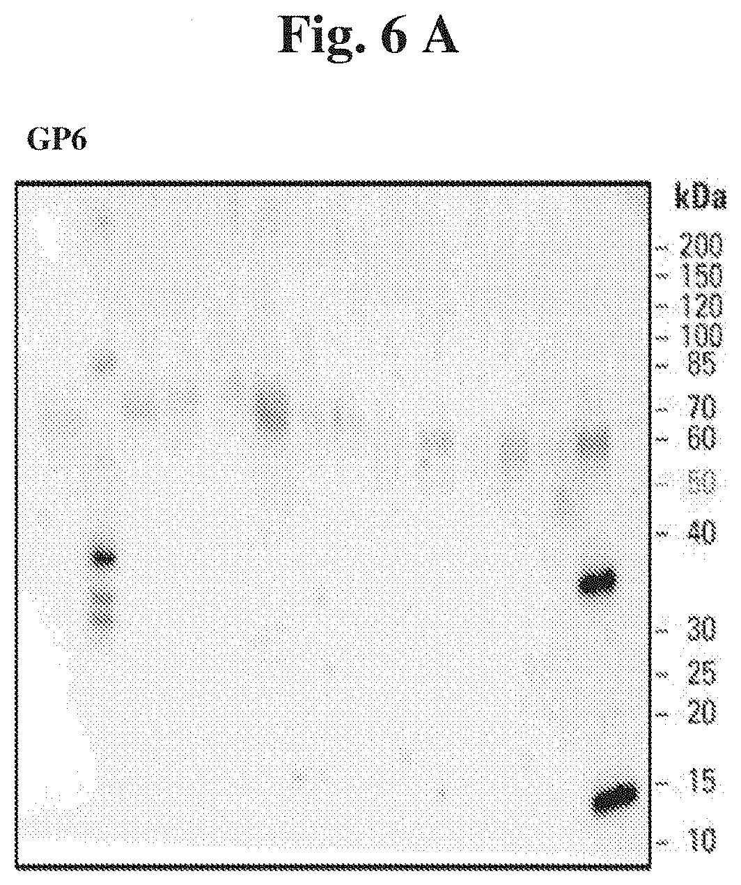
[0037] FIG. 6 (A) present sample results of Western blot analysis of GP6 content in urine samples derived from patients with renal diseases versus healthy controls, and (B) shows a summary of densitometry readings from Western blotting of the GP6 protein in selected urine samples of study participants with renal diseases as compared to healthy subjects.
[0038] FIG. 7 presents a list of possible combinations of the markers for use according to the invention.
EXAMPLES
Example 1
Discovery Phase
[0039] The methodological approach of the discovery phase has been described in Mucha et al. Below, the most pertinent information is listed.
Patients Characteristics
Groups of Patients
[0040] The study included 30 patients with IgAN and 30 healthy age- and sex-matched volunteers serving as controls. Demographic and clinical data of both groups are presented in Mucha, et al. (Supplementary material online, Table S1). Briefly, patients with biopsy-proven IgAN at different stages of chronic kidney disease (CKD) and older than 18 years were included. The inclusion criteria for the control group were as follows: age older than 18 years and absence of any kidney disease or other chronic diseases requiring treatment. The exclusion criteria for both groups included: active infection, history of malignancy, previous organ transplantation, or current pregnancy. To estimate GFR, we used the Chronic Kidney Disease Epidemiology Collaboration equations, which are the most accurate, have been evaluated in large diverse populations, and are applicable for clinical use. The study protocol was approved by the local ethics committee and informed consent was obtained from all participants. The study was performed in accordance with the Declaration of Helsinki.
Urine Collection
[0041] Samples were collected from all individuals according to a uniform study protocol, following the recommendations on urine proteomic sample collection. The second- or third-morning midstream urine was collected to sterile urine containers 1 to 3 h after previous urination. The pH of each sample was stabilized at 7.2 by addition of 1/10th vol. of 1M HEPES pH 7.2 immediately after collection. Then, samples were vortexed for 2 min, centrifuged at 3000.times.g at room temperature for 10 min to clear the debris, filtered (0.4-.mu.m filter, Rotilabo-Spritzenfilter, Roth, Karlsruhe, Germany), and portioned into 1-ml aliquots that were stored at -80.degree. C. before further use.
Sample Filtration
[0042] Membrane filters of the 10 kDa cut-off (Amicon Ultra-0.5, UFC501096, Millipore, Billerica, United States) were washed twice with MilliQ (MQ) water prior to use. Urine was centrifuged through the membrane at 14,000.times.g for 15 min. Next, 500 .mu.l MQ was added to the retentate and centrifugation step was repeated. To recover the concentrated and desalted sample, the filter was placed upside down and centrifuged in a clean microcentrifuge tube for 2 min at 1000.times.g. The protein concentration was measured by the Bradford method. Aliquots of samples were stored at -80.degree. C.
Sample Preparation
[0043] 30 IgAN samples were divided into 3 disease pooled samples (DPSs I, II, and III), and similarly, 30 control samples were divided into 3 control pooled samples (CPSs I, II, and III). Age and sex matching was preserved within the 3 pairs of pooled sample groups. All DPSs and CPSs were obtained in 2 technical replicates (marked A and B) each, making a set of 12 pooled samples to be compared after isobaric tags for relative and absolute quantitation (iTRAQ) labeling. As 4-plex iTRAQ was used, 2 technical replications of DPSs and CPSs were compared in 1 isoelectric focusing/liquid chromatography-mass spectrometry/mass spectrometry (IEF/LC-MS/MS) experiment. To analyze 12 samples, we conducted a set of 3 independent IEF/LC-MS/MS experiments. Aliquots with extracted peptides were stored at -80.degree. C. for the IEF/LC-MS/MS analysis.
Mass Spectrometry
[0044] Qualitative MS/MS data processing The MS/MS data were pre-processed with Mascot Distiller (v. 2.3.2.0, Matrix Science, London, United Kingdom). Data search using the MASCOT search engine was conducted on the Swiss-Prot database with the taxonomy restricted to Homo sapiens (20,236 sequences) in a 3-step procedure described elsewhere to calculate MS and MS/MS measurement errors and to recalibrate the data for the repeated MASCOT to remove systematic bias. Protein ratios were calculated as the median ratio of their peptide's ratios. The statistical significance of a single protein ratio was assessed by an in-house program, Diffprot. Calculated P values were adjusted for multiple testing using a false discovery rate-con-trolling procedure, yielding protein ratio q values.
Results of the Discovery Phase
[0045] As a result of qualitative analysis (peptide and protein identification) in each of the 3 IEF/LC-MS-MS/MS experiments, 761, 951, and 956 proteins were identified, respectively, each represented by 2 or more peptides. The results of this observations were partially presented in Mucha et al. The discovery of alpha-1B-glycoprotein (A1BG), alpha-1-acid glycoprotein 1 (AGP1, ORM1), Ig lambda-2 chain C regions (IGLC2) and serotransferrin (TF) as markers for IgAN is being reported in the current invention (Table 2).
TABLE-US-00002 TABLE 2 Peptide read-outs of urine proteomics specific for alpha-1B-glycoprotein (A1BG), alpha-1-acid glycoprotein 1 (AGP1, ORM1), Ig lambda-2 chain C regions (IGLC2) and serotransferrin (TF) obtained in a discovery phase of the invention. Number of ID 1 Protein name Gene peptides P04217 Alpha-1B-glycoprotein A1BG 51 Peptides q-value SEQ ID NO: 1 SLPAPW 4.26E-03 SEQ ID NO: 2 ITPGLK 4.66E-03 SEQ ID NO: 3 GVTFLLR 2.84E-03 SEQ ID NO: 4 SWVPHTF 5.71E-03 SEQ ID NO: 5 LLELTGPK 0.00E+00 SEQ ID NO: 6 SWITPGLK 4.57E-04 SEQ ID NO: 7 ATWSGAVLAGR 0.00E+00 SEQ ID NO: 8 LELHVDGPPPR 7.51E-04 SEQ ID NO: 9 SLPAPWLSMAPV 0.00E+00 SEQ ID NO: 10 VAPLSGVDFQLR 0.00E+00 SEQ ID NO: 11 IFVGPQHAGNYR 6.31E-05 SEQ ID NO: 12 HQFLLTGDTQGR 0.00E+00 SEQ ID NO: 13 LETPDFQLFK 0.00E+00 SEQ ID NO: 14 SGLSTGWTQLSK 0.00E+00 SEQ ID NO: 15 SMAPVSWITPGLK 1.67E-03 SEQ ID NO: 16 HGESSQVLHPGNK 0.00E+00 SEQ ID NO: 17 SLPAPWLSMAPVSW 6.55E-03 SEQ ID NO: 18 LELHVDGPPPRPQL 3.58E-04 SEQ ID NO: 19 HHGESSQVLHPGNK 7.56E-05 SEQ ID NO: 20 HQFLLTGDTQGRYR 7.25E-03 SEQ ID NO: 21 GVAQEPVHLDSPAIK 0.00E+00 SEQ ID NO: 22 LELIFVGPQHAGNYR 0.00E+00 SEQ ID NO: 23 LELHVDGPPPRPQLR 2.78E-04 SEQ ID NO: 24 IFFHLNAVALGDGGHY 0.00E+00 SEQ ID NO: 25 NLELIFVGPQHAGNYR 0.00E+00 SEQ ID NO: 26 TFESELSDPVELLVAES 7.70E-04 SEQ ID NO: 27 GAAANLELIFVGPQHAGNYR 0.00E+00 SEQ ID NO: 28 SLPAPWLSMAPVSWITPGLK 0.00E+00 SEQ ID NO: 29 TPGAAANLELIFVGPQHAGNYR 0.00E+00 SEQ ID NO: 30 SWVPHTFESELSDPVELLVAES 0.00E+00 SEQ ID NO: 31 TVRTPGAAANLELIFVGPQHAGNYR 0.00E+00 SEQ ID NO: 32 LHDNQNGWSGDSAPVELILSDETLPAPEFSPEPESGR 1.74E-03 SEQ ID NO: 33 TDGEGALSEPSATVTIEELAAPPPPVLMHHGESSQVLHPGNK 0.00E+00 SEQ ID NO: 34 SWVPHTFE 6.44E-03 SEQ ID NO: 35 VGPQHAGNYR 6.31E-05 SEQ ID NO: 36 STGWTQLSK 9.51E-03 SEQ ID NO: 37 HQFLLTGDTQ 0.00E+00 SEQ ID NO: 38 PVSWITPGLK 0.00E+00 SEQ ID NO: 39 HVDGPPPRPQLR 1.02E-03 SEQ ID NO: 40 LSMAPVSWITPGLK 6.31E-05 SEQ ID NO: 41 MHHGESSQVLHPGNK 3.73E-04 SEQ ID NO: 42 SGLSTGWTQLSKLLELTGPK 3.87E-05 SEQ ID NO: 43 GPPPRPQLR 7.28E-03 SEQ ID NO: 44 SLPAPWLSMA 5.85E-04 SEQ ID NO: 45 LELHVDGPPPRPQ 4.34E-04 SEQ ID NO: 46 IFFHLNAVALGDGGH 0.00E+00 SEQ ID NO: 47 NGVAQEPVHLDSPAIK 3.87E-05 SEQ ID NO: 48 TPGAAANLELIFVGPQHAGN 0.00E+00 SEQ ID NO: 49 LPAPWLSMAPVSWITPGLK 3.87E-05 Number of ID 2 Protein name Gene peptides P02763 Alpha-1-acid glycoprotein 1 ORM1 29 Peptides q-value SEQ ID NO: 50 TYMLAF 1.964E-03 SEQ ID NO: 51 AHLLILR 0.000E+00 SEQ ID NO: 52 NWGLSVY 6.310E-05 SEQ ID NO: 53 TYLNVQR 5.215E-03 SEQ ID NO: 54 YVGGQEHF 6.264E-03 SEQ ID NO: 55 FAHLLILR 1.789E-04 SEQ ID NO: 56 TTYLNVQR 4.565E-04 SEQ ID NO: 57 YVGGQEHFA 5.451E-04 SEQ ID NO: 58 YVGGQEHFAH 0.000E+00 SEQ ID NO: 59 EHFAHLLILR 6.704E-03 SEQ ID NO: 60 SDVVYTDWK 0.000E+00 SEQ ID NO: 61 YVGGQEHFAHL 0.000E+00 SEQ ID NO: 62 MLAFDVNDEK 0.000E+00 SEQ ID NO: 63 YVGGQEHFAHLL 3.866E-05 SEQ ID NO: 64 GQEHFAHLLILR 0.000E+00 SEQ ID NO: 65 SVYADKPETTK 0.000E+00 SEQ ID NO: 66 TYMLAFDVNDEK 0.000E+00 SEQ ID NO: 67 GLSVYADKPETTK 3.866E-05 SEQ ID NO: 68 EQLGEFYEALDCLR 0.000E+00 SEQ ID NO: 69 YVGGQEHFAHLLILR 0.000E+00 SEQ ID NO: 70 WGLSVYADKPETTK 3.866E-05 SEQ ID NO: 71 NWGLSVYADKPETTK 0.000E+00 SEQ ID NO: 72 DVNDEKNWGLSVYADKPETTK 0.000E+00 SEQ ID NO: 73 TYMLAFDVNDEKNWGLSVYADKPETTK 0.000E+00 SEQ ID NO: 74 VVYTDWK 7.322E-03 SEQ ID NO: 75 VYADKPETTK 1.877E-03 SEQ ID NO: 76 VGGQEHFAHLLILR 2.390E-04 SEQ ID NO: 77 IPKSDVVYTDWK 6.846E-03 SEQ ID NO: 78 GGQEHFAHLLILR 3.901E-03 Number of ID 3 Protein name Gene peptides P0CG05 Ig lambda-2 chain C regions IGLC2 4 Peptides q-value SEQ ID NO: 79 ADSSPVK 5.85E-04 SEQ ID NO: 80 GVETTTPSK 1.61E-04 SEQ ID NO: 81 AGVETTTPSK 0.00E+00 SEQ ID NO: 82 KAGVETTTPSK 8.03E-04 Number of ID 4 Protein name Gene peptides P02787 Serotransferrin TF 76 Peptides q-value SEQ ID NO: 83 VYIAGK 4.49E-03 SEQ ID NO: 84 DSGFQMN 1.79E-04 SEQ ID NO: 85 HSTIFEN 1.05E-03 SEQ ID NO: 86 GLLYNK 3.85E-03 SEQ ID NO: 87 SAGWNIPI 9.31E-03 SEQ ID NO: 88 PDPWAK 5.33E-03 SEQ ID NO: 89 MYLGYEY 4.17E-04 SEQ ID NO: 90 NPDPWAK 1.79E-04 SEQ ID NO: 91 DSAHGFLK 0.00E+00 SEQ ID NO: 92 FGYSGAFK 2.78E-04 SEQ ID NO: 93 VAEFYGSK 0.00E+00 SEQ ID NO: 94 KDSGFQMN 9.23E-04 SEQ ID NO: 95 EFQLFSSPH 2.43E-04 SEQ ID NO: 96 KPVEEYAN 6.13E-04 SEQ ID NO: 97 DGAGDVAFVK 0.00E+00 SEQ ID NO: 98 SAGWNIPIGLL 0.00E+00 SEQ ID NO: 99 EDLIWELLN 3.73E-04 SEQ ID NO: 100 YLGEEYVK 3.87E-05 SEQ ID NO: 101 HSTIFENLAN 0.00E+00 SEQ ID NO: 102 GYYGYTGAFR 0.00E+00 SEQ ID NO: 103 KPVDEYK 4.57E-04 SEQ ID NO: 104 IPMGLLYNK 3.87E-05 SEQ ID NO: 105 DSGFQMNQLR 0.00E+00 SEQ ID NO: 106 PVVAEFYGSK 0.00E+00 SEQ ID NO: 107 LAQVPSHTVVAR 0.00E+00 SEQ ID NO: 108 KPVDEYKD 8.27E-04 SEQ ID NO: 109 EGYYGYTGAFR 3.87E-05 SEQ ID NO: 110 SAGWNIPIGLLY 6.31E-05 SEQ ID NO: 111 NIPMGLLYNK 2.78E-04
SEQ ID NO: 112 HQTVPQNTGGK 0.00E+00 SEQ ID NO: 113 QYFGYSGAFK 6.31E-05 SEQ ID NO: 114 TAGWNIPMGLLY 6.31E-05 SEQ ID NO: 115 SLDGGFVYIAGK 0.00E+00 SEQ ID NO: 116 SASDLTWDNLK 0.00E+00 SEQ ID NO: 117 HSTIFENLANK 0.00E+00 SEQ ID NO: 118 EFQLFSSPHGK 0.00E+00 SEQ ID NO: 119 TAGWNIPMGLLYN 3.87E-05 SEQ ID NO: 120 SKEFQLFSSPH 2.29E-03 SEQ ID NO: 121 KDSGFQMNQLR 0.00E+00 SEQ ID NO: 122 EDLIWELLNQAQ 1.61E-04 SEQ ID NO: 123 MYLGYEYVTAIR 0.00E+00 SEQ ID NO: 124 KEGYYGYTGAFR 0.00E+00 SEQ ID NO: 125 LKPVVAEFYGSK 0.00E+00 SEQ ID NO: 126 KSASDLTWDNLK 0.00E+00 SEQ ID NO: 127 TAGWNIPMGLLYNK 0.00E+00 SEQ ID NO: 128 STLNQYFGYSGAFK 6.31E-05 SEQ ID NO: 129 NLKPVVAEFYGSK 0.00E+00 SEQ ID NO: 130 EDPQTFYYAVAVVK 0.00E+00 SEQ ID NO: 131 SKEFQLFSSPHGK 0.00E+00 SEQ ID NO: 132 AIAANEADAVTLDAGLVYDAY 0.00E+00 SEQ ID NO: 133 LAPNNLKPVVAEFYGSK 0.00E+00 SEQ ID NO: 134 EDLIWELLNQAQEHFGK 0.00E+00 SEQ ID NO: 135 IMNGEADAMSLDGGFVYIAGK 3.87E-05 SEQ ID NO: 136 AIAANEADAVTLDAGLVYDAYLAPN 0.00E+00 SEQ ID NO: 137 GKEDLIWELLNQAQEHFGK 0.00E+00 SEQ ID NO: 138 GGKEDLIWELLNQAQEHFGK 0.00E+00 SEQ ID NO: 139 EDLIWELLNQAQEHFGKDK 0.00E+00 SEQ ID NO: 140 SMGGKEDLIWELLNQAQEHFGK 0.00E+00 SEQ ID NO: 141 AIAANEADAVTLDAGLVYDAYLAPNNLKPVVAEFYGSK 0.00E+00 SEQ ID NO: 142 APNHAVVT 8.70E-03 SEQ ID NO: 143 APNHAVVTR 6.31E-05 SEQ ID NO: 144 SAGWNIPIGL 8.24E-03 SEQ ID NO: 145 QVPSHTVVAR 1.06E-03 SEQ ID NO: 146 STIFENLANK 0.00E+00 SEQ ID NO: 147 HLAQVPSHTVVAR 0.00E+00 SEQ ID NO: 148 GWNIPMGLLYNK 6.31E-05 SEQ ID NO: 149 MYLGYEYVTAIRNLR 3.86E-03 SEQ ID NO: 150 PNNLKPVVAEFYGSK 5.33E-03 SEQ ID NO: 151 HSTIFENLA 5.85E-04 SEQ ID NO: 152 ADRDQYELL 1.81E-03 SEQ ID NO: 153 QLFSSPHGK 1.75E-03 SEQ ID NO: 154 LGYEYVTAIR 2.69E-03 SEQ ID NO: 155 HSTIFENLANKADR 5.85E-04 SEQ ID NO: 156 HQTVPQNTGGKNPDPWAK 3.87E-05 SEQ ID NO: 157 KEDLIWELLNQAQEHFGK 1.52E-03 SEQ ID NO: 158 GLVYDAYLAPNNLKPVVAEFYGSK 3.87E-05
Example 2
Validation Phase
[0046] The primary difference between the discovery and validation phases is the transition from the assessment of the pooled urine samples (i.e. the discovery phase) to the individual evaluation of each protein in a given patient or a healthy person and a direct correlation of these results with the known clinical parameters in each case (i.e. the validation phase).
Patient Characteristics
[0047] The study included 133 renal disease patients and 19 healthy controls. Renal disease included IgAN (77 cases), ADPKD (29) and LN (27).
Sample Collection
[0048] Urinary samples were collected according to the protocol standardized in the Transplantation Institute, Medical University of Warsaw.
SDS-PAGE
[0049] Samples were defrozen to room temperature (.about.23.degree. C.), then suspended by intensive pipetting or mixing using a vortex. Leammli buffer was added to urine samples to achieve final concentrations as follows: 2% SDS; 10% glycerol; 5% .beta.-mercaptoethanol; 0.002% bromophenol blue; 0.125 M Tris-HCl; pH 6.8. Samples were boiled at 95.degree. C. for 2 min. 10 .mu.l of each sample was loaded on the Mini-PROTEAN TGX 4-15% gradient gel.
Western Blotting Analysis
[0050] The method developed by the Department of Molecular Biology, Institute of Biochemistry and Biophysics, Polish Academy of Sciences (patent application no P.415033) allows to assay all the proteins of interest in the urine samples. It permits the analysis of all the selected protein biomarkers with the accuracy not reachable by classical methods. To date, the proteome analysis of urine in medicine starts from centrifugation of the sample (in line with the European Confederacy of Laboratory Medicine guidelines), which result losing of protein which are insoluble, and exist as aggregates or degradants. This solid fraction is crucial because proteins progress into insoluble forms randomly, depending of protein, state of patient, his diet and properties of urine. For that reason, in the current study we used the whole urine in form of suspension, which next were analyzed by Western blotting technique. That gives an opportunity to examine all of proteins present in a given in urine sample. Advantages of this method are important for medicinal diagnostics. Method is noninvasive for patients, it allows collecting the samples from the patients even few times per day, and it is relatively little time consuming. For Western blotting analysis, the urine protein samples were separated on SDS-PAGE gels, as described above, and transferred to a nitrocellulose membrane. Membranes were incubated in appropriate blocking buffer (either 5% low-fat dry milk or bovine serum albumin in TBS-Tween 20 (TBST)). After an incubation in the primary antibody (A1BG (F-9); catalog number sc-374415; Santa Cruz) the cells were washed in TBST, and incubated with a horseradish peroxidase-conjugated secondary antibody. The chemiluminescence reaction for HRP was developed using SuperSignal West Femto Chemiluminescent Substrate (Thermo Scientific) and visualized with Stella 8300 bioimager Densitometry read-outs were carried out for each of the bands in the blots. Eight randomly chosen patients samples were pooled together and used on each Western blot as a benchmark. Densitometry read-outs from other band on a given Western blot membrane were divided by the read-out of the respective benchmark. The results were referred to as a "relative band density".
[0051] List of antibodies used for Western blotting: A1BG (cat # sc-374415, Santa Cruz), ORM1 (sc-69753, Santa Cruz), IGLC2 (sc-33134, Santa Cruz), TF (sc-21011, Santa Crus), GP6 (sc-20149, Santa Cruz).
Results
[0052] The results of representative Western blotting analysis for A1BG, ORM1, IGLC2 and TF presence in urine samples are presented in FIG. 1.
[0053] A1BG Based on the Western blotting analysis, it becomes evident that study participants with kidney diseases exhibit the presence of different forms of the protein (for the purpose of this invention segregated into within the high [.about.80 kDa], medium [.about.45 kDa] and low [.about.15 kDa] molecular weight range), occurring in different proportions. A direct comparison of selected samples from kidney disease versus heathy patients is presented in FIG. 2A. Mutual relations between the visible forms may be important in the pathophysiology of the given disease. Indeed, as presented in FIG. 2B, various subforms of A1BG correlate differently with the type of kidney disease and also differently as compared to the cumulative assessment of the protein (FIG. 2C). Notably, the bottom-range bands tend to be most prominently elevated in IgAN patients.
[0054] Validation phase as described above was also performed for the other markers, in particular ORM1, IGLC2 and TF and the results are presented in FIGS. 3-5. The results are also presented for GP6 (FIG. 6). Although GP6 is on average expressed at higher levels in LN than IgA, on the basis of our results and data, the compilation of 4 proteins reported in the GP6 compilation may increase the sensitivity and specificity of the test.
Sequence CWU 1
1
15816PRTHomo sapiensAlpha-1B-glycoprotein peptide 1Ser Leu Pro Ala
Pro Trp1 526PRTHomo sapiensAlpha-1B-glycoprotein peptide 2Ile Thr
Pro Gly Leu Lys1 537PRTHomo sapiensAlpha-1B-glycoprotein peptide
3Gly Val Thr Phe Leu Leu Arg1 547PRTHomo
sapiensAlpha-1B-glycoprotein peptide 4Ser Trp Val Pro His Thr Phe1
558PRTHomo sapiensAlpha-1B-glycoprotein peptide 5Leu Leu Glu Leu
Thr Gly Pro Lys1 568PRTHomo sapiensAlpha-1B-glycoprotein peptide
6Ser Trp Ile Thr Pro Gly Leu Lys1 5711PRTHomo
sapiensAlpha-1B-glycoprotein peptide 7Ala Thr Trp Ser Gly Ala Val
Leu Ala Gly Arg1 5 10811PRTHomo sapiensAlpha-1B-glycoprotein
peptide 8Leu Glu Leu His Val Asp Gly Pro Pro Pro Arg1 5
10912PRTHomo sapiensAlpha-1B-glycoprotein peptide 9Ser Leu Pro Ala
Pro Trp Leu Ser Met Ala Pro Val1 5 101012PRTHomo
sapiensAlpha-1B-glycoprotein peptide 10Val Ala Pro Leu Ser Gly Val
Asp Phe Gln Leu Arg1 5 101112PRTHomo sapiensAlpha-1B-glycoprotein
peptide 11Ile Phe Val Gly Pro Gln His Ala Gly Asn Tyr Arg1 5
101212PRTHomo sapiensAlpha-1B-glycoprotein peptide 12His Gln Phe
Leu Leu Thr Gly Asp Thr Gln Gly Arg1 5 101310PRTHomo
sapiensAlpha-1B-glycoprotein peptide 13Leu Glu Thr Pro Asp Phe Gln
Leu Phe Lys1 5 101412PRTHomo sapiensAlpha-1B-glycoprotein peptide
14Ser Gly Leu Ser Thr Gly Trp Thr Gln Leu Ser Lys1 5 101513PRTHomo
sapiensAlpha-1B-glycoprotein peptide 15Ser Met Ala Pro Val Ser Trp
Ile Thr Pro Gly Leu Lys1 5 101613PRTHomo
sapiensAlpha-1B-glycoprotein peptide 16His Gly Glu Ser Ser Gln Val
Leu His Pro Gly Asn Lys1 5 101714PRTHomo
sapiensAlpha-1B-glycoprotein peptide 17Ser Leu Pro Ala Pro Trp Leu
Ser Met Ala Pro Val Ser Trp1 5 101814PRTHomo
sapiensAlpha-1B-glycoprotein peptide 18Leu Glu Leu His Val Asp Gly
Pro Pro Pro Arg Pro Gln Leu1 5 101914PRTHomo
sapiensAlpha-1B-glycoprotein peptide 19His His Gly Glu Ser Ser Gln
Val Leu His Pro Gly Asn Lys1 5 102014PRTHomo
sapiensAlpha-1B-glycoprotein peptide 20His Gln Phe Leu Leu Thr Gly
Asp Thr Gln Gly Arg Tyr Arg1 5 102115PRTHomo
sapiensAlpha-1B-glycoprotein peptide 21Gly Val Ala Gln Glu Pro Val
His Leu Asp Ser Pro Ala Ile Lys1 5 10 152215PRTHomo
sapiensAlpha-1B-glycoprotein peptide 22Leu Glu Leu Ile Phe Val Gly
Pro Gln His Ala Gly Asn Tyr Arg1 5 10 152315PRTHomo
sapiensAlpha-1B-glycoprotein peptide 23Leu Glu Leu His Val Asp Gly
Pro Pro Pro Arg Pro Gln Leu Arg1 5 10 152416PRTHomo
sapiensAlpha-1B-glycoprotein peptide 24Ile Phe Phe His Leu Asn Ala
Val Ala Leu Gly Asp Gly Gly His Tyr1 5 10 152516PRTHomo
sapiensAlpha-1B-glycoprotein peptide 25Asn Leu Glu Leu Ile Phe Val
Gly Pro Gln His Ala Gly Asn Tyr Arg1 5 10 152617PRTHomo
sapiensAlpha-1B-glycoprotein peptide 26Thr Phe Glu Ser Glu Leu Ser
Asp Pro Val Glu Leu Leu Val Ala Glu1 5 10 15Ser2720PRTHomo
sapiensAlpha-1B-glycoprotein peptidep 27Gly Ala Ala Ala Asn Leu Glu
Leu Ile Phe Val Gly Pro Gln His Ala1 5 10 15Gly Asn Tyr Arg
202820PRTHomo sapiensAlpha-1B-glycoprotein peptide 28Ser Leu Pro
Ala Pro Trp Leu Ser Met Ala Pro Val Ser Trp Ile Thr1 5 10 15Pro Gly
Leu Lys 202922PRTHomo sapiensAlpha-1B-glycoprotein peptide 29Thr
Pro Gly Ala Ala Ala Asn Leu Glu Leu Ile Phe Val Gly Pro Gln1 5 10
15His Ala Gly Asn Tyr Arg 203022PRTHomo
sapiensAlpha-1B-glycoprotein peptide 30Ser Trp Val Pro His Thr Phe
Glu Ser Glu Leu Ser Asp Pro Val Glu1 5 10 15Leu Leu Val Ala Glu Ser
203125PRTHomo sapiensAlpha-1B-glycoprotein peptide 31Thr Val Arg
Thr Pro Gly Ala Ala Ala Asn Leu Glu Leu Ile Phe Val1 5 10 15Gly Pro
Gln His Ala Gly Asn Tyr Arg 20 253237PRTHomo
sapiensAlpha-1B-glycoprotein peptide 32Leu His Asp Asn Gln Asn Gly
Trp Ser Gly Asp Ser Ala Pro Val Glu1 5 10 15Leu Ile Leu Ser Asp Glu
Thr Leu Pro Ala Pro Glu Phe Ser Pro Glu 20 25 30Pro Glu Ser Gly Arg
353342PRTHomo sapiensAlpha-1B-glycoprotein peptide 33Thr Asp Gly
Glu Gly Ala Leu Ser Glu Pro Ser Ala Thr Val Thr Ile1 5 10 15Glu Glu
Leu Ala Ala Pro Pro Pro Pro Val Leu Met His His Gly Glu 20 25 30Ser
Ser Gln Val Leu His Pro Gly Asn Lys 35 40348PRTHomo
sapiensAlpha-1B-glycoprotein peptide 34Ser Trp Val Pro His Thr Phe
Glu1 53510PRTHomo sapiensAlpha-1B-glycoprotein peptide 35Val Gly
Pro Gln His Ala Gly Asn Tyr Arg1 5 10369PRTHomo
sapiensAlpha-1B-glycoprotein peptide 36Ser Thr Gly Trp Thr Gln Leu
Ser Lys1 53710PRTHomo sapiensAlpha-1B-glycoprotein peptide 37His
Gln Phe Leu Leu Thr Gly Asp Thr Gln1 5 103810PRTHomo
sapiensAlpha-1B-glycoprotein peptide 38Pro Val Ser Trp Ile Thr Pro
Gly Leu Lys1 5 103912PRTHomo sapiensAlpha-1B-glycoprotein peptide
39His Val Asp Gly Pro Pro Pro Arg Pro Gln Leu Arg1 5 104014PRTHomo
sapiensAlpha-1B-glycoprotein peptide 40Leu Ser Met Ala Pro Val Ser
Trp Ile Thr Pro Gly Leu Lys1 5 104115PRTHomo
sapiensAlpha-1B-glycoprotein peptide 41Met His His Gly Glu Ser Ser
Gln Val Leu His Pro Gly Asn Lys1 5 10 154220PRTHomo
sapiensAlpha-1B-glycoprotein peptide 42Ser Gly Leu Ser Thr Gly Trp
Thr Gln Leu Ser Lys Leu Leu Glu Leu1 5 10 15Thr Gly Pro Lys
20439PRTHomo sapiensAlpha-1B-glycoprotein peptide 43Gly Pro Pro Pro
Arg Pro Gln Leu Arg1 54410PRTHomo sapiensAlpha-1B-glycoprotein
peptide 44Ser Leu Pro Ala Pro Trp Leu Ser Met Ala1 5 104513PRTHomo
sapiensAlpha-1B-glycoprotein peptide 45Leu Glu Leu His Val Asp Gly
Pro Pro Pro Arg Pro Gln1 5 104615PRTHomo
sapiensAlpha-1B-glycoprotein peptide 46Ile Phe Phe His Leu Asn Ala
Val Ala Leu Gly Asp Gly Gly His1 5 10 154716PRTHomo
sapiensAlpha-1B-glycoprotein peptide 47Asn Gly Val Ala Gln Glu Pro
Val His Leu Asp Ser Pro Ala Ile Lys1 5 10 154820PRTHomo
sapiensAlpha-1B-glycoprotein peptide 48Thr Pro Gly Ala Ala Ala Asn
Leu Glu Leu Ile Phe Val Gly Pro Gln1 5 10 15His Ala Gly Asn
204919PRTHomo sapiensAlpha-1B-glycoprotein peptide 49Leu Pro Ala
Pro Trp Leu Ser Met Ala Pro Val Ser Trp Ile Thr Pro1 5 10 15Gly Leu
Lys506PRTHomo sapiensAlpha-1-acid glycoprotein 1 peptide 50Thr Tyr
Met Leu Ala Phe1 5517PRTHomo sapiensAlpha-1-acid glycoprotein 1
peptide 51Ala His Leu Leu Ile Leu Arg1 5527PRTHomo
sapiensAlpha-1-acid glycoprotein 1 peptide 52Asn Trp Gly Leu Ser
Val Tyr1 5537PRTHomo sapiensAlpha-1-acid glycoprotein 1 peptide
53Thr Tyr Leu Asn Val Gln Arg1 5548PRTHomo sapiensAlpha-1-acid
glycoprotein 1 peptide 54Tyr Val Gly Gly Gln Glu His Phe1
5558PRTHomo sapiensAlpha-1-acid glycoprotein 1 peptide 55Phe Ala
His Leu Leu Ile Leu Arg1 5568PRTHomo sapiensAlpha-1-acid
glycoprotein 1 peptide 56Thr Thr Tyr Leu Asn Val Gln Arg1
5579PRTHomo sapiensAlpha-1-acid glycoprotein 1 peptide 57Tyr Val
Gly Gly Gln Glu His Phe Ala1 55810PRTHomo sapiensAlpha-1-acid
glycoprotein 1 peptide 58Tyr Val Gly Gly Gln Glu His Phe Ala His1 5
105910PRTHomo sapiensAlpha-1-acid glycoprotein 1 peptide 59Glu His
Phe Ala His Leu Leu Ile Leu Arg1 5 10609PRTHomo sapiensAlpha-1-acid
glycoprotein 1 peptide 60Ser Asp Val Val Tyr Thr Asp Trp Lys1
56111PRTHomo sapiensAlpha-1-acid glycoprotein 1 peptide 61Tyr Val
Gly Gly Gln Glu His Phe Ala His Leu1 5 106210PRTHomo
sapiensAlpha-1-acid glycoprotein 1 peptide 62Met Leu Ala Phe Asp
Val Asn Asp Glu Lys1 5 106312PRTHomo sapiensAlpha-1-acid
glycoprotein 1 peptide 63Tyr Val Gly Gly Gln Glu His Phe Ala His
Leu Leu1 5 106412PRTHomo sapiensAlpha-1-acid glycoprotein 1 peptide
64Gly Gln Glu His Phe Ala His Leu Leu Ile Leu Arg1 5 106511PRTHomo
sapiensAlpha-1-acid glycoprotein 1 peptide 65Ser Val Tyr Ala Asp
Lys Pro Glu Thr Thr Lys1 5 106612PRTHomo sapiensAlpha-1-acid
glycoprotein 1 peptide 66Thr Tyr Met Leu Ala Phe Asp Val Asn Asp
Glu Lys1 5 106713PRTHomo sapiensAlpha-1-acid glycoprotein 1 peptide
67Gly Leu Ser Val Tyr Ala Asp Lys Pro Glu Thr Thr Lys1 5
106814PRTHomo sapiensAlpha-1-acid glycoprotein 1 peptide 68Glu Gln
Leu Gly Glu Phe Tyr Glu Ala Leu Asp Cys Leu Arg1 5 106915PRTHomo
sapiensAlpha-1-acid glycoprotein 1 peptide 69Tyr Val Gly Gly Gln
Glu His Phe Ala His Leu Leu Ile Leu Arg1 5 10 157014PRTHomo
sapiensAlpha-1-acid glycoprotein 1 peptide 70Trp Gly Leu Ser Val
Tyr Ala Asp Lys Pro Glu Thr Thr Lys1 5 107115PRTHomo
sapiensAlpha-1-acid glycoprotein 1 peptide 71Asn Trp Gly Leu Ser
Val Tyr Ala Asp Lys Pro Glu Thr Thr Lys1 5 10 157221PRTHomo
sapiensAlpha-1-acid glycoprotein 1 peptide 72Asp Val Asn Asp Glu
Lys Asn Trp Gly Leu Ser Val Tyr Ala Asp Lys1 5 10 15Pro Glu Thr Thr
Lys 207327PRTHomo sapiensAlpha-1-acid glycoprotein 1 peptide 73Thr
Tyr Met Leu Ala Phe Asp Val Asn Asp Glu Lys Asn Trp Gly Leu1 5 10
15Ser Val Tyr Ala Asp Lys Pro Glu Thr Thr Lys 20 25747PRTHomo
sapiensAlpha-1-acid glycoprotein 1 peptide 74Val Val Tyr Thr Asp
Trp Lys1 57510PRTHomo sapiens>Alpha-1-acid glycoprotein 1
peptide 75Val Tyr Ala Asp Lys Pro Glu Thr Thr Lys1 5 107614PRTHomo
sapiensAlpha-1-acid glycoprotein 1 peptide 76Val Gly Gly Gln Glu
His Phe Ala His Leu Leu Ile Leu Arg1 5 107712PRTHomo
sapiensAlpha-1-acid glycoprotein 1 peptide 77Ile Pro Lys Ser Asp
Val Val Tyr Thr Asp Trp Lys1 5 107813PRTHomo sapiensAlpha-1-acid
glycoprotein 1 peptide 78Gly Gly Gln Glu His Phe Ala His Leu Leu
Ile Leu Arg1 5 10797PRTHomo sapiensIg lambda-2 chain C regions
79Ala Asp Ser Ser Pro Val Lys1 5809PRTHomo sapiensIg lambda-2 chain
C regions 80Gly Val Glu Thr Thr Thr Pro Ser Lys1 58110PRTHomo
sapiensIg lambda-2 chain C regions 81Ala Gly Val Glu Thr Thr Thr
Pro Ser Lys1 5 108211PRTHomo sapiensIg lambda-2 chain C regions
82Lys Ala Gly Val Glu Thr Thr Thr Pro Ser Lys1 5 10836PRTHomo
sapiensSerotransferrin peptide 83Val Tyr Ile Ala Gly Lys1
5847PRTHomo sapiensSerotransferrin peptide 84Asp Ser Gly Phe Gln
Met Asn1 5857PRTHomo sapiensSerotransferrin peptide 85His Ser Thr
Ile Phe Glu Asn1 5866PRTHomo sapiensSerotransferrin peptide 86Gly
Leu Leu Tyr Asn Lys1 5878PRTHomo sapiensSerotransferrin peptide
87Ser Ala Gly Trp Asn Ile Pro Ile1 5886PRTHomo
sapiensSerotransferrin peptide 88Pro Asp Pro Trp Ala Lys1
5897PRTHomo sapiensSerotransferrin peptide 89Met Tyr Leu Gly Tyr
Glu Tyr1 5907PRTHomo sapiensSerotransferrin peptide 90Asn Pro Asp
Pro Trp Ala Lys1 5918PRTHomo sapiensSerotransferrin peptide 91Asp
Ser Ala His Gly Phe Leu Lys1 5928PRTHomo sapiensSerotransferrin
peptide 92Phe Gly Tyr Ser Gly Ala Phe Lys1 5938PRTHomo
sapiensSerotransferrin peptide 93Val Ala Glu Phe Tyr Gly Ser Lys1
5948PRTHomo sapiensSerotransferrin peptide 94Lys Asp Ser Gly Phe
Gln Met Asn1 5959PRTHomo sapiensSerotransferrin peptide 95Glu Phe
Gln Leu Phe Ser Ser Pro His1 5968PRTHomo sapiensSerotransferrin
peptide 96Lys Pro Val Glu Glu Tyr Ala Asn1 59710PRTHomo
sapiensSerotransferrin peptide 97Asp Gly Ala Gly Asp Val Ala Phe
Val Lys1 5 109811PRTHomo sapiensSerotransferrin peptide 98Ser Ala
Gly Trp Asn Ile Pro Ile Gly Leu Leu1 5 10999PRTHomo
sapiensSerotransferrin peptide 99Glu Asp Leu Ile Trp Glu Leu Leu
Asn1 51008PRTHomo sapiensSerotransferrin peptide 100Tyr Leu Gly Glu
Glu Tyr Val Lys1 510110PRTHomo sapiensSerotransferrin peptide
101His Ser Thr Ile Phe Glu Asn Leu Ala Asn1 5 1010210PRTHomo
sapiensSerotransferrin peptide 102Gly Tyr Tyr Gly Tyr Thr Gly Ala
Phe Arg1 5 101037PRTHomo sapiensSerotransferrin peptide 103Lys Pro
Val Asp Glu Tyr Lys1 51049PRTHomo sapiensSerotransferrin peptide
104Ile Pro Met Gly Leu Leu Tyr Asn Lys1 510510PRTHomo
sapiensSerotransferrin peptide 105Asp Ser Gly Phe Gln Met Asn Gln
Leu Arg1 5 1010610PRTHomo sapiensSerotransferrin peptide 106Pro Val
Val Ala Glu Phe Tyr Gly Ser Lys1 5 1010712PRTHomo
sapiensSerotransferrin peptide 107Leu Ala Gln Val Pro Ser His Thr
Val Val Ala Arg1 5 101088PRTHomo sapiensSerotransferrin peptide
108Lys Pro Val Asp Glu Tyr Lys Asp1 510911PRTHomo
sapiensSerotransferrin peptide 109Glu Gly Tyr Tyr Gly Tyr Thr Gly
Ala Phe Arg1 5 1011012PRTHomo sapiensSerotransferrin peptide 110Ser
Ala Gly Trp Asn Ile Pro Ile Gly Leu Leu Tyr1 5 1011110PRTHomo
sapiensSerotransferrin peptide 111Asn Ile Pro Met Gly Leu Leu Tyr
Asn Lys1 5 1011211PRTHomo sapiensSerotransferrin peptide 112His Gln
Thr Val Pro Gln Asn Thr Gly Gly Lys1 5 1011310PRTHomo
sapiensSerotransferrin peptide 113Gln Tyr Phe Gly Tyr Ser Gly Ala
Phe Lys1 5 1011412PRTHomo sapiensSerotransferrin peptide 114Thr Ala
Gly Trp Asn Ile Pro Met Gly Leu Leu Tyr1 5 1011512PRTHomo
sapiensSerotransferrin peptide 115Ser Leu Asp Gly Gly Phe Val Tyr
Ile Ala Gly Lys1 5 1011611PRTHomo sapiensSerotransferrin peptide
116Ser Ala Ser Asp Leu Thr Trp Asp Asn Leu Lys1 5 1011711PRTHomo
sapiensSerotransferrin peptide 117His Ser Thr Ile Phe Glu Asn Leu
Ala Asn Lys1 5 1011811PRTHomo sapiensSerotransferrin peptide 118Glu
Phe Gln Leu Phe Ser Ser Pro His Gly Lys1 5 1011913PRTHomo
sapiensSerotransferrin peptide 119Thr Ala Gly Trp Asn Ile Pro Met
Gly Leu Leu Tyr Asn1 5 1012011PRTHomo sapiensSerotransferrin
peptide 120Ser Lys Glu Phe Gln Leu Phe Ser Ser Pro His1 5
1012111PRTHomo sapiensSerotransferrin peptide 121Lys Asp Ser Gly
Phe Gln Met Asn Gln Leu Arg1 5 1012212PRTHomo
sapiensSerotransferrin peptide 122Glu Asp Leu Ile Trp Glu Leu Leu
Asn Gln Ala Gln1 5 1012312PRTHomo sapiensSerotransferrin peptide
123Met Tyr Leu Gly Tyr Glu Tyr Val Thr Ala Ile Arg1 5
1012412PRTHomo sapiensSerotransferrin peptide 124Lys Glu Gly Tyr
Tyr Gly Tyr Thr Gly Ala Phe Arg1 5 1012512PRTHomo
sapiensSerotransferrin peptide 125Leu Lys Pro Val Val Ala Glu Phe
Tyr Gly Ser Lys1 5 1012612PRTHomo sapiensSerotransferrin peptide
126Lys Ser Ala Ser Asp Leu Thr Trp Asp Asn Leu Lys1 5
1012714PRTHomo sapiensSerotransferrin peptide 127Thr Ala Gly Trp
Asn Ile Pro Met Gly Leu Leu Tyr Asn Lys1 5 1012814PRTHomo
sapiensSerotransferrin peptide 128Ser Thr Leu Asn Gln Tyr Phe Gly
Tyr Ser Gly Ala Phe Lys1 5 1012913PRTHomo sapiensSerotransferrin
peptide 129Asn Leu Lys Pro Val Val Ala Glu Phe Tyr Gly Ser Lys1 5
1013014PRTHomo sapiensSerotransferrin peptide 130Glu Asp Pro Gln
Thr Phe Tyr Tyr Ala Val Ala Val Val Lys1 5 1013113PRTHomo
sapiensSerotransferrin peptide 131Ser Lys Glu Phe Gln Leu Phe Ser
Ser Pro His Gly Lys1 5 1013221PRTHomo sapiensSerotransferrin
peptide 132Ala Ile Ala Ala Asn Glu Ala Asp Ala Val Thr Leu Asp Ala
Gly Leu1 5 10 15Val Tyr Asp Ala Tyr 2013317PRTHomo
sapiensSerotransferrin peptide 133Leu Ala Pro Asn Asn Leu Lys Pro
Val Val Ala Glu Phe Tyr Gly Ser1 5 10 15Lys13417PRTHomo
sapiensSerotransferrin peptide 134Glu Asp Leu Ile Trp Glu Leu Leu
Asn Gln Ala Gln Glu His Phe Gly1 5 10 15Lys13521PRTHomo
sapiensSerotransferrin peptide 135Ile Met Asn Gly Glu Ala Asp Ala
Met Ser Leu Asp Gly Gly Phe Val1 5 10 15Tyr Ile Ala Gly Lys
2013625PRTHomo sapiensSerotransferrin peptide 136Ala Ile Ala Ala
Asn Glu Ala Asp Ala Val Thr Leu Asp Ala Gly Leu1 5 10 15Val Tyr Asp
Ala Tyr Leu Ala Pro Asn 20 2513719PRTHomo sapiensSerotransferrin
peptide 137Gly Lys Glu Asp Leu Ile Trp Glu Leu Leu Asn Gln Ala Gln
Glu His1 5 10 15Phe Gly Lys13820PRTHomo sapiensSerotransferrin
peptide 138Gly Gly Lys Glu Asp Leu Ile Trp Glu Leu Leu Asn Gln Ala
Gln Glu1 5 10 15His Phe Gly Lys 2013919PRTHomo
sapiensSerotransferrin peptide 139Glu Asp Leu Ile Trp Glu Leu Leu
Asn Gln Ala Gln Glu His Phe Gly1 5 10 15Lys Asp Lys14022PRTHomo
sapiensSerotransferrin peptide 140Ser Met Gly Gly Lys Glu Asp Leu
Ile Trp Glu Leu Leu Asn Gln Ala1 5 10 15Gln Glu His Phe Gly Lys
2014138PRTHomo sapiensSerotransferrin peptide 141Ala Ile Ala Ala
Asn Glu Ala Asp Ala Val Thr Leu Asp Ala Gly Leu1 5 10 15Val Tyr Asp
Ala Tyr Leu Ala Pro Asn Asn Leu Lys Pro Val Val Ala 20 25 30Glu Phe
Tyr Gly Ser Lys 351428PRTHomo sapiensSerotransferrin peptide 142Ala
Pro Asn His Ala Val Val Thr1 51439PRTHomo sapiensSerotransferrin
peptide 143Ala Pro Asn His Ala Val Val Thr Arg1 514410PRTHomo
sapiensSerotransferrin peptide 144Ser Ala Gly Trp Asn Ile Pro Ile
Gly Leu1 5 1014510PRTHomo sapiensSerotransferrin peptide 145Gln Val
Pro Ser His Thr Val Val Ala Arg1 5 1014610PRTHomo
sapiensSerotransferrin peptide 146Ser Thr Ile Phe Glu Asn Leu Ala
Asn Lys1 5 1014713PRTHomo sapiensSerotransferrin peptide 147His Leu
Ala Gln Val Pro Ser His Thr Val Val Ala Arg1 5 1014812PRTHomo
sapiensSerotransferrin peptide 148Gly Trp Asn Ile Pro Met Gly Leu
Leu Tyr Asn Lys1 5 1014915PRTHomo sapiensSerotransferrin peptide
149Met Tyr Leu Gly Tyr Glu Tyr Val Thr Ala Ile Arg Asn Leu Arg1 5
10 1515015PRTHomo sapiensSerotransferrin peptide 150Pro Asn Asn Leu
Lys Pro Val Val Ala Glu Phe Tyr Gly Ser Lys1 5 10 151519PRTHomo
sapiensSerotransferrin peptide 151His Ser Thr Ile Phe Glu Asn Leu
Ala1 51529PRTHomo sapiensSerotransferrin peptide 152Ala Asp Arg Asp
Gln Tyr Glu Leu Leu1 51539PRTHomo sapiensSerotransferrin peptide
153Gln Leu Phe Ser Ser Pro His Gly Lys1 515410PRTHomo
sapiensSerotransferrin peptide 154Leu Gly Tyr Glu Tyr Val Thr Ala
Ile Arg1 5 1015514PRTHomo sapiensSerotransferrin peptide 155His Ser
Thr Ile Phe Glu Asn Leu Ala Asn Lys Ala Asp Arg1 5 1015618PRTHomo
sapiensSerotransferrin peptide 156His Gln Thr Val Pro Gln Asn Thr
Gly Gly Lys Asn Pro Asp Pro Trp1 5 10 15Ala Lys15718PRTHomo
sapiensSerotransferrin peptide 157Lys Glu Asp Leu Ile Trp Glu Leu
Leu Asn Gln Ala Gln Glu His Phe1 5 10 15Gly Lys15824PRTHomo
sapiensSerotransferrin peptide 158Gly Leu Val Tyr Asp Ala Tyr Leu
Ala Pro Asn Asn Leu Lys Pro Val1 5 10 15Val Ala Glu Phe Tyr Gly Ser
Lys 20
D00000
D00001
D00002
D00003
D00004
D00005
D00006
D00007
D00008
D00009
S00001
XML
uspto.report is an independent third-party trademark research tool that is not affiliated, endorsed, or sponsored by the United States Patent and Trademark Office (USPTO) or any other governmental organization. The information provided by uspto.report is based on publicly available data at the time of writing and is intended for informational purposes only.
While we strive to provide accurate and up-to-date information, we do not guarantee the accuracy, completeness, reliability, or suitability of the information displayed on this site. The use of this site is at your own risk. Any reliance you place on such information is therefore strictly at your own risk.
All official trademark data, including owner information, should be verified by visiting the official USPTO website at www.uspto.gov. This site is not intended to replace professional legal advice and should not be used as a substitute for consulting with a legal professional who is knowledgeable about trademark law.