Methods And Compositions For Treating Chronic Obstructive Pulmonary Disorder
YOUNGBLOOD; Bradford Andrew ; et al.
U.S. patent application number 16/476027 was filed with the patent office on 2019-11-07 for methods and compositions for treating chronic obstructive pulmonary disorder. This patent application is currently assigned to Allakos Inc.. The applicant listed for this patent is Allakos Inc.. Invention is credited to Christopher Robert BEBBINGTON, Nenad TOMASEVIC, Bradford Andrew YOUNGBLOOD.
| Application Number | 20190338027 16/476027 |
| Document ID | / |
| Family ID | 62790958 |
| Filed Date | 2019-11-07 |


| United States Patent Application | 20190338027 |
| Kind Code | A1 |
| YOUNGBLOOD; Bradford Andrew ; et al. | November 7, 2019 |
METHODS AND COMPOSITIONS FOR TREATING CHRONIC OBSTRUCTIVE PULMONARY DISORDER
Abstract
The present disclosure provides methods for the treatment of chronic obstructive pulmonary disease (COPD) (e.g., non-eosinophilic COPD). In particular, the present disclosure provides methods for the treatment of COPD (e.g., non-eosinophilic COPD) through administration of antibodies that bind to human Siglec-8 or compositions comprising said antibodies. The present disclosure also provides articles of manufacture or kits comprising antibodies that bind to human Siglec-8 for the treatment of COPD (e.g., non-eosinophilic COPD).
| Inventors: | YOUNGBLOOD; Bradford Andrew; (Burlingame, CA) ; TOMASEVIC; Nenad; (Foster City, CA) ; BEBBINGTON; Christopher Robert; (San Mateo, CA) | ||||||||||
| Applicant: |
|
||||||||||
|---|---|---|---|---|---|---|---|---|---|---|---|
| Assignee: | Allakos Inc. Redwood City CA |
||||||||||
| Family ID: | 62790958 | ||||||||||
| Appl. No.: | 16/476027 | ||||||||||
| Filed: | January 5, 2018 | ||||||||||
| PCT Filed: | January 5, 2018 | ||||||||||
| PCT NO: | PCT/US2018/012694 | ||||||||||
| 371 Date: | July 3, 2019 |
Related U.S. Patent Documents
| Application Number | Filing Date | Patent Number | ||
|---|---|---|---|---|
| 62443591 | Jan 6, 2017 | |||
| Current U.S. Class: | 1/1 |
| Current CPC Class: | C07K 2317/76 20130101; A61K 2039/505 20130101; A61K 45/06 20130101; A61P 11/00 20180101; A61K 2039/55 20130101; C07K 16/2803 20130101; A61P 11/08 20180101 |
| International Class: | C07K 16/28 20060101 C07K016/28; A61P 11/08 20060101 A61P011/08 |
Claims
1. A method for treating chronic obstructive pulmonary disease (COPD) in an individual comprising administering to the individual an effective amount of an antibody that binds to human Siglec-8, wherein the individual has non-eosinophilic COPD.
2. The method of claim 1, wherein the individual has a blood eosinophil count of less than about 5%.
3. The method of claim 2, wherein the individual has a blood eosinophil count of less than about 2%.
4. The method of any one of claims 1-3, wherein an induced sputum sample from the individual contains less than about 2% eosinophils relative to total cell content.
5. The method of any one of claims 1-4, wherein the individual has neutrophil infiltration in the lungs.
6. A method for treating chronic obstructive pulmonary disease (COPD) in an individual comprising administering to the individual an effective amount of an antibody that binds to human Siglec-8, wherein an induced sputum sample from the individual contains greater than about 70% neutrophils relative to total leukocyte content.
7. The method of any one of claims 1-6, wherein the individual is or has been a smoker.
8. The method of any one of claims 1-7, wherein the individual has been diagnosed with one or more of the following: more than 2 exacerbations in a year, chronic bronchitis, alpha1-antitrypsin deficiency, upper zone dominant emphysema, bullous emphysema, centrilobular emphysema (CLE), type 1 respiratory failure, type 2 respiratory failure, biomass COPD, and irreversible COPD.
9. The method of any one of claims 1-7, wherein the individual has been diagnosed with one or more of the following: pulmonary hypertension, systemic inflammation, stable state airway bacterial colonization, bronchiectasis, and airflow obstruction.
10. The method of any one of claims 1-9, wherein the individual does not have asthma or asthma-COPD overlap syndrome (ACOS).
11. The method of any one of claims 1-10, wherein one or more symptom in the individual with COPD is reduced as compared to a baseline level before administration of the antibody.
12. The method of any one of claims 1-10, wherein neutrophil infiltration in the lungs of the individual with COPD is reduced as compared to a baseline level before administration of the antibody.
13. The method of any one of claims 1-10, wherein lung elastance in the individual with COPD is increased as compared to a baseline level before administration of the antibody.
14. The method of any one of claims 1-10, wherein inspiratory capacity in the lungs of the individual with COPD is reduced as compared to a baseline level before administration of the antibody.
15. The method of any one of claims 1-14, wherein the antibody comprises a heavy chain variable region and a light chain variable region, wherein the heavy chain variable region comprises (i) HVR-H1 comprising the amino acid sequence of SEQ ID NO:61, (ii) HVR-H2 comprising the amino acid sequence of SEQ ID NO:62, and (iii) HVR-H3 comprising the amino acid sequence of SEQ ID NO:63; and/or wherein the light chain variable region comprises (i) HVR-L1 comprising the amino acid sequence of SEQ ID NO:64, (ii) HVR-L2 comprising the amino acid sequence of SEQ ID NO:65, and (iii) HVR-L3 comprising the amino acid sequence of SEQ ID NO:66.
16. The method of any one of claims 1-14, wherein the antibody comprises a heavy chain variable region comprising the amino acid sequence of SEQ ID NO:6; and/or a light chain variable region comprising the amino acid sequence selected from SEQ ID NOs:16 or 21.
17. The method of any one of claims 1-16, wherein the antibody comprises a heavy chain Fc region comprising a human IgG Fc region.
18. The method of claim 17, wherein the human IgG Fc region comprises a human IgG1.
19. The method of any one of claims 1-14, wherein the antibody comprises a heavy chain comprising the amino acid sequence of SEQ ID NO:75; and/or a light chain comprising the amino acid sequence selected from SEQ ID NOs:76 or 77.
20. The method of any one of claims 1-14, wherein the antibody comprises a heavy chain variable region and a light chain variable region, wherein the heavy chain variable region comprises (i) HVR-H1 comprising the amino acid sequence of SEQ ID NO:61, (ii) HVR-H2 comprising the amino acid sequence of SEQ ID NO:62, and (iii) HVR-H3 comprising the amino acid sequence selected from SEQ ID NOs:67-70; and/or wherein the light chain variable region comprises (i) HVR-L1 comprising the amino acid sequence of SEQ ID NO:64, (ii) HVR-L2 comprising the amino acid sequence of SEQ ID NO:65, and (iii) HVR-L3 comprising the amino acid sequence of SEQ ID NO:71.
21. The method of any one of claims 1-14, wherein the antibody comprises a heavy chain variable region comprising the amino acid sequence selected from SEQ ID NOs:11-14; and/or a light chain variable region comprising the amino acid sequence selected from SEQ ID NOs:23-24.
22. The method of any one of claims 1-14, wherein the antibody comprises a heavy chain variable region comprising the amino acid sequence selected from SEQ ID NOs:2-14; and/or a light chain variable region comprising the amino acid sequence selected from SEQ ID NOs:16-24.
23. The method of any one of claims 1-14, wherein the antibody comprises a heavy chain variable region comprising the amino acid sequence selected from SEQ ID NOs:2-10; and/or a light chain variable region comprising the amino acid sequence selected from SEQ ID NOs:16-22.
24. The method of any one of claims 1-14, wherein the antibody comprises: (a) heavy chain variable region comprising: (1) an HC-FR1 comprising the amino acid sequence selected from SEQ ID NOs:26-29; (2) an HVR-H1 comprising the amino acid sequence of SEQ ID NO:61; (3) an HC-FR2 comprising the amino acid sequence selected from SEQ ID NOs:31-36; (4) an HVR-H2 comprising the amino acid sequence of SEQ ID NO:62; (5) an HC-FR3 comprising the amino acid sequence selected from SEQ ID NOs:38-43; (6) an HVR-H3 comprising the amino acid sequence of SEQ ID NO:63; and (7) an HC-FR4 comprising the amino acid sequence selected from SEQ ID NOs:45-46, and/or (b) a light chain variable region comprising: (1) an LC-FR1 comprising the amino acid sequence selected from SEQ ID NOs:48-49; (2) an HVR-L1 comprising the amino acid sequence of SEQ ID NO:64; (3) an LC-FR2 comprising the amino acid sequence selected from SEQ ID NOs:51-53; (4) an HVR-L2 comprising the amino acid sequence of SEQ ID NO:65; (5) an LC-FR3 comprising the amino acid sequence selected from SEQ ID NOs:55-58; (6) an HVR-L3 comprising the amino acid sequence of SEQ ID NO:66; and (7) an LC-FR4 comprising the amino acid sequence of SEQ ID NO:60.
25. The method of any one of claims 1-14, wherein the antibody comprises: (a) heavy chain variable region comprising: (1) an HC-FR1 comprising the amino acid sequence of SEQ ID NO:26; (2) an HVR-H1 comprising the amino acid sequence of SEQ ID NO:61; (3) an HC-FR2 comprising the amino acid sequence of SEQ ID NO:34; (4) an HVR-H2 comprising the amino acid sequence of SEQ ID NO:62; (5) an HC-FR3 comprising the amino acid sequence of SEQ ID NO:38; (6) an HVR-H3 comprising the amino acid sequence of SEQ ID NO:63; and (7) an HC-FR4 comprising the amino acid sequence of SEQ ID NOs:45; and/or (b) a light chain variable region comprising: (1) an LC-FR1 comprising the amino acid sequence of SEQ ID NO:48; (2) an HVR-L1 comprising the amino acid sequence of SEQ ID NO:64; (3) an LC-FR2 comprising the amino acid sequence of SEQ ID NO:51; (4) an HVR-L2 comprising the amino acid sequence of SEQ ID NO:65; (5) an LC-FR3 comprising the amino acid sequence of SEQ ID NO:55; (6) an HVR-L3 comprising the amino acid sequence of SEQ ID NO:66; and (7) an LC-FR4 comprising the amino acid sequence of SEQ ID NO:60.
26. The method of any one of claims 1-14, wherein the antibody comprises: (a) heavy chain variable region comprising: (1) an HC-FR1 comprising the amino acid sequence of SEQ ID NO:26; (2) an HVR-H1 comprising the amino acid sequence of SEQ ID NO:61; (3) an HC-FR2 comprising the amino acid sequence of SEQ ID NO:34; (4) an HVR-H2 comprising the amino acid sequence of SEQ ID NO:62; (5) an HC-FR3 comprising the amino acid sequence of SEQ ID NO:38; (6) an HVR-H3 comprising the amino acid sequence of SEQ ID NO:63; and (7) an HC-FR4 comprising the amino acid sequence of SEQ ID NOs:45; and/or (b) a light chain variable region comprising: (1) an LC-FR1 comprising the amino acid sequence of SEQ ID NO:48; (2) an HVR-L1 comprising the amino acid sequence of SEQ ID NO:64; (3) an LC-FR2 comprising the amino acid sequence of SEQ ID NO:51; (4) an HVR-L2 comprising the amino acid sequence of SEQ ID NO:65; (5) an LC-FR3 comprising the amino acid sequence of SEQ ID NO:58; (6) an HVR-L3 comprising the amino acid sequence of SEQ ID NO:66; and (7) an LC-FR4 comprising the amino acid sequence of SEQ ID NO:60.
27. The method of any one of claims 1-14, wherein the antibody comprises: a heavy chain variable region comprising (i) HVR-H1 comprising the amino acid sequence of SEQ ID NO:88, (ii) HVR-H2 comprising the amino acid sequence of SEQ ID NO:91, and (iii) HVR-H3 comprising the amino acid sequence of SEQ ID NO:94; and/or a light chain variable region comprising (i) HVR-L1 comprising the amino acid sequence of SEQ ID NO:97, (ii) HVR-L2 comprising the amino acid sequence of SEQ ID NO: 100, and (iii) HVR-L3 comprising the amino acid sequence of SEQ ID NO: 103; a heavy chain variable region comprising (i) HVR-H1 comprising the amino acid sequence of SEQ ID NO:89, (ii) HVR-H2 comprising the amino acid sequence of SEQ ID NO:92, and (iii) HVR-H3 comprising the amino acid sequence of SEQ ID NO:95; and/or a light chain variable region comprising (i) HVR-L1 comprising the amino acid sequence of SEQ ID NO:98, (ii) HVR-L2 comprising the amino acid sequence of SEQ ID NO: 101, and (iii) HVR-L3 comprising the amino acid sequence of SEQ ID NO: 104; or a heavy chain variable region comprising (i) HVR-H1 comprising the amino acid sequence of SEQ ID NO:90, (ii) HVR-H2 comprising the amino acid sequence of SEQ ID NO:93, and (iii) HVR-H3 comprising the amino acid sequence of SEQ ID NO:96; and/or a light chain variable region comprising (i) HVR-L1 comprising the amino acid sequence of SEQ ID NO:99, (ii) HVR-L2 comprising the amino acid sequence of SEQ ID NO: 102, and (iii) HVR-L3 comprising the amino acid sequence of SEQ ID NO: 105.
28. The method of claim 27, wherein the antibody comprises: a heavy chain variable region comprising the amino acid sequence of SEQ ID NO: 106; and/or a light chain variable region comprising the amino acid sequence of SEQ ID NO: 109; a heavy chain variable region comprising the amino acid sequence of SEQ ID NO:107; and/or a light chain variable region comprising the amino acid sequence of SEQ ID NO:110; or a heavy chain variable region comprising the amino acid sequence of SEQ ID NO:108; and/or a light chain variable region comprising the amino acid sequence of SEQ ID NO:111.
29. The method of any one of claims 1-28, wherein the antibody is a monoclonal antibody.
30. The method of any one of claims 1-29, wherein the antibody is an IgG1 antibody.
31. The method of any one of claims 1-30, wherein the antibody has been engineered to improve antibody-dependent cell-mediated cytotoxicity (ADCC) activity.
32. The method of claim 31, wherein the antibody comprises at least one amino acid substitution in the Fc region that improves ADCC activity.
33. The method of any one of claims 1-32, wherein at least one or two of the heavy chains of the antibody is non-fucosylated.
34. The method of any one of claims 1-27 and 29-33, wherein the antibody is a human antibody, a humanized antibody or a chimeric antibody.
35. The method of any one of claims 1-34, wherein the antibody comprises an antibody fragment selected from the group consisting of Fab, Fab'-SH, Fv, scFv, and (Fab').sub.2 fragments.
36. The method of any one of claims 1-35, wherein the antibody is administered in combination with one or more additional therapeutic agent(s) for treating or preventing COPD.
37. The method of any one of claims 1-36, wherein the individual is a human.
38. The method of any one of claims 1-37, wherein the antibody is in a pharmaceutical composition comprising the antibody and a pharmaceutically acceptable carrier.
39. An article of manufacture comprising a medicament comprising an antibody that binds to human Siglec-8 and a package insert comprising instructions for administration of the medicament in an individual in need thereof according to any one of claims 1-38.
Description
CROSS REFERENCE TO RELATED APPLICATIONS
[0001] This application claims the priority benefit of U.S. Provisional Application Ser. No. 62/443,591, filed Jan. 6, 2017, which is hereby incorporated by reference in its entirety.
SUBMISSION OF SEQUENCE LISTING ON ASCII TEXT FILE
[0002] The content of the following submission on ASCII text file is incorporated herein by reference in its entirety: a computer readable form (CRF) of the Sequence Listing (file name: 701712000540SEQLIST.txt, date recorded: Jan. 5, 2018, size: 91 KB).
FIELD OF THE INVENTION
[0003] The present disclosure relates to methods for treating chronic obstructive pulmonary disorder (COPD) (e.g., non-eosinophilic COPD) by administration of antibodies that bind to human Siglec-8 and/or compositions comprising said antibodies.
BACKGROUND
[0004] Chronic obstructive pulmonary disease (COPD) is a progressive, heterogeneous disease characterized by a progressive decline in lung function because of airflow limitation caused by destruction of alveolar walls, termed emphysema, and/or chronic airway wall inflammation and fibrosis. It is most commonly associated with a long history of cigarette smoking, although genetic risk factors and exposure to other environmental pollutants are important. Cigarette smoking is considered as an important risk factor for COPD, and it has been reported that 15-20% of smokers develop clinically significant COPD.
[0005] COPD has been redefined in the Global Initiative for COPD (GOLD) guidelines as a disease state characterized by airflow limitation that is not fully reversible. The airflow limitation is usually both progressive and associated with an abnormal inflammatory response of the lungs to noxious particles or gases. Loss of elastic recoil, airway collapse, increases in smooth muscle tone, pulmonary hyperinflation, gas exchange abnormalities, hypoxemia and hypercapnia are important manifestations of COPD.
[0006] There are different histologic and radiographic patterns seen in COPD including panlobular and centrilobular emphysema. The latter shows more severe remodeling and narrowing of the small airways. Airway inflammation observed in COPD lungs has been characterized as predominantly neutrophilic. However, subgroups of patients exist with eosinophilic inflammation. Positive bronchodilator response observed in COPD is associated with increased eosinophilic inflammation, while irreversible COPD more frequently exhibits neutrophilia. Smoking asthmatics typically have inflammatory features that resemble COPD with increased neutrophilia, and sometimes include airway remodeling.
[0007] When a patient presents with symptoms of increased variability of airflow alongside partially reversible airflow obstruction, it is known as asthma-COPD overlap syndrome (ACOS). A consensus conference has proposed that an ACOS patient must fulfill two major criteria or one major and two minor criteria from the following--1) major criteria: positive bronchodilator response (>400 mL and >15% FEV1), sputum eosinophilia, or previous diagnosis of asthma; and 2) minor criteria: increased total serum IgE, history of atopy, or positive bronchodilator test (>200 mL and >12% FEV1) on at least two occasions. ACOS typically includes patients with early-onset asthma and a long disease duration who then fulfil criteria for COPD with age, COPD patients with increased reversibility, and smoking asthmatics who have fixed airflow obstruction. Overall, 13-19% of patients with obstructive lung diseases have some overlap, which increases with age. ACOS may include high IgE COPD, eosinophilic COPD, and TH2-high COPD.
[0008] Presently, there are several classifications of COPD patients. Subgroups of COPD that currently have specific treatments (Turner et al., 2015), include: frequent exacerbator (defined as those with more than two exacerbations a year), chronic bronchitis (occurs in 45% of COPD patients and is linked to higher exacerbation frequency), al-antitrypsin deficiency (associated with lower zone dominant emphysema), upper zone dominant emphysema and bullous emphysema (defined by visual appearance on chest computed tomography scans), type 1 and 2 respiratory failure (based on efficacy of long-term oxygen therapy, LTOT), eosinophilic COPD, and biomass COPD (particularly in the developing world with bronchial hyperresponsiveness being a particular feature in wood smoke exposure). Subgroups of COPD with less clear implications for current therapy, include: pulmonary hypertension, systemic inflammation, stable state airway bacterial colonization (occurs in 30-70% of COPD patients), bronchiectasis (may occur in patients who have had COPD for some time, but its prevalence varies widely (30-70% of subjects)), and airflow obstruction.
[0009] The distribution and phenotype of mast cells in lungs of COPD patients are altered compared with lungs of normal individuals. It has been observed that the number of connective-tissue type mast cells, which express mast-cell tryptase and chymase (MCTC cells) is elevated in all lung compartments in COPD patients, whereas mucosal-type mast cells (MCT cells) are reduced in number. Histamine, a major inflammatory mediator released by mast cells, is increased in bronchoalveolar lavage (BAL) from COPD patients. Mast cell tryptase activity has been found to be increased in the sputum and serum of patients with COPD, correlates with disease severity, and can be a marker of exacerbation. Mast cells are also increased in severe COPD exacerbations. Mast cells may have differential contributions to different COPD phenotypes. Louis et al. (2002) found raised levels of sputum tryptase in a subset of COPD patients with sputum eosinophilia, but not in patients with sputum neutrophilia. Ballarin et al. (2012) found increased mast cell numbers in centrilobular emphysema compared with panlobular emphysema and control subjects.
[0010] Siglecs (sialic acid-binding immunoglobulin-like lectins) are single-pass transmembrane cell surface proteins found predominantly on leukocytes and are characterized by their specificity for sialic acids attached to cell-surface glycoconjugates. Siglec-8 was first discovered as part of efforts to identify novel human eosinophil proteins. In addition to expression by eosinophils, it is also expressed by mast cells and basophils. Siglec-8 recognizes a sulfated glycan, i.e., 6'-sulfo-sialyl Lewis X or 6'-sulfo-sialyl-N-acetyl-S-lactosamine, and contains an intracellular immunoreceptor tyrosine-based inhibitory motif (ITIM) domain shown to inhibit mast cell function.
[0011] All references cited herein, including patent applications, patent publications, and scientific literature, are herein incorporated by reference in their entirety, as if each individual reference were specifically and individually indicated to be incorporated by reference.
BRIEF SUMMARY
[0012] There exists a need for methods of treating patients suffering from COPD with low eosinophil levels (e.g., non-eosinophilic COPD) exhibiting neutrophilia. Accordingly, the present disclosure relates, in part, to methods of treating non-eosinophilic COPD by administration of antibodies that bind to human Siglec-8 or compositions comprising said antibodies. The present disclosure also relates, in part, to methods of treating one or more subgroups of COPD (e.g., individuals with more than 2 exacerbations in a year, chronic bronchitis, alpha1-antitrypsin deficiency, upper zone dominant emphysema, bullous emphysema, centrilobular emphysema (CLE), type 1 respiratory failure, type 2 respiratory failure, biomass COPD, irreversible COPD, pulmonary hypertension, systemic inflammation, stable state airway bacterial colonization, bronchiectasis, airflow obstruction, COPD/idiopathic pulmonary fibrosis, COPD/pulmonary hypertensions, COPD/interstitial lung disease, COPD/sarcoidosis, COPD/obstructive lung disease, and/or COPD/pneumonitis) by administration of antibodies that bind to human Siglec-8 or compositions comprising said antibodies. The present disclosure is based, in part, on the surprising finding that anti-Siglec-8 antibody therapy reduced neutrophil infiltration and improved lung function in a cigarette smoke-induced COPD model (See e.g., Example 2), suggesting that neutrophilic COPD (e.g., non-eosinophilic COPD) can be effectively treated using anti-Siglec-8 antibodies. This finding was surprising given the fact that eosinophils, but not neutrophils, express Siglec-8 on their surface (See e.g., Table 2 of Kiwamoto, T. et al. (2012) Pharmacol. Ther. 135(3) 327-36), yet antibodies targeting Siglec-8 were capable of treating non-eosinophilic COPD.
[0013] Accordingly, certain aspects of the present disclosure relate to a method for treating chronic obstructive pulmonary disease (COPD) in an individual comprising administering to the individual an effective amount of an antibody that binds to human Siglec-8, wherein the individual has non-eosinophilic COPD. In some embodiments, the individual has a blood eosinophil count of less than about 5%. In some embodiments, the individual has a blood eosinophil count of less than about 2%. In some embodiments, an induced sputum sample from the individual contains less than about 2% eosinophils relative to total cell content. In some embodiments, the individual has neutrophil infiltration in the lungs. In some aspects, the present disclosure relates to a method for treating chronic obstructive pulmonary disease (COPD) in an individual comprising administering to the individual an effective amount of an antibody that binds to human Siglec-8, wherein an induced sputum sample from the individual contains greater than about 70% neutrophils relative to total leukocyte content. In some embodiments that may be combined with any of the preceding embodiments, the individual is or has been a smoker. In some embodiments that may be combined with any of the preceding embodiments, the individual has been diagnosed with one or more of the following: more than 2 exacerbations in a year, chronic bronchitis, alpha1-antitrypsin deficiency, upper zone dominant emphysema, bullous emphysema, centrilobular emphysema (CLE), type 1 respiratory failure, type 2 respiratory failure, biomass COPD, and irreversible COPD. In some aspects, the present disclosure relates to a method for treating chronic obstructive pulmonary disease (COPD) in an individual comprising administering to the individual an effective amount of an antibody that binds to human Siglec-8, wherein the individual has been diagnosed with one or more of the following: more than 2 exacerbations in a year, chronic bronchitis, alpha1-antitrypsin deficiency, upper zone dominant emphysema, bullous emphysema, centrilobular emphysema (CLE), type 1 respiratory failure, type 2 respiratory failure, biomass COPD, and irreversible COPD. In some embodiments that may be combined with any of the preceding embodiments, the individual has been diagnosed with one or more of the following: pulmonary hypertension, systemic inflammation, stable state airway bacterial colonization, bronchiectasis, and airflow obstruction. In some aspects, the present disclosure relates to a method for treating chronic obstructive pulmonary disease (COPD) in an individual comprising administering to the individual an effective amount of an antibody that binds to human Siglec-8, wherein the individual has been diagnosed with one or more of the following: pulmonary hypertension, systemic inflammation, stable state airway bacterial colonization, bronchiectasis, and airflow obstruction. In some embodiments that may be combined with any of the preceding embodiments, the individual does not have asthma or asthma-COPD overlap syndrome (ACOS). In some embodiments that may be combined with any of the preceding embodiments, one or more symptom in the individual with COPD is reduced as compared to a baseline level before administration of the antibody. In some embodiments that may be combined with any of the preceding embodiments, neutrophil infiltration in the lungs of the individual with COPD is reduced as compared to a baseline level before administration of the antibody. In some embodiments that may be combined with any of the preceding embodiments, lung elastance in the individual with COPD is increased as compared to a baseline level before administration of the antibody. In some embodiments that may be combined with any of the preceding embodiments, inspiratory capacity in the lungs of the individual with COPD is reduced as compared to a baseline level before administration of the antibody.
[0014] In some embodiments, the antibody comprises a heavy chain variable region and a light chain variable region, wherein the heavy chain variable region comprises (i) HVR-H1 comprising the amino acid sequence of SEQ ID NO:61, (ii) HVR-H2 comprising the amino acid sequence of SEQ ID NO:62, and (iii) HVR-H3 comprising the amino acid sequence of SEQ ID NO:63; and/or wherein the light chain variable region comprises (i) HVR-L1 comprising the amino acid sequence of SEQ ID NO:64, (ii) HVR-L2 comprising the amino acid sequence of SEQ ID NO:65, and (iii) HVR-L3 comprising the amino acid sequence of SEQ ID NO:66. In some embodiments, the antibody comprises a heavy chain variable region comprising the amino acid sequence of SEQ ID NO:6; and/or a light chain variable region comprising the amino acid sequence selected from SEQ ID NOs:16 or 21. In some embodiments, the antibody comprises a heavy chain Fc region comprising a human IgG Fc region. In some embodiments, the human IgG Fc region comprises a human IgG1. In some embodiments, the antibody comprises a heavy chain comprising the amino acid sequence of SEQ ID NO:75; and/or a light chain comprising the amino acid sequence selected from SEQ ID NOs:76 or 77. In some embodiments, the antibody comprises a heavy chain variable region and a light chain variable region, wherein the heavy chain variable region comprises (i) HVR-H1 comprising the amino acid sequence of SEQ ID NO:61, (ii) HVR-H2 comprising the amino acid sequence of SEQ ID NO:62, and (iii) HVR-H3 comprising the amino acid sequence selected from SEQ ID NOs:67-70; and/or wherein the light chain variable region comprises (i) HVR-L1 comprising the amino acid sequence of SEQ ID NO:64, (ii) HVR-L2 comprising the amino acid sequence of SEQ ID NO:65, and (iii) HVR-L3 comprising the amino acid sequence of SEQ ID NO:71. In some embodiments, the antibody comprises a heavy chain variable region comprising the amino acid sequence selected from SEQ ID NOs:11-14; and/or a light chain variable region comprising the amino acid sequence selected from SEQ ID NOs:23-24. In some embodiments, the antibody comprises a heavy chain variable region comprising the amino acid sequence selected from SEQ ID NOs:2-14; and/or a light chain variable region comprising the amino acid sequence selected from SEQ ID NOs: 16-24. In some embodiments, the antibody comprises a heavy chain variable region comprising the amino acid sequence selected from SEQ ID NOs:2-10; and/or a light chain variable region comprising the amino acid sequence selected from SEQ ID NOs:16-22. In some embodiments, the antibody comprises: (a) heavy chain variable region comprising: (1) an HC-FR1 comprising the amino acid sequence selected from SEQ ID NOs:26-29; (2) an HVR-H1 comprising the amino acid sequence of SEQ ID NO:61; (3) an HC-FR2 comprising the amino acid sequence selected from SEQ ID NOs:31-36; (4) an HVR-H2 comprising the amino acid sequence of SEQ ID NO:62; (5) an HC-FR3 comprising the amino acid sequence selected from SEQ ID NOs:38-43; (6) an HVR-H3 comprising the amino acid sequence of SEQ ID NO:63; and (7) an HC-FR4 comprising the amino acid sequence selected from SEQ ID NOs:45-46, and/or (b) a light chain variable region comprising: (1) an LC-FR1 comprising the amino acid sequence selected from SEQ ID NOs:48-49; (2) an HVR-L1 comprising the amino acid sequence of SEQ ID NO:64; (3) an LC-FR2 comprising the amino acid sequence selected from SEQ ID NOs:51-53; (4) an HVR-L2 comprising the amino acid sequence of SEQ ID NO:65; (5) an LC-FR3 comprising the amino acid sequence selected from SEQ ID NOs:55-58; (6) an HVR-L3 comprising the amino acid sequence of SEQ ID NO:66; and (7) an LC-FR4 comprising the amino acid sequence of SEQ ID NO:60. In some embodiments, the antibody comprises: (a) heavy chain variable region comprising: (1) an HC-FR1 comprising the amino acid sequence of SEQ ID NO:26; (2) an HVR-H1 comprising the amino acid sequence of SEQ ID NO:61; (3) an HC-FR2 comprising the amino acid sequence of SEQ ID NO:34; (4) an HVR-H2 comprising the amino acid sequence of SEQ ID NO:62; (5) an HC-FR3 comprising the amino acid sequence of SEQ ID NO:38; (6) an HVR-H3 comprising the amino acid sequence of SEQ ID NO:63; and (7) an HC-FR4 comprising the amino acid sequence of SEQ ID NOs:45; and/or (b) a light chain variable region comprising: (1) an LC-FR1 comprising the amino acid sequence of SEQ ID NO:48; (2) an HVR-L1 comprising the amino acid sequence of SEQ ID NO:64; (3) an LC-FR2 comprising the amino acid sequence of SEQ ID NO:51; (4) an HVR-L2 comprising the amino acid sequence of SEQ ID NO:65; (5) an LC-FR3 comprising the amino acid sequence of SEQ ID NO:55; (6) an HVR-L3 comprising the amino acid sequence of SEQ ID NO:66; and (7) an LC-FR4 comprising the amino acid sequence of SEQ ID NO:60. In some embodiments, the antibody comprises: (a) heavy chain variable region comprising: (1) an HC-FR1 comprising the amino acid sequence of SEQ ID NO:26; (2) an HVR-H1 comprising the amino acid sequence of SEQ ID NO:61; (3) an HC-FR2 comprising the amino acid sequence of SEQ ID NO:34; (4) an HVR-H2 comprising the amino acid sequence of SEQ ID NO:62; (5) an HC-FR3 comprising the amino acid sequence of SEQ ID NO:38; (6) an HVR-H3 comprising the amino acid sequence of SEQ ID NO:63; and (7) an HC-FR4 comprising the amino acid sequence of SEQ ID NOs:45; and/or (b) a light chain variable region comprising: (1) an LC-FR1 comprising the amino acid sequence of SEQ ID NO:48; (2) an HVR-L1 comprising the amino acid sequence of SEQ ID NO:64; (3) an LC-FR2 comprising the amino acid sequence of SEQ ID NO:51; (4) an HVR-L2 comprising the amino acid sequence of SEQ ID NO:65; (5) an LC-FR3 comprising the amino acid sequence of SEQ ID NO:58; (6) an HVR-L3 comprising the amino acid sequence of SEQ ID NO:66; and (7) an LC-FR4 comprising the amino acid sequence of SEQ ID NO:60. In some embodiments, the antibody comprises: a heavy chain variable region comprising (i) HVR-H1 comprising the amino acid sequence of SEQ ID NO:88, (ii) HVR-H2 comprising the amino acid sequence of SEQ ID NO:91, and (iii) HVR-H3 comprising the amino acid sequence of SEQ ID NO:94; and/or a light chain variable region comprising (i) HVR-L1 comprising the amino acid sequence of SEQ ID NO:97, (ii) HVR-L2 comprising the amino acid sequence of SEQ ID NO: 100, and (iii) HVR-L3 comprising the amino acid sequence of SEQ ID NO: 103; a heavy chain variable region comprising (i) HVR-H1 comprising the amino acid sequence of SEQ ID NO:89, (ii) HVR-H2 comprising the amino acid sequence of SEQ ID NO:92, and (iii) HVR-H3 comprising the amino acid sequence of SEQ ID NO:95; and/or a light chain variable region comprising (i) HVR-L1 comprising the amino acid sequence of SEQ ID NO:98, (ii) HVR-L2 comprising the amino acid sequence of SEQ ID NO:101, and (iii) HVR-L3 comprising the amino acid sequence of SEQ ID NO: 104; or a heavy chain variable region comprising (i) HVR-H1 comprising the amino acid sequence of SEQ ID NO:90, (ii) HVR-H2 comprising the amino acid sequence of SEQ ID NO:93, and (iii) HVR-H3 comprising the amino acid sequence of SEQ ID NO:96; and/or a light chain variable region comprising (i) HVR-L1 comprising the amino acid sequence of SEQ ID NO:99, (ii) HVR-L2 comprising the amino acid sequence of SEQ ID NO: 102, and (iii) HVR-L3 comprising the amino acid sequence of SEQ ID NO: 105. In some embodiments, the antibody comprises: a heavy chain variable region comprising the amino acid sequence of SEQ ID NO: 106; and/or a light chain variable region comprising the amino acid sequence of SEQ ID NO: 109; a heavy chain variable region comprising the amino acid sequence of SEQ ID NO: 107; and/or a light chain variable region comprising the amino acid sequence of SEQ ID NO: 110; or a heavy chain variable region comprising the amino acid sequence of SEQ ID NO: 108; and/or a light chain variable region comprising the amino acid sequence of SEQ ID NO:111.
[0015] In some embodiments, the antibody is a monoclonal antibody. In some embodiments, the antibody is an IgG1 antibody. In some embodiments, the antibody has been engineered to improve antibody-dependent cell-mediated cytotoxicity (ADCC) activity. In some embodiments, the antibody comprises at least one amino acid substitution in the Fc region that improves ADCC activity. In some embodiments, at least one or two of the heavy chains of the antibody is non-fucosylated. In some embodiments, the antibody is a human antibody, a humanized antibody or a chimeric antibody. In some embodiments, the antibody comprises an antibody fragment selected from the group consisting of Fab, Fab'-SH, Fv, scFv, and (Fab').sub.2 fragments. In some embodiments, the antibody is administered in combination with one or more additional therapeutic agent(s) for treating or preventing COPD. In some embodiments, the individual is a human. In some embodiments, the antibody is in a pharmaceutical composition comprising the antibody and a pharmaceutically acceptable carrier.
[0016] Other aspects of the present disclosure relate to an article of manufacture comprising a medicament comprising an antibody that binds to human Siglec-8 and a package insert comprising instructions for administration of the medicament in an individual in need thereof according to any of the above embodiments.
[0017] It is to be understood that one, some, or all of the properties of the various embodiments described herein may be combined to form other embodiments of the present disclosure. These and other aspects of the present disclosure will become apparent to one of skill in the art. These and other embodiments of the present disclosure are further described by the detailed description that follows.
BRIEF DESCRIPTION OF THE DRAWINGS
[0018] FIG. 1 shows the results of flow cytometry analysis to determine Siglec-8 expression on human mast cells from lung tissue of COPD patients. The level of Siglec-8 on human mast cells was determined by flow cytometry using an antibody specific for Siglec-8 ("Siglec-8") and compared to cells stained with an isotype control antibody ("Isotype"). Mast cells were identified in the lung tissue homogenate with intact cells by staining with labeled antibodies recognizing CD117 and IgE receptor (IgER).
[0019] FIG. 2 shows the results of cytokine analysis quantifying vascular endothelial growth factor (VEGF) production in the supernatant of COPD lung tissue homogenate incubated overnight with 1 .mu.g/mL anti-Siglec-8 IgG4 antibody (HEKA) or isotype control antibody. P-value derived from two-tailed unpaired t-test is also indicated.
[0020] FIG. 3 shows the results of flow cytometry analysis to determine CD203c expression on human mast cells from lung tissue of COPD patients that had been incubated overnight with 1 .mu.g/mL anti-Siglec-8 IgG4 antibody (HEKA) or isotype control antibody in the presence of recombinant human IL-33. Mast cells isolated from untreated lung tissue of COPD patients were used as a control. Mast cells were identified by staining with labeled antibodies targeting CD117 and IgER. Results from three independent COPD lung samples are shown. P-values derived from two-tailed unpaired t-test are indicated below the corresponding treatment groups.
[0021] FIG. 4A shows a timeline of smoke and therapeutic antibody treatment used to test the efficacy of anti-Siglec-8 antibody in a mouse model of cigarette-smoke induced experimental COPD using Siglec-8 transgenic C57BL/6 mice.
[0022] FIG. 4B shows neutrophil infiltration in bronchoalveolar lavage (BAL) fluid (BALF) from control filtered-air exposed Siglec-8 transgenic C57BL/6 mice, as well as cigarette-smoke induced experimental COPD using Siglec-8 transgenic C57BL/6 mice treated with anti-Siglec-8 antibody or isotype control antibody. Eight animals per group were used. P-values derived from Mann-Whitney test are indicated below the corresponding treatment groups.
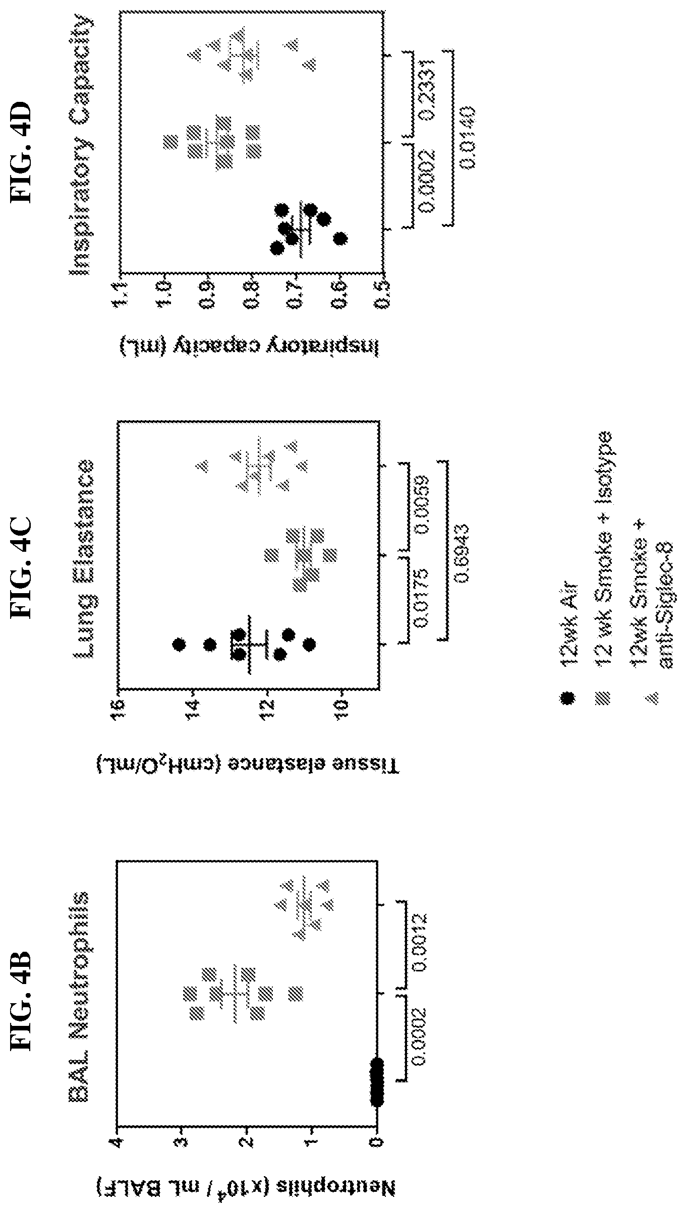
[0023] FIGS. 4C-4D show the results of therapeutic dosing of anti-Siglec-8 antibodies on lung function in cigarette smoke-induced experimental COPD using Siglec-8 transgenic C57BL/6 mice. FIG. 4C shows the results of lung elastance measurements in control mice, as well as cigarette-smoke induced experimental COPD mice treated with anti-Siglec-8 antibody or isotype control antibody. FIG. 4D shows the results of inspiratory capacity measurements in control mice, as well as cigarette-smoke induced experimental COPD mice treated with anti-Siglec-8 antibody or isotype control antibody. Lung function measurements were performed using a forced pulmonary maneuver system, and each maneuver was performed a minimum of three times to calculate the average. P-values derived from Mann-Whitney test are indicated below the corresponding treatment groups.
[0024] FIGS. 5A & 5B show the results of therapeutic dosing of anti-Siglec-8 antibodies on lung function in cigarette smoke-induced experimental COPD using Siglec-8 transgenic C57BL/6 mice, as described above. FIG. 5A shows the results of airway resistance measurements in control mice, as well as cigarette-smoke induced experimental COPD mice treated with anti-Siglec-8 antibody or isotype control antibody. FIG. 5B shows the results of total lung capacity measurements in control mice, as well as cigarette-smoke induced experimental COPD mice treated with anti-Siglec-8 antibody or isotype control antibody.
[0025] FIGS. 6A & 6B show the results of therapeutic dosing of anti-Siglec-8 antibodies on chemokine levels from bronchoalveolar lavage fluid (BAL) in cigarette smoke-induced experimental COPD using Siglec-8 transgenic C57BL/6 mice, as described above. FIG. 6A shows the levels of MCP-1 in BAL from control mice, as well as from cigarette-smoke induced experimental COPD mice treated with anti-Siglec-8 antibody or isotype control antibody. FIG. 6B shows the levels of KC/CXCL1 in BAL from control mice, as well as from cigarette-smoke induced experimental COPD mice treated with anti-Siglec-8 antibody or isotype control antibody.
DETAILED DESCRIPTION
I. Definitions
[0026] It is to be understood that the present disclosure is not limited to particular compositions or biological systems, which can, of course, vary. It is also to be understood that the terminology used herein is for the purpose of describing particular embodiments only, and is not intended to be limiting. As used in this specification and the appended claims, the singular forms "a", "an" and "the" include plural referents unless the content clearly dictates otherwise. Thus, for example, reference to "a molecule" optionally includes a combination of two or more such molecules, and the like.
[0027] The term "about" as used herein refers to the usual error range for the respective value readily known to the skilled person in this technical field. Reference to "about" a value or parameter herein includes (and describes) embodiments that are directed to that value or parameter per se.
[0028] It is understood that aspects and embodiments of the present disclosure include "comprising," "consisting," and "consisting essentially of" aspects and embodiments.
[0029] The term "antibody" includes polyclonal antibodies, monoclonal antibodies (including full length antibodies which have an immunoglobulin Fc region), antibody compositions with polyepitopic specificity, multispecific antibodies (e.g., bispecific antibodies, diabodies, and single-chain molecules), as well as antibody fragments (e.g., Fab, F(ab').sub.2, and Fv). The term "immunoglobulin" (Ig) is used interchangeably with "antibody" herein.
[0030] The basic 4-chain antibody unit is a heterotetrameric glycoprotein composed of two identical light (L) chains and two identical heavy (H) chains. An IgM antibody consists of 5 of the basic heterotetramer units along with an additional polypeptide called a J chain, and contains 10 antigen binding sites, while IgA antibodies comprise from 2-5 of the basic 4-chain units which can polymerize to form polyvalent assemblages in combination with the J chain. In the case of IgGs, the 4-chain unit is generally about 150,000 daltons. Each L chain is linked to an H chain by one covalent disulfide bond, while the two H chains are linked to each other by one or more disulfide bonds depending on the H chain isotype. Each H and L chain also has regularly spaced intrachain disulfide bridges. Each H chain has at the N-terminus, a variable domain (V.sub.H) followed by three constant domains (C.sub.H) for each of the .alpha. and .gamma. chains and four C.sub.H domains for .mu. and .epsilon. isotypes. Each L chain has at the N-terminus, a variable domain (V.sub.L) followed by a constant domain at its other end. The V.sub.L is aligned with the V.sub.H and the C.sub.L is aligned with the first constant domain of the heavy chain (C.sub.H1). Particular amino acid residues are believed to form an interface between the light chain and heavy chain variable domains. The pairing of a V.sub.H and V.sub.L together forms a single antigen-binding site. For the structure and properties of the different classes of antibodies, see e.g., Basic and Clinical Immunology, 8th Edition, Daniel P. Sties, Abba I. Terr and Tristram G. Parsolw (eds), Appleton & Lange, Norwalk, Conn., 1994, page 71 and Chapter 6.
[0031] The L chain from any vertebrate species can be assigned to one of two clearly distinct types, called kappa and lambda, based on the amino acid sequences of their constant domains. Depending on the amino acid sequence of the constant domain of their heavy chains (CH), immunoglobulins can be assigned to different classes or isotypes. There are five classes of immunoglobulins: IgA, IgD, IgE, IgG and IgM, having heavy chains designated .alpha., .delta., .epsilon., .gamma. and .mu., respectively. The .gamma. and .alpha. classes are further divided into subclasses on the basis of relatively minor differences in the CH sequence and function, e.g., humans express the following subclasses: IgG1, IgG2, IgG3, IgG4, IgA1 and IgA2. IgG1 antibodies can exist in multiple polymorphic variants termed allotypes (reviewed in Jefferis and Lefranc 2009. mAbs Vol 1 Issue 4 1-7) any of which are suitable for use in the present disclosure. Common allotypic variants in human populations are those designated by the letters a, f, n, z.
[0032] An "isolated" antibody is one that has been identified, separated and/or recovered from a component of its production environment (e.g., naturally or recombinantly). In some embodiments, the isolated polypeptide is free of association with all other components from its production environment. Contaminant components of its production environment, such as that resulting from recombinant transfected cells, are materials that would typically interfere with research, diagnostic or therapeutic uses for the antibody, and may include enzymes, hormones, and other proteinaceous or non-proteinaceous solutes. In some embodiments, the polypeptide is purified: (1) to greater than 95% by weight of antibody as determined by, for example, the Lowry method, and in some embodiments, to greater than 99% by weight; (1) to a degree sufficient to obtain at least 15 residues of N-terminal or internal amino acid sequence by use of a spinning cup sequenator, or (3) to homogeneity by SDS-PAGE under non-reducing or reducing conditions using Coomassie blue or silver stain. Isolated antibody includes the antibody in situ within recombinant cells since at least one component of the antibody's natural environment will not be present. Ordinarily, however, an isolated polypeptide or antibody is prepared by at least one purification step.
[0033] The term "monoclonal antibody" as used herein refers to an antibody obtained from a population of substantially homogeneous antibodies, i.e., the individual antibodies comprising the population are identical except for possible naturally occurring mutations and/or post-translation modifications (e.g., isomerizations, amidations) that may be present in minor amounts. In some embodiments, monoclonal antibodies have a C-terminal cleavage at the heavy chain and/or light chain. For example, 1, 2, 3, 4, or 5 amino acid residues are cleaved at the C-terminus of heavy chain and/or light chain. In some embodiments, the C-terminal cleavage removes a C-terminal lysine from the heavy chain. In some embodiments, monoclonal antibodies have an N-terminal cleavage at the heavy chain and/or light chain. For example, 1, 2, 3, 4, or 5 amino acid residues are cleaved at the N-terminus of heavy chain and/or light chain. In some embodiments, monoclonal antibodies are highly specific, being directed against a single antigenic site. In some embodiments, monoclonal antibodies are highly specific, being directed against multiple antigenic sites (such as a bispecific antibody or a multispecific antibody). The modifier "monoclonal" indicates the character of the antibody as being obtained from a substantially homogeneous population of antibodies, and is not to be construed as requiring production of the antibody by any particular method. For example, the monoclonal antibodies to be used in accordance with the present disclosure may be made by a variety of techniques, including, for example, the hybridoma method, recombinant DNA methods, phage-display technologies, and technologies for producing human or human-like antibodies in animals that have parts or all of the human immunoglobulin loci or genes encoding human immunoglobulin sequences.
[0034] The term "naked antibody" refers to an antibody that is not conjugated to a cytotoxic moiety or radiolabel.
[0035] The terms "full-length antibody," "intact antibody" or "whole antibody" are used interchangeably to refer to an antibody in its substantially intact form, as opposed to an antibody fragment. Specifically whole antibodies include those with heavy and light chains including an Fc region. The constant domains may be native sequence constant domains (e.g., human native sequence constant domains) or amino acid sequence variants thereof. In some cases, the intact antibody may have one or more effector functions.
[0036] An "antibody fragment" comprises a portion of an intact antibody, the antigen binding and/or the variable region of the intact antibody. Examples of antibody fragments include Fab, Fab', F(ab').sub.2 and Fv fragments; diabodies; linear antibodies (see U.S. Pat. No. 5,641,870, Example 2; Zapata et al., Protein Eng. 8(10): 1057-1062 [1995]); single-chain antibody molecules and multispecific antibodies formed from antibody fragments.
[0037] Papain digestion of antibodies produced two identical antigen-binding fragments, called "Fab" fragments, and a residual "Fc" fragment, a designation reflecting the ability to crystallize readily. The Fab fragment consists of an entire L chain along with the variable region domain of the H chain (V.sub.H), and the first constant domain of one heavy chain (C.sub.H1). Each Fab fragment is monovalent with respect to antigen binding, i.e., it has a single antigen-binding site. Pepsin treatment of an antibody yields a single large F(ab').sub.2 fragment which roughly corresponds to two disulfide linked Fab fragments having different antigen-binding activity and is still capable of cross-linking antigen. Fab' fragments differ from Fab fragments by having a few additional residues at the carboxy terminus of the C.sub.H1 domain including one or more cysteines from the antibody hinge region. Fab'-SH is the designation herein for Fab' in which the cysteine residue(s) of the constant domains bear a free thiol group. F(ab').sub.2 antibody fragments originally were produced as pairs of Fab' fragments which have hinge cysteines between them. Other chemical couplings of antibody fragments are also known.
[0038] The Fc fragment comprises the carboxy-terminal portions of both H chains held together by disulfides. The effector functions of antibodies are determined by sequences in the Fc region, the region which is also recognized by Fc receptors (FcR) found on certain types of cells.
[0039] "Fv" is the minimum antibody fragment which contains a complete antigen-recognition and -binding site. This fragment consists of a dimer of one heavy- and one light-chain variable region domain in tight, non-covalent association. From the folding of these two domains emanate six hypervariable loops (3 loops each from the H and L chain) that contribute the amino acid residues for antigen binding and confer antigen binding specificity to the antibody. However, even a single variable domain (or half of an Fv comprising only three HVRs specific for an antigen) has the ability to recognize and bind antigen, although at a lower affinity than the entire binding site.
[0040] "Single-chain Fv" also abbreviated as "sFv" or "scFv" are antibody fragments that comprise the VH and VL antibody domains connected into a single polypeptide chain. In some embodiments, the sFv polypeptide further comprises a polypeptide linker between the V.sub.H and V.sub.L domains which enables the sFv to form the desired structure for antigen binding. For a review of the sFv, see Pluckthun in The Pharmacology of Monoclonal Antibodies, vol. 113, Rosenburg and Moore eds., Springer-Verlag, New York, pp. 269-315 (1994).
[0041] "Functional fragments" of the antibodies of the present disclosure comprise a portion of an intact antibody, generally including the antigen binding or variable region of the intact antibody or the Fv region of an antibody which retains or has modified FcR binding capability. Examples of antibody fragments include linear antibody, single-chain antibody molecules and multispecific antibodies formed from antibody fragments.
[0042] The monoclonal antibodies herein specifically include "chimeric" antibodies (immunoglobulins) in which a portion of the heavy and/or light chain is identical with or homologous to corresponding sequences in antibodies derived from a particular species or belonging to a particular antibody class or subclass, while the remainder of the chain(s) is (are) identical with or homologous to corresponding sequences in antibodies derived from another species or belonging to another antibody class or subclass, as well as fragments of such antibodies, so long as they exhibit the desired biological activity (U.S. Pat. No. 4,816,567; Morrison et al., Proc. Natl. Acad. Sci. USA, 81:6851-6855 (1984)). Chimeric antibodies of interest herein include PRIMATIZED.RTM. antibodies wherein the antigen-binding region of the antibody is derived from an antibody produced by, e.g., immunizing macaque monkeys with an antigen of interest. As used herein, "humanized antibody" is used as a subset of "chimeric antibodies."
[0043] "Humanized" forms of non-human (e.g., murine) antibodies are chimeric antibodies that contain minimal sequence derived from non-human immunoglobulin. In one embodiment, a humanized antibody is a human immunoglobulin (recipient antibody) in which residues from an HVR of the recipient are replaced by residues from an HVR of a non-human species (donor antibody) such as mouse, rat, rabbit or non-human primate having the desired specificity, affinity, and/or capacity. In some instances, FR residues of the human immunoglobulin are replaced by corresponding non-human residues. Furthermore, humanized antibodies may comprise residues that are not found in the recipient antibody or in the donor antibody. These modifications may be made to further refine antibody performance, such as binding affinity. In general, a humanized antibody will comprise substantially all of at least one, and typically two, variable domains, in which all or substantially all of the hypervariable loops correspond to those of a non-human immunoglobulin sequence, and all or substantially all of the FR regions are those of a human immunoglobulin sequence, although the FR regions may include one or more individual FR residue substitutions that improve antibody performance, such as binding affinity, isomerization, immunogenicity, etc. In some embodiments, the number of these amino acid substitutions in the FR are no more than 6 in the H chain, and in the L chain, no more than 3. The humanized antibody optionally will also comprise at least a portion of an immunoglobulin constant region (Fc), typically that of a human immunoglobulin. For further details, see, e.g., Jones et al., Nature 321:522-525 (1986); Riechmann et al., Nature 332:323-329 (1988); and Presta, Curr. Op. Struct. Biol. 2:593-596 (1992). See also, for example, Vaswani and Hamilton, Ann. Allergy, Asthma & Immunol. 1:105-115 (1998); Harris, Biochem. Soc. Transactions 23:1035-1038 (1995); Hurle and Gross, Curr. Op. Biotech. 5:428-433 (1994); and U.S. Pat. Nos. 6,982,321 and 7,087,409. In some embodiments, humanized antibodies are directed against a single antigenic site. In some embodiments, humanized antibodies are directed against multiple antigenic sites. An alternative humanization method is described in U.S. Pat. No. 7,981,843 and U.S. Patent Application Publication No. 2006/0134098.
[0044] The "variable region" or "variable domain" of an antibody refers to the amino-terminal domains of the heavy or light chain of the antibody. The variable domains of the heavy chain and light chain may be referred to as "VH" and "VL", respectively. These domains are generally the most variable parts of the antibody (relative to other antibodies of the same class) and contain the antigen binding sites.
[0045] The term "hypervariable region," "HVR," or "HV," when used herein refers to the regions of an antibody-variable domain that are hypervariable in sequence and/or form structurally defined loops. Generally, antibodies comprise six HVRs; three in the VH (H1, H2, H3), and three in the VL (L1, L2, L3). In native antibodies, H3 and L3 display the most diversity of the six HVRs, and H3 in particular is believed to play a unique role in conferring fine specificity to antibodies. See, e.g., Xu et al. Immunity 13:37-45 (2000); Johnson and Wu in Methods in Molecular Biology 248:1-25 (Lo, ed., Human Press, Totowa, N.J., 2003)). Indeed, naturally occurring camelid antibodies consisting of a heavy chain only are functional and stable in the absence of light chain. See, e.g., Hamers-Casterman et al., Nature 363:446-448 (1993) and Sheriff et al., Nature Struct. Biol. 3:733-736 (1996).
[0046] A number of HVR delineations are in use and are encompassed herein. The HVRs that are Kabat complementarity-determining regions (CDRs) are based on sequence variability and are the most commonly used (Kabat et al., Sequences of Proteins of Immunological Interest, 5.sup.th Ed. Public Health Service, National Institute of Health, Bethesda, Md. (1991)). Chothia HVRs refer instead to the location of the structural loops (Chothia and Lesk J. Mol. Biol. 196:901-917 (1987)). The "contact" HVRs are based on an analysis of the available complex crystal structures. The residues from each of these HVRs are noted below.
TABLE-US-00001 Loop Kabat Chothia Contact L1 L24-L34 L26-L34 L30-L36 L2 L50-L56 L50-L56 L46-L55 L3 L89-L97 L91-L96 L89-L96 H1 H31-H35B H26-H32 H30-H35B (Kabat Numbering) H1 H31-H35 H26-H32 H30-H35 (Chothia Numbering) H2 H50-H65 H53-H56 H47-H58 H3 H95-H102 H95-H102 H93-H101
[0047] Unless otherwise indicated, the variable-domain residues (HVR residues and framework region residues) are numbered according to Kabat et al., supra.
[0048] "Framework" or "FR" residues are those variable-domain residues other than the HVR residues as herein defined.
[0049] The expression "variable-domain residue-numbering as in Kabat" or "amino-acid-position numbering as in Kabat," and variations thereof, refers to the numbering system used for heavy-chain variable domains or light-chain variable domains of the compilation of antibodies in Kabat et al., supra. Using this numbering system, the actual linear amino acid sequence may contain fewer or additional amino acids corresponding to a shortening of, or insertion into, a FR or HVR of the variable domain. For example, a heavy-chain variable domain may include a single amino acid insert (residue 52a according to Kabat) after residue 52 of H2 and inserted residues (e.g. residues 82a, 82b, and 82c, etc. according to Kabat) after heavy-chain FR residue 82. The Kabat numbering of residues may be determined for a given antibody by alignment at regions of homology of the sequence of the antibody with a "standard" Kabat numbered sequence.
[0050] An "acceptor human framework" for the purposes herein is a framework comprising the amino acid sequence of a VL or VH framework derived from a human immunoglobulin framework or a human consensus framework. An acceptor human framework "derived from" a human immunoglobulin framework or a human consensus framework may comprise the same amino acid sequence thereof, or it may contain pre-existing amino acid sequence changes. In some embodiments, the number of pre-existing amino acid changes are 10 or less, 9 or less, 8 or less, 7 or less, 6 or less, 5 or less, 4 or less, 3 or less, or 2 or less.
[0051] "Percent (%) amino acid sequence identity" with respect to a reference polypeptide sequence is defined as the percentage of amino acid residues in a candidate sequence that are identical with the amino acid residues in the reference polypeptide sequence, after aligning the sequences and introducing gaps, if necessary, to achieve the maximum percent sequence identity, and not considering any conservative substitutions as part of the sequence identity. Alignment for purposes of determining percent amino acid sequence identity can be achieved in various ways that are within the skill in the art, for instance, using publicly available computer software such as BLAST, BLAST-2, ALIGN or Megalign (DNASTAR) software. Those skilled in the art can determine appropriate parameters for aligning sequences, including any algorithms needed to achieve maximal alignment over the full length of the sequences being compared. For example, the % amino acid sequence identity of a given amino acid sequence A to, with, or against a given amino acid sequence B (which can alternatively be phrased as a given amino acid sequence A that has or comprises a certain % amino acid sequence identity to, with, or against a given amino acid sequence B) is calculated as follows:
100 times the fraction X/Y
where X is the number of amino acid residues scored as identical matches by the sequence in that program's alignment of A and B, and where Y is the total number of amino acid residues in B. It will be appreciated that where the length of amino acid sequence A is not equal to the length of amino acid sequence B, the % amino acid sequence identity of A to B will not equal the % amino acid sequence identity of B to A.
[0052] An antibody that "binds to", "specifically binds to" or is "specific for" a particular a polypeptide or an epitope on a particular polypeptide is one that binds to that particular polypeptide or epitope on a particular polypeptide without substantially binding to any other polypeptide or polypeptide epitope. In some embodiments, binding of an anti-Siglec-8 antibody described herein (e.g., an antibody that binds to human Siglec-8) to an unrelated non-Siglec-8 polypeptide is less than about 10% of the antibody binding to Siglec-8 as measured by methods known in the art (e.g., enzyme-linked immunosorbent assay (ELISA)). In some embodiments, an antibody that binds to a Siglec-8 (e.g., an antibody that binds to human Siglec-8) has a dissociation constant (Kd) of .ltoreq.1 .mu.M, .ltoreq.100 nM, .ltoreq.10 nM, .ltoreq.2 nM, .ltoreq.1 nM, .ltoreq.0.7 nM, .ltoreq.0.6 nM, .ltoreq.0.5 nM, .ltoreq.0.1 nM, .ltoreq.0.01 nM, or .ltoreq.0.001 nM (e.g. 10.sup.-8 M or less, e.g. from 10.sup.-8 M to 10.sup.-13 M, e.g., from 10.sup.-9 M to 10.sup.-13 M).
[0053] The term "anti-Siglec-8 antibody" or "an antibody that binds to human Siglec-8" refers to an antibody that binds to a polypeptide or an epitope of human Siglec-8 without substantially binding to any other polypeptide or epitope of an unrelated non-Siglec-8 polypeptide.
[0054] The term "Siglec-8" as used herein refers to a human Siglec-8 protein. The term also includes naturally occurring variants of Siglec-8, including splice variants or allelic variants. The amino acid sequence of an exemplary human Siglec-8 is shown in SEQ ID NO:72. The amino acid sequence of another exemplary human Siglec-8 is shown in SEQ ID NO:73. In some embodiments, a human Siglec-8 protein comprises the human Siglec-8 extracellular domain fused to an immunoglobulin Fc region. The amino acid sequence of an exemplary human Siglec-8 extracellular domain fused to an immunoglobulin Fc region is shown in SEQ ID NO:74. The amino acid sequence underlined in SEQ ID NO:74 indicates the Fc region of the Siglec-8 Fc fusion protein amino acid sequence.
TABLE-US-00002 Human Siglec-8 Amino Acid Sequence (SEQ ID NO: 72) GYLLQVQELVTVQEGLCVHVPCSFSYPQDGWTDSDPVHGYWFRAGDRP YQDAPVATNNPDREVQAETQGRFQLLGDIWSNDCSLSIRDARKRDKGS YFFRLERGSMKWSYKSQLNYKTKQLSVFVTALTHRPDILILGTLESGH SRNLTCSVPWACKQGTPPMISWIGASVSSPGPTTARSSVLTLTPKPQD HGTSLTCQVTLPGTGVTTTSTVRLDVSYPPWNLTMTVFQGDATASTAL GNGSSLSVLEGQSLRLVCAVNSNPPARLSWTRGSLTLCPSRSSNPGLL ELPRVHVRDEGEFTCRAQNAQGSQHISLSLSLQNEGTGTSRPVSQVTL AAVGGAGATALAFLSFCIIFIIVRSCRKKSARPAAGVGDTGMEDAKAI RGSASQGPLTESWKDGNPLKKPPPAVAPSSGEEGELHYATLSFHKVKP QDPQGQEATDSEYSEIKIHKRETAETQACLRNHNPSSKEVRG Human Siglec-8 Amino Acid Sequence (SEQ ID NO: 73) GYLLQVQELVTVQEGLCVHVPCSFSYPQDGWTDSDPVHGYWFRAGDRP YQDAPVATNNPDREVQAETQGRFQLLGDIWSNDCSLSIRDARKRDKGS YFFRLERGSMKWSYKSQLNYKTKQLSVFVTALTHRPDILILGTLESGH PRNLTCSVPWACKQGTPPMISWIGASVSSPGPTTARSSVLTLTPKPQD HGTSLTCQVTLPGTGVTTTSTVRLDVSYPPWNLTMTVFQGDATASTAL GNGSSLSVLEGQSLRLVCAVNSNPPARLSWTRGSLTLCPSRSSNPGLL ELPRVHVRDEGEFTCRAQNAQGSQHISLSLSLQNEGTGTSRPVSQVTL AAVGGAGATALAFLSFCIIFIIVRSCRKKSARPAAGVGDTGMEDAKAI RGSASQGPLTESWKDGNPLKKPPPAVAPSSGEEGELHYATLSFHKVKP QDPQGQEATDSEYSEIKIHKRETAETQACLRNHNPSSKEVRG Siglec-8 Fc Fusion Protein Amino Acid Sequence (SEQ ID NO: 74) GYLLQVQELVTVQEGLCVHVPCSFSYPQDGWTDSDPVHGYWFRAGDRP YQDAPVATNNPDREVQAETQGRFQLLGDIWSNDCSLSIRDARKRDKGS YFFRLERGSMKWSYKSQLNYKTKQLSVFVTALTHRPDILILGTLESGH SRNLTCSVPWACKQGTPPMISWIGASVSSPGPTTARSSVLTLTPKPQD HGTSLTCQVTLPGTGVTTTSTVRLDVSYPPWNLTMTVFQGDATASTAL GNGSSLSVLEGQSLRLVCAVNSNPPARLSWTRGSLTLCPSRSSNPGLL ELPRVHVRDEGEFTCRAQNAQGSQHISLSLSLQNEGTGTSRPVSQVTL AAVGGIEGRSDKTHTCPPCPAPELLGGPSVFLFPPKPKDTLMISRTPE VTCVVVDVSHEDPEVKFNWYVDGVEVHNAKTKPREEQYNSTYRVVSVL TVLHQDWLNGKEYKCKVSNKALPAPIEKTISKAKGQPREPQVYTLPPS REEMTKNQVSLTCLVKGFYPSDIAVEWESNGQPENNYKTTPPVLDSDG SFFLYSKLTVDKSRWQQGNVFSCSVMHEALHNHYTQKSLSLSPGK
[0055] Antibodies that "induce apoptosis" or are "apoptotic" are those that induce programmed cell death as determined by standard apoptosis assays, such as binding of annexin V, fragmentation of DNA, cell shrinkage, dilation of endoplasmic reticulum, cell fragmentation, and/or formation of membrane vesicles (called apoptotic bodies). For example, the apoptotic activity of the anti-Siglec-8 antibodies (e.g., an antibody that binds to human Siglec-8) of the present disclosure can be shown by staining cells with annexin V.
[0056] Antibody "effector functions" refer to those biological activities attributable to the Fc region (a native sequence Fc region or amino acid sequence variant Fc region) of an antibody, and vary with the antibody isotype. Examples of antibody effector functions include: C1q binding and complement dependent cytotoxicity; Fc receptor binding; antibody-dependent cell-mediated cytotoxicity (ADCC); phagocytosis; down regulation of cell surface receptors (e.g., B cell receptors); and B cell activation.
[0057] "Antibody-dependent cell-mediated cytotoxicity" or "ADCC" refers to a form of cytotoxicity in which secreted Ig bound onto Fc receptors (FcRs) present on certain cytotoxic cells (e.g., natural killer (NK) cells, neutrophils and macrophages) enable these cytotoxic effector cells to bind specifically to an antigen-bearing target cell and subsequently kill the target cell with cytotoxins. The antibodies "arm" the cytotoxic cells and are required for killing of the target cell by this mechanism. The primary cells for mediating ADCC, NK cells, express Fc.gamma.RIII only, whereas monocytes express Fc.gamma.RI, Fc.gamma.RII and Fc.gamma.RIII. Fc expression on hematopoietic cells is summarized in Table 3 on page 464 of Ravetch and Kinet, Annu. Rev. Immunol. 9: 457-92 (1991). In some embodiments, an anti-Siglec-8 antibody (e.g., an antibody that binds to human Siglec-8) described herein enhances ADCC. To assess ADCC activity of a molecule of interest, an in vitro ADCC assay, such as that described in U.S. Pat. No. 5,500,362 or 5,821,337 may be performed. Useful effector cells for such assays include peripheral blood mononuclear cells (PBMC) and natural killer (NK) cells. Alternatively, or additionally, ADCC activity of the molecule of interest may be assessed in vivo, e.g., in an animal model such as that disclosed in Clynes et al., PNAS USA 95:652-656 (1998). Other Fc variants that alter ADCC activity and other antibody properties include those disclosed by Ghetie et al., Nat Biotech. 15:637-40, 1997; Duncan et al, Nature 332:563-564, 1988; Lund et al., J. Immunol 147:2657-2662, 1991; Lund et al, Mol Immunol 29:53-59, 1992; Alegre et al, Transplantation 57:1537-1543, 1994; Hutchins et al., Proc Natl. Acad Sci USA 92:11980-11984, 1995; Jefferis et al, Immunol Lett. 44:111-117, 1995; Lund et al., FASEB J 9:115-119, 1995; Jefferis et al, Immunol Lett 54:101-104, 1996; Lund et al, J Immunol 157:4963-4969, 1996; Armour et al., Eur J Immunol 29:2613-2624, 1999; Idusogie et al, J Immunol 164:4178-4184, 200; Reddy et al, J Immunol 164:1925-1933, 2000; Xu et al., Cell Immunol 200:16-26, 2000; Idusogie et al, J Immunol 166:2571-2575, 2001; Shields et al., J Biol Chem 276:6591-6604, 2001; Jefferis et al, Immunol Lett 82:57-65. 2002; Presta et al., Biochem Soc Trans 30:487-490, 2002; Lazar et al., Proc. Natl. Acad. Sci. USA 103:4005-4010, 2006; U.S. Pat. Nos. 5,624,821; 5,885,573; 5,677,425; 6,165,745; 6,277,375; 5,869,046; 6,121,022; 5,624,821; 5,648,260; 6,194,551; 6,737,056; 6,821,505; 6,277,375; 7,335,742; and 7,317,091.
[0058] The term "Fc region" herein is used to define a C-terminal region of an immunoglobulin heavy chain, including native-sequence Fc regions and variant Fc regions. Although the boundaries of the Fc region of an immunoglobulin heavy chain might vary, the human IgG heavy-chain Fc region is usually defined to stretch from an amino acid residue at position Cys226, or from Pro230, to the carboxyl-terminus thereof. Suitable native-sequence Fc regions for use in the antibodies of the present disclosure include human IgG1, IgG2, IgG3 and IgG4. A single amino acid substitution (S228P according to Kabat numbering; designated IgG4Pro) may be introduced to abolish the heterogeneity observed in recombinant IgG4 antibody. See Angal, S. et al. (1993) Mol Immunol 30, 105-108.
[0059] "Non-fucosylated" or "fucose-deficient" antibody refers to a glycosylation antibody variant comprising an Fc region wherein a carbohydrate structure attached to the Fc region has reduced fucose or lacks fucose. In some embodiments, an antibody with reduced fucose or lacking fucose has improved ADCC function. Non-fucosylated or fucose-deficient antibodies have reduced fucose relative to the amount of fucose on the same antibody produced in a cell line. In some embodiments, a non-fucosylated or fucose-deficient antibody composition contemplated herein is a composition wherein less than about 50% of the N-linked glycans attached to the Fc region of the antibodies in the composition comprise fucose.
[0060] The terms "fucosylation" or "fucosylated" refers to the presence of fucose residues within the oligosaccharides attached to the peptide backbone of an antibody. Specifically, a fucosylated antibody comprises a (1,6)-linked fucose at the innermost N-acetylglucosamine (GlcNAc) residue in one or both of the N-linked oligosaccharides attached to the antibody Fc region, e.g. at position Asn 297 of the human IgG1 Fc domain (EU numbering of Fc region residues). Asn297 may also be located about +3 amino acids upstream or downstream of position 297, i.e. between positions 294 and 300, due to minor sequence variations in immunoglobulins.
[0061] The "degree of fucosylation" is the percentage of fucosylated oligosaccharides relative to all oligosaccharides identified by methods known in the art e.g., in an N-glycosidase F treated antibody composition assessed by matrix-assisted laser desorption-ionization time-of-flight mass spectrometry (MALDI-TOF MS). In a composition of a "fully fucosylated antibody" essentially all oligosaccharides comprise fucose residues, i.e. are fucosylated. In some embodiments, a composition of a fully fucosylated antibody has a degree of fucosylation of at least about 90%. Accordingly, an individual antibody in such a composition typically comprises fucose residues in each of the two N-linked oligosaccharides in the Fc region. Conversely, in a composition of a "fully non-fucosylated" antibody essentially none of the oligosaccharides are fucosylated, and an individual antibody in such a composition does not contain fucose residues in either of the two N-linked oligosaccharides in the Fc region. In some embodiments, a composition of a fully non-fucosylated antibody has a degree of fucosylation of less than about 10%. In a composition of a "partially fucosylated antibody" only part of the oligosaccharides comprise fucose. An individual antibody in such a composition can comprise fucose residues in none, one or both of the N-linked oligosaccharides in the Fc region, provided that the composition does not comprise essentially all individual antibodies that lack fucose residues in the N-linked oligosaccharides in the Fc region, nor essentially all individual antibodies that contain fucose residues in both of the N-linked oligosaccharides in the Fc region. In one embodiment, a composition of a partially fucosylated antibody has a degree of fucosylation of about 10% to about 80% (e.g., about 50% to about 80%, about 60% to about 80%, or about 70% to about 80%).
[0062] "Binding affinity" as used herein refers to the strength of the non-covalent interactions between a single binding site of a molecule (e.g., an antibody) and its binding partner (e.g., an antigen). In some embodiments, the binding affinity of an antibody for a Siglec-8 (which may be a dimer, such as the Siglec-8-Fc fusion protein described herein) can generally be represented by a dissociation constant (Kd). Affinity can be measured by common methods known in the art, including those described herein.
[0063] "Binding avidity" as used herein refers to the binding strength of multiple binding sites of a molecule (e.g., an antibody) and its binding partner (e.g., an antigen).
[0064] An "isolated" nucleic acid molecule encoding the antibodies herein is a nucleic acid molecule that is identified and separated from at least one contaminant nucleic acid molecule with which it is ordinarily associated in the environment in which it was produced. In some embodiments, the isolated nucleic acid is free of association with all components associated with the production environment. The isolated nucleic acid molecules encoding the polypeptides and antibodies herein is in a form other than in the form or setting in which it is found in nature. Isolated nucleic acid molecules therefore are distinguished from nucleic acid encoding the polypeptides and antibodies herein existing naturally in cells.
[0065] The term "pharmaceutical formulation" refers to a preparation that is in such form as to permit the biological activity of the active ingredient to be effective, and that contains no additional components that are unacceptably toxic to an individual to which the formulation would be administered. Such formulations are sterile.
[0066] "Carriers" as used herein include pharmaceutically acceptable carriers, excipients, or stabilizers that are nontoxic to the cell or mammal being exposed thereto at the dosages and concentrations employed. Often the physiologically acceptable carrier is an aqueous pH buffered solution. Examples of physiologically acceptable carriers include buffers such as phosphate, citrate, and other organic acids; antioxidants including ascorbic acid; low molecular weight (less than about 10 residues) polypeptide; proteins, such as serum albumin, gelatin, or immunoglobulins; hydrophilic polymers such as polyvinylpyrrolidone; amino acids such as glycine, glutamine, asparagine, arginine or lysine; monosaccharides, disaccharides, and other carbohydrates including glucose, mannose, or dextrins; chelating agents such as EDTA; sugar alcohols such as mannitol or sorbitol; salt-forming counterions such as sodium; and/or nonionic surfactants such as TWEEN.TM., polyethylene glycol (PEG), and PLURONICS.TM..
[0067] As used herein, the term "treatment" or "treating" refers to clinical intervention designed to alter the natural course of the individual or cell being treated during the course of clinical pathology. Desirable effects of treatment include decreasing the rate of disease progression, ameliorating or palliating the disease state, and remission or improved prognosis. An individual is successfully "treated", for example, if one or more symptoms associated with a disease (e.g., non-eosinophilic COPD) are mitigated or eliminated. For example, an individual is successfully "treated" if treatment results in increasing the quality of life of those suffering from a disease, decreasing the dose of other medications required for treating the disease, reducing the frequency of recurrence of the disease, lessening severity of the disease, delaying the development or progression of the disease, and/or prolonging survival of individuals.
[0068] As used herein, "in conjunction with" or "in combination with" refers to administration of one treatment modality in addition to another treatment modality. As such, "in conjunction with" or "in combination with" refers to administration of one treatment modality before, during or after administration of the other treatment modality to the individual.
[0069] As used herein, the term "prevention" or "preventing" includes providing prophylaxis with respect to occurrence or recurrence of a disease in an individual. An individual may be predisposed to a disease, susceptible to a disease, or at risk of developing a disease, but has not yet been diagnosed with the disease. In some embodiments, anti-Siglec-8 antibodies (e.g., an antibody that binds to human Siglec-8) described herein are used to delay development of a disease (e.g., non-eosinophilic COPD).
[0070] As used herein, an individual "at risk" of developing a disease (e.g., non-eosinophilic COPD) may or may not have detectable disease or symptoms of disease, and may or may not have displayed detectable disease or symptoms of disease prior to the treatment methods described herein. "At risk" denotes that an individual has one or more risk factors, which are measurable parameters that correlate with development of the disease (e.g., non-eosinophilic COPD), as known in the art. An individual having one or more of these risk factors has a higher probability of developing the disease than an individual without one or more of these risk factors.
[0071] An "effective amount" refers to at least an amount effective, at dosages and for periods of time necessary, to achieve the desired or indicated effect, including a therapeutic or prophylactic result. An effective amount can be provided in one or more administrations. A "therapeutically effective amount" is at least the minimum concentration required to effect a measurable improvement of a particular disease. A therapeutically effective amount herein may vary according to factors such as the disease state, age, sex, and weight of the patient, and the ability of the antibody to elicit a desired response in the individual. A therapeutically effective amount may also be one in which any toxic or detrimental effects of the antibody are outweighed by the therapeutically beneficial effects. A "prophylactically effective amount" refers to an amount effective, at the dosages and for periods of time necessary, to achieve the desired prophylactic result. Typically but not necessarily, since a prophylactic dose is used in individuals prior to or at the earlier stage of disease, the prophylactically effective amount can be less than the therapeutically effective amount.
[0072] "Chronic" administration refers to administration of the medicament(s) in a continuous as opposed to acute mode, so as to maintain the initial therapeutic effect (activity) for an extended period of time. "Intermittent" administration is treatment that is not consecutively done without interruption, but rather is cyclic in nature.
[0073] The term "package insert" is used to refer to instructions customarily included in commercial packages of therapeutic products, that contain information about the indications, usage, dosage, administration, combination therapy, contraindications and/or warnings concerning the use of such therapeutic products.
[0074] As used herein, an "individual" or a "subject" is a mammal. A "mammal" for purposes of treatment includes humans, domestic and farm animals, and zoo, sports, or pet animals, such as dogs, horses, rabbits, cattle, pigs, hamsters, gerbils, mice, ferrets, rats, cats, etc. In some embodiments, the individual or subject is a human.
II. Methods
[0075] Provided herein are methods for treating and/or preventing COPD in an individual comprising administering to the individual an effective amount of an antibody described herein that binds to human Siglec-8 (e.g., an anti-Siglec-8 antibody) or compositions comprising said antibodies. In some embodiments, the antibody is in a pharmaceutical composition comprising the antibody and a pharmaceutically acceptable carrier. In some embodiments, the individual has been diagnosed with COPD or is at risk of developing COPD. In some embodiments, the individual is a human. In some embodiments, the individual is a smoker. In some embodiments, the individual has been a smoker. In some embodiments, the individual is not or has not been a smoker. In some embodiments, the individual does not have asthma and/or asthma-COPD overlap syndrome (ACOS). In some embodiments, the individual has asthma and/or ACOS.
[0076] A. Individuals
[0077] In some embodiments, provided herein are methods for treating COPD in an individual that has and/or is at risk of developing non-eosinophilic COPD. In some embodiments, the method comprises administering to the individual an effective amount of an antibody described herein that binds to human Siglec-8 (e.g., an anti-Siglec-8 antibody), or compositions comprising said antibodies.
[0078] In some embodiments, provided herein are methods for treating COPD in an individual that has and/or is at risk of developing neutrophilic COPD. In some embodiments, an induced sputum sample from the individual contains greater than about 70% neutrophils relative to total leukocyte content. In some embodiments, the method comprises administering to the individual an effective amount of an antibody described herein that binds to human Siglec-8 (e.g., an anti-Siglec-8 antibody), or compositions comprising said antibodies.
[0079] In some embodiments, provided herein are methods for treating COPD in an individual that has been and/or is at risk of being diagnosed with one or more (e.g., one or more, two or more, three or more, four or more, five or more, six or more, seven or more, eight or more, nine or more, or all ten) of the following: more than 2 exacerbations in a year, chronic bronchitis, alpha1-antitrypsin deficiency, upper zone dominant emphysema, bullous emphysema, centrilobular emphysema (CLE), type 1 respiratory failure, type 2 respiratory failure, biomass COPD, and irreversible COPD. In some embodiments, the individual has been diagnosed with more than 2 exacerbations in a year. In some embodiments, the individual has been diagnosed with chronic bronchitis. In some embodiments, the individual has been diagnosed with alpha1-antitrypsin deficiency. In some embodiments, the individual has been diagnosed with upper zone dominant emphysema. In some embodiments, the individual has been diagnosed with bullous emphysema. In some embodiments, the individual has been diagnosed with centrilobular emphysema (CLE). In some embodiments, the individual has been diagnosed with type 1 respiratory failure. In some embodiments, the individual has been diagnosed with type 2 respiratory failure. In some embodiments, the individual has been diagnosed with biomass COPD. In some embodiments, the individual has been diagnosed with irreversible COPD. In some embodiments, the method comprises administering to the individual an effective amount of an antibody described herein that binds to human Siglec-8 (e.g., an anti-Siglec-8 antibody), or compositions comprising said antibodies.
[0080] In some embodiments, provided herein are methods for treating COPD in an individual that does not have one or more (e.g., one or more, two or more, three or more, four or more, five or more, six or more, seven or more, eight or more, nine or more, or all ten) of the following: more than 2 exacerbations in a year, chronic bronchitis, alpha1-antitrypsin deficiency, upper zone dominant emphysema, bullous emphysema, centrilobular emphysema (CLE), type 1 respiratory failure, type 2 respiratory failure, biomass COPD, and irreversible COPD. In some embodiments, the individual does not have more than 2 exacerbations in a year. In some embodiments, the individual does not have chronic bronchitis. In some embodiments, the individual does not have alpha1-antitrypsin deficiency. In some embodiments, the individual does not have upper zone dominant emphysema. In some embodiments, the individual does not have bullous emphysema. In some embodiments, the individual does not have centrilobular emphysema (CLE). In some embodiments, the individual does not have type 1 respiratory failure. In some embodiments, the individual does not have type 2 respiratory failure. In some embodiments, the individual does not have biomass COPD. In some embodiments, the individual does not have irreversible COPD. In some embodiments, the method comprises administering to the individual an effective amount of an antibody described herein that binds to human Siglec-8 (e.g., an anti-Siglec-8 antibody), or compositions comprising said antibodies.
[0081] In some embodiments, provided herein are methods for treating COPD in an individual that has been and/or is at risk of being diagnosed with one or more (e.g., one or more, two or more, three or more, four or more, or all five) of the following: pulmonary hypertension, systemic inflammation, stable state airway bacterial colonization, bronchiectasis, and airflow obstruction. In some embodiments, the individual has been diagnosed with pulmonary hypertension. In some embodiments, the individual has been diagnosed with systemic inflammation. In some embodiments, the individual has been diagnosed with stable state airway bacterial colonization. In some embodiments, the individual has been diagnosed with bronchiectasis. In some embodiments, the individual has been diagnosed with airflow obstruction. In some embodiments, the method comprises administering to the individual an effective amount of an antibody described herein that binds to human Siglec-8 (e.g., an anti-Siglec-8 antibody), or compositions comprising said antibodies.
[0082] In some embodiments, provided herein are methods for treating COPD in an individual that does not have one or more (e.g., one or more, two or more, three or more, four or more, or all five) of the following: pulmonary hypertension, systemic inflammation, stable state airway bacterial colonization, bronchiectasis, and airflow obstruction. In some embodiments, the individual does not have pulmonary hypertension. In some embodiments, the individual does not have systemic inflammation. In some embodiments, the individual does not have stable state airway bacterial colonization. In some embodiments, the individual does not have bronchiectasis. In some embodiments, the individual does not have airflow obstruction. In some embodiments, the method comprises administering to the individual an effective amount of an antibody described herein that binds to human Siglec-8 (e.g., an anti-Siglec-8 antibody), or compositions comprising said antibodies.
[0083] In some embodiments, provided herein are methods for treating COPD in an individual that has been and/or is at risk of being diagnosed with one or more (e.g., one or more, two or more, three or more, four or more, five or more, six or more, or all seven) of the following COPD overlapping syndromes: COPD/idiopathic pulmonary fibrosis, COPD/pulmonary hypertensions, COPD/interstitial lung disease, COPD/sarcoidosis, COPD/obstructive lung disease, COPD/obstructive sleep apnea, and COPD/pneumonitis. In some embodiments, the individual has been diagnosed with COPD/idiopathic pulmonary fibrosis. In some embodiments, the individual has been diagnosed with COPD/pulmonary hypertensions. In some embodiments, the individual has been diagnosed with COPD/interstitial lung disease. In some embodiments, the individual has been diagnosed with COPD/sarcoidosis. In some embodiments, the individual has been diagnosed with COPD/obstructive lung disease. In some embodiments, the individual has been diagnosed with COPD/obstructive sleep apnea. In some embodiments, the individual has been diagnosed with COPD/pneumonitis. In some embodiments, the method comprises administering to the individual an effective amount of an antibody described herein that binds to human Siglec-8 (e.g., an anti-Siglec-8 antibody), or compositions comprising said antibodies.
[0084] In some embodiments, provided herein are methods for treating COPD in an individual that does not have one or more (e.g., one or more, two or more, three or more, four or more, five or more, six or more, or all seven) of the following COPD overlapping syndromes: COPD/idiopathic pulmonary fibrosis, COPD/pulmonary hypertensions, COPD/interstitial lung disease, COPD/sarcoidosis, COPD/obstructive lung disease, COPD/obstructive sleep apnea, and COPD/pneumonitis. In some embodiments, the individual does not have COPD/idiopathic pulmonary fibrosis. In some embodiments, the individual does not have COPD/pulmonary hypertensions. In some embodiments, the individual does not have COPD/interstitial lung disease. In some embodiments, the individual does not have COPD/sarcoidosis. In some embodiments, the individual does not have COPD/obstructive lung disease. In some embodiments, the individual does not have COPD/obstructive sleep apnea. In some embodiments, the individual does not have COPD/pneumonitis. In some embodiments, the method comprises administering to the individual an effective amount of an antibody described herein that binds to human Siglec-8 (e.g., an anti-Siglec-8 antibody), or compositions comprising said antibodies.
[0085] In some embodiments, the individual has neutrophil infiltration into the lungs. In some embodiments, neutrophil infiltration into the lungs is measured in lung tissue from the individual. In some embodiments, neutrophil infiltration into the lungs is measured in a fluid sample taken from the lungs of the individual. Various methods of measuring neutrophil infiltration into the lungs are known in the art.
[0086] In some embodiments, the individual has COPD in which an induced sputum sample from the individual contains greater than about 70% neutrophils relative to total leukocyte content. In some embodiments, the induced sputum sample from the individual contains greater than about 70%, greater than about 71%, greater than about 72%, greater than about 73%, greater than about 74%, greater than about 75%, or greater than about 76% neutrophils relative to total leukocyte content. In some embodiments, the induced sputum sample has about 70-76%, 71-76%, 72-76%, 73-76%, 74-76%, 75-76%, 71-75%, 72-75%, 73-75%, 74-75%, 71-74%, 72-74%, 73-74%, 71-73%, or 72-73% neutrophils relative to total leukocyte content. Methods of measuring neutrophils in a sputum sample may be carried out by any method known in the art, including, for example by the methods as described in Singh, D. et al. (2010) Respir. Res. 11:77.
[0087] In some embodiments, the individual has a blood eosinophil count of less than about 5%. In some embodiments, the individual has a blood eosinophil count of less than about 5%, less than about 4.5%, less than about 4%, less than about 3.5%, less than about 3%, less than about 2.5%, less than about 2.4%, less than about 2.3%, less than about 2.2%, less than about 2.1%, or less than about 2%. In some embodiments, the individual has a blood eosinophil count of less than about 2%.
[0088] In some embodiments, the individual has a blood eosinophil count of less than about 350 eosinophils per microliter. In some embodiments, the individual has a blood eosinophil count of less than about 350, less than about 325, less than about 300, less than about 275, less than about 250, less than about 225, or less than about 200 eosinophils per microliter. In some embodiments, the individual has a blood eosinophil count of 200-350, 225-350, 250-350, 275-350, 300-350, 325-350, 200-325, 225-325, 250-325, 275-325, 300-325, 200-300, 225-300, 250-300, 275-300, 200-275, 225-275, 250-275, 200-250, 225-250, 200-325 eosinophils per microliter. In some embodiments, the individual has a blood eosinophil count of less than about 200 eosinophils per microliter.
[0089] In some embodiments, an induced sputum sample from the individual contains less than about 3% eosinophils relative to total cell count. In some embodiments, an induced sputum sample from the individual contains less than about 3%, less than about 2.75%, less than about 2.5%, less than about 2.4%, less than about 2.3%, less than about 2.25%, less than about 2.2%, less than about 2.1%, or less than about 2% eosinophils relative to total leukocyte content. In some embodiments, an induced sputum sample from the individual contains less than about 2% eosinophils relative to total leukocyte content. Methods of measuring sputum eosinophil counts are known in the art, including, for example, by the methods described in Rutgers, S. R. et al. (2000) Thorax 55(1): 12-8.
[0090] B. Response to Treatment
[0091] In some embodiments, administering to an individual as described herein (e.g., an individual having COPD, such as non-eosinophilic COPD) an effective amount of an antibody described herein that binds to human Siglec-8 (e.g., an anti-Siglec-8 antibody) reduces one or more (e.g., one or more, two or more, three or more, four or more, etc.) symptoms of COPD in the individual, as compared to a baseline level before administration of the antibody.
[0092] The terms "baseline" or "baseline value" used interchangeably herein can refer to a measurement or characterization of a symptom (e.g., increased neutrophil infiltration, decreased lung elastance, increased inspiratory capacity) before the administration of the therapy (e.g., an anti-Siglec-8 antibody) or at the beginning of administration of the therapy. The baseline value can be compared to a reference value in order to determine the reduction or improvement of a symptom of a type of COPD (e.g., non-eosinophilic COPD) contemplated herein. The terms "reference" or "reference value" used interchangeably herein can refer to a measurement or characterization of a symptom after administration of the therapy (e.g., an anti-Siglec-8 antibody). The reference value can be measured one or more times during a dosage regimen or treatment cycle or at the completion of the dosage regimen or treatment cycle. A "reference value" can be an absolute value; a relative value; a value that has an upper and/or lower limit; a range of values; an average value; a median value; a mean value; or a value as compared to a baseline value. Similarly, a "baseline value" can be an absolute value; a relative value; a value that has an upper and/or lower limit; a range of values; an average value; a median value; a mean value; or a value as compared to a reference value. The reference value and/or baseline value can be obtained from one individual, from two different individuals or from a group of individuals (e.g., a group of two, three, four, five or more individuals). For example, an individual with COPD (e.g., non-eosinophilic COPD) can have a reduced level of neutrophil infiltration into the lungs after administration of the antibody that binds to human Siglec-8 (e.g., a reference value) as compared to the level of neutrophil infiltration into the lungs before or at the beginning of administration of the antibody that binds to human Siglec-8 in the individual (e.g., a baseline value). In another example, an individual with COPD (e.g., non-eosinophilic COPD) can have a reduced level of neutrophil infiltration into the lungs after administration of the antibody that binds to human Siglec-8 (e.g., a reference value) as compared to the level of neutrophil infiltration before or at the beginning of administration of the antibody that binds to human Siglec-8 in a different individual (e.g., a baseline value). In yet another example, an individual with COPD (e.g., non-eosinophilic COPD) can have a reduced level of neutrophil infiltration into the lungs after administration of the antibody that binds to human Siglec-8 (e.g., a reference value) as compared to the level of neutrophil infiltration into the lungs before or at the beginning of administration of the antibody that binds to human Siglec-8 in a group of individuals (e.g., a baseline value). In another example, a group of individuals with COPD (e.g., non-eosinophilic COPD) can have a reduced level of neutrophil infiltration into the lungs after administration of the antibody that binds to human Siglec-8 (e.g., a reference value) as compared to the level of neutrophil infiltration into the lungs before or at the beginning of administration of the antibody that binds to human Siglec-8 in a group of individuals (e.g., a baseline value). In any of the embodiments herein, the baseline value can be obtained from one individual, from two different individuals or from a group of individuals (e.g., a group of two, three, four, five or more individuals) that are not treated with an antibody that binds to human Siglec-8.
[0093] Response to treatment in individuals with COPD (e.g., non-eosinophilic COPD) can be assessed by methods well known in the art. For example, response to treatment in an individual with COPD (e.g., non-eosinophilic COPD) can be the reduction or improvement of any symptom of COPD (e.g., non-eosinophilic COPD) described herein. Symptoms of COPD (e.g., non-eosinophilic COPD) may include, but are not limited to, neutrophil infiltration into the lungs, decreased lung elastance, increased lung compliance, increased inspiratory capacity, poor performance on pulmonary function tests, low blood eosinophil counts, and low eosinophil levels relative to total cell (e.g., leukocyte) content in sputum samples. Response to treatment may result in complete remission (CR), partial remission (PR), or a clinical improvement (Cl) of COPD (e.g., non-eosinophilic COPD) in an individual.
[0094] In some embodiments, neutrophil infiltration in the lungs of an individual with COPD (e.g., non-eosinophilic COPD) administered an effective amount of an antibody described herein is reduced compared to a baseline level. In some embodiments, the baseline level is the level of neutrophil infiltration in the lungs of the individual before administration of the antibody. In some embodiments, neutrophil infiltration in the lungs of an individual with COPD (e.g., non-eosinophilic COPD) administered an effective amount of an antibody described herein is reduced by about 5%, about 10%, about 15%, about 20%, about 25%, about 30%, about 35%, 0%, a 40%, about 45%, about 50%, about 55, about 60%, about 65%, about 70%, about 75%, about 80%, about 85%, about 90%, about 95%, or about 99% as compared to a baseline level. In some embodiments, the baseline level is the level of neutrophil infiltration in the lungs of the individual before administration of the antibody. In some embodiments, neutrophil infiltration in the lungs of an individual with COPD (e.g., non-eosinophilic COPD) administered an effective amount of an antibody described herein is reduced by about 1.5 fold, about 2 fold about 2.5 fold, about 3 fold, about 3.5 fold, about 4 fold, about 4.5 fold, about 5 fold, about 5.5 fold, about 6 fold, about 6.5 fold, about 7 fold, about 7.5 fold, about 8 fold, about 8.5 fold, about 9 fold, about 9.5 fold, about 10 fold, about 25 fold, about 50 fold, about 100 fold, or about 1000 fold as compared to a baseline level. In some embodiments, the baseline level is the level of neutrophil infiltration in the lungs of the individual before administration of the antibody. Methods of measuring neutrophil infiltration/inflammation may be carried out by any methods known in the art, including, for example, by measuring sputum neutrophil count, and by assessing levels of one or more biomarkers (e.g., 11-6, IL-8, HB-EGF, Fibrinogen, MCP-4, sRAGE, Sortilin, etc.) from a sample (e.g., a sputum sample) taken from the individual. See e.g., Cockayne, D. A. et al. (2012) PLoS One 7(6):e38629 and Malerba, M. et al. (2006) Thorax 61:129-133.
[0095] In some embodiments, lung elastance of an individual with COPD (e.g., non-eosinophilic COPD) administered an effective amount of an antibody described herein is increased compared to a baseline level. In some embodiments, lung elastance of an individual with COPD (e.g., non-eosinophilic COPD) administered an effective amount of an antibody described herein is increased by about 5%, about 10%, about 15%, about 20%, about 25%, about 30%, about 35%, about 40%, about 45%, about 50%, about 55%, about 60%, about 65%, about 70%, about 75%, about 80%, about 85%, about 90%, about 95%, or about 99% as compared to a baseline level. In some embodiments, the baseline level is the level of lung elastance of the individual before administration of the antibody. In some embodiments, lung elastance of an individual with COPD (e.g., non-eosinophilic COPD) administered an effective amount of an antibody described herein is increased by about 1.5 fold, about 2 fold about 2.5 fold, about 3 fold, about 3.5 fold, about 4 fold, about 4.5 fold, about 5 fold, about 5.5 fold, about 6 fold, about 6.5 fold, about 7 fold, about 7.5 fold, about 8 fold, about 8.5 fold, about 9 fold, about 9.5 fold, about 10 fold, about 25 fold, about 50 fold, about 100 fold, or about 1000 fold as compared to a baseline level. In some embodiments, the baseline level is the level of lung elastance of the individual before administration of the antibody. Methods of measuring lung elastance may be carried out by any methods known in the art, including, for example, by using the forced oscillation technique (See e.g., the methods of Berger, K. I. et al. (2016) ERJ Open Res. 2(4)).
[0096] In some embodiments, inspiratory capacity of the lungs of an individual with COPD (e.g., non-eosinophilic COPD) administered an effective amount of an antibody described herein is reduced as compared to a baseline level. In some embodiments, inspiratory capacity of the lungs of an individual with COPD (e.g., non-eosinophilic COPD) administered an effective amount of an antibody described herein is reduced by about 5%, about 10%, about 15%, about 20%, about 25%, about 30%, about 35%, 0%, a 40%, about 45%, about 50%, about 55%, about 60%, about 65%, about 70%, about 75%, about 80%, about 85%, about 90%, about 95%, or about 99% as compared to a baseline level. In some embodiments, the baseline level is the inspiratory capacity of the lungs of the individual before administration of the antibody. In some embodiments, inspiratory capacity of the lungs of an individual with COPD (e.g., non-eosinophilic COPD) administered an effective amount of an antibody described herein is reduced by about 1.5 fold, about 2 fold about 2.5 fold, about 3 fold, about 3.5 fold, about 4 fold, about 4.5 fold, about 5 fold, about 5.5 fold, about 6 fold, about 6.5 fold, about 7 fold, about 7.5 fold, about 8 fold, about 8.5 fold, about 9 fold, about 9.5 fold, about 10 fold, about 25 fold, about 50 fold, about 100 fold, or about 1000 fold as compared to a baseline level. In some embodiments, the baseline level is the inspiratory capacity of the lungs of the individual before administration of the antibody. Methods of measuring inspiratory capacity may be carried out by any methods known in the art, including, for example, using body plethysmography with spirometry (See e.g., the methods of Jarenback, L. et al. (2016) Int. J. Chron. Obstruct. Pulmon. Dis. 11: 29.9-50).
[0097] In some embodiments, the method further comprises a step of diagnosing an individual (e.g., a patient) with COPD (e.g., non-eosinophilic COPD); selecting an individual (e.g., a patient) with COPD (e.g., non-eosinophilic COPD) for treatment; and/or determining if an individual (e.g., a patient) has COPD (e.g., non-eosinophilic COPD). In some embodiments, the method further comprises a step of diagnosing an individual with COPD (e.g., non-eosinophilic COPD); selecting an individual with COPD (e.g., non-eosinophilic COPD) for treatment; and/or determining if an individual has COPD (e.g., non-eosinophilic COPD) before treating and/or preventing COPD (e.g., non-eosinophilic COPD) in the individual, wherein the method comprises administering an effective amount of an antibody (e.g., an anti-Siglec-8 antibody) that binds to human Siglec-8. In some embodiments, the method further comprises a step of diagnosing an individual with COPD (e.g., non-eosinophilic COPD); selecting an individual with COPD (e.g., non-eosinophilic COPD) for treatment; and/or determining if an individual has COPD (e.g., non-eosinophilic COPD) before treating and/or preventing COPD (e.g., non-eosinophilic COPD) in the individual, wherein the method comprises administering an effective amount of an antibody (e.g., an anti-Siglec-8 antibody) that binds to human Siglec-8, whereby administration of the antibody results in improvement of one or more symptoms of COPD (e.g., non-eosinophilic COPD) described herein (e.g., neutrophil infiltration). In some embodiments, the method further comprises a step of diagnosing an individual with COPD (e.g., non-eosinophilic COPD); selecting an individual with COPD (e.g., non-eosinophilic COPD) for treatment; and/or determining if an individual has COPD (e.g., non-eosinophilic COPD) before treating and/or preventing COPD (e.g., non-eosinophilic COPD) in the individual, wherein the method comprises administering an effective amount of an antibody (e.g., an anti-Siglec-8 antibody) that binds to human Siglec-8, whereby administration of the antibody results in improvement of one or more pathologic parameter of a COPD (e.g., non-eosinophilic COPD). In some embodiments, the method further comprises a step of diagnosing an individual with COPD (e.g., non-eosinophilic COPD); selecting an individual with COPD (e.g., non-eosinophilic COPD) for treatment; and/or determining if an individual has COPD (e.g., non-eosinophilic COPD) after treating and/or preventing COPD (e.g., non-eosinophilic COPD) in the individual, wherein the method comprises administering an effective amount of an antibody (e.g., an anti-Siglec-8 antibody) that binds to human Siglec-8. In some embodiments, the method further comprises a step of diagnosing an individual with COPD (e.g., non-eosinophilic COPD); selecting an individual with COPD (e.g., non-eosinophilic COPD) for treatment, and/or determining if an individual has COPD (e.g., non-eosinophilic COPD) after treating and/or preventing COPD (e.g., non-eosinophilic COPD) in the individual, wherein the method comprises administering an effective amount of an antibody (e.g., an anti-Siglec-8 antibody) that binds to human Siglec-8, whereby administration of the antibody results in improvement of one or more symptom of COPD (e.g., non-eosinophilic COPD) described herein (e.g., neutrophil infiltration into the lungs). In some embodiments, the method further comprises a step of diagnosing an individual with COPD (e.g., non-eosinophilic COPD); selecting an individual with COPD (e.g., non-eosinophilic COPD) for treatment; and/or determining if an individual has COPD (e.g., non-eosinophilic COPD) after treating and/or preventing COPD (e.g., non-eosinophilic COPD) in the individual, wherein the method comprises administering an effective amount of an antibody (e.g., an anti-Siglec-8 antibody) that binds to human Siglec-8, whereby administration of the antibody results in improvement of one or more pathologic parameter of COPD (e.g., non-eosinophilic COPD).
[0098] In some embodiments, an individual described herein is administered an effective amount of an antibody that binds to human Siglec-8, or compositions thereof, for depletion or reduction of mast cells (e.g., mast cells expressing Siglec-8). In some embodiments, the anti-Siglec-8 antibody depletes or reduces at least about 5%, at least about 10%, at least about 20%, at least about 30%, at least about 40%, at least about 50%, at least about 60%, at least about 70%, at least about 80%, at least about 90%, at least about 95%, or at least about 99% of the mast cells (e.g., mast cells expressing Siglec-8) in a sample obtained from the individual as compared to a baseline level before administration of the antibody. In some embodiments, the anti-Siglec-8 antibody depletes or reduces at least about 1.5 fold, at least about 2 fold, at least about 2.5 fold, at least about 3 fold, at least about 3.5 fold, at least about 4 fold, at least about 4.5 fold, at least about 5 fold, at least about 5.5 fold, at least about 6 fold, at least about 6.5 fold, at least about 7 fold, at least about 7.5 fold, at least about 8 fold, at least about 8.5 fold, at least about 9 fold, at least about 9.5 fold, at least about 10 fold, at least about 25 fold, at least about 50 fold, at least about 100 fold, or at least about 1000 fold of the mast cells (e.g., mast cells expressing Siglec-8) in a sample obtained from the individual as compared to a baseline level before administration of the antibody. In some embodiments, the depletion or reduction of mast cells is measured by comparing the mast cell population number in a sample (e.g., a tissue sample or a biological fluid sample) from an individual after treatment with the antibody to the mast cell population number in a sample from an individual before treatment with the antibody. In some embodiments, the depletion or reduction of mast cells is measured by comparing the mast cell population number in a sample (e.g., a tissue sample or a biological fluid sample) from an individual after treatment with the antibody to the mast cell population number in a sample from another individual without the antibody treatment or average mast cell population number in samples from individuals without the antibody treatment. In some embodiments, the antibody depletes mast cells in a biological fluid sample. In some embodiments, the effective amount of an antibody that binds to human Siglec-8, or compositions thereof, induces apoptosis of mast cells. In some embodiments, the effective amount of an antibody described herein that binds to human Siglec-8, or compositions thereof, has antibody-dependent cell-mediated cytotoxicity (ADCC) activity against mast cells. In some embodiments, depletion or reduction of mast cells prevents or reduces preformed or newly formed inflammatory mediators produced from mast cells. Exemplary inflammatory mediators include, but are not limited to, VEGF, histamine, N-methyl histamine, enzymes (e.g., tryptase, chymase, cathespin G, carboxypeptidase, etc.), lipid mediators (e.g., prostaglandin D2, prostaglandin E2, leukotriene B4, leukotriene C4, platelet-activating factor, 11-beta-prostaglandin F2, etc.), chemokines (e.g., CCL2, CCL3, CCL4, CCL11 (i.e., eotaxin), CXCL1, CXCL2, CXCL3, CXCL10, etc.), and cytokines (e.g., IL-3, IL-4, IL-5, IL-6, IL-8, IL-15, IL-33, GM-CSF, TNF, etc.).
[0099] In some embodiments, an individual described herein is administered an effective amount of an antibody that binds to human Siglec-8, or compositions thereof, for the inhibition of mast cell-mediated activity. In some embodiments, the anti-Siglec-8 antibody inhibits at least about 5%, at least about 10%, at least about 20%, at least about 30%, at least about 40%, at least about 50%, at least about 60%, at least about 70%, at least about 80%, at least about 90%, at least about 95%, or at least about 99% of the mast cell-mediated activity in a sample obtained from the individual as compared to a baseline level before administration of the antibody. In some embodiments, the anti-Siglec-8 antibody inhibits at least about 1.5 fold, at least about 2 fold, at least about 2.5 fold, at least about 3 fold, at least about 3.5 fold, at least about 4 fold, at least about 4.5 fold, at least about 5 fold, at least about 5.5 fold, at least about 6 fold, at least about 6.5 fold, at least about 7 fold, at least about 7.5 fold, at least about 8 fold, at least about 8.5 fold, at least about 9 fold, at least about 9.5 fold, at least about 10 fold, at least about 25 fold, at least about 50 fold, at least about 100 fold, or at least about 1000 fold of the mast cell-mediated activity in a sample obtained from the individual as compared to a baseline level before administration of the antibody. In some embodiments, the inhibition of mast cell-mediated activity is measured by comparing the mast cell-mediated activity in a sample (e.g., a tissue sample or a biological fluid sample) from an individual after treatment with the antibody to the mast cell-mediated activity in a sample from an individual before treatment with the antibody. In some embodiments, the inhibition of mast cell-mediated activity is measured by comparing the mast cell-mediated activity in a sample (e.g., a tissue sample or a biological fluid sample) from an individual after treatment with the antibody to the mast cell-mediated activity in a sample from another individual without the antibody treatment or average mast cell-mediated activity in samples from individuals without the antibody treatment. In some embodiments, inhibition of mast cell-mediated activity is the inhibition of mast cell degranulation. In some embodiments, inhibition of mast cell-mediated activity is the inhibition of cytokine release. In some embodiments, inhibition of mast cell-mediated activity is the reduction in the number of mast cells in the individual. In some embodiments, inhibition of mast cell-mediated activity is the inhibition of release of preformed or newly formed inflammatory mediators from mast cells. Exemplary inflammatory mediators include, but are not limited to, VEGF, histamine, N-methyl histamine, enzymes (e.g., tryptase, chymase, cathespin G, carboxypeptidase, etc.), lipid mediators (e.g., prostaglandin D2, prostaglandin E2, leukotriene B4, leukotriene C4, platelet-activating factor, 11-beta-prostaglandin F2, etc.), chemokines (e.g., CCL2, CCL3, CCL4, CCL11 (i.e., eotaxin), CXCL1, CXCL2, CXCL3, CXCL10, etc.), and cytokines (e.g., IL-3, IL-4, IL-5, IL-6, IL-8, IL-13, IL-15, IL-33, GM-CSF, TNF, etc.).
[0100] C. Administration
[0101] For the prevention or treatment of disease, the appropriate dosage of an active agent, will depend on the type of disease to be treated, as defined above, the severity and course of the disease, whether the agent is administered for preventive or therapeutic purposes, previous therapy, the individual's clinical history and response to the agent, and the discretion of the attending physician. The agent is suitably administered to the individual at one time or over a series of treatments. In some embodiments, an interval between administrations of an anti-Siglec-8 antibody (e.g., an antibody that binds to human Siglec-8) described herein is about one month or longer. In some embodiments, the interval between administrations is about two months, about three months, about four months, about five months, about six months or longer. As used herein, an interval between administrations refers to the time period between one administration of the antibody and the next administration of the antibody. As used herein, an interval of about one month includes four weeks. Accordingly, in some embodiments, the interval between administrations is about four weeks, about five weeks, about six weeks, about seven weeks, about eight weeks, about nine weeks, about ten weeks, about eleven weeks, about twelve weeks, about sixteen weeks, about twenty weeks, about twenty four weeks, or longer. In some embodiments, the treatment includes multiple administrations of the antibody, wherein the interval between administrations may vary. For example, the interval between the first administration and the second administration is about one month, and the intervals between the subsequent administrations are about three months. In some embodiments, the interval between the first administration and the second administration is about one month, the interval between the second administration and the third administration is about two months, and the intervals between the subsequent administrations are about three months. In some embodiments, an anti-Siglec-8 antibody described herein (e.g., an antibody that binds to human Siglec-8) is administered at a flat dose. In some embodiments, an anti-Siglec-8 antibody described herein (e.g., an antibody that binds to human Siglec-8) is administered to an individual at a dosage from about 0.1 mg to about 1800 mg per dose. In some embodiments, the anti-Siglec-8 antibody (e.g., an antibody that binds to human Siglec-8) is administered to an individual at a dosage of about any of 0.1 mg, 0.5 mg, 1 mg, 5 mg, 10 mg, 20 mg, 30 mg, 40 mg, 50 mg, 60 mg, 70 mg, 80 mg, 90 mg, 100 mg, 150 mg, 200 mg, 250 mg, 300 mg, 350 mg, 400 mg, 450 mg, 500 mg, 550 mg, 600 mg, 650 mg, 700 mg, 750 mg, 800 mg, 850 mg, 900 mg, 950 mg, 1000 mg, 1100 mg, 1200 mg, 1300 mg, 1400 mg, 1500 mg, 1600 mg, 1700 mg, and 1800 mg per dose. In some embodiments, an anti-Siglec-8 antibody described herein (e.g., an antibody that binds to human Siglec-8) is administered to an individual at a dosage from about 150 mg to about 450 mg per dose. In some embodiments, the anti-Siglec-8 antibody (e.g., an antibody that binds to human Siglec-8) is administered to an individual at a dosage of about any of 150 mg, 200 mg, 250 mg, 300 mg, 350 mg, 400 mg, and 450 mg per dose. In some embodiments, an anti-Siglec-8 antibody described herein (e.g., an antibody that binds to human Siglec-8) is administered to an individual at a dosage from about 0.1 mg/kg to about 20 mg/kg per dose. In some embodiments, an anti-Siglec-8 antibody described herein (e.g., an antibody that binds to human Siglec-8) is administered to an individual at a dosage from about 0.01 mg/kg to about 10 mg/kg per dose. In some embodiments, an anti-Siglec-8 antibody described herein (e.g., an antibody that binds to human Siglec-8) is administered to an individual at a dosage from about 0.1 mg/kg to about 10 mg/kg or about 1.0 mg/kg to about 10 mg/kg. In some embodiments, an anti-Siglec-8 antibody described herein is administered to an individual at a dosage of about any of 0.1 mg/kg, 0.5 mg/kg, 1.0 mg/kg, 1.5 mg/kg, 2.0 mg/kg, 2.5 mg/kg, 3.0 mg/kg, 3.5 mg/kg, 4.0 mg/kg, 4.5 mg/kg, 5.0 mg/kg, 5.5 mg/kg, 6.0 mg/kg, 6.5 mg/kg, 7.0 mg/kg, 7.5 mg/kg, 8.0 mg/kg, 8.5 mg/kg, 9.0 mg/kg, 9.5 mg/kg, or 10.0 mg/kg. Any of the dosing frequency described above may be used. Any dosing frequency described above may be used in the methods or uses of the compositions described herein. Efficacy of treatment with an antibody described herein (e.g., an antibody that binds to human Siglec-8) can be assessed using any of the methodologies or assays described herein at intervals ranging between every week and every three months. In some embodiments, efficacy of treatment (e.g., reduction or improvement of one or more symptoms) is assessed about every one month, about every two months, about every three months, about every four months, about every five months, about every six months or longer after administration of an antibody that binds to human Siglec-8. In some embodiments, efficacy of treatment (e.g., reduction or improvement of one or more symptoms) is assessed about every one week, about every two weeks, about every three weeks, about every four weeks, about every five weeks, about every six weeks, about every seven weeks, about every eight weeks, about every nine weeks, about every ten weeks, about every eleven weeks, about every twelve weeks, about every sixteen weeks, about every twenty weeks, about every twenty four weeks, or longer.
[0102] Antibodies described herein that bind to human Siglec-8 can be used either alone or in combination with other agents in the methods described herein. For instance, an antibody that binds to a human Siglec-8 may be co-administered with one or more (e.g., one or more, two or more, three or more, four or more, etc.) additional therapeutic agents for treating and/or preventing COPD. Therapeutic agents contemplated herein include, but are not limited to, short acting bronchodilators (e.g., anticholinergics such as ipratropium, Beta2-agonists such as albuterol and levalbuterol, and any combinations thereof), long-acting bronchodilators (e.g., anticholinergics such as aclidinium, tiotropium, and umeclidinium, Beta2-agonists such as formoterol and salmeterol, and any combinations thereof), corticosteroids (e.g., prednisone), phosphodiesterase-4 inhibitors (e.g., roflumilast), antibodies (e.g., inhibitory antibodies including anti-IL-13, anti-IL-33, anti-IL-5, and anti-IL5R.alpha. antibodies) methylxanthines, oxygen treatment, treatment for muscle weakness and weight loss, surgery, and any combinations thereof (e.g., combinations of short and long-acting bronchodilators, combinations of Beta2-agonists and corticosteroids, etc.).
[0103] Such combination therapies noted above encompass combined administration (where two or more therapeutic agents are included in the same or separate formulations), and separate administration, in which case, administration of the antibody of the present disclosure can occur prior to, simultaneously, and/or following, administration of the one or more additional therapeutic agents. In some embodiments, administration of an anti-Siglec-8 antibody described herein and administration of one or more additional therapeutic agents occur within about one month, about two months, about three months, about four months, about five months or about six months of each other. In some embodiments, administration of an anti-Siglec-8 antibody described herein and administration of one or more additional therapeutic agents occur within about one week, about two weeks or about three weeks of each other. In some embodiments, administration of an anti-Siglec-8 antibody described herein and administration of one or more additional therapeutic agents occur within about one day, about two days, about three days, about four days, about five days, or about six days of each other.
[0104] Anti-Siglec8 antibodies and/or one or more additional therapeutic agents may be administered via any suitable route of administration known in the art, including, without limitation, by oral administration, sublingual administration, buccal administration, topical administration, rectal administration, via inhalation, transdermal administration, subcutaneous injection, intradermal injection, intravenous (IV) injection, intra-arterial injection, intramuscular injection, intracardiac injection, intraosseous injection, intraperitoneal injection, transmucosal administration, vaginal administration, intravitreal administration, intra-articular administration, peri-articular administration, local administration, epicutaneous administration, or any combinations thereof.
[0105] D. Antibodies
[0106] Certain aspects of the present disclosure provide isolated antibodies that bind to a human Siglec-8 (e.g., an agonist antibody that binds to human Siglec-8). In some embodiments, an anti-Siglec-8 antibody described herein has one or more of the following characteristics: (1) binds a human Siglec-8; (2) binds to an extracellular domain of a human Siglec-8; (3) binds a human Siglec-8 with a higher affinity than mouse antibody 2E2 and/or mouse antibody 2C4; (4) binds a human Siglec-8 with a higher avidity than mouse antibody 2E2 and/or mouse antibody 2C4; (5) has a T.sub.m of about 70.degree. C.-72.degree. C. or higher in a thermal shift assay; (6) has a reduced degree of fucosylation or is non-fucosylated; (7) binds a human Siglec-8 expressed on eosinophils and induces apoptosis of eosinophils; (8) binds a human Siglec-8 expressed on mast cells and depletes or reduces the number of mast cells; (9) binds a human Siglec-8 expressed on mast cells and inhibits FceRI-dependent activities of mast cells (e.g., histamine release, PGD.sub.2 release, Ca.sup.2+ flux, and/or 3-hexosaminidase release, etc.); (10) has been engineered to improve ADCC activity; and (11) binds a human Siglec-8 expressed on a B cell line sensitive to ADCC activity and depletes or reduces the number of B cells.
[0107] In one aspect, the present disclosure provides antibodies that bind to a human Siglec-8. In some embodiments, the human Siglec-8 comprises an amino acid sequence of SEQ ID NO:72. In some embodiments, the human Siglec-8 comprises an amino acid sequence of SEQ ID NO:73. In some embodiments, an antibody described herein binds to a human Siglec-8 expressed on mast cells and depletes or reduces the number of mast cells. In some embodiments, an antibody described herein binds to a human Siglec-8 expressed on mast cells and inhibits mast cell-mediated activity.
[0108] In one aspect, an anti-Siglec-8 antibody described herein is a monoclonal antibody. In one aspect, an anti-Siglec-8 antibody described herein is an antibody fragment (including antigen-binding fragment), e.g., a Fab, Fab'-SH, Fv, scFv, or (Fab')2 fragment. In one aspect, an anti-Siglec-8 antibody described herein comprises an antibody fragment (including antigen-binding fragment), e.g., a Fab, Fab'-SH, Fv, scFv, or (Fab')2 fragment. In one aspect, an anti-Siglec-8 antibody described herein is a chimeric, humanized, or human antibody. In one aspect, any of the anti-Siglec-8 antibodies described herein are purified.
[0109] In one aspect, anti-Siglec-8 antibodies that compete with murine 2E2 antibody and murine 2C4 antibody binding to Siglec-8 are provided. Anti-Siglec-8 antibodies that bind to the same epitope as murine 2E2 antibody and murine 2C4 antibody are also provided. Murine antibodies to Siglec-8, 2E2 and 2C4 antibody are described in U.S. Pat. Nos. 8,207,305; 8,197,811, 7,871,612, and 7,557,191.
[0110] In one aspect, anti-Siglec-8 antibodies that compete with any anti-Siglec-8 antibody described herein (e.g., HEKA, HEKF, 1C3, 1H10, 4F11, 2C4, 2E2) for binding to Siglec-8 are provided. Anti-Siglec-8 antibodies that bind to the same epitope as any anti-Siglec-8 antibody described herein (e.g., HEKA, HEKF, 1C3, 1H10, 4F11, 2C4, 2E2) are also provided.
[0111] In one aspect of the present disclosure, polynucleotides encoding anti-Siglec-8 antibodies are provided. In certain embodiments, vectors comprising polynucleotides encoding anti-Siglec-8 antibodies are provided. In certain embodiments, host cells comprising such vectors are provided. In another aspect of the present disclosure, compositions comprising anti-Siglec-8 antibodies or polynucleotides encoding anti-Siglec-8 antibodies are provided. In certain embodiments, a composition of the present disclosure is a pharmaceutical formulation for the treatment of COPD (e.g., non-eosinophilic COPD). In certain embodiments, a composition of the present disclosure is a pharmaceutical formulation for the prevention of COPD (e.g., non-eosinophilic COPD).
[0112] In one aspect, provided herein is an anti-Siglec-8 antibody comprising 1, 2, 3, 4, 5, or 6 of the HVR sequences of the murine antibody 2C4. In one aspect, provided herein is an anti-Siglec-8 antibody comprising 1, 2, 3, 4, 5, or 6 of the HVR sequences of the murine antibody 2E2. In some embodiments, the HVR is a Kabat CDR or a Chothia CDR.
[0113] In one aspect, provided herein is an anti-Siglec-8 antibody comprising 1, 2, 3, 4, 5, or 6 of the HVR sequences of the murine antibody 1C3. In one aspect, provided herein is an anti-Siglec-8 antibody comprising 1, 2, 3, 4, 5, or 6 of the HVR sequences of the murine antibody 4F11. In one aspect, provided herein is an anti-Siglec-8 antibody comprising 1, 2, 3, 4, 5, or 6 of the HVR sequences of the murine antibody 1H10. In some embodiments, the HVR is a Kabat CDR or a Chothia CDR.
[0114] In one aspect, provided herein is an anti-Siglec-8 antibody comprising a heavy chain variable region and a light chain variable region, wherein the heavy chain variable region comprises (i) HVR-H1 comprising the amino acid sequence of SEQ ID NO:61, (ii) HVR-H2 comprising the amino acid sequence of SEQ ID NO:62, and (iii) HVR-H3 comprising the amino acid sequence of SEQ ID NO:63; and/or wherein the light chain variable region comprises (i) HVR-L1 comprising the amino acid sequence of SEQ ID NO:64, (ii) HVR-L2 comprising the amino acid sequence of SEQ ID NO:65, and (iii) HVR-L3 comprising the amino acid sequence of SEQ ID NO:66.
[0115] In one aspect, provided herein is an anti-Siglec-8 antibody comprising a heavy chain variable region and a light chain variable region, wherein the heavy chain variable region comprises (i) HVR-H1 comprising the amino acid sequence of SEQ ID NO:61, (ii) HVR-H2 comprising the amino acid sequence of SEQ ID NO:62, and (iii) HVR-H3 comprising the amino acid sequence selected from SEQ ID NOs:67-70; and/or wherein the light chain variable region comprises (i) HVR-L1 comprising the amino acid sequence of SEQ ID NO:64, (ii) HVR-L2 comprising the amino acid sequence of SEQ ID NO:65, and (iii) HVR-L3 comprising the amino acid sequence of SEQ ID NO:66.
[0116] In one aspect, provided herein is an anti-Siglec-8 antibody comprising a heavy chain variable region and a light chain variable region, wherein the heavy chain variable region comprises (i) HVR-H1 comprising the amino acid sequence of SEQ ID NO:61, (ii) HVR-H2 comprising the amino acid sequence of SEQ ID NO:62, and (iii) HVR-H3 comprising the amino acid sequence of SEQ ID NO:63; and/or wherein the light chain variable region comprises (i) HVR-L1 comprising the amino acid sequence of SEQ ID NO:64, (ii) HVR-L2 comprising the amino acid sequence of SEQ ID NO:65, and (iii) HVR-L3 comprising the amino acid sequence of SEQ ID NO:71.
[0117] In another aspect, provided herein is an anti-Siglec-8 antibody comprising a heavy chain variable region and a light chain variable region, wherein the heavy chain variable region comprises (i) HVR-H1 comprising the amino acid sequence of SEQ ID NO:61, (ii) HVR-H2 comprising the amino acid sequence of SEQ ID NO:62, and (iii) HVR-H3 comprising the amino acid sequence selected from SEQ ID NOs:67-70; and/or wherein the light chain variable region comprises (i) HVR-L1 comprising the amino acid sequence of SEQ ID NO:64, (ii) HVR-L2 comprising the amino acid sequence of SEQ ID NO:65, and (iii) HVR-L3 comprising the amino acid sequence of SEQ ID NO:71.
[0118] In another aspect, provided herein is an anti-Siglec-8 antibody comprising a heavy chain variable region and a light chain variable region, wherein the heavy chain variable region comprises (i) HVR-H1 comprising the amino acid sequence of SEQ ID NO:88, (ii) HVR-H2 comprising the amino acid sequence of SEQ ID NO:91, and (iii) HVR-H3 comprising the amino acid sequence of SEQ ID NO:94; and/or a light chain variable region comprising (i) HVR-L1 comprising the amino acid sequence of SEQ ID NO:97, (ii) HVR-L2 comprising the amino acid sequence of SEQ ID NO: 100, and (iii) HVR-L3 comprising the amino acid sequence of SEQ ID NO: 103.
[0119] In another aspect, provided herein is an anti-Siglec-8 antibody comprising a heavy chain variable region and a light chain variable region, wherein the heavy chain variable region comprises (i) HVR-H1 comprising the amino acid sequence of SEQ ID NO:89, (ii) HVR-H2 comprising the amino acid sequence of SEQ ID NO:92, and (iii) HVR-H3 comprising the amino acid sequence of SEQ ID NO:95; and/or a light chain variable region comprising (i) HVR-L1 comprising the amino acid sequence of SEQ ID NO:98, (ii) HVR-L2 comprising the amino acid sequence of SEQ ID NO:101, and (iii) HVR-L3 comprising the amino acid sequence of SEQ ID NO:104.
[0120] In another aspect, provided herein is an anti-Siglec-8 antibody comprising a heavy chain variable region and a light chain variable region, wherein the heavy chain variable region comprises (i) HVR-H1 comprising the amino acid sequence of SEQ ID NO:90, (ii) HVR-H2 comprising the amino acid sequence of SEQ ID NO:93, and (iii) HVR-H3 comprising the amino acid sequence of SEQ ID NO:96; and/or a light chain variable region comprising (i) HVR-L1 comprising the amino acid sequence of SEQ ID NO:99, (ii) HVR-L2 comprising the amino acid sequence of SEQ ID NO: 102, and (iii) HVR-L3 comprising the amino acid sequence of SEQ ID NO: 105.
[0121] An anti-Siglec-8 antibody described herein may comprise any suitable framework variable domain sequence, provided that the antibody retains the ability to bind human Siglec-8. As used herein, heavy chain framework regions are designated "HC-FR1-FR4," and light chain framework regions are designated "LC-FR1-FR4." In some embodiments, the anti-Siglec-8 antibody comprises a heavy chain variable domain framework sequence of SEQ ID NO:26, 34, 38, and 45 (HC-FR1, HC-FR2, HC-FR3, and HC-FR4, respectively). In some embodiments, the anti-Siglec-8 antibody comprises a light chain variable domain framework sequence of SEQ ID NO:48, 51, 55, and 60 (LC-FR1, LC-FR2, LC-FR3, and LC-FR4, respectively). In some embodiments, the anti-Siglec-8 antibody comprises a light chain variable domain framework sequence of SEQ ID NO:48, 51, 58, and 60 (LC-FR1, LC-FR2, LC-FR3, and LC-FR4, respectively).
[0122] In one embodiment, an anti-Siglec-8 antibody comprises a heavy chain variable domain comprising a framework sequence and hypervariable regions, wherein the framework sequence comprises the HC-FR1-HC-FR4 sequences SEQ ID NOs:26-29 (HC-FR1), SEQ ID NOs:31-36 (HC-FR2), SEQ ID NOs:38-43 (HC-FR3), and SEQ ID NOs:45 or 46 (HC-FR4), respectively; the HVR-H1 comprises the amino acid sequence of SEQ ID NO:61; the HVR-H2 comprises the amino acid sequence of SEQ ID NO:62; and the HVR-H3 comprises an amino acid sequence of SEQ ID NO:63. In one embodiment, an anti-Siglec-8 antibody comprises a heavy chain variable domain comprising a framework sequence and hypervariable regions, wherein the framework sequence comprises the HC-FR1-HC-FR4 sequences SEQ ID NOs:26-29 (HC-FR1), SEQ ID NOs:31-36 (HC-FR2), SEQ ID NOs:38-43 (HC-FR3), and SEQ ID NOs:45 or 46 (HC-FR4), respectively; the HVR-H1 comprises the amino acid sequence of SEQ ID NO:61; the HVR-H2 comprises the amino acid sequence of SEQ ID NO:62; and the HVR-H3 comprises an amino acid sequence selected from SEQ ID NOs:67-70. In one embodiment, an anti-Siglec-8 antibody comprises a light chain variable domain comprising a framework sequence and hypervariable regions, wherein the framework sequence comprises the LC-FR1-LC-FR4 sequences SEQ ID NOs:48 or 49 (LC-FR1), SEQ ID NOs:51-53 (LC-FR2), SEQ ID NOs:55-58 (LC-FR3), and SEQ ID NO:60 (LC-FR4), respectively; the HVR-L1 comprises the amino acid sequence of SEQ ID NO:64; the HVR-L2 comprises the amino acid sequence of SEQ ID NO:65; and the HVR-L3 comprises an amino acid sequence of SEQ ID NO:66. In one embodiment, an anti-Siglec-8 antibody comprises a light chain variable domain comprising a framework sequence and hypervariable regions, wherein the framework sequence comprises the LC-FR1-LC-FR4 sequences SEQ ID NOs:48 or 49 (LC-FR1), SEQ ID NOs:51-53 (LC-FR2), SEQ ID NOs:55-58 (LC-FR3), and SEQ ID NO:60 (LC-FR4), respectively; the HVR-L1 comprises the amino acid sequence of SEQ ID NO:64; the HVR-L2 comprises the amino acid sequence of SEQ ID NO:65; and the HVR-L3 comprises an amino acid sequence of SEQ ID NO:71. In one embodiment of these antibodies, the heavy chain variable domain comprises an amino acid sequence selected from SEQ ID NOs:2-10 and the light chain variable domain comprises and amino acid sequence selected from SEQ ID NOs:16-22. In one embodiment of these antibodies, the heavy chain variable domain comprises an amino acid sequence selected from SEQ ID NOs:2-10 and the light chain variable domain comprises and amino acid sequence selected from SEQ ID NOs:23 or 24. In one embodiment of these antibodies, the heavy chain variable domain comprises an amino acid sequence selected from SEQ ID NOs:11-14 and the light chain variable domain comprises and amino acid sequence selected from SEQ ID NOs:16-22. In one embodiment of these antibodies, the heavy chain variable domain comprises an amino acid sequence selected from SEQ ID NOs:11-14 and the light chain variable domain comprises and amino acid sequence selected from SEQ ID NOs:23 or 24. In one embodiment of these antibodies, the heavy chain variable domain comprises an amino acid sequence of SEQ ID NO:6 and the light chain variable domain comprises and amino acid sequence of SEQ ID NO: 16. In one embodiment of these antibodies, the heavy chain variable domain comprises an amino acid sequence of SEQ ID NO:6 and the light chain variable domain comprises and amino acid sequence of SEQ ID NO:21.
[0123] In some embodiments, the heavy chain HVR sequences comprise the following:
TABLE-US-00003 a) HVR-H1 (IYGAH (SEQ ID NO: 61)); b) HVR-H2 (VIWAGGSTNYNSALMS (SEQ ID NO: 62)); and c) HVR-H3 (DGSSPYYYSMEY (SEQ ID NO: 63); DGSSPYYYGMEY (SEQ ID NO: 67); DGSSPYYYSMDY (SEQ ID NO: 68); DGSSPYYYSMEV (SEQ ID NO: 69); or DGSSPYYYGMDV (SEQ ID NO: 70)).
[0124] In some embodiments, the light chain HVR sequences comprise the following:
TABLE-US-00004 a) HVR-H1 (SYAMS (SEQ ID NO: 88); DYYMY (SEQ ID NO: 89); or SSWMN (SEQ ID NO: 90)); b) HVR-H2 (IISSGGSYTYYSDSVKG (SEQ ID NO: 91); RIAPEDGDTEYAPKFQG (SEQ ID NO: 92); or QIYPGDDYTNYNGKFKG (SEQ ID NO: 93)); and c) HVR-H3 (HETAQAAWFAY (SEQ ID NO: 94); EGNYYGSSILDY (SEQ ID NO: 95); or LGPYGPFAD (SEQ ID NO: 96)).
[0125] In some embodiments, the heavy chain FR sequences comprise the following:
TABLE-US-00005 a) HC-FR1 (EVQLVESGGGLVQPGGSLRLSCAASGFSLT (SEQ ID NO: 26); EVQLVESGGGLVQPGGSLRLSCAVSGFSLT (SEQ ID NO: 27); QVQLQESGPGLVKPSETLSLTCTVSGGSIS (SEQ ID NO: 28); or QVQLQESGPGLVKPSETLSLTCTVSGFSLT (SEQ ID NO: 29)); b) HC-FR2 (WVRQAPGKGLEWVS (SEQ ID NO: 31); WVRQAPGKGLEWLG (SEQ ID NO: 32); WVRQAPGKGLEWLS (SEQ ID NO: 33); WVRQAPGKGLEWVG (SEQ ID NO: 34); WIRQPPGKGLEWIG (SEQ ID NO: 35); or WVRQPPGKGLEWLG (SEQ ID NO: 36)); c) HC-FR3 (RFTISKDNSKNTVYLQMNSLRAEDTAVYYCAR (SEQ ID NO: 38); RLSISKDNSKNTVYLQMNSLRAEDTAVYYCAR (SEQ ID NO: 39); RLTISKDNSKNTVYLQMNSLRAEDTAVYYCAR (SEQ ID NO: 40); RFSISKDNSKNTVYLQMNSLRAEDTAVYYCAR (SEQ ID NO: 41); RVTISVDTSKNQFSLKLSSVTAADTAVYYCAR (SEQ ID NO: 42); or RLSISKDNSKNQVSLKLSSVTAADTAVYYCAR (SEQ ID NO: 43)); and d) HC-FR4 (WGQGTTVTVSS (SEQ ID NO: 45); or WGQGTLVTVSS (SEQ ID NO: 46)).
[0126] In some embodiments, the light chain HVR sequences comprise the following:
TABLE-US-00006 a) HVR-L1 (SATSSVSYMH (SEQ ID NO: 64)); b) HVR-L2 (STSNLAS (SEQ ID NO: 65)); and c) HVR-L3 (QQRSSYPFT (SEQ ID NO: 66); or QQRSSYPYT (SEQ ID NO: 71)).
[0127] In some embodiments, the light chain HVR sequences comprise the following:
TABLE-US-00007 a) HVR-L1 (SASSSVSYMH (SEQ ID NO: 97); RASQDITNYLN (SEQ ID NO: 98); or SASSSVSYMY (SEQ ID NO: 99)); b) HVR-L2 (DTSKLAY (SEQ ID NO: 100); FTSRLHS (SEQ ID NO: 101); or DTSSLAS (SEQ ID NO: 102)); and c) HVR-L3 (QQWSSNPPT (SEQ ID NO: 103); QQGNTLPWT (SEQ ID NO: 104); or QQWNSDPYT (SEQ ID NO: 105)).
[0128] In some embodiments, the antibody comprises:
a heavy chain variable region comprising (i) HVR-H1 comprising the amino acid sequence of SEQ ID NO:88, (ii) HVR-H2 comprising the amino acid sequence of SEQ ID NO:91, and (iii) HVR-H3 comprising the amino acid sequence of SEQ ID NO:94; and/or a light chain variable region comprising (i) HVR-L1 comprising the amino acid sequence of SEQ ID NO:97, (ii) HVR-L2 comprising the amino acid sequence of SEQ ID NO: 100, and (iii) HVR-L3 comprising the amino acid sequence of SEQ ID NO: 103; a heavy chain variable region comprising (i) HVR-H1 comprising the amino acid sequence of SEQ ID NO:89, (ii) HVR-H2 comprising the amino acid sequence of SEQ ID NO:92, and (iii) HVR-H3 comprising the amino acid sequence of SEQ ID NO:95; and/or a light chain variable region comprising (i) HVR-L1 comprising the amino acid sequence of SEQ ID NO:98, (ii) HVR-L2 comprising the amino acid sequence of SEQ ID NO:101, and (iii) HVR-L3 comprising the amino acid sequence of SEQ ID NO: 104; or a heavy chain variable region comprising (i) HVR-H1 comprising the amino acid sequence of SEQ ID NO:90, (ii) HVR-H2 comprising the amino acid sequence of SEQ ID NO:93, and (iii) HVR-H3 comprising the amino acid sequence of SEQ ID NO:96; and/or a light chain variable region comprising (i) HVR-L1 comprising the amino acid sequence of SEQ ID NO:99, (ii) HVR-L2 comprising the amino acid sequence of SEQ ID NO: 102, and (iii) HVR-L3 comprising the amino acid sequence of SEQ ID NO: 105.
[0129] In some embodiments, the light chain FR sequences comprise the following:
TABLE-US-00008 a) LC-FR1 (EIVLTQSPATLSLSPGERATLSC (SEQ ID NO: 48); or EIILTQSPATLSLSPGERATLSC (SEQ ID NO: 49)); b) LC-FR2 (WFQQKPGQAPRLLIY (SEQ ID NO: 51); WFQQKPGQAPRLWIY (SEQ ID NO: 52); or WYQQKPGQAPRLLIY (SEQ ID NO: 53)); c) LC-FR3 (GIPARFSGSGSGTDFTLTISSLEPEDFAVYYC (SEQ ID NO: 55); GVPARFSGSGSGTDYTLTISSLEPEDFAVYYC (SEQ ID NO: 56); GVPARFSGSGSGTDFTLTISSLEPEDFAVYYC (SEQ ID NO: 57); or GIPARFSGSGSGTDYTLTISSLEPEDFAVYYC (SEQ ID NO: 58)); and d) LC-FR4 (FGPGTKLDIK (SEQ ID NO: 60)).
[0130] In some embodiments, provided herein is an anti-Siglec-8 antibody (e.g., a humanized anti-Siglec-8) antibody that binds to human Siglec-8, wherein the antibody comprises a heavy chain variable region and a light chain variable region, wherein the antibody comprises:
[0131] (a) heavy chain variable domain comprising: [0132] (1) an HC-FR1 comprising the amino acid sequence selected from SEQ ID NOs:26-29; [0133] (2) an HVR-H1 comprising the amino acid sequence of SEQ ID NO:61; [0134] (3) an HC-FR2 comprising the amino acid sequence selected from SEQ ID NOs:31-36; [0135] (4) an HVR-H2 comprising the amino acid sequence of SEQ ID NO:62; [0136] (5) an HC-FR3 comprising the amino acid sequence selected from SEQ ID NOs:38-43; [0137] (6) an HVR-H3 comprising the amino acid sequence of SEQ ID NO:63; and [0138] (7) an HC-FR4 comprising the amino acid sequence selected from SEQ ID NOs:45-46, and/or
[0139] (b) a light chain variable domain comprising: [0140] (1) an LC-FR1 comprising the amino acid sequence selected from SEQ ID NOs:48-49; [0141] (2) an HVR-L1 comprising the amino acid sequence of SEQ ID NO:64; [0142] (3) an LC-FR2 comprising the amino acid sequence selected from SEQ ID NOs:51-53; [0143] (4) an HVR-L2 comprising the amino acid sequence of SEQ ID NO:65; [0144] (5) an LC-FR3 comprising the amino acid sequence selected from SEQ ID NOs:55-58; [0145] (6) an HVR-L3 comprising the amino acid sequence of SEQ ID NO:66; and [0146] (7) an LC-FR4 comprising the amino acid sequence of SEQ ID NO:60.
[0147] In one aspect, provided herein is an anti-Siglec-8 antibody comprising a heavy chain variable domain selected from SEQ ID NOs:2-10 and/or comprising a light chain variable domain selected from SEQ ID NOs:16-22. In one aspect, provided herein is an anti-Siglec-8 antibody comprising a heavy chain variable domain selected from SEQ ID NOs:2-14 and/or comprising a light chain variable domain selected from SEQ ID NOs:16-24. In one aspect, provided herein is an anti-Siglec-8 antibody comprising a heavy chain variable domain selected from SEQ ID NOs:2-10 and/or comprising a light chain variable domain selected from SEQ ID NO:23 or 24. In one aspect, provided herein is an anti-Siglec-8 antibody comprising a heavy chain variable domain selected from SEQ ID NOs:11-14 and/or comprising a light chain variable domain selected from SEQ ID NOs:16-22. In one aspect, provided herein is an anti-Siglec-8 antibody comprising a heavy chain variable domain selected from SEQ ID NOs:11-14 and/or comprising a light chain variable domain selected from SEQ ID NO:23 or 24. In one aspect, provided herein is an anti-Siglec-8 antibody comprising a heavy chain variable domain of SEQ ID NO:6 and/or comprising a light chain variable domain selected from SEQ ID NO:16 or 21.
[0148] In one aspect, provided herein is an anti-Siglec-8 antibody comprising a heavy chain variable domain selected from SEQ ID NOs:106-108 and/or comprising a light chain variable domain selected from SEQ ID NOs:109-111. In one aspect, provided herein is an anti-Siglec-8 antibody comprising a heavy chain variable domain of SEQ ID NO: 106 and/or comprising a light chain variable domain of SEQ ID NO: 109. In one aspect, provided herein is an anti-Siglec-8 antibody comprising a heavy chain variable domain of SEQ ID NO: 107 and/or comprising a light chain variable domain of SEQ ID NO:110. In one aspect, provided herein is an anti-Siglec-8 antibody comprising a heavy chain variable domain of SEQ ID NO: 108 and/or comprising a light chain variable domain of SEQ ID NO:111.
[0149] In some embodiments, provided herein is an anti-Siglec-8 antibody comprising a heavy chain variable domain comprising an amino acid sequence having at least 90%, 91%, 92%, 93%, 94%, 95%, 96%, 97%, 98%, or 99% sequence identity to an amino acid sequence selected from SEQ ID NOs:2-14. In some embodiments, provided herein is an anti-Siglec-8 antibody comprising a heavy chain variable domain comprising an amino acid sequence having at least 90%, 91%, 92%, 93%, 94%, 95%, 96%, 97%, 98%, or 99% sequence identity to an amino acid sequence selected from SEQ ID NOs:106-108. In some embodiments, an amino acid sequence having at least 90%, 91%, 92%, 93%, 94%, 95%, 96%, 97%, 98%, or 99% sequence identity contains substitutions, insertions, or deletions relative to the reference sequence, but an antibody comprising that amino acid sequence retains the ability to bind to human Siglec-8. In some embodiments, the substitutions, insertions, or deletions (e.g., 1, 2, 3, 4, or 5 amino acids) occur in regions outside the HVRs (i.e., in the FRs). In some embodiments, an anti-Siglec-8 antibody comprises a heavy chain variable domain comprising an amino acid sequence of SEQ ID NO:6. In some embodiments, an anti-Siglec-8 antibody comprises a heavy chain variable domain comprising an amino acid sequence selected from SEQ ID NOs: 106-108.
[0150] In some embodiments, provided herein is an anti-Siglec-8 antibody comprising a light chain variable domain comprising an amino acid sequence having at least 90%, 91%, 92%, 93%, 94%, 95%, 96%, 97%, 98%, or 99% sequence identity to an amino acid sequence selected from SEQ ID NOs: 16-24. In some embodiments, provided herein is an anti-Siglec-8 antibody comprising a light chain variable domain comprising an amino acid sequence having at least 90%, 91%, 92%, 93%, 94%, 95%, 96%, 97%, 98%, or 99% sequence identity to an amino acid sequence selected from SEQ ID NOs: 109-111. In some embodiments, an amino acid sequence having at least 90%, 91%, 92%, 93%, 94%, 95%, 96%, 97%, 98%, or 99% sequence identity contains substitutions, insertions, or deletions relative to the reference sequence, but an antibody comprising that amino acid sequence retains the ability to bind to human Siglec-8. In some embodiments, the substitutions, insertions, or deletions (e.g., 1, 2, 3, 4, or 5 amino acids) occur in regions outside the HVRs (i.e., in the FRs). In some embodiments, an anti-Siglec-8 antibody comprises a light chain variable domain comprising an amino acid sequence of SEQ ID NO:16 or 21. In some embodiments, an anti-Siglec-8 antibody comprises a heavy chain variable domain comprising an amino acid sequence selected from SEQ ID NOs: 109-111.
[0151] In one aspect, the present disclosure provides an anti-Siglec-8 antibody comprising (a) one, two, or three VH HVRs selected from those shown in Table 1 and/or (b) one, two, or three VL HVRs selected from those shown in Table 1.
[0152] In one aspect, the present disclosure provides an anti-Siglec-8 antibody comprising (a) one, two, or three VH HVRs selected from those shown in Table 2 and/or (b) one, two, or three VL HVRs selected from those shown in Table 2.
[0153] In one aspect, the present disclosure provides an anti-Siglec-8 antibody comprising (a) one, two, three or four VH FRs selected from those shown in Table 3 and/or (b) one, two, three or four VL FRs selected from those shown in Table 3.
[0154] In some embodiments, provided herein is an anti-Siglec-8 antibody comprising a heavy chain variable domain and/or a light chain variable domain of an antibody shown in Table 4, for example, HAKA antibody, HAKB antibody, HAKC antibody, etc.
TABLE-US-00009 TABLE 1 Amino acid sequences of HVRs of antibodies Antibody Chain HVR1 HVR2 HVR3 2E2 antibody Heavy chain IYGAH VIWAGGSTNYNSALMS DGSSPYYYSMEY SEQ ID NO: 61 SEQ ID NO: 62 SEQ ID NO: 63 Light chain SATSSVSYMH STSNLAS QQRSSYPFT SEQ ID NO: 64 SEQ ID NO: 65 SEQ ID NO: 66 Humanized Heavy Chain Variants 2E2 RHA, 2E2 RHB, 2E2 RHC, 2E2 RHD, 2E2 RHE, 2E2 RHF, 2E2 RHG, 2E2 RHA2, and 2E2 RHB2 Heavy chain IYGAH VIWAGGSTNYNSALMS DGSSPYYYSMEY SEQ ID NO: 61 SEQ ID NO: 62 SEQ ID NO: 63 Humanized Light Chain Variants 2E2 RKA, 2E2 RKB, 2E2 RKC, 2E2 RKD, 2E2 RKE, 2E2 RKF, and 2E2 RKG Light chain SATSSVSYMH STSNLAS QQRSSYPFT SEQ ID NO: 64 SEQ ID NO: 65 SEQ ID NO: 66 Humanized Heavy Chain Variants 2E2 RHE S-G, 2E2 RHE E-D, 2E2 RHE Y-V, and 2E2 RHE triple 2E2 RHE S-G IYGAH VIWAGGSTNYNSALMS DGSSPYYYGMEY SEQ ID NO: 61 SEQ ID NO: 62 SEQ ID NO: 67 2E2 RHE E-D IYGAH VIWAGGSTNYNSALMS DGSSPYYYSMDY SEQ ID NO: 61 SEQ ID NO: 62 SEQ ID NO: 68 2E2 RHE Y-V IYGAH VIWAGGSTNYNSALMS DGSSPYYYSMEV SEQ ID NO: 61 SEQ ID NO: 62 SEQ ID NO: 69 2E2 RHE IYGAH VIWAGGSTNYNSALMS DGSSPYYYGMDV triple SEQ ID NO: 61 SEQ ID NO: 62 SEQ ID NO: 70 Humanized Light Chain Variants 2E2 RKA F-Y and 2E2 RKF F-Y 2E2 RKA F-Y SATSSVSYMH STSNLAS QQRSSYPYT SEQ ID NO: 64 SEQ ID NO: 65 SEQ ID NO: 71 2E2 RKF F-Y SATSSVSYMH STSNLAS QQRSSYPYT SEQ ID NO: 64 SEQ ID NO: 65 SEQ ID NO: 71
TABLE-US-00010 TABLE 2 Amino acid sequences of HVRs from murine 1C3, 1H10, and 4F11 antibodies Antibody Chain HVR1 HVR2 HVR3 1C3 Heavy SYAMS IISSGGSYTYYSDSVKG HETAQAAWFAY Chain SEQ ID NO: 88 SEQ ID NO: 91 SEQ ID NO: 94 1H10 Heavy DYYMY RIAPEDGDTEYAPKFQG EGNYYGSSILDY Chain SEQ ID NO: 89 SEQ ID NO: 92 SEQ ID NO: 95 4F11 Heavy SSWMN QIYPGDDYTNYNGKFKG LGPYGPFAD Chain SEQ ID NO: 90 SEQ ID NO: 93 SEQ ID NO: 96 1C3 Light SASSSVSYMH DTSKLAY QQWSSNPPT Chain SEQ ID NO: 97 SEQ ID NO: 100 SEQ ID NO: 103 1H10 Light RASQDITNYLN FTSRLHS QQGNTLPWT Chain SEQ ID NO: 98 SEQ ID NO: 101 SEQ ID NO: 104 4F11 Light SASSSVSYMY DTSSLAS QQWNSDPYT Chain SEQ ID NO: 99 SEQ ID NO: 102 SEQ ID NO: 105
TABLE-US-00011 TABLE 3 Amino acid sequences of FRs of antibodies FR1 FR2 FR3 FR4 Heavy Chain 2E2 QVQLKESGPGLVA WVRQPPGKGLEW RLSISKDNSKSQVF WGQGTSVTVSS PSQSLSITCTVSGFS LG LKINSLQTDDTAL (SEQ ID NO: 44) LT (SEQ ID NO: 30) YYCAR (SEQ ID NO: 25) (SEQ ID NO: 37) 2E2 RHA EVQLVESGGGLVQ WVRQAPGKGLEW RFTISKDNSKNTVY WGQGTTVTVSS PGGSLRLSCAASGF VS LQMNSLRAEDTAV (SEQ ID NO: 45) SLT (SEQ ID NO: 31) YYCAR (SEQ ID NO: 26) (SEQ ID NO: 38) 2E2 RHB EVQLVESGGGLVQ WVRQAPGKGLEW RLSISKDNSKNTVY WGQGTTVTVSS PGGSLRLSCAVSGF LG LQMNSLRAEDTAV (SEQ ID NO: 45) SLT (SEQ ID NO: 32) YYCAR (SEQ ID NO: 27) (SEQ ID NO: 39) 2E2 RHC EVQLVESGGGLVQ WVRQAPGKGLEW RFTISKDNSKNTVY WGQGTTVTVSS PGGSLRLSCAVSGF VS LQMNSLRAEDTAV (SEQ ID NO: 45) SLT (SEQ ID NO: 31) YYCAR (SEQ ID NO: 27) (SEQ ID NO: 38) 2E2 RHD EVQLVESGGGLVQ WVRQAPGKGLEW RFTISKDNSKNTVY WGQGTTVTVSS PGGSLRLSCAASGF LS LQMNSLRAEDTAV (SEQ ID NO: 45) SLT (SEQ ID NO: 33) YYCAR (SEQ ID NO: 26) (SEQ ID NO: 38) 2E2 RHE EVQLVESGGGLVQ WVRQAPGKGLEW RFTISKDNSKNTVY WGQGTTVTVSS PGGSLRLSCAASGF VG LQMNSLRAEDTAV (SEQ ID NO: 45) SLT (SEQ ID NO: 34) YYCAR (SEQ ID NO: 26) (SEQ ID NO: 38) 2E2 RHF EVQLVESGGGLVQ WVRQAPGKGLEW RLTISKDNSKNTV WGQGTTVTVSS PGGSLRLSCAASGF VS YLQMNSLRAEDTA (SEQ ID NO: 45) SLT (SEQ ID NO: 31) VYYCAR (SEQ ID NO: 26) (SEQ ID NO: 40) 2E2 RHG EVQLVESGGGLVQ WVRQAPGKGLEW RFSISKDNSKNTVY WGQGTTVTVSS PGGSLRLSCAASGF VS LQMNSLRAEDTAV (SEQ ID NO: 45) SLT (SEQ ID NO: 31) YYCAR (SEQ ID NO: 26) (SEQ ID NO: 41) 2E2 RHA2 QVQLQESGPGLVK WIRQPPGKGLEWI RVTISVDTSKNQFS WGQGTLVTVSS PSETLSLTCTVSGG G LKLSSVTAADTAV (SEQ ID NO: 46) SIS (SEQ ID NO: 35) YYCAR (SEQ ID NO: 28) (SEQ ID NO: 42) 2E2 RHB2 QVQLQESGPGLVK WVRQPPGKGLEW RLSISKDNSKNQVS WGQGTLVTVSS PSETLSLTCTVSGF LG LKLSSVTAADTAV (SEQ ID NO: 46) SLT (SEQ ID NO: 36) YYCAR (SEQ ID NO: 29) (SEQ ID NO: 43) 2E2 RHE S-G EVQLVESGGGLVQ WVRQAPGKGLEW RFTISKDNSKNTVY WGQGTTVTVSS PGGSLRLSCAASGF VG LQMNSLRAEDTAV (SEQ ID NO: 45) SLT (SEQ ID NO: 34) YYCAR (SEQ ID NO: 26) (SEQ ID NO: 38) 2E2 RHE E-D EVQLVESGGGLVQ WVRQAPGKGLEW RFTISKDNSKNTVY WGQGTTVTVSS PGGSLRLSCAASGF VG LQMNSLRAEDTAV (SEQ ID NO: 45) SLT (SEQ ID NO: 34) YYCAR (SEQ ID NO: 26) (SEQ ID NO: 38) 2E2 RHE Y-V EVQLVESGGGLVQ WVRQAPGKGLEW RFTISKDNSKNTVY WGQGTTVTVSS PGGSLRLSCAASGF VG LQMNSLRAEDTAV (SEQ ID NO: 45) SLT (SEQ ID NO: 34) YYCAR (SEQ ID NO: 26) (SEQ ID NO: 38) 2E2 RHE EVQLVESGGGLVQ WVRQAPGKGLEW RFTISKDNSKNTVY WGQGTTVTVSS triple PGGSLRLSCAASGF VG LQMNSLRAEDTAV (SEQ ID NO: 45) SLT (SEQ ID NO: 34) YYCAR (SEQ ID NO: 26) (SEQ ID NO: 38) Light Chain 2E2 QIILTQSPAIMSASP WFQQKPGTSPKLW GVPVRFSGSGSGTS FGSGTKLEIK GEKVSITC IY YSLTISRMEAEDA (SEQ ID NO: 59) (SEQ ID NO: 47) (SEQ ID NO: 50) ATYYC (SEQ ID NO: 54) RKA EIVLTQSPATLSLSP WFQQKPGQAPRLL GIPARFSGSGSGTD FGPGTKLDIK GERATLSC IY FTLTISSLEPEDFAV (SEQ ID NO: 60) (SEQ ID NO: 48) (SEQ ID NO: 51) YYC (SEQ ID NO: 55) RKB EIILTQSPATLSLSP WFQQKPGQAPRL GVPARFSGSGSGT FGPGTKLDIK GERATLSC WIY DYTLTISSLEPEDF (SEQ ID NO: 60) (SEQ ID NO: 49) (SEQ ID NO: 52) AVYYC (SEQ ID NO: 56) RKC EIILTQSPATLSLSP WFQQKPGQAPRLL GIPARFSGSGSGTD FGPGTKLDIK GERATLSC IY FTLTISSLEPEDFAV (SEQ ID NO: 60) (SEQ ID NO: 49) (SEQ ID NO: 51) YYC (SEQ ID NO: 55) RKD EIVLTQSPATLSLSP WFQQKPGQAPRL GIPARFSGSGSGTD FGPGTKLDIK GERATLSC WIY FTLTISSLEPEDFAV (SEQ ID NO: 60) (SEQ ID NO: 48) (SEQ ID NO: 52) YYC (SEQ ID NO: 55) RKE EIVLTQSPATLSLSP WFQQKPGQAPRLL GVPARFSGSGSGT FGPGTKLDIK GERATLSC IY DFTLTISSLEPEDFA (SEQ ID NO: 60) (SEQ ID NO: 48) (SEQ ID NO: 51) VYYC (SEQ ID NO: 57) RKF EIVLTQSPATLSLSP WFQQKPGQAPRLL GIPARFSGSGSGTD FGPGTKLDIK GERATLSC IY YTLTISSLEPEDFA (SEQ ID NO: 60) (SEQ ID NO: 48) (SEQ ID NO: 51) VYYC (SEQ ID NO: 58) RKG EIVLTQSPATLSLSP WYQQKPGQAPRL GIPARFSGSGSGTD FGPGTKLDIK GERATLSC LIY FTLTISSLEPEDFAV (SEQ ID NO: 60) (SEQ ID NO: 48) (SEQ ID NO: 53) YYC (SEQ ID NO: 55) RKA F-Y EIVLTQSPATLSLSP WFQQKPGQAPRLL GIPARFSGSGSGTD FGPGTKLDIK GERATLSC IY FTLTISSLEPEDFAV (SEQ ID NO: 60) (SEQ ID NO: 48) (SEQ ID NO: 51) YYC (SEQ ID NO: 55) RKF F-Y EIVLTQSPATLSLSP WFQQKPGQAPRLL GIPARFSGSGSGTD FGPGTKLDIK GERATLSC IY YTLTISSLEPEDFA (SEQ ID NO: 60) (SEQ ID NO: 48) (SEQ ID NO: 51) VYYC (SEQ ID NO: 58)
TABLE-US-00012 TABLE 4 Amino acid sequences of variable regions of antibodies Antibody Name Variable Heavy Chain Variable Light Chain ch2C4 ch2C4 VH ch2C4 VK ch2E2 ch2E2 VH (SEQ ID NO: 1) ch2E2 VK (SEQ ID NO: 15) cVHKA ch2E2 VH (SEQ ID NO: 1) 2E2 RKA (SEQ ID NO: 16) cVHKB ch2E2 VH (SEQ ID NO: 1) 2E2 RKB (SEQ ID NO: 17) HAcVK 2E2 RHA (SEQ ID NO: 2) ch2E2 VK (SEQ ID NO: 15) HBcVK 2E2 RHB (SEQ ID NO: 3) ch2E2 VK (SEQ ID NO: 15) HAKA 2E2 RHA (SEQ ID NO: 2) 2E2 RKA (SEQ ID NO: 16) HAKB 2E2 RHA (SEQ ID NO: 2) 2E2 RKB (SEQ ID NO: 17) HAKC 2E2 RHA (SEQ ID NO: 2) 2E2 RKC (SEQ ID NO: 18) HAKD 2E2 RHA (SEQ ID NO: 2) 2E2 RKD (SEQ ID NO: 19) HAKE 2E2 RHA (SEQ ID NO: 2) 2E2 RKE (SEQ ID NO: 20) HAKF 2E2 RHA (SEQ ID NO: 2) 2E2 RKF (SEQ ID NO: 21) HAKG 2E2 RHA (SEQ ID NO: 2) 2E2 RKG (SEQ ID NO: 22) HBKA 2E2 RHB (SEQ ID NO: 3) 2E2 RKA (SEQ ID NO: 16) HBKB 2E2 RHB (SEQ ID NO: 3) 2E2 RKB (SEQ ID NO: 17) HBKC 2E2 RHB (SEQ ID NO: 3) 2E2 RKC (SEQ ID NO: 18) HBKD 2E2 RHB (SEQ ID NO: 3) 2E2 RKD (SEQ ID NO: 19) HBKE 2E2 RHB (SEQ ID NO: 3) 2E2 RKE (SEQ ID NO: 20) HBKF 2E2 RHB (SEQ ID NO: 3) 2E2 RKF (SEQ ID NO: 21) HBKG 2E2 RHB (SEQ ID NO: 3) 2E2 RKG (SEQ ID NO: 22) HCKA 2E2 RHC (SEQ ID NO: 4) 2E2 RKA (SEQ ID NO: 16) HCKB 2E2 RHC (SEQ ID NO: 4) 2E2 RKB (SEQ ID NO: 17) HCKC 2E2 RHC (SEQ ID NO: 4) 2E2 RKC (SEQ ID NO: 18) HCKD 2E2 RHC (SEQ ID NO: 4) 2E2 RKD (SEQ ID NO: 19) HCKE 2E2 RHC (SEQ ID NO: 4) 2E2 RKE (SEQ ID NO: 20) HCKF 2E2 RHC (SEQ ID NO: 4) 2E2 RKF (SEQ ID NO: 21) HCKG 2E2 RHC (SEQ ID NO: 4) 2E2 RKG (SEQ ID NO: 22) HDKA 2E2 RHD (SEQ ID NO: 5) 2E2 RKA (SEQ ID NO: 16) HDKB 2E2 RHD (SEQ ID NO: 5) 2E2 RKB (SEQ ID NO: 17) HDKC 2E2 RHD (SEQ ID NO: 5) 2E2 RKC (SEQ ID NO: 18) HDKD 2E2 RHD (SEQ ID NO: 5) 2E2 RKD (SEQ ID NO: 19) HDKE 2E2 RHD (SEQ ID NO: 5) 2E2 RKE (SEQ ID NO: 20) HDKF 2E2 RHD (SEQ ID NO: 5) 2E2 RKF (SEQ ID NO: 21) HDKG 2E2 RHD (SEQ ID NO: 5) 2E2 RKG (SEQ ID NO: 22) HEKA 2E2 RHE (SEQ ID NO: 6) 2E2 RKA (SEQ ID NO: 16) HEKB 2E2 RHE (SEQ ID NO: 6) 2E2 RKB (SEQ ID NO: 17) HEKC 2E2 RHE (SEQ ID NO: 6) 2E2 RKC (SEQ ID NO: 18) HEKD 2E2 RHE (SEQ ID NO: 6) 2E2 RKD (SEQ ID NO: 19) HEKE 2E2 RHE (SEQ ID NO: 6) 2E2 RKE (SEQ ID NO: 20) HEKF 2E2 RHE (SEQ ID NO: 6) 2E2 RKF (SEQ ID NO: 21) HEKG 2E2 RHE (SEQ ID NO: 6) 2E2 RKG (SEQ ID NO: 22) HFKA 2E2 RHF (SEQ ID NO: 7) 2E2 RKA (SEQ ID NO: 16) HFKB 2E2 RHF (SEQ ID NO: 7) 2E2 RKB (SEQ ID NO: 17) HFKC 2E2 RHF (SEQ ID NO: 7) 2E2 RKC (SEQ ID NO: 18) HFKD 2E2 RHF (SEQ ID NO: 7) 2E2 RKD (SEQ ID NO: 19) HFKE 2E2 RHF (SEQ ID NO: 7) 2E2 RKE (SEQ ID NO: 20) HFKF 2E2 RHF (SEQ ID NO: 7) 2E2 RKF (SEQ ID NO: 21) HFKG 2E2 RHF (SEQ ID NO: 7) 2E2 RKG (SEQ ID NO: 22) HGKA 2E2 RHG (SEQ ID NO: 8) 2E2 RKA (SEQ ID NO: 16) HGKB 2E2 RHG (SEQ ID NO: 8) 2E2 RKB (SEQ ID NO: 17) HGKC 2E2 RHG (SEQ ID NO: 8) 2E2 RKC (SEQ ID NO: 18) HGKD 2E2 RHG (SEQ ID NO: 8) 2E2 RKD (SEQ ID NO: 19) HGKE 2E2 RHG (SEQ ID NO: 8) 2E2 RKE (SEQ ID NO: 20) HGKF 2E2 RHG (SEQ ID NO: 8) 2E2 RKF (SEQ ID NO: 21) HGHG 2E2 RHG (SEQ ID NO: 8) 2E2 RKG (SEQ ID NO: 22) HA2KA 2E2 RHA2 (SEQ ID NO: 9) 2E2 RKA (SEQ ID NO: 16) HA2KB 2E2 RHA2 (SEQ ID NO: 9) 2E2 RKB (SEQ ID NO: 17) HB2KA 2E2 RHB2 (SEQ ID NO: 10) 2E2 RKA (SEQ ID NO: 16) HB2KB 2E2 RHB2 (SEQ ID NO: 10) 2E2 RKB (SEQ ID NO: 17) HA2KF 2E2 RHA2 (SEQ ID NO: 9) 2E2 RKF (SEQ ID NO: 21) HB2KF 2E2 RHB2 (SEQ ID NO: 10) 2E2 RKF (SEQ ID NO: 21) HA2KC 2E2 RHA2 (SEQ ID NO: 9) 2E2 RKC (SEQ ID NO: 18) HA2KD 2E2 RHA2 (SEQ ID NO: 9) 2E2 RKD (SEQ ID NO: 19) HA2KE 2E2 RHA2 (SEQ ID NO: 9) 2E2 RKE (SEQ ID NO: 20) HA2KF 2E2 RHA2 (SEQ ID NO: 9) 2E2 RKF (SEQ ID NO: 21) HA2KG 2E2 RHA2 (SEQ ID NO: 9) 2E2 RKG (SEQ ID NO: 22) HB2KC 2E2 RHB2 (SEQ ID NO: 10) 2E2 RKC (SEQ ID NO: 18) HB2KD 2E2 RHB2 (SEQ ID NO: 10) 2E2 RKD (SEQ ID NO: 19) HB2KE 2E2 RHB2 (SEQ ID NO: 10) 2E2 RKE (SEQ ID NO: 20) HA2KFmut 2E2 RHA2 (SEQ ID NO: 9) 2E2 RKF F-Y mut (SEQ ID NO: 24) HB2KFmut 2E2 RHB2 (SEQ ID NO: 10) 2E2 RKF F-Y mut (SEQ ID NO: 24) HEKAmut 2E2 RHE (SEQ ID NO: 6) 2E2 RKA F-Y mut (SEQ ID NO: 23) HEKFmut 2E2 RHE (SEQ ID NO: 6) 2E2 RKF F-Y mut (SEQ ID NO: 24) HAKFmut 2E2 RHA (SEQ ID NO: 2) 2E2 RKF F-Y mut (SEQ ID NO: 24) HBKFmut 2E2 RHB (SEQ ID NO: 3) 2E2 RKF F-Y mut (SEQ ID NO: 24) HCKFmut 2E2 RHC (SEQ ID NO: 4) 2E2 RKF F-Y mut (SEQ ID NO: 24) HDKFmut 2E2 RHD (SEQ ID NO: 5) 2E2 RKF F-Y mut (SEQ ID NO: 24) HFKFmut 2E2 RHF (SEQ ID NO: 7) 2E2 RKF F-Y mut (SEQ ID NO: 24) HGKFmut 2E2 RHG (SEQ ID NO: 8) 2E2 RKF F-Y mut (SEQ ID NO: 24) RHE Y-VKA 2E2 RHE Y-V (SEQ ID NO: 13) 2E2 RKA (SEQ ID NO: 16) RHE Y-VKB 2E2 RHE Y-V (SEQ ID NO: 13) 2E2 RKB (SEQ ID NO: 17) RHE Y-VKC 2E2 RHE Y-V (SEQ ID NO: 13) 2E2 RKC (SEQ ID NO: 18) RHE Y-VKD 2E2 RHE Y-V (SEQ ID NO: 13) 2E2 RKD (SEQ ID NO: 19) RHE Y-VKE 2E2 RHE Y-V (SEQ ID NO: 13) 2E2 RKE (SEQ ID NO: 20) RHE Y-VKF 2E2 RHE Y-V (SEQ ID NO: 13) 2E2 RKF (SEQ ID NO: 21) RHE Y-VKG 2E2 RHE Y-V (SEQ ID NO: 13) 2E2 RKG (SEQ ID NO: 22) RHE E-DKA 2E2 RHE E-D (SEQ ID NO: 12) 2E2 RKA (SEQ ID NO: 16) RHE E-DKB 2E2 RHE E-D (SEQ ID NO: 12) 2E2 RKB (SEQ ID NO: 17) RHE E-DKC 2E2 RHE E-D (SEQ ID NO: 12) 2E2 RKC (SEQ ID NO: 18) RHE E-DKD 2E2 RHE E-D (SEQ ID NO: 12) 2E2 RKD (SEQ ID NO: 19) RHE E-DKE 2E2 RHE E-D (SEQ ID NO: 12) 2E2 RKE (SEQ ID NO: 20) RHE E-DKF 2E2 RHE E-D (SEQ ID NO: 12) 2E2 RKF (SEQ ID NO: 21) RHE E-DKG 2E2 RHE E-D (SEQ ID NO: 12) 2E2 RKG (SEQ ID NO: 22) RHE E-DKFmut 2E2 RHE E-D (SEQ ID NO: 12) 2E2 RKF F-Y mut (SEQ ID NO: 24) RHE S-GKA 2E2 RHE S-G (SEQ ID NO: 11) 2E2 RKA (SEQ ID NO: 16) RHE S-GKB 2E2 RHE S-G (SEQ ID NO: 11) 2E2 RKB (SEQ ID NO: 17) RHE S-GKC 2E2 RHE S-G (SEQ ID NO: 11) 2E2 RKC (SEQ ID NO: 18) RHE S-GKD 2E2 RHE S-G (SEQ ID NO: 11) 2E2 RKD (SEQ ID NO: 19) RHE S-GKE 2E2 RHE S-G (SEQ ID NO: 11) 2E2 RKE (SEQ ID NO: 20) RHE S-GKF 2E2 RHE S-G (SEQ ID NO: 11) 2E2 RKF (SEQ ID NO: 21) RHE S-GKG 2E2 RHE S-G (SEQ ID NO: 11) 2E2 RKG (SEQ ID NO: 22) RHE Triple-KA 2E2 RHE triple (SEQ ID NO: 14) 2E2 RKA (SEQ ID NO: 16) RHE Triple-KB 2E2 RHE triple (SEQ ID NO: 14) 2E2 RKB (SEQ ID NO: 17) RHE Triple-KC 2E2 RHE triple (SEQ ID NO: 14) 2E2 RKC (SEQ ID NO: 18) RHE Triple-KD 2E2 RHE triple (SEQ ID NO: 14) 2E2 RKD (SEQ ID NO: 19) RHE Triple-KE 2E2 RHE triple (SEQ ID NO: 14) 2E2 RKE (SEQ ID NO: 20) RHE Triple-KF 2E2 RHE triple (SEQ ID NO: 14) 2E2 RKF (SEQ ID NO: 21) RHE Triple-KG 2E2 RHE triple (SEQ ID NO: 14) 2E2 RKG (SEQ ID NO: 22) RHE Triple-KFmut 2E2 RHE triple (SEQ ID NO: 14) 2E2 RKF F-Y mut (SEQ ID NO: 24) RHE Y-VKFmut 2E2 RHE Y-V (SEQ ID NO: 13) 2E2 RKF F-Y mut (SEQ ID NO: 24) RHE E-DKFmut 2E2 RHE E-D (SEQ ID NO: 12) 2E2 RKF F-Y mut (SEQ ID NO: 24)
[0155] There are five classes of immunoglobulins: IgA, IgD, IgE, IgG and IgM, having heavy chains designated .alpha., .delta., .epsilon., .gamma. and .mu., respectively. The .gamma. and .alpha. classes are further divided into subclasses e.g., humans express the following subclasses: IgG1, IgG2, IgG3, IgG4, IgA1 and IgA2. IgG1 antibodies can exist in multiple polymorphic variants termed allotypes (reviewed in Jefferis and Lefranc 2009. mAbs Vol 1 Issue 4 1-7) any of which are suitable for use in some of the embodiments herein. Common allotypic variants in human populations are those designated by the letters a, f, n, z or combinations thereof. In any of the embodiments herein, the antibody may comprise a heavy chain Fc region comprising a human IgG Fc region. In further embodiments, the human IgG Fc region comprises a human IgG1 or IgG4. In some embodiments, the antibody is an IgG1 antibody. In some embodiments, the antibody is an IgG4 antibody. In some embodiments, the human IgG4 comprises the amino acid substitution S228P, wherein the amino acid residues are numbered according to the EU index as in Kabat. In some embodiments, the human IgG1 comprises the amino acid sequence of SEQ ID NO:78. In some embodiments, the human IgG4 comprises the amino acid sequence of SEQ ID NO:79.
[0156] In some embodiments, provided herein is an anti-Siglec-8 antibody comprising a heavy chain comprising the amino acid sequence of SEQ ID NO:75; and/or a light chain comprising the amino acid sequence selected from SEQ ID NOs:76 or 77. In some embodiments, the antibody may comprise a heavy chain comprising the amino acid sequence of SEQ ID NO:87; and/or a light chain comprising the amino acid sequence of SEQ ID NO:76. In some embodiments, the anti-Siglec-8 antibody induces apoptosis of activated eosinophils. In some embodiments, the anti-Siglec-8 antibody induces apoptosis of resting eosinophils. In some embodiments, the anti-Siglec-8 antibody depletes activated eosinophils and inhibits mast cell activation. In some embodiments, the anti-Siglec-8 antibody depletes or reduces mast cells and inhibits mast cell activation. In some embodiments, the anti-Siglec-8 antibody depleted or reduces the number of mast cells. In some embodiments, the anti-Siglec-8 antibody kills mast cells by ADCC activity. In some embodiments, the antibody depletes or reduces mast cells expressing Siglec-8 in a tissue. In some embodiments, the antibody depletes or reduces mast cells expressing Siglec-8 in a biological fluid.
[0157] 1. Antibody Affinity
[0158] In some aspects, an anti-Siglec-8 antibody described herein binds to human Siglec-8 with about the same or higher affinity and/or higher avidity as compared to mouse antibody 2E2 and/or mouse antibody 2C4. In certain embodiments, an anti-Siglec-8 antibody provided herein has a dissociation constant (Kd) of .ltoreq.1 .mu.M, .ltoreq.150 nM, .ltoreq.100 nM, .ltoreq.50 nM, .ltoreq.10 nM, .ltoreq.1 nM, .ltoreq.0.1 nM, .ltoreq.0.01 nM, or .ltoreq.0.001 nM (e.g. 10-8 M or less, e.g. from 10-8 M to 10-13 M, e.g., from 10-9 M to 10-13 M). In some embodiments, an anti-Siglec-8 antibody described herein binds to human Siglec-8 at about 1.5-fold, about 2-fold, about 3-fold, about 4-fold, about 5-fold, about 6-fold, about 7-fold, about 8-fold, about 9-fold or about 10-fold higher affinity than mouse antibody 2E2 and/or mouse antibody 2C4. In some embodiments, the anti-Siglec-8 antibody comprises a heavy chain variable region comprising the amino acid sequence of SEQ ID NO:6; and/or a light chain variable region comprising the amino acid sequence selected from SEQ ID NOs:16 or 21.
[0159] In one embodiment, the binding affinity of the anti-Siglec-8 antibody can be determined by a surface plasmon resonance assay. For example, the Kd or Kd value can be measured by using a BIAcore.TM.-2000 or a BIAcore.TM.-3000 (BIAcore, Inc., Piscataway, N.J.) at 25.degree. C. with immobilized antigen CM5 chips at .about.10 response units (RU). Briefly, carboxymethylated dextran biosensor chips (CM5, BIAcore.RTM. Inc.) are activated with N-ethyl-N'-(3-dimethylaminopropyl)-carbodiimide hydrochloride (EDC) and N-hydroxysuccinimide (NHS) according to the supplier's instructions. Capture antibodies (e.g., anti-human-Fc) are diluted with 10 mM sodium acetate, pH 4.8, before injection at a flow rate of 30 .mu.l/minute and further immobilized with an anti-Siglec-8 antibody. For kinetics measurements, two-fold serial dilutions of dimeric Siglec-8 are injected in PBS with 0.05% Tween 20 (PBST) at 25.degree. C. at a flow rate of approximately 25 .mu.l/min. Association rates (k.sub.on) and dissociation rates (k.sub.off) are calculated using a simple one-to-one Langmuir binding model (BIAcore.RTM. Evaluation Software version 3.2) by simultaneously fitting the association and dissociation sensorgrams. The equilibrium dissociation constant (Kd) is calculated as the ratio koff/kon. See, e.g., Chen, Y., et al., (1999) J. Mol. Biol. 293:865-881.
[0160] In another embodiment, biolayer interferometry may be used to determine the affinity of anti-Siglec-8 antibodies against Siglec-8. In an exemplary assay, Siglec-8-Fc tagged protein is immobilized onto anti-human capture sensors, and incubated with increasing concentrations of mouse, chimeric, or humanized anti-Siglec-8 Fab fragments to obtain affinity measurements using an instrument such as, for example, the Octet Red 384 System (ForteBio).
[0161] The binding affinity of the anti-Siglec-8 antibody can, for example, also be determined by the Scatchard analysis described in Munson et al., Anal. Biochem., 107:220 (1980) using standard techniques well known in the relevant art. See also Scatchard, G., Ann. N.Y. Acad. Sci. 51:660 (1947).
[0162] 2. Antibody Avidity
[0163] In some embodiments, the binding avidity of the anti-Siglec-8 antibody can be determined by a surface plasmon resonance assay. For example, the Kd or Kd value can be measured by using a BIAcore T100. Capture antibodies (e.g., goat-anti-human-Fc and goat-anti-mouse-Fc) are immobilized on a CM5 chip. Flow-cells can be immobilized with anti-human or with anti-mouse antibodies. The assay is conducted at a certain temperature and flow rate, for example, at 25.degree. C. at a flow rate of 30 .mu.l/min. Dimeric Siglec-8 is diluted in assay buffer at various concentrations, for example, at a concentration ranging from 15 nM to 1.88 pM. Antibodies are captured and high performance injections are conducted, followed by dissociations. Flow cells are regenerated with a buffer, for example, 50 mM glycine pH 1.5. Results are blanked with an empty reference cell and multiple assay buffer injections, and analyzed with 1:1 global fit parameters.
[0164] 3. Competition Assays
[0165] Competition-assays can be used to determine whether two antibodies bind the same epitope by recognizing identical or sterically overlapping epitopes or one antibody competitively inhibits binding of another antibody to the antigen. These assays are known in the art. Typically, antigen or antigen expressing cells is immobilized on a multi-well plate and the ability of unlabeled antibodies to block the binding of labeled antibodies is measured. Common labels for such competition assays are radioactive labels or enzyme labels. In some embodiments, an anti-Siglec-8 antibody described herein competes with a 2E2 antibody described herein, for binding to the epitope present on the cell surface of a cell (e.g., a mast cell). In some embodiments, an anti-Siglec-8 antibody described herein competes with an antibody comprising a heavy chain variable domain comprising the amino acid sequence of SEQ ID NO:1, and a light chain variable region comprising the amino acid sequence of SEQ ID NO:15, for binding to the epitope present on the cell surface of a cell (e.g., a mast cell). In some embodiments, an anti-Siglec-8 antibody described herein competes with a 2C4 antibody described herein, for binding to the epitope present on the cell surface of a cell (e.g., a mast cell). In some embodiments, an anti-Siglec-8 antibody described herein competes with an antibody comprising a heavy chain variable domain comprising the amino acid sequence of SEQ ID NO:2 (as found in U.S. Pat. No. 8,207,305), and a light chain variable region comprising the amino acid sequence of SEQ ID NO:4 (as found in U.S. Pat. No. 8,207,305), for binding to the epitope present on the cell surface of a cell (e.g., a mast cell).
[0166] 4. Thermal Stability
[0167] In some aspects, an anti-Siglec-8 described herein has a melting temperature (Tm) of at least about 70.degree. C., at least about 71.degree. C., or at least about 72.degree. C. in a thermal shift assay. In an exemplary thermal shift assay, samples comprising a humanized anti-Siglec-8 antibody are incubated with a fluorescent dye (Sypro Orange) for 71 cycles with 1.degree. C. increase per cycle in a qPCR thermal cycler to determine the Tm. In some embodiments, the anti-Siglec-8 antibody has a similar or higher Tm as compared to mouse 2E2 antibody and/or mouse 2C4 antibody. In some embodiments, the anti-Siglec-8 antibody comprises a heavy chain variable region comprising the amino acid sequence of SEQ ID NO:6; and/or a light chain variable region comprising the amino acid sequence selected from SEQ ID NOs:16 or 21. In some embodiments, the anti-Siglec-8 antibody has the same or higher Tm as compared to a chimeric 2C4 antibody. In some embodiments, the anti-Siglec-8 antibody has the same or higher Tm as compared to an antibody having a heavy chain comprising the amino acid sequence of SEQ ID NO:84 and a light chain comprising the amino acid sequence of SEQ ID NO:85.
[0168] 5. Biological Activity Assays
[0169] In some embodiments, an anti-Siglec-8 antibody described herein depletes mast cells. Assays for assessing apoptosis of cells are well known in the art, for example staining with Annexin V and the TUNNEL assay.
[0170] In some embodiments, an anti-Siglec-8 antibody described herein induces ADCC activity. In some embodiments, an anti-Siglec-8 antibody described herein kills mast cells expressing Siglec-8 by ADCC activity. In some embodiments, a composition comprises non-fucosylated (i.e., afucosylated) anti-Siglec-8 antibodies. In some embodiments, a composition comprising non-fucosylated anti-Siglec-8 antibodies described herein enhances ADCC activity as compared to a composition comprising partially fucosylated anti-Siglec-8 antibodies. Assays for assessing ADCC activity are well known in the art and described herein. In an exemplary assay, to measure ADCC activity, effector cells and target cells are used. Examples of effector cells include natural killer (NK) cells, large granular lymphocytes (LGL), lymphokine-activated killer (LAK) cells and PBMC comprising NK and LGL, or leukocytes having Fc receptors on the cell surfaces, such as neutrophils, eosinophils and macrophages. Effector cells can be isolated from any source including individuals with a disease of interest (e.g., non-eosinophilic COPD). The target cell is any cell which expresses on the cell surface antigens that antibodies to be evaluated can recognize. An example of such a target cell is a mast cell which expresses Siglec-8 on the cell surface. Another example of such a target cell is a cell line (e.g., Ramos cell line) which expresses Siglec-8 on the cell surface (e.g., Ramos 2C10)). Target cells can be labeled with a reagent that enables detection of cytolysis. Examples of reagents for labeling include a radio-active substance such as sodium chromate (Na.sub.2.sup.51CrO.sub.4). See, e.g., Immunology, 14, 181 (1968); J. Immunol. Methods., 172, 227 (1994); and J. Immunol. Methods., 184, 29 (1995).
[0171] In another exemplary assay to assess ADCC and apoptotic activity of anti-Siglec-8 antibodies on mast cells, human mast cells are isolated from human tissues or biological fluids according to published protocols (Guhl et al., Biosci. Biotechnol. Biochem., 2011, 75:382-384; Kulka et al., In Current Protocols in Immunology, 2001, (John Wiley & Sons, Inc.)) or differentiated from human hematopoietic stem cells, for example as described by Yokoi et al., J Allergy Clin Immunol., 2008, 121:499-505. Purified mast cells are resuspended in Complete RPMI medium in a sterile 96-well U-bottom plate and incubated in the presence or absence of anti-Siglec-8 antibodies for 30 minutes at concentrations ranging between 0.0001 ng/ml and 10 .mu.g/ml. Samples are incubated for a further 4 to 48 hours with and without purified natural killer (NK) cells or fresh PBL to induce ADCC. Cell-killing by apoptosis or ADCC is analyzed by flow cytometry using fluorescent conjugated antibodies to detect mast cells (CD117 and Fc.epsilon.R1) and Annexin-V and 7AAD to discriminate live and dead or dying cells. Annexin-V and 7AAD staining are performed according to manufacturer's instructions.
[0172] In some aspects, an anti-Siglec-8 antibody described herein inhibits mast cell-mediated activities. Mast cell tryptase has been used as a biomarker for total mast cell number and activation. For example, total and active tryptase as well as histamine, N-methyl histamine, and 11-beta-prostaglandin F2 can be measured in blood or urine to assess the reduction in mast cells. See, e.g., U.S. Patent Application Publication No. US 20110293631 for an exemplary mast cell activity assay.
[0173] E. Antibody Preparation
[0174] The antibody described herein (e.g., an antibody that binds to human Siglec-8) is prepared using techniques available in the art for generating antibodies, exemplary methods of which are described in more detail in the following sections.
[0175] 1. Antibody Fragments
[0176] The present disclosure encompasses antibody fragments. Antibody fragments may be generated by traditional means, such as enzymatic digestion, or by recombinant techniques. In certain circumstances there are advantages of using antibody fragments, rather than whole antibodies. For a review of certain antibody fragments, see Hudson et al. (2003) Nat. Med. 9:129-134.
[0177] Various techniques have been developed for the production of antibody fragments. Traditionally, these fragments were derived via proteolytic digestion of intact antibodies (see, e.g., Morimoto et al., Journal of Biochemical and Biophysical Methods 24:107-117 (1992); and Brennan et al., Science, 229:81 (1985)). However, these fragments can now be produced directly by recombinant host cells. Fab, Fv and ScFv antibody fragments can all be expressed in and secreted from E. coli, thus allowing the facile production of large amounts of these fragments. Antibody fragments can be isolated from the antibody phage libraries discussed above. Alternatively, Fab'-SH fragments can be directly recovered from E. coli and chemically coupled to form F(ab').sub.2 fragments (Carter et al., Bio/Technology 10: 163-167 (1992)). According to another approach, F(ab').sub.2 fragments can be isolated directly from recombinant host cell culture. Fab and F(ab').sub.2 fragment with increased in vivo half-life comprising salvage receptor binding epitope residues are described in U.S. Pat. No. 5,869,046. Other techniques for the production of antibody fragments will be apparent to the skilled practitioner. In certain embodiments, an antibody is a single chain Fv fragment (scFv). See WO 93/16185; U.S. Pat. Nos. 5,571,894; and 5,587,458. Fv and scFv are the only species with intact combining sites that are devoid of constant regions; thus, they may be suitable for reduced nonspecific binding during in vivo use. scFv fusion proteins may be constructed to yield fusion of an effector protein at either the amino or the carboxy terminus of an scFv. See Antibody Engineering, ed. Borrebaeck, supra. The antibody fragment may also be a "linear antibody", e.g., as described in U.S. Pat. No. 5,641,870, for example. Such linear antibodies may be monospecific or bispecific.
[0178] 2. Humanized Antibodies
[0179] The present disclosure encompasses humanized antibodies. Various methods for humanizing non-human antibodies are known in the art. For example, a humanized antibody can have one or more amino acid residues introduced into it from a source which is non-human. These non-human amino acid residues are often referred to as "import" residues, which are typically taken from an "import" variable domain. Humanization can be essentially performed following the method of Winter (Jones et al. (1986) Nature 321:522-525; Riechmann et al. (1988) Nature 332:323-327; Verhoeyen et al. (1988) Science 239:1534-1536), by substituting hypervariable region sequences for the corresponding sequences of a human antibody. Accordingly, such "humanized" antibodies are chimeric antibodies (U.S. Pat. No. 4,816,567) wherein substantially less than an intact human variable domain has been substituted by the corresponding sequence from a non-human species. In practice, humanized antibodies are typically human antibodies in which some hypervariable region residues and possibly some FR residues are substituted by residues from analogous sites in rodent antibodies.
[0180] The choice of human variable domains, both light and heavy, to be used in making the humanized antibodies can be important to reduce antigenicity. According to the so-called "best-fit" method, the sequence of the variable domain of a rodent (e.g., mouse) antibody is screened against the entire library of known human variable-domain sequences. The human sequence which is closest to that of the rodent is then accepted as the human framework for the humanized antibody (Sims et al. (1993) J. Immunol. 151:2296; Chothia et al. (1987) J. Mol. Biol. 196:901. Another method uses a particular framework derived from the consensus sequence of all human antibodies of a particular subgroup of light or heavy chains. The same framework may be used for several different humanized antibodies (Carter et al. (1992) Proc. Natl. Acad. Sci. USA, 89:4285; Presta et al. (1993) J. Immunol., 151:2623.
[0181] It is further generally desirable that antibodies be humanized with retention of high affinity for the antigen and other favorable biological properties. To achieve this goal, according to one method, humanized antibodies are prepared by a process of analysis of the parental sequences and various conceptual humanized products using three-dimensional models of the parental and humanized sequences. Three-dimensional immunoglobulin models are commonly available and are familiar to those, skilled in the art. Computer programs are available which illustrate and display probable three-dimensional conformational structures of selected candidate immunoglobulin sequences. Inspection of these displays permits analysis of the likely role of the residues in the functioning of the candidate immunoglobulin sequence, i.e., the analysis of residues that influence the ability of the candidate immunoglobulin to bind its antigen. In this way, FR residues can be selected and combined from the recipient and import sequences so that the desired antibody characteristic, such as increased affinity for the target antigen(s), is achieved. In general, the hypervariable region residues are directly and most substantially involved in influencing antigen binding.
[0182] 3. Human Antibodies
[0183] Human anti-Siglec-8 antibodies of the present disclosure can be constructed by combining Fv clone variable domain sequence(s) selected from human-derived phage display libraries with known human constant domain sequences(s). Alternatively, human monoclonal anti-Siglec-8 antibodies of the present disclosure can be made by the hybridoma method. Human myeloma and mouse-human heteromyeloma cell lines for the production of human monoclonal antibodies have been described, for example, by Kozbor J. Immunol., 133: 3001 (1984); Brodeur et al., Monoclonal Antibody Production Techniques and Applications, pp. 51-63 (Marcel Dekker, Inc., New York, 1987); and Boerner et al., J. Immunol., 147: 86 (1991).
[0184] It is possible to produce transgenic animals (e.g., mice) that are capable, upon immunization, of producing a full repertoire of human antibodies in the absence of endogenous immunoglobulin production. For example, it has been described that the homozygous deletion of the antibody heavy-chain joining region (JH) gene in chimeric and germ-line mutant mice results in complete inhibition of endogenous antibody production. Transfer of the human germ-line immunoglobulin gene array in such germ-line mutant mice will result in the production of human antibodies upon antigen challenge. See, e.g., Jakobovits et al., Proc. Natl. Acad. Sci. USA, 90: 2551 (1993); Jakobovits et al., Nature, 362: 255 (1993); Bruggermann et al., Year in Immunol., 7: 33 (1993).
[0185] Gene shuffling can also be used to derive human antibodies from non-human (e.g., rodent) antibodies, where the human antibody has similar affinities and specificities to the starting non-human antibody. According to this method, which is also called "epitope imprinting", either the heavy or light chain variable region of a non-human antibody fragment obtained by phage display techniques as described herein is replaced with a repertoire of human V domain genes, creating a population of non-human chain/human chain scFv or Fab chimeras. Selection with antigen results in isolation of a non-human chain/human chain chimeric scFv or Fab wherein the human chain restores the antigen binding site destroyed upon removal of the corresponding non-human chain in the primary phage display clone, i.e., the epitope governs the choice of the human chain partner. When the process is repeated in order to replace the remaining non-human chain, a human antibody is obtained (see PCT WO 93/06213 published Apr. 1, 1993). Unlike traditional humanization of non-human antibodies by CDR grafting, this technique provides completely human antibodies, which have no FR or CDR residues of non-human origin.
[0186] 4. Bispecific Antibodies
[0187] Bispecific antibodies are monoclonal antibodies that have binding specificities for at least two different antigens. In certain embodiments, bispecific antibodies are human or humanized antibodies. In certain embodiments, one of the binding specificities is for Siglec-8 and the other is for any other antigen. In certain embodiments, bispecific antibodies may bind to two different epitopes of Siglec-8. Bispecific antibodies may also be used to localize cytotoxic agents to cells which express Siglec-8. Bispecific antibodies can be prepared as full length antibodies or antibody fragments (e.g. F(ab').sub.2 bispecific antibodies).
[0188] Methods for making bispecific antibodies are known in the art. See Milstein and Cuello, Nature, 305: 537 (1983), WO 93/08829 published May 13, 1993, and Traunecker et al., EMBO J., 10: 3655 (1991). For further details of generating bispecific antibodies see, for example, Suresh et al., Methods in Enzymology, 121:210 (1986). Bispecific antibodies include cross-linked or "heteroconjugate" antibodies. For example, one of the antibodies in the heteroconjugate can be coupled to avidin, the other to biotin. Heteroconjugate antibodies may be made using any convenient cross-linking method. Suitable cross-linking agents are well known in the art, and are disclosed in U.S. Pat. No. 4,676,980, along with a number of cross-linking techniques.
[0189] 5. Single-Domain Antibodies
[0190] In some embodiments, an antibody of the present disclosure is a single-domain antibody. A single-domain antibody is a single polypeptide chain comprising all or a portion of the heavy chain variable domain or all or a portion of the light chain variable domain of an antibody. In certain embodiments, a single-domain antibody is a human single-domain antibody (Domantis, Inc., Waltham, Mass.; see, e.g., U.S. Pat. No. 6,248,516 B1). In one embodiment, a single-domain antibody consists of all or a portion of the heavy chain variable domain of an antibody.
[0191] 6. Antibody Variants
[0192] In some embodiments, amino acid sequence modification(s) of the antibodies described herein are contemplated. For example, it may be desirable to improve the binding affinity and/or other biological properties of the antibody. Amino acid sequence variants of the antibody may be prepared by introducing appropriate changes into the nucleotide sequence encoding the antibody, or by peptide synthesis. Such modifications include, for example, deletions from, and/or insertions into and/or substitutions of, residues within the amino acid sequences of the antibody. Any combination of deletion, insertion, and substitution can be made to arrive at the final construct, provided that the final construct possesses the desired characteristics. The amino acid alterations may be introduced in the subject antibody amino acid sequence at the time that sequence is made.
[0193] A useful method for identification of certain residues or regions of the antibody that are preferred locations for mutagenesis is called "alanine scanning mutagenesis" as described by Cunningham and Wells (1989) Science, 244:1081-1085. Here, a residue or group of target residues are identified (e.g., charged residues such as arg, asp, his, lys, and glu) and replaced by a neutral or negatively charged amino acid (e.g., alanine or polyalanine) to affect the interaction of the amino acids with antigen. Those amino acid locations demonstrating functional sensitivity to the substitutions then are refined by introducing further or other variants at, or for, the sites of substitution. Thus, while the site for introducing an amino acid sequence variation is predetermined, the nature of the mutation per se need not be predetermined. For example, to analyze the performance of a mutation at a given site, ala scanning or random mutagenesis is conducted at the target codon or region and the expressed immunoglobulins are screened for the desired activity.
[0194] Amino acid sequence insertions include amino- and/or carboxyl-terminal fusions ranging in length from one residue to polypeptides containing a hundred or more residues, as well as intrasequence insertions of single or multiple amino acid residues. Examples of terminal insertions include an antibody with an N-terminal methionyl residue. Other insertional variants of the antibody molecule include the fusion to the N- or C-terminus of the antibody to an enzyme or a polypeptide which increases the serum half-life of the antibody.
[0195] In some embodiments, monoclonal antibodies have a C-terminal cleavage at the heavy chain and/or light chain. For example, 1, 2, 3, 4, or 5 amino acid residues are cleaved at the C-terminus of heavy chain and/or light chain. In some embodiments, the C-terminal cleavage removes a C-terminal lysine from the heavy chain. In some embodiments, monoclonal antibodies have an N-terminal cleavage at the heavy chain and/or light chain. For example, 1, 2, 3, 4, or 5 amino acid residues are cleaved at the N-terminus of heavy chain and/or light chain. In some embodiments, truncated forms of monoclonal antibodies can be made by recombinant techniques.
[0196] In certain embodiments, an antibody of the present disclosure is altered to increase or decrease the extent to which the antibody is glycosylated. Glycosylation of polypeptides is typically either N-linked or O-linked. N-linked refers to the attachment of a carbohydrate moiety to the side chain of an asparagine residue. The tripeptide sequences asparagine-X-serine and asparagine-X-threonine, where X is any amino acid except proline, are the recognition sequences for enzymatic attachment of the carbohydrate moiety to the asparagine side chain. Thus, the presence of either of these tripeptide sequences in a polypeptide creates a potential glycosylation site. O-linked glycosylation refers to the attachment of one of the sugars N-aceylgalactosamine, galactose, or xylose to a hydroxyamino acid, most commonly serine or threonine, although 5-hydroxyproline or 5-hydroxylysine may also be used.
[0197] Addition or deletion of glycosylation sites to the antibody is conveniently accomplished by altering the amino acid sequence such that one or more of the above-described tripeptide sequences (for N-linked glycosylation sites) is created or removed. The alteration may also be made by the addition, deletion, or substitution of one or more serine or threonine residues to the sequence of the original antibody (for O-linked glycosylation sites).
[0198] Where the antibody comprises an Fc region, the carbohydrate attached thereto may be altered. For example, antibodies with a mature carbohydrate structure that lacks fucose attached to an Fc region of the antibody are described in US Pat Appl No US 2003/0157108 (Presta, L.). See also US 2004/0093621 (Kyowa Hakko Kogyo Co., Ltd). Antibodies with a bisecting N-acetylglucosamine (GlcNAc) in the carbohydrate attached to an Fc region of the antibody are referenced in WO 2003/011878, Jean-Mairet et al. and U.S. Pat. No. 6,602,684, Umana et al. Antibodies with at least one galactose residue in the oligosaccharide attached to an Fc region of the antibody are reported in WO 1997/30087, Patel et al. See, also, WO 1998/58964 (Raju, S.) and WO 1999/22764 (Raju, S.) concerning antibodies with altered carbohydrate attached to the Fc region thereof. See also US 2005/0123546 (Umana et al.) on antigen-binding molecules with modified glycosylation.
[0199] In certain embodiments, a glycosylation variant comprises an Fc region, wherein a carbohydrate structure attached to the Fc region lacks fucose. Such variants have improved ADCC function. Optionally, the Fc region further comprises one or more amino acid substitutions therein which further improve ADCC, for example, substitutions at positions 298, 333, and/or 334 of the Fc region (Eu numbering of residues). Examples of publications related to "defucosylated" or "fucose-deficient" antibodies include: US 2003/0157108; WO 2000/61739; WO 2001/29246; US 2003/0115614; US 2002/0164328; US 2004/0093621; US 2004/0132140; US 2004/0110704; US 2004/0110282; US 2004/0109865; WO 2003/085119; WO 2003/084570; WO 2005/035586; WO 2005/035778; WO2005/053742; Okazaki et al. J. Mol. Biol. 336:1239-1249 (2004); Yamane-Ohnuki et al. Biotech. Bioeng. 87: 614 (2004). Examples of cell lines producing defucosylated antibodies include Lec13 CHO cells deficient in protein fucosylation (Ripka et al. Arch. Biochem. Biophys. 249:533-545 (1986); US Pat Appl No US 2003/0157108 A1, Presta, L; and WO 2004/056312 A1, Adams et al., especially at Example 11), and knockout cell lines, such as alpha-1,6-fucosyltransferase gene, FUT8, knockout CHO cells (Yamane-Ohnuki et al. Biotech. Bioeng. 87: 614 (2004)), and cells overexpressing .beta.1,4-N-acetylglycosminyltransferase III (GnT-III) and Golgi .mu.-mannosidase II (ManII).
[0200] Antibodies are contemplated herein that have reduced fucose relative to the amount of fucose on the same antibody produced in a wild-type CHO cell. For example, the antibody has a lower amount of fucose than it would otherwise have if produced by native CHO cells (e.g., a CHO cell that produce a native glycosylation pattern, such as, a CHO cell containing a native FUT8 gene). In certain embodiments, an anti-Siglec-8 antibody provided herein is one wherein less than about 50%, 40%, 30%, 20%, 10%, 5% or 1% of the N-linked glycans thereon comprise fucose. In certain embodiments, an anti-Siglec-8 antibody provided herein is one wherein none of the N-linked glycans thereon comprise fucose, i.e., wherein the antibody is completely without fucose, or has no fucose or is non-fucosylated or is afucosylated. The amount of fucose can be determined by calculating the average amount of fucose within the sugar chain at Asn297, relative to the sum of all glycostructures attached to Asn297 (e.g., complex, hybrid and high mannose structures) as measured by MALDI-TOF mass spectrometry, as described in WO 2008/077546, for example. Asn297 refers to the asparagine residue located at about position 297 in the Fc region (Eu numbering of Fc region residues); however, Asn297 may also be located about .+-.3 amino acids upstream or downstream of position 297, i.e., between positions 294 and 300, due to minor sequence variations in antibodies. In some embodiments, at least one or two of the heavy chains of the antibody is non-fucosylated.
[0201] In one embodiment, the antibody is altered to improve its serum half-life. To increase the serum half-life of the antibody, one may incorporate a salvage receptor binding epitope into the antibody (especially an antibody fragment) as described in U.S. Pat. No. 5,739,277, for example. As used herein, the term "salvage receptor binding epitope" refers to an epitope of the Fc region of an IgG molecule (e.g., IgG1, IgG2, IgG3, or IgG4) that is responsible for increasing the in vivo serum half-life of the IgG molecule (US 2003/0190311, U.S. Pat. Nos. 6,821,505; 6,165,745; 5,624,821; 5,648,260; 6,165,745; 5,834,597).
[0202] Another type of variant is an amino acid substitution variant. These variants have at least one amino acid residue in the antibody molecule replaced by a different residue. Sites of interest for substitutional mutagenesis include the hypervariable regions, but FR alterations are also contemplated. Conservative substitutions are shown in Table 5 under the heading of "preferred substitutions." If such substitutions result in a desirable change in biological activity, then more substantial changes, denominated "exemplary substitutions" in Table 5, or as further described below in reference to amino acid classes, may be introduced and the products screened.
TABLE-US-00013 TABLE 5 Preferred Original Residue Exemplary Substitutions Substitutions Ala (A) Val; Leu; Ile Val Arg (R) Lys; Gln; Asn Lys Asn (N) Gln; His; Asp, Lys; Arg Gln Asp (D) Glu; Asn Glu Cys (C) Ser; Ala Ser Gln (Q) Asn; Glu Asn Glu (E) Asp; Gln Asp Gly (G) Ala Ala His (H) Asn; Gln; Lys; Arg Arg Ile (I) Leu; Val; Met; Ala; Phe; Leu Norleucine Leu (L) Norleucine; Ile; Val; Met; Ala; Ile Phe Lys (K) Arg; Gln; Asn Arg Met (M) Leu; Phe; Ile Leu Phe (F) Trp; Leu; Val; Ile; Ala; Tyr Tyr Pro (P) Ala Ala Ser (S) Thr Thr Thr (T) Val; Ser Ser Trp (W) Tyr; Phe Tyr Tyr (Y) Trp; Phe; Thr; Ser Phe Val (V) Ile; Leu; Met; Phe; Ala; Leu Norleucine
[0203] Substantial modifications in the biological properties of the antibody are accomplished by selecting substitutions that differ significantly in their effect on maintaining (a) the structure of the polypeptide backbone in the area of the substitution, for example, as a sheet or helical conformation, (b) the charge or hydrophobicity of the molecule at the target site, or c) the bulk of the side chain. Amino acids may be grouped according to similarities in the properties of their side chains (in A. L. Lehninger, in Biochemistry, second ed., pp. 73-75, Worth Publishers, New York (1975)):
[0204] (1) non-polar: Ala (A), Val (V), Leu (L), Ile (I), Pro (P), Phe (F), Trp (W), Met (M)
[0205] (2) uncharged polar: Gly (G), Ser (S), Thr (T), Cys (C), Tyr (Y), Asn (N), Gln (Q)
[0206] (3) acidic: Asp (D), Glu (E)
[0207] (4) basic: Lys (K), Arg (R), His (H)
[0208] Alternatively, naturally occurring residues may be divided into groups based on common side-chain properties:
[0209] (1) hydrophobic: Norleucine, Met, Ala, Val, Leu, Ile;
[0210] (2) neutral hydrophilic: Cys, Ser, Thr, Asn, Gln;
[0211] (3) acidic: Asp, Glu;
[0212] (4) basic: His, Lys, Arg;
[0213] (5) residues that influence chain orientation: Gly, Pro;
[0214] (6) aromatic: Trp, Tyr, Phe.
[0215] Non-conservative substitutions will entail exchanging a member of one of these classes for another class. Such substituted residues also may be introduced into the conservative substitution sites or, into the remaining (non-conserved) sites.
[0216] One type of substitutional variant involves substituting one or more hypervariable region residues of a parent antibody (e.g., a humanized or human antibody). Generally, the resulting variant(s) selected for further development will have modified (e.g., improved) biological properties relative to the parent antibody from which they are generated. A convenient way for generating such substitutional variants involves affinity maturation using phage display. Briefly, several hypervariable region sites (e.g., 6-7 sites) are mutated to generate all possible amino acid substitutions at each site. The antibodies thus generated are displayed from filamentous phage particles as fusions to at least part of a phage coat protein (e.g., the gene III product of M13) packaged within each particle. The phage-displayed variants are then screened for their biological activity (e.g., binding affinity). In order to identify candidate hypervariable region sites for modification, scanning mutagenesis (e.g., alanine scanning) can be performed to identify hypervariable region residues contributing significantly to antigen binding. Alternatively, or additionally, it may be beneficial to analyze a crystal structure of the antigen-antibody complex to identify contact points between the antibody and antigen. Such contact residues and neighboring residues are candidates for substitution according to techniques known in the art, including those elaborated herein. Once such variants are generated, the panel of variants is subjected to screening using techniques known in the art, including those described herein, and antibodies with superior properties in one or more relevant assays may be selected for further development.
[0217] Nucleic acid molecules encoding amino acid sequence variants of the antibody are prepared by a variety of methods known in the art. These methods include, but are not limited to, isolation from a natural source (in the case of naturally occurring amino acid sequence variants) or preparation by oligonucleotide-mediated (or site-directed) mutagenesis, PCR mutagenesis, and cassette mutagenesis of an earlier prepared variant or a non-variant version of the antibody.
[0218] It may be desirable to introduce one or more amino acid modifications in an Fc region of antibodies of the present disclosure, thereby generating an Fc region variant. The Fc region variant may comprise a human Fc region sequence (e.g., a human IgG1, IgG2, IgG3 or IgG4 Fc region) comprising an amino acid modification (e.g., a substitution) at one or more amino acid positions including that of a hinge cysteine. In some embodiments, the Fc region variant comprises a human IgG4 Fc region. In a further embodiment, the human IgG4 Fc region comprises the amino acid substitution S228P, wherein the amino acid residues are numbered according to the EU index as in Kabat.
[0219] In accordance with this description and the teachings of the art, it is contemplated that in some embodiments, an antibody of the present disclosure may comprise one or more alterations as compared to the wild type counterpart antibody, e.g. in the Fc region. These antibodies would nonetheless retain substantially the same characteristics required for therapeutic utility as compared to their wild type counterpart. For example, it is thought that certain alterations can be made in the Fc region that would result in altered (i.e., either improved or diminished) C1q binding and/or Complement Dependent Cytotoxicity (CDC), e.g., as described in WO99/51642. See also Duncan & Winter Nature 322:738-40 (1988); U.S. Pat. Nos. 5,648,260; 5,624,821; and WO94/29351 concerning other examples of Fc region variants. WO00/42072 (Presta) and WO 2004/056312 (Lowman) describe antibody variants with improved or diminished binding to FcRs. The content of these patent publications are specifically incorporated herein by reference. See, also, Shields et al. J. Biol. Chem. 9(2): 6591-6604 (2001). Antibodies with increased half-lives and improved binding to the neonatal Fc receptor (FcRn), which is responsible for the transfer of maternal IgGs to the fetus (Guyer et al., J. Immunol. 117:587 (1976) and Kim et al., J. Immunol. 24:249 (1994)), are described in US2005/0014934A1 (Hinton et al.). These antibodies comprise an Fc region with one or more substitutions therein which improve binding of the Fc region to FcRn. Polypeptide variants with altered Fc region amino acid sequences and increased or decreased C1q binding capability are described in U.S. Pat. No. 6,194,551B1, WO99/51642. The contents of those patent publications are specifically incorporated herein by reference. See, also, Idusogie et al. J. Immunol. 164: 4178-4184 (2000).
[0220] 7. Vectors, Host Cells, and Recombinant Methods
[0221] For recombinant production of an antibody of the present disclosure, the nucleic acid encoding it is isolated and inserted into a replicable vector for further cloning (amplification of the DNA) or for expression. DNA encoding the antibody is readily isolated and sequenced using conventional procedures (e.g., by using oligonucleotide probes that are capable of binding specifically to genes encoding the heavy and light chains of the antibody). Many vectors are available. The choice of vector depends in part on the host cell to be used. Generally, host cells are of either prokaryotic or eukaryotic (generally mammalian) origin. It will be appreciated that constant regions of any isotype can be used for this purpose, including IgG, IgM, IgA, IgD, and IgE constant regions, and that such constant regions can be obtained from any human or animal species.
[0222] Generating Antibodies Using Prokaryotic Host Cells:
[0223] a) Vector Construction
[0224] Polynucleotide sequences encoding polypeptide components of the antibody of the present disclosure can be obtained using standard recombinant techniques. Desired polynucleotide sequences may be isolated and sequenced from antibody producing cells such as hybridoma cells. Alternatively, polynucleotides can be synthesized using nucleotide synthesizer or PCR techniques. Once obtained, sequences encoding the polypeptides are inserted into a recombinant vector capable of replicating and expressing heterologous polynucleotides in prokaryotic hosts. Many vectors that are available and known in the art can be used for the purpose of the present disclosure. Selection of an appropriate vector will depend mainly on the size of the nucleic acids to be inserted into the vector and the particular host cell to be transformed with the vector. Each vector contains various components, depending on its function (amplification or expression of heterologous polynucleotide, or both) and its compatibility with the particular host cell in which it resides. The vector components generally include, but are not limited to: an origin of replication, a selection marker gene, a promoter, a ribosome binding site (RBS), a signal sequence, the heterologous nucleic acid insert and a transcription termination sequence.
[0225] In general, plasmid vectors containing replicon and control sequences which are derived from species compatible with the host cell are used in connection with these hosts. The vector ordinarily carries a replication site, as well as marking sequences which are capable of providing phenotypic selection in transformed cells. For example, E. coli is typically transformed using pBR322, a plasmid derived from an E. coli species. pBR322 contains genes-encoding ampicillin (Amp) and tetracycline (Tet) resistance and thus provides easy means for identifying transformed cells. pBR322, its derivatives, or other microbial plasmids or bacteriophage may also contain, or be modified to contain, promoters which can be used by the microbial organism for expression of endogenous proteins. Examples of pBR322 derivatives used for expression of particular antibodies are described in detail in Carter et al., U.S. Pat. No. 5,648,237.
[0226] In addition, phage vectors containing replicon and control sequences that are compatible with the host microorganism can be used as transforming vectors in connection with these hosts. For example, bacteriophage such as XGEM.TM.-11 may be utilized in making a recombinant vector which can be used to transform susceptible host cells such as E. coli LE392.
[0227] The expression vector of the present disclosure may comprise two or more promoter-cistron pairs, encoding each of the polypeptide components. A promoter is an untranslated regulatory sequence located upstream (5') to a cistron that modulates its expression. Prokaryotic promoters typically fall into two classes, inducible and constitutive. Inducible promoter is a promoter that initiates increased levels of transcription of the cistron under its control in response to changes in the culture condition, e.g. the presence or absence of a nutrient or a change in temperature.
[0228] A large number of promoters recognized by a variety of potential host cells are well known. The selected promoter can be operably linked to cistron DNA encoding the light or heavy chain by removing the promoter from the source DNA via restriction enzyme digestion and inserting the isolated promoter sequence into the vector of the present disclosure. Both the native promoter sequence and many heterologous promoters may be used to direct amplification and/or expression of the target genes. In some embodiments, heterologous promoters are utilized, as they generally permit greater transcription and higher yields of expressed target gene as compared to the native target polypeptide promoter.
[0229] Promoters suitable for use with prokaryotic hosts include the PhoA promoter, the .beta.-galactamase and lactose promoter systems, a tryptophan (trp) promoter system and hybrid promoters such as the tac or the trc promoter. However, other promoters that are functional in bacteria (such as other known bacterial or phage promoters) are suitable as well. Their nucleotide sequences have been published, thereby enabling a skilled worker operably to ligate them to cistrons encoding the target light and heavy chains (Siebenlist et al. (1980) Cell 20: 269) using linkers or adaptors to supply any required restriction sites.
[0230] In one aspect of the present disclosure, each cistron within the recombinant vector comprises a secretion signal sequence component that directs translocation of the expressed polypeptides across a membrane. In general, the signal sequence may be a component of the vector, or it may be a part of the target polypeptide DNA that is inserted into the vector. The signal sequence selected for the purpose of the present disclosure should be one that is recognized and processed (i.e. cleaved by a signal peptidase) by the host cell. For prokaryotic host cells that do not recognize and process the signal sequences native to the heterologous polypeptides, the signal sequence is substituted by a prokaryotic signal sequence selected, for example, from the group consisting of the alkaline phosphatase, penicillinase, Ipp, or heat-stable enterotoxin II (STII) leaders, LamB, PhoE, PelB, OmpA and MBP. In one embodiment of the present disclosure, the signal sequences used in both cistrons of the expression system are STII signal sequences or variants thereof.
[0231] In another aspect, the production of the immunoglobulins according to the present disclosure can occur in the cytoplasm of the host cell, and therefore does not require the presence of secretion signal sequences within each cistron. In that regard, immunoglobulin light and heavy chains are expressed, folded and assembled to form functional immunoglobulins within the cytoplasm. Certain host strains (e.g., the E. coli trxB-strains) provide cytoplasm conditions that are favorable for disulfide bond formation, thereby permitting proper folding and assembly of expressed protein subunits. Proba and Pluckthun Gene, 159:203 (1995).
[0232] Antibodies of the present disclosure can also be produced by using an expression system in which the quantitative ratio of expressed polypeptide components can be modulated in order to maximize the yield of secreted and properly assembled antibodies of the present disclosure. Such modulation is accomplished at least in part by simultaneously modulating translational strengths for the polypeptide components.
[0233] One technique for modulating translational strength is disclosed in Simmons et al., U.S. Pat. No. 5,840,523. It utilizes variants of the translational initiation region (TIR) within a cistron. For a given TIR, a series of amino acid or nucleic acid sequence variants can be created with a range of translational strengths, thereby providing a convenient means by which to adjust this factor for the desired expression level of the specific chain. TIR variants can be generated by conventional mutagenesis techniques that result in codon changes which can alter the amino acid sequence. In certain embodiments, changes in the nucleotide sequence are silent. Alterations in the TIR can include, for example, alterations in the number or spacing of Shine-Dalgarno sequences, along with alterations in the signal sequence. One method for generating mutant signal sequences is the generation of a "codon bank" at the beginning of a coding sequence that does not change the amino acid sequence of the signal sequence (i.e., the changes are silent). This can be accomplished by changing the third nucleotide position of each codon; additionally, some amino acids, such as leucine, serine, and arginine, have multiple first and second positions that can add complexity in making the bank. This method of mutagenesis is described in detail in Yansura et al. (1992) METHODS: A Companion to Methods in Enzymol. 4:151-158.
[0234] In one embodiment, a set of vectors is generated with a range of TIR strengths for each cistron therein. This limited set provides a comparison of expression levels of each chain as well as the yield of the desired antibody products under various TIR strength combinations. TIR strengths can be determined by quantifying the expression level of a reporter gene as described in detail in Simmons et al. U.S. Pat. No. 5,840,523. Based on the translational strength comparison, the desired individual TIRs are selected to be combined in the expression vector constructs of the present disclosure.
[0235] Prokaryotic host cells suitable for expressing antibodies of the present disclosure include Archaebacteria and Eubacteria, such as Gram-negative or Gram-positive organisms. Examples of useful bacteria include Escherichia (e.g., E. coli), Bacilli (e.g., B. subtilis), Enterobacteria, Pseudomonas species (e.g., P. aeruginosa), Salmonella typhimurium, Serratia marcescans, Klebsiella, Proteus, Shigella, Rhizobia, Vitreoscilla, or Paracoccus. In one embodiment, gram-negative cells are used. In one embodiment, E. coli cells are used as hosts for the present disclosure. Examples of E. coli strains include strain W3110 (Bachmann, Cellular and Molecular Biology, vol. 2 (Washington, D.C.: American Society for Microbiology, 1987), pp. 1190-1219; ATCC Deposit No. 27,325) and derivatives thereof, including strain 33D3 having genotype W3110 .DELTA.fhuA (.DELTA.tonA) ptr3 lac Iq lacL8 .DELTA.ompTA(nmpc-fepE) degP41 kanR (U.S. Pat. No. 5,639,635). Other strains and derivatives thereof, such as E. coli 294 (ATCC 31,446), E. coli B, E. coli.lamda. 1776 (ATCC 31,537) and E. coli RV308 (ATCC 31,608) are also suitable. These examples are illustrative rather than limiting. Methods for constructing derivatives of any of the above-mentioned bacteria having defined genotypes are known in the art and described in, for example, Bass et al., Proteins, 8:309-314 (1990). It is generally necessary to select the appropriate bacteria taking into consideration replicability of the replicon in the cells of a bacterium. For example, E. coli, Serratia, or Salmonella species can be suitably used as the host when well known plasmids such as pBR322, pBR325, pACYC177, or pKN410 are used to supply the replicon. Typically the host cell should secrete minimal amounts of proteolytic enzymes, and additional protease inhibitors may desirably be incorporated in the cell culture.
[0236] b) Antibody Production
[0237] Host cells are transformed with the above-described expression vectors and cultured in conventional nutrient media modified as appropriate for inducing promoters, selecting transformants, or amplifying the genes encoding the desired sequences.
[0238] Transformation means introducing DNA into the prokaryotic host so that the DNA is replicable, either as an extrachromosomal element or by chromosomal integrant. Depending on the host cell used, transformation is done using standard techniques appropriate to such cells. The calcium treatment employing calcium chloride is generally used for bacterial cells that contain substantial cell-wall barriers. Another method for transformation employs polyethylene glycol/DMSO. Yet another technique used is electroporation.
[0239] Prokaryotic cells used to produce the polypeptides of the present disclosure are grown in media known in the art and suitable for culture of the selected host cells. Examples of suitable media include luria broth (LB) plus necessary nutrient supplements. In some embodiments, the media also contains a selection agent, chosen based on the construction of the expression vector, to selectively permit growth of prokaryotic cells containing the expression vector. For example, ampicillin is added to media for growth of cells expressing ampicillin resistant gene.
[0240] Any necessary supplements besides carbon, nitrogen, and inorganic phosphate sources may also be included at appropriate concentrations introduced alone or as a mixture with another supplement or medium such as a complex nitrogen source. Optionally the culture medium may contain one or more reducing agents selected from the group consisting of glutathione, cysteine, cystamine, thioglycollate, dithioerythritol and dithiothreitol.
[0241] The prokaryotic host cells are cultured at suitable temperatures. In certain embodiments, for E. coli growth, growth temperatures range from about 20.degree. C. to about 39.degree. C.; from about 25.degree. C. to about 37.degree. C.; or about 30.degree. C. The pH of the medium may be any pH ranging from about 5 to about 9, depending mainly on the host organism. In certain embodiments, for E. coli, the pH is from about 6.8 to about 7.4, or about 7.0.
[0242] If an inducible promoter is used in the expression vector of the present disclosure, protein expression is induced under conditions suitable for the activation of the promoter. In one aspect of the present disclosure, PhoA promoters are used for controlling transcription of the polypeptides. Accordingly, the transformed host cells are cultured in a phosphate-limiting medium for induction. In certain embodiments, the phosphate-limiting medium is the C.R.A.P. medium (see, e.g., Simmons et al., J. Immunol. Methods (2002), 263:133-147). A variety of other inducers may be used, according to the vector construct employed, as is known in the art.
[0243] In one embodiment, the expressed polypeptides of the present disclosure are secreted into and recovered from the periplasm of the host cells. Protein recovery typically involves disrupting the microorganism, generally by such means as osmotic shock, sonication or lysis. Once cells are disrupted, cell debris or whole cells may be removed by centrifugation or filtration. The proteins may be further purified, for example, by affinity resin chromatography. Alternatively, proteins can be transported into the culture media and isolated therein. Cells may be removed from the culture and the culture supernatant being filtered and concentrated for further purification of the proteins produced. The expressed polypeptides can be further isolated and identified using commonly known methods such as polyacrylamide gel electrophoresis (PAGE) and Western blot assay.
[0244] In one aspect of the present disclosure, antibody production is conducted in large quantity by a fermentation process. Various large-scale fed-batch fermentation procedures are available for production of recombinant proteins. Large-scale fermentations have at least 1000 liters of capacity, and in certain embodiments, about 1,000 to 100,000 liters of capacity. These fermentors use agitator impellers to distribute oxygen and nutrients, especially glucose. Small scale fermentation refers generally to fermentation in a fermentor that is no more than approximately 100 liters in volumetric capacity, and can range from about 1 liter to about 100 liters.
[0245] In a fermentation process, induction of protein expression is typically initiated after the cells have been grown under suitable conditions to a desired density, e.g., an OD550 of about 180-220, at which stage the cells are in the early stationary phase. A variety of inducers may be used, according to the vector construct employed, as is known in the art and described above. Cells may be grown for shorter periods prior to induction. Cells are usually induced for about 12-50 hours, although longer or shorter induction time may be used.
[0246] To improve the production yield and quality of the polypeptides of the present disclosure, various fermentation conditions can be modified. For example, to improve the proper assembly and folding of the secreted antibody polypeptides, additional vectors overexpressing chaperone proteins, such as Dsb proteins (DsbA, DsbB, DsbC, DsbD and or DsbG) or FkpA (a peptidylprolyl cis,trans-isomerase with chaperone activity) can be used to co-transform the host prokaryotic cells. The chaperone proteins have been demonstrated to facilitate the proper folding and solubility of heterologous proteins produced in bacterial host cells. Chen et al. (1999) J. Biol. Chem. 274:19601-19605; Georgiou et al., U.S. Pat. No. 6,083,715; Georgiou et al., U.S. Pat. No. 6,027,888; Bothmann and Pluckthun (2000) J. Biol. Chem. 275:17100-17105; Ramm and Pluckthun (2000) J. Biol. Chem. 275:17106-17113; Arie et al. (2001) Mol. Microbiol. 39:199-210.
[0247] To minimize proteolysis of expressed heterologous proteins (especially those that are proteolytically sensitive), certain host strains deficient for proteolytic enzymes can be used for the present disclosure. For example, host cell strains may be modified to effect genetic mutation(s) in the genes encoding known bacterial proteases such as Protease III, OmpT, DegP, Tsp, Protease I, Protease Mi, Protease V, Protease VI and combinations thereof. Some E. coli protease-deficient strains are available and described in, for example, Joly et al. (1998), supra; Georgiou et al., U.S. Pat. No. 5,264,365; Georgiou et al., U.S. Pat. No. 5,508,192; Hara et al., Microbial Drug Resistance, 2:63-72 (1996).
[0248] In one embodiment, E. coli strains deficient for proteolytic enzymes and transformed with plasmids overexpressing one or more chaperone proteins are used as host cells in the expression system of the present disclosure.
[0249] c) Antibody Purification
[0250] In one embodiment, the antibody protein produced herein is further purified to obtain preparations that are substantially homogeneous for further assays and uses. Standard protein purification methods known in the art can be employed. The following procedures are exemplary of suitable purification procedures: fractionation on immunoaffinity or ion-exchange columns, ethanol precipitation, reverse phase HPLC, chromatography on silica or on a cation-exchange resin such as DEAE, chromatofocusing, SDS-PAGE, ammonium sulfate precipitation, and gel filtration using, for example, Sephadex G-75.
[0251] In one aspect, Protein A immobilized on a solid phase is used for immunoaffinity purification of the antibody products of the present disclosure. Protein A is a 41 kD cell wall protein from Staphylococcus aureas which binds with a high affinity to the Fc region of antibodies. Lindmark et al (1983) J. Immunol. Meth. 62:1-13. The solid phase to which Protein A is immobilized can be a column comprising a glass or silica surface, or a controlled pore glass column or a silicic acid column. In some applications, the column is coated with a reagent, such as glycerol, to possibly prevent nonspecific adherence of contaminants.
[0252] As the first step of purification, a preparation derived from the cell culture as described above can be applied onto a Protein A immobilized solid phase to allow specific binding of the antibody of interest to Protein A. The solid phase would then be washed to remove contaminants non-specifically bound to the solid phase. Finally the antibody of interest is recovered from the solid phase by elution.
[0253] Generating Antibodies Using Eukaryotic Host Cells:
[0254] A vector for use in a eukaryotic host cell generally includes one or more of the following non-limiting components: a signal sequence, an origin of replication, one or more marker genes, an enhancer element, a promoter, and a transcription termination sequence.
[0255] a) Signal Sequence Component
[0256] A vector for use in a eukaryotic host cell may also contain a signal sequence or other polypeptide having a specific cleavage site at the N-terminus of the mature protein or polypeptide of interest. The heterologous signal sequence selected may be one that is recognized and processed (i.e., cleaved by a signal peptidase) by the host cell. In mammalian cell expression, mammalian signal sequences as well as viral secretory leaders, for example, the herpes simplex gD signal, are available. The DNA for such a precursor region is ligated in reading frame to DNA encoding the antibody.
[0257] b) Origin of Replication
[0258] Generally, an origin of replication component is not needed for mammalian expression vectors. For example, the SV40 origin may typically be used only because it contains the early promoter.
[0259] c) Selection Gene Component
[0260] Expression and cloning vectors may contain a selection gene, also termed a selectable marker. Typical selection genes encode proteins that (a) confer resistance to antibiotics or other toxins, e.g., ampicillin, neomycin, methotrexate, or tetracycline, (b) complement auxotrophic deficiencies, where relevant, or (c) supply critical nutrients not available from complex media.
[0261] One example of a selection scheme utilizes a drug to arrest growth of a host cell. Those cells that are successfully transformed with a heterologous gene produce a protein conferring drug resistance and thus survive the selection regimen. Examples of such dominant selection use the drugs neomycin, mycophenolic acid and hygromycin.
[0262] Another example of suitable selectable markers for mammalian cells are those that enable the identification of cells competent to take up the antibody nucleic acid, such as DHFR, thymidine kinase, metallothionein-I and -II, primate metallothionein genes, adenosine deaminase, ornithine decarboxylase, etc.
[0263] For example, in some embodiments, cells transformed with the DHFR selection gene are first identified by culturing all of the transformants in a culture medium that contains methotrexate (Mtx), a competitive antagonist of DHFR. In some embodiments, an appropriate host cell when wild-type DHFR is employed is the Chinese hamster ovary (CHO) cell line deficient in DHFR activity (e.g., ATCC CRL-9096).
[0264] Alternatively, host cells (particularly wild-type hosts that contain endogenous DHFR) transformed or co-transformed with DNA sequences encoding an antibody, wild-type DHFR protein, and another selectable marker such as aminoglycoside 3'-phosphotransferase (APH) can be selected by cell growth in medium containing a selection agent for the selectable marker such as an aminoglycosidic antibiotic, e.g., kanamycin, neomycin, or G418. See U.S. Pat. No. 4,965,199. Host cells may include NSO, CHOK1, CHOK1SV or derivatives, including cell lines deficient in glutamine synthetase (GS). Methods for the use of GS as a selectable marker for mammalian cells are described in U.S. Pat. Nos. 5,122,464 and 5,891,693.
[0265] d) Promoter Component
[0266] Expression and cloning vectors usually contain a promoter that is recognized by the host organism and is operably linked to nucleic acid encoding a polypeptide of interest (e.g., an antibody). Promoter sequences are known for eukaryotes. For example, virtually all eukaryotic genes have an AT-rich region located approximately 25 to 30 bases upstream from the site where transcription is initiated. Another sequence found 70 to 80 bases upstream from the start of transcription of many genes is a CNCAAT region where N may be any nucleotide. At the 3' end of most eukaryotic genes is an AATAAA sequence that may be the signal for addition of the poly A tail to the 3' end of the coding sequence. In certain embodiments, any or all of these sequences may be suitably inserted into eukaryotic expression vectors.
[0267] Transcription from vectors in mammalian host cells is controlled, for example, by promoters obtained from the genomes of viruses such as polyoma virus, fowlpox virus, adenovirus (such as Adenovirus 2), bovine papilloma virus, avian sarcoma virus, cytomegalovirus, a retrovirus, hepatitis-B virus and Simian Virus 40 (SV40), from heterologous mammalian promoters, e.g., the actin promoter or an immunoglobulin promoter, from heat-shock promoters, provided such promoters are compatible with the host cell systems.
[0268] The early and late promoters of the SV40 virus are conveniently obtained as an SV40 restriction fragment that also contains the SV40 viral origin of replication. The immediate early promoter of the human cytomegalovirus is conveniently obtained as a HindIII E restriction fragment. A system for expressing DNA in mammalian hosts using the bovine papilloma virus as a vector is disclosed in U.S. Pat. No. 4,419,446. A modification of this system is described in U.S. Pat. No. 4,601,978. See also Reyes et al., Nature 297:598-601 (1982), describing expression of human (3-interferon cDNA in mouse cells under the control of a thymidine kinase promoter from herpes simplex virus. Alternatively, the Rous Sarcoma Virus long terminal repeat can be used as the promoter.
[0269] e) Enhancer Element Component
[0270] Transcription of DNA encoding an antibody of the present disclosure by higher eukaryotes is often increased by inserting an enhancer sequence into the vector. Many enhancer sequences are now known from mammalian genes (globin, elastase, albumin, .alpha.-fetoprotein, and insulin). Typically, however, one will use an enhancer from a eukaryotic cell virus. Examples include the SV40 enhancer on the late side of the replication origin (bp 100-270), the human cytomegalovirus early promoter enhancer, the mouse cytomegalovirus early promoter enhancer, the polyoma enhancer on the late side of the replication origin, and adenovirus enhancers. See also Yaniv, Nature 297:17-18 (1982) describing enhancer elements for activation of eukaryotic promoters. The enhancer may be spliced into the vector at a position 5' or 3' to the antibody polypeptide-encoding sequence, but is generally located at a site 5' from the promoter.
[0271] f) Transcription Termination Component
[0272] Expression vectors used in eukaryotic host cells may also contain sequences necessary for the termination of transcription and for stabilizing the mRNA. Such sequences are commonly available from the 5' and, occasionally 3', untranslated regions of eukaryotic or viral DNAs or cDNAs. These regions contain nucleotide segments transcribed as polyadenylated fragments in the untranslated portion of the mRNA encoding an antibody. One useful transcription termination component is the bovine growth hormone polyadenylation region. See WO94/11026 and the expression vector disclosed therein.
[0273] g) Selection and Transformation of Host Cells
[0274] Suitable host cells for cloning or expressing the DNA in the vectors herein include higher eukaryote cells described herein, including vertebrate host cells. Propagation of vertebrate cells in culture (tissue culture) has become a routine procedure. Examples of useful mammalian host cell lines are monkey kidney CV1 line transformed by SV40 (COS-7, ATCC CRL 1651); human embryonic kidney line (293 or 293 cells subcloned for growth in suspension culture, Graham et al., J. Gen Virol. 36:59 (1977)); baby hamster kidney cells (BHK, ATCC CCL 10); Chinese hamster ovary cells/-DHFR (CHO, Urlaub et al., Proc. Natl. Acad. Sci. USA 77:4216 (1980)); mouse sertoli cells (TM4, Mather, Biol. Reprod. 23:243-251 (1980)); monkey kidney cells (CV1 ATCC CCL 70); African green monkey kidney cells (VERO-76, ATCC CRL-1587); human cervical carcinoma cells (HELA, ATCC CCL 2); canine kidney cells (MDCK, ATCC CCL 34); buffalo rat liver cells (BRL 3A, ATCC CRL 1442); human lung cells (W138, ATCC CCL 75); human liver cells (Hep G2, HB 8065); mouse mammary tumor (MMT 060562, ATCC CCL51); TRI cells (Mather et al., Annals N.Y. Acad. Sci. 383:44-68 (1982)); MRC 5 cells; FS4 cells; CHOK1 cells, CHOK1SV cells or derivatives and a human hepatoma line (Hep G2).
[0275] Host cells are transformed with the above-described-expression or cloning vectors for antibody production and cultured in conventional nutrient media modified as appropriate for inducing promoters, selecting transformants, or amplifying the genes encoding the desired sequences.
[0276] h) Culturing the Host Cells
[0277] The host cells used to produce an antibody of the present disclosure may be cultured in a variety of media. Commercially available media such as Ham's F10 (Sigma), Minimal Essential Medium ((MEM), Sigma), RPMI-1640 (Sigma), and Dulbecco's Modified Eagle's Medium ((DMEM), Sigma) are suitable for culturing the host cells. In addition, any of the media described in Ham et al., Meth. Enz. 58:44 (1979), Barnes et al., Anal. Biochem. 102:255 (1980), U.S. Pat. Nos. 4,767,704; 4,657,866; 4,927,762; 4,560,655; or 5,122,469; WO 90/03430; WO 87/00195; or U.S. Pat. Re. 30,985 may be used as culture media for the host cells. Any of these media may be supplemented as necessary with hormones and/or other growth factors (such as insulin, transferrin, or epidermal growth factor), salts (such as sodium chloride, calcium, magnesium, and phosphate), buffers (such as HEPES), nucleotides (such as adenosine and thymidine), antibiotics (such as GENTAMYCIN.TM. drug), trace elements (defined as inorganic compounds usually present at final concentrations in the micromolar range), and glucose or an equivalent energy source. Any other supplements may also be included at appropriate concentrations that would be known to those skilled in the art. The culture conditions, such as temperature, pH, and the like, are those previously used with the host cell selected for expression, and will be apparent to the ordinarily skilled artisan.
[0278] i) Purification of Antibody
[0279] When using recombinant techniques, the antibody can be produced intracellularly, or directly secreted into the medium. If the antibody is produced intracellularly, as a first step, the particulate debris, either host cells or lysed fragments, may be removed, for example, by centrifugation or ultrafiltration. Where the antibody is secreted into the medium, supernatants from such expression systems may be first concentrated using a commercially available protein concentration filter, for example, an Amicon or Millipore Pellicon ultrafiltration unit. A protease inhibitor such as PMSF may be included in any of the foregoing steps to inhibit proteolysis, and antibiotics may be included to prevent the growth of adventitious contaminants.
[0280] The antibody composition prepared from the cells can be purified using, for example, hydroxylapatite chromatography, gel electrophoresis, dialysis, and affinity chromatography, with affinity chromatography being a convenient technique. The suitability of protein A as an affinity ligand depends on the species and isotype of any immunoglobulin Fc domain that is present in the antibody. Protein A can be used to purify antibodies that are based on human .gamma.1, .gamma.2, or .gamma.4 heavy chains (Lindmark et al., J. Immunol. Methods 62:1-13 (1983)). Protein G is recommended for all mouse isotypes and for human .gamma.3 (Guss et al., EMBO J. 5:15671575 (1986)). The matrix to which the affinity ligand is attached may be agarose, but other matrices are available. Mechanically stable matrices such as controlled pore glass or poly(styrenedivinyl)benzene allow for faster flow rates and shorter processing times than can be achieved with agarose. Where the antibody comprises a CH3 domain, the Bakerbond ABX.TM. resin (J. T. Baker, Phillipsburg, N.J.) is useful for purification. Other techniques for protein purification such as fractionation on an ion-exchange column, ethanol precipitation, Reverse Phase HPLC, chromatography on silica, chromatography on heparin SEPHAROSE.TM. chromatography on an anion or cation exchange resin (such as a polyaspartic acid column), chromatofocusing, SDS-PAGE, and ammonium sulfate precipitation are also available depending on the antibody to be recovered.
[0281] Following any preliminary purification step(s), the mixture comprising the antibody of interest and contaminants may be subjected to further purification, for example, by low pH hydrophobic interaction chromatography using an elution buffer at a pH between about 2.5-4.5, performed at low salt concentrations (e.g., from about 0-0.25M salt).
[0282] In general, various methodologies for preparing antibodies for use in research, testing, and clinical use are well-established in the art, consistent with the above-described methodologies and/or as deemed appropriate by one skilled in the art for a particular antibody of interest.
[0283] Production of Non-Fucosylated Antibodies
[0284] Provided herein are methods for preparing antibodies with a reduced degree of fucosylation. For example, methods contemplated herein include, but are not limited to, use of cell lines deficient in protein fucosylation (e.g., Lec13 CHO cells, alpha-1,6-fucosyltransferase gene knockout CHO cells, cells overexpressing .beta.1,4-N-acetylglycosminyltransferase III and further overexpressing Golgi i-mannosidase II, etc.), and addition of a fucose analog(s) in a cell culture medium used for the production of the antibodies. See Ripka et al. Arch. Biochem. Biophys. 249:533-545 (1986); US Pat Appl No US 2003/0157108 A1, Presta, L; WO 2004/056312 A1; Yamane-Ohnuki et al. Biotech. Bioeng. 87: 614 (2004); and U.S. Pat. No. 8,574,907. Additional techniques for reducing the fucose content of antibodies include Glymaxx technology described in U.S. Patent Application Publication No. 2012/0214975. Additional techniques for reducing the fucose content of antibodies also include the addition of one or more glycosidase inhibitors in a cell culture medium used for the production of the antibodies. Glycosidase inhibitors include .alpha.-glucosidase I, .alpha.-glucosidase II, and .alpha.-mannosidase I. In some embodiments, the glycosidase inhibitor is an inhibitor of .alpha.-mannosidase I (e.g., kifunensine).
[0285] As used herein, "core fucosylation" refers to addition of fucose ("fucosylation") to N-acetylglucosamine ("GlcNAc") at the reducing terminal of an N-linked glycan. Also provided are antibodies produced by such methods and compositions thereof.
[0286] In some embodiments, fucosylation of complex N-glycoside-linked sugar chains bound to the Fc region (or domain) is reduced. As used herein, a "complex N-glycoside-linked sugar chain" is typically bound to asparagine 297 (according to the number of Kabat), although a complex N-glycoside linked sugar chain can also be linked to other asparagine residues. A "complex N-glycoside-linked sugar chain" excludes a high mannose type of sugar chain, in which only mannose is incorporated at the non-reducing terminal of the core structure, but includes 1) a complex type, in which the non-reducing terminal side of the core structure has one or more branches of galactose-N-acetylglucosamine (also referred to as "gal-GlcNAc") and the non-reducing terminal side of Gal-GlcNAc optionally has a sialic acid, bisecting N-acetylglucosamine or the like; or 2) a hybrid type, in which the non-reducing terminal side of the core structure has both branches of the high mannose N-glycoside-linked sugar chain and complex N-glycoside-linked sugar chain.
[0287] In some embodiments, the "complex N-glycoside-linked sugar chain" includes a complex type in which the non-reducing terminal side of the core structure has zero, one or more branches of galactose-N-acetylglucosamine (also referred to as "gal-GlcNAc") and the non-reducing terminal side of Gal-GlcNAc optionally further has a structure such as a sialic acid, bisecting N-acetylglucosamine or the like.
[0288] According to the present methods, typically only a minor amount of fucose is incorporated into the complex N-glycoside-linked sugar chain(s). For example, in various embodiments, less than about 60%, less than about 50%, less than about 40%, less than about 30%, less than about 20%, less than about 15%, less than about 10%, less than about 5%, or less than about 1% of the antibody has core fucosylation by fucose in a composition. In some embodiments, substantially none (i.e., less than about 0.5%) of the antibody has core fucosylation by fucose in a composition. In some embodiments, more than about 40%, more than about 50%, more than about 60%, more than about 70%, more than about 80%, more than about 90%, more than about 91%, more than about 92%, more than about 93%, more than about 94%, more than about 95%, more than about 96%, more than about 97%, more than about 98%, or more than about 99% of the antibody is nonfucosylated in a composition.
[0289] In some embodiments, provided herein is an antibody wherein substantially none (i.e., less than about 0.5%) of the N-glycoside-linked carbohydrate chains contain a fucose residue. In some embodiments, provided herein is an antibody wherein at least one or two of the heavy chains of the antibody is non-fucosylated.
[0290] As described above, a variety of mammalian host-expression vector systems can be utilized to express an antibody. In some embodiments, the culture media is not supplemented with fucose. In some embodiments, an effective amount of a fucose analog is added to the culture media. In this context, an "effective amount" refers to an amount of the analog that is sufficient to decrease fucose incorporation into a complex N-glycoside-linked sugar chain of an antibody by at least about 10%, at least about 20%, at least about 30%, at least about 40% or at least about 50%. In some embodiments, antibodies produced by the instant methods comprise at least about 10%, at least about 20%, at least about 30%, at least about 40% or at least about 50% non-core fucosylated protein (e.g., lacking core fucosylation), as compared with antibodies produced from the host cells cultured in the absence of a fucose analog.
[0291] The content (e.g., the ratio) of sugar chains in which fucose is not bound to N-acetylglucosamine in the reducing end of the sugar chain versus sugar chains in which fucose is bound to N-acetylglucosamine in the reducing end of the sugar chain can be determined, for example, as described in the Examples. Other methods include hydrazinolysis or enzyme digestion (see, e.g., Biochemical Experimentation Methods 23: Method for Studying Glycoprotein Sugar Chain (Japan Scientific Societies Press), edited by Reiko Takahashi (1989)), fluorescence labeling or radioisotope labeling of the released sugar chain and then separating the labeled sugar chain by chromatography. Also, the compositions of the released sugar chains can be determined by analyzing the chains by the HPAEC-PAD method (see, e.g., J. Liq Chromatogr. 6:1557 (1983)). (See generally U.S. Patent Application Publication No. 2004/0110282.).
III. Compositions
[0292] In some aspects, also provided herein are compositions (e.g., pharmaceutical compositions) comprising any of the anti-Siglec-8 antibodies described herein (e.g., an antibody that binds to Siglec-8). In some aspects, provided herein is a composition comprising an anti-Siglec-8 antibody described herein, wherein the antibody comprises a Fc region and N-glycoside-linked carbohydrate chains linked to the Fc region, wherein less than about 50% of the N-glycoside-linked carbohydrate chains contain a fucose residue. In some embodiments, the antibody comprises a Fc region and N-glycoside-linked carbohydrate chains linked to the Fc region, wherein less than about 45%, about 40%, about 35%, about 30%, about 25%, about 20%, or about 15% of the N-glycoside-linked carbohydrate chains contain a fucose residue. In some aspects, provided herein is a composition comprising an anti-Siglec-8 antibody described herein, wherein the antibody comprises a Fc region and N-glycoside-linked carbohydrate chains linked to the Fc region, wherein substantially none of the N-glycoside-linked carbohydrate chains contain a fucose residue.
[0293] Therapeutic formulations are prepared for storage by mixing the active ingredient having the desired degree of purity with optional pharmaceutically acceptable carriers, excipients or stabilizers (Remington: The Science and Practice of Pharmacy, 20th Ed., Lippincott Williams & Wiklins, Pub., Gennaro Ed., Philadelphia, Pa. 2000). Acceptable carriers, excipients, or stabilizers are nontoxic to recipients at the dosages and concentrations employed, and include buffers, antioxidants including ascorbic acid, methionine, Vitamin E, sodium metabisulfite; preservatives, isotonicifiers, stabilizers, metal complexes (e.g., Zn-protein complexes); chelating agents such as EDTA and/or non-ionic surfactants.
[0294] Buffers can be used to control the pH in a range which optimizes the therapeutic effectiveness, especially if stability is pH dependent. Buffers can be present at concentrations ranging from about 50 mM to about 250 mM. Suitable buffering agents for use with the present disclosure include both organic and inorganic acids and salts thereof. For example, citrate, phosphate, succinate, tartrate, fumarate, gluconate, oxalate, lactate, acetate. Additionally, buffers may be comprised of histidine and trimethylamine salts such as Tris.
[0295] Preservatives can be added to prevent microbial growth, and are typically present in a range from about 0.2%-1.0% (w/v). Suitable preservatives for use with the present disclosure include octadecyldimethylbenzyl ammonium chloride; hexamethonium chloride; benzalkonium halides (e.g., chloride, bromide, iodide), benzethonium chloride; thimerosal, phenol, butyl or benzyl alcohol; alkyl parabens such as methyl or propyl paraben; catechol; resorcinol; cyclohexanol, 3-pentanol, and m-cresol.
[0296] Tonicity agents, sometimes known as "stabilizers" can be present to adjust or maintain the tonicity of liquid in a composition. When used with large, charged biomolecules such as proteins and antibodies, they are often termed "stabilizers" because they can interact with the charged groups of the amino acid side chains, thereby lessening the potential for inter and intra-molecular interactions. Tonicity agents can be present in any amount between about 0.1% to about 25% by weight or between about 1 to about 5% by weight, taking into account the relative amounts of the other ingredients. In some embodiments, tonicity agents include polyhydric sugar alcohols, trihydric or higher sugar alcohols, such as glycerin, erythritol, arabitol, xylitol, sorbitol and mannitol.
[0297] Additional excipients include agents which can serve as one or more of the following: (1) bulking agents, (2) solubility enhancers, (3) stabilizers and (4) and agents preventing denaturation or adherence to the container wall. Such excipients include: polyhydric sugar alcohols (enumerated above); amino acids such as alanine, glycine, glutamine, asparagine, histidine, arginine, lysine, ornithine, leucine, 2-phenylalanine, glutamic acid, threonine, etc.; organic sugars or sugar alcohols such as sucrose, lactose, lactitol, trehalose, stachyose, mannose, sorbose, xylose, ribose, ribitol, myoinisitose, myoinisitol, galactose, galactitol, glycerol, cyclitols (e.g., inositol), polyethylene glycol; sulfur containing reducing agents, such as urea, glutathione, thioctic acid, sodium thioglycolate, thioglycerol, .alpha.-monothioglycerol and sodium thio sulfate; low molecular weight proteins such as human serum albumin, bovine serum albumin, gelatin or other immunoglobulins; hydrophilic polymers such as polyvinylpyrrolidone; monosaccharides (e.g., xylose, mannose, fructose, glucose; disaccharides (e.g., lactose, maltose, sucrose); trisaccharides such as raffinose; and polysaccharides such as dextrin or dextran.
[0298] Non-ionic surfactants or detergents (also known as "wetting agents") can be present to help solubilize the therapeutic agent as well as to protect the therapeutic protein against agitation-induced aggregation, which also permits the formulation to be exposed to shear surface stress without causing denaturation of the active therapeutic protein or antibody. Non-ionic surfactants are present in a range of about 0.05 mg/ml to about 1.0 mg/ml or about 0.07 mg/ml to about 0.2 mg/ml. In some embodiments, non-ionic surfactants are present in a range of about 0.001% to about 0.1% w/v or about 0.01% to about 0.1% w/v or about 0.01% to about 0.025% w/v.
[0299] Suitable non-ionic surfactants include polysorbates (20, 40, 60, 65, 80, etc.), polyoxamers (184, 188, etc.), PLURONIC.RTM. polyols, TRITON.RTM., polyoxyethylene sorbitan monoethers (TWEEN.RTM.-20, TWEEN.RTM.-80, etc.), lauromacrogol 400, polyoxyl 40 stearate, polyoxyethylene hydrogenated castor oil 10, 50 and 60, glycerol monostearate, sucrose fatty acid ester, methyl celluose and carboxymethyl cellulose. Anionic detergents that can be used include sodium lauryl sulfate, dioctyle sodium sulfosuccinate and dioctyl sodium sulfonate. Cationic detergents include benzalkonium chloride or benzethonium chloride.
[0300] In order for the formulations to be used for in vivo administration, they must be sterile. The formulation may be rendered sterile by filtration through sterile filtration membranes. The therapeutic compositions herein generally are placed into a container having a sterile access port, for example, an intravenous solution bag or vial having a stopper pierceable by a hypodermic injection needle.
[0301] The route of administration is in accordance with known and accepted methods, such as by single or multiple bolus or infusion over a long period of time in a suitable manner, e.g., injection or infusion by subcutaneous, intravenous, intraperitoneal, intramuscular, intraarterial, intralesional or intraarticular routes, topical administration, inhalation or by sustained release or extended-release means.
[0302] The formulation herein may also contain more than one active compound as necessary for the particular indication being treated, preferably those with complementary activities that do not adversely affect each other. Such active compounds are suitably present in combination in amounts that are effective for the purpose intended.
IV. Articles of Manufacture or Kits
[0303] In another aspect, an article of manufacture or kit is provided which comprises an anti-Siglec-8 antibody described herein (e.g., an antibody that binds human Siglec-8). The article of manufacture or kit may further comprise instructions for use of the antibody in the methods of the present disclosure. Thus, in certain embodiments, the article of manufacture or kit comprises instructions for the use of an anti-Siglec-8 antibody that binds to human Siglec-8 in methods for treating and/or preventing COPD (e.g., non-eosinophilic COPD) in an individual comprising administering to the individual an effective amount of an anti-Siglec-8 antibody that binds to human Siglec-8. In certain embodiments, the article of manufacture comprises a medicament comprising an antibody that binds to human Siglec-8 and a package insert comprising instructions for administration of the medicament in an individual in need thereof to treat and/or prevent COPD (e.g., non-eosinophilic COPD). In some embodiments, the package insert further indicates that the treatment is effective in reducing one or more symptoms in the individual with COPD (e.g., non-eosinophilic COPD) as compared to a baseline level before administration of the medicament. In some embodiments, the individual is diagnosed with COPD (e.g., non-eosinophilic COPD) before administration of the medicament comprising the antibody. In certain embodiments, the individual is a human.
[0304] The article of manufacture or kit may further comprise a container. Suitable containers include, for example, bottles, vials (e.g., dual chamber vials), syringes (such as single or dual chamber syringes) and test tubes. The container may be formed from a variety of materials such as glass or plastic. The container holds the formulation.
[0305] The article of manufacture or kit may further comprise a label or a package insert, which is on or associated with the container, may indicate directions for reconstitution and/or use of the formulation. The label or package insert may further indicate that the formulation is useful or intended for subcutaneous, intravenous, or other modes of administration for treating and/or preventing COPD (e.g., non-eosinophilic COPD) in an individual. The container holding the formulation may be a single-use vial or a multi-use vial, which allows for repeat administrations of the reconstituted formulation. The article of manufacture or kit may further comprise a second container comprising a suitable diluent. The article of manufacture or kit may further include other materials desirable from a commercial, therapeutic, and user standpoint, including other buffers, diluents, filters, needles, syringes, and package inserts with instructions for use.
[0306] In a specific embodiment, the present disclosure provides kits for a single dose-administration unit. Such kits comprise a container of an aqueous formulation of therapeutic antibody, including both single or multi-chambered pre-filled syringes. Exemplary pre-filled syringes are available from Vetter GmbH, Ravensburg, Germany.
[0307] In another embodiment, provided herein is an article of manufacture or kit comprising the formulations described herein for administration in an auto-injector device. An auto-injector can be described as an injection device that upon activation, will deliver its contents without additional necessary action from the patient or administrator. They are particularly suited for self-medication of therapeutic formulations when the delivery rate must be constant and the time of delivery is greater than a few moments.
[0308] In another aspect, an article of manufacture or kit is provided which comprises an anti-Siglec-8 antibody described herein (e.g., an antibody that binds human Siglec-8). The article of manufacture or kit may further comprise instructions for use of the antibody in the methods of the present disclosure. Thus, in certain embodiments, the article of manufacture or kit comprises instructions for the use of an anti-Siglec-8 antibody that binds to human Siglec-8 in methods for treating or preventing COPD (e.g., non-eosinophilic COPD) in an individual comprising administering to the individual an effective amount of an anti-Siglec-8 antibody that binds to human Siglec-8. In certain embodiments, the article of manufacture or kit comprises a medicament comprising an antibody that binds to human Siglec-8 and a package insert comprising instructions for administration of the medicament in an individual in need thereof to treat and/or prevent COPD (e.g., non-eosinophilic COPD).
[0309] The present disclosure also provides an article of manufacture or kit which comprises an anti-Siglec-8 antibody described herein (e.g., an antibody that binds human Siglec-8) in combination with one or more additional medicament (e.g., a second medicament) for treating or preventing COPD (e.g., non-eosinophilic COPD) in an individual. The article of manufacture or kit may further comprise instructions for use of the antibody in combination with one or more additional medicament in the methods of the present disclosure. For example, the article of manufacture or kit herein optionally further comprises a container comprising a second medicament, wherein the anti-Siglec-8 antibody is a first medicament, and which article or kit further comprises instructions on the label or package insert for treating the individual with the second medicament, in an effective amount. Thus in certain embodiments, the article of manufacture or kit comprises instructions for the use of an anti-Siglec-8 antibody that binds to human Siglec-8 in combination with one or more additional medicament in methods for treating or preventing COPD (e.g., non-eosinophilic COPD) in an individual. In certain embodiments, the article of manufacture or kit comprises a medicament comprising an antibody that binds to human Siglec-8 (e.g., a first medicament), one or more additional medicament and a package insert comprising instructions for administration of the first medicament in combination with the one or more additional medicament (e.g., a second medicament). In some embodiments, the one or more additional therapeutic agents may include, but are not limited to, short acting bronchodilators (e.g., anticholinergics such as ipratropium, Beta2-agonists such as albuterol and levalbuterol, and any combinations thereof), long-acting bronchodilators (e.g., anticholinergics such as aclidinium, tiotropium, and umeclidinium, Beta2-agonists such as formoterol and salmeterol, and any combinations thereof), corticosteroids (e.g., prednisone), phosphodiesterase-4 inhibitors (e.g., roflumilast), methylxanthines, oxygen treatment, treatment for muscle weakness and weight loss, surgery, and any combinations thereof (e.g., combinations of short and long-acting bronchodilators, combinations of Beta2-agonists and corticosteroids, etc.).
[0310] It is understood that the aspects and embodiments described herein are for illustrative purposes only and that various modifications or changes in light thereof will be suggested to persons skilled in the art and are to be included within the spirit and purview of this application and scope of the appended claims.
EXAMPLES
[0311] The present disclosure will be more fully understood by reference to the following examples. The examples should not, however, be construed as limiting the scope of the present disclosure. It is understood that the examples and embodiments described herein are for illustrative purposes only and that various modifications or changes in light thereof will be suggested to persons skilled in the art and are to be included within the spirit and purview of this application and scope of the appended claims.
Example 1: In Vitro Activity of Anti-Siglec-8 Antibodies on Lung Tissue Isolated from COPD Patients
[0312] The activity of anti-Siglec-8 antibodies on lung tissue isolated from human COPD patients was investigated.
[0313] Materials and Methods
Isolation of Cells from COPD Tissue
[0314] Fresh human COPD lung tissue was obtained from the National Cancer Institute Co-operative Human Tissue Network (CHTN). The obtained tissue was digested into single cells using a gentleMACS dissociator in combination with digestion enzymes (Miltenyi Biotec) according to the manufacturer's protocols, and cells were cultured according to standard techniques.
Measuring Siglec-8 Expression
[0315] Mast cells were identified via flow cytometry from COPD lung tissue homogenates by staining the cells with antibodies recognizing CD117 and IgE receptor (IgER) (Miltenyi Biotech) according to standard techniques. Siglec-8 expression was measured on the mast cells by staining the cells with anti-Siglec-8 (R&D Systems) or isotype control antibodies and performing flow cytometry according to standard techniques.
Quantifying VEGF Production
[0316] Isolated cells from COPD lung tissue were incubated overnight with 1 .mu.g/mL anti-Siglec-8 IgG4 antibody (HEKA) or isotype control antibody. The following day, cell culture supernatants were collected, and VEGF levels were quantified using Luminex technology according to the manufacturer's protocol.
Measuring CD203c Expression
[0317] COPD lung tissue homogenates were left untreated, or were incubated overnight with 1 .mu.g/mL anti-Siglec-8 IgG4 antibody (HEKA) or isotype control antibody in the presence of 50 ng/mL recombinant human IL-33 (R&D Systems). The following day, cells were collected and stained with anti-CD117 and anti-IgER antibodies (to identify the mast cells), as well as anti-CD203c antibodies, and the stained cells were analyzed by flow cytometry according to standard techniques.
[0318] Results
[0319] To explore the potential of Siglec-8 as a therapeutic target, the expression of Siglec-8 on mast cells isolated from human lung tissue harvested from COPD patients was evaluated by flow cytometry. Robust Siglec-8 expression was observed on lung mast cells from COPD patients (FIG. 1).
[0320] VEGF is a potent mediator of angiogenesis, and angiogenesis is commonly observed in COPD, asthma, and idiopathic pulmonary fibrosis (IPF). Additionally, VEGF has been implicated in COPD pathogenesis (specifically in driving emphysema through apoptotic and oxidative stress mechanisms), and blood serum concentration of VEGF is significantly higher in COPD patients compared to healthy controls, with serum VEGF concentrations in COPD patients proportionally increasing with severity of disease. Accordingly, the effect of anti-Siglec-8 antibodies on VEGF production by COPD lung tissue was examined. COPD lung tissue homogenates were incubated overnight with either anti-siglec-8 antibody or isotype control, and the concentration of VEGF secreted into the cell supernatant was quantified for each experimental condition. Treatment of COPD lung tissue homogenates with an anti-Siglec-8 IgG4 antibody (HEKA) decreased VEGF production by approximately 50% relative to the VEGF levels produced by cells treated with an isotype control antibody (FIG. 2).
[0321] Finally, the ability of the anti-Siglec-8 IgG4 antibody HEKA to inhibit IL-33 induced expression of the mast cell activation marker CD203c was tested. COPD lung tissue homogenates were left untreated overnight, or were treated overnight with recombinant human IL-33 in combination with the anti-Siglec-8 IgG4 antibody HEKA or isotype control antibody. COPD lung tissue incubated with IL-33 and isotype control antibody induced an approximate 30-fold increase is CD203c expression on lung mast cells relative to untreated control cells (FIG. 3). In stark contrast, the anti-Siglec-8 IgG4 antibody (HEKA) completely prevented the IL-33-mediated induction of CD203c on lung mast cells (FIG. 3). Taken together, these data suggested that human lung mast cells isolated from COPD patients robustly express Siglec-8, and treatment of COPD lung tissue with anti-Siglec-8 antibodies inhibits VEGF production, as well as IL-33-induced CD203c expression on lung mast cells. IL-33 is considered as an important mediator of tissue remodeling, including fibrosis in COPD and other respiratory diseases. It has been reported that mast cells express IL-33 receptor and may be a main effector cell type that amplifies IL-33 signaling. Moreover, mast cells produce several mediators that increase inflammation, angiogenesis, and tissue damage (including fibrosis). Without wishing to be bound by theory, it is believed that inhibition of mast cells (e.g., through use of anti-Siglec-8 antibodies), as demonstrated by reduced VEGF and CD203c expression, may prevent tissue remodeling and progression of COPD.
Example 2: In Vivo Effects of Anti-Siglec-8 Antibodies on Cigarette Smoke-Induced Experimental COPD
[0322] The in vivo effects of therapeutic dosing of anti-Siglec-8 antibodies in a mouse model of cigarette smoke-induced experimental COPD was investigated.
[0323] Materials and Methods
Cigarette Smoke-Induced Experimental COPD
[0324] Cigarette smoke-induced experimental COPD was performed using Siglec-8 transgenic C57BL/6 mice as follows: nose-only exposure was used to deliver cigarette smoke into the lungs of each animal for one hour twice a day, five days per week, for 12 weeks. Control Siglec-8 transgenic C57BL/6 mice were similarly administered filtered air instead of cigarette smoke. See FIG. 4A. The Siglec-8 transgenic mice were engineered such that human Siglec-8 was selectively expressed on the surface of mast cells, eosinophils and basophils.
Treatment with Siglec-8 Antibody
[0325] At the start of week eight, mice exposed to cigarette smoke or filtered air (as described above) were administered 5 mg/kg anti-Siglec-8 antibody (antibody 2E2) or isotype control antibody by intraperitoneal injection every seven days for the remainder of the study. See FIG. 4A.
Neutrophil Quantification
[0326] Neutrophil infiltration into the bronchoalveolar lavage fluid (BALF) from the treated mice was quantified as follows: cells from the BALF were spun into cytospin slides (cell suspensions centrifuged onto glass slides), stained with a Quick Diff kit (Thermo), and neutrophils in the BALF were morphologically recognized and counted from the stained cytospins.
Lung Function Assays
[0327] Lung elastance and inspiratory capacity of the treated mice were determined using a forced pulmonary maneuver system (Buxco, Wilmington N.C.) on anesthetized animals according to standard techniques. Each maneuver was performed a minimum of 3 times. Airway resistance and total lung capacity were also assessed by quasi-static pressure volume loops from oscillation maneuvers (Flexivent (SCIREQ, Montreal, QC, Canada)) on anaesthetized animals. Three inflations were performed and averaged per mouse.
Chemokine Analysis
[0328] Chemokines in BAL fluid were quantified by immunoassay using the Luminex platform (EMD Millipore). BAL fluid was collected from the single-lobed lung by washing twice with PBS (500 .mu.l). Cells were pelleted (150.times.g, 10 minutes) and supernatant was collected for cytokine and chemokine analysis.
[0329] Results
[0330] To determine the in vivo effect of treating COPD with anti-Siglec-8 antibodies, a mouse model of cigarette smoke-induced experimental COPD was used. Nose-only exposure of mice to cigarette smoke has been used as a model for non-eosinophilic COPD, resulting in lung inflammation and neutrophil infiltration into lung tissue, but without the presence of eosinophils in bronchoalveolar lavage (BAL) fluid (Mortaz, E. et al. (2011) Pulmon. Pharm. Ther. 24:367-372).
[0331] Siglec-8 transgenic C57BL/6 mice were treated for 12 weeks with cigarette smoke (or filtered air), and at the beginning of week eight, the mice were administered weekly doses of an anti-Siglec-8 antibody or isotype control antibody. Neutrophil infiltration in BAL fluid was quantified after 12 weeks of smoke exposure in eight mice per treatment group (FIG. 4B). Compared to mice treated with filtered air, cigarette smoke-exposed mice treated with isotype control antibody demonstrated increased neutrophil infiltration in BAL fluid. Treatment with anti-Siglec-8 antibody significantly reduced infiltration of neutrophils in BAL fluid of cigarette smoke-exposed mice (FIG. 4B) relative to isotype control-treated mice. These results demonstrate a significant effect of anti-Siglec-8 antibody treatment on neutrophils in this mouse model of cigarette smoke-induced, non-eosinophilic COPD, suggesting that anti-Siglec-8 antibody treatment can modulate the inflammatory environment induced with cigarette smoke insults.
[0332] Next, therapeutic dosing of anti-Siglec-8 antibody on lung function in the cigarette smoke-induced experimental mouse COPD model was evaluated. Mice exposed to cigarette smoke and treated with isotype control antibody demonstrated reduced lung elastance (FIG. 4C) and increased inspiratory capacity relative to untreated mice (FIG. 4D), indicative of emphysema. However, therapeutic treatment with the anti-Siglec-8 antibody in cigarette smoke-exposed mice significantly improved lung elastance (FIG. 4C) and reduced inspiratory capacity (FIG. 4D) relative to isotype control-treated mice.
[0333] Mice exposed to cigarette smoke with Isotype control antibody demonstrated reduced airway resistance (FIG. 5A) and increased total lung capacity (FIG. 5B) indicative of emphysema. Therapeutic treatment with anti-Siglec-8 significantly improved airway resistance (FIG. 5A) and reduced total lung capacity (FIG. 5B).
[0334] Therapeutic dosing with an anti-Siglec-8 antibody also reduced the levels of the chemokines MCP-1 and KC/CXCL1 in bronchoalveolar lavage (BAL) fluid. Mice exposed to cigarette smoke displayed increased levels of the chemokines MCP-1 (FIG. 6A) and CXCL1 (FIG. 6B) in bronchoalveolar lavage (BAL) fluid compared to mice exposed only to filtered air. Treatment with anti-Siglec-8 antibody starting on week 8 of the 12-week experimental COPD model reduced expression of MCP-1 (FIG. 6A) and CXCL1 (FIG. 6B) in the BAL fluid compared to mice treated with isotype control antibody.
[0335] Taken together, these data demonstrated the surprising finding that anti-Siglec-8 antibody therapy reduced neutrophil infiltration and improved lung function in a cigarette smoke-induced COPD model. These data suggest that use of anti-Siglec-8 antibodies may be an effective treatment for COPD. Infiltration of neutrophils into the lungs is a common feature of COPD. In addition, decline in lung function, as measured by decreased lung elastance and increased inspiratory capacity, total lung capacity and airway resistance, is associated with COPD. The smoking mouse model used in these studies markedly reproduced symptoms observed in COPD patients. Without wishing to be bound by theory, it is believed that as anti-Siglec-8 antibody significantly improved lung function, chemokine expression and neutrophil infiltration, use of anti-Siglec-8 antibodies may have a broad positive effect on multiple processes in COPD. While Siglec-8 is expressed at high levels on mast cells and eosinophils, there was no increase in eosinophil counts in the BALF of smoke exposed mice, suggesting that the positive effects of the anti-Siglec-8 antibody observed in the above experiments are due to mast cell inhibition.
Sequences
[0336] All polypeptide sequences are presented N-terminal to C-terminal unless otherwise noted. All nucleic acid sequences are presented 5' to 3' unless otherwise noted.
TABLE-US-00014 Amino acid sequence of mouse 2E2 heavy chain variable domain (SEQ ID NO: 1) QVQLKESGPGLVAPSQSLSITCTVSGFSLTIYGAHWVRQPPGKGLEW LGVIWAGGSTNYNSALMSRLSISKDNSKSQVFLKINSLQTDDTALYY CARDGSSPYYYSMEYWGQGTSVTVSS Amino acid sequence of 2E2 RHA heavy chain variable domain (SEQ ID NO: 2) EVQLVESGGGLVQPGGSLRLSCAASGFSLTIYGAHWVRQAPGKGLEW VSVIWAGGSTNYNSALMSRFTISKDNSKNTVYLQMNSLRAEDTAVYY CARDGSSPYYYSMEYWGQGTTVTVSS Amino acid sequence of 2E2 RHB heavy chain variable domain (SEQ ID NO: 3) EVQLVESGGGLVQPGGSLRLSCAVSGFSLTIYGAHWVRQAPGKGLEW LGVIWAGGSTNYNSALMSRLSISKDNSKNTVYLQMNSLRAEDTAVYY CARDGSSPYYYSMEYWGQGTTVTVSS Amino acid sequence of 2E2 RHC heavy chain variable domain (SEQ ID NO: 4) EVQLVESGGGLVQPGGSLRLSCAVSGFSLTIYGAHWVRQAPGKGLEW VSVIWAGGSTNYNSALMSRFTISKDNSKNTVYLQMNSLRAEDTAVYY CARDGSSPYYYSMEYWGQGTTVTVSS Amino acid sequence of 2E2 RHD heavy chain variable domain (SEQ ID NO: 5) EVQLVESGGGLVQPGGSLRLSCAASGFSLTIYGAHWVRQAPGKGLEW LSVIWAGGSTNYNSALMSRFTISKDNSKNTVYLQMNSLRAEDTAVYY CARDGSSPYYYSMEYWGQGTTVTVSS Amino acid sequence of 2E2 RHE heavy chain variable domain (SEQ ID NO: 6) EVQLVESGGGLVQPGGSLRLSCAASGFSLTIYGAHWVRQAPGKGLEW VGVIWAGGSTNYNSALMSRFTISKDNSKNTVYLQMNSLRAEDTAVYY CARDGSSPYYYSMEYWGQGTTVTVSS Amino acid sequence of 2E2 RHF heavy chain variable domain (SEQ ID NO: 7) EVQLVESGGGLVQPGGSLRLSCAASGFSLTIYGAHWVRQAPGKGLEW VSVIWAGGSTNYNSALMSRLTISKDNSKNTVYLQMNSLRAEDTAVYY CARDGSSPYYYSMEYWGQGTTVTVSS Amino acid sequence of 2E2 RHG heavy chain variable domain (SEQ ID NO: 8) EVQLVESGGGLVQPGGSLRLSCAASGFSLTIYGAHWVRQAPGKGLEW VSVIWAGGSTNYNSALMSRFSISKDNSKNTVYLQMNSLRAEDTAVYY CARDGSSPYYYSMEYWGQGTTVTVSS Amino acid sequence of 2E2 RHA2 heavy chain variable domain (SEQ ID NO: 9) QVQLQESGPGLVKPSETLSLTCTVSGGSISIYGAHWIRQPPGKGLEW IGVIWAGGSTNYNSALMSRVTISVDTSKNQFSLKLSSVTAADTAVYY CARDGSSPYYYSMEYWGQGTLVTVSS Amino acid sequence of 2E2 RHB2 heavy chain variable domain (SEQ ID NO: 10) QVQLQESGPGLVKPSETLSLTCTVSGFSLTIYGAHWVRQPPGKGLEW LGVIWAGGSTNYNSALMSRLSISKDNSKNQVSLKLSSVTAADTAVYY CARDGSSPYYYSMEYWGQGTLVTVSS Amino acid sequence of 2E2 RHE S-G mutant heavy chain variable domain (SEQ ID NO: 11) EVQLVESGGGLVQPGGSLRLSCAASGFSLTIYGAHWVRQAPGKGLEW VGVIWAGGSTNYNSALMSRFTISKDNSKNTVYLQMNSLRAEDTAVYY CARDGSSPYYYGMEYWGQGTTVTVSS Amino acid sequence of 2E2 RHE E-D heavy chain variable domain (SEQ ID NO: 12) EVQLVESGGGLVQPGGSLRLSCAASGFSLTIYGAHWVRQAPGKGLEW VGVIWAGGSTNYNSALMSRFTISKDNSKNTVYLQMNSLRAEDTAVYY CARDGSSPYYYSMDYWGQGTTVTVSS Amino acid sequence of 2E2 RHE Y-V heavy chain variable domain (SEQ ID NO: 13) EVQLVESGGGLVQPGGSLRLSCAASGFSLTIYGAHWVRQAPGKGLEW VGVIWAGGSTNYNSALMSRFTISKDNSKNTVYLQMNSLRAEDTAVYY CARDGSSPYYYSMEVWGQGTTVTVSS Amino acid sequence of 2E2 RHE triple mutant heavy chain variable domain (SEQ ID NO: 14) EVQLVESGGGLVQPGGSLRLSCAASGFSLTIYGAHWVRQAPGKGLEW VGVIWAGGSTNYNSALMSRFTISKDNSKNTVYLQMNSLRAEDTAVYY CARDGSSPYYYGMDVWGQGTTVTVSS Amino acid sequence of mouse 2E2 light chain variable domain (SEQ ID NO: 15) QIILTQSPAIMSASPGEKVSITCSATSSVSYMHWFQQKPGTSPKLWI YSTSNLASGVPVRFSGSGSGTSYSLTISRMEAEDAATYYCQQRSSYP FTFGSGTKLEIK Amino acid sequence of 2E2 RKA light chain variable domain (SEQ ID NO: 16) EIVLTQSPATLSLSPGERATLSCSATSSVSYMHWFQQKPGQAPRLLI YSTSNLASGIPARFSGSGSGTDFTLTISSLEPEDFAVYYCQQRSSYP FTFGPGTKLDIK Amino acid sequence of 2E2 RKB light chain variable domain (SEQ ID NO: 17) EIILTQSPATLSLSPGERATLSCSATSSVSYMHWFQQKPGQAPRLWI YSTSNLASGVPARFSGSGSGTDYTLTISSLEPEDFAVYYCQQRSSYP FTFGPGTKLDIK Amino acid sequence of 2E2 RKC light chain variable domain (SEQ ID NO: 18) EIILTQSPATLSLSPGERATLSCSATSSVSYMHWFQQKPGQAPRLLI YSTSNLASGIPARFSGSGSGTDFTLTISSLEPEDFAVYYCQQRSSYP FTFGPGTKLDIK Amino acid sequence of 2E2 RKD light chain variable domain (SEQ ID NO: 19) EIVLTQSPATLSLSPGERATLSCSATSSVSYMHWFQQKPGQAPRLWI YSTSNLASGIPARFSGSGSGTDFTLTISSLEPEDFAVYYCQQRSSYP FTFGPGTKLDIK Amino acid sequence of 2E2 RKE light chain variable domain (SEQ ID NO: 20) EIVLTQSPATLSLSPGERATLSCSATSSVSYMHWFQQKPGQAPRLLI YSTSNLASGVPARFSGSGSGTDFTLTISSLEPEDFAVYYCQQRSSYP FTFGPGTKLDIK Amino acid sequence of 2E2 RKF light chain variable domain (SEQ ID NO: 21) EIVLTQSPATLSLSPGERATLSCSATSSVSYMHWFQQKPGQAPRLLI YSTSNLASGIPARFSGSGSGTDYTLTISSLEPEDFAVYYCQQRSSYP FTFGPGTKLDIK Amino acid sequence of 2E2 RKG light chain variable domain (SEQ ID NO: 22) EIVLTQSPATLSLSPGERATLSCSATSSVSYMHWYQQKPGQAPRLLI YSTSNLASGIPARFSGSGSGTDFTLTISSLEPEDFAVYYCQQRSSYP FTFGPGTKLDIK Amino acid sequence of 2E2 RKA F-Y mutant light chain variable domain (SEQ ID NO: 23) EIVLTQSPATLSLSPGERATLSCSATSSVSYMHWFQQKPGQAPRLLI YSTSNLASGIPARFSGSGSGTDFTLTISSLEPEDFAVYYCQQRSSYP YTFGPGTKLDIK Amino acid sequence of 2E2 RKF F-Y mutant light chain variable domain (SEQ ID NO: 24) EIVLTQSPATLSLSPGERATLSCSATSSVSYMHWFQQKPGQAPRLLI YSTSNLASGIPARFSGSGSGTDYTLTISSLEPEDFAVYYCQQRSSYP YTFGPGTKLDIK Amino acid sequence of HEKA heavy chain and HEKF heavy chain (SEQ ID NO: 75) EVQLVESGGGLVQPGGSLRLSCAASGFSLTIYGAHWVRQAPGKGLEW VGVIWAGGSTNYNSALMSRFTISKDNSKNTVYLQMNSLRAEDTAVYY CARDGSSPYYYSMEYWGQGTTVTVSSASTKGPSVFPLAPSSKSTSGG TAALGCLVKDYFPEPVTVSWNSGALTSGVHTFPAVLQSSGLYSLSSV VTVPSSSLGTQTYICNVNHKPSNTKVDKRVEPKSCDKTHTCPPCPAP ELLGGPSVFLFPPKPKDTLMISRTPEVTCVVVDVSHEDPEVKFNWYV DGVEVHNAKTKPREEQYNSTYRVVSVLTVLHQDWLNGKEYKCKVSNK ALPAPIEKTISKAKGQPREPQVYTLPPSREEMTKNQVSLTCLVKGFY PSDIAVEWESNGQPENNYKTTPPVLDSDGSFFLYSKLTVDKSRWQQG NVFSCSVMHEALHNHYTQKSLSLSPG Amino acid sequence of HEKA light chain (SEQ ID NO: 76) EIVLTQSPATLSLSPGERATLSCSATSSVSYMHWFQQKPGQAPRLLI YSTSNLASGIPARFSGSGSGTDFTLTISSLEPEDFAVYYCQQRSSYP FTFGPGTKLDIKRTVAAPSVFIFPPSDEQLKSGTASVVCLLNNFYPR EAKVQWKVDNALQSGNSQESVTEQDSKDSTYSLSSTLTLSKADYEKH KVYACEVTHQGLSSPVTKSFNRGEC
Amino acid sequence of HEKF light chain (SEQ ID NO: 77) EIVLTQSPATLSLSPGERATLSCSATSSVSYMHWFQQKPGQAPRLLI YSTSNLASGIPARFSGSGSGTDYTLTISSLEPEDFAVYYCQQRSSYP FTFGPGTKLDIKRTVAAPSVFIFPPSDEQLKSGTASVVCLLNNFYPR EAKVQWKVDNALQSGNSQESVTEQDSKDSTYSLSSTLTLSKADYEKH KVYACEVTHQGLSSPVTKSFNRGEC Amino acid sequence of IgG1 heavy chain constant region (SEQ ID NO: 78) ASTKGPSVFPLAPSSKSTSGGTAALGCLVKDYFPEPVTVSWNSGALT SGVHTFPAVLQSSGLYSLSSVVTVPSSSLGTQTYICNVNHKPSNTKV DKRVEPKSCDKTHTCPPCPAPELLGGPSVFLFPPKPKDTLMISRTPE VTCVVVDVSHEDPEVKFNWYVDGVEVHNAKTKPREEQYNSTYRVVSV LTVLHQDWLNGKEYKCKVSNKALPAPIEKTISKAKGQPREPQVYTLP PSREEMTKNQVSLTCLVKGFYPSDIAVEWESNGQPENNYKTTPPVLD SDGSFFLYSKLTVDKSRWQQGNVFSCSVMHEALHNHYTQKSLSLSPG Amino acid sequence of IgG4 heavy chain constant region (SEQ ID NO: 79) ASTKGPSVFPLAPCSRSTSESTAALGCLVKDYFPEPVTVSWNSGALT SGVHTFPAVLQSSGLYSLSSVVTVPSSSLGTKTYTCNVDHKPSNTKV DKRVESKYGPPCPPCPAPEFLGGPSVFLFPPKPKDTLMISRTPEVTC VVVDVSQEDPEVQFNWYVDGVEVHNAKTKPREEQFNSTYRVVSVLTV LHQDWLNGKEYKCKVSNKGLPSSIEKTISKAKGQPREPQVYTLPPSQ EEMTKNQVSLTCLVKGFYPSDIAVEWESNGQPENNYKTTPPVLDSDG SFFLYSRLTVDKSRWQEGNVFSCSVMHEALHNHYTQKSLSLSLG Amino acid sequence of Ig kappa light chain constant region (SEQ ID NO: 80) RTVAAPSVFIFPPSDEQLKSGTASVVCLLNNFYPREAKVQWKVDNAL QSGNSQESVTEQDSKDSTYSLSSTLTLSKADYEKHKVYACEVTHQGL SSPVTKSFNRGEC Amino acid sequence of murine 2C4 and 2E2 IgG1 heavy chain (SEQ ID NO: 81) QVQLKRASGPGLVAPSQSLSITCTVSGFSLTIYGAHWVRQPPGKGLE WLGVIWAGGSTNYNSALMSRLSISKDNSKSQVFLKINSLQTDDTALY YCARDGSSPYYYSMEYWGQGTSVTVSSAKTTPPSVYPLAPGSAAQTN SMVTLGCLVKGYFPEPVTVTWNSGSLSSGVHTFPAVLESDLYTLSSS VTVPSSPRPSETVTCNVAHPASSTKVDKKIVPRDCGCKPCICTVPEV SSVFIFPPKPKDVLTITLTPKVTCVVVDISKDDPEVQFSWFVDDVEV HTAQTQPREEQFNSTFRSVSELPIMHQDWLNGKEFKCRVNSAAFPAP IEKTISKTKGRPKAPQVYTIPPPKEQMAKDKVSLTCMITDFFPEDIT VEWQWNGQPAENYKNTQPIMNTNGSYFVYSKLNVQKSNWEAGNTFTC SVLHEGLHNHHTEKSLSHSPG Amino acid sequence of murine 2C4 kappa light chain (SEQ ID NO: 82) EIILTQSPAIMSASPGEKVSITCSATSSVSYMHWFQQKPGTSPKLWI YSTSNLASGVPVRFSGSGSGTSYSLTISRMEAEDAATYYCQQRSSYP FTFGSGTKLEIKADAAPTVSIFPPSSEQLTSGGASVVCFLNNFYPKD INVKWKIDGSERQNGVLNSWTDQDSKDSTYSMSSTLTLTKDEYERHN SYTCEATHKTSTSPIVKSFNRNEC Amino acid sequence of murine 2E2 kappa light chain (SEQ ID NO: 83) QIILTQSPAIMSASPGEKVSITCSATSSVSYMHWFQQKPGTSPKLWI YSTSNLASGVPVRFSGSGSGTSYSLTISRMEAEDAATYYCQQRSSYP FTFGSGTKLEIKADAAPTVSIFPPSSEQLTSGGASVVCFLNNFYPKD INVKWKIDGSERQNGVLNSWTDQDSKDSTYSMSSTLTLTKDEYERHN SYTCEATHKTSTSPIVKSFNRNEC Amino acid sequence of chimeric 2C4 and 2E2 IgG1 heavy chain (SEQ ID NO: 84) QVQLKRASGPGLVAPSQSLSITCTVSGFSLTIYGAHWVRQPPGKGLE WLGVIWAGGSTNYNSALMSRLSISKDNSKSQVFLKINSLQTDDTALY YCARDGSSPYYYSMEYWGQGTSVTVSSASTKGPSVFPLAPSSKSTSG GTAALGCLVKDYFPEPVTVSWNSGALTSGVHTFPAVLQSSGLYSLSS VVTVPSSSLGTQTYICNVNHKPSNTKVDKRVEPKSCDKTHTCPPCPA PELLGGPSVFLFPPKPKDTLMISRTPEVTCVVVDVSHEDPEVKFNWY VDGVEVHNAKTKPREEQYNSTYRVVSVLTVLHQDWLNGKEYKCKVSN KALPAPIEKTISKAKGQPREPQVYTLPPSREEMTKNQVSLTCLVKGF YPSDIAVEWESNGQPENNYKTTPPVLDSDGSFFLYSKLTVDKSRWQQ GNVFSCSVMHEALHNHYTQKSLSLSPG Amino acid sequence of chimeric 2C4 kappa light chain (SEQ ID NO: 85) EIILTQSPAIMSASPGEKVSITCSATSSVSYMHWFQQKPGTSPKLWI YSTSNLASGVPVRFSGSGSGTSYSLTISRMEAEDAATYYCQQRSSYP FTFGSGTKLEIKRTVAAPSVFIFPPSDEQLKSGTASVVCLLNNFYPR EAKVQWKVDNALQSGNSQESVTEQDSKDSTYSLSSTLTLSKADYEKH KVYACEVTHQGLSSPVTKSFNRGEC Amino acid sequence of chimeric 2E2 kappa light chain (SEQ ID NO: 86) QIILTQSPAIMSASPGEKVSITCSATSSVSYMHWFQQKPGTSPKLWI YSTSNLASGVPVRFSGSGSGTSYSLTISRMEAEDAATYYCQQRSSYP FTFGSGTKLEIKRTVAAPSVFIFPPSDEQLKSGTASVVCLLNNFYPR EAKVQWKVDNALQSGNSQESVTEQDSKDSTYSLSSTLTLSKADYEKH KVYACEVTHQGLSSPVTKSFNRGEC Amino acid sequence of HEKA IgG4 heavy chain (IgG4 contains a S228P mutation) (SEQ ID NO: 87) EVQLVESGGGLVQPGGSLRLSCAASGFSLTIYGAHWVRQAPGKGLEW VGVIWAGGSTNYNSALMSRFTISKDNSKNTVYLQMNSLRAEDTAVYY CARDGSSPYYYSMEYWGQGTTVTVSSASTKGPSVFPLAPCSRSTSES TAALGCLVKDYFPEPVTVSWNSGALTSGVHTFPAVLQSSGLYSLSSV VTVPSSSLGTKTYTCNVDHKPSNTKVDKRVESKYGPPCPPCPAPEFL GGPSVFLFPPKPKDTLMISRTPEVTCVVVDVSQEDPEVQFNWYVDGV EVHNAKTKPREEQFNSTYRVVSVLTVLHQDWLNGKEYKCKVSNKGLP SSIEKTISKAKGQPREPQVYTLPPSQEEMTKNQVSLTCLVKGFYPSD IAVEWESNGQPENNYKTTPPVLDSDGSFFLYSRLTVDKSRWQEGNVF SCSVMHEALHNHYTQKSLSLSLG Amino acid sequence of mouse 1C3 heavy chain variable domain (underlined residues comprise CDRs H1 and H2 according to Chothia numbering) (SEQ ID NO: 106) EVQVVESGGDLVKSGGSLKLSCAASGFPFSSYAMSWVRQTPDKRLEW VAIISSGGSYTYYSDSVKGRFTISRDNAKNTLYLQMSSLKSEDTAMY YCARHETAQAAWFAYWGQGTLVTVSA Amino acid sequence of mouse 1H10 heavy chain variable domain (underlined residues comprise CDRs H1 and H2 according to Chothia numbering) (SEQ ID NO: 107) EVQLQQSGAELVRPGASVKLSCTASGFNIKDYYMYWVKQRPEQGLEW IGRIAPEDGDTEYAPKFQGKATVTADTSSNTAYLHLSSLTSEDTAVY YCTTEGNYYGSSILDYWGQGTTLTVSS Amino acid sequence of mouse 4F11 heavy chain variable domain (underlined residues comprise CDRs H1 and H2 according to Chothia numbering) (SEQ ID NO: 108) QVQLQQSGAELVKPGASVKISCKASGYAFRSSWMNWVKQRPGKGLEW IGQIYPGDDYTNYNGKFKGKVTLTADRSSSTAYMQLSSLTSEDSAVY FCARLGPYGPFADWGQGTLVTVSA Amino acid sequence of mouse 1C3 light chain variable domain (SEQ ID NO: 109) QIVLTQSPAIMSASPGEKVTMTCSASSSVSYMHWYQQKSGTSPKRWI YDTSKLAYGVPARFSGSGSGTSYSLTISSMEAEDAATYYCQQWSSNP PTFGGGTKLEIK Amino acid sequence of mouse 1H10 light chain variable domain (SEQ ID NO: 110) DIQMTQTTSSLSASLGDRVTISCRASQDITNYLNWYQQKPDGTVKLL IYFTSRLHSGVPSRFSGSGSGTDYSLTISNLEQEDIATYFCQQGNTL PWTFGGGTKLEIK Amino acid sequence of mouse 4F11 light chain variable domain (SEQ ID NO: 111) QIVLTQSPAIVSASPGEKVTMTCSASSSVSYMYWYQQRPGSSPRLLI YDTSSLASGVPVRFSGSGSGTSYSLTISRIESEDAANYYCQQWNSDP YTFGGGTKLEIK
Sequence CWU 1
1
1111120PRTArtificial SequenceSynthetic Construct 1Gln Val Gln Leu
Lys Glu Ser Gly Pro Gly Leu Val Ala Pro Ser Gln1 5 10 15Ser Leu Ser
Ile Thr Cys Thr Val Ser Gly Phe Ser Leu Thr Ile Tyr 20 25 30Gly Ala
His Trp Val Arg Gln Pro Pro Gly Lys Gly Leu Glu Trp Leu 35 40 45Gly
Val Ile Trp Ala Gly Gly Ser Thr Asn Tyr Asn Ser Ala Leu Met 50 55
60Ser Arg Leu Ser Ile Ser Lys Asp Asn Ser Lys Ser Gln Val Phe Leu65
70 75 80Lys Ile Asn Ser Leu Gln Thr Asp Asp Thr Ala Leu Tyr Tyr Cys
Ala 85 90 95Arg Asp Gly Ser Ser Pro Tyr Tyr Tyr Ser Met Glu Tyr Trp
Gly Gln 100 105 110Gly Thr Ser Val Thr Val Ser Ser 115
1202120PRTArtificial SequenceSynthetic Construct 2Glu Val Gln Leu
Val Glu Ser Gly Gly Gly Leu Val Gln Pro Gly Gly1 5 10 15Ser Leu Arg
Leu Ser Cys Ala Ala Ser Gly Phe Ser Leu Thr Ile Tyr 20 25 30Gly Ala
His Trp Val Arg Gln Ala Pro Gly Lys Gly Leu Glu Trp Val 35 40 45Ser
Val Ile Trp Ala Gly Gly Ser Thr Asn Tyr Asn Ser Ala Leu Met 50 55
60Ser Arg Phe Thr Ile Ser Lys Asp Asn Ser Lys Asn Thr Val Tyr Leu65
70 75 80Gln Met Asn Ser Leu Arg Ala Glu Asp Thr Ala Val Tyr Tyr Cys
Ala 85 90 95Arg Asp Gly Ser Ser Pro Tyr Tyr Tyr Ser Met Glu Tyr Trp
Gly Gln 100 105 110Gly Thr Thr Val Thr Val Ser Ser 115
1203120PRTArtificial SequenceSynthetic Construct 3Glu Val Gln Leu
Val Glu Ser Gly Gly Gly Leu Val Gln Pro Gly Gly1 5 10 15Ser Leu Arg
Leu Ser Cys Ala Val Ser Gly Phe Ser Leu Thr Ile Tyr 20 25 30Gly Ala
His Trp Val Arg Gln Ala Pro Gly Lys Gly Leu Glu Trp Leu 35 40 45Gly
Val Ile Trp Ala Gly Gly Ser Thr Asn Tyr Asn Ser Ala Leu Met 50 55
60Ser Arg Leu Ser Ile Ser Lys Asp Asn Ser Lys Asn Thr Val Tyr Leu65
70 75 80Gln Met Asn Ser Leu Arg Ala Glu Asp Thr Ala Val Tyr Tyr Cys
Ala 85 90 95Arg Asp Gly Ser Ser Pro Tyr Tyr Tyr Ser Met Glu Tyr Trp
Gly Gln 100 105 110Gly Thr Thr Val Thr Val Ser Ser 115
1204120PRTArtificial SequenceSynthetic Construct 4Glu Val Gln Leu
Val Glu Ser Gly Gly Gly Leu Val Gln Pro Gly Gly1 5 10 15Ser Leu Arg
Leu Ser Cys Ala Val Ser Gly Phe Ser Leu Thr Ile Tyr 20 25 30Gly Ala
His Trp Val Arg Gln Ala Pro Gly Lys Gly Leu Glu Trp Val 35 40 45Ser
Val Ile Trp Ala Gly Gly Ser Thr Asn Tyr Asn Ser Ala Leu Met 50 55
60Ser Arg Phe Thr Ile Ser Lys Asp Asn Ser Lys Asn Thr Val Tyr Leu65
70 75 80Gln Met Asn Ser Leu Arg Ala Glu Asp Thr Ala Val Tyr Tyr Cys
Ala 85 90 95Arg Asp Gly Ser Ser Pro Tyr Tyr Tyr Ser Met Glu Tyr Trp
Gly Gln 100 105 110Gly Thr Thr Val Thr Val Ser Ser 115
1205120PRTArtificial SequenceSynthetic Construct 5Glu Val Gln Leu
Val Glu Ser Gly Gly Gly Leu Val Gln Pro Gly Gly1 5 10 15Ser Leu Arg
Leu Ser Cys Ala Ala Ser Gly Phe Ser Leu Thr Ile Tyr 20 25 30Gly Ala
His Trp Val Arg Gln Ala Pro Gly Lys Gly Leu Glu Trp Leu 35 40 45Ser
Val Ile Trp Ala Gly Gly Ser Thr Asn Tyr Asn Ser Ala Leu Met 50 55
60Ser Arg Phe Thr Ile Ser Lys Asp Asn Ser Lys Asn Thr Val Tyr Leu65
70 75 80Gln Met Asn Ser Leu Arg Ala Glu Asp Thr Ala Val Tyr Tyr Cys
Ala 85 90 95Arg Asp Gly Ser Ser Pro Tyr Tyr Tyr Ser Met Glu Tyr Trp
Gly Gln 100 105 110Gly Thr Thr Val Thr Val Ser Ser 115
1206120PRTArtificial SequenceSynthetic Construct 6Glu Val Gln Leu
Val Glu Ser Gly Gly Gly Leu Val Gln Pro Gly Gly1 5 10 15Ser Leu Arg
Leu Ser Cys Ala Ala Ser Gly Phe Ser Leu Thr Ile Tyr 20 25 30Gly Ala
His Trp Val Arg Gln Ala Pro Gly Lys Gly Leu Glu Trp Val 35 40 45Gly
Val Ile Trp Ala Gly Gly Ser Thr Asn Tyr Asn Ser Ala Leu Met 50 55
60Ser Arg Phe Thr Ile Ser Lys Asp Asn Ser Lys Asn Thr Val Tyr Leu65
70 75 80Gln Met Asn Ser Leu Arg Ala Glu Asp Thr Ala Val Tyr Tyr Cys
Ala 85 90 95Arg Asp Gly Ser Ser Pro Tyr Tyr Tyr Ser Met Glu Tyr Trp
Gly Gln 100 105 110Gly Thr Thr Val Thr Val Ser Ser 115
1207120PRTArtificial SequenceSynthetic Construct 7Glu Val Gln Leu
Val Glu Ser Gly Gly Gly Leu Val Gln Pro Gly Gly1 5 10 15Ser Leu Arg
Leu Ser Cys Ala Ala Ser Gly Phe Ser Leu Thr Ile Tyr 20 25 30Gly Ala
His Trp Val Arg Gln Ala Pro Gly Lys Gly Leu Glu Trp Val 35 40 45Ser
Val Ile Trp Ala Gly Gly Ser Thr Asn Tyr Asn Ser Ala Leu Met 50 55
60Ser Arg Leu Thr Ile Ser Lys Asp Asn Ser Lys Asn Thr Val Tyr Leu65
70 75 80Gln Met Asn Ser Leu Arg Ala Glu Asp Thr Ala Val Tyr Tyr Cys
Ala 85 90 95Arg Asp Gly Ser Ser Pro Tyr Tyr Tyr Ser Met Glu Tyr Trp
Gly Gln 100 105 110Gly Thr Thr Val Thr Val Ser Ser 115
1208120PRTArtificial SequenceSynthetic Construct 8Glu Val Gln Leu
Val Glu Ser Gly Gly Gly Leu Val Gln Pro Gly Gly1 5 10 15Ser Leu Arg
Leu Ser Cys Ala Ala Ser Gly Phe Ser Leu Thr Ile Tyr 20 25 30Gly Ala
His Trp Val Arg Gln Ala Pro Gly Lys Gly Leu Glu Trp Val 35 40 45Ser
Val Ile Trp Ala Gly Gly Ser Thr Asn Tyr Asn Ser Ala Leu Met 50 55
60Ser Arg Phe Ser Ile Ser Lys Asp Asn Ser Lys Asn Thr Val Tyr Leu65
70 75 80Gln Met Asn Ser Leu Arg Ala Glu Asp Thr Ala Val Tyr Tyr Cys
Ala 85 90 95Arg Asp Gly Ser Ser Pro Tyr Tyr Tyr Ser Met Glu Tyr Trp
Gly Gln 100 105 110Gly Thr Thr Val Thr Val Ser Ser 115
1209120PRTArtificial SequenceSynthetic Construct 9Gln Val Gln Leu
Gln Glu Ser Gly Pro Gly Leu Val Lys Pro Ser Glu1 5 10 15Thr Leu Ser
Leu Thr Cys Thr Val Ser Gly Gly Ser Ile Ser Ile Tyr 20 25 30Gly Ala
His Trp Ile Arg Gln Pro Pro Gly Lys Gly Leu Glu Trp Ile 35 40 45Gly
Val Ile Trp Ala Gly Gly Ser Thr Asn Tyr Asn Ser Ala Leu Met 50 55
60Ser Arg Val Thr Ile Ser Val Asp Thr Ser Lys Asn Gln Phe Ser Leu65
70 75 80Lys Leu Ser Ser Val Thr Ala Ala Asp Thr Ala Val Tyr Tyr Cys
Ala 85 90 95Arg Asp Gly Ser Ser Pro Tyr Tyr Tyr Ser Met Glu Tyr Trp
Gly Gln 100 105 110Gly Thr Leu Val Thr Val Ser Ser 115
12010120PRTArtificial SequenceSynthetic Construct 10Gln Val Gln Leu
Gln Glu Ser Gly Pro Gly Leu Val Lys Pro Ser Glu1 5 10 15Thr Leu Ser
Leu Thr Cys Thr Val Ser Gly Phe Ser Leu Thr Ile Tyr 20 25 30Gly Ala
His Trp Val Arg Gln Pro Pro Gly Lys Gly Leu Glu Trp Leu 35 40 45Gly
Val Ile Trp Ala Gly Gly Ser Thr Asn Tyr Asn Ser Ala Leu Met 50 55
60Ser Arg Leu Ser Ile Ser Lys Asp Asn Ser Lys Asn Gln Val Ser Leu65
70 75 80Lys Leu Ser Ser Val Thr Ala Ala Asp Thr Ala Val Tyr Tyr Cys
Ala 85 90 95Arg Asp Gly Ser Ser Pro Tyr Tyr Tyr Ser Met Glu Tyr Trp
Gly Gln 100 105 110Gly Thr Leu Val Thr Val Ser Ser 115
12011120PRTArtificial SequenceSynthetic Construct 11Glu Val Gln Leu
Val Glu Ser Gly Gly Gly Leu Val Gln Pro Gly Gly1 5 10 15Ser Leu Arg
Leu Ser Cys Ala Ala Ser Gly Phe Ser Leu Thr Ile Tyr 20 25 30Gly Ala
His Trp Val Arg Gln Ala Pro Gly Lys Gly Leu Glu Trp Val 35 40 45Gly
Val Ile Trp Ala Gly Gly Ser Thr Asn Tyr Asn Ser Ala Leu Met 50 55
60Ser Arg Phe Thr Ile Ser Lys Asp Asn Ser Lys Asn Thr Val Tyr Leu65
70 75 80Gln Met Asn Ser Leu Arg Ala Glu Asp Thr Ala Val Tyr Tyr Cys
Ala 85 90 95Arg Asp Gly Ser Ser Pro Tyr Tyr Tyr Gly Met Glu Tyr Trp
Gly Gln 100 105 110Gly Thr Thr Val Thr Val Ser Ser 115
12012120PRTArtificial SequenceSynthetic Construct 12Glu Val Gln Leu
Val Glu Ser Gly Gly Gly Leu Val Gln Pro Gly Gly1 5 10 15Ser Leu Arg
Leu Ser Cys Ala Ala Ser Gly Phe Ser Leu Thr Ile Tyr 20 25 30Gly Ala
His Trp Val Arg Gln Ala Pro Gly Lys Gly Leu Glu Trp Val 35 40 45Gly
Val Ile Trp Ala Gly Gly Ser Thr Asn Tyr Asn Ser Ala Leu Met 50 55
60Ser Arg Phe Thr Ile Ser Lys Asp Asn Ser Lys Asn Thr Val Tyr Leu65
70 75 80Gln Met Asn Ser Leu Arg Ala Glu Asp Thr Ala Val Tyr Tyr Cys
Ala 85 90 95Arg Asp Gly Ser Ser Pro Tyr Tyr Tyr Ser Met Asp Tyr Trp
Gly Gln 100 105 110Gly Thr Thr Val Thr Val Ser Ser 115
12013120PRTArtificial SequenceSynthetic Construct 13Glu Val Gln Leu
Val Glu Ser Gly Gly Gly Leu Val Gln Pro Gly Gly1 5 10 15Ser Leu Arg
Leu Ser Cys Ala Ala Ser Gly Phe Ser Leu Thr Ile Tyr 20 25 30Gly Ala
His Trp Val Arg Gln Ala Pro Gly Lys Gly Leu Glu Trp Val 35 40 45Gly
Val Ile Trp Ala Gly Gly Ser Thr Asn Tyr Asn Ser Ala Leu Met 50 55
60Ser Arg Phe Thr Ile Ser Lys Asp Asn Ser Lys Asn Thr Val Tyr Leu65
70 75 80Gln Met Asn Ser Leu Arg Ala Glu Asp Thr Ala Val Tyr Tyr Cys
Ala 85 90 95Arg Asp Gly Ser Ser Pro Tyr Tyr Tyr Ser Met Glu Val Trp
Gly Gln 100 105 110Gly Thr Thr Val Thr Val Ser Ser 115
12014120PRTArtificial SequenceSynthetic Construct 14Glu Val Gln Leu
Val Glu Ser Gly Gly Gly Leu Val Gln Pro Gly Gly1 5 10 15Ser Leu Arg
Leu Ser Cys Ala Ala Ser Gly Phe Ser Leu Thr Ile Tyr 20 25 30Gly Ala
His Trp Val Arg Gln Ala Pro Gly Lys Gly Leu Glu Trp Val 35 40 45Gly
Val Ile Trp Ala Gly Gly Ser Thr Asn Tyr Asn Ser Ala Leu Met 50 55
60Ser Arg Phe Thr Ile Ser Lys Asp Asn Ser Lys Asn Thr Val Tyr Leu65
70 75 80Gln Met Asn Ser Leu Arg Ala Glu Asp Thr Ala Val Tyr Tyr Cys
Ala 85 90 95Arg Asp Gly Ser Ser Pro Tyr Tyr Tyr Gly Met Asp Val Trp
Gly Gln 100 105 110Gly Thr Thr Val Thr Val Ser Ser 115
12015106PRTArtificial SequenceSynthetic Construct 15Gln Ile Ile Leu
Thr Gln Ser Pro Ala Ile Met Ser Ala Ser Pro Gly1 5 10 15Glu Lys Val
Ser Ile Thr Cys Ser Ala Thr Ser Ser Val Ser Tyr Met 20 25 30His Trp
Phe Gln Gln Lys Pro Gly Thr Ser Pro Lys Leu Trp Ile Tyr 35 40 45Ser
Thr Ser Asn Leu Ala Ser Gly Val Pro Val Arg Phe Ser Gly Ser 50 55
60Gly Ser Gly Thr Ser Tyr Ser Leu Thr Ile Ser Arg Met Glu Ala Glu65
70 75 80Asp Ala Ala Thr Tyr Tyr Cys Gln Gln Arg Ser Ser Tyr Pro Phe
Thr 85 90 95Phe Gly Ser Gly Thr Lys Leu Glu Ile Lys 100
10516106PRTArtificial SequenceSynthetic Construct 16Glu Ile Val Leu
Thr Gln Ser Pro Ala Thr Leu Ser Leu Ser Pro Gly1 5 10 15Glu Arg Ala
Thr Leu Ser Cys Ser Ala Thr Ser Ser Val Ser Tyr Met 20 25 30His Trp
Phe Gln Gln Lys Pro Gly Gln Ala Pro Arg Leu Leu Ile Tyr 35 40 45Ser
Thr Ser Asn Leu Ala Ser Gly Ile Pro Ala Arg Phe Ser Gly Ser 50 55
60Gly Ser Gly Thr Asp Phe Thr Leu Thr Ile Ser Ser Leu Glu Pro Glu65
70 75 80Asp Phe Ala Val Tyr Tyr Cys Gln Gln Arg Ser Ser Tyr Pro Phe
Thr 85 90 95Phe Gly Pro Gly Thr Lys Leu Asp Ile Lys 100
10517106PRTArtificial SequenceSynthetic Construct 17Glu Ile Ile Leu
Thr Gln Ser Pro Ala Thr Leu Ser Leu Ser Pro Gly1 5 10 15Glu Arg Ala
Thr Leu Ser Cys Ser Ala Thr Ser Ser Val Ser Tyr Met 20 25 30His Trp
Phe Gln Gln Lys Pro Gly Gln Ala Pro Arg Leu Trp Ile Tyr 35 40 45Ser
Thr Ser Asn Leu Ala Ser Gly Val Pro Ala Arg Phe Ser Gly Ser 50 55
60Gly Ser Gly Thr Asp Tyr Thr Leu Thr Ile Ser Ser Leu Glu Pro Glu65
70 75 80Asp Phe Ala Val Tyr Tyr Cys Gln Gln Arg Ser Ser Tyr Pro Phe
Thr 85 90 95Phe Gly Pro Gly Thr Lys Leu Asp Ile Lys 100
10518106PRTArtificial SequenceSynthetic Construct 18Glu Ile Ile Leu
Thr Gln Ser Pro Ala Thr Leu Ser Leu Ser Pro Gly1 5 10 15Glu Arg Ala
Thr Leu Ser Cys Ser Ala Thr Ser Ser Val Ser Tyr Met 20 25 30His Trp
Phe Gln Gln Lys Pro Gly Gln Ala Pro Arg Leu Leu Ile Tyr 35 40 45Ser
Thr Ser Asn Leu Ala Ser Gly Ile Pro Ala Arg Phe Ser Gly Ser 50 55
60Gly Ser Gly Thr Asp Phe Thr Leu Thr Ile Ser Ser Leu Glu Pro Glu65
70 75 80Asp Phe Ala Val Tyr Tyr Cys Gln Gln Arg Ser Ser Tyr Pro Phe
Thr 85 90 95Phe Gly Pro Gly Thr Lys Leu Asp Ile Lys 100
10519106PRTArtificial SequenceSynthetic Construct 19Glu Ile Val Leu
Thr Gln Ser Pro Ala Thr Leu Ser Leu Ser Pro Gly1 5 10 15Glu Arg Ala
Thr Leu Ser Cys Ser Ala Thr Ser Ser Val Ser Tyr Met 20 25 30His Trp
Phe Gln Gln Lys Pro Gly Gln Ala Pro Arg Leu Trp Ile Tyr 35 40 45Ser
Thr Ser Asn Leu Ala Ser Gly Ile Pro Ala Arg Phe Ser Gly Ser 50 55
60Gly Ser Gly Thr Asp Phe Thr Leu Thr Ile Ser Ser Leu Glu Pro Glu65
70 75 80Asp Phe Ala Val Tyr Tyr Cys Gln Gln Arg Ser Ser Tyr Pro Phe
Thr 85 90 95Phe Gly Pro Gly Thr Lys Leu Asp Ile Lys 100
10520106PRTArtificial SequenceSynthetic Construct 20Glu Ile Val Leu
Thr Gln Ser Pro Ala Thr Leu Ser Leu Ser Pro Gly1 5 10 15Glu Arg Ala
Thr Leu Ser Cys Ser Ala Thr Ser Ser Val Ser Tyr Met 20 25 30His Trp
Phe Gln Gln Lys Pro Gly Gln Ala Pro Arg Leu Leu Ile Tyr 35 40 45Ser
Thr Ser Asn Leu Ala Ser Gly Val Pro Ala Arg Phe Ser Gly Ser 50 55
60Gly Ser Gly Thr Asp Phe Thr Leu Thr Ile Ser Ser Leu Glu Pro Glu65
70 75 80Asp Phe Ala Val Tyr Tyr Cys Gln Gln Arg Ser Ser Tyr Pro Phe
Thr 85 90 95Phe Gly Pro Gly Thr Lys Leu Asp Ile Lys 100
10521106PRTArtificial SequenceSynthetic Construct 21Glu Ile Val Leu
Thr
Gln Ser Pro Ala Thr Leu Ser Leu Ser Pro Gly1 5 10 15Glu Arg Ala Thr
Leu Ser Cys Ser Ala Thr Ser Ser Val Ser Tyr Met 20 25 30His Trp Phe
Gln Gln Lys Pro Gly Gln Ala Pro Arg Leu Leu Ile Tyr 35 40 45Ser Thr
Ser Asn Leu Ala Ser Gly Ile Pro Ala Arg Phe Ser Gly Ser 50 55 60Gly
Ser Gly Thr Asp Tyr Thr Leu Thr Ile Ser Ser Leu Glu Pro Glu65 70 75
80Asp Phe Ala Val Tyr Tyr Cys Gln Gln Arg Ser Ser Tyr Pro Phe Thr
85 90 95Phe Gly Pro Gly Thr Lys Leu Asp Ile Lys 100
10522106PRTArtificial SequenceSynthetic Construct 22Glu Ile Val Leu
Thr Gln Ser Pro Ala Thr Leu Ser Leu Ser Pro Gly1 5 10 15Glu Arg Ala
Thr Leu Ser Cys Ser Ala Thr Ser Ser Val Ser Tyr Met 20 25 30His Trp
Tyr Gln Gln Lys Pro Gly Gln Ala Pro Arg Leu Leu Ile Tyr 35 40 45Ser
Thr Ser Asn Leu Ala Ser Gly Ile Pro Ala Arg Phe Ser Gly Ser 50 55
60Gly Ser Gly Thr Asp Phe Thr Leu Thr Ile Ser Ser Leu Glu Pro Glu65
70 75 80Asp Phe Ala Val Tyr Tyr Cys Gln Gln Arg Ser Ser Tyr Pro Phe
Thr 85 90 95Phe Gly Pro Gly Thr Lys Leu Asp Ile Lys 100
10523106PRTArtificial SequenceSynthetic Construct 23Glu Ile Val Leu
Thr Gln Ser Pro Ala Thr Leu Ser Leu Ser Pro Gly1 5 10 15Glu Arg Ala
Thr Leu Ser Cys Ser Ala Thr Ser Ser Val Ser Tyr Met 20 25 30His Trp
Phe Gln Gln Lys Pro Gly Gln Ala Pro Arg Leu Leu Ile Tyr 35 40 45Ser
Thr Ser Asn Leu Ala Ser Gly Ile Pro Ala Arg Phe Ser Gly Ser 50 55
60Gly Ser Gly Thr Asp Phe Thr Leu Thr Ile Ser Ser Leu Glu Pro Glu65
70 75 80Asp Phe Ala Val Tyr Tyr Cys Gln Gln Arg Ser Ser Tyr Pro Tyr
Thr 85 90 95Phe Gly Pro Gly Thr Lys Leu Asp Ile Lys 100
10524106PRTArtificial SequenceSynthetic Construct 24Glu Ile Val Leu
Thr Gln Ser Pro Ala Thr Leu Ser Leu Ser Pro Gly1 5 10 15Glu Arg Ala
Thr Leu Ser Cys Ser Ala Thr Ser Ser Val Ser Tyr Met 20 25 30His Trp
Phe Gln Gln Lys Pro Gly Gln Ala Pro Arg Leu Leu Ile Tyr 35 40 45Ser
Thr Ser Asn Leu Ala Ser Gly Ile Pro Ala Arg Phe Ser Gly Ser 50 55
60Gly Ser Gly Thr Asp Tyr Thr Leu Thr Ile Ser Ser Leu Glu Pro Glu65
70 75 80Asp Phe Ala Val Tyr Tyr Cys Gln Gln Arg Ser Ser Tyr Pro Tyr
Thr 85 90 95Phe Gly Pro Gly Thr Lys Leu Asp Ile Lys 100
1052530PRTArtificial SequenceSynthetic Construct 25Gln Val Gln Leu
Lys Glu Ser Gly Pro Gly Leu Val Ala Pro Ser Gln1 5 10 15Ser Leu Ser
Ile Thr Cys Thr Val Ser Gly Phe Ser Leu Thr 20 25
302630PRTArtificial SequenceSynthetic Construct 26Glu Val Gln Leu
Val Glu Ser Gly Gly Gly Leu Val Gln Pro Gly Gly1 5 10 15Ser Leu Arg
Leu Ser Cys Ala Ala Ser Gly Phe Ser Leu Thr 20 25
302730PRTArtificial SequenceSynthetic Construct 27Glu Val Gln Leu
Val Glu Ser Gly Gly Gly Leu Val Gln Pro Gly Gly1 5 10 15Ser Leu Arg
Leu Ser Cys Ala Val Ser Gly Phe Ser Leu Thr 20 25
302830PRTArtificial SequenceSynthetic Construct 28Gln Val Gln Leu
Gln Glu Ser Gly Pro Gly Leu Val Lys Pro Ser Glu1 5 10 15Thr Leu Ser
Leu Thr Cys Thr Val Ser Gly Gly Ser Ile Ser 20 25
302930PRTArtificial SequenceSynthetic Construct 29Gln Val Gln Leu
Gln Glu Ser Gly Pro Gly Leu Val Lys Pro Ser Glu1 5 10 15Thr Leu Ser
Leu Thr Cys Thr Val Ser Gly Phe Ser Leu Thr 20 25
303014PRTArtificial SequenceSynthetic Construct 30Trp Val Arg Gln
Pro Pro Gly Lys Gly Leu Glu Trp Leu Gly1 5 103114PRTArtificial
SequenceSynthetic Construct 31Trp Val Arg Gln Ala Pro Gly Lys Gly
Leu Glu Trp Val Ser1 5 103214PRTArtificial SequenceSynthetic
Construct 32Trp Val Arg Gln Ala Pro Gly Lys Gly Leu Glu Trp Leu
Gly1 5 103314PRTArtificial SequenceSynthetic Construct 33Trp Val
Arg Gln Ala Pro Gly Lys Gly Leu Glu Trp Leu Ser1 5
103414PRTArtificial SequenceSynthetic Construct 34Trp Val Arg Gln
Ala Pro Gly Lys Gly Leu Glu Trp Val Gly1 5 103514PRTArtificial
SequenceSynthetic Construct 35Trp Ile Arg Gln Pro Pro Gly Lys Gly
Leu Glu Trp Ile Gly1 5 103614PRTArtificial SequenceSynthetic
Construct 36Trp Val Arg Gln Pro Pro Gly Lys Gly Leu Glu Trp Leu
Gly1 5 103732PRTArtificial SequenceSynthetic Construct 37Arg Leu
Ser Ile Ser Lys Asp Asn Ser Lys Ser Gln Val Phe Leu Lys1 5 10 15Ile
Asn Ser Leu Gln Thr Asp Asp Thr Ala Leu Tyr Tyr Cys Ala Arg 20 25
303832PRTArtificial SequenceSynthetic Construct 38Arg Phe Thr Ile
Ser Lys Asp Asn Ser Lys Asn Thr Val Tyr Leu Gln1 5 10 15Met Asn Ser
Leu Arg Ala Glu Asp Thr Ala Val Tyr Tyr Cys Ala Arg 20 25
303932PRTArtificial SequenceSynthetic Construct 39Arg Leu Ser Ile
Ser Lys Asp Asn Ser Lys Asn Thr Val Tyr Leu Gln1 5 10 15Met Asn Ser
Leu Arg Ala Glu Asp Thr Ala Val Tyr Tyr Cys Ala Arg 20 25
304032PRTArtificial SequenceSynthetic Construct 40Arg Leu Thr Ile
Ser Lys Asp Asn Ser Lys Asn Thr Val Tyr Leu Gln1 5 10 15Met Asn Ser
Leu Arg Ala Glu Asp Thr Ala Val Tyr Tyr Cys Ala Arg 20 25
304132PRTArtificial SequenceSynthetic Construct 41Arg Phe Ser Ile
Ser Lys Asp Asn Ser Lys Asn Thr Val Tyr Leu Gln1 5 10 15Met Asn Ser
Leu Arg Ala Glu Asp Thr Ala Val Tyr Tyr Cys Ala Arg 20 25
304232PRTArtificial SequenceSynthetic Construct 42Arg Val Thr Ile
Ser Val Asp Thr Ser Lys Asn Gln Phe Ser Leu Lys1 5 10 15Leu Ser Ser
Val Thr Ala Ala Asp Thr Ala Val Tyr Tyr Cys Ala Arg 20 25
304332PRTArtificial SequenceSynthetic Construct 43Arg Leu Ser Ile
Ser Lys Asp Asn Ser Lys Asn Gln Val Ser Leu Lys1 5 10 15Leu Ser Ser
Val Thr Ala Ala Asp Thr Ala Val Tyr Tyr Cys Ala Arg 20 25
304411PRTArtificial SequenceSynthetic Construct 44Trp Gly Gln Gly
Thr Ser Val Thr Val Ser Ser1 5 104511PRTArtificial
SequenceSynthetic Construct 45Trp Gly Gln Gly Thr Thr Val Thr Val
Ser Ser1 5 104611PRTArtificial SequenceSynthetic Construct 46Trp
Gly Gln Gly Thr Leu Val Thr Val Ser Ser1 5 104723PRTArtificial
SequenceSynthetic Construct 47Gln Ile Ile Leu Thr Gln Ser Pro Ala
Ile Met Ser Ala Ser Pro Gly1 5 10 15Glu Lys Val Ser Ile Thr Cys
204823PRTArtificial SequenceSynthetic Construct 48Glu Ile Val Leu
Thr Gln Ser Pro Ala Thr Leu Ser Leu Ser Pro Gly1 5 10 15Glu Arg Ala
Thr Leu Ser Cys 204923PRTArtificial SequenceSynthetic Construct
49Glu Ile Ile Leu Thr Gln Ser Pro Ala Thr Leu Ser Leu Ser Pro Gly1
5 10 15Glu Arg Ala Thr Leu Ser Cys 205015PRTArtificial
SequenceSynthetic Construct 50Trp Phe Gln Gln Lys Pro Gly Thr Ser
Pro Lys Leu Trp Ile Tyr1 5 10 155115PRTArtificial SequenceSynthetic
Construct 51Trp Phe Gln Gln Lys Pro Gly Gln Ala Pro Arg Leu Leu Ile
Tyr1 5 10 155215PRTArtificial SequenceSynthetic Construct 52Trp Phe
Gln Gln Lys Pro Gly Gln Ala Pro Arg Leu Trp Ile Tyr1 5 10
155315PRTArtificial SequenceSynthetic Construct 53Trp Tyr Gln Gln
Lys Pro Gly Gln Ala Pro Arg Leu Leu Ile Tyr1 5 10
155432PRTArtificial SequenceSynthetic Construct 54Gly Val Pro Val
Arg Phe Ser Gly Ser Gly Ser Gly Thr Ser Tyr Ser1 5 10 15Leu Thr Ile
Ser Arg Met Glu Ala Glu Asp Ala Ala Thr Tyr Tyr Cys 20 25
305532PRTArtificial SequenceSynthetic Construct 55Gly Ile Pro Ala
Arg Phe Ser Gly Ser Gly Ser Gly Thr Asp Phe Thr1 5 10 15Leu Thr Ile
Ser Ser Leu Glu Pro Glu Asp Phe Ala Val Tyr Tyr Cys 20 25
305632PRTArtificial SequenceSynthetic Construct 56Gly Val Pro Ala
Arg Phe Ser Gly Ser Gly Ser Gly Thr Asp Tyr Thr1 5 10 15Leu Thr Ile
Ser Ser Leu Glu Pro Glu Asp Phe Ala Val Tyr Tyr Cys 20 25
305732PRTArtificial SequenceSynthetic Construct 57Gly Val Pro Ala
Arg Phe Ser Gly Ser Gly Ser Gly Thr Asp Phe Thr1 5 10 15Leu Thr Ile
Ser Ser Leu Glu Pro Glu Asp Phe Ala Val Tyr Tyr Cys 20 25
305832PRTArtificial SequenceSynthetic Construct 58Gly Ile Pro Ala
Arg Phe Ser Gly Ser Gly Ser Gly Thr Asp Tyr Thr1 5 10 15Leu Thr Ile
Ser Ser Leu Glu Pro Glu Asp Phe Ala Val Tyr Tyr Cys 20 25
305910PRTArtificial SequenceSynthetic Construct 59Phe Gly Ser Gly
Thr Lys Leu Glu Ile Lys1 5 106010PRTArtificial SequenceSynthetic
Construct 60Phe Gly Pro Gly Thr Lys Leu Asp Ile Lys1 5
10615PRTArtificial SequenceSynthetic Construct 61Ile Tyr Gly Ala
His1 56216PRTArtificial SequenceSynthetic Construct 62Val Ile Trp
Ala Gly Gly Ser Thr Asn Tyr Asn Ser Ala Leu Met Ser1 5 10
156312PRTArtificial SequenceSynthetic Construct 63Asp Gly Ser Ser
Pro Tyr Tyr Tyr Ser Met Glu Tyr1 5 106410PRTArtificial
SequenceSynthetic Construct 64Ser Ala Thr Ser Ser Val Ser Tyr Met
His1 5 10657PRTArtificial SequenceSynthetic Construct 65Ser Thr Ser
Asn Leu Ala Ser1 5669PRTArtificial SequenceSynthetic Construct
66Gln Gln Arg Ser Ser Tyr Pro Phe Thr1 56712PRTArtificial
SequenceSynthetic Construct 67Asp Gly Ser Ser Pro Tyr Tyr Tyr Gly
Met Glu Tyr1 5 106812PRTArtificial SequenceSynthetic Construct
68Asp Gly Ser Ser Pro Tyr Tyr Tyr Ser Met Asp Tyr1 5
106912PRTArtificial SequenceSynthetic Construct 69Asp Gly Ser Ser
Pro Tyr Tyr Tyr Ser Met Glu Val1 5 107012PRTArtificial
SequenceSynthetic Construct 70Asp Gly Ser Ser Pro Tyr Tyr Tyr Gly
Met Asp Val1 5 10719PRTArtificial SequenceSynthetic Construct 71Gln
Gln Arg Ser Ser Tyr Pro Tyr Thr1 572474PRTHomo sapiens 72Gly Tyr
Leu Leu Gln Val Gln Glu Leu Val Thr Val Gln Glu Gly Leu1 5 10 15Cys
Val His Val Pro Cys Ser Phe Ser Tyr Pro Gln Asp Gly Trp Thr 20 25
30Asp Ser Asp Pro Val His Gly Tyr Trp Phe Arg Ala Gly Asp Arg Pro
35 40 45Tyr Gln Asp Ala Pro Val Ala Thr Asn Asn Pro Asp Arg Glu Val
Gln 50 55 60Ala Glu Thr Gln Gly Arg Phe Gln Leu Leu Gly Asp Ile Trp
Ser Asn65 70 75 80Asp Cys Ser Leu Ser Ile Arg Asp Ala Arg Lys Arg
Asp Lys Gly Ser 85 90 95Tyr Phe Phe Arg Leu Glu Arg Gly Ser Met Lys
Trp Ser Tyr Lys Ser 100 105 110Gln Leu Asn Tyr Lys Thr Lys Gln Leu
Ser Val Phe Val Thr Ala Leu 115 120 125Thr His Arg Pro Asp Ile Leu
Ile Leu Gly Thr Leu Glu Ser Gly His 130 135 140Ser Arg Asn Leu Thr
Cys Ser Val Pro Trp Ala Cys Lys Gln Gly Thr145 150 155 160Pro Pro
Met Ile Ser Trp Ile Gly Ala Ser Val Ser Ser Pro Gly Pro 165 170
175Thr Thr Ala Arg Ser Ser Val Leu Thr Leu Thr Pro Lys Pro Gln Asp
180 185 190His Gly Thr Ser Leu Thr Cys Gln Val Thr Leu Pro Gly Thr
Gly Val 195 200 205Thr Thr Thr Ser Thr Val Arg Leu Asp Val Ser Tyr
Pro Pro Trp Asn 210 215 220Leu Thr Met Thr Val Phe Gln Gly Asp Ala
Thr Ala Ser Thr Ala Leu225 230 235 240Gly Asn Gly Ser Ser Leu Ser
Val Leu Glu Gly Gln Ser Leu Arg Leu 245 250 255Val Cys Ala Val Asn
Ser Asn Pro Pro Ala Arg Leu Ser Trp Thr Arg 260 265 270Gly Ser Leu
Thr Leu Cys Pro Ser Arg Ser Ser Asn Pro Gly Leu Leu 275 280 285Glu
Leu Pro Arg Val His Val Arg Asp Glu Gly Glu Phe Thr Cys Arg 290 295
300Ala Gln Asn Ala Gln Gly Ser Gln His Ile Ser Leu Ser Leu Ser
Leu305 310 315 320Gln Asn Glu Gly Thr Gly Thr Ser Arg Pro Val Ser
Gln Val Thr Leu 325 330 335Ala Ala Val Gly Gly Ala Gly Ala Thr Ala
Leu Ala Phe Leu Ser Phe 340 345 350Cys Ile Ile Phe Ile Ile Val Arg
Ser Cys Arg Lys Lys Ser Ala Arg 355 360 365Pro Ala Ala Gly Val Gly
Asp Thr Gly Met Glu Asp Ala Lys Ala Ile 370 375 380Arg Gly Ser Ala
Ser Gln Gly Pro Leu Thr Glu Ser Trp Lys Asp Gly385 390 395 400Asn
Pro Leu Lys Lys Pro Pro Pro Ala Val Ala Pro Ser Ser Gly Glu 405 410
415Glu Gly Glu Leu His Tyr Ala Thr Leu Ser Phe His Lys Val Lys Pro
420 425 430Gln Asp Pro Gln Gly Gln Glu Ala Thr Asp Ser Glu Tyr Ser
Glu Ile 435 440 445Lys Ile His Lys Arg Glu Thr Ala Glu Thr Gln Ala
Cys Leu Arg Asn 450 455 460His Asn Pro Ser Ser Lys Glu Val Arg
Gly465 47073474PRTHomo sapiens 73Gly Tyr Leu Leu Gln Val Gln Glu
Leu Val Thr Val Gln Glu Gly Leu1 5 10 15Cys Val His Val Pro Cys Ser
Phe Ser Tyr Pro Gln Asp Gly Trp Thr 20 25 30Asp Ser Asp Pro Val His
Gly Tyr Trp Phe Arg Ala Gly Asp Arg Pro 35 40 45Tyr Gln Asp Ala Pro
Val Ala Thr Asn Asn Pro Asp Arg Glu Val Gln 50 55 60Ala Glu Thr Gln
Gly Arg Phe Gln Leu Leu Gly Asp Ile Trp Ser Asn65 70 75 80Asp Cys
Ser Leu Ser Ile Arg Asp Ala Arg Lys Arg Asp Lys Gly Ser 85 90 95Tyr
Phe Phe Arg Leu Glu Arg Gly Ser Met Lys Trp Ser Tyr Lys Ser 100 105
110Gln Leu Asn Tyr Lys Thr Lys Gln Leu Ser Val Phe Val Thr Ala Leu
115 120 125Thr His Arg Pro Asp Ile Leu Ile Leu Gly Thr Leu Glu Ser
Gly His 130 135 140Pro Arg Asn Leu Thr Cys Ser Val Pro Trp Ala Cys
Lys Gln Gly Thr145 150 155 160Pro Pro Met Ile Ser Trp Ile Gly Ala
Ser Val Ser Ser Pro Gly Pro 165 170 175Thr Thr Ala Arg Ser Ser Val
Leu Thr Leu Thr Pro Lys Pro Gln Asp 180 185 190His Gly Thr Ser Leu
Thr Cys Gln Val Thr Leu Pro Gly Thr Gly Val 195 200 205Thr Thr Thr
Ser Thr Val Arg Leu Asp Val Ser Tyr Pro Pro Trp Asn 210 215 220Leu
Thr Met Thr Val Phe Gln Gly Asp Ala Thr Ala Ser Thr Ala Leu225 230
235 240Gly Asn Gly Ser Ser Leu Ser Val Leu Glu Gly Gln Ser Leu Arg
Leu 245 250 255Val Cys Ala Val Asn Ser Asn Pro Pro Ala Arg Leu Ser
Trp Thr Arg 260 265 270Gly Ser Leu Thr Leu Cys Pro Ser Arg Ser Ser
Asn Pro Gly Leu Leu 275 280 285Glu Leu Pro Arg Val His Val Arg Asp
Glu Gly Glu Phe Thr Cys Arg 290
295 300Ala Gln Asn Ala Gln Gly Ser Gln His Ile Ser Leu Ser Leu Ser
Leu305 310 315 320Gln Asn Glu Gly Thr Gly Thr Ser Arg Pro Val Ser
Gln Val Thr Leu 325 330 335Ala Ala Val Gly Gly Ala Gly Ala Thr Ala
Leu Ala Phe Leu Ser Phe 340 345 350Cys Ile Ile Phe Ile Ile Val Arg
Ser Cys Arg Lys Lys Ser Ala Arg 355 360 365Pro Ala Ala Gly Val Gly
Asp Thr Gly Met Glu Asp Ala Lys Ala Ile 370 375 380Arg Gly Ser Ala
Ser Gln Gly Pro Leu Thr Glu Ser Trp Lys Asp Gly385 390 395 400Asn
Pro Leu Lys Lys Pro Pro Pro Ala Val Ala Pro Ser Ser Gly Glu 405 410
415Glu Gly Glu Leu His Tyr Ala Thr Leu Ser Phe His Lys Val Lys Pro
420 425 430Gln Asp Pro Gln Gly Gln Glu Ala Thr Asp Ser Glu Tyr Ser
Glu Ile 435 440 445Lys Ile His Lys Arg Glu Thr Ala Glu Thr Gln Ala
Cys Leu Arg Asn 450 455 460His Asn Pro Ser Ser Lys Glu Val Arg
Gly465 47074573PRTArtificial SequenceSynthetic Construct 74Gly Tyr
Leu Leu Gln Val Gln Glu Leu Val Thr Val Gln Glu Gly Leu1 5 10 15Cys
Val His Val Pro Cys Ser Phe Ser Tyr Pro Gln Asp Gly Trp Thr 20 25
30Asp Ser Asp Pro Val His Gly Tyr Trp Phe Arg Ala Gly Asp Arg Pro
35 40 45Tyr Gln Asp Ala Pro Val Ala Thr Asn Asn Pro Asp Arg Glu Val
Gln 50 55 60Ala Glu Thr Gln Gly Arg Phe Gln Leu Leu Gly Asp Ile Trp
Ser Asn65 70 75 80Asp Cys Ser Leu Ser Ile Arg Asp Ala Arg Lys Arg
Asp Lys Gly Ser 85 90 95Tyr Phe Phe Arg Leu Glu Arg Gly Ser Met Lys
Trp Ser Tyr Lys Ser 100 105 110Gln Leu Asn Tyr Lys Thr Lys Gln Leu
Ser Val Phe Val Thr Ala Leu 115 120 125Thr His Arg Pro Asp Ile Leu
Ile Leu Gly Thr Leu Glu Ser Gly His 130 135 140Ser Arg Asn Leu Thr
Cys Ser Val Pro Trp Ala Cys Lys Gln Gly Thr145 150 155 160Pro Pro
Met Ile Ser Trp Ile Gly Ala Ser Val Ser Ser Pro Gly Pro 165 170
175Thr Thr Ala Arg Ser Ser Val Leu Thr Leu Thr Pro Lys Pro Gln Asp
180 185 190His Gly Thr Ser Leu Thr Cys Gln Val Thr Leu Pro Gly Thr
Gly Val 195 200 205Thr Thr Thr Ser Thr Val Arg Leu Asp Val Ser Tyr
Pro Pro Trp Asn 210 215 220Leu Thr Met Thr Val Phe Gln Gly Asp Ala
Thr Ala Ser Thr Ala Leu225 230 235 240Gly Asn Gly Ser Ser Leu Ser
Val Leu Glu Gly Gln Ser Leu Arg Leu 245 250 255Val Cys Ala Val Asn
Ser Asn Pro Pro Ala Arg Leu Ser Trp Thr Arg 260 265 270Gly Ser Leu
Thr Leu Cys Pro Ser Arg Ser Ser Asn Pro Gly Leu Leu 275 280 285Glu
Leu Pro Arg Val His Val Arg Asp Glu Gly Glu Phe Thr Cys Arg 290 295
300Ala Gln Asn Ala Gln Gly Ser Gln His Ile Ser Leu Ser Leu Ser
Leu305 310 315 320Gln Asn Glu Gly Thr Gly Thr Ser Arg Pro Val Ser
Gln Val Thr Leu 325 330 335Ala Ala Val Gly Gly Ile Glu Gly Arg Ser
Asp Lys Thr His Thr Cys 340 345 350Pro Pro Cys Pro Ala Pro Glu Leu
Leu Gly Gly Pro Ser Val Phe Leu 355 360 365Phe Pro Pro Lys Pro Lys
Asp Thr Leu Met Ile Ser Arg Thr Pro Glu 370 375 380Val Thr Cys Val
Val Val Asp Val Ser His Glu Asp Pro Glu Val Lys385 390 395 400Phe
Asn Trp Tyr Val Asp Gly Val Glu Val His Asn Ala Lys Thr Lys 405 410
415Pro Arg Glu Glu Gln Tyr Asn Ser Thr Tyr Arg Val Val Ser Val Leu
420 425 430Thr Val Leu His Gln Asp Trp Leu Asn Gly Lys Glu Tyr Lys
Cys Lys 435 440 445Val Ser Asn Lys Ala Leu Pro Ala Pro Ile Glu Lys
Thr Ile Ser Lys 450 455 460Ala Lys Gly Gln Pro Arg Glu Pro Gln Val
Tyr Thr Leu Pro Pro Ser465 470 475 480Arg Glu Glu Met Thr Lys Asn
Gln Val Ser Leu Thr Cys Leu Val Lys 485 490 495Gly Phe Tyr Pro Ser
Asp Ile Ala Val Glu Trp Glu Ser Asn Gly Gln 500 505 510Pro Glu Asn
Asn Tyr Lys Thr Thr Pro Pro Val Leu Asp Ser Asp Gly 515 520 525Ser
Phe Phe Leu Tyr Ser Lys Leu Thr Val Asp Lys Ser Arg Trp Gln 530 535
540Gln Gly Asn Val Phe Ser Cys Ser Val Met His Glu Ala Leu His
Asn545 550 555 560His Tyr Thr Gln Lys Ser Leu Ser Leu Ser Pro Gly
Lys 565 57075449PRTArtificial SequenceSynthetic Construct 75Glu Val
Gln Leu Val Glu Ser Gly Gly Gly Leu Val Gln Pro Gly Gly1 5 10 15Ser
Leu Arg Leu Ser Cys Ala Ala Ser Gly Phe Ser Leu Thr Ile Tyr 20 25
30Gly Ala His Trp Val Arg Gln Ala Pro Gly Lys Gly Leu Glu Trp Val
35 40 45Gly Val Ile Trp Ala Gly Gly Ser Thr Asn Tyr Asn Ser Ala Leu
Met 50 55 60Ser Arg Phe Thr Ile Ser Lys Asp Asn Ser Lys Asn Thr Val
Tyr Leu65 70 75 80Gln Met Asn Ser Leu Arg Ala Glu Asp Thr Ala Val
Tyr Tyr Cys Ala 85 90 95Arg Asp Gly Ser Ser Pro Tyr Tyr Tyr Ser Met
Glu Tyr Trp Gly Gln 100 105 110Gly Thr Thr Val Thr Val Ser Ser Ala
Ser Thr Lys Gly Pro Ser Val 115 120 125Phe Pro Leu Ala Pro Ser Ser
Lys Ser Thr Ser Gly Gly Thr Ala Ala 130 135 140Leu Gly Cys Leu Val
Lys Asp Tyr Phe Pro Glu Pro Val Thr Val Ser145 150 155 160Trp Asn
Ser Gly Ala Leu Thr Ser Gly Val His Thr Phe Pro Ala Val 165 170
175Leu Gln Ser Ser Gly Leu Tyr Ser Leu Ser Ser Val Val Thr Val Pro
180 185 190Ser Ser Ser Leu Gly Thr Gln Thr Tyr Ile Cys Asn Val Asn
His Lys 195 200 205Pro Ser Asn Thr Lys Val Asp Lys Arg Val Glu Pro
Lys Ser Cys Asp 210 215 220Lys Thr His Thr Cys Pro Pro Cys Pro Ala
Pro Glu Leu Leu Gly Gly225 230 235 240Pro Ser Val Phe Leu Phe Pro
Pro Lys Pro Lys Asp Thr Leu Met Ile 245 250 255Ser Arg Thr Pro Glu
Val Thr Cys Val Val Val Asp Val Ser His Glu 260 265 270Asp Pro Glu
Val Lys Phe Asn Trp Tyr Val Asp Gly Val Glu Val His 275 280 285Asn
Ala Lys Thr Lys Pro Arg Glu Glu Gln Tyr Asn Ser Thr Tyr Arg 290 295
300Val Val Ser Val Leu Thr Val Leu His Gln Asp Trp Leu Asn Gly
Lys305 310 315 320Glu Tyr Lys Cys Lys Val Ser Asn Lys Ala Leu Pro
Ala Pro Ile Glu 325 330 335Lys Thr Ile Ser Lys Ala Lys Gly Gln Pro
Arg Glu Pro Gln Val Tyr 340 345 350Thr Leu Pro Pro Ser Arg Glu Glu
Met Thr Lys Asn Gln Val Ser Leu 355 360 365Thr Cys Leu Val Lys Gly
Phe Tyr Pro Ser Asp Ile Ala Val Glu Trp 370 375 380Glu Ser Asn Gly
Gln Pro Glu Asn Asn Tyr Lys Thr Thr Pro Pro Val385 390 395 400Leu
Asp Ser Asp Gly Ser Phe Phe Leu Tyr Ser Lys Leu Thr Val Asp 405 410
415Lys Ser Arg Trp Gln Gln Gly Asn Val Phe Ser Cys Ser Val Met His
420 425 430Glu Ala Leu His Asn His Tyr Thr Gln Lys Ser Leu Ser Leu
Ser Pro 435 440 445Gly76213PRTArtificial SequenceSynthetic
Construct 76Glu Ile Val Leu Thr Gln Ser Pro Ala Thr Leu Ser Leu Ser
Pro Gly1 5 10 15Glu Arg Ala Thr Leu Ser Cys Ser Ala Thr Ser Ser Val
Ser Tyr Met 20 25 30His Trp Phe Gln Gln Lys Pro Gly Gln Ala Pro Arg
Leu Leu Ile Tyr 35 40 45Ser Thr Ser Asn Leu Ala Ser Gly Ile Pro Ala
Arg Phe Ser Gly Ser 50 55 60Gly Ser Gly Thr Asp Phe Thr Leu Thr Ile
Ser Ser Leu Glu Pro Glu65 70 75 80Asp Phe Ala Val Tyr Tyr Cys Gln
Gln Arg Ser Ser Tyr Pro Phe Thr 85 90 95Phe Gly Pro Gly Thr Lys Leu
Asp Ile Lys Arg Thr Val Ala Ala Pro 100 105 110Ser Val Phe Ile Phe
Pro Pro Ser Asp Glu Gln Leu Lys Ser Gly Thr 115 120 125Ala Ser Val
Val Cys Leu Leu Asn Asn Phe Tyr Pro Arg Glu Ala Lys 130 135 140Val
Gln Trp Lys Val Asp Asn Ala Leu Gln Ser Gly Asn Ser Gln Glu145 150
155 160Ser Val Thr Glu Gln Asp Ser Lys Asp Ser Thr Tyr Ser Leu Ser
Ser 165 170 175Thr Leu Thr Leu Ser Lys Ala Asp Tyr Glu Lys His Lys
Val Tyr Ala 180 185 190Cys Glu Val Thr His Gln Gly Leu Ser Ser Pro
Val Thr Lys Ser Phe 195 200 205Asn Arg Gly Glu Cys
21077213PRTArtificial SequenceSynthetic Construct 77Glu Ile Val Leu
Thr Gln Ser Pro Ala Thr Leu Ser Leu Ser Pro Gly1 5 10 15Glu Arg Ala
Thr Leu Ser Cys Ser Ala Thr Ser Ser Val Ser Tyr Met 20 25 30His Trp
Phe Gln Gln Lys Pro Gly Gln Ala Pro Arg Leu Leu Ile Tyr 35 40 45Ser
Thr Ser Asn Leu Ala Ser Gly Ile Pro Ala Arg Phe Ser Gly Ser 50 55
60Gly Ser Gly Thr Asp Tyr Thr Leu Thr Ile Ser Ser Leu Glu Pro Glu65
70 75 80Asp Phe Ala Val Tyr Tyr Cys Gln Gln Arg Ser Ser Tyr Pro Phe
Thr 85 90 95Phe Gly Pro Gly Thr Lys Leu Asp Ile Lys Arg Thr Val Ala
Ala Pro 100 105 110Ser Val Phe Ile Phe Pro Pro Ser Asp Glu Gln Leu
Lys Ser Gly Thr 115 120 125Ala Ser Val Val Cys Leu Leu Asn Asn Phe
Tyr Pro Arg Glu Ala Lys 130 135 140Val Gln Trp Lys Val Asp Asn Ala
Leu Gln Ser Gly Asn Ser Gln Glu145 150 155 160Ser Val Thr Glu Gln
Asp Ser Lys Asp Ser Thr Tyr Ser Leu Ser Ser 165 170 175Thr Leu Thr
Leu Ser Lys Ala Asp Tyr Glu Lys His Lys Val Tyr Ala 180 185 190Cys
Glu Val Thr His Gln Gly Leu Ser Ser Pro Val Thr Lys Ser Phe 195 200
205Asn Arg Gly Glu Cys 21078329PRTHomo sapiens 78Ala Ser Thr Lys
Gly Pro Ser Val Phe Pro Leu Ala Pro Ser Ser Lys1 5 10 15Ser Thr Ser
Gly Gly Thr Ala Ala Leu Gly Cys Leu Val Lys Asp Tyr 20 25 30Phe Pro
Glu Pro Val Thr Val Ser Trp Asn Ser Gly Ala Leu Thr Ser 35 40 45Gly
Val His Thr Phe Pro Ala Val Leu Gln Ser Ser Gly Leu Tyr Ser 50 55
60Leu Ser Ser Val Val Thr Val Pro Ser Ser Ser Leu Gly Thr Gln Thr65
70 75 80Tyr Ile Cys Asn Val Asn His Lys Pro Ser Asn Thr Lys Val Asp
Lys 85 90 95Arg Val Glu Pro Lys Ser Cys Asp Lys Thr His Thr Cys Pro
Pro Cys 100 105 110Pro Ala Pro Glu Leu Leu Gly Gly Pro Ser Val Phe
Leu Phe Pro Pro 115 120 125Lys Pro Lys Asp Thr Leu Met Ile Ser Arg
Thr Pro Glu Val Thr Cys 130 135 140Val Val Val Asp Val Ser His Glu
Asp Pro Glu Val Lys Phe Asn Trp145 150 155 160Tyr Val Asp Gly Val
Glu Val His Asn Ala Lys Thr Lys Pro Arg Glu 165 170 175Glu Gln Tyr
Asn Ser Thr Tyr Arg Val Val Ser Val Leu Thr Val Leu 180 185 190His
Gln Asp Trp Leu Asn Gly Lys Glu Tyr Lys Cys Lys Val Ser Asn 195 200
205Lys Ala Leu Pro Ala Pro Ile Glu Lys Thr Ile Ser Lys Ala Lys Gly
210 215 220Gln Pro Arg Glu Pro Gln Val Tyr Thr Leu Pro Pro Ser Arg
Glu Glu225 230 235 240Met Thr Lys Asn Gln Val Ser Leu Thr Cys Leu
Val Lys Gly Phe Tyr 245 250 255Pro Ser Asp Ile Ala Val Glu Trp Glu
Ser Asn Gly Gln Pro Glu Asn 260 265 270Asn Tyr Lys Thr Thr Pro Pro
Val Leu Asp Ser Asp Gly Ser Phe Phe 275 280 285Leu Tyr Ser Lys Leu
Thr Val Asp Lys Ser Arg Trp Gln Gln Gly Asn 290 295 300Val Phe Ser
Cys Ser Val Met His Glu Ala Leu His Asn His Tyr Thr305 310 315
320Gln Lys Ser Leu Ser Leu Ser Pro Gly 32579326PRTHomo sapiens
79Ala Ser Thr Lys Gly Pro Ser Val Phe Pro Leu Ala Pro Cys Ser Arg1
5 10 15Ser Thr Ser Glu Ser Thr Ala Ala Leu Gly Cys Leu Val Lys Asp
Tyr 20 25 30Phe Pro Glu Pro Val Thr Val Ser Trp Asn Ser Gly Ala Leu
Thr Ser 35 40 45Gly Val His Thr Phe Pro Ala Val Leu Gln Ser Ser Gly
Leu Tyr Ser 50 55 60Leu Ser Ser Val Val Thr Val Pro Ser Ser Ser Leu
Gly Thr Lys Thr65 70 75 80Tyr Thr Cys Asn Val Asp His Lys Pro Ser
Asn Thr Lys Val Asp Lys 85 90 95Arg Val Glu Ser Lys Tyr Gly Pro Pro
Cys Pro Pro Cys Pro Ala Pro 100 105 110Glu Phe Leu Gly Gly Pro Ser
Val Phe Leu Phe Pro Pro Lys Pro Lys 115 120 125Asp Thr Leu Met Ile
Ser Arg Thr Pro Glu Val Thr Cys Val Val Val 130 135 140Asp Val Ser
Gln Glu Asp Pro Glu Val Gln Phe Asn Trp Tyr Val Asp145 150 155
160Gly Val Glu Val His Asn Ala Lys Thr Lys Pro Arg Glu Glu Gln Phe
165 170 175Asn Ser Thr Tyr Arg Val Val Ser Val Leu Thr Val Leu His
Gln Asp 180 185 190Trp Leu Asn Gly Lys Glu Tyr Lys Cys Lys Val Ser
Asn Lys Gly Leu 195 200 205Pro Ser Ser Ile Glu Lys Thr Ile Ser Lys
Ala Lys Gly Gln Pro Arg 210 215 220Glu Pro Gln Val Tyr Thr Leu Pro
Pro Ser Gln Glu Glu Met Thr Lys225 230 235 240Asn Gln Val Ser Leu
Thr Cys Leu Val Lys Gly Phe Tyr Pro Ser Asp 245 250 255Ile Ala Val
Glu Trp Glu Ser Asn Gly Gln Pro Glu Asn Asn Tyr Lys 260 265 270Thr
Thr Pro Pro Val Leu Asp Ser Asp Gly Ser Phe Phe Leu Tyr Ser 275 280
285Arg Leu Thr Val Asp Lys Ser Arg Trp Gln Glu Gly Asn Val Phe Ser
290 295 300Cys Ser Val Met His Glu Ala Leu His Asn His Tyr Thr Gln
Lys Ser305 310 315 320Leu Ser Leu Ser Leu Gly 32580107PRTHomo
sapiens 80Arg Thr Val Ala Ala Pro Ser Val Phe Ile Phe Pro Pro Ser
Asp Glu1 5 10 15Gln Leu Lys Ser Gly Thr Ala Ser Val Val Cys Leu Leu
Asn Asn Phe 20 25 30Tyr Pro Arg Glu Ala Lys Val Gln Trp Lys Val Asp
Asn Ala Leu Gln 35 40 45Ser Gly Asn Ser Gln Glu Ser Val Thr Glu Gln
Asp Ser Lys Asp Ser 50 55 60Thr Tyr Ser Leu Ser Ser Thr Leu Thr Leu
Ser Lys Ala Asp Tyr Glu65 70 75 80Lys His Lys Val Tyr Ala Cys Glu
Val Thr His Gln Gly Leu Ser Ser 85 90 95Pro Val Thr Lys Ser Phe Asn
Arg Gly Glu Cys 100 10581444PRTMus musculus 81Gln Val Gln Leu Lys
Arg Ala Ser Gly Pro Gly Leu Val Ala Pro Ser1 5 10 15Gln Ser Leu Ser
Ile Thr Cys Thr Val Ser Gly Phe Ser Leu Thr Ile 20 25 30Tyr Gly Ala
His Trp Val Arg Gln Pro Pro Gly Lys Gly Leu Glu Trp 35 40 45Leu Gly
Val Ile Trp Ala Gly Gly Ser Thr Asn Tyr Asn Ser Ala Leu 50 55 60Met
Ser Arg Leu Ser
Ile Ser Lys Asp Asn Ser Lys Ser Gln Val Phe65 70 75 80Leu Lys Ile
Asn Ser Leu Gln Thr Asp Asp Thr Ala Leu Tyr Tyr Cys 85 90 95Ala Arg
Asp Gly Ser Ser Pro Tyr Tyr Tyr Ser Met Glu Tyr Trp Gly 100 105
110Gln Gly Thr Ser Val Thr Val Ser Ser Ala Lys Thr Thr Pro Pro Ser
115 120 125Val Tyr Pro Leu Ala Pro Gly Ser Ala Ala Gln Thr Asn Ser
Met Val 130 135 140Thr Leu Gly Cys Leu Val Lys Gly Tyr Phe Pro Glu
Pro Val Thr Val145 150 155 160Thr Trp Asn Ser Gly Ser Leu Ser Ser
Gly Val His Thr Phe Pro Ala 165 170 175Val Leu Glu Ser Asp Leu Tyr
Thr Leu Ser Ser Ser Val Thr Val Pro 180 185 190Ser Ser Pro Arg Pro
Ser Glu Thr Val Thr Cys Asn Val Ala His Pro 195 200 205Ala Ser Ser
Thr Lys Val Asp Lys Lys Ile Val Pro Arg Asp Cys Gly 210 215 220Cys
Lys Pro Cys Ile Cys Thr Val Pro Glu Val Ser Ser Val Phe Ile225 230
235 240Phe Pro Pro Lys Pro Lys Asp Val Leu Thr Ile Thr Leu Thr Pro
Lys 245 250 255Val Thr Cys Val Val Val Asp Ile Ser Lys Asp Asp Pro
Glu Val Gln 260 265 270Phe Ser Trp Phe Val Asp Asp Val Glu Val His
Thr Ala Gln Thr Gln 275 280 285Pro Arg Glu Glu Gln Phe Asn Ser Thr
Phe Arg Ser Val Ser Glu Leu 290 295 300Pro Ile Met His Gln Asp Trp
Leu Asn Gly Lys Glu Phe Lys Cys Arg305 310 315 320Val Asn Ser Ala
Ala Phe Pro Ala Pro Ile Glu Lys Thr Ile Ser Lys 325 330 335Thr Lys
Gly Arg Pro Lys Ala Pro Gln Val Tyr Thr Ile Pro Pro Pro 340 345
350Lys Glu Gln Met Ala Lys Asp Lys Val Ser Leu Thr Cys Met Ile Thr
355 360 365Asp Phe Phe Pro Glu Asp Ile Thr Val Glu Trp Gln Trp Asn
Gly Gln 370 375 380Pro Ala Glu Asn Tyr Lys Asn Thr Gln Pro Ile Met
Asn Thr Asn Gly385 390 395 400Ser Tyr Phe Val Tyr Ser Lys Leu Asn
Val Gln Lys Ser Asn Trp Glu 405 410 415Ala Gly Asn Thr Phe Thr Cys
Ser Val Leu His Glu Gly Leu His Asn 420 425 430His His Thr Glu Lys
Ser Leu Ser His Ser Pro Gly 435 44082212PRTMus musculus 82Glu Ile
Ile Leu Thr Gln Ser Pro Ala Ile Met Ser Ala Ser Pro Gly1 5 10 15Glu
Lys Val Ser Ile Thr Cys Ser Ala Thr Ser Ser Val Ser Tyr Met 20 25
30His Trp Phe Gln Gln Lys Pro Gly Thr Ser Pro Lys Leu Trp Ile Tyr
35 40 45Ser Thr Ser Asn Leu Ala Ser Gly Val Pro Val Arg Phe Ser Gly
Ser 50 55 60Gly Ser Gly Thr Ser Tyr Ser Leu Thr Ile Ser Arg Met Glu
Ala Glu65 70 75 80Asp Ala Ala Thr Tyr Tyr Cys Gln Gln Arg Ser Ser
Tyr Pro Phe Thr 85 90 95Phe Gly Ser Gly Thr Lys Leu Glu Ile Lys Ala
Asp Ala Ala Pro Thr 100 105 110Val Ser Ile Phe Pro Pro Ser Ser Glu
Gln Leu Thr Ser Gly Gly Ala 115 120 125Ser Val Val Cys Phe Leu Asn
Asn Phe Tyr Pro Lys Asp Ile Asn Val 130 135 140Lys Trp Lys Ile Asp
Gly Ser Glu Arg Gln Asn Gly Val Leu Asn Ser145 150 155 160Trp Thr
Asp Gln Asp Ser Lys Asp Ser Thr Tyr Ser Met Ser Ser Thr 165 170
175Leu Thr Leu Thr Lys Asp Glu Tyr Glu Arg His Asn Ser Tyr Thr Cys
180 185 190Glu Ala Thr His Lys Thr Ser Thr Ser Pro Ile Val Lys Ser
Phe Asn 195 200 205Arg Asn Glu Cys 21083212PRTMus musculus 83Gln
Ile Ile Leu Thr Gln Ser Pro Ala Ile Met Ser Ala Ser Pro Gly1 5 10
15Glu Lys Val Ser Ile Thr Cys Ser Ala Thr Ser Ser Val Ser Tyr Met
20 25 30His Trp Phe Gln Gln Lys Pro Gly Thr Ser Pro Lys Leu Trp Ile
Tyr 35 40 45Ser Thr Ser Asn Leu Ala Ser Gly Val Pro Val Arg Phe Ser
Gly Ser 50 55 60Gly Ser Gly Thr Ser Tyr Ser Leu Thr Ile Ser Arg Met
Glu Ala Glu65 70 75 80Asp Ala Ala Thr Tyr Tyr Cys Gln Gln Arg Ser
Ser Tyr Pro Phe Thr 85 90 95Phe Gly Ser Gly Thr Lys Leu Glu Ile Lys
Ala Asp Ala Ala Pro Thr 100 105 110Val Ser Ile Phe Pro Pro Ser Ser
Glu Gln Leu Thr Ser Gly Gly Ala 115 120 125Ser Val Val Cys Phe Leu
Asn Asn Phe Tyr Pro Lys Asp Ile Asn Val 130 135 140Lys Trp Lys Ile
Asp Gly Ser Glu Arg Gln Asn Gly Val Leu Asn Ser145 150 155 160Trp
Thr Asp Gln Asp Ser Lys Asp Ser Thr Tyr Ser Met Ser Ser Thr 165 170
175Leu Thr Leu Thr Lys Asp Glu Tyr Glu Arg His Asn Ser Tyr Thr Cys
180 185 190Glu Ala Thr His Lys Thr Ser Thr Ser Pro Ile Val Lys Ser
Phe Asn 195 200 205Arg Asn Glu Cys 21084450PRTArtificial
SequenceSynthetic Construct 84Gln Val Gln Leu Lys Arg Ala Ser Gly
Pro Gly Leu Val Ala Pro Ser1 5 10 15Gln Ser Leu Ser Ile Thr Cys Thr
Val Ser Gly Phe Ser Leu Thr Ile 20 25 30Tyr Gly Ala His Trp Val Arg
Gln Pro Pro Gly Lys Gly Leu Glu Trp 35 40 45Leu Gly Val Ile Trp Ala
Gly Gly Ser Thr Asn Tyr Asn Ser Ala Leu 50 55 60Met Ser Arg Leu Ser
Ile Ser Lys Asp Asn Ser Lys Ser Gln Val Phe65 70 75 80Leu Lys Ile
Asn Ser Leu Gln Thr Asp Asp Thr Ala Leu Tyr Tyr Cys 85 90 95Ala Arg
Asp Gly Ser Ser Pro Tyr Tyr Tyr Ser Met Glu Tyr Trp Gly 100 105
110Gln Gly Thr Ser Val Thr Val Ser Ser Ala Ser Thr Lys Gly Pro Ser
115 120 125Val Phe Pro Leu Ala Pro Ser Ser Lys Ser Thr Ser Gly Gly
Thr Ala 130 135 140Ala Leu Gly Cys Leu Val Lys Asp Tyr Phe Pro Glu
Pro Val Thr Val145 150 155 160Ser Trp Asn Ser Gly Ala Leu Thr Ser
Gly Val His Thr Phe Pro Ala 165 170 175Val Leu Gln Ser Ser Gly Leu
Tyr Ser Leu Ser Ser Val Val Thr Val 180 185 190Pro Ser Ser Ser Leu
Gly Thr Gln Thr Tyr Ile Cys Asn Val Asn His 195 200 205Lys Pro Ser
Asn Thr Lys Val Asp Lys Arg Val Glu Pro Lys Ser Cys 210 215 220Asp
Lys Thr His Thr Cys Pro Pro Cys Pro Ala Pro Glu Leu Leu Gly225 230
235 240Gly Pro Ser Val Phe Leu Phe Pro Pro Lys Pro Lys Asp Thr Leu
Met 245 250 255Ile Ser Arg Thr Pro Glu Val Thr Cys Val Val Val Asp
Val Ser His 260 265 270Glu Asp Pro Glu Val Lys Phe Asn Trp Tyr Val
Asp Gly Val Glu Val 275 280 285His Asn Ala Lys Thr Lys Pro Arg Glu
Glu Gln Tyr Asn Ser Thr Tyr 290 295 300Arg Val Val Ser Val Leu Thr
Val Leu His Gln Asp Trp Leu Asn Gly305 310 315 320Lys Glu Tyr Lys
Cys Lys Val Ser Asn Lys Ala Leu Pro Ala Pro Ile 325 330 335Glu Lys
Thr Ile Ser Lys Ala Lys Gly Gln Pro Arg Glu Pro Gln Val 340 345
350Tyr Thr Leu Pro Pro Ser Arg Glu Glu Met Thr Lys Asn Gln Val Ser
355 360 365Leu Thr Cys Leu Val Lys Gly Phe Tyr Pro Ser Asp Ile Ala
Val Glu 370 375 380Trp Glu Ser Asn Gly Gln Pro Glu Asn Asn Tyr Lys
Thr Thr Pro Pro385 390 395 400Val Leu Asp Ser Asp Gly Ser Phe Phe
Leu Tyr Ser Lys Leu Thr Val 405 410 415Asp Lys Ser Arg Trp Gln Gln
Gly Asn Val Phe Ser Cys Ser Val Met 420 425 430His Glu Ala Leu His
Asn His Tyr Thr Gln Lys Ser Leu Ser Leu Ser 435 440 445Pro Gly
45085213PRTArtificial SequenceSynthetic Construct 85Glu Ile Ile Leu
Thr Gln Ser Pro Ala Ile Met Ser Ala Ser Pro Gly1 5 10 15Glu Lys Val
Ser Ile Thr Cys Ser Ala Thr Ser Ser Val Ser Tyr Met 20 25 30His Trp
Phe Gln Gln Lys Pro Gly Thr Ser Pro Lys Leu Trp Ile Tyr 35 40 45Ser
Thr Ser Asn Leu Ala Ser Gly Val Pro Val Arg Phe Ser Gly Ser 50 55
60Gly Ser Gly Thr Ser Tyr Ser Leu Thr Ile Ser Arg Met Glu Ala Glu65
70 75 80Asp Ala Ala Thr Tyr Tyr Cys Gln Gln Arg Ser Ser Tyr Pro Phe
Thr 85 90 95Phe Gly Ser Gly Thr Lys Leu Glu Ile Lys Arg Thr Val Ala
Ala Pro 100 105 110Ser Val Phe Ile Phe Pro Pro Ser Asp Glu Gln Leu
Lys Ser Gly Thr 115 120 125Ala Ser Val Val Cys Leu Leu Asn Asn Phe
Tyr Pro Arg Glu Ala Lys 130 135 140Val Gln Trp Lys Val Asp Asn Ala
Leu Gln Ser Gly Asn Ser Gln Glu145 150 155 160Ser Val Thr Glu Gln
Asp Ser Lys Asp Ser Thr Tyr Ser Leu Ser Ser 165 170 175Thr Leu Thr
Leu Ser Lys Ala Asp Tyr Glu Lys His Lys Val Tyr Ala 180 185 190Cys
Glu Val Thr His Gln Gly Leu Ser Ser Pro Val Thr Lys Ser Phe 195 200
205Asn Arg Gly Glu Cys 21086213PRTArtificial SequenceSynthetic
Construct 86Gln Ile Ile Leu Thr Gln Ser Pro Ala Ile Met Ser Ala Ser
Pro Gly1 5 10 15Glu Lys Val Ser Ile Thr Cys Ser Ala Thr Ser Ser Val
Ser Tyr Met 20 25 30His Trp Phe Gln Gln Lys Pro Gly Thr Ser Pro Lys
Leu Trp Ile Tyr 35 40 45Ser Thr Ser Asn Leu Ala Ser Gly Val Pro Val
Arg Phe Ser Gly Ser 50 55 60Gly Ser Gly Thr Ser Tyr Ser Leu Thr Ile
Ser Arg Met Glu Ala Glu65 70 75 80Asp Ala Ala Thr Tyr Tyr Cys Gln
Gln Arg Ser Ser Tyr Pro Phe Thr 85 90 95Phe Gly Ser Gly Thr Lys Leu
Glu Ile Lys Arg Thr Val Ala Ala Pro 100 105 110Ser Val Phe Ile Phe
Pro Pro Ser Asp Glu Gln Leu Lys Ser Gly Thr 115 120 125Ala Ser Val
Val Cys Leu Leu Asn Asn Phe Tyr Pro Arg Glu Ala Lys 130 135 140Val
Gln Trp Lys Val Asp Asn Ala Leu Gln Ser Gly Asn Ser Gln Glu145 150
155 160Ser Val Thr Glu Gln Asp Ser Lys Asp Ser Thr Tyr Ser Leu Ser
Ser 165 170 175Thr Leu Thr Leu Ser Lys Ala Asp Tyr Glu Lys His Lys
Val Tyr Ala 180 185 190Cys Glu Val Thr His Gln Gly Leu Ser Ser Pro
Val Thr Lys Ser Phe 195 200 205Asn Arg Gly Glu Cys
21087446PRTArtificial SequenceSynthetic Construct 87Glu Val Gln Leu
Val Glu Ser Gly Gly Gly Leu Val Gln Pro Gly Gly1 5 10 15Ser Leu Arg
Leu Ser Cys Ala Ala Ser Gly Phe Ser Leu Thr Ile Tyr 20 25 30Gly Ala
His Trp Val Arg Gln Ala Pro Gly Lys Gly Leu Glu Trp Val 35 40 45Gly
Val Ile Trp Ala Gly Gly Ser Thr Asn Tyr Asn Ser Ala Leu Met 50 55
60Ser Arg Phe Thr Ile Ser Lys Asp Asn Ser Lys Asn Thr Val Tyr Leu65
70 75 80Gln Met Asn Ser Leu Arg Ala Glu Asp Thr Ala Val Tyr Tyr Cys
Ala 85 90 95Arg Asp Gly Ser Ser Pro Tyr Tyr Tyr Ser Met Glu Tyr Trp
Gly Gln 100 105 110Gly Thr Thr Val Thr Val Ser Ser Ala Ser Thr Lys
Gly Pro Ser Val 115 120 125Phe Pro Leu Ala Pro Cys Ser Arg Ser Thr
Ser Glu Ser Thr Ala Ala 130 135 140Leu Gly Cys Leu Val Lys Asp Tyr
Phe Pro Glu Pro Val Thr Val Ser145 150 155 160Trp Asn Ser Gly Ala
Leu Thr Ser Gly Val His Thr Phe Pro Ala Val 165 170 175Leu Gln Ser
Ser Gly Leu Tyr Ser Leu Ser Ser Val Val Thr Val Pro 180 185 190Ser
Ser Ser Leu Gly Thr Lys Thr Tyr Thr Cys Asn Val Asp His Lys 195 200
205Pro Ser Asn Thr Lys Val Asp Lys Arg Val Glu Ser Lys Tyr Gly Pro
210 215 220Pro Cys Pro Pro Cys Pro Ala Pro Glu Phe Leu Gly Gly Pro
Ser Val225 230 235 240Phe Leu Phe Pro Pro Lys Pro Lys Asp Thr Leu
Met Ile Ser Arg Thr 245 250 255Pro Glu Val Thr Cys Val Val Val Asp
Val Ser Gln Glu Asp Pro Glu 260 265 270Val Gln Phe Asn Trp Tyr Val
Asp Gly Val Glu Val His Asn Ala Lys 275 280 285Thr Lys Pro Arg Glu
Glu Gln Phe Asn Ser Thr Tyr Arg Val Val Ser 290 295 300Val Leu Thr
Val Leu His Gln Asp Trp Leu Asn Gly Lys Glu Tyr Lys305 310 315
320Cys Lys Val Ser Asn Lys Gly Leu Pro Ser Ser Ile Glu Lys Thr Ile
325 330 335Ser Lys Ala Lys Gly Gln Pro Arg Glu Pro Gln Val Tyr Thr
Leu Pro 340 345 350Pro Ser Gln Glu Glu Met Thr Lys Asn Gln Val Ser
Leu Thr Cys Leu 355 360 365Val Lys Gly Phe Tyr Pro Ser Asp Ile Ala
Val Glu Trp Glu Ser Asn 370 375 380Gly Gln Pro Glu Asn Asn Tyr Lys
Thr Thr Pro Pro Val Leu Asp Ser385 390 395 400Asp Gly Ser Phe Phe
Leu Tyr Ser Arg Leu Thr Val Asp Lys Ser Arg 405 410 415Trp Gln Glu
Gly Asn Val Phe Ser Cys Ser Val Met His Glu Ala Leu 420 425 430His
Asn His Tyr Thr Gln Lys Ser Leu Ser Leu Ser Leu Gly 435 440
445885PRTMus musculus 88Ser Tyr Ala Met Ser1 5895PRTMus musculus
89Asp Tyr Tyr Met Tyr1 5905PRTMus musculus 90Ser Ser Trp Met Asn1
59117PRTMus musculus 91Ile Ile Ser Ser Gly Gly Ser Tyr Thr Tyr Tyr
Ser Asp Ser Val Lys1 5 10 15Gly9217PRTMus musculus 92Arg Ile Ala
Pro Glu Asp Gly Asp Thr Glu Tyr Ala Pro Lys Phe Gln1 5 10
15Gly9317PRTMus musculus 93Gln Ile Tyr Pro Gly Asp Asp Tyr Thr Asn
Tyr Asn Gly Lys Phe Lys1 5 10 15Gly9411PRTMus musculus 94His Glu
Thr Ala Gln Ala Ala Trp Phe Ala Tyr1 5 109512PRTMus musculus 95Glu
Gly Asn Tyr Tyr Gly Ser Ser Ile Leu Asp Tyr1 5 10969PRTMus musculus
96Leu Gly Pro Tyr Gly Pro Phe Ala Asp1 59710PRTMus musculus 97Ser
Ala Ser Ser Ser Val Ser Tyr Met His1 5 109811PRTMus musculus 98Arg
Ala Ser Gln Asp Ile Thr Asn Tyr Leu Asn1 5 109910PRTMus musculus
99Ser Ala Ser Ser Ser Val Ser Tyr Met Tyr1 5 101007PRTMus musculus
100Asp Thr Ser Lys Leu Ala Tyr1 51017PRTMus musculus 101Phe Thr Ser
Arg Leu His Ser1 51027PRTMus musculus 102Asp Thr Ser Ser Leu Ala
Ser1 51039PRTMus musculus 103Gln Gln Trp Ser Ser Asn Pro Pro Thr1
51049PRTMus musculus 104Gln Gln Gly Asn Thr Leu Pro Trp Thr1
51059PRTMus musculus 105Gln Gln Trp Asn Ser Asp Pro Tyr Thr1
5106120PRTMus musculus 106Glu Val Gln Val Val Glu Ser Gly Gly Asp
Leu Val Lys Ser Gly Gly1 5 10 15Ser Leu Lys Leu Ser Cys Ala Ala Ser
Gly Phe Pro Phe Ser Ser Tyr 20 25 30Ala Met Ser Trp Val Arg Gln Thr
Pro Asp Lys Arg Leu Glu Trp Val 35 40 45Ala Ile Ile Ser Ser Gly Gly
Ser Tyr Thr Tyr Tyr Ser Asp Ser Val 50 55 60Lys Gly Arg Phe Thr Ile
Ser Arg Asp Asn Ala Lys Asn Thr Leu Tyr65 70 75 80Leu Gln Met Ser
Ser Leu Lys Ser Glu Asp Thr Ala Met Tyr Tyr Cys 85 90 95Ala Arg His
Glu Thr Ala Gln Ala Ala Trp Phe
Ala Tyr Trp Gly Gln 100 105 110Gly Thr Leu Val Thr Val Ser Ala 115
120107121PRTMus musculus 107Glu Val Gln Leu Gln Gln Ser Gly Ala Glu
Leu Val Arg Pro Gly Ala1 5 10 15Ser Val Lys Leu Ser Cys Thr Ala Ser
Gly Phe Asn Ile Lys Asp Tyr 20 25 30Tyr Met Tyr Trp Val Lys Gln Arg
Pro Glu Gln Gly Leu Glu Trp Ile 35 40 45Gly Arg Ile Ala Pro Glu Asp
Gly Asp Thr Glu Tyr Ala Pro Lys Phe 50 55 60Gln Gly Lys Ala Thr Val
Thr Ala Asp Thr Ser Ser Asn Thr Ala Tyr65 70 75 80Leu His Leu Ser
Ser Leu Thr Ser Glu Asp Thr Ala Val Tyr Tyr Cys 85 90 95Thr Thr Glu
Gly Asn Tyr Tyr Gly Ser Ser Ile Leu Asp Tyr Trp Gly 100 105 110Gln
Gly Thr Thr Leu Thr Val Ser Ser 115 120108118PRTMus musculus 108Gln
Val Gln Leu Gln Gln Ser Gly Ala Glu Leu Val Lys Pro Gly Ala1 5 10
15Ser Val Lys Ile Ser Cys Lys Ala Ser Gly Tyr Ala Phe Arg Ser Ser
20 25 30Trp Met Asn Trp Val Lys Gln Arg Pro Gly Lys Gly Leu Glu Trp
Ile 35 40 45Gly Gln Ile Tyr Pro Gly Asp Asp Tyr Thr Asn Tyr Asn Gly
Lys Phe 50 55 60Lys Gly Lys Val Thr Leu Thr Ala Asp Arg Ser Ser Ser
Thr Ala Tyr65 70 75 80Met Gln Leu Ser Ser Leu Thr Ser Glu Asp Ser
Ala Val Tyr Phe Cys 85 90 95Ala Arg Leu Gly Pro Tyr Gly Pro Phe Ala
Asp Trp Gly Gln Gly Thr 100 105 110Leu Val Thr Val Ser Ala
115109106PRTMus musculus 109Gln Ile Val Leu Thr Gln Ser Pro Ala Ile
Met Ser Ala Ser Pro Gly1 5 10 15Glu Lys Val Thr Met Thr Cys Ser Ala
Ser Ser Ser Val Ser Tyr Met 20 25 30His Trp Tyr Gln Gln Lys Ser Gly
Thr Ser Pro Lys Arg Trp Ile Tyr 35 40 45Asp Thr Ser Lys Leu Ala Tyr
Gly Val Pro Ala Arg Phe Ser Gly Ser 50 55 60Gly Ser Gly Thr Ser Tyr
Ser Leu Thr Ile Ser Ser Met Glu Ala Glu65 70 75 80Asp Ala Ala Thr
Tyr Tyr Cys Gln Gln Trp Ser Ser Asn Pro Pro Thr 85 90 95Phe Gly Gly
Gly Thr Lys Leu Glu Ile Lys 100 105110107PRTMus musculus 110Asp Ile
Gln Met Thr Gln Thr Thr Ser Ser Leu Ser Ala Ser Leu Gly1 5 10 15Asp
Arg Val Thr Ile Ser Cys Arg Ala Ser Gln Asp Ile Thr Asn Tyr 20 25
30Leu Asn Trp Tyr Gln Gln Lys Pro Asp Gly Thr Val Lys Leu Leu Ile
35 40 45Tyr Phe Thr Ser Arg Leu His Ser Gly Val Pro Ser Arg Phe Ser
Gly 50 55 60Ser Gly Ser Gly Thr Asp Tyr Ser Leu Thr Ile Ser Asn Leu
Glu Gln65 70 75 80Glu Asp Ile Ala Thr Tyr Phe Cys Gln Gln Gly Asn
Thr Leu Pro Trp 85 90 95Thr Phe Gly Gly Gly Thr Lys Leu Glu Ile Lys
100 105111106PRTMus musculus 111Gln Ile Val Leu Thr Gln Ser Pro Ala
Ile Val Ser Ala Ser Pro Gly1 5 10 15Glu Lys Val Thr Met Thr Cys Ser
Ala Ser Ser Ser Val Ser Tyr Met 20 25 30Tyr Trp Tyr Gln Gln Arg Pro
Gly Ser Ser Pro Arg Leu Leu Ile Tyr 35 40 45Asp Thr Ser Ser Leu Ala
Ser Gly Val Pro Val Arg Phe Ser Gly Ser 50 55 60Gly Ser Gly Thr Ser
Tyr Ser Leu Thr Ile Ser Arg Ile Glu Ser Glu65 70 75 80Asp Ala Ala
Asn Tyr Tyr Cys Gln Gln Trp Asn Ser Asp Pro Tyr Thr 85 90 95Phe Gly
Gly Gly Thr Lys Leu Glu Ile Lys 100 105
D00000
D00001
D00002
D00003
D00004
D00005
D00006
S00001
XML
uspto.report is an independent third-party trademark research tool that is not affiliated, endorsed, or sponsored by the United States Patent and Trademark Office (USPTO) or any other governmental organization. The information provided by uspto.report is based on publicly available data at the time of writing and is intended for informational purposes only.
While we strive to provide accurate and up-to-date information, we do not guarantee the accuracy, completeness, reliability, or suitability of the information displayed on this site. The use of this site is at your own risk. Any reliance you place on such information is therefore strictly at your own risk.
All official trademark data, including owner information, should be verified by visiting the official USPTO website at www.uspto.gov. This site is not intended to replace professional legal advice and should not be used as a substitute for consulting with a legal professional who is knowledgeable about trademark law.