Compositions And Methods For Blocking Ultraviolet Radiation
Xu; X.Z. Shawn ; et al.
U.S. patent application number 16/349448 was filed with the patent office on 2019-11-07 for compositions and methods for blocking ultraviolet radiation. The applicant listed for this patent is THE REGENTS OF THE UNIVERSITY OF MICHIGAN. Invention is credited to Jianfeng Liu, X.Z. Shawn Xu.
| Application Number | 20190336430 16/349448 |
| Document ID | / |
| Family ID | 62109738 |
| Filed Date | 2019-11-07 |


View All Diagrams
| United States Patent Application | 20190336430 |
| Kind Code | A1 |
| Xu; X.Z. Shawn ; et al. | November 7, 2019 |
COMPOSITIONS AND METHODS FOR BLOCKING ULTRAVIOLET RADIATION
Abstract
The present disclosure relates to compositions capable of absorbing UVA and UVB light. In particle, the present disclosure relates to UV screening compositions comprising at least a portion of LITE-1 polypeptides which are capable of absorbing UV light (e.g., UV-A and/or UV-B light).
| Inventors: | Xu; X.Z. Shawn; (Ann Arbor, MI) ; Liu; Jianfeng; (Wuhan, CN) | ||||||||||
| Applicant: |
|
||||||||||
|---|---|---|---|---|---|---|---|---|---|---|---|
| Family ID: | 62109738 | ||||||||||
| Appl. No.: | 16/349448 | ||||||||||
| Filed: | November 13, 2017 | ||||||||||
| PCT Filed: | November 13, 2017 | ||||||||||
| PCT NO: | PCT/US17/61305 | ||||||||||
| 371 Date: | May 13, 2019 |
Related U.S. Patent Documents
| Application Number | Filing Date | Patent Number | ||
|---|---|---|---|---|
| 62421672 | Nov 14, 2016 | |||
| Current U.S. Class: | 1/1 |
| Current CPC Class: | A61K 8/987 20130101; A61K 8/64 20130101; A61K 2800/86 20130101; A61K 2800/10 20130101; A61K 38/1767 20130101; A61K 9/0014 20130101; A61Q 17/04 20130101 |
| International Class: | A61K 8/64 20060101 A61K008/64; A61Q 17/04 20060101 A61Q017/04; A61K 38/17 20060101 A61K038/17; A61K 9/00 20060101 A61K009/00 |
Goverment Interests
STATEMENT REGARDING FEDERALLY SPONSORED RESEARCH OR DEVELOPMENT
[0002] This invention was made with government support under EY022315 and GM083241 awarded by the National Institutes of Health. The government has certain rights in the invention.
Claims
1. A composition, comprising: a) an LITE-1 polypeptide or portion thereof; and b) at least one carrier.
2. The composition of claim 1, wherein said LITE-1 is wild type C. elegans LITE-1.
3. The composition of claim 1, wherein said LITE-1 is selected from the group consisting of SEQ ID NO:1 and sequences at least 90% identical to SEQ ID NO:1.
4. (canceled)
5. The composition of claim 1, wherein said LITE-1 is selected from the group consisting of SEQ ID NO:1 and sequences at least 99% identical to SEQ ID NO:1.
6. The composition of claim 1, wherein said LITE-1 polypeptide is isolated from C. elegans.
7. The composition of claim 1, wherein said LITE-1 polypeptide is recombinant.
8. The composition of claim 1, wherein said LITE-1 polypeptide comprises one or more mutations.
9. The composition of claim 8, wherein said mutations are not W77 or W328.
10. A composition, comprising: a) a GUR-3 polypeptide or portion thereof; and b) at least one carrier.
11. The composition of claim 10, wherein said GUR-3 is selected from the group consisting of SEQ ID NO:2 and sequences at least 90% identical to SEQ ID NO:2.
12. (canceled)
13. The composition of claim 10, wherein said GUR-3 is selected from the group consisting of SEQ ID NO:2 and sequences at least 99% identical to SEQ ID NO:2.
14. The composition of claim 10, wherein said GUR-3 polypeptide is recombinant.
15. The composition of claim 10, wherein said GUR-3 polypeptide comprises one or more mutations.
16. The composition of claim 15, wherein said mutations comprise Y79W.
17. The composition of claim 10, wherein said GUR-3 polypeptide absorbs UVA and/or UVB light.
18. The composition of claim 1, wherein said carrier is a pharmaceutically acceptable carrier.
19. The composition of claim 1, wherein said carrier comprises one or more carriers selected from the group consisting of preservatives, emollients, emulsifying agents, surfactants, moisturizers, gelling agents, thickening agents, conditioning agents, film-forming agents, stabilizing agents, anti-oxidants, texturizing agents, gloss agents, mattifying agents, solubilizers, pigments, dyes, and fragrances.
20. The composition of claim 1, wherein said composition is selected from the group consisting of a pharmaceutical composition, a cosmetic composition, a sunscreen, a moisturizer, a gel, an ointment, a stick, a cream, and a lotion.
21. The composition of claim 1, wherein said composition is formulated for topical administration.
22-25. (canceled)
26. A method of protecting skin against UVA and/or UVB light, comprising: contacting skin of a subject with a composition of claim 1.
27. (canceled)
Description
CROSS-REFERENCE TO RELATED APPLICATIONS
[0001] This application claims the benefit of U.S. provisional application Ser. No. 62/421,672, filed Nov. 14, 2016, which is incorporated herein by reference in its entirety.
FIELD OF THE DISCLOSURE
[0003] The present disclosure relates to compositions capable of absorbing UVA and UVB light. In particular, the present disclosure relates to UV screening compositions comprising at least a portion of LITE-1 polypeptides which are capable of absorbing UV light (e.g., UV-A and/or UV-B light).
BACKGROUND OF THE DISCLOSURE
[0004] The harmful effects from exposure to ultraviolet (UV) radiation can be classified as acute or chronic. The acute effects of UV-A and UV-B exposure are both short-lived and reversible. These effects include mainly sunburn (or erythema) and tanning (or pigment darkening). The chronic effects of UV exposure can be much more serious, even life threatening, and include premature aging of the skin, suppression of the immune system, damage to the eyes, and skin cancer.
[0005] According to the National Toxicology Program Report on Carcinogens from the US Department of Health and Human Services, broad-spectrum UV radiation is a carcinogen whose DNA damage is thought to contribute to most of the estimated 1.5 million skin cancers and the 8,000 deaths due to metastatic melanoma that occur annually in the United States.
[0006] Despite the importance of the sun to vitamin D synthesis, it is prudent to limit the exposure of skin to UV radiation from sunlight and from tanning beds. The American Academy of Dermatology advises that photoprotective measures be taken, including the use of sunscreen, whenever one is exposed to the sun. Short-term over-exposure causes the pain and itching of sunburn, which in extreme cases can produce more-severe effects like blistering.
[0007] Additional compositions and methods for preventing UV light damage are needed.
SUMMARY
[0008] Many animal tissues/cells are photosensitive, yet only two types of photoreceptors (opsins and cryptochromes) have been discovered in metazoans. The question arises as to whether unknown types of photoreceptors exist in the animal kingdom. LITE-1, a seven-transmembrane gustatory receptor (GR) homolog, mediates UV light-induced avoidance behavior in C. elegans. However, it remains unclear whether LITE-1 functions as a chemoreceptor or photoreceptor. Experiments conducted during the course of developing embodiments for the present disclosure determined that LITE-1 directly absorbs both UVA and UVB light with an extinction coefficient 10-100 times that of opsins and cryptochromes, indicating that LITE-1 is highly efficient in capturing photons. Unlike typical photoreceptor proteins employing a prosthetic chromophore to capture photons, LITE-1 strictly depends on its protein conformation for photon absorption. Such experiments further identified two tryptophan residues critical for LITE-1 function. Interestingly, unlike GPCRs, LITE-1 adopts a reversed membrane topology. Thus, LITE-1, a taste receptor homolog, represents a distinct type of photoreceptor in the animal kingdom.
[0009] Accordingly, the present disclosure relates to compositions capable of absorbing UVA and UVB light. In particular, the present disclosure relates to UV screening compositions comprising at least a portion of LITE-1 and/or GUR-3 polypeptides which are capable of absorbing UV light (e.g., UV-A and/or UV-B light).
[0010] For example, in some embodiments, in some embodiments, the present disclosure provides a composition, comprising: a) an LITE-1 polypeptide or fragment, portion, or mimetic thereof; and b) at least one carrier. The present disclosure is not limited to particular LITE polypeptides. In some embodiments, the LITE-1 is wild type C. elegans LITE-1 (e.g., SEQ ID NO:1 or sequences at least 81%, 82%, 83%, 84%, 85%, 86%, 87%, 88%, 89%, 90%, 91%, 92%, 93%, 94%, 95%, 96%, 97%, 98%, 99%, or 100% identical to SEQ ID NO:1). In some embodiments, the LITE-1 peptide is isolated from C. elegans, recombinant, or synthesized. In some embodiments, the LITE-1 polypeptide comprises one or more mutations. In some embodiments, the mutations are not W77 or W328. In some embodiments, the GUR-3 polypeptide is is wild type C. elegans GUR-3 (e.g., SEQ ID NO:2 or sequences at least 81%, 82%, 83%, 84%, 85%, 86%, 87%, 88%, 89%, 90%, 91%, 92%, 93%, 94%, 95%, 96%, 97%, 98%, 99%, or 100% identical to SEQ ID NO:2). In some embodiments, the GUR-3 poypeptide comprises one or more mutations (e.g., Y79W). In some embodiments, the LITE-1 or GUR-3 polypeptide absorbs UVA and/or UVB light. In some embodiments, the carrier is a pharmaceutically acceptable carrier. The present disclosure is not limited to a particular carrier. In some embodiments, the carrier comprises one or more carriers selected from, for example, preservatives, emollients, emulsifying agents, surfactants, moisturizers, gelling agents, thickening agents, conditioning agents, film-forming agents, stabilizing agents, anti-oxidants, texturizing agents, gloss agents, mattifying agents, solubilizers, pigments, dyes, or fragrances. In some embodiments, the composition is a pharmaceutical composition, a cosmetic composition, a sunscreen, an aerosol, a moisturizer, a gel, an ointment, a stick, a cream, or a lotion. In some embodiments, composition is formulated for topical administration.
[0011] In further embodiments, the composition is formulated for industrial use. For example, in some embodiments, the present disclosure provides films, lenses, structural elements, or coatings comprsing LITE-1 and/or GUR-3polypeptides. In some embodiments, the carrier is a polymer or plastic.
[0012] Yet other embodiments provide a method of protecting skin against UVA and/or UVB light, comprising: contacting skin of a subject with a composition described herein.
[0013] Still other embodiments provide the use of the compositions described herein to protect skin against UVA and/or UVB light.
[0014] Additional embodiments will be apparent to persons skilled in the relevant art based on the teachings contained herein.
BRIEF DESCRIPTION OF THE DRAWINGS
[0015] FIG. 1A-C shows that LITE-1 adopts an unusual membrane topology with its C-terminus facing extracellularly and N-terminus located intracellularly. (A) A schematic of LITE-1 membrane topology. (B) LITE-1 displays a distinct membrane topology with its C-terminus facing extracelluarly and it N-terminus located in the cytoplasm. (C) BiFC images showing that the N-terminus of LITE-1 is located in the cytoplasm.
[0016] FIG. 2 shows control images for FIG. 1B.
[0017] FIG. 3A-D shows that photoabsorption by LITE-1 relies on its conformation. (A) Denaturing LITE-1 with urea completely abolishes its photoabsorption. Shown are spectral data for mock- and urea-treated LITE-1. (B) Denaturing bacterial rhodopsin (bRho) with urea does not eliminate its photoabsorption. (C) Denaturing LITE-1 with NaOH completely abolishes its photoabsorption. (D) Denaturing bacterial rhodopsin (bRho) with NaOH does not eliminate its photoabsorption.
[0018] FIG. 4A-I shows that LITE-1 absorbs UVA and UVB light, and ectopic expression of LITE-1 confers photo-sensitivity to photo-insensitive cells. (A) Transgenic expression of LITE-1 in muscle cells confers photosensitivity shown by behavioral assays. (B-D) Transgenic expression of LITE-1 in muscle cells confers photosensitivity shown by calcium imaging. Shades along the traces in (B) and (C) represent SEM. (D) Bar graph. n.gtoreq.7. (E-F) Purification of LITE-1. Shown in (E) is an SDS-PAGE gel stained with coomassie blue. Shown in (F) is a Western blot probed with anti-1D4 that recognizes the 1D4 tag attached to the C-terminal end of LITE-1. The amount of each sample loaded in (F) was 1/10 of that in (E). (G) LITE-1 shows strong absorption of UVA and UVB light while BSA does not. (I) LITE-1 is far more efficient in photon absorption than cryptochromes and opsins.
[0019] FIG. 5 shows etopic expression of LITE-1 as a transgene in muscle cells confers photosensitivity.
[0020] FIG. 6A-H shows a comparison of the spectral properties of LITE-1, bovine rhodopsin (Rho) and adenosine A.sub.2A receptor (A.sub.2AR) purified from worm muscles. (A-B) LITE-1, Rho, and A.sub.2AR were purified side-by-side from transgenic worms under the same conditions. (A) coomassie staining. (B) Western. (C) LITE-1 shows strong photoabsorption at 0.5 mM, whereas A.sub.2AR does not. (D) Rho shows minimal photoabsorption at 0.5 mM, and only shows modest photoabsorption at a higher concentration (2.7 mM). Note: the Y-axis scale in (C) and (D) are different. (E-F) Denaturing LITE-1 with urea completely abolishes its photoabsorption (E), whereas the same treatment does not eliminate the photoabsorption of Rho and instead shifts its 500 nm absorbance peak to 370 nm (F). (G-H) Denaturing LITE-1 with NaOH completely abolishes its photoabsorption (E), whereas the same treatment on Rho does not and instead shifts its 500 nm absorbance peak to 370 nm (F).
[0021] FIG. 7A-B shows the impact of H.sub.2O.sub.2 on LITE-1 photoabsorption. (A) H.sub.2O.sub.2 treatment completely abolishes the light absorption of LITE-1. (B) H.sub.2O.sub.2 treatment does not abolish the photosensitivity of bacterial rhodopsin (bRho) but shifts its absorbance peak from 568 nm to 370 nm
[0022] FIG. 8A-I shows genetic engineering of a photoreceptor by introducing a tryptophan residue into another GR family member GUR-3. (A) Mutating Y79 to W in GUR-3 promotes photosensitivity in vivo shown by behavioral assays. GUR-3.sup.Y79W and GUR-3 were expressed as a transgene in muscle cells. Worms were exposed to a 20 sec pulse of UVB light (280.+-.10 nm, 0.03 mW/mm.sup.2), and those showing muscle contraction-induced paralysis during light illumination were scored positive. n=50. Error bars: SEM. ***p<0.00001 (t test). (B-D) Mutating Y79 to W in GUR-3 promotes photosensitivity in vivo shown by calcium imaging. Shades along the traces in (B-C) represent SEM. (D) Bar graph. n=20. ***p<0.00001 (t test). (E-F) Purification of GUR-3.sup.Y79W and GUR-3. Shown in (E) is an SDS-PAGE gel stained with coomassie blue. Shown in (F) is a Western blot probed with anti-1D4 that recognizes the 1D4 tag attached to the C-terminus of GUR-3.sup.Y79W and GUR-3, as well as LITE-1. LITE-1 was purified side-by-side as a reference. As predicted, GUR-3 showed a slightly larger molecular weight than LITE-1. The amount of each sample loaded in (F) was 1/10 of that in (E). Samples for SDS-PAGE and Western were prepared at room temperature under non-reducing conditions (free of .beta.-ME and DTT) to avoid aggregation of LITE-1. (G-H) Mutating Y79 to W in GUR-3 greatly potentiates the absorption of UVB light (280 nm) in vitro. (I) A schematic model denoting LITE-1 membrane topology and the position of residues investigated in this study.
[0023] FIG. 9A-G shows that residues 5226 and A332 in LITE-1 are critical for its sensitivity to UVA light in vivo. (A) S226F and A332V mutations disrupt the function of LITE-1 in vivo shown by behavioral assays. (B-E) S226F and A332V mutations disrupt the function of LITE-1 in vivo shown by calcium imaging. Shades along the traces in (B-D) represent SEM. (E) Bar graph. n.gtoreq.7. ***p<0.00001 (ANOVA with Bonferroni test). (F-G) Purification of mutant forms LITE-1. Shown in (F) is an SDS-PAGE gel stained with coomassie blue. Shown in (G) is a Western blot probed with anti-1D4 that recognizes the 1D4 tag attached to the C-terminus of LITE-1 variants. The amount of each sample loaded in (G) was 1/10 of that in (F).
[0024] FIG. 10A-B shows that residues S226 and A332 in LITE-1 are critical for its sensitivity to UVA but not UVB light in vivo. (A-B) LITE-1.sup.S226F and LITE-1.sup.A332V were expressed as a transgene in muscle cells under the myo-3 promoter. (B) light was directed to the worm, which induced muscle contraction, leading to paralysis of the worm (locomotion speed reduced to zero).
[0025] FIG. 11A-I shows that residues S226 and A332 in LITE-1 are required for its absorption of UVA but not UVB light in vitro. (A-B) S226F and A332V mutations disrupt LITE-1's absorption of UVA but not UVB light in vitro. (C) S226F and A332V mutations do not disrupt the sensitivity of LITE-1 to UVB light in vivo shown by behavioral assays. (D-H) S226F and A332V mutations do not disrupt the sensitivity of LITE-1 to UVB light in vivo shown by calcium imaging. Shades along the traces in (D-G) represent SEM. (H) Bar graph. n.gtoreq.10. ***p<0.00001 (ANOVA with Bonferroni test). (I) LITE-1 absorption of UVB but not UVA light shows resistance to photobleaching.
[0026] FIG. 12A-L shows that the two tryptophan residues W77 and W328 in LITE-1 are required for LITE-1 function both in vivo and in vitro. (A-B) Mutating W77 and W328 but not the other four W residues disrupts the sensitivity of LITE-1 to both UVA and UVB light in vivo shown by behavioral assays. Wild-type (WT) and transgenic worms were exposed to a 20 sec pulse of UVA light (A), or UVB light (B). (C-F) W77F and W328F mutations disrupt the sensitivity of LITE-1 to UVA light in vivo shown by calcium imaging. Shades along the traces in (C-E) represent SEM. (F) Bar graph. n.gtoreq.6. ***p<0.00001 (ANOVA with
[0027] Bonferroni test). (G-J) W77F and W328F mutations disrupt the sensitivity of LITE-1 to UVB light in vivo shown by calcium imaging. Shades along the traces in (G-I) represent SEM. (J) Bar graph. n>10. ***p<0.00001 (ANOVA with Bonferroni test). (K-L) W77F and W328F mutations disrupt LITE-1's absorption of both UVA and UVB light in vitro.
[0028] FIG. 13A-B shows that the two tryptophan residues W77 and W328 in LITE-1 are required for its sensitivity to both UVA and UVB light in vivo. (A-B) LITE-1.sup.W77F and LITE-1.sup.W328F were expressed as a transgene in muscle cells under the myo-3 promoter. UVA (350.+-.20 nm, 0.8 mW/mm2) (A) or UVB (280.+-.10 nm, 0.03 mW/mm2) (B) light was directed to the worm.
[0029] FIG. 14 shows sequence alignment of C. elegans GR family proteins, and additional data related to GUR-3. (A) The two tryptophan residues W77 and W328 in LITE-1 are marked with an asterisk. (B) Mutating Y79 to W in GUR-3 does not promote its sensitivity to UVA light in vivo shown by paralysis assay. (C-E) Mutating Y79 to W in GUR-3 does not promote its sensitivity to UVA light in vivo shown by calcium imaging. (C) and (D) Imaging traces. (E) Bar graph. n=20. p=0.779 (t test). (F-G) Mutating Y79 to W in GUR-3 promote its sensitivity to UVB but not UVA light in vivo shown by locomotion assay.
[0030] FIG. 15A-F shows additional data related to GUR-3. (A) Mutating Y79 to W in GUR-3 does not promote its sensitivity to UVA light in vivo shown by paralysis assay. (B-D) Mutating Y79 to W in GUR-3 does not promote its sensitivity to UVA light in vivo shown by calcium imaging. (B) and (C) Imaging traces. (D) Bar graph. n=20. p=0.779 (t test). (E-F) Mutating Y79 to W in GUR-3 promote its sensitivity to UVB but not UVA light in vivo shown by locomotion assay.
[0031] FIG. 16 shows the sequence of C. elegans LITE-1 peptide (SEQ ID NO:1).
[0032] FIG. 17 shows the sequence of C. elegans GUR-3 peptide (SEQ ID NO:2).
DEFINITIONS
[0033] To facilitate an understanding of the present disclosure, a number of terms and phrases are defined below:
[0034] As used herein, the term "effective amount" refers to the amount of a therapeutic agent (e.g., a LITE-1 or GUR-3 polypeptide of the present disclosure) sufficient to effect beneficial or desired results. An effective amount can be administered in one or more administrations, applications or dosages and is not intended to be limited to a particular formulation or administration route.
[0035] As used herein, the term "co-administration" refers to the administration of at least two agent(s) (e.g., a peptide of the present disclosure) or therapies to a subject. In some embodiments, the co-administration of two or more agents/therapies is concurrent. In some embodiments, a first agent/therapy is administered prior to a second agent/therapy. Those of skill in the art understand that the formulations and/or routes of administration of the various agents/therapies used may vary. The appropriate dosage for co-administration can be readily determined by one skilled in the art. In some embodiments, when agents/therapies are co-administered, the respective agents/therapies are administered at lower dosages than appropriate for their administration alone. Thus, co-administration is especially desirable in embodiments where the co-administration of the agents/therapies lowers the requisite dosage of a known potentially harmful (e.g., toxic) agent(s).
[0036] As used herein, the term "toxic" refers to any detrimental or harmful effects on a cell or tissue as compared to the same cell or tissue prior to the administration of the toxicant.
[0037] As used herein, the term "pharmaceutical composition" refers to the combination of an active agent with a carrier, inert or active, making the composition especially suitable for diagnostic or therapeutic use in vivo, in vivo or ex vivo.
[0038] As used herein, the term "pharmaceutically acceptable carrier" refers to any of the standard pharmaceutical carriers, such as a phosphate buffered saline solution, water, emulsions (e.g., such as an oil/water or water/oil emulsions), and various types of wetting agents. The compositions also can include stabilizers and preservatives. For examples of carriers, stabilizers and adjuvants. (See e.g., Martin, Remington's Pharmaceutical Sciences, 15th Ed., Mack Publ. Co., Easton, Pa. [1975]).
[0039] The term "topically acceptable", as used herein, means the ingredient is suitable for contact with human skin, including the scalp, without undue toxicity, incompatibility, irritation, instability, allergic response, and the like.
[0040] The term "sample" as used herein is used in its broadest sense. A sample may comprise a cell, tissue, or fluids, nucleic acids or polypeptides isolated from a cell, and the like.
[0041] As used herein, the terms "purified" or "to purify" refer, to the removal of undesired components from a sample. As used herein, the term "substantially purified" refers to molecules that are at least 60% free, preferably 75% free, and most preferably 90%, or more, free from other components with which they usually associated.
[0042] "Amino acid sequence" and terms such as "polypeptide" or "protein" are not meant to limit the amino acid sequence to the complete, native amino acid sequence associated with the recited protein molecule.
[0043] The term "native protein" as used herein to indicate that a protein does not contain amino acid residues encoded by vector sequences; that is, the native protein contains only those amino acids found in the protein as it occurs in nature. A native protein may be produced by recombinant means or may be isolated from a naturally occurring source.
[0044] As used herein the term "portion" when in reference to a protein (as in "a portion of a given protein") refers to fragments of that protein. The fragments may range in size from four amino acid residues to the entire amino acid sequence minus one amino acid.
[0045] The term "wildtype" when used in reference to a protein refers to proteins encoded by the genome of a cell, tissue, or organism, other than one manipulated to produce synthetic proteins.
[0046] The term "variant" and "mutant" when used in reference to a polypeptide refer to an amino acid sequence that differs by one or more amino acids from another, usually related polypeptide. The variant may have "conservative" changes, wherein a substituted amino acid has similar structural or chemical properties. One type of conservative amino acid substitutions refers to the interchangeability of residues having similar side chains. For example, a group of amino acids having aliphatic side chains is glycine, alanine, valine, leucine, and isoleucine; a group of amino acids having aliphatic-hydroxyl side chains is serine and threonine; a group of amino acids having amide-containing side chains is asparagine and glutamine; a group of amino acids having aromatic side chains is phenylalanine, tyrosine, and tryptophan; unnatural amino acids like p-aminophenylalanine, a group of amino acids having basic side chains is lysine, arginine, and histidine; and a group of amino acids having sulfur-containing side chains is cysteine and methionine. Preferred conservative amino acids substitution groups are: valine-leucine-isoleucine, phenylalanine-tyrosine, lysine-arginine, alanine-valine, and asparagine-glutamine. More rarely, a variant may have "non-conservative" changes (e.g., replacement of a glycine with a tryptophan). Similar minor variations may also include amino acid deletions or insertions (i.e., additions), or both. Guidance in determining which and how many amino acid residues may be substituted, inserted or deleted without abolishing biological activity may be found using computer programs well known in the art, for example, DNAStar software. Variants can be tested in functional assays. Preferred variants have less than 10%, and preferably less than 5%, and still more preferably less than 2% changes (whether substitutions, deletions, and so on). For an amino acid substitution, the following nomenclature is used: Original amino acid, position, substituted amino acid.
[0047] The relatedness between two amino acid sequences or between two nucleotide sequences is described by the parameter "identity". For purposes of the present disclosure, the degree of identity between two amino acid sequences is determined using the Needleman-Wunsch algorithm (Needleman and Wunsch, 1970, J. Mol. Biol. 48: 443-453) as implemented in the Needle program of the EMBOSS package (EMBOSS: The European Molecular Biology Open Software Suite, Rice et al., 2000, Trends in Genetics 16: 276-277), preferably version 3.0.0 or later. The optional parameters 11644.000-EP7 used are gap open penalty of 10, gap extension penalty of 0.5, and the EBLOSUM62 (EMBOSS version of BLOSUM62) substitution matrix. The output of Needle labeled "longest identity" (obtained using the -nobrief option) is used as the percent identity and is calculated as follows: (Identical Residues.times.100)/(Length of Alignment-Total Number of Gaps in Alignment).
[0048] As used herein, the term "subject" refers to organisms to be treated by the methods of embodiments of the present disclosure. Such organisms preferably include, but are not limited to, mammals (e.g., murines, simians, equines, bovines, porcines, canines, felines, and the like), and most preferably includes humans. In the context of the disclosure, the term "subject" generally refers to an individual who will receive or who has received treatment (e.g., administration of a peptide of the present disclosure and optionally one or more other agents) for prevention of UV light induced damage or other condition requiring treatment.
DETAILED DESCRIPTION OF THE DISCLOSURE
[0049] Light sensation is critical for all phyla of life, ranging from bacteria to humans (Wang and Montell, Pflugers Arch 454, 821-847 2007; Yau and Hardie, Cell 139, 246-264 2009). Organisms have evolved various types of photoreceptor proteins (hereinafter referred to as photoreceptors) to detect light (Falciatore and Bowler, Curr Top Dev Biol 68, 317-350 2005; Wang and Montell, 2007, supra; Yau and Hardie, 2009, supra). These photoreceptors show different spectral properties with some sensing blue and others detecting green and red, covering a wide spectrum of light (Falciatore and Bowler, 2005, supra; Wang and Montell, 2007, supra; Yau and Hardie, 2009,supra). Photoreceptors are typically composed of two moieties: a host protein and a prosthetic chromophore (e.g. retinal), the latter of which is responsible for light absorption (Wang and Montell, 2007, supra; Yau and Hardie, 2009, supra). In addition to image-forming photoreceptor cells in the retina, a growing list of non-image-forming photosensitive cells/tissues has been identified in a wide range of animal species (Wang and Montell, 2007, supra; Yau and Hardie, 2009, supra). For example, a sub-set of ganglion and horizontal cells in the vertebrate retina are photosensitive (Yau and Hardie, 2009, supra). Photosensitive cells are also found in the skin (e.g. keratinocytes and melanocytes) of mammals, the pupil of most vertebrates, the pineal of non-mammalian vertebrates, the hypothalamus of birds, and the body surface of insects (Bellono et al., Proceedings of the National Academy of Sciences of the United States of America 110, 2383-2388 2013; Foster and Soni, Rev Reprod 3, 145-150 1998; Moore et al., Proceedings of the National Academy of Sciences of the United States of America 110, E3225-3234 2013; Xiang et al., Nature 468, 921-926 2010; Yau and Hardie, 2009, supra). However, in contrast to microbes and plants which express many types of photoreceptors, only two such groups of proteins have been identified in the animal kingdom: opsins and cryptochromes (Wang and Montell, 2007, supra; Yau and Hardie, 2009, supra). The question thus arises as to whether unknown types of photoreceptors exist in metazoans.
[0050] The nematode C. elegans detects and responds to a wide variety of sensory cues such as mechanical forces (e.g. touch and stretch), chemicals (e.g. odorants and tastants), and temperature, representing a popular genetic model organism for the study of sensory perception (de Bono and Maricq, Annu Rev Neurosci 28, 451-501 2005). Despite the lack of eyes, C. elegans also responds to light (Edwards et al., PLoS Biol 6, e198 2008; Ward et al., Nature Neurosci 11, 916-922 2008). Specifically, short wavelengths of light, particularly UV light, induce avoidance behavior (negative phototaxis) in C. elegans, which is mediated by a group of photosensory neurons, providing a protective mechanism for the worm to avoid lethal doses of UV in the sunlight (Liu et al., Nat Neurosci 13, 715-722 2010; Ward et al., 2008, supra). LITE-1, a member of the invertebrate seven-transmembrane (7-TM) gustatory receptor (GR) family, is required for UV light-induced avoidance behavior (Edwards et al., 2008, supra; Liu et al., 2010, supra). Ectopic expression of LITE-1 can confer photo-sensitivity to photo-insensitive cells (Edwards et al., 2008, supra; Liu et al., 2010, supra). Despite such indirect evidence providing LITE-1 as a candidate photoreceptor, other possibilities remain. For example, unlike long wavelengths of light, UV illumination produces reactive-oxygen-species (ROS) such as H.sub.2O.sub.2, which in turn can evoke an avoidance behavioral response similar to that induced by UV light (Bhatla and Horvitz, Neuron 85, 804-818 2015). Given that LITE-1 is a member of the gustatory receptor (GR) family, it has thus been stated that LITE-1 may function as a chemoreceptor (Yau and Hardie, 2009, supra). In this case, LITE-1 would sense light-produced chemicals but not light per se.
[0051] To address this conundrum, experiments conducted during the course of developing embodiments for the present disclosure purified LITE-1 protein from worm lysate and found that it directly absorbs UVA and UVB light. This property of LITE-1, together with its capacity in producing light-evoked functional outputs in vivo, indicates that LITE-1 is a photoreceptor. It was found that LITE-1 bears a number of unique features that distinguish it from other photoreceptors. These include an exceptionally high efficiency in photoabsorption, an ability to sense both UVA and UVB light, a strict dependence on conformation for photoabsorption, a strong resistance to bleaching by UV light, and a reversed membrane topology compared to opsins. These results identify LITE-1, a taste receptor homolog, as a unique photoreceptor with features not seen in any known photoreceptors. Thus, novel types of photoreceptors are present in the animal kingdom. Furthermore, such experiments identified two tryptophan residues in LITE-1 that are critical for photoabsorption. Remarkably, it was shown that introducing such a tryptophan residue into another GR family member promotes photosensitivity.
[0052] Accordingly, provided herein are compositions comprising LITE-1 and/or GUR-3 and cosmetic, phamacetical, and industrial uses thereof (e.g., to prevent UV-light damage to skin or eyes). Exemplary, non-limiting examples of compositions and, uses, and methods are described herein.
I. Compositions Comprising LITE-1 and/or GUR-3
[0053] As described herein, embodiments of the present disclosure provide compositions (e.g. cosmetic, pharmaceutical, or industrial compositions) comprising LITE-1 polypeptides.
[0054] A. Polypeptides and Peptides
[0055] The present disclosure is not limited to particular LITE-1 polypeptides or methods of obtaining LITE-1 polypeptides. In some embodiments, the LITE-1 polypeptide is a C. elegans LITE-1 polypeptide or fragments, mimetics, or variants thereof. For example, in some embodiments, the LITE-1 polypeptide is SEQ ID NO:1 or sequences at least 80% (e.g., at least 81%, 82%, 83%, 84%, 85%, 86%, 87%, 88%, 89%, 90%, 91%, 92%, 93%, 94%, 95%, 96%, 97%, 98%, 99%, or 100%) identical to SEQ ID NO:1. In some embodiments, the GUR-3 polypeptide is is wild type C. elegans GUR-3 (e.g., SEQ ID NO:2 or sequences at least 81%, 82%, 83%, 84%, 85%, 86%, 87%, 88%, 89%, 90%, 91%, 92%, 93%, 94%, 95%, 96%, 97%, 98%, 99%, or 100% identical to SEQ ID NO:2). In some embodiments, the GUR-3 poypeptide comprises one or more mutations (e.g., Y79W). In some embodiments, variants and fragments of LITE-1 or GUR-3 retain at least one activity of GUR-3 (e.g., ability to absorb UV-A and/or UB-B light).
[0056] In some embodiments, the present disclosure provides variants of LITE-1 or GUR-3 (e.g., mutation of one or more amino acids). In some embodiments, the mutation is not at position 77 or 328. Conservative amino acid substitutions refer to the interchangeability of residues having similar side chains. For example, a group of amino acids having aliphatic side chains is glycine, alanine, valine, leucine, and isoleucine; a group of amino acids having aliphatic-hydroxyl side chains is serine and threonine; a group of amino acids having amide-containing side chains is asparagine and glutamine; a group of amino acids having aromatic side chains is phenylalanine, tyrosine, and tryptophan; a group of amino acids having basic side chains is lysine, arginine, and histidine; and a group of amino acids having sulfur-containing side chains is cysteine and methionine. Exemplary conservative amino acids substitution groups are: valine-leucine-isoleucine, phenylalanine-tyrosine, lysine-arginine, alanine-valine, and asparagine-glutamine.
[0057] Genetically encoded amino acids can be divided into four families: (1) acidic (aspartate, glutamate); (2) basic (lysine, arginine, histidine); (3) nonpolar (alanine, valine, leucine, isoleucine, proline, phenylalanine, methionine, tryptophan); and (4) uncharged polar (glycine, asparagine, glutamine, cysteine, serine, threonine, tyrosine). Phenylalanine, tryptophan, and tyrosine are sometimes classified jointly as aromatic amino acids. In similar fashion, the amino acid repertoire can be grouped as (1) acidic (aspartate, glutamate); (2) basic (lysine, arginine, histidine), (3) aliphatic (glycine, alanine, valine, leucine, isoleucine, serine, threonine), with serine and threonine optionally be grouped separately as aliphatic hydroxyl; (4) aromatic (phenylalanine, tyrosine, tryptophan); (5) amide (asparagine, glutamine); and (6) sulfur containing (cysteine and methionine) (e.g., Stryer ed., Biochemistry, pg. 17-21, 2nd ed., WH Freeman and Co., 1981).
[0058] In some embodiments, a variant includes "nonconservative" changes (e.g., replacement of a glycine with a tryptophan). Guidance in determining which amino acid residues can be substituted, inserted, or deleted without abolishing biological activity can be found using computer programs.
[0059] Percent sequence identity can be determined by, for example, the Gap program (Wisconsin Sequence Analysis Package, Version 8 for Unix, Genetics Computer Group, University Research Park, Madison Wis.), using default settings, which uses the algorithm of Smith and Waterman (Adv. Appl. Math., 1981, 2, 482-489).
[0060] In some embodiments,l, 2, 3, or 4 amino acids from the peptides described herein may be deleted. In some embodiments,l, 2, 3, or 4 amino acids may be inserted into the peptides or added to either the C or N terminal end. In some embodiments,l, 2, 3, or 4 amino acids within the peptides may be replaced with other amino acids (see above).
[0061] In some embodiments, a naturally occurring amino acid is replaced with, for example, a non-naturally occurring amino acid such as, for example, norleucine, omithine, norvaline, homoserine, and other amino acid residue analogues such as those described in Ellman et al., Meth. Enzym., 1991, 202, 301-336. To generate such non-naturally occurring amino acid residues, the procedures of Noren et al., Science, 1989, 244, 182 and Ellman et al., supra, can be used. Other suitable methods are described in White et al., Methods, 2013, 60, 70-74; Gentilucci et al., Curr Pharm Des 2010, 16, 3195-3203; Hodgson & Sanderson, Chem Soc Rev 2004, 33, 422-430 and Krebs et al., Chemistry 2004, 10:544-553.
[0062] The LITE-1 polypeptides and peptides described herein can further be modified. Example of modifications include, but are not limited to, replace labile amino acids with ones that increase stability and improve activity, replace one or more L-amino acids with D-amino acids, reduce the size of the peptide (e.g., functional fragments of LITE-1), cyclize the peptide, internal hydrocarbon "stapling", that stabilizes peptide conformations, PEGylation of peptides or polypeptides, and C-terminal amidation or N-terminal acetylation as described in, for example, Brinckerhoff et al. (Int'l J. Cancer, 1999, 83, 326-334), or N-pyroglutamylation as described in, for example, Green et al. (J. Endocrinol., 2004, 180, 379-388), conjugation of various fatty acids ranging from 4-18 chain length as described in, for example, DasGupta et al. (Biol. Pharma. Bull., 2002, 25, 29-36), and.
[0063] In some embodiments, peptidomimetics of the peptides described herein are provided. Development of a small molecule peptidomimetic generally involves identification of the smallest functional peptide unit capable of inhibiting the targeted interaction. A growing body of literature demonstrates that high-affinity ligands can be selected from peptide libraries displayed on bacteriophage (Sulochana et al., Curr. Pharm. Des., 2007, 13, 2074-86; Cwirla et al., Proc. Natl. Acad. Sci. USA, 1990, 87, 6378-82; Scott et al., Science, 1990, 249, 386-90; and Devlin et al., Science, 1990, 249, 404-6), and many applications have been directed toward antagonizing the function of a protein ligand (Dower, Curr. Opin. Chem. Biol., 1998, 2, 328-34; and Sidhu et al., Methods Enzymol., 2000, 328, 333-63). Because the libraries can be very large (10.sup.11 or more individual members), no initial assumptions are required concerning how to bias the library, nor the selective enrichment of rare binding phage through biological amplification and rescreening. Those sequences that bind can be identified easily by sequencing their encoding DNA.
[0064] In some embodiments, peptide ligands such identified further serve as starting points for a combinatorial chemistry approach or a medicinal chemistry-based peptidomimetic approach for the development of new directed therapeutic agents. In addition, the determination of the structural basis for the high-binding affinity of these peptides for their substrate contributes to the rational design of a therapeutic agent.
[0065] LITE-1 and GUR-3 polypeptides and peptide fragments described herein are obtained or generated using any suitable method. In some embodiments, LITE-1 polypeptides are purified from C. elegans (See e.g., Example IX below). In some embodiments, LITE-1 or GUR-3 proteins of the present disclosure are isolated and purified from their natural sources by methods such as, preparing a soluble extract and enriching the extract using chromatographic methods on different solid support matrices. In some embodiments, a soluble extract of C. elegans is prepared in a buffer containing various protease inhibitors, followed by sequential chromatography of the extract through chromatograph columns (e.g., Sepharose matrix, cation-ion exchange matrix, gel filtration matrix, or reverse-phase matrix) or using the afinity purification method described herein. The fractions collected from such chromatography columns may be selected for the presence of LITE-1 or GUR-3 (e.g., by activity or size assay).
[0066] In some embodiments, nucleic acids encoding LITE-1 or GUR-3 polypeptides described herein are recombinantly produced (e.g. in mammalian, insect, fungi, or bacterial cells). Recombinant cDNA molecules encoding an LITE-1 or GUR-3 polypeptide is is incorporated into an expression vector, this expression vector is introduced into an appropriate host cell, the host cell is cultured, and the expressed protein is isolated.
[0067] Expression vectors are DNA sequences that are required for the transcription of cloned copies of genes and translation of their mRNAs in an appropriate host. These vectors can express either procaryotic or eucaryotic genes in a variety of cells such as bacteria, yeast, mammalian, plant and insect cells. Proteins may also be expressed in a number of virus systems.
[0068] Suitably constructed expression vectors contain an origin of replication for autonomous replication in host cells, or are capable of integrating into the host cell chromosomes. Such vectors will also contain selective markers, a limited number of useful restriction enzyme sites, a high copy number, and strong promoters. Promoters are DNA sequences that direct RNA polymerase to bind to DNA and initiate RNA synthesis; strong promoters cause such initiation at high frequency. The preferred expression vectors of the present disclosure are operatively linked to a recombinant cDNA molecule of the present disclosure, i.e., the vectors are capable directing both replication of the attached recombinant cDNA molecule and expression of the protein encoded by the recombinant cDNA molecule. Expression vectors may include, but are not limited to cloning vectors, modified cloning vectors and specifically designed plasmids or viruses.
[0069] Suitable host cells for expression of the proteins of the present disclosure include bacteria, yeast, mammalian, plant and insect cells. With each type of cell and species therein certain expression vectors are appropriate as will be disclosed below.
[0070] Procaryotes may be used for expression of the proteins of the present disclosure. Suitable bacteria host cells include the various strains of E. coli, Bacillus subtilis, and various species of Pseudomonas. In these systems, plasmid vectors which contain replication sites and control sequences derived from species compatible with the host are used. Suitable vectors for E. coli are derivatives of pBR322, a plasmid derived from an E. coli species by Bolivar et al., Gene, 2:95 (1977). Common procaryotic control sequences, which are defined herein to include promoters for transcription, initiation, optionally with an operator, along with ribosome binding site sequences, include the beta-lactamase and lactose promoter systems (Chang et al., Nature, 198:1056 (1977)), the tryptophan promoter system (Goeddel et al., Nucleic Acids Res., 8:4057 (1980)) and the lambda-derived-P.sub.L promoter and N-gene ribosome binding site (Shimatake et al., Nature, 292:128 (1981)). However, any available promoter system compatible with procaryotes can be used. Preferred procaryote expression systems include E. coli and their expression vectors.
[0071] Eucaryotes may be used for expression of the proteins of the present disclosure. Eucaryotes are usually represented by the yeast and mammalian cells. Suitable yeast host cells include Saccharomyces cerevisiae and Pichia pastoris. Suitable mammalian host cells include COS and CHO (chinese hamster ovary) cells.
[0072] Expression vectors for the eucaryotes are comprised of promoters derived from appropriate eucaryotic genes. Suitable promoters for yeast cell expression vectors include promoters for synthesis of glycolytic enzymes, including those for 3-phosphoglycerate kinase gene in Saccharomyces cerevisiae (Hitzman et al., J. Biol. Chem., 255:2073 (1980)) and those for the metabolism of methanol as the alcohol oxidase gene in Pichia pastoris (Stroman et al., U.S. Pat. Nos. 4,808,537 and 4,855,231). Other suitable promoters include those from the enolase gene (Holland, M. J. et al., J. Biol. Chem., 256:1385 (1981)) or the Leu2 gene obtained from YEp13 (Broach, J. et al., Gene, 8:121 (1978)).
[0073] Preferred yeast expression systems include Pichia pastoris and their expression vectors.
[0074] Suitable promoters for mammalian cell expression vectors include the early and late promoters from SV40 (Fiers, et al., Nature, 273:113 (1978)) or other viral promoters such as those derived from polyoma, adenovirus II, bovine papilloma virus or avian sarcoma viruses. Suitable viral and mammalian enhancers may also be incorporated into these expression vectors.
[0075] Suitable promoters for plant cell expression vectors include the nopaline synthesis promoter described by Depicker, A. et al., Mol. Appl. Gen., 1:561 (1978).
[0076] Suitable promoters for insect cell expression vectors include modified versions of the system described by Smith et al., U.S. Pat. No. 4,745,051. The expression vector comprises a baculovirus polyhedrin promoter under whose control a cDNA molecule encoding a protein can be placed.
[0077] Host cells are transformed by introduction of expression vectors of the present disclosure into them. Transformation is done using standard techniques appropriate for each type of cell. The calcium treatment employing calcium chloride described in Cohen, S. N., Proc. Natl. Acad. Sci. USA, 69:2110 (1972), or the RbC1 method described in Maniatis et al., Molecular Cloning: A Laboratory Manual, p. 254, Cold Spring Harbor Press (1982) is used for procaryotes or other cells which contain substantial cell wall barriers. The transformation of yeast is carried out as described in Van Solingen, P. et al., J. Bacter., 130:946 (1977) and Hsiao, C. L. et al., Proc. Natl. Acad. Sci. USA, 76:3829 (1979). Mammalian cells without much cell wall are transformed using the calcium phosphate procedure of Graham and van der Eb, Virology, 52:546 (1978). Plant cells are transformed by infection with Agrobacterium tumefaciens as described in Shaw, C. et al, Gene, 23:315 (1983). Preferred methods of transforming E. coli and Pichia pastoris with expression vectors include electroporation.
[0078] Transformed host cells are cultured under conditions, such as type of media, temperature, oxygen content, fluid motion, etc., well known in the biological arts and purified as described herein.
[0079] In some embodiments, peptides are synthesized de novo. A variety of peptide synthesis methods may be utilzed. Examples include, but are not limited to, solid-phase peptide synthesis (SPPS), (R. B. Merrifield (1963). "Solid Phase Peptide Synthesis. I. The Synthesis of a Tetrapeptide". J. Am. Chem. Soc. 85 (14): 2149-2154; Mitchell, A. R. K., S. B. H.; Engelhard, M.; Merrifield, R. B. (1978). "A new synthetic route to tert-butyloxycarbonylaminoacyl-4-(oxymethyl)phenylacetamidomethyl-resi- n, an improved support for solid-phase peptide synthesis". J. Org. Chem. 43 (13): 2845-2852). Recent developments in synthesis methods are further described in Hojo, Curr Opin Struct Biol 2014, 26C, 16-23; Ramakers et al., Chem Soc Rev 2014, 43, 2743-2756 and Chandrudu et al., Molecules 2013, 18, 4373-4388.
[0080] In SPPS, Small solid beads, insoluble yet porous, are treated with functional units (`linkers`) on which peptide chains can be built. The peptide will remain covalently attached to the bead until cleaved from it by a reagent such as anhydrous hydrogen fluoride or trifluoroacetic acid. The peptide is thus `immobilized` on the solid-phase and can be retained during a filtration process, whereas liquid-phase reagents and by-products of synthesis are flushed away.
[0081] The general principle of SPPS is one of repeated cycles of coupling-wash-deprotection-wash. The free N-terminal amine of a solid-phase attached peptide is coupled (see below) to a single N-protected amino acid unit. This unit is then deprotected, revealing a new N-terminal amine to which a further amino acid may be attached. The superiority of this technique partially lies in the ability to perform wash cycles after each reaction, removing excess reagent with all of the growing peptide of interest remaining covalently attached to the insoluble resin.
[0082] There are two majorly used forms of SPPS--Fmoc and Boc. Unlike ribosome protein synthesis, solid-phase peptide synthesis proceeds in a C-terminal to N-terminal fashion. The N-termini of amino acid monomers is protected by either of these two groups and added onto a deprotected amino acid chain.
B. Formulations
[0083] Embodiments of the present disclosure provide cosmetic, pharmaceutical, and industrial compositions and formulations.
[0084] In some embodiments, topical pharmaceutical and cosmetic compositions include, but are not limited to, ointments, creams, lotions, oils, gels, lips ointments, sunscreen, sticks, sprays, pastes, mousses, and aerosols.
[0085] Topical compositions, for example, cosmetic or pharmaceutical formulations, comprising LITE-1 and/or GUR-3 polypeptides described herein may further comprise other topically acceptable ingredients known to those skilled in the art, such as, for example, at least one ingredient selected from the group consisting of preservatives, emollients, emulsifying agents, surfactants, moisturizers, gelling agents, thickening agents, conditioning agents, film-forming agents, stabilizing agents, anti-oxidants, texturizing agents, gloss agents, mattifying agents, solubilizers, pigments, dyes, and fragrances.
[0086] In one embodiment, the cosmetic composition of the disclosure is in the form of an anti-wrinkle or anti-aging cream, in particular intended to be applied on skin termed "aged" (e.g, skin from an individual having a chronological age of 40 or more years), a composition for sensitive and/or irritated skin, or a product for making up the skin of the face, body or lips such as a foundation or a lipstick. In one advantageous embodiment, the cosmetic composition of the disclosure is a composition protecting skin against UV damages, notably a sunscreen composition.
[0087] The LITE-1 or GUR-3 polypeptides decribed herein may be used as the sole active component in a cosmetic or pharmaceutical formulation, or in combination with one or more active components. Advantageous topical cosmetic or pharmaceutical compositions may comprise a LITE-1 and/or GUR-3 polypeptide and one or more proteases selected from the group papain, ficin, bromelain, and actinidin. In one example embodiment, the proteases are stabilized proteases, and more preferably, one or more proteases may be stablized proteases formed by crosslinking such as those described in US 2011-0177052 (Chavan), incorporated herein by reference. Further advantageous topical cosmetic or pharmaceutical compositions comprise a LITE-1 and/or GUR-3 polypeptide and one or more additional skin care ingredients capable of stimulating, improving or otherwise regulating proteasome activity in the skin, such as, by way of example, those described in US 2009-0130139 (Mekideche), U.S. Pat. No. 7,220,417 (Nizard et al), and U.S. Pat. No. 7,919,468 (Reboud-Ravaux, et al), all of which are incorporated herein by reference.
[0088] Additional topical cosmetic or pharmaceutical compositions may comprise one or more additional marine-derived topical skin care ingredients having similar, additional, and/or complementary beneficial skin effects. By way of example, suitable additional marine-derived topical skin care ingredients for use in such topical compositions may include those described in US 2010-0047219 (Ceccoli et al), US 2010-0316720 (Statz et al), US 2009-0142370 (Shih et al), U.S. Pat. No. 7,128,914 (Leclerc et al), U.S. Pat. No. 7,220,517 (Nizard et al), all of which are incorporated herein by reference, and Chondrus crispus extract.
[0089] The CTFA Cosmetic Ingredient Handbook, Second Edition (1992) describes various cosmetic and pharmaceutical active and inactive ingredients that are suitable for topical use and may be used in combination with LITE-1 and/or GUR-3. Non-limiting examples of these classes of ingredients include the following compounds: abrasives, absorbants, compounds with an aesthetic aim such as fragrances, pigments, dyes, essential oils, astringents, etc (for example: clove oil, menthol oil, camphor oil, eucalyptus oil, eugenol, menthyl lactate, witch hazel distillate), anti-acne agents (for example, salicylic acid or benzoyl peroxide), anti-flocculants, anti-foaming agents, antimicrobial agents (for example: iodopropyl butylcarbamate), anti-oxidants (for example, ascorbic acid and its derivatives or tea extracts), anti-wrinkle actives (for example, retinoids or beta-hydroxy acids), binders, biological additives, buffers, swelling agents, chelating agents, additives, biocidal agents, denaturing agents, thickening agents, and vitamins, and their derivatives or equivalents, film-forming materials, polymers, opacifying agents, pH adjusters, reducing agents, de-pigmenting or brightening agents (for example: hydroquinone, kojic acid, ascorbic acid, mulberry extract, magnesium ascorbyl phosphate, ascorbyl glucosamine), conditioning agents (for example: humectants), anti-inflammatory agents (for example, corticosteroids), and sunscreens.
[0090] In some embodiments, composition comprise one or more physiologically suitable solvents, classically used by a person skilled in the art, such as water, glycerol, ethanol, propanediol, butylene glycol, dipropylene glycol, ethoxylated or propoxylated diglycols, cyclic polyols, or any mixture of these solvents.
[0091] According to yet another advantageous embodiment of the disclosure, the LITE-1 polypeptide according to the disclosure is solubilized in a cosmetic or pharmaceutical vector such as liposomes, or adsorbed on powdery organic polymers, mineral supports such as talcs and bentonites, and more generally solubilized in, or fixed on, any physiologically suitable vector.
[0092] In some embodiments, LITE-1 and/or GUR-3polypeptides are present in the composition at a concentration of between around 0.0005 and 500 ppm, and optionally at a concentration of between 0.01 and 5 ppm.
[0093] In some embodiments, the composition according to the disclosure also contains at least one additional active ingredient. It is possible to cite, in a non-limiting manner, the following classes of ingredients:
[0094] sunscreens, ultraviolet and infrared screens
[0095] anti-free radical agents,
[0096] DHEA (dehydroepiandrosterone),--vitamin A and notably retinoic acid, retinol, retinol proprionate, retinol palmitate,
[0097] vitamin B3 and notably niacinamide, niconitate of tocopherol,
[0098] vitamin B5, vitamin B6, vitamin B12, panthenol,
[0099] vitamin C, and notably ascorbic acid, ascorbyl glucoside, ascorbyl tetrapalmitate, magnesium and sodium ascorbyl phosphate,
[0100] vitamins E, F, H, K, PP, and coenzyme Q10,
[0101] metalloproteinase inhibitor, activator of TIMP,
[0102] aminoacids and notably arginine, ornithine, hydroxyproline, hydroxyproline dipalmitate, palmitoylglycine, hydroxylysine, methionine and its derivatives, N-acylated aminoacids,
[0103] natural or synthetic peptides, including, di-, tri-, tetra-, penta- and hexapeptides and their lipophilic derivatives, isomers and complex with other molecules such as metallic ion (i.e. copper, zinc, manganese, magnesium, and others), peptides sold under commercial names MATRIXYL.RTM., ARGIRELINE.RTM., COLLAXYL.TM., PEPTIDE VINCI 02.TM., CHRONOGEN.TM., LAMINIXYL IS.TM., PEPTIDE Q10.TM.,
[0104] peptidic plant extracts obtained by hydrolysis or any other methods such as soy extract, einkorn extract, vitis vinifera extract, rapeseed extract, flaxseed extract rice extract, corn extract, or pea extract, carob extract, bean extract, fava extract,
[0105] yeast extract, anemia salina extract,
[0106] dehydroacetic acid (DHA),
[0107] natural or synthetic phytosterols,
[0108] alpha- and beta-hydroxyacids, silanols,
[0109] sugar amines, glucosamine, D-glucosamine, N-acetyl-glucosamine, N-acetyl-D-glucosamine, mannosamine, N-acetyl mannosamine, galactosamine, N-acetyl galactosamine,
[0110] polyphenols, isoflavones, flavonoids, such as grape extract, pine extract, olive extract,
[0111] lipids such as ceramides or phospholipids,
[0112] animal oils such as squalenes or squalanes,
[0113] vegetal oils, such as almond oil, coconut oil, castor oil, jojoba oil, olive oil, rapeseed oil, peanut oil, sunflower oil, wheat germ oil, corn germ oil, soybean oil, cotton oil, alfalfa oil, poppy oil, pumpkin seed oil, evening primrose oil, millet oil, barley oil, rye oil, safflower oil, passion oil, hazelnut oil, palm oil, apricot kernel oil, avocado oil, calendula oil, ethoxylated vegetable oils, or shea butter,
[0114] Said compounds above can be natural, such as plant peptide hydrolysates, or of synthetic origin, such as peptide compounds.
[0115] In addition, additives such as solvents, diluents, dyes, sunscreens, self-tanning agents, pigments, fillers, preservatives, odor absorbents, thickening agents, emulsifiers, moistening agents, emollients, fragrances, antioxidants, film-forming agents, chelating agents, sequestering agents and conditioners can be added to the composition.
[0116] The present disclosure further provides industrial compositions comprising the LITE-1 polypeptides described herein. In some embodiments, such compositions find use in protecting against UVA and/or UVB light. In some embodiments, LITE-1 polypeptides are embedded in films or coatings (e.g., for use on lenses such as sunglasses or eyeglasses, windows, car windows, etc.).
II. Methods of Use
[0117] In some embodiments, the composition described herein find use in protection against UVA and/or UVB light. In some embodiments, topical formulations find use in protecting skin agains damaging UV light. For example, in some embodiments, a subject applies such formulations to skin on a regular (e.g., daily or multiple times a day) basis to prevent skin damage. In some embodiments, a subject applies compositions comprising LITE-1 to skin prior to and/or during sun exposure.
[0118] In some embodiments, LITE-1 and/or GUR-3 polypeptides or films or coatings comprising LITE-1 and/or GUR-3 polypeptides are incorporated into lenses (e.g., eyeglass, sunglass, or contact lenses) to protect eyes again sun exposure. In some embodiments, LITE-1 and/or GUR-3 polypeptides films or coatings comprising LITE-1 and/or GUR-3 polypeptides are incorporated into windows, car windshields, or other structural elements in order to protect agains sun exposure.
EXPERIMENTAL
[0119] The following examples are provided in order to demonstrate and further illustrate certain preferred embodiments and aspects of the present disclosure and are not to be construed as limiting the scope thereof.
Example I
[0120] This example demonstrates that LITE-1 adopts a membrane topology opposite to conventional 7-TM receptors.
[0121] As a first step, it was considered whether LITE-1 is related to any known photoreceptors. LITE-1 is predicted to contain 7-TM domains (FIG. 1A). The only known 7-TM photoreceptors in metazoans are opsins, but LITE-1 has no significant homology with opsins at the sequence level (Edwards et al., 2008,supra; Liu et al., 2010, supra). As both insect OR (olfactory receptors) and GR (gustatory receptors) members were shown to possess a membrane topology opposite to conventional 7-TM receptors (Benton et al., PLoS Biol 4, e20 2006; Zhang et al., PloS one 6, e24111 2011), it was thus questioned whether LITE-1 and opsins are even related at the membrane topology level. To probe the membrane topology of LITE-1, antibodies were raised against the N- and C-termini of LITE-1 (FIG. 1A). Immunostaining with these antibodies did not reveal consistent LITE-1 expression in worm tissues, indicating that LITE-1 is expressed at a very low level in vivo. Transgenic animals were therefore generated expressing LITE-1 in muscle cells using a muscle-specific promoter, as LITE-1 can be functionally expressed in these cells at a higher level, though it remains possible that recombinant LITE-1 may not fully preserve all the functional properties of native proteins (Edwards et al., 2008, supra; Liu et al., 2010, supra). It was found that the LITE-1 antibodies can detect LITE proteins in primary cultured muscle cells (FIG. 1B). Surprisingly, the C-terminal end of LITE-1 appears to be extracellular, as antibodies against LITE-1's C-terminus can detect LITE-1 when applied extracellularly under non-permeabilizing conditions (FIG. 1B). This staining is specific for LITE-1 since no signal was observed in control muscle cells (FIG. 2). By contrast, the same protocol failed to detect LITE-1 with antibodies against its N-terminal end, though the protein was clearly expressed in these cells as shown under permeabilizing conditions (FIG. 1B). To provide additional evidence, a Myc tag was fused to the N-terminal end of LITE-1 and obtained the same result (FIG. 1B). This indicates that the N-terminal end of LITE-1 is intracellular.
[0122] To collect further evidence, the BiFC (Bimolecular Fluorescence Complementation) approach (Hu et al., Mol Cell 9, 789-798 2002) was employed. In this approach, the N- and C-terminal fragment of YFP is fused to a leucine zipper domain to generate N-YFP::ZIP and C-YFP::ZIP, respectively (FIG. 1C). The zipper domains then bring the two YFP fragments together to reconstitute a fluorescent YFP protein (FIG. 1C). N-YFP::ZIP was attached to the N-terminus of LITE-1, and it was found that this N-YFP::ZIP::LITE-1 fusion complemented with C-YFP::ZIP to reconstitute YFP fluorescence in live muscle cells acutely dissected from the animal, but not with C-YFP::.DELTA.ZIP that lacked the zipper domain (FIG. 1C). This observation further demonstrates that the N-terminus of LITE-1 is located intracellularly. It was concluded that LITE-1 adopts a reversed membrane topology compared to opsins. Thus, LITE-1 does not seem to be closely related to any known photoreceptors at the sequence or structural levels.
Example II
[0123] This example demonstrates the purification of LITE-1 protein from worm lysate.
[0124] Examples were conducted asking the question--is LITE-1 a photoreceptor? A lack of clear similarity to known photoreceptors does not necessarily disqualify LITE-1 as a photoreceptor. To address this question, a simple, yet definitive approach is to examine whether purified LITE-1 protein can capture photons by spectrophotometry (Wang and Montell, 2007, supra; Yau and Hardie, 2009, supra). All known photoreceptors were verified by this approach (FIG. 3C). To this end, an expression system was searched for that would allow us to purify a sufficient amount of LITE-1 protein for spectrophotometric analysis. Muscle cells thus came to attention, as they constitute a major mass of worm tissues and have been successfully utilized as a heterologous system to functionally express receptors and channels (Salom et al., FASEB J 26, 492-502 2012; Wang et al., Neuron 75, 838-850 2012). Importantly, it has been shown that LITE-1 can be functionally expressed in muscles, as its expression can confer photo-sensitivity to these otherwise photo-insensitive cells (Edwards et al., 2008, supra; Liu et al., 2010, supra), though it remains unclear whether such photosensitivity results from light or light-produced chemicals. Indeed, as previously reported (Edwards et al., 2008, supra; Liu et al., 2010, supra), UV light can induce the contraction of body-wall muscles ectopically expressing LITE-1, leading to body paralysis (FIG. 4A, FIG. 5). To provide more direct and quantitative evidence, the response of muscle cells to UV light was recorded by calcium imaging using the genetically-encoded calcium sensor RCaMP. It was found that UV illumination induced robust calcium transients in muscle cells ectopically expressing LITE-1, but not in control muscle cells (FIG. 4B-D). These experiments show that LITE-1 was functionally expressed in muscle cells. They also show that LITE-1 can indeed confer photo-sensitivity to photo-insensitive cells, demonstrating that it can be potentially used as an optogenetic tool.
[0125] Since the LITE-1 antibodies are not suitable for affinity-purification, a number of monoclonal antibodies against small affinity tags such as Myc, FLAG, and 1D4 were tested, and it was found that 1D4 antibody worked most efficiently. Using this antibody, it was able to affinity-purify LITE-1, a membrane protein, to homogeneity, as determined by SDS-PAGE followed by coomassie staining (FIG. 4E), and by Western blot (FIG. 4F). This result was also verified by silver staining.
Example III
[0126] This example demonstrates that purified LITE-1 protein absorbs photons
[0127] By subjecting purified LITE-1 protein to spectrophotometric analysis, it was found that it exhibited strong absorption of UV light, with two absorbance peaks at 280 nm and 320 nm (FIG. 4G). Thus, LITE-1 can capture both UVB and UVA light (WHO definition of UVB: 280-315 nm; UVA: 315-400 nm). As a comparison, at the same concentration (0.4 .mu.M), BSA showed no such absorption (FIG. 4G). In addition, bacterial rhodopsin (bRho), which is a commercial product obtained from Sigma Co., exhibited minimal absorption at its signature peak 568 nm (FIG. 4H). Only at 10.times. concentration (4 .mu.M) was it possible to detect modest light absorption in bacterial rhodopsin (bRho), which was still much weaker than that found in LITE-1 (FIG. 4H). It should be noted that though bRho exhibited weaker photoabsorption compared to LITE-1, its extinction coefficient (62,000 in FIG. 4H vs. 63,000 in FIG. 4I), as well as its spectral properties, were both in line with those reported in literature (FIG. 4H-I), indicating that the quality of bRho samples was reliable. The extinction coefficient of both absorbance peaks of LITE-1 is >.sub.10M.sup.-1 cm.sup.-1, which is 10-100 times that of all known photoreceptors (FIG. 4I). Thus, LITE-1 has a high efficiency in capturing photons.
[0128] To make a more direct comparison, purified bovine rhodopsin (Rho) was ectopically expressed in worm muscles (Salom et al., 2012, supra), and was done side-by-side with LITE-1 under the same conditions (FIG. 6A-B). Compared to LITE-1, purified bovine rhodopsin (Rho) also showed much weaker photoabsorption at its signature peak (FIG. 6C-D), providing additional evidence demonstrating that LITE-1 is highly efficient in capturing photons. The relatively weak photoabsorption by bovine rhodopsin (Rho) was not because the purified Rho samples were of low quality, as the extinction coefficient of the purified
[0129] Rho was in fact very similar to that reported in literature (FIG. 6D vs. FIG. 4I). In addition, the signature absorbance peak of the purified Rho was 500 nm, which was identical to that published in literature (FIG. 6D vs. FIG. 2I). This set of control experiments also validated the experimental system, including protein expression, purification, concentration determination, and spectral analysis.
[0130] In another control experiment, purified mammalian adenosine A.sub.2A receptor (A.sub.2AR) was ectopically expressed in worm muscles (Salom et al., 2012, supra) (FIG. 6A-B). Like LITE-1 and opsins, A.sub.2AR is also a 7-TM receptor but not expected to be photosensitive. Indeed, it was found that as predicted, this receptor did not absorb light when purified and tested side-by-side with LITE-1 and bovine Rho (FIG. 6C). Thus, multiple control experiments support that LITE-1 absorbs photons and does so at a high efficiency. This property of LITE-1, together with its capacity in producing various light-induced functional outputs [e.g. light-induced muscle contraction and calcium transients, and avoidance behavior (FIG. 4A-D, FIG. 5), indicates that LITE-1 is a photoreceptor. LITE-1 is also the only photoreceptor that shows strong absorption of both UVA and UVB light.
Example IV
[0131] This example demonstrates that LITE-1 strictly depends on its conformation for photoabsorption.
[0132] Experiments next sought to characterize the photoabsorption of LITE-1. A photoreceptor is usually composed of two moieties: a host protein and a prosthetic chromophore (Falciatore and Bowler, Curr Top Dev Biol 68, 317-350 2005; Wang and Montell, 2007, supra; Yau and Hardie, 2009, supra). The spectral properties of a photoreceptor are certainly affected by the host protein. However, the absolute ability of a photoreceptor to absorb light does not rely on the host protein, as light absorption is mediated by the chromophore (e.g. retinal, flavin, bilin, and p-coumaric acid) (Falciatore and Bowler, 2005, supra; Marti et al., J Biol Chem 266, 18674-18683 1991; Radding and Wald, J Gen Physiol 39, 909-922 1956). Consequently, denaturing a photoreceptor usually shifts its absorbance peaks to different wavelengths but does not eliminating them, as they are mediated by the associated chromophore (Dutta et al., Biochemistry 49, 6329-6340 2010; Hagins, J Biol Chem 248, 3298-3304 1973; Hubbard, Nature 221, 432-435 1969; Maglova et al., Biochimica Et Biophysica Acta 975, 271-276 1989). This, surprisingly, does not appear to the case for LITE-1. Denaturing LITE-1 with urea completely abolished the light absorption by LITE-1, eliminating both the 280 and 320 nm peaks (FIG. 3A). As a comparison, the same urea treatment failed to abolish the light absorption by bacterial rhodopsin (bRho), but instead shifted its absorbance peak from 568 nm to 370 nm (FIG. 3B), the latter of which is the signature peak of free retinal, the chromophore of bRho (Sperling and Rafferty, 1969). A similar phenomenon was observed with the purified bovine rhodopsin (Rho) (FIG. 3E-F). It is notable that the 280 nm peak of denatured bRho remained unchanged (FIG. 3B), consistent with the notion that this peak was mediated by the intrinsic light absorption by tryptophan residues of the bRho protein. This peak was not that distinct in denatured LITE-1 in FIG. 3A since the concentration of LITE-1 used was 1/10 that of bRho. LITE-1 was also treated using other denaturing agents such as NaOH, and observed a similar phenomenon (FIG. 3C-D). These observations demonstrate that unlike typical photoreceptors, LITE-1 strictly depends on its conformation for photoabsorption.
[0133] Experiments also tested H.sub.2O.sub.2. Interestingly, H.sub.2O.sub.2 treatment completely abolished LITE-1's photoabsorption (FIG. 7A). As an oxidizing agent, H.sub.2O.sub.2 can damage the function of proteins, lipids and nucleic acids (Fridovich, Med Princ Pract 22, 131-137 2013). Oxidization of LITE-1 may affect the conformation of LITE-1, which is required for its absorption of light. Similarly, H.sub.2O.sub.2 treatment also destroyed the spectral fingerprint of bRho by shifting its absorbance peak from 568 nm to 370 nm (FIG. 7B). Thus, H.sub.2O.sub.2 appears to inhibit the photoabsorption of both LITE-1 and bRho in vitro.
Example V
[0134] This example demonstrates that genetic screens identify residues critical for LITE-1 function.
[0135] To obtain a better understanding of LITE-1 photoabsorption, experiments attempted to identify residues critical for LITE-1 function. In a genetic screen for mutant animals defective in UV light-induced avoidance behavior, several lite-1 mutant alleles (Liu et al., 2010, supra) were isolated. It was hypothesized that mutations in transmembrane domains are more likely to affect the photoabsorption of LITE-1 rather than its coupling to downstream signaling molecules. Two mutants, lite-1(xu8) and lite-1(xu10), thus came to attention, as the residues mutated (A332V and S226F, respectively) reside in putative transmembrane domains (FIG. 8I). The objective was to purify these mutant forms of LITE-1 protein and then characterize their photoabsorption in vitro. Experiments first tested their role in vivo, and it was found that as expected, A332V and S226F mutations disrupted LITE-1 function in vivo. Specifically, worms ectopically expressing LITE-1 harboring either mutation were no longer sensitive to UVA light in behavioral assays (FIGS. 9A and 10A). In addition, these two point mutations nearly abolished UVA light-evoked calcium transients in muscle cells ectopically expressing LITE-1 (FIG. 9B-E). Experiments successfully purified LITE-1.sup.A332V and LITE-1.sup.S226F proteins to homogeneity (FIG. 9F-G). LITE-1.sup.A332V and LITE-1.sup.S226F displayed an absorbance spectrum distinct from wild-type LITE-1: they both lost the 320 nm peak but retained normal absorption at 280 nm (FIG. 11A-B). Thus, the two mutations disrupted LITE-1's absorption of UVA but not UVB light. This is consistent with the fact that the genetic screen was targeted for isolating mutants defective in responding to UVA but not UVB light, since the optical system of the microscope used to evoke and assay phototaxis behavior did not transmit UVB light (Liu et al., 2010).
[0136] Given that LITE-1.sup.A332V and LITE-1.sup.S226F proteins retained normal absorption of UVB light in vitro, one would predict that these two mutant forms of LITE-1 shall preserve the sensitivity to UVB light in vivo. To test this idea, an optical path was set up through which UV light was directed to the worm directly. Indeed, though transgenic worms expressing these two mutant forms of LITE-1 were insensitive to UVA light (FIGS. 9A and 10A), they were nevertheless sensitive to UVB light (FIGS. 11C and 10B). In addition, as was the case with wild-type LITE-1, UVB light also induced robust calcium transients in muscle cells ectopically expressing these two mutant forms of LITE-1 (FIG. 11D-H). These results are in line with the data from spectral analysis (FIG. 11A-B). Thus, it appears that the absorption of UVA and UVB light by LITE-1 can be separated, and also provides further evidence demonstrating the specificity of LITE-1 photoabsorption.
Example VI
[0137] This example demonstrates that LITE-1 absorption of UVB but not UVA light shows resistance to photobleaching.
[0138] Prolonged light illumination bleaches photoreceptors (Wang and Montell, 2007, supra; Yau and Hardie, 2009, supra). Experiments tested this property of LITE-1, and it was found that pre-exposure to UV light can readily bleach LITE-1's ability to absorb UVA light by eliminating its 320 nm peak (FIG. 11I). Surprisingly, such treatment spared the 280 nm peak (FIG. 11I), indicating that the ability for LITE-1 to capture UVB light was more stable and relatively resistant to photobleaching. This experiment reveals an additional feature that distinguishes LITE-1 absorption of UVA and UVB light.
Example VII
[0139] This example demonstrates that two tryptophan residues are required for LITE-1 function.
[0140] The success in identifying residues critical for LITE-1's absorption of UVA light encouraged exploration of what may underlie its absorption of UVB light. Tryptophan residues show intrinsic absorption of UVB light, peaking at 280 nm. It is also known that light absorption by tryptophan is quite resistant to photobleaching (Wu et al., 2008, supra). These two features together led to experiments questioning whether tryptophan residues in LITE-1 play a role in mediating its absorption of UVB light. Six tryptophan residues are found in LITE-1 (FIG. 8I). However, should any of these tryptophan residues be important for LITE-1 function, they would not be expected to be picked up by the genetic screen, as the mutagen (EMS) used in the screen would typically mutate a tryptophan residue to a stop codon rather than generate a missense mutation.
[0141] Therefore, to test the above hypothesis, experiments were conducted that mutated each of the six tryptophan residues to alanine through site-directed mutagenesis and expressed the corresponding mutant forms of LITE-1 as a transgene in muscle cells. Experiments first examined their function in vivo. Two tryptophan residues, W77 and W328, when mutated to alanine, abolished the sensitivity of LITE-1 to UVA light in vivo in behavioral assays (FIGS. 12A and 13A), whereas mutating the other four tryptophan residues did not elicit a notable effect (FIG. 12A). Experiments obtained a similar result when mutating W77 and W328 to F (phenylalanine) (FIG. 12A). Furthermore, the two tryptophan mutations W77F and W328F nearly eliminated UVA light-induced calcium transients in muscle cells ectopically expressing LITE-1 (FIG. 12C-F). These data identify a critical role for W77 and W328 in LITE-1 function in vivo.
[0142] Lastly, experiments purified the two mutant forms of LITE-1, LITE-1.sup.W77F and LITE-1.sup.W328F, to homogeneity (FIG. 9F-G), and examined their photoabsorption in vitro. Strikingly, W77F and W328F mutations not only abolished LITE-1's absorption of UVA light at 320 nm, but also nearly eliminated its absorption of UVB light at 280 nm (FIG. 12K-L). Consistently with this spectral data, it was found that these two tryptophan mutations abolished the sensitivity of LITE-1 to UVB light in vivo in behavioral assays (FIGS. 12B and 13B). In addition, UVB light elicited little, if any, calcium transients in muscle cells ectopically expressing these two mutant forms of LITE-1 (FIG. 12G-J). The residual responses arose from the other tryptophan residue. Thus, the two tryptophan residues W77 and W328 are critical for LITE-1 function both in vivo and in vitro. These experiments identify key molecular determinants required for LITE-1 function in vivo and in vitro.
Example VIII
[0143] This example demonstrates the genetic engineering of photoreceptors.
[0144] To provide further evidence supporting a critical role for the two tryptophan residues in mediating photoabsorption, it was wondered if introducing such tryptophan residues into another protein would promote photoabsorption. On the other hand, tryptophan residues alone cannot underpin the high photoabsorption capacity of LITE-1, and other parts of LITE-1 must be involved, which may serve as a "backbone" to support the function of the two tryptophan residues in capturing photons. It was thus reasoned that those proteins related to LITE-1, such as other GR genes, may possess such a backbone structure and thereby would have a higher likelihood to be engineered as a photoreceptor. The C. elegans GR family contains five members. With the exception of LITE-1, no other GR genes have both tryptophan residues at the corresponding positions (FIG. 14A). It was noticed that although GUR-3 is not that similar to LITE-1 at the sequence level (40% sequence identity with LITE-1), it has one tryptophan residue in place, which corresponds to W328 in LITE-1 (FIG. 14A). GUR-3 was contemplated to function as a chemoreceptor (Bhatla and Horvitz, Neuron 85, 804-818 2015). As expected, ectopic expression of GUR-3 in muscle cells did not promote their sensitivity to UV light in behavioral assays (FIGS. 8A, 14B, and 14F-G), indicating that GUR-3 is not photosensitive. Indeed, calcium imaging revealed that UV light evoked little, if any, calcium response in muscle cells ectopically expressing GUR-3 (FIGS. 8B-D and 14C-E). Experiments then mutated residue Y79 in GUR-3 to W (GUR-3.sup.Y79W), which corresponds to W77 in LITE-1 (FIG. 14A). Strikingly, worms ectopically expressing the tryptophan-bearing GUR-3.sup.Y79W then became sensitive to UVB light (FIGS. 8A and 14G). UVA light was not effective on these worms (FIG. 14B-F). This result was expected, as UVA absorption by LITE-1 requires additional key elements such as residues A226 and 5332 and perhaps others (FIG. 9A-E). Experiments also examined UVB light-evoked calcium transients in muscle cells, and found that ectopic expression of the tryptophan-bearing GUR-3.sup.Y79W greatly potentiated UVB light-induced calcium response in these cells (FIG. 8B-D). Thus, introducing a tryptophan residue into GUR-3 promotes photosensitivity.
[0145] Having characterized the photosensitivity of GUR-3.sup.Y79W and GUR-3 in vivo, experiments then purified both proteins to homogeneity (FIG. 8E-F), and examined their photoabsorption in vitro (FIG. 8G-H). As expected, GUR-3 showed little, if any, absorption of UVB light (FIG. 8H). By contrast, strong absorption of UVB light at 280 nm was observed in GUR-3.sup.Y79W (FIG. 8G). The extinction coefficient of this tryptophan-bearing GUR-3.sup.Y79W protein reached the level of 10.sup.6 M.sup.-1 cm.sup.-1 (1.1.times.10.sup.6 M.sup.-1 cm.sup.-1), which is about one-third of that found for LITE-1. This data provides a biochemical basis for the observed photosensitivity of GUR-3.sup.Y79W.
Example IX
[0146] This example describes the materials and methods for Examples I-VIII.
Experimental Model and Subject Details
[0147] C. elegans strains were maintained at 20.degree. C. on nematode growth medium (NGM) plates seeded with OP50 bacteria. Liquid culture was used to produce large quantities of worms for protein purification. Transgenic lines were generated by injecting plasmid DNA directly into hermaphrodite gonad. Integrated transgenic strains were outcrossed at least six times before used for protein purification.
Immunofluorescence
[0148] Immunostaining was performed on primary cultured cells using standard protocols (Christensen et al., Neuron 33, 503-514 2002). Muscle cells co-express LITE-1 and GFP or express GFP alone as a transgene driven by the muscle-specific promoter myo-3. Gravid hermaphrodites were lysed to release eggs, and embryos were dissociated by chitinase treatment and trituration, filtered through a 5 .mu.m membrane, plated on cover glasses coated with peanut lectin, and cultured in L15 with 10% serum (340-345 mOsm) at 20.degree. C. To perform non-permeablized surface staining, live cells were first blocked with 3% BSA and 5% normal goat serum (NGS) in PBS for 30 min, and then incubated with primary antibodies (1 .mu.g/ml) for one hour in PBS (1.5% BSA) at room temperature. Following three washes with PBS, cells were fixed for 10 min with 1.5% paraformaldehyde (PFA) in PBS followed by three washes with PBS and one hour incubation in second antibodies (1:2000, Cy3 conjugated). After five washes with PBS, cover glasses were mounted for imaging analysis. To perform permeabilized staining, cells were first fixed with 1.5% PFA in PBS for 10 min at room temperature, rinsed three times with PBS, and permeabilized with 0.5% Trition X-100 in PBS for 5 min. After three washes with PBS, cells were blocked with BSA and NGS, incubated with primary antibodies, and washed five times. Following one hour incubation with secondary antibodies, cover glasses were rinsed five times before mounting. The N- and C-terminal end peptides (15 residues) were used to immunize rabbits to generate LITE-1 antibodies which were affinity-purified before use for staining (YenZym Antibodies).
Protein Purification and Spectrophotometric Analysis
[0149] Worms were cultured in the dark. They were first cultured on NGM plates and then transferred to 10 litter of S medium for liquid culture using a fermenter (New Brunswick, 20.degree. C., 50% dissolved oxygen, 300 rpm agitation, pH7.2) with the support from concentrated HB101 bacteria. After 2 generations (about 7-8 days) in the fermenter, worms were harvested and suspended in 80 ml of 25 mM bis-trsi-propane BTP buffer (pH7.2) supplemented with proteinase inhibitor cocktail (Complete Mini, EDTA-free). All purification steps were carried out in the dark. A microfluidizer (Microfluidics Inc.) was used to break the worms (120 psi, 5 cycles). After removing the debris by low speed centrifugation at 1,000 g for 10 min at 4.degree. C., the supernatant was collected and centrifuged again at high speed (100,000 g) for 1 h at 4.degree. C. to pellet cell membranes, which were solubilized with 20 mM n-dodecyl-.beta.-D-maltopyranoside (DDM; Affymentrix) in BTP buffer (pH7.2) containing 500mM NaCl. After removing unsolubilized materials by centrifugation at 40,000 g for 30 min, the extract was loaded to an .alpha.1D4 affinity column. Note: an 1D4 tag was attached to the C-terminus of LITE-1 and GUR-3 expressed as a transgene in the worm muscle, as described for A.sub.2A receptor (Salom et al., 2012, supra). Bovine rhodopsin (Rho) has this tag sequence at its C-terminus. After washing with the washing buffer (10 mM DDM in 25 mM BTP buffer [pH7.2] and 500 mM NaCl), LITE-1 was eluted with 1.5 mg/ml of 1D4 peptide diluted in this buffer. Purified LITE-1 was loaded onto a molecular size separation column (GE healthcare Bio-Sciences) to remove 1D4 peptide before spectrophotometric analysis. When purifying bovine rhodopsin (Rho), 2 mM 9-cis-retinal was used to resuspend pelleted cell membranes and incubate for 30 min prior to solubilization with DDM. This treatment was not performed when purifying LITE-1, GUR-3, or A.sub.2A receptor. Purified protein samples used for SDS-PAGE were prepared under non-reducing conditions at room temperature (no heating) to avoid aggregation.
[0150] The concentration of purified proteins was first determined by the Bradford assay (Bio-Rad Inc.), and then verified by SDS-PAGE followed by coomassie staining using rhodopsin as a standard. The concentration data were also independently verified by silver staining following SDS-PAGE using rhodopsin as a standard.
[0151] Spectrophotometric analysis was performed on a UV-Vis spectrophotometer (Varian Cary 50) in a quartz cuvette. Samples and reference blanks were all diluted in the same washing buffer. Note: 1D4 peptide was removed from samples prior to spectrophotometric analysis (see above). For those experiments involving treatment with denaturing agents or H.sub.2O.sub.2, LITE-1 was incubated with these agents for 5 min at room temperature prior to spectrophotometric analysis. All the assays were carried out in the dark.
Behavior, Calcium Imaging and Molecular Biology
[0152] Body paralysis assay was performed on day 1 gravid adult hermaphrodites, which were raised on NGM plates, under a Zeiss fluorescence dissection scope (Zeiss Discovery) coupled with an M2Bio lens system from Kramer Scientifics. The assay was done on NGM plates without OP50 using a protocol similar to that for assaying phototaxis behavior (Liu et al., 2010, supra; Ward et al., 2008, supra). UVA light pulses (350.+-.20 nm, 0.8 mW/mm.sup.2, up to 20 sec) were delivered from an Arc lamp (X-Cite 120) to the worm through a 10.times. lens in combination with 2.5.times. zoom. To deliver UVB light, a 280.+-.10 nm excitation filter (from Semrock, 0.03 mW/mm2) was attached to the end of the liquid light guide of the lamp, which was then directly pointed to the worm using a micromanipulator. The dish was manually moved to keep the worm in the view field. In another assay, body paralysis was quantified by monitoring locomotion speed decrease over time using the Wormlab system (MBF Bioscience). UVA and UVB light was directed to the worm using a liquid light guide as described above. To minimize the effect of endogenous lite-1 gene on locomotion speed under UV light (Liu et al., 2010, supra), this assay was performed in lite-1 (xu7) mutant background for all genotypes. A total of 20-50 animals were assayed for each genotype in each experiment unless otherwise indicated. The sample size of each assay was found to be adequate after running power analysis (P>0.8). Each worm was assayed five times, and once the worm was paralyzed, the assay was stopped to let it recover for next round of test.
[0153] Calcium imaging of muscle cells was performed on an inverted microscope (Olympus IX73) under a 60.times. lens as previously described (Li et al., Cell 159, 751-765 2014; Xiao et al., Cell 152, 806-817 2013). RCaMP was expressed as a transgene in muscle cells using the myo-3 promoter. YFP was also expressed as a transgene under the same promoter to enable ratiometric imaging. Worms were glued on an agarose pad and bathed in solution (10 mM HEPES [pH 7.4], 5 mM KCl, 145 mM NaCl, 1.2 mM MgCl.sub.2, 2.5 mM CaCl.sub.2, and 10 mM glucose). UV light (UVA: 340.+-.20 nm, 0.7 mW/mm.sup.2; UVB: 280.+-.10 nm, 0.02 mW/mm2; 5 sec) was directly projected to the worm through a liquid light guide mounted on a micromanipulator. Images were acquired with a Roper CoolSnap CCD camera and processed with MetaFluor software (Molecular Devices). To minimize the contribution from endogenous photosensation system, all genotypes, including WT, carried lite-1(xu7) mutation in the background (Liu et al., 2010). The peak percentage change in the ratio of RCaMP/YFP fluorescence was quantified.
[0154] All the LITE-1 and GUR-3 constructs carry a 1D4 tag at the C-terminus, with the exception in FIG. 1 where no such a tag was included to LITE-1. Myc tag was only included in the construct used in FIG. 1B. Many plasmids contain an SL2::YFP fragment, which directs expression of YFP as a co-expression marker in muscle cells. SL2 achieves a role analogous to that played by IRES in mammalian expression vectors.
Quantification and Statistical Analysis
[0155] Quantification and statistical parameters were indicated in the legends of each figure, including error bars (SEM), n numbers, and p values. For those involving multiple group comparisons, ANOVA was applied, followed by a post hoc test. p values of <0.05 were considered significant.
INCORPORATION BY REFERENCE
[0156] The entire disclosure of each of the patent documents and scientific articles referred to herein is incorporated by reference for all purposes.
EQUIVALENTS
[0157] The disclosure may be embodied in other specific forms without departing from the spirit or essential characteristics thereof. The foregoing embodiments are therefore to be considered in all respects illustrative rather than limiting the disclosure described herein. Scope of the disclosure is thus indicated by the appended claims rather than by the foregoing description, and all changes that come within the meaning and range of equivalency of the claims are intended to be embraced therein.
Sequence CWU 1
1
71439PRTCaenorhabditis elegans 1Met Pro Pro Pro Ser Ser His Ser Asn
Ile Phe His Ser Thr Phe Lys1 5 10 15His Thr Val Lys Glu Thr Met Ala
Asn Ala Lys Lys Thr Met Ile Ala 20 25 30Lys Ile Leu Ser Ser Arg Asn
Lys Trp Ala Ile Cys Asp Arg Thr Leu 35 40 45Tyr Pro Ile Tyr Tyr Leu
Leu Leu Ile Leu Gly Leu Asn Gln Ser Ile 50 55 60Arg Pro Asn Asn Ser
Leu Leu Phe Arg Ile Tyr Ser Trp Leu Val Phe65 70 75 80Cys Leu Leu
Leu Phe Thr Thr Leu Arg Lys Phe Asn Gln Val Gly Val 85 90 95Arg Pro
Asn Gly Thr Arg Glu Asn Leu Gln Glu Phe Phe Ala Asn Pro 100 105
110Arg Ser Met Ile Thr Leu Cys Asn Ala Leu Ile Met Leu Ser Gly Leu
115 120 125Leu Ala Ser Leu Gln Leu Tyr Thr Leu Gly Ala Lys Arg Leu
Lys Pro 130 135 140Leu Lys Ile Leu Cys Gln Phe Ser Leu Asn Val Arg
Thr Lys Gln Ala145 150 155 160Glu Arg Arg Gln Phe Met Ile Asn Thr
Phe Leu Ala Val Phe Ser Gly 165 170 175Leu Leu Ala Leu Thr Met Ala
Ala Thr Tyr Ala Met Ser Lys Trp Gly 180 185 190Tyr Ile Leu Tyr Ile
Val Gly Thr Pro Asn Leu Asp Thr Glu Thr Ile 195 200 205Phe Cys Val
Leu Leu Asp Ser Tyr Ala Leu Phe Val Ser Arg Ala Ala 210 215 220Ile
Ser Ala Leu Ala Ile Leu Phe Tyr Gln His Cys Ser Val Ile Arg225 230
235 240Arg Ser Ile Lys His Leu Ile Asn Glu Met Val Pro Ala Glu Gln
Asp 245 250 255Glu Cys Pro Leu Pro Glu Ser Ser Leu Gln Lys Ile His
Asp Cys Gln 260 265 270Ile Ser Tyr Gln Arg Ile Phe Asn Gly Lys Ala
Val Ile Glu Glu Tyr 275 280 285Tyr Ser Phe Val Leu Phe Tyr Ser Tyr
Gly Val Cys Ile Pro Ile Phe 290 295 300Cys Phe Leu Met Phe Val Gly
Met Ser Ala Gln Ser Ile Cys Trp Ser305 310 315 320Glu Val Val Ser
Ile Val Ile Trp Ile Val Asn Ala Ile Leu Val Leu 325 330 335Leu Leu
Phe Ser Leu Pro Ala Phe Met Ile Asn Glu Asp Gly Asp Arg 340 345
350Leu Val Ala Ser Ser Phe Arg Met Tyr His Glu Thr Phe His Glu Glu
355 360 365Arg Asp Leu Thr Val Leu Ser Gln Met Thr Phe Phe Thr Phe
Gln Ile 370 375 380His Ser Thr Lys Leu Thr Leu Ser Ala Cys Asn Tyr
Phe Tyr Met Asp385 390 395 400Arg Ser Ile Leu Leu Ser Leu Phe Ser
Ala Ile Leu Thr Tyr Phe Leu 405 410 415Ile Leu Trp Glu Phe Asp Ile
Lys Asn Asn Gln Ser Leu Gln Asn Ile 420 425 430Ala Asn His Thr Ile
His Thr 4352447PRTCaenorhabditis elegans 2Met Thr Ile Thr Ala Ser
Asn Thr Leu Glu Phe Lys Trp Thr Ser Pro1 5 10 15Arg Ser Ser Arg Ser
Ser Phe Arg Thr Thr Thr Asp Ala Glu Gln Lys 20 25 30Ile Ser Ile Asp
Met Ser Asn Thr Tyr Cys Asp Gln Val Leu Gly Pro 35 40 45Leu Tyr Ser
Tyr Met Met Val Leu Gly Leu Asn His Thr His Ser Ser 50 55 60Ala Arg
Asn Thr Met Phe Lys Trp Pro Leu Thr Ile Tyr Asn Tyr Leu65 70 75
80Thr Leu Ala Ile Leu Thr Ala Ala Thr Ile Arg Arg Ile Ser Gln Ile
85 90 95Lys Gln Lys Ser Ala Thr Asn Glu Glu Lys Asp Ala Ala Phe His
Val 100 105 110Leu Asn Pro Thr Phe Val Leu Thr Leu Cys His Ala Leu
Leu Met Phe 115 120 125Ser Gly Leu Ala Ala Gly Phe Leu Leu Leu Lys
Leu Gln Lys Gln Arg 130 135 140Glu Lys Met Tyr His Val Leu Asp Gln
Gly Leu Gly Arg Asn Arg Asn145 150 155 160Glu Glu His Asp Ser His
His Phe Lys Leu Asn Lys Leu Phe Ile Ser 165 170 175Ile Ser Phe Ser
Phe Ala Ala Ala Leu Ser Phe Val Gln Ile Ala Thr 180 185 190Lys Met
Arg Tyr Leu Asp Leu Pro Asp Thr Pro Asp Leu Ile Asn Arg 195 200
205Lys Ile Tyr Phe Val Ile Leu Glu Gly Tyr Val Ile Phe Ile Ala Ser
210 215 220Ser Cys Ile Ser Leu Val Ala Ile Leu Phe Phe Gln Leu Cys
Arg Ile225 230 235 240Leu Gln Phe Ser Ile Gly Gln Leu Ile Glu Glu
Met Val Pro Lys Glu 245 250 255Lys Glu Glu Cys Pro Leu Pro Glu Gln
Ser Leu Gln Gln Ile His Asp 260 265 270Val Gln Ile His Tyr Gln Glu
Ile Ser Asn Ala Lys Leu Tyr Ile Glu 275 280 285Gln Asn Phe Ser Phe
Ser Leu Phe Tyr Thr Tyr Gly Cys Cys Ile Pro 290 295 300Leu Thr Cys
Leu Leu Gly Tyr Ile Ala Phe Arg Asn Gly Ile Gln Ala305 310 315
320Asp Met Ala Glu Thr Phe Ser Val Ala Ile Trp Leu Thr Asn Thr Met
325 330 335Leu Ala Leu Met Leu Phe Ser Ile Pro Ala Phe Met Ile Ala
Glu Glu 340 345 350Gly Asp Lys Leu Leu Thr Ala Ser Phe Lys Met Tyr
His Glu Thr Leu 355 360 365Cys Glu Glu Arg Asp Leu Leu Val Leu Ser
Gln Met Ser Phe Leu Ser 370 375 380Phe Gln Met His Ala Thr Lys Leu
Thr Leu Thr Ala Gly Asn Phe Phe385 390 395 400Met Met Asn Arg Lys
Ile Met Ile Ser Leu Phe Ser Ala Ile Phe Thr 405 410 415Tyr Phe Leu
Ile Leu Val Gln Phe Asp Ala Glu Lys Glu Arg Ala Gly 420 425 430Glu
Cys Asn Asn Gln Ser Arg Val Leu Ile Val Gln Pro Pro Val 435 440
445372PRTCaenorhabditis elegans 3Ile Tyr Ser Trp Leu Val Phe Cys
Leu Leu Leu Phe Thr Thr Leu Arg1 5 10 15Lys Phe Asn Gln Val Gly Val
Arg Pro Asn Gly Thr Arg Glu Asn Leu 20 25 30Gln Glu Phe Phe Ala Asn
Ala Gln Ser Ile Cys Trp Ser Glu Val Val 35 40 45Ser Ile Val Ile Trp
Ile Val Asn Ala Ile Leu Val Leu Leu Leu Phe 50 55 60Ser Leu Pro Ala
Phe Met Ile Asn65 70475PRTCaenorhabditis elegans 4Ile Tyr Asn Tyr
Leu Thr Leu Ala Ile Leu Thr Ala Ala Thr Ile Arg1 5 10 15Arg Ile Ser
Gln Ile Lys Gln Lys Ser Ala Thr Asn Glu Glu Lys Asp 20 25 30Ala Ala
Phe His Val Leu Asn Asn Gly Ile Gln Ala Asp Met Ala Glu 35 40 45Thr
Phe Ser Val Ala Ile Trp Leu Thr Asn Thr Met Leu Ala Leu Met 50 55
60Leu Phe Ser Trp Ile Pro Ala Phe Met Ile Ala65 70
75565PRTCaenorhabditis elegans 5Leu Phe Leu Phe Arg Leu Leu Ala Ile
Phe Pro Ala Thr Thr Asp Arg1 5 10 15Lys Ser Arg Arg Lys Arg Asn His
Arg Ser Ile Ile Lys Leu Ile Leu 20 25 30Tyr Val Asn Val His Val Lys
Ile Cys Trp Ala Ala Tyr Gln Val Val 35 40 45Met Ala Ile Leu His Ile
Ile Ile Ile Cys Ser Thr Gly Met Met Thr 50 55
60Asn65635PRTCaenorhabditis elegans 6Leu Arg Ile Asp Leu Arg Lys
Pro Gln Ala Lys Arg Asn Ile Asn Tyr1 5 10 15Asp Leu Ile Leu Cys Met
Pro Thr Ile Gly Leu Cys Ala Phe Ser Phe 20 25 30Phe Ala Val
35736PRTCaenorhabditis elegans 7Leu Arg Leu Asp Phe Val Asn Ser Asp
Gln Trp Ala Arg Lys Ile Asn1 5 10 15Thr Asp Phe Leu Ile Cys Met Pro
Phe Ile Leu Phe Cys Thr Cys Ala 20 25 30Phe Cys Ser Val 35
D00000
D00001
D00002
D00003
D00004
D00005
D00006
D00007
D00008
D00009
D00010
D00011
D00012

D00013

D00014

D00015

D00016

D00017

D00018
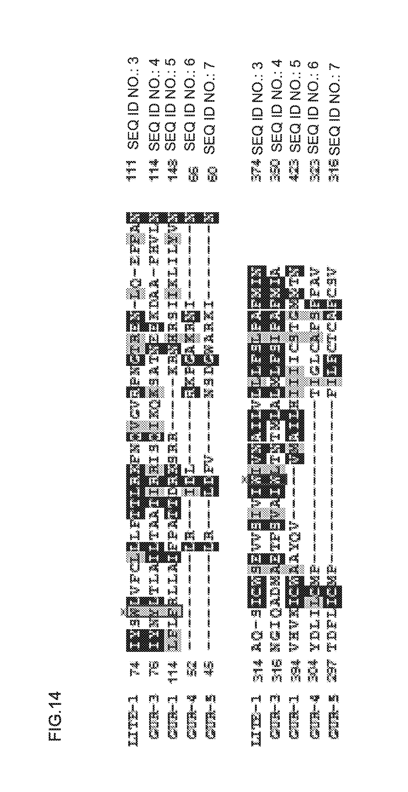
D00019

D00020

S00001
XML
uspto.report is an independent third-party trademark research tool that is not affiliated, endorsed, or sponsored by the United States Patent and Trademark Office (USPTO) or any other governmental organization. The information provided by uspto.report is based on publicly available data at the time of writing and is intended for informational purposes only.
While we strive to provide accurate and up-to-date information, we do not guarantee the accuracy, completeness, reliability, or suitability of the information displayed on this site. The use of this site is at your own risk. Any reliance you place on such information is therefore strictly at your own risk.
All official trademark data, including owner information, should be verified by visiting the official USPTO website at www.uspto.gov. This site is not intended to replace professional legal advice and should not be used as a substitute for consulting with a legal professional who is knowledgeable about trademark law.