Dna Mutation Detection Employing Enrichment Of Mutant Polynucleotide Sequences And Minimally Invasive Sampling
Powell; Michael J ; et al.
U.S. patent application number 16/510722 was filed with the patent office on 2019-10-31 for dna mutation detection employing enrichment of mutant polynucleotide sequences and minimally invasive sampling. The applicant listed for this patent is Michael J Powell, Michael Y Sha, Ke Zhan, Aiguo Zhang. Invention is credited to Michael J Powell, Michael Y Sha, Ke Zhan, Aiguo Zhang.
| Application Number | 20190330692 16/510722 |
| Document ID | / |
| Family ID | 68291535 |
| Filed Date | 2019-10-31 |


View All Diagrams
| United States Patent Application | 20190330692 |
| Kind Code | A1 |
| Powell; Michael J ; et al. | October 31, 2019 |
DNA MUTATION DETECTION EMPLOYING ENRICHMENT OF MUTANT POLYNUCLEOTIDE SEQUENCES AND MINIMALLY INVASIVE SAMPLING
Abstract
The invention relates to a method for enriching a target polynucleotide sequence containing a genetic variation said method comprising: (a) providing two primers targeted to said target polynucleotide sequence; (b) providing a target specific xenonucleic acid clamp oligomer specific for a wildtype polynucleotide sequence; (c) generating multiple amplicons using PCR under specific temperature cycling conditions; and (d) detecting said amplicons. We introduce a novel molecule, Xenonucleic Acid (XNA) for the NGS library preparation. XNA is able to selectively suppress amplification of DNA with wild type alleles and amplify DNA containing mutant alleles. Mutants with low allelic frequency will be easily detectable without deep sequencing after enrichment by adding XNA in multiplex PCR. The 17 actionable mutants related to lung or colorectal cancer diseases at different variant allelic frequency (VAF)% were investigated. Clinical sensitivity is significantly improved with XNA in various types of samples.
| Inventors: | Powell; Michael J; (Alamo, CA) ; Zhang; Aiguo; (San Ramon, CA) ; Sha; Michael Y; (Castro Valley, CA) ; Zhan; Ke; (San Mateo, CA) | ||||||||||
| Applicant: |
|
||||||||||
|---|---|---|---|---|---|---|---|---|---|---|---|
| Family ID: | 68291535 | ||||||||||
| Appl. No.: | 16/510722 | ||||||||||
| Filed: | July 12, 2019 |
Related U.S. Patent Documents
| Application Number | Filing Date | Patent Number | ||
|---|---|---|---|---|
| 14822874 | Aug 10, 2015 | 10400277 | ||
| 16510722 | ||||
| 15786591 | Oct 17, 2017 | |||
| 14822874 | ||||
| 15862581 | Jan 4, 2018 | |||
| 15786591 | ||||
| 62010339 | Jun 10, 2014 | |||
| 62010357 | Jun 10, 2014 | |||
| 62010359 | Jun 10, 2014 | |||
| 62376206 | Aug 17, 2016 | |||
| 62376287 | Aug 17, 2016 | |||
| 62442898 | Jan 5, 2017 | |||
| Current U.S. Class: | 1/1 |
| Current CPC Class: | C12Q 1/686 20130101; C12Q 2527/113 20130101; C12Q 2525/185 20130101; C12Q 1/6827 20130101; C12Q 2525/101 20130101; C40B 40/06 20130101; C12Q 2525/117 20130101; C12Q 2531/113 20130101; C12Q 1/6827 20130101; C12Q 2527/107 20130101; C12Q 1/6858 20130101; C12Q 2531/113 20130101; C12Q 1/6874 20130101 |
| International Class: | C12Q 1/6858 20060101 C12Q001/6858; C12Q 1/686 20060101 C12Q001/686; C12Q 1/6874 20060101 C12Q001/6874; C40B 40/06 20060101 C40B040/06; C12Q 1/6827 20060101 C12Q001/6827 |
Claims
1. A method for enriching a target polynucleotide sequence containing a genetic variation said method comprising: (a) providing two primers targeted to said target polynucleotide sequence; (b) providing a target specific xenonucleic acid clamp oligomer specific for a wildtype polynucleotide sequence, wherein said xenonucleic acid includes moieties selected from the group consisting of oxy-aza, aza-aza, thio-aza and mixtures thereof; (c) generating multiple amplicons using PCR under specific temperature cycling conditions; and (d) detecting said amplicons.
2. The method of claim 1, wherein said detection employs oligonucleotide probes specific for hybridization of variant polynucleotide amplicon sequences.
3. The method of claim 1, wherein the variant target sequence is in a gene selected from the group consisting of: KRAS, BRAF, EGFR, TP53, JAK2, NPM1, and PCA3.
4. A method for enriching multiple target polynucleotide sequences containing a genetic variation said method comprising: (a) providing a library of amplifying primers targeted to said multiple target polynucleotide sequence; (b) providing a library of target specific xenonucleic acid clamp oligomer specific for multiple wildtype polynucleotide sequences, wherein said xenonucleic acid includes moieties selected from the group consisting of oxy-aza, aza-aza, thio-aza and mixtures thereof; (c) generating multiple amplicons using PCR under specific temperature cycling conditions; and (d) detecting said amplicons.
5. The method of claim 3, wherein said detection employs oligonucleotide probes specific for hybridization of variant polynucleotide amplicon sequences.
6. A method for conducting a minimally invasive biopsy in a mammalian subject suspected of a having a neoplastic disease, said method comprising: (a) sampling of target polynucleotides derived from said mammalian subject; (b) providing a library of amplifying primers targeted to said multiple target poly-nucleotide sequence; (c) providing a library of target specific xenonucleic acid clamp oligomer specific for multiple wildtype polynucleotide sequences, wherein said xenonucleic acid includes moieties selected from the group consisting of oxy-aza, aza-aza, thi-aza and mixtures thereof; (d) generating multiple amplicons using PCR under specific temperature cycling conditions; and (e) detecting said amplicons.
7. The method of claim 6, wherein said sampled target polynucleotides are sampled from cells derived from said mammalian subject.
8. The method of claim 6, wherein said sampled target polynucleotides are sampled from free circulating cell free polynucleotides derived from said mammalian subject.
9. The method of claim 4, which includes using multiple XNA clamp probes and amplifying primers targeted to multiple polynucleotide sequences.
10. The method of claim 6, wherein said neoplastic disease is lung cancer.
11. The method of claim 6, wherein said neoplastic disease is colorectal cancer.
12. The method of claim 6, wherein said neoplastic disease is breast cancer.
Description
[0001] This application is a continuation-in-part of pending U.S. Ser. No. 14/822,874 filed Aug. 10, 2015; U.S. Ser. No. 15/786,591 filed Oct. 17, 2017; and U.S. Ser. No. 15/862,581 filed Jan. 4, 2018; the entire contents of which are incorporated herein in their entirety. This application also claims the priority benefit under 35 U.S.C. section 119 of U.S. Provisional Patent Application No. 62/010,339 entitled "Method For Enrichment Of Target Mutant Polynucleotide Sequences" filed on Jun. 10, 2014; U.S. Provisional Patent Application No. 62/010,357 entitled "Detection Of Multiple Mutations In A Single Tube Using QCLAMP.TM. Assay QCLAMP.TM. Mplex" filed on Jun. 10, 2014; and U.S. Provisional Patent Application No. 62/010,359 entitled "Liquid Biopsy" filed on Jun. 10, 2014; which are in their entirety herein incorporated by reference. This application further claims the priority benefit under 35 U.S.C. section 119 of U.S. Provisional Patent Application No. 62/376,206 entitled "Specific Synthetic Chimeric Xenonucleic Acid Guide RNA; s(XNA-gRNA) For Enhancing CRISPR Mediated Genome Editing Efficiency" filed on Aug. 17, 2016; and Provisional Patent Application No. 62/376,287 filed Aug. 17, 2016 entitled "Synthetic Routes To Xenonucleic Acid (Xna) Monomers" which are in their entirety herein incorporated by reference. This application claims the priority benefit under 35 U.S.C. section 119 of U.S. Provisional Patent Application No. 62/442,898 entitled "Method For Conducting Early Detection Of Colon Cancer And/Or Of Colon Cancer Precursor Cells And For Monitoring Colon Cancer Recurrence" filed Jan. 5, 2017, which is in its entirety herein incorporated by reference.
FIELD OF THE INVENTION
[0002] The present invention relates to DNA mutation detection. The invention further relates to enrichment of mutant polynucleotide sequences. The present invention further relates to minimally invasive sampling and analysis of mutations in clinical samples.
[0003] The instant invention also relates to a method for determining whether a target polynucleotide sequence contained in a nucleic acid sample has nucleotide variation(s) in a selected region thereof, the steps of which involve the use of a pair of primers that allows the formation of a PCR product having a sequence covering that of the selected region of the target polynucleotide sequence via a PCR process, and a xenonucleic acid (XNA) that acts as a PCR clamp as well as a sensor probe. This invention also relates to a kit for use in determining the presence of nucleotide variation(s) in the target polynucleotide sequence, which comprises the pair of primers and the XNA.
[0004] The present embodiments relate to precision molecular diagnostics, and in particular, to compositions in detecting sequence variants, such as SNPs, insertions deletions, and altered methylation patterns, from samples. The embodiments disclosed herein can be used to detect (and quantify) sequence variants present in samples that include an excess of wild-type sequences.
BACKGROUND OF THE INVENTION
[0005] Polymerase chain reaction (PCR) is a widely used technique for the detection of pathogens. The technique uses a DNA polymerase used to amplify a piece of DNA by in vitro enzymatic replication. The PCR process generates DNA that is used as a template for replication. This results in a chain reaction that exponentially amplifies the DNA template.
[0006] Technologies for genomic detection most commonly use DNA probes to hybridize to target sequences. To achieve required sensitivity, the use of PCR to amplify target sequences has remained standard practice in many labs. While PCR has been the principle method to identify genes associated with disease states, the method has remained confined to use within a laboratory environment. Most current diagnostic applications that can be used outside of the laboratory are based on antibody recognition of protein targets and use ELISA-based technologies to signal the presence of a disease. These methods are fast and fairly robust, but they can lack the specificity associated with nucleic acid detection.
[0007] With the advent of molecular diagnostics and the discovery of numerous nucleic acid biomarkers useful in the diagnosis and treatment of conditions and diseases, detection of nucleic acid sequences, and sequence variants, mutations and polymorphisms has become increasingly important. In many instances, it is desirable to detect sequence variants or mutations (which may in some instances, differ by one a single nucleotide) present in low copy numbers against a high background of wild-type sequences. For example, as more and more somatic mutations are shown to be biomarkers for cancer prognosis and prediction of therapeutic efficacy, the need for efficient and effective methods to detect rare mutations in a sample is becoming more and more critical. In the case in which one or more allelic variants is/are present in low copy number compared to wild-type sequences, the presence of excess wild-type target sequence creates challenges to the detection of the less abundant variant target sequence. Nucleic acid amplification/detection reactions almost always are performed using limiting amounts of reagents. A large excess of wild-type target sequences, thus competes for and consumes limiting reagents. As a result amplification and/or detection of rare mutant or variant alleles under these conditions is substantially suppressed, and the methods may not be sensitive enough to detect the rare variants or mutants. Various methods to overcome this problem have been attempted. These methods are not ideal, however, because they either require the use of a unique primer for each allele, or the performance of an intricate melt-curve analysis. Both of these shortcomings limit the ability and feasibility of multiplex detection of multiple variant alleles from a single sample.
BRIEF DESCRIPTION OF THE FIGURES
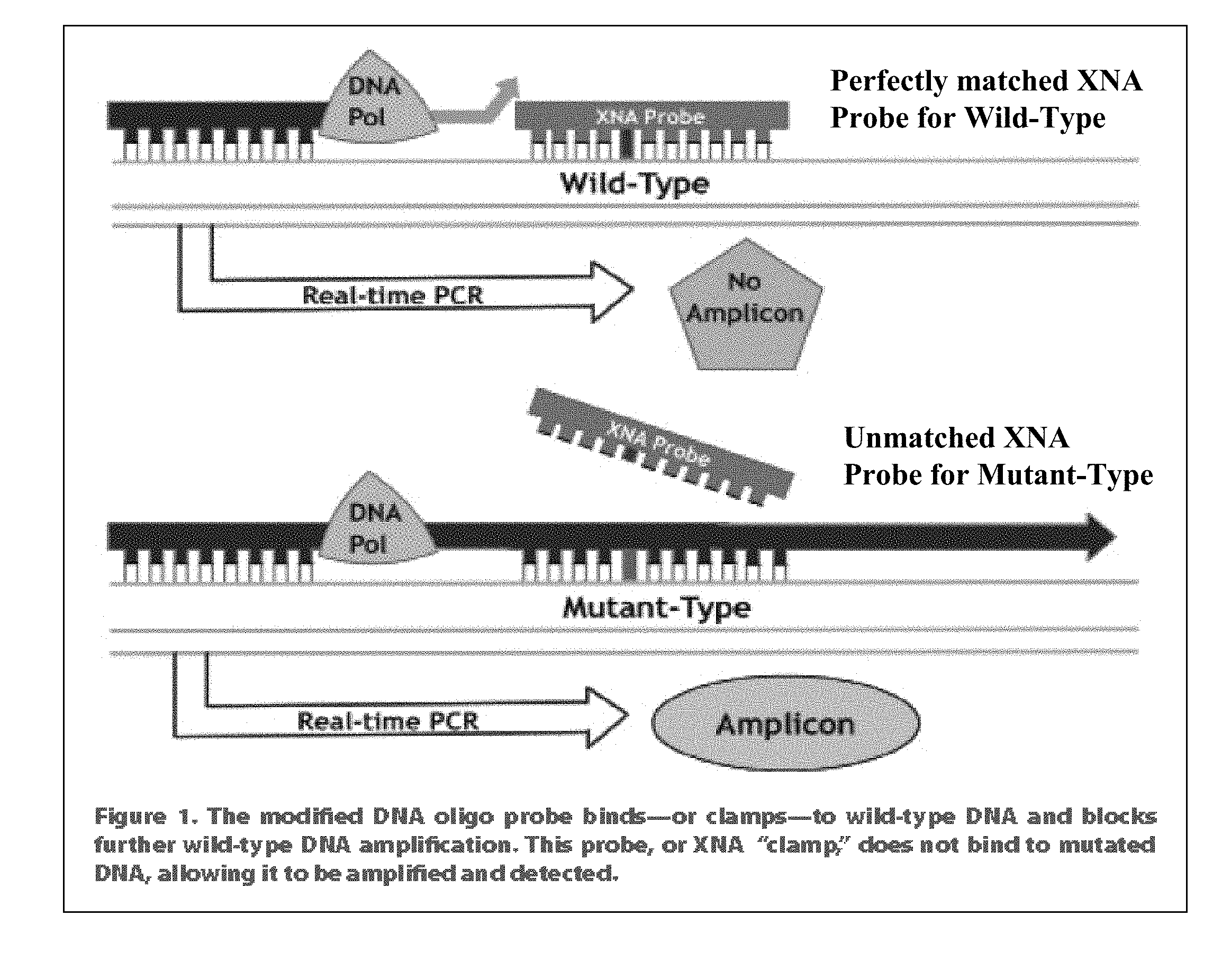
[0008] FIG. 1 illustrates the mechanism of the XNA clamping process.
[0009] FIG. 2 shows the differential melting temperature (Tm) between the XNA clamp bound to mutant templates vs wild type templates.
[0010] FIG. 3 show specific hydrolysis probe having a different fluorophore (and quencher) selected from the available fluorophores for multiplex applications.
[0011] FIG. 4 is a representative fluorophore spectral data and quencher selection guide.
[0012] FIG. 5 shows a specific locus specific hydrolysis probe assay.
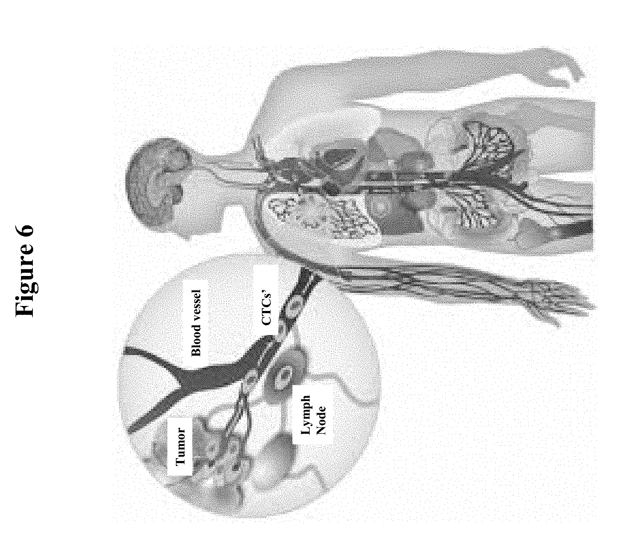
[0013] FIG. 6 is a schematic illustrating how circulating tumor cells (CTC's) and cell-free DNA (cfDNA) derived from tumor cells are present in the peripheral blood of cancer patients.
[0014] FIG. 7A. illustrates a Xenonucleic Acid (XNA) structure.
[0015] FIG. 7B shows preferred Xenonucleic acids having oxy-aza, aza-aza and sulfa-aza (thio-aza) bonding for use in the present invention.
[0016] FIG. 7C illustrates the following: (i) the mechanism of XNA Molecular Clamp Technology, (ii) how XNA makes Low Frequency variant detection easy, and (iii) target enrichment for NGS analysis.
[0017] FIG. 8 describes the effects of XNA mix on Variant Allelic Frequency (VAF) using OPTISEQ.TM. Dual Cancer Panel.
[0018] FIG. 9 illustrates the effects of XNA mix on total coverage using OPTISEQ.TM. Dual Cancer Panel.
[0019] FIG. 10 shows the effects of XNA mix on variant number using OPTISEQ.TM. Dual Cancer Panel.
[0020] FIG. 11 features the effects of XNA mix on VAF enrichment and variant number using OPTISEQ.TM. Dual Cancer Panel.
[0021] FIGS. 12A to 12P describes the correlation of Enriched VAF and original VAF 2.00%) with corresponding reggression equations.
[0022] FIG. 13 shows experimental and data analysis workflows for study of XNA effects on enrichment of variant alleles.
[0023] FIGS. 14A to 14Q shows the correlation of enriched variants allelic frequency (Enriched VAF) and original variant allelic frequency (more than 2.00%) (Original VAF) with corresponding regression equations.
SUMMARY OF THE INVENTION
[0024] Detection of rare sequence variants in biological samples presents numerous challenges. The methods and kits disclosed herein provide for improved, efficient means to detect rare mutations within a high background of wild-type allelic sequences using real-time amplification methods.
[0025] The instant invention provides a method for enriching a target polynucleotide sequence containing a genetic variation said method comprising: (a) providing two primers targeted to said target polynucleotide sequence; (b) providing a target specific xenonucleic acid clamp oligomer specific for a wildtype polynucleotide sequence; (c) generating multiple amplicons using PCR under specific temperature cycling conditions; and (d) detecting said amplicons.
[0026] The invention further provides a method for enriching a target polynucleotide sequence containing a genetic variation, said method comprising: (a) providing a biological sample; (b) isolating DNA from said biological sample; said DNA including said target polynucleotide sequence containing a genetic variation; (c) providing two primer probes targeted to said target polynucleotide sequence said primer probes allowing formation of a PCR process product; (d) providing a target specific xenonucleic acid clamp oligomer probe specific for a wildtype polynucleotide sequence; wherein said target specific xenonucleic acid clamp has oxy-aza and aza-aza moieties so that during the qPCR process only mutant templates are amplified; (e) admixing the primer probes and the xenonucleic clamping probe with the target nucleic acid sample; (f) performing a PCR amplification process in a reaction solution under hybridization conditions thereby generating multiple amplicons; and (g) detecting said amplicons.
[0027] The invention also relates to a method for enriching multiple target polynucleotide sequences containing a genetic variation said method comprising: (a) providing a library of amplifying primers targeted to said multiple target polynucleotide sequence; (b) providing a library of target specific xenonucleic acid clamp oligomer specific for multiple wildtype polynucleotide sequences; (c) generating multiple amplicons using PCR under specific temperature cycling conditions; and (d) detecting said amplicons.
[0028] The invention further relates to a method for conducting a minimally invasive biopsy in a mammalian subject suspected of a having a neoplastic disease, said method comprising: (a) sampling of target polynucleotides derived from said mammalian subject; (b) providing a library of amplifying primers targeted to said multiple target poly-nucleotide sequence; (c) providing a library of target specific xenonucleic acid clamp oligomer specific for multiple wildtype polynucleotide sequences; (d) generating multiple amplicons using PCR under specific temperature cycling conditions; and (e) detecting said amplicons.
[0029] The invention is also directed to means and methodology for the rapid isolation of genetic material from biological fluids and the sensitive detection of somatic and germ-line mutations present in circulating cells and cell-free genetic material obtained from said biological fluids using gene amplification and xeno-nucleic acid (XNA) clamping.
[0030] This invention provides a method for determining whether a target polynucleotide sequence contained in a nucleic acid sample has nucleotide variation(s) in a selected region thereof, comprising the steps of: providing a pair of a first primer and a second primer which allows the formation of a PCR product having a sequence covering that of the selected region of the target polynucleotide sequence via a PCR process, the first primer having a sequence identical to that of a first region located upstream of the selected region of the target polynucleotide sequence, the second primer having a sequence based on that of a second region located downstream of the selected region of the target polynucleotide sequence, wherein the 5'-end of the sequence of the first region is spaced apart from the 5'-end of the sequence of the selected region by 30 nucleotides or more;
providing a detectable xenonucleic acid probe having a sequence that complements fully the sequence of the selected region of the target polynucleotide sequence having no nucleotide variation(s) therein, such that hybridization of the detectable xenonucleic acid probe to the selected region of the target polynucleotide sequence having no nucleotide variation(s) results in the formation of a duplex having a melting temperature;
[0031] determining the melting temperature of the duplex;
[0032] admixing the detectable xenonucleic acid probe and the pair of the first primer and the second primer with the nucleic acid sample to form a mixture;
[0033] subjecting the mixture to a PCR process including an extension reaction set to run at a temperature lower than the melting temperature of the duplex by 5 to 20.degree. C., such that a mixture of PCR products is obtained; and subjecting the mixture of PCR products thus-obtained to a melting analysis to determine melting temperatures of the PCR products, wherein the presence of at least one melting temperature lower than the melting temperature of the duplex is indicative of the nucleotide variation(s) in the selected region of the target polynucleotide sequence contained in the nucleic acid sample.
[0034] The invention also provides a kit for determining whether a target polynucleotide sequence contained in a nucleic acid sample has nucleotide variation(s) in a selected region thereof, comprising: a detectable xenonucleic acid probe having a sequence that complements fully the sequence of the selected region of the target polynucleotide sequence having no nucleotide variation(s) therein, such that hybridization of the detectable xenonucleic acid probe to the selected region of the target polynucleotide sequence having no nucleotide variation(s) results in the formation of a duplex having a melting temperature;
[0035] a pair of a first primer and a second primer which allows the formation of a PCR product having a sequence covering that of the selected region of the target polynucleotide sequence via a PCR process, the first primer having a sequence identical to that of a first region located upstream of the selected region of the target polynucleotide sequence, the second primer having a sequence based on that of a second region located downstream of the selected region of the target polynucleotide sequence, wherein the 5'-end of the sequence of the first region is spaced apart from the 5'-end of the sequence of the selected region by 30 nucleotides or more; and an instruction sheet providing guidance for a user to use the detectable xenonucleic acid probe and the pair of the first primer and the second primer in a method as described above.
[0036] The identification of genetic variants with low variant frequency using next-generation sequencing method is confounded by the complexity of human genome sequence and by bias that arise during library preparation, sequencing and analysis. The present invention also provides a novel molecule, Xenonucleic Acids (XNA) for the NGS library preparation. XNA is able to selectively suppress amplification of DNA with wild type alleles and amplify DNA containing mutant alleles. Mutants with low allelic frequency will be easily detectable without deep sequencing after enrichment by adding XNA in multiplex PCR. The 17 actionable mutants related to lung or colorectal cancer diseases at different VAF % have been studied in great detail in the present invention. Upon XNA blocking of wild type alleles, detectable enriched variant allelic frequency (VAF) can be increased by .about.32 fold from 10 ng of gDNA samples containing mutants as low as 0.10%. Analytical sensitivity of Limit of Detection (LoD) is about 0.10% VAF. These 17 actionable mutants were tested and verified by using FFPE and cfDNA of lung or colon cancer patient samples. Clinical sensitivity for FFPE sample is about 100% for lung cancer and colorectal cancer samples respectively, comparing to without XNA NGS about 85.7% for lung cancer and 70% for colon cancer. For cfDNA sample its clinical sensitivity is about 100% for lung and early colon cancer, but without XNA NGS is about 70% for lung cancer and undetectable for early colon cancer. This invention provides a simple, accurate, higher sensitive and lower cost alternative compared with conventional NGS with deep sequencing.
DETAILED DESCRIPTION OF THE INVENTION
[0037] It is to be understood that both the foregoing general description and the following detailed description are exemplary and explanatory only and are not intended to limit the scope of the current teachings. In this application, the use of the singular includes the plural unless specifically stated otherwise. Also, the use of "comprise", "contain", and "include", or modifications of those root words, for example but not limited to, "comprises", "contained", and "including", are not intended to be limiting. Use of "or" means "and/or" unless stated otherwise. The term "and/or" means that the terms before and after can be taken together or separately. For illustration purposes, but not as a limitation, "X and/or Y" can mean "X" or "Y" or "X and Y". Whenever a range of values is provided herein, the range is meant to include the starting value and the ending value and any value or value range there between unless otherwise specifically stated. For example, "from 0.2 to 0.5" means 0.2, 0.3, 0.4, 0.5; ranges there between such as 0.2-0.3, 0.3-0.4, 0.2-0.4; increments there between such as 0.25, 0.35, 0.225, 0.335, 0.49; increment ranges there between such as 0.26-0.39; and the like.
[0038] In a first embodiment, the present invention relates to compositions and methods for the selective enrichment of low-abundance polynucleotides in a sample. These methods use xeno-nucleic acid (XNA) nucleobase oligomers to selectively block DNA polymerase activity on high abundance wild-type DNA templates, thereby resulting in enrichment of less abundant mutated DNA templates present in a biological sample during a polymerase chain reaction (PCR). The methodology of the present invention can be used to improve DNA sequencing (Sanger sequencing and Pyrosequencing) and also enhance cDNA library preparation for next generation DNA sequencing (NGS).
[0039] Utilizing xeno-nucleic acid (XNA) clamping probes in the PCR mediated amplification of DNA templates, only target genetic material that has a variation, e.g. single nucleotide polymorphism (SNP), gene deletion or insertion and/or translocation or truncation is amplified in the oligonucleotide primer directed polymerase chain reaction (qPCR).
[0040] The XNA probe clamping sequences are designed to bind specifically by Watson-Crick base pairing to abundant wild-type sequences in the DNA templates derived from the biological sample of interest. The presence of the XNA probes in the PCR primer mix employed for the target amplification reaction causes inhibition of the polymerase mediated amplification of wild-type templates but does not impede the amplification of mutant template sequences.
[0041] The mechanism of the XNA clamping process is depicted in FIG. 1. As shown in FIG. 1, the modified DNA oligo probe binds or clamps to wild type DNA and blocks further wild type amplification. This probe or XNA "clamp" does not bind to mutated DNA, allowing it to be amplified and detected.
[0042] The suppression of wild-type (wt) template amplification and amplification of only mutant templates is achieved because there is a differential melting temperature (Tm) between the XNA clamp bound to mutant templates vs wild type templates:
Tm(XNA mutant template)<<Tm(XNA wt template)
[0043] The Tm differential is as much as 15-20.degree. C. for the XNA clamp probes. So that during the PCR process only mutant templates are amplified.
[0044] The methods disclosed herein can be used to analyze nucleic acids of samples. The term "sample" as described herein can include bodily fluids (including, but not limited to, blood, urine, feces, serum, lymph, saliva, anal and vaginal secretions, perspiration, peritoneal fluid, pleural fluid, effusions, ascites, and purulent secretions, lavage fluids, drained fluids, brush cytology specimens, biopsy tissue (e.g., tumor samples), explanted medical devices, infected catheters, pus, biofilms and semen) of virtually any organism, with mammalian samples, particularly human samples.
[0045] Amplification primers useful in the embodiments disclosed herein are preferably between 10 and 45 nucleotides in length. For example, the primers can be at least 10, 11, 12, 13, 14, 15, 16, 17, 18, 19, 20, 21, 22, 23, 24, 25, 26, 27, 28, 29, 30, 31, 32, 33, 34, 35, 36, 37, 38, 39, 40, 41, 42, 43, 44, 45, or more nucleotides in length. Primers can be provided in any suitable form, included bound to a solid support, liquid, and lyophilized, for example. In some embodiments, the primers and/or probes include oligonucleotides that hybridize to a reference nucleic acid sequence over the entire length of the oligonucleotide sequence. Such sequences can be referred to as "fully complementary" with respect to each other. Where an oligonucleotide is referred to as "substantially complementary" with respect to a nucleic acid sequence herein, the two sequences can be fully complementary, or they may form mismatches upon hybridization, but retain the ability to hybridize under stringent conditions or standard PCR conditions as discussed below. As used herein, the term "standard PCR conditions" include, for example, any of the PCR conditions disclosed herein, or known in the art, as described in, for example, PCR 1: A Practical Approach, M. J. McPherson, P. Quirke, and G. R. Taylor, Ed., (c) 2001, Oxford University Press, Oxford, England, and PCR Protocols: Current Methods and Applications, B. White, Ed., (c) 1993, Humana Press, Totowa, N.J. The amplification primers can be substantially complementary to their annealing region, comprising the specific variant target sequence(s) or the wild type target sequence(s). Accordingly, substantially complementary sequences can refer to sequences ranging in percent identity from 100, 99, 98, 97, 96, 95, 94, 93, 92, 91, 90, 89, 85, 80, 75 or less, or any number in between, compared to the reference sequence. Conditions for enhancing the stringency of amplification reactions and suitable in the embodiments disclosed herein, are well-known to those in the art. A discussion of PCR conditions, and stringency of PCR, can be found, for example in Roux, K. "Optimization and Troubleshooting in PCR," in Pcr Primer: A Laboratory Manual, Diffenbach, Ed. .COPYRGT. 1995, Cold Spring Harbor Laboratory Press, Cold Spring Harbor, N.Y.; and Datta, et al. (2003) Nucl. Acids Res. 31(19):5590-5597.
[0046] Provided herein are methods useful in the detection of sequence variants, i.e., insertions, deletions, nonsense mutations, missense mutations, and the like. In the methods for detecting allelic variants or variant target sequences disclosed herein, the sample, which comprises the nucleic acids to be analyzed, are contacted with an amplification primer pair, i.e., comprising a forward primer and a reverse primer that flank the target sequence or target region containing a sequence of interest {e.g., a wild-type, mutant, or variant allele sequence) to be analyzed. By "flanking" the target sequence, it is understood that the variant or wild-type allelic sequence is located between the forward and reverse primers, and that the binding site of neither the forward nor reverse primer comprises the variant or wild-type allelic sequence to be assessed. For example, in some embodiments, the variant or wild-type allelic sequence to be assessed is removed from or positioned away from the 3' end of either oligonucleotide by 1, 2, 3, 4, 5, 6, 7, 8, 9, 10, 11, 12, 13, 14, 15, 16, 17, 18, 19, 20, 21, 22, 23, 24, 25, 26, 27, 28, 29, 30, 31, 32, 33, 34, 35, 36, 37, 38, 39, 40, 41, 42, 43, 44, 45, 46, 47, 48, 49, 50, or more, e.g., 100 or more, 200 or more, 300 or more, 400 or more, 500 or more, etc., nucleotides. Amplification primers that flank, but that do not overlap with, the variant target sequence or the wild-type target sequence are thus not "allele-specific" amplification primers, and are capable of amplification of various different alleles or variants of a sequence of interest. Thus, in some embodiments, the amplification primers are configured to amplify various mutant or variant alleles and wild type alleles non-preferentially. As discussed in further detail below, the addition of XNA to an amplification reaction suppresses the amplification of wild-type target sequences and enables preferential amplification of non-wild-type, e.g., variant, mutant or rare variant alleles. FIG. 1 is a depictions of exemplary method according to the embodiments disclosed herein for the detection of sequence variants. As shown in FIG. 1, amplification primers (i.e., forward primer 1 and reverse primer 2) flank the wild type and mutant allele sequences of interest, and comprise sequences common to both wild-type and mutant or variant allele sequences. Accordingly, as shown in FIG. 1, in contrast to methods that utilize allele-specific amplification primers to achieve preferential amplification of rare sequences, the present methods advantageously enable the simultaneous amplification of multiple variant sequences, using a single amplification primer pair.
[0047] In a second embodiment, the invention relates to compositions and methods for the detection of genetic variations (mutations) in DNA templates derived from biological samples with xeno-nucleic acid clamping probes. The first method employs multi-color fluorescence detection using locus specific fluorescent hybridization probes (Hyb Probes), hydrolysis (TaqMan or ZEN) probes or molecular beacons. The second method employs mutant specific amplicon capture probes immobilized on multiple bar-coded capture beads.
[0048] Current XNA clamping qPCR methodologies utilize a single tube-single mutation detection format it is preferable to detect multiple genetic variations in a single tube thus reducing the complexity of the assay and the amount of template DNA required for analysis.
[0049] This second embodiment of the invention is directed to the use of locus specific fluorescent probes designed to detect the genetic variant (mutant) amplicons generated during the XNA clamping PCR reaction. This second embodiment discloses locus specific probes that bind to mutant specific amplicons at a region upstream or downstream from the site of the mutation to be detected. Furthermore, the second embodiment discloses the use of multiplexed XNA clamping qPCR reactions that are able to detect multiple mutations (up to a maximum of 6) in one PCR reaction tube using fluorescence detection methodology.
[0050] In a third embodiment of the invention, there is provided a method the rapid isolation of genetic material present in circulating cells and also cell-free genetic material from biological fluids and the determination of genetic variations in those cells and biological fluids. Such biological fluids include: blood, serum, plasma, saliva, mucus, urine, sputum, semen or other biological secretions. In this embodiment, the invention also provides the detection of somatic and germ-line mutations in the genetic material derived from these biological fluids utilizing gene amplification and xeno-nucleic acid clamping.
[0051] Circulating tumor cells (CTC's) and cell-free DNA (cfDNA) derived from tumor cells are present in the peripheral blood of cancer patients (See FIG. 6). Tumor derived DNA can also be found in the urine and even the saliva of cancer patients.
[0052] In general circulating free DNA is smaller in size than DNA derived directly from a surgical biopsy or FFPE sample. This embodiment also describes a novel sample treatment procedure that utilizes a novel lysis reagent called QZol.TM.. QZol.TM. sample lysis is a direct one tube procedure and an aliquot of the lysate is used directly in molecular genetic and cytogenetic analysis procedures such as PCR, RTPCR, FISH, Next Generation Sequencing (NGS) and branched DNA (bDNA) assays. The QZol.TM. procedure eliminates the tedious multistep preanalytical processing that is currently used in Molecular Pathology and Cytogenetic analysis.
[0053] The lysis reagent is a 50% solution (A) containing chaotropic salts and detergent (nonionic, anionic, cationic or zwitterionic) and a 50% solution (B) containing neutralizing reagents and stabilizers.
[0054] This invention also concerns to the specific amplification of genetic variant templates from the isolated genetic material described above. Only target genetic material that has a variation, e.g. single nucleotide polymorphism (SNP), gene deletion or insertion and/or translocation or truncation is amplified in a quantitative primer directed polymerase chain reaction (qPCR). This is achieved utilising xenonucleic acid (XNA) probe clamping sequences that have been designed to bind specifically by Watson-Crick base pairing to wild-type sequences in the sample. The presence of the XNA probes in the qPCR primer mix employed for the target amplification reaction causes inhibition of the polymerase mediated amplification of wild-type templates but does not impede the amplification of mutant template sequences.
[0055] The mechanism of the XNA clamping process is depicted in FIG. 1.
[0056] The suppression of wild-type (wt) template amplification and amplification of only mutant templates is achieved because there is a differential melting temperature (Tm) between the XNA clamp bound to mutant templates vs wt templates:
Tm(XNA mutant template)<<Tm(XNA wt template)
The Tm differential is as much as 15-20.degree. C. for the XNA clamp probes. So that during the qPCR process only mutant templates are amplified.
[0057] The methods disclosed herein can be used in the detection of numerous allelic variants, including nonsense mutations, missense mutations, insertions, deletions, and the like. Owing to the advantageous sensitivity and specificity of detection afforded by the methods disclosed herein, the methods can detect the presence of a rare allelic variant within a sample, amongst a high wild-type background. Accordingly, although the skilled artisan will appreciate that the methods disclosed herein can be used in a variety of settings to detect, e.g., germline mutations, the methods are particularly well-suited for use in the detection of somatic mutations, such as mutations present in tumors. Non-limiting examples of rare, somatic mutations useful in the diagnosis, prognosis, and treatment of various tumors include, for example, mutations in ABL, AKT1, AKT2, ALK, APC, ATM, BRAF, CBL, CDH1, CDK 2A, CEBPA, CRLF2, CSF1R, CTNNB1, EGFR, ERBB2, EZH2, FBXW7, FGFR, FGFR2, FGFR3, FLT3, FOXL2, GATA1, GATA2, GNAQ, GNAS, HNF1A, HRAS, IDH1, IDH3, JAK2, KIT, KRAS, MEK1, MET, MPL, NF2, NOTCH 1, NOTCH2, NPM, NRAS, PC A3, PDGFRA, PIK3CA, PIK3R1, PIK3R5, PTCH1, PTEN, PTPN1 1, RBI, RET, RUNX1, SMAD4, SMARCB, SMO, STK11, TET2, P53, TSHR, VHL, WT1, and others. Exemplary mutant alleles associated with cancer useful in the embodiments disclosed herein include, but are not limited to those described in publications listed on the world wide web site for COSMIC (Catalogue Of Somatic Mutations In Cancer).
[0058] Next-generation sequencing (NGS) is widely used to detect sequence variations and an array of genetic markers for oncological diagnostic research and, in combination with bioinformatics, is increasingly used to analyze multiple biomarkers in a low-cost, time-effective manner.sup.1. However, one of the challenges in detecting cancer variants with standard NGS analysis is the low frequency of mutant alleles in cancer cells amongst a background of wild type alleles in healthy cells. The adequate resolution of low-frequency SNVs is essential both to improve treatment of cancer and to monitor minimal residue disease status during follow-up. However, typically NGS sensitivity is limited to variants at 0.1-1.0% mainly due to sequencing related background errors. In order to meet clinical standards and to distinguish true variants from sequencing errors, NGS has to be accurate and robust, several solutions have been described. For example, the application of proofreading enzymes (proofreading DNA-polymerase containing 3'-5' exonuclease activity) significantly increased NGS sensitivity by reducing false-positive variant calls at respective genomic positions. And the use of complex barcoding strategies, which enable the separation of true single nucleotide variants (SNVs) from errors. Deep sequencing is another solution to achieve the detection of variant with low frequency, however, deep sequencing increases the systematic error rate arise from sequencing machine and leads to unreliable result.sup.2,3. In some cases, deep sequencing is still not able to achieve the detection of hotspots covered by low performance primer set amongst large primers pool, which makes deep sequencing a pricy and inefficient method. Detecting the variant with low frequency is still challenging for most of the researcher in this area. In order to reliably distinguish true variants from sequencing-related errors at mutant allele frequencies of <0.1% and to identify suitable markers for cancer disease prognostic detection, A new technology that it enables to detect the "needle" from the "haystack" is needed to face the challenge.
[0059] Xenonucleic Acid (XNA) molecular clamp is an innovative nucleic acid molecular oligomers (FIG. 1a) that hybridize by Watson-Crick base pairing to target DNA sequences, which are used during polymerase chain reaction (PCR) to selectively suppress amplification of DNA with wild type alleles and amplify DNA containing mutant alleles (FIG. 1b). Mutants with low allelic frequency will be easily detectable without deep sequencing after enrichment by adding XNA in PCR.sup.4,5,6 (FIG. 1b).
[0060] Herein, we introduce a highly sensitive OptiSeq.TM. Lung and Colorectal Cancer Dual Cancers Panel powered by the proprietary XNA technology to detect low frequency variants in human standard reference samples and lung or colorectal cancer patients' samples. This NGS diagnostic platform with XNA significantly improves the detection sensitivity of variants for diagnosis of cancer mutants even at ultra-low allele frequency.
[0061] Here we present results of XNAs mix enrichment effects on cell line genomic DNA samples with low variant allelic frequency, and lung and colorectal cancer patient samples, a benchmarking efforts aimed at enriching variant allelic frequency of samples with low frequency, and made low VAF samples detected by next generation sequencing method reliably and cost-efficiently, thus drawing conclusion confidently without sacrificing the quality of results. Meanwhile, regression models for 17 hotspots were constructed to get the relationship between enriched VAF and original VAF. In this way, original VAF value can be derived from enriched VAF by the corresponding equation, which provides an insight for clinical professionals to draw conclusion based on original VAF, particularly for variants with super low variant frequency, since typically NGS sensitivity is limited to variants at 0.1-1.0% mainly due to sequencing related background errors. Any variant allelic frequency below 1.0% will not be sufficient to draw reliable conclusion about the authenticity of mutations.
[0062] From the results of enrichment effects of XNAs mix on cell line genomic DNA samples, the mutant detection powered by the XNAs mix was dramatically boosted. 14 out of 17 hotspots were able to go down to the detection limit 0.10% with detected variant number more than 2. On samples originally with estimated 1.25% of mutants, in 14 of 17 hotspots, observed VAFs were more than 10% after XNA enrichment. This result suggested that XNA is able to enrich mutant alleles and make high confidence calls. The enrichment effects of 13 different XNAs varied based on the characteristics of each XNA. For XNA named EGFR G719, it shows a strong binding affinity towards the wild type and is able to enrich detected VAF up to 94.43% from 1.01%, which is 93.5 times more than the original VAF in the sample. However, for XNA like CTNNB1 S45, detected VAF after adding XNA was less than 1.0%, detected variant number was even less than 1 copy, which is not sufficient enough to draw the confident conclusion about the authenticity of mutant. It indicated that the binding affinity of XNA CTNNB1 S45 was weaker than that of EGFR 5719. It started to show detected variant on the condition of estimated original VAF 0.25% (Variant number was 2) with XNAs mix.
[0063] For some of the XNAs, they cover two loci at the same time. For example, NRAS A59 XNA covers two loci NRAS A59 and NRAS Q61, or NRAS G12 XNA covers two loci NRAS G12 and NRAS G13. Summary for covered hotspots by 13 XNAs was summarized in Table 11. Despite one XNA was used to block two loci, the blocking efficiency of XNA towards two loci was not directly related. For example, NRAS A59 XNA showed a good enrichment efficiency towards hotspot NRAS A59T, enriched VAF with XNA was 3.89 at original VAF 0.08, which is 48.6 times more than original VAF. While for hotspots NRAS Q61H, the enriched VAF was 0.16, which was only 3.2 folds than original VAF.
[0064] Enough sequencing coverage of each loci is necessary to achieve confident call of mutant, particularly for some mutants with super low frequency, in some rare cases, even pricy deep sequencing fails to detect them. From the results shown in Table 13-A and Table 13-B. The total sequencing coverage of samples with XNAs mix were less than those without XNAs mix. For example, average total coverage of estimated original VAF 0.10% with XNAs mix was 603, while that of same VAF without XNA WAS 2121. Despite the reduction of sequencing depth, enriched VAF for 14 out 17 hotspots were more than 1.00% to draw confident calls. While for same libraries without XNAs mix, only 1 out of 17 hotspots were more than 1.00%. Besides one important criterion "Enriched VAF", actual detected variant number was of same importance as well. Enough variant number ensures the authenticity of mutant call. The average variant number with XNAs mix was 9.1 times of those without XNAs mix. All these results demonstrated XNAs effects on the enrichment of detected VAF, which is of great significance to get reliable call from sequencing machine, since typically NGS sensitivity is limited to variants at 0.1-1.0% mainly due to sequencing related background and PCR errors. Meanwhile, sufficient number of variants were got due to the blocking effects on wild type background noise.
[0065] From results of Table 14, we learnt that the higher the original VAF, the lower CV %. The reason that lower VAF leads to higher CV % might due to the sampling issue of the DNA input. Since the real copies of mutant with VAF 0.10% are only 3 copies, which made it hard for experimental operator to pick up exact 3 copies from the stock solution, this sampling issue made a butterfly effect and caused the big variance of variant numbers and VAF in library, thus leading to the high CV %. As the VAF increases up to 1.25%, copies number of mutant was 42 copies, this sampling issue got weakened and high CV % 17.7 was achieved. The trend found in Table 14 was similar to that in Table 14 and it can be explained by sampling issue as well. Since as the original VAF increased, the positive predictive values (PPV) increased and reached 100% when estimated original VAF was 1.25%.
[0066] Although regression equations deduced by modeling can help to get the original VAF from enriched VAF with XNAs mix, it only applies when deduced original VAF value falls within confidence interval range (up to VAF 15%). It might applies when out of confidence interval, however, additional data are required to verify this assumption. For example, Lung cancer FFPE sample ID 16A140, original VAF of mutation EGFR L858R was 34.31%, detected enriched VAF for this mutation was 83.76%. If we applied regression equation, calculated VAF was 134.05%, which is beyond 100% and greatly off the true value 83.76%. While for FFPE sample ID 16A011, original VAF of mutation EGFR L858R was 20.37%, despite off of confidence interval limit 12.4%, calculated enriched VAF was 97.81% which is approximately close to detected enriched 91.91%. we acknowledged that It would be better and more comprehensive of this study to get the full regression model from original VAF 0.00% to 100.00% for each hotspots, however, in this study, we only focus on the XNAs enrichment effects on mutations with super low or low variant frequency help detect existed low variant frequency mutations with more cost-efficient method. For original VAF more than 15.0%, it can be detected confidently with normal NGS method.
[0067] In summary, XNA molecular clamp technology in combination with NGS have a great potential for cancer molecular diagnosis of cancer mutations in ultra-low allele frequency. OptiSeq.TM. Lung and Colorectal Cancer Mini Panel powered by XNAs is able to report mutants from 10 ng of input gDNA with allele frequency as low as 0.10% with confident calls for 14 out of 17 hotspots. The relationship between enriched VAF and original VAF were derived using regression model for 17 hotspots. Some of regression equations were verified using clinical patient samples and proved reliable to deduce original VAF from enriched VAF. Significant progress has been made in characterizing and optimizing the use of XNA in conjunction with OptiSeq.TM. oncology NGS panel, which provides a promising solution to detect mutants with low frequency with improvement sensitivity and confidence. Clinical sensitivity for FFPE is about 100% for lung cancer (14/14 samples) and colorectal cancer samples (10/10 samples), comparing to normal NGS about 85.7% (12/14 lung sample) and 70% (7/10) for colon cancer. For cfDNA its clinical sensitivity is about 100% for lung (10/10) and colon cancer (2/2), but normal NGS is about 70% for lung (3/10) and 0% for colon cancer (0/2 sample)
EXAMPLES
Example 1
[0068] The kit described in great detail in this Example is a KRAS mutation detection kit. However, the same type of kit may be assembled to detect mutations in NRAS, EGFR, BRAF, PIK3CA, JAK2, as well as other genes of importance in precision molecular diagnostics.
QCLAMP.TM. Technology for Mutation Detection
[0069] The QCLAMP.TM. KRAS Mutation Detection Kit is based on xenonucleic acid (XNA) mediated PCR clamping technology. XNA is a synthetic DNA analog in which the phosphodiester backbone has been replaced by a repeat formed by units of (2-aminoethyl)-glycine. XNAs hybridize tightly to complementary DNA target sequences only if the sequence is a complete match. Binding of XNA to its target sequence blocks strand elongation by DNA polymerase. When there is a mutation in the target site, and therefore a mismatch, the XNA:DNA duplex is unstable, allowing strand elongation by DNA polymerase. Addition of an XNA, whose sequence with a complete match to wild-type DNA, to a PCR reaction, blocks amplification of wild-type DNA allowing selective amplification of mutant DNA. XNA oligomers are not recognized by DNA polymerases and cannot be utilized as primers in subsequent real-time PCR reactions.
DNA Isolation
[0070] Human genomic DNA must be extracted from tissue or blood, or fixed paraffin-embedded tissue prior to use. Several methods exist for DNA isolation. For consistency, we recommend using a commercial kit, such as Qiagen DNA extraction kit (QIAamp DNA FFPE Tissue Kit, cat No. 56404, for paraffin embedded specimens; DNeasy Blood & Tissue kit, cat. No. 69504 or 69506, for tissue and blood specimens). Follow the genomic DNA isolation procedure according to manufacturer's protocol. Sufficient amounts of DNA can be isolated from FFPE blocks or fresh frozen sections (approx. 2-10 .mu.g).
[0071] This QCLAMP.TM. assay requires a total of 30-60 ng of DNA per sample (5-10 ng/reaction). After DNA isolation, measure the concentration using spectrophotometric analysis (i.e. Nanodrop or UV spectrophotometer) and dilute to it to 1.25-2.5 ng/.mu.1. Make sure A260/A230 value is greater than 2.0 and A260/A280 value between 1.8 and 2.0.
Preparation of Reagents
[0072] Each kit contains enough material to run 3 sets (10-sample test kit) or 6 sets (30-sample test kit) of Clamping Controls, Positive Controls and Non-Template Controls. Thaw all Primers, XNAs, Positive Control, WT Clamping Control, water and 2.times.PCR Mastermix provided. Thaw all reaction mixes at room temperature for a minimum of 1 hour. Vortex all components except the PCR Master Mix the reaction mixes for 5 sec and perform a quick spin. The PCR Master Mix should be mixed gently by inverting the tube a few times. Do not leave kit components at room temperature for more than 4 hours. After thawing, keep materials on ice at all times. The PCR reactions are set up in a total volume of 20 .mu.l/reaction.
[0073] Table 1 shows the component volumes for each 20ul reaction.
TABLE-US-00001 TABLE 1 QCLAMP .TM. Assay Components and Reaction Volume Components Volume/Reaction 2X PCR Master mix 10 .mu.l Primer Mix 4 .mu.l XNA 2 .mu.l DNA sample or Controls 4 .mu.l Total volume 20 .mu.l
[0074] For accuracy, 2.times.PCR Master mix, primers and XNA should be pre-mixed into assay mixes as described in Table 2 below.
Preparation of Assay Mixes
[0075] IMPORTANT: Assay mixes should be prepared just prior to use. Do not store assay mixes. Prepare and keep assay mixes on ice, until ready for per. Label 7 micro centrifuge tubes (not provided) according to each corresponding reaction mix shown in Table 2.
TABLE-US-00002 TABLE 2 Preparation of Assay Mixes Volume of Volume of Volume of XNA 2X PCR Primer (.dagger.use water for Master Mix Mix ext control) Ext Control Mix 10 .mu.l .times. (*n + 1) 4 .mu.l .times. (*n + 1) 2 .mu.l .times. (*n + 1) G12 Mix 10 .mu.l .times. (*n + 1) 4 .mu.l .times. (*n + 1) 2 .mu.l .times. (*n + 1) G13 Mix 10 .mu.l .times. (*n + 1) 4 .mu.l .times. (*n + 1) 2 .mu.l .times. (*n + 1) A59 Mix 10 .mu.l .times. (*n + 1) 4 .mu.l .times. (*n + 1) 2 .mu.l .times. (*n + 1) Q61 Mix 10 .mu.l .times. (*n + 1) 4 .mu.l .times. (*n + 1) 2 .mu.l .times. (*n + 1) K117 Mix 10 .mu.l .times. (*n + 1) 4 .mu.l .times. (*n + 1) 2 .mu.l .times. (*n + 1) A146 Mix 10 .mu.l .times. (*n + 1) 4 .mu.l .times. (*n + 1) 2 .mu.l .times. (*n + 1) *n = number of reactions (DNA samples plus 3 controls). Prepare enough for 1 extra sample (n + 1) to allow for sufficient overage for the PCR set. .dagger.Use 2 ul of water provided in the kit as the Ext Control Mix does not require XNA. For accuracy, do not pipette less than 10 ul of the XNA.
[0076] Prepare sufficient working assay mixes for the DNA samples, one KRAS Mixed Positive Control, one Nuclease-Free Water for no template control, and one WT Clamping Control, according to the volumes in Table 2. Include reagents for 1 extra sample to allow sufficient overage for the PCR set up. The master mixes contain all of the components needed for PCR except the sample.
[0077] Each sample requires one reaction for each mutation site detected by the kit and an external control. The External Control uses Exon 5 primers to determine if an appropriate level of amplifiable DNA is present in the sample, and ensures that that the supplied primers and polymerase are working properly on the sample. The KRAS Codon-Specific kit requires a total of 7 reactions for each sample.
[0078] A set of clamping controls must be run with each of the 7 reaction mixes, every time the assay is run. Clamping Controls use wild-type DNA as the template. Wild-type DNA should have no mutations, therefore the XNA probes will bind strongly, blocking the polymerase from making amplicons. However, the External Control Mix with the Clamping Control should make amplicons efficiently, providing another way to monitor performance of the primers, polymerase, and sample.
[0079] A set of positive controls must also be run with each of the 7 reaction mixes, every time the assay is run. The Positive Control contains one mutant template for each reaction mix. Positive controls contain mutations; therefore XNA probes will not bind, allowing amplification of the mutant template. Positive controls must show the appropriate values for the reaction to be valid.
[0080] A set of no template control (tube NTC) is run with each of the 7 reaction mixes every time the assay is run. Nuclease-Free Water is used in the place of template. The NTC serves as a negative control and assesses potential contamination during assay set-up.
[0081] Further quantities of KRAS Wild-Type Genomic Reference DNA Control, and Positive Control mixes can be purchased as a separate item, if desired.
Suggested Run Layout (96-Well Plate, Tube Strips, or Tubes)
[0082] Gently vortex the assay mixes for 5 sec and do a quick spin. Add 16 .mu.l of the appropriate assay mix to the plate or tubes. Add 4 .mu.l of template. Prepare and keep on ice until ready for PCR.
[0083] In the case of 96-well plates, the exact plate layout can be set to the user's preference. However, take care to remember which wells are for which reaction mixes, to ensure that all potential detected mutations and controls are processed properly. Table 3 is a suggested plate set-up for a single experiment analyzing 3 unknown samples.
TABLE-US-00003 TABLE 3 Suggested Plate Layout 1 2 3 4 5 6 A NTC PC CC S1 S2 S3 Ext Ctrl Mix Ext Ctrl Mix Ext Ctrl Mix Ext Ctrl Mix Ext Ctrl Mix Ext Ctrl Mix B NTC PC CC S1 S2 S3 G12 Mix G12 Mix G12 Mix G12 Mix G12 Mix G12 Mix C NTC PC CC S1 S2 S3 G13 Mix G13 Mix G13 Mix G13 Mix G13 Mix G13 Mix D NTC PC CC S1 S2 S3 A59 Mix A59 Mix A59 Mix A59 Mix A59 Mix A59 Mix E NTC PC CC S1 S2 S3 Q61 Mix Q61 Mix Q61 Mix Q61 Mix Q61 Mix Q61 Mix F NTC PC CC S1 S2 S3 K117 Mix K117 Mix K117 Mix K117 Mix K117 Mix K117 Mix G NTC PC CC S1 S2 S3 A146 Mix A146 Mix A146 Mix A146 Mix A146 Mix A146 Mix PC: Positive Control, NTC: No Template Control (water), CC: Clamping Control (Wild-type DNA), S1-3: Samples 1-3. NOTE: For setup on the Rotor-Gene Q Platforms, the layout must be changed such that the first well contains Positive Control.
[0084] When all reagents have been loaded, tightly close the PCR tubes or seal the 96-well plate to prevent evaporation. Spin at 2000 rpm for 1 minute to collect all the reagents. Place in the real-time PCR instrument immediately or store on ice until the instrument is ready.
Instrument Set-Up
Roche LightCycler 96 or RocheLightCycler 480
[0085] 1. Select New empty experiment >create 2. In the Run Editor>Measurement, choose SYBR Green 1 (470/514) channel on (LC96), SYBR Green 1/HRM Dye on (LC480) 3. Set up run profile using parameters in Table 7. Ramp rates for the LC 96 and LC480 should match settings below. 4. During the analysis set threshold to Auto.
TABLE-US-00004 TABLE 4 Roche Light Cycler, LC96 and LC480 Parameters Temperature Time Ramp Acquisition Step (.degree. C.) (Seconds) Cycles Rate Mode Mode PreIncubation 95 300 1 4.4 None Denaturation 95 20 X40 2.2 Standard None XNA 70 40 2.2 None Annealing Primer 64 30 2.2 None Annealing Extension 72 30 1.0 Single Melting 95 10 1 4.4 None 65 60 2.2 None 97 1 0.20 Continuous (5 readings/.degree. C.) Cooling 37 30 1 2.2 None *An HRM curve or melt analysis should be run at the end of the PCR reaction. This helps to verify the PCR amplification results and with troubleshooting.
Applied Biosystems Platforms
1. Select File>New Experiment
[0086] 2. Enter an experiment name and select 7500 (96 wells) or as appropriate
3. Select Quantitation--Standard Curve
4. Select SYBR Green Reagents
[0087] 5. Select Standard Ramp Rate if available 6. Click on Plate Setup in the left navigation panel 7. Select the Assign Targets and Samples tab and assign samples to the wells
8. Select NONE for the Passive Reference Dye
[0088] 9. Click on Run Method on the left panel, set reaction volume to 20ul 10. Setup the cycling parameters as shown in the table below 11. Add Melt Curve at the end of the Cycling Stage. Use continuous and leave default setting for data collection 12. During the analysis set threshold to 0.5 (ABI 7900) and 5000 (ABI 7500).
TABLE-US-00005 TABLE 5 Applied Biosystems Platforms Cycling Parameters Temperature Time Step (.degree. C.) (Seconds) Cycles Data Collection PreIncubation 95 300 1 OFF Denaturation 95 20 X40 OFF XNA Annealing 70 40 OFF Primer Annealing 66 30 OFF Extension 72 30 ON Melt Curve Default Continuous
Rotor-Gene Q Platforms
[0089] In the instrument software version 2.1 and above 1. Select File>New, Select Three Step with Melt and click New 2. Select 72-Well Rotor, check the Locking Ring Attached box, click Next 3. Set Reaction volume to 20ul, click next 4. Set Temperature profile as shown in Table 6.
5. Channel Setup: Select Green Source 470 nm, Detector 510 nm, Gain 7
[0090] a. Click Gain Optimization b. Set Temperature to 70 C c. Perform Optimization before 1st acquisition d. Click optimize acquiring e. In the pop-up box enter i. Target Sample Range 5FL up to 10FL ii. Acceptable Gain Range -10 to 10 f. Click OK, Click Close, Click Next
6. Start-run
[0091] 7. During the analysis set threshold to Auto.
TABLE-US-00006 TABLE 6 Rotor-Gene Q Platforms Cycling Parameters Hold 95.degree. C. 5 minutes X1 Not Acquiring Cycling Timed Step 95.degree. C. 20 seconds X40 Not Acquiring Timed Step 70.degree. C. 40 seconds Not Acquiring Timed Step 64.degree. C. 30 seconds Not Acquiring Timed Step 72.degree. C. 40 seconds Acquiring to Cycling A on Green Melt Ramp from 65 to 95, rising by 1degree each Acquire to melt A on step green Wait for 90 sec of pre-melt conditioning on first step Wait for 5 seconds for each step afterwards Gain Optimization Check optimize gain before melt on all tubes The gain giving the highest fluorescence less than 95 will be selected.
Assessment of Real-Time PCR Results
[0092] For the analysis use Absolute Quantitation, automatic baseline. The threshold to be used with each instrument is listed above. Check threshold to ensure that the Threshold is within the exponential growth phase of the amplification plot. If not, the threshold maybe adjusted depending on the run.
[0093] The real-time PCR instrument generates a Cq value. Cq is the cycle threshold, the cycle number at which a signal is detected above background fluorescence. The lower the cycle number at which signal rises above background, the stronger the PCR reaction it represents
No Template Controls
[0094] Verify that there is no amplification in no-template controls for each of the reaction mixes. Cq should be undetermined. For some mixes a Cq of 36 or higher may be observed in the NTC. In such cases, check the melting curves obtained. If the melting curve indicates the presence of primer dimers, the reaction may be acceptable. SYBR green binds to primer dimers, resulting in a peak with a lower melting temperature, than the desired amplicon. In many cases formation of primer dimers can be avoided by setting up the PCR reactions on ice, until ready to load into the PCR instrument.
Analysis of Clamping and Positive Controls
[0095] The Cq values of the Positive Control (mixed mutant templates) should amplify in the presence of XNAs and yield Cq values given in Table 7.
TABLE-US-00007 TABLE 7 Acceptable Cq Ranges for Positive Controls Positive Control Acceptable Cq Range Ext Control 20 .ltoreq. Cq .ltoreq. 26 G12 Mix .ltoreq.32 G13 Mix .ltoreq.32 A59 Mix .ltoreq.32 Q61 Mix .ltoreq.30 K117 Mix .ltoreq.34 A146 Mix .ltoreq.30 The Cq value of the Clamping Control (WT DNA) with the Ext Control Mix should be within 20 and 26. In addition, the Cq of the Clamping Control with each of the mutation reaction mixes should be at least 3 Cq greater than the Cq of Positive Control with the same reaction mix. If these criteria are not met, the reaction has failed and the results are not valid.
PASS: Cq of Clamping Control with mutation reaction mix-Cq of Positive Control with same mutation reaction mix .gtoreq.3 FAIL: Cq of Clamping Control with mutation reaction mix-Cq of Positive Control with same mutation reaction mix .ltoreq.3
Judging Validity of Sample Data Based on External Control Mix Results
[0096] The Cq value of the Ext Control Mix can serve as an indication of the purity and the concentration of DNA. Thus, the validity of the test can be decided by the Cq value of the Ext Control Mix. Cq values of any sample with Ext Control Mix should be in the range of 20-27. If the Cq values fall outside the range given in Table 8, the test results should be considered invalid. The experiment should be repeated.
TABLE-US-00008 TABLE 8 Acceptable Cq Ranges for Samples with External Control Mix Cq Value of Ext Validity Control Mix Descriptions and Recommendations Optimal 20 < Cq < 27 The amplification and amount of DNA sample were optimal. Invalid Cq .ltoreq.20 Possibility of a false positive is high. Repeat the PCR reaction with less DNA. Invalid Cq .gtoreq.27 Not enough DNA or DNA not pure. The amplification is not optimal. Check DNA amount and purity. Repeat the experiment
Scoring Mutational Status
[0097] IMPORTANT: Refer to the Macro Sheet for QCLAMP.TM. Cq Mutation Analysis for scoring mutational status. Macro maybe requested by contacting information@diacarta.com If a Cq value is undetermined, assign a Cq of 40 and proceed to analysis. The table below should be used to determine mutational status
TABLE-US-00009 TABLE 9 Scoring Mutational Status Mutation G12 G13 A59 Q61 K117 A146 Strong Positive: Cq .ltoreq.32 .ltoreq.32 .ltoreq.32 .ltoreq.30 .ltoreq.33 .ltoreq.30 Mutation Content >5% Weak Positive: Cq 32-35* 32-35* 30-35* 30-35* 33-35* 30-35* Mutation Content 1-5% .DELTA.Cq .ltoreq.10 .ltoreq.9 .ltoreq.8 .ltoreq.8 .ltoreq.10 .ltoreq.8 Negative Cq .gtoreq.35 .gtoreq.35 .ltoreq.30 .gtoreq.35 .gtoreq.35 .gtoreq.35 *If reaction has been set-up with 5 ng of DNA, it is recommended that the experiment be repeated with 10 ng of template DNA to confirm the results. *Refer to Table 9 for interpretation of A59/Q61 Mutational Status
If the Cq value suggests mutation content between 1%-5%, a further calculation of .DELTA.Cq should be performed to determine mutational status. .DELTA.Cq=[Cq value of sample with mutant reaction mix]-[Cq value of sample with Ext Control Mix] For ex: .DELTA.Cq=[Cq of sample with G12 mutant reaction mix]-[Cq of sample with Ext Control Mix] Refer to the table above to confirm mutational status of weak positives.
Differentiating A59/Q61 Mutational Status
[0098] The Q61 reaction mix detects both A59 and Q61 mutations, whereas the A59 reaction mix detects only A59 mutations. Therefore, in order to differentiate between A59 and Q61 Mutations a combination of results from the 2 mixes should be used, as described in Table 10 below.
TABLE-US-00010 TABLE 10 Interpretation of A59/Q61 Mutational Status Reaction Mix Result Based on Table 12 Mutational Status A59 Reaction Mix Positive A59 Mutation Q61 Reaction Mix Positive A59 Reaction Mix Negative Q61 Mutation Q61 Reaction Mix Positive A59 Reaction Mix Negative Q61 Mutation Q61 Reaction Mix Positive
HRM Curves as a Tool to Confirm Analyses
[0099] In High Resolution Melting Analysis (HRM), the region of interest amplified by PCR is gradually melted. SYBR green is a dsDNA binding dye that is released as the dsDNA amplicon is melted. Emitted fluorescence is measured to generate a characteristic curve. The Tm (Melting Temperature) is characteristic of the GC content, length and sequence of a DNA product and is a useful tool in product identification. The resulting melt profile reflects the mix of amplicons present.
[0100] Wild-type DNA (clamping control) is provided. Some amplification may occur in these reactions. Melt profiles of unknown samples should be compared to wild-type and positive controls. Enrichment of one or more peaks, resulting in a melt profile distinct from wild-type DNA profile, can serve as an indication of specific amplification of a mutation target. If the melt profile of an unknown sample is similar to wild-type DNA, and has been scored as a mutation due to Cq, the analysis should be repeated. The resulting PCR product can be sent for Sanger sequencing for further clarification.
[0101] HRM curves obtained from unknown samples can be compared to HRM curves obtained from positive controls. Amplicons of similar length and sequence will exhibit the same melt profile.
Example 2
[0102] PCR based enrichment of mutant DNA template sequences from template DNA derived from a lung cancer tumor biopsy sample is shown below using a xeno-nucleic acid clamping probe specific for KRAS Exon 2 codon 12. Only codon 12 mutant sequences are amplified as shown by the melting profile of the PCR amplicons generated before enrichment and after XNA clamped PCR enrichment:
[0103] The PCR product from the XNA clamped mutant enriched PCR reaction can be isolated and used directly in a Sanger sequencing or Pyrosequencing reaction or else it can be processed for next generation sequencing (NGS) by ligation of adapters and after removal of excess a dapters can be used directly for NGS without the need for another PCR amplification step.
Example 3
Multiplex Detection of KRAS Mutations.
[0104] In this example of this invention, locus specific hydrolysis probes are designed to detect mutant amplicons in the KRAS proto-oncogene. Locus specific probes are designed for the following mutant amplicons in KRAS:
Probe 1 KRAS Exon 2 codon 12, Probe 2 KRAS Exon 2 codon 13, Probe 3 KRAS Exon 3 codon 59 Probe 4 KRAS Exon3 codon 61, Probe 5 KRAS Exon 4 codon 117, Probe 6 KRAS Exon 4 codon 146 and a control probe for a coding sequence in KRAS that has no mutations--Probe 7 KRAS Control probe
[0105] Each locus specific hydrolysis probe has a different fluorophore (and quencher) selected from the available fluorophores for multiplex applications (see FIGS. 3 and 4).
[0106] For the KRAS multiplex assay, KRAS c12, c59, c117 and c146 and KRAS control are detected in a one tube and KRAS c13 and c61 and KRAS control in a separate tube. So that all mutations in the KRAS proto-oncogene can be detected using only 2 PCR reaction tubes. FIG. 5 is an Example of the Exon 4 locus specific probes assay.
Example 4
[0107] This example of the invention describes the use of mutation specific capture probes covalently attached to optically bar-coded beads via an amino-linker spacer. Mutant specific probes and control probes for the detection of mutations in KRAS Exon 2 codons 12 and 13 are shown below:
TABLE-US-00011 1. G12A SEQ ID NO: 1 AGCTGCTGGCGTA 2. G12R SEQ ID NO: 2 AGCTCGTGGCGTA 3. G12D SEQ ID NO: 3 AGCTGATGGCGTA 4. G12C SEQ ID NO: 4 AGCTTGTGGCGTA 5. G12I SEQ ID NO: 5 GAGCTATTGGCGT 6. G12L SEQ ID NO: 6 GAGCTCTTGGCGT 7. G12S SEQ ID NO: 7 AGCTAGTGGCGTA 8. G12V SEQ ID NO: 8 AGCTGTTGGCGTA 9. G13C SEQ ID NO: 9 TGGTTGCGTAGGC 10. G13D SEQ ID NO: 10 TGGTGACGTAGGC 11. G13A SEQ ID NO: 11 TGGTGCCGTAGGC 12. G13V SEQ ID NO: 12 TGGTGTCGTAGGC 13. G13S SEQ ID NO: 13 TGGTAGCGTAGGC 14. G13R SEQ ID NO: 14 TGGTCGCGTAGGC
The control Capture Probes are:
TABLE-US-00012 15. (HLA-)DRA Match SEQ ID NO: 15 GGAGACGGTCTGG 16. (HLA-)DRA Mismatch SEQ ID NO: 16 GGAGACGCTCTGG 17. KRAS Wild type: SEQ ID NO: 17 CTGGTGGCGTAGG 18. KRAS PCR control SEQ ID NO: 18 AAGGCCTGCTGAA
[0108] All probes contain a 5'-amino-linker for bar-coded bead conjugation. After, performing XNA clamping PCR reaction is done to eliminate wild-type KRAS using the following primers: KRAS Exon 2 Forward: SEQ ID NO: 19 5'-GTACTGGTGGAGTATTTGATAGTG-3' KRAS Exon 2 Reverse: SEQ ID NO: 20 5'-ATCGTCAAGGCACTCTTGCCTAC-3' and XNA Clamp Probe Blocker specific for KRAS Exon 2 12/13 optically bar-coded mutation specific capture beads are added and incubated for hybridization capture. After washing detection is performed with Streptavidin Phycoerythrin (SAPE) and measured on DigiPlex analyzer.
Example 5
[0109] QCLAMP.TM. Sample DNA Preparation Protocol
[0110] Genomic DNA should be obtained either from whole blood, cells, purified peripheral blood lymphocytes of whole blood, polynuclear cells, or granulocytes, tissue biopsies or FFPE sections. For comparable results it is recommended that the same cellular fraction and DNA extraction method are used. DNA extraction can be performed using a homebrew method or a commercially available kit.
[0111] Carefully transfer FFPE section(s) or equivalent amount of fresh tissue, cells (100 to 100,000 cells) or 200 .mu.l whole blood to a clean 1.7 ml polypropylene micro-centrifuge tube and add the required volume of lysis solution. For FFPE sections add 50 .mu.L of lysis Solution. For liquid or moist cells or tissues add 2.times. volume of the sample volume.
[0112] For FFPE samples warm each sample in heating block at 95.degree. C. until paraffin melts and then vortex each warm sample for 10 seconds. Return the sealed sample preparation tubes to the heating block and heat at 95.degree. C. for 20 minutes make sure to carefully remove the tubes every 5 min and vortex each tube for 10 s and return to heating block.
[0113] Remove sample preparation tube from heating block and immediately add an equivalent volume of lysis solution as the volume added of lysis solution from step 1 above. For example, if 50 .mu.L of lysis solution was added, add 50 .mu.L of lysis solution.
[0114] Vortex each sample for 10 seconds. Spin down the sample preparation tubes in a microcentrifuge and allow to cool. Use the resultant lysis solution lysate supernatant directly in the PCR reaction.
[0115] The extracted DNA needs to be diluted to a concentration of 5 ng/.mu.l in 1.times. TE buffer at pH 8.0 and then stored at +4 to +8.degree. C. for 1 week or at -20.degree. C. if longer term storage is required. The QCLAMP.TM. qPCR reaction is optimized for DNA samples containing 5-20 ng of purified genomic DNA.
[0116] The sequences in the Table below show exemplary primers and xenonucleic acids (XNA's).
TABLE-US-00013 Sequence Name 1047SSF001NEW SEQ ID NO: 21 CGAAAGACCCTAGCCTTAGATAAAACT 1047SSR0012NEW SEQ ID NO: 22 ATTGTGTGGAAGATCCAATCCATTT 146R002f SEQ ID NO: 23 ACGTTGGATGTGTACCATACCTGTCTGGTCTT 21FW1S SEQ ID NO: 24 GTTTTCCCAGTCACGACACGTTGGATGCAGCCAGGAACG TACTGGTGA BIOBRAFCONTRLFP SEQ ID NO: 25 /5Biosg/CTCCAGATCTCAGTAAGGTACGG BIOKRASCONTRLFP SEQ ID NO: 26 /5Biosg/TGAGGGAGATCCGACAATACAG BRAFAZFPNEW02 SEQ ID NO: 27 ACAGTAAAAATAGGTGATTTTGGTCTAGCTA BRAFAZFPNew02s SEQ ID NO: 28 GTTTTCCCAGTCACGACACGTTGGATGACAGTAAAAATA GGTGATTTTGGTCTAGCTA BRAFAZRP001 SEQ ID NO: 29 CATCCACAAAATGGATCCAGACAA BRAFAZRP001s SEQ ID NO: 30 CAGGAAACAGCTATGACACGTTGGATGCATCCACAAAAT GGATCCAGACAA BRAFCONTRLFP SEQ ID NO: 31 CTCCAGATCTCAGTAAGGTACGG BRAFCONTRLRP SEQ ID NO: 32 GGGAAAGAGTGGTCTCTCATC C790F002f SEQ ID NO: 33 ACGTTGGATGTCCACCGTGCAGCTCATC C790F002fS SEQ ID NO: 34 GTTTTCCCAGTCACGACACGTTGGATGTCCACCGTGCAG CT C790R001Bf SEQ ID NO: 35 ACGTTGGATGGTCTTTGTGTTCCCGGACAT C790R001BfS SEQ ID NO: 36 CAGGAAACAGCTATGACACGTTGGATGGTCTTTGTGTTC CC Ex19NewFS SEQ ID NO: 37 GTTTTCCCAGTCACGACACGTTGGATGCTCTCTGTCATA GGGACTCTGGATCC Ex19NewFwd SEQ ID NO: 38 CTCTCTGTCATAGGGACTCTGGATCC Ex19NewRev SEQ ID NO: 39 AGCAAAGCAGAAACTCACATCGAG Ex19NewRS SEQ ID NO: 40 CAGGAAACAGCTATGACACGTTGGATGAGCAAAGCAGAA ACTCACATCGAG Exon18NewFS SEQ ID NO: 41 GTTTTCCCAGTCACGACACGTTGGATGGCTCCCAACCAA GCTCTCTTGA Exon18NewFwd SEQ ID NO: 42 GCTCCCAACCAAGCTCTCTTGA Exon18NewRev SEQ ID NO: 43 CTGTGCCAGGGACCTTACCTTATAC Exon18NewRS SEQ ID NO: 44 CAGGAAACAGCTATGACACGTTGGATGCTGTGCCAGGGA CCTTACCTTATAC Exon2FowardNew SEQ ID NO: 45 TTTGCCAAGGCACGAGTAACAAG Exon2ReverseNew SEQ ID NO: 46 CCCAAGGACCACCTCACAGTTAT JAK2XN9F001 SEQ ID NO: 47 TTAACTGCAGATGCACATCATTACCT KRAS117F002 SEQ ID NO: 48 GGACTCTGAAGATGTACCTATGG KRAS117F002s SEQ ID NO: 49 GTTTTCCCAGTCACGACACGTTGGATGGGACTCTGAAGA TGTACCTATGG KRAS117R002 SEQ ID NO: 50 GCTAAGTCCTGAGCCTGTTT KRAS117R002s SEQ ID NO: 51 CAGGAAACAGCTATGACACGTTGGATGGCTAAGTCCTGA GCCTGTTT KRAS146F003 SEQ ID NO: 52 ACACAAAACAGGCTCAGGAC KRAS146F003s SEQ ID NO: 53 GTTTTCCCAGTCACGACACGTTGGATGACACAAAACAGG CTCAGGAC KRAS146R002 SEQ ID NO: 54 CAGTGTTACTTACCTGTCTTGTCTT KRAS146R002s SEQ ID NO: 55 CAGGAAACAGCTATGACACGTTGGATGCAGTGTTACTTA CCTGTCTTGTCTT KRASBIOFP002 SEQ ID NO: 56 AAGGCCTGCTGAAAATGACTG KRASBioFP002s SEQ ID NO: 57 GTTTTCCCAGTCACGACACGTTGGATGAAGGCCTGCTGA AAATGACTG KRASC12RP002s SEQ ID NO: 58 CAGGAAACAGCTATGACACGTTGGATGTCAAGGCACTCT TGCCTACGC KRASc13F001 SEQ ID NO: 59 ACTTGTGGTAGTTGGAGCTGGT KRASC13F001s SEQ ID NO: 60 GTTTTCCCAGTCACGACACGTTGGATGACTTGTGGTAGT TGGAGCTGGT KRASC13NEWR001 SEQ ID NO: 61 TCATGAAAATGGTCAGAGAAACCTT KRASC13NewR001s SEQ ID NO: 62 CAGGAAACAGCTATGACACGTTGGATGACTTGTGGTAGT TGGAGCTGGT KRASC59R001 SEQ ID NO: 63 ATTGCACTGTACTCCTCTTGACC KRASC59R001s SEQ ID NO: 64 CAGGAAACAGCTATGACACGTTGGATGATTGCACTGTAC TCCTCTTGACC KRASc61F001 SEQ ID NO: 65 CTCTTGGATATTCTCGACACAGCAGGT KRASC61F001s SEQ ID NO: 66 GTTTTCCCAGTCACGACACGTTGGATGCTCTTGGATATT CTCGACACAGCAGGT KRASc61F003 SEQ ID NO: 67 CCAGACTGTGTTTCTCCCTT KRASC61F003s SEQ ID NO: 68 GTTTTCCCAGTCACGACACGTTGGATGCCAGACTGTGTT TCTCCCTT KRASCONTRLFP SEQ ID NO: 69 TGAGGGAGATCCGACAATACAG KRASCONTRLRP SEQ ID NO: 70 TCTGCCAAAATTAATGTGCTGAACT L858RBR001 SEQ ID NO: 71 TTCTCTTCCGCACCCAGC L858RBR001S SEQ ID NO: 72 CAGGAAACAGCTATGACACGTTGGATGTTCTCTTCCGCA CCCAGC L858RNewFS SEQ ID NO: 73 GTTTTCCCAGTCACGACACGTTGGATGTGAAAACACCGC AGCATGTCAAGA L858RNewFwd SEQ ID NO: 74 TGAAAACACCGCAGCATGTCAAGA L858RNewRev SEQ ID NO: 75 CCTTACTTTGCCTCCTTCTGCATG L858RNewRS SEQ ID NO: 76 CAGGAAACAGCTATGACACGTTGGATGCCTTACTTTGCC TCCTTCTGCATG NC12FP004 SEQ ID NO: 77 TGGTGGGATCATATTCATCTACAAAG NC12FP004_G13_Rev SEQ ID NO: 78 TGGTGGGATCATATTCATCTACAAAG NC12FP004s SEQ ID NO: 79 CAGGAAACAGCTATGACACGTTGGATGTGGTGGGATCAT ATTCATCTACAAAG NRAS117F001 SEQ ID NO: 80 AGTAAAAGACTCGGATGATGTACCTAT NRAS117F002f SEQ ID NO: 81 ACGTTGGATGACCTATGGTGCTAGTGGGAAAC NRAS117F003 SEQ ID NO: 82 ACGTTGGATGTCCCGTTTTTAGGGAGCAGA NRAS117F004 SEQ ID NO: 83 CCCGTTTTTAGGGAGCAGAT NRAS117R002 SEQ ID NO: 84 CAGTTCGTGGGCTTGTTTTG NRAS117R004 SEQ ID NO: 85 CTTGCACAAATGCTGAAAGC NRASc12F001 SEQ ID NO: 86 AAACTGGTGGTGGTTGGAGCA NRASC12F001s SEQ ID NO: 87 GTTTTCCCAGTCACGACACGTTGGATGAAACTGGTGGTG GTTGGAGCA NRASC13F001 SEQ ID NO: 88 GGTGGTGGTTGGAGCAGGT NRASC13F001s SEQ ID NO: 89 GTTTTCCCAGTCACGACACGTTGGATGGGTGGTGGTTGG AGCAGGT NRASC59F001 SEQ ID NO: 90 ACACCCCCAGGATTCTTACAGA NRASC59F001s SEQ ID NO: 91 GTTTTCCCAGTCACGACACGTTGGATGACACCCCCAGGA TTCTTACAGA NRASC59R001 SEQ ID NO: 92 ATGGCACTGTACTCTTCTTGTCC NRASC59R001s SEQ ID NO: 93 CAGGAAACAGCTATGACACGTTGGATGATGGCACTGTAC TCTTCTTGTCC NRASc61F001 SEQ ID NO: 94 GTTGGACATACTGGATACAGCTGGA NRASC61F001s SEQ ID NO: 95 GTTTTCCCAGTCACGACACGTTGGATGGTTGGACATACT GGATACAGCTGGA NRASXN3REVSet4 SEQ ID NO: 96 CCGCAAATGACTTGCTATTA NRASXN3RevSet4s SEQ ID NO: 97 CAGGAAACAGCTATGACACGTTGGATGCCGCAAATGACT TGCTATTA NRASXN5FwSet1 SEQ ID NO: 98 ACACACTGGTAAGAGAAATAC NRASXN5REVSet1 SEQ ID NO: 99 CTGAGTCCCATCATCACT BR001 SEQ ID NO: 100 ATCGAGATTTCACTGTAGCTAGAC DPCA001 SEQ ID NO: 101 ACTTCAGGCAGCGTCTTCA DPCA002 SEQ ID NO: 102 TGTTCAGAGCACACTTCAG DPCA003 SEQ ID NO: 103 CTGGTGGTTGAATTTGCTG DPCA004 SEQ ID NO: 104 CATGAGCTCCAGCAGGATGAAC DPCA005 SEQ ID NO: 105 CCGAAGTCTCCAATCTTGG DPCA006 SEQ ID NO: 106 TAGATGTCTCGGGCCATCC DPCBRC001 SEQ ID NO: 107 GGGACACTCTAAGAT DPCBRC002 SEQ ID NO: 108 TTCTGTCCTGGGATTCTC DPCBRC003 SEQ ID NO: 109 AGATTTTCCACTTGCTGT DPCBRCA001-2 SEQ ID NO: 110 CCAGATGGGACACTCTAAGATTTTC DPCBRCA002-2 SEQ ID NO: 111 CCTTTCTGTCCTGGGATTCTCTT DPCBRCA003-2 SEQ ID NO: 112 GACAGATTTTCCACTTGCTGTGCTAA DPCBRCA004 SEQ ID NO: 113 CATAAAGGACACTGTGAAGGCC DPCBRCA004B SEQ ID NO: 114 D-LYS-O-GGCCTTCACAGTGTCCTTTATG DPCCKT002 SEQ ID NO: 115 D-LYS-O-CATTCTTGATGTCTCTGGCTAG DPCE001 SEQ ID NO: 116 GAGCCCAGCACTTT DPCE001B SEQ ID NO: 117 D-LYS-O-CGGAGCCCAGCACTTTGAT DPCE001B1 SEQ ID NO: 118 D-LYS-O-CGGAGCCCAGCACTTTGAT DPCE002 SEQ ID NO: 119 NH(2)-AGATGTTGCTTCTCTTAA-CONH(2) DPCE002B SEQ ID NO: 120 D-LYS-O-AGATGTTGCTTCTCTTAA DPCE002C SEQ ID NO: 121 D-LYS-O-CGGAGATGTTGCTTCTCTTAATTCC DPCE004 SEQ ID NO: 122 CAGTTTGGCCAGCCCA DPCE004B SEQ ID NO: 123 CAGTTTGGCCAGCCCA-O-D-LYS DPCE004C SEQ ID NO: 124 D-LYS-O-TTTGGCCAGCCCAAAATCTGT DPCE004D SEQ ID NO: 125 D-LYS-O-GGCCAGCCCAAAATCTGT DPCE005 SEQ ID NO: 126 ACCCAGCAGTTTGGC DPCE005B SEQ ID NO: 127 D-LYS-O-ACCCAGCAGTTTGGC DPCE006 SEQ ID NO: 128 GCTGCGTGATGAG DPCE007 SEQ ID NO: 129 GCTGCGTGATGA
DPCE008 SEQ ID NO: 130 AGCTCATCACGCAGCTCATG DPCE008B SEQ ID NO: 131 D-LYS-O-CAGCTCATCACGCAGCTCATGC DPCE008C SEQ ID NO: 132 D-LYS-O-TCATCACGCAGCTCATGCCCTT DPCE008D SEQ ID NO: 133 D-LYS-O-CTCATCACGCAGCTCATG DPCE008E SEQ ID NO: 134 D-LYS-O-TGAGCTGCGTGATG DPCE009B SEQ ID NO: 135 D-LYS-O-TCCACGCTGGCCATCACGTA DPCE009B-1 SEQ ID NO: 136 TCCACGCTGGCCATCACGTA-O-D-LYS DPCE010B SEQ ID NO: 137 TGGGGGTTGTCCAC-O-D-LYS DPCE011 SEQ ID NO: 138 GCACACGTGGGGGTT-O-D-LYS DPCE012 SEQ ID NO: 139 D-LYS-O-ACAACCCCCACGTGTGC DPCH001 SEQ ID NO: 140 CTGAGCCAGGAGAAAC DPCH002 SEQ ID NO: 141 GTAAACTGAGCCAGGAG DPCH003 SEQ ID NO: 142 ATGGCACTAGTAAACTGAGC DPCH004 SEQ ID NO: 143 ATCCATATAACTGAAAGCCAA DPCH005 SEQ ID NO: 144 ACCACATCATCCATATAACTGAA DPCHRAS001B SEQ ID NO: 145 D-LYS-O-O-TTGCCCACACCGCCGGC DPCHRAS002 SEQ ID NO: 146 D-LYS-O-O-TCTTGCCCACACCGCC DPCHRAS003 SEQ ID NO: 147 D-LYS-O-O-TACTCCTCCTGGCCGGC DPCJ001 SEQ ID NO: 148 CGTCTCCACAGACACATACTCCA DPCJ002B SEQ ID NO: 149 CGTCTCCACAGACACATACTCCA-O-D-LYS DPCK001B SEQ ID NO: 150 GCCTACGCCACCAGCTCCAAC-O-D-LYS DPCK001B2 SEQ ID NO: 151 GCCTACGCCACCAGCTCCAAC-O-O-D-LYS DPCK001C SEQ ID NO: 152 CTACGCCACCAGCTCCAACTACCA DPCK001C2 SEQ ID NO: 153 CTACGCCACCAGCTCCAACTACCA-O-D-LYS DPCK002 SEQ ID NO: 154 TCTTGCCTACGCCACCAGCTCCA DPCK003 SEQ ID NO: 155 TGTACTCCTCTTGACCTGCTGTG DPCK003B SEQ ID NO: 156 D-LYS-O-TGTACTCCTCTTGACCTGCTGTG DPCK004 SEQ ID NO: 157 NH(2)-GGCAAATCACATTTATTTCCTAC-CONH(2) DPCK004B SEQ ID NO: 158 D-LYS-O-GGCAAATCACATTTATTTCCTAC DPCK005B SEQ ID NO: 159 D-LYS-O-TGTCTTGTCTTTGCTGATGTTTC DPCK005 SEQ ID NO: 160 TGTCTTGTCTTTGCTGATGTTTC DPCK005C SEQ ID NO: 161 D-LYS-O-TGTCTTGTCTTTGCTGATGTTTC DPCK006 SEQ ID NO: 162 NH(2)-CTCTTGACCTGCTGTGTCGAG-CONH(2) DPCN001 SEQ ID NO: 163 TCCCAACACCACCTGCTCCAA DPCN001B SEQ ID NO: 164 D-LYS-O-CAACACCACCTGCTCCAACCACCAC DPCN002 SEQ ID NO: 165 CTTTTCCCAACACCACCTGCTCC DPCN002B SEQ ID NO: 166 D-LYS-O-TGCGCTTTTCCCAACACCACCTGCT DPCN003B SEQ ID NO: 167 GGCACTGTACTCTTCTTGTCCAG DPCN004B SEQ ID NO: 168 D-LYS-O-TCTGGTCTTGGCTGAGGTTTC DPCN006 SEQ ID NO: 169 NH(2)-GGCAAATCACACTTGTTTCCCAC-CONH(2) DPCN006B SEQ ID NO: 170 D-LYS-O-GGCAAATCACACTTGTTTCCCAC DPCN007 SEQ ID NO: 171 NH(2)-TTCTTGTCCAGCTGTATCCAGTATG-CONH(2) DPCPKA003B SEQ ID NO: 172 D-LYS-O-AGATCCTCTCTCTGAAATCAC DPCPKA004 SEQ ID NO: 173 D-LYS-O-TCTTTCTCCTGCTCAGTGATTTCA DPCPKA005 SEQ ID NO: 174 D-LYS-O-AATGATGCACATCATGGTGGCTG NRASN003C SEQ ID NO: 175 D-LYS-O-GGCACTGTACTCTTCTTGTCCAG QMDXNA001 SEQ ID NO: 176 NH(2)-O-TTCATCAACCGCACTCTGTTTATCTC QMDXNA002 SEQ ID NO: 177 NH(2)-O-TGGCGACGACAATGGACCCAATTAT QMDXNA003 SEQ ID NO: 178 NH(2)-O-AGATGTAGTTAGCAATCGGTCCTTGTTGTA QMDXNA004 SEQ ID NO: 179 NH(2)-O-GGGTAATTGAGGTAACGTAGGTATCAAGAT QMDXNA005 SEQ ID NO: 180 NH(2)-O-TACTATCGACTGACATGAGGCTTGTGT XNADE001 SEQ ID NO: 181 D-LYS-O-AGTCCGACGATCTGGAATTC XNADE002 SEQ ID NO: 182 D-LYS-O-ACTGGAGTTCAGACGTGTG XNADE003 SEQ ID NO: 183 D-LYS-O-CTCTTCCGATCAGATCGGAA XNADE003b SEQ ID NO: 184 D-LYS-O-CTCTTCCGATCAGATCGGAAG XNAFGFR001 SEQ ID NO: 185 D-LYS-O-O-AGCGCTCCCCGCACC XNAFGFR001 SEQ ID NO: 186 D-LYS-O-O-AGCGCTCCCCGCACC XNAFGFR002 SEQ ID NO: 187 D-LYS-O-GGGGAGCGCTCTGT-O-TTTTT XNAFGFR003 SEQ ID NO: 188 D-LYS-O-O-AGCGCTCCCCGCACC-O-TTTTTT XNAFGFR004 SEQ ID NO: 189 D-LYS-O-TGCATACACACTGCCCGCCT
[0117] Other sequences of interest in connection with the invention include the following exons:
TABLE-US-00014 BRAF Ex 15 NCBI NG_007873.3;DNA;Wildtype SEQ ID NO: 190 TAGAAATTAG ATCTCTTACC TAAACTCTTC ATAATGCTTG CTCTGATAGG AAAATGAGAT CTACTGTTTT CCTTTACTTA CTACACCTCA GATATATTTC TTCATGAAGA CCTCACAGTA AAAATAGGTG ATTTTGGTCT AGCTACAGTG AAATCTCGAT GGAGTGGGTC CCATCAGTTT GAACAGTTGT CTGGATCCAT TTTGTGGATG GTAAGAATTG AGGCTATTTT TCCACTGATT AAATTTTTGG CCCTGAGATG CTGCTGAGTT ACTAGAAAGT CATTGAAGGT CTCAACTATA GTATTTTCAT AGTTCCCAGT ATTCACAAAA ATCAGTGTTC TTATTTTTTA TGTAAATAGA EGFR Ex18 GeneGenBank: AF288738.1 NCBI NM_005228.3; DNA;Wildtype SEQ ID NO: 191 TAGAGAAGGC GTACATTTGT CCTTCCAAAT GAGCTGGCAA GTGCCGTGTC CTGGCACCCA AGCCCATGCC GTGGCTGCTG GTCCCCCTGC TGGGCCATGT CTGGCACTGC TTTCCAGCAT GGTGAGGGCT GAGGTGACCC TTGTCTCTGT GTTCTTGTCC CCCCCAGCTT GTGGAGCCTC TTACACCCAG TGGAGAAGCT CCCAACCAAG CTCTCTTGAG GATCTTGAAG GAAACTGAAT TCAAAAAGAT CAAAGTGCTG GGCTCCGGTG CGTTCGGCAC GGTGTATAAG GTAAGGTCCC TGGCACAGGC CTCTGGGCTG GGCCGCAGGG CCTCTCATGG TCTGGTGGGG AGCCCAGAGT CCTTGCAAGC TGTATATTTC CATCATCTAC TTTACTCTTT GTTTCACTGA GTGTTTGGGA AACTCCAGTG TTTTTCCCAA GTTATTGAGA GGAAATCTTT TATAACCACA GTAATCAGTG EGFR Ex19 GeneGenBank: AF288738.1 NCBI NM_005228.3; DNA;Wildtype SEQ ID NO: 192 AGCCCAACAG CTGCAGGGCT GCGGGGGCGT CACAGCCCCC AGCAATATCA GCCTTAGGTG CGGCTCCACA GCCCCAGTGT CCCTCACCTT CGGGGTGCAT CGCTGGTAAC ATCCACCCAG ATCACTGGGC AGCATGTGGC ACCATCTCAC AATTGCCAGT TAACGTCTTC CTTCTCTCTC TGTCATAGGG ACTCTGGATC CCAGAAGGTG AGAAAGTTAA AATTCCCGTC GCTATCAAGG AATTAAGAGA AGCAACATCT CCGAAAGCCA ACAAGGAAAT CCTCGATGTG AGTTTCTGCT TTGCTGTGTG GGGGTCCATG GCTCTGAACC TCAGGCCCAC CTTTTCTCAT GTCTGGCAGC TGCTCTGCTC TAGACCCTGC TCATCTCCAC ATCCTAAATG TTCACTTTCT ATGTCTTTCC EGFR Ex20 GeneGenBank: AF288738.1 NCBI NM_005228.3; DNA;Wildtype SEQ ID NO: 193 AAAATTCCCG TCGCTATCAA GGAATTAAGA GAAGCAACAT CTCCGAAAGC CAACAAGGAA ATCCTCGATG AAGCCTACGT GATGGCCAGC GTGGACAACC CCCACGTGTG CCGCCTGCTG GGCATCTGCC TCACCTCCAC CGTGCAGCTCATCACGCAGC TCATGCCCTT CGGCTGCCTC CTGGACTATG TCCGGGAACA CAAAGACAAT ATTGGCTCCC AGTACCTGCT CAACTGGTGT GTGCAGATCG CAAAGGGCAT GAACTACTTG GAGGACCGTC GCTTGGTGCA CCGCGACCTG GCAGCCAGGA ACGTACTGGT GAAAACACCG CAGCATGTCA AGATCACAGA TTTTGGGCTG GCCAAACTGC TGGGTGCGGA AGAGAAAGAA TACCATGCAG AAGGAGGCAA AGTGCCTATC AAGTGGATGG CATTGGAATC AATTTTACAC AGAATCTATA CCCACCAGAG TGATGTCTGG EGFR Ex21 GeneGenBank: AF288738.1 NCBI NM_005228.3; DNA;Wildtyp SEQ ID NO: 194 GGCATCTGCC TCACCTCCAC CGTGCAGCTC ATCACGCAGC TCATGCCCTT CGGCTGCCTC CTGGACTATG TCCGGGAACA CAAAGACAAT ATTGGCTCCC AGTACCTGCT CAACTGGTGT GTGCAGATCG CAAAGGGCAT GAACTACTTG GAGGACCGTC GCTTGGTGCA CCGCGACCTG GCAGCCAGGA ACGTACTGGT GAAAACACCG CAGCATGTCA AGATCACAGA TTTTGGGCTG GCCAAACTGC TGGGTGCGGA AGAGAAAGAA TACCATGCAG AAGGAGGCAA AGTGCCTATC AAGTGGATGG CATTGGAATC AATTTTACAC AGAATCTATA CCCACCAGAG TGATGTCTGG AGCTACGGGG TGACCGTTTG GGAGTTGATG ACCTTTGGAT CCAAGCCATA TGACGGAATC HRAS Ex3 NCBI Reference Sequence: NG_007666.1; DNA;Wildtype SEQ ID NO: 195 CTGCAGGATT CCTACCGGAA GCAGGTGGTC ATTGATGGGG AGACGTGCCT GTTGGACATC CTGGATACCG CCGGCCAGGA GGAGTACAGC GCCATGCGGG ACCAGTACAT GCGCACCGGG GAGGGCTTCC TGTGTGTGTT TGCCATCAAC AACACCAAGT CTTTTGAGGA CATCCACCAG TACAGGTGAA CCCCGTGAGG CTGGCCCGGG AGCCCACGCC GCACAGGTGG GGCCAGGCC JAK2 NCBI Reference Sequence: NG_009904.1; DNA; wildtype SEQ ID NO: 196 CTGACATCTACCTCTAGTTGTACTTCTGTCCTCTATTTCAGGTGTTATGGGTCAAGCCTGTTTGA CTGGCATTATTCATGATTCCTGTACCACTCTTGCTCTCTCTCACTTTGATCTCCATATTCCAGGC TTACACAGGGGTTTCCTCAGAACGTTGATGGCAGTTGCAGGTCCATATAAAGGGACCAAAGCACA TTGTATCCTCATCTAGTCATGCTGAAAGTAGGAGAAAGTGCATCTTTATTATGGCAGAGAGAATT TTCTGAACTATTTATGGACAACAGTCAAACAACAATTCTTTGTACTTTTTTTTTTCCTTAGTCTT TCTTTGAAGCAGCAAGTATGATGAGCAAGCTTTCTCACAAGCATTTGGTTTTAAATTATGGAGTA TGTGTCTGTGGAGACGAGAGTAAGTAAAACTACAGGCTTTCTAATGCCTTTCTCAGAGCATCTGT TTTTGTTTATATAGAAAATTCAGTTTCAGGATCACAGCTAGGTGTCAGTGTAAACTATAATTTAA CAGGAGTTAAGTATTTTTGAAACTGAAAACACTGTAGGACTATTCAGTTATATCTTGTGAAAAAG GAAAGCAATGAAGTTAAAAGTAGAAGGTTACAATGCCCAAACAATAGAGTATTATAGTAAACAAA TGTCTATAAAACATTTTGTGTTCATGATAGCAAAAGAGATTATGGCAGGTTCAACATAACATTGG AATAACTGGCCTTTTCAGTACAAACTTATCTGGAATTATGAAGACAAAGCATA KRAS Ex2 NCBI Reference Sequence: NG_007524.1; DNA; wildtype SEQ ID NO: 197 GGTACTGGTG GAGTATTTGA TAGTGTATTA ACCTTATGTG TGACATGTTC TAATATAGTC ACATTTTCAT TATTTTTATT ATAAGGCCTG CTGAAAATGA CTGAATATAA ACTTGTGGTA GTTGGAGCTG GTGGCGTAGG CAAGAGTGCC TTGACGATAC AGCTAATTCA GAATCATTTT GTGGACGAAT ATGATCCAAC AATAGAGGTA AATCTTGTTT TAATATGCAT ATTACTGGTG KRAS Ex3 NCBI Reference Sequence: NG_007524.1; DNA; wildtype SEQ ID NO: 198 CTTCTCAGGA TTCCTACAGG AAGCAAGTAG TAATTGATGG AGAAACCTGT CTCTTGGATA TTCTCGACAC AGCAGGTCAA GAGGAGTACA GTGCAATGAG GGACCAGTAC ATGAGGACTG GGGAGGGCTT TCTTTGTGTA TTTGCCATAA ATAATACTAA ATCATTTGAA GATATTCACC ATTATAGGTG GGTTTAAATT GAATATAATA AGCTGACATT AAGGAGTAAT TATAGTTTTT KRAS Ex4 NCBI Reference Sequence: NG_007524.1; DNA; wildtype SEQ ID NO: 199 GTGCTATAAC TTTTTTTTCT TTCCCAGAGA ACAAATTAAA AGAGTTAAGG ACTCTGAAGA TGTACCTATG GTCCTAGTAG GAAATAAATG TGATTTGCCT TCTAGAACAG TAGACACAAA ACAGGCTCAG GACTTAGCAA GAAGTTATGG AATTCCTTTT ATTGAAACAT CAGCAAAGAC AAGACAGGTA AGTAACACTG AAATAAATAC AGATCTGTTT TCTGCAAAAT CATAACTGTT ATGTCATTTA ATATATCAGT TTTTCTCTCA ATTATGCTAT ACTAGGAAAT AAAACAATAT KRAS Ex5 NCBI Reference Sequence: NG_007524.1; DNA; wildtype SEQ ID NO: 200 AATGCAACAG ACTTTAAAGA AGTTGTGTTT TACAATGCAG AGAGTGGAGG ATGCTTTTTA TACATTGGTG AGGGAGATCC GACAATACAG ATTGAAAAAA ATCAGCAAAG AAGAAAAGAC TCCTGGCTGT GTGAAAATTA AAAAATGCAT TATAATGTAA TCTGGTAAGT TTAAGTTCAG NRAS Exon 2 NCBI Reference Sequence: NG_007572.1; DNA; wildtype SEQ ID NO: 201 GTGTTTTTGC GTTCTCTAGT CACTTTAAGA ACCAAATGGA AGGTCACACT AGGGTTTTCA TTTCCATTGA TTATAGAAAG CTTTAAAGTA CTGTAGATGT GGCTCGCCAA TTAACCCTGA TTACTGGTTT CCAACAGGTT CTTGCTGGTG TGAAATGACT GAGTACAAAC TGGTGGTGGT TGGAGCAGGT GGTGTTGGGA AAAGCGCACT GACAATCCAG CTAATCCAGA ACCACTTTGT AGATGAATAT GATCCCACCA TAGAGGTGAG GCCCAGTGGT AGCCCGCTGA CCTGATCCTG TCTCTCACTT GTCGGATCAT CTTTACCCAT ATTCTGTATT AAAGGAATAA GAGGAGAGAA AGTAAAAAGT TATTTTGGGT ATACATTCAG TTATGCAATA AGCTTAACGT GTTTATAGAG AACAGTTCAT TTTTATTAGC TGCTGAAGTT TCTAAAACCT GTCCAGTTTT TAACAGTTCT NRAS Exon 3 NCBI Reference Sequence: NG_007572.1; DNA; wildtype SEQ ID NO: 202 TGGGCTTGAA TAGTTAGATG CTTATTTAAC CTTGGCAATA GCATTGCATT CCCTGTGGTT TTTAATAAAA ATTGAACTTC CCTCCCTCCC TGCCCCCTTA CCCTCCACAC CCCCAGGATT CTTACAGAAA ACAAGTGGTT ATAGATGGTG AAACCTGTTT GTTGGACATA CTGGATACAG CTGGACAAGA AGAGTACAGT GCCATGAGAG ACCAATACAT GAGGACAGGC GAAGGCTTCC TCTGTGTATT TGCCATCAAT AATAGCAAGT CATTTGCGGA TATTAACCTC TACAGGTACT AGGAGCATTA TTTTCTCTGA AAGGATGATC TTTGTGTTCT GAATCTTTAT GGGGAAATGA GGTTACCACA CTAGGGAAGA TAGAGCTTTT TAATTATGGG AAGAGTTGGT TTTAGGTTGT TTGACATTGA GAATCTAGGG TAATTACTGA AAGTTAATAC TGGAATTTAT TTTACATAAT NRAS Exon 4 NCBI Reference Sequence: NG_007572.1; DNA; wildtype SEQ ID NO: 203 TGGATACAGC TGGACAAGAA GAGTACAGTG CCATGAGAGA CCAATACATG AGGACAGGCG AAGGCTTCCT CTGTGTATTT GCCATCAATA ATAGCAAGTC ATTTGCGGAT ATTAACCTCT ACAGGGAGCA GATTAAGCGA GTAAAAGACT CGGATGATGT ACCTATGGTG CTAGTGGGAA ACAAGTGTGA TTTGCCAACA AGGACAGTTG ATACAAAACA AGCCCACGAA CTGGCCAAGA GTTACGGGAT TCCATTCATT GAAACCTCAG CCAAGACCAG ACAGGGTGTT GAAGATGCTT TTTACACACT GGTAAGAGAA ATACGCCAGT ACCGAATGAA AAAACTCAAC AGCAGTGATG ATGGGACTCA GGGTTGTATG GGATTGCCAT GTGTGGTGAT GTAACAAGAT ACTTTTAAAG 204 PIK3CA Ex9 NCBI Reference Sequence: NG_012113.2; DNA; wildtype SEQ ID NO: 204 TGTAAAATTT ATTGAAAATG TATTTGCTTT TTCTGTAAAT CATCTGTGAA TCCAGAGGGG AAAAATATGA CAAAGAAAGC TATATAAGAT ATTATTTTAT TTTACAGAGT AACAGACTAG CTAGAGACAA TGAATTAAGG GAAAATGACA AAGAACAGCT CAAAGCAATT TCTACACGAG ATCCTCTCTC TGAAATCACT GAGCAGGAGA AAGATTTTCT ATGGAGTCAC AGGTAAGTGC TAAAATGGAG ATTCTCTGTT TCTTTTTCTT TATTACAGAA AAAATAACTG AATTTGGCTG ATCTCAGCAT GTTTTTACCA TACCTATTGG AATAAATAAA GCAGAATTTA CATGATTTTT PIK3CA Ex20NCBI Reference Sequence: NG_012113.2; DNA; wildtype SEQ ID NO: 205 TAGCTATTCG ACAGCATGCC AATCTCTTCA TAAATCTTTT CTCAATGATG CTTGGCTCTG GAATGCCAGA ACTACAATCT TTTGATGACA TTGCATACAT TCGAAAGACC CTAGCCTTAG ATAAAACTGA GCAAGAGGCT TTGGAGTATT TCATGAAACA AATGAATGAT GCACATCATG GTGGCTGGAC AACAAAAATG GATTGGATCT TCCACACAAT TAAACAGCAT GCATTGAACT GAAAAGATAA CTGAGAAAAT GAAAGCTCAC TCTGGATTCC ACACTGCACT GTTAATAACT PIK3CA Ex16 NCBI Reference Sequence: NG_012113.2; DNA; wildtype SEQ ID NO: 206 GTTGTAAATCTTTGTAACACTTCAAAAAGCTATATTGTATTTATATTTTAAAATAAATTTCAGGG TAAAATAATAATAAAGCAAAGGTACCTAGTAAAGTTTTTAACTATTTTAAAGGCTTGAAGAGTGT CGAATTATGTCCTCTGCAAAAAGGCCACTGTGGTTGAATTGGGAGAACCCAGACATCATGTCAGA GTTACTGTTTCAGAACAATGAGATCATCTTTAAAAATGGGGATGGTAAGGAAGAGTATTAATGAG CTTATGATGCATGAATTTAGCTATCTTTTTATACACAGGATATTTATGAACCATGAAAACTACTG AAAGCCATTTAAGGAATATACACATGTGATAAAATATGTAATATTTATCAGATGTCTTGACCTTT
GAAATATGCATGTATAATCAATGAAAAGAAAAGAAGTACTAGGTTTAGATCAGAAGTCCTGAAAT CAGTTTTTTGTTTTTTCTTTTTCCTGTTCCCTGCC
Other XNA sequences used in the invention and more in particular with respect to Example 6 of the invention includes:
TABLE-US-00015 EGFR G719 SEQ ID NO: 207 D-Lys-O-CG.sub.OAGA.sub.AAGCCC.sub.OAAGCACTTTGAT EGFR Ex19Del SEQ ID NO: 208 D-Lys-O-C.sub.OAG.sub.OAG.sub.OAA.sub.OAG.sub.OAATGTTGCT.sub.OAT.sub.OACT- CTTAATTCC EGFR T790 SEQ ID NO: 209 D-Lys-O-T.sub.AAC.sub.AAA.sub.AATCAC.sub.OAGC.sub.OAAGCTC EGFR L858 SEQ ID NO: 210 D-Lys-O-GGCCAGC.sub.OAC.sub.OACAAAAT.sub.AACTGT NRAS G12 SEQ ID NO: 211 D-Lys-O-C.sub.OAAA.sub.OACAC.sub.AAC.sub.AAAC.sub.OACTGCTCCAACCACCAC NRAS A59 SEQ ID NO: 212 D-Lys-O-TTC.sub.OATTGTC.sub.OACA.sub.OAGCT.sub.AAGTAT.sub.AACCAGTATG KRAS G12 SEQ ID NO: 213 D-Lys-O-C.sub.AATACGCCACC.sub.OAAGCTC.sub.OACAACTACCA KRAS A59 SEQ ID NO: 214 D-Lys-O-C.sub.OATCTTGACCT.sub.OAGCT.sub.OAGTGT.sub.AACGAG KRAS A146 SEQ ID NO: 215 D-Lys-O-T.sub.OAGTCTTTAAGCTG.sub.OAATGT APC E1309 SEQ ID NO: 216 D-Lys-O-C.sub.AATGAC.sub.OACTAGT.sub.OATCCAAT.sub.AACTTTTCTT PIK3CA H1047 SEQ ID NO: 217 D-Lys-O-A.sub.OAATGAT.sub.AAGCACATCAT.sub.OAGGTGGCTG CTNNB1 S45 SEQ ID NO: 218 D-Lys-O-C.sub.AATCCTT.sub.OACTCTAAGAG.sub.OATG BRAF V600 SEQ ID NO: 219 D-Lys-O-A.sub.OATC.sub.OAGAGATAATT.sub.OACACT.sub.AAGTAGCTAGAC
In sequences 207 through 219 the subscripts designations OA and AA stand for oxy-aza and aza-aza moieties in the Xenonucleic acid.
Example 6
[0118] The Following is exemplary of XNA Oligomer Synthesis:
Part I. Synthetic Procedure of the Fmoc Oxy-Aza-T XNA Monomer.
##STR00001## ##STR00002##
[0119] The other oxy-aza nucleotide Monomers A, C and G are prepared similarly with suitable protecting groups on the nucleoside bases.
[0120] Step 1:
[0121] To a solution of O-benzylhydroxylamine (2.00 g, 15.9 mmol) and diisopropylethylamine (3.08 mL, 17.51 mmol) in THF (25 mL) was added dropwise tert-butyl 2-bromoacetate (2.5 mL, 16.71 mmol) in THF (10 mL). The reaction mixture was stirred at 50.degree. C. for 4 hours then at room temperature overnight. Solvent was removed under vacuum to obtain crude which was purified by Biotage Isolera flash column to obtain title compound A (1.17 g, 29.4%) as a colorless oil.
[0122] Step 2:
[0123] Thymine (3.00 g, 23.0 mmol) and potassium carbonate (3.30 g, 24.0 mmol) were dissolved in dry N,N-dimethylformamide (.about.70 mL). Benzyl bromoacetate (3.50 mL, 22.0 mmol) was added dropwise and the reaction mixture was stirred at room temperature overnight. The suspension was filtered and solvent was removed to obtain a residue which was purified by Biotage flash column to obtain compound B (4.09 g, 61.4%) as a white solid.
[0124] Step 3:
[0125] Benzyl 2-(5-methyl-2,4-dioxo-3,4-dihydropyrimidin-1(2H)-yl)acetate (3.00 g, 10.0 mmol), di-tert-butyl decarbonate (4.92 mL, 22.0 mmol), and 4-dimethylaminopyridine (2.56 g, 22 mmol) were added to THF (.about.30 mL) at 0.degree. C. The reaction mixture was stirred at 0.degree. C. for 30 minutes and then at room temperature overnight. The solvent was removed. The residue was dissolved in dichloromethane (100 mL) and washed with water, brine, and dried over anhydrous MgSO.sub.4, filtered and concentrated. The crude was purified by Biotage flash column to obtain compound C (2.91 g, 71.1%) as a white solid.
[0126] Step 4:
[0127] To a solution of tert-butyl 3-(2-(benzyloxy)-2-oxoethyl)-5-methyl-2,6-dioxo-2,3-dihydropyrimidine-1(6- H)-carboxylate (2.91 g, 7.38 mmol) in methanol (30 mL) and acetone (30 mL), 5% Pd/C (582 mg) was added. The reaction mixture was degassed with hydrogen 3 times and stirred at room temperature under hydrogen for 3 hours. The mixture was filtered with celite and washed with methanol and acetone. The filtrate was concentrated to obtain crude compound D (1.84 g, 83.3%).
[0128] Step 5:
[0129] (9H-fluoren-9-yl)methyl carbamate (3.00 g, 12.0 mmol) and paraformaldehyde (0.43 g, 14.0 mmol), were suspended in a mixture of acetic acid (22.5 mL) and acetic anhydride (70 mL). The reaction mixture was stirred at room temperature for 3 days and then filtered. The solvent was removed by distillation in vacuum and the crude was purified by flash column to get compound E (3.46 g, 85.9%) as a white solid.
[0130] Step 6:
[0131] ((((9H-fluoren-9-yl)methoxy)carbonyl)amino)methyl acetate (3.40 g, 10.0 mmol) was dissolved in THF (.about.10 mL) and loaded on a 68-gram neutral alumina column. The loaded cartridge was allowed to stand for 5 hours then eluted by THF, and thereafter concentrated to obtain compound F (1.28 g, 43.5%) as a white solid.
[0132] Step 7:
[0133] N,N-diisopropylethylamine (1.15 mL, 6.49 mmol) was added to a solution of 2-(3-(tert-butoxycarbonyl)-5-methyl-2,4-dioxo-3,4-dihydropyrimidin-1(2H)-- yl)acetic acid (1.03 g, 3.245 mmol), tert-butyl 2-((benzyloxy)amino)acetate (0.89 g, 3.57 mmol), 1-Ethyl-3-(3-dimethylaminopropyl)carbodiimide-HCl (3.38 g, 17.13 mmol) and hydroxybenzotriazole hydrate (2.68 g, 17.13 mmol) in N,N-dimethylformamide (.about.40 mL). The reaction mixture was stirred at room temperature overnight and diluted with dichloromethane (.about.50 mL). The solution was washed with water, brine, dried over anhydrous MgSO.sub.4, filtered and concentrated. The crude was purified by flash column to obtain compound G (1.08 g, 59.5%) as a white solid.
[0134] Step 8:
[0135] To a solution of tert-butyl 3-(2-((benzyloxy)(2-(tert-butoxy)-2-oxoethyl)amino)-2-oxoethyl)-5-methyl-- 2,6-dioxo-2,3-dihydropyrimidine-1(6H)-carboxylate (Compound G; 1.08 g, 2.04 mmol) in methanol (10 mL), 5% Pd/C (216 mg) was added. The reaction mixture was degassed with hydrogen for 3 times and stirred at room temperature under hydrogen for 3 hours. The mixture was filtered by celite and washed with methanol. The filtrate was concentrated to obtain a crude compound H (865 mg, 97.6%) as white foam.
[0136] Steps 9 and 10:
[0137] To a solution of (9H-fluoren-9-yl)methyl (hydroxymethyl)carbamate (Compound F; 1.03 g, 3.63 mmol) in chloroform (40 mL), trimethylsilyl chloride (0.93 mL, 7.267 mmol) was added dropwise and stirred at room temperature for 1 hour. After 1 hour, tert-butyl 3-(2-((2-(tert-butoxy)-2-oxoethyl)(hydroxy)amino)-2-oxoethyl)-5-methyl-2,- 6-dioxo-2,3-dihydropyrimidine-1(6H)-carboxylate (1.74 g, 4.00 mmol) and N,N-diisopropylethylamine (2.58 mL, 14.53 mmol) were added to the above solution. The reaction mixture was stirred at room temperature for 1 hour. The reaction mixture was washed with water, brine, dried over anhydrous Na.sub.2SO.sub.4, filtered, and concentrated to get the residue which was purified by flash column to get compound J (762 mg, 30.0%) as a white solid.
[0138] Step 11:
[0139] To a solution of tert-butyl 3-(7-(2-(tert-butoxy)-2-oxoethyl)-1-(9H-fluoren-9-yl)-3,8-dioxo-2,6-dioxa- -4,7-diazanonan-9-yl)-5-methyl-2,6-dioxo-2,3-dihydropyrimidine-1(6H)-carbo- xylate (0.60 g, 0.857 mmol) in dichloromethane (.about.12 mL), trifluoroacetic acid was added (.about.5 mL, 85.8 mmol) at 0.about.5.degree. C. The reaction mixture was stirred at room temperature for 1 hour. The mixture was concentrated to obtain a residue which was purified by Biotage Isolera flash column to obtain the title compound (220 mg, 48.0%) as an off-white solid.
[0140] .sup.1H NMR (300 MHz, CDCl3): 10.3 (s, 1H), 8.75 (s, 1H), 7.88 (d, J=7.5 Hz, 2H), 7.69 (d, J=7.3 Hz, 2H), 7.44-7.29 (m, 5H), 4.92 (d, J=6.1 Hz, 2H), 4.66 (s, 2H), 4.40-4.37 (m, 2H), 4.25 (t, J=6.4 Hz, 1H), 4.08-3.97 (m, 2H), 1.73 (s, 3H) ppm. LC-MS [M+Na].sup.+: 508.97, [M+Na].sup.+: 531.23. HPLC purity: 95.7% at 254 nm.
Part II. Synthesis of Chemically-Modified EGFR c797S XNA, Using Fmoc Oxy-Aza-T XNA Monomer (Bold Red) to Replace the Regular Fmoc-T Monomer (Bold Black) as Specified Below:
TABLE-US-00016 EGFR c797S Regular-T original sequence: SEQ ID NO: 246 5'-D-LYS-O-TTCGGCTGCCTCCTGG-3' Partial Oxy-Aza-T Replacement Sequence: SEQ ID NO: 247 5'-D-LYS-O-TTCGGCT.sub.OAGCCT.sub.OACCTGG-3' where OA is oxy-aza.
a) Solid-Phase Synthesis Step
[0141] This step has been conducted on an INTAVIS MultiPep automatic synthesizer (INTAVIS Bioanalytical Instruments AG, Cologne, Germany), coupled with a compact Welch vacuum pump (4 m.sup.3 per hour ventilation rate), a 20-liter stainless steel waste container, and a long ventilation hose to discharge the solvent vapor and smell from the system into a nearby chemical fume hood.
[0142] In a typical 24-port (4.times.6) array plate, a micro column (0.5-ml capacity) with PTFE filters was inserted tightly into a chosen port. A certain weight of TentaGel resin (1 micromole, namely 10.0 mg resin at 0.10 mmol/gram loading capacity) was loaded to this column.
Four regular monomers (Fmoc-T/A/C/G) and O-linker monomer (Fmoc-AEEA-OH) were purchased commercially (98+% purity) and prepared freshly as 0.3 M solutions in N-methyl 2-pyrrolidone (NMP); Fmoc-D-Lysine(t-Boc) monomer as a 0.5 M solution in NMP. This unconventional Fmoc Oxy-Aza-T monomer was also made as a 0.3 M solution in a smaller 15-ml polypropylene vial (100 mg about 0.2 mmol dissolved in 600 uL of NMP solvent), and was accordingly given a new code of monomer in the program (perhaps like "oaT"?). All other reagents (from Sigma-Aldrich if not specified otherwise, with purity of 98% or higher) include 0.5 M DMF solution of HATU (from P3 BioSyetems Inc, 1-[Bis(dimethylamino)-methylene]-1H-1,2,3-triazolo[4,5-b]pyridinium 3-oxid hexafluorophosphate, Hexafluoro-phosphate Azabenzotriazole Tetramethyl Uronium) for carboxy activation, a base solution containing 1.2 M DIPEA and 1.8 M 2,6-lutidine(1:1, v/v) in DMF for acid scavenger, a 20% piperidine solution in DMF (v %) for Fmoc group deprotection, 5% (v %) acetic anhydride in DMF for amino capping procedure, NMP and methylene chloride and ethanol for column wash use.
[0143] After the preparative procedures above are completed, the XNA sequence was input to the operating PC's INTAVIS program with double check. The automatic synthesis on the TentaGel resin was started from the 3' terminal of XNA sequence (namely from C-terminal) following this program, using a pre-set 1-micromole-scale double-coupling synthesis method. Briefly, in a typical cycle, a double deprotection, a double coupling and a single capping procedure was included to assure the sufficiently high-yielding and clean synthesis; a molar ratio of HATU/Base/monomer/amino=5:25:5:1 was chosen in general. The synthesizer was programmed to automatically repeat the cycles from 3' end to 5' end, till the 5' end of the sequence that is the D-lysine terminus here. At this last cycle, the resin was thoroughly washed and then dried. Resin weight was found to increase obviously.
b) Resin Cleavage and Side-Chain Deprotection
[0144] The dried resin was transferred to a 50-ml polypropylene centrifuge vial, using methylene chloride as the suspension medium for an easy and complete transfer, then dried in vacuum. A cocktail of TFA/m-cresol/TIPS/water (90:5:2.5:2.5, v %) was added (1000 uL for 1 umol resin), the cleavage/deprotection procedure was carried out at room temperature on an orbital shaker for 3 hrs at 160 cpm. The resin was then filtered out, the filtrate (.about.1 mL) was mixed with 40-mL of cold anhydrous ether (0-5 Celsius degree), a significant amount of off-white loose precipitate appeared. The precipitate was collected and vacuum-dried after high-speed centrifuge (4500 cpm, 20 minutes) on a WAVERLY fixed-angle centrifuge. The crude solid was redissolved in about 300 ul of water for HPLC purification.
c) HPLC Purification of Fmoc-ON XNA
[0145] Our Agilent HPLC 1100 system consists of a G1322A degasser, G1311A Quaternary Pump, G1313A automatic sampler, G1316A column compartment with temperature control and G1315B diode array detector.
[0146] A typical HPLC purification run is conducted as below on a Aeris peptide XB-C18 RP-HPLC column (100.times.4.5 mm, 3.6 um particle size): 5%-29% gradient of mobile phase B in 0-28 minutes (mobile phase A: 0.1% TFA in water; mobile phase B: 0.1% TFA in acetonitrile) for elation of the XNA product and byproduct peaks, followed by 29%-60% wash for 4 minutes (28-32 min), and then 60%-5% wash back to equilibrate the column for the next run (32-36 min). Other parameters: 1.0 ml/min flow rate, column temperature 50.0+/-0.5 Celsius degree, UV detection at 260 nm and 205 nm simultaneously (detecting DNA base and TFA impurity respectively), a single sample injection as 100 ul each run.
[0147] The XNA product peak fractions (a main and sharp peak usually in the range of 17-23 min) were collected and combined, as the eluted solution of purified XNA (Fmoc-ON version).
d) Lyophilization of Fmoc-ON XNA
[0148] The purified Fmoc-ON XNA solution (in mixed solvent of water and acetonitrile, with 0.1% TFA) was transferred to a 50-ml centrifuge vial (polypropylene) and frozen either in cold bath of dry-ice/acetone or -80 Celsius degree freezer, then subjected to lyophilization.
[0149] A 1200-ml LABCONCO flask including the frozen sample vial(s) was attached to a port of multifold of a LABCONCO desktop lyophilizer (Freezone 4.5 model) which was already stabilized at -40 Celsius degree and approximately 100 microbar (0.1 mmHg). The process continued usually for 8-48 hours depending on total sample volume. Upon completion of this process, a loose and white solid was obtained as the dried XNA product (Fmoc-ON version).
[0150] This version of purified XNA can be used directly after being re-dissolved in water or TE buffer. The product quantity can be calculated by the base concentration measured at 260 nm and the XNA solution total volume, then the synthetic yield (%) can be calculated. MALDI-TOF mass spectrum of the synthesized XNA (Fmoc-ON version) was measured on Shimadzu Axima MALDI-TOF mass spectrometer and data was recorded, using sinapinic acid as the matrix and the bovine cytochrome C protein as the molecular weight reference standard. If even higher water solubility is mandatory, then the deprotection of the terminal Fmoc group of the purified XNA above can be further processed, see Step (e) and Step (f) below.
e) Additional D-Lysine Fmoc Deprotection and Further HPLC Purification
[0151] The purified XNA above is redissolved in small amount of DMF (e.g. 300 ul for each micromole), then a calculated amount of piperidine was added in at room temperature so as to make it a 10% piperidine/DMF solution, the deprotection only took a few minutes to complete. Following the deprotection, 40-ml of cold anhydrous ether is added to precipitate the crude product.
[0152] Another round of HPLC was repeated with the conditions listed above, the Fmoc-OFF XNA peak comes out earlier, usually in the range of 10-15 min window due to its increased hydrophilicity and thus less stronger adsorption on the RP-HPLC column. All product fractions were collected and combined.
f) Further Lyophilization and Formulation
[0153] Lyophilization procedure is similar to the procedure (d) described above, during which the acetonitrile and TFA can be completely removed, leaving a final powder product of XNA (Fmoc-OFF version).
[0154] The product quantity can be calculated by the base concentration measured at 260 nm and the XNA solution total volume, and then the synthetic yield (%) can be calculated.
[0155] MALDI-TOF mass spectrum of the synthesized XNA (Fmoc-OFF version) was measured on Shimadzu Axima MALDI-TOF mass spectrometer and data was recorded, using sinapinic acid as the matrix and the bovine cytochrome C protein as the molecular weight reference standard.
[0156] The powder XNA is then redissolved in either pure water or TE buffer, as an aqueous solution of typically 200 micromolar concentration. The resulting solution can be either directly used for the subsequent XNA clamping-based qPCR or aliquoted (e.g. 50 ul=10 nmol) for lyophilization again to store for long term.
[0157] Other XNA oligomers can be synthesized in a similar fashion composed partially or entirely of oxy-aza, aza-aza and/or sulfa-aza (thio-aza) XNA monomers.
Example 7
XNA-Based OPTISEQ.TM. Lung and Colorectal Cancer Dual Cancer Panel, a High Sensitivity Method for Cancer Diagnostic
Patient Eligibility
[0158] Patient eligibility criteria included, histological or cytological diagnosis of advanced solid cancer, potential candidates for phase I/II clinical trials, at least one biopsiable lesion, laboratory parameters safe for tumor biopsy and written voluntary informed consent.
DNA Extraction
[0159] The DNA of lung/Colorectal Cancer patients' samples were manually extracted using QIAamp DNA FFPE Tissue Kit (Qiagen, Venlo, Limburg, Netherlands) following the manufacturer's protocol. Eluted DNA was measured using Qubit.TM. dsDNA HS Assay kit (ThermoFisher Scientific Corp., Waltham, Mass., USA), according to the manufacturer's recommendations. At least 10 ng DNA was obtained for each sample for following amplicon sequencing.
Preparation of Genomic DNA Reference Standard Mix
[0160] 17 Cell line genomic DNA containing specific human cancer mutation were obtained from Horizon Discovery (Horizon Discovery Group plc, Waterbeach, UK) and other commercial vendors. Wild type human gDNA controls were obtained from Bioline (London, UK). Human gDNA containing cancer mutants were pooled in equal amount, and then mixed with wild type controls to reconstitute human tumor samples containing 0.00%, 0.10% 0.25%, 0.50% and 1.25% of mutant alleles in 17 hotspots mutants covered by OptiSeq.TM. lung and colorectal cancer mini panel. 10 ng of tumor samples/reference genomic DNA standard mix were used for library preparation.
Primers and XNA Mix
[0161] The experiment was conducted using OptiSeq.TM. lung and colorectal cancer mini panel in the presence and absence of XNA oligomers pool (a mixture of 13 XNAs). OptiSeq.TM. lung and colorectal cancer mini panel contains PCR primers for amplification of 13 amplicons, which cover 17 hotspots in 7 frequently mutated oncogenes. 17 mutations and corresponding drug therapy and related diseases were summarized in Table 18 (Supplementary to Table 11). Primer sequences and corresponding hotspot covered information were listed in Table 19 (Supplementary to Table 12). It also contains 13 XNAs individually designed and optimized for enrichment of mutant alleles targeted by 13 amplicons. These genes, hotspots and XNAs covered by OptiSeq.TM. Nano panel V2 are illustrated in Table 11. PCR primer concentration of OptiSeq.TM. lung and colorectal cancer dual cancer panel in reaction was 100 nM.
[0162] For some of XNAs, they are able to target for more than one mutant hotspot present in same amplicon, due to close approximate locations. For example, XNA "NRAS A59" are able to target for hotspots "NRAS A59" and "NRAS Q61". In this case, only a single XNA "NRAS A59" is applied to enrich these two hotspots. The classifications of mutation type of each hotspot are included in Table 11, as well.
Library Preparation
[0163] Libraries were prepared by using in-house developed protocol. The protocol is for targeted enrichment sequencing. The details of protocol was attached as supplementary file at the end of this article. There are six steps in total and required reagents and vendors' information were listed in Table 12. Amongst 6 steps, two PCR reaction steps are included 1.sup.st Target PCR Amplification and 2.sup.nd Indexing PCR Amplification. 1.sup.st Target PCR Amplification: 95.degree. C. for 10 mins; 18 cycles for 98.degree. C. for 15 sec; 60.degree. C. for 5 mins; then 10.degree. C. until being used for next 1.sup.st Beads Clean-up Step. While for PCR protocol of 2.sup.nd Indexing PCR Amplification: 98.degree. C. for 30 sec; 12 cycles at 98.degree. C. for 10 sec; 60.degree. C. for 20 sec; 72.degree. C. for 10 sec; Keep samples at 10.degree. C. until being used for next 3.sup.rd Beads Clean-up Step. At 3.sup.rd Beads Clean-up, 10 .mu.L elution nuclease-free water is used to elute and dissolve the library. Qubit 4 Fluorometer instrument (Thermo Fisher Scientific, MA, USA) was applied to quantify library. All libraries for one sequencing run were pooled together with equimolar amount. Pooled library was quantified by Qubit as well. Agilent 2100 BioAnalyzer (Agilent Technologies, CA, USA) was applied to make sure the targeted amplicons of pooled library fall within the expected range.
TABLE-US-00017 TABLE 11 Gene and Hotspots covered OptiSeq .TM. Nano Panel V2 as well as corresponding XNA information Hotspots XNA Gene Covered by Mutation Concentration, Hotspots # Name XNA Type XNA # XNA Name nM 1 NRAS A59 SNV 1 NRAS A59 200 2 NRAS Q61 SNV 3 NRAS G12 SNV 2 NRAS G12 8 4 NRAS G13 SNV 5 CTNNB1 S45 Deletion 3 CTNNB1 S45 100 6 PIK3CA H1047 SNV 4 PIK3CA H1047 15 7 APC E1309 Deletion 5 APC E1309 200 8 EGFR G719 SNV 6 EGFR G719 300 9 EGFR E746-A750 Deletion 7 EGFR E746- 300 A750 10 EGFR T790 SNV 8 EGFR T790 30 11 EGFR L858 SNV 9 EGFR L858 100 12 BRAF V600 SNV 10 BRAF V600 70 13 KRAS A146 SNV 11 KRAS A146 250 14 KRAS A59 SNV 12 KRAS A59 550 15 KRAS Q61 SNV 16 KRAS G12 SNV 13 KRAS G12 30 17 KRAS G13 SNV
Sample Sequencing and Data Analysis
[0164] MiSeq Sequencing System (Illumina Inc., US) was applied to sequence the libraries. Sample sheet was generated by using Illumina Experiment Manager version 1.11.0. "Fastq Only" was chosen as the application of this sequencing run. TruSeq HT was chosen as the selected barcodes. Cluster density of sequenced pooled libraries should fall within the range from
[0165] 800-1200 K/mm.sup.2, otherwise being deemed as failing QC criteria. Besides cluster density, clusters passing filter (>90%) and Q30 score (>90%) are included in quality control criteria to evaluate the sequencing quality. After sequencing is done, resulting fastq data of each sample was analyzed using Biomedical Genomics Workbench (Qiagen, Hilden, Germany) to generate calls and allele frequency reports. The experiment and data analysis workflows are shown in FIG. 14.
TABLE-US-00018 TABLE 12 Brief summary of library preparation protocols and required reagents/volume information Step Name Reagents (Volume, .mu.L) Vendor 1.sup.st Target PCR 2X mPCR Premix (5) DiaCarta, Inc Amplification OptiSeqTM Lung and Colorectal DiaCarta, Inc Cancer Mini Panel (1) DiaCarta, Inc OptiSeq .TM. XNA Mix (1) Horizon Discovery Group plc DNA Template (1~3) GE Healthcare Life Science Nuclease-free Water (0~2) N/A Total (10) 1.sup.st Bead Cleanup PlexPure Magnetic Beads (13) DiaCarta, Inc 70% Ethanol (Details in DiaCarta, Inc Supplementary) GE Healthcare Life Science Nuclease-free Water Non-Specific Amplicon 20X mPCR Cleanup Mix (1) DiaCarta, Inc Digestion 10.times. mPCR Cleanup Buffer (2) DiaCarta, Inc 10.times. Cleanup Stop Buffer (2) DiaCarta, Inc Nuclease-free Water (7) GE Healthcare Life Science 2.sup.nd Bead Cleanup PlexPure Magnetic Beads (29) DiaCarta, Inc 70% Ethanol DiaCarta, Inc Nuclease-free Water GE Healthcare Life Science 2.sup.nd Indexing PCR 2X Indexing PCR Premix (10) DiaCarta, Inc Amplification Index Primer (10 .mu.M) (2) DiaCarta, Inc 3.sup.rd Bead Cleanup PlexPure Magnetic Beads (18) DiaCarta, Inc 70% Ethanol DiaCarta, Inc Nuclease-free Water GE Healthcare Life Science
Investigate the XNA Enrichment Effects on Variant Allelic Frequency
[0166] Effects of 13 XNAs mix on blocking wild type alleles and enrichment of mutant alleles were investigated by performing multiplexing PCR on control cell line human tumor gDNA samples containing low percentage of cancer mutant variant alleles frequency (VAF, 0.00%, 0.10%, 0.25%, 0.50%, and 1.25%) in wild type background, in the presence and absence of XNA oligomers. Results from six experimental replicates were obtained for each type of human standard reference sample mix to evaluate assay reproducibility. After indexing PCR, libraries were pooled and sequenced using Illumina MiSeq and resulting fastq data of each sample was analyzed using Biomedical Genomics Workbench to generate calls and allele frequency reports. The experiment and data analysis workflows are shown in FIG. 14.
Determine the Correlation of Enriched Variant Frequency and Original Variant Frequency
[0167] To determine the relationship between enriched variant allelic frequency (Enriched VAF) and original allelic frequency (Original VAF), cell line human tumor gDNA samples containing same frequency cancer mutant alleles (0.00%, 0.50%, 1.00%, 2.50%, 5.00%, 10.00% and 15.00% VAF) were prepared in wild type background, in the presence and absence of XNA oligomers. Six experimental replicates were obtained for each condition. After indexing PCR, libraries were pooled and sequenced using Illumina MiSeq and resulting fastq data of each sample was analyzed using Qiagen Biomedical Genomics Workbench to generate calls and allele frequency reports. The experiment and data analysis workflows are shown in supplementary FIG. 13.
Verification Study of XNA Enrichment Effects on Patients' Samples
[0168] Effects of 13 XNAs mix on detecting the VAF % of lung and colorectal cancer patients' samples were also investigated. Total 36 patient samples were investigated in this study (14 lung cancer and 10 colorectal Formalin-Fixed, Paraffin-Embedded (FFPE) patients, 10 lung Cancer and 2 colorectal cell-free DNA patients' samples were investigated in this validation experiment. 2 replicates of each patient samples were included. 10 ng DNA (Maximum DNA amount of cfDNA samples input were used due to the limited amount of the DNA, Maximum amount DNA is 10 ng) for each library construction procedure. The experiment and data analysis workflows are the same as the above protocols.
Xenonucleic Acids Structure and its Function
[0169] Xenonucleic acids (XNA), are innovative new nucleic acid molecular oligomers that hybridize by Watson-Crick base pairing to target DNA sequences yet have a modified chemical backbone. XNA oligomers are highly effective at hybridizing to targeted normal DNA sequences and can be employed as molecular clamps in quantitative real-time polymerase chain reactions (PCR) or as highly specific molecular probes for detection of nucleic acid target sequences. The XNA tightly binds to the wild-type sequence that is 100% complementary in sequence and blocks DNA polymerase from DNA elongation; only the mutant target sequence gets amplified because the XNA:mutant DNA duplex is not stable due to mismatch and fall off from the template in PCR reactions.
Effects of XNAs Mix on Enrichment of Mutants in Human Reference Standard gDNA Mix Samples
[0170] XNAs mix was spiked into the PCR reaction containing OptiSeq.TM. Dual Cancer Panel primer mix and human reference standard gDNA control samples containing mutants at different abundance levels. The estimated VAFs of 17 hotspots in human tumor samples were at 0.00%, 0.10%, 0.25%, 0.50% and 1.25%, respectively, of which mutant copy number at 5 different frequency were 0, 3, 8, 17 and 42, respectively.
[0171] The detected VAFs by Next Generation Sequencing are shown in FIG. 8. The mutant detection powered by the XNAs mix was dramatically boosted. There were, on average, 32.0, 23.7, 25.0 and 18.4 folds of increase in VAF for synthetic tumor samples with 0.10%, 0.25%, 0.50% and 1.25% mutants, respectively.
[0172] On samples originally with 1.25% of mutants, in 14 of 17 hotspots, observed VAFs were more than 10% after XNA enrichment. This result suggested that XNA is able to enrich mutant alleles and make high confidence calls. It is also noticeable that some hotspots were enriched less efficiently than others. For example, "CTNNB1 S45del" was enriched from 0.96% to 7.82%. In addition, "KRAS Q61L" was merely enriched from 1.16% to 2.80%. Furthermore, "NRAS Q61H" was enriched from 0% to 1.15%, after adding XNA. These results suggested that the design of XNA and/or experimental condition should be further improved.
In samples containing 0.00% mutants or negative controls, the VAFs were either small or undetectable. The reason for detection of mutants in negative control in the presence of XNA is likely due to low level of DNA contamination from environment or some unknown mutations present in cell line DNA controls from commercial vendors.
XNA-NGS Reduce the Sequencing Coverage to Achieve Required Sensitivity
[0173] Sufficient sequencing coverage is necessary to get reliable results for mutant detection. One of the concerns of using XNA for blocking wild type DNA amplification is that it may also eliminate amplicons for mutant detection. The total coverage of each locus after XNA enrichment PCR is displayed in FIG. 9 and Table 13, and Table 20 (Supplementary to Table 13). Average coverage per hotspot in reference standard gDNA mix samples containing 0.10%, 0.25%, 0.50% and 1.25% of mutants upon XNA enrichment were, 603x, 434x, 556x, and 1156x, respectively. Although the average total coverage of sample was relatively low compared to those without XNA, average variant number per hotspot gets boosted by 9.1.times., 5.3.times., 8.4.times., and 10.9.times., respectively after XNA enrichment in gDNA samples with original VAF % 0.10%, 0.25%, 0.50% and 1.25%. Hence, there is enough confidence to identify mutants 0.1% VAF even at relative lower coverage 500.times.. comparing to classic NGS (without XNA) needs 2000.times. coverage to achieve 1% sensitivity.
Effects of XNAs Mix Enhance NGS Mutation Reading Number
[0174] XNA is designed for blocking amplification of wild type DNA, which leads to increase of the percentage mutant DNA in amplified product. This mutant enrichment mechanism is illustrated in FIG. 7c. As a result, it is expected that more variants will be detected in library prepared after XNA enrichment than library prepared without XNA enrichment, on the same sample and in same number of reads.
[0175] From FIG. 10, Table 13 and Table 20 (Supplementary to Table 13), on average, the NGS read number of mutants at each hotspot in samples with mutant VAF of 0.10% were 7 and 41, without or with XNA enrichment, respectively. Similarly, for samples with mutant VAF at 0.25%, 0.50%, and 1.25%, average number of NGS reads containing mutant alleles at each hotspot were 17 and 49, 33 (without XNA) and 125, and 83 and 443 with XNA enrichment, respectively, which results in 9.1, 5.3, 8.4, and 10.9 folds enrichment. This result demonstrated that XNA can selectively block amplification of wild type DNA, which leads to increase of VAF of mutants in amplified library.
[0176] Detected VAF of 14 hotspots in gDNA reference standard samples with original VAF at 1.25% without or with XNA enrichment are shown in FIG. 11. There are 14 panels, each containing summary of analysis results of one mutant targeted by XNA. For instance, panel in column 1 (from left) and row 2 (from top) is a summary for "EGFR G719S" mutation. The table displayed on left side of the panel contains detected VAF %, total coverage, and variant number of EGFR G719S mutation. The IGV allele coverage plots (noise allele frequency cut off at 4%) on the right side of the panel contains 8 tracks, each from a single sample. The top six tracks are from 6 replicates in XNA enrichment experiment. The bottom two tracks are from two control experiments without XNA enrichment. The wild type reference and mutant alleles for "EGFR G719S" are G (Brown Color) and A (Green Color), respectively. They are shown as G>A in the inset of the plot. It is clear to see that XNA enrichment is specific to the targeted alleles in each amplicon and the effect of enrichment is very robust in all hotspots shown in the figure.
The Correlation of Enriched Variant Frequency (Enriched VAF) and Original Variant Frequency (Original VAF)
[0177] XNAs mix was spiked into the PCR reaction containing OptiSeq.TM. Dual Cancer Panel primer mix and human reference standard gDNA control samples containing mutants at different abundance levels. The estimated VAFs of 17 hotspots in human tumor samples were at 0.00%, 0.50%, 1.00%, 2.50%, 5.00%, 10.00% and 15.00% respectively, of which mutant copy number at 7 different frequency were 0, 17, 83, 167, 333, and 500 copies, respectively.
[0178] The average (Mean) variant allelic frequency (VAFs) with by Next Generation Sequencing, in present and absent of XNAs mix are summarized in Table 21 (Supplementary to Table 14)-A and Table 14-B. As expected, mutant detection powered by XNAs mix was dramatically boosted. To get the relationship between enriched VAF and original VAF of the sample, 17 graphs for 17 hotspots (Enriched VAF against Original VAF) were drafted in the FIG. 12 and S. FIG. 8. From the experiment results, we found out relationship between enriched VAF and original VAF tends to be linear when original VAF is less than 2.00%, while it tends to be polynomial with order 2 when original VAF is more than 2.00%. To get the regression model for each hotspot at different original VAF, we chose original VAF 2.00% as the cut-off value to fit the data less than 2.00% to the linear model, for data points with original VAF more than 2.00%, they were fitted to the polynomial with order 2 model. For example, KRAS A146T from FIG. 12, data on horizontal axis reflects original VAF (less than 2.00%), while those on vertical axis are enriched VAF. These data points fit in the regression equation y=36x with confidence R.sup.2=0.9517. For the original VAF of KRAS A146T more than 2.00%, data points fit polynomial with order 2 equation y=-2.3889x.sup.2+27.709x well with confidence R.sup.2=0.9865. Regression equations of 17 hotspots were summarized in Table 22-A (Supplementary to Table 15) (Original VAF less than 2.00%) and Table 22-B (Original VAF more than 2.00%). 14 out of 17 hotspots (82.35%) achieved a high confidence level with R.sup.2 more than 0.9 (Table 22-A), while 11 out of 17 hotspots (64.17%) with original VAF more than 2.00% achieved a high confidence level with R.sup.2 more than 0.9 (Table 22-B), which gave us confidence to draw the conclusion that relationship between enriched VAF and original VAF less than 2.00% is linear, while those fit in polynomial with order 2 when original VAF is more than 2.00%.
[0179] On average, enriched VAF, original VAF, the number of mutants at each hotspot with original VAF at 6 variant allelic frequency at 0.50%, 1.00%, 2.50%, 5.00%, 10.00%, and 15.00% were summarized in Table 14-A (0.50%, 1.00%, and 2.50%) and Table 14-B (5.00%, 10.00%, and 15.00%). As expected, mutant detection powered by XNAs mix was dramatically boosted. Average enriched VAF for 17 hotspots were enriched by 48.8, 33.4, 12.5, 9.4, 6.8, and 5.0 folds compared to samples with estimated original VAF 0.50%, 1.00%, 2.50%, 5.00%, 10.00%, and 15.00%. Meanwhile, the average total coverage in presence or absence of XNAs mix were comparable. Enriched VAF in presence of XNAs mix ensures higher amount of detectable mutants in the sample. The results showed that average boost folds of mutant number in samples with original VAF 0.50%, 1.00%, 2.50%, 5.00%, 10.00%, and 15.00% are 9.8, 10.2, 5.5, 3.8, 3.8, and 4.8, respectively compared to samples without XNAs mix. This result further demonstrates that XNA can selectively block amplification of wild type DNA, which leads to increase of VAF of mutants in amplified library.
[0180] In samples containing 0.00% mutants or negative controls, the VAFs were either small or undetectable. The reason for detection of mutants in negative control in the presence of XNA is likely due to low level of DNA contamination from environment or some unknown mutations present in cell line DNA controls from commercial vendors.
Verification of XNA-Based Assay by FFPE Patient Samples
[0181] 14 lung cancer FFPE samples and 10 colorectal cancer FFPE samples were applied to investigate the enriched effects of XNAs mix on real patient samples. Average detected VAF in presence and absence of XNAs mix were summarized in Table 15a (14 lung cancer FFPE samples and triplicate tests for each sample) and Table 15b (10 colorectal cancer FFPE samples, one replicate test for each sample). Detected mutations in patients' samples were compared against wild type healthy patient, the mutations detected in healthy people as well in presence of XNAs mix were filtered out in the patients' samples. For patient with ID 16A130, two mutations were detected by sequencing without XNAs mix, they are BRAF V600E (VAF=20.4%) and EGFR L858R (VAF=2.08%). After adding XNAs mix into the patient samples, 6 more mutations were detected (Table 15a). These 6 new mutations were not be able to be detected by normal sequencing method due to the super lower frequency of the samples. Meanwhile, the detected VAF of BRAF V600E increased to 77.17% from original VAF 20.4% and EGFR L858R of which increased from VAF 2.08% to 43.42% after adding XNAs mix. Due to the enriched VAF, mutant number of EGFR L858R increased to 116 from 79, while for the BRAF V600E, despite the enriched VAF, total coverage of loci BRAF V600 was only 242X with XNAs mix compared to that of patient without XNAs mix, that is 4807X, which resulted in decreased number of mutants after adding XNAs mix. Similar phenomena happened to the rest of 13 samples. Despite slight inconsistency with previous results of cell line genomic DNA samples, the overall boost folds of mutant number with XNAs mix is 1.33 times of those without XNAs mix, while the overall boost folds of VAF is 5.21 times of those without XNAs mix. For wild type control, we did not see any such mutants detectable. without XNA, the clinical assay sensitivity for lung cancer is about 86% (12/14 patients). With XNA, its clinical assay sensitivity for lung cancer is 100% (14/14 patients).
[0182] Similarly result was saw for colorectal cancer FFPE samples. From results in Table 15b, overall boost folds of mutant number with XNAs mix is 3.32 times of those without XNAs mix, while the overall boost folds of VAF is 4.40 times of those without XNAs mix. Clinical sensitivity for normal NGS is about 70% (7/10 patients). However, with XNA technology, clinical sensitivity is about 100% (10/10 patients). Wild type sample did not have any mutations detectable.
Verification of XNA-Based Assay with Cell Free DNA (cfDNA) Patient Samples
[0183] 10 lung cancer cfDNA samples and 2 colorectal cancer cfDNA samples were applied to investigate the enriched effects of XNAs mix on real patient samples. Average detected VAF in presence and absence of XNAs mix were summarized in Table 16. The analysis criteria is the same with that of 6.1. One replicate was conducted for each experiment due to the limited amount of cfDNA. For most of the libraries, DNA inputs varied and were less than 10 ng, information of DNA input amount was included in the Table 10. The maximum DNA input for each library was 10 ng. The experiment results for both Lung cancer patient and colorectal cancer patient were included in single table (Table 10). Similar to results of FFPE samples, XNAs mix made mutation with low frequency detectable, at the same time, it enriched the VAF and increased mutant readable number in the sequencing pool. For example, patient ID D1729-B, the original VAF of EGFR L858R without XNA was 2.00%, while the enriched VAF was 40.37% after adding XNAs mix. Despite decreased total coverage from 3549 (without XNAs mix) to 379 (with XNAs mix), the mutant readable number increased to 153 from 71 after XNAs mix enrichment. Similar to Lung cancer cfDNA sample, colorectal cfDNA patients samples showed a similar results compared to those of lung cancer cfDNA. The overall boost folds of mutant number with XNAs mix is 1.10 times of those without XNAs mix, while the overall boost folds of VAF is 8.16 times of those without XNAs mix.
TABLE-US-00019 TABLE 13 Summary Table for the Effects of XNA mix on Detected Variant Allelic Frequency (VAF) and Coverage of sample using OptiSeq .TM. Lung and Colorectal Cancer Mini Panel, 0.10% and 0.25% Variant Allelic Frequency, 0.10% Variant Allelic Frequency, 0.25% Frequency Frequency without # of Frequency # of Mutant without # of Frequency # of Mutant XNA, Mutants with XNA, Mutants # VAF XNA, Mutants with XNA, Mutants # VAF % (Total without % (Total with Boost Boost % (Total without % (Total with Boost Boost Hotspot Name Coverage) XNA Coverage) XNA Folds Folds Coverage) XNA Coverage) XNA Folds Folds KRAS A146T 0.25 (3324) 8 12.68 (477) 60 7 50.7 0.62 (3324) 21 26.05 (377) 98 5 42.0 KRAS G13D 0.16 (1098) 2 2.59 (204) 5 3 16.2 0.41 (1098) 5 5.82 (136) 8 2 14.2 NRAS A59T 0.08 (2288) 2 3.89 (1176) 46 25 48.6 0.21 (2288) 5 11.94 (485) 58 12 56.9 EGFR T790M 0.09 (3396) 3 2.88 (1353) 39 13 32.0 0.23 (3396) 8 5.97 (849) 51 6 26.0 EGFR G719S 1.01 (5624) 57 94.43 (313) 296 5 93.5 2.52 (5624) 142 94.22 (386) 364 3 37.4 NRAS Q61H 0.05 (1436) 1 0.16 (87) 0 0 3.2 0.14 (1436) 2 0.68 (289) 2 1 4.9 NRAS G12V 0.08 (1495) 1 3.03 (119) 4 3 37.9 0.21 (1495) 3 5.6 (91) 5 2 26.7 PIK3CA 0.71 (1879) 13 10.98 (499) 55 4 15.5 1.77 (1879) 33 17.22 (312) 54 2 9.7 H1047R EGFR E746- 0.06 (3424) 2 3.36 (845) 28 14 56.0 0.14 (3424) 5 7.03 (527) 37 8 50.2 A750 EGFR L858R 0.08 (2651) 2 3.33 (1590) 53 25 41.6 0.2 (2651) 5 3.58 (966) 35 7 17.9 BRAF V600E 0.68 (2356) 16 12.04 (505) 61 4 17.7 1.7 (2356) 40 19.28 (278) 54 1 11.3 KRAS G12D 0.17 (1099) 2 6.98 (180) 13 7 41.1 0.42 (1099) 5 14.28 (136) 19 4 34.0 NRAS G13D 0.08 (1012) 1 3.35 (105) 4 4 41.9 0.2 (1012) 2 4.6 (54) 2 1 23.0 APC E1309fs* 0.08 (1771) 1 1.68 (845) 14 10 21.0 0.21 (1771) 4 2.09 (367) 8 2 10.0 KRAS A59T 0.05 (1633) 1 1.14 (1896) 22 26 22.8 0.12 (1633) 2 3.23 (1247) 40 21 26.9 CTNNB1 0.102 (65) 0 0.45 (62) 0 4 4.4 0.255 (65) 0 1.85 (110) 2 12 7.3 S45del KRAS Q61L 0.05 (1564) 1 0 (0) 0 0 0.0 0.12 (1564) 2 0.53 (772) 4 2 4.4 Average Total 2121 603 9.1 32.0 2121 434 5.3 23.7 Coverage
TABLE-US-00020 TABLE 14-A Summary for the Effects of XNA mix on Detected Variant Allelic Frequency (VAF) and Coverage of sample using OptiSeq .TM. Lung and Colorectal Cancer Mini Panel, 0.50%, 1.00%, and 2.50% Variant Allelic Frequency, 0.50% Variant Allelic Frequency, 1.00% Variant Allelic Frequency, 2.50% Frequency Frequency Frequency Frequency without # of with # of Mutant without # of Frequency # of without # of Frequency # of Mutant XNA, % Mutants XNA, % Mutants # VAF XNA, % Mutants with XNA, Mutants Mutant # VAF XNA, % Mutants with XNA, Mutants # VAF Hotspot (Total without (Total with Boost Boost (Total without % (Total with Boost Boost (Total without % (Total with Boost Boost Name Coverage) XNA Coverage) XNA Folds Folds Coverage) XNA Coverage) XNA Folds Folds Coverage) XNA Coverage) XNA Folds Folds KRAS 1.04 (2267) 29 41.52 (336) 139 5 39.9 2.05 (2239) 45 52.26 (377) 198 4 25.5 5.99 (1845) 111 81.22 (751) 610 5 13.6 A146T KRAS 0.6 (616) 7 9.59 (198) 19 3 16.0 1.47 (1089) 16 15.72 (301) 46 3 10.7 4.49 (930) 41 22.33 (478) 106 3 5.0 G13D NRAS 0.17 (457) 5 20.72 (625) 131 26 121.9 0.77 (1520) 14 25.89 (965) 254 18 33.6 2.89 (1624) 48 57.69 (1019) 581 12 20.0 A59T EGFR 0.14 (603) 5 15.04 (816) 125 25 107.4 0.73 (1791) 19 19.08 (1121) 215 11 26.1 2.15 (2636) 57 43.53 (1093) 471 8 20.2 T790M EGFR 4.14 (3929) 162 98.19 (574) 563 3 23.7 9.54 (3373) 328 98.93 (1274) 1261 4 10.4 22.51 (3517) 792 99.53 (2733) 2720 3 4.4 G719S NRAS 0 (0) 0 1.16 (577) 8 N/A N/A 0.45 (1063) 10 2.8 (966) 27 3 6.2 2.31 (1624) 38 5.69 (1022) 61 2 2.5 Q61H NRAS 0 (0) 0 4.48 (153) 6 N/A N/A 0.3 (316) 3 20.02 (187) 39 13 66.7 2.58 (1099) 29 24.93 (192) 48 2 9.7 G12V PIK3CA 2.82 (1849) 53 5.29 (7052) 376 7 1.9 6.11 (1404) 86 13.9 (5804) 723 8 2.3 17.49 (1214) 212 30.01 (5776) 1717 8 1.7 H1047R EGFR 0 (0) 0 35.58 (178) 65 N/A N/A 0.29 (1327) 12 47.85 (253) 121 10 165.0 1.44 (3708) 54 69.8 (352) 246 5 48.5 E746- A750 EGFR 0 (0) 0 11.76 (891) 101 N/A N/A 0.55 (963) 11 17.85 (1130) 202 18 32.5 1.96 (1867) 36 36.74 (1144) 422 12 18.7 L858R BRAF 3.31 (2085) 69 38.32 (345) 132 2 11.6 6.44 (1815) 118 52.92 (584) 310 3 8.2 16.07 (2030) 328 75.23 (814) 613 2 4.7 V600E KRAS 0.31 (447) 4 25.25 (197) 51 13 81.5 1.28 (957) 15 37.87 (299) 114 8 29.6 4.02 (930) 37 59.79 (477) 288 8 14.9 G12D NRAS 0.15 (265) 2 5.27 (129) 9 5 35.1 0.52 (637) 7 13.63 (187) 25 4 26.2 2.61 (1098) 28 25.09 (192) 49 2 9.6 G13D APC 0 (0) 0 6.95 (397) 31 N/A N/A 0.23 (301) 4 11.21 (609) 67 17 48.7 1.71 (1452) 25 26.03 (422) 116 5 15.2 E1309fs* KRAS 0 (0) 0 6.09 (1695) 101 N/A N/A 0.27 (618) 5 8.06 (1988) 159 32 29.9 1.92 (1621) 31 22.34 (1597) 352 11 11.6 A59T CTNNB1 0 (0) 0 3.26 (89) 4 N/A N/A 0.51 (66) 1 6.79 (114) 8 8 13.3 1.86 (220) 4 15.09 (90) 14 4 8.1 S45del KRAS 0 (0) 0 1.28 (1696) 22 N/A N/A 0 (0) 0 3.08 (1991) 62 N/A N/A 2.08 (1622) 33 7.56 (1600) 119 4 3.6 Q61L Average 736 938 9.8 48.8 1146 1068 10.2 33.4 1708 1162 5.5 12.5 Total Coverage
TABLE-US-00021 TABLE 14-B Summary for the Effects of XNA mix on Detected Variant Allelic Frequency (VAF) and Coverage of sample using OptiSeq .TM. Lung and Colorectal Cancer Mini Panel, 5.00%, 10.00%, and 15.00% Variant Allelic Frequency, 0.50% Variant Allelic Frequency, 1.00% Variant Allelic Frequency, 2.50% Frequency Frequency Frequency without # of Frequency # of Mutant without # of Frequency # of without # of Frequency # of Mutant XNA, % Mutants with XNA, Mutants # VAF XNA, % Mutants with XNA, Mutants Mutant # VAF XNA, % Mutants with XNA, Mutants # VAF Hotspot (Total without % (Total with Boost Boost (Total without % (Total with Boost Boost (Total without % (Total with Boost Boost Name Coverage) XNA Coverage) XNA Folds Folds Coverage) XNA Coverage) XNA Folds Folds Coverage) XNA Coverage) XNA Folds Folds KRAS 3.19 (1226) 39 64.13 (292) 187 5 20.1 6.53 83 80.6 (445) 358 4 12.3 6.53 (1539) 100 76.8 (871) 669 7 11.8 A146T (1253) KRAS 5.73 (777) 44 10.17 (799) 70 2 1.8 6.71 69 17.76 (380) 67 1 2.6 12.72 (907) 117 26.15 (685) 179 2 2.1 G13D (1015) NRAS 7.87 (2333) 182 84.13 (1549) 1306 7 10.7 12.06 190 89.93 (1694) 1523 8 7.5 20.29 (980) 197 94.07 (2442) 2296 12 4.6 A59T (1568) EGFR 3.06 (3836) 122 59.22 (914) 542 4 19.4 7.43 198 76.21 (1355) 1026 5 10.3 15.31 (2535) 388 83.45 (3538) 2952 8 5.5 T790M (2680) EGFR 22.68 (4879) 1108 99.51 (2181) 2170 2 4.4 19.38 578 98.85 (1310) 1304 2 5.1 11.28 (4571) 517 90.81 (905) 895 2 8.1 G719S (2983) NRAS 2.89 (2333) 65 2.69 (1551) 40 1 0.9 7.06 110 3.39 (1698) 59 1 0.5 10.85 (980) 106 2.83 (2446) 69 1 0.3 Q61H (1568) NRAS 3.93 (1756) 68 40.57 (191) 76 1 10.3 5.03 46 30.19 (166) 50 1 6.0 9.47 (743) 71 66.06 (130) 86 1 7.0 G12V (921) PIK3CA 23.77 (1746) 418 48.54 (4095) 1702 4 2.0 39.28 621 53.11 (5120) 2756 4 1.4 27.83 (1334) 373 79.36 (1309) 1023 3 2.9 H1047R (1588) EGFR 5.41 (4783) 256 92.11 (850) 786 3 17.0 6.76 214 91.07 (671) 613 3 13.5 15.07 (4083) 608 93.28 (1677) 1583 3 6.2 E746- (3185) A750 EGFR 1.53 (1480) 22 28.31 (698) 195 9 18.5 4.8 82 57.35 (1054) 604 7 11.9 12.37 (1773) 219 77.01 (2442) 1884 9 6.2 L858R (1708) BRAF 6.06 (1584) 96 45.17 (290) 127 1 7.5 14.01 263 71.38 (442) 316 1 5.1 55.13 (1683) 928 95 (1596) 1516 2 1.7 V600E (1851) KRAS 14.32 (776) 112 83.72 (794) 679 6 5.8 6.78 70 68.99 (378) 263 4 10.2 10.21 (907) 93 66.39 (683) 454 5 6.5 G12D (1013) NRAS 4.02 (1757) 70 23.46 (191) 46 1 5.8 8.76 81 40.83 (166) 68 1 4.7 14.56 (825) 122 79.98 (280) 225 2 5.5 G13D (920) APC 5.07 (2120) 107 55.23 (418) 234 2 10.9 9.89 174 68.65 (492) 329 2 6.9 11.77 (1363) 160 55.75 (956) 633 4 4.7 E1309fs* (1752) KRAS 2.54 (1376) 35 27.14 (1360) 353 10 10.7 7.1 102 47.98 (1983) 941 9 6.8 6.7 (1500) 101 39.64 (3756) 1484 15 5.9 A59T (1454) CTNNB1 4.09 (339) 13 41.72 (95) 41 3 10.2 5.85 5 54.71 (74) 41 8 9.4 12.49 (90) 11 55.2 (139) 77 7 4.4 S45del (92) KRAS 2.56 (1376) 36 7.97 (1360) 108 3 3.1 18.63 271 25.96 (1987) 521 2 1.4 29.76 (1500) 450 38.61 (3760) 1456 3 1.3 Q61L (1453) Average 2028 1037 3.8 9.4 1588 1142 3.8 6.8 1607 1624 4.8 5.0 Total Coverage
TABLE-US-00022 TABLE 15a Summary of 14 lung cancer FFPE patient detected VAF % and mutant number changes before and after adding XNA mix Frequency without # of Frequency # of XNA, % Mutants with XNA, Mutants VAF Mutant Patient (Total without % (Total with Boost # Boost ID Hotspots Coverage) XNA Coverage) XNA Folds Folds 16A129 EGFR A750T 0 (0) 0 2.37 (440) 10 N/A N/A EGFR P753L 0 (0) 0 3.53 (441) 16 N/A N/A EGFR T790M 0 (0) 0 5.82 (1695) 97 N/A N/A EGFR L858R 86.9 (9432) 8196 98.93 (28526) 28219 1.14 3.44 16A130 EGFR T790M 0 (0) 0 6.76 (297) 21 N/A N/A EGFR L858R 2.08 (3817) 79 43.42 (267) 116 20.88 1.47 KRAS S145* 0 (0) 0 4.5 (150) 7 N/A N/A EGFR I744- 0 (0) 0 1.73 (130) 5 N/A N/A L747del EGFR A859S 0 (0) 0 1.53 (131) 4 N/A N/A BRAF R603Q 0 (0) 0 1.55 (113) 4 N/A N/A BRAF V600E 20.4 (4807) 980 77.17 (242) 187 3.78 0.19 BRAF R603* 0 (0) 0 3.86 (130) 10 N/A N/A 16A131 EGFR T790M 0 (0) 0 5.36 (372) 20 N/A N/A EGFR L858R 0 (0) 0 1.41 (125) 4 N/A N/A EGFR G719S 0 (0) 0 49.18 (27) 13 N/A N/A KRAS A146T 0 (0) 0 1.03 (98) 2 N/A N/A BRAF V600E 13.29 (3011) 408 68.05 (279) 189 5.12 0.46 NRAS G12D 0 (0) 0 8.62 (15) 3 N/A N/A EGFR G857R 0 (0) 0 4.77 (225) 11 N/A N/A EGFR L858M 0 (0) 0 1.02 (98) 2 N/A N/A NRAS G13S 0 (0) 0 7.89 (19) 3 N/A N/A APC E1309K 0 (0) 0 2.48 (71) 4 N/A N/A APC I1311N 0 (0) 0 1.77 (71) 3 N/A N/A BRAF T599K 0 (0) 0 1.25 (180) 5 N/A N/A 16A132 EGFR T790M 0 (0) 0 9 (219) 20 N/A N/A EGFR L858R 0 (0) 0 3.62 (69) 5 N/A N/A NRAS L56Q 0 (0) 0 11.03 (34) 4 N/A N/A EGFR G719S 0 (0) 0 35.71 (4) 3 N/A N/A KRAS A146T 0 (0) 0 2.11 (71) 3 N/A N/A KRAS T58L 0 (0) 0 2.05 (86) 4 N/A N/A NRAS D57Y 0 (0) 0 13.1 (21) 6 N/A N/A EGFR G746- 26.97 (2598) 701 97.92 (2384) 2333 3.63 3.33 A750del KRAS V14I 0 (0) 0 15.79 (10) 3 N/A N/A KRAS G12S 0 (0) 0 15.79 (10) 3 N/A N/A EGFR G746* 0 (0) 0 1.61 (125) 4 N/A N/A EGFR F856L 0 (0) 0 1.44 (70) 2 N/A N/A 16A133 EGFR T790M 0 (0) 0 1.26 (198) 5 N/A N/A BRAF 0 (0) 0 9.92 (91) 9 N/A N/A c.1800G > T KRAS A146T 0 (0) 0 5.92 (167) 9 N/A N/A APC G1312fs 0 (0) 0 1.74 (86) 3 N/A N/A EGFR A859S 0 (0) 0 5.93 (68) 8 N/A N/A KRAS G60D 0 (0) 0 2.28 (220) 10 N/A N/A PIK3CA 0 (0) 0 1.54 (818) 12 N/A N/A H1047Y BRAF L601fs 0 (0) 0 1.67 (60) 2 N/A N/A KRAS S145fs 0 (0) 0 1.55 (113) 4 N/A N/A 16A134 EGFR L858R 51.02 (19) 10 75 (3) 2 1.47 0.20 16A135 EGFR L858R 20.53 (135) 34 68.76 (27) 19 3.35 0.56 EGFR K852R 0 (0) 0 20.24 (21) 9 N/A N/A 16A136 EGFR G719A 55.27 (160) 89 100 (5) 5 1.81 0.06 EGFR L858R 0 (0) 0 85 (11) 9 N/A N/A 16A137 EGFR A750T 0 (0) 0 8.7 (23) 4 N/A N/A EGFR A750E 0 (0) 0 5.43 (23) 3 N/A N/A EGFR T790M 0.49 (1186) 12 6.17 (77) 10 12.59 0.83 EGFR L858R 1.03 (1557) 16 6.16 (65) 4 5.98 0.25 PIK3CA 0 (0) 0 2.5 (40) 2 N/A N/A G1049D PIK3CA 0 (0) 0 4.38 (40) 4 N/A N/A D1045G EGFR G746- 0 (0) 0 4.35 (23) 2 N/A N/A T751del 16A139 EGFR A750T 0 (0) 0 1.75 (72) 3 N/A N/A EGFR S752F 0 (0) 0 1.75 (72) 3 N/A N/A EGFR P753L 0 (0) 0 3.42 (147) 5 N/A N/A EGFR T790M 0 (0) 0 3.79 (192) 15 N/A N/A EGFR L858R 0 (0) 0 1.11 (113) 3 N/A N/A KRAS G60D 0 (0) 0 1.35 (259) 7 N/A N/A NRAS D57Y 0 (0) 0 5.26 (38) 4 N/A N/A 16A140 EGFR L858R 34.31 (560) 191 83.76 (198) 169 2.44 0.88 KRAS S145* 0 (0) 0 15.38 (7) 2 N/A N/A EGFR 0 (0) 0 6.86 (203) 13 N/A N/A c.2592G > A EGFR G857R 0 (0) 0 4.83 (88) 9 N/A N/A EGFR L858W 0 (0) 0 5.4 (88) 10 N/A N/A EGFR A864V 0 (0) 0 2.29 (88) 4 N/A N/A 16A141 EGFR T790M 0 (0) 0 7.03 (139) 11 N/A N/A EGFR G719S 0 (0) 0 18.18 (6) 2 N/A N/A BRAF 0 (0) 0 4.17 (48) 4 N/A N/A c.1800G > T KRAS S145* 0 (0) 0 6.05 (123) 6 N/A N/A KRAS A146G 0 (0) 0 2.12 (95) 4 N/A N/A PIK3CA 0 (0) 0 1.09 (92) 2 N/A N/A H1047Y KRAS G13C 14.41 (2267) 327 43.08 (81) 48 2.99 0.15 16A010 EGFR G719D 0 (0) 0 7.41 (14) 2 N/A N/A EGFR T790M 0 (0) 0 3.69 (741) 27 N/A N/A EGFR 0 (0) 0 15.1 (20) 3 N/A N/A c.2157C > T BRAF 0 (0) 0 3.53 (281) 10 N/A N/A c.1800G > T KRAS A146T 0 (0) 0 2.53 (301) 8 N/A N/A KRAS S145* 0 (0) 0 2.79 (301) 9 N/A N/A EGFR G857R 0 (0) 0 1.12 (444) 5 N/A N/A KRAS S145fs 0 (0) 0 1.05 (143) 3 N/A N/A NRAS G60V 0 (0) 0 3.2 (155) 5 N/A N/A APC S1315A 0 (0) 0 1.56 (129) 4 N/A N/A BRAF V600L 0 (0) 0 1.11 (157) 4 N/A N/A KRAS G13C 0 (0) 0 4.31 (29) 3 N/A N/A NRAS G60Q 0 (0) 0 1.49 (67) 2 N/A N/A APC T1313I 0 (0) 0 1.3 (115) 3 N/A N/A EGFR 30.5 (3995) 1226 96.97 (6739) 6543 3.18 5.34 G746_A750del BRAF S602C 0 (0) 0 2.15 (281) 7 N/A N/A BRAF A598D 0 (0) 0 1.27 (157) 4 N/A N/A 16A011 EGFR L858R 20.37 (1753) 374 91.91 (592) 542 4.51 1.45 EGFR P753T 0 (0) 0 3.17 (32) 2 N/A N/A NRAS A59G 0 (0) 0 10.75 (47) 10 N/A N/A NRAS A59S 0 (0) 0 9.78 (46) 9 N/A N/A KRAS G12D 0 (0) 0 22.73 (6) 3 N/A N/A Wild All mutations 0 (0) 0 0 (0) 0 N/A N/A Type Average VAF Boost Folds 5.21 1.33
TABLE-US-00023 TABLE 15b Summary of 10 Colon cancer FFPE patient detected VAF % and mutant number changes before and after adding XNA mix Frequency without # of Frequency # of XNA, % Mutants with XNA, Mutants VAF Mutant Patient (Total without % (Total with Boost # Boost ID Hotspots Coverage) XNA Coverage) XNA Folds Folds #7 NRAS V14I 0 (0) 0 2.03 (493) 10 N/A N/A PIK3CA 0 (0) 0 3.88 (103) 4 N/A N/A A1046V PIK3CA 0 (0) 0 11.76 (102) 12 N/A N/A H1047Y PIK3CA 0 (0) 0 9.18 (98) 9 N/A N/A H1048Y APC G1312R 0 (0) 0 3.02 (398) 12 N/A N/A EGFR T790M 0 (0) 0 2.79 (1004) 28 N/A N/A EGFR G812R 0 (0) 0 4.58 (262) 12 N/A N/A EGFR A859T 0 (0) 0 5.28 (265) 14 N/A N/A BRAF K599E 0 (0) 0 2.56 (156) 4 N/A N/A KRAS A59V 0 (0) 0 1.29 (1005) 13 N/A N/A KRAS T58I 0 (0) 0 1.17 (1029) 12 N/A N/A KRAS G13S 0 (0) 0 2.04 (442) 9 N/A N/A KRAS G12S 0 (0) 0 1.36 (442) 6 N/A N/A KRAS A11V 0 (0) 0 2.08 (432) 9 N/A N/A #41 NRAS V14I 0 (0) 0 2.04 (392) 8 N/A N/A PIK3CA 0 (0) 0 2.58 (892) 23 N/A N/A A1046V APC G1312R 0 (0) 0 2.55 (314) 8 N/A N/A EGFR T790M 0 (0) 0 4.23 (449) 19 N/A N/A EGFR Q791* 0 (0) 0 3.52 (454) 16 N/A N/A KRAS D57N 0 (0) 0 2.55 (706) 18 N/A N/A KRAS G12S 0 (0) 0 6.35 (252) 16 N/A N/A KRAS A11V 0 (0) 0 3.59 (251) 9 N/A N/A NRAS G13D 0 (0) 0 2.95 (407) 12 N/A N/A NRAS G13S 0 (0) 0 2.12 (378) 8 N/A N/A APC R1314K 0 (0) 0 7.39 (284) 21 N/A N/A EGFR A859D 0 (0) 0 1.96 (153) 3 N/A N/A BRAF 0 (0) 0 7.14 (140) 10 N/A N/A c.1800G > A KRAS A146S 0 (0) 0 1.72 (232) 4 N/A N/A KRAS G60S 0 (0) 0 3.16 (697) 22 N/A N/A KRAS A59T 0 (0) 0 2.49 (723) 18 N/A N/A KRAS G13D 0 (0) 0 2.87 (244) 7 N/A N/A #73 EGFR T790M 0 (0) 0 3.71 (1160) 43 N/A N/A PIK3CA 0 (0) 0 1.4 (429) 6 N/A N/A G1050D EGFR T751K 0 (0) 0 3.07 (163) 5 N/A N/A KRAS G12V 27 (963) 260 72.43 (1614) 1169 3 4 #81 EGFR T790M 0 (0) 0 2.02 (940) 19 N/A N/A KRAS D57N 0 (0) 0 3.36 (982) 33 N/A N/A APC R1314K 0 (0) 0 3.07 (261) 8 N/A N/A KRAS G12V 26.79 (922) 247 91.44 (1880) 1719 3 7 #99 EGFR P753T 0 (0) 0 1.73 (173) 3 N/A N/A EGFR T790M 0 (0) 0 1.76 (1023) 18 N/A N/A BRAF 0 (0) 0 1.45 (275) 4 N/A N/A c.1800G > A KRAS A146S 0 (0) 0 1.26 (397) 5 N/A N/A EGFR T751K 0 (0) 0 1.66 (181) 3 N/A N/A APC Q1311fs 29.66 (2788) 827 73.66 (911) 671 2 1 BRAF S602C 0 (0) 0 1.44 (277) 4 N/A N/A KRAS A146G 0 (0) 0 1.28 (390) 5 N/A N/A #104 EGFR Q791* 0 (0) 0 3.89 (180) 7 N/A N/A KRAS T58I 0 (0) 0 2.55 (275) 7 N/A N/A KRAS D57N 0 (0) 0 2.24 (268) 6 N/A N/A PIK3CA 15.99 (2357) 377 39.38 (480) 189 2 1 H1047R BRAF V600E 11.05 (1900) 210 61.45 (83) 51 6 0 #116 EGFR G857E 0 (0) 0 6.74 (89) 6 N/A N/A KRAS D57N 0 (0) 0 2.48 (323) 8 N/A N/A KRAS G13D 12.85 (996) 128 80.51 (508) 409 6 3 #138 PIK3CA 1.28 (2424) 31 11.58 (1304) 151 9 5 H1047Y KRAS D57N 0 (0) 0 3.53 (935) 33 N/A N/A KRAS G12S 27.28 (1338) 365 89.68 (2239) 2008 3 6 EGFR A750T 0 (0) 0 5.97 (134) 8 N/A N/A #150 EGFR T790M 0 (0) 0 2.05 (1124) 23 N/A N/A BRAF R603Q 0 (0) 0 1.4 (214) 3 N/A N/A #152 EGFR T790M 0 (0) 0 2.42 (496) 12 N/A N/A EGFR Q791* 0 (0) 0 1.79 (502) 9 N/A N/A EGFR G857R 0 (0) 0 3.2 (125) 4 N/A N/A KRAS D57N 0 (0) 0 1.12 (538) 6 N/A N/A KRAS G13D 0 (0) 0 10 (140) 14 N/A N/A Wild All Mutations 0 (0) 0 0 (0) 0 N/A N/A Type Average 4.4 3.32 VAF Boost Folds
TABLE-US-00024 TABLE 16 Summary of Lung and Colon cancer cfDNA patient detected VAF % and mutant number changes before and after adding XNA mix Frequency Frequency without # of with # of DNA XNA, Mutants XNA, % Mutants VAF Mutant # Cancer Patient Input, %(Total without (Total with Boost Boost Type ID ng Hotspots Coverage) XNA Coverage) XNA Folds Folds Lung D1811-B 3.9 NRAS T58S 0 (0) 0 2.15 (419) 9 N/A N/A Cancer NRAS 0 (0) 0 3.2 (281) 9 N/A N/A c.36T > C CTNNB1 0 (0) 0 1.32 (227) 3 N/A N/A c.132T > A EGFR 0 (0) 0 3.87 (155) 6 N/A N/A L718Q EGFR 0 (0) 0 1.3 (154) 2 N/A N/A G719D EGFR 0 (0) 0 1.71 (175) 3 N/A N/A P753Q EGFR 0 (0) 0 1.13 (796) 9 N/A N/A F856L KRAS 0 (0) 0 1.12 (534) 6 N/A N/A Q61fs KRAS 5.16 (310) 16 60.38 (106) 64 11.70 4.00 G12V Lung D1779-B 10.0 EGFR 0 (0) 0 4.2 (119) 5 N/A N/A Cancer G719D KRAS 0 (0) 0 3.52 (199) 7 N/A N/A G12V EGFR 0 (0) 0 2.52 (119) 3 N/A N/A G719D EGFR 0 (0) 0 5.93 (118) 7 N/A N/A c.2157C > T EGFR 0 (0) 0 1.62 (431) 7 N/A N/A A750T EGFR 0 (0) 0 1.86 (430) 8 N/A N/A S752P Lung D1738-B 3.9 EGFR 10.4 (11946) 1242 98.19 (2381) 2338 9.44 1.88 Cancer G719D NRAS 0 (0) 0 2.37 (253) 6 N/A N/A G13S PIK3CA 0 (0) 0 2.55 (274) 7 N/A N/A c.3135T > C PIK3CA 0 (0) 0 2.93 (273) 8 N/A N/A c.3138A > T EGFR 0 (0) 0 1.01 (1778) 18 N/A N/A T790A BRAF 0 (0) 0 1.27 (473) 6 N/A N/A K601E KRAS 0 (0) 0 4.33 (231) 10 N/A N/A A146T KRAS 0 (0) 0 1.32 (2343) 31 N/A N/A D57N Lung D1729-B 6.0 NRAS 0 (0) 0 9.09 (55) 5 N/A N/A Cancer G60E NRAS 0 (0) 0 12.73 (55) 7 N/A N/A G60R APC I1311T 0 (0) 0 3.32 (211) 7 N/A N/A APC 0 (0) 0 5.19 (212) 11 N/A N/A T1313A APC 0 (0) 0 2.84 (211) 6 N/A N/A S1315P EGFR 2 (3549) 71 40.37 (379) 153 20.19 2.15 L858R Lung D1689-B 4.1 NRAS 0 (0) 0 4.7 (447) 21 N/A N/A Cancer G60E NRAS 0 (0) 0 2.46 (447) 11 N/A N/A G60R APC 0 (0) 0 2.01 (1197) 24 N/A N/A K1310R EGFR 0 (0) 0 2.75 (218) 6 N/A N/A EGFR 0 (0) 0 2.44 (614) 15 N/A N/A c.2231T > C Lung D1685-B 8.2 NRAS T58S 0 (0) 0 1.43 (838) 12 N/A N/A Cancer KRAS 0 (0) 0 4.07 (270) 11 N/A N/A G12V EGFR 0 (0) 0 1.94 (515) 10 N/A N/A A750T EGFR 0 (0) 0 2.53 (514) 13 N/A N/A P753T EGFR 14.29 (4067) 581 86.83 (4511) 3917 6.08 6.74 L858R EGFR 0 (0) 0 1.17 (515) 6 N/A N/A c.2256T > A NRAS I55M 0 (0) 0 1.31 (838) 11 N/A N/A EGFR 0 (0) 0 1.17 (515) 6 N/A N/A E749K BRAF 0 (0) 0 1.42 (564) 8 N/A N/A S602F Lung D1768-D 3.5 EGFR 28.07 (3666) 1029 89.78 (2134) 1916 3.20 1.86 Cancer L858R BRAF 0 (0) 0 16.08 (199) 32 N/A N/A c.1800G > A PIK3CA 0 (0) 0 2.66 (864) 23 N/A N/A H1047R PIK3CA 0 (0) 0 1.62 (863) 14 N/A N/A D1047G APC 0 (0) 0 1.19 (336) 4 N/A N/A K1308E APC 0 (0) 0 1.17 (341) 4 N/A N/A R1314T Lung D1743-B 3.4 EGFR 0 (0) 0 1.5 (133) 2 N/A N/A Cancer L718Q APC 0 (0) 0 1.63 (551) 9 N/A N/A K1310E EGFR 0 (0) 0 2.14 (887) 19 N/A N/A T751A BRAF 0 (0) 0 7.98 (163) 13 N/A N/A V600E EGFR 0 (0) 0 11.94 (134) 16 N/A N/A c.2148A > G EGFR 72.98 (9948) 7260 86.4 (890) 769 1.18 0.11 L747P KRAS 0 (0) 0 1.92 (834) 16 N/A N/A c.177A > G KRAS 0 (0) 0 10.81 (74) 8 N/A N/A G13D Lung D1734-B 3.3 NRAS T58S 0 (0) 0 1.92 (156) 3 N/A N/A Cancer EGFR 0 (0) 0 5.18 (193) 10 N/A N/A P753T EGFR 16.81 (5837) 981 89.34 (2290) 2046 5.31 2.09 L858R NRAS 0 (0) 0 4.49 (156) 7 N/A N/A Q61R NRAS 0 (0) 0 7.69 (156) 12 N/A N/A A59V Lung D1732-B 3.592 APC 0 (0) 0 2.05 (830) 17 N/A N/A Cancer C.3936A > G BRAF 0 (0) 0 1.57 (191) 3 N/A N/A S602F EGFR 0 (0) 0 2.5 (160) 4 N/A N/A T751A KRAS 0 (0) 0 5.08 (118) 6 N/A N/A c.438A > G NRAS 0 (0) 0 8.65 (185) 16 N/A N/A c.168G > A PIK3CA 0 (0) 0 1.41 (1559) 22 N/A N/A c.3144 T > C APC 0 (0) 0 1.57 (828) 13 N/A N/A S1315L EGFR 0 (0) 0 1.09 (3022) 33 N/A N/A c.2235G > A EGFR 0 (0) 0 1.16 (344) 4 N/A N/A c.2571G > A BRAF 0 (0) 0 9.95 (191) 19 N/A N/A S602P Colon D1175-B 7.8 NRAS T58S 0 (0) 0 2.45 (408) 10 N/A N/A Cancer BRAF 0 (0) 0 1.91 (314) 6 N/A N/A K601E KRAS 0 (0) 0 1.11 (1986) 22 N/A N/A D57N NRAS T58I 0 (0) 0 1.71 (409) 7 N/A N/A EGFR 0 (0) 0 6.38 (47) 3 N/A N/A S720P EGFR 0 (0) 0 1.61 (372) 6 N/A N/A L858fs KRAS 0 (0) 0 6.12 (147) 9 N/A N/A G12S Colon D1022-B 3.6 EGFR 0 (0) 0 5.1 (471) 24 N/A N/A Cancer G857R EGFR 0 (0) 0 2.94 (102) 3 N/A N/A c.2152C > T EGFR 0 (0) 0 4.22 (166) 7 N/A N/A A750V BRAF 0 (0) 0 1.01 (298) 3 N/A N/A c.1791A > T KRAS Q61* 0 (0) 0 1.56 (1922) 30 N/A N/A Wild Healthy 5 All 0 (0) 0 0 (0) 0 N/A N/A Type People mutations Average VAF Boost Folds 8.16 1.10
TABLE-US-00025 TABLE 17 Comparison between calculated enriched VAF and detected enriched VAF by using regression equations Calculated Original enriched Enriched VAF % VAF % VAF % Sample Cancer Patient (No (With (With Type Type ID Hotpots XNA) XNA) XNA) Error, % Equation R.sup.2 FFPE Lung 16A130 EGFR 2.1 50.3 43.3 16.0 y = 2.5996x + 1 Cancer L858R 44.863 BRAF 20.4 85.0 77.2 10.1 y = -0.0464x.sup.2 + 0.9901 V600E 3.7915x + 26.927 FFPE Lung 16A131 BRAF 13.3 69.1 68.1 1.6 y = -0.0464x.sup.2 + 0.9901 Cancer V600E 3.7915x + 26.927 FFPE Lung 16A137 EGFR 0.5 12.2 6.2 98.2 y = 24.962x 0.9052 Cancer T790M EGFR 1.0 19.8 6.2 221.9 y = 19.254x 0.9733 L858R FFPE Colorectal #104 PIK3CA 16.0 40.1 39.4 1.7 y = -0.0641x.sup.2 + 0.8138 Cancer H1047R 4.3557x - 13.199 BRAF 11.1 63.2 61.5 2.8 y = -0.0464x.sup.2 + 0.9901 V600E 3.7915x + 26.927 FFPE Colorectal #138 PIK3CA 1.28 13.7 11.58 17.9 y = 10.655x 0.905 Cancer H1047Y cfDNA Lung D1811-B KRAS 5.16 62.5 60.38 3.4 y = 0.1551x.sup.2 - 0.8543 Cancer G12V 0.817x + 62.541 cfDNA Lung D1738-B EGFR 10.4 95.4 98.19 2.8 y = 0.0222x.sup.2 - 0.3157 Cancer G719D 0.3879x + 97.06 cfDNA Lung D1729-B EGFR 2 38.5 40.37 4.6 y = 19.254x 0.9733 Cancer L858R Average standard error rate, % 34.7 Average standard error rate, % exclude sample 16A137 6.8
[0184] For the mutations detected and listed in Table 15a, Table 15b and Table 16, they are more than 17 hotspots as we discussed before. Since XNA only show a higher blocking effects towards wild type sequence. Any mutant happened in this range will lead to loose affinity of the template, thus facilitating the amplification of the mutants. Hence, all loci covered by 13 XNAs were included in these two Tables. The information of covered range of 13 XNAs were summarized in Table 20 (Supplementary to Table 13).
[0185] Although we use some FFPE and cfDNA patients' samples to verify the enrichment effects of XNAs mix on samples with low variant frequency, it is critical to get the real/original VAF in patient, that can be utilized by clinical professional as the criterion to make insight judgement of the patient disease. Particularly for patients with super low VAF that can't be detected by normal NGS method. Meanwhile, this variant is critically related to specific cancer disease. To verify these regression equations, VAF from patient sample with/without XNAs mix were evaluated by NGS method. We applied regression equations to get calculated enriched VAF and compared them with enriched VAF from NGS method. The resulted were summarized in Table 17. Original VAF of all mutations listed in Table 16 fell into confidence interval range (x). For those falling out of this range, they were excluded from this table. For example, patient ID 16A130 on the first raw of Table 17, there are two mutations detected by NGS for both with XNAs mix and without XNAs mix conditions. VAF of EGFR L858R was 2.1% without XNA enrichment, 43.3% after XNA enrichment. Since the original VAF was more than 2.0% cut-off value, regression equation y=2.5996x+44.863 was applied to get the calculated enriched VAF, the result was 50.3%. Standard error was 16.0% compared to detected enriched VAF. The average standard error of 11 mutations were 34.7%. There was one sample with two mutations that had a comparable higher standard error rate, they are EGFR T790M and EGFR L858R from sample 16A137. If excluding this sample, average standard error for 9 mutations will be decreased to 6.8%. This result demonstrated that regression equations got from cell-line genomic DNA are reliable and can be utilized to deduced original VAF of patient samples.
TABLE-US-00026 TABLE 18 Mutations summary and corresponding drug therapy and related diseases Mutation Nucleotide Amino Acid FDA Approved Gene Type Change Change Therapies for Indication Related Diseases APC Deletion c.3921_3925 p.Glu1309fs N/A Rectal cancers, Colon delAAAAG cancers, Melanomas, Colorectal adenomas BRAF SNV c.1799T > A p.Val600Glu 5-fluorouracil, Metastatic colorectal Bevacizumab, cancer, Non-squamous Aflibercept, 5- non-small cell lung fluoropyrimidine, cancer, Metastatic renal Regorafenib, Tipiracil, cell carcinoma, Cervical Trifluridine, 5- cancer, Peritoneal Cancer, fluorouracil, Metastatic melanoma Bevacizumab, Trametinib, Cobimetinib, BRAF inhibitor, MEK, Ipilimumab, BRAF inhibitor, Sorafenib, Cobimetinib, Dabrafenib, Nivolumab, Dabrafenib, Vemurafenib CTNNB1 Deletion c.131_133del p.Ser45del Crizotinib Metastatic nonsmall cell CTT lung cancer, Liver Cancers, Endometrial Cancers, Renal Cancers EGFR Deletion c.2235_2249 p.Glu746_Ala Tyrosine kinase, Metastatic colorectal del 750del Bevacizumab, cancer, Non-squamous GGAATTA Carboplatin, Erlotinib, non-small cell lung AGAGAAGC Carboplatin, Cetuximab, cancer, Metastatic renal Panitumumab, cell carcinoma, Cervical Vandetanib, Bosutinib, cancer, Peritoneal cancer, Lapatinib, Brigatinib, Endometrial, Ovarian, Necitumumab, Head, Pembrolizumab, Afatinib, neck cancers, Cetuximab, EGFR TKIs, Symptomatic or Cetuximab, Cisplatin, progressive medullary Erlotinib, Osimertinib, thyroid cancer, Advanced Gefitinib, Afatinib or metastatic breast cancer EGFR SNV c.2155G > A p.Gly719Ser Bevacizumab, Metastatic colorectal Carboplatin, Erlotinib, cancer, Non-squamous Carboplatin, non-small cell lung Panitumumab, cancer, Metastatic renal Vandetanib, Bosutinib, cell carcinoma, Cervical Lapatinib, Brigatinib, cancer, Peritoneal cancer, Necitumumab, Tyrosine Endometrial, Ovarian, kinase, Pembrolizumab, Head, neck cancers, Afatinib, Cetuximab, Symptomatic or EGFR TKIs, Cetuximab, progressive medullary Cisplatin, Erlotinib, thyroid cancer, Advanced Osimertinib, Gefitinib, or metastatic breast Afatinib cancer EGFR SNV c.2573T > G p.Leu858Arg Bevacizumab, Metastatic colorectal Carboplatin, Erlotinib, cancer, Non-squamous Carboplatin, Cetuximab, non-small cell lung Panitumumab, cancer, Metastatic renal Vandetanib, Bosutinib, cell carcinoma, Cervical Lapatinib, Brigatinib, cancer, Peritoneal cancer, Necitumumab, Tyrosine Endometrial, Ovarian, kinase, Pembrolizumab, and Head and neck Afatinib, Cetuximab, cancers, Symptomatic or EGFR TKIs, Cetuximab, progressive medullary Cisplatin, Erlotinib, thyroid cancer, Advanced Osimertinib, Gefitinib, or metastatic breast Afatinib cancer EGFR SNV c.2369C > T p.Thr790Met Bevacizumab, Metastatic colorectal Carboplatin, Erlotinib, cancer, Non-squamous Carboplatin, Cetuximab, non-small cell lung Brigatinib, Tyrosine cancer, Metastatic renal kinase, Pembrolizumab, cell carcinoma, Cervical Afatinib, Cetuximab, cancer, Peritoneal cancer, EGFR TKIs, Cetuximab, Endometrial, Ovarian, Cisplatin, Erlotinib, and head and neck Osimertinib, Gefitinib, cancers Afatinib KRAS SNV c.436G > A p.Ala146Thr Carboplatin, Erlotinib, Metastatic colorectal Paclitaxel, Bevacizumab, cancer, Endometrial, Fluoropyrimidine, 5- Ovarian, and head and fluorouracil, Irinotecan, neck cancers, Metastatic Leucovorin, renal cell carcinoma, Ramucirumab, Cervical cancer Bevacizumab, 5- fluorouracil, Irinotecan, Leucovorin, Oxaliplatin KRAS SNV c.175G > A p.Ala59Thr Panitumumab, Cetuximab Metastatic colorectal cancer KRAS SNV c.35G > A p.Gly12Asp 5-fluorouracil, Metastatic colorectal Bevacizumab, cancer, Non-squamous Aflibercept, 5- non-small cell lung fluoropyrimidine, cancer, Metastatic renal Regorafenib, Tipiracil, cell carcinoma, Cervical Trifluridine, 5- cancer, Peritoneal cancer, fluorouracil, Endometrial, ovarian, and Bevacizumab, head and neck cancers Carboplatin, Erlotinib, Carboplatin, Cetuximab, Panitumumab KRAS SNV c.38G > A p.Gly13Asp Regorafenib, 5- Metastatic colorectal fluorouracil, Carboplatin, cancer, Endometrial, Erlotinib, Bevacizumab, Ovarian, and head and Fluoropyrimidine, neck cancers, Non-small Tipiracil, Trifluridine, cell lung cancer, Aflibercept, 5- Metastatic testicular fluorouracil, Carboplatin, tumor, Metastatic ovarian Bevacizumab, tumor Panitumumab, Cetuximab, Irinotecan, Cetuximab, Erlotinib, Vinorelbine, Cisplatin, Platinum, Cetuximab, Chemother, EGFR tyrosine kinase, Carboplatin, Erlotinib, Irinotecan, Panitumu, Carboplatin, Paclitaxel, Panitumumab, Cetuximab KRAS SNV c.182A > T p.Gln61Leu Gefitinib, Erlotinib, Intestine cancer, Lung Ektorinib, Cetuximad, cancer, Pancreas cancer, Panitumumad, Haematopoietic, Nimotuzumab lymphoid cancer and skin cancer NRAS SNV c.175G > A p.Ala59Thr 5-fluorouracil, Metastatic colorectal Bevacizumab, cancer, Non-squamous Aflibercept, 5- non-small cell lung fluoropyrimidine, cancer, Metastatic renal Regorafenib, Tipiracil, cell carcinoma, Cervical Trifluridine, 5- cancer, Peritoneal cancer fluorouracil, Bevacizumab NRAS SNV c.35G > T p.Gly12Val Cetuximad, Panitumumad Colorectal Cancer NRAS SNV c.38G > A p.Gly13Asp Cetuximad, Panitumumad Colorectal Cancer NRAS SNV c.183A > T p.Gln61His 5-fluorouracil, Metastatic colorectal Bevacizumab, cancer, Non-squamous Aflibercept, 5- non-small cell lung fluoropyrimidine, cancer, Metastatic renal Regorafenib, Tipiracil, cell carcinoma, Cervical Trifluridine, 5- cancer, Peritoneal cancer fluorouracil, Bevacizumab PIK3CA SNV c.3140A > G p.His1047Arg N/A Unresectable or metastatic melanoma, Nonsmall cell lung cancer, Unresectable or metastatic pancreatic cancer
TABLE-US-00027 TABLE 19 Primer sequences and corresponding hotspots covered information Primer Covered Pair Name Fwd primer Rev primer Hotspots DCNP001 SEQ ID NO: 220 SEQ ID NO: 221 CTNNB1 CCTACACGACGCTCTTCCGATCTAGCAAC TTCAGACGTGTGCTCTTCCGATCTGGGA S45 AGTCTTACCTGGACT GGTATCCACATCCTCTTC DCNP002 SEQ ID NO: 222 SEQ ID NO: 223 KRAS A146 CCTACACGACGCTCTTCCGATCTGTATTT TTCAGACGTGTGCTCTTCCGATCTAAGA ATTTCAGTGTTACTTACCTGTCTTGT TGTACCTATGGTCCTAGTAGGA DCNP003 SEQ ID NO: 224 SEQ ID NO: 225 NRAS A59, CCTACACGACGCTCTTCCGATCTCCTGTA TTCAGACGTGTGCTCTTCCGATCTGTTA NRAS Q61 GAGGTTAATATCCGCAAATG TAGATGGTGAAACCTGTTTGTTG DCNP004 SEQ ID NO: 226 SEQ ID NO: 227 NRAS G12, CCTACACGACGCTCTTCCGATCTTGGGAT TTCAGACGTGTGCTCTTCCGATCTTTAC NRAS G13 CATATTCATCTACAAAGTGGTT TGGTTTCCAACAGGTTCTTG DCNP005 SEQ ID NO: 228 SEQ ID NO: 229 PIK3CA CCTACACGACGCTCTTCCGATCTCTCTGG TTCAGACGTGTGCTCTTCCGATCTGTGG H1047 AATGCCAGAACTACAAT AAGATCCAATCCATTTTTGTTG DCNP006 SEQ ID NO: 230 SEQ ID NO: 231 APC E1309 CCTACACGACGCTCTTCCGATCTCAGGAA TTCAGACGTGTGCTCTTCCGATCTAAGA GCAGATTCTGCTAATACC TAAACTAGAACCCTGCAGTCT DCNP007 SEQ ID NO: 232 SEQ ID NO: 233 EGFR G719 CCTACACGACGCTCTTCCGATCTCCTTGT TTCAGACGTGTGCTCTTCCGATCTTATA CTCTGTGTTCTTGTCC CACCGTGCCGAACGC DCNP008 SEQ ID NO: 234 SEQ ID NO: 235 EGFR CCTACACGACGCTCTTCCGATCTCAGTTA TTCAGACGTGTGCTCTTCCGATCTGCAA del19 ACGTCTTCCTTCTCTCTCT AGCAGAAACTCACATCGA DCNP009 SEQ ID NO: 236 SEQ ID NO: 237 EGFR CCTACACGACGCTCTTCCGATCTCACACT TTCAGACGTGTGCTCTTCCGATCTTCTT T790M GACGTGCCTCTC TGTGTTCCCGGACATAGT DCNP010 SEQ ID NO: 238 SEQ ID NO: 239 EGFR L858 CCTACACGACGCTCTTCCGATCTTCTGTT TTCAGACGTGTGCTCTTCCGATCTTCCT TCAGGGCATGAACTACT TCTGCATGGTATTCTTTCTCT DCNP011 SEQ ID NO: 240 SEQ ID NO: 241 BRAF V600 CCTACACGACGCTCTTCCGATCTGTGGAA TTCAGACGTGTGCTCTTCCGATCTACCT AAATAGCCTCAATTCTTACCAT CAGATATATTTCTTCATGAAGACCTC DCNP012 SEQ ID NO: 242 SEQ ID NO: 243 KRAS A59, CCTACACGACGCTCTTCCGATCTACCCAC TTCAGACGTGTGCTCTTCCGATCTGAGA KRAS Q61 CTATAATGGTGAATATCTTCAA AACCTGTCTCTTGGATATTCTC DCNP013 SEQ ID NO: 244 SEQ ID NO: 245 KRAS G12, CCTACACGACGCTCTTCCGATCTGTCCTG TTCAGACGTGTGCTCTTCCGATCTCTGC KRAS G13 CACCAGTAATATGCATATTAAA TGAAAATGACTGAATATAAACTTGTG
TABLE-US-00028 TABLE 20 Summary Table for the Effects of XNA mix on (VAF) and Coverage of sample using OptiSeq .TM. Lung and Colorectal Cancer Mini Panel, 0.50% and 1.25% Variant Allelic Frequency, 0.50% Variant Allelic Frequency, 1.25% Frequency Frequency Frequency Frequency without # of with # of without # of with # of XNA, % Mutants XNA, % Mutants Mutant # VAF XNA, % Mutants XNA, % Mutants Mutant # VAF Hotspot (Total without (Total with Boost Boost (Total without (Total with Boost Boost Name Coverage) XNA Coverage) XNA Folds Folds Coverage) XNA Coverage) XNA Folds Folds KRAS 1.23 (3324) 41 39.23 (581) 228 6 31.9 3.08 (3324) 102 64.95 (970) 630 6 21.1 A146T KRAS 0.82 (1098) 9 9.89 (130) 13 1 12.1 2.04 (1098) 22 16.81 (442) 74 3 8.2 G13D NRAS 0.42 (2288) 10 31.52 (499) 157 16 75.0 1.05 (2288) 24 41.29 (1277) 527 22 39.3 A59T EGFR 0.46 (3396) 16 12.36 (979) 121 8 26.9 1.15 (3396) 39 25.59 (1680) 430 11 22.3 T790M EGFR 5.04 (5624) 283 98.52 (873) 860 3 19.5 12.6 (5624) 709 994 (3062) 3044 4 7.9 G719S NRAS 0.27 (1436) 4 1.36 (319) 4 1 5.0 0.68 (1436) 10 3.66 (1275) 47 5 5.4 Q61H NRAS 0.42 (1495) 6 8.4 (115) 10 2 20.0 1.05 (1495) 16 22.13 (244) 54 3 21.1 G12V PIK3CA 3.55 (1879) 67 31.99 (413) 132 2 9.0 8.87 (1879) 167 64.97 (776) 504 3 7.3 H1047R EGFR 0.28 (3424) 10 18.21 (659) 120 13 65.0 0.69 (3424) 24 34.88 (1004) 350 15 50.6 E746- A750 EGFR 0.4 (2651) 11 10.81 (1290) 139 13 27.0 1 (2651) 27 24.81 (1724) 428 16 24.8 L858R BRAF 3.41 (2356) 80 37.51 (341) 128 2 11.0 8.52 (2356) 201 61.43 (921) 566 3 7.2 V600E KRAS 0.83 (1099) 9 29.6 (129) 38 4 35.7 2.09 (1099) 23 51.09 (443) 226 10 24.4 G12D NRAS 0.4 (1012) 4 6.92 (93) 6 2 17.3 1.01 (1012) 10 17.65 (243) 43 4 17.5 G13D APC 0.42 (1771) 7 8.51 (384) 33 4 20.3 1.05 (1771) 19 14.73 (799) 118 6 14.0 E1309fs* KRAS 0.25 (1633) 4 8.13 (1328) 108 26 32.5 0.61 (1633) 10 16.94 (2276) 386 39 27.8 A59T CTNNB1 0.51 (65) 0 5.46 (215) 12 35 10.7 1.275 (65) 1 8.92 (238) 21 26 7.0 S45del KRAS 0.23 (1564) 4 1.38 (1106) 15 4 6.0 0.58 (1564) 9 3.77 (2276) 86 9 6.5 Q61L Average 2121 556 8.4 25.0 2121 1156 10.9 18.4 Total Coverage
TABLE-US-00029 TABLE 21-A Average enriched variant allelic frequency (VAF) with XNAs mix Original Original Original Original Original Original Original Original Original VAF VAF VAF VAF VAF VAF VAF VAF VAF 0.00% 0.10% 0.25% 0.50% 1.00% 2.50% 5.00% 10.00% 15.00% With With With With With With With With With Hotspots XNA XNA XNA XNA XNA XNA XNA XNA XNA KRAS 2.13 12.68 26.05 41.52 52.26 81.22 64.13 80.60 76.80 A146T KRAS 0.00 2.59 5.82 9.59 15.72 22.33 10.17 17.76 26.15 G13D NRAS 0.34 3.89 11.94 20.72 25.89 57.69 84.13 89.93 94.07 A59T EGFR 0.00 2.88 5.97 15.04 19.08 43.53 59.22 76.21 83.45 T790M EGFR 16.92 94.43 94.22 98.19 98.93 99.53 99.51 98.85 90.81 G719S NRAS 0.00 0.16 0.68 1.16 2.80 5.69 2.69 3.39 2.83 Q61H NRAS 0.00 3.03 5.60 4.48 20.02 24.93 40.57 30.19 66.06 G12V PIK3CA 0.00 10.98 17.22 5.29 13.90 30.01 48.54 53.11 79.36 H1047R EGFR 0.00 3.36 7.03 35.58 47.85 69.80 92.11 91.07 93.28 E746- A750 EGFR 0.00 3.33 3.58 11.76 17.85 36.74 28.31 57.35 77.01 L858R BRAF 0.00 12.04 19.28 38.32 52.92 75.23 45.17 71.38 95.00 V600E KRAS 0.59 6.98 14.28 25.25 37.87 59.79 83.72 68.99 66.39 G12D NRAS 0.00 3.35 4.60 5.27 13.63 25.09 23.46 40.83 79.98 G13D APC 0.00 1.68 2.09 6.95 11.21 26.03 55.23 68.65 55.75 E1309fs* KRAS 0.00 1.14 3.23 6.09 8.06 22.34 27.14 47.98 39.64 A59T CTNNB1 0.00 0.45 1.85 3.26 6.79 15.09 41.72 54.71 55.20 S45del KRAS 0.00 0.00 0.53 1.28 3.08 7.56 7.97 25.96 38.61 Q61L
TABLE-US-00030 TABLE 21-B Average original variant allelic frequency (VAF) without XNAs mix Original Original Original Original Original Original Original Original Original VAF VAF VAF VAF VAF VAF VAF VAF VAF 0.00% 0.10% 0.25% 0.50% 1.00% 2.50% 5.00% 10.00% 15.00% No No No No No No No No No Hotspots XNA XNA XNA XNA XNA XNA XNA XNA XNA KRAS 0.00 0.24 0.60 1.20 2.39 5.99 3.19 6.53 6.53 A146T KRAS 0.00 0.18 0.45 0.90 1.80 4.49 5.73 6.71 12.72 G13D NRAS 0.00 0.12 0.29 0.58 1.15 2.89 7.87 12.06 20.29 A59T EGFR 0.00 0.09 0.21 0.43 0.86 2.15 3.06 7.43 15.31 T790M EGFR 0.00 0.90 2.25 4.50 9.00 22.51 22.68 19.38 11.28 G719S NRAS 0.00 0.09 0.23 0.46 0.92 2.31 2.89 7.06 10.85 Q61H NRAS 0.00 0.10 0.26 0.52 1.03 2.58 3.93 5.03 9.47 G12V PIK3CA 0.00 0.70 1.75 3.50 6.99 17.49 23.77 39.28 27.83 H1047R EGFR 0.00 0.06 0.14 0.29 0.58 1.44 5.41 6.76 15.07 E746- A750 EGFR 0.00 0.08 0.20 0.39 0.79 1.96 1.53 4.80 12.37 L858R BRAF 0.00 0.64 1.61 3.21 6.43 16.07 6.06 14.01 55.13 V600E KRAS 0.00 0.16 0.40 0.80 1.61 4.02 14.32 6.78 10.21 G12D NRAS 0.00 0.10 0.26 0.52 1.05 2.61 4.02 8.76 14.56 G13D APC 0.00 0.07 0.17 0.34 0.68 1.71 5.07 9.89 11.77 E1309fs* KRAS 0.00 0.08 0.19 0.38 0.77 1.92 2.54 7.10 6.70 A59T CTNNB1 0.00 0.07 0.19 0.37 0.75 1.86 4.09 5.85 12.49 S45del KRAS 0.00 0.08 0.21 0.42 0.83 2.08 2.56 18.63 29.76 Q61L
Cut-Off--2% for Original Variant Allelic Frequency
TABLE-US-00031 [0186] TABLE 22-A Regression equations for hotspots with original allelic frequency (Original VAF) less than 2.00% Confidence Confidence Hotspots Equation R.sup.2 interval (x) interval (y) KRAS A146T y = 36.96x 0.9517 [0, 2] [0, 73.9] KRAS G13D y = 9.3533x 0.9602 [0, 2] [0, 18.7] NRAS A59T y = 25.954x 0.8504 [0, 2] [0, 51.9] EGFR T790M y = 24.962x 0.9052 [0, 2] [0, 49.9] EGFR G719S y = 104.89x 0.9047 [0, 2] [0, 100] NRAS Q61H y = 2.9212x 0.9887 [0, 2] [0, 5.8] NRAS G12V y = 17.558x 0.8874 [0, 2] [0, 35.1] PIK3CA H1047R y = 10.655x 0.905 [0, 2] [0, 21.3] EGFR E746-A750 y = 55.532x 0.8163 [0, 2] [0, 100] EGFR L858R y = 19.254x 0.9733 [0, 2] [0, 38.5] BRAF V600E y = 12.929x 0.9149 [0, 2] [0, 25.9] KRAS G12D y = 25.768x 0.9353 [0, 2] [0, 51.5] NRAS G13D y = 12.837x 0.9244 [0, 2] [0, 25.7] APC E1309fs* y = 15.525x 0.9915 [0, 2] [0, 31.1] KRAS A59T y = 11.693x 0.9868 [0, 2] [0, 23.4] CTNNB1 S45del y = 8.2626x 0.996 [0, 2] [0, 16.5] KRAS Q61L y = 3.5017x 0.9722 [0, 2] [0, 7] Number with R.sup.2 > 0.9 14 Percentage with R.sup.2 > 0.9 82.35%
TABLE-US-00032 TABLE 22-B Regression equations for hotspots with original allelic frequency (Original VAF) more than 2.00% Confidence Confidence Hotspots Equation R.sup.2 interval (x) interval (y) KRAS A146T y = -2.3889x.sup.2 + 27.709x 0.9865 [2, 6.5] [45.9, 79.2] KRAS G13D y = 0.5868x.sup.2 - 9.3552x + 50.331 0.6263 [2, 12.7] [34, 26.2] NRAS A59T y = -0.202x.sup.2 + 6.6933x + 41.012 0.9806 [2, 20.3] [53.6, 93.6] EGFR T790M y = -0.37892x.sup.2 + 9.3359x + 29.137 0.955 [2, 15.3] [46.3, 83.3] EGFR G719S y = 0.0222x.sup.2 - 0.3879x + 97.06 0.3157 [2, 22.7] [96.4, 99.7] NRAS Q61H y = 0.0337x.sup.2 - 0.6216x + 5.7134 0.3222 [2, 10.9] [4.6, 2.9] NRAS G12V y = 0.4788x.sup.2 - 0.3143x + 25.679 0.8782 [2, 9.6] [27, 65.9] PIK3CA y = -0.0641x.sup.2 + 4.3557x - 13.199 0.8138 [2, 39.3] [0, 59] H1047R EGFR y = 0.107x.sup.2 - 2.0697x + 100.18 1 [2, 15.1] [96.5, 93.3] E746-A750 EGFR L858R y = 2.5996x + 44.863 1 [2, 12.4] [50.1, 77.1] BRAF V600E y = -0.0464x.sup.2 + 3.7915x + 26.927 0.9901 [2, 55.1] [34.3, 95] KRAS G12D y = 0.1551x.sup.2 - 0.817x + 62.541 0.8543 [2, 14.3] [61.5, 82.6] NRAS G13D y = 0.3565x.sup.2 - 1.4161x + 25.151 0.9975 [2, 14.6] [23.7, 80.5] APC y = -1.44x.sup.2 + 24.328x - 31.099 1 [2, 11.8] [11.8, 55.5] E1309fs* KRAS A59T y = 3.8664x.sup.2 - 32.697x + 85.225 1 [2, 7.1] [35.3, 48] CTNNB1 y = -0.8701x.sup.2 + 16.029x - 9.2552 1 [2, 12.5] [19.3, 55.2] S45del KRAS Q61L y = 0.0008x.sup.2 + 1.0992x + 5.212 1 [2, 29.8] [7.4, 38.7] Number with R.sup.2 > 0.9 11 Percentage with R.sup.2 > 0.9
[0187] All literature and similar materials cited in this application including, but not limited to, patents, patent applications, articles, books, treatises, and internet web pages, regardless of the format of such literature and similar materials, are expressly incorporated by reference in their entirety for any purpose as if they were entirely denoted. In the event that one or more of the incorporated literature and similar materials defines or uses a term in such a way that it contradicts that term's definition in this application, this application controls.
[0188] Although the foregoing description contains many specifics, these should not be construed as limiting the scope of the present invention, but merely as providing illustrations of some of the presently preferred embodiments. Similarly, other embodiments may be devised without departing from the spirit or scope of the present invention. Features from different embodiments may be employed in combination. The scope of the invention is, therefore, indicated and limited only by the appended claims and their legal equivalents rather than by the foregoing description. All additions, deletions and modifications to the invention as disclosed herein which fall within the meaning and scope of the claims are to be embraced thereby.
REFERENCES
[0189] 1. Muzzey, D., Evans, E. A. & Lieber, C. Understanding the Basics of NGS: From Mechanism to Variant. Calling. Curr. Genet. Med. Rep. 3, 158-165 (2015). [0190] 2. Meldrum, C., Doyle, M. A. & Tothill, R. W. Next-Generation Sequencing for Cancer Diagnostics: a Practical Perspective. Clin. Biochem. Rev. 32, 177-195 (2011). [0191] 3. Kou, R. et al. Benefits and challenges with applying unique molecular identifiersin next generation sequencing to detect low frequency mutations. PLoS ONE. 11, e0146638 (2016). [0192] 4. Kinde, I., Wu, J., Papadopoulos, N., Kinzler, K. W. & Vogelstein, B. Detection and quantification of rare mutations with massively parallel sequencing. Proc. Natl. Acad. Sci. U.S.A. 108, 9530-9535 (2011). [0193] 5. Clement, K., Farouni, R., Bauer, D. E. & Pinello, L. AmpUMI: design and analysis of unique molecular identifiers for deep amplicon sequencing. Bioinformatics. 34, i202-i210 (2018). [0194] 6. Powell, J. P. & Zhang, A. DNA Mutation Detection Employing Enrichment of Mutant Polynucleotide Sequences and Minimally Invasive Sampling. U.S. Pat. No. 20160194691. United States Patent and Trademark Office (2016). [0195] 7. Powell, J. P. & Zhang, A. Detection of PNA clamping. U.S. Pat. No. 9,745,633. United States Patent and Trademark Office (2017). [0196] 8. Powell, J. P. Specific Synthetic Chimeric Xenonucleic Acid Guide RNA; s(XNA-gRNA) for Enhancing CRISPER Mediated Genome Editing Efficiency. U.S. Pat. No. 20180066258. United States Patent and Trademark Office (2018).
Sequence CWU 1
1
247113DNAArtificialsynthetic 1agctgctggc gta
13213DNAArtificialsynthetic 2agctcgtggc gta
13313DNAArtificialsynthetic 3agctgatggc gta
13413DNAArtificialsynthetic 4agcttgtggc gta
13513DNAArtificialsynthetic 5gagctattgg cgt
13613DNAArtificialsynthetic 6gagctcttgg cgt
13713DNAArtificialsynthetic 7agctagtggc gta
13813DNAArtificialsynthetic 8agctgttggc gta
13913DNAArtificialsynthetic 9tggttgcgta ggc
131013DNAArtificialsynthetic 10tggtgacgta ggc
131113DNAArtificialsynthetic 11tggtgccgta ggc
131213DNAArtificialsynthetic 12tggtgtcgta ggc
131313DNAArtificialsynthetic 13tggtagcgta ggc
131413DNAArtificialsynthetic 14tggtcgcgta ggc
131513DNAArtificialsynthetic 15ggagacggtc tgg
131613DNAArtificialsynthetic 16ggagacgctc tgg
131713DNAArtificialsynthetic 17ctggtggcgt agg
131813DNAArtificialsynthetic 18aaggcctgct gaa
131924DNAArtificialsynthetic 19gtactggtgg agtatttgat agtg
242023DNAArtificialsynthetic 20atcgtcaagg cactcttgcc tac
232127DNAartificialsynthetic 21cgaaagaccc tagccttaga taaaact
272225DNAartificialsynthetic 22attgtgtgga agatccaatc cattt
252332DNAartificialsynthetic 23acgttggatg tgtaccatac ctgtctggtc tt
322448DNAartificialsynthetic 24gttttcccag tcacgacacg ttggatgcag
ccaggaacgt actggtga
482523DNAartificialsyntheticmisc_feature(1)..(23)Biotin modified
25ctccagatct cagtaaggta cgg
232622DNAartificialsyntheticmodified_base(1)..(22)Biotin modified
26tgagggagat ccgacaatac ag 222731DNAartificialsynthetic
27acagtaaaaa taggtgattt tggtctagct a 312858DNAartificialsynthetic
28gttttcccag tcacgacacg ttggatgaca gtaaaaatag gtgattttgg tctagcta
582924DNAartificialsynthetic 29catccacaaa atggatccag acaa
243051DNAartificialsynthetic 30caggaaacag ctatgacacg ttggatgcat
ccacaaaatg gatccagaca a 513123DNAartificialsynthetic 31ctccagatct
cagtaaggta cgg 233221DNAartificialsynthetic 32gggaaagagt ggtctctcat
c 213328DNAartificialsynthetic 33acgttggatg tccaccgtgc agctcatc
283441DNAartificialsynthetic 34gttttcccag tcacgacacg ttggatgtcc
accgtgcagc t 413530DNAartificialsynthetic 35acgttggatg gtctttgtgt
tcccggacat 303641DNAartificialsynthetic 36caggaaacag ctatgacacg
ttggatggtc tttgtgttcc c 413753DNAartificialsynthetic 37gttttcccag
tcacgacacg ttggatgctc tctgtcatag ggactctgga tcc
533826DNAartificialsynthetic 38ctctctgtca tagggactct ggatcc
263924DNAartificialsynthetic 39agcaaagcag aaactcacat cgag
244051DNAartificialsynthetic 40caggaaacag ctatgacacg ttggatgagc
aaagcagaaa ctcacatcga g 514149DNAartificialsynthetic 41gttttcccag
tcacgacacg ttggatggct cccaaccaag ctctcttga
494222DNAartificialsynthetic 42gctcccaacc aagctctctt ga
224325DNAartificialsynthetic 43ctgtgccagg gaccttacct tatac
254452DNAartificialsynthetic 44caggaaacag ctatgacacg ttggatgctg
tgccagggac cttaccttat ac 524523DNAartificialsynthetic 45tttgccaagg
cacgagtaac aag 234623DNAartificialsynthetic 46cccaaggacc acctcacagt
tat 234726DNAartificialsynthetic 47ttaactgcag atgcacatca ttacct
264823DNAartificialsynthetic 48ggactctgaa gatgtaccta tgg
234950DNAartificialsynthetic 49gttttcccag tcacgacacg ttggatggga
ctctgaagat gtacctatgg 505020DNAartificialsynthetic 50gctaagtcct
gagcctgttt 205147DNAartificialsynthetic 51caggaaacag ctatgacacg
ttggatggct aagtcctgag cctgttt 475220DNAartificialsynthetic
52acacaaaaca ggctcaggac 205347DNAartificialsynthetic 53gttttcccag
tcacgacacg ttggatgaca caaaacaggc tcaggac
475425DNAartificialsynthetic 54cagtgttact tacctgtctt gtctt
255552DNAartificialsynthetic 55caggaaacag ctatgacacg ttggatgcag
tgttacttac ctgtcttgtc tt 525621DNAartificialsynthetic 56aaggcctgct
gaaaatgact g 215748DNAartificialsynthetic 57gttttcccag tcacgacacg
ttggatgaag gcctgctgaa aatgactg 485848DNAartificialsynthetic
58caggaaacag ctatgacacg ttggatgtca aggcactctt gcctacgc
485922DNAartificialsynthetic 59acttgtggta gttggagctg gt
226049DNAartificialsynthetic 60gttttcccag tcacgacacg ttggatgact
tgtggtagtt ggagctggt 496125DNAartificialsynthetic 61tcatgaaaat
ggtcagagaa acctt 256249DNAartificialsynthetic 62caggaaacag
ctatgacacg ttggatgact tgtggtagtt ggagctggt
496323DNAartificialsynthetic 63attgcactgt actcctcttg acc
236450DNAartificialsynthetic 64caggaaacag ctatgacacg ttggatgatt
gcactgtact cctcttgacc 506527DNAartificialsynthetic 65ctcttggata
ttctcgacac agcaggt 276654DNAartificialsynthetic 66gttttcccag
tcacgacacg ttggatgctc ttggatattc tcgacacagc aggt
546720DNAartificialsynthetic 67ccagactgtg tttctccctt
206847DNAartificialsynthetic 68gttttcccag tcacgacacg ttggatgcca
gactgtgttt ctccctt 476922DNAartificialsynthetic 69tgagggagat
ccgacaatac ag 227025DNAartificialsynthetic 70tctgccaaaa ttaatgtgct
gaact 257118DNAartificialsynthetic 71ttctcttccg cacccagc
187245DNAartificialsynthetic 72caggaaacag ctatgacacg ttggatgttc
tcttccgcac ccagc 457351DNAartificialsynthetic 73gttttcccag
tcacgacacg ttggatgtga aaacaccgca gcatgtcaag a
517424DNAartificialsynthetic 74tgaaaacacc gcagcatgtc aaga
247524DNAartificialsynthetic 75ccttactttg cctccttctg catg
247651DNAartificialsynthetic 76caggaaacag ctatgacacg ttggatgcct
tactttgcct ccttctgcat g 517726DNAartificialsynthetic 77tggtgggatc
atattcatct acaaag 267826DNAartificialsynthetic 78tggtgggatc
atattcatct acaaag 267953DNAartificialsynthetic 79caggaaacag
ctatgacacg ttggatgtgg tgggatcata ttcatctaca aag
538027DNAartificialsynthetic 80agtaaaagac tcggatgatg tacctat
278132DNAartificialsynthetic 81acgttggatg acctatggtg ctagtgggaa ac
328230DNAartificialsynthetic 82acgttggatg tcccgttttt agggagcaga
308320DNAartificialsynthetic 83cccgttttta gggagcagat
208420DNAartificialsynthetic 84cagttcgtgg gcttgttttg
208520DNAartificialsynthetic 85cttgcacaaa tgctgaaagc
208621DNAartificialsynthetic 86aaactggtgg tggttggagc a
218748DNAartificialsynthetic 87gttttcccag tcacgacacg ttggatgaaa
ctggtggtgg ttggagca 488819DNAartificialsynthetic 88ggtggtggtt
ggagcaggt 198946DNAartificialsynthetic 89gttttcccag tcacgacacg
ttggatgggt ggtggttgga gcaggt 469022DNAartificialsynthetic
90acacccccag gattcttaca ga 229149DNAartificialsynthetic
91gttttcccag tcacgacacg ttggatgaca cccccaggat tcttacaga
499223DNAartificialsynthetic 92atggcactgt actcttcttg tcc
239350DNAartificialsynthetic 93caggaaacag ctatgacacg ttggatgatg
gcactgtact cttcttgtcc 509425DNAartificialsynthetic 94gttggacata
ctggatacag ctgga 259552DNAartificialsynthetic 95gttttcccag
tcacgacacg ttggatggtt ggacatactg gatacagctg ga
529620DNAartificialsynthetic 96ccgcaaatga cttgctatta
209747DNAartificialsynthetic 97caggaaacag ctatgacacg ttggatgccg
caaatgactt gctatta 479821DNAartificialsynthetic 98acacactggt
aagagaaata c 219918DNAartificialsynthetic 99ctgagtccca tcatcact
1810024DNAartificialsynthetic 100atcgagattt cactgtagct agac
2410119DNAartificialsynthetic 101acttcaggca gcgtcttca
1910219DNAartificialsynthetic 102tgttcagagc acacttcag
1910319DNAartificialsynthetic 103ctggtggttg aatttgctg
1910422DNAartificialsynthetic 104catgagctcc agcaggatga ac
2210519DNAartificialsynthetic 105ccgaagtctc caatcttgg
1910619DNAartificialsynthetic 106tagatgtctc gggccatcc
1910715DNAartificialsynthetic 107gggacactct aagat
1510818DNAartificialsynthetic 108ttctgtcctg ggattctc
1810918DNAartificialsynthetic 109agattttcca cttgctgt
1811025DNAartificialsynthetic 110ccagatggga cactctaaga ttttc
2511123DNAartificialsynthetic 111cctttctgtc ctgggattct ctt
2311226DNAartificialsynthetic 112gacagatttt ccacttgctg tgctaa
2611322DNAartificialsynthetic 113cataaaggac actgtgaagg cc
2211422DNAartificialsyntheticmodified_base(1)..(22)D-Lysine
modified 114ggccttcaca gtgtccttta tg
2211522DNAartificialsyntheticmodified_base(1)..(22)D-Lysine
modified 115cattcttgat gtctctggct ag 2211614DNAartificialsynthetic
116gagcccagca cttt
1411719DNAartificialsyntheticmodified_base(1)..(19)D-Lysine
modified 117cggagcccag cactttgat
1911819DNAartificialsyntheticmodified_base(1)..(19)D-Lysine
modified 118cggagcccag cactttgat
1911918DNAartificialsyntheticmodified_base(1)..(18)Amino and
carboxamido modified 119agatgttgct tctcttaa
1812018DNAartificialsyntheticmodified_base(1)..(18)D-Lysine
modified 120agatgttgct tctcttaa
1812125DNAartificialsyntheticmodified_base(1)..(25)D-Lysine
modified 121cggagatgtt gcttctctta attcc
2512216DNAartificialsynthetic 122cagtttggcc agccca
1612316DNAartificialsyntheticmodified_base(1)..(16)D-Lysine
modified 123cagtttggcc agccca
1612421DNAartificialsyntheticmodified_base(1)..(21)D-Lysine
modified 124tttggccagc ccaaaatctg t
2112518DNAartificialsyntheticmodified_base(1)..(18)D-Lysine
modified 125ggccagccca aaatctgt 1812615DNAartificialsynthetic
126acccagcagt ttggc
1512715DNAartificialsyntheticmodified_base(1)..(15)D-Lysine
modified 127acccagcagt ttggc 1512813DNAartificialsynthetic
128gctgcgtgat gag 1312912DNAartificialsynthetic 129gctgcgtgat ga
1213020DNAartificialsynthetic 130agctcatcac gcagctcatg
2013122DNAartificialsyntheticmodified_base(1)..(22)D-Lysine
modified 131cagctcatca cgcagctcat gc
2213222DNAartificialsyntheticmodified_base(1)..(22)D-Lysine
modified 132tcatcacgca gctcatgccc tt
2213318DNAartificialsyntheticmodified_base(1)..(18)D-Lysine
modified 133ctcatcacgc agctcatg
1813414DNAartificialsyntheticmodified_base(1)..(14)D-Lysine
modified 134tgagctgcgt gatg
1413520DNAartificialsyntheticmodified_base(1)..(20)D-Lysine
modified 135tccacgctgg ccatcacgta
2013620DNAartificialsyntheticmodified_base(1)..(20)D-Lysine
modified 136tccacgctgg ccatcacgta
2013714DNAartificialsyntheticmodified_base(1)..(14)D-Lysine
modified 137tgggggttgt ccac
1413815DNAartificialsyntheticmodified_base(1)..(15)D-Lysine
modified 138gcacacgtgg gggtt
1513917DNAartificialsyntheticmodified_base(1)..(17)D-Lysine
modified 139acaaccccca cgtgtgc 1714016DNAartificialsynthetic
140ctgagccagg agaaac 1614117DNAartificialsynthetic 141gtaaactgag
ccaggag 1714220DNAartificialsynthetic 142atggcactag taaactgagc
2014321DNAartificialsynthetic 143atccatataa ctgaaagcca a
2114423DNAartificialsynthetic 144accacatcat ccatataact gaa
2314517DNAartificialsyntheticmodified_base(1)..(17)D-Lysine
modified 145ttgcccacac cgccggc
1714616DNAartificialsyntheticmodified_base(1)..(16)D-Lysine
modified 146tcttg
D00000
D00001
D00002
D00003
D00004
D00005
D00006
D00007
D00008
D00009
D00010

D00011

D00012

D00013

D00014
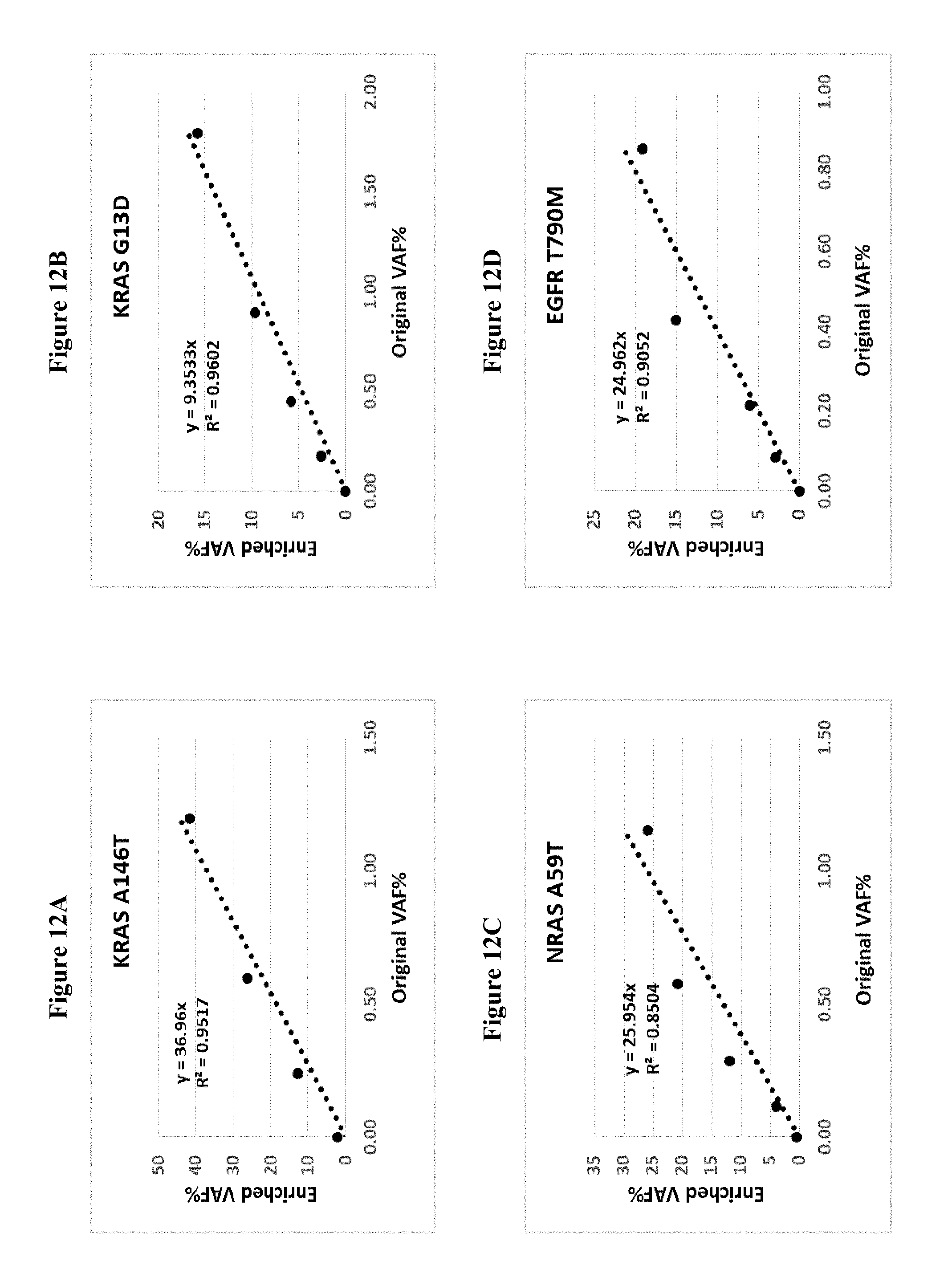
D00015
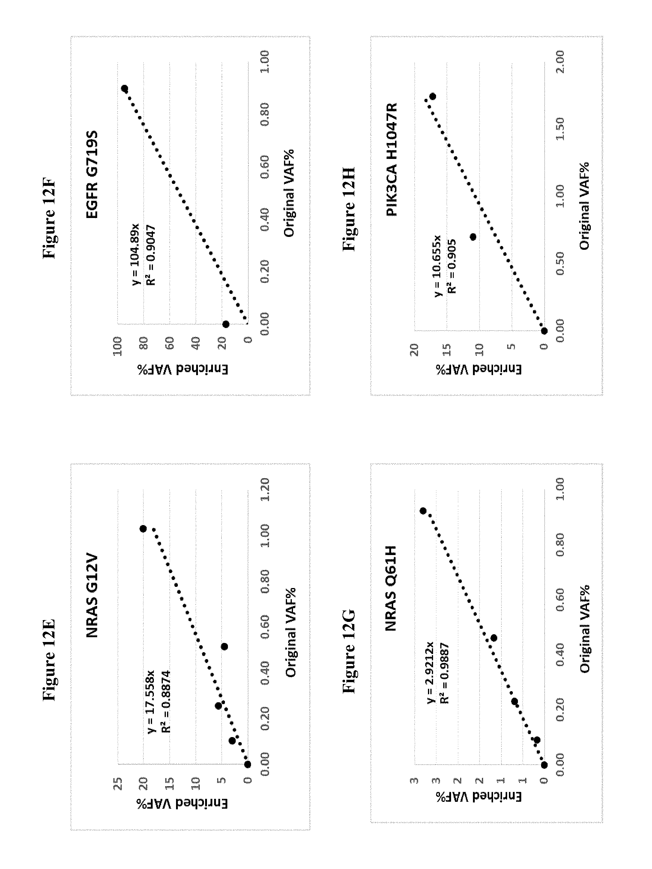
D00016

D00017

D00018

D00019
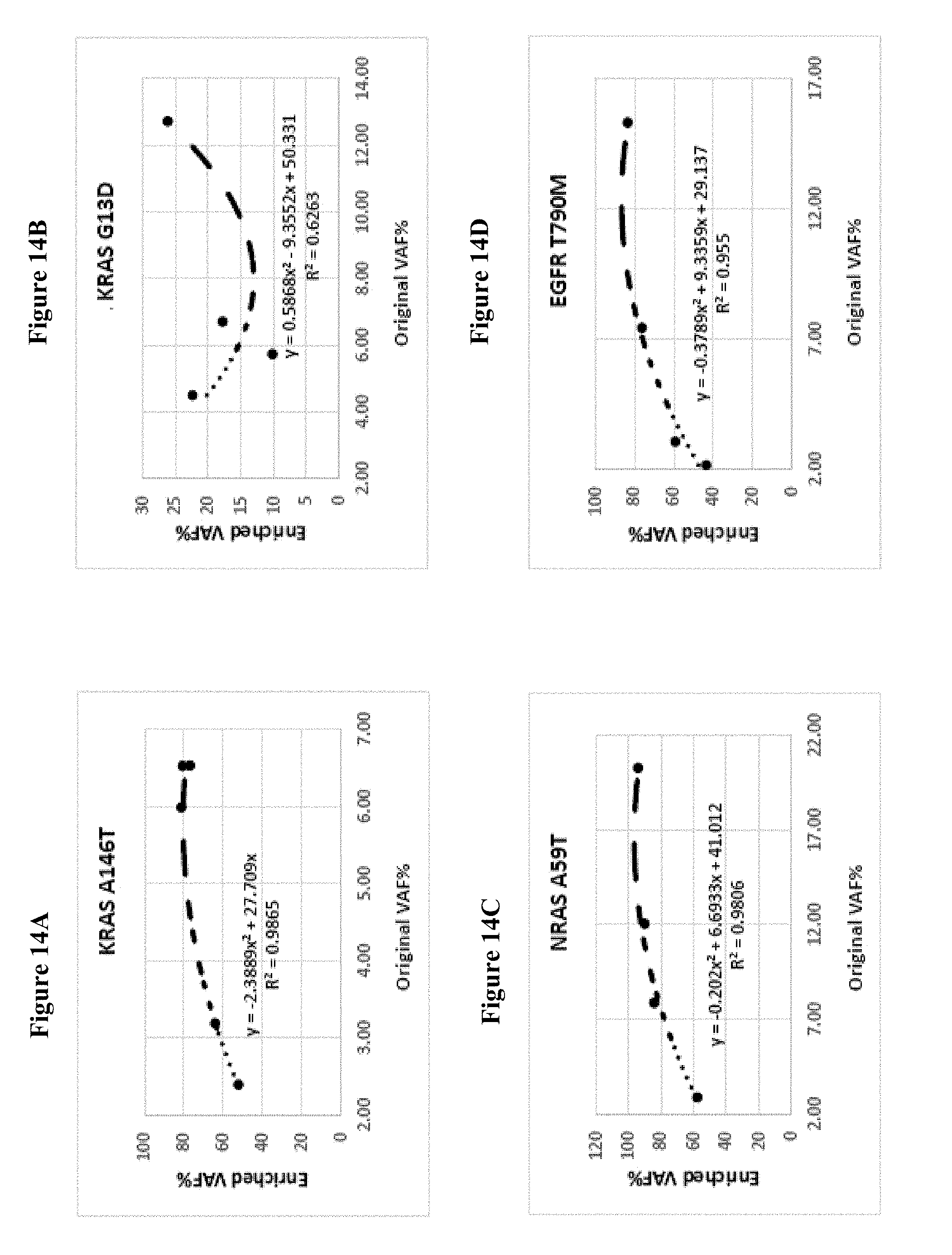
D00020

D00021

D00022

D00023

S00001
XML
uspto.report is an independent third-party trademark research tool that is not affiliated, endorsed, or sponsored by the United States Patent and Trademark Office (USPTO) or any other governmental organization. The information provided by uspto.report is based on publicly available data at the time of writing and is intended for informational purposes only.
While we strive to provide accurate and up-to-date information, we do not guarantee the accuracy, completeness, reliability, or suitability of the information displayed on this site. The use of this site is at your own risk. Any reliance you place on such information is therefore strictly at your own risk.
All official trademark data, including owner information, should be verified by visiting the official USPTO website at www.uspto.gov. This site is not intended to replace professional legal advice and should not be used as a substitute for consulting with a legal professional who is knowledgeable about trademark law.