Novel Chimeric Antigen Receptor And Use Thereof
FAN; Xiaohu ; et al.
U.S. patent application number 16/474735 was filed with the patent office on 2019-10-24 for novel chimeric antigen receptor and use thereof. This patent application is currently assigned to Nanjing Legend Biotech Co., Ltd.. The applicant listed for this patent is Nanjing Legend Biotech Co., Ltd.. Invention is credited to Xiaohu FAN, Jiaying HAO, Shaobo LIU, Pingyan WANG, Lei YANG, Xiujun ZHENG, Qiuchuan ZHUANG.
| Application Number | 20190321404 16/474735 |
| Document ID | / |
| Family ID | 62707011 |
| Filed Date | 2019-10-24 |


| United States Patent Application | 20190321404 |
| Kind Code | A1 |
| FAN; Xiaohu ; et al. | October 24, 2019 |
NOVEL CHIMERIC ANTIGEN RECEPTOR AND USE THEREOF
Abstract
Disclosed herein is a novel chimeric antigen receptor and use thereof. The novel chimeric antigen receptor consists of a signal peptide, an antigen binding domain, a transmembrane region, and an intracellular signal domain, and comprises a 4-1BB signal peptide and/or a 4-1BB molecular transmembrane region. Nucleic acid sequences of various chimeric antigen receptors are separated and purified and provided is a chimeric antigen receptor and a CAR-T cell which are specific for a CD19 malignant tumor antigen. In the malignant tumor killing test of hematological cell lines, the ability of immune cells to target and recognize tumor cells is significantly enhanced, and the killing activity against tumor cells is also enhanced.
| Inventors: | FAN; Xiaohu; (Edmonton, CA) ; ZHUANG; Qiuchuan; (Nanjing, Jiangsu, CN) ; WANG; Pingyan; (Fengyang, Anhui, CN) ; YANG; Lei; (Huainan, Anhui, CN) ; ZHENG; Xiujun; (Nanjing, Jiangsu, CN) ; HAO; Jiaying; (Nanjing, Jiangsu, CN) ; LIU; Shaobo; (Nanjing, Jiangsu, CN) | ||||||||||
| Applicant: |
|
||||||||||
|---|---|---|---|---|---|---|---|---|---|---|---|
| Assignee: | Nanjing Legend Biotech Co.,
Ltd. Nanjing, Jiangsu CN Nanjing Legend Biotech Co., Ltd. Nanjing, Jiangsu CN |
||||||||||
| Family ID: | 62707011 | ||||||||||
| Appl. No.: | 16/474735 | ||||||||||
| Filed: | December 29, 2017 | ||||||||||
| PCT Filed: | December 29, 2017 | ||||||||||
| PCT NO: | PCT/CN2017/119711 | ||||||||||
| 371 Date: | June 28, 2019 |
| Current U.S. Class: | 1/1 |
| Current CPC Class: | C12N 5/0663 20130101; A61K 35/545 20130101; C07K 16/30 20130101; C07K 19/00 20130101; A61K 35/28 20130101; A61K 45/06 20130101; A61P 35/02 20180101; A61P 35/00 20180101; C12N 15/62 20130101; A61K 39/00 20130101; A61K 35/17 20130101; C12N 5/0606 20130101; C12N 5/10 20130101; C07K 14/7051 20130101; C07K 16/32 20130101 |
| International Class: | A61K 35/17 20060101 A61K035/17; C12N 15/62 20060101 C12N015/62; C07K 14/725 20060101 C07K014/725; C12N 5/0735 20060101 C12N005/0735; C07K 16/32 20060101 C07K016/32; C07K 16/30 20060101 C07K016/30; C12N 5/0775 20060101 C12N005/0775 |
Foreign Application Data
| Date | Code | Application Number |
|---|---|---|
| Dec 30, 2016 | CN | 201611263835.1 |
Claims
1. A novel chimeric antigen receptor, comprising an extracellular signal peptide, an antigen binding domain, a transmembrane domain and an intracellular signal domain, wherein said extracellular signal peptide is selected from one of a 4-1BB signal peptide, a CD8.alpha. signal peptide, a GM-CSFR.alpha. signal peptide or a CD4 signal peptide, and the transmembrane domain is a 4-1BB molecular transmembrane region sequence.
2. The chimeric antigen receptor according to claim 1, wherein an amino acid sequence of a 4-1BB molecular transmembrane region as said transmembrane domain is as set forth in SEQ ID NO.1, or a polypeptide having 85%-99% identity with the amino acid sequence of SEQ ID NO.1.
3. The chimeric antigen receptor according to claim 1, wherein said extracellular signal peptide is selected from a 4-1BB signal peptide with an amino acid sequence as set forth in SEQ ID NO.2, or a polypeptide having 85%-99% identity with the amino acid sequence of SEQ ID NO.2.
4. The chimeric antigen receptor according to claim 1, wherein an antibody from which the antigen binding domain is derived is selected from a monoclonal antibody, Fab, scFv or VHH.
5. The chimeric antigen receptor according to claim 1, wherein an antigen bound by said antigen binding domain is associated with a malignant tumor, and includes but not limited to CD19, CD20, CD22, CD30, CD33, CD38, BCMA, CS1, CD138, CD123/IL3R.alpha., c-Met, gp100, MUC1, IGF-I receptor, EPCAM, EGFR/EGFRvIII, HER2, PD1, CTLA4, IGF1R, mesothelin, PSMA, WT1, ROR1, CEA, GD-2, NY-ESO-1, MAGE A3, GPC3, glycolipid F77 or any other tumor antigen or other modified types or a combination thereof.
6. The chimeric antigen receptor according to claim 5, wherein the antigen bound by the antigen binding domain is CD19.
7. The chimeric antigen receptor according to claim 6, wherein said antigen binding domain comprises an amino acid sequence as set forth in SEQ ID NO.9.
8. The chimeric antigen receptor according to claim 1, wherein the intracellular signal domain comprises an intracellular signaling domain and/or a co-stimulatory signal domain and is selected from intracellular domains of the following signal molecules: CD3.zeta., CD3 gamma, CD3 delta, CD3 epsilon, common FcR gamma, FcR beta, CD79a, CD79b, Fc gamma RIIa, DAP10, DAP12, CD27, CD28, 4-1BB, OX40, CD30, CD40, CD2, lymphocyte function-associated antigen-1, LIGHT, NKG2C, B7-H3, PD-1, ICOS, CDS, ICAM-1, GITR, BAFFR, HVEM, SLAMF7, CD7, NKp80, CD160, CD19, CD4, CD8alpha, CD8beta, IL2R beta, IL2R gamma, IL7R alpha, ITGA4, VLA1, CD49a, ITGA4, IA4, CD49D, ITGA6, VLA-6, CD49f, ITGAD, CD11d, ITGAE, CD103, ITGAL, CD11a, LFA-1, ITGAM, CD11b, ITGAX, CD11c, ITGB1, CD29, ITGB2, CD18, LFA-1, ITGB7, TNFR2, TRANCE/RANKL, DNAM1, SLAMF4, CD84, CD96, CEACAM1, CRTAM, Ly9, CD160, PSGL1, CD100, CD69, SLAMF6, SLAM, BLAME, SELPLG, LTBR, LAT, GADS, SLP-76, PAG/Cbp, NKp44, NKp30, NKp46, NKG2D, a ligand specifically binding to CD83, or any combination thereof.
9. The chimeric antigen receptor according to claim 8, wherein the intracellular signal domain comprises a CD3.zeta., 4-1BB and/or CD28 signal domain.
10. The chimeric antigen receptor according to claim 9, wherein the intracellular signal domain comprises a 4-1BB intracellular co-stimulatory signal domain with an amino acid sequence as set forth in SEQ ID NO.6.
11. The chimeric antigen receptor according to claim 9, wherein the intracellular signal domain comprises a CD28 intracellular co-stimulatory signal domain with an amino acid sequence as set forth in SEQ ID NO.7.
12. The chimeric antigen receptor according to claim 9, wherein the intracellular signal domain comprises a CD3.zeta. intracellular signalling domain with an amino acid sequence as set forth in SEQ ID NO.8.
13. The chimeric antigen receptor according to claim 1, wherein said chimeric antigen receptor comprises a 4-1BB signal peptide or CD8.alpha. signal peptide, an antigen binding domain, a 4-1BB transmembrane domain, a 4-1BB and/or CD28 intracellular co-stimulatory binding domain, and a CD3 intracellular signalling domain which are sequentially connected.
14. The chimeric antigen receptor according to claim 13, wherein said chimeric antigen receptor further comprises a hinge region between an extracellular antigen binding domain and the transmembrane domain.
15. The chimeric antigen receptor according to claim 13, wherein said chimeric antigen receptor comprises an amino acid sequence as set forth in SEQ ID NO.10 or SEQ ID NO.12, or an amino acid sequence having 85%-99% identity with SEQ ID NO.10 or SEQ ID NO.12.
16. A nucleic acid encoding the chimeric antigen receptor of claim 1.
17. The nucleic acid according to claim 16, wherein the nucleic acid is selected from a nucleotide sequence as set forth in SEQ ID NO.22 or SEQ ID NO.24.
18. A recombinant expression vector, comprising the nucleic acid molecule of claim 16.
19. A cell expressing the chimeric antigen receptor of claim 1.
20. The cell according to claim 19, wherein said cell is an immune cell.
21. The cell according to claim 20, wherein said cell is selected from T lymphocyte, NK cell, or an immune cell differentiated by culturing a hematopoietic stem cell, a pluripotent stem cell or an embryonic stem cell.
22. A method for preparing a T cell modified by the chimeric antigen receptor, wherein the method comprises separating and activating the T cell to be modified, and then transducing the T cell with the recombinant expression vector of claim 18.
23. A pharmaceutical composition, comprising an effective amount of the cell of claim 19 and a pharmaceutically acceptable carrier.
24. Use of the chimeric antigen receptor of claim 1 in the preparation of an anti-tumor drug or in a tumor cell immunotherapy.
25. Use of the immune effector cell of claim 19 in the preparation of an anti-tumor drug or in a tumor cell immunotherapy.
Description
TECHNICAL FIELD
[0001] The present invention belongs to the technical field of biomedicine or biopharmaceutics, and relates to a novel chimeric antigen receptor and use thereof.
BACKGROUND ART
[0002] According to the 2015 Global Cancer Statistics, data showed that in 2012, there were 14.1 million new tumor patients and 8.2 million patients died worldwide. According to the Chinese Cancer Registry, data showed that in 2015, there were 4.3 million new cancer cases in China, and more than 2.81 million cancer patients died, accounting for 28.82% of the total number of deaths in the year, that is, an average of more than 7,500 people died of cancer every day, which was ranked number one in the world.
[0003] Traditional tumor treatment means mainly comprise surgery, radiotherapy, chemotherapy, and stem cell transplant arising in recent years. These treatment means often cure the symptoms, not the underlying problems. With the development of science, immunotherapy of tumor has achieved a great breakthrough in recent years, which mainly comprises immunological checkpoint inhibitors (such as anti-PD1 monoclonal antibody), and chimeric antigen receptor modified T cell therapy (CAR-T) with the best clinical effect. In recent years, CAR-T technology has made encouraging progresses in clinical trials of hematologic disease treatment, and was ranked as number one of top ten scientific breakthroughs in 2013 by Science magazine. Since the CAR1 June group in University of Pennsylvania firstly reported in 2013 that the child (Emily Whitehead) suffering from the acute B lymphoblastic leukemia who was treated with the targeted CD19 CAR-T was completely relieved, the CAR-T cell immunotherapy had rapidly developed in just a few years. Currently, CAR-T cell treatment is the most effective cell immunotherapy with the highest targeting ability. After the evolution of technology, CAR-T has become more sensitive and more immune persistent, and has a miraculous effect on hematologic tumors such as lymphoma. At present, a number of cell treatment methods have been awarded the "breakthrough therapy" qualification by the FDA. Among them, the results of a number of clinical trials have shown that the clinical complete remission rate of CD19 CAR-T treatment of B-cell lymphoma has exceeded 90%.
[0004] CAR-T cells are a class of T cells that can be genetically engineered to express CAR recognizing particular antigens on the T cell surface and transmit signals. Generally, CAR-T cells recognize specific molecules on the tumor cell surface via a chimeric antigen receptor (CAR) in an antigen-antibody or ligand-receptor recognition mode, and then are activated and proliferated and exert cell killing function through the intracellular signal transduction. The chimeric antigen receptor modified T cells can specifically recognize tumor-associated antigens, making the targeting ability, killing activity and persistence of effector T cells higher than those of conventionally used immune cells, and can overcome local immunosuppression microenvironment of tumors and break the host's immune tolerance state.
[0005] The modified T cells express such a class of chimeric antigen receptor molecules composed of: an extracellular segment generally comprising a CD8.alpha. or GM-CSFR.alpha. signal peptide, an antigen recognition region, or an antigen binding domain comprising a single chain variable region consisting of heavy chain and light chain variable regions of an antibody; an intracellular segment which is an intracellular chimera of various signalling molecules including CD28, 4-1BB, OX-40, CD3zeta, etc.; and a transmembrane region which is derived from other molecules, such as PD1, CD8, CD4, CD28, CD3zeta (CD3.zeta.), etc. The non-antigen recognition region of the extracellular segment of CAR comprises a signal peptide and a hinge region (a linking region between the scFv and the transmembrane region, also referred to as a spacer domain), and has an important influence on the function of the CAR. Kober L et al. in Germany reported that the expression level of a bi-specific single chain antibody (bi-specific scFv antibody) can be significantly increased by optimizing the signal peptide sequence pair from different sources, which is closely related to the ability of the signal peptide to guide the protein into the secretory pathway (Biotechnol Bioeng. 2013 April; 110(4): 1164-73). Michael Hudecek et al. in the Fred Hutchinson Cancer Research Center in the United States reported that although the CAR using an inappropriate spacer domain has a good tumor cell killing effect in in vitro experiments, the CAR-T cells undergo activation-induced T cell death in vivo due to the binding of the spacer domain to the Fc receptor, resulting in the inability of such CAR constructs to persist in vivo and loss of anti-tumor activity (Cancer Immunol Res; 3(2); 125-35). In the international application WO 2016/014789 A2, without changing the extracellular antigen recognition region and the intracellular signal region, the performance of the CAR vector can be greatly realized by only changing the sequence of the transmembrane region, and the non-specific killing effect of the CAR-T on the non-target cells is reduced. Currently, in the field of CAR-T, compared with the CD28 intracellular signal domain, the CAR structure containing a 4-1BB intracellular signal domain is considered to have better tumor cell killing activity and persistence in vivo.
[0006] In the field of B cell leukemia, CD19 is expressed on the surfaces of almostly all the B cell tumor cells, but is hardly expressed in other parenchymal cells and hematopoietic stem cells. CD19 is a relatively specific B-line tumor antigen target. The CD19 CAR structure that has achieved a significant clinical progress at present is CD8.alpha. signal peptide-anti-CD19 single chain antibody--CD8.alpha. hinge region & transmembrane region--4-1BB intracellular region--CD3.zeta. intracellular region (CD8.alpha. signal peptide-antiCD19scFv-CD8.alpha. Hinge&TM-4-1BBcyto-CD3.zeta.).
[0007] Human 4-1BB molecule (NCBI database protein number NP_001552.2), also known as CD137, or tumor necrosis factor receptor superfamily member 9 (TNFRSF9), has amino acids 1-23 as a signal peptide, amino acids 24-186 as an extracellular region, amino acids 187-213 as a transmembrane region, and amino acids 214-255 as an intracellular region in its primary structure. At present, there is no report that designs a CAR using the basic structure of 4-1BB.
[0008] The present invention provides a novel CAR structure comprising a 4-1BB signal peptide and/or a 4-1BB transmembrane region structure, for example, a 4-1BB signal peptide--tumor antigen recognition binding region--4-1BB transmembrane region--4-1BB intracellular region-CD3zeta intracellular region (4-1BB signal peptide-VH-Linker-VL/VHH-4-1BB TM-4-1BBcyto-CD3.zeta.), which maximizes the use of the amino acid sequence of 4-1BB. The use of the 4-1BB signal peptide, and the transmembrane region and intracellular region of 4-1BB facilitates the optimization of the extension of the extracellular tumor antigen recognition binding region on CAR-T cells, which not only achieves good tumor killing effects in vitro and in vivo, but also achieves complete remission in the clinical treatment of patients with acute lymphoma, with a milder cytokine release response.
SUMMARY OF THE INVENTION
[0009] The object of the present invention is to provide a novel chimeric antigen receptor and use thereof in view of the above-mentioned deficiencies of the prior art.
[0010] Another object of the present invention is to provide a nucleic acid encoding the chimeric antigen receptor.
[0011] Still another object of the present invention is to provide a cell containing the chimeric antigen receptor and use thereof.
[0012] Provided is a novel chimeric antigen receptor, comprising an extracellular signal peptide, an antigen binding domain, a transmembrane domain and an intracellular signal domain, wherein said extracellular signal peptide is selected from one of a 4-1BB signal peptide, a CD8.alpha. signal peptide, a GM-CSFR.alpha. signal peptide or a CD4 signal peptide, and the transmembrane domain is selected from a 4-1BB molecular transmembrane region sequence.
[0013] The amino acid sequence of a 4-1BB molecular transmembrane region as said transmembrane domain is preferably as set forth in SEQ ID NO.1, or a polypeptide having 85%-99% identity with the amino acid sequence of SEQ ID NO.1.
[0014] Said extracellular signal peptide is preferably a 4-1BB signal peptide with an amino acid sequence as set forth in SEQ ID NO.2, or a polypeptide having 85%-99% identity with the amino acid sequence of SEQ ID NO.2.
[0015] An antigen bound by said antigen binding domain is associated with a malignant tumor, and includes but not limited to CD19, CD20, CD22, CD30, CD33, CD38, BCMA, CS1, CD138, CD123/IL3R.alpha., c-Met, gp100, MUC1, IGF-I receptor, EPCAM, EGFR/EGFRvIII, HER2, PD1, CTLA4, IGF1R, mesothelin, PSMA, WT1, ROR1, CEA, GD-2, NY-ESO-1, MAGE A3, GPC3, glycolipid F77 or any other tumor antigen or other modified types or a combination thereof.
[0016] The antigen bound by said antigen binding domain is further preferably CD19; the amino acid sequence of the CD19 antigen binding domain is as set forth in SEQ ID NO.9.
[0017] In one embodiment, the antigen binding domain of said chimeric antigen receptor binds to a target by a ligand-receptor interaction and may be selected from IL-3, IL-13 or APRIL.
[0018] Said antigen binding domain consists of antibody fragments, preferably monoclonal antibodies, Fab, scFv, sdAb, VHH or other antibody fragments. The antibody from which the antigen binding domain is derived can be derived from a murine antibody, a camel antibody or a humanized antibody. In one specific example, the antigen binding domain of the chimeric antigen receptor consists of a CD19-specific single chain antibody fragment, containing a heavy chain variable region (VH) and a light chain variable region (VL) of a single chain antibody.
[0019] In one embodiment, the heavy chain variable region (VH) and the light chain variable region (VL) of the single chain antibody of said antigen binding domain are linked by a connecting peptide (linker), comprising a GS linker such as (G.sub.3S).sub.4, (G.sub.4S).sub.3 or GSTSGSGKPGSGEGSTKG, preferably (G.sub.3S).sub.4 connecting peptide.
[0020] Said intracellular signal domain preferably comprises an intracellular signal region sequence of CD3 zeta (CD3.zeta.), CD3 gamma, CD3 delta, CD3 epsilon, common FcR gamma (FCER1G), FcR beta (Fc Epsilon R1b), CD79a, CD79b, Fc gamma RIIa, DAP10 and DAP12 molecules, or a combination thereof. Further preferably, the intracellular signal domain comprises a signalling domain of a CD3.zeta. molecule.
[0021] Said intracellular signal domain preferably also comprises a co-stimulatory signal domain, which may be selected from the following co-stimulatory signal molecules: CD27, CD28, 4-1BB, OX40, CD30, CD40, CD2, lymphocyte function-associated antigen-1 (LFA-1), LIGHT, NKG2C, B7-H3, PD-1, ICOS, CDS, ICAM-1, GITR, BAFFR, HVEM (LIGHTR), SLAMF7, CD7, NKp80 (KLRF1), CD160, CD19, CD4, CD8 alpha, CD8 beta, IL2R beta, IL2R gamma, IL7R alpha, ITGA4, VLA1, CD49a, ITGA4, IA4, CD49D, ITGA6, VLA-6, CD49f, ITGAD, CD11d, ITGAE, CD103, ITGAL, CD11a, LFA-1, ITGAM, CD11b, ITGAX, CD11c, ITGB1, CD29, ITGB2, CD18, LFA-1, ITGB7, TNFR2, TRANCE/RANKL, DNAM1 (CD226), SLAMF4 (CD244, 2B4), CD84, CD96 (Tactile), CEACAM1, CRTAM, Ly9 (CD229), CD160 (BY55), PSGL1, CD100 (SEMA4D), CD69, SLAMF6 (NTB-A, Ly108), SLAM (SLAMFI, CD150, IPO-3), BLAME (SLAMF8), SELPLG (CD162), LTBR, LAT, GADS, SLP-76, PAG/Cbp, NKp44, NKp30, NKp46, NKG2D, a ligand specifically binding to CD83, or any combination thereof. In one specific embodiment, further preferably, the 4-1BB is a co-stimulatory signal domain, the amino acid sequence of which is as set forth in SEQ ID NO.6.
[0022] In one embodiment, the chimeric antigen receptor does not comprise a hinge region between the extracellular antigen binding domain and the transmembrane domain.
[0023] In another embodiment, the chimeric antigen receptor comprises a hinge region between the extracellular antigen binding domain and the transmembrane domain. In a specific embodiment, the hinge region is preferably the CD8.alpha. hinge region as set forth in SEQ ID NO.4.
[0024] In a particular embodiment, the chimeric antigen receptor preferably comprises a 4-1BB signal peptide or CD8.alpha. signal peptide, an antigen binding domain, a 4-1BB transmembrane region, a 4-1BB intracellular co-stimulatory domain, and a CD3.zeta. signalling domain which are sequentially connected.
[0025] The structure of the chimeric antigen receptor of the present invention is as follows:
[0026] 4-1BB signal peptide-VH-Linker-VL-4-1BB TM-4-1BBcyto-CD3.zeta.
[0027] CD8.alpha. signal peptide-VH-Linker-VL-4-1BB TM-4-1BBcyto-CD3.zeta.
[0028] 4-1BB signal peptide-VH-Linker-VL-TM-4-1BBcyto-CD3.zeta.
[0029] 4-1BB signal peptide-V.sub.HH.sub.1-Linker-V.sub.HH.sub.2-4-1BB TM-4-1BBcyto-CD3.zeta.
[0030] CD8.alpha. signal peptide-V.sub.HH.sub.1-Linker-V.sub.HH.sub.2-4-1BB TM-4-1BBcyto-CD3.zeta.
[0031] 4-1BB signal peptide-V.sub.HH.sub.1-Linker-V.sub.HH.sub.2-TM-4-1BBcyto-CD3.zeta.
[0032] wherein the antigen binding domain may consist of VH-Linker-VL or VL-Linker-VH or V.sub.HH.sub.1-Linker-V.sub.HH.sub.2, and V.sub.HH.sub.1 and V.sub.HH.sub.2 can recognize the same antigen or different antigens.
[0033] As a preferable embodiment of the present invention, the chimeric antigen receptor comprises an amino acid sequence as set forth in SEQ ID NO.10 or SEQ ID NO.12, or an amino acid sequence having 85%-99% identity with SEQ ID NO.10 or SEQ ID NO.12.
[0034] A nucleic acid molecule encoding the aforementioned chimeric antigen receptor is provided.
[0035] The nucleic acid molecule preferably comprises a nucleotide sequence of SEQ ID NO.22 or SEQ ID NO.24.
[0036] An expression vector of the above-mentioned nucleic acid molecule is provided.
[0037] The expression vector is preferably a lentivirus expression vector, which comprises a nucleotide sequence encoding SEQ ID NO.22 or SEQ ID NO.24.
[0038] A cell expressing the aforementioned chimeric antigen receptor is provided.
[0039] The cell is preferably an immune cell, further preferably, T lymphocyte, NK cell, an immune cell differentiated by culturing a hematopoietic stem cell, a pluripotent stem cell or an embryonic stem cell.
[0040] A method for preparing a T cell modified by the novel chimeric antigen receptor is provided, the method comprising separating and activating the T cell to be modified, and then transducing the T cell with the aforementioned expression vector.
[0041] The use of said novel chimeric antigen receptor, said expression vector, and said cell in the preparation of a medicament for treating a tumor is provided.
[0042] Said tumor preferably comprises glioblastoma, head and neck cancer, thyroid cancer, kidney cancer, lung cancer, breast cancer or ovarian cancer, and chronic lymphoblastic leukemia (CLL), acute leukemia, acute lymphoblastic leukemia (ALL), B-cell acute lymphoblastic leukemia (ALL), T-cell acute lymphoblastic leukemia (T-ALL), chronic myeloid leukemia (CML), B-cell prolymphocytic leukenia, acute plasmacytoid dendritic cell tumor, Burkitt lymphoma, diffuse large B-cell lymphoma, follicular lymphoma hairy cell leukemia, small cell or large cell follicular lymphoma, malignant lymphoid tissue hyperplasia, MALT lymphoma, mantle cell lymphoma, marginal zone lymphoma, multiple myeloma, myeloproliferative disorder and myelodysplastic syndrome, non-Hodgkin's lymphoma, Hodgkin's lymphoma, plasmablastic lymphoma, plasmacytoid dendritic cell tumor lymphoma and other hematologic tumors.
[0043] In a specific embodiment, the invention relates to the use in the preparation of a medicament for treating a hematologic tumor.
[0044] The present invention relates to the use of the novel chimeric antigen receptor in the preparation of an anti-tumor drug and in cell immunotherapy.
[0045] The present invention relates to the use of the immune effector cells in the preparation of an anti-tumor drug and in cell immunotherapy.
Beneficial Effects:
[0046] The present invention provides a novel CAR structure comprising a 4-1BB signal peptide and/or a 4-1BB transmembrane region structure, for example, a 4-1BB signal peptide--tumor antigen recognition binding region--4-1BB transmembrane region--4-1BB intracellular region--CD3zeta intracellular region (4-1BB signal peptide-VH-Linker-VL/VHH-4-1BB TM-4-1BBcyto-CD3.zeta.), which maximizes the use of the amino acid sequence of 4-1BB molecular structure. The use of the 4-1BB signal peptide, and the transmembrane region or intracellular region structures of 4-1BB facilitates the extension of the extracellular tumor antigen recognition binding region on CAR-T cells, which not only achieves good tumor killing effects in vitro and in vivo, but also achieves complete remission in the clinical treatment of patients with acute lymphoma, with a milder cytokine release response. Taking the CD19 antigen target as an example, the novel chimeric antigen receptor shows a relatively higher anti-tumor ability compared with the prior art, and immune cells modified by the chimeric antigen receptor have a higher ability to target and recognize tumor antigens, thereby enhancing the killing activity against tumor cells.
BRIEF DESCRIPTION OF THE DRAWINGS
[0047] FIG. 1 is a vector structure graph for a chimeric antigen receptor, showing the structure of the vectors for the CAR1 (panel A) and CAR3 (panel B) chimeric antigen receptors in the present invention.
[0048] FIG. 2 shows the detection of CD19 antigen expressed by a stable cell line constructed in the present invention. A. Detection of the expression of CD19 on the constructed Raji.Luc cell line and the isotype control Raji cell line respectively by using a CD19-specific antibody. B. Detection of the expression level of CD19 antigen in the constructed K562.CD19.Luc cell line and the control cell line respectively by using a CD19-specific antibody.
[0049] FIG. 3 is a graph showing the effect of CAR-T killing CD19-positive cells in vitro. A. Under the condition of E:T ratio 1, the relative proportion of the remaining Raji.Luc cells was 20.2%.+-.1.23% after the co-culture of CAR1-T with Raji.Luc, while the relative proportion of the remaining Raji.Luc cells was 98.84%.+-.1.60% in the CAR2-T group and 100%.+-.3.54% in the UnT group; under the condition of E:T ratio 2, the relative proportion of the remaining Raji.Luc cells was 50.09%.+-.2.17% after the co-culture of CAR1-T with Raji.Luc, while the relative proportion of the remaining Raji.Luc cells was 107.07%.+-.3.04% in the CAR2-T group and 100%.+-.3.50% in the UnT group. B. The relative proportion of the remaining Raji.Luc cells was 26.83%.+-.1.97% after the co-culture of CAR1-T with Raji.Luc, while the relative proportion of the remaining target cells was 36.86%.+-.3.46% in the CAR3-T group and 100%.+-.1.78% in the UnT group.
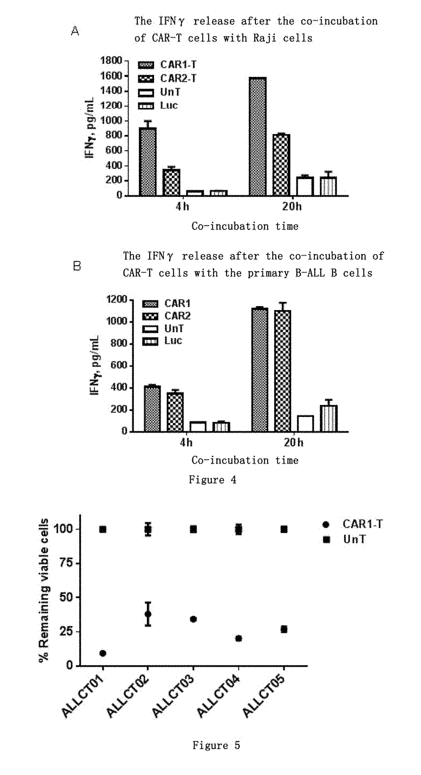
[0050] FIG. 4 shows the detection OF IFN.gamma. release level in CAR-T cells. A. The amount of IFN.gamma. released after co-incubation with Raji.Luc cells for 4 h and 20 h in the CAR1-T, CAR2-T and UnT, Luc groups. B. The amount of IFN.gamma. released after co-incubation with the B lymphocytes from patients with B cell acute lymphoblastic leukemia for 4 h and 20 h in the CAR1-T, CAR2-T and UnT, Luc groups.
[0051] FIG. 5 shows the detection of tumor killing effect of the autologous T cells transduced with CAR1. CAR1 was transduced into T cells from 5 different B-ALL patients, respectively, and the transduced cells were named ALLCT01, ALLCT02, ALLCT03, ALLCT04 and ALLCT05, respectively. After the co-incubation of the CAR1 transduced cells with Raji.Luc target cells, the relative number of the remaining Raji.Luc cells was 9.33%-37.82%.
[0052] FIG. 6 is a graph showing the release level of human interleukin 6. After the ALLCT01 patients were re-infused with CD19 CAR-T cells, the body temperature began to rise at day 6 after the last re-infusion, and the cytokine test results showed that the secretion level of interleukin-6 (IL-6) was significantly increased.
DETAILED DESCRIPTION OF EMBODIMENTS
[0053] The present invention provides a novel chimeric antigen receptor, immune effector cells and their use in inhibiting tumor activity. The present invention is further illustrated in conjunction with the following specific examples.
[0054] The term "transmembrane domain" (abbreviated as TM) as used herein may be used interchangeably with "transmembrane region" and refers to a protein structure region which has thermodynamical stability and is anchored within the cell membrane. The transmembrane region can be obtained from a native protein selected from the 4-1BB molecule.
[0055] The novel chimeric antigen receptor of the present invention comprises an extracellular signal peptide structure, such as a 4-1BB signal peptide, a CD8.alpha. signal peptide, a GM-CSFR.alpha. signal peptide or a CD4 signal peptide, preferably a 4-1BB signal peptide.
[0056] The term "intracellular signal domain" as used herein refers to a protein structure region that is capable of transducing the cell effector function signal and directing the cell to perform a particular function. The intracellular signal domain can comprise a signalling domain and/or a co-stimulatory signal domain.
[0057] As used herein, the term "identity" of amino acid sequences can be used interchangeably with "similarity" to refer to the degree of similarity between amino acid sequences determined by a sequence alignment software such as BLAST. Methods and software for alignment of amino acid sequences are well known to those skilled in the art. A modified amino acid sequence can be obtained by making substitutions, deletions, and/or additions of one or more (e.g., 1-15, such as, 2, 3, 5, 8, 10 or 12) amino acid residues on the basis of a known amino acid sequence. For example, a variant sequence having at least 85% (e.g., 85%-99% or 90%-99% or 95%-99%) sequence identity with SEQ ID NO.1 and having a substantially same transmembrane function can be obtained by modifying the 4-1BB transmembrane domain as set forth in SEQ ID NO.1 of the present invention through conventional protein engineering means (e.g., conservative substitution of amino acids, etc.).
[0058] The term "antibody fragment" as used herein is a portion of an antibody having functions, preferably a variable region for antigen binding and/or a variable region of an intact antibody. Antibody fragments comprise Fab, Fab', F(ab').sub.2, Fv fragments, single chain antibody scFv, single domain antibody sdAb/V.sub.HH and multispecific antibodies.
[0059] The term "scFv" or "scFv antibody" as used herein refers to a single chain antibody fragment (scFv) which is formed by connecting a heavy chain variable region VH and a light chain variable region VL of an antibody with a short peptide of 15-20 amino acids. As used herein, the term "Linker", also referred to as a connecting peptide or adapter, refers to a flexible amino acid sequence used to connect two domains. Selection and preparation of a connecting peptide is readily available to those skilled in the art.
[0060] The present invention designs a chimeric antigen receptor using the human 4-1BB molecular domain through optimization and is further illustrated in conjunction with the following specific examples.
EXAMPLE 1
Preparation of a Chimeric Antigen Receptor
[0061] (I) Preparation of the Gene Fragment of a Chimeric Antigen Receptor
[0062] The present invention provides a novel chimeric antigen receptor comprising a CD19 antigen binding domain. The antibody from which the antigen binding domain is derived, provided by the present invention, consists of a heavy chain variable region (VH), a light chain variable region (VL) of a single chain antibody and a connecting peptide (G3S).sub.4 Linker. The heavy chain variable region and the light chain variable region sequence of the single chain antibody were derived from GenBank:Y14283.1 and GenBank:Y14284.1, respectively. Their codons and sequences were optimized to ensure that they were more suitable for expression in human cells.
[0063] The novel chimeric antigen receptor provided by the present invention can be designed according to the order of the following coding genes and the fusion gene fragments for the chimeric antigen receptor can be changed: the 4-1BB signal peptide, V.sub.H-(G3S).sub.4-V.sub.L, the 4-1BB transmembrane region, the 4-1BB or CD28 intracellular co-stimulatory signal domain or the CD3.zeta. intracellular signalling domain. The gene synthesis technology service was provided by Nanjing Genscript Biotechnology Co., Ltd. The following coding gene sequences were selected by gene synthesis technology to perform the fusion gene synthesis:
[0064] the nucleotide sequence encoding the 4-1BB transmembrane region as set forth in SEQ ID NO.13;
[0065] the nucleotide sequence encoding the 4-1BB signal peptide as set forth in SEQ ID NO.14;
[0066] the nucleotide sequence encoding the CD8.alpha. signal peptide as set forth in SEQ ID NO.15;
[0067] the nucleotide sequence encoding the CD8.alpha. hinge region as set forth in SEQ ID NO.16;
[0068] the nucleotide sequence encoding the CD8.alpha. transmembrane region as set forth in SEQ ID NO.17;
[0069] the nucleotide sequence encoding the 4-1BB intracellular co-stimulatory signal domain as set forth in SEQ ID NO.18;
[0070] the nucleotide sequence encoding the CD28 intracellular co-stimulatory signal domain as set forth in SEQ ID NO.19;
[0071] the nucleotide sequence encoding the CD3.zeta. intracellular signalling domain as set forth in SEQ ID NO.20; and
[0072] the nucleotide sequence encoding the CD19 antigen binding domain as set forth in SEQ ID NO.21.
[0073] The extracellular domain and the intracellular signal domain used in the present invention have various combinations, and comprise the structures or combinations selected from the following items:
[0074] the 4-1BB transmembrane region having an amino acid sequence as set forth in SEQ ID NO.1;
[0075] the 4-1BB signal peptide having an amino acid sequence as set forth in SEQ ID NO.2;
[0076] the CD8.alpha. signal peptide having an amino acid sequence as set forth in SEQ ID NO.3;
[0077] the CD8.alpha. hinge region having an amino acid sequence as set forth in SEQ ID NO.4;
[0078] the CD8.alpha. transmembrane region having an amino acid sequence as set forth in SEQ ID NO.5;
[0079] the 4-1BB intracellular co-stimulatory signal domain having an amino acid sequence as set forth in SEQ ID NO.6;
[0080] CD28 intracellular co-stimulatory signal domain having an amino acid sequence as set forth in SEQ ID NO.7;
[0081] the CD3.zeta. intracellular signalling domain having an amino acid sequence as set forth in SEQ ID NO.8; and
[0082] the CD19 antigen binding domain having an amino acid sequence as set forth in SEQ ID NO.9.
[0083] The composition of the chimeric antigen receptor in the examples of the present invention include, but not limited to, the following structures:
[0084] CAR1: 4-1BB signal peptide-V.sub.H-(G3S).sub.4-VL-4-1BB TM-4-1BBcyto-CD3.zeta. the amino acid sequence of which is as set forth in SEQ ID NO.10 and the nucleotide coding sequence of which is as set forth in SEQ ID NO.22; the gene sequence as set forth in SEQ ID NO.22 was synthesized to construct a recombinant expression vector;
[0085] CAR2: CD8.alpha. signal peptide-V.sub.H-(G3S).sub.4-V.sub.L-CD8.alpha. Hinge&TM-4-1BBcyto-CD3.zeta., the amino acid sequence of which is as set forth in SEQ ID NO.11 and the nucleotide coding sequence of which is as set forth in SEQ ID NO.23; the gene sequence as set forth in SEQ ID NO.23 was synthesized to construct a recombinant expression vector;
[0086] CAR3: CD8.alpha. signal peptide-V.sub.H-(G3S).sub.4-VL-4-1BB TM-4-1BBcyto-CD3.zeta., the amino acid sequence of which is as set forth in SEQ ID NO.12, and the nucleotide coding sequence of which is as set forth in SEQ ID NO.24; the gene sequence as set forth in SEQ ID NO.24 was synthesized to construct a recombinant expression vector;
[0087] wherein the antigen binding domain can be replaced with different tumor antigen targets within the technical scope of the art. The antigen-specific heavy chain variable region and light chain variable region of the antibody can be linked in the direction of V.sub.H-V.sub.L or V.sub.L-V.sub.H, and the connecting peptide Linker can be selected from (G3S).sub.4Linker, (G4S).sub.3Linker or other GS Linker or other protein linkers, such as Whitlow Linker: GSTSGSGKPGSGEGSTKG.
[0088] (II) Construction of a Lentivirus Expression Vector for a Chimeric Antigen Receptor
[0089] In one specific embodiment, the present invention uses a self-inactivated lentivirus expression vector to express the target CAR gene sequence. First, the expression plasmid and pCMV-.DELTA.R-8.74 and pMD2.G helper plasmid were extracted and mixed in a certain ratio to co-transfect 293T cells. After transfection for 48 h and 96 h, the cell culture supernatant containing the virus was collected and centrifuged at 3000 rpm at 4.degree. C. for 5 min. After the supernatant was filtered through a 0.45 .mu.m filter, it was mixed with PEG6000/NaCl in a volume ratio of 4:1, and allowed to stand at 4.degree. C. for 2-3 h, and then centrifuged at a high speed for 30 min. The supernatant was discarded, and the pellet was resuspended and dissolved in pre-cooled PBS to obtain a virus concentrate which was stored at -80.degree. C. until use.
Example 2
Construction of the K562 Cell Line Expressing CD19 Antigen (K562.CD19.Luc)
[0090] K562 cells hardly express CD19. For the gene synthesis of the nucleotide sequence encoding human CD19 molecule (the protein sequence thereof is NCBI number NP_001171569.1) in the present invention, the gene synthesis technology service was provided by Nanjing Genscript Biotechnology Co., Ltd. Under the action of T4 ligase, the synthesized CD19 nucleotide sequence was ligated at 20.degree. C. overnight to the pLVX-Puro (Clontech, Cat #632164) lentivirus vector which was digested at the BamHI and XbaI restriction sites in advance. The ligation product was transformed into DH5.alpha. competent cells and plated onto bacterial plates, and multiple clone spots were picked for the extraction of plasmids (Qiagen Endofree Mega kit). After enzyme digestion identification, sequencing and alignment, the successfully constructed vector was named as pLVX-CD19-Puro.
[0091] The extracted pLVX-CD19-Puro expression plasmid and pCMV-AR-8.74 (J. Virol.-1998-Dull-A third-generation lentivirus vector with a conditional packaging system) and pMD2.G helper plasmid were mixed in a certain ratio to co-transfect 293FT cells. After transfection for 96 h, the cell culture supernatant containing the virus was collected and centrifuged at 3000 rpm at 4.degree. C. for 5 min After the supernatant was filtered through a 0.45 .mu.m filter, it was centrifuged at a high speed of 25,000 rpm at 4.degree. C. for 120 min The virus concentrate was obtained after discarding the supernatant and resuspending the pellets to dissolve, and stored at -80.degree. C. until use. The lentivirus vector carrying the firefly luciferase/Luciferase reporter gene was also prepared by the same method.
[0092] The K562 cell line was purchased from ATCC (Cat No. CCL-243), and was subjected to conventional culture using 90% IMDM (Life technology, Cat No. A10491-01)+10% FBS (Life technology, Cat #10099-141)+1% penicillin/streptomycin (Life technology, Cat #15140-122) (hereinafter referred to as IMDM10 culture solution in short). The constructed lentivirus vector carrying the CD19 gene and the lentivirus vector carrying the firefly luciferase/Luciferase reporter gene were added to the cultured K562 cell supernatant to co-transduce the K562 cell line. After transduction for 24 h, puromycin was added at a final concentration of 10 .mu.g/ml. The culture solution was exchanged every three days and puromycin at the same concentration was added. The screening of monoclones were further performed to obtain monoclonal cells, which were named as K562.CD19.Luc. Another stable cell line K562.CD123.Luc (CD123 gene source: GenBank:NM_002183) constructed simultaneously according to the above-mentioned method in the present invention was used as a CD19 negative control cell line.
[0093] The CD19-positive human lymphoma cell line Raji was purchased from American ATCC (ATCC #CCL-86.TM.) and was cultured with RPMI1640+10% FBS+1% penicillin/streptomycin (hereinafter referred to as R10 culture solution in short). The firefly luciferase/Luciferase reporter gene was transduced into Raji cells using a method similar to the above-mentioned method. A cell line stably expressing the luciferase reporter gene was obtained by the screening under the puromycin stress, which was named as Raji.Luc.
[0094] The monoclonal cells obtained above were identified for CD19 expression by flow cytometry. As shown in FIG. 2A, the expression of CD19 on the constructed Raji.Luc cell line and the isotype control Raji cell line (purchased from Miltenyi) was detected respectively by using a CD19-specific antibody. The flow cytometry results showed that the constructed Raji.Luc cell line had a higher CD19 expression level. FIG. 2B shows the expression level of CD19 antigen in the constructed K562.CD19.Luc cell line and the control cell line respectively detected by using a CD19-specific antibody. The flow cytometry results showed that the constructed K562.CD19.Luc cell line also had a very high CD19 expression level.
EXAMPLE 3
Preparation of Immune Effector Cells Modified by the Chimeric Antigen Receptor and Detection of Killing Activity In Vitro
[0095] (I) Preparation of T Lymphocytes
[0096] 50 mL fresh blood from healthy human was collected, and peripheral blood mononuclear cells (PBMC) were separated by a lymphocyte separation solution and a density gradient centrifugation method. The cells were magnetically labelled using a Pan T Cell Isolation Kit (purchased from Miltenyi Biotech), and T lymphocytes were separated and purified. The purified T cells were then subjected to T lymphocyte activation and proliferation using CD3/CD28 magnetic beads.
[0097] (II) Transduction of T Lymphocytes with Lentivirus
[0098] The activated T lymphocytes were collected and resuspended in RPMI1640 culture medium. 1.times.10.sup.6 activated T lymphocytes were infected with lentivirus, the cell suspension was added to a 6-well plate and incubated overnight at 37.degree. C. in a 5% CO.sub.2 incubator. On the next day, centrifugation was performed again and a fresh culture medium was replaced. A fresh culture medium was added every 3 days to continue the expansion culture.
[0099] (III) Detection of CAR-T Expression
[0100] The CAR-T cells prepared according to the above-mentioned protocol were collected by centrifugation, washed respectively three times with DPBS, and then genomic DNA was prepared using a human genome extraction kit (Gentra Puregene Cell Kit (purchased from Qiagen)). The prepared DNA was detected for the absorbance of OD.sub.260 nm and OD.sub.280 nm and then the concentration was calculated. The appropriate DNA concentration was adjusted. The Q-PCR reaction system was configured according to the kit SYBR Green Real time PCR Master mix plus (purchased from Toyobo). Then the gene copy number was detected on a fluorescence quantitative PCR machine (ABI #7300). The Q-PCR detection used accurately quantified plasmids containing the target fragments as positive control and for a standard curve. The CT value of each copy number concentration obtained by the Q-PCR and the corresponding copy number were plotted as a straight line and fit into a standard curve. The relative copy number for other test samples were calculated based on the fitted equation of the standard curve.
[0101] For the detection of the chimeric antigen receptor expressed in CAR-T cells in the present invention, the untransduced T lymphocyte (UnT) was used as a blank control, and the CD19 CAR-T (CAR2-T) comprising a CD8.alpha. signal peptide, a hinge region and a transmembrane region structures in the patent U.S. Pat. No. 8,399,645 B2 was used as a control.
[0102] The results of the detection of CAR integration copy number are shown in Table 1. The results showed that the integration of CAR1 gene was detected in the cell genome in the CAR1-T group, with the copy number of 7.46.times.10.sup.5 copies/ng genomic DNA; the copy number of the transduced CAR2 gene was 12.90.times.10.sup.5 copies/ng genomic DNA in the CAR2-T group; the measurements were extremely low (about 30 copies/ng genomic DNA) in the blank control UnT group and the H.sub.2O, which belong to the background of the test.
TABLE-US-00001 TABLE 1 The copy number of CAR gene integrated into the donor T cell genome detected by the Q-PCR method Integrated copy Test Ct value Ct value Average Ct number/ng genomic samples repeat 1 repeat 2 value DNA CAR1-T 21.14 20.88 21.01 74643.34 CAR2-T 20.06 20.18 20.12 129031.70 UnT 34.10 33.41 33.76 30.31 H.sub.2O 33.70 33.06 33.38 38.16
EXAMPLE 4
Detection of Killing Activity In Vitro
[0103] The CAR-T cells prepared above were collected, adjusted to a suitable density with R10 culture solution, and inoculated in a 384-well plate. CAR-T cells was co-cultured with the target Raji-Luc cells at an effector-to-target ratio (E:T) of 50:1 or 20:1 at 37.degree. C. for 20 h. Then an equal amount of luciferase activity detection reagent One-Glo.TM. Luciferase Assay (purchased from Promega) was added. After the completion of the co-culture, the relative luciferase activity (RLU, relative light unit) remained in the reaction wells corresponds to the relative amount of viable cells in the wells. If the revealed luciferase RLU value is high, it means that there are many target cells which are not killed remained in the reaction wells, indicating that the cell killing activity in the wells is weak; conversely, if the revealed luciferase RLU value is low, it means that there are few target cells which are not killed in the reaction wells, indicating the cell killing activity in the well is strong. For the killing test of the CAR-T cells of the present invention against tumor cells, the untransduced T lymphocyte (UnT) was used as a blank control and CAR2 was used as a control.
[0104] As shown in FIG. 3A, under the conditions of equivalent copy number of integration and different effector-to-target ratios (E:T ratio 1=50:1, E:T ratio 2=20:1), CAR1-T can significantly kill Raji. Luc cells (the relative luciferase activity RLU % was reduced), which was significantly better than the killing effect of CAR2-T in the control group. Under the condition of E:T ratio 1, the relative proportion of the remaining Raji.Luc cells was 20.2%.+-.1.23% after the co-culture of CAR1-T with Raji.Luc, while the relative proportion of the remaining Raji.Luc cells was 98.84%.+-.1.60% in the CAR2-T group and 100%.+-.3.54% in the UnT group; under the condition of E:T ratio 2, the relative proportion of the remaining Raji.Luc cells was 50.09%.+-.2.17% after the co-culture of CAR1-T with Raji.Luc, while the relative proportion of the remaining Raji.Luc cells was 107.07%.+-.3.04% in the CAR2-T group and 100%.+-.3.50% in the UnT group. The above results can reveal that CAR1 -modified immune cells comprising the 4-1BB signal peptide and the transmembrane region domain and lacking the hinge region have a better in vitro tumor killing effect than the CAR2-modified immune cells comprising the CD8.alpha. signal peptide and the transmembrane region domain
[0105] In another set of experiments, as shown in FIG. 3B, the relative proportion of the remaining Raji.Luc cells was 26.83%.+-.1.97% after the co-culture of CAR1-T with Raji.Luc, while the relative proportion of the remaining Raji.Luc cells was 36.86%.+-.3.46% in the CAR3-T group and 100%.+-.1.78% in the UnT group. CAR1 differs from CAR3 in that the extracellular signal peptide is different, and CAR3 comprises the CD8.alpha. signal peptide. From the results of FIG. 3B, it can be inferred that CAR1 -modified immune cells comprising the 4-1BB signal peptide and the transmembrane region have a better in vitro tumor killing effect than the CAR3-modified immune cells comprising the CD8.alpha. signal peptide.
[0106] Combining the data from FIGS. 3A and 3B, it can be inferred that the immune cells modified by the chimeric antigen receptors comprising different signal peptides or transmembrane region structures have different in vitro killing ability, and the transmembrane region structure may have more significant impacts on the performance of the chimeric antigen receptor.
[0107] Further, in the target-specific killing experiments, the present invention used the constructed K562.CD19.Luc expressing CD19 and the K562.CD123.Luc stable cell line not expressing CD19 but expressing CD123 (Example 2) to evaluate the specificity of the killing effect of CAR1. As shown in FIG. 3C, the relative proportion of the remaining K562.CD19.Luc cells was 6.77%.+-.0.84% after the co-culture of CAR1-T with K562.CD19.Luc; while the relative proportion of the remaining K562.CD123.Luc cells was 107.06%.+-.14.39% after the co-culture of CAR1-T with K562.CD123.Luc; however, the UnT cells had no obvious killing effect on both target cells; and CAR1-T had an obvious target-selective killing effect on K562.CD19.Luc cells.
EXAMPLE 5
Detection OF IFN.gamma. Release Level in CAR-T Cells
[0108] The CAR-T cells prepared above were collected, and adjusted to an appropriate density with R10 culture solution and inoculated in a 96-well plate. The release of IFN.gamma. is a hallmark of T cell activation. In this example, different CAR-T cells were co-cultured with target cells at E:T=20:1 at 37.degree. C. Then the co-cultured supernatant was removed. Using a real-time fluorescence resolution technology kit (Homogeneous Time Resolved Fluorescence (HTRF), Cisbio #64IL2PEB), the amount of IFN-.gamma. released in the supernatant was detected. At the same time, CAR2-T was used as a control, the untransduced T lymphocyte (UnT) and the T lymphocyte (Luc) transduced only with viral vector were used as blank controls.
[0109] As shown in FIG. 4A, after CAR-T cells or UnT cells were co-incubated with CD19-positive Raji.Luc cells for 4 h, the amount of IFN.gamma. released in the CAR1-T group was 894.49.+-.101.64 pg/mL, the amount of IFN.gamma. released in the CAR2-T group was 343.88.+-.44.30 pg/mL, the amount of IFN.gamma. released in the UnT group was 59.49.+-.6.52 pg/mL, and the amount of IFN.gamma. released in the Luc group was 67.43.+-.1.52 pg/mL; after co-incubation for 20 h, the amount of IFN.gamma. released in the CAR1-T group was 1572.23.+-.0.60 pg/mL, the amount of IFN.gamma. released in the CAR2-T group was 808.67.+-.21.42 pg/mL, the amount of IFN.gamma. released in the UnT group was 240.82.+-.34.11 pg/mL, and the amount of IFN.gamma. released in the Luc group was 239.82.+-.83.47 pg/mL. Compared with the UnT and Luc controls, CAR1-T and CAR2-T can significantly release IFN.gamma., and CAR1-T has a more prominent antigen-dependent IFN.gamma. release level than CAR2-T.
[0110] As shown in FIG. 4B, after CAR-T cells or UnT cells were co-incubated with the B lymphocytes from patients with B cell acute lymphoblastic leukemia (B-ALL) for 4 h, the amount of IFN.gamma. release in the CAR1-T group was 411.73.+-.16.14 pg/mL, the amount of IFN.gamma. released in the CAR2-T group was 349.41.+-.43.09 pg/mL, the amount of IFN.gamma. released in the UnT group was 88.66.+-.2.42 pg/mL, and the amount of IFN.gamma. released in the Luc group was 82.87.+-.14.24 pg/mL; after co-incubation for 20 h, the amount of IFN.gamma. released in the CAR1-T group was 1119.37.+-.18.045 pg/mL, the amount of IFN.gamma. released in the CAR2-T group was 1099.93.+-.75.93 pg/mL, the amount of IFN.gamma. released in the UnT group was 146.04 pg/mL, and the amount of IFN.gamma. released in the Luc group was 236.45.+-.57.50 pg/mL. Compared with the UnT and Luc control groups, both CAR1-T and CAR2-T can have a significant IFN.gamma. release amount, and CAR1-T have a slightly higher IFN.gamma. release level than the CAR2-T control group.
EXAMPLE 6
Clinical Trial Treatment Effect of the CAR1 Transduced Autologous T Cells on the Patients with CD19-Positive B Cell Acute Lymphoblastic Leukemia
[0111] The peripheral blood of patients with acute B cell lymphoblastic leukemia was collected. The T cells were separated and purified (Example 2), and were transduced with CAR1 lentivirus vectors after 2 days of in vitro activation. After transduction, centrifugation was performed, and the CAR1 lentivirus vector that did not enter the cells in the culture supernatant was washed away. The transduced T cells were resuspended in the culture medium containing IL-2 at a final concentration of 100 IU/mL and CD3/CD28 magnetic beads. On the next day, the expansion culture was performed at a culture density of 0.5-2.times.10.sup.6/ml for 11-14 days.
[0112] CAR1 was transduced into T cells from 5 different B-ALL patients, respectively, and the transduced cells were named ALLCT01, ALLCT02, ALLCT03, ALLCT04 and ALLCT05, respectively. The results in FIG. 5 showed that all the cells transduced with CAR1 demonstrated strong ability of killing Raji.Luc target cells in vitro, and the relative number of the remaining Raji.Luc cells was 9.33%-37.82% after co-incubation with the Raji.Luc target cells.
[0113] The following contents exemplify the CD19 CAR-T treatment in ALLCT01 patient with acute B lymphoblastic leukemia.
[0114] ALLCT01 patient: through the examination in the hospital, the disease was manifested as active bone marrow hyperplasia, abnormal lymphocyte system hyperplasia, 65% of original lymphocytes, dominant mast cells, irregular edges, relative more cytoplasm, irregular karyotype, visible depression and folding, reduced granulocyte system proliferation, reduced red blood cell system proliferation, and rare platelets, which was clinically diagnosed as B-ALL (NR). After the hospital has made the recommendation and the ethical review has passed, the patient signed the informed consent form, and then the clinical trial study of CAR-T cell treatment was performed.
[0115] The process of the CAR-T cell treatment was: collecting the peripheral blood of the patient, separating and purifying T cells in vitro, preparing CD19 CAR-T cells in vitro, autologously re-infusing into the patient intravenously at three time points. About 1.5.times.10.sup.8 cells was infused totally.
[0116] After the ALLCT01 patients were re-infused with CD19 CAR-T cells, a relatively mild response occurred: the body temperature began to rise at day 6 after the last re-infusion; the cytokine test results showed that the secretion level of interleukin-6 (IL-6) was significantly increased (FIG. 6); and fever lasted for 6 days and the body temperature did not exceed 40.degree. C. The symptomatic treatment was performed by infusing a small amount of IL-6R monoclonal antibody Tocilizumab 3 days after the fever, and then the body temperature gradually decreased and reached the normal body temperature after 4 days. However, generally the B-ALL patients who had received the CD19 CAR-T treatment had high fever within 3 days in the foreign reports. The results of this test indicate that the CD19 CAR (CAR1) of the present invention has a milder response in the treatment of B-ALL and has less side effects in patients.
[0117] The peripheral blood of the patients was tested 14 days after the last re-infusion. As shown in the results of Table 2, the percentage of CD19-positive (CD19+) cells in peripheral blood decreased from 63% before treatment to 0%, and 83.59% of the cells were CD3-positive (CD3+) cells. Combined with the results of hematological detection, it was shown that the CD19-positive leukemia cells in the peripheral blood were completely eliminated after the patient was treated with the CD19 CAR-T cells provided in the present invention, and the clinical complete remission was achieved.
TABLE-US-00002 TABLE 2 Changes of T and B lymphocytes in the hemogram before and after the CAR-T treatment in B-ALL patients Detection item Before Detection at day 14 CD19+ cells 62.80% 0% CD3+ cells 22% 83.59% CD3+ CD4+ cells 14.30% 54.01% CD3+ CD8+ cells 14.20% 22.72%
[0118] The experimental methods not recorded in detail in the present application document are all conventional techniques in the art, and can be realized by the literature or technical means before the filing date.
Sequence CWU 1
1
24127PRTArtificial Sequence4-1BB transmembrane region 1Ile Ile Ser
Phe Phe Leu Ala Leu Thr Ser Thr Ala Leu Leu Phe Leu1 5 10 15Leu Phe
Phe Leu Thr Leu Arg Phe Ser Val Val 20 25223PRTartificial
sequence4-1BB signal peptide 2Met Gly Asn Ser Cys Tyr Asn Ile Val
Ala Thr Leu Leu Leu Val Leu1 5 10 15Asn Phe Glu Arg Thr Arg Ser
20321PRTartificial sequenceCD8a signal peptide 3Met Ala Leu Pro Val
Thr Ala Leu Leu Leu Pro Leu Ala Leu Leu Leu1 5 10 15His Ala Ala Arg
Pro 20448PRTartificial sequenceCD8a hinge region 4Ala Lys Pro Thr
Thr Thr Pro Ala Pro Arg Pro Pro Thr Pro Ala Pro1 5 10 15Thr Ile Ala
Ser Gln Pro Leu Ser Leu Arg Pro Glu Ala Cys Arg Pro 20 25 30Ala Ala
Gly Gly Ala Val His Thr Arg Gly Leu Asp Phe Ala Cys Asp 35 40
45521PRTartificial sequenceCD8a transmembrane region 5Ile Tyr Ile
Trp Ala Pro Leu Ala Gly Thr Cys Gly Val Leu Leu Leu1 5 10 15Ser Leu
Val Ile Thr 20642PRTartificial sequence4-1BB intracellular
co-stimulatory signal domain 6Lys Arg Gly Arg Lys Lys Leu Leu Tyr
Ile Phe Lys Gln Pro Phe Met1 5 10 15Arg Pro Val Gln Thr Thr Gln Glu
Glu Asp Gly Cys Ser Cys Arg Phe 20 25 30Pro Glu Glu Glu Glu Gly Gly
Cys Glu Leu 35 40741PRTartificial sequenceCD28 intracellular
co-stimulatory signal domain 7Arg Ser Lys Arg Ser Arg Leu Leu His
Ser Asp Tyr Met Asn Met Thr1 5 10 15Pro Arg Arg Pro Gly Pro Thr Arg
Lys His Tyr Gln Pro Tyr Ala Pro 20 25 30Pro Arg Asp Phe Ala Ala Tyr
Arg Ser 35 408113PRTartificial sequenceCD3z intracellular
signalling domain 8Arg Val Lys Phe Ser Arg Ser Ala Asp Ala Pro Ala
Tyr Gln Gln Gly1 5 10 15Gln Asn Gln Leu Tyr Asn Glu Leu Asn Leu Gly
Arg Arg Glu Glu Tyr 20 25 30Asp Val Leu Asp Lys Arg Arg Gly Arg Asp
Pro Glu Met Gly Gly Lys 35 40 45Pro Gln Arg Arg Lys Asn Pro Gln Glu
Gly Leu Tyr Asn Glu Leu Gln 50 55 60Lys Asp Lys Met Ala Glu Ala Tyr
Ser Glu Ile Gly Met Lys Gly Glu65 70 75 80Arg Arg Arg Gly Lys Gly
His Asp Gly Leu Tyr Gln Gly Leu Ser Thr 85 90 95Ala Thr Lys Asp Thr
Tyr Asp Ala Leu His Met Gln Ala Leu Pro Pro 100 105
110Arg9259PRTartificial sequenceCD19 antigen binding domain 9Glu
Val Lys Leu Gln Glu Ser Gly Pro Gly Leu Val Ala Pro Ser Gln1 5 10
15Ser Leu Ser Val Thr Cys Thr Val Ser Gly Val Ser Leu Pro Asp Tyr
20 25 30Gly Val Ser Trp Ile Arg Gln Pro Pro Arg Lys Gly Leu Glu Trp
Leu 35 40 45Gly Val Ile Trp Gly Ser Glu Thr Thr Tyr Tyr Asn Ser Ala
Leu Lys 50 55 60Ser Arg Leu Thr Ile Ile Lys Asp Asn Ser Lys Ser Gln
Val Phe Leu65 70 75 80Lys Met Asn Ser Leu Gln Thr Asp Asp Thr Ala
Ile Tyr Tyr Cys Ala 85 90 95Lys His Tyr Tyr Tyr Gly Gly Ser Tyr Ala
Met Asp Tyr Trp Gly Gln 100 105 110Gly Thr Ser Val Thr Val Ser Ser
Gly Gly Gly Ser Gly Gly Gly Ser 115 120 125Gly Gly Gly Ser Gly Gly
Gly Ser Asp Ile Gln Met Thr Gln Thr Thr 130 135 140Ser Ser Leu Ser
Ala Ser Leu Gly Asp Arg Val Thr Ile Ser Cys Arg145 150 155 160Ala
Ser Gln Asp Ile Ser Lys Tyr Leu Asn Trp Tyr Gln Gln Lys Pro 165 170
175Asp Gly Thr Val Lys Leu Leu Ile Tyr His Thr Ser Arg Leu His Ser
180 185 190Gly Val Pro Ser Arg Phe Ser Gly Ser Gly Ser Gly Thr Asp
Tyr Ser 195 200 205Leu Thr Ile Ser Asn Leu Glu Gln Glu Asp Ile Ala
Thr Tyr Phe Cys 210 215 220Gln Gln Gly Asn Thr Leu Pro Tyr Thr Phe
Gly Gly Gly Thr Lys Leu225 230 235 240Glu Ile Thr Arg Ala Asp Ala
Ala Pro Thr Val Ser Ile Phe Pro Pro 245 250 255Ser Ser
Asn10463PRTartificial sequence4-1BB signal
peptide-VH-(G3S)4-VL-4-1BB TM-4- 1BB cyto-CD3z 10Met Gly Asn Ser
Cys Tyr Asn Ile Val Ala Thr Leu Leu Leu Val Leu1 5 10 15Asn Phe Glu
Arg Thr Arg Ser Glu Val Lys Leu Gln Glu Ser Gly Pro 20 25 30Gly Leu
Val Ala Pro Ser Gln Ser Leu Ser Val Thr Cys Thr Val Ser 35 40 45Gly
Val Ser Leu Pro Asp Tyr Gly Val Ser Trp Ile Arg Gln Pro Pro 50 55
60Arg Lys Gly Leu Glu Trp Leu Gly Val Ile Trp Gly Ser Glu Thr Thr65
70 75 80Tyr Tyr Asn Ser Ala Leu Lys Ser Arg Leu Thr Ile Ile Lys Asp
Asn 85 90 95Ser Lys Ser Gln Val Phe Leu Lys Met Asn Ser Leu Gln Thr
Asp Asp 100 105 110Thr Ala Ile Tyr Tyr Cys Ala Lys His Tyr Tyr Tyr
Gly Gly Ser Tyr 115 120 125Ala Met Asp Tyr Trp Gly Gln Gly Thr Ser
Val Thr Val Ser Ser Gly 130 135 140Gly Gly Ser Gly Gly Gly Ser Gly
Gly Gly Ser Gly Gly Gly Ser Asp145 150 155 160Ile Gln Met Thr Gln
Thr Thr Ser Ser Leu Ser Ala Ser Leu Gly Asp 165 170 175Arg Val Thr
Ile Ser Cys Arg Ala Ser Gln Asp Ile Ser Lys Tyr Leu 180 185 190Asn
Trp Tyr Gln Gln Lys Pro Asp Gly Thr Val Lys Leu Leu Ile Tyr 195 200
205His Thr Ser Arg Leu His Ser Gly Val Pro Ser Arg Phe Ser Gly Ser
210 215 220Gly Ser Gly Thr Asp Tyr Ser Leu Thr Ile Ser Asn Leu Glu
Gln Glu225 230 235 240Asp Ile Ala Thr Tyr Phe Cys Gln Gln Gly Asn
Thr Leu Pro Tyr Thr 245 250 255Phe Gly Gly Gly Thr Lys Leu Glu Ile
Thr Arg Ala Asp Ala Ala Pro 260 265 270Thr Val Ser Ile Phe Pro Pro
Ser Ser Asn Ile Ile Ser Phe Phe Leu 275 280 285Ala Leu Thr Ser Thr
Ala Leu Leu Phe Leu Leu Phe Phe Leu Thr Leu 290 295 300Arg Phe Ser
Val Val Lys Arg Gly Arg Lys Lys Leu Leu Tyr Ile Phe305 310 315
320Lys Gln Pro Phe Met Arg Pro Val Gln Thr Thr Gln Glu Glu Asp Gly
325 330 335Cys Ser Cys Arg Phe Pro Glu Glu Glu Glu Gly Gly Cys Glu
Leu Arg 340 345 350Val Lys Phe Ser Arg Ser Ala Asp Ala Pro Ala Tyr
Gln Gln Gly Gln 355 360 365Asn Gln Leu Tyr Asn Glu Leu Asn Leu Gly
Arg Arg Glu Glu Tyr Asp 370 375 380Val Leu Asp Lys Arg Arg Gly Arg
Asp Pro Glu Met Gly Gly Lys Pro385 390 395 400Arg Arg Lys Asn Pro
Gln Glu Gly Leu Tyr Asn Glu Leu Gln Lys Asp 405 410 415Lys Met Ala
Glu Ala Tyr Ser Glu Ile Gly Met Lys Gly Glu Arg Arg 420 425 430Arg
Gly Lys Gly His Asp Gly Leu Tyr Gln Gly Leu Ser Thr Ala Thr 435 440
445Lys Asp Thr Tyr Asp Ala Leu His Met Gln Ala Leu Pro Pro Arg 450
455 46011503PRTartificial sequenceCD8a signal
peptide-VH-(G3S)4-VL-CD8a hinge TM- 4-1BB cyto-CD3z 11Met Ala Leu
Pro Val Thr Ala Leu Leu Leu Pro Leu Ala Leu Leu Leu1 5 10 15His Ala
Ala Arg Pro Glu Val Lys Leu Gln Glu Ser Gly Pro Gly Leu 20 25 30Val
Ala Pro Ser Gln Ser Leu Ser Val Thr Cys Thr Val Ser Gly Val 35 40
45Ser Leu Pro Asp Tyr Gly Val Ser Trp Ile Arg Gln Pro Pro Arg Lys
50 55 60Gly Leu Glu Trp Leu Gly Val Ile Trp Gly Ser Glu Thr Thr Tyr
Tyr65 70 75 80Asn Ser Ala Leu Lys Ser Arg Leu Thr Ile Ile Lys Asp
Asn Ser Lys 85 90 95Ser Gln Val Phe Leu Lys Met Asn Ser Leu Gln Thr
Asp Asp Thr Ala 100 105 110Ile Tyr Tyr Cys Ala Lys His Tyr Tyr Tyr
Gly Gly Ser Tyr Ala Met 115 120 125Asp Tyr Trp Gly Gln Gly Thr Ser
Val Thr Val Ser Ser Gly Gly Gly 130 135 140Ser Gly Gly Gly Ser Gly
Gly Gly Ser Gly Gly Gly Ser Asp Ile Gln145 150 155 160Met Thr Gln
Thr Thr Ser Ser Leu Ser Ala Ser Leu Gly Asp Arg Val 165 170 175Thr
Ile Ser Cys Arg Ala Ser Gln Asp Ile Ser Lys Tyr Leu Asn Trp 180 185
190Tyr Gln Gln Lys Pro Asp Gly Thr Val Lys Leu Leu Ile Tyr His Thr
195 200 205Ser Arg Leu His Ser Gly Val Pro Ser Arg Phe Ser Gly Ser
Gly Ser 210 215 220Gly Thr Asp Tyr Ser Leu Thr Ile Ser Asn Leu Glu
Gln Glu Asp Ile225 230 235 240Ala Thr Tyr Phe Cys Gln Gln Gly Asn
Thr Leu Pro Tyr Thr Phe Gly 245 250 255Gly Gly Thr Lys Leu Glu Ile
Thr Arg Ala Asp Ala Ala Pro Thr Val 260 265 270Ser Ile Phe Pro Pro
Ser Ser Asn Ala Lys Pro Thr Thr Thr Pro Ala 275 280 285Pro Arg Pro
Pro Thr Pro Ala Pro Thr Ile Ala Ser Gln Pro Leu Ser 290 295 300Leu
Arg Pro Glu Ala Cys Arg Pro Ala Ala Gly Gly Ala Val His Thr305 310
315 320Arg Gly Leu Asp Phe Ala Cys Asp Ile Tyr Ile Trp Ala Pro Leu
Ala 325 330 335Gly Thr Cys Gly Val Leu Leu Leu Ser Leu Val Ile Thr
Lys Arg Gly 340 345 350Arg Lys Lys Leu Leu Tyr Ile Phe Lys Gln Pro
Phe Met Arg Pro Val 355 360 365Gln Thr Thr Gln Glu Glu Asp Gly Cys
Ser Cys Arg Phe Pro Glu Glu 370 375 380Glu Glu Gly Gly Cys Glu Leu
Arg Val Lys Phe Ser Arg Ser Ala Asp385 390 395 400Ala Pro Ala Tyr
Gln Gln Gly Gln Asn Gln Leu Tyr Asn Glu Leu Asn 405 410 415Leu Gly
Arg Arg Glu Glu Tyr Asp Val Leu Asp Lys Arg Arg Gly Arg 420 425
430Asp Pro Glu Met Gly Gly Lys Pro Arg Arg Lys Asn Pro Gln Glu Gly
435 440 445Leu Tyr Asn Glu Leu Gln Lys Asp Lys Met Ala Glu Ala Tyr
Ser Glu 450 455 460Ile Gly Met Lys Gly Glu Arg Arg Arg Gly Lys Gly
His Asp Gly Leu465 470 475 480Tyr Gln Gly Leu Ser Thr Ala Thr Lys
Asp Thr Tyr Asp Ala Leu His 485 490 495Met Gln Ala Leu Pro Pro Arg
50012463PRTartificial sequenceCD8a signal
peptide-VH-(G3S)4-VL-4-1BB TM-4-1BB cyto-CD3z 12Met Ala Leu Pro Val
Thr Ala Leu Leu Leu Pro Leu Ala Leu Leu Leu1 5 10 15His Ala Ala Arg
Pro Glu Val Lys Leu Gln Glu Ser Gly Pro Gly Leu 20 25 30Val Ala Pro
Ser Gln Ser Leu Ser Val Thr Cys Thr Val Ser Gly Val 35 40 45Ser Leu
Pro Asp Tyr Gly Val Ser Trp Ile Arg Gln Pro Pro Arg Lys 50 55 60Gly
Leu Glu Trp Leu Gly Val Ile Trp Gly Ser Glu Thr Thr Tyr Tyr65 70 75
80Asn Ser Ala Leu Lys Ser Arg Leu Thr Ile Ile Lys Asp Asn Ser Lys
85 90 95Ser Gln Val Phe Leu Lys Met Asn Ser Leu Gln Thr Asp Asp Thr
Ala 100 105 110Ile Tyr Tyr Cys Ala Lys His Tyr Tyr Tyr Gly Gly Ser
Tyr Ala Met 115 120 125Asp Tyr Trp Gly Gln Gly Thr Ser Val Thr Val
Ser Ser Gly Gly Gly 130 135 140Ser Gly Gly Gly Ser Gly Gly Gly Ser
Gly Gly Gly Ser Asp Ile Gln145 150 155 160Met Thr Gln Thr Thr Ser
Ser Leu Ser Ala Ser Leu Gly Asp Arg Val 165 170 175Thr Ile Ser Cys
Arg Ala Ser Gln Asp Ile Ser Lys Tyr Leu Asn Trp 180 185 190Tyr Gln
Gln Lys Pro Asp Gly Thr Val Lys Leu Leu Ile Tyr His Thr 195 200
205Ser Arg Leu His Ser Gly Val Pro Ser Arg Phe Ser Gly Ser Gly Ser
210 215 220Gly Thr Asp Tyr Ser Leu Thr Ile Ser Asn Leu Glu Gln Glu
Asp Ile225 230 235 240Ala Thr Tyr Phe Cys Gln Gln Gly Asn Thr Leu
Pro Tyr Thr Phe Gly 245 250 255Gly Gly Thr Lys Leu Glu Ile Thr Arg
Ala Asp Ala Ala Pro Thr Val 260 265 270Ser Ile Phe Pro Pro Ser Ser
Asn Thr Ser Ile Ile Ser Phe Phe Leu 275 280 285Ala Leu Thr Ser Thr
Ala Leu Leu Phe Leu Leu Phe Phe Leu Thr Leu 290 295 300Arg Phe Ser
Val Val Lys Arg Gly Arg Lys Lys Leu Leu Tyr Ile Phe305 310 315
320Lys Gln Pro Phe Met Arg Pro Val Gln Thr Thr Gln Glu Glu Asp Gly
325 330 335Cys Ser Cys Arg Phe Pro Glu Glu Glu Glu Gly Gly Cys Glu
Leu Arg 340 345 350Val Lys Phe Ser Arg Ser Ala Asp Ala Pro Ala Tyr
Gln Gln Gly Gln 355 360 365Asn Gln Leu Tyr Asn Glu Leu Asn Leu Gly
Arg Arg Glu Glu Tyr Asp 370 375 380Val Leu Asp Lys Arg Arg Gly Arg
Asp Pro Glu Met Gly Gly Lys Pro385 390 395 400Arg Arg Lys Asn Pro
Gln Glu Gly Leu Tyr Asn Glu Leu Gln Lys Asp 405 410 415Lys Met Ala
Glu Ala Tyr Ser Glu Ile Gly Met Lys Gly Glu Arg Arg 420 425 430Arg
Gly Lys Gly His Asp Gly Leu Tyr Gln Gly Leu Ser Thr Ala Thr 435 440
445Lys Asp Thr Tyr Asp Ala Leu His Met Gln Ala Leu Pro Pro Arg 450
455 4601381DNAartificial sequence4-1BB transmembrane region
13atcatctcct tctttcttgc gctgacgtcg actgcgttgc tcttcctgct gttcttcctc
60acgctccgtt tctctgttgt t 811469DNAartificial sequence4-1BB signal
peptide 14atgggaaaca gctgttacaa catagtagcc actctgttgc tggtcctcaa
ctttgagagg 60acaagatca 691563DNAartificial sequenceCD8a signal
peptide 15atggccttac cagtgaccgc cttgctcctg ccgctggcct tgctgctcca
cgccgccagg 60ccg 6316144DNAartificial sequenceCD8a hinge region
16gcgaagccca ccacgacgcc agcgccgcga ccaccaacac cggcgcccac catcgcgtcg
60cagcccctgt ccctgcgccc agaggcgtgc cggccagcgg cggggggcgc agtgcacacg
120agggggctgg acttcgcctg tgat 1441763DNAartificial sequenceCD8a
transmembrane region 17atctacatct gggcgccctt ggccgggact tgtggggtcc
ttctcctgtc actggttatc 60acc 6318126DNAartificial sequence4-1BB
intracellular co-stimulatory signal domain 18aaacggggca gaaagaaact
cctgtatata ttcaaacaac catttatgag accagtacaa 60actactcaag aggaagatgg
ctgtagctgc cgatttccag aagaagaaga aggaggatgt 120gaactg
12619123DNAartificial sequenceCD28 intracellular co-stimulatory
signal domain 19aggagtaaga ggagcaggct cctgcacagt gactacatga
acatgactcc ccgccgcccc 60gggcccaccc gcaagcatta ccagccctat gccccaccac
gcgacttcgc agcctatcgc 120tcc 12320339DNAartificial sequenceCD3z
intracellular signalling domain 20agagtgaagt tcagcaggag cgcagacgcc
cccgcgtacc agcagggcca gaaccagctc 60tataacgagc tcaatctagg acgaagagag
gagtacgatg ttttggacaa gagacgtggc 120cgggaccctg agatgggggg
aaagccgaga aggaagaacc ctcaggaagg cctgtacaat 180gaactgcaga
aagataagat ggcggaggcc tacagtgaga ttgggatgaa aggcgagcgc
240cggaggggca aggggcacga tggcctttac cagggtctca gtacagccac
caaggacacc 300tacgacgccc ttcacatgca ggccctgccc cctcgctaa
33921777DNAartificial sequenceCD19 antigen binding domain
21gaggtgaaac tgcaggagtc aggacctggc ctggtggcgc cctcacagag cctgtccgtc
60acatgcactg tctcaggggt ctcattaccc gactatggtg taagctggat tcgccagcct
120ccacgaaagg gtctggagtg gctgggagta atatggggta gtgaaaccac
atactataat 180tcagctctca aatccagact gaccatcatc aaggacaact
ccaagagcca agttttctta 240aaaatgaaca gtctgcaaac tgatgacaca
gccatttact actgtgccaa acattattac 300tacggtggta gctatgctat
ggactactgg ggtcaaggaa cctcagtcac cgtctcctca 360ggcggcggca
gcggcggcgg cagcggcggc ggcagcggcg gcggcagcga catccagatg
420acacagacta catcctccct gtctgcctct ctgggagaca gagtcaccat
cagttgcagg 480gcaagtcagg acattagtaa atatttaaat tggtatcagc
agaaaccaga tggaactgtt 540aaactcctga tctaccatac atcaagatta
cactcaggag tcccatcaag gttcagtggc 600agtgggtctg gaacagatta
ttctctcacc attagcaacc tggagcaaga agatattgcc 660acttactttt
gccaacaggg taatacgctt ccgtacacgt tcggaggggg gactaagttg
720gaaataacac gggctgatgc tgcaccaact gtatccatct tcccaccatc cagtaat
777221392DNAartificial sequence4-1BB signal
peptide-VH-(G3S)4-VL-4-1BB TM-4- 1BB cyto-CD3z 22atgggaaaca
gctgttacaa catagtagcc actctgttgc tggtcctcaa ctttgagagg 60acaagatcag
aggtgaaact gcaggagtca ggacctggcc tggtggcgcc ctcacagagc
120ctgtccgtca catgcactgt ctcaggggtc tcattacccg actatggtgt
aagctggatt 180cgccagcctc cacgaaaggg tctggagtgg ctgggagtaa
tatggggtag tgaaaccaca 240tactataatt cagctctcaa atccagactg
accatcatca aggacaactc caagagccaa 300gttttcttaa aaatgaacag
tctgcaaact gatgacacag ccatttacta ctgtgccaaa 360cattattact
acggtggtag ctatgctatg gactactggg gtcaaggaac ctcagtcacc
420gtctcctcag gcggcggcag cggcggcggc agcggcggcg gcagcggcgg
cggcagcgac 480atccagatga cacagactac atcctccctg tctgcctctc
tgggagacag agtcaccatc 540agttgcaggg caagtcagga cattagtaaa
tatttaaatt ggtatcagca gaaaccagat 600ggaactgtta aactcctgat
ctaccataca tcaagattac actcaggagt cccatcaagg 660ttcagtggca
gtgggtctgg aacagattat tctctcacca ttagcaacct ggagcaagaa
720gatattgcca cttacttttg ccaacagggt aatacgcttc cgtacacgtt
cggagggggg 780actaagttgg aaataacacg ggctgatgct gcaccaactg
tatccatctt cccaccatcc 840agtaatatca tctccttctt tcttgcgctg
acgtcgactg cgttgctctt cctgctgttc 900ttcctcacgc tccgtttctc
tgttgttaaa cggggcagaa agaaactcct gtatatattc 960aaacaaccat
ttatgagacc agtacaaact actcaagagg aagatggctg tagctgccga
1020tttccagaag aagaagaagg aggatgtgaa ctgagagtga agttcagcag
gagcgcagac 1080gcccccgcgt accagcaggg ccagaaccag ctctataacg
agctcaatct aggacgaaga 1140gaggagtacg atgttttgga caagagacgt
ggccgggacc ctgagatggg gggaaagccg 1200agaaggaaga accctcagga
aggcctgtac aatgaactgc agaaagataa gatggcggag 1260gcctacagtg
agattgggat gaaaggcgag cgccggaggg gcaaggggca cgatggcctt
1320taccagggtc tcagtacagc caccaaggac acctacgacg cccttcacat
gcaggccctg 1380ccccctcgct aa 1392231512DNAartificial sequenceCD8a
signal peptide-VH-(G3S)4-VL-CD8a hinge TM- 4-1BB cyto-CD3z
23atggccttac cagtgaccgc cttgctcctg ccgctggcct tgctgctcca cgccgccagg
60ccggaggtga aactgcagga gtcaggacct ggcctggtgg cgccctcaca gagcctgtcc
120gtcacatgca ctgtctcagg ggtctcatta cccgactatg gtgtaagctg
gattcgccag 180cctccacgaa agggtctgga gtggctggga gtaatatggg
gtagtgaaac cacatactat 240aattcagctc tcaaatccag actgaccatc
atcaaggaca actccaagag ccaagttttc 300ttaaaaatga acagtctgca
aactgatgac acagccattt actactgtgc caaacattat 360tactacggtg
gtagctatgc tatggactac tggggtcaag gaacctcagt caccgtctcc
420tcaggcggcg gcagcggcgg cggcagcggc ggcggcagcg gcggcggcag
cgacatccag 480atgacacaga ctacatcctc cctgtctgcc tctctgggag
acagagtcac catcagttgc 540agggcaagtc aggacattag taaatattta
aattggtatc agcagaaacc agatggaact 600gttaaactcc tgatctacca
tacatcaaga ttacactcag gagtcccatc aaggttcagt 660ggcagtgggt
ctggaacaga ttattctctc accattagca acctggagca agaagatatt
720gccacttact tttgccaaca gggtaatacg cttccgtaca cgttcggagg
ggggactaag 780ttggaaataa cacgggctga tgctgcacca actgtatcca
tcttcccacc atccagtaat 840gcgaagccca ccacgacgcc agcgccgcga
ccaccaacac cggcgcccac catcgcgtcg 900cagcccctgt ccctgcgccc
agaggcgtgc cggccagcgg cggggggcgc agtgcacacg 960agggggctgg
acttcgcctg tgatatctac atctgggcgc ccttggccgg gacttgtggg
1020gtccttctcc tgtcactggt tatcaccaaa cggggcagaa agaaactcct
gtatatattc 1080aaacaaccat ttatgagacc agtacaaact actcaagagg
aagatggctg tagctgccga 1140tttccagaag aagaagaagg aggatgtgaa
ctgagagtga agttcagcag gagcgcagac 1200gcccccgcgt accagcaggg
ccagaaccag ctctataacg agctcaatct aggacgaaga 1260gaggagtacg
atgttttgga caagagacgt ggccgggacc ctgagatggg gggaaagccg
1320agaaggaaga accctcagga aggcctgtac aatgaactgc agaaagataa
gatggcggag 1380gcctacagtg agattgggat gaaaggcgag cgccggaggg
gcaaggggca cgatggcctt 1440taccagggtc tcagtacagc caccaaggac
acctacgacg cccttcacat gcaggccctg 1500ccccctcgct aa
1512241392DNAartificial sequenceCD8a signal
peptide-VH-(G3S)4-VL-4-1BB TM-4-1BB cyto-CD3z 24atggctctgc
ctgtcaccgc tctgctgctg cccctggctc tgctgctgca cgccgcaaga 60cctgaagtca
aactgcagga atctggccca ggcctggtgg cacctagcca gtccctgtct
120gtgacctgca cagtgagcgg cgtgtccctg cctgactacg gcgtgtcttg
gatcagacag 180ccacctagga agggcctgga gtggctggga gtgatctggg
gctccgagac aacatactat 240aactctgccc tgaagagccg cctgaccatc
atcaaggata acagcaagtc ccaggtgttc 300ctgaagatga atagcctgca
gaccgacgat acagccatct actattgcgc caagcactac 360tattacggcg
gcagctatgc catggactac tggggccagg gcacctccgt gacagtgagc
420tccggaggag gctccggagg aggctctgga ggcggcagcg gcggcggctc
cgatatccag 480atgacccaga ccacatctag cctgtctgca agcctgggcg
accgcgtgac aatctcttgt 540cgggccagcc aggatatctc caagtatctg
aattggtacc agcagaagcc cgacggcacc 600gtgaagctgc tgatctatca
caccagccgg ctgcacagcg gagtgccttc caggttctcc 660ggctctggca
gcggaaccga ctactctctg acaatcagca acctggagca ggaggatatc
720gccacctatt tctgccagca gggcaatacc ctgccttaca catttggcgg
cggcaccaag 780ctggagatca caagagccga tgccgcccca acagtgagca
tctttccacc ctcctctaac 840actagtatca tctccttctt tctggccctg
acctctacag ccctgctgtt cctgctgttc 900tttctgaccc tgaggttttc
cgtggtgaag agaggcagga agaagctgct gtacatcttc 960aagcagcctt
ttatgcgccc agtgcagacc acacaggagg aggacggctg ctcttgtcgg
1020ttcccagagg aggaggaggg cggctgtgag ctgagagtga agttttccag
gtctgcagat 1080gcaccagcat atcagcaggg acagaatcag ctgtacaacg
agctgaatct gggccggaga 1140gaggagtatg acgtgctgga taagaggagg
ggacgggacc ccgagatggg aggcaagcca 1200cggagaaaga acccccagga
gggcctgtat aatgagctgc agaaggacaa gatggccgag 1260gcctactccg
agatcggcat gaagggagag aggaggaggg gaaagggaca cgatggcctg
1320taccagggcc tgagcaccgc aacaaaagac acttatgacg cactgcacat
gcaggctctg 1380cccccaaggt aa 1392
D00001
D00002
D00003
D00004
D00005
S00001
XML
uspto.report is an independent third-party trademark research tool that is not affiliated, endorsed, or sponsored by the United States Patent and Trademark Office (USPTO) or any other governmental organization. The information provided by uspto.report is based on publicly available data at the time of writing and is intended for informational purposes only.
While we strive to provide accurate and up-to-date information, we do not guarantee the accuracy, completeness, reliability, or suitability of the information displayed on this site. The use of this site is at your own risk. Any reliance you place on such information is therefore strictly at your own risk.
All official trademark data, including owner information, should be verified by visiting the official USPTO website at www.uspto.gov. This site is not intended to replace professional legal advice and should not be used as a substitute for consulting with a legal professional who is knowledgeable about trademark law.