Auxotrophic Selection System
Loeffert; Dirk ; et al.
U.S. patent application number 16/348252 was filed with the patent office on 2019-10-17 for auxotrophic selection system. The applicant listed for this patent is BIOMILLENIA SAS. Invention is credited to Guansheng Du, Dirk Loeffert, Eric Shiue.
| Application Number | 20190316165 16/348252 |
| Document ID | / |
| Family ID | 57396261 |
| Filed Date | 2019-10-17 |

| United States Patent Application | 20190316165 |
| Kind Code | A1 |
| Loeffert; Dirk ; et al. | October 17, 2019 |
AUXOTROPHIC SELECTION SYSTEM
Abstract
A method for the analysis of microorganisms, which produce a compound, the method comprising: a. providing a microorganism which produces a compound of interest and a detector microorganism which comprises a reporter gene or reporter gene operon, wherein the microorganism producing said compound of interest and the detector microorganism are combined into single droplets, wherein each droplet comprises at least one cell of each strain; b. subjecting the droplets to a microfluidic system; c. analyzing the droplets for the activation of the reporter gene of the detector strain; d. sorting and collecting the droplets comprising the detector microorganism with expressed reporter gene.
| Inventors: | Loeffert; Dirk; (Haan, DE) ; Shiue; Eric; (Paris, FR) ; Du; Guansheng; (Ivry sur Seine, FR) | ||||||||||
| Applicant: |
|
||||||||||
|---|---|---|---|---|---|---|---|---|---|---|---|
| Family ID: | 57396261 | ||||||||||
| Appl. No.: | 16/348252 | ||||||||||
| Filed: | November 9, 2017 | ||||||||||
| PCT Filed: | November 9, 2017 | ||||||||||
| PCT NO: | PCT/EP2017/078817 | ||||||||||
| 371 Date: | May 8, 2019 |
| Current U.S. Class: | 1/1 |
| Current CPC Class: | B01L 2300/0867 20130101; C12M 47/04 20130101; B01L 2400/0424 20130101; B01L 3/502784 20130101; C12M 35/08 20130101; C12Q 1/04 20130101; C12M 23/16 20130101; B01L 2300/0864 20130101; B01L 3/502761 20130101; B01L 2200/0652 20130101; C12M 25/01 20130101; B01L 2300/18 20130101; C12N 1/02 20130101 |
| International Class: | C12Q 1/04 20060101 C12Q001/04; B01L 3/00 20060101 B01L003/00; C12M 3/06 20060101 C12M003/06; C12M 1/12 20060101 C12M001/12; C12M 1/42 20060101 C12M001/42; C12M 1/00 20060101 C12M001/00; C12N 1/02 20060101 C12N001/02 |
Foreign Application Data
| Date | Code | Application Number |
|---|---|---|
| Nov 9, 2016 | EP | 16197889.5 |
Claims
1. A method for the analysis of microorganisms, which produce a compound of interest, the method comprising: a. providing a microorganism which produces a compound of interest and a detector microorganism which comprises a reporter gene or reporter gene operon, wherein the microorganism producing said compound of interest and the detector microorganism are combined into single droplets, wherein each droplet comprises at least one cell of each strain; b. subjecting the droplets to a microfluidic system; c. analyzing the droplets for the activity of the reporter gene of the detector strain; d. sorting and collecting the droplets comprising the detector microorganism with expressed reporter gene.
2. The method according to claim 1, wherein the microorganism producing a compound and/or the detector microorganism is a bacterial, fungal, yeast, algal, eukaryotic, prokaryotic or insect strain.
3. The method according to claims 1 or 2, wherein the reporter gene product produces a fluorescent signal.
4. The method according to any of the claims 1 to 3, wherein the reporter gene encodes a fluorescent protein such as green fluorescent protein (GFP), a variant of GFP, yellow fluorescent protein (YFP), a variant of YFP, red fluorescent protein (RFP), a variant of RFP, cyan fluorescent protein (CFP), a variant of CFP or the reporter gene operon is a luminescence operon such as the lux operon.
5. The method according to any of the claims 1 to 4, wherein the incubation is performed in the microfluidic system.
6. The method according to claims 1 to 5, wherein the compound is a primary metabolite, including but not limited to: L- and D-amino acids; sugars and carbon sources such as L-arabinose, N-acetyl-D-glucosamine, N-acetyl-D-mannosamine, N-acetylneuraminate, lactose, D-glucosamine, D-glucose-6-phosphate, D-xylose, D-galactose, glycerol, maltose, maltotriose, and melibiose; nucleosides such as cytidine, guanine, adenine, thymidin, guanosine, adenosine; lipids such as hexadecanoate and glycerol 3-phosphate; indole, maltohexose, maltopentose, putrescine, spermidine, ornithine, tetradecanoate, and nicotinamide adenine dinucleotide or a secondary metabolite.
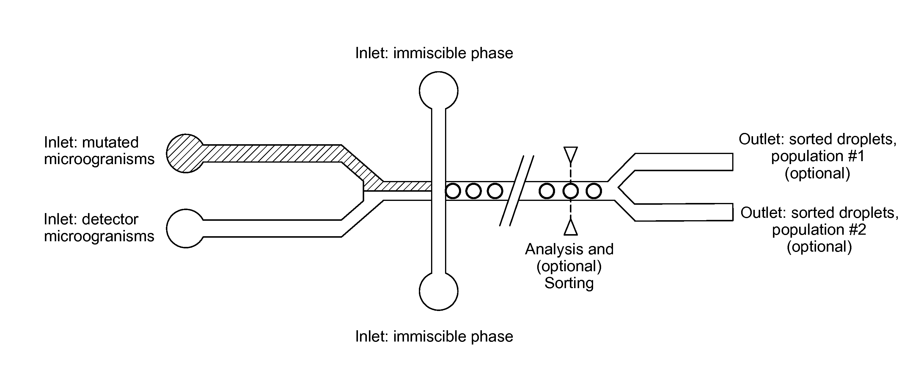
7. A microfluidic device capable of co-encapsulating at least two types of cells, the device comprising: a. at least one inlet for a culture medium comprising a first microorganism; b. at least one inlet for a culture medium comprising a second microorganism; c. at least one inlet for a phase immiscible with the culture media; d. a chamber for combining the first and second medium, suitable to generate droplets comprising at least one cell of each microorganism, and to encapsulate the droplets in the immiscible phase; e. optionally, means to incubate the droplets at a constant or variable temperature; f. optionally, a detector to detect the activity of a reporter gene; g. optionally, an outlet coupled with means for sorting droplets.
8. The microfluidic device according to claim 7 capable of co-encapsulating at least two types of cells, the device comprising: a. a chamber for generating droplets of the first medium, and to encapsulate the droplets in the immiscible phase; b. a chamber for generating droplets of the second medium, and to encapsulate the droplets in the immiscible phase; c. a chamber for combining droplets of the first medium with droplets of the second medium and subsequently fusing said droplets to yield larger droplets comprising a mixture of the first medium and the second medium.
9. The microfluidic device according to claim 7 capable of co-encapsulating at least two types of cells, the device comprising: a. a chamber for generating droplets of the first medium, and to encapsulate the droplets in the immiscible phase; b. a chamber for combining droplets of the first medium with the second medium by picoinjection to yield droplets comprising a mixture of the first medium with the second medium;
10. The microfluidic device according to any of claims 7 to 9, wherein the detector is a fluorescence detector.
11. The microfluidic device according to claim 10, wherein detector is a fluorescence detector and able to quantify the fluorescence intensity.
12. The microfluidic device according to any of the claims 7 to 11, wherein the detector is coupled to a computing device.
13. The microfluidic device according to any of the claims 7 to 12, wherein the device comprises means to incubate the droplets at a temperature range between 18.degree. C. and 50.degree. C.
14. The microfluidic device according to any of claims 7 to 13, wherein the means for sorting droplets comprise dielectric sorting of droplets.
15. Use of a microfluidic device according to any of the claims 7 to 14 in a method according to claims 1 to 6.
Description
FIELD OF THE INVENTION
[0001] The present application is in the field of cell culture analysis. More precisely in the field of cell culture analysis on single cell level. The application is also in the field of microfluidics, particularly in the field of microfluidic analysis and devices.
BACKGROUND
[0002] The production of biological compounds such as sugars, amino acids, antibiotics, carbon sources or nitrogen sources and other chemical building blocks today is often efficiently performed in microorganisms. With the tools of genetic engineering it is possible to optimize microorganisms for an increased production of compounds.
[0003] These optimized microorganisms are generated using different mutagenic/combinatorial strategies capable to generate large libraries of genetically modified organisms. However, the drawback or bottleneck of all strategies are the screening methods used to analyze individual library members.
[0004] The relevant screening methods are dependent on the molecules to be produced, but commonly the screening methods are based on chromatography and subsequent detection, in many cases by mass spectroscopy. A great disadvantage of this is that parallelization and high throughput is difficult to achieve, as the number of clones that can be analyzed is limited.
[0005] Accordingly, there is a need for new screening methods, which allow the detection of strains, which show improved properties in the production of compounds, in particular small molecules such as amino acids or sugars or intermediate chemical building blocks.
[0006] One approach was the use of biosensors for the analysis or identification of small molecules in production media. Pfleger (Pfleger B. F. et al. (2007), Metab. Eng. 9:30-38) describes the generation of a E. coli strain, which is suitable as mevalonate biosensor and expresses GFP in the presence of mevalonate, allowing quantitative detection of mevalonate in an extracellular environment.
[0007] Bertels (Bertels F. et al. (2012), PLoS ONE 7 (7):e41349) describes the development of a biosensor for amino acids, based on an auxotrophic E. coli strain comprising the eGFP gene. U.S. Pat. No. 9,279,139 B2 describes an E. coli glutamine biosensor, comprising the lux operon.
[0008] However, all of these methods are still limited, as they do not allow the rapid analysis of large libraries of colonies. There is therefore still a need for an improved screening method, which allows high throughput screening of microorganism libraries.
BRIEF DESCRIPTION OF THE INVENTION
[0009] The present invention aims to solve this problem by combining the traditional screening approaches with microfluidic devices, thus breaking down the analysis onto single cell level instead of cell cultures.
[0010] The invention relates to a method for the analysis and/or selection of microorganisms, preferably microorganisms which produce a compound of interest, the method comprising: [0011] a. providing a microorganism which produces a compound of interest and a detector microorganism which comprises a reporter gene or reporter gene operon, wherein the microorganism producing said compound of interest and the detector microorganism are combined into single droplets, wherein each droplet comprises at least one cell of each strain; [0012] b. subjecting the droplets to a microfluidic system; [0013] c. analyzing the droplets for the activity of the reporter gene of the detector strain; [0014] d. sorting and collecting the droplets comprising the detector microorganism with the active reporter gene.
[0015] The invention further relates to the use of the method for the analysis of a mutated microorganism, producing a compound of interest.
[0016] In a further aspect the invention relates to a microfluidic device capable of co-encapsulating at least two types of cells, the device comprising: [0017] a. at least one inlet for a culture medium comprising a first microorganism; [0018] b. at least one inlet for a culture medium comprising a second microorganism; [0019] c. optionally, at least one inlet for an immiscible phase; [0020] d. a chamber for combining the first and second medium, suitable to generate droplets comprising at least one cell of each microorganism, and optionally, to encapsulate the droplets in the immiscible phase; [0021] e. optionally, means to incubate the droplets at a constant or variable temperature; [0022] f. optionally, a detector to detect the activity of a reporter gene; [0023] g. optionally, means for sorting the droplets and an outlet for sorted droplets.
[0024] The invention further relates to the use of said microfluidic device.
DETAILED DESCRIPTION OF THE INVENTION
[0025] The present invention relates to a method for the analysis of microorganisms for improved properties. The invention in particular relates to screening methods for microorganisms, which are able to produce biological compounds. In contrast to other screening methods, the claimed screening method allows analysis on single cell level.
[0026] The invention further relates to microfluidic devices suitable for performing the method.
[0027] In a first aspect the invention relates to a method for the analysis and/or selection of microorganisms, which produce a compound, the method comprising: [0028] a. providing a microorganism which produces a compound of interest and a detector microorganism which comprises a reporter gene or reporter gene operon, wherein the microorganism producing said compound of interest and the detector microorganism are combined into single droplets, wherein each droplet comprises at least one cell of each strain; [0029] b. subjecting the droplets to a microfluidic system; [0030] c. analyzing the droplets for the activity of the reporter gene of the detector strain; [0031] d. sorting and collecting the droplets comprising the detector microorganism with active reporter gene.
[0032] In the context of the present invention an activated reporter gene or the activity of the reporter gene refers to the expression of a detectable gene product. Said gene product might be continuously expressed or the expression might be triggered under certain conditions.
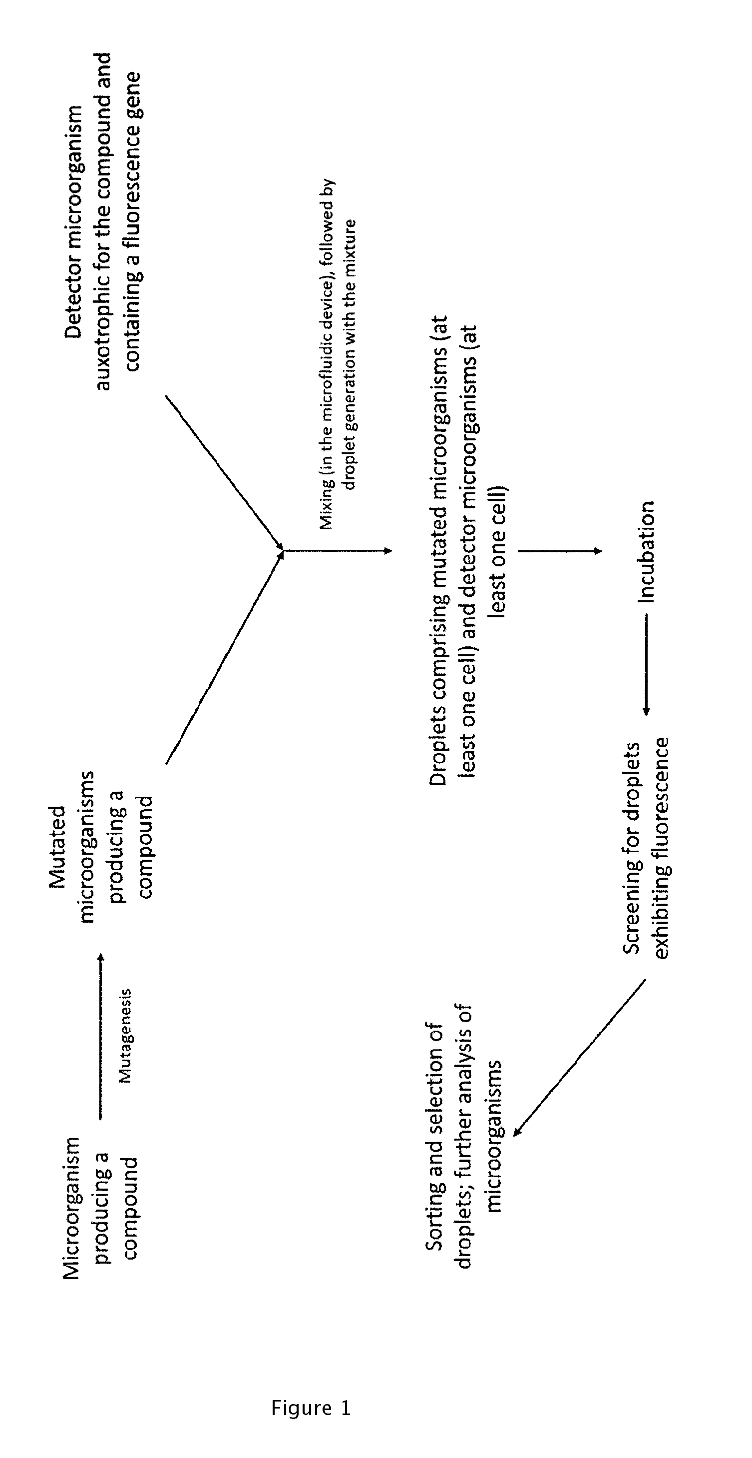
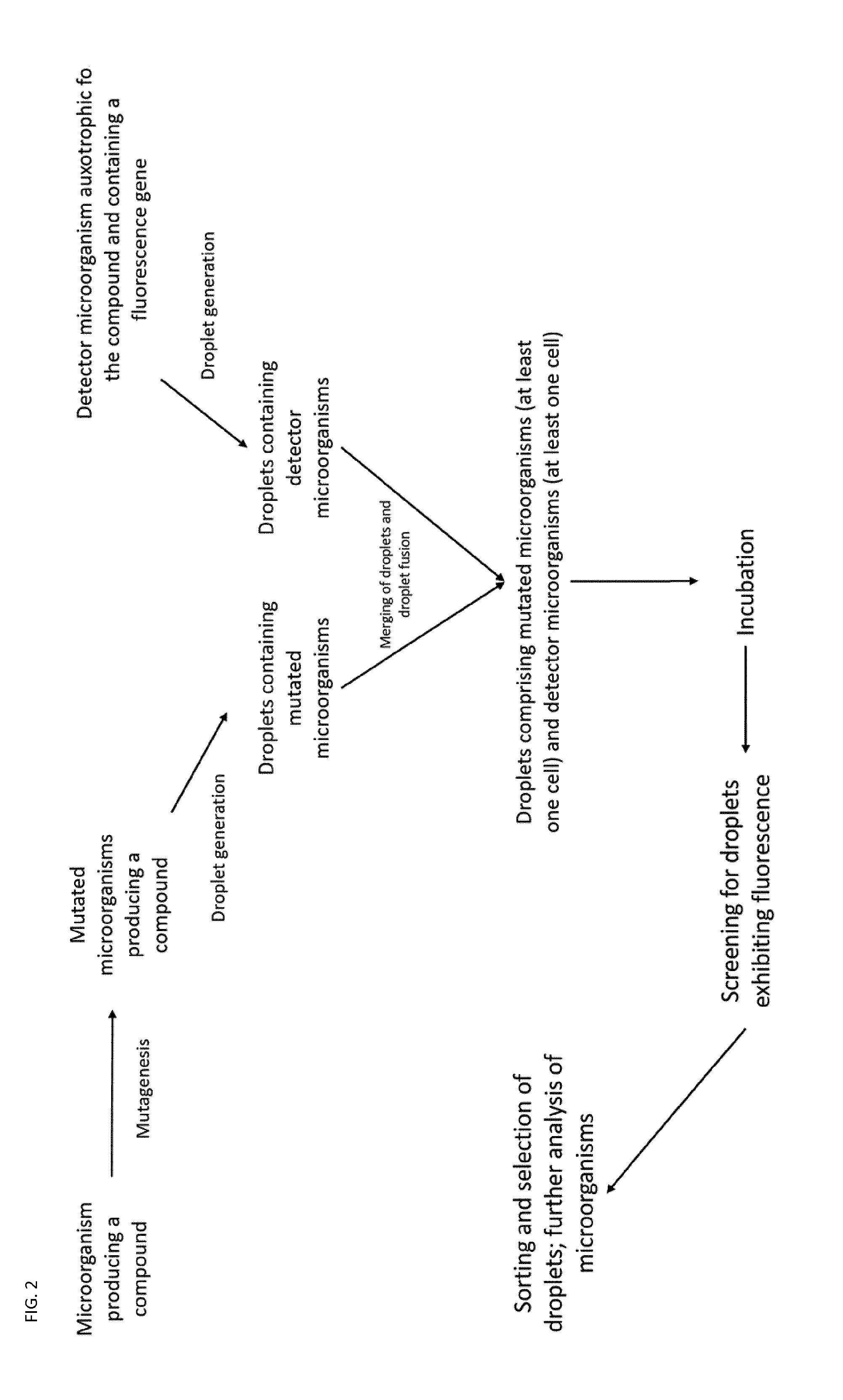
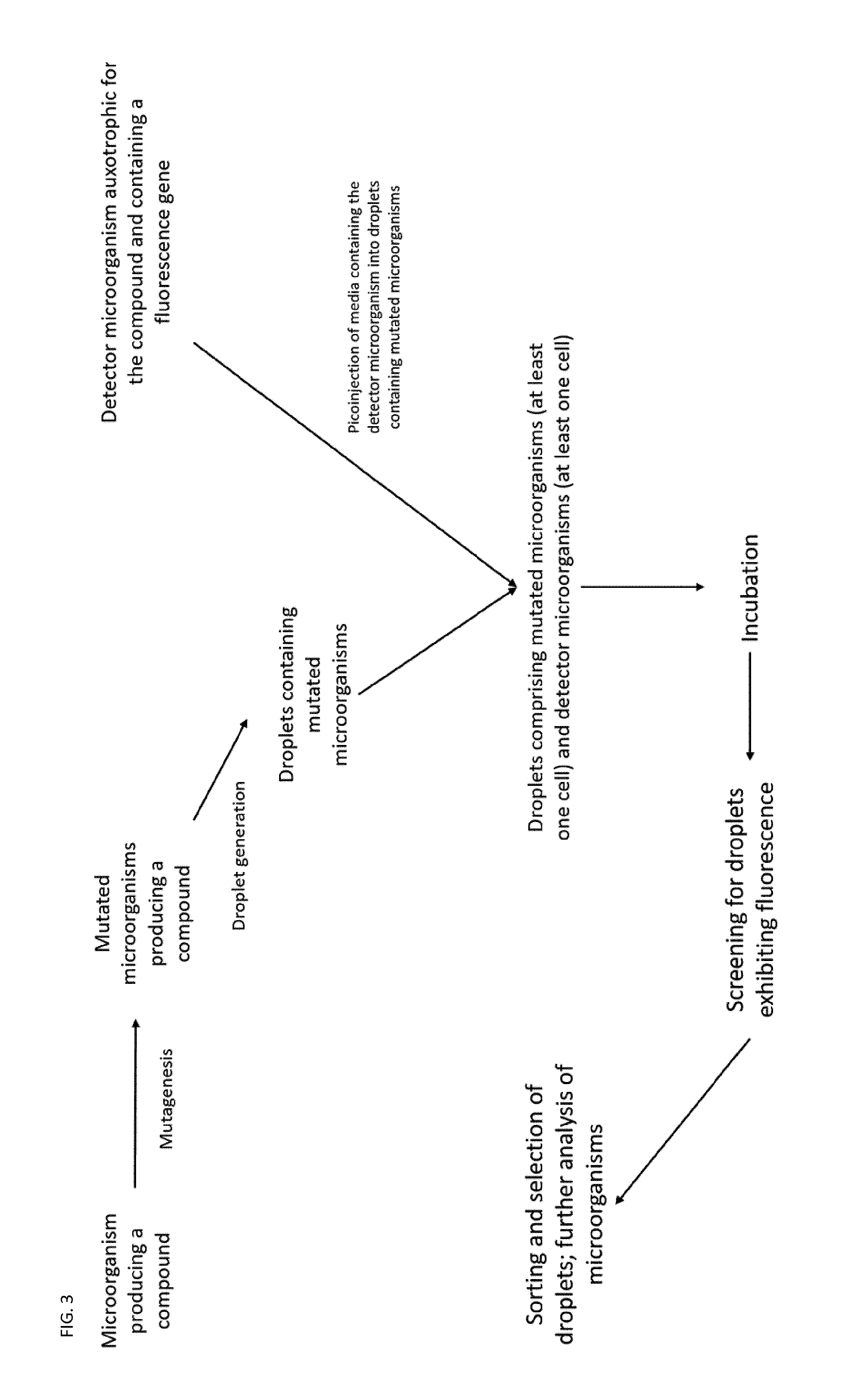
[0033] A general schematic of preferred workflows of the method is shown in FIGS. 1 to 3.
[0034] The method is suitable for any kind of microorganism, which can be handled on single cell level. The microorganism, which produces a compound and the detector microorganism might be of the same species or different species.
[0035] The method is particularly suitable for the analysis of microorganisms, which had been mutated or genetically engineered in order to optimize the production of desired compounds. In one embodiment of the invention the microorganism, which produces a compound is therefore a mutated or genetically engineered organism.
[0036] Mutated or genetically engineered organisms can be generated by means known to the person skilled in the art. Sample methods to induce mutations in microorganisms include but are not limited to, exposure to radiation, in particular UV-radiation or radioactive radiation, stress, phages and viruses, transposon mutagenesis, homologous recombination, metabolic engineering, or chemical mutagenesis. Alternatively, the microorganism producing a compound may comprise a plasmid or cosmid comprising a modified or mutated enzyme or biosynthesis pathway.
[0037] Suitable microorganisms, which might be mutated or produce a compound include, but are not limited to bacterial strains, archeal strains, fungal strains, yeast strains, algae, plant protoplasts, prokaryotic or eukaryotic cells, spores, insect cells or insect strains. In a preferred embodiment of the invention, the microorganism which produces a compound of interest is a bacterial strain, a fungal strain or yeast strain. In a most preferred embodiment, the microorganism, which produces a compound, is a bacterial or fungal strain.
[0038] In a preferred embodiment of the invention, a library of microorganisms producing a compound of interest is generated and analyzed. The method of the invention is in particular suitable for screening for microorganisms exhibiting a higher productivity of the compound and a higher final titer of the compound, in a library of microorganisms.
[0039] An important advantage of the method disclosed herein over the microfluidic system for culturing and selecting cells based on extracellular compound production disclosed in Wang (Wang B. L. et al. (2014), Nat. Biotechnol. 32 (5):473-478) lies in its versatility, since the chemical properties of the compound of interest do not affect the performance of the present method.
[0040] Therefore, the produced compound of interest might be any compound, which can be exported or secreted into the medium by the microorganism and which can be detected by a detector microorganism. The compound preferably has either direct commercial value or may serve as an intermediate in the production of a further compound, which has commercial value.
[0041] Suitable compounds include, but are not limited to, primary metabolites: L- and D-amino acids; sugars and carbon sources such as L-arabinose, N-acetyl-D-glucosamine, N-acetyl-D-mannosamine, N-acetylneuraminate, lactose, D-glucosamine, D-glucose-6-phosphate, D-xylose, D-galactose, glycerol, maltose, maltotriose, and melibiose; nucleosides such as cytidine, guanine, adenine, thymidin, guanosine, adenosine; lipids such as hexadecanoate and glycerol 3-phosphate; indole, maltohexose, maltopentose, putrescine, spermidine, ornithine, tetradecanoate, and nicotinamide adenine dinucleotide.
[0042] Further relevant compounds of interest include, but are not limited to, secondary metabolites. Such metabolites can be produced naturally by the producer microorganism but may also be generated via a heterologous biosynthetic pathway introduced into the microorganisms by genetic engineering. Examples of secondary metabolites include, but are not limited to, polyketides (such as erythryomycin and avermectins), small molecules (such as resveratrol, steviol, and artemisenin) or non-ribosomal peptides.
[0043] The detector microorganism may also be any organism that can be handled on single cell level. Suitable microorganisms, which might be mutated or genetically engineered, include, but are not limited to, bacterial strains, archeal strains, fungal strains, yeast strains, algae, plant protoplasts, prokaryotic or eucaryotic cells, spores, insect cells or insect strain.
[0044] Preferably, the detector strain is a different microorganism than the strain producing a compound of interest. More preferably, the detector strain is a bacterial strain. Most preferably the detector strain is an E. coli strain.
[0045] The detector strain has to comprise a reporter gene or a reporter gene operon. Preferably, said reporter gene or reporter gene operon produces a detectable signal for the detection of the compound of interest. In one embodiment, the intensity of said detectable signal correlates with the amount of produced compound. In an alternative embodiment the intensity of said signal is independent of the amount of compound produced.
[0046] In a preferred embodiment the detectable signal is a fluorescent signal. In one embodiment, said fluorescent signal is generated by the reporter gene product or the reporter gene operon. In a preferred embodiment the reporter gene encodes a fluorescent protein such as green fluorescent protein (GFP), a variant of GFP, yellow fluorescent protein (YFP), a variant of YFP, red fluorescent protein (RFP), a variant of RFP, cyan fluorescent protein (CFP), a variant of CFP or the reporter gene operon is a luminescence operon such as the lux operon. It is known to the person skilled in the art that homologs of said proteins may be used.
[0047] Preferably, the detector strain is an E. coli strain. In a more preferred embodiment, the detector strain is a mutated E. coli strain, optimized for the detection of the compound of interest. E. coli strains can be easily mutated by standard and well-known techniques.
[0048] There are two general possibilities for the detection of the compound of interest. In a first embodiment of the invention, the reporter gene or reporter gene operon of the detector strain might be activated in the presence of said compound. A possible example would be a modified lac-operon, which is utilized for protein expression. Depending on the compound and organism, several potential operons suitable are known for the person skilled in the art. In general, suitable operons usually trigger the degradation or metabolization of the compound.
[0049] One further possibility is the use of modified allosteric transcription factors as described by Taylor (Taylor N. D. et al. (2016), Nature Methods 13:177-183) or the use of synthetic biosensors as described by Rogers (Rogers J. K. et al. (2015), Nucleic Acids Research 43:7648-7660).
[0050] An alternative preferred detector strain might be auxotrophic for the compound, i.e. the detector strain cannot survive without an exogenous supply of said compound. In this case, the reporter gene might be continuously activated.
[0051] According to another embodiment of the first aspect of the present invention, the detector microorganism is auxotrophic for the compound of interest.
[0052] A detector microorganism which is auxotrophic for compound A is unable to grow unless compound A is present in the culture medium. Such a microorganism could be generated via knockout of one or more genes in said microorganism. In the absence of these genes, the microorganism would be unable to synthesize compound A. In some cases, compound A is required directly for growth. In other cases, compound A serves as an intermediate for the synthesis of compound B, which is required for growth. Preventing the synthesis of compound A therefore precludes the synthesis of compound B and prevents cell growth.
[0053] Methods to generate auxotrophic microorganisms are known to the person skilled in the art. Suitable methods include the generation of knockout mutants or random mutagenesis. Alternatively, several naturally existing microorganisms are auxotrophic for specific compounds. In most cases said microorganisms are auxotrophic for amino acids.
[0054] If genome-scale models are available, the compounds which may be sensed and the corresponding gene knockouts which must be made to achieve auxotrophy may be determined based on a computational optimization problem formulated around the available genome-scale model (e.g., Tepper et al. (2011), PLoS ONE 6 (1):e16274).
[0055] Gene knockouts may be achieved via a variety of methods, including but not limited to homologous recombination, gene inactivation via PCR products (e.g., Datsenko and Wanner (2000), PNAS 97 (12):6640-6645), CRISPR-Cas9, transposon mutagenesis, and phage transduction. Thus, auxotrophic sensor strains can be generated today with little effort and time required.
[0056] Generated auxotrophic microorganisms may also be engineered to express a reporter molecule, which may be a fluorescent protein (green fluorescent protein or its derivatives such as eGFP, red fluorescent protein or its derivatives such as mCherry, cyan fluorescent protein or its derivatives, yellow fluorescent protein or its derivatives) or an operon of genes whose expression results in luminescence (such as the lux operon). In a preferred embodiment, generated auxotrophic microorganisms are also engineered to express a reporter molecule, which may be a fluorescent protein (green fluorescent protein or its derivatives such as eGFP, red fluorescent protein or its derivatives such as mCherry, cyan fluorescent protein or its derivatives, yellow fluorescent protein or its derivatives) or an operon of genes whose expression results in luminescence (such as the lux operon).
[0057] According to another embodiment of the first aspect of the present invention, the microorganism producing a compound of interest and a detector microorganism comprising a reporter gene or reporter gene operon are provided in a culture medium.
[0058] The cultivation of microorganisms is known to the person skilled in the art. In general, microorganisms are cultivated in a liquid medium or on a solid medium. In general, solid media are based on liquid media.
[0059] Prior to analysis, the cells might be cultured in any suitable culture medium. Suitable culture media are dependent on the microorganisms. The person skilled in the art generally differentiates between undefined media, such as for example LB-medium, and defined media, in particular minimal media, such as M9 minimal medium or MOPS minimal medium.
[0060] Undefined media usually comprise water, a carbon source, a protein and nitrogen source and salts. In general, the carbon, protein and nitrogen source can be an extract, for example yeast and/or beef extract or protein hydrolysates, such as tryptone or peptone. The exact amino acid composition and salt concentration or composition is usually unknown.
[0061] Defined media on the other hand are exactly known. In a defined medium, all used chemicals are known and the concentrations of the other compounds are known. In the specific case of minimal media, the medium contains the minimum nutrients possible for colony growth, generally without the presence of amino acids.
[0062] As not every organism is able to grow in any medium, it is necessary to adapt the selected medium to the types of microorganisms used. For analysis, a medium which allows survival of both microorganisms is necessary. Depending on the selected microorganisms, the person skilled in the art will know and be able to select the right growth medium.
[0063] Accordingly, the method is not suitable for every combination of microorganisms. It is for example not possible to cultivate a microorganism requiring a medium with high salt concentration together with a microorganism requiring a low salt concentration. Therefore, the detector microorganism needs to be selected dependent on the microorganism producing a compound of interest.
[0064] According to another embodiment of the present invention, the culture medium is suitable for culturing detector microorganism and microorganism producing a compound of interest.
[0065] Preferably, prior to the analysis according to the method of the invention, the microorganisms are cultivated separately in appropriate media. In one embodiment of the invention, the microorganisms are cultivated and incubated in full media. In an alternative embodiment, the microorganisms are cultivated in defined media, preferably minimal media.
[0066] In an alternative embodiment of the invention, the microorganism producing a compound of interest is cultivated in a full medium and the detector microorganism is cultivated in a defined medium, preferably cultivated in a minimal medium.
[0067] The inventors have found that by means of controlling nutrients comprised in the culture medium comprising the detector microorganism and microorganism producing a compound of interest is possible to discriminate droplets comprising said microorganism producing a compound of interest.
[0068] For analysis the microorganisms are then used in their respective medium or transferred in an analysis medium. Preferably, said analysis medium is a defined medium. In a more preferred embodiment, said analysis medium is a minimal medium.
[0069] The person skilled in the art knows how to transfer cell cultures in different media. In one embodiment, the different culture media are simply mixed to form a new culture medium. In a preferred embodiment, the microorganisms are transferred using several centrifugation and washing steps, involving suspending the cells in the target medium.
[0070] Microorganisms in the analysis media are then diluted and/or encapsulated into single droplets. Droplet generation is known to the person skilled in the art. Preferably, said droplets are generated using a microfluidic device. Preferably, during droplet generation the microorganism producing a compound and the detector microorganism are combined. Alternatively, the microorganism producing a compound and the detector strain are diluted into separate droplets and two droplets, each comprising one of the microorganisms are united into a single droplet.
[0071] Regardless of the method of droplet generation, it is preferred that the final droplets in their majority comprise at least one microorganism of each type, i.e. at least one microorganism producing a compound of interest and at least one detector microorganism. Preferably, the majority of droplets comprises one cell of each microorganism.
[0072] It is essential that the droplets additionally comprise all necessary compounds to support growth of the microorganisms, both the detector microorganism and the microorganism producing a compound of interest, and to support the production of said compound by the producing microorganism.
[0073] The droplets comprising the microorganisms may be additionally encapsulated to separate the contents from the environment. A possible method of encapsulation is discussed in WO 2010/063937 A1. In a preferred embodiment, the droplets are encapsulated in a soft alginate shell.
[0074] Alternatively, the droplets are separated from the environment using a phase immiscible with the medium to separate or encapsulate droplets. In one embodiment, said immiscible phase is an oil. In a more preferred embodiment, said immiscible phase is a fluorinated oil.
[0075] In one embodiment, the droplets comprising a microorganism which produces a compound of interest and a detector microorganism which comprises a reporter gene or reporter gene operon have a volume of between 1 pL and 1 .mu.L.
[0076] The inventors have also found that controlling droplet size is important, especially maintaining a monodisperse population. Consistent droplet size is important for maintaining consistent conditions between droplets such that microorganisms are exposed to equivalent environments.
[0077] After diluting and optionally encapsulating the droplets, the microorganisms are incubated for an appropriate amount of time. Said incubation might be performed directly in the microfluidic device or separate from the microfluidic device.
[0078] Incubation might be performed in any way possible. It is however important that the droplets remain intact during the incubation. Stable droplets might be incubated outside of a microfluidic device and later again be subjected to a microfluidic device.
[0079] Independently from where the droplets and microorganisms are incubated, it is preferred that the microorganisms are incubated at appropriate temperatures. The suitable temperature is dependent on the microorganisms in the droplets and the requirements for the production of the compounds. For example, bacterial cultures, such as E. coli usually require temperatures between 20 and 37.degree. C.
[0080] In one embodiment, the incubation temperature is between 18.degree. C. and 50.degree. C. In a preferred embodiment, the incubation temperature is between 20 and 48.degree. C. In a more preferred embodiment, the incubation temperature is between 25 and 45.degree. C. In an even more preferred embodiment, the incubation temperature is between 35 and 40.degree. C. In the most preferred embodiment, the incubation temperature is 37.degree. C.
[0081] The temperature may vary during incubation or may be constant. In one embodiment of the invention, the droplets comprising the microorganisms are incubated at a constant temperature. In an alternative embodiment, the droplets comprising the microorganisms are incubated at variable temperatures.
[0082] Incubation time has to be selected accordingly. In general, the incubation time needs to be long enough to allow for the microorganisms to grow and produce and detect the compound of interest. The time is dependent of the medium, the temperature and the microorganisms. A "richer" medium and a temperature near the optimum temperature for the microorganism results in shorter incubation times.
[0083] After incubation, the droplets are analyzed in a microfluidic device, screening for the activation of the reporter gene. The detection method is dependent on the reporter gene. If the reporter gene is a fluorescent protein or a reporter operon generating a fluorescent signal, the detection method is fluorescence detection.
[0084] In particular, droplets exhibiting higher fluorescence are correlated to higher concentrations of fluorescent protein and therefore to higher number of cells of the detector strain. Alternatively, droplets containing higher numbers of detector strain cells also contain producer strain cells which generate higher amounts of the compound of interest.
[0085] Therefore, according to another embodiment of the first aspect of the present invention, the method for the analysis of microorganisms in droplet disclosed herein is capable of providing with a qualitative and/or quantitative analysis of the compound of interest.
[0086] Preferably, following incubation, the concentration of the reporter molecule in each droplet is determined via fluorescence or luminescence measurements. Such measurements may be performed on the same microfluidic device in which the droplets were generated or on a second microfluidic device distinct from the first microfluidic chip. Preferably, improved production strains can be identified by fluorescence or luminescence above that measured from droplets produced by co-encapsulating the biosensor strain with the parent production strain.
[0087] After detection, the droplets which had been identified as comprising an activated reporter gene or a surviving detector microorganism are selected and separated for further analysis. Potential mechanisms for sorting the droplets are known to the person skilled in the art. In one embodiment, the cells are sorted using dielectrophoresis.
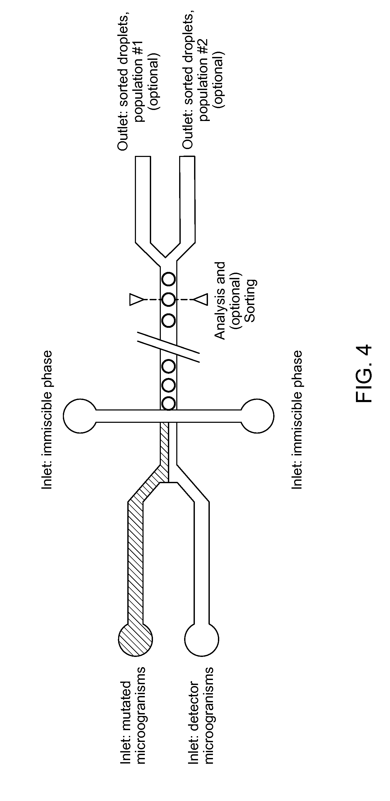
[0088] The invention also relates to several devices to be used in said method. In a second aspect, the invention relates to a microfluidic device capable of co-encapsulating at least two types of cells, the device comprising (see FIG. 4): [0089] a. at least one inlet for a culture medium comprising a first microorganism; [0090] b. at least one inlet for a culture medium comprising a second microorganism; [0091] c. at least one inlet for a phase immiscible with the culture media; [0092] d. a chamber for combining the first and second medium, suitable to generate droplets comprising at least one cell of each microorganism, and optionally to encapsulate the droplets in the immiscible phase.
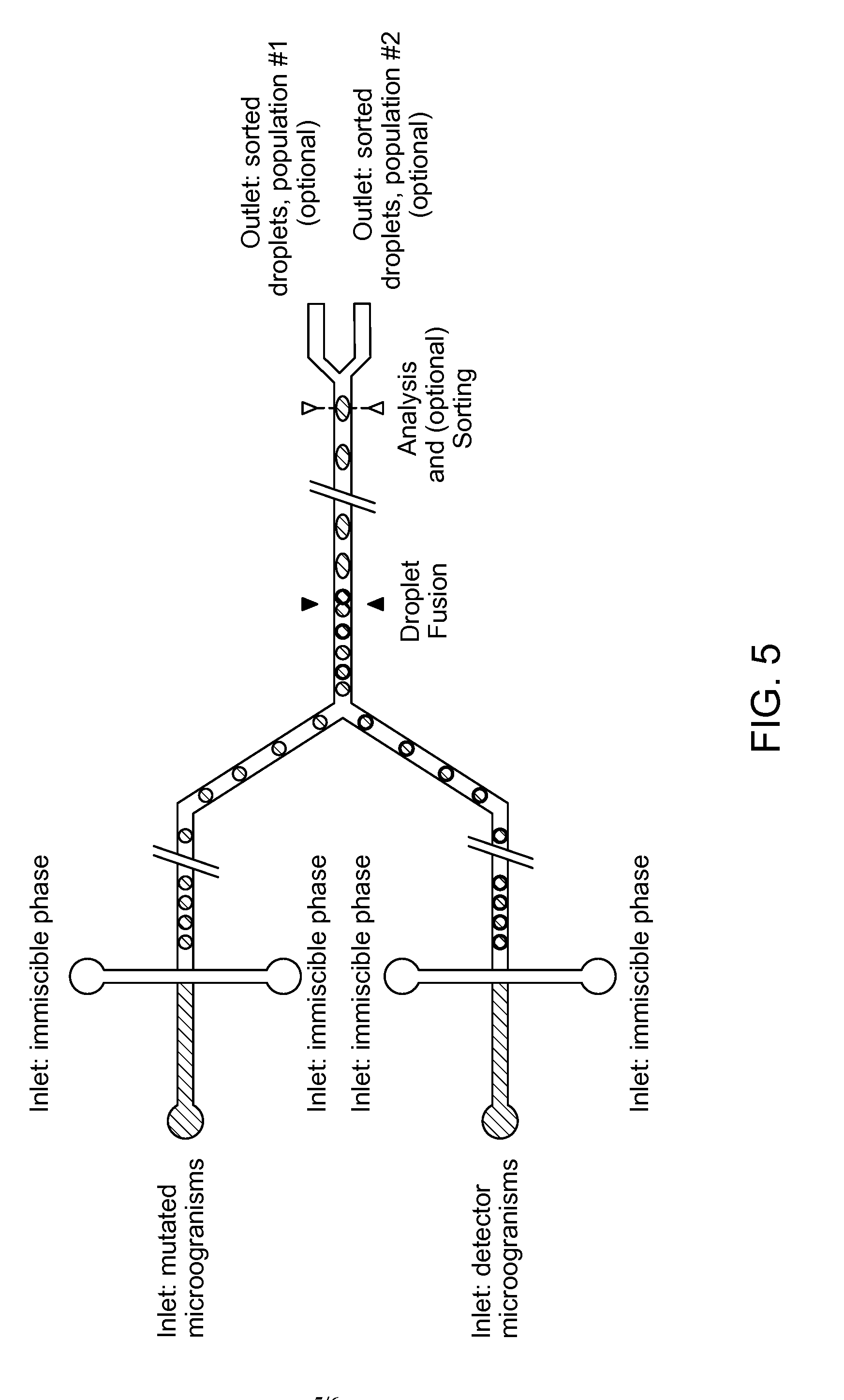
[0093] In an alternative embodiment, the invention relates to a microfluidic device capable of co-encapsulating at least two types of cells, the device comprising (see FIG. 5): [0094] a. at least one inlet for a culture medium comprising a first microorganism; [0095] b. at least one inlet for a culture medium comprising a second microorganism; [0096] c. at least one inlet for a phase immiscible with the culture media; [0097] d. a chamber for combining the first and second medium, suitable to generate droplets comprising at least one cell of each microorganism, and optionally to encapsulate the droplets in the immiscible phase.
[0098] In a further embodiment, the invention relates to a microfluidic device capable of co-encapsulating at least two types of cells, the device comprising: [0099] a. a chamber for generating droplets of the first medium, and to encapsulate the droplets in the immiscible phase; [0100] b. a chamber for generating droplets of the second medium, and to encapsulate the droplets in the immiscible phase; [0101] c. a chamber for combining droplets of the first medium with droplets of the second medium and subsequently fusing said droplets to yield larger droplets comprising a mixture of the first medium and the second medium.
[0102] In one embodiment, the droplets comprising at least one cell of each medium are generated by generating a droplet comprising at least one cell of a first microorganism and in said chamber picoinjecting said second microorganism into said droplet (see FIG. 6).
[0103] In an alternative embodiment, the droplets are generated by generating droplets comprising a first microorganism and droplets comprising the second microorganism and in the chamber combining and/or fusing the droplets into single droplets.
[0104] The microfluidic device may optionally comprise further inlets. Said inlets might be for further modifications of the droplets, e.g. for adding additional components into the culture medium. Alternatively, said additional inlets might be used for modification of the droplets such as mixing of droplets, addition of other droplets into the stream for subsequent fusion, addition of spacing oil to further separate droplets the droplets.
[0105] Said microfluidic device might be a standalone device, or part of a larger microfluidic device. If said microfluidic device is a standalone device, it is preferred that the device can be connected to other devices, preferably other microfluidic devices.
[0106] In one embodiment, the microfluidic device comprises means for temperature control in order to maintain the culture media comprising microorganisms at a desired temperature. Preferably, the microfluidic device comprises means for temperature control in order to maintain the culture media comprising the microorganisms at constant temperature. More preferably, the microfluidic device comprises means for temperature to maintain the culture media comprising the microorganisms at a constant temperature during the whole droplet generation process.
[0107] In a preferred embodiment, the microfluidic device allows the control of droplet size. In a more preferred embodiment, the microfluidic device allows for the generation of droplets with variable size. In the most preferred embodiment, the microfluidic device allows for the generation of a monodisperse population of droplets.
[0108] Optionally, the microfluidic device comprises means for a further treatment of the droplets, such as additional means for injection of reagents, injection of cells, temperature control, delay lines for on chip incubation, sorting of droplets.
[0109] In a further aspect, the invention relates to a microfluidic device capable of co-encapsulating at least two types of cells, the device comprising: [0110] a. at least one inlet for a culture medium comprising a first microorganism; [0111] b. at least one inlet for a culture medium comprising a second microorganism; [0112] c. optionally, at least one inlet for a phase immiscible with the culture media; [0113] d. a chamber for combining the first and second medium, suitable to generate droplets comprising at least one cell of each microorganism, and optionally, to encapsulate the droplets in the immiscible phase; [0114] e. optionally, means to incubate the droplets at a constant or variable temperature; [0115] f. optionally, a detector to detect the activity of a reporter gene; [0116] g. optionally, an outlet coupled with means for sorting droplets.
[0117] In one particular embodiment, the invention relates to a microfluidic device for generating, incubating and analyzing and/or sorting droplets comprising cells, the device comprising: [0118] a. a first inlet for a culture medium comprising a first microorganism; [0119] b. a second inlet for a culture medium comprising a second microorganism; [0120] c. a third inlet for a phase immiscible with the culture media; [0121] d. a chamber for combining the first and second medium, suitable to generate droplets comprising at least one cell of each microorganism, and to encapsulate the droplets in the oil; [0122] e. optionally, means to incubate the droplets at a constant or variable temperature; [0123] f. a detector to detect the activity of a reporter gene; [0124] g. an outlet coupled with means for sorting droplets.
[0125] In a particular embodiment, the invention relates to a microfluidic device for generating, incubating and sorting droplets comprising cells, the device comprising: [0126] a. a first inlet for a culture medium comprising a first microorganism; [0127] b. a second inlet for a culture medium comprising a second microorganism; [0128] c. a third inlet for an oil; [0129] d. a chamber for combining the first and second medium, suitable to generate droplets comprising at least one cell of each microorganism, and to encapsulate the droplets in the oil; [0130] e. optionally, means to incubate the droplets at a constant or variable temperature; [0131] f. optionally, a detector to detect the activity of a reporter gene; [0132] g. optionally, an outlet coupled with means for sorting droplets.
[0133] In another particular embodiment, the invention relates to a microfluidic device for generating, incubating and sorting droplets comprising cells, the device comprising: [0134] a. a first inlet for a culture medium comprising a first microorganism; [0135] b. a second inlet for a culture medium comprising a second microorganism; [0136] c. a third inlet for an oil; [0137] d. a fourth inlet for an oil; [0138] e. a chamber for encapsulating droplets of the first medium in the oil; [0139] f. a chamber for encapsulating droplets of the second medium in the oil; [0140] g. a chamber for combining droplets of the first medium with droplets of the second medium and fusion of droplets into larger droplets comprising a mixture of the first medium and second medium; [0141] h. optionally, a means to incubate the droplets at a constant or variable temperature; [0142] i. optionally, a detector to detect the activity of a reporter gene; [0143] j. optionally, an outlet coupled with means for sorting droplets.
[0144] In another particular embodiment, the invention relates to a microfluidic device for creating incubating and sorting droplets comprising cells, the device comprising: [0145] a. a first inlet for a culture medium comprising a first microorganism; [0146] b. a second inlet for a culture medium comprising a second microorganism; [0147] c. a third inlet for an oil; [0148] d. a fourth inlet for an oil; [0149] e. a chamber for encapsulating droplets of the first medium in the oil; [0150] f. a chamber for combining droplets of the first medium with the second medium via picoinjection, generating larger droplets comprising a mixture of the first medium and second medium; [0151] g. optionally, a means to incubate the droplets at a constant or variable temperature; [0152] h. optionally, a detector to detect the activity of a reporter gene; [0153] i. optionally, an outlet coupled with means for sorting droplets.
[0154] The microfluidic device according to the invention preferably comprises at least three inlets, one for a culture medium comprising a first microorganism, which is preferably producing a compound, one for a second culture medium, comprising a second microorganism, which comprises a reporter gene or reporter gene operon and a third inlet for an immiscible phase.
[0155] According to another embodiment of the second aspect of the present invention, said first and second inlet for a culture medium comprising a first or a second microorganism comprises means for temperature control.
[0156] According to another embodiment of the second aspect of the present invention, said first and second inlet for a culture medium comprising a first or a second microorganism comprises means for controlling the composition of the culture medium. In the context of the present invention, the term "means for controlling the composition of the culture medium" refers to means for supplying nutrients for growth of microorganisms, means for controlling temperature and pH. Nutrients for enriching the culture medium comprises amino acids, vitamins, fatty acids and lipids.
[0157] Said immiscible phase might be an oil or a gas. Preferably, said immiscible phase is an oil, preferably fluorinated oil. In an alternative preferred embodiment, said immiscible phase is a gas.
[0158] In one embodiment, the microfluidic device comprises means for temperature control in order to maintain the culture media comprising microorganisms at a desired temperature. Preferably, the microfluidic device comprises means for temperature control in order to maintain the culture media comprising the microorganisms at constant temperature. More preferably, the microfluidic device comprises means for temperature to maintain the culture media comprising the microorganisms at a constant temperature during the whole droplet generation process.
[0159] In a preferred embodiment, the microfluidic device allows the control of droplet size. In a more preferred embodiment, the microfluidic device allows for the generation of droplets with variable size.
[0160] In a particular embodiment, the microfluidic device allows to incubate the droplets. Means for incubation are known to the person skilled in the art. A possible way would be a temperature controlled loop, which allows temperature controlled incubation.
[0161] In a preferred embodiment, the device allows to control incubation temperature. In one embodiment of the invention, the microfluidic device allows incubation at a constant temperature. In an alternative embodiment, the device allows incubation at a variable temperature.
[0162] In an alternative embodiment, the microfluidic device comprises an outlet and/or an additional inlet, allowing to remove the droplets for incubation and to reinsert the droplets into the microfluidic device. In one embodiment, at least two ports may be used, one inlet and one outlet. In an alternative embodiment, one port may serve as inlet and outlet.
[0163] The device preferably comprises means for the detection of the activation of the reporter gene. As it is preferred that the reporter gene provides a fluorescent signal, said means preferably allow the detection of fluorescence and more preferably, additional determination of fluorescence intensity.
[0164] In a preferred embodiment, the detector is coupled to a computing device.
[0165] Finally, the device preferably comprises, optionally an outlet, which allows sorting the droplets. Preferably, the outlet allows the sorting of droplets showing increased fluorescence compared to other droplets. In a preferred embodiment said outlet comprises means which allow sorting via dielectrophoresis.
[0166] In a preferred embodiment, said outlet is coupled to a computing device.
[0167] The invention further relates to a microfluidic device for the analysis of droplets comprising single cells, preferably single cells of each one microorganism producing a compound and a detector microorganism. The microfluidic device comprises: [0168] a. an inlet for droplets; [0169] b. optionally means for maintaining the droplets at a defined temperature; [0170] c. a detector to detect the activity of a reporter gene; [0171] d. at least one outlet.
[0172] In a preferred embodiment, the device allows to control temperature. In one embodiment of the invention, the microfluidic device allows to keep the droplets at a constant temperature. In an alternative embodiment, the device allows to keep the droplets at a variable temperature.
[0173] The device comprises means for the detection of the activation of the reporter gene. As it is preferred that the reporter gene provides a fluorescent signal, said means preferably allow the detection of fluorescence and more preferably, additional determination of fluorescence intensity.
[0174] In a preferred embodiment, the detector is coupled to a computing device.
[0175] The device preferably comprises at least one outlet. Preferably, the outlet allows the sorting of droplets. In a preferred embodiment, said outlet allows sorting of droplets via dielectrophoresis.
[0176] In a preferred embodiment, said outlet is coupled to a computing device.
FIGURE LEGENDS
[0177] FIGS. 1 to 3: schematic examples of preferred workflows of the method.
[0178] FIGS. 4 to 6: schematics of microfluidic devices for droplet generation. Broken lines represent positions, where the droplets might be further processed either within or of the microfluidic device.
EXAMPLES
Example 1
[0179] A strain of Escherichia coli (e.g., MG1655) is transformed with a plasmid (named here as pTrp) containing the trpABCDE operon under the control of a strong constitutive promoter. The E. coli strain harboring pTrp is able to overproduce L-tryptophan and secrete the amino acid in to the surrounding culture medium, hereafter referred to as the "producer strain".
[0180] A strain of Saccharomyces cerevisiae that is auxotrophic for L-tryptophan and L-leucine (e.g., W303 and its derivatives) is transformed with a plasmid (named here as pFluor) containing the coding sequence of a fluorescent protein (e.g., GFP, eGFP, mCherry, RFP, etc.) under the control of a strong constitutive promoter (e.g., P.sub.TEF1) as well as the gene or gene operon that allows for intracellular production of L-leucine. Such complementation of the L-leucine auxotroph allows for positive selection of S. cerevisiae cells harboring the pFluor plasmid. When cultured in the presence of L-tryptophan but in the absence of L-leucine, the auxotrophic Saccharomyces cerevisiae strain harboring pFluor proliferates and expresses the fluorescent protein intracellularly. The proliferation of this strain can be monitored via fluorescence measurements, namely illuminating the cells with light of a wavelength or range of wavelengths and measuring the amount of light emitted by the cells at a wavelength or range of wavelengths greater than the wavelength(s) used for illumination. This auxotrophic Saccharomyces cerevisiae strain will be referred to hereafter as the "detector strain."
[0181] The producer strain is inoculated into a minimal medium (e.g., M9 minimal medium with 4 g/L glucose). This culture is grown for 4-8 hours at 37.degree. C. with shaking at 200 rpm, then diluted to an OD.sub.600 of 0.02 using the same minimal medium. The detector strain is inoculated into a synthetically defined medium containing L-tryptophan (to allow for cell growth) but missing L-leucine (to ensure maintenance of the pFluor plasmid). This detector strain culture is grown for 4-8 hours at 30.degree. C. with shaking at 200 rpm. The detector strain culture is then washed with an isotonic buffer and resuspended using a synthetically defined medium missing both L-tryptophan and L-leucine. Microfluidic droplets 20 pL in volume are generated using a microfluidic system in which the aqueous phase comprising the producer strain diluted in minimal medium is separated into droplets by a fluorinated oil (e.g., HFE7500) containing a fluorinated surfactant. These microfluidic droplets are collected and subjected to picoinjection, in which a small, defined volume (5 pL) of detector strain culture is added to each microfluidic droplet, thereby contacting cells of the producer strain with cells of the detector strain within microfluidic droplets. The picoinjected droplets are then collected and incubated at 30.degree. C. to allow for growth of the producer strain, production of L-tryptophan, subsequent growth of the detector strain, and concomitant production of the fluorescent protein.
[0182] The microfluidic droplets are then analyzed using the microfluidic system. The fluorescence of each droplet is analyzed by illuminating the droplet with a laser having a wavelength corresponding to the excitation maximum of the fluorescent protein of interest and measuring the amount of light emitted by the droplet at a range of wavelengths longer than the wavelength used for illumination/excitation. Droplets exhibiting higher fluorescence must contain higher concentrations of fluorescent protein and must therefore contain a higher number of cells of the detector strain. One may also infer that droplets containing higher numbers of detector strain cells must also contain producer strain cells which generated higher amounts of L-tryptophan.
[0183] Using the microfluidic system, droplets exhibiting high levels of fluorescence are separated from the remainder of the droplet pool and collected for further analysis.
Example 2
[0184] A strain of E. coli is engineered to overproduce L-tryptophan via replacement of the native trpABCDE promoter with a strong constitutive promoter. However, feedback regulation has been shown to limit the amount of L-tryptophan that can be produced by this engineered E. coli strain. To overcome this feedback regulation and other regulatory phenomena that may limit L-tryptophan production, the engineered strain is subjected to UV-induced random mutagenesis, generating a library of L-tryptophan-producing E. coli strains. Following generation, this library is cultured on solid medium. Prior to plating on a solid medium, the library is sufficiently diluted such that clonal isolates are obtained on solid media following a period of incubation.
[0185] A strain of Saccharomyces cerevisiae that is auxotrophic for L-tryptophan and L-leucine (e.g., W303 and its derivatives) is transformed with a plasmid (named here as pFluor) containing the coding sequence of a fluorescent protein (e.g., GFP, eGFP, mCherry, RFP, etc.) under the control of a strong constitutive promoter (e.g., P.sub.TEF1) as well as the gene or gene operon that allows for intracellular production of L-leucine. Such complementation of the L-leucine auxotroph allows for positive selection of S. cerevisiae cells harboring the pFluor plasmid. When cultured in the presence of L-tryptophan but in the absence of L-leucine, the auxotrophic Saccharomyces cerevisiae strain harboring pFluor proliferates and expresses the fluorescent protein intracellularly. The proliferation of this strain can be monitored via fluorescence measurements, namely illuminating the cells with light of a wavelength or range of wavelengths and measuring the amount of light emitted by the cells at a wavelength or range of wavelengths greater than the wavelength(s) used for illumination. This auxotrophic Saccharomyces cerevisiae strain will be referred to hereafter as the "detector strain."
[0186] The producer strain library is recovered from solid medium, then diluted and inoculated into a minimal medium (e.g., M9 minimal medium with 4 g/L glucose). This culture is grown for 4-8 hours at 37.degree. C. with shaking at 200 rpm, then diluted to an OD.sub.600 of 0.02 using the same minimal medium. The detector strain is inoculated into a synthetically defined medium containing L-tryptophan (to allow for cell growth) but missing L-leucine (to ensure maintenance of the pFluor plasmid). This detector strain culture is grown for 4-8 hours at 30.degree. C. with shaking at 200 rpm. The detector strain culture is then washed with an isotonic buffer and resuspended using a synthetically defined medium missing both L-tryptophan and L-leucine. Microfluidic droplets 20 pL in volume are generated using a microfluidic system in which the aqueous phase comprising the producer strain diluted in minimal medium is separated into droplets by a fluorinated oil (e.g., HFE7500) containing a fluorinated surfactant. These microfluidic droplets are collected and subjected to picoinjection, in which a small, defined volume (5 pL) of detector strain culture is added to each microfluidic droplet, thereby contacting cells of the producer strain with cells of the detector strain within microfluidic droplets. The picoinjected droplets are then collected and incubated at 30.degree. C. to allow for growth of the producer strain, production of L-tryptophan, subsequent growth of the detector strain, and concomitant production of the fluorescent protein.
[0187] The microfluidic droplets are then analyzed using the microfluidic system. The fluorescence of each droplet is analyzed by illuminating the droplet with a laser having a wavelength corresponding to the excitation maximum of the fluorescent protein of interest and measuring the amount of light emitted by the droplet at a range of wavelengths longer than the wavelength used for illumination/excitation. Droplets exhibiting higher fluorescence must contain higher concentrations of fluorescent protein and must therefore contain a higher number of cells of the detector strain. One may also infer that droplets containing higher numbers of detector strain cells must also contain producer strain cells which generated higher amounts of L-tryptophan.
[0188] Using the microfluidic system, droplets exhibiting high levels of fluorescence are separated from the remainder of the droplet pool and collected. These droplets are then spread on solid media, which is then incubated to recover variants of the producer strain which exhibit higher production of L-tryptophan. Individual clonal isolates are then analyzed in a secondary screen to confirm increased L-tryptophan production: colonies are inoculated into Luria-Bertani (LB) medium and cultured for several days, and culture supernatants are analyzed for L-tryptophan concentration via high performance liquid chromatography (HPLC).
Example 3
[0189] A strain of E. coli is engineered to overproduce L-tryptophan via replacement of the native trpABCDE promoter with a strong constitutive promoter. However, feedback regulation has been shown to limit the amount of L-tryptophan that can be produced by this engineered E. coli strain. To overcome this feedback regulation and other regulatory phenomena that may limit L-tryptophan production, the engineered strain is subjected to UV-induced random mutagenesis, generating a library of L-tryptophan-producing E. coli strains. Following generation, this library is cultured on solid medium. Prior to plating on a solid medium, the library is sufficiently diluted such that clonal isolates are obtained on solid media following a period of incubation.
[0190] A strain of Saccharomyces cerevisiae that is auxotrophic for L-tryptophan and L-leucine (e.g., W303 and its derivatives) is transformed with a plasmid (named here as pLux) containing the coding sequence of the lux luminescence operon under the control of a strong constitutive promoter (e.g., P.sub.TEF1) as well as the gene or gene operon that allows for intracellular production of L-leucine. Such complementation of the L-leucine auxotroph allows for positive selection of S. cerevisiae cells harboring the pLux plasmid. When cultured in the presence of L-tryptophan but in the absence of L-leucine, the auxotrophic Saccharomyces cerevisiae strain harboring pLux proliferates and generates the machinery necessary to produce luminescence. The proliferation of this strain can be monitored via luminescence measurements, namely by measuring the amount of light emitted by the cells at wavelength or range of wavelengths appropriate for the given lux luminescence operon. This auxotrophic Saccharomyces cerevisiae strain will be referred to hereafter as the "detector strain."
[0191] The producer strain library is recovered from solid medium, then diluted and inoculated into a minimal medium (e.g., M9 minimal medium with 4 g/L glucose). This culture is grown for 4-8 hours at 37.degree. C. with shaking at 200 rpm, then diluted to an OD.sub.600 of 0.02 using the same minimal medium. The detector strain is inoculated into a synthetically defined medium containing L-tryptophan (to allow for cell growth) but missing L-leucine (to ensure maintenance of the pLux plasmid). This detector strain culture is grown for 4-8 hours at 30.degree. C. with shaking at 200 rpm. The detector strain culture is then washed with an isotonic buffer and resuspended using a synthetically defined medium missing both L-tryptophan and L-leucine. Microfluidic droplets 20 pL in volume are generated using a microfluidic system in which the aqueous phase comprising the producer strain diluted in minimal medium is separated into droplets by a fluorinated oil (e.g., HFE7500) containing a fluorinated surfactant. These microfluidic droplets are collected and subjected to picoinjection, in which a small, defined volume (5 pL) of detector strain culture is added to each microfluidic droplet, thereby contacting cells of the producer strain with cells of the detector strain within microfluidic droplets. The picoinjected droplets are then collected and incubated at 30.degree. C. to allow for growth of the producer strain, production of L-tryptophan, subsequent growth of the detector strain, and concomitant production of the fluorescent protein.
[0192] The microfluidic droplets are then analyzed using the microfluidic system. The luminescence of each droplet is analyzed by measuring the amount of light emitted by each droplet over a range of wavelengths appropriate for the chosen lux luminescence. Droplets exhibiting higher luminescence must contain higher concentrations of luminescence machinery and must therefore contain a higher number of cells of the detector strain. One may also infer that droplets containing higher numbers of detector strain cells must also contain producer strain cells which generated higher amounts of L-tryptophan.
[0193] Using the microfluidic system, droplets exhibiting high levels of luminescence are separated from the remainder of the droplet pool and collected. These droplets are then spread on solid media, which is then incubated to recover variants of the producer strain which exhibit higher production of L-tryptophan. Individual clonal isolates are then analyzed in a secondary screen to confirm increased L-tryptophan production: colonies are inoculated into Luria-Bertani (LB) medium and cultured for several days, and culture supernatants are analyzed for L-tryptophan concentration via high performance liquid chromatography (HPLC).
Example 4
[0194] To identify novel producers of L-tryptophan, a soil environmental sample is washed with an isotonic buffer to recover bacteria present in the sample. These bacteria are then diluted using a chemically defined medium that does not contain L-tryptophan, generating a library of potential producer strains.
[0195] A strain of Saccharomyces cerevisiae that is auxotrophic for L-tryptophan and L-leucine (e.g., W303 and its derivatives) is transformed with a plasmid (named here as pFluor) containing the coding sequence of a fluorescent protein (e.g., GFP, eGFP, mCherry, RFP, etc.) under the control of a strong constitutive promoter (e.g., P.sub.TEF1) as well as the gene or gene operon that allows for intracellular production of L-leucine. Such complementation of the L-leucine auxotroph allows for positive selection of S. cerevisiae cells harboring the pFluor plasmid. When cultured in the presence of L-tryptophan but in the absence of L-leucine, the auxotrophic Saccharomyces cerevisiae strain harboring pFluor proliferates and expresses the fluorescent protein intracellularly. The proliferation of this strain can be monitored via fluorescence measurements, namely illuminating the cells with light of a wavelength or range of wavelengths and measuring the amount of light emitted by the cells at a wavelength or range of wavelengths greater than the wavelength(s) used for illumination. This auxotrophic Saccharomyces cerevisiae strain will be referred to hereafter as the "detector strain."
[0196] The detector strain is inoculated into a synthetically defined medium containing L-tryptophan (to allow for cell growth) but missing L-leucine (to ensure maintenance of the pFluor plasmid. This detector strain culture is grown for 4-8 hours at 30.degree. C. with shaking at 200 rpm. The detector strain culture is then washed with an isotonic buffer and resuspended using a synthetically defined medium missing both L-tryptophan and L-leucine. Microfluidic droplets 20 pL in volume are generated using a microfluidic system in which the aqueous phase comprising the library of producer strains diluted in a chemically defined medium is separated into droplets by a fluorinated oil (e.g., HFE7500) containing a fluorinated surfactant. These microfluidic droplets are collected and subjected to picoinjection, in which a small, defined volume (5 pL) of detector strain culture is added to each microfluidic droplet, thereby contacting cells of the producer strain with cells of the detector strain within microfluidic droplets. The picoinjected droplets are then collected and incubated at 30.degree. C. to allow for growth of the producer strain, production of L-tryptophan, subsequent growth of the detector strain, and concomitant production of the fluorescent protein. The microfluidic droplets are then analyzed using the microfluidic system. The fluorescence of each droplet is analyzed by illuminating the droplet with a laser having a wavelength corresponding to the excitation maximum of the fluorescent protein of interest and measuring the amount of light emitted by the droplet at a range of wavelengths longer than the wavelength used for illumination/excitation. Droplets exhibiting higher fluorescence must contain higher concentrations of fluorescent protein and must therefore contain a higher number of cells of the detector strain. One may also infer that droplets containing higher numbers of detector strain cells must also contain producer strain cells which generated higher amounts of L-tryptophan.
[0197] Using the microfluidic system, droplets exhibiting high levels of fluorescence are separated from the remainder of the droplet pool and collected. These droplets are then spread on solid media, which is then incubated to recover variants of the producer strain which exhibit higher production of L-tryptophan. Individual clonal isolates are then analyzed in a secondary screen to confirm increased L-tryptophan production: colonies are inoculated into Luria-Bertani (LB) medium and cultured for several days, and culture supernatants are analyzed for L-tryptophan concentration via high performance liquid chromatography (HPLC).
* * * * *
D00000
D00001
D00002
D00003
D00004
D00005
D00006
XML
uspto.report is an independent third-party trademark research tool that is not affiliated, endorsed, or sponsored by the United States Patent and Trademark Office (USPTO) or any other governmental organization. The information provided by uspto.report is based on publicly available data at the time of writing and is intended for informational purposes only.
While we strive to provide accurate and up-to-date information, we do not guarantee the accuracy, completeness, reliability, or suitability of the information displayed on this site. The use of this site is at your own risk. Any reliance you place on such information is therefore strictly at your own risk.
All official trademark data, including owner information, should be verified by visiting the official USPTO website at www.uspto.gov. This site is not intended to replace professional legal advice and should not be used as a substitute for consulting with a legal professional who is knowledgeable about trademark law.