Citrullinated Brain And Neurological Proteins As Biomarkers Of Brain Injury Or Neurodegeneration
Van Eyk; Jennifer E. ; et al.
U.S. patent application number 16/433106 was filed with the patent office on 2019-10-10 for citrullinated brain and neurological proteins as biomarkers of brain injury or neurodegeneration. This patent application is currently assigned to THE JOHNS HOPKINS UNIVERSITY. The applicant listed for this patent is THE JOHNS HOPKINS UNIVERSITY. Invention is credited to Allen Dale Everett, Zhicheng Jin, Jennifer E. Van Eyk.
| Application Number | 20190310265 16/433106 |
| Document ID | / |
| Family ID | 49161784 |
| Filed Date | 2019-10-10 |


View All Diagrams
| United States Patent Application | 20190310265 |
| Kind Code | A1 |
| Van Eyk; Jennifer E. ; et al. | October 10, 2019 |
CITRULLINATED BRAIN AND NEUROLOGICAL PROTEINS AS BIOMARKERS OF BRAIN INJURY OR NEURODEGENERATION
Abstract
The present invention relates to the field of biomarkers. More specifically, the present invention relates to biomarkers useful in diagnosing brain injury or neurodegeneration. In one embodiment, a method for diagnosing brain injury in a patient comprises the steps of (a) obtaining a sample from the patient; (b) determining the ratio of citrullinated to unmodified arginine residues at one or more arginine residues of one or more brain injury biomarker proteins; and (c) correlating the ratio to a patient having brain injury or to a patient not having brain injury, thereby providing the diagnosis.
| Inventors: | Van Eyk; Jennifer E.; (Baltimore, MD) ; Everett; Allen Dale; (Baltimore, MD) ; Jin; Zhicheng; (Baltimore, MD) | ||||||||||
| Applicant: |
|
||||||||||
|---|---|---|---|---|---|---|---|---|---|---|---|
| Assignee: | THE JOHNS HOPKINS
UNIVERSITY BALTIMORE MD |
||||||||||
| Family ID: | 49161784 | ||||||||||
| Appl. No.: | 16/433106 | ||||||||||
| Filed: | June 6, 2019 |
Related U.S. Patent Documents
| Application Number | Filing Date | Patent Number | ||
|---|---|---|---|---|
| 15636076 | Jun 28, 2017 | 10365288 | ||
| 16433106 | ||||
| 14384848 | Sep 12, 2014 | 9709573 | ||
| PCT/US2013/031012 | Mar 13, 2013 | |||
| 15636076 | ||||
| 61610034 | Mar 13, 2012 | |||
| Current U.S. Class: | 1/1 |
| Current CPC Class: | G01N 33/5306 20130101; G01N 33/6842 20130101; G01N 2440/18 20130101; H01J 49/0027 20130101; G01N 2800/28 20130101; H01J 49/0077 20130101; G01N 33/6848 20130101 |
| International Class: | G01N 33/68 20060101 G01N033/68; H01J 49/00 20060101 H01J049/00; G01N 33/53 20060101 G01N033/53 |
Goverment Interests
STATEMENT OF GOVERNMENTAL INTEREST
[0002] This invention was made with U.S. government support under grant nos. 1R01HL091759-02, 5U54HL090515-02, and NHLBI-HV-10-05(2). The government has certain rights in the invention.
Claims
1. A method of detecting treatment efficacy in a subject who is being treated for brain injury, the method comprising: a. measuring at a first time point the level of autoantibodies to at least two citrullinated brain injury biomarker proteins selected from the group consisting of neurogranin (cit-NRGN), glial fibrillary acidic protein (cit-GFAP), and citrullinated myelin basic protein (cit-MBP), in a sample obtained from the subject prior to initiating brain injury treatment in the subject; b. measuring the levels of autoantibodies to cit-NRGN, cit-GFAP and/or cit-MBP biomarker proteins using a cit-NRGN-specific peptide, a cit-GFAP-specific peptide and/or a cit-MBP-specific peptide that specifically binds the autoantibodies in a sample obtained from the subject at one or more time points after initiating brain injury treatment in the subject; and c. detecting efficacy of the brain injury treatment by measuring decreased levels of autoantibodies that bind to the cit-NRGN, cit-GFAP and/or cit-MBP peptide in the sample in step (b) relative to the levels of autoantibodies to cit-NRGN, cit-GFAP and/or cit-MBP measured in the sample at the first time point in step (a).
2. The method of claim 1, wherein the citrullinated NRGN is citrullinated human NRGN which is citrullinated at one or more arginine (R) amino acid residues selected from R38, R43, R51, R53, and R68 in the human NRGN polypeptide of SEQ ID NO: 1.
3. The method of claim 1, wherein the citrullinated GFAP is citrullinated human GFAP, which is citrullinated at one or more arginine (R) amino acid residues selected from R30, R36, R88, R105, R124, R126, R136, R173, R217, R258, R270, R276, R286, R287, R367, R406, and R416 of the human GFAP polypeptide of SEQ ID NO: 3.
4. The method of claim 1, wherein the citrullinated MBP is citrullinated human MBP, which is citrullinated at one or more arginine (R) amino acid residues selected from R26, R32, R34, R44, R50, R92, R106, R124 R157, R186, R189, R196, and R197 in the human MBP polypeptide of SEQ ID NO: 163.
5. The method of claim 1, further comprising contacting the sample with one or more citrullinated brain injury biomarker proteins selected from the group consisting of tubulin beta-4B chain, tubulin alpha-1B chain, CNPase, PPIA, Septin-7, Elongation factor1-alpha2, TPPP, TPPP3, Ermin Isoform 2, NDRG2 Isoform 2, astrotactin 1 (ASTN1), brain angiogenesis inhibitor 3 (BAI3); carnosine dipeptidase 1 (CNDP1); ERMIN; glutamate receptor metabotropic 3 (GRM3); kelch-like protein 32 (KLH32); melanoma antigen family E,2 (MAGE2); neuregulin 3 (NRG3); oligodendrocyte myelin glycoprotein (OMG); solute carrier family 39 (zinc transporter); reticulon 1 (RTN1); MT3, and peptidylarginine deiminase, types 1-4 and 6 (PAD1-4 or PAD6).
6. The method of claim 1, wherein the sample is blood, serum, plasma, cerebrospinal fluid (CSF), urine, saliva, stool and synovial fluid.
7. The method of claim 6, wherein the sample is blood, serum or plasma.
8. The method of claim 1, wherein binding is detected using a detection method selected from mass spectrometry, immunoassay, electrochemical luminescent assay, electrochemical voltametry, electrochemical amperometry, atomic force microscopy, radio frequency multipolar resonance spectroscopy, confocal microscopy, non-confocal microscopy, fluorescence optical detection, luminescence optical detection, chemiluminescence optical detection, absorbance optical detection, reflectance optical detection, transmittance optical detection, birefringence optical detection, refractive index detection, surface plasmon resonance, ellipsometry, resonant mirror detection, grating coupler waveguide detection, or interferometry.
9. The method of claim 8, wherein the detecting by mass spectrometry is accomplished using multiple reaction monitoring mass spectrometry (MRM-MS).
10. The method of claim 1, wherein the subject has or is suspected of having a neurodegenerative disease.
11. The method of claim 10, wherein the neurodegenerative disease is selected from hemorrhagic stroke, ischemic stroke, chronic traumatic encephalopathy, Alzheimer's disease, or Parkinson's Disease.
12. A method of detecting progress of brain injury treatment in a subject having or suspected of having brain injury, the method comprising: a. measuring at a first time point prior to treatment a baseline level of autoantibodies to at least two citrullinated brain injury biomarker proteins selected from the group consisting of neurogranin (cit-NRGN), glial fibrillary acidic protein (cit-GFAP), and citrullinated myelin basic protein (cit-MBP), in a sample obtained from a subject in need thereof; b. measuring the level of autoantibodies to cit-NRGN, cit-GFAP and/or cit-MBP biomarker proteins using a cit-NRGN-specific peptide, a cit-GFAP-specific peptide and/or a cit-MBP-specific peptide that specifically binds the autoantibodies in a sample obtained from the subject at least one additional time point at after treatment initiation; and c. detecting progress of the brain injury treatment by measuring decreased levels of autoantibodies that bind to the cit-NRGN, cit-GFAP and/or cit-MBP peptide in the sample in step (b) relative to the baseline level of autoantibodies to cit-NRGN, cit-GFAP and/or cit-MBP measured in the sample at the first time point in step (a).
13. The method of claim 12, wherein the citrullinated NRGN is citrullinated human NRGN which is citrullinated at one or more arginine (R) amino acid residues selected from R38, R43, R51, R53, and R68 in the human NRGN polypeptide of SEQ ID NO: 1.
14. The method of claim 12, wherein the citrullinated GFAP is citrullinated human GFAP, which is citrullinated at one or more arginine (R) amino acid residues selected from R30, R36, R88, R105, R124, R126, R136, R173, R217, R258, R270, R276, R286, R287, R367, R406, and R416 of the human GFAP polypeptide of SEQ ID NO: 3.
15. The method of claim 12, wherein the citrullinated MBP is citrullinated human MBP, which is citrullinated at one or more arginine (R) amino acid residues selected from R26, R32, R34, R44, R50, R92, R106, R124 R157, R186, R189, R196, and R197 in the human MBP polypeptide of SEQ ID NO: 163.
16. The method of claim 12, further comprising contacting the sample with one or more citrullinated brain injury biomarker proteins selected from the group consisting of tubulin beta-4B chain, tubulin alpha-1B chain, CNPase, PPIA, Septin-7, Elongation factor1-alpha2, TPPP, TPPP3, Ermin Isoform 2, NDRG2 Isoform 2, astrotactin 1 (ASTN1), brain angiogenesis inhibitor 3 (BAI3); carnosine dipeptidase 1 (CNDP1); ERMIN; glutamate receptor metabotropic 3 (GRM3); kelch-like protein 32 (KLH32); melanoma antigen family E,2 (MAGE2); neuregulin 3 (NRG3); oligodendrocyte myelin glycoprotein (OMG); solute carrier family 39 (zinc transporter); reticulon 1 (RTN1); MT3, and peptidylarginine deiminase, types 1-4 and 6 (PAD1-4 or PAD6).
17. The method of claim 12, wherein the sample is blood, serum, plasma, cerebrospinal fluid (CSF), urine, saliva, stool and synovial fluid.
18. The method of claim 17, wherein the sample is blood, serum or plasma.
Description
CROSS-REFERENCE TO RELATED APPLICATIONS
[0001] This application is a divisional of U.S. Ser. No. 15/636,076, filed on Jun. 28, 2017, which is a continuation of patent application U.S. Ser. No. 14/384,848, filed on Sep. 12, 2014, now U.S. Pat. No. 9,709,573, issued on Jul. 18, 2017, which is a U.S. National Stage Application under 35 U.S.C. .sctn. 371 of International PCT Application No. PCT/US2013/031012, having an international filing date of Mar. 13, 2013, which claims the benefit of U.S. Provisional Application No. 61/610,034, filed on Mar. 13, 2012, the contents of all of which are incorporated by reference herein in their entireties.
FIELD OF THE INVENTION
[0003] The present invention relates to the field of biomarkers. More specifically, the present invention relates to biomarkers useful in diagnosing brain injury or neurodegeneration.
INCORPORATION-BY-REFERENCE OF MATERIAL SUBMITTED ELECTRONICALLY
[0004] This application contains a Sequence Listing, which has been submitted electronically via EFS-Web as an ASCII text file entitled "P11929-02_Sequence_Listing.txt." The Sequence Listing is 75,410 bytes in size, and was created on Mar. 12, 2013. It is hereby incorporated by reference in its entirety.
BACKGROUND OF THE INVENTION
[0005] Clinical tools such as physical exam, and CNS imaging (CT Scan or MRI) are subjective, not widely available, not sensitive or specific enough and too costly to identify the infant, child or adult with CNS injury. This can include individuals on life support or cardiopulmonary bypass, trauma, loss of oxygen, etc, regardless of the initial injury or disease. There is a great clinical need to identify patients with CNS or brain injury and especially subclinical injury because these infants, children and adults are at significant risk of progressing to overt stroke and development of cognitive and motor loss, dementia and poor mental performance. In addition, accurate and sensitive identification of CNS injury by circulating biomarkers will provide an objective gold standard to test and compare new therapeutic modalities for efficacy.
SUMMARY OF THE INVENTION
[0006] The present invention is based, at least in part, on the discovery of unique post-translational modifications (e.g., citrullination) of CNS proteins neurogranin (NRGN), myelin basic protein (MBP), glial fibrillary acid protein (GFAP), tubulin beta-4B chain, tubulin alpha-1B chain, CNPase, PPIA, Septin-7, Elongation factor1-alpha2, TPPP, TPPP3, Ermin Isoform 2, and NDRG2 Isoform 2. Other proteins include astrotactin 1 (ASTN1); brain angiogenesis inhibitor 3 (BAI3); carnosine dipeptidase 1 (CNDP1); ERMIN; glutamate receptor metabotropic 3 (GRM3); kelch-like protein 32 (KLH32); melanoma antigen family E,2 (MAGE2); myelin basic protein (MBP); neuregulin 3 (NRG3); oligodendrocyte myelin glycoprotein (OMG); solute carrier family 39 (zinc transporter); reticulon 1 (RTN1); and peptidylarginine deiminase (types 1-4 and 6) (PAD) (including PAD-2), are found circulating in patients with brain injury. Thus, the present invention provides methods for the detection and quantification of one or more modified protein or specific amino acid residues on one or more brain injury biomarker proteins. As described further herein, the present invention also provides methods for the detection and quantification of autoantibodies to the unmodified and/or modified brain injury biomarker proteins listed above.
[0007] Accordingly, the identification and development of assays for post-translation modifications and unmodified forms of brain specific circulating proteins such as NRGN, MBP and GFAP provide a new specific diagnostic of brain injury providing the exquisite brain specificity for an accurate and fast diagnosis. In addition, modified forms of NRGN, MBP and GFAP provide information on the biology of injury that can be useful for defining the phase of injury or recovery that can be used for targeting specific therapies to personalize and improve outcome. Ratios of citrullinated to non-citrullinated proteins could be used to determine the risk or progression of CNS injury, stroke, reduced mental capacity, and neurodegenerative diseases.
[0008] As described in certain embodiments, unmodified and citrullinated NRGN, MBP and/or GFAP represent a new diagnostic assay for brain injury. The present invention provides kits using antibodies, aptamers, and mass spectrometry based methods for detection of citrullinated proteins and or modified amino acid residues. The present invention can be used to detect and/or quantify brain injury biomarkers in various body fluids (including plasma, serum, cerebral spinal fluid) and tissue and cells.
[0009] In specific embodiments, the citrullination at residues at NRGN: R38, R43, R51, R53, R68; MBP: R31, R43, R49, R65, R107, R113, R162, R169; and GFAP: R88, R105, R124, R126, R136, R173, R217, R258, R270, R286, R287, R367, R406, and combinations of any of the foregoing can be used to diagnosis brain injury in a patient. In more specific embodiments, the detection and quantification can be directed at the ratio of modified to unmodified proteins or specific amino acid residues.
[0010] The present invention is applicable to the prediction of the susceptibility or presence of both subclinical and overt brain injury. The present invention further provides protein diagnostic(s)/prognostic(s) useful for identifying infants, children and adults with subclinical brain injury, in which routine clinical assessments are normal, to prevent progression to overt stroke. More specifically, protein diagnostic(s)/prognostic(s) of the present invention can be used to assess and monitor efficacy of therapies in infants, children and adults. In fact, the protein diagnostic(s)/prognostic(s) described herein can be used to identify brain injury in ill infants, children and adults at risk for brain injury and to predict outcomes.
[0011] In other embodiments, protein diagnostic(s)/prognostic(s) can be used to (1) identify brain injury in children and adults on life support or cardiopulmonary bypass including during surgery (regardless of initialing injury or disease) to assess potential neurological injury; (2) identify the permeability of the blood brain barrier; and/or (3) identify degenerative brain disease.
[0012] Furthermore, as citrullination of proteins has been proposed to increase immunogenicity of some proteins, the identification of citrullinated brain proteins may be very important for diagnosis of chronic neurodegenerative diseases such as chronic traumatic encephalopathy, Alzheimer's disease, multiple sclerosis and Parkinson's disease. It is possible that auto-antibodies against the various citrullinated forms of these proteins could act as auto-antigens following brain injury exacerbating brain injury. Thus, detection (or blocking) of these auto-antibodies could be used for improved prognosis, risk stratification, or therapy.
[0013] Accordingly, the present invention provides methods for diagnosing brain injury in a patient. In one embodiment, a method for diagnosing brain injury in a patient comprises the steps of (a) obtaining a sample from the patient; (b) determining the ratio of citrullinated to unmodified arginine residues at one or more arginine residues of one or more brain injury biomarker proteins; and (c) correlating the ratio to a patient having brain injury or to a patient not having brain injury, thereby providing the diagnosis.
[0014] The sample can be selected from the group consisting of blood, peripheral blood, serum, plasma, cerebrospinal fluid, urine, saliva, stool and synovial fluid. In a specific embodiment, the sample is blood, plasma serum, cerebrospinal fluid (CSF), or urine. In a more specific embodiment, the sample is CSF. In another specific embodiment, the sample is blood. In an alternative embodiment, the sample is serum.
[0015] In certain embodiments, the determining step is accomplished using mass spectrometry. In a specific embodiment, the determining step is accomplished using multiple reaction monitoring mass spectrometry (MRM-MS). In other embodiments, the determining step is accomplished using an immunoassay. In further embodiments, aptamers, peptoids, or other capture/detection systems are used.
[0016] In a specific embodiment, the one or more brain injury biomarker proteins is neurogranin (NRGN), myelin basic protein (MBP), glial fibrillary acid protein (GFAP), peptidylarginine deiminase (PAD), isoforms thereof, post-translationally modified forms thereof, or combinations of any of the foregoing. In a more specific embodiment, the brain injury biomarker protein is NRGN, isoforms thereof, or post-translationally modified forms thereof. In another specific embodiment, the brain injury biomarker protein is MBP, isoforms thereof, or post-translationally modified forms thereof. In yet another embodiment, the brain injury biomarker protein is GFAP, isoforms thereof, or post-translationally modified forms thereof. In a further embodiment, in the brain injury biomarker protein is PAD-2, isoforms thereof, or post-translationally modified forms thereof.
[0017] In yet another embodiment, the brain injury biomarker protein is one or more proteins selected from the group consisting of tubulin beta-4B chain; tubulin alpha-1B chain; CNPase; PPIA; Septin-7; Elongation factor1-alpha2; TPPP; TPPP3; Ermin Isoform 2; NDRG2 Isoform 2, astrotactin 1 (ASTN1), brain angiogenesis inhibitor 3 (BAI3); carnosine dipeptidase 1 (CNDP1); ERMIN; glial fibrillary acidic protein (GFAP); glutamate receptor metabotropic 3 (GRM3); kelch-like protein 32 (KLH32); melanoma antigen family E,2 (MAGE2); myelin basic protein (MBP); neuregulin 3 (NRG3); neurogranin (NRGN); oligodendrocyte myelin glycoprotein (OMG); solute carrier family 39 (zinc transporter); reticulon 1 (RTN1); and peptidylarginine deiminase (types 1-4 and 6) (PAD); isoforms thereof; post-translationally modified forms thereof; or combinations of any of the foregoing.
[0018] The present invention also provides a method for diagnosing brain injury in a patient comprising the steps of (a) determining the ratio of one or more citrullinated peptides to the corresponding unmodified peptides in a sample collected from the patient using mass spectrometry; and (b) correlating the ratio to a patient having brain injury or to a patient not having brain injury, thereby providing the diagnosis. In an alternative embodiment, a method for diagnosing brain injury in a patient comprises the steps of (a) determining the ratio of one or more citrullinated peptides to the corresponding unmodified peptides in a sample collected from the patient using mass spectrometry; and (b) comparing the ratio with predefined ratios of the same peptides that correlate to a patient having brain injury and predefined ratios of the same peptides that correlate to a patient not having brain injury, wherein a correlation to one of the predefined ratios provides the diagnosis.
[0019] In such embodiments, the one or more peptides is selected from the group consisting of tubulin beta-4B chain, tubulin alpha-1B chain, CNPase, PPIA, Septin-7, Elongation factor1-alpha2, TPPP, TPPP3, Ermin Isoform 2, NDRG2 Isoform 2, ASTN1, BAI3, CNDP1, ERMIN, GFAP, GRM3, KLH32, MAGE2, MBP, NRG3, NRGN, OMG, SLC39A12, RTN1, MT3, PAD (including PAD-2), isoforms thereof, post-translationally modified forms thereof, or combinations of any of the foregoing. Alternatively, the one or more peptide is NRGN, MBP, GFAP, PAD or a combination thereof. In other embodiments, the one or more peptide is NRGN, MBP, GFAP, isoforms thereof, post-translationally modified forms thereof, or combinations of any of the foregoing.
[0020] In a specific embodiment, the present invention provides a method for diagnosing brain injury in a patient comprising the steps of (a) determining the ratio of one or more post-translationally modified peptides to the corresponding unmodified peptides in a sample collected from the patient using MRM-MS; and (b) correlating the ratio to a patient having brain injury or to a patient not having brain injury, thereby providing the diagnosis. In another specific embodiment, a method for diagnosing brain injury in a patient comprises the steps of (a) determining the ratio of one or more post-translationally modified peptides to the corresponding unmodified peptides in a sample collected from the patient using MRM-MS; and (b) comparing the one or more ratios with predefined ratios of the same post-translationally modified/unmodified peptides that correlate to a patient having brain injury and predefined ratios of the same post-translationally modified/unmodified peptides that correlate to a patient not having brain injury, wherein a correlation to one of the predefined ratios provides the diagnosis.
[0021] In such embodiments, the post-translational modification is citrullination, oxidation, methylation, phosphorylation, cysteinylation s-nitrosation, s-glutathyolation, or a combination thereof. Moreover, the one or more peptides can be selected from the group consisting of tubulin beta-4B chain, tubulin alpha-1B chain, CNPase, PPIA, Septin-7, Elongation factor1-alpha2, TPPP, TPPP3, Ermin Isoform 2, NDRG2 Isoform 2, ASTN1, BAI3, CNDP1, ERMIN, GFAP, GRM3, KLH32, MAGE2, MBP, NRG3, NRGN, OMG, SLC39A12, RTN1, PAD, isoforms thereof, post-translationally modified forms thereof, or combinations of any of the foregoing. In a specific embodiment, the one or more peptide is NRGN, MBP, GFAP, PAD, isoforms thereof, post-translationally modified forms thereof, or combinations of any of the foregoing.
[0022] In another embodiment, a method for diagnosing brain injury in a patient comprises the steps of (a) determining the ratio of one or more citrullinated peptides to the corresponding unmodified peptides in a sample collected from the patient using mass spectrometry; (b) determining the ratio of one or more post-translationally modified peptides to the corresponding unmodified peptides in the same sample collected from the patient using MRM-MS; (c) comparing the one or more citrullinated:unmodified peptide ratios to one or more post-translationally modified:unmodified peptide ratios; and (d) correlating the compared ratios to a patient having brain injury or to a patient not having brain injury, thereby providing the diagnosis.
[0023] In another embodiment, a method for diagnosing brain injury in a patient comprises the steps of (a) determining the degree of citrullination of one or more arginine sites of one or more of NRGN, PAD, MBP GFAP, isoforms thereof, post-translationally modified forms thereof, or combinations of any of the foregoing; and (b) correlating the degree of citrullination to a patient having brain injury or to a patient not having brain injury, thereby providing the diagnosis.
[0024] In particular embodiments, the peptides are one or more peptides as shown in Tables 5-9, 11-12, and 14-17. In other embodiments, the peptides comprise an amino acid sequence of about 8 to about 45 amino acid residues of NRGN, MBP, GFAP, and PAD, isoforms thereof, post-translationally modified forms thereof, or combinations of any of the foregoing. In further embodiments, the peptides comprise an amino acid sequence of about 8 to about 45 amino acid residues of tubulin beta-4B chain, tubulin alpha-1B chain, CNPase, PPIA, Septin-7, Elongation factor1-alpha2, TPPP, TPPP3, Ermin Isoform 2, NDRG2 Isoform 2, ASTN1, BAI3, CNDP1, ERMIN, GFAP, GRM3, KLH32, MAGE2, MBP, NRG3, NRGN, OMG, SLC39A12, RTN1, PAD, isoforms thereof, post-translationally modified forms thereof, or combinations of any of the foregoing.
[0025] In another aspect, the present invention provides methods for determining the concentration or levels of citrullinated brain injury biomarker proteins/peptides. In one embodiment, a method for diagnosing brain injury in a patient comprises the steps of (a) measuring the level of one or more citrullinated brain injury biomarker proteins in a sample collected from the patient; and (b) comparing the level of the one or more biomarkers with predefined levels of the same biomarkers that correlate to a patient having brain injury and predefined levels of the same biomarkers that correlate to a patient not having brain injury, wherein a correlation to one of the predefined levels provides the diagnosis.
[0026] In particular embodiments, the one or more brain injury biomarker proteins is tubulin beta-4B chain, tubulin alpha-1B chain, CNPase, PPIA, Septin-7, Elongation factor1-alpha2, TPPP, TPPP3, Ermin Isoform 2, NDRG2 Isoform 2, ASTN1, BAI3, CNDP1, ERMIN, GFAP, GRM3, KLH32, MAGE2, MBP, NRG3, NRGN, OMG, SLC39A12, RTN1, MT3, PAD, isoforms thereof, post-translationally modified forms thereof, or combinations of any of the foregoing. Alternatively, the one or more brain injury biomarker proteins is NRGN, MBP, GFAP, PAD, isoforms thereof, post-translationally modified forms thereof, or combinations of any of the foregoing. In a specific embodiment, the brain injury biomarker protein is NRGN, isoforms thereof, or post-translationally modified forms thereof. In another embodiment, the brain injury biomarker protein is MBP, isoforms thereof, or post-translationally modified forms thereof. In yet another embodiment, the brain injury biomarker protein is GFAP, isoforms thereof, or post-translationally modified forms thereof. In a further embodiment, the brain injury biomarker protein is PAD, isoforms thereof, or post-translationally modified forms thereof.
[0027] In such embodiments, the sample can be selected from the group consisting of blood, peripheral blood, serum, plasma, cerebrospinal fluid, urine, saliva, stool and synovial fluid. In a specific embodiment, the sample is blood, plasma serum, cerebrospinal fluid (CSF), or urine. In a more specific embodiment, the sample is CSF. In another specific embodiment, the sample is blood. In yet another embodiment, the sample is serum.
[0028] In certain embodiments, the determining step is accomplished using mass spectrometry. In a specific embodiment, the determining step is accomplished using multiple reaction monitoring mass spectrometry (MRM-MS). In other embodiments, the determining step is accomplished using an immunoassay. In further embodiments, aptamers, peptoids, or other capture/detection systems are used.
[0029] The present invention also provides a method for diagnosing brain injury in a patient comprising the steps of (a) collecting a sample from the patient; (b) measuring the levels of a panel of citrullinated brain injury biomarker proteins in the sample collected from the patient using mass spectrometry, wherein the panel of biomarkers comprises NRGN, MBP, GFAP, PAD, isoforms thereof, post-translationally modified forms thereof, or combinations of any of the foregoing; and (c) comparing the levels of the panel of biomarkers with predefined levels of the same panel of biomarkers that correlate to a patient having brain injury and predefined levels of the same panel of biomarkers that correlate to a patient not having brain injury, wherein a correlation to one of the predefined levels provides the diagnosis.
[0030] The panel of biomarkers can further comprise one or more citrullinated brain injury biomarker proteins selected from the group consisting of tubulin beta-4B chain, tubulin alpha-1B chain, CNPase, PPIA, Septin-7, Elongation factor1-alpha2, TPPP, TPPP3, Ermin Isoform 2, NDRG2 Isoform 2, ASTN1, BAI3, CNDP1, ERMIN, GRM3, KLH32, MAGE2, NRG3, OMG, SLC39A12, RTN1, MT3, isoforms thereof, post-translationally modified forms thereof, or combinations of any of the foregoing.
[0031] In another embodiment, a method for determining brain injury status in a patient comprises the steps of (a) collecting a sample from the patient; (b) measuring the levels of a panel of citrullinated brain injury biomarker proteins in the sample collected from the patient using SRM-MS, wherein the panel of biomarkers comprises NRGN, MBP, GFAP, PAD, isoforms thereof, post-translationally modified forms thereof, or combinations of any of the foregoing; and (c) comparing the levels of the panel of biomarkers with predefined levels of the same panel of biomarkers that correlate to one or more brain injury statuses selected from the group consisting of having brain injury, not having brain injury, progressing brain injury, and regressing brain injury, wherein a correlation to one of the predefined levels determines the brain injury status of the patient. In another embodiment, the panel of biomarkers further comprises one or more citrullinated brain injury biomarker proteins selected from the group consisting of tubulin beta-4B chain, tubulin alpha-1B chain, CNPase, PPIA, Septin-7, Elongation factor1-alpha2, TPPP, TPPP3, Ermin Isoform 2, NDRG2 Isoform 2, ASTN1, BAI3, CNDP1, ERMIN, GRM3, KLH32, MAGE2, NRG3, OMG, SLC39A12, RTN1, MT3, isoforms thereof, post-translationally modified forms thereof, or combinations of any of the foregoing.
[0032] The present invention also provides diagnostic kits for use in diagnosing brain injury in a patient. In one embodiment, a diagnostic kit for diagnosing brain injury in a patient comprises (a) a substrate for collecting a biological sample from the patient; and (b) means for measuring the levels of one or more human citrullinated brain injury biomarker proteins selected from the group consisting of NRGN, MBP, GFAP, PAD, isoforms thereof, post-translationally modified forms thereof, or combinations of any of the foregoing. In another embodiment, the one or more human citrullinated brain injury biomarker proteins further comprises tubulin beta-4B chain, tubulin alpha-1B chain, CNPase, PPIA, Septin-7, Elongation factor1-alpha2, TPPP, TPPP3, Ermin Isoform 2, NDRG2 Isoform 2, ASTN1, BAI3, CNDP1, ERMIN, GRM3, KLH32, MAGE2, NRG3, OMG, SLC39A12, RTN1, MT3, isoforms thereof, post-translationally modified forms thereof, or combinations of any of the foregoing.
[0033] In certain embodiments, the ratio of one or more citrullinated peptides to the corresponding unmodified peptides in GFAP is one or more of SEQ ID NO:28: SEQ ID NO:27; SEQ ID NO:30: SEQ ID NO:29; SEQ ID NO:40: SEQ ID N0:41; SEQ ID NO:42: SEQ ID NO:43; SEQ ID NO:44: SEQ ID NO:45; SEQ ID NO:46: SEQ ID NO:47; SEQ ID NO:48: SEQ ID NO:49; SEQ ID NO:50: SEQ ID NO:51; SEQ ID NO:52: SEQ ID NO:53; SEQ ID NO:54: SEQ ID NO:55; SEQ ID NO:56: SEQ ID NO:57; SEQ ID NO:91: SEQ ID NO:92; SEQ ID NO:93: SEQ ID NO:94; SEQ ID NO:95: SEQ ID NO:96; SEQ ID NO:97: SEQ ID NO:27; SEQ ID NO:98: SEQ ID NO:160; SEQ ID NO:118: SEQ ID NO:119; SEQ ID NO:120: SEQ ID NO:121; SEQ ID NO:122: SEQ ID NO:123; and SEQ ID NO:124: SEQ ID NO:125.
[0034] In other embodiments, the ratio of one or more citrullinated peptides to the corresponding unmodified peptides in MBP is one or more of SEQ ID NO:32: SEQ ID NO:31; SEQ ID NO:34: SEQ ID NO:33; SEQ ID NO:75: SEQ ID NO:74; SEQ ID NO:77: SEQ ID NO:76; SEQ ID NO:68: SEQ ID NO:69; SEQ ID NO:79: SEQ ID NO:78; SEQ ID NO:80: SEQ ID NO:78; SEQ ID NO:104: SEQ ID NO:105; SEQ ID NO:106: SEQ ID NO:107; SEQ ID NO:108: SEQ ID NO:109; SEQ ID NO:110: SEQ ID NO:111; SEQ ID NO:112: SEQ ID NO:113; SEQ ID NO:114: SEQ ID NO:115; and SEQ ID NO:116: SEQ ID NO:117.
[0035] In further embodiments, the ratio of one or more citrullinated peptides to the corresponding unmodified peptides in NRGN is one or more of SEQ ID NO:35: SEQ ID NO:36; SEQ ID NO:37: SEQ ID NO:157; SEQ ID NO:71: SEQ ID NO:73; SEQ ID NO:77: SEQ ID NO:159; and SEQ ID NO:99: SEQ ID NO:100.
[0036] In additional embodiments, the ratio of one or more citrullinated peptides to the corresponding unmodified peptides in the proteins listed in Table 17 is one or more of SEQ ID NO:128: SEQ ID NO:129; SEQ ID NO:130: SEQ ID NO:131; SEQ ID NO:132: SEQ ID NO:133; SEQ ID NO:134: SEQ ID NO:135; SEQ ID NO:136: SEQ ID NO:137; SEQ ID NO:138: SEQ ID NO:139; SEQ ID NO:140: SEQ ID NO:141; SEQ ID NO:142: SEQ ID NO:143; SEQ ID NO:144: SEQ ID NO:145; SEQ ID NO:146: SEQ ID NO:147; SEQ ID NO:148: SEQ ID NO:149; SEQ ID NO:150: SEQ ID NO:151; SEQ ID NO:152: SEQ ID NO:153; and SEQ ID NO:154: SEQ ID NO:155.
[0037] Furthermore, the present invention provides for the combination of any of the foregoing ratios of citrullinated or other post-translationally modified peptides to the corresponding unmodified peptides provided herein including, but not limited to, SEQ ID NO:28: SEQ ID NO:27; SEQ ID NO:30: SEQ ID NO:29; SEQ ID NO:40: SEQ ID NO:41; SEQ ID NO:42: SEQ ID NO:43; SEQ ID NO:44: SEQ ID NO:45; SEQ ID NO:46: SEQ ID NO:47; SEQ ID NO:48: SEQ ID NO:49; SEQ ID NO:50: SEQ ID NO:51; SEQ ID NO:52: SEQ ID NO:53; SEQ ID NO:54: SEQ ID NO:55; SEQ ID NO:56: SEQ ID NO:57; SEQ ID NO:91: SEQ ID NO:92; SEQ ID NO:93: SEQ ID NO:94; SEQ ID NO:95: SEQ ID NO:96; SEQ ID NO:97: SEQ ID NO:27; SEQ ID NO:98: SEQ ID NO:160; SEQ ID NO:118: SEQ ID NO:119; SEQ ID NO:120: SEQ ID NO:121; SEQ ID NO:122: SEQ ID NO:123; SEQ ID NO:124: SEQ ID NO:125; SEQ ID NO:32: SEQ ID NO:31; SEQ ID NO:34: SEQ ID NO:33; SEQ ID NO:75: SEQ ID NO:74; SEQ ID NO:77: SEQ ID NO:76; SEQ ID NO:68: SEQ ID NO:69; SEQ ID NO:79: SEQ ID NO:78; SEQ ID NO:80: SEQ ID NO:78; SEQ ID NO:104: SEQ ID NO:105; SEQ ID NO:106: SEQ ID NO:107; SEQ ID NO:108: SEQ ID NO:109; SEQ ID NO:110: SEQ ID NO:111; SEQ ID NO:112: SEQ ID NO:113; SEQ ID NO:114: SEQ ID NO:115; SEQ ID NO:116: SEQ ID NO:117; SEQ ID NO:35: SEQ ID NO:36; SEQ ID NO:37: SEQ ID NO:157; SEQ ID NO:71: SEQ ID NO:73; SEQ ID NO:77: SEQ ID NO:159; SEQ ID NO:99: SEQ ID NO:100; SEQ ID NO:128: SEQ ID NO:129; SEQ ID NO:130: SEQ ID NO:131; SEQ ID NO:132: SEQ ID NO:133; SEQ ID NO:134: SEQ ID NO:135; SEQ ID NO:136: SEQ ID NO:137; SEQ ID NO:138: SEQ ID NO:139; SEQ ID NO:140: SEQ ID NO:141; SEQ ID NO:142: SEQ ID NO:143; SEQ ID NO:144: SEQ ID NO:145; SEQ ID NO:146: SEQ ID NO:147; SEQ ID NO:148: SEQ ID NO:149; SEQ ID NO:150: SEQ ID NO:151; SEQ ID NO:152: SEQ ID NO:153; and SEQ ID NO:154: SEQ ID NO:155.
[0038] In another aspect, the present invention provides methods for diagnosing brain injury by detecting autoantibodies to one or more citrullinated biomarker peptides described herein. In one embodiment, a method for diagnosing brain injury in a subject comprises the steps of (a) collecting a sample from the subject; (b) detecting the presence of autoantibodies to citrullinated brain injury biomarker peptides in the sample collected from the subject; and (c) correlating the amount of autoantibodies to citrullinated brain injury biomarker peptides to a patient having brain injury or to a patient not having brain injury, thereby providing the diagnosis. In another embodiment, the detecting step comprises the steps of (a) contacting a biological sample taken from a subject with a citrullinated brain injury biomarker peptide; and (b) detecting the binding of the peptide with an autoantibody specific for the peptide, wherein the detection of binding is indicative of the presence of citrullinated brain injury biomarker peptide autoantibodies in the subject. In a specific embodiment, the binding is detected by enzyme-linked immunosorbent assay (ELISA), immunoprecipitation or immunoblotting. In certain embodiments, the citrullinated brain injury biomarker peptide is one or more of NRGN, MBP, GFAP, PAD-2, tubulin beta-4B chain, tubulin alpha-1B chain, CNPase, PPIA, Septin-7, Elongation factor1-alpha2, TPPP, TPPP3, Ermin Isoform 2, and NDRG2 Isoform 2. In other embodiments, the citrullinated brain injury biomarker peptide is one or more of ASTN1, BAI3, CNDP1, ERMIN, GFAP, GRM3, KLH32, MAGE2, MBP, NRG3, NRGN, OMG, SLC39A12, RTN1, MT3, PAD.
[0039] In another embodiment, a method for assessing efficacy of a brain injury treatment regimen in a subject comprises the steps of (a) establishing a baseline level of autoantibodies to cit-NRGN, cit-MBP, and cit-GFAP in a subject prior to brain injury treatment regimen; (b) monitoring the levels of autoantibodies to cit-NRGN, cit-MBP, and cit-GFAP using a cit-NRGN, cit-MBP, and cit-GFAP peptide at least at one point after initiation of the brain injury treatment regimen; and (c) comparing the observed level of autoantibodies to cit-NRGN, cit-MBP, and cit-GFAP to the baseline level of autoantibodies to cit-NRGN, cit-MBP, and cit-GFAP, wherein a decrease in the level of autoantibodies is indicative of the efficacy of the brain injury treatment regimen. In a specific embodiment, the level of autoantibodies to cit-NRGN, cit-MBP, and cit-GFAP is measured by ELISA, immunoprecipitation or immunoblotting. In other embodiments, the baseline level of autoantibodies is also established with respect to one of more of citrullinated tubulin beta-4B chain, tubulin alpha-1B chain, CNPase, PPIA, Septin-7, Elongation factor1-alpha2, TPPP, TPPP3, Ermin Isoform 2, and NDRG2 Isoform 2. In other embodiments, the baseline level of autoantibodies is also established with respect to one or more of ASTN1, BAI3, CNDP1, ERMIN, GFAP, GRM3, KLH32, MAGE2, MBP, NRG3, NRGN, OMG, SLC39A12, RTN1, MT3, PAD.
[0040] In a further embodiment, a method for qualifying brain injury status in a subject comprises the steps of (a) measuring the level of autoantibodies to cit-NRGN, cit-MBP, and cit-GFAP in a biological sample from the subject; and (b) correlating the measurement with brain injury status. In a specific embodiment, the level of autoantibodies to cit-NRGN, cit-MBP, and cit-GFAP is measured by ELISA, immunoprecipitation or immunoblotting. In particular embodiments, the brain injury status is selected from the group consisting of the risk of brain injury, the development of brain injury, the presence or absence of brain injury, the stage of brain injury, the subtype of brain injury, the prognosis for the subject, and the effectiveness of treatment of brain injury.
[0041] In another aspect, the present invention relates to methods for detecting the presence of autoantibodies to citrullinated peptides in a subject (cit-peptide autoantibodies) in a subject. In certain embodiments, the citrullinated peptide is MBP, GFAP, tubulin beta-4B chain, tubulin alpha-1B chain, CNPase, PPIA, Septin-7, Elongation factor1-alpha2, TPPP, TPPP3, Ermin Isoform 2, NDRG2 Isoform 2, ASTN1, BAI3, CNDP1, ERMIN, GFAP, GRM3, KLH32, MAGE2, NRG3, NRGN, OMG, SLC39A12, RTN1, MT3, and PAD2, as well as the peptides listed in Tables 1-11 and 13-17. The present invention also relates to methods for detecting the presence of autoantibodies to unmodified peptides.
[0042] In particular embodiment, the method comprises contacting a biological sample taken from a subject with a polypeptide of the present invention and detecting the binding of the polypeptide with an autoantibody specific for the polypeptide, wherein the detection of binding is indicative of the presence of cit-polypeptide autoantibodies in the subject. In such methods, the binding can be detected by enzyme-linked immunosorbent assay (ELISA), immunoprecipitation or immunoblotting.
[0043] In yet another aspect, the present invention relates to methods for assessing efficacy of a brain injury treatment regimen in a subject. In particular embodiments, the methods comprise establishing a baseline level of cit-peptide autoantibodies in a subject prior to a brain injury treatment regimen; monitoring the level of cit-peptide autoantibodies using a polypeptide of the present invention at least at one point after initiation of the brain injury treatment regimen; and comparing the observed level of cit-peptide autoantibodies to the baseline level of cit-peptide autoantibodies, wherein a decrease in the level of peptide autoantibodies is indicative of the efficacy of the brain injury treatment regimen. The peptide can be one or more of peptide is MBP, GFAP, PAD-2, tubulin beta-4B chain, tubulin alpha-1B chain, CNPase, PPIA, Septin-7, Elongation factor1-alpha2, TPPP, TPPP3, Ermin Isoform 2, NDRG2 Isoform 2, ASTN1, BAI3, CNDP1, ERMIN, GFAP, GRM3, KLH32, MAGE2, NRG3, NRGN, OMG, SLC39A12, RTN1, MT3, and PAD2. In such methods, the binding can be detected by enzyme-linked immunosorbent assay (ELISA), immunoprecipitation or immunoblotting.
[0044] In a further aspect, the present invention relates to methods for qualifying brain injury status in a subject. Qualifying brain injury status can be qualifying the risk of brain injury, the development of brain injury, the presence or absence of brain injury, the stage of brain injury, the subtype of brain injury, the prognosis for the subject, and the effectiveness of treatment for brain injury. In certain embodiments, the methods comprise measuring the level of cit-peptide autoantibodies in a biological sample from the subject; and correlating the measurement with brain injury status. In one embodiment, the level of cit-peptide autoantibodies is measured using a polypeptide described herein. In another embodiment, the level of cit-peptide autoantibodies is measured by ELISA, immunoprecipitation or immunoblotting. The peptide can be one or more of peptide is MBP, GFAP, PAD-2, tubulin beta-4B chain, tubulin alpha-1B chain, CNPase, PPIA, Septin-7, Elongation factor1-alpha2, TPPP, TPPP3, Ermin Isoform 2, NDRG2 Isoform 2, ASTN1, BAI3, CNDP1, ERMIN, GFAP, GRM3, KLH32, MAGE2, NRG3, NRGN, OMG, SLC39A12, RTN1, MT3, and PAD2.
[0045] Finally, the present invention provides methods of detecting, measuring, determining, and the like, the biomarkers described herein in terms of both unmodified and modified forms, as well as autoantibodies thereto, i.e., combinations of both protein form and autoantibodies thereto.
BRIEF DESCRIPTION OF THE FIGURES
[0046] FIGS. 1A-1D present MS/MS spectra and extracted ion chromatograms of citrullinated peptides of endogenous bovine MBP digested with Lys-C. FIG. 1A: LTQ MS/MS spectrum of doubly charged species of NIVTPR*TPPPSQGK (residue 91-104) at m/z 746.91. FIG. 1B: extracted ion chromatogram of doubly charged species of NIVTPR*TPPPSQGK and NIVTPRTPPPSQGK. *: Likely Q deamination occurred for the peptide NIVTPRTPPPSQ GK. FIG. 1C: high resolution MS spectrum of double charged species of NIVTPRTPPPSQ GK. FIG. 1D: high resolution MS spectrum of double charged species of NIVTPR*TPPPSQ GK.
[0047] FIGS. 2A and 2B show MS/MS spectra and extracted ion chromatograms of citrullinated peptide of endogenous bovine MBP digested with Lys-C. FIG. 2A: LTQ MS/MS spectrum of doubly charged species of PGFGYGGR*ASDYK (residue 122-134) at m/z 688.32. FIG. 2B: extracted ion chromatogram of doubly charged species of PGFGYGGR*ASDYK (residues 122-134) and PGFGYGGRASDYK.
[0048] FIGS. 3A and 3B show MS/MS spectra and extracted ion chromatograms of citrullinated peptide of endogenous bovine MBP digested with Lys-C. FIG. 3A: Panel A: LTQ MS/MS spectrum of triply charged species of DGHHAAR*TTHYGSLPQK (residue 57-73) at m/z 626.31. FIG. 3B: Panel B: extracted ion chromatogram of triply charged species of DGHHAARTTHYGSLPQK (residues 122-134) and DGHHAAR*TTHYGSLPQK.
[0049] FIG. 4 shows the amino acid sequence alignment for bovine NRGN and human NRGN. The underline indicates the peptides observed in tryptic digests. The initial Met (1) is removed when the protein is translated, but is included in the numbering of the peptides. Shaded area indicated sequence difference of bovine and human NRGN. R68 (in bold, larger font) is the citrullination site in endogenous NRGN.
[0050] FIGS. 5A and 5B present MS/MS spectra and extracted ion chromatograms of a citrullinated tryptic peptide of bovine neurogranin. FIG. 5A: MS/MS spectrum of doubly charged species of KGPGPGGPGGAGGAR*GGAGGGPSGD (residues 54-78) at m/z 953.44. FIG. 5B: extracted ion chromatogram of the ion m/z 953.44.
[0051] FIGS. 6A and 6B show MS/MS spectra and extracted ion chromatograms of citrullinated peptide of endogenous bovine GFAP digested with Glu-C. FIG. 6A: LTQ MS/MS spectrum of doubly charged species of GHLKR*NIVVKTVE (residues 398-410) at m/z 747.44. FIG. 6B: extracted ion chromatogram of doubly charged species of GHLKR*NIVVKTVE and GHLKRNIVVKTVE.
[0052] FIGS. 7A and 7B show extracted ion chromatograms of citrullinated peptide in an endogenous human GFAP sample. FIG. 7A: extracted ion chromatogram of doubly charged species of GHLKR*NIVVKTVE (residues 402-414) and GHLKRNIVVKTVE of human GFAP. FIG. 7B: extracted ion chromatogram of doubly charged species of NIVTPR*TPPPSQGK (residues 92-105) and NIVTPRTPPPSQGK of human MBP isoform 5.
[0053] FIG. 8 shows the amino acid sequence of recombinant human protein NRGN. The initial Met (1) is removed when the protein is translated but is included in the numbering of this protein. Shaded amino acids at N-terminus, T79, and R80 were introduced during subcloning. Shaded amino acids at N-terminus were not included in the numbering of this protein. The underline indicates the peptides identified in Lys-C digests and dotted line shows the peptide observed in Glu-C digests. Endogenous citrullination site was marked in italics. New citrullination sites observed after PAD2 treatment were in bold, larger font.
[0054] FIGS. 9A and 9B present MS/MS spectra and extracted ion chromatograms of a citrullinated peptide IQASFR*GHMAR*K (SEQ ID NO:35) of recombinant human protein NRGN after PAD2 treatment and digestion with Lys-C. FIG. 9A: LTQ MS/MS spectrum of doubly charged species of IQASFR*GHMAR*K (residues 33-44) (SEQ ID NO:35) at m/z 702.36. FIG. 9B: extracted ion chromatogram of doubly charged species of IQASFR*GHMAR*K (SEQ ID NO:35).
[0055] FIG. 10 shows the amino acid sequence of human GFAP and identified peptides with citrullination sites. The underline indicates the modified peptides in Lys-C digests. The dotted line indicates modified peptide in Glu-C digests. The initial Met (1) is removed when the protein is translated but is included in the numbering of the protein. Endogenous citrullination site was marked with an arrow and citrullination sites observed after PAD2 treatment were in italicized, bold, larger font.
[0056] FIG. 11 presents the aligned amino acid sequence of the isoform 5 (18.5 kDa, 170 residues) of human MBP (SEQ ID NO:162) and bovine MBP (169 amino acids) (SEQ ID NO:5). The initial Met (1) is removed in the numbering of both proteins. The underline indicated the peptide observed in Lys-C digests and dotted line showed the peptides observed in tryptic digests. Double-headed black arrows indicate citrullination sites previously reported in bovine MBP. Black arrows indicate citrullination sites previously reported in human MBP. Dotted arrows are previously identified citrullination sites in human MBP after PAD4 treatment. For all citrullinated residues identified in this study, endogenous citrullination sites were marked in grey shading and citrullination sites observed after PAD2 treatment were in italicized, bold larger font.
[0057] FIGS. 12A and 12B present a comparison of CID and HCD spectra of a citrullinated peptide of human GFAP in Glu-C digests. FIG. 12A: CID spectrum of the citrullinated peptide, GHLKR*NIVVKTVE of human GFAP at m/z 747.44, z: +2; FIG. 12B: HCD spectrum of this citrullinated peptide at m/z 747.44.
[0058] FIGS. 13A-13C present extracted ion chromatograms of the in vivo citrullinated peptide and intact peptide of NRGN in Lys-C digests of the AD3 brain sample. FIG. 13A: HPLC peak of the unmodified peptide at m/z 909.9275 with mass tolerance of 10 ppm. Right: isotopic clusters of the +2 ion. FIG. 13B: HPLC peak of the citrullinated peptide at m/z 910.4195 with mass tolerance of 10 ppm. Right: isotopic clusters of the +2 ion. FIG. 13C: Overlap of HPLC peaks shown in FIGS. 13A and 13B. From integrated peak area, occupancy rate of citrullinated peptide at R68 residue was 8.6% for the AD3 sample.
[0059] FIG. 14: Citrullination occupancy rate for NRGN at R68, MBP at R92 and R124. Occupancy rate was calculated based on integrated peak area of citrullinated peptide vs. the sum of citrullinated and intact peptides.
DETAILED DESCRIPTION OF THE INVENTION
[0060] It is understood that the present invention is not limited to the particular methods and components, etc., described herein, as these may vary. It is also to be understood that the terminology used herein is used for the purpose of describing particular embodiments only, and is not intended to limit the scope of the present invention. It must be noted that as used herein and in the appended claims, the singular forms "a," "an," and "the" include the plural reference unless the context clearly dictates otherwise. Thus, for example, a reference to a "protein" is a reference to one or more proteins, and includes equivalents thereof known to those skilled in the art and so forth.
[0061] Unless defined otherwise, all technical and scientific terms used herein have the same meaning as commonly understood by one of ordinary skill in the art to which this invention belongs. Specific methods, devices, and materials are described, although any methods and materials similar or equivalent to those described herein can be used in the practice or testing of the present invention.
[0062] All publications cited herein are hereby incorporated by reference including all journal articles, books, manuals, published patent applications, and issued patents. In addition, the meaning of certain terms and phrases employed in the specification, examples, and appended claims are provided. The definitions are not meant to be limiting in nature and serve to provide a clearer understanding of certain aspects of the present invention.
[0063] Brain and spinal cord (central nervous system, CNS) injury takes many forms, including hemorrhagic or ischemic stroke, hypoxic-ischemic encephalopathy, traumatic, mass effect compression from tumors or indolent as with degenerative brain diseases. Release or secretion of proteins from cells of the injured CNS can be useful for diagnostic/prognostic assessment of patient viability, recovery and the effects of therapy to stabilize or prevent new or recurrent CNS injury in children and adults. It can also reflect compromise of the brain blood barrier. Thus detection of circulating CNS proteins in body fluids, including the peripheral blood, saliva, urine and CSF, could improve the diagnostic accuracy of CNS injury by identifying children and adults with subclinical and overt CNS injury. This can provide insight into stroke, brain injury following surgery or with life support, following trauma as well as providing the means to determine and validate new and existing CNS injury treatments for efficacy to improve outcomes. In addition to brain specificity for a circulating protein to identify brain injury, are specific protein post-translational modifications of brain proteins to increase the diagnostic specificity of acute brain injury. The present inventors have discovered that certain brain proteins are post-translationally modified. Such proteins include neurogranin (gene symbol: NRGN, Uniprot accession: Q92686), myelin basic protein (gene symbol: MBP, Uniprot accession: P02686) and glial fibrillary acidic protein (gene symbol GFAP, Uniprot accession: P14136), tubulin beta-4B chain, tubulin alpha-1B chain, CNPase, PPIA, Septin-7, Elongation factor1-alpha2, TPPP, TPPP3, Ermin Isoform 2, and NDRG2 Isoform 2. They discovered that NRGN and GFAP are endogenously citrullinated on specific arginines and citrullinated on additional arginines that were identified in vitro in proteins treated by the enzyme peptidyl arginine deiminase, PAD. PADs are calcium-activated enzymes that deiminate arginine residues when they are part of a protein creating a citrulline moiety in the place of the specific arginine residue. The ratio of citrullinated and unmodified forms of NRGN, MBP and GFAP (or fragments) can be quantitated multiple ways including antibody and aptamer based approaches. As well, mass spectrometry based methods can be used and the present inventors have developed specific quantitative multiple reaction monitoring assays for modified and unmodified peptides representing the total (unmodified) protein concentration, and each potential citrullinated residue. The value that quantitation of citrulline modification adds to detection of brain specific proteins is that it provides a window into the biology of the injured brain. The calcium burst necessary to increase PAD activity is a consequence of CNS cell injury. Quantifying the amount of the degree of citrullination provides insights into the scale, timing and recovery of injury that is critical to personalizing and developing new therapies appropriate to the phase of injury/recovery. In addition, citrullination of some proteins is known to increase their antigenicity, thus identification of citrullinated brain proteins may be very important for diagnosis of chronic neurodegenerative diseases such as Alzheimer's disease, multiple sclerosis and Parkinson's Disease. Detection and quantification of autoantibodies to these modified proteins could also be used to assess long term brain injury. Taken together, assays of post-translation modifications or the ratio of modified to unmodified at one or more specific arginine residues of circulating brain proteins will provide more accurate and specific diagnostic information for diagnosing brain injury.
I. Definitions
[0064] As used herein, the term "antibody" is used in reference to any immunoglobulin molecule that reacts with a specific antigen. It is intended that the term encompass any immunoglobulin (e.g., IgG, IgM, IgA, IgE, IgD, etc.) obtained from any source (e.g., humans, rodents, non-human primates, caprines, bovines, equines, ovines, etc.). Specific types/examples of antibodies include polyclonal, monoclonal, humanized, chimeric, human, or otherwise-human-suitable antibodies. "Antibodies" also includes any fragment or derivative of any of the herein described antibodies.
[0065] As used herein, the term "antigen" is generally used in reference to any substance that is capable of reacting with an antibody. It is intended that this term encompass any antigen and "immunogen" (i.e., a substance which induces the formation of antibodies). Thus, in an immunogenic reaction, antibodies are produced in response to the presence of an antigen (immunogen) or portion of an antigen. More specifically, the terms are used herein to describe an antigen that elicits a humoral and/or cellular immune response (i.e., is immunogenic), such that administration of the immunogen to an animal (e.g., via a vaccine) mounts an antigen-specific immune response against the same or similar antigens that are encountered within the tissues of the animal. In another embodiment, when it is desirable to suppress an immune response against a given antigen, an antigen may comprise a toleragen.
[0066] As used herein, the term "autoantibodies" refers to antibodies that are capable of reacting against an antigenic constituent of an individual's own tissue or cells (e.g., the antibodies recognize and bind to "self" antigens). In certain embodiments, the term "neurogranin autoantibodies" refers to antibodies produced by an individual that are immunospecific to the individual's own neurogranin protein. In other embodiments, the term "citrullinated neurogranin autoantibodies" or "cit-neurogranin autoantibodies" refers to antibodies produced by an individual that are immunospecific to the individual's own citrullinated neurogranin protein.
[0067] The term "brain injury" refers to a condition in which the brain is damaged by injury caused by an event. As used herein, an "injury" is an alteration in cellular or molecular integrity, activity, level, robustness, state, or other alteration that is traceable to an event. For example, an injury includes a physical, mechanical, chemical, biological, functional, infectious, or other modulator of cellular or molecular characteristics. An event can include a physical trauma such as a single or repetitive impact (percussive) or a biological abnormality such as a stroke resulting from either blockade or leakage of a blood vessel. An event is optionally an infection by an infectious agent. A person of skill in the art recognizes numerous equivalent events that are encompassed by the terms injury or event.
[0068] More specifically, the term "brain injury" refers to a condition that results in central nervous system damage, irrespective of its pathophysiological basis. Among the most frequent origins of a "brain injury" are stroke and traumatic brain injury (TBI). A "stroke" is classified into hemorrhagic and non-hemorrhagic. Examples of hemorrhagic stroke include cerebral hemorrhage, subarachnoid hemorrhage, and intracranial hemorrhage secondary to cerebral arterial malformation, while examples of non-hemorrhagic stroke include cerebral infarction.
[0069] The term "traumatic brain injury" or "TBI" refer to traumatic injuries to the brain which occur when physical trauma causes brain damage. For example, TBI can result from a closed head injury or a penetrating head injury. A "non-traumatic brain injury" refers to brain injuries that do not involve ischemia or external mechanical force (e.g., stroke, Alzheimer's disease, Parkinson's disease, Huntington's disease, multiple sclerosis, amyotrophic lateral sclerosis, brain hemorrhage, brain infections, brain tumor, among others).
[0070] The term "brain injury" also refers to subclinical brain injury, spinal cord injury, and anoxic-ischemic brain injury. The term "subclinical brain injury" (SCI) refers to brain injury without overt clinical evidence of brain injury. A lack of clinical evidence of brain injury when brain injury actually exists could result from degree of injury, type of injury, level of consciousness, medications particularly sedation and anesthesia.
[0071] The "spinal cord injury" refers to a condition in which the spinal cord receives compression/detrition due to a vertebral fracture or dislocation to cause dysfunction. As used herein, the term "anoxic-ischemic brain injury" refers to deprivation of oxygen supply to brain tissue resulting in compromised brain function and includes cerebral hypoxia. For example, anoxic-ischemic brain injury includes focal cerebral ischemia, global cerebral ischemia, hypoxic hypoxia (i.e., limited oxygen in the environment causes reduced brain function, such as with divers, aviators, mountain climbers, and fire fighters, all of whom are at risk for this kind of cerebral hypoxia), obstructions in the lungs (e.g., hypoxia resulting from choking, strangulation, the crushing of the windpipe).
[0072] The term "brain injury biomarker" (BIB), "brain injury biomarker protein", "brain injury biomarker peptide", brain injury biomarker polypeptide" and the like refer to a protein, including those described herein, that can be used in a method of the present invention, e.g., to diagnose brain injury in a patient. Brain injury biomarker proteins include, but are not limited to, neurogranin (NRGN), glial fibrillary acidic protein (GFAP) and myelin basic protein (MBP). The term further includes, but is not limited to, PAD-2, tubulin beta-4B chain, tubulin alpha-1B chain, CNPase, PPIA, Septin-7, Elongation factor1-alpha2, TPPP, TPPP3, Ermin Isoform 2, NDRG2 Isoform 2astrotactin 1 (ASTN1), brain angiogenesis inhibitor 3 (BAI3); carnosine dipeptidase 1 (CNDP1); ERMIN; glutamate receptor metabotropic 3 (GRM3); kelch-like protein 32 (KLH32); melanoma antigen family E,2 (MAGE2); neuregulin 3 (NRG3); oligodendrocyte myelin glycoprotein (OMG); solute carrier family 39 (zinc transporter); reticulon 1 (RTN1); and peptidylarginine deiminase (types 1-4 and 6) (PAD). The term also includes other brain injury biomarker proteins known in the art. In addition, the term "brain injury biomarkers" also includes the isoforms and/or post-translationally modified forms of any of the foregoing. In further embodiments, the term includes autoantibodies to the foregoing. The present invention contemplates the detection, measurement, quantification, determination and the like of both unmodified and modified (e.g., citrullination or other post-translational modification) proteins/polypeptides/peptides as well as autoantibodies to any of the foregoing. In certain embodiments, it is understood that reference to the detection, measurement, determination, and the like, of a biomarker refers detection of the protein/polypeptide/peptide (modified and/or unmodified). In other embodiments, reference to the detection, measurement, determination, and the like, of a biomarker refers detection of autoantibodies of the protein/polypeptide/peptide.
[0073] As used herein, the term "comparing" refers to making an assessment of how the proportion, level or cellular localization of one or more biomarkers in a sample from a patient relates to the proportion, level or cellular localization of the corresponding one or more biomarkers in a standard or control sample. For example, "comparing" may refer to assessing whether the proportion, level, or cellular localization of one or more biomarkers in a sample from a patient is the same as, more or less than, or different from the proportion, level, or cellular localization of the corresponding one or more biomarkers in standard or control sample. More specifically, the term may refer to assessing whether the proportion, level, or cellular localization of one or more biomarkers in a sample from a patient is the same as, more or less than, different from or otherwise corresponds (or not) to the proportion, level, or cellular localization of predefined biomarker levels/ratios that correspond to, for example, a patient having brain injury, not having brain injury, is responding to treatment for brain injury, is not responding to treatment for brain injury, is/is not likely to respond to a particular brain injury treatment, or having/not having another disease or condition. In a specific embodiment, the term "comparing" refers to assessing whether the level of one or more biomarkers of the present invention in a sample from a patient is the same as, more or less than, different from other otherwise correspond (or not) to levels/ratios of the same biomarkers in a control sample (e.g., predefined levels/ratios that correlate to uninfected individuals, standard brain injury levels/ratios, etc.).
[0074] In another embodiment, the term "comparing" refers to making an assessment of how the proportion, level or cellular localization of one or more biomarkers in a sample from a patient relates to the proportion, level or cellular localization of another biomarker in the same sample. For example, a ratio of one biomarker to another from the same patient sample can be compared. In another embodiment, a level of one biomarker in a sample (e.g., a post-translationally modified biomarker protein) can be compared to the level of the same biomarker (e.g., unmodified biomarker protein) in the sample. In a specific embodiment, the proportion of a citrullinated biomarker protein can be compared to the unmodified protein, both of which are measured in the same patient sample. Ratios of modified:unmodified biomarker proteins can be compared to other protein ratios in the same sample or to predefined reference or control ratios.
[0075] As used herein, the terms "indicates" or "correlates" (or "indicating" or "correlating," or "indication" or "correlation," depending on the context) in reference to a parameter, e.g., a modulated proportion, level, or cellular localization in a sample from a patient, may mean that the patient has a brain injury or is suffering from neurodegeneration. In specific embodiments, the parameter may comprise the level of one or more biomarkers of the present invention. A particular set or pattern of the amounts of one or more biomarkers may indicate that a patient has a brain injury (i.e., correlates to a patient having brain injury). In other embodiments, a correlation could be the ratio of a post-translationally modified protein to the unmodified protein indicates (or a change in the ratio over time or as compared to a reference/control ratio) could mean that the patient has a brain injury). In specific embodiments, a correlation could be the ratio of a citrullinated peptide to the non-citrullinated form, or any other combination in which a change in one peptide causes or is accompanied by a change in another.
[0076] In other embodiments, a particular set or pattern of the amounts of one or more biomarkers may be correlated to a patient being unaffected (i.e., indicates a patient does not have brain injury). In certain embodiments, "indicating," or "correlating," as used according to the present invention, may be by any linear or non-linear method of quantifying the relationship between levels/ratios of biomarkers to a standard, control or comparative value for the assessment of the diagnosis, prediction of brain injury or brain injury progression, assessment of efficacy of clinical treatment, identification of a patient that may respond to a particular treatment regime or pharmaceutical agent, monitoring of the progress of treatment, and in the context of a screening assay, for the identification of an anti-brain injury therapeutic.
[0077] The terms "patient," "individual," or "subject" are used interchangeably herein, and refer to a mammal, particularly, a human. The patient may have mild, intermediate or severe disease. The patient may be treatment naive, responding to any form of treatment, or refractory. The patient may be an individual in need of treatment or in need of diagnosis based on particular symptoms or family history. In some cases, the terms may refer to treatment in experimental animals, in veterinary application, and in the development of animal models for disease, including, but not limited to, rodents including mice, rats, and hamsters; and primates.
[0078] The terms "measuring" and "determining" are used interchangeably throughout, and refer to methods which include obtaining a patient sample and/or detecting the level of a biomarker(s) in a sample. In one embodiment, the terms refer to obtaining a patient sample and detecting the level of one or more biomarkers in the sample. In another embodiment, the terms "measuring" and "determining" mean detecting the level of one or more biomarkers in a patient sample. Measuring can be accomplished by methods known in the art and those further described herein. The term "measuring" is also used interchangeably throughout with the term "detecting."
[0079] The terms "sample," "patient sample," "biological sample," and the like, encompass a variety of sample types obtained from a patient, individual, or subject and can be used in a diagnostic or monitoring assay. The patient sample may be obtained from a healthy subject, a diseased patient or a patient having associated symptoms of brain injury. Moreover, a sample obtained from a patient can be divided and only a portion may be used for diagnosis. Further, the sample, or a portion thereof, can be stored under conditions to maintain sample for later analysis. The definition specifically encompasses blood and other liquid samples of biological origin (including, but not limited to, peripheral blood, serum, plasma, cerebrospinal fluid, urine, saliva, stool and synovial fluid), solid tissue samples such as a biopsy specimen or tissue cultures or cells derived therefrom and the progeny thereof. In certain embodiment, a sample comprises cerebrospinal fluid. In a specific embodiment, a sample comprises a blood sample. In another embodiment, a sample comprises a plasma sample. In yet another embodiment, a serum sample is used.
[0080] The definition of "sample" also includes samples that have been manipulated in any way after their procurement, such as by centrifugation, filtration, precipitation, dialysis, chromatography, treatment with reagents, washed, or enriched for certain cell populations. The terms further encompass a clinical sample, and also include cells in culture, cell supernatants, tissue samples, organs, and the like. Samples may also comprise fresh-frozen and/or formalin-fixed, paraffin-embedded tissue blocks, such as blocks prepared from clinical or pathological biopsies, prepared for pathological analysis or study by immunohistochemistry.
[0081] The terms "specifically binds to," "specific for," and related grammatical variants refer to that binding which occurs between such paired species as enzyme/substrate, receptor/agonist, antibody/antigen, and lectin/carbohydrate which may be mediated by covalent or non-covalent interactions or a combination of covalent and non-covalent interactions. When the interaction of the two species produces a non-covalently bound complex, the binding which occurs is typically electrostatic, hydrogen-bonding, or the result of lipophilic interactions. Accordingly, "specific binding" occurs between a paired species where there is interaction between the two which produces a bound complex having the characteristics of an antibody/antigen or enzyme/substrate interaction. In particular, the specific binding is characterized by the binding of one member of a pair to a particular species and to no other species within the family of compounds to which the corresponding member of the binding member belongs. Thus, for example, an antibody typically binds to a single epitope and to no other epitope within the family of proteins. In some embodiments, specific binding between an antigen and an antibody will have a binding affinity of at least 10.sup.-6 M. In other embodiments, the antigen and antibody will bind with affinities of at least 10.sup.-7 M, 10.sup.-8 M to 10.sup.-9 M, 10.sup.-10 M, 10.sup.-11 M, or 10.sup.-12 M.
[0082] Various methodologies of the instant invention include a step that involves comparing a value, level, feature, characteristic, property, etc. to a "suitable control," referred to interchangeably herein as an "appropriate control" or a "control sample." A "suitable control," "appropriate control" or a "control sample" is any control or standard familiar to one of ordinary skill in the art useful for comparison purposes. In one embodiment, a "suitable control" or "appropriate control" is a value, level, feature, characteristic, property, etc., determined in a cell, organ, or patient, e.g., a control or normal cell, organ, or patient, exhibiting, for example, normal traits. For example, the biomarkers of the present invention may be assayed for levels/ratios in a sample from an unaffected individual (UI) or a normal control individual (NC) (both terms are used interchangeably herein). In another embodiment, a "suitable control" or "appropriate control" is a value, level, feature, characteristic, property, ratio, etc. determined prior to performing a therapy (e.g., a brain injury treatment) on a patient. In yet another embodiment, a transcription rate, mRNA level, translation rate, protein level/ratio, biological activity, cellular characteristic or property, genotype, phenotype, etc., can be determined prior to, during, or after administering a therapy into a cell, organ, or patient. In a further embodiment, a "suitable control" or "appropriate control" is a predefined value, level, feature, characteristic, property, ratio, etc. A "suitable control" can be a profile or pattern of levels/ratios of one or more biomarkers of the present invention that correlates to brain injury, to which a patient sample can be compared. The patient sample can also be compared to a negative control, i.e., a profile that correlates to not having brain injury.
II. Detection of Brain Injury or Neurodegeneration Biomarkers
[0083] A. Detection by Mass Spectrometry
[0084] In one aspect, the biomarkers of the present invention may be detected by mass spectrometry, a method that employs a mass spectrometer to detect gas phase ions. Examples of mass spectrometers are time-of-flight, magnetic sector, quadrupole filter, ion trap, ion cyclotron resonance, Orbitrap, hybrids or combinations of the foregoing, and the like.
[0085] In particular embodiments, the biomarkers of the present invention are detected using selected reaction monitoring (SRM) mass spectrometry techniques. Selected reaction monitoring (SRM) is a non-scanning mass spectrometry technique, performed on triple quadrupole-like instruments and in which collision-induced dissociation is used as a means to increase selectivity. In SRM experiments two mass analyzers are used as static mass filters, to monitor a particular fragment ion of a selected precursor ion. The specific pair of mass-over-charge (m/z) values associated to the precursor and fragment ions selected is referred to as a "transition" and can be written as parent m/z.fwdarw.fragment m/z (e.g. 673.5.fwdarw.534.3). Unlike common MS based proteomics, no mass spectra are recorded in a SRM analysis. Instead, the detector acts as counting device for the ions matching the selected transition thereby returning an intensity distribution over time. Multiple SRM transitions can be measured within the same experiment on the chromatographic time scale by rapidly toggling between the different precursor/fragment pairs (sometimes called multiple reaction monitoring, MRM). Typically, the triple quadrupole instrument cycles through a series of transitions and records the signal of each transition as a function of the elution time. The method allows for additional selectivity by monitoring the chromatographic coelution of multiple transitions for a given analyte. The terms SRM/MRM are occasionally used also to describe experiments conducted in mass spectrometers other than triple quadrupoles (e.g. in trapping instruments) where upon fragmentation of a specific precursor ion a narrow mass range is scanned in MS2 mode, centered on a fragment ion specific to the precursor of interest or in general in experiments where fragmentation in the collision cell is used as a means to increase selectivity. In this application the terms SRM and MRM or also SRM/MRM can be used interchangeably, since they both refer to the same mass spectrometer operating principle. As a matter of clarity, the term MRM is used throughout the text, but the term includes both SRM and MRM, as well as any analogous technique, such as e.g. highly-selective reaction monitoring, hSRM, LC-SRM or any other SRM/MRM-like or SRM/MRM-mimicking approaches performed on any type of mass spectrometer and/or, in which the peptides are fragmented using any other fragmentation method such as e.g. CAD (collision-activated dissociation (also known as CID or collision-induced dissociation), HCD (higher energy CID), ECD (electron capture dissociation), PD (photodissociation) or ETD (electron transfer dissociation).
[0086] In another specific embodiment, the mass spectrometric method comprises matrix assisted laser desorption/ionization time-of-flight (MALDI-TOF MS or MALDI-TOF). In another embodiment, method comprises MALDI-TOF tandem mass spectrometry (MALDI-TOF MS/MS). In yet another embodiment, mass spectrometry can be combined with another appropriate method(s) as may be contemplated by one of ordinary skill in the art. For example, MALDI-TOF can be utilized with trypsin digestion and tandem mass spectrometry as described herein.
[0087] In an alternative embodiment, the mass spectrometric technique comprises surface enhanced laser desorption and ionization or "SELDI," as described, for example, in U.S. Pat. Nos. 6,225,047 and 5,719,060. Briefly, SELDI refers to a method of desorption/ionization gas phase ion spectrometry (e.g. mass spectrometry) in which an analyte (here, one or more of the biomarkers) is captured on the surface of a SELDI mass spectrometry probe. There are several versions of SELDI that may be utilized including, but not limited to, Affinity Capture Mass Spectrometry (also called Surface-Enhanced Affinity Capture (SEAC)), and Surface-Enhanced Neat Desorption (SEND) which involves the use of probes comprising energy absorbing molecules that are chemically bound to the probe surface (SEND probe). Another SELDI method is called Surface-Enhanced Photolabile Attachment and Release (SEPAR), which involves the use of probes having moieties attached to the surface that can covalently bind an analyte, and then release the analyte through breaking a photolabile bond in the moiety after exposure to light, e.g., to laser light (see, U.S. Pat. No. 5,719,060). SEPAR and other forms of SELDI are readily adapted to detecting a biomarker or biomarker panel, pursuant to the present invention.
[0088] In another mass spectrometry method, the biomarkers can be first captured on a chromatographic resin having chromatographic properties that bind the biomarkers. For example, one could capture the biomarkers on a cation exchange resin, such as CM Ceramic HyperD F resin, wash the resin, elute the biomarkers and detect by MALDI. Alternatively, this method could be preceded by fractionating the sample on an anion exchange resin before application to the cation exchange resin. In another alternative, one could fractionate on an anion exchange resin and detect by MALDI directly. In yet another method, one could capture the biomarkers on an immuno-chromatographic resin that comprises antibodies that bind the biomarkers, wash the resin to remove unbound material, elute the biomarkers from the resin and detect the eluted biomarkers by MALDI or by SELDI.
[0089] B. Detection by Immunoassay
[0090] In other embodiments, the biomarkers of the present invention can be detected and/or measured by immunoassay. Immunoassay requires biospecific capture reagents, such as antibodies, to capture the biomarkers. Many antibodies are available commercially. Antibodies also can be produced by methods well known in the art, e.g., by immunizing animals with the biomarkers. Biomarkers can be isolated from samples based on their binding characteristics. Alternatively, if the amino acid sequence of a polypeptide biomarker is known, the polypeptide can be synthesized and used to generate antibodies by methods well-known in the art.
[0091] The present invention contemplates traditional immunoassays including, for example, sandwich immunoassays including ELISA or fluorescence-based immunoassays, immunoblots, Western Blots (WB), as well as other enzyme immunoassays. Nephelometry is an assay performed in liquid phase, in which antibodies are in solution. Binding of the antigen to the antibody results in changes in absorbance, which is measured. In a SELDI-based immunoassay, a biospecific capture reagent for the biomarker is attached to the surface of an MS probe, such as a pre-activated protein chip array. The biomarker is then specifically captured on the biochip through this reagent, and the captured biomarker is detected by mass spectrometry.
[0092] Although antibodies are useful because of their extensive characterization, any other suitable agent (e.g., a peptide, an aptamer, or a small organic molecule) that specifically binds a biomarker of the present invention is optionally used in place of the antibody in the above described immunoassays. For example, an aptamer that specifically binds all neurogranin and/or one or more of its breakdown products might be used. Aptamers are nucleic acid-based molecules that bind specific ligands. Methods for making aptamers with a particular binding specificity are known as detailed in U.S. Pat. Nos. 5,475,096; 5,670,637; 5,696,249; 5,270,163; 5,707,796; 5,595,877; 5,660,985; 5,567,588; 5,683,867; 5,637,459; and 6,011,020.
[0093] C. Detection by Electrochemiluminescent Assay
[0094] In several embodiments, the biomarker biomarkers of the present invention may be detected by means of an electrochemiluminescent assay developed by Meso Scale Discovery (Gaithersburg, Md.). Electrochemiluminescence detection uses labels that emit light when electrochemically stimulated. Background signals are minimal because the stimulation mechanism (electricity) is decoupled from the signal (light). Labels are stable, non-radioactive and offer a choice of convenient coupling chemistries. They emit light at .about.620 nm, eliminating problems with color quenching. See U.S. Pat. Nos. 7,497,997; 7,491,540; 7,288,410; 7,036,946; 7,052,861; 6,977,722; 6,919,173; 6,673,533; 6,413,783; 6,362,011; 6,319,670; 6,207,369; 6,140,045; 6,090,545; and 5,866,434. See also U.S. Patent Applications Publication 2009/0170121; No. 2009/006339; No. 2009/0065357; No. 2006/0172340; No. 2006/0019319; No. 2005/0142033; No. 2005/0052646; No. 2004/0022677; No. 2003/0124572; No. 2003/0113713; No. 2003/0003460; No. 2002/0137234; No. 2002/0086335; and No. 2001/0021534.
[0095] D. Other Methods for Detecting Biomarkers
[0096] The biomarkers of the present invention can be detected by other suitable methods. Detection paradigms that can be employed to this end include optical methods, electrochemical methods (voltametry and amperometry techniques), atomic force microscopy, and radio frequency methods, e.g., multipolar resonance spectroscopy. Illustrative of optical methods, in addition to microscopy, both confocal and non-confocal, are detection of fluorescence, luminescence, chemiluminescence, absorbance, reflectance, transmittance, and birefringence or refractive index (e.g., surface plasmon resonance, ellipsometry, a resonant mirror method, a grating coupler waveguide method or interferometry).
[0097] Furthermore, a sample may also be analyzed by means of a biochip. Biochips generally comprise solid substrates and have a generally planar surface, to which a capture reagent (also called an adsorbent or affinity reagent) is attached. Frequently, the surface of a biochip comprises a plurality of addressable locations, each of which has the capture reagent bound there. Protein biochips are biochips adapted for the capture of polypeptides. Many protein biochips are described in the art. These include, for example, protein biochips produced by Ciphergen Biosystems, Inc. (Fremont, Calif.), Invitrogen Corp. (Carlsbad, Calif.), Affymetrix, Inc. (Fremong, Calif.), Zyomyx (Hayward, Calif.), R&D Systems, Inc. (Minneapolis, Minn.), Biacore (Uppsala, Sweden) and Procognia (Berkshire, UK). Examples of such protein biochips are described in the following patents or published patent applications: U.S. Pat. Nos. 6,537,749; 6,329,209; 6,225,047; 5,242,828; PCT International Publication No. WO 00/56934; and PCT International Publication No. WO 03/048768.
III. Determination of a Patient's Brain Injury Status
[0098] A. The present invention relates to the use of biomarkers to diagnose brain injury or neurodegeneration. It is understood that, for the sake of brevity, the term "brain injury" is used throughout the specification, but it is understood that the methods and biomarkers described herein are applicable in the context of diagnosing neurodegeneration. More specifically, the biomarkers of the present invention can be used in diagnostic tests to determine, qualify, and/or assess brain injury or status, for example, to diagnose brain injury, in an individual, subject or patient. In particular embodiments, brain injury status can include determining a patient's brain injury status or brain injury status, for example, to diagnose brain injury, in an individual, subject or patient. More specifically, the biomarkers to be detected in diagnosing brain injury (e.g., subclinical brain injury) include, but are not limited to, MBP, GFAP, tubulin beta-4B chain, tubulin alpha-1B chain, CNPase, PPIA, Septin-7, Elongation factor1-alpha2, TPPP, TPPP3, Ermin Isoform 2, NDRG2 Isoform 2, astrotactin 1 (ASTN1), brain angiogenesis inhibitor 3 (BAI3); carnosine dipeptidase 1 (CNDP1); ERMIN; glial fibrillary acidic protein (GFAP); glutamate receptor metabotropic 3 (GRM3); kelch-like protein 32 (KLH32); melanoma antigen family E,2 (MAGE2); neuregulin 3 (NRG3); neurogranin (NRGN); oligodendrocyte myelin glycoprotein (OMG); solute carrier family 39 (zinc transporter), member 12 (SLC39A12); reticulon 1 (RTN1); metallothionein (MT3), and peptidylarginine deiminase type-2 (PAD2). Tables 1-11 and 13-17 also list biomarkers (i.e., peptides) useful in the methods of the present invention. Other biomarkers known in the relevant art may be used in combination with the biomarkers described herein. The present invention further contemplates the detection, measurement, quantification, determination and the like of both unmodified and modified (e.g., citrullination or other post-translational modification) proteins/polypeptides/peptides as well as autoantibodies to any of the foregoing, determining a patient's brain injury status.
[0099] B. Biomarker Panels
[0100] The biomarkers of the present invention can be used in diagnostic tests to assess, determine, and/or qualify (used interchangeably herein) brain injury status in a patient. The phrase "brain injury status" includes any distinguishable manifestation of the condition, including not having brain injury. For example, brain injury status includes, without limitation, the presence or absence of brain injury in a patient, the risk of developing brain injury, the stage or severity of brain injury, the progress of brain injury (e.g., progress of brain injury over time) and the effectiveness or response to treatment of brain injury (e.g., clinical follow up and surveillance of brain injury after treatment). Based on this status, further procedures may be indicated, including additional diagnostic tests or therapeutic procedures or regimens.
[0101] The power of a diagnostic test to correctly predict status is commonly measured as the sensitivity of the assay, the specificity of the assay or the area under a receiver operated characteristic ("ROC") curve. Sensitivity is the percentage of true positives that are predicted by a test to be positive, while specificity is the percentage of true negatives that are predicted by a test to be negative. An ROC curve provides the sensitivity of a test as a function of 1-specificity. The greater the area under the ROC curve, the more powerful the predictive value of the test. Other useful measures of the utility of a test are positive predictive value and negative predictive value. Positive predictive value is the percentage of people who test positive that are actually positive. Negative predictive value is the percentage of people who test negative that are actually negative.
[0102] In particular embodiments, the biomarker panels of the present invention may show a statistical difference in different brain injury statuses of at least p<0.05, p<10', p<10.sup.-3, p<10.sup.-4 or p<10.sup.-5. Diagnostic tests that use these biomarkers may show an ROC of at least 0.6, at least about 0.7, at least about 0.8, or at least about 0.9.
[0103] The biomarkers can be differentially present in UI (NC or non-brain injury) and brain injury, and, therefore, are useful in aiding in the determination of brain injury status. In certain embodiments, the biomarkers are measured in a patient sample using the methods described herein and compared, for example, to predefined biomarker levels/ratios and correlated to brain injury status. In particular embodiments, the measurement(s) may then be compared with a relevant diagnostic amount(s), cut-off(s), or multivariate model scores that distinguish a positive brain injury status from a negative brain injury status. The diagnostic amount(s) represents a measured amount of a biomarker(s) above which or below which a patient is classified as having a particular brain injury status. For example, if the biomarker(s) is/are up-regulated compared to normal during brain injury, then a measured amount(s) above the diagnostic cutoff(s) provides a diagnosis of brain injury. Alternatively, if the biomarker(s) is/are down-regulated during brain injury, then a measured amount(s) at or below the diagnostic cutoff(s) provides a diagnosis of non-brain injury. As is well understood in the art, by adjusting the particular diagnostic cut-off(s) used in an assay, one can increase sensitivity or specificity of the diagnostic assay depending on the preference of the diagnostician. In particular embodiments, the particular diagnostic cut-off can be determined, for example, by measuring the amount of biomarkers in a statistically significant number of samples from patients with the different brain injury statuses, and drawing the cut-off to suit the desired levels of specificity and sensitivity.
[0104] In other embodiments, ratios of post-translationally modified biomarkers to the corresponding unmodified biomarkers are useful in aiding in the determination of brain injury status. In certain embodiments, the biomarker ratios are indicative of diagnosis. In other embodiments, a biomarker ratio can be compared to another biomarker ratio in the same sample or to a set of biomarker ratios from a control or reference sample.
[0105] Indeed, as the skilled artisan will appreciate there are many ways to use the measurements of two or more biomarkers in order to improve the diagnostic question under investigation. In a quite simple, but nonetheless often effective approach, a positive result is assumed if a sample is positive for at least one of the markers investigated.
[0106] Furthermore, in certain embodiments, the values measured for markers of a biomarker panel are mathematically combined and the combined value is correlated to the underlying diagnostic question. Biomarker values may be combined by any appropriate state of the art mathematical method. Well-known mathematical methods for correlating a marker combination to a disease status employ methods like discriminant analysis (DA) (e.g., linear-, quadratic-, regularized-DA), Discriminant Functional Analysis (DFA), Kernel Methods (e.g., SVM), Multidimensional Scaling (MDS), Nonparametric Methods (e.g., k-Nearest-Neighbor Classifiers), PLS (Partial Least Squares), Tree-Based Methods (e.g., Logic Regression, CART, Random Forest Methods, Boosting/Bagging Methods), Generalized Linear Models (e.g., Logistic Regression), Principal Components based Methods (e.g., SIMCA), Generalized Additive Models, Fuzzy Logic based Methods, Neural Networks and Genetic Algorithms based Methods. The skilled artisan will have no problem in selecting an appropriate method to evaluate a biomarker combination of the present invention. In one embodiment, the method used in a correlating a biomarker combination of the present invention, e.g. to diagnose brain injury, is selected from DA (e.g., Linear-, Quadratic-, Regularized Discriminant Analysis), DFA, Kernel Methods (e.g., SVM), MDS, Nonparametric Methods (e.g., k-Nearest-Neighbor Classifiers), PLS (Partial Least Squares), Tree-Based Methods (e.g., Logic Regression, CART, Random Forest Methods, Boosting Methods), or Generalized Linear Models (e.g., Logistic Regression), and Principal Components Analysis. Details relating to these statistical methods are found in the following references: Ruczinski et al., 12 J. OF COMPUTATIONAL AND GRAPHICAL STATISTICS 475-511 (2003); Friedman, J. H., 84 J. OF THE AMERICAN STATISTICAL ASSOCIATION 165-75 (1989); Hastie, Trevor, Tibshirani, Robert, Friedman, Jerome, The Elements of Statistical Learning, Springer Series in Statistics (2001); Breiman, L., Friedman, J. H., Olshen, R. A., Stone, C. J. Classification and regression trees, California: Wadsworth (1984); Breiman, L., 45 MACHINE LEARNING 5-32 (2001); Pepe, M. S., The Statistical Evaluation of Medical Tests for Classification and Prediction, Oxford Statistical Science Series, 28 (2003); and Duda, R. O., Hart, P. E., Stork, D. G., Pattern Classification, Wiley Interscience, 2nd Edition (2001).
[0107] C. Determining Risk of Developing Brain Injury
[0108] In a specific embodiment, the present invention provides methods for determining the risk of developing brain injury in a patient. Biomarker percentages, ratios, amounts or patterns are characteristic of various risk states, e.g., high, medium or low. The risk of developing brain injury is determined by measuring the relevant biomarkers and then either submitting them to a classification algorithm or comparing them with a reference amount, i.e., a predefined level or pattern of biomarkers that is associated with the particular risk level.
[0109] D. Determining Brain Injury Severity
[0110] In another embodiment, the present invention provides methods for determining the severity of brain injury in a patient. Each grade or stage of brain injury likely has a characteristic level of a biomarker or relative levels/ratios of a set of biomarkers (a pattern or ratio). The severity of brain injury is determined by measuring the relevant biomarkers and then either submitting them to a classification algorithm or comparing them with a reference amount, i.e., a predefined level or pattern of biomarkers that is associated with the particular stage.
[0111] E. Determining Brain Injury Prognosis
[0112] In one embodiment, the present invention provides methods for determining the course of brain injury in a patient. Brain injury course refers to changes in brain injury status over time, including brain injury progression (worsening) and brain injury regression (improvement). Over time, the amount or relative amount (e.g., the pattern or ratio) of the biomarkers changes. For example, biomarker "X" may be increased with brain injury, while biomarker "Y" may be decreased with brain injury. Therefore, the trend of these biomarkers, either increased or decreased over time toward brain injury or non-brain injury indicates the course of the condition. Accordingly, this method involves measuring the level of one or more biomarkers in a patient at least two different time points, e.g., a first time and a second time, and comparing the change, if any. The course of brain injury is determined based on these comparisons.
[0113] F. Patient Management
[0114] In certain embodiments of the methods of qualifying brain injury status, the methods further comprise managing patient treatment based on the status. Such management includes the actions of the physician or clinician subsequent to determining brain injury status. For example, if a physician makes a diagnosis of brain injury, then a certain regime of monitoring would follow. An assessment of the course of brain injury using the methods of the present invention may then require a certain brain injury therapy regimen. Alternatively, a diagnosis of non-brain injury might be followed with further testing to determine a specific disease that the patient might be suffering from. Also, further tests may be called for if the diagnostic test gives an inconclusive result on brain injury status.
[0115] G. Determining Therapeutic Efficacy of Pharmaceutical Drug
[0116] In another embodiment, the present invention provides methods for determining the therapeutic efficacy of a pharmaceutical drug. These methods are useful in performing clinical trials of the drug, as well as monitoring the progress of a patient on the drug. Therapy or clinical trials involve administering the drug in a particular regimen. The regimen may involve a single dose of the drug or multiple doses of the drug over time. The doctor or clinical researcher monitors the effect of the drug on the patient or subject over the course of administration. If the drug has a pharmacological impact on the condition, the amounts or relative amounts (e.g., the pattern, profile or ratio) of one or more of the biomarkers of the present invention may change toward a non-brain injury profile. Therefore, one can follow the course of one or more biomarkers in the patient during the course of treatment. Accordingly, this method involves measuring one or more biomarkers in a patient receiving drug therapy, and correlating the biomarker levels/ratios with the brain injury status of the patient (e.g., by comparison to predefined levels/ratios of the biomarkers that correspond to different brain injury statuses). One embodiment of this method involves determining the levels/ratios of one or more biomarkers for at least two different time points during a course of drug therapy, e.g., a first time and a second time, and comparing the change in levels/ratios of the biomarkers, if any. For example, the levels/ratios of one or more biomarkers can be measured before and after drug administration or at two different time points during drug administration. The effect of therapy is determined based on these comparisons. If a treatment is effective, then the level/ratio of one or more biomarkers will trend toward normal, while if treatment is ineffective, the level/ratio of one or more biomarkers will trend toward brain injury indications.
[0117] H. Generation of Classification Algorithms for Qualifying Brain Injury Status
[0118] In some embodiments, data that are generated using samples such as "known samples" can then be used to "train" a classification model. A "known sample" is a sample that has been pre-classified. The data that are used to form the classification model can be referred to as a "training data set." The training data set that is used to form the classification model may comprise raw data or pre-processed data. Once trained, the classification model can recognize patterns in data generated using unknown samples. The classification model can then be used to classify the unknown samples into classes. This can be useful, for example, in predicting whether or not a particular biological sample is associated with a certain biological condition (e.g., diseased versus non-diseased).
[0119] Classification models can be formed using any suitable statistical classification or learning method that attempts to segregate bodies of data into classes based on objective parameters present in the data. Classification methods may be either supervised or unsupervised. Examples of supervised and unsupervised classification processes are described in Jain, "Statistical Pattern Recognition: A Review", IEEE Transactions on Pattern Analysis and Machine Intelligence, Vol. 22, No. 1, January 2000, the teachings of which are incorporated by reference.
[0120] In supervised classification, training data containing examples of known categories are presented to a learning mechanism, which learns one or more sets of relationships that define each of the known classes. New data may then be applied to the learning mechanism, which then classifies the new data using the learned relationships. Examples of supervised classification processes include linear regression processes (e.g., multiple linear regression (MLR), partial least squares (PLS) regression and principal components regression (PCR)), binary decision trees (e.g., recursive partitioning processes such as CART), artificial neural networks such as back propagation networks, discriminant analyses (e.g., Bayesian classifier or Fischer analysis), logistic classifiers, and support vector classifiers (support vector machines).
[0121] Another supervised classification method is a recursive partitioning process. Recursive partitioning processes use recursive partitioning trees to classify data derived from unknown samples. Further details about recursive partitioning processes are provided in U.S. Patent Application No. 2002 0138208 A1 to Paulse et al., "Method for analyzing mass spectra."
[0122] In other embodiments, the classification models that are created can be formed using unsupervised learning methods. Unsupervised classification attempts to learn classifications based on similarities in the training data set, without pre-classifying the spectra from which the training data set was derived. Unsupervised learning methods include cluster analyses. A cluster analysis attempts to divide the data into "clusters" or groups that ideally should have members that are very similar to each other, and very dissimilar to members of other clusters. Similarity is then measured using some distance metric, which measures the distance between data items, and clusters together data items that are closer to each other. Clustering techniques include the MacQueen's K-means algorithm and the Kohonen's Self-Organizing Map algorithm.
[0123] Learning algorithms asserted for use in classifying biological information are described, for example, in PCT International Publication No. WO 01/31580 (Barnhill et al., "Methods and devices for identifying patterns in biological systems and methods of use thereof"), U.S. Patent Application Publication No. 2002/0193950 (Gavin et al. "Method or analyzing mass spectra"), U.S. Patent Application Publication No. 2003/0004402 (Hitt et al., "Process for discriminating between biological states based on hidden patterns from biological data"), and U.S. Patent Application Publication No. 2003/0055615 (Zhang and Zhang, "Systems and methods for processing biological expression data").
[0124] The classification models can be formed on and used on any suitable digital computer. Suitable digital computers include micro, mini, or large computers using any standard or specialized operating system, such as a Unix, Windows.RTM. or Linux.TM. based operating system. In embodiments utilizing a mass spectrometer, the digital computer that is used may be physically separate from the mass spectrometer that is used to create the spectra of interest, or it may be coupled to the mass spectrometer.
[0125] The training data set and the classification models according to embodiments of the invention can be embodied by computer code that is executed or used by a digital computer. The computer code can be stored on any suitable computer readable media including optical or magnetic disks, sticks, tapes, etc., and can be written in any suitable computer programming language including R, C, C++, visual basic, etc.
[0126] The learning algorithms described above are useful both for developing classification algorithms for the biomarkers already discovered, and for finding new biomarker biomarkers. The classification algorithms, in turn, form the base for diagnostic tests by providing diagnostic values (e.g., cut-off points) for biomarkers used singly or in combination.
IV. Kits for the Detection of Brain Injury Biomarkers
[0127] In another aspect, the present invention provides kits for qualifying brain injury status, which kits are used to detect the biomarkers described herein. In a specific embodiment, the kit is provided as an ELISA kit comprising antibodies to the biomarkers of the present invention including, but not limited to, MBP, GFAP, PAD-2, tubulin beta-4B chain, tubulin alpha-1B chain, CNPase, PPIA, Septin-7, Elongation factor1-alpha2, TPPP, TPPP3, Ermin Isoform 2, NDRG2 Isoform 2, ASTN1, BAI3, CNDP1, ERMIN, GFAP, GRM3, KLH32, MAGE2, NRG3, NRGN, OMG, SLC39A12, RTN1, MT3, and PAD2, as well as the peptides listed in Tables 1-11 and 13-17. In a specific embodiment, the antibodies specifically bind to the modified or unmodified forms of NRGN or peptides thereof.
[0128] The ELISA kit may comprise a solid support, such as a chip, microtiter plate (e.g., a 96-well plate), bead, or resin having biomarker capture reagents attached thereon. The kit may further comprise a means for detecting the biomarkers, such as antibodies, and a secondary antibody-signal complex such as horseradish peroxidase (HRP)-conjugated goat anti-rabbit IgG antibody and tetramethyl benzidine (TMB) as a substrate for HRP.
[0129] The kit for qualifying brain injury status may be provided as an immuno-chromatography strip comprising a membrane on which the antibodies are immobilized, and a means for detecting, e.g., gold particle bound antibodies, where the membrane, includes NC membrane and PVDF membrane. The kit may comprise a plastic plate on which a sample application pad, gold particle bound antibodies temporally immobilized on a glass fiber filter, a nitrocellulose membrane on which antibody bands and a secondary antibody band are immobilized and an absorbent pad are positioned in a serial manner, so as to keep continuous capillary flow of blood serum.
[0130] In certain embodiments, a patient can be diagnosed by adding blood or blood serum from the patient to the kit and detecting the relevant biomarkers conjugated with antibodies, specifically, by a method which comprises the steps of: (i) collecting blood or blood serum from the patient; (ii) separating blood serum from the patient's blood; (iii) adding the blood serum from patient to a diagnostic kit; and, (iv) detecting the biomarkers conjugated with antibodies. In this method, the antibodies are brought into contact with the patient's blood. If the biomarkers are present in the sample, the antibodies will bind to the sample, or a portion thereof. In other kit and diagnostic embodiments, blood or blood serum need not be collected from the patient (i.e., it is already collected). Moreover, in other embodiments, the sample may comprise a tissue sample or a clinical sample.
[0131] The kit can also comprise a washing solution or instructions for making a washing solution, in which the combination of the capture reagents and the washing solution allows capture of the biomarkers on the solid support for subsequent detection by, e.g., antibodies or mass spectrometry. In a further embodiment, a kit can comprise instructions for suitable operational parameters in the form of a label or separate insert. For example, the instructions may inform a consumer about how to collect the sample, how to wash the probe or the particular biomarkers to be detected, etc. In yet another embodiment, the kit can comprise one or more containers with biomarker samples, to be used as standard(s) for calibration.
IV. Citrullinated Polypeptides
[0132] The present invention provides citrullinated polypeptides. As used herein, the terms "polypeptide," "peptide," and "protein" are used interchangeably herein to refer to polymers of amino acids of any length. Thus, peptides, oligopeptides, and proteins are included within the definition of polypeptide. This term also encompasses post expression modifications of the polypeptide, for example, glycosylations, acetylations, phosphorylations and the like. Included within the definition are, for example, polypeptides containing one or more analogues of an amino acid (including, for example, unnatural amino acids, etc.), polypeptides with substituted linkages, as well as other modifications known in the art, both naturally occurring and non-naturally occurring.
[0133] In a particular aspect, the citrullinated polypeptide sequences are human amino acid sequences. The present invention also encompasses both the full length amino acid sequences having at least one of the arginine amino acids converted to a citrulline, or fragments thereof. The limitation being that any fragment of any desired length has at least one citrulline that specifically binds to autoantibodies. Indeed, in a specific example, the present invention encompasses any NRGN peptide that is demonstrated to be a potential target of cit-NRGN auto antibodies in brain injury patients. The full length amino acid sequence for human NRGN is shown at SEQ ID NO:1. As a further example, the amino acid sequences for MBP, GFAP, and PAD-2 are shown in SEQ ID NOS:2-4, respectively. The amino acid sequences of the other biomarker proteins described herein, namely, tubulin beta-4B chain, tubulin alpha-1B chain, CNPase, PPIA, Septin-7, Elongation factor1-alpha2, TPPP, TPPP3, Ermin Isoform 2, NDRG2 Isoform 2, ASTN1, BAI3, CNDP1, ERMIN, GRM3, KLH32, MAGE2, NRG3, OMG, SLC39A12, RTN1, MT3, are publicly available.
[0134] In a particular aspect, the citrullinated peptides of the invention bind with high affinity to cit-peptide autoantibodies (the term "peptide" is used generally and can refer to a protein biomarker described herein including NRGN, MBP, etc.). It is understood by one of skill in the art, that "high affinity" is used synonymously with the terms "specifically binds to" and "specific for" and refers to the capability of the citrullinated NRGN peptides (for example) to bind with higher or increased affinity to a cit-NRGN autoantibody as compared with a non-citrullinated NRGN antibody. It is further understood that such binding affinity can be readily established for example in vitro using a peptide binding assay in which a sample peptide is used to displace a standard peptide.
[0135] Accordingly, in a specific embodiment, the present invention provides a NRGN polypeptide comprising one or more citrullinated arginine sites. In another embodiments, the present invention provides peptides comprising one or more citrullinated arginine sites of MBP, GFAP, PAD-2, tubulin beta-4B chain, tubulin alpha-1B chain, CNPase, PPIA, Septin-7, Elongation factor1-alpha2, TPPP, TPPP3, Ermin Isoform 2, NDRG2 Isoform 2, ASTN1, BAI3, CNDP1, ERMIN, GRM3, KLH32, MAGE2, NRG3, OMG, SLC39A12, RTN1, MT3, isoforms thereof, post-translationally modified forms thereof. Reference is made to the Tables and sequence listing provided herein for the citrullinated arginine residues.
[0136] Methods for generating peptides are well known in the art. Therefore, additional citrullinated polypeptides are within the scope of the invention. The replacement of one or more arginine residues with a citrulline residue to generate a multitude of citrullinated polypeptides (of any length) and subsequent analyses thereof can be carried out without undue experimentation.
[0137] Certain of these polypeptide sequences may contain additional arginines that may be converted to citrulline. Generally, the polypeptides of the present invention may comprise any suitable length for specific recognition by cit-peptide autoantibodies. In one embodiment, the citrullinated polypeptide is a full length protein. In other embodiments, the citrullinated polypeptide may comprise at least about 2 amino acids to about several hundred amino acids in length. More specifically, the citrullinated polypeptides may comprise at least about 8 or 9 amino acids to several hundred amino acids in length. Even more specifically, the citrullinated polypeptides may comprise at least about 10-20 amino acids in length to at least about 100 amino acids in length. Furthermore the citrullinated polypeptides of the present invention may comprise about 9 to about 50 amino acids in length and include any ranges of length therein (i.e., 9-50, 9-45, 9-40, 9-35, 9-30, 9-25, 9-20, 9-15, etc.) as is understood by one of skill in the art. Peptides of over about 50 amino acids in length are also encompassed by the present invention. The length of polypeptide is only restricted by its binding capability to specifically bind cit-polypeptide autoantibodies.
[0138] The polypeptides of the present invention may also include multipeptides. In the context of the present invention, a multipeptide is a molecule comprised of at least two antigenic peptide units, i.e. combinations of peptide units that may or may not be linked by a covalent bond. Such multipeptides may be comprised of linear, branched, cyclic peptide units or a combination of these. Multipeptides may be comprised both of peptide units having the same amino acid sequence, and of peptide units having different amino acid sequences. A multipeptide according to the invention comprises at least 7, preferably at least 10 amino acids, i.e. the peptide units may overlap. In specific embodiments, the present invention may comprise cyclic versions of citrullinated polypeptides.
[0139] In addition, the citrullinated polypeptides of the present invention may also include dimers and trimers of the peptides as well as additional stabilizing flanking sequences as is understood by those of skill in the art and described for example in U.S. Pat. Nos. 6,184,204 and 5,824,315. A multimer according to the invention can either be a homomer, consisting of a multitude of the same peptide, or a heteromer consisting of different peptides. As stated, the amino acid sequences of the polypeptides according to the invention can be flanked by random amino acid sequences. Preferred are flanking sequences that have a stabilizing effect on the polypeptides, thus increasing their biological availability. In addition, other peptidomimetics are also useful in the polypeptides of the present invention. For a general review, see A. F. Spatola, in CHEMISTRY AND BIOCHEMISTRY OF AMINO ACIDS, PEPTIDES AND PROTEINS, B. Weinstein, eds., Marcel Dekker, New York, p. 267 (1983). The polypeptides of the invention also encompass polypeptides that have been modified by, for example, phosphorylation, glycosylation or lipidation.
[0140] Furthermore, the polypeptides of the present invention may also encompass "functionally equivalent variants" or "analogues" of the polypeptides. As such, this would include but not be limited to polypeptides with partial sequence homology, polypeptides having one or more specific conservative and/or non-conservative amino acid changes and polypeptide conjugates which do not alter the biological or structural properties of the polypeptide.
[0141] In terms of "functional analogues", it is well understood by those skilled in the art, that inherent in the definition of a biologically functional polypeptide analogue is the concept that there is a limit to the number of changes that may be made within a defined portion of the molecule and still result in a molecule with an acceptable level of equivalent biological activity. A plurality of distinct polypeptides with different substitutions may easily be made and used in accordance with the invention. It is also understood that certain residues are particularly important to the biological or structural properties of a polypeptide, and such residues may not generally be exchanged.
[0142] Functional analogues can be generated by conservative or non-conservative amino acid substitutions. Amino acid substitutions are generally based on the relative similarity of the amino acid side-chain substituents, for example, their hydrophobicity, hydrophilicity, charge, size and the like. Thus, within the scope of the invention, conservative amino acid changes means, an amino acid change at a particular position which is of the same type as originally present; i.e. a hydrophobic amino acid exchanged for a hydrophobic amino acid, a basic amino acid for a basic amino acid, etc. Examples of conservative substitutions include the substitution of non-polar (hydrophobic) residues such as isoleucine, valine, leucine or methionine for another, the substitution of one polar (hydrophilic) residue for another such as between arginine and lysine, between glutamine and asparagine, between glycine and serine, the substitution of one basic residue such as lysine, arginine or histidine for another, or the substitution of one acidic residue, such as aspartic acid or glutamic acid for another, the substitution of a branched chain amino acid, such as isoleucine, leucine, or valine for another, the substitution of one aromatic amino acid, such as phenylalanine, tyrosine or tryptophan for another. Such amino acid changes result in functional analogues in that they do not significantly alter the overall charge and/or configuration of the polypeptide. Examples of such conservative changes are well-known to the skilled artisan and are within the scope of the present invention. Conservative substitution also includes the use of a chemically derivatized residue in place of a non-derivatized residue provided that the resulting polypeptide is a biologically functional equivalent to the polypeptides of the invention. Therefore, the citrullinated polypeptides of the present invention encompass a polypeptide having an amino acid sequence that differs from the sequences provided herein by one or more conservative amino acid substitutions. The citrullinated polypeptides of the invention also encompass a polypeptide having an amino acid sequence that differs from the sequences provided herein by a single mutation, where the single mutation represents a single amino acid deletion, insertion or substitution.
[0143] The present invention further provides citrullinated peptides. The citrullinated peptides of the present invention may be made by methods known to those of skill in the art most notably and preferably by chemical synthesis using techniques well known in the chemistry of proteins such as solid phase synthesis (Merrifield et al., 65 J. AM. CHEM. ASSOC. 2149 (1964); Merrifield et al., 85 J. AMER. CHEM. SOC. 2149 (1963); and Merrifield et al., 35 INT. J. PEPTIDE PROTEIN RES. 161-214 (1990)) or synthesis in homogenous solution (METHoDs OF ORGANIC CHEMISTRY, E. Wansch (Ed.) Vol. 15, pts. I and II, Thieme, Stuttgart (1987)) to generate synthetic peptides. Citrulline is a post-translationally modified arginine that is created through the process of deimination which is catalyzed by the enzyme peptidylarginine deiminase 4 (PAD-4) that removes a positive charge from arginine and makes the resulting citrulline polar in nature.
[0144] In one embodiment, citrullinated peptides of the invention can be made from known commercially available sources. In this aspect, the lyophilized protein is reconstituted in an appropriate buffer to which the enzyme peptidylarginine deiminase 4 is added. Alternatively, Ca.sup.2+ is added to PAD-4 in solution. The solution is allowed to stand at an appropriate temperature for a time sufficient to cause modification of arginine residues to citrulline and thus create a citrullinated protein. The citrullinated protein is then isolated by the removal of the enzyme using a high molecular weight membrane to separate the enzyme or other methods of chromatography. One of skill in the art will understand that the temperature of incubation, buffer condition and time of incubation may vary depending on the protein that is being deiminated (Masson-Bessiere et al., 166 J. IMIMUNOL. 4177-4184 (2001)).
[0145] The citrullinated proteins may be further isolated and purified by methods selected on the basis of properties revealed by its sequence. Purification can be achieved by protein purification procedures such as chromatography methods (gel-filtration, ion-exchange and immunoaffinity), by high-performance liquid chromatography (HPLC, RP-HPLC, ion-exchange HPLC, size-exclusion HPLC, high-performance chromatofocusing and hydrophobic interaction chromatography) or by precipitation (immunoprecipitation). Polyacrylamide gel electrophoresis can also be used to isolate the citrullinated proteins based on the molecular weight of the protein, charge properties and hydrophobicity. The purified citrullinated proteins can be used in further biochemical analyses to establish secondary and tertiary structure which may aid in the design of pharmaceuticals to interact with the protein, alter the protein charge configuration or charge interaction with other proteins or alter its function.
V. Assays of Autoantibodies to Citrullinated Peptides
[0146] The present invention provides compositions and methods for using citrullinated peptides. In several embodiments, the citrullinated peptides described herein can be used to assay for the presence of corresponding autoantibodies. In a specific embodiment, a method for detecting the presence of autoantibodies to citrullinated NRGN in a subject comprises contacting a biological sample taken from a subject with a citrullinated NRGN polypeptide, and detecting the binding of the polypeptide with an autoantibody specific for the polypeptide, wherein the detection of binding is indicative of the presence of citrullinated NRGN autoantibodies in the subject. The present invention contemplates the detection of autoantibodies to tubulin beta-4B chain, tubulin alpha-1B chain, CNPase, PPIA, Septin-7, Elongation factor1-alpha2, TPPP, TPPP3, Ermin Isoform 2, NDRG2 Isoform 2, ASTN1, BAI3, CNDP1, ERMIN, GRM3, KLH32, MAGE2, NRG3, OMG, SLC39A12, RTN1, MT3, isoforms thereof, post-translationally modified forms thereof, or combinations of any of the foregoing. More specifically, the present invention contemplates the detection/quantification/measurement of autoantibodies against the modified and/or unmodified protein/peptide.
[0147] Methods for assaying such autoantibodies are described herein in and known to those of ordinary skill in the art. For example, an immunoassay can be used to detect and analyze autoantibodies in a biological sample. As used herein, the term "immunoassay" is used in reference to any method in which antibodies are used in the detection of an antigen. It is contemplated that a range of immunoassay formats be encompassed by this definition, including but not limited to, direct immunoassays, indirect immunoassays, and "sandwich immunoassays." However, it is not intended that the present invention be limited to any particular format. It is contemplated that other formats, including radioimmunoassays (RIA), immunofluorescent assays (IFA), and other assay formats, including, but not limited to, variations on the ELISA, RIA and/or IFA methods will be useful in the methods of the present invention. The term also includes immunoprecipitation and immunoblotting.
[0148] Thus, in one aspect, the methods of the present invention include using a sandwich assay to detect the cit-NRGN autoantibodies. It is understood that in the context of this section, the term NRGN is used as a specific example out of convenience. The assays and peptides described below are applicable to the other proteins/peptides/biomarkers described herein including, but not limited to, tubulin beta-4B chain, tubulin alpha-1B chain, CNPase, PPIA, Septin-7, Elongation factor1-alpha2, TPPP, TPPP3, Ermin Isoform 2, NDRG2 Isoform 2, ASTN1, BAI3, CNDP1, ERMIN, GRM3, KLH32, MAGE2, NRG3, OMG, SLC39A12, RTN1, MT3, isoforms thereof, post-translationally modified forms thereof, or combinations of any of the foregoing. Sandwich assays generally involve the use of two binding agents, e.g., antibodies, each capable of binding to a different portion, or epitope, of the protein(s) to be detected and/or quantified. In a sandwich assay, the analyte is typically bound by a first binding agent which is immobilized on a solid support, and thereafter a second binding agent binds to the analyte, thus forming an insoluble complex. See, e.g., U.S. Pat. No. 4,376,110. Alternatively, the sandwich assay may be performed in solution, also referred to as a homogeneous assay. See, e.g., U.S. Pat. No. 7,413,862.
[0149] In some embodiments of these methods, a capture probe including a first binding agent is capable of specifically binding to a brain injury-associated antigen, e.g., a NRGN polypeptide, which is bound to one or more autoantibodies. In turn, the detection probe including a second binding agent binds to the autoantibodies. Thus, in this particular example, a four-part complex is formed between: (1) the capture probe, (2) the disease-associated antigen, (3) the autoantibody, and (4) the detection probe. In an alternative embodiment, the positions of the first and second binding agents are reversed, such that the capture probe attached to the solid support is capable of specifically binding to the autoantibodies and the detection probe is capable of specifically binding to the brain injury-associated antigen.
[0150] As stated above, the methods can be performed using any immunological technique known to those skilled in the art of immunochemistry. As examples, ELISA, immunofluorescence, radioimmunoassays or similar techniques may be utilized. In general, an appropriate capture probe is immobilized on a solid surface and the sample to be tested (e.g., human serum) is brought into contact with the capture probe. For example, modified glass substrates that covalently or non-covalently bind proteins can be used to bind the disease-associated antigen. The substrate may be treated with suitable blocking agents to minimize non-specific binding. If the autoantibody is present in the sample, a complex between the autoantibody and the capture probe is formed. A detection probe is then added, which specifically recognizes an epitope of a human immunoglobulin (Ig), if present. The anti-human immunoglobulin detection probe may be directed against the Fc region of the human antibody and with as little cross-reactivity as possible against the capture antibody species.
[0151] In another embodiment, the methods comprise contacting a sample with a capture probe including an antibody capable of binding to a disease-associated antigen. The sample is also contacted with a detection probe including anti-human Ig antibodies. The presence, absence, and/or amount of the complex may be detected, wherein the presence or absence of the complex is indicative of the presence or absence of the autoantibodies.
[0152] The complex can then be detected or quantitatively measured using methods well-known in the art. The detection probe may be labeled with biochemical markers such as, for example, a nanoparticle, horseradish peroxidase (HRP) or alkaline phosphatase (AP), and detection of the complex can be achieved by the addition of a substrate for the enzyme which generates a calorimetric, chemiluminescent or fluorescent product. Alternatively, the presence of the complex may be determined by addition of a marker protein labeled with a detectable label, for example an appropriate enzyme. In this case, the amount of enzymatic activity measured is inversely proportional to the quantity of complex formed and a negative control is needed as a reference to determine the presence of antigen in the sample. Another method for detecting the complex may utilize antibodies or antigens that have been labeled with radioisotopes followed by measure of radioactivity.
[0153] The sample may be contacted with the detection probe before, after, or simultaneously with the capture probe. In one embodiment, the sample is first contacted with the detection probe so that autoantibodies present in the sample bind to the detection probe to form a target analyte complex. The mixture is then contacted with the substrate having capture probes bound thereto so that the target analyte complex binds to the capture probe on the substrate. In another embodiment, the sample is first contacted with the substrate so that a target analyte complex present in the sample binds to a capture probe, and the target analyte complex bound to the capture probe is then contacted with the detection probe so that the autoantibodies bind to the detection probe. In another embodiment, the sample, the detection probe and the capture probe on the substrate are contacted simultaneously.
[0154] The present invention further provides kits for commercial sale. In certain embodiments, the kit may comprise at least one cit-NRGN polypeptide. The kit may comprise the equipment, solutions and and/or instructions necessary for all steps in the process of creating cit-NRGN polypeptides, detecting cit-NRGN autoantibodies, and the like. Furthermore, the kit can further comprise peptides of MBP, GFAP, PAD-2, tubulin beta-4B chain, tubulin alpha-1B chain, CNPase, PPIA, Septin-7, Elongation factor1-alpha2, TPPP, TPPP3, Ermin Isoform 2, NDRG2 Isoform 2, ASTN1, BAI3, CNDP1, ERMIN, GRM3, KLH32, MAGE2, NRG3, OMG, SLC39A12, RTN1, MT3, isoforms thereof, post-translationally modified forms thereof, for use in detecting autoantibodies thereto.
[0155] Without further elaboration, it is believed that one skilled in the art, using the preceding description, can utilize the present invention to the fullest extent. The following examples are illustrative only, and not limiting of the remainder of the disclosure in any way whatsoever.
EXAMPLES
[0156] The following examples are put forth so as to provide those of ordinary skill in the art with a complete disclosure and description of how the compounds, compositions, articles, devices, and/or methods described and claimed herein are made and evaluated, and are intended to be purely illustrative and are not intended to limit the scope of what the inventors regard as their invention. Efforts have been made to ensure accuracy with respect to numbers (e.g., amounts, temperature, etc.) but some errors and deviations should be accounted for herein. Unless indicated otherwise, parts are parts by weight, temperature is in degrees Celsius or is at ambient temperature, and pressure is at or near atmospheric. There are numerous variations and combinations of reaction conditions, e.g., component concentrations, desired solvents, solvent mixtures, temperatures, pressures and other reaction ranges and conditions that can be used to optimize the product purity and yield obtained from the described process. Only reasonable and routine experimentation will be required to optimize such process conditions.
Materials and Methods
[0157] Reagents and Chemicals.
[0158] Bovine myelin basic protein (MBP) and peptidylarginine deiminase type 2 (PAD2) were obtained from Sigma-Aldrich (St. Louis, Mo.). Bovine neurogranin (NRGN) and bovine glial fibrillary acidic protein (GFAP) were identified by mass spectrometry (MS) within the bovine MBP sample. Recombinant human NRGN was expressed in bacteria (Rosetta 2 DE3, pEX--N-His-NRGN plasmid) and purified with Ni-NTA agarose beads. Additional amino acid residues were introduced in the recombinant protein NRGN, including His tag at N-terminus, T79 and R80 at C-terminus (FIG. 8). Native human glial fibrillary acidic protein (GFAP) from human brain was purchased from Calbiochem (EMD Chemical, Inc., Gibbstown, N.J.). Human MBP was identified by mass spectrometry within the human GFAP sample. Sequencing grade modified trypsin and endoproteinase Glu-C were purchased from Promega (Madison, Wis.). Endoproteinase Lys-C was obtained from Roche Diagnostics (Indianapolis, Ind.). RapiGest surfactant was purchased from Waters (Milford, Mass.). Other chemicals were from Sigma-Aldrich (St. Louis, Mo.).
[0159] Peptidylargininedeiminase Treatment and Proteolysis.
[0160] Bovine MBP, human GFAP, and human recombinant protein NRGN were untreated or treated with PAD2 to convert peptidylarginine to peptidylcitrulline. Protein concentration was determined using a Pierce bicinchoninc acid (BCA) protein assay kit (Pierce, Rockford, Ill.). Each protein (2 .mu.g) was treated with 0.15 .mu.g of PAD2 (specific activity, 0.254 unit/.mu.g; one unit will produce 1 .mu.mole of N-.alpha.-benzoylcitrulline ethyl ester from N-benzoyl-L-arginine ethyl ester (BAEE) per hr at 55.degree. C. at pH 7.2) in a total volume of 40 uL of a buffer composed of 20mMCaCl.sub.2, 200 mM Tris-HCl pH 7.5, 10 mM dithiothreitol, at 55.degree. C. for 2 hours. Untreated samples were dissolved in the same buffer. Samples with and without PAD2 treatment were denatured with 0.1% RapiGest surfactant, reduced with 5 mM tris(2-carboxyethyl)phosphine at 50.degree. C. for 30 minutes, and alkylated with 10 mM iodoacetamide at room temperature for 30 min (in the dark). Sequencing grade modified trypsin, Lys-C, or Glu-C was added to protein samples at a ratio of 1:20 (enzyme to substrate). The samples were incubated at 37.degree. C. for 16 hours. Samples were desalted by solid phase extraction with Oasis reverse-phase HLB cartridges (30 mg/30 .mu.m, Waters, Milford, Mass.).
[0161] Protein Extraction from Human Brain Tissue Samples.
[0162] Human spinal cord tissue was obtained with permission from two patients of the School of Medicine Hospital at Johns Hopkins University. The tissue sample (.about.60 mg) was homogenized in 0.5 mL lysis buffer (8 M urea, Amberlite IRN 150L, 2 M thiourea, 4% Chaps, 1% DTT) and proteins were extracted with Sample Grinding Kit (GE Healthcare, Piscataway, N.J.) according to the product protocol. The homogenate was centrifuged at 12,000 rpm for 10 min and the supernatant was saved in -80.degree. C. freezer. The supernatant (100 .mu.L) was cleaned with 2-D clean-up kit (GE Healthcare, Piscataway, N.J.) following the instruction. After precipitation and centrifugation, the pellet was dissolved in 6 M urea. The protein concentration of two preparations was determined using Pierce BCA protein assay kit (Pierce, Rockford, Ill.). The protein extract was reduced, alkylated, and digested with trypsin, Lys-C, or Glu-C as described above.
[0163] LC-MS/MS and Data Analysis.
[0164] Digested protein samples (100 ng) were analyzed by nanoflow HPLC on a LTQ-Orbitrap mass spectrometer (Thermo Fisher Scientific, San Jose, Calif.). An Agilent 1200 series nanoflow LC system (Agilent, Santa Clara, Calif.) was used for chromatographic separation with Solvent A (0.1% formic acid) and Solvent B (90% acetonitrile in 0.1% formic acid). Samples were loaded onto reverse-phase capillary columns, 75 .mu.m inner diameter PicoFritSelf/P column (New Objective, Woburn, Mass.), in house packed with 10 cm of Magic C18AQ packing material (5 .mu.m diameter particles, 200 A pore size) from Michrom Bioresources, Inc. (Auburn, Calif.). Separation started with 2% solvent B for 8 min at a flow rate of 2 .mu.L/min. A 36 min linear gradient from 10% to 45% solvent B was followed by a 10 min linear gradient from 45% to 95% solvent B at a flow rate of 300 nL/min.
[0165] The LTQ-Orbitrap with a nanoelectrospray ionization source was controlled by Xcalibur 2.0.7 and LTQ-Orbitrap MS SP2 (Thermo Fisher Scientific, San Jose, Calif.). The LTQ-Orbitrap mass spectrometer was operated in positive mode with the electrospray voltage at +1.7 kV. An MS survey scan (from m/z 250-1800) was acquired in the Orbitrap with a resolution r=60,000 at m/z 400. The top five most intense ions were isolated and fragmented by CID (normalized collision energy: 35%) in the linear ion trap. The MS/MS spectra were acquired in the linear ion trap at unit resolution sequentially. Dynamic exclusion was enabled with exclusion duration of 30 sec.
[0166] Peptides and proteins were initially identified using the Sorcerer 2 SEQUEST (version 3.5, Sage-N Research, Milpitas, Calif.) search engine with postsearch analysis using Scaffold 3 (Proteome Software Inc., Portland, Oreg.). MS/MS spectra were searched against the International Protein Index human sequence database (v3.79) or International Protein Index bovine sequence database (v3.68). Search parameters included carbamidomethylation at cysteines, semi-enzymatic digestion with up to three missed cleavages, asparagines or glutamine deamination as variable modifications. Mass tolerance was 50 ppm for precursor ions and 1.00 Da for fragment ions. Manual verification was applied to MS/MS spectra of citrullinated peptides and their unmodified counterpart. First, MS/MS spectra were compared with theoretical fragment ion peaks. Second, m/z values of precursor ions were compared to theoretical values obtained from MS-Product (http://prospector.ucsfedu). Third, HPLC peaks of citrullinated peptides and their unmodified counterpart were identified from all protein samples.
Results
Example 1: LC-MS/MS Analysis of Bovine MBP Protein
[0167] It was reported previously that bovine MBP has two citrullination sites. Because bovine MBP was readily available commercially, bovine MBP was selected as a model protein to establish a LC-MS/MS method for the identification of citrullinated peptides from complex mixtures. Both Lys-C and trypsin were used as endoproteinase to generate peptide fragments from bovine MBP sample.
[0168] A total of five arginine residues were found citrullinated naturally in bovine MBP (Table 1). The citrullinated peptides and related unmodified peptides were identified by initial database searching. Each LTQ-MS/MS spectrum (FIGS. 1A, 2A, and 3A) was manually verified by comparing it with theoretical fragment ion peaks. Citrullination of a peptide results in mass increment increase of 0.98402amu. The peptide sequences were also confirmed by high-resolution Orbitrap MS analysis (Table 1 and FIGS. 1C, 1D). Natural citrullination at R41, R47, andR63 residues in MBP protein were first identified. Citrullination at R129 residue (R130 in human MBP) was previously reported in human MBP protein and citrullination of R96 residue was reported previously in bovine MBP. These citrullinated arginine residues are conserved in human. The citrullinated peptide NIVTPR*TPPPSQGK (residue 91-104) (SEQ ID NO:17), PGFGYGGR*ASDYK (residue 122-134) (SEQ ID NO:10), and DGHHAAR*TTHYGSLPQK (residue 57-73) (SEQ ID NO: 13) were eluted later than their unmodified peptides on the reversed-phase HPLC column (FIGS. 1B, 2B and 3B). These results can be explained by the loss of positive charged functional group of arginine after citrullination. As shown in FIG. 2A, neutral loss of isocyanic acid (HN.dbd.CO) in the MS/MS spectra of citrullinated peptides is a typical fragmentation pathway under collision induced dissociation (CID). This unique fragment ion was monitored in the identification and verification of citrullinated peptides.
TABLE-US-00001 TABLE 1 Citrullinated Peptide and Related Unmodified Peptide of MBP Identified in Bovine MBP Digested with Lys-C or Trypsin; New Sites are Underlined. Theo- Experi- Citrullin- Sequence retical mental R.T. ated amino Peptide Sequence Number.sup.a Enzyme m/z.sup.b M/z z.sup.c (min) acid residue NIVTPRTPPPSQGK 91-104.sup.d Lys-C 746.4181 746.4188 +2 27.9 NA.sup.e (SEQ ID NO: 8) NIVTPR*TPPPSQGK 91-104 Lys-C 746.9095 746.9110 +2 29.1 R96.sup.f (SEQ ID NO: 9) PGFGYGGRASDYK 122-134 Lys-C 687.8261 687.8264 +2 29.9 NA (SEQ ID NO: 10) PGFGYGGR*ASDYK 122-134 Lys-C 688.3181 688.3188 +2 31.7 R129.sup.f (SEQ ID NO: 11) DGHHAARTTHYGSLPQK 57-73 Lys-C 625.9785 625.9796 +3 26.2 NA (SEQ ID NO: 12) DGHHAAR*TTHYGSLPQK 57-73 Lys-C 626.3065 626.3068 +3 27.0 R63.sup.g (SEQ ID NO: 13) DTGILDSLGR*FFGSDR 32-47 Trypsin 878.9287 878.9311 +2 47.7 R41.sup.g (SEQ ID NO: 14) DTGILDSLGR*FFGSDR*G 32-51 Trypsin 1056.0238 1056.0277 +2 46.7 R41, R47.sup.g APK (SEQ ID NO: 15) NIVTPR 91-96 Trypsin 350.2110 350.2110 +2 26.1 NA (SEQ ID NO: 16) NIVTPR*TPPPSQGK 91-104 Trypsin 746.9095 746.9102 +2 28.4 R96.sup.f (SEQ ID NO: 9) PGFGYGGR 122-129 Trypsin 405.6983 405.6981 +2 28.6 NA (SEQ ID NO: 17) PGFGYGGR*ASDYK 122-134 Trypsin 688.3181 688.3188 +2 30.8 R129.sup.f (SEQ ID NO: 11) .sup.asequence number, the initiating amino acid at position 1 is alanine for bovine MBP .sup.bmass-to-charge ratio of the peptide .sup.ccharge state of selected precursor ions .sup.dpeptides with or without phosphorylation at T97 were both detected .sup.eNA, not applicable, as no citrullinated residues was identified .sup.fPeptide sequence is conserved in human. .sup.gCitrullinated arginine residue is conserved in human, but peptide sequence is different.
[0169] These citrullinated amino acid residues were confirmed in tryptic digests of bovine MBP. The observation of citrullinated peptides NIVTPR*TPPPSQGK (residue 91-104) (SEQ ID NO:9), PGFGYGGR*ASDYK (residue 122-134) (SEQ ID NO:11), and their unmodified counterpart NIVTPR (residue 91-96) (SEQ ID NO:16), PGFGYGGR (residue 122-129) (SEQ ID NO:17) indicated that citrullination inhibited proteolytic digestion by trypsin (Table 1). These results were different from a previous report that citrullination seemed to not affect tryptic cleavage. The peptide NIVTPRTPPPSQGK (SEQ ID NO:8) likely underwent deamination at Q102 (FIG. 1B). This deamination was also reported previously in the chicken MBP protein.
[0170] It was reported previously that the R106 residue of bovine MBP can be mono- or dimethylated. The peptide, GRGLSLSRFSWGAEGQK (residue 105-121) (SEQ ID NO:18) without modification, with mono- or di-methylation at R106 were observed. After PAD2 treatment, GR*GLSLSR*FSWGAEGQK (residue 105-121) (SEQ ID NO:19), peptide GRGLSLSR*FSWGAEGQK (SEQ ID NO:20) with mono- and di-methylation at R106 were observed. These results indicated that mono- or dimethylated arginine may not be citrullinated by PAD2 enzyme.
Example 2: Bovine NRGN and GFAP in MBP Sample
[0171] A new protein, neurogranin (NRGN), was found citrullinated in bovine MBP sample. A citrullinated peptide of GFAP was also found. This is the first time that citrullinated residue of GFAP was identified. Both citrullinated peptides and their unmodified counterpart of bovine NRGN and GFAP were identified in the bovine MBP sample (Table 2). The sequence of the citrullinated peptides was confirmed by comparing MS/MS spectra with theoretical fragment ions, unique fragmentation pattern of citrullinated peptide (FIGS. 5A, 5B, 6A and 6B), and high resolution mass-to-charge ratio of precursor ions.
TABLE-US-00002 TABLE 2 Other Citrullinated Peptides and Proteins Identified in Bovine MBP Sample Digested with Lys-C, Glu-C, or Trypsin; New Sites are Underlined. Citrullinated Sequence Theoretical Experimental amino acid Protein Peptide Sequence Number .sup.a Enzyme m/z .sup.b m/z z .sup.c residue NRGN GPGPGGPGGAGGARGGAGG 55-78 Lys-C 888.9040 888.9058 +2 NA .sup.d GPSGD (SEQ ID NO: 21) GPGPGGPGGAGGAR*GGAG 55-78 Lys-C 889.3960 889.3983 +2 R68 .sup.e GGPSGD (SEQ ID NO: 22) GPGPGGPGGAGGAR 55-68 Trypsin 532.7652 532.7656 +2 NA (SEQ ID NO: 23) KGPGPGGPGGAGGAR*GGA 54-78 Trypsin 953.4435 953.4429 +2 R68 .sup.e GGGPSGD (SEQ ID NO: 24) GFAP GHLKRNIVVKTVE 398-410 Glu-C 746.9515 746.9504 +2 NA (SEQ ID NO: 25) GHLKR*NIVVKTVE 398-410 Glu-C 747.4435 747.4420 +2 R402 .sup.f (SEQ ID NO: 26) .sup.a sequence number includes the initiating Met at position 1 for all proteins except for MBP, bovine. Met is cleaved from in the mature protein. .sup.b mass-to-charge ratio of the peptide .sup.c z is the charge state of selected precursor ions .sup.d NA, not applicable, as no citrullinated residues was identified .sup.e Citrullinated arginine residue is conserved in human, but peptide sequence is different. .sup.f Peptide sequence is conserved in human.
[0172] R68 residue of NRGN was identified as a natural citrullination site present in bovine NRGN. The peptide sequences were confirmed in the digests of Lys-C and trypsin. Because the primary sequence of NRGN is highly conserved across species (FIG. 4), this unique post-translational modification may also be present in human NRGN.
[0173] Citrullination at R402 residue of GFAP was identified in endogenous GFAP. Citrullinated peptide GHLKR*NIVVKTVE (residues 398-410) (SEQ ID NO: 26) and the unmodified counterpart (SEQ ID NO: 25) were observed in the Glu-C digests of bovine MBP (FIGS. 6A and 6B).
Example 3: Citrullinated Protein Identified from Human GFAP Sample
[0174] It was reported previously that GFAP and MBP protein were citrullinated in the human autoimmune disease, multiple sclerosis (MS). In the previous studies, the citrullinated GFAP and MBP were detected using an antibody against chemically modified citrulline. However, the citrullinated residue(s) of GFAP was not identified.
TABLE-US-00003 TABLE 3 Citrullinated Peptide and Unmodified Peptide Identified in Human GFAP Samples Digested with Lys-C, Glu-C; New Sites are Underlined. Citrullinated Sequence Theoretical Experimental R.T. amino acid Protein Peptide Sequence Number .sup.a Enzyme m/z .sup.b m/z z .sup.c (min) residue GFAP, FADLTDAAARNAELLR 261-279 Lys-C 692.0395 692.0394 +3 36.6 NA .sup.d human QAK (SEQ ID NO: 27) FADLTDAAAR*NAELL 261-279 Lys-C 692.3675 692.3679 +3 38.0 R270 RQAK (SEQ ID NO: 28) GHLKRNIVVKTVE 402-414 Glu-C 746.9515 746.9543 +2 29.5 NA (SEQ ID NO: 29) GHLKR*NIVVKTVE 402-414 Glu-C 747.4435 747.4464 +2 29.5 R406 (SEQ ID NO: 30) MBP, NIVTPRTPPPSQGK 92-105 Lys-C 746.4181 746.4174 +2 26.7 NA human (SEQ ID NO: 31) NIVTPR*TPPPSQGK 92-105 Lys-C 746.9095 746.9084 +2 27.7 R97 (SEQ ID NO: 32) .sup.a sequence number includes the initiating Met at position 1 for GFAP. Met is removed in the mature protein. .sup.b mass-to-charge ratio of the peptide .sup.c z is the charge state of selected precursor ions .sup.d NA, not applicable, as no citrullinated residues was identified
[0175] In this investigation, two natural citrullination sites, R270 and R406 residues, were found in an endogenous human GFAP sample digested with Lys-C or Glu-C (Table 3). LC peaks of citrullinated peptide GHLKR*NIVVKTVE (402-414) (SEQ ID NO: 30) and the unmodified counterpart are shown in FIG. 7A. MS/MS spectrum of the citrullinated peptide GHLKR*NIVVKTVE (402-414) (SEQ ID NO: 30) was the same as FIG. 6A and was not shown here. Natural citrullination of R406 residue (R402 in bovine) was also found in bovine GFAP as described above. However, relative abundance of the citrullinated peptide GHLKR*NIVVKTVE (402-414) (SEQ ID NO:30) versus unmodified peptide (SEQ ID NO:29) was much higher in the human GFAP sample (FIGS. 6B and 7A). This result indicated that higher percentage of endogenous GFAP protein was citrullinated in human sample (without considering ionization efficiencies of the two peptides).
[0176] Human MBP protein was identified in the human GFAP sample digested with Lys-C (Table 3). This was not unexpected because the GFAP sample was extracted from human brain tissue. Proteins that bind to GFAP may also be isolated during protein purification. Both citrullinated peptide NIVTPR*TPPPSQGK (residues 226-239) (SEQ ID NO:33) and the unmodified counterpart (SEQ ID NO:34) were identified. MS/MS spectra of the citrullinated peptide NIVTPR*TPPPSQ GK (residues 226-239) (SEQ ID NO:33) is the same as FIG. 1A and was not shown here. The peptide sequences were also confirmed by high resolution precursor ion MS analysis (less than .+-.5 ppm). LC peaks of the citrullinated peptide NIVTPR*TPPPSQGK (residues 226-239) (SEQ ID NO:33) and the unmodified peptide were shown in FIG. 7B. Relative ratio of the citrullinated peptide versus unmodified counterpart was higher in human MBP than that in bovine MBP protein (FIG. 7B and FIG. 1B).
Example 4: LC-MS/MS Analysis of NRGN, GFAP, and MBP after PAD2 Treatment
[0177] To locate all possible citrullination sites in NRGN, GFAP, and MBP, these three proteins were treated with PAD2 enzyme. Lys-C and Glu-C were used to generate appropriate peptides for mass spectrometry analysis. The recombinant human NRGN has additional amino acids at the N- and C-terminals introduced as a consequence of subcloning (FIG. 8).
[0178] The R68, natural citrullination site found in bovine NRGN, was also citrullinated in human recombinant NRGN after PAD2 treatment. Five additional citrullination sites were observed for recombinant protein NRGN (Table 4 and FIGS. 9A and 9B), including the R80 amino acid introduced during subcloning. These results demonstrated that all five arginine residues of human NRGN may be citrullinated. MS/MS spectrum and extracted ion chromatogram of citrullinated peptide IQASFR*GHMAR*K (residues 33-44) (SEQ ID NO: 35) at m/z 702.36 of human recombinant NRGN in Lys-C digests were shown in FIGS. 9A and 9B. This citrullinated peptide was also observed in the Lys-C digests of bovine NRGN after PAD2 treatment. The unmodified peptide IQASFRGHMARK (residues 33-44) (SEQ ID NO: 36) of NRGN was not observed after PAD2 treatment. This result indicated that PAD2 enzyme catalyzed complete conversion of peptidylarginine to peptidylcitrulline.
[0179] For human GFAP protein, a total of thirteen citrullinated arginine residues were identified (Table 4). Two arginine residues, R270 and R406, were found citrullinated naturally in human GFAP protein (Table 3). Eleven additional citrullinated residues were found in PAD2 treated human GFAP in either Lys-C or Glu-C digestions (Table 4 and FIG. 10). The peptide sequences were confirmed by high resolution MS analysis of precursor ions and their CID spectra. Considering the fact that all arginine residues of NRGN can be citrullinated, it's possible that there are additional citrullination sites in the remaining 34 arginine residues of human GFAP.
TABLE-US-00004 TABLE 4 Citrullination Sites Identified from GFAP, MBP and NRGN after PAD2 Treatment Followed by Lys-C or Glu-C Digestion; New Sites are Underlined. Citrullinated Sequence Theoretical amino acid Protein Citrullinated peptide number .sup.a m/z .sup.b Experimental m/z Enzyme residue NRGN IQASFR*GHMAR*K 33-44 702.3642 702.3629 Lys-C R38, R43 human (SEQ ID NO: 35) GPGPGGPGGAGVAR*GGAGGG 55-80 1039.4859 1039.4839 Lys-C R68, R80 .sup.c PSGDTR* (SEQ ID NO: 37) NRGN IQASFR*GHMAR*K 33-44 702.3642 702.3647 Lys-C R38, R43 bovine (SEQ ID NO: 38) GPGPGGPGGAGGAR*GGAGGG 55-78 889.3960 889.3967 Lys-C R68 PSGD (SEQ ID NO: 39) GFAP VR*FLEQQNK (SEQ ID NO: 40) 87-95 581.8144 581.8152 Lys-C R88 human ALAAELNQLR*AK 96-107 649.8750 649.8754 Lys-C R105 (SEQ ID NO: 42) LR*LR*LDQLTANSAR*LE 123-138 936.5129 936.5170 Glu-C R124, (SEQ ID NO: 44) R126, R136 AENNLAAYR*QEADE 165-178 797.8526 797.8555 Glu-C R173 (SEQ ID NO: 46) LQEQLAR*QQV HVE 211-223 789.9154 789.9181 Glu-C R217 (SEQ ID NO: 48) WYR*SKFADLTDAAAR*NAE 256-273 1043.9996 1043.9951 Glu-C R258, R270 (SEQ ID NO: 50) FADLTDAAAR*NAELLRQAK 261-279 692.3675 692.3679 Lys-C R270 (SEQ ID NO: 28) ANDYR*R*QLQSLTCDLE 282-297 992.4575 992.4612 Glu-C R286, R287 (SEQ ID NO: 52) LALDIEIATYR*K 357-368 703.8981 703.8985 Lys-C R367 (SEQ ID NO: 54) GHLKR*NIVVKTVE 402-414 747.4435 747.4455 Glu-C R406 (SEQ ID NO: 56) MBP NIVTPR*TPPPSQGK 92-105 746.9095 746.9089 Lys-C R97 human (SEQ ID NO: 32) MBP YLASASTMDHAR*HGFLPR*HR 12-31 775.7150 775.7166 trypsin R23, R29 bovine (SEQ ID NO: 58) (z: +3) (z: +3) YLASASTMDHAR*HGFLPR*HR 12-31 776.0430 776.0431 trypsin R23, R29, * (SEQ ID NO: 60) (z: +3) (z: +3) R31 DTGILDSLGR*FFGSDR*GAPK 32-51 1056.0238 1056.0266 trypsin R41, R47 (SEQ ID NO: 61) R*GSGKDGHHAAR*TTHYGSLP 52-73 591.5454 591.5466 Lys-C R52, R63 QK (SEQ ID NO: 63) (z: +4) (z: +4) DGHHAAR*TTHYGSLPQK 57-73 938.9561 938.9566 Lys-C R63 (SEQ ID NO: 13) AQGHR*PQDENPVVHFFK 74-90 1003.9952 1003.9971 Lys-C R78 (SEQ ID NO: 65) NIVTPR*TPPPSQGK .sup.d 91-104 746.9095 746.9109 Lys-C R96 (SEQ ID NO: 9) GR*GLSLSR*FSWGAEGQK .sup.e 105-121 919.4632 919.4651 Lys-C R106, R112 (SEQ ID NO: 66) PGFGYGGR*ASDYK 122-134 688.3181 688.3184 Lys-C R129 (SEQ ID NO: 11) LGGR*DSR*SGSPMAR*R* 155-169 803.8837 803.8842 Lys-C R158, (SEQ ID NO: 68) R161, R168, R169 .sup.a sequence number includes the initiating Met at position 1 for all proteins except for MBP, bovine .sup.b mass-to-charge ratio of doubly charged ions; charge state of precursor ions is +2 usually .sup.c T79 and R80 are introduced into recombinant protein NRGN during subcloning .sup.d peptides with or without phosphorylation at T97 were both detected .sup.e mono- and di-methylation of R106 were observed on this peptide
[0180] It was reported previously that 11 arginine residues of human MBP may be citrullinated. Six arginine residues were endogenous citrullination sites (FIG. 11) and five sites were citrullinated after PAD4 treatment. Bovine MBP was reported to have at least two citrullination sites (FIG. 11).
[0181] It was found herein that residue R97 was naturally citrullinated in human MBP (isoform 5, 170 residues) as described above. In bovine MBP protein, three new natural citrullination sites, R41, R47, and R63, were identified which were not reported previously. Two natural citrullination sites at the residues R96 and R129 (R130 in human MBP) were confirmed. In the bovine MBP sample treated with PAD2 enzyme, five new sites were identified, and eleven citrullination sites were confirmed (Table 4 and FIG. 11). The C-terminal peptide LGGR*DSR*SGSPMAR*R* contains four citrullination sites. The sequences of all peptides were confirmed by high resolution MS analysis of precursor ions and their CID spectra. These results showed that all arginine residues of MBP may be citrullinated by PAD enzyme. As shown above, eight new citrullination sites have been identified in bovine MBP: R29, R41, R47, R63, R106, R112, R161, and R168. The corresponding residues in human MBP are R31, R43, R49, R65, R107, R113, R162, and R169.
Example 5: Citrullinated Protein in Human Tissue Samples
[0182] MBP, GFAP, and NRGN were identified in the human spinal brain tissue sample in both trypsin and lys-C digests. Protein sequence coverage was 64%, 54%, and 32% respectively. For MBP, eleven endogenous citrullination sites were confirmed in human tissue sample (FIG. 11). The citrullination sites include five novel arginine residues (R43, R49, R65, R162, and R169) that were identified in this invention. This result proves that citrullinated forms of neurological proteins are present in human samples. The modified form of the peptides and proteins can be detected in complex mixtures using our current method.
Example 6: Multiple Reactions Monitor (MRM) Assays for NRGN, GFAP, and MBP
[0183] Based on these discoveries, the present inventors have developed MRM assays for the quantification of endogenous human proteins, NRGN, GFAP, MBP, and PAD2 (Tables 5-9, 11-12, and 14-17). The MRM assays were only a fraction of possible example bioanalytical assays based on citrullinated and unmodified peptides described in Table 1-4. These are not a full list of all possible MRM assays. Alternative MRM assays can be developed based on peptides ions of different charge states, peptides with other modifications (for example, oxidation, methylation, and phosphorylation), peptides with combination of citrullinated residues and/or modifications, and peptides generated using other endoproteinase or chemical reagents. Additional citrullination sites may be present for the human GFAP protein. Similar MRM assays can be developed for these additional modified peptides of human GFAP protein. MRM assays could be developed for citrullinated peptides or proteins that were chemically modified.
[0184] Other bioanalytical assays can also be developed targeting at either citrullinated peptides and/or their unmodified counterparts. For example, antibodies could be produced to the modified proteins or peptides and be used as assay reagents. Additional assays include but are not limited to, ELISA assays, HPLC with various detectors, capillary electrophoresis (CE) coupled with a mass spectrometer or any other detectors. Similar bioanalytical assays could be developed for citrullinated peptides or proteins that were chemically modified. For high sensitive assays, enrichment method could be developed to improve recovery of modified proteins or peptides. The enrichment methods could be based on chemical reactions that are specific to ureido group of citrulline. Other enrichment methods could antibody based, for example, antibodies against conjugated citrulline, modified peptides, or modified proteins.
[0185] One or more of these modified proteins could be used in MRM assays or other bioanalytical assays. Any or combination of modified residues, peptides of one or more of these modified proteins could be used in a bioanalytical assays.
TABLE-US-00005 TABLE 5 Developed MRM Assay for Endogenous Human NRGN Protein Peptide Enzyme Q1, m/z Charge Q3, m/z NRGN, GPGPGGPGGAGVAR (SEQ ID NO: 70) Trypsin 553.79 +2 476.75 human 553.79 +2 448.24 553.79 +2 952.50 553.79 +2 684.38 553.79 +2 741.40 GPGPGGPGGAGVAR*GGAGGGPSGD Trypsin 910.42 +2 888.92 (SEQ ID NO: 71) 910.42 +2 731.29 910.42 +2 1089.54 910.42 +2 674.27 910.42 +2 546.21 KGPGPGGPGGAGVAR*GGAGGGPSGD Trypsin 974.47 +2 952.97 (SEQ ID NO: 72) 974.47 +2 731.29 974.47 +2 833.43 974.47 +2 1060.55 974.47 +2 546.21 GPGPGGPGGAGVARGGAGGGPSGD Lys-C 909.93 +2 804.87 (SEQ ID NO: 73) 909.93 +2 833.38 909.93 +2 1115.51 909.93 +2 546.24 909.93 +2 1058.49 GPGPGGPGGAGVAR*GGAGGGPSGD Lys-C 910.42 +2 888.92 (SEQ ID NO: 71) 910.42 +2 731.29 910.42 +2 1089.54 910.42 +2 674.27 910.42 546.21 IQASFRGHMARK (SEQ ID NO: 36) Lys-C 467.92 +3 580.81 467.92 +3 644.84 467.92 +3 545.29 467.92 +3 456.47 467.92 +3 516.74 IQASFR*GHMAR*K (SEQ ID NO: 35) Lys-C 468.58 +3 454.48 468.58 +3 581.79 468.58 +3 645.82 468.58 +3 607.81 468.58 +3 546.27
TABLE-US-00006 TABLE 6 Developed MRM Assay for Endogenous Human GFAP Protein Peptide Enzyme Q1, m/z Charge Q3, m/z GFAP VRFLEQQNK Lys-C 581.32 +2 517.27 human (SEQ ID NO: 41) 581.32 +2 645.37 581.32 +2 773.43 581.32 +2 901.49 581.32 +2 1015.53 VR*FLEQQNK Lys-C 581.81 +2 560.31 (SEQ ID NO: 40) 581.81 +2 404.23 581.81 +2 906.47 581.81 +2 759.40 581.81 +2 646.32 ALAAELNQLRAK Lys-C 649.38 +2 374.25 (SEQ ID NO: 43) 649.38 +2 487.33 649.38 +2 924.51 649.38 +2 1080.62 649.38 +2 1151.65 ALAAELNQLR*AK Lys-C 649.88 +2 628.38 (SEQ ID NO: 42) 649.88 +2 375.24 649.88 +2 488.32 649.88 +2 730.42 649.88 +2 924.51 LALDIEIATYRK Lys-C 703.41 +2 466.28 (SEQ ID NO: 55) 703.41 +2 567.33 703.41 +2 638.36 703.41 +2 751.45 703.41 +2 880.49 LALDIEIATYR*K Lys-C 703.90 +2 682.40 (SEQ ID NO: 54) 703.90 +2 839.49 703.90 +2 881.47 703.90 +2 752.43 703.90 +2 568.31 GHLKRNIVVKTVE Glu-C 498.31 +3 486.81 (SEQ ID NO: 57) 498.31 +3 573.37 498.31 +3 673.43 498.31 +3 593.37 498.31 +3 649.91 GHLKR*NIVVKTVE Glu-C 498.63 +3 484.30 (SEQ ID NO: 56) 498.63 +3 487.30 498.63 +3 410.74 498.63 +3 575.34 498.63 +3 674.41
TABLE-US-00007 TABLE 7 Developed MRM Assay for Endogenous Human MBP Protein Peptide Enzyme Q1, m/z Charge Q3, m/z MBP NIVTPRTPPPSQGK (SEQ ID NO: 31) Lys-C 746.42 +2 583.32 human 746.42 +2 632.86 746.42 +2 710.38 746.42 +2 1064.58 746.42 +2 1165.63 NIVTPR*TPPPSQGK (SEQ ID NO: 32) Lys-C 746.91 +2 725.41 746.91 +2 710.38 746.91 +2 1065.57 746.91 +2 1166.62 746.91 +2 533.29 SHGRTQDENPVVHFFK (SEQ ID NO: 74) Lys-C 949.47 +2 782.35 949.47 +2 802.88 949.47 +2 911.40 949.47 +2 987.54 949.47 +2 1116.58 SHGR*TQDENPVVHFFK (SEQ ID NO: 75) Lys-C 949.96 +2 928.46 949.96 +2 987.54 949.96 +2 873.50 949.96 +2 677.38 949.96 +2 578.31 DSHHPARTAHYGSLPQK (SEQ ID NO: 76) Lys-C 951.47 +2 372.22 951.47 +2 792.43 951.47 +2 893.96 951.47 +2 929.48 951.47 +2 1110.52 DSHHPAR*TAHYGSLPQK (SEQ ID NO: 77) Lys-C 951.96 +2 930.46 951.96 +2 759.36 951.96 +2 872.95 951.96 +2 894.45 951.96 +2 1101.57 LGGRDSRSGSPMARR (SEQ ID NO: 69) Lys-C 534.95 +3 515.28 534.95 +3 523.34 534.95 +3 552.79 534.95 +3 714.86 534.95 +3 499.23 LGGR*DSR*SGSPMAR*R* (SEQ ID NO: 68) Lys-C 803.88 +2 782.39 803.88 +2 760.89 803.88 +2 632.32 803.88 +2 975.32 803.88 +2 863.40 MBP GRGLSLSRFSWGAEGQRPGFGYGGRASDYK.sup.a Lys-C 1073.87 +3 512.23 human (SEQ ID NO: 78) 1073.87 +3 796.39 1073.87 +3 827.48 1073.87 +3 974.55 1073.87 +3 1061.59 GRGLSLSRFSWGAEGQRPGFGYGGR*ASDYK Lys-C 1074.19 +3 1059.86 (SEQ ID NO: 79) 1074.19 +3 797.38 1074.19 +3 854.4 1074.19 +3 1017.46 1074.19 +3 827.48 GR*GLSLSR*FSWGAEGQRPGFGYGGR*ASDYK Lys-C 1074.85 +3 1060.52 (SEQ ID NO: 80) 1074.85 +3 797.38 1074.85 +3 854.40 1074.85 +3 585.34 1074.85 +3 829.45 GRGLSLSRFSWGAEGQRPGFGYGGRASDYK.sup.a Lys-C 1073.87 +3 512.23 (SEQ ID NO: 78) .sup.aWhen Lys-C is used, this peptide is longer for human MBP protein because of sequence difference.
TABLE-US-00008 TABLE 8 Developed MRM Assay for Human PAD2 Pro- Q1, tein Peptide Enzyme m/z Charge Q3, m/z PAD2 GFPVVLDSPR Trypsin 543.80 +2 441.76 human (SEQ ID NO: 81) 543.80 +2 882.50 543.80 +2 587.31 543.80 +2 686.38 543.80 +2 474.23 WIQDEIEFGYIEAPHK Trypsin 987.98 +2 914.47 (SEQ ID NO: 82) 987.98 +2 694.39 987.98 +2 1061.54 987.98 +2 1190.58 987.98 +2 452.26
Example 6: In Vivo Analysis of Human GFAP Using CID Triggered HCD
[0186] To assess the potential for the citrullination of GFAP, the purified human protein was treated with the PAD2 enzyme in presence of activating calcium. Trypsin, Lys-C or Glu-C were used to generate appropriate peptides of the untreated and treated sample prior to mass spectrometry analysis. In the untreated sample, there were 5 natural citrullination sites, residues R30, R36, R270, R406, and R416, identified for human GFAP protein (Table 9; MS/MS spectra of the citrullinated peptides (data not shown)). Each MS/MS spectrum was manually verified by comparing it with theoretical fragment ions. The peptide sequences were also confirmed by high-resolution Orbitrap MS analysis (Table 12). Neutral loss of isocyanic acid (HNCO) in the MS/MS spectra of citrullinated peptides is a typical fragmentation pathway upon collision induced dissociation. This unique fragment ion was monitored for identification and verification of citrullinated peptides and was observed in majority of samples.
[0187] With HCD fragmentation for the same untreated sample the peptide amide backbone cleavage dominates MS/MS spectra of citrullinated peptides as illustrated from a representative typical HCD spectrum of a citrullinated GFAP peptide shown in FIG. 12A and FIG. 12B. The relative abundance of the neutral loss fragment ion was reduced significantly. The formation of sequence-informative b and y ions with HCD greatly facilitates the identification of citrullinated peptides and proteins from complex protein digests. For the identification of citrullinated peptides, HCD spectra are complementary to CID spectra.
[0188] After PAD2 treatment of GFAP, a total of seventeen citrullinated arginine residues were identified (Table 9). Note this includes site that were only detected with Lys-C or Glu-C digestion and not trypsin. These included the five in vivo citrullination sites of GFAP observed in the untreated sample as well as an additional twelve citrullinated residues. The peptide sequences were confirmed by CID spectra (FIGS. 13A-13C) and the high accurate m/z values of the precursor ions.
Example 7: LC-MS/MS Analysis of Bovine MBP Protein
[0189] A total of five arginine residues of bovine MBP were found citrullinated in the untreated sample whether digested with trypsin (Table 9) or digested with Lys-C (Table 13). The observation of citrullinated peptides NIVTPR*TPPPSQGK (residue 91-104) (SEQ ID NO:9), PGFGYGGR*ASDYK (residue 122-134) (SEQ ID NO:11), and their unmodified counterpart NIVTPR (residue 91-96), PGFGYGGR (residue 122-129) (SEQ ID NO:17) indicated that citrullination may inhibit proteolytic digestion by trypsin at the C-terminal of citrulline residue. However, one citrullinated peptide of MBP does contain citrulline at the C-terminal (Table 9 and Supplement Table 13). The peptide NIVTPRTPPPSQGK (SEQ ID NO:8) can also undergo deamination at residue Q102. This deamination was reported previously in chicken MBP protein. A neutral loss peak can be used as a diagnostic fragment ion to differentiate this peptide with a N or Q deamination from the peptide with citrullination at residue R96. However, MS/MS spectrum of peptides with N or Q deamination may not be collected due to low ion intensities. In this case, high resolution HCD spectrum was helpful in identifying the peptides with citrullination at arginine residues.
[0190] In the bovine MBP sample treated in vitro with the PAD2 enzyme, five novel (not detected previously) citrullination sites were identified, and all eleven citrullination sites that had previously reported were confirmed (Table 9). The CID spectra of all peptides were shown in Table 12 and FIG. 14. The C-terminal peptide LGGR*DSR*SGSPMAR*R* (SEQ ID NO: 83) contains four potential citrullination sites and truncated C-terminal peptides were also observed (Table 9).
Example 8: Citrullinated Peptide of NRGN
[0191] Bovine neurogranin was identified within in the commercial bovine MBP sample, as a contaminant which co-purified with MBP. This bovine NRGN protein was found both as the R68 unmodified and citrullinated forms (Table 14). The sequence of the citrullinated peptides containing R68 were confirmed by MS/MS spectra upon CID (data not shown) and accurate m/z value of precursor ions. To locate all possible citrullination sites in human NRGN, which is highly conserved with bovine, recombinant human NRGN was treated with PAD2 enzyme and the untreated and treated samples were digested with either trypsin, Lys-C, or Glu-C. Not unexpected, residue R68 of human NRGN was also citrullinated in human NRGN after PAD2 treatment. Five additional citrullination sites were observed for recombinant protein NRGN after treatment (Table 9 and Table 15). This included an additional R80 amino acid not found in the endogenous protein but had been introduced during cloning of the NRGN. These results demonstrated that all five arginine residues of human NRGN can be citrullinated in vitro. Data of MS/MS spectra of the citrullinated peptides of human NRGN not shown.
Example 9: Citrullinated Proteins in Human Brain Samples
[0192] Endogenous citrullinated sites of three brain proteins GFAP, MBP, and NRGN were identified and quantified in brain tissue samples obtained from non-Alzheimer controls (n=3) and with Alzheimer's disease (n=3) (Table 2 for clinical description). GFAP, MBP, and NRGN were identified in all of the human brain tissue samples with maximum amino acid sequence coverage of 64%, 54%, and 44% respectively. The peptide sequences and the citrullinated residues are listed in Table 11. In vivo, GFAP contained five citrullination sites at residues R30, R36, R270, R406, and R416 while MBP had fourteen residues citrullinated, including six novel arginine residues (R32, R44, R50, R92, R189, and R196). Mapping of citrullinated arginine residues demonstrated conserved in vivo citrullination of MBP R92 and R124, GFAP R406 and 416 in both the non-AZ and AZ samples and MBP R32 and GFAP R270 detected only in AZ samples. Residue R68 of NRGN was found citrullinated (representative chromatographic peak of the citrullinated and unmodified peptides containing residue R68 of human NRGN; see, FIGS. 13A-13C). With mass tolerance of 10 ppm, the citrullinated peptide can be differentiated from the C13 peak of the unmodified peptide based on the m/z value of the precursor ion.
[0193] Based on integrated peak area (FIG. 13C), occupancy rate of citrullination can be estimated by calculating the ratio of modified peptide vs. the sum of modified and unmodified peptides. It was estimated that occupancy rate of citrullinated peptide at R68 residue was 8.6% for the AD3 brain tissue sample without considering different ionization efficiency of the two peptides (FIG. 14). The occupancy rate of the endogenous citrullination site of NRGN (residue R68) and MBP (residues R92 and R124) in all brain tissue samples were determined for citrullinated peptides identified in Lys-C digests. For peptide DSHHPAR*TAHYGSLPQK of MBP that contains residue R92, peak area of both charge+4 and charge+3 ions are included. The occupancy rates of three sites were significantly higher for AD3 and Control_1 samples. Citrullination of NRGN at residue R68 was detected in AD1, AD3, and C1 control samples. The higher ratio of citrullination may be age related.
Example 10: Citrullinated Proteins in Human Brain Samples with Stroke
[0194] Endogenous citrullination of a number of additional brain proteins were identified in brain tissue samples from a single patient from an area of normal brain and from the penumbra region of an infarcted area from a clinical stroke. Several of these citrullinated forms were only identified in the stroke tissue (Tubulin beta-4B chain, Tubulin alpha-1B chain, TPPP3, and NDRG2, Isoform 2) suggesting ischemic specific modification of these proteins.
TABLE-US-00009 TABLE 9 Citrullination sites of MBP, GFAP, and NRGN Protein, Sequence In vivo UniProt ID Peptide number .sup.a Enzyme In vitro site site MBP, YLASASTMDHAR*HGFLPR 12-29 Trypsin R23 bovine (SEQ ID NO: 84) P02687 YLASASTMDHAR*HGFLPR*HR 12-31 Trypsin R23, R29 (SEQ ID NO: 58) YLASASTMDHAR*HGFLPR*HR* 12-31 Trypsin R23, R29, (SEQ ID NO: 60) R31 DTGILDSLGR*FFGSDR 32-47 Trypsin R41 R41 (SEQ ID NO: 14) FFGSDR*GAPK 42-51 Trypsin R47 R47 (SEQ ID NO: 85) R*GSGKDGHHAAR*TTHYGSLPQK 52-73 Lys-C R52, R63 R63 (SEQ ID NO: 63) DGHHAAR*TTHYGSLPQK 57-73 Trypsin, R63 R63 (SEQ ID NO: 13) Lys-C AQGHR*PQDENPVVHFFK 74-90 Trypsin, R78 (SEQ ID NO: 65) Lys-C NIVTPR*TPPPSQGK 91-104 Trypsin, R96 R96 (SEQ ID NO: 32) Lys-C GR*GLSLSR*FSWGAEGQK 105-121 Trypsin, R106, R112 (SEQ ID NO: 66) Lys-C PGFGYGGR*ASDYK 122-134 Trypsin, R129 R129 (SEQ ID NO: 11) Lys-C LGGR*DSR*SGSPMA(-) .sup.c 155-167 Trypsin, R158, R161 (SEQ ID NO: 87) Lys-C LGGR*DSR*SGSPMAR*(-) 155-168 Trypsin, R158, R161, (SEQ ID NO: 89) Lys-C R168 LGGR*DSR*SGSPMAR*R*(-) 155-169 Trypsin, R158, R161, (SEQ ID NO: 83) Lys-C R168, R169 GFAP, R*LGPGTR*LSLAR (SEQ ID NO: 158) 30-41 Trypsin R30, R36 R30, R36 Human LGPGTR*LSLAR (SEQ ID NO: 93) 31-41 Trypsin R36 R36 P14136 VR*FLEQQNK (SEQ ID NO: 40) 87-95 Lys-C R88 ALAAELNQLR*AK (SEQ ID NO: 42) 96-107 Lys-C R105 LR*LR*LDQLTANSAR*LE 123-138 Glu-C R124, R126, (SEQ ID NO: 44) R136 AENNLAAYR*QEADE 165-178 Glu-C R173 (SEQ ID NO: 46) LQEQLAR*QQVHVE (SEQ ID NO: 48) 211-223 Glu-C R217 WYR*SKFADLTDAAAR*NAE 256-273 Glu-C R258, R270 (SEQ ID NO: 50) FADLTDAAAR*NAELLR 261-276 Trypsin R270 R270 (SEQ ID NO: 95) FADLTDAAAR*NAELLR*QAK 261-279 Lys-C R270, R276 (SEQ ID NO: 97) ANDYR*R*QLQSLTCDLE 282-297 Glu-C R286, R287 (SEQ ID NO: 52) LALDIEIATYR*K (SEQ ID NO: 54) 357-368 Lys-C R367 GHLKR*NIVVKTVE (SEQ ID NO: 56) 402-414 Glu-C R406 R406 TVEMR*DGEVIK (SEQ ID NO: 98) 412-422 Trypsin R416 R416 NRGN, GPGPGGPGGAGVAR*GGAGGGPSGD 55-78 Trypsin, R68 R68 human, (-) (SEQ ID NO: 71) Lys-C Q92686 RGRKGPGPGGPGGAGVAR*GGAGGG 51-78 Glu-C R68 R68 PSGD(-) (SEQ ID NO: 99) NRGN, IQASFR*GHMAR*K (SEQ ID NO: 101) 33-44 Lys-C R38, R43 recombinant GPGPGGPGGAGVAR*GGAGGGPSGD 55-80 Lys-C R68, R80 .sup.b protein .sup.b TR*(-) (SEQ ID NO: 102) R*GR*KGPGPGGPGGAGVAR*GGAG 51-80 Glu-C R51, R53 GGPSGDTR*(-) (SEQ ID NO: 103) R68, R80 .sup.b .sup.a sequence number includes the initiating Met at position 1 for all proteins .sup.b T79 and R80 are introduced into recombinant protein NRGN during subcloning .sup.c (-) indicates the C-terminal of this protein
TABLE-US-00010 TABLE 10 Summary of human brain tissue samples BRAAK PMD Samples CERAD .sup.a Stages Age Sex Race Score From Control 1 1 91 F W 8 Occ .sup.b Control 2 0 80 F W 8 Occ Control 3 1 2 68 F W 12 Occ AD 1 C 6 68 F W 5.5 Occ AD 2 C 6 79 F W 4 Occ AD 3 C 6 92 F W 7 Occ .sup.aCERAD: Consortium to establish a registry for Alzheimer`s Disease .sup.bOcc: occipital brain section
TABLE-US-00011 TABLE 11 In vivo citrullination sites of human MBP, GFAP, and NRGN Protein, Control .sup.a AD .sup.a UniProt ID Peptide Enzyme In vivo site 1 2 3 1 2 3 mBp, YLATASTMDHAR*HGFLPR Trypsin R26 + + + + human (SEQ ID NO: 104) P02686-3 YLATASTMDHAR*HGFLPR* Trypsin R26, R32 + + HR (SEQ ID NO: 106) HR*DTGILDSIGR Trypsin R34 + + + + + (SEQ ID NO: 108) DTGILDSIGR*FFGGDR Trypsin R44 + + + + (SEQ ID NO: 110) FFGGDR*GAPK Trypsin R50 + + + + + (SEQ ID NO: 112) DSHHPAR*TAHYGSLPQK Trypsin, R92 + + + + + + (SEQ ID NO: 77) Lys-C SHGR*TQDENPVVHFFK Trypsin, R106 + + + + + (SEQ ID NO: 75) Lys-C NIVTPR*TPPPSQGK Trypsin, R124 + + + + + + (SEQ ID NO: 32) Lys-C FSWGAEGQR*PGFGYGGR Trypsin R149 + + + + (SEQ ID NO: 114) PGFGYGGR*ASDYK Trypsin R157 + + + + + (SEQ ID NO: 116) LGGR*DSR*SGSPMAR*R*(-) Trypsin, R186, R189 + + + + (SEQ ID NO: 68) Lys-C R196, R197 GFAP, R*LGPGTR*LSLAR Trypsin R30, R36 + + + + Human (SEQ ID NO: 118) P14136 LGPGTR*LSLAR Trypsin R36 + + + + (SEQ ID NO: 120) FADLTDAAAR*NAELLR Trypsin R270 + + (SEQ ID NO: 122) GHLKR*NIVVKTVE Glu-C R406 + + + + + + (SEQ ID NO: 30) TVEMR*DGEVIK Trypsin R416 + + + + + + (SEQ ID NO: 124) NRGN, GPGPGGPGGAGVAR*GGAG Trypsin, R68 + + + Human GGPSGD(-)(SEQ ID NO: 71) Lys-C Q92686 RGRKGPGPGGPGGAGVAR* Glu-C R68 + + GGAGGGPSGD(-) (SEQ ID NO: 99) .sup.a Refer to Table 2 for the details of brain tissue samples
TABLE-US-00012 TABLE 12 Citrullinated peptides of human GFAP after PAD2 treatment followed by trypsin, Lys-C or Glu-C digestion Protein, UniProt Sequence Theoretical Experimental ID Citrullinated peptide number .sup.a m/z .sup.b m/z Enzyme Residue GFAP, R*LGPGTR*LSLAR 30-41 649.8806 649.8832 Trypsin R30, R36 human (SEQ ID NO: 118) P14136 LGPGTR*LSLAR 31-41 571.3380 571.3375 Trypsin R36 (SEQ ID NO: 120) VR*FLEQQNK 87-95 581.8144 581.8152 Lys-C R88 (SEQ ID NO: 40) ALAAELNQLR*AK 96-107 649.8750 649.8754 Lys-C R105 (SEQ ID NO: 42) LR*LR*LDQLTANSAR*LE 123-138 936.5129 936.5170 Glu-C R124, (SEQ ID NO: 44) R126, R136 AENNLAAYR*QEADE 165-178 797.8526 797.8555 Glu-C R173 (SEQ ID NO: 46) LQEQLAR*QQVHVE 211-223 789.9154 789.9181 Glu-C R217 (SEQ ID NO: 48) WYR*SKFADLTDAAAR*N 256-273 1043.9996 1043.9951 Glu-C R258, R270 AE (SEQ ID NO: 50) FADLTDAAAR*NAELLR 261-276 874.4523 874.4540 Trypsin R270 (SEQ ID NO: 28) FADLTDAAAR*NAELLR* 261-279 1038.5396 1038.5411 Lys-C R270, R276 QAK (SEQ ID NO: 97) ANDYR*R*QLQSLTCDLE 282-297 992.4575 992.4612 Glu-C R286, R287 (SEQ ID NO: 52) LALDIEIATYR*K 357-368 703.8981 703.8985 Lys-C R367 (SEQ ID NO: 54) GHLKR*NIVVKTVE 402-414 747.4435 747.4455 G1u-C R406 (SEQ ID NO: 30) TVEMR*DGEVIK 412-422 639.3239 639.3220 Trypsin R416 (SEQ ID NO: 124) .sup.a sequence number includes the initiating Met at position 1 .sup.b mass-to-charge ratio of doubly charged ions
TABLE-US-00013 TABLE 13 Citrullinated peptides of bovine MBP after PAD treatment followed by trypsin or Lys-C digestion Protein, UniProt Sequence Theoretical Experimental ID Citrullinated peptide number .sup.a m/z .sup.b m/z Enzyme Residue MBP, YLASASTMDHAR*HGFLPR 12-29 677.6670 677.6676 Trypsin R23 bovine (SEQ ID NO: 84) (z: +3) (z: +3) P02687 YLASASTMDHAR*HGFLPR*HR 12-31 775.7150 775.7166 Trypsin R23, R29 (SEQ ID NO: 58) (z: +3) (z: +3) YLASASTMDHAR*HGFLPR*HR 12-31 776.0430 776.0431 Trypsin R23, R29, * (SEQ ID NO: 60) (z: +3) (z: +3) R31 DTGILDSLGR*FFGSDR 32-47 878.9287 878.9320 Trypsin R41 (SEQ ID NO: 14) FFGSDR*GAPK (SEQ ID NO: 85) 42-51 541.7669 541.7678 Trypsin R47 R*GSGKDGHHAAR*TTHYGSLP 52-73 591.5454 591.5466 Lys-C R52, R63 QK (SEQ ID NO: 63) (z: +4) (z: +4) DGHHAAR*TTHYGSLPQK 57-73 938.9561 938.9566 Trypsin, R63 (SEQ ID NO: 13) Lys-C AQGHR*PQDENPVVHFFK 74-90 1003.9952 1003.9971 Trypsin, R78 (SEQ ID NO: 65) Lys-C NIVTPR*TPPPSQGK .sup.c 91-104 746.9095 746.9109 Trypsin, R96 (SEQ ID NO: 32) Lys-C GR*GLSLSR*FSWGAEGQK .sup.d 105-121 919.4632 919.4651 Trypsin, R106, R112 (SEQ ID NO: 66) Lys-C PGFGYGGR*ASDYK 122-134 688.3181 688.3184 Trypsin, R129 (SEQ ID NO: 11) Lys-C LGGR*DSR*SGSPMA(-) .sup.e 155-167 646.7986 646.7998 Trypsin, R158, R161 (SEQ ID NO: 87) Lys-C LGGR*DSR*SGSPMAR*(-) 155-168 725.3411 725.3438 Trypsin, R158, R161, (SEQ ID NO: 89) Lys-C R168 LGGR*DSR*SGSPMAR*R*(-) 155-169 803.8837 803.8842 Trypsin, R158, R161, (SEQ ID NO: 83) Lys-C R168, R169 .sup.a sequence number does not include the initiating Met at position 1 .sup.b charge state of precursor ion is +2 unless otherwise specified .sup.c peptides with or without phosphorylation at T97 were both detected .sup.d peptides with mono- and di-methylation at residue R106 were observed .sup.e (-) indicates the C-terminal of this protein
TABLE-US-00014 TABLE 14 Citrullinated peptides and related unmodified peptide of neurogranin Protein, Seq. Theoretical UniProt ID Peptide Sequence Number .sup.a Enzyme m/z .sup.b Exp. m/z Residues NRGN, GPGPGGPGGAGGARGGAGGG 55-78 Lys-C 888.9040 888.9058 NA .sup.c bovine PSGD(-) (SEQ ID NO: 21) P35722 GPGPGGPGGAGGAR*GGAGG 55-78 Lys-C 889.3960 889.3983 R68 GPSGD(-) (SEQ ID NO: 22) GPGPGGPGGAGGAR 55-68 Trypsin 532.7652 532.7656 NA (SEQ ID NO: 23) KGPGPGGPGGAGGAR*GGAG 54-78 Trypsin 953.4435 953.4429 R68 GGPSGD(-)(SEQ ID NO: 23) NRGN, GPGPGGPGGAGVARGGAGGG 54-78 Lys-C 909.9275 909.9316 NA human PSGD(-)(SEQ ID NO: 24) Q92686 GPGPGGPGGAGVAR*GGAGG 55-78 Trypsin, 910.4195 910.4236 R68 GPSGD(-)(SEQ ID NO: 73) Lys-C RGRKGPGPGGPGGAGVARGG 51-78 Glu-C 772.7270 772.7285 NA AGGGPSGD(-)(SEQ ID NO: 71) (z: +3) (z: +3) RGRKGPGPGGPGGAGVAR*G 51-78 Glu-C 773.0550 773.0557 R68 GAGGGPSGD(-) (z: +3) (z: +3) (SEQ ID NO: 100) NRGN, IQASFR*GHMAR*K 33-44 Lys-C 702.3642 702.3629 R38, R43 recombinant (SEQ ID NO: 101) protein GPGPGGPGGAGVAR*GGAGG 55-80 Lys-C 1039.4859 1039.4839 R68, R80 GPSGDTR*(-) (SEQ ID NO: 102) R*GR*KGPGPGGPGGAGVAR* 51-80 Glu-C 859.7553 859.7544 R51, R53, GGAGGGPSGDTR*(-) (z: +3) (z: +3) R68, R80 .sup.b (SEQ ID NO: 103) .sup.a sequence number includes the initiating Met at position 1. Met is cleaved from in the mature protein. .sup.b mass-to-charge ratio of the peptide .sup.c NA, not applicable for unmodified peptides
TABLE-US-00015 TABLE 15 In vivo citrullinated peptides of human MBP Protein, UniProt Sequence Theoretical Experimental ID Citrullinated peptide number .sup.a m/z .sup.b m/z Enzyme Residues MBP, YLATASTMDHAR*HGFLPR 15-32 1023.0047 1023.0049 Trypsin R26 human (SEQ ID NO: 104) P02686-3 YLATASTMDHAR*HGFLPR 15-34 780.3869 780.3881 Trypsin R26, R32 *HR (SEQ ID NO: 106) (z: +3) (z: +3) HR*DTGILDSIGR 33-44 670.8495 670.8501 Trypsin R34 (SEQ ID NO: 108) DTGILDSIGR*FFGGDR 35-50 863.9234 863.9266 Trypsin R44 (SEQ ID NO: 110) FFGGDR*GAPK 45-54 526.7616 526.7618 Trypsin R50 (SEQ ID NO: 112) DSHHPAR*TAHYGSLPQK 86-102 951.9639 951.9655 Trypsin, R92 (SEQ ID NO: 77) Lys-C SHGR*TQDENPVVHFFK 103-118 949.9608 949.9587 Trypsin, R106 (SEQ ID NO: 75) Lys-C NIVTPR*TPPPSQGK 119-132 746.9095 746.9106 Trypsin, R124 (SEQ ID NO: 75) Lys-C FSWGAEGQR*PGFGYGGR 141-157 915.4213 915.4208 Trypsin R149 (SEQ ID NO: 114) PGFGYGGR*ASDYK 150-162 688.3184 688.3177 Trypsin R157 (SEQ ID NO: 116) LGGR*DSR*SGSPMAR*R* 783-197 803.8837 803.8853 Trypsin, R186, R189 (SEQ ID NO: 68) Lys-C R196, R197 .sup.a sequence number does not include the initiating Met at position 1 .sup.b charge state of precursor ion is +2 unless otherwise specified
TABLE-US-00016 TABLE 16 In vivo citrullination sites of human MBP and GFAP in brain tissue Protein In vivo (UniProt ID) Peptide Enzyme site Brain Sample.sup.1 MBP, human YLATASTMDHAR*HGFLPR Trypsin R26 C, S (P02686-3) (SEQ ID NO: 104) HR*DTGILDSIGR Trypsin R34 C, S (SEQ ID NO: 108) FFGGDR*GAPK Trypsin R50 C, S (SEQ ID NO: 112) DSHHPAR*TAHYGSLPQK Trypsin, R92 C, S (SEQ ID NO: 77) Lys-C SHGR*TQDENPVVHFFK Trypsin, R106 C, S (SEQ ID NO: 75) Lys-C NIVTPR*TPPPSQGK Trypsin, R124 C, S (SEQ ID NO: 32) Lys-C FSWGAEGQR*PGFGYGGR Trypsin R149 C, S (SEQ ID NO: 114) PGFGYGGR*ASDYK Trypsin R157 C, S (SEQ ID NO: 116) LGGR*DSR*SGSPMAR*R* Lys-C R186 C, S (SEQ ID NO: 68) R189 C, S R196 C, S R197 C, S GFAP, R*LGPGTR*LSLAR Trypsin R30 C, S Human (SEQ ID NO: 118) R36 C, S (P14136) GHLKR*NIVVKTVE Glu-C R406 C, S, (SEQ ID NO: 30) TVEMR*DGEVIK Trypsin R416 C, S, (SEQ ID NO: 124) NRGN, IQASFR (SEQ ID NO: 126) Trypsin NA C, S Human GPGPGGPGGAGVAR Trypsin NA C, S (Q92686) (SEQ ID NO: 70) KGPGPGGPGGAGVAR Trypsin NA C, S (SEQ ID NO: 127) GPGPGGPGGAGVARGGAGG Lys-C NA C, S GPSGD (SEQ ID NO: 73) RGRKGPGPGGPGGAGVARG Glu-C NA C, S GAGGGPSGD (SEQ ID NO: Brain tissue sample from one patient. S, brain tissue from stroke region; C: brain tissue from normal region. Extent of modification may differ between control and disease.
TABLE-US-00017 TABLE 17 In vivo citrullination sites of other proteins in human brain tissue (not patented yet) Brain Proteins UniProt ID Peptide start stop Residue Sample .sup.1 Tubulin beta-4B P68371 IREEYPDR*IMNTF 155 167 R162 S chain (SEQ ID NO: 128) Tubulin alpha-1B P68363 YMACCLLYR*GDVVPK 312 326 R320 C, S chain (SEQ ID NO: 130) VR*TGTYR*QLFHPE 78 90 R79, R84 S (SEQ ID NO: 132) CNPase P09543 STLAR*VIVDK 64 73 R68 C, S (SEQ ID NO: 134) ITPGAR*GAFSEEYK 88 101 R93 C, S (SEQ ID NO: 136) PPIA P62937 TAENFR*ALSTGEK 32 44 R37 C, S (SEQ ID NO: 138) Septin-7 Q16181 ILEQQNSSR*TLEK 417 429 R425 C, S (SEQ ID NO: 140) Elongation factor 1- Q05639 PLR*LPLQDVYK 245 255 R247 C, S alpha 2 (SEQ ID NO: 142) VYKIGGIGTVPVGR*VE 253 268 R266 C, S (SEQ ID NO: 144) TPPP O94811 AISSPTVSR*LTDTTK 157 171 R165 C, S (SEQ ID NO: 146) TPPP3 Q9BW30 TGGAVDRLTDTSR*YTGSHK 118 136 R130 S (SEQ ID NO: 148) GIAGR*QDILDDSGYVSAYK 147 165 R151 S (SEQ ID NO: 150) Ermin, Isoform 2 Q8TAM6-2 LTDVDSPLPHYR*VEPSLE 46 63 R57 C (SEQ ID NO: 152) NDRG2, Isoform 2 Q9UN36-2 TASLTSAASVDGNR*SR 316 331 R329 S (SEQ ID NO: 154) Brain tissue sample from one patient. S, brain tissue from stroke region; C, brain tissue from normal region.
Sequence CWU 1
1
162178PRTHomo sapiensMISC_FEATURE(1)..(78)Neurogranin (NRGN) amino
acid sequence 1Met Asp Cys Cys Thr Glu Asn Ala Cys Ser Lys Pro Asp
Asp Asp Ile1 5 10 15Leu Asp Ile Pro Leu Asp Asp Pro Gly Ala Asn Ala
Ala Ala Ala Lys 20 25 30Ile Gln Ala Ser Phe Arg Gly His Met Ala Arg
Lys Lys Ile Lys Ser 35 40 45Gly Glu Arg Gly Arg Lys Gly Pro Gly Pro
Gly Gly Pro Gly Gly Ala 50 55 60Gly Val Ala Arg Gly Gly Ala Gly Gly
Gly Pro Ser Gly Asp65 70 752304PRTHomo
sapiensMISC_FEATURE(1)..(304)Myelin Basic Protein (MBP) amino acid
sequence 2Met Gly Asn His Ala Gly Lys Arg Glu Leu Asn Ala Glu Lys
Ala Ser1 5 10 15Thr Asn Ser Glu Thr Asn Arg Gly Glu Ser Glu Lys Lys
Arg Asn Leu 20 25 30Gly Glu Leu Ser Arg Thr Thr Ser Glu Asp Asn Glu
Val Phe Gly Glu 35 40 45Ala Asp Ala Asn Gln Asn Asn Gly Thr Ser Ser
Gln Asp Thr Ala Val 50 55 60Thr Asp Ser Lys Arg Thr Ala Asp Pro Lys
Asn Ala Trp Gln Asp Ala65 70 75 80His Pro Ala Asp Pro Gly Ser Arg
Pro His Leu Ile Arg Leu Phe Ser 85 90 95Arg Asp Ala Pro Gly Arg Glu
Asp Asn Thr Phe Lys Asp Arg Pro Ser 100 105 110Glu Ser Asp Glu Leu
Gln Thr Ile Gln Glu Asp Ser Ala Ala Thr Ser 115 120 125Glu Ser Leu
Asp Val Met Ala Ser Gln Lys Arg Pro Ser Gln Arg His 130 135 140Gly
Ser Lys Tyr Leu Ala Thr Ala Ser Thr Met Asp His Ala Arg His145 150
155 160Gly Phe Leu Pro Arg His Arg Asp Thr Gly Ile Leu Asp Ser Ile
Gly 165 170 175Arg Phe Phe Gly Gly Asp Arg Gly Ala Pro Lys Arg Gly
Ser Gly Lys 180 185 190Asp Ser His His Pro Ala Arg Thr Ala His Tyr
Gly Ser Leu Pro Gln 195 200 205Lys Ser His Gly Arg Thr Gln Asp Glu
Asn Pro Val Val His Phe Phe 210 215 220Lys Asn Ile Val Thr Pro Arg
Thr Pro Pro Pro Ser Gln Gly Lys Gly225 230 235 240Arg Gly Leu Ser
Leu Ser Arg Phe Ser Trp Gly Ala Glu Gly Gln Arg 245 250 255Pro Gly
Phe Gly Tyr Gly Gly Arg Ala Ser Asp Tyr Lys Ser Ala His 260 265
270Lys Gly Phe Lys Gly Val Asp Ala Gln Gly Thr Leu Ser Lys Ile Phe
275 280 285Lys Leu Gly Gly Arg Asp Ser Arg Ser Gly Ser Pro Met Ala
Arg Arg 290 295 3003432PRTHomo sapiensMISC_FEATURE(1)..(432)Glial
Fibrillary Acidic Protein (GFAP) amino acid sequence 3Met Glu Arg
Arg Arg Ile Thr Ser Ala Ala Arg Arg Ser Tyr Val Ser1 5 10 15Ser Gly
Glu Met Met Val Gly Gly Leu Ala Pro Gly Arg Arg Leu Gly 20 25 30Pro
Gly Thr Arg Leu Ser Leu Ala Arg Met Pro Pro Pro Leu Pro Thr 35 40
45Arg Val Asp Phe Ser Leu Ala Gly Ala Leu Asn Ala Gly Phe Lys Glu
50 55 60Thr Arg Ala Ser Glu Arg Ala Glu Met Met Glu Leu Asn Asp Arg
Phe65 70 75 80Ala Ser Tyr Ile Glu Lys Val Arg Phe Leu Glu Gln Gln
Asn Lys Ala 85 90 95Leu Ala Ala Glu Leu Asn Gln Leu Arg Ala Lys Glu
Pro Thr Lys Leu 100 105 110Ala Asp Val Tyr Gln Ala Glu Leu Arg Glu
Leu Arg Leu Arg Leu Asp 115 120 125Gln Leu Thr Ala Asn Ser Ala Arg
Leu Glu Val Glu Arg Asp Asn Leu 130 135 140Ala Gln Asp Leu Ala Thr
Val Arg Gln Lys Leu Gln Asp Glu Thr Asn145 150 155 160Leu Arg Leu
Glu Ala Glu Asn Asn Leu Ala Ala Tyr Arg Gln Glu Ala 165 170 175Asp
Glu Ala Thr Leu Ala Arg Leu Asp Leu Glu Arg Lys Ile Glu Ser 180 185
190Leu Glu Glu Glu Ile Arg Phe Leu Arg Lys Ile His Glu Glu Glu Val
195 200 205Arg Glu Leu Gln Glu Gln Leu Ala Arg Gln Gln Val His Val
Glu Leu 210 215 220Asp Val Ala Lys Pro Asp Leu Thr Ala Ala Leu Lys
Glu Ile Arg Thr225 230 235 240Gln Tyr Glu Ala Met Ala Ser Ser Asn
Met His Glu Ala Glu Glu Trp 245 250 255Tyr Arg Ser Lys Phe Ala Asp
Leu Thr Asp Ala Ala Ala Arg Asn Ala 260 265 270Glu Leu Leu Arg Gln
Ala Lys His Glu Ala Asn Asp Tyr Arg Arg Gln 275 280 285Leu Gln Ser
Leu Thr Cys Asp Leu Glu Ser Leu Arg Gly Thr Asn Glu 290 295 300Ser
Leu Glu Arg Gln Met Arg Glu Gln Glu Glu Arg His Val Arg Glu305 310
315 320Ala Ala Ser Tyr Gln Glu Ala Leu Ala Arg Leu Glu Glu Glu Gly
Gln 325 330 335Ser Leu Lys Asp Glu Met Ala Arg His Leu Gln Glu Tyr
Gln Asp Leu 340 345 350Leu Asn Val Lys Leu Ala Leu Asp Ile Glu Ile
Ala Thr Tyr Arg Lys 355 360 365Leu Leu Glu Gly Glu Glu Asn Arg Ile
Thr Ile Pro Val Gln Thr Phe 370 375 380Ser Asn Leu Gln Ile Arg Glu
Thr Ser Leu Asp Thr Lys Ser Val Ser385 390 395 400Glu Gly His Leu
Lys Arg Asn Ile Val Val Lys Thr Val Glu Met Arg 405 410 415Asp Gly
Glu Val Ile Lys Glu Ser Lys Gln Glu His Lys Asp Val Met 420 425
4304665PRTHomo sapiensMISC_FEATURE(1)..(665)Protein-arginine
deiminase type-2 (PAD2) amino acid sequence 4Met Leu Arg Glu Arg
Thr Val Arg Leu Gln Tyr Gly Ser Arg Val Glu1 5 10 15Ala Val Tyr Val
Leu Gly Thr Tyr Leu Trp Thr Asp Val Tyr Ser Ala 20 25 30Ala Pro Ala
Gly Ala Gln Thr Phe Ser Leu Lys His Ser Glu His Val 35 40 45Trp Val
Glu Val Val Arg Asp Gly Glu Ala Glu Glu Val Ala Thr Asn 50 55 60Gly
Lys Gln Arg Trp Leu Leu Ser Pro Ser Thr Thr Leu Arg Val Thr65 70 75
80Met Ser Gln Ala Ser Thr Glu Ala Ser Ser Asp Lys Val Thr Val Asn
85 90 95Tyr Tyr Asp Glu Glu Gly Ser Ile Pro Ile Asp Gln Ala Gly Leu
Phe 100 105 110Leu Thr Ala Ile Glu Ile Ser Leu Asp Val Asp Ala Asp
Arg Asp Gly 115 120 125Val Val Glu Lys Asn Asn Pro Lys Lys Ala Ser
Trp Thr Trp Gly Pro 130 135 140Glu Gly Gln Gly Ala Ile Leu Leu Val
Asn Cys Asp Arg Glu Thr Pro145 150 155 160Trp Leu Pro Lys Glu Asp
Cys Arg Asp Glu Lys Val Tyr Ser Lys Glu 165 170 175Asp Leu Lys Asp
Met Ser Gln Met Ile Leu Arg Thr Lys Gly Pro Asp 180 185 190Arg Leu
Pro Ala Gly Tyr Glu Ile Val Leu Tyr Ile Ser Met Ser Asp 195 200
205Ser Asp Lys Val Gly Val Phe Tyr Val Glu Asn Pro Phe Phe Gly Gln
210 215 220Arg Tyr Ile His Ile Leu Gly Arg Arg Lys Leu Tyr His Val
Val Lys225 230 235 240Tyr Thr Gly Gly Ser Ala Glu Leu Leu Phe Phe
Val Glu Gly Leu Cys 245 250 255Phe Pro Asp Glu Gly Phe Ser Gly Leu
Val Ser Ile His Val Ser Leu 260 265 270Leu Glu Tyr Met Ala Gln Asp
Ile Pro Leu Thr Pro Ile Phe Thr Asp 275 280 285Thr Val Ile Phe Arg
Ile Ala Pro Trp Ile Met Thr Pro Asn Ile Leu 290 295 300Pro Pro Val
Ser Val Phe Val Cys Cys Met Lys Asp Asn Tyr Leu Phe305 310 315
320Leu Lys Glu Val Lys Asn Leu Val Glu Lys Thr Asn Cys Glu Leu Lys
325 330 335Val Cys Phe Gln Tyr Leu Asn Arg Gly Asp Arg Trp Ile Gln
Asp Glu 340 345 350Ile Glu Phe Gly Tyr Ile Glu Ala Pro His Lys Gly
Phe Pro Val Val 355 360 365Leu Asp Ser Pro Arg Asp Gly Asn Leu Lys
Asp Phe Pro Val Lys Glu 370 375 380Leu Leu Gly Pro Asp Phe Gly Tyr
Val Thr Arg Glu Pro Leu Phe Glu385 390 395 400Ser Val Thr Ser Leu
Asp Ser Phe Gly Asn Leu Glu Val Ser Pro Pro 405 410 415Val Thr Val
Asn Gly Lys Thr Tyr Pro Leu Gly Arg Ile Leu Ile Gly 420 425 430Ser
Ser Phe Pro Leu Ser Gly Gly Arg Arg Met Thr Lys Val Val Arg 435 440
445Asp Phe Leu Lys Ala Gln Gln Val Gln Ala Pro Val Glu Leu Tyr Ser
450 455 460Asp Trp Leu Thr Val Gly His Val Asp Glu Phe Met Ser Phe
Val Pro465 470 475 480Ile Pro Gly Thr Lys Lys Phe Leu Leu Leu Met
Ala Ser Thr Ser Ala 485 490 495Cys Tyr Lys Leu Phe Arg Glu Lys Gln
Lys Asp Gly His Gly Glu Ala 500 505 510Ile Met Phe Lys Gly Leu Gly
Gly Met Ser Ser Lys Arg Ile Thr Ile 515 520 525Asn Lys Ile Leu Ser
Asn Glu Ser Leu Val Gln Glu Asn Leu Tyr Phe 530 535 540Gln Arg Cys
Leu Asp Trp Asn Arg Asp Ile Leu Lys Lys Glu Leu Gly545 550 555
560Leu Thr Glu Gln Asp Ile Ile Asp Leu Pro Ala Leu Phe Lys Met Asp
565 570 575Glu Asp His Arg Ala Arg Ala Phe Phe Pro Asn Met Val Asn
Met Ile 580 585 590Val Leu Asp Lys Asp Leu Gly Ile Pro Lys Pro Phe
Gly Pro Gln Val 595 600 605Glu Glu Glu Cys Cys Leu Glu Met His Val
Arg Gly Leu Leu Glu Pro 610 615 620Leu Gly Leu Glu Cys Thr Phe Ile
Asp Asp Ile Ser Ala Tyr His Lys625 630 635 640Phe Leu Gly Glu Val
His Cys Gly Thr Asn Val Arg Arg Lys Pro Phe 645 650 655Thr Phe Lys
Trp Trp His Met Val Pro 660 6655169PRTBos
taurusMISC_FEATURE(1)..(169)Bovine Myelin Basic Protein (MBP) amino
acid sequence 5Ala Ala Gln Lys Arg Pro Ser Gln Arg Ser Lys Tyr Leu
Ala Ser Ala1 5 10 15Ser Thr Met Asp His Ala Arg His Gly Phe Leu Pro
Arg His Arg Asp 20 25 30Thr Gly Ile Leu Asp Ser Leu Gly Arg Phe Phe
Gly Ser Asp Arg Gly 35 40 45Ala Pro Lys Arg Gly Ser Gly Lys Asp Gly
His His Ala Ala Arg Thr 50 55 60Thr His Tyr Gly Ser Leu Pro Gln Lys
Ala Gln Gly His Arg Pro Gln65 70 75 80Asp Glu Asn Pro Val Val His
Phe Phe Lys Asn Ile Val Thr Pro Arg 85 90 95Thr Pro Pro Pro Ser Gln
Gly Lys Gly Arg Gly Leu Ser Leu Ser Arg 100 105 110Phe Ser Trp Gly
Ala Glu Gly Gln Lys Pro Gly Phe Gly Tyr Gly Gly 115 120 125Arg Ala
Ser Asp Tyr Lys Ser Ala His Lys Gly Leu Lys Gly His Asp 130 135
140Ala Gln Gly Thr Leu Ser Lys Ile Phe Lys Leu Gly Gly Arg Asp
Ser145 150 155 160Arg Ser Gly Ser Pro Met Ala Arg Arg 165678PRTBos
taurusMISC_FEATURE(1)..(78)Bovine Neurogranin (NRGN) amino acid
sequence 6Met Asp Cys Cys Thr Glu Ser Ala Cys Ser Lys Pro Asp Asp
Asp Ile1 5 10 15Leu Asp Ile Pro Leu Asp Asp Pro Gly Ala Asn Ala Ala
Ala Ala Lys 20 25 30Ile Gln Ala Ser Phe Arg Gly His Met Ala Arg Lys
Lys Ile Lys Ser 35 40 45Gly Glu Arg Gly Arg Lys Gly Pro Gly Pro Gly
Gly Pro Gly Gly Ala 50 55 60Gly Gly Ala Arg Gly Gly Ala Gly Gly Gly
Pro Ser Gly Asp65 70 757428PRTBos
taurusMISC_FEATURE(1)..(428)Bovine Glial Fibrillary Acidic Protein
(GFAP) amino acid sequence 7Met Glu Arg Arg Arg Val Thr Ser Ala Thr
Arg Arg Ser Tyr Val Ser1 5 10 15Ser Ser Glu Met Val Val Gly Gly Arg
Arg Leu Gly Pro Gly Thr Arg 20 25 30Leu Ser Leu Ala Arg Met Pro Pro
Pro Leu Pro Ala Arg Val Asp Phe 35 40 45Ser Leu Ala Gly Ala Leu Asn
Ser Gly Phe Lys Glu Thr Arg Ala Ser 50 55 60Glu Arg Ala Glu Met Met
Glu Leu Asn Asp Arg Phe Ala Ser Tyr Ile65 70 75 80Glu Lys Val Arg
Phe Leu Glu Gln Gln Asn Lys Ala Leu Ala Ala Glu 85 90 95Leu Asn Gln
Leu Arg Ala Lys Glu Pro Thr Lys Leu Ala Asp Val Tyr 100 105 110Gln
Ala Glu Leu Arg Glu Leu Arg Leu Arg Leu Asp Gln Leu Thr Ala 115 120
125Asn Ser Ala Arg Leu Glu Val Glu Arg Asp Asn Leu Ala Gln Asp Leu
130 135 140Gly Thr Leu Arg Gln Lys Leu Gln Asp Glu Thr Asn Gln Arg
Leu Glu145 150 155 160Ala Glu Asn Asn Leu Ala Ala Tyr Arg Gln Glu
Ala Asp Glu Ala Thr 165 170 175Leu Ala Arg Leu Asp Leu Glu Arg Lys
Ile Glu Ser Leu Glu Glu Glu 180 185 190Ile Arg Phe Leu Arg Lys Ile
His Glu Glu Glu Val Arg Glu Leu Gln 195 200 205Glu Gln Leu Ala Gln
Gln Gln Val His Val Glu Met Asp Val Ala Lys 210 215 220Pro Asp Leu
Thr Ala Ala Leu Arg Glu Ile Arg Thr Gln Tyr Glu Ala225 230 235
240Val Ala Ser Ser Asn Met His Glu Ala Glu Glu Trp Tyr Arg Ser Lys
245 250 255Phe Ala Asp Leu Asn Asp Ala Ala Ala Arg Asn Ala Glu Leu
Leu Arg 260 265 270Gln Ala Lys His Glu Ala Asn Asp Tyr Arg Arg Gln
Leu Gln Ala Leu 275 280 285Thr Cys Asp Leu Glu Ser Leu Arg Gly Thr
Asn Glu Ser Leu Glu Arg 290 295 300Gln Met Arg Glu Gln Glu Glu Arg
His Ala Arg Glu Ala Ala Ser Tyr305 310 315 320Gln Glu Ala Leu Ala
Arg Leu Glu Glu Glu Gly Gln Ser Leu Lys Asp 325 330 335Glu Met Ala
Arg His Leu Gln Glu Tyr Gln Asp Leu Leu Asn Val Lys 340 345 350Leu
Ala Leu Asp Ile Glu Ile Ala Thr Tyr Arg Lys Leu Leu Glu Gly 355 360
365Glu Glu Asn Arg Ile Thr Ile Pro Val Gln Thr Phe Ser Asn Leu Gln
370 375 380Ile Arg Glu Thr Ser Leu Asp Thr Lys Ser Val Ser Glu Gly
His Leu385 390 395 400Lys Arg Asn Ile Val Val Lys Thr Val Glu Met
Arg Asp Gly Glu Val 405 410 415Ile Lys Glu Ser Lys Gln Glu His Lys
Asp Val Met 420 425814PRTBos taurusMISC_FEATURE(1)..(14)Bovine MBP
peptide, amino acids 91-104 8Asn Ile Val Thr Pro Arg Thr Pro Pro
Pro Ser Gln Gly Lys1 5 10914PRTBos taurusMOD_RES(6)..(6)Bovine MBP
peptide, amino acids 91-105, 6th residue citullinated (Arg96) 9Asn
Ile Val Thr Pro Arg Thr Pro Pro Pro Ser Gln Gly Lys1 5 101013PRTBos
taurusMISC_FEATURE(1)..(13)Bovine MBP peptide, amino acids 122-134
10Pro Gly Phe Gly Tyr Gly Gly Arg Ala Ser Asp Tyr Lys1 5
101113PRTBos taurusMOD_RES(8)..(8)Bovine MBP peptide, amino acids
122-134, 8th residue citrullinated (Arg129) 11Pro Gly Phe Gly Tyr
Gly Gly Arg Ala Ser Asp Tyr Lys1 5 101217PRTBos
taurusMISC_FEATURE(1)..(17)Bovine MBP peptide, amino acids 57-73
12Asp Gly His His Ala Ala Arg Thr Thr His Tyr Gly Ser Leu Pro Gln1
5 10 15Lys1317PRTBos taurusMOD_RES(7)..(7)Bovine MBP peptide, amino
acids 57-73, 7th residue citrullinated (Arg63) 13Asp Gly His His
Ala Ala Arg Thr Thr His Tyr Gly Ser Leu Pro Gln1 5 10
15Lys1416PRTBos taurusMOD_RES(10)..(10)Bovine MBP peptide, amino
acids 32-47, 10th residue citrullinated (Arg41) 14Asp Thr Gly Ile
Leu Asp Ser Leu Gly Arg Phe Phe Gly Ser Asp Arg1 5 10 151520PRTBos
taurusMOD_RES(10)..(10)Bovine MBP peptide, amino acids 32-51, 10th
residue citrullinated (Arg41)MOD_RES(16)..(16)Bovine MBP peptide,
amino acids 32-51, 16th residue citrullinated (Arg47) 15Asp Thr Gly
Ile Leu Asp Ser Leu Gly Arg
Phe Phe Gly Ser Asp Arg1 5 10 15Gly Ala Pro Lys 20166PRTBos
taurusMISC_FEATURE(1)..(6)Bovine MBP peptide, amino acids 91-96
16Asn Ile Val Thr Pro Arg1 5178PRTBos
taurusMISC_FEATURE(1)..(8)Bovine MBP peptide, amino acids 122-129
17Pro Gly Phe Gly Tyr Gly Gly Arg1 51817PRTBos
taurusMISC_FEATURE(1)..(17)Bovine MBP peptide, amino acids 105-121
18Gly Arg Gly Leu Ser Leu Ser Arg Phe Ser Trp Gly Ala Glu Gly Gln1
5 10 15Lys1917PRTBos taurusMOD_RES(2)..(2)Bovine MBP peptide, amino
acids 101-121, 2nd residue citrullinated
(Arg106)MOD_RES(8)..(8)Bovine MBP peptide, amino acids 101-121, 8th
residue citrullinated (Arg112) 19Gly Arg Gly Leu Ser Leu Ser Arg
Phe Ser Trp Gly Ala Glu Gly Gln1 5 10 15Lys2017PRTBos
taurusMOD_RES(8)..(8)Bovine MBP peptide, amino acids 105-121), 8th
residue citrullinated (Arg112) 20Gly Arg Gly Leu Ser Leu Ser Arg
Phe Ser Trp Gly Ala Glu Gly Gln1 5 10 15Lys2124PRTBos
taurusMISC_FEATURE(1)..(24)Bovine Neurogranin (NRGN) peptide, amino
acids 55-78 21Gly Pro Gly Pro Gly Gly Pro Gly Gly Ala Gly Gly Ala
Arg Gly Gly1 5 10 15Ala Gly Gly Gly Pro Ser Gly Asp 202224PRTBos
taurusMOD_RES(14)..(14)Bovine NRGN peptide, amino acids 55-78, 14th
residue citrullinated 22Gly Pro Gly Pro Gly Gly Pro Gly Gly Ala Gly
Gly Ala Arg Gly Gly1 5 10 15Ala Gly Gly Gly Pro Ser Gly Asp
202314PRTBos taurusMISC_FEATURE(1)..(14)Bovine NRGN peptide, amino
acids 55-68 23Gly Pro Gly Pro Gly Gly Pro Gly Gly Ala Gly Gly Ala
Arg1 5 102425PRTBos taurusMOD_RES(15)..(15)Bovine NRGN peptide,
amino acids 54-78, 15th residue citrullinated (Arg68) 24Lys Gly Pro
Gly Pro Gly Gly Pro Gly Gly Ala Gly Gly Ala Arg Gly1 5 10 15Gly Ala
Gly Gly Gly Pro Ser Gly Asp 20 252513PRTBos
taurusMISC_FEATURE(1)..(13)Bovine GFAP peptide, amino acids 398-410
25Gly His Leu Lys Arg Asn Ile Val Val Lys Thr Val Glu1 5
102613PRTBos taurusMOD_RES(5)..(5)Bovine GFAP peptide, amino acids
398-410, 5th residue citrullinated (Arg402) 26Gly His Leu Lys Arg
Asn Ile Val Val Lys Thr Val Glu1 5 102719PRTHomo
sapiensMISC_FEATURE(1)..(19)Human GFAP peptide, amino acids 261-279
27Phe Ala Asp Leu Thr Asp Ala Ala Ala Arg Asn Ala Glu Leu Leu Arg1
5 10 15Gln Ala Lys2819PRTHomo sapiensMOD_RES(10)..(10)Human GFAP
peptide, amino acids 261-279, 10th residue citrullinated (Arg270)
28Phe Ala Asp Leu Thr Asp Ala Ala Ala Arg Asn Ala Glu Leu Leu Arg1
5 10 15Gln Ala Lys2913PRTHomo sapiensMISC_FEATURE(1)..(13)Human
GFAP peptide, amino acids 402-414 29Gly His Leu Lys Arg Asn Ile Val
Val Lys Thr Val Glu1 5 103013PRTHomo sapiensMOD_RES(5)..(5)Human
GFAP peptide, amino acids 402-414, 5th residue citrullinated
(Arg406) 30Gly His Leu Lys Arg Asn Ile Val Val Lys Thr Val Glu1 5
103114PRTHomo sapiensMISC_FEATURE(1)..(14)Human MBP peptide, amino
acids 92-105 31Asn Ile Val Thr Pro Arg Thr Pro Pro Pro Ser Gln Gly
Lys1 5 103214PRTHomo sapiensMOD_RES(6)..(6)Human MBP peptide, amino
acids 92-105, 6th residue citrullinated (Arg97) 32Asn Ile Val Thr
Pro Arg Thr Pro Pro Pro Ser Gln Gly Lys1 5 103314PRTHomo
sapiensMOD_RES(6)..(6)Human MBP peptide, amino acids 226-239, 6th
residue citrullinated (Arg231) 33Asn Ile Val Thr Pro Arg Thr Pro
Pro Pro Ser Gln Gly Lys1 5 103414PRTHomo
sapiensMISC_FEATURE(1)..(14)Human MBP peptide, amino acids 226-249
34Asn Ile Val Thr Pro Arg Thr Pro Pro Pro Ser Gln Gly Lys1 5
103512PRTHomo sapiensMOD_RES(6)..(6)Human NRGN peptide, amino acids
33-44, 6th residue citrullinated (Arg38)MOD_RES(11)..(11)Human NRGN
peptide, amino acids 33-44, 11th residue citrullinated (Arg43)
35Ile Gln Ala Ser Phe Arg Gly His Met Ala Arg Lys1 5 103612PRTHomo
sapiensMISC_FEATURE(1)..(12)Human NRGN peptide, amino acids 33-44
36Ile Gln Ala Ser Phe Arg Gly His Met Ala Arg Lys1 5 103726PRTHomo
sapiensMOD_RES(14)..(14)Human NRGN peptide, amino acids 55-80, 14th
residue citrullinated (Arg68)MOD_RES(26)..(26)Human NRGN peptide,
amino acids 55-80, 26th residue citrullinated (Arg80) 37Gly Pro Gly
Pro Gly Gly Pro Gly Gly Ala Gly Val Ala Arg Gly Gly1 5 10 15Ala Gly
Gly Gly Pro Ser Gly Asp Thr Arg 20 253812PRTBos
taurusMOD_RES(6)..(6)Bovine NRGN peptide, amino acids 33-44, 6th
residue citrullinated (Arg38)MOD_RES(11)..(11)Bovine NRGN peptide,
amino acids 33-44, 11th residue citrullinated (Arg43) 38Ile Gln Ala
Ser Phe Arg Gly His Met Ala Arg Lys1 5 103924PRTBos
taurusMOD_RES(14)..(14)Bovine NRGN peptide, amino acids 55-78, 14th
residue citrullinated (Arg68) 39Gly Pro Gly Pro Gly Gly Pro Gly Gly
Ala Gly Gly Ala Arg Gly Gly1 5 10 15Ala Gly Gly Gly Pro Ser Gly Asp
20409PRTHomo sapiensMOD_RES(2)..(2)Human GFAP peptide, amino acids
87-95, 2nd residue citrullinated (Arg 88) 40Val Arg Phe Leu Glu Gln
Gln Asn Lys1 5419PRTHomo sapiensMISC_FEATURE(1)..(9)Human GFAP
peptide, amino acids 87-95 41Val Arg Phe Leu Glu Gln Gln Asn Lys1
54212PRTHomo sapiensMOD_RES(10)..(10)Human GFAP peptides, amino
acids 96-107, 10th residue citrullinated (Arg105) 42Ala Leu Ala Ala
Glu Leu Asn Gln Leu Arg Ala Lys1 5 104312PRTHomo
sapiensMISC_FEATURE(1)..(12)Human GFAP peptide, amino acids 96-107
43Ala Leu Ala Ala Glu Leu Asn Gln Leu Arg Ala Lys1 5 104416PRTHomo
sapiensMOD_RES(2)..(2)Human GFAP peptide, amino acids 123-138, 2nd
residue citrullinated (Arg124)MOD_RES(4)..(4)Human GFAP peptide,
amino acids 123-138, 4th residue citrullinated
(Arg126)MOD_RES(14)..(14)Human GFAP peptide, amino acids 123-138,
14th residue citrullinated (Arg136) 44Leu Arg Leu Arg Leu Asp Gln
Leu Thr Ala Asn Ser Ala Arg Leu Glu1 5 10 154516PRTHomo
sapiensMISC_FEATURE(1)..(16)Human GFAP peptide, amino acids 123-138
45Leu Arg Leu Arg Leu Asp Gln Leu Thr Ala Asn Ser Ala Arg Leu Glu1
5 10 154614PRTHomo sapiensMOD_RES(9)..(9)Human GFAP peptide, amino
acids 165-178, 9th residue citrullinated (Arg173) 46Ala Glu Asn Asn
Leu Ala Ala Tyr Arg Gln Glu Ala Asp Glu1 5 104714PRTHomo
sapiensMISC_FEATURE(1)..(14)Human GFAP peptide, amino acids 165-178
47Ala Glu Asn Asn Leu Ala Ala Tyr Arg Gln Glu Ala Asp Glu1 5
104813PRTHomo sapiensMOD_RES(7)..(7)Human GFAP peptide, amino acids
211-223, 7th residue citrullinated (Arg117) 48Leu Gln Glu Gln Leu
Ala Arg Gln Gln Val His Val Glu1 5 104913PRTHomo
sapiensMISC_FEATURE(1)..(13)Human GFAP peptide, amino acids 211-223
49Leu Gln Glu Gln Leu Ala Arg Gln Gln Val His Val Glu1 5
105018PRTHomo sapiensMOD_RES(3)..(3)Human GFAP peptide, amino acids
256-273, 3rd residue citrullinated (Arg258)MOD_RES(15)..(15)Human
GFAP peptide, amino acids 256-273, 15th residue citrullinated
(Arg270) 50Trp Tyr Arg Ser Lys Phe Ala Asp Leu Thr Asp Ala Ala Ala
Arg Asn1 5 10 15Ala Glu5118PRTHomo
sapiensMISC_FEATURE(1)..(18)Human GFAP peptide, amino acids 256-273
51Trp Tyr Arg Ser Lys Phe Ala Asp Leu Thr Asp Ala Ala Ala Arg Asn1
5 10 15Ala Glu5216PRTHomo sapiensMOD_RES(5)..(5)Human GFAP peptide,
amino acids 282-297, 5th residue citrullinated
(Arg286)MOD_RES(6)..(6)Human GFAP peptide, amino acids 282-297, 6th
residue citrullinated (Arg287) 52Ala Asn Asp Tyr Arg Arg Gln Leu
Gln Ser Leu Thr Cys Asp Leu Glu1 5 10 155316PRTHomo
sapiensMISC_FEATURE(1)..(16)Human GFAP peptide, amino acids 282-297
53Ala Asn Asp Tyr Arg Arg Gln Leu Gln Ser Leu Thr Cys Asp Leu Glu1
5 10 155412PRTHomo sapiensMOD_RES(11)..(11)Human GFAP peptide,
amino acids 357-368, 11th residue citrullinated (Arg367) 54Leu Ala
Leu Asp Ile Glu Ile Ala Thr Tyr Arg Lys1 5 105512PRTHomo
sapiensMISC_FEATURE(1)..(12)Human GFAP peptide, amino acids 357-368
55Leu Ala Leu Asp Ile Glu Ile Ala Thr Tyr Arg Lys1 5 105613PRTHomo
sapiensMOD_RES(5)..(5)Human GFAP peptide, amino acids 402-414, 5th
residue citrullinated (Arg406) 56Gly His Leu Lys Arg Asn Ile Val
Val Lys Thr Val Glu1 5 105713PRTHomo
sapiensMISC_FEATURE(1)..(13)Human GFAP peptide, amino acids 402-414
57Gly His Leu Lys Arg Asn Ile Val Val Lys Thr Val Glu1 5
105820PRTHomo sapiensMOD_RES(12)..(12)Human MBP peptide, amino
acids 12-31, 12th residue citrullinated
(Arg23)MOD_RES(18)..(18)Human MBP peptide, amino acids 12-31, 18th
residue citrullinated (Arg29) 58Tyr Leu Ala Ser Ala Ser Thr Met Asp
His Ala Arg His Gly Phe Leu1 5 10 15Pro Arg His Arg 205920PRTHomo
sapiensMISC_FEATURE(1)..(20)Human MBP peptide, amino acids 12-31
59Tyr Leu Ala Ser Ala Ser Thr Met Asp His Ala Arg His Gly Phe Leu1
5 10 15Pro Arg His Arg 206020PRTHomo sapiensMOD_RES(12)..(12)Human
MBP peptide, amino acids 12-29, 12th residue citrullinated
(Arg23)MOD_RES(18)..(18)Human MBP peptide, amino acids 12-29, 18th
residue citrullinated (Arg29)MOD_RES(20)..(20)Human MBP peptide,
amino acids 12-29, 20th residue citrullinated (Arg31) 60Tyr Leu Ala
Ser Ala Ser Thr Met Asp His Ala Arg His Gly Phe Leu1 5 10 15Pro Arg
His Arg 206120PRTHomo sapiensMOD_RES(10)..(10)Human MBP peptide,
amino acids 32-51, 10th residue citrullinated
(Arg41)MOD_RES(16)..(16)Human MBP peptide, amino acids 32-51, 16th
residue citrullinated (Arg47) 61Asp Thr Gly Ile Leu Asp Ser Leu Gly
Arg Phe Phe Gly Ser Asp Arg1 5 10 15Gly Ala Pro Lys 206220PRTHomo
sapiensMISC_FEATURE(1)..(20)Human MBP peptide, amino acids 32-51
62Asp Thr Gly Ile Leu Asp Ser Leu Gly Arg Phe Phe Gly Ser Asp Arg1
5 10 15Gly Ala Pro Lys 206322PRTHomo sapiensMOD_RES(1)..(1)Human
MBP peptide, amino acids 52-73, 1st residue citrullinated (Arg52)
63Arg Gly Ser Gly Lys Asp Gly His His Ala Ala Arg Thr Thr His Tyr1
5 10 15Gly Ser Leu Pro Gln Lys 206422PRTHomo
sapiensMISC_FEATURE(1)..(22)Human MBP peptide, amino acids 52-73
64Arg Gly Ser Gly Lys Asp Gly His His Ala Ala Arg Thr Thr His Tyr1
5 10 15Gly Ser Leu Pro Gln Lys 206517PRTHomo
sapiensMOD_RES(5)..(5)Human MBP peptide, amino acids 74-90, 5th
residue citrullinated (Arg78) 65Ala Gln Gly His Arg Pro Gln Asp Glu
Asn Pro Val Val His Phe Phe1 5 10 15Lys6617PRTHomo
sapiensMOD_RES(2)..(2)Human MBP peptide, amino acids 105-121, 2nd
residue citrullinated (Arg106)MOD_RES(8)..(8)Human MBP peptide,
amino acids 105-121, 8th residue citrullinated (Arg112) 66Gly Arg
Gly Leu Ser Leu Ser Arg Phe Ser Trp Gly Ala Glu Gly Gln1 5 10
15Lys6717PRTHomo sapiensMISC_FEATURE(1)..(17)Human MBP peptide,
amino acids 105-121 67Gly Arg Gly Leu Ser Leu Ser Arg Phe Ser Trp
Gly Ala Glu Gly Gln1 5 10 15Lys6815PRTHomo
sapiensMOD_RES(4)..(4)Human MBP peptide, amino acids 155-169, 4th
residue citrullinated (Arg158)MOD_RES(7)..(7)Human MBP peptide,
amino acids 155-169, 7th residue citrullinated
(Arg161)MOD_RES(14)..(14)Human MBP peptide, amino acids 155-169,
14th residue citrullinated (Arg168)MOD_RES(15)..(15)Human MBP
peptide, amino acids 155-169, 15th residue citrullinated (Arg169)
68Leu Gly Gly Arg Asp Ser Arg Ser Gly Ser Pro Met Ala Arg Arg1 5 10
156915PRTHomo sapiensMISC_FEATURE(1)..(15)Human MBP peptide, amino
acids 155-169 69Leu Gly Gly Arg Asp Ser Arg Ser Gly Ser Pro Met Ala
Arg Arg1 5 10 157014PRTHomo sapiensMISC_FEATURE(1)..(14)Human NRGN
peptide 70Gly Pro Gly Pro Gly Gly Pro Gly Gly Ala Gly Val Ala Arg1
5 107124PRTHomo sapiensMOD_RES(14)..(14)Human NRGN peptide, 14th
residue citrullinated 71Gly Pro Gly Pro Gly Gly Pro Gly Gly Ala Gly
Val Ala Arg Gly Gly1 5 10 15Ala Gly Gly Gly Pro Ser Gly Asp
207225PRTHomo sapiensMOD_RES(15)..(15)Human NRGN peptide, 15th
residue citrullinated 72Lys Gly Pro Gly Pro Gly Gly Pro Gly Gly Ala
Gly Val Ala Arg Gly1 5 10 15Gly Ala Gly Gly Gly Pro Ser Gly Asp 20
257324PRTHomo sapiensMISC_FEATURE(1)..(24)Human NRGN peptide 73Gly
Pro Gly Pro Gly Gly Pro Gly Gly Ala Gly Val Ala Arg Gly Gly1 5 10
15Ala Gly Gly Gly Pro Ser Gly Asp 207416PRTHomo
sapiensMISC_FEATURE(1)..(16)Human MBP peptide 74Ser His Gly Arg Thr
Gln Asp Glu Asn Pro Val Val His Phe Phe Lys1 5 10 157516PRTHomo
sapiensMOD_RES(4)..(4)Human MBP peptide, 4th residue citrullinated
75Ser His Gly Arg Thr Gln Asp Glu Asn Pro Val Val His Phe Phe Lys1
5 10 157617PRTHomo sapiensMISC_FEATURE(1)..(17)Human MBP peptide
76Asp Ser His His Pro Ala Arg Thr Ala His Tyr Gly Ser Leu Pro Gln1
5 10 15Lys7717PRTHomo sapiensMOD_RES(7)..(7)Human MBP peptide, 7th
residue citrullinated 77Asp Ser His His Pro Ala Arg Thr Ala His Tyr
Gly Ser Leu Pro Gln1 5 10 15Lys7830PRTHomo
sapiensMISC_FEATURE(1)..(30)Human MBP peptide 78Gly Arg Gly Leu Ser
Leu Ser Arg Phe Ser Trp Gly Ala Glu Gly Gln1 5 10 15Arg Pro Gly Phe
Gly Tyr Gly Gly Arg Ala Ser Asp Tyr Lys 20 25 307930PRTHomo
sapiensMOD_RES(25)..(25)Human MBP peptide, 25th residue
citrullinated 79Gly Arg Gly Leu Ser Leu Ser Arg Phe Ser Trp Gly Ala
Glu Gly Gln1 5 10 15Arg Pro Gly Phe Gly Tyr Gly Gly Arg Ala Ser Asp
Tyr Lys 20 25 308030PRTHomo sapiensMOD_RES(2)..(2)Human MBP
peptide, 2nd residue citrullinatedMOD_RES(8)..(8)Human MBP peptide,
8th residue citrullinatedMOD_RES(25)..(25)Human MBP peptide, 25th
residue citrullinated 80Gly Arg Gly Leu Ser Leu Ser Arg Phe Ser Trp
Gly Ala Glu Gly Gln1 5 10 15Arg Pro Gly Phe Gly Tyr Gly Gly Arg Ala
Ser Asp Tyr Lys 20 25 308110PRTHomo
sapiensMISC_FEATURE(1)..(10)Human PAD2 peptide 81Gly Phe Pro Val
Val Leu Asp Ser Pro Arg1 5 108216PRTHomo
sapiensMISC_FEATURE(1)..(16)Human PAD2 peptide 82Trp Ile Gln Asp
Glu Ile Glu Phe Gly Tyr Ile Glu Ala Pro His Lys1 5 10 158315PRTBos
taurusMOD_RES(4)..(4)Bovine MBP peptide, 4th residue
citrullinatedMOD_RES(7)..(7)Bovine MBP peptide, 7th residue
citrullinatedMOD_RES(14)..(14)Bovine MBP peptide, 14th residue
citrullinatedMOD_RES(15)..(15)Bovine MBP peptide, 15th residue
citrullinated 83Leu Gly Gly Arg Asp Ser Arg Ser Gly Ser Pro Met Ala
Arg Arg1 5 10 158418PRTBos taurusMOD_RES(12)..(12)Bovine MBP
peptide, amino acids 12-29, 12th residue citrullinated (Arg23)
84Tyr Leu Ala Ser Ala Ser Thr Met Asp His Ala Arg His Gly Phe Leu1
5 10 15Pro Arg8510PRTBos taurusMOD_RES(6)..(6)Bovine MBP peptide,
amino acids 42-51, 6th residue citrullinated (Arg47) 85Phe Phe Gly
Ser Asp Arg Gly Ala Pro Lys1 5 108610PRTBos
taurusMISC_FEATURE(1)..(10)Bovine MBP peptide, amino acids 42-51
86Phe Phe Gly Ser Asp Arg Gly Ala Pro Lys1 5 108713PRTBos
taurusMOD_RES(4)..(4)Bovine MBP, amino acids 155-167, 4th residue
citrullinated (Arg158)MOD_RES(7)..(7)Bovine MBP, amino acids
155-167, 7th residue citrullinated (Arg161) 87Leu Gly Gly Arg Asp
Ser Arg Ser Gly Ser Pro Met Ala1 5 108813PRTBos
taurusMISC_FEATURE(1)..(13)Bovine MBP peptides, amino acids 155-167
88Leu Gly Gly Arg Asp Ser Arg Ser Gly Ser Pro Met Ala1
5 108914PRTBos taurusMOD_RES(4)..(4)Bovine MBP peptide, amino acids
155-168, 4th residue citrullinated (Arg158)MOD_RES(7)..(7)Bovine
MBP peptide, amino acids 155-168, 7th residue citrullinated
(Arg161)MOD_RES(14)..(14)Bovine MBP peptide, amino acids 155-168,
14th residue citrullinated (Arg168) 89Leu Gly Gly Arg Asp Ser Arg
Ser Gly Ser Pro Met Ala Arg1 5 109014PRTBos
taurusMISC_FEATURE(1)..(14)Bovine MBP peptide, amino acids 155-168
90Leu Gly Gly Arg Asp Ser Arg Ser Gly Ser Pro Met Ala Arg1 5
109115PRTBos taurusMISC_FEATURE(1)..(15)Bovine MBP peptide, amino
acids 155-169 91Leu Gly Gly Arg Asp Ser Arg Ser Gly Ser Pro Met Ala
Arg Arg1 5 10 159212PRTHomo sapiensMISC_FEATURE(1)..(12)Human GFAP
peptide, amino acids 30-41 92Arg Leu Gly Pro Gly Thr Arg Leu Ser
Leu Ala Arg1 5 109311PRTHomo sapiensMOD_RES(6)..(6)Human GFAP
peptide, amino acids 31-41, 6th residue citrullinated (Arg36) 93Leu
Gly Pro Gly Thr Arg Leu Ser Leu Ala Arg1 5 109411PRTHomo
sapiensMISC_FEATURE(1)..(11)Human GFAP peptide, amino acids 31-41
94Leu Gly Pro Gly Thr Arg Leu Ser Leu Ala Arg1 5 109516PRTHomo
sapiensMOD_RES(10)..(10)Human GFAP peptide, amino acids 261-276,
10th residue citrullinated (Arg270) 95Phe Ala Asp Leu Thr Asp Ala
Ala Ala Arg Asn Ala Glu Leu Leu Arg1 5 10 159616PRTHomo
sapiensMISC_FEATURE(1)..(16)Human GFAP peptide, amino acids 261-276
96Phe Ala Asp Leu Thr Asp Ala Ala Ala Arg Asn Ala Glu Leu Leu Arg1
5 10 159719PRTHomo sapiensMOD_RES(10)..(10)Human GFAP peptide,
amino acids 261-279, 10th residue citrullinated
(Arg270)MOD_RES(16)..(16)Human GFAP peptide, amino acids 261-279,
16th residue citrullinated (Arg276) 97Phe Ala Asp Leu Thr Asp Ala
Ala Ala Arg Asn Ala Glu Leu Leu Arg1 5 10 15Gln Ala Lys9811PRTHomo
sapiensMOD_RES(5)..(5)Human GFAP peptide, amino acids 412-422, 5th
residue citrullinated (Arg416) 98Thr Val Glu Met Arg Asp Gly Glu
Val Ile Lys1 5 109928PRTHomo sapiensMOD_RES(18)..(18)Human NRGN
peptide, amino acids 51-78, 18th residue citrullinated (Arg68)
99Arg Gly Arg Lys Gly Pro Gly Pro Gly Gly Pro Gly Gly Ala Gly Val1
5 10 15Ala Arg Gly Gly Ala Gly Gly Gly Pro Ser Gly Asp 20
2510028PRTHomo sapiensMISC_FEATURE(1)..(28)Human NRGN peptide,
amino acids 51-78 100Arg Gly Arg Lys Gly Pro Gly Pro Gly Gly Pro
Gly Gly Ala Gly Val1 5 10 15Ala Arg Gly Gly Ala Gly Gly Gly Pro Ser
Gly Asp 20 2510112PRTArtificial SequenceNRGN Recominant
ProteinMOD_RES(6)..(6)Recombinant NRGN peptide, amino acids 33-44,
6th residue citrullinated (Arg38)MOD_RES(11)..(11)Recombinant NRGN
peptide, amino acids 33-44, 11th residue citrullinated (Arg43)
101Ile Gln Ala Ser Phe Arg Gly His Met Ala Arg Lys1 5
1010226PRTArtificial SequenceRecombinant NRGN
proteinMOD_RES(14)..(14)Recombinant NRGN peptide, a mino acids
55-80, 14th residue citrullinated
(Arg68)MOD_RES(26)..(26)Recombinant NRGN peptide, a mino acids
55-80, 26th residue citrullinated (Arg80) 102Gly Pro Gly Pro Gly
Gly Pro Gly Gly Ala Gly Val Ala Arg Gly Gly1 5 10 15Ala Gly Gly Gly
Pro Ser Gly Asp Thr Arg 20 2510330PRTArtificial SequenceRecombinant
NRGN proteinMOD_RES(1)..(1)Recombinant NRGN peptide, amino acids
51-80, 1st residue citrullianted (Arg51)MOD_RES(3)..(3)Recombinant
NRGN peptide, amino acids 51-80, 3rd residue citrullianted
(Arg53)MOD_RES(18)..(18)Recombinant NRGN peptide, amino acids
51-80, 18th residue citrullianted
(Arg67)MOD_RES(30)..(30)Recombinant NRGN peptide, amino acids
51-80, 30th residue citrullianted (Arg80) 103Arg Gly Arg Lys Gly
Pro Gly Pro Gly Gly Pro Gly Gly Ala Gly Val1 5 10 15Ala Arg Gly Gly
Ala Gly Gly Gly Pro Ser Gly Asp Thr Arg 20 25 3010418PRTHomo
sapiensMOD_RES(12)..(12)Human MBP peptide, 12th residue
citrullinated (Arg26) 104Tyr Leu Ala Thr Ala Ser Thr Met Asp His
Ala Arg His Gly Phe Leu1 5 10 15Pro Arg10518PRTHomo
sapiensMISC_FEATURE(1)..(18)Human MBP peptide 105Tyr Leu Ala Thr
Ala Ser Thr Met Asp His Ala Arg His Gly Phe Leu1 5 10 15Pro
Arg10620PRTHomo sapiensMOD_RES(12)..(12)Human MBP peptide, 12th
residue citrullinated (Arg 26)MOD_RES(18)..(18)Human MBP peptide,
18th residue citrullinated (Arg 232) 106Tyr Leu Ala Thr Ala Ser Thr
Met Asp His Ala Arg His Gly Phe Leu1 5 10 15Pro Arg His Arg
2010720PRTHomo sapiensMISC_FEATURE(1)..(20)Human MBP peptide 107Tyr
Leu Ala Thr Ala Ser Thr Met Asp His Ala Arg His Gly Phe Leu1 5 10
15Pro Arg His Arg 2010812PRTHomo sapiensMOD_RES(2)..(2)Human MBP
peptide, 2nd residue citrullinated (Arg34) 108His Arg Asp Thr Gly
Ile Leu Asp Ser Ile Gly Arg1 5 1010912PRTHomo
sapiensMISC_FEATURE(1)..(12)Human MBP peptide 109His Arg Asp Thr
Gly Ile Leu Asp Ser Ile Gly Arg1 5 1011016PRTHomo
sapiensMOD_RES(10)..(10)Human MBP peptide, 10th residue
citrullinated (Arg44) 110Asp Thr Gly Ile Leu Asp Ser Ile Gly Arg
Phe Phe Gly Gly Asp Arg1 5 10 1511116PRTHomo
sapiensMISC_FEATURE(1)..(16)Human MBP peptide 111Asp Thr Gly Ile
Leu Asp Ser Ile Gly Arg Phe Phe Gly Gly Asp Arg1 5 10
1511210PRTHomo sapiensMOD_RES(6)..(6)Human MBP peptide, 6th residue
citrullinated (Arg50) 112Phe Phe Gly Gly Asp Arg Gly Ala Pro Lys1 5
1011310PRTHomo sapiensMISC_FEATURE(1)..(10)Human MBP peptide 113Phe
Phe Gly Gly Asp Arg Gly Ala Pro Lys1 5 1011417PRTHomo
sapiensMOD_RES(9)..(9)Human MBP peptide, 9th residue citrullinated
(Arg149) 114Phe Ser Trp Gly Ala Glu Gly Gln Arg Pro Gly Phe Gly Tyr
Gly Gly1 5 10 15Arg11517PRTHomo sapiensMISC_FEATURE(1)..(17)Human
MBP peptide 115Phe Ser Trp Gly Ala Glu Gly Gln Arg Pro Gly Phe Gly
Tyr Gly Gly1 5 10 15Arg11613PRTHomo sapiensMOD_RES(8)..(8)Human MBP
peptide, 8th residue citrullianted (Arg157) 116Pro Gly Phe Gly Tyr
Gly Gly Arg Ala Ser Asp Tyr Lys1 5 1011713PRTHomo
sapiensMISC_FEATURE(1)..(13)Human MBP peptide 117Pro Gly Phe Gly
Tyr Gly Gly Arg Ala Ser Asp Tyr Lys1 5 1011812PRTHomo
sapiensMOD_RES(1)..(1)Human GFAP peptide, 1st residue citrullinated
(Arg30)MOD_RES(7)..(7)Human GFAP peptide, 7th residue citrullinated
(Arg36) 118Arg Leu Gly Pro Gly Thr Arg Leu Ser Leu Ala Arg1 5
1011912PRTHomo sapiensMISC_FEATURE(1)..(12)Human GFAP peptide
119Arg Leu Gly Pro Gly Thr Arg Leu Ser Leu Ala Arg1 5
1012011PRTHomo sapiensMOD_RES(6)..(6)Human GFAP peptide, 6th
residue citrullinated (Arg36) 120Leu Gly Pro Gly Thr Arg Leu Ser
Leu Ala Arg1 5 1012111PRTHomo sapiensMISC_FEATURE(1)..(11)Human
GFAP peptide 121Leu Gly Pro Gly Thr Arg Leu Ser Leu Ala Arg1 5
1012216PRTHomo sapiensMOD_RES(10)..(10)Human GFAP peptide, 10th
residue citrullianted (Arg270) 122Phe Ala Asp Leu Thr Asp Ala Ala
Ala Arg Asn Ala Glu Leu Leu Arg1 5 10 1512316PRTHomo
sapiensMISC_FEATURE(1)..(16)Human GFAP peptide 123Phe Ala Asp Leu
Thr Asp Ala Ala Ala Arg Asn Ala Glu Leu Leu Arg1 5 10
1512411PRTHomo sapiensMOD_RES(5)..(5)Human GFAP peptide, 5th
residue citrullianted (Arg416) 124Thr Val Glu Met Arg Asp Gly Glu
Val Ile Lys1 5 1012511PRTHomo sapiensMISC_FEATURE(1)..(11)Human
GFAP peptide 125Thr Val Glu Met Arg Asp Gly Glu Val Ile Lys1 5
101266PRTHomo sapiensMISC_FEATURE(1)..(6)Human NRGN peptide 126Ile
Gln Ala Ser Phe Arg1 512715PRTHomo
sapiensMISC_FEATURE(1)..(15)Human NRGN peptide 127Lys Gly Pro Gly
Pro Gly Gly Pro Gly Gly Ala Gly Val Ala Arg1 5 10 1512813PRTHomo
sapiensMOD_RES(8)..(8)Human Tubulin beta-4B chain peptide, 8th
residue citrullinated (Arg162) 128Ile Arg Glu Glu Tyr Pro Asp Arg
Ile Met Asn Thr Phe1 5 1012913PRTHomo
sapiensMISC_FEATURE(1)..(13)Human Tubulin beta-4B chain peptide
129Ile Arg Glu Glu Tyr Pro Asp Arg Ile Met Asn Thr Phe1 5
1013015PRTHomo sapiensMOD_RES(9)..(9)Human Tubulin alpha-1B chain
peptide, 9th resideu citrullinated (Arg320) 130Tyr Met Ala Cys Cys
Leu Leu Tyr Arg Gly Asp Val Val Pro Lys1 5 10 1513115PRTHomo
sapiensMISC_FEATURE(1)..(15)Human tubulin alpha-1B chain peptide
131Tyr Met Ala Cys Cys Leu Leu Tyr Arg Gly Asp Val Val Pro Lys1 5
10 1513213PRTHomo sapiensMOD_RES(2)..(2)Human tubulin alpha-1B
chain peptide, 1st residue citrullinated
(Arg79)MOD_RES(7)..(7)Human tubulin alpha-1B chain peptide, 7th
residue citrullinated (Arg84) 132Val Arg Thr Gly Thr Tyr Arg Gln
Leu Phe His Pro Glu1 5 1013313PRTHomo
sapiensMISC_FEATURE(1)..(13)Human tubulin alpha-1B chain 133Val Arg
Thr Gly Thr Tyr Arg Gln Leu Phe His Pro Glu1 5 1013410PRTHomo
sapiensMOD_RES(5)..(5)Human CNPase peptide, 5th residue
citrullinated (Arg68) 134Ser Thr Leu Ala Arg Val Ile Val Asp Lys1 5
1013510PRTHomo sapiensMISC_FEATURE(1)..(10)Human CNPase peptide
135Ser Thr Leu Ala Arg Val Ile Val Asp Lys1 5 1013614PRTHomo
sapiensMOD_RES(6)..(6)Human CNPase peptide, 6th residue
citrullianted (Arg93) 136Ile Thr Pro Gly Ala Arg Gly Ala Phe Ser
Glu Glu Tyr Lys1 5 1013714PRTHomo sapiensMISC_FEATURE(1)..(14)Human
CNPase peptide 137Ile Thr Pro Gly Ala Arg Gly Ala Phe Ser Glu Glu
Tyr Lys1 5 1013813PRTHomo sapiensMOD_RES(6)..(6)Human PPIA peptide,
6th residue citrullinated (Arg37) 138Thr Ala Glu Asn Phe Arg Ala
Leu Ser Thr Gly Glu Lys1 5 1013913PRTHomo
sapiensMISC_FEATURE(1)..(13)Human PPIA peptide 139Thr Ala Glu Asn
Phe Arg Ala Leu Ser Thr Gly Glu Lys1 5 1014013PRTHomo
sapiensMOD_RES(9)..(9)Human Septin-7 peptide, 10th residue
citrullinated (Arg425) 140Ile Leu Glu Gln Gln Asn Ser Ser Arg Thr
Leu Glu Lys1 5 1014113PRTHomo sapiensMISC_FEATURE(1)..(13)Human
Septin-7 peptide 141Ile Leu Glu Gln Gln Asn Ser Ser Arg Thr Leu Glu
Lys1 5 1014211PRTHomo sapiensMOD_RES(3)..(3)Human Elongation factor
1-alpha 2 peptide, 3rd residue citrullinated (Arg247) 142Pro Leu
Arg Leu Pro Leu Gln Asp Val Tyr Lys1 5 1014311PRTHomo
sapiensMISC_FEATURE(1)..(11)Human elongation factor1-alpha 2 143Pro
Leu Arg Leu Pro Leu Gln Asp Val Tyr Lys1 5 1014416PRTHomo
sapiensMOD_RES(14)..(14)Human elongation factor 1-alpha 2 peptide,
14th residue citrullinated (Arg266) 144Val Tyr Lys Ile Gly Gly Ile
Gly Thr Val Pro Val Gly Arg Val Glu1 5 10 1514516PRTHomo
sapiensMISC_FEATURE(1)..(16)Human elongation factor 1-alpha 2
peptide 145Val Tyr Lys Ile Gly Gly Ile Gly Thr Val Pro Val Gly Arg
Val Glu1 5 10 1514615PRTHomo sapiensMOD_RES(9)..(9)Human TPPP
peptide, 9th residue citrullinated (Arg165) 146Ala Ile Ser Ser Pro
Thr Val Ser Arg Leu Thr Asp Thr Thr Lys1 5 10 1514715PRTHomo
sapiensMISC_FEATURE(1)..(15)Human TPPP peptide 147Ala Ile Ser Ser
Pro Thr Val Ser Arg Leu Thr Asp Thr Thr Lys1 5 10 1514819PRTHomo
sapiensMOD_RES(13)..(13)Human TPPP3 peptide, 13th residue
citrullinated (Arg130) 148Thr Gly Gly Ala Val Asp Arg Leu Thr Asp
Thr Ser Arg Tyr Thr Gly1 5 10 15Ser His Lys14919PRTHomo
sapiensMISC_FEATURE(1)..(19)Human TPPP3 petpide 149Thr Gly Gly Ala
Val Asp Arg Leu Thr Asp Thr Ser Arg Tyr Thr Gly1 5 10 15Ser His
Lys15019PRTHomo sapiensMOD_RES(5)..(5)Human TPPP3 peptide, 5th
residue citrullinated (Arg151) 150Gly Ile Ala Gly Arg Gln Asp Ile
Leu Asp Asp Ser Gly Tyr Val Ser1 5 10 15Ala Tyr Lys15119PRTHomo
sapiensMISC_FEATURE(1)..(19)Human TPPP3 peptide 151Gly Ile Ala Gly
Arg Gln Asp Ile Leu Asp Asp Ser Gly Tyr Val Ser1 5 10 15Ala Tyr
Lys15218PRTHomo sapiensMOD_RES(12)..(12)Human Ermin, Isoform 2,
12th residue citrullinated (Arg57) 152Leu Thr Asp Val Asp Ser Pro
Leu Pro His Tyr Arg Val Glu Pro Ser1 5 10 15Leu Glu15318PRTHomo
sapiensMISC_FEATURE(1)..(18)Human Ermin, Isoform2 153Leu Thr Asp
Val Asp Ser Pro Leu Pro His Tyr Arg Val Glu Pro Ser1 5 10 15Leu
Glu15416PRTHomo sapiensMOD_RES(14)..(14)Human NDRG2, Isoform2
peptide, 14th residue citrullinated (Arg329) 154Thr Ala Ser Leu Thr
Ser Ala Ala Ser Val Asp Gly Asn Arg Ser Arg1 5 10 1515516PRTHomo
sapiensMISC_FEATURE(1)..(16)Human NDRG2, Isoform 2 155Thr Ala Ser
Leu Thr Ser Ala Ala Ser Val Asp Gly Asn Arg Ser Arg1 5 10
1515617PRTHomo sapiensMISC_FEATURE(1)..(17)Human MBP peptide, amino
acids 74-90 156Ala Gln Gly His Arg Pro Gln Asp Glu Asn Pro Val Val
His Phe Phe1 5 10 15Lys15726PRTHomo
sapiensMISC_FEATURE(1)..(26)Human NRGN peptide, amino acids 55-80
157Gly Pro Gly Pro Gly Gly Pro Gly Gly Ala Gly Val Ala Arg Gly Gly1
5 10 15Ala Gly Gly Gly Pro Ser Gly Asp Thr Arg 20 2515812PRTHomo
sapiensMOD_RES(1)..(1)Human GFAP peptide, amino acids 30-41, 1st
residue citrullinated (Arg30)MOD_RES(7)..(7)Human GFAP peptide,
amino acids 30-41, 7th residue citrullinated (Arg36) 158Arg Leu Gly
Pro Gly Thr Arg Leu Ser Leu Ala Arg1 5 1015925PRTHomo
sapiensMISC_FEATURE(1)..(25)Human NRGN peptide 159Lys Gly Pro Gly
Pro Gly Gly Pro Gly Gly Ala Gly Val Ala Arg Gly1 5 10 15Gly Ala Gly
Gly Gly Pro Ser Gly Asp 20 2516011PRTHomo
sapiensMISC_FEATURE(1)..(11)Human GFAP peptide, amino acids 412-422
160Thr Val Glu Met Arg Asp Gly Glu Val Ile Lys1 5
1016197PRTArtificial SequenceRecombinant human NRGN protein 161Met
His His His His His His Glu Asn Leu Tyr Phe Gln Gly Ala Ile1 5 10
15Ala Met Asp Cys Cys Thr Glu Asn Ala Cys Ser Lys Pro Asp Asp Asp
20 25 30Ile Leu Asp Ile Pro Leu Asp Asp Pro Gly Ala Asn Ala Ala Ala
Ala 35 40 45Lys Ile Gln Ala Ser Phe Arg Gly His Met Ala Arg Lys Lys
Ile Lys 50 55 60Ser Gly Glu Arg Gly Arg Lys Gly Pro Gly Pro Gly Gly
Pro Gly Gly65 70 75 80Ala Gly Val Ala Arg Gly Gly Ala Gly Gly Gly
Pro Ser Gly Asp Thr 85 90 95Arg162171PRTHomo
sapiensMISC_FEATURE(1)..(171)Human MBP Isoform 5 162Met Ala Ser Gln
Lys Arg Pro Ser Gln Arg His Gly Ser Lys Tyr Leu1 5 10 15Ala Thr Ala
Ser Thr Met Asp His Ala Arg His Gly Phe Leu Pro Arg 20 25 30His Arg
Asp Thr Gly Ile Leu Asp Ser Ile Gly Arg Phe Phe Gly Gly 35 40 45Asp
Arg Gly Ala Pro Lys Arg Gly Ser Gly Lys Asp Ser His His Pro 50 55
60Ala Arg Thr Ala His Tyr Gly Ser Leu Pro Gln Lys Ser His Gly Arg65
70 75 80Thr Gln Asp Glu Asn Pro Val Val His Phe Phe Lys Asn Ile Val
Thr 85 90 95Pro Arg Thr Pro Pro Pro Ser Gln Gly Lys Gly Arg Gly Leu
Ser Leu 100 105 110Ser Arg Phe Ser Trp Gly Ala Glu Gly Gln Arg Pro
Gly Phe Gly Tyr 115 120 125Gly Gly Arg Ala Ser Asp Tyr Lys Ser Ala
His Lys Gly Phe Lys Gly 130 135 140Val Asp Ala Gln Gly Thr Leu Ser
Lys Ile Phe Lys Leu Gly Gly Arg145 150 155 160Asp Ser Arg Ser Gly
Ser Pro Met Ala Arg Arg 165 170
References
D00001
D00002
D00003
D00004
D00005
D00006
D00007
D00008
D00009
D00010
D00011
D00012
D00013

D00014

D00015

D00016

D00017

D00018

D00019

D00020
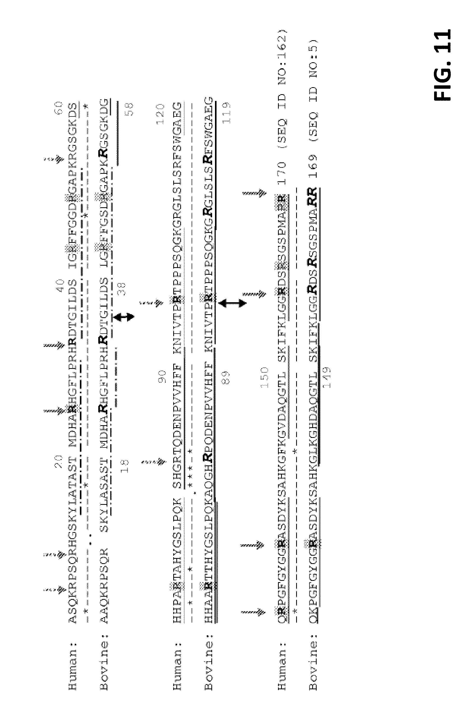
D00021

D00022

D00023
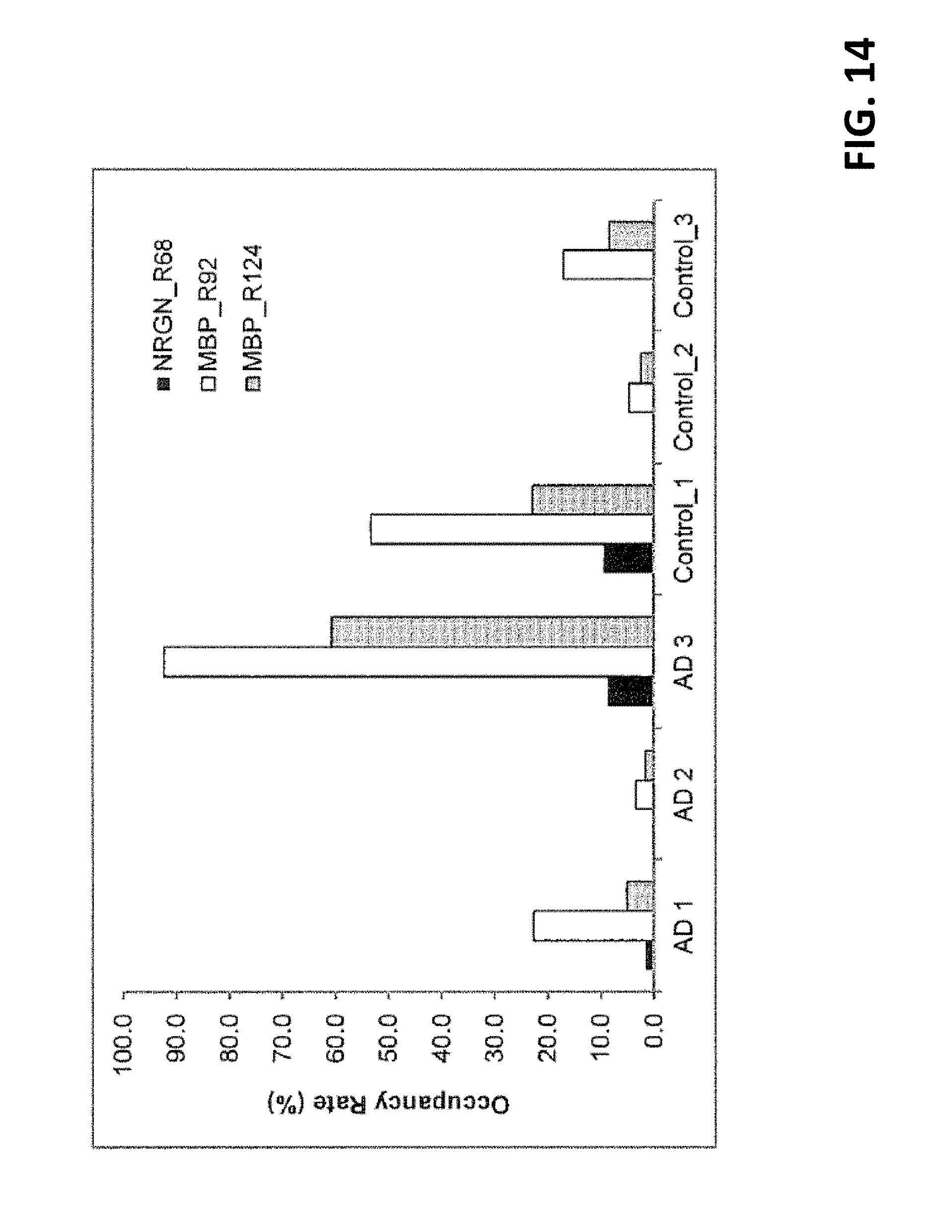
S00001
XML
uspto.report is an independent third-party trademark research tool that is not affiliated, endorsed, or sponsored by the United States Patent and Trademark Office (USPTO) or any other governmental organization. The information provided by uspto.report is based on publicly available data at the time of writing and is intended for informational purposes only.
While we strive to provide accurate and up-to-date information, we do not guarantee the accuracy, completeness, reliability, or suitability of the information displayed on this site. The use of this site is at your own risk. Any reliance you place on such information is therefore strictly at your own risk.
All official trademark data, including owner information, should be verified by visiting the official USPTO website at www.uspto.gov. This site is not intended to replace professional legal advice and should not be used as a substitute for consulting with a legal professional who is knowledgeable about trademark law.