Biomarker, Method For Searching Disease-related Gene, And Renal Cancer Marker
Ueda; Koji ; et al.
U.S. patent application number 16/345539 was filed with the patent office on 2019-10-10 for biomarker, method for searching disease-related gene, and renal cancer marker. This patent application is currently assigned to JAPANESE FOUNDATION FOR CANCER RESEARCH. The applicant listed for this patent is JAPANESE FOUNDATION FOR CANCER RESEARCH, OSAKA UNIVERSITY. Invention is credited to Kazutoshi Fujita, Kentarou Jingushi, Norio Nonomura, Naomi Ohnishi, Kazutake Tsujikawa, Koji Ueda, Motohide Uemura.
| Application Number | 20190310258 16/345539 |
| Document ID | / |
| Family ID | 62023642 |
| Filed Date | 2019-10-10 |


| United States Patent Application | 20190310258 |
| Kind Code | A1 |
| Ueda; Koji ; et al. | October 10, 2019 |
BIOMARKER, METHOD FOR SEARCHING DISEASE-RELATED GENE, AND RENAL CANCER MARKER
Abstract
Provided is a method wherein an affected tissue and a normal tissue obtained from the vicinity of the affected tissue are left at rest in a culture medium, and a disease-specific biomarker is searched for in an exudate therefrom. A biomarker specific to renal cell carcinoma has been found by this method.
| Inventors: | Ueda; Koji; (Tokyo, JP) ; Ohnishi; Naomi; (Tokyo, JP) ; Nonomura; Norio; (Suita-shi, Osaka, JP) ; Uemura; Motohide; (Suita-shi, Osaka, JP) ; Fujita; Kazutoshi; (Suita-shi, Osaka, JP) ; Tsujikawa; Kazutake; (Suita-shi, Osaka, JP) ; Jingushi; Kentarou; (Suita-shi, Osaka, JP) | ||||||||||
| Applicant: |
|
||||||||||
|---|---|---|---|---|---|---|---|---|---|---|---|
| Assignee: | JAPANESE FOUNDATION FOR CANCER
RESEARCH Tokyo JP OSAKA UNIVERSITY Suita-shi, Osaka JP |
||||||||||
| Family ID: | 62023642 | ||||||||||
| Appl. No.: | 16/345539 | ||||||||||
| Filed: | October 26, 2017 | ||||||||||
| PCT Filed: | October 26, 2017 | ||||||||||
| PCT NO: | PCT/JP2017/038812 | ||||||||||
| 371 Date: | April 26, 2019 |
| Current U.S. Class: | 1/1 |
| Current CPC Class: | G01N 33/68 20130101; G01N 33/574 20130101; G01N 33/50 20130101; C12Q 1/6886 20130101; G01N 2333/4721 20130101; C12Q 2600/158 20130101; G01N 33/57488 20130101; G01N 33/92 20130101; G01N 33/57438 20130101 |
| International Class: | G01N 33/574 20060101 G01N033/574; C12Q 1/6886 20060101 C12Q001/6886 |
Foreign Application Data
| Date | Code | Application Number |
|---|---|---|
| Oct 28, 2016 | JP | 2016-211239 |
Claims
1-10. (canceled)
11. A search method, comprising: immersing resected diseased tissue in an immersion liquid; analyzing an exuded component derived from the diseased tissue exuded from the diseased tissue in the immersion liquid; and identifying a biomarker and/or disease-related gene derived from the diseased tissue.
12. The search method according to claim 11, comprising: immersing normal tissue obtained from around the resected diseased tissue in an immersion liquid; analyzing an exuded component derived from the normal tissue exuded from the normal tissue in the immersion liquid; identifying a biomarker and/or disease-related gene derived from the normal tissue; and selecting a disease-specific biomarker and/or disease-related gene by comparatively examining the biomarker and/or disease-related gene derived from the diseased tissue and the biomarker and/or disease-related gene derived from the normal tissue.
13. The search method according to claim 11, wherein the exuded component is an extracellular vesicle, a protein, a nucleic acid or a lipid.
14. The search method according to claim 12, wherein the exuded component is an extracellular vesicle, a protein, a nucleic acid or a lipid.
15. The search method according to claim 12, wherein the exuded component is an extracellular vesicle, and the biomarker and/or disease-related gene contained in the extracellular vesicle is a protein, a nucleic acid or a lipid.
16. The search method according to claim 14, wherein the exuded component is an extracellular vesicle, and the biomarker and/or disease-related gene contained in the extracellular vesicle is a protein, a nucleic acid or a lipid.
17. The search method according to claim 12, wherein a disease is cancer, neurodegenerative disease, multiple sclerosis, diabetes, liver disease, autism or cerebral infarction.
18. The search method according to claim 14, wherein a disease is cancer, neurodegenerative disease, multiple sclerosis, diabetes, liver disease, autism or cerebral infarction.
19. The search method according to claim 16, wherein a disease is cancer, neurodegenerative disease, multiple sclerosis, diabetes, liver disease, autism or cerebral infarction.
20. A test method for renal cell carcinoma, comprising: detecting at least one biomarker out of biomarkers listed in Tables 1, 2 and 4 contained in extracellular vesicles contained in a body fluid; and comparing with a prescribed value.
21. The test method for renal cell carcinoma according to claim 20, wherein the biomarker is AZU1, CA9, STBD1, COMT or GYG1.
22. The test method for renal cell carcinoma according to claim 21, wherein the biomarker is AZU1.
23. The test method for renal cell carcinoma according to claim 21, wherein the body fluid is blood or urine.
24. The test method for renal cell carcinoma according to claim 22, wherein the body fluid is blood or urine.
25. A method for screening a therapeutic agent for renal cell carcinoma using AZU1, CA9, STBD1, COMT or GYG1 as a target, comprising: selecting a candidate compound by using AZU1, CA9, STBD1, COMT or GYG1 as an index.
26. The method for screening a therapeutic agent for renal cell carcinoma according to claim 25, wherein using AZU1 as the target, comprising: selecting a candidate compound by using AZU1 as the index.
Description
TECHNICAL FIELD
[0001] The present invention relates to a method for identifying a biomarker or disease-related gene from separated/purified extracellular vesicles, and a test method. Besides, the present invention relates to a renal cancer marker obtained by using the search method.
BACKGROUND ART
[0002] A biomarker refers to an in vivo substance or image data correlating with a normal process or pathological process of a body, or a pharmacological reaction against treatment, and is used as an objective index of a body condition. Biomarkers include various information such as so-called clinical laboratory values of a biochemical test, a blood test and tumor markers, and diagnostic imaging data of CT and MRI. In particular, a biomarker indicating the presence or the progression of a disease as an index of the disease is indispensable for the present medical care for finding, diagnosing and making prognostic prediction of the disease. Besides, a biomarker is used in development of a new drug for selection and evaluation of a compound to be developed.
[0003] In recent years, a large number of biomarkers have been discovered as a result of development of techniques for enabling detection of a tiny amount of protein or nucleic acid and development of genome analysis and proteome analysis. Among these, however, a very small number of markers are actually used in clinical practice. Particularly for diseases such as cancers and neurodegenerative diseases exhibiting little subjective symptoms at an early stage of the diseases, a biomarker useful as a tool for diagnosis or drug discovery is desired. Few biomarkers are, however, currently practically used.
[0004] For example, renal cell carcinoma causes few symptoms in many cases, and is found after considerable progression in most cases. Although renal cell carcinoma is recently accidentally found in more cases through ultrasonic echography or CT scan carried out for medical examination or another disease, it is still regarded as cancer difficult to find at an early stage. Patent Literatures 1 to 3 report biomarkers for renal cell carcinoma, but these biomarkers have not been put to practical use yet.
[0005] In recent years, liquid biopsy (diagnosis using a body fluid) is attracting attention in the field of clinical diagnosis using a biomarker. In the field of cancer diagnosis employing the liquid biopsy, a body liquid is used as a specimen, without collecting tumor tissue as in the conventional biopsy, for detecting a cancer cell or non-invasively or minimally invasively detecting a disease by measuring a biomarker.
[0006] As an analysis object of the liquid biopsy, a circulating tumor cell (CTC) and a DNA derived from a cancer cell (circulating tumor DNA; ctDNA) are well known. A CTC or ctDNA is, however, presumed to be detected in metastasis of a cancer cell, and hence is regarded to be suitable for prognostic prediction of cancer but unusable for early diagnosis. Therefore, as an analysis object to be used for the early diagnosis of cancers and other diseases, extracellular vesicles (hereinafter sometimes referred to as EVs) have started to attract attention.
[0007] The extracellular vesicles are vesicles released from almost all cells, and are roughly divided, depending on the size and marker molecule to be presented, into exosomes, microvesicles and apoptotic bodies. In recent years, it has been clarified that the extracellular vesicles play various roles. In particular, it has been clarified that the exosomes and the microvesicles contain nucleic acids such as mRNA and microRNA and proteins, and are involved in communication not only between close cells but also between organs (Non Patent Literatures 1 to 4).
[0008] It has been reported that the extracellular vesicles are involved, regarding cancer, in development of the cancer including angiogenesis, immunosuppression and metastasis. Besides, based on the composition of a special integrin contained in the extracellular vesicles, it has been suggested that there is a possibility of the extracellular vesicles preparing for metastasis of cancer cells (Non Patent Literature 5). In this manner, the extracellular vesicles are involved also in the development of a disease, and proteins and nucleic acids contained in the extracellular vesicles are changed depending on the state of a living body. Accordingly, a protein or a nucleic acid to be detected using the extracellular vesicles varies depending on the type or state of the disease, and the extracellular vesicles are known to work as biomarkers.
[0009] Results of studies conventionally reported are, however, based on mainly cultured cells, and there is no data of comparison of microvesicles obtained from a disease site and a normal site having the same histological background. Therefore, it has been doubted whether or not they function as a biomarker effective for disease diagnosis or new drug development.
CITATION LIST
Patent Literature
[0010] Patent Literature 1: Japanese Patent Laid-Open No. 2016-86678 [0011] Patent Literature 2: Japanese Patent Laid-Open No. 2013-140030 [0012] Patent Literature 3: National Publication of International Patent Application No. 2015-505965
Non Patent Literature
[0012] [0013] Non Patent Literature 1: Kahlert, C., & Kalluri, R., J. Mol. Med. (Berl)., 2013, Vol. 91(4), p. 431-437. [0014] Non Patent Literature 2: An, T. et al., J. Extracell. Vesicles, 2015, Vol. 4, 27522. [0015] Non Patent Literature 3: Peinado, H. et al., Nat. Med., 2012, Vol. 18(6), p. 883-891 [0016] Non Patent Literature 4: Costa-Sliva, B. et al., Nat. Cell Biol., 2015, Vol. 17(6), p. 816-826. [0017] Non Patent Literature 5: Hoshino, A. et al., Nature, 2015, Vol. 527(7578), p. 329-335. [0018] Non Patent Literature 6: Lasser, C., et al, J. Vis. Exp., 2012, Vol. 59, e3037, doi:10.3791/3037. [0019] Non Patent Literature 7: Geissler, K. et al., Oncoimmunology, 2015, Vol. 4, e985082. [0020] Non Patent Literature 8: Teltsh, O., et al., Oncotarget, 2015, Vol. 32, p. 33191-33205. [0021] Non Patent Literature 9: Guldur, M. E. et al., J. Pak. Med. Assoc., 2014, Vol. 3, p. 300-303. [0022] Non Patent Literature 10: Bussolati, B. et al., FASEB J., 2003, Vol. 17(9), p. 1159-1161. [0023] Non Patent Literature 11: Wiklander, O. P. et al., J. Extracell. Vesicles, 2015, Vol. 4, 26316. [0024] Non Patent Literature 12: Tominaga, N. et al., Adv. Drug Deliv. Rev., 2015, Vol. 95, p. 50-55. [0025] Non Patent Literature 13: Lai, C. P. et al., ACS Nano., 2014, Vol. 8(1), p. 483-494. [0026] Non Patent Literature 14: Gruenwald, V. et al., BMC Cancer, 2010, 10:695. [0027] Non Patent Literature 15: Vroling, L. et al., Angiogenesis, 2009, Vol. 12(1), p. 69-79. [0028] Non Patent Literature 16: del Puerto-Nevado, L. et al., Br. J. Cancer, 2014, Vol. 110, p. 2700-2707. [0029] Non Patent Literature 17: Shankhajit, D. et al., International Journal of Nutrition, Pharmacology, Neurological diseases, 2012, Vol. 2, p. 3-7.
SUMMARY OF INVENTION
Technical Problem
[0030] An object of the present invention is to provide a method for searching a novel biomarker from biological components contained in extracellular vesicles usable in liquid biopsy. Another object is to provide a novel search method for disease-related gene. Besides, a novel biomarker for renal cell carcinoma, for which an effective biomarker has not been found, and a test method are provided.
Solution to Problem
[0031] The present invention relates to a method for searching a biomarker or disease-related gene derived from diseased tissue, a test method for renal cell carcinoma, a biomarker for renal cell carcinoma, and a method for screening a therapeutic agent for renal cell carcinoma.
(1) A search method, comprising: immersing resected diseased tissue in an immersion liquid; analyzing an exuded component derived from the diseased tissue exuded from the diseased tissue in the immersion liquid; and identifying a biomarker and/or disease-related gene derived from the diseased tissue. (2) The search method according to (1), comprising: immersing normal tissue obtained from around the resected diseased tissue in an immersion liquid; analyzing an exuded component derived from the normal tissue exuded from the normal tissue in the immersion liquid; identifying a biomarker and/or disease-related gene derived from the normal tissue; and selecting a disease-specific biomarker and/or disease-related gene by comparatively examining the biomarker and/or disease-related gene derived from the diseased tissue and the biomarker and/or disease-related gene derived from the normal tissue. (3) The search method according to (1) or (2), wherein the exuded component is an extracellular vesicle, a protein, a nucleic acid or a lipid. (4) The search method according to any one of (1) to (3), wherein the exuded component is an extracellular vesicle, and the biomarker and/or disease-related gene contained in the extracellular vesicle is a protein, a nucleic acid or a lipid. (5) The search method according to any one of (1) to (4), wherein the disease is cancer, neurodegenerative disease, multiple sclerosis, diabetes, liver disease, autism or cerebral infarction. (6) A test method for renal cell carcinoma, comprising: separating an extracellular vesicle contained in a body fluid; detecting at least one biomarker out of biomarkers listed in Tables 1, 2 and 4 contained in the extracellular vesicle; and comparing with a prescribed value. (7) The test method for renal cell carcinoma according to (6), wherein the biomarker is AZU1, CA9, STBD1, COMT or GYG1. (8) The test method for renal cell carcinoma according to (6) or (7), wherein the body fluid is blood or urine. (9) Biomarkers for renal cell carcinoma, listed in Tables 1, 2 and 4. (10) A method for screening a therapeutic agent for renal cell carcinoma using AZU1, CA9, STBD1, COMT or GYG1 as a target, comprising: selecting a candidate compound by using AZU1, CA9, STBD1, COMT or GYG1 as an index.
BRIEF DESCRIPTION OF DRAWINGS
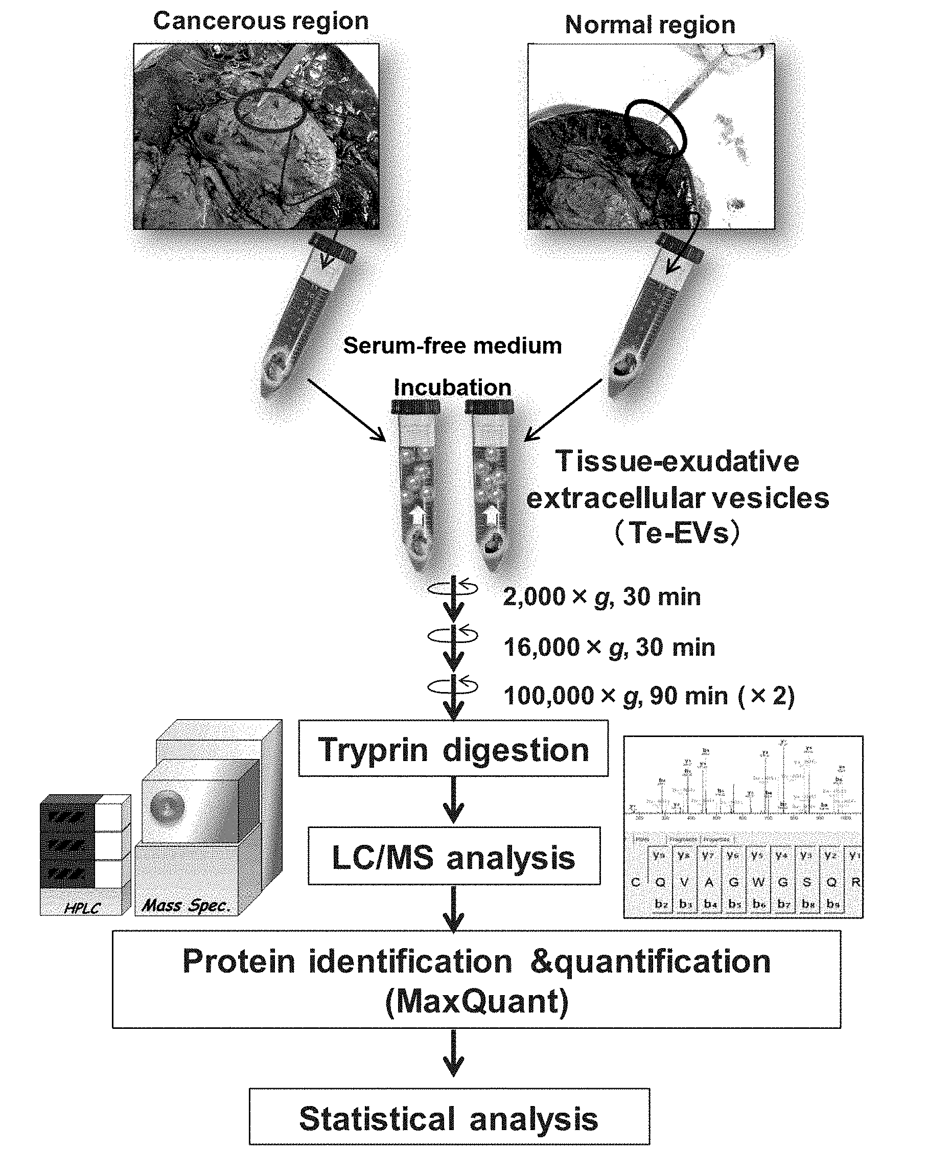
[0032] FIG. 1 is a diagram illustrating the outline of a method for isolating tissue-exudative extracellular vesicles and for searching a biomarker.
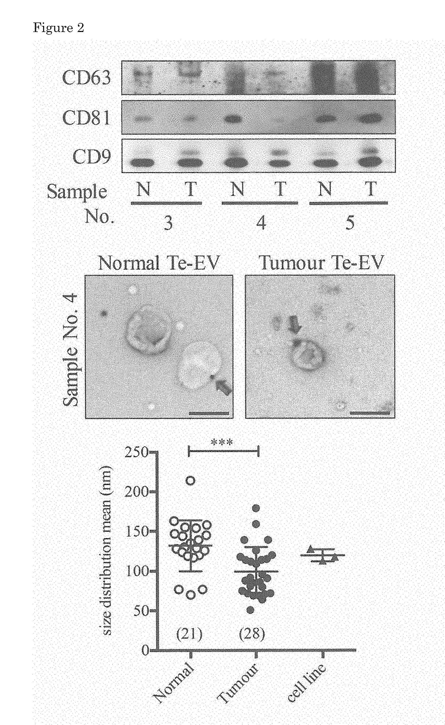
[0033] FIG. 2 is a diagram illustrating marker analysis and size distribution of obtained tissue-exudative extracellular vesicles.
[0034] FIG. 3 is a diagram illustrating the results of analysis, by mass spectrometry, of expression of tetraspanin family molecules in the tissue-exudative extracellular vesicles.
[0035] FIG. 4 illustrates the analysis results of the tissue-exudative extracellular vesicles by gene ontology analysis. FIG. 4A illustrates the numbers of proteins identified in tissue-exudative extracellular vesicles obtained from renal cell carcinoma tissue and normal tissue in the vicinity of the carcinoma tissue. FIG. 4B, FIG. 4C and FIG. 4D are diagrams listing biological properties of proteins classified into three categories of cellular components, biological processes and molecular functions, respectively.
[0036] FIG. 5 is a volcano plot illustrating the results of analysis, by corresponding t-test, of protein expression in extracellular vesicles derived from normal tissue and tumor tissue.
[0037] FIG. 6 is a diagram illustrating expression of AZU1 in extracellular vesicles derived from normal tissue and tumor tissue. FIG. 6A is a diagram illustrating the expression of AZU1 by using samples obtained from the same patient as a pair. FIG. 6B is a diagram illustrating correlation between the stage of the cancer and the expression of AZU1. FIG. 6C is a diagram in which the expression of AZU1 is analyzed by Western blotting. FIG. 6D is a diagram illustrating an amount of AZU1 in a serum sample.
[0038] FIG. 7 illustrates disintegration, depending on AZU1, of a vascular endothelial layer caused by extracellular vesicles derived from a renal cell carcinoma cell line. FIG. 7A is a diagram illustrating the expression and localization of AZU1 in immunoelectron microscopic images. FIG. 7B is a diagram illustrating the expression level of AZU1 and its content in secreted extracellular vesicles in the renal cell carcinoma cell line. FIG. 7C is a diagram illustrating results of TEER analysis using extracellular vesicles secreted from the cell line. FIG. 7D is a Western blot diagram illustrating an amount of AZU1 in the extracellular vesicles secreted from ACHN cell line in which AZU1 is forcedly expressed. FIG. 7E illustrates immunoelectron microscopic images of localization of forcedly expressed AZU1. FIG. 7F is a diagram illustrating results of the TEER analysis using extracellular vesicles secreted from the ACHN cell line in which AZU1 is forcedly expressed.
[0039] FIG. 8 illustrates disintegration of a vascular endothelial layer caused by extracellular vesicles derived from a patient. FIGS. 8A and 8B are diagrams illustrating results of the TEER analysis using tissue-exudative extracellular vesicles derived from patient tissue. FIG. 8C is a microscopic image illustrating incorporation into HUVEC cells of tissue-exudative extracellular vesicles derived from tumor tissue and normal tissue. FIG. 8D is a diagram illustrating results of the TEER analysis using tissue-exudative extracellular vesicles derived from a patient.
DESCRIPTION OF EMBODIMENTS
[0040] Now, a method for isolating extracellular vesicles from tissue and for analyzing a biomarker, and a method for searching disease-related gene will be described in detail by exemplarily describing search for a renal cell carcinoma marker, but it is noted that a search method for a biomarker and disease-related gene of the present invention is applicable to not only renal cell carcinoma but also any disease. In particular, for cancer to be treated by surgical resection of diseased tissue, a useful biomarker and disease-related gene can be searched by the method of the present invention. Besides, as the diseased tissue, not only tissue surgically obtained but also tissue obtained by autopsy can be used, and the present invention is also applicable to various diseases for which effective biomarkers have not been found yet, such as neurodegenerative diseases including amyotrophic lateral sclerosis, Parkinson's disease and Alzheimer's disease, multiple sclerosis, prostate cancer, pancreatic cancer, diabetes, liver disease, developmental disorder such as autism, and cerebral infarction.
[0041] In the case of, for example, cancer to be treated by surgical resection, a so-called non-cancerous part (normal tissue) around a cancerous part is also resected. Therefore, tissues of both a disease site and a normal site can be obtained from the same patient, and thus, samples having the same genetic background can be obtained. Accordingly, when extracellular vesicles obtained from these samples are analyzed, proteins and nucleic acids expressed irrespectively of the disease of interest in the individual can be excluded. As a result, a biomarker specific to the disease can be searched for. Besides, in the case where completely normal tissue is unavailable as in neurodegenerative disease, tissues can be obtained from a group of patients different in severity and the degree of progression to be subjected to comparative quantitative analysis, and thus, disease-related gene and a biomarker can be specified. Furthermore, a biomarker exuded from tissue and information of the biomarker obtained from a body fluid such as a serum can be used in combination.
[0042] Alternatively, in addition to the tissue obtained from a patient, tissue can be obtained from a model animal for searching a biomarker. Also for a disease such as neurodegenerative disease and diabetes for which no tissue is resected from a patient for treatment, a biomarker can be searched by using a model animal.
[0043] Besides, disease-specific biomarkers thus searched are exuded from diseased tissue, and those contained in extracellular vesicles secreted into a body fluid such as urine or blood can be selected among these to be used as a disease marker for use in liquid biopsy.
[0044] Furthermore, since a large number of biological components such as proteins are contained in extracellular vesicles, a large number of disease-specific biomarkers can be found. Among the thus obtained biomarkers, a plurality of biomarkers are selected to be measured in combination, and thus, not only diagnostic sensitivity but also specificity can be increased. When a plurality of markers are used, a disease that could not be found at an early stage can be detected.
[0045] Besides, through analysis of proteins and nucleic acids expressed specifically to a disease, a biological component that relates to the disease and can be a target of a therapeutic agent for the disease can be found. For example, when the function of a protein highly expressed in a diseased tissue is analyzed, and if it has a function essential for the onset or development of the disease, a pharmaceutical can be developed by using it as a target of a therapeutic agent.
[0046] In addition, when tissue derived from a patient who has become resistant to a molecular target drug is used, extracellular vesicles released from molecular target drug-resistant cells can be captured. A way for braking the resistance can be thus found based on an extracellular vesicle regarded as a replica of a cell.
[0047] Besides, a biomarker thus searched can be used in screening of candidate compounds in studies for developing new drugs. In high throughput screening, the screening is often carried out using a cell line. As a cell line to be used for the screening, cell lines having expression tendency similar to that of a plurality disease markers having been searched by the method of the present invention are selected in advance, and thus, candidate compounds can be accurately narrowed down. Furthermore, the obtained biomarker can be used, in addition to the cell line, for examining an effect of a candidate compound in an animal model or at a stage of clinical trial.
[0048] Now, renal cell carcinoma will be exemplarily described for describing a method for searching a novel biomarker and a test method using the same in detail. It is noted that analysis using human tissue is approved by the ethics committee of each research institution before the analysis. Besides, renal cell carcinoma tissue and normal tissue around the tumor tissue are used for the analysis after informed consent is obtained from a patient.
[Example 1] Method for Isolating Te-EVs
[0049] FIG. 1 illustrates the outline of a method for isolating tissue-exudative extracellular vesicles (tissue-exudative EVs, hereinafter sometimes referred to as the Te-EVs) and analyzing a biomarker. As tissue surgically resected, diseased tissue and normal tissue are cut out and immediately immersed in an immersion liquid. In the case of renal cell carcinoma, a serum-free DMEM is used as the immersion liquid, and the immersion liquid can be appropriately selected depending on tissue from which the Te-EVs are extracted. Since extracellular vesicles are contained in a serum, a serum-free medium is preferably used as the immersion liquid, but a medium containing a serum may be used as long as extracellular vesicles are removed therefrom in advance.
[0050] A piece of tissue collected from a disease site or a normal site is allowed to stand still in the immersion liquid at 4.degree. C. for about 1 hour to cause extracellular vesicles to be secreted. The time for the standing in the immersion liquid can be adjusted in accordance with the amount of the obtained piece of tissue. When the piece of tissue is allowed to stand still in the immersion liquid for a long period of time, a larger number of extracellular vesicles can be obtained. Besides, a temperature condition for immersing the tissue can be any temperature within a range from 0.degree. C. to 37.degree. C. Since a higher temperature increases a secretion rate and increases the amount of extracellular vesicles and the like to be obtained, the time and the temperature for the immersion may be appropriately determined. Alternatively, the immersion can be performed by, instead of still standing, gently stirring the immersion liquid by shaking, inverting or rotating. After causing extracellular vesicles to be sufficiently secreted, the resultant is centrifuged at 2,000 g for 30 minutes to remove the piece of tissue and cells through precipitation. Next, the resultant is centrifuged at 16,000 g for 30 minutes to remove cell debris through precipitation. Furthermore, a resultant supernatant is centrifuged at 100,000 g for 90 minutes to collect the Te-EVs secreted from the diseased tissue or the normal tissue through precipitation. The thus obtained Te-EVs are suspended in PBS, and washed by centrifugation at 100,000 g for 90 minutes. The thus isolated Te-EVs are extracellular vesicles derived from the diseased tissue and the normal tissue obtained from the same patient, and hence have the same genetic background, and therefore, when these are comparatively analyzed, a disease-specific biomarker can be selected.
[0051] The isolated Te-EVs are decomposed, by trypsin digestion, into peptides analyzable by mass spectrometry. The resultant peptides are identified and quantitatively determined by LC/MS (liquid chromatography/mass spectrometry) analysis. Besides, a protein specific to each of the Te-EVs is analyzed by statistical analysis.
[Example 2] Purification of Extracellular Vesicles from Renal Tissue
[0052] Te-EVs were collected to be analyzed for renal cell carcinoma patient from whom both tumor tissue and normal tissue (non-cancerous part) were obtained among 20 renal cell carcinoma patients. Histological diagnosis of renal cell carcinoma was carried out by hematoxylin-eosin staining. The disease stages of the patients classified based on AJCC TNM 6th edition were stages T1a to T3c.
[0053] The degree of purification of the thus obtained extracellular vesicles was analyzed by the Western blotting method, the immunoelectron microscopy, nanoparticle tracking analysis (FIG. 2) and the mass spectrometry (FIG. 3). The Western blotting method was carried out by an ordinary method using 100 ng of each Te-EVs protein obtained as above. Exosome markers of an anti-CD63 monoclonal antibody (8A12), an anti-CD81 monoclonal antibody (12C4) and an anti-CD9 monoclonal antibody (12A12) (all manufactured by Cosmo Bio Co., Ltd.) were used as primary antibodies, an HRP-labeled goat anti-mouse IgG antibody (manufactured by Santa Cruz Biotechnology, Inc.) was used as a secondary antibody, and detection was performed with ECL (ECL Prime western blotting detection reagent, manufactured by GE Healthcare).
[0054] An upper panel of FIG. 2 illustrates the results of the analysis by the Western blotting method of the Te-EVs obtained from three patients. N corresponds to a non-cancerous region (normal tissue), and T corresponds to Te-EVs derived from a cancerous region. In any one of the samples, no matter whether it was obtained from a cancerous region or a non-cancerous region, expression of the exosome markers of the CD63, CD81 and CD9 was found.
[0055] The immunoelectron microscopy was performed basically in accordance with a method of Lasser et al., (Non Patent Literature 6) by using the anti-CD9 monoclonal antibody (12A12) and a 20 nm gold colloid-labeled anti-mouse antibody (manufactured by Abcam Plc.), respectively, as a primary antibody and a secondary antibody. Specifically, 1 .mu.g of a Te-EVs sample was allowed to stand still for 1 hour on a formvar support film having carbon deposited thereon, and then was fixed with 2% paraformaldehyde to be reacted with the primary antibody. Thereafter, the resultant was reacted with the secondary antibody, and was observed with an electron microscope H-7650 (manufactured by Hitachi High-Technologies Corporation). A gold colloid bonded to the CD9 was observed as illustrated with an arrow on the Te-EVs derived from either of the normal tissue and the tumor tissue (middle panel of FIG. 2).
[0056] It was found through the observation under an electron microscope that particle sizes of the Te-EVs derived from the tumor tissue largely varied. Therefore, the particle sizes of the Te-EVs derived from each tissue were measured by the nanoparticle tracking analysis. It is known that renal cell carcinoma tissue contains not only cancer cells but also non-cancerous cells (immune cells, endothelial cells and mast cells) (Non Patent Literatures 7 to 10). Therefore, it is presumed that EVs derived from normal cells are also secreted in addition to the EVs derived from cancer cells. Accordingly, in addition to the Te-EVs, extracellular vesicles secreted from cell lines established from renal cell carcinoma, that is, 786-O, ACHN and Caki-1 cell lines, were similarly isolated, purified and analyzed for a size distribution.
[0057] The 786-O, ACHN and Caki-1 cell lines were all obtained from American Type Culture Collection (ATCC), and cultured in an RPMI medium (manufactured by Wako Pure Chemical Industries Ltd.) supplemented with 10% fetal bovine serum, 100 U/mL penicillin G and 0.1 .mu.g/mL streptomycin. For isolating EVs from the cell line, 5.0.times.10.sup.5 cells were seeded in a 10 cm culture dish and cultured for 48 hours, and EVs secreted into the culture fluid were collected, by the centrifugation similarly to the Te-EVs, to be used for the analysis.
[0058] Te-EVs obtained from 21 normal tissues, Te-EVs obtained from 28 tumor tissues and EVs isolated from the above-described three renal cell carcinoma cell lines were used for analyzing the particle sizes. The nanoparticle tracking analysis was performed under the same conditions by using NanoSight LM10 (manufactured by Malvern Panalytical Ltd.) in which NTA 2.0 analysis software was installed (lower panel in FIG. 2). The value of an average particle size of the EVs obtained from the renal cell carcinoma cell lines fall in a range of the particle size distribution of the Te-EVs derived from the tumor tissues, which suggested that the Te-EVs derived from the tumor tissue contain EVs secreted from cancer cells. Besides, it was clarified that the average particle size is significantly different between the Te-EVs derived from the normal tissue and those derived from the tumor tissue (p<0.001). Since the particle size distribution of the Te-EVs was different between the diseased tissue and the normal tissue obtained from the renal cell carcinoma patient, there is a possibility that the particle size distribution of the Te-EVs is different between those derived from the diseased tissue and those derived from the normal tissue.
[0059] Next, Te-EVs derived from tumor tissues obtained from 20 patients and Te-EVs to be paired derived from normal tissues were used for identifying a tetraspanin molecule, which is known as an exosome marker, by the mass spectrometry, and its expression level was analyzed by the LC/MS (FIG. 3).
[0060] The isolated Te-EVs were reduced with 20 mM dithiothreitol at 100.degree. C. for 10 minutes, followed by alkylation with 50 mM iodoacetamide at an ambient temperature for 45 minutes. Thereafter, the resultant was digested with 5 .mu.l immobilized trypsin (manufactured by Thermo Fisher Scientific K.K.) by rotating/swinging at 1000 rpm at 37.degree. C. for 6 hours. The thus obtained peptide was extracted with ethyl acetate, then desalted using Oasis HLB .mu.-elution plate (manufactured by Waters Corporation), and subjected to the mass spectrometry. In the mass spectrometry, an LTQ-Orbitrap-Velos mass spectrometer directly connected to UltriMate 3000 RSLC nano-flow HPLC system (both manufactured by Thermo Fisher Scientific K.K.) was used. Protein was identified and quantitatively determined through analysis using MaxQuant software.
[0061] In order to examine properties of the obtained Te-EVs, the expression level of the tetraspanin molecule was compared. Fifteen tetraspanin molecules were detected, and it was revealed that the Te-EVs derived from either tissue had a high degree of purification. Besides, it was clarified that the expression of most molecules of the tetraspanin family was reduced in the Te-EVs derived from the tumor tissue as compared with the Te-EVs derived from the normal tissue.
[Example 3] Analysis of Te-EVs of Tumor Tissue and Normal Tissue in Renal Cell Carcinoma
[0062] Te-EVs derived from tumor tissue obtained from 20 patients and Te-EVs to be paired derived from normal tissue around a cancerous region were used for performing the LC/MS analysis in the same manner as described above (FIG. 4). Thus, 3871 proteins in total were identified, and among these, 160 were Te-EVs derived from the normal tissue, 253 were expression specific to Te-EVs derived from the tumor tissue, and 3458 were expressed in both of these tissues (FIG. 4A).
[0063] These genes were classified, in accordance with the DAVID gene ontology analysis, into categories of cellular components (CC; FIG. 4B), biological processes (BP; FIG. 4C) and molecular functions (MF; FIG. 4D), and FIGS. 4B to 4D each illustrate the top 6 protein groups specific to the Te-EVs derived from the whole tissue, the normal tissue or the tumor tissue, respectively.
[0064] According to the gene ontology enrichment analysis, in the Te-EVs derived from the tumor tissue, membrane protein was characteristically present in the proteins classified as the cellular components (FIG. 4B), metabolism-related protein was characteristically present in those classified as the biological processes (FIG. 4C), and expression of nucleotide binding protein was characteristically present in those classified as the molecular functions (FIG. 4D). That there is a difference between proteins contained in the Te-EVs derived from the tumor tissue and the those contained in the Te-EVs derived from the normal tissue means that proteins contained in the extracellular vesicles are also regulated in accordance with various cancer-related events, and indicates that these proteins are useful as cancer markers.
[0065] Among the 3871 proteins thus identified, proteins specifically expressed in the Te-EVs derived from the renal cell carcinoma tissue were analyzed. The Te-EVs derived from the tumor tissue and the normal tissue obtained from the renal tissue of the same patient were analyzed as a pair by a paired t-test. FIG. 5 illustrates proteins found to be significantly different in the expression between the Te-EVs derived from the cancerous region and those derived from the non-cancerous region (p<0.05, fold change .gtoreq.2.0, both sides of dotted lines in FIG. 5). Besides, those with large fold change are illustrated with arrows. The contents of carbonic anhydrase 9 (CA9), starch-binding domain-containing protein 1 (STBD1), azurocidin (AZUL), catechol O-methyltransferase (COMT) and glycogenin-1 (GYG1) were remarkably larger in the Te-EVs derived from the tumor tissue than in the Te-EVs derived from the normal tissue.
[0066] In the Te-EVs derived from the renal cell carcinoma tissue, as compared with the proteins contained in the Te-EVs derived from the normal tissue, the expression of 106 proteins shown in Table 1 (Tables 1-1 and 1-2) or 291 proteins shown in Table 2 (Tables 2-1 to 2-5) was found to be significantly larger or smaller.
TABLE-US-00001 TABLE 1-1 AC number Protein Description Gene name P46976 Glycogenin-1 GYG1 P20160 Azurocidin AZU1 P09104 Gamma-enolase ENO2 Q16790 Carbonic anhydrase 9 CA9 Q96HE7 ERO1-like protein alpha ERO1L A2PYH4 Probable ATP-dependent DNA helicase HFM1 HFM1 Q5VT79 Annexin A8-like protein 2 ANXA8L2 P21964 Catechol O-methyltransferase COMT O95210 Starch-binding domain-containing protein 1 STBD1 P20701 Integrin alpha-L ITGAL Q8N386 Leucine-rich repeat-containing protein 25 LRRC25 O75505 Putative double homeobox protein 2 DUX2 Q9NWQ8 Phosphoprotein associated with glycosphingolipid-enriched PAG1 microdomains 1 P11215 Integrin alpha-M ITGAM P32455 Interferon-induced guanylate-binding protein 1 GBP1 P13726 Tissue factor F3 P19971 Thymidine phosphorylase TYMP Q8IZ83 Aldehyde dehydrogenase family 16 member A1 ALDH16A1 P46821 Microtubule-associated protein 1B MAP1B Q02928 Cytochrome P450 4A11 CYP4A11 Q8IZJ1 Netrin receptor UNC5B UNC5B Q6GTX8 Leukocyte-associated immunoglobulin-like receptor 1 LAR1 P04179 Superoxide dismutase [Mn], mitochondrial SOD2 P05107 Integrin beta-2 ITGB2 Q6NYC8 Phostensin PPP1R18 Q14956 Transmembrane glycoprotein NMB GPNMB Q96FQ6 Protein S100-A16 S100A16 P32942 Intercellular adhesion molecule 3 ICAM3 Q0JRZ9 FCH domain only protein 2 FCHO2 P30453 HLA class I histocompatibility antigen, A-34 alpha chain HLA-A Q8IYT3 Coiled-coil domain-containing protein 170 CCDC170 Q06210 Glutamine-fructose-6-phosphate aminotransferase [isomerizing] 1 GFPT1 Q96A46 Mitoferrin-2 SLC25A28 P13807 Glycogen [starch] synthase, muscle GYS1 Q9NZR1 Tropomodulin-2 TMOD2 O43752 Syntaxin-6 STX6 Q6UWP8 Suprabasin SBSN P30273 High affinity immunoglobulin epsilon receptor subunit gamma FCER1G P10321 HLA class I histocompatibility antigen Cw-7 alpha chain HLA-C Q5T681 Uncharacterized protein C10orf62 C10orf62 P02794 Ferritin heavy chain FTH1 P13611 Versican core protein VCAN Q8N6N2 Tetratricopeptide repeat protein 9B TTC9B P01892 HLA class I histocompatibility antigen, A-2 alpha chain HLA-A P01612 Ig kappa chain V-I region Mey -- P10316 HLA class I histocompatibility antigen, A-69 alpha chain HLA-A Q92614 Unconventional myosin-XVIIIa MYO18A P49454 Centromere protein F CENPF Q92572 AP-3 complex subunit sigma-1 AP3S1 Q13643 Four and a half LIM domains protein 3 FHL3 Q5SNT6 WASH complex subunit FAM21B FAM21B P33908 Mannosyl-oligosaccharide 1,2-alpha-mannosidase IA MAN1A1 P28799 Granulins GRN
TABLE-US-00002 TABLE 1-2 AC number Protein Description Gene name Q01628 Interferon-induced transmembrane protein 3 IFITM3 P20929 Nebulin NEB Q9H2L5 Ras association domaim-containing protein 4 RASSF4 Q8TD55 Pleckstrin homology domain-containing family O member 2 PLEKHO2 Q53T59 HCLS1-binding protein 3 HS1BP3 P49207 60S ribosomal protein L34 RPL34 P31146 Coronin-1A CORO1A P51397 Death-associated protein 1 DAP P39060 Collagen alpha-1(XVIII) chain COL18A1 P02786 Transferrin receptor protein 1 TFRC Q6ZUB1 Spermatogenesis-associated protein 31E1 SPATA31E1 P28065 Proteasome subunit beta type-9 PSMB9 P02792 Ferrtin light chain FTL P34810 Macrosialin CD68 P16188 HLA class I histocompatibility antigen, A-30 alpha chain HLA-A Q9P282 Prostaglandin F2 receptor negative regulator PTGFRN Q00151 PDZ and LIM domain protein 1 PDLIM1 P09972 Fructose-bisphosphate aldolase C ALDOC Q9Y4A5 Transformation/transcription domain-associated protein TRRAP P18084 Integrin beta-5 ITGB5 P30508 HLA class I histocompatibility antigen, Cw-12 alpha chain HLA-C P47914 60S ribosomal protein L29 RPL29 P17693 HLA class I histocompatibility antigen, alpha chain G HLA-G P16989 Y-box-binding protein 3 YBX3 P11169 Solute carrier family 2, facilitated glucose transporter member 3 SLC2A3 P16949 Stathmin STMN1 P30685 HLA class I histocompatibility antigen, B-35 alpha chain HLA-B Q02388 Collagen aplha-1(VII) chain COL7A1 P02747 Complement C1q subcomponent subunit C C1QC Q9ULU4 Protein kinase C-binding protein 1 ZMYND8 P78324 Tyrosine-protein phosphatase non-receptor type substrate 1 SIRPA P04430 Ig kappa chain V-I region BAN -- Q7Z5R6 Amyloid beta A4 precursor protein-binding family B member APBB1IP 1-interacting protein P01714 Ig lambda chain V-III region SH -- P06748 Nucleophosmin NPM1 Q99571 P2X purinoceptor 4 P2RX4 P08575 Receptor-type tyrosine-protein phosphatase C PTPRC Q99426 Tubulin-folding co factor B TBCB Q9NYZ2 Mitoferrin-1 SLC25A37 P05534 HLA class I histocompatibility antigen, A-24 alpha chain HLA-A Q6ZRP7 Sulfhydryl oxidase 2 QSOX2 Q16555 Dihydropyrimidinase-related protein 2 DPYSL2 O43583 Density-regulated protein DENR Q96MI9 Cytosolic carboxypeptidase 4 AGBL1 P62280 40S ribosomal protein S11 RPS11 P27695 DNA-(apurinic or apyrimidinic site) lyase APEX1 Q07812 Apoptosis regulator BAX BAX O15155 BET1 homolog BET1 P10632 Cytochrome P4502C8 CYP2C8 P08670 Vimentin VIM P30622 CAP-Gly domain-containing linker protein 1 CLIP1 P07451 Carbonic anhydrase 3 CA3 P01703 Ig lambda chain V-I region NEWM --
TABLE-US-00003 TABLE 2-1 AC number Protein Description Gene name Q8WWT9 Solute carrier family 13 member 3 SLC13A3 P31639 Sodium/glucose cotransporter 2 SLC5A2 O75264 Transmembrane protein C19orf77 C19orf77 P07148 Fatty acid-binding protein, liver FABP1 Q9UHI7 Solute carrier family member 1 SLC23A1 P16444 Dipeptidase 1 DPEP1 P05062 Fructose-bisphosphate aldolase B ALDOB Q9NZA1 Chloride intracellular channel protein 5 CLIC5 P36269 Gamma-glutamyltransferase 5 GGT5 Q03154 Aminoacylase-1 ACY1 Q92499 ATP-dependent RNA helicase DDX1 DDX1 Q695T7 Sodium-dependent neutral amino acid transporter B(0)AT1 SLC6A19 Q13113 PDZK1-interacting protein 1 PDZK1IP1 Q00592 Podocalyxin PODXL P09467 Fructose-1,6-bisphosphatase 1 FBP1 P21695 Glycerol-3-phosphate dehydrogenase [NAD(+)], cytoplasmic GPD1 Q5T2W1 Na(+)/H(+) exchange regulatory co factor NHE-RF3 PDZK1 Q15599 Na(+)/H(+) exchange regulatory co factor NHE-RF3 SLC9A3R2 Q9BYF1 Angiotensin-converting enzyme 2 ACE2 Q8WW52 Protein FAM151A FAM151A P51580 Thicourine S-methyltransferase TPMT O96013 Serine/threonine-protein kinase PAK 4 PAK4 Q93088 Betaine-homocysteine S-methyltransferase 1 BHMT P07911 Uromodulin UMOD P29972 Aquaporin-1 AQP1 Q07337 Neutral and basic amino add transport protein rBAT SLC3A1 P08473 Neprilysin MME P15144 Aminopeptidase N ANPEP P35558 Phosphoenolpyruvate carboxykinase, cytosolic [GTP] PCK1 O43175 D-3-phosphoglycerate dehydrogenase PHGDH P08729 Keratin, type II cytoskeletal 7 KRT7 O14745 Na(+)/H(+) exchange regulatory co factor NHE-RF1 SLC8A3R1 P09758 Tumor-associated calcium signal transducer 2 TACSTD2 P00742 Coagulation factor X F10 Q9UGT4 Sushi domain-containing protein 2 SUSD2 Q1EHB4 Sodium-coupled monocarboxylate transporter 2 SLC5A12 Q75348 V-type proton ATPase subunit G 1 ATP6VIG1 Q16864 V-type proton ATPase subunit F ATP6VIF Q14894 Thiomorpholine-carboxylate dehydrogenase CRYM Q8Y2J2 Band 4,1-like protein 3 EPB41L3 Q4V9L6 Transmembrane protein 119 TMEM119 P11465 Pregnancy-specific beta-1-glycoprotein 2 PSG2 Q9H0W9 Ester hydrolase C11orf54 C11orf54 P12821 Angiotensin-converting enzyme ACE P13640 Metallothionein-1G MT1G P21266 Glutathione S-transferase Mu 3 GSTM3 P52758 Ribonuclease UK114 HRSP12 P16083 Ribosyldihydronicotinamide dehydrogenese [quinone] NQO2 Q6ZQN7 Solute carrier organic anion transporter family member 4C1 SLCO4C1 P36543 V-type proton ATPase subunit E 1 ATP6VIE1 O75954 Tetraspanin TSPAN9 O43451 Maltase-glucoamylase, intestinal MGAM P11137 Microtubule-associated protein 2 MAP2 P15941 Mucin-1 MUC1 Q9NQ84 G-protein coupled receptor family C group 5 member C GPRC5C P07305 Histone H1.0 H1F0 P53990 IST1 homolog IST1 P05937 Calbindin CALB1
TABLE-US-00004 TABLE 2-2 AC number Protein Description Gene name P50135 Histamine N-methyltransferase HNMT O75131 Copine-3 CPNE3 O60262 Guanine nucleotide-binding protein G(I)/G(S)/G(O) subunit gamma-7 GNG7 Q9Y252 Lambda-crystallin homolog CRYL1 Q16822 Phosphoenolpyruvate carboxykinase [GTP], mitochondrial PCK2 P62330 ADP-ribosylation factor 6 ARF6 Q16625 Occludin OCLN P22748 Carbonic anhydrase 4 CA4 P18859 ATP synthase-coupling factor 6, mitochondrial ATP5J O43895 Xaa-Pro aminopeptidase 2 XPNPEP2 P27487 Dipeptidyl peptidase 4 DPP4 Q9U112 V-type proton ATPase subunit H ATP6VIH P29034 Protein S100-A2 S100A2 Q01740 Dimethylaniline monooxygenase [N-oxide-forming] 1 FMO1 P07195 L-lactate dehydrogenase B chain LDHB P38606 V-type proton ATPase catalytic subunit A ATP6VIA Q8N357 Solute carrier family 35 member F6 SLC35F6 Q14019 Coactosin-like protein COTL1 Q98X6 TBC1 domain family member 10A TBC1D10A P09669 Cytochrome c oxidase subuni 6C COX6C Q9BUT1 3-hydroxybutyrate dehydrogenase type 2 BDH2 P21281 V-type proton ATPase subunit B, brain isoform ATP6VIB2 O75094 Signal recognition particle subunit SRP72 SRP72 Q00796 Sorbitol dehydrogenase SORD Q2LD37 Uncharacterized protein KIAA1109 KIAA1109 Q96C23 Aldose 1-epimerase GALM Q9Y696 Chloride intracellular channel protein 4 CUC4 Q12929 Epidermal growth factor receptor kinase substrate 8 EPS8 P30086 Phosphatidylethanolamine-binding protein 1 PEBP1 O60749 Sorting nexin-2 SNX2 Q96YE9 Cadherin-related family member 2 CDHR2 B2RUZ4 Small integral membrane protein 1 SMM1 P05413 Fatty acid-binding protein heart FABP3 P17813 Endogin ENG P00966 Argininosuccinate synthase ASS1 Q96FL8 Multidrug and toxin extrusion protein 1 SLC47A1 Q92820 Gamma-glutamyl hydrolase GGH O43181 NADH dehydrogenase [ubiquinonel] iron-sulfur protein 4, mitochondrial NDUFS4 Q13228 Selenium-binding protein 1 SELENBP1 Q9HBJ8 Collectrin TMEM27 P13073 Cytochrome c oxidase subunit 4 isoform 1, mitochondrial COX4I1 P09210 Glutathione S-transferase A2 GSTA2 P35241 Radixin ROX P22732 Solute carrier family 2, facilitated glucose transporter member 5 SLC2A5 Q9Y5X3 Sorting nexin-5 SNX5 P98082 Disabled homolog 2 DAB2 Q07075 Glutamyl aminopeptidase ENPEP P21291 Cysteine and glycine-rich protein 1 CSRP1 P29992 Guanine nucleotide-binding protein subunit alpha-11 GNA11 P82980 Retinol-binding protein 5 RBP5 Q9UBI6 Guanine nucleotide-binding protein G(I)/G(S)/G(O) subunit gamma-12 GNG12 Q13277 Syntaxin-3 STX3 O43490 Prominin-1 PROM1 O95747 Serine/threonine-protein kinase OSR1 OXSR1 Q9HD42 Charged multivesicular body protein 1a CHMP1A Q8NGM8 Olfactory receptor 6M1 OR6M1 P05026 Sodium/potassium-transporting ATPase subunit beta-1 ATP1B1 Q9NV59 Pyridoxine-5-phosphate oxidase PNPO P21912 Succinate dehydrogenase [ubiquinone] iron-sulfur subunit, mitochondrial SDHB
TABLE-US-00005 TABLE 2-3 AC number Protein Description Gene name P16930 Fumarylacetoacetase FAH P08195 4F2 cell-surface antigen heavy chain SLC3A2 P14550 Alcohol dehydrogenase [NADP(+)] AKR1A1 Q9H0E2 Toll-interacting protein TOLLIP P56385 ATP synthase subunit e, mitochondrial ATP5I Q9Y5K8 V-type proton ATPase subunit D ATP6V1D Q93099 Homogentisate 1,2-dioxygenase HGD Q9Y6R1 Electrogenic sodium bicarbonate cotransporter 1 SLC4A4 Q16853 Membrane primary amine oxidase AOC3 P54710 Sodium/potassium-transporting ATPase subunit gamma FXYD2 P34896 Serine hydroxymethyltransferase, cytosolic SHMT1 Q9NVD7 Alpha-parvin PARVA Q7Z3B1 Neuronal growth regulator 1 NEGR1 P12277 Creatine kinase B-type CKB O95292 Vesicle-associated membrane protein-associated protein B/C VAPB P63000 Ras-related C3 botulinum toxin substrate 1 RAC1 O15244 Solute carrier family 22 member 2 SLC22A2 Q92736 Ryanodine receptor 2 RYR2 Q13427 Peptidyl-prolyl cis-trans isomerase G PPIG O96019 Actin-like protein 6A ACTL6A P50148 Guanine nucleotide-binding protein G(q) subunit alpha GNAQ P35611 Alpha-adducin ADD1 Q99653 Calcineurin B homologous protein 1 CHP1 P00167 Cytochrome b5 CYB5A Q8NFU3 Thiosulfate sulfurtransferase/rhodanese-like domain-containing protein 1 TSTD1 P19440 Gamma-glutamyltranspeptidase 1 GGT1 Q96IX5 Up-regulated during skeletal muscle growth protein 5 USMG5 P09455 Retinol-binding protein 1 RBP1 P15311 Ezrin EZR O94760 N(G),N(G)-dimethylarginine dimethylaminohydrolase 1 DDAH1 O00499 Myc box-dependent-interacting protein 1 BIN1 P48059 LIM and senescent cell antigen-like-containing domain protein 1 LIMS1 Q16775 Hydroxyacylglutathione hydrolase, mitochondrial HAGH P05023 Sodium/potassium-transporting ATPase subunit alpha-1 ATP1A1 Q8WU39 Marginal zone B- and B1-cell-specific protein MZB1 P80723 Brain acid soluble protein 1 BASP1 P54920 Alpha-soluble NSF attachment protein NAPA O75309 Cadherin-16 CDH16 P62873 Guanine nucleotide-binding protein G(I)/G(S)/G(T) subunit beta-1 GNB1 P98164 Low-density lipoprotein receptor-related protein 2 LRP2 Q9Y2Q5 Ragulator complex protein LAMTOR2 LAMTOR2 P20073 Annexin A7 ANXA7 Q96IU4 Alpha/beta hydrolase domain-containing protein 14B ABHD14B Q9BZV1 UBX domain-containing protein 6 UBXN6 O75936 Gamma-butyrobetaine dioxygenase BBOX1 P50053 Ketohexokinase KHK P00918 Carbonic anhydrase 2 CA2 P11279 Lysosome-associated membrane glycoprotein 1 LAMP1 O75874 Isocitrate dehydrogenase [NADP] cytoplasmic IDH1 P00325 Alcohol dehydrogenase 1B ADH1B Q14699 Raftlin RFTN1 Q8N201 Integrator complex subunit 1 INTS1 Q9NZZ3 Charged multivesicular body protein 5 CHMP5 Q8WUM4 Programmed cell death 6-interacting protein PDCD6IP P09211 Glutathione S-transferase P GSTP1 Q10567 AP-1 complex subunit beta-1 AP1B1 Q96KP4 Cytosolic non-specific dipeptidase CNDP2 Q9H1C7 Cysteine-rich and transmembrane domain-containing protein 1 CYSTM1 P60953 Cell division control protein 42 homolog CDC42
TABLE-US-00006 TABLE 2-4 AC number Protein Description Gene name P24539 ATP synthase subunit b, mitochondrial ATP5F1 P62070 Ras-related protein R-Ras2 RRAS2 Q9UHE5 Probable N-acetyltransferase 8 NAT8 Q8NFJ5 Retinoic acid-induced protein 3 GPRC5A Q13596 Sorting nexin-1 SNX1 Q8N3Y1 F-box/WD repeat-containing protein 8 FBXW8 Q9NZ45 CDGSH iron-sulfur domain-containing protein 1 CISD1 Q9H4A4 Arninopeptidase B RNPEP Q96DG6 Carboxymethylenebutenolidase homolog CMBL Q9ULE6 Paladin PALD1 Q6UX53 Methyltransferase-like protein 7B METTL7B P17174 Aspartate aminotransferase, cytoplasmic GOT1 P55795 Heterogeneous nuclear ribonucleoprotein H2 HNRNPH2 Q9NQR4 Omega-amidase NIT2 NIT2 P51149 Ras-related protein Rab-7a RAB7A P11908 Ribose-phosphate pyrophosphokinase 2 PRPS2 P10599 Thioredoxin TXN P05783 Keratin, type I cytoskeletal 18 KRT18 O94903 Proline synthase co-transcribed bacterial homolog protein PROSC Q9UJ68 Mitochondrial peptide methionine sulfoxide reductase MSRA P15374 Ubiquitin carboxyl-terminal hydrolase isozyme L3 UCHL3 P32119 Peroxiredoxin-2 PRDX2 Q9NVA2 Septin-11 SEPT11 Q6QHC5 Sphingolipid delta(4)-desaturase/C4-hydroxylase DES2 DEGS2 P11233 Ras-related protein Ral-A RALA Q96CX2 BTB/POZ domain-containing protein KCTD12 KCTD12 O95154 Aflatoxin B1 aldehyde reductase member 3 AKR7A3 Q13183 Solute carrier family 13 member 2 SLC13A2 P84077 ADP-ribosylation factor 1 ARF1 P21810 Biglycan BGN P21796 Voltage-dependent anion-selective channel protein 1 VDAC1 Q16270 Insulin-like growth factor-binding protein 7 IGFBP7 P62834 Ras-related protein Rap-1A RAP1A P13473 Lysosome-associated membrane glycoprotein 2 LAMP2 Q00839 Heterogeneous nuclear ribonucleoprotein U HNRNPU Q13418 Integrin-linked protein kinase ILK P51148 Ras-related protein Rab-5C RAB5C P50895 Basal cell adhesion molecule BCAM P62820 Ras-related protein Rab-1A RAB1A Q96F10 Diamine acetyltransferase 2 SAT2 P21283 V-type proton ATPase subunit C 1 ATP6V1C1 Q9HCU5 Prolactin regulatory element-binding protein PREB P68104 Elongation factor 1-alpha 1 EEF1A1 Q9NQV5 PR domain-containing protein 11 PRDM11 Q9UEU0 Vesicle transport through interaction with t-SNAREs homolog 1B VTI1B Q9H2A2 Aldehyde dehydrogenase family 8 member A1 ALDH8A1 Q01650 Large neutral amino acids transporter small subunit 1 SLC7A5 P11142 Heat shock cognate 71 kDa protein HSPA8 Q9NRA2 Sialin SLC17A5 O75165 DnaJ homolog subfamily C member 13 DNAJC13 P61224 Ras-related protein Rap-1b RAP1B P17927 Complement receptor type 1 CR1 Q96A57 Transmembrane protein 230 TMEM230 O94886 Transmembrane protein 63A TMEM63A P60981 Destrin DSTN P30153 Serine/threonine-protein phosphatase 2A 65 kDa regulatory PPP2R1A subunit A alpha isoform Q06830 Peroxiredoxin-1 PRDX1 Q96BW5 Phosphotriesterase-related protein PTER Q6P4A8 Phospholipase B-like 1 PLBD1
TABLE-US-00007 TABLE 2-5 AC number Protein Description Gene name P04216 Thy-1 membrane glycoprotein THY1 P56199 Integrin alpha-1 ITGA1 O95865 N(G),N(G)-dimethylarginine dimethylaminohydrolase 2 DDAH2 Q6IAA8 Ragulator complex protein LAMTOR1 LAMTOR1 P40925 Malate dehydrogenase, cytoplasmic MDH1 O00560 Syntenin-1 SDCBP P54707 Potassium-transporting ATPase alpha chain 2 ATP12A Q02952 A-kinase anchor protein 12 AKAP12 P08183 Multidrug resistance protein 1 ABCB1 Q9Y4F1 FERM, RhoGEF and pleckstrin domain-containing protein 1 FARP1 P02549 Spectrin alpha chain, erythrocytic 1 SPTA1 Q5TZA2 Rootletin CROCC P01116 GTPase KRas KRAS Q15907 Ras-related protein Rab-11B RAB11B Q9H2P9 Diphthine synthase DPH5 O75223 Gamma-glutamylcyclotransferase GGCT Q9H223 EH domain-containing protein 4 EHD4 P30041 Peroxiredoxin-6 PRDX6 P30046 D-dopachrome decarboxylase DDT P22352 Glutathione peroxidase 3 GPX3 Q13200 26S proteasome non-ATPase regulatory subunit 2 PSMD2 P62879 Guanine nucleotide-binding protein G(I)/G(S)/G(T) subunit beta-2 GNB2 P00441 Superoxide dismutase [Cu--Zn] SOD1 P98172 Ephrin-B1 EFNB1 Q13526 Peptidyl-prolyl cis-trans isomerase NIMA-interacting 1 PIN1 P15090 Fatty acid-binding protein, adipocyte FABP4 P00491 Purine nucleoside phosphorylase PNP P18206 Vinculin VCL P14384 Carboxypeptidase M CPM Q96KX1 Uncharacterized protein C4orf36 C4orf36 Q93050 V-type proton ATPase 116 kDa subunit a isoform 1 ATP6V0A1 Q93052 Lipoma-preferred partner LPP Q13576 Ras GTPase-activating-like protein IQGAP2 IQGAP2 Q8IWA5 Choline transporter-like protein 2 SLC44A2 Q15847 Adipogenesis regulatory factor ADIRF Q9Y5Z4 Heme-binding protein 2 HEBP2 Q96CW1 AP-2 complex subunit mu AP2M1 Q9BX97 Plasmalemma vesicle-associated protein PLVAP P26038 Moesin MSN P11277 Spectrin beta chain, erythrocytic SPTB Q92692 Poliovirus receptor-related protein 2 PVRL2 Q15286 Ras-related protein Rab-35 RAB35 Q01995 Transgelin TAGLN P15586 N-acetylglucosamine-6-sulfatase GNS Q9UL25 Ras-related protein Rab-21 RAB21 P05106 Integrin beta-3 ITGB3 P13987 CD59 glycoprotein CD59 P06703 Protein S100-A6 S100A6 O43488 Aflatoxin B1 aldehyde reductase member 2 AKR7A2 Q13423 NAD(P) transhydrogenase, mitochondrial NNT Q9UK41 Vacuolar protein sorting-associated protein 28 homolog VPS28 Q06495 Sodium-dependent phosphate transport protein 2A SLC34A1 Q9Y266 Nuclear migration protein nudC NUDC Q7LBR1 Charged multivesicular body protein 1b CHMP1B O43707 Alpha-actinin-4 ACTN4 Q9UBV8 Peflin PEF1
[0067] Among the 106 proteins found to be increased in the expression, the proteins shown in Table 3 including azurocidin (hereinafter referred to as AZUL) are characteristic with a large difference in the expression level from the proteins contained in the Te-EVs derived from the normal tissue, and hence can be more preferably used as the markers for renal cell carcinoma.
TABLE-US-00008 TABLE 3 Gene Protein Description name AC number Glycogenin-1 GYG1 P46976 Azurocidin AZU1 P20160 Carbonic anhydrase 9 CA9 Q16790 Catechol O-methyltransferase COMT P21964 Starch-binding domain-containing protein 1 STBD1 O95210 Phosphoprotein associated with PAG1 Q9NWQ8 glycosphingolipid-enriched microdomains 1 Tissue factor F3 P13726 Thymidine phosphorylase TYMP P19971 Leukocyte-associated immunoglobulin-like LAIR1 Q6GTX8 receptor 1 Transmembrane glycoprotein NMB GPNMB Q14956 Glutamine--fructose-6-phosphate GFPT1 Q06210 aminotransferase [isomerizing] 1 Glycogen [starch] synthase, muscle GYS1 P13807 High affinity immunoglobulin epsilon receptor FCER1G P30273 subunit gamma Versican core protein VCAN P13611 Granulins GRN P28799 Prostaglandin F2 receptor negative regulator PTGFRN Q9P2B2 Solute carrier family 2, facilitated glucose SLC2A3 P11169 transporter member 3 Tyrosine-protein phosphatase non-receptor type SIRPA P78324 substrate 1 Nucleophosmin NPM1 P06748 Receptor-type tyrosine-protein phosphatase C PTPRC P08575 Density-regulated protein DENR O43583 DNA-(apurinic or apyrimidinic site) lyase APEX1 P27695 Cytochrome P450 2C8 CYP2C8 P10632 Vimentin VIM P08670 CAP-Gly domain-containing linker protein 1 CLIP1 P30622 Carbonic anhydrase 3 CA3 P07451
[0068] In particular, carbonic anhydrase 9, catechol O-methyltransferase, phosphoprotein associated with glycosphingolipid-enriched microdomains 1, leukocyte-associated immunoglobulin-like receptor 1, transmembrane glycoprotein NMB, high affinity immunoglobulin epsilon receptor subunit gamma, solute carrier family 2, facilitated glucose transporter member 3, tyrosine-protein phosphatase non-receptor type substrate 1, receptor-type tyrosine-protein phosphatase C, vimentin and carbonic anhydrase 3 are regarded as particularly useful markers because these have been reported to be highly expressed in renal cancer. Besides, phosphoprotein associated with glycosphingolipid-enriched microdomains 1 and tyrosine-protein phosphatase non-receptor type substrate 1 are regarded as promising candidates for a target for drug discovery because these make contribution to inhibition of T cell activity and immune evasion by cancer cells when highly expressed. One of these markers may be singly used, or when a plurality of markers are used in combination, the specificity and the sensitivity can be increased. Further, an existing biomarker may be used in combination for diagnosis.
[0069] Although the analysis results of the proteins contained in the Te-EVs are herein described, any biological component contained in the Te-EVs may be analyzed to be used as a biomarker. Examples of such a biological component include a nucleic acid and a lipid.
[Example 4] Analysis of AZU1
[0070] The results of detailed analysis of AZU1 farthest from the origin of the volcano plot (p=2.85.times.10.sup.-3, fold-change: 31.59, shown with an arrow AZU1 in FIG. 5) will now be described, and it goes without saying that the other proteins described above can be similarly used as the markers.
[0071] FIG. 6A illustrates the concentrations of AZU1 in the Te-EVs derived from the normal tissue and the tumor tissue of 20 cases of renal cell carcinoma patients. In all the 20 renal cell carcinoma patients used for the analysis by the mass spectrometry, the expression of AZU1 increased in the Te-EVs derived from the tumor tissue. FIG. 6B illustrates the concentrations of AZU1 depending on the progression of the cancer, and as renal cell carcinoma progressed, the amount of AZU1 contained in the Te-EVs increased. In a cancerous region of renal cell carcinoma at stage T3 (T3a and T3b), the content of AZU1 was significantly higher than in a non-cancerous region. Besides, as illustrated in an enlarged view in FIG. 6B, the content of AZU1 was different even in early stages of cancer (T1a to T2a) as compared with that in the Te-EVs derived from the normal tissue.
[0072] Besides, the increase of the expression of AZU1 in accordance with the progression of cancer was checked by the Western blotting (FIG. 6C). Five hundred ng per lane of EVs protein was separated by electrophoresis and then transferred onto a film, and the detection was performed in the same manner as in Example 2 by using an anti-AZU1 monoclonal antibody (manufactured by Abcam Plc.) as a primary antibody. As a result of the analysis of Te-EVs obtained from four patients at stages of T1a to T3b, it was confirmed, in all samples, that the expression of AZU1 remarkably increased in Te-EVs obtained from a cancerous region (T) as compared with Te-EVs obtained from a non-cancerous region (N).
[0073] It has been reported that extracellular vesicles derived from cancer are detected also in a serum (Non Patent Literatures 3 to 5 and 11 to 13). Therefore, the amount of AZU1 in EVs contained in a serum sample was measured by quantitative mass spectrometry (FIG. 6D).
[0074] The EVs contained in the serum sample was purified by using an EVSecond column (manufactured by GL Sciences Inc.). AZU1 was not at all detected in EVs contained in serums of 10 cases of healthy persons, but AZU1 was detected in 10 cases out of 19 cases of renal cell carcinoma patients. Although the AZU1 content did not increase in accordance with the progression of cancer as in the Te-EVs derived from the renal cell carcinoma tissue, it was detected at a high ratio in EVs contained in serums of cancer patients at early stages of T1a to T2b. This result reveals that a renal cell carcinoma test for detecting AZU1 using a serum is effective.
[0075] That AZU1 was detected in the extracellular vesicles present in the serum of a renal cell carcinoma patient indicates that a protein different in the content in Te-EVs can be detected in the serum. Accordingly, it indicates that a biomarker found by this method can function as a disease marker with which a test can be performed by using a serum.
[Example 5] Analysis of Effect of AZU1 in Renal Cell Carcinoma
[0076] Since the expression of AZU1 was detected specifically to the renal cell carcinoma tissue, the biological effect of AZU1 was examined. It is known that angiogenesis is generated largely to construct microenvironment of tumor tissue in renal cell carcinoma (Non Patent Literatures 14 to 17). Therefore, the effect of AZU1 on the form of vascular endothelial cells was examined.
[0077] First, localization of intrinsic AZU1 was examined under an immunoelectron microscope. The localization of AZU1 was examined in Te-EVs obtained from normal tissue and tumor tissue of the same patient by using an anti-AZU1 antibody obtained by immunizing a rabbit (manufactured by Abcam Plc.) and an anti-CD9 monoclonal antibody as primary antibodies, and secondary antibodies labeled with colloidal gold particles having different sizes (FIG. 7A).
[0078] An antibody labeled with 40 nm colloidal gold particles was used as an anti-rabbit antibody, and an antibody labelled with 20 nm colloidal gold particles was used as an anti-mouse antibody. Accordingly, a large particle corresponds to the expression of AZU1 and a small particle corresponds to the expression of CD9 in FIG. 7A. Although the expression of CD9 was found on the surface of the Te-EVs obtained from either of the normal tissue and the tumor tissue, the expression of AZU1 was found on the surface of the Te-EVs derived from the tumor tissue alone.
[0079] The AZU1 expression in a renal cell carcinoma cell line was checked by the Western blotting (FIG. 7B). The expression of AZU1 in EVs isolated from 786-O, ACHN, Caki-1 and Caki-2 (obtained from ATCC) cell lines and in a cell lysate (whole cell lysate) was examined. The expression of AZU1 was found in any of these cells, and although AZU1 was detected in the EVs isolated from culture fluids (cultured media EVs, hereinafter sometimes referred to as CM-EVs) of the three cell lines of 786-O, Caki-1 and Caki-2, AZU1 was minimally detected in CM-EVs derived from the ACHN cell line.
[0080] Next, the permeability of the vascular endothelial cells of the CM-EVs obtained from these cell lines was analyzed based on transendothelial electrical resistance (TEER) (FIG. 7C). The TEER was measured by using HUVEC cells (obtained from Gibco). After seeding 4.0.times.10.sup.4 HUVEC cells in a 24-well culture insert (manufactured by Thermo Fisher Scientific K.K.) with a pore size of 0.4 .mu.m and culturing the cells for 4 days, CM-EVs obtained from each cell line was added thereto in a concentration of 10 .mu.g/ml, and the TEER was measured with Millicell.RTM. ERS-2 voltammeter (manufactured by Millipore) to calculate the TEER of each sample in accordance with the following expression:
(Electric resistance (.OMEGA.) of sample well-Electric resistance (.OMEGA.) of empty well).times.culture area (cm.sup.2)=TEER (.OMEGA.cm.sup.2) [Expression 1]
[0081] The results are illustrated in FIG. 7C. As a result of measuring the TEER over time, the TEER of the HUVEC cell sheet was reduced 24 hours after the addition of the CM-EVs. In particular, the TEER was found to be remarkably reduced by addition of CM-EVs obtained from the Caki-1 and 786-O cells in which a larger amount of AZU1 was presented on EVs.
[0082] Since it was presumed that the EVs having a larger amount of AZU1 presented thereon had an effect of increasing permeability, detailed analysis was performed by using a system in which AZU1 was forcedly expressed. AZU1-FLAG (manufactured by Addgene) was introduced into and forcedly expressed in ACHN cells in which AZU1 was minimally detected in CM-EVs. As illustrated in FIG. 7D, in the ACHN cell line in which AZU1-FLAG was forcedly expressed, it was found that EVs also contained a large amount of AZU1-FLAG.
[0083] Besides, it was confirmed under an immunoelectron microscope that the AZU1-FLAG was presented also on the EVs. The detection of the FLAG was performed by using an anti-FLAG monoclonal antibody (manufactured by Sigma-Aldrich) as a primary antibody and using a colloidal gold labeled anti-mouse antibody (FIG. 7E). Although the FLAG was not detected in the CM-EVs obtained from cells to which a vector alone was introduced, the FLAG was detected on the CM-EVs obtained from cells into which the AZU1-FLAG was introduced. It was confirmed based on these results that the forcedly expressed AZU1-FLAG was also presented on the EVs in the same manner as the intrinsic AZU1.
[0084] The thus obtained expression system was used to examine the permeability by using HUVEC (FIG. 7F). It was clarified that CM-EVs obtained from cells in which AZU1 is forcedly expressed remarkably reduces the TEER as compared with CM-EVs obtained from cells into which a vector alone is introduced. Accordingly, it is suggested that AZU1 has an effect of disintegrating the form of vascular endothelial cells.
[0085] Next, examination was made to check whether or not a similar effect can be exhibited by Te-EVs obtained from renal cell carcinoma patients. The permeability of cells was measured by using Te-EVs derived from normal tissue and tumor tissue obtained from six patients at various stages. FIG. 8A illustrates change, over time, of the TEER in each patient after addition of Te-EVs derived from the normal tissue and the tumor tissue, and FIG. 8B is a diagram in which the respective measurement values are plotted together. When the Te-EVs derived from the tumor tissue were added, the TEER was found to be remarkably reduced from 12 hours after the addition. Besides, when the Te-EVs obtained from a patient with cancer in an advanced stage were added, the TEER was more remarkably reduced.
[0086] Examinations were made to check whether or not the difference in the effect on the TEER of the Te-EVs derived from the normal tissue and the tumor tissue was caused by incorporation into cells of the Te-EVs derived from these tissues. The Te-EVs derived from the normal tissue and the tumor tissue were respectively labeled with PKH-67 and PKH-26 (manufactured by Sigma Aldrich) and then mixed, the resultant mixture was added to a culture fluid of HUVEC cells, and the resultant was observed under a microscope 12 hours later. FIG. 8C illustrates the results obtained by using the Te-EVs obtained from two patients. In either sample, Te-EVs derived from the normal tissue looking bright around a nucleus (stained green under a fluorescence microscope) and Te-EVs derived from the tumor tissue recognized as a dark stained image (stained red under a fluorescence microscope; two portions are shown with arrows in each image) were both detected to the same extent. Accordingly, it was confirmed that the incorporation efficiency of Te-EVs did not vary depending on the tissue from which the Te-EVs were derived. In other words, it was revealed that the reduction of the TEER caused by the Te-EVs derived from the tumor tissue was not caused by a difference in the incorporation efficiency of Te-EVs. Therefore, the reduction is probably caused by proteins and the like including AZU1 presented on the Te-EVs.
[0087] In blood of a patient, Te-EVs derived from tumor tissue and normal tissue are circulating in a mixed state. It was examined whether or not the reduction of the TEER was induced in such a mixed state. FIG. 8D illustrates the TEER measured over time after adding, to a culture fluid of HUVEC cells, a mixture of Te-EVs derived from normal tissue and tumor tissue obtained from three patients. The reduction of the TEER was observed in the Te-EVs obtained from any one of the patients even if the mixture of the Te-EVs derived from tumor tissue and normal tissue was used. This result seems to reflect a phenomenon actually occurring in a body of a patient, and hematogenous metastasis seems to be induced by AZU1.
[0088] Based on the above-described results, it is presumed that AZU1 is a disease-related gene closely involved in the onset and development of renal cell carcinoma. Accordingly, a therapeutic agent can be created by using AZU1 as a target. In this manner, when the function of a biological component such as a protein or a nucleic acid expressed specifically to a disease is analyzed, a target of a novel therapeutic agent can be found.
[Example 6] Analysis Using Serum of Patient
[0089] If a protein characteristic to a renal cell carcinoma patient can be detected with EVs contained in a serum, it can be used as a useful marker for early detection of renal cell carcinoma. Therefore, proteins different between a renal cell carcinoma patient and a healthy person were searched for in EVs contained in a serum. Table 4 shows proteins that can be detected in EVs contained in a serum of a renal cell carcinoma patient but cannot be detected in those of a healthy person, excluding those detected as a result of the Te-EVs analysis shown in Tables 1 and 2.
TABLE-US-00009 TABLE 4 AC number Protein Description Gene name P11678 Eosinophil peroxidase EPX Q8NI35 InaD-like protein INADL P68104 Elongation factor 1-alpha 1 EEF1A1 Q63ZY3 KN motif and ankyrin repeat domain- KANK2 containing protein 2 Q16836 Hydroxyacyl-coenzyme A dehydrogenase, HADH mitochondrial P07437 Tubulin beta chain TUBB P55058 Phospholipid transfer protein PLTP Q9Y3R5 Protein dopey-2 DOPEY2 P05543 Thyroxine-binding globulin SERPINA7 P06732 Creatine kinase M-type CKM P31946 14-3-3 protein beta/alpha YWHAB O75533 Splicing factor 3B subunit 1 SF3B1 P07900 Heat shock protein HSP 90-alpha HSP90AA1 Q15404 Ras suppressor protein 1 RSU1 Q9UII5 Zinc finger protein 107 ZNF107 P09874 Poly [ADP-ribose] polymerase 1 PARP1 P10619 Lysosomal protective protein CTSA P14618 Pyruvate kinase PKM PKM P14625 Endoplasmin HSP90B1 P23284 Peptidyl-prolyl cis-trans isomerase B PPIB P60033 CD81 antigen CD81 P62249 40S ribosomal protein S16 RPS16 Q86XI8 Uncharacterized protein C19orf68 C19orf68
[0090] These proteins are characteristic to a renal cell carcinoma patient, but are not detected in all renal cell carcinoma patients. When a plurality of these markers are combined, however, a renal cell carcinoma patient can be detected at an early stage by using a blood sample. In addition to findings obtained from Te-EVs of renal cell carcinoma patients, these proteins specifically detected in the serums of the renal cell carcinoma patients are usable as novel markers for renal cell carcinoma, for which an effective biomarker has not been found.
INDUSTRIAL APPLICABILITY
[0091] According to a search method for a biomarker of the present invention, a tissue-specific disease marker contained in extracellular vesicles can be obtained. Since extracellular vesicles are secreted into a body fluid, they are very useful as non-invasive or minimally invasive disease markers. When this method is employed, a biomarker can be obtained for a disease for which an effective biomarker has not been found.
[0092] Besides, a renal cell carcinoma marker described herein can be used, in a renal cell carcinoma detecting test, as a novel marker for renal cell carcinoma for which there has been no effective biomarker. Furthermore, since AZU1 has an effect of disintegrating the form of vascular endothelial cells, it is suggested to be significant for metastasis of cancer cells. Therefore, a molecular target drug using AZU1 as a target is expected to have an anticancer metastasis effect.
* * * * *
D00000
D00001
D00002
D00003
D00004
D00005
D00006
D00007
XML
uspto.report is an independent third-party trademark research tool that is not affiliated, endorsed, or sponsored by the United States Patent and Trademark Office (USPTO) or any other governmental organization. The information provided by uspto.report is based on publicly available data at the time of writing and is intended for informational purposes only.
While we strive to provide accurate and up-to-date information, we do not guarantee the accuracy, completeness, reliability, or suitability of the information displayed on this site. The use of this site is at your own risk. Any reliance you place on such information is therefore strictly at your own risk.
All official trademark data, including owner information, should be verified by visiting the official USPTO website at www.uspto.gov. This site is not intended to replace professional legal advice and should not be used as a substitute for consulting with a legal professional who is knowledgeable about trademark law.