Pd-1 Antibody, Antigen-binding Fragment Thereof, And Medical Application Thereof
YUAN; Jijun ; et al.
U.S. patent application number 16/426001 was filed with the patent office on 2019-10-10 for pd-1 antibody, antigen-binding fragment thereof, and medical application thereof. The applicant listed for this patent is Jiangsu Hengrui Medicine Co., Ltd., Shanghai Hengrui Pharmaceutical Co., Ltd.. Invention is credited to Guoqing CAO, Jufang LIN, Xiangdong QU, Weikang TAO, Li YANG, Xin YE, Jijun YUAN, Lei ZHANG, Lianshan ZHANG.
| Application Number | 20190309069 16/426001 |
| Document ID | / |
| Family ID | 53370605 |
| Filed Date | 2019-10-10 |

| United States Patent Application | 20190309069 |
| Kind Code | A1 |
| YUAN; Jijun ; et al. | October 10, 2019 |
PD-1 ANTIBODY, ANTIGEN-BINDING FRAGMENT THEREOF, AND MEDICAL APPLICATION THEREOF
Abstract
The present invention provides a human PD-1 antibody, an antigen-binding fragment thereof, and medical use thereof, and further provides a chimeric antibody and humanized antibodies comprising a complementarity-determining region (CDR) of the antibody, a pharmaceutical composition comprising the human PD-1 antibody and the antigen-binding fragment thereof, and use of the antibody in preparing medicines for treating diseases or disorders.
| Inventors: | YUAN; Jijun; (Shanghai, CN) ; QU; Xiangdong; (Shanghai, CN) ; LIN; Jufang; (Shanghai, CN) ; YE; Xin; (Shanghai, CN) ; CAO; Guoqing; (Lianyungang, CN) ; TAO; Weikang; (Lianyungang, CN) ; ZHANG; Lianshan; (Lianyungang, CN) ; ZHANG; Lei; (Shanghai, CN) ; YANG; Li; (Shanghai, CN) | ||||||||||
| Applicant: |
|
||||||||||
|---|---|---|---|---|---|---|---|---|---|---|---|
| Family ID: | 53370605 | ||||||||||
| Appl. No.: | 16/426001 | ||||||||||
| Filed: | May 30, 2019 |
Related U.S. Patent Documents
| Application Number | Filing Date | Patent Number | ||
|---|---|---|---|---|
| 15103758 | Jun 10, 2016 | 10344090 | ||
| PCT/CN2014/091090 | Nov 14, 2014 | |||
| 16426001 | ||||
| Current U.S. Class: | 1/1 |
| Current CPC Class: | C07K 2317/565 20130101; C07K 2317/92 20130101; A61K 2039/505 20130101; A61P 35/00 20180101; C07K 2317/24 20130101; C07K 2317/73 20130101; C07K 2317/56 20130101; C07K 16/2818 20130101; C07K 2317/76 20130101; C07K 2317/567 20130101; C07K 2317/33 20130101; C07K 2317/20 20130101 |
| International Class: | C07K 16/28 20060101 C07K016/28 |
Foreign Application Data
| Date | Code | Application Number |
|---|---|---|
| Dec 12, 2013 | CN | CN201310681942.6 |
Claims
1. An anti-PD1 antibody or an antigen-binding fragment thereof, comprising a light chain variable region and a heavy chain variable region, wherein the heavy chain variable region is identical to the heavy chain variable region of an antibody heavy chain having the amino acid sequence of SEQ ID NO: 11, or a variant thereof containing 1 to 10 amino acid mutation(s) in the heavy chain framework region.
2. The anti-PD1 antibody or the antigen-binding fragment thereof according to claim 1, wherein the heavy chain variable region comprises the variant having a G44R amino acid mutation in the heavy chain framework region, wherein the position of the amino acid mutation is according to Kabat numbering.
3. The anti-PD1 antibody or the antigen-binding fragment thereof according to claim 1, wherein the light chain variable region is identical to the light chain variable region of an antibody light chain having the amino acid sequence of in SEQ ID NO: 12 or a variant thereof, wherein the variant contains 1 to 10 amino acid mutation(s) in the light chain framework region.
4. The anti-PD1 antibody or the antigen-binding fragment thereof according to claim 3, wherein the light chain variable region comprises the variant having an A43S amino acid mutation in the light chain framework region, wherein the position of the amino acid mutation is according to Kabat numbering.
5. The anti-PD1 antibody or the antigen-binding fragment thereof according to claim 3, wherein the light chain variable region comprises amino acid residues 1-107 of SEQ ID NO: 12 or a variant thereof having an A43S amino acid mutation in the light chain framework region; and the heavy chain variable region comprises amino acid residues 1-116 of SEQ ID NO: 11 or a variant thereof having a G44R amino acid mutation in the heavy chain framework region, wherein the positions of the amino acid mutations are according to Kabat numbering.
6. The anti-PD1 antibody or the antigen-binding fragment thereof according to claim 3, wherein the antibody further comprises a heavy chain constant region of human IgG1, IgG2, IgG3 or IgG4 or a variant thereof, and a light chain constant region of human .kappa., .lamda. chain or a variant thereof.
7. The anti-PD1 antibody or the antigen-binding fragment thereof according to claim 6, wherein the antibody comprises a variant of a heavy chain constant region of human IgG2, IgG4 or IgG1 having no antibody-dependent cellular cytotoxicity (ADCC) function.
8. The antibody or the antigen-binding fragment thereof according to claim 6, comprising a heavy chain having the amino acid sequence of SEQ ID NO: 11 or a variant thereof, wherein the variant contains 1 to 10 amino acid mutation(s) in the heavy chain framework region.
9. The anti-PD1 antibody or the antigen-binding fragment thereof according to claim 8, wherein the heavy chain comprises the variant having a G44R amino acid mutation in the heavy chain framework region, wherein the position of the amino acid mutation is according to Kabat numbering.
10. The antibody or the antigen-binding fragment thereof according to claim 6, comprising a light chain having the amino acid sequence of SEQ ID NO: 12 or a variant thereof, wherein the variant contains 1 to 10 amino acid mutation(s) in the light chain framework region.
11. The anti-PD1 antibody or the antigen-binding fragment thereof according to claim 10, wherein the light chain comprises the variant having an A43S amino acid mutation in the light chain framework region, wherein the position of the amino acid mutation is according to Kabat numbering.
12. The anti-PD1 antibody or the antigen-binding fragment thereof according to claim 6, comprising a light chain having the amino acid sequence of SEQ ID NO: 12 or a variant thereof having an A43S amino acid mutation in the light chain framework region; and a heavy chain having the amino acid sequence of SEQ ID NO: 11 or a variant thereof having a G44R amino acid mutation in the heavy chain framework region, wherein the positions of the amino acid mutations are according to Kabat numbering.
13. A DNA molecule encoding the antibody or the antigen-binding fragment thereof according to claim 3.
14. An expression vector comprising the DNA molecule according to claim 13.
15. A host cell transformed with the expression vector according to claim 14.
16. The host cell according to claim 15, wherein the host cell is a bacteria cell or a yeast cell.
17. A pharmaceutical composition comprising the antibody or the antigen-binding fragment thereof according to claim 3 and a pharmaceutically acceptable excipient, diluent or carrier.
18. A method for treating a PD-1 mediated disease or disorder in a human subject in need thereof, the method comprising administering to the subject a therapeutically effective amount of the antibody or the antigen-binding fragment thereof according to claim 3.
19. The method of claim 18, wherein the PD-1 mediated disease or disorder is cancer.
20. The method of claim 19, wherein the cancer is selected from the group consisting of breast cancer, lung cancer, stomach cancer, intestinal cancer, renal cancer, melanoma and non-small cell lung cancer.
Description
CROSS-REFERENCE TO RELATED APPLICATION
[0001] This application is a Continuation of co-pending U.S. patent application Ser. No. 15/103,758, filed Jun. 10, 2016, which was a Section 371 of International Application No. PCT/CN2014/091090, filed Nov. 14, 2014, which was published in the Chinese language on Jun. 18, 2015, under International Publication No. WO 2015/085847 A1, which claims priority under 35 U.S.C. .sctn. 119(b) to Chinese Application No. 2013-10681942.6, filed Dec. 12, 2013, the disclosures of which are incorporated herein by reference in their entirety.
FIELD OF THE INVENTION
[0002] The present invention relates to a PD-1 antibody, a PD-1 antigen-binding fragment, a chimeric antibody and humanized antibodies comprising the CDR of the PD-1 antibody, as well as a pharmaceutical composition comprising the PD-1 antibody and the antigen-binding fragment thereof, as well as its use as an anti-cancer drug.
BACKGROUND OF THE INVENTION
[0003] Tumor immunotherapy is a hot spot in tumor therapeutic area for a long time, T cell associated cancer immunotherapy is at the core position. Tumor immunotherapy affects tumors by fully utilizing and mobilizing cytotoxic T lymphocytes in patients with tumors; it may be the most effective and safest way for cancer treatment. At the same time, tumor escape is a huge obstacle faced by tumor immunotherapy, in which cancer cells promote rapid growth of the tumor via its inhibitory effect on the immune system.
[0004] There is extremely complex relationship between tumor immune escape mechanism and body's immune response to tumors. In early stage of tumor immunotherapy, tumor-specific killer T cells have biological activity, but lose the killing function in the late stage of tumor growth. So tumor immunotherapy is to utmostly enhance the response of the patient's own immune system to the tumor. The key of tumor immunotherapy is not only to activate the response of the existing immune system, but also to maintain the duration and intensity of the response of the immune system.
[0005] Human T-cell activation in vivo is implemented by a two-signaling-pathway system which not only needs to submit a MHC-antigen peptide via antigen-presenting cells to T cells to provide a first signal, but also requires a series of costimulatory molecules to provide a second signal, and then T cells exhibit normal immune response. This double-signaling system plays a vital role in balance of the immune system, and strictly regulates the different immune responses stimulated by endogenous and exogenous antigens. The absence of a second signal provided by co-stimulatory molecules will result in no response or sustained-specific T cell immune response, consequently leading to tolerance. Therefore, the second signal pathway plays a key regulatory role in the whole process of the immune response.
[0006] Programmed death-1 (PD-1), found in 1992, is a protein receptor expressed in T cell surface, and is involved in cell apoptosis. PD-1 belongs to CD28 family, exhibits 23% homology in amino acid sequence with cytotoxic T lymphocyte antigen 4 (CTLA-4), but is mainly expressed in activated T cells, B cells and myeloid cells, which is different from CTLA. PD-1 has two ligands, PD-L1 and PD-L2 respectively. PD-L1 is mainly expressed in T cells, B cells, macrophages, and dendritic cells (DC), and the expression is upregulated in the activated cells. The expression of PD-L2 is mainly limited to antigen-presenting cells, such as activated macrophages and dendritic cells.
[0007] New studies have detected high expression of PD-L1 protein in human tumor tissues such as breast cancer, lung cancer, stomach cancer, intestinal cancer, renal cancer, melanoma and others, and the expression levels of PD-L1 is closely related to clinical condition and prognosis of patients. For PD-L1 inhibits T cell proliferation through the second signaling pathway, blocking the binding of PD-L1/PD-1 becomes a very promising target in tumor immunotherapy field.
[0008] Currently, there are several multinational pharmaceutical companies engaged in monoclonal antibodies against PD-1, which maximize the self immune response of patients against tumor by blocking the binding of PD-L1/PD-1, and sequentially achieve the killing purpose against tumor cells, such as WO2009114335. In the clinical results of BMS' and Merck's PD-1 monoclonal antibodies, certain response rate have been observed in non-small cell lung cancer, melanoma and renal carcinoma, and the response rate exhibited prominently high relevance with PD-L1 expression in tumors, which suggested that PD-1 antibody exerts a positive effect on tumors.
[0009] The present invention provides a PD-1 antibody with high affinity, high selectivity, and high biological activity.
SUMMARY OF THE INVENTION
[0010] The present invention provides a PD-1 antibody or an antigen-binding fragment thereof, comprising:
[0011] a light chain variable region comprising at least one LCDR selected from those sequences as shown in: SEQ ID NO: 6, SEQ ID NO: 7 or SEQ ID NO: 8; and
[0012] a heavy chain variable region comprising at least one HCDR selected from those sequences as shown in: SEQ ID NO: 3, SEQ ID NO: 4 or SEQ ID NO: 5.
[0013] In a preferred embodiment of the present invention, provided is a PD-1 antibody or an antigen-binding fragment thereof, wherein the light chain variable region comprises a LCDR1 as shown in SEQ ID NO: 6.
[0014] In a preferred embodiment of the present invention, provided is a PD-1 antibody or an antigen-binding fragment thereof, wherein the light chain variable region comprises a LCDR2 as shown in SEQ ID NO: 7.
[0015] In a preferred embodiment of the present invention, provided is a PD-1 antibody or an antigen-binding fragment thereof, wherein the light chain variable region comprises a LCDR3 as shown in SEQ ID NO: 8.
[0016] In a preferred embodiment of the present invention, provided is a PD-1 antibody or an antigen-binding fragment thereof, wherein the heavy chain variable region comprises a HCDR1 as shown in SEQ ID NO: 3.
[0017] In a preferred embodiment of the present invention, provided is a PD-1 antibody or an antigen-binding fragment thereof, wherein the heavy chain variable region comprises a HCDR2 as shown in SEQ ID NO: 4.
[0018] In a preferred embodiment of the present invention, provided is a PD-1 antibody or an antigen-binding fragment thereof, wherein the heavy chain variable region comprises a HCDR3 as shown in SEQ ID NO: 5.
[0019] In a preferred embodiment of the present invention, provided is a PD-1 antibody or an antigen-binding fragment thereof, wherein the light chain variable region comprises a LCDR1, a LCDR2 and a LCDR3 as shown in SEQ ID NO: 6, SEQ ID NO: 7 and SEQ ID NO: 8, respectively.
[0020] In a preferred embodiment of the present invention, provided is a PD-1 antibody or an antigen-binding fragment thereof, wherein the heavy chain variable region comprises a HCDR1, a HCDR2 and a HCDR3 as shown in SEQ ID NO: 3, SEQ ID NO: 4 and SEQ ID NO: 5, respectively.
[0021] In a preferred embodiment of the present invention, provided is a PD-1 antibody or an antigen-binding fragment thereof, wherein the light chain variable region comprises a LCDR1, a LCDR2 and a LCDR3 as shown in SEQ ID NO: 6, SEQ ID NO: 7 and SEQ ID NO: 8, respectively; and wherein the heavy chain variable region comprises a HCDR1, a HCDR2 and a HCDR3 as shown in SEQ ID NO: 3, SEQ ID NO: 4 and SEQ ID NO: 5, respectively.
[0022] In a preferred embodiment of the present invention, according to the PD-1 antibody or the antigen-binding fragment thereof provided herein, the antibody is a murine antibody or a fragment thereof.
[0023] In a preferred embodiment of the present invention, according to the murine antibody or the fragment thereof provided herein, the light chain variable region further comprises the light chain FR of murine .kappa., .lamda. chain or a variant thereof.
[0024] In a preferred embodiment of the present invention, the murine antibody or the fragment thereof provided herein further comprises a light chain constant region of murine .kappa., .lamda. chain or a variant thereof.
[0025] In a preferred embodiment of the present invention, according to the murine antibody or the fragment thereof provided herein, the heavy chain variable region further comprises the heavy chain FR of murine IgG1, IgG2, IgG3, IgG4 or a variant thereof.
[0026] In a preferred embodiment of the present invention, the murine antibody or the fragment thereof provided herein further comprises a heavy chain constant region of murine IgG1, IgG2, IgG3, IgG4 or a variant thereof.
[0027] In a preferred embodiment of the present invention, according to the PD-1 antibody or antigen-binding fragment provided herein, the antibody is a chimeric antibody or a fragment thereof.
[0028] In a preferred embodiment of the present invention, according to the PD-1 chimeric antibody or the fragment thereof provided herein, the light chain variable region sequence of the chimeric antibody is SEQ ID NO: 10.
[0029] In a preferred embodiment of the present invention, according to the PD-1 chimeric antibody or the fragment thereof provided herein, the heavy chain variable region sequence of chimeric antibody is SEQ ID NO: 9.
[0030] In a preferred embodiment of the present invention, the PD-1 chimeric antibody or the fragment thereof provided herein further comprises a light chain constant region of human .kappa., .lamda. chain or a variant thereof.
[0031] In a preferred embodiment of the present invention, the PD-1 chimeric antibody or the fragment thereof provided herein further comprises a heavy chain constant region of human IgG1, IgG2, IgG3 or IgG4 or a variant thereof, preferably comprises a heavy chain constant region of human IgG2 or IgG4, or that of IgG1 which has no ADCC (antibody-dependent cell-mediated cytotoxicity) after amino acid mutation.
[0032] In a preferred embodiment of the present invention, according to the PD-1 antibody or the antigen-binding fragment provided herein, the antibody is a humanized antibody or a fragment thereof.
[0033] In a preferred embodiment of the present invention, according to the PD-1 humanized antibody or the fragment thereof provided herein, the light chain variable region of the humanized antibody further comprises light chain FR of human .kappa., .lamda. chain or a variant thereof.
[0034] In a preferred embodiment of the present invention, according to the PD-1 humanized antibody or the fragment thereof provided herein, the light chain FR sequence of the light chain variable region of the humanized antibody is derived from a combination sequence of human germline light chains IGKV1-39 and JK4 as shown in SEQ ID NO: 14, comprising FR1, FR2 and FR3 of IGKV1-39 and FR4 of JK4.
[0035] In a preferred embodiment of the present invention, according to the PD-1 humanized antibody or the fragment thereof provided herein, the sequence of the humanized antibody light chain is shown in SEQ ID NO: 12 or a variant thereof.
[0036] In a preferred embodiment of the present invention, according to the PD-1 humanized antibody or the fragment thereof provided herein, the variant of humanized antibody light chain variable region comprises a 0-10 amino acid mutation in the light chain variable region, preferably A43S.
[0037] In a preferred embodiment of the present invention, the PD-1 humanized antibody or the fragment thereof provided herein further comprises a light chain constant region of human .lamda. chain or a variant thereof.
[0038] In a preferred embodiment of the present invention, according to the PD-1 humanized antibody or the fragment thereof provided herein, the heavy chain variable region further comprises a heavy chain FR of human IgG1, IgG2, IgG3, IgG4, or a variant thereof.
[0039] In a preferred embodiment of the present invention, according to the PD-1 humanized antibody or fragment thereof provided herein, the heavy chain FR sequence of the heavy chain variable region of the humanized antibody is derived from a combination sequence of human germline heavy chains IgHV3-7 and JH6 as shown in SEQ ID NO: 13, comprising FR1, FR2 and FR3 of IgHV3-7 and FR4 of JH6.
[0040] In a preferred embodiment of the present invention, according to the PD-1 humanized antibody or the fragment thereof provided herein, the sequence of the humanized antibody heavy chain is shown in SEQ ID NO: 11 or a variant thereof wherein the variant preferably comprises a 0-10 amino acid mutation in the heavy chain variable region, more preferably G44R.
[0041] In a preferred embodiment of the present invention, the PD-1 humanized antibody or the fragment thereof provided herein further comprises a heavy chain constant region of human IgG1, IgG2, IgG3 or IgG4 or a variant thereof, and preferably comprises a heavy chain constant region of human IgG2 or IgG4 which has no ADCC, or that of IgG1 which has no ADCC (antibody-dependent cell-mediated cytotoxicity) after amino acid mutation. The variant is preferably a heavy chain constant region mutation which causes ADCC attenuation or deficiency, and more preferably N297A, L234A, L235A of IgG1, IgG2/4 chimera, and F235E or L234A/E235A of IgG4.
[0042] In a preferred embodiment of the present invention, according to the PD-1 antibody or the antigen-binding fragment provided herein, the antigen-binding fragment is Fab, Fv, sFv or F(ab')2.
[0043] The present invention further provides a DNA molecule encoding the PD-1 antibody or the antigen-binding fragment described above.
[0044] The present invention further provides an expression vector comprising the DNA molecule as described above.
[0045] The present invention further provides a host cell transformed with the expression vector as described above.
[0046] In a preferred embodiment of the present invention, according to the host cell provided herein, the host cell is bacteria, preferably E. coli.
[0047] In a preferred embodiment of the present invention, the host cell provided herein is yeast, preferably Pichia pastoris.
[0048] The present invention further provides a pharmaceutical composition which comprises the PD-1 antibody or the antigen-binding fragment thereof as described herein and a pharmaceutically acceptable excipient, diluent or carrier.
[0049] The present invention further provides use of the above PD-1 antibody or the antigen-binding fragment, or the pharmaceutical composition containing the same, in the preparation of a medicament for treatment of a PD-1 mediated disease or disorder; wherein the disease is preferably cancer, more preferably PD-L1-expressing cancer; and the cancer is preferably breast cancer, lung cancer, stomach cancer, intestinal cancer, renal cancer, melanoma, and most preferably non-small cell lung cancer, melanoma and renal cancer.
[0050] The present invention further provides a method for treating and preventing the PD-1 mediated disease or disorder, comprising administering to a subject in need thereof a therapeutically effective amount of the PD-1 antibody or the antigen-binding fragment thereof according to the invention, or the pharmaceutical composition comprising the same; wherein the disease is preferably cancer, more preferably PD-L1-expressing cancer; the cancer is preferably breast cancer, lung cancer, stomach cancer, intestinal cancer, renal cancer, melanoma, non-small cell lung cancer, and most preferably non-small cell lung cancer, melanoma and renal cancer.
BRIEF DESCRIPTION OF THE DRAWINGS
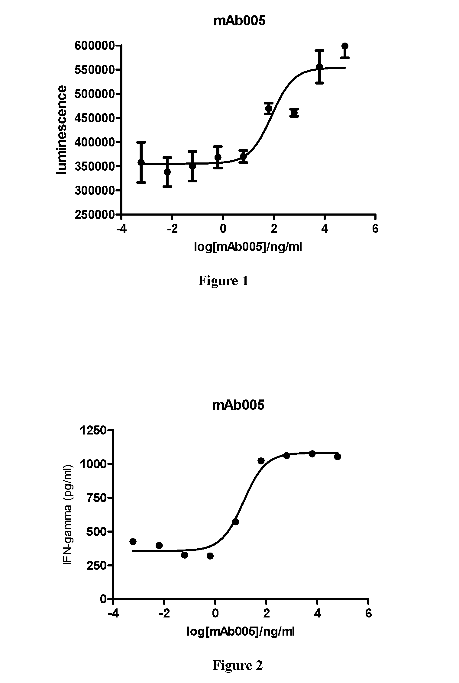
[0051] FIG. 1: Human peripheral blood mononuclear cell proliferation assay. Result shows that the test PD-1 antibody mAb005 can effectively stimulate the proliferation of human peripheral blood mononuclear cells, with EC50 of 83 ng/ml.
[0052] FIG. 2: Human peripheral blood mononuclear cell cytokine IFN-.gamma. secretion test. Result shows that the test PD-1 antibody mAb005 can stimulate PBMC proliferation, and effectively stimulate secretion of cytokine IFN-.gamma. at the same time, with EC50 of 13 ng/ml.
[0053] FIG. 3: inhibitory effect of PD-1 antibody H005-1 on growth of glioma cells.
[0054] FIG. 4: diagram showing tumor volume change after treatment.
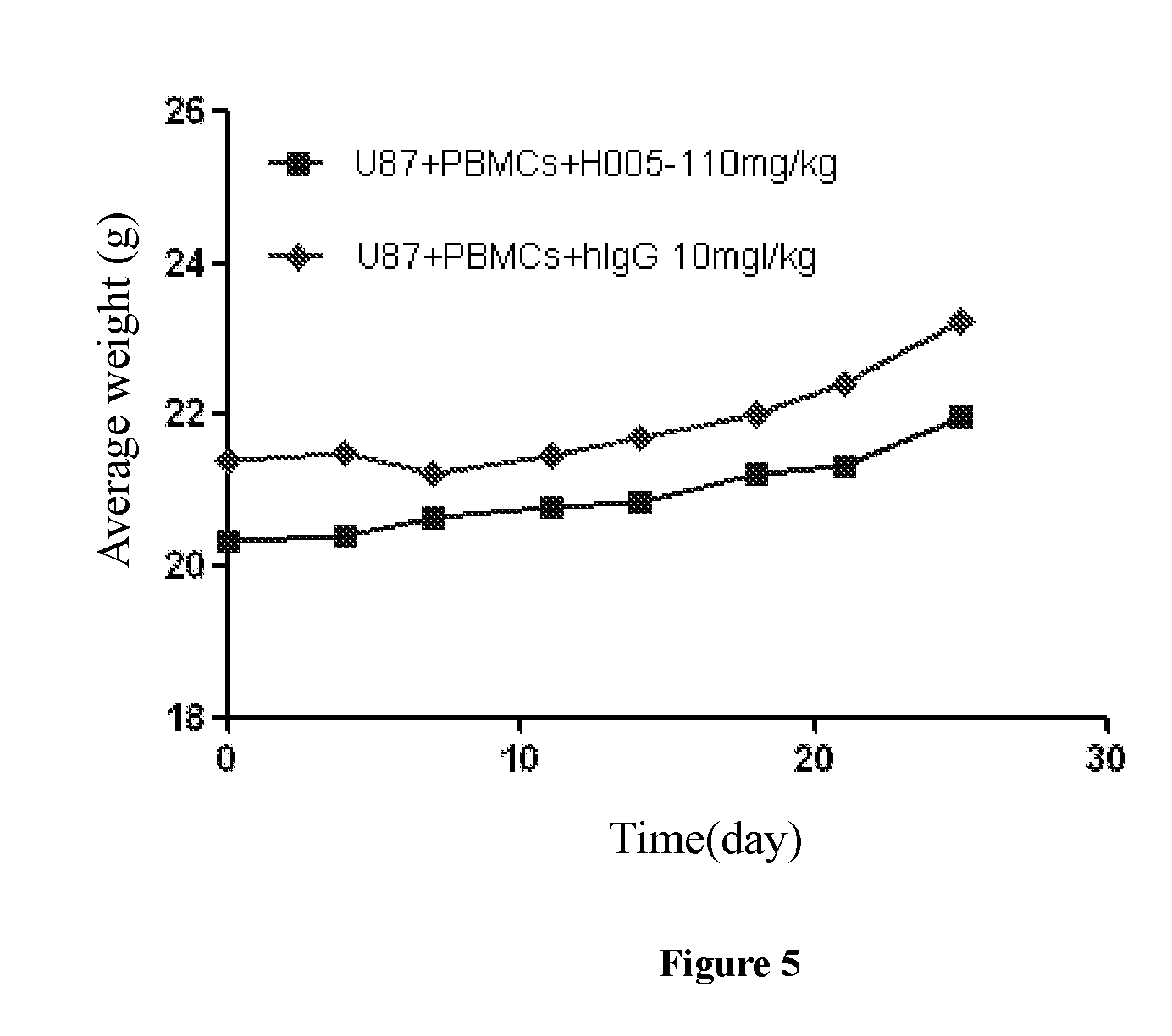
[0055] FIG. 5: diagram showing weight change of mice after treatment.
DETAILED DESCRIPTION OF THE INVENTION
1. Definitions
[0056] In order to more readily understood the invention, certain technical and scientific terms are specifically defined below. Unless specifically defined elsewhere in this document, all other technical and scientific terms used herein have the meaning commonly understood by one of ordinary skill in the art to which this invention belongs.
[0057] As used herein, the single-letter code and the three-letter code for amino acids are as described in J. biol. chem, 243, (1968) p 3558.
[0058] As used herein, "Antibody" refers to immunoglobulin, a four-peptide chain structure connected together by disulfide bonds between two identical heavy chains and two identical light chains. Different immunoglobulin heavy chain constant regions exhibit different amino acid compositions and rank orders, thereby presenting different kinds of antigenicity. Accordingly, immunoglobulins can be divided into five categories, or called immunoglobulin isotypes, namely IgM, IgD, IgG; IgA and IgE, their heavy chains are .mu. chain, .delta. chain, .gamma. chain, .alpha. chain and .epsilon. chain, respectively. According to its amino acid composition of hinge region and the number and location of heavy chain disulfide bonds, the same type of Ig can be divided into different sub-categories, for example, IgG can be divided into IgG1, IgG2, IgG3, and IgG4. Light chain can be divided into .kappa. or .lamda. chain considering of different constant regions. Each of the five Igs can have .kappa. or .lamda. chain.
[0059] In the present invention, the antibody light chain variable region mentioned herein further comprises a light chain constant region, which comprises a human or murine .kappa., .lamda. chain or a variant thereof.
[0060] In the present invention, the antibody heavy chain variable region mentioned herein further comprises a heavy chain constant region, which comprises human or murine IgG1, 2, 3, 4 or a variant thereof.
[0061] Near the N-terminal sequence of the antibody heavy chains and light chains, about 110 amino acid sequence varies largely, known as the variable region (V region); the rest of the amino acid sequence near the C-terminus is relative stable, known as the constant region (C region). Variable region comprises three hypervariable regions (HVR) and four relatively conserved sequence framework region (FR). The three hypervariable regions determine the specificity of the antibody, also known as a complementarity determining region (CDR). Each light chain variable region (LCVR) and each heavy chain variable region (HCVR) is composed of three CDRs and four FRs, with sequentially order from the amino terminal to the carboxyl terminal being: FR1, CDR1, FR2, CDR2, FR3, CDR3, and FR4. Three light chain CDRs refer to LCDR1, LCDR2, and LCDR3; three heavy chain CDRs refer to HCDR1, HCDR2 and HCDR3. The numbers and locations of CDR amino acid residues in LCVR and HCVR of the antibody or the antigen-binding fragment herein correspond with known Kabat numbering criteria (LCDR1-3, HCDE2-3), or correspond with kabat and chothia numbering criteria (HCDR1).
[0062] The term "murine antibody" in the present invention refers to anti-human-PD-1 monoclonal antibody prepared according to the knowledge and skills in the art. During the preparation, a test object was injected with PD-1 antigen, and then hybridoma expressing antibody which possesses desired sequence or functional characteristics was separated. In a preferred embodiment of the present invention, the murine PD-1 antibody or the antigen-binding fragment thereof further comprises a light chain constant region of murine .kappa., .lamda. chain or a variant thereof, or further comprises a heavy chain constant region of murine IgG1, IgG2, IgG3 or IgG4, or a variant thereof.
[0063] The term "chimeric antibody", is an antibody which is formed by fusing the variable region of a murine antibody with the constant region of a human antibody, the chimeric antibody can alleviate the murine antibody-induced immune response. To establish a chimeric antibody, hybridoma secreting specific murine monoclonal antibody is firstly established, a variable region gene is cloned from mouse hybridoma cells, then a constant region gene of a human antibody is cloned as desired, the mouse variable region gene is ligated with the human constant region gene to form a chimeric gene which can be inserted into a human vector, and finally the chimeric antibody molecule is expressed in the eukaryotic or prokaryotic industrial system. In a preferred embodiment of the present invention, the light chain variable region of PD-1 chimeric antibody further comprises the light chain FR of murine .kappa., .lamda. chain or a variant thereof, and the sequence of the light chain variable region is shown in SEQ ID NO: 10. The heavy chain variable region of the PD-1 chimeric antibody further comprises the heave chain FR of murine IgG1, IgG2, IgG3, IgG4 or a variant thereof, and the sequence of the heavy chain variable region is shown in SEQ ID NO: 10. The constant region of a human antibody is selected from the heavy chain constant region of human IgG1, IgG2, IgG3 or IgG4 or a variant thereof, preferably comprises the heavy chain constant region of human IgG2 or IgG4, or that of IgG1 which has no ADCC (antibody-dependent cell-mediated cytotoxicity) after amino acid mutation.
[0064] The term "humanized antibody", also known as CDR-grafted antibody, refers to an antibody generated by grafting murine CDR sequences into a variable region framework of a human antibody, namely, a sequence of human germline antibody framework of different type. Humanized antibody overcomes the disadvantageously strong antibody response induced by the chimeric antibody which carries a large amount of murine protein components. Such framework sequences can be obtained from public DNA database covering germline antibody gene sequences or published references. For example, germline DNA sequences of human heavy and light chain variable region genes can be found in "VBase" human germline sequence database (available on web www.mrccpe.com.ac.uk/vbase), as well as can be found in Kabat, E A, et al, 1991 Sequences of Proteins of Immunological Interest, 5.sup.th Ed. In a preferred embodiment of the invention, the murine CDR sequences of PD-1 humanized antibody are selected from SEQ ID NO: 3, 4, 5, 6, 7, 8. Human antibody variable region frameworks were designed and selected such that the light chain FR sequence of the antibody light chain variable region is derived from combination sequence of human germline light chains IGKV1-39 and JK4: SEQ ID NO: 14, comprising FR1, FR2 and FR3 of IGKV 1-39 and FR4 of JK4; the heavy chain FR sequence of the antibody heavy chain variable region is derived from combination sequence of human germline heavy chains IgHV3-7 and JH6: SEQ ID NO: 13, comprising FR1, FR2 and FR3 of IgHV3-7 and FR4 of MI6. To avoid activity decrease during immunogenicity reduction, the variable region of the human antibody is subjected to a minimum back mutation to maintain the activity.
[0065] As used herein, "antigen-binding fragment" refers to a Fab fragment, a Fab' fragment, a F(ab')2 fragment with antigen-binding activity, as well as a Fv fragment sFv fragment binding with human PD-1; comprising one or more CDR regions of antibodies described in the present invention selected from the group consist of SEQ ID NO:3 to SEQ ID NO:8. Fv fragment is a minimum antibody fragment comprising a heavy chain variable region, a light chain variable region, and all antigen-binding sites without a constant region. Generally, Fv antibody further comprises a polypeptide linker between the VH and VL domains, and is capable of forming a structure required for antigen binding. Also, different linkers can be used to connect the variable regions of two antibodies to form a polypeptide, named single chain antibody or single chain Fv (sFv). As used herein, the term "binding with PD-1", means interacting with human PD-1. As used herein, the term "antigenic determinant" of the present invention, refers to discontinuous three-dimensional sites on the antigen, recognized the antibody or the antigen-binding fragment of the present invention.
[0066] As used herein, the term "ADCC", namely antibody-dependent cell-mediated cytotoxicity, refers to cells expressing Fc receptors directly kill target cells coated by an antibody through recognizing the Fc segment of the antibody. ADCC effector function of the antibody can be reduced or eliminated via modification of the Fc segment in IgG The modification refers to mutations of the antibody heavy chain constant region, such as mutations selected from N297A, L234A, L235A in IgG1; IgG2/4 chimera; F235E, or L234A/E235A mutations in IgG4.
[0067] As used herein, fusion protein described in the present invention is a protein product obtained by co-expressing two genes via recombinant DNA technology. Recombinant PD-1 extracellular domain Fc fusion protein obtained by co-expressing a PD-1 extracellular domain and a human antibody Fc fragment via recombinant DNA technology. The PD-1 extracellular domain refers to the moiety of PD-1 outside cytomembrane, the sequence of which is the scribing region of SEQ ID NO: 1 below.
[0068] Methods for producing and purifying antibodies and antigen-binding fragments are well known in the art and can be found, for example, in Antibody Experimental Technology Guide of Cold Spring Harbor, Chapter 5-8 and 15. For example, mice can be immunized with human PD-1, or fragments thereof, and the resulting antibodies can then be renatured, purified and sequenced using conventional methods well known in the art. Antigen-binding fragments can also be prepared by conventional methods. The antibody or the antigen-binding fragment of the present invention is genetically engineered to introduce one or more human framework regions (FRs) to a non-human derived CDR. Human FR germline sequences can be obtained from ImMunoGeneTics (IMGT) via their website http://imgt.cines.fr, or from The Immunoglobulin FactsBook, 2001ISBN012441351. Specifically, light chain FR germline for use in the antibody or the antigen-binding fragment of the present invention include A3 and 02. Particular heavy chain FR germline for use in the antibody or the antigen-binding fragment of the present invention include VH3-21 and VH3-23.
[0069] The engineered antibody or antigen-binding fragment of the present invention may be prepared and purified using conventional methods. For example, cDNA sequences encoding a heavy chain (SEQ ID NO: 11) and a light chain (SEQ ID NO: 12) may be cloned and recombined into a GS expression vector. The recombined immunoglobulin expression vector may then stably transfect CHO cells. As a more recommended method well known in the art, mammalian expression of antibodies will result in glycosylation, typically at the highly conserved N-terminal in the FC region. Stable clones may be obtained through expression of an antibody specifically binding to human PCSK9. Positive clones may be expanded in a serum-free culture medium for antibody production in bioreactors. Culture medium, into which an antibody has been secreted, may be purified by conventional techniques. For example, the medium may be conveniently applied to a Protein A or G Sepharose FF column that has been equilibrated with a compatible buffer. The column is washed to remove nonspecific binding components. The bound antibody is eluted by PH gradient and antibody fragments are detected by SDS-PAGE, and then pooled. The antibody may be filtered and concentrated using common techniques. Soluble aggregate and multimers may be effectively removed by common techniques, including size exclusion or ion exchange. The obtained product may be immediately frozen, for example at -70.degree. C., or may be lyophilized.
[0070] The antibody of the present invention is a monoclonal antibody. Monoclonal antibody or mAb, as used herein, refers to an antibody that is derived from a single clone including but not limited to any eukaryotic, prokaryotic, or phage clone. Monoclonal antibodies and antigen-binding fragments thereof can be recombined, for example, by hybridoma technologies, recombinant technologies, phage display technologies, synthetic technologies (e.g., CDR-grafting), or other technologies known in the art.
[0071] "Administration" and "treatment," as it applies to an animal, human, experimental subject, cell, tissue, organ, or biological fluid, refers to contacting an exogenous pharmaceutical, therapeutic, diagnostic agent, or composition with the animal, human, subject, cell, tissue, organ, or biological fluid. "Administration" and "treatment" can refer, e.g., to therapeutic, pharmacokinetic, diagnostic, research, and experimental methods. Treatment of a cell encompasses contacting a reagent with the cell, as well as contacting a reagent with a fluid, where the fluid is in contact with the cell. "Administration" and "treatment" also means in vitro and ex vivo treatments, e.g., of a cell, by a reagent, diagnostic, binding compound, or by another cell. "Treatment," as it applies to a human, veterinary, or a research subject, refers to therapeutic treatment, prophylactic or preventative measures, to research and diagnostic applications.
[0072] "Treat" means to administer a therapeutic agent, such as a composition comprising any of the binding compounds of the present invention, internally or externally to a patient having one or more disease symptoms for which the agent has known therapeutic activity. Typically, the agent is administered in an amount effective to alleviate one or more disease symptoms in the treated patient or population, whether by inducing the regression of or inhibiting the progression of such symptom(s) to any clinically measurable degree. The amount of a therapeutic agent that is effective to alleviate any particular disease symptom (also referred to "therapeutically effective amount") may vary according to factors such as the disease state, age, and weight of the patient, and the ability of the drug to elicit a desired response in the patient. Whether a disease symptom has been alleviated can be assessed by any clinical measurement typically used by physicians or other skilled healthcare providers to assess the severity or progression status of that symptom. While an embodiment of the present invention (e.g., a treatment method or article of manufacture) may not be effective in alleviating the disease symptom(s) of interest in every patient, it should alleviate the target disease symptom(s) of interest in a statistically significant number of patients as determined by any statistical test known in the art such as the Student's t-test, the chi-square test, the U-test according to Mann and Whitney, the Kruskal-Wallis test (H-test), Jonckheere-Terpstra-test and the Wilcoxon-test.
[0073] "Conservative modifications" or "conservative replacement or substitution" refers to substitutions of amino acids in a protein with other amino acids having similar characteristics (e.g. charge, side-chain size, hydrophobicity/hydrophilicity, backbone conformation and rigidity, etc.), such that the changes can frequently be made without altering the biological activity of the protein. Those of skill in this art recognize that, in general, single amino acid substitutions in non-essential regions of a polypeptide do not substantially alter biological activity (see, e.g., Watson et al. (1987) Molecular Biology of the Gene, The Benjamin/Cummings Pub. Co., p. 224 (4.sup.th Ed.)). In addition, substitutions of structurally or functionally similar amino acids are less likely to disrupt biological activity.
[0074] "Consisting essentially of," or its variation as used throughout the specification and claims, indicate the inclusion of any recited elements or group of elements, and the optional inclusion of other elements of similar or different nature than the recited elements, which do not materially change the basic or novel properties of the specified dosage regimen, method, or composition. As a nonlimiting example, a binding compound which consists essentially of a recited amino acid sequence may also include one or more amino acids that do not materially affect the properties of the binding compound.
[0075] "Effective amount" encompasses an amount sufficient to ameliorate or prevent a symptom or sign of a medical condition. Effective amount also means an amount sufficient to allow or facilitate diagnosis. An effective amount for a particular patient or veterinary subject may vary depending on factors such as the condition being treated, the general health of the patient, the route and dose of administration and the severity of side affects. An effective amount can be the maximal dose or dosing protocol that avoids significant side effects or toxic effects.
[0076] "Exogenous" refers to substances that are produced outside an organism, cell, or human body, depending on the context. "Endogenous" refers to substances that are produced within a cell, organism, or human body, depending on the context.
[0077] "Homology" refers to sequence similarity between two polynucleotide sequences or between two polypeptides. When a position in both of the two compared sequences is occupied by the same base or amino acid monomer subunit, e.g., if a position in each of two DNA molecules is occupied by adenine, then the molecules are homologous at that position. The percent of homology between two sequences is a function of the number of matching or homologous positions shared by the two sequences divided by the number of positions compared.times.100. For example, if 6 of 10 positions in two sequences are matched or homologous when the sequences are optimally aligned, then the two sequences are 60% homologous. Generally, the comparison is made when two sequences are aligned to give maximum percent homology.
[0078] As used herein, the expressions "cell," "cell line," and "cell culture" are used interchangeably and all such designations include progeny. Thus, the words "transformants" and "transformed cells" include the primary subject cell and cultures derived therefrom without considering the number of transfers. It is also understood that all progeny may not be precisely identical in DNA content, due to deliberate or inadvertent mutations. Mutant progeny that have the same function or biological activity as screened for in the originally transformed cell are included. Where distinct designations are intended, it will be clear from the context.
[0079] As used herein, "polymerase chain reaction" or "PCR" refers to a procedure or technique in which minute amounts of a specific moiety of nucleic acid, RNA and/or DNA, are amplified as described in, e.g., U.S. Pat. No. 4,683,195. Generally, sequence information from the ends of the region of interest or beyond needs to be available, such that oligonucleotide primers can be designed; these primers will be identical or similar in sequence to corresponding strands of the template to be amplified. The 5' terminal nucleotides of the two primers can be identical with the ends of the material to be amplified. PCR can be used to amplify specific RNA sequences, specific DNA sequences from total genomic DNA, and cDNA transcribed from total cellular RNA, bacteriophage or plasmid sequences, etc. See generally Mullis et al. (1987) Cold Spring Harbor Symp. Ouant. Biol. 51:263; Erlich, ed., (1989) PCR TECHNOLOGY (Stockton Press, N.Y.). As used herein, PCR is considered as one, but not the only, example of a nucleic acid polymerase reaction method for amplifying a nucleic acid test sample, comprising the use of a known nucleic acid as a primer and a nucleic acid polymerase to amplify or generate a specific moiety of the nucleic acid.
[0080] "Optional" or "optionally" means that the event or situation that follows may but does not necessarily occur, and the description includes the instances in which the event or circumstance does or does not occur. For example, "optionally comprises 1-3 antibody heavy chain variable regions" means the antibody heavy chain variable region with specific sequence can be, but not necessarily be present.
[0081] "Pharmaceutical composition" refers to one containing a mixture of one or more compounds according to the present invention or a physiologically/pharmaceutically acceptable salt or produg thereof with other chemical components, as well as additional components such as physiologically/pharmaceutically acceptable carriers and excipients. The pharmaceutical composition aims at promoting the administration to an organism, facilitating the absorption of the active ingredient and thereby exerting a biological effect.
DETAILED DESCRIPTION OF THE INVENTION
[0082] Hereinafter, the present invention is further described with reference to examples; however, the scope of the present invention is not limited thereto. In the examples of the present invention, where specific conditions are not described, the experiments are generally conducted under conventional conditions as described in Antibody Tcchnology Laboratory Manual and Mecular Cloning Manual of Cold Spring Harbor, or under conditions proposed by the material or product manufacturers. Where the source of the reagents is not specifically given, the reagents are commercially available conventional reagents.
Example 1: Antibody Preparation
[0083] Murine monoclonal antibodies against human PD-1 were generated. Purified recombinant PD-1 extracellular domain Fc fusion protein (PD-1 Fc) (SEQID NO: 1); or CHO cells transfected with PD-1 (SEQ ID NO: 2) was used as an antigen to immunize Balb/C mice and SJL mice. Human PD-1 antigen was purchased from ORIGENE, Cat No. SC117011, NCBI Reference Sequence: NM 005018.1.
TABLE-US-00001 PD-1 Fc, recombinant PD-1 extracellular domain Fc fusion protein (SEQ ID NO: 1): MDMRVPAQLLGLLLLWFPGSRCPGWFLDSPDRPWNPPTFSPALLVVTEGD NATFTCSFSNTSESFVLNWYRMSPSNQTDKLAAFPEDRSQPGQDCRFRVT QLPNGRDPHMSVVRARRNDSGTYLCGAISLAPKAQIKESLRAELRVTERR AEVPTAHPSPSPRPAGQFQTLVDKTHTCPPCPAPELLGGPSVFLFPPKPK DTLMISRTPEVTCVVVDVSHEDPEVKFNWYVDGVEVHNAKTKPREEQYNS TYRVVSVLTVLHQDWLNGKEYKCKVSNKALPAPIEKTISKAKGQPREPQV YTLPPSREEMTKNQVSLTCLVKGFYPSDIAVEWESNGQPENNYKTTPPVL DSDGSFFLYSKLTVDKSRWQQGNVFSCSVMHEALHNHYTQKSLSLSPGK. PD-1, PD-1 antigen transfecting cells (SEQ ID NO: 2): MQIPQAPWPVVWAVLQLGWRPGWFLDSPDRPWNPPTFSPALLVVTEGDNA TFTCSFSNTSESFVLNWYRMSPSNQTDKLAAFPEDRSQPGQDCRFRVTQL PNGRDFHMSVVRARRNDSGTYLCGAISLAPKAQIKESLRAELRVTERRAE VPTAHPSPSPRPAGQFQTLVVGVVGGLLGSLVLLVWVLAVICSRAARGTI GARRTGQPLKEDPSAVPVFSVDYGELDFQWREKTPEPPVPCVPEQTEYAT IVFPSGMGTSSPARRGSADGPRSAQPLRPEDGHCSWPL.
[0084] Immunization with the PD-1 extracellular domain-Fc fusion protein is divided into high dose (50 ug) and low dose (10 ug) of the purified antigen, immunization with the PD-1 transfected CHO cells uses 0.5-1.times.10.sup.7 cells. Immunization was carried out on day 0, 14, and 35 respectively with Complete Freund's adjuvant; blood was sampled in the retro-orbital site to monitor the immune response. Mice with anti-PD-1 human immunoglobulin titer were obtained by plasma screening ELISA. On day 56, mice with the highest anti-PD-1 human immunoglobulin titer were subjected to boost immunization. 3 days later, mice were sacrificed and the spleen was removed for fusion. Hybridoma fusions were screened and a murine monoclonal antibody mAb005 was obtained. The heavy chain variable region sequence and light chain variable region sequence of the murine monoclonal antibody mAb005 are as follows:
TABLE-US-00002 mAb005 HCVR SEQ ID NO: 9 EVLMVESGGGLVKPGGSLKLSCAASGFTFSSYMMSWVRQTPEKRLEWVAT ISGGGANTYYPDSVKGRFTISRDNAKNTLYLQMSSLRSEDTALYYCARQL YYFDYWGQGTTLTVSS mAb005 LCVR SEQ ID NO: 10 DIQMTQSPASQSASLEGEGVTITCLASQTIGTWLTWYQQKPGKSPQLLIY TATSLADGVPSRFSGSGSGTKFSFKISSLQAEDFVTYYCQQVYSIPWTFG GGTKLEIK
[0085] CDR sequences are as follows:
TABLE-US-00003 Name Sequence Numbering HCDR1 SYMMS SEQ ID NO: 3 HCDR2 TISGGGANTYYPDSVKG SEQ ID NO: 4 HCDR3 QLYYFDY SEQ ID NO: 5 LCDR1 LASQTIGTWLT SEQ ID NO: 6 LCDR2 TATSLAD SEQ ID NO: 7 LCDR3 QQVYSIPWT SEQ ID NO: 8
Example 2: Antibody Screening
[0086] In Vitro PD-1 Antibody ELISA Binding Assay:
[0087] The PD-1 antibody blocks signaling pathway of PD-1 and its ligand by binding to PD-1 extracellular domain. In vitro ELISA assay is used to detect the binding property of the PD-1 antibody. Biotinylated PD-1 extracellular domain FC fusion protein (PD-1 FC) is coated onto 96-well plates by binding to neutralization avidin. Signal intensity after the addition of the antibody is used to determine the binding property of the antibody and PD-1.
[0088] Neutralization avidin (binding to biotin) was diluted to 1 .mu.l/ml with PBS buffer, pipetted into a 96-well plate with at 100 .mu.l/well and standed for 16 h-20 h at 4.degree. C. The 96-well plate was washed once with PBST (PH7.4 PBS, containing 0.05% tweeen20) after PBS buffer was removed, then the plate was incubated and blocked for 1 h at room temperature with addition of 120 .mu.l/well PBST/1% milk. After removal of the blocking solution, the plate was washed with PBST buffer, followed by addition of 1 .mu.g/ml biotin-labeled PD1-FC which was diluted by PBST/1% milk, and incubated for 1 h at room temperature. After removal of the blocking solution, the plate was washed with PBST buffer for 3 times, followed by addition of the test PD-1 antibody which was diluted to a suitable concentration by PBST/1% milk, and incubated for 1.5 h at room temperature. After removal of reaction system, the plate was washed for 3 times with PBST buffer, followed by addition of 100 .mu.l/well HRP-labeled anti-murine secondary antibody (The Jackson Laboratory) which was diluted by PBST/1% milk, and incubated for 1 h at room temperature. After being washed with PBST for three times, the plate was added with 100 .mu.l/well TMB, and incubated for 5-10 min at room temperature. Then the reaction was terminated with addition of 100.mu./well 1M H.sub.2SO.sub.4. The absorbance value at 450 nm was read on NOVOStar microplate reader; the ELISA binding EC.sub.50 value was calculated.
TABLE-US-00004 ELISA, EC50, nM Test Antibody human PD-1 cyno PD-1 mAb005 0.25 0.27
[0089] The results demonstrated that the antibody mAb005 showed excellent binding activity to human PD-1Fc (human PD-1) and cynomolgus PD-1Fc (cyno PD-1).
[0090] In vitro blocking assay of binding of PD-1 antibody and PD-1 ligand:
[0091] PD-L1 on the surface of a tumor cell exhibits suppressive effect on the proliferation of T cells by binding to PD-1 on the surface of a T cell. The PD-1 antibody blocks PD-L1/PD-1 signaling pathway by binding to PD-1 so as to stimulate T cell proliferation. PD-1/PD-L1 binding blocking assay is used to detect the blocking activity of PD-1 antibody on the signaling pathway.
[0092] In this experiment, a 96-well plate was coated with a PD-1 protein with the extracellular domain fused with FC (PD-1-FC), and incubated with the test PD-1 antibody; later biotin-labeled PD-L1 was added for incubation. After washing the plate, the binding amount of biotin-labeled PD-L1 was detected; the blocking IC.sub.50 value of PD-1 antibody for ligand PD-L1 binding was calculated.
[0093] PD-1-FC was diluted to 1 .mu.g/ml with PH 9.6 CB buffer (1.59 g Na.sub.2CO.sub.3 and 2.93 g NaHCO.sub.3 were dissolved in 1 L of distilled water), pipetted into a 96-well plate at 100 .mu.l/well and standed for 16 h-20 h at 4.degree. C. The 96-well plate was washed once with PBST (PH7.4 PBS, containing 0.05% tweeen20) after PBS buffer was removed, then the plate was incubated and blocked for 1 h at room temperature with 120 .mu.l/well PBST/1% milk. After removal of the blocking solution, the plate was washed with PBST buffer once, followed by addition of 90 .mu.l of test PD-1 antibody which was diluted to a suitable concentration with sample diluents (PH7.4 PBS containing 5% BSA, 0.05% Tween20), and incubated for 1 h at 4.degree. C. Then 10.times. concentrations of biotin-labeled PD-L1 (Beijing Sino Biological Inc.) (10 .mu.g/ml) was added to the plate at 10 .mu.l/well, oscillated and mixed by an oscillator, and incubated at 37.degree. C. for 1 h. After removal of the reaction system, the plate was washed for 6 times with PBST buffer, followed by addition of 100 ul/well Streptavidin-Peroxidase Polymer which was diluted by PBST at a ratio of 1:400, and incubated under oscillation for 50 min at room temperature. After being washed with PBST for 6 times, the plate was added with 100 .mu.l/well TMB, and incubated for 5-10 min at room temperature. Then the reaction was terminated with addition of 100 .mu.l/well 1M H.sub.2SO.sub.4. The absorbance value at 450 nm was read on the NOVOStar microplate reader; the blocking IC.sub.50 value of PD-1 for ligand PD-L1 binding was calculated.
TABLE-US-00005 Test LBB assay antibody IC50, nM mAb005 1.13
[0094] The result showed that the antibody mAb005 was very effective to block the binding of PD-L1 with PD-1.
Example 3: Binding Selectivity Assay of PD-1 Antibody In Vitro
[0095] To detect the specific binding activity of PD-1 antibody to other proteins of the PD-1 family, human CTLA4 and human CD28 were used for binding assays. Meanwhile, the PD-1 of mice was also used for binding assays so as to determine the diversity of PD-1 antibody for different species other than human/monkey.
[0096] Selectively binding proteins: human PD-1, human ICOS, human CTLA4, human CD28 and mouse PD-1, (Beijing Sino Biological Inc.), were respectively diluted to 1 .mu.g/ml with PBS buffer, pipetted into a 96-well plate at 100 .mu.l/well and standed for 16 h-20 h at 4.degree. C. The 96-well plate was washed once with PBST (PH7.4 PBS, containing 0.05% tweeen20) after PBS buffer was removed, then the plate was incubated and blocked for 1 h at room temperature with 120 .mu.l/well PBST/1% milk. After removal of the blocking solution, the plate was washed with PBST buffer for 3 times, followed by addition of the test PD-1 antibody, and incubated for 1.5 h at room temperature. After removal of the reaction system, the plate was washed for 3 times with PBST, followed by addition of 100 .mu.l/well HRP-labeled anti-murine secondary antibody (The Jackson Laboratory) which was diluted by PBST/1% milk, and incubated for 1 h at room temperature. The plate was washed for 3 times with PBST, followed by addition of 100 .mu.l/well TMB, and incubated for 5-10 min at room temperature. Then the reaction was terminated with addition of 100 .mu.l/well 1M H.sub.2SO.sub.4. The absorbance value at 450 nm was read on the NOVOStar microplate reader.
TABLE-US-00006 Test human mouse human human human Antibody PD1-FC PD1-Fc ICOS/Fc CTLA4 CD28 mAb005 2.64 0.07 0.15 0.17 0.12
[0097] The result demonstrated that mAb005 antibody exhibites no specific binding activity to other proteins of the PD-1 family. Meanwhile, mAb has no species cross-reactivity against murine PD-1.
Example 4: In Vitro Cell Binding Assay of PD-1 Antibody
[0098] FACS (fluorescence-activated cell sorter) is a test method for detecting interaction of proteins and cells. The test is used for detecting the binding activity of PD-1 antibody to native PD-1 expressed on the cell surface. Cells used in the test are CHO cells highly expressing PD-1 (see Example 1, CHO cells transfected with PD-1 (SEQID NO: 2)).
[0099] The CHO cells highly expressing PD-1 were centrifuged at 1000 rpm for 5 minutes, and the pellet was collected and suspended with 10-15 ml of precooled flow buffer for cell count. Cells were centrifuged at 1000 rpm in 50 ml centrifuge tubes for 5 minutes and collected. After removal of the supernatant, the pellet was resuspended with precooled blocking buffer with density of 0.5-1.0.times.10.sup.7 cells/ml. After incubation at 4.degree. C. for 30 minutes, re-suspension was pipetted to the 96-well plate at 100 .mu.l/well. The 96-well plate was centrifuged at 1500 rpm for 5 minutes, the supernatant was discarded. 100 .mu.l of primary antibody was added to each well; the cells were resuspended, and incubated in the dark for 60 minutes at 4.degree. C. After centrifugation and discard of the supernatant, 100 .mu.l of FITC-labeled secondary antibody (BD Biosciences) diluted at 1:400 was added. The cells were resuspended and incubated in the dark for 60 minutes at 4.degree. C. Cells were washed twice with flow buffer, resuspended and fixed with 1% paraformaldehyde for flow cytometry assay.
TABLE-US-00007 MFI Test Antibody 50 nM 5 nM 0.5 nM 0.05 nM mAb005 468 319 71.2 14
[0100] The results show that mAb005 antibody can also bind to PD-1 on the cell surface.
Example 5: In Vitro Binding Affinity and Kinetic Assay
[0101] Biacore method is a recognized assay which objectively detects the interactional affinity and kinetics of proteins. We analyzed the characterized affinity and binding kinetics of the test PD-1 antibody of the present invention by Biacore (GE).
[0102] According to the instruction of a kit provided by Biacore, the test PD-1 antibody of the present invention was covalently linked to CM5 (GE) chip using a standard amino coupling method. Then a series of gradient concentrations of PD-1 His protein (Beijing Sino Biological Inc.), which were diluted in the same buffer, were loaded into each cycle successively. After that, the samples were regenerated with regenerated reagent in the kit. The antigen-antibody binding kinetics was tracked for 3 minutes and the dissociation kinetics was tracked for 10 minutes. The data obtained was analyzed by GE's BIAevaluation software using 1:1 (Langmuir) binding model. Ka (kon), kd (koff) and KD values determined by the assay were shown in the following table.
TABLE-US-00008 Test Antibody ka (1/Ms) kd (1/s) KD (M) mAb005 1.057E+5 3.769E-4 3.566E-9
[0103] The results showed that the binding Kd value of the antibody mAb005 to PD-1 reached to 3.57 nM.
Example 6: In Vitro Cytology Test
[0104] Fresh human peripheral blood mononuclear cells (PBMC) proliferation assay affected by antibody is used to detect the cell activity of the antibody mAb005.
[0105] Fresh human PBMC density was adjusted to 2.times.10.sup.6/ml, seeded in a 6-well plate at 2 ml/well, and incubated for 6 hours at 37.degree. C., 5% CO.sub.2. After the suspension cells were discarded, each well of adherent cells was mixed with 2 ml of RPMI1640 medium containing 100 ng/ml GM-CSF (granulocyte colony stimulating biological factor) and 100 ng/ml IL-4, and another 1 ml of RPMI1640 medium containing 100 ng/ml GM-CSF and 100 ng/ml IL-4 after incubation for 2 days, then the cells were continually cultured for 2 days, followed by addition of 100 ng/ml TNF-.alpha. (tumor necrosis factor-.alpha.) each well, and cultured for another 2 days to obtain mature dendritic cells. The dendritic cells and allogeneic T cells were respectively centrifugated and resuspended at a concentration of 1.times.10.sup.6/ml and 1.times.10.sup.5/ml, and pipetted into a 96-well plate at 100 .mu.l/well, followed by addition of 20 .mu.l/well of antibody which was diluted to different concentration gradients with PBS, and the cells were cultured in 37.degree. C., 5% CO.sub.2 incubator for 5 days. Thereafter, 100 .mu.l of cell culture was sampled to detect the cell proliferation with CellTiter-Glo.RTM. Luminescent Cell Viability Assay kit. The result was shown in FIG. 1, indicating that the test PD-1 antibody mAb005 can effectively stimulate the proliferation of human peripheral blood mononuclear cells, with EC50 of 83 ng/ml. The remaining sample was detected for secretion of cytokine IFN-.gamma.. The result was shown in FIG. 2, demonstrating that the test PD-1 antibody mAb005 could stimulate PBMC proliferation, and effectively stimulate secretion of cytokine IFN-.gamma. at the same time, with EC50 of 13 ng/ml.
Example 7: Murine Antibody Humanization
[0106] With reference to the sequences of the light chain variable region (mAb005 LCVR, SEQ ID NO: 10) and the heavy chain variable region (mAb005 HCVR, SEQ ID NO: 9) of the mAb005 antibody, humanized templates best matching with their non-CDR in Germline database were selected. The antibody heavy chain template is IgHV3-7/JH6, selecting for FR1, FR2, FR3 of human germline light chain IGKV1-39 and FR4 of JK4, with sequence of SEQ ID NO: 13; light chain template is IGKV1-39/JK4, selecting for FR1, FR2, FR3 of human germline light chain IGKV1-39, and FR4 of JK4, with sequence of SEQ ID NO: 14.
TABLE-US-00009 Human germline heavy chain template (SEQ ID NO: 13): EVQLVESGGGLVQPGGSLRLSCAASGFTFSSYWMSWVRQAPGKGLEWVAN IKQDGSEKYYVDSVKGRFTISRDNAKNSLYLQMNSLRAEDTAVYYCARWG QGTTVTVSS, Human germline light chain template (SEQ ID NO: 14): DIQMTQSPSSLSASVGDRVTITCRASQSISSYLNWYQQKPGKAPKLLIYA ASSLQSGVPSRFSGSGSGTDFTLTISSLQPEDFATYYCFGGGTKVEIK.
[0107] The CDR of the murine antibody was grafted to the selected humanization template, replacing the CDR of human template, and then recombined with IgG4 constant region to obtain a humanized antibody H005-1. Afterwards, based on three-dimensional structure of the murine antibody, embedded residues, residues directly interacted with the CDR, and residues which significantly influence the conformation of VL and VH were backmutated to obtain humanized antibodies H005-2, H005-3, and H005-4, sequences are as follows.
[0108] Antibody Expression
TABLE-US-00010 H005-1 HC SEQ ID NO: 11 EVQLVESGGGLVQPGGSLRLSCAASGFTFSSYMMSWVRQAPGKGLEWVAT ISGGGANTYYPDSVKGRFTISRDNAKNSLYLQMNSLRAEDTAVYYCARQL YYFDYWGQGTTVTVSSASTKGPSVFPLAPCSRSTSESTAALGCLVKDYFP EPVTVSWNSGALTSGVHTFPAVLQSSGLYSLSSVVTVPSSSLGTKTYTCN VDHKPSNTKVDKRVESKYGPPCPPCPAPEAAGGPSVFLFPPKPKDTLMIS RTPEVTCVVVDVSQEDPEVQFNWYVDGVEVHNAKTKPREEQFNSTYRVVS VLTVLHQDWLNGKEYKCKVSNKGLPSSIEKTISKAKGQPREPQVYTLPPS QEEMTKNQVSLTCLVKGFYPSDIAVEWESNGQPENNYKTTPPVLDSDGSF FLYSRLTVDKSRWQEGNVFSCSVMHEALHNHYTQKSLSLSLGK H005-1 LC SEQ ID NO: 12 DIQMTQSPSSLSASVGDRVTITCLASQTIGTWLTWYQQKPGKAPKLLIYT ATSLADGVPSRFSGSGSGTDFTLTISSLQPEDFATYYCQQVYSIPWTFGG GTKVEIKRTVAASPVFIFPPSDEQLKSGTASVVCLLNNFYPREAKVQWKV DNALQSGNSQESVTEQDSKDSTYSLSSTLTLSKADYEKHKVYACEVTHQG LSSPVTKSFNRGEC
[0109] The HC sequence of the humanized antibody H005-1 with grafted murine CDR is (SEQID NO: 11), the LC sequence of the humanized antibody is (SEQ ID NO: 12). Sites which may affect the antibody activity were subjected to point mutations, the sequences are as follows:
TABLE-US-00011 HC LC H005-1 SEQID NO: 11 SEQID NO: 12 H005-2 SEQID NO: 11, G44R SEQID NO: 12 H005-3 SEQID NO: 11 SEQID NO: 12, A43S H005-4 SEQID NO: 11, G44R SEQID NO: 12, A43S
[0110] cDNAs were synthesized according to the amino acid sequences of the light chain and the heavy chain of each humanized antibody (SEQ NO 11, SEQ NO 12 and variants thereof). After the cDNAs were digested with Xhol and BamHI, the obtained cDNA fragments were inserted into pcDNA3.1 expression vectors (Life Technologies Cat. No. V790-20) at BamHI/Xhol restriction sites. The expression vectors and a transfection reagent PEI (Polysciences, Inc. Cat. No. 23966) were used to transfect HEK293 cells (Life Technologies Cat. No. 11625019) at 1:2, and the transfected cells were incubated in a CO.sub.2 incubator for 4-5 days. Expressed antibodies were recovered by centrifugation, and purified according to a conventional method to obtain the humanized antibodies of the present invention.
Example 8: Humanized Antibody Activity Data
[0111] Humanized antibodies were subjected to ELISA binding assay (method is the same as that of Example 2), ligand binding blocking assay (method is the same as that of Example 2), and affinity kinetics experiment (method is the same as that of Example 5) in vitro. The results are shown in the following table:
TABLE-US-00012 Test Antibody ELISA, EC50, nM LBB assay, IC50, nM KD (M) H005-1 0.11 1.27 2.79E-09 H005-2 0.14 1.27 2.98E-09 H005-3 0.15 1.33 2.45E-09 H005-4 0.14 1.36 3.89E-09
[0112] The result showed that humanized antibodies H005-1, H005-2, H005-3 and H005-4 maintained the PD-1 binding activity, with affinity kinetics KD of 2.79, 2.98, 2.45 and 3.89 nM respectively. Simultaneously, all the humanized antibodies effectively exhibited blocking activity against the PD-L1/PD-1 pathway.
Example 9: Tumor Cell Growth Inhibition by PD-1 Antibody
1. Experimental Materials
[0113] U87MG cells (glioma cells): purchased from the Chinese Academy of Sciences Cell Bank, Cat. TCHu138;
[0114] PBMCs (peripheral blood mononuclear cells) purchased from the Shanghai Blood Center;
[0115] CD3: purchased from Miltenyi Biotec Cat No. 130-093-387;
[0116] CD28: purchased from Miltenyi Biotec Cat No. 130-093-375; Cell Counting Kit-8: available from DOJINDO LABORATORIES, Cat No. CK04;
[0117] mIgG (negative control): purchased from SANTA CRUZ Cat No. sc-2025; using dose of 1660 ng/ml.
2. Experimental Methods
[0118] 1) U87MG cells were cultured in EMEM medium containing 10% FBS and 1% P/S, incubated in a 96-well plate, 1.times.10.sup.4 cells per well.
[0119] 2) H005-1 antibody was diluted to different concentration gradients (shown in abscissa of FIG. 3) with PBS, added to the 96-well plate at 1 Oul/well, and incubated in 37.degree. C., 5% CO.sub.2 incubator for 4 hours.
[0120] 3) After cell adherence, 80 ul of PBMC cell suspension was added to each well with a cell density of 2.times.10.sup.4 cells/well, and 10 ul of CD3 antibody and CD28 antibody were added in each well, the the final concentrations of CD3 and CD28 antibodies were both 500 ng/ml.
[0121] 4) After 72 hours of incubation in the 37.degree. C., 5% CO.sub.2 incubator, 10 ul of CCK8 was added to each well for development. 2 hours later, OD450 was determinated.
3. Result
[0122] The result was shown in FIG. 3, as compared with mIgG (negative control), different concentrations of PD-1 antibody (H005-1) had significant inhibitory effect on U87MG cell growth, and inhibition rate at the highest concentration was about 30%.
Example 10: Activity of H005-1 on Tuberculin-Stimulated PBMC Proliferation
[0123] The activity of the humanized antibody H005-1 on tuberculin-stimulated PBMC proliferation in vitro was detected.
[0124] To 15 ml of fresh PBMCs, about 3.times.10.sup.7 cells, were added 20 .mu.l tuberculin (Shanghai BiYou Biotechnology, cat#97-8800) and the mixture were incubated for 5 days in the 37.degree. C., 5% CO.sub.2 incubator. On day 6, the cultured cells were centrifugated, and resuspended into fresh medium with a density adjusted to 5.times.10.sup.5 cells/ml. 190 .mu.l of resuspended cells was planted into each well of a 96-well plate. The humanized antibody H005-1 was added to corresponding wells of the 96-well plate at 10 .mu.l/well. The control group and blank group were added with 10 .mu.l of PBS. The Cell culture plate was incubated in the 37.degree. C., 5% CO.sub.2 incubator, and 72 hours later, PBMC Proliferation (Promega, cat # G7571) and IFN-.gamma. secretion (Neo Bioscience, cat # EHC102 g) were determined. The results are as follows:
[0125] Activation Effect of the Test Sample on Tuberculin Stimulated PBMC Proliferation and IFN-.gamma. Secretion
TABLE-US-00013 T cell proliferation EC50 Sample (ng/ml) IFN-.lamda. EC50 (ng/ml) H005-1 15.95 .+-. 17.15 56.87 .+-. 48.53 Note: n = 4
[0126] Experiment results showed that the humanized antibody H005-1 excellently activates exogenous tuberculin stimulated PBMC proliferation and IFN-.gamma. secretion.
Example 11: Inhibition of Subcutaneously Inoculated U-87MG Tumor by H005-1
[0127] 100 ul of U87 cells (5.times.10.sup.6 cells) was inoculated subcutaneously in right ribs of SCID-Beige mice. When the tumor grew to 80-100 mm.sup.3 after 7 to 10 days, the SCID-Beige mice, getting rid of ones with too large or too small body weight or tumor, were randomly divided into a H005-1 10 mg/kg group and a Human IgG 10 mg/kg group according to the tumor volume, each group of seven mice (DO). Two kinds of PBMCs stimulated by CD3 antibody for 3 days were mixed at a ratio of 1:1, and injected into the tumor tissues at 5.times.10.sup.5 cells/60 ul, meanwhile, the test antibody was injected subcutaneously, once per 7 days for total 3 doses. Mice were measured for tumor volume and weighed twice a week. Data was recorded. Tumor volume (V) was calculated as: V=1/2.times.a.times.b2, wherein a and b represented length and width, respectively.
[0128] The result was shown in FIG. 4: tumor volume change after treatment, and FIG. 5: mice weight change after treatment, indicating that antibody H005-1 excellently inhibited U87MG tumor growth, and had no effect on the body weight of the mice.
Sequence CWU 1
1
141399PRTArtificial Sequencerecombinant PD-1 extracellular domain
Fc fusion protein 1Met Asp Met Arg Val Pro Ala Gln Leu Leu Gly Leu
Leu Leu Leu Trp1 5 10 15Phe Pro Gly Ser Arg Cys Pro Gly Trp Phe Leu
Asp Ser Pro Asp Arg 20 25 30Pro Trp Asn Pro Pro Thr Phe Ser Pro Ala
Leu Leu Val Val Thr Glu 35 40 45Gly Asp Asn Ala Thr Phe Thr Cys Ser
Phe Ser Asn Thr Ser Glu Ser 50 55 60Phe Val Leu Asn Trp Tyr Arg Met
Ser Pro Ser Asn Gln Thr Asp Lys65 70 75 80Leu Ala Ala Phe Pro Glu
Asp Arg Ser Gln Pro Gly Gln Asp Cys Arg 85 90 95Phe Arg Val Thr Gln
Leu Pro Asn Gly Arg Asp Phe His Met Ser Val 100 105 110Val Arg Ala
Arg Arg Asn Asp Ser Gly Thr Tyr Leu Cys Gly Ala Ile 115 120 125Ser
Leu Ala Pro Lys Ala Gln Ile Lys Glu Ser Leu Arg Ala Glu Leu 130 135
140Arg Val Thr Glu Arg Arg Ala Glu Val Pro Thr Ala His Pro Ser
Pro145 150 155 160Ser Pro Arg Pro Ala Gly Gln Phe Gln Thr Leu Val
Asp Lys Thr His 165 170 175Thr Cys Pro Pro Cys Pro Ala Pro Glu Leu
Leu Gly Gly Pro Ser Val 180 185 190Phe Leu Phe Pro Pro Lys Pro Lys
Asp Thr Leu Met Ile Ser Arg Thr 195 200 205Pro Glu Val Thr Cys Val
Val Val Asp Val Ser His Glu Asp Pro Glu 210 215 220Val Lys Phe Asn
Trp Tyr Val Asp Gly Val Glu Val His Asn Ala Lys225 230 235 240Thr
Lys Pro Arg Glu Glu Gln Tyr Asn Ser Thr Tyr Arg Val Val Ser 245 250
255Val Leu Thr Val Leu His Gln Asp Trp Leu Asn Gly Lys Glu Tyr Lys
260 265 270Cys Lys Val Ser Asn Lys Ala Leu Pro Ala Pro Ile Glu Lys
Thr Ile 275 280 285Ser Lys Ala Lys Gly Gln Pro Arg Glu Pro Gln Val
Tyr Thr Leu Pro 290 295 300Pro Ser Arg Glu Glu Met Thr Lys Asn Gln
Val Ser Leu Thr Cys Leu305 310 315 320Val Lys Gly Phe Tyr Pro Ser
Asp Ile Ala Val Glu Trp Glu Ser Asn 325 330 335Gly Gln Pro Glu Asn
Asn Tyr Lys Thr Thr Pro Pro Val Leu Asp Ser 340 345 350Asp Gly Ser
Phe Phe Leu Tyr Ser Lys Leu Thr Val Asp Lys Ser Arg 355 360 365Trp
Gln Gln Gly Asn Val Phe Ser Cys Ser Val Met His Glu Ala Leu 370 375
380His Asn His Tyr Thr Gln Lys Ser Leu Ser Leu Ser Pro Gly Lys385
390 3952288PRTHomo sapiens 2Met Gln Ile Pro Gln Ala Pro Trp Pro Val
Val Trp Ala Val Leu Gln1 5 10 15Leu Gly Trp Arg Pro Gly Trp Phe Leu
Asp Ser Pro Asp Arg Pro Trp 20 25 30Asn Pro Pro Thr Phe Ser Pro Ala
Leu Leu Val Val Thr Glu Gly Asp 35 40 45Asn Ala Thr Phe Thr Cys Ser
Phe Ser Asn Thr Ser Glu Ser Phe Val 50 55 60Leu Asn Trp Tyr Arg Met
Ser Pro Ser Asn Gln Thr Asp Lys Leu Ala65 70 75 80Ala Phe Pro Glu
Asp Arg Ser Gln Pro Gly Gln Asp Cys Arg Phe Arg 85 90 95Val Thr Gln
Leu Pro Asn Gly Arg Asp Phe His Met Ser Val Val Arg 100 105 110Ala
Arg Arg Asn Asp Ser Gly Thr Tyr Leu Cys Gly Ala Ile Ser Leu 115 120
125Ala Pro Lys Ala Gln Ile Lys Glu Ser Leu Arg Ala Glu Leu Arg Val
130 135 140Thr Glu Arg Arg Ala Glu Val Pro Thr Ala His Pro Ser Pro
Ser Pro145 150 155 160Arg Pro Ala Gly Gln Phe Gln Thr Leu Val Val
Gly Val Val Gly Gly 165 170 175Leu Leu Gly Ser Leu Val Leu Leu Val
Trp Val Leu Ala Val Ile Cys 180 185 190Ser Arg Ala Ala Arg Gly Thr
Ile Gly Ala Arg Arg Thr Gly Gln Pro 195 200 205Leu Lys Glu Asp Pro
Ser Ala Val Pro Val Phe Ser Val Asp Tyr Gly 210 215 220Glu Leu Asp
Phe Gln Trp Arg Glu Lys Thr Pro Glu Pro Pro Val Pro225 230 235
240Cys Val Pro Glu Gln Thr Glu Tyr Ala Thr Ile Val Phe Pro Ser Gly
245 250 255Met Gly Thr Ser Ser Pro Ala Arg Arg Gly Ser Ala Asp Gly
Pro Arg 260 265 270Ser Ala Gln Pro Leu Arg Pro Glu Asp Gly His Cys
Ser Trp Pro Leu 275 280 28535PRTMus musculus 3Ser Tyr Met Met Ser1
5417PRTMus musculus 4Thr Ile Ser Gly Gly Gly Ala Asn Thr Tyr Tyr
Pro Asp Ser Val Lys1 5 10 15Gly57PRTMus musculus 5Gln Leu Tyr Tyr
Phe Asp Tyr1 5611PRTMus musculus 6Leu Ala Ser Gln Thr Ile Gly Thr
Trp Leu Thr1 5 1077PRTMus musculus 7Thr Ala Thr Ser Leu Ala Asp1
589PRTMus musculus 8Gln Gln Val Tyr Ser Ile Pro Trp Thr1
59116PRTMus musculus 9Glu Val Met Leu Val Glu Ser Gly Gly Gly Leu
Val Lys Pro Gly Gly1 5 10 15Ser Leu Lys Leu Ser Cys Ala Ala Ser Gly
Phe Thr Phe Ser Ser Tyr 20 25 30Met Met Ser Trp Val Arg Gln Thr Pro
Glu Lys Arg Leu Glu Trp Val 35 40 45Ala Thr Ile Ser Gly Gly Gly Ala
Asn Thr Tyr Tyr Pro Asp Ser Val 50 55 60Lys Gly Arg Phe Thr Ile Ser
Arg Asp Asn Ala Lys Asn Thr Leu Tyr65 70 75 80Leu Gln Met Ser Ser
Leu Arg Ser Glu Asp Thr Ala Leu Tyr Tyr Cys 85 90 95Ala Arg Gln Leu
Tyr Tyr Phe Asp Tyr Trp Gly Gln Gly Thr Thr Leu 100 105 110Thr Val
Ser Ser 11510107PRTMus musculus 10Asp Ile Gln Met Thr Gln Ser Pro
Ala Ser Gln Ser Ala Ser Leu Gly1 5 10 15Glu Gly Val Thr Ile Thr Cys
Leu Ala Ser Gln Thr Ile Gly Thr Trp 20 25 30Leu Thr Trp Tyr Gln Gln
Lys Pro Gly Lys Ser Pro Gln Leu Leu Ile 35 40 45Tyr Thr Ala Thr Ser
Leu Ala Asp Gly Val Pro Ser Arg Phe Ser Gly 50 55 60Ser Gly Ser Gly
Thr Lys Phe Ser Phe Lys Ile Ser Ser Leu Gln Ala65 70 75 80Glu Asp
Phe Val Thr Tyr Tyr Cys Gln Gln Val Tyr Ser Ile Pro Trp 85 90 95Thr
Phe Gly Gly Gly Thr Lys Leu Glu Ile Lys 100 10511443PRTArtificial
sequenceheavy chain sequence of humanized antibody H005-1 11Glu Val
Gln Leu Val Glu Ser Gly Gly Gly Leu Val Gln Pro Gly Gly1 5 10 15Ser
Leu Arg Leu Ser Cys Ala Ala Ser Gly Phe Thr Phe Ser Ser Tyr 20 25
30Met Met Ser Trp Val Arg Gln Ala Pro Gly Lys Gly Leu Glu Trp Val
35 40 45Ala Thr Ile Ser Gly Gly Gly Ala Asn Thr Tyr Tyr Pro Asp Ser
Val 50 55 60Lys Gly Arg Phe Thr Ile Ser Arg Asp Asn Ala Lys Asn Ser
Leu Tyr65 70 75 80Leu Gln Met Asn Ser Leu Arg Ala Glu Asp Thr Ala
Val Tyr Tyr Cys 85 90 95Ala Arg Gln Leu Tyr Tyr Phe Asp Tyr Trp Gly
Gln Gly Thr Thr Val 100 105 110Thr Val Ser Ser Ala Ser Thr Lys Gly
Pro Ser Val Phe Pro Leu Ala 115 120 125Pro Cys Ser Arg Ser Thr Ser
Glu Ser Thr Ala Ala Leu Gly Cys Leu 130 135 140Val Lys Asp Tyr Phe
Pro Glu Pro Val Thr Val Ser Trp Asn Ser Gly145 150 155 160Ala Leu
Thr Ser Gly Val His Thr Phe Pro Ala Val Leu Gln Ser Ser 165 170
175Gly Leu Tyr Ser Leu Ser Ser Val Val Thr Val Pro Ser Ser Ser Leu
180 185 190Gly Thr Lys Thr Tyr Thr Cys Asn Val Asp His Lys Pro Ser
Asn Thr 195 200 205Lys Val Asp Lys Arg Val Glu Ser Lys Tyr Gly Pro
Pro Cys Pro Pro 210 215 220Cys Pro Ala Pro Glu Ala Ala Gly Gly Pro
Ser Val Phe Leu Phe Pro225 230 235 240Pro Lys Pro Lys Asp Thr Leu
Met Ile Ser Arg Thr Pro Glu Val Thr 245 250 255Cys Val Val Val Asp
Val Ser Gln Glu Asp Pro Glu Val Gln Phe Asn 260 265 270Trp Tyr Val
Asp Gly Val Glu Val His Asn Ala Lys Thr Lys Pro Arg 275 280 285Glu
Glu Gln Phe Asn Ser Thr Tyr Arg Val Val Ser Val Leu Thr Val 290 295
300Leu His Gln Asp Trp Leu Asn Gly Lys Glu Tyr Lys Cys Lys Val
Ser305 310 315 320Asn Lys Gly Leu Pro Ser Ser Ile Glu Lys Thr Ile
Ser Lys Ala Lys 325 330 335Gly Gln Pro Arg Glu Pro Gln Val Tyr Thr
Leu Pro Pro Ser Gln Glu 340 345 350Glu Met Thr Lys Asn Gln Val Ser
Leu Thr Cys Leu Val Lys Gly Phe 355 360 365Tyr Pro Ser Asp Ile Ala
Val Glu Trp Glu Ser Asn Gly Gln Pro Glu 370 375 380Asn Asn Tyr Lys
Thr Thr Pro Pro Val Leu Asp Ser Asp Gly Ser Phe385 390 395 400Phe
Leu Tyr Ser Arg Leu Thr Val Asp Lys Ser Arg Trp Gln Glu Gly 405 410
415Asn Val Phe Ser Cys Ser Val Met His Glu Ala Leu His Asn His Tyr
420 425 430Thr Gln Lys Ser Leu Ser Leu Ser Leu Gly Lys 435
44012214PRTArtificial sequencelight chain sequence of humanized
antibody H005-1 12Asp Ile Gln Met Thr Gln Ser Pro Ser Ser Leu Ser
Ala Ser Val Gly1 5 10 15Asp Arg Val Thr Ile Thr Cys Leu Ala Ser Gln
Thr Ile Gly Thr Trp 20 25 30Leu Thr Trp Tyr Gln Gln Lys Pro Gly Lys
Ala Pro Lys Leu Leu Ile 35 40 45Tyr Thr Ala Thr Ser Leu Ala Asp Gly
Val Pro Ser Arg Phe Ser Gly 50 55 60Ser Gly Ser Gly Thr Asp Phe Thr
Leu Thr Ile Ser Ser Leu Gln Pro65 70 75 80Glu Asp Phe Ala Thr Tyr
Tyr Cys Gln Gln Val Tyr Ser Ile Pro Trp 85 90 95Thr Phe Gly Gly Gly
Thr Lys Val Glu Ile Lys Arg Thr Val Ala Ala 100 105 110Pro Ser Val
Phe Ile Phe Pro Pro Ser Asp Glu Gln Leu Lys Ser Gly 115 120 125Thr
Ala Ser Val Val Cys Leu Leu Asn Asn Phe Tyr Pro Arg Glu Ala 130 135
140Lys Val Gln Trp Lys Val Asp Asn Ala Leu Gln Ser Gly Asn Ser
Gln145 150 155 160Glu Ser Val Thr Glu Gln Asp Ser Lys Asp Ser Thr
Tyr Ser Leu Ser 165 170 175Ser Thr Leu Thr Leu Ser Lys Ala Asp Tyr
Glu Lys His Lys Val Tyr 180 185 190Ala Cys Glu Val Thr His Gln Gly
Leu Ser Ser Pro Val Thr Lys Ser 195 200 205Phe Asn Arg Gly Glu Cys
21013109PRTArtificial sequenceheavy chain framework region sequence
of humanized antibody 13Glu Val Gln Leu Val Glu Ser Gly Gly Gly Leu
Val Gln Pro Gly Gly1 5 10 15Ser Leu Arg Leu Ser Cys Ala Ala Ser Gly
Phe Thr Phe Ser Ser Tyr 20 25 30Trp Met Ser Trp Val Arg Gln Ala Pro
Gly Lys Gly Leu Glu Trp Val 35 40 45Ala Asn Ile Lys Gln Asp Gly Ser
Glu Lys Tyr Tyr Val Asp Ser Val 50 55 60Lys Gly Arg Phe Thr Ile Ser
Arg Asp Asn Ala Lys Asn Ser Leu Tyr65 70 75 80Leu Gln Met Asn Ser
Leu Arg Ala Glu Asp Thr Ala Val Tyr Tyr Cys 85 90 95Ala Arg Trp Gly
Gln Gly Thr Thr Val Thr Val Ser Ser 100 1051498PRTArtificial
sequencelight chain framework region sequence of humanized antibody
14Asp Ile Gln Met Thr Gln Ser Pro Ser Ser Leu Ser Ala Ser Val Gly1
5 10 15Asp Arg Val Thr Ile Thr Cys Arg Ala Ser Gln Ser Ile Ser Ser
Tyr 20 25 30Leu Asn Trp Tyr Gln Gln Lys Pro Gly Lys Ala Pro Lys Leu
Leu Ile 35 40 45Tyr Ala Ala Ser Ser Leu Gln Ser Gly Val Pro Ser Arg
Phe Ser Gly 50 55 60Ser Gly Ser Gly Thr Asp Phe Thr Leu Thr Ile Ser
Ser Leu Gln Pro65 70 75 80Glu Asp Phe Ala Thr Tyr Tyr Cys Phe Gly
Gly Gly Thr Lys Val Glu 85 90 95Ile Lys
References
D00001
D00002
D00003
S00001
XML
uspto.report is an independent third-party trademark research tool that is not affiliated, endorsed, or sponsored by the United States Patent and Trademark Office (USPTO) or any other governmental organization. The information provided by uspto.report is based on publicly available data at the time of writing and is intended for informational purposes only.
While we strive to provide accurate and up-to-date information, we do not guarantee the accuracy, completeness, reliability, or suitability of the information displayed on this site. The use of this site is at your own risk. Any reliance you place on such information is therefore strictly at your own risk.
All official trademark data, including owner information, should be verified by visiting the official USPTO website at www.uspto.gov. This site is not intended to replace professional legal advice and should not be used as a substitute for consulting with a legal professional who is knowledgeable about trademark law.