Anti-TLR9 Antibody, Pharmaceutical Composition, and Kit
Miyake; Kensuke ; et al.
U.S. patent application number 16/465833 was filed with the patent office on 2019-09-26 for anti-tlr9 antibody, pharmaceutical composition, and kit. The applicant listed for this patent is The University of Tokyo. Invention is credited to Ryutaro Fukui, Kensuke Miyake, Yusuke Murakami.
| Application Number | 20190292270 16/465833 |
| Document ID | / |
| Family ID | 62242524 |
| Filed Date | 2019-09-26 |


| United States Patent Application | 20190292270 |
| Kind Code | A1 |
| Miyake; Kensuke ; et al. | September 26, 2019 |
Anti-TLR9 Antibody, Pharmaceutical Composition, and Kit
Abstract
The present invention provides a novel antibody targeting TLR9.
| Inventors: | Miyake; Kensuke; (Tokyo, JP) ; Murakami; Yusuke; (Tokyo, JP) ; Fukui; Ryutaro; (Tokyo, JP) | ||||||||||
| Applicant: |
|
||||||||||
|---|---|---|---|---|---|---|---|---|---|---|---|
| Family ID: | 62242524 | ||||||||||
| Appl. No.: | 16/465833 | ||||||||||
| Filed: | November 30, 2017 | ||||||||||
| PCT Filed: | November 30, 2017 | ||||||||||
| PCT NO: | PCT/JP2017/043124 | ||||||||||
| 371 Date: | May 31, 2019 |
| Current U.S. Class: | 1/1 |
| Current CPC Class: | G01N 33/563 20130101; G01N 2333/70596 20130101; G01N 2800/104 20130101; G01N 33/6893 20130101; G01N 2800/085 20130101; G01N 2800/205 20130101; C07K 16/2896 20130101; A61P 29/00 20180101; A61P 37/06 20180101; C07K 16/28 20130101; C12N 15/09 20130101; A61K 39/395 20130101; C07K 2317/24 20130101; A61P 1/16 20180101; A61P 17/06 20180101 |
| International Class: | C07K 16/28 20060101 C07K016/28; A61P 37/06 20060101 A61P037/06; G01N 33/563 20060101 G01N033/563 |
Foreign Application Data
| Date | Code | Application Number |
|---|---|---|
| Dec 2, 2016 | JP | 2016-235262 |
Claims
1. An antibody recognizing a N terminus of TLR9.
2. The antibody according to claim 1, wherein the antibody recognizes a region from positions 1 to 356 at the N terminus of TLR9.
3. The antibody according to claim 1, wherein the antibody recognizes a region from positions 243 to 356 at the N terminus of TLR9.
4. The antibody according to claim 1, wherein the N terminus of TLR9 has at least one of the following amino acid sequences: (a) the amino acid sequence represented by SEQ ID NO: 1; (b) an amino acid sequence derived from the amino acid sequence represented by SEQ ID NO: 1 by the deletion, substitution or addition of one or more amino acids; and (c) an amino acid sequence having 90% or higher identity to the amino acid sequence represented by SEQ ID NO: 1.
5. The antibody according to claim 1, wherein the antibody comprises at least one of the following amino acid sequences: (a) the amino acid sequence represented by SEQ ID NO: 2; (b) the amino acid sequence represented by SEQ ID NO: 4; (c) the amino acid sequence represented by SEQ ID NO: 6; (d) the amino acid sequence represented by SEQ ID NO: 8; (e) the amino acid sequence represented by SEQ ID NO: 10; (f) the amino acid sequence represented by SEQ ID NO: 12; (g) the amino acid sequence represented by SEQ ID NO: 14; (h) the amino acid sequence represented by SEQ ID NO: 16; (i) an amino acid sequence derived from the amino acid sequence represented by any of SEQ ID NOs: 2, 4, 6, 8, 10, 12, 14, and 16 by the deletion, substitution or addition of one or more amino acids; and (j) an amino acid sequence having 90% or higher identity to the amino acid sequence represented by any of SEQ ID NOs: 2, 4, 6, 8, 10, 12, 14, and 16.
6. The antibody according to, wherein the N terminus of TLR9 is the N terminus of mouse TLR9.
7. A pharmaceutical composition comprising an antibody according to claim 1.
8. (canceled)
9. A kit comprising an antibody according to claim 1.
10. A method of treating or preventing a disease related to TLR9 comprising a step of administering the antibody according to claim 1 to a subject.
11. The method according to claim 10, wherein the disease related to TLR9 is systemic lupus erythematosus, psoriasis, or non-alcoholic steatohepatitis.
12. The method according to claim 10, wherein the antibody recognizes a region from positions 1 to 356 at the N terminus of TLR9.
13. The method according to claim 10, wherein the antibody recognizes a region from positions 243 to 356 at the N terminus of TLR9.
14. The method according to claim 10, wherein the N terminus of TLR9 has at least one of the following amino acid sequences: (a) the amino acid sequence represented by SEQ ID NO: 1; (b) an amino acid sequence derived from the amino acid sequence represented by SEQ ID NO: 1 by the deletion, substitution or addition of one or more amino acids; and (c) an amino acid sequence having 90% or higher identity to the amino acid sequence represented by SEQ ID NO: 1.
15. The method according to claim 10, wherein the antibody comprises at least one of the following amino acid sequences: (a) the amino acid sequence represented by SEQ ID NO: 2; (b) the amino acid sequence represented by SEQ ID NO: 4; (c) the amino acid sequence represented by SEQ ID NO: 6; (d) the amino acid sequence represented by SEQ ID NO: 8; (e) the amino acid sequence represented by SEQ ID NO: 10; (f) the amino acid sequence represented by SEQ ID NO: 12; (g) the amino acid sequence represented by SEQ ID NO: 14; (h) the amino acid sequence represented by SEQ ID NO: 16; (i) an amino acid sequence derived from the amino acid sequence represented by any of SEQ ID NOs: 2, 4, 6, 8, 10, 12, 14, and 16 by the deletion, substitution or addition of one or more amino acids; and (j) an amino acid sequence having 90% or higher identity to the amino acid sequence represented by any of SEQ ID NOs: 2, 4, 6, 8, 10, 12, 14, and 16.
16. The method according to claim 10, wherein the N terminus of TLR9 is the N terminus of mouse TLR9.
17. A method of treating or preventing a disease related to TLR9 comprising a step of administering the pharmaceutical composition according to claim 7 to a subject.
18. The method according to claim 17, wherein the disease related to TLR9 is systemic lupus erythematosus, psoriasis, or non-alcoholic steatohepatitis.
Description
TECHNICAL FIELD
[0001] The present invention relates to an anti-TLR9 antibody, a pharmaceutical composition and a kit.
BACKGROUND ART
[0002] Toll-like receptors (TLRs) form a family of pathogen sensors. TLRs induce various activation signals and phylactic responses by recognizing specific pathogen components. TLRs are not only important for a defense against infection but also involved in autoimmune inflammatory diseases and the like.
[0003] Of approximately 10 types of TLRs, TLR3, TLR7, TLR8, and TLR9 are distributed in endoplasmic reticula which are intracellular organelles and recognize bacterium- or virus-derived nucleic acids. TLR7 and TLR8 recognize single-stranded RNA, while TLR9 recognizes unmethylated single-stranded DNA containing CpG motifs (CpG-DNA). However, unlike double-stranded RNA specific for viruses, single-stranded RNA or DNA does not greatly differ from a nucleic acid derived from host cells. Thus, TLRs cause a response against own cells, leading to autoimmune diseases, without precise control of their ligand recognition mechanism.
[0004] In this respect, the autoimmune response caused by TLR9 is controlled by limiting a nucleic acid recognition site to endolysosome (Non Patent Literature 1). In a steady state, extracellular self-nucleic acids are rapidly degraded so that the nucleic acids do not reach intracellular endolysosome and therefore, are not recognized by TLR9. On the other hand, microbial nucleic acids are protected by bacterial cell walls or virions and therefore reach endolysosome where the nucleic acids are released for the first time and recognized by TLR9.
[0005] By contrast, self-nucleic acids acquire resistance to degradation through interaction with an antimicrobial peptide or an autoantibody and can thereby reach endolysosome. As the result, TLR9-dependent autoimmune responses are caused. In fact, the relation of TLR9 to psoriasis or systemic lupus erythematosus (SLE) has been suggested (Non Patent Literature 2).
[0006] TLR9 is therefore thought to be a therapeutic target in TLR9-dependent autoimmune diseases such as psoriasis and SLE. Various methods for suppressing the expression or function of TLR9 have hitherto been proposed. Specifically, a method using oligo DNA having an antagonism against TLR9, microRNA suppressing the expression of TLR9, or the like has been attempted. In general, however, the safety of nucleic acid drugs is unknown. In addition, it cannot be denied that the complete suppression of the function of TLR9 may cause a risk such as infectious diseases.
[0007] From the standpoint of safety and specificity, antibody drugs are desired. As described above, however, TLR9 is localized in endolysosome and isolated from the cell surface in order to limit an autoimmune response, probably hindering use of antibodies acting only on the cell surface. As a result, there have been few attempts to use an antibody drug against TLR9.
[0008] It has been reported that the cleavage of the extracellular domain of TLR9 by a kind of proteinase, such as cathepsin family, in endolysosome is necessary for exerting the function of TLR9 (Non Patent Literature 3). In other words, mainly the C-terminal side of TLR9 has been thought to be important for exerting the function of TLR9.
CITATION LIST
Non Patent Literature
[0009] Non Patent Literature 1: Barton, G. et al., d Medzhitov, R. Nat Immunol 7, 49-56. (2006) [0010] Non Patent Literature 2: Lande, R. et al., Nature 449, 564-569 (2007) [0011] Non Patent Literature 3: Ewald, S. E. et al., The ectodomain of Toll-like receptor 9 is cleaved to generate a functional receptor. Nature 456, 658-662 (2008)
SUMMARY OF INVENTION
Technical Problem
[0012] An object of the present invention is to provide a novel antibody targeting TLR9.
Solution to Problem
[0013] The present inventors have conducted diligent studies to attain the object and consequently completed the present invention by finding that, surprisingly, an antibody recognizing the N terminus which is an extracellular region to be cleaved from TLR9 has a TLR9 response suppressive effect.
[0014] Specifically, the invention of the present application encompasses the following aspects.
[1] An antibody recognizing the N terminus of TLR9. [2] The antibody according to [1], wherein the antibody recognizes a region from positions 1 to 356 at the N terminus of TLR9. [3] The antibody according to [1] or [2], wherein the antibody recognizes a region from positions 243 to 356 at the N terminus of TLR9. [4] The antibody according to any of [1] to [3], wherein the N terminus of TLR9 has at least one of the following amino acid sequences:
[0015] (a) the amino acid sequence represented by SEQ ID NO: 1;
[0016] (b) an amino acid sequence derived from the amino acid sequence represented by SEQ ID NO: 1 by the deletion, substitution or addition of one or more amino acids; and
[0017] (c) an amino acid sequence having 90% or higher identity to the amino acid sequence represented by SEQ ID NO: 1.
[5] The antibody according to any of [1] to [4], wherein the antibody comprises at least one of the following amino acid sequences:
[0018] (a) the amino acid sequence represented by SEQ ID NO: 2;
[0019] (b) the amino acid sequence represented by SEQ ID NO: 4;
[0020] (c) the amino acid sequence represented by SEQ ID NO: 6;
[0021] (d) the amino acid sequence represented by SEQ ID NO: 8;
[0022] (e) the amino acid sequence represented by SEQ ID NO: 10;
[0023] (f) the amino acid sequence represented by SEQ ID NO: 12;
[0024] (g) an amino acid sequence derived from the amino acid sequence represented by any of SEQ ID NOs: 2, 4, 6, 8, and 10 by the deletion, substitution or addition of one or more amino acids; and
[0025] (h) an amino acid sequence having 90% or higher identity to the amino acid sequence represented by any of SEQ ID NOs: 2, 4, 6, 8, and 10.
[6] The antibody according to any of [1] to [5], wherein the N terminus of TLR9 is the N terminus of mouse TLR9. [7] A pharmaceutical composition for use in the treatment or prevention of a disease related to TLR9, comprising an antibody according to any of [1] to [6]. [8] The pharmaceutical composition according to [7], wherein the disease related to TLR9 is systemic lupus erythematosus, psoriasis, or non-alcoholic steatohepatitis. [9] A kit for use in the diagnosis of a disease related to TLR9, comprising an antibody according to any of [1] to [6].
Advantageous Effects of Invention
[0026] An antibody recognizing the N-terminal side of TLR9, even when administered with a TLR9 agonist CpG-B, inhibited a TLR9 response and suppressed the production of tumor necrosis factor (TNF)-.alpha. in bone marrow-derived macrophages (BM-MCs) in vitro.
[0027] Such a TLR9 response suppressive effect of this antibody was similarly confirmed in vivo. Specifically, it was found that individuals of mice given a TLR9 agonist (CpG-B) and D-galactosamine die due to hepatocyte apoptosis induced by TNF.alpha. produced from hepatocyte-derived macrophages, whereas the administration of the antibody significantly suppresses the production of cytokines such as TNF.alpha. or IL-12p40 in serum so that the death of mouse individuals is circumvented.
[0028] These results suggest that the antibody of the present invention is capable of controlling pathological conditions involving abnormal activation of TLR9. For example, SLE is thought to be induced partly because DNA accumulation ascribable to deficiency in DNase gene stimulates a DNA sensor and thereby causes production of type I IFN. The antibody of the present invention can control the DNA sensor TLR9 and is therefore probably usable in the treatment or prevention SLE.
[0029] Thus, the antibody according to the present invention is capable of preventing or treating a disease related to TLR9 by inhibiting the function of TLR9 present on cell surface. Also, the antibody according to the present invention has high specificity for TLR9 and is therefore excellent in safety.
BRIEF DESCRIPTION OF DRAWINGS
[0030] FIG. 1 shows results of an experiment revealing the expression of TLR9 using mouse spleen cDCs in order to verify the specificity of an antibody.
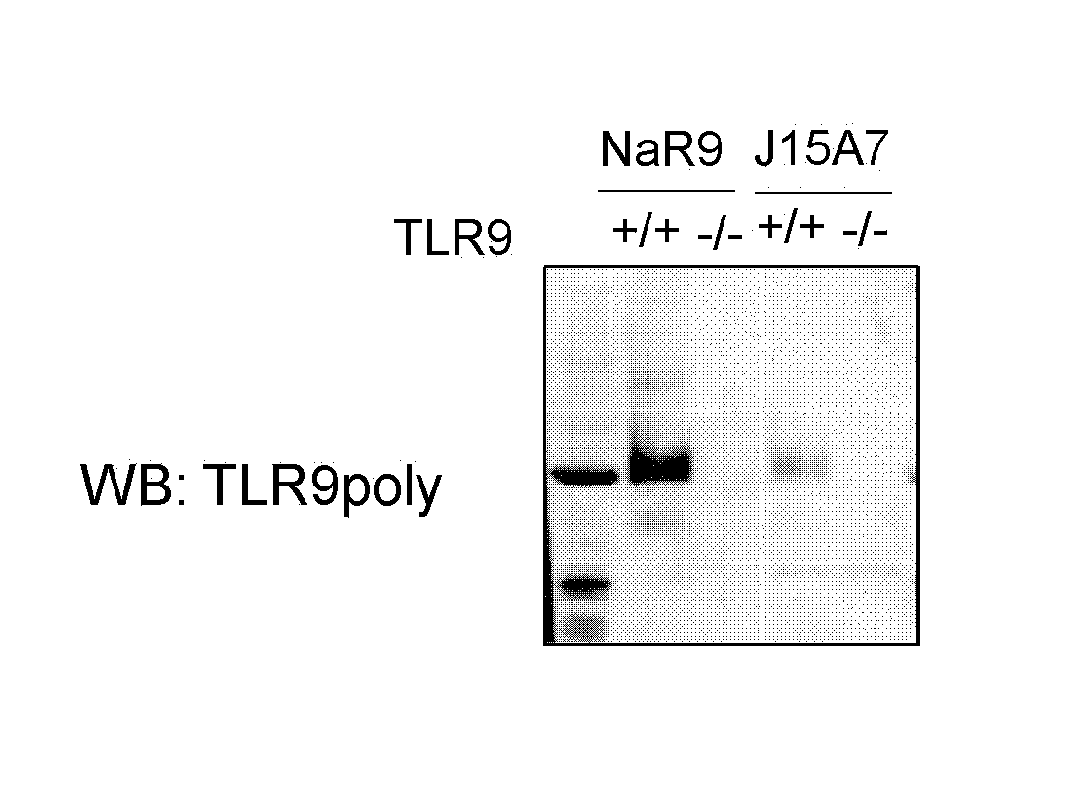
[0031] FIG. 2 shows results of conducting an immunoprecipitation experiment of TLR9 using spleen cDCs.
[0032] FIG. 3 shows results of conducting a staining inhibition experiment using an antibody against TLR9 (B33A4 and J15A7).
[0033] FIG. 4 shows results of an experiment of inducing BM-MCs from a wild-type mouse in order to verify the TLR9 response suppressive effect of NaR9.
[0034] FIG. 5 shows results of conducting an experiment of administering CpGB and D-(+)-galactosamine to mice in order to verify the suppressive effect of NaR9 to TLR9 response in vivo.
[0035] FIG. 6 shows results of measuring TNF-.alpha. in blood 1, 3, and 6 hours after ligands stimulation.
[0036] FIG. 7 shows chimeric TLR9 wherein the chimeric TLR9 was prepared from mouse TLR9 and human TLR9.
[0037] FIG. 8 shows results of analyzing an epitope for a NaR9 antibody by examining the binding of the NaR9 antibody to chimeric TLR9.
[0038] FIG. 9 shows the amino acid sequence (SEQ ID NO: 14) of the heavy chain of the NaR9 antibody.
[0039] FIG. 10 shows the nucleotide sequence (SEQ ID NO: 15) of the heavy chain of the NaR9 antibody.
[0040] FIG. 11 shows the amino acid sequence (SEQ ID NO: 16) of the light chain of the NaR9 antibody.
[0041] FIG. 12 shows the nucleotide sequence (SEQ ID NO: 17) of the light chain of the NaR9 antibody.
DESCRIPTION OF EMBODIMENTS
[Antibody]
[0042] Mainly the C-terminal side of TLR9 has been thought to be important for exerting the function of TLR9. The antibody according to the present invention is capable of recognizing the N terminus of TLR9, particularly, an extracellular domain to be cleaved within endolysosome, and its neighborhood. Hereinafter, the antibody according to the present invention is also referred to as an "N terminus-recognizing antibody" or an "anti-TLR9 antibody".
[0043] In the present specification, the "N terminus of TLR9" means the first half region from position 440 or 454 at the N terminus of TLR9, in a structure on an amino acid unit side with no amino group bonded and a carboxyl group bonded, particularly, in an extracellular domain to be cleaved within endolysosome, in the protein of TLR9. The extracellular domain means a region from position 1 at the N terminus of TLR9 to position 818. The "C terminus of TLR9" means a structure on an amino acid unit side with no carboxyl group bonded and an amino group bonded, particularly, a structure of TLR9 except for the extracellular domain, in the protein of TLR9.
[0044] In the protein of TLR9, an amino acid unit with no amino group bonded and a carboxyl group bonded is defined as position 1, and subsequent amino acid units are referred to as positions 2, 3, etc. from the N terminus of TLR9 toward the C terminus of TLR9.
[0045] The N terminus-recognizing antibody recognizes, for example, a region from positions 1 to 166, 1 to 242, 1 to 306, 1 to 356, or 1 to 440, preferably a region from positions 1 to 356, more preferably a region from positions 167 to 356, further preferably a region from positions 243 to 356, at the N terminus of TLR9.
[0046] Specifically, the N terminus-recognizing antibody recognizes the N terminus of TLR9 which is at least one of the following amino acid sequences:
[0047] (a) the amino acid sequence represented by SEQ ID NO: 1;
[0048] (b) an amino acid sequence derived from the amino acid sequence represented by SEQ ID NO: 1 by the deletion, substitution or addition of one or more amino acids; and
[0049] (c) an amino acid sequence having 80% or higher, preferably 90% or higher identity to the amino acid sequence represented by SEQ ID NO: 1.
[0050] The mouse TLR family is known to consist of 12 members, while the human TLR family is known to consist of 10 members. TLR1, TLR2, TLR4, TLR5, and TLR6 are distributed in the cell surface and recognize lipoproteins which are bacterial membrane components, glycolipids such as LPS, or proteins such as flagellin. TLR3, TLR7, TLR8, and TLR9 are distributed in endoplasmic reticula which are intracellular organelles and recognize bacterium- or virus-derived nucleic acids. TLR is a type I membrane protein extracellularly having leucine-rich repeat (LRR).
[0051] The N terminus-recognizing antibody recognizes the N terminus of mouse TLR9, human TLR9, or mouse/human chimeric TLR9 and is not particularly limited as long as the antibody recognizes the N terminus of TLR9. The N terminus of TLR9 that is recognized by the N terminus-recognizing antibody is, for example, the N terminus of mouse/human chimeric TLR9 shown in FIG. 7.
[0052] The N terminus-recognizing antibody is not particularly limited as long as the antibody recognizes the N terminus of TLR9. The N terminus-recognizing antibody comprises, for example, at least one of the following amino acid sequences:
[0053] (a) the amino acid sequence represented by SEQ ID NO: 2;
[0054] (b) the amino acid sequence represented by SEQ ID NO: 4;
[0055] (c) the amino acid sequence represented by SEQ ID NO: 6;
[0056] (d) the amino acid sequence represented by SEQ ID NO: 8;
[0057] (e) the amino acid sequence represented by SEQ ID NO: 10;
[0058] (f) the amino acid sequence represented by SEQ ID NO: 12;
[0059] (g) an amino acid sequence derived from the amino acid sequence represented by any of SEQ ID NOs: 2, 4, 6, 8, 10, and 12 by the deletion, substitution or addition of one or more amino acids; and
[0060] (h) an amino acid sequence having 80% or higher, preferably 90% or higher identity to the amino acid sequence represented by any of SEQ ID NOs: 2, 4, 6, 8, 10, and 12.
[0061] The N terminus-recognizing antibody may comprise at least one of heavy chain CDR1 to CDR3 and light chain CDR1 to CDR3, preferably at least one of heavy chain CDR1 to CDR3, more preferably heavy chain CDR3, with the amino acid sequences (a) to (c).
[0062] In the present specification, the "amino acid" is used in its broadest sense and is meant to include not only a natural amino acid but also a non-natural amino acid such as amino acid variant or derivative. Examples of the amino acid include, but are not limited to: natural proteinaceous L-amino acids; D-amino acids; chemically modified amino acids such as amino acid variants and derivatives; natural nonproteinaceous amino acids such as norleucine, .beta.-alanine, and ornithine; and chemically synthesized compounds having properties characteristic to amino acids and known in the art. Examples of the non-natural amino acids include, but not limited to, .alpha.-methylamino acids (.alpha.-methylalanine, etc.), D-amino acids, histidine-like amino acids (2-amino-histidine, .beta.-hydroxy-histidine, homohistidine, .alpha.-fluoromethyl-histidine, .alpha.-methyl-histidine, etc.), amino acids having, in the side chain thereof, excess methylene ("homo" amino acids, etc.), and amino acids derived from an amino acid having a carboxylic acid functional group in the side chain thereof by the substitution of the group by a sulfonic acid group (cysteic acid, etc.).
[0063] In the present specification, the number of amino acids to be deleted, substituted, or the like in the phrase "derived by the deletion, substitution or addition of one or more amino acids" is not particularly limited as long as the resulting set of CDRs retains the function of recognizing the antigen. In this context, the term "more" means an integer of 2 or larger, preferably several, for example, 2 to 5, more preferably 2, 3, or 4. The position of deletion, substitution, or addition in each CDR may be the N terminus, the C terminus, or between the N and C termini as long as the resulting set of CDRs retains the function of recognizing the antigen.
[0064] In the present specification, the phrase "having Y % or higher identity to the amino acid sequence represented by SEQ ID NO: X" means that when two polypeptides are arranged (aligned) to give the maximum coincidence of their amino acid sequences, the proportion of the number of amino acid residues in common is Y % or higher based on the total number of amino acids shown in SEQ ID NO: X.
[0065] The N terminus-recognizing antibody may be a monoclonal antibody or may be a polyclonal antibody. Also, the N terminus-recognizing antibody may be of any of isotypes IgG, IgM, IgA, IgD and IgE.
[0066] The N terminus-recognizing antibody may be a mouse antibody, a human CDR-grafted antibody, a human chimeric antibody, a humanized antibody, or a fully human antibody or may be a low-molecular antibody as long as the antibody binds to TLR9 on the cell surface and inhibits its function, though the N terminus-recognizing antibody is not limited thereto.
[0067] The human CDR-grafted antibody is an antibody obtained by substituting CDRs of a non-human animal antibody with CDRs of a human antibody. The human chimeric antibody is an antibody composed of variable regions derived from a non-human animal antibody and constant regions derived from a human antibody. The humanized antibody is an antibody obtained by incorporating a moiety derived from a human antibody into a non-human animal antibody while leaving a highly safe partial region of the non-human animal antibody, and conceptually includes the human chimeric antibody and the human CDR-grafted antibody.
[0068] In the present specification, the "low-molecular antibody" means an antibody fragment or an antibody fragment bound with an arbitrary molecule that recognizes the same epitope as that recognized by the original antibody. Specific examples thereof include, but are not limited to: Fab composed of VL, VH, CL, and CH1 regions; F(ab')2 in which two Fabs are linked to each other via a disulfide bond in the hinge region; Fv composed of VL and VH; and scFv which is a single-chain antibody in which VL and VH are linked to each other via an artificial polypeptide linker, and additionally include sdFv, Diabody, and sc(Fv)2.
[Method for Preparing Antibody]
[0069] The method for preparing the N terminus-recognizing antibody is not limited. For example, a monoclonal antibody can be obtained by isolating antibody-producing cells from a non-human mammal immunized with TLR9 or a fragment thereof, fusing these cells with myeloma cells or the like to prepare hybridomas, and purifying an antibody produced by these hybridomas. Also, a polyclonal antibody can be obtained from the serum of an animal immunized with TLR9 or a fragment thereof. The fragment of TLR9 for use in immunization is not particularly limited as long as the resulting antibody binds to TLR9 on the cell surface and inhibits its function.
[0070] In the case of preparing an antibody having a particular amino acid sequence, the antibody can be prepared, for example, by transforming an appropriate host with an expression vector containing a nucleic acid encoding the antibody, culturing the transformant under appropriate conditions to express an antibody, and isolating and purifying the antibody according to a known method. Examples of the isolation and purification method include affinity column using protein A or the like, other chromatography columns, filters, ultrafiltration, salting-out, and dialysis. These methods can be appropriately combined.
[0071] The "antibody Y specifically binding to the same epitope as that for certain antibody X" can be prepared after determination of the sequence of the epitope as described below.
[0072] For example, an epitope on an antigen protein can be determined by immobilizing many peptides having a random sequence to a solid-phase carrier to form an array, reacting the array with the antibody X, detecting binding using an enzymatically labeled secondary antibody, examining the amino acid sequence of the peptide to which the antibody X specifically binds, and retrieving homology between this amino acid sequence and the amino acid sequence of the antigen protein. The peptides to be immobilized onto a solid-phase carrier may be a group of partial peptides of the antigen protein prepared in advance. Alternatively, an epitope on an antigen protein may be determined by detecting the binding between the antibody X and the antigen protein in the presence of various partial peptides of the antigen protein by ELISA, and examining the presence or absence of competitive activity.
[0073] Once the sequence of the epitope can be determined, antibody Y specifically binding to this epitope can be prepared by those skilled in the art according to a known method. For example, an antibody specifically binding to the epitope can be obtained by immobilizing a peptide containing the epitope sequence to a solid-phase carrier, and detecting the binding between the peptide and various antibodies.
[0074] In this context, antibodies obtained by immunizing animals with an antigen protein or a partial peptide thereof may be used as the "various antibodies", or an antibody library or an antibody fragment library prepared by a phage display method may be used thereas. In the case of using a library prepared by a phage display method, antibody Y specifically binding to the epitope can also be obtained by immobilizing a peptide containing the epitope sequence onto a solid-phase carrier, and repeating panning.
[0075] A human chimeric antibody and a human CDR-grafted antibody can be prepared by cloning an antibody gene from the mRNA of a hybridoma producing a non-human animal antibody, and linking this gene with a portion of a human antibody gene through a gene recombination technique.
[0076] In the case of, for example, a human chimeric antibody, cDNA is synthesized from the mRNA of a hybridoma producing a mouse antibody, using reverse transcriptase. Heavy chain variable region (VH) and light chain variable region (LH) genes are cloned by PCR and analyzed for their sequences. Next, a 5' primer containing a leader sequence is prepared from an antibody nucleotide sequence having a high coincidence ratio, and a region from a signal sequence to the 3' end of each variable region gene is cloned from the cDNA by PCR using the 5' primer and a variable region 3' primer. Meanwhile, human IgG1 heavy chain and light chain constant region genes are cloned. For each of the heavy chain and the light chain, the mouse antibody-derived variable region gene and the human antibody-derived constant region gene are linked to each other and amplified by an overlapping hanging method based on PCR. The DNA thus obtained can be inserted into an appropriate vector, followed by transformation to obtain a human chimeric antibody.
[0077] In the case of a CDR-grafted antibody, human antibody variable regions having the highest homology to the mouse antibody variable regions used are selected, and their genes are cloned. The nucleotide sequences of CDRs are engineered by site-directed mutagenesis using a mega primer method. If the humanization of amino acid sequences constituting framework regions hinders the specific binding to an antigen, some amino acids of the frameworks may be converted from a human type to a rat type.
[0078] CDR consisting of the "amino acid sequence derived from the amino acid sequence represented by SEQ ID NO: X by the deletion, substitution or addition of one or more amino acids" or CDR consisting of the "amino acid sequence having Y % or higher identity to the amino acid sequence represented by SEQ ID NO: X" can be prepared by use of a known method such as site-directed mutagenesis, random mutagenesis, chain shuffling, or CDR walking.
[0079] It is well known to those skilled in the art that CDRs having more mature affinity can be obtained by displaying antibodies or antibody fragments having various mutations in their CDRs on the surface of phages by a phage display method according to these methods, and screening the antibodies or the antibody fragments using an antigen (e.g., Wu et al., PNAS, 95: 6037-6042 (1998); Schier, R. et al., J. Mol. Bio. 263: 551-567 (1996); Schier, R. et al., J. Mol. Biol. 255: 28-43 (1996); and Yang, W. P. et al., J. Mol. Biol., 254: 392-403 (1995)). The present invention also encompasses an antibody containing CDRs matured by such a method.
[0080] Other examples of the method for producing the antibody include the Adlib method (Seo, H. et al., Nat. Biotechnol., 6: 731-736, 2002) of obtaining an antibody-producing line from a DT40 cell line derived from trichostatin A-treated chicken B cells, and a method of immunizing KM mice, which are mice having a human antibody gene introduced therein instead of a disrupted mouse antibody gene, to prepare a human antibody (Itoh, K. et al., Jpn. J. Cancer Res., 92: 1313-1321, 2001; and Koide, A. et al., J. Mol. Biol., 284: 1141-1151, 1998). These methods can also be applied to the production of the N terminus-recognizing antibody.
[0081] When the N terminus-recognizing antibody is a low-molecular antibody, the antibody may be expressed by the above-described method while using DNA encoding this low-molecular antibody, or may be prepared by treating a full-length antibody with an enzyme such as papain or pepsin.
[0082] The N terminus-recognizing antibody may differ in amino acid sequence, molecular weight, isoelectric point, presence or absence of sugar chain, morphology, or the like depending on a preparation method or a purification method. However, the obtained antibody is included in the scope of the present invention as long as the antibody has a function equivalent to that of the N terminus-recognizing antibody. For example, when the N terminus-recognizing antibody is expressed in prokaryotic cells such as E. coli, a methionine residue is added to the N terminus of the amino acid sequence of the original antibody. Such an antibody is also included in the scope of the present invention.
[Pharmaceutical Composition]
[0083] The pharmaceutical composition according to the present invention comprises the N terminus-recognizing antibody and is used in the treatment or prevention of a disease related to TLR9.
[0084] Those skilled in the art can appropriately determine whether or not an antibody can be used in the treatment or prevention of a disease related to TLR9. For example, usability in the treatment or prevention of a disease related to TLR9 can be determined by confirming, according to a method shown in Example, at least one of (i) whether or not the obtained antibody binds to TLR9 on the cell surface; (ii) whether or not the amount of an inflammatory cytokine secreted from immune cells is reduced when the immune cells are brought into contact with the obtained antibody while stimulated with a TLR9 ligand; (iii) whether or not the proliferation of B cells is suppressed when the B cells are brought into contact with the obtained antibody while stimulated with a TLR9 ligand; and (iv) whether or not pathological conditions are ameliorated by administering the obtained antibody to an inflammatory disease animal model.
[0085] Examples of the disease related to TLR9 include various autoimmune diseases (rheumatoid arthritis (RA), systemic lupus erythematosus (SLE), scleroderma, polymyositis, Sjogren's syndrome (SS), ANCA-associated vasculitis, Behcet's disease, Kawasaki disease, mixed cryoglobulinemia, multiple sclerosis (MS), Guillain-Barre syndrome, myasthenia, type 1 diabetes, Graves' disease, Hashimoto's disease, Addison's disease, IPEX, APS type-II, autoimmune cardiomyopathy, interstitial pneumonia, bronchial asthma, autoimmune hepatitis, non-alcoholic steatohepatitis (NASH), primary biliary cirrhosis, inflammatory bowel disease (IBD), Crohn's disease, ulcerative colitis, psoriasis, atopic dermatitis, hemolytic anemia, autoimmune thyroiditis, and polyarticular juvenile idiopathic arthritis, etc.), graft rejection, and graft versus host disease (GvHD).
[0086] The pharmaceutical composition is potentially useful for, among these diseases, SLE, psoriasis, or NASH which develops under a mechanism involving TLR9. While not intending to be bound by any theory, for example, the N terminus-recognizing antibody is thought to contribute to the treatment or prevention of psoriasis related to TLR9 by recognizing TLR9 on the cell surface and inhibiting a TLR9 response of the cells, thereby suppressing the abnormal activation of immunity.
[0087] A steroid drug such as prednisolone has heretofore been used for, for example, SLE or its related disease systemic sclerosis. Also, belimumab, an antibody against B lymphocyte stimulator (Blys) involved in the activation of B cells, is used as a biological formulation. However, all of these drugs involve resistant cases. Thus, there is a demand for a therapeutic drug targeting a novel molecule for such diseases.
[0088] In SLE, type I interferon (IFN) is excessively produced. Cells producing type I IFN are pDCs (plasmacytoid dendritic cells), and the production is induced by DNA stimulation. The N terminus-recognizing antibody can therapeutically target TLR9 as a DNA sensor expressed on pDCs and is therefore useful in the treatment or prevention of SLE.
[0089] The pharmaceutical composition according to the present invention comprises the N terminus-recognizing antibody as an active ingredient and further contains a pharmaceutically acceptable carrier or additive.
[0090] Examples of the carrier and the additive include, but are not limited to, water, saline, pharmaceutically acceptable organic solvents such as phosphate buffers, dextrose, glycerol, and ethanol, collagen, polyvinyl alcohol, polyvinylpyrrolidone, carboxyvinyl polymers, sodium carboxymethylcellulose, sodium polyacrylate, sodium alginate, water-soluble dextran, sodium carboxymethyl starch, pectin, methyl cellulose, ethyl cellulose, xanthan gum, gum arabic, casein, agar, polyethylene glycol, diglycerin, glycerin, propylene glycol, Vaseline, paraffin, stearyl alcohol, stearic acid, human serum albumin, mannitol, sorbitol, lactose, and surfactants.
[0091] The pharmaceutical composition according to the present invention can be provided in various forms, for example, solutions (e.g., injections), dispersions, suspensions, tablets, pills, powders, or suppositories. A preferred form is an injection, which is preferably administered parenterally (e.g., intravenously, transdermally, intraperitoneally, or intramuscularly).
[0092] In the present specification, the "treatment or prevention" means causing at least one of recovery and remission of a disease, prevention or retardation of onset, prevention and retardation of the progression of a disease, and relief of at least one of the symptoms associated with a disease.
[0093] In the case of administering the pharmaceutical composition according to the present invention to mammals (e.g., humans, mice, rats, guinea pigs, rabbits, dogs, horses, monkeys, and pigs), particularly, to humans, the dose differs depending on symptoms, the age, sex, weight, and sensitivity difference of a patient, an administration route, an administration interval, the type of the active ingredient, and the type of a formulation and is not particularly limited. For example, a dose from 30 .mu.g to 1000 mg, from 100 .mu.g to 500 mg, or from 100 .mu.g to 100 mg can be administered once or in several portions. For administration through injection, a dose from 1 .mu.g/kg to 5000 .mu.g/kg or from 3 .mu.g/kg to 3000 .mu.g/kg may be administered once or in several portions based on the weight of a patient.
(Kit)
[0094] The kit according to the present invention comprises the N terminus-recognizing antibody. The purpose of the kit is not particularly limited, and the kit is used in the detection of TLR9 or the diagnosis of a disease related to TLR9.
[0095] The kit may contain a reagent, or a carrier or an additive, as in the pharmaceutical composition, according to the purpose, and may further contain a buffer, a container, an instruction manual, and the like.
(Method)
[0096] The method according to the present invention comprises the step of administering the N terminus-recognizing antibody to a subject.
[0097] In the method according to the present invention, the N terminus-recognizing antibody can be administered as the pharmaceutical composition according to the present invention.
[0098] The method according to the present invention can treat or prevent a disease related to TLR9 in a human or any of other mammals, etc., which is a subject. Alternatively, the method of the present invention may be used for other purposes, for example, diagnosis.
[0099] The disclosure of all patent literatures and non patent literatures cited herein is incorporated herein by reference in its entirety.
EXAMPLES
[0100] Hereinafter, the present invention will be described specifically with reference to Examples. However, the present invention is not limited by these examples by any means. Those skilled in the art can change the present invention into various modes without departing from the principle of the present invention, and such a change is also included in the scope of the present invention.
[0101] Establishment of anti-TLR9 antibody, detection of TLR9 in primary immune cell, and TLR9 response inhibition test of anti-TLR9 antibody
[Material and Method]
Preparation of Anti-TLR9 Monoclonal Antibody NaR9
[0102] In order to establish an anti-mouse TLR9 monoclonal antibody, a Tlr9.sup.-/- mouse of BALB/c background was immunized by the intraperitoneal administration of Ba/F3 cells forced to express mouse TLR9 (TLR9-Ba/F3). The initial immunization employed complete Freund's adjuvant (CFA) as an immunostimulator. The second and third immunizations employed incomplete Freund's adjuvant (IFA) as an immunostimulator. For the fourth immunization, TLR9-Ba/F3 suspended in 1 (PBS (phosphate-buffered saline) was intraperitoneally administered to the mouse. Five days after the final immunization, the spleen cells of the mouse were fused with a mouse myeloma-derived cell line SP2/O. The cell fusion employed hemagglutinating virus of Japan-derived envelope protein (GenomeONE-CF) purchased from Ishihara Sangyo Kaisha, Ltd. In order to select a hybridoma producing an antibody against TLR9, cell membrane permeability staining was performed using TLR9-Ba/F3, followed by analysis by flow cytometry. The subclass of NaR9 was determined as IgG2a/.kappa. using Mouse antibody Isotype kit purchased from Bio-Rad Laboratories, Inc.
Mouse
[0103] Wild-Type C57BL/6 Mice were Purchased from Japan SLC, Inc.
[0104] TLR9-deficient mice (Tlr9.sup.-/-) of C57BL/6 background were established in our laboratory. Tlr9.sup.-/- mice of BALB/c background were established by mating seven times with the wild-type BALB/c mice purchased from Japan SLC, Inc. The mice were raised in an SPF environment, and all animal experiments were conducted under the approval of the animal experiment committee of the Institute of Medical Science, The University of Tokyo.
Reagent and Antibody
[0105] CpGA 1585 (5'-G*G*GGTCAACGTTGAG*G*G*G*G*G-3'; the asterisk represents a phosphorothioation residue) (SEQ ID NO: 19), PolyU (5'-UUUUUUUUUUUUUUUUUUU-3'; all are phosphorothioation residues) (SEQ ID NO: 20) and CpGB 1688 (5'-TCCATGACGTTCCTGATGCT-3'; all are phosphorothioation residues) (SEQ ID NO: 18) were synthesized by FASMAC. Loxoribine (7-allyl-7,8-dihydro-8-oxo-guanosine) was purchased from Enzo Life Sciences, Inc. Saponin and D-(+)-galactosamine were purchased from Sigma-Aldrich Co. LLC. FuGene6 and DOTAP were purchased from Roche Applied Science. Anti-mouse TLR9 monoclonal antibodies NaR9, J15A7 and B33A4 were purified from the ascitic fluids of nude mice (purchased from Oriental Yeast Co., Ltd.) inoculated with hybridomas. Streptavidin-PE, anti-mouse IgG1-PE, anti-mouse IgG2a-PE, isotype control antibodies (mouse IgG1 and mouse IgG2a), anti-mouse CD16/32, anti-mouse CD19-APC-Cy7, anti-mouse CD11b-APC, anti-mouse CD11c-APC, anti-mouse CD11c-PE-Cy7, anti-mouse Siglec-H-FITC, and anti-mouse Ly-6G-PerCP-Cy5.5 were purchased from BioLegend, Inc. Anti-mouse B220-APC was purchased from TONBO Biosciences Inc. J15A7-PE, anti-mouse CD49b-BV421 and anti-mouse CD11b-BV510 were purchased from BD Biosciences.
Cell Culture
[0106] Ba/F3 cells were cultured in Roswell Park Memorial Institute (RPMI) 1640 medium (supplemented with IL-3, 10% FBS, PS/Gln and 50 .mu.M 2-ME). Bone marrow-derived macrophages (BM-MCs), conventional DCs (cDCs) or plasmacytoid DCs (pDCs) were induced from wild-type C57BL/6 mice or Tlr9.sup.-/- mice. In order to induce the macrophages, 1.times.10.sup.7 bone marrow cells were cultured for 6 days in 10% FCS-RPMI1640 supplemented with 100 ng/ml recombinant M-CSF (PeproTech, Rocky Hill, N.J., USA) in 10 cm cell culture dishes. In order to induce the cDCs or the pDCs, 1.times.10.sup.7 bone marrow cells were cultured for 7 days in 10% FCS-RPMI1640 supplemented with 10 ng/ml recombinant GM-CSF or 100 ng/ml Flt3-L (PeproTech, Rocky Hill, N.J., USA) in 10 cm cell culture dishes.
Construction of Plasmid and Transfection Using Retrovirus Vector
[0107] The gene sequence of mouse or human TLR9 was amplified by PCR and cloned into a pMX or pMXs retrovirus vector kindly given by Prof. Toshio Kitamura from the University of Tokyo. Mouse/human TLR9 chimeric variants describe below were constructed. The genes of TLR9.sub.166 (mouse TLR9 amino acids from positions 1 to 166 and human TLR9 amino acids from positions 167 to 1016), TLR9.sub.242 (mouse TLR9 amino acids from positions 1 to 242 and human TLR9 amino acids from positions 243 to 1016), =.sup.9.sub.356 (mouse TLR9 amino acids from positions 1 to 356 and human TLR9 amino acids from positions 357 to 1016), TLR9.sub.440 (mouse TLR9 amino acids from positions 1 to 440 and human TLR9 amino acids from positions 441 to 1016) and TLR9.sub.544 (mouse TLR9 amino acids from positions 1 to 544 and human TLR9 amino acids from positions 545 to 1016) were each cloned into a pMX-GFP vector so as to add GFP to the C-terminal side of each TLR9 variant.
[0108] Plat-E packaging cells (1.times.10.sup.3 cells/well) were transfected with each plasmid together with polyethylenimine (Polysciences, Inc.). Two days later, a culture supernatant containing viruses was added together with DOTAP to Ba/F3 cells, which were then centrifuged at 2000 rpm for 60 minutes.
Staining of Spleen Immune Cell and Flow Cytometry Analysis
[0109] The spleen was collected from each of wild-type mice and TLR9-knockout mice of C57BL/6N lineage, and cells were isolated using glass slides. The isolated cells were treated with an erythrolysis buffer (BioLegend, Inc.) to remove erythrocytes. Then, Fc receptor on the cell surface was blocked using purified anti-CD16/32 (BioLegend, Inc., clone 93).
[0110] After the blocking of the Fc receptor, the resultant was stained with Fluorescein (FITC)-conjugated anti-mouse SiglecH (BioLegend, Inc., 551) and Phycoerythrin (PE)-Cy7-conjugated anti-mouse CD11c (BioLegend, Inc., N418). A CD11c-positive/SiglecH-positive fraction was regarded as pDCs. A highly CD11c expressing/SiglecH-negative fraction was regarded as cDCs.
[0111] After the staining of SiglecH and CD11c on the cell surface, TLR9 on the cell surface and inside the cells was stained. In order to stain TLR9 on the cell surface, the cell surface was reacted with purified anti-TLR9 under nonfixed conditions, and PE conjugated rat anti-mouse IgG2a (BioLegend, Inc., clone RMG2a-62) was used as a secondary antibody. TLR9 inside the cells was stained using BD Cytofix/Cytoperm fixation/permeabilization solution kit. These cells were buffer-replaced with a buffer for staining and analyzed using LSRFortessa X-20 (BD).
Immunoprecipitation and Western Blot
[0112] The protein expression of TLR9 was analyzed by Western blot. BM-cDCs were washed twice with 1.times.PBS and recovered. The recovered cells were lysed for 30 minutes in an ice-cold buffer for lysis (1% Triton X 100, 20 mM Tris-HCl (pH 7.4), 150 mM NaCl, 1 mM CaCl.sub.2, 1 mM MgCl.sub.2, 10% glycerol, 1 mM DTT and Complete protease Inhibitor Cocktail (F. Hoffmann-La Roche, Ltd.)) and centrifuged at a high speed, followed by the recovery of the supernatant of the cell lysate.
[0113] The recovered cell lysate was added to N-hydroxy succinimide-activated Sepharose 4FF beads bound with the anti-TLR9 monoclonal antibody (NaR9), and the mixture was stirred at 4.degree. C. for 2 hours. TLR9 was immunoprecipitated by this step. The beads thus stirred were washed three times with an ice-cold buffer for washing (0.1% Triton X 100, 20 mM Tris/HCl (pH 7.4), 150 mM NaCl, 1 mM CaCl.sub.2), 1 mM MgCl2, 0.1% glycerol, and 1 mM DTT). To the washed beads, an SDS sample buffer (125 mM Tris/HCl [pH 6.8], 20% glycerol, 4% SDS, 10% 2-ME, and 0.005% bromophenol blue) was added, and the mixture was heated at 96.degree. C. for 5 minutes for protein denaturation treatment. The sample thus prepared was subjected to polyacrylamide electrophoresis, and the protein was transferred to a PVDF membrane and Western-blotted.
[0114] The antibody used in the Western blot was an autologous polyclonal antibody purified from serum recovered from a rabbit immunized with the TIR domain of mouse TLR9.
Statistical Processing
[0115] In an experiment of administering an antibody to mice, a significant difference test of data between an anti-TLR9 monoclonal antibody administration group and a control antibody administration group was conducted according to Student's t-test. A significance level of less than 0.01 in the t test was judged as being the significant difference between the compared groups.
[0116] TLR9 response inhibition experiment using anti-TLR9 monoclonal antibody
(In Vitro Test)
[0117] The cells used were BM-MCs, BM-cDCs, and BM-pDCs. Each well of a 96-well flat-bottomed plate was inoculated with 5(10.sup.4 BM-MCs, BM-cDCs, or BM-pDCs, and an anti-TLR9 antibody was added thereto at each concentration. Four hours after the addition of the antibody, a TLR ligand was added to the inoculated cultured cells. Twenty-four hours after the addition of the TLR ligand, the culture supernatant was recovered. The recovered culture supernatant was subjected to ELISA to measure a cytokine produced by the stimulation with the ligand.
(In Vivo Test)
[0118] It has been reported that wild-type mice die within 10 hours by the intraperitoneal administration of a CpGB ligand and D-(+)-galactosamine at the same time (Sparwasser, T. et al., Eur J Immunol 27, 1671-1679, doi:10.1002/eji.1830270712 (1997)). An antibody or PBS (phosphate-buffered saline) was intraperitoneally administered to wild-type C57BL/6 mice. Fifteen hours later, 10 nmol of CpGB and 20 mg of D-(+)-galactosamine were further intraperitoneally administered to each mouse. Blood was collected from each mouse before the ligand administration and 1, 3, and 6 hours after the administration, and TNF-.alpha. or IL-12p40 was measured by ELISA using the serum.
Analysis on Amino Acid Sequence and Nucleotide Sequence of Antibody and CDR
[0119] The amino acid sequence of NaR9, a monoclonal antibody against TLR9, was analyzed by GenScript. Total RNA was recovered from the hybridoma using TRIzol(R) Reagent manufactured by Ambion, Inc., and subjected to reverse transcription reaction into cDNA using PrimeScript(R) 1st Strand cDNA Synthesis Kit manufactured by Takara Bio Inc. From this cDNA, V.sub.H and V.sub.L gene fragments were amplified, and each fragment was cloned into a general vector for cloning. After transformation of E. coli, plasmids were recovered by colony PCR from at least 5 clones with a band of a correct size and analyzed for their sequences. The highest consensus sequences were determined as the sequences of the antibody by the sequence analysis. As for the determined sequences of the antibody, FIG. 9 shows the amino acid sequence (SEQ ID NO: 14) of the heavy chain of the NaR9 antibody, FIG. 10 shows the nucleotide sequence (SEQ ID NO: 15) of the heavy chain of the NaR9 antibody, FIG. 11 shows the amino acid sequence (SEQ ID NO: 16) of the light chain of the NaR9 antibody, and FIG. 12 shows the nucleotide sequence (SEQ ID NO: 17) of the light chain of the NaR9 antibody.
[Results]
Establishment of Anti-TLR9 Monoclonal Antibody and Detection of TLR9
[0120] In order to examine the intracellular expression of endogenous TLR9 in primary immune cells, a NaR9 antibody was established as a monoclonal antibody against mouse TLR9. In order to verify the specificity of the antibody, the expression of TLR9 was revealed using mouse spleen cDCs. The specificity of staining was confirmed by the absence of a stain of the spleen cDCs of TLR9 knockout mice (FIG. 1). Further, the immunoprecipitation experiment of TLR9 was conducted using BM-cDCs. NaR9 was shown to more strongly immunoprecipitate TLR9 as compared with a previously established antibody against TLR9 (J15A7) (FIG. 2). In order to reveal the recognition site of NaR9, a staining inhibition experiment was first conducted using previously established antibodies against TLR9 (B33A4 and J15A7) (for B33A4 and J15A7, see International Publication No. WO 2014/174704). The TLR9 staining of NaR9 was inhibited by the pretreatment of cells with J15A7 (FIG. 3). Likewise, the TLR9 staining of J15A7 was inhibited by the pretreatment of cells with NaR9 (FIG. 3). These results demonstrated that the newly established NaR9 recognizes TLR9 on primary immune cells, can be utilized in cell staining or immunoprecipitation, and has an antigen recognition site similar to that of J15A7.
TLR9 Response Inhibition Test Using Anti-TLR9 Monoclonal Antibody--(1)
[0121] In order to verify the TLR9 response suppressive effect of NaR9, BM-MCs were induced from wild-type mice and used in the experiment. In a NaR9 prior administration group, TNF-.alpha. and IL-12p40 production in BM-MCs in response to CpGB or CpGA was suppressed in an antibody concentration-dependent manner (FIG. 4). Similar results were obtained about BM-cDCs (data not shown). On the other hand, there was no influence on the loxoribine response of the TLR7 ligand. On the other hand, the TLR9 response suppressive effect of NaR9 was not observed in BM-pDCs (data not shown).
TLR9 Response Inhibition Test Using Anti-TLR9 Monoclonal Antibody--(2)
[0122] Subsequently, in order to verify the TLR9 response suppressive effect of NaR9 in vivo, an experiment of administering CpGB and D-(+)-galactosamine to a mouse was conducted. In a PBS or IgG2a control antibody prior administration group, approximately 90% of the mice died within 10 hours after ligand stimulation. By contrast, in a NaR9 prior administration group, the death rate was reduced to 20% (FIG. 5). As a result of further measuring TNF-.alpha. in blood 1, 3, and 6 hours after ligand stimulation, cytokine production was significantly suppressed in a NaR9 antibody administration group compared with an IgG2a control antibody administration group (FIG. 6).
Amino Acid Sequence Analysis of NaR9 Variable Regions
[0123] The heavy chain amino acid sequence of a monoclonal antibody obtained from the hybridoma line NaR9 is shown in SEQ ID NO: 14, and the light chain amino acid sequence thereof is shown in SEQ ID NO: 16. Nucleotide sequences corresponding to these amino acid sequences are shown in SEQ ID NOs: 15 and 17.
[0124] The amino acid sequences of the heavy chain CDR1 to CDR3 of this antibody are shown in SEQ ID NOs: 2, 4, and 6, and the amino acid sequences of light chain CDR1 to CDR3 are shown in SEQ ID NOs: 8, 10, and 12. Nucleotide sequences corresponding to these amino acid sequences are shown in SEQ ID NOs: 3, 5, 7, 9, 11, and 13.
Antigen Recognition Site Analysis of NaR9
[0125] In order to determine the antigen recognition site of NaR9, a cell line expressing chimeric TLR9 of mouse TLR9 and human TLR9 was prepared. The mouse TLR9 used was from amino acid positions 1 to 544 (TLR9.sub.544), from amino acid positions 1 to 454 (TLR9.sub.454), from amino acid positions 1 to 356 (TLR9.sub.356), and from amino acid positions 1 to 242 (TLR9.sub.242) of TLR9 shown in SEQ ID NO: 1, and the sequence of the human TLR9 was positioned on the C-terminal side to prepare each chimera (FIG. 7), which was then expressed in the cell line. The binding of NaR9 to each chimeric TLR9 was measured by flow cytometry.
[0126] The results are shown in FIG. 8. NaR9 did not bind to TLR9.sub.242, but bound to TLR9.sub.356, suggesting that an epitope for NaR9 is present in or near a region from positions 243 to 356 in the amino acid sequence of TLR9.
Free Text of Sequence Listing
[0127] SEQ ID NO: 1 represents the amino acid sequence of mouse TLR9.
[0128] SEQ ID NO: 2 represents the amino acid sequence of the heavy chain CDR1 of a NaR9 antibody.
[0129] SEQ ID NO: 3 represents the nucleotide sequence of the heavy chain CDR1 of the NaR9 antibody.
[0130] SEQ ID NO: 4 represents the amino acid sequence of the heavy chain CDR2 of the NaR9 antibody.
[0131] SEQ ID NO: 5 represents the nucleotide sequence of the heavy chain CDR2 of the NaR9 antibody.
[0132] SEQ ID NO: 6 represents the amino acid sequence of the heavy chain CDR3 of the NaR9 antibody.
[0133] SEQ ID NO: 7 represents the nucleotide sequence of the heavy chain CDR3 of the NaR9 antibody.
[0134] SEQ ID NO: 8 represents the amino acid sequence of the light chain CDR1 of the NaR9 antibody.
[0135] SEQ ID NO: 9 represents the nucleotide sequence of the light chain CDR1 of the NaR9 antibody.
[0136] SEQ ID NO: 10 represents the amino acid sequence of the light chain CDR2 of the NaR9 antibody.
[0137] SEQ ID NO: 11 represents the nucleotide sequence of the light chain CDR2 of the NaR9 antibody.
[0138] SEQ ID NO: 12 represents the amino acid sequence of the light chain CDR3 of the NaR9 antibody.
[0139] SEQ ID NO: 13 represents the nucleotide sequence of the light chain CDR3 of the NaR9 antibody.
[0140] SEQ ID NO: 14 represents the amino acid sequence of the heavy chain of the NaR9 antibody.
[0141] SEQ ID NO: 15 represents the nucleotide sequence of the heavy chain of the NaR9 antibody.
[0142] SEQ ID NO: 16 represents the amino acid sequence of the light chain of the NaR9 antibody.
[0143] SEQ ID NO: 17 represents the nucleotide sequence of the light chain of the NaR9 antibody.
[0144] SEQ ID NO: 18 represents the DNA sequence of CpGB.
[0145] SEQ ID NO: 19 represents the DNA sequence of CpGA.
[0146] SEQ ID NO: 20 represents the RNA sequence of PolyU.
Sequence CWU 1
1
2011032PRTMus musculus 1Met Val Leu Arg Arg Arg Thr Leu His Pro Leu
Ser Leu Leu Val Gln1 5 10 15Ala Ala Val Leu Ala Glu Thr Leu Ala Leu
Gly Thr Leu Pro Ala Phe 20 25 30Leu Pro Cys Glu Leu Lys Pro His Gly
Leu Val Asp Cys Asn Trp Leu 35 40 45Phe Leu Lys Ser Val Pro Arg Phe
Ser Ala Ala Ala Ser Cys Ser Asn 50 55 60Ile Thr Arg Leu Ser Leu Ile
Ser Asn Arg Ile His His Leu His Asn65 70 75 80Ser Asp Phe Val His
Leu Ser Asn Leu Arg Gln Leu Asn Leu Lys Trp 85 90 95Asn Cys Pro Pro
Thr Gly Leu Ser Pro Leu His Phe Ser Cys His Met 100 105 110Thr Ile
Glu Pro Arg Thr Phe Leu Ala Met Arg Thr Leu Glu Glu Leu 115 120
125Asn Leu Ser Tyr Asn Gly Ile Thr Thr Val Pro Arg Leu Pro Ser Ser
130 135 140Leu Val Asn Leu Ser Leu Ser His Thr Asn Ile Leu Val Leu
Asp Ala145 150 155 160Asn Ser Leu Ala Gly Leu Tyr Ser Leu Arg Val
Leu Phe Met Asp Gly 165 170 175Asn Cys Tyr Tyr Lys Asn Pro Cys Thr
Gly Ala Val Lys Val Thr Pro 180 185 190Gly Ala Leu Leu Gly Leu Ser
Asn Leu Thr His Leu Ser Leu Lys Tyr 195 200 205Asn Asn Leu Thr Lys
Val Pro Arg Gln Leu Pro Pro Ser Leu Glu Tyr 210 215 220Leu Leu Val
Ser Tyr Asn Leu Ile Val Lys Leu Gly Pro Glu Asp Leu225 230 235
240Ala Asn Leu Thr Ser Leu Arg Val Leu Asp Val Gly Gly Asn Cys Arg
245 250 255Arg Cys Asp His Ala Pro Asn Pro Cys Ile Glu Cys Gly Gln
Lys Ser 260 265 270Leu His Leu His Pro Glu Thr Phe His His Leu Ser
His Leu Glu Gly 275 280 285Leu Val Leu Lys Asp Ser Ser Leu His Thr
Leu Asn Ser Ser Trp Phe 290 295 300Gln Gly Leu Val Asn Leu Ser Val
Leu Asp Leu Ser Glu Asn Phe Leu305 310 315 320Tyr Glu Ser Ile Thr
His Thr Asn Ala Phe Gln Asn Leu Thr Arg Leu 325 330 335Arg Lys Leu
Asn Leu Ser Phe Asn Tyr Arg Lys Lys Val Ser Phe Ala 340 345 350Arg
Leu His Leu Ala Ser Ser Phe Lys Asn Leu Val Ser Leu Gln Glu 355 360
365Leu Asn Met Asn Gly Ile Phe Phe Arg Leu Leu Asn Lys Tyr Thr Leu
370 375 380Arg Trp Leu Ala Asp Leu Pro Lys Leu His Thr Leu His Leu
Gln Met385 390 395 400Asn Phe Ile Asn Gln Ala Gln Leu Ser Ile Phe
Gly Thr Phe Arg Ala 405 410 415Leu Arg Phe Val Asp Leu Ser Asp Asn
Arg Ile Ser Gly Pro Ser Thr 420 425 430Leu Ser Glu Ala Thr Pro Glu
Glu Ala Asp Asp Ala Glu Gln Glu Glu 435 440 445Leu Leu Ser Ala Asp
Pro His Pro Ala Pro Leu Ser Thr Pro Ala Ser 450 455 460Lys Asn Phe
Met Asp Arg Cys Lys Asn Phe Lys Phe Thr Met Asp Leu465 470 475
480Ser Arg Asn Asn Leu Val Thr Ile Lys Pro Glu Met Phe Val Asn Leu
485 490 495Ser Arg Leu Gln Cys Leu Ser Leu Ser His Asn Ser Ile Ala
Gln Ala 500 505 510Val Asn Gly Ser Gln Phe Leu Pro Leu Thr Asn Leu
Gln Val Leu Asp 515 520 525Leu Ser His Asn Lys Leu Asp Leu Tyr His
Trp Lys Ser Phe Ser Glu 530 535 540Leu Pro Gln Leu Gln Ala Leu Asp
Leu Ser Tyr Asn Ser Gln Pro Phe545 550 555 560Ser Met Lys Gly Ile
Gly His Asn Phe Ser Phe Val Thr His Leu Ser 565 570 575Met Leu Gln
Ser Leu Ser Leu Ala His Asn Asp Ile His Thr Arg Val 580 585 590Ser
Ser His Leu Asn Ser Asn Ser Val Arg Phe Leu Asp Phe Ser Gly 595 600
605Asn Gly Met Gly Arg Met Trp Asp Glu Gly Gly Leu Tyr Leu His Phe
610 615 620Phe Gln Gly Leu Ser Gly Leu Leu Lys Leu Asp Leu Ser Gln
Asn Asn625 630 635 640Leu His Ile Leu Arg Pro Gln Asn Leu Asp Asn
Leu Pro Lys Ser Leu 645 650 655Lys Leu Leu Ser Leu Arg Asp Asn Tyr
Leu Ser Phe Phe Asn Trp Thr 660 665 670Ser Leu Ser Phe Leu Pro Asn
Leu Glu Val Leu Asp Leu Ala Gly Asn 675 680 685Gln Leu Lys Ala Leu
Thr Asn Gly Thr Leu Pro Asn Gly Thr Leu Leu 690 695 700Gln Lys Leu
Asp Val Ser Ser Asn Ser Ile Val Ser Val Val Pro Ala705 710 715
720Phe Phe Ala Leu Ala Val Glu Leu Lys Glu Val Asn Leu Ser His Asn
725 730 735Ile Leu Lys Thr Val Asp Arg Ser Trp Phe Gly Pro Ile Val
Met Asn 740 745 750Leu Thr Val Leu Asp Val Arg Ser Asn Pro Leu His
Cys Ala Cys Gly 755 760 765Ala Ala Phe Val Asp Leu Leu Leu Glu Val
Gln Thr Lys Val Pro Gly 770 775 780Leu Ala Asn Gly Val Lys Cys Gly
Ser Pro Gly Gln Leu Gln Gly Arg785 790 795 800Ser Ile Phe Ala Gln
Asp Leu Arg Leu Cys Leu Asp Glu Val Leu Ser 805 810 815Trp Asp Cys
Phe Gly Leu Ser Leu Leu Ala Val Ala Val Gly Met Val 820 825 830Val
Pro Ile Leu His His Leu Cys Gly Trp Asp Val Trp Tyr Cys Phe 835 840
845His Leu Cys Leu Ala Trp Leu Pro Leu Leu Ala Arg Ser Arg Arg Ser
850 855 860Ala Gln Thr Leu Pro Tyr Asp Ala Phe Val Val Phe Asp Lys
Ala Gln865 870 875 880Ser Ala Val Ala Asp Trp Val Tyr Asn Glu Leu
Arg Val Arg Leu Glu 885 890 895Glu Arg Arg Gly Arg Arg Ala Leu Arg
Leu Cys Leu Glu Asp Arg Asp 900 905 910Trp Leu Pro Gly Gln Thr Leu
Phe Glu Asn Leu Trp Ala Ser Ile Tyr 915 920 925Gly Ser Arg Lys Thr
Leu Phe Val Leu Ala His Thr Asp Arg Val Ser 930 935 940Gly Leu Leu
Arg Thr Ser Phe Leu Leu Ala Gln Gln Arg Leu Leu Glu945 950 955
960Asp Arg Lys Asp Val Val Val Leu Val Ile Leu Arg Pro Asp Ala His
965 970 975Arg Ser Arg Tyr Val Arg Leu Arg Gln Arg Leu Cys Arg Gln
Ser Val 980 985 990Leu Phe Trp Pro Gln Gln Pro Asn Gly Gln Gly Gly
Phe Trp Ala Gln 995 1000 1005Leu Ser Thr Ala Leu Thr Arg Asp Asn
Arg His Phe Tyr Asn Gln 1010 1015 1020Asn Phe Cys Arg Gly Pro Thr
Ala Glu1025 103025PRTMus musculus 2Asn Tyr Tyr Leu His1 5315DNAMus
musculus 3aactactatt tacac 15417PRTMus musculus 4Trp Ile Tyr Pro
Gly Asp Gly Ser Thr Lys Tyr Asn Asp Gln Phe Arg1 5 10
15Gly551DNAMus musculus 5tggatttatc ctggagatgg tagcactaag
tacaatgacc agttcagggg c 5167PRTMus musculus 6Ser Trp Asp Tyr Phe
Asp Tyr1 5721DNAMus musculus 7agctgggact attttgacta c 21811PRTMus
musculus 8Lys Ala Ser Gln Asp Ile Asp Asn Tyr Ile Ala1 5
10933DNAMus musculus 9aaggcaagcc aagacattga caattatata gct
33107PRTMus musculus 10Tyr Ala Ser Thr Leu Gln Pro1 51121DNAMus
musculus 11tacgcatcta cattacagcc g 21128PRTMus musculus 12Leu Gln
Tyr Asp Asp Leu Tyr Thr1 51324DNAMus musculus 13ctacagtatg
atgatctata tacg 2414135PRTMus musculus 14Met Arg Trp Ser Trp Ile
Phe Leu Phe Leu Leu Ser Ile Thr Ala Gly1 5 10 15Val His Cys Gln Val
His Leu Gln Gln Ser Gly Pro Asp Leu Val Lys 20 25 30Pro Gly Ala Ser
Val Gln Met Ser Cys Lys Thr Ser Asp Tyr Thr Phe 35 40 45Thr Asn Tyr
Tyr Leu His Trp Val Arg Gln Arg Pro Gly Gln Gly Leu 50 55 60Glu Trp
Ile Gly Trp Ile Tyr Pro Gly Asp Gly Ser Thr Lys Tyr Asn65 70 75
80Asp Gln Phe Arg Gly Arg Thr Thr Leu Thr Ala Asp Lys Ser Ser Ser
85 90 95Thr Ala Tyr Met Phe Leu Ser Ser Leu Thr Ser Glu Asp Ser Ala
Ile 100 105 110Tyr Phe Cys Ala Lys Ser Trp Asp Tyr Phe Asp Tyr Trp
Gly Gln Gly 115 120 125Thr Thr Leu Thr Val Ser Ser 130
13515405DNAMus musculus 15atgcgatgga gctggatctt tctcttcctc
ctgtcaataa ctgcaggtgt ccattgccag 60gtccatctgc agcagtctgg acctgacctg
gtgaagcctg gggcttcagt gcagatgtcc 120tgcaagactt ctgactacac
cttcacaaac tactatttac actgggtgag gcagaggcct 180ggacagggac
ttgagtggat tggatggatt tatcctggag atggtagcac taagtacaat
240gaccagttca ggggcaggac cacactgact gcagacaaat cctccagcac
agcctacatg 300ttcctcagca gcctgacctc tgaggactct gcgatctatt
tctgtgcgaa gagctgggac 360tattttgact actggggcca aggcaccact
ctcacagtct cctca 40516126PRTMus musculus 16Met Arg Pro Ser Ile Gln
Phe Leu Gly Leu Leu Leu Phe Trp Leu His1 5 10 15Gly Ala Gln Cys Asp
Ile Gln Met Thr Gln Ser Pro Ser Ser Leu Ser 20 25 30Ala Ser Leu Gly
Gly Lys Val Thr Ile Thr Cys Lys Ala Ser Gln Asp 35 40 45Ile Asp Asn
Tyr Ile Ala Trp Tyr Gln His Lys Pro Gly Lys Gly Pro 50 55 60Arg Leu
Leu Ile His Tyr Ala Ser Thr Leu Gln Pro Gly Ile Pro Ser65 70 75
80Arg Phe Ser Gly Ser Gly Ser Gly Arg Asp Tyr Ser Leu Ser Ile Ser
85 90 95Asn Leu Glu Pro Glu Asp Ile Ala Thr Tyr Tyr Cys Leu Gln Tyr
Asp 100 105 110Asp Leu Tyr Thr Phe Gly Gly Gly Thr Lys Leu Glu Ile
Lys 115 120 12517378DNAMus musculus 17atgagaccgt ctattcagtt
cctggggctc ttgttgttct ggcttcatgg tgctcagtgt 60gacatccaga tgacacagtc
tccatcctca ctgtctgcat ctctgggagg caaagtcacc 120atcacttgca
aggcaagcca agacattgac aattatatag cttggtacca gcacaagcct
180ggaaaaggtc ctaggctact catacattac gcatctacat tacagccggg
catcccatca 240aggttcagtg gaagtgggtc tgggagagat tattccctca
gcatcagcaa cctggaacct 300gaagatattg caacttatta ttgtctacag
tatgatgatc tatatacgtt cggagggggg 360accaagctgg aaataaaa
3781820DNAArtificial SequenceCpGB
1688misc_feature(1)..(20)phosphorothioate 18tccatgacgt tcctgatgct
201920DNAArtificial SequenceCpGA
1585misc_feature(1)..(2)phosphorothioatemisc_feature(15)..(19)phosphoroth-
ioate 19ggggtcaacg ttgagggggg 202019RNAArtificial SequencePoly
Umisc_feature(1)..(19)phosphorothioate 20uuuuuuuuuu uuuuuuuuu
19
D00000
D00001
D00002
D00003
S00001
XML
uspto.report is an independent third-party trademark research tool that is not affiliated, endorsed, or sponsored by the United States Patent and Trademark Office (USPTO) or any other governmental organization. The information provided by uspto.report is based on publicly available data at the time of writing and is intended for informational purposes only.
While we strive to provide accurate and up-to-date information, we do not guarantee the accuracy, completeness, reliability, or suitability of the information displayed on this site. The use of this site is at your own risk. Any reliance you place on such information is therefore strictly at your own risk.
All official trademark data, including owner information, should be verified by visiting the official USPTO website at www.uspto.gov. This site is not intended to replace professional legal advice and should not be used as a substitute for consulting with a legal professional who is knowledgeable about trademark law.