Photoreceptors And Photoreceptor Progenitors Produced From Pluripotent Stem Cells
Lanza; Robert P. ; et al.
U.S. patent application number 16/284565 was filed with the patent office on 2019-09-26 for photoreceptors and photoreceptor progenitors produced from pluripotent stem cells. This patent application is currently assigned to Astellas Institute for Regenerative Medicine. The applicant listed for this patent is Astellas Institute for Regenerative Medicine. Invention is credited to Robert P. Lanza, Shi-Jiang Lu, Wei Wang.
| Application Number | 20190290701 16/284565 |
| Document ID | / |
| Family ID | 51537881 |
| Filed Date | 2019-09-26 |


View All Diagrams
| United States Patent Application | 20190290701 |
| Kind Code | A1 |
| Lanza; Robert P. ; et al. | September 26, 2019 |
PHOTORECEPTORS AND PHOTORECEPTOR PROGENITORS PRODUCED FROM PLURIPOTENT STEM CELLS
Abstract
Methods are provided for the production of photoreceptor cells and photoreceptor progenitor cells from pluripotent stem cells. Additionally provided are compositions of photoreceptor cells and photoreceptor cells, as well as methods for the therapeutic use thereof. Exemplary methods may produce substantially pure cultures of photoreceptor cells and/or photoreceptor cells.
| Inventors: | Lanza; Robert P.; (Clinton, MA) ; Lu; Shi-Jiang; (Shrewsbury, MA) ; Wang; Wei; (Rochester, MN) | ||||||||||
| Applicant: |
|
||||||||||
|---|---|---|---|---|---|---|---|---|---|---|---|
| Assignee: | Astellas Institute for Regenerative
Medicine Marlborough MA |
||||||||||
| Family ID: | 51537881 | ||||||||||
| Appl. No.: | 16/284565 | ||||||||||
| Filed: | February 25, 2019 |
Related U.S. Patent Documents
| Application Number | Filing Date | Patent Number | ||
|---|---|---|---|---|
| 14774771 | Sep 11, 2015 | |||
| PCT/US14/29790 | Mar 14, 2014 | |||
| 16284565 | ||||
| 61793168 | Mar 15, 2013 | |||
| Current U.S. Class: | 1/1 |
| Current CPC Class: | A61P 27/06 20180101; C12N 5/0623 20130101; A61K 35/545 20130101; A61P 27/02 20180101; C12N 2501/155 20130101; A61K 35/30 20130101; C12N 2501/385 20130101; A61P 9/10 20180101; C12N 2501/33 20130101; C12N 2501/235 20130101; C12N 2500/33 20130101; C12N 2506/02 20130101; C12N 5/0696 20130101; C12N 5/062 20130101; C12N 5/0606 20130101; C12N 2501/13 20130101; C12N 2501/105 20130101 |
| International Class: | A61K 35/545 20060101 A61K035/545; C12N 5/074 20060101 C12N005/074; C12N 5/0793 20060101 C12N005/0793; C12N 5/0735 20060101 C12N005/0735; C12N 5/0797 20060101 C12N005/0797; A61K 35/30 20060101 A61K035/30 |
Claims
1. A preparation of photoreceptor progenitor cells, comprising: a plurality of photoreceptor progenitor cells, and a medium suitable for maintaining the viability of the photoreceptor progenitor cells, wherein greater than 90% of the cells in the preparation are immunocytochemically PAX6+ and CHX10-, and mRNA transcript positive for MASH1 as detected by qPCR.
2. (canceled)
3. The preparation of photoreceptor progenitor cells of claim 1, wherein the plurality of photoreceptor progenitor cells is substantially free of pluripotent stem cells, retinal ganglion cells, mature photoreceptors, and/or amacrine cells.
4. (canceled)
5. The preparation of photoreceptor progenitor cells of claim 1, wherein the preparation is a cryogenic cell preparation comprising at least 10.sup.9 photoreceptor progenitor cells
6.-20. (canceled)
21. The preparation of claim 1, wherein the medium suitable for maintaining the viability of the photoreceptor progenitor cells is selected from the group consisting of a culture medium, a cryopreservative, and a biocompatible injection medium suitable for injection in a human patient.
22.-26. (canceled)
27. The preparation of photoreceptor progenitor cells of claim 1, wherein greater than 90% of the cells are immunocytochemically PAX6+, CHX10-, rhodopsin+, and opsin.
28. (canceled)
29. A method of treating a disease or disorder caused by loss of photoreceptors in a patient, comprising administering the pharmaceutical preparation of photoreceptor progenitor cells of claim 1.
30. (canceled)
31. A method of producing photoreceptor progenitor cells, comprising the steps of culturing eye field progenitor cells under culture conditions alternating between low adherence or non-adherent conditions for a period of time sufficient to form individual cell spheres, and then adherent conditions, which alternating culture conditions are continued until a majority of the cells are photoreceptor progenitor cells, wherein the photoreceptor progenitor cells are characterized as PAX6(+) and CHX10(-), and wherein the photoreceptor progenitor cells differentiate into photoreceptor cells upon treatment with retinoic acid.
32. A method of producing photoreceptor progenitor cells, comprising the steps of (a) culturing eye field progenitor cells, preferably as cells clusters and preferably under low adherence or non-adherent conditions, in a neural differentiation media for a period of time sufficient for the cell clusters to form individual cell spheres, wherein the eye field progenitor cells are characterized as PAX6(+) and RX1(+) and OCT4(-) and NANOG(-), and preferably are also characterized as SIX3(+), SIX6(+), LHX2(+), TBX3(+), SOX2(+) and Nestin+, as determined by immunostaining and/or flow cytometry; (b) culturing the cell spheres in a neural differentiation media under adherent conditions, preferably on a biomaterial scaffold such as gelatin, alginate, collagen type 1, Matrigel.TM., polyglycolide, collagen, fibrin, or self-assembling peptides, until a majority of cells in the culture are retinal neural progenitor cells characterized as PAX6(+), CHX10(+) and SOX2(-); (c) thereafter, alternating culture conditions one or more times between low adherence or non-adherent conditions for a period of time sufficient for the retinal neural progenitor cells to form individual cell spheres, and then culturing the retinal neural progenitor cell containing cell spheres under adherent conditions, which alternating culture conditions are continued until a majority of the cells are photoreceptor progenitor cells, wherein the photoreceptor progenitor cells are characterized as PAX6(+) and CHX10(-), and preferably are also characterized as mRNA transcript positive for Mash1, Nr2e3, Tr.beta.2, ROR.beta. and NRO as detected by qPCR, and wherein the photoreceptor progenitor cells differentiate into photoreceptor cells upon treatment with retinoic acid.
33.-37. (canceled)
38. A method for preparing a substantially pure culture of pluripotent stem cell-derived photoreceptor progenitor cells comprising: (a) culturing pluripotent stem cells in a feeder-free system to produce one or more eye field progenitor cells; (b) culturing said one or more eye field progenitor cells to produce retinal neural progenitor cells that are PAX6+ and CHX10+; (c) culturing said retinal neural progenitor cells to produce photoreceptor progenitor cells (PRPCs) that are PAX6+ and CHX10-.
Description
RELATED APPLICATIONS
[0001] This application is a continuation of U.S. application Ser. No. 14/774,771, filed Sep. 11, 2015, which is a national stage filing under 35 U.S.C. .sctn. 371 of International Application No. PCT/US2014/029790, filed Mar. 14, 2014, which was published under PCT Article 21(2) in English, and which claims the benefit under 35 U.S.C. .sctn. 119(e) of U.S. Provisional Application Ser. No. 61/793,168, entitled "PHOTORECEPTORS AND PHOTORECEPTOR PROGENITORS PRODUCED FROM PLURIPOTENT STEM CELLS" filed on Mar. 15, 2013, each of which is herein incorporated by reference in its entirety.
BACKGROUND
[0002] Retinal diseases often result in blindness due to loss of post-mitotic neuronal cells. Among the retinal diseases are rod or cone dystrophies, retinal degeneration, retinitis pigmentosa, diabetic retinopathy, macular degeneration, Leber congenital amaurosis and Stargardt disease. In most retinal degenerations, cell loss is primarily in the outer nuclear layer which includes rod and cone photoreceptors. With the loss of post-mitotic neuronal cell populations, an exogenous source of new cells as a replacement for photoreceptor cells is needed.
[0003] A potential replacement source of photoreceptor cells includes stem cells. Early studies incorporated the use of mouse cells, mouse stem cells or heterogeneous populations of retinal progenitor cells as a possible source of cells for replacement of lost photoreceptors. These early studies described transplantation of photoreceptor precursor cells from postnatal day 1 mouse retina (Maclaren et al. Nature 444(9):203-207, 2006), in vitro generation of retinal precursor cells from mouse embryonic stem cells (Ikeda et al. Proc. Natl. Acad. Sci. 102(32):11331-11336, 2005), generation of retinal progenitor cells from postnatal day 1 mouse retinas (Klassen et al. Invest. Ophthal. Vis. Sci. 45(11):4167-4175, 2004), implantation of bone marrow mesenchymal stem cells in an RCS rat model of retinal degeneration (Inoue et al. Exp. Eye Res. 8(2):234-241, 2007), production of retinal progenitor cells, including ganglion cells, amacrine cells, photoreceptors wherein 0.01% of the total cells expressed S-opsin or rhodopsin, bipolar cells and horizontal cells, from the H1 human embryonic stem cell line (Lamba et al. Proc. Natl. Acad. Sci. 10(34):12769-12774, 2006) and induction of induced pluripotent stem cells (iPS) from human fibroblasts to produce retinal progenitor cells (Lamba et al. PLoS ONE 5(1):e8763. doi:10.1371/journal.pone.0008763). None of these approaches produced a homogeneous population of photoreceptor progenitor cells or photoreceptor cells for implantation. None of these approaches produced a homogeneous population of photoreceptor progenitor cells or photoreceptor cells that showed in vivo rod or cone function (e.g., detectable by conferring improvements in visual acuity). Supplies of donor-derived tissue from which photoreceptors and photoreceptor progenitors may be isolated (such as cadavers, fetal tissue, and live animals) are limited. Stem cells can be propagated and expanded in vitro indefinitely, providing a potentially inexhaustible source of non-donor derived cells for human therapy. Differentiation of stem cells into a homogeneous population of photoreceptor progenitors or photoreceptors may provide an abundant supply of non-donor derived cells for implantation and treatment of retinal diseases.
BRIEF SUMMARY
[0004] In certain embodiments, the invention provides a substantially pure preparation of photoreceptor progenitor cells, comprising: a plurality of photoreceptor progenitor cells, and a medium suitable for maintaining the viability of the photoreceptor progenitor cells.
[0005] In certain embodiments, the invention provides a preparation of photoreceptor progenitor cells, comprising a plurality of cells containing at least 50% photoreceptor progenitor cells, and a medium suitable for maintaining the viability of the photoreceptor progenitor cells
[0006] In certain embodiments, the invention provides a preparation of photoreceptor progenitor cells, comprising: a plurality of photoreceptor progenitor cells substantially free of pluripotent stem cells, retinal ganglion cells, mature photoreceptors, and/or amacrine cells, i.e., include less than 10% or either of those cells, and even more preferably less than less than 5%, 2%, 1%, 0.1% or even less than 0.01% eye field pluripotent stem cells, retinal ganglion cells, mature photoreceptors, and/or amacrine cells; and a medium suitable for maintaining the viability of the photoreceptor progenitor cells.
[0007] In certain embodiments, the invention provides a pharmaceutical preparation of photoreceptor progenitor cells that is suitable for use in a mammalian patient, comprising: a plurality of photoreceptor progenitor cells; and a pharmaceutically acceptable carrier for maintaining the viability of the photoreceptor progenitor cells for transplantation into a mammalian patient.
[0008] In certain embodiments, the invention provides a cryogenic cell preparation comprising at least 10.sup.9 photoreceptor progenitor cells, and a cryopreservative system compatible the photoreceptor progenitor cells and able to maintain the viability of such cells after thawing.
[0009] In preferred embodiments of the above preparations, at least 70% of the cells in the preparation are immunocytochemically PAX6+ and CHX10-, and (though optionally) mRNA transcript positive for MASH1 as detected by qPCR, and even more preferably at least 80%, 90%, 95% or 98% of the cells in the preparation are immunocytochemically PAX6+ and CHX10-, and (though optionally) mRNA transcript positive for MASH1 as detected by qPCR.
[0010] In certain embodiments, a majority of the photoreceptor progenitor cells are mRNA transcript positive for Nr2e3, Tr.beta.2, ROR.beta. and NRO as detected by qPCR.
[0011] In certain embodiments, the photoreceptor progenitor cells express at least 2, 3, 4, 5 or even 10 fold more, relative to retinal neural progenitor cells, of one or more proteins selected from uPA, Tenascin-C, CXCL16, CX3CL1 and Chitinase 3 like-1, as detected by immunoassay of secreted proteins or mRNA transcript levels by qPCR,
[0012] In certain embodiments, the photoreceptor progenitor cells have replicative capacity to undergo at least 10, 20, 30, 50 or even 100 population doublings in cell culture with less than 25 percent of the cells undergoing cell death, senescing or differentiating into phenotypically non-photoreceptor cells by the 10.sup.th, 20.sup.th, 30.sup.th, 50.sup.th or even 100.sup.th doubling.
[0013] In certain embodiments, the photoreceptor progenitor cells have transferrin protein and or transferrin mRNA levels which are at least 10, 25, 50 or even 75 percent less than for glyceraldehyde 3-phosphate dehydrogenase.
[0014] In certain embodiments, the photoreceptor progenitor cells are HLA-genotypically identical, and preferably are genomically identical.
[0015] In certain embodiments, the photoreceptor progenitor cells have a mean terminal restriction fragment length (TRF) that is longer than 7 kb, 7.5 kb, 8 kb, 8.5 kb, 9 kb, 9.5 kb, 10 kb, 10.5 kb, 11 kb, 11.5 kb or even 12 kb.
[0016] In certain embodiments, the photoreceptor progenitor cells have a statistically significant decreased content and/or enzymatic activity, relative to fetal-derived photoreceptors, of proteins involved in one or more of (i) cell cycle regulation and cellular aging, (ii) cellular energy and/or lipid metabolism, (iii) apoptosis.
[0017] In certain embodiments, the photoreceptor progenitor cells have a statistically significant increased content and/or enzymatic activity of proteins involved in cytoskeleton structure and cellular dynamics relating thereto, relative to fetal derived photoreceptors.
[0018] In certain embodiments, the photoreceptor progenitor cells are suitable for administration to a human patient.
[0019] In certain embodiments, the photoreceptor progenitor cells are suitable for administration to a non-human veterinarian patient.
[0020] In preferred embodiments of the above preparations, the photoreceptor progenitor cells are derived from mammalian pluripotent stem cells, especially human pluripotent stem cells, preferably selected from the group consisting of embryonic stem cells and induced pluripotent stem cells.
[0021] In certain embodiments, the photoreceptor progenitor cells are differentiated from a common pluripotent stem cell source.
[0022] In certain embodiments, the photoreceptor progenitor cells maintain plasticity to differentiate into both rods and cones.
[0023] In certain embodiments, the photoreceptor progenitor cells can be transplanted into the subretinal space of ELOVL4-TG2 mice, will migrate to the outer nucleated layer and will improve scotopic and photopic ERG responses in the ELOVL4-TG2 mice.
[0024] In certain embodiments, the photoreceptor progenitor cells have phagocytic activity, such as the ability to phagocytose isolated photoreceptor outer segments, pHrodo.TM. Red E. coli BioParticles or both.
[0025] In certain embodiments, the photoreceptor progenitor cells secrete one or more neuroprotective factors.
[0026] In certain embodiments, the medium suitable for maintaining the viability of the photoreceptor progenitor cells is selected from the group consisting of a culture medium, a cryopreservative, and a biocompatible injection medium suitable for injection in a human patient.
[0027] In certain embodiments, the photoreceptor progenitor cell preparation is pyrogen and mycogen free.
[0028] Another aspect of the present invention provides a pharmaceutical preparation of photoreceptors that is suitable for use in a mammalian patient, comprising pluripotent stem cell derived photoreceptor cells, wherein greater than 70%, 80%, 90%, 95% or even 98% of the cells are immunocytochemically PAX6+, CHX10- and are rhodopsin+ and/or opsin+; and a pharmaceutically acceptable carrier for maintaining the viability of the photoreceptor cells for transplantation into a mammalian patient.
[0029] Still another aspect of the present invention provides a pharmaceutical preparation comprising: retinal pigment epithelial cells and either photoreceptor progenitor cells, photoreceptor cells or both; and a pharmaceutically acceptable carrier for maintaining the viability of the photoreceptor cells for transplantation into a mammalian patient. The preparation of cells can be provided as cells suspensions (either admixed together, or in the form of a kit with separate doses of cells that be delivered conjointly), or as a multi-layer cell graft (optionally disposed on a biocompatible matrix or solid support). In the case of the multi-layer cell graft, the RPE cells can be provided as a monolayer, preferably a polarized monolayer.
[0030] Yet another aspect of the invention provides methods for treating diseases and disorders caused by loss of photoreceptors in a patient, comprising administering such pharmaceutical preparations as described herein, such as preparations of photoreceptor progenitor cells or photoreceptor cells, or both. The preparations can be injected locally, such as into the subretinal space of the patient's eye, into the vitreous of the patients, or delivered systemically or into other body cavities where the cells can persist.
[0031] The diseases or disorders caused by loss of photoreceptors include macular degeneration such as age-related macular degeneration, whether at early or late stage, and retinitis pigmentosa.
[0032] In certain embodiments, the invention provides a method of producing photoreceptor progenitor cells, comprising the steps of
[0033] (a) culturing eye field progenitor cells, preferably as cells clusters and preferably under low adherence or non-adherent conditions, in a neural differentiation media for a period of time sufficient for the cell clusters to form individual cell spheres;
[0034] (b) culturing the cell spheres in a neural differentiation media under adherent conditions, preferably on a matrix (such as a biomaterial scaffold) until a majority of cells in the culture are retinal neural progenitor cells characterized as PAX6+, CHX10+ and SOX2-;
[0035] (c) thereafter, alternating culture conditions one or more times between low adherence or non-adherent conditions for a period of time sufficient for the retinal neural progenitor cells to form individual cell spheres, and then culturing the retinal neural progenitor cell containing cell spheres under adherent conditions, which alternating culture conditions are continued until a majority of the cells are photoreceptor progenitor cells.
[0036] In preferred embodiments, the eye field progenitor cells are characterized, such as immunocytochemically, as PAX6+ and RX1+ and OCT4- and NANOG-, and even more preferably are also characterized as Six3+, Six6+, Lhx2+, Tbx3+, SOX2+ and Nestin+, such as may be determined by immunostaining and/or flow cytometry or other standard assay used characterized marker expression in cells.
[0037] In preferred embodiments, the photoreceptor progenitor cells are characterized, such as immunocytochemically, as PAX6+ and CHX10- (such as may be determined by immunostaining and/or flow cytometry or other standard assay used characterized marker expression in cells), and even more preferably are also characterized as mRNA transcript positive for Mash1, Nr2e3, Tr.beta.2, ROR.beta. and NRO as detected by qPCR,
[0038] In preferred embodiments, the photoreceptor progenitor cells are characterized as able to differentiate into photoreceptor cells upon treatment with retinoic acid.
[0039] In preferred embodiments, the photoreceptor progenitor cells maintain plasticity to differentiate into both rods and cones.
[0040] In preferred embodiments, the photoreceptor progenitor cells, when transplanted into the subretinal space of ELOVL4-TG2 mice, migrate to the outer nucleated layer and improve scotopic and photopic ERG responses in the ELOVL4-TG2 mice relative to control (no cells injected) ELOVL4-TG2 mice.
[0041] In certain embodiments, the adherent conditions include a culture system having a surface to which the cells can adhere that includes an adherent material, which may be, merely to illustrate, comprises one or more of a polyester, a polypropylene, a polyalkylene, a polyfluorochloroethylene, a polyvinyl chloride, a polyvinyl fluoride resin, a polystyrene, a polysulfone, a polyurethane, a polyethyene terephtalate, a cellulose, a glass fiber, a ceramic particle, a biomaterial scaffold, a poly L lactic acid, a dextran, an inert metal fiber, silica, natron glass, borosilicate glass, chitosan, or a vegetable sponge. In some embodiments, the adherent material is electrostatically charged. In certain embodiments, the biomaterial scaffold is extracellular matrix, such as collagen (such as collagent type IV or type I), 804G-derived matrix, fibronectin, vitronectin, chondronectin, laminin or Matrigel.TM.. In other embodiments, the biomaterial is gelatin, alginate, polyglycolide, fibrin, or self-assembling peptides,
[0042] In certain embodiments, the eye field progenitor cells, and as a consequence the retinal neural progenitor cells and photoreceptor progenitor cells, are derived from pluripotent stem cells, such as embryonic stem cells or induced pluripotent stem cells.
[0043] In preferred embodiments, the resulting preparation of photoreceptor progenitor cells, are provided substantially free of pluripotent stem cells, i.e., include less than 10% pluripotent stem cells, and even more preferably less than less than 5%, 2%, 1%, 0.1% or even less than 0.01% pluripotent stem cells.
[0044] In preferred embodiments, the resulting preparation of photoreceptor progenitor cells, are provided substantially free of eye field progenitor cells and retinal neural progenitor cells, i.e., include less than 10% or either of those cells, and even more preferably less than less than 5%, 2%, 1%, 0.1% or even less than 0.01% eye field progenitor cells and retinal neural progenitor cells.
[0045] In preferred embodiments, cellular component of the resulting preparation of photoreceptor progenitor cells is at least 50% pure with respect to other cell types (i.e., cells which are not photoreceptor progenitor cells), and preferably at least 75%, at least 85%, at least 95%, at least 99% or about 100% pure.
[0046] In certain embodiments, the method includes the further step of cryopreserving the photoreceptor progenitor cells. The cells are preferably frozen in a cryopreservative which is compatible with ultimately thawing the frozen cells and, after optionally washing the cells to remove the cryopreservative, the photoreceptors retaining at least 25% cell viability (such as based on culture efficiency), and more preferably at least 50%, 60%, 70%, 80% or even at least 90% cell viability.
[0047] Various of the progenitor cells as well as the photoreceptor cells may be cryopreserved. In some embodiments, the photoreceptor progenitor cells are cryopreserved as spheres.
[0048] In certain embodiments, the neural differentiation media (or medium as it is sometimes referred to herein) may comprise D-glucose, penicillin, streptomycin, GlutaMAX.TM., N2 supplement, B27 supplement, MEM Non-essential amino acids solution and optionally including Noggin.
[0049] The neural differentiation media may include agents which activate the Notch pathway, such as Notch ligands or antibodies.
[0050] In certain embodiments, the neural differentiation media may be an essentially serum free medium, such as a MEDII conditioned medium. In certain embodiments, the neural differentiation media comprises DMEM/F12, FGF-2 and a MEDII conditioned medium. In certain embodiments, the neural differentiation media is between approximately 10% to approximately 50%>MEDII conditioned medium. In certain embodiments, the MEDII conditioned medium is a Hep G2 conditioned medium. The MEDII medium may comprise a large molecular weight extracellular matrix protein. The MEDII medium may comprise a low molecular weight component comprising proline.
[0051] In certain embodiments, the neural differentiation media is essentially serum free cell differentiation environment comprises less than 5% serum.
[0052] In certain embodiments, the neural differentiation media is essentially LIF free.
[0053] The neural differentiation media may also comprise various supplements such as B27 supplement (Invitrogen) and N2 supplement (also from Invitrogen). B27 supplement contains, amongst other constituents, SOD, catalase and other anti-oxidants (GSH), and unique fatty acids, such as linoleic acid, linolenic acid, lipoic acids. The N2 supplement can be replaced with, for example, the following cocktail: transferrin (10 g/L), insulin (500 mg/L), progesterone (0.63 mg/L), putrescine (1611 mg/L) and selenite (0.52 mg/L).
[0054] In certain embodiments of the foregoing aspects and embodiments, the photoreceptor progenitor cells are differentiated from a pluripotent stem cell source, such as a pluripotent stem cell that expresses OCT4, alkaline phosphatase, SOX2, SSEA-3, SSEA-4, TRA-1-60, and TRA-1-80 (such as, but not limited to, an embryonic stem (ES) cell line or induced pluripotency stem (iPS) cell line), and even more preferably from a common pluripotent stem cell source.
[0055] In certain embodiments of the foregoing aspects and embodiments, the photoreceptor progenitor cells have a mean terminal restriction fragment length (TRF) that is longer than 7 kb, 7.5 kb, 8 kb, 8.5 kb, 9 kb, 9.5 kb, 10 kb, 10.5 kb, 11 kb, 11.5 kb or even 12 kb.
[0056] In certain embodiments of the foregoing aspects and embodiments, a preparation is suitable for administration to a human patient, and more preferably pyrogen-free and/or free of non-human animal products.
[0057] In certain embodiments of the foregoing aspects and embodiments, a preparation is suitable for administration to a non-human veterinarian mammal, such as but not limited to a dog, cat or horse.
[0058] In one aspect, the disclosure provides a method of producing eye field progenitor cells, comprising (a) culturing pluripotent stem cells in a retinal induction culture medium. Said pluripotent stem cells may be human.
[0059] Said retinal induction culture medium may comprise insulin. Said insulin may be human. Said insulin may be present in a concentration of about 5-50 ug/ml human insulin or about 25 ug/ml. Said retinal induction culture medium may comprise DMEM/F12, DMEM/high glucose, or DMEM/knock-out.
[0060] Said retinal induction culture medium may comprise D-glucose. The retinal induction culture medium may comprise about 450 mg/ml D-glucose or between about 400 and about 500 mg/ml D-glucose.
[0061] The retinal induction culture medium may comprise one or more antibiotics. Said antibiotics may include one or both of penicillin and streptomycin, optionally in concentrations of about 0-100 units/ml of penicillin and optionally about 0-100 .mu.g/ml of streptomycin, and further optionally in concentrations of about 100 units/ml of penicillin and optionally about 100 .mu.g/ml of streptomycin.
[0062] The retinal induction culture medium may comprise N2 supplement. Said N2 supplement may be present in a concentration of about 0.1 to 5% or about 1%.
[0063] The retinal induction culture medium may comprise B27 supplement. Said B27 supplement may be present in a concentration of about 0.05-2.0% or about 0.2%.
[0064] The retinal induction culture medium may comprise non-essential amino acids or MEM non-essential amino acids or glutamine or GlutaMAX.TM.. Said non-essential amino acids or MEM non-essential amino acids may be present in a concentration of about 0.1 mM
[0065] The retinal induction culture medium may comprise a BMP signaling inhibitor. Said BMP signaling inhibitor may be selected from the group consisting of: Noggin such as Noggin polypeptide, dorsomorphin, LDN-193189, and any combination thereof.
[0066] The retinal induction culture medium may comprise Noggin, such as Noggin polypeptide. Said Noggin may be present at a concentration of between about 5-100 ng/ml or about 10-100 ng/ml or about 50 ng/ml.
[0067] In some embodiments, the medium may comprise Noggin, DKK1 and IGF-1. In some embodiments, the medium may comprise 5 ng/ml Noggin, 5 ng/ml DKK1, and 5 ng/ml IGF-1.
[0068] Said pluripotent stem cells may comprise human ES cells, human iPS cells, or human STAP cells. Said pluripotent stem cells may be cultured under feeder-free and/or xeno-free conditions and/or on a substrate optionally comprising Matrigel.TM. (a soluble preparation from Engelbreth-Holm-Swarm (EHS) mouse sarcoma cells) and optionally in mTESR1 medium, prior to being cultured in said retinal induction culture medium comprising insulin.
[0069] Said retinal induction culture medium may be replaced with fresh retinal induction culture medium daily. Said culturing in step (a) may be continued for about 1-10 days or about 2-7 days, or about 5-6 days.
[0070] The method may further comprise (b) culturing the cells in a neural differentiation medium. Said neural differentiation medium may comprise Neurobasal medium.
[0071] Said neural differentiation medium may comprise D-glucose. The neural differentiation medium may comprise about 450 mg/ml D-glucose or between about 400 and about 500 mg/ml D-glucose.
[0072] The neural differentiation medium may comprise one or more antibiotics. Said antibiotics may include one or both of penicillin and streptomycin, optionally in concentrations of about 0-100 units/ml of penicillin and optionally about 0-100 .mu.g/ml of streptomycin, and further optionally in concentrations of about 100 units/ml of penicillin and optionally about 100 .mu.g/ml of streptomycin.
[0073] The neural differentiation medium may comprise N2 supplement. Said N2 supplement may be present in a concentration of about 0.1 to 5% or about 2%.
[0074] The neural differentiation medium may comprise B27 supplement. Said B27 supplement may be present in a concentration of about 0.05-5.0%, about 0.05-2.0% or about 2%.
[0075] The neural differentiation medium may comprise non-essential amino acids or MEM non-essential amino acids or glutamine or GlutaMAX.TM.. Said non-essential amino acids or MEM non-essential amino acids may be present in a concentration of about 0.1 mM
[0076] The neural differentiation culture medium may comprise a BMP signaling inhibitor. Said BMP signaling inhibitor may be selected from the group consisting of: Noggin such as Noggin polypeptide, dorsomorphin, LDN-193189, and any combination thereof.
[0077] The neural differentiation culture medium may comprise Noggin, such as Noggin polypeptide. Said Noggin may be present at a concentration of between about 10-100 ng/ml or about 50 ng/ml.
[0078] Said cells may be cultured in said neural differentiation medium for about 10-60 days or about 15-35 days or about 24 days.
[0079] Said eye field progenitor cells may comprise at least 50%, at least 75%, at least 85%, at least 95%, at least 99% or about 100% of the cells in said culture.
[0080] Said eye field progenitor cells express one or both of the markers PAX6 and RX1. Thus, the eye field progenitor cells may be PAX6(+) and/or RX1(+). Said eye field progenitor cells may be one or more of SIX3(+), SIX6(+), LHX2(+), TBX3(+), and/or Nestin(+). Said eye field progenitor cells may be one or more of SOX2(+) and OCT4(-) and Nanog (-). Said eye field progenitor cells may be human.
[0081] The method may further comprise differentiating said eye field progenitor cells into retinal neural progenitor cells.
[0082] In another aspect, the disclosure provides a composition comprising eye field progenitor cells produced using a method as described herein, e.g., as described in the preceding paragraphs. In another aspect, the disclosure provides a composition comprising eye field progenitor cells, which are optionally human.
[0083] Said eye field progenitor cells may be human. Said eye field progenitor cells may comprise at least 50%, at least 75%, at least 85%, at least 95%, at least 99% or about 100% of the cells in said culture. Said eye field progenitor cells express one or both of the markers PAX6 and RX1. Thus, the eye field progenitor cells may be PAX6(+) and/or RX1(+). Said eye field progenitor cells may be one or more of SIX3(+), SIX6(+), LHX2(+), TBX3(+), and/or Nestin(+). Said eye field progenitor cells may be one or more of SOX2(+) and OCT4(-) and Nanog (-). Said eye field progenitor cells may be cryopreserved.
[0084] In another aspect, the disclosure provides a method of treatment of an individual in need thereof, comprising administering a composition comprising eye field progenitor cells (e.g., a composition as described herein or a composition produced using a method as described herein) to said individual. Said composition may be administered to the eye, subretinal space, or intravenously. Such individuals may have macular degeneration including age-related macular degeneration, and such macular degeneration may be early or late stage. Such individuals may have retinitis pigmentosa, retinal dysplasia, retinal degeneration, diabetic retinopathy, congenital retinal dystrophy, Leber congenital amaurosis, retinal detachment, glaucoma, or optic neuropathy.
[0085] In another aspect, the disclosure provides a method of producing retinal neural progenitor cells or photoreceptor progenitor cells, comprising (a) culturing eye field progenitor cells in a neural differentiation medium. Said neural differentiation medium may comprise Neurobasal medium.
[0086] Said neural differentiation medium may comprise D-glucose. The neural differentiation medium may comprise about 450 mg/ml D-glucose or between about 400 and about 500 mg/ml D-glucose.
[0087] The neural differentiation medium may comprise one or more antibiotics. Said antibiotics may include one or both of penicillin and streptomycin, optionally in concentrations of about 0-100 units/ml of penicillin and optionally about 0-100 .mu.g/ml of streptomycin, and further optionally in concentrations of about 100 units/ml of penicillin and optionally about 100 .mu.g/ml of streptomycin.
[0088] The neural differentiation medium may comprise N2 supplement. Said N2 supplement may be present in a concentration of about 0.1 to 5% or about 2%.
[0089] The neural differentiation medium may comprise B27 supplement. Said B27 supplement may be present in a concentration of about 0.05-5.0%, about 0.05-2.0% or about 2%.
[0090] The neural differentiation medium may comprise non-essential amino acids or MEM non-essential amino acids or glutamine or GlutaMAX.TM.. Said non-essential amino acids or MEM non-essential amino acids may be present in a concentration of about 0.1 mM
[0091] The neural differentiation culture medium optionally does not comprises an exogenously added BMP signaling inhibitor. The neural differentiation medium optionally does not contain exogenously added Noggin, such as Noggin polypeptide.
[0092] Step (a) may comprise (i) culturing eye field progenitor cells until the cells form spheres, and (ii) plating the spheres under adherent conditions.
[0093] Step (i) may comprise culturing the cells on low-adherent plates. Step (i) may comprise culturing the cells in a hanging drop. The culture of step (i) may be formed by mechanically or enzymatically breaking cultured cells into a single cell suspension. Step (i) may be continued for 1-10, 3-8, or about 5 days.
[0094] Step (ii) may comprise plating the spheres on Matrigel.TM.. Step (ii) may comprise plating the spheres on laminin or collagen. Step (ii) may be continued until said culture is confluent.
[0095] Steps (i) and (ii) may be repeated in an alternating fashion.
[0096] Said cells may be cultured in said neural differentiation medium for about 10-60 days or about 15-35 days or about 25 days.
[0097] Said retinal neural progenitor cells may differentiate from said eye field progenitor cells and may be present in increasing numbers in said culture. Said retinal neural progenitor cells may comprise at least 50%, at least 75%, at least 85%, at least 95%, at least 99% or about 100% of the cells in said culture.
[0098] Said retinal neural progenitor cells may express one or both of the markers PAX6 and RX1. Thus, the neural progenitor cells may be PAX6(+) and/or CHX10(+). Said retinal neural progenitor cells may be SOX2(-). Said retinal neural progenitor cells may be Tuj1(+) or Tuj1(-).
[0099] Said cells may be cultured in said neural differentiation medium for about 10-330 days or about 15-300 days or about 10-100 days or about 15-100 days or about 100 days.
[0100] Said photoreceptor progenitor cells differentiate from said retinal neural progenitor cells and may be present in increasing numbers in said culture. Said photoreceptor progenitor cells may comprise at least 50%, at least 75%, at least 85%, at least 95%, at least 99% or about 100% of the cells in said culture.
[0101] Said photoreceptor progenitor cells may be PAX6(+) and/or CHX10(-). Said photoreceptor progenitor cells may express one or more of the markers Nr2e3, Tr.beta.2, Mash1, ROR.beta. and/or NRO, and thus may be Nr2e3(+) and/or Tr.beta.2(+) and/or Mash1(+) and/or ROR.beta.(+) and/or NRO(+).
[0102] Said cells may be cultured in said neural differentiation medium for at least about 130 days, at least about 160 days, at least about 190 days, or longer, whereby said photoreceptor progenitor cells exhibit decreased or no ability to differentiate into cones while retaining the ability to form rods.
[0103] The method may further comprise differentiating said photoreceptor progenitor cells into photoreceptors.
[0104] Said eye field progenitor cells may be differentiated from a pluripotent stem cell, such as an ES cell or iPS cell or a STAP cell, which pluripotent stem cell, such as an ES cell or iPS cell or STAP cell, may optionally be human.
[0105] In another aspect, said retinal neural progenitor cells may be human.
[0106] In another aspect, the disclosure provides a composition comprising retinal neural progenitor cells produced according to any method described herein, e.g., the methods described in the preceding paragraphs. In another aspect, the disclosure provides a composition comprising retinal neural progenitor cells, which are optionally human.
[0107] Said retinal neural progenitor cells may comprise at least 50%, at least 75%, at least 85%, at least 95%, at least 99% or about 100% of the cells in said culture.
[0108] Said retinal neural progenitor cells may express one or both of the PAX6 and CHX10 markers, and thus may be PAX6(+) and/or CHX10(+). Said retinal neural progenitor cells may be SOX2(-). Said retinal neural progenitor cells may be Tuj1(+) or Tuj1(-).
[0109] Said retinal neural progenitor cells may be cryopreserved.
[0110] In another aspect, the disclosure provides a method of treatment of an individual in need thereof, comprising administering a composition comprising retinal neural progenitor cells, e.g., a composition described herein or a composition produced according to a method described herein, to said individual. Said composition may be administered to the eye, subretinal space, or intravenously. Said photoreceptor progenitor cells may be human. Such individuals may have macular degeneration including age-related macular degeneration, and such macular degeneration may be early or late stage. Such individuals may have retinitis pigmentosa, retinal dysplasia, retinal degeneration, diabetic retinopathy, congenital retinal dystrophy, Leber congenital amaurosis, retinal detachment, glaucoma, or optic neuropathy.
[0111] In another aspect, the disclosure provides a composition comprising photoreceptor progenitor cells produced according to a method described herein, e.g., a method according to the preceding paragraphs. In another aspect, the disclosure provides a composition comprising photoreceptor progenitor cells, which are optionally human.
[0112] Said photoreceptor progenitor cells may comprise at least 50%, at least 75%, at least 85%, at least 95%, at least 99% or about 100% of the cells in said culture.
[0113] Said photoreceptor progenitor cells may be PAX6(+) and/or CHX10(-). Said photoreceptor progenitor cells express one or more of the Nr2e3, Tr.beta.2, Mash1, ROR.beta. and/or NRO markers, and thus may be Nr2e3(+) and/or Tr.beta.2(+) and/or Mash1(+) and/or ROR.beta.(+) and/or NRO(+).
[0114] Said photoreceptor progenitor cells may be cryopreserved.
[0115] In another aspect, the disclosure provides a method of treatment of an individual in need thereof, comprising administering a composition comprising photoreceptor progenitor cells, e.g., a composition as described herein e.g., in the preceding paragraphs, or a composition produced according to the methods described herein e.g., in the preceding paragraphs, to said individual. Said composition may be administered to the eye, subretinal space, or intravenously. Such individuals may have macular degeneration including age-related macular degeneration, and such macular degeneration may be early or late stage. Such individuals may have retinitis pigmentosa, retinal dysplasia, retinal degeneration, diabetic retinopathy, congenital retinal dystrophy, Leber congenital amaurosis, retinal detachment, glaucoma, or optic neuropathy.
[0116] In another aspect, the disclosure provides a method of producing photoreceptor cells, comprising (a) culturing photoreceptor progenitor cells in a photoreceptor differentiation medium. Said photoreceptor differentiation medium may comprise Neurobasal medium.
[0117] Said photoreceptor differentiation medium may comprise D-glucose. The photoreceptor differentiation medium may comprise about 450 mg/ml D-glucose or between about 400 and about 500 mg/ml D-glucose.
[0118] The photoreceptor differentiation medium may comprise one or more antibiotics. Said antibiotics may include one or more or all of penicillin and streptomycin, optionally in concentrations of about 0-100 units/ml of penicillin and optionally about 0-100 .mu.g/ml of streptomycin, and further optionally in concentrations of about 100 units/ml of penicillin and optionally about 100 .mu.g/ml of streptomycin.
[0119] The photoreceptor differentiation medium may comprise N2 supplement. Said N2 supplement may be present in a concentration of about 0.1 to 5% or about 2%.
[0120] The photoreceptor differentiation medium may comprise B27 supplement (e.g., formula number 080085-SA). Said B27 supplement may be present in a concentration of about 0.05-5.0%, about 0.05-2.0% or about 2%.
[0121] The photoreceptor differentiation medium may comprise non-essential amino acids or MEM non-essential amino acids or glutamine or GlutaMAX.TM. GlutaMAX.TM. is L-alanyl-L-glutamine, which is a stabilized form of L-glutamine. Said non-essential amino acids or MEM non-essential amino acids may be present in a concentration of about 0.1 mM
[0122] Said photoreceptor differentiation medium may comprise forskolin. Said forskolin may be present in the photoreceptor differentiation medium at a concentration between about 1-100 .mu.M or about 5 .mu.M.
[0123] Said photoreceptor differentiation medium may comprise BDNF. Said BDNF may be present in the photoreceptor differentiation medium at a concentration between about 1-100 ng/ml or about 10 ng/ml.
[0124] Said photoreceptor differentiation medium may comprise CNTF. Said CNTF may be present in the photoreceptor differentiation medium at a concentration between about 1-100 ng/ml or about 10 ng/ml.
[0125] Said photoreceptor differentiation medium may comprise LIF. Said LIF may be present in the photoreceptor differentiation medium at a concentration between about 5-50 ng/ml or about 10 ng/ml.
[0126] Said photoreceptor differentiation medium may comprise DATP. Said DATP may be present in the photoreceptor differentiation medium at a concentration between about 1-100 M or about 10 .mu.M.
[0127] Said photoreceptor progenitor cells may be differentiated from retinal neural progenitor cells, which are optionally human. Said photoreceptor cells may be human.
[0128] In some embodiment, photoreceptor progenitor cells are pre-treated with retinoic acid and taurine in ND medium, prior to culture in the photoreceptor differentiation medium. The retinoic acid may be used at a concentration of about 100-1000 ng/ml and taurine may be used at a concentration of about 20-500 .mu.M. This culture step may occur for about 1-2 weeks, in some embodiments. The medium may be changed (e.g., half change) every 2 days, in some instances. The medium may then be changed to ND medium lacking retinoic acid and taurine, and the cells may be cultured for about an additional 1-2 weeks, or until they become confluent.
[0129] In another aspect, the disclosure provides a composition comprising photoreceptor cells produced according to a method as described herein, e.g., in the preceding paragraphs, which are optionally human.
[0130] Said photoreceptor cells may be PAX6(-). Said photoreceptor cells may comprise at least 50%, at least 75%, at least 85%, at least 95%, at least 99% or about 100% of the cells in said culture. Said photoreceptor cells may be cryopreserved.
[0131] In another aspect, the disclosure provides a method of treatment of an individual in need thereof, comprising administering a composition comprising photoreceptor cells, e.g., a composition as described herein such as in the preceding paragraphs or a composition produced by a method as described herein e.g., in the preceding paragraphs, to said individual. Said composition may be administered to the eye, subretinal space, or intravenously. Such individuals may have macular degeneration including age-related macular degeneration, and such macular degeneration may be early or late stage. Such individuals may have retinitis pigmentosa, retinal dysplasia, retinal degeneration, diabetic retinopathy, congenital retinal dystrophy, Leber congenital amaurosis, retinal detachment, glaucoma, or optic neuropathy.
[0132] In another embodiment, the invention is directed to a substantially pure preparation of photoreceptor progenitor cells (PRPCs) or photoreceptor cells (PRs) of human origin, preferably non-donor derived photoreceptor progenitor cells or photoreceptor cells, originating from cells not grown on a mouse fibroblast feeder platform. For example, the preparation may be 85%-95% pure. In an embodiment, the invention is directed to a method of preparing the substantially pure preparation of PRPCs or PRs of human origin which omits the need for cells derived from a mouse fibroblast feeder platform. Replacing a feeder system with the methods of the present invention produces a greater homogeneity of photoreceptors cells, e.g., at 75%-100% or 85%-95%. The differentiation of the feeder-free stem cells can also occur in the absence of the introduction of exogenous inducing factors, which is a substantial improvement over the prior art. The optional addition of Noggin, however, can accelerate differentiation of the stem cells, even though it is not necessary for differentiation to occur. The resultant photoreceptor progenitor cells are uniquely characterized immunocytochemically as PAX6 positive (PAX6(+)) and CHX10 negative (CHX10(-)).
BRIEF DESCRIPTION OF THE DRAWINGS
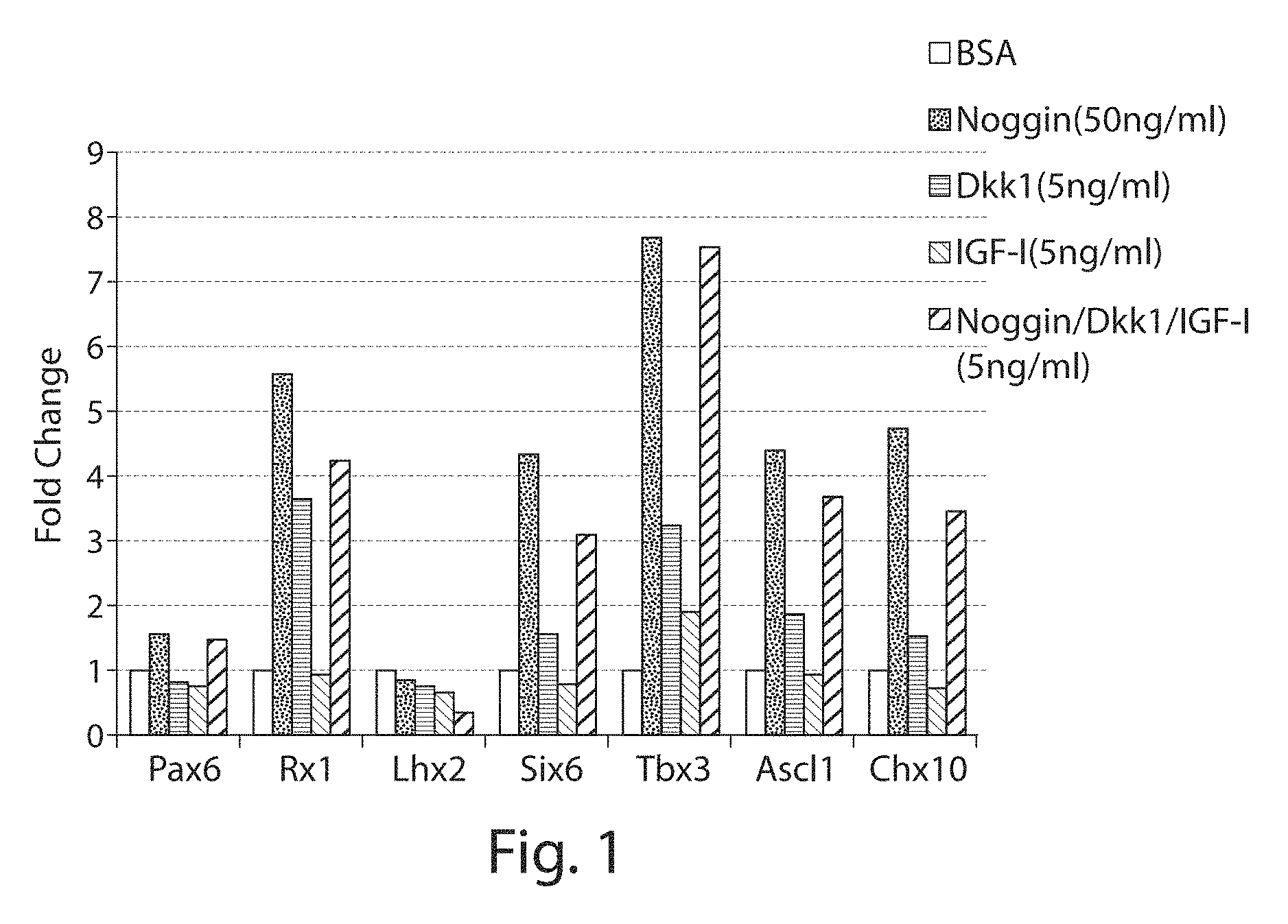
[0133] FIG. 1: Real-time PCR analysis of transcripts of eye field transcription factors in cells differentiated under different conditions.
[0134] FIGS. 2A-2C: Morphology of differentiating cells. (FIG. 2A) At day 1 after cell differentiation, cells at the colony margin were column-shaped (arrow). (FIG. 2B) At day 10 after differentiation, the edge cells became big and flat (arrow head) and the central cells were small and compact (arrow). (FIG. 2C) Rosette like structures formed at day 21.
[0135] FIGS. 3A-3E: Cells cultured at 21 days after initiation of differentiation expressed eye-field transcription factors. (FIG. 3A) Co-expression of PAX6 and RX1. (FIG. 3B) 93% of cells co-expressed PAX6 and RX1 as shown by flow cytometric analysis. (FIG. 3C) Cells expressed Nestin. (FIG. 3D) Cells expressed SOX2. In both (FIG. 3C) and (FIG. 3D), DAPI labels cell nuclei. (FIG. 3E) RT-PCR analysis of transcripts of eye field transcription factors: RX1, PAX6, LHX2, SIX3, SIX6, TBX3 and SOX2.
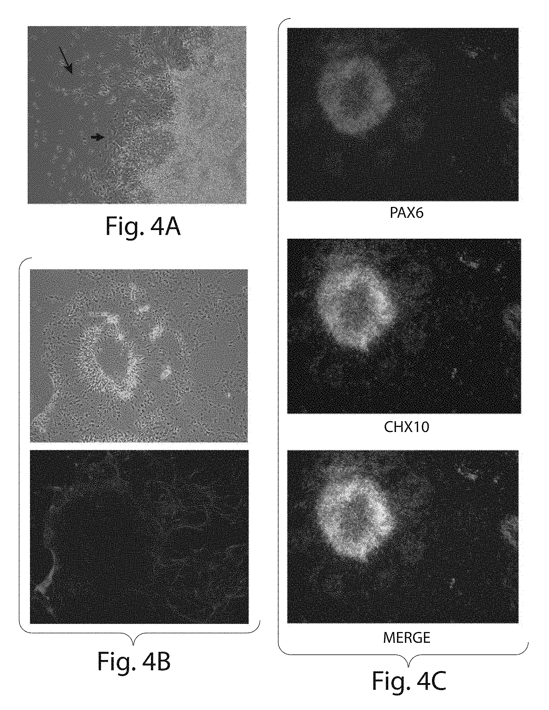
[0136] FIGS. 4A-4C: Cells cultured at 30 days after initiation of differentiation expressed retinal neural progenitor markers. (FIG. 4A) Morphology of cells. After plating on Matrigel.TM., neurons migrated out from cell aggregates (arrow). A few epithelial-like cells (arrow head) are observed around cell aggregates. (FIG. 4B) Upper panel, phase contrast image of migrating neurons; Lower panel, migrating neurons expressed Tuj1. (FIG. 4C) Cells co-expressed PAX6 and CHX10.
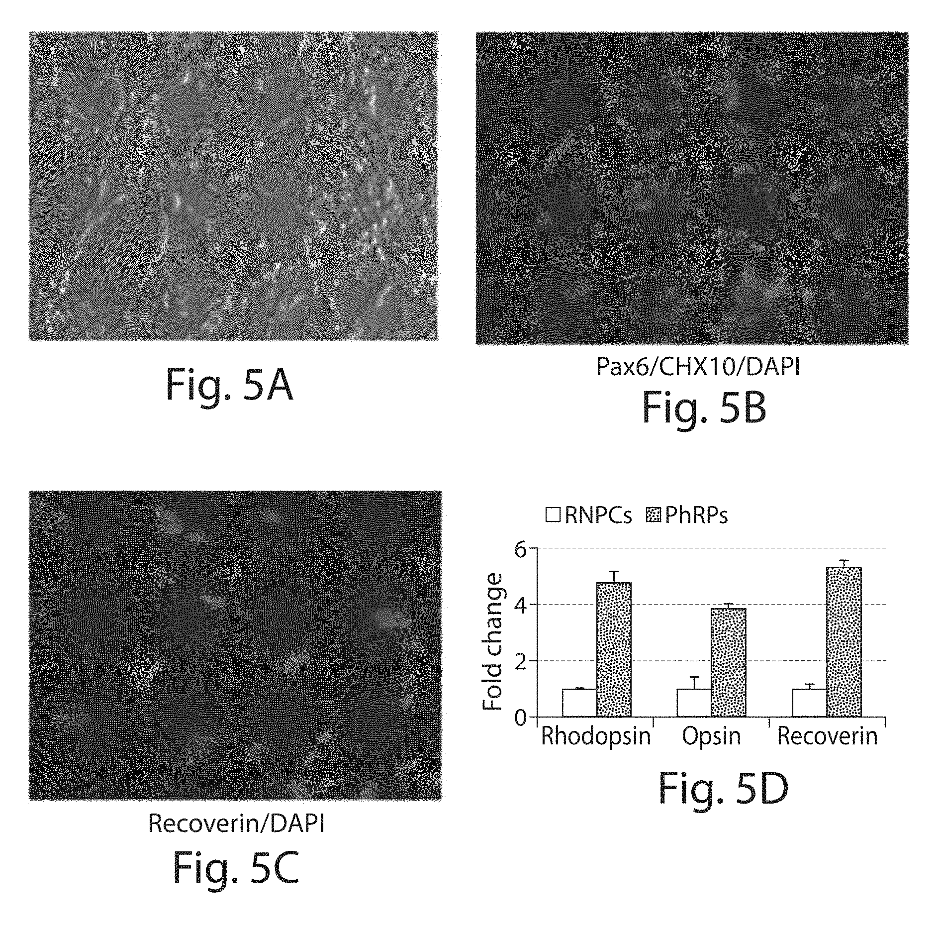
[0137] FIGS. 5A-5D: Cells cultured at 3 months after initiation of differentiation. (FIG. 5A) Morphology of cells. (FIG. 5B) Cells express PAX6 but not CHX10. (FIG. 5C) The expression of Recoverin was restricted to the cytoplasm of the cell body. (FIG. 5D), Real-time RT-PCT analysis of transcripts of Rhodopsin, Opsin, and Recoverin in retinal neural progenitors (RNPs) and photoreceptor progenitor cells (indicated as PhRPs).
[0138] FIGS. 6A-6D: Differentiated cells express photoreceptor cell markers. Cells expressed (FIG. 6A) Rhodopsin, (FIG. 6B) Rhodopsin and Recoverin, (FIG. 6C) Opsin, and (FIG. 6D) phosphodiesterase 6A alpha subunit (PDE6a). DAPI labels cell nuclei.
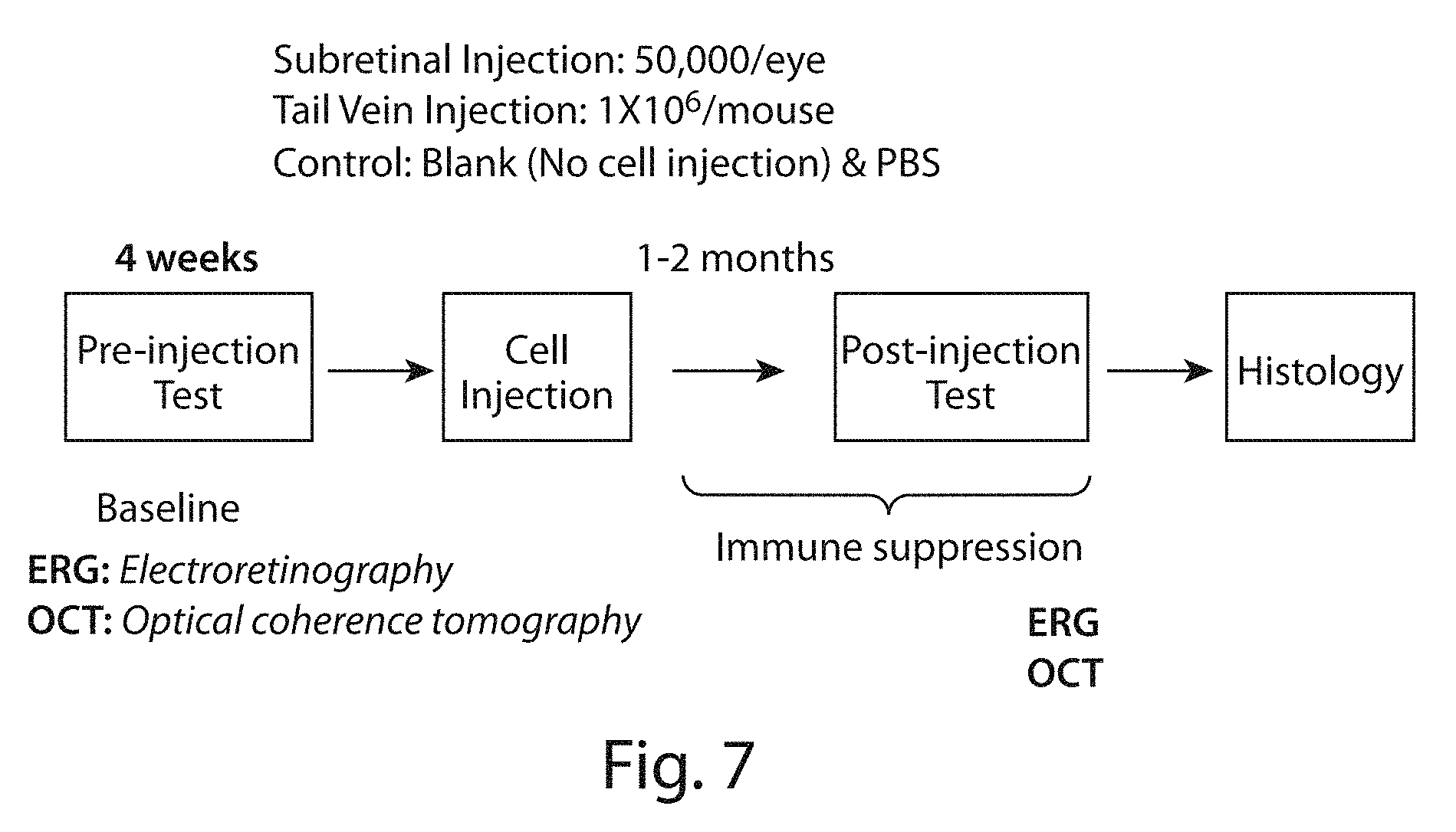
[0139] FIG. 7: Schematic diagram of animal studies in ELOVL4-transgenic mice.
[0140] FIG. 8: Scotopic ERG intensity-response function recorded at one month after subretinal cell injection. Stimulus intensity curves for scotopic a-waves (upper panel) and b-waves (lower panel) from ELOVL4-TG2 mice administered PBS (black line) or photoreceptor progenitor cells (indicated as PhRPs, grey line). *, p<0.001 (vs. PBS).
[0141] FIG. 9: Scotopic ERG intensity-response function recorded at one month after systemic cell injection. Stimulus intensity curves for scotopic a-waves (upper panel) and b-waves (lower panel) from ELOVL4-TG2 mice administered PBS, photoreceptor progenitor cells (indicated as PhRPs) or retinoic acid treated photoreceptor progenitor cells (PhRPs-RA). Blank represents untreated mice. #, p<0.01 (vs. PBS).
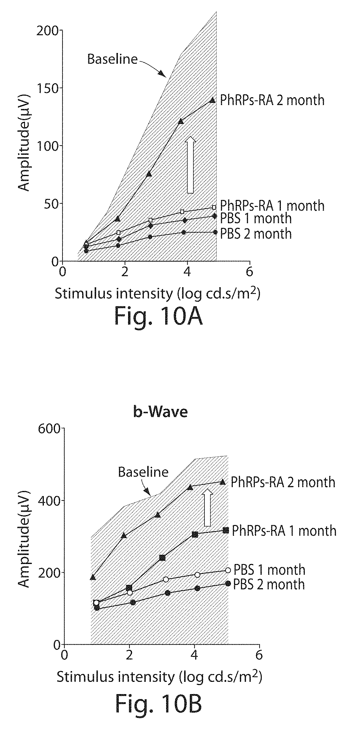
[0142] FIGS. 10A-10B. Photoreceptor progenitor cell systemic injection restores rod function between one month and two months after cell transplantation. Scotopic ERG amplitude of a-waves (FIG. 10A) and b-waves (FIG. 10B) at one and two month after cell injection from ELOVL4-TG2 mice administered PBS (PBS) or retinoic acid treated photoreceptor progenitor cells (PhRPs-RA).
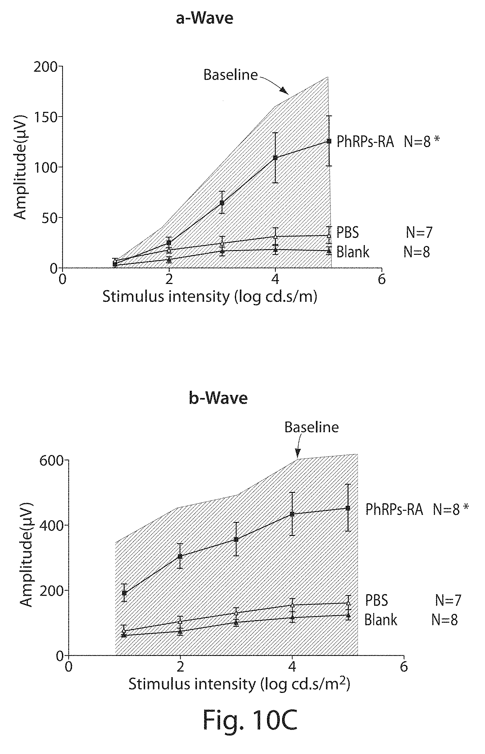
[0143] FIG. 10C: Scotopic ERG intensity-response function recorded at two months after systemic cell injection. Stimulus intensity curves for scotopic a-waves (upper panel) and b-waves (lower panel) from ELOVL4-TG2 mice administered PBS or retinoic acid treated photoreceptor progenitor cells (PhRPs-RA). Blank represents untreated mice. Baseline is the level recorded at 4 weeks. *, p<0.001 (vs. PBS).
[0144] FIG. 11: Photopic ERG amplitude of a-waves (upper panel) and b-waves (lower panel) at one and two month after cell injection from ELOVL4-TG2 mice administered with PBS or retinoic acid treated Photoreceptor progenitors (PhRPs-RA). *, p<0.001 (vs. PBS 2 month).
[0145] FIG. 12: Whole central retina thickness measured by OCT at one and two months after cell injection from untreated ELOVL4-TG2 mice (blank) and mice administered PBS, photoreceptor progenitor cells (PhRPs), or retinoic acid treated photoreceptor progenitor cells (PhRPs-RA).
[0146] FIGS. 13A-13B: (FIG. 13A) Representative images of retina HE staining at two months after cell injection from ELOVL4-TG2 mice administered PBS (Left panel) and retinoic acid treated photoreceptor progenitor cells (Right panel). ONL, outer nuclear layer. INL, internal nuclear layer. (FIG. 13B), Quantification of the thickness of ONL of retina at two month after cell injection from untreated ELOVL4-TG2 mice (blank) and mice administered PBS or retinoic acid treated Photoreceptor progenitors (PhRPs-RA).
[0147] FIG. 14: Schematic diagram of animal studies in RCS rats.
[0148] FIGS. 15A-15C: Preservation of host photoreceptor cells after transplantation of human ES cell-derived photoreceptor progenitor cells. Retinal sections at P90 stained with DAPI: (FIG. 15A) Outer nuclear layer (ONL) is reduced to 0-1 layer in control rats. (FIG. 15B) Rescued ONL cells in RCS rat after intravenous cell injection, which is 2-4 cells deep. (FIG. 15C) Rescued ONL cells in RCS rat receiving intravitreal cell injection, which is 3-5 cells deep. INL, inner nuclear layer; GL, ganglion cell layer.
[0149] FIGS. 16A-16C: Preservation of host rod photoreceptor cell outer segment (OS) after transplantation of human ES cell-derived photoreceptor progenitor cells. Retinal sections at P90 stained for Rhodopsin. (FIG. 16A) Complete loss of rod OS in control rats (arrow). (FIG. 16B) Expression of Rhodopsin in the OS of host rod photoreceptor cells in RCS rat retina after intravenous injection of photoreceptor progenitor cells (arrow). (FIG. 16C) Expression of Rhodopsin in the OS of host rod photoreceptor cells in RCS rat retina after intravitreal transplantation of photoreceptor progenitor cells (arrow). Expression of Rhodopsin is apparent in the Figure.
[0150] FIGS. 17A-17C: Preservation of host cone photoreceptor cell outer segment (OS) after transplantation of human ES cell-derived photoreceptor progenitor cells. Retinal sections at P90 stained for Opsin. (FIG. 17A) Complete loss of cone OS in control rats (arrow). (FIG. 17B) Expression of Opsin in the OS of host cone photoreceptor cells in RCS rat retina after intravenous injection of photoreceptor progenitor cells (arrow). (FIG. 17C) Expression of Opsin in the OS of host cone photoreceptor cells in RCS rat retina after intravitreal transplantation of photoreceptor progenitors (arrow). Expression of Opsin is apparent in the Figure.
[0151] FIGS. 18A-18B: Human ES cell-derived photoreceptor progenitor cells transplanted into the vitreous of RCS rats differentiated into mature rod photoreceptor cells. Retinal sections at P90 stained for rhodopsin (FIG. 18A), Recoverin FIG. 18B. Human cells were labeled with anti-HuNU antibody. DAPI labeled all nuclei. Expression of rhodopsin and recoverin and staining with DAPI is apparent in the Figure.
[0152] FIG. 19 illustrates the overall method used for photoreceptor development in Examples 1-2 and further illustrates the media used at each step of the process.
[0153] FIG. 20 illustrates the timing of photoreceptor cell and photoreceptor progenitor cell development in Examples 1-2.
[0154] FIGS. 21A-21E show the components of culture media and media supplements used in the Examples.
[0155] FIG. 22 illustrates the gene expression pattern of ESC, eye field progenitor cells, retinal neural progenitor cells, photoreceptor progenitor cells, and photoreceptor cells during in vitro differentiation from human pluripotent stem cells.
[0156] FIG. 23 provides flow cytometry histograms showing relative degrees of phagocytosis of pHrodo.TM. Red E. coli BioParticles (Invitrogen) fluorescent bioparticles by hES-RPE and hES-photoreceptor progenitors at 37.degree. C. and at 4.degree. C., compared to control (no bioparticles). The histogram for the photoreceptor progenitor cells illustrate that, like RPE cells, the intensity of the fluorescence signal increases upon shifting the cells from a relatively non-permissive temperature of 4.degree. C. to a physiologically relevant temperature of 37.degree. C., indicating that photoreceptor progenitor cells are capable of phagocytosing the bioparticles. The bioparticles are a surrogate for shed outer segments and drusen in the eye.
DETAILED DESCRIPTION
[0157] The invention provides methods for generating photoreceptor cells (PRC) and photoreceptor progenitor cells (PRPC). These methods involve in vitro differentiation from earlier progenitors including pluripotent stem cells, eye field (EF) progenitors, and retinal neural progenitor cells. The methods provided herein may use as a starting material any of the foregoing progenitor (including stem cell) populations.
[0158] The invention further contemplates generating photoreceptor cells (PRC) and photoreceptor progenitor cells (PRPC) in vitro from primary eye field (EF) progenitors and retinal neural progenitors cells (i.e., primary cells referring to cells obtained from a subject rather than from in vitro differentiation of a more immature progenitor.
[0159] Photoreceptor development occurs through a number of developmental stages, each of which can be defined phenotypically (e.g., by way of marker expression profile) and/or functionally. This development is illustrated schematically in FIG. 22. In vitro pluripotent stem cells differentiate into EF progenitors, which in turn differentiate into retinal neural progenitor cells, which in turn differentiate into photoreceptor progenitor cells, which in turn differentiate into photoreceptor cells.
[0160] Progenitor cells, as used herein, refer to cells that remain mitotic and can produce more progenitor cells, of the same or of more limited differentiative capacity, or can differentiate to an end fate cell lineage. The terms progenitor and precursor are used interchangeably. Cells at each of these stages will be discussed in greater detail herein.
[0161] The photoreceptor progenitor cells (also referred to as photoreceptor progenitors) and photoreceptor cells provided herein may be used in a variety of in vivo and in vitro methods. For example, the photoreceptor progenitor cells may be used in vivo to treat conditions of the retina, including but not limited to macular degeneration and retinitis pigmentosa. The photoreceptor progenitor cells and photoreceptor cells may be used in vitro in screening assays to identify putative therapeutic or prophylactic treatment candidates.
[0162] The invention further provides photoreceptor progenitor cells and photoreceptor cells obtained by the methods described herein. Photoreceptor progenitor cells and photoreceptor cells obtained by in vitro differentiation of pluripotent stem cells or their differentiated progeny such as eye field progenitor cells. Eye field progenitor cells may themselves be obtained from in vitro differentiation of pluripotent stem cells, or they may be primary eye field progenitors obtained from a subject.
[0163] The invention provides populations of photoreceptor progenitor cells and populations of photoreceptor cells that have not been attained or are not attainable from primary sources. These populations may be homogenous or near homogeneous in their cell content. For example, at least 50%, at least 60%, at least 70%, at least 80%, at least 90%, at least 95%, at least 99%, or about 100% of the cells in such a population may be photoreceptor progenitor cells. As another example, at least 50%, at least 60%, at least 70%, at least 80%, at least 90%, at least 95%, at least 99%, or about 100% of the cells in such a population may be photoreceptor cells. These cells in these populations may be of a single haplotype. For example, they may be HLA-matched. These cells in these populations may be genetically identical.
[0164] The disclosure provides substantially pure (or homogeneous) preparations of various cell populations based on the ability of the disclosed methods to directly differentiate progenitor cells such as but not limited to pluripotent stem cells. As used herein, directed differentiation intends that the progenitor cell population differentiates into or towards a desired lineage, due in part to the factors or other stimuli provided to such progenitor cells, thereby avoiding differentiation into other undesired, and thus potentially contaminating, lineages. The methods provided herein drive differentiation of for example pluripotent stem cells to eye field progenitors without generating embryoid bodies (EB). EBs, as described below, are three dimensional cell clusters that can form during differentiation of pluripotent stem cells including but not limited to embryonic stem (ES) cells, and that typically contain cells, including progenitors, of mesodermal, ectodermal and endodermal lineages. The three dimensional nature of the EB may create a different environment, including different cell-cell interactions and different cell-cell signaling, than occurs in the non-EB based methods described herein. In addition, cells within EBs may not all receive a similar dose of an exogenously added agent, such as a differentiation factor present in the surrounding medium, and this can result in various differentiation events and decisions during development of the EB.
[0165] In contrast, the culture methods of the invention culture progenitor cells do not require and preferably avoid EB formation. Instead, these methods culture cells in conditions that provide the cells with equal contact with the surrounding medium, including factors in such medium. The cells may grow as a monolayer or near monolayer attached to a culture surface, as an example.
[0166] The ability of all or a majority of the progenitor cells to be in contact with their surrounding medium and thus the factors in such medium to an approximately equal degree results in those progenitor cells differentiating at similar times and to similar degrees. This similar differentiation timeline for a population of progenitor cells indicates that such cells are synchronized. The cells may be cell cycle synchronized in some instances also. Such synchronicity results in populations of cells that are homogeneous or near homogeneous in their cellular make-up. As an example, the methods described herein can produce cellular populations wherein at least 50%, at least 60%, at least 70%, at least 80%, at least 90%, at least 95%, or about 100% of cells are a particular cell of interest. The cell of interest may be defined phenotypically, for example by intracellular or extracellular marker expression. The cell of interest may be an eye field progenitor cell, a neural retinal progenitor cell, a photoreceptor progenitor cell, or a photoreceptor cell.
[0167] As used herein, a majority of cells means at least 50%, and depending on the embodiment may include at least 60%, at least 70%, at least 80%, at least 90%, at least 95%, or about 100% of cells.
[0168] The degree of purity that may be achieved using the methods of the invention are particularly important where such cell populations are to be used in vivo for therapeutic or prophylactic purposes. The ability to obtain populations of high cellular purity avoids performing another manipulation such as an enrichment or selection step, which may result in unnecessary cell loss. This is particularly important where the cell population may be small or the cell number may be limited.
Definitions
[0169] As defined here, singular forms are provided for illustrative purposes, but may also apply to plural versions of the phrase. The following definitions are meant to supplement conventional definitions of the terms as they would be understood by persons of ordinary skill.
[0170] "Substantially pure preparation of photoreceptor progenitor cells (PRPCs)." As used herein, this phrase refers to a preparation of cells (e.g., a composition comprising cells) wherein the cells are at least 75% pure or preferably at least 85% pure, at least 95% pure, or are about 85% to 95% pure. For example, the level of purity may be quantified by determining the proportion of cells in the preparation that express one or more markers, such as those markers of PRPCs (including those markers identified in this application or others known in the art), relative to the total number of cells in the preparation, e.g., by detecting cells that do or do not express said one or more markers. Optionally expression of markers indicative of non-PRPC cells may also be detected, thereby facilitating detection and/or quantitation of said cells. Exemplary methods that may be utilized to include, without limitation, Fluorescence Activated Cell Sorting (FACS), immunohistochemistry, in situ hybridization, and other suitable methods known in the art. Optionally the determination of purity may be performed disregarding non-viable cells present in the preparation.
[0171] "Substantially pure preparation of photoreceptor cells (PRs) of human origin." As used herein, this phrase refers to a preparation of cells (e.g., a composition comprising cells) wherein the cells are at least 75% pure or preferably at least 85% pure, at least 95% pure, or are about 85% to 95% pure. For example, the level of purity may be quantified by determining the proportion of cells in the preparation that express one or more markers, such as those markers of PRs (including those markers identified in this application or others known in the art), relative to the total number of cells in the preparation, e.g., by detecting cells that do or do not express said one or more markers. Optionally expression of markers indicative of non-PR cells may also be detected, thereby facilitating detection and/or quantitation of said cells. Exemplary methods that may be utilized to include, without limitation, Fluorescence Activated Cell Sorting (FACS), immunohistochemistry, in situ hybridization, and other suitable methods known in the art. Optionally the determination of purity may be performed disregarding non-viable cells present in the preparation.
[0172] "Embryoid bodies" refers to clumps or clusters of pluripotent cells (e.g., iPSC or ESC) which may be formed by culturing pluripotent cells under non-attached conditions, e.g., on a low-adherent substrate or in a "hanging drop." In these cultures, pluripotent cells can form clumps or clusters of cells denominated as embryoid bodies. See Itskovitz-Eldor et al., Mol Med. 2000 February; 6(2):88-95, which is hereby incorporated by reference in its entirety. Typically, embryoid bodies initially form as solid clumps or clusters of pluripotent cells, and over time some of the embryoid bodies come to include fluid filled cavities, the latter former being referred to in the literature as "simple" EBs and the latter as "cystic" embryoid bodies.
[0173] The term "embryonic stem cell" (ES cell or ESC) is used herein as it is used in the art. This term includes cells derived from the inner cell mass of human blastocysts or morulae, including those that have been serially passaged as cell lines. The ES cells may be derived from fertilization of an egg cell with sperm, as well as using DNA, nuclear transfer, parthenogenesis, or by means to generate ES cells with homozygosity in the HLA region. ES cells are also cells derived from a zygote, blastomeres, or blastocyst-staged mammalian embryo produced by the fusion of a sperm and egg cell, nuclear transfer, parthenogenesis, androgenesis, or the reprogramming of chromatin and subsequent incorporation of the reprogrammed chromatin into a plasma membrane to produce a cell. Embryonic stem cells, regardless of their source or the particular method used to produce them, can be identified based on (i) the ability to differentiate into cells of all three germ layers, (ii) expression of at least OCT 4 and alkaline phosphatase, and (iii) ability to produce teratomas when transplanted into immunodeficient animals. Embryonic stem cells that may be used in embodiments of the present invention include, but are not limited to, human ES cells ("ESC" or "hES cells") such as MA01, MA09, ACT-4, No. 3, H1, H7, H9, H14 and ACT30 embryonic stem cells. Additional exemplary cell lines include NED1, NED2, NED3, NED4, NED5, and NED7. See also NIH Human Embryonic Stem Cell Registry. An exemplary human embryonic stem cell line that may be used is MA09 cells. The isolation and preparation of MA09 cells was previously described in Klimanskaya, et al. (2006) "Human Embryonic Stem Cell lines Derived from Single Blastomeres." Nature 444: 481-485. The human ES cells used in accordance with exemplary embodiments of the present invention may be derived and maintained in accordance with GMP standards.
[0174] The term "ES cells" does not infer, and should not be inferred to mean, that the cells were generated through the destruction of an embryo. To the contrary, various methods are available and can be used to generate ES cells without destruction of an embryo, such as a human embryo. As an example, ES cells may be generated from single blastomeres derived from an embryo, in a manner similar to the extraction of blastomeres for pre-implantation genetic diagnosis (PGD). Examples of such cell lines include NED1, NED2, NED3, NED4, NED5, and NED7. An exemplary human embryonic stem cell line that may be used is MA09 cells. The isolation and preparation of MA09 cells was previously described in Klimanskaya, et al. (2006) "Human Embryonic Stem Cell lines Derived from Single Blastomeres." Nature 444: 481-485. See also Chung et al. 2008, Cell Stem Cell, 2:113. All of these lines were generated without embryo destruction.
As used herein, the term "pluripotent stem cells" includes but is not limited to tissue-derived stem cells, embryonic stem cells, embryo-derived stem cells, induced pluripotent stem cells, and stimulus-triggered acquisition of pluripotency (STAP) cells, regardless of the method by which the pluripotent stem cells are derived. The term also includes pluripotent stem cells having the functional and phenotypic characteristics of the afore-mentioned cells, regardless of the method used to generate such cells. Pluripotent stem cells are defined functionally as stem cells that are: (a) capable of inducing teratomas when transplanted in immunodeficient (SCID) mice; (b) capable of differentiating to cell types of all three germ layers (e.g., can differentiate to ectodermal, mesodermal, and endodermal cell types); and (c) express one or more markers of embryonic stem cells (e.g., express OCT4, alkaline phosphatase, SSEA-3 surface antigen, SSEA-4 surface antigen, Nanog, TRA-1-60, TRA-1-81, SOX2, REX1, etc). In certain embodiments, pluripotent stem cells express one or more markers selected from the group consisting of: OCT4, alkaline phosphatase, SSEA-3, SSEA-4, TRA-1-60, and TRA-1-81. Exemplary pluripotent stem cells can be generated using, for example, methods known in the art. Exemplary pluripotent stem cells include embryonic stem cells derived from the ICM of blastocyst stage embryos, as well as embryonic stem cells derived from one or more blastomeres of a cleavage stage or morula stage embryo (optionally without destroying the remainder of the embryo). Such embryonic stem cells can be generated from embryonic material produced by fertilization or by asexual means, including somatic cell nuclear transfer (SCNT), parthenogenesis, and androgenesis. Further exemplary pluripotent stem cells include induced pluripotent stem cells (iPSCs) generated by reprogramming a somatic cell by expressing a combination of factors (herein referred to as reprogramming factors). The iPSCs can be generated using fetal, postnatal, newborn, juvenile, or adult somatic cells.
[0175] In certain embodiments, factors that can be used to reprogram somatic cells to pluripotent stem cells include, for example, a combination of OCT 4 (sometimes referred to as OCT 3/4), SOX2, c-Myc, and KLF4. In other embodiments, factors that can be used to reprogram somatic cells to pluripotent stem cells include, for example, a combination of OCT 4, SOX2, Nanog, and Lin28. In certain embodiments, at least two reprogramming factors are expressed in a somatic cell to successfully reprogram the somatic cell. In other embodiments, at least three reprogramming factors are expressed in a somatic cell to successfully reprogram the somatic cell. In other embodiments, at least four reprogramming factors are expressed in a somatic cell to successfully reprogram the somatic cell. In other embodiments, additional reprogramming factors are identified and used alone or in combination with one or more known reprogramming factors to reprogram a somatic cell to a pluripotent stem cell. Induced pluripotent stem cells are defined functionally and include cells that are reprogrammed using any of a variety of methods (integrative vectors, non-integrative vectors, chemical means, etc). Pluripotent stem cells may be genetically modified or otherwise modified to increase longevity, potency, homing, to prevent or reduce alloimmune responses or to deliver a desired factor in cells that are differentiated from such pluripotent cells (for example, photoreceptors, photoreceptor progenitor cells, rods, cones, etc. and other cell types described herein, e.g., in the examples).
[0176] "Induced pluripotent stem cells" (iPS cells or iPSC) can be produced by protein transduction of reprogramming factors in a somatic cell. In certain embodiments, at least two reprogramming proteins are transduced into a somatic cell to successfully reprogram the somatic cell. In other embodiments, at least three reprogramming proteins are transduced into a somatic cell to successfully reprogram the somatic cell. In other embodiments, at least four reprogramming proteins are transduced into a somatic cell to successfully reprogram the somatic cell.
[0177] The pluripotent stem cells can be from any species. Embryonic stem cells have been successfully derived in, for example, mice, multiple species of non-human primates, and humans, and embryonic stem-like cells have been generated from numerous additional species. Thus, one of skill in the art can generate embryonic stem cells and embryo-derived stem cells from any species, including but not limited to, human, non-human primates, rodents (mice, rats), ungulates (cows, sheep, etc.), dogs (domestic and wild dogs), cats (domestic and wild cats such as lions, tigers, cheetahs), rabbits, hamsters, gerbils, squirrel, guinea pig, goats, elephants, panda (including giant panda), pigs, raccoon, horse, zebra, marine mammals (dolphin, whales, etc.) and the like. In certain embodiments, the species is an endangered species. In certain embodiments, the species is a currently extinct species.
[0178] Similarly, iPS cells can be from any species. These iPS cells have been successfully generated using mouse and human cells. Furthermore, iPS cells have been successfully generated using embryonic, fetal, newborn, and adult tissue. Accordingly, one can readily generate iPS cells using a donor cell from any species. Thus, one can generate iPS cells from any species, including but not limited to, human, non-human primates, rodents (mice, rats), ungulates (cows, sheep, etc), dogs (domestic and wild dogs), cats (domestic and wild cats such as lions, tigers, cheetahs), rabbits, hamsters, goats, elephants, panda (including giant panda), pigs, raccoon, horse, zebra, marine mammals (dolphin, whales, etc.) and the like. In certain embodiments, the species is an endangered species. In certain embodiments, the species is a currently extinct species.
[0179] Induced pluripotent stem cells can be generated using, as a starting point, virtually any somatic cell of any developmental stage. For example, the cell can be from an embryo, fetus, neonate, juvenile, or adult donor. Exemplary somatic cells that can be used include fibroblasts, such as dermal fibroblasts obtained by a skin sample or biopsy, synoviocytes from synovial tissue, foreskin cells, cheek cells, or lung fibroblasts. Although skin and cheek provide a readily available and easily attainable source of appropriate cells, virtually any cell can be used. In certain embodiments, the somatic cell is not a fibroblast.
[0180] The induced pluripotent stem cell may be produced by expressing or inducing the expression of one or more reprogramming factors in a somatic cell. The somatic cell may be a fibroblast, such as a dermal fibroblast, synovial fibroblast, or lung fibroblast, or a non-fibroblastic somatic cell. The somatic cell may be reprogrammed through causing expression of (such as through viral transduction, integrating or non-integrating vectors, etc.) and/or contact with (e.g., using protein transduction domains, electroporation, microinjection, cationic amphiphiles, fusion with lipid bilayers containing, detergent permeabilization, etc.) at least 1, 2, 3, 4, 5 reprogramming factors. The reprogramming factors may be selected from OCT 3/4, SOX2, NANOG, LIN28, C-MYC, and KLF4. Expression of the reprogramming factors may be induced by contacting the somatic cells with at least one agent, such as a small organic molecule agents, that induce expression of reprogramming factors.
[0181] Further exemplary pluripotent stem cells include induced pluripotent stem cells generated by reprogramming a somatic cell by expressing or inducing expression of a combination of factors ("reprogramming factors"). iPS cells may be obtained from a cell bank. The making of iPS cells may be an initial step in the production of differentiated cells. iPS cells may be specifically generated using material from a particular patient or matched donor with the goal of generating tissue-matched PHRPS or photoreceptor cells. iPSCs can be produced from cells that are not substantially immunogenic in an intended recipient, e.g., produced from autologous cells or from cells histocompatible to an intended recipient.
[0182] The somatic cell may also be reprogrammed using a combinatorial approach wherein the reprogramming factor is expressed (e.g., using a viral vector, plasmid, and the like) and the expression of the reprogramming factor is induced (e.g., using a small organic molecule.) For example, reprogramming factors may be expressed in the somatic cell by infection using a viral vector, such as a retroviral vector or a lentiviral vector. Also, reprogramming factors may be expressed in the somatic cell using a non-integrative vector, such as an episomal plasmid. See, e.g., Yu et al., Science. 2009 May 8; 324(5928):797-801, which is hereby incorporated by reference in its entirety. When reprogramming factors are expressed using non-integrative vectors, the factors may be expressed in the cells using electroporation, transfection, or transformation of the somatic cells with the vectors. For example, in mouse cells, expression of four factors (OCT3/4, SOX2, C-MYC, and KLF4) using integrative viral vectors is sufficient to reprogram a somatic cell. In human cells, expression of four factors (OCT3/4, SOX2, NANOG, and LIN28) using integrative viral vectors is sufficient to reprogram a somatic cell.
[0183] Once the reprogramming factors are expressed in the cells, the cells may be cultured. Over time, cells with ES characteristics appear in the culture dish. The cells may be chosen and subcultured based on, for example, ES morphology, or based on expression of a selectable or detectable marker. The cells may be cultured to produce a culture of cells that resemble ES cells--these are putative iPS cells.
[0184] To confirm the pluripotency of the iPS cells, the cells may be tested in one or more assays of pluripotency. For example, the cells may be tested for expression of ES cell markers; the cells may be evaluated for ability to produce teratomas when transplanted into SCID mice; the cells may be evaluated for ability to differentiate to produce cell types of all three germ layers. Once a pluripotent iPSC is obtained it may be used to produce cell types disclosed herein, e.g., photoreceptor progenitor cells, photoreceptor cells, rods, cones, etc. and other cell types described herein, e.g., in the examples.
[0185] Stimulus-triggered acquisition of pluripotency (STAP) cells are pluripotent stem cells produced by reprogramming somatic cells with sublethal stimuli such as low-pH exposure. The reprogramming does not require nuclear transfer into or genetic manipulation of the somatic cells. Reference can be made to Obokata et al., Nature, 505:676-680, 2014.
[0186] "Stem cell" is used here to refer to a pluripotent cell which can proliferate and/or differentiate into a mature cell and is optionally of human origin.
[0187] "Adult stem cell" refers to a multipotent cell isolated from adult tissue and can include bone marrow stem cells, cord blood stem cells and adipose stem cells and is of human origin.
[0188] "Retina" refers to the neural cells of the eye, which are layered into three nuclear layers comprised of photoreceptors, horizontal cells, bipolar cells, amacrine cells, Muller glial cells and ganglion cells.
[0189] "Precursor cell" refers to a cell capable of differentiating to an end fate cell lineage. In embodiments of the invention, an "eye field progenitor cell" is differentiated from embryonic stem cells or induced pluripotent stem cells and expresses the markers PAX6 and RX1. In embodiments of the invention, a "retinal neural progenitor cell" refers to a cell differentiated from embryonic stem cells or induced pluripotent stem cells, that expresses the cell markers PAX6 and CHX10. In embodiments of the invention, "photoreceptor progenitor" refers to cells differentiated from embryonic stem cells or induced pluripotent stem cells and that expresses the marker PAX6 while not expressing the marker CHX10 (i.e. CHX10(-)). These cells transiently express CHX10 at retinal neural progenitor stage, but the CHX10 expression is turned off when cells differentiate into the photoreceptor progenitor stage. Also, "photoreceptor" may refer to post-mitotic cells differentiated from embryonic stem cells or induced pluripotent stem cells and that expresses the cell marker rhodopsin or any of the three cone opsins, and optionally express the rod or cone cGMP phosphodiesterase. The photoreceptors may also express the marker recoverin, which is found in photoreceptors. The photoreceptors may be rod and/or cone photoreceptors.
[0190] "Signs" of disease, as used herein, refers broadly to any abnormality indicative of disease, discoverable on examination of the patient; an objective indication of disease, in contrast to a symptom, which is a subjective indication of disease.
[0191] "Symptoms" of disease as used herein, refers broadly to any morbid phenomenon or departure from the normal in structure, function, or sensation, experienced by the patient and indicative of disease.
[0192] "Therapy," "therapeutic," "treating," "treat" or "treatment", as used herein, refers broadly to treating a disease, arresting or reducing the development of the disease or its clinical symptoms, and/or relieving the disease, causing regression of the disease or its clinical symptoms. Therapy encompasses prophylaxis, prevention, treatment, cure, remedy, reduction, alleviation, and/or providing relief from a disease, signs, and/or symptoms of a disease. Therapy encompasses an alleviation of signs and/or symptoms in patients with ongoing disease signs and/or symptoms. Therapy also encompasses "prophylaxis" and "prevention". Prophylaxis includes preventing disease occurring subsequent to treatment of a disease in a patient or reducing the incidence or severity of the disease in a patient. The term "reduced", for purpose of therapy, refers broadly to the clinical significant reduction in signs and/or symptoms. Therapy includes treating relapses or recurrent signs and/or symptoms. Therapy encompasses but is not limited to precluding the appearance of signs and/or symptoms anytime as well as reducing existing signs and/or symptoms and eliminating existing signs and/or symptoms. Therapy includes treating chronic disease ("maintenance") and acute disease. For example, treatment includes treating or preventing relapses or the recurrence of signs and/or symptoms.
[0193] Conditions to be treated according to the invention and thus using one or more of the preparations provided herein include but are not limited macular degeneration including age-related macular degeneration, and such macular degeneration may be early or late stage. Other conditions to be treated include but are not limited to retinitis pigmentosa, retinal dysplasia, retinal degeneration, diabetic retinopathy, congenital retinal dystrophy, Leber congenital amaurosis, retinal detachment, glaucoma, optic neuropathy, and trauma that affects the eye.
[0194] Cell Markers:
[0195] Exemplary cell markers that may be assessed for expression include the following: PAX6, RX1, SIX3, SIX6, LHX2, TBX3, SOX2, CHX10, Nestin, TRbeta2, NR2E3, NRL, MASH1, RORbeta, Recoverin, Opsin, Rhodopsin, rod and cone cGMP Phosphodiesterase, which may be assessed at the protein and/or mRNA (see Fischer A J, Reh T A, Dev Neurosci. 2001; 23(4-5):268-76; Baumer et al., Development. 2003 July; 130(13):2903-15, Swaroop et al., Nat Rev Neurosci. 2010 August; 11(8):563-76, Agathocleous and Harris, Annu. Rev. Cell Dev. Biol. 2009. 25:45-69, each of which is hereby incorporated by reference in its entirety). Said marker identifiers are generally used as in the literature and in the art, particular in the fields of art in related to the contexts in which those gene identifiers are recited herein, which may include literature related to photoreceptors, rods, cones, photoreceptor differentiation, photoreceptor progenitors, neural differentiation, neural stem cells, pluripotent stem cells, and other fields as indicated by context. Additionally, the markers are generally human, e.g., except where the context indicates otherwise. The cell markers can be identified using conventional immunocytochemical methods or conventional PCR methods which techniques are well known to those of ordinary skill in the art.
[0196] Cell Culture Media:
[0197] In embodiments of the invention, the cells are stored, proliferated or differentiated in various cell culture media. Retinal induction medium is utilized for the stem cell production into Eye Field Progenitor Cells. The retinal induction medium may comprise D-glucose, penicillin, streptomycin, N2 supplement (e.g. 0.1-5%), B27 supplement (e.g., 0.005 to 0.2%), MEM Non-essential amino acids solution and optionally including insulin and/or Noggin, and may be in a DMEM/F12 (Invitrogen) or similar base medium. For example, the Retinal induction medium may include at least insulin. Additionally, the insulin concentration may be varied or increased which may promote cell survival and/or yield of differentiated cells. For example, the insulin concentration may be varied across a range and survival and/or differentiation monitored in order to identify an insulin concentration with improves either or both of these attributes. The addition of Noggin is believed not to be necessary but was observed to increase the expression of eye field transcription factors.
[0198] The components of DMEM/F12, Neurobasal medium, N2 serum supplement, and B27 serum supplement are provided in FIGS. 21A-21E. It is to be understood that the invention contemplates the use of these particular media and supplements or media or supplements comprising, consisting essentially of, or consisting of these components.
[0199] The methods described herein may use human factors such as human Noggin, human insulin, and the like.
[0200] Noggin is a secreted BMP inhibitor that reportedly binds BMP2, BMP4, and BMP7 with high affinity to block TGF.beta. family activity. SB431542 is a small molecule that reportedly inhibits TGF.beta./Activin/Nodal by blocking phosphorylation of ACTRIB, TGF.beta.R1, and ACTRIC receptors. SB431542 is thought to destabilize the Activin- and Nanog-mediated pluripotency network as well as suppress BMP induced trophoblast, mesoderm, and endodermal cell fates by blocking endogenous Activin and BMP signals. It is expected that agents having one or more of the aforementioned activities could replace or augment the functions of one or both of Noggin and SB431542, e.g., as they are used in the context of the disclosed methods. For example, applicants envision that the protein Noggin and/or the small molecule SB4312542 could be replaced or augmented by one or more inhibitors that affect any or all of the following three target areas: 1) preventing the binding of the ligand to the receptor; 2) blocking activation of receptor (e.g., dorsomorphin), and 3) inhibition of SMAD intracellular proteins/transcription factors. Exemplary potentially suitable factors include the natural secreted BMP inhibitors Chordin (which blocks BMP4) and Follistatin (which blocks Activin), as well as analogs or mimetics thereof. Additional exemplary factors that may mimic the effect of Noggin include use of dominant negative receptors or blocking antibodies that would sequester BMP2, BMP4, and/or BMP7. Additionally, with respect to blocking receptor phosphorylation, dorsomorphin (or Compound C) has been reported to have similar effects on stem cells. Inhibition of SMAD proteins may also be effected using soluble inhibitors such as SIS3 (6,7-Dimethoxy-2-((2E)-3-(1-methyl-2-phenyl-1H-pyrrolo[2,3-b]pyridin-3-yl- -prop-2-enoyl))-1,2,3,4-tetrahydroisoquinoline, Specific Inhibitor of Smad3, SIS3), overexpression of one or more of the inhibitor SMADs (e.g., SMAD6, SMAD7, SMAD10) or RNAi for one of the receptor SMADs (SMAD1, SMAD2, SMAD3, SMAD5, SMAD8/9). Another combination of factors expected to be suitable for generating neural progenitors comprises a cocktail of Leukemia Inhibitory Factor (LIF), GSK3 inhibitor (CHIR 99021), Compound E (.gamma. secretase inhibitor XXI) and the TGF.beta. inhibitor SB431542 which has been previously shown to be efficacious for generating neural crest stem cells (Li et al., Proc Natl Acad Sci USA. 2011 May 17; 108(20):8299-304). Additional exemplary factors may include derivatives of SB431542, e.g., molecules that include one or more added or different substituents, analogous functional groups, etc. and that have a similar inhibitory effect on one or more SMAD proteins. Suitable factors or combinations of factors may be identified, for example, by contacting pluripotent cells with said factor(s) and monitoring for adoption of eye field progenitor cell phenotypes, such as characteristic gene expression (including expression of the markers described herein, expression of a reporter gene coupled to an eye field progenitor cell promoter, or the like) or the ability to form a cell type disclosed herein such as retinal neural progenitor cells, photoreceptor progenitors, rod progenitors, cones, and/or rods.
[0201] Preferably the cells are treated with or cultured in a retinal induction medium prior to culture with a neural differentiation medium. A neural differentiation medium is utilized for Eye Field Progenitor Cell production into Retinal Neural Progenitor Cells. The neural differentiation medium may comprise D-glucose, penicillin, streptomycin, GlutaMAX.TM., N2 supplement, B27 supplement, MEM Non-essential amino acids solution and optionally including Noggin. The neural differentiation medium may also be utilized for Retinal Neural Progenitor Cell production into Photoreceptor Progenitor Cells but without the inclusion of Noggin. The use of a neural differentiation medium, optionally supplemented with retinoic acid and taurine, followed by utilization of a photoreceptor differentiation medium (Invitrogen) which optionally may comprise D-glucose, penicillin, streptomycin, GlutaMAX.TM., N2 supplement, B27 supplement (e.g., formula number 080085-SA) with the addition of Forskolin, BDNF, CNTF, LIF and DATP is utilized for Photoreceptor Progenitor Cells production into Photoreceptor Cells. For example the photoreceptor differentiation medium may comprise thyroid hormone, e.g., in an amount that is present in the foregoing medium, or in a different or greater amount. For example said medium may comprise exogenously added thyroid hormone. In exemplary embodiments the photoreceptor differentiation medium may comprise one, two, or all three BDNF, CNTF and DATP, e.g., BDNF, CNTF, DATP, BDNF and CNTF, CNTF and DATP, BDNF and DATP, or all three of BDNF, CNTF and DATP, which medium may optionally comprise Neurobasal Medium and/or may optionally comprise thyroid hormone.
[0202] The neural differentiation medium constituents are as follows: N2: 1% (1 ml of N2 per 100 ml), B27: 2% (2 ml of B27 per 100 ml), and Noggin: 50 ng/ml.
[0203] Noggin is not needed after cells have all become eye field progenitors.
[0204] Embryonic Stem Cells (ESCs) or Adult Stem Cells or Induced Pluripotent Stem Cells (iPS):
[0205] The ESCs, or Adult Stem Cells or iPS cells utilized herein may be propagated on a feeder-free system, such as in Matrigel.TM. (a soluble preparation from Engelbreth-Holm-Swarm (EHS) mouse sarcoma cells) or another matrix. Additionally, or alternatively, said pluripotent cells may be cultured on a matrix which may be selected from the group consisting of laminin, fibronectin, vitronectin, proteoglycan, entactin, collagen, collagen I, collagen IV, collagen VIII, heparan sulfate, Matrigel.TM. (a soluble preparation from Engelbreth-Holm-Swarm (EHS) mouse sarcoma cells), CellStart, a human basement membrane extract, and any combination thereof. Said matrix may comprise, consist of, or consist essentially of Matrigel.TM. (a soluble preparation from Engelbreth-Holm-Swarm (EHS) mouse sarcoma cells). The stem cells do not form embryoid bodies in culture, which is an improvement over the prior art. The cells differentiate into eye field progenitor cells in the absence of exogenous factors. In an embodiment, ESCs differentiate into eye field progenitor cells in the presence of Noggin.
[0206] Eye Field Progenitor Cells (EFPCs):
[0207] The EFPCs differentiate from ESCs, Adult stem cells or induced pluripotent cells (iPCs) into cells that are PAX6(+) and RX1(+). The EFPCs can also be SIX3(+), SIX6(+), LHX2(+), TBX3(+) Nestin(+) and/or SOX2(+) and OCT4(-) and NANOG(-). The differentiation into EFPCs occurs in a retinal induction medium which may comprise DMEM/F12, D-glucose, penicillin, streptomycin, N2 supplement, B27 supplement, MEM non-essential amino acid and insulin. On day 5, when cells reach confluence, cells are changed to neural differentiation medium. Preferably the step of producing EFPCs is performed prior to culturing pluripotent cells in the neural differentiation medium described below, as it has been observed that such culture conditions may adversely affect pluripotent cell viability.
[0208] Retinal Neural Progenitor Cells (RNPCs):
[0209] The RNPCs differentiate from the EFPCs in the absence of exogenous factors. The RNPCs are PAX6(+) and CHX10(+). The cells at this state may be Tuj1+ or Tuj1-. Optionally the method may include enriching or purifying Tuj1+ or Tuj1- cells at this stage, and/or purifying or removing strongly Tuj1+ cells and/or purifying or removing strongly Tuj1- cells (e.g., cells lacking even low level detectable expression thereof) and proceeding with the subsequent method steps with one or the other of these populations. In an embodiment, Noggin is added to accelerate the differentiation from EFPCs to RNPCs The differentiation into RNPCs occurs in a neural differentiation media which may comprise Neurobasal Medium (Invitrogen), D-glucose, penicillin, streptomycin, GlutaMAX.TM., N2 supplement, B27 supplement and MEM non-essential amino acid solution. Noggin may be added at a final concentration of 5-100 .mu.g/ml.
[0210] Photoreceptor Progenitor Cells (PhRPCs):
[0211] The PhRPCs may be differentiated from the RNPCs in the absence of Noggin and in neural differentiation medium). The PRPCs are PAX6(+) and CHX10(-). In embodiment, 60%, 70%, 80%, 85%, 90%, or 95% of the PRPCs are PAX6(+) and CHX10(-) The PRPCs can also be Nr2e3(+), Tr.beta.2(+), Mash1(+), ROR.beta.(+) and/or NRO(+). The presence of CHX10 would suggest a bipolar cell lineage, but in the present method, the PRPCs have differentiated to a photoreceptor lineage, and therefore do not possess CHX10 at this stage. The cells may be grown as spheres or neurospheres (e.g., on low attachment plates or optionally on hanging drop cultures, in a low-gravity environment, or other suitable culture condition).
[0212] Photoreceptors (PRs):
[0213] The PRs may differentiate from the PhRPCs in a two-step differentiation process 1) Adding neural differentiation medium with retinoic acid and taurine for 2 weeks and 2) addition of the photoreceptor differentiation medium.--see Example 2.
[0214] The PRs may be rhodopsin(+), recoverin(+), PE6a(+) or opsin(+). The opsin may be any of the cone opsins. The PRs may be bipotential for cones or rods. Exemplary photoreceptors produced by this method may be PAX6-, which may be in contrast to some previously described purported photoreceptor cells. As described below in exemplary embodiments there is a 2 step differentiation process 1) adding ND medium and retinoic acid and taurine for 2 weeks and 2) use of the photoreceptor differentiation medium, which methods are further exemplified in the working examples below.
[0215] In exemplary embodiments the method may produce 40-60 million EFPCs, 60-90 million RNPCs, or 0.5-1 billion PhRPCs per starting 1 million pluripotent cells.
[0216] In an exemplary embodiment, the cells may be transplanted into a rat in need thereof, e.g., an RCS rat, or other animal model of disease (e.g. for night blindness or for color blindness), and the resulting effect on visual function may be detected by the Optomotor response test, ERG, luminance threshold recording and/or the visual center blood flow assay.
[0217] Applications and Uses
[0218] Screening Assays
[0219] The present invention provides methods for screening various agents that modulate the differentiation of a retinal progenitor cell. It could also be used to discover therapeutic agents that support and/or rescue mature photoreceptors that are generated in culture from retinal progenitor cells. For the purposes of this invention, an "agent" is intended to include, but not be limited to, a biological or chemical compound such as a simple or complex organic or inorganic molecule, a peptide, a protein (e.g. antibody), a polynucleotide (e.g. anti-sense) or a ribozyme. A vast array of compounds can be synthesized, for example polymers, such as polypeptides and polynucleotides, and synthetic organic compounds based on various core structures, and these are also included in the term "agent." In addition, various natural sources can provide compounds for screening, such as plant or animal extracts, and the like. It should be understood, although not always explicitly stated, that the agent is used alone or in combination with another agent, having the same or different biological activity as the agents identified by the inventive screen.
[0220] To practice the screening method in vitro, an isolated population of cells can be obtained as described herein. When the agent is a composition other than a DNA or RNA, such as a small molecule as described above, the agent can be directly added to the cells or added to culture medium for addition. As is apparent to those skilled in the art, an "effective" amount must be added which can be empirically determined. When the agent is a polynucleotide, it can be directly added by use of a gene gun or electroporation. Alternatively, it can be inserted into the cell using a gene delivery vehicle or other method as described above. Positive and negative controls can be assayed to confirm the purported activity of the drug or other agent.
[0221] Neurosensory Retinal Structures
[0222] The photoreceptor progenitor cells, and optionally the photoreceptor cells differentiated therefrom, can be used to generate neurosensory retinal structures. For instance, the invention contemplates the generation of multilayer cellular structures comprised of retinal pigment epithelial (RPE) cells and photoreceptor cells (or photoreceptor progenitor cells). These structures can be used for drug screening, as models for diseases, or as or in a pharmaceutical preparation. In the latter case, the pharmaceutical preparation can be an RPE-photoreceptor graft, which may be disposed on a biocompatible solid support or matrix (preferably a bioresorbable matrix or support) that can be implanted like a "patch".
[0223] To further illustrate, the biocompatible support for the cells can be a biodegradable polyester film support for retinal progenitor cells. The biodegradable polyester can be any biodegradable polyester suitable for use as a substrate or scaffold for supporting the proliferation and differentiation of retinal progenitor cells. The polyester should be capable of forming a thin film, preferably a micro-textured film, and should be biodegradable if used for tissue or cell transplantation. Suitable biodegradable polyesters for use in the invention include polylactic acid (PLA), polylactides, polyhydroxyalkanoates, both homopolymers and co-polymers, such as polyhydoxybutyrate (PHB), polyhydroxybutyrate co-hydroxyvalerate (PHBV), polyhydroxybutyrate co-hydroxyhexanote (PHBHx), polyhydroxybutyrate co-hydroxyoctonoate (PHBO) and polyhydroxybutyrate co-hydroxyoctadecanoate (PHBOd), polycaprolactone (PCL), polyesteramide (PEA), aliphatic copolyesters, such as polybutylene succinate (PBS) and polybutylene succinate/adipate (PBSA), aromatic copolyesters. Both high and low molecular weight polyesters, substituted and unsubstituted polyester, block, branched or random, and polyester mixtures and blends can be used. Preferably the biodegradable polyester is polycaprolactone (PCL).
[0224] In certain embodiments, the biocompatible support is a poly(p-xylylene) polymer, such as parylene N, parylene D, parylene-C, parylene AF-4, parylene SF, parylene HT, parylene VT-4 and Parylene CF, and most preferably parylene-C.
[0225] The polymeric support can typically be formed into a thin film using known techniques. The film thickness is advantageously from about 1 micron to about 50 microns, and preferably about 5 microns in thickness. The surface of the film can be smooth, or the film surface can be partially or completely micro-textured. Suitable surface textures include micro-grooves or micro-posts, for instance. The film can be cut and shaped to form a suitable shape for implantation.
[0226] The RPE and/or photoreceptor cells or photoreceptor progenitor cells can be plated directly--together or sequentially (e.g., photoreceptor cells or photoreceptor progenitor cells after an RPE layer is formed)--onto the film to form a biocompatible scaffold. Alternatively, the polymer film can be coated with a suitable coating material such as poly-D-lysine, poly-L-lysine, fibronectin, laminin, collagen I, collagen IV, vitronectin and Matrigel.TM.. The cells can be plated to any desired density, but a single layer of RPE cells (an RPE monolayer) is preferred.
[0227] Therapeutic Uses
[0228] This invention also provides methods for replacing or repairing photoreceptor cells in a patient in need of this treatment comprising administering a pharmaceutical preparation including the photoreceptor progenitor cells of the present invention, or photoreceptors derived therefrom or a combination thereof, to a patient. As described herein, the pharmaceutical preparation can be a suspension of cells or cells which are formed into transplantable tissue in vitro. In many instances, the cells will be administered to the subretinal space of a diseased or degenerated retina. However, as the photoreceptor progenitor cells of the present invention also have a neuroprotective effect, the cells can be administered locally but outside of the retina (such as in the vitreous) or by depot or systemic delivery to other parts of the body.
[0229] The pharmaceutical preparations of the present invention can be used in a wide range of diseases and disorders that result in visual system deterioration, including retinal degeneration-related disease. Such diseases and disorders may be caused by aging, such that there appears to be an absence of an injury or disease that is identifiable as a substantial source of the deterioration. Skilled artisans will understand the established methods for diagnosing such disease states, and/or inspecting for known signs of such injuries. In addition, the literature is replete with information on age-related decline or deterioration in aspects of the visual systems of animals. The term "retinal degeneration-related disease" is intended to refer to any disease resulting from innate or postnatal retinal degeneration or abnormalities. Examples of retinal degeneration-related diseases include retinal dysplasia, retinal degeneration, aged macular degeneration, diabetic retinopathy, retinitis pigmentosa, congenital retinal dystrophy, Leber congenital amaurosis, retinal detachment, glaucoma, optic neuropathy, and trauma.
[0230] Additionally or alternatively, the deterioration of the visual system components, such as the neurosensory retina can be caused by injury, for example trauma to the visual system itself (e.g., an eye), to the head or brain, or the body more generally. Certain such injuries are known to be age-related injuries, i.e., their likelihood, or frequency increases with age. Examples of such injuries include retinal tears, macular holes, epiretinal membrane, and retinal detachments, each of which might occur in an animal of any age, but which are more likely to occur, or occur with greater frequency in aging animals, including otherwise healthy aging animals.
[0231] The deterioration of the visual system or components thereof also can be caused by disease. Included among the diseases are various age-related diseases that impact the visual system. Such diseases occur with greater likelihood and/or frequency in older animals than in the young. Examples of diseases which may affect the visual system, including for example the neurosensory retinal layers, and cause deterioration thereof are various forms of retinitis, optic neuritis, macular degeneration, proliferative or nonproliferative diabetic retinopathy, diabetic macular edema, progressive retinal atrophy, progressive retinal degeneration, sudden acquired retinal degeneration, immune-mediated retinopathy, retinal dysplasia, chorioretinitis, retinal ischemia, retinal hemorrhage (preretinal, intraretinal and/or subretinal), hypertensive retinopathy, retinal inflammation, retinal edema, retinoblastoma, or retinitis pigmentosa.
[0232] Some of the foregoing diseases tend to be specific to certain animals such as companion animals, e.g., dogs and/or cats. Some of the diseases are listed generically, i.e., there may be many types of retinitis, or retinal hemorrhage; thus some of the disease are not caused by one specific etiologic agent, but are more descriptive of the type of disease or the result. Many of the diseases that can cause decline or deterioration of one or more components of the visual system can have both primary and secondary or more remote effects on an animal's visual system.
[0233] Advantageously, the pharmaceutical preparations of the present invention may be used to compensate for a lack or diminution of photoreceptor cell function. Examples of retinal dysfunction that can be treated by the retinal stem cell populations and methods of the invention include but are not limited to: photoreceptor degeneration (as occurs in, e.g., retinitis pigmentosa, cone dystrophies, cone-rod and/or rod-cone dystrophies, and macular degeneration); retina detachment and retinal trauma; photic lesions caused by laser or sunlight; a macular hole; a macular edema; night blindness and color blindness; ischemic retinopathy as caused by diabetes or vascular occlusion; retinopathy due to prematurity/premature birth; infectious conditions, such as, e.g., CMV retinitis and toxoplasmosis; inflammatory conditions, such as the uveitidies; tumors, such as retinoblastoma and ocular melanoma; and for the replacement of inner retinal neurons, which are affected in ocular neuropathies including glaucoma, traumatic optic neuropathy, and radiation optic neuropathy and retinopathy.
[0234] In one aspect, the cells can treat or alleviate the symptoms of retinitis pigmentosa in a patient in need of the treatment. In another aspect, the cells can treat or alleviate the symptoms of macular degeneration, such as age-related macular degeneration (wet or dry), Stargardt's disease, myopic macular degeneration or the like, in a patient in need of this treatment. For all of these treatments, the cells can be autologous or allogeneic to the patient. In a further aspect, the cells of the invention can be administered in combination with other treatments.
[0235] Retinitis pigmentosa (RP) refers to a heterogeneous group of hereditary eye disorders characterized by progressive vision loss due to a gradual degeneration of photoreceptors. An estimated 100,000 people in the United States have RP. Classification of this group of disorders under one rubric is based on the clinical features most commonly observed in these patients. The hallmarks of RP are night blindness and reduction of peripheral vision, narrowing of the retinal vessels, and the migration of pigment from disrupted retinal pigment epithelium into the retina, forming clumps of various sizes, often next to the retinal blood vessels.
[0236] Typically, patients first notice difficulty seeing at night due to the loss of rod photoreceptors; the remaining cone photoreceptors then become the mainstay of visual function. Over years and decades, however, the cones also degenerate, leading to a progressive loss of vision. In most RP patients, visual field defects begin in the midperiphery, between 30.degree. and 50.degree. from fixation. The defective regions gradually enlarge, leaving islands of vision in the periphery and a constricted central field (called tunnel vision). When the visual field contracts to 200 or less and/or central vision is 20/200 or worse, the patient becomes legally blind.
[0237] Inheritance patterns indicate that RP can be transmitted in X-linked (XLRP), autosomal dominant (ADRP), or recessive (ARRP) modes. Among the three genetic types of RP, ADRP is the mildest. These patients often retain good central vision to 60 years of age and beyond. In contrast, patients with the XLRP form of the disease are usually legally blind by 30 to 40 years of age. However, the severity and the age of onset of the symptoms varies greatly among patients with the same genetic type of RP. This variation is apparent even within the same family when presumably all the affected members have the same genetic mutation. Many RP-inducing mutations have now been described. Of the genes identified so far, many encode photoreceptor-specific proteins, several being associated with phototransduction in the rods, such as rhodopsin, subunits of the cGMP phosphodiesterase, and the cGMP-gated Ca.sup.2+ channel. Multiple mutations in each of the cloned genes have been found. For example, in the case of the rhodopsin gene, 90 different mutations have been identified among ADRP patients.
[0238] Regardless of the specific mutation, the vision loss that is most critical to RP patients is due to the gradual degeneration of cones. In many cases, the protein that the RP-causing mutation affects is not even expressed in the cones; the prime example is rhodopsin--the rod-specific visual pigment. Therefore, the loss of cones may be an indirect consequence of a rod-specific mutation. The ability to replace damaged photoreceptors provides an approach to the treatment of this disease.
[0239] Age-related macular degeneration (AMD) causes a progressive loss of central vision, and is the most common cause of vision loss in people over age 55. The underlying pathology is degeneration of the photoreceptors. Various studies have implicated hereditary factors, cardiovascular disease, environmental factors such as smoking and light exposure, and nutritional causes as contributing to the risk of developing AMD. RPE degeneration is accompanied by variable loss of both the overlying photoreceptors and the underlying choroidal perfusion. Visual acuity loss or visual field loss occurs when the RPE atrophies and results in secondary loss of the overlying photoreceptor cells that it supplies. The ability to replace RPE and photoreceptor cells provides a means of treating established AMD.
[0240] Macular degeneration is broadly divided into two types. In the exudative-neovascular form, or "wet" AMD, which accounts for 10% of all cases, abnormal blood vessel growth occurs under the macula. There is formation of a subretinal network of choroidal neovascularization often associated with intraretinal hemorrhage, subretinal fluid, pigment epithelial detachment, and hyperpigmentation. Eventually, this complex contracts and leaves a distinct elevated scar at the posterior pole. These blood vessels leak fluid and blood into the retina and thus cause damage to the photoreceptors. Wet AMD tends to progress rapidly and can cause severe damage; rapid loss of central vision may occur over just a few months.
[0241] The remaining 90% of AMD cases are atrophic macular degeneration (dry form), where there is pigmentary disturbance in the macular region but no elevated macular scar and no hemorrhage or exudation in the region of the macula. In these patients there is a gradual disappearance of the retinal pigment epithelium (RPE), resulting in circumscribed areas of atrophy. Since photoreceptor loss follows the disappearance of RPE, the affected retinal areas have little or no visual function. Vision loss from dry AMD occurs more gradually over the course of many years. These patients usually retain some central vision, although the loss can be severe enough to compromise performance of tasks that require seeing details.
[0242] When the appropriate age and clinical findings are accompanied by the loss of visual acuity, visual field, or other visual functions, the condition often is classified as AMD. At times, the step prior to the onset of visual loss has been classified as AMD if the patient has characteristic drusen and relevant family history.
[0243] Occasionally, macular degeneration occurs at a much earlier age. Many of these cases are caused by genetic mutations. There are many forms of hereditary macular degeneration, each with its own clinical manifestations and genetic cause. The most common form of juvenile macular degeneration is known as Stargardt disease, which is inherited as an autosomal recessive. Patients are usually diagnosed under the age of 20. Although the progression of vision loss is variable, most of these patients are legally blind by age 50. Mutations that cause Stargardt disease have been identified in the ABCR gene, which codes for a protein that transports retinoids across the photoreceptor membrane.
[0244] The photoreceptor progenitor cells of the present invention find use in the treatment of degenerative diseases, and may be delivered as progenitor cells; as the differentiated progeny (rods and cones) thereof, e.g. after commitment to a photoreceptor lineage of interest. The cells are administered in a manner that permits them to graft or migrate to the intended retinal site, such as in the outer nucleated layer, and reconstitute or regenerate the functionally deficient area.
[0245] Genetically engineered progenitor cells or photoreceptors can also be used to target gene products to sites of degeneration. These gene products can include survival-promoting factors to rescue native degenerating neurons, factors that can act in an autocrine manner to promote survival and differentiation of grafted cells into site-specific neurons or to deliver neurotransmitter(s) to permit functional recovery. Ex vivo gene therapy, e.g., the recombinantly engineering the progenitor cells or the photoreceptors in culture, could be used effectively as a neuroprotective strategy to prevent retinal cell loss in RP, AMD, and glaucoma and in diseases that cause retinal detachment, by the delivery of growth factors and neurotrophins such as FGF2, NGF, ciliary neurotrophic factor (CNTF), and brain derived neurotrophic factor (BDNF), which factors have been shown to significantly slow the process of cell death in models of retinal degeneration. Therapy using photoreceptor progenitor and/or photoreceptor cells engineered to synthesize a growth factor or a combination of growth factors can not only ensure sustained delivery of neuroprotectants, but may also reconstruct damaged retina.
[0246] In the methods of the invention, cells to be transplanted are transferred to a recipient in any physiologically acceptable excipient comprising an isotonic excipient prepared under sufficiently sterile conditions for human administration. For general principles in medicinal formulation, the reader is referred to Cell Therapy: Stem Cell Transplantation, Gene Therapy, and Cellular Immunotherapy, by G. Morstyn & W. Sheridan eds, Cambridge University Press, 1996. Choice of the cellular excipient and any accompanying elements of the composition will be adapted in accordance with the route and device used for administration. The cells may be introduced by injection, catheter, or the like. The cells may be frozen at liquid nitrogen temperatures and stored for long periods of time, being capable of use on thawing. If frozen, the cells will usually be stored in a 10% DMSO, 50% FCS, 40% RPMI 1640 medium.
[0247] The pharmaceutical preparations of the invention are optionally packaged in a suitable container with written instructions for a desired purpose. Such formulations may comprise a cocktail of retinal differentiation and/or trophic factors, in a form suitable for combining with photoreceptor progenitor or photoreceptor cells. Such a composition may further comprise suitable buffers and/or excipients appropriate for transfer into an animal. Such compositions may further comprise the cells to be engrafted.
[0248] Pharmaceutical Preparations
[0249] The PRPCs or photoreceptor cells may be formulated with a pharmaceutically acceptable carrier. For example, PRPCs or photoreceptor cells may be administered alone or as a component of a pharmaceutical formulation. The subject compounds may be formulated for administration in any convenient way for use in medicine. Pharmaceutical preparations suitable for administration may comprise the PRPCs or photoreceptor cells, in combination with one or more pharmaceutically acceptable sterile isotonic aqueous or nonaqueous solutions (e.g., balanced salt solution (BSS)), dispersions, suspensions or emulsions, or sterile powders which may be reconstituted into sterile injectable solutions or dispersions just prior to use, which may contain antioxidants, buffers, bacteriostats, solutes or suspending or thickening agents. Exemplary pharmaceutical preparations comprises the PRPCs or photoreceptor cells in combination with ALCON.RTM. BSS PLUS.RTM. (a balanced salt solution containing, in each mL, sodium chloride 7.14 mg, potassium chloride 0.38 mg, calcium chloride dihydrate 0.154 mg, magnesium chloride hexahydrate 0.2 mg, dibasic sodium phosphate 0.42 mg, sodium bicarbonate 2.1 mg, dextrose 0.92 mg, glutathione disulfide (oxidized glutathione) 0.184 mg, hydrochloric acid and/or sodium hydroxide (to adjust pH to approximately 7.4) in water).
[0250] When administered, the pharmaceutical preparations for use in this disclosure may be in a pyrogen-free, physiologically acceptable form.
[0251] The preparation comprising PRPCS or photoreceptor cells used in the methods described herein may be transplanted in a suspension, gel, colloid, slurry, or mixture. Further, the preparation may desirably be encapsulated or injected in a viscous form into the vitreous humor for delivery to the site of retinal or choroidal damage. Also, at the time of injection, cryopreserved PRPCS photoreceptor cells may be resuspended with commercially available balanced salt solution to achieve the desired osmolality and concentration for administration by subretinal injection. The preparation may be administered to an area of the pericentral macula that was not completely lost to disease, which may promote attachment and/or survival of the administered cells.
[0252] The PRPCS and/or photoreceptor cells may be frozen (cryopreserved) as described herein. Upon thawing, the viability of such cells may be at least 20%, at least 30%, at least 40%, at least 50%, at least 60%, at least 70%, at least 80%, at least 90% at least 95% or about 100% (e.g., at least 20%, at least 30%, at least 40%, at least 50%, at least 60%, at least 70%, at least 80%, at least 90% at least 95% or about 100% of the cells harvested after thawing are viable or at least 20%, at least 30%, at least 40%, at least 50%, at least 60%, at least 70%, at least 80%, at least 90% at least 95% or about 100% of the cell number initially frozen are harvested in a viable state after thawing).
[0253] The PRPCS or photoreceptor cells of the disclosure may be delivered in a pharmaceutically acceptable ophthalmic formulation by intraocular injection. When administering the formulation by intravitreal injection, for example, the solution may be concentrated so that minimized volumes may be delivered. Concentrations for injections may be at any amount that is effective and non-toxic, depending upon the factors described herein. The pharmaceutical preparations of PRPCS or photoreceptor cells for treatment of a patient may be formulated at doses of at least about 10.sup.4 cells/mL. The PRPCS or photoreceptor cell preparations for treatment of a patient are formulated at doses of at least about 10.sup.3, 10.sup.4, 10.sup.5, 10.sup.6, 10.sup.7, 10.sup.8, 10.sup.9, or 10.sup.10 PRPCS or photoreceptor cells/mL. For example, the PRPCS or photoreceptor cells may be formulated in a pharmaceutically acceptable carrier or excipient.
[0254] The pharmaceutical preparations of PRPCS or photoreceptor cells described herein may comprise at least about 1,000; 2,000; 3,000; 4,000; 5,000; 6,000; 7,000; 8,000; or 9,000 PRPCS or photoreceptor cells. The pharmaceutical preparations of PRPCS or photoreceptor cells may comprise at least about 1.times.10.sup.4, 2.times.10.sup.4, 3.times.10.sup.4, 4.times.10.sup.4, 5.times.10.sup.4, 6.times.104, 7.times.10.sup.4, 8.times.10.sup.4, 9.times.10.sup.4, 1.times.10.sup.5, 2.times.10.sup.5, 3.times.10.sup.5, 4.times.10.sup.5, 5.times.10.sup.5, 6.times.105, 7.times.10.sup.5, 8.times.105, 9.times.10.sup.5, 1.times.10.sup.6, 2.times.10.sup.6, 3.times.10.sup.6, 4.times.10.sup.6, 5.times.10.sup.6, 6.times.10.sup.6, 7.times.106, 8.times.106, 9.times.106, 1.times.107, 2.times.10.sup.7, 3.times.107, 4.times.10.sup.7, 5.times.10.sup.7, 6.times.107, 7.times.10.sup.7, 8.times.10.sup.7, 9.times.10.sup.7, 1.times.10.sup.8, 2.times.10.sup.8, 3.times.10.sup.8, 4.times.10.sup.8, 5.times.10.sup.8, 6.times.10.sup.8, 7.times.10.sup.8, 8.times.10.sup.8, 9.times.10.sup.8, 1.times.10.sup.9, 2.times.10.sup.9, 3.times.10.sup.9, 4.times.10.sup.9, 5.times.10.sup.9, 6.times.10.sup.9, 7.times.10.sup.9, 8.times.10.sup.9, 9.times.10.sup.9, 1.times.10.sup.10, 2.times.1010, 3.times.10.sup.10, 4.times.10.sup.10, 5.times.10.sup.10, 6.times.10.sup.10, 7.times.10.sup.10, 8.times.10.sup.10, or 9.times.10.sup.10 PRPCS or photoreceptor cells. The pharmaceutical preparations of PRPCS or photoreceptor cells may comprise at least about 1.times.10.sup.2-1.times.10.sup.3, 1.times.10.sup.2-1.times.10.sup.4, 1.times.10.sup.4-1.times.10.sup.5, or 1.times.10.sup.3-1.times.10.sup.6 PRPCS OR PRPCS or photoreceptor cells. The pharmaceutical preparations of PRPCS or photoreceptor cells may comprise at least about 10,000, 20,000, 25,000, 50,000, 75,000, 100,000, 125,000, 150,000, 175,000, 180,000, 185,000, 190,000, or 200,000 PRPCS or photoreceptor cells. For example, the pharmaceutical preparation of PRPCS or photoreceptor cells may comprise at least about 20,000-200,000 PRPCS or photoreceptor cells in a volume at least about 50-200 .mu.L. Further, the pharmaceutical preparation of PRPCS or photoreceptor cells may comprise about 50,000 PRPCS or photoreceptor is in a volume of 150 .mu.L, about 200,000 PRPCS or photoreceptor cells in a volume of 150 .mu.L, or at least about 180,000 PRPCS or photoreceptor cells in a volume at least about 150 .mu.L.
[0255] In the aforesaid pharmaceutical preparations and compositions, the number of PRPCS or photoreceptor cells or concentration of PRPCS or photoreceptor cells may be determined by counting viable cells and excluding non-viable cells. For example, non-viable PRPCS or photoreceptor may be detected by failure to exclude a vital dye (such as Trypan Blue), or using a functional assay (such as the ability to adhere to a culture substrate, phagocytosis, etc.). Additionally, the number of PRPCS or photoreceptor cells or concentration of PRPCS or photoreceptor cells may be determined by counting cells that express one or more PRPCS or photoreceptor cell markers and/or excluding cells that express one or more markers indicative of a cell type other than PRPCS or photoreceptor.
[0256] The PRPCS or photoreceptor cells may be formulated for delivery in a pharmaceutically acceptable ophthalmic vehicle, such that the preparation is maintained in contact with the ocular surface for a sufficient time period to allow the cells to penetrate the affected regions of the eye, as for example, the anterior chamber, posterior chamber, vitreous body, aqueous humor, vitreous humor, cornea, iris/ciliary, lens, choroid, retina, sclera, suprachoridal space, conjunctiva, subconjunctival space, episcleral space, intracorneal space, epicorneal space, pars plana, surgically-induced avascular regions, or the macula.
[0257] The PRPCS or photoreceptor cells may be contained in a sheet of cells. For example, a sheet of cells comprising PRPCS or photoreceptor cells may be prepared by culturing PRPCS or photoreceptor cells on a substrate from which an intact sheet of cells can be released, e.g., a thermoresponsive polymer such as a thermoresponsive poly(N-isopropylacrylamide) (PNIPAAm)-grafted surface, upon which cells adhere and proliferate at the culture temperature, and then upon a temperature shift, the surface characteristics are altered causing release the cultured sheet of cells (e.g., by cooling to below the lower critical solution temperature (LCST) (see da Silva et al., Trends Biotechnol. 2007 December; 25(12):577-83; Hsiue et al., Transplantation. 2006 Feb. 15; 81(3):473-6; Ide, T. et al. (2006); Biomaterials 27, 607-614, Sumide, T. et al. (2005), FASEB J. 20, 392-394; Nishida, K. et al. (2004), Transplantation 77, 379-385; and Nishida, K. et al. (2004), N. Engl. J. Med. 351, 1187-1196 each of which is incorporated by reference herein in its entirety). The sheet of cells may be adherent to a substrate suitable for transplantation, such as a substrate that may dissolve in vivo when the sheet is transplanted into a host organism, e.g., prepared by culturing the cells on a substrate suitable for transplantation, or releasing the cells from another substrate (such as a thermoresponsive polymer) onto a substrate suitable for transplantation. An exemplary substrate potentially suitable for transplantation may comprise gelatin (see Hsiue et al., supra). Alternative substrates that may be suitable for transplantation include fibrin-based matrixes and others. The sheet of cells may be used in the manufacture of a medicament for the prevention or treatment of a disease of retinal degeneration. The sheet of PRPCS OR photoreceptor cells may be formulated for introduction into the eye of a subject in need thereof. For example, the sheet of cells may be introduced into an eye in need thereof by subfoveal membranectomy with transplantation the sheet of PRPCS or photoreceptor cells, or may be used for the manufacture of a medicament for transplantation after subfoveal membranectomy.
[0258] The volume of preparation administered according to the methods described herein may be dependent on factors such as the mode of administration, number of P PRPCS or photoreceptor cells, age and weight of the patient, and type and severity of the disease being treated. If administered by injection, the volume of a pharmaceutical preparations of PRPCS or photoreceptor cells of the disclosure may be from at least about 1, 1.5, 2, 2.5, 3, 4, or 5 mL. The volume may be at least about 1-2 mL. For example, if administered by injection, the volume of a pharmaceutical preparation of PRPCS or photoreceptor cells of the disclosure may be at least about 1, 2, 3, 4, 5, 6, 7, 8, 9, 10, 11, 12, 13, 14, 15, 16, 17, 18, 19, 20, 21, 22, 23, 24, 25, 26, 27, 28, 29, 30, 31, 32, 33, 34, 35, 36, 37, 38, 39, 40, 41, 42, 43, 44, 45, 46, 47, 48, 49, 50, 51, 52, 53, 54, 55, 56, 57, 58, 59, 60, 61, 62, 63, 64, 65, 66, 67, 68, 69, 70, 71, 72, 73, 74, 75, 76, 77, 78, 79, 80, 81, 82, 83, 84, 85, 86, 87, 88, 89, 90, 91, 92, 93, 94, 95, 96, 97, 98, 99, 100, 101, 102, 103, 104, 105, 106, 107, 108, 109, 100, 111, 112, 113, 114, 115, 116, 117, 118, 119, 120, 121, 122, 123, 124, 125, 126, 127, 128, 129, 130, 131, 132, 133, 134, 135, 136, 137, 138, 139, 140, 141, 142, 143, 144, 145, 146, 147, 148, 149, 150, 151, 152, 153, 154, 155, 156, 157, 158, 159, 160, 161, 162, 163, 164, 165, 166, 167, 168, 169, 170, 171, 172, 173, 174, 175, 176, 177, 178, 179, 180, 181, 182, 183, 184, 185, 186, 187, 188, 189, 190, 191, 192, 193, 194, 195, 196, 197, 198, 199, or 200 .mu.L (microliters). For example, the volume of a preparation of the disclosure may be from at least about 10-50, 20-50, 25-50, or 1-200 .mu.L. The volume of a preparation of the disclosure may be at least about 10, 20, 30, 40, 50, 100, 110, 120, 130, 140, 150, 160, 170, 180, 190, or 200 .mu.L, or higher.
[0259] For example, the preparation may comprise at least about 1.times.103, 2.times.103, 3.times.103, 4.times.103, 5.times.103, 6.times.103, 7.times.103, 8.times.103, 9.times.103, 1.times.104, 2.times.104, 3.times.104, 4.times.104, 5.times.104, 6.times.104, 7.times.104, 8.times.104, or 9.times.104 PRPCS or photoreceptor cells per .mu.L. The preparation may comprise 2000 PRPCS or photoreceptor cells per .mu.L, for example, 100,000 PRPCS or photoreceptor cells per 50 .mu.L or 180,000 PRPCS or photoreceptor cells per 90 .mu.L.
[0260] The method of treating retinal degeneration may further comprise administration of an immunosuppressant. Immunosuppressants that may be used include but are not limited to anti-lymphocyte globulin (ALG) polyclonal antibody, anti-thymocyte globulin (ATG) polyclonal antibody, azathioprine, BASILIXIMAB.RTM. (anti-IL-2R.alpha. receptor antibody), cyclosporin (cyclosporin A), DACLIZUMAB.RTM. (anti-IL-2R.alpha. receptor antibody), everolimus, mycophenolic acid, RITUXIMAB.RTM. (anti-CD20 antibody), sirolimus, and tacrolimus. The immunosuppressants may be dosed at least about 1, 2, 4, 5, 6, 7, 8, 9, or 10 mg/kg. When immunosuppressants are used, they may be administered systemically or locally, and they may be administered prior to, concomitantly with, or following administration of the PRPCS or photoreceptor cells. Immunosuppressive therapy may continue for weeks, months, years, or indefinitely following administration of PRPCS or photoreceptor cells. For example, the patient may be administered 5 mg/kg cyclosporin for 6 weeks following administration of the PRPCS or photoreceptor cells.
[0261] The method of treatment of retinal degeneration may comprise the administration of a single dose of PRPCS or photoreceptor cells. Also, the methods of treatment described herein may comprise a course of therapy where PRPCS or photoreceptor cells are administered multiple times over some period. Exemplary courses of treatment may comprise weekly, biweekly, monthly, quarterly, biannually, or yearly treatments. Alternatively, treatment may proceed in phases whereby multiple doses are administered initially (e.g., daily doses for the first week), and subsequently fewer and less frequent doses are needed.
[0262] If administered by intraocular injection, the PRPCS or photoreceptor cells may be delivered one or more times periodically throughout the life of a patient. For example, the PRPCS or photoreceptor cells may be delivered once per year, once every 6-12 months, once every 3-6 months, once every 1-3 months, or once every 1-4 weeks. Alternatively, more frequent administration may be desirable for certain conditions or disorders. If administered by an implant or device, the PRPCS or photoreceptor cells may be administered one time, or one or more times periodically throughout the lifetime of the patient, as necessary for the particular patient and disorder or condition being treated. Similarly contemplated is a therapeutic regimen that changes over time. For example, more frequent treatment may be needed at the outset (e.g., daily or weekly treatment). Over time, as the patient's condition improves, less frequent treatment or even no further treatment may be needed.
[0263] The methods described herein may further comprise the step of monitoring the efficacy of treatment or prevention by measuring electroretinogram responses, optomotor acuity threshold, or luminance threshold in the subject. The method may also comprise monitoring the efficacy of treatment or prevention by monitoring immunogenicity of the cells or migration of the cells in the eye.
[0264] The PRPCs or PRs may be used in the manufacture of a medicament to treat retinal degeneration. The disclosure also encompasses the use of the preparation comprising PRPCs or PRs in the treatment of blindness. For example, the preparations comprising human PRPCs or PRs may be used to treat retinal degeneration associated with a number of vision-altering ailments that result in photoreceptor damage and blindness, such as, diabetic retinopathy, macular degeneration (including age related macular degeneration, e.g., wet age related macular degeneration and dry age related macular degeneration), retinitis pigmentosa, and Stargardt's Disease (fundus flavimaculatus), night blindness and color blindness. The preparation may comprise at least about 5,000-500,000 PRPCs or PRs (e.g., 100,00 PRPCs or PRs) which may be administered to the retina to treat retinal degeneration associated with a number of vision-altering ailments that result in photoreceptor damage and blindness, such as, diabetic retinopathy, macular degeneration (including age related macular degeneration), retinitis pigmentosa, and Stargardt's Disease (fundus flavimaculatus).
[0265] The PRPCs or PRs provided herein may be PRPCs or PRs. Note, however, that the human cells may be used in human patients, as well as in animal models or animal patients. For example, the human cells may be tested in mouse, rat, cat, dog, or non-human primate models of retinal degeneration. Additionally, the human cells may be used therapeutically to treat animals in need thereof, such as in veterinary medicine. Examples of veterinary subjects or patients include without limitation dogs, cats, and other companion animals, and economically valuable animals such as livestock and horses.
[0266] The following are examples to illustrate the invention and should not be viewed as limiting the scope of the invention.
EXAMPLES
Example 1: Generation of Photoreceptor Progenitor Cells
[0267] Human embryonic stem cells were cultured under feeder free conditions in mTESR1 media (Stem Cell Technology) on a Matrigel.TM. (a soluble preparation from Engelbreth-Holm-Swarm (EHS) mouse sarcoma cells, BD Biosciences) surface. Upon 80-90% confluence, cells were passaged or frozen. Passaging of stem cells was performed using enzymatic (dispase) or non-enzymatic (EDTA-based cell dissociation buffer, Invitrogen) techniques.
[0268] Direct differentiation methods were used for generation of eye field progenitor cells, retinal neural progenitor cells, photoreceptor progenitor cells and retinal photoreceptor cells. Formation of embryoid bodies was not required.
[0269] The overall method used for photoreceptor development in these examples is schematically illustrated in FIG. 19, which further illustrates the media used at each step of the process.
[0270] Based on staining data it was determined that the cells become EFPC between day 7-day 30 (indicated by staining done at day 20 which confirmed this cell identity), they become RNPC between day 21-day 45 (indicated by staining done at day 30), and they become PhRPC between 1-4 month (based on staining done at day 90).
[0271] Additionally it was estimated that the timing at which different cell types arose using the methods described in Example 1 were as follows:
[0272] Eye Field Progenitors (EFPC): 7-30 days/65%-98% purity
[0273] Retinal Neural Progenitors (RNPC): 21-45 days/70%-95% purity
[0274] Photoreceptor Progenitors (PhRPs) capable of becoming both rod photoreceptors and cone photoreceptors: 1-4 months/85%-95% purity
[0275] Photoreceptor Progenitors (PhRPs) thought to have lost or experienced reduction in their capability of becoming rod photoreceptors (but not cone photoreceptors): 5-12 months/85%-95% purity.
[0276] Day 0: Cell differentiation of human pluripotent stem cells was induced at 15-20% confluence. Culture media was changed to retinal induction (RI) medium: DMEM/F12 supplied with 450 mg/ml D-glucose, 100 unit/ml of penicillin, 100 .mu.g/ml of streptomycin, 1% (or optionally 0.1 to 5%) N2 supplement (Invitrogen), 0.2% (or optionally 0.05-2.0%) B27 supplement, 0.1 mM MEM Non-essential amino acids solution, 25 .mu.g/ml (or optionally 5-50 g/ml) human insulin was added to the RI medium. The Smad inhibitor Noggin was also included and increased the expression of eye field transcription factors when included at a concentration of 10-100 ng/ml or preferably 50 ng/ml. As shown in FIG. 1, inclusion of different factors including 50 ng/ml Noggin, 5 ng/ml Dkk1, 5 ng/ml IGF-1, or a combination of 5 ng/ml Noggin, 5 ng/ml Dkk1, and 5 ng/ml IGF-1 affected the level of expression of eye field transcription factors in differentiated eye field progenitor cells at day 21. Among those conditions, inclusion of 50 ng/ml Noggin greatly induced the expression of eye field progenitor markers.
[0277] The RI medium composition included the following:
[0278] N2: 1% (1 ml of N2 per 100 ml media)
[0279] B27: 0.2% (0.2 ml of b27 per 100 ml)
[0280] Human insulin: 20 .mu.g/ml (in addition to the 5 .mu.g/ml insulin supplied by N2). The final concentration of insulin was 25 .mu.g/ml.
[0281] Noggin: 50 ng/ml final concentration.
[0282] Day 1-Day 4: A complete media change was done on every day. Though this frequency is preferred, it is thought that changing the medium less often, e.g., every 2-3 days, may be suitable particularly if a larger volume of media is used. Cell colonies continued to grow in the RI media with insulin and Noggin in the same concentrations as in the previous step. After 1 day exposure to RI media, cells located at the colony margin were elongated and column-shaped, as shown in FIG. 2A.
[0283] Day 5: Cell cultures became 80-90% confluent on day 5. Media was changed to neural differentiation (ND) medium: Neurobasal Medium (components listed in FIGS. 21A-21E, Invitrogen) supplied with 450 mg/ml D-glucose, 100 unit/ml of penicillin, 100 .mu.g/ml of streptomycin, lx GlutaMAX.TM. (a stabilized form dipeptide from L-glutamine, L-alanyl-L-glutamine), 1% (or optionally 0.1 to 5%) N2 supplement (a chemically defined, serum-free supplement based on Bottenstein's N-1 formulation comprising 1 mM Human Transferrin (Holo), 0.0861 mM Insulin Recombinant Full Chain, 0.002 Progesterone, 10.01 mM Putrescine, and 0.00301 mM Selenite, Invitrogen), 2% (or optionally 0.05-2.0%) B27 supplement (components listed in FIGS. 21A-21E), 0.1 mM MEM Non-essential amino acids solution. Noggin was also added to the ND media at the final concentration of 50 ng/ml (or optionally 10-100 ng/ml).
[0284] Day 6-Day 20: Cells were maintained in the ND medium. Half the amount of medium was changed every 2 days. Cell colonies continued to grow in the ND medium. The edge cells become flat and large, while the central cells were smaller and formed compact cell clusters (FIG. 2B). Around Day 14, cells located at the center of colonies began to form Rosette-like structures (FIG. 2C). At day 21, over 90% of the cells co-expressed PAX6 and RX1 (FIG. 3A-3B) as revealed by immunostaining and flow cytometry. By immunostaining, cells were positive for Nestin and SOX2 (FIG. 3C-3D). Cells were negative for an ES cell marker (specifically OCT4) and a retinal neural progenitor marker (specifically CHX10). By RT-PCR, cells expressed eye field transcription factors: PAX6, RX1, LHX2, SIX3, SIX6, TBX3 and SOX2 (FIG. 3E). These results indicate that the cells were eye-field progenitor cells.
[0285] The cells become eye field progenitors after they are cultured with neural differentiation media, from about day 7-8 (about 2-3 days culture in ND media). At these time points detectable pax6/rx1 double positive cells arise. After about day 14 (between days 14-30), high purity (>90%) of eye field progenitor are generated.
[0286] Between about days 7-30 "Eye Field Progenitor Cells" or "EFPCs" are formed.
[0287] Day 21-Day 24: At Day 21, cells were lifted off from the growth surface and mechanically fragmented into clusters in ND medium without Noggin. Cell clusters were transferred to 100 mm ultra-low attachment culture dishes. Cell clusters rounded up and formed individual spheres (solid clusters) in the suspension culture. At Day 23, half of the culture medium was replaced.
[0288] At Day 25, spheres were collected and dead cells and debris were removed by washing the spheres with the ND media. Cell spheres were plated onto Matrigel.TM. coated glass chamber slide (for immunostaining) or tissue culture dishes in the ND medium. Spheres attached within 12 hours. They continued to grow and show neuronal phenotypes, specifically exhibiting cell aggregates within the spheres that extended axon-like neurites with some cells migrating out from aggregates (FIG. 4A). There were few big epithelial-like cells which could be eliminated during cell passage (see "Month 2-Month 3" below). The cultures were maintained with half of the culture medium changed every two days until the cell cultures become confluent It was observed that balls of spheres attached to the plate.
[0289] At Day 30, the migrating cells were positive for Tuj1, which labels immature and mature neurons (FIG. 4B). Cells in the aggregates were negative for Tuj1. Over 95% cells (including cells in the aggregates or migrating out from aggregates) co-expressed PAX6 and CHX10 suggesting that they had become retinal neural progenitors (FIG. 4C).
[0290] Month 2-Month 3: Growth and passaging cells in the ND media. The cells from the previous step were passaged when they became confluent. A two-step successive passaging technique was used to produce high-purity neural cultures by eliminating the majority of non-neuronal phenotype cells. The first step: neural sphere culture. Cells were enzymatically (e.g., using Accutase) or mechanically dissociated into a mixture of single cells and cell clusters. Cells were transferred to ultra-low attachment dishes in ND medium. All cells with neuronal phenotype form neural spheres in the suspension culture. On day 3, half of the medium was changed and the cells were maintained until day 5. The second step: adherent culture. Neural spheres were collected on day 5 and dead cells and debris were removed by washing the spheres with ND medium. Spheres were plated on Matrigel.TM.-coated tissue culture dishes until confluent. The first and second steps were alternated and the cells were so maintained until the end of the third month.
[0291] At the end of the 3rd month, the cells showed neural phenotype. Specifically, the cells formed neurites in culture (FIG. 5A). They were capable of proliferation. They expressed PAX6 but were negative for CHX10 as assessed by immunostaining (FIG. 5B). By immunostaining, the cells were positive for Recoverin, which was expressed in the cytoplasm of the cell body (FIG. 5C). The cells also expressed Rhodopsin, Opsin and Recoverin mRNA (FIG. 5D). Real-time PCR analyses revealed that the expressions of transcription factors controlling rod and/or cone photoreceptor differentiation are highly expressed (Table 1). These results indicate that the cells were photoreceptor progenitors. Additionally, at this time-point it was through based on observations that all or essentially all of the cells in the culture are photoreceptor progenitors.
TABLE-US-00001 TABLE 1 Quantitative RT-PCT analyses of transcription factors controlling photoreceptor differentiation and regeneration. Transcription Factors Rod/Cone Fold change (vs. ESC) TR.beta.2 Cone 3.5-5.sup. NR2E3 Rod 7-11 NRL Rod 4-8 MASH1 Rod 1000-1200 CRX Rod, Cone -- ROR.beta. Rod, Cone 40-60 OTX2 Rod, Cone --
[0292] Month 4-Month 9/or longer: In vitro expansion of photoreceptor progenitors. In some experiments the cells were further expanded using the two-step successive passaging technique described above ("Month 2-Month 3"). However, it was observed that over time the cells lose their capability to differentiate into cone photoreceptors (though they retain the ability to differentiate into rod photoreceptors). Specifically, after photoreceptor progenitors were maintained by the two-step successive passaging technique for 9 months in culture and then induced to differentiate, they only produced cells that expressed rod photoreceptor markers and not cells that expressed cone photoreceptor markers. This property could potentially be put to advantageous use, as progenitor cells that preferentially produce rod photoreceptors may be useful in the treatment of diseases wherein rod formation is desirable, or as a reagent for the study of factors involved in photoreceptor progenitor fate determination.
Example 2: Differentiation of Photoreceptor Progenitor Cells: Cell Treatment with Retinoic Acid and Taurine
[0293] Attached photoreceptor progenitors were treated with retinoic acid in the following conditions for two weeks: ND medium supplied with 100 ng/ml (or optionally 10-1000 ng/ml) retinoic acid and 100 .mu.M (or optionally 20-500 .mu.M) taurine. Half of the culture medium was changed every 2 days.
[0294] Differentiate cells in Photoreceptor differentiation media: The medium was changed to Photoreceptor Differentiation Medium comprising Neurobasal Medium (Invitrogen) supplied with 450 mg/ml D-glucose, 100 unit/ml of penicillin, 100 .mu.g/ml of streptomycin, lx GlutaMAX.TM., 1% N2 supplement (Invitrogen), 2% B27 supplement (formula number 080085-SA), with the addition of 5 .mu.M (or optionally 1-100 .mu.M) Forskolin, 10 ng/ml (or optionally 1-100 ng/ml) BDNF, 10 ng/ml (or optionally 1-100 ng/ml) CNTF, 10 ng/ml (or optionally 5-50 ng/ml) LIF and 10 .mu.M (or optionally 1-100 .mu.M) DATP. Half of the medium was changed every 2 days. Specifically the amounts of each factor were as follows: Forskolin (5 .mu.M), BDNF (10 ng/ml), CNTF (10 ng/ml), LIF (10 ng/ml) and DATP (10 .mu.M). LIF was determined not to be necessary and can be left out.
[0295] At two weeks after initiating cell differentiation, the expressions of Rhodopsin, Opsin (green/red), Recoverin and phosphodiesterase 6A alpha subunit (PDE6a) were detected in the cytoplasm of the cell body and neurites (FIG. 6A-6D). These gene expression results indicate that these are photoreceptor cells.
Example 3: Cryopreservation of Human ESC-Derived Retinal Neural Progenitors
[0296] Retinal neural progenitors of the invention, photoreceptor progenitors of the invention and retinoic acid treated photoreceptor progenitors of the invention can be frozen down in an animal-free cryopreservation buffer, such as Cryostor CS 10, or another cryopreservation buffer such as 90% FBS and 10% DMSO. With respect to the photoreceptor progenitors, it was observed that freezing cells as neurospheres was beneficial, which may be due to the benefits of cell-cell contact. Preferably the neurospheres were frozen down at a size that was not too large, such as 50-250 cells.
Example 4: Animal Studies in Stargardt Macular Dystrophy Animal Model
[0297] Animal studies were carried out in a Stargardt macular dystrophy animal model, ELOVL4 transgenic 2 (TG2) mice (FIG. 7).
[0298] Photoreceptor progenitors (produced as described in in Example 1) and separately, retinoic acid and taurine treated photoreceptor progenitors (i.e., immature photoreceptor cells, produced as described in Example 2) were dissociated into single cells using Accutase. Cells were re-suspended in PBS buffer.
[0299] 28 days-old TG2 mice received an injection of 1 al of cell suspension containing 5.times.10.sup.5 cells into the subretinal space or 150 al of cell suspension containing 1.times.10.sup.6 cells into the tail vein. All mice underwent baseline ERG and OCT tests before cell injection.
[0300] Mice were fed with water supplied with Cyclosporin A (USP modified).
[0301] At one month after cell injection, mice that received a subretinal injection of photoreceptor progenitors showed a significant improvement of the rod photoreceptor function revealed by a significant increase of the scotopic ERG amplitude of both the a- and b-wave (FIG. 8). Mice that received a tail vein injection of photoreceptor progenitors and retinoic acid and taurine-treated photoreceptor progenitors showed a significant improvement of the Rod photoreceptor function revealed by a significant increase of the scotopic ERG amplitude of both a- and b-wave (FIG. 9).
[0302] At two months after cell injection, mice that received a tail vein injection of retinoic acid treated photoreceptor progenitors showed a further improvement of the rod photoreceptor function revealed by a further increase of the amplitude of both a- and b-wave of scotopic ERG responsive curve (FIG. 10A-10C). The function of cone photoreceptors was significantly improved as revealed by a significant increase of the photopic ERG amplitude of both a- and b-wave (FIG. 11).
[0303] At two months after injection, mice that received a tail vein injection of immature photoreceptor cells treated with retinoic acid and taurine showed a significant increase of whole retina thickness revealed by OCT (FIG. 12).
[0304] At two months after cell transplantation, there was a significant preservation of photoreceptor neurons in the ONL of retina in mice that received retinoic acid and taurine-treated photoreceptor progenitors (FIG. 13).
Example 5: Animal Models of Achromatopsia (Color Blindness) and Improving Night Vision
[0305] Cells produced according to the methods described in Example 1 or Example 2 are tested in mouse, sheep, and/or dog models of Achromatopsia (color blindness). The following models are used:
[0306] Mouse: (1) the cpfl5 mouse: a naturally occurring mouse model of achromatopsia with a CNGA3 mutation; (2) CNGA3 knockout mice; (3) GNAT2cpfl3 mice: mutation related to GNAT2; (4) PDE6C-cpfl1: mutation related to pde6c.
[0307] Sheep: Awassi Sheep Lambs: Mutation in CNGA3
[0308] Dog: Two natural occurring canines for mutation in CNGA3 have been identified: the autosomal recessive canine cone degeneration in the Alaskan malamute and the German shorthaired pointer.
[0309] Photoreceptor progenitors (produced as described in Example 1) are dissociated into single cells using accutase. Cells and are re-suspended in PBS buffer. The animals receive injections of 2.times.10.sup.5 cells or more into the vitreous cavity or 5.times.10.sup.6 cells or more into a tail vein (e.g., the tail vein). Control animals receive an injection with PBS buffer. After one or two months or at other time points, the animals are given optomotor responsiveness tests to check visual function in order to detect possible improvements thereto. Additionally, histological analysis is performed to determine whether there is any significant preservation of photoreceptor neurons or growth of photoreceptor neurons, and additionally to detect whether cells transplanted into the vitreous cavity showed good survival after injection, and whether the cells differentiated into rod or cone photoreceptor cells expressing markers thereof.
Example 6: Animal Studies in a Photoreceptor Degeneration Rat Model, Royal College of Surgeons (RCS) Rat
[0310] Photoreceptor progenitors (produced as described in Example 1) were dissociated into single cells using accutase. Cells were re-suspended in PBS buffer.
[0311] On postnatal day 30, RCS rats received injections of 2.times.10.sup.5 cells into the vitreous cavity or 5.times.10.sup.6 cells into the tail vein. Control rats received an injection with PBS buffer.
[0312] RCS rats were fed with water supplied with Cyclosporin A (USP modified).
[0313] At one month and two months after cell injection, rats were given optomotor responsive tests to check visual function. There was no significant improvement in visual function in treated rats (data not shown).
[0314] The resulting effect on visual function may be detected by the Optomotor response test, ERG, luminance threshold recording and/or visual center blood flow assay.
[0315] At two months after cell injection, Histology revealed a significant preservation of photoreceptor neurons in the ONL of retina in RCS rats administered with cell treatment (FIG. 15).
[0316] Preservation of rod and cone photoreceptor outer segment revealed by immunostaining of Rhodopsin (rod) and Opsin (cone) was observed in cell treated groups (both intravitreal and tail vein injection, FIG. 16 and FIG. 17).
[0317] Cells transplanted into the vitreous cavity showed good survival at 2 months after injection, then further differentiated into rod photoreceptor cells expressing rod photoreceptor markers (FIG. 18).
* * * * *
D00001
D00002
D00003
D00004
D00005
D00006
D00007
D00008
D00009
D00010
D00011
D00012
D00013
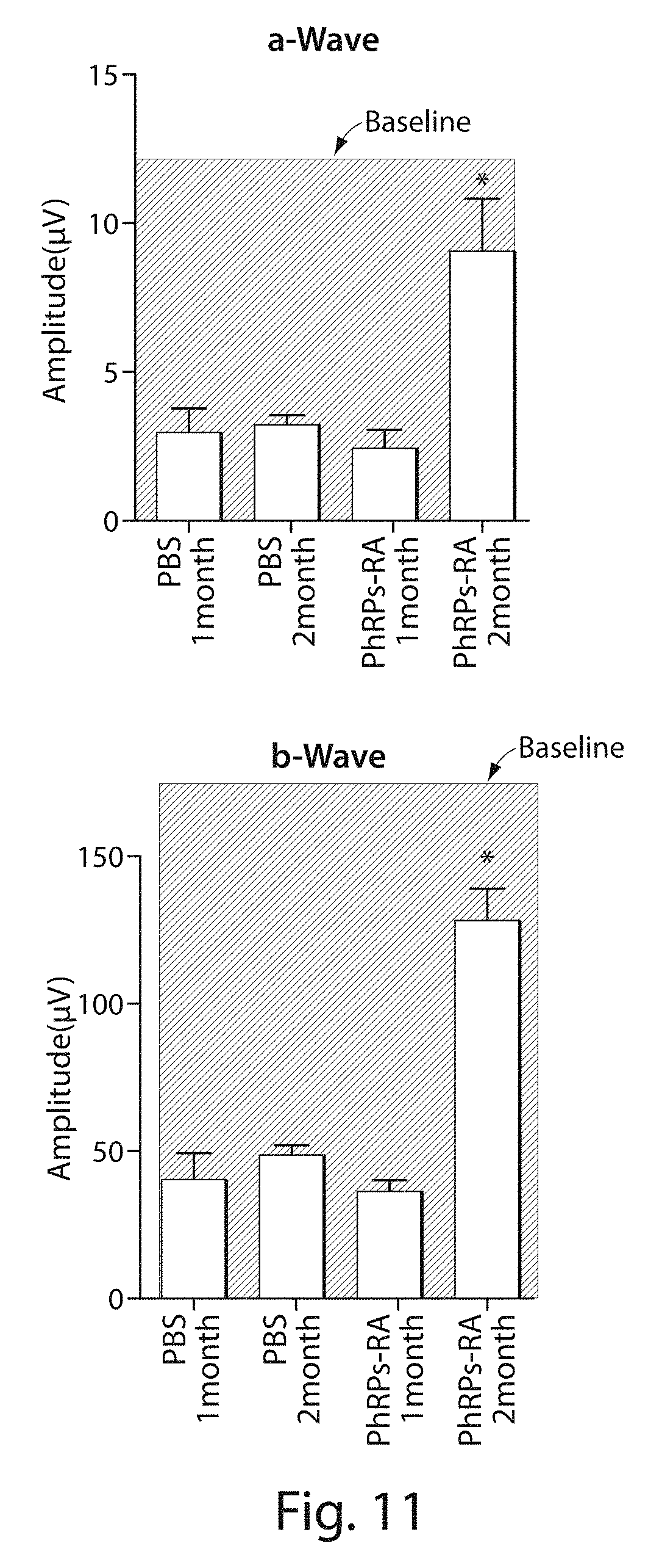
D00014
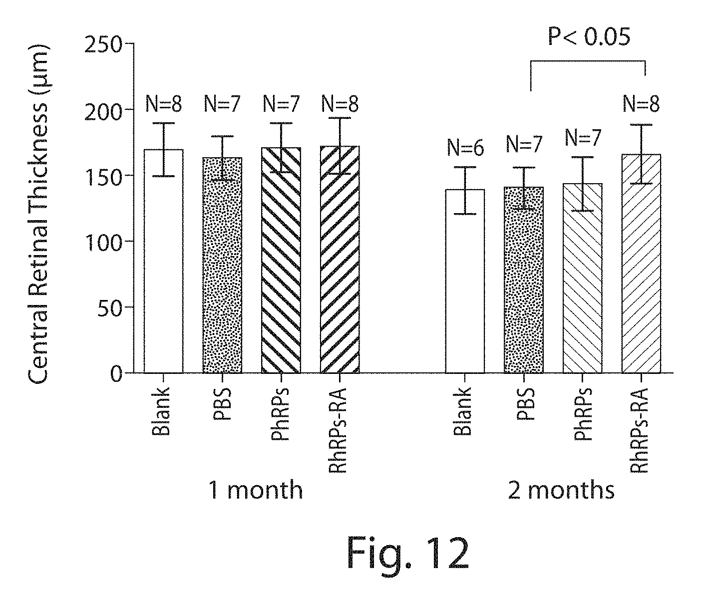
D00015

D00016

D00017

D00018
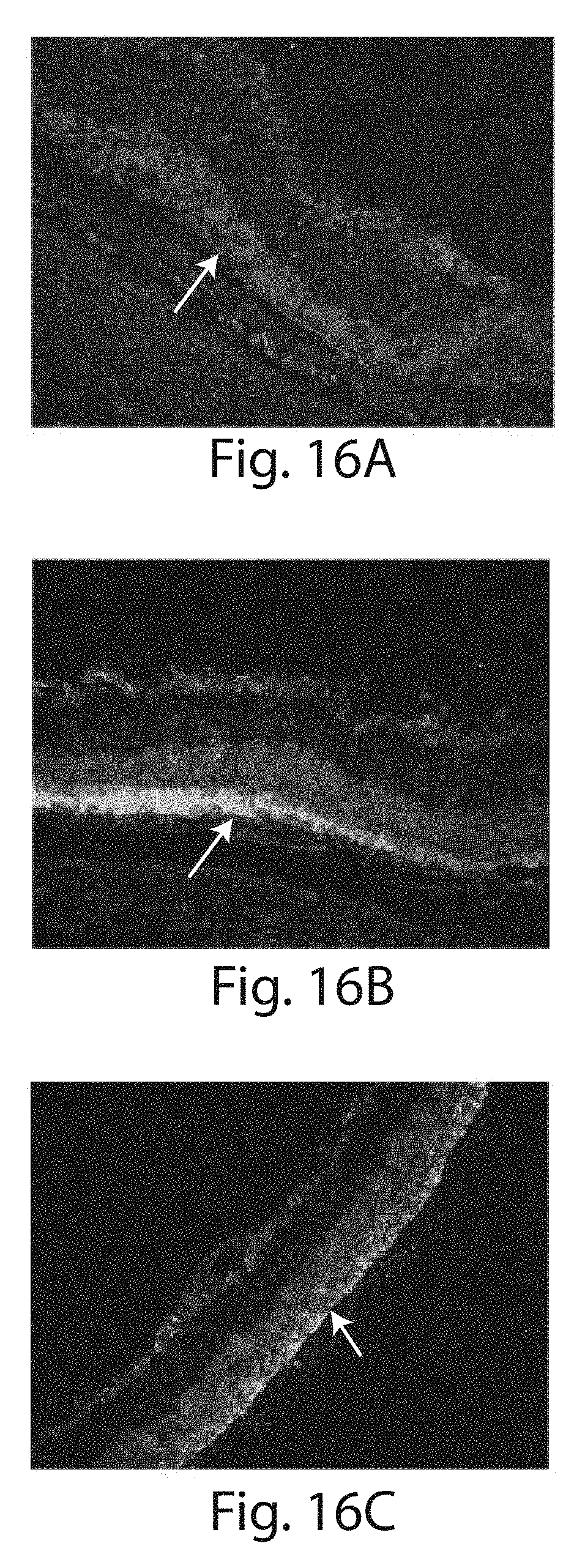
D00019

D00020
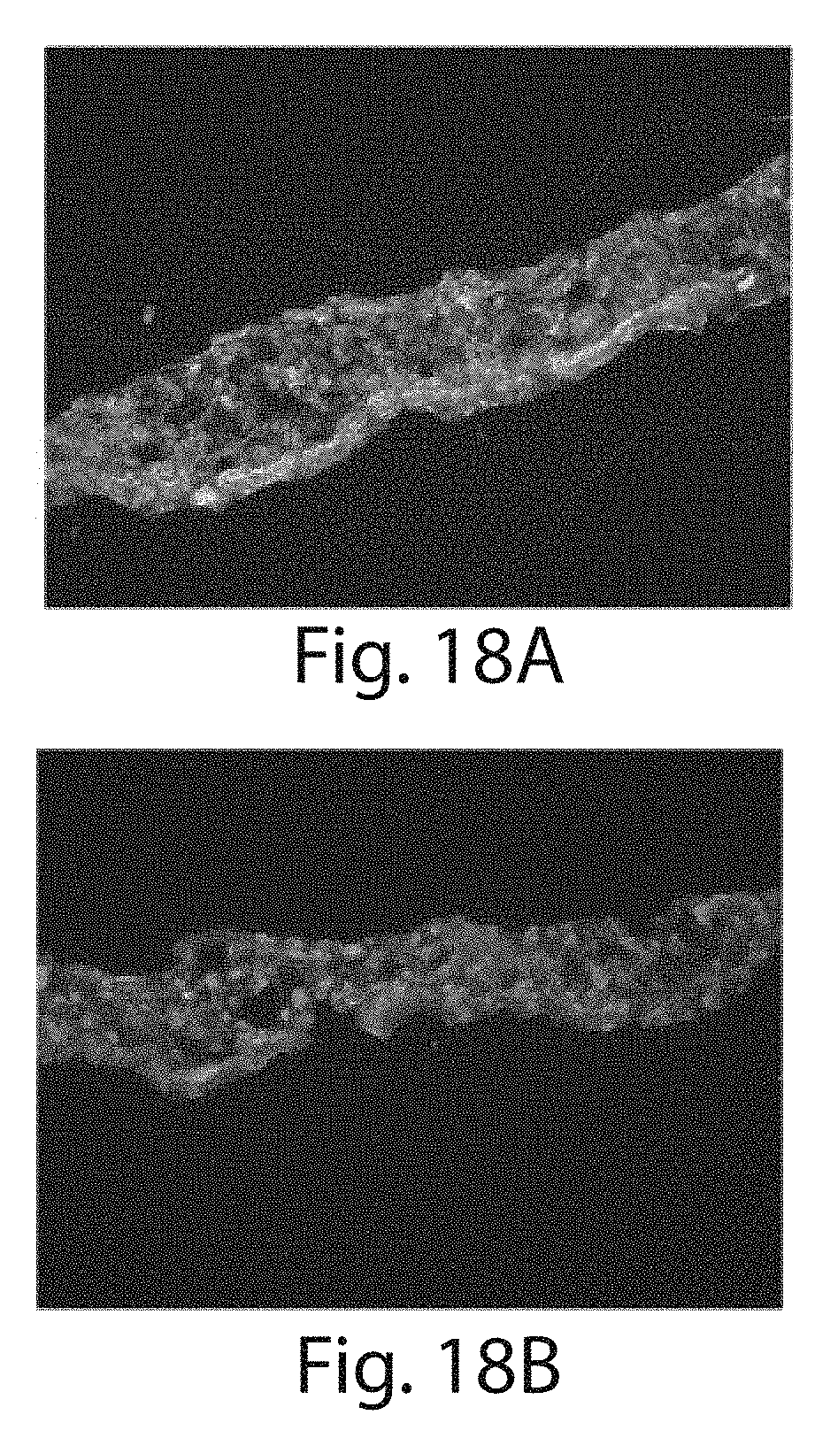
D00021

D00022

D00023

D00024

D00025
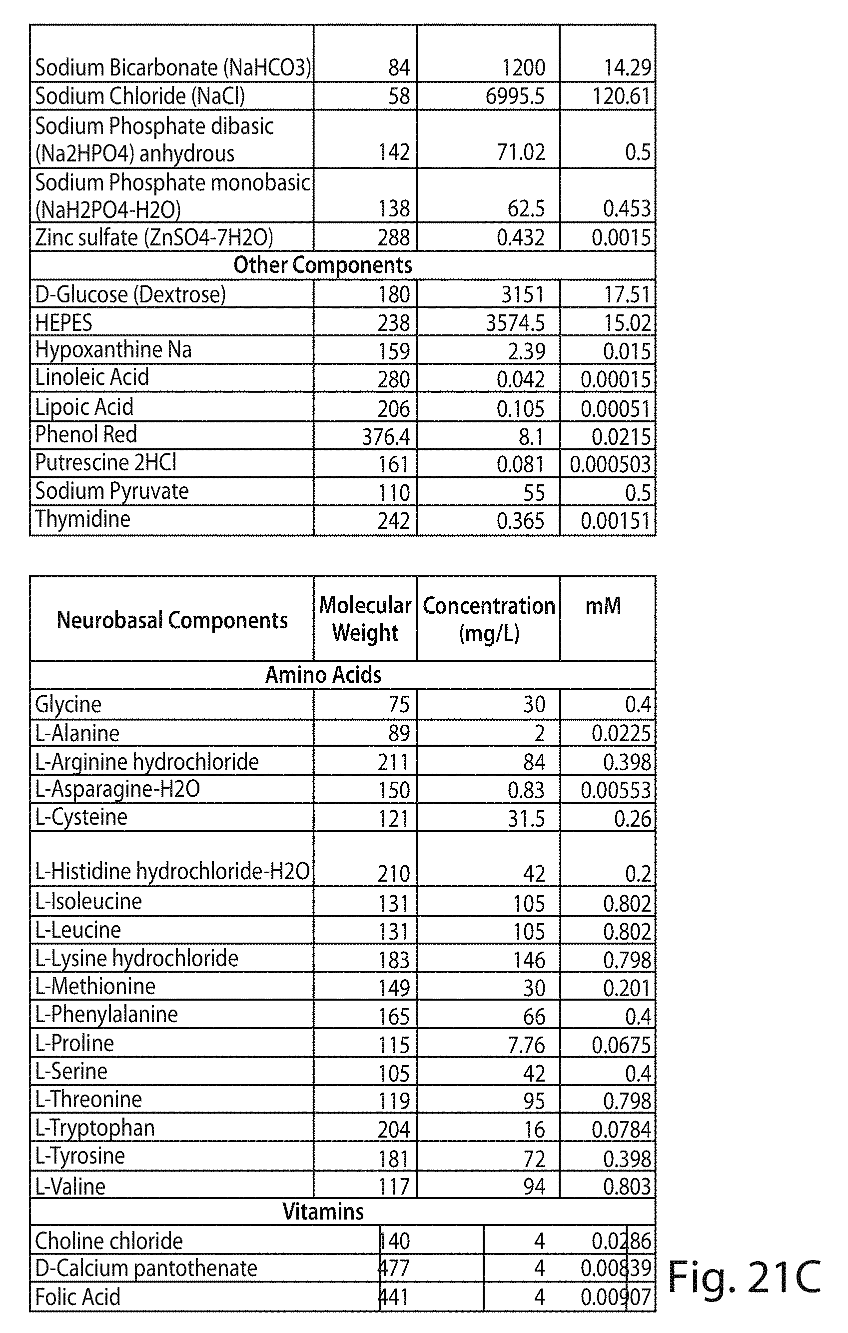
D00026
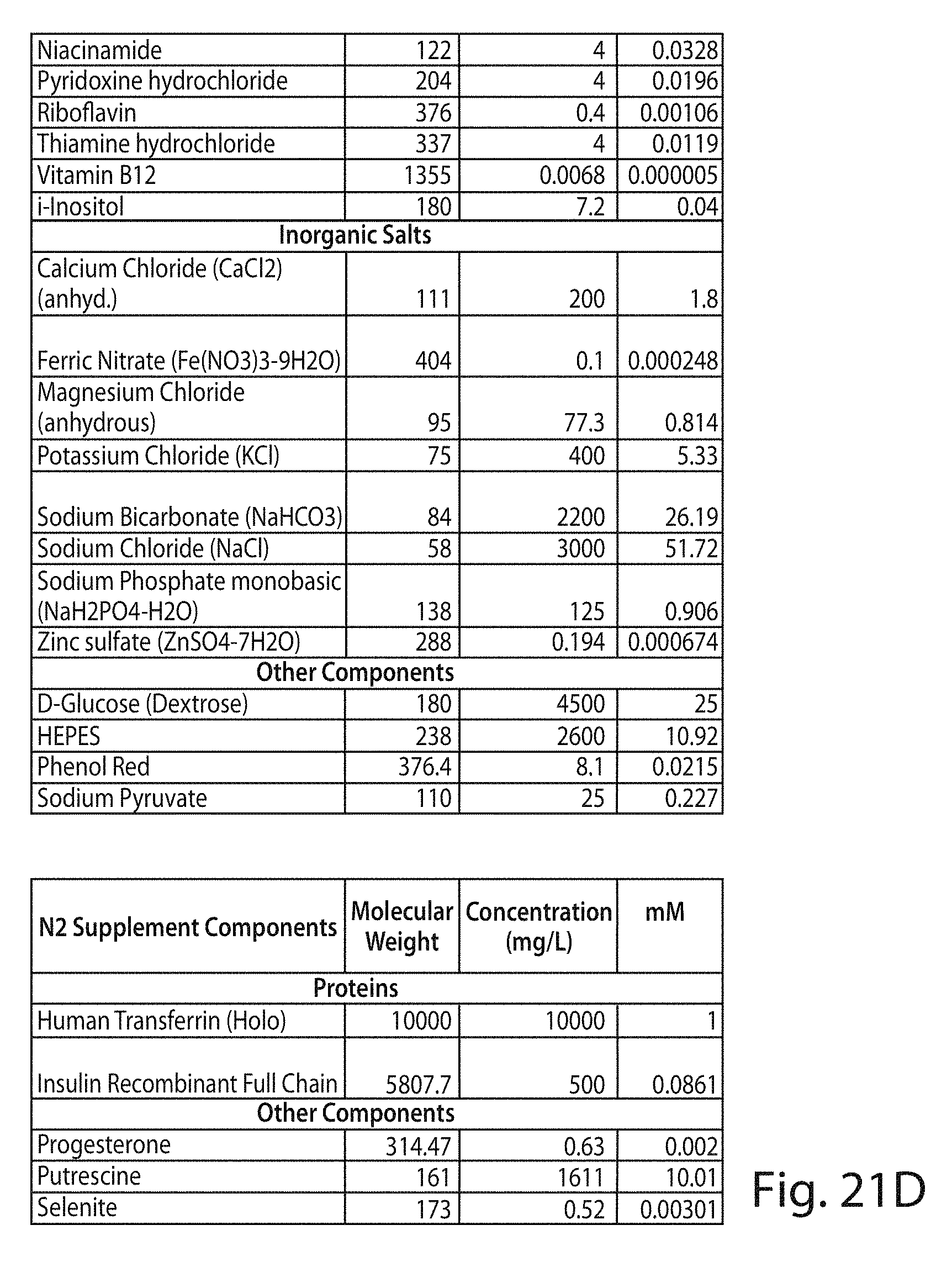
D00027
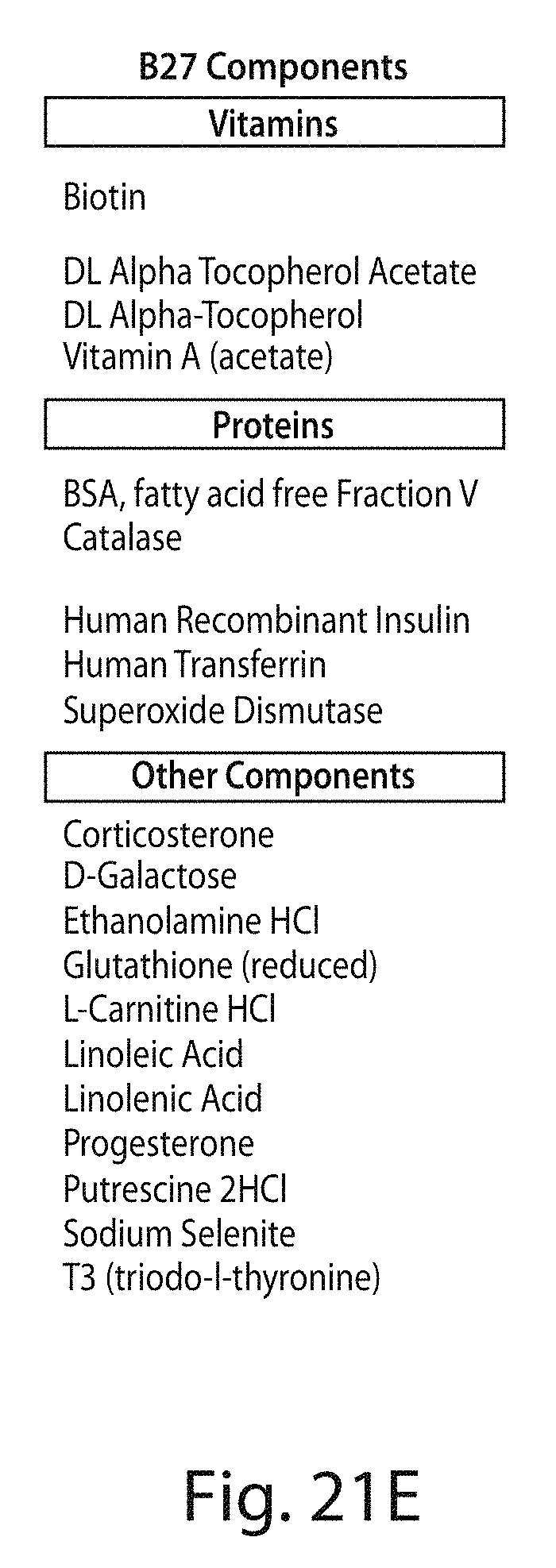
D00028
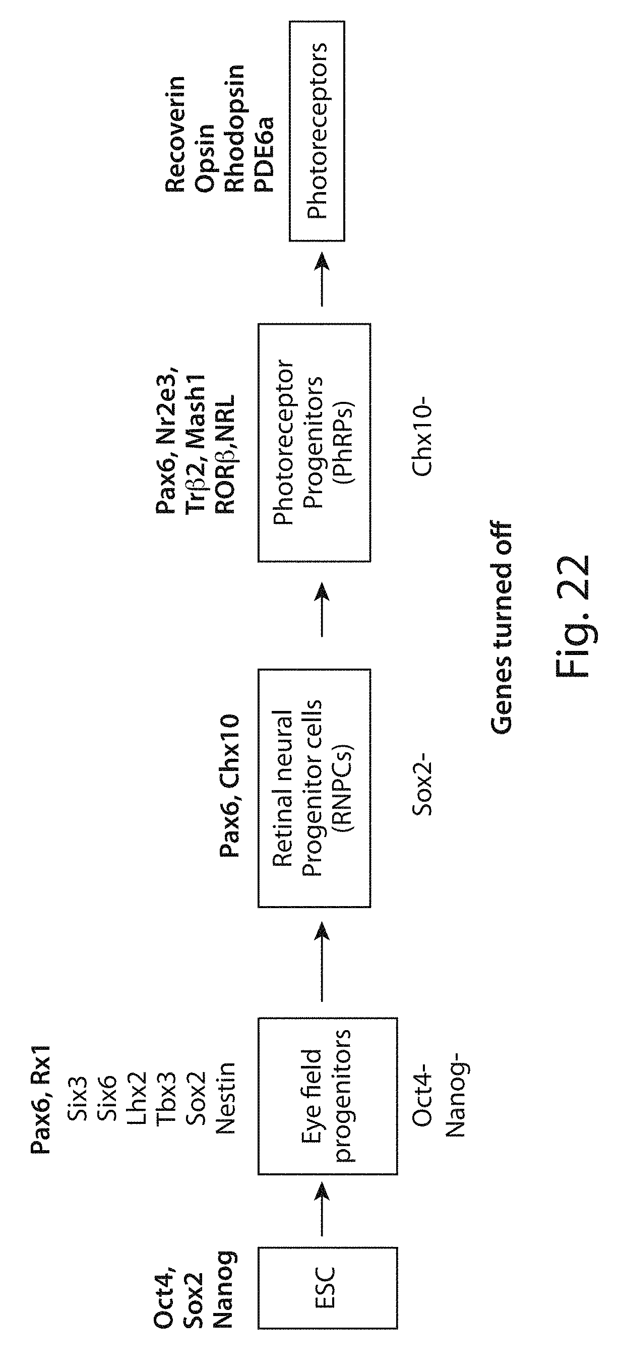
D00029
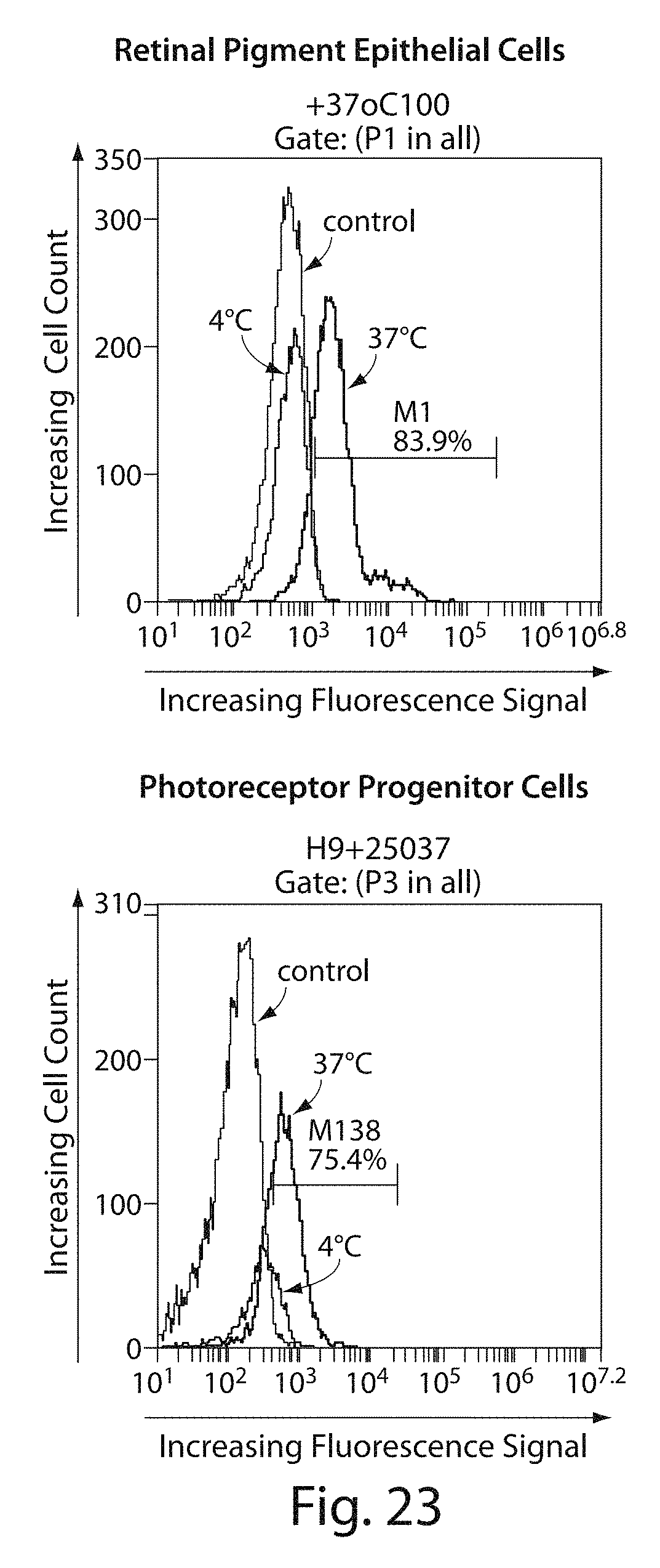
XML
uspto.report is an independent third-party trademark research tool that is not affiliated, endorsed, or sponsored by the United States Patent and Trademark Office (USPTO) or any other governmental organization. The information provided by uspto.report is based on publicly available data at the time of writing and is intended for informational purposes only.
While we strive to provide accurate and up-to-date information, we do not guarantee the accuracy, completeness, reliability, or suitability of the information displayed on this site. The use of this site is at your own risk. Any reliance you place on such information is therefore strictly at your own risk.
All official trademark data, including owner information, should be verified by visiting the official USPTO website at www.uspto.gov. This site is not intended to replace professional legal advice and should not be used as a substitute for consulting with a legal professional who is knowledgeable about trademark law.