Peptide Inhibiting Colonization By Pathogenic Bacteria, And Colonization Inhibitor Including Same
NAKAMURA; Shota ; et al.
U.S. patent application number 16/097219 was filed with the patent office on 2019-09-19 for peptide inhibiting colonization by pathogenic bacteria, and colonization inhibitor including same. This patent application is currently assigned to OSAKA UNIVERSITY. The applicant listed for this patent is BIKENBIOMICS CO., LTD, OSAKA UNIVERSITY. Invention is credited to Shunsuke FUKAKUSA, Tetsuya IIDA, Kazuki KAWAHARA, Yuji KOBAYASHI, Toshio KODAMA, Takahiro MARUNO, Shigeaki MATSUDA, Daisuke MOTOOKA, Shota NAKAMURA, Tadayasu OHKUBO, Hiroya OKI, Yasumitsu TSUJINO, Takuya YOSHIDA.
| Application Number | 20190284246 16/097219 |
| Document ID | / |
| Family ID | 60160459 |
| Filed Date | 2019-09-19 |
| United States Patent Application | 20190284246 |
| Kind Code | A1 |
| NAKAMURA; Shota ; et al. | September 19, 2019 |
PEPTIDE INHIBITING COLONIZATION BY PATHOGENIC BACTERIA, AND COLONIZATION INHIBITOR INCLUDING SAME
Abstract
A peptide capable of preventing pathogenic bacteria from colonizing in digestive organs comprises a first domain having an amino acid sequence capable of binding to a minor pilin of IVb fimbriae, and a second domain having an amino acid sequence capable of multimerization, the second domain connect to the first domain via a linker sequence.
| Inventors: | NAKAMURA; Shota; (Suita-shi, Osaka, JP) ; KAWAHARA; Kazuki; (Suita-shi, Osaka, JP) ; OKI; Hiroya; (Suita-shi, Osaka, JP) ; YOSHIDA; Takuya; (Suita-shi, Osaka, JP) ; OHKUBO; Tadayasu; (Suita-shi, Osaka, JP) ; KOBAYASHI; Yuji; (Suita-shi, Osaka, JP) ; MARUNO; Takahiro; (Suita-shi, Osaka, JP) ; MOTOOKA; Daisuke; (Suita-shi, Osaka, JP) ; MATSUDA; Shigeaki; (Suita-shi, Osaka, JP) ; KODAMA; Toshio; (Suita-shi, Osaka, JP) ; IIDA; Tetsuya; (Suita-shi, Osaka, JP) ; TSUJINO; Yasumitsu; (Ibaraki-shi, Osaka, JP) ; FUKAKUSA; Shunsuke; (Ibaraki-shi, Osaka, JP) | ||||||||||
| Applicant: |
|
||||||||||
|---|---|---|---|---|---|---|---|---|---|---|---|
| Assignee: | OSAKA UNIVERSITY Suita-shi, Osaka JP BIKENBIOMICS CO., LTD Ibaraki-shi, Osaka JP |
||||||||||
| Family ID: | 60160459 | ||||||||||
| Appl. No.: | 16/097219 | ||||||||||
| Filed: | April 25, 2017 | ||||||||||
| PCT Filed: | April 25, 2017 | ||||||||||
| PCT NO: | PCT/JP2017/016284 | ||||||||||
| 371 Date: | February 25, 2019 |
| Current U.S. Class: | 1/1 |
| Current CPC Class: | A61P 31/04 20180101; C12N 15/00 20130101; A61K 38/00 20130101; A61P 1/00 20180101; C07K 2319/73 20130101; C07K 14/245 20130101; A61K 38/16 20130101; A61P 31/00 20180101; A61P 43/00 20180101; C12N 15/09 20130101 |
| International Class: | C07K 14/245 20060101 C07K014/245 |
Foreign Application Data
| Date | Code | Application Number |
|---|---|---|
| Apr 27, 2016 | JP | 2016-089902 |
Claims
1. A peptide comprising: a first domain having an amino acid sequence capable of binding to minor pilin of type IVb pili, and a second domain having an amino acid sequence connected via a linker sequence to said first domain and capable of multimerizing.
2. The peptide according to claim 1, wherein said first domain has an amino acid sequence having 70% or more homology with any one of SEQ ID NOS: 1 to 4.
3. The peptide according to claim 1, wherein said first domain consists of the sequence of any one of SEQ ID NOS: 1 to 4.
4. The peptide according to claim 1, wherein said first domain has the sequence of SEQ ID NO: 5.
5. The peptide according to claim 1, wherein said first domain has the sequence of SEQ ID NO: 23.
6. The peptide according to claim 1, wherein said second domain contains an amino acid sequence capable of trimerizing.
7. The peptide according to claim 6, wherein said second domain has an amino acid sequence of any one of SEQ ID NOS: 6 to 8.
8. The peptide according to claim 1, wherein said linker sequence is SEQ ID NO: 9.
9. An inhibitor of colonization of pathogenic bacteria in digestive organs, comprising the peptide according to claim 1.
Description
CROSS REFERENCE TO RELATED APPLICATIONS
[0001] This application is the U.S. National Stage Application under 35 U.S.C. .sctn. 371 of International Application No. PCT/JP2017/016284, filed Apr. 25, 2017, designating the U.S. and published as WO 2017/188215 A1 on Nov. 2, 2017, which claims the benefit of Japanese Patent Application No. JP 2016-089902, filed Apr. 27, 2016. Any and all applications for which a foreign or a domestic priority is claimed is/are identified in the Application Data Sheet filed herewith and is/are hereby incorporated by reference in their entirety under 37 C.F.R. .sctn. 1.57.
SEQUENCE LISTING IN ELECTRONIC FORMAT
[0002] The present application is being filed along with an Electronic Sequence Listing as an ASCII text file via EFS-Web. The Electronic Sequence Listing is provided as a file entitled CPJ007001APCSEQLIST.txt, created and last saved on Oct. 19, 2018, which is 5,761 bytes in size. The information in the Electronic Sequence Listing is incorporated herein by reference in its entirety in accordance with 35 U.S.C. .sctn. 1.52(e).
TECHNICAL FIELD
[0003] The present invention relates to novel peptides, more particularly to peptides that inhibit colorization of pathogenic bacteria in digestive organs and a colonization inhibitor containing the peptide.
BACKGROUND
[0004] In recent years, a number of pathogenic bacteria that cause diseases of digestive organs have been revealed. For example, enterotoxigenic Escherichia coli (ETEC) which is a type of pathogenic bacteria with intestinal toxicity is known to invade the digestive tract of humans through water or food and cause cholera like diarrhea. Infectious diseases by ETEC have become a serious problem among people living in developing countries and travelers visiting the the region, and it is estimated that about 200 million people a year are infected in the world and approximately 380,000 people including mainly infants under 5 years old will die (Non-Patent Document 1). A statistical survey conducted at the quarantine station in Osaka airport in Japan reported that about 8% of Japanese travelers who developed diarrhea are infected with ETEC (Non-Patent Document 2).
[0005] As a first step in the pathogenic expression of ETEC, it must adhere to the small intestinal epithelial cells and colonize there, and colonization factors (CFs) play its role. To date, at least twenty five CFs such as colonization factor antigen 1 (CFA/I), E. coli surface antigen 1 (CS1), presumed colonization factor 071 (PCF071) and the like have been reported (Non-Patent Document 3). By the action of these pathogenic factors, when ETEC adheres to the small intestinal epithelial cells of the host, it forms biofilms, microcolonies and the like and colonizes there. Thereafter, as a second step, ETEC produces heat-labile intestinal toxin (LT) or heat-resistant intestinal toxin (ST) and develops pathogenicity.
[0006] Almost all of CFs that ETEC has are ciliary colonization factors, and classified into several groups depending on the amino acid sequence of the constituent component and the feature of immunogenicity or the like (Non-patent document 3). For example, CFA/I, CS1, CS2, CS4, CS5, CS7, CS14, CS17, CS19 and PCF071 form pili belonging to the class type 5, while CS12, CS18, CS20, CS26, CS27 and CS28 form pili belonging to the class type 1b. In addition to these, CS13, CS15, CS22 and CS23, or CS3 and CS6 are similarly grouped, respectively. The structures and natures of these CFs are still unknown in many respects, but these CFs have similar constituent components and are mainly constituted of Chaperon proteins present in the E. coli periplasm, pilus main constituent subunits (major pilin), a small number of subunits contained in the pilus (minor pilin) and outer membrane Usher proteins, and pili are formed by the Chaperon-Usher path (Non-patent document 3). In contrast, CFA/III (CS8) and Longus (CS21) form pili classified as type IV, and the pilus structure and the pilus forming mechanism different from those by the Chaperon-Usher pathway are provided. CS10 and CS11 have been reported as other CFs, but the pilus structures of them are still unknown in many respects.
[0007] An outer membrane protein Tia identified from the ETEC H10407 strain is known as a non-ciliary colonization factor expressed by ETEC, and the protein binds to proteoglycan on the host cell surface (Non-Patent Document 4). In addition, also EtpA identified from ETEC H10407 strain is known as a non-ciliary colonization factor expressed by ETEC. It has been suggested that EtpA binds to a highly conserved region of the flagellin protein and may bridge the microbial body and the host cell surface (Non-Patent Document 5).
[0008] Generally, each strain of ETEC possesses 1 to 3 kinds of CFs and also possess various secretory colonization factors (Non-Patent Document 6). Therefore, development of treatment means such as vaccination is difficult for reasons such as diversity of the spatial and temporal expression control mechanism in accordance with the environments around each CFs, further, introduction of strain-dependent variations into the surface antigen, and the like, in addition to the complexity of the combination. Currently, as candidate antigens for vaccine development, toxin LT produced by almost all ETECs is considered, but due to its toxicity, there is a restriction on the dose in oral administration or the like, and in recent years, a variant obtained by substituting the amino acid at the active site has been studied as an alternative antigen. Similar use is considered also for ST, but it is a problem that the molecular size (peptide consisting of about 20 residues) is small and it is difficult to cause an immune response (Non-Patent Document 7). Development of a vaccine using each colonization factor as an antigen has also been advanced. Because it is necessary to devise to cause broad immune, the current most effective vaccine is a live or inactivated ETEC vaccine using CFA/I, CS3, CS5, CS6, and a strain producing intestinal toxin LT (Non-patent Document 7 and 8). These vaccines are applicable to each group of pili primarily formed by the Chaperon-Usher pathway and are believed to represent the effect on 80% of the clinical strains of ETEC.
REFERENCES
[0009] [Non-Patent Literature 1] Qadri, F. et al. Clin. Microbiol. Rev. 2005, 18, 465-483. [0010] [Non-Patent Literature 2] Abe, H. et al. J. Diar. Dis. Res. 1984, 2, 83-87. [0011] [Non-Patent Literature 3] Madhavan, T. V. et al. Adv. Appl. Microbiol. 2015, 90, 155-197. [0012] [Non-Patent Literature 4] Fleckenstein, J. M. et al. Infect. Immun 2002, 70, 1530-1537. [0013] [Non-Patent Literature 5] Roy, K. et al. Nature 2009, 457, 594-598. [0014] [Non-Patent Literature 6] Mentzer, A. et al. Nat. Genetics 2014, 46, 1321-1326. [0015] [Non-Patent Literature 7] Sjoling, A. et al. Expert Rev. Vaccines 2015, 4, 551-560. [0016] [Non-Patent Literature 8] Fleckenstein, J. M. et al. Microbes and infections 2010, 12, 89-98.
SUMMARY
[0017] Effective ETEC vaccines have not been developed to date. Studies of live or inactivated ETEC vaccines using CFA/I, CS3, CS5, CS6, and a strain producing intestinal toxin LT, as the most promising vaccine candidates, are progressing. However, there is no effect against strains of ETEC containing colonization factors or toxins not contained in this vaccine. In particular, it is not suitable for strains which form type IV pili and produce CFs having different pilus structures and characteristics from CFs contained in the above-described inactivated vaccines. Particularly, longus (CS21) has been identified from ETECs in a wide area and is prevalent in regions including Latin America, Middle East and North Africa (Isidean S D et al. Vaccine 2011, 29, 6167-6178). With the above-described inactivated vaccine, it is difficult to inhibit infection of strains carrying such colonization factors. There is also a concern that inactivation of strains does not cause immunity to secretory pathogenic factors, and there is also a problem that it is difficult to induce long-term immunity. To solve these problems, it is considered to identify highly conserving protein expressing on the surface of the broad ETEC and to produce a vaccine using this protein as an antigen, however, such proteins have not been discovered yet.
[0018] Thus, it is thought to be realistic and more effective to perform immunization with multiple antigens by revealing the details of the structure and colonizing mechanism of the colonization factors of ETEC at the molecular level and identifying conserved epitopes, rather than searching and identification of the protein. In addition, if the colonizing mechanism becomes clear, development of a colonization inhibitor is also possible.
[0019] The present invention has been made in view of the above problems, and an object thereof is to be able to prevent bacteria having a colonization factor from colonizing in digestive organs.
[0020] The present inventors have intensively studied and resultantly found a mechanism in which particularly pathogenic bacteria that produce type IVb pili, among bacteria, colonize in digestive organs and succeeded in producing a novel peptide that inhibits colonization of the bacteria, based on the mechanism.
[0021] Specifically, the peptide according to the present invention is characterized by comprising a first domain having an amino acid sequence capable of binding to minor pilin of type IVb pili, and a second domain having an amino acid sequence connected via a linker sequence to the first domain and capable of multimerizing.
[0022] Bacteria producing type IVb pili become able to adhere to and colonize on the target cell by bonding of secretory proteins generated by the bacteria themselves to secretory protein binding sites of minor pilin located at the top of the major constituent subunit (major pilin) of type IVb pili, though the bacteria are unable to bind to the receptor of target cell in digestive organs and the like only by the pili. In contrast, the peptide according to the present invention can bind to type IVb pili since the peptide has a first domain having an amino acid sequence capable of binding to the secretory protein binding site of minor pilin of type IVb pili. Thus, the peptide according to the present invention inhibits the bonding of secretory proteins and type IVb pili of bacteria, by binding to type IVb pili, and resultantly can inhibit the bacteria from adhering to and colonizing on the target cell in digestive organs and the like. Usually type IVb pili form a homotrimer, while the peptide according to the present invention has a second domain capable of multimerizing, hence, the peptide is able to bind simultaneously to all the binding sites of the homotrimer. Therefore, it is possible to efficiently inhibit the bonding between the secretory proteins of the bacteria and type IVb pili, and resultantly it is possible to efficiently inhibit the bacteria from adhering to and colonizing on the target cell in digestive organs and the like. This anti-adhesion mechanism has very little effect on the host since the peptide selectively binds to pathogenic bacteria rather than binding to receptors on host cells and then elimination outside the host is elicited. Also, unlike antibiotics, pathogenic bacteria are not killed, thus, selection pressures are small and it is possible to suppress the appearance of drug-resistant bacteria. Inhibitors focusing on such anti-adhesion mechanism have been studied for type I pili and type P pili carried by uropathogenic Escherichia coli (UPEC) (Sharon, N. Biochim Biophys. Acta 2006, 1760, 527-537). Both are pili having a lectin domain that recognizes a sugar chain at the tip, and the development of sugar derivatives and the like that specifically bind to the sugar chain binding region thereof is advancing. Regarding ETEC, an antiadhesive agent in which a sugar chain is bound to nanoparticles has been studied (Ravel, Y. S. et al. Nanoscale 2015, 7, 8326-8331). However, since the adhesion mechanism via pili is not clear, inhibitors that efficiently inhibit adhesion of type IVb pili have not been developed ever.
[0023] In the peptide according to the present invention, it is preferable that the first domain has an amino acid sequence having homology of 70% or more with any sequence of SEQ ID NOS: 1 to 4.
[0024] SEQ ID NOS: 1 to 4 are sequences involved in binding to the binding sites of miner pilin (CofB, LngB, CfcB and TcpB) corresponding to secretory proteins such as CofJ, LngJ, CfcJ and TcpF expressed by bacteria that produce type IVb pili. Therefore, it is possible to inhibit the binding of the secretory protein to the type IVb pili, by including the first domain having high homology with these. Further, it is particularly preferable that the first domain consists of the sequence of any one of SEQ ID NOS: 1 to 4.
[0025] In the peptide according to the present invention, it is preferable that the first domain has the sequence of SEQ ID NO: 5.
[0026] SEQ ID NO: 5 is a sequence strongly involved in binding to the binding site of the minor pilin (CofB) in the amino acid sequence of CofJ. Therefore, when the first domain has the sequence of SEQ ID NO: 5, the peptide according to the present invention can efficiently inhibit the binding between CofJ and minor pilin (CofB). In addition, by configuring the first domain only with the sequence of SEQ ID NO: 5, the first domain can be made shorter and the structure can be simplified.
[0027] In the peptide according to the present invention, it is preferable that the first domain has the sequence of SEQ ID NO: 23.
[0028] SEQ ID NO: 23 is a sequence in which serine is added to the N terminal side of the above SEQ ID NO: 5 and lysine is added to the C terminal side, and when the first domain has the sequence of SEQ ID NO: 23, the peptide according to the present invention can efficiently inhibit the binding between CofJ and minor pilin (CofB) as in the case of SEQ ID NO: 5 above. In addition, by configuring the first domain only with the sequence of SEQ ID NO: 23, the first domain can be made shorter and the structure can be simplified.
[0029] In the peptide according to the present invention, the second domain preferably contains an amino acid sequence capable of trimerizing.
[0030] Since the type IVb pilus forms a homotrimer as described above, when the second domain contains the amino acid sequence capable of trimerizing, the peptide of the present invention can bind to the binding sites in all minor pilins in the homotrimer. This can effectively inhibit the binding between secretory proteins of bacteria and type IVb pili.
[0031] In the peptide of the present invention, the second domain preferably has the amino acid sequence of SEQ ID NO: 6, or an amino acid sequence repeating 4 to 10 times the amino acid sequence of SEQ ID NO: 7 or SEQ ID NO: 8.
[0032] SEQ ID NO: 6 is a sequence that forms a GCN4 trimeric coiled-coil, and the amino acid sequence repeating 4 to 10 times the sequence of SEQ ID NO: 7 or SEQ ID NO: 8 is a sequence that forms a collagen-like triple helical structure, that is, a sequence that promotes peptide trimerization. Therefore, by providing the second domain having such an amino acid sequence, the peptide of the present invention can form a trimer. Therefore, by adopting these sequences, it is possible to obtain a peptide capable of efficiently inhibiting the binding between the bacterial secretory proteins and the type IVb pili as described above.
[0033] In addition, in the peptide according to the present invention, the linker sequence may be SEQ ID NO: 9.
[0034] The colonization inhibitor according to the present invention is one which inhibits colonization of pathogenic bacteria in digestive organs characterized by containing the peptide according to the present invention.
[0035] According to the colonization inhibitor according to the present invention, it is possible to inhibit the binding between the type IVb pili and the target cells of digestive organs since the inhibitor contains the peptide according to the present invention. As a result, it is possible to inhibit adhesion and colonization of type IVb pili-producing bacteria in digestive organs.
[0036] According to the peptide of the present invention and the colonization inhibitor containing the same, it is possible to inhibit adhesion and colonization of type IVb pili-producing bacteria in digestive organs.
BRIEF DESCRIPTION OF THE DRAWINGS
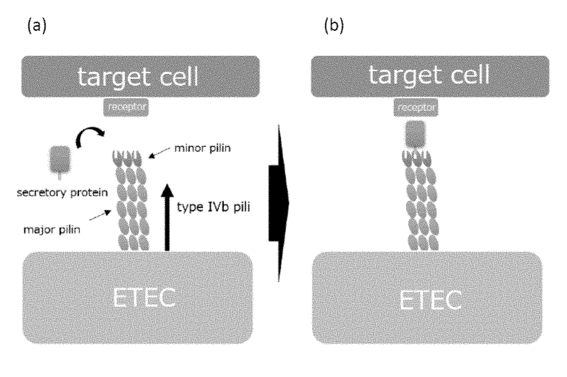
[0037] FIGS. 1 (a) and (b) are model diagrams for explaining a mechanism by which pathogenic bacteria having type IVb pili adhere to and colonize on cells constituting a digestive organ and the like.
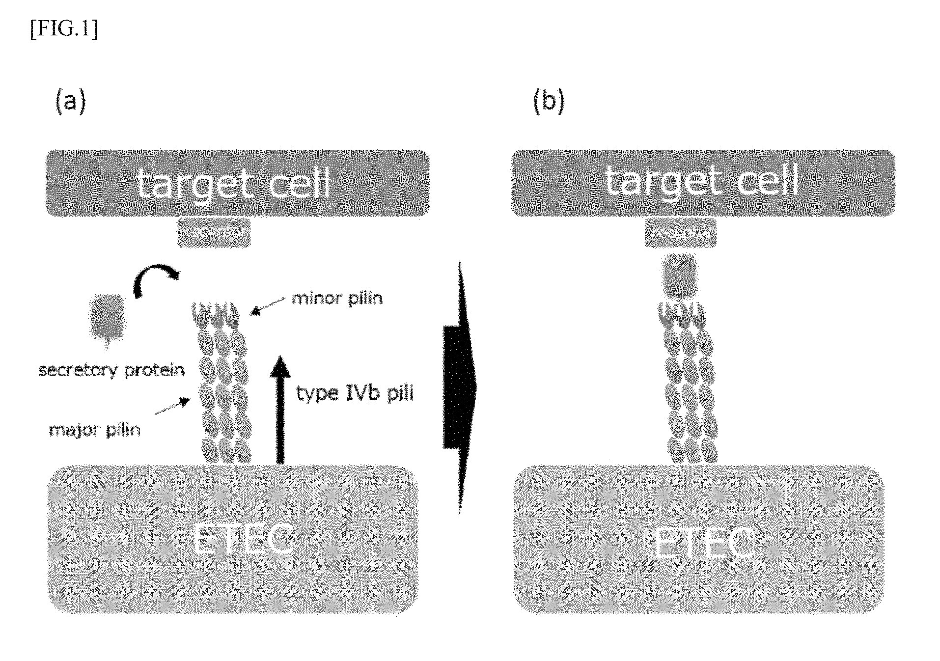
[0038] FIGS. 2 (a) and (b) are model diagrams for explaining a mechanism by which a peptide according to an embodiment of the present invention inhibits adhesion and colonization thereof.
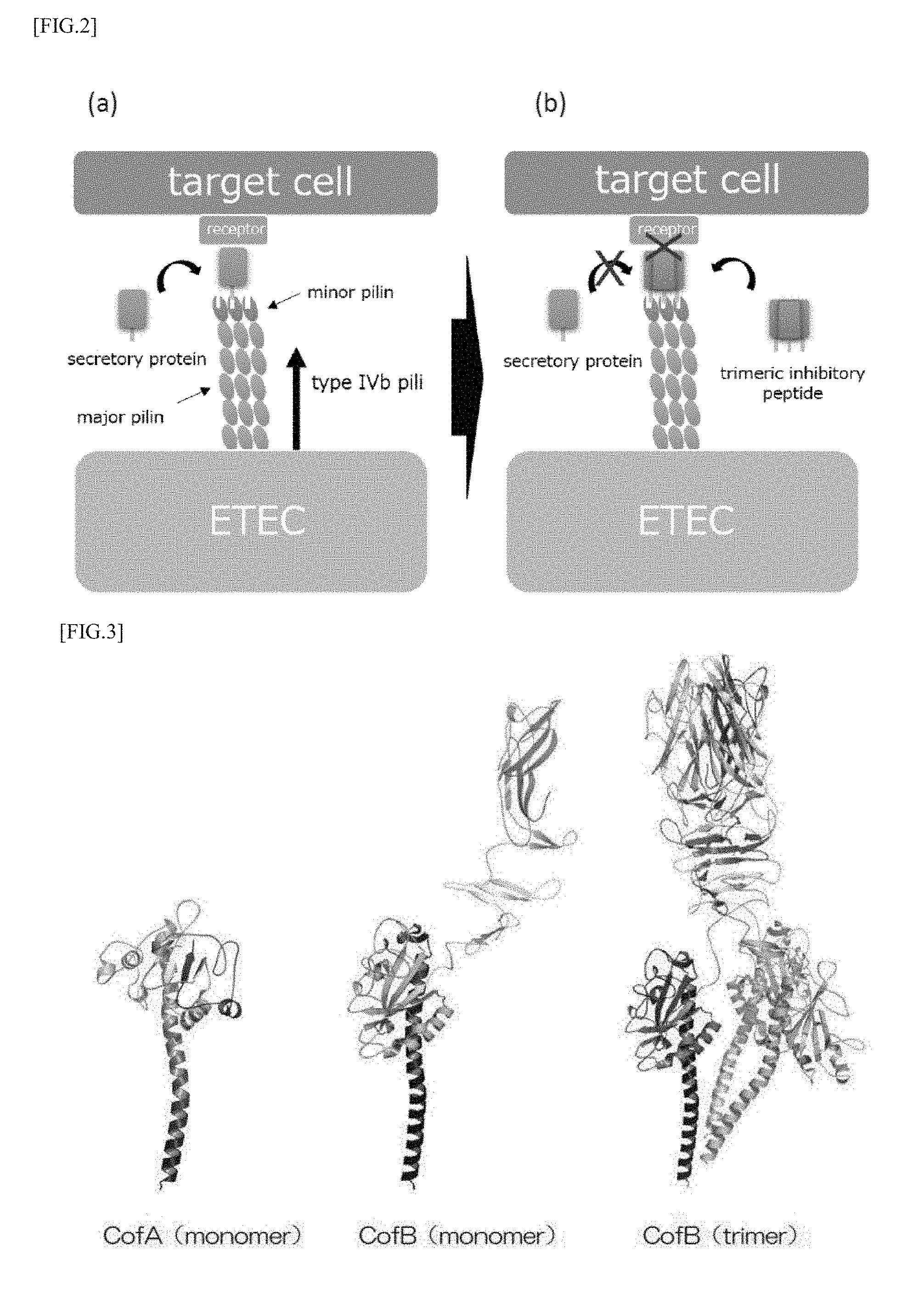
[0039] FIG. 3 is a model diagram showing a three-dimensional structure of CofA and CofB which are constituent components of CFA/III pilus.
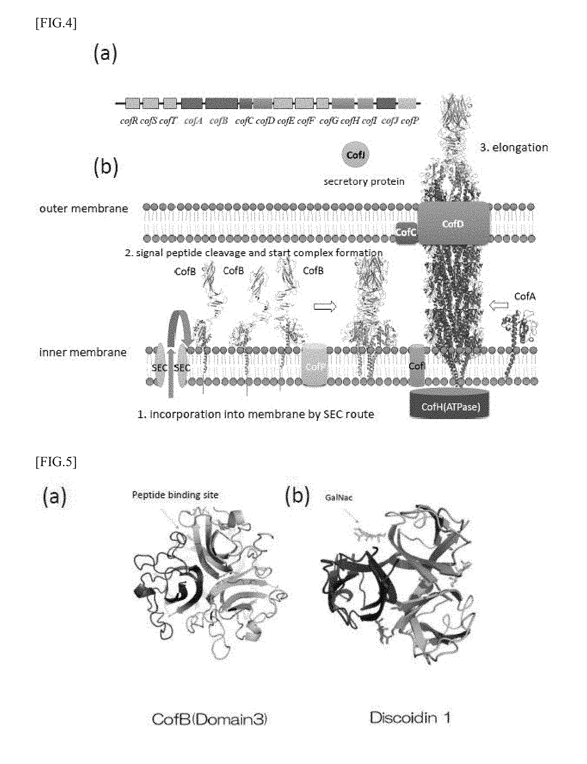
[0040] FIG. 4 (a) is a view showing a configuration of a cof operon which is a group of genes constituting CFA/III, and FIG. 4 (b) is a view for explaining the formation of type IV pili consisting of CofA and CofB.
[0041] FIG. 5 (a) is a view showing a trimeric structure of CofB, and FIG. 5 (b) is a view showing a trimeric structure of Discoidin I.
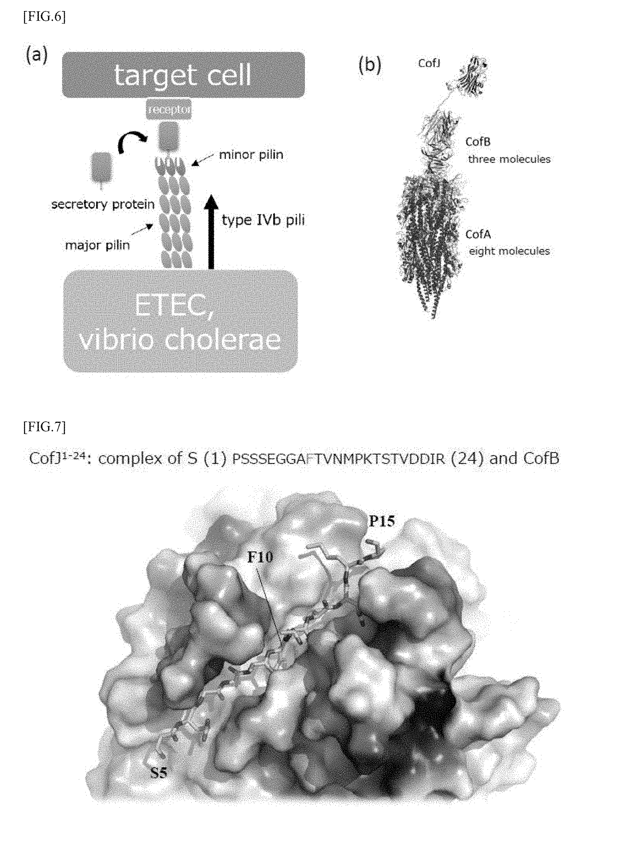
[0042] FIGS. 6 (a) and (b) are views showing the binding between CofJ and CofB.
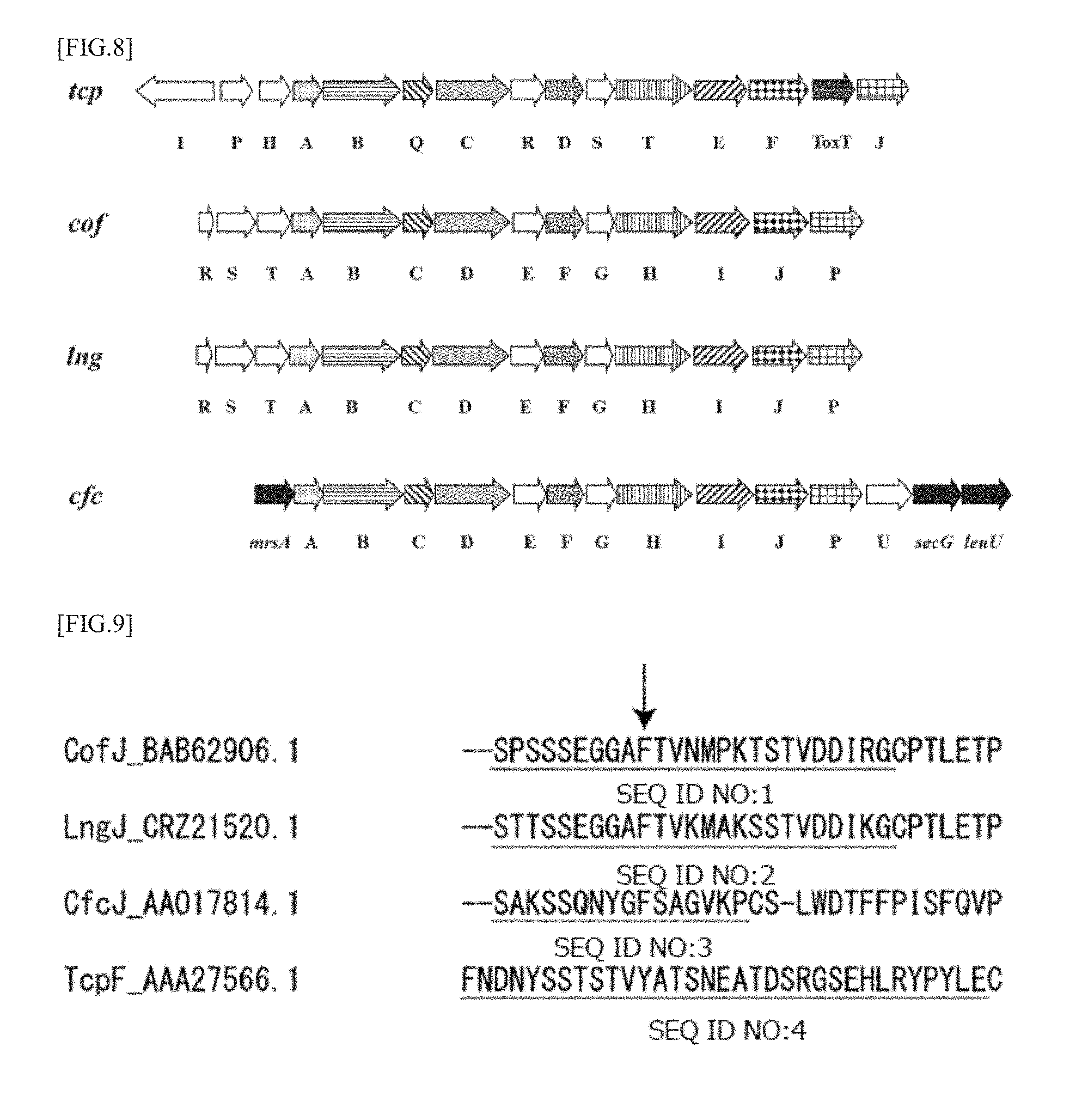
[0043] FIG. 7 is a model diagram showing a crystal structure of a complex in which CofJ.sup.1-24 peptide and CofB are bound.
[0044] FIG. 8 is a view which shows homologous genes in each coding region coding for type IVb pili, showing cof operon, lng operon, Tcp operon and cfc operon.
[0045] FIG. 9 is a view which shows the amino acid sequences of N-terminal regions of CofJ, LngJ, CfcJ and TcpF.
[0046] FIGS. 10 (a) to (c) are model diagrams showing the results of structure analysis of CofB, and (a) shows its overall structure, (b) shows an enlarged view of the area enclosed by a square in (a) and (c) is a diagram showing the top side of (b).
[0047] FIG. 11 is a view showing the results of a pull-down assay for examining whether the CofJ.sup.1-24-GCN4 inhibitory peptide can inhibit the binding between CofB and CofJ.
[0048] FIG. 12 is a view showing the results of a pull-down assay for examining whether the CofJ.sup.1-24 peptide and CofJpepR can inhibit the binding between CofB and CofJ.
[0049] FIG. 13 is a graph showing the results of measurement of the ETEC adhesion inhibitory activity of the CofJ.sup.1-24-GCN4 inhibitory peptide using Caco2 cells.
[0050] FIG. 14 is a views showing the results of a pull-down assay for examining whether the CofJ.sup.4-16-GCN4 inhibitory peptide can inhibit the binding between CofB and CofJ.
[0051] FIG. 15 is a graph showing the results of measurement of the ETEC adhesion inhibitory activity of the CofJ.sup.4-16-GCN4 inhibitory peptide using Caco2 cells.
DETAILED DESCRIPTION
[0052] Embodiments for carrying out the present invention will be described below. The following descriptions of preferred embodiments are merely substantially exemplary and are not intended to limit the present invention, its application methods or its uses.
[0053] The peptide according to the embodiment of the present invention is a peptide which can bind to pili, particularly to type IVb pili of pathogenic bacteria, thereby preventing the pathogenic bacteria from binding to receptors of target cells via the pili. The peptide according to this embodiment comprises a first domain that binds to type IVb pili, a second domain that is capable of multimerizing, and a linker sequence that connects them.
[0054] In the present embodiment, the pathogenic bacteria are pathogenic bacteria in particular having type IVb pili, for example, enterotoxigenic Escherichia coli (ETEC), cholera (Vibrio cholerae), Citrobacter rodentium and the like.
[0055] First, outlines of a mechanism of adhesion and colonization of pathogenic bacteria having type IVb pili such as ETEC to cells constituting a digestive organ and the like and a mechanism by which the peptide according to this embodiment inhibits adhesion and colonization thereof will be illustrated.
[0056] As shown in FIG. 1 (a), the above-mentioned ETEC and the like form pili constituted of major pilin as the major constituent subunit and a minor pilin slightly contained and situated at the tip of the major pilin, on the surface thereof. In particular, they form a multimer, for example, a homotrimer. ETEC and the like can not adhere to the target cells in digestive organs and the like only by the pili having such a configuration, and secretory proteins expressed by ETEC and the like are necessary for adhesion. In the minor pilin, a binding site of a secretory protein is present and the secretory protein binds to minor pilin, whereby the pilus can bind to receptors of the target cells through the secretory protein as shown in FIG. 1 (b). As a result, ETEC adheres to target cells, then, microcolonies and the like are formed, thereby colonization is made possible.
[0057] As described above, the peptide according to the present embodiment has a first domain that binds to type IVb pili, and as shown in FIG. 2, the first domain of the peptide binds to a secretory protein binding site in minor pilin of the pili, as a result, it is possible to inhibit the secretory protein from binding to the binding site. In particular, since the peptide according to the present embodiment has a second domain that promotes multimerization, particularly trimerization, the peptide forms a trimer and can bind to all binding sites of pili of the trimer as shown in FIG. 2 (b). By this means, it is possible to efficiently inhibit the binding of secretory proteins to pili, and as a result, it is possible to efficiently inhibit adhesion and colonization of pathogenic bacteria such as ETEC and the like to target cells.
[0058] These mechanisms will be described in more detail below.
[0059] (Mechanism of Formation of Type IVb Pilus)
[0060] Bacteria that produce type IVb pili such as ETEC and the like express colonization factors, which are responsible for the formation of pilus. As a colonization factor to form type IVb pili on bacteria, CFA/III (CS8) is known, and as other colonization factors having the gene structure and constituent proteins which are very similar to this, Longus (CS21) is known. Type IV pili are roughly classified to type IVa or IVb depending on the N-terminal sequence and overall size of the major pilin that is the main constituent subunit, and CFA/III (CS8) and Longus (CS21) belong to the type IVb.
[0061] Previous functional analysis studies have revealed that cof operon which is a gene group constituting the CFA/III is constituted of 14 kinds of gene groups including the gene cofA encoding major pilin, the gene cofB encoding minor pilin and the like (Taniguchi, T. et al. Infect. Immun. 2001, 69, 5864-5873). Furthermore, the present inventors have determined the steric conformation of CofA and revealed the structure of pili formed by CFA/III (Fukakusa, S. et al. Acta Cryst. 2012, D68, 1418-1429) (FIG. 3). Further, as shown in FIG. 3, three-dimensional structure determination using X-ray crystal structure analysis and analysis of the associated state in solution by ultracentrifugation analysis have revealed that the minor pilin CofB constituting the pili formed by CFA/III forms a strong homotrimeric structure in which the pilin-like domain at the N-terminal side and two domains rich 13 sheets at the central portion and the C-terminal side are entangled helically. In addition, it has also been clarified that the trimer structure is located at the tip of the type IVb pilus which is mainly composed of major pilin CofA and plays a role of promoting the initiation of pili formation, as shown in FIGS. 4 (a) and 4 (b) (Kawahara, K. et al. J. Mol. Biol. 2016, 428, 1209-1226).
[0062] The tips of the type IVb pili are bundled by a domain rich in .beta. sheets existing on the C-terminal side of the minor pilin CofB, and as a result of search of analogous domains, it has been clarified that this domain has a type H lectin-like folded structure, as shown in FIG. 5 (a). As the type H lectin domain, Discoidin I (FIG. 5 (b)) and Discoidin II of Dictyostelium discoideum and Helix pomatia agglutinin (HPA) discovered from snail called apple snail are known, and in recent years, Sinularia lochmodes (SLL2) of Symbiodinium sp. is known (Lescar, J. et al. Glycobiology 2007, 17, 1077-1083; Kita, A. et al. Glycobiology 2015, 25, 1016-1023), in addition to the CFA/III pili held by ETEC, and all of them form a homotrimer. At the molecular boundary of the trimer, there are three equivalently preserved sugar chain binding sites. It has been confirmed that N-acetyl-D-galactosamine (GalNac) binds at the binding site in the case of Discoidin I, Discoidin II, HPA and SLL2. All of them are involved in cell adhesion and self-aggregation, by binding to sugar chains. Recognition of sugar chains by lectin domains has also been confirmed in type I pili.
[0063] (Mechanism of Adhesion and Colonization of ETEC)
[0064] Schematic mechanism of adhesion and colonization of ETEC to the target cell is as described above with reference to FIG. 1, and the mechanism of adhesion and colonization of ETEC to the target cells will be illustrated more in detail below.
[0065] Since the CofB has the lectin domain described above, the expression of the adhesion ability to the target cells is assumed to occur by the domain, but it has been clarified from the results of the cell assay described in detail in the examples below that the CFA/III strain of E. coli does not exert the adhesion ability to Caco2 cells only by type IVb pili produced on the surface and expression of the secretory protein CofJ encoded likewise by the cof operon is essential. This is a result which was clarified by the fact that the CofJ-deficient strain of ETEC prepared by the present inventors does not show adhesion ability despite having pili forming ability. From this result, it is thought that CofJ intervenes between the pili and intestinal epithelial cells, thereby participating in adhesion to the intestinal tract and subsequent colonization.
[0066] It has been clarified from the results of ultracentrifugation analysis and the like that CofJ interacts with CofB which is a subunit located only at the pilus tip among pilus constituent subunits as shown in FIGS. 6 (a) and 6 (b), and furthermore, it has been shown from the results of the X-ray crystal structure analysis and the like that CofJ binds to one of three equivalent binding sites present in the CofB trimer at the N-terminal secondary structure-free regions (1 to 24).
[0067] At present, various subtypes have been reported in CofJ, and vaccines using CofJ are difficult to develop since mutations tend to occur in particular on the surface of the globular domain of CofJ. It has also been reported that CofA, which is the main constituent subunit of pili, also undergoes a lot of mutations on the surface of pili (Njoroge, S M et al. FEMS Pathogens and Diseases 2015, 73, 1-4). On the other hand, amino acids in the interaction site between CofJ and CofB were also found to be highly conserved. This suggests that ETEC colonization can be inhibited by inhibiting the interaction between CofJ and CofB, and the peptide according to the present invention was found based on this idea.
[0068] (Constitution of Peptide)
[0069] As described above, the peptide according to the present embodiment comprises a first domain that binds specifically to minor pilin of type IVb pili, a second domain capable of multimerizing, and a linker sequence that connects them.
[0070] Specifically, the first domain contains an amino acid sequence capable of binding to the binding site of the secretory protein in minor pilin of the pilus. It is preferable that it has for example an amino acid sequence (SPSSSEGGAFTVNMPKTSTVDDIR: SEQ ID NO: 1) in the N-terminal secondary structure-free regions (1 to 24-th) in CofJ capable of binding to CofB and homology with the sequence of SEQ ID NO: 1 is preferably at least 70%, more preferably 80% or more, further preferably 90% or more, particularly preferably 95% or more.
[0071] Further, in the crystal structure of a complex in which CofB and the N-terminal secondary structure-free regions in CofJ (CofJ.sup.1-24) are bound, CofB, as shown in FIG. 7, recognizes particularly from the fifth serine to the fifteenth proline of the CofJ.sup.1-24 peptide, and, among which, the tenth phenylalanine is pierced deep into the hydrophobic pocket of CofB. Thus, the first domain can achieve binding to CofB by containing at least an amino acid sequence (SEGGAFTVNMP: SEQ ID NO: 5).
[0072] In addition, the second domain contains a sequence that promotes multimerization, particularly trimerization, so that it can bind to all of the binding sites at the tip of type IV pili showing the trimeric structure. For example, as a peptide enabling trimerization, a peptide modified to enable trimerization of GCN4 leucine zipper (RMKQIEDKIEEILSKIYHIENEIARIKKLI: SEQ ID NO: 6) (Harbury et al. Nature 1994, 371, 80-83), a collagen-like model peptide composed of repeating sequences of (Pro-Pro-Gly)n (SEQ ID NO: 7) or (Pro-Hyp-Gly)n (SEQ ID NO: 8) in which n=4 to 10, and the like, can be used. Here, Hyp is 4 (R)-hydroxyproline.
[0073] The linker sequence is not particularly limited as long as it functions as a linker, and for example, TSGGG (SEQ ID NO: 9) and the like can be used.
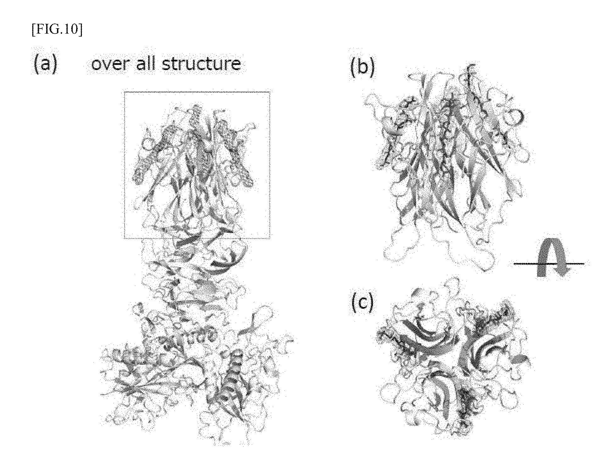
[0074] In the above description, the type IVb pili formed mainly by the colonization factor CFA/III has been described, however, type IVb pili formed by Longus expressed by ETEC, Tcp expressed by Vibrio cholerae, Cfc expressed by C. rodentium and the like as other colonization factors are known to have high similarities to pili by the CFA/III (Mundy, R. et al. Mol. Microbiol. 2003, 48, 795-809). LngA (L Ongus), TcpA (Tcp) and CfcA (Cfc) have been identified as those corresponding to the major pilin CofA, and the pilin-specific hydrophobic regions at their N-terminal side have a high sequence homology of 70 to 75%. FIG. 8 shows lng operon expressing LngA or the like, tcp operon expressing TcpA or the like, and cfc operon expressing CfcA or like, together with the cof operon expressing CofA or the like. As shown in FIG. 8, not only CofA, LngA, TcpA and CfcA but also many homologous genes are present.
[0075] Also, LngB (Longus), TcpB (Tcp) and CfcB (Cfc) corresponding to the minor pilin CofB likewise mutually have similarities. In addition to these, the N-terminal sequences of LngJ (Longus), TcpF (Tcp) and CfcJ (Cfc) corresponding to the secretory protein CofJ also have similarities. FIG. 9 shows the amino acid sequences of their N-terminal regions. In FIG. 9, the arrow shows the 10-th phenylalanine of CofJ involved strongly in the binding to CofB described above, and the underline shows a sequence portion which would not form a secondary structure. The underlined portion corresponds to the sequences of SEQ ID NOs: 1 to 4 related to the peptide of the present embodiment, and this sequence is a sequence of a portion not forming a secondary structure as described above, that is, it is a sequence favorable for the binding to minor pilin.
[0076] The peptide of the present embodiment may be modified to prevent decomposition of the peptide and the like.
[0077] The method of preparing the peptide according to the present embodiment is not particularly limited, and a method that is generally used for peptide synthesis can be used, and for example, a genetic engineering method or a chemical method using organic synthesis can be used, more specifically, methods shown in the following examples can be used.
[0078] Another embodiment of the present invention is a colonization inhibitor comprising the above-described peptide of the present invention for inhibiting adhesion and colonization of bacteria producing type IVb pili to target cells in digestive organs and the like. The colonization inhibitor according to this embodiment may contain additives such as a stabilizer, a buffer, a diluent, an excipient and the like in addition to the above-mentioned peptide. In addition, the colonization inhibitor according to this embodiment is preferably in a dosage form capable of oral administration, and is preferably, for example, a tablet, a capsule, a pill or the like, but is not limited thereto. In addition, the colonization inhibitor according to the present embodiment may be subjected to coating treatments such as enteric coating using cellulose acetate phthalate (CAP), hypromellose phthalate (HPMCP), acrylic acid type polymer and the like, sustained-release coating using ethyl cellulose and the like, but the treatment is not limited to them.
EXAMPLES
[0079] The effect of the peptide according to the present invention on the colonization ability by CFA/III pili of ETEC will be explained below as typical examples of the present invention.
(Example 1) Experiment of Adhesion of Escherichia coli CFA/III Producing Strain to Caco2 Cells
[0080] From the reports so far, not only the CFA/III producing wild type ETEC 31-10 strain (ETEC 1373 strain) cultured with CFA agar but also the strain (HB101 cof) obtained by introducing the plasmid pTT240 containing the cof operon into E. coli HB101 strain (Nippon Gene) have been reported to have type IVb pili forming ability like the wild type under the same culture conditions and adhere to Caco2 cells (Honda, T. et al. Infect. Immun 1989, 57, 3452-3457; Taniguchi, T. et al. Infect. Immun 2001, 69, 5864-5873). Here, the plasmid pTT240 containing the cof operon was prepared by the following procedure. First, as a result of base sequence analysis of the cof gene cluster, the presence of the cleavage site of the restriction enzyme KpnI upstream of the promoter sequence of the cof gene and the cleavage site of the restriction enzyme StuI downstream of the cofJ gene which is the last gene of the cof gene cluster became clear, hence the plasmid pSH1134 containing the cof gene cluster was cleaved with the restriction enzyme StuI, then, phosphorylated KpnI linker (Takara Shuzo) was ligated and cleaved with the restriction enzyme KpnI. The resultant 13.8 kb fragment was subjected to electrophoresis on a 0.8% agarose gel, purified, then, ligated to the plasmid pMW 119 (Nippon Gene) treated with the restriction enzyme KpnI. The obtained plasmid was named pTT240. In addition, Caco2 cells are considered to be model cells for colonization of ETEC to small intestinal epithelial cells (Darfeuille-Michaud, A. et al. Infect. Immun 1990, 58, 893-902). Therefore, adhesion experiments were conducted using HB 101 cof and Caco2 cells as follows.
[0081] First, Caco2 cells were dispersed using Dulbecco's Modified Eagle's Medium (DMEM) containing 50 .mu.g/mL gentamicin and 10% fetal bovine serum, the cell concentration was measured using a blood cell counting chamber and diluted to 3.0.times.10.sup.5 cells/mL. Dilution liquid was added to a 6-well plate each in an amount of 500 .mu.L, and culture was performed by allowing the plate to stand still in a CO.sub.2 incubator at 5% CO.sub.2 and 37.degree. C. for 48 hours. The cultured Caco2 cells were washed with PBS twice. After washing, DMEM containing 100 .mu.g/mL ampicillin and 1% D-mannose was filtrated by passing through a 0.22 .mu.m filter, then, the solution was added each in an amount of 500 .mu.L to each well.
[0082] Next, a small amount of the glycerol stock of HB101 cof was scraped off and added to LB medium containing 100 .mu.g/mL ampicillin, and shaking culture was carried out overnight at 25.degree. C. The culture liquid was suitably diluted and OD.sub.660 of the culture liquid was calculated using a turbidimeter. The culture liquid was serially diluted and CFU (colony forming unit) was measured. From the measured CFU, the concentration (cells/mL) of HB101 cof at OD.sub.660=1.0 was calculated. This calculated value was used to determine the concentration of HB101 cof later.
[0083] Apart from the above culture, a small amount of the glycerol stock of HB101 cof was scraped off and added to LB medium containing 100 .mu.g/mL ampicillin, and shaking culture was carried out overnight at 25.degree. C. The bacterium culture liquid was diluted 10 times with LB medium containing 100 .mu.g/mL ampicillin and transferred in an amount of 500 .mu.L to a CFA agar plate (1% Casamino Acid, 0.15% yeast extract, 0.005% MgSO.sub.4, 0.0005% MnCl.sub.2, 2% agarose) and culture was performed by allowing the plate to stand still at 37.degree. C. for 24 hours. 3 mL of PBS was added to the agar medium after culture, and the suspension was suspended with a spreading stick, then, recovered. The recovered culture liquid was diluted 100 times and OD.sub.660 was measured. OD.sub.660 of the culture liquid before dilution was calculated from the measured OD.sub.660, and the E. coli concentration (cells/mL) was calculated based on the concentration (cells/mL) of HB101 cof at OD.sub.660=1.0 described above. HB101 cof was diluted with PBS to 1.0.times.10.sup.9 cells/mL to prepare a sample for colonization assay.
[0084] The prepared sample was added in an amount of 50 .mu.L to Caco2 cells in the 6-well plate, and the plate was allowed to stand still in a CO.sub.2 incubator at 5% CO.sub.2 and 37.degree. C. for 3 hours. Three hours later, after washing three times with PBS, 500 .mu.L of PBS containing 0.1% Triton X-100 was added, and after standing still for 5 minutes, it was recovered. The recovered solution was serially diluted and CFU was measured. From measured CFU, the number of bacteria colonized on Caco2 cells was calculated. The recovery rate (%) was calculated by dividing the number of bacteria that had been colonized by the number of bacteria added to Caco2 cells (5.0.times.10.sup.7 cells) and multiplying by 100. As a result, it was 16.9% and the adhesion ability in this experimental system was confirmed.
(Example 2) Experiment of Adhesion of CofB-Deficient Strain and CofJ-Deficient Strain to Caco2 Cells
[0085] In the above Example 1, it was confirmed that HB101 cof was able to adhere to Caco2 cells, hence, CofB-deficient strain and CofJ-deficient strain were then prepared, and the functions of CofB and CofJ were examined. The method will be described below.
[0086] First, a cofJ gene-deficient strain (HB101-cof-.DELTA.cofJ) was prepared based on plasmid pTT240. Paying attention to the presence of the cleavage site of the restriction enzyme NcoI at two places in the ORF region of the cofJ gene, the strain was cleaved by incubating at 37.degree. C. for 1 hour with NcoI, followed by purification. The plasmid obtained by ligating the purified cleavage product was designated as CofJ-deficient plasmid pcof-.DELTA.cofJ. A strain obtained by transforming Escherichia coli strain HB101 with pcof-.DELTA.cofJ was designated as CofJ-deficient strain HB101-cof-.DELTA.cofJ.
[0087] In order to complement the cofJ gene to the cofJ-deficient strain in trans configuration, it was decided to incorporate the cofJ gene into plasmid vector pACYC184. Paying attention to the presence of the restriction enzyme sites of SalI upstream and HindIII downstream of the cofJ gene in the plasmid pTT240, after the restriction enzyme treatment, the end of the cleavage product was blunted and the product was purified. The plasmid obtained by ligating the purified cleavage product with EcoRV-treated plasmid vector pACYC184 (NIPPON GENE) was designated as cofJ complemented plasmid pcofJ. A strain obtained by transforming Escherichia coli strain HB101 with pcof-.DELTA.cofJ and pcofJ was designated as CofJ complemented strain HB101-cof-.DELTA.cofJ+pcofJ.
[0088] The CofB-deficient strain HB101-cof-.DELTA.cofB and the CofB complemented strain HB101-cof-.DELTA.cofB+pcofB were fabricated according to the cited literature (Kawahara, K, et al. J. Mol. Biol, 2016, 428, 1209-1226). Experiments of adhesion of the CofJ-deficient strain HB101-cof-.DELTA.cofJ, the CofJ complemented strain HB101-cof-.DELTA.cofJ+PcofJ, the CofB-deficient strain HB101-cof-.DELTA.cofB and the CofB complemented strain HB101-cof-.DELTA.cofB+pcofB were conducted in the same manner as in Example 1.
[0089] As a result of the experiment, the recovery rate was 0.30% for the CofJ-deficient strain HB101-cof-.DELTA.cofJ, 2.9% for the CofJ complemented strain HB101-cof-.DELTA.cofJ+pcofJ, 0.79% for the CofB-deficient strain HB101-cof-.DELTA.cofB and 2.9% for the CofB complemented strain HB101-cof-.DELTA.cofB+pcofB.
[0090] That is, the CofJ-deficient strain HB101-cof-.DELTA.cofJ adhered to Caco2 cells only 0.018 times and the CofB-deficient strain HB101-cof-.DELTA.cofB adhered only 0.047 times, as compared with HB101 cof. In addition, although the CofJ complemented strain HB101-cof-.DELTA.cofJ+pcofJ and the CofB complemented strain HB101-cof-.DELTA.cofB+pcofB did not have the colonization ability as much as HB101 cof, recovery of the colonization ability was observed as compared with each deficient strain. Therefore, it is recognized that CofB and CofJ are involved in adhesion to Caco2 cells by HB101 cof.
(Example 3) Analysis of Interaction Between CofJ and Pilus Subunit
[0091] Next, the interaction between a secretory protein CofJ and pilus constituent subunits CofA and CofB was analyzed.
[0092] To analyze the interaction between CofJ and pilus constituent subunits CofA and CofB, constructs (.DELTA.N28-CofA, .DELTA.N28-CofB) excluding the hydrophobic region (1 to 28 residues) at the N terminal of CofA and CofB were designed for improvement of solubility, and expression and purification thereof were conducted. The expression and purification methods were carried out according to past papers (Fukakusa, S, et al. Acta Cryst. 2012, D68, 1418-1429; Kawahara, K, et al. J. Mol. Biol, 2016, 428, 1209-1226).
[0093] Expression and purification of CofJ were performed by the following procedure. Amplification by PCR was performed from plasmid pTT240 using forward primer: 5'-GCGCCCGGGTCGCCATCCTCTTCAGAAGG-3' (SEQ ID NO: 10) and reverse primer: 5'-CAAGAATTCTTATTAATCAAGGCCACAAGCCTTC-3' (SEQ ID NO: 11). After treatment with the restriction enzymes SmaI and EcoRI, a plasmid ligated to the pET48b vector (Merck Biosciences) treated in the same manner was used as the CofJ expression plasmid. A strain obtained by transforming E. coli strain Shuffle T7 Express lysY Competent E. coli (New England Biolabs) using the CofJ expression plasmid was shaking-cultured at 37.degree. C. with LB medium containing 35 .mu.g/mL kanamycin as preculture. All of the preculture liquid was added to the main culture liquid and cultured at 37.degree. C. until the OD.sub.660 reached 0.60. Expression induction was carried out by adding 0.2 mM IPTG, and shaking culture was carried out at 20.degree. C. for 18 hours. The microbial body was recovered by centrifugation at 4000 G and 4.degree. C. for 7 min and suspended in Lysis buffer (50 mM Tris-HCl, 1 M NaCl, pH 8.0) on ice, and lysozyme in a final concentration of 0.2 mg/mL and Triton-X 100 in a final concentration of 0.2% were added and the mixture was allowed to stand still for 15 minutes. The microbial body was disrupted by ultrasound (10 seconds, 50 seconds rest, 15 cycles), and the supernatant containing the soluble protein was recovered by centrifugation at 24000 G and 4.degree. C. for 1 hour. The resulting supernatant was added to a HiTrap chelating column (GE Healthcare Biosciences) which bound Ni.sup.2+ and equilibrated with the above lysis buffer. After washing with the above-described lysis buffer containing 15 mM imidazole added, CofJ with the Histag was eluted by 15 to 500 mM imidazole gradient. 30 units of Turbo 3C protease (Accelagen) was added to the eluted fraction and the fusion tag was cleaved at 10.degree. C. while dialyzing against 50 mM Tris-HCl, 150 mM NaCl, pH 8.0, 15 mM Imidazole. The fusion tag-cleaved CofJ was allowed to pass through an equilibrated Ni column, to cause separation from the tag. The separated CofJ was subjected to final purification using a gel filtration column Superdex 75 (GE Healthcare Biosciences) equilibrated with 20 mM Tris-HCl, 150 mM NaCl, pH 8.0.
[0094] Expression of .DELTA.N24-CofJ not having 24 residues on the N-terminal side of CofJ was carried out as follows. After amplification by inverse PCR using forward primer: 5'-GGTTGCCCAACTTTGGAAAC-3' (SEQ ID NO: 12) and reverse primer: 5'-ACCCAGACCCGGGTCCCTGAAAGAG-3' (SEQ ID NO: 13) based on the vector for CofJ expression, blunting with the restriction enzyme SmaI was conducted, then, ligation was performed to obtain a plasmid designated as CofJ.DELTA.24 expression plasmid. Expression and purification of CofJ.DELTA.24 were performed in the same manner as for CofJ.
[0095] Interaction analysis was performed using CofB, CofJ and .DELTA.N24-CofJ prepared by the above methods. An isothermal titration calorimeter Microcal iTC 200 (manufactured by GE Healthcare) was used for analysis of the interaction between the secretory protein CofJ and .DELTA.N28-CofB. A 0.59 mM CofJ solution was charged in a titration syringe and a 28.5 .mu.M (in terms of trimer) .DELTA.N28-CofB solution was charged in a measuring cell, respectively, and the interaction of them was evaluated by directly observing heat changes that occur upon dropping the CofJ solution into the .DELTA.N28-CofB solution. Titration conditions were such that the initial dropping amount was 1 .mu.L, the dropping amount at the second and subsequent times was 2 .mu.L, and dropping was performed 20 times in total every 120 seconds. The measurement temperature was 25.degree. C., and the solvent was 20 mM Tris-HCl (pH 8.0), 150 mM NaCl. An exothermic change was observed by dropping the CofJ solution, which converged to the same level as the heat of dilution as the titration progressed. As a result of analysis of the obtained data using an analysis software Origin (Microcal), binding of CofJ and .DELTA.N28-CofB was observed. The dissociation constant Kd value of both was 0.2 .mu.M.
[0096] Similarly, an isothermal titration calorimeter Microcal iTC 200 (manufactured by GE Healthcare) was used for analysis of the interaction between CofJ and .DELTA.N28-CofA. A 0.59 mM CofJ solution was charged in a titration syringe and a 28.5 .mu.M (in terms of trimer) .DELTA.N28-CofA solution was charged in a measuring cell, respectively, and the interaction of them was evaluated by directly observing heat changes that occur upon dropping the CofJ solution into the .DELTA.N28-CofA solution. The titration conditions and solvent conditions were the same as in the analysis of the interaction between the secretory protein CofJ and .DELTA.N28-CofB. No change in caloric value due to dropping of the CofJ solution was observed. From this, it was shown that CofJ and .DELTA.N28-CofA do not bind.
[0097] Next, in order to investigate the contribution to binding to pili in the N-terminal region (1 to 24) of the secretory protein CofJ, analysis of the interaction between .DELTA.N24-CofJ and .DELTA.N28-CofB was carried out using an isothermal titration calorimeter Microcal iTC 200 (manufactured by GE Healthcare). A 0.59 mM .DELTA.N24-CofJ solution was charged in a titration syringe and a 28.5 .mu.M (in terms of trimer) .DELTA.N28-CofB solution was charged in a measuring cell, respectively, and the interaction of them was evaluated by directly observing heat changes that occur upon dropping the .DELTA.N24-CofJ solution into the .DELTA.N28-CofB solution. No change in caloric value due to dropping of the .DELTA.N24-CofJ solution was observed. From this, it was shown that .DELTA.N24-CofJ and .DELTA.N28-CofB do not bind.
[0098] Furthermore, in order to investigate the contribution to binding to pili in the N-terminal region (1 to 24) of the secretory protein CofJ, analysis of the interaction between a synthetic peptide consisting of 24 amino acid residues at the N terminal of CofJ (CofJ.sup.1-24 peptide) purchased from Scrum Co., Ltd. and .DELTA.N28-CofB was carried out using an isothermal titration calorimeter Microcal iTC200 (manufactured by GE Healthcare) in the same manner as described above. A 2 mM CofJ.sup.1-24 peptide solution was charged in a titration syringe and a 33.9 .mu.M (in terms of trimer) .DELTA.N28-CofB solution was charged in a measuring cell, respectively, and the interaction of them was evaluated by directly observing heat changes that occur upon dropping the CofJ.sup.1-24 peptide solution into the .DELTA.N28-CofB solution. An exothermic change was observed by dropping the CofJ.sup.1-24 peptide solution, which converged to the same level as the heat of dilution as the titration progressed. As a result of analysis of the obtained data using an analysis software Origin, binding of the CofJ.sup.1-24 peptide and .DELTA.N28-CofB was observed. The dissociation constant Kd value of both was 4 .mu.M.
[0099] Next, in order to investigate the binding specificity of the CofJ.sup.1-24 peptide, analysis of the interaction between a synthetic random peptide (CofJpepR) purchased from Scrum Co., Ltd. and .DELTA.N28-CofB was carried out using an isothermal titration calorimeter Microcal iTC 200 (manufactured by GE Healthcare). A 2 mM random peptide solution was charged in a titration syringe and a 28.5 .mu.M (in terms of trimer) .DELTA.N28-CofB solution was charged in a measuring cell, respectively, and the interaction of them was evaluated by directly observing heat changes that occur upon dropping the .DELTA.N24-CofJ solution into the .DELTA.N28-CofB solution. No change in caloric value due to dropping of the random peptide solution was observed. From this, it was shown that the random peptide and .DELTA.N28-CofB do not bind.
[0100] From the above, it is suggested that CofA and CofJ do not bind, but CofB and CofJ bind to each other, and particularly 1 to 24 amino acid residues at the N terminal of CofJ are involved.
(Example 4) Determination of Association Ratio of CofJ to .DELTA.N28-CofB by Ultracentrifugation Analysis
[0101] Next, the association ratio of CofJ to .DELTA.N28-CofB was examined. For the determination of their association ratio, an analytical ultracentrifuge Optima XL-I (manufactured by Beckman Coulter) was used. Quartz was selected for the window of the measurement cell, and 6-hole center piece made of Charcoal Epon having a cell length of 1.2 cm was used as the center piece. The measurement temperature was 20.degree. C., and the solvent was 20 mM Tris-HCl (pH 8.0), 150 mM NaCl. The rotor rotation speed was set to 5000, 8000, 10000 rpm, and the concentration gradient in the cell was monitored by an UV detector every 2 hours after reaching the set rotation speed at each rotation number. At the time when the rmsd value of the absorbance observed for each scan became 0.01 or less, the sedimentation equilibrium was established, and after equilibrium was confirmed, the concentration gradient at sedimentation equilibrium was evaluated at 297 nm. The number of integrations was set to 4 times. It was confirmed that one molecule of CofJ and one molecule of trimer .DELTA.N28-CofB were associated, from the concentration gradient obtained as described above and the apparent molecular weight calculated based on this.
(Example 5) Determination of Structure of Complex of CofJ.sup.1-24 Peptide and CofB
[0102] Next, in order to determine the structure of the complex consisting of the CofJ.sup.1-24 peptide and CofB described above, attempts were made to prepare crystals of the complex.
[0103] For this purpose, firstly, the CofJ.sup.1-24 peptide obtained by peptide synthesis was mixed with .DELTA.N28-CofB so as to be doubled in the substance amount ratio, to obtain a crystallized sample. Initial screening was carried out at 20.degree. C. by a sitting drop vapor diffusion method using a screening kit Wizard Screen I, II (Emerald Biosystems). Each drop, 1 .mu.L of the above-described mixed peptide (.DELTA.N28-CofB/CofJ.sup.1-24 peptide) prepared so that the concentration of CofB was 10, 5, 2.5 mg/mL and 1 .mu.L of the crystallization solution were mixed and allowed to stand still. As a result of screening, initial crystals were obtained under the conditions of Wizard II-39 (100 mM CAPS, pH 10.5, 20% PEG 8000, 200 mM NaCl). After optimizing the conditions, a drop of 3 to 4 .mu.L was formed by mixing equal amounts of .DELTA.N28-CofB/CofJ.sup.1-24 peptide having a concentration of 5 mg/mL and a crystallization solution using 40 .mu.L of the crystallization solution as a reservoir, and it was allowed to stand still at 20.degree. C. for 3 days, thus, acicular crystals could be obtained.
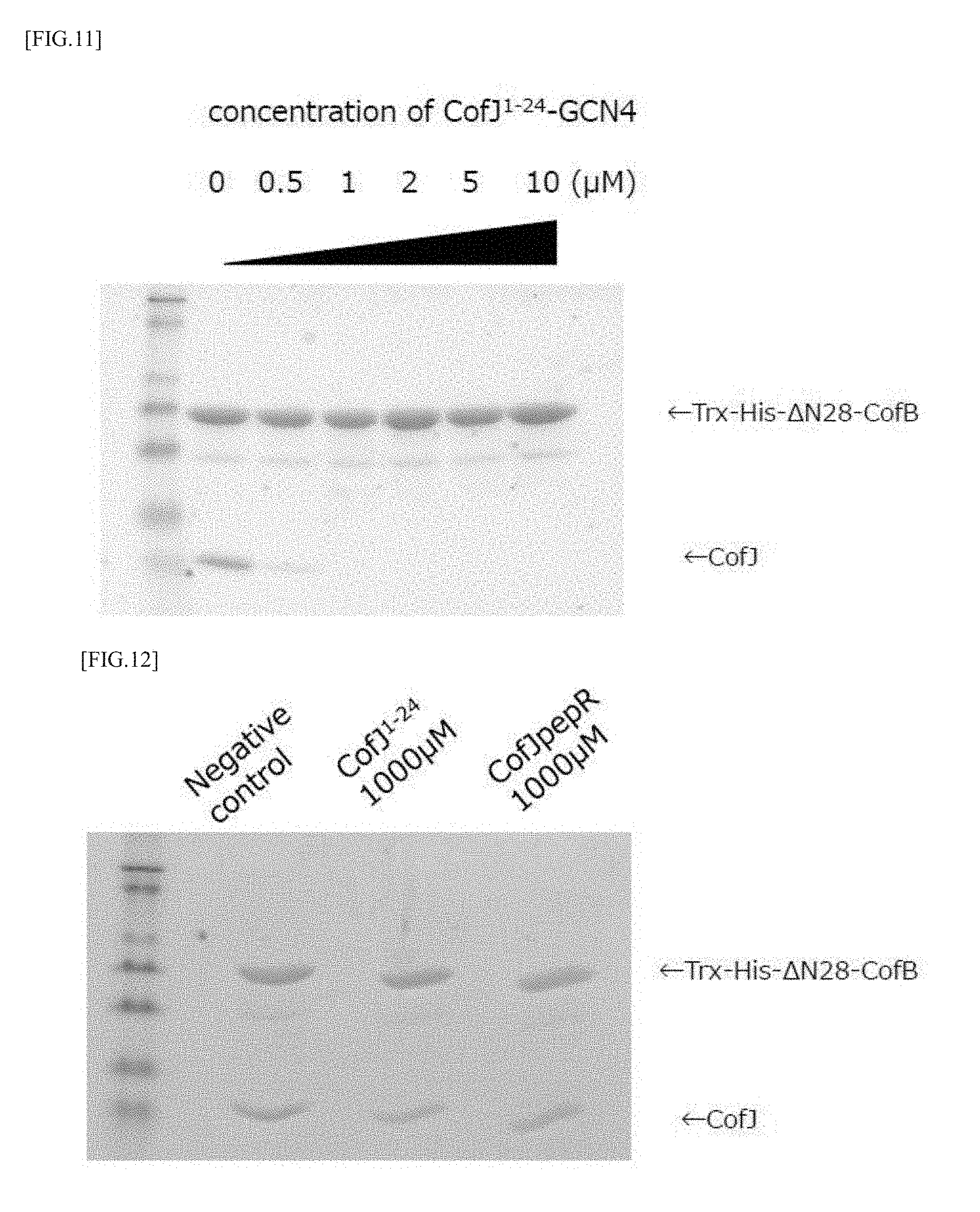
[0104] For the resultant .DELTA.N28-CofB/CofJ.sup.1-24 peptide complex crystals, diffraction measurements were carried out on the large synchrotron radiation facility SPring-8 BL26B1. After recovering the complex crystal from the drop with nylon loop, quick freezing in a nitrogen stream of -173.degree. C. was performed, and the diffraction data were recovered. Diffraction images from the complex crystals showed a diffraction ability of the highest resolution of 3.85 .ANG.. Intensity integration and scaling processing were performed using program HKL2000. The complex crystals belonged to the space group P65, and the lattice constants were a=157.76 .ANG., b=157.76 .ANG., c=118.53 .ANG., .alpha.=.beta.=90.0.degree. and .gamma.=120.0.degree.. Determination of the initial phase was performed by a molecular replacement method using a program Phaser. The coordinate data of .DELTA.N28-CofB (PDBID: 5AX6) was used as the coordinate data of the initial search model in applying the molecular replacement, and considering the existence of a linker between domain 1 and domain 2 with high mobility, those decomposed into two fragments: a domain 1 fragment and other fragment (trimer fragment composed of domains 2 to 3) were adopted as the initial search model, and the solution of the initial phase by molecular replacement was obtained. As a result, one solution containing three domain 1 monomer fragments and one trimer fragment composed of domains 2 and 3 adopted as the initial search model was found. Fragments resulting from the molecular replacement method were connected by a molecular modeling support program Coot, then, refinement of the initial structure by a structure refinement program PHENIX. refine was performed, as a result, the electron density of the CofJ.sup.1-24 peptide was observed at three places in the molecular boundary of the type H lectin domain of a .DELTA.N28-CofB trimer, as shown in FIG. 10.
[0105] From these results, it was suggested that three CofJ peptides can bind in the type H lectin domain of a CofB trimer.
(Example 6) Design of GCN4 Fusion Type Inhibitory Peptide
[0106] Next, in order to prepare a trimeric peptide capable of binding to the type H lectin domain of each of the above-mentioned CofB trimers, GCN4 having a trimerizing domain was used, and attempts were made to create a peptide in which GCN and a CofJ peptide are bound via a linker.
[0107] Therefore, firstly, the expression plasmid of an inhibitory peptide (called CofJ.sup.1-24-GCN4) in which the CofJ.sup.1-24 peptide and GCN4 were fused using a linker (TSGGG) was constructed as follows. Oligonucleic acids were connected by a PCR reaction, to synthesize the CofJ.sup.1-24-GCN4 gene. CJN-T-1, 6 (Table 1) were prepared to have a final concentration of 0.6 .mu.M and CJN-T-2, 3, 4, 5 (Table 1) were prepared to have a final concentration of 0.03 .mu.M, and 20 cycles of PCR were performed. The resultant amplified product was cleaved with restriction enzymes NdeI/XhoI, purified, and ligated to a pET30a vector (MERCK Biosciences) treated in the same manner, to obtain a plasmid. However, since CofJ.sup.1-24-GCN4 was not expressed in this plasmid, it was decided to be expressed by a vector to which a GST tag was added at the N terminal. PCR was performed for 35 cycles using CJN-T-7,8 as a primer and the above-described plasmid as a template. The resultant amplified product was cleaved with the restriction enzymes BamHI/XhoI, purified, and ligated to a pGEX6P-1 vector (GE Healthcare Biosciences) treated in the same manner, to obtain a CofJ.sup.1-24-GCN4 expression plasmid.
TABLE-US-00001 TABLE 1 Primer Primer base sequence CJN-T-1 5V-GTTCCACATATGAGCCCGTCTTCCAGCGAAGGTGGTGC (SEQ ID TTTCACCGTTAAC-3' NO: 14) CJN-T-2 5'-GACCTCTACTGTTGACGATATCCGTACTAGTGGTGGCG (SEQ ID GTCGTATGAAACAGATC-3' NO: 15) CJN-T-3 5'-GATCGAGGAAATCCTGTCCAAGATCTACCACATCGAAA (SEQ ID ACGAGATCGCGC-3' NO: 16) CJN-T-4 5'-GGATATCGTCAACAGTAGAGGTCTTCGGCATGTTAACG (SEQ ID GTGAAAGCACCACC-3' NO: 17) CJN-T-5 5'-CTTGGACAGGATTTCCTCGATCTTGTCTTCGATCTGTT (SEQ ID TCATACGACCGCC-3' NO: 18) CJN-T-6 5'-CTAGCTCTCGAGTTAGATAAGCTTCTTGATACGCGCGA (SEQ ID TCTCGTTTTCGATG-3' NO: 19) CJN-T-7 5'-GTTCCAGGATCCCCGTCTTCCAGCGAAGGTG-3' (SEQ ID NO: 20) CJN-T-8 5'-GGTGCTCGAGTTAGATAAGCTTCTTG-3' (SEQ ID NO: 21)
[0108] Next, an Escherichia coli strain BL21 (DE3) (NIPPON GENE) was transformed using a plasmid for expressing CofJ.sup.1-24-GCN4. Colonies were picked, added to LB medium containing 100 .mu.g/mL ampicillin, and shaking-cultured overnight at 37.degree. C. The next morning, the whole culture liquid was added to the main culture medium and scaled up and cultured with shaking at 37.degree. C. When reaching OD.sup.660=0.6, IPTG was added to a final concentration of 1 mM and shaking was carried out at 25.degree. C. for 18 hours, to induce expression. The expression-induced microbial body was recovered by centrifugation at 4000 G and 4.degree. C. for 7 minutes. The resultant microbial body was dissolved with a lysis buffer (20 mM Tris-HCl, 1 M NaCl, pH 8.0) on ice, then, lysozyme was added at 20 .mu.g/mL and Triton X-100 was added at 0.2% and the sample was left for 15 minutes, then, the cycle of ultrasonic disruption for 15 seconds and break for 1 minute was repeated 15 times. The ultrasonic-disrupted solution was centrifuged at 20000 G and 4.degree. C. for 1 hour, and the supernatant of centrifugation was filtered through a 0.8 .mu.m filter. The filtered sample was applied to a GST column (Glutathione Sepharose 4 Fast Flow: GE Healthcare Biosciences) equilibrated with a lysis buffer. The GST column was washed with a lysis buffer, then, eluted with an elution buffer (20 mM Tris-HCl, 20 mM GSH, pH 8.0). The eluted sample was dialyzed with a dialysis buffer (20 mM Tris-HCl, pH8.0) while enzymatically treating with 30 units of Turbo 3C Protease (Accelagen), to cleave the GST tag and CofJ.sup.1-24-GCN4. The sample after cleaving was applied to the GST column, to separate the GST tag and CofJ.sup.1-24-GCN4. The recovered sample was applied to a Hitrap DEAE column (GE Healthcare Biosciences) equilibrated with a dialysis buffer, and elution was carried out by applying a gradient of NaCl concentration from 0 to 2 M. The resultant sample was applied to a Superdex 75 (26/60) column (GE Healthcare Biosciences) equilibrated with a gel filtration buffer (20 mM Tris-HCl, 150 mM NaCl, pH 8.0), to obtain a final purified product.
[0109] The resultant final purified product is constituted of 4 residues of the vector-derived sequence (GPLG), 24 residues derived from the CofJ N terminal region (CofJ.sup.1-24 sequence) (SPSSSEGGAFTVNM PKTSTVDDIR: SEQ ID NO: 1), 5 residues of a linker sequence (TSGGG: SEQ ID NO: 9) and 30 residues of a GCN4 sequence (RMKQIEDKIEEILSKIYHIENEIARIKKLI) (SEQ ID NO: 6), from the N-terminal side. This is called CofJ.sup.1-24-GCN4 inhibitory peptide.
(Example 7) Analysis of Interaction Between GCN4 Fusion Type Inhibitory Peptide and .DELTA.N28-CofB by ITC
[0110] In order to investigate the contribution to binding to pili of the CofJ.sup.1-24-GCN4 inhibitory peptide obtained in Example 6, analysis of the interaction between the CofJ.sup.1-24-GCN4 inhibitory peptide and .DELTA.N28-CofB was carried out using an isothermal titration calorimeter Microcal iTC200 (manufactured by GE Healthcare). A 0.59 mM CofJ.sup.1-24-GCN4 inhibitory peptide solution was charged in a titration syringe and a 28.5 .mu.M (in terms of trimer) .DELTA.N28-CofB solution was charged in a measuring cell, respectively, and the interaction of them was evaluated by directly observing heat changes that occur upon dropping the CofJ.sup.1-24-GCN4 inhibitory peptide solution into the .DELTA.N28-CofB solution. An exothermic change was observed by dropping the CofJ.sup.1-24-GCN4 inhibitory peptide solution, which converged to the same level as the heat of dilution as the titration progressed. As a result of analysis of the obtained data using an analysis software Origin, binding of the CofJ.sup.1-24-GCN4 inhibitory peptide and .DELTA.N28-CofB was observed. The dissociation constant Kd value of both was 0.04 .mu.M.
[0111] As a result of analysis, it was shown that the binding affinity between the CofJ.sup.1-24-GCN4 inhibitory peptide and .DELTA.N28-CofB was stronger about 5 times as compared with the binding affinity between CofJ and .DELTA.N28-CofB and it was clarified that the GCN4 fusion type inhibitory peptide efficiently inhibits the interaction between CofJ and .DELTA.N28-CofB.
(Example 8) Inhibition of CofB/CofJ Interaction by Inhibitory Peptide by Pull Down Assay
[0112] To examine whether the CofJ.sup.1-24-GCN4 inhibitory peptide is capable of inhibiting the binding of CofB and CofJ, it was confirmed in the in vitro experiment system by pull down assay using a Ni column. For experiments, .DELTA.N28-CofB (Trx-His-.DELTA.N28-CofB), CofJ, CofJ.sup.1-24-GCN4, a CofJ.sup.1-24 peptide and a peptide (CofJpepR) in which the amino acid sequence of the CofJ.sup.1-24 peptide was randomized, with added thioredoxin tag (Trx) and His tag, were used. The sequence of CofJpepR is NPSGFDKSGSSTTRTPAMSVIVDE (SEQ ID NO: 22).
[0113] Solutions mixed so that the concentration of Trx-His-.DELTA.N28-CofB was 1 .mu.M and the concentration of CofJ.sup.1-24-GCN4 was (0, 0.5, 1.0, 2.0, 5.0, 10.0 .mu.M) were allowed to stand still on ice overnight. Further, CofJ was mixed so that the final concentration became 1 .mu.M, and the mixtures were allowed to stand still on ice for 1 hour, to obtain experiment samples. Each sample was applied to a Ni column equilibrated with 20 mM Tris-HCl, 150 mM NaCl, pH 8.0, and the column was washed with 20 mM Tris-HCl, 150 mM NaCl, 15 mM Imidazole, pH 8.0. Thereafter, elution was performed with 20 mM Tris-HCl, 150 mM NaCl, 300 mM Imidazole, pH 8.0, and the eluted fraction was confirmed by SDS-PAGE. The results are shown in FIG. 11. It was clarified that the binding of CofJ and Trx-His-.DELTA.N28-CofB can be strongly inhibited by preliminarily mixing CofJ.sup.1-24-GCN4 having the concentration not lower than that of CofJ with Trx-His-.DELTA.N28-CofB, as shown in FIG. 11.
[0114] Also for the CofJ.sup.1-24 peptide and CofJpepR, the inhibition ability thereof was investigated by the same method. Specifically, solutions mixed so that the concentration of Trx-His-.DELTA.N28-CofB was 1 .mu.M and the concentration of the CofJ.sup.1-24 peptide and CofJpepR was 1000 .mu.M were allowed to stand still on ice overnight. Further, CofJ was mixed so that the final concentration became 1 .mu.M, and the mixtures were allowed to stand still on ice for 1 hour, to obtain experiment samples. The pull down assay was performed in the same manner as described above. The results are shown in FIG. 12. It was clarified that even if the CofJ.sup.1-24 peptide is previously added with the concentration 1000 times of that of CofJ, the binding of CofJ and Trx-His-.DELTA.N28-CofB can be hardly inhibited, as shown in FIG. 12. In addition, it was clarified that the binding can not be inhibited at all with CofJpepR. From this, it is considered that CofJ.sup.1-24-GCN4 can inhibit the binding between CofB and CofJ by strongly binding with CofB by trimerizing.
(Example 9) Evaluation of ETEC Adhesion Inhibitory Activity of Inhibitory Peptide Using Caco2 Cells
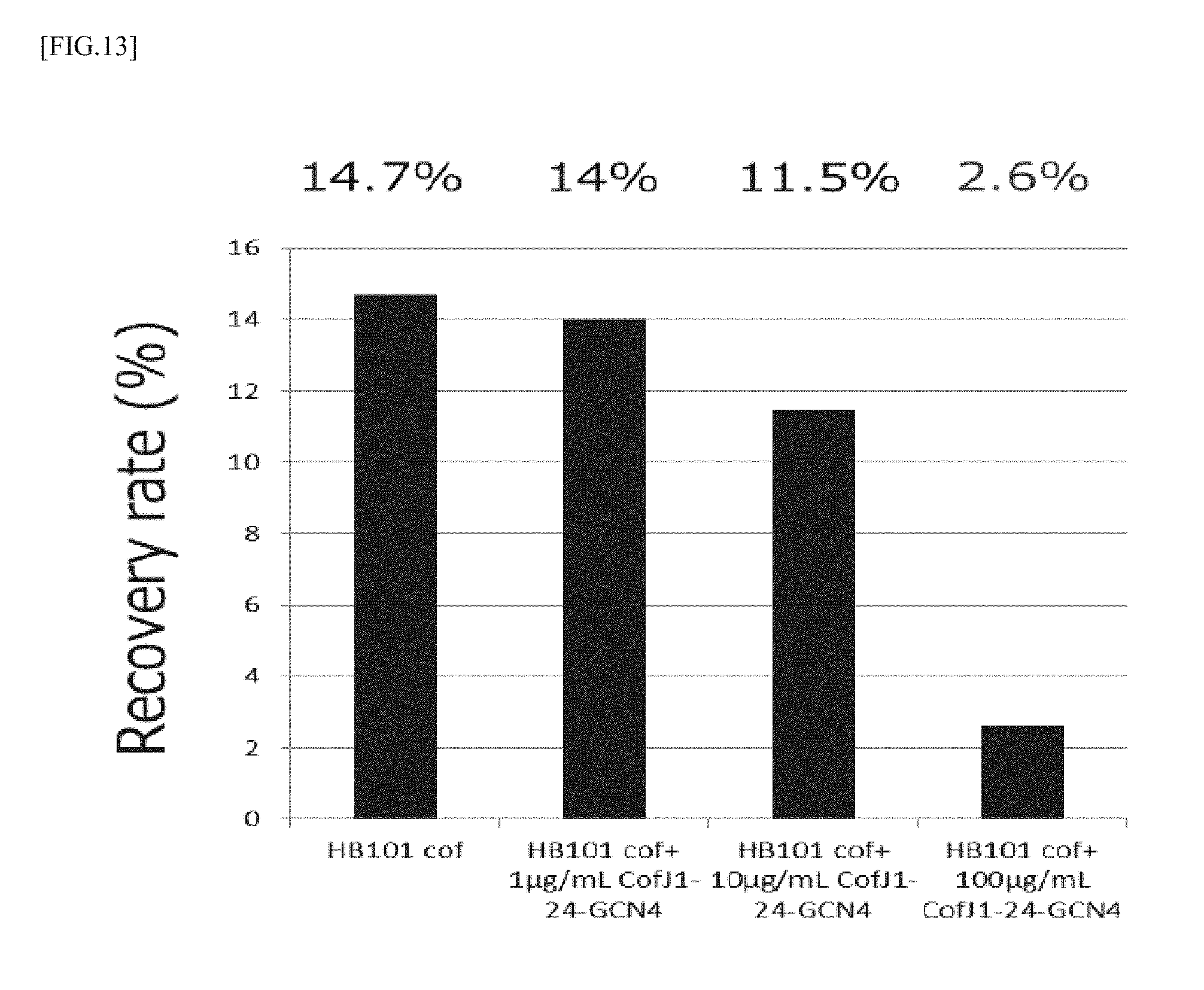
[0115] The colonization experiment was conducted in the same manner as in Example 1 and Example 2. In the experiment, mixtures were prepared so that the concentration of CofJ.sup.1-24-GCN4 was 0, 10, 100, 1000 .mu.g/mL and the concentration of HB101 cof was 1.0.times.10.sup.9 cells/mL, then, the mixtures were allowed to stand still at 25.degree. C. for 1 hour, and each 50 .mu.L of the mixtures were added to 500 .mu.L medium so that the concentration of CofJ.sup.1-24-GCN4 was 0, 1, 10, 100 .mu.g/mL and the concentration of HB101 cof was 1.0.times.10.sup.8 cells/mL. The results are shown in FIG. 13. As shown in FIG. 13, the recovery rate was 14.7% for 0 .mu.g/mL CofJ.sup.1-24-GCN4, 14.0% for 1 .mu.g/mL CofJ.sup.1-24-GCN4, 11.5% for 10 .mu.g/mL CofJ.sup.1-24-GCN4 and 2.6% for 100 .mu.g/mL CofJ.sup.1-24-GCN4.
[0116] It was clarified from the results that CofJ.sup.1-24-GCN4 inhibits colonization of HB101 cof to Caco2 cells in concentration-dependent manner.
(Example 10) Analysis of Interaction Between CofJ.sup.4-16 Peptide and .DELTA.N28-CofB by ITC
[0117] It was hitherto shown that the CofJ.sup.1-24 peptide inhibits the CofB/CofJ interactions, thereby inhibiting adhesion of ETEC to intestinal tissue. It is supposed from the crystal structure of the CofJ.sup.1-24 peptide/.DELTA.N28-CofB complex that a part of the CofJ.sup.1-24 peptide, specifically, the fifth serine to the fifteenth proline are particularly involved in the interaction of both as shown in FIG. 7, as described above. Then, it was investigated whether a CofJ.sup.4-16 peptide (SEQ ID NO: 23) composed of 4-th to 16-th amino acid sequences of the CofJ.sup.1-24 peptide, containing a portion that is considered especially important in the CofJ.sup.1-24 peptide, can exert the equivalent effect to the CofJ.sup.1-24 peptide.
[0118] First, in order to investigate the contribution to binding to pili of the CofJ.sup.4-16 peptide, analysis of the interaction between a CofJ.sup.4-16 synthetic peptide (purchased from Scrum Co., Ltd.) and .DELTA.N28-CofB was carried out using an isothermal titration calorimeter Microcal iTC 200 (manufactured by GE Healthcare) in the same manner as the experiment conducted in Example 3. Specifically, a 2 mM CofJ.sup.4-16 peptide solution was charged in a titration syringe and a 33.9 .mu.M (in terms of trimer) .DELTA.N28-CofB solution was charged in a measuring cell, respectively, and the interaction of them was evaluated by directly observing heat changes that occur upon dropping the CofJ.sup.4-16 peptide solution into the .DELTA.N28-CofB solution. An exothermic change was observed by dropping the CofJ.sup.4-16 peptide solution, which converged to the same level as the heat of dilution as the titration progressed. As a result of analysis of the obtained data using an analysis software Origin, binding of the CofJ.sup.4-16 peptide and .DELTA.N28-CofB was observed. The dissociation constant Kd value of both was 4 .mu.M.
[0119] From the above, it was suggested that the CofJ.sup.4-16 peptide can bind to CofB, like the CofJ.sup.1-24 peptide.
(Example 11) Design of CofJ.sup.4-16-GCN4 Peptide
[0120] Then, to make a trimer peptide also for CofJ.sup.4-16, attempts were made to prepare a CofJ.sup.4-16-GCN4 peptide using GCN4 having a trimerizing domain, and a linker, in the same manner as in Example 6.
[0121] The expression plasmid of an inhibitory peptide (referred to as CofJ.sup.4-16-GCN4) in which a CofJ.sup.4-16 peptide was fused to GCN4 using a linker (TSGGG) was constructed as follows. The sites of the restriction enzymes BamHI and Spel which had been previously incorporated at both ends of the CofJ.sup.1-24 peptide sequence in the CofJ.sup.4-16-GCN4 expression plasmid were cleaved using both restriction enzymes, then, purified. By mixing and annealing the oligonucleotide 1: 5'-GATCCAGCGAAGGTGGTGCTTTCACCGTTAACATGCCGAAGA-3' (SEQ ID NO: 24) and the oligonucleotide 2: 5'-CTAGTCTTCGGCATGTTAACGGTGAAAGCACCACCTTCGCTG-3' (SEQ ID NO: 25) containing the sequence of Coif', double stranded DNA was prepared, which was then ligated to a purified vector, to obtain a CofJ.sup.4-16-GCN4 expression plasmid.
[0122] Next, an Escherichia coli strain BL21 (DE3) (NIPPON GENE) was transformed with a plasmid for expressing CofJ.sup.4-16-GCN4. Colonies were picked, added to LB medium containing 100 .mu.g/mL ampicillin, and shaking-cultured overnight at 37.degree. C. The next morning, the whole culture liquid was added to the main culture medium and scaled up and cultured with shaking at 37.degree. C. When reached OD.sub.660=0.6, IPTG was added to a final concentration of 1 mM and shaking was carried out at 25.degree. C. for 18 hours, to induce expression. The expression-induced microbial body was recovered by centrifugation at 4000 G and 4.degree. C. for 7 minutes. The resultant microbial body was dissolved in a lysis buffer (20 mM Tris-HCl, 1 M NaCl, pH 8.0) on ice, then, lysozyme was added at 20 .mu.g/mL and Triton X-100 was added at 0.2% and the sample was left for 15 minutes, then, the cycle of ultrasonic disruption for 15 seconds and break for 1 minute was repeated 15 times. The ultrasonic-disrupted solution was centrifuged at 20000 G and 4.degree. C. for 1 hour, and the supernatant of centrifugation was filtered through a 0.8 .mu.m filter. The filtered sample was applied to a GST column (Glutathione Sepharose 4 Fast Flow: GE Healthcare Biosciences) equilibrated with a lysis buffer. The GST column was washed with a lysis buffer, then, eluted with an elution buffer (20 mM Tris-HCl, 20 mM GSH, pH8.0). The eluted sample was dialyzed with a dialysis buffer (20 mM Tris-HCl, pH8.0) while enzymatically treating with 30 units of Turbo 3C Protease (Accelagen), to cleave the GST tag and CofJ.sup.4-16-GCN4. The sample after cleaving was applied to the GST column, to separate the GST tag and CofJ.sup.4-16-GCN4. The recovered sample was applied to a Hitrap DEAE column (GE Healthcare Biosciences) equilibrated with a dialysis buffer, and the fraction not bound to the column was recovered. The resultant sample was applied to a Superdex 75 (26/60) column (GE Healthcare Biosciences) equilibrated with a gel filtration buffer (20 mM Tris-HCl, 150 mM NaCl, pH 8.0), to obtain a final purified product.
[0123] The resultant final purified product is constituted of 4 residues of the vector-derived sequence (GPLG), 13 residues derived from the CofJ N terminal region (SSEGGAFTVNMPK: SEQ ID NO: 23), 5 residues of a linker sequence (TSGGG: SEQ ID NO: 9) and 30 residues of a GCN4 sequence (RMKQIEDKIEEILSKIYHIENEIARIKKLI: SEQ ID NO: 6), from the N-terminal side. This is called CofJ.sup.4-16-GCN4 inhibitory peptide.
(Example 12) Analysis of Interaction Between CofJ.sup.4-16-GCN4 Inhibitory Peptide and .DELTA.N28-CofB by ITC
[0124] In order to investigate the contribution to binding to pili of the CofJ.sup.4-16-GCN4 inhibitory peptide obtained in Example 11, analysis of the interaction between the CofJ.sup.4-16-GCN4 inhibitory peptide and .DELTA.N28-CofB was carried out using an isothermal titration calorimeter Microcal iTC200 (manufactured by GE Healthcare). A 0.59 mM CofJ.sup.4-16-GCN4 inhibitory peptide solution was charged in a titration syringe and a 28.5 .mu.M (in terms of trimer) .DELTA.N28-CofB solution was charged in a measuring cell, respectively, and the interaction of them was evaluated by directly observing heat changes that occur upon dropping the CofJ.sup.4-16-GCN4 inhibitory peptide solution into the .DELTA.N28-CofB solution. An exothermic change was observed by dropping the CofJ.sup.4-16-GCN4 inhibitory peptide solution, which converged to the same level as the heat of dilution as the titration progressed. As a result of analysis of the obtained data using an analysis software Origin, binding of the CofJ.sup.4-16-GCN4 inhibitory peptide and .DELTA.N28-CofB was observed. The dissociation constant Kd value of both was 0.08 .mu.M.
[0125] As a result of analysis, it was shown that the binding affinity between the CofJ.sup.4-16-GCN4 inhibitory peptide and .DELTA.N28-CofB was stronger about 2.5 times as compared with the binding affinity between CofJ and .DELTA.N28-CofB and it was clarified that the GCN4 fusion type inhibitory peptide efficiently inhibits the interaction between CofJ and .DELTA.N28-CofB.
(Example 13) Inhibition of CofB/CofJ Interaction by CofJ.sup.4-16-GCN4 Inhibitory Peptide by Pull Down Assay
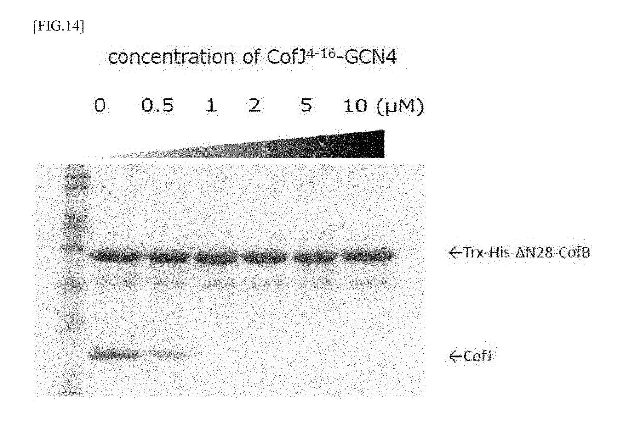
[0126] In order to investigate whether the CofJ.sup.4-16-GCN4 inhibitory peptide can inhibit the binding of CofB and CofJ, it was confirmed in the in vitro experimental system by a pull down assay using a Ni column. For the experiment, .DELTA.N28-CofB (Trx-His-.DELTA.N28-CofB), CofJ and CofJ.sup.4-16-GCN4, with added thioredoxin tag (Trx) and His tag (His), were used.
[0127] Solutions mixed so that the concentration of Trx-His-.DELTA.N28-CofB was 1 .mu.M and the concentration of CofJ.sup.4-16-GCN4 was (0, 0.5, 1.0, 2.0, 5.0, 10.0 .mu.M) were allowed to stand still on ice overnight. Further, CofJ was mixed so that the final concentration became 1 .mu.M, and the mixtures were allowed to stand still on ice for 1 hour, to obtain experiment samples. Each sample was applied to a Ni column equilibrated with 20 mM Tris-HCl, 150 mM NaCl, pH 8.0, and the column was washed with 20 mM Tris-HCl, 150 mM NaCl, 15 mM Imidazole, pH 8.0. Thereafter, elution was performed with 20 mM Tris-HCl, 150 mM NaCl, 300 mM Imidazole, pH 8.0, and the eluted fraction was confirmed by SDS-PAGE. As a result, it was clarified that the binding of CofJ and Trx-His-.DELTA.N28-CofB can be strongly inhibited by preliminarily mixing CofJ.sup.4-16-GCN4 having the concentration not lower than that of CofJ with Trx-His-.DELTA.N28-CofB, as shown in FIG. 14.
(Example 14) Evaluation of ETEC Adhesion Inhibitory Activity of CofJ.sup.4-16-GCN4 Using Caco2 Cells
[0128] ETEC adhesion inhibitory activity was evaluated in the same manner as in Example 9 using CofJ.sup.4-16-GCN4.
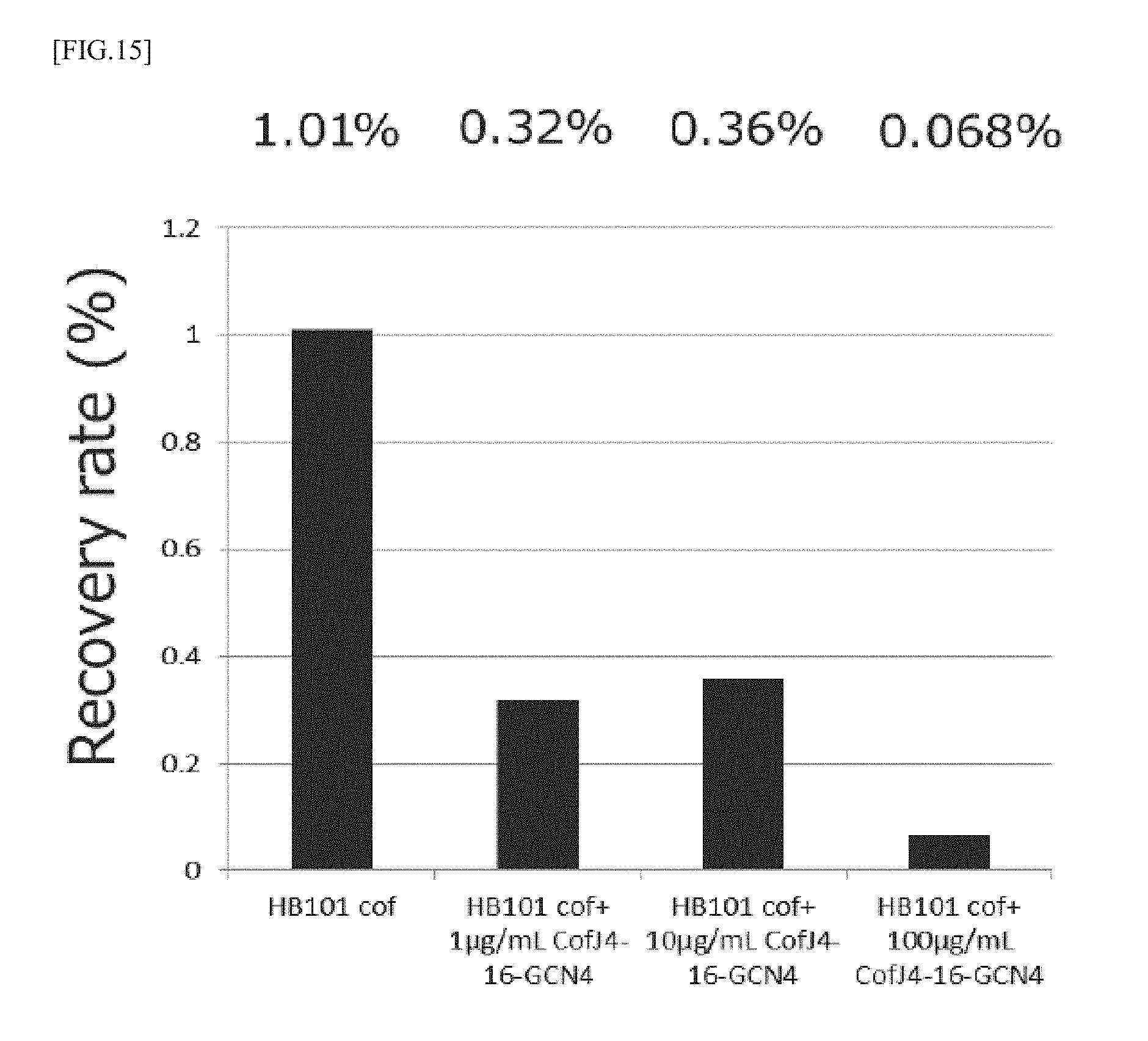
[0129] Mixtures were prepared so that the concentration of CofJ.sup.4-16-GCN4 was 0, 10, 100, 1000 .mu.g/mL and the concentration of HB101 cof was 1.0.times.10.sup.9 cells/mL, then, the mixtures were allowed to stand still at 25.degree. C. for 1 hour, and each 50 .mu.L of the mixtures were added to 500 .mu.L medium so that the concentration of CofJ.sup.4-16-GCN4 was 0, 1, 10, 100 .mu.g/mL and the concentration of HB101 cof was 1.0.times.10.sup.8 cells/mL. As a result, as shown in FIG. 15, the recovery rate was 1.01% for 0 .mu.g/mL CofJ.sup.4-16-GCN4, 0.32% for 1 .mu.g/mL CofJ.sup.4-16-GCN4, 0.36% for 10 .mu.g/mL CofJ.sup.4-16-GCN4 and 0.068% for 100 .mu.g/mL CofJ.sup.4-16-GCN4.
[0130] It was clarified from the results that CofJ.sup.4-16-GCN4 inhibits colonization of HB101 cof to Caco2 cells in concentration-dependent manner.
Sequence CWU 1
1
25124PRTArtificial SequenceSynthetic 1Ser Pro Ser Ser Ser Glu Gly
Gly Ala Phe Thr Val Asn Met Pro Lys1 5 10 15Thr Ser Thr Val Asp Asp
Ile Arg 20225PRTArtificial SequenceSynthetic 2Ser Thr Thr Ser Ser
Glu Gly Gly Ala Phe Thr Val Lys Met Ala Lys1 5 10 15Ser Ser Thr Val
Asp Asp Ile Lys Gly 20 25316PRTArtificial SequenceSynthetic 3Ser
Ala Lys Ser Ser Gln Asn Tyr Gly Phe Ser Ala Gly Val Lys Pro1 5 10
15433PRTArtificial SequenceSynthetic 4Phe Asn Asp Asn Tyr Ser Ser
Thr Ser Thr Val Tyr Ala Thr Ser Asn1 5 10 15Glu Ala Thr Asp Ser Arg
Gly Ser Glu His Leu Arg Tyr Pro Tyr Leu 20 25 30Glu511PRTArtificial
SequenceSynthetic 5Ser Glu Gly Gly Ala Phe Thr Val Asn Met Pro1 5
10630PRTArtificial SequenceSynthetic 6Arg Met Lys Gln Ile Glu Asp
Lys Ile Glu Glu Ile Leu Ser Lys Ile1 5 10 15Tyr His Ile Glu Asn Glu
Ile Ala Arg Ile Lys Lys Leu Ile 20 25 3073PRTArtificial
SequenceSynthetic 7Pro Pro Gly183PRTArtificial
SequenceSyntheticMISC_FEATURE(2)..(2)Xaa is 4Hyp 8Pro Xaa
Gly195PRTArtificial SequenceSynthetic 9Thr Ser Gly Gly Gly1
51029DNAArtificial SequenceSynthetic 10gcgcccgggt cgccatcctc
ttcagaagg 291134DNAArtificial SequenceSynthetic 11caagaattct
tattaatcaa ggccacaagc cttc 341220DNAArtificial SequenceSynthetic
12ggttgcccaa ctttggaaac 201325DNAArtificial SequenceSynthetic
13acccagaccc gggtccctga aagag 251451DNAArtificial SequenceSynthetic
14gttccacata tgagcccgtc ttccagcgaa ggtggtgctt tcaccgttaa c
511555DNAArtificial SequenceSynthetic 15gacctctact gttgacgata
tccgtactag tggtggcggt cgtatgaaac agatc 551650DNAArtificial
SequenceSynthetic 16gatcgaggaa atcctgtcca agatctacca catcgaaaac
gagatcgcgc 501752DNAArtificial SequenceSynthetic 17ggatatcgtc
aacagtagag gtcttcggca tgttaacggt gaaagcacca cc 521851DNAArtificial
SequenceSynthetic 18cttggacagg atttcctcga tcttgtcttc gatctgtttc
atacgaccgc c 511952DNAArtificial SequenceSynthetic 19ctagctctcg
agttagataa gcttcttgat acgcgcgatc tcgttttcga tg 522031DNAArtificial
SequenceSynthetic 20gttccaggat ccccgtcttc cagcgaaggt g
312126DNAArtificial SequenceSynthetic 21ggtgctcgag ttagataagc
ttcttg 262224PRTArtificial SequenceSynthetic 22Asn Pro Ser Gly Phe
Asp Lys Ser Gly Ser Ser Thr Thr Arg Thr Pro1 5 10 15Ala Met Ser Val
Ile Val Asp Glu 202313PRTArtificial SequenceSynthetic 23Ser Ser Glu
Gly Gly Ala Phe Thr Val Asn Met Pro Lys1 5 102442DNAArtificial
SequenceSynthetic 24gatccagcga aggtggtgct ttcaccgtta acatgccgaa ga
422542DNAArtificial SequenceSynthetic 25ctagtcttcg gcatgttaac
ggtgaaagca ccaccttcgc tg 42
D00000
D00001
D00002
D00003
D00004
D00005
D00006
D00007
D00008
D00009
D00010
S00001
XML
uspto.report is an independent third-party trademark research tool that is not affiliated, endorsed, or sponsored by the United States Patent and Trademark Office (USPTO) or any other governmental organization. The information provided by uspto.report is based on publicly available data at the time of writing and is intended for informational purposes only.
While we strive to provide accurate and up-to-date information, we do not guarantee the accuracy, completeness, reliability, or suitability of the information displayed on this site. The use of this site is at your own risk. Any reliance you place on such information is therefore strictly at your own risk.
All official trademark data, including owner information, should be verified by visiting the official USPTO website at www.uspto.gov. This site is not intended to replace professional legal advice and should not be used as a substitute for consulting with a legal professional who is knowledgeable about trademark law.