B-cell-mimetic Cells
FUSSENEGGER; Martin ; et al.
U.S. patent application number 16/318348 was filed with the patent office on 2019-09-19 for b-cell-mimetic cells. The applicant listed for this patent is ETH ZURICH. Invention is credited to Martin FUSSENEGGER, Mingqi XIE.
| Application Number | 20190282710 16/318348 |
| Document ID | / |
| Family ID | 59564137 |
| Filed Date | 2019-09-19 |


View All Diagrams
| United States Patent Application | 20190282710 |
| Kind Code | A1 |
| FUSSENEGGER; Martin ; et al. | September 19, 2019 |
B-CELL-MIMETIC CELLS
Abstract
The present invention relates to .beta.-cell-mimetic cells. Methods for producing .beta.-cell-mimetic cells as well as methods of use of .beta.-cell-mimetic cells as a medicament and methods of use of .beta.-cell-mimetic cells for the prevention, delay of progression or treatment of a metabolic disease in a subject are also provided.
| Inventors: | FUSSENEGGER; Martin; (Magenwil, CH) ; XIE; Mingqi; (Basel, CH) | ||||||||||
| Applicant: |
|
||||||||||
|---|---|---|---|---|---|---|---|---|---|---|---|
| Family ID: | 59564137 | ||||||||||
| Appl. No.: | 16/318348 | ||||||||||
| Filed: | July 17, 2017 | ||||||||||
| PCT Filed: | July 17, 2017 | ||||||||||
| PCT NO: | PCT/EP2017/067981 | ||||||||||
| 371 Date: | January 16, 2019 |
| Current U.S. Class: | 1/1 |
| Current CPC Class: | A61K 48/0091 20130101; A61P 35/00 20180101; C12Q 1/6897 20130101; A61P 3/04 20180101; A61K 48/0066 20130101; A61P 9/00 20180101; A61P 3/10 20180101; A61P 5/50 20180101; A61K 48/0058 20130101; A61P 3/00 20180101 |
| International Class: | A61K 48/00 20060101 A61K048/00 |
Foreign Application Data
| Date | Code | Application Number |
|---|---|---|
| Jul 18, 2016 | EP | 16180000.8 |
| Nov 23, 2016 | EP | 16200258.8 |
Claims
1. A recombinant cell comprising a nucleic acid construct comprising a promoter which is responsive to carbohydrate metabolism of said recombinant cell, wherein the promoter is operably linked to a gene coding for a therapeutic protein.
2. The recombinant cell of claim 1, wherein the recombinant cell further comprises a nucleic acid construct coding for a cellular component for sensing extracellular carbohydrates.
3. The recombinant cell of claim 2, wherein the cellular component for sensing extracellular carbohydrates is a membrane protein or a fragment thereof or a subunit of a membrane protein or a fragment thereof.
4. The recombinant cell of claim 2, wherein the cellular component for sensing extracellular carbohydrates is a membrane protein or a fragment thereof or a subunit of a membrane protein or a fragment thereof, wherein the membrane protein is selected from the group consisting of G-protein coupled receptors, SLC2A family glucose transporters, SLC5A family sodium-glucose linked transporters, potassium channels, calcium channels and sodium channels.
5. The recombinant cell of claim 2, wherein the cellular component for sensing extracellular carbohydrates is a voltage-gated calcium channel.
6. The recombinant cell of anyone of claims 1-5, wherein the promoter which is responsive to carbohydrate metabolism is responsive to a physiological effect of membrane depolarization caused by the carbohydrate metabolism of said recombinant cell.
7. The recombinant cell of anyone of claims 1-5, wherein the promoter which is responsive to carbohydrate metabolism is a calcium-responsive promoter.
8. The recombinant cell of anyone of claims 1-5, wherein the promoter which is responsive to carbohydrate metabolism is a calcium-responsive promoter comprising nucleic acid sequences bound by transcription factors of the NFAT family, the NFkB family, the AP-1 family, and/or the CREB family and/or cFOS.
9. The recombinant cell of anyone of claims 1-8, wherein the therapeutic protein is an insulinogenic agent selected from the group consisting of GLP1R-agonists, insulin, insulin analogues, growth hormones, insulin-like growth factors; an anorexic hormone; or a protein that activates brown fat metabolism.
10. The recombinant cell of anyone of claims 1-9, wherein the therapeutic protein is an agent against a metabolic disease, wherein the metabolic disease is selected from the group consisting of T1D (type-1 diabetes), T2D (type-2 diabetes), diabetic ketoacidosis, obesity, cardiovascular disease, the metabolic syndrome and cancer.
11. An encapsulated cell comprising the recombinant cell of anyone of claims 1-10 and a semi-permeable membrane.
12. The recombinant cell of anyone of claims 1-10 or the encapsulated cell of claim 11 for use as a medicament 13 The recombinant cell of anyone of claims 1-10 or the encapsulated cell of claim 11 for use in a method for the prevention, delay of progression or treatment of a metabolic disease in a subject.
14. A method of producing a recombinant cell expressing a therapeutic protein, said method comprising the steps of: (a) obtaining a population of cells; (b) transfecting said population of cells with a nucleic acid construct comprising a promoter which is responsive to a product of the carbohydrate metabolism of said cell, wherein the promoter is operably linked to a gene coding for a therapeutic protein; (c) incubating the population of transfected cell in the presence of carbohydrates for a sufficient time to permit the transfected cells to express a therapeutic protein.
15. A method to deliver a nucleic acid construct to a cell, wherein the nucleic acid construct comprises a promoter which is responsive to carbohydrate metabolism of said cell, wherein the promoter is operably linked to a gene coding for a therapeutic protein, comprising administering said nucleic acid construct to said cell, whereby said gene coding for a therapeutic protein is expressed in said cell in response to carbohydrate stimulation.
Description
[0001] The present invention relates to .beta.-cell-mimetic cells. Methods for producing .beta.-cell-mimetic cells as well as methods of use of .beta.-cell-mimetic cells as a medicament and methods of use of .beta.-cell-mimetic cells for the prevention, delay of progression or treatment of a metabolic disease in a subject are also provided.
BACKGROUND OF THE INVENTION
[0002] Diabetes mellitus is a complex and progressive disease with a pathophysiology involving metabolic impairments that can lead to many clinical complications. Diabetes mellitus is currently estimated to affect at least 415 million people (1 in 11 adults) worldwide (Diabetes Atlas 7.sup.th Edition, International Diabetes Federation, 2015), a number already exceeding the value for 2025 predicted a decade ago. The most characteristic feature of diabetic patients is a chronically elevated blood-glucose level, known as hyperglycaemia, that results from either an absolute loss of pancreatic insulin-producing .beta.-cells (type-1 diabetes, T1D) or a progressive exhaustion of active .beta.-cells due to environmental factors such as a sedentary lifestyle, malnutrition, or obesity (type-2 diabetes, T2D). Unless sufficiently treated in time, sustained hyperglycaemia can initiate many pathologic cascades that result in more severe disorders such as cardiovascular disease, renal failure, the metabolic syndrome, neuropathic pain, hormone dysfunction and cancer. Therefore, improved glycaemic control by a therapeutic intervention that either enables tightly controlled insulin delivery or restores a patient's .beta.-cell function will be of utmost importance in diabetes treatment.
[0003] Because T1D patients suffer from complete insulin deficiency due to a selective autoimmune destruction of .beta.-cells treatment options focus on a disciplined or automated supply of exogenous insulin. By contrast, the number of possible drug targets for T2D therapy is higher due to the progressive and multifactorial nature of this disease type. For example, incretin hormones (e.g., GLP-1-analogues) improve the efficiency of the exhausting .beta.-cells to secrete insulin upon glucose stimulation. In recent years, studies capitalizing on the high capacity of mammalian cells to produce insulinogenic components within a patient have gained increased attention because they promise effective drug production, delivery and dosage. For example, the regeneration of functional glucose-responsive insulin-secreting .beta.-cells from stem cells (Pagliuca F W et al., Cell 159, 428-439 (2014)) represents a major breakthrough for treating T1D: transplantation of these ex vivo reprogrammed cells into T1 D patients would directly restore their defective glucose-stimulated insulin expression. Approaches based on the delivery of glucose-responsive insulin expression elements into extrapancreatic mammalian cell types (Han J et al., WJG 18, 6420-6426 (2012)) can protect against fundamental diabetic vulnerabilities such as autoimmune (re)-targeting in T1D (Aguayo-Mazzucato C and Bonner-Weir S, Nat Rev Endocrinol 6, 139-148 (2010)) and metabolic stress-induced .beta.-cell apoptosis in T2D (Marzban L et al., Diabetes 55, 2192-2201 (2006)). Recently, synthetic biology-inspired rational circuit design has led to the engineering of immunoprotective implants that enable trigger-inducible insulin- (Stanley S et al., Nat Med 21, 92-98 (2015)) or GLP1-expression (Ye H et al., Science 332, 1565-1568 (2011)) with traceless and non-invasive signals. However, neither of these approaches combines direct glucose sensing, endogenous real-time control of therapeutic dosage, and straightforward engineering of non-stem-cell human cells.
SUMMARY OF THE INVENTION
[0004] The invention provides therapeutically applicable .beta.-cell-mimetic cells and methods for producing such .beta.-cell-mimetic cells. The .beta.-cell-mimetic cells of the present invention comprise a carbohydrate-inducible transcriptional system that directly senses extracellular carbohydrate concentrations and is capable to coordinate the dose-dependent transcription of therapeutic proteins such as e.g. insulin and GLP-1 The system mimics core functions of pancreatic .beta.-cells, which sense carbohydrates as glucose via a mechanism that combines glycolysis and stimulus-secretion coupling. Implanted .beta.-cell-mimetic cells corrected insulin deficiency and self-sufficiently abolished persistent hyperglycaemia in T1D mice. Similarly, glucose-inducible GLP-1 transcription improved endogenous glucose-stimulated insulin release and glucose tolerance in T2D mice. The .beta.-cell-mimetic cells of the present invention are useful for the treatment of metabolic diseases such as e.g. metabolic diseases selected from the group consisting of T1D, T2D, metabolic syndrome and cardiovascular disease.
[0005] Thus, in a first aspect, the invention relates to a recombinant cell comprising
a nucleic acid construct comprising a promoter which is responsive to carbohydrate metabolism of said recombinant cell, wherein the promoter is operably linked to a gene coding for a therapeutic protein.
[0006] In a further aspect, the invention relates to an encapsulated cell comprising a recombinant cell.
[0007] In a further aspect, the invention relates to a recombinant cell or an encapsulated recombinant cell for use as a medicament.
[0008] In a further aspect, the invention relates to a recombinant cell or an encapsulated recombinant cell for use in a method for the prevention, delay of progression or treatment of a metabolic disease in a subject.
[0009] In a further aspect, the invention relates to a method of producing a recombinant cell expressing a therapeutic protein, said method comprising the steps of:
(a) obtaining a population of cells; (b) transfecting said population of cells with a nucleic acid construct comprising a promoter which is responsive to a product of the carbohydrate metabolism of said cell, wherein the promoter is operably linked to a gene coding for a therapeutic protein; (c) incubating the population of transfected cell in the presence of carbohydrates for a sufficient time to permit the transfected cells to express a therapeutic protein.
[0010] In a further aspect, the invention relates to a method to deliver a nucleic acid construct to a cell, wherein the nucleic acid construct comprises a promoter which is responsive to carbohydrate metabolism of said cell, wherein the promoter is operably linked to a gene coding for a therapeutic protein, comprising administering said nucleic acid construct to said cell, whereby said gene coding for a therapeutic protein is expressed in said cell in response to carbohydrate stimulation.
[0011] In a further aspect the present invention relates to a method for producing a therapeutic protein in vivo in a mammal, said method comprising:
(a) providing an in vitro population of recombinant cells into an implantable semi-permeable device; (b) implanting the device with the cell population into a mammalian host; and (c) maturing the cell population in said device in vivo such that at least some cells of the cell population are cells that produce a therapeutic protein in response to carbohydrate stimulation in vivo.
[0012] In a further aspect, the invention relates to an in vitro cell culture comprising the recombinant cell, wherein said recombinant cell is expressing a protein, preferably a therapeutic protein in the presence of carbohydrates.
BRIEF DESCRIPTION OF THE DRAWINGS
[0013] FIG. 1 shows engineering of an excitation-transcription coupling system in mammalian cells. (A) Scheme of a synthetic excitation-transcription coupling system. At resting membrane potentials, basic ion currents keep the relative concentration of intracellular cations (rel.[cat.].sub.i) low and the plasma membrane hyperpolarized. Gene expression from calcium-specific promoters (CSPs) remains inactive. Stimuli that lead to a successive increase in intracellular cations either by blocking outward potassium channels (K.sup.+-channels) or by inducing the entry of cations result in membrane depolarization. Depending on the degree of depolarization and the subsequent instant membrane potential, different activation threshold-dependent voltage-gated sodium (Na.sub.v) or calcium (Ca.sub.v) channels open and amplify the depolarization signal. Sustained increases of intracellular calcium levels activate various calcium-regulated transcription factors (CTFs) that translocate to the nucleus and initiate reporter gene transcription from synthetic cognate CSPs. (B) Calcium-specific promoters activated by chemically induced membrane depolarization. HEK-293 cells were transfected with pMX53 (P.sub.cFOS-SEAP-pA), pHY30 (P.sub.NFAT-IL2-SEAP-pA), pMX56 (P.sub.NFAT-IL4-SEAP-pA) or pKR32 (P.sub.NFkB-SEAP-pA) and grown in cell culture medium containing 0 or 75 mM potassium chloride (KCl). SEAP levels in the culture supernatants were profiled at 48 h after the addition of KCl. (C) Ca.sub.v1.2-amplified excitation-transcription coupling. HEK-293 cells were co-transfected with pMX56 (P.sub.NFAT-IL4-SEAP-pA) and either Cav1.2 (pCaV1.2/pCavb3/pCaV.alpha.2.delta.1; 1:1:1, w/w) or pcDNA3.1(+), and the cells were grown for 48 h in cell culture medium containing different KCl concentrations before SEAP levels in the culture supernatants were profiled. (D) Optimization of the P.sub.NFAT-IL4-promoter. HEK-293 cells were co-transfected with equal amounts of Ca.sub.v1.2 and SEAP expression vectors driven by promoter architectures containing three (pMX56, (NFAT.sub.IL4).sub.3-P.sub.min-SEAP-pA), five (pMX57, (NFAT.sub.IL4).sub.5-P.sub.min-SEAP-pA) or seven (pMX58, (NFAT.sub.IL4).sub.7-P.sub.min-SEAP-pA) mouse IL4-derived NFAT repeat (NFAT.sub.IL4) sequences. Transfected cells were grown for 48 h in cell culture medium containing 0 or 40 mM KCl before SEAP levels in culture supernatants were profiled. (E) Activation threshold-dependent excitation-transcription coupling. HEK-293 cells were co-transfected with pMX57 and either pcDNA3.1(+), Ca.sub.v1.2 (pCaV1.2/pCavb3/pCaV.alpha.2.delta.1; 1:1:1, w/w), Ca.sub.v1.3 (pCaV1.3/pCavb3/pCaV.alpha.2.delta.1; 1:1:1, w/w) or Ca.sub.v2.2 (pCav2.2/pCavb3/pCaV.alpha.2.delta.1; 1:1:1, w/w), and the cells were grown for 48 h in cell culture medium containing different KCl concentrations before SEAP levels in the culture supernatants were profiled. All data presented are mean.+-.SD, n.gtoreq.5.
[0014] FIG. 2 shows glucose sensing in extrapancreatic human cells. (A) Contributory analysis of ectopically expressed glucose-sensing components in extrapancreatic human cells. HEK-293 cells were co-transfected with pMX57 and either (1) mammalian expression vectors for hGLUT2 (pcDNA3.2/v5-DEST hGlut2), GCK (pMX90), K.sub.ATP (pCMV-hSUR1/pCMV6-hKir6.2; 1:1, w/w) and Ca.sub.v1.3 (pCaV1.3/pCavb3/pCaV.alpha.2.delta.1; 1:1:1, w/w) or (0): equal amounts of pcDNA3.1(+) (P.sub.hCMV-MCS-pA). Twenty-four hours after transfection and cultivation in low glucose medium (2 mM), D-glucose was added to the indicated final concentrations. SEAP levels in the culture supernatants were scored at 48 h after the addition of D-glucose. Data presented are mean.+-.SD, n.gtoreq.5; Circles indicate simulation results. (B) Schematic representation of a hypothetical glucose-sensing mechanism in HEK-293 cells. Low levels of extracellular glucose are insufficient in inducing membrane depolarization to activate voltage-gated Ca.sub.v1.3 channels. By contrast, higher levels of extracellular glucose are taken up by mammalian cells to generate increased amounts of ATP. The subsequent closure of ATP-sensitive potassium channels (K.sub.ATP) activates Ca.sub.v1.3, resulting in increased Ca.sup.2+ influx and the calcineurin-dependent activation of NFAT-regulated transcription units.
[0015] FIG. 3 shows Ca.sub.v1.3/P.sub.NFAT-IL4-regulated SEAP expression in HeLa and human MSCs. (A) HeLa and (B) hMSCs were co-transfected with equal plasmid amounts of Ca.sub.v1.3 (pCaV1.3/pCavb3/pCaV.alpha.2.delta.1; 1:1:1, w/w), pMX57 and pcDNA3.1(+), and the cells were cultured in glucose-free medium for 12 h before different concentrations of D-glucose were added. Forty-eight hours after the addition of D-glucose, the SEAP levels in the culture supernatants were scored. Data presented are mean.+-.SD, n.gtoreq.3.
[0016] FIG. 4 shows characterization of Car 1.3/P.sub.NFAT-IL4-constituted excitation-transcription coupling systems. (A-B) Substrate specificity of Ca.sub.v1.3/pMX57-transgenic mammalian cells. HEK-293 cells were co-transfected with Ca.sub.v1.3 (pCaV1.3/pCavb3/pCaV.alpha.2.delta.1; 1:1:1, w/w) and pMX57, and the cells were cultured in glucose-free medium for 12 h before different (A) glucose isomers and osmotic controls or (B) nutritional sugar compounds were added. Forty-eight hours after the addition of control compounds, the SEAP levels in the culture supernatants were scored. Data presented are mean.+-.SD, n.gtoreq.3. (C) Right: Fluorescence micrographs profiling representative TurboGFP expression in HEK-293 cells co-transfected with Cad 0.3 and pFS119 ((NFAT.sub.IL4).sub.5-P.sub.min-TurboGFP:dest1-pA) and cultured in medium containing different concentrations of D-glucose (D-Glc) or potassium chloride (KCl). Left: Control cells transfected with pcDNA3.1(+) and pFS119.
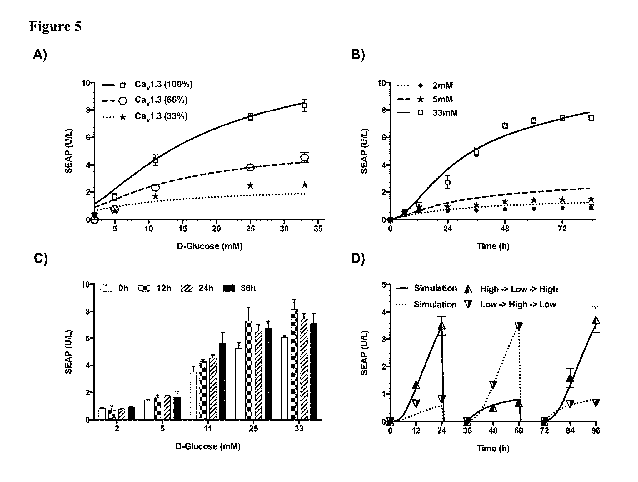
[0017] FIG. 5 shows characterization of the Ca.sub.v1.3-transgenic HEK-293.sub.NFAT-SEAP1 cell line. (A) Ca.sub.v1.3-dependent glucose sensing. Twenty-four hours after the transfection of HEK-293.sub.NFAT-SEAP1 cells with different amounts of Ca.sub.v1.3 expression vectors (pCaV1.3/pCavb3/pCaV.alpha.2.delta.1; 1:1:1, w/w) and cultivation in low-glucose medium (2 mM), D-glucose was added at the indicated final concentrations. The SEAP levels in the culture supernatants were scored at 48 h after the addition of D-glucose. Curves show corresponding simulations for data that was (100%) and was not (66% and 33%) used for calibrating the model. Data presented are mean.+-.SD, n.gtoreq.5. (B) SEAP expression kinetics. Twelve hours after cultivation in low-glucose medium (2 mM), Ca.sub.v1.3-transgenic HEK-293.sub.NFAT-SEAP1 cells were grown in cell culture medium containing different concentrations of D-glucose. The SEAP levels in the culture supernatants were profiled every 12 h. Solid and dashed curves show corresponding model-based simulations. Data presented are mean.+-.SD, n.gtoreq.5. (C) Time-delayed glucose responsiveness of the Ca.sub.v1.3/HEK-293.sub.NFAT-SEAP1 system. Ca.sub.v1.3-transgenic Ca.sub.v1.3/HEK-293.sub.NFAT-SEAP1 cells were cultured in low-glucose medium (2 mM) for 0-36 h before D-glucose was added at the indicated final concentrations. Forty-eight hours after the addition of D-glucose, the SEAP levels in the culture supernatants were profiled. Data presented are mean.+-.SD, n.gtoreq.3. (D) Reversibility of the synthetic excitation-transcription coupling system. Ca.sub.v1.3-transgenic Ca.sub.v1.3/HEK-293.sub.NFAT-SEAP1 cells were cultured in high-D-glucose medium (40 mM) or low-D-glucose medium (5 mM) for 72 h while resetting the cell density to 0.75.times.10.sup.6 cells/mL and alternating D-glucose concentrations every 24 h followed by extensive washing over 12 h. The SEAP levels in the culture supernatants were profiled every 12 h within 24 h intervals of exposure to high/low glucose. Solid and dashed curves show corresponding model-based simulations. Data presented are mean.+-.SD, n.gtoreq.4.
[0018] FIG. 6 shows Ca.sub.v1.3-dependent glucose sensing and antidiabetic potential in diabetic mice. (A) Dose-dependent glycaemia-induced SEAP expression in different diabetic mouse models. HEK-293.sub.NFAT-SEAP1 cells were transfected with Ca.sub.v1.3 and microencapsulated into alginate-poly-(L-lysine)-alginate beads. Capsules (1.times.10.sup.4; 500 cells/capsule) were implanted into mice suffering from different types of diabetes. The SEAP levels in the bloodstream of treated animals were quantified 48 h after implantation. (B, C) Self-sufficient GLP-1 expression in wild-type and type-2 diabetic mice. HEK-293 cells were co-transfected with Ca.sub.v1.3 and pMX115 ((NFAT.sub.IL4).sub.9-P.sub.min-shGLP1-pA), and the cells were then microencapsulated into alginate-poly-(L-lysine)-alginate beads (GLP-1 capsules). Control implants consisted of equally encapsulated Ca.sub.v1.3-transgenic HEK-293.sub.NFAT-SEAP1 cells (SEAP capsules). Capsules (1.times.10.sup.4; 500 cells/capsule) were implanted into wild-type (WT) or type-2 diabetic (T2D) mice. (B) GLP-1 and (C) insulin levels in the blood were profiled at 48 h after implantation. (D) Intraperitoneal glucose tolerance test (IPGTT) of wild-type and type-2 diabetic mice. Forty-eight hours after implantation and prior to serum collection, the same groups of mice as in (B and C) received an intraperitoneal injection of aqueous 2 g/kg D-glucose, and the glycaemic profile of each animal was tracked every 30 min. All data are shown as the mean.+-.SEM, and the analysis as performed with a two-tailed t-test (n=8 mice). * P<0.05, ** P<0.01, *** P<0.001 vs. control. (E) Self-sufficient insulin expression in wild-type and type-1 diabetic mice. HEK-293 cells were co-transfected with Ca.sub.v1.3 and pMX100 ((NFAT.sub.IL4).sub.9-P.sub.min-mINS-pA), and the cells were then microencapsulated into alginate-poly-(L-lysine)-alginate beads (INS capsules). Control implants consisted of equally encapsulated Ca.sub.v1.3/pMX115-transgenic HEK-293 cells (GLP-1 capsules). Capsules (1.times.10.sup.4; 500 cells/capsule) were implanted into wild-type (WT) or type-1 diabetic (T1D) mice. Serum insulin levels (detection limit: 0.2 .mu.g/L) were profiled at 72 h after capsule implantation and 4 h after food intake. (F) Self-sufficient glycaemic control of Ca.sub.v1.3/pMX100-transgenic implants. The fasting glycaemia of the same groups of mice as in (E) was tracked for 96 h after capsule implantation. All data in (E-F) are shown as the mean.+-.SEM, and the statistical analysis was performed with a two-tailed t-test (n=8 mice). * P<0.05, ** P<0.01, *** P<0.001 vs. control.
[0019] FIG. 7 shows glucose-sensor control experiments. (A) Quantitative RT-PCR-based expression profiling of endogenous glucose transporters and K.sub.ATP-channels in HEK-293 using specific primers shown in Table S2. Transcription levels were normalized to glyceraldehyde 3-phosphate dehydrogenase (GAPDH) transcripts by setting undetermined values to a maximum Ct of 40 cycles. Data are mean.+-.SD, n=3. (B) Comparison of different glucose sensors in mammalian cells. HEK-293 cells were co-transfected with 1000 ng pMX57 and 1000 ng of either pcDNA3.1(+), Ca.sub.v1.3 (pCaV1.3/pCaVb3/pCaV.alpha.2.delta.1; 1:1:1, w/w) or the full sweet taste receptor componentry (pT1R2/pT1R3/pGNAT3; 1:1:1, w/w). 24 h after transfection and cultivation in low glucose medium (2 mM), D-glucose was added to final concentrations as indicated on the x-axis. SEAP levels in the culture supernatants were scored at 48 h after addition of D-glucose. Data presented are mean.+-.SD, n.gtoreq.5. (C) Substrate specificity of Ca.sub.v1.3/pMX57-transgenic mammalian cells. HEK-293 cells were co-transfected with Ca.sub.v1.3 (pCaV1.3/pCaVb3/pCaV.alpha.2.delta.1; 333 ng each) and pMX57 (1000 ng) and cultivated in glucose-free medium for 12 h before different concentrations of D-glucose (D-Glc), glutamine (L-Gln), leucine (L-Leu), palmitic acid (PA) or linoleic acid (LA) were added. 48 h after addition of control compounds, SEAP levels in the culture supernatants were scored. Data presented are mean.+-.SD, n.gtoreq.3. (D) GLuc expression kinetics. HEK-293 cells were co-transfected with Ca.sub.v1.3 (pCaV1.3/pCaVb3/pCaV.alpha.2.delta.1; 333 ng each) and pWH29 ((NFAT.sub.IL4).sub.5-P.sub.min-GLuc-pA; 1000 ng) and cultivated in low-glucose medium (2 mM) for 12 h before different concentrations of D-Glucose (5 or 40 mM) or KCl (40 mM) were added. GLuc levels in the culture supernatants were profiled at different time points after the addition of inducer compounds as indicated on the x-axis. All data presented are mean.+-.SD, n.gtoreq.3. (E, F) Insensitivity of Cav1.3/pMX57-transgenic HEK-293 cells to cytokine signaling. HEK-293 cells were cotransfected with pMX57 (P.sub.NFAT3-SEAP-pA; 1000 ng) and either (E) Cav1.3 (pCaV1.3/pCaVb3/pCaV.alpha.2.delta.1; 333 ng each) or (F) pcDNA3.1(+) (1000 ng) and cultivated in glucose-free medium for 12 h before different concentrations of recombinant human interleukin 2 (IL-2), interleukin 12 (IL-12) and interleukin 15 (IL-15) or potassium chloride (40 mM KCl; positive control) were added and SEAP levels were profiled in the culture supernatants after 48 h. Data presented are mean.+-.SD, n.gtoreq.3.
[0020] FIG. 8 shows design and construction of the stable HEK-.sub.293NFAT-SEAP1 cell line. (A) Depolarization-stimulated SEAP expression of different P.sub.NFAT-IL4-transgenic cell clones (HEK-293.sub.NFAT-SEAP1). HEK-293 cells were stably transfected with pMX57 ((NFAT.sub.IL4).sub.5-P.sub.min-SEAP-pA) and 16 randomly selected cell clones were profiled for their depolarization-stimulated SEAP regulation performance by cultivating them for 48 h in the presence (50 mM) or absence (0 mM) of potassium chloride (KCl). (B) Stable depolarization-stimulated SEAP expression of the HEK-293.sub.NFAT-SEAP1 cell line. 5.times.10.sup.4 HEK-293.sub.NFAT-SEAP1 cells from different generations were cultivated for 48 h in the presence (50 mM) or absence (0 mM) of potassium chloride (KCl) before SEAP levels in the culture supernatants were scored. All data presented are mean.+-.SD, n.gtoreq.3.
[0021] FIG. 9 shows experimental data (symbols) and simulation results (lines) for D-glucose- and KCl-stimulated SEAP expression in vitro. (A) Cav1.3-dependent excitation-transcription coupling HEK-293 cells were co-transfected with pMX57 and either pcDNA3.1(+) (-Ca.sub.v1.3) or Ca.sub.v1.3 (+Ca.sub.v1.3), and the cells were grown for 48 h in cell culture medium containing different KCl concentrations before SEAP levels in the culture supernatants were profiled (see also FIG. 1E). All data presented are mean.+-.SD, n.gtoreq.5. (B) D-glucose activated P.sub.NFAT3-activation. HEK-293 cells were co-transfected with Ca.sub.v1.3 and pMX57, and the cells were cultured in low-glucose medium (2 mM) for 12 h before D-glucose was added to the indicated final concentrations. Forty-eight hours after the addition of control compounds, the SEAP levels in the culture supernatants were scored. Data presented are mean.+-.SD, n.gtoreq.3. (C, D) SEAP expression kinetics. Twelve hours after transfection of 9.times.10.sup.5 HEK-293 cells with pMX57 (P.sub.NFAT3-SEAP-pA) and Ca.sub.v1.3 (pCaV1.3/pCaVb3/pCaV.alpha.2.delta.1, 1:1, w/w), culture supernatants were exchanged by fresh medium containing different D-glucose-(C) and KCl-(D) concentrations. SEAP levels in the culture supernatants were profiled every 12 h. Circles represent equally treated Ca.sub.v1.3-transgenic HEK-.sub.239NFAT-SEAP1 cells. Data presented are mean.+-.SD, n.gtoreq.3.
[0022] FIG. 10 shows glucose-sensor in vivo control experiments. (A) Ca.sub.v1.3-dependent SEAP-expression kinetics in vivo. Wild-type (WT, black) or type 1 diabetic (T1D, white) mice were implanted with 1.times.10.sup.4 microencapsules (500 cells/capsule) containing HEK-293.sub.NFAT-SEAP1 cells transfected with either pcDNA3.1(+) (-Ca.sub.v1.3) or Ca.sub.v1.3 (+Ca.sub.v1.3), and SEAP levels in the bloodstream were quantified every 24 h (opaque or hollow circles). Solid or dashed curves show corresponding model-based simulations. The data are shown as the mean.+-.SD, n=8 mice (B) Diet-induced glucose sensing in vivo. Wild-type mice were implanted with 1.times.10.sup.4 microcapsules (500.times.Ca.sub.v1.3-transgenic HEK-293.sub.NFAT-SEAP1 cells/capsule) and received 4.times. daily oral administrations of 200 .mu.l water, Coca-Cola.RTM. or aqueous D-glucose (0.5M). SEAP levels in the bloodstream were quantified every 24 h after capsules implantation. The data are shown as the mean.gtoreq.SD, n=8 mice. (C, D) Optimization of the P.sub.NFAT-IL4-promoter for glucose- and depolarization-stimulated (C) shGLP1- and (D) mINS-expression. (C) HEK-293 cells were co-transfected with 1000 ng Ca.sub.v1.3 and 1000 ng of pMX61 ((NFAT.sub.IL4).sub.5-P.sub.min-shGLP1-pA), pMX117 ((NFAT.sub.IL4).sub.7-P.sub.min-shGLP1-pA) or pMX115 ((NFAT.sub.IL4).sub.9-P.sub.min-shGLP1-pA) and cultivated in low-glucose medium (2 mM) for 12 h before different concentrations of D-glucose (Glc) or potassium chloride (KCl) were added. 48 h after addition of control compounds, murine IgG levels in the culture supernatants were quantified (BDL: below detection limit of 9.375 ng/mL). (D) HEK-293 cells were co-transfected with 1000 ng Ca.sub.v1.3 and 1000 ng of pMX68 ((NFAT.sub.IL4).sub.5-P.sub.min-mINS-pA), pMX99 ((NFAT.sub.IL4).sub.7-P.sub.min-mINS-pA) or pMX100 ((NFAT.sub.IL4).sub.9-P.sub.min-mINSpA) and cultivated in low-glucose medium (2 mM) for 12 h before different concentrations of D-glucose (Glc) or potassium chloride (KCl) were added. 48 h after addition of control compounds, murine insulin levels in the culture supernatants were quantified (BDL: below detection limit of 0.21 .mu.g/L). All data presented are mean.+-.SD, n.gtoreq.3.
[0023] FIG. 11 shows (A) Glycaemic control in healthy and T1D mice. (Circles) Fasting CD-1 Swiss albino mice (2.times.18 h/day) were injected with a single dose of freshly diluted alloxan monohydrate (ALX; 200 mg/kg in 300 .mu.L phosphate buffered saline) and fasting glycaemia was measured every 24 h after ALX injection. (Squares) Equally treated CD-1 Swiss albino mice harboring implants containing 5.times.10.sup.6 microencapsulated Cav1.3/pMX100-transgenic HEK-293 cells. Fasting glycaemia data are shown as the mean.+-.SD, n=6 mice. (B) Model simulations for glucose tolerance in healthy WT-mice. Forty-eight hours after implantation of 5.times.10.sup.6 Ca.sub.v1.3 transgenic HEK-293.sub.NFAT-SEAP1 cells, CD-1 Swiss albino mice received an intraperitoneal injection of aqueous 2 g/kg D-glucose, and the glycaemic profile of each animal was tracked every 30 min. The curve shows a corresponding model-based simulation. All data shown as the mean.+-.SD, n=8 mice.
[0024] FIG. 12 shows treatment potential of .beta.-cell-mimetic designer cells in type-1 diabetic mice. (A) Schematic of HEK-.beta.. Extracellular D-glucose triggers glycolysis-dependent membrane depolarization which activates the voltage-gated calcium channel Ca.sub.v1.3, resulting in Ca.sup.2+ influx, induction of the calmodulin/calcineurin signaling cascade and P.sub.NFAT-mediated induction of insulin expression and secretion. (B) Self-sufficient glycemic control in wild-type and type-1 diabetic mice. 5.times.10.sup.6 HEK-.beta. cells or 1.1E7 cells were microencapsulated in alginate-poly-(L-lysine)-alginate beads (500 cells/capsule) and implanted into wild-type (WT) or type-1 diabetic (T1D) mice (1.times.10.sup.4 capsules/mice). Control implants contained microencapsulated Ca.sub.v1.3/pMX115-transgenic HEK-293 cells (cntrl). Fasting glycemia of treated animals was recorded for 3 weeks. T1D mice treated with negative-control implants did not survive the first glucose tolerance test on day 7 shown in (D). (C) Self-sufficient insulin expression in wild-type and type-1 diabetic mice. Postprandial blood insulin levels of the treatment groups shown in (B) were profiled every 4 days for up to 3 weeks. (D) Intraperitoneal glucose tolerance tests in type-1 diabetic mice. On days 7 and 14, the treatment groups shown in (B, C) received intraperitoneal D-glucose (2 g/kg) injections and the glycemic excursion of individual animals was recorded for 30 min. (E) Schematic of HEK-.beta..sub.GLP. D-glucose activates P.sub.NFAT-driven promoters by excitation-transcription coupling and triggers dose-dependent expression of secreted human glucagon-like peptide 1 (shGLP1). shGLP1 activates constitutively expressed GLP-1 receptor (GLP1R) via an autocrine loop and triggers insulin expression from P.sub.CRE-driven promoters. In vivo, insulin expression by HEK-.beta..sub.GLP cells may also be triggered following postprandial release of GLP-1 by intestinal cells. (F) Response of .beta.-cell-mimetic implants to meals. Wild-type mice were implanted with 5.times.10.sup.6 microencapsulated Ca.sub.v1.3/pMX57-transgenic HEK-293 cells (-GLP1R) or Ca.sub.v1.3/pMX61/pMX258-transgenic HEK.sub.GLP1R cells (+GLP1R) and received oral doses of 200 .mu.L H.sub.2O, Coca-Cola.RTM. or sugared water (0.5M D-Glucose). Resulting blood SEAP levels were quantified after 48 h. (G) Oral glucose tolerance test of wild-type (WT) and type-1 diabetic (T1D) mice. Mice received 5.times.10.sup.6 microencapsulated HEK-.beta., 1.1E7 or HEK-.beta..sub.GLP cells or negative-control implants containing Ca.sub.v1.3/pMX115-transgenic HEK-293 cells (cntrl). After oral administration of sugared water (2 g/kg D-glucose in H.sub.2O), the glycemic excursions of individual animals were recorded for 6 h. (H) Self-sufficient glycemic control by implants containing transgenic HEK-.beta. and HEK-.beta..sub.GLP cells. 5.times.10.sup.6 microencapsulated HEK-.beta. or HEK-.beta..sub.GLP cells were implanted into wild-type (WT) or type-1 diabetic (T1D) mice (1.times.10.sup.4 capsules/mice). Fasting glycemia of treated animals was recorded for 3 weeks. All data are shown as the mean.+-.SEM, statistics were performed using two-tailed t-test (n=8 mice). *P<0.05, **P<0.01, ***P<0.001 HEK-.beta. vs. cntrl.
[0025] FIG. 13 shows construction and characterization of the stable HEK-.beta. cell line. (A, B) Characterization of HEK.sub.MX252 stably expressing the Ca.sub.v1.3 .alpha.1D subunit. (A) 3.times.10.sup.6 HEK-293 cells were cotransfected with pMX252 (ITR-P.sub.hEF1.alpha.-Cacna1d-pA:P.sub.RPBSA-BFP-P2A-PuroR-pA-ITR; 9500 ng) and pCMV-T7-SB100 (P.sub.hCMV-SB100X-pA; 500 ng), selected with 0.5 .mu.g/mL puromycin for two passages and 3.times.10.sup.5 cells of the surviving population (HEK.sub.MX252) were then cotransfected with pMX57 (P.sub.NFAT3-SEAP-pA; 1000 ng) and different amounts of pMX251 (ITR-P.sub.hEF1.alpha.-Cacna2d1-P2A-Cacnb3-pA:P.sub.RPBSA-dTomato-P2A-Bla- stR-pA-ITR; 0-200 ng filled to 1000 ng with pcDNA3.1(+)). 24 h after transfection and cultivation in low glucose medium (2 mM), D-glucose or potassium chloride (KCl; 50 mM) was added to the indicated final concentrations. SEAP levels were profiled in the culture supernatants 48 h after addition of D-glucose. Data presented are mean.+-.SD, n.gtoreq.5. (B) Control experiment of HEK-293 cells transfected with pMX57 (1000 ng) and different amounts of pMX251 (0-10 ng filled to 1000 ng with pcDNA3.1(+)). Data presented are mean.+-.SD, n.gtoreq.5. (C) Characterization of HEK.sub.Cav1.3 stably expressing the full Ca.sub.v1.3 channel componentry (Cacna1d/Cacnb3/Cacna2d1). 3.times.10.sup.6 HEK.sub.MX252 cells were cotransfected with pMX251 (9500 ng) and pCMV-T7-SB100 (500 ng), selected with 10 .mu.g/mL blasticidin for three passages and 3.times.10.sup.5 cells of the surviving population (HEK.sub.Cav1.3) were then cotransfected with pMX57 (P.sub.NFAT3-SEAP-pA; 1900 ng) and pcDNA3.1(+) (100 ng). HEK-293 cotransfected with either pMX57 alone (1900 ng) or in combination with pMX252 (100 ng) were used as negative controls. 24 h after transfection and cultivation in low glucose medium (2 mM), D-glucose or potassium chloride (KCl; 50 mM) was added to the indicated final concentrations. SEAP expression levels were profiled in the culture supernatants 48 h after the addition of D-glucose. Data presented are mean.+-.SD, n.gtoreq.5. (D) Glucose- and depolarization-stimulated insulin expression in HEK.sub.Cav1.3. 3.times.10.sup.5 HEK.sub.Cav1.3 cells were cotransfected with different amounts of pMX256 (ITR-P.sub.NFAT5-SEAP-P2A-mINS-pA:P.sub.RPBSA-EGFP-P2A-ZeoR-pA-ITR, 1000-2000 ng filled to 2000 ng with pcDNA3.1(+)) and cultivated in low glucose medium (2 mM) for 12 h before different concentrations of D-glucose and KCl were added. 48 h after the addition of the control compounds, mINS levels were profiled in the culture supernatants. Data presented are mean.+-.SD, n.gtoreq.3. (E, F) Clonal selection of HEK-13 cells. (E) 3.times.10.sup.6 HEK.sub.Cav1.3 cells were cotransfected with pMX256 (9500 ng) and pCMV-T7-SB100 (500 ng), selected with 100 .mu.g/mL zeocin for three passages and 5% of the surviving population showing highest EGFP expression levels were subjected to FACS-mediated single-cell cloning. 50 expanded cell clones were profiled for glucose-stimulated SEAP expression by cultivating 5.times.10.sup.4 cells in high-glucose (40 mM) or low-glucose medium (5 mM) for 48 h before SEAP levels were profiled in the culture supernatants. Data presented are mean.+-.SD, n=3. (F) The 20 clones showing highest glucose-stimulated SEAP inductions in (E) were profiled for glucose-stimulated insulin expression by cultivating 5.times.10.sup.4 cells in high-glucose (40 mM) or low-glucose medium (5 mM) for 48 h before mINS levels were profiled in the culture supernatants. HEK-13 (cell clone no. 4) was chosen for further analysis. Data presented are mean.+-.SD, n=3.
[0026] FIG. 14 shows characterization of the monoclonal HEK-.beta. cell line. (A) 3.times.10.sup.4 HEK-13 cells were cultivated in high-glucose (40 mM) or low-glucose medium (5 mM) for 48 h and mINS levels were profiled in the culture supernatants every 12 h after addition of D-glucose. Data presented are mean.+-.SD, n.gtoreq.5. (B) 5.times.10.sup.4 HEK-13 cells were cultivated in low-glucose medium (2 mM) for 12 h, before different concentrations of D-glucose were added and mINS levels were profiled in the culture supernatants after 24 h. Data presented are mean.+-.SD, n.gtoreq.5. (C) Reversible glucose-stimulated insulin secretion. Identical capsule batches used for implantation into mice (human islets, Fig. S13; HEK-.beta. and 1.1E7, FIG. 6B) were also maintained in cell culture medium for 3 weeks and glucose-stimulated (-, 2.8 mM; +, 30 mM) insulin production was profiled for 24 h once every week. Data presented are mean.+-.SD, n.gtoreq.3.
[0027] FIG. 15 shows engineering of HEK-.beta..sub.GLP. (A) GLP-1 triggered SEAP expression in HEK-293 cells. 3.times.10.sup.5 HEK-293 cells were cotransfected with pCK53 (P.sub.CRE-SEAP-pA, 200 ng) and different amounts of pMX250 (ITR-P.sub.hEF1.alpha.-GLP1R-pA:P.sub.RPBSA-dTomato-P2A-PuroR-pA-ITR; 10-1000 ng filled to 1800 ng with pcDNA3.1(-)) before different concentrations of recombinant human GLP-1 was added. 24 h after the addition of GLP-1, SEAP levels were profiled in the culture supernatants. Data presented are mean.+-.SD, n.gtoreq.5. (B, C) Characterization of HEK.sub.GLP1R stably expressing the human GLP-1 receptor (GLP1R). (B) 3.times.10.sup.6 HEK-293 cells were cotransfected with pMX250 (9500 ng) and pCMV-T7-SB100 (500 ng), selected with 1 .mu.g/mL puromycin for two passages and the surviving population was FACS-sorted into three populations with different red-fluorescence intensities (HEK.sub.GLP1R, HEK.sub.GLP1Rmedium. HEK.sub.GLP1Rlow). Each population (1.times.10.sup.5 cells) was transfected with pCK53 (100 ng filled to 2000 ng with pcDNA3.1(+)) before different concentrations of recombinant human GLP-1 were added. 24 h after the addition of GLP-1, SEAP levels were profiled in the culture supernatants. Data presented are mean.+-.SD, n.gtoreq.3. (C) HEK-293 cells transfected with pCK53 (100 ng filled to 2000 ng with pcDNA3.1(+)) were used as negative control. Data presented are mean.+-.SD, n.gtoreq.5. (D, E) Validation of the HEK-.beta..sub.GLP circuit. HEK.sub.GLP1R was cotransfected with Ca.sub.v1.3 (pCaV1.3/pCaVb3/pCaV.alpha.2.delta.1; 333 ng each), pMX61 (P.sub.NFAT3-shGLP1-pA, 1000 ng) and pCK53 (P.sub.CRE-SEAP-pA; 250 ng) and cultivated in low-glucose medium (2 mM) for 12 h before different concentration of (D) recombinant human GLP-1 or (E) D-glucose were added HEK.sub.GLP1R cotransfected with pMX61/pCK53 or Ca.sub.v1.3/pCK53 and HEK-293 cotreansfected with Ca.sub.v1.3/pMX61/pCK53 were used as negative controls. (D) 24 h after addition of GLP-1 and (E) 72 h after addition of D-Glucose, SEAP levels were profiled in the culture supernatants. Data presented are mean.+-.SD, n.gtoreq.5 (F, G) SEAP expression kinetics HEK.sub.GLP1R cells were cotransfected with Ca.sub.v1.3 (pCaV1.3/pCaVb3/pCaV.alpha.2.delta.1, 333 ng each), pMX61 (P.sub.NFAT3-shGLP1-pA; 1000 ng) and pCK53 (P.sub.CRE-SEAP-pA; 250 ng), cultivated in low-glucose medium (2 mM) for 12 h before different concentrations of (F) recombinant human GLP-1 or (G) D-glucose were added. SEAP levels were profiled in the culture supernatants (F) 24 h or (G) 72 h after addition of the respective compounds. Data presented are mean.+-.SD, n.gtoreq.5. (H) mINS expression kinetics of HEK-.quadrature..sub.GLP. HEK.sub.GLP1R cells were cotransfected with Ca.sub.v1.3 (pCaV1.3/pCaVb3/pCaV.alpha.2.delta.1; 333 ng each), pMX61 (P.sub.NFAT3-shGLP1-pA; 1000 ng) and pDA145 (P.sub.CRE-mINS-pA, 1000 ng), cultivated in low-glucose medium (2 mM) for 12 h before different concentrations of recombinant human GLP-1 were added. mINS levels were profiled in the culture supernatants for 2411 after addition of control compounds. Data presented are mean.+-.SD, n.gtoreq.5.
[0028] FIG. 16 shows oral glucose tolerance test (OGTT) of type-1 diabetic mice treated with encapsulated human islets. 2000 IEQs of human islets were microencapsulated in alginate-poly-(L-lysine)-alginate beads and injected into each of four type-1 diabetic mice. 7 and 14 days after implantation the animals received oral D-glucose (2 g/kg) and their glycemic excursions were recorded over 2 h.
DETAILED DESCRIPTION
[0029] So that the invention may be more readily understood, certain terms are first defined. Unless otherwise defined within the specification, all technical and scientific terms used herein have their art-recognized meaning Although similar or equivalent methods and materials to those described herein can be used in the practice or testing of the invention, suitable methods and materials are described below. All publications, patent applications, patents, and other references mentioned herein are incorporated by reference in their entirety. In case of conflict, the present specification, including definitions, will prevail. The materials, methods, and examples are illustrative only and not intended to be limiting. The terms "comprising". "having", and "including" are to be construed as open-ended terms (i.e., meaning "including, but not limited to,") unless otherwise noted.
[0030] As used herein, the term "beta-cell" or ".beta.-cell" refers to a cell type found in the pancreas, in particular in the mammalian, more particular in the human pancreatic islets. Beta cells are the primary producers of insulin.
[0031] As used herein, the term "recombinant cell" refers to cells, preferably mammalian cells, more preferably human cells, which have been artificially manipulated to express genes which are introduced to the mammalian cells by e.g. transfection or transformation using nucleic acid constructs, such as e.g. expression vectors in which those genes are incorporated.
[0032] As used herein, the term "transfection" or "transfected" refers to the introduction of a nucleic acid e.g. the introduction of a nucleic acid construct as described herein into a cell. In general the nucleic acid is a DNA sequence, in particular a vector or a plasmid carrying a gene of interest like a gene coding for a therapeutic protein as described herein, operably linked to a suitable promoter as described herein. Transfection methods which can be used are e.g. those using carrier molecules like cationic lipids such as DOTAP (Roche), TransFast (Promega), and Lipofectamine (Invitrogene), or polyethylenimine (PEI), calcium phosphate and DEAE dextran. Other useful transfection techniques include electroporation, bombardment with nucleic-acid-coated carrier particles (gene gun), microinjection and using of viral vectors.
[0033] As used herein, the term "transiently transfected" of "transient transfection" refer to the transient, i.e. non-permanent expression of the gene of interest due to the episomal nature of the introduced nucleic acid. Episomal nucleic acids, including DNA (plasmids or vectors), is degraded by the cells after two to seven days, and hence the expression of the gene of interest ceases then.
[0034] As used herein, the term "stably transfected" or "stable transfection" refers to the permanent expression of a gene of interest due to the integration of the transfected DNA into the genome of the cell. Most if not all cells have the potential to incorporate episomal DNA into their genome albeit at a very low rate. However, sophisticated selection strategies are employed to expand those cells that have integrated the transfected DNA. For that a nucleic acid construct to be stably integrated, a vector carrying the DNA to be transfected normally contains at least one gene that encodes for a selection marker such as e.g. a puromycin-resistance gene.
[0035] As used herein, the term "nucleic acid construct" refers to a nucleic acid, preferably to a recombinant nucleic acid construct, i.e. a genetically engineered nucleic acid construct which includes the nucleic acid of a gene and at least one promoter for directing transcription of the nucleic acid in a host cell. Nucleic acid constructs of the present invention are preferably suitable for mammalian cell expression. The nucleic acid construct (also referred to herein as an "expression vector") may include additional sequences that render the construct e.g. the vector, suitable for replication and integration in eukaryotes (e.g. shuttle vectors). In addition, a typical nucleic acid construct such as e.g. a cloning vector may also contain transcription and translation initiation sequences, transcription and translation terminators, and a polyadenylation signal.
[0036] As used herein, the term "promoter" refers to a regulatory DNA sequence generally located upstream of a gene that mediates the initiation of transcription by directing RNA polymerase to bind to DNA and initiating RNA synthesis. The term "P.sub.min" as used herein refers to a minimal promoter, preferably to the promoter as shown in SEQ ID NO: 33. A minimal promoter usually does not contain an enhancer i.e. do not comprise enhancer elements and is not a constitutive promoter. Preferably a minimal promoter shows no or only minimal transcriptional activity in the absence of transcription factors.
[0037] As used herein, the term "enhancer" as used herein refers to a nucleotide sequence that acts to potentiate the transcription of genes independent of the identity of the gene, the position of the sequence in relation to the gene, or the orientation of the sequence.
[0038] As used herein, the terms "functionally linked" and "operably linked" are used interchangeably and refer to a functional relationship between two or more DNA segments, in particular gene sequences to be expressed and those sequences controlling their expression. For example, a promoter and/or enhancer sequence, including any combination of cis-acting transcriptional control elements is operably linked to a coding sequence if it stimulates or modulates the transcription of the coding sequence in an appropriate host cell or other expression system. Promoter regulatory sequences that are operably linked to the transcribed gene sequence are physically contiguous to the transcribed sequence.
[0039] As used herein, the term "promoter which is responsive to carbohydrate metabolism of a cell" refers to a promoter which is activated or repressed for transcription in response to the presence or absence of a product of the carbohydrate metabolism of a cell. A product of the carbohydrate metabolism of a cell can be a metabolic product of the cellular pathway of carbohydrate transformation or carbohydrate degradation e.g. a product of glycolysis such as intracellular ATP production or can be a cellular response to the metabolism of a carbohydrate in the cell e.g. production of intracellular ATP lead to the closure of K.sub.ATP, channels which causes membrane depolarization as cellular response.
[0040] As used herein, the term "carbohydrate metabolism of a cell" refers to the various biochemical processes responsible for the formation, breakdown and interconversion of carbohydrates in living organisms. Oligosaccharides and/or polysaccharides are typically cleaved into smaller monosaccharides by enzymes called glycoside hydrolases. The monosaccharide units then enter the cellular pathway of carbohydrate transformation or carbohydrate degradation, i.e. the cellular pathway of glucose transformation or degradation such as glycolysis.
[0041] As used herein, the term "cellular component for sensing extracellular carbohydrates" refers to a cellular component such as e.g. a transporter of carbohydrates like e.g. a transporter of glucose or a glucose linked transporter, a receptor involved in the regulation of carbohydrate homeostasis e.g. glucose homeostasis, or a membrane protein like potassium channels, calcium channels or sodium channels which is capable of sensing extracellular carbohydrates such as glucose by activating a gene, preferably a gene coding for a therapeutic protein whose expression level correlates with the extracellular carbohydrate levels. Usually the cellular component for sensing extracellular carbohydrates of the recombinant cell of the present invention is capable of sensing extracellular carbohydrates such as glucose by activating a gene, preferably a gene coding for a therapeutic protein whose expression level correlates with the extracellular carbohydrate levels via a promoter which is responsive to carbohydrate metabolism of said recombinant cell, wherein the promoter is operably linked to a gene coding for the therapeutic protein. Preferably the cellular component for sensing extracellular carbohydrates is of mammalian, more preferably human origin.
[0042] As used herein, the term "carbohydrate" or "saccharide" are interchangeably and equivalently used within this context refer to a biological molecule consisting of carbon (C), hydrogen (H) and oxygen (O) atoms, usually with a hydrogen-oxygen atom ratio of 2.1 (as in water); in other words, with the empirical formula C.sub.m(H.sub.2O).sub.n (where m could be different from n). Carbohydrates include monosaccharides, disaccharides, oligosaccharides, and polysaccharides, preferably monosaccharides and disaccharides, more preferably monosaccharides, most preferably monosaccharides selected from the group consisting of glucose, galactose, fructose and xylose and epimeric forms thereof like mannose, in particular glucose and mannose. The term "glucose" in its broadest sense relates to glucose and its epimeric forms like mannose. Preferably the term "glucose" relates to glucose (D-Glucose, (2R,3S,4R,5R)-2,3,4,5,6-Pentahydroxyhexanal).
[0043] As used herein, the term "membrane protein" refers to a protein molecule that is attached to or associated with the membrane of a cell or organelle. The membrane protein is preferably a membrane protein of a human cell or organelle.
[0044] As used herein, the term "fragment of a membrane protein" refers to a region of a membrane protein that is shorter in length as compared with the full-length membrane protein. It is however, a requirement of the present invention that any fragment of a membrane protein used as part retain the activity of the full-length membrane protein.
[0045] As used herein, the term "subunit of a membrane protein" or "monomer of a membrane protein" are interchangeably and equivalently used within this context and refers to a separate polypeptide chain that makes a membrane protein which is made up of two or more polypeptide chains joined together. In a membrane protein molecule composed of more than one subunit, each subunit can form a stable folded structure by itself. The amino acid sequences of subunits of a protein can be identical, similar, or completely different.
[0046] As used herein, the term "physiological effect of membrane depolarization" refers to a physiological effect due to depolarization of a cell's membrane such as e.g. the variation in intra-cellular cation, e.g. intra-cellular calcium, intra-cellular sodium or intra-cellular potassium concentration, in particular intra-cellular calcium concentration within a cell which has been caused by physiological activities, in particular by the carbohydrate metabolism of said cell.
[0047] As used herein, the term "calcium-responsive promoter" refers to a promoter which is activated or repressed for transcription in response to the presence or absence of calcium in the cell. As used herein, the term "expression system" refers to a set of transgenic genetic elements within a cell as well as proteins encoded by such genetic elements.
[0048] The term "G-protein coupled receptors" as used herein refers to a family of transmembrane receptors, that sense molecules outside the cell and activate responses inside the cell by coupling with specific intracellular signaling pathways via G proteins. Preferred G-protein coupled receptors are GPR1, TAS1R2, TAS1R3, GLP1R or anyone of their orthologues. Most preferred G-protein coupled receptors are GPR1 (GenBank: CAA98593.1), TAS1R2 (UniProtKB/Swiss-Prot: Q8TE23.2), TAS1R3 (UniProtKB/Swiss-Prot: Q7RTX0.2) and GLP1R (UniProtKB/Swiss-Prot: P43220.2).
[0049] The term "glucoincretin receptor" as used herein refers to glucoincretin receptors such as gastric inhibitory polypeptide receptor (GIPR) and glucagon-like peptide-1 receptor (GLP1R), preferably to human glucoincretin receptors such as human GIPR e.g. human GIPR (UniProtKB: P48546) and/or human GLP1R e.g. human GLP1R (UniProtKB/Swiss-Prot: P43220.2).
[0050] The term "SLC2A family glucose transporters" (also known as GLUT) as used herein refers to a family of transmembrane proteins that catalyze the entry of carbohydrates into mammalian cells. Preferred SLC2A family glucose transporters are GLUT1 (SLC2A1), GLUT2 (SLC2A2), GLUT3 (SLC2A3), GLUT4 (SLC2A4), GLUT5 (SLC2A5), GLUT6 (SLC2A6), GLUT7 (SLC2A7), GLUT8 (SLC2A8), GLUT9 (SLC2A9). GLUT10 (SLC2A10). GLUT11 (SLC2A11), GLUT12 (SLC2A12) and GLUT13 (SLC2A13) or anyone of their orthologues. Most preferred SLC2A family glucose transporters are human GLUT1, human GLUT2 and human GLUT3.
[0051] The term "SLC5A family sodium-glucose linked transporters" (also known as SGLT) as used herein refers to a family of transmembrane proteins that mediate sodium-dependent co-transport of carbohydrates across the plasma membrane of mammalian cells Preferred SLC5A family sodium-glucose linked transporters are SGLT1 (SLC5A1), SGLT2 (SLC5A2) and SGLT3 (SLC5A3) or anyone of their orthologues. Most preferred SLC5A family sodium-glucose linked transporters are human SGLT1 and human SGLT3.
[0052] The term "potassium channels" as used herein refers to a family of pore-forming transmembrane proteins that facilitate the transport of potassium ions across the cell plasma membrane Preferred potassium channels are ATP-sensitive potassium channels (K.sub.ATP), calcium-activated potassium channels (BK.sub.Ca), inward rectifier potassium channels (Kir) and voltage-dependent potassium channels (K.sub.v). Most preferred potassium channels are human ATP-sensitive potassium channels (K.sub.ATP), human calcium-activated potassium channels (BK.sub.Ca), human inward rectifier potassium channels (Kir) and human voltage-dependent potassium channels (K.sub.v).
[0053] The term "calcium channels" as used herein refers to a family of pore-forming transmembrane proteins that facilitate the transport of calcium ions across the cell plasma membrane Preferred calcium channels are voltage-gated calcium channels (VGCC), N-methyl-D-aspartate type of Glutamate (NMDA) receptors, Ca.sup.2+ release-activated Ca.sup.2+ current (CRAC) channels and transient receptor potential channels (TRPCs) Most preferred calcium channels are human voltage-gated calcium channels (VGCC), human N-methyl-D-aspartate type of Glutamate (NMDA) receptors, human Ca.sup.2+ release-activated Ca.sup.2+ current (CRAC) channels and human transient receptor potential channels (TRPCs).
[0054] The term "sodium channels" as used herein refers to a family of pore-forming transmembrane proteins that facilitate the transport of sodium ions across the cell plasma membrane Preferred sodium channels are voltage gated-sodium channels, most preferably human voltage gated-sodium channels
[0055] As used herein, the term "voltage-gated calcium channel" (VDCC) refers to a group of voltage-gated ion channels, preferably human voltage-gated calcium channels, found in the membrane of excitable cells whose permeability to the calcium ion Ca.sup.2+ correlates with the membrane potential. Voltage-dependent calcium channels are formed as a complex of several different subunits. Subunits known are the pore-forming Ca.sub.v.alpha.1, the intracellular Ca.sub.v.beta., the transmembrane Ca.sub.v.gamma., and a disulfide-linked dimer Ca.sub.v.alpha.2.delta.. The .alpha.1 subunit is the primary subunit necessary for channel functioning in the VDCC, and consists of the characteristic four homologous I-IV domains containing six transmembrane .alpha.-helices each forms the ion conducting pore while the associated subunits have several functions including modulation of gating. Voltage-gated calcium channels are functionalized by their al subunit, which sets the activation threshold of the entire channel. Non-limiting examples of al subunits are Ca.sub.v1, Ca.sub.v1.2, Ca.sub.v1.3, Ca.sub.v1.4, Ca.sub.v2.1, Ca.sub.v2.2, Ca.sub.v2.3, Ca.sub.v3.1, Ca.sub.v3.2 and Ca.sub.v3.3.
[0056] As used herein, the term "therapeutic protein" refers to a protein which is therapeutically applicable i.e. a therapeutic protein is any protein or polypeptide that can be expressed to provide a therapeutic effect, in particular a protein that can be expressed to provide a therapeutic effect with respect to metabolic diseases.
[0057] As used herein, the term "orthologues" with respect to a protein e.g. a receptor or channel refers to one of two or more homologous gene sequences found in different species.
[0058] As used herein, the term "insulin" refers to the protein hormone produced by beta cells in the pancreas which decreases blood glucose concentrations and is therefore involved in the regulation of blood sugar levels. One international unit of insulin (1 IU) is defined as the "biological equivalent" of 34.7 .mu.g pure crystalline insulin, which corresponds to the amount required to reduce the concentration of blood glucose in a fasting rabbit to 45 mg/dl (2.5 mmol/L) Insulin is produced as a proinsulin precursor consisting of the B and A chains of insulin linked together via a connecting C-peptide. Insulin itself is comprised of only the B and A chains. Human insulin is encoded by the INS gene corresponding to GenBank Accession No: NM-000207.2 The term "insulin" or "insulin molecule" is a generic term that designates the 51 amino acid heterodimer comprising the A-chain peptide and the B-chain peptide, wherein the cysteine residues a positions 6 and 11 of the A chain are linked in a disulfide bond, the cysteine residues at position 7 of the A chain and position 7 of the B chain are linked in a disulfide bond, and the cysteine residues at position 20 of the A chain and 19 of the B chain are linked in a disulfide bond. The term "insulin" means the active principle of the pancreas that affects the metabolism of carbohydrates in the animal body and which is of value in the treatment of diabetes mellitus. The term includes synthetic and biotechnologically derived products that are the same as, or similar to, naturally occurring insulins in structure, use, and intended effect and are of value in the treatment of diabetes mellitus.
[0059] The term "insulin analogue" as used herein includes any heterodimer analogue or single-chain analogue that comprises one or more modification(s) of the native A-chain peptide and/or B-chain peptide. Modifications include but are not limited to substituting an amino acid for the native amino acid at a position selected from A4. A5, A8, A9, A10, A12, A13, A14, A15, A16, A17, A18, A19, A21, B1, B2, B3, B4, B5, B9, B10, B13, B14, B15, B16, B17, B18, B20, B21, B22, B23, B26. B27, B28. B29, and B30; deleting any or all of positions B1-4 and B26-30; or conjugating directly or by a polymeric or non-polymeric linker one or more acyl, polyethylglycine (PEG), or saccharide moiety (moieties); or any combination thereof. Examples of insulin analogues include but are not limited to the heterodimer and single-chain analogues disclosed in published international application WO20100080606, WO2009099763, and WO2010080609, the disclosures of which are incorporated herein by reference. Examples of single-chain insulin analogues also include but are not limited to those disclosed in published International Applications WO9634882, WO95516708, WO2005054291, WO2006097521, WO2007104734, WO2007104736, WO2007104737, WO2007104738, WO2007096332, WO2009132129; U.S. Pat. Nos. 5,304,473 and 6,630,348; and Kristensen et al., Biochem. J. 305: 981-986 (1995), the disclosures of which are each incorporated herein by reference. The term "insulin analogues" further includes single-chain and heterodimer polypeptide molecules that have little or no detectable activity at the insulin receptor but which have been modified to include one or more amino acid modifications or substitutions to have an activity at the insulin receptor that has at least 1%, 10%, 50%, 75%, or 90% of the activity at the insulin receptor as compared to native insulin and which further includes at least one N-linked glycosylation site. In particular aspects, the insulin analogue is a partial agonist that has from 2' to 100' less activity at the insulin receptor as does native insulin. In other aspects, the insulin analogue has enhanced activity at the insulin receptor, for example, the IGF <B16B17> derivative peptides disclosed in published international application WO02010080607 (which is incorporated herein by reference) These insulin analogues, which have reduced activity at the insulin growth hormone receptor and enhanced activity at the insulin receptor, include both heterodimers and single-chain analogues.
[0060] As used herein, the term "autologous" or "endogenous" refers to any material that is present in a cell or an organism which is native to said recombinant cell or organism.
[0061] The term "stem cell" as used herein refers to undifferentiated biological cells that can differentiate into specialized cells and which is capable of proliferation to produce more stem cells.
[0062] As used herein, the term "somatic cell" refers to any cell forming the body of an organism, as opposed to germline cells or undifferentiated stem cells.
[0063] The terms "individual." "subject" or "patient" are used herein interchangeably. In certain embodiments, the subject is a mammal. Mammals include, but are not limited to primates (including human and non-human primates). In a preferred embodiment, the subject is a human.
[0064] The term "about" as used herein refers to +/-10% of a given measurement.
[0065] In a first aspect, the present invention provides a recombinant cell comprising a nucleic acid construct comprising a promoter which is responsive to carbohydrate metabolism of said cell, wherein the promoter is operably linked to a gene coding for a therapeutic protein. Preferably, the promoter which is responsive to carbohydrate metabolism is responsive to glucose metabolism.
[0066] In one embodiment the promoter which is responsive to carbohydrate metabolism is responsive to a physiological effect of membrane depolarization caused by the carbohydrate metabolism of said recombinant cell. Preferably the physiological effect of membrane depolarization caused by the carbohydrates metabolism of the cell is extracellular calcium influx Thus in a preferred embodiment the promoter which is responsive to carbohydrate metabolism is responsive to a physiological effect of membrane depolarization caused by the carbohydrate metabolism of said recombinant cell, wherein the physiological effect of membrane depolarization caused by the carbohydrates metabolism of said cell is extracellular calcium influx. Thus in a further preferred embodiment the promoter which is responsive to carbohydrate metabolism is responsive to extracellular calcium influx.
[0067] In one embodiment the promoter which is responsive to carbohydrate metabolism is a calcium-responsive promoter, preferably a calcium-responsive promoter comprising nucleic acid sequences bound by transcription factors of the NFAT family, the NFkB family, the AP-1 family, and/or the CREB family and/or cFOS, more preferably a calcium-responsive promoter comprising nucleic acid sequences bound by transcription factors of the NFAT family.
[0068] The NFAT family is a family of transcription factors shown to be important in immune response. One or more members of the NFAT family is expressed in most cells of the immune system. NFAT is also involved in the development of cardiac, skeletal muscle, and nervous systems. The NFAT family comprises the NFAT1, NFAT2, NFAT3, NFAT4, and NFAT5 proteins that specifically bind their cognate promoters. Preferred promoters that contain NFAT-binding sites are synthetic or natural promoters containing one or multiple 5'-GGAAA-3' consensus sites, more preferred are mammalian cytokine promoters e.g. mammalian cytokine promoters selected from the group consisting of interleukin (IL)-2, IL-3, IL-4 promoter, peroxisome-proliferator-activated receptor-.gamma. (PPAR.gamma.) promoter, orphan nuclear receptor 77 (NUR77) promoter, interferon .gamma. promoter, GATA-binding protein 3 (GATA3) promoter and promoters of the T-box family of transcription factors (T-bex and eomesodermin), preferably the interleukin (IL)-2, L-4 promoter and PPAR.gamma. promoter Most preferred is the murine interleukin (IL)-4 promoter. Thus in one embodiment the promoter which is responsive to carbohydrate metabolism of the recombinant cell is a synthetic or natural promoter that contains NFAT-binding sites containing one or multiple 5'-GGAAA-3' consensus sites, more preferably a mammalian cytokine promoter e.g. a mammalian cytokine promoter selected from the group consisting of interleukin (IL)-2, IL-3, IL-4 promoter, peroxisome-proliferator-activated receptor-.gamma. (PPAR.gamma.) promoter, orphan nuclear receptor 77 (NUR77) promoter, interferon .gamma. promoter, GATA-binding protein 3 (GATA3) promoter and promoters of the T-box family of transcription factors (T-bex and eomesodermin), preferably the interleukin (IL)-2, IL-4 promoter and PPAR.gamma. promoter and most preferrably the murine interleukin (IL)-4 promoter. The murine interleukin (IL)-4 promoter is described e.g. in Rooney J W et al., EMBO J 13, 625-633 (1994).
[0069] The NFkB family is as family of protein complexes that act as transcription factors controlling the transcription of DNA, cytokine production and cell survival. The NFkB family comprises NF-.kappa.B1, NF-.kappa.B2, RelA, RelB and c-Rel. Preferred members of the NFkB family are NF-.kappa.B1 and NF-.kappa.B2, more preferably human NF-.kappa.B1 and NF-.kappa.B2.
[0070] The AP-1 family is as family of transcription factors that regulates gene expression in response to a variety of stimuli, including cytokines, growth factors, stress and infections AP-1 is a heterodimer composed of proteins belonging to the c-Fos, c-Jun, ATF and JDP families. Preferred members of the AP-1 family are c-Fos and c-Jun cFOS is a human proto-oncogene that belongs to the FOS family of transcription factors and encodes a 62 kDa protein, which forms a heterodimer with c-jun (part of Jun family of transcription factors), resulting in the formation of AP-1 (Activator Protein-1) complex which binds DNA at AP-1 specific sites at the promoter and enhancer regions of target genes and converts extracellular signals into changes of gene expression.
[0071] The CREB (cAMP-responsive element-binding protein) family is as family of transcription factors that binds cAMP response elements (CRE) containing the highly conserved nucleotide sequence, 5'-TGACGTCA-3', thereby modulating target gene expression from CRE-containing promoters (P.sub.CRE). Preferred members of the CREB family are CREB1 and ATF4, more preferably human CREB1 and ATF4.
[0072] In one embodiment the promoter which is responsive to carbohydrate metabolism is a calcium-responsive promoter, wherein the calcium responsive promoter is a synthetic promoter consisting of one or multiple tandem repeats of binding sites of transcription factors selected from the group consisting of the NFAT family, the NFkB family, the AP-1 family, and/or the CREB family and/or cFOS operably linked to one or multiple promoters wherein the one or multiple promoters do not comprise enhancer elements, i.e. is not a constitutive promoter.
[0073] In one embodiment the promoter which is responsive to carbohydrate metabolism is a calcium-responsive promoter wherein the calcium responsive promoter is a synthetic promoter consisting of one or multiple tandem repeats of binding sites of NFAT operably linked to one or multiple promoters wherein die one or multiple promoters do not comprise enhancer elements, wherein the sequence of the NFAT-binding sites of the calcium responsive promoter contains one or multiple tandem repeats of the binding site of a mammalian cytokine promoter selected from the group consisting of interleukin (IL)-2, IL-3, IL-4 promoter, peroxisome-proliferator-activated receptor-.gamma. (PPAR.gamma.) promoter, orphan nuclear receptor 77 (NUR77) promoter, interferon .gamma. promoter, GATA-binding protein 3 (GATA3) promoter and promoters of the T-box family of transcription factors (T-bex and eomesodermin), preferably the interleukin (IL)-2, IL-4 promoter and PPAR.gamma. promoter and most preferrably the murine interleukin (IL)-4 promoter in particular one or multiple tandem repeats of the binding site of the murine interleukin (IL)-4 promoter as shown in SEQ ID NO: 34, preferably 3 to 9 tandem repeats, more preferably 3, 5, 7 or 9 tandem repeats. In one embodiment the promoter which is responsive to carbohydrate metabolism is a calcium-responsive promoter, wherein the calcium responsive promoter is a CRE-containing synthetic mammalian promoter preferably the P.sub.CRE promoter as described e.g. in Auslander D et al., Mol Cell 55, 397-408 (2014), more preferably the P.sub.CRE promoter comprising SEQ ID NO: 60.
[0074] In one embodiment the promoter which is responsive to carbohydrate metabolism comprises SEQ ID NO: 4
[0075] In one embodiment the promoter which is responsive to carbohydrate metabolism comprises SEQ ID NO. 5
[0076] In one embodiment the promoter which is responsive to carbohydrate metabolism comprises SEQ ID NO: 6
[0077] In one embodiment the promoter which is responsive to carbohydrate metabolism comprises SEQ ID NO: 39
[0078] In one embodiment the recombinant cell further comprises a nucleic acid construct coding for a cellular component for sensing extracellular carbohydrates, in particular coding for a cellular component for sensing extracellular glucose. Preferably the cellular component for sensing extracellular carbohydrates e.g. extracellular glucose is a membrane protein or a fragment thereof or a subunit of a membrane protein or a fragment thereof, more preferably a membrane protein or a fragment or subunit of a membrane protein or a fragment thereof selected from the group consisting of G-protein coupled receptors, SLC2A family glucose transporters, SLC5A family sodium-glucose linked transporters, potassium channels, calcium channels and sodium channels, most preferably a membrane protein or a fragment or subunit of a membrane protein or a fragment thereof selected from the group consisting of potassium channels, calcium channels and sodium channels, in particular calcium channels, more particular voltage-gated calcium channels.
[0079] In one embodiment the recombinant cell may comprise two or more nucleic acid constructs comprising a promoter which is responsive to carbohydrate metabolism of said recombinant cell, wherein the promoter is operably linked to a gene coding for a therapeutic protein. Preferably each nucleic acid construct comprises a gene coding for a different therapeutic protein. Thus in a preferred embodiment, the recombinant cell comprises two or more nucleic acid constructs, wherein the first nucleic acid construct comprises a promoter which is responsive to carbohydrate metabolism of said recombinant cell, wherein the promoter is operably linked to a gene coding for a first therapeutic protein and wherein the second or a further nucleic acid construct comprises a promoter which is responsive to carbohydrate metabolism of said recombinant cell, wherein the promoter is operably linked to a gene coding for a second or a further therapeutic protein, wherein the first therapeutic protein is different from the second or further therapeutic protein.
[0080] In one embodiment the recombinant cell comprises a nucleic acid construct comprising a first promoter which is responsive to carbohydrate metabolism of said recombinant cell, wherein the first promoter is operably linked to a gene coding for a first therapeutic protein, and further comprises a nucleic acid construct comprising a second promoter which is responsive to carbohydrate metabolism of said recombinant cell, wherein the promoter is operably linked to a gene coding for a second therapeutic protein, wherein the first therapeutic protein is different from the second therapeutic protein, and wherein the first promoter is different from or identical to the second promoter, preferably the first promoter is different from the second promoter.
[0081] In one embodiment the recombinant cell comprises a nucleic acid construct comprising a promoter which is responsive to carbohydrate metabolism of said recombinant cell, wherein the promoter is operably linked to a gene coding for a therapeutic protein and further comprises a nucleic acid construct coding for a cellular component for sensing extracellular carbohydrates.
[0082] In one embodiment the recombinant cell comprises a nucleic acid construct comprising a first promoter which is responsive to carbohydrate metabolism of said recombinant cell, wherein the first promoter is operably linked to a gene coding for a first therapeutic protein, and further comprises a nucleic acid construct comprising a second promoter which is responsive to carbohydrate metabolism of said recombinant cell, wherein the promoter is operably linked to a gene coding for a second therapeutic protein, wherein the first therapeutic protein is different from the second therapeutic protein, and wherein the first promoter is different from or identical to the second promoter, preferably the first promoter is different from the second promoter, wherein the recombinant cell further comprises a nucleic acid construct coding for a cellular component for sensing extracellular carbohydrates.
[0083] In one embodiment the recombinant cell further comprises a nucleic acid construct coding for a glucoincretin receptor. The glucoincretin receptor is preferably a gastric inhibitory polypeptide receptor (GIPR) or a glucagon-like peptide-1 receptor (GLP1R), more preferably a human GIPR such as GIPR (UniProtKB: P48546) and/or a human GLP1R such as GLP1R (UniProtKB/Swiss-Prot: P43220.2), most preferably a human GLP1R, in particular GLP1R (UniProtKB/Swiss-Prot: P43220.2). In a preferred embodiment the recombinant cell further comprises a nucleic acid construct coding for a constitutively expressed glucoincretin receptor.
[0084] In one embodiment the recombinant cell comprises a nucleic acid construct comprising a first promoter which is responsive to carbohydrate metabolism of said recombinant cell, wherein the first promoter is operably linked to a gene coding for a first therapeutic protein, and further comprises a nucleic acid construct comprising a second promoter which is responsive to carbohydrate metabolism of said recombinant cell, wherein the promoter is operably linked to a gene coding for a second therapeutic protein, wherein the first therapeutic protein is different from the second therapeutic protein, and wherein the first promoter is different from or identical to the second promoter, preferably the first promoter is different from the second promoter, wherein the recombinant cell further comprises a nucleic acid construct coding for a glucoincretin receptor.
[0085] In one embodiment the recombinant cell comprises a nucleic acid construct comprising a promoter which is responsive to carbohydrate metabolism of said recombinant cell, wherein the promoter is operably linked to a gene coding for a therapeutic protein and further comprises a nucleic acid construct coding for a cellular component for sensing extracellular carbohydrates and a nucleic acid construct coding for a glucoincretin receptor.
[0086] In one embodiment the recombinant cell comprises a nucleic acid construct comprising a first promoter which is responsive to carbohydrate metabolism of said recombinant cell, wherein the first promoter is operably linked to a gene coding for a first therapeutic protein, and further comprises a nucleic acid construct comprising a second promoter which is responsive to carbohydrate metabolism of said recombinant cell, wherein the promoter is operably linked to a gene coding for a second therapeutic protein, wherein the first therapeutic protein is different from the second therapeutic protein, and wherein the first promoter is different from or identical to the second promoter, preferably the first promoter is different from the second promoter, wherein the recombinant cell further comprises a nucleic acid construct coding for a cellular component for sensing extracellular carbohydrates and a nucleic acid construct coding for a glucoincretin receptor.
[0087] In one embodiment the recombinant cell further comprises a nucleic acid construct coding for a cellular component for sensing extracellular carbohydrates e.g. extracellular glucose, wherein the cellular component for sensing extracellular carbohydrates e.g. extracellular glucose is a voltage-gated calcium channel, usually a voltage-gated calcium channel selected from the group consisting of Ca.sub.v1.1, Ca.sub.v1.2, Ca.sub.v1.3, Ca.sub.v1.4, Ca.sub.v2.1, Ca.sub.v2.2, Ca.sub.v2.3, Ca.sub.v3.1, Ca.sub.v3.2 and Ca.sub.v3.3, more preferably Ca.sub.v1.2, Ca.sub.v1.3, Ca.sub.v2.2, and most preferably Ca.sub.v1.3. In a preferred embodiment the cellular component for sensing extracellular carbohydrates e.g. extracellular glucose is a combination of subunits of a voltage-gated calcium channel, more preferably a combination of a .beta. subunit selected from the group consisting of Ca.sub.v.beta..sub.1 (CACNB1), Ca.sub.v.beta..sub.2 (CACNB2), Ca.sub.v.beta..sub.3 (CACNB3) and Ca.sub.v.beta..sub.4 (CACNB4), a .alpha.2.delta. subunit selected from the group consisting of Ca.sub.v.alpha.2.delta..sub.1 (CACNA2D1), Ca.sub.v.alpha.2.delta..sub.2 (CACNA2D2), Ca.sub.v.alpha.2.delta..sub.3 (CACNA2D3) and Ca.sub.v.alpha.2.delta..sub.4 (CACNA2D4) and a .alpha.1 subunit selected from the group consisting of Ca.sub.v1.1 (CACNA1S), Ca.sub.v1.2 (CACNA1C), Ca.sub.v1.3 (CACNA1D), Ca.sub.v1.4 (CACNA1F), Ca.sub.v2.1 (CACNA1A), Ca.sub.v2.2 (CACNA1B), Ca.sub.v2.3 (CACNA1E), Ca.sub.v3.1 (CACNA1G), Ca.sub.v3.2 (CACNA1H) and Ca.sub.v3.3 (CACNA1I), even more preferably a .alpha.1 subunit of Ca.sub.v1.2 (CACNA1C), Ca.sub.v1.3 (CACNA1D), Ca.sub.v2.2 (CACNA1B) combined with Ca.sub.v.beta..sub.3 (CACNB3) and Ca.sub.v.alpha.2.delta..sub.1 (CACNA21), and most preferably a .alpha.1 subunit of Ca.sub.v1.3 (CACNA1D) combined with Ca.sub.v.beta..sub.3 (CACNB3) and Ca.sub.v.alpha.2.delta..sub.1 (CACNA2D1).
[0088] In one embodiment the recombinant cell is a cell which express an autologous cellular component (i.e. a cell which autologously express) a cellular component for sensing extracellular carbohydrates, wherein the autologous cellular component for sensing extracellular carbohydrates is a membrane protein selected from the group consisting of G-protein coupled receptors, the SLC2A family glucose transporters, the SLC5A family sodium-glucose linked transporters, potassium channels, calcium channels and sodium channels, in particular calcium channels, more particular voltage-gated calcium channels in a preferred embodiment, the recombinant cell autologously express a calcium channel, preferably a voltage-gated calcium channel, in particular a voltage-gated calcium channel selected from the group consisting of Ca.sub.v1.1, Ca.sub.v1.2, Ca.sub.v1.3, Ca.sub.v1.4, Ca.sub.v2.1, Ca.sub.v2.2, Ca.sub.v2.3, Ca.sub.v3.1, Ca.sub.v3.2 and Ca.sub.v3.3, more preferably Ca.sub.v1.2, Ca.sub.v1.3, Ca.sub.v2.2, and most preferably Ca.sub.v1.3. In a preferred embodiment the cellular component for sensing extracellular carbohydrates e.g. extracellular glucose is a combination of subunits of a voltage-gated calcium channel, more preferably a combination of a .beta. subunit selected from the group consisting of Ca.sub.v.beta..sub.1 (CACNB1), Ca.sub.v.beta..sub.2 (CACNB2), Ca.sub.v.beta..sub.3 (CACNB3) and Ca.sub.v.beta..sub.4 (CACNB4), a .alpha.2.delta. subunit selected from the group consisting of Ca.sub.v.alpha.2.delta..sub.1 (CACNA2D1), Ca.sub.v.alpha.2.delta..sub.2 (CACNA2D2), Ca.sub.v.alpha.2.delta..sub.3 (CACNA2D3) and Ca.sub.v.alpha.2.delta..sub.4 (CACNA2D4) and a .alpha.1 subunit selected from the group consisting of Ca.sub.v1.1 (CACNA1S), Ca.sub.v1.2 (CACNA1C), Ca.sub.v1.3 (CACNA1D), Ca.sub.v1.4 (CACNA1F), Ca.sub.v2.1 (CACNA1A), Ca.sub.v2.2 (CACNA1B), Ca.sub.v2.3 (CACNA1E), Ca.sub.v3.1 (CACNA1G), Ca.sub.v3.2 (CACNA1H) and Ca.sub.v3.3 (CACNA1I), even more preferably a .alpha.1 subunit of Ca.sub.v1.2 (CACNA1C). Ca.sub.v1.3 (CACNA1D), Ca.sub.v2.2 (CACNA1B) combined with Ca.sub.v.beta..sub.3 (CACNB3) and Ca.sub.v.alpha.2.delta..sub.1 (CACNA2D1), and most preferably a .alpha.1 subunit of Ca.sub.v1.3 (CACNA1D) combined with Ca.sub.v.beta..sub.3 (CACNB3) and Ca.sub.v.alpha.2.delta..sub.1 (CACNA2D1).
[0089] Usually the cellular component for sensing extracellular carbohydrates of the recombinant cell of the present invention activates the promoter which is responsive to carbohydrate metabolism comprised by said recombinant cell e.g. the cellular component for sensing extracellular carbohydrates of the recombinant cell of the present invention activates the promoter which is responsive to carbohydrate metabolism comprised by said recombinant cell, wherein the gene coding for a therapeutic protein to which the promoter is operably linked is expressed, wherein the expression level of said gene correlates with the levels of extracellular carbohydrates. In a particular embodiment the cellular component for sensing extracellular carbohydrates of the recombinant cell of the present invention activates the promoter via calcium influx into the recombinant cell.
[0090] In one embodiment the recombinant cell is a non-pancreatic cell, preferably a non-pancreatic mammalian cell, more preferably a non-pancreatic human cell.
[0091] In one embodiment the recombinant cell is a mammalian cell, preferably a human cell. Recombinant mammalian cells are preferably mammalian cells selected from the group consisting of kidney cells, liver cells, stem cells, blood cells, brain cells, nerve cells, intestinal cells, fibroblasts, and adipose-derived cells, more preferably stem cells, kidney cells or liver cells, most preferably stem cells or kidney cells, in particular kidney cells. Recombinant human cells are preferably human cells selected from the group consisting of HEK-293, HeLa, mesenchymal stem cells (MSC), induced pluripotent stem cells (iPSC), pheochromocvtoma of the rat adrenal medulla (PC12), the mouse neuroblastoma cell line (N2A), liver hepatocellular carcinoma (HepG2), enteroendocrine L-cells, human epithelial colorectal adenocarcinoma (Caco2), and clinical-grade human neural stein cell (CTX), preferably HEK-293, HeLa and MSC cells, most preferably HEK-293 cells.
[0092] In one embodiment the recombinant cell is a transiently transfected recombinant cell. In one embodiment the recombinant cell is a recombinant cell transiently transfected with a nucleic acid construct comprising a promoter which is responsive to carbohydrate metabolism of said recombinant cell, wherein the promoter is operably linked to a gene coding for a therapeutic protein, preferably a recombinant cell transiently co-transfected with a nucleic acid construct comprising a promoter which is responsive to carbohydrate metabolism of said recombinant cell, wherein the promoter is operably linked to a gene coding for a therapeutic protein and a nucleic acid construct coding for a cellular component for sensing extracellular carbohydrates and/or a nucleic acid construct coding for a glucoincretin receptor.
[0093] In one embodiment the recombinant cell is a stably transfected recombinant cell. In one embodiment the recombinant cell is a recombinant cell stably transfected with a nucleic acid construct comprising a promoter which is responsive to carbohydrate metabolism of said recombinant cell, wherein the promoter is operably linked to a gene coding for a therapeutic protein, preferably a recombinant cell stably co-transfected with a nucleic acid construct comprising a promoter which is responsive to carbohydrate metabolism of said recombinant cell, wherein the promoter is operably linked to a gene coding for a therapeutic protein and a nucleic acid construct coding for a cellular component for sensing extracellular carbohydrates and/or a nucleic acid construct coding for a glucoincretin receptor.
[0094] In a specific embodiment, the recombinant cell of the present invention comprises a nucleic acid construct comprising a promoter which is responsive to glucose metabolism of said cell, wherein the promoter is operably linked to a gene coding for a therapeutic protein and optionally the recombinant cell further comprises a nucleic acid construct coding for a cellular component for sensing extracellular glucose and/or a nucleic acid construct coding for a glucoincretin receptor.
[0095] In another specific embodiment, the recombinant cell of the present invention comprises a nucleic acid construct comprising a calcium-responsive promoter, wherein the promoter is operably linked to a gene coding for a therapeutic protein and optionally the recombinant cell further comprises a nucleic acid construct coding for a membrane protein or a fragment thereof or a subunit of a membrane protein or a fragment thereof, wherein the membrane protein is selected from the group consisting of G-protein coupled receptors, SLC2A family glucose transporters, SLC5A family sodium-glucose linked transporters, potassium channels, calcium channels and sodium channels, in particular calcium channels, and/or a nucleic acid construct coding for a glucoincretin receptor.
[0096] In another specific embodiment, the recombinant cell of the present invention comprises a nucleic acid construct comprising a calcium-responsive promoter wherein the promoter is operably linked to a gene coding for a therapeutic protein and optionally the recombinant cell further comprises a nucleic acid construct coding for a voltage-gated calcium channel and/or a nucleic acid construct coding for a glucoincretin receptor.
[0097] A particular, exemplars embodiment of the expression system of the invention is shown in FIG. 2B. Herein, HEK-293 cells comprising a nucleic acid construct comprising a synthetic promoter comprising multiple tandem repeats of binding sites of a NFAT transcription factor and a nucleic acid construct coding for a Ca.sub.v1.3 channel are displayed, whereas extracellular glucose are taken up by the cells to generate increased amounts of ATP The subsequent closure of ATP-sensitive potassium channels (K.sub.ATP) activates Ca.sub.v1.3, resulting in increased Ca.sup.2+ influx and the calcium-dependent activation of NFAT-regulated transcription units.
[0098] Another particular, exemplary embodiment of the expression system of the invention is shown in FIG. 12E. Herein, HEK-293 cells comprising a nucleic acid construct comprising a synthetic promoter comprising multiple tandem repeats of binding sites of a NFAT transcription factor linked to a gene coding for shGLP1, a nucleic acid construct comprising a synthetic promoter comprising binding sites of a CREB transcription factor linked to a gene coding for m INS, a nucleic acid construct coding for a Ca.sub.v1.3 channel and a nucleic acid construct coding for a GLP-1 receptor are displayed. D-glucose activates P.sub.NFAT-driven promoters by excitation-transcription coupling and triggers dose-dependent expression of secreted human glucagon-like peptide 1 (shGLP1). shGLP1 activates constitutively expressed GLP-1 receptor (GLP1R) via an autocrine loop and triggers insulin expression from P.sub.CRE-driven promoters.
[0099] In one embodiment the therapeutic protein is an insulinogenic agent selected from the group consisting of GLP1R-agonists, insulin, insulin analogues, growth hormones, insulin-like growth factors; an anorexic hormone; or a protein that activates brown fat metabolism, preferably selected from the group consisting of GLP1R-agonists, insulin and insulin analogues.
[0100] In one preferred embodiment, the recombinant cell comprises two nucleic acid constructs, wherein the first nucleic acid construct comprises a promoter which is responsive to carbohydrate metabolism of said recombinant cell, wherein the promoter is operably linked to a gene coding for a first therapeutic protein and wherein the second nucleic acid construct comprises a promoter which is responsive to carbohydrate metabolism of said recombinant cell, wherein the promoter is operably linked to a gene coding for a second therapeutic protein, wherein the first therapeutic protein is different from the second therapeutic protein. In a particular embodiment, the first therapeutic protein is a GLP1R-agonist, preferably shGLP1 as shown in SEQ ID NO: 35 or exedin-4 and the second therapeutic protein is an insulin analogue or insulin, preferably human insulin.
[0101] A GLP1R-agonist is any molecule that activates the GLP-1 receptor (GLP1R). GLP1R-agonists are usually selected from the group consisting of GLP-1, shGLP1, preferably shGLP1 as shown in SEQ ID NO 35, exedin-4, exenatide, liraglutide, lixisenatide, albiglutide and dulaglutide. Preferred GLP1R-agonists are selected from the group consisting of shGLP1, preferably shGLP1 as shown in SEQ ID NO 35 and exedin-4. Most preferred is shGLP1 or shGLP1 as shown in SEQ ID NO: 35.
[0102] Insulin analogues are usually selected from the group consisting of compounds derived from insulin that has been altered in its structure for the primary purpose of enhanced pharmaceutics or pharmacology. Preferred insulin analogues are selected from the group consisting of human, rodent, porcine, or bovine insulin, as well as Lispro, Aspar, Glulisine, Glargine and Detemir.
[0103] Growth hormones are usually selected from the group of hormones which stimulates growth in animal or plant cells, especially (in animals) that secreted by the pituitary gland. Preferred growth hormones are selected from the group consisting of ephinephrine, norepheniphrine and glucocorticoids.
[0104] Insulin-like growth factors are usually selected from the group consisting of hormones that are similar in molecular structure to insulin. Preferred insulin-like growth factors are selected from the group consisting of IGF1, IGF2 and IGFBP-6.
[0105] Anorexic hormones are usually selected from the group consisting of adiponectin, amylin, calcitonin, cholecystokinin (CCK), gastrin, gastric inhibitory polypeptide (GIP), ghrelin, leptin, motilin, pramlintide, secretin, somatostatin and peptide YY. Preferred anorexic hormones are selected from the group consisting of amylin, adiponectin, amylin- and adiponectin-analogues. Amylin- and adiponectin-analogues have usually an amino acid sequence identity of at least 70%, preferably at least 80%, more preferably at least 90%, most preferably at least 95%, in particular at least 97%, more particular at least 99% with the naturally occurring amylin and adiponectin, preferably with the naturally occurring human amylin and human adiponectin.
[0106] A protein that activates brown fat metabolism is usually selected from the group consisting of .beta.2 AR activators. BMP7 irisin107, fibroblast growth factor 21 and natriuretic peptides. Preferred proteins that activates brown fat metabolism is selected from the group consisting of .beta.2 AR activators and natriuretic peptides.
[0107] In a preferred embodiment the therapeutic protein is human insulin, human GLP-1 or a modified or truncated GLP-1, preferably the modified GLP1R-agonist shGLP1 as shown in SEQ ID NO 35.
[0108] In one embodiment the therapeutic protein is an agent against a metabolic disease, wherein the metabolic disease is selected from the group consisting of T1D (type-1 diabetes), T2D (type-2 diabetes), diabetic ketoacidosis, obesity, cardiovascular disease, the metabolic syndrome and cancer. Preferably the metabolic disease is selected from the group consisting of T1D (type-1 diabetes), T2D (type-2 diabetes), diabetic ketoacidosis, and the metabolic syndrome.
[0109] Type 1 diabetes (also known as diabetes mellitus type 1) is a form of diabetes mellitus that results from the autoimmune destruction of the insulin-producing beta cells in the pancreas. The subsequent lack of insulin leads to increased glucose in blood and urine.
[0110] Type 2 diabetes (also known as diabetes mellitus type 2) is a long term metabolic disorder that is characterized by high blood sugar, insulin resistance, and relative lack of insulin.
[0111] Cardiovascular diseases are usually selected from the group consisting of consisting of: myocardial interstitial disease, cardiac fibrosis, heart failure such as heart failure with diastolic heart failure (DHF), heart failure with preserved ejection fraction (HFpEF), congestive heart failure (CHF), asymptomatic left ventricular diastolic dysfunction (ALVDD), coronary atherosclerosis, cancer and diabetes, inflammatory bowel disease, chronic prostatitis, infections, pulmonary inflammation, osteomyelitis, renal disease, gout, arthritis and shock.
[0112] Metabolic syndrome (also referred to as syndrome X) is a cluster of risk factors that is responsible for increased cardiovascular morbidity and mortality. The National Cholesterol Education Program-Adult Treatment panel (NECP-ATP III) identified metabolic syndrome as an independent risk factor for cardiovascular disease. (National Institutes of Health: Third Report of the National Cholesterol Education Program Expert Panel on Detection, Evaluation, and Treatment of High Blood Cholesterol in Adults (Adult Treatment Panel III). Executive publication no. 01-3670). As used herein, metabolic syndrome is defined according to the World Health Organization criteria (1999) which require presence of diabetes mellitus, impaired glucose tolerance, impaired fasting glucose or insulin resistance, AND two of the following: blood pressure: .gtoreq.140/90 mmHg, dyslipidaemia: triglycerides (TG): .gtoreq.1.695 mmol/L and high-density lipoprotein cholesterol (HDL-C).ltoreq.0.9 mmol/L (male), .ltoreq.1.0 mmol/L (female); central obesity: waist:hip ratio >0.90 (male); >0.85 (female), and/or body mass index >30 kg/m2; microalbuminuria:urinary albumin excretion ratio .gtoreq.20 mg/min or albumin:creatinine ratio .gtoreq.30 mg/g.
[0113] In one specific embodiment the therapeutic protein is an agent against T1D and/or T2D.
[0114] In a further aspect the present invention provides an encapsulated cell, comprising the recombinant cell as described above and a semi-permeable membrane.
[0115] Encapsulated cell biodelivery is based on the concept of isolating cells from the recipient host's immune system by surrounding the cells with a semipermeable biocompatible membrane before implantation within the host. Cells are immunoisolated from the host by enclosing them within implantable polymeric capsules formed by a semi-permeable membrane. This approach prevents the cell-to-cell contact between host and implanted tissues, eliminating antigen recognition through direct presentation.
[0116] The encapsulated cell of the present invention has a semi-permeable membrane which is tailored to control diffusion of molecules, such as growth factor hormones, neurotransmitters, peptides, antibodies and complements, based on their molecular weight. Using encapsulation techniques, cells can be transplanted into a host without immune rejection, either with or without use of immunosuppressive drugs. Useful biocompatible polymer capsules usually contain a core that contains cells, either suspended in a liquid medium or immobilised within an immobilising matrix, and a surrounding or peripheral region of permselective matrix or membrane ("jacket") that does not contain isolated cells, that is biocompatible, and that is sufficient to protect cells in the core from detrimental immunological attack. Encapsulation hinders elements of the immune system from entering the capsule, thereby protecting the encapsulated cells from immune destruction. The semipermeable nature of the membrane also permits the biologically active molecule of interest to easily diffuse from the capsule into the surrounding host tissue and allows nutrients to diffuse easily into the capsule and support the encapsulated cells. The capsule can be made from a biocompatible material. A "biocompatible material" is a material that, after implantation in a host, does not elicit a detrimental host response sufficient to result in the rejection of the capsule or to render it inoperable, for example through degradation. The biocompatible material is relatively impermeable to large molecules, such as components of the host's immune system, but is permeable to small molecules, such as insulin, growth factors, and nutrients, while allowing metabolic waste to be removed. A variety of biocompatible materials are suitable for delivery of growth factors by the composition of the invention Numerous biocompatible materials are known, having various outer surface morphologies and other mechanical and structural characteristics as described e.g. by WO 92/19195 or WO 95/05452. Components of the biocompatible material may include a surrounding semipermeable membrane and the internal cell-supporting scaffolding. Preferably, the recombinant cells are seeded onto the scaffolding, which is encapsulated by the permselective membrane. The filamentous cell-supporting scaffold may be made from any biocompatible material selected from the group consisting of acrylic, polyester, polyethylene, polypropylene polyacetonitrile, polyethylene teraphthalate, nylon, polyamides, polyurethanes, polybutester, silk, cotton, chitin, carbon, or biocompatible metals. Also, bonded fibre structures can be used for cell implantation (U.S. Pat. No. 5,512,600, incorporated by reference). Biodegradable polymers include those comprised of poly(lactic acid) PLA, poly(lactic-coglycolic acid) PLGA, and poly(glycolic acid) PGA and their equivalents. Foam scaffolds have been used to provide surfaces onto which transplanted cells may adhere (WO 98/05304, incorporated by reference). Woven mesh tubes have been used as vascular grafts (WO 99/52573, incorporated by reference). Additionally, the core can be composed of an immobilizing matrix formed from a hydrogel, which stabilizes the position of the cells. A hydrogel is a 3-dimensional network of cross-linked hydrophilic polymers in the form of a gel, substantially composed of water.
[0117] Various polymers and polymer blends can be used to manufacture the semipermeable membrane, including alginate, alginate-poly-(L-lysine)-alginate, polycarbonate, polyethylene, polyethylene-terephthalate, collagen, gelatin, agarose, cellulose acetate and cellulose sulfate, polyacrylates (including acrylic copolymers), polyvinylidenes, polyvinyl chloride copolymers, polyurethanes, polystyrenes, polyamides, cellulose acetates, cellulose nitrates, polysulfones (including polyether sulfones), polyphosphazenes, polyacrylonitriles, poly(acrylonitrile/covinyl chloride), as well as derivatives, copolymers and mixtures thereof. Such membranes, and methods of making them are disclosed by e.g. U.S. Pat. Nos. 5,284,761 and 5,158,881.
[0118] The capsule can be any configuration appropriate for maintaining biological activity and providing access for delivery of the product or function, including for example, cylindrical, rectangular, disk-shaped, patch-shaped, ovoid, stellate, or spherical. Moreover, the capsule can be coiled or wrapped into a mesh-like or nested structure. If the capsule is to be retrieved after it is implanted, configurations, which tend to lead to migration of the capsules from the site of implantation, such as spherical capsules small enough to travel in the recipient host's blood vessels, are not preferred. Certain shapes, such as rectangles, patches, disks, cylinders, and flat sheets offer greater structural integrity and are preferable where retrieval is desired.
[0119] The encapsulated cell devices are implanted according to known techniques. Many implantation sites are contemplated for the devices and methods of this invention. These implantation sites include, but are not limited to the intraperitoneal cavity, into the kidney capsules, subcutaneous tissues, the portal vein, the liver and the cerebral cortex.
[0120] In one embodiment the encapsulated cell comprises a biocompatible material selected from the group consisting of alginate, alginate-poly-(L-lysine)-alginate, polycarbonate, polyethylene, polyethylene-terephthalate, collagen, gelatin, agarose, cellulose acetate and cellulose sulfate, preferably alginate, more preferably alginate-poly-(L-lysine)-alginate.
[0121] In one embodiment the semi-permeable membrane comprises a biocompatible material selected from the group consisting of alginate, alginate-poly-(L-lysine)-alginate, polycarbonate, polyethylene, polyethylene-terephthalate, collagen, gelatin, agarose, cellulose acetate and cellulose sulfate, preferably alginate, more preferably alginate-poly-(L-lysine)-alginate
[0122] In a further aspect the present invention provides thus a method for producing a therapeutic protein in vivo in a mammal, said method comprising:
(a) providing an in vitro population of the recombinant cells as described herein into an implantable semi-permeable device; (b) implanting the device with the cell population into a mammalian host: and (c) maturing the cell population in said device in vivo such that at least some cells of the cell population are cells that produce a therapeutic protein in response to carbohydrate stimulation in vivo.
[0123] In one embodiment the implantable semi-permeable device are capsules or beads consisting of a biocompatible material selected from the group consisting of alginate, alginate-poly-(L-lysine)-alginate, polycarbonate, polyethylene, polyethylene-terephthalate, collagen, gelatin, agarose, cellulose acetate and cellulose sulfate, preferably alginate, more preferably alginate-poly-(L-lysine)-alginate.
[0124] In a further aspect the present invention provides an in vitro cell culture comprising the recombinant cell as described herein, wherein said recombinant cell is expressing a therapeutic protein in the presence of carbohydrates, preferably in the presence of glucose. Cells can be grown and maintained in vitro as generally known. The nutrient medium and the cells generally are contained in a suitable vessel to which an adequate supply of oxygen and carbon dioxide is furnished in order to support cell growth and maintenance. Cell cultures may be batch systems in which nutrients are not replenished during cultivation but oxygen is added as required, fed-batch systems in which both nutrient and oxygen concentrations are monitored and replenished as necessary, and perfusion systems in which nutrient and waste product concentrations are monitored and controlled. Cells may be cultured in a variety of media. Commercially available media such as Ham's F10 (Sigma-Aldrich Chemie GmbH, Buchs, Switzerland), Minimal Essential Medium (MEM; Sigma-Aldrich Chemie GmbH), RPMI-1640 (Sigma-Aldrich Chemie GmbH, Basel, Switzerland), and Dulbecco's Modified Eagle's Medium ((DMEM; Sigma-Aldrich Chemie GmbH) are suitable for culturing the host cells.
[0125] In a further aspect the present invention provides a method, preferably an in vitro method, of producing a recombinant cell expressing a therapeutic protein, said method comprising the steps of:
(a) obtaining a population of cells; (b) transfecting said population of cells with a nucleic acid construct comprising a promoter which is responsive to a product of the carbohydrate metabolism of said cell, wherein the promoter is operably linked to a gene coding for a therapeutic protein; (c) incubating the population of transfected cell in the presence of carbohydrates for a sufficient time to permit the transfected cells to express a therapeutic protein.
[0126] In one embodiment the population of cells is further transfected in step (b) with a nucleic acid construct encoding a cellular component for sensing extracellular carbohydrates. The cells, the promoter which is responsive to a product of the carbohydrates metabolism, and the cellular component for sensing extracellular carbohydrates used in the method are as described above. The carbohydrate used is preferably glucose. Incubation time in the presence of carbohydrates is usually between 0.5 to 96 hours, preferably between 1 to 12 hours.
[0127] In a further aspect the present invention provides the recombinant cell or the encapsulated cell as described herein for use as a medicament.
[0128] In a further aspect the present invention provides the recombinant cell or the encapsulated cell as described herein for use in a method for the prevention, delay of progression or treatment of a metabolic disease in a subject.
[0129] Also provided is the use of the recombinant cell or the encapsulated cell as described herein for the manufacture of a medicament for the prevention, delay of progression or treatment of a metabolic disease in a subject.
[0130] Also provided is the use of the recombinant cell or the encapsulated cell as described herein for the prevention, delay of progression or treatment of a metabolic disease in a subject.
[0131] Also provided is a method for the prevention, delay of progression or treatment of a metabolic disease in a subject, comprising administering to said subject the recombinant cell or the encapsulated cell as described herein.
[0132] In one embodiment the metabolic disease is selected from the group consisting of T1D (type-1 diabetes), T2D (type-2 diabetes), diabetic ketoacidosis, obesity, cardiovascular disease, the metabolic syndrome and cancer. Preferably the metabolic disease is selected from the group consisting of T1D (type-1 diabetes), T2D (type-2 diabetes), diabetic ketoacidosis, and the metabolic syndrome.
[0133] In a further aspect the present invention provides a method to deliver a nucleic acid construct to a cell, wherein the nucleic acid construct comprises a promoter which is responsive to carbohydrate metabolism of said cell, wherein the promoter is operably linked to a gene coding for a therapeutic protein, comprising administering said nucleic acid construct to said cell, whereby said gene coding for a therapeutic protein is expressed in said cell in response to carbohydrate stimulation. In one embodiment the cell is further transfected with a nucleic acid construct encoding a cellular component for sensing extracellular carbohydrates. The promoter which is responsive to carbohydrate metabolism, and the cellular component for sensing extracellular carbohydrates used in the method are as described above. The carbohydrate used is preferably glucose.
[0134] In one embodiment the nucleic acid construct is delivered to a cell in a subject e.g. by gene therapy Thus in a specific embodiment the present invention provides a method to deliver a nucleic acid construct to a cell of a subject, wherein the nucleic acid construct comprises a promoter which is responsive to carbohydrate metabolism of said cell, wherein the promoter is operably linked to a gene coding for a therapeutic protein, comprising administering said nucleic acid construct to said subject, whereby said gene coding for a therapeutic protein is expressed in said cell of said subject in response to carbohydrate stimulation.
[0135] For delivery of a nucleic acid construct to a cell in a subject it may be valuable in some instances to utilize or design vectors to deliver the nucleic acid construct to a particular cell type. Certain vectors exhibit a natural tropism for certain tissue types. Cell type specificity or cell type targeting may be achieved in vectors derived from viruses having characteristically broad infectivities by the modification of the viral envelope proteins. For example, cell targeting has been achieved with adenovirus vectors by selective modification of the viral genome knob and fiber coding sequences to achieve expression of modified knob and fiber domains having specific interaction with unique cell surface receptors. Other methods of cell specific targeting have been achieved by the conjugation of antibodies or antibody fragments to the envelope proteins. Alternatively, particularly moieties may be conjugated to the viral surface to achieve targeting. Additionally, the virally encoded nucleic acid construct may also be under control of a tissue specific promoter region allowing expression of the gene coding for a therapeutic protein preferentially in particular cell types.
[0136] It will be apparent to one skilled in the art that nucleic acid constructs according to the invention may be introduced into an animal subject in a variety of ways including enterally (orally, rectally or sublingually) or parenterally (intravenously, subcutaneously, or by inhalation). The nucleic acid constructs may be provided to the mammal by e.g. implanted catheters. The nucleic acid constructs can be instilled into a body cavity to facilitate transduction of the surrounding tissues. Examples of such body cavities into which the solutions may be provided for the delivery of nucleic acids include the peritoneal cavity, pleural cavity, and the abdominal cavity. Additionally the nucleic acid constructs may be provided in other fluid containing spaces.
[0137] The nucleic acid constructs to be delivered to a cell of a subject may further comprise additional carriers, excipients or diluants. The compositions comprising the nucleic acid constructs may contain pharmaceutically acceptable auxiliary substances as required to approximate physiological conditions, such as pH adjusting and buffering agents, tonicity adjusting agents, wetting agents and the like, for example, sodium acetate, sodium lactate, sodium chloride, potassium chloride, calcium chloride, sorbitan monolaurate, triethanolamine oleate, etc. The concentration of the nucleic acid constructs in the compositions can vary widely, i.e., from less than about 0.1%0, usually at or at least about 2% to as much as 20% to 50% or more by weight, and will be selected primarily by fluid volumes, viscosities, etc., in accordance with the particular mode of administration selected.
EXAMPLES
[0138] General Experimental Procedures
[0139] Vector Design.
[0140] Comprehensive design and construction details for all expression vectors are provided in Table 1. Some expression vectors were constructed by Gibson assembly using the GeneArt.RTM. Seamless Assembly Cloning Kit (Obio Technology, Shanghai, China; cat. no. BACR(C)20144001). Plasmids encoding K.sub.AT-subunits (pCMV Human SUR1 and pCMV6c hKir6.2(BIR)) were kindly provided by Susumu Seino (Kobe University, Kobe, Japan). Plasmids encoding Ca.sub.v2.2-, Ca.sub.v1.2- and Ca.sub.v1.3-subunits (CaV1.3e[8a,11,31b,.DELTA.32,42a], CaV1.2, Cav2.2e[.DELTA.a10, .DELTA.18a, .DELTA.24a, 31a, 37a, 46], Cavb3 and CaV.alpha.2.delta.1) were kindly provided by Diane Lipscombe (Brown University, RI, USA).
TABLE-US-00001 TABLE 1 Plasmids and oligonucleotides used and designed herein Plasmid Description and Cloning Strategy Reference or Source pCaV1.2 pcDNA3.1(+)-derived constitutive Cacna1c expression vector Helton TD et al., J (.sub.PhCMV-Cacna1c-pA)(Addgene no. 26572). Neurosci 25, 10247- 10251 (2005) pCaV1.3 pcDNA3.1(+)-derived constitutive Cacna1d expression vector Xu W et al., J Neurosci (P.sub.hCMV-Cacna1d-pA)(Addgene no. 26576). 21, 5944-5951 (2001) pCavb3 pcDNA3.1(+)-derived constitutive Cacnb3 expression vector Prof. Lipscombe Lab (P.sub.hCMV-Cacnb3-pA)(Addgene no. 26574). pCaV.alpha.2.delta.1 pcDNA3.1(+)-derived constitutive Cacna2d1 expression Lin Y et al., J vector (P.sub.hCMV-Cacna2d1-pA)(Addgene no. 26575). Neurophysiol 92, 2820- 2830 (2004) pCav2.2 pcDNA3.1(+)-derived constitutive Cacna1b expression vector Bell TJ et al., Neuron 41, (P.sub.hCMV-Cacna1b-pA)(Addgene no. 26569). 127-138 (2004) pcDNA3.1 Mammalian expression vector (.sub.PhCMV-MCS-pA). Life Technologies, CA (+) pcDNA3.1 pcDNA3.1(+) containing a constitutive expression unit for a Life Technologies, CA (+)-Hygro gene product conferring Hygromycin B resistance pcDNA3.2/ Mammalian Gateway.RTM.-compatible destination vector (P.sub.hCMV- Life Technologies, CA v5-DEST attR1-Cm.sup.r-ccdB-attR2-pA). pcDNA3.2/ pcDNA3.2/v5-DEST containing a constitutive hGlut2 Takanaga H et al., v5-DEST- expression unit (P.sub.hCMV-hGlut2-pA)(Addgene no. 18086). FASEB J 24, 2849-2858 hGlut2 (2010) pCK53 P.sub.CRE-driven SEAP-expression vector (P.sub.CRE-SEAP-pA). Kemmer C et al., J Control Release 150, 23- 9 (2011) pCMV6 Mammalian expression vector (P.sub.hCMV-MCS-pA). Yamada Y et al., Proc Natl Acad Sci USA 89, 251-255 (1992) pCMV6- pCMV6 containing a constitutive hSUR1 expression unit Beguin P et al., EMBO J hSUR1 (P.sub.hCMV-hSUR1-pA). 18, 4722-4732 (1999) pCMV6- pCMV6 containing a constitutive hKir6.2 expression unit Inagaki N et al., Science hKir6.2 (P.sub.hCMV-hKir6.2-pA). 270, 1166-1170 (1995) pCMV-T7- Constitutive SB100X expression vector (P.sub.hCMV-SB100X-pA) Mates L et al., Nat Genet SB100 (Addgene no. 34879). 41, 753-761 (2009). pDA43 Tetracycline-responsive GLuc expression vector (P.sub.hCMV*-1- Muller M et al., Metab GLuc-pA). Eng 14, 325-335 (2012) pDA145 P.sub.CRE-driven mINS-expression vector (P.sub.CRE-mINS-pA). Auslander D et al., Mol Cell 55, 397-408 (2014) pDONR Gateway.RTM.-compatible cloning vector. Life Technologies, CA pDONR- pDONR containing hGCK (Addgene no. 23750). Johannessen CM et al., hGCK Nature 468, 968-972 (2010) pEGFP-N1 Constitutive EGFP-expression vector (P.sub.hCMV-EGFP-pA). Clontech, CA pGLP1R Constitutive GLP1R expression vector (P.sub.hCMV-GLP1R-pA). Chepurny OG et al., Cell Tissue Res 307, 191-201 (2002) pGNAT3 pCMV6 containing a constitutive hGNAT3 expression unit OriGene, MD (P.sub.hCMV-GNAT3-pA). pHY30 P.sub.NFAT-IL2-driven SEAP expression vector ((NFAT.sub.IL2).sub.3-P.sub.min- Ye H et al., Science 332, SEAP-pA). 1565-1568 (2011) pHY57 P.sub.NFAT-IL2-driven shGLP1 expression vector ((NFAT.sub.IL2).sub.3-P.sub.min- Ye H etal., Science 332, shGLP1-pA). 1565-1568 (2011) pMM195 Constitutive GLP1R expression vector (P.sub.SV40-GLP1R-pA). Milner of Prof. GLP1R was excised from pGLP-1-R using HindIII/XbaI and Fussenegger's lab ligated into the corresponding sites (HindIII/XbaI) of pSEAP2-Control. pSBbi-BP SB100X-specific transposon containing a constitutive BFP Kowarz E et al., and PuroR expression unit (ITR-P.sub.hEF1.alpha.-MCS-pA: P.sub.RPBSA-BFP- Biotechnol J 10, 647- P2A-PuroR-pA-ITR)(Addgene no. 60512). 653 (2015) pSBbi-RB SB100X-specific transposon containing a constitutive Kowarz E et al., dTomato and BlastR expression unit (ITR-P.sub.hEF1.alpha.-MCS- Biotechnol J 10, 647- pA: P.sub.RPBSA-dTomato-P2A-BlastR-pA-ITR)(Addgene no. 653 (2015) 60522). pSBbi-RP SB100X-specific transposon containing a constitutive Kowarz E et al., dTomato and PuroR expression unit (ITR-P.sub.hEF1.alpha.-MCS- Biotechnol J 10, 647- pA: P.sub.RPBSA-dTomato-P2A-PuroR-pA-ITR)(Addgene no. 653 (2015) 60513). pSBtet-Pur SB100X-specific transposon containing a tetracycline- Kowarz E et al., inducible luciferase expression unit and a constitutive rtTA Biotechnol J 10, 647- and PuroR expression unit (.sub.PhCMV*-1-Luc-pA: P.sub.RPBSA-rtTA- 653 (2015) P2A-PuroR-pA)(Addgene no. 60507). pSEAP2- Constitutive SEAP expression vector (P.sub.SV40-SEAP-pA). Clontech, CA Control pSP16 P.sub.CREm-driven SEAP expression vector (P.sub.CREm-SEAP-pA). Saxena P et al., Nat Commun 7, 11247 (2016) pUC57 pUC19-derived prokaryotic expression vector. GeneScript, NJ pZeoSV2(+) Constitutive mammalian expression vector conferring zeocin Life Technologies, CA resistance (P.sub.hCMV-ZeoR-pA). pFS119 P.sub.NFAT-IL4x5-driven TurboGFP: dest1 expression vector Sedlmayer of Prof. ((NFAT.sub.IL4).sub.5-P.sub.min-TurboGFP: dest1-pA). Fussenegger's lab TurboGFP: dest1 was PCR-amplified using oligonucleotides oFS200 (5'- tgttggtaaagaattcgcccaccaagctttaagccaccATGGAGAGCGACG AGAGCGGCC-3') and oMX78 (5'- ctttaaaaaacctcccacacctccc-3'), restricted with EcoRI/HpaI and cloned into the corresponding sites (EcoRI/HpaI) of pMX57. pKK56 Constitutive Cacna2d1 and Cacnb3 expression vector (P.sub.hEF1.alpha.- Krawczyk of Prof. Cacna2d1-P2A-Cacnb3-pA). Fussenegger's lab Custom-designed P.sub.hEF1.alpha.-Cacna2d1-P2A-Cacnb3 was restricted with MluI/NotI and cloned into the corresponding sites (MluI/NotI) of pcDNA3.1(+)-Hygro pKR32 P.sub.NFkB-driven SEAP expression vector (P.sub.NFkB-SEAP-pA). Rossger of Prof Custom-designed PNFkB was restricted with MluI/HindIII and Fussenegger's lab cloned into the corresponding sites (MluI/HindIII) of pHY30. pT1R2 Constitutive hT1R2 expression vector (P.sub.hCMV-hT1R2-pA). Wieland of Prof. Custom-designed hT1R was restricted with EcoRI/XbaI and Fussenegger's lab cloned into the corresponding sites (EcoRI/XbaI) of pcDNA3.1(+). pT1R3 Constitutive hT1R3 expression vector (P.sub.hCMV-hT1R3-pA). Wieland of Prof. Custom-designed hT1R3 was restricted with EcoRI/XbaI and Fussenegger's lab cloned into the corresponding sites (EcoRI/XbaI) of pcDNA3.1(+). pHY101 P.sub.CRE-driven expression vector for SEAP and mINS (P.sub.CRE- This work SEAP-P2A-mINS-pA). SEAP was PCR-amplified from pMX57 using oligonucleotides OHY701 (5'- gccacggggatgaagcagaagctgaattcgCCACCATGCTGCTGCTG CTGCTGCTGCTG-3') and OHY702 (5'- ggaaaagaggagctccTGTCTGCTCGAAGCGGCCGGCCGCC C-3'), P2A-mINS was PCR-amplified from pMX155 using oligonucleotides OHY703 (5'- gggcggccggccgcttcgagcagacaGGAGCAACCAACTTTTCC- 3') and OHY704 (5'- cgaagcggccggccgccccgactctagaaagcttTCAGTTGCAGTAGTT CTCCAGT-3') and both fragments were joined and cloned into the EcoRI/XbaI sites of pCK53 using the GeneArt.RTM. Seamless Cloning and Assembly Kit. pMX53 P.sub.cFOS-driven SEAP expression vector (P.sub.cFOS-SEAP-pA; P.sub.cFOS, This work (c-fos).sub.4-P.sub.min). P.sub.min-SEAP was PCR-amplified from pSP16 using oligonucleotides OMX59 (5'- cgcgtgctagcagcctgacgtttcagagactgacgtttcagagactgacgtttcagaga ctgacgtttcagatctctcga ggtcgacagcggAGACTCTAGAGGGTATATAATG-3') and OMX24 (5'- CTTGAGCACATAGCCTGGACCGTTTCCGTA-3'), restricted with NdeI/NheI and cloned into the corresponding sites (NdeI/NheI) of pHY30. pMX56 P.sub.NFAT-IL4-driven SEAP expression vector ((NFAT.sub.IL4).sub.3-P.sub.min- This work SEAP-pA). P.sub.min-SEAP was PCR-amplified from pSP16 using oligonucleotides OMX63 (5'- cgcgtg- [ctagctacattggaaaattttatacacgtt].sub.3AGACTCTAGAGGGTATAT AA-3') and OMX24 (5'- CTTGAGCACATAGCCTGGACCGTTTCCGTA-3'), restricted with NdellNhel and cloned into the corresponding sites (NdeI/NheI) of pHY30. pMX57 P.sub.NFAT-IL4x5-driven SEAP expression vector ((NFAT.sub.IL4).sub.5-P.sub.min- This work SEAP-pA). P.sub.NFAT-IL4x5 (5'- cgcgtg-[ctagctacattggaaaattttatacacgtt].sub.5-agactc- P.sub.min-cttggcaatccggtactgttggtaaagaattcgcccacc-3') was excised from pMX570 with NheI/EcoRI and cloned into the corresponding sites (NheI/EcoRI) of pMX56. pMX58 P.sub.NFAT-IL4x7-driven SEAP expression vector ((NFAT.sub.IL4).sub.7-P.sub.min- This work SEAP-pA). P.sub.NFAT-IL4x7 (5'- cgcgtg-[ctagctacattggaaaattttatacacgtt].sub.7-agactc- P.sub.min-cttggcaatccggtactgttggtaaagaattcgcccacc-3') was excised from pMX580 with NheI/EcoRI and cloned into the corresponding sites (NheI/EcoRI) of pMX56. pMX59 P.sub.NFAT-IL4x9-driven SEAP expression vector ((NFAT.sub.IL4).sub.9-P.sub.min- This work SEAP-pA). P.sub.NFAT-IL4x9 (5'-
cgcgtg-[ctagctacattggaaaattttatacacgtt].sub.9-agactc- P.sub.min-cttggcaatccggtactgttggtaaagaattcgcccacc-3') was excised from pMX590 with NheI/EcoRI and cloned into the corresponding sites (NheI/EcoRI) of pMX56. pMX61 P.sub.NFAT-IL4x5-driven shGLP1-expression vector ((NFAT.sub.IL4).sub.5- This work P.sub.min-shGLP1-pA). shGLP1 was PCR-amplified from pHY57 using oligonucleotides OMX70 (5'- ctgttggtaaagaattcgcccaccATGAAGATCATCCTGTGGCTGT GTG-3') and OMX71 (5'- ggagtcgacgcgtGAAGCGGCCGGCCTCATTTACCAGGAGA GTGGG-3'), restricted with EcoRI/FseI and cloned into the corresponding sites (EcoRI/FseI) of pMX57. pMX68 P.sub.NFAT-IL4x5-driven mINS -expression vector ((NFAT.sub.IL4).sub.5-P.sub.min- This work mINS-pA). mINS was PCR-amplified from pDA145 using oligonucleotides OMX72 (5'- ctgttggtaaagaattcgcCCACCATGGCCCTGTGGATGCGCTT CCTGC-3') and OMX73 (5'- CTGAAACATAAAATGAATGCAATTGTTGTTG-3'), restricted with EcoRI/MfeI and cloned into the corresponding sites (EcoRI/MfeI) of pMX57. pMX90 Constitutive hGCK expression vector (P.sub.hCMV-hGCK-pA). This work GCK was PCR-amplified from pDONR-hGCK using oligonucleotides OMX100 (5'- ctggaattccaccATGCTGGACGACAGAGCCAGG-3') and OMX101 (5'- ctctagatgcatgctcgagTCACTGGCCCAGCATACAGGCCTTC- 3'), restricted with EcoRI/XhoI and cloned into the corresponding sites (EcoRI/XhoI) of pcDNA3.1(+). pMX99 P.sub.NFAT-IL4x7-driven mINS-expression vector ((NFAT.sub.IL4).sub.7-P.sub.min- This work mINS-pA). mINS was excised from pMX68 using EcoRI/HpaI and cloned into the corresponding sites (EcoRI/HpaI) of pMX58. pMX100 P.sub.NFAT-IL4x9-driven mINS-expression vector ((NFAT.sub.IL4).sub.9-P.sub.min- This work mINS-pA). mINS was excised from pMX68 using EcoRI/HpaI and cloned into the corresponding sites (EcoRI/HpaI) of pMX59. pMX115 P.sub.NFAT-IL4x9-driven shGLP1-expression vector ((NFAT.sub.IL4).sub.9- This work P.sub.min-shGLP1-pA). shGLP1 was excised from pMX61 using EcoRI/HpaI and cloned into the corresponding sites (EcoRI/HpaI) of pMX59. pMX117 P.sub.NFAT-IL4x7-driven shGLP1-expression vector ((NFAT.sub.IL4).sub.9- This work P.sub.min-shGLP1-pA). shGLP1 was excised from pMX61 using EcoRI/HpaI and cloned into the corresponding sites (EcoRI/HpaI) of pMX58. pMX155 P.sub.NFAT5-driven expression vector for EGFP and mINS (PNFAT5- This work EGFP-P2A-mINS-pA). EGFP-P2A was PCR-amplified from pEGFP-N1 using oligonucleotides OMX193 (5'- ctaacgaattcgcATGGTGAGCAAGGGCGAGGAG-3') and OMX192 (5'- cacagggccatgggtccaggattctcctccacgtcgcctgcctgcttcagcagggaaa agttggttgctccCTTGTACAG CTCGTCCATGCCGAGAG-3'), restricted with EcoRI/NcoI and cloned into the corresponding sites (EcoRI/NcoI) of pMX100. pMX245 SB100X-specific transposon containing a tetracycline- This work inducible luciferase expression unit and a constitutive ZeoR expression unit (ITR-P.sub.hCMV*-1-Luc-pA: P.sub.RPBSA-ZeoR-pA-ITR). ZeoR was PCR-amplified from pZeoSV2(+) using oligonucleotides OMX248 (5'- ctgcacctgaggccaccATGGCCAAGTTGACCAGTGCCG-3') and OMX249 (5'- caagcttcacgacaggccttcgaaTCAGTCCTGCTCCTCGGCCACG AAG-3'), restricted with Bsu36I/BstBI and cloned into the corresponding sites (Bsu36I/1BstBI) of pSBtet-Pur. pMX248 SB100X-specific transposon containing a P.sub.NFAT5-driven EGFP This work and mINS expression unit and a constitutive ZeoR expression unit (ITR-P.sub.NFAT5-EGFP-P2A-mINS-pA: P.sub.RPBSA-ZeoR-pA- ITR). GFP-P2A-mINS was PCR-amplified from pMX155 using oligonucleotides OMX251 (5'- catttctctatcgataactagtGAGCTCTTACGCGTGCTAGC-3') and OMX252 (5'- cggggtaccGGTCGACGGATCCTTATCGATTTTACC-3') and restricted with SpeI/ KpnI. pMX245 was then restricted with KpnI/PciI into a 4476 bp fragment A and a 1736 bp fragment B. Fragment A was further restricted with XbaI/NcoI/HindIII to yield a 2182 bp fragment C. Finally, fragment C (PciI/XbaI), fragment B (KpnI/PciI) and GFP- P2A-mINS (SpeI/KpnI) were assembled by ligating compatible SpeI/XbaI ends. pMX250 SB100X-specific transposon containing a constitutive This work dTomato and PuroR expression unit and a constitutive GLP1R expression unit (ITR-P.sub.hEF1.alpha.-GLP1R-pA: P.sub.RPBSA-dTomato-P2A- PuroR-pA-ITR). GLP1R was PCR-amplified from pMM195 using oligonucleotides OMX253 (5'- ctaccccaagctggcctctgaggccaccatggctcgagATGGCCGTCACC CCCAGCCTGCTGCGCCTG-3') and OMX245 (5'- tgggctgcaggtcgactctagagTCAGCTGCAGGAATTTTGGCAG GTGGCTGC-'3), restricted with NcoI/XbaI and cloned into the corresponding sites (NcoI/XbaI) of pSBbi-RP. pMX251 SB100X-specific transposon containing a constitutive This work dTomato and BlastR expression unit and a constitutive Cacna2d1 and Cacnb3 expression unit (ITR-P.sub.hEF1.alpha.-Cacna2d1- P2A-Cacnb3-pA: P.sub.RPBSA-dTomato-P2A-BlastR-pA). Cacna2d1-P2A-Cacnb3 was PCR-amplified from pKK56 using oligonucleotides OMX254 (5'- aaggtgtcgtgaaaactaccccaagctggcctctgaggccaccATGGCTGCT GGCTGCCTGCTGGCCTTGACTCTG-3') and OMX255 (5'- gagaattgatccccaagcttggcctgacaggccCTAGACTCGAGCGGC CGCTCATCAGTAGCTGTCCTTAGGCCAAGGCC GG-3'), restricted with SfiI and cloned into the corresponding site (SfiI) of pSBbi-RB. pMX252 SB100X-specific transposon containing a constitutive BFP This work and PuroR expression unit and a constitutive Cacna1d expression unit (ITR-P.sub.hEF1.alpha.-Cacna1d-pA: P.sub.RPBSA-BFP-P2A- PuroR-pA-ITR). Cacna1d was PCR-amplified from pCaV1.3 using oligonucleotides OMX256 (5'- aaggtgtcgtgaaaactaccccaagctggcctctgaggccaccATGCAGCAT CAACGGCAGCAGCAAGAGGAC-3') and OMX257 (5'- gagaattgatccccaagcttggcctgacaggccCTAGACTCGAGCGGC CGCTCATCAGAGCATCCGTTCAAGCATCTGTA GGGCGATC-3'), restricted with SfiI and cloned into the corresponding site (SfiI) of pSBbi-BP. pMX256 SB100X-specific transposon containing a P.sub.NFAT5-driven SEAP This work and mINS expression unit and a constitutive EGFP and ZeoR expression unit (ITR-P.sub.NFAT5-SEAP-P2A-mINS-pA: P.sub.RPBSA- EGFP-P2A-ZeoR-pA-ITR). P.sub.RPBSA-EGFP-P2A-ZeoR-pA was excised from pMX257 using KpnI/SphI and cloned into the corresponding sites (KpnI/SphI) of pMX260. pMX257 SB100X-specific transposon containing a P.sub.CRE-driven SEAP This work and mINS expression unit and a EGFP and ZeoR constitutive expression unit for (ITR-P.sub.CRE-SEAP-P2A-mINS-pA: P.sub.RPBSA- EGFP-P2A-ZeoR-pA-ITR). EGFP-P2A was PCR-amplified from pEGFP-N1 using oligonucleotides OWH107 (5'- attgaattcgcgaggccaccaaggccaccATGGTGAGCAAGGGCGA GGAGCT-3') and OWH74 (5'- aggtccaggattctcctccacgtcgcctgcctgcttcagcagggaaaagttggttgctcc agatccCTTGTACAGCTCGT CCATGC-3'), P2A-ZeoR was PCR-amplified from pMX248 using oligonucleotides OWH108 (5'- acfittccctgctgaagcaggcaggcgacgtggaggagaatcctggacctATGGC CAAGTTGACCAGTGCCGTT-3') and 0WH29 (5'- AACAACAGATGGCTGGCAACTAGAAG-3'), and both fragments were joined by overlapping PCR using OWH107 and OWH29. P.sub.CRE-SEAP-P2A-mINS was then excised from pHY101 using MluI/SalI and pMX248 was restricted with MluI/SalI/SfiI/DraIII resulting in a 2942 bp fragment A and a 635 bp fragment B. Finally, EGFP-P2A-ZeoR was restricted with SfiI/DraIII and ligated with fragment A (DraIII/MluI), PCRE-SEAP-2A-mINS (MluI/SalI) and fragment B (SalI/SfiI). pMX258 SB100X-specific transposon containing a P.sub.CRE-driven SEAP This work and mINS expression unit and a constitutive ZeoR expression unit (ITR-P.sub.CRE-SEAP-P2A-mINS-pA: P.sub.RPBSA-ZeoR-pA-ITR). PCRE-SEAP-P2A-mINS-pA was excised from pHY101 using MluI/SalI and cloned into the corresponding sites (MluI/SalI) of pMX248. pMX260 SB100X-specific transposon containing a P.sub.NFAT5-driven SEAP This work and mINS expression unit and a constitutive ZeoR expression unit (ITR-P.sub.NFAT5-SEAP-P2A-mINS-pA: P.sub.RPBSA-ZeoR-pA- ITR). SEAP-P2A-mINS-pA was excised from pHY101 using EcoRI/SalI and cloned into the corresponding sites (EcoRI/SalI) of pMX248. pMX570 pUC57-derived vector containing custom-designed P.sub.NFAT-IL4x5. This work pMX580 pUC57-derived vector containing custom-designed P.sub.NFAT-IL4x7. This work pMX590 pUC57-derived vector containing custom-designed P.sub.NFAT-IL4x9. This work pWH29 P.sub.NFAT-IL4x5-driven GLuc expression vector ((NFAT.sub.IL4).sub.5-P.sub.min- This work GLuc-pA). P.sub.NFAT-IL4x5 was excised from pMX57 using MluI/EcoRI and cloned into the corresponding sites (MluI/EcoRI) of pDA43.
[0141] Oligonucleotides: restriction endonuclease-specific sites are underlined in oligonucleotide sequences. Annealing base pairs contained in oligonucleotide sequences are shown in capital letters.
Abbreviations
[0142] attR1/2, Gateway.RTM.-compatible recombination sites; BFP, blue fluorescent protein; BlastR, gene conferring blasticidin resistance; Cav1.2, member 2 of the Ca.sub.v1 family of L-type voltage-gated Ca.sup.2+ channels; Ca.sub.v1.3, member 3 of the Ca.sub.v1 family of L-type voltage-gated Ca.sup.2+ channels; Ca.sub.v2.2, N-type voltage-gated Ca.sup.2+ channel; c-fos, human proto-oncogene from the Fos family of transcription factors; Cacna1b, .alpha.1-subunit of rat Ca.sub.v2.2 (NCBI Gene ID: 257648); Cacna1c, .alpha.1-subunit of mouse Ca.sub.v1.2 (NCBI Gene ID: 12288); Cacna1d, .alpha.1-subunit of rat Ca.sub.v1.3 (NCBI Gene ID: 29716); Cacnb3, .beta.3-subunit of rat Ca.sub.v1.3 (NCBI Gene ID: 25297); Cacna2d1, .alpha.2.delta.-subunit of rat Ca.sub.v1.3 (NCBI Gene ID: 25399); ccdB, DNA gyrase toxin for positive selection; Cmr, chloramphenicol-resistance gene for negative selection; CRE, cAMP-response element; dTomato, destabilized red fluorescent protein variant; EGFP, enhanced green fluorescent protein (Genbank: U55762); GLP-1, glucagon-like peptide 1; GLP1R, human GLP-1 receptor; GLuc, Gaussia princeps luciferase; hGCK, human pancreas glucokinase (also known as hexokinase 4; NCBI Gene ID: 2645); hGlut2, human facilitated glucose transporter member 2 (also known as SLC2A2; NCBI Gene ID: 6514); hGNAT3, human guanine nucleotide binding protein alpha transducing 3 (NCBI Gene ID: 346562); hKir6.2, human inwardly rectifying KATP-channel subunit (also known as KCNJ11; NCBI Gene ID: 3767); hSUR1, regulatory sulphonylurea receptor subunit of human K.sub.ATP-channels (also known as ABCC8; NCBI Gene ID: 6833); hT1R2, member 2 of the human taste receptor type 1 family (NCBI Gene ID: 80834); hT1R3, member 3 of the human taste receptor type 1 family (NCBI Gene ID: 83756); IL2/4, interleukin 2/4; ITR, inverted terminal repeats of SB100X; KATP, adenosine triphosphate-sensitive potassium channel; Luc, Firefly Luciferase; MCS, multiple cloning site; mINS, modified rat insulin variant as shown in SEQ ID NO: 36 for optimal expression in HEK-293 cells; NFAT, nuclear factor of activated T-cells; NF-.kappa.B, nuclear factor kappa-light-chain-enhancer of activated B cells; P2A, picornavirus-derived ribosome skipping sequence optimized for bicistronic expression in mammalian cells; pA, polyadenylation signal; P.sub.cFOS, synthetic mammalian promoter containing a tetrameric c-fos response element ((c-fos).sub.4-P.sub.min; Sheng M et al., Mol Cell Biol 8, 2787-96 (1988)); PCR, polymerase chain reaction; P.sub.CRE, CRE-containing synthetic mammalian promoter; P.sub.CREm, modified PCRE variant; P.sub.hEF1.alpha., human elongation factor 1.alpha. promoter; PEST, peptide sequence rich in proline, glutamic acid, serine and threonine; P.sub.hCMV, human cytomegalovirus immediate early promoter; P.sub.hCMV*-1, tetracycline-responsive promoter (tetO.sub.7-P.sub.hCMVmin); P.sub.hCMVmin, minimal version of P.sub.hCMV; P.sub.min, minimal eukaryotic TATA-box promoter (5'-TAGAGGGTATATAATGGAAGCTCGACTTCCAG-3') as shown in SEQ ID NO: 33; P.sub.NFAT-IL2, synthetic mammalian promoter containing three tandem repeats of a murine IL2 NFAT-binding site ((NFAT.sub.IL2).sub.3-P.sub.min; Rooney J W et al., EMBO J 13, 625-633 (1994)); P.sub.NFAT-IL4, synthetic mammalian promoter containing three tandem repeats of a murine IL4 NFAT-binding site ((NFAT.sub.IL4).sub.3-P.sub.min; Rooney J W et al., EMBO J 13, 625-633 (1994)); P.sub.NFAT-IL4.times.5, synthetic mammalian promoter containing five tandem repeats of a human IL4 NFAT-binding site ((NFAT.sub.IL4).sub.5-P.sub.min); P.sub.NFAT-IL4.times.7, synthetic mammalian promoter containing seven tandem repeats of a human IL4 NFAT-binding site ((NFAT.sub.IL4).sub.7-P.sub.min); P.sub.NFAT-IL4.times.9, synthetic mammalian promoter containing nine tandem repeats of a human IL4 NFATbinding site ((NFAT.sub.IL4).sub.9-P.sub.min); P.sub.NFkB, synthetic mammalian promoter containing a NF-.kappa.B-response element (Genbank: EU581860.1); P.sub.RPBSA, constitutive synthetic mammalian promoter; P.sub.SV40, simian virus 40 promoter; PuroR, gene conferring puromycin resistance; rtTA, reverse TetR-dependent mammalian transactivator; SB100X, optimized Sleeping Beauty transposase; SEAP, human placental secreted alkaline phosphatase; shGLP1, short human glucagon-like peptide 1; tetO, TetR-specific operator; TetR, Escherichia coli Tn10-derived tetracycline-dependent repressor of the tetracycline resistance gene; TurboGFP:dest1, PEST-tagged TurboGFP variant (Evrogen); ZeoR, gene conferring zeocin resistance. Oligonucleotides: Restriction endonuclease-specific sites are underlined and annealing base pairs are indicated in capital letters.
[0143] Cell Culture and Transfection.
[0144] Human embryonic kidney cells (HEK-293T, ATCC: CRL-11268) were cultured in Dulbecco's modified Eagle's medium (DMEM, Invitrogen) supplemented with 10% (v/v) fetal bovine serum (FBS; Sigma-Aldrich, Buchs, Switzerland; cat. no. F7524, lot no. 022M3395) and 1% (v/v) penicillin/streptomycin solution (PenStrep; Biowest, Nuaille, France; cat. no. L0022-100) at 37.degree. C. in a humidified atmosphere containing 5% CO.sub.2. The human 1.1E7 .beta.-cell line (Sigma-Aldrich, cat. no. EC10070101) was cultured in RPMI 1640 medium (ThermoFisher Scientific, cat. no. 11875085) supplemented with 10% FBS and 1% PenStrep. Human islets (HIR; Prodo Laboratories, Irvine, Calif.; lot. no. HP-16161-01) were transferred from Prodo Transport medium (PIM(T).RTM.; Prodo Laboratories; cat. no. IMT001GMP) into CMRL medium (ThermoFisher Scientific; cat. no. 11530037) supplemented with 10% FBS, 1% PenStrep, 1% ITS, 5 mM D-Glucose, 2 mM GlutaMAX, 1 mM pyruvate, 10 mM nicotinamide and 2.5 mM HEPES, and cultivated for seven days prior to encapsulation while changing fresh CMRL medium every third day. For passaging, cells of pre-confluent HEK-293 and 1.1E7 cultures were detached by incubation in 0.05% Trypsin-EDTA (Life Technologies, CA, USA; cat. no. 25300-054) for 3 min at 37.degree. C., collected in 10 ml cell culture medium, centrifuged for 3 min at 290 g and resuspended in fresh culture medium at standard cell densities (1.5.times.10.sup.5 cells/mL), before seeding into new tissue culture plates. Cell number and viability were quantified using an electric field multichannel cell counting device (Casy Cell Counter and Analyzer Model TT, Roche Diagnostics GmbH). For transfection, a solution containing 2-3 .mu.g plasmid DNA and 6-9 .mu.g polyethyleneimine (PEI; Polysciences, Eppelheim, Germany; cat. no. 24765-2) was incubated in 300 .mu.l serum- and antibiotics-free DMEM for 30 min at 22.degree. C. and subsequently added dropwise to 3.times.10.sup.5 cells seeded per well of a 6-well plate. 12 h after addition of PEI, transfected HEK-293 cells were detached by incubation in Trypsin-EDTA, centrifuged (3 min at 290 g) and resuspended in low/no-glucose medium (glucose-free DMEM [Life Technologies, CA, USA; cat. no. 11966-025] supplemented with 10% FBS, 1% PenStrep, 0-2 mM D-glucose and 0.7 mM CaCl.sub.2) and reseeded at a cell density of 2.times.10.sup.5/mL. Unless stated otherwise, D-glucose or other control compounds were added to transfected cells after cultivation under low/no-glucose conditions for another 12 h.
[0145] Quantification of Target Gene Expression.
[0146] Expression levels of human placental secreted alkaline phosphatase (SEAP) in culture supernatants were quantified according to a p-nitrophenylphosphate-based light absorbance time course (Wang H et al., Nucleic Acids Res 43(14):e91 (2015)). SEAP levels in mouse serum were profiled using a chemiluminescence-based assay (Roche Diagnostics GmbH, Mannheim, Germany; cat. no. 11 779 842 001). Human insulin levels secreted by 1.1E7 cells and human islets were quantified using an Ultrasensitive C-peptide ELISA kit (Mercordia, Uppsala, Sweden; cat. no. 10-1141-01). Murine insulin levels (mINS) in culture supernatants and mouse serum were quantified with a Mouse Insulin ELISA kit (Mercordia, Uppsala, Sweden; cat. no. 10-1247-01). Short human glucagon-like peptide 1 (shGLP1) levels in culture supernatants were quantified with a Mouse IgG ELISA Kit (Immunology Consultants Laboratory Inc., Portland, Oreg.; cat. no. E-90G). Bioactive GLP-1 levels in mouse serum were quantified with a High Sensitivity GLP-1 Active ELISA Kit (Merck Millipore, Schaffhausen, Switzerland; cat. no. EZGLPHS-35K). TurboGFP was visualized by fluorescence microscopy using a Nikon Ti-E base Wide Field microscope (Nikon AG, Egg, Switzerland) equipped with a Hammamatsu Orca Flash 4 digital camera, a 20.times. objective, a 488 nm/509 nm excitation and emission filter set and NIS Elements AR software (version 4.3.0).
[0147] Generation of Stable Cell Lines.
[0148] The monoclonal HEK-293.sub.NFAT-SEAP cell line, transgenic for depolarization-stimulated SEAP expression, was constructed by co-transfecting HEK-293 cells with a 20:1 (w/w) mixture of pMX57 (P.sub.NFAT3-SEAP-pA) and pZeoSV2(+) (P.sub.SV40-zeo-pA), followed by selection in culture medium containing 1 mg/mL zeocin (Life Technologies, CA, USA; cat. no. R250-05) and FACS-mediated single-cell cloning. Sixteen cell clones were picked and the best-in-class HEK-293NFAT-SEAP was used for all follow-up studies.
[0149] The polyclonal HEK.sub.GLP1R population, transgenic for high-level GLP1R expression, was constructed by cotransfecting 3.times.10.sup.6 HEK-293 cells with 9500 ng pMX250 (ITR-P.sub.hEF1.sub..alpha.-GLP1R-pA:P.sub.RPBSA-dTomato-P2A-PuroR-pA-ITR- ) and 500 ng of the Sleeping Beauty transposase expression vector pCMV-T7-SB100 (P.sub.hCMV-SB100X-pA, (61)). After selection with 1 .mu.g/mL puromycin for two passages, the surviving population HEK.sub.MX250 was FACSsorted into three different subpopulations according to different red-fluorescence intensities. The subpopulation with top 10% dTomato intensity HEKGLP1R showed highest sensitivity to GLP-1 and was used for all follow-up studies.
[0150] The polyclonal HEK.sub.MX252 s population, transgenic for stable expression of the .alpha.1D subunit of Cav1.3 (Cacna1d), was constructed by co-transfecting 3.times.10.sup.6 HEK-293 cells with 9500 ng pMX252 (ITR-P.sub.hEF1.sub..alpha.-Cacna1d-pA:P.sub.RPBSA-BFP-P2A-PuroR-pA-ITR) and 500 ng pCMV-T7-SB100 and selecting with 0.5 .mu.g/mL puromycin for two passages.
[0151] The polyclonal HEK.sub.Cav1.3 population, transgenic for stable expression of the full Ca.sub.v1.3 channel componentry, was constructed by co-transfecting 3.times.10.sup.6 HEK.sub.MX252 cells with 9500 ng pMX251 (ITR-P.sub.hEF1.sub..alpha.-Cacna2d1-P2A-Cacnb3-pA:P.sub.RPBSA-dTo- mato-P2A-BlastR-pAITR) and 500 ng pCMV-T7-SB100 and selecting with 10 .mu.g/mL of blasticidin for three passages.
[0152] The monoclonal HEK-.beta. cell line, transgenic for glucose-stimulated SEAP- and insulin-expression, was constructed by cotransfecting 3.times.10.sup.6 HEK.sub.Cav1.3 cells with 9500 ng pMX256 (ITR-P.sub.NFAT5-SEAP-P2A-mINS-pA:P.sub.RPBSA-EGFP-P2A-ZeoR-pA-ITR) and 500 ng pCMV-T7-SB100. After selection with 100 .mu.g/mL zeocin for three passages, 5% of the surviving population with highest EGFP expression levels were subjected to FACS-mediated single-cell cloning. 50 cell clones were selected and clone no. 4 showing optimal glucoseinducible insulin expression was used for all follow-up studies.
[0153] FACS-Mediated Cell Sorting.
[0154] HEK-293 cells expressing EGFP (488 nm laser, 505 nm long-pass filter, 530/30 emission filter) or dTomato (561 nm laser, 570 nm long-pass filter, 586/15 emission filter) were sorted using a Becton Dickinson LSRII Fortessa flow cytometer (Becton Dickinson, Allschwil, Switzerland) while excluding dead cells and cell doublets. Untreated HEK-293 cells or parental polyclonal populations were used as negative controls.
[0155] RT-PCR.
[0156] Total RNA of untreated HEK-293 cells was isolated using the ZR RNA MiniPrep.TM. kit (Zymo Research, CA, USA; cat. no. R1064), treated with DNaseI (Thermo Scientific, cat. no. EN0521) and cDNA was synthesized using the Applied Biosystems High Capacity cDNA Reverse Transcription Kit (Life Technologies, CA, USA; cat. no. 4368814). Amplicons of target components were generated by PCR reactions of 30-45 cycles of denaturation (95.degree. C., 20 s), annealing (58.degree. C., 30 s) and extension (68.degree. C., 30 s) with primers listed in Table 2. PCR products were separated on 1.5% agarose gels supplemented with 1.times. RedSafe.TM. (iNtRON Biotechnology, Gyeonggi-do, Republic of Korea; cat. no. 21141) and visualized under UV light. For quantitative analysis, PCR reaction (2 min at 50.degree. C., 20 s at 95.degree. C. and 60 cycles of is at 95.degree. C. followed by Imin at 60.degree. C.) was performed on the Eppendorf Realplex.sup.2 Mastercycler (Eppendorf GmbH, Hamburg, Germany) using the SYBR.RTM. Green PCR Master Mix (Life Technologies, CA, USA; cat. no. 4309155) and the primers listed in Table 2. The relative cycle threshold (CT) was determined and normalized against the endogenous human glyceraldehyde 3-phosphate dehydrogenase (GAPDH) gene.
TABLE-US-00002 TABLE 2 RT-PCR primers Ampli- SEQ con ID Target Sequences size NO: Reference GAPDH 5'-ACATCGCTCAGACACCATG-3' 143 bp 13 Zhu X et al., J Cell Biochem 5'-TGTAGTTGAGGTCAATGAA-3' 14 113, 3069-85 (2012) GLUT1 5'-TGAACCTGCTGGCCTTC-3' 399 bp 15 Castro MA et al., Pflugers 5'-GCAGCTTCTTTAGCACA-3' 16 Arch 457, 519-28 (2008) GLUT2 5'-TCCAGCTACCGACAGCCTATTC-3' 253 bp 17 Limbert C et al., Cytotherapy 5'-AGATGGCACAAACAAACATCCC-3' 18 13, 802-13 (2011) GLUT3 5'-AAGGATAACTATAATGG-3' 411 bp 19 Castro MA et al., Pflugers 5'-GGTCTCCTTAGCAGG-3' 20 Arch 457, 519-28 (2008) SGLT1 5'-TCCTGCTTGCTATTTTCTGGA-3' 150 bp 21 Kanwal A et al., Anal 5'-ATAATCGTGGGACAGTTGCTG-3' 22 Biochem 429, 70-5 (2012) SGLT2 5'-TCCTCACCCTCACGGTCTC-3' 180 bp 23 Kanwal A et al., Anal 5'-CTGGGGCTCATTCATCTCCA-3' 24 Biochem 429, 70-5 (2012) SUR1 5'-TCACACCGCTGTTCCTGCT-3' 412 bp 25 Du Q et al., Hum Reprod 25, 5'-AGAAGGAGCGAGGACTTGCC-3' 26 2774-82 (2010) SUR2 5'-CATTGCCTACTTATTTCTCTCAG-3' 474 bp 27 Du Q et al., Hum Reprod 25, 5'-ACCATTCTGAAGAAAGCCAG-3' 28 2774-82 (2010) Kir6.1 5'-CTGGCTGCTCTTCGCTATC-3' 234 bp 29 Du Q et al., Hum Reprod 25, 5'-AGAATCAAAACCGTGATGGC-3' 30 2774-82 (2010) Kir6.2 5'-CCAAGAAAGGCAACTGCAACG-3' 449 bp 31 Du Q et al., Hum Reprod 25, 5'-ATGCTTGCTGAAGATGAGGGT-3' 32 2774-82 (2010)
[0157] Glucose-Stimulated Insulin Secretion (GSIS).
[0158] Encapsulated human islets and 1.1E7 cells were washed (incubation for 30 min) in 0.25 mL Krebs-Ringer Bicarbonate Buffer (Sigma-Aldrich, cat. No. K4002; 129 mM NaCl, 5 mM NaHCO.sub.3, 4.8 mM KCl, 1.2 mM KH.sub.2PO.sub.4, 1.2 mM MgSO.sub.4, 2.5 mM CaCl.sub.2, 10 mM HEPES, 0.1% BSA, pH7.4) and incubated for 30 min in low-glucose (2.8 mM) Krebs-Ringer Bicarbonate Buffer. The culture was then switched to high-glucose (30 mM) Krebs-Ringer Bicarbonate Buffer for another 30 min. The secreted isoform of the connecting peptide (C-peptide) produced during proinsulin processing was quantified using the Ultrasensitive Human C-peptide ELISA and the capsules were then transferred to fresh culture medium and cultivated until the next GSIS assay.
[0159] Chemicals and Soft Drinks.
[0160] Ethanol (EtOH; cat. no. 02860), acetic acid (cat. no. A6283), calcium chloride dihydrate (stock solution 0.5M in ddH.sub.2O; cat. no. C7902), D-glucose (stock solution 1M in ddH.sub.2O; cat. no. G-7021), D-mannitol (stock solution 0.1M in ddH.sub.2O; cat. no. M4125), D-mannose (stock solution 1M in ddH.sub.2O; cat. no. M6020), D-galactose (stock solution 0.1M in ddH.sub.2O; cat. no. 48263), magnesium sulfate (MgSO.sub.4; cat. no. M2643), nicotinamide (stock solution 0.5M in ddH.sub.2O; cat. no. N0636), potassium phosphate monobasic (K.sub.2HPO.sub.4; cat. no. P5655), sodium bicarbonate (NaHCO.sub.3; cat. no. S5761), sucrose (stock solution 0.1M in ddH.sub.2O; cat. no. S0389), D-maltose monohydrate (stock solution 0.1M in ddH.sub.2O; cat. no. M9171), D-xylose (stock solution 0.1M in ddH.sub.2O; cat. no. X1500), L-glutamine (stock solution 0.15M in ddH.sub.2O; cat. no. G3126), 3-(N-Morpholino)propanesulfonic acid (MOPS; stock solution 0.1M in ddH.sub.2O; cat. no. M1254), palmitic acid (stock solution 0.02M in EtOH; cat. no. P0500) and alloxan monohydrate (cat. no. A7413) were purchased from Sigma-Aldrich (Buchs, Switzerland). Blasticidin S HCl (cat. no. A1113903), GlutaMAX.TM. Supplement (cat. no. 35050061), Insulin Transferrin Selenium liquid media supplement (ITS; cat. no. 41400045), N-2-hydroxyethylpiperazine-N-2-ethane sulfonic acid (HEPES; stock solution 1M; cat no. 15630080), puromycin dihydrochloride (cat. no. A1113803), sodium pyruvate (stock solution 100 mM; cat no. 11360070) and Zeocin.TM. selection reagent (cat. no. R25005) were purchased from ThermoFisher Scientific (Reinach, Switzerland). Recombinant human GLP-1 (stock solution 1 mM in ddH.sub.2O; cat. no. 130-08; lot no. 0108358), IL-2 (stock solution 10 .mu.M in 10 mM aqueous acetic acid; cat. no. 200-02; lot no. 051512-1), IL-12 p70 (stock solution 1 .mu.M in DMEM; cat. no. 200-12; lot no. 0909S96) and IL-15 (stock solution 1 .mu.M in ddH2O; cat. no. 200-15; lot no. 061024) were purchased from PeproTech EC Ltd (London, UK). D-fructose (stock solution 0.1M in ddH.sub.2O; cat. no. 161350010), L-leucine (stock solution 0.01M in glucose-free DMEM; cat. no. 125121000) and linoleic acid (stock solution 0.02M in EtOH; cat. no. 215040050) were purchased from Acros Organics (Geel, Belgium). L-glucose anhydrous (stock solution 1M in ddH.sub.2O; cat. no. AB116919) was purchased from abcr GmbH (Karlsruhe, Germany). Potassium chloride (KCl; stock solution 4M in ddH.sub.2O; cat. no. A3582) and sodium chloride (NaCl; stock solution 5M in ddH.sub.2O; cat. no. A2942) were purchased from AppliChem (Darmstadt, Germany). Citric acid anhydrous (stock solution 0.1M in ddH.sub.2O; cat. no. sc-211113) was purchased from Santa Cruz Biotechnology (Dallas, USA). Poly(L-lysine) hydrobromide (cat. no. PLKB50) was purchased from Alamanda Polymers (Alabama, USA). Trisodium citrate 2-hydrate (stock solution 0.1M in ddH.sub.2O; cat. no. 6448) was purchased from Merck Millipore (Schaffhausen, Switzerland). Bovine serum albumin (BSA; stock solution 10 g/L; cat. no. B9000S) was purchased from NEB Biolabs (Ipswich, Mass.). Streptozotocin (cat. no. 1621) was purchased from Tocris Bioscience (Bristol, UK). Coke was purchased at local supermarkets, degassed by extensive shaking and directly administered to mice (4.times.200 .mu.l).
[0161] Animal Experiments.
[0162] The type 1 diabetes mouse model (T1D) was generated as described previously (Auslander D et al., Mol Cell 55, 397-408 (2014)). In brief, fasted mice (2.times.18 h/day) were injected with a single dose of freshly diluted alloxan monohydrate (ALX; 200 mg/kg in 300 .mu.l phosphate buffered saline) and persistent fasting hyperglycemia (>20 mM) developed after 48 h. The type 2 diabetes mouse model (T2D) was generated as described in (Arora S et al., Global J Pharmacol 3, 81-84 (2009)). In brief, fasted mice (20 h/day) were injected with daily doses of freshly diluted streptozotocin (STZ; 40 mg/kg in 250 .mu.l ice-cold sodium citrate buffer [pH 4.5, 0.01M, 0.11 g/L NaCl]) for five consecutive days and chronic fasting hyperglycemia (>10 mM) developed after 3 weeks. Glycemia of mice were measured with a commercial glucometer (Contour.RTM. Next; Bayer HealthCare, Leverkusen, Germany; detection range: 0.5-35 mM) purchased at local pharmacies. Intraperitoneal implants were produced by encapsulating transgenic HEK-293 cells, 1.1E7 cells or human islets into coherent alginate-poly-(L-lysine)-alginate beads (400 .mu.m; 500 cells or 1-10 IEQs/capsule) using an Inotech Encapsulator Research Unit IE-50R (EncapBioSystems Inc., Greifensee, Switzerland) set to the following parameters: a 200-.mu.m nozzle with a vibration frequency of 1025 Hz, a 25-mL syringe operated at a flow rate of 410 units and 1.12-kV voltage for bead dispersion (Ye H et al., PNAS 110, 141-146 (2013)). 5-9-weeks old female wild-type or ALX/STZ-pretreated CD-1 Swiss albino mice (Janvier Labs, Le Genest-Saint-Isle, France) were intraperitoneally injected with 1 mL of glucose-free DMEM containing 1.times.10.sup.4 microcapsules. Blood serum was isolated using microtainer serum separating tubes (SST) according to the manufacturer's instructions (centrifugation for 5 min at 10 000.times.g; Becton Dickinson, Plymouth, UK; cat. no. 365967). Most experiments involving animals were performed according to the directive of the European Community Council (2010/63/EU), approved by the French Republic and carried out by Ghislaine Charpin-El Hamri (No. 69266309; project No. DR2013-01 (v2)) and Marie Daoud-El Baba (No. 69266310; project No. DR2013-01 (v2)) at the Institut Universitaire de Technologie, UCB Lyon 1, F-69622 Villeurbanne Cedex, France. Animal experiments related to FIGS. 12B, 12C, 12D and 12G were performed according to the protocol (Protocol ID: m20140301) approved by the East China Normal University (ECNU) Animal Care and Use Committee and in direct accordance with the Ministry of Science and Technology of the People's Republic of China on Animal Care Guidelines.
Example 1: Coupling the .beta.-Cell-Mimetic Cascade of Glycolysis-Mediated Calcium Entry to a Synthetic Excitation-Transcription Coupling System
[0163] Glucose sensing was achieved by coupling the 3-cell-mimetic cascade of glycolysis-mediated calcium entry to a synthetic excitation-transcription coupling system (D'Arco M and Dolphin A C, Sci Signal 5, pe34 (2012)) in human embryonic kidney cells. This human cell line is widely used in studies of ion channel activities (Thomas P and Smart T G, J Pharmacol Toxicol Methods 51, 187-200 (2005)) and shows optimal production capacities for antidiabetic proteins (Auslander D et al., Mol Cell 55, 397-408 (2014)). A cell-based assay was constructed in human embryonic kidney cells (HEK-293) to evaluate the stimulus strength of membrane depolarization with a quantitative reporter protein (FIG. 1A). When different calcium-responsive promoters (Hogan P et al., Genes Dev 17, 2205-2232 (2003)) were exposed to 75 mM potassium chloride (KCl), the synthetic promoter P.sub.NFAT-IL4, which contains NFAT repeats derived from the murine interleukin 4 (IL4) promoter (Rooney J W et al., EMBO J 13, 625-633 (1994)), was most responsive to chemically induced membrane depolarization (FIG. 1B). Co-transfection of a voltage-gated calcium channel such as Ca.sub.v1.2 resulted in an amplified excitation-transcription coupling as well as a shift of the dose-response curve to higher sensitivity (FIG. 1C). The promoter architecture of five tandem NFAT.sub.IL4-repeat sequences preceding a minimal eukaryotic TATA-box promoter ((NFAT.sub.IL4).sub.5-P.sub.min) showed optimal induction ratios between resting and depolarized states of membrane potentials (FIG. 1D). Because this promoter could also distinguish between signals generated by voltage-gated calcium channels of different activation thresholds (FIG. 1E), we used pMX57 ((NFAT.sub.IL4).sub.5-P.sub.min-SEAP-pA) as a reporter system for all subsequent studies that involved excitation-transcription coupling.
[0164] To experimentally evaluate the contributing effects of each 3-cell-derived component for sensing glucose (GLUT2, GCK, K.sub.ATP, Ca.sub.v1.3) with the pMX57-based depolarization-induced transcriptional system, a combinatorial screening approach (FIG. 2A) was used. The expression of K.sub.ATP components did not show significant contributions to glucose sensing, whereas GLUT2 overexpression increased overall glucose-induced calcium-dependent transcription (FIG. 2A, condition #10), and GCK overexpression might cause toxic effects at higher extracellular glucose concentrations (FIG. 2A, conditions #6, #14, #16). Indeed, semi-quantitative transcriptional profiling confirmed that the GLUT1 and GLUT3 glucose transporters (Castro M et al., Pflugers Arch 457, 519-528 (2008); Elsner M et al., Diabetologia 45, 1542-1549 (2002)) as well as the K.sub.ATP subunits of metabolism-dependent potassium channels are endogenously expressed in wild-type HEK-293 cells (FIG. 7A), and most mammalian cell types express at least one hexokinase isoform (Robey R B and Hay N, Oncogene 25, 4683-4696 (2006)). Therefore, ectopic expression of the Ca.sub.v1.3 channel was sufficient to confer glucose sensitivity to HEK-293 cells (FIG. 2A, inset), HeLa (FIG. 3A) and human mesenchymal stem cells (FIG. 3B). In contrast, putative glucose sensors reported in the literature, such as the T1R2/T1R3 sweet taste receptors (Jang H et al., PNAS 104, 15069-15074 (2007)), failed to mediate target gene regulation in response to glucose levels that are relevant for glycaemic control (FIG. 7B). It can be concluded that Ca.sub.v1.3 represents the missing link to reconstitute an intact, physiologically relevant glucose-sensing cascade in HEK-293 cells (FIG. 2B), and that only minimal, targeted engineering is required to mimic .beta.-cell function in non-pancreatic human cells.
[0165] To quantitatively analyse the system, ensure consistency in the design steps, and eventually predict circuit operation in vivo, we developed a dynamic mathematical model for the .beta.-cell-derived glucose-sensing cascade. Briefly, this ordinary differential equation (ODE) model covers the components shown in FIG. 2B, a detailed representation of the cell's electrophysiology and a previously developed, simplified representation of in vivo glucose-insulin interactions (Auslander D et al., Mol Cell 55, 397-408 (2014)). We parameterized the model with experimental data across conditions and experimental assays to establish a single, quantitative representation of the system. The model reproduced, among others, the potassium (FIG. 1E) and glucose (FIG. 2A) dose-response curves. We used the model for quantitative circuit characterization and, ultimately, to make essential predictions of in vivo behaviours.
Example 2: Substrate Specificity of the Ca.sub.v1.3/P.sub.NFAT-IL4-Constituted Glucose-Sensing System
[0166] To test the substrate specificity of the Ca.sub.v1.3/P.sub.NFAT-IL4-constituted glucose-sensing system, Ca.sub.v1.3/pMX57-transgenic HEK-293 cells were cultured in cell culture medium containing different sugar compounds such as osmotic controls (FIG. 4A), common dietary sugars (FIG. 4B) and other nutrients (FIG. 7C). D-glucose and D-mannose were the only component among the tested substrates in HEK-293 cells that, at their physiologically relevant concentrations, activated SEAP expression from the synthetic excitation-transcription coupling system. HEK-293 cells use the GLUT1 transporter (FIG. 7A), which is most specific to glucose. Other mammalian cells express other transporters such as GLUT2 (not the case for HEK-293) which are also permeable for other carbohydrates such as D-fructose and D-galactose (Augustin R, IUBMB Life 62, 315-333 (2010)). Similar performances as with HEK-293 cells would be expected with other sugars if the sugar transport is mediated by e.g. GLUT2. Mannose is an epimer of glucose with an almost identical structure and may be transported into HEK-293 cells and metabolized by HEK-293 cells the same way as HEK-293 cells treats glucose. Capitalizing on the tight induction kinetics of the P.sub.NFAT3-regulated gene expression system (FIG. 4C), and on the system's strict Ca.sub.v1.3-dependent activation (FIG. 4C and FIG. 7D), a stable human HEK-293.sub.NFAT-SEAP1 reporter cell line transgenic for P.sub.NFAT-IL4-driven SEAP expression (FIG. 8) was constructed. Ectopic expression of the Ca.sub.v1.3 channel in HEK-293.sub.NFAT-SEAP1 resulted in improved induction ratios between low and high extracellular glucose concentrations (FIG. 5A; .about.4.6 fold-induction from 5 mM to 25 mM glucose) compared with its transiently constructed counterpart system (FIG. 2A; .about.2.6 fold-induction). The induction of Ca.sub.v1.3-transgenic HEK-293.sub.NFAT-SEAP1 cells was clearly significant with the first 24 h and reached maximal SEAP expression levels after culture in high-glucose medium for 48 h (FIG. 5B). Additionally, SEAP expression could be switched to dose-dependent regulation even after maintaining the Ca.sub.v1.3-transgenic HEK-293.sub.NFAT-SEAP1 cells in low-glucose conditions (2 mM) for different periods of time (FIG. 5C), and an independent time-course experiment showing glucose-mediated system sensitization as well as starvation-mediated desensitization confirmed the reversibility of the synthetic excitation-transcription coupling system (FIG. 5D). The mathematical model reproduced this behaviour quantitatively (FIGS. 5, A, B and D; FIG. 9), and independent predictions for varying Ca.sub.v1.3 dosages agreed well with experiments (FIG. 5A), emphasizing the model's consistency across constructs and conditions.
Example 3: Diabetes Treatment with Microencapsulated Cav1.3-Transgenic HEK-293NFAT-SEAP1 Cells
[0167] To test the application potential of the glucose-induced excitation-transcription coupling system for diabetes treatment, Ca.sub.v1.3-transgenic HEK-293.sub.NFAT-SEAP1 cells were microencapsulated into coherent, semi-permeable and immunoprotective alginate-poly-(L-lysine)-alginate beads and implanted them into the peritoneum of mice, where they become vascularized and connected to the animal's bloodstream with appropriate oxygen supply (Jacobs-Tulleneers-Thevissen D et al., Diabetologia 56, 1605-1614 (2013)). Also in vivo, the transcriptional regulation system operated in a dose- and Ca.sub.v1.3-dependent manner, as recapitulated by the same in vitro dynamic model coupled to a mathematical representation of mouse physiology (FIG. 10A). Importantly, the system translated the characteristic average fasting glycaemia values of wild-type as well as T1D- and T2D-diabetic mouse models into correspondingly expressed SEAP levels in the serum (FIG. 6A). When wild-type non-diabetic mice were fed concentrated sugar solutions, such as aqueous D-glucose (0.5 M) or Coca-Cola.RTM. (11% w/w of total sugars), SEAP expression in the bloodstream was not significantly upregulated as compared with control groups receiving the same portions of water (FIG. 10B), thus indicating an intact glucose tolerance in healthy animals. Therefore, this glucose-induced transcriptional system might be tailored to exclusively target conditions of chronic hyperglycaemia while remaining insensitive to standard temporary glycaemic fluctuations such as nutrition.
[0168] State-of-the-art treatment options for diabetes mellitus are either long-acting drugs, such as stabilized GLP-1 variants, in which the frequency of drug injection can be reduced to weekly periods (T2D) (Trujillo J et al., Ther Adv Endocrinol Metab 6, 19-28 (2015)), or portable, external pump systems that self-sufficiently inject fast-acting insulin analogues according to the patient's instantaneous glycaemia (T1D) (Pickup J C, N Engl J Med 366, 1616-1624 (2012)). To test whether the expression levels achieved with the glucose-inducible excitation-transcription coupling system were compatible with antidiabetic therapeutic activities, HEK-293 cells were co-transfected with Ca.sub.v1.3 and the previously reported short human GLP-1 (shGLP1) construct (Ye H et al., Science 332, 1565-1568 (2011)) (pMX115; (NFAT.sub.IL4).sub.9-P.sub.min-shGLP1-pA) to engineer therapeutic mammalian cells that express GLP-1 exclusively under hyperglycaemic conditions (FIG. 10C). When implanting 5.times.10.sup.6 microencapsulated Ca.sub.v1.3/pMX115-transgenic HEK-293 cells into type-2 diabetic mice (Arora S et al., Global J Pharmacol 3, 81-84 (2009)), the self-sufficient exogenous expression of bioactive GLP-1 in the bloodstream (FIG. 6B) improved endogenous regulation of glucose-stimulated insulin secretion (FIG. 6C) and it substantially ameliorated glucose tolerance (FIG. 6D). Similarly, co-transfection of Ca.sub.v1.3 with an isogenic expression vector for insulin (mINS; modified rat insulin variant for optimized secretion) (Auslander D et al., Mol Cell 55, 397-408 (2014)) (pMX100; (NFAT.sub.IL4).sub.9-P.sub.min-mINS-pA) into HEK-293 cells generated glycaemia-triggered insulin-expressing cells (FIG. 10D). In agreement between experiments and model, self-sufficient insulin expression from Ca.sub.v1.3/pMX100-transgenic implants not only restored the typical insulin-deficiency (Polonsky K S, N Engl J Med 367, 1332-1340 (2012)) in a type-1 diabetic mouse model (Auslander D et al., Mol Cell 55, 397-408 (2014)) (FIG. 6E) but it also corrected the animals' persistent hyperglycaemia within 2-3 days (FIG. 6F). Importantly, hypoglycaemic side effects resulting from basal or excessive insulin expression at normoglycaemic levels that are often observed in classical insulin monotherapies (Pickup J C, N Engl J Med 366, 1616-1624 (2012)) were not detected (FIGS. 6, E and F). Further development of the engineered system into the treatment option of choice to achieve lifelong glycaemic control in T1D patients also appears to be realistic because T1D mice developed hyperglycaemia with a high fatal outcome within one week (37.5% death cases without insulin treatment; n=24) whereas T1D animals treated with the Ca.sub.v1.3/pMX100-transgenic cell implant showed no hyperglycaemia and an improved life quality (0% death cases; n=24) (FIG. 11).
Example 4: Diabetes Treatment with Microencapsulated HEK-.beta. Cells
[0169] .beta.-cells modulate the insulin release not only in response to glucose but also by the action of glucoincretins such as GLP-1 (Lee Y S, Metabolism 63, 9-19 (2014)). We therefore engineered HEK-293 for HEK-.beta. componentry as well as for constitutive expression of the GLP-1 receptor (GLP1R) and P.sub.CRE-driven insulin expression (FIG. 12E). The resulting HEK-.beta..sub.GLP showed not only substantially improved insulin secretion dynamics (FIG. 15) compared to HEK-.beta., but was also sensitive to meals taken up by the animals (FIG. 12F). Although HEK-.beta..sub.GLP was as potent as HEK-.beta. in attenuating glycemic excursions in oral glucose tolerance tests (FIG. 12G), glucose homeostasis was less efficiently restored in type-1 diabetic mice compared to HEK-.beta. (FIG. 12H). This finding was confirmed by model simulations and established HEK-.beta. as the prime .beta.-cell-mimetic design.
[0170] Implantation of microencapsulated HEK-.beta. cells (FIG. 12A) stably transgenic for reversible glucose-stimulated insulin secretion (FIGS. 13, 14) restored glucose (FIG. 12B) and blood insulin homeostasis (FIG. 12C) in type-1 diabetic mice as predicted by the mathematical model. Importantly, glucose homeostasis of treated T1D mice was robust during the entire 3-week study (FIG. 12B) and treated animals challenged by glucose tolerance tests (FIG. 12D) to simulate meal responses did not suffer from glycemic excursions during the HEK-O-mediated restoration of normoglycemia (FIG. 12B). In contrast, T1D mice receiving negative-control implants (Ca.sub.V1.3/pMX115-transgenic HEK-293 cells) remained hyperglycemic and did not survive the first glucose tolerance test at day 7 (FIG. 12B,D).
[0171] In a comparative analysis of reversible glucose-stimulated insulin secretion by the .beta.-cell-mimetic HEK-.beta., the pancreatic .beta.-cell line 1.1E7 (McCluskey J T, The Journal of biological chemistry 286, 21982-21992 (2011)) and human islets over three weeks, HEK-.beta. showed higher insulin secretion capacity than 1.1E7 and human islets in vitro (FIG. 14C). In type-1 diabetic mice, implanted microencapsulated HEK-.beta. and 1.1E7 were equally efficient in establishing postprandial glucose metabolism (FIG. 12G), but HEK-.beta. restored glucose homeostasis more efficiently than 1.1E7 after 2 weeks and reached fasting glycemia levels of wild-type mice over the 3-week period (FIG. 12B). As for human islets, postprandial glucose metabolism could only be restored in two out of four T1D mice (FIG. 16), which confirms performance variations of encapsulated human islets. However, in the two T1D mice where postprandial glucose metabolism could be restored (FIG. 16), the human islets showed similar efficiency in providing glucose tolerance as HEK-13 (FIG. 12D).
[0172] Coupling of Ca.sub.V1.3-based glucose sensing to insulin production and secretion resulted in the .beta.-cell-mimetic HEK-.beta. that provided increased 3-week insulin secretion profiles compared to the pancreatic cell line 1.1E7 and human islets in vitro. Control of postprandial glucose metabolism was similar between HEK-.beta. and 1.1E7 but only HEK-.beta. reached the blood glucose levels of healthy mice. Interestingly, since the different insulin release dynamics of HEK-.beta., 1.1E7 and human islets in vitro had apparently no significant impact on postprandial glucose metabolism, the differences in the secretion modality--constitutive for HEK-.beta., vesicular for 1.1E7 and human islets--may not be as relevant in response to meals as generally thought. This is supported by our model simulations and by the latest generation of basal insulin analogs such as insulin degludec (Tresiba.RTM., Novo Nordisk), which provides autonomous glucose control for up to 42 hours without the need to synchronize its administration with meals.
[0173] Implantation of microencapsulated mammalian cells into patients does not necessitate immunosuppression, as host and graft communicate via secretory metabolites that diffuse across a semi-permeable biocompatible membrane (Jacobs-Tulleneers-Thevissen D et al., Diabetologia 56, 1605-1614 (2013)). Since the first implantable alginate-poly-(L-lysine) capsules harbouring rat islet cells were presented almost 35 years ago, techniques for microencapsulated pancreatic .beta.-cells have been continuously optimized for diabetes treatment, with several clinical trials already approved by governments. However, although current advances in stem cell research (Pagliuca F W et al., Cell 159, 428-439 (2014)) have successfully solved the previous issues of poor source availability and differentiation efficiency to generate adequate numbers of functional 3-cells that elicit observable antidiabetic functions (Kobayashi N, Cell Transplant 15, 849-854 (2006)), regenerated pancreatic n-cells are generally restricted to the treatment of insulin-deficient type-1 diabetes (Bruin J E et al., Stem Cell Reports 4, 605-620 (2015)). Type-2 diabetes, however, is much more common, accounting for more than 95% of all diabetes cases and with a pathogenesis that often involves an impaired sensitivity of body cells to excessive levels of circulating insulin (Johnson A M and Olefsky J M, Cell 152, 673-684 (2013)). Glucagon-like peptide 1 (GLP-1) is an incretin hormone naturally released from the intestine after meal ingestion that not only acts on n-cells to stimulate their postprandial release of insulin (Drucker D J et al., Lancet 368, 1696-1705 (2006)) but also circulates to other somatic cells to (i) promote satiety, (ii) improve hepatic insulin sensitivity, (iii) slow gastric emptying and (iv) inhibit glucagon secretion (Ye H et al., PNAS 110, 141-146 (2013)). Currently, subcutaneous injection of long-acting GLP-1 analogues is the state-of-the-art treatment option for most type-2 diabetes cases (Trujillo J et al., Ther Adv Endocrinol Metab 6, 19-28 (2015)).
[0174] The quest for a cell-based glucose sensor has always been in great demand for biomedical research (Auslander D et al., Mol Cell 55, 397-408 (2014)). Although a variety of putative glucose-sensing components, including GPCRs (Jang H et al., PNAS 104, 15069-15074 (2007)), bacterial transcriptional repressors (Gaigalat L et al., BMC Mol Biol 8, 104 (2007)) and human nuclear receptors (Mitro N et al., Nature 445, 219-223 (2007)) have been characterized, to the best of our knowledge, a successful translation of the glucose input into a transcriptional message that includes antidiabetic activities in vivo has not yet been achieved. In this work, we used the low threshold voltage-gated calcium channel Ca.sub.v1.3, which permits long-lasting Ca.sup.2+ influxes during weak membrane depolarizations (Lipscombe D et al., J Neurophysiol 92, 2633-2641 (2004)), to sense glycaemia-relevant extracellular glucose levels but to remain insensitive to other potential trigger compounds such as metabolites and salts within their physiologically relevant concentration range (FIG. 4B, FIG. 7C); it represented the single missing component for engineering a glucose-inducible transcription unit in human embryonic kidney cells. Therefore, this work indicated that the glucose-sensing mechanism of glycolysis-stimulated calcium entry may not be restricted to .beta.-cells and that it might serve as a blueprint for conferring glucose responsiveness to other mammalian cell types. Collectively, our engineered glucose-inducible excitation-transcription system integrated all potential advantages of an improved alternative for diabetes therapy in future clinical applications: First, unlike technology profiling glucose levels indirectly via low blood pH during diabetic ketoacidosis (Auslander D et al., Mol Cell 55, 397-408 (2014)), our cell-based glucose sensor directly quantifies hyperglycaemia in the absence of any interference with other types of acidosis such as lactic or alcohol acidosis. Second, hyperglycaemia-inducible expression of a glycaemia-lowering protein, such as insulin, allows for the engineering of a closed-loop prosthetic gene circuit that self-(in)activates in an automated manner, i.e., insulin expression occurs exclusively under diabetic hyperglycaemic conditions and remains inactive under normoglycaemic conditions. Such a self-sufficient theranostic device (Kojima R et al., Curr Opin Chem Biol 28, 29-38 (2015)) would guarantee delivery of a therapeutic protein at optimal bioavailable concentrations. Third, the device enables flexible and patient-centered customization of the output protein. The hyperglycaemic disease marker can be coupled to the self-sufficient expression of not only insulinogenic agents such as insulin (T1D) and GLP-1 (T2D) but in principle to any drug against other hyperglycaemia-related diseases, such as cardiovascular disease or the metabolic syndrome. Fourth, engineering of synthetic biology-inspired gene circuits is economical in cost and time when compared to other techniques such as islet transplantation or stem cell differentiation, thus compensating for the main limitation of a regular need for cell replacements in microcapsule implants (Yang H K and Yoon K H, J Diabetes Complications 5, 737-43 (2015)).
[0175] .beta.-cell-mimetic designer cells such as HEK-13 could combine the best of all strategies: (i) they use glucose-sensor components evolved in native .beta.-cells, (ii) they take advantage of parental cell lines with a track record in biopharmaceutical manufacturing that are known for their robustness and reliability and (iii) they show glucose-induced insulin release performance comparable to .beta.-cell lines and human islets. Additionally, rational programming of designer cells enables (iv) straightforward fine-tuning of performance parameters and provides (v) flexibility to couple glucose-sensing to the production of other therapeutic proteins such as GLP-1 required for the treatment of type-2 diabetes.
[0176] Although an ideal implant genotype for T1D therapy might also include postprandial insulin release to attenuate immediate perturbations of blood glucose levels following dietary sugar intake, our experimental and computational analyses showed that the glucose-induced insulin expression system achieved rapid attenuation of life-threatening hyperglycaemia in a T1D animal model by restoring near-homeostatic blood insulin and glucose levels in a self-sufficient and (predicted) robust manner. In particular, glycaemic control in T1D mice was restored in less than one week following implantation--a response time that compares favourably with experimental cell-based therapies in animals using pancreatic progenitor cells (30 weeks; (Rezania A et al., Diabetes 61, 2016-2029 (2012)) or .beta.-cell mimetics produced from human pluripotent stem cells by a seven-stage in vitro differentiation protocol (40 days; (Rezania A et al., Nat Biotechnol 32, 1121-1133 (2014)). Furthermore, the animal experiments demonstrated an absolutely increased survival rate already with the current implant version and within only one week of treatment, which should be the foremost criterion to judge the system's efficiency. This work is therefore considered as a proof-of-principle study that introduces an attractive alternative concept for diabetes treatment with therapeutic features that have already been achieved at a standard of practical relevance.
Sequence CWU 1
1
601114DNAArtificial SequenceOMX59 primer 1cgcgtgctag cagcctgacg
tttcagagac tgacgtttca gagactgacg tttcagagac 60tgacgtttca gatctctcga
ggtcgacagc ggagactcta gagggtatat aatg 114230DNAArtificial
SequenceOMX24 primer 2cttgagcaca tagcctggac cgtttccgta
303116DNAArtificial SequenceOMX63 primer 3cgcgtgctag ctacattgga
aaattttata cacgttctag ctacattgga aaattttata 60cacgttctag ctacattgga
aaattttata cacgttagac tctagagggt atataa 1164233DNAArtificial
SequencePNFAT_IL4x5 4cgcgtgctag ctacattgga aaattttata cacgttctag
ctacattgga aaattttata 60cacgttctag ctacattgga aaattttata cacgttctag
ctacattgga aaattttata 120cacgttctag ctacattgga aaattttata
cacgttagac tctagagggt atataatgga 180agctcgactt ccagcttggc
aatccggtac tgttggtaaa gaattcgccc acc 2335293DNAArtificial
SequencePNFAT_IL4x7 5cgcgtgctag ctacattgga aaattttata cacgttctag
ctacattgga aaattttata 60cacgttctag ctacattgga aaattttata cacgttctag
ctacattgga aaattttata 120cacgttctag ctacattgga aaattttata
cacgttctag ctacattgga aaattttata 180cacgttctag ctacattgga
aaattttata cacgttagac tctagagggt atataatgga 240agctcgactt
ccagcttggc aatccggtac tgttggtaaa gaattcgccc acc
2936353DNAArtificial SequencePNFAT_IL4x9 6cgcgtgctag ctacattgga
aaattttata cacgttctag ctacattgga aaattttata 60cacgttctag ctacattgga
aaattttata cacgttctag ctacattgga aaattttata 120cacgttctag
ctacattgga aaattttata cacgttctag ctacattgga aaattttata
180cacgttctag ctacattgga aaattttata cacgttctag ctacattgga
aaattttata 240cacgttctag ctacattgga aaattttata cacgttagac
tctagagggt atataatgga 300agctcgactt ccagcttggc aatccggtac
tgttggtaaa gaattcgccc acc 353749DNAArtificial SequenceOMX70 primer
7ctgttggtaa agaattcgcc caccatgaag atcatcctgt ggctgtgtg
49846DNAArtificial SequenceOMX71 primer 8ggagtcgacg cgtgaagcgg
ccggcctcat ttaccaggag agtggg 46949DNAArtificial SequenceOMX72
primer 9ctgttggtaa agaattcgcc caccatggcc ctgtggatgc gcttcctgc
491031DNAArtificial SequenceOMX73 primer 10ctgaaacata aaatgaatgc
aattgttgtt g 311134DNAArtificial SequenceOMX100 primer 11ctggaattcc
accatgctgg acgacagagc cagg 341244DNAArtificial SequenceOMX101
primer 12ctctagatgc atgctcgagt cactggccca gcatacaggc cttc
441319DNAArtificial SequenceGAPDH forward primer 13acatcgctca
gacaccatg 191419DNAArtificial SequenceGAPDH reverse primer
14tgtagttgag gtcaatgaa 191517DNAArtificial SequenceGLUT1 forward
primer 15tgaacctgct ggccttc 171617DNAArtificial SequenceGLUT1
reverse primer 16gcagcttctt tagcaca 171722DNAArtificial
SequenceGLUT2 forward primer 17tccagctacc gacagcctat tc
221822DNAArtificial SequenceGLUT2 reverse primer 18agatggcaca
aacaaacatc cc 221917DNAArtificial SequenceGLUT3 forward primer
19aaggataact ataatgg 172015DNAArtificial SequenceGLUT3 reverse
primer 20ggtctcctta gcagg 152121DNAArtificial SequenceSGLT1 forward
primer 21tcctgcttgc tattttctgg a 212221DNAArtificial SequenceSGLT1
reverse primer 22ataatcgtgg gacagttgct g 212319DNAArtificial
SequenceSGLT2 forward primer 23tcctcaccct cacggtctc
192420DNAArtificial SequenceSGLT2 reverse primer 24ctggggctca
ttcatctcca 202519DNAArtificial SequenceSUR1 forward primer
25tcacaccgct gttcctgct 192620DNAArtificial SequenceSUR1 reverse
primer 26agaaggagcg aggacttgcc 202723DNAArtificial SequenceSUR2
forward primer 27cattgcctac ttatttctct cag 232820DNAArtificial
SequenceSUR2 reverse primer 28accattctga agaaagccag
202919DNAArtificial SequenceKir6.1 forward primer 29ctggctgctc
ttcgctatc 193020DNAArtificial SequenceKir6.1 reverse primer
30agaatcaaaa ccgtgatggc 203121DNAArtificial SequenceKir6.2 forward
primer 31ccaagaaagg caactgcaac g 213221DNAArtificial SequenceKir6.2
reverse primer 32atgcttgctg aagatgaggg t 213332DNAArtificial
SequencePmin 33tagagggtat ataatggaag ctcgacttcc ag
323430DNAArtificial SequenceNFAT binding site of murine interleukin
(IL)-4 promoter 34ctagctacat tggaaaattt tatacacgtt
3035255PRTArtificial SequenceshGLP1 35His Gly Glu Gly Thr Phe Thr
Ser Asp Val Ser Ser Tyr Leu Glu Gly1 5 10 15Gln Ala Ala Lys Glu Phe
Ile Ala Trp Leu Val Lys Gly Arg Gly Arg 20 25 30Ser Gly Cys Lys Pro
Cys Ile Cys Thr Val Pro Glu Val Ser Ser Val 35 40 45Phe Ile Phe Pro
Pro Lys Pro Lys Asp Val Leu Thr Ile Thr Leu Thr 50 55 60Pro Lys Val
Thr Cys Val Val Val Asp Ile Ser Lys Asp Asp Pro Glu65 70 75 80Val
Gln Phe Ser Trp Phe Val Asp Asp Val Glu Val His Thr Ala Gln 85 90
95Thr Gln Pro Arg Glu Glu Gln Phe Asn Ser Thr Phe Arg Ser Val Ser
100 105 110Glu Leu Pro Ile Met His Gln Asp Trp Leu Asn Gly Lys Glu
Phe Lys 115 120 125Cys Arg Val Asn Ser Ala Ala Phe Pro Ala Pro Ile
Glu Lys Thr Ile 130 135 140Ser Lys Thr Lys Gly Arg Pro Lys Ala Pro
Gln Val Tyr Thr Ile Pro145 150 155 160Pro Pro Lys Glu Gln Met Ala
Lys Asp Lys Val Ser Leu Thr Cys Met 165 170 175Ile Thr Asp Phe Phe
Pro Glu Asp Ile Thr Val Glu Trp Gln Trp Asn 180 185 190Gly Gln Pro
Ala Glu Asn Tyr Lys Asn Thr Gln Pro Ile Met Asp Thr 195 200 205Asp
Gly Ser Tyr Phe Val Tyr Ser Lys Leu Asn Val Gln Lys Ser Asn 210 215
220Trp Glu Ala Gly Asn Thr Phe Thr Cys Ser Val Leu His Glu Gly
Leu225 230 235 240His Asn His His Thr Glu Lys Ser Leu Ser His Ser
Pro Gly Lys 245 250 25536110PRTArtificial SequencemINS 36Met Ala
Leu Trp Met Arg Phe Leu Pro Leu Leu Ala Leu Leu Val Leu1 5 10 15Trp
Glu Pro Lys Pro Ala Gln Ala Phe Val Lys Gln His Leu Cys Gly 20 25
30Pro His Leu Val Glu Ala Leu Tyr Leu Val Cys Gly Glu Arg Gly Phe
35 40 45Phe Tyr Thr Pro Lys Ser Arg Arg Lys Arg Glu Asp Pro Gln Val
Pro 50 55 60Gln Leu Glu Leu Gly Gly Gly Pro Glu Ala Gly Asp Leu Gln
Thr Leu65 70 75 80Ala Leu Glu Val Ala Arg Gln Lys Arg Gly Ile Val
Asp Gln Cys Cys 85 90 95Thr Ser Ile Cys Ser Leu Tyr Gln Leu Glu Asn
Tyr Cys Asn 100 105 1103760DNAArtificial SequenceOFS200 Primer
37tgttggtaaa gaattcgccc accaagcttt aagccaccat ggagagcgac gagagcggcc
603825DNAArtificial SequenceOMX78 Primer 38ctttaaaaaa cctcccacac
ctccc 2539186DNAArtificial SequencePNFAT_IL4x3 39tggccggtac
ctgagctcgc tagctacatt ggaaaatttt atacacgttc tagctacatt 60ggaaaatttt
atacacgttc tagctacatt ggaaaatttt atacacgtta gactctagag
120ggtatataat ggaagctcga cttccagctt ggcaatccgg tactgttggt
aaagaattcg 180cccacc 1864059DNAArtificial SequenceOHY701 Primer
40gccacgggga tgaagcagaa gctgaattcg ccaccatgct gctgctgctg ctgctgctg
594144DNAArtificial SequenceOHY702 Primer 41ggaaaagttg gttgctcctg
tctgctcgaa gcggccggcc gccc 444244DNAArtificial SequenceOHY703
Primer 42gggcggccgg ccgcttcgag cagacaggag caaccaactt ttcc
444356DNAArtificial SequenceOHY704 Primer 43cgaagcggcc ggccgccccg
actctagaaa gctttcagtt gcagtagttc tccagt 564434DNAArtificial
SequenceOMX193 Primer 44ctaacgaatt cgcatggtga gcaagggcga ggag
344597DNAArtificial SequenceOMX192 Primer 45cacagggcca tgggtccagg
attctcctcc acgtcgcctg cctgcttcag cagggaaaag 60ttggttgctc ccttgtacag
ctcgtccatg ccgagag 974639DNAArtificial SequenceOMX248 Primer
46ctgcacctga ggccaccatg gccaagttga ccagtgccg 394749DNAArtificial
SequenceOMX249 Primer 47caagcttcac gacaggcctt cgaatcagtc ctgctcctcg
gccacgaag 494842DNAArtificial SequenceOMX251 Primer 48catttctcta
tcgataacta gtgagctctt acgcgtgcta gc 424936DNAArtificial
SequenceOMX252 Primer 49cggggtaccg gtcgacggat ccttatcgat tttacc
365068DNAArtificial SequenceOMX253 Primer 50ctaccccaag ctggcctctg
aggccaccat ggctcgagat ggccgtcacc cccagcctgc 60tgcgcctg
685153DNAArtificial SequenceOMX245 Primer 51tgggctgcag gtcgactcta
gagtcagctg caggaatttt ggcaggtggc tgc 535276DNAArtificial
SequenceOMX254 Primer 52aaggtgtcgt gaaaactacc ccaagctggc ctctgaggcc
accatggctg ctggctgcct 60gctggccttg actctg 765382DNAArtificial
SequenceOMX255 Primer 53gagaattgat ccccaagctt ggcctgacag gccctagact
cgagcggccg ctcatcagta 60gctgtcctta ggccaaggcc gg
825473DNAArtificial SequenceOMX256 Primer 54aaggtgtcgt gaaaactacc
ccaagctggc ctctgaggcc accatgcagc atcaacggca 60gcagcaagag gac
735588DNAArtificial SequenceOMX257 Primer 55gagaattgat ccccaagctt
ggcctgacag gccctagact cgagcggccg ctcatcagag 60catccgttca agcatctgta
gggcgatc 885652DNAArtificial SequenceOWH107 Primer 56attgaattcg
cgaggccacc aaggccacca tggtgagcaa gggcgaggag ct 525786DNAArtificial
SequenceOWH74 Primer 57aggtccagga ttctcctcca cgtcgcctgc ctgcttcagc
agggaaaagt tggttgctcc 60agatcccttg tacagctcgt ccatgc
865874DNAArtificial SequenceOWH108 Primer 58acttttccct gctgaagcag
gcaggcgacg tggaggagaa tcctggacct atggccaagt 60tgaccagtgc cgtt
745926DNAArtificial SequenceOWH29 Primer 59aacaacagat ggctggcaac
tagaag 2660249DNAArtificial SequencePCRE promoter 60tgctagcgca
ccagacagtg acgtcagctg ccagatccca tggccgtcat actgtgacgt 60ctttcagaca
ccccattgac gtcaatggga gaacagatct gccgccccga ctgcatctgc
120gtgttcgaat tcgccaatga caagacgctg ggcggggttt gtgtcatcat
agaactaaag 180acatgcaaat atatttcttc cggggacacc gccagcaaac
gcgagcaacg ggccacgggg 240atgaagcag 249
D00000
D00001
D00002
D00003
D00004
D00005
D00006
D00007
D00008
D00009
D00010
D00011
D00012

D00013

D00014

D00015

D00016

D00017

D00018

D00019

D00020

D00021
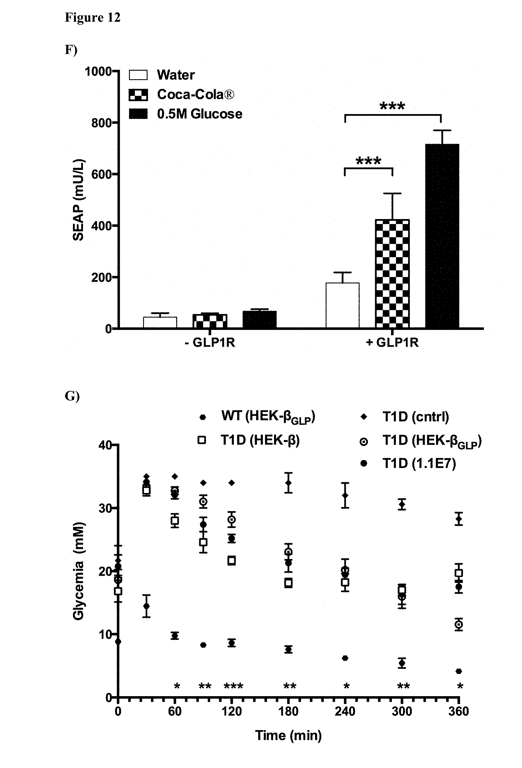
D00022

D00023

D00024

D00025

D00026

D00027

S00001
XML
uspto.report is an independent third-party trademark research tool that is not affiliated, endorsed, or sponsored by the United States Patent and Trademark Office (USPTO) or any other governmental organization. The information provided by uspto.report is based on publicly available data at the time of writing and is intended for informational purposes only.
While we strive to provide accurate and up-to-date information, we do not guarantee the accuracy, completeness, reliability, or suitability of the information displayed on this site. The use of this site is at your own risk. Any reliance you place on such information is therefore strictly at your own risk.
All official trademark data, including owner information, should be verified by visiting the official USPTO website at www.uspto.gov. This site is not intended to replace professional legal advice and should not be used as a substitute for consulting with a legal professional who is knowledgeable about trademark law.