Modalities For The Treatment Of Degenerative Diseases Of The Retina
Klimanskaya; Irina V. ; et al.
U.S. patent application number 16/359832 was filed with the patent office on 2019-09-19 for modalities for the treatment of degenerative diseases of the retina. This patent application is currently assigned to Astellas Institute for Regenerative Medicine. The applicant listed for this patent is Astellas Institute for Regenerative Medicine. Invention is credited to Irina V. Klimanskaya, Robert P. Lanza.
| Application Number | 20190282622 16/359832 |
| Document ID | / |
| Family ID | 67904837 |
| Filed Date | 2019-09-19 |


View All Diagrams
| United States Patent Application | 20190282622 |
| Kind Code | A1 |
| Klimanskaya; Irina V. ; et al. | September 19, 2019 |
MODALITIES FOR THE TREATMENT OF DEGENERATIVE DISEASES OF THE RETINA
Abstract
This invention relates to methods for improved cell-based therapies for retinal degeneration and for differentiating human embryonic stem cells and human embryo-derived into retinal pigment epithelium (RPE) cells and other retinal progenitor cells.
| Inventors: | Klimanskaya; Irina V.; (Upton, MA) ; Lanza; Robert P.; (Clinton, MA) | ||||||||||
| Applicant: |
|
||||||||||
|---|---|---|---|---|---|---|---|---|---|---|---|
| Assignee: | Astellas Institute for Regenerative
Medicine Marlborough MA |
||||||||||
| Family ID: | 67904837 | ||||||||||
| Appl. No.: | 16/359832 | ||||||||||
| Filed: | March 20, 2019 |
Related U.S. Patent Documents
| Application Number | Filing Date | Patent Number | ||
|---|---|---|---|---|
| 15653472 | Jul 18, 2017 | |||
| 16359832 | ||||
| 14692191 | Apr 21, 2015 | 9730962 | ||
| 15653472 | ||||
| 12781929 | May 18, 2010 | 9040770 | ||
| 14692191 | ||||
| 11186720 | Jul 20, 2005 | 7736896 | ||
| 12781929 | ||||
| 11041382 | Jan 24, 2005 | 7794704 | ||
| 11186720 | ||||
| 60538964 | Jan 23, 2004 | |||
| Current U.S. Class: | 1/1 |
| Current CPC Class: | A61K 35/12 20130101; C12N 2501/15 20130101; C12N 2501/155 20130101; C12N 2533/52 20130101; A61K 9/0048 20130101; C12N 5/062 20130101; C12N 2501/01 20130101; C12N 2506/02 20130101; C12N 2501/115 20130101; C12N 2533/90 20130101; A61K 35/44 20130101; C12N 2501/33 20130101; C12N 2533/54 20130101; A61K 35/30 20130101; C12N 5/0621 20130101 |
| International Class: | A61K 35/30 20060101 A61K035/30; A61K 9/00 20060101 A61K009/00; A61K 35/44 20060101 A61K035/44; C12N 5/0793 20060101 C12N005/0793; C12N 5/079 20060101 C12N005/079 |
Claims
1. A method of treating or preventing retinal degeneration, comprising use of a cell selected from the group consisting of at least one of: RPE cells, RPE-like cells, RPE or RPE-like progenitors derived from mammalian embryonic stem cells.
2. The method of claim 1, wherein the condition of retinal degeneration is selected from the group consisting of at least one of: retinitis pigmentosa and macular degeneration.
3. The method of claim 1, further comprising transplantation of the cell by vitrectomy surgery into the subretinal space of the eye.
4. The method of claim 3, wherein the cells are transplanted in a suspension, matrix, or substrate.
5. The method of claim 2, wherein the retinitis pigmentosa is associated with an animal model.
6. The method of claim 5, wherein the animal model is selected from the group consisting of: rd mouse, RPE-65 knockout mouse, tubby-like mouse, RCS rat, Abyssinian cat, cone degeneration "cd" dog, progressive rod-cone degeneration "prcd" dog, early retinal degeneration "erd" dog, rod-cone dysplasia 1, 2 & 3 "rcd1, rcd2 and rcd3" dogs, photoreceptor dysplasia "pd" dog, and Briard "RPE-65" dog.
7. The method of claim 6, wherein the outcome of the therapy in the animal model is evaluated using one or more of behavioral tests, fluorescent angiography, histology, and functional testing such as measuring the ability of the cells to perform phagocytosis (photoreceptor fragments), vitamin A metabolism, tight junctions conductivity, or evaluation using electron microscopy.
8. A method for the spontaneous differentiation of hES cells or embryoid bodies into RPE cells, RPE-like cells, or RPE progenitor cells, said method comprising: a) allowing hES cell cultures to overgrow on MEF; b) allowing the hES cell cultures to form a thick multilayer of cells; c) culturing the hES cells; d) isolating and culturing the pigmented RPE, RPE-like, and/or RPE progenitor cells from the resultant cell cultures.
9. The method of claim 8, wherein the isolating and culturing of PRE-like cells in step d comprises: a) digesting the cultured hES cells or embryoid bodies with an enzyme; b) selectively isolating the pigmented cells; c) plating the isolated cells on gelatin or laminin for 1-2 days to form primary cultures (PO); d) continued culturing the primary culture for a period of up to 3 weeks; and, e) isolating the RPE-like cells.
10. The method of claim 9, wherein the enzyme is selected from the group consisting of one or more of trypsin, collagenase, and dispase.
11. The method of claim 8, wherein the RPE cells are grown to establish a new RPE cell line.
12. The method of claim 11, wherein the RPE cell line is differentiated into alternate lineages comprising treatment of the RPE cell line in culture with bFGF or FGF.
13. The method of claim 11, wherein the new RPE cell lines varies from the already established RPE cell lines in at least one of the characteristics selected from the group consisting of: growth rate, expression of pigment, de-differentiation in culture, and re-differentiation in culture, of RPE-like cells when they are derived from different ES cell lines.
14.-15. (canceled)
16. The method of claim 8, wherein the RPE-like cells are derived from a bank of ES or embryo-derived cells with homozygosity in the HLA region such that ES-derived cells have reduced complexity of their HLA antigens.
17. The method of claim 8, wherein the ES cells are derived from a human.
18. (canceled)
19. A method for isolating RPE-like cells comprising: a) culturing hES cells in medium that supports proliferation and transdifferentiation of hES cells to RPE-like cells; b) selecting the cells of step a) that exhibit the signs of differentiation along the neural lineage; c) passaging the cells selected in step b) using an enzyme selected from the group consisting of trypsin, collagenase IV, collagenase I, and dispase until pigmented epithelial islands appear or multiply in number, and d) selecting pigmented or non-pigmented cells passaged in step c) for establishment of high purity RPE-like cultures.
20. The method of claim 19 wherein the passaging of cells in step c) is repeated at least twice.
21. The method of claim 19 wherein the selection of cells in step b) is a selection of cells that express a nestin or Pax6 neural lineage-specific marker.
22. The method of claim 19, wherein said medium contains Serum Replacement.
23. The method of claim 22, wherein said medium comprises knockout high glucose DMEM supplemented with 500 u/ml Penicillin, 500 pg/ml streptomycin, 1% non-essential amino acids solution, 2 mM GlutaMAX I, 0.1 raM beta-mercaptoethanol, 4-80 ng/ml bFGF, and 8.4%-20% Serum Replacement.
24.-26. (canceled)
Description
CROSS-REFERENCE TO RELATED APPLICATIONS
[0001] This application is a continuation of U.S. application Ser. No. 15/653,472, filed Jul. 18, 2017, which is a continuation of U.S. application Ser. No. 14/692,191, filed Apr. 21, 2015, which is a continuation of U.S. application Ser. No. 12/781,929, filed May 18, 2010, which is a continuation of U.S. application Ser. No. 11/186,720, filed Jul. 20, 2005, which is a continuation-in-part of U.S. application Ser. No. 11/041,382, filed Jan. 24, 2005 and claims the benefit of the filing date of U.S. Provisional Application No. 60/538,964, filed Jan. 23, 2004, the contents of each of which are incorporated by reference herein in their entirety.
FIELD OF THE INVENTION
[0002] This invention relates generally to methods for improved cell-based therapies for retinal degeneration and other visual disorders as well as treatment of Parkinson's disease and for differentiating mammalian embryonic stem cells and mammalian embryo-derived cells into retinal pigment epithelium (RPE) cells and other eye tissue including, but not limited to, rods, cones, bipolar, corneal, neural, iris epithelium, and progenitor cells.
BACKGROUND OF THE INVENTION
[0003] Many parts of the central nervous system (CNS) exhibit laminar organization, and neuropathological processes generally involve more than one of these multiple cellular layers. Diseases of the CNS frequently include neuronal cell loss, and, because of the absence of endogenous repopulation, effective recovery of function following CNS-related disease is either extremely limited or absent. In particular, the common retinal condition known as age-related macular degeneration (AMD) results from the loss of photoreceptors together with the retinal pigment epithelium (RPE), with additional variable involvement of internuncial ("relay") neurons of the inner nuclear layer (INL). Restoration of moderate-to-high acuity vision, therefore, requires the functional replacement of some or all of the damaged cellular layers.
[0004] Anatomically, retinitis pigmentosa (RP), a family of inherited retinal degenerations, is a continuing decrease in the number of photoreceptor cells which leads to loss of vision. Although the phenotype is similar across most forms of RP, the underlying cellular mechanisms are diverse and can result from various mutations in many genes. Most involve mutations that alter the expression of photoreceptor-cell-specific genes, with mutations in the rhodopsin gene accounting for approximately 10% of these. In other forms of the disease, the regulatory genes of apoptosis are altered (for example, Bar and Pax2). AMD is a clinical diagnosis encompassing a range of degenerative conditions that likely differ in etiology at the molecular level. All cases of AMD share the feature of photoreceptor cell loss within the central retina. However, this common endpoint appears to be a secondary consequence of earlier abnormalities at the level of the RPE, neovascularization, and underlying Bruch's membrane. The latter may relate to difficulties with photoreceptor membrane turnover, which are as yet poorly understood. Additionally, the retinal pigment epithelium is one of the most important cell types in the eye, as it is crucial to the support of the photoreceptor function. It performs several complex tasks, including phagocytosis of shed outer segments of rods and cones, vitamin A metabolism, synthesis of mucoploysacharides involved in the metabolite exchange in the subretinal space, transport of metabolites, regulation of angiogenesis, absorption of light, enhancement of resolution of images, and the regulation of many other functions in the retina through secreted proteins such as proteases and protease inhibitors.
[0005] An additional feature present in some cases of AMD is the presence of aberrant blood vessels, which result in a condition known as choroidal neovascularization (CNV). This neovascular ("wet") form of AMD is particularly destructive and seems to result from a loss of proper regulation of angiogenesis. Breaks in Bruch's membrane as a result of RPE dysfunction allows new vessels from the choroidal circulation access to the subretinal space, where they can physically disrupt outer-segment organization and cause vascular leakage or hemorrhage leading to additional photoreceptor loss.
[0006] CNV can be targeted by laser treatment. Thus, laser treatment for the "wet" form of AMD is in general use in the United States. There are often undesirable side effects, however, and therefore patient dissatisfaction with treatment outcome. This is due to the fact that laser burns, if they occur, are associated with photoreceptor death and with absolute, irreparable blindness within the corresponding part of the visual field. In addition, laser treatment does not fix the underlying predisposition towards developing CNV. Indeed, laser burns have been used as a convenient method for induction of CNV in monkeys (Archer and Gardiner, 1981). Macular laser treatments for CNV are used much more sparingly in other countries such as the U.K. There is no generally recognized treatment for the more common "dry" form of AMD, in which there is photoreceptor loss overlying irregular patches of RPE atrophy in the macula and associated extracellular material called drusen.
[0007] Since RPE plays an important role in photoreceptor maintenance, and regulation of angiogenesis, various RPE malfunctions in vivo are associated with vision-altering ailments, such as retinitis pigmentosa, RPE detachment, displasia, atrophy, retinopathy, macular dystrophy or degeneration, including age-related macular degeneration, which can result in photoreceptor damage and blindness. Specifically and in addition to AMD, the variety of other degenerative conditions affecting the macula include, but are not limited to, cone dystrophy, cone-rod dystrophy, malattia leventinese, Doyne honeycomb dystrophy, Sorsby's dystrophy, Stargardt disease, pattern/butterfly dystrophies, Best vitelliform dystrophy, North Carolina dystrophy, central areolar choroidal dystrophy, angioid streaks, and toxic maculopathies.
[0008] General retinal diseases that can secondarily affect the macula include retinal detachment, pathologic myopia, retinitis pigmentosa, diabetic retinopathy, CMV retinitis, occlusive retinal vascular disease, retinopathy of prematurity (ROP), choroidal rupture, ocular histoplasmosis syndrome (POHS), toxoplasmosis, and Leber's congenital amaurosis. None of the above lists is exhaustive.
[0009] All of the above conditions involve loss of photoreceptors and, therefore, treatment options are few and insufficient.
[0010] Because of its wound healing abilities, RPE has been extensively studied in application to transplantation therapy. In 2002, one year into the trial, patients were showing a 30-50% improvement. It has been shown in several animal models and in humans (Gouras et al., 2002, Stanga et al., 2002, Binder et al., 2002, Schraermeyer et al., 2001, reviewed by Lund et al., 2001) that RPE transplantation has a good potential of vision restoration. However, even in an immune-privileged site such as the eye, there is a problem with graft rejection, hindering the progress of this approach if allogenic transplantation is used. Although new photoreceptors (PRCs) have been introduced experimentally by transplantation, grafted PRCs show a marked reluctance to link up with surviving neurons of the host retina. Reliance on RPE cells derived from fetal tissue is another problem, as these cells have shown a very low proliferative potential. Emory University researchers performed a trial where they cultured RPE cells from a human eye donor in vitro and transplanted them into six patients with advanced Parkinson's Disease. Although a 30-50% decrease in symptoms was found one year after transplantation, there is a shortage of eye donors, this is not yet FDA approved, and there would still exist a need beyond what could be met by donated eye tissue.
[0011] Thus far, therapies using ectopic RPE cells have been shown to behave like fibroblasts and have been associated with a number of destructive retinal complications including axonal loss (Villegas-Perez, et al, 1998) and proliferative vitreoretinopathy (PVR) with retinal detachment (Cleary and Ryan, 1979). RPE delivered as a loose sheet tends to scroll up. This results in poor effective coverage of photoreceptors as well as a multilayered RPE with incorrect polarity, possibly resulting in cyst formation or macular edema.
[0012] Delivery of neural retinal grafts to the subretinal (submacular) space of the diseased human eye has been described in Kaplan et al. (1997), Humayun et al. (2000), and del Cerro et al. (2000). A serious problem exists in that the neural retinal grafts typically do not functionally integrate with the host retina. In addition, the absence of an intact RPE monolayer means that RPE dysfunction or disruption of Bruch's membrane has not been rectified. Both are fundamental antecedents of visual loss.
[0013] Thus, there exists no effective means for reconstituting RPE in any of the current therapies and there remain deficiencies in each, particularly the essential problem of a functional disconnection between the graft and the host retina. Therefore there exists the need for an improved retinal therapy.
SUMMARY OF THE INVENTION
[0014] The purpose of the present invention is to provide improved methods for the derivation of eye cells including, but not limited to, neural cells, including horizontal cells and amacrine cells, retinal cells such as rods and cones, corneal cells, vascular cells, and RPE and RPE-like cells from stem cells and to provide improved methods and therapies for the treatment of retinal degeneration. In particular, these methods involve the use of RPE and RPE-like cells derived from human embryonic stem cells.
[0015] One embodiment of the present invention provides an improved method of generating cells for therapy for retinal degeneration using RPE cells, RPE-like cells, the progenitors of these cells or a combination of two or three of any of the preceding derived from mammalian embryonic stem cells in order to treat various conditions including but not limited to retinitis pigmentosa and macular degeneration and associated conditions. The cell types which can be produced using this invention include, but are not limited to, RPE, RPE-like cells, and RPE progenitors. Cells which may also be produced include iris pigmented epithelial (IPE) cells. Vision associated neural cells including internuncial neurons (e.g. "relay" neurons of the inner nuclear layer (INL)) and amacrine cells (interneurons that interact at the second synaptic level of the vertically direct pathways consisting of the photoreceptor-bipolar-ganglion cell chain--they are synaptically active in the inner plexiform layer (IPL) and serve to integrate, modulate and interpose a temporal domain to the visual message presented to the ganglion cell) can also be produced using this invention. Additionally, retinal cells, rods, cones, and corneal cells can be produced. In a further embodiment of the present invention, cells providing the vasculature of the eye can also be produced. The cells of the present invention may be transplanted into the subretinal space by using vitrectomy surgery. Non-limiting examples include the transplantation of these cells in a suspension, matrix, or substrate. Animal models of retinitis pigmentosa that may be treated include rodents (rd mouse, RPE-65 knockout mouse, tubby-like mouse, RCS rat, cats (Abyssinian cat), and dogs (cone degeneration "cd" dog progressive rod-cone degeneration "pred" dog, early retinal degeneration "erd" dog, rod-cone dysplasia 1, 2 & 3 "rcd1, rcd2 & rcd3" dogs, photoreceptor dysplasia "pd" dog, and Briard "RPE-65" (dog). Evaluation is performed using behavioral tests, fluorescent angiography, histology, or functional testing such as measuring the ability of the cells to perform phagocytosis (photoreceptor fragments), vitamin A metabolism, tight junctions conductivity, or evaluation using electron microscopy. One of the many advantages to the methods presented here is the ability to produce and treat many more patients than it would be possible to treat if one were limited to using eye donor tissue.
[0016] A further embodiment of the present invention provides methods for the spontaneous differentiation of hES cells into cells with numerous characteristics of RPE. These RPE preparations are capable of phenotypic changes in culture and maintaining RPE characteristics through multiple passages. The present invention also provides for methods of differentiation of established RPE cell lines into alternate neuronal lineages, corneal cells, retinal cells as a non-limiting example through the use of bFGF or FGF.
[0017] Another embodiment of the present invention is a method for the derivation of new RPE lines and progenitor cells from existing and new ES cell lines. There can be variations in the properties, such as growth rate, expression of pigment, or de-differentiation and re-differentiation in culture, of RPE-like cells when they are derived from different ES cell lines. There can be certain variations in their functionality and karyotypic stability, so it is desirable to provide methods for the derivation of new RPE lines and new ES cell lines which would allow choosing the lines with desired properties that can be clonally selected to produce a pure population of high quality RPE-like cells.
[0018] In yet another embodiment, the present invention provides an isolated RPE or RPE-like cell line which varies from established RPE cell lines in at least one of the characteristics selected from the group consisting of: growth rate, expression of pigment, de-differentiation in culture, and re-differentiation in culture.
[0019] Cells which may also be derived from existing and new ES cell lines include iris pigmented epithelial (IPE) cells. In an additional embodiment, vision associated neural cells including internuncial neurons (e.g. "relay" neurons of the inner nuclear layer (INL)) and amacrine cells can also be produced using this invention. Additionally, retinal cells, rods, cones, and corneal cells can be produced. In a further embodiment of the present invention, cells providing the vasculature of the eye can also be produced.
[0020] Another embodiment of the present invention is a method for the derivation of RPE lines or precursors to RPE cells that have an increased ability to prevent neovascularization. Such cells can be produced by aging a somatic cell from a patient such that telomerase is shortened where at least 10% of the normal replicative lifespan of the cell has been passed, then the use of said somatic cell as a nuclear transfer donor cell to create cells that overexpress angiogenesis inhibitors such as Pigment Epithelium Derived Factor (PEDF/EPC-1). Alternatively such cells may be genetically modified with exogenous genes that inhibit neovascularization.
[0021] Another embodiment of the present invention utilized a bank of ES or embryo-derived cells with homozygosity in the HLA region such that said cells have reduced complexity of their HLA antigens.
[0022] Therefore, an additional embodiment of the present invention includes the characterization of ES-derived RPE-like cells. Although the ES-derived pigmented epithelial cells strongly resemble RPE by their morphology, behavior and molecular markers, their therapeutic value will depend on their ability to perform RPE functions and to remain non-carcinogenic. Therefore, the ES-derived RPE cells are characterized using one or more of the following techniques: (i) assessment of their functionality, i.e. phagocytosis of the photoreceptor fragments, vitamin A metabolism, wound healing potential; (ii) evaluation of the pluripotency of RPE-like ES cells derivatives through animal model transplantations, (as a non-limiting example this can include SCID mice); (iii) phenoytping and karyotyping of RPE-like cells; (iv) evaluation of ES cells-derived RPE-like cells and RPE tissue by gene expression profiling, (v) evaluation of the expression of molecular markers of RPE at the protein level, including bestrophin, CRALBP, RPE-65, PEDF, and the absence of ES markers, and (vi) evaluation of the ratio of RPE and neural markers. The cells can also be evaluated based on their expression of transcriptional activators normally required for the eye development, including rx/rax, chx10/vsx-2/alx, ots-1, otx-2, six3/optx, six6/optx2, mitf, pax6/mitf, and pax6/pax2 (Fischer and Reh, 2001, Baumer et al., 2003).
[0023] An additional embodiment of the present invention is a method for the characterization of ES-derived RPE-like cells using at least one of the techniques selected from the group consisting of (i) assessment of the ES-derived RPE-like cells functionality; (ii) evaluation of the pluripotency of RPE-like ES cell derivatives through animal model transplantations; (iii) phenoytping and karyotyping of RPE-like cells; (iv) evaluation of gene expression profiling, (v) evaluation of the expression of molecular markers of RPE at the protein level; and (vi) the expression of transcriptional activators normally required for the eye development. In a further embodiment these techniques may be used for the assessment of multiple hES cell-derived cell types.
[0024] Another embodiment of the present invention is a method for the derivation of RPE cells and RPE precursor cells directly from human and non-human animal morula or blastocyst-staged embryos (EDCs) without the generation of ES cell lines.
[0025] Embryonic stem cells (ES) can be indefinitely maintained in vitro in an undifferentiated state and yet are capable of differentiating into virtually any cell type. Thus human embryonic stem (hES) cells are useful for studies on the differentiation of human cells and can be considered as a potential source for transplantation therapies. To date, the differentiation of human and mouse ES cells into numerous cell types have been reported (reviewed by Smith, 2001) including cardiomyocytes [Kehat et al. 2001, Mummery et al., 2003 Carpenter et al., 2002], neurons and neural precursors (Reubinoff et al. 2000, Carpenter et al. 2001, Schuldiner et al., 2001), adipocytes (Bost et al., 2002, Aubert et al., 1999), hepatocyte-like cells (Rambhatla et al., 2003), hematopoetic cells (Chadwick et al., 2003), oocytes (Hubner et all., 2003), thymocyte-like cells (Lin R Y et al., 2003), pancreatic islet cells (Kahan, 2003), and osteoblasts (Zur Nieden et al., 2003). Another embodiment of the present invention is a method of identifying cells such as RPE cells, hematopoietic cells, muscle cells, liver cells, pancreatic beta cells, neurons, endothelium, progenitor cells or other cells useful in cell therapy or research, derived from embryos, embryonic stem cell lines, or other embryonic cells with the capacity to differentiate into useful cell types by comparing the messenger RNA transcripts of such cells with cells derived in-vivo. This method facilitates the identification of cells with a normal phenotype and for deriving cells optimized for cell therapy for research.
[0026] The present invention provides for the differentiation of human ES cells into a specialized cell in the neuronal lineage, the retinal pigment epithelium (RPE). RPE is a densely pigmented epithelial monolayer between the choroid and neural retina. It serves as a part of a barrier between the bloodstream and retina, and it's functions include phagocytosis of shed rod and cone outer segments, absorption of stray light, vitamin A metabolism, regeneration of retinoids, and tissue repair. (Grierson et al., 1994, Fisher and Reh, 2001, Marmorstein et al., 1998). The RPE is easily recognized by its cobblestone cellular morphology of black pigmented cells. In addition, there are several known markers of the RPE, including cellular retinaldehyde-binding protein (CRALBP), a cytoplasmic protein that is also found in apical microvilli (Bunt-Milam and Saari, 1983); RPE65, a cytoplasmic protein involved in retinoid metabolism (Ma et al., 2001, Redmond et al., 1998); bestrophin, the product of the Best vitelliform macular dystrophy gene (VMD2, Marmorstein et al., 2000), and pigment epithelium derived factor (PEDF) a 48 kD secreted protein with angiostatic properties (Karakousis et al., 2001, Jablonski et al., 2000).
[0027] An unusual feature of the RPE is its apparent plasticity. RPE cells are normally mitotically quiescent, but can begin to divide in response to injury or photocoagulation. RPE cells adjacent to the injury flatten and proliferate forming a new monolayer (Zhao et al, 1997). Several studies have indicated that the RPE monolayer can produce cells of fibroblast appearance that can later revert to their original RPE morphology (Grierson et al., 1994, Kirchhof et al., 1988, Lee et al., 2001). It is unclear whether the dividing cells and pigmented epithelial layer are from the same lineage as two populations of RPE cells have been isolated: epithelial and fusiforms. (McKay and Burke, 1994). In vitro, depending on the combination of growth factors and substratum, RPE can be maintained as an epithelium or rapidly dedifferentiate and become proliferative (Zhao 1997, Opas and Dziak, 1994). Interestingly, the epithelial phenotype can be reestablished in long-term quiescent cultures (Griersion et al., 1994).
[0028] In mammalian development, RPE shares the same progenitor with neural retina, the neuroepithelium of the optic vesicle. Under certain conditions, it has been suggested that RPE can transdifferentiate into neuronal progenitors (Opas and Dziak, 1994), neurons (Chen et al., 2003, Vinores et al., 1995), and lens epithelium (Eguchi, 1986). One of the factors which can stimulate the change of RPE into neurons is bFGF (Opaz and Dziak, 1994, a process associated with the expression of transcriptional activators normally required for the eye development, including rx/rax, chx10/vsx-2/alx, ots-1, otx-2, six3/optx, six6/optx2, mitf, and pax6/pax2 (Fischer and Reh, 2001, Baumer et al., 2003). Recently, it has been shown that the margins of the chick retina contain neural stem cells (Fischer and Reh, 2000) and that the pigmented cells in that area, which express pax6/mitf, can form neuronal cells in response to FGF (Fisher and Reh, 2001).
[0029] The present invention provides for the derivation of trabecular meshwork cells from hES and also for genetically modified trabecular meshwork cells for the treatment of glaucoma.
[0030] The present invention also provides for the derivation of trabecular meshwork cells from RPE progenitors and RPE-like cells and also for genetically modified trabecular meshwork cells for the treatment of glaucoma.
[0031] In another embodiment, the present invention provides a method for isolating RPE-like cells. Such a method may comprise: a) culturing hES cells in medium that supports proliferation and transdifferentiation of hES cells to RPE-like cells; b) selecting the cells of step a) that exhibit the signs of differentiation along the neural lineage; c) passaging the cells selected in step b) using an enzyme, such as a or a combination of collagenase(s) and/or a dissociation buffer (non-limiting examples of these include trypsin, collagenase IV, collagenase I, dispase, EDTA, or other commercially available dissociation buffers) until pigmented epithelial islands appear or multiply in number, and d) selecting pigmented or non-pigmented cells passaged in step c) for establishment of high purity RPE-like cultures. In certain aspects, the hES cells of the invention may be cultured in any medium that supports proliferation and transdifferentiation. In other aspects, the hES cells are cultured in medium that contains Serum Replacement. In a specific aspect, the hES cells of the invention may be cutured in medium that includes knockout high glucose DMEM supplemented with 500 u/ml Penicillin, 500 .mu.g/ml streptomycin, 1% non-essential amino acids solution, 2 mM GlutaMAX I, 0.1 mM beta-mercaptoethanol, 4-80 ng/ml bFGF, and 8.4%-20% Serum Replacement. Optionally, the hES cells of the present invention are cultured in medium that further comprises 10-100 ng/ml human LIF. Optionally, the hES culture medium of the invention further comprises Plasmanate. Plasmanate may be added to a final concentration of about 1% to about 25% (e.g., about 1%0, 4%, 6%, 8%, 12%, 16% or 20%). In another aspect, the hES cells of the invention may be passaged repeatedly, including 2, 3, 5, 7, 10 or more times. Differentiating cells may be selected due to their expression of neural-lineage specific markers. Exemplary neural-lineage specific markers include and Pax6. In a preferred embodiment bFGF is added to the RPE cultures during proliferation and the cells are cultured without bFGF during differentiation.
[0032] The present invention includes methods for the derivation of RPE cells and RPE precursor cells directly from human and non-human animal morula or blastocyst-staged embryos (EDCs) without the generation of ES cell lines. In one embodiment, such a method comprises the steps of: a) maintaining ES cells in vitro in an undifferentiated state; b) differentiating the ES cells into RPE and RPE precursor cells; c) identifying the RPE cells by comparing the messenger RNA transcripts of such cells with cells derived in-vivo and/or identifying the RPE cells by comparing the protein expression profile with known RPE cells and/or phenotypic assessment; and e) identifying and/or isolating RPE cells and/or RPE precursors.
[0033] Further provided by the present invention are methods for the derivation of RPE lines or precursors to RPE cells that have an increased ability to prevent neovascularization, said methods comprising: a) aging a somatic cell from an animal such that telomerase is shortened wherein at least 10% of the normal replicative lifespan of the cell has been passed; and, b) using the somatic cell as a nuclear transfer donor cell to create cells that overexpress angiogenesis inhibitors, wherein the angiogenesis inhibitors can be Pigment Epithelium Derived Factor (PEDF/EPC-1).
[0034] The present invention provides methods for the treatment of Parkinson's disease with hES cell-derived RPE, RPE-like and/or RPE progenitor cells. These may be delivered by stereotaxic intrastriatal implantation with or microcarriers. Alternately, they may be delivered without the use of microcarriers. The cells may also be expanded in culture and used in the treatment of Parkinson's disease by any method known to those skilled in the art.
[0035] Other features and advantages of the invention will be apparent from the following detailed description.
BRIEF DESCRIPTION OF THE DRAWINGS
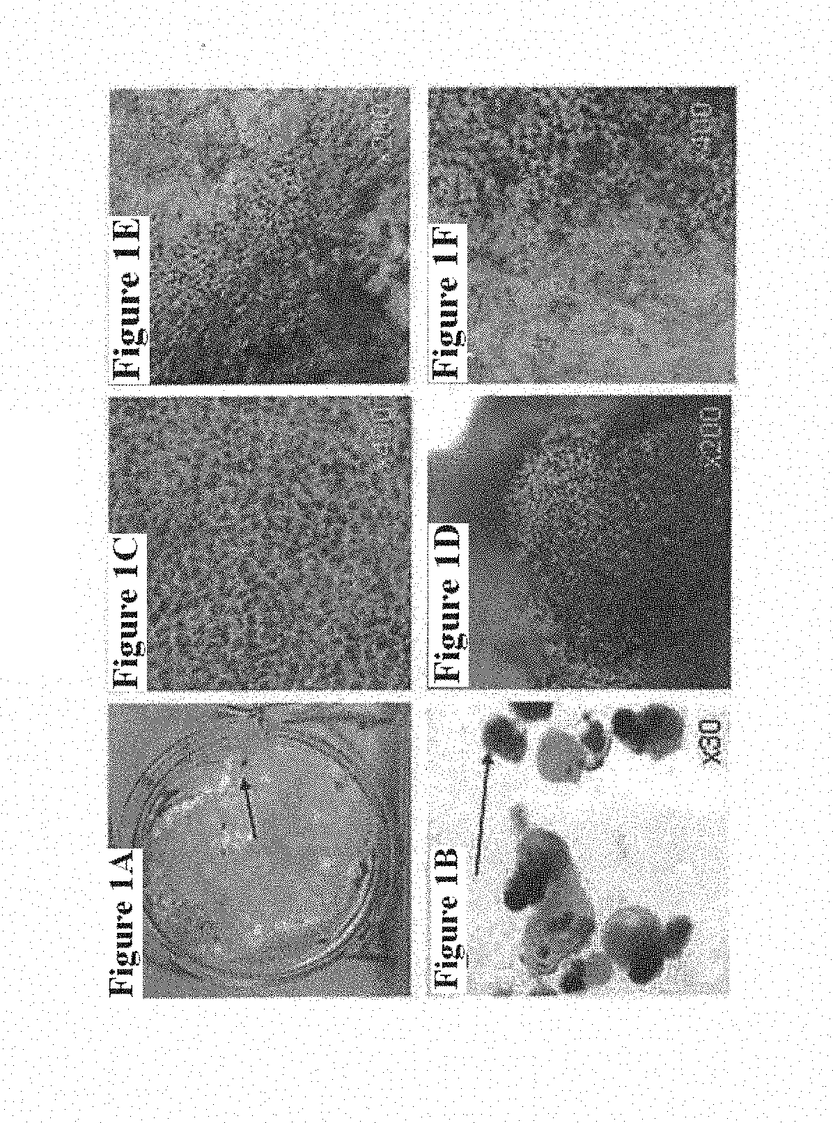
[0036] FIGS. 1A-1F are a series of photographs showing the appearance of pigmented areas (characteristic of RPE cells) in spontaneously differentiating hES cells. FIG. 1A is a photograph of pigmented regions in a 2.5 month old adherent culture, a well of a 4-weH plate, scanned: FIG. 1B is a photograph of pigmented regions in a 2.5 month old cultures grown as EB, at 45.times. magnification: FIG. 1C is a photograph of a pigmented area of an adherent culture: FIG. 1D is a photograph of a pigmented region of an EB;
[0037] FIG. 1E is a photograph of the boundary between pigmented region and the rest of the culture, .times.200: FIG. 1F is the same as FIG. 1E but at .times.400 magnification. Arrows in FIGS. 1A and 1B point to pigmented regions.
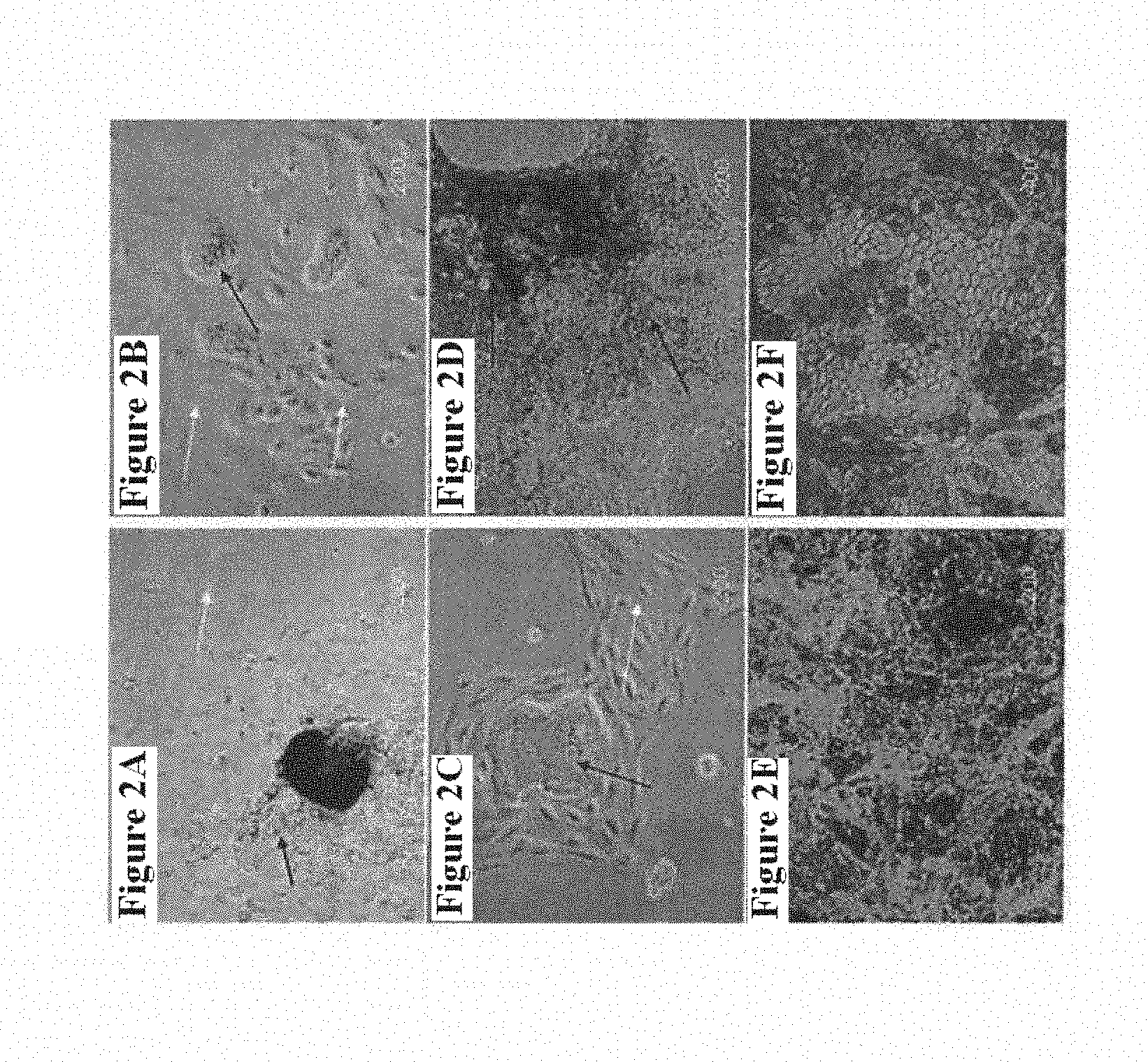
[0038] FIGS. 2A-2F are a series of photographs which show the loss and regain of pigmentation and epithelial morphology in culture. FIG. 2A is a photograph showing primary EB outgrowth, 1 week; FIG. 2B is a photograph showing the primary culture of cells, hand-picked, 1 week; FIG. 2C is a photograph showing epithelial islet surrounded by proliferating cells: FIG. 2D is a photograph showing the regain of pigmentation and epithelial morphology in 1 month old culture; FIG. 2E is a photograph showing the culture after 3 passages, .times.200 magnification; FIG. 2F shows the same culture as in FIG. 2E, .times.400 magnification, Hoffinan microscopy. Black arrows point to pigmented cells, white arrows show outgrowing cells with no pigment.
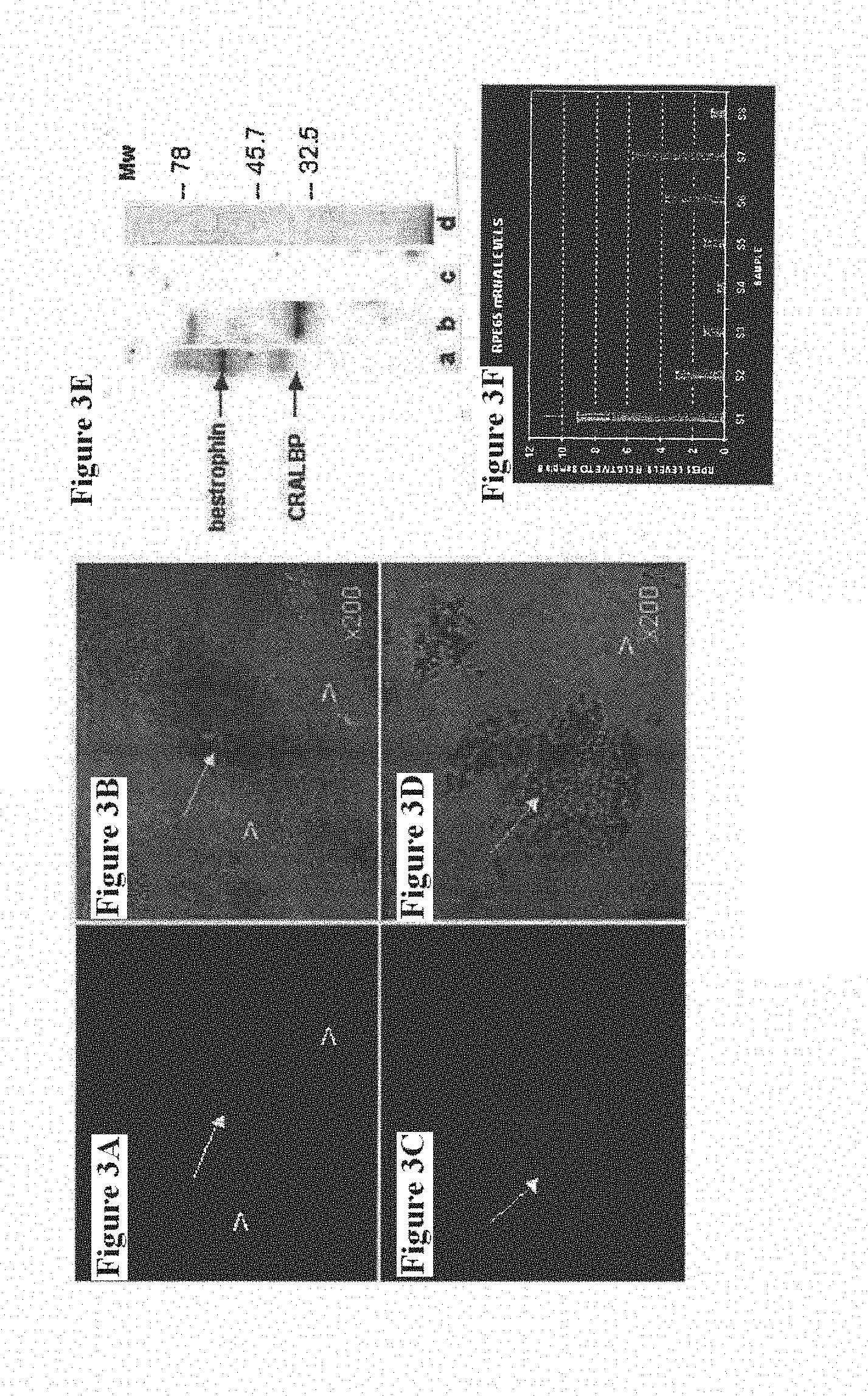
[0039] FIGS. 3A-3D are a series of photographs and one graph--these show markers of RPE in hES cells-derived pigmented epithelial cells. FIGS. 3A and 3B are photographs showing immunolocalization of RPE marker, bestrophin and corresponding phase microscopy field, .times.200 magnification; FIGS. 3C and 3D are photographs showing CRALBP and corresponding phase contrast microscopy field, .times.400 magnification. Arrows show the colocalization of bestrophin (FIG. 3A) and CRALBP (FIG. 3C) to pigmented cells (FIGS. 3C, 3D); arrowheads point to the absence of staining for these proteins (FIGS. 3A, 3B) in non-pigmented regions (FIGS. 3C, 3D).
[0040] FIG. 3E shows a photograph of Western blot of cell lysates with antibodies to bestrophin (a) and CRALBP (b): (c), (d)--undifferentiated hES cells, c-control to anti-CRALBP antibody, d-control to anti-bestrophin antibody
[0041] FIG. 3F shows a comparison of RPE65 expression in mature and immature RPE-like cells by real-time RT-PCR. Sample numbers 1, 6 and 7 are mature seven-weeks old culture; sample numbers 2,3 4 and 5 are immature fifteen-days old cultures; and sample number 8 is undifferentiated hES cells.
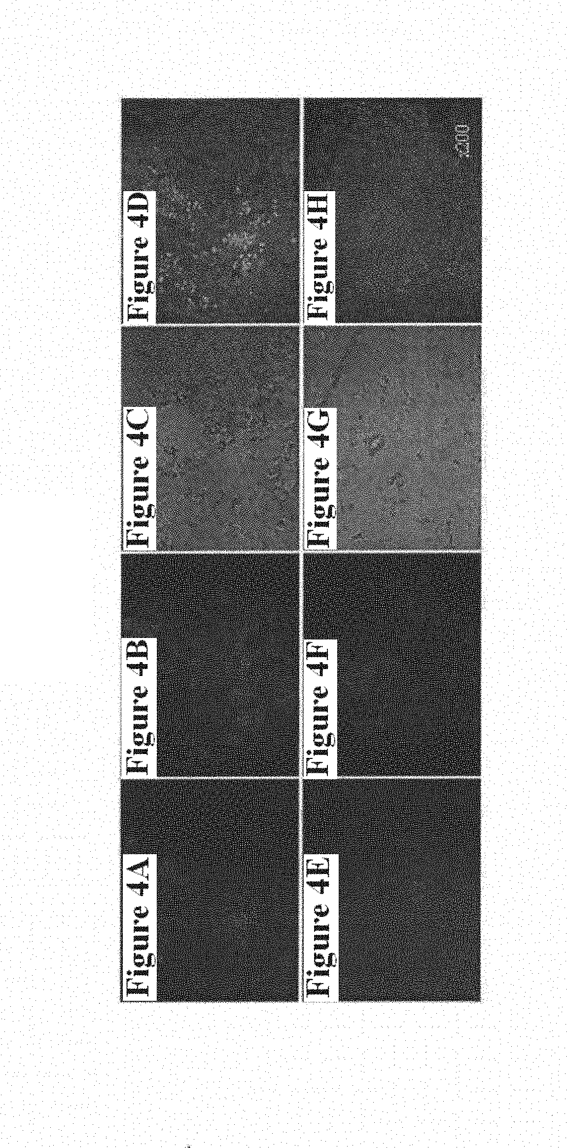
[0042] FIGS. 4A-4H show photographs which demonstrate the expression of markers of Pax6 (FIG. 4A), Pax2 (FIG. 4E) and mitf (FIG. 4B, FIG. 4F) in RPE-like cells in long-term quiescent cultures. FIG. 4C, FIG. 4G-phase contrast, FIG. 4D, FIG. 4H merged images of Pax6/mitf/phase contrast (FIG. 4A, FIG. 4B, FIG. 4C) and Pax2/mitf7phase contrast (FIG. 4E, FIG. 4F, FIG. 4G).
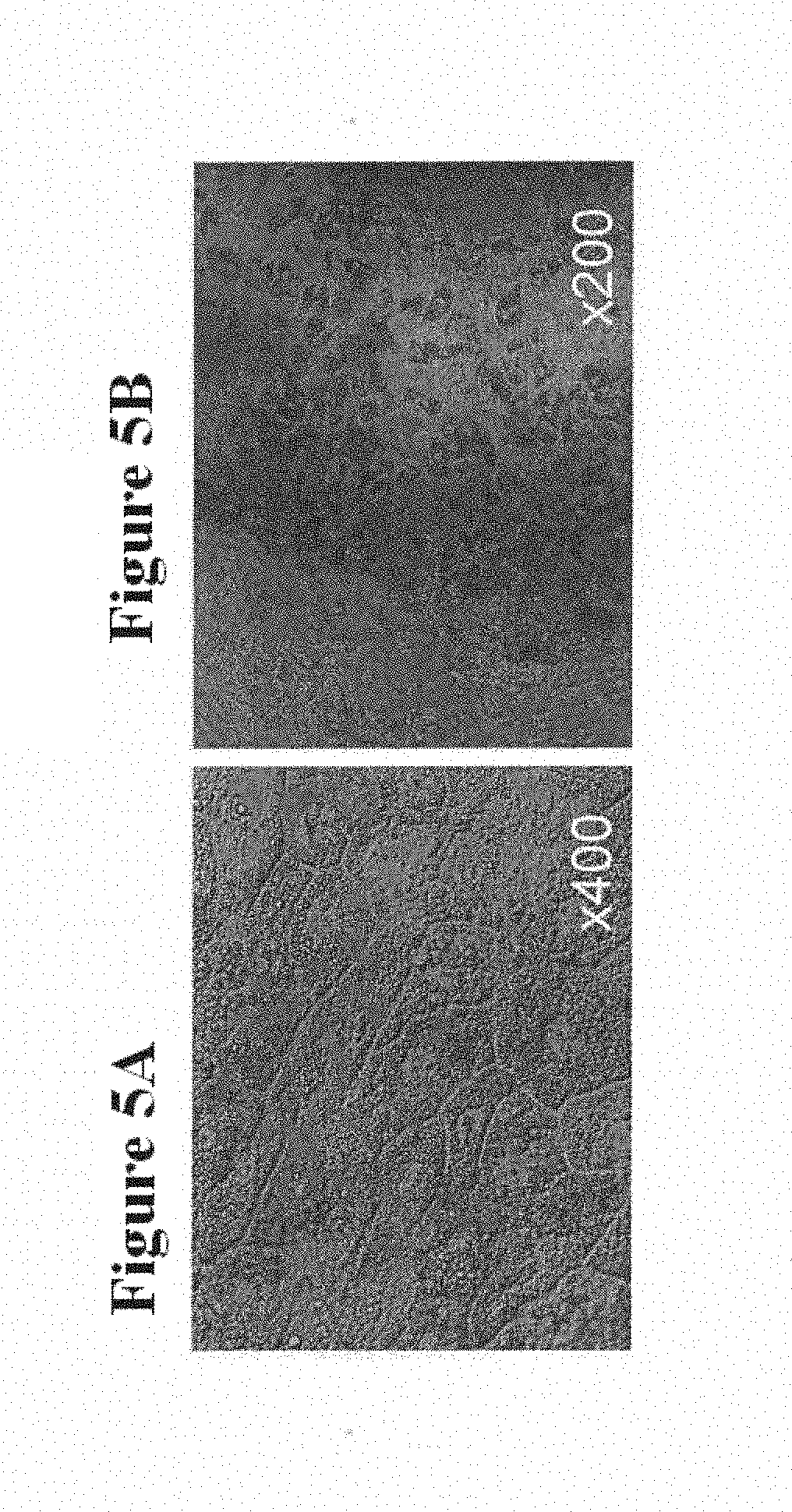
[0043] FIGS. 5A-5B show photographs of RPE differentiation in the culture of human embryo-derived cells bypassing the stage of derivation of ES cell lines.
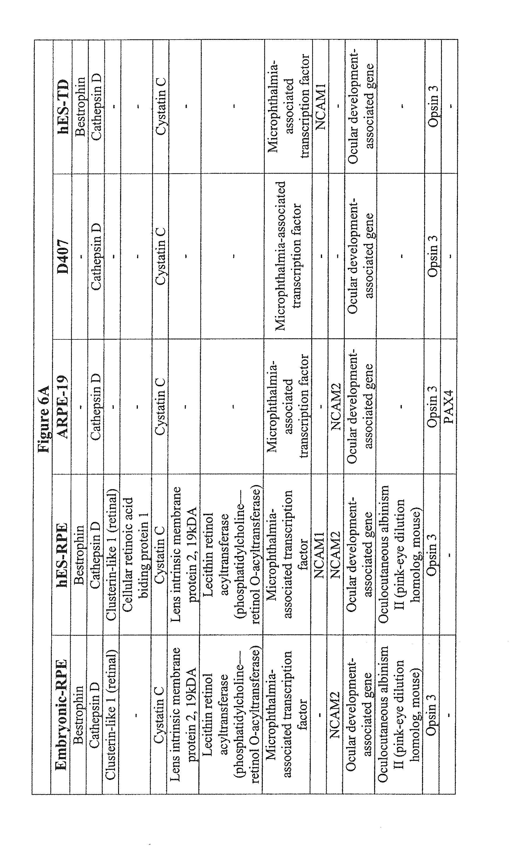
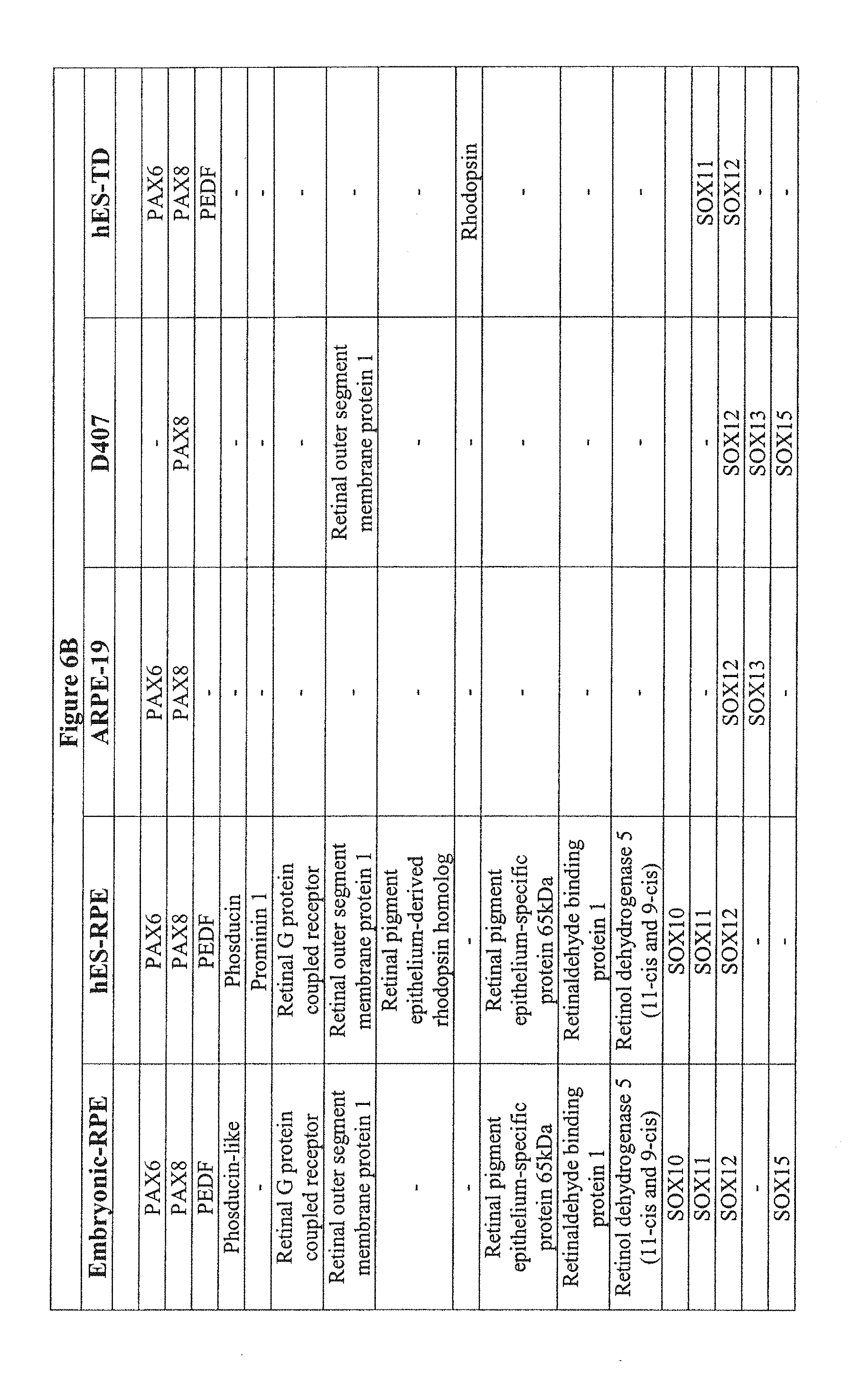
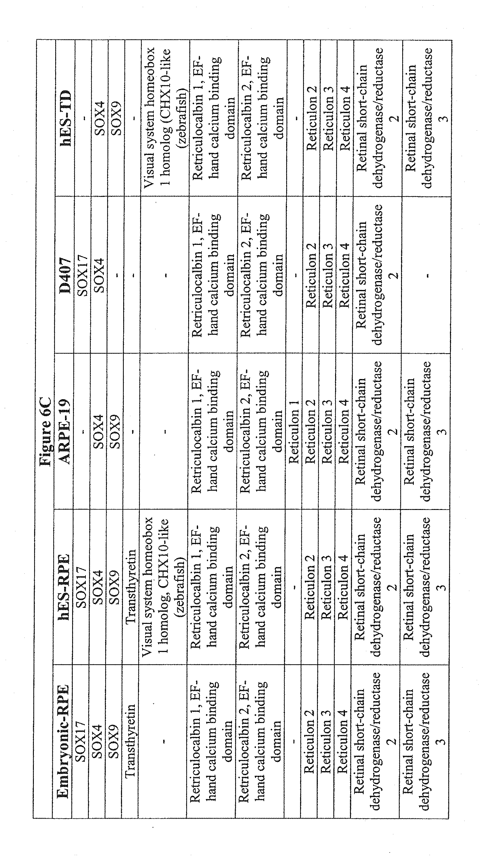
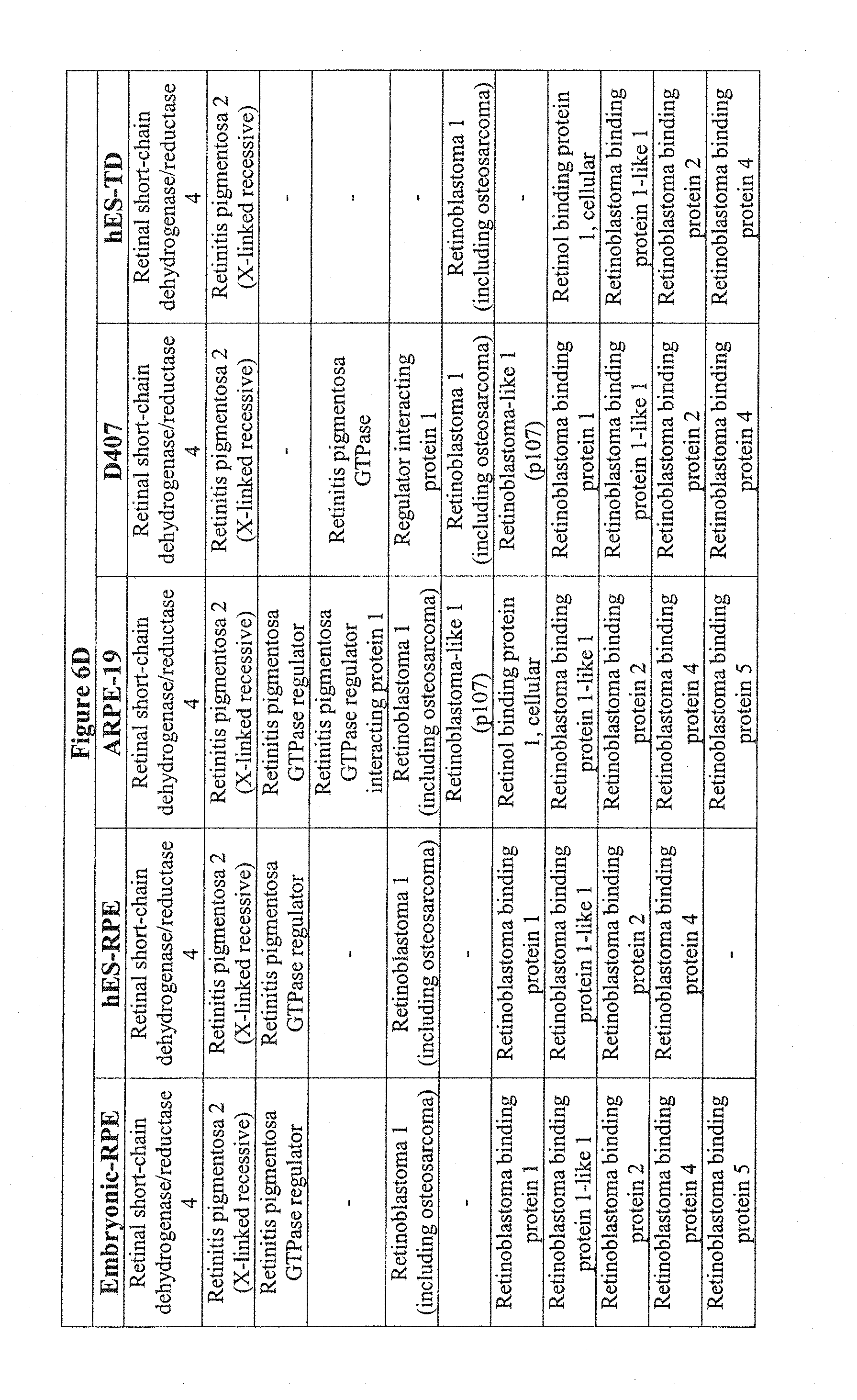
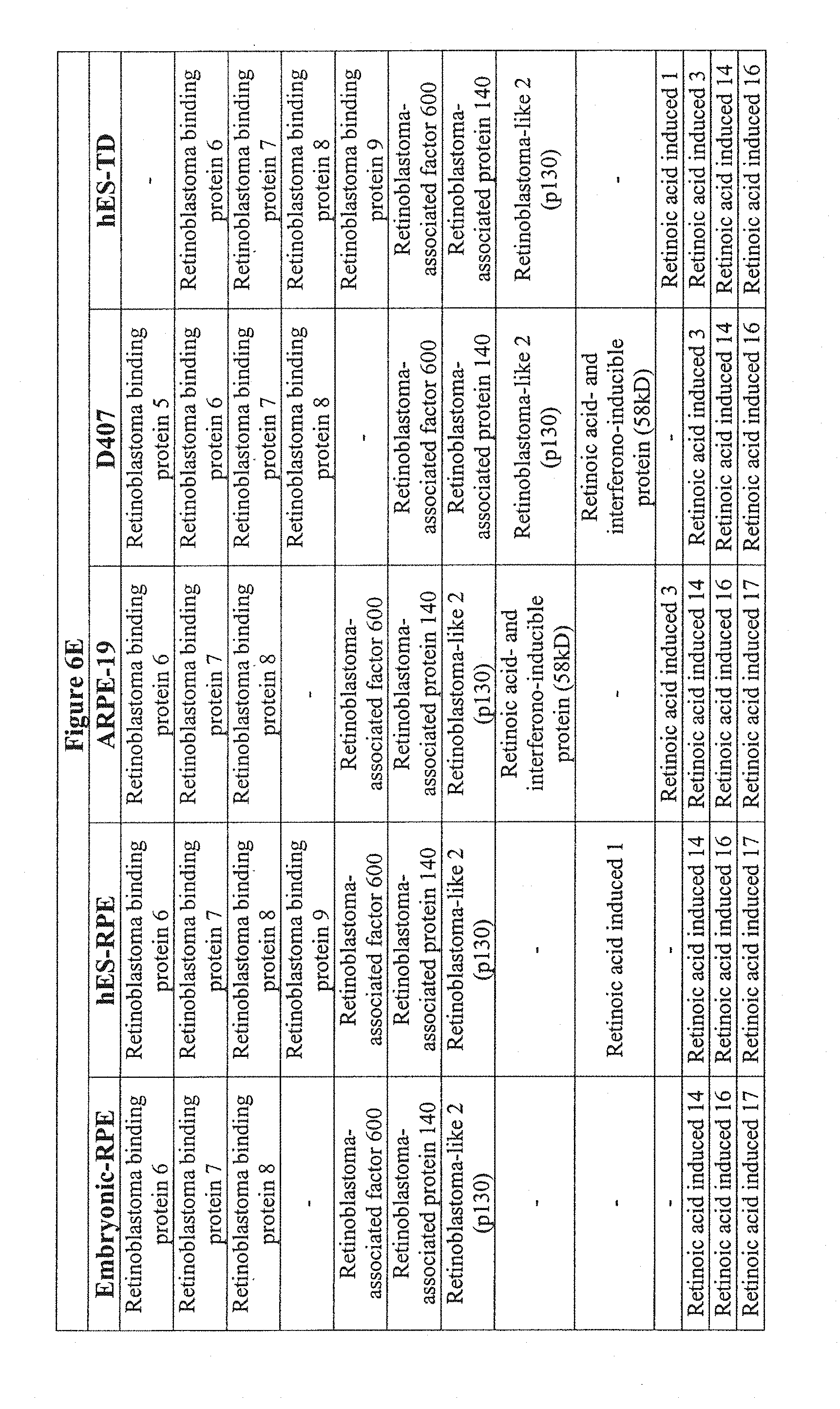
[0044] FIGS. 6A-6G show the transcriptional comparison of RPE preparations. FIG. 6A-6F--Based on the Ontological annotation, this table represents the expression patterns of RPE related genes for hES cell-derived retinal pigment epithelium (hES-RPE), hES cell derived transdifferentiated (hES-RPE-TD), ARPE-19 and D407, and freshly isolated human RPE (fe-RPE). FIG. 6G--Further data mining revealed known RPE specific ontologies, such as melanin biosynthesis, vision, retinol-binding, only in fetal RPE and ES-RPE but not ARPE-19.
[0045] FIGS. 7A-7D show generation of neural progenitors from hES cells. FIG. 7A) Overgrown culture of hES cell, stereomicroscopy. FIG. 7B) Spheroids (arrow in FIG. 7A) were removed and plated onto gelatin-coated plates in EB medium, producing spindle-like cells in 1-2 weeks. FIG. 7C), 7D) Staining of the cells shown in FIG. 7B) with antibodies to tubulin beta 111 (FIG. 7C) and nestin (FIG. 7D). Magnification: FIG. 7A), .times.60; FIGS. 7B-7D), .times.200.
[0046] FIGS. 8A-8D show morphology of different RPE cultures. FIG. 8A) Uniform differentiated RPE. FIG. 8B) Some elongated non-pigmented cells (arrows). FIG. 8C) Pigmented islands surrounded by non-piented cells, the culture described as a candidate for hand-picking of the pigmented cells after collagenase and/or dispase digestion. FIG. 8D) Transdifferentiated cells. Magnification .times.200.
[0047] FIGS. 9A-9E are a series of photographs showing the appearance of rod and cone-like structures in differtiating cultures of hES cells. FIG. 9A is a histological examination of differentiating cultures, stained with hematoxylin-eosin, .times.200. FIG. 9B-9E are RT-PCR analyses of Opsin 5 and Opsin 1 (FIG. 9B), recoverin (FIG. 9C), rhodopsin (FIG. 9D) and Keratin 12 (FIG. 9E) in these cultures.
DETAILED DESCRIPTION OF THE INVENTION
[0048] Various embodiments of the invention are described in detail and may be further illustrated by the provided examples. As used in the description herein and throughout the claims that follow, the meaning of"a," "an," and "the" includes plural reference unless the context clearly dictates otherwise. Also, as used in the description herein, the meaning of "in" includes "in" and "on" unless the context clearly dictates otherwise.
[0049] Throughout this specification and claims, the word "comprise," or variations such as "comprises" or "comprising," will be understood to imply the inclusion of a stated integer or group of integers but not the exclusion of any other integer or group of integers.
[0050] The terms used in this specification generally have their ordinary meanings in the art, within the context of the invention, and in the specific context where each term is used. Certain terms that are used to describe the invention are discussed below, or elsewhere in the specification, to provide additional guidance to the practitioner in describing the compositions and methods of the invention and how to make and use them. For convenience, certain terms may be highlighted, for example using italics and/or quotation marks. The use of highlighting has no influence on the scope and meaning of a term; the scope and meaning of a term is the same, in the same context, whether or not it is highlighted. It will be appreciated that the same thing can be said in more than one way. Consequently, alternative language and synonyms may be used for any one or more of the terms discussed herein, nor is any special significance to be placed upon whether or not a term is elaborated or discussed herein. Synonyms for certain terms are provided. A recital of one or more synonyms does not exclude the use of other synonyms. The use of examples anywhere in this specification, including examples of any terms discussed herein, is illustrative only, and in no way limits the scope and scope of the invention so long as data are processed, sampled, converted, or the like according to the invention without regard for any particular theory or scheme of action.
Definitions
[0051] By "embryo" or "embryonic" is meant a developing cell mass that has not implanted into the uterine membrane of a maternal host. An "embryonic cell" is a cell isolated from or contained in an embryo. This also includes blastomeres, obtained as early as the two-cell stage, and aggregated blastomeres.
[0052] The term "embryonic stem cells" refers to embryo-derived cells. More specifically it refers to cells isolated from the inner cell mass of blastocysts or morulae and that have been serially passaged as cell lines.
[0053] The term "human embryonic stem cells" (hES cells) refers human embryo-derived cells. More specifically hES refers to cells isolated from the inner cell mass of human blastocysts or morulae and that have been serially passaged as cell lines and can also include blastomeres and aggregated blastomeres.
[0054] The term "human embryo-derived cells" (hEDC) refers to morula-derived cells, blastocyst-derived cells including those of the inner cell mass, embryonic shield, or epiblast, or other totipotent or pluripotent stem cells of the early embryo, including primitive endoderm, ectoderm, and mesoderm and their derivatives, also including blastomeres and cell masses from aggregated single blastomeres or embryos from varying stages of development, but excluding human embryonic stem cells that have been passaged as cell lines.
[0055] Embryonic stem (ES) cells which have the ability to differentiate into virtually any tissue of a human body can provide a limitless supply of rejuvenated and histocompatible cells for transplantation therapy, as the problem of immune rejection can be overcome with nuclear transfer and parthenogenetic technology. The recent findings of Hirano et al (2003) have shown that mouse ES cells can produce eye-like structures in differentiation experiments in vitro. Among those, pigmented epithelial cells were described, resembling retinal pigment epithelium. Preliminary experiments carried out at Advanced Cell Technology with primate and human ES cell lines show that in a specialized culture system these cells differentiate into RPE-like cells that can be isolated and passaged. Human and mouse NT, Cyno parthenote ES cell derivatives have multiple features of RPE: these pigmented epithelial cells express four molecular markers of RPE-bestrophin, CRALBP, PEDF, and RPE65; like RPE, their proliferation in culture is accompanied by dedifferentiation--loss of pigment and epithelial morphology, both of which are restored after the cells form a monolayer and become quiescent. Such RPE-like cells can be easily passaged, frozen and thawed, thus allowing their expansion. Histological analysis of differentiating ES cultures shows a pattern of cells consistent with early retinal development, including aggregates of cells similar to rods and cones.
[0056] RPE Transplantation
[0057] At present, chronic, slow rejection of the RPE allografts prevents scientists from determining the therapeutic efficacy of this RPE transplantation. Several methods are being considered to overcome this obstacle. The easiest way is to use systemic immunosuppression, which is associated with serious side-effects such as cancer and infection. A second approach is to transplant the patient's own RPE, i.e. homografts, but this has the drawback of using old, diseased RPE to replace even more diseased RPE. Yet, a third approach is to use iris epithelium (IPE) from the same patient but this has the drawback that IPE may not perform all the vision related functions of RPE.
[0058] The present invention substantially reduces the possibility that transplantation rejection will occur, because RPE or RPE-like cells derived from hES cells could be derived from a bank of hES cells with homozygosity in the HLA region or could be derived from cloned hES cell lines . . . . Also, nuclear transfer and parthenogenesis facilitate histocompatibility of grafted RPE cells and progenitors.
[0059] RPE Defects in Retinitis Pigmentosa
[0060] Retinitis pigmentosa is a hereditary condition in which the vision receptors are gradually destroyed through abnormal genetic programming. Some forms cause total blindness at relatively young ages, where other forms demonstrate characteristic "bone spicule" retinal changes with little vision destruction. This disease affects some 1.5 million people worldwide. Two gene defects that cause autosomal recessive RP have been found in genes expressed exclusively in RPE: one is due to an RPE protein involved in vitamin A metabolism (cis retinaldehyde binding protein), a second involves another protein unique to RPE, RPE65. With the use of hES cell derived RPE cell lines cultured without the use of non-human animal cells, both of these forms of RP should be treatable immediately by RPE transplantation. This treatment was inconceivable a few years ago when RP was a hopelessly untreatable and a poorly understood form of blindness.
[0061] New research in RPE transplantation suggests there is promise for the treatment of retinal degeneration, including macular degeneration. In addition, a number of patients with advanced RP have regained some useful vision following fetal retinal cell transplant. One of the patients, for instance, improved from barely seeing light to being able to count fingers held at a distance of about six feet from the patient's face. In a second case, vision improved to ability to see letters through tunnel vision. The transplants in these studies were performed by injection, introducing the new retinal cells underneath the existing neural retina. Not all of the cells survived since the transplanted fetal cells were allogeneic (i.e. not genetically-matched), although those that did survive formed connections with other neurons and begin to function like the photoreceptors around them. Approximately a year after the first eight people received the transplants, four have recovered some visual function and a fifth shows signs of doing so.
[0062] Three newly derived human embryonic stem cell lines are similar in properties to those described earlier (Thomson et al. 1998, Reibunoff et al., 2000, Richards et al., 2000, Lanzendorf et al., 2001): they maintain undifferentiated phenotype and express known markers of undifferentiated hES cells, Oct-4, alkaline phosphatase, SSEA-3, SSEA-4, TRA-I-60, TRA-I-81 through 45 passages in culture or over 130 population doublings. All hES cell lines differentiate into derivatives of three germ layers in EB or long term adherent cultures and in teratomas. One of the differentiation derivatives of hES cells is similar to retinal pigment epithelium by the following criteria: morphologically, they have a typical epithelial cobblestone monolayer appearance and contain dark brown pigment in their cytoplasm, which is known to be present in the human body only in melanocytes, keratinocytes, retinal and iris pigment epithelium (IPE). Melanocytes, however, are non-epithelial cells, and keratinocytes don't secrete but only accumulate melanin. The set of RPE-specific proteins--bestrophin, CRALBP, PEDF--present in these cells indicates that they are likely to be similar to RPE and not IPE. Another similarity is the behavior of isolated pigmented cells in culture, when little or no pigment was seen in proliferating cells but was retained in tightly packed epithelial islands or re-expressed in newly established cobblestone monolayer after the cells became quiescent. Such behavior was described for RPE cells in culture (reviewed by Zhao et al., 1997), and it was previously reported (Vinores et al., 1995) that a neuronal marker tubulin beta III was specifically localized in dedifferentiating RPE cells in vitro and not in the cells with the typical RPE morphology suggesting that it reflects the plasticity of RPE and its ability to dedifferentiate to a neural lineage. The inventors have observed the same pattern of tubulin beta II localization in primary and passaged cultures of RPE and RPE-like cells which can reflect a dedifferentiation of such cells in culture or indicate a separate population of cells committed to a neuronal fate, that were originally located next to pigmented cells through differentiation of hES cells in long-term cultures and could have been co-isolated with RPE-like cells.
[0063] In the growing optic vesicle RPE and the neural retina share the same bipotential neuroepithelial progenitor, and their fate was shown to be determined by Pax2, Pax6, and Mitf (Baumer et al., 2003), the latter being a target of the first two. Pax6 at earlier stages acts as an activator of proneural genes and is downregulated in the RPE in further development, remaining in amacrine and ganglion cells in mature retina (reviewed by Ashery-Padan and Gruss, 2001). In goldfish, it is also found in mitotically active progenitors of regenerating neurons (Hitchcock et al., 1996). The inventors have found that many of the RPE-like cells expressed mitf and Pax6 in a pattern similar to tubulin beta m and were found only in non-pigmented cells of non-epithelial morphology that surround pigmented epithelial islands in long term cultures or in cells with a "partial" RPE phenotype (lightly pigmented and loosely packed). In proliferating cells in recently passaged cultures all these markers were found nearly in every cell suggesting either a reversal of RPE-like cells to progenitor stage at the onset of proliferation or massive proliferation of retinal progenitors. Interestingly, in teratomas where islands of pigmented cells of epithelial morphology were also found, Pax6 was expressed in non-pigmented cells adjacent to pigmented regions (data not shown). Multiple studies have previously shown dedifferentiation of RPE in culture and their transdifferentiation into cells of neuronal phenotype (Reh and Gretton, 1987, Skaguchi et al., 1997, Vinores et al., 1995, Chen et al., 2003), neuronal, amacrine and photoreceptor cells (Zhao et al., 1995), glia (Skaguchi et al., 1997), neural retina (Galy et al., 2002), and to neuronal progenitors (Opaz and Dziak, 1993). Such progenitors can in turn coexist with mature RPE-like cells in culture or appear as a result of dedifferentiation of RPE-like cells. At the same time, cells of neural retina can transdifferentiate into RPE in vitro (Opas et al., 2001), so alternatively, tubulin beta m and Pax6 positive cells could represent a transient stage of such transdifferentiation of co-isolated neural cells or neural progenitors into RPE-like cells.
[0064] Differentiation of hES cells into RPE-like cells happened spontaneously when using methods described in the Examples below, and the inventors noticed that pigmented epithelial cells reliably appeared in cultures older than 6-8 weeks and their number progressed overtime--in 3-5 months cultures nearly every EB had a large pigmented region. In addition to the described hES lines, six more newly derived hES lines turned into RPE-like cells, which suggests that since neural fate is usually chosen by ES cells spontaneously, RPE-like cells can arise by default as an advanced stage of such pathway. It is also possible that in such long term cultures, where differentiating hES cells form a multi-layered environment, permissive and/or instructive differentiation signals come from cxtracellular matrix and growth factors produced by differentiating derivatives of hES cells. The model of differentiation of hES cells into RPE-like cells could be a useful tool to study how such microenvironment orchestrates RPE differentiation and transdifferentiation.
[0065] RPE plays an important role in photoreceptor maintenance, and various RPE malfunctions in vivo are associated with a number of vision-altering ailments, such as RPE detachment, displasia, atrophy, retinopathy, retinitis pigmentosa, macular dystrophy or degeneration, including age-related macular degeneration, which can result in photoreceptor damage and blindness. Because of its wound healing abilities, RPE has been extensively studied in application to transplantation therapy. It has been shown in several animal models and in humans (Gouras et al., 2002, Stanga et al., 2002, Binder et al., 2002, Schraermeyer et al., 2001, reviewed by Lund et al., 2001) that RPE transplantation has a good potential of vision restoration. Recently another prospective niche for RPE transplantation was proposed and even reached the phase of clinical trials: since these cells secrete dopamine, they could be used for treatment of Parkinson disease (Subramanian, 2001). However, even in an immune-privileged eye, there is a problem of graft rejection, hindering the progress of this approach if allogenic transplant is used. The other problem is the reliance on fetal tissue, as adult RPE has a very low proliferative potential. The present invention decreases the likelihood that graft rejection will occur and removes the reliance on the use of fetal tissue.
[0066] As a source of immune compatible tissues, hES cells hold a promise for transplantation therapy, as the problem of immune rejection can be overcome with nuclear transfer technology. The use of the new differentiation derivatives of human ES cells, including retinal pigment epithelium-like cells and neuronal precursor cells, and the use of the differentiation system for producing the same offers an attractive potential supply of RPE and neuronal precursor cells for transplantation.
EXAMPLES
Example 1
Spontaneous Dfferentiation into Pigmented Epithelial Cells in Long Term Cultures
[0067] When hES cell cultures are allowed to overgrow on MEF in the absence of LIF, FGF and Plasmanate, they form a thick multilayer of cells. About 6 weeks later, dark islands of cells appear within the larger clusters (FIG. 1). These dark cells are easily seen with the naked eye and looked like "freckles" in a plate of cells as shown in FIG. 1A. At higher magnification these islands appear as tightly packed polygonal cells in a cobblestone monolayer, typical of epithelial cells, with brown pigment in the cytoplasm (FIG. 1C). There are differences in the amount of pigment in the cells with cells in the central part of the islands having the most pigment and those near the edges the least. (FIGS. 1E and IF).
[0068] When hES cells form embryoid bodies (EB)--pigmented epithelial cells appear in about 1-2% of EBs in the first 6-8 weeks (FIG. 1B). Over time more and more EBs develop pigmented cells, and by 3 months nearly every EB had a pigmented epithelial region (FIG. 1D). Morphology of the cells in the pigmented regions of EBs was very similar to that of adherent cultures (FIG. 1D).
Example 2
Isolation and Culture of Pigmented Epithelial Cells
[0069] The inventors isolated pigmented epithelial cells from both adherent hES cell cultures and from EBs. Pigmented polygonal cells were digested with enzymes (trypsin, and/or collagenase, and/or dispase), and the cells from these pigmented islands were selectively picked with a glass capillary. Although care was taken to pick only pigmented cells, the population of isolated cells invariably contained some non-pigmented cells. After plating cells on gelatin or laminin for 1-2 days, the cells were considered to be primary cultures (P0).
[0070] Primary cultures contained islands of pigmented polygonal cells as well as some single pigmented cells. After 3-4 days in culture, non-pigmented cells that seemed to have lost epithelial morphology (flatter and cells with lamellipodia) appeared at the periphery of some islands (FIG. 2). The number of such peripheral cells increased over time, suggesting that these cells were proliferating, and after 2 weeks most cells in the newly formed monolayer contained very little or no pigment. After continued culture, for another 2-3 weeks, pigmented epithelial cells began to reappear, visibly indistinguishable from those in the original cultures (FIG. 2).
Example 3
Detection of RPE Markers
[0071] The preliminary characterization of these differentiated human cells as RPE is based on their similarity to RPE cultures previously described; principally, their epithelial morphology and possession of pigment. There are three types of pigmented epithelial cells in human body: retinal and iris pigmented epithelium and keratinocytes, but the latter don't secrete pigment. The epithelial structure and cobblestone morphology are not shared by other pigmented cells, e.g. melanocytes. It is also noteworthy that RPE cells have been shown to lose and regain their pigment and epithelial morphology when grown in culture (Zhao 1997, Opas and Dziak, 1994), and the pigmented cells behaved in a similar manner, so to test the hypothesis that the ES derived cells may be RPE, they were stained with antibodies to known markers for RPE: bestrophin and CRALBP. FIG. 3 (left panel) shows membrane localization of bestrophin (A) and CRALBP (C), both are found in pigmented epithelial islands. Not all of the cells stain with these antibodies and intensity of staining correlated with pigment expression and "tightness" of colonies--the borders of each pigmented island where cells were larger and more loosely packed showed lower expression of both proteins.
[0072] To further characterize presumably RPE cells, analysis was performed on the expression of bestrophin, CRALBP by Western blotting. FIG. 3 (right panel, top) shows the bands, corresponding to bestrophin, 68 kD (a), CRALBP, 36 kD (b) in cell lysates. All these proteins were found in both primary cultures and subsequent passages.
[0073] Another known PRE marker, RPE65, was found in the RPE-like cells by real-time RT-PCR (FIG. 3, right panel, bottom). As shown in FIG. 3, right panel, bottom, expression of RPE65 was confirmed in all hES-RPE samples analyzed. Interestingly, mature cultures (seven weeks after passaging) had four to nine folds more RPE65 mRNA than the control undifferentiated hES cells, whereas earlier passage (two-week old) cultures only exceeded the control by 1.5 to 2.5 fold. See FIG. 3, right panel, bottom.
[0074] PEDF ELISA assay showed the presence of PEDF in cell lysates of all presumed RPE cultures, and Western blot showed a band of approximately 48 kD (not shown).
Detection of Markers of Neuronal and Retinal Progenitors in RPE-Like Cultures
[0075] PAX-6, Pax2, mitf and tubulin beta III were shown to be expressed in the majority of cells in recently passaged and only in a small number of cells in old cultures of RPE-like cells derived from hES cells (FIG. 4).
[0076] In proliferating cultures (day 3 after trypsinization) where RPE-like morphology of the proliferating cells is lost, nearly every cell showed the presence of mitf, Pax6, tubulin beta II and nestin. Pax2 was found only a small subset of cells which appeared mitf-negative, while there was a strong degree of co-localization of Pax6/mitf, mitf/tubulin beta II, and Pax6/tubulin beta m. In 21 days old quiescent cultures after pigmented epithelial islands were reestablished, groups of PAX-6 and mitf were found mostly in non-pigmented cells of non-epithelial morphology between pigmented epithelial islands (FIG. 4, A-C). and tubulin beta 11 had a similar pattern of distribution (not shown). However, there were populations of mitf-positive and Pax6-negative cells, located close to the periphery of pigmented islands (FIG. 4, A-C). Pax2 was found only in a very small subset of mitf-negative cells (FIG. 4, E-H). No presence of either of these proteins was ever detected in the cells of "mature" pigmented epithelial islands. However, these markers in cells that only had some RPE features were often visible, i.e. either looked epithelial but had no pigment or in certain single pigmented cells away from pigmented epithelial islands.
Example 4
Characterization of RPE-Like Cells Derived from hES Cell Line ACT J-1 from Cyno-1 ES Cells and Derivation of RPE-Like Cells from Existing hES Cell Lines H1, H9, and H7
[0077] An RPE-like cell line is expanded, tested for freezing and recovery, and characterized using the following methods and molecular markers of RPE cells: bestrophin and CRALBP by Western blot and immunofluorescence, PEDF by ELISA and Western blot, and RPE65 by RT-PCR. The cells are injected in SCID mice with undifferentiated hES or Cyno-1 cells as a control to evaluate tumorigenicity. Karyotyping of RPE-like cells will be done by a clinical laboratory on a commercial basis. Characterization of the functional properties of RPE-like cells and studies of their transplantation potential are then carried out as otherwise described in this application and also using those techniques known to those skilled in the art.
[0078] Gene expression profiling experiments are done using Affymetrix human genome arrays. Gene expression is compared in RPE-like cells derived from ES cells and in retinal samples from autopsies. Several animal models can be used to verify the effectiveness of the transplanted RPE-like cells, including but not limited to, rhesus monkey, rat, and rabbit.
Example 5
Optimization of the Differentiation Culture System Ensuring High Yields of RPE-Like Cells
[0079] ES cells are cultured on feeder cells or as embryoid bodies (EB) in the presence of factors such as bFGF, insulin, TGF-beta, IBMX, bmp-2, bmp-4 or their combinations, including stepwise addition. Alternatively, ES cells are grown on various extracellular matrix-coated plates (laminin, fibronectin, collagen 1, collagen IV, Matrigel, etc.) in evaluating the role of ECM in RPE formation. Expression of molecular markers of early RPE progenitors (Pax6, Pax2, mitf) and of RPE cells (CRALBP, bestrophin, PEDF, RPE65) are evaluated at various time intervals by real-time RT-PCR to verify and determine successful combinations of the above mentioned agents and stepwise procedure that produces enrichment in RPE-like cells or their progenitors. This approach can also be used to produce common progenitors of RPE and other eye tissues, such as photoreceptor or neural retina which can be isolated and further characterized for their differentiation potential and used in transplantation studies.
Example 6
Derivation of RPE and Other Eye Tissue Progenitors from Existing and New ES Cell Lines
[0080] Using the data from the gene expression profiling, expression of the RPE progenitor markers will be correlated with the expression of the surface proteins in order to find a unique combination of surface markers for RPE progenitor cells. If such markers are found, antibodies to surface proteins can be used to isolate a pure population of RPE progenitors that can be then cultured and further differentiated in culture or used in transplantation studies to allow their differentiation after grafting.
[0081] If the data from the gene expression profiling experiments is insufficient, to isolate the RPE progenitors the following approach will be used. ES cells and RPE-like cells will be transfected with GFP under the control of a promoter such as Pax6, and stable transfectants will be selected. From a culture of transfected differentiating ES cells or proliferating (dedifferentiated) RPE cells, GFP/Pax6-positive cells will be isolated by FACS and used as an antigen source for mouse injection to raise monoclonal antibodies to the surface molecules of Pax6 positive cells. Because Pax6 is present not only in RPE progenitors, screening will be done (by FACS) using several strategies: a) against proliferating RPE-like cells, b) against Pax2-positive RPE cells, c) against mitf-positive RPE cells. For b) and c) RPE cells will be transfected with GFP under the corresponding promoter, as a negative control, RPE or ES cells negative by these antigens will be used. After expansion of positive clones selected by all three strategies, antibodies will be tested against all types of cells used in screening and further analyzed: since this strategy can produce antibodies that recognize cell surface antigens specific and non-specific for RPE progenitors, the cells from differentiating total population of ES cells or of RPE cells selected with these antibodies will be assessed for molecular markers of RPE progenitors and for their ability to produce RPE.
[0082] Using the optimized defined stepwise procedures to produce RPE or other early progenitors of eye tissues and the antibodies to their unique surface markers, such progenitors will be isolated from differentiated ES cells and cultured in vitro. Their ability to differentiate into various tissues of the eye will be investigated using the strategy described in Aim 2.
[0083] ES cell lines that already produced RPE-like cells (H1, H7, H9, ACT J-1, ACT-4, Cyno-1), RPE-like cells will be used to continue to derive RPE-like cells and their progenitors as described in Aims 1 and 2. After expansion and characterization for molecular markers of RPE, these lines will be single-cell cloned, and the resulting lines will be characterized as described in Aim 1. The lines meeting criteria for RPE cells will be used for transplantation studies. New human ES cell lines will be derived from unused IVF embryos, from donated oocytes, stimulated to develop without fertilization (parthenote), and from generated developing blastocysts obtained from donated oocytes with the application of nuclear transfer technology. RPE-like cells and common eye progenitors will be derived from these lines using the approach in Aim 2, and the resulting lines will be characterized as in Aim 1. [Optional] new human ES cell lines will be derived in a virus-free system, characterized and submitted for clinical trials.
Example 7
Therapeutic Potential of RPE-Like Cells and Progenitors in Various Animal Models of Retinitis Pigmentosa & Macular Degeneration
[0084] Primate ES cells are tested in cynomologus monkeys (Macaques). Initially, vitrectomy surgery is performed and the cells are transplanted into the subretinal space of the animals. The first step is the transplantation of the cells in the suspension format after which a substrate or matrix is used to produce a monolayer transplantation. This can also be performed in immunosuppressed rabbits using cells derived from human ES-cells and also in various other animal models of retinitis pigmentosa, including rodents (rd mouse, RPE-65 knockout mouse, tubby-like mouse, RCS rat, cats (Abyssinian cat), and dogs (cone degeneration "cd" dog, progressive rod-cone degeneration "prcd" dog, early retinal degeneration "erd" dog, rod-cone dysplasia 1, 2 & 3 "rcd1, rcd2 & rcd3" dogs, photoreceptor dysplasia "pd" dog, and Briard "RPE-65) dog). Evaluation is performed using fluorescent angiography, histology (whether or not there is photoreceptor restoration and possibly ERG. Functional testing will also be carried out, including phagocytosis (photoreceptor fragments), vitamin A metabolism, tight junctions conductivity, and electron microscopy.
Example 8
Direct Differentiation of RPE Cells from Human Embryo-Derived Cells
[0085] Human blastocyst-staged embryos are plated in the presence of murine or chick embryo fibroblasts with or without immunosurgery to remove the trophectoderm or directly plates on extracellular matrix protein-coated tissue cultureware. Instead of culturing and passaging the cells to produce a human ES cell line, the cells are directly differentiated.
[0086] When hEDC cell cultures are allowed to overgrow on MEF in the absence of LIF, FGF and Plasmanate, they will form a thick multilayer of cells. (Alternate growth factors, media, and FBS can be used to alternate direct differentiation as is known to those skilled in the art.) About 6 weeks later, dark islands of cells will appear within the larger clusters. These dark cells are easily seen with the naked eye and looked like "freckles" in a plate of cells as shown in FIG. 5B. At higher magnification these islands appear as tightly packed polygonal cells in a cobblestone monolayer, typical of epithelial cells, with brown pigment in the cytoplasm (FIG. 5A). There are differences in the amount of pigment in the cells with cells in the central part of the islands having the most pigment and those near the edges the least. (FIG. 5B).
[0087] When hEDC cells are directly differentiated they may, though typically have not, formed embryoid bodies (EB). Pigmented epithelial cells appear in about 1-2% of these differentiated cells and/or EBs in the first 6-8 weeks. Over time more and more EBs develop pigmented cells, and by 3 months nearly every EB had a pigmented epithelial region. Morphology of the cells in the pigmented regions of EBs was very similar to that of adherent cultures.
Materials and Methods:
[0088] MEF medium: high glucose DMEM, supplemented with 2 mM GlutaMAX I, and 500 u/ml Penicillin, 500 .mu.g/ml streptomycin (all from Invitrogen) and 16% FCS (HyCLone). hES Cells Growth medium: knockout high glucose DMEM supplemented with 500 u/ml Penicillin, 500 .mu.g/ml streptomycin, 1% non-essential amino acids solution, 2 mM GlutaMAX I, 0.1 mM beta-mercaptoethanol, 4 ng/ml bFGF (Invitrogen), l-ng/ml human LIF (Chemicon, Temecula, Calif.), 8.4% of Serum Replacement (SR, Invitrogen) and 8.4% Plasmanate (Bayer). Derivation medium contained the same components as growth medium except that it had lower concentration of SR and Plasmanate (4.2% each) and 8.4% FCS and 2.times. concentration of human LIF and bFGF, as compared to growth medium. EB medium: same as growth medium except bFGF, LIF, and Plasmanate; the SR concentration was 13%. RPE medium: 50% EB medium and 50% MEF medium.
hES Cell Lines
[0089] Differentiation experiments were performed with adherent hES cells or with embryoid bodies (EBs). For adherent differentiation, hES cells were allowed to overgrow on MEFs until the hES colonies lost their tight borders at which time the culture media was replaced with EB medium (usually, 8-10 days after passaging). The medium was changed every 1-2 days. For EB formation, hES cells were trypsinized and cultured in EB medium on low adherent plates (Costar).
Immunostaining
[0090] Cells were fixed with 2% paraformnaldehyde, permeabilized with 0.1% NP-40 for localization of intracellular antigens, and blocked with 10% goat serum, 10% donkey serum (Jackson Immunoresearch Laboratories, West Grove, Pa.) in PBS (Invitrogen) for at least one hour. Incubation with primary antibodies was carried out overnight at 4.degree. C., the secondary antibodies (Jackson Immunoresearch Laboratories, West Grove, Pa.) were added for one hour. Between all incubations specimens were washed with 0.1% Tween-20 (Sigma) in PBS 3-5 times, 10-15 minutes each wash. Specimens were mounted using Vectashield with DAPI (Vector Laboratories, Burlingame, Calif.) and observed under fluorescent microscope (Nikon). Antibodies used: bestrophin (Novus Biologicals, Littleton, Colo.), anti-CRALBP antibody was a generous gift from Dr. Saari, University of Washington. Secondary antibodies were from Jackson Immunoresearch Laboratories, and Streptavidin-FITC was purchased from Amersham.
Isolation and Passaging of RPE-Like Cells
[0091] Adherent cultures of hES cells or EBs were rinsed with PBS twice and incubated in 0.25% Trypsin/l mM EDTA (Invitrogen) at 37.degree. C. until the monolayer loosened. Cells from the pigmented regions were scraped off with a glass capillary, transferred to MEF medium, centrifuged at 200.times.g, and plated onto gelatin-coated plates in RPE medium. The medium was changed after the cells attached (usually in 1-2 days) and every 5-7 days after that; the cells were passaged every 2-4 weeks with 0.05% Trypsin/0.53 mM EDTA (Invitrogen).
Western Blot and ELISA
[0092] Samples were prepared in Laemmli buffer (Laemmli, 1970), supplemented with 5% Mercaptoethanol and Protease Inhibitor Cocktail (Roche), boiled for 5 minutes and loaded onto a 8-16% gradient gel (Bio-Rad, Hercules, Calif.) using a Mini-Protean apparatus; the gels were run at 25-30 mA per gel; proteins were transferred to a 0.2 Nitrocellulose membrane (Schleicher and Shull, Keene, N.H.) at 20 volt overnight. Blots were briefly stained with Ponceau Red (Sigma) to visualize the bands, washed with Milli-Q water, and blocked for 1 hour with 5% non-fat dry milk in 0.1% TBST (Bio-Rad). Primary antibodies to bestrophin, CRALBP or PEDF (Chemicon) were added for 2 hours followed by three 15-minute washes with TBST; peroxidase-conjugated secondary antibodies were added for 1 hour, and the washes were repeated. Blots were detected using ECL system with Super-Signal reagent (Pierce). PEDF ELISA was performed on cell lysates using PEDF ELISA kit (Chemicon) according to manufacturer's protocol.
Real-Time RT-PCR
[0093] Total RNA was purified from differentiating ES cultures by a two-step procedure Crude RNA was isolated using Trizol reagent (Invitrogen) and further purified on RNeasy minicolumns (Qiagen). The levels of RPE65 transcripts were monitored by real-time PCR using a commercial primer set for RPE65 detection (Assay on Demand #Hs00165642_ml, Applied Biosystems) and Quantitect Probe RT-PCR reagents (Qiagen), according to the manufacturer's (Qiagen) protocol.
Example 9
Use of Transcriptomics to Identify Normal Differentiated Cells Differentiated Ex Vivo
[0094] hES-cell derivatives are likely to play an important role in the future of regenerative medicine. Qualitative assessment of these and other stem cell derivatives remains a challenge that could be approached using functional genomics. We compared the transcriptional profile of hES-RPE vs. its in vivo counterpart, fetal RPE cells, which have been extensively researched for its transplantation value. Both profiles were then compared with previously published (Rogojina et al., 2003) transcriptomics data on human RPE cell lines.
[0095] The gene expression profile of our data set was compared to two human RPE cell lines (non-transformed ARPE-19 and transformed D407, Rogojina et al., 2003) to determine whether hES-RPE have similar global transcriptional profiles. To account for common housekeeping genes expressed in all cells, we used publicly available Affymetrix data sets from undifferentiated hES cells (H1 line, h1-hES, --Sato et al., 2003) and bronchial epithelial cells (BE, Wright et al., 2004) as a control based on its common epithelial origin that would allow to exclude common housekeeping and epithelial genes and identify RPE-specific genes.
[0096] There were similarities and differences between hES-RPE, hES-RPE-TD, ARPE-19, D407. The similarities were further demonstrated by analyzing the exclusive intersection between those genes present in hES-RPE/ARPE-19 but not in BE (1026 genes). To account for background, we compared this to the exclusive intersection of genes present in BE/hES-RPE, but not ARPE-19 (186 genes), which results in a five- to six-fold greater similarity in hES-RPE and ARPE-19 when compared to BE. D407/ARPE19 appear to lose RPE specific genes such as RPE65, Bestrophin, CRALBP, PEDF, which is typical of long-term passaged cells (FIG. 6). Further data mining revealed known RPE specific ontologies such as melanin biosynthesis, vision, retinol-binding, only in fetal RPE and ES-RPE but not ARPE19.
[0097] Comparison of hES-RPE, ARPE-19 and D407 to their in vivo counterpart, freshly isolated human fetal RPE (feRPE), was in concordance with our previous data, demonstrating that the transcriptional identity of hES-RPE to human feRPE is significantly greater than D407 to fe RPE (2.3 fold difference--849 genes/373 genes) and ARPE-19 to feRPE (1.6 fold difference--588 genes/364 genes (FIG. 5c/5d). The RPE specific markers identified above, which were only present in hES-RPE and not in ARPE-19 or D407 were also present in feRPE, demonstrating a higher similarity of hES-RPE to its in vivo counterpart than of the cultured RPE lines.
[0098] Seven-hundred-and-eighty-four genes present in hES-RPE were absent in feRPE and ARPE-19 data sets. Since the retention of "sternmness" genes could potentially cause transformation of hES derivatives into malignant teratomas if transplanted into patients, we created a conservative potential "sternness" genes data using currently available Affymetrix microarray data sets Abeyta et.al 2004 Sato 2003). This resulted in a list of 3806 genes present in all 12 data sets (including common housekeeping genes). j Only 36 of the 784 genes present in the hES-RPE data set but not feRPE-ARPE-19 were common to the 3806 potential sternmness genes. None of these were known sternness genes such as Oct4, Sox2, TDGF1.
Example 10
Use of RPE Cells for Treatment of Parkinson's Disease
[0099] hRPE can be used as an alternative source of cells for cell therapy of Parkinson's Disease because they secrete L-DOPA. Studies have showed that such cells attached to gelatin-coated microcarriers can be successfully transplanted in hemiparkinsonian monkeys and produced notable improvements (10-50) thousand cells per target), and in FDA-approved trial started in 2000 the patients received hRPE intrastriatial transplants without adverse effects. One of the many advantages to the use of hES cell-derived RPE is that it circumvents the shortage of donor eye tissue. It also facilitates the use of gene therapy.
Example 11
Use of Stem Cell Derived RPE Cell Line for Rescuing or Preventing Photoreceptor loss
Derivation of RPE Cell Lines
[0100] Human embryonic stem ("hES") cells were grown in MEF medium containing high glucose DMEM, supplemented with 2 mM GlutaMAX I or glutamine, 500 u/ml Penicillin, 500 .mu.g/ml streptomycin (all from Invitrogen) and 16% FCS (can range from 8 to 20%) (HyCLone). hES cells may be grown in growth medium containing knockout high glucose DMEM supplemented with 500 u/ml Penicillin, 500 .mu.g/ml streptomycin, 1% non-essential amino acids solution, 2 mM GlutaMAX I, 0.1 mM beta-mercaptoethanol, 4 ng/ml (or up to 80) bFGF (Invitrogen), 10 ng/ml (or up to 100) human LIF (LIF is optional) (Chemicon, Temecula, Calif.), 8.4% of Serum Replacement (can be used up to 20%) (SR, Invitrogen) and 8.4% Plasmanate (optional) (Bayer). EB medium is the same as growth medium except bFGF, LIF, and Plasmanate are not included and the SR concentration was 13%. RPE medium is 50% EB medium and 50% MEF medium. Alternatively, hES cells can be cultured in the presence of human serum or FBS. For RPE culture, different media can be used that supports its proliferation, transdifferentiation and re-establishment of differentiated phenotype. Examples include, but are not limited to, high glucose DMEM supplemented with 2 mM GlutaMAX I or glutamine, 500 u/ml Penicillin, 500 .mu.g/ml streptomycin (antibiotics are optional) (all from Invitrogen) and 16% FCS (can range from 8 to 20%) (HyClone) or human serum; 1:1 mixture of Dulbecco's modified Eagle's medium and Ham's F12 medium containing 1.2 g/L sodium bicarbonate, 2.5 mM L-glutamine, 15 mM HEPES 0.5 mM sodium pyruvate; fetal bovine serum, 10%; (from ATCC, recommended for propagation of ARPE-19 cell line established from human RPE cells). A cell culture medium that supports the differentiation of human retinal pigment epithelium into functionally polarized monolayers may also be employed for this purpose.
[0101] RPE may be cultured as previously described (Hu and Bok, Molecular Vision (2000) 7:14-19, the disclosure of which is incorporated by reference) or other culture medium which has serum or serum replacement components or growth factor combination that supports RPE growth.
[0102] Two of the hES cell used for these studies were derived as described (Cowan et al., N. Eng. J. Med. 350: 1353-1356 (2004), Klimanskaya and McMahon, Handbook of Stem Cells, Vol. 1: Embryonic Stem Cells, Edited by Lanza et al., Elsevier/Academic Press, pp. 437-449 (2004), the disclosures of both are incorporated by reference), three lines were derived by Jamie Thomson (H1, H7, & H9) Human frozen blastocysts were donated to the study by couples who had completed their fertility treatment.
[0103] Differentiation experiments were performed with adherent hES cells or with embryoid bodies (EBs). For adherent differentiation, hES cells were allowed to overgrow on MEFs until the hES colonies lost their tight borders, at which time the culture media was replaced with EB medium (usually, 8-10 days after passaging). The medium was changed as it became yellow or every 1-2 days for dense cultures and less frequently for sparse cultures or EBs. For EB formation, hES cells were trypsinized and cultured in EB medium on low adherent plates (Costar).
Immunostaining
[0104] Cells were fixed with 2% paraformaldehyde, permeabilized with 0.1% NP-40 for localization of intracellular antigens, and blocked with 10% goat serum, 10% donkey serum (Jackson Immunoresearch Laboratories, West Grove, Pa.) in PBS (Invitrogen) for at least one hour. The specimen were then incubated with primary antibodies overnight at 4.degree. C., and then incubated with secondary antibodies (Jackson Immunoresearch Laboratories, West Grove, Pa.) for one hour. Between all incubations, the specimens were washed with 0.1% Tween-20 (Sigma) in PBS 3-5 times for 10-15 minutes each wash. Specimens were mounted using Vectashield with DAPI (Vector Laboratories, Burlingame, Calif.) and observed under fluorescent microscope (Nikon). Antibodies used include anti-bestrophin antibody (Novus Biologicals, Littleton, Colo.), and anti-CRALBP antibody (a gift from Dr. Saari, University of Washington). Secondary antibodies were obtained from Jackson Immunoresearch Laboratories, and Streptavidin-FITC was purchased from Amersham.
Isolation and Passaging of RPE-Like Cells
[0105] Adherent cultures of hES cells or EBs were rinsed with PBS twice and incubated in 0.25% Trypsin/1 mM EDTA (Invitrogen) at 37.degree. C. until the monolayer loosened. Cells from the pigmented regions were scraped off with a glass capillary, transferred to MEF medium, centrifuged at 200.times.g, and plated onto gelatin-coated plates in RPE medium. The medium was changed after the cells attached (usually in 1-2 days) and every 5-7 days after that. The cells were passaged every 2-4 weeks with 0.05% Trypsin/0.53 mM EDTA (Invitrogen).
[0106] Cells may be passaged or collected for transplantation using trypsin or collagenase IV, collagenase I, or dispase at concentrations of 1-10%. Any combination of these at concentrations of 1-10% each could be used instead of trypsin for isolation and passaging of pigmented cells. "Combination" in this context is intended to mean use of such enzymes together or sequentially--e.g., collagenase digestion followed by trypsin. The passage dilution may vary from no dilution to 1:6 or higher. The substrate for culture prior to transplantation may be anything that supports growth and features of hES-RPE, such as, but not limited to, gelatin, fibronectin, laminin, collagen or different types of extracellular matrix, uncoated plastic surface, filters--uncoated or coated with ECM (extracellular matrix) proteins, Matrigel, ECM isolated from other cell cultures, such as cornea, RPE, fibroblasts, uncoated beads, or beads coated with ECM. The time between passaging can vary from one day to several weeks. In prior experiments, nine-month old embryoid bodies with sheets of RPE on the surface were used to establish passagable cultures of hES-RPE so there is no limit known on how long the cells can be kept in culture without passaging.
[0107] Cultures may consist not only of cells with proper RPE morphology--i.e. polygonal tightly packed pigmented cells--but also of cells with varying degree of transdifferentiation (elongated pigmented or non-pigmented cells, etc.) and other cell types that co-differentiate from hES cells. Unless cells are individually selected for culturing, such cultures usually contain RPE islands that are separated by non-RPE cells.
[0108] The cultures of differentiating ES cells that exhibited the signs of differentiation along the neural lineage (expressing markers of this lineage, such as nestin, Pax6, etc., as could be detected by RT-PCR, Western blot, immunostaining, histology, or morphology of the individual cells which could be islands of pigmented cells, or epithelial sheets, or aggregates of vacuolated cells) were passaged with trypsin, collagenase, dispase, or mixture of such, expanded and cultured until the pigmented epithelial islands appeared or multiplied in numbers (usually, one or two passages). Such mixed cultures of pigmented epithelial and non-pigmented non-epithelial cells could be used to selectively hand-pick pigmented and non-pigmented cells after collagenase or collagenase-dispase digestion. These hand-picked pigmented and non-pigmented cells could then be dispersed into smaller aggregates and single cells or plated without dispersion, resulting in establishment of high purity RPE cultures.
Western Blot and ELISA
[0109] Samples were prepared in Laemmli buffer (Laemmli, 1970), supplemented with 5% Mercaptoethanol and Protease Inhibitor Cocktail (Roche), boiled for 5 minutes and loaded onto a 8-16% gradient gel (Bio-Rad, Hercules, Calif.) using a Mini-Protean apparatus; the gels were run at 25-30 mA per gel. Proteins were then transferred from the gel to a 0.2 Nitrocellulose membrane (Schleicher and Shull, Keene, N.H.) at 20 volt overnight. Blots were briefly stained with Ponceau Red (Sigma) to visualize the bands, washed with Milli-Q water, and blocked for 1 hour with 5% non-fat dry milk in 0.1% TBST(Bio-Rad). Primary antibodies to bestrophin, CRALBP or PEDF (Chemicon) were added to the blot for 2 hours followed by three 15-minute washes with TBST. peroxidase-conjugated secondary antibodies were then added to the blot for 1 hour, and the washes were repeated. Blots were detected using ECL system with Super-Signal reagent (Pierce). PEDF ELISA was performed on cell lysates using PEDF ELISA kit (Chemicon) according to the manufacturer's protocol.
Real-Time RT-PCR
[0110] Total RNA was purified from differentiating ES cultures by a two-step procedure. Crude RNA was isolated using Trizol reagent (Invitrogen) and further purified on RNeasy minicolumns (Qiagen). The levels of RPE65 transcripts were monitored by real-time PCR using a commercial primer set for RPE65 detection (Assay on Demand #Hs00165642_m1, Applied Biosystems) and QuantiTect Probe RT-PCR reagents (Qiagen), according to the manufacturer's (Qiagen) protocol.
Transplantation of hES-Derived RPE Cell Line
[0111] Cultures of hES cell lines may be used as transplant cells to one eye of 23-day old RCS rats to rescue or prevent photoreceptor loss. Cells of different morphology and/or of different degrees of differentiation may be chosen. Transplantation may be done as described (Lund et al. (2001) Proc. Natl. Acad. Sci. USA 98: 9942-47; Del Priore et al. Investigative Ophthalmology & Visual Science (2004) 45: 985-992; Gouras et al. (2002) Ophthalmology & Visual Science 43: 3307-11, the disclosures of which are incorporated by reference herein). Following digestion with trypsin or other enzyme (as described above), hES cells may be washed, and delivered trans-sclerally in a suspension at a density of 2.times.10.sup.5 cells per 2 .mu.l injection. Delivery may be achieved in Ham's F-10 medium with the use of a fine glass pipette with internal diameter of about 75-150 .mu.m. Injections are to be delivered into the dorso-temporal subretinal space of one eye of anesthetized 23-day old, dystrophic-pigmented RCS rats, at a time before functional deterioration and significant photoreceptor death. Sham-injected rats receive carrier medium alone without hES-derived cells. Histological assessment of postoperative rats may be done at shorter time points (e.g., at 1 month post-operatively) to assess short-term changes associated with transplantation and at longer time points (e.g., at 5 months post-operatively) to examine donor cell survival. Cells may be tagged or labeled by culturing in medium containing 20 .mu.m BrdUrd for 48 hours before transplantation.
Functional Assessment of Transplanted hES-Derived Cells
[0112] Also described in Lund et al. (2001) Proc. Natl. Acad. Sci. USA 98: 9942-47, behavioral assessment of grafted rats may be performed with a head-tracking apparatus that consists of a circular drum rotating at a constant velocity of 12 degrees/sec around a stationary holding chamber containing the animal. Presenting stimuli may be placed on interchangeable panels covered with black and white stripes with varying spatial frequencies such as 0.125, 0.25, and 0.5 cycles per degree. Animals may be tested at 10-20 weeks postoperatively. All animal assessments may be conducted blindly by a sole operator. Behavioral data may be analyzed using ANOVA.
[0113] Physiological studies may be conducted on animals with corneal electrocardiograms (ERGs) at 60 and 90 days. Prior to testing, the rats are to be adapted to the dark overnight and anesthetized under red light with ketamine and xylazine. See, for example, Peachy et al., Vis Neurosci. 2002 November-December; 19(6):693-701. The pupils are to be dilated and ERGs recorded from the cornea with a cotton wick saline electrode. Subcutaneous 30-gauge needles may be inserted into the forehead and trunk as reference and ground electrodes, respectively. While maintaining the subject rat body temperature at 35-36.degree. C., a light stimulus is to be applied at a maximum flash intensity measured at the cornea of about 0.7.times.103 .mu.W/cm.sup.2. Responses are to be recorded and averaged by a computerized data acquisition system at varying frequencies. ERG amplitudes are to be measured from the initial negative peak of the a-wave or from the baseline to the positive peak of the b-wave.
[0114] As in Lund et al. (2001) Proc. Natl. Acad. Sci. USA 98: 9942-47, threshold responses to illumination of visual receptive fields from the superior colliculus at 100 days are to be recorded. Animals are to be placed under terminal urethane anesthesia (1.25 g/kg i.p.). Data is to be collected over the entire visual field at independent points spaced roughly 200 .mu.m apart, with each point corresponding to about 10-15.degree. displacements in the visual field. Visual thresholds are to be measured as the increase in intensity over background and maintained at 0.02 cd/m.sup.2 [at least 2.6 logarithm (log) units below rod saturation] for activating units in the superficial 200 .mu.m of the superior colliculus with a spot light of 3.degree. diameter. Threshold maps are to be generated for each animal and illustrated as retinal representations.
[0115] Seven eyes transplanted with RPE cells derived from an H9 cell line and six eyes transplanted with RPE cells derived from a J1 cell line were subjected to ERG analysis. RPE transplants were conducted with 23 day old rats, and ERG analysis was done 36 days following transplantation. The H9 group yielded uniformly good responses, while the J1 group yielded 1 animal with only minimal response.
Histological Analysis of Transplanted Cells
[0116] As described in Lund et al. (2001) Proc. Natl. Acad. Sci. USA 98: 9942-47, animals are to be subjected to histological analysis. Animals may be euthanized with Euthanal and perfused transcardially with PBS following by periodate-lysine-paraformaldehyde (PLP). Eyes are to be sectioned and stained with cresyl violet. Eyes from animals of the BrdU group are to be labeled with anti-BrdUrd antibody and visualized with the use of an appropriate secondary antibody and respective reagents. Other eyes may be fixed by injection with 2.5% paraformaldehyde, 2.5% glutaraldehyde, and 0.01% picric acid in 0.1 M cacodylate buffer. Eyes may be postfixed in 1% osmium tetroxide, and subsequently dehydrated through graded alcohol to epoxypropane. Tissue may be embedded in resin from which semi-thin sections may be cut and stained with toluidine blue in 1% borate buffer.
Example 12
Use of Stem Cell Derived Neural Progenitor Cells for the Treatment of Retinal Degeneration
Generation of Neural Progenitors
[0117] hES cells (e.g. H1, H7 and H9, National Institutes of Health--registered as WA01, WA07 and WA09) are allowed to overgrow on MEF medium (high glucose DMEM, supplemented with 2 mM GlutaMAX I or glutamine, and 500 u/ml penicillin, 500 .mu.g/ml streptomycin (antibiotics optional) (all from Invitrogen) and 15% FCS (can range from 8% to 20%) (HyClone)) or on extracellular matrix. And after one week or longer after passaging, the hES cells are split with trypsin, collagenase or dispase, or a combination of the two latter enzymes, and plated on gelatin in EB medium (see Example 11). The medium is changed as it gets yellow, usually every 2-4 days. The majority of the cells growing under these conditions are positive as neural progenitors because they express nestin and/or tubulin beta III and have typical appearance of neural progenitor cells--elongated spindle-like cells. They can be passaged again under the same conditions, which leads to enrichment of the cell population with nestin- and tubulin beta III-positive cells. RT-PCR is used to confirm the presence of nestin, Pax6, N-CAM, tubulin beta III in such cultures.
[0118] Alternatively, spheroids forming in differentiating cultures of hES cells can be removed and plated onto cell culture dishes (could be coated with gelatin or another extracellular matrix, permitting the formation of the described cell type) in EB medium, or such spheroids could form after the first passage of the total population of differentiating hES cells and can be approached in the same way. Within a few days, growth of spindle-like cells is noticed, which can later be expanded and express the above mentioned markers for neural progenitor cells.
Differentiation of Ocular Tissues from hES Cells
[0119] Differentiation conditions as described in Example 11 of hES cells allow for the appearance of rod and cone-like structures as shown by histological examination of differentiating cultures (FIG. 9a) and by RT-PCR analysis, which confirms expression of rhodopsin, opsin 5, opsin 1, and recoverin (FIGS. 9b, 9c and 9d). We also show that such cultures may contain corneal cells, as RT-PCR detected keratin 12, a corneal marker (FIG. 9e).
OTHER EMBODIMENTS
[0120] From the foregoing description, it will be apparent that variations and modifications may be made to the invention described herein to adopt it to various usages and conditions.
* * * * *
D00001
D00002
D00003
D00004
D00005
D00006
D00007
D00008
D00009
D00010
D00011
D00012
D00013
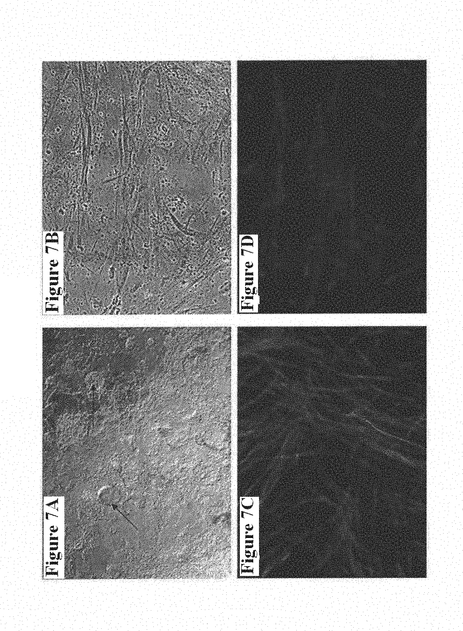
D00014
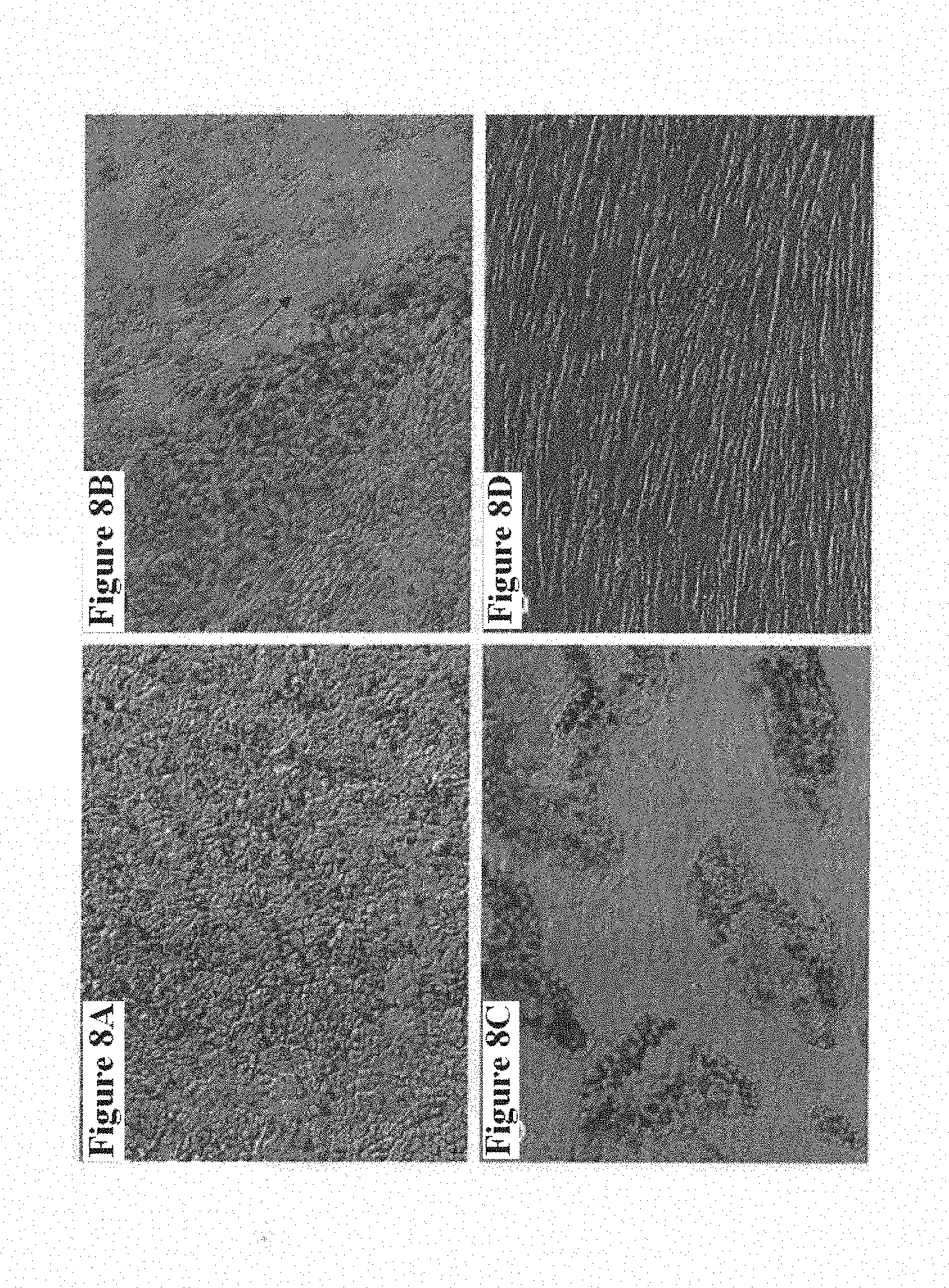
D00015
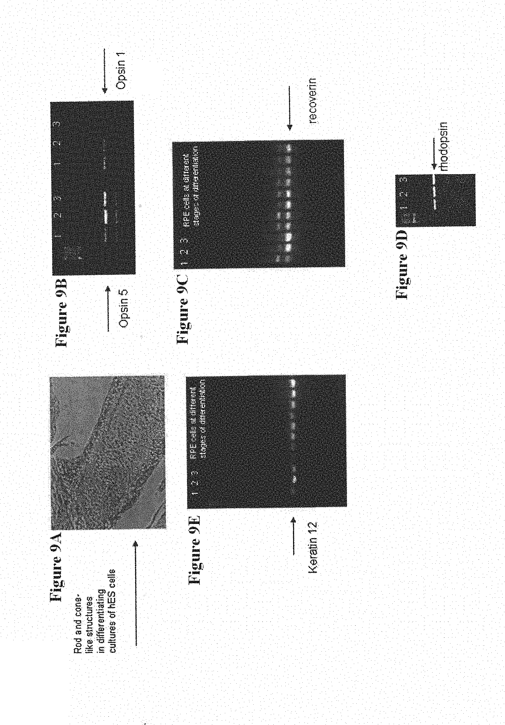
XML
uspto.report is an independent third-party trademark research tool that is not affiliated, endorsed, or sponsored by the United States Patent and Trademark Office (USPTO) or any other governmental organization. The information provided by uspto.report is based on publicly available data at the time of writing and is intended for informational purposes only.
While we strive to provide accurate and up-to-date information, we do not guarantee the accuracy, completeness, reliability, or suitability of the information displayed on this site. The use of this site is at your own risk. Any reliance you place on such information is therefore strictly at your own risk.
All official trademark data, including owner information, should be verified by visiting the official USPTO website at www.uspto.gov. This site is not intended to replace professional legal advice and should not be used as a substitute for consulting with a legal professional who is knowledgeable about trademark law.