Treatment Planning For Immunotherapy Based Treatments Using Non-thermal Ablation Techniques
DAVALOS; RAFAEL V. ; et al.
U.S. patent application number 16/352759 was filed with the patent office on 2019-09-19 for treatment planning for immunotherapy based treatments using non-thermal ablation techniques. This patent application is currently assigned to VIRGINIA TECH INTELLECTUAL PROPERTIES, INC.. The applicant listed for this patent is IRVING COY ALLEN, RAFAEL V. DAVALOS, NIKOLAOS DERVISIS, NATALIE BEITEL WHITE. Invention is credited to IRVING COY ALLEN, RAFAEL V. DAVALOS, NIKOLAOS DERVISIS, NATALIE BEITEL WHITE.
| Application Number | 20190282294 16/352759 |
| Document ID | / |
| Family ID | 67904794 |
| Filed Date | 2019-09-19 |


View All Diagrams
| United States Patent Application | 20190282294 |
| Kind Code | A1 |
| DAVALOS; RAFAEL V. ; et al. | September 19, 2019 |
TREATMENT PLANNING FOR IMMUNOTHERAPY BASED TREATMENTS USING NON-THERMAL ABLATION TECHNIQUES
Abstract
Described herein are methods of performing immunotherapy on a subject and/or determining if a subject will be responsive to ablation immunotherapy.
| Inventors: | DAVALOS; RAFAEL V.; (Blacksburg, VA) ; WHITE; NATALIE BEITEL; (Christianburg, VA) ; DERVISIS; NIKOLAOS; (Blacksburg, VA) ; ALLEN; IRVING COY; (Blacksburg, VA) | ||||||||||
| Applicant: |
|
||||||||||
|---|---|---|---|---|---|---|---|---|---|---|---|
| Assignee: | VIRGINIA TECH INTELLECTUAL
PROPERTIES, INC. BLACKSBURG VA |
||||||||||
| Family ID: | 67904794 | ||||||||||
| Appl. No.: | 16/352759 | ||||||||||
| Filed: | March 13, 2019 |
Related U.S. Patent Documents
| Application Number | Filing Date | Patent Number | ||
|---|---|---|---|---|
| 62642276 | Mar 13, 2018 | |||
| 62642298 | Mar 13, 2018 | |||
| 62732703 | Sep 18, 2018 | |||
| Current U.S. Class: | 1/1 |
| Current CPC Class: | A61B 2018/143 20130101; A61B 18/14 20130101; A61B 2018/00613 20130101; A61K 45/06 20130101; A61K 31/282 20130101; A61K 31/519 20130101; A61K 31/513 20130101; A61B 2018/00577 20130101; A61K 31/4745 20130101; A61B 2018/00875 20130101; A61B 2018/00827 20130101 |
| International Class: | A61B 18/14 20060101 A61B018/14; A61K 31/282 20060101 A61K031/282; A61K 31/4745 20060101 A61K031/4745; A61K 31/513 20060101 A61K031/513; A61K 31/519 20060101 A61K031/519 |
Claims
1. A method of treating a tissue in a patient comprising: ablating the tissue using a non-thermal ablation technique; measuring a change in a treatment parameter in real-time during the step of ablating; and administering an additional treatment to the subject in response to the measured change in the treatment parameter.
2. The method of claim 1, wherein the step of administering the additional treatment occurs 4-30 days post ablating.
3. The method of claim 1, wherein the additional treatment is selected from the group consisting of: tissue resection, thermal ablation, non-thermal ablation, chemotherapy, radiation therapy, immunotherapy, biologic therapy, genetic therapy, and combinations thereof.
4. The method of claim 1, wherein the additional treatment is measuring the amount of a pro-inflammatory immune molecule or cell, a suppressive immune molecule or cell, or both in a bodily fluid or a biopsied tissue of the patient.
5. The method of claim 1, where the non-thermal ablation technique is irreversible electrophoresis.
6. The method of claim 5, wherein the non-thermal ablation technique, is high-frequency irreversible electrophoresis.
7. The method of claim 1, wherein the treatment parameter is bulk tissue conductivity.
8. The method of claim 7, wherein the change in bulk tissue conductivity is measured by measuring current during the step of ablating.
9. The method of claim 8, wherein the step of administering the additional treatment occurs 4-30 days post ablating when the current measured is between 25 A and 100 A.
10. The method of claim 1, wherein the treatment parameter is measured by measuring current during the step of ablating.
11. The method of claim 10, wherein the step of administering the additional treatment occurs 4-30 days post ablating when the current measured is between 25 A and 100 A.
12. A method of treating a tissue in a patient comprising: inserting first electrode and a second electrode into the tissue applying a plurality of electrical pulses through the first electrode and the second electrode, wherein the electrical pulses are configured to cause non-thermal irreversible electroporation of the tissue; measuring a current of the applied electrical pulses; and administering an additional treatment to the subject in response to the subject, wherein the step of administering occurs 0 to 5 days after non-thermal irreversible electroporation when the current is less than 25A, and wherein the step of administering occurs 4-30 days after non-thermal irreversible electroporation when the current is between 25 A and 100 A.
13. The method of claim 12, wherein the additional treatment is selected from the group consisting of: tissue resection, thermal ablation, non-thermal ablation, chemotherapy, radiation therapy, immunotherapy, biologic therapy, genetic therapy, and combinations thereof.
14. The method of claim 12, wherein the additional treatment is measuring the amount of a pro-inflammatory immune molecule or cell, a suppressive immune molecule or cell, or both in a bodily fluid or a biopsied tissue of the patient.
15. The method of claim 12, wherein the electrical pulses are configured to cause non-thermal high-frequency irreversible electroporation of the tissue
16. A method of treating a tissue in a patient comprising: applying a plurality of electrical pulses to the tissue, wherein the electrical pulses are configured to cause non-thermal irreversible electroporation of the tissue; measuring a current of the applied electrical pulses; administering an additional treatment to the subject in response to the subject, wherein the step of administering occurs 0 to 5 days after non-thermal irreversible electroporation when the current is less than 25A, and wherein the step of administering occurs 4-30 days after non-thermal irreversible electroporation when the current is between 25 A and 100 A
17. The method of claim 16, wherein the additional treatment is selected from the group consisting of: tissue resection, thermal ablation, non-thermal ablation, chemotherapy, radiation therapy, immunotherapy, biologic therapy, genetic therapy, and combinations thereof.
18. The method of claim 16, wherein the additional treatment is measuring the amount of a pro-inflammatory immune molecule or cell, a suppressive immune molecule or cell, or both in a bodily fluid or a biopsied tissue of the patient.
19. The method of claim 16, wherein the electrical pulses are configured to cause non-thermal high-frequency irreversible electroporation of the tissue.
20. The method of claim 16, wherein in the plurality of electrical pulses are applied through a plurality of electrodes inserted into the tissue.
Description
CROSS-REFERENCE TO RELATED APPLICATIONS
[0001] This application claims the benefit of and priority to co-pending U.S. Provisional Patent Application No. 62/642,276, filed on Mar. 13, 2018, entitled " Irreversible electroporation for liver cancer immunotherapy," the contents of which is incorporated by reference herein in its entirety.
[0002] This application claims the benefit of and priority to co-pending U.S. Provisional Patent Application No. 62/642,298, filed on Mar. 13, 2018, entitled " Novel Approaches for Stimulating the Immune System," the contents of which is incorporated by reference herein in its entirety.
[0003] This application claims the benefit of and priority to co-pending U.S. Provisional Patent Application No. 62/732,703, filed on Sep. 18, 2018, entitled "Real-Time Patient Screening for Immunotherapy," the contents of which is incorporated by reference herein in its entirety.
BACKGROUND
[0004] Some types of cancers come with the most dismal prognoses, particularly after metastasis has already occurred. Although several immunotherapies for cancers exists, there still is a great need for techniques to allow treatment customization to improve treatment outcomes on the individual patient level.
SUMMARY
[0005] Described herein are aspects of a method of treating a tissue in a patient that can include ablating the tissue using a non-thermal ablation technique; measuring a change in a treatment parameter in real-time during the step of ablating; and administering an additional treatment to the subject in response to the measured change in the treatment parameter. The step of administering the additional treatment can occur 4-30 days post ablating. The additional treatment can be selected from the group of: tissue resection, thermal ablation, non-thermal ablation, chemotherapy, radiation therapy, immunotherapy, biologic therapy, genetic therapy, and combinations thereof. The additional treatment can be measuring the amount of a pro-inflammatory immune molecule or cell, a suppressive immune molecule or cell, or both in a bodily fluid or a biopsied tissue of the patient. The non-thermal ablation technique can be irreversible electrophoresis. The non-thermal ablation technique, can be high-frequency irreversible electrophoresis. The treatment parameter can be bulk tissue conductivity. The change in bulk tissue conductivity can be measured by measuring current during the step of ablating. The step of administering the additional treatment can occur 4-30 days post ablating when the current measured is between 25 A and 100 A. The treatment parameter can be measured by measuring current during the step of ablating. The step of administering the additional treatment can occur 4-30 days post ablating when the current measured is between 25 A and 100 A.
[0006] Also described herein are aspects of a method that can include a method of treating a tissue in a patient comprising: inserting first electrode and a second electrode into the tissue applying a plurality of electrical pulses through the first electrode and the second electrode, wherein the electrical pulses are configured to cause non-thermal irreversible electroporation of the tissue; measuring a current of the applied electrical pulses; and administering an additional treatment to the subject in response to the subject, wherein the step of administering occurs 0 to 5 days after non-thermal irreversible electroporation when the current is less than 25A, and wherein the step of administering occurs 4-30 days after non-thermal irreversible electroporation when the current is between 25 A and 100 A. The additional treatment can be selected from the group consisting of: tissue resection, thermal ablation, non-thermal ablation, chemotherapy, radiation therapy, immunotherapy, biologic therapy, genetic therapy, and combinations thereof. The additional treatment can be measuring the amount of a pro-inflammatory immune molecule or cell, a suppressive immune molecule or cell, or both in a bodily fluid or a biopsied tissue of the patient. The electrical pulses can be configured to cause non-thermal high-frequency irreversible electroporation of the tissue.
[0007] Also described herein are aspects of a method of treating a tissue in a patient that can include the step of applying a plurality of electrical pulses to the tissue, wherein the electrical pulses are configured to cause non-thermal irreversible electroporation of the tissue; measuring a current of the applied electrical pulses; administering an additional treatment to the subject in response to the subject, wherein the step of administering occurs 0 to 5 days after non-thermal irreversible electroporation when the current is less than 25A, and wherein the step of administering occurs 4-30 days after non-thermal irreversible electroporation when the current is between 25 A and 100 A. The additional treatment is selected from the group consisting of: tissue resection, thermal ablation, non-thermal ablation, chemotherapy, radiation therapy, immunotherapy, biologic therapy, genetic therapy, and combinations thereof. The additional treatment can be measuring the amount of a pro-inflammatory immune molecule or cell, a suppressive immune molecule or cell, or both in a bodily fluid or a biopsied tissue of the patient. The electrical pulses can be configured to cause non-thermal high-frequency irreversible electroporation of the tissue. The plurality of electrical pulses can be applied through a plurality of electrodes inserted into the tissue.
BRIEF DESCRIPTION OF THE DRAWINGS
[0008] Further aspects of the present disclosure will be readily appreciated upon review of the detailed description of its various embodiments, described below, when taken in conjunction with the accompanying drawings.
[0009] FIG. 1 shows a graph that can demonstrate imputed median primary tumor volumes (mm.sup.3) for each group of mice, showing significantly smaller median tumor size in the treated immunocompetent (IC) BALB/c mice.
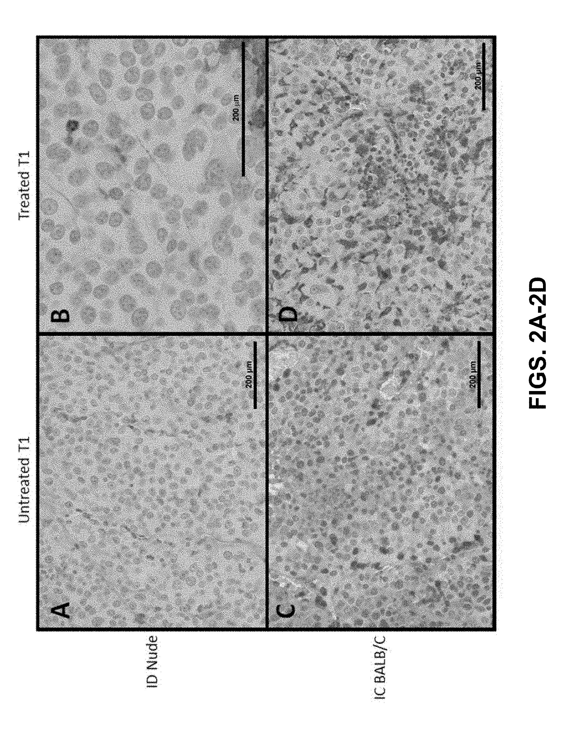
[0010] FIGS. 2A-2D show CD3+ staining, which is indicative for T-cell presences, performed for (FIGS. 2A and 2C) untreated and (FIGS. 2B and 2D) treated initial T1 tumors between (FIGS. 2A and 2B) ID nude and (FIGS. 2C and 2D) IC BALB/c mice. There was no notable difference observed in CD3+ infiltration for ID nude mice between (FIG. 2A) untreated and (FIG. 2B) treated tumors. For the IC BALB/c mice, a robust increase in CD3+ (T-cell) infiltration is observed in treated tumors (FIG. 2D) relative to untreated T1 controls (FIG. 2C). Increased T-cell presence in treated T1 IC mice was also more robust than for both groups for nude mice (FIGS. 2A and 2B). All scale bars 200 mm. Panels (FIGS. 2A, 2C, and 2D) 200.times., panel (FIG. 2B) 400.times. magnification
[0011] FIG. 3 shows a graph that can demonstrate percent CD4+CD25+FoxP3+ of total T cells 24 h post-IRE vs. difference in current (n=7).
[0012] FIG. 4 shows a graph that can demonstrate percent CD4+CD25+ cells 24 h post-irreversible electroporation (IRE) vs. difference in current (n=7).
[0013] FIG. 5 shows a graph that can demonstrate percent CD4+CD25+ of total T cells 24 h post-IRE vs. average current.
[0014] FIG. 6 shows a graph that can demonstrate the percent change in CD4+CD25+ and CD4+CD25+FoxP3+ 24 hours after IRE. The changes are shown in order of increasing overall change in output current (lower most panel) during the treatment in each patient. Populations were calculated as a subset of total CD4+ cells (n=7).
[0015] FIG. 7 shows a representative 3D reconstruction of a human pancreas, tumor, and vasculature used for treatment planning purposes. Two electrodes are inserted into the tumor in this example. Pancreas and vasculature reconstruction was prepared using 3matic and Gmsh software using a pre-operative CT scan; electrodes and tumor mimic placed using COMSOL software.
[0016] FIG. 8 shows a graph that can demonstrate a representative IRE pulse delivered using the Nanoknife.RTM. device. The resistance value resulting from the applied voltage and current is displayed to the user during treatment.
[0017] FIG. 9 shows a graph that can demonstrate the percent change in Regulatory T-cell subpopulations i.e. CD4+CD25+, CD4+CD25+FoxP3+, and CD4+CD25+FoxP3-, after 24 hours following IRE treatment. Populations were calculated as a subset of total CD4+ cells. n=7
[0018] FIGS. 10A-10B shows graphs that can demonstrate the change in FIG. 10A) CD4+CD25+FoxP3+ and FIG. 10B) CD4+CD25+ after 24 hours following IRE decreases linearly with the range of current delivered during the IRE treatment for n=7 patients. A linear regression was performed using JMP Pro software.
[0019] FIGS. 11A-11C shows CT scan images of the pelvis and hind limbs visualizing the target region, practically surrounding the femur and adjacent to the sciatic nerve and femoral arteries. IRE resulted in marked influx of mixed inflammatory cells into the treatment region (FIG. 11B), which was composed of primarily CD3+ lymphocytes, 24 hours post-treatment (FIG.11A.fwdarw.pretreatment, FIG. 11A.fwdarw.post treatment).
[0020] FIGS. 12A-12B show graphs that can demonstrate that H-FIRE treatment significantly reduces tumor progression and ablates the primary 4T1 mammary tumor. (FIG. 12A) Mice were injected with 1.26.times.106 4T1 cells directly into mammary tissue. On day 11, H-FIRE was administered. Clinical and tumor progression were monitored daily through day 27 where the tumors reached about 1.6 cm in diameter in the untreated mice. n=10 mice per treatment group. *p<0.05. (FIG. 12B) Mice were injected with 1.26.times.106 Pan02 cells subcutaneously (s.c.) and tumors were allowed to progress for 10 days. On day 11 (0.5 cm), H-FIRE was administered. Clinical and tumor progression were monitored daily through day 35 where the tumors reached about 1.35 cm in diameter in the untreated mice. n=3 mice per treatment group. *p<0.05.
[0021] FIGS. 13A-13B show graphs that can demonstrate a decrease in circulating metastases following H-FIRE treatment of the primary tumor in immunocompetent mice. Circulating metastases were quantified in both (FIG. 13A) BALB/c and (FIG. 13B) NSG mice following treatment of the primary 4T1 (breast cancer) tumor with an ablation dose of H-FIRE. Following necropsy, blood samples were plated in media with 6 mM 6-thioguanine. After 10-14 days, formed colonies were counted. n=6-10 mice. *p=0.05.
[0022] FIG. 14 shows a microscopic image that can demonstrate increased lymphocyte infiltration following H-FIRE in the local Pan02 tumor. Histopathology assessments revealed increased marginal and intra-tumor lymphocytes 14 days post-treatment with an ablation dose of H-FIRE.
[0023] FIG. 15 shows a gene expression pathway analysis of tumor microenvironment following H-FIRE. Gene expression profiling revealed significant changes in inflammatory signaling in the 4T1 mammary tumor model. Data indicate a significant shift in the tumor microenvironment to a Th1/Th17 profile and high levels of IL-6 following full ablation. A significant increase in IL-2 signaling was found in tumors that received a sub-ablation dose of H-FIRE, consistent with increased lymphocyte expansion and recruitment.
[0024] FIG. 16 shows a schematic of the local and systemic efficacy of IRE and H-FIRE ablation is associated with immune system activation/promotion. In this model, the cell death and tumor ablation driven by IRE/H-FIRE treatment promotes the innate immune system and promotes a shift in the tumor microenvironment from immunosuppressive to pro-inflammatory (Th1 and Th17), which serves to recruit increased numbers of antigen presenting cells to the local tumor. The cell death and damage associated with treatment increases antigen availability and presentation, further engaging the adaptive immune system. Increased tumor specific antigen presentation promotes T cell activation and increased CTL mediated cancer cell killing in the primary tumor and metastatic cells in distal locations. Increased activation/promotion of the adaptive immune system will also improve immunological memory, which should attenuate recurrence. IRE and H-FIRE would be excellent candidates for combination therapeutic approaches using new classes of checkpoint inhibitors and/or other immunomodulatory therapeutics.
[0025] FIG. 17 shows a schematic of H-FIRE targeting within a tumor.
[0026] FIGS. 18A-18E can demonstrate H-FIRE treatment and modeling in a 3D tumor mimic. FIG. 18A) Experimental setup with electrodes inserted into tissue mimic. FIGS. 18B-18E) Live/dead staining reveals regions of the tissue impacted following 80 bursts containing (FIG. 18B) 2, (FIG. 18C) 24, and (FIG. 18D) 50 bipolar 2 .mu.s pulses with a 2 ps delay between alternating pulses. (FIG. 18E) Diffuse treatment of 50 bipolar 2 .mu.s pulses with 20 ms between alternating pulses. Scale bar=2 mm.
[0027] FIGS. 19A-19D shows graphs that can demonstrate that IRE enhances the pro-inflammatory tumor microenvironment in Pan02 cells. FIG. 19A) Viability of cells after overnight incubation measured using LDH following 99 pulses at increasing field strengths. FIGS. 19B-19D) Influence of electric pulse parameters on select gene expression in Pan02 cells measured with real time PCR (ddCt). (FIG. 19B) II-6, (FIG. 19C) Tslp, (FIG. 19D) Ccl-2. (n=3 repetitions; *,**, +, #p<0.05 ANOVA with Tukeys post-test).
[0028] FIG. 20 shows the appropriate shape factor calculation for voltage applied across two needle electrodes. An example three needle configuration is shown. During IRE, pulses are delivered across probe pairs, therefore only two will act as source and sink at any given point. Bulk tissue conductivity is calculated using this shape factor and the resistance according to Ohm's Law (voltage equals current times resistance). The relationship is given by R=1/(.sigma.*K) where R is resistance (.OMEGA.), .sigma. is the conductivity (S/m), and K is the shape factor given in FIG. 20 where I is the spacing between the electrodes and r is the electrode radius.
[0029] FIG. 21 shows a table that shows three example shape factor calculations assuming a static, bulk tissue conductivity of 0.2 S/m. The changes in this conductivity value result in changes in the output current. Additionally, changes in the electrode and/or voltage parameters affects the output current value as in the examples shown; V is applied voltage, is .sigma. the bulk electrical conductivity, d is the spacing between the electrodes, r is the radius of the electrodes, K is the shape factor (dimensionless), and I is output current.
[0030] FIG. 22 shows a scatter plot of gene expression array data illustrating the results from FIG. 34. CXCL10 and HIF1A expression is significantly up-regulated and TLR2, SPP1 and MYC are significantly down-regulated following H-FIRE treatment in all 3 patients compared to baseline. Change in expression of all other genes was less than 2-fold (unchanged) and represented by the solid black line
[0031] FIG. 23 show results an Ingenuity Pathway Analysis (IPA) of global changes in gene expression patterns following H-FIRE in 3 patients. Results revealed diverse, but functionally related predications in canonical pathways significantly increased by H-FIRE for Patients 1 and 2. Conversely, the gene expression profile for Patient 3 showed either no change or down-regulation of functionally similar canonical pathways.
[0032] FIGS. 24A-24F show graphs show results from the top 6 canonical pathways impacted by H-FIRE, comparing pre-treatment to post-treatment, ranked by z-score. Patient 1 and 2 are highly consistent, with patient 3 demonstrating opposing results.
[0033] FIGS. 25A-25C show pathways associated with the activation of cellular immunity (FIG. 25B) and cell death (FIG. 25A) were significantly up-regulated following H-FIRE treatment. FIG. 25A. IPA network analysis identified 2 functional networks that fit the canonical pathways identified above, cell injury/death and cell mediated immunity. Both of these networks were significantly upregulated in all patients following H-FIRE treatment FIG. 25C. NF-.kappa.B signaling was one of the most dominate pathways impacted by H-FIRE treatment. Gene expression patterns revealed a significant global up-regulation in NF-.kappa.B signaling.
[0034] FIGS. 26A-26D a (FIG. 26A) Schematic of HFIRE experimental set-up and (FIG. 26C) 18-gauge, bipolar electrode. (FIG. 26B) A standard IRE voltage waveform pulse and (FIG. 26D) HFIRE 2-5-2 voltage waveform burst.
[0035] FIG. 27 shows a schematic of the 3D-reconstruction and numerical modeling process for patient 1.
[0036] FIGS. 28A-28B show placement of the bipolar treatment probe into the tumor under ultrasound guidance (FIG. 28A). Ultrasound image of the bipolar treatment probe inserted into the tumor during treatment delivery (FIG. 28B); arrow--HFIRE bipolar electrode).
[0037] FIG. 29 shows a panel of CT images Pre- (left) and post-treatment (center) and images of gross tumor samples (right). CT images from all three patients showing predictable ablation volume (outlined) resulting from H-FIRE treatment. This correlated with the ablation volume noted in gross tumor samples (right).
[0038] FIG. 30 shows a panel of images of tumor histopathology from patient 1 (top) and patient 2 (bottom) showing the well-defined ablation/tumor interface (arrows) with H&E at 40.times. (left) and 100.times. (center). IHC for CD3 revealed positive staining cells infiltrating the tumor-ablation interface. Untreated HCC is denoted by (*) and the ablation zone by the bolt.
[0039] FIG. 31 shows a panel of images of tumor histopathology from patient 3 showing absence of a well-defined ablation/tumor interface (arrows) with H&E at 40.times. (left) and 100.times. (center). IHC for CD3 (right) shows the lack of CD3+ cells within the ablation/tumor interface. Untreated HCC is denoted by (*) and the ablation zone by the bolt.
[0040] FIG. 32 shows a pane of images showing IHC results for CD3 (left), CD4 (center) and CD8 (right) on tumor samples from patient 1 (top) and patient 2 (bottom) showing infiltration of the ablation/tumor interface (arrows) with CD3+/CD4-/CD8- lymphocytes. Untreated HCC is denoted by (*) and the ablation zone by the bolt.
[0041] FIGS. 33A-33B show graphs that can demonstrate that ALT (FIG. 33A) and ALP (FIG. 33B) increased following H-FIRE treatment (bolt), but resolved over time following tumor removal (day 4).
[0042] FIG. 34 shows Tissue and probe properties employed for numerical modeling.
[0043] FIG. 35 shows mean (n=3) fold change in gene expression following H-FIRE treatment compared to baseline. Only genes with significant changes in expression are shown.
DETAILED DESCRIPTION
[0044] Before the present disclosure is described in greater detail, it is to be understood that this disclosure is not limited to particular embodiments described, and as such may, of course, vary. It is also to be understood that the terminology used herein is for the purpose of describing particular embodiments only, and is not intended to be limiting.
[0045] Unless defined otherwise, all technical and scientific terms used herein have the same meaning as commonly understood by one of ordinary skill in the art to which this disclosure belongs. Although any methods and materials similar or equivalent to those described herein can also be used in the practice or testing of the present disclosure, the preferred methods and materials are now described.
[0046] All publications and patents cited in this specification are cited to disclose and describe the methods and/or materials in connection with which the publications are cited. All such publications and patents are herein incorporated by references as if each individual publication or patent were specifically and individually indicated to be incorporated by reference. Such incorporation by reference is expressly limited to the methods and/or materials described in the cited publications and patents and does not extend to any lexicographical definitions from the cited publications and patents. Any lexicographical definition in the publications and patents cited that is not also expressly repeated in the instant application should not be treated as such and should not be read as defining any terms appearing in the accompanying claims. The citation of any publication is for its disclosure prior to the filing date and should not be construed as an admission that the present disclosure is not entitled to antedate such publication by virtue of prior disclosure. Further, the dates of publication provided could be different from the actual publication dates that may need to be independently confirmed.
[0047] As will be apparent to those of skill in the art upon reading this disclosure, each of the individual embodiments described and illustrated herein has discrete components and features which may be readily separated from or combined with the features of any of the other several embodiments without departing from the scope or spirit of the present disclosure. Any recited method can be carried out in the order of events recited or in any other order that is logically possible.
[0048] Where a range is expressed, a further aspect includes from the one particular value and/or to the other particular value. Where a range of values is provided, it is understood that each intervening value, to the tenth of the unit of the lower limit unless the context clearly dictates otherwise, between the upper and lower limit of that range and any other stated or intervening value in that stated range, is encompassed within the disclosure. The upper and lower limits of these smaller ranges may independently be included in the smaller ranges and are also encompassed within the disclosure, subject to any specifically excluded limit in the stated range. Where the stated range includes one or both of the limits, ranges excluding either or both of those included limits are also included in the disclosure. For example, where the stated range includes one or both of the limits, ranges excluding either or both of those included limits are also included in the disclosure, e.g. the phrase "x to y" includes the range from `x` to `y` as well as the range greater than `x` and less than `y`. The range can also be expressed as an upper limit, e.g. `about x, y, z, or less` and should be interpreted to include the specific ranges of `about x`, `about y`, and `about z` as well as the ranges of `less than x`, less than y', and `less than z`. Likewise, the phrase `about x, y, z, or greater` should be interpreted to include the specific ranges of `about x`, `about y`, and `about z` as well as the ranges of `greater than x`, greater than y', and `greater than z`. In addition, the phrase "about `x` to `y`", where `x` and `y` are numerical values, includes "about `x` to about `y`".
[0049] It should be noted that ratios, concentrations, amounts, and other numerical data can be expressed herein in a range format. It will be further understood that the endpoints of each of the ranges are significant both in relation to the other endpoint, and independently of the other endpoint. It is also understood that there are a number of values disclosed herein, and that each value is also herein disclosed as "about" that particular value in addition to the value itself. For example, if the value "10" is disclosed, then "about 10" is also disclosed. Ranges can be expressed herein as from "about" one particular value, and/or to "about" another particular value. Similarly, when values are expressed as approximations, by use of the antecedent "about," it will be understood that the particular value forms a further aspect. For example, if the value "about 10" is disclosed, then "10" is also disclosed.
[0050] It is to be understood that such a range format is used for convenience and brevity, and thus, should be interpreted in a flexible manner to include not only the numerical values explicitly recited as the limits of the range, but also to include all the individual numerical values or sub-ranges encompassed within that range as if each numerical value and sub-range is explicitly recited. To illustrate, a numerical range of "about 0.1% to 5%" should be interpreted to include not only the explicitly recited values of about 0.1% to about 5%, but also include individual values (e.g., about 1%, about 2%, about 3%, and about 4%) and the sub-ranges (e.g., about 0.5% to about 1.1%; about 5% to about 2.4%; about 0.5% to about 3.2%, and about 0.5% to about 4.4%, and other possible sub-ranges) within the indicated range.
[0051] As used in the specification and the appended claims, the singular forms "a," "an," and "the" include plural referents unless the context clearly dictates otherwise.
[0052] As used herein, "about," "approximately," "substantially," and the like, when used in connection with a numerical variable, can generally refers to the value of the variable and to all values of the variable that are within the experimental error (e.g., within the 95% confidence interval for the mean) or within +/-10% of the indicated value, whichever is greater. As used herein, the terms "about," "approximate," "at or about," and "substantially" can mean that the amount or value in question can be the exact value or a value that provides equivalent results or effects as recited in the claims or taught herein. That is, it is understood that amounts, sizes, formulations, parameters, and other quantities and characteristics are not and need not be exact, but may be approximate and/or larger or smaller, as desired, reflecting tolerances, conversion factors, rounding off, measurement error and the like, and other factors known to those of skill in the art such that equivalent results or effects are obtained. In some circumstances, the value that provides equivalent results or effects cannot be reasonably determined. In general, an amount, size, formulation, parameter or other quantity or characteristic is "about," "approximate," or "at or about" whether or not expressly stated to be such. It is understood that where "about," "approximate," or "at or about" is used before a quantitative value, the parameter also includes the specific quantitative value itself, unless specifically stated otherwise.
[0053] Embodiments of the present disclosure will employ, unless otherwise indicated, techniques of immunology, molecular biology, electrophysiology, physiology, cell biology, cancer biology, and the like, which are within the skill of the art. Such techniques are explained fully in the literature.
[0054] Before the embodiments of the present disclosure are described in detail, it is to be understood that, unless otherwise indicated, the present disclosure is not limited to particular materials, reagents, reaction materials, manufacturing processes, or the like, as such can vary. It is also to be understood that the terminology used herein is for purposes of describing particular embodiments only, and is not intended to be limiting. It is also possible in the present disclosure that steps can be executed in different sequence where this is logically possible unless the context clearly dictates otherwise.
[0055] Definitions
[0056] As used herein, "active agent" or "active ingredient" refers to a substance, compound, or molecule, which is biologically active or otherwise, induces a biological or physiological effect on a subject to which it is administered to. In other words, "active agent" or "active ingredient" refers to a component or components of a composition to which the whole or part of the effect of the composition is attributed.
[0057] As used herein, "administering" refers to an administration that is oral, topical, intravenous, subcutaneous, transcutaneous, transdermal, intramuscular, intra-joint, parenteral, intra-arteriole, intradermal, intraventricular, intraosseous, intraocular, intracranial, intraperitoneal, intralesional, intranasal, intracardiac, intraarticular, intracavernous, intrathecal, intravireal, intracerebral, and intracerebroventricular, intratympanic, intracochlear, rectal, vaginal, by inhalation, by catheters, stents or via an implanted reservoir or other device that administers, either actively or passively (e.g. by diffusion) a composition the perivascular space and adventitia. For example a medical device such as a stent can contain a composition or formulation disposed on its surface, which can then dissolve or be otherwise distributed to the surrounding tissue and cells. The term "parenteral" can include subcutaneous, intravenous, intramuscular, intra-articular, intra-synovial, intrasternal, intrathecal, intrahepatic, intralesional, and intracranial injections or infusion techniques. In some embodiments, administering can be achieved by any suitable mechanism, technique, and/or device. In some embodiments, administering can be intravenous through a catheter and/or needle.
[0058] As used herein, "agent" refers to any substance, compound, molecule, and the like, which can be biologically active or otherwise can induce a biological and/or physiological effect on a subject to which it is administered to. An agent can be a primary active agent, or in other words, the component(s) of a composition to which the whole or part of the effect of the composition is attributed. An agent can be a secondary agent, or in other words, the component(s) of a composition to which an additional part and/or other effect of the composition is attributed
[0059] As used herein "cancer" can refer to one or more types of cancer including, but not limited to, acute lymphoblastic leukemia, acute myeloid leukemia, adrenocortical carcinoma, Kaposi Sarcoma, AIDS-related lymphoma, primary central nervous system (CNS) lymphoma, anal cancer, appendix cancer, astrocytomas, atypical teratoid/Rhabdoid tumors, basa cell carcinoma of the skin, bile duct cancer, bladder cancer, bone cancer (including but not limited to Ewing Sarcoma, osteosarcomas, and malignant fibrous histiocytoma), brain tumors, breast cancer, bronchial tumors, Burkitt lymphoma, carcinoid tumor, cardiac tumors, germ cell tumors, embryonal tumors, cervical cancer, cholangiocarcinoma, chordoma, chronic lymphocytic leukemia, chronic myelogenous leukemia, chronic myeloproliferative neoplasms, colorectal cancer, craniopharyngioma, cutaneous T-Cell lymphoma, ductal carcinoma in situ, endometrial cancer, ependymoma, esophageal cancer, esthesioneuroblastoma, extracranial germ cell tumor, extragonadal germ cell tumor, eye cancer (including, but not limited to, intraocular melanoma and retinoblastoma), fallopian tube cancer, gallbladder cancer, gastric cancer, gastrointestinal carcinoid tumor, gastrointestinal stromal tumors, central nervous system germ cell tumors, extracranial germ cell tumors, extragonadal germ cell tumors, ovarian germ cell tumors, testicular cancer, gestational trophoblastic disease, hary cell leukemia, head and neck cancers, hepatocellular (liver) cancer, Langerhans cell histiocytosis, Hodgkin lymphoma, hypopharyngeal cancer, islet cell tumors, pancreatic neuroendocrine tumors, kidney (renal cell) cancer, laryngeal cancer, leukemia, lip cancer, oral cancer, lung cancer (non-small cell and small cell), lymphoma, melanoma, Merkel cell carcinoma, mesothelioma, metastatic squamous cell neck cancer, midline tract carcinoma with and without NUT gene changes, multiple endocrine neoplasia syndromes, multiple myeloma, plasma cell neoplasms, mycosis fungoides, myelodyspastic syndromes, myelodysplastic/myeloproliferative neoplasms, chronic myelogenous leukemia, nasal cancer, sinus cancer, non-Hodgkin lymphoma, pancreatic cancer, paraganglioma, paranasal sinus cancer, parathyroid cancer, penile cancer, pharyngeal cancer, pheochromocytoma, pituitary cancer, peritoneal cancer, prostate cancer, rectal cancer, Rhabdomyosarcoma, salivary gland cancer, uterine sarcoma, Sezary syndrome, skin cancer, small intestine cancer, large intestine cancer (colon cancer), soft tissue sarcoma, T-cell lymphoma, throat cancer, oropharyngeal cancer, nasopharyngeal cancer, hypoharyngeal cancer, thymoma, thymic carcinoma, thyroid cancer, transitional cell cancer of the renal pelvis and ureter, urethral cancer, uterine cancer, vaginal cancer, cervical cancer, vascular tumors and cancer, vulvar cancer, and Wilms Tumor.
[0060] As used herein, "chemotherapeutic agent" or "chemotherapeutic" refers to a therapeutic agent utilized to prevent or treat cancer.
[0061] As used herein, "control" can refer to an alternative subject or sample used in an experiment for comparison purpose and included to minimize or distinguish the effect of variables other than an independent variable.
[0062] As used herein, "differentially expressed," refers to the differential production of RNA, including but not limited to mRNA, tRNA, miRNA, siRNA, snRNA, and piRNA transcribed from a gene or regulatory region of a genome or the protein product encoded by a gene as compared to the level of production of RNA or protein by the same gene or regulator region in a normal or a control cell. In another context, "differentially expressed," also refers to nucleotide sequences or proteins in a cell or tissue which have different temporal and/or spatial expression profiles as compared to a normal or control cell.
[0063] As used herein, "dose," "unit dose," or "dosage" can refer to physically discrete units suitable for use in a subject, each unit containing a predetermined quantity of a pharmaceutical formulation or immunotherapy thereof calculated to produce the desired response or responses in association with its administration.
[0064] As used herein, "immunomodulator," refers to an agent, such as a therapeutic agent, which is capable of modulating or regulating one or more immune function or response.
[0065] As used herein, "mammal," for the purposes of treatments, can refer to any animal classified as a mammal, including human, domestic and farm animals, nonhuman primates, and zoo, sports, or pet animals, such as, but not limited to, dogs, horses, cats, and cows.
[0066] As used herein, "organism", "host", and "subject" refers to any living entity comprised of at least one cell. A living organism can be as simple as, for example, a single isolated eukaryotic cell or cultured cell or cell line, or as complex as a mammal, including a human being, and animals (e.g., vertebrates, amphibians, fish, mammals, e.g., cats, dogs, horses, pigs, cows, sheep, rodents, rabbits, squirrels, bears, primates (e.g., chimpanzees, gorillas, and humans).
[0067] As used herein, the terms "optional" or "optionally" means that the subsequently described event or circumstance can or cannot occur, and that the description includes instances where said event or circumstance occurs and instances where it does not.
[0068] As used herein, "patient" can refer to an organism, host, or subject in need of treatment.
[0069] As used herein, "pharmaceutical formulation" refers to the combination of an active agent, compound, or ingredient with a pharmaceutically acceptable carrier or excipient, making the composition suitable for diagnostic, therapeutic, or preventive use in vitro, in vivo, or ex vivo.
[0070] As used herein, "preventative" and "prevent" refers to hindering or stopping a disease or condition before it occurs, even if undiagnosed, or while the disease or condition is still in the sub-clinical phase.
[0071] As used interchangeably herein, the terms "sufficient" and "effective," can refer to an amount (e.g. mass, volume, dosage, concentration, and/or time period) needed to achieve one or more desired result(s). For example, a therapeutically effective amount refers to an amount needed to achieve one or more therapeutic effects.
[0072] A "suitable control" is a control that will be instantly appreciated by one of ordinary skill in the art as one that is included such that it can be determined if the variable being evaluated an effect, such as a desired effect or hypothesized effect. One of ordinary skill in the art will also instantly appreciate based on inter alia, the context, the variable(s), the desired or hypothesized effect, what is a suitable or an appropriate control needed.
[0073] As used herein, "therapeutic" can refer to treating, healing, and/or ameliorating a disease, disorder, condition, or side effect, or to decreasing in the rate of advancement of a disease, disorder, condition, or side effect. A "therapeutically effective amount" can therefore refer to an amount of a compound that can yield a therapeutic effect.
[0074] As used herein, the terms "treating" and "treatment" can refer generally to obtaining a desired pharmacological and/or physiological effect. The effect can be therapeutic in terms of a partial or complete cure of a disease, condition, symptom or adverse effect attributed to the disease, disorder, or condition. The term "treatment" as used herein covers any treatment of a tumor or a cancer in a subject, particularly a human, and can include any one or more of the following: (a) inhibiting the disease, i.e., arresting its development; and (b) relieving the disease, i.e., mitigating or ameliorating the disease and/or its symptoms or conditions. The term "treatment" as used herein can refer to both therapeutic treatment alone, prophylactic treatment alone, or both therapeutic and prophylactic treatment. Those in need of treatment (subjects in need thereof) can include those already with the disorder and/or those in which the disorder is to be prevented. As used herein, the term "treating", can include inhibiting the disease, disorder or condition, e.g., impeding its progress; and relieving the disease, disorder, or condition, e.g., causing regression of the disease, disorder and/or condition. Treating the disease, disorder, or condition can include ameliorating at least one symptom of the particular disease, disorder, or condition, even if the underlying pathophysiology is not affected, such as treating the pain of a subject by administration of an analgesic agent even though such agent does not treat the cause of the pain.
[0075] As used herein, "non-thermal ablation" refers to any technique that can result in the destruction and/or death of cells, cellular structures, cellular components, and/or tissue, including human tissue and non-human tissue, within and surrounding a target site, while preserving the extracellular matrix, nerves, major blood vessels, and other sensitive structures of the treated tissues, without raising the temperature of the cells or treated tissues local to and surrounding the target site of ablation to an overall temperature over about 65 .degree. C. at the conclusion of pulse delivery and those cells that do not die but are still affected by the treatment. In some aspects, "non-thermal ablation" includes techniques that involve stimulating, killing or altering cellular components of cells and/or tissue with pulsed electrical energy that does not raise the temperature of the cells and/or tissue over about 65 .degree. C. at the conclusion of pulse. Non-thermal ablation techniques include, but are not limited to irreversible electroporation (IRE), high frequency irreversible electroporation (HFIRE or H-FIRE) supraporation, reversible electroporation, histotripsy (focused ultrasound), shock wave therapies, high-intensity focused ultrasound (HIFU), and ultrasonic ablation. Techniques and parameters of operation of non-thermal ablation techniques, including HFIRE and IRE can be found in U.S. Pat. No. 8,926,606, U.S. Pat. Pub. 2012/0109122, and U.S. Pat. Pub.: 2014/0039489, which are herein incorporated by reference as if expressed in their entireties.
[0076] As used herein, "thermal ablation" refers to any technique that can result in the destruction and/or death of cells, cellular structures, cellular components, and/or tissue, including human tissue and non-human tissue, within and surrounding a target site, by transmitting energy, sufficient to increase the overall temperature of the tissue and/or cells within or surrounding a target site to above 65.degree. C. Thermal ablation techniques can include but are not limited to electrical energy, microwave ablation, a radiofrequency (RF) ablation, dual thermal ablation, and laser ablation.
[0077] As used herein, "ablation" without further qualification refers to both thermal and non-thermal ablation.
[0078] As used herein, "immunosuppressive treatment" refers to any treatment modality, including, but not limited to, thermal ablation, resection, and/or chemotherapy, that do not allow for the innate and/or adaptive immune responses to active, peak, and/or come to completion.
[0079] As used herein, "non-immunosuppressive treatment" refers to any treatment modality, including, but not limited to, non-thermal ablation, such as IRE and/or HFIRE, that allows for the innate and/or adaptive immune responses to active, peak, and/or come to completion.
Discussion
[0080] Some types of cancers come with the most dismal prognoses, particularly after metastasis has already occurred. In some cases, tumor resection and/or thermal ablation is not a viable option for therapy. Further, while delivery of chemotherapeutics are often part of a standard cancer therapy regimen or protocol, they may also be immunosuppressive, and thus can reduce the patient's own immune system's ability to combat the cancer and mute or otherwise suppress the patient's biological immune defense mechanisms. Additionally, tumors in some areas, such as immunotolerant organs and systems (e.g. liver, gastrointestinal organs, respiratory system, genitourinary tract), are not affected as much by the patient's immune system because its immediate microenvironment is effectively immunosuppressed. The immunosuppressive microenvironment characteristic in immunotolerant organs are characterized by a balance between pro-inflammatory and anti-inflammatory mediators produced by specialized immune cells that reside in these organs and serve to protect the system from overzealous immune responses that can lead to diseases, such as those associated with autoimmunity. However, this immunosuppression can promote tumorigenesis and become a hindrance to certain therapeutics, such as checkpoint inhibitor therapies, that function through activating the immune system. This immunosuppression in both healthy immunotolerant organs and in tumors is, in part, mediated by groups of immunosuppressive cells, including regulatory T cells (Treg), tumor associated macrophages (TAM), tumor associated neutrophils (TAN), and myeloid derived suppressor cells (MDSC). Based on the location and type of tumor, in many cases, tumor resection is immediately considered as a viable treatment option followed by chemotherapy and other modalities, such as thermal ablation. As stated above, the use of resection, chemotherapy, and thermal ablation may reduce a subject's or patient's ability to capitalize on their own immune response.
[0081] Cancer immunotherapy, which involves eliciting a host immune response that can result in tumor regression has been an increasingly compelling field of interest. Much focus in this field has been on the vaccination against various cancer types by injecting cancer antigens and thus stimulating the host's immune system to target the cancer. A more recent area of investigation for inducing anti-tumor immunity is based on the direct destruction of cancerous tumors by ablative methods. By treating cancer using ablative approaches, the tumor may undergo a multi-agent anti-cancer treatment or vaccine through the production of tumor antigens resulting from the destructed tissue. Although some efficacy in some patients has been achieved, the invention described herein provides for the ability to optimize patient treatment planning and improve treatment outcome at the individual patient level. Most common cancer treatments, including, but not limited to, thermal ablation, resection, and delivery of chemotherapies limit or hinder the natural biological immune response and what is needed in the art is a primary treatment modality of non-thermal ablation, such as IRE or HFIRE, that targets the destruction of tumor cells and/or tissue at the target site and the surrounding area, while also supporting, maintaining, and enhancing the natural biological immune response.
[0082] After treatment of a target site, including, but not limited to, thermal ablation, non-thermal ablation, resection, chemotherapy, and other modalities, a human patient will have both an innate immune response and an adaptive immune response. While the innate immune response peaks a couple of days (up to 72 hours or more) post treatment e.g. IRE, the adaptive immune response takes longer to promote (about 10-21 days). The adaptive immune response can destroy circulating and/or metastatic cancer cells. This peak or total response time for both the innate and adaptive immune responses are important factors when considering an overall treatment plan for a patient. As disclosed herein, and described in greater detail below, one advantage of using non-thermal ablation, such as IRE or HFIRE, as a primary modality for treatment of tumors is to allow the patient's innate and adaptive immune responses sufficient time to peak or respond. In other words, non-thermal ablation, such as IRE and HFIRE, may be described herein as a form of non-immunosuppressive treatment modalities. A non-immunosuppressive treatment modality is a form of treatment that allows for the innate and adaptive immune response to active, peak, and/or come to completion, thereby providing the patient with the important immune response defense to the tumor. Conversely, common treatment modalities used in the art today, including, but not limited to, thermal ablation, resection, and/or chemotherapy are considered immunosuppressive treatments, meaning they do not allow for the innate and adaptive immune responses to active, peak, and/or come to completion, thereby depriving the patient of important immune response defense.
[0083] Depending on the tumor type and tissue location of the tumor, it is common practice for an overall treatment plan to include delivery of multiple forms of treatment modalities to a patient, including, but not limited to, thermal ablation, resection, and/or chemotherapy. If any of these treatment modalities, such as thermal ablation, resection, and/or chemotherapy, is completed before the innate immune response can peak and trigger the adaptive immune response, then the subject may not benefit from this overall immune response effect. As described above, these treatment modalities may be considered immunosuppressive. If any of these treatment modalities, such as thermal ablation, resection, and/or chemotherapy, is completed before the innate immune response can peak and trigger the adaptive immune response, then the subject may not benefit from this overall immune response effect. As described above, these treatment modalities may be considered immunosuppressive. Moreover, if a non-immunosuppressive treatment modality, such as non-thermal ablation using IRE or HFIRE, is followed by an immunosuppressive treatment modality, such as thermal ablation, resection, and/or chemotherapy, without waiting a sufficient amount of time this secondary immunosuppressive treatment modality may impede the innate and/or adaptive immune response. Therefore, there is a need in the art to confirm that a sufficient delay in time, which is described below in greater detail, may be measured in terms of days, has passed after the primary non-immunosuppressive treatment has been delivered before any secondary immunosuppressive treatment is delivered. Improved immune system activation/promotion is expected to improve local tumor ablation, increased tumor surveillance, and increased targeting of metastatic lesions at sites distal to the primary tumor. If the delivery of an immunosuppressive treatment modality is given to a patient before the innate and/or adaptive immune responses active, peak, and/or come to completion, the patient may lose these benefits.
[0084] Described herein are treatment devices, systems and methods of measuring, directly or indirectly, a treatment parameter, including, but not limited to, the bulk tissue conductivity using a suitable technique in real-time during a non-thermal ablation treatment and forming a treatment plan based on the measured the treatment parameter, such as the tumor bulk tissue conductivity of the tissue being treated. As shown in FIG. 9, the devices, systems, and methods described herein can result in patient stratification based on their ability to immunologically respond to non-thermal ablation treatment, which provides the opportunity for a clinician to prescribe customized precise care and improve treatment outcome. This treatment plan may include stopping the delivery of non-thermal ablation, such as stopping the delivery of electrical pulses used for IRE and/or HFIRE, before the non-thermal ablation is complete, and/or treating the subject with subsequent suitable treatment modalities, including, but not limited to, immunosuppressive treatment(s) and/or non-immunosuppressive treatment(s). The subsequent treatment(s) can be different than the current standard of care for that patient. In other words, the clinician can make different treatment decisions based on the real-time measurement a treatment parameter, such as bulk tissue conductivity of the tissue being treated, during a non-thermal ablation treatment. As is discussed elsewhere herein the different treatment regimens that can be prescribed and applied can aim to take advantage of an innate and/or adaptive immune response that can be activated by the non-immunosuppressive non-thermal ablation treatment. In some instances, non-thermal ablation techniques may be used after immunosuppressive treatment efforts are delivered. The devices, systems, and methods described herein can be a departure from that standard paradigm and instead be used prior to down-stream treatments and co-therapies to provide a more precise and individualize treatment approach. Even when non-thermal ablation is already a first form of treatment, the devices, systems, and method described herein can be used to adjust and customize the patient's overall treatment protocol, thereby increasing the overall efficacy of secondary or downstream treatments.
[0085] Other compositions, compounds, methods, features, and advantages of the present disclosure will be or become apparent to one having ordinary skill in the art upon examination of the following drawings, detailed description, and examples. It is intended that all such additional compositions, compounds, methods, features, and advantages be included within this description, and be within the scope of the present disclosure.
[0086] Described herein are aspects of a method of treating a target site comprising target tissue (e.g. tumor and/or cancer) and/or target cells in a subject, that can include the step of measuring, directly or indirectly, a treatment parameter, including, but not limited to, bulk tissue conductivity, in real-time during a non-thermal ablation treatment and determining a downstream and/or secondary treatment regimen for the patient based on the measured treatment parameter, such as the bulk tissue conductivity, or change thereof during non-thermal ablation therapy. In some aspects the non-thermal ablation treatment can be the delivery of electrical pulses capable of resulting in IRE and/or H-FIRE of the tissues and/or cells within and surrounding the target site. Other suitable non-thermal ablation treatments are described elsewhere herein. Moreover, the non-thermal ablation may also be a non-immunosuppressive treatment modality.
[0087] Using IRE as a model to discuss the specific treatment parameter of bulk tissue conductivity, attention is directed to FIG. 20 shows the appropriate shape factor calculation for voltage applied across two needle electrodes. An example three needle configuration is shown. During an IRE treatment, electrical pulses are delivered across at least a pair of electrodes, therefore only two electrodes may act as source and sink at any given point. Bulk tissue conductivity is calculated using this shape factor and the resistance according to Ohm's Law (voltage equals current times resistance). The relationship is given by R=1/(.sigma.*K) where R is resistance (.OMEGA.), .sigma. is the conductivity (S/m), and K is the shape factor given in FIG. 20, where I is the spacing between the electrodes and r is the electrode radius. FIG. 21 shows a table that shows three example shape factor calculations assuming a static, bulk tissue conductivity of 0.2 S/m. The changes in this conductivity value result in changes in the output current. Additionally, changes in the electrode distance, size, and shape, and/or voltage and pulse parameters affects the output current value as in the output current value as in the examples shown; V is applied voltage, is tithe bulk electrical conductivity, d is the spacing between the electrodes, r is the radius of the electrodes, K is the shape factor (dimensionless), and I is output current. Although the change in amperage or absolute value in amperage that would indicate to a clinician to make a treatment decision, indicate that a patient would be a responder to immunotherapy, and/or apply a secondary treatment as described elsewhere herein, may change based on the specific device and treatment characteristics, based on those specific characteristics one of ordinary skill in the art will be able to calculate an equivalent change in amperage and/or threshold value to base a treatment decision, diagnosis, treatment application on.
[0088] In some aspects, the clinician can determine a treatment strategy for a patient in response to a positive change in the treatment parameter. The positive change can be an increase in the absolute value in current, a percentage increase in the current, and/or a percentage increase or change in the bulk conductivity of the tissue. In some aspects, the positive change can be an increase in the current to over 25 A. In some aspects, the positive change can be an increase in the absolute value in the current to about 25-100 A. The amperage that is considered a positive change can vary based on, for example, the tissue and the characteristics of the device used (e.g. electrode length, distance apart, etc.). Based on the exact parameters used for treatment, one of skill in the art can calculate the appropriate amperage values that would guide a clinician in further treatment of the patient as described herein. This is discussed in greater detail elsewhere herein. In some aspects, the positive change can be in increase in the percentage change in current. In some aspects, the percentage change can range from about 100% to about 400%. In some aspects, the percentage change can be about 175%. In some aspects, the positive change can be an increase or change in the tissue bulk conductivity such that tissue bulk conductivity ranges from about 0.7 S/m to about 1.5 S/m. In some aspects, a positive change can be reached when the tissue bulk conductivity reaches about 1 S/m. In some aspects, treatment is delayed for a period of time (e.g. 4-30 days) when a positive change in the treatment parameter occurs. In some times treatment that is different than the standard of care is taken when a positive change in the treatment parameter occurs. In some aspects, immediate treatment action is taken when a positive change in the treatment parameter occurs. In some aspects, patients that will respond to immunotherapy can be identified when a positive change in the treatment parameter occurs.
[0089] The downstream or secondary treatment regimen can be any suitable treatment modality and can include, but is not limited to, tumor resection, thermal ablation, a subsequent non-thermal ablation, chemotherapy, radiation therapy, immunotherapy, biologic therapy, genetic therapy (gene editing), and combinations thereof. In some aspects, the subsequent downstream treatment(s) is/are delayed a period of time that can be at least 4-30 days after the initial non-thermal ablation treatment. This can allow both the innate and the adaptive immune systems to be promoted. In some aspects, the period of time that downstream treatment can be delayed can be 4, 5, 6, 7, 8, 9, 10, 11, 12, 13, 14, 15, 16, 17, 18, 19, 20, 21, 22, 23, 24, 25, 26, 27, 28, 29, or 30 days.
[0090] The downstream treatment regimen can optionally be or include measuring an immune response in the subject at one or more time points post non-thermal ablation treatment. In some aspects, the activation/promotion of the immune response can be evaluated by monitoring the reduction in immunosuppressive immune molecules and/or increase in pro-inflammatory molecules on the local and systemic level. In some aspects, the population of Treg cells are monitored. Other suitable immune molecules and/or cell types that can be measured post non-thermal ablation can include, but are not limited to, interferon gamma (IFNg); TNF; IL-6; HMGB1; ATP; IL-21; IL-22; IL-23; IL-1B (which are all secreted proteins or damage associated molecular patterns that are associated with inflammatory conditions and whose increase is associated with an increase in the immune response) TGFB, IL-2, IL-10, Tregs, TAMs, TANs, and MDSCs (which are cell types and molecules whose decrease is associated with decreasing suppression of the immune response). These immune molecules and/or cells can be measured in biopsy or other tissue and/or a bodily fluid (e.g. blood, saliva, urine, breast milk). The local and/or systemic levels of one or more of these immune molecules and/or cells can be measured. Assays and techniques (e.g. flow cytometry, spectroscopy, immunoassays, and the like) for detecting and measuring these immune cells and/or molecules will be appreciated by those of ordinary skill in the art. Monitoring can occur for 1-30 days or more post non-thermal ablation treatment. In some aspects, the only additional treatment step taken is monitoring of the immune response.
[0091] Downstream or secondary treatment(s) can be delayed when the current increases above 25 A. Downstream or secondary treatment(s) can be delayed when the current increases to between 25 A and 100 A. Downstream or secondary treatment(s) can be delayed when the current increases about 100% to about 400% from the start of treatment. Downstream or secondary treatment(s) can be delayed when the tissue bulk conductivity reaches about 0.7 S/m to about 1.5 S/m.
[0092] In some aspects, a treatment parameter such as the bulk tissue conductivity can be measured during a non-thermal ablating treatment via measuring tissue impedance. Methods and techniques for measuring tumor tissue impedance can include measuring impedance using an impedance sensor during the initial non-thermal ablation treatment. In some aspects, the bulk tissue conductivity can be measured during a non-thermal ablation treatment by evaluating current output during a non-thermal ablation treatment or a change in current during non-thermal ablation treatment. It will be appreciated that the tissue can be modeled as an electric circuit. Non-thermal ablation and/or tissue impedance measurement technique that relies on delivery of a current to the tissue can result in pores in the membrane that can disrupt cell membrane capacitance and result in a lower cell resistance and an observed periprocedural current rise, which can be measured in real time. Current can be measured by a variety of techniques, including but not limited to impedance spectroscopy, MREIT, and impedance of tissue (previously discussed). In some aspects, when the current rises to above 25 A, rise to between 25 A and 100 A, or not rise above 25 A during treatment, this can indicate to a clinician that an immune response in the patient can be promoted, which can result in alteration to downstream or secondary treatment procedures. In some aspects, the non-thermal ablation treatment can be the delivery of electrical pulses that results in either IRE and/or H-FIRE of tissue and/or cells within the target site and surrounding the target site.
[0093] In some aspects, where the current is measured in real-time and rises to above 25 A, then the non-thermal tissue ablation procedure can be ceased and a subsequent treatment plan may be developed that includes, but not limited to, the delivery of downstream or secondary treatment(s) that can be delayed 4-30 days. In some aspects, when the current does not rise above 25 A, then a downstream or secondary treatment(s) can begin immediately or within 5 days post non-thermal ablation. In aspects where the patient's immune response is going to be monitored by detecting and/or measuring the levels of immunosuppressive and/or pro-inflammatory immune molecules and/or cells, screening can begin before, during, and/or after non-thermal ablation treatment and is not depending on any change in or threshold level of bulk tissue conductivity.
[0094] In some aspects, the method of treating a tissue (e.g. a tumor or a cancer) in a subject can include: administering non-thermal ablation to a treatment site, measuring bulk tissue conductivity of the tissue during the non-thermal ablation, detecting a change in the bulk tissue conductivity during the non-thermal ablation and performing an additional downstream treatment in response to the change in the bulk tissue conductivity. In some aspects, the bulk tissue conductivity can be measured by measuring tissue impedance (e.g. via an impedance sensor). In some aspects, the bulk tissue conductivity can be measured by measuring a change in current that can be applied to the treatment site. In some aspects, downstream treatment(s) can be delayed when the current increases above 25 A. Downstream treatment(s) can be delayed when the current increases to between 25 A and 100 A.
[0095] In some aspects, the method of treating a tissue and/or cells (e.g. a tumor or a cancer) within a target site in a subject can include: administering non-thermal ablation to a treatment site, measuring a treatment parameter, including, but not limited to the bulk tissue conductivity of the tissue during the non-thermal ablation, detecting a change in the treatment parameter, such as the bulk tissue conductivity during the non-thermal ablation and performing an additional downstream or secondary treatment in response to the change in the bulk tissue conductivity (the measured treatment parameter). In some aspects, the bulk tissue conductivity can be measured by measuring tissue impedance (e.g. via an impedance sensor). In some aspects, the bulk tissue conductivity can be measured by measuring a change in current that can be applied to the treatment site. In some aspects, downstream or secondary treatment(s) can be delayed when the measured current increases above 25 A. Downstream or secondary treatment(s) can be delayed when the measured current increases to between 25 A and 100 A.
[0096] In some aspects, the method of treating tissue and/or cells (e.g. a tumor or a cancer) within a target site in a subject can include: administering a specific set of a plurality of electrical pulses to the target site which can induce non-thermal irreversible electroporation (IRE) and/or non-thermal High Frequency irreversible electroporation (H-FIRE) of the treatment site; measuring a treatment parameter, including, but not limited to, current during the step of administering the plurality of electrical pulses to the tumor; detecting a change in the measured parameter such as current; and performing an additional downstream or secondary treatment as a result of the change in current (the measured treatment parameter), wherein the downstream or secondary treatment step can include, but is not limited to, tumor resection, thermal ablation, a secondary non-thermal ablation, chemotherapy, radiation therapy, immunotherapy, biologic therapy, genetic therapy (gene editing), and combinations thereof. In some aspects, if there is not a positive change in the current, then this can indicate that the immune system (local and/or systemic) of the subject will not be stimulated by the IRE and/or H-FIRE treatment and thus there is no reason to delay the downstream or secondary treatment.
[0097] In some aspects the current can increase during non-thermal IRE and/or H-FIRE. Downstream or secondary treatment(s) can be delayed when the current increases above 25 A. Downstream or secondary treatment(s) can be delayed when the current increases to between 25 A and 100 A. Downstream or secondary treatment(s) can be delayed when the current increases about 100% to about 400% from the start of treatment. Downstream or secondary treatment(s) can be delayed when the tissue bulk conductivity reaches about 0.7 S/m to about 1.5 S/m.
[0098] In some aspects, the immune response can be measured by detecting one or more immune molecules or cell types at a time point after the delivery of the non-thermal ablation such as IRE and/or H-FIRE. This is also discussed in greater detail elsewhere herein. The immune response stimulated by the non-thermal ablation, such as IRE and/or H-FIRE, can be pro-inflammatory and/or non-immunosuppressive.
[0099] In one example, insofar as the subject can be immunologically responsive to the IRE and/or H-FIRE where there is a positive change in current, for example a change of current between 25 A-100 A, the downstream or secondary treatment of a tissue resection can be delayed for a period of time in an effort to best engage the adaptive immune system, which peaks 10-21 days post-stimulation. This intentional and/or planned delay in time between the primary non-immunosuppressive treatment of non-thermal ablation and the downstream or secondary immunosuppressive treatment of a tissue resection is an example of a treatment plan specifically devised by the clinician to allow, in-part, for the subject's own immune response to respond and work to destroy the tumor or metastatic cells in circulation. In some cases, the subject's own immune response can respond to tumor antigens released as a result of the non-thermal ablation such as IRE and/or H-FIRE or already present. In some aspects, when a positive change in current, for example a change of current between 25 A-100 A, is detected, the downstream or secondary immunosuppressive treatment of a tissue resection of all or part of the tumor is delayed for a period of time to allow the subject's immune system to respond to the H-FIRE and/or IRE.
[0100] In yet another example, insofar as the subject can be immunologically responsive to the primary non-immunosuppressive treatment of non-thermal ablation and a positive change in current, for example a change of current between 25 A-100 A, is detected, the primary non-immunosuppressive treatment of non-thermal ablation may be intentionally stopped by the clinician mid-treatment and before all of the planned electrical pulses have been delivered. For example, if the primary non-immunosuppressive treatment of non-thermal ablation has an initial treatment plan of delivering at least 90 total electrical pulses over the entire non-thermal ablation procedure, and a treatment parameter, such as a positive change in current is monitored and detected to have a change of between 25 A-100 A, before all of the at least 90 total electrical pulses have been delivered, then the clinician may pause and/or stop the delivery of electrical pulses as this change in current may indicate that the subject's immune system is having a positive respond to the non-thermal ablation procedure and the treatment does not need to be completed as originally planned.
[0101] In some aspects, the patient is a subject that is eligible for tumor resection surgery as a current standard of care. Standards and guidelines for determining if a tumor and thus a subject is eligible for a resection procedure will be instantly appreciated by one of ordinary skill in the art. In some cases, the method can be performed on a subject that is ineligible for tumor resection to monitor the effect of other treatment modalities and/or determine when other treatment modalities should be administered as discussed in greater detail elsewhere herein.
[0102] In some aspects, the method described herein can be performed on the remaining tissue after a partial resection of tissue.
[0103] In some aspects, the method described herein can be used to screen patients to determine if their (or will not) immune response will be stimulated post-non-ablation treatment or not. In other words, the method described herein can be used to screen patients to determine if they will respond to this immunotherapy or not and thus be used by the clinician to determine and administer an appropriate treatment for the individual patient.
EXAMPLES
[0104] Now having described the embodiments of the present disclosure, in general, the following Examples describe some additional embodiments of the present disclosure. While embodiments of the present disclosure are described in connection with the following examples and the corresponding text and figures, there is no intent to limit embodiments of the present disclosure to this description. On the contrary, the intent is to cover all alternatives, modifications, and equivalents included within the spirit and scope of embodiments of the present disclosure. The following examples are put forth so as to provide those of ordinary skill in the art with a complete disclosure and description of how to perform the methods and use the probes disclosed and claimed herein. Efforts have been made to ensure accuracy with respect to numbers (e.g., amounts, temperature, etc.), but some errors and deviations should be accounted for. Unless indicated otherwise, parts are parts by weight, temperature is in .degree. C., and pressure is at or near atmospheric. Standard temperature and pressure are defined as 20.degree. C. and 1 atmosphere.
Example 1.
[0105] Patients with pancreatic cancer have one of the most dismal prognoses of all cancer types with an approximate five-year survival rate less than 5%. A primary cause of such discouraging statistics is that most cases of pancreatic cancer are diagnosed after metastasis has already occurred. Of all pancreatic cancer types, pancreatic ductal adenocarcinoma (PDAC) represents approximately 95% of cases. The only curative option for patients with PDAC is surgery, yet most patients present with unresectable tumors (due to the involvement of critical structures) and prognosis remains poor. Most patients with metastatic PDAC are given cytotoxic chemotherapy for palliative care. Recently, a combinatorial chemotherapy cocktail (FOLFIRINOX) consisting of oxaliplatin, irinotecan, fluorouracil, and leucovorin has been shown to incrementally improve survival rates for patients with metastatic pancreatic cancer, however, its associated toxicity profile limits its use to otherwise healthy patients.
[0106] To achieve an optimal clinical outcome (with destruction of both primary and metastatic cancer cells), it is important to consider how different therapeutic strategies affect the patients' immune systems. Most common chemotherapies may be considered immunosuppressive therapies and limit or hinder the natural biological immune response. What is needed in the art is a primary treatment modality, such as a non-immunosuppressive non-thermal ablation modality like IRE and/or HFIRE that targets the destruction of tumor cells while also supporting, maintaining, and enhancing the natural biological immune response. By attacking tumor and/or cancer cell using non-thermal ablative approaches, the tumor and/or cancel cell may actually act as its own multi-agent anti-cancer vaccine through the production of tumor antigens resulting from the destructed tissue and/or cells. Likewise, this could potentially afford destruction of both primary and metastatic cancers and prevent the likelihood of cancer recurrence. Such alternative modalities either as supplement to chemotherapy or as stand-alone primary treatments to enhance immune response are needed. Clinicians are also in need of treatment planning methods and systems to determine an overall treatment regime that will be most effective for a given patient. The method described in this Example 1 can aid clinicians in deciding which secondary or downstream treatment(s), after non-thermal ablation has been delivered as a primary treatment, are optimal for a given patient. Additionally, non-thermal ablation such as IRE and/or HFIRE may lend itself as a preliminary or primary treatment modality as it is a non-immunosuppressive treatment, and a clinician may decide to deliver the non-thermal ablation prior to a downstream or secondary treatment(s), such as tumor resection.
[0107] Moreover, a clinician can be able to use real-time feedback of a treatment parameter, including, but not limited to, a change in current, during the preliminary or primary non-thermal ablation treatment such as IRE and/or HFIRE to create a patient specific treatment plan that is intended to maintain, support, and/or enhance the patient's immune response. If there is a positive change in current, clinicians can leave the tumor in situ while the immune response is promoted, and follow up after a period of time, such as 4-30 days, with administration of downstream treatment(s) such as tumor resection, thermal ablation, a subsequent non-thermal ablation, chemotherapy, radiation therapy, immunotherapy, biologic therapy, genetic therapy (gene editing), and/or combinations thereof. If there is no positive change in current, the clinician could immediately perform the downstream or secondary treatment(s), such an immediate tissue resection and put the patient on a standard of chemotherapy therapy. Such an approach could greatly increase the number of patients that would benefit from non-thermal ablation as a preliminary or primary treatment modality.
[0108] Background
[0109] Irreversible electroporation (IRE), which can include H-FIRE, is a non-thermal and non-immunosuppressive ablation technique used to kill cancerous tissue by delivering short electric pulses through electrodes inserted directly into a target site. The resulting electric field increases the transmembrane potential and disrupts cell homeostasis through irreversible membrane pore formation, leading to cell death. IRE can be used to treat unresectable tumors in close proximity to vital structures, such as PDAC surrounding the superior mesenteric artery (SMA). Thus, IRE can be used to treat tumors with intricate morphologies while preserving nerves and major blood vessels in the treatment zone, making it an excellent surgical candidate when resection or thermal ablations are limited by these critical structures. This modality is also particularly attractive for tumors with perineural invasion, which is related to pain and worsened prognosis. To date, IRE has been clinically used to treat human pancreatic, prostate, liver, and kidney tumors and is currently in clinical trials for the ablation of unresectable pancreatic cancer. IRE has been shown to be promising in treating locally advanced pancreatic cancer (LAPC). In a 200 patient clinical trial, IRE combined with resection doubled the median survival rate of LAPC patients. IRE can be modulated to enrich an anti-tumor microenvironment, surpassing traditional pancreatic cancer treatment paradigms. Additionally, it is disclosed herein that using real-time a treatment parameter, such as a change in measured current, can be used to create a patient specific treatment regime that enhances this modulation and enrichment that in turn may lead to regression of primary and metastatic disease through the production of an anti-tumor immune response. Initial results suggest a positive systemic immune response resulting from local ablation, making this technology potentially beneficial for metastatic diseases. This invention utilizes the now clinically available IRE treatment option to monitor the pro-immune response of a patient in real time.
[0110] A rapidly advancing field of research in energy-based ablation technologies is based on the concept of immunomodulation that is promoted as a result of a primary tumor ablation, which could contribute to yet another mechanism of tumor cell death and destruction. Combining non-thermal ablation procedures, such as IRE and/or HFIRE, with a downstream or secondary treatment(s) of delivering immunotherapies to a subject has been shown to induce an abscopal effect in which the local non-thermal ablation promotes systematic immune responses leading to regression of tumors and/or cancer cells distant from the target or treatment site. Conversely, high temperature based thermal ablation methods destroy tumors by disruptive necrosis which results in protein denaturation and reduces the amount of intact antitumor antigens while coagulating the tissue, preventing the spill of intracellular products into systemic circulation. Whereas the non-thermal ablation, such as IRE and/or HFIRE, maintains intact intracytoplasmic organelles and proteins, while opening up the plasma membrane, releasing intact, recognizable tumor antigens. In particular, supplying the patient's immune system with adjuvants has been shown to better promote a positive systemic immune response in patients.
[0111] The non-thermal mechanism of IRE promotes a more pro-inflammatory and non-immunosuppressive response when compared to thermal ablation techniques. This suggests that a non-thermal ablation such as IRE and/or HFIRE can be used in conjunction with immunotherapy to optimize patient survival. This also suggests that it may be possible to create a patient specific treatment protocol or regime using real-time feedback of a treatment parameter, such as a change in current of from about 25 A-100 A, from the IRE and/or HFIRE procedure. The immune response to IRE has been examined in osteosarcoma rat and renal carcinoma mouse models. Local anti-tumor response (FIG. 1) has been shown to be greater in immunocompetent BALB/c mice when compared with immunodeficient nude mice. IC mice also exhibited higher T-cell infiltration relative to control (FIGS. 2A-2D). In mice treated with IRE, the therapy also promoted a systemic immune response which protected the mice from tumor re-challenge.
[0112] This Example can describe the use of real-time current feedback from IRE treatment by the clinician to determine the optimal downstream or secondary treatment(s) to maximize patient outcome and maximize the patient's immune response. Depending on monitored and measured treatment parameter feedback, such as a change in current between 25 A-100 A, the clinician can determine if the patient is having a positive immune response and choose to continue treatment with either continuing the non-thermal ablation or select the downstream or secondary treatment and an optimal delay between these treatments if so desired. Non-thermal ablation such as IRE and/or HFIRE can create an inflammatory response and increases antigen presentation at the primary tumor, and this release of antigens and cellular content from the cells can be monitored in real time using the IRE pulse delivery device. This method can allows clinician to make real time decisions about adjuvant therapies during the IRE treatment itself. Thus, IRE lends itself as a preliminary therapy to determine the extent of a patient's immune response and inform clinicians as to which subsequent therapy to administer.
Method Summary
[0113] Seven patients with unresectable Pancreatic Ductal Adenocarcinoma (PDAC) were treated with IRE. Postoperative management of patients treated for pancreatic lesions with IRE is standard and follows guidelines for any type of pancreatic resection. Forkhead box P3 (FoxP3) expressing T-regulatory cells are a sign of immune tolerance. To identify T-regulatory cells and FoxP3 subsets, isolated peripheral blood mononuclear cells (PBMC) were labeled with anti-CD4-Pe-Cy-5, CD25-Pe and anti-FoxP3- AlexaFlour488. Concentrations of CD4+ and CD25+ cells and T-regulatory levels were measured using a flow cytometer assay for PBMC in CD4+ cells. FoxP3 subset T-regulatory cells were identified as those stained with antibodies against FoxP3/CD25 and identified according to the expression of CD4+, CD25hi and FoxP3+ by fluorescence-activated cell sorting (FACS). All PBMC samples were analyzed within 3 hours of collection to ensure cell viability.
[0114] Current waveforms from the 7 performed IRE treatments were collected from the IRE pulse generator. The total change in current as well as average current delivered were calculated using a MATLAB script (vR2016a, Mathworks Inc., Natick, Mass., US). A linear regression was performed to examine the relationship between current and T-cell population changes using JMP Pro software (version 13.0.0, SAS Institute, Cary, N.C., USA). A statistical significance level of 0.05 was used for the analysis. T cell populations measured from blood samples before and 24 hours, but could be up to 5 days post IRE treatment, after treatment show an array of responses between the 7 different patients (FIG.6). This indicates a response from the patient's immune system following IRE treatment. Of the 7 patients treated with IRE, some exhibited an increase in T-cell populations while others exhibited a decrease. The change in T cell levels was found to linearly correlate with changes in the delivered electrical current. In particular, a higher change in current values during treatment responded in a more negative change in CD4+CD25+ and CD4+CD25+FoxP3+ levels. Both linear relationships were found to be statistically significant (FIGS. 10A and 10B). Supporting data can be found in FIGS. 3-5. Both average current and change in current are linearly correlated with T cell populations (n=7 patients) measured 24 hours after IRE treatment. FIG. 6 shows a bar chart showing range of current and percent change in patient T-cell populations before and 24 hours after IRE.
[0115] Considering the dire need for new approaches for treating late-stage pancreatic cancers with curative intent, this method allows clinicians to make a real-time decision regarding a downstream or secondary treatment. The suggested predictive linear model can be implemented in the clinic during non-thermal ablation such as IRE and/or HFIRE treatments to correlate treatment feedback with a patient specific immune response. The relationship between current delivered and changes in the patient's immune response could position IRE as a preliminary or a primary therapy which clinicians can use to treat the underlying disease; screen patients for possible immunotherapy candidates; and help the clinician to formulate patient specific treatment regimens to increase the positive immune response and positive overall outcomes. This screening capability could prevent unnecessary rounds of ineffective treatments, and allows for a more personalized treatment plan.
[0116] Patients eligible for tumor resection surgery or another immunosuppressive therapy can first undergo a non-thermal IRE and/or HFIRE procedure. If there is a positive change in a selected treatment parameter, such as a change in current of from about 25 A-100 A, the clinician may decide to leave the tumor in situ and wait for an intentional delay in time, such as more than one day, for the optimal immune response to occur before resecting and delivering immunotherapy. If during the non-thermal procedural there is no positive change in the selected treatment parameter, such as current, the clinician could immediately resect and/or perform the immunosuppressive therapy. Such an approach could greatly increase the number of patients that would benefit from the IRE.
Example 2
[0117] Patients with pancreatic cancer have one of the most dismal prognoses of all cancer types with an approximate five-year survival rate of 9%. The only curative option for patients with pancreatic ductal adenocarcinoma (PDAC) is surgical resection, yet most patients present with unresectable tumors due to the proximity of the tumor to critical structures or with metastatic disease. Immune-based therapies have recently gained attention due to their early clinical success in altering the course of disease in patients with previously untreatable cancers. Unfortunately, the relatively immunosuppressive nature of pancreatic cancer hinders the systemic delivery of immunotherapies in PDAC as compared with other tumor types. This immunosuppressive phenotype of pancreatic cancer derives from specialized immune cells, such as T-regulatory (T.sub.reg) cells, which ultimately mask the tumor from the immune system, resulting in a reduced anti-cancer immune response.
[0118] A local anti-tumor response has been shown to be greater in immunocompetent rats when compared with immunodeficient rats following a non-thermal IRE treatment. Also, non-thermal IRE has been shown to reduce resistance to an immune checkpoint blockade in a rat model. By using tumor disruptive approaches, the tumor can act as its own anti-cancer vaccine through the production of patient-specific tumor antigens associated with the ablated tissue. The production of neoantigens along with decreases in suppressive immune cell populations like T.sub.reg cells in the ablation zone can lead to increased pro-inflammatory immune system involvement. This could potentially facilitate the destruction of both primary and metastatic tumors and prevent the likelihood of cancer recurrence following treatment. Non-thermal ablation such as IRE and/or HFIRE has been used to treat human prostate, liver, and kidney tumors. In locally advanced pancreatic cancer (LAPC) patients, IRE has been shown to nearly double the median survival when combined with chemotherapy. However, since some types of chemotherapy have been shown to have a cytotoxic effect on immune cells, some patients demonstrating a decline in T.sub.reg cells may benefit from the synergistic effects of IRE and immunotherapy.
[0119] Recently, circulating T.sub.reg cell populations in the blood of unresectable pancreatic cancer patients undergoing chemotherapy were shown to be associated with improved overall survival rates. This Example 3 can demonstrate report that post-IRE T.sub.reg cell populations are correlated with the change in a treatment parameter, here such a treatment parameter was the current delivered to the local tumor. Specifically, a change of approximately 25 A resulted in a negative change in Tie cell populations 24 hours after IRE treatment. Since current changes can be monitored in real-time, future changes in T.sub.reg cell populations may be predicted during the IRE treatment. This ability to predict changes in the patient's systemic immune system may improve treatment applications and has the potential to maximize post-treatment options.
[0120] Methods.
[0121] Seven patients with stage 3 pancreatic cancer were treated with IRE via laparotomy procedure. Prior to the electrode placement procedure, two-dimensional ultrasound imaging was used to check for metastatic disease and to confirm primary tumor size. Ultrasound was used during needle placement in order to bracket the primary tumor and safeguard proper needle placement (FIG. 7). Patients were under appropriate paralytic and narcotic protocol. The first set of pulses consisted of 20 pulses per pair of probes that were used to assess local fibrosis and tissue resistance. The remainder of the treatment consisted of 100-300 pulses per probe-pair contingent on changes in resistance measured across each probe-pair.
[0122] Postoperative management of patients treated for pancreatic lesions with IRE was standard and followed guidelines for any type of pancreatic resection.
[0123] Flow Cytometry Assay
[0124] Patient blood specimens were collected both before and 24 hours after the IRE procedure. Blood specimens were transported to the lab for immediate processing to isolate peripheral blood mononuclear cells (PBMCs). Briefly, red blood cells were lysed with hypotonic lysis buffer and total PBMCs were isolated and stored at -80 .degree. C. in RPMI media with 10% human serum albumin and 10% DMSO. Forkhead box P3 (FoxP3) expressing T-regulatory cells are a sign of immune tolerance .To identify Treg and FoxP3 subsets, isolated PBMCs were stained with FoxP3 staining buffer set (130-093-142, Miltenyi Biotec, Germany) per manufacturer instructions, and evaluated using FACSCalibur (BD Biosciences, San Jose, Calif.). Next, the CD4+ lymphocyte population was identified using BD FACSCalibur (BD Biosciences, San Jose, Calif.), then sub-populations were gated and recorded. For each patient, PBMC specimens obtained before IRE and 24 hours after IRE, were processed and analyzed simultaneously to avoid experimental variability due to the thawing and staining procedure. Data were recorded and analysis was performed using FlowJo software (Ashland, Oreg.).
[0125] Waveform Analysis
[0126] Current waveforms from the seven performed IRE treatments were collected from the IRE pulse generator (FIG. 8). The total change in current as well as average current delivered were calculated using a MATLAB script (vR2016a, Mathworks Inc., Natick, MA, US). A linear regression was performed to examine the relationship between current and T-cell population changes using JMP Pro software (version 13.0.0, SAS Institute, Cary, N.C., USA). A statistical significance level of 0.05 was used for the analysis.
[0127] Results.
[0128] T.sub.reg cell populations measured from blood samples before and 24 hours after treatment show an array of responses between the seven different patients (FIG. 9). These T.sub.reg cell populations represent both natural and promoted T.sub.reg cells, which can attenuate anti-tumor immune responses. Of the patients treated with IRE, some exhibited an increase in T-cell populations, while others exhibited a decrease. The change in T.sub.reg cell levels were found to linearly correlate with changes in the delivered electrical current (FIG. 6). The range in current is defined as the difference between the maximum and minimum output current values reached during treatment. In particular, a higher change in current value during treatment responded in a more negative change in CD4+CD25+ and CD4+CD25+FoxP3+ levels. Both linear relationships were found to be statistically significant (FIGS. 10A-10B).
[0129] Discussion.
[0130] The seven patients had an array of responses in output current change, ranging from approximately 17 A to 30 A. This range indicates that the content of the treatment zone varies from patient to patient, and the electrical impedance changes differently in response to treatments with similar applied voltage and electrode spacing. These results indicate that the degree to which the target tissue undergoes impedance changes affects cell populations related to the immune response in the ablation zone and the surrounding area. Electroporation induces pores in the membranes of cells in the target zone, which may induce inflammation resulting in edema and the production of antigens. In turn, these changes reduce the impedance of the tissue resulting in an increased current output. Additionally, while IRE retains the stromal elements of vasculature, the treatment may interfere with endothelial cells, which further promotes inflammation in the treatment site.
[0131] The decrease of T.sub.reg cells in the ablation zone also creates an inflammatory response at the primary tumor site. Since systemic T.sub.reg cell reduction has been previously shown to improve the prognosis of pancreatic cancer patients, removal of these inhibitory cells could shift the tumor microenvironment from an anti-inflammatory state to one more favorable for anti-tumor immune system activation/promotion. During treatment, this shift may be predicted by monitoring the change in output current delivered through the electrodes. In practice, these results imply that the decline of the T.sub.reg population be predicted in real time using the IRE pulse delivery device. The relationship between current delivered and changes in the patient's immune cell populations, beyond just the T.sub.reg cell populations, could also position IRE as a preliminary therapy that clinicians could use to screen patients for immunotherapy.
[0132] Ultimately, this correlation can provide clinicians with a metric they can use to optimize an IRE treatment and subsequent therapy.
[0133] This Example can describe and at least demonstrate the use of real-time current feedback as a treatment parameter from the IRE treatment to be used by the clinician when determining the possibility of a follow-up downstream or secondary therapy to maximize patient outcomes and maximize the immune response. The suggested predictive linear model can be implemented in the clinic during IRE treatments to correlate treatment feedback with the patient's predicted Tie cell population, which is normally unknown until hours following treatment. This information provides clinicians with a much needed metric to monitor a facet of the patient's immune response. Considering the dire need for new approaches for treating late-stage pancreatic cancers, we believe that this approach could poise IRE as a potential preliminary or primary therapy which can be used to screen patients for the appropriate follow-up therapy. For example, a patient demonstrating a high predicted change in T.sub.reg cells would perhaps benefit more from downstream or secondary treatment of immunotherapy rather than chemotherapy, which has been shown to have a cytotoxic effect on immune cells. Additionally, the ability to predict an unchanging T.sub.reg cell population in some patients could prevent unnecessary rounds of ineffective treatment and provide a more personalized treatment plan. This screening capability can improve clinician decision making processes regarding follow-up treatment, and ultimately patient outcomes. As discussed elsewhere herein, the screening can confirm the increase in immune activity and/or a decrease in immunosuppressive activity and thus further optimize the downstream treatment strategy post non-thermal ablation.
Example 3
[0134] Canine hepatocellular carcinoma (HCC) is an uncommon primary liver tumor that can be presented in three forms: massive, nodular, and diffuse. The massive form represents about 60% of HCCs and carries an excellent prognosis when amendable to surgery, while there is no effective treatment for the nodular and diffuse forms. The prognosis for dogs diagnosed with non-resectable HCC has remained dismal for decades, with a median survival measured in weeks to months, despite chemotherapy, targeted therapies, and radiation. Immunotherapy is an attractive therapeutic option for primary liver tumors, because they arise in the background of an immune-tolerant microenvironment. Described in this Example is the effect of H-FIRE on local and systemic immunological stimulation.
[0135] High-frequency irreversible electroporation (H-FIRE) is a technique for the focal treatment of pathologic tissues that involves placing minimally invasive electrodes within the targeted tumor tissue. Based on the standard irreversible electroporation (IRE), H-FIRE induces a series of short (ranging from 1 nanosecond to 5 microseconds), bi-polar electric pulses electric pulses, which are applied to destabilize the cell membrane, by creating nanopores, and inducing cell death in a non-thermal fashion. H-FIRE preserves the extracellular matrix, major tissue vasculature, nerves, and other sensitive structures, while negating the need for paralytics during treatment. HFIRE treatments are thought to promote an antitumor immune response, are independent of local blood flow, can be delivered quickly, and can be visualized in real-time.
[0136] A flat-coated retriever diagnosed with histiocytic sarcoma presented with a history of degenerative coxofemoral joint disease, postural reaction deficits associated with sciatic neuropathy and a large periarticular mass of the left coxofemoral joint. A single IRE treatment to the primary tumor followed by lomustine chemotherapy was curative, as the dog achieved complete response and was euthanized for unrelated reasons about 5 years post IRE/chemotherapeutic treatment. Post-treatment biopsies indicated significant T-cell infiltrates (FIGS. 11A-11C).
[0137] Following this response, the effect of IRE therapy on the immune system was examined. A murine model of renal cortical adenocarcinoma (RENCA) was used in 2 groups of mice: 1) immunodeficient mice and 2) immunocompetent mice. During these experiments, both groups of mice were implanted with RENCA cells, and upon tumor formation, treated with IRE.
[0138] Treatment resulted in significantly increased tumor response rate and overall survival in the immunocompetent mice when compared to the immunodeficient mice. A robust CD3+ lymphocytic tumor infiltration post-IRE was observed in the treated immunocompetent mice. Moreover, when re-challenged with the same cell line 18 days after the IRE treatment, tumor growth was significantly reduced or prevented entirely (FIGS. 2A-2D). These data can support that IRE can promote an immune response that can invoke a systemic immune reaction beyond the ablation region. As is discussed elsewhere herein, a non-immunosuppressive manner such that an initial and local innate immune response followed by promotion of the adaptive immune response, which can be effective to target and destroy circulating and/or metastatic cells.
Example 4
[0139] IRE is a technique designed to ablate cancerous tissue by delivering short electric pulses through electrodes inserted directly into targeted tumors (FIG. 17). IRE was originally developed for non-resectable tumors; however, as these techniques have evolved, the clinical applications that can benefit from the precision and non-thermal nature of the approach can now be extended, which are discussed at least in this Example.
[0140] Several in vitro models and tissue mimics were developed to evaluate the field effects of IRE and H-FIRE. These include cell monoculture, spheroid models, 3D tissue mimics and 3D organoids (FIGS. 18A-18E). Studies with these models have been validated in a variety of pre-clinical animal models including mouse, rate, pig, and dog, in both human and veterinary clinical trials.
[0141] FIGS. 18A-18E can demonstrate H-FIRE treatment and modeling in a 3D tumor mimic. FIG. 18A) Experimental setup with electrodes inserted into tissue mimic. FIGS. 18B-18E) Live/dead staining reveals regions of the tissue impacted following 80 bursts containing (FIG. 18B) 2, (FIG. 18C) 24, and (FIG. 18D) 50 bipolar 2 .mu.s pulses with a 2 .mu.s delay between alternating pulses. (FIG. 18E) Diffuse treatment of 50 bipolar 2 .mu.s pulses with 20 ms between alternating pulses. Scale bar=2 mm.
[0142] In these in vitro models, IRE treatment of mouse Pan02 pancreatic cancer cells was observed to promote a pro-inflammatory micro environment. IRE was observed to dose dependently induce cell death, while simultaneously increasing the production of pro-inflammatory mediators, such as IL-6 (FIGS. 19A-19D). Furthermore, IRE treatment was shown to dose dependently reduce the concentrations of genes associated with the immunosuppressive microenvironment, including thymic stromal lymphopoietin (Tslp) and Ccl2 (FIGS. 19A-19D). Epithelial derived cells are the primary producers of TSLP, which has been implicated in augmenting Th2 microenvironments and facilitating M1 macrophage polarization to M2/TAM phenotypes. Likewise, CCL2 has been shown to have multiple pro-tumorigenic roles, including promoting growth, facilitating angiogenesis, and recruiting host stromal cells to support progression.
[0143] FIGS. 19A-19D shows the results from gene expression pathway analysis of the tumor microenvironment following H-FIRE. To better define changes in the in situ 4T1 tumor microenvironment, we utilized gene expression profiling and commercial pathway analysis software to identify biological functions and gene networks impacted by treatment. In these studies, we observed significant down-regulation of breast cancer-associated genes in the ablated tumors compared to the untreated tumor groups. We also observed a significant shift in the balance between immunosuppressive and pro-inflammatory-associated gene transcription. Following treatment, genes associated with immunosuppression were significantly down-regulated, while genes associated with inflammation and pro-inflammatory immune responses were significantly up-regulated. Specifically, as shown here, we observed a significant shift in the tumor microenvironment to a Th1/Th17 profile and high levels of IL-6 following tumor ablation. A significant increase in IL-2 signaling was found in tumors that received a sub-ablation dose of H-FIRE, consistent with increased lymphocyte expansion and recruitment. Signaling networks associated with cytotoxicity were also significantly increased in responsive tumors, likely associated with increased cell death following treatment. Also shown here, gene networks associated with recruitment of leukocytes were significantly up-regulated in tumors following H-FIRE. IL-17 signaling and antigen presentation networks were also significantly up-regulated in some groups of animals. These data are consistent with the pro-inflammatory shift observed in the tumor microenvironment and may suggest that adaptive immune system signaling may benefit from this tumor ablation strategy. Together, these data indicate that H-FIRE treatment changes the tumor microenvironment, shifting it from one that is favorable for tumor progression to an anti-tumor, pro-inflammatory microenvironment driven by increased cellular immunity.
Example 5
[0144] Patient specific modeling and IRE treatment have beneficial therapeutic effects on the clinical progression of pancreatic cancer. An initial study of IRE on 200 patients with radiographic stage III LAPC and perioperative 90-day outcomes, local failure, and overall survival was completed and reported (NCT02041936) (Martin et al. Treatment of 200 locally advanced (stage III) pancreatic adenocarcinoma patients with irreversible electroporation: safety and efficacy. Annals of surgery. 2015; 123(4):1008-25. All patients underwent chemotherapy and 52% of patients received chemoradiation therapy for a median of 6 months prior to IRE. The initial results suggested that IRE reduced length of hospital stays, reduced local recurrence rates, and increased survival from 13 months to 28.3 months.
[0145] Beyond these benefits, further examination into immune system response revealed a decrease in Tr.sub.ee populations in the pancreas within the first 3 days after treatment. An apparent influx of inflammatory T cells 9 days following IRE occurred. Immune cell infiltration paralleled vascular re-epithelialization, and this population was expected to contain proportionately more cytotoxic CD8+ T cells, pro-inflammatory macrophages, and anti-tumor T helper cells (Th1), as well as, fewer regulatory T cells, anti-tumor macrophages, MDSCs, and Th17 cells.
Example 6
[0146] Despite promising treatments for cancer, metastatic disease and recurrence remain significant challenges. New therapeutic paradigms and treatment strategies are needed to eliminate mortality. Irreversible Electroporation (IRE) and High Frequency Irreversible Electroporation (H-FIRE) are emerging therapeutic approaches for tumor ablation. These techniques use a series of electric pulses applied through electrodes inserted directly into the targeted tumor to induce cancer cell death without the generation of significant thermal effects.
[0147] This Example can describe and demonstrate the effects of these treatment strategies on both tumor ablation and changes in the local tumor microenvironment. Both treatments were observed to be effective at improving survival outcomes and reducing tumor ablation in models of both breast and pancreatic cancer. Likewise, these treatments were both observed to improve innate and adaptive anti-tumor immunity by shifting the tumor microenvironment from an immunosuppressive environment that favors tumor growth and metastasis to a pro-inflammatory environment that restricts tumor progression. See e.g. FIGS. 12A-16. This can occur through the reduction of pro-tumor leukocytes in the field of treatment, the improved release of pro-inflammatory damage associated molecular patterns, and increased recruitment of antigen presenting cells and lymphocytes to the treated area. The non-thermal nature of the treatment can result in greater availability of tumor antigens in a non-denatured state, which can improve antigen presentation and engagement of the anti-tumor adaptive immune system.
[0148] Ultimately, promotion of the adaptive immune system improves detection and eradication of metastatic cells at sites distal to the primary tumor and treatment site. Together, these data can indicate that improved therapeutic outcomes can be achieved by a treat and resect approach, whereby resection occurs about 5-14 days post-IRE or post-HFIRE treatment after engagement of the adaptive immune system. The increased tumor antigen presentation and lymphocyte recruitment can improve conventional treatment strategies with chemotherapeutics and complement emerging immunomodulatory approaches targeting primary tumors, metastatic lesions, and preventing recurrence.
Example 7
[0149] This Example can describe HFIRE treatment on HCC in canines. Three dogs with resectable HCC were recruited. CT was performed for HFIRE treatment planning prior to delivery on day 0. Post-treatment CT and tumor resection were performed on day 4. Dogs were hospitalized following HFIRE until discharge. Pre- and post-HFIRE biopsy samples were processed with H&E and IHC for CD3, CD4, CD8, and CD79a. Blood was collected on days 0, 2 and 4 for CBC and chemistry.
[0150] HFIRE generally resulted in predictable ablation volumes assessed by post-treatment CT and gross specimens. No detectable cardiac interference and minimal muscle contraction occurred during HFIRE. No clinically significant adverse events occurred secondary to HFIRE. Microscopically, a well-defined ablation zone surrounded by a reactive zone was evident in the majority of samples. This zone was composed primarily of maturing collagen interspersed with few fibroblasts, macrophages, and CD3+/CD4-/CD8- lymphocytes not identified in the pre-treatment samples. FIGS. 26A-26D can show a voltage waveform that can illustrate the bipolar electrode, a schematic of the experimental set-up, and a comparison between a typical IRE voltage waveform and the HFIRE voltage waveform utilized in this assessment.
[0151] Methods
[0152] Dogs. Dogs diagnosed with HCC were screened for inclusion with a complete blood count, serum chemistry, urinalysis, prothrombin time, partial thromboplastin time, CT imaging of the thorax and abdomen with triple phase contrast enhancement and ultrasound-guided needle core biopsy of the liver mass prior to enrollment. Inclusion criteria included the presence of a surgically resectable primary liver tumor, a histologic diagnosis of HCC, and liver enzymes less than 4.times. the upper reference limit. Dogs were excluded if their tumor was non-resectable based on consultation with a board-certified surgeon or if survival was expected to be less than 6 weeks due to the presence of significant comorbidities.
[0153] Imaging. Dogs were imaged using a Toshiba Aquilion CT scanner. Dogs were anesthetized via inhalation anesthesia, breath hold during the scan. Precontrast and immediate, 1 minute delay and 3 minute delay scans of the thorax and abdomen were obtained using contrast agent and pressure injector. Images were analyzed in Horos image station. Dogs were imaged before H-FIRE treatment, and 4 days after treatment.
[0154] H-FIRE Treatment.
[0155] A custom built High Frequency Irreversible Electroporation (HFIRE) generator (EPULSUS.RTM.-FBM1-5, Energy Pulse Systems, Lisbon, Portugal) capable of producing sub-microsecond, bipolar pulses in rapid bursts was used to deliver HFIRE therapy via a single, 18-gauge bipolar electrode (AngioDynamics Inc., Latham, N.Y., USA) transcutaneously. The generator was set to deliver 300 bursts of a voltage amplitude of 2,250 V via a voltage waveform presenting pulse widths 2 .mu.s with a 5 .mu.s delay between each change in polarity (2-5-2; on-off-on [.mu.s]) for a total on-time (energized-time) of 100 .mu.s for each burst. An oscilloscope (DPO2002B, Tektronix Inc., Beaverton, Oreg., USA) was used to monitor the voltage and current waveforms subsequent of the signal being attenuated using a 1000.times.high voltage probe (P5210A, Tektronix Inc., Beaverton, Oreg., USA) and passing through a current probe (2877, Pearson Electronics, Palo Alto, Calif., USA). A voltage waveform FIG. 1 illustrates the bipolar electrode, a schematic of the experimental set-up, and a comparison between a typical IRE voltage waveform and the HFIRE voltage waveform utilized within this assessment.
[0156] Numerical Modeling.
[0157] Numerical modeling can be employed predict electric field distributions, and determine electric field thresholds required prior to treatment of the patients. Abdominal computed tomography (CT) images from HCC patients were imported into 3D Slicer (an open source platform for medical image informatics), where relevant anatomical features (liver, hepatic artery, and tumor) were highlighted, labeled. Then a surface model maker tool was employed to interpolate between slices and re-construct the identified features in 3-dimensions. Each relevant geometry was transferred to 3-matic (Materialse, Leuven, Belgium), where the bipolar electrode was reconstructed and placed into the simulated tumor-tissue model. The entire assembly was meshed for import into a commercial finite element package (COMSOL Multiphysics, v.5.4; Stockholm, Sweden) for analysis using predefined boundary and initial conditions (FIG. 34). Dynamic electrical properties were then assigned to normal and malignant tissue within patient-specific hepatic geometries using tissue data normalized to hepatic tissue properties. The methods for predicting electric field distributions for HFIRE are similar to those described for IRE therapies. The governing equation for the electric potential, .PHI. at the end of a pulse was determined from equation 1 using the electro-quasistatic approximation and the electric field distribution, by taking the gradient of the electric potential as shown in equation 2.
0=-.gradient.(.sigma..gradient..PHI.) (1)
E=-.gradient..PHI. (2)
where .sigma. is the tissue conductivity. The electrical boundary conditions at the tissue-electrode interface were set to .PHI.=V (source) and .PHI.=0 (sink). Boundaries not in contact with an electrode were treated as electrically insulating.
[0158] A modified Pennes Bioheat equation, shown in equation 3, was applied to simulate the tissue temperature throughout treatment
.rho. c p .differential. T .differential. t = .gradient. ( k .gradient. T ) - .omega. b .rho. b c b ( T - T b ) + .sigma. .gradient. .PHI. 2 ( 3 ) ##EQU00001##
where .rho. is the density of the tissue, c.sub.p is the specific heat of the tissue, k is the thermal conductivity, .omega..sub.b is the blood perfusion, .rho..sub.b is the blood density, c.sub.b is the specific heat of the blood, and T.sub.b is the blood temperature. The tissue boundaries were defined as adiabatic at the edge of the domain to illustrate the maximum temperature increase within the tissue model. Further, a duty cycle approach was applied to the thermal simulation, averaging the thermal energy of a single pulse over a period of 1 s to significantly reduce the computation time. FIG. 27 provides a schematic describing each step of the 3D reconstruction process, including a patient specific finite element model.
[0159] Dog Hospitalization and Tumor Resection
[0160] Dogs were hospitalized post H-FIRE treatment in the Intensive Care Unit (ICU) for monitoring, until surgical resection of their HCC. The surgery was performed 4 days post H-FIRE treatment, immediately after the second CT scan. Tumor resection was performed using standard surgical technique. After surgery, the tumor was submitted for histopathology and immunohistochemistry. Dogs were recovered in the ICU and discharged to their owners 2 days later. Recheck exam and blood collection were performed 2 weeks after surgical removal (day 14). CBC, serum chemistry and serum analyses were performed on blood collected at each time point.
[0161] Histopathology and Immunohistochemistry.
[0162] Pre- and post-H-FIRE biopsy samples were processed within 20 minutes of excision. Biopsy samples were evaluated by a board-certified pathologist (SLC) to confirm a definitive diagnosis and assess completeness of excision. Post-HFIRE samples were grossly examined to identify regions of electroporation, non-treated tumor, and normal, non-neoplastic liver. Foci of electroporation were grossly identified in the majority of samples as well-demarcated foci of hemorrhage and necrosis. Sections were taken through these areas for examination of the treated/un-treated interface. Sections were routinely processed and stained with H&E. Immunohistochemistry for CD3, CD4, CD8, and CD79a was performed to evaluate the local immune response to treatment
[0163] Gene Expression Analysis.
[0164] Formalin-fixed paraffin embedded (FFPE) pre- and post-H-FIRE treatment tumor tissue was used in a SuperArray to compare specific gene expression between pre-treatment and post-treatment. Sections of the transition zone, at the ablated/non-ablated tumor of the post-treatment tumor were selected for RNA extraction. Total RNA was extracted from 10 um thick slices of each of pre-treatment tumor biopsies, and post-treatment FFPE tissues. RNA extraction was performed using an RNA isolation kit (Zymo Research Corporation, Irvine, Calif., USA), according to manufacturer's directions. The extracted RNA was quantified and assessed through standard QA/QC. The resulting RNA samples went through 1st strand synthesis and qPCR amplification in an ABI 7500 FAST thermocycler, according to the Qiagen RT2 profiler protocol. A custom, canine specific, superarray developed and validated in our Lab was used, based on the Qiagen RT2 profiler platform. Our array design contains 89 genes of interest, associated with inflammation and cancer, 3 positive controls (actin, HPRT1, GAPDH), genomic DNA control, no-template control, no-RT control, and no-amplification control. The RT-qPCR data were analyzed
[0165] H-FIRE treatment can significantly increase cell death and innate immune system signaling. FIG. 22 shows a scatter plot of gene expression array data illustrating the results from FIG. 34. CXCL10 and HIF1A expression is significantly up-regulated and TLR2, SPP1 and MYC are significantly down-regulated following H-FIRE treatment in all 3 patients compared to baseline. Change in expression of all other genes was less than 2-fold (unchanged) and represented by the solid black line using the 2(-Delta Delta C(2)) methodology. We compared the pre-treatment gene expression profile to the post-treatment groups samples, normalized to Normal Liver tissue archived from previous dogs. The superarray results were be analyzed using Qiagen's GeneGlobe Data Analysis software suite for individual gene expression differences and Ingenuity's IPA pathway analysis software for pathway analysis. FIG. 35 shows gene expression results following HFIRE treatment from genes with significant changes in expression.
[0166] Results.
[0167] Three dogs met the inclusion criteria and were enrolled in the study. Patient 1 was a 15-year-old male castrated toy poodle with concurrent diabetes mellitus (controlled), hyperadrenocorticism (untreated) and seizures (controlled) that initially presented for general malaise and subsequent bloodwork revealed elevated liver enzymes. An MRI performed after his initial diagnosis of HCC revealed a primary brain tumor in the left frontal lobe most consistent with a meningioma and probably cerebral hemorrhages. Patient 2 was a 13-year-old female spayed mixed breed dog with concurrent hypothyroidism (medically controlled) that was asymptomatic on presentation but routine bloodwork revealed significant increases in liver enzymes, which initiated a thorough work-up. Patient 3 was a 14-year-old male castrated toy poodle with no significant concurrent medical conditions that presented with clinical signs of anorexia and weight loss. All patients had elevations in ALT and ALP at presentation. FIGS. 28A-28B show administration of HFIRE using ultrasound guided imaging. The arrow in FIG. 28B shows the bipolar treatment probe inserted into the tumor during treatment delivery.
[0168] Feasibility and Toxicity
[0169] H-FIRE Ablation
[0170] No clinically significant adverse events were associated with H-FIRE treatment. No detectable cardiac interference occurred during H-FIRE delivery and only minimal muscle contraction was noted despite the absence of paralytics. H-FIRE resulted in a predictable ablation volume visible on CT prior to surgical resection (day 4) and in gross tumor samples following tumor excision in all three dogs (FIG. 29). Histopathology (H&E) revealed a well-defined ablation/tumor interface following H-FIRE in 2 patients (FIG. 30). In 1 patient, the ablation/tumor interface was poorly defined (FIG. 30).
[0171] Immunologic Reaction to the Ablation.
[0172] Immunohistochemistry on the tumor/ablation interface revealed infiltration with CD3+ lymphocytes following H-FIRE treatment in 2 patients (FIG. 31) Infiltrating T-cells were negative for both CD4 and CD8, suggesting they had a unique CD3+/CD4-/CD8- phenotype (FIG. 32) CD3+ lymphocyte infiltration was absent in the patient with a poorly defined ablation/tumor interface (FIG. 30-31) In all patients, the tumor/ablation interface was negative for CD79a+ lymphocytes.
[0173] Alanine transaminase (ALT) and alkaline phosphatase (ALP) both increased compared to baseline following H-FIRE treatment (day 0) but resolved over time following tumor removal on day 4 (FIGS. 33A-33B). Liver enzyme elevations were most severe in patient 3 at all time points. Baseline liver values for patient 2 were obtained from the referring veterinarian so reference ranges varied significantly from serum chemistry analyzers at the VTH, thus it was excluded from the data set. Patient 1 was euthanized 8 days after surgery due to uncontrolled seizures thus recheck liver values were not available. Patients 2 and 3 are currently alive and undergo chest x-rays and an abdominal ultrasound every 3 months for re-staging. Neither recurrence nor metastasis has been identified in either patient to date.
[0174] Gene expression analysis (SuperArray) revealed up-regulation of CXCL10 and HIF1A expression following H-FIRE treatment in all 3 patients. In contrast, TLR2, SPP1 and MYC expression was down-regulated following H-FIRE treatment in all 3 patients compared to baseline (FIGS. 22). Change in expression of all other genes was less than 2-fold, thus considered unchanged.
[0175] FIG. 23 show results an Ingenuity Pathway Analysis (IPA) of global changes in gene expression patterns following H-FIRE in 3 patients. Results revealed diverse, but functionally related predications in canonical pathways significantly increased by H-FIRE for Patients 1 and 2. Conversely, the gene expression profile for Patient 3 showed either no change or down-regulation of functionally similar canonical pathways.
[0176] FIGS. 24A-24F show graphs show results from the top 6 canonical pathways impacted by H-FIRE, comparing pre-treatment to post-treatment, ranked by z-score. Patient 1 and 2 are highly consistent, with patient 3 demonstrating opposing results.
[0177] FIGS. 25A-25C show pathways associated with the activation/promotion of cellular Immunity (FIG. 25B) and cell death (FIG. 25A) were significantly up-regulated following H-FIRE treatment. FIG. 25A. IPA network analysis identified 2 functional networks that fit the canonical pathways identified above, cell injury/death and cell mediated immunity. Both of these networks were significantly upregulated in all patients following H-FIRE treatment FIG. 25C. NF-KB signaling was one of the most dominate pathways impacted by H-FIRE treatment. Gene expression patterns revealed a significant global up-regulation in NF-KB signaling. While patient 3's data was significant up-regulated, it was a down-regulation in the network going to a less down-regulation in the network.
[0178] Discussion
[0179] High Frequency irreversible electroporation (H-FIRE) appears to be an effective minimally invasive method for HCC ablation in our spontaneous canine liver tumor model. In our proof of principle study, we did not need to use paralytics or cardiac synchronization, without any adverse events observed. The ablated tumor volume was in close agreement with previous work from our team in normal liver, and was characterized by a unique change in the tumor microenvironment. H-FIRE represents the next step in ablative technology that negates the need for paralytics and cardiac synchronization required during traditional IRE. H-FIRE resulted in a predictable ablation volume in all three dogs. This allows for more precise treatment planning prior to ablation of entire tumors to minimize damage to surrounding normal structures. Additionally, critical structures, such as vascular/biliary structures appear to be preserved following H-FIRE treatment, thus it could be superior to alternative ablation methods for treatment of tumors adjacent to these structures. This ablation technique was evaluated in resectable tumors to allow for thorough evaluation of the loco-regional that would otherwise be limited without removal of the entire tumor.
[0180] The unique CD3+/CD4-/CD8- phenotype of lymphocytes infiltrating the ablation/tumor interface was unexpected as terminally differentiated T-cells typically express either CD4 or CD8. Double negative T-lymphocytes appear to be involved in immune regulation and tolerance, as well as host defense and inflammation. Since the liver is an immunologically rich but tolerogenic organ, the presence of these cells may serve to prevent development of auto-immune reactions upon exposure to foreign substances entering the liver.
[0181] One explanation for the absence of T-cell infiltration within the ablation/tumor interface of patient 3 is as follows. Cell death secondary to tumor necrosis typically triggers an adaptive immune response. Despite the abundant necrosis contained within this patient's tumor, adaptive immune cell infiltration was not detected. Additionally, due to the amount of necrosis associated with this tumor, it was difficult to appreciate the ablation zone grossly, thus it is possible that sections submitted for histopathology did not represent the ablation/tumor interface.
[0182] Liver enzymes (ALT and ALP) increased above baseline in all patients following H-FIRE treatment and resolved following tumor removal. This increase was anticipated since tumor cells and normal hepatocytes likely contain similar enzymes that are released upon cell death. Patient 3 had the most severe liver enzyme elevations and took the longest for these elevations to resolve, likely secondary to the relative increase in tumor size compared to patients 1 and 2.
[0183] CXCL10 (C-X-C motif chemokine ligand 10) encodes the ligand for CXCR3. Binding of the ligand to its receptor results in stimulation of monocytes, natural killer and T-cell migration and modulation of adhesion molecule expression. HIF-lalpha (hypoxia inducible factor 1 subunit alpha) encodes for the alpha subunit of the transcription factor HIF-1, which regulates cellular and systemic homeostatic response to hypoxia, plays a role in tumor angiogenesis and may contribute to cell autophagy. Increased expression of these genes can suggest that autophagy may play a role in the mechanism of cell death.
[0184] TLR2 (toll like receptor 2) encodes a protein that functions in pathogen recognition and activation/promotion of the innate immunity and may promote apoptosis. SPP1 (secreted phosphoprotein 1) encodes a cytokine that upregulates expression of IFN-y and IL-12, which are important for activation/promotion of innate and adaptive immunity. MYC is a proto-oncogene that codes for a protein involved in cell cycle progression, apoptosis and cellular transformation. Given this information, decreased expression of TLR2, SPP1 and MYC were not expected. This may be because by post-treatment measurements represent a single point in time, so initial increases in gene expression may have been missed.
[0185] Regulatory T-cells play an important role in immunosurveillance within the tumor microenvironment. Initial attempts at performing flow cytometry on peripheral blood for FOXP3 were compromised by sample handing during shipment, and since cells need to be alive for evaluation, repeat analysis on blood/serum is not possible. We plan to develop a method to extract lymphocytes from the ablation/tumor interface of persevered samples to definitively characterize their phenotype. Additionally, immunohistochemistry for FOXP3 will be performed on existing tumor samples, specifically evaluating the ablation/tumor interface. Beyond evaluation of H-FIRE in non-resectable tumors, future studies should aim to evaluate the effect of local therapy on distant metastatic lesions (abscopal effect). Additionally, combination therapy with checkpoint inhibitors may enhance the initial immune response induced by H-FIRE delivery, and should therefore be evaluated.
* * * * *
D00000
D00001
D00002
D00003
D00004
D00005
D00006
D00007
D00008
D00009
D00010
D00011
D00012

D00013

D00014

D00015

D00016

D00017

D00018

D00019
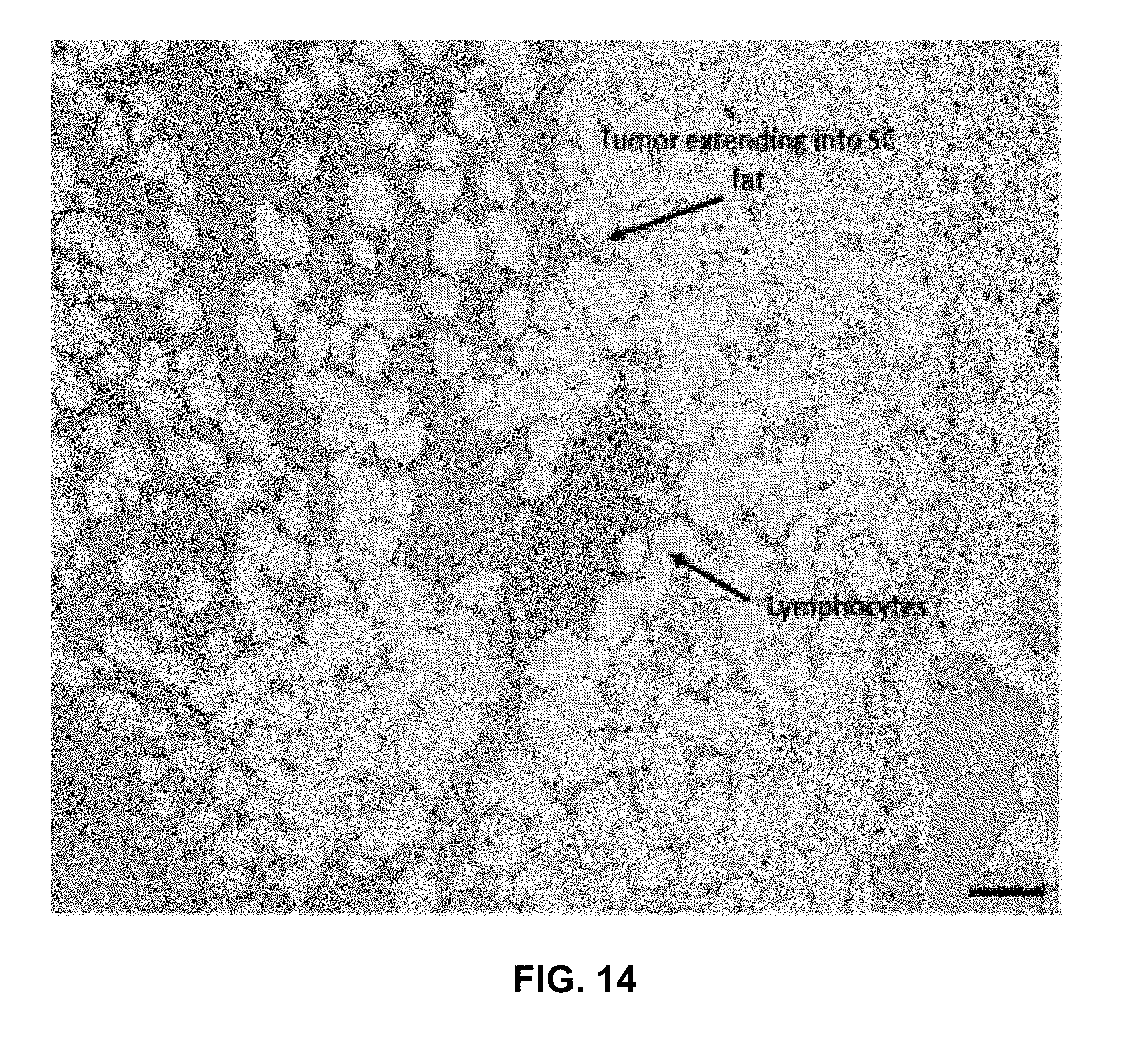
D00020

D00021

D00022

D00023
D00024

D00025

D00026

D00027

D00028

D00029

D00030
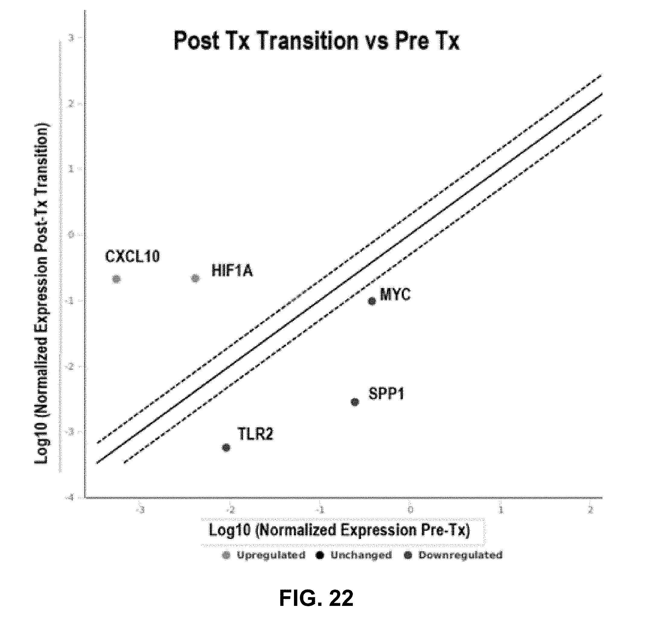
D00031

D00032

D00033

D00034

D00035

D00036

D00037

D00038

D00039

D00040

D00041

D00042

D00043

D00044

D00045

D00046

D00047

D00048


XML
uspto.report is an independent third-party trademark research tool that is not affiliated, endorsed, or sponsored by the United States Patent and Trademark Office (USPTO) or any other governmental organization. The information provided by uspto.report is based on publicly available data at the time of writing and is intended for informational purposes only.
While we strive to provide accurate and up-to-date information, we do not guarantee the accuracy, completeness, reliability, or suitability of the information displayed on this site. The use of this site is at your own risk. Any reliance you place on such information is therefore strictly at your own risk.
All official trademark data, including owner information, should be verified by visiting the official USPTO website at www.uspto.gov. This site is not intended to replace professional legal advice and should not be used as a substitute for consulting with a legal professional who is knowledgeable about trademark law.