Anti-pd1 And Anti-ctla4 Antibodies
LIU; Zhi ; et al.
U.S. patent application number 16/347554 was filed with the patent office on 2019-09-12 for anti-pd1 and anti-ctla4 antibodies. The applicant listed for this patent is QILU PUGET SOUND BIOTHERAPEUTICS CORPORATION. Invention is credited to William C. FANSLOW, Zhonghua HU, Zhi LIU, Wei YAN.
| Application Number | 20190276542 16/347554 |
| Document ID | / |
| Family ID | 60409406 |
| Filed Date | 2019-09-12 |


View All Diagrams
| United States Patent Application | 20190276542 |
| Kind Code | A1 |
| LIU; Zhi ; et al. | September 12, 2019 |
ANTI-PD1 AND ANTI-CTLA4 ANTIBODIES
Abstract
Anti-CTLA4 antibodies, anti-PD1 antibodies, mixtures thereof, polynucleotides encoding such antibodies or mixtures, optionally carried on vectors, and methods of treatment utilizing such antibodies, mixtures, polynucleotides, or vectors are described herein. The anti-CTLA4 and anti-PD1 antibodies have particular sequences and properties. The methods of treatment include uses in treatment of cancer, infections, and immunodeficiency disorders.
| Inventors: | LIU; Zhi; (Shoreline, WA) ; HU; Zhonghua; (Kenmore, WA) ; FANSLOW; William C.; (Normandy Park, WA) ; YAN; Wei; (Samamish, WA) | ||||||||||
| Applicant: |
|
||||||||||
|---|---|---|---|---|---|---|---|---|---|---|---|
| Family ID: | 60409406 | ||||||||||
| Appl. No.: | 16/347554 | ||||||||||
| Filed: | November 6, 2017 | ||||||||||
| PCT Filed: | November 6, 2017 | ||||||||||
| PCT NO: | PCT/US2017/060122 | ||||||||||
| 371 Date: | May 3, 2019 |
Related U.S. Patent Documents
| Application Number | Filing Date | Patent Number | ||
|---|---|---|---|---|
| 62419157 | Nov 8, 2016 | |||
| Current U.S. Class: | 1/1 |
| Current CPC Class: | C07K 2317/92 20130101; C07K 16/30 20130101; C07K 2317/21 20130101; A61K 9/0019 20130101; C07K 2317/55 20130101; C07K 2317/14 20130101; A61K 2039/507 20130101; C07K 16/2818 20130101; C07K 2317/56 20130101; C07K 2317/76 20130101; C07K 2317/74 20130101; A61P 31/00 20180101; C07K 2317/522 20130101; A61P 35/00 20180101 |
| International Class: | C07K 16/28 20060101 C07K016/28; A61P 35/00 20060101 A61P035/00; A61K 9/00 20060101 A61K009/00 |
Claims
1-136. (canceled)
137. An anti-human cytotoxic T lymphocyte-associated 4 (anti-hCTLA4) antibody comprising a heavy chain variable domain (VH) and a light chain variable region (VL), each comprising a complementarity determining region 1 (CDR1), a CDR2, and a CDR3, wherein the anti-hCTLA4 antibody comprises a VH CDR1, VH CDR2, VH CDR3, VL CDR1, VL CDR 2, and VL CDR3 comprising, respectively, the following amino acid sequences: SEQ ID NOs: 3, 4, 5, 8, 9, and 10; SEQ ID NOs: 3, 13, 5, 16, 9, and 17; SEQ ID NOs: 3, 20, 21, 16, 9, and 17; SEQ ID NOs: 3, 24, 25, 28, 9, and 17; SEQ ID NOs: 3, 31, 5, 34, 9, and 17; SEQ ID NOs: 3, 37, 5, 40, 9, and 10; SEQ ID NOs: 3, 43, 5, 28, 9, and 17; SEQ ID NOs: 3, 46, 47, 50, 9, and 17; SEQ ID NOs: 3, 31, 5, 55, 9, and 56; SEQ ID NOs: 3, 59, 5, 40, 9, and 62; SEQ ID NOs: 3, 65, 21, 16, 9, and 62; or SEQ ID NOs: 3, 70, 21, 73, 9, and 62, and wherein the anti-hCTLA4 antibody inhibits interaction of hCTLA4 with human B-lymphocyte activation antigen B7-1 (hB7-1) and/or human B-lymphocyte activation antigen B7-2 (hB7-2).
138. The anti-hCTLA4 antibody of claim 137, wherein the VH comprises an amino acid sequence which comprises no more than four alterations relative to an amino acid sequence selected from the group consisting of SEQ ID NOs: 2, 12, 19, 23, 30, 36, 42, 45, 52, 58, 64, and 69, and/or the VL comprises an amino acid sequence which comprises no more than four amino acid alterations relative to an amino acid sequence selected from the group consisting of SEQ ID Nos: 7, 15, 27, 33, 39, 49, 54, 61, 67, and 72.
139. The anti-hCTLA4 antibody of claim 138, wherein the VH and the VL of the anti-hCTLA4 antibody each comprise an amino acid sequence, which, together, comprise two sequences, wherein one of the two sequences comprises not more than four amino acid alterations relative to one sequence in a VH/VL pair of sequences, and the other of the two sequences comprises not more than four amino acid alterations relative to the other sequence in the VH/VL pair of sequences, and wherein the VH/VL pair of sequences is selected from the group consisting of: SEQ ID NOs: 2 (VH) and 7 (VL); SEQ ID NOs: 12 (VH) and 15 (VL); SEQ ID NOs: 19 (VH) and 15 (VL); SEQ ID NOs: 23 (VH) and 27 (VL); SEQ ID NOs: 30 (VH) and 33 (VL); SEQ ID NOs: 36 (VH) and 39 (VL); SEQ ID NOs: 42 (VH) and 27 (VL); SEQ ID NOs: 45 (VH) and 49 (VL); SEQ ID NOs: 52 (VH) and 54 (VL); SEQ ID NOs: 58 (VH) and 61 (VL); SEQ ID NOs: 64 (VH) and 67 (VL); and SEQ ID NOs: 69 (VH) and 72 (VL).
140. The anti-CTLA4 antibody of claim 137, wherein the VH CDR1, VH CDR2, VH CDR3, VL CDR1, VL CDR 2, and VL CDR3 comprise, respectively, the amino acid sequences of SEQ ID NOs: 3, 65, 21, 16, 9, and 62, the VH comprises an amino acid sequence which comprises no more than four alterations relative to the amino acid sequence of SEQ ID NO: 64, and the VL comprises an amino acid sequence which comprises no more than four alterations relative to the amino acid sequence of SEQ ID NO: 67.
141. One or more polynucleotide(s) encoding an anti-hCTLA4 antibody comprising a VH and a VL, each comprising a CDR1, a CDR2, and a CDR3, wherein the anti-hCTLA4 antibody comprises a VH CDR1, VH CDR2, VH CDR3, VL CDR1, VL CDR 2, and VL CDR3 comprising, respectively, the following amino acid sequences: SEQ ID NOs: 3, 4, 5, 8, 9, and 10; SEQ ID NOs: 3, 13, 5, 16, 9, and 17; SEQ ID NOs: 3, 20, 21, 16, 9, and 17; SEQ ID NOs: 3, 24, 25, 28, 9, and 17; SEQ ID NOs: 3, 31, 5, 34, 9, and 17; SEQ ID NOs: 3, 37, 5, 40, 9, and 10; SEQ ID NOs: 3, 43, 5, 28, 9, and 17; SEQ ID NOs: 3, 46, 47, 50, 9, and 17; SEQ ID NOs: 3, 31, 5, 55, 9, and 56; SEQ ID NOs: 3, 59, 5, 40, 9, and 62; SEQ ID NOs: 3, 65, 21, 16, 9, and 62; or SEQ ID NOs: 3, 70, 21, 73, 9, and 62, and wherein the anti-hCTLA4 antibody inhibits interaction of hCTLA4 with hB7-1 and/or hB7-2.
142. A host cell comprising one or more polynucleotide(s) encoding an anti-hCTLA4 antibody comprising a VH and a VL, each comprising a CDR1, a CDR2, and a CDR3, wherein the anti-hCTLA4 antibody comprises a VH CDR1, VH CDR2, VH CDR3, VL CDR1, VL CDR 2, and VL CDR3 comprising, respectively, the following sequences: SEQ ID NOs: 3, 4, 5, 8, 9, and 10; SEQ ID NOs: 3, 13, 5, 16, 9, and 17; SEQ ID NOs: 3, 20, 21, 16, 9, and 17; SEQ ID NOs: 3, 24, 25, 28, 9, and 17; SEQ ID NOs: 3, 31, 5, 34, 9, and 17; SEQ ID NOs: 3, 37, 5, 40, 9, and 10; SEQ ID NOs: 3, 43, 5, 28, 9, and 17; SEQ ID NOs: 3, 46, 47, 50, 9, and 17; SEQ ID NOs: 3, 31, 5, 55, 9, and 56; SEQ ID NOs: 3, 59, 5, 40, 9, and 62; SEQ ID NOs: 3, 65, 21, 16, 9, and 62; or SEQ ID NOs: 3, 70, 21, 73, 9, and 62, and wherein the anti-hCTLA4 antibody inhibits interaction of hCTLA4 with hB7-1 and/or hB7-2.
143. A method of treating a patient having a cancer or an infection comprising: (a) administering to the patient an anti-hCTLA4 antibody comprising a VH and a VL, each comprising a CDR1, a CDR2, and a CDR3, wherein: (1) the anti-hCTLA4 antibody comprises a VH CDR1, VH CDR2, VH CDR3, VL CDR1, VL CDR 2, and VL CDR3 comprising, respectively, the following sequences: SEQ ID NOs: 3, 4, 5, 8, 9, and 10; SEQ ID NOs: 3, 13, 5, 16, 9, and 17; SEQ ID NOs: 3, 20, 21, 16, 9, and 17; SEQ ID NOs: 3, 24, 25, 28, 9, and 17; SEQ ID NOs: 3, 31, 5, 34, 9, and 17; SEQ ID NOs: 3, 37, 5, 40, 9, and 10; SEQ ID NOs: 3, 43, 5, 28, 9, and 17; SEQ ID NOs: 3, 46, 47, 50, 9, and 17; SEQ ID NOs: 3, 31, 5, 55, 9, and 56; SEQ ID NOs: 3, 59, 5, 40, 9, and 62; SEQ ID NOs: 3, 65, 21, 16, 9, and 62; or SEQ ID NOs: 3, 70, 21, 73, 9, and 62; and (2) the anti-hCTLA4 antibody inhibits interaction of hCTLA4 with hB7-1 and/or hB7-2; or (b) administering to the patient one or more polynucleotide(s) encoding the anti-hCTLA4 antibody of (a).
144. An anti-human programmed cell death 1 (anti-hPD1) antibody comprising a VH and a VL, each comprising a CDR1, a CDR2, and a CDR3, wherein the anti-hPD1 antibody comprises a VH CDR1, VH CDR2, VH CDR3, VL CDR1, VL CDR 2, and VL CDR3 comprising, respectively, the following sequences: SEQ ID NOs: 88, 89, 90, 93, 94, and 95; SEQ ID NOs: 98, 89, 99, 102, 94, and 95; SEQ ID NOs: 105, 89, 90, 93, 108, and 95; SEQ ID NOs: 105, 111, 112, 115, 94, and 95; SEQ ID NOs: 105, 111, 90, 120, 94, and 95; SEQ ID NOs: 88, 89, 112, 102, 108, and 95; SEQ ID NOs: 88, 89, 127, 130, 108, and 131; SEQ ID NOs: 88, 89, 134, 137, 94, and 131; SEQ ID NOs: 98, 89, 90, 142, 94, and 131; SEQ ID NOs: 98, 111, 112, 130, 94, and 131; SEQ ID NOs: 149, 150, 112, 130, 94, and 131; SEQ ID NOs: 105, 111, 90, 142, 94, and 131; SEQ ID NOs: 149, 89, 127, 115, 94, and 95; SEQ ID NOs: 88, 89, 112, 93, 94, and 95; or SEQ ID NOs: 105, 89, 127, 102, 108, and 95, and wherein the anti-hPD1 antibody inhibits the interaction of hPD1 with human programmed cell death 1 ligand 1 (hPDL1).
145. The anti-hPD1 antibody of claim 144, wherein the anti-hPD1 VH comprises an amino acid sequence which comprises no more than four alterations relative to an amino acid sequence selected from the group consisting of SEQ ID NOs: 87, 97, 104, 110, 117, 122, 126, 133, 139, 144, 148, 154, 158, 162, and 166, and/or the anti-hPD1 VL comprises an amino acid sequence which comprises no more than four alterations relative to an amino acid sequence selected from the group consisting of SEQ ID NOs: 92, 101, 107, 114, 119, 124, 129, 136, 141, 146, 152, 156, 160, 164, and 168.
146. The anti-hPD1 antibody of claim 145, wherein the VH and the VL of the anti-hPD1 antibody each comprise an amino acid sequence, which, together, comprise two sequences, wherein one of the two sequences comprises not more than four amino acid alterations relative to one sequence in a VH/VL pair of sequences, and the other of the two sequences comprises not more than four amino acid alterations relative to the other sequence in the VH/VL pair of sequences, and wherein the VH/VL pair of sequences is selected from the group consisting of: SEQ ID NOs: 87 (VH) and 92 (VL); SEQ ID NOs: 97 (VH) and 101 (VL); SEQ ID NOs: 104 (VH) and 107 (VL); SEQ ID NOs: 110 (VH) and 114 (VL); SEQ ID NOs: 117 (VH) and 119 (VL); SEQ ID NOs: 122 (VH) and 124 (VL); SEQ ID NOs: 126 (VH) and 129 (VL); SEQ ID NOs: 133 (VH) and 136 (VL); SEQ ID NOs: 139 (VH) and 141 (VL); SEQ ID NOs: 144 (VH) and 146 (VL); SEQ ID NOs: 148 (VH) and 152 (VL); SEQ ID NOs: 154 (VH) and 156 (VL); SEQ ID NOs: 158 (VH) and 160 (VL); SEQ ID NOs: 162 (VH) and 164 (VL); and SEQ ID NOs: 166 (VH) and 168 (VL).
147. The anti-hPD1 antibody of claim 144, wherein the VH CDR1, VH CDR2, VH CDR3, VL CDR1, VL CDR 2, and VL CDR3 comprise, respectively, the amino acid sequences of SEQ ID NOs: 88, 89, 90, 93, 94, and 95, the VH comprises an amino acid sequence which comprises no more than four alterations relative to the amino acid sequence of SEQ ID NO: 87, and the VL comprises an amino acid sequence which comprises no more than four alterations relative to the amino acid sequence of SEQ ID NO: 92.
148. One or more polynucleotide(s) encoding an anti-hPD1 antibody comprising a VH and a VL, each comprising a CDR1, a CDR2, and a CDR3, wherein the anti-hPD1 antibody comprises a VH CDR1, VH CDR2, VH CDR3, VL CDR1, VL CDR 2, and VL CDR3 comprising, respectively, the following sequences: SEQ ID NOs: 88, 89, 90, 93, 94, and 95; SEQ ID NOs: 98, 89, 99, 102, 94, and 95; SEQ ID NOs: 105, 89, 90, 93, 108, and 95; SEQ ID NOs: 105, 111, 112, 115, 94, and 95; SEQ ID NOs: 105, 111, 90, 120, 94, and 95; SEQ ID NOs: 88, 89, 112, 102, 108, and 95; SEQ ID NOs: 88, 89, 127, 130, 108, and 131; SEQ ID NOs: 88, 89, 134, 137, 94, and 131; SEQ ID NOs: 98, 89, 90, 142, 94, and 131; SEQ ID NOs: 98, 111, 112, 130, 94, and 131; SEQ ID NOs: 149, 150, 112, 130, 94, and 131; SEQ ID NOs: 105, 111, 90, 142, 94, and 131; SEQ ID NOs: 149, 89, 127, 115, 94, and 95; SEQ ID NOs: 88, 89, 112, 93, 94, and 95; or SEQ ID NOs: 105, 89, 127, 102, 108, and 95, and wherein the anti-hPD1 antibody inhibits the interaction of hPD1 with hPDL1.
149. A host cell comprising one or more polynucleotide(s) encoding an anti-hPD1 antibody comprising a VH and a VL, each comprising a CDR1, a CDR2, and a CDR3, wherein the anti-hPD1 antibody comprises a VH CDR1, VH CDR2, VH CDR3, VL CDR1, VL CDR 2, and VL CDR3 comprising, respectively, the following sequences: SEQ ID NOs: 88, 89, 90, 93, 94, and 95; SEQ ID NOs: 98, 89, 99, 102, 94, and 95; SEQ ID NOs: 105, 89, 90, 93, 108, and 95; SEQ ID NOs: 105, 111, 112, 115, 94, and 95; SEQ ID NOs: 105, 111, 90, 120, 94, and 95; SEQ ID NOs: 88, 89, 112, 102, 108, and 95; SEQ ID NOs: 88, 89, 127, 130, 108, and 131; SEQ ID NOs: 88, 89, 134, 137, 94, and 131; SEQ ID NOs: 98, 89, 90, 142, 94, and 131; SEQ ID NOs: 98, 111, 112, 130, 94, and 131; SEQ ID NOs: 149, 150, 112, 130, 94, and 131; SEQ ID NOs: 105, 111, 90, 142, 94, and 131; SEQ ID NOs: 149, 89, 127, 115, 94, and 95; SEQ ID NOs: 88, 89, 112, 93, 94, and 95; or SEQ ID NOs: 105, 89, 127, 102, 108, and 95, and wherein the anti-hPD1 antibody inhibits the interaction of hPD1 with hPDL1.
150. A method of treating a patient having a cancer or an infection comprising: (a) administering to the patient an anti-hPD1 antibody comprising a VH and a VL, each comprising a CDR1, a CDR2, and a CDR3, wherein: (1) the anti-hPD1 antibody comprises a VH CDR1, VH CDR2, VH CDR3, VL CDR1, VL CDR 2, and VL CDR3 comprising, respectively, the following sequences: SEQ ID NOs: 88, 89, 90, 93, 94, and 95; SEQ ID NOs: 98, 89, 99, 102, 94, and 95; SEQ ID NOs: 105, 89, 90, 93, 108, and 95; SEQ ID NOs: 105, 111, 112, 115, 94, and 95; SEQ ID NOs: 105, 111, 90, 120, 94, and 95; SEQ ID NOs: 88, 89, 112, 102, 108, and 95; SEQ ID NOs: 88, 89, 127, 130, 108, and 131; SEQ ID NOs: 88, 89, 134, 137, 94, and 131; SEQ ID NOs: 98, 89, 90, 142, 94, and 131; SEQ ID NOs: 98, 111, 112, 130, 94, and 131; SEQ ID NOs: 149, 150, 112, 130, 94, and 131; SEQ ID NOs: 105, 111, 90, 142, 94, and 131; SEQ ID NOs: 149, 89, 127, 115, 94, and 95; SEQ ID NOs: 88, 89, 112, 93, 94, and 95; or SEQ ID NOs: 105, 89, 127, 102, 108, and 95; and (2) the anti-hPD1 antibody inhibits the interaction of hPD1 with hPDL1; or (b) administering to the patient one or more polynucleotide(s) encoding the anti-hPD1 antibody of (a).
151. A mixture comprising an anti-hCTLA4 antibody and an anti-hPD1 antibody, wherein: (a) (1) the anti-hCTLA4 antibody comprises a VH and a VL, each comprising a CDR1, a CDR2, and a CDR3, (2) the anti-hCTLA4 antibody comprises a VH CDR1, VH CDR2, VH CDR3, VL CDR1, VL CDR 2, and VL CDR3 comprising, respectively, the following sequences: SEQ ID NOs: 3, 4, 5, 8, 9, and 10; SEQ ID NOs: 3, 13, 5, 16, 9, and 17; SEQ ID NOs: 3, 20, 21, 16, 9, and 17; SEQ ID NOs: 3, 24, 25, 28, 9, and 17; SEQ ID NOs: 3, 31, 5, 34, 9, and 17; SEQ ID NOs: 3, 37, 5, 40, 9, and 10; SEQ ID NOs: 3, 43, 5, 28, 9, and 17; SEQ ID NOs: 3, 46, 47, 50, 9, and 17; SEQ ID NOs: 3, 31, 5, 55, 9, and 56; SEQ ID NOs: 3, 59, 5, 40, 9, and 62; SEQ ID NOs: 3, 65, 21, 16, 9, and 62; or SEQ ID NOs: 3, 70, 21, 73, 9, and 62, and (3) the anti-hCTLA4 antibody inhibits interaction of hCTLA4 with hB7-1 and/or hB7-2, and (b) (1) the anti-hPD1 antibody comprises a VH and a VL, each comprising a CDR1, a CDR2, and a CDR3, (2) the anti-hPD1 antibody comprises a VH CDR1, VH CDR2, VH CDR3, VL CDR1, VL CDR 2, and VL CDR3 comprising, respectively, the following sequences: SEQ ID NOs: 88, 89, 90, 93, 94, and 95; SEQ ID NOs: 98, 89, 99, 102, 94, and 95; SEQ ID NOs: 105, 89, 90, 93, 108, and 95; SEQ ID NOs: 105, 111, 112, 115, 94, and 95; SEQ ID NOs: 105, 111, 90, 120, 94, and 95; SEQ ID NOs: 88, 89, 112, 102, 108, and 95; SEQ ID NOs: 88, 89, 127, 130, 108, and 131; SEQ ID NOs: 88, 89, 134, 137, 94, and 131; SEQ ID NOs: 98, 89, 90, 142, 94, and 131; SEQ ID NOs: 98, 111, 112, 130, 94, and 131; SEQ ID NOs: 149, 150, 112, 130, 94, and 131; SEQ ID NOs: 105, 111, 90, 142, 94, and 131; SEQ ID NOs: 149, 89, 127, 115, 94, and 95; SEQ ID NOs: 88, 89, 112, 93, 94, and 95; or SEQ ID NOs: 105, 89, 127, 102, 108, and 95, and (3) the anti-hPD1 antibody inhibits the interaction of hPD1 with hPDLl.
152. The mixture of claim 151, wherein (a) (1) the VH CDR1, VH CDR2, VH CDR3, VL CDR1, VL CDR 2, and VL CDR3 of the anti-hCTLA4 antibody comprise, respectively, the amino acid sequences of SEQ ID NOs: 3, 65, 21, 16, 9, and 62, (2) the VH of the anti-hCTLA4 antibody comprises an amino acid sequence which comprises no more than four alterations relative to the amino acid sequence of SEQ ID NO: 64, and (3) the VL of the anti-hCTLA4 antibody comprises an amino acid sequence which comprises no more than four alterations relative to the amino acid sequence of SEQ ID NO: 67, and (b) (1) the VH CDR1, VH CDR2, VH CDR3, VL CDR1, VL CDR 2, and VL CDR3 of the anti-hPD1 antibody comprise, respectively, the amino acid sequences of SEQ ID NOs: 88, 89, 90, 93, 94, and 95, (2) the VH comprises an amino acid sequence which comprises no more than four alterations relative to the amino acid sequence of SEQ ID NO: 87, and (3) the VL comprises an amino acid sequence which comprises no more than four alterations relative to the amino acid sequence of SEQ ID NO: 92.
153. One or more polynucleotide(s) encoding a mixture comprising an anti-hCTLA4 antibody and an anti-hPD1 antibody, wherein (a) (1) the anti-hCTLA4 antibody comprises a VH and a VL, each comprising a CDR1, a CDR2, and a CDR3, (2) the anti-hCTLA4 antibody comprises a VH CDR1, VH CDR2, VH CDR3, VL CDR1, VL CDR 2, and VL CDR3 comprising, respectively, the following amino acid sequences: SEQ ID NOs: 3, 4, 5, 8, 9, and 10; SEQ ID NOs: 3, 13, 5, 16, 9, and 17; SEQ ID NOs: 3, 20, 21, 16, 9, and 17; SEQ ID NOs: 3, 24, 25, 28, 9, and 17; SEQ ID NOs: 3, 31, 5, 34, 9, and 17; SEQ ID NOs: 3, 37, 5, 40, 9, and 10; SEQ ID NOs: 3, 43, 5, 28, 9, and 17; SEQ ID NOs: 3, 46, 47, 50, 9, and 17; SEQ ID NOs: 3, 31, 5, 55, 9, and 56; SEQ ID NOs: 3, 59, 5, 40, 9, and 62; SEQ ID NOs: 3, 65, 21, 16, 9, and 62; or SEQ ID NOs: 3, 70, 21, 73, 9, and 62, and (3) the anti-hCTLA4 antibody inhibits interaction of hCTLA4 with hB7-1 and/or hB7-2, and (b) (1) the anti-hPD1 antibody comprises a VH and a VL, each comprising a CDR1, a CDR2, and a CDR3, (2) the anti-hPD1 antibody comprises a VH CDR1, VH CDR2, VH CDR3, VL CDR1, VL CDR 2, and VL CDR3 comprising, respectively, the following amino acid sequences: SEQ ID NOs: 88, 89, 90, 93, 94, and 95; SEQ ID NOs: 98, 89, 99, 102, 94, and 95; SEQ ID NOs: 105, 89, 90, 93, 108, and 95; SEQ ID NOs: 105, 111, 112, 115, 94, and 95; SEQ ID NOs: 105, 111, 90, 120, 94, and 95; SEQ ID NOs: 88, 89, 112, 102, 108, and 95; SEQ ID NOs: 88, 89, 127, 130, 108, and 131; SEQ ID NOs: 88, 89, 134, 137, 94, and 131; SEQ ID NOs: 98, 89, 90, 142, 94, and 131; SEQ ID NOs: 98, 111, 112, 130, 94, and 131; SEQ ID NOs: 149, 150, 112, 130, 94, and 131; SEQ ID NOs: 105, 111, 90, 142, 94, and 131; SEQ ID NOs: 149, 89, 127, 115, 94, and 95; SEQ ID NOs: 88, 89, 112, 93, 94, and 95; or SEQ ID NOs: 105, 89, 127, 102, 108, and 95, and (3) the anti-hPD1 antibody inhibits the interaction of hPD1 with hPDL1.
154. A host cell comprising one or more polynucleotides encoding a mixture comprising an anti-hCTLA4 antibody and an anti-hPD1 antibody, wherein (a) (1) the anti-hCTLA4 antibody comprises a VH and a VL, each comprising a CDR1, a CDR2, and a CDR3, (2) the anti-hCTLA4 antibody comprises a VH CDR1, VH CDR2, VH CDR3, VL CDR1, VL CDR 2, and VL CDR3 comprising, respectively, the following amino acid sequences: SEQ ID NOs: 3, 4, 5, 8, 9, and 10; SEQ ID NOs: 3, 13, 5, 16, 9, and 17; SEQ ID NOs: 3, 20, 21, 16, 9, and 17; SEQ ID NOs: 3, 24, 25, 28, 9, and 17; SEQ ID NOs: 3, 31, 5, 34, 9, and 17; SEQ ID NOs: 3, 37, 5, 40, 9, and 10; SEQ ID NOs: 3, 43, 5, 28, 9, and 17; SEQ ID NOs: 3, 46, 47, 50, 9, and 17; SEQ ID NOs: 3, 31, 5, 55, 9, and 56; SEQ ID NOs: 3, 59, 5, 40, 9, and 62; SEQ ID NOs: 3, 65, 21, 16, 9, and 62; or SEQ ID NOs: 3, 70, 21, 73, 9, and 62, and (3) the anti-hCTLA4 antibody inhibits interaction of hCTLA4 with hB7-1 and/or hB7-2, and (b) (1) the anti-hPD1 antibody comprises a VH and a VL, each comprising a CDR1, a CDR2, and a CDR3, (2) the anti-hPD1 antibody comprises a VH CDR1, VH CDR2, VH CDR3, VL CDR1, VL CDR 2, and VL CDR3 comprising, respectively, the following amino acid sequenes: SEQ ID NOs: 88, 89, 90, 93, 94, and 95; SEQ ID NOs: 98, 89, 99, 102, 94, and 95; SEQ ID NOs: 105, 89, 90, 93, 108, and 95; SEQ ID NOs: 105, 111, 112, 115, 94, and 95; SEQ ID NOs: 105, 111, 90, 120, 94, and 95; SEQ ID NOs: 88, 89, 112, 102, 108, and 95; SEQ ID NOs: 88, 89, 127, 130, 108, and 131; SEQ ID NOs: 88, 89, 134, 137, 94, and 131; SEQ ID NOs: 98, 89, 90, 142, 94, and 131; SEQ ID NOs: 98, 111, 112, 130, 94, and 131; SEQ ID NOs: 149, 150, 112, 130, 94, and 131; SEQ ID NOs: 105, 111, 90, 142, 94, and 131; SEQ ID NOs: 149, 89, 127, 115, 94, and 95; SEQ ID NOs: 88, 89, 112, 93, 94, and 95; or SEQ ID NOs: 105, 89, 127, 102, 108, and 95, and (3) the anti-hPD1 antibody inhibits the interaction of hPD1 with hPDL1.
155. A method of treating a patient having a cancer or an infection comprising: (a) administering to the patient a mixture comprising an anti-hCTLA4 antibody and an anti-hPD1 antibody, wherein: (1) (A) the anti-hCTLA4 antibody comprises a VH and a VL, each comprising a CDR1, a CDR2, and a CDR3, (B) the anti-hCTLA4 antibody comprises a VH CDR1, VH CDR2, VH CDR3, VL CDR1, VL CDR 2, and VL CDR3 comprising, respectively, the following amino acid sequences: SEQ ID NOs: 3, 4, 5, 8, 9, and 10; SEQ ID NOs: 3, 13, 5, 16, 9, and 17; SEQ ID NOs: 3, 20, 21, 16, 9, and 17; SEQ ID NOs: 3, 24, 25, 28, 9, and 17; SEQ ID NOs: 3, 31, 5, 34, 9, and 17; SEQ ID NOs: 3, 37, 5, 40, 9, and 10; SEQ ID NOs: 3, 43, 5, 28, 9, and 17; SEQ ID NOs: 3, 46, 47, 50, 9, and 17; SEQ ID NOs: 3, 31, 5, 55, 9, and 56; SEQ ID NOs: 3, 59, 5, 40, 9, and 62; SEQ ID NOs: 3, 65, 21, 16, 9, and 62; or SEQ ID NOs: 3, 70, 21, 73, 9, and 62, and (C) the anti-hCTLA4 antibody inhibits interaction of hCTLA4 with hB7-1 and/or hB7-2; and (2) (A) the anti-hPD1 antibody comprises a VH and a VL, each comprising a CDR1, a CDR2, and a CDR3, (B) the anti-hPD1 antibody comprises a VH CDR1, VH CDR2, VH CDR3, VL CDR1, VL CDR 2, and VL CDR3 comprising, respectively, the following amino acid sequenes: SEQ ID NOs: 88, 89, 90, 93, 94, and 95; SEQ ID NOs: 98, 89, 99, 102, 94, and 95; SEQ ID NOs: 105, 89, 90, 93, 108, and 95; SEQ ID NOs: 105, 111, 112, 115, 94, and 95; SEQ ID NOs: 105, 111, 90, 120, 94, and 95; SEQ ID NOs: 88, 89, 112, 102, 108, and 95; SEQ ID NOs: 88, 89, 127, 130, 108, and 131; SEQ ID NOs: 88, 89, 134, 137, 94, and 131; SEQ ID NOs: 98, 89, 90, 142, 94, and 131; SEQ ID NOs: 98, 111, 112, 130, 94, and 131; SEQ ID NOs: 149, 150, 112, 130, 94, and 131; SEQ ID NOs: 105, 111, 90, 142, 94, and 131; SEQ ID NOs: 149, 89, 127, 115, 94, and 95; SEQ ID NOs: 88, 89, 112, 93, 94, and 95; or SEQ ID NOs: 105, 89, 127, 102, 108, and 95, and (C) the anti-hPD1 antibody inhibits the interaction of hPD1 with hPDL1; or (b) administering to the patient one or more polynucleotide(s) encoding the mixture of (a).
Description
FIELD
[0001] The compositions and methods described herein are in the field of recombinant antibodies.
BACKGROUND
[0002] Immune checkpoint inhibitors represent a new class of cancer therapeutics. See, e.g., Pardoll (2012), Nat. Rev. Cancer 12: 252-264. T cell response is regulated by a balance of activating and inhibitory signals. Immune checkpoints, that is, inhibitory pathways, can help maintain self tolerance and/or modulate the duration and/or intensity of an immune response, thereby limiting collateral damage to healthy tissues. Cancer cells can develop multiple ways to upregulate these immune checkpoint pathways to dampen host immune response, which might otherwise kill cancer cells. The success of immune checkpoint inhibitors or blockers, e.g. anti-programmed cell death 1 (PD1), anti-programmed cell death 1 ligand 1 (PDL1), and anti-cytotoxic T lymphocyte-associated 4 (CTLA4) antibodies, in treating many types of cancer is evidence of the importance of immune checkpoints in regulating T cell response to cancer. See, e.g., Larkin et al. (2015), New Engl. J. Med 373(1): 23-34.
[0003] Refining this approach to improve patient responses and limit toxicity remains an ongoing challenge. Thus, there is a need in the art for improved immune checkpoint inhibitors and improved methods of treatment using such inhibitors. Such improved immune checkpoint inhibitors and improved methods of treatment are described herein.
SUMMARY
[0004] Described herein are anti-human CTLA4 (hCTLA4) antibodies, anti-human PD1 (hPD1) antibodies, mixtures thereof, nucleic acids encoding these antibodies and mixtures, host cells containing these nucleic acids, pharmaceutical compositions comprising these antibodies, mixtures, and nucleic acids, and methods of treatment comprising administering these antibodies, mixtures, nucleic acids, or pharmaceutical compositions to patients. The numbered items below describe these compositions and methods.
[0005] 1. An anti-human CTLA4 (hCTLA4) antibody comprising a heavy chain variable domain (VH),
[0006] wherein the VH comprises an amino acid sequence which comprises no more than four alterations relative to an amino acid sequence selected from the group consisting of SEQ ID NOs: 2, 12, 19, 23, 30, 36, 42, 45, 52, 58, 64, and 69, and wherein the anti-hCTLA4 antibody inhibits interaction of hCTLA4 with human B-lymphocyte activation antigen B7-1 (hB7-1) and/or human B-lymphocyte activation antigen B7-2 (hB7-2).
[0007] 2. The anti-hCTLA4 antibody of item 1, wherein the VH comprises a VH complementarity determining region 1 (CDR1), a VH complementarity determining region 2 (CDR2), and a VH complementarity determining region 3 (CDR3) which comprise, respectively the amino acid sequences of SEQ ID NOs: 3, 4, and 5, SEQ ID NOs: 3, 13 and 5, SEQ ID NOs: 3, 20, and 21, SEQ ID NOs: 3, 24, and 25, SEQ ID NOs: 3, 31, and 5, SEQ ID NOs: 3, 37, and 5, SEQ ID NOs: 3, 43, and 5, SEQ ID NOs: 3, 46, and 47, SEQ ID NOs: 3, 59, and 5, SEQ ID NOs: 3, 65, and 21, or SEQ ID NOs: 3, 70, and 21.
[0008] 3. The anti-hCTLA4 antibody of item 1 or 2, wherein the amino acid sequence comprised by the VH comprises no more than three amino acid alterations relative to the amino acid sequence selected from the group consisting of SEQ ID NOs: 2, 12, 19, 23, 30, 36, 42, 45, 52, 58, 64, and 69.
[0009] 4. The anti-hCTLA4 antibody of item 3, wherein the amino acid sequence comprised by the VH comprises no more than two amino acid alterations relative to the amino acid sequence selected from the group consisting of SEQ ID NOs: 2, 12, 19, 23, 30, 36, 42, 45, 52, 58, 64, and 69.
[0010] 5. The anti-hCTLA4 antibody of item 4, wherein the amino acid sequence comprised by the VH comprises no more than one amino acid alteration relative to the amino acid sequence selected from the group consisting of SEQ ID NOs: 2, 12, 19, 23, 30, 36, 42, 45, 52, 58, 64, and 69.
[0011] 6. The anti-hCTLA4 antibody of any one of items 1 to 5, wherein the amino acid alteration(s) is (are) (a) substitution(s), and the substitution(s) is (are) (a) partner directing alteration(s).
[0012] 7. The anti-hCTLA4 antibody of item 5, wherein the amino acid sequence comprised by the VH is selected from the group consisting of SEQ ID NOs: 2, 12, 19, 23, 30, 36, 42, 45, 52, 58, 64, and 69.
[0013] 8. The anti-hCTLA4 antibody of any one of items 1 to 7, further comprising a light chain variable domain (VL), which comprises a VL CDR1, a VL CDR2, and a VL CDR3 comprising, respectively, the amino acid sequences of SEQ ID NOs: 3, 75, and 77.
[0014] 9. The anti-hCTLA4 antibody of item 8, wherein the VL CDR1, VL CDR2, and VL CDR3 comprise, respectively, the amino acid sequences of SEQ ID NOs: 8, 9, and 10, SEQ ID NOs: 16, 9 and 17, SEQ ID NOs: 28, 9, and 17, SEQ ID NOs: 34, 9, and 17, SEQ ID NOs: 40, 9, and 10, SEQ ID NOs: 50, 9, and 17, SEQ ID NOs: 55, 9, and 56, SEQ ID NOs: 40, 9, and 62, SEQ ID NOs: 16, 9, and 62, or SEQ ID NOs: 73, 9, and 62.
[0015] 10. The anti-hCTLA4 antibody of any one of items 1 to 7, further comprising a VL comprising an amino acid sequence which comprises no more than 4 amino acid alterations relative to an amino acid sequence selected from the group consisting of SEQ ID NOs: 7, 15, 27, 33, 39, 49, 54, 61, 67, 72, 84, and 85.
[0016] 11. The anti-hCTLA4 antibody of item 10, wherein the amino acid sequence comprised by the VL comprises no more than three amino acid alterations relative to the amino acid sequence selected from the group consisting of SEQ ID NOs: 7, 15, 27, 33, 39, 49, 54, 61, 67, 72, 84, and 85.
[0017] 12. The anti-hCTLA4 antibody of item 11, wherein the amino acid sequence comprised by the VL comprises no more than two amino acid alterations relative to the amino acid sequence selected from the group consisting of SEQ ID NOs: 7, 15, 27, 33, 39, 49, 54, 61, 67, 72, 84, and 85.
[0018] 13. The anti-hCTLA4 antibody of item 12, wherein the amino acid sequence comprised by the VL comprises no more than one amino acid alteration relative to the amino acid sequence selected from the group consisting of SEQ ID NOs: 7, 15, 27, 33, 39, 49, 54, 61, 67, 72, 84, and 85.
[0019] 14. The anti-hCTLA4 antibody of any one of items 10 to 13, wherein the amino acid alteration(s) relative to SEQ ID NOs: 7, 15, 27, 33, 39, 49, 54, 61, 67, 72, 84 or 85 is (are) substitution(s), and the substitution(s) is (are) (a) partner-directing alteration(s).
[0020] 15. The anti-hCTLA4 antibody of any one of items 10 to 14, wherein the VL comprises a VL CDR1, a VL CDR2, and a VL CDR3 comprising, respectively, the amino acid sequences of SEQ ID NOs: 8, 9, and 10, SEQ ID NOs: 16, 9 and 17, SEQ ID NOs: 28, 9, and 17, SEQ ID NOs: 34, 9, and 17, SEQ ID NOs: 40, 9, and 10, SEQ ID NOs: 50, 9, and 17, SEQ ID NOs: 55, 9, and 56, SEQ ID NOs: 40, 9, and 62, SEQ ID NOs: 16, 9, and 62, or SEQ ID NOs: 73, 9, and 62.
[0021] 16. The anti-hCTLA4 antibody of item 15, wherein the amino acid sequence comprised by the VL is selected from the group consisting of SEQ ID NOs: 7, 15, 27, 33, 39, 49, 54, 61, 67, 72, 84, and 85.
[0022] 17. An anti-hCTLA4 antibody comprising a VL,
[0023] wherein the VL comprises an amino acid sequence which comprises no more than four amino acid alterations relative to an amino acid sequence selected from the group consisting of SEQ ID NOs: 7, 15, 27, 33, 39, 49, 54, 61, 67, and 72,
[0024] wherein the VL comprises a CDR1, a CDR2, and a CDR3, which comprise, respectively, the amino acid sequences of SEQ ID NOs: 8, 9, and 10, SEQ ID NOs: 16, 9 and 17, SEQ ID NOs: 28, 9, and 17, SEQ ID NOs: 34, 9, and 17, SEQ ID NOs: 40, 9, and 10, SEQ ID NOs: 50, 9, and 17, SEQ ID NOs: 55, 9, and 56, SEQ ID NOs: 40, 9, and 62, SEQ ID NOs: 16, 9, and 62, or SEQ ID NOs: 73, 9, and 62, and
[0025] wherein the anti-hCTLA4 antibody inhibits interaction of hCTLA4 with hB7-1 and/or hB7-2.
[0026] 18. The anti-hCTLA4 antibody of item 17, wherein the amino acid sequence comprised by the VL comprises no more than three amino acid alterations relative to the amino acid sequence selected from the group consisting of SEQ ID NOs: 7, 15, 27, 33, 39, 49, 54, 61, 67, and 72.
[0027] 19. The anti-hCTLA4 antibody of item 18, wherein the amino acid sequence comprised by the VL comprises no more than two amino acid alterations relative to the amino acid sequence selected from the group consisting of SEQ ID NOs: 7, 15, 27, 33, 39, 49, 54, 61, 67, and 72.
[0028] 20. The anti-hCTLA4 antibody of item 19, wherein the amino acid sequence comprised by the VL comprises no more than one amino acid alteration relative to the amino acid sequence selected from the group consisting of SEQ ID NOs: 7, 15, 27, 33, 39, 49, 54, 61, 67, and 72.
[0029] 21. The anti-hCTLA4 antibody of any one of items 17 to 20, wherein the amino acid alteration(s) is (are) (a) substitution(s), and the substitution(s) is (are) (a) partner-directing alteration(s).
[0030] 22. The anti-hCTLA4 antibody of item 20, wherein the amino acid sequence comprised by the VL is selected from the group consisting of SEQ ID NOs: 7, 15, 27, 33, 39, 49, 54, 61, 67, and 72.
[0031] 23. The anti-hCTLA4 antibody of any one of items 17 to 22, further comprising a VH, which comprises a VH CDR1, a VH CDR2, and a VH CDR3 comprising, respectively, the amino acid sequences of SEQ ID NO:79, SEQ ID NO:9, and SEQ ID NO:81.
[0032] 24. The anti-hCTLA4 antibody of item 23, wherein the VH CDR1, VH CDR2, and VH CDR3 comprise, respectively, the amino acid sequences of SEQ ID NOs: 3, 4, and 5, SEQ ID NOs: 3, 13 and 5, SEQ ID NOs: 3, 20, and 21, SEQ ID NOs: 3, 24, and 25, SEQ ID NOs: 3, 31, and 5, SEQ ID NOs: 3, 37, and 5, SEQ ID NOs: 3, 43, and 5, SEQ ID NOs: 3, 46, and 47, SEQ ID NOs: 3, 59, and 5, SEQ ID NOs: 3, 65, and 21, or SEQ ID NOs: 3, 70, and 21.
[0033] 25. The anti-hCTLA4 antibody of any one of items 17 to 22, further comprising a VH, wherein the VH comprises an amino acid sequence which comprises no more than four amino acid alterations relative to an amino acid sequence selected from the group consisting of SEQ ID NOs: 2, 12, 19, 23, 30, 36, 42, 45, 52, 58, 64, 69, 82, and 83.
[0034] 26. The anti-hCTLA4 antibody of item 25, wherein the amino acid sequence comprised by the VH comprises no more than three amino acid alterations relative to the amino acid sequence selected from the group consisting of SEQ ID NOs: 2, 12, 19, 23, 30, 36, 42, 45, 52, 58, 64, 69, 82, and 83.
[0035] 27. The anti-hCTLA4 antibody of item 26, wherein the amino acid sequence comprised by the VH comprises no more than two amino acid alterations relative to the amino acid sequence selected from the group consisting of SEQ ID NOs: 2, 12, 19, 23, 30, 36, 42, 45, 52, 58, 64, 69, 82, and 83.
[0036] 28. The anti-hCTLA4 antibody of item 27, wherein the amino acid sequence comprised by the VH comprises no more than one amino acid alteration relative to the amino acid sequence selected from the group consisting of SEQ ID NOs: 2, 12, 19, 23, 30, 36, 42, 45, 52, 58, 64, 69, 82, and 83.
[0037] 29. The anti-hCTLA4 antibody of any one of items 25 to 28, wherein the amino acid alteration(s) is (are) (a) substitution(s), and the substitution(s) is (are) (a) partner-directing alteration(s).
[0038] 30. The anti-hCTLA4 antibody of any one of items 25 to 29, wherein the VH comprises a VH CDR1, VH CDR2, and VH CDR3 comprising, respectively, the amino acid sequences of SEQ ID NOs: 3, 4, and 5, SEQ ID NOs: 3, 13 and 5, SEQ ID NOs: 3, 20, and 21, SEQ ID NOs: 3, 24, and 25, SEQ ID NOs: 3, 31, and 5, SEQ ID NOs: 3, 37, and 5, SEQ ID NOs: 3, 43, and 5, SEQ ID NOs: 3, 46, and 47, SEQ ID NOs: 3, 59, and 5, SEQ ID NOs: 3, 65, and 21, or SEQ ID NOs: 3, 70, and 21.
[0039] 31. The anti-hCTLA4 antibody of item 28, wherein the amino acid sequence comprised by the VH is selected from the group consisting of SEQ ID NOs: 2, 12, 19, 23, 30, 36, 42, 45, 52, 58, 64, 69, 82, and 83.
[0040] 32. An anti-hCTLA4 antibody comprising a VH CDR1, VH CDR2, VH CDR3, VL CDR1, VL CDR2, and VL CDR3, which comprise, respectively, the following amino acid sequences:
[0041] (a) SEQ ID NOs: 3, 4, 5, 8, 9, and 10;
[0042] (b) SEQ ID NOs: 3, 13, 5, 16, 9, and 17;
[0043] (c) SEQ ID NOs: 3, 20, 21, 16, 9, and 17;
[0044] (d) SEQ ID NOs: 3, 24, 25, 28, 9, and 17;
[0045] (e) SEQ ID NOs: 3, 31, 5, 34, 9, and 17;
[0046] (f) SEQ ID NOs: 3, 37, 5, 40, 9, and 10;
[0047] (g) SEQ ID NOs: 3, 43, 5, 28, 9, and 17;
[0048] (h) SEQ ID NOs: 3, 46, 47, 50, 9, and 17;
[0049] (i) SEQ ID NOs: 3, 31, 5, 55, 9, and 56;
[0050] (j) SEQ ID NOs: 3, 59, 5, 40, 9, and 62;
[0051] (k) SEQ ID NOs: 3, 65, 21, 16, 9, and 62; or
[0052] (l) SEQ ID NOs: 3, 70, 21, 73, 9, and 62;
[0053] wherein the anti-hCTLA4 antibody inhibits interaction of hCTLA4 with hB7-1 and/or hB7-2.
[0054] 33. The anti-hCTLA4 antibody of item 32, wherein the VH and the VL of the anti-hCTLA4 antibody each comprise an amino acid sequence, which, together, comprise two sequences, and
[0055] wherein one of the two sequences comprises not more than four amino acid alterations relative to one sequence in a pair of sequences and the other of the two sequences comprises not more than four amino acid alterations relative to the other sequence in the pair of sequences, and
[0056] wherein the pair of sequences is selected from the group consisting of: SEQ ID NOs: 2 (VH) and 7 (VL); SEQ ID NOs: 12 (VH) and 15 (VL); SEQ ID NOs: 19 (VH) and 15 (VL); SEQ ID NOs: 23 (VH) and 27 (VL); SEQ ID NOs: 30 (VH) and 33 (VL); SEQ ID NOs: 36 (VH) and 39 (VL); SEQ ID NOs: 42 (VH) and 27 (VL); SEQ ID NOs: 45 (VH) and 49 (VL); SEQ ID NOs: 52 (VH) and 54 (VL); SEQ ID NOs: 58 (VH) and 61 (VL); SEQ ID NOs: 64 (VH) and 67 (VL); and SEQ ID NOs: 69 (VH) and 72 (VL).
[0057] 34. The anti-hCTLA4 antibody of item 33, wherein one of the two sequences comprises not more than three amino acid alterations relative to one sequence in the pair of sequences and the other of the two sequences comprises not more than three amino acid alterations relative to the other sequence in the pair of sequences.
[0058] 35. The anti-hCTLA4 antibody of item 34, wherein one of the two sequences comprises not more than two amino acid alterations relative to one sequence in the pair of sequences and the other of the two sequences comprises not more than two amino acid alterations relative to the other sequence in the pair of sequences.
[0059] 36. The anti-hCTLA4 antibody of item 35, wherein one of the two sequences comprises not more than one amino acid alteration relative to one sequence in the pair of sequences and the other of the two sequences comprises not more than one amino acid alteration relative to the other sequence in the pair of sequences.
[0060] 37. The anti-hCTLA4 antibody of any one of items 33 to 36, wherein the alteration(s) is (are) (a) substitution(s), and the substitution(s) is (are) (a) partner-directing alteration(s).
[0061] 38. The anti-hCTLA4 antibody of item 36, wherein one of the two sequences comprises one sequence in the pair of sequences and the other of the two sequences comprises the other sequence in the pair of sequences.
[0062] 39. The anti-hCTLA4 antibody of any one of items 1 to 38, which is a human, humanized, or primate IgG antibody.
[0063] 40. The anti-hCTLA4 antibody of item 39, which is (1) an IgG4 antibody or (2) an IgG1 antibody comprising the alteration K409R.
[0064] 41. The anti-hCTLA4 antibody of item 39, which is an IgG1 antibody.
[0065] 42. The anti-hCTLA4 antibody of any one of items 39 to 41,
[0066] wherein the heavy chain (HC) or the light chain (LC) contains a first partner-directing alteration at one or more positions selected from the group consisting of positions 44, 105, 147, 168 and 181 in the HC and positions 43, 100, 131, 174, and 178 in the LC,
[0067] wherein the first partner-directing alteration is a substitution where a charged amino acid replaces another amino acid,
[0068] wherein the substituted, charged amino acid of the first partner-directing alteration forms part of a charge pair of contacting amino acids within the antibody, and wherein the contacting amino acids of the charge pair are selected from the group consisting of:
[0069] (a) 44R/K (HC) and 100D/E (LC) or 44D/E (HC) and 100R/K (LC);
[0070] (b) 105R/K (HC) and 43D/E (LC) or 105D/E (HC) and 43R/K (LC);
[0071] (c) 147R/K (HC) and 131D/E (LC) or 147D/E (HC) and 131R/K (LC);
[0072] (d) 168R/K (HC) and 174D/E (LC) or 168D/E (HC) and 174R/K (LC); and
[0073] (e) 181R/K (HC) and 178D/E (LC) or 181D/E (HC) and 178R/K (LC).
[0074] 43. The anti-hCTLA4 antibody of item 42, further comprising a second partner-directing alteration,
[0075] wherein the second partner-directing alteration is a substitution of a charged amino acid for another amino acid, and
[0076] wherein the substituted, charged amino acids of the first and second partner-directing alterations form the charge pair of contacting amino acids.
[0077] 44. The anti-hCTLA4 antibody of item 42 or 43,
[0078] wherein the anti-hCTLA4 antibody is an IgG1 antibody,
[0079] wherein the HC or LC of the anti-hCTLA4 antibody contains a third partner-directing alteration at an amino acid in the HC or the LC,
[0080] wherein the third partner-directing alteration is a cysteine substitution, and wherein the substituted cysteine of the third partner-directing alteration contacts a cysteine (1) in the HC if the third partner-directing alteration is in the LC or (2) in the LC if the third partner-directing alteration is in the HC, thereby forming a contacting pair of cysteines.
[0081] 45. The anti-hCTLA4 antibody of item 44, wherein the contacting pair of cysteines is at positions selected from the group consisting of:
[0082] (a) position 126 of the HC and position 121 or 124 in the LC;
[0083] (b) position 128 in the HC and position 118 in the LC;
[0084] (c) position 133 in the HC and position 117 or 209 in the LC;
[0085] (d) position 134 or 141 in the HC and position 116 in the LC;
[0086] (e) position 168 in the HC and position 174 in the LC;
[0087] (f) position 170 in the HC and position 162 or 176 in the LC;
[0088] (g) position 173 in the HC and position 160 in the LC; and
[0089] (h) position 183 in the HC and position 176 in the LC.
[0090] 46. The anti-hCTLA4 antibody of item 45, wherein the contacting pair of cysteines is at positions selected from the group consisting of:
[0091] (a) position 126 of the HC and position 124 in the LC;
[0092] (b) position 128 of the HC and position 118 in the LC;
[0093] (c) position 133 of the HC and position 117 or 209 in the LC;
[0094] (d) position 134 of the HC and position 116 in the LC;
[0095] (e) position 168 of the HC and position 174 in the LC;
[0096] (f) position 170 in the HC and position 162 or 176 in the LC; and
[0097] (g) position 173 in the HC and position 160 in the LC.
[0098] 47. The anti-hCTLA4 antibody of any one of items 44 to 46, wherein
[0099] if the third partner-directing alteration is in the HC, then the LC comprises a fourth partner-directing alteration which is a cysteine substitution at an amino acid contacting the amino acid of the third partner-directing alteration, or
[0100] if the third partner-directing alteration is in the LC, then the HC comprises a fourth partner-directing alteration, which is a cysteine substitution at an amino acid contacting the amino acid of the third partner-directing alteration.
[0101] 48. The anti-hCTLA4 antibody of item 42 or 43,
[0102] wherein the anti-hCTLA4 antibody is an IgG4 antibody,
[0103] wherein the HC or LC of the anti-hCTLA4 antibody contains a third partner-directing alteration at an amino acid in the HC or LC,
[0104] wherein the third partner-directing alteration is a cysteine substitution, and
[0105] wherein the substituted cysteine of the third partner-directing alteration contacts a cysteine (1) in the HC if the third partner-directing alteration is in the LC or (2) in the LC if the third partner-directing alteration is in the HC, thereby forming a contacting pair of cysteines.
[0106] 49. The anti-hCTLA4 antibody of item 48, wherein the contacting pair of cysteines is at positions within the HC and LC selected from the group consisting of:
[0107] (a) position 126 in the HC and position 121 or 124 in the LC;
[0108] (b) position 127 in the HC and position 121 in the LC;
[0109] (c) position 128 in the HC and position 118 in the LC;
[0110] (d) position 141 in the HC and position 116 in the LC;
[0111] (e) position 168 in the HC and position 174 in the LC; and
[0112] (f) position 170 or 173 in the HC and position 162 in the LC.
[0113] 50. The anti-hCTLA4 antibody of item 48 or 49, wherein
[0114] if the third partner-directing alteration is in the HC, then the LC comprises a fourth partner-directing alteration, which is a cysteine substitution at an amino acid contacting the amino acid of the third partner-directing alteration, or
[0115] if the third partner-directing alteration is in the LC, then the HC comprises a fourth partner-directing alteration, which is a cysteine substitution at an amino acid contacting the amino acid of the third partner-directing alteration.
[0116] 51. The anti-hCTLA4 antibody of any one of items 1 to 50, which comprises the alterations D399K/R and K/R409E/D.
[0117] 52. The anti-hCTLA4 antibody of any one of items 1 to 51, wherein the anti-hCTLA4 antibody is a human, humanized, or primate IgG antibody comprising an Fc portion, wherein the Fc portion of the antibody comprises one or more alterations that increase clearance of the antibody in vivo.
[0118] 53. The anti-hCTLA4 antibody of item 52, wherein the Fc portion of the antibody comprises at least one of the following alterations: M252A, M252L, M252S, M252R, R255K or H435R.
[0119] 54. An anti-hCTLA4 antibody, which comprises the amino acid sequences of SEQ ID NOs: 189 and 191 or SEQ ID NOs: 193 and 195.
[0120] 55. An anti-human PD1 (hPD1) antibody comprising a VH,
[0121] wherein the VH comprises an amino acid sequence which comprises no more than four amino acid alterations relative to an amino acid sequence selected from the group consisting of SEQ ID NOs: 87, 97, 104, 110, 117, 122, 126, 133, 139, 144, 148, 154, 158, 162, and 166, and
[0122] wherein the anti-hPD1 antibody inhibits the interaction of hPD1 with human PDL1 (hPDL1).
[0123] 56. The anti-hPD1 antibody of item 55, wherein the VH comprises a VH CDR1, a VH CDR2, and a VH CDR3 comprising, respectively, the amino acid sequence of SEQ ID NOs: 88, 89, and 90, SEQ ID NOs: 98, 89 and 99, SEQ ID NOs: 105, 89, and 90, SEQ ID NOs: 105, 111, and 112, SEQ ID NOs: 105, 111, and 90, SEQ ID NOs: 88, 89, and 112, SEQ ID NOs: 88, 89, and 127, SEQ ID NOs: 88, 89, and 134, SEQ ID NOs: 98, 89, and 90, SEQ ID NOs: 98, 111, and 112, SEQ ID NOs: 149, 150, and 112, SEQ ID NOs: 105, 111, and 90, SEQ ID NOs: 149, 89, and 127, SEQ ID NOs: 88, 89, and 112, or SEQ ID NOs: 105, 89, and 127.
[0124] 57. The anti-hPD1 antibody of item 55 or 56, wherein the amino acid sequence comprised by the VH comprises no more than three amino acid alterations relative to the amino acid sequence selected from the group consisting of SEQ ID NOs: 87, 97, 104, 110, 117, 122, 126, 133, 139, 144, 148, 154, 158, 162, and 166.
[0125] 58. The anti-hPD1 antibody of item 57, wherein the amino acid sequence comprised by the VH comprises no more than two amino acid alterations relative to the amino acid sequence selected from the group consisting of SEQ ID NOs: 87, 97, 104, 110, 117, 122, 126, 133, 139, 144, 148, 154, 158, 162, and 166.
[0126] 59. The anti-hPD1 antibody of item 58, wherein the amino acid sequence comprised by the VH comprises no more than one amino acid alteration relative to the amino acid sequence selected from the group consisting of SEQ ID NOs: 87, 97, 104, 110, 117, 122, 126, 133, 139, 144, 148, 154, 158, 162, and 166.
[0127] 60. The anti-hPD1 antibody of any one of items 55 to 59, wherein the amino acid alteration(s) is (are) (a) substitution(s), and the substitution(s) is (are) (a) partner-directing alteration(s).
[0128] 61. The anti-hPD1 antibody of item 59, wherein the amino acid sequence comprised by the VH is selected from the group consisting of SEQ ID NOs: 87, 97, 104, 110, 117, 122, 126, 133, 139, 144, 148, 154, 158, 162, and 166.
[0129] 62. The anti-hPD1 antibody of any one of items 55 to 61, further comprising a VL, which comprises a VL CDR1, a VL CDR2, and a VL CDR3 comprising, respectively, the amino acid sequences of SEQ ID NOs:176, 177, and 179.
[0130] 63. The anti-hPD1 antibody of item 62, wherein the VL CDR1, VL CDR2, and VL CDR3 comprise, respectively, the amino acid sequences of SEQ ID NOs: 93, 94, and 95, SEQ ID NOs: 102, 94, and 95, SEQ ID NOs: 93, 108, and 95, SEQ ID NOs: 115, 94, and 95, SEQ ID NOs: 120, 94, and 95, SEQ ID NOs: 102, 108, and 95, SEQ ID NOs: 130, 108, and 131, SEQ ID NOs: 137, 94, and 131, SEQ ID NOs: 142, 94, and 131, or SEQ ID NOs: 130, 94, and 131.
[0131] 64. The anti-hPD1 antibody of any one of items 55 to 61, comprising a VL comprising an amino acid sequence which comprises no more than four amino acid alterations relative to an amino acid sequence selected from the group consisting of SEQ ID NOs: 92, 101, 107, 114, 119, 124, 129, 136, 141, 146, 152, 156, 160, 164, 168, 182, and 183.
[0132] 65. The anti-hPD1 antibody of item 64, wherein the amino acid sequence comprised by the VL comprises no more than three amino acid alterations relative to the amino acid sequence selected from the group consisting of SEQ ID NOs: 92, 101, 107, 114, 119, 124, 129, 136, 141, 146, 152, 156, 160, 164, 168, 182, and 183.
[0133] 66. The anti-hPD1 antibody of item 65, wherein the amino acid sequence comprised by the VL comprises no more than two amino acid alterations relative to the amino acid sequence selected from the group consisting of SEQ ID NOs: 92, 101, 107, 114, 119, 124, 129, 136, 141, 146, 152, 156, 160, 164, 168, 182, and 183.
[0134] 67. The anti-hPD1 antibody of item 66, wherein the amino acid sequence comprised by the VL comprises no more than one amino acid alteration relative to the amino acid sequence selected from the group consisting of SEQ ID NOs: 92, 101, 107, 114, 119, 124, 129, 136, 141, 146, 152, 156, 160, 164, 168, 182, and 183.
[0135] 68. The anti-hPD1 antibody of any one of items 64 to 67, wherein the amino acid alteration(s) relative to SEQ ID NOs: 92, 101, 107, 114, 119, 124, 129, 136, 141, 146, 152, 156, 160, 164, 168, 182, or 183 is (are) (a) substitution(s), and the substitution(s) is (are) (a) partner-directing alteration(s).
[0136] 69. The anti-hPD1 antibody of item 67, wherein the amino acid sequence comprised by the VL is selected from the group consisting of SEQ ID NOs: 92, 101, 107, 114, 119, 124, 129, 136, 141, 146, 152, 156, 160, 164, 168, 182, and 183.
[0137] 70. The anti-hPD1 antibody of any one of items 64 to 69, wherein the VL comprises a VL CDR1, a VL CDR2, and a VL CDR3 comprising, respectively, the amino acid sequences SEQ ID NOs: 93, 94, and 95, SEQ ID NOs: 102, 94, and 95, SEQ ID NOs: 93, 108, and 95, SEQ ID NOs: 115, 94, and 95, SEQ ID NOs: 120, 94, and 95, SEQ ID NOs: 102, 108, and 95, SEQ ID NOs: 130, 108, and 131, SEQ ID NOs: 137, 94, and 131, SEQ ID NOs: 142, 94, and 131, or SEQ ID NOs: 130, 94, and 131.
[0138] 71. An anti-hPD1 antibody comprising a VL,
[0139] wherein the VL comprises an amino acid sequence which comprises no more than four amino acid alterations relative to an amino acid sequence selected from the group consisting of SEQ ID NOs: 92, 101, 107, 114, 119, 124, 129, 136, 141, 146, 152, 156, 160, 164, and 168, and
[0140] wherein the anti-hPD1 antibody inhibits the interaction of hPD1 with hPDL1.
[0141] 72. The anti-hPD1 antibody of item 71 comprising a VL CDR1, a VL CDR2, and a VL CDR3 comprising, respectively, the amino acid sequences of SEQ ID NOs: 93, 94, and 95, SEQ ID NOs: 102, 94, and 95, SEQ ID NOs: 93, 108, and 95, SEQ ID NOs: 115, 94, and 95, SEQ ID NOs: 120, 94, and 95, SEQ ID NOs: 102, 108, and 95, SEQ ID NOs: 130, 108, and 131, SEQ ID NOs: 137, 94, and 131, SEQ ID NOs: 142, 94, and 131, or SEQ ID NOs: 130, 94, and 131.
[0142] 73. The anti-PD1 antibody of item 71 or 72, wherein the amino acid sequence comprised by the VL comprises no more than three amino acid alterations relative to the amino acid sequence selected from the group consisting of SEQ ID NOs: 92, 101, 107, 114, 119, 124, 129, 136, 141, 146, 152, 156, 160, 164, and 168.
[0143] 74. The anti-PD1 antibody of item 73, wherein the amino acid sequence comprised by the VL comprises no more than two amino acid alterations relative to the amino acid sequence selected from the group consisting of SEQ ID NOs: 92, 101, 107, 114, 119, 124, 129, 136, 141, 146, 152, 156, 160, 164, and 168.
[0144] 75. The anti-PD1 antibody of item 74, wherein the amino acid sequence comprised by the VL comprises no more than one amino acid alteration relative to the amino acid sequence selected from the group consisting of SEQ ID NOs: 92, 101, 107, 114, 119, 124, 129, 136, 141, 146, 152, 156, 160, 164, and 168.
[0145] 76. The anti-PD1 antibody of any one of items 71 to 75, wherein the amino acid alteration(s) is (are) (a) substitution(s), and the substitution(s) is (are) (a) partner-directing alteration(s).
[0146] 77. The anti-PD1 antibody of item 75, wherein the amino acid sequence comprised by the VL is selected from the group consisting of SEQ ID NOs: 92, 101, 107, 114, 119, 124, 129, 136, 141, 146, 152, 156, 160, 164, and 168.
[0147] 78. The anti-hPD1 antibody of any one of items 71 to 77, further comprising a VH, which comprises a VH CDR1, a VH CDR2, and a VH CDR3 comprising, respectively, the amino acid sequences of SEQ ID NOs: 170, 172, and 174.
[0148] 79. The anti-hPD1 antibody of item 78, wherein the VH CDR1, VH CDR2, and VH CDR3 comprise, respectively, the amino acid sequences of SEQ ID NOs: 88, 89, and 90, SEQ ID NOs: 98, 89 and 99, SEQ ID NOs: 105, 89, and 90, SEQ ID NOs: 105, 111, and 112, SEQ ID NOs: 105, 111, and 90, SEQ ID NOs: 88, 89, and 112, SEQ ID NOs: 88, 89, and 127, SEQ ID NOs: 88, 89, and 134, SEQ ID NOs: 98, 89, and 90, SEQ ID NOs: 98, 111, and 112, SEQ ID NOs: 149, 150, and 112, SEQ ID NOs: 105, 111, and 90, SEQ ID NOs: 149, 89, and 127, SEQ ID NOs: 88, 89, and 112, or SEQ ID NOs: 105, 89, and 127.
[0149] 80. The anti-hPD1 antibody of any one of items 71 to 77, further comprising a VH comprising an amino acid sequence which comprises no more than four amino acid alterations relative to an amino acid sequence selected from the group consisting of: SEQ ID NOs: 87, 97, 104, 110, 117, 122, 126, 133, 139, 144, 148, 154, 158, 162, 166, 180, and 181.
[0150] 81. The anti-hPD1 antibody of item 80, wherein the amino acid sequence comprised by the VH comprises no more than three amino acid alterations relative to the amino acid sequence selected from the group consisting of: SEQ ID NOs: 87, 97, 104, 110, 117, 122, 126, 133, 139, 144, 148, 154, 158, 162, 166, 180, and 181.
[0151] 82. The anti-hPD1 antibody of item 81, wherein the amino acid sequence comprised by the VH comprises no more than two amino acid alterations relative to the amino acid sequence selected from the group consisting of: SEQ ID NOs: 87, 97, 104, 110, 117, 122, 126, 133, 139, 144, 148, 154, 158, 162, 166, 180, and 181.
[0152] 83. The anti-hPD1 antibody of item 82, wherein the amino acid sequence comprised by the VH comprises no more than one amino acid alteration relative to the amino acid sequence selected from the group consisting of: SEQ ID NOs: 87, 97, 104, 110, 117, 122, 126, 133, 139, 144, 148, 154, 158, 162, 166, 180, and 181.
[0153] 84. The anti-hPD1 antibody of any one of items 80 to 83, wherein the amino acid alteration(s) is (are) (a) substitution(s), and the substitution(s) is (are) (a) partner-directing alteration(s).
[0154] 85. The anti-hPD1 antibody of item 83, wherein the amino acid sequence comprised by the VH is selected from the group consisting of: SEQ ID NOs: 87, 97, 104, 110, 117, 122, 126, 133, 139, 144, 148, 154, 158, 162, 166, 180, and 181.
[0155] 86. The anti-PD1 antibody of any one of items 80 to 85, wherein the VH comprises a VH CDR1, a VH CDR2, and a VH CDR3 comprising, respectively, the amino acid sequences of SEQ ID NOs: 88, 89, and 90, SEQ ID NOs: 98, 89 and 99, SEQ ID NOs: 105, 89, and 90, SEQ ID NOs: 105, 111, and 112, SEQ ID NOs: 105, 111, and 90, SEQ ID NOs: 88, 89, and 112, SEQ ID NOs: 88, 89, and 127, SEQ ID NOs: 88, 89, and 134, SEQ ID NOs: 98, 89, and 90, SEQ ID NOs: 98, 111, and 112, SEQ ID NOs: 149, 150, and 112, SEQ ID NOs: 105, 111, and 90, SEQ ID NOs: 149, 89, and 127, SEQ ID NOs: 88, 89, and 112, or SEQ ID NOs: 105, 89, and 127.
[0156] 87. An anti-hPD1 antibody comprising a VH CDR1, VH CDR2, VH CDR3, VL CDR1, VL CDR2, and VL CDR3, which comprise, respectively, the following amino acid sequences:
[0157] (a) SEQ ID NOs: 88, 89, 90, 93, 94, and 95;
[0158] (b) SEQ ID NOs: 98, 89, 99, 102, 94, and 95;
[0159] (c) SEQ ID NOs: 105, 89, 90, 93, 108, and 95;
[0160] (d) SEQ ID NOs: 105, 111, 112, 115, 94, and 95;
[0161] (e) SEQ ID NOs: 105, 111, 90, 120, 94, and 95;
[0162] (f) SEQ ID NOs: 88, 89, 112, 102, 108, and 95;
[0163] (g) SEQ ID NOs: 88, 89, 127, 130, 108, and 131;
[0164] (h) SEQ ID NOs: 88, 89, 134, 137, 94, and 131;
[0165] (i) SEQ ID NOs: 98, 89, 90, 142, 94, and 131;
[0166] (j) SEQ ID NOs: 98, 111, 112, 130, 94, and 131;
[0167] (k) SEQ ID NOs: 149, 150, 112, 130, 94, and 131;
[0168] (l) SEQ ID NOs: 105, 111, 90, 142, 94, and 131;
[0169] (m) SEQ ID NOs: 149, 89, 127, 115, 94, and 95;
[0170] (n) SEQ ID NOs: 88, 89, 112, 93, 94, and 95; or
[0171] (o) SEQ ID NOs: 105, 89, 127, 102, 108, and 95,
[0172] wherein the anti-hPD1 antibody inhibits the interaction of hPD1 with hPDL1.
[0173] 88. The anti-hPD1 antibody of item 87, wherein the VH and the VL of the anti-hPD1 antibody each comprise an amino acid sequence, which, together, comprise two sequences,
[0174] wherein one of the two sequences comprises not more than four amino acid alterations relative to one sequence in a pair of sequences and the other of the two sequences comprises not more than four amino acid alterations relative to the other sequence in the pair of sequences, and
[0175] wherein the pair of sequences is selected from the group consisting of: SEQ ID NOs: 87 (VH) and 92 (VL); SEQ ID NOs: 97 (VH) and 101 (VL); SEQ ID NOs: 104 (VH) and 107 (VL); SEQ ID NOs: 110 (VH) and 114 (VL); SEQ ID NOs: 117 (VH) and 119 (VL); SEQ ID NOs: 122 (VH) and 124 (VL); SEQ ID NOs; 126 (VH) and 129 (VL); SEQ ID NOs: 133 (VH) and 136 (VL); SEQ ID NOs: 139 (VH) and 141 (VL); SEQ ID NOs: 144 (VH) and 146 (VL); SEQ ID NOs: 148 (VH) and 152 (VL); SEQ ID NOs: 154 (VH) and 156 (VL); SEQ ID NOs: 158 (VH) and 160 (VL); SEQ ID NOs: 162 (VH) and 164 (VL); and SEQ ID NOs: 166 (VH) and 168 (VL).
[0176] 89. The anti-hPD1 antibody of item 88, wherein one of the two sequences comprises not more than three amino acid alterations relative to one sequence in the pair of sequences and the other of the two sequences comprises not more than three amino acid alterations relative to the other sequence in the pair of sequences.
[0177] 90. The anti-hPD1 antibody of item 89, wherein one of the two sequences comprises not more than two amino acid alterations relative to one sequence in the pair of sequences and the other of the two sequences comprises not more than two amino acid alterations relative to the other sequence in the pair of sequences.
[0178] 91. The anti-hPD1 antibody of item 90, wherein one of the two sequences comprises not more than one amino acid alteration relative to one sequence in the pair of sequences and the other of the two sequences comprises not more than one amino acid alteration relative to the other sequence in the pair of sequences.
[0179] 92. The anti-hPD1 antibody of any one of items 88 to 91, wherein the alteration(s) is (are) (a) substitution(s), and the substitution(s) is (are) (a) partner-directing alteration(s).
[0180] 93. The anti-hPD1 antibody of item 91, wherein one of the two sequences comprises one sequence in the pair of sequences and the other of the two sequences comprises the other sequence in the pair of sequences.
[0181] 94. The anti-hPD1 antibody of any one of items 55 to 93, which is a human, humanized, or primate IgG antibody.
[0182] 95. The anti-hPD1 antibody of item 94, which is (1) an IgG4 antibody or (2) an IgG1 antibody comprising the alteration K409R. 96. The anti-hPD1 antibody of item 94, which is an IgG1 antibody.
[0183] 97. The anti-hPD1 antibody of any one of items 94 to 96,
[0184] wherein the heavy chain (HC) or the light chain (LC) contains a first partner-directing alteration at one or more positions selected from the group consisting of positions 44, 105, 147, 168 and 181 in the HC and positions 43, 100, 131, 174, and 178 in the LC,
[0185] wherein the partner-directing alteration is a substitution where a charged amino acid replaces another amino acid,
[0186] wherein the substituted, charged amino acid of the partner-directing alteration forms part of a charge pair of contacting amino acids within the antibody, and
[0187] wherein the contacting amino acids of the charge pair are selected from the group consisting of:
[0188] (a) 44R/K (HC) and 100D/E (LC) or 44D/E (HC) and 100R/K (LC);
[0189] (b) 105R/K (HC) and 43D/E (LC) or 105D/E (HC) and 43R/K (LC);
[0190] (c) 147R/K (HC) and 131D/E (LC) or 147D/E (HO) and 131R/K (LC);
[0191] (d) 168R/K (HC) and 174D/E (LC) or 168D/E (HC) and 174R/K (LC); and
[0192] (e) 181R/K (HC) and 178D/E (LC) or 181D/E (HC) and 178R/K (LC).
[0193] 98. The anti-hPD1 antibody of item 97, further comprising a second partner-directing alteration,
[0194] wherein the second partner-directing alteration is a substitution of a charged amino acid for another amino acid, and
[0195] wherein the substituted, charged amino acids of the first and second partner-directing alterations form the charge pair of contacting amino acids.
[0196] 99. The anti-hPD1 antibody of item 97 or 98,
[0197] wherein the anti-hPD1 antibody is an IgG1 antibody,
[0198] wherein the HC or LC of the anti-hPD1 antibody contains a third partner-directing alteration at an amino acid in the HC or the LC,
[0199] wherein the third partner-directing alteration is a cysteine substitution, and wherein the substituted cysteine of the third partner-directing alteration contacts a cysteine (1) in the HC if the third partner-directing alteration is in the LC or (2) in the LC if the third partner-directing alteration is in the HC, thereby forming a contacting pair of cysteines.
[0200] 100. The anti-hPD1 antibody of item 99, wherein the contacting pair of cysteines is at positions selected from the group consisting of:
[0201] (a) position 126 of the HC and position 121 or 124 in the LC;
[0202] (b) position 128 in the HC and position 118 in the LC;
[0203] (c) position 133 in the HC and position 117 or 209 in the LC;
[0204] (d) position 134 or 141 in the HC and position 116 in the LC;
[0205] (e) position 168 in the HC and position 174 in the LC;
[0206] (f) position 170 in the HC and position 162 or 176 in the LC;
[0207] (g) position 173 in the HC and position 160 in the LC; and
[0208] (h) position 183 in the HC and position 176 in the LC.
[0209] 101. The anti-hPD1 antibody of item 100, wherein the contacting pair of cysteines is at a pair of positions selected from the group consisting of:
[0210] (a) position 126 of the HC and position 124 in the LC;
[0211] (b) position 128 of the HC and position 118 in the LC;
[0212] (c) position 133 of the HC and position 117 or 209 in the LC;
[0213] (d) position 134 of the HC and position 116 in the LC;
[0214] (e) position 168 of the HC and position 174 in the LC;
[0215] (f) position 170 in the HC and position 162 or 176 in the LC; and
[0216] (g) position 173 in the HC and position 160 in the LC.
[0217] 102. The anti-hPD1 antibody of any one of items 99 to 101, wherein
[0218] if the third partner-directing alteration is in the HC, then the LC comprises a fourth partner-directing alteration which is a cysteine substitution at an amino acid contacting the amino acid of the third partner-directing alteration, or
[0219] if the third partner-directing alteration is in the LC, then the HO comprises a fourth partner-directing alteration, which is a cysteine substitution at an amino acid contacting the amino acid of the third partner-directing alteration.
[0220] 103. The anti-hPD1 of item 97 or 98,
[0221] wherein the anti-hPD1 antibody is an IgG4 antibody,
[0222] wherein the HO or LC of the anti-hPD1 antibody contains a third partner-directing alteration at an amino acid in the HO or LC,
[0223] wherein the third partner-directing alteration is a cysteine substitution, and
[0224] wherein the substituted cysteine of the third partner-directing alteration contacts a cysteine (1) in the HO if the third partner-directing alteration is in the LC or (2) in the LC if the second partner-directing alteration is in the HC, thereby forming a contacting pair of cysteines.
[0225] 104. The anti-hPD1 antibody of item 103, wherein the contacting pair of cysteines is at positions within the HO and LC selected from the group consisting of:
[0226] (a) position 126 in the HO and position 121 or 124 in the LC;
[0227] (b) position 127 in the HO and position 121 in the LC;
[0228] (c) position 128 in the HO and position 118 in the LC;
[0229] (d) position 141 in the HO and position 116 in the LC;
[0230] (e) position 168 in the HC and position 174 in the LC; and
[0231] (f) position 170 or 173 in the HO and position 162 in the LC.
[0232] 105. The anti-hPD1 antibody of item 103 or 104, wherein
[0233] if the third partner-directing alteration is in the HC, then the LC comprises a fourth partner-directing alteration, which is a cysteine substitution at an amino acid contacting the amino acid of the third partner-directing alteration, or
[0234] if the third partner-directing alteration is in the LC, then the HO comprises a fourth partner-directing alteration, which is a cysteine substitution at an amino acid contacting the amino acid of the third partner-directing alteration.
[0235] 106. The anti-hPD1 antibody of any one of items 55 to 105, which comprises the alterations D399K/R and K/R409E/D.
[0236] 107. An anti-hPD1 antibody, which comprises the amino acid sequences of SEQ ID NOs: 185 and 187.
[0237] 108, A mixture of antibodies comprising the anti-hCTLA4 antibody of any one of items 1 to 54 and the anti-hPD1 antibody of any one of items 55 to 107,
[0238] 109. The mixture of antibodies of item 108, comprising the anti-hCTLA4 antibody of item 8 and the anti-hPD1 antibody of item 61.
[0239] 110, The mixture of antibodies of item 108 or 109, wherein the mixture is produced in a single host cell line.
[0240] 111. The mixture of antibodies of item 110, which comprises not more than three major species of antibodies.
[0241] 112. The mixture of antibodies of item 111, which comprises not more than two major species of antibodies.
[0242] 113. One or more polynucleotide(s), which encode(s) the anti-hCTLA4 antibody of any one of items 1 to 54, the anti-hPD1 antibody of any one of items 55 to 107, or the mixture of antibodies of any one of items 108 to 112.
[0243] 114. The polynucleotide(s) of item 113, which encode(s) the mixture of antibodies of any one of items 108 to 112.
[0244] 115. One or more vector(s), which comprise(s) the polynucleotide(s) of item 113 or 114.
[0245] 116. The vector(s) of item 115, which is (are) (a) viral vector(s).
[0246] 117. The vector(s) of item 116, which is (are) (an) oncolytic viral vector(s),
[0247] 118. The vector(s) of item 116 or 117, which is (are) (a) retroviral, adenoviral, adeno-associated viral (AAV), vaccinia viral, modified vaccinia viral Ankara (MVA), herpes viral, lentiviral, measles viral, coxsackie viral, Newcastle Disease viral, reoviral, or poxviral vector(s).
[0248] 119. A pharmaceutical composition comprising the anti-hCTLA4 antibody of any one of items 1 to 54, the anti-hPD1 antibody of any one of items 55 to 107, the mixture of antibodies of any one of items 108 to 112, the polynucleotide(s) of item 113 or 114, or the vector(s) of any one of items 115 to 118.
[0249] 120. A host cell, into which the polynucleotide(s) of item 113 or 114 or the vector(s) of item 115 or 116 has (have) been introduced.
[0250] 121. The host cell of item 120, which is a mammalian cell.
[0251] 122. The host cell of item 121, which is a CHO cell or a mouse myeloma cell.
[0252] 123. A method for making an antibody or a mixture of antibodies comprising culturing the host cell of any one of items 120 to 122 and recovering the antibody or mixture of antibodies from the culture medium or the cell mass.
[0253] 124. A method for treating a patient having a solid tumor or a hematological malignancy comprising administering to the patient the anti-hCTLA4 antibody of any one of items 1 to 54, the anti-hPD1 antibody of any one of items 55 to 107, the mixture of any one of items 108 to 112, the polynucleotide(s) of item 113 or 114, the vector(s) of any one of item 115 to 118, or the pharmaceutical composition of item 119.
[0254] 125. The method of item 124, comprising administering the anti-hCTLA4 antibody, the anti-hPD1 antibody, or the mixture of antibodies.
[0255] 126. The method of item 124 or 125, wherein the patient has melanoma, non-small cell lung cancer, small cell lung cancer, gastric cancer, bladder cancer, clear cell renal carcinoma, Hodgkin's lymphoma, or head and neck squamous cell cancer.
[0256] 127. The method of any one of items 124 to 126, further comprising administering an additional therapy before, after, and/or concurrently with the anti-hCTLA4 antibody, the anti-hPD1 antibody, or the mixture of antibodies, the polynucleotide(s), the vector(s), or the pharmaceutical composition, wherein the additional therapy is selected from the group consisting of immunomodulatory molecules, radiation, a chemotherapeutic agent, a targeted biologic, a targeted inhibitor, and/or an oncolytic virus.
[0257] 128. The method of item 127, wherein the additional therapy is radiation or a chemotherapeutic agent.
[0258] 129. The method of item 127, wherein the additional therapy is a targeted biologic, a targeted inhibitor, and/or a immunomodulatory molecule selected from the group consisting of: an antagonist of PDL1, TIGIT, CCR4, CCR8, CSFR1a, B7H3, B7H4, CD96, or CD73; an agonist of GITR, 41BB, OX40, or CD40; an oncolytic virus such as talimogene laherparepvec (IMLYGIC.TM.); a bispecific T cell engager (BiTE) such as blinatumomab; an indoleamine 2, 3 dioxygenase (IDO) inhibitor; an anti-angiogenic agent such as bevacizumab; an antibody-drug conjugate; and a tyrosine kinase inhibitor.
[0259] 130. A method for inducing or enhancing an immune response in a patient comprising administering to the patient the anti-hCTLA4 antibody of any one of item 1 to 54, the anti-hPD1 antibody of any one of items 55 to 107, the mixture of any one of items 108 to 112, the polynucleotide(s) of item 113 or 114, the vector(s) of any one of items 115 to 118, or the pharmaceutical composition of item 119.
[0260] 131. The method of item 130, comprising administering the mixture of antibodies, one or more vector(s) encoding the mixture of antibodies, or a pharmaceutical composition comprising the mixture of antibodies or the vector(s) encoding the mixture of antibodies.
[0261] 132. The method of item 130 or 131, wherein the patient has melanoma, non-small cell lung cancer, small cell lung cancer, gastric cancer, bladder cancer, clear cell renal carcinoma, Hodgkin's lymphoma, or head and neck squamous cell cancer.
[0262] 133. The method of any one of items 130 to 132, further comprising administering an additional therapy before, after, and/or concurrently with the anti-hCTLA4 antibody, the anti-hPD1 antibody, the mixture of antibodies, the polynucleotide(s), or the vector(s), wherein the additional therapy is selected from the group consisting of immunomodulatory molecules, radiation, a chemotherapeutic agent, a targeted biologic, a targeted inhibitor, and/or an oncolytic virus.
[0263] 134. The method of item 133, wherein the additional therapy is radiation or a chemotherapeutic agent.
[0264] 135. The method of item 133, wherein the additional therapy is a targeted biologic, a targeted inhibitor, and/or an immunomodulatory molecule selected from the group consisting of: an antagonist of PDL1, TIGIT, CCR4, CCR8, CSFR1a, B7H3, B7H4, CD96, or CD73; an agonist of GITR, 41BB, OX40, or CD40; an oncolytic virus such as talimogene laherparepvec (IMLYGIC.TM.); a bispecific T cell engager (BiTE) such as blinatumomab; an indoleamine 2, 3 dioxygenase (IDO) inhibitor; an anti-angiogenic agent such as bevacizumab; an antibody-drug conjugate; and a tyrosine kinase inhibitor.
[0265] 136. A method for treating a patient having an infection comprising administering to the patient the anti-hCTLA4 antibody of any one of items 1 to 54, the anti-hPD1 antibody of any one of items 55 to 107, the mixture of any one of items 108 to 112, the polynucleotide(s) of item 113 or 114, the vector(s) of any one of items 115 to 118, or the pharmaceutical composition of item 119.
[0266] 137. An anti-human CTLA4 (hCTLA4) antibody comprising a heavy chain variable domain (VH) comprising a VH complementarity determining region 1 (CDR1), a VH complementarity determining region 2 (CDR2), and a VH complementarity determining region 3 (CDR3) comprising, respectively, the amino acid sequences of SEQ ID NOs: 3, 4, and 5, SEQ ID NOs: 3, 13 and 5, SEQ ID NOs: 3, 20, and 21, SEQ ID NOs: 3, 24, and 25, SEQ ID NOs: 3, 31, and 5, SEQ ID NOs: 3, 37, and 5, SEQ ID NOs: 3, 43, and 5, SEQ ID NOs: 3, 46, and 47, SEQ ID NOs: 3, 59, and 5, SEQ ID NOs: 3, 65, and 21, or SEQ ID NOs: 3, 70, and 21.
[0267] 138. The anti-hCTLA4 antibody of item 137, wherein the VH comprises the amino acid sequence of SEQ ID NO:82.
[0268] 139. The anti-hCTLA4 antibody of item 138, wherein the VH comprises the amino acid sequence of SEQ ID NO:83.
[0269] 140. The anti-hCTLA4 antibody of item 139, wherein the VH comprises an amino acid sequence selected from the group consisting of SEQ ID NOs: 2, 12, 19, 23, 30, 36, 42, 45, 52, 58, 64, and 69.
[0270] 141. An anti-hCTLA4 antibody comprising a light chain variable domain (VL) comprising a VL CDR1, a VL CDR2, and a VL CDR3 comprising, respectively, the amino acid sequences of SEQ ID NOs: 8, 9, and 10, SEQ ID NOs: 16, 9 and 17, SEQ ID NOs: 28, 9, and 17, SEQ ID NOs: 34, 9, and 17, SEQ ID NOs: 40, 9, and 10, SEQ ID NOs: 50, 9, and 17, SEQ ID NOs: 55, 9, and 56, SEQ ID NOs: 40, 9, and 62, SEQ ID NOs: 16, 9, and 62, or SEQ ID NOs: 73, 9, and 62.
[0271] 142, The anti-hCTLA4 antibody of item 141, wherein the VL comprises the amino acid sequence of SEQ ID NO:84.
[0272] 143. The anti-hCTLA4 antibody of item 142, wherein the VL comprises the amino acid sequence of SEQ ID NO:85.
[0273] 144. The anti-hCTLA4 antibody of item 143, wherein the VL comprises an amino acid sequence selected from the group consisting of SEQ ID NOs: 7, 15, 27, 33, 39, 49, 54, 61, 67, and 72.
[0274] 145. The anti-hCTLA4 antibody of any one of items 137 to 140, further comprising a VL comprising a VL CDR1, a VL CDR2, and a VL CDR3 comprising, respectively, the amino acid sequences of SEQ ID NOs: 8, 9, and 10, SEQ ID NOs: 16, 9 and 17, SEQ ID NOs: 28, 9, and 17, SEQ ID NOs: 34, 9, and 17, SEQ ID NOs: 40, 9, and 10, SEQ ID NOs: 50, 9, and 17, SEQ ID NOs: 55, 9, and 56, SEQ ID NOs: 40, 9, and 62, SEQ ID NOs: 16, 9, and 62, or SEQ ID NOs: 73, 9, and 62.
[0275] 146. The anti-hCTLA4 antibody of item 145, comprising a VH CDR1, VH CDR2, VH CDR3, VL CDR1, VL CDR2, and VL CDR3, which comprise, respectively, the following amino acid sequences:
[0276] (a) SEQ ID NOs: 3, 4, 5, 8, 9, and 10;
[0277] (b) SEQ ID NOs: 3, 13, 5, 16, 9, and 17;
[0278] (c) SEQ ID NOs: 3, 20, 21, 16, 9, and 17;
[0279] (d) SEQ ID NOs: 3, 24, 25, 28, 9, and 17;
[0280] (e) SEQ ID NOs: 3, 31, 5, 34, 9, and 17;
[0281] (f) SEQ ID NOs: 3, 37, 5, 40, 9, and 10;
[0282] (g) SEQ ID NOs: 3, 43, 5, 28, 9, and 17;
[0283] (h) SEQ ID NOs: 3, 46, 47, 50, 9, and 17;
[0284] (i) SEQ ID NOs: 3, 31, 5, 55, 9, and 56;
[0285] (j) SEQ ID NOs: 3, 59, 5, 40, 9, and 62;
[0286] (k) SEQ ID NOs: 3, 65, 21, 16, 9, and 62; or
[0287] (l) SEQ ID NOs: 3, 70, 21, 73, 9, and 62.
[0288] 147. The anti-hCTLA4 antibody of item 145, wherein the VL comprises the amino acid sequence of SEQ ID NO:84 and the VH comprises the amino acid sequence of SEQ ID NO:82.
[0289] 148. The anti-hCTLA4 antibody of item 147, wherein the VL comprises the amino acid sequence of SEQ ID NO:85 and the VH comprises the amino acid sequence of SEQ ID NO:83.
[0290] 149. The anti-hCTLA4 antibody of item 148, wherein the VL comprises an amino acid sequence selected from the group consisting of SEQ ID NOs: 7, 15, 27, 33, 39, 49, 54, 61, 67, and 72.
[0291] 150. The anti-hCTLA4 antibody of item 149, wherein the VH and VL comprise, respectively, amino acid sequences selected from the group consisting of: SEQ ID NOs: 2 and 7; SEQ ID NOs: 12 and 15; SEQ ID NOs: 19 and 15; SEQ ID NOs: 23 and 27; SEQ ID NOs: 30 and 33; SEQ ID NOs: 36 and 39; SEQ ID NOs: 42 and 27; SEQ ID NOs: 45 and 49; SEQ ID NOs: 52 and 54; SEQ ID NOs: 58 and 61; SEQ ID NOs: 64 and 67; and SEQ ID NOs: 69 and 72.
[0292] 151. The anti-hCTLA4 antibody of any one of items 137 to 150, which is a human, humanized or primate IgG antibody. 152. The anti-hCTLA4 antibody of item 151, which is an IgG4 antibody or an IgG1 antibody comprising the alteration K409R.
[0293] 153. The anti-hCTLA4 antibody of item 151, which is an IgG1 antibody.
[0294] 154. The anti-hCTLA4 antibody of any one of items 151 to 153,
[0295] wherein the heavy chain (HC) and/or the light chain (LC) contain at least one partner-directing alteration at one or more sites selected from the group consisting of positions 44, 105, 147, 168 and 181 in the HC and positions 43, 100, 131, 174, and 178 in the LC,
[0296] wherein the partner-directing alteration is a substitution where a charged amino acid replaces another amino acid,
[0297] wherein the substituted, charged amino acid of the partner-directing alteration forms part of a charge pair of contacting amino acids within the antibody, and
[0298] wherein the contacting pair of amino acids that form the charge pair are selected from the group consisting of:
[0299] (a) 44R/K (HC) and 100D/E (LC) or 44D/E (HC) and 100R/K (LC);
[0300] (b) 105R/K (HC) and 43D/E (LC) or 105D/E (HC) and 43R/K (LC);
[0301] (c) 147R/K (HC) and 131D/E (LC) or 147D/E (HC) and 131R/K (LC);
[0302] (d) 168R/K (HC) and 174D/E (LC) or 168D/E (HC) and 174R/K (LC); and
[0303] (e) 181R/K (HC) and 178D/E (LC) or 181D/E (HC) and 178R/K (LC).
[0304] 155, The anti-hCTLA4 antibody of item 151, 152, or 154,
[0305] wherein the anti-hCTLA4 antibody is an IgG1 antibody,
[0306] wherein the HC or LC of the anti-hCTLA4 antibody contains a second partner-directing alteration at an amino acid in the HC or the LC,
[0307] wherein the second partner-directing alteration is a substitution where a cysteine replaces another amino acid, and
[0308] wherein the substituted cysteine of the second partner-directing alteration contacts a cysteine (1) in the HC if the second partner-directing alteration is in the LC or (2) in the LC if the second partner-directing alteration is in the HC.
[0309] 156. The anti-hCTLA4 antibody of item 155, wherein the contacting pairs of cysteines are at pairs of positions within the HC and LC selected from the group consisting of:
[0310] (a) the amino acids at position 126 of the HC and position 121 in the LC;
[0311] (b) the amino acids at position 170 in the HC and position 162 in the LC;
[0312] (c) the amino acids at position 170 in the HC and position 176 in the LC;
[0313] (d) the amino acids at position 173 in the HC and position 160 in the LC; and
[0314] (e) the amino acids at position 183 in the HC and position 176 in the LC.
[0315] 157. The anti-hCTLA4 antibody of item 155 or 156, wherein
[0316] if the second partner-directing alteration is in the HC, then the LC comprises a third partner-directing alteration which is a cysteine substitution at an amino acid contacting the amino acid of the second partner-directing alteration, or
[0317] if the second partner-directing alteration is in the LC, then the HC comprises a third partner-directing alteration, which is a cysteine substitution at an amino acid contacting the amino acid of the second partner-directing alteration.
[0318] 158. The anti-hCTLA4 antibody of any one of items 151, 152, and 154,
[0319] wherein the anti-hCTLA4 antibody is an IgG4 antibody,
[0320] wherein the HC or LC of the anti-hCTLA4 antibody contains a second partner-directing alteration at an amino acid in the HC or LC,
[0321] wherein the second partner-directing alteration is a substitution where a cysteine replaces another amino acid, and
[0322] wherein the substituted cysteine of the second partner-directing alteration contacts a cysteine (1) in the HC if the second partner-directing alteration is in the LC or (2) in the LC if the second partner-directing alteration is in the HC.
[0323] 159. The anti-hCTLA4 antibody of item 158, wherein the contacting pairs of cysteines are at pairs of positions within the HO and LC selected from the group consisting of:
[0324] (a) the amino acids at position 170 in the HO and position 162 in the LC;
[0325] (b) the amino acids at position 173 in the HO and position 162 in the LC; and
[0326] (c) the amino acids at position 183 in the HC and position 176 in the LC.
[0327] 160, The anti-hCTLA4 antibody of item 159, wherein
[0328] if the second partner-directing alteration is in the HC, then the LC comprises a third partner-directing alteration, which is a cysteine substitution at an amino acid contacting the amino acid of the second partner-directing alteration, or
[0329] if the second partner-directing alteration is in the LC, then the HO comprises a third partner-directing alteration, which is a cysteine substitution at an amino acid contacting the amino acid of the second partner-directing alteration.
[0330] 161. The anti-hCTLA4 antibody of any one of items 151 to 160, which comprises the alterations D399K/R and K409D/E.
[0331] 162. The anti-hCTLA4 antibody of any one of items 151 to 161, wherein the Fc portion of the antibody comprises one or more alterations that increase clearance of the antibody the in vivo.
[0332] 163. The anti-hCTLA4 antibody of item 162, wherein the Fc portion of the antibody comprises at least one of the following alterations: M252A, M252L, M252S, M252R, R255K or H435R.
[0333] 164. An anti-hPD1 antibody comprising a VH comprising a VH CDR1, a VH CDR2, and a VH CDR3 comprising, respectively, the amino acid sequences of SEQ ID NOs: 88, 89, and 90, SEQ ID NOs: 98, 89 and 99, SEQ ID NOs: 105, 89, and 90, SEQ ID NOs: 105, 111, and 112, SEQ ID NOs: 105, 111, and 90, SEQ ID NOs: 88, 89, and 112, SEQ ID NOs: 88, 89, and 127, SEQ ID NOs: 88, 89, and 134, SEQ ID NOs: 98, 89, and 90, SEQ ID NOs: 98, 111, and 112, SEQ ID NOs: 149, 150, and 112, SEQ ID NOs; 105, 111, and 90, SEQ ID NOs: 149, 89, and 127, or SEQ ID NOs: 105, 89, and 127.
[0334] 165. The anti-hPD1 antibody of item 164, wherein the VH comprises the amino acid sequence of SEQ ID NO:180.
[0335] 166. The anti-hPD1 antibody of item 165, wherein the VH comprises the amino acid sequence of SEQ ID NO:181.
[0336] 167. The anti-hPD1 antibody of item 166, wherein the VH comprises an amino acid sequence selected from the group consisting of SEQ ID NOs: 87, 97, 104, 110, 117, 122, 126, 133, 139, 144, 148, 154, 158, 162, and 166.
[0337] 168. An anti-hPD1 antibody comprising a VL comprising a VL CDR1, a VL CDR2, and a VL CDR3 comprising, respectively, the amino acid sequences of SEQ ID NOs: 93, 94, and 95, SEQ ID NOs: 102, 94, and 95, SEQ ID NOs: 93, 108, and 95, SEQ ID NOs: 115, 94, and 95, SEQ ID NOs: 120, 94, and 95, SEQ ID NOs: 102, 108, and 95, SEQ ID NOs: 130, 108, and 131, SEQ ID NOs: 137, 94, and 131, SEQ ID NOs: 142, 94, and 131, or SEQ ID NOs: 130, 94, and 131.
[0338] 169. The anti-hPD1 antibody of item 168, wherein the VL comprises the amino acid sequence of SEQ ID NO:182.
[0339] 170. The anti-hPD1 antibody of item 169, wherein the VL comprises the amino acid sequence of SEQ ID NO:183.
[0340] 171. The anti-hPD1 antibody of item 170, wherein the VL comprises an amino acid sequence selected from the group consisting of SEQ ID NOs: 92, 101, 107, 114, 119, 124, 129, 136, 141, 146, 152, 156, 160, 164, and 168.
[0341] 172. The anti-hPD1 antibody of any one of items 164 to 167, further comprising a VL comprising a VL CDR1, a VL CDR2, and a VL CDR3 comprising, respectively, the amino acid sequences of SEQ ID NOs: 93, 94, and 95, SEQ ID NOs: 102, 94, and 95, SEQ ID NOs: 93, 108, and 95, SEQ ID NOs: 115, 94, and 95, SEQ ID NOs: 120, 94, and 95, SEQ ID NOs: 102, 108, and 95, SEQ ID NOs: 130, 108, and 131, SEQ ID NOs: 137, 94, and 131, SEQ ID NOs: 142, 94, and 131, or SEQ ID NOs; 130, 94, and 131.
[0342] 173. The anti-hPD1 antibody of item 172, comprising a VH CDR1, VH CDR2, VH CDR3, VL CDR1, VL CDR2, and VL CDR3, which comprise, respectively, the following amino acid sequences:
[0343] (a) SEQ ID NOs: 88, 89, 90, 93, 94, and 95;
[0344] (b) SEQ ID NOs: 98, 89, 99, 102, 94, and 95;
[0345] (c) SEQ ID NOs: 105, 89, 90, 93, 108, and 95;
[0346] (d) SEQ ID NOs: 105, 111, 112, 115, 94, and 95;
[0347] (e) SEQ ID NOs: 105, 111, 90, 120, 94, and 95;
[0348] (f) SEQ ID NOs: 88, 89, 112, 102, 108, and 95;
[0349] (g) SEQ ID NOs: 88, 89, 127, 130, 108, and 131;
[0350] (h) SEQ ID NOs: 88, 89, 134, 137, 94, and 131;
[0351] (i) SEQ ID NOs: 98, 89, 90, 142, 94, and 131;
[0352] (j) SEQ ID NOs: 98, 111, 112, 130, 94, and 131;
[0353] (k) SEQ ID NOs: 149, 150, 112, 130, 94, and 131;
[0354] (l) SEQ ID NOs: 105, 111, 90, 142, 94, and 131;
[0355] (m) SEQ ID NOs: 149, 89, 127, 115, 94, and 95;
[0356] (n) SEQ ID NOs: 88, 89, 112, 93, 94, and 95; or
[0357] (o) SEQ ID NOs: 105, 89, 127, 102, 108, and 95.
[0358] 174, The anti-hPD1 antibody of item 172 or 173, wherein the VL comprises the amino acid sequence of SEQ ID NO:182.
[0359] 175. The anti-hPD1 antibody of item 174, wherein the VL comprises the amino acid sequence of SEQ ID NO:183.
[0360] 176. The anti-hPD1 antibody of item 175, wherein the VL comprises an amino acid sequence selected from the group consisting of SEQ ID NOs: 92, 101, 107, 114, 119, 124, 129, 136, 141, 146, 152, 156, 160, 164, and 168.
[0361] 177. The anti-hPD1 antibody of item 174, wherein the VH and VL comprise, respectively, amino acid sequences selected from the group consisting of: SEQ ID NOs: 87 and 92; SEQ ID NOs: 97 and 101; SEQ ID NOs: 104 and 107; SEQ ID NOs: 110 and 114; SEQ ID NOs: 117 and 119; SEQ ID NOs: 122 and 124; SEQ ID NOs: 126 and 129; SEQ ID NOs: 133 and 136; SEQ ID NOs: 139 and 141; SEQ ID NOs: 144 and 146; SEQ ID NOs: 148 and 152; SEQ ID NOs: 154 and 156; SEQ ID NOs: 158 and 160; SEQ ID NOs: 162 and 164; and SEQ ID NOs: 166 and 168.
[0362] 178. The anti-hPD1 antibody of any one of items 164 to 177, which is a human, humanized, or primate IgG antibody.
[0363] 179. The anti-hPD1 antibody of item 178, which is an IgG4 antibody or an IgG1 antibody comprising the alteration K409R.
[0364] 180. The anti-hPD1 antibody of item 178, which is an IgG1 antibody.
[0365] 181. The anti-hPD1 antibody of any one of items 178 to 180,
[0366] wherein the HO and/or LC contain at least one partner-directing alteration at one or more sites selected from the group consisting of positions 44, 105, 147, 168 and 181 in the HO and positions 43, 100, 131, 174, and 178 in the LC,
[0367] wherein the partner-directing alteration is a substitution where a charged amino acid replaces another amino acid,
[0368] wherein the substituted, charged amino acid of the partner-directing alteration forms part of a charge pair within the antibody, and
[0369] wherein the contacting pair of amino acids that form the charge pair are selected from the group consisting of:
[0370] (a) 44R/K (HC) and 100D/E (LC) or 44D/E (HC) and 100R/K (LC);
[0371] (b) 105R/K (HC) and 43D/E (LC) or 105D/E (HC) and 43R/K (LC);
[0372] (c) 147R/K (HC) and 131D/E (LC) or 147D/E (HC) and 131R/K (LC);
[0373] (d) 168R/K (HC) and 174D/E (LC) or 168D/E (HC) and 174R/K (LC); and
[0374] (e) 181R/K (HC) and 178D/E (LC) or 181D/E (HC) and 178R/K (LC).
[0375] 182. The anti-hPD1 antibody of item 178, 179, or 181,
[0376] wherein the anti-hPD1 antibody is an IgG1 antibody,
[0377] wherein the HO or LC of the anti-hCTLA4 antibody contains a second partner-directing alteration at an amino acid in the HC or the LC,
[0378] wherein the second partner-directing alteration is a substitution where a cysteine replaces another amino acid, and
[0379] wherein the substituted cysteine of the second partner-directing alteration contacts a cysteine (1) in the HO if the second partner-directing alteration is in an LC or (2) in the LC if the second partner-directing alteration is in an HC.
[0380] 183. The anti-hPD1 antibody of item 182, wherein the contacting pairs of cysteines are at pairs of positions within the HO and LC selected from the group consisting of:
[0381] (a) the amino acids at position 126 of the HC and position 121 in the LC;
[0382] (b) the amino acids at position 170 in the HC and position 162 in the LC;
[0383] (c) the amino acids at position 170 in the HC and position 176 in the LC;
[0384] (d) the amino acids at position 173 in the HC and position 160 in the LC; and
[0385] (e) the amino acids at position 183 in the HC and position 176 in the LC,
[0386] 184, The anti-hPD1 antibody of claim 182 or 183, wherein
[0387] if the second partner-directing alteration is in the HC, then the LC comprises a third partner-directing alteration, which is a cysteine substitution at an amino acid contacting the amino acid of the second partner-directing alteration, or
[0388] if the second partner-directing alteration is in the LC, then the HC comprises a third partner-directing alteration, which is a cysteine substitution at an amino acid contacting the amino acid of the second partner-directing alteration.
[0389] 185. The anti-hPD1 antibody of any one of items 178, 179, and 181,
[0390] wherein the anti-hPD1 antibody is an IgG4 antibody,
[0391] wherein the HC or LC of the anti-hPD1 antibody contains a second partner-directing alteration at an amino acid in the HC or LC,
[0392] wherein the second partner-directing alteration is a substitution where a cysteine replaces another amino acid,
[0393] wherein the substituted cysteine of the second partner-directing alteration contacts a cysteine (1) in the HC if the second partner-directing alteration is in the LC or (2) in the LC if the second partner-directing alteration is in the HC.
[0394] 186. The anti-hPD1 antibody of item 185, wherein the contacting pairs of cysteines are at pairs of positions within the HC and LC selected from the group consisting of:
[0395] (a) the amino acids at position 170 in the HC and position 162 in the LC;
[0396] (b) the amino acids at position 173 in the HC and position 162 in the LC; and
[0397] (c) the amino acids at position 183 in the HC and position 176 in the LC.
[0398] 187. The anti-hPD1 antibody of item 186, wherein
[0399] if the second partner-directing alteration is in the HC, then the LC comprises a third partner-directing alteration, which is a cysteine substitution at an amino acid contacting the amino acid of the second partner-directing alteration, or
[0400] if the second partner-directing alteration is in the LC, then the HC comprises a third partner-directing alteration, which is a cysteine substitution at an amino acid contacting the amino acid of the second partner-directing alteration.
[0401] 188. The anti-hPD1 antibody of any one of items 178 to 187, which comprises the alterations D399K/R and K409D/E.
[0402] 189. A mixture of antibodies comprising the anti-hCTLA4 antibody of any one of items 137 to 163 and the anti-hPD1 antibody of any one of items 164 to 188.
[0403] 190. The mixture of antibodies of item 189, comprising the anti-hCTLA4 antibody of item 151 and the anti-hPD1 antibody of item 178.
[0404] 191. The mixture of antibodies of items 190, wherein the Fc portion of the anti-hCTLA4 antibody comprises one or more alterations that increase clearance of the anti-hCTLA4 antibody in vivo.
[0405] 192. The mixture of antibodies of item 191, wherein the Fc portion of the anti-hCTLA4 antibody comprises at least one of the following alterations: M252A, M252L, M252S, M252R, R255K or H435R.
[0406] 193. The mixture of antibodies of any one of items 189 to 192, wherein the mixture is produced in a single host cell line.
[0407] 194. The mixture of antibodies of item 193, which comprises not more than three major species of antibodies.
[0408] 195. The mixture of antibodies of any one of items 190 to 194,
[0409] wherein the HC and/or the LC of either one of or both the anti-hCTLA4 antibody and the anti-hPD1 antibody contain(s) at least one partner-directing alteration at one or more sites selected from the group consisting of positions 44, 105, 147, 168 and 181 in the HCs and positions 43, 100, 131, 174, and 178 in the LCs,
[0410] wherein the partner-directing alteration is a substitution where a charged amino acid replaces any other amino acid,
[0411] wherein the substituted, charged amino acid of the partner-directing alteration forms part of a charge pair of amino acids within either or both cognate HC/LC pairs, and
[0412] wherein the contacting pair of amino acids that form the charge pair are selected from the group consisting of:
[0413] (a) 44R/K (HC) and 100D/E (LC) or 44D/E (HC) and 100R/K (LC);
[0414] (b) 105R/K (HC) and 43D/E (LC) or 105D/E (HC) and 43R/K (LC);
[0415] (c) 147R/K (HC) and 131D/E (LC) or 147D/E (HC) and 131R/K (LC);
[0416] (d) 168R/K (HC) and 174D/E (LC) or 168D/E (HC) and 174R/K (LC); and
[0417] (e) 181R/K (HC) and 178D/E (LC) or 181D/E (HC) and 178R/K (LC).
[0418] 196. The mixture of antibodies of any one of items 190 to 195,
[0419] wherein the heavy and/or light chains of either one or both of the anti-hCTLA4 antibody and the anti-hPD1 antibody contain(s) a second partner-directing alteration in an HC or LC,
[0420] wherein the antibody in which the second partner-directing alteration occurs is an IgG1 antibody,
[0421] wherein the second partner-directing alteration is a substitution where a cysteine replaces another amino acid, and
[0422] wherein the substituted cysteine of the second partner-directing alteration contacts a cysteine (1) in the cognate HC if the second partner-directing alteration is in the LC of one or both antibodies or (2) in the cognate LC if the second partner-directing alteration is in the HC of one or both antibodies.
[0423] 197. The mixture of antibodies of item 196, wherein the contacting pairs of cysteines are at pairs of positions within the cognate HC/LC pair(s) of either or both antibodies selected from the group consisting of:
[0424] (a) the amino acids at position 126 of the HC and position 121 in the LC;
[0425] (b) the amino acids at position 170 in the HC and position 162 in the LC;
[0426] (c) the amino acids at position 170 in the HC and position 176 in the LC;
[0427] (d) the amino acids at position 173 in the HC and position 160 in the LC; and
[0428] (e) the amino acids at position 183 in the HC and position 176 in the LC.
[0429] 198. The mixture of antibodies of item 196 or 197, wherein
[0430] if the second partner-directing alteration is in the HC(s) of one or both antibodies, then the cognate LC(s) comprise(s) a third partner-directing alteration, which is a cysteine substitution at an amino acid contacting the amino acid of the second partner-directing alteration, or
[0431] if the second partner-directing alteration is in the LC(s) of one or both antibodies, then the cognate HC(s) comprise(s) a third partner-directing alteration, which is a cysteine substitution at an amino acid contacting the amino acid of the second partner-directing alteration.
[0432] 199. The mixture of antibodies of any one of items 190 to 195,
[0433] wherein the heavy and/or light chains of the anti-hCTLA4 antibody and/or the anti-hPD1 antibody contain(s) a fourth partner-directing alteration in an HC or LC,
[0434] wherein the antibody or antibodies in which the fourth partner-directing alteration occurs is an IgG4 antibody,
[0435] wherein the fourth partner-directing alteration is a substitution where a cysteine replaces another amino acid, and
[0436] wherein the substituted cysteine of the fourth partner-directing alteration contacts a cysteine (1) in the cognate HC if the fourth partner-directing alteration is in the LC of one or both antibodies or (2) in the cognate LC if the fourth partner-directing alteration is in the HC of one or both antibodies.
[0437] 200. The mixture of antibodies of item 199, wherein the contacting pairs of cysteines are at pairs of positions within a cognate HC/LC pair selected from the group consisting of:
[0438] (a) the amino acids at position 170 in the HC and position 162 in the LC;
[0439] (b) the amino acids at position 173 in the HC and position 162 in the LC; and
[0440] (c) the amino acids at position 183 in the HC and position 176 in the LC.
[0441] 201, The mixture of antibodies of item 200, wherein
[0442] if the fourth partner-directing alteration is in the HC(s) of one or both antibodies, then the cognate LC(s) comprise(s) a fifth partner-directing alteration, which is a cysteine substitution at an amino acid contacting the amino acid of the fourth partner-directing alteration, or
[0443] if the fourth partner-directing alteration is in the LC(s) of one or both antibodies, then the cognate HC(s) comprise(s) a fifth partner-directing alteration, which is a cysteine substitution at an amino acid contacting the amino acid of the fourth partner-directing alteration.
[0444] 202. The mixture of antibodies of any one of items 189 to 201, wherein the mixture comprises not more than two major species of antibodies.
[0445] 203. The mixture of antibodies of item 202, wherein (1) the anti-hPD1 antibody is an IgG4 antibody or an IgG1 antibody comprising the alteration K409R and (2) the anti-hCTLA4 antibody is an IgG1 antibody comprising the alterations D399K/R and K409D/E.
[0446] 204. One or more polynucleotide(s), which encode the anti-hCTLA4 antibody of any one of items 137 to 163, the anti-hPD1 antibody of any one of items 164 to 188, or mixture of antibodies of any one of items 189 to 203.
[0447] 205. The polynucleotide(s) of item 204, which encode(s) the mixture of antibodies of any one of items 189 to 203.
[0448] 206. One or more vector(s), which comprise(s) the polynucleotide(s) of item 204 or 205.
[0449] 207. The vector(s) of item 206, which is (are) (a) viral vector(s).
[0450] 208. The vector(s) of item 207, which is (are) (an) oncolytic viral vector(s).
[0451] 209. The vector(s) of item 207 or 208, which is (are) (a) retroviral, adenoviral, adeno-associated viral (AAV), vaccinia viral, modified vaccinia viral Ankara (MVA), herpes viral, lentiviral, measles viral, coxsackie viral, Newcastle Disease viral, reoviral, or poxviral vector(s).
[0452] 210. A pharmaceutical composition comprising the anti-hCTLA4 antibody of any one of items 137 to 163, the anti-hPD1 antibody of any one of items 164 to 188, the mixture of antibodies of any one of items 189 to 203, the polynucleotide(s) of item 204 or 205, or the vector(s) of any one of items 206 to 209.
[0453] 211. A host cell, into which the polynucleotide(s) of item 204 or 205 or the vector(s) of items 206 or 207 has (have) been introduced.
[0454] 212. The host cell of item 211, which is a mammalian cell.
[0455] 213. The host cell of item 212, which is a CHO cell or a mouse myeloma cell.
[0456] 214. A method for making an antibody or a mixture of antibodies comprising culturing the host cell of any one of items 211 to 213 and recovering the antibody or mixture of antibodies from the culture medium or the cell mass.
[0457] 215. A method for treating a patient having a solid tumor or a hematological malignancy comprising administering the anti-hCTLA4 antibody of any one of items 137 to 163, the anti-hPD1 antibody of any one of items 164 to 188, the mixture of any one of items 189 to 203, the polynucleotide(s) of item 204 or 205, the vector(s) of any one of items 206 to 209, or the pharmaceutical composition of item 210.
[0458] 216. The method of item 215, comprising administering the anti-hCTLA4 antibody of item 146, the anti-hPD1 antibody of item 173, or the mixture of antibodies of item 189.
[0459] 217. The method of item 215 or 216, wherein the patient has melanoma, non-small cell lung cancer, small cell lung cancer, gastric cancer, bladder cancer, clear cell renal carcinoma, Hodgkin's lymphoma, or head and neck squamous cell cancer.
[0460] 218. The method of any one of items 216 to 217, further comprising administering an additional therapy before, after, and/or concurrently with the anti-hCTLA4 antibody, the anti-hPD1 antibody, the mixture of antibodies, the polynucleotide(s), the vector(s), or the pharmaceutical composition, wherein the additional therapy is selected from the group consisting of immunomodulatory molecules, radiation, a chemotherapeutic agent, a targeted biologic, a targeted inhibitor, and/or an oncolytic virus.
[0461] 219. The method of item 218, wherein the additional therapy is radiation or a chemotherapeutic agent.
[0462] 220. The method of item 218, wherein the additional therapy is an antagonist of PDL1, TIGIT, CCR4, CCR8, CSFR1a, B7H3, B7H4, CD96, or CD73, an agonist of GITR, 41BB, OX40, or CD40, an oncolytic virus such as talimogene laherparepvec (IMLYGIC.TM.), a bispecific T cell engager (BiTE) such as blinatumomab, an indoleamine 2, 3 dioxygenase (IDO) inhibitor, an anti-angiogenic agent such as bevacizumab, an antibody-drug conjugate, or a tyrosine kinase inhibitor.
[0463] 221. A method for inducing or enhancing an immune response in a patient comprising administering to the patient the anti-hCTLA4 antibody of any one of items 137 to 163, the anti-hPD1 antibody of any one of items 164 to 188, the mixture of any one of items 189 to 203, the polynucleotide(s) of item 204 or 205, the vector(s) of any one of items 206 to 209, or the pharmaceutical composition of item 210.
[0464] 222. The method of item 221, comprising administering the mixture of antibodies of any one of items 189 to 203 or a vector comprising the polynucleotide(s) of item 206.
[0465] 223. The method of item 221 or 222, wherein the patient has melanoma, non-small cell lung cancer, small cell lung cancer, gastric cancer, bladder cancer, clear cell renal carcinoma, Hodgkin's lymphoma, or head and neck squamous cell cancer.
[0466] 224. The method of any one of items 221 to 223, further comprising administering an additional therapy before, after, and/or concurrently with the antibody, the mixture of antibodies, the polynucleotide(s), the vector(s), or the pharmaceutical composition, wherein the additional therapy is selected from the group consisting of immunomodulatory molecules, radiation, a chemotherapeutic agent, a targeted biologic, a targeted inhibitor, and/or an oncolytic virus
[0467] 225. The method of item 224, wherein the additional therapy is radiation or a chemotherapeutic agent.
[0468] 226. The method of item 224, wherein the additional therapy is an antagonist of PDL1, TIGIT, CCR4, CCR8, CSFR1a, B7H3, B7H4, CD96, or CD73, an agonist of GITR, 41BB, OX40, or CD40, an oncolytic virus such as talimogene laherparepvec (IMLYGIC.TM.), a bispecific T cell engager (BiTE) such as blinatumomab, an indoleamine 2, 3 dioxygenase (IDO) inhibitor, an anti-angiogenic agent such as bevacizumab, an antibody-drug conjugate, or a tyrosine kinase inhibitor.
[0469] 227. A method for treating a patient having an infection comprising administering to the patient the anti-hCTLA4 antibody of any one of items 137 to 163, the anti-hPD1 antibody of any one of items 164 to 188, or the mixture of any one of items 189 to 203.
[0470] 228. The method of item 227, comprising administering the anti-hCTLA4 antibody of item 146, the anti-hPD1 antibody of item 173, or the mixture of item 193.
BRIEF DESCRIPTION OF THE FIGURES
[0471] FIG. 1: Amino acid sequences of anti-hCTLA4 antibody VH CDRs. Amino acids are identified with their one letter designation. The leftmost column states the name of each antibody as referred to herein. The CDRs for the VH of each antibody are shown in the same row to the right of the name, as indicated. Amino acids that vary from the amino acid at the same site in the CDRs of antibody 1E1 are shown in boldface italic type. Under the CDRs for the twelve antibodies is a consensus sequence for each CDR in plain boldface type. At sites in the consensus sequence that can vary, alternate amino acids that can occur at the site are shown below, with the most common amino acid (or one of two equally common amino acids that are more common than all others) shown in the full sequence of the CDR above the alternate amino acids.
[0472] FIG. 2: Amino acid sequences of anti-hCTLA4 antibody VL CDRs. The antibody names, CDR sequences, varying amino acids, and consensus CDR sequences are indicated as in FIG. 1.
[0473] FIG. 3: Amino acid sequences of anti-hCTLA4 VHs. The name of each antibody is indicated to the left of each row of sequence. The mature amino acid sequences of the VHs of twelve CTLA4 antibodies are shown. CDRs are underlined. The numbering according to Kabat et al., SEQUENCES OF PROTEINS OF IMMUNOLOGICAL INTEREST, FIFTH EDITION, U.S. Department of Health and Human Services, Public Health Service, National Institutes of Health, NIH Publication No. 91-3242, 1991, is shown above the sequence at 20 amino acid intervals. This numbering does not in all cases correspond to the actual number of amino acids because inserted amino acids (indicated by a #), which do not count in this numbering system, occur at a number of sites throughout the sequence. Amino acids that vary among the twelve sequences are shown in boldface type. An illustration of this numbering system, as applied to human VHs, is shown in Table 5 of International Application No. PCT/US2017/030676, which is incorporated herein by reference, and in Table 2 below.
[0474] FIG. 4: Amino acid sequences of anti-hCTLA4 VLs. CDRs and varying amino acids are indicated as in FIG. 3 using the numbering system of Kabat et al, supra. An illustration of this numbering system, as applied to VLs, is shown in Table 9 of International Application No. PCT/US2017/030676, which is incorporated herein by reference, and in Table 6 below. There are no inserted amino acids in these sequences.
[0475] FIG. 5: Amino acid sequences of anti-hPD1 antibody VH CDRs. Antibody names, CDR sequences, varying amino acids, and consensus sequences are indicated as in FIG. 1.
[0476] FIG. 6: Amino acid sequences of anti-hPD1 antibody VL CDRs. Antibody names, CDR sequences, varying amino acids, and consensus sequences are indicated as in FIG. 1.
[0477] FIG. 7: Amino acid sequences of anti-hPD1 VHs. The name of each antibody (which is a number) is indicated to the left of each row of sequence. All other indications are as described for FIG. 3.
[0478] FIG. 8: Amino acid sequences of anti-hPD1 VLs. All indications are as described for FIG. 7.
[0479] FIG. 9: Cell sorting to select anti-hCTLA4 Fab fragments. Procedures are described in detail in Example 1. The x axes indicate levels of expression of the displayed Fab fragments on the yeast cells as detected with a labeled antibody specific to the HA tag between the CH1 domain (CH1) of the displayed Fab fragments and a cell wall protein, agglutinin. The y axes indicate the amount of binding of a labeled negative control protein (a labeled PD1-Fc fusion protein) to the cells in the upper graph or amount of binding of a labeled hCTLA4:Fc fusion protein to the cells in the lower two graphs. The polygon labeled "PS" indicates the sorting window. The lower two panels show data from the first (at left) and second (at right) sorts in the selection procedure.
[0480] FIG. 10: Cell sorting to select anti-hPD1 Fab fragments. Procedures are described in detail in Example 2. Indications are as in FIG. 9. As indicated, the upper right, lower left, and lower right panels contain data from the first, second, and third rounds of sorting, respectively, in which anti-hPD1 antibody was detected with a labeled hPD1 protein. The upper left panel contains data from a sorting using a negative control protein (a biotinylated human PDL1 protein) for detection.
[0481] FIG. 11: Antibody inhibition of the interaction between hCTLA4 and hB7-1/hB7-2. Procedures for assaying inhibition of hCTLA4 and hB7-1/hB7-2 using engineered Jurkat T cells are described in Example 4, and numeric results from the assay are reported in Table 12. The meaning of symbols identifying samples is indicated in the legends in the upper left corner of each panel. The 10D1 antibody is a positive control anti-CTLA4 antibody. The x axes indicate the concentration of the indicated anti-hCTLA4 antibody used (nM), and the y axes indicate the relative luminescent units (RLU) as a mean plus or minus a standard error of the mean (.+-.SEM).
[0482] FIG. 12: Potency of anti-hCTLA4 antibodies in a Staphylococcal Enterotoxin B (SEB) T cell activation assay. Procedures are described in Example 5. Identity of the samples is stated in the legend in the upper left of the graph. The antibody 10D1 is a positive control anti-CTLA4 antibody. The x axis indicates the concentration (nM) of antibody used in the assays, and the y axis indicates the levels of interleukin-2 (IL-2) (pg/ml) detected in the samples.
[0483] FIG. 13: Inhibition of the hPD1/hPDL1 interaction by anti-hPD1 antibodies. Procedures are described in Example 7. Identities of the antibody samples are indicated in the legends in the upper left corner of each panel. The x axes indicate the antibody concentration used in each sample (nM). The y axes indicate the mean RLU.+-.SEM. Nivo G4 is a positive control anti-hPD1 antibody.
[0484] FIG. 14: Inhibition of the PD1/PDL1 interaction by anti-hPD1 antibodies. Procedures, i.e., the ALPHALISA.RTM. assay, are described in Example 7. Identities of the antibody samples are indicated in the legend in the upper right corner of the graph. The x axis indicates the concentrations of antibody (nM) used in the assays. They axis indicates the Relative Fluorescence Units (RFU).+-.SEM. Nivo-G4 and Pembro-G4 are positive control IgG4 anti-hPD1 antibodies.
[0485] FIG. 15: Activity of anti-hPD1 antibodies in a mixed lymphocyte reaction (MLR) assay. Procedures are described in Example 8. Identities of the antibody samples in all panels are indicated in the legend the upper left corner of the top panel. In all panels, the x axis indicates the concentration of the tested antibody in each sample. In the top panel, they axis indicates the concentration of interferon gamma (IFN.gamma.) detected in the samples. They axis in the middle and bottom panels indicates the percentage of CD4.sup.+ (middle panel) or CD8.sup.+ (bottom panel) T cells that stained with 5-ethynyl-2'-deoyuridine (EdU), which reflects the percentage of proliferating cells in the culture.
[0486] FIG. 16: Activity of anti-hPD1 antibodies in a cytomegalovirus (CMV) recall response assay. Procedures are described in Example 9. Identities of the antibody samples are indicated in the legend the upper left corner of the panel. The x axis indicates the concentration (pM) of the tested antibody in each sample. The y axis indicates the concentration of interferon gamma (IFN.gamma.) detected in the samples.
[0487] FIG. 17: Chain drop-out transient transfections to assess HC/LC pairing. Experiments are described in Example 11. Each set of five samples comes from transfections using various combinations of the DNAs encoding a first heavy chain (HC1) and a first light chain (LC1), which together encode a first antibody (anti-hPD1 #1), and a second heavy chain (HC2) and a second light chain (LC2), which together encode a second antibody (anti-hCTLA4 10D4 or 11F4). The combinations of DNAs used in the transfections encoded the following chains: lanes 1 and 6, HC1, LC1, HC2, and LC2; lanes 2 and 7, HC1 and LC1; lanes 3 and 8, HC1 and LC2; lanes 4 and 9, HC2 and LC2; and lanes 5 and 10, HC2 and LC1. The first and second of antibodies used were altered anti-hPD1 #1 and altered anti-hCTLA4 10D4 (left panel) or altered anti-hCTLA4 11F4 (right panel).
[0488] FIG. 18: Mass spectrometry of antibodies. Procedures are described in Example 11. The x axes show the deconvoluted mass, and the y axes show counts, which are reflective of the quantity of protein of a given mass. Panels A and B show data from a mixture of altered anti-PD1 #1 and altered anti-CTLA4 10D4 antibodies prepared in a single host cell line as described in Example 11.
[0489] FIG. 19: Mass spectrometry of antibodies. Procedures are described in Example 11. The x axes show the deconvoluted mass, and the y axes show counts, which are reflective of the quantity of protein of a given mass. Panels A and B show data from a mixture of altered anti-PD1 #1 and altered anti-CTLA4 11F4 antibodies prepared in a single host cell line as described in Example 11.
[0490] FIG. 20: Potency of altered and unaltered anti-hCTLA4 antibodies alone and in an antibody mixture. Procedures are described in Example 12. The x axes show the concentration of antibody used in each assay, and the y axes show RLU.+-.SEM. The symbols and lines signify antibody samples as follows: filled squares joined by a solid line, anti-hCTLA4 antibody 10D1, a positive control, in both top and bottom panels; unfilled diamonds joined by a solid line, anti-hCTLA4 antibody 10D4 (top panel) or 11F4 (bottom panel); unfilled downward-pointing triangles joined by a solid line, anti-hCTLA4 antibody 10D4 (top panel) or 11F4 (bottom panel), both altered as described in Example 11; unfilled upward-pointing triangles joined by a dashed line, a mixture of altered anti-PD1 antibody #1 and altered anti-hCTLA4 antibody 10D4 (top panel) or altered anti-hCTLA4 antibody 11F4 (bottom panel), where the mixtures were made in a single cell line as described in Example 11; and the filled circle with error bars (which looks like a square) in the lower right corner of each panel, the value obtained for the negative control, which was an unrelated human IgG1 antibody.
[0491] FIG. 21: Potency of altered and unaltered anti-hPD1 antibodies alone and in an antibody mixture. Procedures are described in Example 12. The x axes show the concentration of antibody used in each assay, and the y axes show RLU.+-.SEM. The symbols and lines signify antibody samples as follows: filled squares joined by a solid line, anti-hPD1 antibody having the VH and VL of nivolumab, a positive control (both top and bottom panels); filled diamonds joined by a solid line, anti-hPD1 antibody #1 without alteration (both top and bottom panels); unfilled downward-pointing triangles joined by a solid line, anti-hPD1 antibody #1 altered as described in Example 11 (both top and bottom panels); unfilled upward-pointing triangles joined by a dashed line, a mixture of altered anti-hPD1 antibody #1 and altered anti-hCTLA4 antibody 10D4 (bottom panel) or altered anti-hCTLA4 antibody 11F4 (top panel), where the mixtures were made in a single cell line as described in Example 11; and filled circles joined by a solid line, an unrelated human IgG4 antibody, a negative control (both top and bottom panels).
[0492] FIG. 22: Induction of IL-2 expression by anti-hPD1, anti-hCTLA4 antibodies, or combinations thereof. Procedures are described in Example 13. The antibodies added to the various samples are indicated in the legend in the upper left corner of each of panels A and B. The designation "a-PD1 (#1)" indicates the anti-hPD1 antibody 1, and the designations "a-CTLA4 (#10D4)" and "a-CTLA4 (#11F4)" indicate the anti-hCTLA4 antibodies 10D4 and 11F4, respectively. As indicated, the x axes show the concentrations of Staphylococal Enterotoxin B (SEB) in nanograms per milliliter (ng/mL) in the samples, and the y axes show the concentrations (picograms per milliliter (pg/mL)) of IL-2 detected in the cell supernatants of the samples.
REFERENCE TO SEQUENCE LISTING
[0493] This application includes a sequence listing submitted electronically in a file entitled SB002WO_ST25.txt created Sep. 20, 2017 and having a size of 185 kilobytes (KB), which is incorporated herein by reference.
DETAILED DESCRIPTION
[0494] Described herein are antibodies that bind to human CTLA4 (hCTLA4), antibodies that bind to human PD1 (hPD1), mixtures of these antibodies, polynucleotides encoding these antibodies and mixtures, host cells containing such polynucleotides, and methods of treatment with these antibodies, mixtures, and polynucleotides. Further described herein are methods of making mixtures of these anti-hCTLA4 antibodies and anti-hPD1 antibodies utilizing a single host cell line for producing the mixture. The anti-hCTLA4 antibodies described herein can bind to both human and cynomolgus monkey CTLA4, can inhibit binding of the hCTLA4 ligands hB7-1 and/or hB7-2 to hCTLA4, and can enhance secretion of one or more cytokines, e.g., IL-2, by T cells. The anti-hPD1 antibodies described herein can inhibit binding of hPDL1 and/or hPDL2 to hPD1, can bind to both human and cynomolgus monkey PD1, and can enhance the proliferation and cytokine secretion of T cells in a primary response to an alloantigen and/or in a recall response to cytomegalovirus (CMV). A mixture of anti-hCTLA4 and anti-hPD1 antibodies can exhibit greater effects on T cell activation than either antibody alone.
Definitions
[0495] An "agonist," as meant herein, is a molecule that mimics or enhances the activity of a particular biologically active molecule. For example, a protein expressed on a cell surface might mediate known downstream effects due to its interaction with a cytokine that binds to it. An agonist of the protein could elicit similar, or even greater, downstream effects when it interacts with the protein, although it may or may not block or inhibit the binding of the cytokine to the protein.
[0496] An "alteration," as meant herein is a change in an amino acid sequence. Alterations can be insertions, deletions, or substitutions. An "alteration" is the insertion, deletion, or substitution of a single amino acid. If, for example, a deletion removes three amino acids from an amino acid sequence, then three alterations (in this case, deletions) have occurred. Alterations that are substitutions can be referred to by stating the amino acid present in the original sequence followed by the position of the amino acid in the original sequence followed by the amino acid replacing the original amino acid. For example, G133M means that the glycine at position 133 in the original sequence is replaced by a methionine. Further, 133M means that the amino acid at position 133 is methionine, but does not specify the identity of the original amino acid, which could be any amino acid including methionine. Finally, G133 means that glycine is the amino acid at position 133 in the original sequence.
[0497] An "alteration that disfavors heterodimers," as meant herein, is a substitution, insertion, or deletion of a single amino acid within a third heavy chain constant domain (CH3) amino acid sequence, optionally a human or primate CH3 amino acid sequence, where the substitution, insertion, or deletion disfavors the formation of heterodimers in the context of a mixture of antibodies. An antibody can comprise more than one alteration that disfavors heterodimers, and multiple alterations that disfavor heterodimers can occur at multiple sites in one or more antibodies in a mixture of antibodies. In some cases an alteration that disfavors heterodimers may have little or no effect alone but can inhibit heterodimer formation when one or more other alteration that disfavors heterodimer formation is present in the same antibody or in a different antibody in a mixture of antibodies. Included among the alterations can be the substitution of a charged residue for the residue present in the wild type sequence, which may or may not be charged. Alternatively, a substitution can create a steric clash that interferes with proper heavy chain/heavy chain (HC/HC) pairing such as a "protuberance" abutting against another "protuberance" or a "hole" abutting against another "hole," Protuberances or knobs and holes are described in U.S. Pat. No. 8,679,785, col. 12, line 12 to col. 13, line 2, which is incorporated herein by reference.
[0498] Whether one or more alteration(s) has (have) an effect on HC/HC heterodimer formation can be determined by introducing into host cells DNAs encoding two different Fc fragments that, when dimerized, form dimers of distinguishable sizes. For example, one could be a full-length IgG HC, which includes an Fc fragment, and the other could be a fragment including only the Fc fragment. Amounts of homo- and hetero-dimers produced could be determined by the sizes of these proteins as detected by Western blotting. Such amounts could be compared in samples coming from cells where the Fc regions do or do not contain alterations.
[0499] Alterations that disfavor heterodimers occur at "domain interface residues." Domain interface residues are discussed in U.S. Pat. No. 8,592,562 in Table 1 and accompanying text, which are incorporated herein by reference. Such domain interface residues are said to be "contacting" residues or are said to "contact" each other if they are predicted to be physically close, i.e., at most 12 angstroms (.ANG.) between the alpha carbons (Ca, i.e., the carbon between the amino and the carboxyl moiety of the amino acid) of the two amino acids or at most 5.5 .ANG. between a side chain heavy atom (any atom other than hydrogen) of one amino acid and any heavy atom of the other amino acid according to known structure models. Such structures are available online, for example, through the Protein Data Bank (available at http.www.rcsb.org/pdb/home/home.do) or through the INTERNATIONAL IMMUNOGENETICS INFORMATION SYSTEM.RTM. (IMGT; available at http://www.imgt.org). In Table 1 below, examples of contacting residues at the CH3/CH3 interface in a human IgG antibody are listed.
TABLE-US-00001 TABLE 1 Contacting residues at a human IgG CH3/CH3 interface Residues in second CH3* having a heavy atom within 4.5 angstroms of a side chain Contacting residue heavy atom of the contacting in first CH3* amino acid in first CH3 Q347 K360 Y349 S354, D356, E357, K360 T350 S354, R355 L351 L351, P352, P353, S354, T366 S354 Y349, T350, L351 R355 T350 D356 Y349, K439 E357 Y349, K370 K360 Q347, Y349 S364 L368, K370 T366 L351, Y407 L368 S364, K409 K370 E357, S364 N390 S400 K392 L398, D399, S400, F405 T394 T394, V397, F405, Y407 P395 V397 V397 T393, T394, P395 D399 K392, K409 S400 N390, K392 F405 K392, T394, K409 Y407 T366, T394, Y407, S408, K409 K409 L368, D399, F405, Y407 K439 D356 *Numbering is according to Edelman et al. (1969), Proc. Natl. Acad. Sci. USA 63: 78-85, which is incorporated herein in its entirety
Examples of alterations that disfavor heterodimers include, e.g., K/R409D plus D399K/R in a primate IgG HO in the context of a mixture of antibodies that includes another IgG antibody comprising 409R.
[0500] An "antagonist," as meant herein, is an agent that blocks or inhibits the activity of a particular biologically active molecule. For example, a particular protein may activate a biological pathway with known downstream effects when it interacts with its binding partner. An antagonist or inhibitor of that protein could lessen or eliminate those downstream effects, optionally by blocking or inhibiting interaction with the binding protein.
[0501] An "antibody," as meant herein, is a protein that contains at least one VH or VL. An antibody often contains both a VH and a VL. VHs and VLs are described in full detail in, e.g., Kabat et al., SEQUENCES OF PROTEINS OF IMMUNOLOGICAL INTEREST, FIFTH EDITION, U.S, Department of Health and Human Services, Public Health Service, National Institutes of Health, NIH Publication No. 91-3242, 1991, pp. xvi-xix and pp. 103-533, which are incorporated by reference herein. "Antibody" includes molecules having different formats such as single chain Fv antibodies (scFv, which contain a VH and a VL joined by a linker), Fab, F(ab').sub.2, Fab', scFv:Fc antibodies (as described in Carayannopoulos and Capra, Ch. 9 in FUNDAMENTAL IMMUNOLOGY, 3.sup.rd ed., Paul, ed., Raven Press, New York, 1993, pp. 284-286, which is incorporated herein by reference), and IgG antibodies as defined below, among many other possible formats.
[0502] An "antibody-drug conjugate," as meant herein, is an antibody as defined above conjugated to a drug or a toxin via a chemical linker. The linker is labile under certain conditions and can function to release the drug or toxin from the antibody in an intracellular environment. The drug or toxin can be cytotoxic and can be one of a large variety of cytotoxins including, e.g., maytansinoids, dolastatins, auristatin drug analogues, cryptophycin, duocarmycin deriatives such as CC-1065 analogs and duocarmycin, enediyne antibiotics including esperamicin and calicheamicin, and pyrolobenodiazepine. Antibody-drug conjugates are used as cancer therapeutics. Examples of such therapeutics include brentuximab vedotin (Adcetris.RTM.; Seattle Genetics/Millennium Pharmaceuticals) and ado-trastuzumab emtansine (Kadcyla.RTM.; Genentech/Roche).
[0503] A "charged" amino acid, as meant herein, is an acidic or basic amino acid that can have a charge at near-physiologic pH. These include the acidic amino acids glutamic acid (E) and aspartic acid (D), which are negatively charged at physiologic pH, and the basic amino acids arginine (R) and lysine (K), which are positively charged at physiologic pH. The weakly basic amino acid histidine, which can be partially charged at near-physiologic pH, is not within the definition of "charged" amino acid herein. To avoid confusion, a positive charge is considered to be "opposite" to a negative charge, as meant herein. Thus, for example, amino acid residues E and R are opposite in charge.
[0504] A "charge pair," as meant herein, is a pair of oppositely charged "contacting" amino acids, one on each of two different polypeptide chains. Thus, a charge pair is a pair of oppositely charged, contacting (as defined herein) amino acids on different polypeptide chains of a multimeric protein.
[0505] "Clearance" of an antibody in vivo refers to elimination of the antibody, which can be detected as elimination or a lessening in amount of the antibody in the bloodstream or in other tissues of a mammal. Generally, to determine a rate of clearance, the antibody will be administered to the mammal, and subsequently blood or tissue of the mammal will be periodically sampled and quantitatively tested for the presence of the antibody. From such tests, an in vivo half-life (T.sub.1/2) and/or an Area Under the Curve (AUC) value can be derived. A decrease in T.sub.1/2 or AUC indicates an increase in clearance, as meant herein. An exemplary method for determining whether clearance of an altered human IgG antibody in a mouse has increased or decreased relative to the unaltered antibody includes the following steps. The unaltered and altered antibodies can each be injected subcutaneously, e.g., under the skin over the shoulders, into separate mice. Whole blood samples of about 0.1 mL can be collected at each time point by retro-orbital sinus puncture. The blood can be clotted and processed to obtain serum. Serum samples can be assayed for the presence of human antibody using an antibody specific for a human Fc, for example a commercially-sold immunoassay system such as one of those available from Gyros U.S., Inc., Warren, N.J., USA. Blood samples can be collected, for example, at 0, 0.5, 2, 8, 24, 72, 120, 168, 240, 312, 384, and 480 hours after injection. Pharmacokinetic parameters can be estimated from serum concentrations using, for example, Phoenix.RTM. 6.3 software (Pharsight, Sunnyvale, Calif., USA).
[0506] A "chemotherapeutic agent" targets dividing cells and interferes with processes that are tied to cell division, for example, DNA replication, RNA synthesis, protein synthesis, the assembly, disassembly, or function of the mitotic spindle, and/or the synthesis or stability of molecules that play a role in these processes, such as nucleotides or amino acids. Thus, a chemotherapeutic agent can kill both cancer cells and other dividing cells. Chemotherapeutic agents are well-known in the art. They include, for example, the following agents: alkylating agents (e.g., busulfan, temozolomide, cyclophosphamide, lomustine (CCNU), streptozotocin, methyllomustine, cis-diamminedi-chloroplatinum, thiotepa, and aziridinylbenzo-quinone); inorganic ions (e.g., cisplatin and carboplatin); nitrogen mustards (e.g., melphalan hydrochloride, chlorambucil, ifosfamide, and mechlorethamine HCl); nitrosoureas (e.g., carmustine (BCNU)); anti-neoplastic antibiotics (e.g., adriamycin (doxorubicin), daunomycin, mithramycin, daunorubicin, idarubicin, mitomycin C, and bleomycin); plant derivatives (e.g., vincristine, vindesine, vinblastine, vinorelbine, paclitaxel, docetaxel, VP-16, and VM-26); antimetabolites (e.g., methotrexate with or without leucovorin, 5-fluorouracil with or without leucovorin, 5-fluorodeoxyuridine, 6-mercaptopurine, 6-thioguanine, gemcitabine, cytarabine, 5-azacytidine, hydroxyurea, deoxycoformycin, and fludarabine); podophyllotoxins (e.g., etoposide, irinotecan, and topotecan); as well as actinomycin D, dacarbazine (DTIC), mAMSA, procarbazine, hexamethylmelamine, pentamethylmelamine, L-asparaginase, and mitoxantrone. See, e.g., Cancer: Principles and Practice of Oncology, 4.sup.th Edition, DeVita et al., eds., J.B. Lippincott Co., Philadelphia, Pa. (1993), the relevant portions of which are incorporated herein by reference.
[0507] Other chemotherapeutic agents include those that act by the same general mechanism as those listed above. For example, agents that act by alkylating DNA, as do, for example, alkylating agents and nitrogen mustards, are considered chemotherapeutic agents. Agents that interfere with nucleotide synthesis, like, for example, methotrexate, cytarabine, 6-mercaptopurine, 5-fluorouracil, and gemcitabine, are considered to be chemotherapeutic agents. Mitotic spindle poisons are considered chemotherapeutic agents, as are, for, example, paclitaxel and vinblastine. Topoisomerase inhibitors (e.g., podophyllotoxins), which interfere with DNA replication, are considered to be chemotherapeutic agents. Antibiotics that interfere with DNA synthesis by various mechanisms, examples of which are doxorubicin, bleomycin, and mitomycin, are considered to be chemotherapeutic agents. Agents that carbamoylate amino acids (e.g., lomustine, carmustine) or deplete asparagine pools (e.g., asparaginase) are also considered chemotherapeutic agents. Merck Manual of Diagnosis and Therapy, 17.sup.th Edition, Section 11, Hematology and Oncology, 144. Principles of Cancer Therapy, Table 144-2 (1999). Specifically included among chemotherapeutic agents are those that directly affect the same cellular processes that are affected by the chemotherapeutic agents listed above.
[0508] A "cognate" HC in the context of a mixture of antibodies, as meant herein, is the HC that a particular LC is known to pair with to form a binding site for a particular antigen. For example, if a known full-length IgG Antibody X binds to Antigen X, the Antibody X HC is the cognate HC of the Antibody X LC, and vice versa. Further, if the mixture also comprises an Antibody Y that binds to Antigen Y, the antibody Y HC is "non-cognate" with respect to the Antibody X LC and vice versa, and the Antibody Y LC is "non-cognate" with respect to the Antibody X HC and vice versa.
[0509] A "complementarity determining region" (CDR) is a hypervariable region within a VH or VL.
[0510] Each VH and VL contains three CDRs called CDR1, CDR2, and CDR3. The CDRs form loops on the surface of the antibody and are primarily responsible for determining the binding specificity of an antibody. The CDRs are interspersed between four more conserved framework regions (called FR1, FR2, FR3, and FR4) as follows: FR1-CDR1-FR2-CDR2-FR3-CDR3-FR4. Positions of CDRs are indicated in, for example, FIGS. 3 and 7 (for a VH) and FIGS. 4 and 8 (for a VL). Kabat et al. position the VH CDRs as follows: CDR1 is at positions 31-35 (with possible insertions numbered 35A and 35B); CDR2 is at positions 50-65 (with possible insertions numbered 52A-52C); and CDR3 is at positions 95-102 (with possible insertions numbered 100A-100K). Kabat et al., supra, at xvii. Kabat et al. position the VL CDRs as follows: CDR1 is at positions 24-34 (with possible insertions numbered 27A-27F); CDR2 is at positions 50-56; and CDR3 is at positions 89-97 (with possible insertions numbered 95A-95F). Kabat et al., supra, at xvii, which is incorporated herein by reference.
[0511] A treatment or drug is considered to be administered "concurrently" with another treatment or drug if the two treatments/drugs are administered within the same small time frame, for example on the same day, or within the same more extended time frame. Such a more extended time frame can include a situation where, for example, one treatment/drug is administered once per week and the other is administered every 4 days. Although the two treatments/drugs may never or rarely be administered on the same day, the two treatments/drugs are administered on an ongoing basis during a common period of weeks, months, or longer. Similarly, if one drug is administered once per year and the other is administered weekly, they are considered to be administered "concurrently" if the drug administered weekly is administered during the year before and/or after the administration of the drug that is administered once per year. Hence, as meant herein, "concurrent" administration of the two treatments/drugs includes ongoing treatment with two different treatments/drugs that goes on in a common time period.
[0512] A "conservative" amino acid substitution, as meant herein, is the substitution of an amino acid with a different amino acid having similar properties, such as similar polarity, hydrophobicity, or volume. Conservative substitutions include replacement of an amino acid with another amino acid within the same group, where the groups of amino acids include the following: (1) hydrophobic amino acids, which include alanine, leucine, isoleucine, valine, proline, phenylalanine, tryptophan, and methionine; (2) uncharged polar amino acids, which include glycine, serine, threonine, cysteine, tyrosine, asparagine, and glutamine; (3) basic amino acids, which include arginine, lysine, and histidine; and (4) acidic amino acids, which include aspartic acid and glutamic acid. Conservative substitutions also include the substitution of (1) A with V, L, or I, (2) R with K, Q, or N, (3) N with Q, H, K, R, (4) D with E, (5) C with S or A, (6) Q with N, (7) E with D, (8), G with P or A, (9) H with N, Q, K, or R, (10) I with L, V, M, A, or F, (11) L with I, V, M, A, or F, (12) K with R, Q, or N, (13) M with L, F, or I, (14) F with L, V, I, A, or Y, (15) P with A, (16) S with T, A, or G, (17) T with S, (18) W with Y or F, (19) Y with W, F, T, or S, and (20) V with I, M, L, F, or A.
[0513] A "cysteine substitution," as meant herein, is an amino acid substitution where a cysteine replaces another amino acid.
[0514] Two or more antibodies are "different," as meant herein, if the amino acid sequences of all the polypeptide chains included in the antibody are not "the same," as meant herein.
[0515] Amino acid sequences are "different," as meant herein, if they have one or more amino acid substitution, deletion, or insertion relative to each other, with the caveat that such "different" amino acid sequences are not considered different if the differences are due solely to post-translational modifications, that is, if the amino sequences could be encoded by the same DNA sequence.
[0516] An "Fc fragment," "Fc region," or "Fc portion," as meant herein, consists essentially of a hinge domain (hinge), a second heavy chain constant domain (CH2), and a CH3 from an HC, although it may further comprise regions downstream from the CH3 in some isotypes such as IgA or IgM.
[0517] A "heavy chain (HC)," as meant herein, comprises at least a VH, CH1, hinge, CH2, and CH3. An HC including all of these domains could also be referred to as a "full-length HC" or an "IgG HC." Some isotypes such as IgA or IgM can contain additional sequences, such as the IgM CH4 domain. The numbering system of Kabat et al., supra, is used for the VH (see FIGS. 3 and 7), and the EU system (Edelman et al. (1969), Proc. Natl. Acad. Sci, USA 63: 78-85, which is incorporated herein in its entirety) is used for the CH1, hinge, CH2, and CH3. The use of these well-known numbering systems can lead to a difference between an actual amino acid position in a sequence disclosed herein and a number assigned to that position using the Kabat or Edelman numbering system. However, one of skill in the art can assign a Kabat or Edelman number to any particular position in a disclosed antibody sequence with reference to tables disclosed herein below showing how Kabat or Edelman numbers can be assigned with reference to the conserved features of antibody sequences, which can be located in disclosed sequences. Tables 2-5 below illustrate this numbering on generalized HC sequences.
TABLE-US-00002 TABLE 2 Consensus sequence of human VHs 1 2 3 4 5 6 7 8 9 10 11 12 13 14 15 L G P 16 17 18 19 20 21 22 23 24 25 26 27 28 29 30 S V L S C G T L V T 36 37 38 39 40 41 42 43 W R Q G K Q 44 45 46 47 48 49 G L W 66 67 68 69 70 R 71 72 73 74 75 76 77 78 79 80 81 82 82A 82B 82C S L 83 84 85 86 87 88 89 90 91 92 93 94 D Y C 103 104 105 106 107 108 109 110 111 112 113 W Q G V V S (SEQ ID NO: 196) TABLE 2: This table shows conserved amino acids based on the human VH amino acid sequences (I-III) in Kabat et al. (supra). Numbering is according to Kabat et al., supra. Site numbers within the CDRs are written in . Position numbers with letters after them, e.g., 100A, with the exception of 82A-82C, may or may not be filled by an amino acid due to the varying lengths of CDRs. Positions 82A-82C, which are in a framework region, are always filled by an amino acid in a human VH, as meant herein. A single boldface amino acid at a particular position indicates an "invariant" amino acid in all three classes of human VHs as described by Kabat et al. (supra). At sites of interest where the amino acid at a given position is most commonly one amino acid or either of two amino acids, those amino acids are indicated in plain text. Site numbers in underlined boldface indicate positions that are described as being altered herein or in the Examples of PCT/US2017/030676. Positions where no amino acid is designated did not meet the criteria stated above.
[0518] Table 2 shows that there are numerous conserved amino acids having conserved spacing that would allow alignment of any VH sequence with the conserved amino acids spaced as shown above by eye. Alternatively, a novel sequence could be aligned with a known VH sequence using alignment software, for example, alignment software available on the International ImMunoGeneTics (IMGT) Information System.RTM. (for example, IMGT/DomainGapAlign, which is available at http/www.imgt.org or CLUSTAL Omega (Sievers et al., (2011), Molecular Systems Biology 7(1): 539).
[0519] Table 3 below shows a consensus amino acid sequence of CHI s.
TABLE-US-00003 TABLE 3 CH1 consensus 118 119 120 121 122 123 124 125 126 127 128 129 130 131 132 P P L 133 134 134 136 137 138 139 140 141 142 143 144 145 146 147 R/K C L K 148 149 150 151 152 153 154 155 156 157 158 159 160 161 162 P W 163 164 165 166 167 168 169 170 171 172 173 174 175 176 177 H F V A 178 179 180 181 182 183 184 185 186 187 188 189 190 191 192 T S S 193 194 195 196 197 198 199 200 201 202 203 204 205 206 207 C 208 209 210 211 212 213 214 215 (SEQ ID NO: 197) TABLE 3: The numbering is according to Edelman et al. (supra). The single amino acids shown in boldface below the numbers are "invariant" residues according to Kabat et al. (supra) from alignments of CH1s from a variety of species. Sites selected for alteration described herein or in PCT/US2017/030676 (131, 133, 147, 168, 170, 173, 176, 181, and 183) are shown in underlined boldface. At these sites, the most common one or two amino acids in the 63 primate CH1 sequences reported in Kabat et al. (supra) are shown in plain text. Positions where no amino acid is designated were not "invariant" and were not selected for alteration.
[0520] Table 4 below shows an alignment human CH1s of the IgG1, IgG2, IgG3 and IgG4 isotypes. This alignment highlights the very strong conservation of sequence among these closely-related CH1s.
TABLE-US-00004 TABLE 4 Alignment of human IgG1, IgG2, IgG3, and IgG4 CH1s 118 120 130 140 150 160 170 177 * * * * * * * * IgG1 ASTKGPSVFPLAPSS STSGGTAALGCLV DYFPEPVTVSWNSGALTSGV T PA LQ S IgG2 ASTKGPSVFPLAP S STSESTAALGCLV DYFPEPVTVSWNSGALTSGV T PA LQ S IgG3 ASTKGPSVFPLAP S STSESTAALGCLV DYFPEPVTVSWNSGALTSGV T PA LQ S IgG4 ASTKGPSVFPLAP S STSESTAALGCLV DYFPEPVTVSWNSGALTSGV T PA LQ S 178 180 190 200 210 215 * * * * * * IgG1 GLY L SVVTVPSSSLGTQTYICNVNHKPSNTKVDKKV (SEQ ID NO: 198) IgG2 GLY L SVVTVPSSNFGTQTYTCNVDHKPSNTKVDKTV (SEQ ID NO: 199) IgG3 GLY L SVVTVPSSSLGTQTYTCNVNHKPSNTKVDKRV (SEQ ID NO: 200) IgG4 GLY L SVVTVPSSSLGTKTYTCNVDHKPSNTKVDKRV (SEQ ID NO: 201) The amino acid sequences of representative CH1s of human IgG1, IgG2, IgG3 and IgG4 antibodies were obtained from IMGT web page, accession numbers J00228, J00230, X03604, and K01316, respectively, and aligned with CLUSTALW software. Residues are numbered according to the EU system of Edelman et al., supra. "Invariant" residues according to Kabat et al., supra are shown in boldface. These residues are highly conserved, but not completely invariant. Residues that are underlined and in boldface italics are sites at which substitutions have been made and tested as reported in the Examples below or in PCT/US2017/030676.
[0521] Table 5 below shows an alignment of human IgG Fc regions of the four human IgG subclasses, IgG1, IgG2, IgG3, and IgG4. This alignment shows the differences between these subclasses, as well as the high sequence conservation.
TABLE-US-00005 TABLE 5 Amino acid sequences of human IgG Fc regions IgG1 ----------------------------------------------- IgG2 ----------------------------------------------- IgG3 ELKTPLGDTTHTCPRCPEPKSCDTPPPCPRCPEPKSCDTPPPCPRCP IgG4 ----------------------------------------------- 216 226 236 246 256 266 * * * * * * IgG1 EPKSCDKTHTCPPCPAPELLGGPSVFLFPPKPKDTLMISRTPEVTCVVVDVSHEDPEVKF IgG2 ERKCCVE---CPPCPAPPVA-GPSVFLFPPKPKDTLMISRTPEVTCVVVDVSHEDPEVQF IgG3 EPKSCDTPPPCPRCPAPELLGGPSVFLFPPKPKDTLMISRTPEVTCVVVDVSHEDPEVQF IgG4 ESKYG---PPCPSCPAPEFLGGPSVFLFPPKPKDTLMISRTPEVTCVVVDVSQEDPEVQF 276 286 296 306 316 326 * * * * * * IgG1 NWYVDGVEVHNAKTKPREEQYNSTYRVVSVLTVLHQDWLNGKEYKCKVSNKALPAPIEKT IgG2 NWYVDGMEVHNAKTKPREEQFNSTFRVVSVLTVVHQDWLNGKEYKCKVSNKGLPAPIEKT IgG3 KWYVDGVEVHNAKTKPREEQYNSTFRVVSVLTVLHQDWLNGKEYKCKVSNKALPAPIEKT IgG4 NWYVDGVEVHNAKTKPREEQFNSTYRVVSVLTVLHQDWLNGKEYKCKVSNKGLPSSIEKT 336 346 356 366 376 386 * * * * * * IgG1 ISKAKGQPREPQVYTLPPSREEMTKNQVSLTCLVKGFYPSDIAVEWESNGQPENNYKTTP IgG2 ISKTKGQPREPQVYTLPPSREEMTKNQVSLTCLVKGFYPSDIAVEWESNGQPENNYKTTP IgG3 ISKTKGQPREPQVYTLPPSREEMTKNQVSLTCLVKGFYPSDIAVEWESSGQPENNYNTTP IgG4 ISKAKGQPREPQVYTLPPSQEEMTKNQVSLTCLVKGFYPSDIAVEWESNGQPENNYKTTP 396 406 416 426 436 446 * * * * * * IgG1 PVLDSDGSFFLYSKLTVDKSRWQQGNVFSCSVMHEALHNHYTQKSLSLSPGK (SEQ ID NO: 202) IgG2 PMLDSDGSFFLYSKLTVDKSRWQQGNVFSCSVMHEALHNHYTQKSLSLSPGK (SEQ ID NO: 203) IgG3 PMLDSDGSFFLYSKLTVDKSRWQQGNIFSCSVMHEALHNRFTQKSLSLSPGK (SEQ ID NO: 204) IgG4 PVLDSDGSFFLYSRLTVDKSRWQEGNVFSCSVMHEALHNHYTQKSLSLSLGK (SEQ ID NO: 205)
[0522] A "human," nucleotide or amino acid sequence, protein, or antibody is one that occurs naturally in a human or one that is identical to such a sequence or protein except for a small number of alterations as explained below. Many human nucleotide and amino acid sequences are reported in, e.g., Kabat et al., supra, which illustrates the use of the word "human" in the art. A "human" amino acid sequence or antibody, as meant herein, can contain one or more insertions, deletions, or substitutions relative to a naturally-occurring sequence, with the proviso that a "human" amino acid sequence does not contain more than 10 insertions, deletions, and/or substitutions of a single amino acid per every 100 amino acids. Similarly, a human nucleotide sequence does not contain more than 30 insertions, deletions, and/or substitutions of a single nucleotide per every 300 nucleotides. In the particular case of a VH or VL sequence, the CDRs are expected to be extremely variable, and, for the purpose of determining whether a particular VH or VL amino acid sequence (or the nucleotide sequence encoding it) is a "human" sequence, the CDRs (or the nucleotides encoding them) are not considered part of the sequence.
[0523] A "humanized" antibody, as meant herein, is an antibody where the antibody is of non-human origin but has been engineered to be human as much as possible, thereby hopefully reducing immunogenicity in humans while retaining antibody stability and binding properties. Generally, this means that most or all of the constant domains and the framework regions of the variable domains are human, or nearly human sequences, while the CDRs originate from a different organism. However, merely grafting CDRs from, e.g., a mouse antibody, into a human framework may not produce an antibody with the desired properties, and further modification may be required. In recent years, a variety of approaches to streamline and improve the results of humanization have been developed. See, e.g., Choi et al. (2015), mAbs 7(6): 1045-1057 and references cited therein.
[0524] An "IgG antibody," as meant herein, comprises (1) two HCs, each comprising a VH, a CH1, a hinge domain, a CH2, and a CH3 and (2) two light chains (LCs), each comprising a VL and a LC constant domain (CL). The heavy chains of an IgG antibody are of an IgG isotype, for example, IgG1, IgG2, IgG3, or IgG4. These domains are described in, e.g., Kabat et al., supra, pp. xv-xix and 647-699, which pages are incorporated herein by reference. The numbering system of Kabat et al., supra, is used for VHs and VLs (see FIGS. 3, 4, 7, and 8), and the EU system (Edelman et al. (1969), Proc. Natl. Acad. Sci. USA 63: 78-85, which is incorporated herein in its entirety) is used for CLs, CH1s, hinges, CH2s, and CH3s.
[0525] An "immunomodulatory molecule," as meant herein, is a molecule that interacts with a component, for example a protein, that can mediate the activity of the immune system, thereby regulating the activity of the immune system. The activity of the immune system can be assessed in a mixed lymphocyte reaction (MLR) assay as described in Example 8 below, and an immunomodulatory molecule can either increase or decrease the activity of the immune system. As an example, the anti-hPD1 antibodies described herein are immunomodulatory molecules by this definition.
[0526] "Inhibition" of the interaction between hCTLA4 and human CD80 (hCD80, also known as human B-lymphocyte activation antigen B7-1 (hB7-1)) and/or human CD86 (hCD86, also known as human B-lymphocyte activation antigen B7-2 (hB7-2)) by an antibody, as meant herein, can be measured using the CTLA4 luciferase reporter assay employing Raji cells that express B7-1 and B7-2 as described in Example 4. An antibody that "inhibits" the interaction of hCTLA4 with hB7-1/hB7-2 if it has an IC50 in this assay that is no more than 20, 15, 10, or 5 times as high as that of anti-CTLA4 antibody 7A4.
[0527] Similarly, "inhibition" of the interaction between hPD1 and hPDL1 by an anti-hPD1 antibody can be measured by the PD1 dual reporter assay using CHO-K1 cells and Jurkat T cells described in Example 7. An antibody that "inhibits" the interaction between hPD1 and hPDL1 if it has an IC.sub.50 in this assay that is no more than 20, 15, 10, or 5 times as high as that of anti-hPD1 antibody #9.
[0528] An "inhibitor," as meant herein, is similar to an "antagonist" as defined above, except that it refers to a small molecule (as opposed to a protein or polynucleotide), whereas an antagonist is a more general term referring to any kind of molecule. For example, "tyrosine kinase inhibitor" refers to a small molecule that antagonizes a tyrosine kinase. To avoid any confusion, this association of the term "inhibitor" with small molecules does not extend to the verb "inhibit" or the noun "inhibition." For example, a large molecule, such as an antibody, can inhibit the interaction of, e.g., PD1 and POLL Further, "inhibition" of an interaction need not be mediated by a small molecule to be "inhibition," as meant herein.
[0529] A "light chain (LC)," as meant herein, comprises a VL and a CL, which can be a kappa (CLK) or lambda (CL.lamda.) domain. These domains, including exemplary amino acid sequences thereof, are described in, e.g., Kabat et al., supra, pages xiii-lix, 103-309, and 647-660, which are incorporated herein by reference. The numbering system used herein for the VL is that described in Kabat et al., supra, and the EU numbering system used for the CL is that described in Edelman et al., supra. Tables 6 and 7 below illustrate the application of these systems to a variety of light chain sequences. One of skill in the art can use such information to assign Kabat or Edelman numbers to particular positions in the sequences disclosed herein.
TABLE-US-00006 TABLE 6 Consensus sequence of human VLs 1 2 3 4 5 6 7 8 9 10 11 12 13 14 15 16 17 18 19 20 21 22 23 G C 35 36 37 38 39 40 41 42 43 44 W A P S P 45 46 47 48 49 57 58 59 60 I/V P 61 62 63 64 65 66 67 68 69 70 71 72 73 74 75 R F S G S L 76 77 78 79 80 81 82 83 84 85 86 87 88 A/G Y Y/F 98 99 100 101 102 103 104 F G Q/G G T 105 106 106A 107 108 109 (SEQ ID NO: 206) TABLE 6: The numbering is according to Kabat et al. (supra). Numbers in indicate the positions of the CDRs. Position numbers with letters after them, e.g., 27A, may or may not be filled by an amino acid, due to the varying lengths of CDRs. Invariant residues for all human light chains in Kabat et al. (supra) are shown as bold letters indicating the amino acid found at that position. At selected sites, the one to three most common amino acids found at that site are indicated in plain text. In addition, many other amino acids are invariant or highly conserved within some subgroups of kappa or lambda VLs, which can aid in categorizing a particular amino acid sequence as a VL. Sites selected for alteration herein, as reported in the Examples below or in PCT/US2017/030676, are indicated by boldface underlined type. Positions where no amino acid is designated and/or the number is not shown in boldface underlined type do not meet the criteria stated above.
TABLE-US-00007 TABLE 7 Consensus sequence and numbering for CLs 108 109 110 111 112 113 114 115 116 117 118 119 120 121 122 123 .kappa. P I P P .lamda. P L P P 124 125 126 127 128 129 130 131 132 133 134 135 136 137 138 139 .kappa. S V C .lamda. A V C 140 141 142 143 144 145 146 147 148 149 150 151 152 153 154 155 .kappa. P V W .lamda. P V W 156 157 158 159 160 161 162 163 164 165 166 167 168 169 170 171 .kappa. Q S T .lamda. E T P 172 173 174 175 176 177 178 179 180 181 182 183 184 185 186 187 .kappa. S S S T L T L .lamda. A/M S S Y L S L 188 189 190 191 192 193 194 195 196 197 198 199 200 201 202 203 .kappa. C H .lamda. C H 204 205 206 207 208 209 210 211 212 213 214 .kappa. F C .lamda. V C (SEQ ID NO: 207) (SEQ ID NO: 208) TABLE 7: The numbering is according to Edelman et al. (supra), which is the same as the numbering of Kabat et al. (supra) for CLs. The amino acids shown in bold below the numbers are "invariant" residues according to Kabat et al. (supra) from alignments of both kappa and lambda CLs from a variety of species. As indicated at selected sites (131, 160, 162, 174, 176, and 178), amino acids conserved in the ten human kappa chains (top) and 28 human lambda chains (below) reported in Kabat et al. (supra) are shown in plain text. In cases where either of two different amino acids are found at one of these sites, the more common amino acid is shown prior to the less common, e.g., A/M. Bold underlined numbers indicate sites that were altered as reported in the Examples below or in PCT/US2017/030676. In addition, many other amino acids are invariant or highly conserved within some subgroups of CL.kappa. or CL.lamda. domains, which can aid in categorizing a particular amino acid sequence as a CL. Positions where no amino acid is designated and/or the number is not shown in boldface underlined type do not meet the criteria stated above.
[0530] A "partner-directing alteration," as meant herein, is a substitution, insertion, or deletion of a single amino acid at the HC/LC interface within a VH, CH1, VL, or CL amino acid sequence, optionally a substitution of a charged amino acid or a cysteine for the naturally occurring amino acid, which causes an HC and LC, optionally a human and/or primate HC and LC, to associate more strongly. More specifically, an "HC partner-directing alteration" is an alteration in a VL or CL that can, sometimes only in the presence of an "LC partner-directing alteration" at a "contacting" residue in a VH or CH1, cause an HC and LC to associate more strongly. Similarly, an "LC partner-directing alteration" is an alteration in a VH or CH1 that can, sometimes only in the presence of an "HC partner-directing alteration" at a "contacting" residue in a VL or CL, cause an HC and LC to associate more strongly. In some embodiments, a contacting pair of HC and LC partner-directing alterations can be substitutions of charged amino acids having opposite charges. In other embodiments, a charged amino acid already exists at one of the contacting sites of the HC or LC so that alteration of only one chain is required to create a pair of oppositely charged residues at contacting sites in a cognate HC/LC pair, i.e., a charge pair. In other embodiments, cysteine residues can be introduced at contacting sites so that disulfide bridges in a cognate HC/LC pair can form. In further embodiments, HC- and LC-partner-directing alterations can be substitutions or pre-existing amino acids that create a knob and a hole (or a protuberance and a cavity) at contacting residues as described in U.S. Pat. No. 8,679,785, the relevant portions of which are incorporated herein by reference. The HC can be of the IgG, IgA, IgD, IgM, or IgE isotype, optionally IgG1, IgG2, IgG3, or IgG4. HC- and LC-partner-directing alterations occur at contacting amino acid positions that form part of the HC/LC interface. Interface residues in the CLs and CH1s include those within 4.5 .ANG., as explained in U.S. Pat. No. 8,592,562, Tables 4 and 5 and accompanying text in columns 10 and 11, all of which is incorporated herein by reference. These positions in human CH1s and CLs are catalogued in Table 8 below.
TABLE-US-00008 TABLE 8 Contacting residues between CH1 and CL CH1 residue CL.sub..kappa. residue CL.lamda. residue 125 123 119 126 121, 123, 124 117, 119, 120 127 121 117, 119 128 118, 133 114, 129 129 118 114 130 118 139 116 140 116 141 116, 118, 135 112, 114 142 118 114 143 114 145 124, 131 127, 129, 173 147 124, 131 125, 127 148 125 168 137, 138, 174 133, 163, 169 169 164 170 135, 162, 164, 174, 176 131, 133, 169, 171 171 162, 164 158, 161, 171 172 158 173 160, 162 156, 158, 173 174 160 156 175 160 156 176 156 181 173 182 173 183 176 129, 131, 173 185 135 114, 131 187 137 213 123 119 218 122
[0531] In the particular case of contacting residues on the interface between a VH and a VL, pairs of residues, one in the VH and one in the VL, suitable for alteration can be selected using the following criteria: (1) the residues are buried or partially buried, i.e., inaccessible in the tertiary structure of a full-length antibody, (2) the residues are spatially close, that is, where the Ca of the two amino acids are within about 12 .ANG., or where there is at most 5.5 .ANG. between a side chain heavy atom (any atom other than hydrogen) of one amino acid and any heavy atom of the other amino acid according to known structure models, (3) the residues are highly conserved, although they need not be totally invariant, and (4) the residues are not within or interacting with the CDRs. Examples of such contacting residues include, without limitation, the following: position 44 (VH) and position 100 (VL); position 39 (VH) and position 38 (VL); and position 105 (VH) and position 43 (VL).
[0532] To a first approximation, a change in the strength of HC/LC association due to HC- and/or LC-partner-directing alterations can be measured by "chain drop out" experiments as described in Example 11.
[0533] To confirm or, in some cases, clarify results from chain drop out experiments, the sizes Fab fragments arising in transfectants containing DNAs encoding the HC and LC of a first antibody (Mab1) and the HC and LC of a second antibiody (Mab2) can be determined by mass spectrometry as described in Example 11 herein, in Thompson et al. (2014), mAbs 6:1, 197-203 (which is incorporated herein in its entirety), and in FIG. 15 and in Example 5 of U.S. Provisional Application 62/342,167 (which are incorporated herein by reference). In most cases, cognate and non-cognate pairs can be distinguished by mass using such techniques. If non-cognate pairs are major species in cells transfected with DNAs encoding an unaltered Mab1 HC and LC and an unaltered Mab2 HC and LC and are not major species in cells transfected with DNAs encoding Mab1 HC and LC and Mab2 HC and LC, wherein at least one of these antibodies comprises a partner-directing alteration, then it is considered herein that at least one of the alterations is a favorable partner-directing alteration.
[0534] Examples of partner-directing alterations include alterations that create, partially or wholly, any of the following charge pairs: 44D/E (VH) and 100R/K (VL); 44R/K (VH) and 100D/E (VL); 105R/K (VH) and 43D/E (VL); 105D/E (VH) and 43R/K (VL); 147D/E (CH1) and 131R/K (CL); 147R/K (CH1) and 131D/E (CL); 168D/E (CH1) and 174R/K (CL); 168R/K (CH1) and 174D/E (CL); 181R/K (CH1) and 178E/D (CL); and 181E/D (CH1) and 178R/K (CL). In addition, partner-directing alterations include substitutions where cysteine is substituted for another amino acid such that contacting pairs of cysteines exist in the HC and LC of the antibody, for example any of the following pairs: 126C (CH1) and 121C (CL); 126C (CH1) and 124C (CL); 127C (CH1) and 121C (CL); 128C (CH1) and 118C (CL); 133C (CH1) and 117C (CL); 133C (CH1) and 209C (CL); 123C (CH1) and 116C (CL); 141C (CH1) and 116C (CL); 168C (CH1) and 174C (CL); 170C (CH1) and 162C (CL); 183C (CH1) and 176C (CL); 173C (CH1) and 160C (CL); 170C (CH1) and 176C (CL); and 173C (CH1) and 162C (CL).
[0535] A "major species" of antibody in the context of a mixture of antibodies, as meant herein, is a particular antibody that makes up at least 10% of the total amount of antibodies within the mixture. To determine how many major species are in a mixture of antibodies, low pH CEX chromatography as described in Example 5 and shown in FIG. 14 of U.S. Provisional Application 62/342,167 (which portions of U.S. Provisional Application 62/342,167 are incorporated herein by reference) can be performed. This method is described by Chen et al. (2010), Protein Science, 19:1191-1204, which is incorporated herein in its entirety. Briefly, it employs a Thermo PROPAC.TM. WCX-10 weak CEX column, 4.times.250 mm, preceded by a 50 mm guard column (PROPAC.TM. WCX-10G) using a Waters Alliance 2695 high performance liquid chromatography (HPLC) system. Chromatography can be run with a linear gradient from 100% Buffer A (20 mM sodium acetate pH 5.2) to 100% Buffer B (20 mM sodium acetate with 250 mM sodium chloride pH 5.2) over 30 minutes. The column can be washed with high salt (1M sodium chloride) and re-equilibrated to starting condition of Buffer A. Antibodies can be detected in the column outflow by absorbance at 214 nm. Relative amounts of the detected peaks can be determined using EMPOWER.TM. software (Waters Corp., Milford, Mass., USA). Low pH CEX can distinguish between different full-length antibody species and can be used to quantitate relative amounts of specific antibody species in a mixture.
[0536] A "minor species" of antibody within a mixture of antibodies, as meant herein, comprises less than 10% of the total amount of antibodies in the mixture. This can be determined by low pH CEX chromatography as described in the definition of "major species."
[0537] The terms "nucleic acid" and "polynucleotide" are used interchangeably herein.
[0538] A "primate," nucleotide or amino acid sequence or a protein is one which occurs naturally in nucleic acids or proteins found in a primate or one that is identical to such a sequence or protein except for a small number of alterations as explained below. Primates include animals from a number of families including, without limitation, prosimians (including lemurs), new world monkeys, chimpanzees, humans, gorillas, orangutans, gibbons, and old world monkeys. Specific primate species include, without limitation, Homo sapiens, Macaca mulata (rhesus macaque), Macaca fascicularis (cynomolgus monkey), and Pan troglodytes (chimpanzee), among many others. Many primate nucleotide and amino acid sequences are known in the art, e.g., those reported in, e.g., Kabat et al., supra. Generally, a "primate" amino acid sequence, as meant herein, can contain one or more insertions, deletions, or substitutions relative to a naturally-occurring primate sequence, with the proviso that a "primate" amino acid sequence does not contain more than 10 insertions, deletions, and/or substitutions of a single amino acid per every 100 amino acids. Similarly, a primate nucleotide sequence does not contain more than 30 insertions, deletions, and/or substitutions of a single nucleotide relative to a naturally-occurring primate sequence per every 300 nucleotides. In the particular case of a VH or VL sequence, the CDRs are expected to be extremely variable, and, for the purpose of determining whether a particular VH or VL amino acid sequence (or the nucleotide sequence encoding it) is a "primate" sequence, the CDRs (or the nucleotides encoding them) are not considered part of the sequence.
[0539] Two amino acid sequences are "the same," as meant herein, if the two sequences could be encoded by the same DNA sequence. That is, amino acid sequences that differ only as a result of post-translational modifications, e.g., elimination of a carboxyl-terminal lysine or cyclization of N-terminal glutamate or glutamine residues, are "the same" as meant herein.
[0540] A "targeted biologic," as meant herein, is a protein that can influence an aspect of a cell's biological status via its interaction with another specific molecule (which can be a protein). For example, a "targeted biologic" may influence a cell's ability to live, to proliferate, to produce specific cytokines or proteins, etc. As an example, the anti-hPD1 antibodies described herein are "targeted biologics" since they interact with PD1, which causes a number of biological effects in T cells including an increase in proliferation and an increase in IFN.gamma. production.
[0541] Similarly, a "targeted inhibitor," as meant herein, is small molecule that can influence an aspect of a cell's biological status via its interaction with a specific cellular molecule (which can be a protein). For example, a "tyrosine kinase inhibitor" is a small molecule that affects the activity of tyrosine kinase (which affects a variety of cell functions) via its interaction with tyrosine kinase.
[0542] As meant herein, a "treatment" for a particular disease or condition refers to a course of action, which can comprise administration of one or more antibodies or polynucleotides encoding one or more antibodies, that results in a lessening of one or more symptoms or a decrease or interruption in an expected progression of the disease or condition in a human patient, an animal model system considered to be reflective of the disease or condition, or an in vitro cell-based assay considered to be reflective of the disease or condition. This can be ascertained by an objective measurement of symptoms in humans or animals or by measurement of various parameters in cell-based assays, for example, production of one or more cytokines, e.g., IFN.gamma., cell proliferation, cell death, etc. For example, for a cancer "treatment," the treatment can result in a decrease in tumor volume, an absence of expected tumor metastasis in a human or in an animal model system, an increase in survival time, or an increase in progression-free or disease-free survival time in a human or animal suffering from cancer. A cancer treatment may also result in an increase in indices indicating activation of the immune system in a cell-based assay, for example, proliferation of T cells and/or increased production of cytokines, e.g., IFN.gamma. and/or IL-2, by T cells.
Variable Domains of Anti-hCTLA4 Antibodies
[0543] In one aspect variable domains of anti-hCTLA4 antibodies are provided herein that have unique, but closely related, amino acid sequences. These antibodies can bind to both human and cynomolgus monkey CTLA4 and can inhibit the interaction of hCTLA4 with hB7-1 and/or hB7-2. In one aspect these, antibodies can be human, humanized, or primate IgG antibodies, which can be IgG1, IgG2, IgG3, or IgG4 antibodies. In some embodiments they can comprise alterations that increase in vivo clearance of an antibody. Such alterations include, for example, M252A, M252L, M252S, M252R, R255K or H435R.
[0544] FIGS. 1 and 2 show alignments of the CDRs of the VH and VLs of the anti-hCTLA4 antibodies whose selection is described in Example 1. FIGS. 3 and 4 show alignments of the VHs and VLs of the selected anti-hCTLA4 antibodies. Only eight sites vary over the entire length of the VHs, and only five sites vary over the entire length of the VLs. Thus, the sequences these selected antibodies are closely related. Consensus sequences for these anti-hCTLA4 VHs and VLs are provided in SEQ ID NOs: 82 and 83 (VH) and SEQ ID NOs: 84 and 85 (VL).
[0545] In one aspect, a VH of an anti-hCTLA4 antibody contains a VH CDR1, a VH CDR2, and a VH CDR3 which comprise the amino acid sequences SEQ ID NO:3 (CDR1), SEQ ID NO: 74 or 75 (CDR2), and SEQ ID NO: 76 or 77 (CDR3). SEQ ID NOs: 74, 75, 76 and 77 are consensus sequences (see FIG. 1) where several amino acids can vary within a limited range of amino acids. Further, the VH CDR1, VH CDR2, and VH CDR3 can comprise, respectively, the amino acid sequences of SEQ ID NOs: 3, 4, and 5, SEQ ID NOs: 3, 13 and 5, SEQ ID NOs: 3, 20, and 21, SEQ ID NOs: 3, 24, and 25, SEQ ID NOs: 3, 31, and 5, SEQ ID NOs: 3, 37, and 5, SEQ ID NOs: 3, 43, and 5, SEQ ID NOs: 3, 46, and 47, SEQ ID NOs: 3, 59, and 5, SEQ ID NOs: 3, 65, and 21, or SEQ ID NOs: 3, 70, and 21. Antibodies comprising a VH which comprises any one of these sets of CDR sequences can inhibit the interaction of hCTLA4 with hB7-1/hB7-2.
[0546] Further, a VH can comprise the amino acid sequence of any one of SEQ ID NOs: 2, 12, 19, 23, 30, 36, 42, 45, 52, 58, 64, 69, 82, or 83 or can comprise slightly altered versions of these sequences. For example, a VH can comprise one or more partner-directing alteration(s), which can be (an) amino acid substitution(s) relative to any one of SEQ ID NOs: 2, 12, 19, 23, 30, 36, 42, 45, 52, 58, 64, 69, 82, and 83. In some embodiments, a VH can comprise no more than 6, 5, 4, 3, 2, or 1 amino acid alterations relative to any one of any one of SEQ ID NOs: 2, 12, 19, 23, 30, 36, 42, 45, 52, 58, 64, 69, 82, and 83. These amino acid alterations can be substitutions and/or can be partner-directing alterations. VHs comprising such altered sequences can be part of antibodies that can inhibit the interaction of hCTLA4 with hB7-1 and/or hB7-2.
[0547] Similarly, a VL of an anti-hCTLA4 antibody can comprise a VL CDR1, VL CDR2 and VL CDR3, which comprise the amino acid sequences of SEQ ID NO: 78 or 79 (CDR1), SEQ ID NO:9 (CDR2), and SEQ ID NO: 80 or 81 (CDR3). SEQ ID NO: 78, 79, 80 and 81 (see FIG. 2) are consensus sequences that each vary at two amino acid positions within a limited range of amino acids. Further, a VL CDR1, VL CDR2, and VL CDR3 can comprise, respectively, the amino acid sequences of SEQ ID NOs: 8, 9, and 10, SEQ ID NOs: 16, 9 and 17, SEQ ID NOs: 28, 9, and 17, SEQ ID NOs: 34, 9, and 17, SEQ ID NOs: 40, 9, and 10, SEQ ID NOs: 50, 9, and 17, SEQ ID NOs: 55, 9, and 56, SEQ ID NOs: 40, 9, and 62, SEQ ID NOs: 16, 9, and 62, or SEQ ID NOs: 73, 9, and 62. Antibodies comprising a VL which comprises any one of these sets of CDR sequences can inhibit the interaction of hCTLA4 with hB7-1/hB7-2.
[0548] Further, a VL of an anti-hCTLA4 antibody can comprise the amino acid sequence of any one of SEQ ID NOs: 7, 15, 27, 33, 39, 49, 54, 61, 67, 72, 84, and 85 or can comprise slightly altered versions of these sequences. For example, a VL can comprise one or more partner-directing alteration(s), which can be (an) amino acid substitution(s) relative to any one of SEQ ID NOs: 7, 15, 27, 33, 39, 49, 54, 61, 67, 72, 84, and 85. In some embodiments, a VL can comprise no more than 6, 5, 4, 3, 2, or 1 amino acid alterations relative to any one of any one of SEQ ID NOs: 7, 15, 27, 33, 39, 49, 54, 61, 67, 72, 84, and 85. These amino acid alterations can be substitutions and/or can be partner-directing alterations. VLs comprising such altered sequences can be part of antibodies that can inhibit the interaction of hCTLA4 with hB7-1 and/or hB7-2.
[0549] In another aspect, an anti-hCTLA4 antibody can comprise a VH CDR1, VH CDR2, VH CDR3, VL CDR1, VL CDR2, and VL CDR3, which have the amino acid sequences of SEQ ID NOs: 3, 4, 5, 8, 9, and 10, SEQ ID NOs: 3, 13, 5, 16, 9, and 17, SEQ ID NOs: 3, 20, 21, 16, 9, and 17, SEQ ID NOs: 3, 24, 25, 28, 9, and 17, SEQ ID NOs: 3, 31, 5, 34, 9, and 17, SEQ ID NOs: 3, 37, 5, 40, 9, and 10, SEQ ID NOs: 3, 43, 5, 28, 9, and 17, SEQ ID NOs: 3, 46, 47, 50, 9, and 17, SEQ ID NOs: 3, 31, 5, 55, 9, and 56, SEQ ID NOs: 3, 59, 5, 40, 9, and 62, SEQ ID NOs: 3, 65, 21, 16, 9, and 62, or SEQ ID NOs: 3, 70, 21, 73, 9, and 62. Antibodies comprising such sets of CDR sequences can inhibit the interaction of hCTLA4 with hB7-1/hB7-2.
[0550] In a further aspect, the VH and VL can comprise, respectively, the amino acid sequences of any one of the following groups of two amino acid sequences: SEQ ID NOs: 2 (VH) and 7 (VL); SEQ ID NOs: 12 (VH) and 15 (VL); SEQ ID NOs: 19 (VH) and 15 (VL); SEQ ID NOs: 23 (VH) and 27 (VL); SEQ ID NOs: 30 (VH) and 33 (VL); SEQ ID NOs: 36 (VH) and 39 (VL); SEQ ID NOs: 42 (VH) and 27 (VL); SEQ ID NOs: 45 (VH) and 49 (VL); SEQ ID NOs: 52 (VH) and 54 (VL); SEQ ID NOs: 58 (VH) and 61 (VL); SEQ ID NOs: 64 (VH) and 67 (VL); SEQ ID NOs: 69 (VH) and 72 (VL); and SEQ ID NOs: 83 (VH) and 85 (VL). In some embodiments, the VH and VL can comprise slightly altered versions of one of these groups of two sequences. For example, a VH and can comprise one or more partner-directing alteration(s), which can be (an) amino acid substitution(s), relative to one sequence in one of the groups of two sequences, and the VL can comprise one or more partner-directing alteration(s), which can be (an) amino acid substitution(s), relative to the other sequence in the same group of two sequences. A VH and VL that form an antibody or a portion thereof can each comprise an amino acid sequence which comprises no more than 6, 5, 4, 3, 2, or 1 amino acid alterations, optionally substitutions, relative to the first (VH) and the second (VL) amino acid sequence in a group of two sequences. These alterations can be partner-directing alterations. VHs and VLs comprising such altered sequences can be part of antibodies that can inhibit the interaction of hCTLA4 with hB7-1 and/or hB7-2.
Variable Domains of Anti-hPD1 Antibodies
[0551] In one aspect, provided herein are variable domains of anti-hPD1 antibodies that have unique, but closely related, amino acid sequences. These antibodies can bind to both human and cynomolgus monkey PD1 and can inhibit the interaction of hPD1 with hPDL1. In one aspect these, antibodies can be human, humanized, or primate IgG antibodies, which can be IgG1, IgG2, IgG3, or IgG4 antibodies.
[0552] FIGS. 5 and 6 show alignments of the CDRs of the VHs and VLs of the anti-hPD1 antibodies, the selection of which is described in Example 2. FIGS. 7 and 8 show alignments of the amino acid sequences of VHs and VLs, respectively, of the selected anti-hPD1 antibodies. VHs and VLs both vary at ten sites. Thus, the sequences these selected antibodies are closely related. Consensus sequences of these VHs and VLs are provided in SEQ ID NOs: 180 and 181 (VH) and SEQ ID NOs: 182 and 183 (VL).
[0553] In one aspect, a VH of an anti-hPD1 antibody contains a VH CDR1, a VH CDR2, and a VH CDR3 which comprise the amino acid sequences SEQ ID NOs: 169 or 170 (CDR1), 171 or 172 (CDR2), and 173 or 174 (CDR3). These are consensus sequences (FIG. 5) where at least one amino acid in each CDR can vary within a limited range of amino acids. Further, the VH CDR1, VH CDR2, and VH CDR3 can comprise, respectively, the amino acid sequences of SEQ ID NOs: 88, 89, and 90, SEQ ID NOs: 98, 89 and 99, SEQ ID NOs: 105, 89, and 90, SEQ ID NOs: 105, 111, and 112, SEQ ID NOs: 105, 111, and 90, SEQ ID NOs: 88, 89, and 112, SEQ ID NOs: 88, 89, and 127, SEQ ID NOs: 88, 89, and 134, SEQ ID NOs: 98, 89, and 90, SEQ ID NOs: 98, 111, and 112, SEQ ID NOs: 149, 150, and 112, SEQ ID NOs: 105, 111, and 90, SEQ ID NOs: 149, 89, and 127, SEQ ID NOs: 88, 89, and 112, or SEQ ID NOs: 105, 89, and 127. Antibodies comprising a VH which comprises any one of these sets of CDR sequences can inhibit the interaction of hPD1 with hPDL1.
[0554] Further, the VH can comprise the amino acid sequence of any one of SEQ ID NOs: 87, 97, 104, 110, 117, 122, 126, 133, 139, 144, 148, 154, 158, 162, 166, 180, and 181 or can comprise slightly altered versions of these sequences. For example, a VH can comprise one or more partner-directing alteration(s), which can be (an) amino acid substitution(s) relative to any one of SEQ ID NOs: 87, 97, 104, 110, 117, 122, 126, 133, 139, 144, 148, 154, 158, 162, 166, 180, and 181. In some embodiments, a VH can comprise no more than 6, 5, 4, 3, 2, or 1 amino acid alterations, which can be substitutions, relative to any one of any one of SEQ ID NOs: 87, 97, 104, 110, 117, 122, 126, 133, 139, 144, 148, 154, 158, 162, 166, 180, and 181. These alterations can be partner-directing alterations. VHs comprising such altered sequences can be part of antibodies that can inhibit the interaction of hPD1 with hPDL1.
[0555] Similarly, a VL of an anti-hPD1 antibody can have a VL comprising a VL CDR1, VL CDR2 and VL CDR3, which comprise the amino acid sequences of SEQ ID NOs: 175 or 176 (CDR1), 177 (CDR2), and 178 or 179 (CDR3). These are consensus sequences (see FIG. 6) where at least one amino acid in each CDR can vary within a limited range of amino acids. Further, the VL CDR1, VL CDR2, and VL CDR3 can comprise, respectively, the amino acid sequences of SEQ ID NOs: 93, 94, and 95, SEQ ID NOs: 102, 94, and 95, SEQ ID NOs: 93, 108, and 95, SEQ ID NOs: 115, 94, and 95, SEQ ID NOs: 120, 94, and 95, SEQ ID NOs: 102, 108, and 95, SEQ ID NOs: 130, 108, and 131, SEQ ID NOs: 137, 94, and 131, SEQ ID NOs: 142, 94, and 131, SEQ ID NOs: 130, 94, and 131, SEQ ID NOs: 142, 94, and 131, SEQ ID NOs: 115, 94, and 95, or SEQ ID NOs: 102, 108, and 95. Antibodies comprising a VL which comprises any one of these sets of CDR sequences can inhibit the interaction of hPD1 with hPDL1.
[0556] The VL can comprise the amino acid sequence of any one of SEQ ID NOs: 92, 101, 107, 114, 119, 124, 129, 136, 141, 146, 152, 156, 160, 164, 168, 182, and 183 or can comprise slightly altered versions of these sequences. For example, a VL can comprise one or more partner-directing alteration(s), which can be (an) amino acid substitution(s) relative to any one of SEQ ID NOs: 92, 101, 107, 114, 119, 124, 129, 136, 141, 146, 152, 156, 160, 164, 168, 182, and 183. In some embodiments, a VL can comprise no more than 6, 5, 4, 3, 2, or 1 amino acid alterations, which can be substitutions, relative to any one of any one of SEQ ID NOs: 92, 101, 107, 114, 119, 124, 129, 136, 141, 146, 152, 156, 160, 164, 168, 182, and 183. These alterations can be partner-directing alterations. VLs comprising such altered sequences can be part of antibodies that can inhibit the interaction of hCTLA4 with hB7-1 and/or hB7-2.
[0557] In another aspect, an anti-hPD1 antibody can comprise a VH CDR1, VH CDR2, VH CDR3, VL CDR1, VL CDR2, and VL CDR3, which have the amino acid sequences of SEQ ID NOs: 88, 89, 90, 93, 94, and 95, SEQ ID NOs: 98, 89, 99, 102, 94, and 95, SEQ ID NOs: 105, 89, 90, 93, 108, and 95, SEQ ID NOs: 105, 111, 112, 115, 94, and 95, SEQ ID NOs: 105, 111, 90, 120, 94, and 95, SEQ ID NOs: 88, 89, 112, 102, 108, and 95, SEQ ID NOs: 88, 89, 127, 130, 108, and 131, SEQ ID NOs: 88, 89, 134, 137, 94, and 131, SEQ ID NOs: 98, 89, 90, 142, 94, and 131, SEQ ID NOs: 98, 111, 112, 130, 94, and 131, SEQ ID NOs: 149, 150, 112, 130, 94, and 131, SEQ ID NOs: 105, 111, 90, 142, 94, and 131, SEQ ID NOs: 149, 89, 127, 115, 94, and 95, SEQ ID NOs: 88, 89, 112, 93, 94, and 95, or SEQ ID NOs: 105, 89, 127, 102, 108, and 95. Antibodies comprising such sets of CDR sequences can inhibit the interaction of hPD1 with hPDL1.
[0558] In a further aspect, the VH and VLs of an anti-hPD1 antibody can comprise, respectively, the amino acid sequences of any one of the following groups of two amino acid sequences: SEQ ID NOs: 87 (VH) and 92 (VL); SEQ ID NOs: 97 (VH) and 101 (VL); SEQ ID NOs: 104 (VH) and 107 (VL); SEQ ID NOs: 110 (VH) and 114 (VL); SEQ ID NOs: 117 (VH) and 119 (VL); SEQ ID NOs: 122 (VH) and 124 (VL); SEQ ID NOs: 126 (VH) and 129 (VL); SEQ ID NOs: 133 (VH) and 136 (VL); SEQ ID NOs: 139 (VH) and 141 (VL); SEQ ID NOs: 144 (VH) and 146 (VL); SEQ ID NOs: 148 (VH) and 152 (VL); SEQ ID NOs: 154 (VH) and 156 (VL); SEQ ID NOs: 158 (VH) and 160 (VL); SEQ ID NOs: 162 (VH) and 164 (VL); SEQ ID NOs: 166 (VH) and 168 (VL); SEQ ID NOs: 180 (VH) and 182 (VL); and SEQ ID NOs: 181 (VH) and 183 (VL). In some embodiments, a VH and VL can comprise slightly altered versions of one of these groups of two sequences. For example, a VH and can comprise one or more partner-directing alteration(s), which can be (an) amino acid substitution(s), relative to one sequence in one of the groups of two sequences, and the VL can comprise one or more partner-directing alteration(s), which can be (an) amino acid substitution(s), relative to the other sequence in the same group of two sequences. A VH and VL that form an antibody or a portion thereof can each comprise an amino acid sequence which comprises no more than 6, 5, 4, 3, 2, or 1 amino acid alterations, which can be substitutions, relative to the first (VH) and second (VL) amino acid sequence in a group of two sequences. These alterations can be partner-directing alterations. VHs and VLs comprising such altered sequences can be part of antibodies that can inhibit the interaction of hPD1 with hPDL1.
Additional Aspects of Anti-hPD1 and/or Anti-hCTLA4 Antibodies
[0559] The anti-hCTLA4 and anti-hPD1 antibodies described herein can be human, humanized, or primate antibodies and/or IgG antibodies, for example IgG1, IgG2, IgG3, or IgG4 antibodies. Such IgG antibodies can comprise partner-directing alterations, such as HC and/or LC partner-directing alterations. Further, such IgG antibodies can comprise one or more alteration(s) that disfavor(s) heterodimers. Such alterations can improve the chances that a single host cell line into which DNAs encoding at least two different IgG antibodies have been introduced will produce no more than two or three different major species of antibodies.
[0560] The partner-directing alteration(s) can form part of charge pairs or pairs of contacting cysteines within an IgG anti-hCTLA4 and/or anti-hPD1 antibody as described herein, optionally a human or primate IgG antibody. For example, partner-directing alterations include alterations (including amino acid substitutions) that create, partially or wholly, any one of following charge pairs: 44D/E (VH) and 100R/K (VL); 44R/K (VH) and 100D/E (VL); 105R/K (VH) and 43D/E (VL); 105D/E (VH) and 43R/K (VL); 147D/E (CH1) and 131R/K (CL); 147R/K (CH1) and 131D/E (CL); 168D/E (CH1) and 174R/K (CL); 168R/K (CH1) and 174D/E (CL); 181R/K (CH1) and 178E/D (CL); and 181E/D (CH1) and 178R/K (CL). If a charged amino acid already exists at one of these sites, only one partner-directing alteration will be necessary to create the charge pair. In other situations, two partner-directing alterations, one in the HC and one in the LC, will be needed to create the charge pair. In addition, partner-directing alterations include substitutions where cysteine is substituted for another amino acid such that contacting pairs of cysteines are created, which can form disulfide bridges. In a human IgG1 antibody, these can include the following pairs: 126 (CH1) and 121 (CL),170C (CH1) and 162C (CL), 170 (CH1) and 176 (CL), 173 (CH1) and 160 (CL), and 183 (CH1) and 176 (CL). In a human IgG4 antibody, these can include the following pairs: 170C (CH1) and 162C (CL),173C (CH1) and 162C (CL), and 183 (CH1) and 176 (CL). The contacting cysteine pair in a cognate CH1/CL pair in one antibody in mixture of antibodies can be located at a different position than that of the another antibody in the mixture, which can increase the selectivity in formation of cognate HC/LC pairs. Those portions of U.S. Provisional Application 62/342,267 that describe partner-directing alterations, i.e., pages 33-36 and Examples 1-3 and 5, including figures referred to therein, are incorporated herein by reference.
[0561] Well known methods can be used to create DNAs encoding HCs and/or LCs containing partner-directing alterations. Such methods are described in Examples 1 and 2 below and include methods such as artificial synthesis of DNA sequences (for example by commercial vendors such as, e.g., Integrated DNA Technologies, Coralville, Iowa, USA or Genewiz, South Plainfield, N.J., USA, among many others) and joining of DNA segments by Gibson reaction (i.e., overlap PCR) as described in, e.g., Gibson Assembly.RTM. Master Mix Instruction Manual, New England Biolabs Inc. (NEB), Version 3.3, NEB catalog no. #E2611S/L, NEB Inc., Ipswich, Mass., USA. The designing, making, and testing of partner-directing alterations is described in detail in U.S. Provisional Application 62/342,267. Examples 1-3 and 5 and FIGS. 4-7 and 12-15 of U.S. Provisional Application 62/342,267 are incorporated herein by reference. Once DNAs encoding the antibodies are made, the antibodies can be made in transfected host cells as described in, e.g., Example 1 below.
[0562] In further embodiments, an anti-hCTLA4 and/or anti-hPD1 antibody as described herein can comprise one or more alteration(s) that disfavor(s) HC/HC heterodimer formation. In U.S. Provisional Application 62/342,267, it is shown that heterodimers were not readily detectable in cells transfected with two different IgG antibodies where one of the antibodies is either an IgG4 antibody or an IgG1 antibody comprising the substitution K409R in its HO and the other antibody is an IgG1 antibody comprising the substitutions D399K/R and K409D/E. Example 4 and FIGS. 8-11 of U.S. Provisional Application 62/342,267 are incorporated herein by reference.
Mixtures of Anti-hCTLA4 and Anti-hPD1 Antibodies
[0563] In another aspect, mixtures of anti-hCTLA4 and anti-hPD1 antibodies are described herein. In the treatment of some conditions, blocking or inhibiting both CTLA4 and PD1 can be beneficial. See, e.g., Larkin et al. (2015), New Engl. J. Med. 373(1): 23-34. The antibody mixture can comprise the particular anti-hPD1 and anti-hCTLA4 antibodies described herein.
[0564] In some embodiments, a mixture of an anti-hPD1 and an anti-hCTLA4 antibody can be made in a single host cell line into which DNA encoding both of the antibodies has been introduced. Such methods have been described in detail in U.S. Provisional Application 62/342,167 and in International Application PCT/US2017/030676, and the portions of U.S. Provisional Application 62/342,167 and PCT/US2017/030676 that describe such methods are incorporated herein by reference. Production of an antibody mixture in a single host cell line, as compared to production in two separate cell lines, is much more efficient and cost-effective since it requires developing and running only one commercial process rather than two. The antibodies in mixtures produced in a single host cell line can comprise one or more HO and/or LC partner-directing alteration(s) and/or one or more alteration(s) that disfavor(s) heterodimers. Such alterations can limit the number of antibody species formed in the host cell line due to promiscuous pairing of the two different HCs and LCs and/or HC/HC heterodimer formation. See, e.g. FIG. 3 of U.S. Provisional Application 62/342,167, which is incorporated herein by reference. This can reduce the number of purification steps required to produce a mixture comprising at most three or at most two different antibodies from a single host cell line containing DNA encoding two different HCs and two different LCs.
[0565] Exemplary HC and LC partner-directing alterations, one or more of which can be included in the anti-hCTLA4 and/or anti-hPD1 antibodies in an antibody mixture, are listed in Table 9 below.
TABLE-US-00009 TABLE 9 Exemplary partner-directing alterations Antibody 1* Antibody 2* HC1 LC1 HC2 LC2 VH CH1.sup.@ VL CL VH CH1.sup.@ VL CL 1.sup.# 44E/D 100 44R/K 100 R/K E/D 2 105 43E/D 105 43R/K R/K E/D 3 147 131 147 131 R/K E/D E/D R/K 4 168 174 168 174 E/D R/K R/K E/D 5 181 178 181 178 R/K E/D E/D R/K 6 126C 121C 133C 209C 7 168C 174C 133C 117C 8 170C 162C 183C 176C 9 173C 160C 170C 162C 10 173C 160C 183C 176C 11 170C 176C 173C 160C 12 170C 176C 183C 176C 13 170C 162C 170C 176C 14 173C 162C 170C 176C 15 173C 162C 173C 160C 16 173C 162C 170C 162C *Antibodies 1 and 2 are different antibodies. For the purposes of this table, they are interchangeable. .sup.#The alterations listed in a single row for heavy and light chains of a single first antibody (e.g., HC1 and LC1) can occur together as listed. However, the second antibody in the mixture may or may not contain the alterations listed in the same row for Antibody 2. In some embodiments, an antibody can comprise the alterations listed in two or more rows, e.g., 105R/K and 147R/K in a heavy chain and 43E/D and 131E/D in a light chain. .sup.@Not all alterations are suitable for all IgG subtypes.
[0566] Alterations that disfavor heterodimers can also be included in the anti-hCTLA4 and/or the anti-hPD1 antibody when they are part of an antibody mixture, assuming that both antibodies are IgG antibodies. In one embodiment, one antibody can be an IgG4 antibody (which has a naturally occurring arginine at position 409) or an IgG1 antibody that has been altered so as to have an arginine at position 409, i.e., has the alteration K409R, and the other antibody has the alterations 399K/R and 409D/E.
[0567] In some embodiments, the anti-hCTLA4 antibody in an antibody mixture can comprise one or more alterations that increase the clearance of the antibody in vivo. Such alterations can include, for example, one or more of the following: M252A, M252L, M252S, M252R, R255K, and H435R. Other alterations having such effects can also be used. If a particular alteration within an IgG constant domain of an antibody has the effect of increasing in vivo clearance (as defined herein above) of, for example, a particular human, humanized, or primate IgG antibody, it is herein defined to be an alteration that increases in vivo clearance of any human, humanized, or primate IgG antibody comprising such an altered constant domain.
Methods of Making Antibodies and Mixtures of Antibodies
[0568] Generally, individual antibodies can be produced by introducing DNA encoding the antibody into a host cell, culturing the host cell under conditions suitable for production of the antibody by the cell, and recovering the antibody from the cell mass or the cell supernatant. The DNA can be introduced by transfection, transformation, electroporation, bombardment with microprojectiles, microinjection, lipofection, etc. Thereafter, the antibody can be purified to eliminate components other than the desired antibody, for example, host cell proteins, medium components, and/or undesired antibody species, for example, species of an IgG antibody that do not contain two heavy and two light chains. Such purification steps can include, for example, selective precipitation, column chromatography, dialysis, etc.
[0569] Antibodies produced individually by the methods described immediately above can be mixed to produce a mixture. Alternatively, mixtures of antibodies can be produced in a similar way except that DNA encoding both antibodies can be introduced into the host cell, either simultaneously or sequentially. A host cell containing DNAs encoding two different IgG antibodies, i.e., two different heavy and light chains, can potentially produce up to ten different IgG antibody species, due to promiscuous HC/HC and HC/LC pairing. To limit this number of species, the antibodies can comprise HC and LC partner-directing alterations and/or alterations that disfavor heterodimers. Such alterations can limit the number of major antibody species produced by the host cell. Such mixtures can be purified as described above.
[0570] One of skill in the art will appreciate that producing a mixture of antibodies in a single host cell line, rather than in two host cell lines, represents a significant increase in ease and efficiency of production relative to developing and running two commercial production processes. Development of a commercial production process for any one antibody requires optimization of a myriad of factors including, e.g., the expression system, the host cell line (if a cell line is used for expression), the cell culture process (including physical variables such as using stirred tank vs. perfusion vs. many other culture methods, as well as the medium and feeding strategy used to grow the host cell line), and antibody purification and formulation. Moreover, once a process is developed, it must be characterized and validated and transferred to a manufacturing facility for current good manufacturing practices (cGMP) production. See, e.g., Li et al. (2010), mAbs 2(5): 466-477. Thus, it is clear that production of an antibody mixture in a single process, versus production in two processes, represents a significant increase in ease and efficiency of production, not to mention a significant decrease in cost.
Polynucleotides and Vectors
[0571] Provided are polynucleotides, e.g., DNA or other nucleic acids, encoding the antibodies and mixtures of antibodies described herein. Using the guidance provided herein, one of skill in the art could combine known or novel nucleic acid sequences encoding antibodies and modify them by known methods to create polynucleotides encoding the antibodies and the mixtures of antibodies described herein, which comprise VH and VL amino acid sequences described herein. Such VH and VL sequences are disclosed, for example, in FIGS. 3, 4, 7, and 8 and throughout this Specification. In some embodiments, (a) polynucleotide(s) can encode an HC and/or LC comprising alterations with respect to the amino acid sequences disclosed in FIGS. 3, 4, 7, and 8, such as partner-directing alterations. Such alterations can be amino acid substitutions. In addition, such (a) polynucleotide(s) can encode an HO and/or an LC comprising one or more partner-directing alterations outside of the variable domains and/or one or more alterations that disfavor heterodimers. Exemplary nucleic acid sequences encoding VH or VLs described herein include SEQ ID NOs: 1, 6, 11, 14, 18, 22, 26, 29, 32, 35, 38, 41, 44, 48, 51, 53, 57, 60, 63, 66, 68, 71, 86, 91, 96, 100, 103, 106, 109, 113, 116, 118, 121, 123, 125, 128, 132, 135, 138, 140, 143, 145, 147, 151, 153, 155, 157, 159, 161, 163, 165, 167, 184, 186, 188, 190, 192, and 194. Numerous nucleic acid sequences encoding human, mammalian, and primate immunoglobulin constant domains, for example the CL, CH1, hinge, CH2, and CH3 are known in the art. See, e.g., Kabat et al., supra. Optionally, polynucleotide sequences encoding variable domains described herein can be combined with polynucleotide sequences encoding such constant domains to create antibodies in any of a variety of formats, e.g., IgG, IgM, IgD, IgE, IgA, bispecific formats, scFv, scFv-Fc, Fabs, BiTE (scFc-linker-scFv), Fab-scFv, IgG-scFv. In some embodiments, these antibodies can comprise partner-directing alterations and/or alterations that disfavor heterodimers. In some embodiments these antibodies can be mammalian antibodies, optionally human, humanized, or primate antibodies.
[0572] Methods of modifying polynucleotides are well-known in the art. Perhaps the most straightforward method for creating a modified polynucleotide is to synthesize a polynucleotide having the desired sequence. A number of companies, e.g., DNA 2.0 (Menlo Park, Calif., USA), BlueHeron (Bothell, Wash.), Genewiz (South Plainfield, N.J.), Gen9 (Cambridge, Mass.), and Integrated DNA Technologies (Coralville, Iowa), provide this service. Other known methods of introducing mutations, for example site-directed mutagenesis using polymerase chain reaction (PCR), can also be employed. See, e.g., Zoller (1991), Curr. Opin. Biotechnol. 2(4): 526-531; Reikofski and Tao (1992), Biotechnol. Adv. 10(4): 535-547.
[0573] Vector(s) that contain(s) polynucleotides, optionally DNA, encoding the antibodies and mixtures thereof described herein can be any vector(s) suitable for expression of the antibodies in a chosen host cell. The vector can include a selectable marker for selection of host cells containing the vector and/or for maintenance and/or amplification of the vector in the host cell. Such markers include, for example, (1) genes that confer resistance to antibiotics or other toxins, e.g., ampicillin, tetracycline, or kanamycin for prokaryotic host cells, (2) genes that complement auxotrophic deficiencies of the cell, or (3) genes whose operation supplies critical nutrients not available from complex or defined media. Specific selectable markers can be the kanamycin resistance gene, the ampicillin resistance gene, and the tetracycline resistance gene. A zeocin resistance or neomycin resistance gene may also be used for selection in both prokaryotic and eukaryotic host cells. A dihydrofolate reductase (DHFR) gene and/or a promoterless thymidine kinase gene can be used in mammalian cells, as is known in the art. See, e.g., Kingston et al. 2002, AMPLIFICATION USING CHO CELL EXPRESSION VECTORS, Current Protocols in Molecular Biology, Ch. 16, Unit 16.23, Wiley 2002.
[0574] In addition, a vector can contain one or more other sequence elements necessary for the maintenance of the vector and/or the expression of the inserted sequences encoding the antibodies or antibody mixtures described herein. Such elements include, for example, an origin of replication, a promoter, one or more enhancers, a transcriptional terminator, a ribosome binding site, a polyadenylation site, a polylinker insertion site for exogenous sequences (such as the DNA encoding an antibody or mixture of antibodies described herein), and an intervening sequence between two inserted sequences, e.g., DNAs encoding an HC and an LC. These sequence elements can be chosen to function in the desired host cells so as to promote replication and/or amplification of the vector and expression and of the heterologous sequences inserted into the vector. Such sequence elements are well known in the art and available in a large array of commercially available vectors.
[0575] DNA encoding one or more antibodies can be introduced into a host cell using any appropriate method including, for example, transfection, transduction, lipofection, transformation, bombardment with microprojectiles, microinjection, or electroporation. In some embodiments, DNA encoding two full-length antibodies can be introduced into the host cells. Such methods are known in the art and described in, e.g., Kaestner et al. (2015), Bioorg. Med. Chem. Lett. 25: 1171-1176, which is incorporated herein by reference.
[0576] In some embodiments, the polynucleotides encoding the antibodies or the mixtures of antibodies can be carried on one or more viral vector(s), optionally oncolytic viral vector(s). Examples of such viral vectors include adenovirus, adeno-associated virus (AAV), retrovirus, vaccinia virus, modified vaccinia virus Ankara (MVA), herpes virus, lentivirus, Newcastle Disease virus, measles virus, coxsackievirus, reovirus, and poxvirus vectors. In such embodiments, these viral vectors containing polynucleotides encoding the antibody or mixture of antibodies described herein can be administered to patients to treat a disease. In a cancer patient, for example, such viral vectors containing polynucleotides encoding an antibody or mixture of antibodies can be administered directly to a tumor or a major site of cancer cells in the patient, for example by injection, inhalation (for a lung cancer), topical administration (for a skin cancer), and/or administration to a mucus membrane (through with the nucleic acids can be absorbed), among many possibilities. Alternatively, such viral vectors can be administered systemically, for example, orally, topically, via a mucus membrane, or by subcutaneous, intravenous, intraarterial, intramuscular, or peritoneal injection as described herein. Similarly, polynucleotides encoding a mixture of antibodies as described herein, which can be encased in liposomes, can be administered to a patient suffering from a disease.
Pharmaceutical Compositions and Methods of Administration
[0577] The antibodies, antibody mixtures, polynucleotides, and vectors described herein can be administered in a pharmaceutically acceptable formulation. Numerous pharmaceutical formulations are known in the art. Many such formulations are described in REMINGTON: THE SCIENCE AND PRACTICE OF PHARMACY, 21.sup.st ed., Lippincott Williams & Wilkins, Philadelphia, Pa., 2005, the relevant portions of which are incorporated herein by reference. Such a pharmaceutically acceptable formulation can be, for example, a liquid such as a solution or a suspension, a solid such as a pill, a capsule, a paste, or a gel. A liquid formulation can contain, for example, one or more of the following components: a buffer, an excipient, a salt, a sugar, a detergent, and a chelating agent. It can be designed to preserve the function of the antibody, antibody mixture, polynucleotide, or vector and to be well tolerated by the patient.
[0578] Polynucleotides and proteins such as antibodies are usually administered parenterally, as opposed to orally. Depending on the formulation, oral administration could subject the protein or polynucleotide to the acidic environment of the stomach, which could inactivate the protein or polynucleotide. In some embodiments, a specific formulation might allow oral administration of a specific protein or polynucleotide where the protein or polynucleotide is either insensitive to stomach acid or is adequately protected from the acidic environment, e.g., by a specific coating on a pill or capsule. The formulations could also be administered via a mucus membrane, including, for example, intranasal, vaginal, rectal, or oral administration, or administration as an inhalant. The formulations could also be administered topically in some embodiments. Commonly, antibodies and polynucleotides are administered by injection of a liquid formulation. Injection can be, for example, subcutaneous, intravenous, intraarterial, intralesional (e.g., intratumoral), intramuscular, or peritoneal.
Host Cells Containing Polynucleotides Encoding an Antibody or a Mixture of Antibodies
[0579] A host cell containing one or more polynucleotide(s) encoding one or more antibodies can be any of a variety of cells suitable for the expression of a recombinant protein. These include, for example, gram negative or gram positive prokaryotes, for example, bacteria such as Escherichia coli, Bacillus subtilis, or Salmonella typhimurium. In other embodiments, the host cell can be a eukaryotic cell, including such species as Saccharomyces cerevisiae, Schizosaccharomyces pombe, or eukaryotes of the genus Kluyveromyces, Candida, Spodotera, or any cell capable of expressing heterologous polypeptides. In further embodiments, the host cell can be a mammalian cell. Many mammalian cell lines suitable for expression of heterologous polypeptides are known in the art and can be obtained from a variety of vendors including, e.g., American Type Culture Collection (ATCC). Suitable mammalian host cell lines include, for example, the COS-7 line (ATCC CRL 1651) (Gluzman et al., 1981, Cell 23:175), L cells, C127 cells, 3T3 cells (ATCC CCL 163), Chinese hamster ovary (CHO) cells, or their derivatives such as Veggie CHO and related cell lines, which grow in serum-free media (Rasmussen et al., 1998, Cytotechnology 28: 31), CHO-K1 and CHO pro-3 cell lines and their derivatives such as the DUKX-X11 and DG44 cell lines, which are deficient in dihydrofolate reductase (DHFR) activity, HeLa cells, baby hamster kidney (BHK) cells (e.g., ATCC CRL 10), the CVI/EBNA cell line derived from the African green monkey kidney cell line CVI (ATCC CCL 70) as described by McMahan et al., 1991, EMBO J. 10: 2821, human embryonic kidney (HEK) cells such as 293, 293 EBNA or MSR 293, human epidermal A431 cells, human CoIo205 cells, HL-60 cells, U937 cells, HaK cells, Jurkat cells, HepG2/3B cells, KB cells, NIH 3T3 cells, S49 cells, and mouse myeloma cells, including NSO and Sp2/0 cells. Other prokaryotic, eukaryotic, or mammalian cell types that are capable of expression of a heterologous polypeptide could also be used.
Methods of Treatment
[0580] The anti-hCTLA4 antibodies, anti-hPD1 antibodies, mixtures thereof, and/or polynucleotides encoding such antibodies or mixtures described herein, optionally contained within one or more vectors, e.g., oncolytic viral vectors, can be used to enhance an immune response and/or to treat a variety of conditions including, for example, infections, immunodeficiency disorders, and various cancers such as melanoma, non-small cell lung cancer, head and neck squamous cell cancer, small cell lung cancer, gastric cancer, bladder cancer, clear cell renal sarcoma, and Hodgkin's lymphoma. Whether an immune response has been enhanced, as meant herein, can be assessed by a mixed lymphocyte reaction (MLR) assay as described below in Example 8.
[0581] The anti-hCTLA4 antibodies, anti-hPD1 antibodies, mixtures thereof, and/or polynucleotide(s) encoding such antibodies or mixtures can be administered with an additional therapy, which is administered before, after, and/or concurrently with the antibody, mixture of antibodies, or polynucleotide(s). The additional therapy can be selected from the group consisting of immunomodulatory molecules, radiation, a chemotherapeutic agent, a targeted biologic, a targeted inhibitor, and/or an oncolytic virus.
[0582] In some embodiments the additional therapy can be an antagonist of PDL1, TIGIT, CCR4, CCR8, CSFR1a, B7H3, B7H4, CD96, or CD73, an agonist of GITR, 41BB, OX40, or CD40, an oncolytic virus such as talimogene laherparepvec (IMLYGIC.TM.), a bispecific T cell engager (BiTE) such as blinatumomab, an indoleamine 2, 3 dioxygenase (IDO) inhibitor, an anti-angiogenic agent such as bevacizumab, an antibody-drug conjugate, or a tyrosine kinase inhibitor.
[0583] If the additional therapy is a chemotherapeutic, it can, for example, be busulfan, temozolomide, cyclophosphamide, lomustine (CCNU), streptozotocin, methyllomustine, cis-diamminedi-chloroplatinum, thiotepa, aziridinylbenzo-quinone, cisplatin, carboplatin, melphalan hydrochloride, chlorambucil, ifosfamide, mechlorethamine HCl, carmustine (BCNU)), adriamycin (doxorubicin), daunomycin, mithramycin, daunorubicin, idarubicin, mitomycin C, bleomycin, vincristine, vindesine, vinblastine, vinorelbine, paclitaxel, docetaxel, VP-16, VM-26, methotrexate with or without leucovorin, 5-fluorouracil with or without leucovorin, 5-fluorodeoxyuridine, 5-fluorouracil, 6-mercaptopurine, 6-thioguanine, gemcitabine, cytarabine, 5-azacytidine, hydroxyurea, deoxycoformycin, fludarabine, etoposide, irinotecan, topotecan, actinomycin D, dacarbazine (DTIC), mAMSA, procarbazine, hexamethylmelamine, pentamethylmelamine, L-asparaginase, mitoxantrone. See, e.g., Cancer: Principles and Practice of Oncology, 4.sup.th Edition, DeVita et al., eds., J.B. Lippincott Co., Philadelphia, Pa. (1993), the relevant portions of which are incorporated herein by reference.
[0584] With regard to the antibodies or mixtures thereof, they can be administered to a patient in a therapeutically effective dose at appropriate intervals. For example, a single dose of a single antibody or antibody mixture can be from about 0.01 milligram per kilogram of body weight (mg/kg) to about 50 mg/kg, from about 0.05 mg/kg to about 10 mg/kg, from about 0.1 mg/kg to about 10 mg/kg, or from about 0.5 mg/kg to about 7 mg/kg. A single dose can be at a dose of about 0.05 mg/kg, 0.1 mg/kg, 0.5 mg/kg, 1 mg/kg, 2 mg/kg, 3 mg/kg, 4 mg/kg, 5, mg/kg, 6 mg/kg, 7 mg/kg, 8 mg/kg, 9 mg/kg, or 10 mg/kg. Similarly, a single dose of an antibody or antibody mixture can be from about 0.37 milligrams per square meter of skin surface area (mg/m.sup.2) to about 1850 mg/m.sup.2, from about 0.5 mg/m.sup.2 to about 370 mg/m.sup.2, from about 3.7 mg/m.sup.2 to about 370 mg/m.sup.2, or from about 18.5 mg/m.sup.2 to about 259 mg/m.sup.2. A single dose can be about 10 mg/m.sup.2, 20 mg/m.sup.2, 37 mg/m.sup.2, 74 mg/m.sup.2, 111 mg/m.sup.2, 148 mg/m.sup.2, 185 mg/m.sup.2, 222 mg/m.sup.2, 259 mg/m.sup.2, 296 mg/m.sup.2, 333, mg/m.sup.2, or 370 mg/m.sup.2. Similarly, a single dose of an antibody or antibody mixture can be administered at a dose from about 0.62 mg to about 3100 mg, from about 1 mg to about 620 mg, from about 6.2 mg to about 620 mg, or from about 10 mg to about 434 mg. A single dose can be about 0.5 1, 3, 6, 10, 15, 20, 25, 30, 35, 40, 45, 50, 60, 70, 80, 90, or 100 mg.
[0585] Doses of antibodies, mixtures of antibodies, or polynucleotides encoding them can be administered once or twice or at time intervals over a period of time. For example, doses can be administered every day, every other day, twice a week, once a week, once every ten days, once every two weeks, once every three weeks, once per month, or once every two, three, four, five, six, seven eight, nine, ten, eleven, or twelve months. Dosing can continue, for example, for about one to four weeks, for about one to six months, for about six months to a year, for about one to two years, or for up to five years. In some cases, dosing can be discontinued and restarted. In some embodiments, a mixture comprising an anti-hCTLA4 and an anti-hPD1 antibody can be administered so that both antibodies can be administered simultaneously. After one or more doses of the mixture, the anti-hPD1 antibody alone can be administered. In some embodiments, dosing with the anti-hPD1 antibody can continue for a period of time. In some embodiments, dosing with the antibody or mixture of antibodies can be discontinued and resumed one or more times.
[0586] In the case of one or more polynucleotide(s) encoding the antibody or mixtures of antibodies described herein, doses can, for example, be from about 5.times.10.sup.9 copies the of the polynucleotide(s) per kilogram of body weight (copies/kg) to about 10.sup.15 copies/kg, from about 10.sup.10 copies/kg to about 10.sup.14 copies/kg, or from about 5.times.10.sup.10 copies/kg to about 5.times.10.sup.13 copies/kg. Alternatively, doses can be about 10.sup.10, 10.sup.11, 10.sup.12, 10.sup.13, 5.times.10.sup.13, 10.sup.14, 2.times.10.sup.14, 3.times.10.sup.14, 4.times.10.sup.14, 5.times.10.sup.14, 6.times.10.sup.14, 7.times.10.sup.14, 8.times.10.sup.14, 9.times.10.sup.14, or 10.sup.15 copies of the polynucleotide(s). Frequency of dosing can be adjusted as needed and can be as described above or, for example, every day, every other day, twice a week, once a week, once every ten days, once every two weeks, once every three weeks, once per month, or once every two, three, four, five, six, seven eight, nine, ten, eleven, or twelve months.
[0587] Having described the invention in general terms above, the specific Examples below are offered to exemplify the invention, not limit its scope. It is understood that various changes and modifications may be made to the invention that are in keeping with the spirit of the invention described herein and would be apparent to one of skill in the art. Such changes and modifications are within the scope of the invention described herein, including in the appended claims.
EXAMPLES
Example 1: Making Anti-hCTLA4 Antibodies
[0588] A DNA library encoding a variety of anti-hCTLA4 Fab fragments, which were randomized at selected positions, was created as follows. The library was based on human germline VH (IGHV3-33*01-IGHJ4*01) and VL (IGKV3-20*01-IGKJ3*01) framework regions. Overlapping single-stranded oligonucleotides, 69 to 99 bases long, were used to assemble cDNAs encoding heavy and light chain variable regions. Using degenerate synthetic oligonucleotides, eight and five conservatively randomized codons were introduced into DNA encoding the VH and VLs, respectively. The conservatively randomized codons were randomized such that only conservative amino acid substitutions were expected to result from the randomization. For example, a G-G-C codon encoding glycine could be conservatively randomized using A/G-C/G-C to encode glycine, alanine, serine or threonine. The oligonucleotides were mixed and assembled into VH and VL libraries separately by polynucleotide chain reaction (PCR). DNA encoding a CL kappa (Ck) fragment followed by the self-cleaving 2A peptide (Pep2A; see, e.g., Kim et al. (2011), PLOS ONE, http://dx.doi.org/10.1371/journal.pone.0018556) and a signal peptide for the downstream VH was amplified using a gBlock.RTM. (Integrated DNA Technologies (IDT), Coralville, Iowa, USA) as template for PCR. All oligonucleotides were synthesized by IDT. All amplifications were carried out with the Pfu DNA polymerase.
[0589] To display the library of randomized Fab fragments in yeast, Saccharomyces cerevisiae cells were electroporated with a linearized vector having sequence encoding signal peptide SP1 on one end (which is preceded by a galactose-inducible promoter) and sequence encoding a CH1 on the other end (followed by be sequence encoding agglutinin) plus the three PCR fragments described above encoding (1) SP1-VL-Ck(part), (2) Ck-R6-Pep2A-SP2, and (3) SP2-VH-CH1(part), in which the VL and VH were conservatively randomized at selected sites. PCR fragment (1) overlaps the SP1 end of the vector and the Ck end of PCR fragment 2. The SP2 end of PCR fragment (2) overlaps PCR fragment 3 on its SP2 end, and the CH1 end of PCR fragment 3 overlaps the vector on its CH1 end. Since all of these overlaps are within the range of 30 to 60 base pairs, homologous recombination in yeast enables the fragments to assemble into a single vector containing an insert having the following order of sequence elements: SP1-VL-Ck-R6-Pep2A-SP2-VH-CH1-HA-agglutinin, where R6 encodes six consecutive arginine residues, and HA is an HA tag, i.e., a small peptide derived from human influenza hemagglutinin that has been extensively used as a protein tag. Expression of these sequences is driven by a galactose inducible promoter upstream from SP1. Transformants were grown on selective agar plates made with yeast medium containing dextrose and lacking uracil. The vector contains a gene that complements the inability of the host yeast strain to make uracil. The library sizes, i.e., the total number of transformants, were in the range of about 2.times.10.sup.8 to 3.times.10.sup.8 transformants. The estimated complexities of the libraries were less than about 10.sup.7, meaning that less than about 10.sup.7 different combinations of nucleotide sequences encoding Fab fragments existed within each library. These estimates were based on the number of possible combinations that could occur given the conservative randomization at the sites that were varied.
[0590] To assess the actual diversity and quality of the library, DNA segments encoding the Fab fragments from 50 randomly picked yeast clones were examined. The VH and VL DNA fragments of these clones were amplified by yeast colony PCR and sequenced by Genewiz Inc., Seattle, Wash. See, e.g. Dudaite et al. (2015), Direct PCR from Yeast Cells, Application Note, Thermo Scientific. DNA sequence analysis revealed that 70% of the sequences encoded in-frame VHs and CH1s and in-frame VLs and CLs, and the DNA encoding the VH and VLs contained the designed and expected variations at the targeted sites.
[0591] To screen the libraries, S. cerevisiae cells were grown in medium lacking uracil and containing dextrose, the cells were spun down and resuspended in medium lacking uracil and containing galactose to induce Fab expression. Yeast cells were labeled with an ALEXA FLUOR.RTM. 488-labeled antibody specific for the HA tag and a complex of streptavidin-allophycocyanin (streptavidin-APC) and biotinylated hCTLA4:Fc fusion protein, i.e., the extracellular region of hCTLA4 fused to the Fc region of IgG1, to simultaneously detect levels of Fab displayed by the cells and levels of hCTLA4 bound by the displayed Fab fragments. A negative control was labeled with the anti-HA tag antibody and a complex of streptavidin-APC and an irrelevant biotinylated protein (biotinylated PD1). Samples were sorted on a FACSARIA.TM. flow cytometer (BD Biosciences, San Jose, Calif.) with a sorting window as shown in FIG. 9 and collected at an event rate of 4,000 cells/sec. A total of 1.times.10.sup.8 cells were screened during the first round of fluorescence-activated cell sorting (FACS), and 0.5% of the population was collected. The collected cells were grown at 30.degree. C. on agar plates made with yeast medium containing dextrose and lacking uracil. Yeast cells from these plates were scraped into liquid medium containing dextrose and lacking uracil, and cultured at 30.degree. C. prior to induction in liquid medium containing galactose and lacking uracil for next round of sorting. Two rounds of sorting were performed. The first sort was done in enrichment mode, i.e., done using a fast sorting speed to enable the sorting of many cells, using 500 nM of the complex of streptavidin-APC and biotinylated hCTLA4:Fc for labeling, and the second sort was done in purification mode, i.e., done using a slow sorting speed, and used 0.6 nM of the complex of streptavidin-APC and biotinylated hCTLA4 for labeling. FIG. 9 shows the FACS analyses of a negative control and the first and second rounds of cell sorting. A polygon labeled "PS" in FIG. 9 indicates the gated sorting window.
[0592] To increase the chances of getting true positives, the collected cells were re-sorted for a third time and plated on agar plates made with medium containing dextrose but lacking uracil to obtain individual colonies. To confirm that the Fab fragments expressed on the yeast cells obtained from the third sort could bind to hCTLA4, 96 of these yeast colonies were randomly picked, induced, and subjected to FACS analysis using the complex of streptavidin-APC and biotinylated hCTLA4:Fc and the ALEXA FLUOR.RTM.-labeled antibody specific for the HA tag. Of these yeast clones, 95% bound both hCTLA4:Fc and the antibody specific for the HA tag. DNA sequencing revealed that 40% of these yeast clones expressed Fab fragments having unique sequences. To identify the strongest hCTLA4 binders, 32 yeast clones that showed strong binding to hCTLA4:Fc and also expressed Fab fragments having unique VH/VL sequences, were subjected to additional FACS analyses using varying concentrations of biotinylated hCTLA4:Fc to identify those that could bind hCTLA4:Fc at the lowest concentrations.
[0593] The Fab fragments from the twelve yeast clones that showed the strongest binding to hCTLA4:Fc were converted to human IgG1 format by inserting DNA encoding the VH from a single clone into a mammalian expression vector encoding the signal peptide V.kappa.102/012 and the remainder of the human IgG1 HC, and inserting DNA encoding the VL from the same clone into another mammalian expression vector encoding the signal peptide V.kappa.102/012 and a Ck domain. Escherichia coli cells were transformed with the ligated mixture, and bacterial clones containing the correct heavy and light chain sequences were identified by DNA sequencing. Mammalian expression constructs encoding correct heavy and light chain sequences were co-transfected into EXPI293.TM. cells (ThermoFisher Scientific, Waltham, Mass., USA), and the resulting transfectants were cultured under conditions conducive to expression. Antibodies in the resulting cell supernatants were purified using a Protein A column. Amino acid sequences of the VH and VLs of the twelve Fab fragments that were converted to IgG1 format are shown in FIGS. 3 and 4. As indicated in these figures, these antibodies are designated herein as the 1E1, 2F1, 3G1, 4H1, 5B2, 6E3, 7A4, 8B4, 9C4, 10D4, 11F4, and 12G4 antibodies. The amino acid sequences of the VH and VL CDRs are shown in FIGS. 1 and 2, respectively.
Example 2: Making Anti-hPD1 Antibodies
[0594] To select anti-hPD1 antibodies, libraries were constructed, sorted, and tested in essentially the same way as described above for the anti-hCTLA4 antibodies, except that nine and seven conservatively randomized codons were introduced into the DNA encoding the VH and VL, respectively.
[0595] FACS was used to select yeast cells expressing Fab fragments binding to hPD1 as described above for the anti-hCTLA4 antibodies, with a few exceptions. A biotinylated monovalent hPD1 was made by fusing DNA encoding the extracellular region of hPD1 to a hexahistidine tag and an AVITAG.TM. (Avidity L.L.C., Colorado, USA). The hPD1-His6-AVITAG.TM. fusion protein was biotinylated in vivo by co-transfecting plasmid DNA encoding the BirA biotin-protein ligase and the hPD1-His6-AVITAG.TM. fusion protein and adding biotin in the growth medium. The biotinylated hPD1-His6-AVITAG.TM. fusion protein was purified through a nickel-nitrilotriacetic acid (Ni-NTA) agarose column. This biotinylated hPD1 protein was used instead of biotinylated hCTLA4:Fc. A total of 3 rounds of sorting were performed using 500 nM, 5 nM, and 0.05 nM biotinylated hPD1 consecutively for labeling in the first, second, and third rounds. As in the anti-hCTLA4 selection, a negative control was performed using an unrelated biotinylated protein. Results of these FACS analyses are shown in FIG. 10. The first two sorts were done in enrichment mode, that is, with a fast sorting speed, and the last sort was done in purification mode, that is, with a slow sorting speed.
[0596] Following the third sort, the collected cells were re-sorted and plated on agar plates made with yeast medium containing dextrose and lacking uracil. Three hundred colonies were randomly picked, cultured in medium containing dextrose and lacking uracil, induced in medium containing galactose and lacking uracil, and analyzed by FACS as described above. DNAs encoding the VL and VHs of each individual clone were amplified by PCR reactions and sequenced. Among the 300 colonies, 95% produced Fab fragments that bound hPD1 as determined by FACS, and 50% contained DNA encoding unique Fab fragments.
[0597] To further characterize the Fab fragments, yeast cells expressing unique anti-hPD1 Fab fragments were grown in medium containing galactose and lacking uracil. The cells were heated at 75.degree. C. for 5 minutes and then stained with biotinylated hPD1 followed with streptavidin-APC. Thereafter, the cells were subjected to FACS analysis. The 100 yeast clones that produced Fab fragments having the strongest hPD1 binding were identified by titrating the amount of hPD1 added to the cells prior to FACS analysis. Those clones that produced a strong signal for hPD1 binding at low hPD1 concentrations were considered strong binders.
[0598] Fifteen Fab fragments that exhibited both good temperature stability and strong binding to hPD1 were selected for conversion to the human IgG4 format. The amino acid sequences of the VH and VLs of these Fab fragments are shown in FIGS. 7 and 8. As indicated in these figures, these antibodies are designated herein by numbers from 1 to 15. The amino acid sequences of the VH and VL CDRs are shown in FIGS. 5 and 6, respectively.
[0599] Anti-hPD1 antibodies were produced essentially as described above for the anti-hCTLA4 antibodies, except that the DNAs encoding the VHs were inserted into a vector encoding the CH1, hinge, CH2, and CH3 of a human IgG4 antibody, rather than a human IgG1 antibody.
Example 3: Binding of Anti-hCTLA4 Antibodies to Cell Surface-Expressed hCTLA4
[0600] The following experiment was done to determine whether the anti-hCTLA4 antibodies selected as described in Example 1 could bind to hCTLA4 when expressed on a cell surface. About 1.times.10.sup.5 Jurkat T cells expressing hCTLA4 (Promega CS186907) were incubated in wells of a microtiter plate containing varying concentrations of the selected anti-hCTLA4 antibodies for 30 minutes at 4.degree. C. in FACS buffer (0.1% NaN.sub.3 and 2% BSA in phosphate buffered saline (PBS, which contains 137 mM NaCl, 2.7 mM KCl, 10 mM Na.sub.2HPO.sub.4, and 1.8 mM K.sub.2HPO.sub.4)). After washing, the surface-bound anti-hCTLA4 antibodies were detected with a polyclonal anti-hlgG (Fc-specific) antibody labeled with fluorescein isothiocyanate (FITC)(anti-hlgG-FITC). Binding of anti-hCTLA4 antibodies were detected by flow cytometric analysis. The results were reported a geometric mean fluorescence intensity (MFI) using FlowJo software (FLOWJO, L.L.C., Ashland, Oreg., USA). EC.sub.K values were computed from these data using GraphPad Prism software (GraphPad, Inc., La Jolla, Calif., USA) and are shown in Table 10 below.
TABLE-US-00010 TABLE 10 EC.sub.50's of anti-hCTLA4 antibodies for binding to cell surface-expressed hCTLA4 Antibody EC.sub.50 (pM) 1E1 696.8 2F1 276.3 3G1 3101.0 4H1 189.1 5B2 2927.0 6E3 437.0 7A4 217.2 8B4 384.0 9C4 472.7 10D4 85.0 11F4 157.9 12G4 150.2
[0601] These data indicate that all twelve antibodies were able to bind to cell surface-expressed CTLA4, although there was variation in the strength of their binding as measured in this assay.
Example 4: Anti-hCTLA4 Antibody Inhibition of hCTLA4 Binding to hB7-1/hB7-2
[0602] Blocking or inhibition of the interaction of hCTLA4 with its ligands hB7-1 and/or hB7-2 by the anti-hCTLA4 antibodies was assessed in two different assay systems.
[0603] In a first assay, inhibition of hCTLA4 binding to hB7-1 and hB7-2 expressed on the surface of Raji cells was assessed. Raji cells, which are human cells, express high levels of hB7-1 and hB7-2. A soluble version of hCTLA4 comprising the extracellular domain of hCTLA4 fused to an Fc fragment (CTLA4-ECD_Fc) was pre-incubated in a microtiter plate with varying concentrations of selected anti-hCTLA4 antibodies in FACS buffer at room temperature (RT). After 30 minutes, 1.times.10.sup.5 Raji cells were added to each well and incubated for another 30 minutes. CTLA4-ECD_Fc bound to Raji cells was detected with a non-blocking, anti-CTLA4 antibody (anti-human CD152 (CTLA4) (clone 14D3) labeled with PerCP-eFluor.RTM. 710, Affymetrix eBioscience catalog number 46-1529). Levels of bound CTLA4-ECD_Fc were detected by flow cytometric analysis, and binding of CTLA4-ECD_Fc was reported as an MFI. IC.sub.50's were calculated using GraphPad Prism software (GraphPad, Inc., La Jolla, Calif., USA).
TABLE-US-00011 TABLE 11 IC.sub.50's for inhibiting hCTLA4 binding to hB7-1/hB7-2 on Raji cells Antibody IC.sub.50 (nM) 1E1 6.3 2F1 9.1 3G1 3.5 4H1 8.2 5B2 11.5 6E3 14.1 7A4 19.8 8B4 3.8 9C4 15.7 10D4 3.3 11F4 7.5 12G4 3.5
[0604] In a second inhibition assay, the CTLA4 Dual-Cell Reporter Assay (Promega CS186907) was used to assess the functional effect of the anti CTLA4 antibodies on CTLA4 activity. In this assay, anti-CD3 activation of Jurkat cells, which are human cells expressing a luciferase reporter driven by an IL-2 promotor, induces luciferase production, which can be inhibited by hB7-1 or hB7-2 (from added Raji cells) engagement of hCTLA4 expressed on the same Jurkat cell. Anti-CTLA4 antibodies that bind hCTLA4 and inhibit or block hB7-1 or hB7-2 binding remove the inhibitory signal blocking the IL-2 pathway, thereby restoring the luciferase signal in the dual-cell reporter system
[0605] The assay was performed essentially according to the manufacturer's instructions as described in brief below. The engineered Jurkat T cells expressing CTLA4 in assay medium (RPMI 1640 medium (see, e.g., ATCC.RTM. 30-2001.TM.) containing 10% fetal bovine serum (FBS)) were distributed into a half area 96-well plate (Costar, catalog number 3688) using 5.times.10.sup.4 cells in 15 .mu.L per well. In a separate microtiter plate, serial dilutions of each of test antibody were made. Then each well containing the Jurkat T cells received two 15 .mu.L additions, one containing a test antibody dilution and the other containing 5.times.10.sup.4 Raji cells (which express hB7-1 and hB7-2) and anti-CD3 antibody. The microplate was incubated for 16 hours at 37.degree. C. in 5% 00.sub.2. After incubation, 40 .mu.L of Bio-Glo.TM. reagent (Promega, catalog number G7941) was added to each well, following the manufacturer's instructions. Luciferase activity was detected using an EnVision 2103 Multilabel Reader (PerkinElmer). The data were plotted as Relative Luminescence Units (RLU) and analyzed using GraphPad Prism software to determine the IC.sub.50 values. FIG. 11 and Table 12 below show that all 12 anti-hCTLA4 antibodies caused increased luciferase expression compared to a negative control antibody that did not bind to CTLA4 (an isotype-matched control). These data strongly suggest that the tested anti-hCTLA4 antibodies inhibited the hCTLA4 interaction with hB7-1 and/or hB7-2, thereby decreasing functional inhibition of the IL-2 pathway. The antibody 10D1 is an anti-hCTLA4 antibody (ipilimumab) used as a positive control.
TABLE-US-00012 TABLE 12 IC.sub.20's for inhibiting hCTLA4 interaction with hB7-1/B7-2 Anti-hCTLA4 antibody IC.sub.50 (nM) 1E1 28.19 2F1 20.53 3G1 >340 4H1 73.75 5B2 >340 6E3 29.96 7A4 10.86 8B4 19.01 9C4 25.32 10D4 6.123 11F4 7.393 12G4 13.94 10D1 5.19
Example 5: Effects of Anti-hCTLA4 Antibodies on T Cell Activation
[0606] The ability of the selected anti-hCTLA4 antibodies to boost T cell activation was tested in an in vitro Staphylococcal Enterotoxin B (SEB) T cell activation assay. Human peripheral blood mononuclear cells (PBMCs) from three different donors were pre-activated with 0.5 .mu.g/ml OKT3 (an anti-CD3 antibody that can activate T cells) for 3 days in complete RPMI-1640 medium (Invitrogen RPMI-1640 plus 5% FBS). PBMCs (mostly consisting of activated T cells at day 3) were harvested, washed three times with warm medium, and rested for 2 days. Then the rested, pre-activated PBMCs were distributed in the wells of a 96-well microtiter plate using 3.times.10.sup.4 cells/well. Thereafter, the following additions were made to each well: SEB to a final concentration of 100 ng/ml, 1.times.10.sup.4 PBMCs that had been previously depleted of autologous CDT.sup.+ T cells, and varying concentrations of each antibody being tested. Culture supernatants were harvested two days later, and levels of IL-2 were determined by enzyme linked immunosorbent assay (ELISA).
[0607] FIG. 12 and Table 13 below show the results from duplicate samples from a single donor (which were representative of experiments performed with other donors). These results show that all the anti-hCTLA4 antibodies tested were able to increase production of IL-2. Among the antibodies tested, 10D4 was the most potent. The antibody designated 10D1 is an anti-hCTLA4 human IgG1 kappa antibody (ipilimumab) used as a positive control.
TABLE-US-00013 TABLE 13 EC.sub.50's for production of IL-2 Antibody EC.sub.50 (nM) 1E1 293 2F1 170 7A4 363 8B4 319 10D4 63 11F4 214 10D1 280
Example 6: Binding of Anti-hPD1 Antibodies to hPD1 Expressed on a Cell Surface
[0608] The following experiment tested whether the selected anti-hPD1 antibodies can bind to native PD1 expressed on the surface of activated T cells. Human PBMCs were pre-activated with OKT3 (an anti-CD3 antibody) at a concentration of 1 .mu.g/ml. After three days, the PBMCs, which were mostly activated T cells at this time, were distributed into a 96 well microtiter plate using 0.1.times.10.sup.6 cells/well. Varying concentrations of the selected anti-hPD1 antibodies were added to the wells, and the plate was incubated at 4.degree. C. for 30 minutes in FACS buffer (as described in Example 3). The antibody bound to CD4.sup.+ T cells and CD4.sup.- T cells (which were predominately CD8.sup.+ T cells) was detected using anti-hlgG-FITC and an anti-CD4 antibody labeled with allophycocyanin (anti-CD4-APC).
[0609] The samples were analyzed by flow cytometry (FACScalibur, BD), and binding was reported as an MFI. EC.sub.50's for binding were calculated using GraphPad Prism software. Data is reported in Table 14 below.
TABLE-US-00014 TABLE 14 EC50 values for binding of anti-hPD1 antibodies to hPD1 expressed on T cells EC.sub.50 (pM) Antibody CD4.sup.+ cells CD4.sup.- cells Anti-hPD1 #1 918.0 1368.0 Anti-hPD1 #2 398.0 416.0 Anti-hPD1 #3 536.2 689.5 Anti-hPD1 #4 672.1 884.9 Anti-hPD1 #5 974.4 1123.0 Anti-hPD1 #6 667.5 616.5 Anti-hPD1 #7 439.1 569.2 Anti-hPD1 #8 646.8 903.9 Anti-hPD1 #9 1010.0 1484.0 Anti-hPD1 #10 1067.0 1605.0 Anti-hPD1 #11 33737.0 24657.0 Anti-hPD1 #12 849.1 1158.0 Anti-hPD1 #13 997.7 1463.0 Anti-hPD1 #14 389.9 449.3 Anti-hPD1 #15 720.1 627.5
These data show that all of the tested anti-hPD1 antibodies could bind to hPD1 expressed on T cells, although there was variation in the EC.sub.50's observed in this assay.
Example 7: Inhibition of the hPD1/hPDL1 Interaction by Anti-hPD1 Antibodies
[0610] Two different assays were used to assess the ability of the anti-hPD1 antibodies to inhibit the interaction of hPD1 and its ligand hPDL1. A dual cell reporter assay tested the ability of the selected anti-hPD1 antibodies to inhibit hPD1 function mediated by its interaction with hPDL1 (Promega catalog no. J1250, formerly catalog no. CS187106). The ability of the selected anti-hPD1 antibodies to inhibit hPDL1 binding to hPD1 was measured using an ALPHALISA.RTM. (PerkinElmer).
[0611] The PD1 dual reporter assay system relies on the fact that interaction of PDL1 with PD1 expressed on a T cell inhibits transcription from the promoter for the nuclear factor of activated T cells (NFAT) gene in a T cell induced by anti-CD3 antibody activation. The T cells used in the assay express hPD1 on their cell surface and contain a luciferase gene whose expression is driven by the NFAT promoter. If the hPD1 on the cell surface is engaged by hPDL1, luciferase production will be inhibited. This inhibition will be reversed when an anti-hPD1 antibody prevents binding of hPDL1 to hPD1.
[0612] The assay was performed as follows. CHO-K1 cells (see, e.g., ATCC.RTM. CCL-61.TM.) expressing hPDL1 and an anti-CD3 (4.times.10.sup.4 cells per well in 50 .mu.L of F-12 medium (see, e.g., ATCC.RTM. 30-2004.TM.) with 10% FBS) were distributed into a half area 96-well plate (Costar, 3688) and incubated overnight. In a separate plate the next day, two-fold serial dilutions of each of test antibody were made in duplicate in assay medium (RPMI 1640 containing 2% FBS) starting at a concentration of 68 nM. Then the medium from each well containing CH0-K1 cells was removed and replaced with 20 .mu.L from a well of the plate containing the diluted test antibodies and 20 .mu.L of Jurkat T cells (4.times.10.sup.4) expressing hPD1 and containing the NFAT-luciferase reporter gene. The plate was incubated for 6 hours at 37.degree. C. in 5% CO2. After incubation, 38 .mu.L of Bio-Glo.TM. reagent (Promega catalog number G7941) was added to each well according to the manufacturer's instructions. Luciferase activity was read on an EnVision Multilabel Reader (PerkinElmer). The data were plotted as RLU and analyzed using GraphPad Prism software (GraphPad, Inc., La Jolla, Calif., USA) to determine IC.sub.50 values.
[0613] Data are shown in FIG. 13 and Table 15 below. All ten tested anti-hPD1 antibodies inhibited the PD1/PDL1 interaction compared to an isotype control 10D1 IgG4 (which is an anti-CTLA4 antibody). The tested antibodies were more potent than one positive control antibody having the variable domains of nivolumab, i.e., Nivo G4, an IgG4 anti-hPD1 antibody that inhibits the PD1/PDL1 interaction.
TABLE-US-00015 TABLE 15 IC.sub.50's for inhibiting PD1/PDL1 interaction Antibody IC.sub.50 (nM) Anti-PD1 #1 1.181 Anti-PD1 #2 2.453 Anti-PD1 #3 2.37 Anti-PD1 #4 2.032 Anti-PD1 #5 3.004 Anti-PD1 #6 3.848 Anti-PD1 #7 2.392 Anti-PD1 #9 3.135 Anti-PD1 #10 2.691 Anti-PD1 #14 2.694 Nivo G4 4.587
[0614] Selected anti-hPD1 antibodies were also tested for inhibiting the interaction of hPD1 and its ligand hPDL1 using an ALPHALISA.RTM. assay (PerkinElmer, USA). Briefly, 10 .mu.L containing a soluble, biotinylated version PD1 protein (which was biotinylated in vivo (see, e.g., Ashraf et al. (2004), Protein Expr, Purif. 33(2): 238-245) and contained only the extracellular region) and 10 .mu.L containing an anti-hPD1 antibody at varying concentrations in ALPHALISA.RTM. immunoassay buffer (catalog number AL000, PerkinElmer) were mixed in wells of a 96-well half-area plate (PerkinElmer, USA) and incubated for one hour at RT. Then a soluble, glutathione S-transferase (GST)-tagged human PDL1 (containing only the extracellular domain; 10 .mu.L at a concentration of 1 nM) was added, and the mixture was incubated for a further hour at RT, Next, 10 .mu.L of GST acceptor beads (which can bind the GST-tagged PDL1) were added to a final concentration of 16 .mu.g/mL, and the plates were shaken for an hour at RT. Finally, 10 .mu.L of streptavidin donor beads (which can bind the biotinylated hPD1) were added to bring the final concentration of donor beads to 16 .mu.g/mL, and the plates were incubated in the dark at RT for one hour. If hPD1 and hPDL1 bind or interact, the donor and acceptor beads will be brought close to each other, and a signal should therefore be detected. An antibody that inhibits this interaction or binding will decrease the signal, since the inhibiting antibody prevents physical proximity of the donor and acceptor beads.
[0615] Plates were read in an EnVision 2103 Multilabel Reader (PerkinElmer) using illumination at a wavelength of 680 nM and reading emission at a wavelength of about 615 mM. The results were plotted and IC.sub.50 values were calculated using GraphPad Prism software. Results are shown in FIG. 14 and Table 16 below.
TABLE-US-00016 TABLE 16 IC.sub.50's for inhibiting PD1/PDL1 interaction Antibody IC.sub.50 (nM) Anti-PD1 #1 0.1966 Anti-PD1 #3 0.3055 Anti-PD1 #4 0.3143 Anti-PD1 #7 0.3070 Anti-PD1 #10 0.2789 Nivo-G4 0.3255 Pembro-G4 0.3997
[0616] These data indicate that the tested antibodies were able to inhibit binding of hPDL1 to hPD1 more potently the two anti-hPD1 antibodies used as positive controls, Nivo-G4 and Pembro-04.
Example 8: Testing Anti-hPD1 Antibodies in a Mixed Lymphocyte Reaction (MLR) Assay
[0617] The potency of the selected anti-hPD1 antibodies in enhancing T cell activation was tested in an MLR assay. Briefly, monocyte-derived immature dendritic cells (iDCs) were used as stimulators. To prepare iDCs, monocytes were isolated from PBMCs using a Human Monocyte Isolation Kit (Miltenyi) and cultured for 6 days in complete RPMI medium containing 800 U/mL granulocyte-macrophage colony-stimulating factor (GM-CSF) and 500 U/mL interleukin-4 (IL-4). Thereafter, 2.times.10.sup.4 cells from the cultured monocytes (now predominately iDCs) were cultured with 1.times.10.sup.5 allogeneic CD3.sup.+ T cells in the presence of selected anti-hPD1 antibodies at 37.degree. C. in 96-well flat-bottom plates. It is expected that the iDCs will activate the T cells and that the activation will be greater if the negative PD1 pathway is not activated via a ligand-engaged hPD1 expressed on the surface of the T cells. Thus, if an anti-PD1 antibody can inhibit the interaction of hPD1 with its ligands, it is expected that the anti-hPD1 antibody could increase T cell activation.
[0618] Culture supernatants were collected after 5 days. IFN.gamma. levels were measured using IFN-gamma DuoSet ELISA kit (R&D Systems). T cell proliferation was measured using CLICK-IT.RTM. Plus EdU Alexa Fluor.RTM. Flow Cytometry Assay Kit (Life Technologies, which is now part of ThermoFisher Scientific) following the manufacturer's instructions. Briefly, EdU at a final concentration of 15 .mu.M was added on day five, 24 hours before harvest. Proliferation of T cell subsets is presented as percentage of EdU.sup.+ cells among CD4.sup.+ or CD4.sup.- (predominately CD8.sup.+) T cell subsets. CD4.sup.+ and CD4.sup.- cells were distinguished with a labeled anti-CD4 antibody.
[0619] As shown in FIG. 15, the selected anti-hPD1 antibodies promoted IFN.gamma. production and the proliferation of CD4.sup.+ and CD8.sup.+ T cells. EC.sub.50 values for these antibodies were calculated using GraphPad Prism (GraphPad Software, Inc., La Jolla, Calif.) and are shown in Table 17 below. Results shown are representative of MLR assay results from 3 HLA-mismatched donor pairs. A control IgG did not increase IFN.gamma. production over that of that observed with no addition (see FIG. 15).
TABLE-US-00017 TABLE 17 EC.sub.50 values of selected anti-hPD1 antibodies in MLR assay EC.sub.50 for EC.sub.50 for EC.sub.50 for IFN.gamma. CD4+ T cell CD8+ T cell Antibody production (pM) proliferation (pM) proliferation (pM) Anti-PD1 #1 889.5 134.8 322.9 Anti-PD1 #3 2218.0 365.3 3670.0 Anti-PD1 #4 1542.0 391.2 534.3 Anti-PD1 #7 1460.0 165.0 1518.0 Anti-PD1 #10 2212.0 234.1 839.4
Example 9: Assay of Anti-hPD1 Antibodies for Cytomegalovirus (CMV) Recall Response
[0620] To test the effects of selected anti-hPD1 antibodies on an antigen-specific T cell memory response, 1.times.10.sup.5 PBMCs from CMV seropositive donors (Bentech, Seattle, Wash.) were stimulated with 1.5 .mu.g/ml CMV lysate (Astarte Biologics) in the presence of varying antibody concentrations in 96-well round-bottom microtiter plates in complete RPMI-1640 media at 37.degree. C. Culture supernatants were collected after 5 days to measure IFN.gamma. levels. EC.sub.50's were calculated as described above.
[0621] Results are shown in FIG. 16 and Table 18 below. The anti-hPD1 antibodies boosted the antigen-specific IFN.gamma. production by the PMBCs in response to the CMV lysate. Results are representative of two experiments using different CMV seropositive donors. A control IgG antibody did not increase IFN.gamma. production in response to the CMV lysate at the same concentrations as did the anti-hPD1 antibodies, i.e., the EC.sub.50 of the control IgG was greater than 10,000 pM. Data not shown.
TABLE-US-00018 TABLE 18 EC.sub.50's of anti-hPD1 antibodies for IFN .gamma. production in CMV recall response assay Antibody EC.sub.50 (pM) Anti-PD1 #1 209.0 Anti-PD1 #10 376.1 Anti-PD1 #7 516.1
Example 10: Binding Properties of Anti-hPD1 Antibodies
[0622] The association rate constant (k.sub.a), dissociation rate constant (k.sub.d), and equilibrium dissociation constant (K.sub.D=k.sub.d/k.sub.a) for the binding of five different anti-hPD1 antibodies (#1, #3, #4, #7 and #10) to recombinant human (rhu) PD1 (extracellular domain only) was determined as described below. The same values were determined for the binding of two of these anti-hPD1 antibodies (#1 and #7) to the extracellular domain of a recombinant cynomolgus (rcyno) PD1.
[0623] Biosensor analysis was conducted at 25.degree. C. in an HBS-EP buffer (10 mM HEPES, 150 mM NaCl, 3 mM EDTA, 0.005% polysorbate 20, 0.1% bovine serum albumin, pH 7.4) using a Biacore 3000 optical biosensor equipped with a CM5 sensor chip according to the manufacturer's general protocol. The auto sampler was maintained at ambient temperature. Goat anti-human IgG capture antibody (Jackson Laboratories; 109-005-098) was immobilized to flow cells 3 and 4 of the sensor chip using standard amine coupling chemistry. About 8000 resonance units (RU) of this antibody was immobilized on these chips. Flow cell 4 was used to analyze captured anti-hPD1 antibodies (about 100 to 200 RU were captured on flow cell 4), while flow cell 3 was used as the reference flow cell. Both analytes, recombinant human PD1 and recombinant cynomolgus PD1, were prepared in five concentrations for each antibody tested. The analyte concentrations tested ranged from 1.2 to 300 nM. All analyte dilutions were prepared in HPS-EP buffer. Each of the five analyte sample concentrations were run in duplicate and in a mixed order, as a means of assessing the reproducibility of binding and mitigating systematic bias to the order of injection. Multiple blank (running buffer) injections also were run and used to assess and subtract system artifacts. The association phase and dissociation phase for all analyte concentrations were monitored for 300 seconds and 1500 seconds, respectively, at a flow rate of 50 .mu.L/min. Between antibody samples the surface of the flow cell was regenerated with 10 mM glycine, pH 1.5 for 30 seconds at a flow rate of 50 .mu.L/min.
[0624] The data was aligned, double referenced, and fit using SCRUBBER 2.TM. software (BioLogic Software, Pty, Australia), which is a Surface Plasmon Resonance (SPR) data processing and non-linear least squares regression fitting program. A dissociation rate constant (k.sub.d) was determined from the first 1500 seconds of the dissociation phase data. The dissociation rate constant was then applied as a fixed parameter in the global fit of the association phase data using a first order binding model to determine the association rate constant (k.sub.a) and the analyte binding capacity of the surface (R.sub.max).
[0625] Results are shown in Table 19 below. Among the antibodies tested, anti-hPD1 antibody #7 has the lowest K.sub.D for binding to both human and cynomolgus monkey PD1,
TABLE-US-00019 TABLE 19 Binding constants of anti-hPD1 antibodies Anti-hPD1 antibody Analyte R.sub.max (RU) k.sub.a (1/Ms) k.sub.d (1/s) K.sub.D (nM) #1 rhu PD1 57.9 6.09E+04 1.33E-04 2.18 #3 rhu PD1 57.4 6.74E+04 3.48E-04 5.16 #4 rhu PD1 60 6.04E+04 2.79E-04 4.62 #7 rhu PD1 51.5 1.19E+05 3.54E-05 0.297 #10 rhu PD1 60.4 7.80E+04 7.15E-05 0.917 #1 rcyno PD1 32.3 1.18E+05 5.97E-04 5.06 #7 rcyno PD1 40.3 1.45E+05 5.79E-05 0.4
Example 11: Engineering Anti-hPD1 and Anti-hCTLA4 Antibodies for Production of a Pair of Antibodies in a Single Host Cell Line
[0626] The goal of the following experiments was to obtain a host cell line that expressed only two major antibody species, an anti-hPD1 antibody and an anti-hCTLA4 antibody. Non-cognate HC/LC pairing and/or formation of HC/HC heterodimers could possibly lead to production of as many as ten different antibody species in a cell transfected with DNAs encoding two different HCs and two different LCs. We have observed that cells transfected with DNAs encoding an HC from an anti-hPD1 antibody and an LC from an anti-hCTLA4 antibody (or vice versa), i.e., a non-cognate HC/LC pair, can produce IgG antibody, indicating that promiscuous HC/LC occurs. Data not shown.
[0627] In the following experiment, the antibodies were altered so as to strengthen cognate HC/LC pairs, weaken non-cognate HC/LC pairs, and weaken HC/HC heterodimers. In Example 3 of U.S. Provisional Application 62/342,167 (which is incorporated herein by reference) altered versions of anti-hPD1 and anti-hCTLA4 antibodies are described. Variant pair 18C comprises an altered IgG4 anti-PDI antibody (comprising HC1 and LC1) and an altered IgG1 anti-hCTLA4 antibody (comprising HC2 and LC2). These HCs and LCs contain the following alterations: HC1, G44D, Q105R, K147R, H168R, and V1730; LC1, V43D, G100R, S131D, S174D, and S162C; HC2, G44R, Q105D, V173C, K147D, H168D, D399R, and K409E; and LC2, A43R, P100D, Q1600, S131R, and S174R. U.S. Provisional Application 62/342,167, Table 18, which is incorporated herein by reference. These alterations create the following charge pairs in HC1/LC1: G44D/G100R, Q105R/V43D, K147R/S131D, and H168R/S174D. The following similar set of charge pairs is created in HC2/LC2: G44R/P100D, Q105D/A43R, K147D/S131R, and H168D/S174R. These alterations are also likely to weaken non-cognate HC/LC pairs since they create contacting residues with the same charge in non-cognate HC/LC pairs, e.g., G44D(HC1)/P100D(LC2). Contacting pairs of cysteine residues were also created in HC1/LC1 (V1730/51620) and HC2/LC2 (V1730/Q1600). Further, the alterations D399R and K409E in the anti-CTLA4 HO disfavor formation of HC/HC heterodimers, i.e., the HO of the anti-hPD1 antibody paired with the HO of the anti-hCTLA4 antibody, because it creates contacting residues with the same charge in heterodimers, e.g., D399R(HC2)/409R(HC1). The alterations in the 18C variant pair of antibodies were incorporated into HO and LC of anti-hPD1 antibody #1 (SEQ ID NOs: 185 and 187, respectively; referred to herein as altered anti-hPD1 antibody #1) and the HO and LC of two different anti-hCTLA4 antibodies, 10D4 (SEQ ID NOs: 189 and 191, respectively; referred to herein as altered anti-hCTLA4 10D4), and 11F4 (SEQ ID NOs: 193 and 195, respectively; referred to herein as altered anti-hCTLA4 11F4). In addition, the two altered anti-hCTLA4 antibodies include the additional alteration R255K in their HC, which increases in vivo clearance of the antibody.
[0628] To clarify any possible confusion about numbering of positions, applicants point out below the actual positions of selected alterations mentioned above in some sequences disclosed in the Sequence Listing appended hereto. For example, the alterations G44D, Q105R, K147R, H168R, and V173C are at positions 44, 111, 149, 170, and 175 respectively, in SEQ ID NO:185. Similarly, the alterations V43D, G100R, SI31D, S162C, and S174D are at positions 49, 106, 137, 168, and 180 respectively, in SEQ ID NO:187.
[0629] DNAs encoding these altered antibodies were tested in "chain drop out" experiments, as explained in U.S. Provisional Application 62/342,167, Example 3, which is incorporated herein by reference. Briefly, five different combinations of DNAs were used in a set of five transfections done for each pair of altered anti-hPD1 and anti-hCTLA4 antibodies, i.e., (1) altered anti-hPD1 #1/altered anti-hCTLA4 10D4 and (2) altered anti-hPD1 #1/altered anti-hCTLA4 11F4. For each pair of antibodies, DNAs encoding the following HCs and LCs were tranfected into EXP1293.TM. cells in five separate transfections: 1) HC1, LC1, HC2, and LC2; 2) HC1 and LC1; 3) HC1 and LC2; 4) HC2 and LC2; and 5) HC2 and LC1. The transfectants were cultured, and the culture supernatants were harvested and analyzed by SDS-PAGE and Western blotting. Antibodies were detected using an HRP-conjugated polyclonal goat-anti-human IgG (Fc-specific).
[0630] As can be seen in FIG. 17, when DNAs encoding HC1, LC1, HC2, and LC2 were co-transfected into EXPI293.TM. cells, IgG antibodies of around 150 kilodaltons (Kda) were detected in the supernatants (FIG. 17, lanes 1 and 6). When DNAs encoding HC1 and LC1 or HC2 and LC2 were co-transfected, full-length antibodies at around 150 Kda were also detected (FIG. 17, lanes 2, 4, 7, and 9), indicating that the cognate HC/LC pairs could form. However, when DNAs encoding non-cognate pairs HC1/LC2 and HC2/LC1 were co-transfected, no antibody was detected (FIG. 17, lanes 3, 5, 8, and 10), suggesting that the non-cognate HC/LC pairs do not form readily. Thus, these data suggest that the alterations in these antibodies reduced or eliminated non-cognate HC/LC pairing observed in the absence of the alterations.
[0631] To confirm these results, mass spectrometry was performed. To obtain enough material for analysis, DNAs encoding the HCs and LCs (HC1, LC1, HC2, and LC2) of each pair of altered antibodies, i.e., altered anti-hPD1 #1/altered anti-hCTLA4 10D4 and altered anti-hPD1 #1/altered anti-hCTLA4 11F4, were used to transfect cells on a larger scale. Antibodies in the culture supernatant were purified by Protein A affinity chromatography. These antibodies were subjected to mass spectrometry on Agilent 6224 accurate-mass time-of-flight (TOF) mass spectrometer equipped with an electrospray ionization (ESI) source.
[0632] Panels A and B of FIG. 18 show the results for the altered anti-hPD1 #1/altered anti-hCTLA4 10D4 pair. Panel A shows results over a broad range of sizes, and panel B zooms in on the size range of full length IgG antibodies, i.e., just under 150 Kda. Panel A shows two major peaks within the size range of full length IgG antibodies and a trace of a peak at about 75 Kda, possibly a half antibody containing an HC and an LC. Panel B shows a dominant peak at 145,473.16 daltons, which matches the predicted mass of full length altered anti-hCTLA4 10D4 (145477.80 daltons) with an error of 32 parts per million (ppm). A shorter peak at 147,065.89 daltons matches the predicted mass of altered anti-hPD1 #1 (147068.86 daltons) with an error of 20 ppm. By calculation of the area under the peaks, the altered anti-hCTLA4 10D4 and the altered anti-hPD1 #1 antibodies constituted, respectively, 72.8% and 27.2% of the mass of antibody in the mixture, as indicated in FIG. 18, panel B.
[0633] Panels A and B of FIG. 19 show the results for the altered anti-hPD1 #1/altered anti-hCTLA4 11F4 pair. Panel A shows results over a broad range of sizes, and panel B zooms in on the size range of full length IgG antibodies. Panel A shows two major peaks within the size range of full length IgG antibodies, and a trace of a peak at about 75 Kda. Panel B shows a dominant peak at 145,430.59 daltons, which matches the predicted mass of full length altered anti-hCTLA4 10D4 (145,427.68 daltons) with an error of 20 ppm. A shorter peak at 147,076.88 daltons matches to the predicted mass of altered anti-hPD1 #1 (147,068.86 daltons) with an error of 54.5 ppm. The altered anti-hCTLA4 11F4 and the altered anti-hPD1 #1 antibodies constituted, respectively, 70.9% and 29.1% of the mass of antibody in the mixture, as indicated in FIG. 19, panel B.
[0634] Tables 20 and 21 below show the predicted masses of all possible IgG species that could result from cognate or non-cognate HC/LC pairing and/or homo- or heterodimer formation of HCs. In Table 20 and 21, HC1 and LC1 are the HC and LC of altered anti-hPD1 #1. HC2 and LC2 are the HO and LC of altered anti-hCTLA4 10D4 (Table 20) and 11F4 (Table 21).
TABLE-US-00020 TABLE 20 Predicted masses of potential IgGs in cells transfected with DNAs encoding altered anti-hPD1 #1 and altered anti-hCTLA4 10D4 Combination Predicted mass (daltons) HC1/LC1 + HC1/LC1 147068.86 HC1/LC1 + HC1/LC2 146392.35 HC1/LC1 + HC2/LC1 146949.84 HC1/LC1 + HC2/LC2 146273.33 HC1/LC2 + HC2/LC1 146273.33 HC2/LC1 + HC2/LC1 146830.82 HC1/LC2 + HC1/LC2 145715.84 HC2/LC2 + HC2/LC1 146154.31 HC1/LC2 + HC2/LC2 145596.82 HC2/LC2 + HC2/LC2 145477.80
TABLE-US-00021 TABLE 21 Predicted masses of potential IgGs in cells transfected with DNAs encoding altered anti-hPD1 #1 and altered anti-hCTLA4 11F4 Combination Predicted mass (daltons) HC1/LC1 + HC1/LC1 147068.86 HC1/LC1 + HC1/LC2 146419.37 HC1/LC1 + HC2/LC1 146897.76 HC1/LC1 + HC2/LC2 146248.27 HC1/LC2 + HC2/LC1 146248.27 HC2/LC1 + HC2/LC1 146726.66 HC1/LC2 + HC1/LC2 145769.88 HC2/LC2 + HC2/LC1 146077.17 HC1/LC2 + HC2/LC2 145598.78 HC2/LC2 + HC2/LC2 145427.68
The mass spectrometry data, combined with the results from the chain drop out experiment described above, strongly suggest that a mixture containing only two major antibody species can be produced in a single host cell line.
Example 12: Luciferase Reporter Assays to Assess the Potency of the Altered Antibodies
[0635] The following experiments tested the potency of altered (as described in Example 11) and unaltered versions of anti-hCTLA4 antibodies 10D4 or 11F4 and altered and unaltered versions of anti-hPD1 antibody #1, either alone or in mixtures containing altered anti-PD1 #1 and an altered CTLA4 antibody (either 10D4 or 11F4) produced by a single host cell line (as described in Example 11 above).
[0636] This assay for anti-hCTLA4 potency is described in detail in Example 8 of U.S. Provisional Application 62/342,167, which is incorporated herein by reference. Briefly, Jurkat cells stably expressing CTLA4 and luciferase (from Promega Corporation, Madison, Wis., USA) were seeded into microtiter plates. Following one day of incubation, Raji cells expressing hCD80 (also called hB7-1) and hCD86 (also called hB7-2) were added to each well, along with an anti-hCTLA4 antibody (altered or unaltered 10D4 or 11F4 or the control antibody, 10D1) or one of the mixtures containing altered anti-hPD1 #1 and either altered anti-hCTLA4 10D4 or altered anti-hCTLA4 11F4. When a mixture of antibodies was used, the amount of anti-hCTLA4 antibody was determined based on the percentage of anti-hCTLA4 antibody in the mixture. See Example 11. The hCD80 and hCD86 expressed on the Raji cells can engage with the hCTLA4 on the Jurkat cells, activating an intracellular signaling pathway that inhibits expression of luciferase. If an anti-hCTLA4 antibody prevents hCTLA4 engagement with hCD80 and/or hCD86, luciferase expression will increase. A dilution series of the antibody or the mixtures was done so that different wells had different concentrations of the antibody or an antibody mixture. The plates were incubated at 37.degree. C., and thereafter, BIO-GLO.TM. luciferase reagent (Promega, Cat no. G7941) was added. Plates were read in ENVISION.RTM. plate reader (PerkinElmer). The anti-hCTLA4 antibody 10D1, which has the same VH and VL as ipilimumab, was used as a positive control. An unrelated human IgG1 antibody (HulgG1) was used as a negative control. Results are shown in FIG. 20 and Tables 22 and 23 below.
TABLE-US-00022 TABLE 22 Potency of anti-hCTLA4 antibody 10D4 Antibody IC.sub.50 (nM) Anti-CTLA4 10D1 (positive control) 11.55 Anti-CTLA4 10D4 2.12 Altered anti-CTLA4 10D4 3.75 Altered anti-CTLA4 10D4 and altered anti-PD1 #1 3.87
TABLE-US-00023 TABLE 23 Potency of anti-hCTLA4 antibody 11F4 Antibody IC.sub.50 (nM) Anti-CTLA4 10D1 (positive control) 11.55 Anti-CTLA4 11F4 4.80 Altered anti-CTLA4 11F4 8.68 Altered anti-CTLA4 11F4 and altered anti-PD1 #1 6.03
[0637] These results indicate that the IC.sub.50's of anti-hCTLA4 10D4, altered anti-hCTLA4 10D4, the mixture of altered anti-hCTLA4 10D4 with altered anti-hPD1 #1 were comparable, as were the IC.sub.50's of anti-hCTLA4 11F4, altered anti-hCTLA4 11F4, and the mixture of altered anti-hCTLA4 11F4 with altered anti-hPD1 #1. Thus, the alterations in the anti-CTLA4 antibodies 10D4 and 11F4 had little effect on their potency in this assay.
[0638] The assay for anti-hPD1 potency is described in detail in Example 7 of U.S. Provisional Application 62/342,167, which is incorporated herein by reference. Briefly, Jurkat cells stably expressing hPD1 and luciferase (from Promega Corporation, Madison, Wis., USA) were seeded into 96-well plates (4.times.10.sup.4 cells/well in 20 .mu.l). Following one day of incubation, CHO cells stably expressing hPDL1 (5.times.10.sup.4 cells/well in 20 .mu.l) were added, along with a test antibody or antibody mixture (20 .mu.l) at various dilutions. The hPDL1 expressed on the CHO cells can engage with the hPD1 on the Jurkat cells, activating an intracellular signaling pathway that inhibits expression of luciferase. If an antibody prevents hPD1/hPDL1 engagement, luciferase expression will increase. The plates were incubated at 37.degree. C. in 5% CO.sub.2 for 6 hours. BIO-GLO.TM. luciferase reagent (Promega, cat. no. G7941) was added (40 .mu.l/well) to lyse the cells, and plates were read in ENVISION.RTM. plate reader (PerkinElmer). An IgG4 anti-hPD1 antibody comprising the same VH and VL as nivolumab (Nivo-G4) was used as a positive control, and an unrelated IgG4 antibody was used a negative control. The results are shown in FIG. 21 and Tables 24 and 25 below.
TABLE-US-00024 TABLE 24 Potency of anti-hPD1 antibody #1 Antibody IC.sub.50 (nM) Nivo-G4 3.95 Anti-hPD1 #1 2.49 Altered anti-hPD1 #1 2.65 Altered anti-CTLA4 10D4 and altered anti-PD1 #1 3.80
TABLE-US-00025 TABLE 25 Potency of anti-hPD1 antibody #1 Antibody IC.sub.50 (nM) Nivo-G4 3.98 Anti-hPD1 #1 2.50 Altered anti-hPD1 #1 2.67 Altered anti-CTLA4 11F4 and altered anti-PD1 #1 3.34
[0639] These data indicate that the alterations in altered anti-hPD1 antibody #1 had little effect on its potency. Further, the IC.sub.50's of the mixtures containing altered anti-hPD1 #1 and altered anti-hCTLA 10D4 or 11F4 produced in a single cell line as described in Example 11 were comparable to that of altered anti-PD1 #1 alone.
Example 13: Effects of Anti-hPD1 or Anti-hCTLA4 Antibodies or a Mixture Thereof on T Cell Activation
[0640] The following experiment tests the effects on T cell activation of an IgG4 anti-hPD1 antibody, either of two IgG1 anti-hCTLA4 antibodies, or mixtures of the anti-hPD1 antibody with either of the anti-hCTLA4 antibodies.
[0641] Human PBMCs from a healthy donor were distributed in a 96-well U-bottom microtiter plate at 3.times.10.sup.5 cells per well. Antibodies (or a control solution lacking antibodies) were added to each well. The following six different antibodies, combinations thereof, or a control solution lacking antibodies were used: 1) an IgG4 anti-hPD1 #1 antibody at 2.5 .mu.g/ml; 2) an IgG1 anti-hCTLA4 #10D4 antibody at 10 .mu.g/ml; 3) an IgG1 anti-hCTLA4 #11F4 antibody at 10 .mu.g/ml; 4) a mixture of the anti-hPD1 #1 antibody at 2.5 .mu.g/ml and the anti-hCTLA4 #10D4 antibody at 10 .mu.g/ml; 5) a mixture of the anti-hPD1 #1 antibody at 2.5 .mu.g/ml and the anti-hCTLA4 #11F4 antibody at 10 .mu.g/ml; or 6) a control solution lacking antibodies. Staphylococcal Enterotoxin B (SEB) was added at varying concentrations (0, 0.6, 6, 60, or 600 ng/ml) to all wells. All conditions were tested in duplicate. Cells were incubated at 37.degree. C. at 5% CO.sub.2 for 4 days. On the fourth day, cell supernatants were collected and assayed to determine levels of IL-2 using ELISA. Absolute concentrations of IL-2 were determined by comparison to a standard curve generated using known quantities of IL-2.
[0642] Control samples to which no antibody was added showed no increase in IL-2 production. FIG. 22, panels A and B, solid lines with "X" symbol. Addition of the IgG4 anti-hPD1 #1 antibody slightly increased IL-2 production. FIG. 22, panels A and B, solid lines with filled circles. Addition of the IgG1 anti-hCTLA4 #10D4 or the IgG1 anti-hCTLA4 #11F4 antibody significantly increased IL-2 production. FIG. 22, panels A or B, dashed lines with filled squares or filled diamonds, respectively. Addition of a mixture of the anti-hPD1 antibody and the anti-hCTLA4 #10D4 or 11F4 antibody increased the IL-2 production more than the addition of any of these antibodies alone. FIG. 22, panels A and B, solid lines with open squares or open diamonds, respectively.
Sequence CWU 1
1
2081354DNAArtificial SequenceDNA sequence encoding the VH domain of
the anti-hCTLA4 antibody designated 1E1 1caggtgcagc ttgtcgaaag
cggtggtggg gtagttgaac cagggcgttc tttacgttta 60tcttgtgcag cctccggttt
caccttcagt tcatacggga tgcactgggt tagacaagct 120ccaggaaaag
gattagaatg ggtagctgtt atttggtaca accctagcga gaaggattac
180gccgattcag ctaagggtag gtttaccatt agtagagaca atagtaaaaa
cactctatat 240ctacaaatga acagcttgcg tgccgaggat actgcagttt
actactgcgc gagggctggt 300cttctcggtt atttcgacta ctggggtcag
gggacattgg taactgtttc aagc 3542118PRTArtificial SequenceAmino acid
sequence of the VH domain of the anti-hCTLA4 antibody designated
1E1 2Gln Val Gln Leu Val Glu Ser Gly Gly Gly Val Val Glu Pro Gly
Arg1 5 10 15Ser Leu Arg Leu Ser Cys Ala Ala Ser Gly Phe Thr Phe Ser
Ser Tyr 20 25 30Gly Met His Trp Val Arg Gln Ala Pro Gly Lys Gly Leu
Glu Trp Val 35 40 45Ala Val Ile Trp Tyr Asn Pro Ser Glu Lys Asp Tyr
Ala Asp Ser Ala 50 55 60Lys Gly Arg Phe Thr Ile Ser Arg Asp Asn Ser
Lys Asn Thr Leu Tyr65 70 75 80Leu Gln Met Asn Ser Leu Arg Ala Glu
Asp Thr Ala Val Tyr Tyr Cys 85 90 95Ala Arg Ala Gly Leu Leu Gly Tyr
Phe Asp Tyr Trp Gly Gln Gly Thr 100 105 110Leu Val Thr Val Ser Ser
11535PRTArtificial SequenceAmino acid sequence of the CDR1 of the
VH domain of the anti-hCTLA4 antibodies designated 1E1, 2F1, 3G1,
4H1, 5B2, 6E3, 7A4, 8B4, 9C4, 10D4, 11F4, and 12G4 3Ser Tyr Gly Met
His1 5417PRTArtificial SequenceAmino acid sequence of the CDR2 of
the VH domain of the anti-hCTLA4 antibody designated 1E1 4Val Ile
Trp Tyr Asn Pro Ser Glu Lys Asp Tyr Ala Asp Ser Ala Lys1 5 10
15Gly59PRTArtificial SequenceAmino acid sequence of the CDR3 of the
VH domain of the anti-hCTLA4 antibodies designated 1E1, 2F1, 5B2,
6E3, 7A4, 9C4, and 10D4 5Ala Gly Leu Leu Gly Tyr Phe Asp Tyr1
56321DNAArtificial SequenceDNA sequence encoding the VL domain of
the anti-hCTLA4 antibody designated 1E1 6gagattgttc tgacacagag
tcctggtaca ttatctttgt cccctggtga aagggcaact 60ctatcttgta gggcctctca
atctattagc agctacttgg cttggtatca acaaaaacca 120ggtcaagcgc
cgagaccatt gatttatggt gtctcctcta gagcaacagg gataccagac
180agatttagtg gaagcggttc aggtactgat ttcactctaa cgattagccg
tttagaacct 240gaagattttg cagtgtacta ttgtcaacag tacggcatga
gcccctttac ctttggtcct 300ggaactaaag tggatataaa g
3217107PRTArtificial SequenceAmino acid sequence of the VL domain
of the anti-hCTLA4 antibody designated 1E1 7Glu Ile Val Leu Thr Gln
Ser Pro Gly Thr Leu Ser Leu Ser Pro Gly1 5 10 15Glu Arg Ala Thr Leu
Ser Cys Arg Ala Ser Gln Ser Ile Ser Ser Tyr 20 25 30Leu Ala Trp Tyr
Gln Gln Lys Pro Gly Gln Ala Pro Arg Pro Leu Ile 35 40 45Tyr Gly Val
Ser Ser Arg Ala Thr Gly Ile Pro Asp Arg Phe Ser Gly 50 55 60Ser Gly
Ser Gly Thr Asp Phe Thr Leu Thr Ile Ser Arg Leu Glu Pro65 70 75
80Glu Asp Phe Ala Val Tyr Tyr Cys Gln Gln Tyr Gly Met Ser Pro Phe
85 90 95Thr Phe Gly Pro Gly Thr Lys Val Asp Ile Lys 100
105811PRTArtificial SequenceAmino acid sequence of the CDR1 of the
VL domain of the anti-hCTLA4 antibody designated 1E1 8Arg Ala Ser
Gln Ser Ile Ser Ser Tyr Leu Ala1 5 1097PRTArtificial SequenceAmino
acid sequence of the CDR2 of the VL domain of the anti-hCTLA4
antibodies designated 1E1, 2F1, 3G1, 4H1, 5B2, 6E3, 7A4, 8B4, 9C4,
10D4, 11F4, and 12G4 9Gly Val Ser Ser Arg Ala Thr1
5109PRTArtificial SequenceAmino acid sequence of the CDR3 of the VL
domain of the anti-hCTLA4 antibodies designated 1E1 and 6E3 10Gln
Gln Tyr Gly Met Ser Pro Phe Thr1 511354DNAArtificial SequenceDNA
sequence encoding the VH domain of the anti-hCTLA4 antibody
designated 2F1 11caggtgcagc ttgtcgaaag cggtggtggg gtagttgaac
cagggcgttc tttacgttta 60tcttgtgcag cctccggttt caccttcagt tcatacggga
tgcactgggt tagacaagct 120ccaggaaaag gattagaatg ggtagctgtt
atttggtacc accgtagcga gaaggattac 180gccgattcag ctaagggtag
gtttaccatt agtagagaca atagtaaaaa cactctatat 240ctacaaatga
acagcttgcg tgccgaggat actgcagttt actactgcgc gagggctggt
300cttctcggtt atttcgacta ctggggtcag gggacattgg taactgtttc aagc
35412118PRTArtificial SequenceAmino acid sequence of the VH domain
of the anti-hCTLA4 antibody designated 2F1 12Gln Val Gln Leu Val
Glu Ser Gly Gly Gly Val Val Glu Pro Gly Arg1 5 10 15Ser Leu Arg Leu
Ser Cys Ala Ala Ser Gly Phe Thr Phe Ser Ser Tyr 20 25 30Gly Met His
Trp Val Arg Gln Ala Pro Gly Lys Gly Leu Glu Trp Val 35 40 45Ala Val
Ile Trp Tyr His Arg Ser Glu Lys Asp Tyr Ala Asp Ser Ala 50 55 60Lys
Gly Arg Phe Thr Ile Ser Arg Asp Asn Ser Lys Asn Thr Leu Tyr65 70 75
80Leu Gln Met Asn Ser Leu Arg Ala Glu Asp Thr Ala Val Tyr Tyr Cys
85 90 95Ala Arg Ala Gly Leu Leu Gly Tyr Phe Asp Tyr Trp Gly Gln Gly
Thr 100 105 110Leu Val Thr Val Ser Ser 1151317PRTArtificial
SequenceAmino acid sequence of the CDR2 of the VH domain of the
anti-hCTLA4 antibody designated 2F1 13Val Ile Trp Tyr His Arg Ser
Glu Lys Asp Tyr Ala Asp Ser Ala Lys1 5 10 15Gly14321DNAArtificial
SequenceDNA sequence encoding the VL domain of the anti-hCTLA4
antibodies designated 2F1 and 3G1 14gagattgttc tgacacagag
tcctggtaca ttatctttgt cccctggtga aagggcaact 60ctatcttgta gggcctctca
atctattaac agctacttgg cttggtatca acaaaaacca 120ggtcaagcgc
cgagaccatt gatttatggt gtctcctcta gagcaacagg gataccagac
180agatttagtg gaagcggttc aggtactgat ttcactctaa cgattagccg
tttagaacct 240gaagattttg cagtgtacta ttgtcaacag tacggcatgt
acccctttac ctttggtcct 300ggaactaaag tggatataaa g
32115107PRTArtificial SequenceAmino acid sequence of the VL domain
of the anti-hCTLA4 antibodies designated 2F1 and 3G1 15Glu Ile Val
Leu Thr Gln Ser Pro Gly Thr Leu Ser Leu Ser Pro Gly1 5 10 15Glu Arg
Ala Thr Leu Ser Cys Arg Ala Ser Gln Ser Ile Asn Ser Tyr 20 25 30Leu
Ala Trp Tyr Gln Gln Lys Pro Gly Gln Ala Pro Arg Pro Leu Ile 35 40
45Tyr Gly Val Ser Ser Arg Ala Thr Gly Ile Pro Asp Arg Phe Ser Gly
50 55 60Ser Gly Ser Gly Thr Asp Phe Thr Leu Thr Ile Ser Arg Leu Glu
Pro65 70 75 80Glu Asp Phe Ala Val Tyr Tyr Cys Gln Gln Tyr Gly Met
Tyr Pro Phe 85 90 95Thr Phe Gly Pro Gly Thr Lys Val Asp Ile Lys 100
1051611PRTArtificial SequenceAmino acid sequence of the CDR1 of the
VL domain of the anti-hCTLA4 antibodies designated 2F1, 3G1, and
11F4 16Arg Ala Ser Gln Ser Ile Asn Ser Tyr Leu Ala1 5
10179PRTArtificial SequenceAmino acid sequence of the CDR3 of the
VL domain of the anti-hCTLA4 antibodies designated 2F1, 3G1, 4H1,
5B2, 7A4, and 8B4 17Gln Gln Tyr Gly Met Tyr Pro Phe Thr1
518354DNAArtificial SequenceDNA sequence encoding the VH domain of
the anti-hCTLA4 antibody designated 3G1 18caggtgcagc ttgtcgaaag
cggtggtggg gtagttgaac cagggcgttc tttacgttta 60tcttgtacag cctccggttt
caccttcagt tcatacggga tgcactgggt tagacaagct 120ccaggaaaag
gattagaatg ggtagctgtt atttggtaca agagtagcga aaagtattac
180gccgattcag ctaagggtag gtttaccatt agtagagaca atagtaaaaa
cactctatat 240ctacaaatga acagcttgcg tgccgaggat actgcagttt
actactgcgc gaggggtggt 300cttctcggtt atttcgacta ctggggtcag
gggacattgg taactgtttc aagc 35419118PRTArtificial SequenceAmino acid
sequence of the VH domain of the anti-hCTLA4 antibody designated
3G1 19Gln Val Gln Leu Val Glu Ser Gly Gly Gly Val Val Glu Pro Gly
Arg1 5 10 15Ser Leu Arg Leu Ser Cys Thr Ala Ser Gly Phe Thr Phe Ser
Ser Tyr 20 25 30Gly Met His Trp Val Arg Gln Ala Pro Gly Lys Gly Leu
Glu Trp Val 35 40 45Ala Val Ile Trp Tyr Lys Ser Ser Glu Lys Tyr Tyr
Ala Asp Ser Ala 50 55 60Lys Gly Arg Phe Thr Ile Ser Arg Asp Asn Ser
Lys Asn Thr Leu Tyr65 70 75 80Leu Gln Met Asn Ser Leu Arg Ala Glu
Asp Thr Ala Val Tyr Tyr Cys 85 90 95Ala Arg Gly Gly Leu Leu Gly Tyr
Phe Asp Tyr Trp Gly Gln Gly Thr 100 105 110Leu Val Thr Val Ser Ser
1152017PRTArtificial SequenceAmino acid sequence of the CDR2 of the
VH domain of the anti-hCTLA4 antibody designated 3G1 20Val Ile Trp
Tyr Lys Ser Ser Glu Lys Tyr Tyr Ala Asp Ser Ala Lys1 5 10
15Gly219PRTArtificial SequenceAmino acid sequence of the CDR3 of
the VH domain of the anti-hCTLA4 antibody designated 3G1, 11F4, and
12G4 21Gly Gly Leu Leu Gly Tyr Phe Asp Tyr1 522354DNAArtificial
SequenceDNA sequence encoding the VH domain of the anti-hCTLA4
antibody designated 4H1 22caggtgcagc ttgtcgaaag cggtggtggg
gtagttgaac cagggcgttc tttacgttta 60tcttgtacag cctccggttt caccttcagt
tcatacggga tgcactgggt tagacaagct 120ccaggaaaag gattagaatg
ggtagctgtt atttggtaca accctagcaa aaaggattac 180gccgattcag
ctaagggtag gtttaccatt agtagagaca atagtaaaaa cactctatat
240ctacaaatga acagcttgcg tgccgaggat actgcagttt actactgcgc
gaggggtggt 300cttttcggtt atttcgacta ctggggtcag gggacattgg
taactgtttc aagc 35423118PRTArtificial SequenceAmino acid sequence
of the VH domain of the anti-hCTLA4 antibody designated 4H1 23Gln
Val Gln Leu Val Glu Ser Gly Gly Gly Val Val Glu Pro Gly Arg1 5 10
15Ser Leu Arg Leu Ser Cys Thr Ala Ser Gly Phe Thr Phe Ser Ser Tyr
20 25 30Gly Met His Trp Val Arg Gln Ala Pro Gly Lys Gly Leu Glu Trp
Val 35 40 45Ala Val Ile Trp Tyr Asn Pro Ser Lys Lys Asp Tyr Ala Asp
Ser Ala 50 55 60Lys Gly Arg Phe Thr Ile Ser Arg Asp Asn Ser Lys Asn
Thr Leu Tyr65 70 75 80Leu Gln Met Asn Ser Leu Arg Ala Glu Asp Thr
Ala Val Tyr Tyr Cys 85 90 95Ala Arg Gly Gly Leu Phe Gly Tyr Phe Asp
Tyr Trp Gly Gln Gly Thr 100 105 110Leu Val Thr Val Ser Ser
1152417PRTArtificial SequenceAmino acid sequence of the CDR2 of the
VH domain of the anti-hCTLA4 antibody designated 4H1 24Val Ile Trp
Tyr Asn Pro Ser Lys Lys Asp Tyr Ala Asp Ser Ala Lys1 5 10
15Gly259PRTArtificial SequenceAmino acid sequence of the CDR3 of
the VH domain of the anti-hCTLA4 antibody designated 4H1 25Gly Gly
Leu Phe Gly Tyr Phe Asp Tyr1 526321DNAArtificial SequenceDNA
sequence encoding the VL domain of the anti-hCTLA4 antibodies
designated 4H1 and 7A4 26gagattgttc tgacacagag tcctggtaca
ttatctttgt cccctggtga aagggcaact 60ctatcttgta gggcctctca atctgttagc
agctacttgg cttggtatca acaaaaacca 120ggtcaagcgc cgagaccatt
gatttatggt gtctcctcta gagcaacagg gataccagac 180agatttagtg
gaagcggttc aggtactgat ttcactctaa cgattagccg tttagaacct
240gaagattttg cagtgtacta ttgtcaacag tacggcatgt acccctttac
ctttggtcct 300ggaactaaag tggatataaa g 32127107PRTArtificial
SequenceAmino acid sequence of the VL domain of the anti-hCTLA4
antibodies designated 4H1 and 7A4 27Glu Ile Val Leu Thr Gln Ser Pro
Gly Thr Leu Ser Leu Ser Pro Gly1 5 10 15Glu Arg Ala Thr Leu Ser Cys
Arg Ala Ser Gln Ser Val Ser Ser Tyr 20 25 30Leu Ala Trp Tyr Gln Gln
Lys Pro Gly Gln Ala Pro Arg Pro Leu Ile 35 40 45Tyr Gly Val Ser Ser
Arg Ala Thr Gly Ile Pro Asp Arg Phe Ser Gly 50 55 60Ser Gly Ser Gly
Thr Asp Phe Thr Leu Thr Ile Ser Arg Leu Glu Pro65 70 75 80Glu Asp
Phe Ala Val Tyr Tyr Cys Gln Gln Tyr Gly Met Tyr Pro Phe 85 90 95Thr
Phe Gly Pro Gly Thr Lys Val Asp Ile Lys 100 1052811PRTArtificial
SequenceAmino acid sequence of the CDR1 of the VL domain of the
anti-hCTLA4 antibodies designated 4H1 and 7A4 28Arg Ala Ser Gln Ser
Val Ser Ser Tyr Leu Ala1 5 1029354DNAArtificial SequenceDNA
sequence encoding the VH domain of the anti-hCTLA4 antibody
designated 5B2 29caggtgcagc ttgtcgaaag cggtggtggg gtagttgaac
cagggcgttc tttacgttta 60tcttgtacag cctccggttt caccttcagt tcatacggga
tgcactgggt tagacaagct 120ccaggaaaag gattagaatg ggtagctgtt
atttggtaca agagtagcga aaaggattac 180gccgattcag ctaagggtag
gtttaccatt agtagagaca atagtaaaaa cactctatat 240ctacaaatga
acagcttgcg tgccgaggat actgcagttt actactgcgc gagggctggt
300cttctcggtt atttcgacta ctggggtcag gggacattgg taactgtttc aagc
35430118PRTArtificial SequenceAmino acid sequence of the VH domain
of the anti-hCTLA4 antibody designated 5B2 30Gln Val Gln Leu Val
Glu Ser Gly Gly Gly Val Val Glu Pro Gly Arg1 5 10 15Ser Leu Arg Leu
Ser Cys Thr Ala Ser Gly Phe Thr Phe Ser Ser Tyr 20 25 30Gly Met His
Trp Val Arg Gln Ala Pro Gly Lys Gly Leu Glu Trp Val 35 40 45Ala Val
Ile Trp Tyr Lys Ser Ser Glu Lys Asp Tyr Ala Asp Ser Ala 50 55 60Lys
Gly Arg Phe Thr Ile Ser Arg Asp Asn Ser Lys Asn Thr Leu Tyr65 70 75
80Leu Gln Met Asn Ser Leu Arg Ala Glu Asp Thr Ala Val Tyr Tyr Cys
85 90 95Ala Arg Ala Gly Leu Leu Gly Tyr Phe Asp Tyr Trp Gly Gln Gly
Thr 100 105 110Leu Val Thr Val Ser Ser 1153117PRTArtificial
SequenceAmino acid sequence of the CDR2 of the VH domain of the
anti-hCTLA4 antibodies designated 5B2 and 9C4 31Val Ile Trp Tyr Lys
Ser Ser Glu Lys Asp Tyr Ala Asp Ser Ala Lys1 5 10
15Gly32321DNAArtificial SequenceDNA sequence encoding the VL domain
of the anti-hCTLA4 antibody designated 5B2 32gagattgttc tgacacagag
tcctggtaca ttatctttgt cccctggtga aagggcaact 60ctatcttgta gggcctctca
atctcttagc agctacttgg cttggtatca acaaaaacca 120ggtcaagcgc
cgagactatt gatttatggt gtctcctcta gagcaacagg gataccagac
180agatttagtg gaagcggttc aggtactgat ttcactctaa cgattagtcg
tttagaacct 240gaagattttg cagtgtacta ttgtcaacag tacggcatgt
acccctttac ctttggtcct 300ggaactaaag tggatataaa g
32133107PRTArtificial SequenceAmino acid sequence of the VL domain
of the anti-hCTLA4 antibody designated 5B2 33Glu Ile Val Leu Thr
Gln Ser Pro Gly Thr Leu Ser Leu Ser Pro Gly1 5 10 15Glu Arg Ala Thr
Leu Ser Cys Arg Ala Ser Gln Ser Leu Ser Ser Tyr 20 25 30Leu Ala Trp
Tyr Gln Gln Lys Pro Gly Gln Ala Pro Arg Leu Leu Ile 35 40 45Tyr Gly
Val Ser Ser Arg Ala Thr Gly Ile Pro Asp Arg Phe Ser Gly 50 55 60Ser
Gly Ser Gly Thr Asp Phe Thr Leu Thr Ile Ser Arg Leu Glu Pro65 70 75
80Glu Asp Phe Ala Val Tyr Tyr Cys Gln Gln Tyr Gly Met Tyr Pro Phe
85 90 95Thr Phe Gly Pro Gly Thr Lys Val Asp Ile Lys 100
1053411PRTArtificial SequenceAmino acid sequence of the CDR1 of the
VL domain of the anti-hCTLA4 antibody designated 5B2 34Arg Ala Ser
Gln Ser Leu Ser Ser Tyr Leu Ala1 5 1035354DNAArtificial SequenceDNA
sequence encoding the VH domain of the anti-hCTLA4 antibody
designated 6E3 35caggtgcagc ttgtcgaaag cggtggtggg gtagttgaac
cagggcgttc tttacgttta 60tcttgtgcag cctccggttt caccttcagt tcatacggga
tgcactgggt tagacaagct 120ccaggaaaag gattagaatg ggtagctgtt
atttggtaca agactagcga gaaggattac 180gccgattcag ctaagggtag
gtttaccatt agtagagaca atagtaaaaa cactctatat 240ctacaaatga
acagcttgcg tgccgaggat actgcagttt actactgcgc gagggctggt
300cttctcggtt atttcgacta ctggggtcag gggacattgg taactgtttc aagc
35436118PRTArtificial SequenceAmino acid sequence of the VH domain
of the anti-hCTLA4 antibody designated 6E3 36Gln Val Gln Leu Val
Glu Ser Gly Gly Gly Val Val Glu Pro Gly Arg1 5 10 15Ser Leu Arg Leu
Ser Cys Ala Ala Ser Gly Phe Thr Phe Ser Ser Tyr 20 25 30Gly Met His
Trp Val Arg Gln Ala Pro Gly Lys Gly Leu Glu Trp Val 35 40 45Ala Val
Ile Trp Tyr Lys Thr Ser Glu Lys Asp Tyr Ala Asp Ser Ala 50
55 60Lys Gly Arg Phe Thr Ile Ser Arg Asp Asn Ser Lys Asn Thr Leu
Tyr65 70 75 80Leu Gln Met Asn Ser Leu Arg Ala Glu Asp Thr Ala Val
Tyr Tyr Cys 85 90 95Ala Arg Ala Gly Leu Leu Gly Tyr Phe Asp Tyr Trp
Gly Gln Gly Thr 100 105 110Leu Val Thr Val Ser Ser
1153717PRTArtificial SequenceAmino acid sequence of the CDR2 of the
VH domain of the anti-hCTLA4 antibody designated 6E3 37Val Ile Trp
Tyr Lys Thr Ser Glu Lys Asp Tyr Ala Asp Ser Ala Lys1 5 10
15Gly38321DNAArtificial SequenceDNA sequence encoding the VL domain
of the anti-hCTLA4 antibody designated 6E3 38gagattgttc tgacacagag
tcctggtaca ttatctttgt cccctggtga aagggcaact 60ctatcttgta gggcctctca
atctgttacc agctacttgg cttggtatca acaaaaacca 120ggtcaagcgc
cgagaccatt gatttatggt gtctcctcta gagcaacagg gataccagac
180agatttagtg gaagcggttc aggtactgat ttcactctaa cgattagccg
tttagaacct 240gaagattttg cagtgtacta ttgtcaacag tacggcatga
gcccctttac ctttggtcct 300ggaactaaag tggatataaa g
32139107PRTArtificial SequenceAmino acid sequence of the VL domain
of the anti-hCTLA4 antibody designated 6E3 39Glu Ile Val Leu Thr
Gln Ser Pro Gly Thr Leu Ser Leu Ser Pro Gly1 5 10 15Glu Arg Ala Thr
Leu Ser Cys Arg Ala Ser Gln Ser Val Thr Ser Tyr 20 25 30Leu Ala Trp
Tyr Gln Gln Lys Pro Gly Gln Ala Pro Arg Pro Leu Ile 35 40 45Tyr Gly
Val Ser Ser Arg Ala Thr Gly Ile Pro Asp Arg Phe Ser Gly 50 55 60Ser
Gly Ser Gly Thr Asp Phe Thr Leu Thr Ile Ser Arg Leu Glu Pro65 70 75
80Glu Asp Phe Ala Val Tyr Tyr Cys Gln Gln Tyr Gly Met Ser Pro Phe
85 90 95Thr Phe Gly Pro Gly Thr Lys Val Asp Ile Lys 100
1054011PRTArtificial SequenceAmino acid sequence of the CDR1 of the
VL domain of the anti-hCTLA4 antibodies designated 6E3 and 10D4
40Arg Ala Ser Gln Ser Val Thr Ser Tyr Leu Ala1 5
1041354DNAArtificial SequenceNucleotide sequence encoding the VH
domain of the anti-hCTLA4 antibody designated 7A4 41caggtgcagc
ttgtcgaaag cggtggtggg gtagttgaac cagggcgttc tttacgttta 60tcttgtacag
cctccggttt caccttcagt tcatacggga tgcactgggt tagacaagct
120ccaggaaaag gattagaatg ggtagctgtt atttggtacc agactagcga
aaaggattac 180gccgattcag ctaagggtag gtttaccatt agtagagaca
atagtaaaaa cactctatat 240ctacaaatga acagcttgcg tgccgaggat
actgcagttt actactgcgc gagggctggt 300cttctcggtt atttcgacta
ctggggtcag gggacattgg taactgtttc aagc 35442118PRTArtificial
SequenceAmino acid sequence of the VH domain of the anti-hCTLA4
antibody designated 7A4 42Gln Val Gln Leu Val Glu Ser Gly Gly Gly
Val Val Glu Pro Gly Arg1 5 10 15Ser Leu Arg Leu Ser Cys Thr Ala Ser
Gly Phe Thr Phe Ser Ser Tyr 20 25 30Gly Met His Trp Val Arg Gln Ala
Pro Gly Lys Gly Leu Glu Trp Val 35 40 45Ala Val Ile Trp Tyr Gln Thr
Ser Glu Lys Asp Tyr Ala Asp Ser Ala 50 55 60Lys Gly Arg Phe Thr Ile
Ser Arg Asp Asn Ser Lys Asn Thr Leu Tyr65 70 75 80Leu Gln Met Asn
Ser Leu Arg Ala Glu Asp Thr Ala Val Tyr Tyr Cys 85 90 95Ala Arg Ala
Gly Leu Leu Gly Tyr Phe Asp Tyr Trp Gly Gln Gly Thr 100 105 110Leu
Val Thr Val Ser Ser 1154317PRTArtificial SequenceAmino acid
sequence of the CDR2 of the VH domain of the anti-hCTLA4 antibody
designated 7A4 43Val Ile Trp Tyr Gln Thr Ser Glu Lys Asp Tyr Ala
Asp Ser Ala Lys1 5 10 15Gly44354DNAArtificial SequenceDNA sequence
encoding the VH domain of the anti-hCTLA4 antibody designated 8B4
44caggtgcagc ttgtcgaaag cggtggtggg gtagttgaac cagggcgttc tttacgttta
60tcttgtgcag cctccggttt caccttcagt tcatacggga tgcactgggt tagacaagct
120ccaggaaaag gattagaatg ggtagctgtt atttggtacc agcctagcga
gaaggattac 180gccgattcag ctaagggtag gtttaccatt agtagagaca
atagtaaaaa cactctatat 240ctacaaatga acagcttgcg tgccgaggat
actgcagttt actactgcgc gagggctggt 300tttctcggtt atttcgacta
ctggggtcag gggacattgg taactgtttc aagc 35445118PRTArtificial
SequenceAmino acid sequence of the VH domain of the anti-hCTLA4
antibody designated 8B4 45Gln Val Gln Leu Val Glu Ser Gly Gly Gly
Val Val Glu Pro Gly Arg1 5 10 15Ser Leu Arg Leu Ser Cys Ala Ala Ser
Gly Phe Thr Phe Ser Ser Tyr 20 25 30Gly Met His Trp Val Arg Gln Ala
Pro Gly Lys Gly Leu Glu Trp Val 35 40 45Ala Val Ile Trp Tyr Gln Pro
Ser Glu Lys Asp Tyr Ala Asp Ser Ala 50 55 60Lys Gly Arg Phe Thr Ile
Ser Arg Asp Asn Ser Lys Asn Thr Leu Tyr65 70 75 80Leu Gln Met Asn
Ser Leu Arg Ala Glu Asp Thr Ala Val Tyr Tyr Cys 85 90 95Ala Arg Ala
Gly Phe Leu Gly Tyr Phe Asp Tyr Trp Gly Gln Gly Thr 100 105 110Leu
Val Thr Val Ser Ser 1154617PRTArtificial SequenceAmino acid
sequence of the CDR2 of the VH domain of the anti-hCTLA4 antibody
designated 8B4 46Val Ile Trp Tyr Gln Pro Ser Glu Lys Asp Tyr Ala
Asp Ser Ala Lys1 5 10 15Gly479PRTArtificial SequenceAmino acid
sequence of the CDR3 of the VH domain of the anti-hCTLA4 antibody
designated 8B4 47Ala Gly Phe Leu Gly Tyr Phe Asp Tyr1
548321DNAArtificial SequenceDNA sequence encoding the VL domain of
the anti-hCTLA4 antibody designated 8B4 48gagattgttc tgacacagag
tcctggtaca ttatctttgt cccctggtga aagggcaact 60ctatcttgta gggcctctca
atctgttaac agctacttgg cttggtatca acaaaaacca 120ggtcaagcgc
cgagaccatt gatttatggt gtctcctcta gagcaacagg gataccagac
180agatttagtg gaagcggttc aggtactgat ttcactctaa cgattagccg
tttagaacct 240gaagattttg cagtgtacta ttgtcaacag tacggcatgt
acccctttac ctttggtcct 300ggaactaaag tggatataaa g
32149107PRTArtificial SequenceAmino acid sequence of the VL domain
of the anti-hCTLA4 antibody designated 8B4 49Glu Ile Val Leu Thr
Gln Ser Pro Gly Thr Leu Ser Leu Ser Pro Gly1 5 10 15Glu Arg Ala Thr
Leu Ser Cys Arg Ala Ser Gln Ser Val Asn Ser Tyr 20 25 30Leu Ala Trp
Tyr Gln Gln Lys Pro Gly Gln Ala Pro Arg Pro Leu Ile 35 40 45Tyr Gly
Val Ser Ser Arg Ala Thr Gly Ile Pro Asp Arg Phe Ser Gly 50 55 60Ser
Gly Ser Gly Thr Asp Phe Thr Leu Thr Ile Ser Arg Leu Glu Pro65 70 75
80Glu Asp Phe Ala Val Tyr Tyr Cys Gln Gln Tyr Gly Met Tyr Pro Phe
85 90 95Thr Phe Gly Pro Gly Thr Lys Val Asp Ile Lys 100
1055011PRTArtificial SequenceAmino acid sequence of the CDR1 of the
VL domain of the anti-hCTLA4 antibody designated 8B4 50Arg Ala Ser
Gln Ser Val Asn Ser Tyr Leu Ala1 5 1051354DNAArtificial SequenceDNA
sequence encoding the VH domain of the anti-hCTLA4 antibody
designated 9C4 51caggtgcagc ttgtcgaaag cggtggtggg gtagttgaac
cagggcgttc tttacgttta 60tcttgtgcag cctccggttt caccttcagt tcatacggga
tgcactgggt tagacaagct 120ccaggaaaag gattagaatg ggtagctgtt
atttggtaca agagtagcga aaaggattac 180gccgattcag ctaagggtag
gtttaccatt agtagagaca atagtaaaaa cactctatat 240ctacaaatga
acagcttgcg tgccgaggat actgcagttt actactgcgc gagggctggt
300cttctcggtt atttcgacta ctggggtcag gggacattgg taactgtttc aagc
35452118PRTArtificial SequenceAmino acid sequence of the VH domain
of the anti-hCTLA4 antibody designated 9C4 52Gln Val Gln Leu Val
Glu Ser Gly Gly Gly Val Val Glu Pro Gly Arg1 5 10 15Ser Leu Arg Leu
Ser Cys Ala Ala Ser Gly Phe Thr Phe Ser Ser Tyr 20 25 30Gly Met His
Trp Val Arg Gln Ala Pro Gly Lys Gly Leu Glu Trp Val 35 40 45Ala Val
Ile Trp Tyr Lys Ser Ser Glu Lys Asp Tyr Ala Asp Ser Ala 50 55 60Lys
Gly Arg Phe Thr Ile Ser Arg Asp Asn Ser Lys Asn Thr Leu Tyr65 70 75
80Leu Gln Met Asn Ser Leu Arg Ala Glu Asp Thr Ala Val Tyr Tyr Cys
85 90 95Ala Arg Ala Gly Leu Leu Gly Tyr Phe Asp Tyr Trp Gly Gln Gly
Thr 100 105 110Leu Val Thr Val Ser Ser 11553321DNAArtificial
SequenceDNA sequence encoding the VL domain of the anti-hCTLA4
antibody designated 9C4 53gagattgttc tgacacagag tcctggtaca
ttatctttgt cccctggtga aagggcaact 60ctatcttgta gggcctctca atctcttaac
agctacttgg cttggtatca acaaaaacca 120ggtcaagcgc cgagaccatt
gatttatggt gtctcctcta gagcaacagg gataccagac 180agatttagtg
gaagcggttc aggtactgat ttcactctaa cgattagccg tttagaacct
240gaagattttg cagtgtacta ttgtcaacag tacggcatat acccctttac
ctttggtcct 300ggaactaaag tggatataaa g 32154107PRTArtificial
SequenceAmino acid sequence of the VL domain of the anti-hCTLA4
antibody designated 9C4 54Glu Ile Val Leu Thr Gln Ser Pro Gly Thr
Leu Ser Leu Ser Pro Gly1 5 10 15Glu Arg Ala Thr Leu Ser Cys Arg Ala
Ser Gln Ser Leu Asn Ser Tyr 20 25 30Leu Ala Trp Tyr Gln Gln Lys Pro
Gly Gln Ala Pro Arg Pro Leu Ile 35 40 45Tyr Gly Val Ser Ser Arg Ala
Thr Gly Ile Pro Asp Arg Phe Ser Gly 50 55 60Ser Gly Ser Gly Thr Asp
Phe Thr Leu Thr Ile Ser Arg Leu Glu Pro65 70 75 80Glu Asp Phe Ala
Val Tyr Tyr Cys Gln Gln Tyr Gly Ile Tyr Pro Phe 85 90 95Thr Phe Gly
Pro Gly Thr Lys Val Asp Ile Lys 100 1055511PRTArtificial
SequenceAmino acid sequence of the CDR1 of the VL domain of the
anti-hCTLA4 antibody designated 9C4 55Arg Ala Ser Gln Ser Leu Asn
Ser Tyr Leu Ala1 5 10569PRTArtificial SequenceAmino acid sequence
of the CDR3 of the VL domain of the anti-hCTLA4 antibody designated
9C4 56Gln Gln Tyr Gly Ile Tyr Pro Phe Thr1 557354DNAArtificial
SequenceDNA sequence encoding the VH domain of the anti-hCTLA4
antibody designated 10D4 57caggtgcagc ttgtcgaaag cggtggtggg
gtagttgaac cagggcgttc tttacgttta 60tcttgtacag cctccggttt caccttcagt
tcatacggga tgcactgggt tagacaagct 120ccaggaaaag gattagaatg
ggtagctgtt atttggtacc accctagcaa aaaggattac 180gccgattcag
ctaagggtag gtttaccatt agtagagaca atagtaaaaa cactctatat
240ctacaaatga acagcttgcg tgccgaggat actgcagttt actactgcgc
gagggctggt 300cttctcggtt atttcgacta ctggggtcag gggacattgg
taactgtttc aagc 35458118PRTArtificial SequenceAmino acid sequence
of the VH domain of the anti-hCTLA4 antibody designated 10D4 58Gln
Val Gln Leu Val Glu Ser Gly Gly Gly Val Val Glu Pro Gly Arg1 5 10
15Ser Leu Arg Leu Ser Cys Thr Ala Ser Gly Phe Thr Phe Ser Ser Tyr
20 25 30Gly Met His Trp Val Arg Gln Ala Pro Gly Lys Gly Leu Glu Trp
Val 35 40 45Ala Val Ile Trp Tyr His Pro Ser Lys Lys Asp Tyr Ala Asp
Ser Ala 50 55 60Lys Gly Arg Phe Thr Ile Ser Arg Asp Asn Ser Lys Asn
Thr Leu Tyr65 70 75 80Leu Gln Met Asn Ser Leu Arg Ala Glu Asp Thr
Ala Val Tyr Tyr Cys 85 90 95Ala Arg Ala Gly Leu Leu Gly Tyr Phe Asp
Tyr Trp Gly Gln Gly Thr 100 105 110Leu Val Thr Val Ser Ser
1155917PRTArtificial SequenceAmino acid sequence of the CDR2 of the
VH domain of the anti-hCTLA4 antibody designated 10D4 59Val Ile Trp
Tyr His Pro Ser Lys Lys Asp Tyr Ala Asp Ser Ala Lys1 5 10
15Gly60321DNAArtificial SequenceDNA sequence encoding the VL domain
of the anti-hCTLA4 antibody designated 10D4 60gagattgttc tgacacagag
tcctggtaca ttatctttgt cccctggtga aagggcaact 60ctatcttgta gggcctctca
atctgttacc agctacttgg cttggtatca acaaaaacca 120ggtcaagcgc
cgagaccatt gatttatggt gtctcctcta gagcaacagg gataccagac
180agatttagtg gaagcggttc aggtactgat ttcactctaa cgattagccg
tttagaacct 240gaagattttg cagtgtacta ttgtcaacag tacggcagat
acccctttac ctttggtcct 300ggaactaaag tggatataaa g
32161107PRTArtificial SequenceAmino acid sequence of the VL domain
of the anti-hCTLA4 antibody designated 10D4 61Glu Ile Val Leu Thr
Gln Ser Pro Gly Thr Leu Ser Leu Ser Pro Gly1 5 10 15Glu Arg Ala Thr
Leu Ser Cys Arg Ala Ser Gln Ser Val Thr Ser Tyr 20 25 30Leu Ala Trp
Tyr Gln Gln Lys Pro Gly Gln Ala Pro Arg Pro Leu Ile 35 40 45Tyr Gly
Val Ser Ser Arg Ala Thr Gly Ile Pro Asp Arg Phe Ser Gly 50 55 60Ser
Gly Ser Gly Thr Asp Phe Thr Leu Thr Ile Ser Arg Leu Glu Pro65 70 75
80Glu Asp Phe Ala Val Tyr Tyr Cys Gln Gln Tyr Gly Arg Tyr Pro Phe
85 90 95Thr Phe Gly Pro Gly Thr Lys Val Asp Ile Lys 100
105629PRTArtificial SequenceAmino acid sequence of the CDR3 of the
VL domain of the anti-hCTLA4 antibodies designated 10D4, 11F4, and
12G4 62Gln Gln Tyr Gly Arg Tyr Pro Phe Thr1 563354DNAArtificial
SequenceDNA sequence encoding the VH domain of the anti-hCTLA4
antibody designated 11F4 63caggtgcagc ttgtcgaaag cggtggtggg
gtagttgaac cagggcgttc tttacgttta 60tcttgtgcag cctccggttt caccttcagt
tcatacggga tgcactgggt tagacaagct 120ccaggaaaag gattagaatg
ggtagctgtt atttggtaca agcctagcga gaaggattac 180gccgattcag
ctaagggtag gtttaccatt agtagagaca atagtaaaaa cactctatat
240ctacaaatga acagcttgcg tgccgaggat actgcagttt actactgcgc
gaggggtggt 300cttctcggtt atttcgacta ctggggtcag gggacattgg
taactgtttc aagc 35464118PRTArtificial SequenceAmino acid sequence
of the VH domain of the anti-hCTLA4 antibody designated 11F4 64Gln
Val Gln Leu Val Glu Ser Gly Gly Gly Val Val Glu Pro Gly Arg1 5 10
15Ser Leu Arg Leu Ser Cys Ala Ala Ser Gly Phe Thr Phe Ser Ser Tyr
20 25 30Gly Met His Trp Val Arg Gln Ala Pro Gly Lys Gly Leu Glu Trp
Val 35 40 45Ala Val Ile Trp Tyr Lys Pro Ser Glu Lys Asp Tyr Ala Asp
Ser Ala 50 55 60Lys Gly Arg Phe Thr Ile Ser Arg Asp Asn Ser Lys Asn
Thr Leu Tyr65 70 75 80Leu Gln Met Asn Ser Leu Arg Ala Glu Asp Thr
Ala Val Tyr Tyr Cys 85 90 95Ala Arg Gly Gly Leu Leu Gly Tyr Phe Asp
Tyr Trp Gly Gln Gly Thr 100 105 110Leu Val Thr Val Ser Ser
1156517PRTArtificial SequenceAmino acid sequence of the CDR2 of the
VH domain of the anti-hCTLA4 antibody designated 11F4 65Val Ile Trp
Tyr Lys Pro Ser Glu Lys Asp Tyr Ala Asp Ser Ala Lys1 5 10
15Gly66321DNAArtificial SequenceDNA sequence encoding the VL domain
of the anti-hCTLA4 antibody designated 11F4 66gagattgttc tgacacagag
tcctggtaca ttatctttgt cccctggtga aagggcaact 60ctatcttgta gggcctctca
atctattaac agctacttgg cttggtatca acaaaaacca 120ggtcaagcgc
cgagaccatt gatttatggt gtctcctcta gagcaacagg gataccagac
180agatttagtg gaagcggttc aggtactgat ttcactctaa cgattagccg
tttagaacct 240gaagattttg cagtgtacta ttgtcaacag tacggcaggt
acccctttac ctttggtcct 300ggaactaaag tggatataaa g
32167107PRTArtificial SequenceAmino acid sequence of the VL domain
of the anti-hCTLA4 antibody designated 11F4 67Glu Ile Val Leu Thr
Gln Ser Pro Gly Thr Leu Ser Leu Ser Pro Gly1 5 10 15Glu Arg Ala Thr
Leu Ser Cys Arg Ala Ser Gln Ser Ile Asn Ser Tyr 20 25 30Leu Ala Trp
Tyr Gln Gln Lys Pro Gly Gln Ala Pro Arg Pro Leu Ile 35 40 45Tyr Gly
Val Ser Ser Arg Ala Thr Gly Ile Pro Asp Arg Phe Ser Gly 50 55 60Ser
Gly Ser Gly Thr Asp Phe Thr Leu Thr Ile Ser Arg Leu Glu Pro65 70 75
80Glu Asp Phe Ala Val Tyr Tyr Cys Gln Gln Tyr Gly Arg Tyr Pro Phe
85 90 95Thr Phe Gly Pro Gly Thr Lys Val Asp Ile Lys 100
10568354DNAArtificial SequenceDNA sequence encoding the VH domain
of the anti-hCTLA4 antibody designated 12G4 68caggtgcagc ttgtcgaaag
cggtggtggg gtagttgaac cagggcgttc tttacgttta 60tcttgtgcag cctctggttt
caccttcagt tcatacggga tgcactgggt tagacaagct 120ccaggaaaag
gattagaatg ggtagctgtt atttggtaca acactagcaa gaaggattac
180gccgattcag ctaagggtag gtttaccatt agtagagaca atagtaaaaa
cactctatat
240ctacaaatga acagcttgcg tgccgaggat actgcagttt actactgcgc
gaggggtggt 300cttctcggtt atttcgacta ctggggtcag gggacattgg
taactgtttc aagc 35469118PRTArtificial SequenceAmino acid sequence
of the VH domain of the anti-hCTLA4 antibody designated 12G4 69Gln
Val Gln Leu Val Glu Ser Gly Gly Gly Val Val Glu Pro Gly Arg1 5 10
15Ser Leu Arg Leu Ser Cys Ala Ala Ser Gly Phe Thr Phe Ser Ser Tyr
20 25 30Gly Met His Trp Val Arg Gln Ala Pro Gly Lys Gly Leu Glu Trp
Val 35 40 45Ala Val Ile Trp Tyr Asn Thr Ser Lys Lys Asp Tyr Ala Asp
Ser Ala 50 55 60Lys Gly Arg Phe Thr Ile Ser Arg Asp Asn Ser Lys Asn
Thr Leu Tyr65 70 75 80Leu Gln Met Asn Ser Leu Arg Ala Glu Asp Thr
Ala Val Tyr Tyr Cys 85 90 95Ala Arg Gly Gly Leu Leu Gly Tyr Phe Asp
Tyr Trp Gly Gln Gly Thr 100 105 110Leu Val Thr Val Ser Ser
1157017PRTArtificial SequenceAmino acid sequence of the CDR2 of the
VH domain of the anti-hCTLA4 antibody designated 12G4 70Val Ile Trp
Tyr Asn Thr Ser Lys Lys Asp Tyr Ala Asp Ser Ala Lys1 5 10
15Gly71321DNAArtificial SequenceDNA sequence encoding the VL domain
of the anti-hCTLA4 antibody designated 12G4 71gagattgttc tgacacagag
tcctggtaca ttatctttgt cccctggtga aagggcaact 60ctatcttgta gggcctctca
atctattacc agctacttgg cttggtatca acaaaaacca 120ggtcaagcgc
cgagaccatt gatttatggt gtctcctcta gagcaacagg gataccagac
180agatttagtg gaagcggttc aggtactgat ttcactctaa cgattagccg
tttagaacct 240gaagattttg cagtgtacta ttgtcaacag tacggcagat
acccctttac ctttggtcct 300ggaactaaag tggatataaa g
32172107PRTArtificial SequenceAmino acid sequence of the VL domain
of the anti-hCTLA4 antibody designated 12G4 72Glu Ile Val Leu Thr
Gln Ser Pro Gly Thr Leu Ser Leu Ser Pro Gly1 5 10 15Glu Arg Ala Thr
Leu Ser Cys Arg Ala Ser Gln Ser Ile Thr Ser Tyr 20 25 30Leu Ala Trp
Tyr Gln Gln Lys Pro Gly Gln Ala Pro Arg Pro Leu Ile 35 40 45Tyr Gly
Val Ser Ser Arg Ala Thr Gly Ile Pro Asp Arg Phe Ser Gly 50 55 60Ser
Gly Ser Gly Thr Asp Phe Thr Leu Thr Ile Ser Arg Leu Glu Pro65 70 75
80Glu Asp Phe Ala Val Tyr Tyr Cys Gln Gln Tyr Gly Arg Tyr Pro Phe
85 90 95Thr Phe Gly Pro Gly Thr Lys Val Asp Ile Lys 100
1057311PRTArtificial SequenceAmino acid sequence of the CDR1 of the
VL domain of the anti-hCTLA4 antibody designated 12G4 73Arg Ala Ser
Gln Ser Ile Thr Ser Tyr Leu Ala1 5 107417PRTArtificial
SequenceConsensus amino acid sequence for anti-hCTLA4 VH
CDR2sX(5)..(5)X is asparagine, lysine, arginine, glutamine, or
histidineX(6)..(6)X is proline, alanine, serine, threonine, lysine,
glutamine, asparagine, or arginineX(8)..(8)X is glutamate,
aspartate, arginine, or lysineX(10)..(10)X is aspartate, glutamate,
tryptophan, phenylalanine, threonine, serine, or tyrosine 74Val Ile
Trp Tyr Xaa Xaa Ser Xaa Lys Xaa Tyr Ala Asp Ser Ala Lys1 5 10
15Gly7517PRTArtificial SequenceSecond consensus amino acid sequence
for anti-hCTLA4 VH CDR2sX(5)..(5)X is asparagine, lysine,
glutamine, or histidineX(6)..(6)X is proline, serine, threonine, or
arginineX(8)..(8)X is glutamate or lysineX(10)..(10)X is aspartate
or tyrosine 75Val Ile Trp Tyr Xaa Xaa Ser Xaa Lys Xaa Tyr Ala Asp
Ser Ala Lys1 5 10 15Gly769PRTArtificial SequenceConsensus amino
acid sequence for anti-hCTLA4 VH CDR3sX(1)..(1)X is alanine,
valine, leucine, isoleucine, or glycineX(3)..(4)X is leucine,
isoleucine, valine, methionine, alanine, or phenylalanine 76Xaa Gly
Xaa Xaa Gly Tyr Phe Asp Tyr1 5779PRTArtificial SequenceSecond
consensus amino acid sequence for anti-hCTLA4 VH CDR3X(1)..(1)X is
alanine or glycineX(3)..(4)X is leucine or phenylalanine 77Xaa Gly
Xaa Xaa Gly Tyr Phe Asp Tyr1 57811PRTArtificial SequenceConsensus
amino acid sequence for anti-hCTLA4 VL CDR1sX(6)..(6)X is
isoleucine, valine, leucine, methionine, alanine, or
phenylalanineX(7)..(7)X is serine asparagine, threonine, glutamine,
alanine, or cysteine 78Arg Ala Ser Gln Ser Xaa Xaa Ser Tyr Leu Ala1
5 107911PRTArtificial SequenceSecond consensus amino acid sequence
for anti-hCTLA4 VL CDR1sX(6)..(6)X is isoleucine, valine, or
leucineX(7)..(7)X is serine, asparagine, or threonine 79Arg Ala Ser
Gln Ser Xaa Xaa Ser Tyr Leu Ala1 5 10809PRTArtificial
SequenceConsensus amino acid sequence for anti-hCTLA4 VL
CDR3X(5)..(5)X is methionine, arginine, or isoleucineX(6)..(6)X is
tyrosine, tryptophan, phenylalanine, threonine, or serine 80Gln Gln
Tyr Gly Xaa Xaa Pro Phe Thr1 5819PRTArtificial SequenceSecond
consensus amino acid sequence for anti-hCTLA4 VL CDR3sX(5)..(5)X is
methionine, arginine, or isoleucineX(6)..(6)X is tyrosine or serine
81Gln Gln Tyr Gly Xaa Xaa Pro Phe Thr1 582118PRTArtificial
SequenceConsensus amino acid sequence for anti-hCTLA4 VH
domainsX(23)..(23)X is threonine, alanine, serine, valine, leucine,
or isoleucineX(54)..(54)X is asparagine, histidine, lysine,
glutamine, or arginineX(55)..(55)X is proline, arginine, serine,
threonine, lysine, glutamine, asparagine, alanine, or
threonineX(57)..(57)X is glutamate, lysine, aspartate, arginine,
glutamine, or asparagineX(59)..(59)X is aspartate, tyrosine,
glutamate, tryptophan, phenylalanine, threonine, or
serineX(99)..(99)X is alanine, glycine, valine, leucine,
isoleucine, or prolineX(101)..(102)X is leucine, phenylalanine,
isoleucine, valine, methionine, alanine, or tyrosine 82Gln Val Gln
Leu Val Glu Ser Gly Gly Gly Val Val Glu Pro Gly Arg1 5 10 15Ser Leu
Arg Leu Ser Cys Xaa Ala Ser Gly Phe Thr Phe Ser Ser Tyr 20 25 30Gly
Met His Trp Val Arg Gln Ala Pro Gly Lys Gly Leu Glu Trp Val 35 40
45Ala Val Ile Trp Tyr Xaa Xaa Ser Xaa Lys Xaa Tyr Ala Asp Ser Ala
50 55 60Lys Gly Arg Phe Thr Ile Ser Arg Asp Asn Ser Lys Asn Thr Leu
Tyr65 70 75 80Leu Gln Met Asn Ser Leu Arg Ala Glu Asp Thr Ala Val
Tyr Tyr Cys 85 90 95Ala Arg Xaa Gly Xaa Xaa Gly Tyr Phe Asp Tyr Trp
Gly Gln Gly Thr 100 105 110Leu Val Thr Val Ser Ser
11583118PRTArtificial SequenceSecond consensus amino acid sequence
for anti-hCTLA4 VH domainsX(23)..(23)X is threonine or
alanineX(54)..(54)X is asparagine, histidine, lysine, or
glutamineX(55)..(55)X is proline, arginine, serine, or
threonineX(57)..(57)X is glutamate or lysineX(59)..(59)X is
aspartate or tyrosineX(99)..(99)X is alanine or
glycineX(101)..(102)X is leucine or phenylalanine 83Gln Val Gln Leu
Val Glu Ser Gly Gly Gly Val Val Glu Pro Gly Arg1 5 10 15Ser Leu Arg
Leu Ser Cys Xaa Ala Ser Gly Phe Thr Phe Ser Ser Tyr 20 25 30Gly Met
His Trp Val Arg Gln Ala Pro Gly Lys Gly Leu Glu Trp Val 35 40 45Ala
Val Ile Trp Tyr Xaa Xaa Ser Xaa Lys Xaa Tyr Ala Asp Ser Ala 50 55
60Lys Gly Arg Phe Thr Ile Ser Arg Asp Asn Ser Lys Asn Thr Leu Tyr65
70 75 80Leu Gln Met Asn Ser Leu Arg Ala Glu Asp Thr Ala Val Tyr Tyr
Cys 85 90 95Ala Arg Xaa Gly Xaa Xaa Gly Tyr Phe Asp Tyr Trp Gly Gln
Gly Thr 100 105 110Leu Val Thr Val Ser Ser 11584107PRTArtificial
SequenceConsensus amino acid sequence for anti-hCTLA4 VL
domainsX(29)..(29)X is isoleucine, valine, leucine, methionine,
alanine, or phenylalanineX(30)..(30)X is serine, asparagine,
threonine, alanine, or glutamineX(46)..(46)X is proline, leucine,
alanine, isoleucine, valine, methionine, or
phenylalanineX(93)..(93)X is methionine, isoleucine, arginine,
leucine, phenylalanine, valine, alanine lysine, glutamine, or
asparagineX(94)..(94)X is serine, tyrosine, threonine, alanine,
tryptophan, or phenylalanine 84Glu Ile Val Leu Thr Gln Ser Pro Gly
Thr Leu Ser Leu Ser Pro Gly1 5 10 15Glu Arg Ala Thr Leu Ser Cys Arg
Ala Ser Gln Ser Xaa Xaa Ser Tyr 20 25 30Leu Ala Trp Tyr Gln Gln Lys
Pro Gly Gln Ala Pro Arg Xaa Leu Ile 35 40 45Tyr Gly Val Ser Ser Arg
Ala Thr Gly Ile Pro Asp Arg Phe Ser Gly 50 55 60Ser Gly Ser Gly Thr
Asp Phe Thr Leu Thr Ile Ser Arg Leu Glu Pro65 70 75 80Glu Asp Phe
Ala Val Tyr Tyr Cys Gln Gln Tyr Gly Xaa Xaa Pro Phe 85 90 95Thr Phe
Gly Pro Gly Thr Lys Val Asp Ile Lys 100 10585107PRTArtificial
SequenceSecond consensus amino acid sequence for anti-hCTLA4 VL
domainsX(29)..(29)X is isoleucine, valine, or leucineX(30)..(30)X
is serine asparagine, or threonineX(46)..(46)X is proline or
leucineX(93)..(93)X is methionine, isoleucine, or
arginineX(94)..(94)X is serine or tyrosine 85Glu Ile Val Leu Thr
Gln Ser Pro Gly Thr Leu Ser Leu Ser Pro Gly1 5 10 15Glu Arg Ala Thr
Leu Ser Cys Arg Ala Ser Gln Ser Xaa Xaa Ser Tyr 20 25 30Leu Ala Trp
Tyr Gln Gln Lys Pro Gly Gln Ala Pro Arg Xaa Leu Ile 35 40 45Tyr Gly
Val Ser Ser Arg Ala Thr Gly Ile Pro Asp Arg Phe Ser Gly 50 55 60Ser
Gly Ser Gly Thr Asp Phe Thr Leu Thr Ile Ser Arg Leu Glu Pro65 70 75
80Glu Asp Phe Ala Val Tyr Tyr Cys Gln Gln Tyr Gly Xaa Xaa Pro Phe
85 90 95Thr Phe Gly Pro Gly Thr Lys Val Asp Ile Lys 100
10586357DNAArtificial SequenceDNA sequence encoding the VH domain
of the anti-hPD1 antibody designated #1 86caagttcaat tggttcaatc
tggtgctgaa gtcaaaaagc caggtgcctc tgtcaaggtt 60tcttgtaaag cttccggtta
caccttcact aattactgga ttcactgggt tagacaagct 120ccaggtcaag
gtttggaatg gatgggtgag atcgacccat atgattctta tactaattac
180aatcaaaagt tcaagggtcg tgttactatg accgtagaca aatccactag
taccgtttac 240atggaattat cttccttgag atccgaggac actgctgttt
attattgcgc tcgtccaggt 300tttacctacg gtggtatgga tttttggggt
caaggtactt tggttaccgt ttcttct 35787119PRTArtificial SequenceAmino
acid sequence of the VH domain of the anti-hPD1 antibody designated
#1 87Gln Val Gln Leu Val Gln Ser Gly Ala Glu Val Lys Lys Pro Gly
Ala1 5 10 15Ser Val Lys Val Ser Cys Lys Ala Ser Gly Tyr Thr Phe Thr
Asn Tyr 20 25 30Trp Ile His Trp Val Arg Gln Ala Pro Gly Gln Gly Leu
Glu Trp Met 35 40 45Gly Glu Ile Asp Pro Tyr Asp Ser Tyr Thr Asn Tyr
Asn Gln Lys Phe 50 55 60Lys Gly Arg Val Thr Met Thr Val Asp Lys Ser
Thr Ser Thr Val Tyr65 70 75 80Met Glu Leu Ser Ser Leu Arg Ser Glu
Asp Thr Ala Val Tyr Tyr Cys 85 90 95Ala Arg Pro Gly Phe Thr Tyr Gly
Gly Met Asp Phe Trp Gly Gln Gly 100 105 110Thr Leu Val Thr Val Ser
Ser 115885PRTArtificial SequenceAmino acid sequence of the CDR1 of
the VH domain of the anti-hPD1 antibodies designated #1, #6, #7,
#8, and #14 88Asn Tyr Trp Ile His1 58917PRTArtificial SequenceAmino
acid sequence of the CDR2 of the VH domain of the anti-hPD1
antibodies designated #1, #2, #3, #6, #7, #8, #9, #13, #14, and #15
89Glu Ile Asp Pro Tyr Asp Ser Tyr Thr Asn Tyr Asn Gln Lys Phe Lys1
5 10 15Gly9010PRTArtificial SequenceAmino acid sequence of the CDR3
of the VH domain of the anti-hPD1 antibodies designated #1, #3, #5,
#9, and #12 90Pro Gly Phe Thr Tyr Gly Gly Met Asp Phe1 5
1091339DNAArtificial SequenceDNA sequence encoding the VL domain of
the anti-hPD1 antibody designated #1 91gatattcaaa tgacccaatc
cccatcctct ttgtccgctt ctgttggtga cagagtcacc 60atcacttgta agtcctctca
atccttgttt aattctggta accaaaaaaa ctacttggcc 120tggtaccaac
aaaagccagg taaggttcct aaattgttga tttatggtgc ctccactaga
180gattctggtg tcccatatag attctctggt tctggttccg gtactgactt
tactttgact 240atttcttcct tgcaaccaga agacgtcgcc acctattact
gtcaaaatga ccactactac 300ccatacactt ttggtggtgg tactaaggtt gaaattaaa
33992113PRTArtificial SequenceAmino acid sequence of the VL domain
of the anti-hPD1 antibody designated #1 92Asp Ile Gln Met Thr Gln
Ser Pro Ser Ser Leu Ser Ala Ser Val Gly1 5 10 15Asp Arg Val Thr Ile
Thr Cys Lys Ser Ser Gln Ser Leu Phe Asn Ser 20 25 30Gly Asn Gln Lys
Asn Tyr Leu Ala Trp Tyr Gln Gln Lys Pro Gly Lys 35 40 45Val Pro Lys
Leu Leu Ile Tyr Gly Ala Ser Thr Arg Asp Ser Gly Val 50 55 60Pro Tyr
Arg Phe Ser Gly Ser Gly Ser Gly Thr Asp Phe Thr Leu Thr65 70 75
80Ile Ser Ser Leu Gln Pro Glu Asp Val Ala Thr Tyr Tyr Cys Gln Asn
85 90 95Asp His Tyr Tyr Pro Tyr Thr Phe Gly Gly Gly Thr Lys Val Glu
Ile 100 105 110Lys9317PRTArtificial SequenceAmino acid sequence of
the CDR1 of the VL domain of the anti-hPD1 antibodies designated
#1, #3, and #14 93Lys Ser Ser Gln Ser Leu Phe Asn Ser Gly Asn Gln
Lys Asn Tyr Leu1 5 10 15Ala947PRTArtificial SequenceAmino acid
sequence of the CDR2 of the VL domain of the anti-hPD1 antibodies
designated #1, #2, #4, #5, #8, #9, #10, #11, #12, #13, and #14
94Gly Ala Ser Thr Arg Asp Ser1 5959PRTArtificial SequenceAmino acid
sequence of the CDR3 of the VL domain of the anti-hPD1 antibodies
designated #1, #2, #3, #4, #5, #6, #13, #14, and #15 95Gln Asn Asp
His Tyr Tyr Pro Tyr Thr1 596357DNAArtificial SequenceDNA sequence
encoding the VH domain of the anti-hPD1 antibody designated #2
96caagttcaat tggttcaatc tggtgctgaa gtcaaaaagc caggtgcctc tgtcaaggtt
60tcttgtaaag cttccggtta caccttcact tcttactgga tgcactgggt tagacaagct
120ccaggtcaag gtttggaatg gatgggtgag atcgacccat atgattctta
tactaattac 180aatcaaaagt tcaagggtcg tgttactatg accgtagaca
aatccactag taccgtttac 240atggaattat cttccttgag atccgaggac
actgctgttt attattgcgc tcgtccaggt 300aatacctacg gtggtatgga
ttattggggt caaggtactt tggttaccgt ttcttct 35797119PRTArtificial
SequenceAmino acid sequence of the VH domain of the anti-hPD1
antibody designated #2 97Gln Val Gln Leu Val Gln Ser Gly Ala Glu
Val Lys Lys Pro Gly Ala1 5 10 15Ser Val Lys Val Ser Cys Lys Ala Ser
Gly Tyr Thr Phe Thr Ser Tyr 20 25 30Trp Met His Trp Val Arg Gln Ala
Pro Gly Gln Gly Leu Glu Trp Met 35 40 45Gly Glu Ile Asp Pro Tyr Asp
Ser Tyr Thr Asn Tyr Asn Gln Lys Phe 50 55 60Lys Gly Arg Val Thr Met
Thr Val Asp Lys Ser Thr Ser Thr Val Tyr65 70 75 80Met Glu Leu Ser
Ser Leu Arg Ser Glu Asp Thr Ala Val Tyr Tyr Cys 85 90 95Ala Arg Pro
Gly Asn Thr Tyr Gly Gly Met Asp Tyr Trp Gly Gln Gly 100 105 110Thr
Leu Val Thr Val Ser Ser 115985PRTArtificial SequenceAmino acid
sequence of the CDR1 of the VH domain of the anti-hPD1 antibodies
designated #2, #9, and #10 98Ser Tyr Trp Met His1
59910PRTArtificial SequenceAmino acid sequence of the CDR3 of the
VH domain of the anti-hPD1 antibody designated #2 99Pro Gly Asn Thr
Tyr Gly Gly Met Asp Tyr1 5 10100339DNAArtificial SequenceDNA
sequence encoding the VL domain of the anti-hPD1 antibody
designated #2 100gatattcaaa tgacccaatc cccatcctct ttgtccgctt
ctgttggtga cagagtcacc 60atcacttgta ggtcctctca atccttgttt aattctggta
accaaaaaaa ctacttggcc 120tggtaccaac aaaagccagg taaggttcct
aaattgttga tttatggtgc ctccactaga 180gattctggtg tcccatatag
attctctggt tctggttccg gtactgactt tactttgact 240atttcttcct
tgcaaccaga agacgtcgcc acctattact gtcaaaatga ccactactac
300ccatacactt ttggtggtgg tactaaggtt gaaattaaa
339101113PRTArtificial SequenceAmino acid sequence of the VL domain
of the anti-hPD1 antibody designated #2 101Asp Ile Gln Met Thr Gln
Ser Pro Ser Ser Leu Ser Ala Ser Val Gly1 5 10 15Asp Arg Val Thr Ile
Thr Cys Arg
Ser Ser Gln Ser Leu Phe Asn Ser 20 25 30Gly Asn Gln Lys Asn Tyr Leu
Ala Trp Tyr Gln Gln Lys Pro Gly Lys 35 40 45Val Pro Lys Leu Leu Ile
Tyr Gly Ala Ser Thr Arg Asp Ser Gly Val 50 55 60Pro Tyr Arg Phe Ser
Gly Ser Gly Ser Gly Thr Asp Phe Thr Leu Thr65 70 75 80Ile Ser Ser
Leu Gln Pro Glu Asp Val Ala Thr Tyr Tyr Cys Gln Asn 85 90 95Asp His
Tyr Tyr Pro Tyr Thr Phe Gly Gly Gly Thr Lys Val Glu Ile 100 105
110Lys10217PRTArtificial SequenceAmino acid sequence of the CDR1 of
the VL domain of the anti-hPD1 antibodies designated #2, #6, and
#15 102Arg Ser Ser Gln Ser Leu Phe Asn Ser Gly Asn Gln Lys Asn Tyr
Leu1 5 10 15Ala103357DNAArtificial SequenceDNA sequence encoding
the VH domain of the anti-hPD1 antibody designated #3 103caagttcaat
tggttcaatc tggtgctgaa gtcaaaaagc caggtgcctc tgtcaaggtt 60tcttgtaaag
cttccagtta caccttcact aattactgga tgcactgggt tagacaagct
120ccaggtcaag gtttggaatg gatgggtgag atcgacccat atgattctta
tactaattac 180aatcaaaagt tcaagggtcg tgttactatg accatagaca
catccactag taccgtttac 240atggaattat cttccttgac atccgaggac
actgctgttt attattgcgc tcgtccaggt 300tttacctacg gtggtatgga
tttttggggt caaggtactt tggttaccgt ttcttct 357104119PRTArtificial
SequenceAmino acid sequence of the VH domain of the anti-hPD1
antibody designated #3 104Gln Val Gln Leu Val Gln Ser Gly Ala Glu
Val Lys Lys Pro Gly Ala1 5 10 15Ser Val Lys Val Ser Cys Lys Ala Ser
Ser Tyr Thr Phe Thr Asn Tyr 20 25 30Trp Met His Trp Val Arg Gln Ala
Pro Gly Gln Gly Leu Glu Trp Met 35 40 45Gly Glu Ile Asp Pro Tyr Asp
Ser Tyr Thr Asn Tyr Asn Gln Lys Phe 50 55 60Lys Gly Arg Val Thr Met
Thr Ile Asp Thr Ser Thr Ser Thr Val Tyr65 70 75 80Met Glu Leu Ser
Ser Leu Thr Ser Glu Asp Thr Ala Val Tyr Tyr Cys 85 90 95Ala Arg Pro
Gly Phe Thr Tyr Gly Gly Met Asp Phe Trp Gly Gln Gly 100 105 110Thr
Leu Val Thr Val Ser Ser 1151055PRTArtificial SequenceAmino acid
sequence of the CDR1 of the VH domain of the anti-hPD1 antibodies
designated #3, #4, #5, #12, and #15 105Asn Tyr Trp Met His1
5106339DNAArtificial SequenceDNA sequence encoding the VL domain of
the anti-hPD1 antibody designated #3 106gatattcaaa tgacccaatc
cccatcctct ttgtccgctt ctgttggtga cagagtcacc 60atcacttgta agtcctctca
atccttgttt aattctggta accaaaaaaa ctacttggcc 120tggtaccaac
aaaagccagg taaggttcct aaattgttga tttatggtgc ctccactaga
180gaatctggtg tcccagctag attctctggt tctggttccg gtactgactt
tactttgact 240atttcttcct tgcaagcaga agacgtcgcc acctattact
gtcaaaatga ccactactac 300ccatacactt ttggtggtgg tactaaggtt gaaattaaa
339107113PRTArtificial SequenceAmino acid sequence of the VL domain
of the anti-hPD1 antibody designated #3 107Asp Ile Gln Met Thr Gln
Ser Pro Ser Ser Leu Ser Ala Ser Val Gly1 5 10 15Asp Arg Val Thr Ile
Thr Cys Lys Ser Ser Gln Ser Leu Phe Asn Ser 20 25 30Gly Asn Gln Lys
Asn Tyr Leu Ala Trp Tyr Gln Gln Lys Pro Gly Lys 35 40 45Val Pro Lys
Leu Leu Ile Tyr Gly Ala Ser Thr Arg Glu Ser Gly Val 50 55 60Pro Ala
Arg Phe Ser Gly Ser Gly Ser Gly Thr Asp Phe Thr Leu Thr65 70 75
80Ile Ser Ser Leu Gln Ala Glu Asp Val Ala Thr Tyr Tyr Cys Gln Asn
85 90 95Asp His Tyr Tyr Pro Tyr Thr Phe Gly Gly Gly Thr Lys Val Glu
Ile 100 105 110Lys1087PRTArtificial SequenceAmino acid sequence of
the CDR2 of the VL domain of the anti-hPD1 antibodies designated
#3, #6, #7, and #15 108Gly Ala Ser Thr Arg Glu Ser1
5109357DNAArtificial SequenceDNA sequence encoding the VH domain of
the anti-hPD1 antibody designated #4 109caagttcaat tggttcaatc
tggtgctgaa gtcaaaaagc caggtgcctc tgtcaaggtt 60tcttgtaaag cttccggtta
caccttcact aattactgga tgcactgggt tagacaagct 120ccaggtcaag
gtttggaatg gatgggtgag atcgacccat ttgattctta tactaattac
180aatcaaaagt tcaagggtcg tgttactatg accagagaca catccactag
taccgtttac 240atggaattat cttccttgac atccgaggac actgctgttt
attattgcgc tcgtccaggt 300tatacctacg gtggtatgga tttttggggt
caaggtactt tggttaccgt ttcttct 357110119PRTArtificial SequenceAmino
acid sequence of the VH domain of the anti-hPD1 antibody designated
#4 110Gln Val Gln Leu Val Gln Ser Gly Ala Glu Val Lys Lys Pro Gly
Ala1 5 10 15Ser Val Lys Val Ser Cys Lys Ala Ser Gly Tyr Thr Phe Thr
Asn Tyr 20 25 30Trp Met His Trp Val Arg Gln Ala Pro Gly Gln Gly Leu
Glu Trp Met 35 40 45Gly Glu Ile Asp Pro Phe Asp Ser Tyr Thr Asn Tyr
Asn Gln Lys Phe 50 55 60Lys Gly Arg Val Thr Met Thr Arg Asp Thr Ser
Thr Ser Thr Val Tyr65 70 75 80Met Glu Leu Ser Ser Leu Thr Ser Glu
Asp Thr Ala Val Tyr Tyr Cys 85 90 95Ala Arg Pro Gly Tyr Thr Tyr Gly
Gly Met Asp Phe Trp Gly Gln Gly 100 105 110Thr Leu Val Thr Val Ser
Ser 11511117PRTArtificial SequenceAmino acid sequence of the CDR2
of the VH domain of the anti-hPD1 antibodies designated #4, #5,
#10, and #12 111Glu Ile Asp Pro Phe Asp Ser Tyr Thr Asn Tyr Asn Gln
Lys Phe Lys1 5 10 15Gly11210PRTArtificial SequenceAmino acid
sequence of the CDR3 of the VH domain of the anti-hPD1 antibodies
designated #4, #6, #10, #11, and #14 112Pro Gly Tyr Thr Tyr Gly Gly
Met Asp Phe1 5 10113339DNAArtificial SequenceDNA sequence encoding
the VL domain of the anti-hPD1 antibody designated #4 113gatattcaaa
tgacccaatc cccatcctct ttgtccgctt ctgttggtga cagagtcacc 60atcacttgta
agtcctctca atccttgttg aattctggta accaaaaaaa ctacttggcc
120tggtaccaac aaaagccagg taagcctcct aaattgttga tttatggtgc
ctccactaga 180gattctggtg tcccatctag attctctggt tctggttccg
gtactgactt tactttgact 240atttcttcct tgcaaccaga agacgtcgcc
acctattact gtcaaaatga ccactactac 300ccatacactt ttggtggtgg
tactaaggtt gaaattaaa 339114113PRTArtificial SequenceAmino acid
sequence of the VL domain of the anti-hPD1 antibody designated #4
114Asp Ile Gln Met Thr Gln Ser Pro Ser Ser Leu Ser Ala Ser Val Gly1
5 10 15Asp Arg Val Thr Ile Thr Cys Lys Ser Ser Gln Ser Leu Leu Asn
Ser 20 25 30Gly Asn Gln Lys Asn Tyr Leu Ala Trp Tyr Gln Gln Lys Pro
Gly Lys 35 40 45Pro Pro Lys Leu Leu Ile Tyr Gly Ala Ser Thr Arg Asp
Ser Gly Val 50 55 60Pro Ser Arg Phe Ser Gly Ser Gly Ser Gly Thr Asp
Phe Thr Leu Thr65 70 75 80Ile Ser Ser Leu Gln Pro Glu Asp Val Ala
Thr Tyr Tyr Cys Gln Asn 85 90 95Asp His Tyr Tyr Pro Tyr Thr Phe Gly
Gly Gly Thr Lys Val Glu Ile 100 105 110Lys11517PRTArtificial
SequenceAmino acid sequence of the CDR1 of the VL domain of the
anti-hPD1 antibodies designated #4 and #13 115Lys Ser Ser Gln Ser
Leu Leu Asn Ser Gly Asn Gln Lys Asn Tyr Leu1 5 10
15Ala116357DNAArtificial SequenceDNA sequence encoding the VH
domain of the anti-hPD1 antibody designated #5 116caagttcaat
tggttcaatc tggtgctgaa gtcaaaaagc caggtgcctc tgtcaaggtt 60tcttgtaaag
cttccggtta caccttcact aattactgga tgcactgggt tagacaagct
120ccaggtcaag gtttggaatg gatgggtgag atcgacccat ttgattctta
tactaattac 180aatcaaaagt tcaagggtcg tgttactatg accagagaca
aatccactag taccgtttac 240atggaattat cttccttgag atccgaggac
actgctgttt attattgcgc tcgtccaggt 300tttacctacg gtggtatgga
tttttggggt caaggtactt tggttaccgt ttcttct 357117119PRTArtificial
SequenceAmino acid sequence of the VH domain of the anti-hPD1
antibody designated #5 117Gln Val Gln Leu Val Gln Ser Gly Ala Glu
Val Lys Lys Pro Gly Ala1 5 10 15Ser Val Lys Val Ser Cys Lys Ala Ser
Gly Tyr Thr Phe Thr Asn Tyr 20 25 30Trp Met His Trp Val Arg Gln Ala
Pro Gly Gln Gly Leu Glu Trp Met 35 40 45Gly Glu Ile Asp Pro Phe Asp
Ser Tyr Thr Asn Tyr Asn Gln Lys Phe 50 55 60Lys Gly Arg Val Thr Met
Thr Arg Asp Lys Ser Thr Ser Thr Val Tyr65 70 75 80Met Glu Leu Ser
Ser Leu Arg Ser Glu Asp Thr Ala Val Tyr Tyr Cys 85 90 95Ala Arg Pro
Gly Phe Thr Tyr Gly Gly Met Asp Phe Trp Gly Gln Gly 100 105 110Thr
Leu Val Thr Val Ser Ser 115118339DNAArtificial SequenceDNA sequence
encoding the VL domain of the anti-hPD1 antibody designated #5
118gatattcaaa tgacccaatc cccatcctct ttgtccgctt ctgttggtga
cagagtcacc 60atcacttgta gatcctctca atccttgttg aattctggta accaaaaaaa
ctacttggcc 120tggtaccaac aaaagccagg taagcttcct aaattgttga
tttatggtgc ctccactaga 180gattctggtg tcccatctag attctctggt
tctggttccg gtactgactt tactttgact 240atttcttcct tgcaagcaga
agacgtcgcc acctattact gtcaaaatga ccactactac 300ccatacactt
ttggtggtgg tactaaggtt gaaattaaa 339119113PRTArtificial
SequenceAmino acid sequence of the VL domain of the anti-hPD1
antibody designated #5 119Asp Ile Gln Met Thr Gln Ser Pro Ser Ser
Leu Ser Ala Ser Val Gly1 5 10 15Asp Arg Val Thr Ile Thr Cys Arg Ser
Ser Gln Ser Leu Leu Asn Ser 20 25 30Gly Asn Gln Lys Asn Tyr Leu Ala
Trp Tyr Gln Gln Lys Pro Gly Lys 35 40 45Leu Pro Lys Leu Leu Ile Tyr
Gly Ala Ser Thr Arg Asp Ser Gly Val 50 55 60Pro Ser Arg Phe Ser Gly
Ser Gly Ser Gly Thr Asp Phe Thr Leu Thr65 70 75 80Ile Ser Ser Leu
Gln Ala Glu Asp Val Ala Thr Tyr Tyr Cys Gln Asn 85 90 95Asp His Tyr
Tyr Pro Tyr Thr Phe Gly Gly Gly Thr Lys Val Glu Ile 100 105
110Lys12017PRTArtificial SequenceAmino acid sequence of the CDR1 of
the VL domain of the anti-hPD1 antibody designated #5 120Arg Ser
Ser Gln Ser Leu Leu Asn Ser Gly Asn Gln Lys Asn Tyr Leu1 5 10
15Ala121357DNAArtificial SequenceDNA sequence encoding the VH
domain of the anti-hPD1 antibody designated #6 121caagttcaat
tggttcaatc tggtgctgaa gtcaaaaagc caggtgcctc tgtcaaggtt 60tcttgtaaag
cttccggtta caccttcact aattactgga ttcactgggt tagacaagct
120ccaggtcaag gtttggaatg gatgggtgag atcgacccat atgattctta
tactaattac 180aatcaaaagt tcaagggtcg tgttactatg accggagaca
catccactag taccgtttac 240atggaattat cttccttgac atccgaggac
actgctgttt attattgcgc tcgtccaggt 300tatacctacg gtggtatgga
tttttggggt caaggtactt tggttaccgt ttcttct 357122119PRTArtificial
SequenceAmino acid sequence of the VH domain of the anti-hPD1
antibody designated #6 122Gln Val Gln Leu Val Gln Ser Gly Ala Glu
Val Lys Lys Pro Gly Ala1 5 10 15Ser Val Lys Val Ser Cys Lys Ala Ser
Gly Tyr Thr Phe Thr Asn Tyr 20 25 30Trp Ile His Trp Val Arg Gln Ala
Pro Gly Gln Gly Leu Glu Trp Met 35 40 45Gly Glu Ile Asp Pro Tyr Asp
Ser Tyr Thr Asn Tyr Asn Gln Lys Phe 50 55 60Lys Gly Arg Val Thr Met
Thr Gly Asp Thr Ser Thr Ser Thr Val Tyr65 70 75 80Met Glu Leu Ser
Ser Leu Thr Ser Glu Asp Thr Ala Val Tyr Tyr Cys 85 90 95Ala Arg Pro
Gly Tyr Thr Tyr Gly Gly Met Asp Phe Trp Gly Gln Gly 100 105 110Thr
Leu Val Thr Val Ser Ser 115123339DNAArtificial SequenceDNA sequence
encoding the VL domain of the anti-hPD1 antibody designated #6
123gatattcaaa tgacccaatc cccatcctct ttgtccgctt ctgttggtga
cagagtcacc 60atcacttgta ggtcctctca atccttgttt aattctggta accaaaaaaa
ctacttggcc 120tggtaccaac aaaagccagg taagcctcct aaattgttga
tttatggtgc ctccactaga 180gaatctggtg tcccagctag attctctggt
tctggttccg gtactgactt tactttgact 240atttcttcct tgcaaccaga
agacgtcgcc acctattact gtcaaaatga ccactactac 300ccatacactt
ttggtggtgg tactaaggtt gaaattaaa 339124113PRTArtificial
SequenceAmino acid sequence of the VL domain of the anti-hPD1
antibody designated #6 124Asp Ile Gln Met Thr Gln Ser Pro Ser Ser
Leu Ser Ala Ser Val Gly1 5 10 15Asp Arg Val Thr Ile Thr Cys Arg Ser
Ser Gln Ser Leu Phe Asn Ser 20 25 30Gly Asn Gln Lys Asn Tyr Leu Ala
Trp Tyr Gln Gln Lys Pro Gly Lys 35 40 45Pro Pro Lys Leu Leu Ile Tyr
Gly Ala Ser Thr Arg Glu Ser Gly Val 50 55 60Pro Ala Arg Phe Ser Gly
Ser Gly Ser Gly Thr Asp Phe Thr Leu Thr65 70 75 80Ile Ser Ser Leu
Gln Pro Glu Asp Val Ala Thr Tyr Tyr Cys Gln Asn 85 90 95Asp His Tyr
Tyr Pro Tyr Thr Phe Gly Gly Gly Thr Lys Val Glu Ile 100 105
110Lys125357DNAArtificial SequenceDNA sequence encoding the VH
domain of the anti-hPD1 antibody designated #7 125caagttcaat
tggttcaatc tggtgctgaa gtcaaaaagc caggtgcctc tgtcaaggtt 60tcttgtaaag
cttccggtta caccttcact aattactgga ttcactgggt tagacaagct
120ccaggtcaag gtttggaatg gatgggtgag atcgacccat atgattctta
tactaattac 180aatcaaaagt tcaagggtcg tgttactatg accatagaca
aatccactaa taccgtttac 240atggaattat cttccttgag atccgaggac
actgctgttt attattgcgc tcgtccaggt 300cttacctacg gtggtatgga
tttttggggt caaggtactt tggttaccgt ttcttct 357126119PRTArtificial
SequenceAmino acid sequence of the VH domain of the anti-hPD1
antibody designated #7 126Gln Val Gln Leu Val Gln Ser Gly Ala Glu
Val Lys Lys Pro Gly Ala1 5 10 15Ser Val Lys Val Ser Cys Lys Ala Ser
Gly Tyr Thr Phe Thr Asn Tyr 20 25 30Trp Ile His Trp Val Arg Gln Ala
Pro Gly Gln Gly Leu Glu Trp Met 35 40 45Gly Glu Ile Asp Pro Tyr Asp
Ser Tyr Thr Asn Tyr Asn Gln Lys Phe 50 55 60Lys Gly Arg Val Thr Met
Thr Ile Asp Lys Ser Thr Asn Thr Val Tyr65 70 75 80Met Glu Leu Ser
Ser Leu Arg Ser Glu Asp Thr Ala Val Tyr Tyr Cys 85 90 95Ala Arg Pro
Gly Leu Thr Tyr Gly Gly Met Asp Phe Trp Gly Gln Gly 100 105 110Thr
Leu Val Thr Val Ser Ser 11512710PRTArtificial SequenceAmino acid
sequence of the CDR3 of the VH domain of the anti-hPD1 antibody
designated #7, #13, and #15 127Pro Gly Leu Thr Tyr Gly Gly Met Asp
Phe1 5 10128339DNAArtificial SequenceDNA sequence encoding the VL
domain of the anti-hPD1 antibody designated #7 128gatattgtta
tgacccaatc cccagattcc ttggctgtct ctttgggtga aagagctact 60attaattgta
gatcttccca atctttgttt atatccggta accaaaagaa ctatttggcc
120tggtatcaac aaaagccagg tcaaccacct aagttattga tctacggtgc
ttccaccaga 180gaatctggtg ttccagatcg tttctctggt tctggttctg
gtactgattt caccttaact 240atttcttctt tgcaagccga agacgtcgct
gtttactact gtcaaaacaa ccattattac 300ccttacacct tcggtggtgg
taccaaggtc gaaattaag 339129113PRTArtificial SequenceAmino acid
sequence of the VL domain of the anti-hPD1 antibody designated #7
129Asp Ile Val Met Thr Gln Ser Pro Asp Ser Leu Ala Val Ser Leu Gly1
5 10 15Glu Arg Ala Thr Ile Asn Cys Arg Ser Ser Gln Ser Leu Phe Ile
Ser 20 25 30Gly Asn Gln Lys Asn Tyr Leu Ala Trp Tyr Gln Gln Lys Pro
Gly Gln 35 40 45Pro Pro Lys Leu Leu Ile Tyr Gly Ala Ser Thr Arg Glu
Ser Gly Val 50 55 60Pro Asp Arg Phe Ser Gly Ser Gly Ser Gly Thr Asp
Phe Thr Leu Thr65 70 75 80Ile Ser Ser Leu Gln Ala Glu Asp Val Ala
Val Tyr Tyr Cys Gln Asn 85 90 95Asn His Tyr Tyr Pro Tyr Thr Phe Gly
Gly Gly Thr Lys Val Glu Ile 100 105 110Lys13017PRTArtificial
SequenceAmino acid sequence of the CDR1 of the VL domain of the
anti-hPD1 antibodies designated #7, #10, and #11 130Arg Ser Ser Gln
Ser Leu Phe Ile Ser Gly Asn Gln Lys Asn Tyr Leu1 5 10
15Ala1319PRTArtificial SequenceAmino acid sequence of the CDR3 of
the VL domain of the anti-hPD1 antibodies designated #7, #8, #9,
#10, #11, and #12 131Gln Asn Asn His Tyr Tyr Pro Tyr Thr1
5132357DNAArtificial SequenceDNA sequence
encoding the VH domain of the anti-hPD1 antibody designated #8
132caagttcaat tggttcaatc tggtgctgaa gtcaaaaagc caggtgcctc
tgtcaaggtt 60tcttgtaaag cttccggtta caccttcact aattactgga ttcactgggt
tagacaagct 120ccaggtcaag gtttggaatg gatgggtgag atcgacccgt
atgattctta tactaattac 180aatcaaaagt tcaagggtcg tgttactatg
accatagaca catccactag taccgtttac 240atggaattat cttccttgag
atccgaggac actgctgttt attattgcgc tcgtccaggt 300aatacctacg
gtggtatgga tttttggggt caaggtactt tggttaccgt ttcttct
357133119PRTArtificial SequenceAmino acid sequence of the VH domain
of the anti-hPD1 antibody designated #8 133Gln Val Gln Leu Val Gln
Ser Gly Ala Glu Val Lys Lys Pro Gly Ala1 5 10 15Ser Val Lys Val Ser
Cys Lys Ala Ser Gly Tyr Thr Phe Thr Asn Tyr 20 25 30Trp Ile His Trp
Val Arg Gln Ala Pro Gly Gln Gly Leu Glu Trp Met 35 40 45Gly Glu Ile
Asp Pro Tyr Asp Ser Tyr Thr Asn Tyr Asn Gln Lys Phe 50 55 60Lys Gly
Arg Val Thr Met Thr Ile Asp Thr Ser Thr Ser Thr Val Tyr65 70 75
80Met Glu Leu Ser Ser Leu Arg Ser Glu Asp Thr Ala Val Tyr Tyr Cys
85 90 95Ala Arg Pro Gly Asn Thr Tyr Gly Gly Met Asp Phe Trp Gly Gln
Gly 100 105 110Thr Leu Val Thr Val Ser Ser 11513410PRTArtificial
SequenceAmino acid sequence of the CDR3 of the VH domain of the
anti-hPD1 antibody designated #8 134Pro Gly Asn Thr Tyr Gly Gly Met
Asp Phe1 5 10135339DNAArtificial SequenceDNA sequence encoding the
VL domain of the anti-hPD1 antibody designated #8 135gatattgtta
tgacccaatc cccagattcc ttggctgtct ctttgggtga aagagctact 60attaattgta
aatcttccca atctttgttt atatccggta accaaaagaa ctatttggcc
120tggtatcaac aaaagccagg tcaaccacct aagttattga tctacggtgc
ttccaccaga 180gattctggtg ttccagatcg tttctctggt tctggttctg
gtactgattt caccttaact 240atttcttctt tgcaagccga agacgtcgct
gtttactact gtcaaaacaa ccattattac 300ccttacacct tcggtggtgg
taccaaggtc gaaattaag 339136113PRTArtificial SequenceAmino acid
sequence of the VL domain of the anti-hPD1 antibody designated #8
136Asp Ile Val Met Thr Gln Ser Pro Asp Ser Leu Ala Val Ser Leu Gly1
5 10 15Glu Arg Ala Thr Ile Asn Cys Lys Ser Ser Gln Ser Leu Phe Ile
Ser 20 25 30Gly Asn Gln Lys Asn Tyr Leu Ala Trp Tyr Gln Gln Lys Pro
Gly Gln 35 40 45Pro Pro Lys Leu Leu Ile Tyr Gly Ala Ser Thr Arg Asp
Ser Gly Val 50 55 60Pro Asp Arg Phe Ser Gly Ser Gly Ser Gly Thr Asp
Phe Thr Leu Thr65 70 75 80Ile Ser Ser Leu Gln Ala Glu Asp Val Ala
Val Tyr Tyr Cys Gln Asn 85 90 95Asn His Tyr Tyr Pro Tyr Thr Phe Gly
Gly Gly Thr Lys Val Glu Ile 100 105 110Lys13717PRTArtificial
SequenceAmino acid sequence of the CDR1 of the VL domain of the
anti-hPD1 antibody designated #8 137Lys Ser Ser Gln Ser Leu Phe Ile
Ser Gly Asn Gln Lys Asn Tyr Leu1 5 10 15Ala138357DNAArtificial
SequenceDNA sequence encoding the VH domain of the anti-hPD1
antibody designated #9 138caagttcaat tggttcaatc tggtgctgaa
gtcaaaaagc caggtgcctc tgtcaaggtt 60tcttgtaaag cttccggtta caccttcact
tcttactgga tgcactgggt tagacaagct 120ccaggtcaag gtttggaatg
gatgggtgag atcgacccat atgattctta tactaattac 180aatcaaaagt
tcaagggtcg tgttactatg accatagaca aatccactag taccgtttac
240atggaattat cttccttgac atccgaggac actgctgttt attattgcgc
tcgtccaggt 300tttacctacg gtggtatgga tttttggggt caaggtactt
tggttaccgt ttcttct 357139119PRTArtificial SequenceAmino acid
sequence of the VH domain of the anti-hPD1 antibody designated #9
139Gln Val Gln Leu Val Gln Ser Gly Ala Glu Val Lys Lys Pro Gly Ala1
5 10 15Ser Val Lys Val Ser Cys Lys Ala Ser Gly Tyr Thr Phe Thr Ser
Tyr 20 25 30Trp Met His Trp Val Arg Gln Ala Pro Gly Gln Gly Leu Glu
Trp Met 35 40 45Gly Glu Ile Asp Pro Tyr Asp Ser Tyr Thr Asn Tyr Asn
Gln Lys Phe 50 55 60Lys Gly Arg Val Thr Met Thr Ile Asp Lys Ser Thr
Ser Thr Val Tyr65 70 75 80Met Glu Leu Ser Ser Leu Thr Ser Glu Asp
Thr Ala Val Tyr Tyr Cys 85 90 95Ala Arg Pro Gly Phe Thr Tyr Gly Gly
Met Asp Phe Trp Gly Gln Gly 100 105 110Thr Leu Val Thr Val Ser Ser
115140339DNAArtificial SequenceDNA sequence encoding the VL domain
of the anti-hPD1 antibody designated #9 140gatattgtta tgacccaatc
cccagattcc ttggctgtct ctttgggtga aagagctact 60attaattgta gatcttccca
atctttgttg atttccggta accaaaagaa ctatttggcc 120tggtatcaac
aaaagccagg tcaaccacct aagttattga tctacggtgc ttccaccaga
180gattctggtg ttccagatcg tttctctggt tctggttctg gtactgattt
caccttaact 240atttcttctt tgcaagccga agacgtcgct gtttactact
gtcaaaacaa ccattattac 300ccttacacct tcggtggtgg taccaaggtc gaaattaag
339141113PRTArtificial SequenceAmino acid sequence of the VL domain
of the anti-hPD1 antibody designated #9 141Asp Ile Val Met Thr Gln
Ser Pro Asp Ser Leu Ala Val Ser Leu Gly1 5 10 15Glu Arg Ala Thr Ile
Asn Cys Arg Ser Ser Gln Ser Leu Leu Ile Ser 20 25 30Gly Asn Gln Lys
Asn Tyr Leu Ala Trp Tyr Gln Gln Lys Pro Gly Gln 35 40 45Pro Pro Lys
Leu Leu Ile Tyr Gly Ala Ser Thr Arg Asp Ser Gly Val 50 55 60Pro Asp
Arg Phe Ser Gly Ser Gly Ser Gly Thr Asp Phe Thr Leu Thr65 70 75
80Ile Ser Ser Leu Gln Ala Glu Asp Val Ala Val Tyr Tyr Cys Gln Asn
85 90 95Asn His Tyr Tyr Pro Tyr Thr Phe Gly Gly Gly Thr Lys Val Glu
Ile 100 105 110Lys14217PRTArtificial SequenceAmino acid sequence of
the CDR1 of the VL domain of the anti-hPD1 antibodies designated #9
and #12 142Arg Ser Ser Gln Ser Leu Leu Ile Ser Gly Asn Gln Lys Asn
Tyr Leu1 5 10 15Ala143357DNAArtificial SequenceDNA sequence
encoding the VH domain of the anti-hPD1 antibody designated #10
143caagttcaat tggttcaatc tggtgctgaa gtcaaaaagc caggtgcctc
tgtcaaggtt 60tcttgtaaag cttccggtta caccttcact tcttactgga tgcactgggt
tagacaagct 120ccaggtcaag gtttggaatg gatgggtgag atcgacccat
ttgattctta tactaattac 180aatcaaaagt tcaagggtcg tgttactatg
accatagaca aatccactaa taccgtttac 240atggaattat cttccttggg
atccgaggac actgctgttt attattgcgc tcgtccaggt 300tatacctacg
gtggtatgga tttttggggt caaggtactt tggttaccgt ttcttct
357144119PRTArtificial SequenceAmino acid sequence of the VH domain
of the anti-hPD1 antibody designated #10 144Gln Val Gln Leu Val Gln
Ser Gly Ala Glu Val Lys Lys Pro Gly Ala1 5 10 15Ser Val Lys Val Ser
Cys Lys Ala Ser Gly Tyr Thr Phe Thr Ser Tyr 20 25 30Trp Met His Trp
Val Arg Gln Ala Pro Gly Gln Gly Leu Glu Trp Met 35 40 45Gly Glu Ile
Asp Pro Phe Asp Ser Tyr Thr Asn Tyr Asn Gln Lys Phe 50 55 60Lys Gly
Arg Val Thr Met Thr Ile Asp Lys Ser Thr Asn Thr Val Tyr65 70 75
80Met Glu Leu Ser Ser Leu Gly Ser Glu Asp Thr Ala Val Tyr Tyr Cys
85 90 95Ala Arg Pro Gly Tyr Thr Tyr Gly Gly Met Asp Phe Trp Gly Gln
Gly 100 105 110Thr Leu Val Thr Val Ser Ser 115145339DNAArtificial
SequenceDNA sequence encoding the VL domain of the anti-hPD1
antibody designated #10 145gatattgtta tgacccaatc cccagattcc
ttggctgtct ctttgggtga aagagctact 60attaattgta gatcttccca atctttgttt
atttccggta accaaaagaa ctatttggcc 120tggtatcaac aaaagccagg
tcaaccacct aagttattga tctacggtgc ttccaccaga 180gattctggtg
ttccagatcg tttctctggt tctggttctg gtactgattt caccttaact
240atttcttctt tgcaagccga agacgtcgct gtttactact gtcaaaacaa
ccattattac 300ccttacacct tcggtggtgg taccaaggtc gaaattaag
339146113PRTArtificial SequenceAmino acid sequence of the VL domain
of the anti-hPD1 antibody designated #10 146Asp Ile Val Met Thr Gln
Ser Pro Asp Ser Leu Ala Val Ser Leu Gly1 5 10 15Glu Arg Ala Thr Ile
Asn Cys Arg Ser Ser Gln Ser Leu Phe Ile Ser 20 25 30Gly Asn Gln Lys
Asn Tyr Leu Ala Trp Tyr Gln Gln Lys Pro Gly Gln 35 40 45Pro Pro Lys
Leu Leu Ile Tyr Gly Ala Ser Thr Arg Asp Ser Gly Val 50 55 60Pro Asp
Arg Phe Ser Gly Ser Gly Ser Gly Thr Asp Phe Thr Leu Thr65 70 75
80Ile Ser Ser Leu Gln Ala Glu Asp Val Ala Val Tyr Tyr Cys Gln Asn
85 90 95Asn His Tyr Tyr Pro Tyr Thr Phe Gly Gly Gly Thr Lys Val Glu
Ile 100 105 110Lys147357DNAArtificial SequenceDNA sequence encoding
the VH domain of the anti-hPD1 antibody designated #11
147caagttcaat tggttcaatc tggtgctgaa gtcaaaaagc caggtgcctc
tgtcaaggtt 60tcttgtaaag cttccggtta caccttcact tcttactgga ttcactgggt
tagacaagct 120ccaggtcaag gtttggaatg gatgggtgag atcgacccat
ctgattctta tactaattac 180aatcaaaagt tcaagggtcg tgttactatg
accggagaca catccactaa taccgtttac 240atggaattat cttccttgac
atccgaggac actgctgttt attattgcgc tcgtccaggt 300tatacctacg
gtggtatgga tttttggggt caaggtactt tggttaccgt ttcttct
357148119PRTArtificial SequenceAmino acid sequence of the VH domain
of the anti-hPD1 antibody designated #11 148Gln Val Gln Leu Val Gln
Ser Gly Ala Glu Val Lys Lys Pro Gly Ala1 5 10 15Ser Val Lys Val Ser
Cys Lys Ala Ser Gly Tyr Thr Phe Thr Ser Tyr 20 25 30Trp Ile His Trp
Val Arg Gln Ala Pro Gly Gln Gly Leu Glu Trp Met 35 40 45Gly Glu Ile
Asp Pro Ser Asp Ser Tyr Thr Asn Tyr Asn Gln Lys Phe 50 55 60Lys Gly
Arg Val Thr Met Thr Gly Asp Thr Ser Thr Asn Thr Val Tyr65 70 75
80Met Glu Leu Ser Ser Leu Thr Ser Glu Asp Thr Ala Val Tyr Tyr Cys
85 90 95Ala Arg Pro Gly Tyr Thr Tyr Gly Gly Met Asp Phe Trp Gly Gln
Gly 100 105 110Thr Leu Val Thr Val Ser Ser 1151495PRTArtificial
SequenceAmino acid sequence of the CDR1 of the VH domain of the
anti-hPD1 antibodies designated #11 and #13 149Ser Tyr Trp Ile His1
515017PRTArtificial SequenceAmino acid sequence of the CDR2 of the
VH domain of the anti-hPD1 antibody designated #11 150Glu Ile Asp
Pro Ser Asp Ser Tyr Thr Asn Tyr Asn Gln Lys Phe Lys1 5 10
15Gly151339DNAArtificial SequenceDNA sequence encoding the VL
domain of the anti-hPD1 antibody designated #11 151gatattgtta
tgacccaatc cccagattcc ttggctgtct ctttgggtga aagagctact 60attagttgta
gatcttccca atctttgttt atatccggta accaaaagaa ctatttggcc
120tggtatcaac aaaagccagg tcaaccacct aagttattga tctacggtgc
ttccaccaga 180gattctggtg ttccagatcg tttctctggt tctggttctg
gtactgattt caccttaact 240atttcttctt tgcaagccga agacgtcgct
gtttactact gtcaaaacaa ccattattac 300ccttacacct tcggtggtgg
taccaaggtc gaaattaag 339152113PRTArtificial SequenceAmino acid
sequence of the VL domain of the anti-hPD1 antibody designated #11
152Asp Ile Val Met Thr Gln Ser Pro Asp Ser Leu Ala Val Ser Leu Gly1
5 10 15Glu Arg Ala Thr Ile Ser Cys Arg Ser Ser Gln Ser Leu Phe Ile
Ser 20 25 30Gly Asn Gln Lys Asn Tyr Leu Ala Trp Tyr Gln Gln Lys Pro
Gly Gln 35 40 45Pro Pro Lys Leu Leu Ile Tyr Gly Ala Ser Thr Arg Asp
Ser Gly Val 50 55 60Pro Asp Arg Phe Ser Gly Ser Gly Ser Gly Thr Asp
Phe Thr Leu Thr65 70 75 80Ile Ser Ser Leu Gln Ala Glu Asp Val Ala
Val Tyr Tyr Cys Gln Asn 85 90 95Asn His Tyr Tyr Pro Tyr Thr Phe Gly
Gly Gly Thr Lys Val Glu Ile 100 105 110Lys153357DNAArtificial
SequenceDNA sequence encoding the VH domain of the anti-hPD1
antibody designated #12 153caagttcaat tggttcaatc tggtgctgaa
gtcaaaaagc caggtgcctc tgtcaaggtt 60tcttgtaaag cttccggtta caccttcact
aattactgga tgcactgggt tagacaagct 120ccaggtcaag gtttggaatg
gatgggtgag atcgacccat ttgattctta tactaattac 180aatcaaaagt
tcaagggtcg tgttactatg accatagaca catccactaa taccgtttac
240atggaattat cttccttgag atccgaggac actgctgttt attattgcgc
tcgtccaggt 300tttacctacg gtggtatgga tttttggggt caaggtactt
tggttaccgt ttcttct 357154119PRTArtificial SequenceAmino acid
sequence of the VH domain of the anti-hPD1 antibody designated #12
154Gln Val Gln Leu Val Gln Ser Gly Ala Glu Val Lys Lys Pro Gly Ala1
5 10 15Ser Val Lys Val Ser Cys Lys Ala Ser Gly Tyr Thr Phe Thr Asn
Tyr 20 25 30Trp Met His Trp Val Arg Gln Ala Pro Gly Gln Gly Leu Glu
Trp Met 35 40 45Gly Glu Ile Asp Pro Phe Asp Ser Tyr Thr Asn Tyr Asn
Gln Lys Phe 50 55 60Lys Gly Arg Val Thr Met Thr Ile Asp Thr Ser Thr
Asn Thr Val Tyr65 70 75 80Met Glu Leu Ser Ser Leu Arg Ser Glu Asp
Thr Ala Val Tyr Tyr Cys 85 90 95Ala Arg Pro Gly Phe Thr Tyr Gly Gly
Met Asp Phe Trp Gly Gln Gly 100 105 110Thr Leu Val Thr Val Ser Ser
115155339DNAArtificial SequenceNucleotide sequence encoding the VL
domain of the anti-hPD1 antibody designated #12 155gatattgtta
tgacccaatc cccagattcc ttggctgtct ctttgggtga aagagctact 60attagttgta
gatcttccca atctttgttg atttccggta accaaaagaa ctatttggcc
120tggtatcaac aaaagccagg tcaaccacct aagttattga tctacggtgc
ttccaccaga 180gattctggtg ttccagatcg tttctctggt tctggttctg
gtactgattt caccttaact 240atttcttctt tgcaagccga agacgtcgct
gtttactact gtcaaaacaa ccattattac 300ccttacacct tcggtggtgg
taccaaggtc gaaattaag 339156113PRTArtificial SequenceAmino acid
sequence of the VL domain of the anti-hPD1 antibody designated #12
156Asp Ile Val Met Thr Gln Ser Pro Asp Ser Leu Ala Val Ser Leu Gly1
5 10 15Glu Arg Ala Thr Ile Ser Cys Arg Ser Ser Gln Ser Leu Leu Ile
Ser 20 25 30Gly Asn Gln Lys Asn Tyr Leu Ala Trp Tyr Gln Gln Lys Pro
Gly Gln 35 40 45Pro Pro Lys Leu Leu Ile Tyr Gly Ala Ser Thr Arg Asp
Ser Gly Val 50 55 60Pro Asp Arg Phe Ser Gly Ser Gly Ser Gly Thr Asp
Phe Thr Leu Thr65 70 75 80Ile Ser Ser Leu Gln Ala Glu Asp Val Ala
Val Tyr Tyr Cys Gln Asn 85 90 95Asn His Tyr Tyr Pro Tyr Thr Phe Gly
Gly Gly Thr Lys Val Glu Ile 100 105 110Lys157357DNAArtificial
SequenceNucleotide sequence encoding the VH domain of the anti-hPD1
antibody designated #13 157caagttcaat tggttcaatc tggtgctgaa
gtcaaaaagc caggtgcctc tgtcaaggtt 60tcttgtaaag cttccggtta caccttcact
tcttactgga ttcactgggt tagacaagct 120ccaggtcaag gtttggaatg
gatgggtgag atcgacccat atgattctta tactaattac 180aatcaaaagt
tcaagggtcg tgttactatg accgtagaca aatccactaa taccgtttac
240atggaattat cttccttaac atccgaggac actgctgttt attattgcgc
tcgtccaggt 300cttacctacg gtggtatgga tttttggggt caaggtactt
tggttaccgt ttcttct 357158119PRTArtificial SequenceAmino acid
sequence of the VH domain of the anti-hPD1 antibody designated #13
158Gln Val Gln Leu Val Gln Ser Gly Ala Glu Val Lys Lys Pro Gly Ala1
5 10 15Ser Val Lys Val Ser Cys Lys Ala Ser Gly Tyr Thr Phe Thr Ser
Tyr 20 25 30Trp Ile His Trp Val Arg Gln Ala Pro Gly Gln Gly Leu Glu
Trp Met 35 40 45Gly Glu Ile Asp Pro Tyr Asp Ser Tyr Thr Asn Tyr Asn
Gln Lys Phe 50 55 60Lys Gly Arg Val Thr Met Thr Val Asp Lys Ser Thr
Asn Thr Val Tyr65 70 75 80Met Glu Leu Ser Ser Leu Thr Ser Glu Asp
Thr Ala Val Tyr Tyr Cys 85 90 95Ala Arg Pro Gly Leu Thr Tyr Gly Gly
Met Asp Phe Trp Gly Gln Gly 100 105 110Thr Leu Val Thr Val Ser Ser
115159339DNAArtificial SequenceDNA sequence encoding the VL domain
of the anti-hPD1 antibody designated #13 159gatattcaaa tgacccaatc
cccatcctct ttgtccgctt ctgttggtga cagagtcacc 60atcacttgta agtcctctca
atccttgttg aattctggta accaaaaaaa ctacttggcc 120tggtaccaac
aaaagccagg taagcctcct aaattgttga tttatggtgc ctccactaga
180gattctggtg tcccagctaa
attctctggt tctggttccg gtactgactt tactttgact 240atttcttcct
tgcaaccaga agacgtcgcc acctattact gtcaaaatga ccactactac
300ccatacactt ttggtggtgg tactaaggtt gaaattaaa
339160113PRTArtificial SequenceAmino acid sequence of the VL domain
of the anti-hPD1 antibody designated #13 160Asp Ile Gln Met Thr Gln
Ser Pro Ser Ser Leu Ser Ala Ser Val Gly1 5 10 15Asp Arg Val Thr Ile
Thr Cys Lys Ser Ser Gln Ser Leu Leu Asn Ser 20 25 30Gly Asn Gln Lys
Asn Tyr Leu Ala Trp Tyr Gln Gln Lys Pro Gly Lys 35 40 45Pro Pro Lys
Leu Leu Ile Tyr Gly Ala Ser Thr Arg Asp Ser Gly Val 50 55 60Pro Ala
Lys Phe Ser Gly Ser Gly Ser Gly Thr Asp Phe Thr Leu Thr65 70 75
80Ile Ser Ser Leu Gln Pro Glu Asp Val Ala Thr Tyr Tyr Cys Gln Asn
85 90 95Asp His Tyr Tyr Pro Tyr Thr Phe Gly Gly Gly Thr Lys Val Glu
Ile 100 105 110Lys161357DNAArtificial SequenceNucleotide sequence
encoding the VH domain of the anti-hPD1 antibody designated #14
161caagttcaat tggttcaatc tggtgctgaa gtcaaaaagc caggtgcctc
tgtcaaggtt 60tcttgtaaag cttccggtta caccttcact aattactgga ttcactgggt
tagacaagct 120ccaggtcaag gtttggaatg gatgggtgag atcgacccat
atgattctta tactaattac 180aatcaaaagt tcaagggtcg tgttactatg
accggagaca aatccactag taccgtttac 240atggaattat cttccttgac
atccgaggac actgctgttt attattgcgc tcgtccaggt 300tatacctacg
gtggtatgga tttttggggt caaggtactt tggttaccgt ttcttct
357162119PRTArtificial SequenceAmino acid sequence of the VH domain
of the anti-hPD1 antibody designated #14 162Gln Val Gln Leu Val Gln
Ser Gly Ala Glu Val Lys Lys Pro Gly Ala1 5 10 15Ser Val Lys Val Ser
Cys Lys Ala Ser Gly Tyr Thr Phe Thr Asn Tyr 20 25 30Trp Ile His Trp
Val Arg Gln Ala Pro Gly Gln Gly Leu Glu Trp Met 35 40 45Gly Glu Ile
Asp Pro Tyr Asp Ser Tyr Thr Asn Tyr Asn Gln Lys Phe 50 55 60Lys Gly
Arg Val Thr Met Thr Gly Asp Lys Ser Thr Ser Thr Val Tyr65 70 75
80Met Glu Leu Ser Ser Leu Thr Ser Glu Asp Thr Ala Val Tyr Tyr Cys
85 90 95Ala Arg Pro Gly Tyr Thr Tyr Gly Gly Met Asp Phe Trp Gly Gln
Gly 100 105 110Thr Leu Val Thr Val Ser Ser 115163339DNAArtificial
SequenceDNA sequence encoding the VL domain of the anti-hPD1
antibody designated #14 163gatattcaaa tgacccaatc cccatcctct
ttgtccgctt ctgttggtga cagagtcacc 60atcacttgta agtcctctca atccttgttt
aattctggta accaaaaaaa ctacttggcc 120tggtaccaac aaaagccagg
taagcttcct aaattgttga tttatggtgc ctccactaga 180gattctggtg
tcccatctag attctctggt tctggttccg gtactgactt tactttgact
240atttcttcct tgcaaccaga agacgtcgcc acctattact gtcaaaatga
ccactactac 300ccatacactt ttggtggtgg tactaaggtt gaaattaaa
339164113PRTArtificial SequenceAmino acid sequence of the VL domain
of the anti-hPD1 antibody designated #14 164Asp Ile Gln Met Thr Gln
Ser Pro Ser Ser Leu Ser Ala Ser Val Gly1 5 10 15Asp Arg Val Thr Ile
Thr Cys Lys Ser Ser Gln Ser Leu Phe Asn Ser 20 25 30Gly Asn Gln Lys
Asn Tyr Leu Ala Trp Tyr Gln Gln Lys Pro Gly Lys 35 40 45Leu Pro Lys
Leu Leu Ile Tyr Gly Ala Ser Thr Arg Asp Ser Gly Val 50 55 60Pro Ser
Arg Phe Ser Gly Ser Gly Ser Gly Thr Asp Phe Thr Leu Thr65 70 75
80Ile Ser Ser Leu Gln Pro Glu Asp Val Ala Thr Tyr Tyr Cys Gln Asn
85 90 95Asp His Tyr Tyr Pro Tyr Thr Phe Gly Gly Gly Thr Lys Val Glu
Ile 100 105 110Lys165357DNAArtificial SequenceDNA sequence encoding
the VH domain of the anti-hPD1 antibody designated #15
165caagttcaat tggttcaatc tggtgctgaa gtcaaaaagc caggtgcctc
tgtcaaggtt 60tcttgtaaag cttccggtta caccttcact aattactgga tgcactgggt
tagacaagct 120ccaggtcaag gtttggaatg gatgggtgag atcgacccat
atgattctta tactaattac 180aatcaaaagt tcaagggtcg tgttactatg
accagagaca catccactaa taccgtttac 240atggaattat cttccttgag
atccgaggac actgctgttt attattgcgc tcgtccaggt 300cttacctacg
gtggtatgga tttttggggt caaggtactt tggttaccgt ttcttct
357166119PRTArtificial SequenceAmino acid sequence of the VH domain
of the anti-hPD1 antibody designated #15 166Gln Val Gln Leu Val Gln
Ser Gly Ala Glu Val Lys Lys Pro Gly Ala1 5 10 15Ser Val Lys Val Ser
Cys Lys Ala Ser Gly Tyr Thr Phe Thr Asn Tyr 20 25 30Trp Met His Trp
Val Arg Gln Ala Pro Gly Gln Gly Leu Glu Trp Met 35 40 45Gly Glu Ile
Asp Pro Tyr Asp Ser Tyr Thr Asn Tyr Asn Gln Lys Phe 50 55 60Lys Gly
Arg Val Thr Met Thr Arg Asp Thr Ser Thr Asn Thr Val Tyr65 70 75
80Met Glu Leu Ser Ser Leu Arg Ser Glu Asp Thr Ala Val Tyr Tyr Cys
85 90 95Ala Arg Pro Gly Leu Thr Tyr Gly Gly Met Asp Phe Trp Gly Gln
Gly 100 105 110Thr Leu Val Thr Val Ser Ser 115167339DNAArtificial
SequenceDNA sequence encoding the VL domain of the anti-hPD1
antibody designated #15 167gatattcaaa tgacccaatc cccatcctct
ttgtccgctt ctgttggtga cagagtcacc 60atcacttgta ggtcctctca atccttgttt
aattctggta accaaaaaaa ctacttggcc 120tggtaccaac aaaagccagg
taagcctcct aaattgttga tttatggtgc ctccactaga 180gaatctggtg
tcccatatag attctctggt tctggttccg gtactgactt tactttgact
240atttcttcct tgcaaccaga agacgtcgcc acctattact gtcaaaatga
ccactactac 300ccatacactt ttggtggtgg tactaaggtt gaaattaaa
339168113PRTArtificial SequenceAmino acid sequence of the VL domain
of the anti-hPD1 antibody designated #15 168Asp Ile Gln Met Thr Gln
Ser Pro Ser Ser Leu Ser Ala Ser Val Gly1 5 10 15Asp Arg Val Thr Ile
Thr Cys Arg Ser Ser Gln Ser Leu Phe Asn Ser 20 25 30Gly Asn Gln Lys
Asn Tyr Leu Ala Trp Tyr Gln Gln Lys Pro Gly Lys 35 40 45Pro Pro Lys
Leu Leu Ile Tyr Gly Ala Ser Thr Arg Glu Ser Gly Val 50 55 60Pro Tyr
Arg Phe Ser Gly Ser Gly Ser Gly Thr Asp Phe Thr Leu Thr65 70 75
80Ile Ser Ser Leu Gln Pro Glu Asp Val Ala Thr Tyr Tyr Cys Gln Asn
85 90 95Asp His Tyr Tyr Pro Tyr Thr Phe Gly Gly Gly Thr Lys Val Glu
Ile 100 105 110Lys1695PRTArtificial SequenceConsensus amino acid
sequence for anti-hPD1 VH CDR1sX(1)..(1)X is asparagine, glutamine,
threonine, alanine, or serineX(4)..(4)X is methionine, leucine,
phenylalanine, valine, alanine, or isoleucine 169Xaa Tyr Trp Xaa
His1 51705PRTArtificial SequenceSecond consensus amino acid
sequence for anti- hPD1 VH CDR1sX(1)..(1)X is asparagine or
serineX(4)..(4)X is methionine or isoleucine 170Xaa Tyr Trp Xaa
His1 517117PRTArtificial SequenceConsensus amino acid sequence for
anti-hPD1 VH CDR2sX(1)..(17)X is tyrosine, tryptophan,
phenylalanine, threonine, alanine, or serine 171Glu Ile Asp Pro Xaa
Asp Ser Tyr Thr Asn Tyr Asn Gln Lys Phe Lys1 5 10
15Gly17217PRTArtificial SequenceSecond consensus amino acid
sequence for anti- hPD1 VH CDR2sX(1)..(17)X is tyrosine,
phenylalanine, or serine 172Glu Ile Asp Pro Xaa Asp Ser Tyr Thr Asn
Tyr Asn Gln Lys Phe Lys1 5 10 15Gly17310PRTArtificial
SequenceConsensus amino acid sequence for anti-hPD1 VH
CDR3sX(3)..(3)X is phenylalanine, tyrosine, leucine, isoleucine,
valine, methionine, alanine, asparagine, or glutamineX(10)..(10)X
is phenylalanine, or tyrosine 173Pro Gly Xaa Thr Tyr Gly Gly Met
Asp Xaa1 5 1017410PRTArtificial SequenceSecond consensus amino acid
sequence for anti-hPD1 VH CDR3sX(3)..(3)X is phenylalanine,
tyrosine, leucine, or asparagineX(10)..(10)X is phenylalanine or
tyrosine 174Pro Gly Xaa Thr Tyr Gly Gly Met Asp Xaa1 5
1017517PRTArtificial SequenceConsensus amino acid sequence for
anti-hPD1 VL CDR1sX(1)..(1)X is arginine, glutamine, asparagine, or
lysineX(7)..(7)X is phenylalanine, valine, isoleucine, alanine,
tyrosine, methionine, or leucineX(8)..(8)X is asparagine,
glutamine, leucine, valine, methionine, alanine, phenylalanine, or
isoleucine 175Xaa Ser Ser Gln Ser Leu Xaa Xaa Ser Gly Asn Gln Lys
Asn Tyr Leu1 5 10 15Ala17617PRTArtificial SequenceSecond consensus
amino acid sequence for anti-hPD1 VL CDR1sX(1)..(1)X is arginine or
lysineX(7)..(7)X is phenylalanine or leucineX(8)..(8)X is
asparagine or isoleucine 176Xaa Ser Ser Gln Ser Leu Xaa Xaa Ser Gly
Asn Gln Lys Asn Tyr Leu1 5 10 15Ala1777PRTArtificial
SequenceConsensus amino acid sequence for anti-hPD1 VL
CDR2sX(1)..(7)X is aspartate or glutamate 177Gly Ala Ser Thr Arg
Xaa Ser1 51789PRTArtificial SequenceConsensus amino acid sequence
for anti-hPD1 VL CDR3sX(1)..(9)X is aspartate, glutamate,
glutamine, or asparagine 178Gln Asn Xaa His Tyr Tyr Pro Tyr Thr1
51799PRTArtificial SequenceSecond consensus amino acid sequence for
anti-hPD1 VL CDR3sX(1)..(9)X is aspartate or asparagine 179Gln Asn
Xaa His Tyr Tyr Pro Tyr Thr1 5180119PRTArtificial SequenceConsensus
amino acid sequence for anti-hPD1 VH domainsX(26)..(26)X is
glycine, alanine, threonine, or serineX(31)..(31)X is asparagine,
glutamine, threonine, alanine, or serineX(34)..(34)X is isoleucine,
leucine, valine, alanine, phenylalanine, or methionineX(54)..(54)X
is tyrosine, tryptophan, threonine, phenylalanine, leucine, valine,
isoleucine, alanine, threonine, or serineX(72)..(72)X is valine,
methionine, leucine, phenylalanine, alanine, isoleucine, valine,
arginine, lysine, glutamine, asparagine, or glycineX(74)..(74)X is
lysine, arginine, glutamine, asparagine, serine, or
threonineX(77)..(77)X is asparagine, glutamine, threonine, alanine,
or serineX(87)..(87)X is arginine, lysine, glutamine, asparagine,
serine, threonine, alanine, proline, or glycineX(101)..(101)X is
phenylalanine, valine, isoleucine, alanine, asparagine, glutamine,
tyrosine, tryptophan, threonine, serine, methionine, or
leucineX(108)..(108)X is phenylalanine, leucine, valine,
isoleucine, alanine, tryptophan, threonine, serine, or tyrosine
180Gln Val Gln Leu Val Gln Ser Gly Ala Glu Val Lys Lys Pro Gly Ala1
5 10 15Ser Val Lys Val Ser Cys Lys Ala Ser Xaa Tyr Thr Phe Thr Xaa
Tyr 20 25 30Trp Xaa His Trp Val Arg Gln Ala Pro Gly Gln Gly Leu Glu
Trp Met 35 40 45Gly Glu Ile Asp Pro Xaa Asp Ser Tyr Thr Asn Tyr Asn
Gln Lys Phe 50 55 60Lys Gly Arg Val Thr Met Thr Xaa Asp Xaa Ser Thr
Xaa Thr Val Tyr65 70 75 80Met Glu Leu Ser Ser Leu Xaa Ser Glu Asp
Thr Ala Val Tyr Tyr Cys 85 90 95Ala Arg Pro Gly Xaa Thr Tyr Gly Gly
Met Asp Xaa Trp Gly Gln Gly 100 105 110Thr Leu Val Thr Val Ser Ser
115181119PRTArtificial SequenceSecond consensus amino acid sequence
for anti-hPD1 VH domainsX(26)..(26)X is glycine or
serineX(31)..(31)X is asparagine or serineX(34)..(34)X is
isoleucine or methionineX(54)..(54)X is tyrosine, phenylalanine, or
serineX(72)..(72)X is valine, isoleucine, arginine, or
glycineX(74)..(74)X is lysine or threonineX(77)..(77)X is
asparagine or serineX(87)..(87)X is arginine, threonine, or
glycineX(101)..(101)X is phenylalanine, asparagine, tyrosine, or
leucineX(108)..(108)X is phenylalanine or tyrosine 181Gln Val Gln
Leu Val Gln Ser Gly Ala Glu Val Lys Lys Pro Gly Ala1 5 10 15Ser Val
Lys Val Ser Cys Lys Ala Ser Xaa Tyr Thr Phe Thr Xaa Tyr 20 25 30Trp
Xaa His Trp Val Arg Gln Ala Pro Gly Gln Gly Leu Glu Trp Met 35 40
45Gly Glu Ile Asp Pro Xaa Asp Ser Tyr Thr Asn Tyr Asn Gln Lys Phe
50 55 60Lys Gly Arg Val Thr Met Thr Xaa Asp Xaa Ser Thr Xaa Thr Val
Tyr65 70 75 80Met Glu Leu Ser Ser Leu Xaa Ser Glu Asp Thr Ala Val
Tyr Tyr Cys 85 90 95Ala Arg Pro Gly Xaa Thr Tyr Gly Gly Met Asp Xaa
Trp Gly Gln Gly 100 105 110Thr Leu Val Thr Val Ser Ser
115182113PRTArtificial SequenceConsensus amino acid sequence for
anti-hPD1 VL domainsX(3)..(3)X is valine, isoleucine, methionine,
leucine, phenylalanine, alanine, asparagine, or glutamineX(9)..(9)X
is serine, threonine, alanine, glutamate, or aspartateX(12)..(12)X
is serine, threonine, valine, leucine, isoleucine, or
alanineX(13)..(13)X is alanine, leucine, isoleucine, methionine,
phenylalanine, or valineX(15)..(15)X is valine, isoleucine,
methionine, phenylalanine, alanine, or leucineX(17)..(17)X is
aspartate or glutamateX(19)..(19)X is valine, isoleucine,
methionine, leucine, phenylalanine or alanineX(22)..(22)X is
threonine, asparagine, glutamine, alanine, or serineX(24)..(24)X is
lysine, glutamine, asparagine, or arginineX(30)..(30)X is
phenylalanine, valine, isoleucine, alanine, tyrosine, methionine,
or leucineX(31)..(31)X is asparagine, glutamine, leucine, valine,
methionine, alanine, phenylalanine, or isoleucineX(48)..(48)X is
lysine, arginine, asparagine, or glutamineX(49)..(49)X is valine,
isoleucine, methionine, phenylalanine, alanine, proline, or
leucineX(61)..(61)X is aspartate or glutamateX(66)..(66)X is
tyrosine, tryptophan, phenylalanine, threonine, alanine, valine,
leucine, isoleucine, serine, glutamate, or aspartateX(67)..(67)X is
arginine, lysine, asparagine, or glutatmineX(86)..(86)X is proline,
valine, leucine, isoleucine, or alanineX(91)..(91)X is threonine,
serine, isoleucine, methionine, leucine, phenylalanine, alanine, or
valineX(97)..(97)X is aspartate, glutamate, glutamine, or
asparagine 182Asp Ile Xaa Met Thr Gln Ser Pro Xaa Ser Leu Xaa Xaa
Ser Xaa Gly1 5 10 15Xaa Arg Xaa Thr Ile Xaa Cys Xaa Ser Ser Gln Ser
Leu Xaa Xaa Ser 20 25 30Gly Asn Gln Lys Asn Tyr Leu Ala Trp Tyr Gln
Gln Lys Pro Gly Xaa 35 40 45Xaa Pro Lys Leu Leu Ile Tyr Gly Ala Ser
Thr Arg Xaa Ser Gly Val 50 55 60Pro Xaa Xaa Phe Ser Gly Ser Gly Ser
Gly Thr Asp Phe Thr Leu Thr65 70 75 80Ile Ser Ser Leu Gln Xaa Glu
Asp Val Ala Xaa Tyr Tyr Cys Gln Asn 85 90 95Xaa His Tyr Tyr Pro Tyr
Thr Phe Gly Gly Gly Thr Lys Val Glu Ile 100 105
110Lys183113PRTArtificial SequenceSecond consensus amino acid
sequence for anti-hPD1 VL domainsX(3)..(3)X is valine or
glutamineX(9)..(9)X is serine or aspartateX(12)..(12)X is serine or
alanineX(13)..(13)X is alanine or valineX(15)..(15)X is valine or
leucineX(17)..(17)X is aspartate or glutamateX(19)..(19)X is valine
or alanineX(22)..(22)X is threonine, asparagine, or
serineX(24)..(24)X is lysine or arginineX(30)..(30)X is
phenylalanine or leucineX(31)..(31)X is asparagine or
isoleucineX(48)..(48)X is lysine or glutamineX(49)..(49)X is
valine, proline, or leucineX(61)..(61)X is aspartate or
glutamateX(66)..(66)X is tyrosine, alanine, serine, or
aspartateX(67)..(67)X is arginine or lysineX(86)..(86)X is proline
or alanineX(91)..(91)X is threonine or valineX(97)..(97)X is
aspartate or asparagine 183Asp Ile Xaa Met Thr Gln Ser Pro Xaa Ser
Leu Xaa Xaa Ser Xaa Gly1 5 10 15Xaa Arg Xaa Thr Ile Xaa Cys Xaa Ser
Ser Gln Ser Leu Xaa Xaa Ser 20 25 30Gly Asn Gln Lys Asn Tyr Leu Ala
Trp Tyr Gln Gln Lys Pro Gly Xaa 35 40 45Xaa Pro Lys Leu Leu Ile Tyr
Gly Ala Ser Thr Arg Xaa Ser Gly Val 50 55 60Pro Xaa Xaa Phe Ser Gly
Ser Gly Ser Gly Thr Asp Phe Thr Leu Thr65 70 75 80Ile Ser Ser Leu
Gln Xaa Glu Asp Val Ala Xaa Tyr Tyr Cys Gln Asn 85 90 95Xaa His Tyr
Tyr Pro Tyr Thr Phe Gly Gly Gly Thr Lys Val Glu Ile 100 105
110Lys1841338DNAArtificial SequenceDNA sequence encoding the human
IgG4 HC of an altered anti-PD1 antibody
#1 with the alterations G44D, Q105R, K147R, H168R, and V173C
184caggtgcagc tggtgcagtc tggcgccgaa gtgaagaaac ctggcgcctc
cgtgaaggtg 60tcctgcaagg cttccggcta cacctttacc aactactgga tccactgggt
gcgacaggcc 120ccaggccagg acctggaatg gatgggcgag atcgacccct
acgactccta caccaactac 180aaccagaaat tcaagggccg cgtgaccatg
accgtggaca agtccacctc caccgtgtac 240atggaactgt cctccctgcg
gagcgaggac accgccgtgt actactgtgc cagacccggc 300ttcacctacg
gcggcatgga tttctggggc agaggcaccc tcgtgaccgt gtcctctgct
360tccaccaagg gcccctccgt gtttcctctg gccccttgct ccagatccac
ctccgagtct 420accgccgctc tgggatgtct cgtgcgggac tacttccccg
agcccgtgac agtgtcttgg 480aactctggcg ccctgacctc tggcgtgcgg
acctttcctg cttgcctgca gtctagcggc 540ctgtactccc tgtcctccgt
cgtgactgtg ccctccagct ctctgggcac caagacctac 600acctgtaacg
tggaccacaa gccctccaac accaaggtgg acaagcgggt ggaatctaag
660tacggccctc cctgccctcc ttgcccagcc cctgagtttc tgggcggacc
cagcgtgttc 720ctgttccccc caaagcccaa ggacaccctg atgatctccc
ggacccccga agtgacctgc 780gtggtggtgg atgtgtccca ggaagatccc
gaggtgcagt tcaattggta cgtggacggc 840gtggaagtgc acaacgccaa
gaccaagcct agagaggaac agttcaactc cacctaccgg 900gtggtgtccg
tgctgaccgt gctgcaccag gattggctga acggcaaaga gtacaagtgc
960aaggtgtcca acaagggcct gcccagctcc atcgaaaaga ccatctccaa
ggccaagggc 1020cagccccggg aaccccaggt gtacacactg cctccaagcc
aggaagagat gaccaagaac 1080caggtgtccc tgacctgtct cgtgaaaggc
ttctacccct ccgacatcgc cgtggaatgg 1140gagtccaacg gccagcctga
gaacaactac aagaccaccc cccctgtgct ggactccgac 1200ggctccttct
tcctgtactc tcggctgaca gtggataagt cccggtggca ggaaggcaac
1260gtgttctcct gctccgtgat gcacgaggcc ctgcacaacc actacaccca
gaagtccctg 1320tccctgtctc tgggaaag 1338185446PRTArtificial
SequenceAmino acid sequence of the human IgG4 HC of an altered
anti-PD1 antibody #1 with the alterations G44D, Q105R, K147R,
H168R, and V173C 185Gln Val Gln Leu Val Gln Ser Gly Ala Glu Val Lys
Lys Pro Gly Ala1 5 10 15Ser Val Lys Val Ser Cys Lys Ala Ser Gly Tyr
Thr Phe Thr Asn Tyr 20 25 30Trp Ile His Trp Val Arg Gln Ala Pro Gly
Gln Asp Leu Glu Trp Met 35 40 45Gly Glu Ile Asp Pro Tyr Asp Ser Tyr
Thr Asn Tyr Asn Gln Lys Phe 50 55 60Lys Gly Arg Val Thr Met Thr Val
Asp Lys Ser Thr Ser Thr Val Tyr65 70 75 80Met Glu Leu Ser Ser Leu
Arg Ser Glu Asp Thr Ala Val Tyr Tyr Cys 85 90 95Ala Arg Pro Gly Phe
Thr Tyr Gly Gly Met Asp Phe Trp Gly Arg Gly 100 105 110Thr Leu Val
Thr Val Ser Ser Ala Ser Thr Lys Gly Pro Ser Val Phe 115 120 125Pro
Leu Ala Pro Cys Ser Arg Ser Thr Ser Glu Ser Thr Ala Ala Leu 130 135
140Gly Cys Leu Val Arg Asp Tyr Phe Pro Glu Pro Val Thr Val Ser
Trp145 150 155 160Asn Ser Gly Ala Leu Thr Ser Gly Val Arg Thr Phe
Pro Ala Cys Leu 165 170 175Gln Ser Ser Gly Leu Tyr Ser Leu Ser Ser
Val Val Thr Val Pro Ser 180 185 190Ser Ser Leu Gly Thr Lys Thr Tyr
Thr Cys Asn Val Asp His Lys Pro 195 200 205Ser Asn Thr Lys Val Asp
Lys Arg Val Glu Ser Lys Tyr Gly Pro Pro 210 215 220Cys Pro Pro Cys
Pro Ala Pro Glu Phe Leu Gly Gly Pro Ser Val Phe225 230 235 240Leu
Phe Pro Pro Lys Pro Lys Asp Thr Leu Met Ile Ser Arg Thr Pro 245 250
255Glu Val Thr Cys Val Val Val Asp Val Ser Gln Glu Asp Pro Glu Val
260 265 270Gln Phe Asn Trp Tyr Val Asp Gly Val Glu Val His Asn Ala
Lys Thr 275 280 285Lys Pro Arg Glu Glu Gln Phe Asn Ser Thr Tyr Arg
Val Val Ser Val 290 295 300Leu Thr Val Leu His Gln Asp Trp Leu Asn
Gly Lys Glu Tyr Lys Cys305 310 315 320Lys Val Ser Asn Lys Gly Leu
Pro Ser Ser Ile Glu Lys Thr Ile Ser 325 330 335Lys Ala Lys Gly Gln
Pro Arg Glu Pro Gln Val Tyr Thr Leu Pro Pro 340 345 350Ser Gln Glu
Glu Met Thr Lys Asn Gln Val Ser Leu Thr Cys Leu Val 355 360 365Lys
Gly Phe Tyr Pro Ser Asp Ile Ala Val Glu Trp Glu Ser Asn Gly 370 375
380Gln Pro Glu Asn Asn Tyr Lys Thr Thr Pro Pro Val Leu Asp Ser
Asp385 390 395 400Gly Ser Phe Phe Leu Tyr Ser Arg Leu Thr Val Asp
Lys Ser Arg Trp 405 410 415Gln Glu Gly Asn Val Phe Ser Cys Ser Val
Met His Glu Ala Leu His 420 425 430Asn His Tyr Thr Gln Lys Ser Leu
Ser Leu Ser Leu Gly Lys 435 440 445186660DNAArtificial SequenceDNA
sequence encoding the human LC of an altered anti-PD1 antibody #1
with the alterations V43D, G100R, S131D, S162C, and S174D
186gacatccaga tgacccagtc cccctccagc ctgtctgcct ctgtgggcga
cagagtgacc 60atcacatgca agtcctccca gtccctgttc aactccggca accagaagaa
ctacctggcc 120tggtatcagc agaagcccgg caaggacccc aagctgctga
tctacggcgc ctccaccaga 180gactctggcg tgccctacag attctccggc
tctggctctg gcaccgactt taccctgacc 240atcagctccc tgcagcccga
ggatgtggcc acctactact gccagaacga ccactactac 300ccctacacct
tcggcagagg caccaaggtg gaaatcaagc ggaccgtggc cgctccctcc
360gtgttcatct tcccaccttc cgacgagcag ctgaagtccg gcaccgctga
tgtcgtgtgc 420ctgctgaaca acttctaccc ccgcgaggcc aaggtgcagt
ggaaggtgga caacgccctg 480cagtccggca actcccagga atgcgtgacc
gagcaggact ccaaggacag cacctacgac 540ctgtcctcca ccctgaccct
gtccaaggcc gactacgaga agcacaaggt gtacgcctgc 600gaagtgaccc
accagggcct gtctagcccc gtgaccaagt ctttcaaccg gggcgagtgc
660187220PRTArtificial SequenceAmino acid sequence of the human LC
of an altered anti-PD1 antibody #1 with the alterations V43D,
G100R, S131D, S162C, and S174D 187Asp Ile Gln Met Thr Gln Ser Pro
Ser Ser Leu Ser Ala Ser Val Gly1 5 10 15Asp Arg Val Thr Ile Thr Cys
Lys Ser Ser Gln Ser Leu Phe Asn Ser 20 25 30Gly Asn Gln Lys Asn Tyr
Leu Ala Trp Tyr Gln Gln Lys Pro Gly Lys 35 40 45Asp Pro Lys Leu Leu
Ile Tyr Gly Ala Ser Thr Arg Asp Ser Gly Val 50 55 60Pro Tyr Arg Phe
Ser Gly Ser Gly Ser Gly Thr Asp Phe Thr Leu Thr65 70 75 80Ile Ser
Ser Leu Gln Pro Glu Asp Val Ala Thr Tyr Tyr Cys Gln Asn 85 90 95Asp
His Tyr Tyr Pro Tyr Thr Phe Gly Arg Gly Thr Lys Val Glu Ile 100 105
110Lys Arg Thr Val Ala Ala Pro Ser Val Phe Ile Phe Pro Pro Ser Asp
115 120 125Glu Gln Leu Lys Ser Gly Thr Ala Asp Val Val Cys Leu Leu
Asn Asn 130 135 140Phe Tyr Pro Arg Glu Ala Lys Val Gln Trp Lys Val
Asp Asn Ala Leu145 150 155 160Gln Ser Gly Asn Ser Gln Glu Cys Val
Thr Glu Gln Asp Ser Lys Asp 165 170 175Ser Thr Tyr Asp Leu Ser Ser
Thr Leu Thr Leu Ser Lys Ala Asp Tyr 180 185 190Glu Lys His Lys Val
Tyr Ala Cys Glu Val Thr His Gln Gly Leu Ser 195 200 205Ser Pro Val
Thr Lys Ser Phe Asn Arg Gly Glu Cys 210 215 2201881344DNAArtificial
SequenceDNA sequence encoding the human IgG1 HC of an altered
anti-CTLA4 antibody 10D4 with the alterations G44R, Q105D, K147D,
H168D, V173C, R255K, D399R, and K409E 188caagttcagc ttgttgagtc
tgggggtgga gtagttgaac ctggtagaag cctccgattg 60agttgcactg ctagtggatt
cactttcagt tcatacggga tgcactgggt acgccaagca 120ccaggcaagc
gtctcgaatg ggtggccgtt atctggtatc acccctccaa aaaagactac
180gccgacagcg ccaagggacg gtttaccatt agccgagata acagtaaaaa
cactctctat 240cttcaaatga acagccttcg cgctgaagac acagccgtct
actattgtgc tagagctggc 300cttcttggct acttcgacta ctggggagat
ggcactctcg tcacagtgag ttctgctagc 360accaagggcc catcggtctt
ccccctggca ccctcctcca agagcacctc tgggggcaca 420gcggccctgg
gctgcctggt cgacgactac ttccccgaac cggtgacggt gtcgtggaac
480tcaggcgccc tgaccagcgg cgtggacacc ttcccggctt gcctacagtc
ctcaggactc 540tactccctca gcagcgtggt gaccgtgccc tccagcagct
tgggcaccca gacctacatc 600tgcaacgtga atcacaagcc cagcaacacc
aaggtggaca agaaagttga gcccaaatct 660tgtgacaaaa ctcacacatg
cccaccgtgc ccagcacctg aactcctggg gggaccgtca 720gtcttcctct
tccccccaaa acccaaggac accctcatga tctccaagac ccctgaggtc
780acatgcgtgg tggtggacgt gagccacgaa gaccctgagg tcaagttcaa
ctggtacgtg 840gacggcgtgg aggtgcataa tgccaagaca aagccgcggg
aggagcagta caacagcacg 900taccgtgtgg tcagcgtcct caccgtcctg
caccaggact ggctgaatgg caaggagtac 960aagtgcaagg tctccaacaa
agccctccca gcccccatcg agaaaaccat ctccaaagcc 1020aaagggcagc
cccgagaacc acaggtgtac accctgcccc catcccggga ggagatgacc
1080aagaaccagg tcagcctgac ctgcctggtc aaaggcttct atcccagcga
catcgccgtg 1140gagtgggaga gcaatgggca gccggagaac aactacaaga
ccacgcctcc cgtgctgcgg 1200tccgacggct ccttcttcct ctatagcgag
ctcaccgtgg acaagagcag gtggcagcag 1260gggaacgtct tctcatgctc
cgtgatgcat gaggctctgc acaaccacta cacgcagaag 1320agcctctccc
tgtctccggg taaa 1344189448PRTArtificial SequenceAmino acid sequence
of the human IgG1 HC of an altered anti-CTLA4 antibody 10D4 with
the alterations G44R, Q105D, K147D, H168D, V173C, R255K, D399R, and
K409E 189Gln Val Gln Leu Val Glu Ser Gly Gly Gly Val Val Glu Pro
Gly Arg1 5 10 15Ser Leu Arg Leu Ser Cys Thr Ala Ser Gly Phe Thr Phe
Ser Ser Tyr 20 25 30Gly Met His Trp Val Arg Gln Ala Pro Gly Lys Arg
Leu Glu Trp Val 35 40 45Ala Val Ile Trp Tyr His Pro Ser Lys Lys Asp
Tyr Ala Asp Ser Ala 50 55 60Lys Gly Arg Phe Thr Ile Ser Arg Asp Asn
Ser Lys Asn Thr Leu Tyr65 70 75 80Leu Gln Met Asn Ser Leu Arg Ala
Glu Asp Thr Ala Val Tyr Tyr Cys 85 90 95Ala Arg Ala Gly Leu Leu Gly
Tyr Phe Asp Tyr Trp Gly Asp Gly Thr 100 105 110Leu Val Thr Val Ser
Ser Ala Ser Thr Lys Gly Pro Ser Val Phe Pro 115 120 125Leu Ala Pro
Ser Ser Lys Ser Thr Ser Gly Gly Thr Ala Ala Leu Gly 130 135 140Cys
Leu Val Asp Asp Tyr Phe Pro Glu Pro Val Thr Val Ser Trp Asn145 150
155 160Ser Gly Ala Leu Thr Ser Gly Val Asp Thr Phe Pro Ala Cys Leu
Gln 165 170 175Ser Ser Gly Leu Tyr Ser Leu Ser Ser Val Val Thr Val
Pro Ser Ser 180 185 190Ser Leu Gly Thr Gln Thr Tyr Ile Cys Asn Val
Asn His Lys Pro Ser 195 200 205Asn Thr Lys Val Asp Lys Lys Val Glu
Pro Lys Ser Cys Asp Lys Thr 210 215 220His Thr Cys Pro Pro Cys Pro
Ala Pro Glu Leu Leu Gly Gly Pro Ser225 230 235 240Val Phe Leu Phe
Pro Pro Lys Pro Lys Asp Thr Leu Met Ile Ser Lys 245 250 255Thr Pro
Glu Val Thr Cys Val Val Val Asp Val Ser His Glu Asp Pro 260 265
270Glu Val Lys Phe Asn Trp Tyr Val Asp Gly Val Glu Val His Asn Ala
275 280 285Lys Thr Lys Pro Arg Glu Glu Gln Tyr Asn Ser Thr Tyr Arg
Val Val 290 295 300Ser Val Leu Thr Val Leu His Gln Asp Trp Leu Asn
Gly Lys Glu Tyr305 310 315 320Lys Cys Lys Val Ser Asn Lys Ala Leu
Pro Ala Pro Ile Glu Lys Thr 325 330 335Ile Ser Lys Ala Lys Gly Gln
Pro Arg Glu Pro Gln Val Tyr Thr Leu 340 345 350Pro Pro Ser Arg Glu
Glu Met Thr Lys Asn Gln Val Ser Leu Thr Cys 355 360 365Leu Val Lys
Gly Phe Tyr Pro Ser Asp Ile Ala Val Glu Trp Glu Ser 370 375 380Asn
Gly Gln Pro Glu Asn Asn Tyr Lys Thr Thr Pro Pro Val Leu Arg385 390
395 400Ser Asp Gly Ser Phe Phe Leu Tyr Ser Glu Leu Thr Val Asp Lys
Ser 405 410 415Arg Trp Gln Gln Gly Asn Val Phe Ser Cys Ser Val Met
His Glu Ala 420 425 430Leu His Asn His Tyr Thr Gln Lys Ser Leu Ser
Leu Ser Pro Gly Lys 435 440 445190642DNAArtificial SequenceDNA
sequence encoding the human LC of an altered anti-CTLA4 antibody
10D4 with the alterations V43R, G100D, S131R, Q160C, and S174R
190gaaatcgtct tgacccaatc acctgggacc ctctccctgt cacctggaga
gagagcaact 60ttgagttgta gagctagtca gagtgtaacc tcatacctgg cttggtatca
gcagaagcct 120gggcaaaggc cacgccctct catatatggt gtaagtagtc
gtgctactgg catccccgac 180cgattcagcg gctccggttc tggtacagat
ttcaccctta caatcagtag gctggaacca 240gaggactttg cagtctacta
ttgccaacag tatggtcgtt accctttcac attcggggat 300gggacaaagg
tggatattaa acgtacggtg gctgcaccat ctgtcttcat cttcccgcca
360tctgatgagc agttgaaatc tggaactgcc cgcgttgtgt gcctgctgaa
taacttctat 420cccagagagg ccaaagtaca gtggaaggtg gataacgccc
tccaatcggg taactcctgc 480gagagtgtca cagagcagga cagcaaggac
agcacctaca ggctcagcag caccctgacg 540ctgagcaaag cagactacga
gaaacacaaa gtctacgcct gcgaagtcac ccatcagggc 600ctgagctcgc
ccgtcacaaa gagcttcaac aggggagagt gt 642191214PRTArtificial
SequenceAmino acid sequence of the human LC of an altered
anti-CTLA4 antibody 10D4 with the alterations V43R, G100D, S131R,
Q160C, and S174R 191Glu Ile Val Leu Thr Gln Ser Pro Gly Thr Leu Ser
Leu Ser Pro Gly1 5 10 15Glu Arg Ala Thr Leu Ser Cys Arg Ala Ser Gln
Ser Val Thr Ser Tyr 20 25 30Leu Ala Trp Tyr Gln Gln Lys Pro Gly Gln
Arg Pro Arg Pro Leu Ile 35 40 45Tyr Gly Val Ser Ser Arg Ala Thr Gly
Ile Pro Asp Arg Phe Ser Gly 50 55 60Ser Gly Ser Gly Thr Asp Phe Thr
Leu Thr Ile Ser Arg Leu Glu Pro65 70 75 80Glu Asp Phe Ala Val Tyr
Tyr Cys Gln Gln Tyr Gly Arg Tyr Pro Phe 85 90 95Thr Phe Gly Asp Gly
Thr Lys Val Asp Ile Lys Arg Thr Val Ala Ala 100 105 110Pro Ser Val
Phe Ile Phe Pro Pro Ser Asp Glu Gln Leu Lys Ser Gly 115 120 125Thr
Ala Arg Val Val Cys Leu Leu Asn Asn Phe Tyr Pro Arg Glu Ala 130 135
140Lys Val Gln Trp Lys Val Asp Asn Ala Leu Gln Ser Gly Asn Ser
Cys145 150 155 160Glu Ser Val Thr Glu Gln Asp Ser Lys Asp Ser Thr
Tyr Arg Leu Ser 165 170 175Ser Thr Leu Thr Leu Ser Lys Ala Asp Tyr
Glu Lys His Lys Val Tyr 180 185 190Ala Cys Glu Val Thr His Gln Gly
Leu Ser Ser Pro Val Thr Lys Ser 195 200 205Phe Asn Arg Gly Glu Cys
2101921344DNAArtificial SequenceDNA sequence encoding the human
IgG1 HC of an altered anti-CTLA4 antibody 11F4 with the alterations
G44R, Q105D, K147D, H168D, V173C, R255K, D399R, and K409E
192caggtgcagc tggtggagtc cggcggtggt gtggtggagc ccggacgtag
tctgcgtttg 60tcctgcgctg cttccgggtt caccttctcc tcttatggaa tgcattgggt
gcgtcaggcc 120ccgggaaagc gcctggagtg ggtggccgtg atttggtaca
agcccagcga gaaggactat 180gctgacagcg ccaaagggag attcaccatc
agccgcgaca acagcaagaa cacactctac 240ctgcagatga acagtctccg
cgctgaggac acagcagtct actactgtgc acgcggcggc 300ctcctgggct
atttcgacta ctggggcgac ggcaccctgg tgaccgtgag ctctgctagc
360accaagggcc catcggtctt ccccctggca ccctcctcca agagcacctc
tgggggcaca 420gcggccctgg gctgcctggt cgacgactac ttccccgaac
cggtgacggt gtcgtggaac 480tcaggcgccc tgaccagcgg cgtggacacc
ttcccggctt gcctacagtc ctcaggactc 540tactccctca gcagcgtggt
gaccgtgccc tccagcagct tgggcaccca gacctacatc 600tgcaacgtga
atcacaagcc cagcaacacc aaggtggaca agaaagttga gcccaaatct
660tgtgacaaaa ctcacacatg cccaccgtgc ccagcacctg aactcctggg
gggaccgtca 720gtcttcctct tccccccaaa acccaaggac accctcatga
tctccaagac ccctgaggtc 780acatgcgtgg tggtggacgt gagccacgaa
gaccctgagg tcaagttcaa ctggtacgtg 840gacggcgtgg aggtgcataa
tgccaagaca aagccgcggg aggagcagta caacagcacg 900taccgtgtgg
tcagcgtcct caccgtcctg caccaggact ggctgaatgg caaggagtac
960aagtgcaagg tctccaacaa agccctccca gcccccatcg agaaaaccat
ctccaaagcc 1020aaagggcagc cccgagaacc acaggtgtac accctgcccc
catcccggga ggagatgacc 1080aagaaccagg tcagcctgac ctgcctggtc
aaaggcttct atcccagcga catcgccgtg 1140gagtgggaga gcaatgggca
gccggagaac aactacaaga ccacgcctcc cgtgctgcgg 1200tccgacggct
ccttcttcct ctatagcgag ctcaccgtgg acaagagcag gtggcagcag
1260gggaacgtct tctcatgctc cgtgatgcat gaggctctgc acaaccacta
cacgcagaag 1320agcctctccc tgtctccggg taaa 1344193447PRTArtificial
SequenceAmino acid sequence of the human IgG1 HC of an altered
anti-CTLA4 antibody 11F4 with the alterations G44R, Q105D, K147D,
H168D, V173C, R255K, D399R, and K409E 193Gln Val Gln Leu Val Glu
Ser Gly Gly Gly Val Val Glu Pro Gly Arg1
5 10 15Ser Leu Arg Leu Ser Cys Ala Ala Ser Gly Phe Thr Phe Ser Ser
Tyr 20 25 30Gly Met His Trp Val Arg Gln Ala Pro Gly Lys Arg Leu Glu
Trp Val 35 40 45Ala Val Ile Trp Tyr Lys Pro Ser Glu Lys Asp Tyr Ala
Asp Ser Ala 50 55 60Lys Gly Arg Phe Thr Ile Ser Arg Asp Asn Ser Lys
Asn Thr Leu Tyr65 70 75 80Leu Gln Met Asn Ser Leu Arg Ala Glu Asp
Thr Ala Val Tyr Tyr Cys 85 90 95Ala Arg Gly Gly Leu Leu Gly Tyr Phe
Asp Tyr Trp Gly Asp Gly Thr 100 105 110Leu Val Thr Val Ser Ser Ala
Ser Thr Lys Gly Pro Ser Val Phe Pro 115 120 125Leu Ala Pro Ser Ser
Lys Ser Thr Ser Gly Gly Thr Ala Ala Leu Gly 130 135 140Cys Leu Val
Asp Asp Tyr Phe Pro Glu Pro Val Thr Val Ser Trp Asn145 150 155
160Ser Gly Ala Leu Thr Ser Gly Val Asp Thr Phe Pro Ala Cys Leu Gln
165 170 175Ser Ser Gly Leu Tyr Ser Leu Ser Ser Val Val Thr Val Pro
Ser Ser 180 185 190Ser Leu Gly Thr Gln Thr Tyr Ile Cys Asn Val Asn
His Lys Pro Ser 195 200 205Asn Thr Lys Val Asp Lys Lys Val Glu Pro
Lys Ser Cys Asp Lys Thr 210 215 220His Thr Cys Pro Pro Cys Pro Ala
Pro Glu Leu Leu Gly Gly Pro Ser225 230 235 240Val Phe Leu Phe Pro
Pro Lys Pro Lys Asp Thr Leu Met Ile Ser Lys 245 250 255Thr Pro Glu
Val Thr Cys Val Val Val Asp Val Ser His Glu Asp Pro 260 265 270Glu
Val Lys Phe Asn Trp Tyr Val Asp Gly Val Glu Val His Asn Ala 275 280
285Lys Thr Lys Pro Arg Glu Glu Gln Tyr Asn Ser Thr Tyr Arg Val Val
290 295 300Ser Val Leu Thr Val Leu His Gln Asp Trp Leu Asn Gly Lys
Glu Tyr305 310 315 320Lys Cys Lys Val Ser Asn Lys Ala Leu Pro Ala
Pro Ile Glu Lys Thr 325 330 335Ile Ser Lys Ala Lys Gly Gln Pro Arg
Glu Pro Gln Val Tyr Thr Leu 340 345 350Pro Pro Ser Arg Glu Glu Met
Thr Lys Asn Gln Val Ser Leu Thr Cys 355 360 365Leu Val Lys Gly Phe
Tyr Pro Ser Asp Ile Ala Val Glu Trp Glu Ser 370 375 380Asn Gly Gln
Pro Glu Asn Asn Tyr Lys Thr Thr Pro Pro Val Leu Arg385 390 395
400Ser Asp Gly Ser Phe Phe Leu Tyr Ser Glu Leu Thr Val Asp Lys Ser
405 410 415Arg Trp Gln Gln Gly Asn Val Phe Ser Cys Ser Val Met His
Glu Ala 420 425 430Leu His Asn His Tyr Thr Gln Lys Ser Leu Ser Leu
Ser Pro Lys 435 440 445194642DNAArtificial SequenceDNA sequence
encoding the human LC of an altered anti-CTLA4 antibody 11F4 with
the alterations V43R, G100D, S131R, Q160C, and S174R 194gaaatcgtct
tgacccaatc acctgggacc ctctccctgt cacctggaga gagagcaact 60ttgagttgta
gagctagtca gagtatcaac tcatacctgg cttggtatca gcagaagcct
120gggcaaaggc cacgccctct catatatggt gtaagtagtc gtgctactgg
catccccgac 180cgattcagcg gctccggttc tggtacagat ttcaccctta
caatcagtag gctggaacca 240gaggactttg cagtctacta ttgccaacag
tatggtcgtt accctttcac attcggggat 300gggacaaagg tggatattaa
acgtacggtg gctgcaccat ctgtcttcat cttcccgcca 360tctgatgagc
agttgaaatc tggaactgcc cgcgttgtgt gcctgctgaa taacttctat
420cccagagagg ccaaagtaca gtggaaggtg gataacgccc tccaatcggg
taactcctgc 480gagagtgtca cagagcagga cagcaaggac agcacctaca
ggctcagcag caccctgacg 540ctgagcaaag cagactacga gaaacacaaa
gtctacgcct gcgaagtcac ccatcagggc 600ctgagctcgc ccgtcacaaa
gagcttcaac aggggagagt gt 642195214PRTArtificial SequenceAmino acid
sequence of the human LC of an altered anti-CTLA4 antibody 11F4
with the alterations V43R, G100D, S131R, Q160C, and S174R 195Glu
Ile Val Leu Thr Gln Ser Pro Gly Thr Leu Ser Leu Ser Pro Gly1 5 10
15Glu Arg Ala Thr Leu Ser Cys Arg Ala Ser Gln Ser Ile Asn Ser Tyr
20 25 30Leu Ala Trp Tyr Gln Gln Lys Pro Gly Gln Arg Pro Arg Pro Leu
Ile 35 40 45Tyr Gly Val Ser Ser Arg Ala Thr Gly Ile Pro Asp Arg Phe
Ser Gly 50 55 60Ser Gly Ser Gly Thr Asp Phe Thr Leu Thr Ile Ser Arg
Leu Glu Pro65 70 75 80Glu Asp Phe Ala Val Tyr Tyr Cys Gln Gln Tyr
Gly Arg Tyr Pro Phe 85 90 95Thr Phe Gly Asp Gly Thr Lys Val Asp Ile
Lys Arg Thr Val Ala Ala 100 105 110Pro Ser Val Phe Ile Phe Pro Pro
Ser Asp Glu Gln Leu Lys Ser Gly 115 120 125Thr Ala Arg Val Val Cys
Leu Leu Asn Asn Phe Tyr Pro Arg Glu Ala 130 135 140Lys Val Gln Trp
Lys Val Asp Asn Ala Leu Gln Ser Gly Asn Ser Cys145 150 155 160Glu
Ser Val Thr Glu Gln Asp Ser Lys Asp Ser Thr Tyr Arg Leu Ser 165 170
175Ser Thr Leu Thr Leu Ser Lys Ala Asp Tyr Glu Lys His Lys Val Tyr
180 185 190Ala Cys Glu Val Thr His Gln Gly Leu Ser Ser Pro Val Thr
Lys Ser 195 200 205Phe Asn Arg Gly Glu Cys 210196132PRTArtificial
SequenceConsensus amino acid sequence for a human VHX(1)..(16)X is
any amino acid ordinarily found in living organismsX(17)..(17)X is
serine or threonineX(18)..(18)X is valine or leucineX(19)..(19)X is
any amino acid ordinarily found in living organismsX(20)..(20)X is
valine or leucineX(21)..(21)X is serine or threonineX(23)..(35)X is
any amino acid ordinarily found in living organismsX(36)..(37)X may
or may not be present, but, if present, can be any amino acid
ordinarily found in living organismsX(39)..(43)X is any amino acid
ordinarily found in living organismsX(45)..(45)X is lysine or
glutamineX(48)..(54)X is any amino acid ordinarily found in living
organismsX(55)..(57)X may or may not be present, but, if present,
can be any amino acid ordinarily found in living
organismsX(58)..(108)X is any amino acid ordinarily found in living
organismsX(109)..(119)X may or may not be present, but, if present,
can be any amino acid ordinarily found in living
organismsX(120)..(132)X is any amino acid ordinarily found in
living organisms 196Xaa Xaa Xaa Leu Xaa Xaa Xaa Gly Xaa Xaa Xaa Xaa
Xaa Pro Xaa Xaa1 5 10 15Xaa Xaa Xaa Xaa Xaa Cys Xaa Xaa Xaa Gly Xaa
Xaa Xaa Xaa Xaa Xaa 20 25 30Xaa Xaa Xaa Xaa Xaa Trp Xaa Arg Gln Xaa
Xaa Gly Xaa Gly Leu Xaa 35 40 45Trp Xaa Xaa Xaa Xaa Xaa Xaa Xaa Xaa
Xaa Xaa Xaa Xaa Xaa Xaa Xaa 50 55 60Xaa Xaa Xaa Xaa Xaa Xaa Arg Xaa
Xaa Xaa Xaa Xaa Xaa Xaa Ser Xaa65 70 75 80Xaa Xaa Xaa Xaa Leu Xaa
Xaa Xaa Xaa Xaa Xaa Xaa Xaa Asp Xaa Xaa 85 90 95Xaa Tyr Xaa Cys Xaa
Xaa Xaa Xaa Xaa Xaa Xaa Xaa Xaa Xaa Xaa Xaa 100 105 110Xaa Xaa Xaa
Xaa Xaa Xaa Xaa Xaa Xaa Trp Xaa Gln Gly Xaa Xaa Val 115 120 125Xaa
Val Ser Xaa 13019798PRTArtificial SequenceConsensus amino acid
sequence for a CH1X(1)..(15)X is any amino acid ordinarily found in
living organismsX(16)..(16)X is arginine or lysineX(17)..(98)X is
any amino acid ordinarily found in living organisms 197Xaa Xaa Xaa
Xaa Xaa Pro Xaa Xaa Xaa Pro Xaa Xaa Xaa Xaa Xaa Xaa1 5 10 15Xaa Xaa
Xaa Xaa Xaa Xaa Xaa Xaa Xaa Xaa Cys Leu Xaa Lys Xaa Xaa 20 25 30Xaa
Pro Xaa Xaa Xaa Xaa Xaa Xaa Trp Xaa Xaa Xaa Xaa Xaa Xaa Xaa 35 40
45Xaa Xaa His Xaa Phe Xaa Xaa Val Xaa Xaa Xaa Xaa Xaa Xaa Xaa Thr
50 55 60Xaa Ser Ser Xaa Xaa Xaa Xaa Xaa Xaa Xaa Xaa Xaa Xaa Xaa Xaa
Xaa65 70 75 80Xaa Xaa Cys Xaa Xaa Xaa Xaa Xaa Xaa Xaa Xaa Xaa Xaa
Xaa Xaa Xaa 85 90 95Xaa Xaa19898PRTHomo sapiens 198Ala Ser Thr Lys
Gly Pro Ser Val Phe Pro Leu Ala Pro Ser Ser Lys1 5 10 15Ser Thr Ser
Gly Gly Thr Ala Ala Leu Gly Cys Leu Val Lys Asp Tyr 20 25 30Phe Pro
Glu Pro Val Thr Val Ser Trp Asn Ser Gly Ala Leu Thr Ser 35 40 45Gly
Val His Thr Phe Pro Ala Val Leu Gln Ser Ser Gly Leu Tyr Ser 50 55
60Leu Ser Ser Val Val Thr Val Pro Ser Ser Ser Leu Gly Thr Gln Thr65
70 75 80Tyr Ile Cys Asn Val Asn His Lys Pro Ser Asn Thr Lys Val Asp
Lys 85 90 95Lys Val19998PRTHomo sapiens 199Ala Ser Thr Lys Gly Pro
Ser Val Phe Pro Leu Ala Pro Cys Ser Arg1 5 10 15Ser Thr Ser Glu Ser
Thr Ala Ala Leu Gly Cys Leu Val Lys Asp Tyr 20 25 30Phe Pro Glu Pro
Val Thr Val Ser Trp Asn Ser Gly Ala Leu Thr Ser 35 40 45Gly Val His
Thr Phe Pro Ala Val Leu Gln Ser Ser Gly Leu Tyr Ser 50 55 60Leu Ser
Ser Val Val Thr Val Pro Ser Ser Asn Phe Gly Thr Gln Thr65 70 75
80Tyr Thr Cys Asn Val Asp His Lys Pro Ser Asn Thr Lys Val Asp Lys
85 90 95Thr Val20098PRTHomo sapiens 200Ala Ser Thr Lys Gly Pro Ser
Val Phe Pro Leu Ala Pro Cys Ser Arg1 5 10 15Ser Thr Ser Gly Gly Thr
Ala Ala Leu Gly Cys Leu Val Lys Asp Tyr 20 25 30Phe Pro Glu Pro Val
Thr Val Ser Trp Asn Ser Gly Ala Leu Thr Ser 35 40 45Gly Val His Thr
Phe Pro Ala Val Leu Gln Ser Ser Gly Leu Tyr Ser 50 55 60Leu Ser Ser
Val Val Thr Val Pro Ser Ser Ser Leu Gly Thr Gln Thr65 70 75 80Tyr
Thr Cys Asn Val Asn His Lys Pro Ser Asn Thr Lys Val Asp Lys 85 90
95Arg Val20198PRTHomo sapiens 201Ala Ser Thr Lys Gly Pro Ser Val
Phe Pro Leu Ala Pro Cys Ser Arg1 5 10 15Ser Thr Ser Glu Ser Thr Ala
Ala Leu Gly Cys Leu Val Lys Asp Tyr 20 25 30Phe Pro Glu Pro Val Thr
Val Ser Trp Asn Ser Gly Ala Leu Thr Ser 35 40 45Gly Val His Thr Phe
Pro Ala Val Leu Gln Ser Ser Gly Leu Tyr Ser 50 55 60Leu Ser Ser Val
Val Thr Val Pro Ser Ser Ser Leu Gly Thr Lys Thr65 70 75 80Tyr Thr
Cys Asn Val Asp His Lys Pro Ser Asn Thr Lys Val Asp Lys 85 90 95Arg
Val202232PRTHomo sapiens 202Glu Pro Lys Ser Cys Asp Lys Thr His Thr
Cys Pro Pro Cys Pro Ala1 5 10 15Pro Glu Leu Leu Gly Gly Pro Ser Val
Phe Leu Phe Pro Pro Lys Pro 20 25 30Lys Asp Thr Leu Met Ile Ser Arg
Thr Pro Glu Val Thr Cys Val Val 35 40 45Val Asp Val Ser His Glu Asp
Pro Glu Val Lys Phe Asn Trp Tyr Val 50 55 60Asp Gly Val Glu Val His
Asn Ala Lys Thr Lys Pro Arg Glu Glu Gln65 70 75 80Tyr Asn Ser Thr
Tyr Arg Val Val Ser Val Leu Thr Val Leu His Gln 85 90 95Asp Trp Leu
Asn Gly Lys Glu Tyr Lys Cys Lys Val Ser Asn Lys Ala 100 105 110Leu
Pro Ala Pro Ile Glu Lys Thr Ile Ser Lys Ala Lys Gly Gln Pro 115 120
125Arg Glu Pro Gln Val Tyr Thr Leu Pro Pro Ser Arg Glu Glu Met Thr
130 135 140Lys Asn Gln Val Ser Leu Thr Cys Leu Val Lys Gly Phe Tyr
Pro Ser145 150 155 160Asp Ile Ala Val Glu Trp Glu Ser Asn Gly Gln
Pro Glu Asn Asn Tyr 165 170 175Lys Thr Thr Pro Pro Val Leu Asp Ser
Asp Gly Ser Phe Phe Leu Tyr 180 185 190Ser Lys Leu Thr Val Asp Lys
Ser Arg Trp Gln Gln Gly Asn Val Phe 195 200 205Ser Cys Ser Val Met
His Glu Ala Leu His Asn His Tyr Thr Gln Lys 210 215 220Ser Leu Ser
Leu Ser Pro Gly Lys225 230203228PRTHomo sapiens 203Glu Arg Lys Cys
Cys Val Glu Cys Pro Pro Cys Pro Ala Pro Pro Val1 5 10 15Ala Gly Pro
Ser Val Phe Leu Phe Pro Pro Lys Pro Lys Asp Thr Leu 20 25 30Met Ile
Ser Arg Thr Pro Glu Val Thr Cys Val Val Val Asp Val Ser 35 40 45His
Glu Asp Pro Glu Val Gln Phe Asn Trp Tyr Val Asp Gly Met Glu 50 55
60Val His Asn Ala Lys Thr Lys Pro Arg Glu Glu Gln Phe Asn Ser Thr65
70 75 80Phe Arg Val Val Ser Val Leu Thr Val Val His Gln Asp Trp Leu
Asn 85 90 95Gly Lys Glu Tyr Lys Cys Lys Val Ser Asn Lys Gly Leu Pro
Ala Pro 100 105 110Ile Glu Lys Thr Ile Ser Lys Thr Lys Gly Gln Pro
Arg Glu Pro Gln 115 120 125Val Tyr Thr Leu Pro Pro Ser Arg Glu Glu
Met Thr Lys Asn Gln Val 130 135 140Ser Leu Thr Cys Leu Val Lys Gly
Phe Tyr Pro Ser Asp Ile Ala Val145 150 155 160Glu Trp Glu Ser Asn
Gly Gln Pro Glu Asn Asn Tyr Lys Thr Thr Pro 165 170 175Pro Met Leu
Asp Ser Asp Gly Ser Phe Phe Leu Tyr Ser Lys Leu Thr 180 185 190Val
Asp Lys Ser Arg Trp Gln Gln Gly Asn Val Phe Ser Cys Ser Val 195 200
205Met His Glu Ala Leu His Asn His Tyr Thr Gln Lys Ser Leu Ser Leu
210 215 220Ser Pro Gly Lys225204279PRTHomo sapiens 204Glu Leu Lys
Thr Pro Leu Gly Asp Thr Thr His Thr Cys Pro Arg Cys1 5 10 15Pro Glu
Pro Lys Ser Cys Asp Thr Pro Pro Pro Cys Pro Arg Cys Pro 20 25 30Glu
Pro Lys Ser Cys Asp Thr Pro Pro Pro Cys Pro Arg Cys Pro Glu 35 40
45Pro Lys Ser Cys Asp Thr Pro Pro Pro Cys Pro Arg Cys Pro Ala Pro
50 55 60Glu Leu Leu Gly Gly Pro Ser Val Phe Leu Phe Pro Pro Lys Pro
Lys65 70 75 80Asp Thr Leu Met Ile Ser Arg Thr Pro Glu Val Thr Cys
Val Val Val 85 90 95Asp Val Ser His Glu Asp Pro Glu Val Gln Phe Lys
Trp Tyr Val Asp 100 105 110Gly Val Glu Val His Asn Ala Lys Thr Lys
Pro Arg Glu Glu Gln Tyr 115 120 125Asn Ser Thr Phe Arg Val Val Ser
Val Leu Thr Val Leu His Gln Asp 130 135 140Trp Leu Asn Gly Lys Glu
Tyr Lys Cys Lys Val Ser Asn Lys Ala Leu145 150 155 160Pro Ala Pro
Ile Glu Lys Thr Ile Ser Lys Thr Lys Gly Gln Pro Arg 165 170 175Glu
Pro Gln Val Tyr Thr Leu Pro Pro Ser Arg Glu Glu Met Thr Lys 180 185
190Asn Gln Val Ser Leu Thr Cys Leu Val Lys Gly Phe Tyr Pro Ser Asp
195 200 205Ile Ala Val Glu Trp Glu Ser Ser Gly Gln Pro Glu Asn Asn
Tyr Asn 210 215 220Thr Thr Pro Pro Met Leu Asp Ser Asp Gly Ser Phe
Phe Leu Tyr Ser225 230 235 240Lys Leu Thr Val Asp Lys Ser Arg Trp
Gln Gln Gly Asn Ile Phe Ser 245 250 255Cys Ser Val Met His Glu Ala
Leu His Asn Arg Phe Thr Gln Lys Ser 260 265 270Leu Ser Leu Ser Pro
Gly Lys 275205229PRTHomo sapiens 205Glu Ser Lys Tyr Gly Pro Pro Cys
Pro Ser Cys Pro Ala Pro Glu Phe1 5 10 15Leu Gly Gly Pro Ser Val Phe
Leu Phe Pro Pro Lys Pro Lys Asp Thr 20 25 30Leu Met Ile Ser Arg Thr
Pro Glu Val Thr Cys Val Val Val Asp Val 35 40 45Ser Gln Glu Asp Pro
Glu Val Gln Phe Asn Trp Tyr Val Asp Gly Val 50 55 60Glu Val His Asn
Ala Lys Thr Lys Pro Arg Glu Glu Gln Phe Asn Ser65 70 75 80Thr Tyr
Arg Val Val Ser Val Leu Thr Val Leu His Gln Asp Trp Leu 85 90 95Asn
Gly Lys Glu Tyr Lys Cys Lys Val Ser Asn Lys Gly Leu Pro Ser 100
105 110Ser Ile Glu Lys Thr Ile Ser Lys Ala Lys Gly Gln Pro Arg Glu
Pro 115 120 125Gln Val Tyr Thr Leu Pro Pro Ser Gln Glu Glu Met Thr
Lys Asn Gln 130 135 140Val Ser Leu Thr Cys Leu Val Lys Gly Phe Tyr
Pro Ser Asp Ile Ala145 150 155 160Val Glu Trp Glu Ser Asn Gly Gln
Pro Glu Asn Asn Tyr Lys Thr Thr 165 170 175Pro Pro Val Leu Asp Ser
Asp Gly Ser Phe Phe Leu Tyr Ser Arg Leu 180 185 190Thr Val Asp Lys
Ser Arg Trp Gln Glu Gly Asn Val Phe Ser Cys Ser 195 200 205Val Met
His Glu Ala Leu His Asn His Tyr Thr Gln Lys Ser Leu Ser 210 215
220Leu Ser Leu Gly Lys225206117PRTArtificial sequenceConsensus
amino acid sequence for a human VLX(1)..(27)X is any amino acid
ordinarily found in living organismsX(28)..(33)X may or may not be
present, but, if present, can be any amino acid ordinarily found in
living organismsX(34)..(48)X is any amino acid ordinarily found in
living organismsX(49)..(49)X is alanine, serine, or
prolineX(51)..(63)X is any amino acid ordinarily found in living
organismsX(64)..(64)X is isoleucine or valineX(66)..(89)X is any
amino acid ordinarily found in living organismsX(90)..(90)X is
alanine or glycineX(91)..(91)X is any amino acid ordinarily found
in living organismsX(93)..(93)X is tyrosine or
phenylalanineX(94)..(101)X is any amino acid ordinarily found in
living organismsX(102)..(102)X may or may not be present, but, if
present, can be any amino acid ordinarily found in living
organismsX(103)..(104)X is any amino acid ordinarily found in
living organismsX(107)..(107)X is glutamine or
glycineX(110)..(117)X is any amino acid ordinarily found in living
organisms 206Xaa Xaa Xaa Xaa Xaa Xaa Xaa Xaa Xaa Xaa Xaa Xaa Xaa
Xaa Xaa Gly1 5 10 15Xaa Xaa Xaa Xaa Xaa Xaa Cys Xaa Xaa Xaa Xaa Xaa
Xaa Xaa Xaa Xaa 20 25 30Xaa Xaa Xaa Xaa Xaa Xaa Xaa Xaa Trp Xaa Xaa
Xaa Xaa Xaa Xaa Xaa 35 40 45Xaa Pro Xaa Xaa Xaa Xaa Xaa Xaa Xaa Xaa
Xaa Xaa Xaa Xaa Xaa Xaa 50 55 60Pro Xaa Arg Phe Ser Gly Ser Xaa Xaa
Xaa Xaa Xaa Xaa Xaa Leu Xaa65 70 75 80Xaa Xaa Xaa Xaa Xaa Xaa Xaa
Xaa Xaa Xaa Xaa Tyr Xaa Xaa Xaa Xaa 85 90 95Xaa Xaa Xaa Xaa Xaa Xaa
Xaa Xaa Phe Gly Xaa Gly Thr Xaa Xaa Xaa 100 105 110Xaa Xaa Xaa Xaa
Xaa 115207107PRTArtificial sequenceConsensus amino acid sequence
for a CL kappaX(1)..(107)X is any amino acid ordinarily found in
living organisms 207Xaa Xaa Xaa Xaa Xaa Pro Xaa Xaa Xaa Xaa Xaa Pro
Pro Xaa Xaa Xaa1 5 10 15Xaa Xaa Xaa Xaa Xaa Xaa Xaa Ser Xaa Val Cys
Xaa Xaa Xaa Xaa Xaa 20 25 30Xaa Pro Xaa Xaa Xaa Xaa Val Xaa Trp Xaa
Xaa Xaa Xaa Xaa Xaa Xaa 35 40 45Xaa Xaa Xaa Xaa Gln Xaa Ser Xaa Thr
Xaa Xaa Xaa Xaa Xaa Xaa Xaa 50 55 60Xaa Xaa Ser Xaa Ser Ser Thr Leu
Thr Leu Xaa Xaa Xaa Xaa Xaa Xaa65 70 75 80Xaa Xaa Xaa Xaa Xaa Xaa
Cys Xaa Xaa Xaa His Xaa Xaa Xaa Xaa Xaa 85 90 95Xaa Xaa Xaa Xaa Xaa
Xaa Xaa Xaa Xaa Xaa Cys 100 105208107PRTArtificial
sequenceConsensus amino acid sequence for a CL lambdaX(1)..(66)X is
any amino acid ordinarily found in living organismsX(67)..(67)X is
alanine or methionineX(68)..(106)X is any amino acid ordinarily
found in living organisms 208Xaa Xaa Xaa Xaa Xaa Pro Xaa Xaa Xaa
Xaa Xaa Pro Pro Xaa Xaa Xaa1 5 10 15Xaa Xaa Xaa Xaa Xaa Xaa Xaa Ala
Xaa Val Cys Xaa Xaa Xaa Xaa Xaa 20 25 30Xaa Pro Xaa Xaa Xaa Xaa Val
Xaa Trp Xaa Xaa Xaa Xaa Xaa Xaa Xaa 35 40 45Xaa Xaa Xaa Xaa Glu Xaa
Thr Xaa Pro Xaa Xaa Xaa Xaa Xaa Xaa Xaa 50 55 60Xaa Xaa Xaa Xaa Ser
Ser Tyr Leu Ser Leu Xaa Xaa Xaa Xaa Xaa Xaa65 70 75 80Xaa Xaa Xaa
Xaa Xaa Xaa Cys Xaa Xaa Xaa His Xaa Xaa Xaa Xaa Xaa 85 90 95Xaa Xaa
Xaa Xaa Xaa Xaa Xaa Xaa Xaa Xaa Cys 100 105
References
D00000
D00001
D00002
D00003
D00004
D00005
D00006
D00007
D00008
D00009
D00010
D00011
D00012

D00013
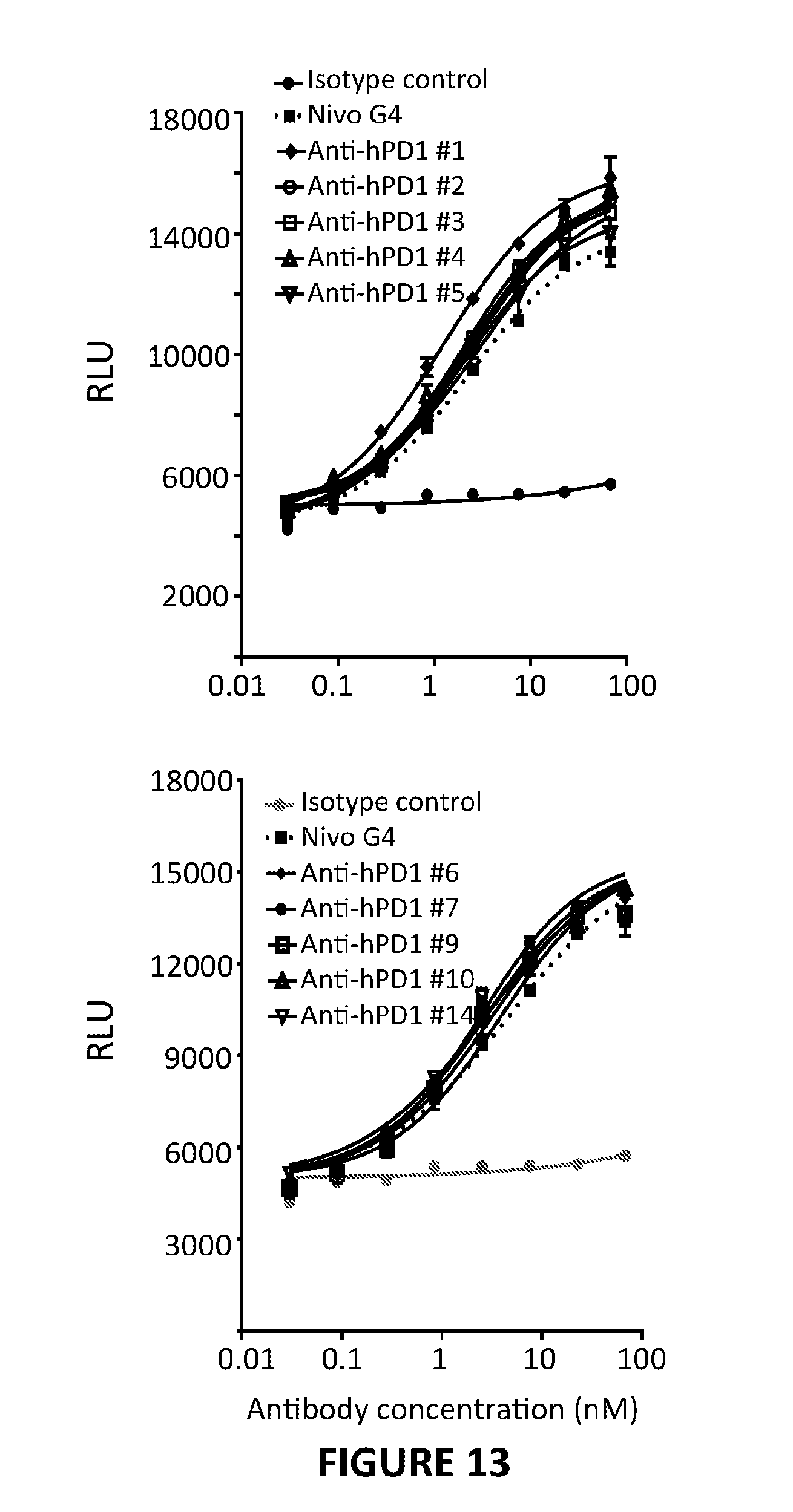
D00014

D00015

D00016

D00017

D00018

D00019
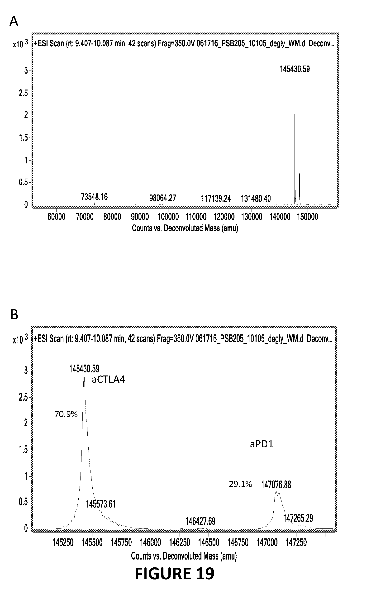
D00020
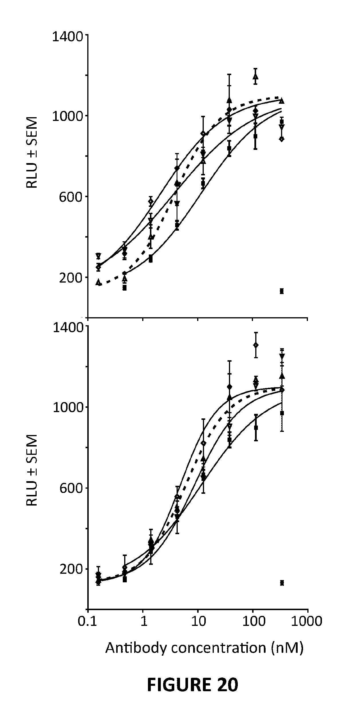
D00021

D00022

P00001

P00002

P00003

P00004

P00005

P00006

P00007

P00008

P00009

P00010

P00011

P00012

P00013

P00014

P00015

P00016

P00017

P00018

P00019

P00020

P00021

P00022

P00023

P00024

P00025

P00026

P00027

P00028

P00029

P00030

P00031

P00032

P00033

P00034

P00035

P00036

P00037

P00038

P00039

P00040

P00041

P00042

P00043

P00044

P00045

P00046

P00047

P00048

P00049

P00050

P00051

P00052

P00053

P00054

P00055

P00056

P00057

P00058

P00059

P00060

P00061

P00062

P00063

P00064

P00065

P00066

P00067

P00068

P00069

P00070

P00071

P00072

P00073

P00074

P00075

P00076

P00077

P00078

P00079

P00080

P00081

P00082

P00083

P00084

P00085

P00086

P00087

P00088

P00089

S00001
XML
uspto.report is an independent third-party trademark research tool that is not affiliated, endorsed, or sponsored by the United States Patent and Trademark Office (USPTO) or any other governmental organization. The information provided by uspto.report is based on publicly available data at the time of writing and is intended for informational purposes only.
While we strive to provide accurate and up-to-date information, we do not guarantee the accuracy, completeness, reliability, or suitability of the information displayed on this site. The use of this site is at your own risk. Any reliance you place on such information is therefore strictly at your own risk.
All official trademark data, including owner information, should be verified by visiting the official USPTO website at www.uspto.gov. This site is not intended to replace professional legal advice and should not be used as a substitute for consulting with a legal professional who is knowledgeable about trademark law.