Single Molecule Detection Or Quantification Using Dna Nanotechnology
GRABMAYR; Heinrich ; et al.
U.S. patent application number 16/310322 was filed with the patent office on 2019-09-05 for single molecule detection or quantification using dna nanotechnology. The applicant listed for this patent is LUDWIG-MAXIMILIANS-UNIVERSITAT MUNCHEN. Invention is credited to Heinrich GRABMAYR, Johannes Benedikt WOEHRSTEIN.
| Application Number | 20190271647 16/310322 |
| Document ID | / |
| Family ID | 59313194 |
| Filed Date | 2019-09-05 |


| United States Patent Application | 20190271647 |
| Kind Code | A1 |
| GRABMAYR; Heinrich ; et al. | September 5, 2019 |
SINGLE MOLECULE DETECTION OR QUANTIFICATION USING DNA NANOTECHNOLOGY
Abstract
The present invention relates to a method and a DNA nanostructure for detecting a target structure. In particular, the present invention relates to a DNA nanostructure, which ensures a preferably linear dependence on the number of marker molecules and the measurement signal regardless of the physical arrangement of a plurality of such DNA nanostructures by virtue of the skilled selection of the shape of the DNA nanostructure and the placement of the marker molecules attached to it. The invention additionally relates to the use of said DNA nanostructures and other nanoreporters, preferably in combination with adapters which bind specifically to target molecules, in a method for quantifying a plurality of target molecules, preferably in a simultaneous manner, using a multiplex method.
| Inventors: | GRABMAYR; Heinrich; (Munchen, DE) ; WOEHRSTEIN; Johannes Benedikt; (Munchen, DE) | ||||||||||
| Applicant: |
|
||||||||||
|---|---|---|---|---|---|---|---|---|---|---|---|
| Family ID: | 59313194 | ||||||||||
| Appl. No.: | 16/310322 | ||||||||||
| Filed: | June 14, 2017 | ||||||||||
| PCT Filed: | June 14, 2017 | ||||||||||
| PCT NO: | PCT/EP2017/064634 | ||||||||||
| 371 Date: | March 1, 2019 |
| Current U.S. Class: | 1/1 |
| Current CPC Class: | G01N 21/6428 20130101; C12Q 1/682 20130101; B82Y 15/00 20130101; C12Q 2565/1025 20130101; C07H 21/04 20130101; B82Y 35/00 20130101; C12Q 1/682 20130101; C12Q 2525/313 20130101 |
| International Class: | G01N 21/64 20060101 G01N021/64; C07H 21/04 20060101 C07H021/04 |
Foreign Application Data
| Date | Code | Application Number |
|---|---|---|
| Jun 15, 2016 | DE | 10 2016 007 270.9 |
Claims
1. A method for the detection of a target structure, comprising: a) forming formation-of an identification structure, comprising: (i) the target structure, and (ii) at least two 3D DNA nanostructures, wherein each of the 3D DNA nanostructures comprises one or more inwardly disposed fluorescence dye molecules, wherein each of the 3D DNA nanostructures is specifically bound to the target structure, and wherein the 3D DNA nanostructures are bound to regions of the target structure that are pairwise different; b) detecting 4eteeti-e.reef the target structure by measuring at least one fluorescence signal, wherein the 3D DNA nanostructures and the parameters of fluorescence measurement are selected such that the at least one measured fluorescence signal of the identification structure formed in a) is distinguishable from the fluorescence signal of each of the at least two isolated 3D DNA nanostructures, when these are not bound in the identification structure.
2. (canceled)
3. The method of claim 1, wherein the identification structure is bound to a carrier or wherein the method further comprises the step of binding the formed identification structure to a carrier.
4.-5. (canceled)
6. The method of claim 3, wherein the bond or the binding of the identification structure to the carrier is mediated or is being mediated via the target structure.
7. The method of claim 6, wherein the target structure is bound or is being bound to the carrier, wherein the bond or binding is mediated by a carrier adapter that specifically binds or is bound to the target structure.
8.-14. (canceled)
15. The method of claim 1, wherein the specific binding of at least one of 3D DNA nanostructures is mediated by a target adapter assigned to a corresponding 31) I)NA nanostructure, wherein the target adapter or each of the target adapters is designed to bind to the respective DNA nanostructure and to the respective region(s) of the target structure.
16.-22. (canceled)
23. The method of claim 1, wherein the method is further additionally suited for the detection of one or more further target structures that are different from each other, wherein the difThrent target structures are pairwise different, and wherein the method further comprises: c) for each of the one or more further target structures that are different from each other: forming a respectively assigned identification structure, wherein each of the further identification structures comprises: (i) the further target structure that was assigned, and (ii) at least two 3D DNA nanostructures, wherein each of the at least two 3D DNA nanostructures comprises one or more inwardly disposed fluorescence dye molecules and wherein each of the at least two 3D DNA nanostructures is specifically bound to the respective further target structure, and wherein the at least two 3D DNA nanostructures are bound to regions of the respective target structure that are pairwise different; and wherein step b) further comprises: d) detecting the one or more further target structures by measuring the at least one fluorescence signal, wherein all 3D DNA nanostructures and the parameters of fluorescence measurement are selected such that the at least one measured fluorescence signal of the identification structures formed in a) and c) is distinguishable from the fluorescence signal of all isolated 3D DNA nanostructures, when these are not bound in one of the identification structures, and that the measured fluorescence signals of all formed identification structures are pairwise distinguishable from each other.
24.-70. (canceled)
71. The method of claim 1, wherein measuring at least one fluorescence signal comprises: e) creating a data set, which contains data of fluorescence signals emitted by a section of a sample by using a fluorescence microscope, preferably in epifluorescence, TIRF, lightsheet and/or confocal microscopy; and wherein the detection of the target structure comprises: f) identifying one or more of the datums contained in the data set, which represent(s) the fluorescence signal of the identification structures.
72. (canceled)
73. The method of claim 71, wherein the identifying in step comprises the following steps: f1) a color and/or an intensity information of a datum and/or image element; and f2) comparing the color and/or intensity information of the datum and/or image element with a color and/or intensity infori ation, being representative for the identification structure.
74. (canceled)
75. The method of claim 114, wherein the fluorescence signals of the identification structures formed for the individual different target structures differ from the fluorescence signal of all isolated 3D DNA nanostructures, when these are not bound in one of the identification structures, and the fluorescence signals of the identification structures which were formed for the individual different target structures are pairwise different, in that the corresponding fluorescence signals comprise a distinguishably different combination of color and/or intensity information.
76. The method of claim 75, wherein in the 3D DNA nanostructures k intensity levels distinguishable from each other and/or m color levels distinguishable from each other are used, wherein preferably each of the k intensity levels is formed by intensity distribution and wherein the k intensity distributions are distinguishable from each other, preferably statistically, and/or wherein preferably each of the m color levels is formed by color distribution and wherein the m color distributions are distinguishable from each other, preferably statistically, wherein the respective overlap of adjacent distributions is preferably lower than 30%, and wherein k>2 and m>2.
77.-81. (canceled)
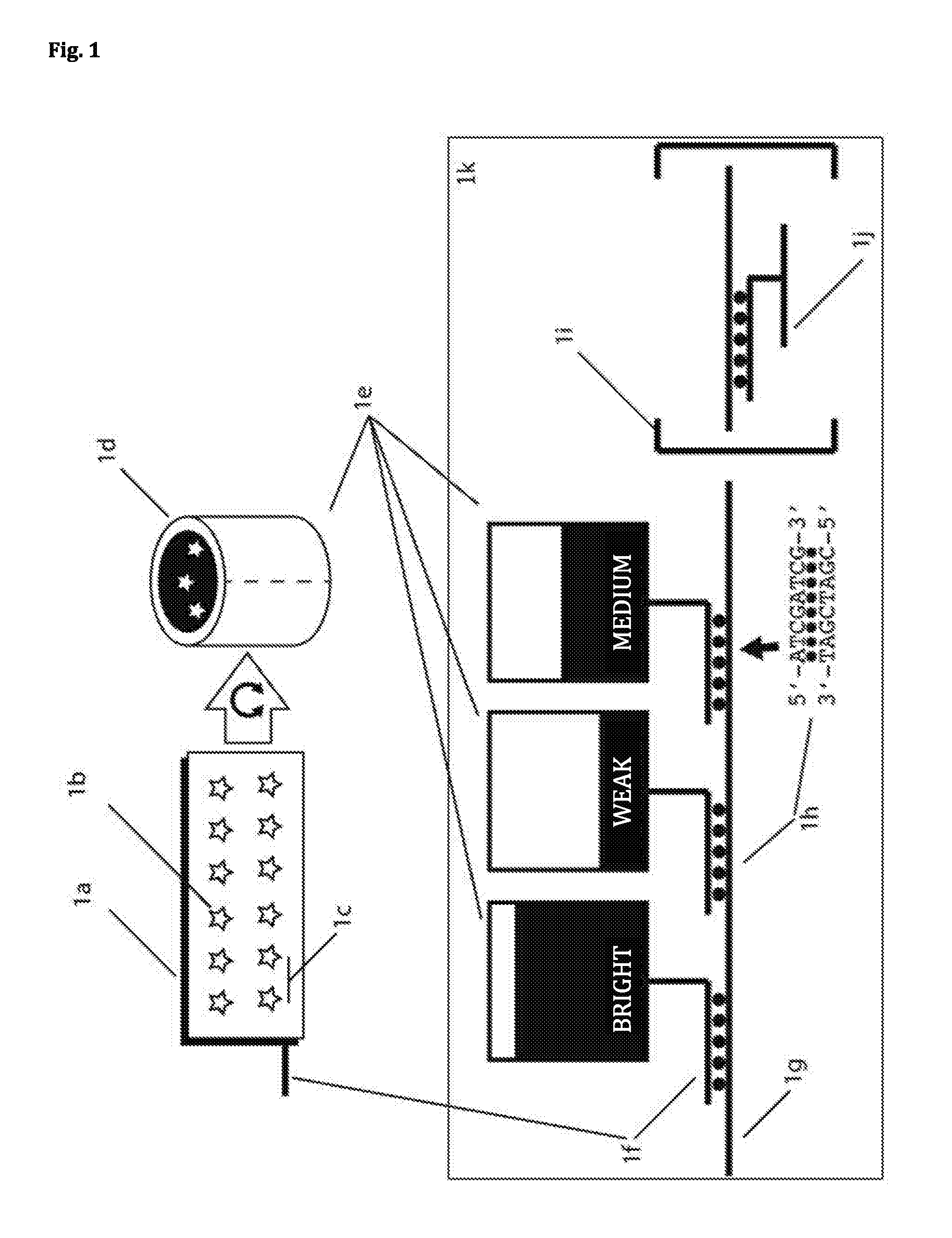
82. A 3D DNA nanostructure, on which at least one fluorescence dye molecule is attached, wherein the shape of the 3D DNA nanostructure prevents that when approaching a second 3D DNA nanostructure, on which at least one fluorescence dye molecule is attached, the fluorescence dye molecules of the two 3D DNA nanostructures interact significantly.
83. (canceled)
84. The 3D DNA. nanostructure of claim 82, wherein at least two fluorescence dye molecules are attached on the 3D DNA nanostructure, and wherein the distance between the at least two fluorescence dye molecules is pairwise greater than that at which they interact significantly.
85.-104. (canceled)
105. A set comprising multiple 3D DNA nanostructures of claim 82, wherein the set comprises N 3D DNA nanostructures that are different from each other and wherein the N 3D DNA nanostructures of the set are pairwise different from each other due to the fluorescence dye molecules.
106. The set of claim 105, wherein the N 3D DNA nanostructures that are different from each other contain a different number of fluorescence dye molecules and/or different fluorescence dye molecules, so that with the N 3D DNA nanostructures of the set that are different from each other, k intensity levels that are distinguishable from each other and/or m color levels that are distinguishable from each other can be generated, and wherein k>2 and m>2.
107. The set of claim 106, wherein at least a part of the N 3D DNA nanostructures that are different from each other are contained in the set multiple times, so that each of the k intensity levels is formed by an intensity distribution, and wherein the k intensity distributions are distinguishable from each other, preferably statistically.
108. The set of claim 106, wherein at least a part of the N 3D DNA nanostructures that are different from each other are contained in the set multiple times, so that each of the m color levels is formed by a color distribution, and wherein the m color distributions are distinguishable from each other, preferably statistically.
109. The set of claim 107 or 108, wherein the overlap of adjacent distributions is lower than 30%.
110.-112. (canceled)
113. The method of claim 23, wherein each of the different target structures is present multiple times and the method comprises the multiple detection of one or more of the different target structures.
114. The method of claim 23, wherein measuring at least one fluorescence signal comprises: e) creating a data set, which contains data of fluorescence signals emitted by a section of a sample by using a fluorescence microscope, preferably in epifluorescence, TIRF, lightsheet and/or confocal microscopy; and wherein the detection of the target structure comprises: f) identifying one or more of the datums contained in the data set, which represents the fluorescence signal of the identification structure.
115. The method of claim 114, wherein the identifying in step f) comprises the following steps: f1) reading out a color and/or an intensity information of a datum and/or image element; and f2) comparing the color and/or intensity information of the datum and/or image element with a color and/or intensity information, being representative for the identification structure.
Description
[0001] The present invention relates to a method and a DNA nanostructure for detecting a target structure. In particular, the present invention relates to a DNA nanostructure, which ensures a preferably linear dependence on the number of marker molecules and the measurement signal regardless of the physical arrangement of a plurality of such DNA nanostructures by virtue of the skilled selection of the shape of the DNA nanostructure and the placement of the marker molecules attached to it. The invention additionally relates to the use of said DNA nanostructures and other nanoreporters, preferably in combination with adapters which bind specifically to target molecules, in a method for quantifying a plurality of target molecules using a multiplex method, preferably in a simultaneous manner
[0002] A frequently occurring question in research as well as in applied medicine and biotechnology is the analysis, i.e. the detection, of particular target structures. For example, in the case of sepsis of a patient, the detection of the relevant toxins may be necessary or, in the case of a disease entailing altered cellular constitution, e.g. with cancer, a quantitative analysis of the conversion of genetic information may be necessary.
[0003] The quantitative analysis of the conversion of genetic information, the so-called gene expression profiling (GEP) requires the detection and identification of a number of different target mRNA molecules (Stadler, Z. K. et al. Critical Reviews in Oncology Hematology 69, 1-11 (2009)). mRNA is the established abbreviation for the English term messenger RNA (in German "Boten-RNA"), which is also used in German Since the mRNA sequences are specifically produced in the expression process of the corresponding genes, the gene expression of the analyzed material can be quantified in that manner
[0004] GEP is able to provide information on the state or type of individual cells or also a tissue sample (plurality of cells) and thereby enable, e.g., the molecular diagnosis of tumor tissue. The GEP comprises the analysis of the expression of at least one gene, preferably the analysis of the expression of a plurality of genes. In order to achieve this ultimate objective, a technology is required, which at the same time allows for quantitative analysis having the highest possible specificity and sensitivity, preferably for a number of genes that is as high as possible. For comprehensive applicability in clinical practice, this technology must additionally be characterized by expeditious analysis times, low costs and simple use.
[0005] There is a plurality of GEP techniques, which have different advantages and disadvantages, depending on the question at issue.
[0006] Microarrays.
[0007] GEP analysis using microarrays is based on the surface hybridization of fluorescent DNA molecules to specific, physical separate target sequences on a chip (Trevino, V. et al. Mol Med 13, 527-541 (2007)). These molecules are generated in a preceding step by reverse transcription and polymerase chain reaction (PCR) amplification of RNA molecules. Thereby, microarrays allow for the parallel detection of up to several thousand sequences. However, an exact quantification is impossible, since enzymatic reactions such as reverse transcription and PCR amplification entail systematic errors. Moreover, microarray-based methods require a relatively long time of process of about one day and are therefore not suited for the majority of clinical applications.
[0008] qPCR/dPCR. Using PCR, specific DNA molecules may be exponentially duplicated in an enzymatic process (VanGuilder, H. D. et al. Biotechniques 44, 619-626 (2008)). For PCR reactions, mRNA targets must be generally converted enzymatically using reverse transcription of RNA into DNA. The amplification can be measured by adding fluorescent, DNA-binding molecules and the original concentration can be roughly determined (qPCR). Since the amplification rate significantly depends on various parameters (inter alfa target sequence, target length, primer, instruments, reagents, reaction volume), said quantification must be calibrated precisely. For compensation of these disadvantages, digital PCR (dPCR) was developed. It allows for a more precise quantification by compartmentalization of individual target molecules (Baker, M. Nature Methods 9, 541-544 (2012)). However, said high preciseness of dPCR is offset by limitation to typically two targets. dPCR is therefore not suited for GEP analyses.
[0009] RNA-Seq/NGS. With the so-called next generation sequencing (NGS), on which also RNA sequencing (RNA seq) is based, sequences are read out and identified in several steps by base (Wang, Z. et al. Nat Rev Genet 10, 57-63 (2009)). With RNA seq, too, RNA must initially be enzymatically converted into DNA. The advantage and the unique feature of NGS in transcription analysis is the possibility to recognize novel or altered sequences. Due to the great number of complex process steps, the analysis devices are complex and expensive and the process duration is lengthy. This excludes its use in the field of point-of-care. Just as in the above-described methods, enzymatic processes prevent exact quantifiability.
[0010] nCounter. The nCounter system of NanoString Technologies is based on the detection of RNA molecules by hybridization using a fluorescent reporter (barcode), on the one hand, as well as a second DNA molecule (capture strand) for attachment to a microscopy chip, on the other hand (Geiss, G. K. et al. Nat Biotechnol 26, 317-325 (2008)). The fluorescent reporters are linear geometric barcodes that are micrometers in length and that consist of the arrangement of different areas labeled with fluorescent dyes. One of the advantages of the nCounter system is the enzyme-free and quantitative detection of up to 800 different RNA target molecules. One disadvantage of the method is the necessity to stretch the geometric barcode molecule after target hybridization and surface immobilization, in order to be able to read the respective barcode. This has two important implications: (1) The electrophoretic stretching of the reporters is a complex process step, (2) Conditioned by the low efficiency of this step, approximately 80% of the target molecules are excluded due to non-identifiable barcodes. Furthermore, complex and time-consuming purification steps are necessary, e.g., using magnetic particles. In sum, these process steps lead to expensive analysis devices (>200,000 euros) and long measuring times (24-48 hours).#
[0011] Thus, these traditional methods do not meet all of the above-mentioned preferred requirements for GEP and sacrifice e.g. partial quantifiability for necessary sensitivity.
[0012] A further promising analysis approach for GEPs is based on direct labeling of individual target mRNA sequences by so-called hybridization using fluorescent reporters. Fluorescent methods allow for the sensitive detection of single molecules (Joo, C. et al. Annu Rev Biochem 77, 51-76 (2008)). However, established methods are not able to determine a great number of different reporters.
[0013] Quantitative GEP on single-cell basis employing the available methods is possible only in a limited manner, but would be highly advantageous for both scientific research and industrial research and development as well as for clinical applications.
[0014] Essential desirable features of a GEP would be the exact quantification and the efficient implementation of the analysis. This results in the following preferred criteria for an improved GEP: [0015] direct detection of the mRNA, in particular no translation into DNA, e.g. by reverse transcription [0016] no amplification of the target molecules, since an amplification that is not precise impairs quantification [0017] thereby: detection of individual target molecules [0018] fast-as-possible analysis (order of minutes) [0019] simple-as-possible analysis, in particular: few working steps.
[0020] Apart from nucleic acids and proteins, there is a myriad of further problems with which units in the order of a few nanometers to several tens of nanometers must be detected and preferably counted and/or the concentration be determined; such as inorganic complexes and nanoparticles.
[0021] All these problems have in common that molecules or other structures should be detected in a fast, simple and exact manner and be preferably counted. Exact counting in particular entails that the probability of the false positive and/or false negative counting events must be kept as low as possible. The invention aims at an improvement of one or more of said properties, in particular at a reduction of the probability of the false positive and/or false negative counting events.
[0022] This problem is solved by the present invention according to the independent claims.
[0023] Thus, the present invention is directed to a method for the detection (preferably quantification) of a target structure, comprising: [0024] a) formation of an identification structure, comprising: [0025] (i) the target structure, and [0026] (ii) at least two 3D DNA nanostructures, wherein each of the 3D DNA nanostructures comprises one or more inwardly disposed fluorescence dye molecules, wherein each of the 3D DNA nanostructures is specifically bound to the target structure; [0027] b) detection (preferably quantification) of the target structure by measuring at least one fluorescence signal, [0028] wherein the 3D DNA nanostructures and the parameters of fluorescence measurement are selected such that the at least one measured fluorescence signal of the identification structure formed in a) is distinguishable from the fluorescence signal of each of the at least two isolated 3D DNA nanostructures, when these are not bound in the identification structure.
[0029] Furthermore, the invention is inter alfa directed to a 3D DNA nanostructure, on which at least one fluorescence dye molecule is attached, wherein the shape of the 3D DNA nanostructure prevents that when approaching a second 3D DNA nanostructure, on which at least one fluorescence dye molecule is attached, the fluorescence dye molecules of the two 3D DNA nanostructures interact significantly.
[0030] Preferably, the 3D DNA nanostructure of the invention is a 3D DNA nanostructure having a cavity and at least one inwardly disposed fluorescence dye molecule, wherein the distance of the at least one inwardly disposed fluorescence dye molecule to the rim of the 3D DNA nanostructure is at least 2 nm, preferably at least 3 nm and particularly preferably at least 5 nm.
[0031] The skilled choice of the shape of the 3D nanostructures and the arrangement of the fluorescence dye molecules in the inside of/in the cavity of the 3D nanostructure ensures that the dye molecules do not sterically interact with their surroundings. As shown in the experimental Examples as attached, the 3D nanostructures of the invention thereby interact, e.g., to a significantly lesser extent with surfaces, such as a glass surface, than 2D nanostructures comprising the same dyes. This is highly advantageous for the use of such a 3D DNA nanostructure in the method according to the invention. For example, the rate of false positive detection of a target structure can be reduced in that manner, in particular if a carrier structure is used. Moreover, an advantage of shielding the fluorescence dye molecules is that the fluorescence dye molecules of the two 3D DNA nanostructures do not significantly interact (optically and/or sterically). This permits the fluorescence signal of a 3D DNA nanostructure of the invention not being significantly affected by a very close adjacent 3D DNA nanostructure. Moreover, the unspecific interaction of 3D DNA nanostructures which is mediated by fluorescence dye molecules is prevented. Taken as a whole, this is highly advantageous in the use of said 3D nanostructures for the detection of target structures. In particular, like in the method of the invention, this allows for the binding of at least two 3D DNA nanostructures to a target structure, without the fluorescence signal of the individual 3D DNA nanostructures being influenced due to their proximity (e.g. by quenching, fluorescence quenching or FRET).
[0032] Depending on the measurement method, the use of at least two DNA nanostructures may even be necessary in order to distinguish the identification structure of target structure and bound DNA nanostructures from the free DNA nanostructures. This is in particular the case with measurement methods which are implemented in solution. When the identification structure is bound to a carrier and/or a carrier surface in the method for detection (as in a preferred embodiment of the method of the invention), this allows for the removal of free DNA nanostructures using one or more washing steps. The use of at least two DNA nanostructures, which bind to a target structure, in a method that uses a carrier has the advantage that the rate of false positive detections may be lowered. First, an identification structure having at least two DNA nanostructures can be clearly distinguished from DNA nanostructures which interact with the surface in an unspecific manner. This is not possible in an analogous method, in which only one DNA nanostructure recognizes the target structure. Second, the use of at least two DNA nanostructures, both of which recognize different regions of the target structure, may significantly reduce and in many cases exclude the probability that other similar target structures are erroneously detected as an alleged target structure.
[0033] Moreover, with independent coupling of at least two 3D DNA nanostructures (e.g. different orthogonal measurable nanoreporters) to target molecules, the sensitivity of measuring may be significantly increased. It increases with the potency of the number of independent coupling reactions. This is highly advantageous in particular for high dynamics, i.e., for example, for simultaneously measuring of genes with a very low expression and a very high expression.
[0034] In a preferred embodiment, the present invention is also directed to a method for the detection and/or the quantification of at least two different target structures (e.g. two mRNAs, which are derived from different genes, i.e. which comprise a different nucleic acid sequence), preferably of a plurality of different target structures. The different target structures are pairwise distinguishable.
[0035] Preferably, said method comprises the following steps: [0036] a) formation of an identification structure for each of the at least two different target structures, comprising: [0037] (i) the respective target structure, and [0038] (ii) at least two 3D DNA nanostructures, wherein each of the at least two 3D DNA nanostructures comprises one or more inwardly disposed fluorescence dye molecule and wherein each of the at least two 3D DNA nanostructures is specifically bound to the respective target structure, and wherein the at least two 3D DNA nanostructures are bound to regions of the respective target regions which are pairwise different; [0039] b) detection of the at least two target structures by measuring at least one fluorescence signal, wherein all 3D DNA nanostructures and the parameters of fluorescence measurement are selected such that the at least one measured fluorescence signal of the identification structures formed in a) is distinguishable from the fluorescence signal of all isolated 3D DNA nanostructures, when these are not bound in one of the respective identification structure, and that the measured fluorescence signals of the identification structures that are formed for the individual different target structures are pairwise distinguishable from each other.
[0040] Preferably, each of the different target structures is detected several times, i.e. each of the identification structures that is assigned to a specific target structure is formed several times.
[0041] All of the above-mentioned advantages of the method according to the invention apply mutatis mutandis in this case. Having the relevant form, DNA nanostructures offer the possibility to attach a plurality of identical and/or different fluorescence dye molecules. As is known from WO2016/1407727 A2 and WO2016/1407726 A2, a plurality of DNA nanostructures having a distinguishable fluorescence signal can thus be generated. However, the amount of the distinguishable combinations critically depends on the size of the DNA nanostructure, which limits the number of fluorescence dye molecules which may be attached thereon, without interacting with each other. However, large DNA nanostructures require more effort and are expensive as regards their production and furthermore poorer than smaller structures as regards kinetics in a method for the detection of a target structure. Due to the use of at least two DNA nanostructures, which become part of the identification structure, the method of the invention has the advantage that also with significantly smaller DNA nanostructures, a similar plurality of fluorescence molecule combinations, which are bound to a target structure, may be generated. Furthermore, the use of at least two DNA nanostructures in an identification structure to further increase the number of the different distinguishable combinations of fluorescence dye molecules on a target structure and/or in an identification structure having the same maximum size as described in WO2016/1407727 A2 and WO2016/1407726 A2. Thereby, the number of different target structures that can be detected in a method can be considerably increased. Apart from the use of at least two DNA nanostructures, in particular the form of the 3D nanostructure and the orientation of the fluorescence dye molecules into the inside of the structure are decisive. As mentioned above, it is only then that an interaction of the dyes of two DNA nanostructures is prevented and the signal of a combination of two or more 3D DNA nanostructures is reliably predictable and measurable.
[0042] According to the invention, nucleic acid nanostructures, also referred to as DNA nanostructures or DNA origami, are used. Nucleic acid nanostructures, also referred to as DNA nanostructures or DNA origami, are two- or three-dimensional structures, which inter alfa consist of nucleic acids. The term "origami" describes that one or more strands or components of nucleic acids, preferably DNA, may be folded into almost any pre-defined structure or shape. Such a DNA strand is also referred to as scaffold strand. The one or the more scaffold strands is/are kept in shape by shorter nucleic acid strands (relative to the at least one scaffold strand), which are also referred to as staple strands. Here, it is of major importance that the shorter strands (staple strands) are placed very precisely on well-defined positions on the DNA origami. DNA origami are described in more detail for example in Rothemund, "Folding DNA to create nano-scale shapes and patterns", Nature, March 2006, pp. 297-302, vol. 440; Douglas et al., Nature, 459, pp. 414-418 (2009); and Seeman, "Nanomaterials based on DNA", An. Rev. Biochem. 79, pp.65-87 (2010). Reference to all of these documents is made herein in their entirety as part of the application.
[0043] Said DNA origami nanostructures can be functionalized with photoactive molecules, in particular with one or more fluorescent dye molecules. Thereby, the DNA origami as a whole becomes a fluorescent particle. Such DNA nanostructures, which are based on DNA origami, are described in WO 2016/140727 A2 and WO2016/1407726 A2. Reference to all of these documents is made herein in their entirety as part of the application.
[0044] Preferably, a DNA nanostructure of the invention is a 3D DNA nanostructure on which at least one fluorescence dye molecule is attached, wherein the shape of the 3D DNA nanostructure prevents that with approaching a second similar or identical 3D DNA nanostructure, on which the at least one fluorescence dye molecule is attached, the fluorescence dye molecules of the two 3D DNA nanostructures significantly interact. Thus, by means of steric effects, the geometry of the DNA nanostructures prevents fluorescence dye molecules of different DNA nanostructures from coming too close to each other.
[0045] As can be inferred from its name, a DNA nanostructure preferably consists of DNA. In principle, a DNA nanostructure of the invention may, however, also contain other nucleic acids such as e.g. RNA, RNA analogues such as LNA, or DNA analogues or consist of these. Therefore, the present invention also comprises all embodiments described herein, in which the DNA nanostructure(s) and/or the 3D DNA nanostructure(s) are nucleic acid nanostructures and/or 3D nucleic acid nanostructures. A nanostructure formed of DNA is preferred, e.g. due to significantly lower production costs for such nanostructures.
[0046] A fluorescence dye molecule may be, for example, RFP, GFP, YFP or any of their derivatives (see for example P J Cranfill et al., "Quantitative assessment of fluorescent proteins", Nature Methods, 13, 557562 (2016)), an Atto dye (e.g. Atto647N, Atto565 or Atto488), an Alexa dye, DAPI, rhodamine, rhodamine derivative, cyanine, cyanine derivative, coumarin, coumarin derivative (for different organic fluorescence dyes see for example Q. Zheng and L Lavis, "Development of photostable fluorophores for molecular imaging", Curr. Op. Chem. Biol. 39 p32-38 (2017)) or quantum dot (quantum dot, see E. Petryayeva et al, "Quantum dots in Bioanalysis: a review of applications across various platforms for fluorescence spectroscopy and imaging", Appl. spectroscopy 67 (2013).
[0047] A 3D DNA nanostructure preferably refers to a structure, which substantially extends into three dimensions and is therefore different from an essentially two-dimensional structure such as, e.g., a slightly bent, rough or rippled surface. In other words, the minimal dimensions in three spatial directions that are perpendicular to each other are at least 2 nm, preferably at least 5 nm, more preferably at least 10 nm and particularly preferably at least 20 nm.
[0048] The interaction of two fluorescence dye molecules may in particular comprise fluorescence quenching and/or FRET.
[0049] In particular, at least two fluorescence dye molecules may be attached on the 3D DNA nanostructure, wherein the distance between the at least two fluorescence dye molecules is pairwise greater than that at which the fluorescence dye molecules significantly interact pairwise. For the exemplary case of a 3D DNA nanostructure having two fluorescence dye molecules, this means that the two fluorescence dye molecules have a distance, which is greater than the distance of interaction. For the exemplary case of a 3D DNA nanostructure having three fluorescence dye molecules, this means that each fluorescence dye molecule has a distance to the other two fluorescence dye molecules, which is greater than the respective interaction distance. For more fluorescence dye molecules, the same applies accordingly. That means, irrespective of which fluorescence dye molecule pair of the 3D DNA nanostructure is observed, the distance of the fluorescence dye molecules of the observed pair is always greater than the interaction radius of the observed pair. "Interaction radius" refers to the distance, which fluorescence dye molecules must have at the minimum in order to show negligible interactions (such as, e.g., fluorescence quenching or FRET). Which kind of dye pair has which kind of interaction radius is known to the skilled person. For example, corresponding interaction radii are described in Novotny, Lukas: Principles of nano-optics 2.ed, Cambridge Univ. Press, 2013, Kapitel "Optical Interactions", pp 224 et seqq.; Peter Atkins: Physikalische Chemie, 5.sup.th edition, 2013, Wiley-VCH, Weinheim, Chapter 17 "Wechselwirkungen zwischen Molekulen", pp 657 et seqq.; Forster T: Zwischenmolekulare Energiewanderung and Fluoreszenz. In: Ann. Physik. 437, 1948, S. 55. doi:10.100.sup.2/.sub.andp.19484370105. With regard to FRET, an interaction radius may be e.g. the distance at which a FRET rate of 50% is given.
[0050] The DNA nanostructures preferably serve the purpose of precise arrangement of a well-defined number of marker molecules (preferably fluorescence dye molecules) of one or more kinds. The geometric arrangement is of crucial importance, in order to prevent interactions between marker molecules (preferably fluorescence dye molecules). The defined number of marker molecules (preferably fluorescence dye molecules) allows for programming the intensity values and therefore the subsequent definite identification. The number of different combinations is N=k m, wherein k is the number of intensity levels and m is the number of different (in this case orthogonally measurable) marker molecules (preferably fluorescence dye molecules). By this multiplexing, a great number of different DNA nanostructures can be distinguished, without multiplexing, the number would only be m. In an Example, there are 5 intensity levels and three types of marker molecules and therefore 124 instead of 3 simultaneously distinguishable species. Thus, the DNA nanostructures are nanoreporters.
[0051] Thus, the term nanoreporter" is used herein in order to define structures with attached marker molecules, which have measurable and identifying signatures, with measurements in the nanometer range. In particular, the DNA nanostructures of the invention may be nanoreporters. Other DNA origami, e.g. according to Rothemund, can also be nanoreporters, as well as cascades of fluorescent proteins.
[0052] The term "orthogonally measurable" describes the property that it can be clearly extrapolated from a linear combination of measurement values of marker molecules (e.g. fluorescence dye molecules) to a linear combination of the underlying marker molecules and/or a combination of nanostructures. With the example of fluorescence detection, this may be realized by spectrally far-apart dyes and hence far-separated excitement and detection wave length (e.g. blue and red) or by closer-adjacent dyes in combination with multispectral detection, so that the orthogonal components may be calculated by "linear unmixing".
[0053] A DNA nanostructure of the invention is preferably characterized in that one or multiple marker molecules (e.g. fluorescence dye molecules) of one or more kinds of marker molecules are attached such that the distance between two marker molecules is greater than that at which they significantly interact, and their form prevents that with approaching a similar DNA nanostructure, their marker molecules significantly interact with those of the other DNA nanostructure. A preferred DNA nanostructure of the invention is a DNA nanostructure, on which one or more marker molecules of one or more kinds of marker molecules is/are attached such that the distance between identical marker molecules is greater than that at which fluorescence quenching occurs and the distance between different marker molecules is greater than the Froster resonant energy transfer (FRET) radius, and their shape prevents that with approaching a similar DNA nanostructure, its marker molecule(s) interact with one or more marker molecules of the other DNA nanostructure by fluorescence quenching or FRET.
[0054] "Marker molecules" refer to particles which emit measurable signals. In particular, fluorescence dye molecules are marker molecules. The signal of fluorescence dye molecules may be the intensity, but also lifetime, polarization, blink kinetics or similar quantities. Gold particles are also marker molecules within this meaning.
[0055] Two marker molecules, in particular two fluorescence dye molecules, significantly interact, when the signal emitted by at least one of them significantly depends on the presence or absence of the other. For our purposes, for example a decline in intensity of the signal by 1% due to the presence of another molecule can be neglected, whereas a decline by 20% cannot be neglected. In the first case, the marker molecules (in particular fluorescence dye molecules) do not interact significantly, whereas they do so in the second case. In the case of similar fluorescence dye molecules, the interaction mechanism prevalent for us is fluorescence quenching, which is different depending on the fluorescence dye molecule, but which becomes negligible starting from a distance between the fluorescence dye molecules of about 2-3 nanometers. For fluorescence dye molecules of different types, the prevalent interaction mechanism is the Forster resonant energy transfer (FRET) and the distance from which the fluorescence dye molecule pairs no longer interact significantly (also referred to as FRET distance) varies in the range of 2-20 nanometers, depending on the individual pair. FRET distances are known for a plurality of fluorescence dye pairs, for example many dye producers publish lists for FRET distances of their dyes (e. g. ://www.atto-tec.com/fileadmin/user_upload/Katalog_Flyer_Support/R_0_-Tabe- lle_2016_web.pdf). Moreover, FRET distances may be calculated using the spectral properties of the fluorescence dye molecules (see Atkins, Physikalische Chemie).
[0056] Preferably, a significant interaction between two fluorescence dye molecules is an interaction, in which the measured fluorescence signal (with common excitation) of the one molecule is reduced by the presence of the other molecule in comparison to a measured fluorescence signal of the one molecule in the absence of the other molecule to 80% or less, preferably 90% or less, particularly preferably 95% or less, highly particularly preferably 99% or less.
[0057] A 3D DNA nanostructure of the invention preferably comprises a cavity, wherein the cavity has a volume of at least 0.1 zeptoliters (le-22 liters), preferably at least 10 zeptoliters and particularly preferably at least 100 zeptoliters.
[0058] A 3D DNA nanostructure of the invention may essentially be formed as a hollow cylindrical DNA nanostructure, i.e. as hollow cylinder, wherein at least one fluorescence dye molecule is attached on the inside of the hollow-cylindrical DNA nanostructure. The cavity of the hollow cylinder preferably has a volume as indicated above.
[0059] The part of the 3D DNA nanostructure, which corresponds to the shell of the hollow cylinder, may comprise gaps. Those parts of the 3D DNA nanostructure, which correspond to the top and bottom surface of the hollow cylinder, are preferably open but may also each be at least partially closed and/or entirely closed. The barrel and optionally the top and bottom surface of the hollow cylinder are formed by the 3D DNA nanostructure, which means that it is not a mathematically precise cylinder, since e.g. the individual helices of the nanostructure form uneven surfaces with projections and recesses. However, the envelope of the 3D nanostructure is essentially the form of a cylinder.
[0060] Preferably, in a 3D DNA nanostructure of the invention, at least 2, more preferably at least 3, more preferably at least 4, more preferably at least 5, more preferably at least 6, more preferably at least 15, particularly preferably at least 30, particularly preferably at least 60 fluorescence dye molecules are attached. Preferably, the fluorescence dye molecules on a 3D DNA nanostructure are uniform, i.e. of the same type.
[0061] Preferably, a 3D DNA nanostructure of the invention comprises a cavity and at least one inwardly disposed fluorescence dye molecule, wherein the distance of the at least one inwardly disposed fluorescence dye molecule to the rim and/or the outer surface of the envelope of the 3D DNA nanostructure is at least 2 nm, preferably at least 3 nm and particularly preferably at least 5 nm. This has the advantage that due to the arrangement in the inside of the 3D DNA nanostructure and the minimum distance to the rim of the 3D DNA nanostructure, the dyes of the 3D DNA nanostructure also comprise said minimum distance to other structures with which the 3D DNA nanostructures may interact in the course of a method. For example, to a surface of a carrier, for example a substrate, the dyes have a distance that is greater than or equal to the minimum distance, when the 3D DNA nanostructure contacts said surface.
[0062] Preferably, a 3D DNA nanostructure of the invention comprises at least two inwardly disposed fluorescence dye molecules, wherein the pairwise distance of the at least two inwardly disposed fluorescence dye molecule is at least 2 nm, preferably at least 5 nm and particularly preferably at least 9 nm. For the exemplary case of a 3D DNA nanostructure with two dye molecules, this means that the two dye molecules comprise a distance of at least 2 nm, preferably at least 5 nm and particularly preferably at least 9 nm. For the exemplary case of a 3D DNA nanostructure with three dye molecules, this means that each dye comprises a distance to the two other dyes of at least 2 nm, preferably at least 5 nm and particularly preferably at least 9 nm. For more dyes the same applies mutatis mutandis. That means, irrespective of which dye pair of the 3D DNA nanostructure is observed, the distance of the dyes of the observed pair is always at least 2 nm, preferably at least 5 nm and particularly preferably at least 9 nm.
[0063] The distance of the dye molecules may, for example, be determined by identifying the structure of the 3D DNA nanostructure, from which can be inferred at which positions the dye molecules are positioned. To this end, sequence analysis may, for example, be required. For this purpose, a solution with DNA nanostructures is transferred to a solution with individual nucleic acid stands for example by reduction of the salt concentration. With a reduction of the salt concentration, the negative charges of the DNA backbone become more prevalent due to reduced shielding as compared to the permanently constant binding energies of the base pair-forming hydrogen bonds. Thus, with the reduction of the salt concentration, the DNA nanostructures are initially destabilized and with further reduction, the individual DNA double helices break and are sequenced, for example, with My Seq or another method provided by Illumina, Inc. and with the protocol as indicated by the manufacturer. Thus, the sequences of all nucleic acid strands present in the sample get known. The form of the DNA nanostructure may be reconstructed with this sequence information. To this end, any sequence editor (e.g. ApE 2.0 (http://biologylabs.utah.edu/jorgensen/wayned/ape/) can be used. The longest DNA strand of the analysis is the scaffold strand. It is loaded into the editor and the sequence regions complementary to the scaffold strand are calculated and noted for each single staple sequence. There are two different regions on the scaffold strand for each staple strand. As described at a different point in this specification, with this information, the topology can in turn be defined in CaDNAno and thus the DNA nanostructure design can be reconstructed. In the next step, it must be determined which of the staple strands are provided with dyes. This is simple with the use of dye adapters, since a particular number of staple strands have further identical sequences, which remain single-stranded after the above reconstruction of the DNA nanostructure. If the staple strands are directly modified, the individual staple strands must be isolated and be analyzed for their fluorescence properties. Each individual staple strand may be loaded by hybridization to a complementary strand with additional molecular weight and be isolated in an agarose gel electrophoresis from the above solution with individual nucleic acid strands. Finally, a respective fluorescence measurement shows which of the staple strands is labeled with a fluorophore. These can be identified in caDNAno and the distances between fluorescence molecules can be calculated. As an alternative to this method, using mass spectrometry (Sauer S, Lechner D, Berlin K et al. (2000) Full flexibility genotyping of single nucleotide polymorphisms by the GOOD assay. Nucleic Acids Res 28:E100; Haff L A, Smirnov I P (1997) Single-nucleotide polymorphism identification assays using a thermostable DNA polymerase and delayed extraction MALDI-TOF mass spectrometry. Genome Res 7:378-388; Wenzel T, Elssner T, Fahr K et al. (2003) Genosnip: SNP genotyping by MALDI-TOF MS using photocleavable oligonucleotides. Nucleosides Nucleotides Nucleic Acids 22:1579-1581; Braun A, Little D P, Koster H (1997) Detecting CFTR gene mutations by using primer oligo base extension and mass spectrometry. Clin Chem 43:1151-1158; Sun X, Ding H, Hung K et al. (2000) A new MALDI-TOF based mini-sequencing assay for genotyping of SNPS. Nucleic Acids Res 28:E68), the solution with DNA nanostructures can be directly sequenced and it can be simultaneously established which staple strands have fluorescent molecules attached. In analogy to the above, the topology of the DNA structure can subsequently be reconstructed in CaDNAno using complementary sequence analysis of staple strands and scaffold strand (which is again the longest strand). The co-measured information, on which strands the fluorescence molecules are attached, can be directly used to calculate the distances of the fluorescence molecules using CaDNAno.
[0064] In a preferred embodiment, the 3D DNA nanostructure is essentially formed as elliptic cylinder, preferably essentially as circular cylinder.
[0065] As already set forth above, the structures according to the invention are not a mathematically precise cylinder, since the surface of the structure at the single-atom level is extremely uneven and, e.g. at the level of the individual helices of the nanostructure, may comprise unevenness of the order of a helix radius or helix diameter. However, these structures are cylindrical to a skilled person nonetheless, since, e.g., the envelope of the 3D nanostructure may essentially have the form of a cylinder.
[0066] In a preferred embodiment, the hollow cylinder of the 3D DNA nanostructure is a circular cylinder and comprises an outer radius of at least 5 nm, at least 10 nm, preferably of at least 20 nm, preferably of at least 20 nm, preferably an outer radius of 30 nm to 80 nm, preferably of 50 nm to 70 nm and particularly preferably an outer radius of 60 nm. The outer radius refers to the structure of the outer radius of the envelope.
[0067] In a preferred embodiment, the hollow cylinder of the 3D DNA nanostructure, which is preferably a circular cylinder, has a height of at most 200 nm, preferably a height of 60 nm-30 nm, particularly preferably a height of 30 nm. The height of the structure refers to the height of the envelope.
[0068] The preferred embodiments ensure a sufficient size of the 3D DNA nanostructure in order to attach a sufficient number of dye molecules at sufficient distance of the dye molecules from each other and preferably in the inside of the 3D DNA nanostructure.
[0069] In a preferred embodiment, the hollow cylinder of the 3D DNA nanostructure comprises a wall thickness of at least 2 nm, preferably 2 nm to 7 nm, particularly preferably 5 nm. The wall thickness of the structure refers to the difference between outer radius and inner radius. In other words, the wall thickness corresponds to the distance between the outer envelope of the cylinder and the inner envelop of the cylinder cavity.
[0070] Preferably, a 3D DNA nanostructure of the invention comprises a wall thickness that at least corresponds to the diameter of a DNA double helix. Preferably, the wall thickness has space for 2-4, particularly preferably 3 DNA double strands, wherein the DNA strands are preferably arranged perpendicularly to the wall thickness in their length and are additionally arranged on adjacent lattice sites of a honeycomb lattice.
[0071] The inner diameter of the hollow cylinder is defined as the difference of outer diameter of the hollow cylinder and the wall thickness. Thus, preferred inner diameters result from the details on the outer diameter and on wall thickness as indicated herein.
[0072] The described minimum wall thicknesses in combination with the shape of the 3D DNA nanostructure, which shields the dyes from their surroundings, also contribute to the fact that an interaction of the dyes of the 3D DNA nanostructure with a surface of a carrier and/or another dye, which may be attached in another 3D DNA nanostructure, is prevented by steric hindrance. Thus, in particular, quenching and/or FRET with another dye and/or unspecific binding to a carrier, in particular a substrate surface, is prevented.
[0073] As is common for DNA origami, the 3D nanostructures of the invention preferably comprise a DNA single strand as "scaffold strand". The scaffold strand preferably comprises at least 3000 bases, preferably 5000-50000 bases, particularly preferably 10000-11000 bases. However, a different number of bases is possible. Preferably, the scaffold strand is circular. Here, circular means that the DNA strand comprises no open 5'-end and no open 3'-end. A non-limiting example for a scaffold strand is mentioned in the attached Examples and is shown in SEQ ID NO:1258.
[0074] Moreover, as is common for DNA origami, the 3D nanostructures preferably comprise a plurality of further shorter single-stranded DNA molecules. These are preferably referred to as "staple strands". According to the invention, the number of the staple strands of the 3D DNA nanostructure is preferably selected that it is adjusted to the length of the at least one scaffold strand of the 3D DNA nanostructure. Preferably, the 3D DNA nanostructure comprises 100-500 staple strands. A staple strand preferably comprises 30-100 bases. Non-limiting examples for staple strands are mentioned in the attached Examples and/or shown in SEQ ID NOs.:1 to 504.
[0075] The 3D DNA nanostructures may be of cylindrical shape, in which the marker molecules are inwardly disposed with a distance to the rim of at least half of the interaction radius. This ensures that the marker molecules of different DNA structures are not closer to each other than the interaction radius, irrespective of the position of the DNA structures relative to each other. To this end, a cylinder with side view that is approximately square provides a good ratio of steric hindrance (and thus shielding), small hydrodynamic radius (and thus diffusion rate) and inwardly disposed marker molecule positions with sufficient distance between marker molecules. In particular, a DNA nanostructure hollow cylinder with a diameter of about 30-90 nanometers and a height of about 30-90 nanometers is envisaged.
[0076] Alternatively, the DNA structures may be basket-shaped, with dyes positioned on the "bottom" of the basket and a basket rim that is at least as large as the largest interaction radius of all combinations of marker molecule types. The basket rim may have any angle to the basket bottom, preferably the angle is in a range of -45.degree. to +45.degree., however. Preferably, the basket rim has a height of at most 200 nm, preferably a height of 60 nm to 30 nm, particularly preferably a height of 30 nm. The height of the structure again refers to the height of the envelope.
[0077] The open side of the basket preferably comprises an outer diameter of at least 5 nm, preferably an inner radius of 30 nm to 60 nm, particularly preferably an inner radius of 60 nm. A hollow cylinder with an open and a closed top surface is an specific case of a basket.
[0078] The 3D DNA nanostructure of the invention may comprise a substantially square projection, i.e. a ratio of height to width or diameter is in the range of 0.8 to 1.2. In particular, a 3D DNA nanostructure designed as a hollow cylinder and/or basket may comprise a substantially square projection.
[0079] A 3D DNA nanostructure of the invention may comprise the form of a hollow ball and preferably be provided with inwardly disposed marker molecules.
[0080] 3D DNA nanostructures of the invention are also wireframe geometries such as tetrahedron, octahedron, cube, hexahedron, pyramids etc.
[0081] The general principle of the production of DNA origami and thus DNA nanostructures in different shapes is well-established, e.g. from Rothemund, "Folding DNA to create nano-scale shapes and patterns", Nature, March 2006, pp. 297-302, vol. 440; Douglas et al., Nature, 459, pp. 414-418 (2009); and Seeman, "Nanomaterials based on DNA", An. Rev. Biochem. 79, pp.65-87 (2010). As already mentioned, the production principle for DNA origami is based on the joint incubation of at least one scaffold strand, which is preferably a single strand, and a plurality of staple strands. The staple strands comprise at least two binding segments that have the purpose of binding each to complementary segments of the scaffold strand. During incubation, which is preferably initiated at a temperature of 50-70.degree. C. that is subsequently reduced, the staple strands and the scaffold strand bind via their respective complementary binding segments. Thereby, the generated DNA nanostructure folds into a conformation. By directed design of the scaffold strand and the staple strands as well as their complementary binding segments, a DNA origami may be designed and produced in accordance with the need of the user. For designing the DNA origami, freely-accessible software is available. For example, the program CaDNAno 2.5 (source code available at: https://github.com/cadnano; user manuals available at: http://cadnano.org/docs.html or http://cando-dna-origami.org/tutorial/ (see also S.M. Douglas et al, "Rapid prototyping of 3D DNA-origami shapes with caDNAno, Nucleic Acids Res., 37(15), 2009) may be used.
[0082] In this program, any scaffold strand sequence, which is preferably available for production of the DNA nanostructures, is predefined; particularly preferred are scaffold strands p7308 (SEQ ID NO: 1258) or p7249 (SEQ ID NO: 1257). In addition, the desired topology of the DNA nanostructure is specified. Specifically, CaDNAno calculates the number and sequences of the required scaffold strands and staple strands with the specification of (1) length and (2) sequence of the scaffold strand, (3) spatial shape of the scaffold strand and (4) starting position of the scaffold strand. The spatial shape of the scaffold strand defines the shape of the envisaged structure and contains the number and arrangement of the helices and the course of the scaffold strand through these helices. On the push of a button, the program is able to connect the determined scaffold strand with the staple strands in an autonomous manner. Subsequently, the staple strands can be output in tabular form. The table contains in particular starting and end position of the staple strand (defined by helix and number of bases from the rim of the helix, e.g. 7[128] refers to a position on the 7th helix, 128 bases from the arbitrarily but uniformly defined left rim) for unambiguous identification, the number of bases (i.e. the length of the staple strand) and the sequence of the staple strand. It is worth noting that with identical structure design (point (1)-(3)) but different starting position of the circular scaffold strand, other staple strand sequences are generated. These are equally usable without limitation as those having a different starting position, yet not compatible with these.
[0083] For bends of a DNA helix that are occasionally useful in 3D structures, positions along the scaffold strand may be defined at which the corresponding staple strand comprises one base more or less. Additional staple strand-staple strand pairings may also be defined, which add further DNA-helix contour length to the structure beyond the prescribed length of the scaffold strand. It is of note that CaDNAno merely shows representations on one plane, and 3D representation must be made by the spatial imagination of the user or a plugin of CaDNAno for the design program Autodesk Maya (D Selnihin and E S Andersen, "Computer-aided design of DNA origami structures", Comp. Meth. Synth. Biol. 1244, pp 23-44 (2014)). CaDNAno eventually provides a list of staple strands, by means of which the desired structure can be produced.
[0084] Each of these staple strands may in principle be provided with a fluorophor/fluorescence dye molecule. However, the program does not envisage a function for the definition of fluorescence dye molecule positions for direct labeling in the exported staple strand list. However, the program assists in the visualization of this process, because it shows the starting and end position of the strands as well as the shape of the scaffold strand and the arrangement of the helices. Using this tool, the user can readily calculate relative distances of possible positions on the staple strands within the designed 3D DNA nanostructure. Concretely and preferably, the user selects positions on staple strands that are to be modified. These are, for example, the 5'-ends or the 3'-ends. Subsequently, the user selects the staple strands which meet the positioning requirements for the fluorescence dye molecules, for example, that they are positioned inside the cavity structure and have the desired, sufficient distance to the rim of the structure. As described above, this can be inferred from CaDNAno. Finally, the user calculates the minimum distance of the positions to be modified on the staple strands that can still be selected, which is limited to 2-3 calculations and can be readily accomplished due to the lattice structure of the design and the visualization in CaDNAno. Finally, depending on the number of fluorescence dye molecules to be realized, the user selects an arbitrary subset of the selectable staple strands, notes the staple strand IDs and orders the corresponding sequences in the exported staple strand list with fluorescence dye molecule modification at the selected position. For an attachment of fluorescence dye molecules on the DNA nanostructures based on fluorescence dye molecule adapters, the strategy is analogous, with the only difference that the exported staple strand sequences are not used with fluorescence dye molecule modification but with a sequence that is extended by the adapter sequence complementary to the fluorescence dye molecule adapter(s). Moreover, the selected position along the staple strands is preferably an end of the staple strands which particularly preferably protrudes from the structure into the inside (and/or into the cavity). With this adapter-based fluorescence dye molecule attachment on the DNA structures, many adapter strands of similar kind provided with fluorescence dye molecule (and SEQ ID NO, for example 1259-1261 for the different fluorescence dye molecule colors) to many different staple strands, which are each extended with the same complementary sequence for the adapters, on the DNA nanostructure. When using fluorescence dye molecule adapters, the fluorescence dye molecule is preferably bound on the side of the structure of the fluorescence dye molecule adapter that is directed to the rim or the wall. This allows a reduced mobility. For example, this is the 3' end of the fluorescence dye molecule adapter, when the 5' end of the staple strands as defined by CaDNAno is extended by the complementary adapter sequence. However, the opposite case is also conceivable.
[0085] According to the same model as for the staple strands for fluorophore adapters, one staple strand is designed for target adapters for each 3D DNA nanostructure, wherein the selection criteria for the staple strand position may be different, for example exposedness for good binding efficiency with target structures.
[0086] The production of 3D nanostructures in different 3D shapes, e.g., in a structure of a hollow cylinder, is also known from Knudsen J. et al. (Nature Nanotechnology 10, 892-898 (2015) doi:10.1038/nnano 2015.190). In order to produce 3D DNA nanostructures of the invention that are based on the design therein, preferably all or a subset of the oligomers, which exclusively bind helices that point to the inside of the hollow cylinder and that are not located at one of the edges (rims) of the hollow cylinder, are provided with a dye at one end or extended by adapter sequences (to which a fluorescence adapter, which comprises a fluorescence dye molecule, can be bound). For the target adapter or carrier adapter, an oligomer can be selected which is incorporated in a helix that is located at the rim of the hollow cylinder.
[0087] Moreover, in the attached Examples (in particular in Example 1), an exemplary example is explained for a production method of a 3D DNA nanostructure of the invention. Based on the sequences used for this method and based on the method provided for the production, a skilled person may also produce similar 3D DNA nanostructures (e.g. with entirely different sequences (which, however, e.g., have complementary DNA sequences at similar or at the same positions)).
[0088] In another aspect, the present invention relates to a set of multiple 3D DNA nanostructures (wherein these are preferably 3D DNA nanostructures as described in this application), wherein the set comprises N pairwise different 3D DNA nanostructures and wherein the N pairwise different 3D DNA nanostructures of the set are pairwise different from each other in the fluorescence dye molecules. Preferably, the N pairwise different 3D DNA nanostructures contain a different number of fluorescence dye molecules and/or different fluorescence dye molecules, so that with the N pairwise different 3D DNA nanostructures of the set, k intensity levels that are distinguishable from each other and/or m color levels that are distinguishable from each other can be generated. Preferably, at least a part of the N pairwise different 3D DNA nanostructures are contained in the set multiple times, so that each of the k intensity levels is formed by intensity distribution, and wherein the k intensity distributions are distinguishable from each other, preferably statistically. Preferably, at least a part of the N pairwise different 3D DNA nanostructures are contained in the set multiple times, so that each of the m color levels is formed by color distribution, and wherein the m color distributions are distinguishable from each other, preferably statistically. The overlap of adjacent distributions is lower than 30%, preferably lower than 20%, preferably lower than 10%, more preferably lower than 5%, even more preferably lower than 2% and particularly preferably lower than 1%.
[0089] In this context it applies preferably that: k>2, preferably k>3, more preferably k>4, even more preferably k>5, particularly preferably k>6; and/or m>2, preferably m>3, more preferably m>4, even more preferably m>5, particularly preferably m>6.
[0090] In another aspect, the present invention is also directed to a method for the detection of a target structure. This method comprises: [0091] a) formation of a identification structure, comprising: [0092] (i) the target structure, and [0093] (ii) at least two 3D DNA nanostructures, wherein each of the 3D DNA nanostructures comprises one or more inwardly disposed fluorescence dye molecules and wherein each of the 3D DNA nanostructures is (specifically) bound to the target structure, [0094] b) detection of the target structure by measuring at least one fluorescence signal, wherein the 3D DNA nanostructures and the parameters of fluorescence measurement are selected such that the at least one measured fluorescence signal of the identification structure formed in a) is distinguishable from the fluorescence signal of each of the at least two isolated 3D DNA nanostructures, when these are not bound in the identification structure.
[0095] Preferably, the at least two 3D DNA nanostructures bind to regions/segments of the target molecule that are pairwise different. Thus, a higher specificity for the detection of a target structure is achieved.
[0096] A target structure that is detected with a method of the invention may be a molecule, a complex of several molecules or a particle. In particular, a target structure may be a DNA (preferably an at least partially single-stranded DNA), an RNA (preferably an at least partially single-stranded RNA, e.g. mRNA), an LNA (preferably an at least partially single-stranded LNA) or a protein. However, a target structure that is a complex, which contains or consists of one or more DNAs (preferably at least an at least partially single-stranded DNA), one or more RNAs (preferably at least an at least partially single-stranded RNA), one or more LNAs (preferably at least an at least partially single-stranded LNA) and/or one or more proteins, is also applicable. In principle, the target structure may also be an inorganic particle. Preferably, the target structure comprises or is a polynucleotide (i.e., e.g., a DNA, an RNA or LNA), particularly preferably an at least partially single-stranded polynucleotide and particularly preferably a single-stranded polynucleotide. Particularly preferably, the target structure comprises or is a single-stranded DNA, a single-stranded RNA or a single-stranded LNA. In a preferred aspect, the method of the invention is used for gene expression analysis. In this case, the target structure preferably is an mRNA or a protein, particularly preferably mRNA. Consequently, the target structure may be a protein or an mRNA. "Partially" single-stranded means that the poly nucleic acid (i.e., e.g., a DNA, an RNA or LNA) comprises a single-stranded area of at least 10 bases, preferably at least 15 bases and particularly preferably at least 21 bases.
[0097] Consequently, in a preferred embodiment, the method of the invention may be a method for gene expression analysis, wherein an mRNA is detected and the method comprises: [0098] a) Formation of an identification structure, comprising: [0099] (i) an mRNA, and [0100] (ii) at least two 3D DNA nanostructures, wherein each of the 3D DNA nanostructures comprises one or more inwardly disposed fluorescence dye molecules and wherein each of the 3D DNA nanostructures is (specifically) bound to sequence segments of the mRNA that are pairwise different, [0101] d) detection of the target structure by measuring at least one fluorescence signal, wherein the 3D DNA nanostructures and the parameters of fluorescence measurement are selected such that the at least one measured fluorescence signal of the identification structure formed in a) is distinguishable from the fluorescence signal of each of the at least two isolated 3D DNA nanostructures, when these are not bound in the identification structure.
[0102] With respect to the method of the invention, a 3D DNA nanostructure" refers to the same as defined above in connection with the 3D DNA nanostructures of the invention. In other words, a 3D DNA nanostructure may be referred to as DNA origami.
[0103] Preferably, at least one, preferably all, of the at least two 3D DNA nanostructures, which comprise the identification structure, are 3D DNA nanostructures as described herein.
[0104] The skilled person knows how to select the 3D DNA nanostructures and the measurement parameters and what fluorescence signals can be differentiated with what kinds of measurement methods and what kinds of measurement parameters. The 3D DNA nanostructures that are different from each other may be different from each other in their fluorescence response, for example by color and/or intensity. It is clear to the skilled person that when using different colors, accordingly suitable excitation wave lengths and filters adjusted to the colors must be used. The maximum number of obtainable color levels m depends inter alfa on how precisely two adjacent color distributions can be technically resolved. The same applies to the maximum number of achievable intensity levels, since the method is based on the fact that two adjacent intensity distributions (the intensity levels k1 and k1+1) can still be separated (at least statistically). For example, individual intensity distributions can be modeled and the mixture distribution of multiple partially overlapping intensity distributions can be calculated by deconvolution or statistical interference, preferably Bayesian inference relative population sizes of the individual distributions.
[0105] Instead of color and/or intensity also other variables can be employed, which are suited to distinguish the fluorescence signals of different identification structures such as e.g. the bleaching rate of the color molecule or fluorescence lifetime.
[0106] The detection of a target structure may comprise the detection that a target structure is present. However, in principle, the detection may also comprise the exclusion that a target structure is not present. In particular, the method of the invention is also suited for the quantification of the target structure. In other words, the detection of the target structure preferably comprises the quantification of a target structure (e.g. in a sample solution). The quantification may be relative, i.e. in relation to another component (e.g. a second structure/target structure in a sample solution) or absolute (i.e. in form of a concentration or absolute number). For example, it may be quantified in absolute terms when the detection of the identification structure(s) is in solution, such as in flow cytometry, FCS or light sheet microscopy-based measurement geometries. Then, all identification structures can be measured in a given sample volume and an absolute number and/or concentration can be indicated. For more precise statements, the sample can be measured multiple times in different dilution steps and/or the number of the target structures not considered can be estimated by statistical methods, preferably sorted out measurement events (for example due to the presence of less than the expected number of DNA nanostructures in a measurement event) may be estimated by Bayesian inference. For the measurement of identification structures that are bound to a carrier or a carrier surface, an analogous approach is conceivable. To this end, the parameters must be selected such that the probability that identification structures do not bind to the carrier and/or the (first) carrier surface is low. This may be achieved, for example, by a high ratio of carrier surface and/or the first carrier surface to the sample volume (for example 0.01/.mu.m, preferably 0.1/.mu.m, more preferably 0.5/.mu.m, highly preferably 1/.mu.m) and/or by a long incubation period (for example 30 min, preferably 2h, more preferably 10 h, highly preferably 24 h). Then, all identification structures can again be measured and their number can optionally be divided by the sample volume. As in the above case, further steps in the analysis can refine the estimation. Preferably, the measurement can be performed in a sample chamber where only one of the surface comprises carrier adapters (preferably the surface easiest to measure) and all other surfaces are passivated (e.g. as described elsewhere herein) so that the measurement of all identification structures is more simple.
[0107] The quantification can also be carried out on the basis of an internal standard or on empirical data. Preferably, the internal standard preferably defines a comparative value with a known concentration.
[0108] The method of the invention for the detection of a target structure may further comprise that the identification structure formed in a) is bound or is being bound to a carrier. Consequently, the formed identification structure can be bound to a carrier, preferably to a first surface of the carrier. Thus, the method may comprise the step of binding of the formed identification structures to a carrier, preferably to a first surface of the carrier.
[0109] If the identification structure is bound to carrier, it preferably means that the identification structure is formed on the carrier. The binding of the identification structure to the carrier may be mediated in that the target structure and/or one of the at least two 3D DNA nanostructures (preferably the target structure) is/are pre-bound or is/are bound to the carrier already prior to formation. In other words, the target structure and/or one of the at least two 3D DNA nanostructures (preferably the target structure) may be bound to the carrier (prior to formation of the identification structure). Thus, the method may further comprise the step of binding of the target structure and/or at least one of the at least two 3D DNA nanostructures (preferably the target structure) to the carrier. For example, this step may comprise the incubation of the carrier and/or the first carrier surface with a sample solution that contains the target structure (and optionally also further components such as e.g. further polynucleotides, preferably mRNAs). Furthermore, this step can comprise one or more washing steps (with a buffer solution). The pre-binding of the target structure to the carrier and/or the first carrier surface in combination with the at least one washing step has the advantage that the target structure can already be removed from the context of the sample solution prior to formation of the identification structure. This is particularly advantageous with complex samples having many and optionally similar components.
[0110] The buffer solution can contain 1.times. to 8.times.SSC, preferably 3.times. to 5.times.SSC, and particularly preferably 4.times.SSC. Therein, SSC refers to the so-called saline sodium citrate buffer, which consists of an aqueous solution of 150 mM sodium chloride and 15 mM trisodiumcitrate, which is adjusted to pH 7.0 with HC1. As a buffer basis, also Tris or PBS can be used as an alternative to a citrate buffer. The buffer can also comprise NaCl and/or Mg Cl.sub.2 (preferably as an alternative to SSC). The concentration of NaCl is preferably 50 mM to 1200 mM, particularly preferably 200 mM to 800 mM. For example, the NaCl concentration can be 600 mM, preferably 500 mM and particularly preferably 300 mM. The concentration of MgCl.sub.2 can be 2 mM to 20 mM, preferably 5 mM to 15 mM, preferably 8 mM to 12 mM and particularly preferably 10 mM. Moreover, the buffer solution can comprise 4% to 6%, 2% to 10%, 15% or 20% dextran sulfates. Preferably, the buffer comprises, for example, 5% dextran sulfates. The buffer solution can also comprise polyethylene glycol (PEG), e.g. PEG8000, PEG2000, PEG4000, PEG1000. The buffer can also comprise 0.01 to 5% Tween-20. Optionally, the buffer can comprise EDTA, preferably at a concentration of 0.1 mM to 5 mM, particularly preferably 1 mM. A further optional component of the buffer is "sheared salmon sperm" (commercially available), preferably at a concentration of 0.1 mg/ml. "Sheared salmon sperm" can potentially increase specificity. The buffer can also comprise Denhardts medium (consisting of an aqueous solution of 0.02% (w/v) BSA (Fraction V), 0.02% Ficoll 400 (commercially available) as well as 0.02% polyvinylpyrrolidone (PVP), see also Cold Spring Harb Protoc 2008, doi:10.1101/pdb.rec11538), preferably in 1-fold, 2-fold, 3-fold, 4-fold or 5-fold concentration. A particularly preferred buffer comprises or has the composition 4 x SSC, 5% dextran sulfate and 0.1% Tween 20. This buffer is used particularly preferably, when at least one of, preferably all of, the target structures to be detected are a polynucleic acid, e.g. an mRNA.
[0111] If the identification structure is being bound to a carrier, this preferably means that the identification structure is not only formed but also gets bound on the carrier, preferably on the first surface of the carrier. In other words, none of the components of the identification structure is pre-bound. Therefore, the method can comprise the step of binding of the identification structure to the carrier, preferably the first surface of the carrier. The binding can occur in one step with the formation of the identification structure in a) (i.e. the carrier and all components required for the formation of the identification structure (and optionally components that are necessary for binding to the carrier, e.g. the carrier adapter described below) are mixed in one step and incubated). Alternatively, the binding to the carrier can occur subsequently (preferably prior to measurement in b)). In both cases, after the binding of the identification structure to the carrier and/or the first carrier surface, the method can further comprise at least one washing step (e.g. with one of the above-mentioned buffer solutions) to remove non-bound components (e.g. 3D DNA nanostructures and/or other components contained in a sample solution) from the carrier. When the identification structure is being bound, the binding can be mediated by the target structure and/or one of the at least two 3D DNA nanostructures (preferably the target structure) in analogy to "is bound".
[0112] The bond (if the identification structure is bound to carriers) or the binding (if the identification structure is being bound to carriers) of the identification structure can be mediated by the target structure and/or one of the at least two 3D DNA nanostructures (preferably target structure). It is particularly preferred that the bond or the binding is mediated by the target structure.
[0113] The bond or the binding of the identification structure to the carrier and/or the first surface of the carrier can be mediated directly or by a carrier adapter. Consequently, the bond or the binding of the target structure and/or at least one of the 3D DNA nanostructures (preferably the target structure) bound in the identification structure can be directly to the carrier or the first carrier surface (e.g. by a covalent or non-covalent bond) or be mediated by a carrier adapter (i.e. be indirect). To this end, the carrier adapter itself binds the target structure (specifically) and is bound or is being bound to the carrier and/or the first carrier surface. The binding of the carrier adapter to the carrier and/or the first carrier surface can again be direct (e.g. by a covalent or non-covalent bond) or mediated by an intermediate carrier adapter which specifically binds the carrier adapter and which specifically binds or is bound to the carrier adapter and/or the first surface thereof itself. In principle, further intermediate carrier adapters according to the same concept are conceivable. It is preferred that the carrier adapter itself is bound/is being bound to the carrier and/or the first carrier surface. The carrier and/or the first carrier surface can, for example, be coated with the carrier adapter.
[0114] "Carrier adapter" preferably refers to a particle that can bind specifically to subregions of target structures (e.g. target molecules) or a nanoreporter (i.e. one of the at least two 3D DNA nanostructures) and that can be coupled to a carrier (directly or indirectly). The specific binding to the target structure can be achieved, for example, by Watson-Crick base pairing, by Van Der Waals interactions or hydrogen bridges and be realized, inter alia, with nucleic acids, antibodies, aptamers, adhirons or nanobodies (depending on the target structure).
[0115] A carrier on which the identification structure is /is being bound can have different shapes and be formed of different materials. Preferably, the carrier has a first carrier surface and the identification structure is bound on the first carrier surface. Consequently, in the method for the detection of a target structure, the identification structure preferably is bound or is to be bound to a carrier surface. The first carrier surface of a carrier can comprise a complete lateral surface of a carrier structure. Alternatively, only a part of a lateral surface of a carrier structure can be the first surface. The first carrier surface can be a surface of at least 0.01 mm.sup.2, preferably at least 1 mm.sup.2 or particularly preferably at least 10 mm.sup.2. The first carrier surface can comprise an area of at most 1000 mm.sup.2, preferably at most 400 mm.sup.2 or particularly preferably at most 100 mm.sup.2 A carrier surface and/or the first surface of the carrier preferably comprises a glass surface (e.g. a borosilicate glass surface or a quartz glass surface) or a polymer surface (such as e.g.
[0116] Slide VI.sup.0.1 of ibidi GmbH, or surfaces that are based on other polymers suited for fluorescence microscopy). Further, a carrier surface and/or the first surface of the carrier can be a glass surface (e.g. a borosilicate glass surface or a quartz glass surface) or a polymer surface (such as e.g. .mu.-Slide VI.sup.0.1 of ibidi GmbH, or surfaces that are based on other polymers suited for fluorescence microscopy). In other words, the first surface of the carrier preferably is a glass surface (e.g. a borosilicate glass surface or a quartz glass surface) or a polymer surface (such as e.g. .mu.-Slide VI.sup.0.1 of ibidi GmbH, or surfaces that are based on other polymers suited for fluorescence microscopy). Consequently, a carrier can comprise a first surface which comprises or consists of a glass surface (e.g. a borosilicate glass surface or a quartz glass surface) or a polymer surface (such as e.g. .mu.-Slide VI.sup.0.1 of ibidi GmbH, or surfaces that are based on other polymers suited for fluorescence microscopy). The carrier can also comprise a polymer network which preferably comprises one of or a combination of the following materials: biopolymer, agarose, collagen. A preferred carrier in the context of the invention is a microscopy chip, a well or a plate (preferably suited for high-resolution fluorescence microscopy; e.g. a .sub.la plate of Ibidi) or a cover slip.
[0117] The carrier or the first carrier surface is preferably passivated. "Passivated" means that the carrier or the carrier surface are coated or treated such that unspecific binding of a target structure and/or a 3D DNA nanostructure and/or optionally further components, which get in contact with the carrier and/or the first carrier surface (e.g. further components of the sample which contains the target structure), is minimized Preferably, passivate can mean that the surface is contacted with and/or washed with a BSA solution having a concentration of 0.1 mg/ml to 10 mg/ml, preferably 0.5 mg/ml to 1 mg/ml. Passivating can also be carried out by PEGylation (for example described in the following publications: S Chandradoss et al, Jove 2014, "Surface Passivation for Single-molecule Protein Studies", J Pichler et al, Biosensors & Bioelectronics 2000, "A high-density poly(ethylene glycol) polymer brush for immobilization on glass-type surfaces", R Schlapak et al, Langmuir 2006, "Glass Surfaces Grafted with High-Density Poly(ethylene glycol) as Substrates for DNA Oligonucleotide Microarrays") or silanization (for example described in the following publications: B Hua et al. and Tj Ha, Nature Methods 2014, "An improved surface passivation method for single-molecule studies", A. Kumar et al, Nuc Ac Research 2000, "Silanized nucleic acids: a general platform for DNA immobilization", H Labit et al, BioTechniques 2008, "A simple and optimized method of producing silanized surfaces for FISH and replication mapping on combed DNA fibers"). Moreover, passivating can also comprise washing the carrier or the carrier surface with a solution that comprises structures that are of the same type as the target structure (i.e. single-stranded polynucleotides), but not identical to the target structure. The carrier and/or the first surface of the carrier is particularly preferably passivated, when the first carrier surface is or comprises a glass surface. With glass surfaces, it is preferably passivated with a BSA solution, PEGylation or silanization.
[0118] A carrier can also be a matrix, such as, e.g., a polymer matrix. This is particularly advantageous for analyses at a single-cell level. In this case, it is preferred that the identification structure is/or is being embedded in the polymer matrix. Alternatively or additionally, the identification structure can be bound/being bound to a surface of the polymer matrix. As starting substance for the matrix and/or polymer matrix, in particular agarose, collagen, DNA or a combination of the same are suited. Agarose is particularly preferred. Methods for the production and embedding of an identification structure or a component of an identification structure (in the case of pre-binding of the target structure and/or one of the at least two 3D DNA nanostructures) are well-known to the skilled person. To this end, the agarose matrix can be produced of a mixture of agarose and biotinylated agarose (commercially available) or streptavidin agarose (commercially available), and target adapters or carrier adapters can be bound to these functionalizations (in the case of biotinylated agarose with previous streptavidin rinsing). Alternatively and for the other optional matrix materials, the adapter can be coupled (directly or via biotin-streptavidin) to an antibody that specifically binds to the matrix material (i.e. anti-agarose, anti-collagen, etc.). It is further possible that the adapter is bought in maleimid or NHS ester modified form and is directly covalently bound to the corresponding chemical groups of the matrix material. In the case of a DNA polymer matrix as a carrier, it can per se contain single-stranded segments having carrier sequences.
[0119] The above-mentioned polymer matrix can limit the diffusion of target structures that were released from a cell by lysis such that even if several cells are contained in the polymer matrix, it can still be determined in which region of the polymer the target structure(s) released from a cell is/are located. Preferably, the individual cells are embedded in the polymer matrix such that they essentially have a minimum distance of 50 gm, preferably 200 gm and more preferably 500 gm. In this context, essentially means that at least 80%, preferably at least 90% and particularly preferably at least 99% of the cells at least have the above-mentioned distance of adjacent cells. The polymer network preferably comprises an average mesh size of 1 gm to 50 gm, preferably 2 gm to 10 gm. This serves to limit the diffusion as well as to provide a sufficient number of binding sites for carrier adapters or carrier adapter binding sites. The pairwise minimum distance of the carrier adapters or carrier adapter binding sites can essentially be 200 nm to 10 gm, preferably 500 nm to 5 gm, particularly preferably 2 gm to 3 gm. In this context, essentially means that at least 80%, preferably at least 90% and particularly preferably at least 99% of the carrier adapters or carrier adapter binding sites at least have the above-mentioned distance of adjacent carrier adapters or carrier adapter binding sites. #
[0120] The bond or the binding of the identification structure to the carrier or the carrier surface (e.g. via target structure or at least one of the at least two DNA nanostructures of an identification structure) can be carried out via different covalent and non-covalent bonds known to the skilled person irrespective of whether it is direct or via a carrier adapter or an intermediate carrier adapter bound to the carrier adapter. For example, the binding can be achieved via biotin-streptavidin coupling. Consequently, the component that directly interacts with the carrier (e.g. carrier adapter, target structure, one of the DNA nanostructures or an intermediate carrier adapter) can either comprise biotin or streptavidin. Accordingly, the carrier and/or the carrier surface can comprise streptatvidin or biotin as a counterpart so that a streptavidin-biotin interaction is possible. Apart from streptavidin-biotin interactions, also other interactions such as, e.g., antibody bindings can be used. It is also conceivable that the component which directly interacts with the carrier (e.g. carrier adapter, target structure, one of the DNA nanostructures or an intermediate carrier adapter) is (being) attached via a carrier adapter by NHS reaction on an amine-modified carrier and/or an amine-modified carrier surface. The application of click-chemistry methods (for example described in H. C. Kolb; M. G. Finn; K. B. Sharpless (2001). "Click Chemistry: Diverse Chemical Function from a Few Good Reactions". Angewandte Chemie International Edition. 40 (11): 2004-2021) is also conceivable.
[0121] By means of the method of the invention, several copies of the target structure can be detected. If several copies of the target structure are detected (i.e. if the identification structure forms multiple times), preferably the majority of, preferably all, identification structures formed for said target structure is/are bound to the carrier, preferably to the first surface of the carrier. Preferably the majority or essentially all of the identification structures is/are attached to the carrier such that these are still resolvable by the measurement method used for the measurement of the at least one fluorescence signal in b). This may mean that all or essentially all identification structures or their measurement events do not overlap spatially (e.g. the diffraction-limited images of the identification structures when using fluorescence microscopy). Preferably all or essentially all identification structures have a distance of at least 250 nm, preferably at least 500 nm and particularly preferably at least 1 gm. This may mean that several carrier adapters or intermediate carrier adapters are attached with the mentioned minimum distance on the carrier and/or the first carrier surface. Essentially all means that at least 80%, preferably at least 90% and particularly preferably at least 95% of the formed identification structures fulfill these requirements.
[0122] Preferably, several identification structures are/are being bound to the carrier and/or the carrier surface such that the fluorescence signal of the individual identification structures can be measured separately. Via the density and/or the distance of the binding sites on the surface, i.e., for example, via the density of the streptavidin or biotin or the carrier adapter or intermediated carrier adapter, which is attached to/in the carrier and/or its surface, it can be regulated in which spatial distance identification structures bind to the carrier and/or carrier surface.
[0123] Preferably, the binding sites, for example, the streptavidin, the biotin, the carrier adapter or the intermediate carrier adapter are arranged on the carrier and/or the carrier surface in the following distance/pattern/density.
[0124] Preferably, all or essentially all binding sites have a distance of at least 250 nm, preferably of at least 500 nm and particularly preferably of at least 1 gm. Preferably, the binding sites are arranged in a hexagonal lattice having the above-mentioned edge length. The binding sites can preferably also be arranged on the carrier or its surface in an arbitrary manner, with a surface density of le-5/.mu.m.sup.2-1e3/.mu.m.sup.2, preferably 1e-3/.mu.m.sup.2-4/.mu.m.sup.2 and particularly preferably 0,1/.mu.m.sup.2-1/.mu.m.sup.2. Said surface density ensures sufficient distance of the binding sites.
[0125] Additionally or alternatively, the regulation of the spatial distance of the identification structures on the carrier or its surface can be regulated via the concentration of the identification structures. If the concentration is selected such that no saturation of the binding sites on the carrier occurs, every further reduction of the concentration (i.e. dilution) causes a reduction in the attachment density on the carrier. Optionally, in order to determine the necessary dilution, the method is carried out once or several times in a dilution series until the suitable dilution/concentration has been established. Accordingly, after the formation of the identification structure(s), the method of the invention can also comprise the step of diluting the solution in which the identification structure formed prior to binding of the identification structure to the carrier.
[0126] As mentioned above, the target structure in the present method can be or comprise a partially single-stranded polynucleotide or single-stranded polynucleotide. In this case, it is particularly preferred that the carrier adapter comprises or is an oligonucleotide, the nucleic acid sequence of which is designed such that it specifically binds to a first single-stranded segment of the nucleic acid sequence of the target structure of claim 2. It is particularly preferred that the target structure is an mRNA having a poly-(A) tail, wherein the carrier adapter comprises or is an oligonucleotide, the nucleic acid sequence of which is designed such that it specifically binds to the poly-(A) tail of the mRNA target structure. Being designed in order to bind to a poly-(A) tail can include comprising a poly-(T) sequence segment.
[0127] As mentioned above, the target structure in the present method can also be a protein or comprise a protein. In this case, it is particularly preferred that the carrier adapter comprises or is an antibody or an antigen-binding domain of an antibody, which (specifically) binds to the target structure.
[0128] The method for the detection of a target structure after a) and prior to b) can further comprise washing of the carrier or the first carrier surface with a buffer solution. Such a washing step can have the purpose of removing components that are not bound to the surface. In particular, such a washing step can ensure that free 3D DNA nanostructure that are not bound on the identification structure are washed off the carrier and/or the first carrier surface. This has, inter alia, the advantage that a possibly disruptive background of fluorescence signal by free 3D nanostructures is reduced or prevented.
[0129] The washing step is preferably carried out with a buffer solution. The composition of the buffer is preferred as defined already above in connection with other washing steps.
[0130] In an identification structure, a 3D DNA nanostructure can be bound either directly or via a target adapter to the target structure.
[0131] Therefore, at least one, preferably all of the at least two 3D DNA nanostructures may be designed for direct binding to the target structure and be directly bound to the target molecule. Preferably, at least one, particularly preferably all of the at least two 3D DNA nanostructures are designed such that it/they is/are directly designed to the respective region(s) of the target structure and directly bound to the target molecule. Direct binding can for example occur by via base pairing (e.g., with a partially single-stranded or single-stranded polynucleotide as target structure). Consequently, at least one, preferably all of the at least two 3D DNA nanostructures can contain a single-stranded sequence segment, which can specifically bind to the target structure (e.g. a partially single-stranded or single-stranded polynucleic acid). Preferably, said sequence segment is disposed outwardly or arranged at an outer surface of the 3D DNA nanostructure. It is thereby ensured that the sequence segment is accessible in the best possible way.
[0132] Alternatively, the specific binding of at least one, preferably all 3D DNA nanostructures can be mediated by a respectively designed target adapter. The target adapter or each of the target adapters is designed to bind to the respective DNA nanostructure and to the respective region(s) of the target structure. Thus, a target adapter can comprise a first segment that is designed to (specifically) bind to the respective 3D DNA nanostructure. Moreover, a target adapter can comprise a second segment (also referred to as target binding segment) which is designed to (specifically) bind to the target structure. Preferably, a 3D DNA nanostructure, the binding of which to the target structure is mediated by a target adapter, comprises at least one single-stranded DNA segment that is designed such that it specifically binds the target adapter. This single-stranded DNA segment is preferably disposed outwardly, i.e. is accessible from the outside. The above-mentioned first segment of the target adapter preferably is a polynucleic acid sequence (e.g. a DNA, LNA or RNA sequence) which is designed such that it specifically binds to the single-stranded DNA segment of the 3D DNA nanostructure by hybridization (i.e. is complementary to at least a part of the single-stranded DNA segment of the 3D DNA nanostructure). The binding occurs via base pairing. Preferably, at least 15, preferably at least 18 and particularly preferably 21 bases of the two single-stranded segments hybridize.
[0133] A target binding adapter preferably comprises one (or at least one) target binding segment. The target binding segment is preferably designed such that it mediates the specific binding to the target structure or the respective region of the target structure, to which the respective identification structure binds.
[0134] If the target structure is or comprises a partially single-stranded polynucleotide or preferably a single-stranded polynucleotide (e.g. mRNA), the target binding segment preferably comprises a nucleic acid sequence that is designed such that it specifically binds to a single-stranded segment of the target structure. Preferably, the target binding segment is complementary to the single-stranded segment of the target structure. The binding to the target structure preferably occurs via base pairing. Preferably, at least 14, preferably at least 18 and particularly preferably 21 bases of the two single-stranded segments hybridize.
[0135] If the target structure is or comprises a protein, the target binding segment of the target adapter can comprise or be a peptide/protein or an antibody (or an antigen-binding fragment of an antibody) which specifically binds to the protein.
[0136] The method for the detection of a target structure can further comprise the provision of a preferably aqueous sample solution, which contains the target structure, and of the at least two 3D DNA nanostructures. The sample solution can, for example, comprise cell lysate, nucleic acids extracted from cell suspension or tissue and/or mRNA. Optionally, the method can further comprise the provision of the carrier, the carrier adapter(s) and/or the target adapter(s). One or both of said provision steps can be comprised by the step of the formation of the identification structure.
[0137] The method can further comprise mixing of a preferably aqueous sample solution, which contains the target structure, with the at least two 3D DNA nanostructures and optionally the carrier adapter and/or the target adapters. The sample solution can for example comprise cell lysate, nucleic acids extracted from cell suspension or tissue and/or mRNA. Furthermore, the method can comprise contacting said mixture with the carrier and/or the first carrier surface. The mixing of the components can also be stepwise.
[0138] A sample solution that contains the target structure can be a cell lysate, preferably the cell lysate of an individual cell. The sample solution can also be a mixture of nucleic acids, e.g. purified nucleic acids. In particular, the sample solution can also be a purified whole-RNA of cells. Said whole-RNA can be obtained by commercially available kits from cells (e.g. TRIzol.RTM. Reagent, TRIzol.RTM. LS, PureLink.TM. Total RNA Blood Kit, RecoverAll.TM. Total Nucleic Acid Isolation Kit for FFPE by Thermo Scientific, or RNeasy Kits, PAXgene Blood RNA Kit, or RNAlater reagent by Qiagen GmbH).
[0139] Preferably, the method of the invention for the detection of a target structure is also configured for the detection of further target structures that are different from the first target structure (wherein the different target structures are pairwise different). Consequently, the method can also be referred to as a method for the detection of at least two different target structures, wherein the at least two different target structures are pairwise distinguishable from each other.
[0140] The method of the invention for the detection of a target structure, which is further a method for the detection of several different target structures that are pairwise distinguishable from each other, further preferably comprises: [0141] c) Formation of a respectively assigned identification structure for each further different target structure, wherein each identification structure comprises: [0142] (i) the respective target structure, and [0143] (ii) at least two 3D DNA nanostructures, wherein each of the 3D DNA nanostructures comprises one or more inwardly disposed fluorescence dye molecules and wherein each of the 3D DNA nanostructures is (specifically) bound to the target structure, and wherein step b) further comprises: [0144] d) detection of the one or more further target structures by measuring the at least one fluorescence signal, [0145] wherein all 3D DNA nanostructures and the parameters of fluorescence measurement are selected such that the at least one measured fluorescence signal of the identification structures formed in a) and c) is distinguishable from the fluorescence signal of all isolated 3D DNA nanostructures, when these are not bound in one of the identification structures, and that the measured fluorescence signals of the identification structures formed for the respective different target structures are pairwise distinguishable from each other.
[0146] Preferably, each of the different target structures is detected multiple times, i.e. each of the identification structures is preferably formed multiple times. In other words, for example N different target structures can be detected, which are each pairwise distinguishable from each other, wherein each of the one or more of the N different target structures is, detected multiple times. This implies that those identification structures that are assigned to the target structures that are detected multiple times are formed multiple times accordingly. As a matter of course, the fluorescence signals of the same identification structures that were formed for the respective same target structures do not have to be different from each other. Rather, the repeated measurement of the same fluorescence signal serves for quantification of the corresponding target structure.
[0147] For example, the method of the invention can have the purpose of detecting the target structures A, B and C that are different from each other in an aqueous solution. To this end, the 3D DNA nanostructures a1, a2, b1, b2, c1 and c2 assigned to these are provided, which together with the target structures A, B and C form the identification structures Aa1a2, Bb1b2 and Cc1c2. Here, the fluorescence signals of identification structures Aa1a2, Bb1b2 and Cc1c2 are pairwise different from each other and different from the isolated 3D DNA nanostructures a1, a2, b1, b2, c1 and c2. If the target structures A, B and C are each provided multiple times, the fluorescence signals of, for example, the same identification structures Aa1a2 do not have to be different from each other. Each measurement of a fluorescence signal of the identification structure Aa1a2 then corresponds to the detection of a target structure A so that the concentration can be derived from the number of measurement points.
[0148] What was mentioned above for the detection of a target structure applies mutatis mutandis to the detection of the further and/or each further target structure of the different target structures. In particular, everything above with regard to the target structure, identification structure, the bond or the binding of the identification structure to a carrier, the carrier adapter, the 3D DNA nanostructure(s), the target adapter and the further optional method steps is applicable mutatis mutandis to at least one, preferably all the further identification structures.
[0149] With respect to the preferred detection of multiple different target structures, the present invention is in one aspect also directed to a method for the detection of at least two different target structures, wherein the at least two different target structures are all pairwise distinguishable from each other and the method comprises: [0150] a) Formation of an identification structure for each of the at least two target structures, comprising: [0151] (i) the respective target structure, and [0152] (ii) at least two 3D DNA nanostructures, wherein each of the 3D DNA nanostructures comprises one or more inwardly disposed fluorescence dye molecules and wherein each of the 3D DNA nanostructures is (specifically) bound to the target structure, [0153] b) detection of the at least two target structures by measuring at least one fluorescence signal, wherein all 3D DNA nanostructures and the parameters of fluorescence measurement are selected such that the at least one measured fluorescence signal of the identification structures formed in a) is distinguishable from the fluorescence signal of all isolated 3D DNA nanostructures, when these are not bound in one of the respective identification structures, and that the measured fluorescence signals of the identification structures formed for the respective different target structures are pairwise distinguishable from each other.
[0154] Also in this case, the preferred features specified above for the method for the detection of a target structure can be applied mutatis mutandis. In particular, everything mentioned above with regard to the target structure, identification structure, the bond or the binding of the identification structure to a carrier, the carrier adapter, the 3D DNA nanostructure(s), the target adapter and the further optional method steps is applicable mutatis mutandis to at least one, preferably each of the at least two identification structures.
[0155] If the method for the detection of a target structure is designed for the detection of further target structures which differ from the first target structure, or if the method is for the detection of at least two different target structures, the target structures can be target structures of the same type (e.g. several mRNAs) with pairwise different structure/sequence, or the target structures comprise several different target structures of the same type. Alternatively, also target structures of different types (e.g., at least one partially single-stranded DNA or single-stranded DNA and at least a partially single-stranded or single-stranded RNA; or at least a partially single-stranded or single-stranded RNA) are conceivable. Preferably, however, the target structures are of the same type and are pairwise distinguishable from each other in structure or in sequence. In particular, it is intended that the method of the invention is a method for gene expression analysis. For gene expression analysis, preferably all the target structures are RNAs, particularly preferably mRNAs, or proteins. In the case of gene expression analysis, particularly preferably all the target structures are RNAs, particularly preferably mRNAs.
[0156] If the method is for the detection of more than one target structure, particularly preferably at least one further identification structure comprising one of the further target structures, preferably all the further identification structures, are/are being bound to the carrier. Particularly preferably, all the identification structures are bound to the same carrier. The above "is being bound" and "is bound" mentioned in connection with an identification structure applies mutatis mutandis for the "are bound" and "are being bound" in the case of more/further identification structures.
[0157] In the embodiments where one, more or all the identification structures are bound/being bound to a carrier and/or a first carrier surface, preferably all or essentially all the identification structures are being bound/bound to the carrier in such a way that they can still be resolved by means of the measurement method used in b) for measuring the at least one fluorescence signal This may mean that all or essentially all the identification structures and/or their measurement events (e.g. (diffraction-limited) images in the case of fluorescence microscopy) do not overlap spatially. Preferably, all the identification structures (of the same target structure or several target structures) comprise a distance of at least 250 nm, preferably at least 500 nm and particularly preferably at least 1.mu.m. Essentially all, in this context, means that at least 80%, preferably at least 90% and particularly preferably 95% of the identification structures formed meet this requirement.
[0158] The methods explained above, based on the example of the method for the detection of a target structure, as to how such density of the identification structures can be achieved, apply accordingly. In particular, a carrier and/or the first carrier surface may comprise (pre-)bound carrier adapters binding one or more target structures. These one or more target structures may be bound to the carrier and/or the first carrier surface with the distances mentioned above.
[0159] How "specifically bind" or "specifically bound" is to be understood in a method of the invention for the detection of one or more target structures is known to the person skilled in the art. In particular, in the context of hybridisation of polynucleotide single-strands, "specifically bind" can mean that at least 14, preferably at least 18 and particularly preferably at least 21 complementary bases pair. In particular, "specifically bind" can also mean that at least 90%, preferably 95%, particularly preferably 99% of the bases of the two complementary polynucleotide single-strands pair (preferably with the minimal number of base pairs mentioned in the previous sentence). In the context of protein-protein interactions (e.g. antibody antigen binding), "specifically bind" may preferably mean a binding affinity (-.DELTA. .DELTA.G) of at least 5 kcal/mol, preferably at least 8 kcal/mol and particularly preferably at least 11 kcal/mol. Binding affinities can be determined, e.g., by means of thermophoretic measurements (M. Jerabek-Willemsen et al., "Molecular Interaction Studies Using Microscale Thermophoresis", Assay Drug Dev Technol, 9(4): 342-353 (2011). Another possibility are electrodynamic measurements as offered by e.g. Dynamic Biosensors GmbH (A. Langer et al, "Protein analysis by time-resolved measurements with an electro-switchable DNA chip", Nat. Comm. 4:2099 (2013)).
[0160] In the method of the invention, the one or more different target structures can be present in a crude lysate (typical designation for result of a lysis without subsequent purification steps) or a cell lysate (where some cell components can be filtered already but the RNA is not present in pure form), a tissue embedded in formalin-fixed paraffin ("FFPE" tissue). Preferably, several or all the target structures are present in the same cell lysate or the same formalin-fixed paraffin-embedded ("FFPE") tissue. Particularly preferably, the target structure or the target structures is/are in solution, preferably all the target structures are in the same solution. Preferably, each target structure is present in a concentration of at least 1 fM, preferably at least 100 fM and particularly preferably 1 pM. Preferably, the target structure is present in a concentration of at most 10 pM and particularly preferably at most 1 nM. Thus, a preferred concentration range of each target structure may be, e.g., 1 fM to 1 nM or 100 fM to 1 nM. If a higher concentration of one or more target structure(s) in a solution is to be expected, the method may further comprise diluting the sample solution prior to the formation of the identification structure(s).
[0161] In the method of the invention, 3D nanostructures are preferably used in concentrations of 10 pM to 10 1.11\4 each.
[0162] In the method of the invention, the formation of one or more identification structures preferably takes place in a buffer solution which is preferably designated hybridization buffer. The buffer solution can contain 1.times. to 8.times., SSC, preferably 3.times. to 5.times.SSC and particularly preferably 4x SSC. "SSC" refers to the so-called saline sodium citrate buffer consisting of an aqueous solution of 150 mM sodium chloride and 15 mM trisodium citrate, which is adjusted to pH 7.0 with HCl. As an alternative to a citrate buffer such as SCC, Tris or PBS can be also used as buffer bases. The buffer can (preferably as an alternative to SSC) also comprise NaCl and/or MgCl.sub.2. The concentration of NaCl preferably is 50 mM to 1200 mM, particularly preferably 200 mM to 800 mM. For example, the NaCl concentration can be 600 mM, preferably 500 mM and particularly preferably 300 mM. The concentration of MgCl.sub.2 can be 2 mM to 20 mM, preferably 5 mM to 15 mM, preferably 8 mM to 12 mM and particularly preferably 10 mM. Furthermore, the buffer solution can comprise 4% to 6%, 2% to 10%, 15% or 20% dextran sulphate. Preferably, the buffer comprises, for example, 5% dextran sulphate. The buffer solution can also comprise polyethylenglycol (PEG), e.g. PEG8000, PEG2000, PEG4000, PEG1000. The buffer can also comprise 0.01 to 5% Tween-20. Optionally, the buffer can also comprise EDTA, preferably in a concentration of 0.1 to 5 mM, particularly preferred 1 mM. A further optional component of the buffer is "sheared salmon sperm" (commercially available), preferably in a concentration of 0.1 mg/ml. Sheared salmon sperm can increase specificity. The buffer can also comprise Denhardt's medium (consisting of an aqueous solution of 0.02% (w/v) BSA (Fraction V), 0.02% Ficoll 400 (commercially available) and 0.02% polyvinylpyrrolidone (PVP), see also Cold Spring Harb Protoc 2008, doi:10.1101/pdb.rec11538), preferably in one-fold, two-fold, three-fold, four-fold or five-fold concentration. A particularly preferred buffer comprises or has the composition 4.times.SSC, 5% dextran sulphate and 0.1% Tween 20. This buffer is used particularly preferably if at least one, preferably all, the target structures to be detected is/are (a) polynucleic acid(s), e.g. an mRNA.
[0163] In the method of the invention, the formation of the identification structure(s) in the method of the invention preferably takes place at a temperature of 4.degree. C. to 50.degree. C., preferably 18.degree. C. to 40.degree. C. For example, the temperature can be 40.degree. C. 30.degree. C. is used particularly preferably. The temperatures stated are particularly preferred if one or more partially single-stranded or single-stranded polynucleotides are to be detected.
[0164] The formation of the identification structure can comprise an incubation period of a mixture of the components bound therein. Preferably, the incubation period can be 5 min to 20 h, preferably 1 h to 20 h (e.g. 1 h, 2 h, 4 h, 8 h) and particularly preferably 10 h to 20 h.
[0165] In the methods of the invention, the one or several identification structure(s) can be formed in solution (preferably in the buffer solution mentioned above) and subsequently be bound to a carrier.
[0166] For the formation of the identification structure in solution, preferably a sample comprising the target structures to be detected is mixed with the DNA nanostructures (at least 2 per target structure; preferably in the concentration mentioned above). Optionally, also one or more carrier adapters can be added to the solution. Carrier adapters can, however, already be pre-bound to the carrier. The incubation of the reaction partners in solution instead of, for example, on the substrate in combination with small dimensions of the nanoreporter and high concentrations (achievable range 10 pM-10 .mu.M) increases the reaction kinetics drastically. Thus, the time period from begin of the preparation until the result of the analysis can be reduced drastically. This can be important, in particular, with gene expression analysis of time-sensitive clinical samples such as, for example, for the determination of sepsis pathogens. Also in general, however, it is advantageous economically.
[0167] For example, the carrier adapter can be added at a concentration of 1 nM per target structure bound to the carrier adapter. Preferably, carrier adapters are added in the total concentration of the 3D DNA nanostructures per target structures (e.g. 3 nM , with 3D DNA nanostructures for a target with 1 nM each. A total volume for the formation of the identification structure(s) can, for example, be 1 .mu.l to 500 .mu.l. For the formation of the formed identification structure, preferably, the mixture described above is added to the carrier and/or the first carrier surface. The carrier adapter can either be pre-bound (if it is not part of the solution for the formation of the identification structure) or be bound to the carrier surface during the incubation. The binding the bond is either covalently or non-covalently (e.g. biotin streptavidin interaction). The binding to the carrier can comprise an incubation period of 1 min to 2 h, 5 min to 1 h, 8 min to 25 min and particularly preferably 10 min. Further examples for incubation periods are 2 min, 5 min, 20 min. Incubation can take place at 4.degree. C. to 30.degree. C., preferably room temperature (e.g. 20.degree. C. to 25.degree. C.). Prior to application to the carrier surface, the solution can be diluted again (e.g. with PBS or the above-mentioned buffer used for the formation of the identification structure) to, e.g., reduce the amount of dextran, cover a larger area or adjust the concentration. Prior to measurement, the surface can be washed with a buffer. Preferably, a buffer as defined above can be used for the washing steps.
[0168] Preferably, measurement of at least one fluorescence signal in the method of the invention comprises: [0169] e) Creating a data set containing data concerning fluorescence signals emitted from a sample portion/section, by means of fluorescence microscopy, preferably in epifluorescence, TIRF, lightsheet and/or confocal microscopy. In other words, the measurement of at least one fluorescence signal can comprise the measurement of several fluorescence signals or a plurality of fluorescence signals in that, for example, the intensity emitted by a two- or three-dimensional sample portion is measured and stored in pixels or in voxels. In this context, measurement can also comprise a simultaneous or sequential measurement with several excitation wavelengths and/or several detection wavelengths (regions).
[0170] Preferably, the detection of the target structure and/or the one or more further target structures comprises: [0171] f) Identification of the one or more datum contained in the data set, representing the fluorescence signal of one of the identification structures.
[0172] This identification can comprise, for example, the comparison of the measured intensity of a pixel or a voxel with a specific excitation wavelength and/or several detection wavelength with a set of such predetermined combinations so that the comparison is indicative of the identification structure assigned to the combination.
[0173] Accordingly, step e) can comprise: recording of at least one image file containing fluorescence data of the sample portion/section. Furthermore, the identification in step f) can comprise the following steps: [0174] f1) readout of a color and/or an intensity information (and preferably) of a datum and/or image element; and [0175] f2) comparing the color and/or intensity information (and preferably) of the datum and/or image element with a color and/or intensity information, being representative for the identification structure.
[0176] Preferably, the samples are visualized by means of fluorescence microscopy technique. For further processing of the information, the provided image can be analyzed directly. Preferably, however, it is saved. Images are preferably saved digitally; however, images can also be recorded analogously. Image information can comprise the illustration of one or more image elements. An image element is part of an image. An image element can be, for example, an extended point or another structure of a first intensity on a background of a second intensity.
[0177] Depending on the selection of the fluorescence dyes on the DNA nanostructures, the target structures are distinguishable by their color and/or by their intensity. Preferably, the sample portion is recorded in every color individually and saved in a separate image. However, the sample portion can also be recorded in several colors simultaneously, wherein the recordings of different colors are saved in a common image or in separate images.
[0178] These steps can take place manually, aided manually supported by software or automatically by analysis software.
[0179] In a manual analysis, a user knowing the color and/or intensity information of the target structure to be detected can search and identify the corresponding color and/or intensity information in the image. Optionally, the user can analyze the number of such identified target structures and/or the corresponding location information. If the color and/or intensity information in multicolored identification structures are present in different images, the image elements can be analyzed for each color, i.e. each image, separately, including the location information. Subsequently the colored elements can be recognized by identification of image elements of different colors with sufficiently similar location information. Alternatively, first, a colored image can be created by superimposing the individual single-colored images. A colored identification structure can then be identified as image element with the corresponding mixed color. Care has to be taken in that the images do not have to be stored in the real color. Color can of course be added to the images, be it the real color or a false color, at a later point in time.
[0180] The manual analysis can take place by means of image analysis software, for example Fiji or the like, or by software support. There is a smooth transition towards fully automated analysis. In general, it is possible to carry out any individual step manually or with software.
[0181] One aspect of the invention relates to the analysis with software, wherein the analysis is based on the density based spatial clustering of applications with noise method (in short DBSCAN). The description is based on the application for the case that the image to be analyzed has several fluorescence elements. The analysis, of course, works in the same way if only one or no fluorescence element is present in the image.
[0182] Preferably, the analysis software programmed for this purpose is python-based. The python-based analysis software loads image data. Preferably, it determines local maxima in image boxes. Preferably, the size of the image boxes is adjusted dependent on the magnification or the numeric aperture of the object used and is for example 9 to 15 pixels. Preferably, the software calculates the cumulative absolute value of the gradient as background-independent measurement value of the signal. In order to differentiate between image boxes with and without DNA nanostructures, preferably DBSCAN (density based spatial clustering of applications with noise) (published in: Ester, Martin; Kriegel, Hans-Peter; Sander, Jorg; Xu, Xiaowei "A density-based algorithm for discovering clusters in large spatial databases with noise", Proceedings of the Second International Conference on Knowledge Discovery and Data Mining (KDD-96). AAAI Press. pp. 226-231 (1996)) is used. Alternatively, also other clustering algorithms such as HDBSCAN (Ricardo J. G. B. Campello, Davoud Moulavi, Jorg Sander; "Density-Based Clustering Based on Hierarchical Density Estimates", Advances in Knowledge Discovery and Data Mining 2013, pp. 160-172), k-means or hierarchical clustering (Rokach, Lior, and Oded Maimon "Clustering methods." Data mining and knowledge discovery handbook. Springer U S, 2005. 321-352) can be used. Optionally, the image boxes are divided into groups of different values of the cumulative absolute value of the gradient, which corresponds to a division according to different numbers of fluorophores. This can take place in particular if different target structures are identified by means of identification structures of different intensities. Due to their transversal (x, y) positions, the image boxes with DNA nanostructures of a color channel can be compared with all the image boxes with DNA nanostructures of other color channels in order to recognize multicolored fluorescence elements, for example spots, in the image. This, of course, does not have to take place at all if only identification structures of a single color are present. The software can store the determined values for the number of single-color spots as well as optionally their locations in the image and the values for the number of the multicolored spots as well as optionally their locations in the image for further use. The software can also store the intensity values of the spots. For example, the software can be used to determine the number of the single-color fluorescence elements in comparison with the multicolored fluorescence elements. For example, the software can be used to compare the number of the fluorescence elements detected in experiments with samples with or without target structure. This is explained in detail in the appended Examples 1 to 3.
[0183] Due to the variation in the number of color dye molecules per 3 D nanostructure that can occasionally occur during their production as well as due to measurement errors, one is typically confronted with, for example, intensity distributions. If two adjacent intensity distributions have a significant overlap, one might not be able to conclude, simply due to an identity measurement, without doubt that there is exactly one identification structure. In order to address this problem, statistical methods are preferred by means of which a statistical assignment can be achieved. These distributions can be described one- or multi-dimensionally along intensity or color gradients and by means of one or more measured values, such as the mentioned cumulative gradient, the average value over the box, the maximum pixel value within the box or other measured values. The measured, optionally, multi-dimensional distribution is a mixture distribution of the individual distributions of the identification structures with the same target structures. The aim of the analysis is to calculate the contribution of these individual distributions to the mixture distribution and, thus, to learn something about the relative number of the different target structures. These individual distributions can be determined either by separate measurements or approximated by means of models (for example Gaussian distributions and/or Poisson distributions or mixtures thereof, possibly different in different dimensions). Thus, the different contributions can be calculated by deconvolution or by statistical methods, preferably based on Bayesian inference (cf. D. S. Sivia "Data Analysis, a bayesian tutorial", Oxford science publications, second edition, ISBN: 0198568320 and/ore A. B. Downey, "Think Bayes", O'Reilly, Sebastopol C A, fourth edition 2013 ISBN: 9781449370787). In this context, there is the possibility to initially assume a uniform distribution of the number of target structures and starting from there, to calculate--for each measurement event--the probability to originate from each of the distributions, thereby updating the assumption as to the distribution of the number of target structures. With a sufficiently high number of identification structure measurement events (over 50, preferably more than 100, particularly preferably more than 1000), this method is independent from the initial assumption of the distribution of the number of target structures.
[0184] We allow for the case that the intensity distributions of different nanoreporters overlap and are only clearly distinguishable from each other by statistical means by taking the distribution histograms into account as described in method step (h/ii). This adds complexity to the analysis because for each measurement event the probability to be derived from the various nanoreporters is calculated, and with greater overlap and the same amount of measurement events, the result becomes more uncertain. However, the number of different nanoreporter combinations and thus the number of target molecules can be increased since the intensity levels are reduced. Thus, there is a weighing between the size of overlap of the intensity distributions and the number of target molecules quantifiable simultaneously. Thus, the optimum configuration can be selected for each application.
[0185] Preferably, the fluorescence signals of the identification structures formed for the individual different target structures are different from the fluorescence signal of all the isolated 3D DNA nanostructures when those are not bound in one of the identification structures, and the fluorescence signals of the identification structures formed for the individual different target structures are pairwise different in that the corresponding fluorescence signals comprise a different distinguishable combination of color and/or intensity information. As explained above, a "different distinguishable combination" can be a combination that can be distinguished statistically, i.e. the distinguishability is guaranteed by means of statistical methods.
[0186] Preferably, in the 3D DNA nanostructures, k pairwise (statistically) distinguishable intensity levels and/or m pairwise (statistically) distinguishable color levels are used, wherein preferably, each of the k intensity levels is formed by an intensity distribution and wherein the k intensity distributions are distinguishable from each other, preferably statistically, and/or wherein preferably, each of the m color level is formed by a color distribution and wherein the m color distributions, are distinguishable from each other, preferably statistically. For this purpose, preferably, the overlap of adjacent distributions is less than 30%, preferably less than 10%, particularly preferably less than 5%, particularly preferably less than 2% and highly particularly preferably less than 1%.
[0187] In this context, the following preferably applies: k>2, preferably k>3, more preferably k>4, even more preferably k>5, particularly preferably k>6. Furthermore, the following preferably applies: m>2, preferably m>3, more preferably m>4, even more preferably m>5, particularly preferably m>6.
[0188] It is further preferred that step f1) comprises one or a combination of the following steps and/or techniques: Determining the average value of the image element, determining a maximum of the image element, calculating a cumulative gradient, calculating one or more statistical moments such as, e.g., variance of the pixel in the image element, variation coefficient, fano factor. It is further preferred that step f2) is based on a clustering method, preferably a DBSCAN (density based spatial clustering of application with noise) method. It is further preferred that step f1) and/or f2) are based on probabilistic consideration, preferably taking in to account the Bayesian theorem.
[0189] In particular, the intensity gradations of the nanoreporters and/or the 3D DNA nanostructures of each orthogonal measurable type can be selected differently, for example, by variation of number, distance or orientation. The gradations can be so great that the intensity distributions which are extended due to factors such as Poisson statistics of the photons or incomplete marker molecule attachment do not overlap. Thus, the analysis is simple and the measurement values can be identified directly by means of nanoreporters. On the other hand, the gradients can also be smaller so that the intensity distributions of adjacent intensity gradations overlap up to 0.1 to 10% and even 5 to 80%. Then, after measurement, the vectors from distributions of the orthogonal measurement results can be compared with those to be expected, and the relative frequency of the target molecules can be determined by means of Least Squares--or Maximum Likelihood-based methods. The same can be achieved with Cluster-based methods of machine learning with or without including the expected distributions.
[0190] It is also preferred that one of the orthogonal measurable nanoreporters is designed in a fixed amplitude value, the measurement value of which is clearly distinguishable from the one of the double amplitude value, and is designed onto each target molecule. Thus, the number of target molecules in a measurement event can be measured directly, which facilitates the measurement and analysis within a liquid (as is the case with FCS-like, flow and microfluidics detection).
[0191] For the method discussed above, preferably, a set of several 3D DNA nanostructures is used (wherein the 3D DNA nanostructures preferably are the ones as described in this application), wherein the set comprises N 3D DNA nanostructures which are different from each other and wherein the N 3D DNA nanostructures of the set which are different from each other are pairwise different due to the fluorescence dye molecules. Preferably, the N 3D DNA nanostructures which are different from each other comprise a different number of fluorescence dye molecules and/or different fluorescence dye molecules so that k distinguishable intensity level and/or m distinguishable color level can be generated by means of the N pairwise distinguishable 3D DNA nanostructures of the set. Preferably, at least a part of N pairwise distinguishable 3D DNA nanostructures is contained several times in the set so that each of the k intensity levels is formed by an intensity distribution and wherein the k intensity distributions are distinguishable from each other, preferably statistically. Preferably, at least a part of the N pairwise different 3D DNA nanostructures is contained several times in the set so that each of the m color levels is formed by a color distribution and wherein the color distributions are distinguishable from each other, preferably statistically. The overlap of the adjacent distributions is less than 30%, preferably less than 10%, more preferably less than 5%, even more preferably less than 2% and particularly preferably less than 1%.
[0192] Also in this context, the following applies preferably: k>2, preferably k>3, more preferably k>4, even more preferably k>5, particularly preferably k>6; and/or m>2, preferably m>3, more preferably m>4, even more preferably m>5, particularly preferably m>6.
[0193] Preferably, the method of the invention can be used in a microfluidics system, wherein the method preferably comprises:
[0194] Introducing a solution containing the target structure into a microfluidics system; and measuring at least one fluorescence signal at the solution in the microfluidics system.
[0195] Alternatively, the method may comprise:
[0196] Forming of the target structure in a microfluidics system; and measuring at least one fluorescence signal of the target structure p resent in the microfluidics system.
[0197] In the method of the invention, the measuring of the at least one fluorescence signal can be carried out by fluorescence microscopy, preferably by one or a combination of the following techniques: point scanning, wide field microscopy, (spinning disc) confocal microscopy; wherein each time preferably an alternating laser excitation (ALEX) or an excitation by means of pulsed-interleaved light sources takes place.
[0198] In the method of the invention, the measuring of the at least one fluorescence signal can be carried out by flow cytometry (e.g. if no carrier is used) and/or fluorescence correlation spectrometry (FCS), preferably by means of one or a combination of the following techniques: confocal microscopy, two photons spectroscopy, epifluorescence, light sheet microscopy; TIRF; wherein each time preferably an alternating laser excitation (ALEX) or an excitation by means of pulsed-interleaved light sources takes place.
[0199] In a preferred embodiment, a volume, kept constant in situ, which is illuminated and observed by point detectors is used as measuring device, as with FCS (fluorescence correlation spectroscopy). The sample fluid may be a small 0.5-5 .mu.l droplet on a microscope objective and/or on the end of an optical fibre or be located in a sample chamber with a volume of up to 50 ml. The excitation and/or detection may be caused by an optical single- or multi-mode fibre, however also by classical optical elements such as for example a microscope objective. Complexes diffusing in the illuminated sample volume are detected and analyzed as measuring results. One advantage of this configuration is that only an extremely low amount of sample volume has to be used. Thus, it is advantageously used in clinical applications where only a small amount of starting material is available.
[0200] In a further preferred embodiment, a flow capillary is used as measuring device similar to "flow cytometry". However, in this case, attention has to be paid that all detected complexes of target molecules, adapters and nanoreporters should have similar velocity, since, otherwise, they would create measuring signals depending on their velocity. Due to the velocity gradient prevailing in a capillary, the fluid filament containing the sample should flow in the middle of the capillary and should be surrounded by a thick fluid enveloping the cylinder and moving with the flow, which ideally has low viscosity and does not easily mix with the sample fluid. Moreover, the excitation intensity should be high in the case of fluorescence detection in order to allow the detection of a number of markers, as small as possible, with a nanostructure of the lowest intensity level. Thus, a capillary of approximately one millimetre internal diameter can be used, limiting the sample fluid to a filament of 5-100 micrometre diameter. This limitation can be done by first generating a thicker filament of sample fluid in a region where the capillary has a larger internal diameter that is tapered in a second step. Instead or in addition, acoustical waves may be used in order to move the nanoreporter-adapter-target-molecule complexes to the middle of the capillary. In order to maximize the detection efficacy, a hollow ellipsoid can be used as mirror in the focus of which the capillary and the illumination region are located and in the other focus of which a detector is located so that light is collected by the detector independently of the direction in which it is radiated. In order to extend multi-color detection with the same excitation wavelength, it is possible to integrate a dichroic mirror into such a hollow ellipsoid and to increase the cavity for the reflected portion in such a way that an ellipsoid is topologically present for both the radiation transmitted through the dichroic mirror and the reflected radiation. Thus, the capillary can be located in the common focus and individual detectors for the different spectral components of the emitted light can be located in the relevant focus differing from the common focus. This can be extended to more spectral distributions. Such detection apparatuses may be present at different sites along the capillary for the different light wavelengths wherein the possibility to correlate the measuring events of the different detection apparatuses due to the temporal delay resulting from flow rate and distance exists. This variant embodiment provides particularly high measuring rates and for this reason it is to be preferred for time sensitive applications.
[0201] In a further preferred embodiment, the measuring device can be based on microfluidics. Here, the microfluidic channel may have side lengths and heights of 1-100 .mu.m. For the different light wavelengths, the same or different regions along the channel can be used. At least one side of the channel should be transparent in the illuminated regions. The other sides may, for example, consist of silicon and thus provide a higher proportion of the emitted light for detection. The illumination may be by laser or LED via lens-based optics or via LED or diode laser directly installed in the channel wall or behind a window, or via an optical single- or multi-mode fibre that may be pressed or permanently fixed to the channel The detection may also be carried out using a camera or a point detector via lens-based optics or via a detector that is installed directly in the wall of the channel or behind a window, or via an optical single- or multi-mode fibre that may be pressed or permanently fixed to the channel The optical path of illumination and detection can partially use the same components.
[0202] An exemplary method to achieve the desired objective with said DNA nanostructures or other nanoreporters has the following steps or consists in that [0203] a. each type of target molecule is assigned to an unambiguous combination of one or more orthogonally measurable nanoreporters (e.g. 3D DNA nanostructures of the invention) in such a way that every type of target molecule is distinguishable and identifiable in an unambiguous manner, [0204] b. for each type of target molecules, at least as many binding regions, as nanoreporters, are identified and adapters are selected which are able to bind specifically and unambiguously to the binding regions, [0205] c. nanoreporters and adapters which are assigned to one type of target molecule are unambiguously paired and coupled, [0206] d. optionally, the adapters which are not assigned to nanoreporters are coupled to substrate or matrix, [0207] e. the complexes consisting of nanoreporters and adapters are incubated with the targets in solution, [0208] f. the resulting complexes consisting of target molecules, adapters and nanoreporters are measured with measuring apparatuses for the markers present in such a way that, only with an acceptable low probability, more than one complex is responsible for a measuring event, g. measuring events which do not have the minimum number of orthogonal signals are filtered out, [0209] h. the other measuring events are either [0210] i. directly assigned a combination of nanoreporters by the individual signals, or [0211] ii. statistically assigned a probability of being derived from a combination of nanoreporters by comparison of the measured distribution of measuring signals and the distribution expected for each nanoreporter, or [0212] iii. assigned a probability of being derived from a combination of nanoreporters by cluster-based algorithms; [0213] i. the relative number of target molecules is determined by the total number of calculated combinations of nanoreporters or the probabilities thereof; and [0214] j. optionally, with known absolute concentration of at least one of the target molecules or with known measuring volumes, the absolute number of target molecules is determined; and [0215] k. optionally, a nanoreporter which can be measured orthogonally with respect to all other nanoreporters, can be designed for all target molecules so that its measuring signals provides information on the number of target molecules on which one measuring event is based.
[0216] In particular, it is envisaged that the methods of the invention and/or the 3D DNA nanostructures are used for the gene expression analysis, preferably the gene expression analysis at a single cell level.
[0217] For the analysis of gene expression at single cell level, a polymer matrix is preferably used as carrier which preferably envelops the cell partially or completely. In this case, the formation of the identification structure preferably comprises:
[0218] 1) Providing a cell which is partially or completely enveloped by a polymer matrix (i.e. the carrier);
[0219] 2) Lysing the cell (e.g. mechanically by ultrasonication, pressure (for example with a French Press or nitrogen decompression), or freezing; or chemically, for example by modification of the pH value, introduction of EDTA, enzymes such as for example lysozyme, toluol, Triton-X or Trizol, which all destabilize the cell walls or membranes or induces autolysis), so that the mRNAs are released from the cell.
[0220] With corresponding selection of cell concentration and density of the polymer matrix, the polymer matrix limits the diffusion of target structures released from the cell in such a way that, even if several cells are contained in the polymer matrix, it is still possible to determine in which region of the polymer the mRNAs are released from the cell. Preferably, the individual cells are embedded in the polymer matrix in such a way that they essentially have a minimum distance of 50 gm, preferably of 200 .mu.m, and more preferably of 500 gm. In this context, "essentially" means that at least 80%, preferably at least 90% and most preferably at least 99% of the cells have at least the above-indicated distance from adjacent cells. The polymer network has preferably an average mesh size of 1 .mu.m to 50 .mu.m, preferably 2 .mu.m to 10 .mu.m. This is to limit diffusion as well as to provide a sufficient number of binding sites for carrier adapters and carrier adapter binding sites, respectively. The minimum distance of pairs of carrier adapters and/or carrier adapter binding sites can be substantially 200 nm to 10 .mu.m, preferably 500 nm to 5 .mu.m, most preferably 2 .mu.m to 3 .mu.m. In this context, "essentially" means that at least 80%, preferably at least 90% and most preferably at least 99% of the adapter carrier and/or carrier adapter binding sites have the above-indicated distance from adjacent adapter carrier and/or carrier adapter binding sites.
[0221] For the analysis of gene expression or of other cell components at single cell level, it can be preferred [0222] (a) to envelop a cell partially or completely with a polymer matrix; if this is done only partially, it may be advantageous that the sides that are not covered by the polymer matrix are not or only to a certain extent permeable to fluid; [0223] (b) to decorate the polymer matrix with elements which are able to bind to the target molecules, such as complementary oligonucleotides; [0224] (c) to lyse the cell so that the target molecules diffuse and bind to the decoration elements; [0225] (d) to add nanostructures or other elements and to wash these out after an appropriate incubation period; [0226] (e) to image the sample for example on an epifluorescence, lightsheet or confocal microscope and to analyze it as described above.
[0227] In this context, it is important to bear in mind that the decoration density of the elements binding target molecules on the polymer matrix should be sufficiently low so that the majority of complexes do not overlap in the image. In order to achieve this aim, optionally "Expansion Microscopy" (F. Chen, P. Tillberg, E. Boyden, "Expansion Microscopy", Science Express 2015) may be used in a last preparation step in order to increase the distance of the complexes. A suitable starting material for the polymer matrix is in particular agarose, however also collagen, DNA or other materials.
[0228] In a further application for the analysis of single cell gene expression or other molecular cell components at single-cell level, it is possible [0229] (a) to fix and perforate a cell; [0230] (b) to add nanostructures or other orthogonally measurable elements, which were optimized for binding to the target molecules, and to wash these out after an appropriate incubation period; [0231] (c) to swell the sample using "Expansion Microscopy" to an appropriate degree; [0232] (d) to image the sample, for example, using an epifluorescence, lightsheet or confocal microscope and to analyze it as described above. The frequency of overlapping images of target molecule nanostructure complexes again shows the suitability of the degree of swelling.
[0233] Herein, the term `cells` includes all biologic organisms, in particular human cells, cells from tissue samples, animal cells, bacteria, fungi, algae.
[0234] The term `matrix` includes a gel from agarose or other substances the purpose of which is: [0235] to limit the diffusion of target molecules to such a degree that they may be locally assigned to a cell and/or to limit the diffusion to 2.5D near the surface in order to minimize target loss due to diffusion; [0236] and/or to provide anchor points for the target molecules near the surface.
[0237] A gene expression analysis having single-cell accuracy may comprise: [0238] 1. Deposit of the cells to be analyzed on a surface, in some cases a microscope glass slide, which, in some cases, is coated in such a way that the cells adhere, and/or with a matrix and/or anchor points to bind the target molecules. [0239] 2. In some cases, the cells are covered by a matrix. [0240] 3. The cells are lysed so that the target molecules are released from the cell. [0241] 4. The released targets are bound on the surface and/or in some cases to the matrix. [0242] 5. Nanoreporters bind the targets. [0243] 6. The nanoreporters are detected with corresponding instruments. [0244] 7. The data are analyzed and the detected nanoreporters and the corresponding targets are assigned to their original cell using cluster analysis.
[0245] A gene expression analysis of single cells can, for example, be carried out using microfluidics. The term microfluidics encompasses channels and tubes, valves and surfaces of glass, PDMS, polymer, silicon and the like.
[0246] Microfluidics is microfluidics on a glass surface of a microscope consisting of at least one chamber which is larger than the cell to be analyzed and has a surface area suitable to receive a suitable number of nanoreportes so that they are physically separate (in some cases 100 .mu.m.times.100 .mu.m), inlets and outlets at the chamber through which cells, solutions and nanoreporters and the like can be channelled, as well as valves at the inlets and outlets to open and to close these. In some cases, the chamber outlet is smaller than the cell or obstructed in such a way that the cell remains in the chamber prior to lysis even in a washing step. In some cases, there is a device in front of the chamber, which allows placing exactly one cell in each chamber.
[0247] In the following, we assume that a plurality of chambers are dealt with simultaneously, however, the procedure can be applied to a single chamber without limitation.
[0248] A method using microfluidics can, for example, include: [0249] 1. Attaching anchors (e.g. carrier adapter(s)) to the surfaces of the chambers by introducing the corresponding materials and washing them out after a suitable reaction time; [0250] 2. Introducing the cells through the inlets, in some cases by targeted sorting and/or random sorting wherein, in the majority of chambers, there is to be exactly one cell or no cell; [0251] 3. Inflow of lysation buffer and closing of the chamber. In some cases, the cells are lysated by light.
[0252] Closing the chambers prevents that the nucleic acid targets that are now set free diffuse out of the chamber. [0253] 4. The nucleic acid targets bind to the anchors (e.g. carrier adapter(s)) on the surface. [0254] 5. After an appropriate reaction time, the inlets and outlets are opened and components that are not bound at the surface are washed out of the chamber. [0255] 6. Introducing the nanoreporters (e.g. 3D DNA nanostructures) and reacting the nanoreporters with targets attached at the surface. In some cases, the nanoreporters may be already introduced in the lysation step. [0256] 7. Washing out the non-bound nanoreporters. [0257] 8. Detection and identification of the nanoreporters bound by targets on the surface in each chamber
[0258] The present invention is inter alfa directed to the following aspects: [0259] (1) A DNA nanostructure, characterized in that one or more marker molecules of one or more type of marker molecules are attached on it in such a way that the distance between two marker molecules is greater than the distance at which they interact significantly and the form of which prevents that when approaching a similar DNA nanostructure, its marker molecules interact significantly with those of the other DNA nanostructure. [0260] (2) A plurality of DNA nanostructures of aspect (1), characterized in that the intensity distributions resulting from a measurement of groups of different DNA nanostructures have an overlap of 0.1-80% between one group and another. [0261] (3) A plurality of DNA nanostructures, characterized in that, on each of these, one or more marker molecules of one or more type of marker molecules are attached in such a way that the distance between two marker molcules is greater than that at which they interact significantly and the intensity distributions resulting from a measurement of groups of different DNA nanostructures have an overlap of 0.1-80% between one group and another. [0262] (4) A method to quantify a plurality of target molecules, characterized in that [0263] a. each type of target molecule is assigned an unambiguous combination of one or more orthogonally measurable nanoreporters (of aspect 1, or others) in such a way that each type of target molecule can be distinguished and identified unambiguously [0264] b. for each type of target molecules, at least as many of binding regions, as nanoreporters previously, are identified and adapters are selected which are able to bind specifically and unambiguously to the binding regions, [0265] c. nanoreporters and adapters assigned to one type of target molecule are unambiguously paired and coupled, [0266] d. optionally, the adapters which are not assigned to nanoreporters are coupled to substrate or matrix, [0267] e. the complexes of nanoreporters and adapters are incubated with the targets in solution, [0268] f. the resulting complexes of target molecules, adapters and nanoreporters are measured with measuring apparatuses for the markers present in such a way that, with only an acceptable low probability, more than one complex is responsible for one measuring event, [0269] g. measuring events which have not the minimum number of orthogonal signals are filtered out, [0270] h. to the other measuring events, either [0271] i. are directly assigned a combination of nanoreporters based on the individual signals, or [0272] ii. are statistically assigned a probability of originating from a combination of nanoreporters based on the comparison of the measured distribution of measuring signals and the distribution expected for each nanoreporter, or [0273] iii. are assigned a probability of originating from a combination of nanoreporters by cluster-based algorithms; [0274] i. The relative number of target molecules are determined based on the total number of calculated combinations of nanoreporters or their probabilities; and [0275] j. Optionally, with known absolute concentration of at least one of the target molecules or with known measurement volumes, the absolute number of target molecules is determined; and [0276] k. Optionally, a nanoreporter which is orthogonally measurable with respect to all the others can be designed for all target molecules so that its measuring signal provides information on the number of target molecules which underlie a measuring event. [0277] (5) A method according to aspect (4), characterized in that the target molecules are immobilized on a substrate by means of substrate-binding adapters and the majority of resulting images of different complexes do not spatially overlap. [0278] (6) A method according to aspects (4-5), characterized in that the target molecules, the adapters and the nanoreporters diffuse in aqueous solution and the latter are detected in a locally limited volume, wherein the simultaneous detection of several nanoparticles is an indicator of the binding of the nanoparticles by a target molecule and the combination of measurement results allows the identification of the target molecule. [0279] (7) A method according to aspects (4-5), characterized in that the target molecules, the adapters and the nanoreporters are pumped in aqueous solution through a capillary passing one or more detection volumes, wherein the simultaneous detection of several nanoparticles is an indicator of the binding of the nanoparticles by a target molecule and the combination of measurement results allows the identification of the target molecule. [0280] (8) A method according to aspects (4-5), characterized in that the target molecules, the adapters and the nanoreporters are pumped in aqueous solution through a microfluidics channel passsing one or more detection volumes, wherein the simultaneous detection of several nanoparticles is an indicator of the binding of the nanoparticles by a target molecule and the combination of measurement results allows the identification of the target molecule. [0281] (9) A method for gene expression analysis with single-cell assignment, characterized in that [0282] a. the cells to be analyzed are deposited on a substrate which is selectively or collectively cell-adhesively coated with a matrix and/or with anchor points for target molecule binding; [0283] b. the cells are optionally covered with a matrix; [0284] c. the cells are lysed so that the target molecules may be released from the cell; [0285] d. the released target molecules are bound at the surface of the substrate and/or the matrix; [0286] e. the nanoreporters of claims 1-3) or of other origin are introduced with adapters and bind the targets; [0287] f. the nanoreporters are detected with corresponding instruments; [0288] g. the data are analyzed and the target molecules identified are assigned to their cell of origin using cluster-analyses. [0289] (10) A method of gene expression analysis of single cells in a microfluidics system, characterized in that [0290] a. adapters are attached on the surface of the substrate by introducing corresponding materials and by washing after an appropriate reaction time; [0291] b. the cells to be analyzed are introduced into closable chambers, in some cases by targeted sorting and/or random sorting, wherein the parameters are adjusted such that, in the majority of chambers, exactly one cell or no cell is located; [0292] c. the chambers are closed; [0293] d. the cells are lysed using buffer or light, in some cases, after adapters and nanoreporters of claims 1-3) or of other origin have been introduced; [0294] e. the nucleic acid target molecules are given time to bind to the adapters on the surface of the substrate; [0295] f after appropriate reaction time, the inlets and outlets are opened and components that are not bound on the surface are washed out of the chamber; [0296] g. the nanoreporters of claims 1-3) or of other origin are introduced with suitable adapters coupled, if not previously introduced; [0297] h. after an appropriate reaction time, the non-bound nanoreporters are washed out; [0298] i. the nanoreporters bound by target molecules to the surface of the substrate of the chambers are detected and identified.
[0299] In the following, preferred embodiments of the invention are explained in more detail with reference to the figures. The figures show:
[0300] FIG. 1 schematically, the basic principle underlying the invention for the exemplary case of gene expression analysis;
[0301] FIG. 2 schematically, the course of a single-cell gene expression analysis according to a preferred embodiment of the method of the invention;
[0302] FIG. 3 schematically, the course of the method of the invention using a microfluidics system according to a preferred embodiment;
[0303] FIG. 4 the results of an experiment; and
[0304] FIG. 5 a 3D DNA nanostructure according to a preferred embodiment.
[0305] FIG. 1 shows, schematically, the basic principle underlying the invention for the exemplary case of gene expression analysis. The cylindrical 3D DNA nanostructure ld of the case shown (see top of FIG. 1) with inwardly disposed fluorescence dye molecules lb (with a distance lc) is formed, as schematically indicated, by bending or rolling an 2D DNA nanostructure la forming the cylinder wall and the cylinder is stabilized by means of corresponding staple strands in its form. The cylinder comprises an adapter if coupled to it by means of which the 3D DNA nanostructure ld can bind to a target structure lg (see bottom of FIG. 1), in this case mRNA by hybridisation. The reference number lh is to indicate, by way of example, a Watson Crick base pairing for the specific binding of the 3D DNA nanostructure and the target structure. The sequence indicated is to illustrate the mechanism only in an exemplary manner and does not reflect the actual sequence. The optional situation that the target structure is coupled via an adapter to a substrate 1j is represented in square brackets 1i.
[0306] At the bottom of FIG. 1 three 3D DNA nanostructures le are bound to the target structure 1g in an exemplary manner, wherein the intensities of the three 3D DNA nanostructures 1e measured under fluorescence conditions are different from each other, which is indicated by "BRIGHT", "WEAK" and "MEDIUM", and are caused by a different number of fluorescence molecules present in the 3D DNA nanostructures.
[0307] FIG. 2 shows, schematically, the course of a single-cell gene expression analysis according to a preferred embodiment of the method of the invention. Single cells 2c are on or in a polymer matrix 2b. A zoom into the polymer matrix (see Figure at the bottom) shows a polymer filament 2g of this matrix on which carrier adapters 2h are located. Between step 1 and step 2 the cells are lysed so that target structures, e.g. mRNAs (2d) as well as other cell components (2e) may move freely. Target structures bind to the carrier adapters in step 3. Step 4 shows the added 3D DNA nanostructures with target adapters which bind to the target structures. Due to the high local density of the carrier adapters, the target structures of individual cells can be particularly well captured so that target structures can be assigned to the individual cells and do not mix with adjacent cells. However, due to the three-dimensionality of the polymer matrix, it is possible to image densely arranged identification structures, for example, by using confocal microscopy, separately from each other in a satisfying manner
[0308] FIG. 3 shows, schematically, the course of the method of the invention using a microfluidics system according to a preferred embodiment. The microfluidics system consists of inlet and outlet channels, valves and chambers. These are rinsed in step 1. In step 2, anchor or carrier adapters are introduced and fixed at the bottom of the chambers 3c, for example, by means of biotin streptavidin binding. In step 3, the chamber inlet valves are tapered so that single cells 3d which are introduced through the inlet channels are stuck in the openings. In step 4, the cells are removed from the inlet channel and in step 5, the stuck cells 3f are allowed to flow through the openings. The outlet valves are closed so far that the cells do not flow out of the chamber. Thus, it is ensured that each chamber contains exactly one cell. Step 6 shows the introduction of lysation reagents for the lysis of the cells. In step 7, the valves are closed in order to prevent the target structures (for example mRNA) from escaping and incubation is carried out until the target structures 3h have bound to the carrier adapters on the bottom of the chambers. Step 8 shows the washing-out of the unbound cell components from the chambers and the target structures bound to the carrier adapters. In step 9, the 3D DNA nanostructures (also "nanoreporters") 3j are introduced into the chambers and are bound to target structures so that identification structures are formed. This illustrates a method for single-cell gene expression analysis which is not based on stochastics and thus has a low error rate.
[0309] FIG. 5A shows, schematically, a 3D DNA nanostructure according to a preferred embodiment in form of a hollow cylinder with a diameter d of 60 nm and a height h of 30 nm. In the example shown, the cylinder is formed by 22 DNA double-helices with a diameter of approximately 2 nm, with each circle representing the cross-section of a helix. As can easily be seen, the external face of the wall of the hollow cylinder is not smooth (or even mathematically perfect). Rather, the external face of the wall comprises projections and recesses in circumferential direction. The same applies to the inner wall. The helices are arranged on a grid-like or honeycomb-form structure in such a way that the wall thickness b corresponds to about 2.5 helix diameters (or approximately 5 nm).
[0310] The following non-limiting examples are to illustrate the present invention.
EXAMPLE 1
Preparation of 3D Nanostructures of the Invention
[0311] 1. Description of the 3D DNA nanostructures prepared 3D DNA nanostructures having the form of a hollow cylinder with 60 inwardly disposed dye molecules each, which show pairwise distances of at least 9 nm to the adjacent dye molecules within the same 3D DNA nanostructure, were prepared in three different dye variants, i.e. variants 3D_1, 3D_2 and 3D_3: [0312] 3D DNA nanostructure variant 3D_1: red fluorescent: [0313] The 3D_1 DNA nanostructure comprises 60 inwardly disposed DNA staple strands with additional single-stranded sequences complementary to fluorophore adapters for red dyes (designated as sequence S1), and 60 related fluorophore adapters which are each provided with an Atto647N dye molecule (oligomers SEQ_ID_NO 1259). The staples strands to be extended for fluorophore adapters binding were selected in the design program CaDNAno based on the criteria that they are located within the hollow cylinder, that they are located at a distance of more than 2 nm from the rim of the hollow cylinder, have a free end on the internal surface of the hollow cylinder and have a distance of more than 5 nm from each other (due to the design and the sequence lengths, the latter condition applies to all staple strands which meet the above criteria). The dye adapters are dye-modified at the 3' end. This end was selected to ensure that the dyes are located as close as possible to the internal surface of the hollow cylinder and have little leeway and thus, to approach each other as little as possible. Several modifications per dye adapter would be conceivable, however, they would imply higher costs and a certain loss of control of their positioning. Moreover, the 3D_1 nanostructure was provided with a T1 target adapter (oligomer SEQ_ID_NO 1263). The SEQ ID NOs of all staple strands used for the 3D_1 nanostructure are summarized in definition oligopool3D_S1 below. Strand p7308 (SEQ ID NO 1258) was used as scaffold strand. [0314] 3D DNA nanostructure variant 3D_2: green fluorescent: [0315] The 3D_2 DNA nanostructure comprises 60 inwardly disposed DNA staple strands with additional single-stranded sequences complementary to fluorophore adapters for red dyes (designated as sequence S2), and 60 related fluorophore adapters which are each provided with an Atto565 dye molecule (oligomers SEQ_ID_NO 1260). The staples strands to be extended for fluorophore adapters binding were selected in the design program CaDNAno based on the criteria that they are located within the hollow cylinder, that they are located at a distance of not more than 2 nm from the rim of the hollow cylinder, have a free end on the internal surface of the hollow cylinder and have a distance of more than 5 nm from each other (due to the design and the sequence lengths, the latter condition applies to all staple strands which meet the above criteria). The dye adapters are dye-modified at the 3' end. This end was selected to ensure that the dyes are located as close as possible to the internal surface of the hollow cylinder and have little leeway and thus, to approach each other as little as possible. Several modifications per dye adapter would be conceivable, however, they would imply higher costs and a certain loss of control of their positioning. The 3D_2 nanostructure was provided with a T2 target adapter (oligomer SEQ_ID_NO 1264). The SEQ ID NOs of all staple strands used for the 3D_2 nanostructure are summarized in definition oligopool3D_S2 below. Strand p7308 (SEQ ID NO 1258) was used as scaffold strand [0316] 3D DNA nanostructure variant 3D_3: blue fluorescent: [0317] A 3D_3 DNA nanostructure comprises 60 inwardly disposed DNA staple strands with additional single-stranded sequences complementary to fluorophore adapters for red dyes (designated as sequence S3), and 60 related fluorophore adapters which are each provided with an Atto488 dye (oligomers SEQ_ID_NO 1261). The staples strands to be extended for fluorophore adapter binding were selected in the design program CaDNAno based on the criteria that they are located within the hollow cylinder, that they are located at a distance of not more than 2 nm from the rim of the hollow cylinder, have a free end on the internal surface of the hollow cylinder and have a distance of more than 5 nm from each other (due to the design and the sequence lengths, the latter condition applies to all staple strands which meet the above criteria). The dye adapter are dye-modified at the 3' end. This end was selected to ensure that the dyes are located as close as possible to the internal surface of the hollow cylinder and have little leeway and thus, to approach each other as little as possible. Several modifications per dye adapter would be conceivable, however, they would imply higher costs and a certain loss of control of their positioning. The 3D_3 nanostructure was provided with a T3 target adapter (oligomer SEQ_ID_NO 1265). The SEQ ID NOs of all staple strands used for the 3D_3 nanostructure are summarized in definition oligopool3D_S3 below. Strand p7308 (SEQ ID NO 1258) was used as scaffold strand.
[0318] All DNA oligomers (also the DNA oligomers provided with fluorescence dye) were acquired from Eurofins Genomics GmbH.
[0319] An illustration of a 3D DNA nanostructure of Example 1 is shown in FIG. 5 already discussed above. FIG. 5A shows a schematic representation of 3D DNA nanostructures 3D_1, 3D_2 and 3D_3 which differ only with respect to the type of the attached fluorescence dye molecules. FIG. 5B shows the hollow cylinder of FIG. 5A cut and unrolled (for illustration purposes), with the internal surface of the hollow cylinder of FIG. 5A being shown in plan view. Each x represents a fluorescence dye molecule. Between the nearest neighbors the pairwise dye molecules distances a are 9 nm. Between the other dye molecules, the pairwise distances, for example the distance g, are greater. The dye molecules are each located at the innermost helix in FIG. 5A so that the distance of the dye molecules from the outer rim of the hollow cylinder corresponds approximately to the wall thickness of the cylinder wall, i.e. approximately 2.5 helix diameters (corresponding approximately to 5 nm).
[0320] 2. Preparation Protocol:
[0321] In the present example, the 3D DNA nanostructures 3D_1-3 were prepared according to the preparation protocol described in the following.
[0322] For the preparation of the 3D DNA nanostructures 3D_1, 3D_2 and 3D_3, first the following components were mixed to obtain a final volume of 200 .mu.l: [0323] 10 nM p7308 scaffold strand (SEQ ID NO 1258) [0324] 100 nM of each of the staple strands (SEQ ID NOs from oligopool3D_S1 for DNA nanostructure 3D_1, SEQ ID NOs from oligopool3D_S2 for DNA nanostructure 3D_2 and SEQ ID NOs from oligopool3D_S3 for DNA nanostructure 3D_3) [0325] 120 nM fluorescence dye modified DNA oligos (SEQ ID NO 1259 for DNA nanostructure 3D_1, SEQ ID NO 1260 for DNA nanostructure 3D_2 and SEQ ID NO 1261 for DNA nanostructure 3D_3) [0326] 400 nM target-binding DNA oligos (SEQ ID NO 1263 for DNA nanostructure 3D_1, SEQ ID NO 1264 for DNA nanostructure 3D_2 and SEQ ID NO 1265 for DNA nanostructure 3D_3) 1.times. buffer FB02 (buffer composition see below)
[0327] In order to have the DNA nanostructures fold into their shape, the above-mentioned mixtures were first heated to melt secondary structures possibly present, then slowly cooled so that the base pairings were able to find the thermal equilibrium and accordingly the planed conformation. To this end, the following thermocycler program was used (using Mastercycler Nexus X2 by Eppendorf GmbH, Hamburg, Germany): [0328] Maintain 15 minutes at 65.degree. C. and cool within one minute to 50.degree. C. [0329] Decrease from 50.degree. C. to 40.degree. C. in 66 hours with constant rate [0330] At the end of 66 h (optionally, it is possible to incubate for a few additional hours at 40.degree. C.) cool within one minute to 4.degree. C. and store at 2-8.degree. C.
[0331] Subsequently, the correctly folded 3D DNA nanostructures were separated from single DNA oligomers or smaller DNA oligomer complexes. To this end, the following PEG purification (purification with polyethylene glycol) was used: [0332] Mix the liquid derived from the thermocycler with the same volume of PEGX01 (for buffer composition see below) [0333] Centrifugation for 35 min at 13000-16000 rfc at room temperature (20-25.degree. C.) [0334] Remove the supernatant with a pipette [0335] Resuspend in 200 .mu.l FB02 (for buffer composition see below) and 200 .mu.l PEGX01 (for buffer composition see below) [0336] Centrifugation for 35 min at 13000-16000 rfc at room temperature (20-25.degree. C.) [0337] Resuspend in 100 .mu.l FB01(for buffer composition see below) [0338] Incubation on the shaker for 8 to 16, preferably 10 hours (in this case over night) in the dark at room temperature and with 600 rpm in order to dissolve the pellet completely [0339] Store at 4.degree. C. for further use (up to 12 months possible, however, in the present case, the storage was only a few days)
[0340] While in the present case, PEG purification of the DNA nanostructures was used, in the alternative, it would in principle be possible to carry out purification with agarose gel electrophoresis with subsequent extraction of the DNA nanostructures from the gel. An agarose gel electrophoresis-based purification could for example be carried out as follows: [0341] Prepare 1.5% w/v agarose gel [0342] Add 5.times. Orange G loading buffer to the sample [0343] Electrophoresis at 4.5 V/cm voltage for 1.5 h with ice cooling [0344] Excise pieces of gel with DNA origami band. The latter has a breadth of approximately 2 mm, and it is optionally possible to identify it more easily by loading an adjacent lane with scaffold strand, place it in Freeze `N Squeeze.TM. DNA Gel Extraction Spin Columns (BioRad Laboratories GmbH) and centrifuge for 4.5 min at room temperature and at 1050 rcf.
[0345] Buffer Solutions Used
[0346] PEGX01: [0347] 15% PEG 8000 [0348] 1.times. TAE (Tris acetate EDTA buffer: 40 mM Tris, 20 mM acetic acid, 1 mM EDTA) [0349] 12.5 mM MgCl2 [0350] 500 mM NaCl
[0351] FB01: [0352] 10 mM Tris pH8.0 [0353] 1 mM EDTA [0354] 12.5 mM MgCl2
[0355] FB02: [0356] 10 mM Tris pH8.0 [0357] 1 mM EDTA [0358] 10 mM MgCl2
[0359] DNA Oligo Pools:
[0360] For the preparation of the respective 3D DNA nanostructures, oligomers were combined based on their SEQ _ID_NOs (numbers in square brackets indicate the respective SEQ ID NOs) in the following pools:
[0361] oligopool_3D-S1: [1, 2, 3, 4, 5, 6, 7, 8, 9, 10, 11, 12, 13, 14, 15, 16, 17, 18, 19, 20, 21, 22, 23, 24, 25, 26, 27, 28, 29, 30, 31, 32, 33, 34, 35, 36, 37, 38, 39, 40, 41, 42, 43, 44, 45, 46, 47, 48, 49, 50, 51, 52, 53, 54, 55, 56, 57, 58, 59, 60, 61, 62, 63, 64, 65, 66, 67, 68, 69, 70, 71, 72, 73, 74, 75, 76, 77, 78, 79, 80, 81, 82, 83, 84, 85, 86, 87, 88, 89, 90, 91, 92, 93, 94, 95, 96, 97, 98, 99, 100, 101, 102, 103, 104, 105, 106, 107, 108, 109, 110, 111, 112, 113, 114, 115, 116, 117, 118, 119, 120, 121, 122, 123, 124, 125, 126, 127, 128, 129, 130, 131, 132, 133, 134, 135, 136, 137, 138, 139, 140, 141, 142, 143, 144, 145, 146, 147, 148, 149, 150, 151, 152, 153, 154, 155, 156, 157, 158, 159, 160, 161, 162, 163, 164, 165, 166, 167, 168, 169, 170, 171, 172, 173, 174, 175, 176, 177, 178, 179, 180, 181, 182, 183, 184, 185, 186, 187, 188, 189, 190, 191, 192, 193, 194, 195, 196, 197, 198, 199, 200, 201, 202, 203, 204, 205, 206, 207, 208, 209, 210, 211, 212, 213, 214, 215, 216, 217, 218, 219, 220, 221, 222, 223, 224, 225, 226, 227, 228, 229, 230, 231, 232, 233, 234, 235, 236, 237, 238, 239, 240, 277, 286, 287, 288, 278, 279, 280, 281, 282, 283, 284, 285, 286, 287, 288, 469, 478, 479, 480, 470, 471, 472, 473, 474, 475, 476, 477, 478, 479, 480, 265, 274, 275, 276, 266, 267, 268, 269, 270, 271, 272, 273, 274, 275, 276, 301, 310, 311, 312, 302, 303, 304, 305, 306, 307, 308, 309, 310, 311, 312, 253, 262, 263, 264, 254, 255, 256, 257, 258, 259, 260, 261, 262, 263, 264, 289, 298, 299, 300, 290, 291, 292, 293, 294, 295, 296, 297, 298, 299, 300, 241, 250, 251, 252, 242, 243, 244, 245, 246, 247, 248, 249, 250, 251, 252]
[0362] oligopool_3D-S2: [1, 2, 3, 4, 5, 6, 7, 8, 9, 10, 11, 12, 13, 14, 15, 16, 17, 18, 19, 20, 21, 22, 23, 24, 25, 26, 27, 28, 29, 30, 31, 32, 33, 34, 35, 36, 37, 38, 39, 40, 41, 42, 43, 44, 45, 46, 47, 48, 49, 50, 51, 52, 53, 54, 55, 56, 57, 58, 59, 60, 61, 62, 63, 64, 65, 66, 67, 68, 69, 70, 71, 72, 73, 74, 75, 76, 77, 78, 79, 80, 81, 82, 83, 84, 85, 86, 87, 88, 89, 90, 91, 92, 93, 94, 95, 96, 97, 98, 99, 100, 101, 102, 103, 104, 105, 106, 107, 108, 109, 110, 111, 112, 113, 114, 115, 116, 117, 118, 119, 120, 121, 122, 123, 124, 125, 126, 127, 128, 129, 130, 131, 132, 133, 134, 135, 136, 137, 138, 139, 140, 141, 142, 143, 144, 145, 146, 147, 148, 149, 150, 151, 152, 153, 154, 155, 156, 157, 158, 159, 160, 161, 162, 163, 164, 165, 166, 167, 168, 169, 170, 171, 172, 173, 174, 175, 176, 177, 178, 179, 180, 181, 182, 183, 184, 185, 186, 187, 188, 189, 190, 191, 192, 193, 194, 195, 196, 197, 198, 199, 200, 201, 202, 203, 204, 205, 206, 207, 208, 209, 210, 211, 212, 213, 214, 215, 216, 217, 218, 219, 220, 221, 222, 223, 224, 225, 226, 227, 228, 229, 230, 231, 232, 233, 234, 235, 236, 237, 238, 239, 240, 277, 286, 287, 288, 278, 279, 280, 281, 282, 283, 284, 285, 286, 287, 288, 481, 490, 491, 492, 482, 483, 484, 485, 486, 487, 488, 489, 490, 491, 492, 337, 346, 347, 348, 338, 339, 340, 341, 342, 343, 344, 345, 346, 347, 348, 361, 370, 371, 372, 362, 363, 364, 365, 366, 367, 368, 369, 370, 371, 372, 325, 334, 335, 336, 326, 327, 328, 329, 330, 331, 332, 333, 334, 335, 336, 349, 358, 359, 360, 350, 351, 352, 353, 354, 355, 356, 357, 358, 359, 360, 241, 250, 251, 252, 242, 243, 244, 245, 246, 247, 248, 249, 250, 251, 252]
[0363] oligopool_3D-S3: [1, 2, 3, 4, 5, 6, 7, 8, 9, 10, 11, 12, 13, 14, 15, 16, 17, 18, 19, 20, 21, 22, 23, 24, 25, 26, 27, 28, 29, 30, 31, 32, 33, 34, 35, 36, 37, 38, 39, 40, 41, 42, 43, 44, 45, 46, 47, 48, 49, 50, 51, 52, 53, 54, 55, 56, 57, 58, 59, 60, 61, 62, 63, 64, 65, 66, 67, 68, 69, 70, 71, 72, 73, 74, 75, 76, 77, 78, 79, 80, 81, 82, 83, 84, 85, 86, 87, 88, 89, 90, 91, 92, 93, 94, 95, 96, 97, 98, 99, 100, 101, 102, 103, 104, 105, 106, 107, 108, 109, 110, 111, 112, 113, 114, 115, 116, 117, 118, 119, 120, 121, 122, 123, 124, 125, 126, 127, 128, 129, 130, 131, 132, 133, 134, 135, 136, 137, 138, 139, 140, 141, 142, 143, 144, 145, 146, 147, 148, 149, 150, 151, 152, 153, 154, 155, 156, 157, 158, 159, 160, 161, 162, 163, 164, 165, 166, 167, 168, 169, 170, 171, 172, 173, 174, 175, 176, 177, 178, 179, 180, 181, 182, 183, 184, 185, 186, 187, 188, 189, 190, 191, 192, 193, 194, 195, 196, 197, 198, 199, 200, 201, 202, 203, 204, 205, 206, 207, 208, 209, 210, 211, 212, 213, 214, 215, 216, 217, 218, 219, 220, 221, 222, 223, 224, 225, 226, 227, 228, 229, 230, 231, 232, 233, 234, 235, 236, 237, 238, 239, 240, 277, 286, 287, 288, 278, 279, 280, 281, 282, 283, 284, 285, 286, 287, 288, 493, 502, 503, 504, 494, 495, 496, 497, 498, 499, 500, 501, 502, 503, 504, 385, 394, 395, 396, 386, 387, 388, 389, 390, 391, 392, 393, 394, 395, 396, 409, 418, 419, 420, 410, 411, 412, 413, 414, 415, 416, 417, 418, 419, 420, 373, 382, 383, 384, 374, 375, 376, 377, 378, 379, 380, 381, 382, 383, 384, 397, 406, 407, 408, 398, 399, 400, 401, 402, 403, 404, 405, 406, 407, 408, 241, 250, 251, 252, 242, 243, 244, 245, 246, 247, 248, 249, 250, 251, 252]
EXAMPLE 2
Preparation of 2D DNA Nanostructures for Comparison
[0364] 1. Description of the 2D DNA Nanostructures Prepared
[0365] Rectangular 2D DNA nanostructures, each with 48 dye molecules with a pairwise distance of 5 nm to the respective nearest dye molecule within the same 2D DNA nanostructure (such a 2D DNA nanostructure is for example shown in FIG. 1A of WO 002016140727) were prepared in three variants, i.e. in variants 2D1, 2D_2 and 2D_3: [0366] 2D DNA nanostructure variant 2D1: red fluorescent: [0367] A 2D_1 DNA nanostructure comprises 48 DNA staple strands with additional single-stranded sequences complementary to fluorophore adapters for red dyes (designated sequence S1), as well as 48 related fluorophore adapters which are each provided with an Atto647N dye, (oligomers SEQ_ID_NO 1259). The staple strands to be extended for fluorophore adapter binding were selected in the design program CaDNAno according to the criteria that they all are located at the same side of the rectangle and have a distance from each that is sufficient that they do not significantly interact with each other. The dye adapters are dye-modified at the 3' end. This end is selected to ensure that the dyes are located as close as possible to the surface of the rectangle and have little leeway and thus, to approach each other as little as possible. Several modifications per dye adapter would be conceivable, however, they would imply higher costs and a certain loss of control of their positioning. The 2D_1 nanostructure was provided with a T1 target adapter (oligomer SEQ_ID_NO 1266) which, according to the design, is located along the rim of the structure. The SEQ ID NOs of all staple strands used for the 2D_1 DNA nanostructure are summarized in definition oligopool2D_S1 further below. Strand p7249 (SEQ ID NO 1257) was used as scaffold strand. [0368] 2D DNA nanostructure variant 2D_2: green fluorescent: [0369] A 2D_2 DNA nanostructure comprises 48 DNA staple strands with additional single-stranded sequences complementary to fluorophore adapters for red dyes (designated sequence S2) as well as 48 related fluorophore adapters which are each provided with an Atto565 dye (oligomers SEQ_ID_NO 1260). The staple strands to be extended for fluorophore adapter binding were selected in the design program CaDNAno according to the criteria that they all are located at the same side of the rectangle and have a distance from each that is sufficient that they do not significantly interact with each other. The dye adapters are dye-modified at the 3' end. This end is selected to ensure that the dyes are located as close as possible to the inner surface of the rectangle and have little leeway and thus, to approach each other as little as possible. Several modifications per dye adapter would be conceivable, however, they would imply higher costs and a certain loss of control of their positioning. The 2D_2 DNA nanostructure was provided with a T2 target adapter (oligomer SEQ_ID_NO 1267) which, according to the design, is located along the rim of the structure. SEQ ID NOs of all staple strands used are summarized in definition oligopool2D_S2 further below. Strand p7249 (SEQ ID NO 1257) was used as scaffold strand. [0370] 2D DNA nanostructure variant 2D_3: blue fluorescent: [0371] A 2D_3 DNA nanostructure comprises 48 DNA staple strands with additional single-stranded sequences complementary to fluorophore adapters for red dyes (designated sequence S3) as well as 48 related fluorophore adapters which are each provided with an Atto488 dye (oligomers SEQ_ID_NO 1261). The staple strands to be extended for fluorophore adapter binding were selected in the design program CaDNAno according to the criteria that they all are located at the same side of the rectangle and have a distance from each that is sufficient that they do not significantly interact with each other. The dye adapters are dye-modified at the 3' end. This end is selected to ensure that the dyes are located as close as possible to the internal surface of the rectangle and have little leeway and thus, to approach each other as little as possible. Several modifications per dye adapter would be conceivable, however, they would imply higher costs and a certain loss of control of their positioning. The 2D_3 DNA nanostructure was provided with a T3 target adapter (oligomer SEQ_ID_NO 1268) which, according to the design, is located along the rim of the structure. SEQ ID NOs of all staple strands used are summarized in definition oligopool2D_S3 further below. Strand p7249 (SEQ ID NO 1257) was used as scaffold strand.
[0372] 2. Preparation Protocol:
[0373] In the present example, the 2D DNA nanostructures 2D_1-3 were prepared according to the preparation protocol described in the following.
[0374] For the preparation of the 2D DNA nanostructures 2D_1-3, first the following components were mixed to obtain a final volume of 200 .mu.l: [0375] 10 nM p7249 scaffold strand (SEQ ID NO 1257) [0376] 100 nM of each of the staple strands (SEQ ID NOs from oligopool2D_S1 for DNA nanostructure 2D_1, SEQ ID NOs from oligopool2D_S2 for DNA nanostructure 2D_2 and SEQ ID NOs from oligopool2D_S3 for DNA nanostructure 2D_3) [0377] 120 nM fluorescence dye modified DNA oligos (SEQ ID NO 1259 for DNA nanostructure 2D_1, SEQ ID NO 1260 for DNA nanostructure 2D_2 and SEQ ID NO 1261 for DNA nanostructure 2D_3) [0378] 400 nM target-binding DNA oligos (SEQ ID NO 1266 for DNA nanostructure 2D_1, SEQ ID NO 1267 for DNA nanostructure 2D_2 and SEQ ID NO 1268 for DNA nanostructure 2D_3) 1.times. buffer FB02 (buffer composition see Example 1)
[0379] In order to have the DNA nanostructures fold into their shape, the above-mentioned mixtures were first heated to melt secondary structures possibly present, then slowly cooled so that the base pairings were able to find the thermal equilibrium and accordingly the planed conformation. To this end, the following thermocycler program was used (using Mastercycler Nexus X2 by Eppendorf GmbH, Hamburg, Germany): [0380] Maintain 15 minutes at 65.degree. C. and cool within one minute to 60.degree. C. [0381] Decrease from 60.degree. C. to 20.degree. C. in 1 hour at constant rate [0382] cool within one minute to 4.degree. C. and keep at 4.degree. C.
[0383] Subsequently, correctly folded 2D DNA nanostructures were separated from single DNA oligomers or smaller DNA oligomer complexes. To this aim, the following PEG purification (purification with polyethylene glycol) was used: [0384] Mix the liquid derived from the thermocycler with the same volume of PEGX01 (for buffer composition see below) [0385] Centrifugation for 35 min at 13000-16000 rfc at room temperature (20-25.degree. C.) [0386] Resuspend in 200 .mu.l FB01 and 200 .mu.l FB02 (for buffer composition see Example 1). [0387] Centrifugation for 35 min at 13000-16000 rfc at room temperature (20-25.degree. C.) [0388] Resuspend in 100 .mu.l FB01 [0389] Incubate on a shaker over night in the dark, at room temperature (20-25.degree. C.) and at 600 rpm. [0390] Store at -20.degree. C. (e.g. for up to 3 years) or at 4.degree. C. (e.g. for up to 12 months) for further use (in the present case, the storage was for a few days)
[0391] Also in this case, it would in principle be possible to use the agarose gel electrophoresis-based purification mentioned in Example 1 as an alternative to the PEG-based purification.
[0392] DNA Oligo Pools Used:
[0393] For the preparation of the respective 2D DNA nanostructures, oligomers were combined based on their SEQJDNOs (numbers in square brackets indicate the respective SEQ ID NOs) in the following pools:
[0394] oligopool_2D-S1: [529, 538, 539, 540, 530, 531, 532, 533, 534, 535, 536, 537, 538, 539, 540, 563, 572, 573, 574, 564, 565, 566, 567, 568, 569, 570, 571, 572, 573, 574, 623, 632, 633, 634, 624, 625, 626, 627, 628, 629, 630, 631, 632, 633, 634, 657, 666, 667, 668, 658, 659, 660, 661, 662, 663, 664, 665, 666, 667, 668, 693, 702, 703, 704, 694, 695, 696, 697, 698, 699, 700, 701, 702, 703, 704, 705, 714, 715, 716, 706, 707, 708, 709, 710, 711, 712, 713, 714, 715, 716, 787, 796, 797, 798, 788, 789, 790, 791, 792, 793, 794, 795, 796, 797, 798, 799, 808, 809, 810, 800, 801, 802, 803, 804, 805, 806, 807, 808, 809, 810, 575, 584, 585, 586, 576, 577, 578, 579, 580, 581, 582, 583, 584, 585, 586, 587, 596, 597, 598, 588, 589, 590, 591, 592, 593, 594, 595, 596, 597, 598, 669, 678, 679, 680, 670, 671, 672, 673, 674, 675, 676, 677, 678, 679, 680, 681, 690, 691, 692, 682, 683, 684, 685, 686, 687, 688, 689, 690, 691, 541, 548, 549, 550, 542, 543, 544, 545, 546, 547, 548, 549, 550, 551, 560, 561, 562, 552, 553, 554, 555, 556, 557, 558, 559, 560, 561, 562, 635, 642, 643, 644, 636, 637, 638, 639, 640, 641, 642, 643, 644, 645, 654, 655, 656, 646, 647, 648, 649, 650, 651, 652, 653, 654, 655, 656]
[0395] oligopool_2D-S2: [529, 538, 539, 540, 530, 531, 532, 533, 534, 535, 536, 537, 538, 539, 540, 563, 572, 573, 574, 564, 565, 566, 567, 568, 569, 570, 571, 572, 573, 574, 623, 632, 633, 634, 624, 625, 626, 627, 628, 629, 630, 631, 632, 633, 634, 657, 666, 667, 668, 658, 659, 660, 661, 662, 663, 664, 665, 666, 667, 668, 505, 514, 515, 516, 506, 507, 508, 510, 512, 514, 515, 516, 517, 526, 527, 528, 518, 519, 520, 522, 524, 526, 527, 528, 599, 608, 609, 610, 600, 601, 602, 604, 606, 608, 609, 610, 611, 620, 621, 622, 612, 613, 614, 616, 618, 620, 621, 622, 697, 699, 701, 709, 711, 713, 791, 793, 795, 803, 805, 807, 951, 960, 961, 962, 952, 953, 954, 955, 956, 957, 958, 959, 960, 961, 962, 963, 972, 973, 974, 964, 965, 966, 967, 968, 969, 970, 971, 972, 973, 974, 1045, 1054, 1055, 1056, 1046, 1047, 1048, 1049, 1050, 1051, 1052, 1053, 1054, 1055, 1056, 1057, 1066, 1067, 1068, 1058, 1059, 1060, 1061, 1062, 1063, 1064, 1065, 1066, 1067, 1068, 541, 548, 549, 550, 542, 543, 544, 545, 546, 547, 548, 549, 550, 551, 560, 561, 562, 552, 553, 554, 555, 556, 557, 558, 559, 560, 561, 562, 635, 642, 643, 644, 636, 637, 638, 639, 640, 641, 642, 643, 644, 645, 654, 655, 656, 646, 647, 648, 649, 650, 651, 652, 653, 654, 655, 656]
[0396] oligopool_2D-S3: [529, 538, 539, 540, 530, 531, 532, 533, 534, 535, 536, 537, 538, 539, 540, 563, 572, 573, 574, 564, 565, 566, 567, 568, 569, 570, 571, 572, 573, 574, 623, 632, 633, 634, 624, 625, 626, 627, 628, 629, 630, 631, 632, 633, 634, 657, 666, 667, 668, 658, 659, 660, 661, 662, 663, 664, 665, 666, 667, 668, 505, 514, 515, 516, 506, 507, 508, 510, 512, 514, 515, 516, 517, 526, 527, 528, 518, 519, 520, 522, 524, 526, 527, 528, 599, 608, 609, 610, 600, 601, 602, 604, 606, 608, 609, 610, 611, 620, 621, 622, 612, 613, 614, 616, 618, 620, 621, 622, 697, 699, 701, 709, 711, 713, 791, 793, 795, 803, 805, 807, 575, 584, 585, 586, 576, 577, 578, 579, 580, 581, 582, 583, 584, 585, 586, 587, 596, 597, 598, 588, 589, 590, 591, 592, 593, 594, 595, 596, 597, 598, 669, 678, 679, 680, 670, 671, 672, 673, 674, 675, 676, 677, 678, 679, 680, 681, 690, 691, 692, 682, 683, 684, 685, 686, 687, 688, 689, 690, 691, 1105, 1112, 1113, 1114, 1106, 1107, 1108, 1109, 1110, 1111, 1112, 1113, 1114, 1115, 1124, 1125, 1126, 1116, 1117, 1118, 1119, 1120, 1121, 1122, 1123, 1124, 1125, 1126, 1199, 1206, 1207, 1208, 1200, 1201, 1202, 1203, 1204, 1205, 1206, 1207, 1208, 1209, 1218, 1219, 1220, 1210, 1211, 1212, 1213, 1214, 1215, 1216, 1217, 1218, 1219, 1220]
EXAMPLE 3
Comparison of the Detection of a Target Structure With the 3D Nanostructures of Example 1 and the 2D DNA Nanostructures of Example 2
[0397] 1. Outline
[0398] In order to compare how well target structures can be detected with the 2D DNA nanostructures or the 3D DNA nanostructures of the invention by means of the method of the invention in its embodiment in which the identification structure with target structures and the respective bound DNA nanostructures is bound or is being bound to the surface of a carrier (in the present case, the surface of a cover slip), in the example described herein, the same target DNA oligomers were used as target structures and were detected on the one hand, based on the binding of red, green and blue 2D DNA nanostructures of the variants 2D_1, 2D_2 and 2D_3 as described in Example 2 (2D sample), and on the other hand, based on the binding of red, green and blue 3D DNA nanostructures of the variants 3D_1, 3D_2 and 3D_3 as described in Example 1 (3D sample). In addition, a control experiment was carried out for both experiments wherein it was checked whether and to which degree a false positive detection occurs on the surface of the cover slip if a target structure is absent. The comparison shows that, due to the optimized and well-conceived positioning of the dye molecules, the 3D DNA nanostructures adhere to a much lesser extent to the surface of the microscopy cover slip.
[0399] 2. Material and Methods:
[0400] For the detection of target structures (in the present case, target DNA oligomers, in short: target oligomers), these were first incubated in a hybridization reaction with the respective DNA nanostructures (i.e. 2D_1, 2D_2 and 2D_3 or 3D_1, 3D_2 and 3D_3) and a capture strand in a suited buffer so that all these components were able to bind to each other and to form the identification structure (see FIG. 1, 1k). This was carried out separately for the 2D and 3D DNA nanostructures.
[0401] For the sample with 3D DNA nanostructures, hybridisation was carried out in a solution with the following components: [0402] 4 .mu.l mixture of DNA nanoparticles 3D_1-3 with 200 pM per DNA nanoparticle set according to Example 1 [0403] 10 .mu.l 2.times. buffer RX07 (composition see below) [0404] 1 .mu.l 20 nM carrier adapter (herein also referred to as capture strand) (SEQ ID NO 1262) with biotin adapter, wherein the capture strand with biotin adapter (modified with biotin at the 5' end) was provided by the company Eurofins [0405] 1 .mu.l 750 pM single-stranded target DNA oligomer (SEQ ID NO 1269). This oligomer has complementary regions for T1, T2 and T3 so that the 3D DNA nanostructures 3D_1, 3D_2 and 3D_3 can bind with their respective Ti, 2 and 3 regions to the corresponding complementary regions of the target DNA oligomer, as well as a complementary region for the capture strand. The target structure and the respective binding regions were designed in such a way that all three 3D DNA oligomers as well as the capture strand could concomitantly bind to a target DNA oligomer. For the control experiment, this volume was replaced with H.sub.2O. [0406] 4 .mu.l H.sub.2O
[0407] This was incubated for 16 hours at 30.degree. C. in order to allow capture strands, target oligomers and 3D DNA nanostructures to bind to each other.
[0408] For the case of the sample with the 2D DNA nanostructures, hybridisation was carried out in a solution with the same components, however, 4.mu.l mixture of DNA nanoparticles 2D_1-3, with 200 pM per DNA nanoparticle set according to Example 2, instead of 4.mu.l mixture of DNA nanoparticles 3D_1-3 with 200 pM per DNA nanoparticle set according to Example 1 were used. In this case also, it was ensured that all three 2D DNA nanostructures as well as the capture strand could bind to the target oligomer. The target DNA oligomer used also had the sequence with SEQ ID NO 1269. For the 2D DNA nanostructures too, a control reaction, which did not contain a target DNA oligomer, was carried out. Thus, in total, four experiments were carried out.
[0409] For the measurement, the capture strands of each experiment were placed on a microscopy surface of a respective microscopy sample carrier. A p.-slide of type VI'' from ibidi GmbH (Martinsried, Germany) was used as microscopy sample carrier. For this purpose, it was treated according to the following protocol for the preparation of microscopy slides as sample: [0410] .mu.-slides VI.sup.0.1 (ibidi GmbH, Martinsried) were rinsed with 100 .mu.l buffer A (composition, see below) [0411] pipetting 40 .mu.l 1 mg/ml biotinylated BSA (Sigma-Aldrich GmbH) into a first channel end of the corresponding channel of the p.-slide, incubating for 2 min, removing the fluid via the other, second channel end of the corresponding channel of the p.-slide by pipetting, pipetting 100 .mu.l buffer A into the above first channel end, removing the fluid via the above second channel end by pipetting. [0412] pipetting 40 .mu.l 0.5 mg/ml streptavidin (Thermo Scientific) in the above first channel end, incubating for 2 min, removing via the above second channel end by pipetting, pipetting 100 .mu.l buffer A in the above first channel end and removing the fluid via the above second channel end by pipetting. [0413] pipetting 100 .mu.l buffer B (composition see below) into the above first channel end and removing the fluid via the above second channel end by pipetting [0414] pipetting 40 .mu.l of the hybridized solution (solution with 2D nanostructures for the 2D sample Probe or solution with 3D DNA nanostructures for the 3D sample or corresponding control experiments without target DNA oligomer) in the above first channel end. Incubation for 15 min. During this time, the biotin of the capture strands is bound to the surface-bound streptavidin. In addition, some of the DNA nanostructures adhere unspecifically to the surface. This is unintended and occurs with 2D DNA nanostructures significantly more frequently than with 3D DNA nanostructures. Removing via the above second channel end by pipetting. [0415] Washing out the unbound DNA nanostructures by pipetting 200 .mu.l buffer RX07 (composition see below) into the above first channel end and removing via the above second channel end by pipetting [0416] Pipetting 50 .mu.l buffer RX07 (composition see below) into the above first channel end
[0417] With completion of the above-described steps, there were two samples, one 2D sample and one 3D sample. Moreover, there were respective controls (without target DNA) for both the 2D and 3D nanostructures. The combination of the selected concentrations of biotin-BSA, streptavidin and target structures in the above described method was selected such that the individual identification structures could be easily resolved in the subsequent analysis.
[0418] Subsequently, the identification structures in the two samples were imaged with an epifluorescence microscope. To this end, an "Elite" microscope (DeltaVision) was employed. The use of a microscope of the type "Ti Eclipse" (Nikon) would also be conceivable. The camera used was a sCMOS camera (edge 4.2 by PCO) with a pixel size of 6.5 .mu.m. With an objective magnification of 100.times. and a numeric aperture of 1.4, a pixel resolution of 60 nm was given, i.e. one pixel in the image corresponds to 60 nm in reality. Thus, diffraction limited images of the DNA nanostructures were taken. The filter sets were Chroma 49914 for red, Chroma 49008 for green, Chroma 49020 for blue excitation.
[0419] 10 images were taken per color for both the 2D sample and the 3D sample as well as for the two controls without target DNA. The colors were imaged sequentially. Each of these images was taken at a specific section of the sample which did not overlap with the sections of the other images and which was located in the mean half along the channel width.
[0420] The 10 images of each related sample were analysed. The objective of the analysis presented herein is to compare the number of detected spots in the respective sample with target DNA and the respective sample without target DNA and to specify the number of the single-colored in comparison to multi-colored spots. The analysis was carried out manually To this end, the images were opened with the image processing program FIJI (www.fiji.sc) and the number of single-colored, three-colored as well as the total number of fluorescent spots were counted. For 2D DNA nanostructures and 3D DNA nanostructures, mean value and standard deviation of different parameters of the respective 10 images were determined and plotted in FIG. 4.
[0421] Even though it was not applied in the present example, it is possible to use specifically programmed analysis software instead of manual analysis. In the present example, the programmed analysis software is python-based. The python-based analysis software loads the measurement data, determines local maximum values in image boxes of 9-15 pixel size (depending on magnification and numeric aperture of the objective) and calculates the cumulative absolute value of the gradient as noise-independent measurement value of the signal. In order to distinguish between image boxes with and without DNA nanostructures DBSCAN (density based spatial clustering of applications with noise) (published in Ester, Martin; Kriegel, Hans-Peter; Sander, Jorg; Xu, Xiaowei "A density-based algorithm for discovering clusters in large spatial databases with noise", Proceedings of the Second International Conference on Knowledge Discovery and Data Mining (KDD-96) AAAI Press. pp. 226-231 (1996)) is used. Optionally, the image boxes are partitioned into groups of different values of the cumulative absolute value of the gradient, which corresponds to a classification according to different numbers of fluorophores. Based on their transversal (x, y) positions, the image boxes with DNA nanostructures of each color channel are compared with all image boxes with DNA nanostructures of other color channels in order to recognize multi-colored spots in the image. The aim of the analysis using python-based software as presented herein is also to compare the number of the detected spots in experiments with and without target DNA oligomers and/or to specify the number of single-colored spots in comparison to multi-colored spots. The software stores the determined values for the number of single-colored spots as well as their location in the image and the values for the number of multi-colored spots as well as their location in the image for further use.
[0422] 3. Results and Discussion:
[0423] To evaluate the suitability of the DNA nanostructures for the method described, it was measured how many DNA nanostructures were found in the respective control (without target DNA oligomer) in comparison to the corresponding sample with target DNA oligomer (top of FIG. 4). This allowed conclusions which proportion of the DNA nanostructures measured in the sample in the presence of the target structure was due to unspecific nanostructures bound to the surface. It was found that the 3D DNA nanostructures show a significantly lower degree (1.2% for 3D compared to 4.0% for 2D) of unspecific adherence to the microscopy surface of the microscopy sample carrier. The error bar (standard deviation) for the 3D DNA nanostructures crosses the zero line and thus indicates that with 3D DNA nanostructures, the rate of unspecific binding is below the limit of resolution of the measurement series.
[0424] It was further measured how many three-colored spots, i.e. data points which could be interpreted as target molecules, can be found in the control in comparison to the sample with target DNA oligomer, again for the 2D sample in comparison to the 2D control and for the 3D sample in comparison to the 3D control. This provides information regarding the false-positive recognized target structures in the case of the 2D DNA nanostructures and in the case of the 3D DNA nanostructures. The presence of three-colored measuring points in the control could be caused by the interaction of outwardly exposed dyes of different DNA nanostructures. This was avoided by the design of the 3D DNA nanostructures with inwardly disposed dyes. FIG. 4 (middle) shows the result of 0.7% for the 2D DNA nanostructures and 0.0% for the 3D DNA nanostructures. In the 3D case, not a single false-positive measurement was made. Thus, the result shows that due to the use of a plurality of DNA nanostructures binding to a target structure a very low rate of false-positive signals is measured. The result further shows that the 3D DNA nanostructures with inwardly disposed fluorescence dye molecules of the invention interact less with the carrier surface used in the example. This is due to their position inside the 3D structure.
[0425] Another parameter is the percentage of three-colored measuring points in the total number of measuring points with target DNA oligomers. It was calculated for both the 2D sample and the 3D sample based on the results of the image analysis and is represented in FIG. 4 (bottom). Due to the reaction kinetics, it is to be expected that identification structures are only partially hybridized under certain circumstances. This may for example be the case if the concentration of the DNA nanostructures is not sufficiently high for saturation of the reaction within the duration of the hybridization reaction. If the reaction is not saturated at the time of measurement, absolute quantification of an internal standard or based on empirical data is possible. The internal standard preferably indicates a reference value with known concentration, whereas empirical data preferably allow a comparison between unsaturated and saturated measurement. However, for relative measurements (as carried out herein), this is not relevant, because the ratio of partially hybridized identification structures to target structures does not depend on the target structure and the embodiments of the 3D DNA nanostructures and, thus, the relative frequencies of the completely hybridized identification structures among these are the same as the relative frequencies of the target structures among the latter. Yet, in this respect 2D and 3D nanostructures are comparable due the similar size. However, DNA nanostructures that are bound to each other as well as DNA nanostructures that are unspecifically bound to the surface may alter the result drastically. This is clear in FIG. 4 (bottom) where only 6.8% of the measuring points on the surface may be considered recognizable structures for 2D DNA nanostructures whereas 25.7% can be considered such for 3D DNA nanostructures. Thus, the microscopy surface can be used more efficiently with 3D DNA nanostructures than with 2D DNA nanostructures.
[0426] In summary, it can be noted that the 3D DNA nanostructures of the invention have a more than three-times lower tendency to unspecifically adhere to the microscopy surface of the microscopy sample carrier and a drastic and unquantifiable lower tendency to adhere to each other compared to the 2D DNA nanostructures. The area use efficiency of 3D DNA nanostructures is almost four times higher.
[0427] The method of the invention uses multiple binding of DNA nanostructures to increase the specificity, i.e. it reduces the false-positive identification rate. With the measurement method with only one DNA nanostructure binding, this rate would be as high as shown in FIG. 4 (top), i.e. 4.0% (2D) and 1.2% (3D), while it is 0.7% (2D) and 0% (3D).
[0428] Buffers Used
[0429] RX07buffer [0430] 4.times.SSC (saline sodium citrate buffer consisting of an aqueous solution of 150 mM sodium chloride and 15 mM trisodium citrate which is adjusted to pH 7.0 with HCl) [0431] 5% dextrane sulfate [0432] 0.1% Tween20 [0433] 5.times. Denhardts
[0434] Buffer A [0435] 10 mM Tris-HCl at pH 7.5 [0436] 100 mM NaCl [0437] 0.05% Tween 20
[0438] Buffer B [0439] 10 mM Tris-HCl at pH 8.0 [0440] 10 mM MgCl.sub.2 [0441] 1 mM EDTA [0442] 0.05% Tween 20
EXAMPLE 4
Gene Expression Analysis Based on Tissue Samples
[0443] Here, a tissue sample, e.g. of a breast tumor, is to be analyzed with respect to the expression of the number of marker genes, e.g. 100 genes such as Her2/neu, estrogen receptor, progesterone receptor, TFRC, GAPDH, etc.
[0444] The tissue sample can first be dissolved into a suspension of single cells, e.g. enzymatically and/or by shearing forces.
[0445] Subsequently, the cells may be disrupted, e.g. mechanically, by lysis buffer, enzymatically and/or chemically or by light.
[0446] The lysate can be further processed, e.g. RNA can be extracted, components can be filtered, nucleic acids can be isolated for example by ethanol precipitation. Alternatively, the lysate can be directly subject to further use.
[0447] Lysate, the at least two nanoreporters (designed for the respective mRNA sequences which correspond to the genes to be analyzed), in some applications also substrate-binding adapters (target adapters) and reaction buffers are mixed and incubated for a sufficient time period, e.g. 12 h, wherein complexes (identification structures) are formed.
[0448] Subsequently, the complexes are detected, in some applications on a surface or in solution.
[0449] It is possible to determine relative gene expression by counting the individual mRNA sequences detected and identified, e.g. if mRNA 1 is detected 200 times and mRNA 2 is detected 300 times, gene expression of gene 2 is 3/2 higher than that of gene 1.
[0450] In addition, it is possible to add nucleic acids which bind to defined nanoreporter (exactly comparable with an mRNA sequence) in known concentrations as reference in the incubation step. This allows the normalization and absolute qualification of the other detected mRNA sequences. If this reference is e.g. identified 100 times with an initial concentration of 100 pM, the concentration of mRNA 1 can be determined to be 200 pM.
EXAMPLE 5
Protein Detection
[0451] Detection and identification of protein target molecules can be carried out in a way similar to the detection and identification of nucleic acid target molecules in a microfluidic-based, flow-based or FCS-based detection.
[0452] Precondition is that the protein target molecule possesses at least two distinguishable epitopes which can be bound by corresponding binders (antibodies, aptameres, nanobodies, adhirons).
[0453] These adapters can be specifically bound to nanoreporters which identify them. To this end, protein-based binders (antibodies, adhirons, nanobodies) can be modified with a specific, short (15-35 nt) DNA sequence (via SNAP tags, HALO tags, click chemistry, SMCC linker, NHS/amino reactions or the like), the reverse complement of which is attached to the corresponding DNA nanostructures (nanoreporters) and, thus, becomes an adapter. Nucleic acid-based binders (RNA or DNA aptameres) can be elongated with a corresponding sequence into an adapter, wherein their reverse complement is attached to the corresponding DNA nanostructures.
[0454] By admixing protein target molecules and adapters at DNA nanostructures and with sufficient reaction time, detectable complexes consisting of target protein and at least two adapters which are each coupled at a DNA nanostructure form.
[0455] The complexes can be identified by simultaneous detection of the at least two DNA nanostructures.
[0456] Example of the detection of two protein targets:
[0457] Protein target 1 with epitope A and epitope B which bind to antibody A and antibody B, respectively.
[0458] Protein target 2 with epitope C and epitope D which bind to antibody C and antibody D, respectively.
[0459] Antibody A is coupled to a DNA nanostructure which is labeled with red fluorescence dyes.
[0460] Antibody B is coupled to a DNA nanostructure which is labeled with green fluorescence dyes.
[0461] Antibody C is coupled to a DNA nanostructure which is labeled with yellow fluorescence dyes.
[0462] Antibody D is coupled to a DNA nanostructure which is labeled with blue fluorescence dyes.
[0463] Admixing the protein targets and the coupled antibody/DNA nanostructures and reaction for a sufficient time period, e.g. 12 hours.
[0464] Measurement of the complexes in flow detection. The reaction solution is diluted such that only in extremely rare cases, more than one complex is detected. Simultaneous detection of red/green identifies one single protein target 1, simultaneous detection of yellow/blue identifies one single protein target 2. After previous calibration, it is possible to quantify protein targets 1 and 2 by counting the different detections.
[0465] With the same method, it is possible to identify clusters of proteins, wherein, in this case, different antibodies identify different proteins (instead of different epitopes) in the target cluster.
Sequence CWU 1
1
1269145DNAArtificial SequenceStaple strand_0000 of the 3D DNA
nanostructure. 1agagtccact attttaatga acgctttcca gtcgggggtc gacgt
45245DNAArtificial SequenceStaple strand_0001 of the 3D DNA
nanostructure. 2tatctaaaat atcttttgaa tacctgaaag cgtaagagca ataca
45345DNAArtificial SequenceStaple strand_0002 of the 3D DNA
nanostructure. 3cagtacataa atcgcacgta aatggaaggg ttagaaagat taggt
45445DNAArtificial SequenceStaple strand_0003 of the 3D DNA
nanostructure. 4aacatgttca gctccgtgtg aatcttctga cctaaatgct tctaa
45545DNAArtificial SequenceStaple strand_0004 of the 3D DNA
nanostructure. 5gggtaattga gcgttaaatc atagcgaacc tcccgacaac aatac
45645DNAArtificial SequenceStaple strand_0005 of the 3D DNA
nanostructure. 6cgtcagactg tagaatagaa aaaagacacc acggaacaag aatga
45745DNAArtificial SequenceStaple strand_0006 of the 3D DNA
nanostructure. 7accgtactca ggagcgtcat atggaaagcg cagtctgtca tagag
45845DNAArtificial SequenceStaple strand_0007 of the 3D DNA
nanostructure. 8aacggctaca gagcgaataa tcggagtgag aatagaaacc gcctc
45945DNAArtificial SequenceStaple strand_0008 of the 3D DNA
nanostructure. 9aataaaacga actgtacaga cgaactgacc aacttttcat gaggc
451045DNAArtificial SequenceStaple strand_0009 of the 3D DNA
nanostructure. 10tgttttaaat atgatgacca tccccctcaa atgctttaga aagtt
451145DNAArtificial SequenceStaple strand_0010 of the 3D DNA
nanostructure. 11gtaatcgtaa aacgggagaa gtgtaccaaa aacattagtt tcaca
451245DNAArtificial SequenceStaple strand_0011 of the 3D DNA
nanostructure. 12aaaacgacgg ccacatcgta accgtaatgg gataggccgg ttgcg
451345DNAArtificial SequenceStaple strand_0012 of the 3D DNA
nanostructure. 13tcccttataa atccggtttg caagcctggg gtgcctcaag tccgg
451445DNAArtificial SequenceStaple strand_0013 of the 3D DNA
nanostructure. 14aaccctcaat caaaactgat acagtcacac gaccagtcac ttgaa
451545DNAArtificial SequenceStaple strand_0014 of the 3D DNA
nanostructure. 15caagaaaaca aaaagatttt catcagatga tggcaattga ggaca
451645DNAArtificial SequenceStaple strand_0015 of the 3D DNA
nanostructure. 16aagagaatat aaataaacac ctccaatcgc aagacaattt tccat
451745DNAArtificial SequenceStaple strand_0016 of the 3D DNA
nanostructure. 17ataaaaacag ggacagctac aatagcaagc aaatcaaata ataat
451845DNAArtificial SequenceStaple strand_0017 of the 3D DNA
nanostructure. 18caatgaaacc atcaaagaca agtatgttag caaacgagca agaac
451945DNAArtificial SequenceStaple strand_0018 of the 3D DNA
nanostructure. 19accaggcgga taagtgtact gcaggtcaga cgattgtctt ttcac
452045DNAArtificial SequenceStaple strand_0019 of the 3D DNA
nanostructure. 20ggccgctttt gcgccaaaaa aacgttagta aatgaaagag ccagt
452145DNAArtificial SequenceStaple strand_0020 of the 3D DNA
nanostructure. 21tgcgatttta agattcatca agacctgctc catgttaaat acgaa
452245DNAArtificial SequenceStaple strand_0021 of the 3D DNA
nanostructure. 22gaggtcattt ttgcctgact aaaatgttta gactggaata ccata
452345DNAArtificial SequenceStaple strand_0022 of the 3D DNA
nanostructure. 23ctacaaaggc tataaaattt taaggcaaag aattagcaat tctaa
452445DNAArtificial SequenceStaple strand_0023 of the 3D DNA
nanostructure. 24ggatgtgctg caaggacgac gttaaatgtg agcgagacag gaaat
452545DNAArtificial SequenceStaple strand_0024 of the 3D DNA
nanostructure. 25acgctggttt gccttctttt ctgaaattgt tatccggcta gtagc
452645DNAArtificial SequenceStaple strand_0025 of the 3D DNA
nanostructure. 26gctgagagcc agcaataccg aatacctaca ttttgatatc ggccc
452745DNAArtificial SequenceStaple strand_0026 of the 3D DNA
nanostructure. 27gcgcagaggc gaaacagtaa ccattttgcg gaacaaaaac aatac
452845DNAArtificial SequenceStaple strand_0027 of the 3D DNA
nanostructure. 28atcgccatat ttagcctgtt tcctttttaa cctccggcga tagtc
452945DNAArtificial SequenceStaple strand_0028 of the 3D DNA
nanostructure. 29agaaacgatt tttcgctaac gctcatcgag aacaagtgta gaaga
453045DNAArtificial SequenceStaple strand_0029 of the 3D DNA
nanostructure. 30aattagagcc agcagggagg gacgcaataa taacggatct tacta
453145DNAArtificial SequenceStaple strand_0030 of the 3D DNA
nanostructure. 31agtattaaga ggcgtgcctt gcagagccgc caccagagag ccagg
453245DNAArtificial SequenceStaple strand_0031 of the 3D DNA
nanostructure. 32aacaaccatc gccattgtat cattccacag acagccgcaa gccaa
453345DNAArtificial SequenceStaple strand_0032 of the 3D DNA
nanostructure. 33gagtagtaaa ttgatattca tcgaaacaaa gtacaaaacc taaac
453445DNAArtificial SequenceStaple strand_0033 of the 3D DNA
nanostructure. 34ccggaagcaa actttgcatc aaaaaccaaa atagcgtaac gccac
453545DNAArtificial SequenceStaple strand_0034 of the 3D DNA
nanostructure. 35gatattcaac cgttgcaatg caaggtggca tcaattgacc attga
453645DNAArtificial SequenceStaple strand_0035 of the 3D DNA
nanostructure. 36aactgttggg aagcgcactc caggaacgcc atcaaaaatt gtaat
453745DNAArtificial SequenceStaple strand_0036 of the 3D DNA
nanostructure. 37ctggccaaca gaggattagt gatagggttg agtgtacgcg cggct
453845DNAArtificial SequenceStaple strand_0037 of the 3D DNA
nanostructure. 38tgattgtttg gataatagat caaatcaaca gttgaagtct ttatt
453945DNAArtificial SequenceStaple strand_0038 of the 3D DNA
nanostructure. 39tttttcaaat atagtcgcta aatttcattt gaattataaa gaacc
454045DNAArtificial SequenceStaple strand_0039 of the 3D DNA
nanostructure. 40ccggtattct aagtcctgaa aaagtaattc tgtccggcgt taaac
454145DNAArtificial SequenceStaple strand_0040 of the 3D DNA
nanostructure. 41aaaggtggca acaagcccaa agaattaact gaacattgct attat
454245DNAArtificial SequenceStaple strand_0041 of the 3D DNA
nanostructure. 42acaaataaat cctattagcg atcagtagcg acagatggtt tacat
454345DNAArtificial SequenceStaple strand_0042 of the 3D DNA
nanostructure. 43ttgctaaaca actgaaccgc ttgatataag tatagtttga tgaaa
454445DNAArtificial SequenceStaple strand_0043 of the 3D DNA
nanostructure. 44gcgcagacgg tcaccattaa gcagcgaaag acagctcacg ttgtt
454545DNAArtificial SequenceStaple strand_0044 of the 3D DNA
nanostructure. 45cggaatcgtc ataagttgag cagtcaggac gttggatagg ctgag
454645DNAArtificial SequenceStaple strand_0045 of the 3D DNA
nanostructure. 46agcctcagag cattaacagt ttaattgctg aatataaatc aggtg
454745DNAArtificial SequenceStaple strand_0046 of the 3D DNA
nanostructure. 47tctccgtggg aacgaaaagc gagtctggag caaacttcaa cgcaa
454845DNAArtificial SequenceStaple strand_0047 of the 3D DNA
nanostructure. 48cacattaatt gcgctttgat taacgccagg gtttttctgc cagat
454945DNAArtificial SequenceStaple strand_0048 of the 3D DNA
nanostructure. 49atggattatt tacagaagaa cctgtttgat ggtgggcgcc aggca
455045DNAArtificial SequenceStaple strand_0049 of the 3D DNA
nanostructure. 50agcggaatta tcagtattag taaagcatca ccttgaacat cgcaa
455145DNAArtificial SequenceStaple strand_0050 of the 3D DNA
nanostructure. 51ataactatat gtatccttga acctgagcaa aagaaacgtc agagg
455245DNAArtificial SequenceStaple strand_0051 of the 3D DNA
nanostructure. 52tcatcgtagg aatctaattt aatttaggca gaggctaatt actat
455345DNAArtificial SequenceStaple strand_0052 of the 3D DNA
nanostructure. 53ggcatgatta agaaaatagc atgaaaatag cagcccctga atctt
455445DNAArtificial SequenceStaple strand_0053 of the 3D DNA
nanostructure. 54gccgccagca ttgaaatcac accattacca ttagcacatt caact
455545DNAArtificial SequenceStaple strand_0054 of the 3D DNA
nanostructure. 55cgatctaaag tttcattttc gaaggattag gattagtttt aaccc
455645DNAArtificial SequenceStaple strand_0055 of the 3D DNA
nanostructure. 56cgcctgataa attactacga tattcggtcg ctgagcaaaa ggaaa
455745DNAArtificial SequenceStaple strand_0056 of the 3D DNA
nanostructure. 57gaagttttgc cagctaatgc aatttcaact ttaatcttga caaat
455845DNAArtificial SequenceStaple strand_0057 of the 3D DNA
nanostructure. 58cattaacatc caaagtagat tagagagtac ctttacagaa gcaaa
455945DNAArtificial SequenceStaple strand_0058 of the 3D DNA
nanostructure. 59ccttcctgta gcctaagcaa aatgccggag agggtctcat ataag
456045DNAArtificial SequenceStaple strand_0059 of the 3D DNA
nanostructure. 60acatacgagc cggaccgtat cctcttcgct attaccggcc tcagg
456145DNAArtificial SequenceStaple strand_0060 of the 3D DNA
nanostructure. 61attgcaacag gaagtaatat cctggccctg agagaagacg ggccg
456245DNAArtificial SequenceStaple strand_0061 of the 3D DNA
nanostructure. 62taattttaaa agtctcgtat gtcagtatta acaccaccag cagcc
456345DNAArtificial SequenceStaple strand_0062 of the 3D DNA
nanostructure. 63tatcaaaatc atataagacg cctgattgct ttgaatttac atcat
456445DNAArtificial SequenceStaple strand_0063 of the 3D DNA
nanostructure. 64agaacgggta ttaaataatc agccaacgct caacatatgc gtttt
456545DNAArtificial SequenceStaple strand_0064 of the 3D DNA
nanostructure. 65caaagttacc agacttttta aaacagccat attatttcca gagca
456645DNAArtificial SequenceStaple strand_0065 of the 3D DNA
nanostructure. 66aaccgccacc ctcaaccgcc attatcaccg tcaccatatt gacaa
456745DNAArtificial SequenceStaple strand_0066 of the 3D DNA
nanostructure. 67accagtacaa actaacccat ctatttcgga acctagtgcc cgtag
456845DNAArtificial SequenceStaple strand_0067 of the 3D DNA
nanostructure. 68tttgaccccc agcgaggcaa agcttgatac cgatacagct tgctc
456945DNAArtificial SequenceStaple strand_0068 of the 3D DNA
nanostructure. 69ataaccctcg tttattacga ataaggcttg ccctgatcaa cgttc
457045DNAArtificial SequenceStaple strand_0069 of the 3D DNA
nanostructure. 70atattttcat ttgtttcgca gcgttttaat tcgagtaaga ggatc
457145DNAArtificial SequenceStaple strand_0070 of the 3D DNA
nanostructure. 71gttaaatcag ctcatatttt aaggccggag acagtatgtg tagct
457245DNAArtificial SequenceStaple strand_0071 of the 3D DNA
nanostructure. 72taatcatggt cataaggatc aggcaaagcg ccatttttcc ggctt
457342DNAArtificial SequenceStaple strand_0072 of the 3D DNA
nanostructure. 73acgtggacgg gactgcgcga gggtaggacc agctgcaaaa ga
427440DNAArtificial SequenceStaple strand_0073 of the 3D DNA
nanostructure. 74gtggcacagt gggcacactc gagaactccc gttgtaatac
407542DNAArtificial SequenceStaple strand_0074 of the 3D DNA
nanostructure. 75gagcactaac gggcccgaat tgagggcaag acaatatttt ag
427640DNAArtificial SequenceStaple strand_0075 of the 3D DNA
nanostructure. 76catatcactg aggatatgag acgatgacaa ctaatcctac
407742DNAArtificial SequenceStaple strand_0076 of the 3D DNA
nanostructure. 77atgtgagtcc agtcgccccc cgcagttgaa attatttaat at
427840DNAArtificial SequenceStaple strand_0077 of the 3D DNA
nanostructure. 78tggtttgatc tgggtagcag cggtaggaat aaccttttaa
407942DNAArtificial SequenceStaple strand_0078 of the 3D DNA
nanostructure. 79agaacgcgtt gcccacgagt gcggtgataa ataccgaaat gc
428040DNAArtificial SequenceStaple strand_0079 of the 3D DNA
nanostructure. 80gggaggtatc ccggaccaca tatatccctg tttatcttgc
408142DNAArtificial SequenceStaple strand_0080 of the 3D DNA
nanostructure. 81atcagagact gtgggccgcg caccgccctt tgaagcccta at
428240DNAArtificial SequenceStaple strand_0081 of the 3D DNA
nanostructure. 82ttattttgag gcgacggcta ttgaccgata acccataagt
408342DNAArtificial SequenceStaple strand_0082 of the 3D DNA
nanostructure. 83tttcatcgct agcggttcta agtcgggtgt cacaatccgc gt
428440DNAArtificial SequenceStaple strand_0083 of the 3D DNA
nanostructure. 84tttaccgtgt aaatgcgcgg ccgagggcat tttcgctgaa
408542DNAArtificial SequenceStaple strand_0084 of the 3D DNA
nanostructure. 85agtaccgctg ctggaccgtg gaccaaggtt ccagtaaggt tt
428640DNAArtificial SequenceStaple strand_0085 of the 3D DNA
nanostructure. 86acaactatga gggtacggga ccatggcacc ctcagaagga
408742DNAArtificial SequenceStaple strand_0086 of the 3D DNA
nanostructure. 87gaggactagg agttgtgcca atggcgagaa ggaattggct tt
428840DNAArtificial SequenceStaple strand_0087 of the 3D DNA
nanostructure. 88aggacaggca ctgggcgcct tgttcaaaga cttttgaaag
408942DNAArtificial SequenceStaple strand_0088 of the 3D DNA
nanostructure. 89aacaacatgc tctagtcgga cgtcgactat gaacggtaac gg
429040DNAArtificial SequenceStaple strand_0089 of the 3D DNA
nanostructure. 90agttcagcgc aacgggacgc acagcatatt acaggtaaac
409142DNAArtificial SequenceStaple strand_0090 of the 3D DNA
nanostructure. 91aaagtacgcg agggtagggc ctaactcaaa aacgagacaa ct
429240DNAArtificial SequenceStaple strand_0091 of the 3D DNA
nanostructure. 92cctgtaagcg tctagggact cggcacgtgt ctggaatgac
409342DNAArtificial SequenceStaple strand_0092 of the 3D DNA
nanostructure. 93tgtcaatcgc ggtggccgta ctggcccata cttttgctag ca
429440DNAArtificial SequenceStaple strand_0093 of the 3D DNA
nanostructure. 94ttggtgttaa tctgtggata atccgaatat gtacctcacg
409542DNAArtificial SequenceStaple strand_0094 of the 3D DNA
nanostructure. 95aagcttgccg gctcaaggcc tcgcgccaag atgggcggtg cc
429640DNAArtificial SequenceStaple strand_0095 of the 3D DNA
nanostructure. 96tgtcgtgggc caagacgtag cgcatgatgc ctgcaaaacc
409742DNAArtificial SequenceStaple strand_0096 of the 3D DNA
nanostructure. 97cagtttggcc ggagacccag ggataagccc tcggccatgt tc
429840DNAArtificial SequenceStaple strand_0097 of the 3D DNA
nanostructure. 98acccttcgag gttgagggcg cagatgaact tctttataga
409942DNAArtificial SequenceStaple strand_0098 of the 3D DNA
nanostructure. 99attgaggagc ggctgcgtac gttcgagctg ggctattaag ga
4210040DNAArtificial SequenceStaple strand_0099 of the 3D DNA
nanostructure. 100ttctgaaatg ccgcttctcc agtgttagag ccgtctatac
4010142DNAArtificial SequenceStaple strand_0100 of the 3D DNA
nanostructure. 101ttttaatgta ctacccgagg acggagctta aacagaaacc tt
4210240DNAArtificial SequenceStaple strand_0101 of the 3D DNA
nanostructure. 102gttaattcgc tgttccctgc gcgcttgagt aaatctttta
4010342DNAArtificial SequenceStaple strand_0102 of the 3D DNA
nanostructure. 103acgacaatca cacacacggc gaattatgtc taaataaaga cg
4210440DNAArtificial SequenceStaple strand_0103 of the 3D DNA
nanostructure. 104gaggcgttgt ggcgtcgtgc gatggcaaag ataagaacgc
4010542DNAArtificial SequenceStaple strand_0104 of the 3D DNA
nanostructure. 105aacaaagtgt ttcgaggagg ccgttcgttt agattagccc tg
4210640DNAArtificial SequenceStaple strand_0105 of the 3D DNA
nanostructure. 106aagaaacgtt acatccccct cacaagcatg agttatataa
4010742DNAArtificial SequenceStaple strand_0106 of the 3D DNA
nanostructure. 107gtttgcctca gtgcgtgcgc cacgtggtgc attcataatc aa
4210840DNAArtificial SequenceStaple strand_0107 of the 3D DNA
nanostructure. 108aagccagtac ctcccaggct cctacattcc cccttcatta
4010942DNAArtificial SequenceStaple strand_0108 of the 3D DNA
nanostructure. 109aataggtgga ggccggtcac cctagctgaa catggctccc gg
4211040DNAArtificial SequenceStaple strand_0109 of the 3D DNA
nanostructure. 110cagtttctgg caagggctga gggcagtaac cctcattcaa
4011142DNAArtificial SequenceStaple strand_0110 of the 3D DNA
nanostructure. 111aacgagggga actcgctgtg cgggtctgag aatttttatc gg
4211240DNAArtificial SequenceStaple strand_0111 of the 3D DNA
nanostructure. 112aagggaacac gttgcgcatg aagccataga agtttatcat
4011342DNAArtificial SequenceStaple strand_0112 of the 3D DNA
nanostructure. 113aaaatctagg tgaccgttgg cgtaccgtcc caggcgcgaa ga
4211440DNAArtificial SequenceStaple strand_0113 of the 3D DNA
nanostructure. 114tcattgaagg ttggccgctg acttctcgat tcatcaatat
4011542DNAArtificial SequenceStaple strand_0114 of the 3D DNA
nanostructure. 115tgtagctcat tcgccgtgga cgcctttgat aaatcaaaat gc
4211640DNAArtificial SequenceStaple strand_0115 of the 3D DNA
nanostructure. 116taaatcgcct atgcgcgtag acgcagaatt ccataaaagc
4011742DNAArtificial SequenceStaple strand_0116 of the 3D DNA
nanostructure. 117aatcgatgcg cagcccgcca gcacactcgt cctttataag ag
4211840DNAArtificial SequenceStaple strand_0117 of the 3D DNA
nanostructure. 118gcggattgct tcctgagatt cttactaaat aatcaaaacg
4011942DNAArtificial SequenceStaple strand_0118 of the 3D DNA
nanostructure. 119tcacgacgcg gccaaggatt tactgctcga ccgtgcaccc ag
4212040DNAArtificial SequenceStaple strand_0119 of the 3D DNA
nanostructure. 120ctcactgtgc tctgctccgc tgcggttttc tagacttgcg
4012142DNAArtificial SequenceStaple strand_0120 of the 3D DNA
nanostructure. 121aatagcccaa acgacgaggg gcacgaaaaa ggagaggaaa ag
4212240DNAArtificial SequenceStaple strand_0121 of the 3D DNA
nanostructure. 122aaagggaggc agcactgcaa gaacgggaaa taacataata
4012342DNAArtificial SequenceStaple strand_0122 of the 3D DNA
nanostructure. 123ggtcagttat cagggagtca ccgcctctca atgcgcgtat ct
4212440DNAArtificial SequenceStaple strand_0123 of the 3D DNA
nanostructure. 124caatatagcc cgcctgaaag acgcctggaa
tacatttcat 4012542DNAArtificial SequenceStaple strand_0124 of the
3D DNA nanostructure. 125tacatttact ggcgtccccc tgagtcccat
attgcgttta at 4212640DNAArtificial SequenceStaple strand_0125 of
the 3D DNA nanostructure. 126cgcgagatat cactcgtgag cccagtactt
aattaaagaa 4012742DNAArtificial SequenceStaple strand_0126 of the
3D DNA nanostructure. 127gacaaaagct aagccgaagc ggaaagccaa
ataagaagta cc 4212840DNAArtificial SequenceStaple strand_0127 of
the 3D DNA nanostructure. 128agaaggcggc accacacgag aaggacgtca
agaaagatat 4012942DNAArtificial SequenceStaple strand_0128 of the
3D DNA nanostructure. 129attagacgcg tggctcgcaa gggcagcgtt
ttgcaccagc gc 4213040DNAArtificial SequenceStaple strand_0129 of
the 3D DNA nanostructure. 130aatacattgc tgttggttac acgccgggta
ataagtagaa 4013142DNAArtificial SequenceStaple strand_0130 of the
3D DNA nanostructure. 131cagcaccgtg ctgccgcaag cgagaaaaac
cagcgccgat ag 4213240DNAArtificial SequenceStaple strand_0131 of
the 3D DNA nanostructure. 132gatattccaa gatggtaggt ccttgatatt
tgccagcctt 4013342DNAArtificial SequenceStaple strand_0132 of the
3D DNA nanostructure. 133gtcgagagca actggagtgg cgaccggtac
tacaggagtg cc 4213440DNAArtificial SequenceStaple strand_0133 of
the 3D DNA nanostructure. 134tgtatgggtg cgcgagaccg ttctagggca
ccctcttttc 4013542DNAArtificial SequenceStaple strand_0134 of the
3D DNA nanostructure. 135gtcacccttc aaagcacgcc caccgtgtga
aaaatctgga tc 4213640DNAArtificial SequenceStaple strand_0135 of
the 3D DNA nanostructure. 136gccggaacca cgcagtgtca gtcggacaac
gggtaactta 4013742DNAArtificial SequenceStaple strand_0136 of the
3D DNA nanostructure. 137ctcattatca acgggcacag cccatcggcg
gctgaccact gg 4213840DNAArtificial SequenceStaple strand_0137 of
the 3D DNA nanostructure. 138gtccaatcgc aaggttctcc gactcaacat
ttaggatagc 4013942DNAArtificial SequenceStaple strand_0138 of the
3D DNA nanostructure. 139ggcttagacg tccctgcgca ataggccgac
tctttaccgg at 4214040DNAArtificial SequenceStaple strand_0139 of
the 3D DNA nanostructure. 140ttaagcagag tgctgtgttt cgacgggctg
attcccaaaa 4014142DNAArtificial SequenceStaple strand_0140 of the
3D DNA nanostructure. 141cattgcctga gagtcgtggt acgatgtgat
aaggatacag gt 4214240DNAArtificial SequenceStaple strand_0141 of
the 3D DNA nanostructure. 142acccgtcaga taggctggct ccgtgtgacc
caaaataaca 4014342DNAArtificial SequenceStaple strand_0142 of the
3D DNA nanostructure. 143ttaagttgca aggctgttcc gcaacggggg
tttgaggggc ga 4214440DNAArtificial SequenceStaple strand_0143 of
the 3D DNA nanostructure. 144gtgagctgga ctgtgagcga ccggtcggag
cgaggaatga 4014542DNAArtificial SequenceStaple strand_0144 of the
3D DNA nanostructure. 145aaatcggcca gcattcccta gcctcccgta
gtattggttc cg 4214640DNAArtificial SequenceStaple strand_0145 of
the 3D DNA nanostructure. 146cagattcggg aacaccacca ctgatgaacc
tgagtattgg 4014742DNAArtificial SequenceStaple strand_0146 of the
3D DNA nanostructure. 147cctcaaatgg acttgcaagg cggcgaaaac
gccctaactg aa 4214840DNAArtificial SequenceStaple strand_0147 of
the 3D DNA nanostructure. 148ttcctgaggc ttcttgcggc atgatgattt
tagaatcata 4014942DNAArtificial SequenceStaple strand_0148 of the
3D DNA nanostructure. 149tgaaacaagg tcccgacagc ttcgcgtatt
aggtttagat ga 4215040DNAArtificial SequenceStaple strand_0149 of
the 3D DNA nanostructure. 150tgatgcaaag cacggagcat tgccgtacct
tagaaaatgc 4015142DNAArtificial SequenceStaple strand_0150 of the
3D DNA nanostructure. 151cgagccagtg ccgcgactcg aaggtaacaa
ggaatcaatt tt 4215240DNAArtificial SequenceStaple strand_0151 of
the 3D DNA nanostructure. 152ccgcgcccgg gagcagacgt gtggtgtatc
ccatccatta 4015342DNAArtificial SequenceStaple strand_0152 of the
3D DNA nanostructure. 153agagagaaga acgaacgagc gggattcaca
attttatttt ac 4215440DNAArtificial SequenceStaple strand_0153 of
the 3D DNA nanostructure. 154tattacgaca ttcccattag ccgggataaa
caatgctcct 4015542DNAArtificial SequenceStaple strand_0154 of the
3D DNA nanostructure. 155cggaaacgcg acgatttgca gcgcttctca
aagggcgaag gc 4215640DNAArtificial SequenceStaple strand_0155 of
the 3D DNA nanostructure. 156aggttgaacg agcagaccgg cacgggtcat
aatcaacagg 4015742DNAArtificial SequenceStaple strand_0156 of the
3D DNA nanostructure. 157gttttgctgt gcgtcgaggc gactaactgg
gtaataagcg gg 4215840DNAArtificial SequenceStaple strand_0157 of
the 3D DNA nanostructure. 158tctttcctag ttgggccggt cccgtccacc
acccttgtcg 4015942DNAArtificial SequenceStaple strand_0158 of the
3D DNA nanostructure. 159cagggagtcg ggatgccttc tgcttctcag
aaggctcgct tg 4216040DNAArtificial SequenceStaple strand_0159 of
the 3D DNA nanostructure. 160gaaatccggt gcgaactgtg gccgactata
atgccgtgtc 4016142DNAArtificial SequenceStaple strand_0160 of the
3D DNA nanostructure. 161tgaattacgg gagtctgccg agaatggcgc
gagtaatcat tg 4216240DNAArtificial SequenceStaple strand_0161 of
the 3D DNA nanostructure. 162gtaatagcgt gcttttccat gtcggtctca
ttcaaagggg 4016342DNAArtificial SequenceStaple strand_0162 of the
3D DNA nanostructure. 163tccttttgct acctgggcca ctgcacgcta
ttatagtatt gc 4216440DNAArtificial SequenceStaple strand_0163 of
the 3D DNA nanostructure. 164catacagacg cattacctgg aacgacatgc
gaacgtaaat 4016542DNAArtificial SequenceStaple strand_0164 of the
3D DNA nanostructure. 165tttttgagct cacaccgcag tagcccgagc
tagaaccagc ta 4216640DNAArtificial SequenceStaple strand_0165 of
the 3D DNA nanostructure. 166tcatcaacct acgtagaccg cacgatagga
ttgtaagctt 4016742DNAArtificial SequenceStaple strand_0166 of the
3D DNA nanostructure. 167ctggcgaagt agtgccgcga cgctgatgca
acagtatgcc ag 4216840DNAArtificial SequenceStaple strand_0167 of
the 3D DNA nanostructure. 168taaagtgcgt cccttcggac ccacgtaggc
tagcgaagca 4016942DNAArtificial SequenceStaple strand_0168 of the
3D DNA nanostructure. 169aggcgaaaac ggtggtcacc ggtgcgtgca
gtggtttcca gc 4217040DNAArtificial SequenceStaple strand_0169 of
the 3D DNA nanostructure. 170aatcgtcccg actgccggat ggttcgatct
caaaccgctc 4017142DNAArtificial SequenceStaple strand_0170 of the
3D DNA nanostructure. 171atgaaaaact ttccttctag cttatggctg
cattaaaagc aa 4217240DNAArtificial SequenceStaple strand_0171 of
the 3D DNA nanostructure. 172ccaccagcct agggcgcccg gtgagatcac
tttacagaaa 4017342DNAArtificial SequenceStaple strand_0172 of the
3D DNA nanostructure. 173catttcaagc attgaggccc tcggaaggaa
tgaatattta tt 4217440DNAArtificial SequenceStaple strand_0173 of
the 3D DNA nanostructure. 174ggttggggca tggacccagg tgaagattaa
acatagctta 4017542DNAArtificial SequenceStaple strand_0174 of the
3D DNA nanostructure. 175gccaacatgt cagctgctct ccgtccggtt
agaaaaaaca ac 4217640DNAArtificial SequenceStaple strand_0175 of
the 3D DNA nanostructure. 176cgtttttcga ggcacacgtc taggttgtac
gagcacaagc 4017742DNAArtificial SequenceStaple strand_0176 of the
3D DNA nanostructure. 177aacgtcaacg ctctgttcgc gcactaccat
ttaccaatgt tt 4217840DNAArtificial SequenceStaple strand_0177 of
the 3D DNA nanostructure. 178ccaaaagccg cgttcggcga aacgggaaaa
tagctaatac 4017942DNAArtificial SequenceStaple strand_0178 of the
3D DNA nanostructure. 179caccagtagg ccggaagttg tttatccaaa
ccgattgaaa at 4218040DNAArtificial SequenceStaple strand_0179 of
the 3D DNA nanostructure. 180ccaccagtac aggcatgagt gcggcggccg
gaaccaacca 4018142DNAArtificial SequenceStaple strand_0180 of the
3D DNA nanostructure. 181ctcctcaaga cgtaaatgac taaacccgag
ggggtcatga ga 4218240DNAArtificial SequenceStaple strand_0181 of
the 3D DNA nanostructure. 182agttagcatg gataccggcg gctggcgaag
ggatactcat 4018342DNAArtificial SequenceStaple strand_0182 of the
3D DNA nanostructure. 183ataaccgatg ccgctgctgt gctcgcaggt
gcctttacac gc 4218440DNAArtificial SequenceStaple strand_0183 of
the 3D DNA nanostructure. 184atttgtacct ggcccaaaac tggagataag
gcacccggag 4018542DNAArtificial SequenceStaple strand_0184 of the
3D DNA nanostructure. 185gagatggttc gcatatgcgg atcccaactc
gaaccggggc tt 4218640DNAArtificial SequenceStaple strand_0185 of
the 3D DNA nanostructure. 186cttttgcttc gcgcgttgga gtcgatttag
atacaagagg 4018742DNAArtificial SequenceStaple strand_0186 of the
3D DNA nanostructure. 187aggtcagggg ctagtgccag cccgtcctaa
aagcggacca ac 4218840DNAArtificial SequenceStaple strand_0187 of
the 3D DNA nanostructure. 188aatagtagcc tacagtttgt ggccgaattt
agtttctact 4018942DNAArtificial SequenceStaple strand_0188 of the
3D DNA nanostructure. 189ctgataaagc gcgagcagtc gatagcgcgt
ttttaaatct ag 4219040DNAArtificial SequenceStaple strand_0189 of
the 3D DNA nanostructure. 190ttcgcgtact cgaggctagg ttcaacttat
atttaaataa 4019142DNAArtificial SequenceStaple strand_0190 of the
3D DNA nanostructure. 191tcggtgcggc ggaattgacg tcaatgccct
ggaagatggc ga 4219240DNAArtificial SequenceStaple strand_0191 of
the 3D DNA nanostructure. 192aattccagtt gttgttctct ctccgcggac
gcatgctcac 4019342DNAArtificial SequenceStaple strand_0192 of the
3D DNA nanostructure. 193agcaagcgct tcacgttaag cactgcgctg
accagtggtt gc 4219440DNAArtificial SequenceStaple strand_0193 of
the 3D DNA nanostructure. 194ctcatggacc ctgtaggcct ccctcggtct
tgctgaaacg 4019542DNAArtificial SequenceStaple strand_0194 of the
3D DNA nanostructure. 195caacagtgcg gacccggctg tggagctaaa
acgaaccgcc tg 4219640DNAArtificial SequenceStaple strand_0195 of
the 3D DNA nanostructure. 196taacattgga ggcctctgca tctgcacctc
gacaattgag 4019742DNAArtificial SequenceStaple strand_0196 of the
3D DNA nanostructure. 197agttacaaag accgcgtagg accgctgaat
agtaccttac ca 4219840DNAArtificial SequenceStaple strand_0197 of
the 3D DNA nanostructure. 198gagagacgat gagtacgcat cgtccaaact
tagatggtct 4019942DNAArtificial SequenceStaple strand_0198 of the
3D DNA nanostructure. 199gcttaattgt aggccggcac tggcacggta
agtatcagta gg 4220040DNAArtificial SequenceStaple strand_0199 of
the 3D DNA nanostructure. 200agtaccgtgt cgcaggccac aggtgagaac
caatcaacca 4020142DNAArtificial SequenceStaple strand_0200 of the
3D DNA nanostructure. 201ccaatccaca aaccgttcca gcccacatca
agcgtcttta tc 4220240DNAArtificial SequenceStaple strand_0201 of
the 3D DNA nanostructure. 202accgaggaca tcgggtggtg ccctagaacg
aagccaggaa 4020342DNAArtificial SequenceStaple strand_0202 of the
3D DNA nanostructure. 203gagccattag ggcgacgcac gtcataacaa
aaggtaagac tt 4220440DNAArtificial SequenceStaple strand_0203 of
the 3D DNA nanostructure. 204caccacctct gtatgctagg tgctcgtgcc
accggagagc 4020542DNAArtificial SequenceStaple strand_0204 of the
3D DNA nanostructure. 205ctgaaacagc ggccctgcca agctgcccct
agtaacatta tt 4220640DNAArtificial SequenceStaple strand_0205 of
the 3D DNA nanostructure. 206gcctgtaatc gggcgtccct aacttctgca
ataggacaac 4020742DNAArtificial SequenceStaple strand_0206 of the
3D DNA nanostructure. 207gccgacaacg gcatggcgaa caaggacagc
ggtttatgtt gc 4220840DNAArtificial SequenceStaple strand_0207 of
the 3D DNA nanostructure. 208taccaagtct cgccaccctc accttgtgaa
cgaaagatta 4020942DNAArtificial SequenceStaple strand_0208 of the
3D DNA nanostructure. 209aaacaccaca ccttctgcac tgtcgtcacg
tacccaaacg ag 4221040DNAArtificial SequenceStaple strand_0209 of
the 3D DNA nanostructure. 210acgacgagct tcacagaccc taccgcgaaa
aaggaaccag 4021142DNAArtificial SequenceStaple strand_0210 of the
3D DNA nanostructure. 211aagcgaacaa cacctccgta ctccacccta
aaaagatctt ca 4221240DNAArtificial SequenceStaple strand_0211 of
the 3D DNA nanostructure. 212cgagctgttg cgcaaaaggc gcgaagcaag
atacagggcg 4021342DNAArtificial SequenceStaple strand_0212 of the
3D DNA nanostructure. 213caccatcatg ttgagtccgc tgccatgaaa
ctgagtacaa at 4221440DNAArtificial SequenceStaple strand_0213 of
the 3D DNA nanostructure. 214ttaaccactc cgaggtcgcc ggagggataa
cgttaatttt 4021542DNAArtificial SequenceStaple strand_0214 of the
3D DNA nanostructure. 215ttcaggctca cgcaacactc ccgtctagat
agccagccgc ca 4221640DNAArtificial SequenceStaple strand_0215 of
the 3D DNA nanostructure. 216tttcctgact cgaaccgcac cctgcagccc
cgtatagctg 4021742DNAArtificial SequenceStaple strand_0216 of the
3D DNA nanostructure. 217cccttcactg tgcaccgccc ggatggtgtt
aacagctgat tg 4221840DNAArtificial SequenceStaple strand_0217 of
the 3D DNA nanostructure. 218taccgccaag acaacagcgc caggctcgcc
agaacaatat 4021942DNAArtificial SequenceStaple strand_0218 of the
3D DNA nanostructure. 219aggtgagggt atagcgccat cgggcataag
aagataaaac ag 4222040DNAArtificial SequenceStaple strand_0219 of
the 3D DNA nanostructure. 220ccgaacggga cccagccgag agccgacgta
aatcctttgc 4022142DNAArtificial SequenceStaple strand_0220 of the
3D DNA nanostructure. 221aacggattaa ctgcgtgagt gaaggaactt
gggagaaaca at 4222240DNAArtificial SequenceStaple strand_0221 of
the 3D DNA nanostructure. 222aatagtgaca ggtgtaggat cgggcgcgct
gagaagagtc 4022342DNAArtificial SequenceStaple strand_0222 of the
3D DNA nanostructure. 223accagtatgg catcaactgt ggtgacggaa
atacaaattc tt 4222440DNAArtificial SequenceStaple strand_0223 of
the 3D DNA nanostructure. 224ttatcataga cctctgcttg tgccacaagg
ctgtctttcc 4022542DNAArtificial SequenceStaple strand_0224 of the
3D DNA nanostructure. 225gttacaaagc gccagcgact gggcgtcctc
cctaatttgc ca 4222640DNAArtificial SequenceStaple strand_0225 of
the 3D DNA nanostructure. 226agatagctct gctcaacact caccgcatag
aaaagtaagc 4022742DNAArtificial SequenceStaple strand_0226 of the
3D DNA nanostructure. 227ttaaaggtag tccgccctac agaagttccg
ggaaattatt ca 4222840DNAArtificial SequenceStaple strand_0227 of
the 3D DNA nanostructure. 228gccacccccc ggtcgaaaga gcaaacgatc
cctcagagcc 4022942DNAArtificial SequenceStaple strand_0228 of the
3D DNA nanostructure. 229tgccccctag cagcccgtcc gtgatgagtc
ataaacagtt aa 4223040DNAArtificial SequenceStaple strand_0229 of
the 3D DNA nanostructure. 230tgagtttagg ccagagtcga gtgcatgcgt
accgtaacac 4023142DNAArtificial SequenceStaple strand_0230 of the
3D DNA nanostructure. 231tttcttaaat ccgggcattt gggaagcccg
tttcgaggtg aa 4223240DNAArtificial SequenceStaple strand_0231 of
the 3D DNA nanostructure. 232aaacactggc cggcgtctta gttacgacaa
gaatacacta 4023342DNAArtificial SequenceStaple strand_0232 of the
3D DNA nanostructure. 233cattcagtcc gggccgcgca tacggaccca
aacaaagctg ct 4223440DNAArtificial SequenceStaple strand_0233 of
the 3D DNA nanostructure. 234gcaacactga ctgggcagtg gttgctgagg
catagtaaga 4023542DNAArtificial SequenceStaple strand_0234 of the
3D DNA nanostructure. 235ttcaaatatc gtcgcgcgtg tagcatatta
agcccgaaag ac 4223640DNAArtificial SequenceStaple strand_0235 of
the 3D DNA nanostructure. 236cctgtttgtt cgttcgatga ttgtcgtcaa
tggtcaataa 4023742DNAArtificial SequenceStaple strand_0236 of the
3D DNA nanostructure. 237aagggtgaac cctgtcgggc cacatggtag
gtaaagattc aa 4223840DNAArtificial SequenceStaple strand_0237 of
the 3D DNA nanostructure. 238attaaatctt acgtgggcgt cgagaagagt
taaaattcgc 4023942DNAArtificial SequenceStaple strand_0238 of the
3D DNA nanostructure. 239gccggaaact tgttgtgtca cctcggagtt
accgcttctg gt 4224040DNAArtificial SequenceStaple strand_0239 of
the 3D DNA nanostructure. 240gctcgaagcc cgaagaggac gtgtgccccc
cgggtaccga 4024139DNAArtificial SequenceStaple strand_0240 of the
3D DNA nanostructure. 241tcgggcccgt catcgtctca tatcctcagc
aactgcggg
3924239DNAArtificial SequenceStaple strand_0241 of the 3D DNA
nanostructure. 242gggcgactgg ctaccgctgc tacccagata tcaccgcac
3924339DNAArtificial SequenceStaple strand_0242 of the 3D DNA
nanostructure. 243tcgtgggcaa gatatatgtg gtccgggatg ggcggtgcg
3924439DNAArtificial SequenceStaple strand_0243 of the 3D DNA
nanostructure. 244cggcccacag ggtcaatagc cgtcgcctca cccgactta
3924539DNAArtificial SequenceStaple strand_0244 of the 3D DNA
nanostructure. 245gaaccgctag cctcggccgc gcatttacac cttggtcca
3924639DNAArtificial SequenceStaple strand_0245 of the 3D DNA
nanostructure. 246cggtccagca ccatggtccc gtaccctcac tcgccattg
3924739DNAArtificial SequenceStaple strand_0246 of the 3D DNA
nanostructure. 247gcacaactcc tgaacaaggc gcccagtgca gtcgacgtc
3924839DNAArtificial SequenceStaple strand_0247 of the 3D DNA
nanostructure. 248cgactagagc tgctgtgcgt cccgttgcgt gagttaggc
3924939DNAArtificial SequenceStaple strand_0248 of the 3D DNA
nanostructure. 249cctaccctcg gtgccgagtc cctagacgct gggccagta
3925039DNAArtificial SequenceStaple strand_0249 of the 3D DNA
nanostructure. 250cggccaccgc tcggattatc cacagattat ggcgcgagg
3925139DNAArtificial SequenceStaple strand_0250 of the 3D DNA
nanostructure. 251ccttgagccg catgcgctac gtcttggcct cctaccctc
3925239DNAArtificial SequenceStaple strand_0251 of the 3D DNA
nanostructure. 252gcgcagtccc gttctcgagt gtgcccactt gccctcaat
3925360DNAArtificial SequenceStaple strand_0252 of the 3D DNA
nanostructure. 253actccctgat aggcgtcttt caggcgggcg ggactcaggt
tcctctacca cctacatcac 6025460DNAArtificial SequenceStaple
strand_0253 of the 3D DNA nanostructure. 254gggacgccag actgggctca
cgagtgatag gctttccgct tcctctacca cctacatcac 6025560DNAArtificial
SequenceStaple strand_0254 of the 3D DNA nanostructure.
255ttcggcttag gtccttctcg tgtggtgccc gctgcccttt tcctctacca
cctacatcac 6025660DNAArtificial SequenceStaple strand_0255 of the
3D DNA nanostructure. 256gcgagccacg cggcgtgtaa ccaacagcat
tttctcgctt tcctctacca cctacatcac 6025760DNAArtificial
SequenceStaple strand_0256 of the 3D DNA nanostructure.
257tgcggcagca tcaaggacct accatcttga ccggtcgcct tcctctacca
cctacatcac 6025860DNAArtificial SequenceStaple strand_0257 of the
3D DNA nanostructure. 258actccagttg ctagaacggt ctcgcgcaca
cacggtgggt tcctctacca cctacatcac 6025960DNAArtificial
SequenceStaple strand_0258 of the 3D DNA nanostructure.
259cgtgctttga tccgactgac actgcgtggc cgatgggctt tcctctacca
cctacatcac 6026060DNAArtificial SequenceStaple strand_0259 of the
3D DNA nanostructure. 260gtgcccgttg tgagtcggag aaccttgcgc
ggcctattgt tcctctacca cctacatcac 6026160DNAArtificial
SequenceStaple strand_0260 of the 3D DNA nanostructure.
261cgcagggacg ccgtcgaaac acagcactcc acatcgtact tcctctacca
cctacatcac 6026260DNAArtificial SequenceStaple strand_0261 of the
3D DNA nanostructure. 262cacgactctc acacggagcc agcctatctc
ccgttgcggt tcctctacca cctacatcac 6026360DNAArtificial
SequenceStaple strand_0262 of the 3D DNA nanostructure.
263aacagccttg gaccggtcgc tcacagtcct ttcgtgccct tcctctacca
cctacatcac 6026460DNAArtificial SequenceStaple strand_0263 of the
3D DNA nanostructure. 264ctcgtcgttt ccgttcttgc agtgctgcca
gaggcggtgt tcctctacca cctacatcac 6026560DNAArtificial
SequenceStaple strand_0264 of the 3D DNA nanostructure.
265agaaggaaag tctcaccggg cgccctaggc cttccgaggt tcctctacca
cctacatcac 6026660DNAArtificial SequenceStaple strand_0265 of the
3D DNA nanostructure. 266gcctcaatgc tcttcacctg ggtccatgcc
cggacggagt tcctctacca cctacatcac 6026760DNAArtificial
SequenceStaple strand_0266 of the 3D DNA nanostructure.
267agcagctgac aacctagacg tgtgcctcgg gtagtgcgct tcctctacca
cctacatcac 6026860DNAArtificial SequenceStaple strand_0267 of the
3D DNA nanostructure. 268gaacagagcg cccgtttcgc cgaacgcggt
ggataaacat tcctctacca cctacatcac 6026960DNAArtificial
SequenceStaple strand_0268 of the 3D DNA nanostructure.
269acttccggcc cgccgcactc atgcctgtac gggtttagtt tcctctacca
cctacatcac 6027060DNAArtificial SequenceStaple strand_0269 of the
3D DNA nanostructure. 270catttacgtc gccagccgcc ggtatccatc
tgcgagcact tcctctacca cctacatcac 6027160DNAArtificial
SequenceStaple strand_0270 of the 3D DNA nanostructure.
271agcagcggca tctccagttt tgggccaggg ttgggatcct tcctctacca
cctacatcac 6027260DNAArtificial SequenceStaple strand_0271 of the
3D DNA nanostructure. 272gcatatgcga atcgactcca acgcgcgaaa
ggacgggctt tcctctacca cctacatcac 6027360DNAArtificial
SequenceStaple strand_0272 of the 3D DNA nanostructure.
273ggcactagcc tcggccacaa actgtaggcg cgctatcgat tcctctacca
cctacatcac 6027460DNAArtificial SequenceStaple strand_0273 of the
3D DNA nanostructure. 274ctgctcgcgc gttgaaccta gcctcgagtg
gcattgacgt tcctctacca cctacatcac 6027560DNAArtificial
SequenceStaple strand_0274 of the 3D DNA nanostructure.
275tcaattccgc gcggagagag aacaacaacc acgcaccggt tcctctacca
cctacatcac 6027660DNAArtificial SequenceStaple strand_0275 of the
3D DNA nanostructure. 276tgaccaccgt cgaaccatcc ggcagtcggg
ccataagctt tcctctacca cctacatcac 6027739DNAArtificial
SequenceStaple strand_0276 of the 3D DNA nanostructure.
277ggcgctatac tcggctctcg gctgggtccg ttccttcac 3927839DNAArtificial
SequenceStaple strand_0277 of the 3D DNA nanostructure.
278tcacgcagtt cgcccgatcc tacacctgtc cgtcaccac 3927939DNAArtificial
SequenceStaple strand_0278 of the 3D DNA nanostructure.
279agttgatgcc gtggcacaag cagaggtctg gacgcccag 3928039DNAArtificial
SequenceStaple strand_0279 of the 3D DNA nanostructure.
280tcgctggcgc gcggtgagtg ttgagcagag aacttctgt 3928139DNAArtificial
SequenceStaple strand_0280 of the 3D DNA nanostructure.
281agggcggact gtttgctctt tcgaccgggc tcatcacgg 3928239DNAArtificial
SequenceStaple strand_0281 of the 3D DNA nanostructure.
282acgggctgct atgcactcga ctctggcctg gcttcccaa 3928339DNAArtificial
SequenceStaple strand_0282 of the 3D DNA nanostructure.
283atgcccggat cgtaactaag acgccggccg gtccgtatg 3928439DNAArtificial
SequenceStaple strand_0283 of the 3D DNA nanostructure.
284cgcggcccgg agcaaccact gcccagtcaa tatgctaca 3928539DNAArtificial
SequenceStaple strand_0284 of the 3D DNA nanostructure.
285cgcgcgacga cgacaatcat cgaacgaaca ccatgtggc 3928639DNAArtificial
SequenceStaple strand_0285 of the 3D DNA nanostructure.
286ccgacagggt ttctcgacgc ccacgtaagc tccgaggtg 3928739DNAArtificial
SequenceStaple strand_0286 of the 3D DNA nanostructure.
287acacaacaag gcacacgtcc tcttcgggcc accatccgg 3928839DNAArtificial
SequenceStaple strand_0287 of the 3D DNA nanostructure.
288gcggtgcaca agcctggcgc tgttgtcttt atgcccgat 3928960DNAArtificial
SequenceStaple strand_0288 of the 3D DNA nanostructure.
289cctcaacctc gctcgaacgt acgcagccgc aacactggat tcctctacca
cctacatcac 6029060DNAArtificial SequenceStaple strand_0289 of the
3D DNA nanostructure. 290gaagcggcat agctccgtcc tcgggtagta
aagcgcgcat tcctctacca cctacatcac 6029160DNAArtificial
SequenceStaple strand_0290 of the 3D DNA nanostructure.
291gggaacagcg cataattcgc cgtgtgtgtg gccatcgcat tcctctacca
cctacatcac 6029260DNAArtificial SequenceStaple strand_0291 of the
3D DNA nanostructure. 292cgacgccaca acgaacggcc tcctcgaaac
cttgtgaggt tcctctacca cctacatcac 6029360DNAArtificial
SequenceStaple strand_0292 of the 3D DNA nanostructure.
293gggatgtaac accacgtggc gcacgcactg tgtaggagct tcctctacca
cctacatcac 6029460DNAArtificial SequenceStaple strand_0293 of the
3D DNA nanostructure. 294ctgggaggta cagctagggt gaccggcctc
ctgccctcat tcctctacca cctacatcac 6029560DNAArtificial
SequenceStaple strand_0294 of the 3D DNA nanostructure.
295gcccttgcca cagacccgca cagcgagttc tggcttcatt tcctctacca
cctacatcac 6029660DNAArtificial SequenceStaple strand_0295 of the
3D DNA nanostructure. 296gcgcaacgtg acggtacgcc aacggtcacc
agaagtcagt tcctctacca cctacatcac 6029760DNAArtificial
SequenceStaple strand_0296 of the 3D DNA nanostructure.
297cggccaacct caaaggcgtc cacggcgaat ctgcgtctat tcctctacca
cctacatcac 6029860DNAArtificial SequenceStaple strand_0297 of the
3D DNA nanostructure. 298cgcgcatagg gagtgtgctg gcgggctgcg
agtaagaatt tcctctacca cctacatcac 6029960DNAArtificial
SequenceStaple strand_0298 of the 3D DNA nanostructure.
299ctcaggaagc gagcagtaaa tccttggccg accgcagcgt tcctctacca
cctacatcac 6030060DNAArtificial SequenceStaple strand_0299 of the
3D DNA nanostructure. 300gagcagagca gcttatccct gggtctccgg
catctgcgct tcctctacca cctacatcac 6030160DNAArtificial
SequenceStaple strand_0300 of the 3D DNA nanostructure.
301tggtgttccc tttcgccgcc ttgcaagtcc catcatgcct tcctctacca
cctacatcac 6030260DNAArtificial SequenceStaple strand_0301 of the
3D DNA nanostructure. 302gcaagaagcc tacgcgaagc tgtcgggacc
acggcaatgt tcctctacca cctacatcac 6030360DNAArtificial
SequenceStaple strand_0302 of the 3D DNA nanostructure.
303ctccgtgctt gttaccttcg agtcgcggca caccacacgt tcctctacca
cctacatcac 6030460DNAArtificial SequenceStaple strand_0303 of the
3D DNA nanostructure. 304tctgctcccg tgaatcccgc tcgttcgttc
tcccggctat tcctctacca cctacatcac 6030560DNAArtificial
SequenceStaple strand_0304 of the 3D DNA nanostructure.
305atgggaatgt agaagcgctg caaatcgtcg cccgtgccgt tcctctacca
cctacatcac 6030660DNAArtificial SequenceStaple strand_0305 of the
3D DNA nanostructure. 306gtctgctcgt agttagtcgc ctcgacgcac
gacgggacct tcctctacca cctacatcac 6030760DNAArtificial
SequenceStaple strand_0306 of the 3D DNA nanostructure.
307ggcccaacta gagaagcaga aggcatcccg gtcggccact tcctctacca
cctacatcac 6030860DNAArtificial SequenceStaple strand_0307 of the
3D DNA nanostructure. 308agttcgcacc gccattctcg gcagactccc
accgacatgt tcctctacca cctacatcac 6030960DNAArtificial
SequenceStaple strand_0308 of the 3D DNA nanostructure.
309gaaaagcacg gcgtgcagtg gcccaggtag gtcgttccat tcctctacca
cctacatcac 6031060DNAArtificial SequenceStaple strand_0309 of the
3D DNA nanostructure. 310ggtaatgcgt tcgggctact gcggtgtgag
atcgtgcggt tcctctacca cctacatcac 6031160DNAArtificial
SequenceStaple strand_0310 of the 3D DNA nanostructure.
311tctacgtagg catcagcgtc gcggcactac acgtgggtct tcctctacca
cctacatcac 6031260DNAArtificial SequenceStaple strand_0311 of the
3D DNA nanostructure. 312cgaagggacg cgggaggcta gggaatgctg
catcagtggt tcctctacca cctacatcac 6031339DNAArtificial
SequenceStaple strand_0312 of the 3D DNA nanostructure.
313cctacagggt tagctccaca gccgggtccg tgcagatgc 3931439DNAArtificial
SequenceStaple strand_0313 of the 3D DNA nanostructure.
314agaggcctcc tcagcggtcc tacgcggtct tggacgatg 3931539DNAArtificial
SequenceStaple strand_0314 of the 3D DNA nanostructure.
315cgtactcatc ccgtgccagt gccggcctac tcacctgtg 3931639DNAArtificial
SequenceStaple strand_0315 of the 3D DNA nanostructure.
316gcctgcgaca atgtgggctg gaacggtttg ctagggcac 3931739DNAArtificial
SequenceStaple strand_0316 of the 3D DNA nanostructure.
317cacccgatgt gttatgacgt gcgtcgccct cgagcacct 3931839DNAArtificial
SequenceStaple strand_0317 of the 3D DNA nanostructure.
318agcatacaga gggcagcttg gcagggccgc gaagttagg 3931939DNAArtificial
SequenceStaple strand_0318 of the 3D DNA nanostructure.
319gacgcccgat tgtccttgtt cgccatgccg caaggtgag 3932039DNAArtificial
SequenceStaple strand_0319 of the 3D DNA nanostructure.
320ggtggcgaga tgacgacagt gcagaaggtg gcggtaggg 3932139DNAArtificial
SequenceStaple strand_0320 of the 3D DNA nanostructure.
321tctgtgaagc gggtggagta cggaggtgtt cttcgcgcc 3932239DNAArtificial
SequenceStaple strand_0321 of the 3D DNA nanostructure.
322ttttgcgcaa tcatggcagc ggactcaaca ccctccggc 3932339DNAArtificial
SequenceStaple strand_0322 of the 3D DNA nanostructure.
323gacctcggag ctagacggga gtgttgcgtg tgcagggtg 3932439DNAArtificial
SequenceStaple strand_0323 of the 3D DNA nanostructure.
324cggttcgagt gcgcagtgct taacgtgaag cgagggagg 3932560DNAArtificial
SequenceStaple strand_0324 of the 3D DNA nanostructure.
325actccctgat aggcgtcttt caggcgggcg ggactcaggt aacattccta
acttctcata 6032660DNAArtificial SequenceStaple strand_0325 of the
3D DNA nanostructure. 326gggacgccag actgggctca cgagtgatag
gctttccgct aacattccta acttctcata 6032760DNAArtificial
SequenceStaple strand_0326 of the 3D DNA nanostructure.
327ttcggcttag gtccttctcg tgtggtgccc gctgcccttt aacattccta
acttctcata 6032860DNAArtificial SequenceStaple strand_0327 of the
3D DNA nanostructure. 328gcgagccacg cggcgtgtaa ccaacagcat
tttctcgctt aacattccta acttctcata 6032960DNAArtificial
SequenceStaple strand_0328 of the 3D DNA nanostructure.
329tgcggcagca tcaaggacct accatcttga ccggtcgcct aacattccta
acttctcata 6033060DNAArtificial SequenceStaple strand_0329 of the
3D DNA nanostructure. 330actccagttg ctagaacggt ctcgcgcaca
cacggtgggt aacattccta acttctcata 6033160DNAArtificial
SequenceStaple strand_0330 of the 3D DNA nanostructure.
331cgtgctttga tccgactgac actgcgtggc cgatgggctt aacattccta
acttctcata 6033260DNAArtificial SequenceStaple strand_0331 of the
3D DNA nanostructure. 332gtgcccgttg tgagtcggag aaccttgcgc
ggcctattgt aacattccta acttctcata 6033360DNAArtificial
SequenceStaple strand_0332 of the 3D DNA nanostructure.
333cgcagggacg ccgtcgaaac acagcactcc acatcgtact aacattccta
acttctcata 6033460DNAArtificial SequenceStaple strand_0333 of the
3D DNA nanostructure. 334cacgactctc acacggagcc agcctatctc
ccgttgcggt aacattccta acttctcata 6033560DNAArtificial
SequenceStaple strand_0334 of the 3D DNA nanostructure.
335aacagccttg gaccggtcgc tcacagtcct ttcgtgccct aacattccta
acttctcata 6033660DNAArtificial SequenceStaple strand_0335 of the
3D DNA nanostructure. 336ctcgtcgttt ccgttcttgc agtgctgcca
gaggcggtgt aacattccta acttctcata 6033760DNAArtificial
SequenceStaple strand_0336 of the 3D DNA nanostructure.
337agaaggaaag tctcaccggg cgccctaggc cttccgaggt aacattccta
acttctcata 6033860DNAArtificial SequenceStaple strand_0337 of the
3D DNA nanostructure. 338gcctcaatgc tcttcacctg ggtccatgcc
cggacggagt aacattccta acttctcata 6033960DNAArtificial
SequenceStaple strand_0338 of the 3D DNA nanostructure.
339agcagctgac aacctagacg tgtgcctcgg gtagtgcgct aacattccta
acttctcata 6034060DNAArtificial SequenceStaple strand_0339 of the
3D DNA nanostructure. 340gaacagagcg cccgtttcgc cgaacgcggt
ggataaacat aacattccta acttctcata 6034160DNAArtificial
SequenceStaple strand_0340 of the 3D DNA nanostructure.
341acttccggcc cgccgcactc atgcctgtac gggtttagtt aacattccta
acttctcata 6034260DNAArtificial SequenceStaple strand_0341 of the
3D DNA nanostructure. 342catttacgtc gccagccgcc ggtatccatc
tgcgagcact aacattccta acttctcata 6034360DNAArtificial
SequenceStaple strand_0342 of the 3D DNA nanostructure.
343agcagcggca tctccagttt tgggccaggg ttgggatcct aacattccta
acttctcata 6034460DNAArtificial SequenceStaple strand_0343 of the
3D DNA nanostructure. 344gcatatgcga atcgactcca acgcgcgaaa
ggacgggctt aacattccta acttctcata 6034560DNAArtificial
SequenceStaple strand_0344 of the 3D DNA nanostructure.
345ggcactagcc tcggccacaa actgtaggcg cgctatcgat aacattccta
acttctcata 6034660DNAArtificial SequenceStaple strand_0345 of the
3D DNA nanostructure. 346ctgctcgcgc gttgaaccta gcctcgagtg
gcattgacgt aacattccta acttctcata 6034760DNAArtificial
SequenceStaple strand_0346 of the 3D DNA nanostructure.
347tcaattccgc gcggagagag aacaacaacc acgcaccggt aacattccta
acttctcata 6034860DNAArtificial SequenceStaple strand_0347 of the
3D DNA nanostructure. 348tgaccaccgt cgaaccatcc ggcagtcggg
ccataagctt aacattccta acttctcata 6034960DNAArtificial
SequenceStaple strand_0348 of the 3D DNA nanostructure.
349cctcaacctc gctcgaacgt acgcagccgc aacactggat aacattccta
acttctcata 6035060DNAArtificial SequenceStaple strand_0349 of the
3D DNA nanostructure. 350gaagcggcat agctccgtcc tcgggtagta
aagcgcgcat aacattccta acttctcata 6035160DNAArtificial
SequenceStaple strand_0350 of the 3D DNA nanostructure.
351gggaacagcg cataattcgc cgtgtgtgtg gccatcgcat aacattccta
acttctcata 6035260DNAArtificial SequenceStaple strand_0351 of the
3D DNA nanostructure. 352cgacgccaca acgaacggcc tcctcgaaac
cttgtgaggt aacattccta acttctcata 6035360DNAArtificial
SequenceStaple strand_0352 of the 3D DNA nanostructure.
353gggatgtaac accacgtggc gcacgcactg tgtaggagct aacattccta
acttctcata 6035460DNAArtificial SequenceStaple strand_0353 of the
3D DNA nanostructure. 354ctgggaggta cagctagggt gaccggcctc
ctgccctcat aacattccta acttctcata 6035560DNAArtificial
SequenceStaple strand_0354 of the 3D DNA nanostructure.
355gcccttgcca cagacccgca cagcgagttc tggcttcatt aacattccta
acttctcata 6035660DNAArtificial SequenceStaple strand_0355 of the
3D DNA nanostructure. 356gcgcaacgtg acggtacgcc aacggtcacc
agaagtcagt aacattccta
acttctcata 6035760DNAArtificial SequenceStaple strand_0356 of the
3D DNA nanostructure. 357cggccaacct caaaggcgtc cacggcgaat
ctgcgtctat aacattccta acttctcata 6035860DNAArtificial
SequenceStaple strand_0357 of the 3D DNA nanostructure.
358cgcgcatagg gagtgtgctg gcgggctgcg agtaagaatt aacattccta
acttctcata 6035960DNAArtificial SequenceStaple strand_0358 of the
3D DNA nanostructure. 359ctcaggaagc gagcagtaaa tccttggccg
accgcagcgt aacattccta acttctcata 6036060DNAArtificial
SequenceStaple strand_0359 of the 3D DNA nanostructure.
360gagcagagca gcttatccct gggtctccgg catctgcgct aacattccta
acttctcata 6036160DNAArtificial SequenceStaple strand_0360 of the
3D DNA nanostructure. 361tggtgttccc tttcgccgcc ttgcaagtcc
catcatgcct aacattccta acttctcata 6036260DNAArtificial
SequenceStaple strand_0361 of the 3D DNA nanostructure.
362gcaagaagcc tacgcgaagc tgtcgggacc acggcaatgt aacattccta
acttctcata 6036360DNAArtificial SequenceStaple strand_0362 of the
3D DNA nanostructure. 363ctccgtgctt gttaccttcg agtcgcggca
caccacacgt aacattccta acttctcata 6036460DNAArtificial
SequenceStaple strand_0363 of the 3D DNA nanostructure.
364tctgctcccg tgaatcccgc tcgttcgttc tcccggctat aacattccta
acttctcata 6036560DNAArtificial SequenceStaple strand_0364 of the
3D DNA nanostructure. 365atgggaatgt agaagcgctg caaatcgtcg
cccgtgccgt aacattccta acttctcata 6036660DNAArtificial
SequenceStaple strand_0365 of the 3D DNA nanostructure.
366gtctgctcgt agttagtcgc ctcgacgcac gacgggacct aacattccta
acttctcata 6036760DNAArtificial SequenceStaple strand_0366 of the
3D DNA nanostructure. 367ggcccaacta gagaagcaga aggcatcccg
gtcggccact aacattccta acttctcata 6036860DNAArtificial
SequenceStaple strand_0367 of the 3D DNA nanostructure.
368agttcgcacc gccattctcg gcagactccc accgacatgt aacattccta
acttctcata 6036960DNAArtificial SequenceStaple strand_0368 of the
3D DNA nanostructure. 369gaaaagcacg gcgtgcagtg gcccaggtag
gtcgttccat aacattccta acttctcata 6037060DNAArtificial
SequenceStaple strand_0369 of the 3D DNA nanostructure.
370ggtaatgcgt tcgggctact gcggtgtgag atcgtgcggt aacattccta
acttctcata 6037160DNAArtificial SequenceStaple strand_0370 of the
3D DNA nanostructure. 371tctacgtagg catcagcgtc gcggcactac
acgtgggtct aacattccta acttctcata 6037260DNAArtificial
SequenceStaple strand_0371 of the 3D DNA nanostructure.
372cgaagggacg cgggaggcta gggaatgctg catcagtggt aacattccta
acttctcata 6037360DNAArtificial SequenceStaple strand_0372 of the
3D DNA nanostructure. 373actccctgat aggcgtcttt caggcgggcg
ggactcaggt taccatctct cctaaactcg 6037460DNAArtificial
SequenceStaple strand_0373 of the 3D DNA nanostructure.
374gggacgccag actgggctca cgagtgatag gctttccgct taccatctct
cctaaactcg 6037560DNAArtificial SequenceStaple strand_0374 of the
3D DNA nanostructure. 375ttcggcttag gtccttctcg tgtggtgccc
gctgcccttt taccatctct cctaaactcg 6037660DNAArtificial
SequenceStaple strand_0375 of the 3D DNA nanostructure.
376gcgagccacg cggcgtgtaa ccaacagcat tttctcgctt taccatctct
cctaaactcg 6037760DNAArtificial SequenceStaple strand_0376 of the
3D DNA nanostructure. 377tgcggcagca tcaaggacct accatcttga
ccggtcgcct taccatctct cctaaactcg 6037860DNAArtificial
SequenceStaple strand_0377 of the 3D DNA nanostructure.
378actccagttg ctagaacggt ctcgcgcaca cacggtgggt taccatctct
cctaaactcg 6037960DNAArtificial SequenceStaple strand_0378 of the
3D DNA nanostructure. 379cgtgctttga tccgactgac actgcgtggc
cgatgggctt taccatctct cctaaactcg 6038060DNAArtificial
SequenceStaple strand_0379 of the 3D DNA nanostructure.
380gtgcccgttg tgagtcggag aaccttgcgc ggcctattgt taccatctct
cctaaactcg 6038160DNAArtificial SequenceStaple strand_0380 of the
3D DNA nanostructure. 381cgcagggacg ccgtcgaaac acagcactcc
acatcgtact taccatctct cctaaactcg 6038260DNAArtificial
SequenceStaple strand_0381 of the 3D DNA nanostructure.
382cacgactctc acacggagcc agcctatctc ccgttgcggt taccatctct
cctaaactcg 6038360DNAArtificial SequenceStaple strand_0382 of the
3D DNA nanostructure. 383aacagccttg gaccggtcgc tcacagtcct
ttcgtgccct taccatctct cctaaactcg 6038460DNAArtificial
SequenceStaple strand_0383 of the 3D DNA nanostructure.
384ctcgtcgttt ccgttcttgc agtgctgcca gaggcggtgt taccatctct
cctaaactcg 6038560DNAArtificial SequenceStaple strand_0384 of the
3D DNA nanostructure. 385agaaggaaag tctcaccggg cgccctaggc
cttccgaggt taccatctct cctaaactcg 6038660DNAArtificial
SequenceStaple strand_0385 of the 3D DNA nanostructure.
386gcctcaatgc tcttcacctg ggtccatgcc cggacggagt taccatctct
cctaaactcg 6038760DNAArtificial SequenceStaple strand_0386 of the
3D DNA nanostructure. 387agcagctgac aacctagacg tgtgcctcgg
gtagtgcgct taccatctct cctaaactcg 6038860DNAArtificial
SequenceStaple strand_0387 of the 3D DNA nanostructure.
388gaacagagcg cccgtttcgc cgaacgcggt ggataaacat taccatctct
cctaaactcg 6038960DNAArtificial SequenceStaple strand_0388 of the
3D DNA nanostructure. 389acttccggcc cgccgcactc atgcctgtac
gggtttagtt taccatctct cctaaactcg 6039060DNAArtificial
SequenceStaple strand_0389 of the 3D DNA nanostructure.
390catttacgtc gccagccgcc ggtatccatc tgcgagcact taccatctct
cctaaactcg 6039160DNAArtificial SequenceStaple strand_0390 of the
3D DNA nanostructure. 391agcagcggca tctccagttt tgggccaggg
ttgggatcct taccatctct cctaaactcg 6039260DNAArtificial
SequenceStaple strand_0391 of the 3D DNA nanostructure.
392gcatatgcga atcgactcca acgcgcgaaa ggacgggctt taccatctct
cctaaactcg 6039360DNAArtificial SequenceStaple strand_0392 of the
3D DNA nanostructure. 393ggcactagcc tcggccacaa actgtaggcg
cgctatcgat taccatctct cctaaactcg 6039460DNAArtificial
SequenceStaple strand_0393 of the 3D DNA nanostructure.
394ctgctcgcgc gttgaaccta gcctcgagtg gcattgacgt taccatctct
cctaaactcg 6039560DNAArtificial SequenceStaple strand_0394 of the
3D DNA nanostructure. 395tcaattccgc gcggagagag aacaacaacc
acgcaccggt taccatctct cctaaactcg 6039660DNAArtificial
SequenceStaple strand_0395 of the 3D DNA nanostructure.
396tgaccaccgt cgaaccatcc ggcagtcggg ccataagctt taccatctct
cctaaactcg 6039760DNAArtificial SequenceStaple strand_0396 of the
3D DNA nanostructure. 397cctcaacctc gctcgaacgt acgcagccgc
aacactggat taccatctct cctaaactcg 6039860DNAArtificial
SequenceStaple strand_0397 of the 3D DNA nanostructure.
398gaagcggcat agctccgtcc tcgggtagta aagcgcgcat taccatctct
cctaaactcg 6039960DNAArtificial SequenceStaple strand_0398 of the
3D DNA nanostructure. 399gggaacagcg cataattcgc cgtgtgtgtg
gccatcgcat taccatctct cctaaactcg 6040060DNAArtificial
SequenceStaple strand_0399 of the 3D DNA nanostructure.
400cgacgccaca acgaacggcc tcctcgaaac cttgtgaggt taccatctct
cctaaactcg 6040160DNAArtificial SequenceStaple strand_0400 of the
3D DNA nanostructure. 401gggatgtaac accacgtggc gcacgcactg
tgtaggagct taccatctct cctaaactcg 6040260DNAArtificial
SequenceStaple strand_0401 of the 3D DNA nanostructure.
402ctgggaggta cagctagggt gaccggcctc ctgccctcat taccatctct
cctaaactcg 6040360DNAArtificial SequenceStaple strand_0402 of the
3D DNA nanostructure. 403gcccttgcca cagacccgca cagcgagttc
tggcttcatt taccatctct cctaaactcg 6040460DNAArtificial
SequenceStaple strand_0403 of the 3D DNA nanostructure.
404gcgcaacgtg acggtacgcc aacggtcacc agaagtcagt taccatctct
cctaaactcg 6040560DNAArtificial SequenceStaple strand_0404 of the
3D DNA nanostructure. 405cggccaacct caaaggcgtc cacggcgaat
ctgcgtctat taccatctct cctaaactcg 6040660DNAArtificial
SequenceStaple strand_0405 of the 3D DNA nanostructure.
406cgcgcatagg gagtgtgctg gcgggctgcg agtaagaatt taccatctct
cctaaactcg 6040760DNAArtificial SequenceStaple strand_0406 of the
3D DNA nanostructure. 407ctcaggaagc gagcagtaaa tccttggccg
accgcagcgt taccatctct cctaaactcg 6040860DNAArtificial
SequenceStaple strand_0407 of the 3D DNA nanostructure.
408gagcagagca gcttatccct gggtctccgg catctgcgct taccatctct
cctaaactcg 6040960DNAArtificial SequenceStaple strand_0408 of the
3D DNA nanostructure. 409tggtgttccc tttcgccgcc ttgcaagtcc
catcatgcct taccatctct cctaaactcg 6041060DNAArtificial
SequenceStaple strand_0409 of the 3D DNA nanostructure.
410gcaagaagcc tacgcgaagc tgtcgggacc acggcaatgt taccatctct
cctaaactcg 6041160DNAArtificial SequenceStaple strand_0410 of the
3D DNA nanostructure. 411ctccgtgctt gttaccttcg agtcgcggca
caccacacgt taccatctct cctaaactcg 6041260DNAArtificial
SequenceStaple strand_0411 of the 3D DNA nanostructure.
412tctgctcccg tgaatcccgc tcgttcgttc tcccggctat taccatctct
cctaaactcg 6041360DNAArtificial SequenceStaple strand_0412 of the
3D DNA nanostructure. 413atgggaatgt agaagcgctg caaatcgtcg
cccgtgccgt taccatctct cctaaactcg 6041460DNAArtificial
SequenceStaple strand_0413 of the 3D DNA nanostructure.
414gtctgctcgt agttagtcgc ctcgacgcac gacgggacct taccatctct
cctaaactcg 6041560DNAArtificial SequenceStaple strand_0414 of the
3D DNA nanostructure. 415ggcccaacta gagaagcaga aggcatcccg
gtcggccact taccatctct cctaaactcg 6041660DNAArtificial
SequenceStaple strand_0415 of the 3D DNA nanostructure.
416agttcgcacc gccattctcg gcagactccc accgacatgt taccatctct
cctaaactcg 6041760DNAArtificial SequenceStaple strand_0416 of the
3D DNA nanostructure. 417gaaaagcacg gcgtgcagtg gcccaggtag
gtcgttccat taccatctct cctaaactcg 6041860DNAArtificial
SequenceStaple strand_0417 of the 3D DNA nanostructure.
418ggtaatgcgt tcgggctact gcggtgtgag atcgtgcggt taccatctct
cctaaactcg 6041960DNAArtificial SequenceStaple strand_0418 of the
3D DNA nanostructure. 419tctacgtagg catcagcgtc gcggcactac
acgtgggtct taccatctct cctaaactcg 6042060DNAArtificial
SequenceStaple strand_0419 of the 3D DNA nanostructure.
420cgaagggacg cgggaggcta gggaatgctg catcagtggt taccatctct
cctaaactcg 6042139DNAArtificial SequenceStaple strand_0420 of the
3D DNA nanostructure. 421actccctgat aggcgtcttt caggcgggcg ggactcagg
3942239DNAArtificial SequenceStaple strand_0421 of the 3D DNA
nanostructure. 422gggacgccag actgggctca cgagtgatag gctttccgc
3942339DNAArtificial SequenceStaple strand_0422 of the 3D DNA
nanostructure. 423ttcggcttag gtccttctcg tgtggtgccc gctgccctt
3942439DNAArtificial SequenceStaple strand_0423 of the 3D DNA
nanostructure. 424gcgagccacg cggcgtgtaa ccaacagcat tttctcgct
3942539DNAArtificial SequenceStaple strand_0424 of the 3D DNA
nanostructure. 425tgcggcagca tcaaggacct accatcttga ccggtcgcc
3942639DNAArtificial SequenceStaple strand_0425 of the 3D DNA
nanostructure. 426actccagttg ctagaacggt ctcgcgcaca cacggtggg
3942739DNAArtificial SequenceStaple strand_0426 of the 3D DNA
nanostructure. 427cgtgctttga tccgactgac actgcgtggc cgatgggct
3942839DNAArtificial SequenceStaple strand_0427 of the 3D DNA
nanostructure. 428gtgcccgttg tgagtcggag aaccttgcgc ggcctattg
3942939DNAArtificial SequenceStaple strand_0428 of the 3D DNA
nanostructure. 429cgcagggacg ccgtcgaaac acagcactcc acatcgtac
3943039DNAArtificial SequenceStaple strand_0429 of the 3D DNA
nanostructure. 430cacgactctc acacggagcc agcctatctc ccgttgcgg
3943139DNAArtificial SequenceStaple strand_0430 of the 3D DNA
nanostructure. 431aacagccttg gaccggtcgc tcacagtcct ttcgtgccc
3943239DNAArtificial SequenceStaple strand_0431 of the 3D DNA
nanostructure. 432ctcgtcgttt ccgttcttgc agtgctgcca gaggcggtg
3943339DNAArtificial SequenceStaple strand_0432 of the 3D DNA
nanostructure. 433agaaggaaag tctcaccggg cgccctaggc cttccgagg
3943439DNAArtificial SequenceStaple strand_0433 of the 3D DNA
nanostructure. 434gcctcaatgc tcttcacctg ggtccatgcc cggacggag
3943539DNAArtificial SequenceStaple strand_0434 of the 3D DNA
nanostructure. 435agcagctgac aacctagacg tgtgcctcgg gtagtgcgc
3943639DNAArtificial SequenceStaple strand_0435 of the 3D DNA
nanostructure. 436gaacagagcg cccgtttcgc cgaacgcggt ggataaaca
3943739DNAArtificial SequenceStaple strand_0436 of the 3D DNA
nanostructure. 437acttccggcc cgccgcactc atgcctgtac gggtttagt
3943839DNAArtificial SequenceStaple strand_0437 of the 3D DNA
nanostructure. 438catttacgtc gccagccgcc ggtatccatc tgcgagcac
3943939DNAArtificial SequenceStaple strand_0438 of the 3D DNA
nanostructure. 439agcagcggca tctccagttt tgggccaggg ttgggatcc
3944039DNAArtificial SequenceStaple strand_0439 of the 3D DNA
nanostructure. 440gcatatgcga atcgactcca acgcgcgaaa ggacgggct
3944139DNAArtificial SequenceStaple strand_0440 of the 3D DNA
nanostructure. 441ggcactagcc tcggccacaa actgtaggcg cgctatcga
3944239DNAArtificial SequenceStaple strand_0441 of the 3D DNA
nanostructure. 442ctgctcgcgc gttgaaccta gcctcgagtg gcattgacg
3944339DNAArtificial SequenceStaple strand_0442 of the 3D DNA
nanostructure. 443tcaattccgc gcggagagag aacaacaacc acgcaccgg
3944439DNAArtificial SequenceStaple strand_0443 of the 3D DNA
nanostructure. 444tgaccaccgt cgaaccatcc ggcagtcggg ccataagct
3944539DNAArtificial SequenceStaple strand_0444 of the 3D DNA
nanostructure. 445cctcaacctc gctcgaacgt acgcagccgc aacactgga
3944639DNAArtificial SequenceStaple strand_0445 of the 3D DNA
nanostructure. 446gaagcggcat agctccgtcc tcgggtagta aagcgcgca
3944739DNAArtificial SequenceStaple strand_0446 of the 3D DNA
nanostructure. 447gggaacagcg cataattcgc cgtgtgtgtg gccatcgca
3944839DNAArtificial SequenceStaple strand_0447 of the 3D DNA
nanostructure. 448cgacgccaca acgaacggcc tcctcgaaac cttgtgagg
3944939DNAArtificial SequenceStaple strand_0448 of the 3D DNA
nanostructure. 449gggatgtaac accacgtggc gcacgcactg tgtaggagc
3945039DNAArtificial SequenceStaple strand_0449 of the 3D DNA
nanostructure. 450ctgggaggta cagctagggt gaccggcctc ctgccctca
3945139DNAArtificial SequenceStaple strand_0450 of the 3D DNA
nanostructure. 451gcccttgcca cagacccgca cagcgagttc tggcttcat
3945239DNAArtificial SequenceStaple strand_0451 of the 3D DNA
nanostructure. 452gcgcaacgtg acggtacgcc aacggtcacc agaagtcag
3945339DNAArtificial SequenceStaple strand_0452 of the 3D DNA
nanostructure. 453cggccaacct caaaggcgtc cacggcgaat ctgcgtcta
3945439DNAArtificial SequenceStaple strand_0453 of the 3D DNA
nanostructure. 454cgcgcatagg gagtgtgctg gcgggctgcg agtaagaat
3945539DNAArtificial SequenceStaple strand_0454 of the 3D DNA
nanostructure. 455ctcaggaagc gagcagtaaa tccttggccg accgcagcg
3945639DNAArtificial SequenceStaple strand_0455 of the 3D DNA
nanostructure. 456gagcagagca gcttatccct gggtctccgg catctgcgc
3945739DNAArtificial SequenceStaple strand_0456 of the 3D DNA
nanostructure. 457tggtgttccc tttcgccgcc ttgcaagtcc catcatgcc
3945839DNAArtificial SequenceStaple strand_0457 of the 3D DNA
nanostructure. 458gcaagaagcc tacgcgaagc tgtcgggacc acggcaatg
3945939DNAArtificial SequenceStaple strand_0458 of the 3D DNA
nanostructure. 459ctccgtgctt gttaccttcg agtcgcggca caccacacg
3946039DNAArtificial SequenceStaple strand_0459 of the 3D DNA
nanostructure. 460tctgctcccg tgaatcccgc tcgttcgttc tcccggcta
3946139DNAArtificial SequenceStaple strand_0460 of the 3D DNA
nanostructure. 461atgggaatgt agaagcgctg caaatcgtcg cccgtgccg
3946239DNAArtificial SequenceStaple strand_0461 of the 3D DNA
nanostructure. 462gtctgctcgt agttagtcgc ctcgacgcac gacgggacc
3946339DNAArtificial SequenceStaple strand_0462 of the 3D DNA
nanostructure. 463ggcccaacta gagaagcaga aggcatcccg gtcggccac
3946439DNAArtificial SequenceStaple strand_0463 of the 3D DNA
nanostructure. 464agttcgcacc gccattctcg gcagactccc accgacatg
3946539DNAArtificial SequenceStaple strand_0464 of the 3D DNA
nanostructure. 465gaaaagcacg gcgtgcagtg gcccaggtag gtcgttcca
3946639DNAArtificial SequenceStaple strand_0465 of the 3D DNA
nanostructure. 466ggtaatgcgt tcgggctact gcggtgtgag atcgtgcgg
3946739DNAArtificial SequenceStaple strand_0466 of the 3D DNA
nanostructure. 467tctacgtagg catcagcgtc gcggcactac acgtgggtc
3946839DNAArtificial SequenceStaple strand_0467 of the 3D DNA
nanostructure. 468cgaagggacg cgggaggcta gggaatgctg catcagtgg
3946960DNAArtificial SequenceStaple strand_0468 of the 3D DNA
nanostructure. 469cctacagggt tagctccaca gccgggtccg tgcagatgct
tcctctacca cctacatcac 6047060DNAArtificial SequenceStaple
strand_0469 of the 3D DNA nanostructure. 470agaggcctcc tcagcggtcc
tacgcggtct tggacgatgt tcctctacca cctacatcac 6047160DNAArtificial
SequenceStaple strand_0470 of the 3D DNA nanostructure.
471cgtactcatc ccgtgccagt gccggcctac tcacctgtgt tcctctacca
cctacatcac 6047260DNAArtificial SequenceStaple strand_0471
of the 3D DNA nanostructure. 472gcctgcgaca atgtgggctg gaacggtttg
ctagggcact tcctctacca cctacatcac 6047360DNAArtificial
SequenceStaple strand_0472 of the 3D DNA nanostructure.
473cacccgatgt gttatgacgt gcgtcgccct cgagcacctt tcctctacca
cctacatcac 6047460DNAArtificial SequenceStaple strand_0473 of the
3D DNA nanostructure. 474agcatacaga gggcagcttg gcagggccgc
gaagttaggt tcctctacca cctacatcac 6047560DNAArtificial
SequenceStaple strand_0474 of the 3D DNA nanostructure.
475gacgcccgat tgtccttgtt cgccatgccg caaggtgagt tcctctacca
cctacatcac 6047660DNAArtificial SequenceStaple strand_0475 of the
3D DNA nanostructure. 476ggtggcgaga tgacgacagt gcagaaggtg
gcggtagggt tcctctacca cctacatcac 6047760DNAArtificial
SequenceStaple strand_0476 of the 3D DNA nanostructure.
477tctgtgaagc gggtggagta cggaggtgtt cttcgcgcct tcctctacca
cctacatcac 6047860DNAArtificial SequenceStaple strand_0477 of the
3D DNA nanostructure. 478ttttgcgcaa tcatggcagc ggactcaaca
ccctccggct tcctctacca cctacatcac 6047960DNAArtificial
SequenceStaple strand_0478 of the 3D DNA nanostructure.
479gacctcggag ctagacggga gtgttgcgtg tgcagggtgt tcctctacca
cctacatcac 6048060DNAArtificial SequenceStaple strand_0479 of the
3D DNA nanostructure. 480cggttcgagt gcgcagtgct taacgtgaag
cgagggaggt tcctctacca cctacatcac 6048160DNAArtificial
SequenceStaple strand_0480 of the 3D DNA nanostructure.
481cctacagggt tagctccaca gccgggtccg tgcagatgct aacattccta
acttctcata 6048260DNAArtificial SequenceStaple strand_0481 of the
3D DNA nanostructure. 482agaggcctcc tcagcggtcc tacgcggtct
tggacgatgt aacattccta acttctcata 6048360DNAArtificial
SequenceStaple strand_0482 of the 3D DNA nanostructure.
483cgtactcatc ccgtgccagt gccggcctac tcacctgtgt aacattccta
acttctcata 6048460DNAArtificial SequenceStaple strand_0483 of the
3D DNA nanostructure. 484gcctgcgaca atgtgggctg gaacggtttg
ctagggcact aacattccta acttctcata 6048560DNAArtificial
SequenceStaple strand_0484 of the 3D DNA nanostructure.
485cacccgatgt gttatgacgt gcgtcgccct cgagcacctt aacattccta
acttctcata 6048660DNAArtificial SequenceStaple strand_0485 of the
3D DNA nanostructure. 486agcatacaga gggcagcttg gcagggccgc
gaagttaggt aacattccta acttctcata 6048760DNAArtificial
SequenceStaple strand_0486 of the 3D DNA nanostructure.
487gacgcccgat tgtccttgtt cgccatgccg caaggtgagt aacattccta
acttctcata 6048860DNAArtificial SequenceStaple strand_0487 of the
3D DNA nanostructure. 488ggtggcgaga tgacgacagt gcagaaggtg
gcggtagggt aacattccta acttctcata 6048960DNAArtificial
SequenceStaple strand_0488 of the 3D DNA nanostructure.
489tctgtgaagc gggtggagta cggaggtgtt cttcgcgcct aacattccta
acttctcata 6049060DNAArtificial SequenceStaple strand_0489 of the
3D DNA nanostructure. 490ttttgcgcaa tcatggcagc ggactcaaca
ccctccggct aacattccta acttctcata 6049160DNAArtificial
SequenceStaple strand_0490 of the 3D DNA nanostructure.
491gacctcggag ctagacggga gtgttgcgtg tgcagggtgt aacattccta
acttctcata 6049260DNAArtificial SequenceStaple strand_0491 of the
3D DNA nanostructure. 492cggttcgagt gcgcagtgct taacgtgaag
cgagggaggt aacattccta acttctcata 6049360DNAArtificial
SequenceStaple strand_0492 of the 3D DNA nanostructure.
493cctacagggt tagctccaca gccgggtccg tgcagatgct taccatctct
cctaaactcg 6049460DNAArtificial SequenceStaple strand_0493 of the
3D DNA nanostructure. 494agaggcctcc tcagcggtcc tacgcggtct
tggacgatgt taccatctct cctaaactcg 6049560DNAArtificial
SequenceStaple strand_0494 of the 3D DNA nanostructure.
495cgtactcatc ccgtgccagt gccggcctac tcacctgtgt taccatctct
cctaaactcg 6049660DNAArtificial SequenceStaple strand_0495 of the
3D DNA nanostructure. 496gcctgcgaca atgtgggctg gaacggtttg
ctagggcact taccatctct cctaaactcg 6049760DNAArtificial
SequenceStaple strand_0496 of the 3D DNA nanostructure.
497cacccgatgt gttatgacgt gcgtcgccct cgagcacctt taccatctct
cctaaactcg 6049860DNAArtificial SequenceStaple strand_0497 of the
3D DNA nanostructure. 498agcatacaga gggcagcttg gcagggccgc
gaagttaggt taccatctct cctaaactcg 6049960DNAArtificial
SequenceStaple strand_0498 of the 3D DNA nanostructure.
499gacgcccgat tgtccttgtt cgccatgccg caaggtgagt taccatctct
cctaaactcg 6050060DNAArtificial SequenceStaple strand_0499 of the
3D DNA nanostructure. 500ggtggcgaga tgacgacagt gcagaaggtg
gcggtagggt taccatctct cctaaactcg 6050160DNAArtificial
SequenceStaple strand_0500 of the 3D DNA nanostructure.
501tctgtgaagc gggtggagta cggaggtgtt cttcgcgcct taccatctct
cctaaactcg 6050260DNAArtificial SequenceStaple strand_0501 of the
3D DNA nanostructure. 502ttttgcgcaa tcatggcagc ggactcaaca
ccctccggct taccatctct cctaaactcg 6050360DNAArtificial
SequenceStaple strand_0502 of the 3D DNA nanostructure.
503gacctcggag ctagacggga gtgttgcgtg tgcagggtgt taccatctct
cctaaactcg 6050460DNAArtificial SequenceStaple strand_0503 of the
3D DNA nanostructure. 504cggttcgagt gcgcagtgct taacgtgaag
cgagggaggt taccatctct cctaaactcg 6050531DNAArtificial
SequenceStaple strand_0504 of the 2D DNA nanostructure.
505agaaaggaac aactaaagga attcaaaaaa a 3150630DNAArtificial
SequenceStaple strand_0505 of the 2D DNA nanostructure.
506aggctccaga ggctttgagg acacgggtaa 3050732DNAArtificial
SequenceStaple strand_0506 of the 2D DNA nanostructure.
507acggctacaa aaggagcctt taatgtgaga at 3250830DNAArtificial
SequenceStaple strand_0507 of the 2D DNA nanostructure.
508aatacgtttg aaagaggaca gactgacctt 3050932DNAArtificial
SequenceStaple strand_0508 of the 2D DNA nanostructure.
509gaccaactaa tgccactacg aagggggtag ca 3251030DNAArtificial
SequenceStaple strand_0509 of the 2D DNA nanostructure.
510catcaagtaa aacgaactaa cgagttgaga 3051132DNAArtificial
SequenceStaple strand_0510 of the 2D DNA nanostructure.
511tacgttaaag taatcttgac aagaaccgaa ct 3251230DNAArtificial
SequenceStaple strand_0511 of the 2D DNA nanostructure.
512tttaggacaa atgctttaaa caatcaggtc 3051332DNAArtificial
SequenceStaple strand_0512 of the 2D DNA nanostructure.
513atccccctat accacattca actagaaaaa tc 3251430DNAArtificial
SequenceStaple strand_0513 of the 2D DNA nanostructure.
514tttaccccaa catgttttaa atttccatat 3051532DNAArtificial
SequenceStaple strand_0514 of the 2D DNA nanostructure.
515ctgtagcttg actattatag tcagttcatt ga 3251630DNAArtificial
SequenceStaple strand_0515 of the 2D DNA nanostructure.
516aacagttttg taccaaaaac attttatttc 3051732DNAArtificial
SequenceStaple strand_0516 of the 2D DNA nanostructure.
517acaactttca acagtttcag cggatgtatc gg 3251832DNAArtificial
SequenceStaple strand_0517 of the 2D DNA nanostructure.
518tttatcagga cagcatcgga acgacaccaa cc 3251930DNAArtificial
SequenceStaple strand_0518 of the 2D DNA nanostructure.
519cagcgaaact tgctttcgag gtgttgctaa 3052032DNAArtificial
SequenceStaple strand_0519 of the 2D DNA nanostructure.
520taaaacgagg tcaatcataa gggaaccgga ta 3252130DNAArtificial
SequenceStaple strand_0520 of the 2D DNA nanostructure.
521gcgcagacaa gaggcaaaag aatccctcag 3052232DNAArtificial
SequenceStaple strand_0521 of the 2D DNA nanostructure.
522ttcattacgt caggacgttg ggaaatgcag at 3252330DNAArtificial
SequenceStaple strand_0522 of the 2D DNA nanostructure.
523ttataccacc aaatcaacgt aacgaacgag 3052432DNAArtificial
SequenceStaple strand_0523 of the 2D DNA nanostructure.
524acataacggg aatcgtcata aataaagcaa ag 3252530DNAArtificial
SequenceStaple strand_0524 of the 2D DNA nanostructure.
525aatactgccc aaaaggaatt acgtggctca 3052632DNAArtificial
SequenceStaple strand_0525 of the 2D DNA nanostructure.
526cggattgcag agcttaattg ctgaaacgag ta 3252730DNAArtificial
SequenceStaple strand_0526 of the 2D DNA nanostructure.
527gatggcttat caaaaagatt aagagcgtcc 3052832DNAArtificial
SequenceStaple strand_0527 of the 2D DNA nanostructure.
528gatttagtca ataaagcctc agagaaccct ca 3252932DNAArtificial
SequenceStaple strand_0528 of the 2D DNA nanostructure.
529taaatgaatt ttctgtatgg gattaatttc tt 3253032DNAArtificial
SequenceStaple strand_0529 of the 2D DNA nanostructure.
530aaacagcttt ttgcgggatc gtcaacacta aa 3253132DNAArtificial
SequenceStaple strand_0530 of the 2D DNA nanostructure.
531aaggccgctg ataccgatag ttgcgacgtt ag 3253232DNAArtificial
SequenceStaple strand_0531 of the 2D DNA nanostructure.
532acactcatcc atgttactta gccgaaagct gc 3253332DNAArtificial
SequenceStaple strand_0532 of the 2D DNA nanostructure.
533gacctgctct ttgaccccca gcgagggagt ta 3253432DNAArtificial
SequenceStaple strand_0533 of the 2D DNA nanostructure.
534tcattcagat gcgattttaa gaacaggcat ag 3253532DNAArtificial
SequenceStaple strand_0534 of the 2D DNA nanostructure.
535attacctttg aataaggctt gcccaaatcc gc 3253632DNAArtificial
SequenceStaple strand_0535 of the 2D DNA nanostructure.
536taagagcaaa tgtttagact ggataggaag cc 3253732DNAArtificial
SequenceStaple strand_0536 of the 2D DNA nanostructure.
537aatagtaaac actatcataa ccctcattgt ga 3253832DNAArtificial
SequenceStaple strand_0537 of the 2D DNA nanostructure.
538cgaaagactt tgataagagg tcatatttcg ca 3253932DNAArtificial
SequenceStaple strand_0538 of the 2D DNA nanostructure.
539ttgctccttt caaatatcgc gtttgagggg gt 3254032DNAArtificial
SequenceStaple strand_0539 of the 2D DNA nanostructure.
540aatggtcaac aggcaaggca aagagtaatg tg 3254131DNAArtificial
SequenceStaple strand_0540 of the 2D DNA nanostructure.
541tctaaagttt tgtcgtcttt ccagccgaca a 3154244DNAArtificial
SequenceStaple strand_0541 of the 2D DNA nanostructure.
542tgacaactcg ctgaggcttg cattatacca agcgcgatga taaa
4454332DNAArtificial SequenceStaple strand_0542 of the 2D DNA
nanostructure. 543atattcggaa ccatcgccca cgcagagaag ga
3254432DNAArtificial SequenceStaple strand_0543 of the 2D DNA
nanostructure. 544tcatcgccaa caaagtacaa cggacgccag ca
3254532DNAArtificial SequenceStaple strand_0544 of the 2D DNA
nanostructure. 545gatggtttga acgagtagta aatttaccat ta
3254638DNAArtificial SequenceStaple strand_0545 of the 2D DNA
nanostructure. 546cgtttaccag acgacaaaga agttttgcca taattcga
3854732DNAArtificial SequenceStaple strand_0546 of the 2D DNA
nanostructure. 547cttttgcaga taaaaaccaa aataaagact cc
3254830DNAArtificial SequenceStaple strand_0547 of the 2D DNA
nanostructure. 548gcttcaatca ggattagaga gttattttca
3054932DNAArtificial SequenceStaple strand_0548 of the 2D DNA
nanostructure. 549ccaacaggag cgaaccagac cggagccttt ac
3255030DNAArtificial SequenceStaple strand_0549 of the 2D DNA
nanostructure. 550tttggggata gtagtagcat taaaaggccg
3055132DNAArtificial SequenceStaple strand_0550 of the 2D DNA
nanostructure. 551tccacagaca gccctcatag ttagcgtaac ga
3255232DNAArtificial SequenceStaple strand_0551 of the 2D DNA
nanostructure. 552ttaggattgg ctgagactcc tcaataaccg at
3255330DNAArtificial SequenceStaple strand_0552 of the 2D DNA
nanostructure. 553tattaagaag cggggttttg ctcgtagcat
3055432DNAArtificial SequenceStaple strand_0553 of the 2D DNA
nanostructure. 554ttgacaggcc accaccagag ccgcgatttg ta
3255530DNAArtificial SequenceStaple strand_0554 of the 2D DNA
nanostructure. 555caccagaaag gttgaggcag gtcatgaaag
3055632DNAArtificial SequenceStaple strand_0555 of the 2D DNA
nanostructure. 556gcaaggcctc accagtagca ccatgggctt ga
3255730DNAArtificial SequenceStaple strand_0556 of the 2D DNA
nanostructure. 557cagcaaaagg aaacgtcacc aatgagccgc
3055832DNAArtificial SequenceStaple strand_0557 of the 2D DNA
nanostructure. 558ttattacgaa gaactggcat gattgcgaga gg
3255930DNAArtificial SequenceStaple strand_0558 of the 2D DNA
nanostructure. 559atacccaaca gtatgttagc aaattagagc
3056032DNAArtificial SequenceStaple strand_0559 of the 2D DNA
nanostructure. 560agagagaaaa aaatgaaaat agcaagcaaa ct
3256130DNAArtificial SequenceStaple strand_0560 of the 2D DNA
nanostructure. 561ttaacgtcta acataaaaac aggtaacgga
3056232DNAArtificial SequenceStaple strand_0561 of the 2D DNA
nanostructure. 562ccaatagctc atcgtaggaa tcatggcatc aa
3256332DNAArtificial SequenceStaple strand_0562 of the 2D DNA
nanostructure. 563tcaccagtac aaactacaac gcctagtacc ag
3256432DNAArtificial SequenceStaple strand_0563 of the 2D DNA
nanostructure. 564gcggataacc tattattctg aaacagacga tt
3256532DNAArtificial SequenceStaple strand_0564 of the 2D DNA
nanostructure. 565tttcggaagt gccgtcgaga gggtgagttt cg
3256632DNAArtificial SequenceStaple strand_0565 of the 2D DNA
nanostructure. 566ggccttgaag agccaccacc ctcagaaacc at
3256732DNAArtificial SequenceStaple strand_0566 of the 2D DNA
nanostructure. 567ccaccctcta ttcacaaaca aatacctgcc ta
3256832DNAArtificial SequenceStaple strand_0567 of the 2D DNA
nanostructure. 568cgatagcatt gagccatttg ggaacgtaga aa
3256932DNAArtificial SequenceStaple strand_0568 of the 2D DNA
nanostructure. 569tcaccgacgc accgtaatca gtagcagaac cg
3257032DNAArtificial SequenceStaple strand_0569 of the 2D DNA
nanostructure. 570atacataccg aggaaacgca ataagaagcg ca
3257132DNAArtificial SequenceStaple strand_0570 of the 2D DNA
nanostructure. 571aaggaaacat aaaggtggca acattatcac cg
3257232DNAArtificial SequenceStaple strand_0571 of the 2D DNA
nanostructure. 572ttagacggcc aaataagaaa cgatagaagg ct
3257332DNAArtificial SequenceStaple strand_0572 of the 2D DNA
nanostructure. 573atcccaatga gaattaactg aacagttacc ag
3257432DNAArtificial SequenceStaple strand_0573 of the 2D DNA
nanostructure. 574tatccggtct catcgagaac aagcgacaaa ag
3257531DNAArtificial SequenceStaple strand_0574 of the 2D DNA
nanostructure. 575aggaacccat gtaccgtaac acttgatata a
3157630DNAArtificial SequenceStaple strand_0575 of the 2D DNA
nanostructure. 576gtatagcaaa cagttaatgc ccaatcctca
3057732DNAArtificial SequenceStaple strand_0576 of the 2D DNA
nanostructure. 577gcccgtatcc ggaataggtg tatcagccca at
3257830DNAArtificial SequenceStaple strand_0577 of the 2D DNA
nanostructure. 578ttaaagccag agccgccacc ctcgacagaa
3057932DNAArtificial SequenceStaple strand_0578 of the 2D DNA
nanostructure. 579gcctccctca gaatggaaag cgcagtaaca gt
3258030DNAArtificial SequenceStaple strand_0579 of the 2D DNA
nanostructure. 580tcaagtttca ttaaaggtga atataaaaga
3058132DNAArtificial SequenceStaple strand_0580 of the 2D DNA
nanostructure. 581gaaattattg cctttagcgt cagaccggaa cc
3258230DNAArtificial SequenceStaple strand_0581 of the 2D DNA
nanostructure. 582aacgcaaaga tagccgaaca aaccctgaac
3058332DNAArtificial SequenceStaple strand_0582 of the 2D DNA
nanostructure. 583aagtaagcag acaccacgga ataatattga cg
3258430DNAArtificial SequenceStaple strand_0583 of the 2D DNA
nanostructure. 584aaagtcacaa aataaacagc cagcgtttta
3058532DNAArtificial SequenceStaple strand_0584 of the 2D DNA
nanostructure. 585gccagttaga gggtaattga gcgctttaag aa
3258630DNAArtificial SequenceStaple strand_0585 of the 2D DNA
nanostructure. 586gcgaacctcc aagaacgggt atgacaataa
3058732DNAArtificial SequenceStaple strand_0586 of the 2D DNA
nanostructure. 587ccaccctcat tttcagggat agcaaccgta ct
3258832DNAArtificial SequenceStaple strand_0587 of the 2D DNA
nanostructure. 588caggaggtgg
ggtcagtgcc ttgagtctct ga 3258930DNAArtificial SequenceStaple
strand_0588 of the 2D DNA nanostructure. 589gttttaactt agtaccgcca
cccagagcca 3059032DNAArtificial SequenceStaple strand_0589 of the
2D DNA nanostructure. 590atttaccggg aaccagagcc accactgtag cg
3259130DNAArtificial SequenceStaple strand_0590 of the 2D DNA
nanostructure. 591aaatcacctt ccagtaagcg tcagtaataa
3059232DNAArtificial SequenceStaple strand_0591 of the 2D DNA
nanostructure. 592cgttttcaag ggagggaagg taaagtttat tt
3259330DNAArtificial SequenceStaple strand_0592 of the 2D DNA
nanostructure. 593accgattgtc ggcattttcg gtcataatca
3059432DNAArtificial SequenceStaple strand_0593 of the 2D DNA
nanostructure. 594tgtcacaatc ttaccgaagc cctttaatat ca
3259530DNAArtificial SequenceStaple strand_0594 of the 2D DNA
nanostructure. 595aatagctatc aatagaaaat tcaacattca
3059632DNAArtificial SequenceStaple strand_0595 of the 2D DNA
nanostructure. 596gagagataga gcgtctttcc agaggttttg aa
3259730DNAArtificial SequenceStaple strand_0596 of the 2D DNA
nanostructure. 597acgctaacac ccacaagaat tgaaaatagc
3059832DNAArtificial SequenceStaple strand_0597 of the 2D DNA
nanostructure. 598gccttaaacc aatcaataat cggcacgcgc ct
3259932DNAArtificial SequenceStaple strand_0598 of the 2D DNA
nanostructure. 599taaatcggga ttcccaattc tgcgatataa tg
3260030DNAArtificial SequenceStaple strand_0599 of the 2D DNA
nanostructure. 600aacgcaaaat cgatgaacgg taccggttga
3060132DNAArtificial SequenceStaple strand_0600 of the 2D DNA
nanostructure. 601aacaagaggg ataaaaattt ttagcataaa gc
3260230DNAArtificial SequenceStaple strand_0601 of the 2D DNA
nanostructure. 602taatcagcgg attgaccgta atcgtaaccg
3060332DNAArtificial SequenceStaple strand_0602 of the 2D DNA
nanostructure. 603acaaacggaa aagccccaaa aacactggag ca
3260430DNAArtificial SequenceStaple strand_0603 of the 2D DNA
nanostructure. 604tgcatctttc ccagtcacga cggcctgcag
3060532DNAArtificial SequenceStaple strand_0604 of the 2D DNA
nanostructure. 605ccagggttgc cagtttgagg ggacccgtgg ga
3260630DNAArtificial SequenceStaple strand_0605 of the 2D DNA
nanostructure. 606gtcgacttcg gccaacgcgc ggggtttttc
3060732DNAArtificial SequenceStaple strand_0606 of the 2D DNA
nanostructure. 607ttaatgaact agaggatccc cggggggtaa cg
3260830DNAArtificial SequenceStaple strand_0607 of the 2D DNA
nanostructure. 608ttttcactca aagggcgaaa aaccatcacc
3060932DNAArtificial SequenceStaple strand_0608 of the 2D DNA
nanostructure. 609ctccaacgca gtgagacggg caaccagctg ca
3261031DNAArtificial SequenceStaple strand_0609 of the 2D DNA
nanostructure. 610caaatcaagt tttttggggt cgaaacgtgg a
3161130DNAArtificial SequenceStaple strand_0610 of the 2D DNA
nanostructure. 611aaattaagtt gaccattaga tacttttgcg
3061232DNAArtificial SequenceStaple strand_0611 of the 2D DNA
nanostructure. 612tatattttgt cattgcctga gagtggaaga tt
3261330DNAArtificial SequenceStaple strand_0612 of the 2D DNA
nanostructure. 613gctatcagaa atgcaatgcc tgaattagca
3061432DNAArtificial SequenceStaple strand_0613 of the 2D DNA
nanostructure. 614gtataagcca acccgtcgga ttctgacgac ag
3261530DNAArtificial SequenceStaple strand_0614 of the 2D DNA
nanostructure. 615gcgagtaaaa atatttaaat tgttacaaag
3061632DNAArtificial SequenceStaple strand_0615 of the 2D DNA
nanostructure. 616tatcggccgc aaggcgatta agtttaccga gc
3261730DNAArtificial SequenceStaple strand_0616 of the 2D DNA
nanostructure. 617gatgtgcttc aggaagatcg cacaatgtga
3061832DNAArtificial SequenceStaple strand_0617 of the 2D DNA
nanostructure. 618tcgaattcgg gaaacctgtc gtgcagctga tt
3261930DNAArtificial SequenceStaple strand_0618 of the 2D DNA
nanostructure. 619ttccagtcgt aatcatggtc ataaaagggg
3062032DNAArtificial SequenceStaple strand_0619 of the 2D DNA
nanostructure. 620gcccttcaga gtccactatt aaagggtgcc gt
3262130DNAArtificial SequenceStaple strand_0620 of the 2D DNA
nanostructure. 621tggaacaacc gcctggccct gaggcccgct
3062230DNAArtificial SequenceStaple strand_0621 of the 2D DNA
nanostructure. 622aaagcactaa atcggaaccc taatccagtt
3062332DNAArtificial SequenceStaple strand_0622 of the 2D DNA
nanostructure. 623taaatcatat aacctgttta gctaaccttt aa
3262432DNAArtificial SequenceStaple strand_0623 of the 2D DNA
nanostructure. 624taggtaaact atttttgaga gatcaaacgt ta
3262532DNAArtificial SequenceStaple strand_0624 of the 2D DNA
nanostructure. 625gagggtagga ttcaaaaggg tgagacatcc aa
3262632DNAArtificial SequenceStaple strand_0625 of the 2D DNA
nanostructure. 626atattttggc tttcatcaac attatccagc ca
3262732DNAArtificial SequenceStaple strand_0626 of the 2D DNA
nanostructure. 627tgtagccatt aaaattcgca ttaaatgccg ga
3262832DNAArtificial SequenceStaple strand_0627 of the 2D DNA
nanostructure. 628gctttccgat tacgccagct ggcggctgtt tc
3262932DNAArtificial SequenceStaple strand_0628 of the 2D DNA
nanostructure. 629tcttcgctgc accgcttctg gtgcggcctt cc
3263032DNAArtificial SequenceStaple strand_0629 of the 2D DNA
nanostructure. 630ctgtgtgatt gcgttgcgct cactagagtt gc
3263132DNAArtificial SequenceStaple strand_0630 of the 2D DNA
nanostructure. 631cacattaaaa ttgttatccg ctcatgcggg cc
3263232DNAArtificial SequenceStaple strand_0631 of the 2D DNA
nanostructure. 632agcaagcgta gggttgagtg ttgtagggag cc
3263332DNAArtificial SequenceStaple strand_0632 of the 2D DNA
nanostructure. 633gcccgagagt ccacgctggt ttgcagctaa ct
3263432DNAArtificial SequenceStaple strand_0633 of the 2D DNA
nanostructure. 634cccgatttag agcttgacgg ggaaaaagaa ta
3263532DNAArtificial SequenceStaple strand_0634 of the 2D DNA
nanostructure. 635ttctactacg cgagctgaaa aggttaccgc gc
3263630DNAArtificial SequenceStaple strand_0635 of the 2D DNA
nanostructure. 636gagacagcta gctgataaat taatttttgt
3063732DNAArtificial SequenceStaple strand_0636 of the 2D DNA
nanostructure. 637caaccgtttc aaatcaccat caattcgagc ca
3263844DNAArtificial SequenceStaple strand_0637 of the 2D DNA
nanostructure. 638taaatcaaaa taattcgcgt ctcggaaacc aggcaaaggg aagg
4463932DNAArtificial SequenceStaple strand_0638 of the 2D DNA
nanostructure. 639gccatcaagc tcatttttta accacaaatc ca
3264032DNAArtificial SequenceStaple strand_0639 of the 2D DNA
nanostructure. 640caactgttgc gccattcgcc attcaaacat ca
3264132DNAArtificial SequenceStaple strand_0640 of the 2D DNA
nanostructure. 641aagcctggta cgagccggaa gcatagatga tg
3264238DNAArtificial SequenceStaple strand_0641 of the 2D DNA
nanostructure. 642cccagcaggc gaaaaatccc ttataaatca agccggcg
3864332DNAArtificial SequenceStaple strand_0642 of the 2D DNA
nanostructure. 643tcggcaaatc ctgtttgatg gtggaccctc aa
3264431DNAArtificial SequenceStaple strand_0643 of the 2D DNA
nanostructure. 644aacgtggcga gaaaggaagg gaaaccagta a
3164530DNAArtificial SequenceStaple strand_0644 of the 2D DNA
nanostructure. 645ttttatttaa gcaaatcaga tattttttgt
3064632DNAArtificial SequenceStaple strand_0645 of the 2D DNA
nanostructure. 646gtaataagtt aggcagaggc atttatgata tt
3264730DNAArtificial SequenceStaple strand_0646 of the 2D DNA
nanostructure. 647catgtaatag aatataaagt accaagccgt
3064832DNAArtificial SequenceStaple strand_0647 of the 2D DNA
nanostructure. 648atcgcaagta tgtaaatgct gatgatagga ac
3264930DNAArtificial SequenceStaple strand_0648 of the 2D DNA
nanostructure. 649tataactaac aaagaacgcg agaacgccaa
3065032DNAArtificial SequenceStaple strand_0649 of the 2D DNA
nanostructure. 650agaaaacaaa gaagatgatg aaacaggctg cg
3265130DNAArtificial SequenceStaple strand_0650 of the 2D DNA
nanostructure. 651ctgagcaaaa attaattaca ttttgggtta
3065232DNAArtificial SequenceStaple strand_0651 of the 2D DNA
nanostructure. 652gcaattcaca tattcctgat tatcaaagtg ta
3265330DNAArtificial SequenceStaple strand_0652 of the 2D DNA
nanostructure. 653attatcattc aatataatcc tgacaattac
3065432DNAArtificial SequenceStaple strand_0653 of the 2D DNA
nanostructure. 654tcaatatcga acctcaaata tcaattccga aa
3265530DNAArtificial SequenceStaple strand_0654 of the 2D DNA
nanostructure. 655accttgcttg gtcagttggc aaagagcgga
3065630DNAArtificial SequenceStaple strand_0655 of the 2D DNA
nanostructure. 656taaaagggac attctggcca acaaagcatc
3065732DNAArtificial SequenceStaple strand_0656 of the 2D DNA
nanostructure. 657gtaccgcaat tctaagaacg cgagtattat tt
3265832DNAArtificial SequenceStaple strand_0657 of the 2D DNA
nanostructure. 658gtaaagtaat cgccatattt aacaaaactt tt
3265932DNAArtificial SequenceStaple strand_0658 of the 2D DNA
nanostructure. 659aattgagaat tctgtccaga cgactaaacc aa
3266032DNAArtificial SequenceStaple strand_0659 of the 2D DNA
nanostructure. 660tcaaatataa cctccggctt aggtaacaat tt
3266132DNAArtificial SequenceStaple strand_0660 of the 2D DNA
nanostructure. 661acctttttat tttagttaat ttcatagggc tt
3266232DNAArtificial SequenceStaple strand_0661 of the 2D DNA
nanostructure. 662catttgaagg cgaattattc atttttgttt gg
3266332DNAArtificial SequenceStaple strand_0662 of the 2D DNA
nanostructure. 663cgcgcagatt acctttttta atgggagaga ct
3266432DNAArtificial SequenceStaple strand_0663 of the 2D DNA
nanostructure. 664attatactaa gaaaccacca gaagtcaaca gt
3266532DNAArtificial SequenceStaple strand_0664 of the 2D DNA
nanostructure. 665gcggaacatc tgaataatgg aaggtacaaa at
3266632DNAArtificial SequenceStaple strand_0665 of the 2D DNA
nanostructure. 666tgaaaggagc aaatgaaaaa tctagagata ga
3266732DNAArtificial SequenceStaple strand_0666 of the 2D DNA
nanostructure. 667agccagcaat tgaggaaggt tatcatcatt tt
3266832DNAArtificial SequenceStaple strand_0667 of the 2D DNA
nanostructure. 668acccttctga cctgaaagcg taagacgctg ag
3266932DNAArtificial SequenceStaple strand_0668 of the 2D DNA
nanostructure. 669cttatcattc ccgacttgcg ggagcctaat tt
3267030DNAArtificial SequenceStaple strand_0669 of the 2D DNA
nanostructure. 670acaacatgcc aacgctcaac agtcttctga
3067132DNAArtificial SequenceStaple strand_0670 of the 2D DNA
nanostructure. 671agtataaagt tcagctaatg cagatgtctt tc
3267230DNAArtificial SequenceStaple strand_0671 of the 2D DNA
nanostructure. 672cctaaatcaa aatcataggt ctaaacagta
3067332DNAArtificial SequenceStaple strand_0672 of the 2D DNA
nanostructure. 673gaatttattt aatggtttga aatattctta cc
3267430DNAArtificial SequenceStaple strand_0673 of the 2D DNA
nanostructure. 674cataaatctt tgaataccaa gtgttagaac
3067532DNAArtificial SequenceStaple strand_0674 of the 2D DNA
nanostructure. 675cctgattgca atatatgtga gtgatcaata gt
3267630DNAArtificial SequenceStaple strand_0675 of the 2D DNA
nanostructure. 676ctaccatagt ttgagtaaca tttaaaatat
3067732DNAArtificial SequenceStaple strand_0676 of the 2D DNA
nanostructure. 677attttaaaat caaaattatt tgcacggatt cg
3267830DNAArtificial SequenceStaple strand_0677 of the 2D DNA
nanostructure. 678ctttagggcc tgcaacagtg ccaatacgtg
3067932DNAArtificial SequenceStaple strand_0678 of the 2D DNA
nanostructure. 679ttaacaccag cactaacaac taatcgttat ta
3268031DNAArtificial SequenceStaple strand_0679 of the 2D DNA
nanostructure. 680gcacagacaa tatttttgaa tggggtcagt a
3168130DNAArtificial SequenceStaple strand_0680 of the 2D DNA
nanostructure. 681tgtagaaatc aagattagtt gctcttacca
3068232DNAArtificial SequenceStaple strand_0681 of the 2D DNA
nanostructure. 682gtttatcaat atgcgttata caaaccgacc gt
3268330DNAArtificial SequenceStaple strand_0682 of the 2D DNA
nanostructure. 683ttagtatcac aatagataag tccacgagca
3068432DNAArtificial SequenceStaple strand_0683 of the 2D DNA
nanostructure. 684gtgataaaaa gacgctgaga agagataacc tt
3268530DNAArtificial SequenceStaple strand_0684 of the 2D DNA
nanostructure. 685cttagattta aggcgttaaa taaagcctgt
3068632DNAArtificial SequenceStaple strand_0685 of the 2D DNA
nanostructure. 686gcttctgttc gggagaaaca ataacgtaaa ac
3268730DNAArtificial SequenceStaple strand_0686 of the 2D DNA
nanostructure. 687cttttacaaa atcgtcgcta ttagcgatag
3068832DNAArtificial SequenceStaple strand_0687 of the 2D DNA
nanostructure. 688agaaataaaa atcctttgcc cgaaagatta ga
3268930DNAArtificial SequenceStaple strand_0688 of the 2D DNA
nanostructure. 689ctcgtattag aaattgcgta gatacagtac
3069032DNAArtificial SequenceStaple strand_0689 of the 2D DNA
nanostructure. 690gccgtcaaaa aacagaggtg aggcctatta gt
3269130DNAArtificial SequenceStaple strand_0690 of the 2D DNA
nanostructure. 691cagaagatta gataatacat ttgtcgacaa
3069230DNAArtificial SequenceStaple strand_0691 of the 2D DNA
nanostructure. 692ctttaatgcg cgaactgata gccccaccag
3069352DNAArtificial SequenceStaple strand_0692 of the 2D DNA
nanostructure. 693agaaaggaac aactaaagga attcaaaaaa attcctctac
cacctacatc ac 5269451DNAArtificial SequenceStaple strand_0693 of
the 2D DNA nanostructure. 694aggctccaga ggctttgagg acacgggtaa
ttcctctacc acctacatca c 5169553DNAArtificial SequenceStaple
strand_0694 of the 2D DNA nanostructure. 695acggctacaa aaggagcctt
taatgtgaga atttcctcta ccacctacat cac 5369651DNAArtificial
SequenceStaple strand_0695 of the 2D DNA nanostructure.
696aatacgtttg aaagaggaca gactgacctt ttcctctacc acctacatca c
5169753DNAArtificial SequenceStaple strand_0696 of the 2D DNA
nanostructure. 697gaccaactaa tgccactacg aagggggtag cattcctcta
ccacctacat cac 5369851DNAArtificial SequenceStaple strand_0697 of
the 2D DNA nanostructure. 698catcaagtaa aacgaactaa cgagttgaga
ttcctctacc acctacatca c 5169953DNAArtificial SequenceStaple
strand_0698 of the 2D DNA nanostructure. 699tacgttaaag taatcttgac
aagaaccgaa ctttcctcta ccacctacat cac 5370051DNAArtificial
SequenceStaple strand_0699 of the 2D DNA nanostructure.
700tttaggacaa atgctttaaa caatcaggtc ttcctctacc acctacatca c
5170153DNAArtificial SequenceStaple strand_0700 of the 2D DNA
nanostructure. 701atccccctat accacattca actagaaaaa tcttcctcta
ccacctacat cac 5370251DNAArtificial SequenceStaple strand_0701 of
the 2D DNA nanostructure. 702tttaccccaa catgttttaa atttccatat
ttcctctacc acctacatca c 5170353DNAArtificial SequenceStaple
strand_0702 of the 2D DNA nanostructure. 703ctgtagcttg actattatag
tcagttcatt gattcctcta ccacctacat cac 5370451DNAArtificial
SequenceStaple strand_0703 of the 2D DNA nanostructure.
704aacagttttg taccaaaaac attttatttc ttcctctacc acctacatca c
5170553DNAArtificial SequenceStaple strand_0704 of the 2D
DNA nanostructure. 705acaactttca acagtttcag cggatgtatc ggttcctcta
ccacctacat cac 5370653DNAArtificial SequenceStaple strand_0705 of
the 2D DNA nanostructure. 706tttatcagga cagcatcgga acgacaccaa
ccttcctcta ccacctacat cac 5370751DNAArtificial SequenceStaple
strand_0706 of the 2D DNA nanostructure. 707cagcgaaact tgctttcgag
gtgttgctaa ttcctctacc acctacatca c 5170853DNAArtificial
SequenceStaple strand_0707 of the 2D DNA nanostructure.
708taaaacgagg tcaatcataa gggaaccgga tattcctcta ccacctacat cac
5370951DNAArtificial SequenceStaple strand_0708 of the 2D DNA
nanostructure. 709gcgcagacaa gaggcaaaag aatccctcag ttcctctacc
acctacatca c 5171053DNAArtificial SequenceStaple strand_0709 of the
2D DNA nanostructure. 710ttcattacgt caggacgttg ggaaatgcag
atttcctcta ccacctacat cac 5371151DNAArtificial SequenceStaple
strand_0710 of the 2D DNA nanostructure. 711ttataccacc aaatcaacgt
aacgaacgag ttcctctacc acctacatca c 5171253DNAArtificial
SequenceStaple strand_0711 of the 2D DNA nanostructure.
712acataacggg aatcgtcata aataaagcaa agttcctcta ccacctacat cac
5371351DNAArtificial SequenceStaple strand_0712 of the 2D DNA
nanostructure. 713aatactgccc aaaaggaatt acgtggctca ttcctctacc
acctacatca c 5171453DNAArtificial SequenceStaple strand_0713 of the
2D DNA nanostructure. 714cggattgcag agcttaattg ctgaaacgag
tattcctcta ccacctacat cac 5371551DNAArtificial SequenceStaple
strand_0714 of the 2D DNA nanostructure. 715gatggcttat caaaaagatt
aagagcgtcc ttcctctacc acctacatca c 5171653DNAArtificial
SequenceStaple strand_0715 of the 2D DNA nanostructure.
716gatttagtca ataaagcctc agagaaccct cattcctcta ccacctacat cac
5371753DNAArtificial SequenceStaple strand_0716 of the 2D DNA
nanostructure. 717taaatgaatt ttctgtatgg gattaatttc ttttcctcta
ccacctacat cac 5371853DNAArtificial SequenceStaple strand_0717 of
the 2D DNA nanostructure. 718aaacagcttt ttgcgggatc gtcaacacta
aattcctcta ccacctacat cac 5371953DNAArtificial SequenceStaple
strand_0718 of the 2D DNA nanostructure. 719aaggccgctg ataccgatag
ttgcgacgtt agttcctcta ccacctacat cac 5372053DNAArtificial
SequenceStaple strand_0719 of the 2D DNA nanostructure.
720acactcatcc atgttactta gccgaaagct gcttcctcta ccacctacat cac
5372153DNAArtificial SequenceStaple strand_0720 of the 2D DNA
nanostructure. 721gacctgctct ttgaccccca gcgagggagt tattcctcta
ccacctacat cac 5372253DNAArtificial SequenceStaple strand_0721 of
the 2D DNA nanostructure. 722tcattcagat gcgattttaa gaacaggcat
agttcctcta ccacctacat cac 5372353DNAArtificial SequenceStaple
strand_0722 of the 2D DNA nanostructure. 723attacctttg aataaggctt
gcccaaatcc gcttcctcta ccacctacat cac 5372453DNAArtificial
SequenceStaple strand_0723 of the 2D DNA nanostructure.
724taagagcaaa tgtttagact ggataggaag ccttcctcta ccacctacat cac
5372553DNAArtificial SequenceStaple strand_0724 of the 2D DNA
nanostructure. 725aatagtaaac actatcataa ccctcattgt gattcctcta
ccacctacat cac 5372653DNAArtificial SequenceStaple strand_0725 of
the 2D DNA nanostructure. 726cgaaagactt tgataagagg tcatatttcg
cattcctcta ccacctacat cac 5372753DNAArtificial SequenceStaple
strand_0726 of the 2D DNA nanostructure. 727ttgctccttt caaatatcgc
gtttgagggg gtttcctcta ccacctacat cac 5372853DNAArtificial
SequenceStaple strand_0727 of the 2D DNA nanostructure.
728aatggtcaac aggcaaggca aagagtaatg tgttcctcta ccacctacat cac
5372952DNAArtificial SequenceStaple strand_0728 of the 2D DNA
nanostructure. 729tctaaagttt tgtcgtcttt ccagccgaca attcctctac
cacctacatc ac 5273065DNAArtificial SequenceStaple strand_0729 of
the 2D DNA nanostructure. 730tgacaactcg ctgaggcttg cattatacca
agcgcgatga taaattcctc taccacctac 60atcac 6573153DNAArtificial
SequenceStaple strand_0730 of the 2D DNA nanostructure.
731atattcggaa ccatcgccca cgcagagaag gattcctcta ccacctacat cac
5373253DNAArtificial SequenceStaple strand_0731 of the 2D DNA
nanostructure. 732tcatcgccaa caaagtacaa cggacgccag cattcctcta
ccacctacat cac 5373353DNAArtificial SequenceStaple strand_0732 of
the 2D DNA nanostructure. 733gatggtttga acgagtagta aatttaccat
tattcctcta ccacctacat cac 5373459DNAArtificial SequenceStaple
strand_0733 of the 2D DNA nanostructure. 734cgtttaccag acgacaaaga
agttttgcca taattcgatt cctctaccac ctacatcac 5973553DNAArtificial
SequenceStaple strand_0734 of the 2D DNA nanostructure.
735cttttgcaga taaaaaccaa aataaagact ccttcctcta ccacctacat cac
5373651DNAArtificial SequenceStaple strand_0735 of the 2D DNA
nanostructure. 736gcttcaatca ggattagaga gttattttca ttcctctacc
acctacatca c 5173753DNAArtificial SequenceStaple strand_0736 of the
2D DNA nanostructure. 737ccaacaggag cgaaccagac cggagccttt
acttcctcta ccacctacat cac 5373851DNAArtificial SequenceStaple
strand_0737 of the 2D DNA nanostructure. 738tttggggata gtagtagcat
taaaaggccg ttcctctacc acctacatca c 5173953DNAArtificial
SequenceStaple strand_0738 of the 2D DNA nanostructure.
739tccacagaca gccctcatag ttagcgtaac gattcctcta ccacctacat cac
5374053DNAArtificial SequenceStaple strand_0739 of the 2D DNA
nanostructure. 740ttaggattgg ctgagactcc tcaataaccg atttcctcta
ccacctacat cac 5374151DNAArtificial SequenceStaple strand_0740 of
the 2D DNA nanostructure. 741tattaagaag cggggttttg ctcgtagcat
ttcctctacc acctacatca c 5174253DNAArtificial SequenceStaple
strand_0741 of the 2D DNA nanostructure. 742ttgacaggcc accaccagag
ccgcgatttg tattcctcta ccacctacat cac 5374351DNAArtificial
SequenceStaple strand_0742 of the 2D DNA nanostructure.
743caccagaaag gttgaggcag gtcatgaaag ttcctctacc acctacatca c
5174453DNAArtificial SequenceStaple strand_0743 of the 2D DNA
nanostructure. 744gcaaggcctc accagtagca ccatgggctt gattcctcta
ccacctacat cac 5374551DNAArtificial SequenceStaple strand_0744 of
the 2D DNA nanostructure. 745cagcaaaagg aaacgtcacc aatgagccgc
ttcctctacc acctacatca c 5174653DNAArtificial SequenceStaple
strand_0745 of the 2D DNA nanostructure. 746ttattacgaa gaactggcat
gattgcgaga ggttcctcta ccacctacat cac 5374751DNAArtificial
SequenceStaple strand_0746 of the 2D DNA nanostructure.
747atacccaaca gtatgttagc aaattagagc ttcctctacc acctacatca c
5174853DNAArtificial SequenceStaple strand_0747 of the 2D DNA
nanostructure. 748agagagaaaa aaatgaaaat agcaagcaaa ctttcctcta
ccacctacat cac 5374951DNAArtificial SequenceStaple strand_0748 of
the 2D DNA nanostructure. 749ttaacgtcta acataaaaac aggtaacgga
ttcctctacc acctacatca c 5175053DNAArtificial SequenceStaple
strand_0749 of the 2D DNA nanostructure. 750ccaatagctc atcgtaggaa
tcatggcatc aattcctcta ccacctacat cac 5375153DNAArtificial
SequenceStaple strand_0750 of the 2D DNA nanostructure.
751tcaccagtac aaactacaac gcctagtacc agttcctcta ccacctacat cac
5375253DNAArtificial SequenceStaple strand_0751 of the 2D DNA
nanostructure. 752gcggataacc tattattctg aaacagacga ttttcctcta
ccacctacat cac 5375353DNAArtificial SequenceStaple strand_0752 of
the 2D DNA nanostructure. 753tttcggaagt gccgtcgaga gggtgagttt
cgttcctcta ccacctacat cac 5375453DNAArtificial SequenceStaple
strand_0753 of the 2D DNA nanostructure. 754ggccttgaag agccaccacc
ctcagaaacc atttcctcta ccacctacat cac 5375553DNAArtificial
SequenceStaple strand_0754 of the 2D DNA nanostructure.
755ccaccctcta ttcacaaaca aatacctgcc tattcctcta ccacctacat cac
5375653DNAArtificial SequenceStaple strand_0755 of the 2D DNA
nanostructure. 756cgatagcatt gagccatttg ggaacgtaga aattcctcta
ccacctacat cac 5375753DNAArtificial SequenceStaple strand_0756 of
the 2D DNA nanostructure. 757tcaccgacgc accgtaatca gtagcagaac
cgttcctcta ccacctacat cac 5375853DNAArtificial SequenceStaple
strand_0757 of the 2D DNA nanostructure. 758atacataccg aggaaacgca
ataagaagcg cattcctcta ccacctacat cac 5375953DNAArtificial
SequenceStaple strand_0758 of the 2D DNA nanostructure.
759aaggaaacat aaaggtggca acattatcac cgttcctcta ccacctacat cac
5376053DNAArtificial SequenceStaple strand_0759 of the 2D DNA
nanostructure. 760ttagacggcc aaataagaaa cgatagaagg ctttcctcta
ccacctacat cac 5376153DNAArtificial SequenceStaple strand_0760 of
the 2D DNA nanostructure. 761atcccaatga gaattaactg aacagttacc
agttcctcta ccacctacat cac 5376253DNAArtificial SequenceStaple
strand_0761 of the 2D DNA nanostructure. 762tatccggtct catcgagaac
aagcgacaaa agttcctcta ccacctacat cac 5376352DNAArtificial
SequenceStaple strand_0762 of the 2D DNA nanostructure.
763aggaacccat gtaccgtaac acttgatata attcctctac cacctacatc ac
5276451DNAArtificial SequenceStaple strand_0763 of the 2D DNA
nanostructure. 764gtatagcaaa cagttaatgc ccaatcctca ttcctctacc
acctacatca c 5176553DNAArtificial SequenceStaple strand_0764 of the
2D DNA nanostructure. 765gcccgtatcc ggaataggtg tatcagccca
atttcctcta ccacctacat cac 5376651DNAArtificial SequenceStaple
strand_0765 of the 2D DNA nanostructure. 766ttaaagccag agccgccacc
ctcgacagaa ttcctctacc acctacatca c 5176753DNAArtificial
SequenceStaple strand_0766 of the 2D DNA nanostructure.
767gcctccctca gaatggaaag cgcagtaaca gtttcctcta ccacctacat cac
5376851DNAArtificial SequenceStaple strand_0767 of the 2D DNA
nanostructure. 768tcaagtttca ttaaaggtga atataaaaga ttcctctacc
acctacatca c 5176953DNAArtificial SequenceStaple strand_0768 of the
2D DNA nanostructure. 769gaaattattg cctttagcgt cagaccggaa
ccttcctcta ccacctacat cac 5377051DNAArtificial SequenceStaple
strand_0769 of the 2D DNA nanostructure. 770aacgcaaaga tagccgaaca
aaccctgaac ttcctctacc acctacatca c 5177153DNAArtificial
SequenceStaple strand_0770 of the 2D DNA nanostructure.
771aagtaagcag acaccacgga ataatattga cgttcctcta ccacctacat cac
5377251DNAArtificial SequenceStaple strand_0771 of the 2D DNA
nanostructure. 772aaagtcacaa aataaacagc cagcgtttta ttcctctacc
acctacatca c 5177353DNAArtificial SequenceStaple strand_0772 of the
2D DNA nanostructure. 773gccagttaga gggtaattga gcgctttaag
aattcctcta ccacctacat cac 5377451DNAArtificial SequenceStaple
strand_0773 of the 2D DNA nanostructure. 774gcgaacctcc aagaacgggt
atgacaataa ttcctctacc acctacatca c 5177553DNAArtificial
SequenceStaple strand_0774 of the 2D DNA nanostructure.
775ccaccctcat tttcagggat agcaaccgta ctttcctcta ccacctacat cac
5377653DNAArtificial SequenceStaple strand_0775 of the 2D DNA
nanostructure. 776caggaggtgg ggtcagtgcc ttgagtctct gattcctcta
ccacctacat cac 5377751DNAArtificial SequenceStaple strand_0776 of
the 2D DNA nanostructure. 777gttttaactt agtaccgcca cccagagcca
ttcctctacc acctacatca c 5177853DNAArtificial SequenceStaple
strand_0777 of the 2D DNA nanostructure. 778atttaccggg aaccagagcc
accactgtag cgttcctcta ccacctacat cac 5377951DNAArtificial
SequenceStaple strand_0778 of the 2D DNA nanostructure.
779aaatcacctt ccagtaagcg tcagtaataa ttcctctacc acctacatca c
5178053DNAArtificial SequenceStaple strand_0779 of the 2D DNA
nanostructure. 780cgttttcaag ggagggaagg taaagtttat ttttcctcta
ccacctacat cac 5378151DNAArtificial SequenceStaple strand_0780 of
the 2D DNA nanostructure. 781accgattgtc ggcattttcg gtcataatca
ttcctctacc acctacatca c 5178253DNAArtificial SequenceStaple
strand_0781 of the 2D DNA nanostructure. 782tgtcacaatc ttaccgaagc
cctttaatat cattcctcta ccacctacat cac 5378351DNAArtificial
SequenceStaple strand_0782 of the 2D DNA nanostructure.
783aatagctatc aatagaaaat tcaacattca ttcctctacc acctacatca c
5178453DNAArtificial SequenceStaple strand_0783 of the 2D DNA
nanostructure. 784gagagataga gcgtctttcc agaggttttg aattcctcta
ccacctacat cac 5378551DNAArtificial SequenceStaple strand_0784 of
the 2D DNA nanostructure. 785acgctaacac ccacaagaat tgaaaatagc
ttcctctacc acctacatca c 5178653DNAArtificial SequenceStaple
strand_0785 of the 2D DNA nanostructure. 786gccttaaacc aatcaataat
cggcacgcgc ctttcctcta ccacctacat cac 5378753DNAArtificial
SequenceStaple strand_0786 of the 2D DNA nanostructure.
787taaatcggga ttcccaattc tgcgatataa tgttcctcta ccacctacat cac
5378851DNAArtificial SequenceStaple strand_0787 of the 2D DNA
nanostructure. 788aacgcaaaat cgatgaacgg taccggttga ttcctctacc
acctacatca c 5178953DNAArtificial SequenceStaple strand_0788 of the
2D DNA nanostructure. 789aacaagaggg ataaaaattt ttagcataaa
gcttcctcta ccacctacat cac 5379051DNAArtificial SequenceStaple
strand_0789 of the 2D DNA nanostructure. 790taatcagcgg attgaccgta
atcgtaaccg ttcctctacc acctacatca c 5179153DNAArtificial
SequenceStaple strand_0790 of the 2D DNA nanostructure.
791acaaacggaa aagccccaaa aacactggag cattcctcta ccacctacat cac
5379251DNAArtificial SequenceStaple strand_0791 of the 2D DNA
nanostructure. 792tgcatctttc ccagtcacga cggcctgcag ttcctctacc
acctacatca c 5179353DNAArtificial SequenceStaple strand_0792 of the
2D DNA nanostructure. 793ccagggttgc cagtttgagg ggacccgtgg
gattcctcta ccacctacat cac 5379451DNAArtificial SequenceStaple
strand_0793 of the 2D DNA nanostructure. 794gtcgacttcg gccaacgcgc
ggggtttttc ttcctctacc acctacatca c 5179553DNAArtificial
SequenceStaple strand_0794 of the 2D DNA nanostructure.
795ttaatgaact agaggatccc cggggggtaa cgttcctcta ccacctacat cac
5379651DNAArtificial SequenceStaple strand_0795 of the 2D DNA
nanostructure. 796ttttcactca aagggcgaaa aaccatcacc ttcctctacc
acctacatca c 5179753DNAArtificial SequenceStaple strand_0796 of the
2D DNA nanostructure. 797ctccaacgca gtgagacggg caaccagctg
cattcctcta ccacctacat cac 5379852DNAArtificial SequenceStaple
strand_0797 of the 2D DNA nanostructure. 798caaatcaagt tttttggggt
cgaaacgtgg attcctctac cacctacatc ac 5279951DNAArtificial
SequenceStaple strand_0798 of the 2D DNA nanostructure.
799aaattaagtt gaccattaga tacttttgcg ttcctctacc acctacatca c
5180053DNAArtificial SequenceStaple strand_0799 of the 2D DNA
nanostructure. 800tatattttgt cattgcctga gagtggaaga ttttcctcta
ccacctacat cac 5380151DNAArtificial SequenceStaple strand_0800 of
the 2D DNA nanostructure. 801gctatcagaa atgcaatgcc tgaattagca
ttcctctacc acctacatca c 5180253DNAArtificial SequenceStaple
strand_0801 of the 2D DNA nanostructure. 802gtataagcca acccgtcgga
ttctgacgac agttcctcta ccacctacat cac 5380351DNAArtificial
SequenceStaple strand_0802 of the 2D DNA nanostructure.
803gcgagtaaaa atatttaaat tgttacaaag ttcctctacc acctacatca c
5180453DNAArtificial SequenceStaple strand_0803 of the 2D DNA
nanostructure. 804tatcggccgc aaggcgatta agtttaccga gcttcctcta
ccacctacat cac 5380551DNAArtificial SequenceStaple strand_0804 of
the 2D DNA nanostructure. 805gatgtgcttc aggaagatcg cacaatgtga
ttcctctacc acctacatca c 5180653DNAArtificial SequenceStaple
strand_0805 of the 2D DNA nanostructure. 806tcgaattcgg gaaacctgtc
gtgcagctga ttttcctcta ccacctacat cac 5380751DNAArtificial
SequenceStaple strand_0806 of the 2D DNA nanostructure.
807ttccagtcgt aatcatggtc ataaaagggg ttcctctacc acctacatca c
5180853DNAArtificial SequenceStaple strand_0807 of the 2D DNA
nanostructure. 808gcccttcaga gtccactatt aaagggtgcc gtttcctcta
ccacctacat cac 5380951DNAArtificial SequenceStaple strand_0808 of
the 2D DNA nanostructure. 809tggaacaacc gcctggccct gaggcccgct
ttcctctacc acctacatca c 5181051DNAArtificial SequenceStaple
strand_0809 of the 2D DNA nanostructure. 810aaagcactaa atcggaaccc
taatccagtt ttcctctacc acctacatca c 5181153DNAArtificial
SequenceStaple strand_0810 of the 2D DNA nanostructure.
811taaatcatat aacctgttta gctaaccttt aattcctcta ccacctacat cac
5381253DNAArtificial SequenceStaple strand_0811 of the 2D DNA
nanostructure. 812taggtaaact atttttgaga gatcaaacgt tattcctcta
ccacctacat cac 5381353DNAArtificial SequenceStaple strand_0812 of
the 2D DNA nanostructure. 813gagggtagga ttcaaaaggg tgagacatcc
aattcctcta ccacctacat cac 5381453DNAArtificial SequenceStaple
strand_0813 of the 2D DNA nanostructure. 814atattttggc tttcatcaac
attatccagc cattcctcta ccacctacat cac 5381553DNAArtificial
SequenceStaple strand_0814 of the 2D DNA nanostructure.
815tgtagccatt aaaattcgca ttaaatgccg gattcctcta ccacctacat cac
5381653DNAArtificial SequenceStaple strand_0815 of the 2D DNA
nanostructure. 816gctttccgat tacgccagct ggcggctgtt tcttcctcta
ccacctacat cac 5381753DNAArtificial SequenceStaple strand_0816 of
the 2D DNA nanostructure. 817tcttcgctgc accgcttctg gtgcggcctt
ccttcctcta ccacctacat cac 5381853DNAArtificial SequenceStaple
strand_0817 of the 2D DNA nanostructure. 818ctgtgtgatt gcgttgcgct
cactagagtt gcttcctcta ccacctacat cac
5381953DNAArtificial SequenceStaple strand_0818 of the 2D DNA
nanostructure. 819cacattaaaa ttgttatccg ctcatgcggg ccttcctcta
ccacctacat cac 5382053DNAArtificial SequenceStaple strand_0819 of
the 2D DNA nanostructure. 820agcaagcgta gggttgagtg ttgtagggag
ccttcctcta ccacctacat cac 5382153DNAArtificial SequenceStaple
strand_0820 of the 2D DNA nanostructure. 821gcccgagagt ccacgctggt
ttgcagctaa ctttcctcta ccacctacat cac 5382253DNAArtificial
SequenceStaple strand_0821 of the 2D DNA nanostructure.
822cccgatttag agcttgacgg ggaaaaagaa tattcctcta ccacctacat cac
5382353DNAArtificial SequenceStaple strand_0822 of the 2D DNA
nanostructure. 823ttctactacg cgagctgaaa aggttaccgc gcttcctcta
ccacctacat cac 5382451DNAArtificial SequenceStaple strand_0823 of
the 2D DNA nanostructure. 824gagacagcta gctgataaat taatttttgt
ttcctctacc acctacatca c 5182553DNAArtificial SequenceStaple
strand_0824 of the 2D DNA nanostructure. 825caaccgtttc aaatcaccat
caattcgagc cattcctcta ccacctacat cac 5382665DNAArtificial
SequenceStaple strand_0825 of the 2D DNA nanostructure.
826taaatcaaaa taattcgcgt ctcggaaacc aggcaaaggg aaggttcctc
taccacctac 60atcac 6582753DNAArtificial SequenceStaple strand_0826
of the 2D DNA nanostructure. 827gccatcaagc tcatttttta accacaaatc
cattcctcta ccacctacat cac 5382853DNAArtificial SequenceStaple
strand_0827 of the 2D DNA nanostructure. 828caactgttgc gccattcgcc
attcaaacat cattcctcta ccacctacat cac 5382953DNAArtificial
SequenceStaple strand_0828 of the 2D DNA nanostructure.
829aagcctggta cgagccggaa gcatagatga tgttcctcta ccacctacat cac
5383059DNAArtificial SequenceStaple strand_0829 of the 2D DNA
nanostructure. 830cccagcaggc gaaaaatccc ttataaatca agccggcgtt
cctctaccac ctacatcac 5983153DNAArtificial SequenceStaple
strand_0830 of the 2D DNA nanostructure. 831tcggcaaatc ctgtttgatg
gtggaccctc aattcctcta ccacctacat cac 5383252DNAArtificial
SequenceStaple strand_0831 of the 2D DNA nanostructure.
832aacgtggcga gaaaggaagg gaaaccagta attcctctac cacctacatc ac
5283351DNAArtificial SequenceStaple strand_0832 of the 2D DNA
nanostructure. 833ttttatttaa gcaaatcaga tattttttgt ttcctctacc
acctacatca c 5183453DNAArtificial SequenceStaple strand_0833 of the
2D DNA nanostructure. 834gtaataagtt aggcagaggc atttatgata
ttttcctcta ccacctacat cac 5383551DNAArtificial SequenceStaple
strand_0834 of the 2D DNA nanostructure. 835catgtaatag aatataaagt
accaagccgt ttcctctacc acctacatca c 5183653DNAArtificial
SequenceStaple strand_0835 of the 2D DNA nanostructure.
836atcgcaagta tgtaaatgct gatgatagga acttcctcta ccacctacat cac
5383751DNAArtificial SequenceStaple strand_0836 of the 2D DNA
nanostructure. 837tataactaac aaagaacgcg agaacgccaa ttcctctacc
acctacatca c 5183853DNAArtificial SequenceStaple strand_0837 of the
2D DNA nanostructure. 838agaaaacaaa gaagatgatg aaacaggctg
cgttcctcta ccacctacat cac 5383951DNAArtificial SequenceStaple
strand_0838 of the 2D DNA nanostructure. 839ctgagcaaaa attaattaca
ttttgggtta ttcctctacc acctacatca c 5184053DNAArtificial
SequenceStaple strand_0839 of the 2D DNA nanostructure.
840gcaattcaca tattcctgat tatcaaagtg tattcctcta ccacctacat cac
5384151DNAArtificial SequenceStaple strand_0840 of the 2D DNA
nanostructure. 841attatcattc aatataatcc tgacaattac ttcctctacc
acctacatca c 5184253DNAArtificial SequenceStaple strand_0841 of the
2D DNA nanostructure. 842tcaatatcga acctcaaata tcaattccga
aattcctcta ccacctacat cac 5384351DNAArtificial SequenceStaple
strand_0842 of the 2D DNA nanostructure. 843accttgcttg gtcagttggc
aaagagcgga ttcctctacc acctacatca c 5184451DNAArtificial
SequenceStaple strand_0843 of the 2D DNA nanostructure.
844taaaagggac attctggcca acaaagcatc ttcctctacc acctacatca c
5184553DNAArtificial SequenceStaple strand_0844 of the 2D DNA
nanostructure. 845gtaccgcaat tctaagaacg cgagtattat ttttcctcta
ccacctacat cac 5384653DNAArtificial SequenceStaple strand_0845 of
the 2D DNA nanostructure. 846gtaaagtaat cgccatattt aacaaaactt
ttttcctcta ccacctacat cac 5384753DNAArtificial SequenceStaple
strand_0846 of the 2D DNA nanostructure. 847aattgagaat tctgtccaga
cgactaaacc aattcctcta ccacctacat cac 5384853DNAArtificial
SequenceStaple strand_0847 of the 2D DNA nanostructure.
848tcaaatataa cctccggctt aggtaacaat ttttcctcta ccacctacat cac
5384953DNAArtificial SequenceStaple strand_0848 of the 2D DNA
nanostructure. 849acctttttat tttagttaat ttcatagggc ttttcctcta
ccacctacat cac 5385053DNAArtificial SequenceStaple strand_0849 of
the 2D DNA nanostructure. 850catttgaagg cgaattattc atttttgttt
ggttcctcta ccacctacat cac 5385153DNAArtificial SequenceStaple
strand_0850 of the 2D DNA nanostructure. 851cgcgcagatt acctttttta
atgggagaga ctttcctcta ccacctacat cac 5385253DNAArtificial
SequenceStaple strand_0851 of the 2D DNA nanostructure.
852attatactaa gaaaccacca gaagtcaaca gtttcctcta ccacctacat cac
5385353DNAArtificial SequenceStaple strand_0852 of the 2D DNA
nanostructure. 853gcggaacatc tgaataatgg aaggtacaaa atttcctcta
ccacctacat cac 5385453DNAArtificial SequenceStaple strand_0853 of
the 2D DNA nanostructure. 854tgaaaggagc aaatgaaaaa tctagagata
gattcctcta ccacctacat cac 5385553DNAArtificial SequenceStaple
strand_0854 of the 2D DNA nanostructure. 855agccagcaat tgaggaaggt
tatcatcatt ttttcctcta ccacctacat cac 5385653DNAArtificial
SequenceStaple strand_0855 of the 2D DNA nanostructure.
856acccttctga cctgaaagcg taagacgctg agttcctcta ccacctacat cac
5385753DNAArtificial SequenceStaple strand_0856 of the 2D DNA
nanostructure. 857cttatcattc ccgacttgcg ggagcctaat ttttcctcta
ccacctacat cac 5385851DNAArtificial SequenceStaple strand_0857 of
the 2D DNA nanostructure. 858acaacatgcc aacgctcaac agtcttctga
ttcctctacc acctacatca c 5185953DNAArtificial SequenceStaple
strand_0858 of the 2D DNA nanostructure. 859agtataaagt tcagctaatg
cagatgtctt tcttcctcta ccacctacat cac 5386051DNAArtificial
SequenceStaple strand_0859 of the 2D DNA nanostructure.
860cctaaatcaa aatcataggt ctaaacagta ttcctctacc acctacatca c
5186153DNAArtificial SequenceStaple strand_0860 of the 2D DNA
nanostructure. 861gaatttattt aatggtttga aatattctta ccttcctcta
ccacctacat cac 5386251DNAArtificial SequenceStaple strand_0861 of
the 2D DNA nanostructure. 862cataaatctt tgaataccaa gtgttagaac
ttcctctacc acctacatca c 5186353DNAArtificial SequenceStaple
strand_0862 of the 2D DNA nanostructure. 863cctgattgca atatatgtga
gtgatcaata gtttcctcta ccacctacat cac 5386451DNAArtificial
SequenceStaple strand_0863 of the 2D DNA nanostructure.
864ctaccatagt ttgagtaaca tttaaaatat ttcctctacc acctacatca c
5186553DNAArtificial SequenceStaple strand_0864 of the 2D DNA
nanostructure. 865attttaaaat caaaattatt tgcacggatt cgttcctcta
ccacctacat cac 5386651DNAArtificial SequenceStaple strand_0865 of
the 2D DNA nanostructure. 866ctttagggcc tgcaacagtg ccaatacgtg
ttcctctacc acctacatca c 5186753DNAArtificial SequenceStaple
strand_0866 of the 2D DNA nanostructure. 867ttaacaccag cactaacaac
taatcgttat tattcctcta ccacctacat cac 5386852DNAArtificial
SequenceStaple strand_0867 of the 2D DNA nanostructure.
868gcacagacaa tatttttgaa tggggtcagt attcctctac cacctacatc ac
5286951DNAArtificial SequenceStaple strand_0868 of the 2D DNA
nanostructure. 869tgtagaaatc aagattagtt gctcttacca ttcctctacc
acctacatca c 5187053DNAArtificial SequenceStaple strand_0869 of the
2D DNA nanostructure. 870gtttatcaat atgcgttata caaaccgacc
gtttcctcta ccacctacat cac 5387151DNAArtificial SequenceStaple
strand_0870 of the 2D DNA nanostructure. 871ttagtatcac aatagataag
tccacgagca ttcctctacc acctacatca c 5187253DNAArtificial
SequenceStaple strand_0871 of the 2D DNA nanostructure.
872gtgataaaaa gacgctgaga agagataacc ttttcctcta ccacctacat cac
5387351DNAArtificial SequenceStaple strand_0872 of the 2D DNA
nanostructure. 873cttagattta aggcgttaaa taaagcctgt ttcctctacc
acctacatca c 5187453DNAArtificial SequenceStaple strand_0873 of the
2D DNA nanostructure. 874gcttctgttc gggagaaaca ataacgtaaa
acttcctcta ccacctacat cac 5387551DNAArtificial SequenceStaple
strand_0874 of the 2D DNA nanostructure. 875cttttacaaa atcgtcgcta
ttagcgatag ttcctctacc acctacatca c 5187653DNAArtificial
SequenceStaple strand_0875 of the 2D DNA nanostructure.
876agaaataaaa atcctttgcc cgaaagatta gattcctcta ccacctacat cac
5387751DNAArtificial SequenceStaple strand_0876 of the 2D DNA
nanostructure. 877ctcgtattag aaattgcgta gatacagtac ttcctctacc
acctacatca c 5187853DNAArtificial SequenceStaple strand_0877 of the
2D DNA nanostructure. 878gccgtcaaaa aacagaggtg aggcctatta
gtttcctcta ccacctacat cac 5387951DNAArtificial SequenceStaple
strand_0878 of the 2D DNA nanostructure. 879cagaagatta gataatacat
ttgtcgacaa ttcctctacc acctacatca c 5188051DNAArtificial
SequenceStaple strand_0879 of the 2D DNA nanostructure.
880ctttaatgcg cgaactgata gccccaccag ttcctctacc acctacatca c
5188152DNAArtificial SequenceStaple strand_0880 of the 2D DNA
nanostructure. 881agaaaggaac aactaaagga attcaaaaaa ataacattcc
taacttctca ta 5288251DNAArtificial SequenceStaple strand_0881 of
the 2D DNA nanostructure. 882aggctccaga ggctttgagg acacgggtaa
taacattcct aacttctcat a 5188353DNAArtificial SequenceStaple
strand_0882 of the 2D DNA nanostructure. 883acggctacaa aaggagcctt
taatgtgaga attaacattc ctaacttctc ata 5388451DNAArtificial
SequenceStaple strand_0883 of the 2D DNA nanostructure.
884aatacgtttg aaagaggaca gactgacctt taacattcct aacttctcat a
5188553DNAArtificial SequenceStaple strand_0884 of the 2D DNA
nanostructure. 885gaccaactaa tgccactacg aagggggtag cataacattc
ctaacttctc ata 5388651DNAArtificial SequenceStaple strand_0885 of
the 2D DNA nanostructure. 886catcaagtaa aacgaactaa cgagttgaga
taacattcct aacttctcat a 5188753DNAArtificial SequenceStaple
strand_0886 of the 2D DNA nanostructure. 887tacgttaaag taatcttgac
aagaaccgaa cttaacattc ctaacttctc ata 5388851DNAArtificial
SequenceStaple strand_0887 of the 2D DNA nanostructure.
888tttaggacaa atgctttaaa caatcaggtc taacattcct aacttctcat a
5188953DNAArtificial SequenceStaple strand_0888 of the 2D DNA
nanostructure. 889atccccctat accacattca actagaaaaa tctaacattc
ctaacttctc ata 5389051DNAArtificial SequenceStaple strand_0889 of
the 2D DNA nanostructure. 890tttaccccaa catgttttaa atttccatat
taacattcct aacttctcat a 5189153DNAArtificial SequenceStaple
strand_0890 of the 2D DNA nanostructure. 891ctgtagcttg actattatag
tcagttcatt gataacattc ctaacttctc ata 5389251DNAArtificial
SequenceStaple strand_0891 of the 2D DNA nanostructure.
892aacagttttg taccaaaaac attttatttc taacattcct aacttctcat a
5189353DNAArtificial SequenceStaple strand_0892 of the 2D DNA
nanostructure. 893acaactttca acagtttcag cggatgtatc ggtaacattc
ctaacttctc ata 5389453DNAArtificial SequenceStaple strand_0893 of
the 2D DNA nanostructure. 894tttatcagga cagcatcgga acgacaccaa
cctaacattc ctaacttctc ata 5389551DNAArtificial SequenceStaple
strand_0894 of the 2D DNA nanostructure. 895cagcgaaact tgctttcgag
gtgttgctaa taacattcct aacttctcat a 5189653DNAArtificial
SequenceStaple strand_0895 of the 2D DNA nanostructure.
896taaaacgagg tcaatcataa gggaaccgga tataacattc ctaacttctc ata
5389751DNAArtificial SequenceStaple strand_0896 of the 2D DNA
nanostructure. 897gcgcagacaa gaggcaaaag aatccctcag taacattcct
aacttctcat a 5189853DNAArtificial SequenceStaple strand_0897 of the
2D DNA nanostructure. 898ttcattacgt caggacgttg ggaaatgcag
attaacattc ctaacttctc ata 5389951DNAArtificial SequenceStaple
strand_0898 of the 2D DNA nanostructure. 899ttataccacc aaatcaacgt
aacgaacgag taacattcct aacttctcat a 5190053DNAArtificial
SequenceStaple strand_0899 of the 2D DNA nanostructure.
900acataacggg aatcgtcata aataaagcaa agtaacattc ctaacttctc ata
5390151DNAArtificial SequenceStaple strand_0900 of the 2D DNA
nanostructure. 901aatactgccc aaaaggaatt acgtggctca taacattcct
aacttctcat a 5190253DNAArtificial SequenceStaple strand_0901 of the
2D DNA nanostructure. 902cggattgcag agcttaattg ctgaaacgag
tataacattc ctaacttctc ata 5390351DNAArtificial SequenceStaple
strand_0902 of the 2D DNA nanostructure. 903gatggcttat caaaaagatt
aagagcgtcc taacattcct aacttctcat a 5190453DNAArtificial
SequenceStaple strand_0903 of the 2D DNA nanostructure.
904gatttagtca ataaagcctc agagaaccct cataacattc ctaacttctc ata
5390553DNAArtificial SequenceStaple strand_0904 of the 2D DNA
nanostructure. 905taaatgaatt ttctgtatgg gattaatttc tttaacattc
ctaacttctc ata 5390653DNAArtificial SequenceStaple strand_0905 of
the 2D DNA nanostructure. 906aaacagcttt ttgcgggatc gtcaacacta
aataacattc ctaacttctc ata 5390753DNAArtificial SequenceStaple
strand_0906 of the 2D DNA nanostructure. 907aaggccgctg ataccgatag
ttgcgacgtt agtaacattc ctaacttctc ata 5390853DNAArtificial
SequenceStaple strand_0907 of the 2D DNA nanostructure.
908acactcatcc atgttactta gccgaaagct gctaacattc ctaacttctc ata
5390953DNAArtificial SequenceStaple strand_0908 of the 2D DNA
nanostructure. 909gacctgctct ttgaccccca gcgagggagt tataacattc
ctaacttctc ata 5391053DNAArtificial SequenceStaple strand_0909 of
the 2D DNA nanostructure. 910tcattcagat gcgattttaa gaacaggcat
agtaacattc ctaacttctc ata 5391153DNAArtificial SequenceStaple
strand_0910 of the 2D DNA nanostructure. 911attacctttg aataaggctt
gcccaaatcc gctaacattc ctaacttctc ata 5391253DNAArtificial
SequenceStaple strand_0911 of the 2D DNA nanostructure.
912taagagcaaa tgtttagact ggataggaag cctaacattc ctaacttctc ata
5391353DNAArtificial SequenceStaple strand_0912 of the 2D DNA
nanostructure. 913aatagtaaac actatcataa ccctcattgt gataacattc
ctaacttctc ata 5391453DNAArtificial SequenceStaple strand_0913 of
the 2D DNA nanostructure. 914cgaaagactt tgataagagg tcatatttcg
cataacattc ctaacttctc ata 5391553DNAArtificial SequenceStaple
strand_0914 of the 2D DNA nanostructure. 915ttgctccttt caaatatcgc
gtttgagggg gttaacattc ctaacttctc ata 5391653DNAArtificial
SequenceStaple strand_0915 of the 2D DNA nanostructure.
916aatggtcaac aggcaaggca aagagtaatg tgtaacattc ctaacttctc ata
5391752DNAArtificial SequenceStaple strand_0916 of the 2D DNA
nanostructure. 917tctaaagttt tgtcgtcttt ccagccgaca ataacattcc
taacttctca ta 5291865DNAArtificial SequenceStaple strand_0917 of
the 2D DNA nanostructure. 918tgacaactcg ctgaggcttg cattatacca
agcgcgatga taaataacat tcctaacttc 60tcata 6591953DNAArtificial
SequenceStaple strand_0918 of the 2D DNA nanostructure.
919atattcggaa ccatcgccca cgcagagaag gataacattc ctaacttctc ata
5392053DNAArtificial SequenceStaple strand_0919 of the 2D DNA
nanostructure. 920tcatcgccaa caaagtacaa cggacgccag cataacattc
ctaacttctc ata 5392153DNAArtificial SequenceStaple strand_0920 of
the 2D DNA nanostructure. 921gatggtttga acgagtagta aatttaccat
tataacattc ctaacttctc ata 5392259DNAArtificial SequenceStaple
strand_0921 of the 2D DNA nanostructure. 922cgtttaccag acgacaaaga
agttttgcca taattcgata acattcctaa cttctcata 5992353DNAArtificial
SequenceStaple strand_0922 of the 2D DNA nanostructure.
923cttttgcaga taaaaaccaa aataaagact cctaacattc ctaacttctc ata
5392451DNAArtificial SequenceStaple strand_0923 of the 2D DNA
nanostructure. 924gcttcaatca ggattagaga gttattttca taacattcct
aacttctcat a 5192553DNAArtificial SequenceStaple strand_0924 of the
2D DNA nanostructure. 925ccaacaggag cgaaccagac cggagccttt
actaacattc ctaacttctc ata 5392651DNAArtificial SequenceStaple
strand_0925 of the 2D DNA nanostructure. 926tttggggata gtagtagcat
taaaaggccg taacattcct aacttctcat a 5192753DNAArtificial
SequenceStaple strand_0926 of the 2D DNA nanostructure.
927tccacagaca gccctcatag ttagcgtaac gataacattc ctaacttctc ata
5392853DNAArtificial SequenceStaple strand_0927 of the 2D DNA
nanostructure. 928ttaggattgg ctgagactcc tcaataaccg attaacattc
ctaacttctc ata 5392951DNAArtificial SequenceStaple strand_0928 of
the 2D DNA nanostructure. 929tattaagaag cggggttttg ctcgtagcat
taacattcct aacttctcat a 5193053DNAArtificial SequenceStaple
strand_0929 of the 2D DNA nanostructure. 930ttgacaggcc accaccagag
ccgcgatttg tataacattc ctaacttctc ata 5393151DNAArtificial
SequenceStaple strand_0930 of the 2D DNA nanostructure.
931caccagaaag gttgaggcag gtcatgaaag taacattcct aacttctcat a
5193253DNAArtificial SequenceStaple
strand_0931 of the 2D DNA nanostructure. 932gcaaggcctc accagtagca
ccatgggctt gataacattc ctaacttctc ata 5393351DNAArtificial
SequenceStaple strand_0932 of the 2D DNA nanostructure.
933cagcaaaagg aaacgtcacc aatgagccgc taacattcct aacttctcat a
5193453DNAArtificial SequenceStaple strand_0933 of the 2D DNA
nanostructure. 934ttattacgaa gaactggcat gattgcgaga ggtaacattc
ctaacttctc ata 5393551DNAArtificial SequenceStaple strand_0934 of
the 2D DNA nanostructure. 935atacccaaca gtatgttagc aaattagagc
taacattcct aacttctcat a 5193653DNAArtificial SequenceStaple
strand_0935 of the 2D DNA nanostructure. 936agagagaaaa aaatgaaaat
agcaagcaaa cttaacattc ctaacttctc ata 5393751DNAArtificial
SequenceStaple strand_0936 of the 2D DNA nanostructure.
937ttaacgtcta acataaaaac aggtaacgga taacattcct aacttctcat a
5193853DNAArtificial SequenceStaple strand_0937 of the 2D DNA
nanostructure. 938ccaatagctc atcgtaggaa tcatggcatc aataacattc
ctaacttctc ata 5393953DNAArtificial SequenceStaple strand_0938 of
the 2D DNA nanostructure. 939tcaccagtac aaactacaac gcctagtacc
agtaacattc ctaacttctc ata 5394053DNAArtificial SequenceStaple
strand_0939 of the 2D DNA nanostructure. 940gcggataacc tattattctg
aaacagacga tttaacattc ctaacttctc ata 5394153DNAArtificial
SequenceStaple strand_0940 of the 2D DNA nanostructure.
941tttcggaagt gccgtcgaga gggtgagttt cgtaacattc ctaacttctc ata
5394253DNAArtificial SequenceStaple strand_0941 of the 2D DNA
nanostructure. 942ggccttgaag agccaccacc ctcagaaacc attaacattc
ctaacttctc ata 5394353DNAArtificial SequenceStaple strand_0942 of
the 2D DNA nanostructure. 943ccaccctcta ttcacaaaca aatacctgcc
tataacattc ctaacttctc ata 5394453DNAArtificial SequenceStaple
strand_0943 of the 2D DNA nanostructure. 944cgatagcatt gagccatttg
ggaacgtaga aataacattc ctaacttctc ata 5394553DNAArtificial
SequenceStaple strand_0944 of the 2D DNA nanostructure.
945tcaccgacgc accgtaatca gtagcagaac cgtaacattc ctaacttctc ata
5394653DNAArtificial SequenceStaple strand_0945 of the 2D DNA
nanostructure. 946atacataccg aggaaacgca ataagaagcg cataacattc
ctaacttctc ata 5394753DNAArtificial SequenceStaple strand_0946 of
the 2D DNA nanostructure. 947aaggaaacat aaaggtggca acattatcac
cgtaacattc ctaacttctc ata 5394853DNAArtificial SequenceStaple
strand_0947 of the 2D DNA nanostructure. 948ttagacggcc aaataagaaa
cgatagaagg cttaacattc ctaacttctc ata 5394953DNAArtificial
SequenceStaple strand_0948 of the 2D DNA nanostructure.
949atcccaatga gaattaactg aacagttacc agtaacattc ctaacttctc ata
5395053DNAArtificial SequenceStaple strand_0949 of the 2D DNA
nanostructure. 950tatccggtct catcgagaac aagcgacaaa agtaacattc
ctaacttctc ata 5395152DNAArtificial SequenceStaple strand_0950 of
the 2D DNA nanostructure. 951aggaacccat gtaccgtaac acttgatata
ataacattcc taacttctca ta 5295251DNAArtificial SequenceStaple
strand_0951 of the 2D DNA nanostructure. 952gtatagcaaa cagttaatgc
ccaatcctca taacattcct aacttctcat a 5195353DNAArtificial
SequenceStaple strand_0952 of the 2D DNA nanostructure.
953gcccgtatcc ggaataggtg tatcagccca attaacattc ctaacttctc ata
5395451DNAArtificial SequenceStaple strand_0953 of the 2D DNA
nanostructure. 954ttaaagccag agccgccacc ctcgacagaa taacattcct
aacttctcat a 5195553DNAArtificial SequenceStaple strand_0954 of the
2D DNA nanostructure. 955gcctccctca gaatggaaag cgcagtaaca
gttaacattc ctaacttctc ata 5395651DNAArtificial SequenceStaple
strand_0955 of the 2D DNA nanostructure. 956tcaagtttca ttaaaggtga
atataaaaga taacattcct aacttctcat a 5195753DNAArtificial
SequenceStaple strand_0956 of the 2D DNA nanostructure.
957gaaattattg cctttagcgt cagaccggaa cctaacattc ctaacttctc ata
5395851DNAArtificial SequenceStaple strand_0957 of the 2D DNA
nanostructure. 958aacgcaaaga tagccgaaca aaccctgaac taacattcct
aacttctcat a 5195953DNAArtificial SequenceStaple strand_0958 of the
2D DNA nanostructure. 959aagtaagcag acaccacgga ataatattga
cgtaacattc ctaacttctc ata 5396051DNAArtificial SequenceStaple
strand_0959 of the 2D DNA nanostructure. 960aaagtcacaa aataaacagc
cagcgtttta taacattcct aacttctcat a 5196153DNAArtificial
SequenceStaple strand_0960 of the 2D DNA nanostructure.
961gccagttaga gggtaattga gcgctttaag aataacattc ctaacttctc ata
5396251DNAArtificial SequenceStaple strand_0961 of the 2D DNA
nanostructure. 962gcgaacctcc aagaacgggt atgacaataa taacattcct
aacttctcat a 5196353DNAArtificial SequenceStaple strand_0962 of the
2D DNA nanostructure. 963ccaccctcat tttcagggat agcaaccgta
cttaacattc ctaacttctc ata 5396453DNAArtificial SequenceStaple
strand_0963 of the 2D DNA nanostructure. 964caggaggtgg ggtcagtgcc
ttgagtctct gataacattc ctaacttctc ata 5396551DNAArtificial
SequenceStaple strand_0964 of the 2D DNA nanostructure.
965gttttaactt agtaccgcca cccagagcca taacattcct aacttctcat a
5196653DNAArtificial SequenceStaple strand_0965 of the 2D DNA
nanostructure. 966atttaccggg aaccagagcc accactgtag cgtaacattc
ctaacttctc ata 5396751DNAArtificial SequenceStaple strand_0966 of
the 2D DNA nanostructure. 967aaatcacctt ccagtaagcg tcagtaataa
taacattcct aacttctcat a 5196853DNAArtificial SequenceStaple
strand_0967 of the 2D DNA nanostructure. 968cgttttcaag ggagggaagg
taaagtttat tttaacattc ctaacttctc ata 5396951DNAArtificial
SequenceStaple strand_0968 of the 2D DNA nanostructure.
969accgattgtc ggcattttcg gtcataatca taacattcct aacttctcat a
5197053DNAArtificial SequenceStaple strand_0969 of the 2D DNA
nanostructure. 970tgtcacaatc ttaccgaagc cctttaatat cataacattc
ctaacttctc ata 5397151DNAArtificial SequenceStaple strand_0970 of
the 2D DNA nanostructure. 971aatagctatc aatagaaaat tcaacattca
taacattcct aacttctcat a 5197253DNAArtificial SequenceStaple
strand_0971 of the 2D DNA nanostructure. 972gagagataga gcgtctttcc
agaggttttg aataacattc ctaacttctc ata 5397351DNAArtificial
SequenceStaple strand_0972 of the 2D DNA nanostructure.
973acgctaacac ccacaagaat tgaaaatagc taacattcct aacttctcat a
5197453DNAArtificial SequenceStaple strand_0973 of the 2D DNA
nanostructure. 974gccttaaacc aatcaataat cggcacgcgc cttaacattc
ctaacttctc ata 5397553DNAArtificial SequenceStaple strand_0974 of
the 2D DNA nanostructure. 975taaatcggga ttcccaattc tgcgatataa
tgtaacattc ctaacttctc ata 5397651DNAArtificial SequenceStaple
strand_0975 of the 2D DNA nanostructure. 976aacgcaaaat cgatgaacgg
taccggttga taacattcct aacttctcat a 5197753DNAArtificial
SequenceStaple strand_0976 of the 2D DNA nanostructure.
977aacaagaggg ataaaaattt ttagcataaa gctaacattc ctaacttctc ata
5397851DNAArtificial SequenceStaple strand_0977 of the 2D DNA
nanostructure. 978taatcagcgg attgaccgta atcgtaaccg taacattcct
aacttctcat a 5197953DNAArtificial SequenceStaple strand_0978 of the
2D DNA nanostructure. 979acaaacggaa aagccccaaa aacactggag
cataacattc ctaacttctc ata 5398051DNAArtificial SequenceStaple
strand_0979 of the 2D DNA nanostructure. 980tgcatctttc ccagtcacga
cggcctgcag taacattcct aacttctcat a 5198153DNAArtificial
SequenceStaple strand_0980 of the 2D DNA nanostructure.
981ccagggttgc cagtttgagg ggacccgtgg gataacattc ctaacttctc ata
5398251DNAArtificial SequenceStaple strand_0981 of the 2D DNA
nanostructure. 982gtcgacttcg gccaacgcgc ggggtttttc taacattcct
aacttctcat a 5198353DNAArtificial SequenceStaple strand_0982 of the
2D DNA nanostructure. 983ttaatgaact agaggatccc cggggggtaa
cgtaacattc ctaacttctc ata 5398451DNAArtificial SequenceStaple
strand_0983 of the 2D DNA nanostructure. 984ttttcactca aagggcgaaa
aaccatcacc taacattcct aacttctcat a 5198553DNAArtificial
SequenceStaple strand_0984 of the 2D DNA nanostructure.
985ctccaacgca gtgagacggg caaccagctg cataacattc ctaacttctc ata
5398652DNAArtificial SequenceStaple strand_0985 of the 2D DNA
nanostructure. 986caaatcaagt tttttggggt cgaaacgtgg ataacattcc
taacttctca ta 5298751DNAArtificial SequenceStaple strand_0986 of
the 2D DNA nanostructure. 987aaattaagtt gaccattaga tacttttgcg
taacattcct aacttctcat a 5198853DNAArtificial SequenceStaple
strand_0987 of the 2D DNA nanostructure. 988tatattttgt cattgcctga
gagtggaaga tttaacattc ctaacttctc ata 5398951DNAArtificial
SequenceStaple strand_0988 of the 2D DNA nanostructure.
989gctatcagaa atgcaatgcc tgaattagca taacattcct aacttctcat a
5199053DNAArtificial SequenceStaple strand_0989 of the 2D DNA
nanostructure. 990gtataagcca acccgtcgga ttctgacgac agtaacattc
ctaacttctc ata 5399151DNAArtificial SequenceStaple strand_0990 of
the 2D DNA nanostructure. 991gcgagtaaaa atatttaaat tgttacaaag
taacattcct aacttctcat a 5199253DNAArtificial SequenceStaple
strand_0991 of the 2D DNA nanostructure. 992tatcggccgc aaggcgatta
agtttaccga gctaacattc ctaacttctc ata 5399351DNAArtificial
SequenceStaple strand_0992 of the 2D DNA nanostructure.
993gatgtgcttc aggaagatcg cacaatgtga taacattcct aacttctcat a
5199453DNAArtificial SequenceStaple strand_0993 of the 2D DNA
nanostructure. 994tcgaattcgg gaaacctgtc gtgcagctga tttaacattc
ctaacttctc ata 5399551DNAArtificial SequenceStaple strand_0994 of
the 2D DNA nanostructure. 995ttccagtcgt aatcatggtc ataaaagggg
taacattcct aacttctcat a 5199653DNAArtificial SequenceStaple
strand_0995 of the 2D DNA nanostructure. 996gcccttcaga gtccactatt
aaagggtgcc gttaacattc ctaacttctc ata 5399751DNAArtificial
SequenceStaple strand_0996 of the 2D DNA nanostructure.
997tggaacaacc gcctggccct gaggcccgct taacattcct aacttctcat a
5199851DNAArtificial SequenceStaple strand_0997 of the 2D DNA
nanostructure. 998aaagcactaa atcggaaccc taatccagtt taacattcct
aacttctcat a 5199953DNAArtificial SequenceStaple strand_0998 of the
2D DNA nanostructure. 999taaatcatat aacctgttta gctaaccttt
aataacattc ctaacttctc ata 53100053DNAArtificial SequenceStaple
strand_0999 of the 2D DNA nanostructure. 1000taggtaaact atttttgaga
gatcaaacgt tataacattc ctaacttctc ata 53100153DNAArtificial
SequenceStaple strand_1000 of the 2D DNA nanostructure.
1001gagggtagga ttcaaaaggg tgagacatcc aataacattc ctaacttctc ata
53100253DNAArtificial SequenceStaple strand_1001 of the 2D DNA
nanostructure. 1002atattttggc tttcatcaac attatccagc cataacattc
ctaacttctc ata 53100353DNAArtificial SequenceStaple strand_1002 of
the 2D DNA nanostructure. 1003tgtagccatt aaaattcgca ttaaatgccg
gataacattc ctaacttctc ata 53100453DNAArtificial SequenceStaple
strand_1003 of the 2D DNA nanostructure. 1004gctttccgat tacgccagct
ggcggctgtt tctaacattc ctaacttctc ata 53100553DNAArtificial
SequenceStaple strand_1004 of the 2D DNA nanostructure.
1005tcttcgctgc accgcttctg gtgcggcctt cctaacattc ctaacttctc ata
53100653DNAArtificial SequenceStaple strand_1005 of the 2D DNA
nanostructure. 1006ctgtgtgatt gcgttgcgct cactagagtt gctaacattc
ctaacttctc ata 53100753DNAArtificial SequenceStaple strand_1006 of
the 2D DNA nanostructure. 1007cacattaaaa ttgttatccg ctcatgcggg
cctaacattc ctaacttctc ata 53100853DNAArtificial SequenceStaple
strand_1007 of the 2D DNA nanostructure. 1008agcaagcgta gggttgagtg
ttgtagggag cctaacattc ctaacttctc ata 53100953DNAArtificial
SequenceStaple strand_1008 of the 2D DNA nanostructure.
1009gcccgagagt ccacgctggt ttgcagctaa cttaacattc ctaacttctc ata
53101053DNAArtificial SequenceStaple strand_1009 of the 2D DNA
nanostructure. 1010cccgatttag agcttgacgg ggaaaaagaa tataacattc
ctaacttctc ata 53101153DNAArtificial SequenceStaple strand_1010 of
the 2D DNA nanostructure. 1011ttctactacg cgagctgaaa aggttaccgc
gctaacattc ctaacttctc ata 53101251DNAArtificial SequenceStaple
strand_1011 of the 2D DNA nanostructure. 1012gagacagcta gctgataaat
taatttttgt taacattcct aacttctcat a 51101353DNAArtificial
SequenceStaple strand_1012 of the 2D DNA nanostructure.
1013caaccgtttc aaatcaccat caattcgagc cataacattc ctaacttctc ata
53101465DNAArtificial SequenceStaple strand_1013 of the 2D DNA
nanostructure. 1014taaatcaaaa taattcgcgt ctcggaaacc aggcaaaggg
aaggtaacat tcctaacttc 60tcata 65101553DNAArtificial SequenceStaple
strand_1014 of the 2D DNA nanostructure. 1015gccatcaagc tcatttttta
accacaaatc cataacattc ctaacttctc ata 53101653DNAArtificial
SequenceStaple strand_1015 of the 2D DNA nanostructure.
1016caactgttgc gccattcgcc attcaaacat cataacattc ctaacttctc ata
53101753DNAArtificial SequenceStaple strand_1016 of the 2D DNA
nanostructure. 1017aagcctggta cgagccggaa gcatagatga tgtaacattc
ctaacttctc ata 53101859DNAArtificial SequenceStaple strand_1017 of
the 2D DNA nanostructure. 1018cccagcaggc gaaaaatccc ttataaatca
agccggcgta acattcctaa cttctcata 59101953DNAArtificial
SequenceStaple strand_1018 of the 2D DNA nanostructure.
1019tcggcaaatc ctgtttgatg gtggaccctc aataacattc ctaacttctc ata
53102052DNAArtificial SequenceStaple strand_1019 of the 2D DNA
nanostructure. 1020aacgtggcga gaaaggaagg gaaaccagta ataacattcc
taacttctca ta 52102151DNAArtificial SequenceStaple strand_1020 of
the 2D DNA nanostructure. 1021ttttatttaa gcaaatcaga tattttttgt
taacattcct aacttctcat a 51102253DNAArtificial SequenceStaple
strand_1021 of the 2D DNA nanostructure. 1022gtaataagtt aggcagaggc
atttatgata tttaacattc ctaacttctc ata 53102351DNAArtificial
SequenceStaple strand_1022 of the 2D DNA nanostructure.
1023catgtaatag aatataaagt accaagccgt taacattcct aacttctcat a
51102453DNAArtificial SequenceStaple strand_1023 of the 2D DNA
nanostructure. 1024atcgcaagta tgtaaatgct gatgatagga actaacattc
ctaacttctc ata 53102551DNAArtificial SequenceStaple strand_1024 of
the 2D DNA nanostructure. 1025tataactaac aaagaacgcg agaacgccaa
taacattcct aacttctcat a 51102653DNAArtificial SequenceStaple
strand_1025 of the 2D DNA nanostructure. 1026agaaaacaaa gaagatgatg
aaacaggctg cgtaacattc ctaacttctc ata 53102751DNAArtificial
SequenceStaple strand_1026 of the 2D DNA nanostructure.
1027ctgagcaaaa attaattaca ttttgggtta taacattcct aacttctcat a
51102853DNAArtificial SequenceStaple strand_1027 of the 2D DNA
nanostructure. 1028gcaattcaca tattcctgat tatcaaagtg tataacattc
ctaacttctc ata 53102951DNAArtificial SequenceStaple strand_1028 of
the 2D DNA nanostructure. 1029attatcattc aatataatcc tgacaattac
taacattcct aacttctcat a 51103053DNAArtificial SequenceStaple
strand_1029 of the 2D DNA nanostructure. 1030tcaatatcga acctcaaata
tcaattccga aataacattc ctaacttctc ata 53103151DNAArtificial
SequenceStaple strand_1030 of the 2D DNA nanostructure.
1031accttgcttg gtcagttggc aaagagcgga taacattcct aacttctcat a
51103251DNAArtificial SequenceStaple strand_1031 of the 2D DNA
nanostructure. 1032taaaagggac attctggcca acaaagcatc taacattcct
aacttctcat a 51103353DNAArtificial SequenceStaple strand_1032 of
the 2D DNA nanostructure. 1033gtaccgcaat tctaagaacg cgagtattat
tttaacattc ctaacttctc ata 53103453DNAArtificial SequenceStaple
strand_1033 of the 2D DNA nanostructure. 1034gtaaagtaat cgccatattt
aacaaaactt tttaacattc ctaacttctc ata 53103553DNAArtificial
SequenceStaple strand_1034 of the 2D DNA nanostructure.
1035aattgagaat tctgtccaga cgactaaacc aataacattc ctaacttctc ata
53103653DNAArtificial SequenceStaple strand_1035 of the 2D DNA
nanostructure. 1036tcaaatataa cctccggctt aggtaacaat tttaacattc
ctaacttctc ata 53103753DNAArtificial SequenceStaple strand_1036 of
the 2D DNA nanostructure. 1037acctttttat tttagttaat ttcatagggc
tttaacattc ctaacttctc ata 53103853DNAArtificial SequenceStaple
strand_1037 of the 2D DNA nanostructure. 1038catttgaagg cgaattattc
atttttgttt ggtaacattc ctaacttctc ata 53103953DNAArtificial
SequenceStaple strand_1038 of the 2D DNA nanostructure.
1039cgcgcagatt acctttttta atgggagaga cttaacattc ctaacttctc ata
53104053DNAArtificial SequenceStaple strand_1039 of the 2D DNA
nanostructure. 1040attatactaa gaaaccacca gaagtcaaca gttaacattc
ctaacttctc ata 53104153DNAArtificial SequenceStaple strand_1040 of
the 2D DNA nanostructure. 1041gcggaacatc tgaataatgg aaggtacaaa
attaacattc ctaacttctc ata 53104253DNAArtificial SequenceStaple
strand_1041 of the 2D DNA nanostructure. 1042tgaaaggagc aaatgaaaaa
tctagagata gataacattc ctaacttctc ata 53104353DNAArtificial
SequenceStaple strand_1042 of the 2D DNA nanostructure.
1043agccagcaat tgaggaaggt tatcatcatt tttaacattc ctaacttctc ata
53104453DNAArtificial SequenceStaple strand_1043 of the 2D DNA
nanostructure. 1044acccttctga cctgaaagcg taagacgctg agtaacattc
ctaacttctc ata 53104553DNAArtificial SequenceStaple strand_1044 of
the 2D DNA nanostructure. 1045cttatcattc ccgacttgcg ggagcctaat
tttaacattc ctaacttctc ata 53104651DNAArtificial SequenceStaple
strand_1045 of the 2D DNA nanostructure. 1046acaacatgcc aacgctcaac
agtcttctga taacattcct aacttctcat a 51104753DNAArtificial
SequenceStaple strand_1046 of the 2D DNA nanostructure.
1047agtataaagt tcagctaatg cagatgtctt tctaacattc ctaacttctc ata
53104851DNAArtificial SequenceStaple strand_1047 of the 2D DNA
nanostructure. 1048cctaaatcaa aatcataggt ctaaacagta taacattcct
aacttctcat a 51104953DNAArtificial SequenceStaple strand_1048 of
the 2D DNA nanostructure. 1049gaatttattt aatggtttga aatattctta
cctaacattc ctaacttctc ata 53105051DNAArtificial SequenceStaple
strand_1049 of the 2D DNA nanostructure. 1050cataaatctt tgaataccaa
gtgttagaac taacattcct aacttctcat a 51105153DNAArtificial
SequenceStaple strand_1050 of the 2D DNA nanostructure.
1051cctgattgca atatatgtga gtgatcaata gttaacattc ctaacttctc ata
53105251DNAArtificial SequenceStaple strand_1051 of the 2D DNA
nanostructure. 1052ctaccatagt ttgagtaaca tttaaaatat taacattcct
aacttctcat a 51105353DNAArtificial SequenceStaple strand_1052 of
the 2D DNA nanostructure. 1053attttaaaat caaaattatt tgcacggatt
cgtaacattc ctaacttctc ata 53105451DNAArtificial SequenceStaple
strand_1053 of the 2D DNA nanostructure. 1054ctttagggcc tgcaacagtg
ccaatacgtg taacattcct aacttctcat a 51105553DNAArtificial
SequenceStaple strand_1054 of the 2D DNA nanostructure.
1055ttaacaccag cactaacaac taatcgttat tataacattc ctaacttctc ata
53105652DNAArtificial SequenceStaple strand_1055 of the 2D DNA
nanostructure. 1056gcacagacaa tatttttgaa tggggtcagt ataacattcc
taacttctca ta 52105751DNAArtificial SequenceStaple strand_1056 of
the 2D DNA nanostructure. 1057tgtagaaatc aagattagtt gctcttacca
taacattcct aacttctcat a 51105853DNAArtificial SequenceStaple
strand_1057 of the 2D DNA nanostructure. 1058gtttatcaat atgcgttata
caaaccgacc gttaacattc ctaacttctc ata 53105951DNAArtificial
SequenceStaple strand_1058 of the 2D DNA nanostructure.
1059ttagtatcac aatagataag tccacgagca taacattcct aacttctcat a
51106053DNAArtificial SequenceStaple strand_1059 of the 2D DNA
nanostructure. 1060gtgataaaaa gacgctgaga agagataacc tttaacattc
ctaacttctc ata 53106151DNAArtificial SequenceStaple strand_1060 of
the 2D DNA nanostructure. 1061cttagattta aggcgttaaa taaagcctgt
taacattcct aacttctcat a 51106253DNAArtificial SequenceStaple
strand_1061 of the 2D DNA nanostructure. 1062gcttctgttc gggagaaaca
ataacgtaaa actaacattc ctaacttctc ata 53106351DNAArtificial
SequenceStaple strand_1062 of the 2D DNA nanostructure.
1063cttttacaaa atcgtcgcta ttagcgatag taacattcct aacttctcat a
51106453DNAArtificial SequenceStaple strand_1063 of the 2D DNA
nanostructure. 1064agaaataaaa atcctttgcc cgaaagatta gataacattc
ctaacttctc ata 53106551DNAArtificial SequenceStaple strand_1064 of
the 2D DNA nanostructure. 1065ctcgtattag aaattgcgta gatacagtac
taacattcct aacttctcat a 51106653DNAArtificial SequenceStaple
strand_1065 of the 2D DNA nanostructure. 1066gccgtcaaaa aacagaggtg
aggcctatta gttaacattc ctaacttctc ata 53106751DNAArtificial
SequenceStaple strand_1066 of the 2D DNA nanostructure.
1067cagaagatta gataatacat ttgtcgacaa taacattcct aacttctcat a
51106851DNAArtificial SequenceStaple strand_1067 of the 2D DNA
nanostructure. 1068ctttaatgcg cgaactgata gccccaccag taacattcct
aacttctcat a 51106952DNAArtificial SequenceStaple strand_1068 of
the 2D DNA nanostructure. 1069agaaaggaac aactaaagga attcaaaaaa
attaccatct ctcctaaact cg 52107051DNAArtificial SequenceStaple
strand_1069 of the 2D DNA nanostructure. 1070aggctccaga ggctttgagg
acacgggtaa ttaccatctc tcctaaactc g 51107153DNAArtificial
SequenceStaple strand_1070 of the 2D DNA nanostructure.
1071acggctacaa aaggagcctt taatgtgaga atttaccatc tctcctaaac tcg
53107251DNAArtificial SequenceStaple strand_1071 of the 2D DNA
nanostructure. 1072aatacgtttg aaagaggaca gactgacctt ttaccatctc
tcctaaactc g 51107353DNAArtificial SequenceStaple strand_1072 of
the 2D DNA nanostructure. 1073gaccaactaa tgccactacg aagggggtag
cattaccatc tctcctaaac tcg 53107451DNAArtificial SequenceStaple
strand_1073 of the 2D DNA nanostructure. 1074catcaagtaa aacgaactaa
cgagttgaga ttaccatctc tcctaaactc g 51107553DNAArtificial
SequenceStaple strand_1074 of the 2D DNA nanostructure.
1075tacgttaaag taatcttgac aagaaccgaa ctttaccatc tctcctaaac tcg
53107651DNAArtificial SequenceStaple strand_1075 of the 2D DNA
nanostructure. 1076tttaggacaa atgctttaaa caatcaggtc ttaccatctc
tcctaaactc g 51107753DNAArtificial SequenceStaple strand_1076 of
the 2D DNA nanostructure. 1077atccccctat accacattca actagaaaaa
tcttaccatc tctcctaaac tcg 53107851DNAArtificial SequenceStaple
strand_1077 of the 2D DNA nanostructure. 1078tttaccccaa catgttttaa
atttccatat ttaccatctc tcctaaactc g 51107953DNAArtificial
SequenceStaple strand_1078 of the 2D DNA nanostructure.
1079ctgtagcttg actattatag tcagttcatt gattaccatc tctcctaaac tcg
53108051DNAArtificial SequenceStaple strand_1079 of the 2D DNA
nanostructure. 1080aacagttttg taccaaaaac attttatttc ttaccatctc
tcctaaactc g 51108153DNAArtificial SequenceStaple strand_1080 of
the 2D DNA nanostructure. 1081acaactttca acagtttcag cggatgtatc
ggttaccatc tctcctaaac tcg 53108253DNAArtificial SequenceStaple
strand_1081 of the 2D DNA nanostructure. 1082tttatcagga cagcatcgga
acgacaccaa ccttaccatc tctcctaaac tcg 53108351DNAArtificial
SequenceStaple strand_1082 of the 2D DNA nanostructure.
1083cagcgaaact tgctttcgag gtgttgctaa ttaccatctc tcctaaactc g
51108453DNAArtificial SequenceStaple strand_1083 of the 2D DNA
nanostructure. 1084taaaacgagg tcaatcataa gggaaccgga tattaccatc
tctcctaaac tcg 53108551DNAArtificial SequenceStaple strand_1084 of
the 2D DNA nanostructure. 1085gcgcagacaa gaggcaaaag aatccctcag
ttaccatctc tcctaaactc g 51108653DNAArtificial SequenceStaple
strand_1085 of the 2D DNA nanostructure. 1086ttcattacgt caggacgttg
ggaaatgcag atttaccatc tctcctaaac tcg 53108751DNAArtificial
SequenceStaple strand_1086 of the 2D DNA nanostructure.
1087ttataccacc aaatcaacgt aacgaacgag ttaccatctc tcctaaactc g
51108853DNAArtificial SequenceStaple strand_1087 of the 2D DNA
nanostructure. 1088acataacggg aatcgtcata aataaagcaa agttaccatc
tctcctaaac tcg 53108951DNAArtificial SequenceStaple strand_1088 of
the 2D DNA nanostructure. 1089aatactgccc aaaaggaatt acgtggctca
ttaccatctc tcctaaactc g 51109053DNAArtificial SequenceStaple
strand_1089 of the 2D DNA nanostructure. 1090cggattgcag agcttaattg
ctgaaacgag tattaccatc tctcctaaac tcg 53109151DNAArtificial
SequenceStaple strand_1090 of the 2D DNA nanostructure.
1091gatggcttat caaaaagatt aagagcgtcc ttaccatctc tcctaaactc g
51109253DNAArtificial SequenceStaple strand_1091 of the 2D DNA
nanostructure. 1092gatttagtca ataaagcctc agagaaccct cattaccatc
tctcctaaac tcg 53109353DNAArtificial SequenceStaple strand_1092 of
the 2D DNA nanostructure. 1093taaatgaatt ttctgtatgg gattaatttc
ttttaccatc tctcctaaac tcg 53109453DNAArtificial SequenceStaple
strand_1093 of the 2D DNA nanostructure. 1094aaacagcttt ttgcgggatc
gtcaacacta aattaccatc tctcctaaac tcg 53109553DNAArtificial
SequenceStaple strand_1094 of the 2D DNA nanostructure.
1095aaggccgctg ataccgatag ttgcgacgtt agttaccatc tctcctaaac tcg
53109653DNAArtificial SequenceStaple strand_1095 of the 2D DNA
nanostructure. 1096acactcatcc atgttactta gccgaaagct gcttaccatc
tctcctaaac tcg 53109753DNAArtificial SequenceStaple strand_1096 of
the 2D DNA nanostructure. 1097gacctgctct ttgaccccca gcgagggagt
tattaccatc tctcctaaac tcg 53109853DNAArtificial SequenceStaple
strand_1097 of the 2D DNA nanostructure. 1098tcattcagat gcgattttaa
gaacaggcat agttaccatc tctcctaaac tcg 53109953DNAArtificial
SequenceStaple strand_1098 of the 2D DNA nanostructure.
1099attacctttg aataaggctt gcccaaatcc gcttaccatc tctcctaaac tcg
53110053DNAArtificial SequenceStaple strand_1099 of the 2D DNA
nanostructure. 1100taagagcaaa tgtttagact ggataggaag ccttaccatc
tctcctaaac tcg 53110153DNAArtificial SequenceStaple strand_1100 of
the 2D DNA nanostructure. 1101aatagtaaac actatcataa ccctcattgt
gattaccatc tctcctaaac tcg 53110253DNAArtificial SequenceStaple
strand_1101 of the 2D DNA nanostructure. 1102cgaaagactt tgataagagg
tcatatttcg cattaccatc tctcctaaac tcg 53110353DNAArtificial
SequenceStaple strand_1102 of the 2D DNA nanostructure.
1103ttgctccttt caaatatcgc gtttgagggg gtttaccatc tctcctaaac tcg
53110453DNAArtificial SequenceStaple strand_1103 of the 2D DNA
nanostructure. 1104aatggtcaac aggcaaggca aagagtaatg tgttaccatc
tctcctaaac tcg 53110552DNAArtificial SequenceStaple strand_1104 of
the 2D DNA nanostructure. 1105tctaaagttt tgtcgtcttt ccagccgaca
attaccatct ctcctaaact cg 52110665DNAArtificial SequenceStaple
strand_1105 of the 2D DNA nanostructure. 1106tgacaactcg ctgaggcttg
cattatacca agcgcgatga taaattacca tctctcctaa 60actcg
65110753DNAArtificial SequenceStaple strand_1106 of the 2D DNA
nanostructure. 1107atattcggaa ccatcgccca cgcagagaag gattaccatc
tctcctaaac tcg 53110853DNAArtificial SequenceStaple strand_1107 of
the 2D DNA nanostructure. 1108tcatcgccaa caaagtacaa cggacgccag
cattaccatc tctcctaaac tcg 53110953DNAArtificial SequenceStaple
strand_1108 of the 2D DNA nanostructure. 1109gatggtttga acgagtagta
aatttaccat tattaccatc tctcctaaac tcg 53111059DNAArtificial
SequenceStaple strand_1109 of the 2D DNA nanostructure.
1110cgtttaccag acgacaaaga agttttgcca taattcgatt accatctctc
ctaaactcg 59111153DNAArtificial SequenceStaple strand_1110 of the
2D DNA nanostructure. 1111cttttgcaga taaaaaccaa aataaagact
ccttaccatc tctcctaaac tcg 53111251DNAArtificial SequenceStaple
strand_1111 of the 2D DNA nanostructure. 1112gcttcaatca ggattagaga
gttattttca ttaccatctc tcctaaactc g 51111353DNAArtificial
SequenceStaple strand_1112 of the 2D DNA nanostructure.
1113ccaacaggag cgaaccagac cggagccttt acttaccatc tctcctaaac tcg
53111451DNAArtificial SequenceStaple strand_1113 of the 2D DNA
nanostructure. 1114tttggggata gtagtagcat taaaaggccg ttaccatctc
tcctaaactc g 51111553DNAArtificial SequenceStaple strand_1114 of
the 2D DNA nanostructure. 1115tccacagaca gccctcatag ttagcgtaac
gattaccatc tctcctaaac tcg 53111653DNAArtificial SequenceStaple
strand_1115 of the 2D DNA nanostructure. 1116ttaggattgg ctgagactcc
tcaataaccg atttaccatc tctcctaaac tcg 53111751DNAArtificial
SequenceStaple strand_1116 of the 2D DNA nanostructure.
1117tattaagaag cggggttttg ctcgtagcat ttaccatctc tcctaaactc g
51111853DNAArtificial SequenceStaple strand_1117 of the 2D DNA
nanostructure. 1118ttgacaggcc accaccagag ccgcgatttg tattaccatc
tctcctaaac tcg 53111951DNAArtificial SequenceStaple strand_1118 of
the 2D DNA nanostructure. 1119caccagaaag gttgaggcag gtcatgaaag
ttaccatctc tcctaaactc g 51112053DNAArtificial SequenceStaple
strand_1119 of the 2D DNA nanostructure. 1120gcaaggcctc accagtagca
ccatgggctt gattaccatc tctcctaaac tcg 53112151DNAArtificial
SequenceStaple strand_1120 of the 2D DNA nanostructure.
1121cagcaaaagg aaacgtcacc aatgagccgc ttaccatctc tcctaaactc g
51112253DNAArtificial SequenceStaple strand_1121 of the 2D DNA
nanostructure. 1122ttattacgaa gaactggcat gattgcgaga ggttaccatc
tctcctaaac tcg 53112351DNAArtificial SequenceStaple strand_1122 of
the 2D DNA nanostructure. 1123atacccaaca gtatgttagc aaattagagc
ttaccatctc tcctaaactc g 51112453DNAArtificial SequenceStaple
strand_1123 of the 2D DNA nanostructure. 1124agagagaaaa aaatgaaaat
agcaagcaaa ctttaccatc tctcctaaac tcg 53112551DNAArtificial
SequenceStaple strand_1124 of the 2D DNA nanostructure.
1125ttaacgtcta acataaaaac aggtaacgga ttaccatctc tcctaaactc g
51112653DNAArtificial SequenceStaple strand_1125 of the 2D DNA
nanostructure. 1126ccaatagctc atcgtaggaa tcatggcatc aattaccatc
tctcctaaac tcg 53112753DNAArtificial SequenceStaple strand_1126 of
the 2D DNA nanostructure. 1127tcaccagtac aaactacaac gcctagtacc
agttaccatc tctcctaaac tcg 53112853DNAArtificial SequenceStaple
strand_1127 of the 2D DNA nanostructure. 1128gcggataacc tattattctg
aaacagacga ttttaccatc tctcctaaac tcg 53112953DNAArtificial
SequenceStaple strand_1128 of the 2D DNA nanostructure.
1129tttcggaagt gccgtcgaga gggtgagttt cgttaccatc tctcctaaac tcg
53113053DNAArtificial SequenceStaple strand_1129 of the 2D DNA
nanostructure. 1130ggccttgaag agccaccacc ctcagaaacc atttaccatc
tctcctaaac tcg 53113153DNAArtificial SequenceStaple strand_1130 of
the 2D DNA nanostructure. 1131ccaccctcta ttcacaaaca aatacctgcc
tattaccatc tctcctaaac tcg 53113253DNAArtificial SequenceStaple
strand_1131 of the 2D DNA nanostructure. 1132cgatagcatt gagccatttg
ggaacgtaga aattaccatc tctcctaaac tcg 53113353DNAArtificial
SequenceStaple strand_1132 of the 2D DNA nanostructure.
1133tcaccgacgc accgtaatca gtagcagaac cgttaccatc tctcctaaac tcg
53113453DNAArtificial SequenceStaple strand_1133 of the 2D DNA
nanostructure. 1134atacataccg aggaaacgca ataagaagcg cattaccatc
tctcctaaac tcg 53113553DNAArtificial SequenceStaple strand_1134 of
the 2D DNA nanostructure. 1135aaggaaacat aaaggtggca acattatcac
cgttaccatc tctcctaaac tcg 53113653DNAArtificial SequenceStaple
strand_1135 of the 2D DNA nanostructure. 1136ttagacggcc aaataagaaa
cgatagaagg ctttaccatc tctcctaaac tcg 53113753DNAArtificial
SequenceStaple strand_1136 of the 2D DNA nanostructure.
1137atcccaatga gaattaactg aacagttacc agttaccatc tctcctaaac tcg
53113853DNAArtificial SequenceStaple strand_1137 of the 2D DNA
nanostructure. 1138tatccggtct catcgagaac aagcgacaaa agttaccatc
tctcctaaac tcg 53113952DNAArtificial SequenceStaple strand_1138 of
the 2D DNA nanostructure. 1139aggaacccat gtaccgtaac acttgatata
attaccatct ctcctaaact cg 52114051DNAArtificial SequenceStaple
strand_1139 of the 2D DNA nanostructure. 1140gtatagcaaa cagttaatgc
ccaatcctca ttaccatctc tcctaaactc g 51114153DNAArtificial
SequenceStaple strand_1140 of the 2D DNA nanostructure.
1141gcccgtatcc ggaataggtg tatcagccca atttaccatc tctcctaaac tcg
53114251DNAArtificial SequenceStaple strand_1141 of the 2D DNA
nanostructure. 1142ttaaagccag agccgccacc ctcgacagaa ttaccatctc
tcctaaactc g 51114353DNAArtificial SequenceStaple strand_1142 of
the 2D DNA nanostructure. 1143gcctccctca gaatggaaag cgcagtaaca
gtttaccatc tctcctaaac tcg 53114451DNAArtificial SequenceStaple
strand_1143 of the 2D DNA nanostructure. 1144tcaagtttca ttaaaggtga
atataaaaga ttaccatctc tcctaaactc g 51114553DNAArtificial
SequenceStaple strand_1144 of the 2D DNA nanostructure.
1145gaaattattg cctttagcgt cagaccggaa ccttaccatc tctcctaaac tcg
53114651DNAArtificial SequenceStaple strand_1145 of the 2D DNA
nanostructure. 1146aacgcaaaga tagccgaaca aaccctgaac ttaccatctc
tcctaaactc g 51114753DNAArtificial SequenceStaple strand_1146 of
the 2D DNA nanostructure. 1147aagtaagcag acaccacgga ataatattga
cgttaccatc tctcctaaac tcg 53114851DNAArtificial SequenceStaple
strand_1147 of the 2D DNA nanostructure. 1148aaagtcacaa aataaacagc
cagcgtttta ttaccatctc tcctaaactc g 51114953DNAArtificial
SequenceStaple strand_1148 of the 2D DNA nanostructure.
1149gccagttaga gggtaattga gcgctttaag aattaccatc tctcctaaac tcg
53115051DNAArtificial SequenceStaple strand_1149 of the 2D DNA
nanostructure. 1150gcgaacctcc aagaacgggt atgacaataa ttaccatctc
tcctaaactc g 51115153DNAArtificial SequenceStaple strand_1150 of
the 2D DNA nanostructure. 1151ccaccctcat tttcagggat agcaaccgta
ctttaccatc tctcctaaac tcg 53115253DNAArtificial SequenceStaple
strand_1151 of the 2D DNA nanostructure. 1152caggaggtgg ggtcagtgcc
ttgagtctct gattaccatc tctcctaaac tcg 53115351DNAArtificial
SequenceStaple strand_1152 of the 2D DNA nanostructure.
1153gttttaactt agtaccgcca cccagagcca ttaccatctc tcctaaactc g
51115453DNAArtificial SequenceStaple strand_1153 of the 2D DNA
nanostructure. 1154atttaccggg aaccagagcc accactgtag cgttaccatc
tctcctaaac tcg 53115551DNAArtificial SequenceStaple strand_1154 of
the 2D DNA nanostructure. 1155aaatcacctt ccagtaagcg tcagtaataa
ttaccatctc tcctaaactc
g 51115653DNAArtificial SequenceStaple strand_1155 of the 2D DNA
nanostructure. 1156cgttttcaag ggagggaagg taaagtttat ttttaccatc
tctcctaaac tcg 53115751DNAArtificial SequenceStaple strand_1156 of
the 2D DNA nanostructure. 1157accgattgtc ggcattttcg gtcataatca
ttaccatctc tcctaaactc g 51115853DNAArtificial SequenceStaple
strand_1157 of the 2D DNA nanostructure. 1158tgtcacaatc ttaccgaagc
cctttaatat cattaccatc tctcctaaac tcg 53115951DNAArtificial
SequenceStaple strand_1158 of the 2D DNA nanostructure.
1159aatagctatc aatagaaaat tcaacattca ttaccatctc tcctaaactc g
51116053DNAArtificial SequenceStaple strand_1159 of the 2D DNA
nanostructure. 1160gagagataga gcgtctttcc agaggttttg aattaccatc
tctcctaaac tcg 53116151DNAArtificial SequenceStaple strand_1160 of
the 2D DNA nanostructure. 1161acgctaacac ccacaagaat tgaaaatagc
ttaccatctc tcctaaactc g 51116253DNAArtificial SequenceStaple
strand_1161 of the 2D DNA nanostructure. 1162gccttaaacc aatcaataat
cggcacgcgc ctttaccatc tctcctaaac tcg 53116353DNAArtificial
SequenceStaple strand_1162 of the 2D DNA nanostructure.
1163taaatcggga ttcccaattc tgcgatataa tgttaccatc tctcctaaac tcg
53116451DNAArtificial SequenceStaple strand_1163 of the 2D DNA
nanostructure. 1164aacgcaaaat cgatgaacgg taccggttga ttaccatctc
tcctaaactc g 51116553DNAArtificial SequenceStaple strand_1164 of
the 2D DNA nanostructure. 1165aacaagaggg ataaaaattt ttagcataaa
gcttaccatc tctcctaaac tcg 53116651DNAArtificial SequenceStaple
strand_1165 of the 2D DNA nanostructure. 1166taatcagcgg attgaccgta
atcgtaaccg ttaccatctc tcctaaactc g 51116753DNAArtificial
SequenceStaple strand_1166 of the 2D DNA nanostructure.
1167acaaacggaa aagccccaaa aacactggag cattaccatc tctcctaaac tcg
53116851DNAArtificial SequenceStaple strand_1167 of the 2D DNA
nanostructure. 1168tgcatctttc ccagtcacga cggcctgcag ttaccatctc
tcctaaactc g 51116953DNAArtificial SequenceStaple strand_1168 of
the 2D DNA nanostructure. 1169ccagggttgc cagtttgagg ggacccgtgg
gattaccatc tctcctaaac tcg 53117051DNAArtificial SequenceStaple
strand_1169 of the 2D DNA nanostructure. 1170gtcgacttcg gccaacgcgc
ggggtttttc ttaccatctc tcctaaactc g 51117153DNAArtificial
SequenceStaple strand_1170 of the 2D DNA nanostructure.
1171ttaatgaact agaggatccc cggggggtaa cgttaccatc tctcctaaac tcg
53117251DNAArtificial SequenceStaple strand_1171 of the 2D DNA
nanostructure. 1172ttttcactca aagggcgaaa aaccatcacc ttaccatctc
tcctaaactc g 51117353DNAArtificial SequenceStaple strand_1172 of
the 2D DNA nanostructure. 1173ctccaacgca gtgagacggg caaccagctg
cattaccatc tctcctaaac tcg 53117452DNAArtificial SequenceStaple
strand_1173 of the 2D DNA nanostructure. 1174caaatcaagt tttttggggt
cgaaacgtgg attaccatct ctcctaaact cg 52117551DNAArtificial
SequenceStaple strand_1174 of the 2D DNA nanostructure.
1175aaattaagtt gaccattaga tacttttgcg ttaccatctc tcctaaactc g
51117653DNAArtificial SequenceStaple strand_1175 of the 2D DNA
nanostructure. 1176tatattttgt cattgcctga gagtggaaga ttttaccatc
tctcctaaac tcg 53117751DNAArtificial SequenceStaple strand_1176 of
the 2D DNA nanostructure. 1177gctatcagaa atgcaatgcc tgaattagca
ttaccatctc tcctaaactc g 51117853DNAArtificial SequenceStaple
strand_1177 of the 2D DNA nanostructure. 1178gtataagcca acccgtcgga
ttctgacgac agttaccatc tctcctaaac tcg 53117951DNAArtificial
SequenceStaple strand_1178 of the 2D DNA nanostructure.
1179gcgagtaaaa atatttaaat tgttacaaag ttaccatctc tcctaaactc g
51118053DNAArtificial SequenceStaple strand_1179 of the 2D DNA
nanostructure. 1180tatcggccgc aaggcgatta agtttaccga gcttaccatc
tctcctaaac tcg 53118151DNAArtificial SequenceStaple strand_1180 of
the 2D DNA nanostructure. 1181gatgtgcttc aggaagatcg cacaatgtga
ttaccatctc tcctaaactc g 51118253DNAArtificial SequenceStaple
strand_1181 of the 2D DNA nanostructure. 1182tcgaattcgg gaaacctgtc
gtgcagctga ttttaccatc tctcctaaac tcg 53118351DNAArtificial
SequenceStaple strand_1182 of the 2D DNA nanostructure.
1183ttccagtcgt aatcatggtc ataaaagggg ttaccatctc tcctaaactc g
51118453DNAArtificial SequenceStaple strand_1183 of the 2D DNA
nanostructure. 1184gcccttcaga gtccactatt aaagggtgcc gtttaccatc
tctcctaaac tcg 53118551DNAArtificial SequenceStaple strand_1184 of
the 2D DNA nanostructure. 1185tggaacaacc gcctggccct gaggcccgct
ttaccatctc tcctaaactc g 51118651DNAArtificial SequenceStaple
strand_1185 of the 2D DNA nanostructure. 1186aaagcactaa atcggaaccc
taatccagtt ttaccatctc tcctaaactc g 51118753DNAArtificial
SequenceStaple strand_1186 of the 2D DNA nanostructure.
1187taaatcatat aacctgttta gctaaccttt aattaccatc tctcctaaac tcg
53118853DNAArtificial SequenceStaple strand_1187 of the 2D DNA
nanostructure. 1188taggtaaact atttttgaga gatcaaacgt tattaccatc
tctcctaaac tcg 53118953DNAArtificial SequenceStaple strand_1188 of
the 2D DNA nanostructure. 1189gagggtagga ttcaaaaggg tgagacatcc
aattaccatc tctcctaaac tcg 53119053DNAArtificial SequenceStaple
strand_1189 of the 2D DNA nanostructure. 1190atattttggc tttcatcaac
attatccagc cattaccatc tctcctaaac tcg 53119153DNAArtificial
SequenceStaple strand_1190 of the 2D DNA nanostructure.
1191tgtagccatt aaaattcgca ttaaatgccg gattaccatc tctcctaaac tcg
53119253DNAArtificial SequenceStaple strand_1191 of the 2D DNA
nanostructure. 1192gctttccgat tacgccagct ggcggctgtt tcttaccatc
tctcctaaac tcg 53119353DNAArtificial SequenceStaple strand_1192 of
the 2D DNA nanostructure. 1193tcttcgctgc accgcttctg gtgcggcctt
ccttaccatc tctcctaaac tcg 53119453DNAArtificial SequenceStaple
strand_1193 of the 2D DNA nanostructure. 1194ctgtgtgatt gcgttgcgct
cactagagtt gcttaccatc tctcctaaac tcg 53119553DNAArtificial
SequenceStaple strand_1194 of the 2D DNA nanostructure.
1195cacattaaaa ttgttatccg ctcatgcggg ccttaccatc tctcctaaac tcg
53119653DNAArtificial SequenceStaple strand_1195 of the 2D DNA
nanostructure. 1196agcaagcgta gggttgagtg ttgtagggag ccttaccatc
tctcctaaac tcg 53119753DNAArtificial SequenceStaple strand_1196 of
the 2D DNA nanostructure. 1197gcccgagagt ccacgctggt ttgcagctaa
ctttaccatc tctcctaaac tcg 53119853DNAArtificial SequenceStaple
strand_1197 of the 2D DNA nanostructure. 1198cccgatttag agcttgacgg
ggaaaaagaa tattaccatc tctcctaaac tcg 53119953DNAArtificial
SequenceStaple strand_1198 of the 2D DNA nanostructure.
1199ttctactacg cgagctgaaa aggttaccgc gcttaccatc tctcctaaac tcg
53120051DNAArtificial SequenceStaple strand_1199 of the 2D DNA
nanostructure. 1200gagacagcta gctgataaat taatttttgt ttaccatctc
tcctaaactc g 51120153DNAArtificial SequenceStaple strand_1200 of
the 2D DNA nanostructure. 1201caaccgtttc aaatcaccat caattcgagc
cattaccatc tctcctaaac tcg 53120265DNAArtificial SequenceStaple
strand_1201 of the 2D DNA nanostructure. 1202taaatcaaaa taattcgcgt
ctcggaaacc aggcaaaggg aaggttacca tctctcctaa 60actcg
65120353DNAArtificial SequenceStaple strand_1202 of the 2D DNA
nanostructure. 1203gccatcaagc tcatttttta accacaaatc cattaccatc
tctcctaaac tcg 53120453DNAArtificial SequenceStaple strand_1203 of
the 2D DNA nanostructure. 1204caactgttgc gccattcgcc attcaaacat
cattaccatc tctcctaaac tcg 53120553DNAArtificial SequenceStaple
strand_1204 of the 2D DNA nanostructure. 1205aagcctggta cgagccggaa
gcatagatga tgttaccatc tctcctaaac tcg 53120659DNAArtificial
SequenceStaple strand_1205 of the 2D DNA nanostructure.
1206cccagcaggc gaaaaatccc ttataaatca agccggcgtt accatctctc
ctaaactcg 59120753DNAArtificial SequenceStaple strand_1206 of the
2D DNA nanostructure. 1207tcggcaaatc ctgtttgatg gtggaccctc
aattaccatc tctcctaaac tcg 53120852DNAArtificial SequenceStaple
strand_1207 of the 2D DNA nanostructure. 1208aacgtggcga gaaaggaagg
gaaaccagta attaccatct ctcctaaact cg 52120951DNAArtificial
SequenceStaple strand_1208 of the 2D DNA nanostructure.
1209ttttatttaa gcaaatcaga tattttttgt ttaccatctc tcctaaactc g
51121053DNAArtificial SequenceStaple strand_1209 of the 2D DNA
nanostructure. 1210gtaataagtt aggcagaggc atttatgata ttttaccatc
tctcctaaac tcg 53121151DNAArtificial SequenceStaple strand_1210 of
the 2D DNA nanostructure. 1211catgtaatag aatataaagt accaagccgt
ttaccatctc tcctaaactc g 51121253DNAArtificial SequenceStaple
strand_1211 of the 2D DNA nanostructure. 1212atcgcaagta tgtaaatgct
gatgatagga acttaccatc tctcctaaac tcg 53121351DNAArtificial
SequenceStaple strand_1212 of the 2D DNA nanostructure.
1213tataactaac aaagaacgcg agaacgccaa ttaccatctc tcctaaactc g
51121453DNAArtificial SequenceStaple strand_1213 of the 2D DNA
nanostructure. 1214agaaaacaaa gaagatgatg aaacaggctg cgttaccatc
tctcctaaac tcg 53121551DNAArtificial SequenceStaple strand_1214 of
the 2D DNA nanostructure. 1215ctgagcaaaa attaattaca ttttgggtta
ttaccatctc tcctaaactc g 51121653DNAArtificial SequenceStaple
strand_1215 of the 2D DNA nanostructure. 1216gcaattcaca tattcctgat
tatcaaagtg tattaccatc tctcctaaac tcg 53121751DNAArtificial
SequenceStaple strand_1216 of the 2D DNA nanostructure.
1217attatcattc aatataatcc tgacaattac ttaccatctc tcctaaactc g
51121853DNAArtificial SequenceStaple strand_1217 of the 2D DNA
nanostructure. 1218tcaatatcga acctcaaata tcaattccga aattaccatc
tctcctaaac tcg 53121951DNAArtificial SequenceStaple strand_1218 of
the 2D DNA nanostructure. 1219accttgcttg gtcagttggc aaagagcgga
ttaccatctc tcctaaactc g 51122051DNAArtificial SequenceStaple
strand_1219 of the 2D DNA nanostructure. 1220taaaagggac attctggcca
acaaagcatc ttaccatctc tcctaaactc g 51122153DNAArtificial
SequenceStaple strand_1220 of the 2D DNA nanostructure.
1221gtaccgcaat tctaagaacg cgagtattat ttttaccatc tctcctaaac tcg
53122253DNAArtificial SequenceStaple strand_1221 of the 2D DNA
nanostructure. 1222gtaaagtaat cgccatattt aacaaaactt ttttaccatc
tctcctaaac tcg 53122353DNAArtificial SequenceStaple strand_1222 of
the 2D DNA nanostructure. 1223aattgagaat tctgtccaga cgactaaacc
aattaccatc tctcctaaac tcg 53122453DNAArtificial SequenceStaple
strand_1223 of the 2D DNA nanostructure. 1224tcaaatataa cctccggctt
aggtaacaat ttttaccatc tctcctaaac tcg 53122553DNAArtificial
SequenceStaple strand_1224 of the 2D DNA nanostructure.
1225acctttttat tttagttaat ttcatagggc ttttaccatc tctcctaaac tcg
53122653DNAArtificial SequenceStaple strand_1225 of the 2D DNA
nanostructure. 1226catttgaagg cgaattattc atttttgttt ggttaccatc
tctcctaaac tcg 53122753DNAArtificial SequenceStaple strand_1226 of
the 2D DNA nanostructure. 1227cgcgcagatt acctttttta atgggagaga
ctttaccatc tctcctaaac tcg 53122853DNAArtificial SequenceStaple
strand_1227 of the 2D DNA nanostructure. 1228attatactaa gaaaccacca
gaagtcaaca gtttaccatc tctcctaaac tcg 53122953DNAArtificial
SequenceStaple strand_1228 of the 2D DNA nanostructure.
1229gcggaacatc tgaataatgg aaggtacaaa atttaccatc tctcctaaac tcg
53123053DNAArtificial SequenceStaple strand_1229 of the 2D DNA
nanostructure. 1230tgaaaggagc aaatgaaaaa tctagagata gattaccatc
tctcctaaac tcg 53123153DNAArtificial SequenceStaple strand_1230 of
the 2D DNA nanostructure. 1231agccagcaat tgaggaaggt tatcatcatt
ttttaccatc tctcctaaac tcg 53123253DNAArtificial SequenceStaple
strand_1231 of the 2D DNA nanostructure. 1232acccttctga cctgaaagcg
taagacgctg agttaccatc tctcctaaac tcg 53123353DNAArtificial
SequenceStaple strand_1232 of the 2D DNA nanostructure.
1233cttatcattc ccgacttgcg ggagcctaat ttttaccatc tctcctaaac tcg
53123451DNAArtificial SequenceStaple strand_1233 of the 2D DNA
nanostructure. 1234acaacatgcc aacgctcaac agtcttctga ttaccatctc
tcctaaactc g 51123553DNAArtificial SequenceStaple strand_1234 of
the 2D DNA nanostructure. 1235agtataaagt tcagctaatg cagatgtctt
tcttaccatc tctcctaaac tcg 53123651DNAArtificial SequenceStaple
strand_1235 of the 2D DNA nanostructure. 1236cctaaatcaa aatcataggt
ctaaacagta ttaccatctc tcctaaactc g 51123753DNAArtificial
SequenceStaple strand_1236 of the 2D DNA nanostructure.
1237gaatttattt aatggtttga aatattctta ccttaccatc tctcctaaac tcg
53123851DNAArtificial SequenceStaple strand_1237 of the 2D DNA
nanostructure. 1238cataaatctt tgaataccaa gtgttagaac ttaccatctc
tcctaaactc g 51123953DNAArtificial SequenceStaple strand_1238 of
the 2D DNA nanostructure. 1239cctgattgca atatatgtga gtgatcaata
gtttaccatc tctcctaaac tcg 53124051DNAArtificial SequenceStaple
strand_1239 of the 2D DNA nanostructure. 1240ctaccatagt ttgagtaaca
tttaaaatat ttaccatctc tcctaaactc g 51124153DNAArtificial
SequenceStaple strand_1240 of the 2D DNA nanostructure.
1241attttaaaat caaaattatt tgcacggatt cgttaccatc tctcctaaac tcg
53124251DNAArtificial SequenceStaple strand_1241 of the 2D DNA
nanostructure. 1242ctttagggcc tgcaacagtg ccaatacgtg ttaccatctc
tcctaaactc g 51124353DNAArtificial SequenceStaple strand_1242 of
the 2D DNA nanostructure. 1243ttaacaccag cactaacaac taatcgttat
tattaccatc tctcctaaac tcg 53124452DNAArtificial SequenceStaple
strand_1243 of the 2D DNA nanostructure. 1244gcacagacaa tatttttgaa
tggggtcagt attaccatct ctcctaaact cg 52124551DNAArtificial
SequenceStaple strand_1244 of the 2D DNA nanostructure.
1245tgtagaaatc aagattagtt gctcttacca ttaccatctc tcctaaactc g
51124653DNAArtificial SequenceStaple strand_1245 of the 2D DNA
nanostructure. 1246gtttatcaat atgcgttata caaaccgacc gtttaccatc
tctcctaaac tcg 53124751DNAArtificial SequenceStaple strand_1246 of
the 2D DNA nanostructure. 1247ttagtatcac aatagataag tccacgagca
ttaccatctc tcctaaactc g 51124853DNAArtificial SequenceStaple
strand_1247 of the 2D DNA nanostructure. 1248gtgataaaaa gacgctgaga
agagataacc ttttaccatc tctcctaaac tcg 53124951DNAArtificial
SequenceStaple strand_1248 of the 2D DNA nanostructure.
1249cttagattta aggcgttaaa taaagcctgt ttaccatctc tcctaaactc g
51125053DNAArtificial SequenceStaple strand_1249 of the 2D DNA
nanostructure. 1250gcttctgttc gggagaaaca ataacgtaaa acttaccatc
tctcctaaac tcg 53125151DNAArtificial SequenceStaple strand_1250 of
the 2D DNA nanostructure. 1251cttttacaaa atcgtcgcta ttagcgatag
ttaccatctc tcctaaactc g 51125253DNAArtificial SequenceStaple
strand_1251 of the 2D DNA nanostructure. 1252agaaataaaa atcctttgcc
cgaaagatta gattaccatc tctcctaaac tcg 53125351DNAArtificial
SequenceStaple strand_1252 of the 2D DNA nanostructure.
1253ctcgtattag aaattgcgta gatacagtac ttaccatctc tcctaaactc g
51125453DNAArtificial SequenceStaple strand_1253 of the 2D DNA
nanostructure. 1254gccgtcaaaa aacagaggtg aggcctatta gtttaccatc
tctcctaaac tcg 53125551DNAArtificial SequenceStaple strand_1254 of
the 2D DNA nanostructure. 1255cagaagatta gataatacat ttgtcgacaa
ttaccatctc tcctaaactc g 51125651DNAArtificial SequenceStaple
strand_1255 of the 2D DNA nanostructure. 1256ctttaatgcg cgaactgata
gccccaccag ttaccatctc tcctaaactc g 5112577249DNAPhage
M13mp18scaffold DNA p7249_tillibit.txt 1257aatgctacta ctattagtag
aattgatgcc accttttcag ctcgcgcccc aaatgaaaat 60atagctaaac aggttattga
ccatttgcga aatgtatcta atggtcaaac taaatctact 120cgttcgcaga
attgggaatc aactgttata tggaatgaaa cttccagaca ccgtacttta
180gttgcatatt taaaacatgt tgagctacag cattatattc agcaattaag
ctctaagcca 240tccgcaaaaa tgacctctta tcaaaaggag caattaaagg
tactctctaa tcctgacctg 300ttggagtttg cttccggtct ggttcgcttt
gaagctcgaa ttaaaacgcg atatttgaag 360tctttcgggc ttcctcttaa
tctttttgat gcaatccgct ttgcttctga ctataatagt 420cagggtaaag
acctgatttt tgatttatgg tcattctcgt tttctgaact gtttaaagca
480tttgaggggg attcaatgaa tatttatgac gattccgcag tattggacgc
tatccagtct 540aaacatttta ctattacccc ctctggcaaa acttcttttg
caaaagcctc tcgctatttt 600ggtttttatc gtcgtctggt aaacgagggt
tatgatagtg ttgctcttac tatgcctcgt 660aattcctttt ggcgttatgt
atctgcatta gttgaatgtg gtattcctaa atctcaactg 720atgaatcttt
ctacctgtaa taatgttgtt ccgttagttc gttttattaa cgtagatttt
780tcttcccaac gtcctgactg gtataatgag ccagttctta aaatcgcata
aggtaattca 840caatgattaa agttgaaatt aaaccatctc aagcccaatt
tactactcgt tctggtgttt 900ctcgtcaggg caagccttat tcactgaatg
agcagctttg ttacgttgat ttgggtaatg 960aatatccggt tcttgtcaag
attactcttg atgaaggtca gccagcctat gcgcctggtc 1020tgtacaccgt
tcatctgtcc tctttcaaag ttggtcagtt cggttccctt atgattgacc
1080gtctgcgcct cgttccggct aagtaacatg gagcaggtcg cggatttcga
cacaatttat 1140caggcgatga tacaaatctc cgttgtactt tgtttcgcgc
ttggtataat cgctgggggt 1200caaagatgag tgttttagtg tattcttttg
cctctttcgt tttaggttgg tgccttcgta 1260gtggcattac gtattttacc
cgtttaatgg aaacttcctc
atgaaaaagt ctttagtcct 1320caaagcctct gtagccgttg ctaccctcgt
tccgatgctg tctttcgctg ctgagggtga 1380cgatcccgca aaagcggcct
ttaactccct gcaagcctca gcgaccgaat atatcggtta 1440tgcgtgggcg
atggttgttg tcattgtcgg cgcaactatc ggtatcaagc tgtttaagaa
1500attcacctcg aaagcaagct gataaaccga tacaattaaa ggctcctttt
ggagcctttt 1560ttttggagat tttcaacgtg aaaaaattat tattcgcaat
tcctttagtt gttcctttct 1620attctcactc cgctgaaact gttgaaagtt
gtttagcaaa atcccataca gaaaattcat 1680ttactaacgt ctggaaagac
gacaaaactt tagatcgtta cgctaactat gagggctgtc 1740tgtggaatgc
tacaggcgtt gtagtttgta ctggtgacga aactcagtgt tacggtacat
1800gggttcctat tgggcttgct atccctgaaa atgagggtgg tggctctgag
ggtggcggtt 1860ctgagggtgg cggttctgag ggtggcggta ctaaacctcc
tgagtacggt gatacaccta 1920ttccgggcta tacttatatc aaccctctcg
acggcactta tccgcctggt actgagcaaa 1980accccgctaa tcctaatcct
tctcttgagg agtctcagcc tcttaatact ttcatgtttc 2040agaataatag
gttccgaaat aggcaggggg cattaactgt ttatacgggc actgttactc
2100aaggcactga ccccgttaaa acttattacc agtacactcc tgtatcatca
aaagccatgt 2160atgacgctta ctggaacggt aaattcagag actgcgcttt
ccattctggc tttaatgagg 2220atttatttgt ttgtgaatat caaggccaat
cgtctgacct gcctcaacct cctgtcaatg 2280ctggcggcgg ctctggtggt
ggttctggtg gcggctctga gggtggtggc tctgagggtg 2340gcggttctga
gggtggcggc tctgagggag gcggttccgg tggtggctct ggttccggtg
2400attttgatta tgaaaagatg gcaaacgcta ataagggggc tatgaccgaa
aatgccgatg 2460aaaacgcgct acagtctgac gctaaaggca aacttgattc
tgtcgctact gattacggtg 2520ctgctatcga tggtttcatt ggtgacgttt
ccggccttgc taatggtaat ggtgctactg 2580gtgattttgc tggctctaat
tcccaaatgg ctcaagtcgg tgacggtgat aattcacctt 2640taatgaataa
tttccgtcaa tatttacctt ccctccctca atcggttgaa tgtcgccctt
2700ttgtctttgg cgctggtaaa ccatatgaat tttctattga ttgtgacaaa
ataaacttat 2760tccgtggtgt ctttgcgttt cttttatatg ttgccacctt
tatgtatgta ttttctacgt 2820ttgctaacat actgcgtaat aaggagtctt
aatcatgcca gttcttttgg gtattccgtt 2880attattgcgt ttcctcggtt
tccttctggt aactttgttc ggctatctgc ttacttttct 2940taaaaagggc
ttcggtaaga tagctattgc tatttcattg tttcttgctc ttattattgg
3000gcttaactca attcttgtgg gttatctctc tgatattagc gctcaattac
cctctgactt 3060tgttcagggt gttcagttaa ttctcccgtc taatgcgctt
ccctgttttt atgttattct 3120ctctgtaaag gctgctattt tcatttttga
cgttaaacaa aaaatcgttt cttatttgga 3180ttgggataaa taatatggct
gtttattttg taactggcaa attaggctct ggaaagacgc 3240tcgttagcgt
tggtaagatt caggataaaa ttgtagctgg gtgcaaaata gcaactaatc
3300ttgatttaag gcttcaaaac ctcccgcaag tcgggaggtt cgctaaaacg
cctcgcgttc 3360ttagaatacc ggataagcct tctatatctg atttgcttgc
tattgggcgc ggtaatgatt 3420cctacgatga aaataaaaac ggcttgcttg
ttctcgatga gtgcggtact tggtttaata 3480cccgttcttg gaatgataag
gaaagacagc cgattattga ttggtttcta catgctcgta 3540aattaggatg
ggatattatt tttcttgttc aggacttatc tattgttgat aaacaggcgc
3600gttctgcatt agctgaacat gttgtttatt gtcgtcgtct ggacagaatt
actttacctt 3660ttgtcggtac tttatattct cttattactg gctcgaaaat
gcctctgcct aaattacatg 3720ttggcgttgt taaatatggc gattctcaat
taagccctac tgttgagcgt tggctttata 3780ctggtaagaa tttgtataac
gcatatgata ctaaacaggc tttttctagt aattatgatt 3840ccggtgttta
ttcttattta acgccttatt tatcacacgg tcggtatttc aaaccattaa
3900atttaggtca gaagatgaaa ttaactaaaa tatatttgaa aaagttttct
cgcgttcttt 3960gtcttgcgat tggatttgca tcagcattta catatagtta
tataacccaa cctaagccgg 4020aggttaaaaa ggtagtctct cagacctatg
attttgataa attcactatt gactcttctc 4080agcgtcttaa tctaagctat
cgctatgttt tcaaggattc taagggaaaa ttaattaata 4140gcgacgattt
acagaagcaa ggttattcac tcacatatat tgatttatgt actgtttcca
4200ttaaaaaagg taattcaaat gaaattgtta aatgtaatta attttgtttt
cttgatgttt 4260gtttcatcat cttcttttgc tcaggtaatt gaaatgaata
attcgcctct gcgcgatttt 4320gtaacttggt attcaaagca atcaggcgaa
tccgttattg tttctcccga tgtaaaaggt 4380actgttactg tatattcatc
tgacgttaaa cctgaaaatc tacgcaattt ctttatttct 4440gttttacgtg
caaataattt tgatatggta ggttctaacc cttccattat tcagaagtat
4500aatccaaaca atcaggatta tattgatgaa ttgccatcat ctgataatca
ggaatatgat 4560gataattccg ctccttctgg tggtttcttt gttccgcaaa
atgataatgt tactcaaact 4620tttaaaatta ataacgttcg ggcaaaggat
ttaatacgag ttgtcgaatt gtttgtaaag 4680tctaatactt ctaaatcctc
aaatgtatta tctattgacg gctctaatct attagttgtt 4740agtgctccta
aagatatttt agataacctt cctcaattcc tttcaactgt tgatttgcca
4800actgaccaga tattgattga gggtttgata tttgaggttc agcaaggtga
tgctttagat 4860ttttcatttg ctgctggctc tcagcgtggc actgttgcag
gcggtgttaa tactgaccgc 4920ctcacctctg ttttatcttc tgctggtggt
tcgttcggta tttttaatgg cgatgtttta 4980gggctatcag ttcgcgcatt
aaagactaat agccattcaa aaatattgtc tgtgccacgt 5040attcttacgc
tttcaggtca gaagggttct atctctgttg gccagaatgt cccttttatt
5100actggtcgtg tgactggtga atctgccaat gtaaataatc catttcagac
gattgagcgt 5160caaaatgtag gtatttccat gagcgttttt cctgttgcaa
tggctggcgg taatattgtt 5220ctggatatta ccagcaaggc cgatagtttg
agttcttcta ctcaggcaag tgatgttatt 5280actaatcaaa gaagtattgc
tacaacggtt aatttgcgtg atggacagac tcttttactc 5340ggtggcctca
ctgattataa aaacacttct caggattctg gcgtaccgtt cctgtctaaa
5400atccctttaa tcggcctcct gtttagctcc cgctctgatt ctaacgagga
aagcacgtta 5460tacgtgctcg tcaaagcaac catagtacgc gccctgtagc
ggcgcattaa gcgcggcggg 5520tgtggtggtt acgcgcagcg tgaccgctac
acttgccagc gccctagcgc ccgctccttt 5580cgctttcttc ccttcctttc
tcgccacgtt cgccggcttt ccccgtcaag ctctaaatcg 5640ggggctccct
ttagggttcc gatttagtgc tttacggcac ctcgacccca aaaaacttga
5700tttgggtgat ggttcacgta gtgggccatc gccctgatag acggtttttc
gccctttgac 5760gttggagtcc acgttcttta atagtggact cttgttccaa
actggaacaa cactcaaccc 5820tatctcgggc tattcttttg atttataagg
gattttgccg atttcggaac caccatcaaa 5880caggattttc gcctgctggg
gcaaaccagc gtggaccgct tgctgcaact ctctcagggc 5940caggcggtga
agggcaatca gctgttgccc gtctcactgg tgaaaagaaa aaccaccctg
6000gcgcccaata cgcaaaccgc ctctccccgc gcgttggccg attcattaat
gcagctggca 6060cgacaggttt cccgactgga aagcgggcag tgagcgcaac
gcaattaatg tgagttagct 6120cactcattag gcaccccagg ctttacactt
tatgcttccg gctcgtatgt tgtgtggaat 6180tgtgagcgga taacaatttc
acacaggaaa cagctatgac catgattacg aattcgagct 6240cggtacccgg
ggatcctcta gagtcgacct gcaggcatgc aagcttggca ctggccgtcg
6300ttttacaacg tcgtgactgg gaaaaccctg gcgttaccca acttaatcgc
cttgcagcac 6360atcccccttt cgccagctgg cgtaatagcg aagaggcccg
caccgatcgc ccttcccaac 6420agttgcgcag cctgaatggc gaatggcgct
ttgcctggtt tccggcacca gaagcggtgc 6480cggaaagctg gctggagtgc
gatcttcctg aggccgatac tgtcgtcgtc ccctcaaact 6540ggcagatgca
cggttacgat gcgcccatct acaccaacgt gacctatccc attacggtca
6600atccgccgtt tgttcccacg gagaatccga cgggttgtta ctcgctcaca
tttaatgttg 6660atgaaagctg gctacaggaa ggccagacgc gaattatttt
tgatggcgtt cctattggtt 6720aaaaaatgag ctgatttaac aaaaatttaa
tgcgaatttt aacaaaatat taacgtttac 6780aatttaaata tttgcttata
caatcttcct gtttttgggg cttttctgat tatcaaccgg 6840ggtacatatg
attgacatgc tagttttacg attaccgttc atcgattctc ttgtttgctc
6900cagactctca ggcaatgacc tgatagcctt tgtagatctc tcaaaaatag
ctaccctctc 6960cggcattaat ttatcagcta gaacggttga atatcatatt
gatggtgatt tgactgtctc 7020cggcctttct cacccttttg aatctttacc
tacacattac tcaggcattg catttaaaat 7080atatgagggt tctaaaaatt
tttatccttg cgttgaaata aaggcttctc ccgcaaaagt 7140attacagggt
cataatgttt ttggtacaac cgatttagct ttatgctctg aggctttatt
7200gcttaatttt gctaattctt tgccttgcct gtatgattta ttggatgtt
724912587308DNAPhage M13mp18scaffold DNA p7308_NatChem2012.txt
1258aatgctacta ctattagtag aattgatgcc accttttcag ctcgcgcccc
aaatgaaaat 60atagctaaac aggttattga ccatttgcga aatgtatcta atggtcaaac
taaatctact 120cgttcgcaga attgggaatc aactgttata tggaatgaaa
cttccagaca ccgtacttta 180gttgcatatt taaaacatgt tgagctacag
cattatattc agcaattaag ctctaagcca 240tccgcaaaaa tgacctctta
tcaaaaggag caattaaagg tactctctaa tcctgacctg 300ttggagtttg
cttccggtct ggttcgcttt gaagctcgaa ttaaaacgcg atatttgaag
360tctttcgggc ttcctcttaa tctttttgat gcaatccgct ttgcttctga
ctataatagt 420cagggtaaag acctgatttt tgatttatgg tcattctcgt
tttctgaact gtttaaagca 480tttgaggggg attcaatgaa tatttatgac
gattccgcag tattggacgc tatccagtct 540aaacatttta ctattacccc
ctctggcaaa acttcttttg caaaagcctc tcgctatttt 600ggtttttatc
gtcgtctggt aaacgagggt tatgatagtg ttgctcttac tatgcctcgt
660aattcctttt ggcgttatgt atctgcatta gttgaatgtg gtattcctaa
atctcaactg 720atgaatcttt ctacctgtaa taatgttgtt ccgttagttc
gttttattaa cgtagatttt 780tcttcccaac gtcctgactg gtataatgag
ccagttctta aaatcgcata aggtaattca 840caatgattaa agttgaaatt
aaaccatctc aagcccaatt tactactcgt tctggtgttt 900ctcgtcaggg
caagccttat tcactgaatg agcagctttg ttacgttgat ttgggtaatg
960aatatccggt tcttgtcaag attactcttg atgaaggtca gccagcctat
gcgcctggtc 1020tgtacaccgt tcatctgtcc tctttcaaag ttggtcagtt
cggttccctt atgattgacc 1080gtctgcgcct cgttccggct aagtaacatg
gagcaggtcg cggatttcga cacaatttat 1140caggcgatga tacaaatctc
cgttgtactt tgtttcgcgc ttggtataat cgctgggggt 1200caaagatgag
tgttttagtg tattcttttg cctctttcgt tttaggttgg tgccttcgta
1260gtggcattac gtattttacc cgtttaatgg aaacttcctc atgaaaaagt
ctttagtcct 1320caaagcctct gtagccgttg ctaccctcgt tccgatgctg
tctttcgctg ctgagggtga 1380cgatcccgca aaagcggcct ttaactccct
gcaagcctca gcgaccgaat atatcggtta 1440tgcgtgggcg atggttgttg
tcattgtcgg cgcaactatc ggtatcaagc tgtttaagaa 1500attcacctcg
aaagcaagct gataaaccga tacaattaaa ggctcctttt ggagcctttt
1560ttttggagat tttcaacgtg aaaaaattat tattcgcaat tcctttagtt
gttcctttct 1620attctcactc cgctgaaact gttgaaagtt gtttagcaaa
atcccataca gaaaattcat 1680ttactaacgt ctggaaagac gacaaaactt
tagatcgtta cgctaactat gagggctgtc 1740tgtggaatgc tacaggcgtt
gtagtttgta ctggtgacga aactcagtgt tacggtacat 1800gggttcctat
tgggcttgct atccctgaaa atgagggtgg tggctctgag ggtggcggtt
1860ctgagggtgg cggttctgag ggtggcggta ctaaacctcc tgagtacggt
gatacaccta 1920ttccgggcta tacttatatc aaccctctcg acggcactta
tccgcctggt actgagcaaa 1980accccgctaa tcctaatcct tctcttgagg
agtctcagcc tcttaatact ttcatgtttc 2040agaataatag gttccgaaat
aggcaggggg cattaactgt ttatacgggc actgttactc 2100aaggcactga
ccccgttaaa acttattacc agtacactcc tgtatcatca aaagccatgt
2160atgacgctta ctggaacggt aaattcagag actgcgcttt ccattctggc
tttaatgagg 2220atttatttgt ttgtgaatat caaggccaat cgtctgacct
gcctcaacct cctgtcaatg 2280ctggcggcgg ctctggtggt ggttctggtg
gcggctctga gggtggtggc tctgagggtg 2340gcggttctga gggtggcggc
tctgagggag gcggttccgg tggtggctct ggttccggtg 2400attttgatta
tgaaaagatg gcaaacgcta ataagggggc tatgaccgaa aatgccgatg
2460aaaacgcgct acagtctgac gctaaaggca aacttgattc tgtcgctact
gattacggtg 2520ctgctatcga tggtttcatt ggtgacgttt ccggccttgc
taatggtaat ggtgctactg 2580gtgattttgc tggctctaat tcccaaatgg
ctcaagtcgg tgacggtgat aattcacctt 2640taatgaataa tttccgtcaa
tatttacctt ccctccctca atcggttgaa tgtcgccctt 2700ttgtctttgg
cgctggtaaa ccatatgaat tttctattga ttgtgacaaa ataaacttat
2760tccgtggtgt ctttgcgttt cttttatatg ttgccacctt tatgtatgta
ttttctacgt 2820ttgctaacat actgcgtaat aaggagtctt aatcatgcca
gttcttttgg gtattccgtt 2880attattgcgt ttcctcggtt tccttctggt
aactttgttc ggctatctgc ttacttttct 2940taaaaagggc ttcggtaaga
tagctattgc tatttcattg tttcttgctc ttattattgg 3000gcttaactca
attcttgtgg gttatctctc tgatattagc gctcaattac cctctgactt
3060tgttcagggt gttcagttaa ttctcccgtc taatgcgctt ccctgttttt
atgttattct 3120ctctgtaaag gctgctattt tcatttttga cgttaaacaa
aaaatcgttt cttatttgga 3180ttgggataaa taatatggct gtttattttg
taactggcaa attaggctct ggaaagacgc 3240tcgttagcgt tggtaagatt
caggataaaa ttgtagctgg gtgcaaaata gcaactaatc 3300ttgatttaag
gcttcaaaac ctcccgcaag tcgggaggtt cgctaaaacg cctcgcgttc
3360ttagaatacc ggataagcct tctatatctg atttgcttgc tattgggcgc
ggtaatgatt 3420cctacgatga aaataaaaac ggcttgcttg ttctcgatga
gtgcggtact tggtttaata 3480cccgttcttg gaatgataag gaaagacagc
cgattattga ttggtttcta catgctcgta 3540aattaggatg ggatattatt
tttcttgttc aggacttatc tattgttgat aaacaggcgc 3600gttctgcatt
agctgaacat gttgtttatt gtcgtcgtct ggacagaatt actttacctt
3660ttgtcggtac tttatattct cttattactg gctcgaaaat gcctctgcct
aaattacatg 3720ttggcgttgt taaatatggc gattctcaat taagccctac
tgttgagcgt tggctttata 3780ctggtaagaa tttgtataac gcatatgata
ctaaacaggc tttttctagt aattatgatt 3840ccggtgttta ttcttattta
acgccttatt tatcacacgg tcggtatttc aaaccattaa 3900atttaggtca
gaagatgaaa ttaactaaaa tatatttgaa aaagttttct cgcgttcttt
3960gtcttgcgat tggatttgca tcagcattta catatagtta tataacccaa
cctaagccgg 4020aggttaaaaa ggtagtctct cagacctatg attttgataa
attcactatt gactcttctc 4080agcgtcttaa tctaagctat cgctatgttt
tcaaggattc taagggaaaa ttaattaata 4140gcgacgattt acagaagcaa
ggttattcac tcacatatat tgatttatgt actgtttcca 4200ttaaaaaagg
taattcaaat gaaattgtta aatgtaatta attttgtttt cttgatgttt
4260gtttcatcat cttcttttgc tcaggtaatt gaaatgaata attcgcctct
gcgcgatttt 4320gtaacttggt attcaaagca atcaggcgaa tccgttattg
tttctcccga tgtaaaaggt 4380actgttactg tatattcatc tgacgttaaa
cctgaaaatc tacgcaattt ctttatttct 4440gttttacgtg caaataattt
tgatatggta ggttctaacc cttccattat tcagaagtat 4500aatccaaaca
atcaggatta tattgatgaa ttgccatcat ctgataatca ggaatatgat
4560gataattccg ctccttctgg tggtttcttt gttccgcaaa atgataatgt
tactcaaact 4620tttaaaatta ataacgttcg ggcaaaggat ttaatacgag
ttgtcgaatt gtttgtaaag 4680tctaatactt ctaaatcctc aaatgtatta
tctattgacg gctctaatct attagttgtt 4740agtgctccta aagatatttt
agataacctt cctcaattcc tttcaactgt tgatttgcca 4800actgaccaga
tattgattga gggtttgata tttgaggttc agcaaggtga tgctttagat
4860ttttcatttg ctgctggctc tcagcgtggc actgttgcag gcggtgttaa
tactgaccgc 4920ctcacctctg ttttatcttc tgctggtggt tcgttcggta
tttttaatgg cgatgtttta 4980gggctatcag ttcgcgcatt aaagactaat
agccattcaa aaatattgtc tgtgccacgt 5040attcttacgc tttcaggtca
gaagggttct atctctgttg gccagaatgt cccttttatt 5100actggtcgtg
tgactggtga atctgccaat gtaaataatc catttcagac gattgagcgt
5160caaaatgtag gtatttccat gagcgttttt cctgttgcaa tggctggcgg
taatattgtt 5220ctggatatta ccagcaaggc cgatagtttg agttcttcta
ctcaggcaag tgatgttatt 5280actaatcaaa gaagtattgc tacaacggtt
aatttgcgtg atggacagac tcttttactc 5340ggtggcctca ctgattataa
aaacacttct caggattctg gcgtaccgtt cctgtctaaa 5400atccctttaa
tcggcctcct gtttagctcc cgctctgatt ctaacgagga aagcacgtta
5460tacgtgctcg tcaaagcaac catagtacgc gccctgtagc ggcgcattaa
gcgcggcggg 5520tgtggtggtt acgcgcagcg tgaccgctac acttgccagc
gccctagcgc ccgctccttt 5580cgctttcttc ccttcctttc tcgccacgtt
cgccggcttt ccccgtcaag ctctaaatcg 5640ggggctccct ttagggttcc
gatttagtgc tttacggcac ctcgacccca aaaaacttga 5700tttgggtgat
ggttcacgta gtgggccatc gccctgatag acggtttttc gccctttgac
5760gttggagtcc acgttcttta atagtggact cttgttccaa actggaacaa
cactcaaccc 5820tatctcgggc tattcttttg atttataagg gattttgccg
atttcggaac caccatcaaa 5880caggattttc gcctgctggg gcaaaccagc
gtggaccgct tgctgcaact ctctcagggc 5940caggcggtga agggcaatca
gctgttgccc gtctcactgg tgaaaagaaa aaccaccctg 6000gcgcccaata
cgcaaaccgc ctctccccgc gcgttggccg attcattaat gcagctggca
6060cgacaggttt cccgactgga aagcgggcag tgagcgcaac gcaattaatg
tgagttagct 6120cactcattag gcaccccagg ctttacactt tatgcttccg
gctcgtatgt tgtgtggaat 6180tgtgagcgga taacaatttc acacaggaaa
cagctatgac catgattacg aattcgagct 6240cggtacccgg ggatccttat
acgggtacta gccatgcgta tacggtcgct agcggacttg 6300cctcgctatc
aaaggtctag agtcgacctg caggcatgca agcttggcac tggccgtcgt
6360tttacaacgt cgtgactggg aaaaccctgg cgttacccaa cttaatcgcc
ttgcagcaca 6420tccccctttc gccagctggc gtaatagcga agaggcccgc
accgatcgcc cttcccaaca 6480gttgcgcagc ctgaatggcg aatggcgctt
tgcctggttt ccggcaccag aagcggtgcc 6540ggaaagctgg ctggagtgcg
atcttcctga ggccgatact gtcgtcgtcc cctcaaactg 6600gcagatgcac
ggttacgatg cgcccatcta caccaacgtg acctatccca ttacggtcaa
6660tccgccgttt gttcccacgg agaatccgac gggttgttac tcgctcacat
ttaatgttga 6720tgaaagctgg ctacaggaag gccagacgcg aattattttt
gatggcgttc ctattggtta 6780aaaaatgagc tgatttaaca aaaatttaat
gcgaatttta acaaaatatt aacgtttaca 6840atttaaatat ttgcttatac
aatcttcctg tttttggggc ttttctgatt atcaaccggg 6900gtacatatga
ttgacatgct agttttacga ttaccgttca tcgattctct tgtttgctcc
6960agactctcag gcaatgacct gatagccttt gtagatctct caaaaatagc
taccctctcc 7020ggcattaatt tatcagctag aacggttgaa tatcatattg
atggtgattt gactgtctcc 7080ggcctttctc acccttttga atctttacct
acacattact caggcattgc atttaaaata 7140tatgagggtt ctaaaaattt
ttatccttgc gttgaaataa aggcttctcc cgcaaaagta 7200ttacagggtc
ataatgtttt tggtacaacc gatttagctt tatgctctga ggctttattg
7260cttaattttg ctaattcttt gccttgcctg tatgatttat tggatgtt
7308125921DNAArtificial SequenceDNA dye adapter red, 3'
modification Atto 647N 1259gtgatgtagg tggtagagga a
21126021DNAArtificial SequenceDNA dye adapter green, 3'
modification Atto 565 1260tatgagaagt taggaatgtt a
21126121DNAArtificial SequenceDNA dye adapter blue, 3' modification
Atto 488 1261cgagtttagg agagatggta a 21126221DNAArtificial
SequenceDNA carrier adapter, 5' modification biotin 1262gaatcggtca
cagtacaacc g 21126368DNAArtificial SequenceDNA T1 target adapter 3D
1263taattttaaa agtctcgtat gtcagtatta acaccaccag cagccttgag
acgatgttga 60ccttaacc 68126468DNAArtificial SequenceDNA T2 target
adapter 3D 1264taattttaaa agtctcgtat gtcagtatta acaccaccag
cagccttcat gtcaggagat 60tttcagcc 68126568DNAArtificial SequenceDNA
T3 target adapter 3D 1265taattttaaa agtctcgtat gtcagtatta
acaccaccag cagcctttac cctatctgag 60tgagtagc 68126661DNAArtificial
SequenceDNA T1 target adapter 2D 1266ttgtgtcgtg acgagaaaca
ccaaatttca actttaattt gagacgatgt tgaccttaac 60c
61126761DNAArtificial SequenceDNA T2 target adapter 2D
1267ttgtgtcgtg acgagaaaca ccaaatttca actttaattt catgtcagga
gattttcagc 60c 61126861DNAArtificial SequenceDNA T3 target adapter
2D 1268ttgtgtcgtg acgagaaaca ccaaatttca actttaattt taccctatct
gagtgagtag 60c 61126987DNAArtificial SequenceDNA target structure
1269gctactcact cagatagggt atggctgaaa atctcctgac atgtggttaa
ggtcaacatc 60gtctctcggt
tgtactgtga ccgattc 87
References
D00000
D00001
D00002
D00003
D00004
D00005
S00001
XML
uspto.report is an independent third-party trademark research tool that is not affiliated, endorsed, or sponsored by the United States Patent and Trademark Office (USPTO) or any other governmental organization. The information provided by uspto.report is based on publicly available data at the time of writing and is intended for informational purposes only.
While we strive to provide accurate and up-to-date information, we do not guarantee the accuracy, completeness, reliability, or suitability of the information displayed on this site. The use of this site is at your own risk. Any reliance you place on such information is therefore strictly at your own risk.
All official trademark data, including owner information, should be verified by visiting the official USPTO website at www.uspto.gov. This site is not intended to replace professional legal advice and should not be used as a substitute for consulting with a legal professional who is knowledgeable about trademark law.