Gastrointestinal-tract Constricting Method
OKUMURA; Takuya ; et al.
U.S. patent application number 15/911237 was filed with the patent office on 2019-09-05 for gastrointestinal-tract constricting method. This patent application is currently assigned to OLYMPUS CORPORATION. The applicant listed for this patent is OLYMPUS CORPORATION. Invention is credited to Takayuki HATANAKA, Hiroyuki MORISHITA, Shunsuke MOTOSUGI, Takuya OKUMURA, Shinji TAKAHASHI.
| Application Number | 20190269494 15/911237 |
| Document ID | / |
| Family ID | 67767282 |
| Filed Date | 2019-09-05 |


View All Diagrams
| United States Patent Application | 20190269494 |
| Kind Code | A1 |
| OKUMURA; Takuya ; et al. | September 5, 2019 |
GASTROINTESTINAL-TRACT CONSTRICTING METHOD
Abstract
A gastrointestinal-tract constricting method includes, while observing the gastrointestinal tract by inserting an endoscope into the gastrointestinal tract, forming a spreading block that blocks infiltration of a substance, which damages a mucosa basal layer of the gastrointestinal tract, into the muscular layer underlying the mucosa basal layer, the spreading block being formed along a circumferential direction of the gastrointestinal tract and between the mucosa basal layer and the muscular layer; and supplying the substance along the circumferential direction of the gastrointestinal tract and to a surface of the mucosa that lies within a region that overlaps an inner side of the gastrointestinal tract with respect to the spreading block out of the entire circumference of the gastrointestinal tract in a transverse section of the gastrointestinal tract.
| Inventors: | OKUMURA; Takuya; (Tokyo, JP) ; MOTOSUGI; Shunsuke; (Tokyo, JP) ; TAKAHASHI; Shinji; (Tokyo, JP) ; HATANAKA; Takayuki; (Tokyo, US) ; MORISHITA; Hiroyuki; (Tokyo, JP) | ||||||||||
| Applicant: |
|
||||||||||
|---|---|---|---|---|---|---|---|---|---|---|---|
| Assignee: | OLYMPUS CORPORATION Tokyo JP |
||||||||||
| Family ID: | 67767282 | ||||||||||
| Appl. No.: | 15/911237 | ||||||||||
| Filed: | March 5, 2018 |
| Current U.S. Class: | 1/1 |
| Current CPC Class: | A61B 90/04 20160201; A61B 2090/0427 20160201; A61B 2090/0815 20160201; A61B 2018/00601 20130101; A61F 2002/044 20130101; A61B 2090/0445 20160201; A61F 2/04 20130101; A61B 1/2736 20130101; A61B 2018/00613 20130101; A61B 2018/00494 20130101; A61B 2217/007 20130101; A61F 2002/045 20130101; A61B 18/1477 20130101; A61B 2017/0225 20130101; A61B 2017/00827 20130101 |
| International Class: | A61F 2/04 20060101 A61F002/04; A61B 1/273 20060101 A61B001/273 |
Claims
1. A gastrointestinal-tract constricting method comprising: while observing the gastrointestinal tract by inserting an endoscope into the gastrointestinal tract, forming a spreading block that blocks infiltration of a substance, which damages a mucosa basal layer of the gastrointestinal tract, into the muscular layer underlying the mucosa basal layer, the spreading block being formed along a circumferential direction of the gastrointestinal tract and between the mucosa basal layer and the muscular layer; and supplying the substance along the circumferential direction of the gastrointestinal tract and to a surface of the mucosa that lies within a region that overlaps an inner side of the gastrointestinal tract with respect to the spreading block out of the entire circumference of the gastrointestinal tract in a transverse section of the gastrointestinal tract.
2. The gastrointestinal-tract constricting method according to claim 1, wherein the spreading block is formed in a part of a region that extends from the gastroesophageal junction, where the stomach and the esophagus are joined, to the cardiac part.
3. The gastrointestinal-tract constricting method according to claim 1, wherein the spreading block is formed over a range larger than a target region, which is a region where the mucosa basal layer is to be damaged, in the circumferential direction and the longitudinal direction of the gastrointestinal tract.
4. The gastrointestinal-tract constricting method according to claim 1, wherein the spreading block is formed by inserting an injecting tool that injects an infiltration inhibitor, which suppresses infiltration of the substance, between the mucosa basal layer and the muscular layer and injecting the infiltration inhibitor.
5. The gastrointestinal-tract constricting method according to claim 4, wherein the infiltration inhibitor is sodium hyaluronate, and the substance is ethanol.
6. The gastrointestinal-tract constricting method according to claim 1, wherein: a space is formed between the mucosa basal layer and the muscular layer by inserting a separating tool between the mucosa basal layer and the muscular layer, and while maintaining a state in which the space is formed between the mucosa basal layer and the muscular layer, an infiltration inhibitor, which suppresses infiltration of the substance, is supplied to the space so as to form the spreading block.
7. The gastrointestinal-tract constricting method according to claim 6, wherein: the separating tool is formed to be capable of supplying the infiltration inhibitor, and while the infiltration inhibitor is supplied to the space from the separating tool, the separating tool inserted into the space between the mucosa basal layer and the muscular layer is withdrawn.
8. The gastrointestinal-tract constricting method according to claim 6, wherein the infiltration inhibitor is sodium hyaluronate, and the substance is ethanol.
9. The gastrointestinal-tract constricting method according to claim 1, wherein a mucosal layer is incised, and a sheet-shaped infiltration inhibitor sheet that suppresses infiltration of the substance is inserted from the incised portion of the mucosal layer and placed between the mucosa basal layer and the muscular layer so as to form the spreading block.
10. The gastrointestinal-tract constricting method according to claim 9, wherein the infiltration inhibitor sheet is composed of polylactate, and the substance is ethanol.
11. The gastrointestinal-tract constricting method according to claim 1, wherein a substance spraying tool filled with the substance is inserted into the gastrointestinal tract, and the substance is sprayed onto the surface of the mucosa.
12. The gastrointestinal-tract constricting method according to claim 1, wherein the substance is applied to the surface of the mucosa by rolling a roller member, which is saturated with the substance and can rotate about a particular axial line, along the surface of the mucosa.
13. The gastrointestinal-tract constricting method according to claim 1, wherein a sheet-shaped substance sheet saturated with the substance is collapsed, inserted into the gastrointestinal tract, and then expanded inside the gastrointestinal tract so that the substance sheet attaches to the surface of the mucosa and the substance infiltrates the mucosa.
Description
TECHNICAL FIELD
[0001] The present invention relates to a gastrointestinal-tract constricting method.
BACKGROUND ART
[0002] Heretofore, known methods for treating gastroesophageal reflux disease, which is a benign disorder caused by degradation of the function of the cardiac sphincter at the entrance of the stomach, include oral administration of a proton pump inhibitor (PPI) that decreases the amount of gastric acid, the Nissen fundoplication technique (fundoplication technique) that involves wrapping a part of the stomach around the esophagus, the LINX technique that involves squeezing the esophagus with a magnet band or rubber band, the transoral incisionless fundoplication (TIF) technique that involves pulling the cardiac part under peroral endoscopy and stapling the cardiac part in the pulled state to form a valve, etc.
[0003] In addition, the methods described in, for example, PTL 1 and PTL 2 are other known methods for treating gastroesophageal reflux disease. The method described in PTL 1 involves removing tissue from the surface of the gastrointestinal tract, such as the esophagus, the stomach, or the like, and re-constructing the body passageway by utilizing the healing response. In PTL 2, the gastrointestinal tract is constricted by deliberately causing scars to form by incising at least one of the mucosal layer and the submucosal layer in the gastroesophageal junction or the stomach.
CITATION LIST
Patent Literature
{PTL 1} Japanese Translation of PCT International Application, Publication No. 2009-536083
{PTL 2} US Patent Application No. 2015/0374352
SUMMARY OF INVENTION
[0004] One aspect of the present invention provides a gastrointestinal-tract constricting method that includes, while observing the gastrointestinal tract by inserting an endoscope into the gastrointestinal tract, forming a spreading block that blocks infiltration of a substance, which damages a mucosa basal layer of the gastrointestinal tract, into a muscular layer underlying the mucosa basal layer, the spreading block being formed along a circumferential direction of the gastrointestinal tract and between the mucosa basal layer and the muscular layer; and supplying the substance along the circumferential direction of the gastrointestinal tract and to the mucosal surface that lies within a range on the inner side of the gastrointestinal tract with respect to the spreading block and overlapping the spreading block in the circumferential direction of the gastrointestinal tract in a transverse section of the gastrointestinal tract.
BRIEF DESCRIPTION OF DRAWINGS
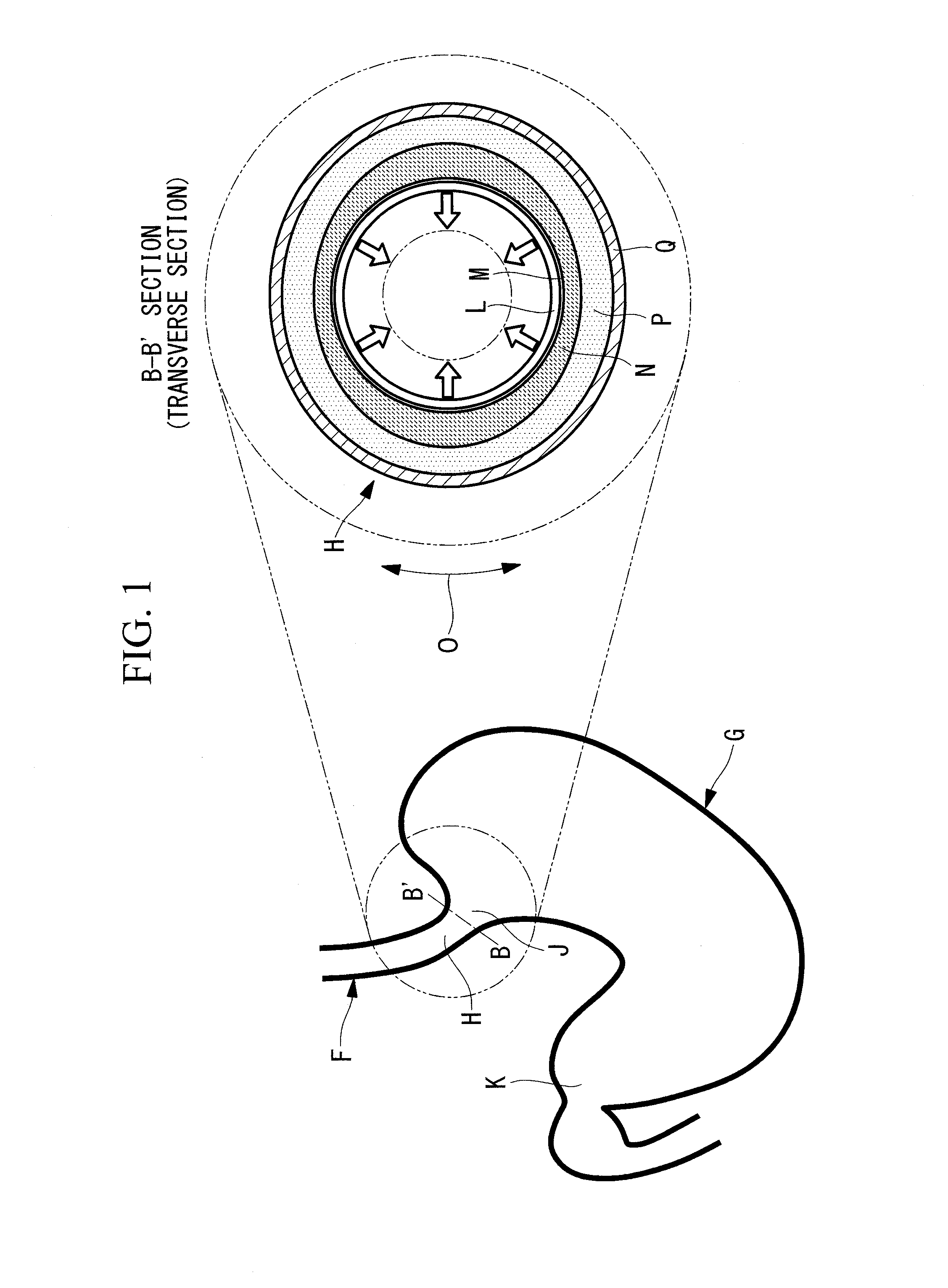
[0005] FIG. 1 includes a diagram illustrating the periphery of the gastroesophageal junction to which a gastrointestinal-tract constricting method according to a first embodiment of the present invention is applied, and a cross-sectional view taken along B-B' illustrating a transverse section of the gastroesophageal junction.
[0006] FIG. 2 is a flowchart illustrating the gastrointestinal-tract constricting method according to the first embodiment of the present invention.
[0007] FIG. 3 is a longitudinal sectional view showing how an endoscope is inserted into the stomach illustrated in FIG. 1.
[0008] FIG. 4 is an endoscopic image of the gastroesophageal junction illustrated in FIG. 1 and a mucosal surface in a target region, as viewed from the inside of the stomach.
[0009] FIG. 5 is a diagram illustrating the position of the target region in the gastroesophageal junction.
[0010] FIG. 6 is a cross-sectional view of the gastroesophageal junction taken along B-B' in FIG. 1, and illustrates the position of the target region.
[0011] FIG. 7 is a partial cross-sectional view taken along B-B' in FIG. 1, and illustrates how a spreading block is formed by injecting a sodium hyaluronate solution into the submucosal layer.
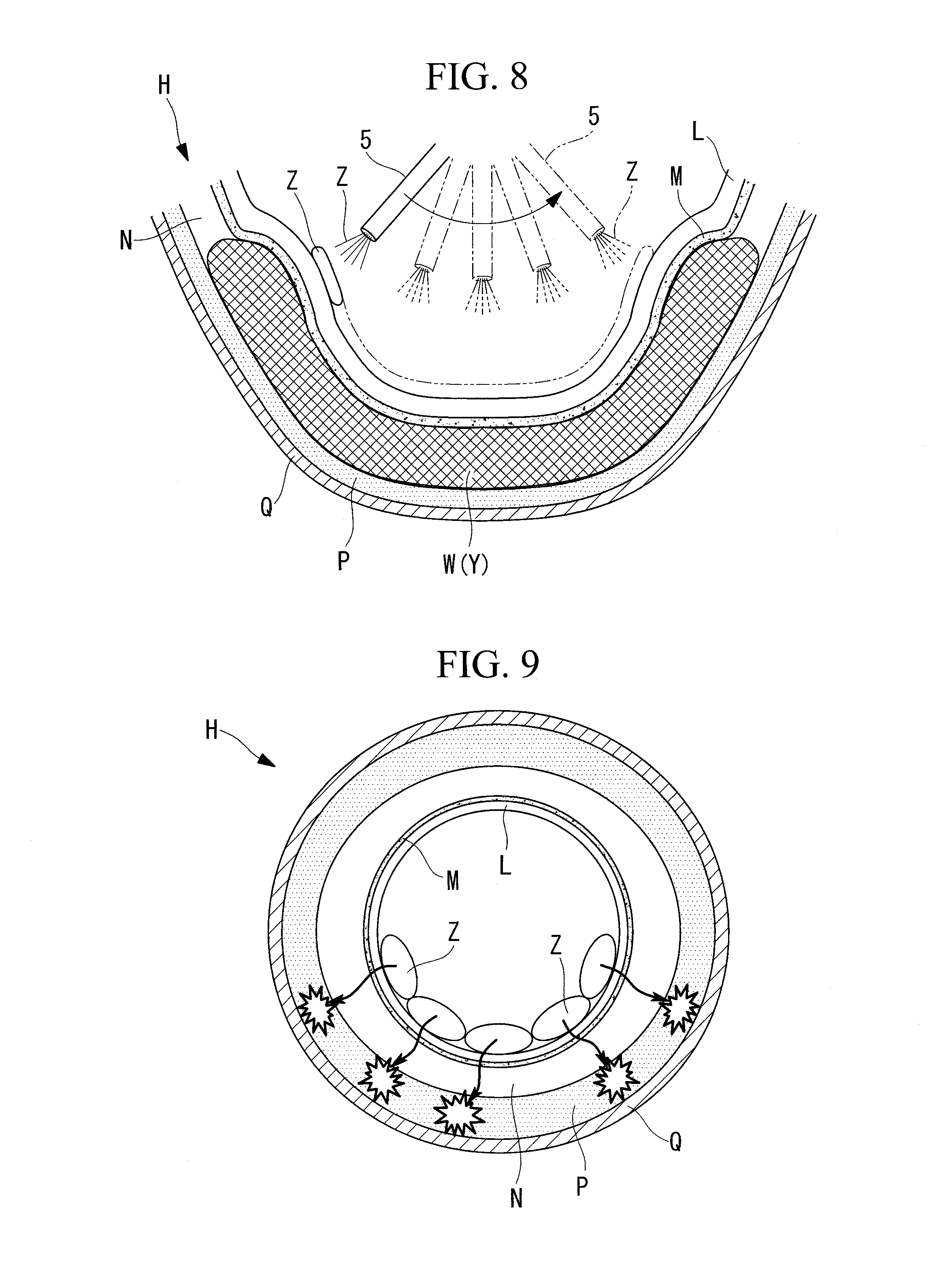
[0012] FIG. 8 is a partial cross-sectional view of the gastroesophageal junction taken along B-B' in FIG. 1, and illustrates how ethanol is sprayed onto the surface of the mucosal layer in the target region.
[0013] FIG. 9 is a cross-sectional view of the gastroesophageal junction taken along B-B' in FIG. 1, and illustrates how ethanol infiltrates into the outer side of the submucosal layer when the spreading block is not formed.
[0014] FIG. 10 is a cross-sectional view of the gastroesophageal junction taken along B-B' in FIG. 1, and illustrates how ethanol infiltrates into the outer side of the submucosal layer when the spreading block is not formed.
[0015] FIG. 11 is a plan view of a balloon catheter in a deflated-balloon state as viewed in the radial direction of a catheter body.
[0016] FIG. 12 is a plan view of the balloon catheter in an inflated-balloon state as viewed in the radial direction of the catheter body.
[0017] FIG. 13 is a perspective view of the balloon catheter in an inflated-balloon state.
[0018] FIG. 14 is a perspective view of the catheter body illustrated in FIG. 13.
[0019] FIG. 15 is a cross-sectional view of the catheter body taken along D-D' in FIG. 13.
[0020] FIG. 16 is a partial cross-sectional view of the gastroesophageal junction taken along B-B' in FIG. 1, and illustrates how a catheter insertion hole, which penetrates from the mucosal layer to the submucosal layer in the target region, is formed by using an electrode-equipped treatment tool.
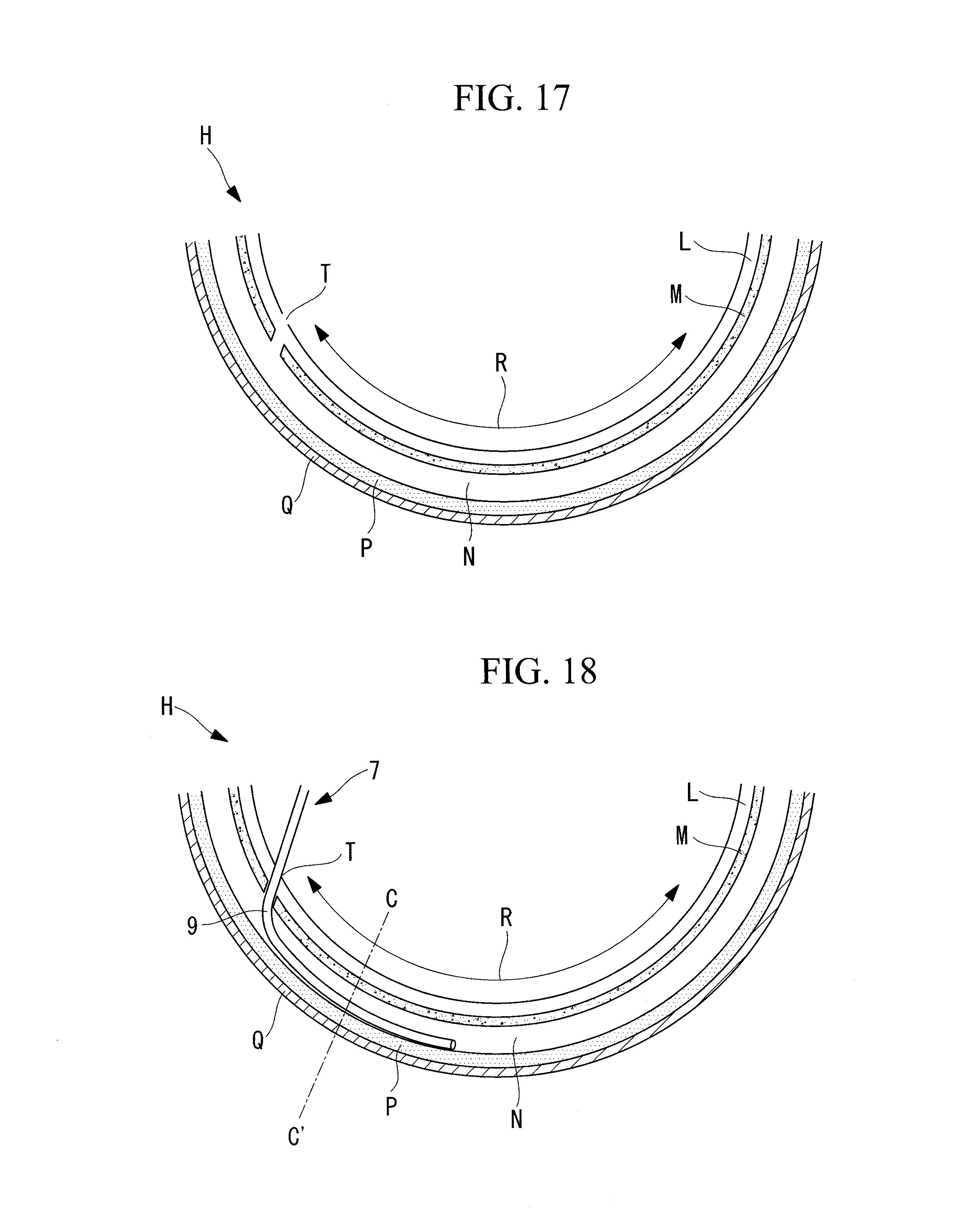
[0021] FIG. 17 is a partial cross-sectional view of the gastroesophageal junction taken along B-B' in FIG. 1, and illustrates the catheter insertion hole formed in the target region.
[0022] FIG. 18 is a partial cross-sectional view of the gastroesophageal junction taken along B-B' in FIG. 1, and illustrates how a balloon catheter is inserted into the submucosal layer through the catheter insertion hole.
[0023] FIG. 19 is a cross-sectional view taken along C-C' in FIG. 18, illustrating a state in which the balloon is deflated in the submucosal layer.
[0024] FIG. 20 is a cross-sectional view taken along C-C' in FIG. 18, illustrating a state in which the balloon is inflated in the submucosal layer.
[0025] FIG. 21 is a partial cross-sectional view of the gastroesophageal junction taken along B-B' in FIG. 1, and illustrates the state in which the balloon catheter is pushed in so as to reach the other end of the target region in the circumferential direction of the gastrointestinal tract.
[0026] FIG. 22 is a partial cross-sectional view of the gastroesophageal junction taken along B-B' in FIG. 1, and illustrates how the balloon catheter is withdrawn while injecting a sodium hyaluronate solution.
[0027] FIG. 23 is a partial cross-sectional view of the gastroesophageal junction taken along B-B' in FIG. 1, and illustrates how a spreading block composed of a sodium hyaluronate solution is formed in the submucosal layer.
[0028] FIG. 24 is a partial cross-sectional view of the gastroesophageal junction taken along B-B' in FIG. 1, and illustrates how ethanol is applied to the surface of the mucosal layer in the target region by using a sponge-roller-equipped treatment tool.
[0029] FIG. 25 is a cross-sectional view of a catheter body according to a modification of the balloon catheter illustrated in FIG. 13, taken along D-D'.
[0030] FIG. 26 is a cross-sectional view of a catheter body according to another modification of the balloon catheter illustrated in FIG. 13, taken along D-D'.
[0031] FIG. 27 is a diagram illustrating the state in which a substance sheet is housed in an endoscope.
[0032] FIG. 28 is a diagram illustrating the state in which the substance sheet is expanded outside the endoscope.
[0033] FIG. 29 is a partial cross-sectional view of the gastroesophageal junction taken along B-B' in FIG. 1, and illustrates how the electrode-equipped treatment tool incises the mucosal layer of the gastroesophageal junction.
[0034] FIG. 30 is a plan view illustrating the incised portion of the mucosal layer.
[0035] FIG. 31 is a partial cross-sectional view of the gastroesophageal junction taken along B-B' in FIG. 1, and illustrates how a polylactate sheet is inserted into the submucosal layer through the incised portion of the mucosal layer.
[0036] FIG. 32 is a partial cross-sectional view of the gastroesophageal junction taken along B-B' in FIG. 1, and illustrates how a spreading block formed of the polylactate sheet is formed in the submucosal layer.
[0037] FIG. 33 is a partial cross-sectional view of the gastroesophageal junction taken along B-B' in FIG. 1, and illustrates how a substance sheet is attached to the surface of the mucosal layer in the target region.
[0038] FIG. 34 is a partial cross-sectional view of the gastroesophageal junction taken along B-B' in FIG. 1, and illustrates the state in which the substance sheet is attached to the surface of the mucosal layer in the target region.
DESCRIPTION OF EMBODIMENTS
First Embodiment
[0039] A gastrointestinal-tract constricting method according to a first embodiment of the present invention will now be described with reference to the drawings.
[0040] The case described as an example in this embodiment is the case in which the gastrointestinal-tract constricting method is applied to the treatment of gastroesophageal reflux disease, wherein, as illustrated in FIG. 1, a part of a region that extends from the gastroesophageal junction H (lower part of the esophagus), where the esophagus F connects to the stomach G, to the cardiac part is constricted. In FIG. 1, reference sign J denotes the cardiac part constituting the entrance of the stomach G, reference sign K denotes the pyloric part constituting the endmost part of the stomach G, reference sign L denotes the mucosal layer, reference sign M denotes a mucosa basal layer, reference sign N denotes the submucosal layer, reference sign P denotes the muscular layer, and reference sign Q denotes the serosa.
[0041] As illustrated in the flowchart of FIG. 2, the gastrointestinal-tract constricting method includes an inserting step S1 of inserting an endoscope into the gastrointestinal tract constituting the gastroesophageal junction H; an identifying step S2 of identifying a target region where the tissue located between the mucosal layer L and the muscular layer P in the gastroesophageal junction H is to be damaged by ethanol (substance) while observing the gastroesophageal junction H with an endoscope; a block forming step S3 of forming a spreading block that blocks the infiltration of ethanol, which is to be supplied to the target region, into the muscular layer P and into a radially outer side (abdominal cavity side) of the gastrointestinal tract with respect to the muscular layer P; a supplying step S4 of supplying ethanol to the target region after the block forming step S3; an endoscope withdrawing step S5 of withdrawing an endoscope 1 from the inside of the gastrointestinal tract to the outside of the body; a waiting step S6 of waiting until a part of the region extending from the gastroesophageal junction H (lower part of the esophagus) to the cardiac part is constricted; and a constriction confirming step S7 of confirming constriction of the part of the region extending from the gastroesophageal junction H (lower part of the esophagus) to the cardiac part.
[0042] As illustrated in FIG. 3, in the inserting step S1, the endoscope 1 is inserted via the mouth of a subject into the stomach G through the esophagus F, the distal end of the endoscope 1 is bent inside the stomach G, and the distal end of the endoscope 1 is arranged to face the cardiac part J and the gastroesophageal junction H so as to look up into the esophagus F from the stomach G.
[0043] As illustrated in FIG. 4, in the identifying step S2, after the mucosal surface of the relaxed cardiac part J is observed with the endoscope 1, the range of a target region R on the surface of the mucosal layer L is identified. In FIG. 4, reference sign S denotes the gastric wall; and in FIGS. 1 and 4, the arrow indicated by reference sign O indicates the circumferential direction of the gastrointestinal tract.
[0044] In the supplying step S4 described below, in the range coincident with the thus identified target region R, the mucosa basal layer M (refer to FIG. 1), which is the lowermost layer of the mucosal layer L, in a part of the region extending from the gastroesophageal junction H (lower part of the esophagus) to the cardiac part J is damaged. By damaging the mucosa basal layer M in the target region R, constriction occurs in part of the region extending from the gastroesophageal junction H (lower part of the esophagus) to the cardiac part J. Preferably, the range of the target region R is appropriately determined in advance so that the lumen has a desired inner diameter after constriction.
[0045] In order to prevent excessive constriction, the target region R is set to be a part of the region extending from the gastroesophageal junction H (lower part of the esophagus) to the cardiac part J, and is a range that does not extend all around the circumference. For example, as illustrated in FIGS. 5 and 6, the target region R is preferably a range that extends from the lesser curvature side to the gastric fundus side and occupies 60% to 80% of the entire circumference.
[0046] In the block forming step S3, as illustrated in FIG. 7, a spreading block W is formed in the submucosal layer (position between the mucosal layer L and the muscular layer P) N in the target region R by using an infiltration inhibitor that suppresses infiltration of ethanol. As the infiltration inhibitor, for example, a sodium hyaluronate solution, which is a liquid that is immiscible with ethanol due to substance polarity and that has a higher viscosity than ethanol, is used. The substance polarity referred to here is the electrical bias present within the molecule.
[0047] As illustrated in FIG. 7, in the block forming step S3, an injection needle 3 of an injection-needle-equipped treatment tool is used to fill the submucosal layer N in the target region R with the sodium hyaluronate solution Y so as to form a spreading block W composed of the sodium hyaluronate solution Y in the submucosal layer N. The sodium hyaluronate solution Y can remain at the injected position in the submucosal layer N due to its high viscosity. The spreading block W has, for example, a shape such that at least part of the transverse section thereof extends in an arc shape along the circumferential direction of the gastrointestinal tract.
[0048] Furthermore, the spreading block W is preferably formed over a range larger than the target region R in the circumferential direction and the longitudinal direction of the gastrointestinal tract. When the spreading block W is formed over a range larger than the target region R in the circumferential direction and the longitudinal direction of the gastrointestinal tract, it is possible to more reliably suppress infiltration of ethanol Z, which is supplied to the target region R and circumvents the spreading block W from the outer side in the circumferential direction of the gastrointestinal tract, into the muscular layer P. In the longitudinal direction (direction along the axial line) of the gastrointestinal tract, the spreading block W is preferably formed partly in the region that extends from the gastroesophageal junction H (lower part of the esophagus) to the cardiac part J.
[0049] As illustrated in FIG. 8, in the supplying step S4, a substance spraying treatment tool (substance spraying tool) 5 is used to spray ethanol Z onto the surface of the mucosal layer L in the target region R. In other words, the ethanol Z is sprayed onto the surface of the mucosal layer L that lies in the region that overlaps the inner side of the gastrointestinal tract with respect to the spreading block W in a transverse section of the gastrointestinal tract, out of the entire circumference of the gastrointestinal tract. When the ethanol Z is not to infiltrate into the muscular layer P in the region that does not overlap the inner side of the gastrointestinal tract with respect to the spreading block W out of the entire circumference of the gastrointestinal tract, the ethanol Z is not sprayed onto the surface of the mucosal layer L that lies in the region that does not overlap the inner side of the gastrointestinal tract with respect to the spreading block W. The mucosa basal layer M in the target region R lies within the region that overlaps the inner side of the gastrointestinal tract with respect to the spreading block W out of the entire circumference of the gastrointestinal tract, and lies on the inner side of the gastrointestinal tract with respect to the spreading block W, in other words, on the radially inner side of the gastrointestinal tract with respect to the spreading block W.
[0050] The operation of the gastrointestinal-tract constricting method of this embodiment will now be described.
[0051] In order to constrict a part of the region extending from the gastroesophageal junction H (lower part of the esophagus) to the cardiac part J of the subject by the gastrointestinal-tract constricting method according to this embodiment, first, as illustrated in FIG. 3, the endoscope 1 is inserted into the gastrointestinal tract via the mouth of the subject, and the distal end of the endoscope 1 is bent inside the stomach G so as to face the cardiac part J and the gastroesophageal junction H (inserting step S1).
[0052] Next, as illustrated in FIGS. 4, 5, and 6, while observing the region that extends from the gastroesophageal junction H (lower part of the esophagus) to the cardiac part J with the endoscope 1, the target region R is identified within the region that extends from the gastroesophageal junction H (lower part of the esophagus) to the cardiac part J (identifying step S2).
[0053] Once the target region R is identified, the injection-needle-equipped treatment tool is inserted into a forceps channel of the endoscope 1, and a syringe (not illustrated) filled with the sodium hyaluronate solution Y is attached to the injection-needle-equipped treatment tool.
[0054] Then, as illustrated in FIG. 7, an injection needle 3 of the injection-needle-equipped treatment tool punctures the submucosal layer N in the target region R, and the sodium hyaluronate solution Y is injected into the submucosal layer N. This operation is repeated several times by shifting the position in the circumferential direction of the gastrointestinal tract that constitutes the target region R (for example, the gastroesophageal junction H) so as to form a spreading block W in the submucosal layer N in the target region R, the spreading block W spanning over a range larger than the target region R in the circumferential direction of the gastroesophageal junction H and being filled with the sodium hyaluronate solution Y (block forming step S3).
[0055] After the spreading block W is formed, a syringe (not illustrated) filled with ethanol is attached to the substance spraying treatment tool 5 so as to replace the syringe filled with the sodium hyaluronate solution Y. Then, as illustrated in FIG. 8, ethanol Z is sprayed from the substance spraying treatment tool 5 onto the surface of the mucosal layer L in the target region R while shifting the position in the circumferential direction of the gastrointestinal tract (supplying step S4).
[0056] As the sprayed ethanol Z infiltrates toward the spreading block W side from the surface of the mucosal layer L in the target region R, at least the mucosa basal layer M in the target region R becomes damaged by the ethanol Z. The range in which the ethanol Z is supplied onto the mucosal surface in the target region R can be confirmed with the endoscope 1. The ethanol Z reaches the mucosa basal layer M and damages the mucosa basal layer M before it is absorbed in the body. In other words, the ethanol Z is absorbed in the body after causing damage to the mucosa basal layer M. The sodium hyaluronate solution Y is absorbed in the body more slowly than the ethanol Z is. Thus, the state in which the spreading block W is formed is maintained until the ethanol Z is absorbed in the body.
[0057] In the supplying step S4, the ethanol Z is repeatedly sprayed onto the mucosal surface while shifting the position within the target region R until the ethanol Z sprayed onto the mucosal surface reaches and contacts the sodium hyaluronate solution Y injected into the submucosal layer N.
[0058] After the supplying step S4, it is confirmed whether the ethanol Z has been thoroughly sprayed onto the surface of the mucosal layer L in the target region R, and then the endoscope 1 is withdrawn out of the body from the gastrointestinal tract (endoscope withdrawing step S5).
[0059] After the endoscope 1 is withdrawn out of the body from the gastrointestinal tract, the operation thereof is waited until the part of the region extending from the gastroesophageal junction H (lower part of the esophagus) to the cardiac part J is constricted by the constrictive effect of the tissue around the target region R undergoing the process of forming scars as the damaged tissue heals (waiting step S6).
[0060] After waiting of the operation until the part of the region extending from the gastroesophageal junction H (lower part of the esophagus) to the cardiac part J is constricted, the endoscope 1 is again inserted into the gastrointestinal tract so as to confirm that the part of the region extending from the gastroesophageal junction H (lower part of the esophagus) to the cardiac part J is constricted (constriction confirming step S7). The gastric acid reflux can be suppressed without excessively constricting a part of the region that extends from the gastroesophageal junction H (lower part of the esophagus) to the cardiac part J by constricting the part of the region that extends from the gastroesophageal junction H (lower part of the esophagus) to the cardiac part J by damaging the mucosa basal layer M with the ethanol Z within the range of the desired target region R, which is a part of the region extending from the gastroesophageal junction H (lower part of the esophagus) to the cardiac part J and which does not extend all around the circumference.
[0061] If needed, after the sodium hyaluronate solution Y is further injected into the submucosal layer N as described in the block forming step S3, the ethanol Z may be additionally sprayed onto the surface of the mucosal layer L located on the inner side of the gastrointestinal tract with respect to the spreading block W, as described in the supplying step S4.
[0062] As described above, according to the gastrointestinal-tract constricting method of this embodiment, since the mucosa basal layer M in the target region R (for example, the gastroesophageal junction H) is damaged by the ethanol Z, the invasiveness is low and the procedure is simple compared to when the tissue is damaged by incising the target region R (for example, the gastroesophageal junction H) or excising the tissue in the target region R.
[0063] In such a case, if no spreading block W is provided between the mucosal layer L and the muscular layer P in the target region R, as illustrated in FIG. 9, the ethanol Z sprayed onto the surface of the mucosal layer L in the target region R infiltrates into the muscular layer P (the muscular layer P underlying the mucosa basal layer M) on the radially outer side of the gastrointestinal tract with respect to the submucosal layer N and into the radially outer side (abdominal cavity side) of the gastrointestinal tract with respect to the muscular layer P; thus, the muscular layer P may become damaged, possibly resulting in perforation and bleeding.
[0064] To address this issue, as illustrated in FIG. 10, the spreading block W is formed in advance between the mucosa basal layer M and the muscular layer P in the target region R. Subsequently, the ethanol Z is sprayed onto the surface of the mucosal layer L located on the inner side of the gastrointestinal tract with respect to the spreading block W so that the ethanol Z infiltrates toward the radially outer side of the gastrointestinal tract but is blocked by the spreading block W. As a result, the ethanol Z stays at a position between the mucosal layer L and the muscular layer P, and, at the same time, infiltration of the ethanol Z into the muscular layer P and the radially outer side (abdominal cavity side) of the gastrointestinal tract with respect to the muscular layer P can be suppressed.
[0065] Thus, a part of the region extending from the gastroesophageal junction H (lower part of the esophagus) to the cardiac part J can be constricted by causing the constrictive effect to occur in the tissue in the mucosa basal layer M without damaging the muscular layer P.
[0066] In this embodiment, the spreading block W is formed partly in the circumferential direction of the gastrointestinal tract; alternatively, the spreading block W may be formed all around the circumference of the gastrointestinal tract by supplying sodium hyaluronate.
[0067] Moreover, in this embodiment, the spreading block W is formed by injecting the sodium hyaluronate solution Y into the submucosal layer N in the target region R (for example, the gastroesophageal junction H); alternatively, for example, the spreading block W may be formed by placing an absorbent polymer in the submucosal layer N in the target region R. In this case also, since the absorbent polymer absorbs the ethanol Z, infiltration of the ethanol Z into the muscular layer P and the radially outer side (abdominal cavity side) of the gastrointestinal tract with respect to the muscular layer P can be suppressed.
Second Embodiment
[0068] A gastrointestinal-tract constricting method according to a second embodiment of the present invention will now be described with reference to the drawings.
[0069] The gastrointestinal-tract constricting method of this embodiment differs from the first embodiment in that the method further includes a space forming step of forming a space between the mucosa basal layer M and the muscular layer P, the space forming step being performed between the identifying step S2 and the block forming step S3; and in that the method involves a different block forming step S3 and supplying step S4. The inserting step S1, the identifying step S2, the endoscope withdrawing step S5, the waiting step S6, and the constriction confirming step S7 are the same as those in the first embodiment.
[0070] In the description of this embodiment, the features common to the gastrointestinal-tract constricting method according to the first embodiment described above are denoted by the same reference signs, and descriptions therefor are omitted.
[0071] In the space forming step, for example, a space is formed inside the submucosal layer N by separating the submucosal layer N itself by using a balloon catheter (separating tool) 7, such as the one illustrated in FIGS. 11, 12, and 13. Although an example in which the submucosal layer N itself is separated is described as an example in this embodiment, this example is not limiting. Alternatively, a space may be formed by performing separation at the boundary between the mucosa basal layer M and the submucosal layer N or the boundary between the submucosal layer N and the muscular layer P. The balloon catheter 7 is equipped with a catheter body 9, which has a long thin double-tube structure, and balloons 11A and 11B that can be inflated and deflated in radial directions opposite from each other with respect to the catheter body 9.
[0072] As illustrated in FIGS. 13, 14, and 15, the catheter body 9 is equipped with an outer tube 10A to which the balloons 11A and 11B are attached, and an inner tube 10B that lies in the center portion in the radial direction of the outer tube 10A, extends along the longitudinal direction of the outer tube 10A, and has an outer diameter smaller than the inner diameter of the outer tube 10A. The outer tube 10A and the inner tube 10B are fixed at the distal end portion in the longitudinal direction. In FIGS. 14 and 15, the balloons 11A and 11B are omitted from the illustration, and only the catheter body 9 is illustrated. The same applies to FIGS. 25 and 26.
[0073] The catheter body 9 has a center lumen 9a, which lies at the center portion in the radial direction of the inner tube 10B and extends in the longitudinal direction (direction along the axial line), and a ring lumen 9b that lies between the outer tube 10A and the inner tube 10B and extends in the longitudinal direction. In addition, the catheter body 9 has an opening 9c, at which the center lumen 9a opens at the distal end surface in the longitudinal direction of the inner tube 10B and through which the infiltration inhibitor is discharged, and balloon-inflating openings 9d, which are arranged along the longitudinal direction at the distal end portion of the outer tube 10A and through which the ring lumen 9b communicates with the balloons 11A and 11B.
[0074] This balloon catheter 7 is configured such that, when pressure is applied to the ring lumen 9b of the catheter body 9 by injecting a liquid or a gas, the liquid or gas fills the interiors of the balloons 11A and 11B through the balloon-inflating openings 9d, and, as illustrated in FIGS. 12 and 13, the balloons 11A and 11B each inflate in a direction intersecting the axial line and along a flat plane that includes the axial line of the catheter body 9.
[0075] In the space forming step, the balloon catheter 7, with the balloons 11A and 11B in a deflated state, is inserted into the submucosal layer N in the target region R, and the balloons 11A and 11B are inflated by applying pressure from the interiors of the balloons 11A and 11B so as to tear the submucosal layer N and separate the interior of the submucosal layer N in the thickness direction (radial direction of the gastrointestinal tract), thereby forming a space inside the submucosal layer N.
[0076] In the block forming step S3 of this embodiment, the sodium hyaluronate solution Y is discharged from the infiltration-inhibitor-injecting opening 9c of the balloon catheter 7 pushed into the submucosal layer N in the space forming step, and while the space formed inside the submucosal layer N is being filled with the sodium hyaluronate solution Y, the balloon catheter 7 is withdrawn.
[0077] In the supplying step S4 of this embodiment, the ethanol Z is applied to the surface of the mucosal layer L by using a sponge-roller-equipped treatment tool 13 (refer to FIG. 24) with a sponge saturated with the ethanol Z.
[0078] The sponge-roller-equipped treatment tool 13 is equipped with a sponge roller (roller member) 15 that can absorb and release the ethanol Z, and a supporting member 17 that rotatably supports the sponge roller 15 about a particular rotation axis. The sponge-roller-equipped treatment tool 13 can apply the ethanol Z to the surface of the mucosal layer L by rolling the ethanol-Z-saturated sponge roller 15 along the surface of the mucosal layer L.
[0079] The effects of the gastrointestinal-tract constricting method according to this embodiment will now be described.
[0080] In order to constrict, for example, a part of the region that extends from the gastroesophageal junction H (lower part of the esophagus) to the cardiac part J of the subject with the gastrointestinal-tract constricting method of this embodiment, first, the endoscope 1 is inserted into the gastrointestinal tract in the inserting step S1, and, in the identifying step S2, the target region R is identified in the region that extends from the gastroesophageal junction H (lower part of the esophagus) to the cardiac part J.
[0081] Next, as illustrated in FIGS. 16 and 17, first an electrode-equipped treatment tool 19, which can, for example, excise biotissue with a high-frequency electrode, is inserted into a forceps channel of the endoscope 1 inserted into the gastrointestinal tract in the inserting step S1, and a catheter insertion hole T that penetrates from the surface of the mucosal layer L to the submucosal layer N is formed at one end of the target region R in the circumferential direction of the gastrointestinal tract.
[0082] Next, as illustrated in FIG. 18, the balloon catheter 7 is inserted into the catheter insertion hole T. At this stage, the balloons 11A and 11B are in a deflated state. Then, while the balloons 11A and 11B are in a deflated state, the balloon catheter 7 is pushed into the interior of the submucosal layer N in the target region R. Subsequently, a liquid or gas is injected into the ring lumen 9b of the balloon catheter 7 to apply pressure, and, as illustrated in FIG. 20, the balloons 11A and 11B are inflated so as to partly separate the submucosal layer N in the radial direction of the gastrointestinal tract.
[0083] Once the submucosal layer N is partly separated in the radial direction of the gastrointestinal tract, as illustrated in FIG. 19, the balloons 11A and 11B are deflated again, and the balloon catheter 7 is further pushed into the interior of the unseparated part of the submucosal layer N. In this embodiment, the direction in which the balloon catheter 7 is pushed is the circumferential direction of the gastrointestinal tract that constitutes the gastroesophageal junction H, but the direction is not limited to this and may be the longitudinal direction of the gastrointestinal tract.
[0084] This operation is repeated, and, as illustrated in FIG. 21, the balloon catheter 7 is pushed in along the submucosal layer N until the balloon catheter 7 reaches the other end in the circumferential direction of the gastrointestinal tract that constitutes the target region R (for example, the gastroesophageal junction H) (space forming step). In this manner, the interior of the submucosal layer N from one end to the other end in the circumferential direction of the gastrointestinal tract that constitutes the target region R (for example, the gastroesophageal junction H) is separated in the radial direction of the gastrointestinal tract so as to form a space, which spreads in the circumferential direction of the gastrointestinal tract, in the submucosal layer N. As a result, when the sodium hyaluronate solution Y is supplied to the submucosal layer N in the supplying step S4, the sodium hyaluronate solution Y can spread easily from one end to the other end in the circumferential direction of the gastrointestinal tract in the target region R (for example, the gastroesophageal junction H).
[0085] Next, the balloons 11A and 11B of the balloon catheter 7 are deflated, and, as illustrated in FIG. 22, while the sodium hyaluronate solution Y is being injected into the submucosal layer N from the infiltration inhibitor-discharging opening 9c of the balloon catheter 7, the balloon catheter 7 is withdrawn.
[0086] After the balloon catheter 7 is completely withdrawn from the catheter insertion hole T, as illustrated in FIG. 23, a spreading block W filled with the sodium hyaluronate solution Y is formed in the space formed in the submucosal layer N from one end to the other end in the circumferential direction of the gastrointestinal tract in the target region R (block forming step S3).
[0087] Next, the ethanol Z is transfused into the sponge roller 15 of the sponge-roller-equipped treatment tool 13 by using a syringe (not illustrated) filled with the ethanol Z. Then the sponge roller 15 is brought into contact with the surface of the mucosal layer L in the target region R, and the sponge roller 15 is rolled along the surface of the mucosal layer L in the target region R. As a result, the ethanol Z can be applied to the surface of the mucosal layer L from one end to the other end of the target region R in the circumferential direction (supplying step S4).
[0088] As a result, while the ethanol Z applied to the surface of the mucosal layer L in the target region R infiltrates into the mucosa basal layer M so that the mucosa basal layer M in the target region R is damaged by the ethanol Z, infiltration of the ethanol Z into the muscular layer P and the radially outer side (abdominal cavity side) of the gastrointestinal tract with respect to the muscular layer P can be suppressed by the spreading block W.
[0089] As described above, with the gastrointestinal-tract constricting method according to this embodiment, the space and the spreading block W can be formed in the submucosal layer N by a simple task of inserting and removing the balloon catheter 7 between the mucosa basal layer M and the muscular layer P, and thus the operation efficiency can be improved. Moreover, the sponge-roller-equipped treatment tool 13 can more reliably and more thoroughly supply the ethanol Z over the desired range of the surface of the mucosal layer L.
[0090] In this embodiment, the catheter body 9 has the ring lumen 9b; alternatively, for example, as illustrated in FIG. 25, the catheter body 9 may have two arc-shaped lumens 9e and 9f, which are spaces created by dividing a space between the outer tube 10A and the inner tube 10B into two in the circumferential direction and each of which has an arc shape at a cross-section taken in a direction intersecting the longitudinal direction. In this case, the arc-shaped lumen 9e may have balloon-inflating openings 9d communicating with the balloon 11A, and the other arc-shaped lumen 9f may have balloon-inflating openings 9d communicating with the balloon 11B. Moreover, in this case, one or both of the balloons 11A and 11B may be inflated inside the submucosal layer N so that the submucosal layer N is divided in the radial direction of the gastrointestinal tract. The same applies to the case in which separation is performed at the boundary between the mucosa basal layer M and the submucosal layer N and the case in which separation is performed at the boundary between the submucosal layer N and the muscular layer P.
[0091] Alternatively, for example, as illustrated in FIG. 26, the catheter body 9 may have one arc-shaped lumen 9g between the outer tube 10A and the inner tube 10B, the arc-shaped lumen 9g having an arc shape at a cross-section intersecting the longitudinal direction. In this case, one arc-shaped lumen 9g may have both balloon-inflating openings 9d communicating with the balloon 11A, and the balloon-inflating openings 9d communicating with the balloon 11B.
[0092] In this embodiment, the space is formed by separating the interior of the submucosal layer N in the circumferential direction of the gastrointestinal tract; alternatively, it suffices that a space that extends in the circumferential direction of the gastrointestinal tract can be formed between the mucosa basal layer M and the muscular layer P in the target region R. For example, separation may be performed at the boundary between the mucosa basal layer M and the submucosal layer N and along the circumferential direction of the gastrointestinal tract so that a space is formed between the mucosa basal layer M and the submucosal layer N. Alternatively, separation may be performed at the boundary between the submucosal layer N and the muscular layer P and along the circumferential direction of the gastrointestinal tract so that a space is formed between submucosal layer N and the muscular layer P. Alternatively, for example, in the region that extends from the gastroesophageal junction H (lower part of the esophagus) to the cardiac part J, the balloon catheter 7 may be inserted in the longitudinal direction of the gastrointestinal tract or in an oblique direction with respect to the longitudinal direction of the gastrointestinal tract so as to form a space between the mucosa basal layer M and the muscular layer P.
[0093] Although the balloon catheter 7 is used as an example of the separating tool in the description, the separating tool is not limited to this and may be any separating tool as long as a space can be formed between the mucosa basal layer M and the muscular layer P in the target region R.
Third Embodiment
[0094] A gastrointestinal-tract constricting method according to a third embodiment of the present invention will now be described with reference to the drawings.
[0095] The gastrointestinal-tract constricting method of this embodiment differs from the first embodiment in that the block forming step S3 and the supplying step S4 are different. The inserting step S1, the identifying step S2, the endoscope withdrawing step S5, the waiting step S6, and the constriction confirming step S7 are the same as those in the first embodiment.
[0096] In the description of this embodiment, features common to the gastrointestinal-tract constricting method according to the first embodiment described above are denoted by the same reference signs, and descriptions therefor are omitted.
[0097] In the block forming step S3 of this embodiment, the mucosal layer L in the target region R is incised, and a polylactate sheet E (infiltration inhibitor sheet, refer to FIG. 31) is inserted from the incised portion and placed in the submucosal layer N so as to form a spreading block W.
[0098] In the supplying step S4 of this embodiment, a substance-sheet-equipped treatment tool 21 illustrated in FIGS. 27 and 28 is used to cause the ethanol Z to infiltrate into the mucosal layer L in the target region R.
[0099] The substance-sheet-equipped treatment tool 21 is equipped with a long thin center rod member 23, rib members 25 that project from the distal end portion of the center rod member 23, and a sheet-shaped substance sheet 27 saturated with the ethanol Z and detachably supported on the rib members 25.
[0100] The substance-sheet-equipped treatment tool 21 is openable and closable, that is, is configured to assume a closed state in which the rib members 25 lie in the longitudinal direction of the center rod member 23 and an open state in which the rib members 25 lie in a direction intersecting the longitudinal direction.
[0101] As illustrated in FIG. 27, the substance-sheet-equipped treatment tool 21 can be housed in the forceps channel of the endoscope 1 by closing the rib members 25 and collapsing the substance sheet 27. As illustrated in FIG. 28, the substance-sheet-equipped treatment tool 21 can expand the substance sheet 27 by spreading the rib members 25 outside the forceps channel of the endoscope 1. As illustrated in FIG. 27, the substance-sheet-equipped treatment tool 21 can cause the substance sheet 27 to detach from the rib members 25 by withdrawing the rib members 25 into the forceps channel of the endoscope 1 while leaving the substance sheet 27 expanded.
[0102] In the supplying step S4, the substance sheet 27 of the substance-sheet-equipped treatment tool 21 is expanded near the target region R in the gastrointestinal tract and is attached to the surface of the mucosal layer L in the target region R so that the ethanol Z in the substance sheet 27 infiltrates into the mucosal layer L.
[0103] The effects of the gastrointestinal-tract constricting method according to this embodiment will now be described.
[0104] In order to constrict, for example, a part of the region that extends from the gastroesophageal junction H (lower part of the esophagus) to the cardiac part J of the subject with the gastrointestinal-tract constricting method of this embodiment, first, the endoscope 1 is inserted into the gastrointestinal tract in the inserting step S1, and, in the identifying step S2, the target region R is identified in the region that extends from the gastroesophageal junction H (lower part of the esophagus) to the cardiac part J.
[0105] The electrode-equipped treatment tool 13 is inserted into the forceps channel of the endoscope 1 inserted into the gastrointestinal tract in the inserting step S1, and, as illustrated in FIGS. 29 and 30, the mucosal layer L is incised at one end in the circumferential direction of the gastrointestinal tract constituting the target region R (for example, the gastroesophageal junction H) and along the longitudinal direction X of the gastrointestinal tract, so that the incised range is the same range as the target region R or a larger range than the target region R.
[0106] Next, as illustrated in FIG. 31, the polylactate sheet E is inserted into the submucosal layer N from the incised portion U in the mucosal layer L, and the polylactate sheet E is pushed along the submucosal layer N until the polylactate sheet E passes the other end in the circumferential direction of the gastrointestinal tract constituting the target region R (for example, the gastroesophageal junction H). Then, as illustrated in FIG. 32, the polylactate sheet E is placed in the submucosal layer N so as to form the spreading block W formed of the polylactate sheet E (block forming step S3).
[0107] Next, as illustrated in FIG. 27, the substance-sheet-equipped treatment tool 21 is inserted into the forceps channel of the endoscope 1 inserted into the gastrointestinal tract, and, as illustrated in FIG. 28, the rib members 25 are caused to project from the forceps channel of the endoscope 1 so as to expand the substance sheet 27 near the gastroesophageal junction H inside the gastrointestinal tract.
[0108] Then, as illustrated in FIG. 33, the substance sheet 27 is attached to the surface of the mucosal layer L in the target region R, and then, as illustrated in FIG. 34, the substance sheet 27 is detached from the rib members 25 and is made to stay on the surface of the mucosal layer L. As a result, the ethanol Z in the substance sheet 27 can infiltrate into the mucosal layer L in the target region R (supplying step S4).
[0109] Then, while the ethanol Z that has infiltrated into the mucosal layer L in the target region R damages the mucosa basal layer M in the target region R, infiltration of the ethanol Z into the muscular layer P and the radially outer side (abdominal cavity side) of the gastrointestinal tract with respect to the muscular layer P can be suppressed by the spreading block W.
[0110] As described above, with the gastrointestinal-tract constricting method according to this embodiment, the spreading block W can be formed in the submucosal layer N by a single step of inserting the polylactate sheet E. In addition, by using the polylactate sheet E, which does not spread in the surrounding area within the submucosal layer N unlike a liquid infiltration inhibitor, as the infiltration inhibitor sheet, the spreading block W can be formed in the desired region.
[0111] Since polylactate is decomposed into harmless substances inside the body, the polylactate sheet E can be left in the submucosal layer N, thus simplifying the procedure. Moreover, the ethanol Z can be efficiently supplied onto the mucosal layer L in a low-invasiveness manner over a wide range by attaching the substance sheet 27 saturated with the ethanol Z onto the surface of the mucosal layer L in the target region R.
[0112] In this embodiment, the mucosal layer L is incised along the longitudinal direction X of the gastrointestinal tract, and the polylactate sheet E is inserted in the circumferential direction of the gastrointestinal tract; alternatively, for example, the mucosal layer L may be incised in the circumferential direction of the gastrointestinal tract and the polylactate sheet E may be inserted in the longitudinal direction X of the gastrointestinal tract, or the mucosal layer L may be incised obliquely with respect to the longitudinal direction X of the gastrointestinal tract and the polylactate sheet E may be inserted obliquely with respect to the longitudinal direction X of the gastrointestinal tract.
[0113] Although ethanol Z is described as an example of the substance in the embodiments described above, the substance may be any substance that impairs the normal functions of cells, in other words, any substance that can damage cells, and examples thereof include, in addition to ethanol Z, peptase, protease, acetylcysteine, and sodium 2-mercaptoethanesulfonate.
[0114] Although the sodium hyaluronate solution Y is described as an example of the infiltration inhibitor in the first and second embodiments described above, the infiltration inhibitor may be any liquid that does not easily spread but remains at the position at which it is placed in the submucosal layer N, and examples thereof include, in addition to the sodium hyaluronate solution Y, sodium chondroitin sulfate, chitosan, poly-N-acetylglucosamine, carboxymethyl cellulose sodium, carmellose sodium, cyanoacrylate, and a polylactate sheet.
[0115] Among these substances and infiltration inhibitors, a combination of substances that are immiscible with each other, such as a combination of the ethanol Z and the sodium hyaluronate solution Y described above, may be used.
[0116] Although embodiments of the present invention have been described in detail with reference to the drawings in the description above, the specific structures are not limited to these embodiments and include design modifications etc., within the scope of the present invention. For example, the present invention is not limited to implementations in the embodiments and modifications described above but may be applied to embodiments in which these embodiments and modifications are appropriately combined, without specific limitation.
[0117] Furthermore, for example, the block forming step S3 and the supplying step S4 of the respective embodiments may be combined as appropriate. For example, the block forming step S3 of the first embodiment may be combined with the supplying step S4 of the second embodiment or the supplying step S4 of the third embodiment; or the block forming step S3 of the second embodiment may be combined with the supplying step S4 of the first embodiment or the supplying step S4 of the third embodiment. Furthermore, the block forming step S3 of the third embodiment may be combined with the supplying step S4 of the first embodiment or the supplying step S4 of the second embodiment.
[0118] Although in the embodiments described above, the case in which the gastrointestinal-tract constricting method is applied to the treatment of gastroesophageal reflux disease is described, any approach with which the substance is supplied to the target region R of the gastrointestinal tract, and with which the gastrointestinal tract can be constricted by using the constrictive effect of the surrounding tissue in the target region R caused by formation of scars as the tissue in the damaged mucosa basal layer M heals will suffice. Thus, the application range is not limited to the treatment of gastroesophageal reflux disease, and the site where the method is to be applied is not limited to the gastroesophageal junction H or the region that extends from the gastroesophageal junction H (lower part of the esophagus) to the cardiac part J.
[0119] The following aspects of the invention are derived from the embodiments described above.
[0120] One aspect of the present invention provides a gastrointestinal-tract constricting method that includes, while observing the gastrointestinal tract by inserting an endoscope into the gastrointestinal tract, forming a spreading block that blocks infiltration of a substance, which damages a mucosa basal layer of the gastrointestinal tract, into a muscular layer underlying the mucosa basal layer, the spreading block being formed along a circumferential direction of the gastrointestinal tract and between the mucosa basal layer and the muscular layer; and supplying the substance along the circumferential direction of the gastrointestinal tract and to a surface of a mucosa that lies within a region that overlaps an inner side of the gastrointestinal tract with respect to the spreading block out of the entire circumference of the gastrointestinal tract in a transverse section of the gastrointestinal tract.
[0121] According to this aspect, the substance supplied to the surface of the mucosa of the gastrointestinal tract infiltrates a part located between the mucosal layer and the muscular layer and damages the mucosa basal layer, and thus, the gastrointestinal tract can be constricted by utilizing the constrictive effect of the surrounding tissue undergoing scar formation as the damaged tissue heals.
[0122] In this case, a spreading block, which blocks infiltration of the substance into the muscular layer underlying the mucosa basal layer, is formed in advance between the mucosa basal layer and the muscular layer and along the circumferential direction of the gastrointestinal tract, and the substance is supplied to the mucosal surface located within the region overlaps an inner side of the gastrointestinal tract with respect to the spreading block so that the substance is supplied along the circumferential direction of the gastrointestinal tract. In this manner, infiltration of the substance can be blocked by the infiltration block between the mucosa basal layer and the muscular layer, and infiltration of the substance into the muscular layer on the outer side of the submucosal layer of the gastrointestinal tract can be suppressed.
[0123] Thus, the gastrointestinal tract can be constricted by causing the constrictive effect to occur in the tissue in the mucosa basal layer without damaging the muscular layer of the gastrointestinal tract.
[0124] In the aspect described above, the spreading block may be formed in a part of a region that extends from the gastroesophageal junction, where the stomach and the esophagus are joined, to the cardiac part.
[0125] When the spreading block is formed in a part of the region that extends from the gastroesophageal junction to the cardiac part, the part of the region that extends from the gastroesophageal junction to the cardiac part can be constricted without damaging the muscular layer of the gastroesophageal junction.
[0126] In the aspect described above, the spreading block may be formed over a range larger than a target region, which is a region where the mucosa basal layer is to be damaged by the substance, in the circumferential direction and a longitudinal direction of the gastrointestinal tract.
[0127] When the spreading block is formed over a range larger than the target region in the circumferential direction and the longitudinal direction of the gastrointestinal tract, infiltration of the substance, which circumvents the spreading block from the outer side in the circumferential direction of the gastrointestinal tract, into the muscular layer side can be suppressed.
[0128] In the aspect described above, the spreading block may be formed by inserting an injecting tool that injects an infiltration inhibitor, which suppresses infiltration of the substance, between the mucosa basal layer and the muscular layer and injecting the infiltration inhibitor.
[0129] By inserting the injecting tool into the submucosal layer and injecting the infiltration inhibitor through the injecting tool, the spreading block can be easily formed in the desired region between the mucosa basal layer and the muscular layer in a low invasive manner compared to the method that involves incising the gastrointestinal tract.
[0130] In the aspect described above, the infiltration inhibitor may be sodium hyaluronate, and the substance may be ethanol.
[0131] Sodium hyaluronate has a higher viscosity than ethanol, and sodium hyaluronate and ethanol are immiscible with each other. Thus, the spreading block can be easily and accurately formed in the desired region between the mucosa basal layer and the muscular layer by using sodium hyaluronate as the infiltration inhibitor, and infiltration of ethanol into the muscular layer side can be effectively suppressed by the spreading block formed of sodium hyaluronate.
[0132] In the aspect described above, a space may be formed between the mucosa basal layer and the muscular layer by inserting a separating tool between the mucosa basal layer and the muscular layer; and while maintaining a state in which the space is formed between the mucosa basal layer and the muscular layer, an infiltration inhibitor, which suppresses infiltration of the substance, may be supplied to the space so as to form the spreading block.
[0133] When the separating tool is inserted between the mucosa basal layer and the muscular layer and the space is formed between the mucosa basal layer and the muscular layer, the spreading block can be easily and more reliably formed between the mucosa basal layer and the muscular layer.
[0134] In the aspect described above, the separating tool may be formed to be capable of supplying the infiltration inhibitor, and, while the infiltration inhibitor is supplied to the space from the separating tool, the separating tool inserted into the space between the mucosa basal layer and the muscular layer may be withdrawn.
[0135] When the separating tool is withdrawn from the space while the infiltration inhibitor is supplied to the space from the separating tool, fewer operation steps of inserting and removing the separating tool between the mucosa basal layer and the muscular layer are required to form the space and the spreading block between the mucosa basal layer and the muscular layer, and the operation efficiency can be improved.
[0136] In the aspect described above, the infiltration inhibitor may be sodium hyaluronate, and the substance may be ethanol.
[0137] Due to the properties of sodium hyaluronate and ethanol, the spreading block can be easily and accurately formed in the desired region between the mucosa basal layer and the muscular layer, and infiltration of ethanol into the muscular layer side can be reliably blocked by the spreading block.
[0138] In the aspect described above, a mucosal layer may be incised, and a sheet-shaped infiltration inhibitor sheet that suppresses infiltration of the substance may be inserted from the incised portion of the mucosal layer and placed between the mucosa basal layer and the muscular layer so as to form the spreading block.
[0139] When the infiltration inhibitor sheet is inserted from the incised portion of the mucosal layer and placed between the mucosa basal layer and the muscular layer, the spreading block can be formed between the mucosa basal layer and the muscular layer by inserting the infiltration inhibitor sheet once. In addition, by using the sheet-shaped infiltration inhibitor sheet, the spreading block can be formed in the desired region in a manner unlike the case of using the liquid infiltration inhibitor, which spreads in the surrounding area between the mucosa basal layer and the muscular layer.
[0140] In the aspect described above, the infiltration inhibitor sheet may be composed of polylactate, and the substance may be ethanol.
[0141] Since polylactate is decomposed into harmless substances inside the body, the infiltration inhibitor sheet can be left between the mucosa basal layer and the muscular layer, and thus the procedure can be facilitated.
[0142] In the aspect described above, a substance spraying tool filled with the substance may be inserted into the gastrointestinal tract, and the substance may be sprayed onto the surface of the mucosa.
[0143] When the substance spraying tool is inserted into the gastrointestinal tract and the substance is sprayed onto the surface of the mucosa by using the substance spraying tool, the substance can be supplied to the surface of the mucosa by a simpler and more convenient structure.
[0144] In the aspect described above, the substance may be applied to the surface of the mucosa by rolling a roller member, which is saturated with the substance and can rotate about a particular axial line, along the surface of the mucosa.
[0145] When the roller member saturated with the substance is rolled along the surface of the mucosa, the substance can be more reliably and more thoroughly supplied to the desired range of the surface of the mucosa.
[0146] In the aspect described above, a sheet-shaped substance sheet saturated with the substance may be collapsed, inserted into the gastrointestinal tract, and then expanded inside the gastrointestinal tract so that the substance sheet attaches to the surface of the mucosa and the substance infiltrates the mucosa.
[0147] When the substance sheet is collapsed, inserted into the gastrointestinal tract, and then expanded inside the gastrointestinal tract, and under this state, the substance sheet is attached to the surface of the mucosa, the substance can be supplied to the surface of the mucosa in one operation in a low invasive manner over a wide range.
REFERENCE SIGNS LIST
[0148] 1 endoscope [0149] 5 substance spraying treatment tool (substance spraying tool) [0150] 7 balloon catheter (separating tool) [0151] 15 sponge roller (roller member) [0152] 27 substance sheet [0153] E polylactate sheet (infiltration inhibitor sheet) [0154] H gastroesophageal junction (gastrointestinal tract) [0155] R target region [0156] W spreading block [0157] Y sodium hyaluronate solution (infiltration inhibitor) [0158] Z ethanol (substance)
* * * * *
D00000
D00001
D00002
D00003
D00004
D00005
D00006
D00007
D00008
D00009
D00010
D00011
D00012

D00013

D00014

D00015

D00016

D00017

D00018

XML
uspto.report is an independent third-party trademark research tool that is not affiliated, endorsed, or sponsored by the United States Patent and Trademark Office (USPTO) or any other governmental organization. The information provided by uspto.report is based on publicly available data at the time of writing and is intended for informational purposes only.
While we strive to provide accurate and up-to-date information, we do not guarantee the accuracy, completeness, reliability, or suitability of the information displayed on this site. The use of this site is at your own risk. Any reliance you place on such information is therefore strictly at your own risk.
All official trademark data, including owner information, should be verified by visiting the official USPTO website at www.uspto.gov. This site is not intended to replace professional legal advice and should not be used as a substitute for consulting with a legal professional who is knowledgeable about trademark law.