Cd20 Antibodies
Leusen; Jeanette Henrica Wilhelmina ; et al.
U.S. patent application number 16/329726 was filed with the patent office on 2019-08-29 for cd20 antibodies. The applicant listed for this patent is UMC UTRECHT HOLDING B.V.. Invention is credited to Peter Boross, Johannes Hendrik Marco Jansen, Jeanette Henrica Wilhelmina Leusen, Saskia Meyer.
| Application Number | 20190263922 16/329726 |
| Document ID | / |
| Family ID | 56852201 |
| Filed Date | 2019-08-29 |


View All Diagrams
| United States Patent Application | 20190263922 |
| Kind Code | A1 |
| Leusen; Jeanette Henrica Wilhelmina ; et al. | August 29, 2019 |
CD20 ANTIBODIES
Abstract
The invention is related to CD20 antibodies with improved characteristics. Some embodiments describe antibodies comprising a mouse IgG2; a human IgG1, IgA1 or IgA2 constant region and a variable domain that can bind the epitope "EPANpSEK" on human CD20 expressed on Ramos cells and which antibody has an increased PCD functionality when compared to Rituximab with a constant region of the same isotype.
| Inventors: | Leusen; Jeanette Henrica Wilhelmina; (Utrecht, NL) ; Boross; Peter; (Utrecht, NL) ; Jansen; Johannes Hendrik Marco; (Utrecht, NL) ; Meyer; Saskia; (Utrecht, NL) | ||||||||||
| Applicant: |
|
||||||||||
|---|---|---|---|---|---|---|---|---|---|---|---|
| Family ID: | 56852201 | ||||||||||
| Appl. No.: | 16/329726 | ||||||||||
| Filed: | September 1, 2017 | ||||||||||
| PCT Filed: | September 1, 2017 | ||||||||||
| PCT NO: | PCT/NL2017/050581 | ||||||||||
| 371 Date: | February 28, 2019 |
| Current U.S. Class: | 1/1 |
| Current CPC Class: | C07K 2317/732 20130101; A61P 37/06 20180101; C07K 2317/90 20130101; A61K 2039/505 20130101; C07K 2317/24 20130101; C07K 2317/92 20130101; A61P 37/04 20180101; A61P 35/00 20180101; A61P 35/02 20180101; C07K 2317/34 20130101; C07K 2317/73 20130101; C07K 16/2887 20130101; C07K 2317/56 20130101; C07K 2317/52 20130101; C07K 2317/21 20130101; C07K 2317/734 20130101 |
| International Class: | C07K 16/28 20060101 C07K016/28; A61P 35/00 20060101 A61P035/00 |
Foreign Application Data
| Date | Code | Application Number |
|---|---|---|
| Sep 1, 2016 | EP | 16186850.0 |
Claims
1. An antibody comprising: a mouse IgG2, a human IgG1, human IgA1 or human IgA2 constant region and a variable domain that can bind the epitope "EPANpSEK" on human CD20 expressed on Ramos cells, wherein the antibody has an increased programmed cell death ("PCD") functionality when compared to Rituximab with a constant region of the same isotype.
2. An antibody that can bind to an extracellular part of human CD20 expressed on Ramos cells, the antibody comprising: a variable domain with a heavy chain variable region, and a light chain variable region, wherein the heavy chain variable region comprises a CDR3 region with the sequence SNSYGSTYWYFDV (SEQ ID NO:21).
3. The antibody of claim 2, wherein the heavy chain variable region comprises a CDR1, CDR2 and CDR3 region with the sequence SYNLH (SEQ ID NO: 26), AIYPGNGDTSYNQKFKG (SEQ ID NO: 17), and SNSYGSTYWYFDV (SEQ ID NO: 21), respectively.
4. The antibody of claim 3, wherein the heavy chain variable region comprises the sequence of SEQ ID NO: 1, with 0-5 amino acid insertions, deletions, substitutions, additions or a combination thereof at one or more positions other than positions of the amino acids that constitute the CDR1, CDR2 and CDR3 regions.
5. The antibody of claim 2, wherein the light chain variable region comprises the sequence of SEQ ID NO: 2, with 0-5 amino acid insertions, deletions, substitutions, additions or a combination thereof at one or more positions other than positions of the amino acids that constitute the CDR1, CDR2 and CDR3 regions.
6. An antibody comprising: a mouse IgG2; a human IgG1, human IgA1 or human IgA2 constant region, and a variable domain that can bind the epitope "EPANpSEK" on human CD20 expressed on Ramos cells, wherein the antibody has an increased Complement-Dependent Cytotoxicity ("CDC") and/or increased antibody-dependent cellular cytotoxicity ("ADCC") functionality when compared to Rituximab with a constant region of the same isotype.
7. The antibody of claim 2, comprising a mouse IgG2; a human IgG1, human IgG2, human IgG3, human IgG4, human IgM, human IgE, human IgA heavy chain constant region or a combination thereof.
8. The antibody of claim 7, comprising a human IgG1, human IgG2, human IgA1 or human IgA2 heavy chain constant region or a combination thereof.
9. The antibody of claim 2, comprising a heavy chain and a light chain, wherein the heavy chain comprises the sequence of SEQ ID NO: 1 and the sequence of SEQ ID NO: 3, SEQUENCE ID NO: 4 or SEQ ID NO: 5 with 0-15 amino acid insertions, deletions, substitutions, additions or a combination thereof at one or more positions other than positions of the amino acids that constitute the CDR1, CDR2 and CDR3 regions.
10. The antibody of claim 2, wherein the light chain comprises the sequence of SEQ ID NO: 2 and the sequence of SEQ ID NO: 6 with 0-15 amino acid insertions, deletions, substitutions, additions or a combination thereof at one or more positions other than positions of the amino acids that constitute the CDR1, CDR2 and CDR3 regions.
11. A method of using the antibody of claim 2, the method comprising: administering the antibody to the individual for use in the treatment of a disease in the individual.
12. (canceled)
13. (canceled)
14. A method for treating an individual that has a disease involving too many B cells, overactive B cells, and/or dysfunctional B cells, the method comprising administering to an individual in need thereof the antibody of claim 2.
15. A method for treating an individual that has a CD20 positive neoplasm, a CD20 positive B-cell lymphoma, hairy cell leukemia, B-cell chronic lymphocytic leukemia, or melanoma, the method comprising: administering to an individual in need thereof the antibody of claim 2.
16. The antibody of claim 6 comprising: a variable domain with a heavy chain variable region and a light chain variable region, wherein the heavy chain variable region comprises a CDR3 region with the sequence SAYYGSNVWFFDV (SEQ ID NO: 25).
17. The antibody of claim 1, wherein the heavy chain variable region comprises a CDR1, CDR2, and CDR3 region with the sequence SYNLH (SEQ ID NO: 26), AIYPGNGDTSYNQKFKG (SEQ ID NO: 17), and SNSYGSTYWYFDV (SEQ ID NO: 21), respectively.
18. The antibody of claim 17, wherein the heavy chain variable region comprises the sequence of SEQ ID NO: 1, with from 0 to 5 amino acid insertion(s), deletion(s), substitution(s), addition(s) or a combination thereof, at one or more position(s) other than at positions of the amino acids that constitute the CDR1, CDR2, and CDR3 regions.
19. The antibody of claim 1, wherein the light chain variable region comprises the sequence of SEQ ID NO: 2, with from 0 to 5 amino acid insertion(s), deletion(s), substitution(s), addition(s) or a combination thereof, at one or more position(s) other at than positions of the amino acids that constitute the CDR1, CDR2 and CDR3 regions.
20. The antibody of claim 1, comprising a mouse IgG2, a human IgG1, human IgG2, human IgG3, human IgG4, human IgM, human IgE, human IgA heavy chain constant region, or a combination thereof.
21. The antibody of claim 20, comprising a human IgG1, human IgG2, human IgA1 or human IgA2 heavy chain constant region, or a combination thereof.
22. The antibody of claim 1, comprising a heavy chain and a light chain, wherein the heavy chain comprises the sequence of SEQ ID NO: 1 and the sequence of SEQ ID NO: 3, SEQ ID NO: 4 or SEQ ID NO: 5 with from 0 to 15 amino acid insertion(s), deletion(s), substitution(s), addition(s), or a combination thereof, at one or more positions other than at positions of the amino acids that constitute the CDR1, CDR2, and CDR3 regions.
23. The antibody of claim 1, comprising a heavy chain and a light chain, wherein the light chain comprises the sequence of SEQ ID NO: 2 and the sequence of SEQ ID NO: 6 with from 0 to 15 amino acid insertion(s), deletion(s), substitution(s), addition(s), or a combination thereof, at one or more position(s) other than at positions of the amino acids that constitute the CDR1, CDR2, and CDR3 regions.
Description
[0001] The invention relates to the field of antibodies. In particular it relates to antibodies that bind CD20. It further relates to the use of CD20 antibodies in medical and detection methods. The invention further relates to cells, nucleic acid molecules and methods for the production of the antibodies.
[0002] Three CD20 mAbs have been approved for the treatment of various subtypes of Non-Hodgkin's lymphomas and leukemias (NHL). Rituximab (RTX), the first mAb on the market, has significantly improved survival of patients when given in combination with chemotherapy regimens..sup.(1-5) Ofatumumab (OFA) was selected based on its ability to activate the classical complement pathway, leading to membrane disruption and complement-dependent cytotoxicity (CDC). Clinical trials highlighted its emerging potential in combination therapies and maintenance of e.g. chronic lymphocytic leukemia (CLL) patients..sup.(6,7) Obinutuzumab (OBZ; GA101), a CD20 mAb with enhanced Fc.gamma.RIII binding and direct programmed cell death (PCD) induction capacity, has been approved for first-line treatment of CLL.sup.(8) and RTX refractory follicular NHL..sup.(9)
[0003] In vitro, CD20 mAbs can induce antibody-dependent cell-mediated cytotoxicity/phagocytosis (ADCC/ADCP), CDC or PCD. Depending on their mechanism-of-actions (MoA), CD20 mAbs are grouped into Type I and Type II. Both Type I and II mAbs elicit ADCC Type I mAbs, including RTX and OFA, relocate CD20 into lipid rafts and efficiently activate the complement system..sup.(10) Type II mAbs induce PCD via a caspase-independent pathway..sup.(11) The only described Type II mAbs are OBZ,.sup.(12) B1,.sup.(13) and 11B8..sup.(14) Interestingly, RTX was shown to induce PCD via the same pathway in lymphoma cell lines and primary CLL cells, but to a lower extent than Type II mAbs..sup.(15) Most recently, a CD20 mAb displaying Type I and Type II characteristics in vitro was described..sup.(16)
[0004] In literature, three features were suggested to govern Type III classification: (a) the epitope, (b) binding kinetics and (c) residues within the elbow-hinge angle determining region of the VH chain framework.
[0005] Human CD20 comprises a small (residue 72-80) and larger (residue 140-186) extracellular loop. The epitope of RTX is located on the larger loop with .sup.170 ANPS.sup.173 representing the core binding region..sup.(17) Mutagenesis experiments confirmed N171 to be an important residue for RTX..sup.(18) For RTX and another panel of Type I mouse CD20 mAbs A170 and P172 were determined to be important..sup.(19, 20) Although Type II mAbs OBZ and B1 have an overlapping epitope (.sup.170 ANPSEKNSP.sup.178) with RTX, residues 176-178 contribute most to the binding. .sup.(18) In contrast, the epitope of the Type I mAb OFA and Type II mAb 11B8 is comprised by residues on the small and larger loop..sup.(20) As OFA recognizes this unique epitope and efficiently activates complement, it was suggested that the membrane-proximal binding results in a beneficial orientation of the available Fc-fragments allowing better complement deposition. Additionally, Ab kinetics were proposed to correlate with the CDC activity of CD20 mAbs, as the strong complement inducer OFA dissociates significantly more slowly from CD20 than RTX with an intermediate CDC capacity..sup.(14) Further evidence supporting the contribution of a slower off-rate to better CDC induction comes from studies with the CD20 mAb veltuzumab..sup.(21)
[0006] Next to antigen binding properties, a structural Ab feature was suggested. During the humanization of BLy-1 (Type I) to OBZ (Type II) a L11V mutation was introduced in the VH chain framework. The reverse mutation in OBZ resulted in loss of PCD induction..sup.(12) Modeling of RTX and OBZ indicated that the L11V mutation results in a wider Ab elbow-hinge angle for OBZ) (167.degree. compared to RTX)(140.degree.)..sup.(18)
[0007] Although various CD20 antibodies are known there is still little known about the properties that determine the mechanism-of-action of CD20 mAbs.
[0008] The present invention discloses novel CD20 antibodies. All antibodies display Type I characteristics whereas some of these also display Type II characteristics. It was found that neither the epitope, nor the off-rate are by themselves is enough to predict whether a CD20 antibody exhibits a Type I, a Type II or Type III activity.
[0009] The present invention further provides CD20 antibodies with a human IgA constant region. IgA is the second most prominent antibody in blood, after IgG, and the predominant Ab at the mucosa. The monomeric version of IgA is mostly found in serum, whereas polymeric IgA is produced at mucosal sites. The 2 Ab subclasses, IgA1 and IgA2, differ structurally in their hinge regions, which is 13 amino acids longer for IgA1 compared to IgA2. This might enable an improved reach for antigens which are distant, but at the same time makes it more prone to degradation by proteases..sup.(36) Furthermore, the hinge region of IgA1 Abs carries several O-linked glycosylation sites, which are absent in IgA2 Abs. IgA2 exists as 3 allotypes; IgA2(m1) which has 2 additional N-linked glycosylation sites compared with IgA1, and IgA2(m2) and IgA2(n), which have 3 additional N-linked glycosylation sites. Contrary to IgG, IgA is a weak activator of the classical complement pathway as it cannot bind Clq..sup.(37) However, IgA mAbs have been shown to activate the complement system through the lectin pathway, as the carbohydrate recognition domain (CRD) of mannan-binding lectin (MBL) can bind to IgA..sup.(38)
[0010] IgA engages immune effector cells by binding to the Fc.alpha.RI (CD89), which is expressed on cells of the myeloid lineage: neutrophils, monocytes, different macrophage populations and eosinophils. .sup.(39) Expression on in vitro generated dendritic cells was shown, .sup.(40, 41) but remains controversial. Neutrophils express high levels of Fc.alpha.RI, while macrophages have lower expression..sup.(42) In ADCC assays with IgA mAbs targeting solid tumor targets, neutrophils have been shown to efficiently eradicate tumor cells..sup.(43-46) In contrast, IgG1 mAbs were less able to engage this effector cell population. Monocyte/macrophage mediated tumor cell killing was shown to be comparable between IgA and IgG mAbs..sup.(43) Next to the activating Fc.gamma.RIIIa, macrophages also express the inhibitory Fc.gamma.RIIb. It has been shown that the presence of Fc.gamma.RIIb reduces mAb activity..sup.(47) For IgA, no inhibitory receptor has been described yet.
[0011] The knowledge on IgA mAb targeting tumor-associated antigens has increased significantly over the last few years. Several bottlenecks faced a few years ago are now overcome, and currently we are able to produce and purify sufficient amounts of monomeric IgA mAbs for in vitro and in vivo testing. Mice lack a receptor for IgA, therefore the generation of human Fc.alpha.RI transgenic now allows in vivo testing..sup.(48) Boross and colleagues eventually showed in an immunocompetent tumor model the great potential of IgA mAbs in a therapeutic setting.sup.(44) The majority of IgA mAbs studied so far are targeting HER2 or EGFR, antigens expressed on solid tumors. Only one study has looked at the potential of monomeric IgA-CD20 mAbs..sup.(49) Complement-mediated tumor cell killing was demonstrated to rely on weak indirect activation of the classical pathway and more pronounced direct activation of the alternative pathway. With a passive immunization strategy in Fc.alpha.RI transgenic mice, a good protection against tumor development with monomeric IgA2-CD20 mAbs was achieved. However, therapeutic in vivo testing for IgA-CD20 mAbs has not yet been performed. Further, a direct comparison of IgA1 and IgA2 mAbs in particular with respect to their complement activation properties is lacking. The invention describes unique IgA1- and IgA2-CD20 mAbs.
BRIEF DESCRIPTION OF THE DRAWINGS
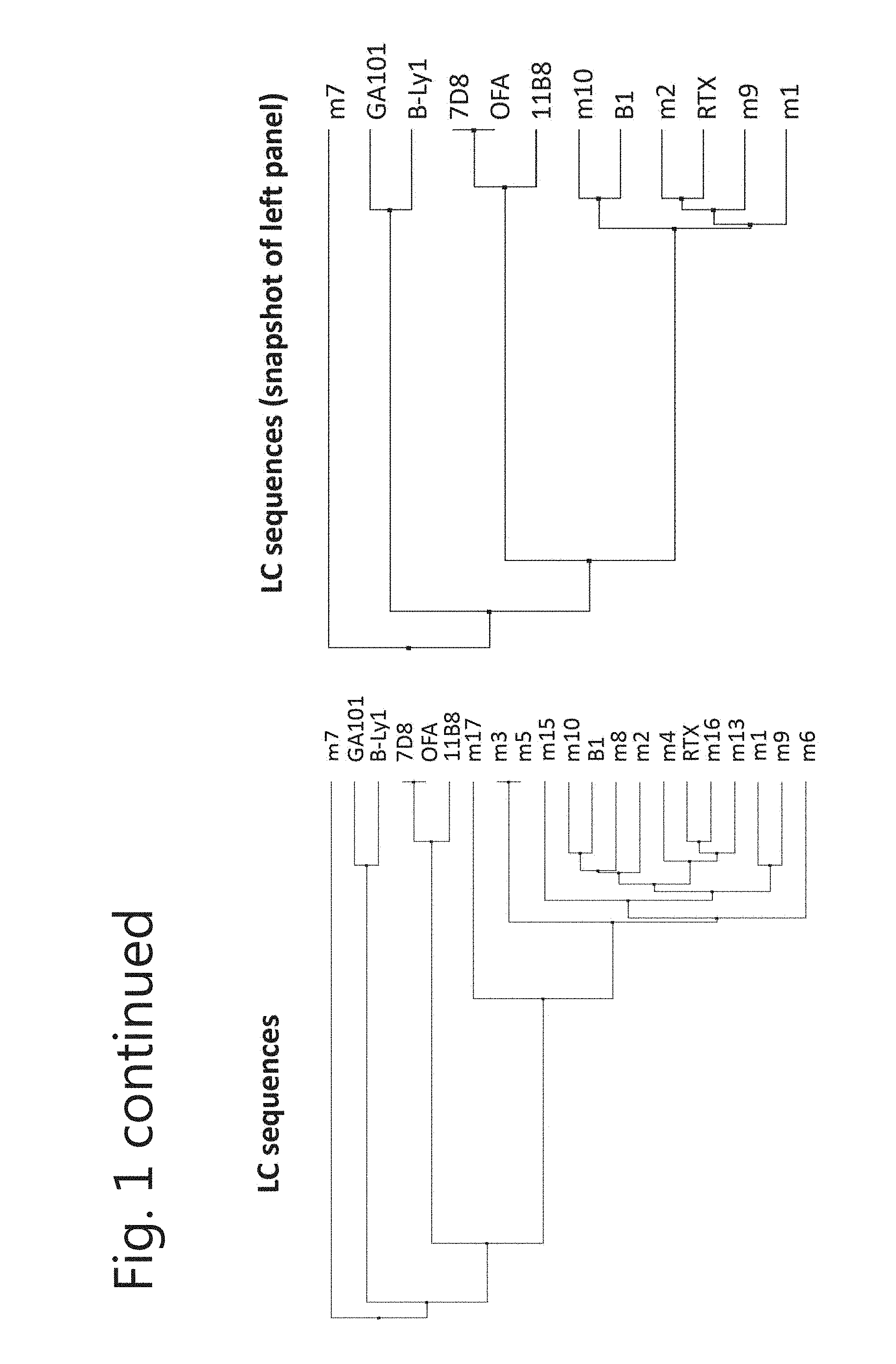
[0012] FIG. 1. Most new CD20 mAbs have unique heavy and light chain sequences. Sequences coding for the variable region of the (A) heavy chain and (B) light chain were aligned and compared with commercially available CD20 mAbs by calculating the average distance using % identity (PID).The right panel is a snapshot of the tree on the left.
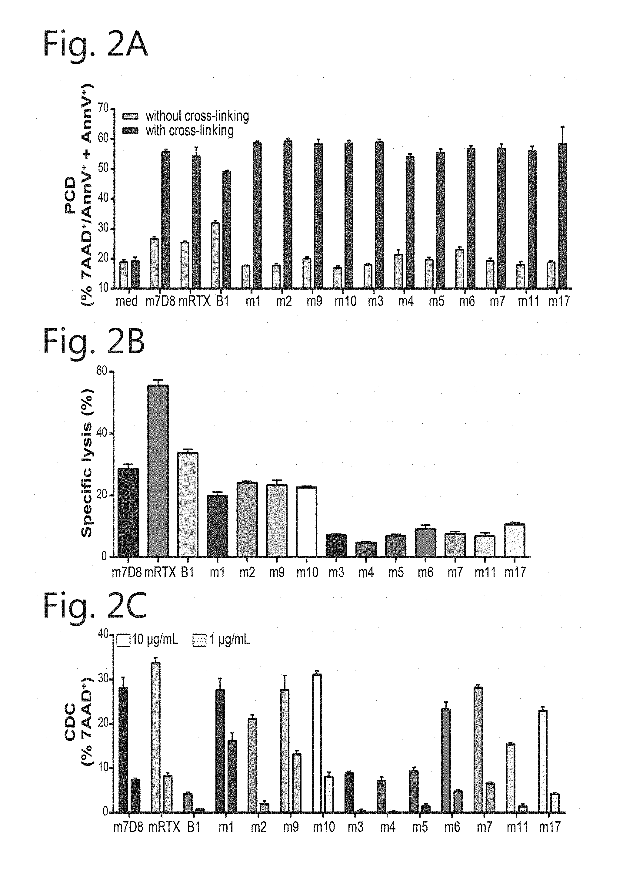
[0013] FIG. 2. As mouse antibodies, all new CD20 mouse IgG mAbs exhibit Type I characteristics. (A) PCD: EL4-CD20 cells incubated for 24 h with 1 .mu.g/mL CD20 mAbs in the absence or presence of cross-linking Ab. PCD determined by 7-AAD.sup.+/AnnexinV-PE.sup.- staining (mean+SEM). (B) ADCC: Specific lysis of Daudi cells in a chromium release assay with PBMCs as effector cells (E:T=100:1) at 1 .mu.g/mL CD20 mAb (mean+SEM). (C) CDC by new CD20 mAbs was determined at 10 and 1 .mu.g/mL mAb in 15.5% human serum and detected by 7-AAD staining ((value.sub.sample-mean.sub.medium) +SEM). mIgG2a-CD20 mAbs B1, mRTX, and m7D8 in grey were taken along as controls. mIgG2c mAbs in pink and mIgG2b mAbs in blue.
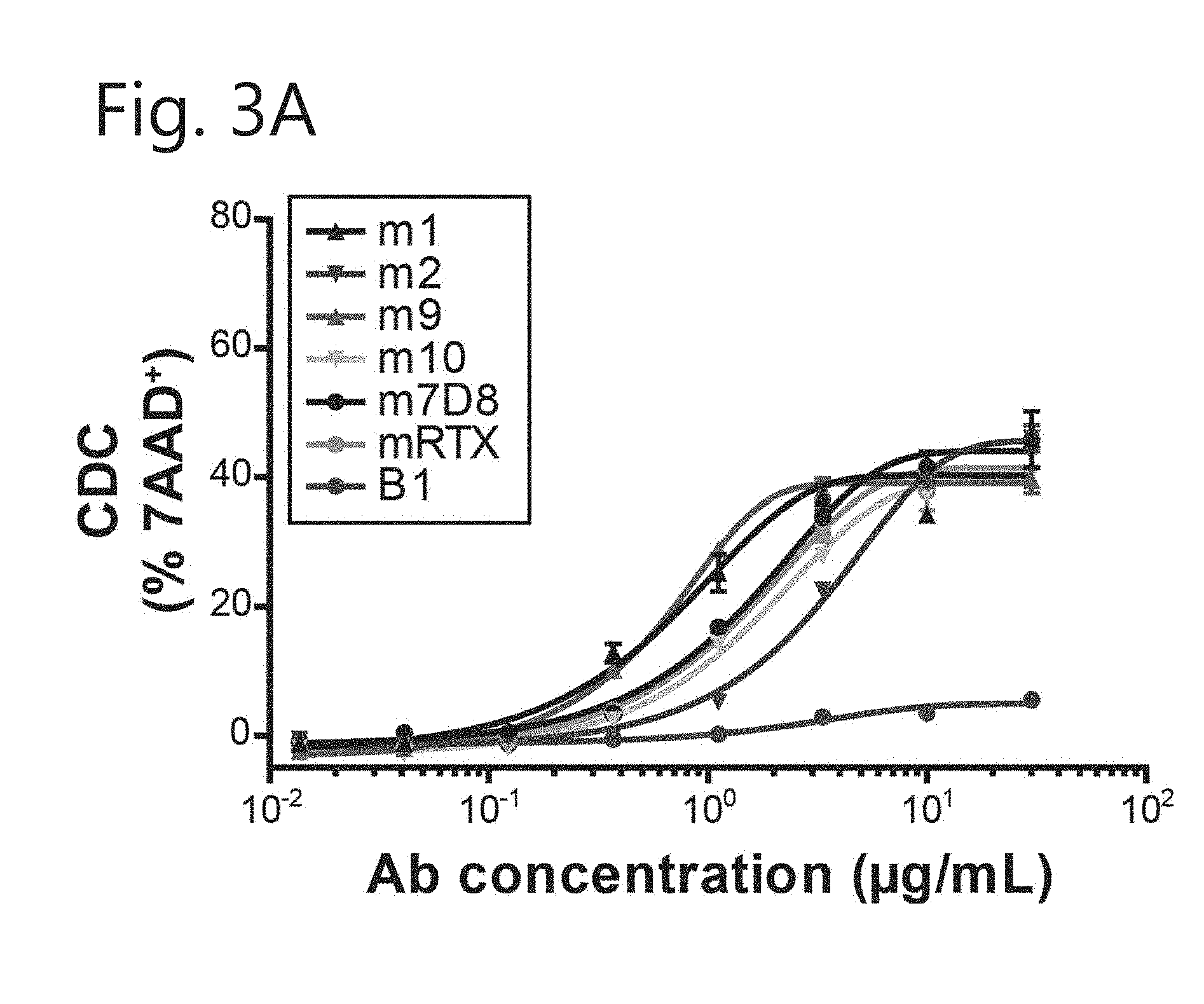
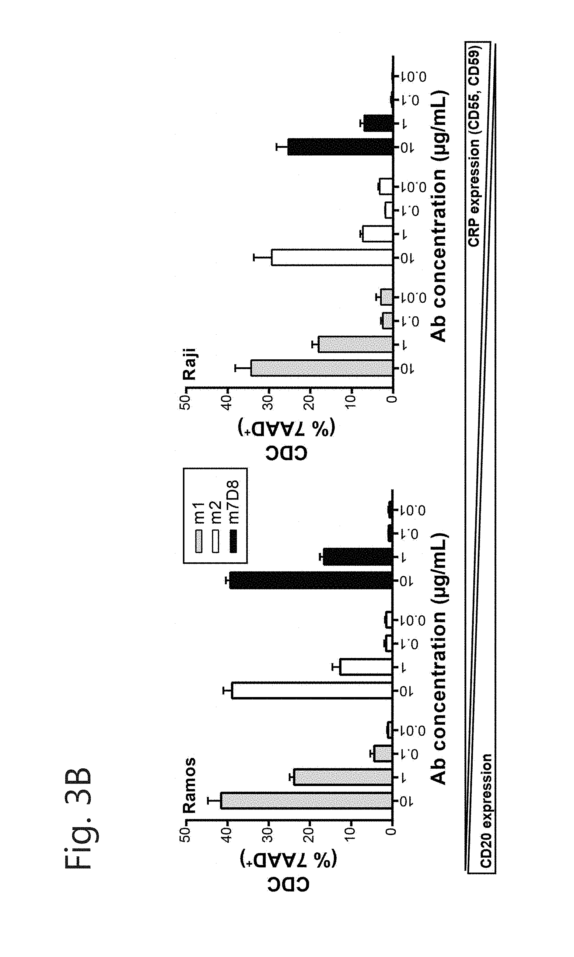
[0014] FIG. 3. New mIgG2c-CD20 mAbs display varying CDC properties. Induction of complement-dependent cytotoxicity by (A) all new mIgG2c-CD20 mAbs on Daudi cells and (B) m1, m2 and m7D8 on Ramos and Raji cells. mIgG2a-CD20 mAbs B1 and m7D8 were taken along as controls. Cells were incubated with indicated mAb concentrations and 15.5% human serum. Cytotoxicity was determined by 7-AAD staining ((value.sub.sample-mean.sub.medium)+SEM).
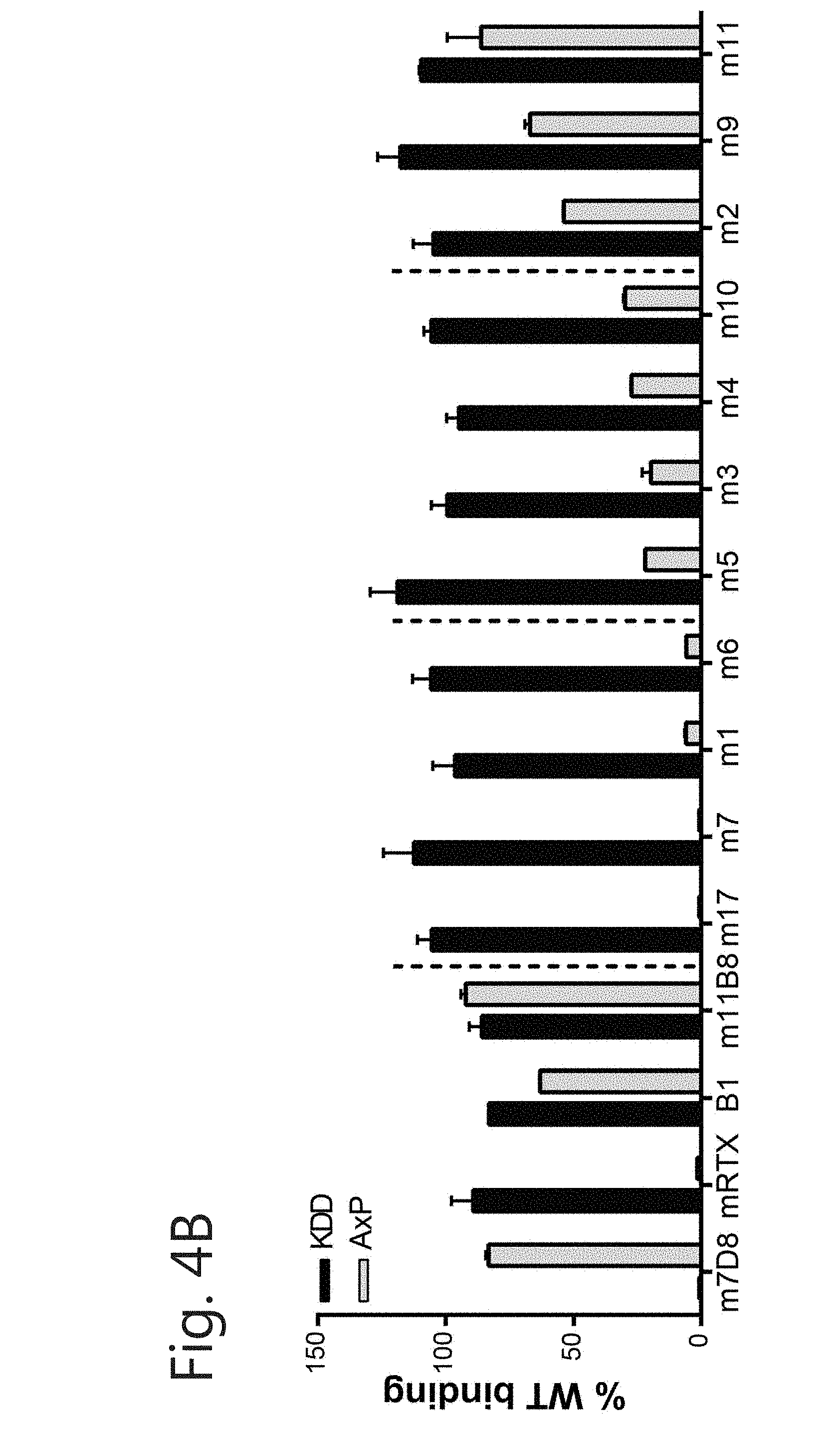
[0015] FIG. 4. Binding site on CD20 is different among new CD20 mAbs. (A) Epitope mapping using the circular peptide YNCEPANPSEKNSPSTQYCYS resulted in identification of the epitope of m1 (left) but only marginally of m2 (right). Binding of 1 .mu.g/mL mAb to the peptide and mutants with each amino acid replaced with all other available (positional scan; excluding cysteine) was determined by ELISA. Grey line and shaded area represents WT binding.+-.SEM. Results are displayed with Tukey-whiskers. (B) Rough epitope mapping with CD20 WT or CD20 mutant (KDD=T159K/N163D/N166D, AxP=A170S/P172S). HEK293F cells were transfected with plasmids and binding of mAbs (5 .mu.g/mL) to CD20 was measured by FACS. Binding was compared to Type I CD20 mAbs (m7D8 and mRTX) and Type II mAbs (B1 and m11B8). (C) Determination of residues crucial for CD20 mAb binding. Data are represented as % of best binder. Coloring according to binding compared to best binder (dark grey: 0-20%=loss of binding; grey: 21-70%=intermediate binding; light grey: 71-100% full binding).
[0016] FIG. 5. New CD20 mAbs with distinct kinetics. Real-time binding and dissociation curve to SKBR3-CD20 cells using Ligand Tracer Green. Association of 10 nM FITC-labeled CD20 mAbs was monitored for 1 h before following the dissociation for 3 h in the presence of (A) RPMI culture medium (non-competitive) or (B) 100 nM unlabeled CD20 mAbs (competitive). (C) Comparison of dissociation rate constants under non-competitive and competitive conditions determined by 1:1 Fitting model.
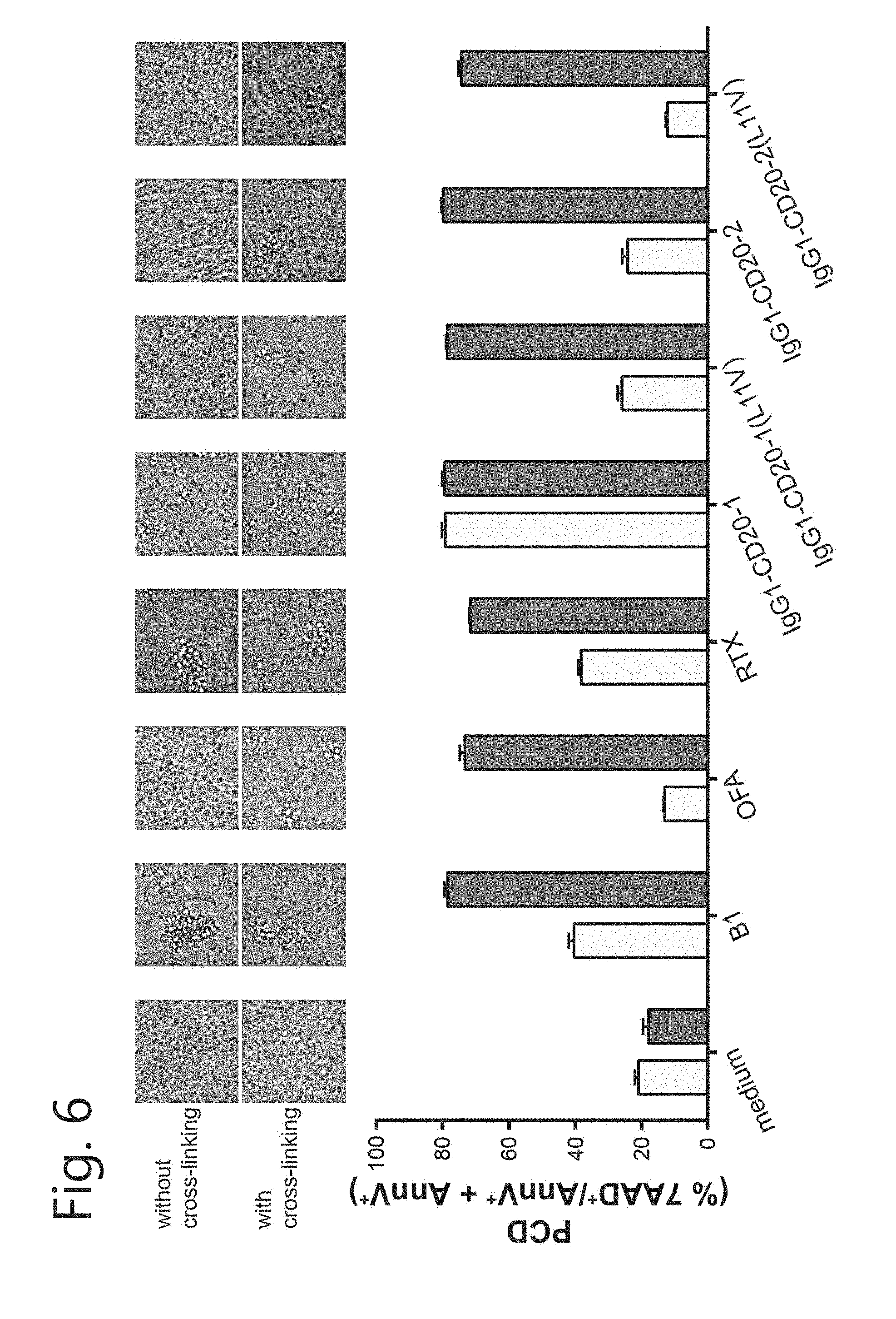
[0017] FIG. 6. Chimerization can alter PCD activity. Induction of homotypic aggregation (pictures) and PCD by IgG1-CD20-1 and IgG1-CD20-2 mAbs (1 .mu.g/mL) with or without the L11V mutation in the absence (light grey bar) or presence (dark grey bar) of cross-linking Ab (for IgG1, a-human IgG: 20 .mu.g/mL; for B1, a-mouse IgG: 50 .mu.g/mL). B1 and OFA as positive and negative control, respectively. Induction of cell death was determined by 7-AAD and AnnexinV-PE staining (mean+SEM).
[0018] FIG. 7. Differences in in vivo efficacy of mIgG2c-CD20 mAbs. C57BL/6 mice (4-6 mice/group) were injected intraperitoneally with 5.times.105 CellTraceViolet labeled EL4-CD20 cells 16 h prior to mAb or PBS treatment. The anti-tumor response was evaluated 24 h later by determining the amount of remaining tumor cells in the peritoneal lavage with TruCount tubes. (A) mAb titration of m1, m2 and m7D8 (median.+-.interquartile range). (B) Anti-tumor response by 1 .mu.g mIgG2c mAb, represented as % of PBS (median.+-.interquartile range). 1 .mu.g m7D8 (mIgG2a) as positive control. 2 separate experiments as indicated by dashed line.
[0019] FIG. 8. New chimeric IgG1-CD20 mAbs bind to CD20-expressing Daudi cells. Binding was determined by FACS after incubation of Daudi cells with a dilution series of mAb. RTX and OFA were included as positive controls, and Trastuzumab as isotype control.
[0020] FIG. 9. Analysis of in vitro efficacy of new chimeric IgG1-CD20 mAbs. (A) ADCC: Specific lysis of Daudi cells in a chromium release assay with PBMCs as effector cells (E:T=50:1) over a wider mAb concentration range (mean.+-.SEM). (B) CDC of new CD20 mAbs determined in 15.5% human serum and detected by 7-AAD staining ((value.sub.sample-mean.sub.medium) .+-.SEM). (C) PCD: EL4-CD20 cells incubated for 24 h with 1 .mu.g/mL CD20 mAbs in the absence or presence of 20 .mu.g/mL cross-linking Ab. PCD was determined by 7-AAD/AnnexinV-PE staining (mean+SEM). B1 (mIgG2a-CD20 mAb) is a positive control for PCD ((value.sub.sample-mean.sub.medium) .+-.SEM). In all assays RTX and OFA were included as positive controls, and TRA as isotype control. PCD induction on (D, E) Ramos cells and (F, G) Daudi cells by IgG1-CD20-1 without or with the L11V mutation (10 .mu.g/ml ). Induction of cell death was determined by (D, F) 7-AAD/AnnexinVPE staining and (E, G) DiOC6/TO-PRO-3 staining (mean+SEM). 11B8 and OFA were taken along as positive and negative control, respectively. Results are representative of 3 separate assays. *p<0.05; **p<0.01; ***p<0.001, by one way ANOVA followed by Bonferroni posthoc analysis.
[0021] FIG. 10. Disappearance of CD19+ viable B cells in autologous setting. B-cell depletion by CD20 mAbs was determined by incubating mAbs for 1 h at 37.degree. C. with unprocessed blood from healthy donors. (A) FACS plots showing disappearance of viable B cells upon incubation with mAb (here IgG1-CD20-1) compared to PBS control. (B) B-cell depletion mediated by CD20 mAbs determined over a broader concentration range (mean.+-.SEM). RTX and OFA were included as positive controls, and TRA as isotype control.
[0022] FIG. 11. In vivo efficacy of IgG1-CD20 mAbs. C57BL/6 mice (6 mice/group) were injected intraperitoneally with 5.times.10.sup.5 CellTraceViolet labeled EL4-CD20 cells 16 h prior to mAb (10 .mu.g) or PBS treatment. The anti-tumor response was evaluated 24 h later by determining the amount of remaining tumor cells in the peritoneal lavage with TruCount tubes (median.+-.interquartile range).
[0023] FIG. 12: Production of IgA-CD20 mAbs. (A) Test transfection of HEK293F cells to determine optimal ratio between HC, LC and pAdvantage coding plasmids. Concentrations of produced IgA-CD20 mAbs were measured by an IgA specific ELISA. Large scale produced IgA-CD20 mAbs were purified by (A) anti-human kappa affinity chromatography, followed by (B) size-exclusion chromatography to separate the full size antibody from loose light chains and aggregates. Representative graphs are displayed.
[0024] FIG. 13: Binding of IgA-CD20 mAbs to CD20-expressing Ramos cells. Binding was determined by FACS after incubation of Ramos cells with a dilution series of mAb.
[0025] FIG. 14: Variable extent of programmed cell death induction by IgA-CD20 mAbs. EL4-CD20 cells were incubated for 24 h with 1 .mu.g/mL mAb in the absence and presence of cross-linking Ab (B1: 50 .mu.g/mL; IgG1 and IgA mAbs: 20 .mu.g/mL). The degree of cell death was determined as the sum of AnnexinV.sup.+ and AnnexinV.sup.+/7AAD.sup.+ cells (value.sub.sample-mean.sub.medium+SEM). B1 (mIgG2a-CD20 mAb) and RTX were taken along as positive controls and OFA as a negative control.
[0026] FIG. 15: Complement-dependent cytotoxicity induced by IgA-CD20 mAbs. Target cells were incubated for indicated time with 15.5% pooled human serum and degree of CDC was determined as % 7AAD.sup.+ cells. (A,B) Time dependency of complement induction by IgA-CD20 mAbs. Complement mediated lysis of (A) Daudi and (B) Ramos cells incubated for 15, 60, 240, and 360 min in the presence of 10 .mu.g/mL mAb. (C) The degree of complement-mediated lysis of Ramos cells after 15 min incubation with 10 .mu.g/mL mAb was inhibited by pre-treatment of the complement source with the indicated inhibitors (heat inactivated serum; excess of eculizumab; EDTA+MgCl.sub.2). Results are shown as (value.sub.sample-mean.sub.medium)+SEM.
[0027] FIG. 16: B-cell depletion assay in autologous setting. Whole blood leukocytes were incubated with CD20 mAbs for 3 h at 37.degree. C. Analysis was performed on FSC/SSC lymphocyte gate from which CD3, CD14, CD56 and CD11b positive cells were excluded. CD19 was used as B-cell marker. (A) Analysis of CD19 expression on CD19.sup.+ cells (B cells). (B) Number of CD19.sup.- events found in lymphocyte gate. (C) Number of CD19.sup.- cells found in lymphocyte gate after background (no antibody) subtraction.
[0028] FIG. 17. Anti-CD20 IgA mediates efficient tumor cell lysis of CD20 targets. ADCC as measured by the release of .sup.51Cr from EL4-CD20-Luc2 cells using (A) PBMC, E:T=100:1, or (B) PMN, E:T=40:1, as effector cells and the indicated antibody concentrations. Anti-CD20 IgA's contain the same variable regions as rituximab.
[0029] FIG. 18. Amino acid sequence of various VH and VL chains. CDR sequences are underlined from left to right in the sequence CDR1, CDR2 and CDR3.
[0030] The positions of the CDRs in the VH were determined using the following criteria:
[0031] CDR-H1 [0032] Start-Approx residue 26 (always 4 after a CYS) [Chothia/AbM defintion]. Kabat definition starts 5 residues later. Residues before always CYS-XXX-XXX-XXX. Residues after always a TRP. Typically TRP-VAL, but also, TRP-ILE, TRP-ALA. Length 10 to 12 residues (AbM definition) Chothia definition excludes the last 4 residues.
[0033] CDR-H2 [0034] Start-always 15 residues after the end of Kabat/AbM definition of CDR-H1. Residues before typically LEU-GLU-TRP-ILE-GLY, but a number of variations. Residues after LYS/ARG-LEU/ILE/VAL/PHE/THR/ALA-THR/SER/ILE/ALA. Length Kabat definition 16 to 19 residues.
[0035] CDR-H3 [0036] Start-always 33 residues after end of CDR-H2 (always 2 after a CYS). Residues before always CYS-XXX-XXX (typically CYS-ALA-ARG). Residues after always TRP-GLY-XXX-GLY. Length 3 to 25(!) residues.
[0037] The positions of the CDRs in the VL were determined using the following criteria:
[0038] CDR-L1 [0039] Start-Approx residue 24. Residue before is always a Cys. Residue after is always a Trp. Typically TRP-TYR-GLN, but also, TRP-LEU-GLN, TRP-PHE-GLN, TRP-TYR-LEU. Length 10 to 17 residues.
[0040] CDR-L2 [0041] Start-always 16 residues after the end of L1. Residues before generally ILE-TYR, but also, VAL-TYR, ILE-LYS, ILE-PHE. Length always 7 residues.
[0042] CDR-L3
[0043] Start-always 33 residues after end of L2. Residue before is always Cys. Residues after always PHE-GLY-XXX-GLY. Length 7 to 11 residues.
[0044] FIG. 19. Binding of mIgG-CD20 mAbs to Daudi cells.
[0045] FIG. 20. No induction of HA of EL4-CD20 cells by new CD20 mAbs (1 .mu.g/mL) (20.times. magnification). Cross-linking Ab (50 .mu.g/mL) was added as positive control conditions. B1 as positive control (Type II).
[0046] FIG. 21. Epitope mapping using the linear peptide NSPSTQYGPANPSEK resulted in identification of aa contributing to binding of (A) m 1 but not (B) m2. Binding of 1 .mu.g/mL mAb to the peptide and corresponding mutants with each aa replaced with all other available (positional scan; excluding cysteine) was determined by ELISA. Grey line and shaded area=WT binding.+-.SEM; Results are displayed with Tukey-whiskers.
[0047] FIG. 22. (A,B) FACS based dissociation analysis. Daudi cells were stained with 10 .mu.g/mL Alexa647-labeled CD20 mAb. Cells were left in (A) RPMI culture medium (non-competitive) or (B) the presence of a 10-fold excess of unlabeled mAb (100 .mu.g/mL; competitive). At indicated time points the level of cell-bound mAb was determined. (C,D) Real-time binding and dissociation curve to SKBR3-CD20 cells using the Ligand Tracer technology. Association of 10 nM FITC-labeled CD20 mAbs was monitored for 4 h before following the dissociation for 8 h in the presence of (C) RPMI culture medium (non-competitive) or (D) 100 nM unlabeled CD20 mAbs of the same clone (competitive).
[0048] FIG. 23. Summarizing overview of molecular determinants of existing and novel CD20 mAbs. Three distinct molecular determinants were described to determine the MoA of CD20 mAbs: 1) epitope (positions of several CD20 antibodies are indicated); 2) kinetics (k.sub.off of the CD20 antibodies were ranked from high (10.sup.-5) to low (10.sup.-6), and 3) elbow angle (commercially available CD20 mAbs (OFA, RTX and OBZ) were grouped in wide and narrow angle according to literature. The amino acid at Kabat position 11 was described to influence the angle, and based on this our new chimeric mAbs were ordered according to the residue).
[0049] FIG. 24. Amino acid sequence of suitable IgG1, IgA1 and IgA2 heavy and light chain constant regions. [0050] SEQ ID NO: 3 Heavy chain IgG1 constant region CH1-3 and Hinge [0051] SEQ ID NO: 4 Heavy chain IgA1 constant region CH1-3 and Hinge [0052] SEQ ID NO: 5 Heavy chain IgA2 constant region CH1-3 and Hinge [0053] SEQ ID NO: 6 light chain constant region [0054] FIG. 25. Internalization of CD20 from B cells at 1 ug of antibody. The number in the UMAB . . . reference in the figures refer to the variable domains of antibody m . . . with the respective numbers and the constant region as indicated.
[0055] FIG. 26. Titration range of antibodies in CD20 internalization of B cells.
[0056] FIG. 27. Analysis of in vitro efficacy of new chimeric IgA-CD20 mAbs. (A) ADCC of IgA1-CD20 antibodies: Specific lysis of Daudi cells in a chromium release assay with PMNs as effector cells (E:T=40:1). (B) ADCC of IgA2-CD20 antibodies: Specific lysis of Daudi cells in a chromium release assay with PMNs as effector cells (E:T=40:1).
[0057] FIG. 28. In vivo efficacy of IgA-CD20 mAbs. C57BL/6 mice (6 mice/group) were injected intraperitoneally with 5.times.10e5 CellTraceViolet labeled EL4-CD20 cells 16 h prior to mAb (10 .mu.g) or PBS treatment. The anti-tumor response was evaluated 24 h later by determining the amount of remaining tumor cells in the peritoneal lavage with TruCount tubes (median.+-.interquartile range).
[0058] FIG. 29. Loss of CD20 expression occurs after IgG treatment, but not after IgA treatment in vivo. C57BL/6 mice (6 mice/group) were injected intraperitoneally with 5.times.10e5 CellTraceViolet labeled EL4-CD20 cells 16 h prior to mAb (10 .mu.g) or PBS treatment. Subsequently, CD20 expression was determined on these cells by flow cytometric analysis.
[0059] FIG. 30. CD19/CD24+ events with antibodies having a variable domain of the indicated antibody and the indicated constant regions, IgG1, IgA1 or IgA2.
SUMMARY OF THE INVENTION
[0060] The invention provides an antibody comprising a mouse IgG2; a human IgG1, IgA1 or IgA2 constant region and a variable domain that can bind the epitope "EPANpSEK" on human CD20 expressed on Ramos cells and which antibody has an increased PCD functionality when compared to Rituximab with a constant region of the same isotype.
[0061] The invention also provides an antibody that can bind to an extracellular part of human CD20 expressed on Ramos cells comprising a variable domain with a heavy chain variable region and a light chain variable region characterized in that the heavy chain variable region comprises a CDR3 region with the sequence SNSYGSTYWYFDV.
[0062] The invention further provides an antibody comprising a mouse IgG2; a human IgG1, IgA1 or IgA2 constant region and a variable domain that can bind the epitope "EPANPsEK" on human CD20 expressed on Ramos cells and which antibody has an increased ADCC functionality when compared to Rituximab with a constant region of the same isotype.
[0063] Also provided is an antibody that can bind to an extracellular part of human CD20 expressed on Ramos cells, the antibody comprising a variable domain with a heavy chain variable region and a light chain variable region characterized in that the heavy chain variable region comprises a CDR3 region with the sequence YYYGSSYGAMDY.
[0064] Further provided is an antibody comprising a mouse IgG2; a human IgG1, IgA1 or IgA2 constant region and a variable domain that can bind the epitope "EPANpsEK" on human CD20 expressed on Ramos cells and which antibody has an increased CDC functionality when compared to Rituximab with a constant region of the same isotype.
[0065] Also provided is an antibody that can bind to an extracellular part of human CD20 expressed on Ramos cells comprising a variable domain with a heavy chain variable region and a light chain variable region characterized in that the heavy chain variable region comprises a CDR3 region with the sequence TYYYGSSPYWSFDV.
[0066] Also provided is an antibody that can bind to an extracellular part of human CD20 expressed on Ramos cells comprising a variable domain with a heavy chain variable region and a light chain variable region characterized in that the heavy chain variable region comprises a CDR3 region with the sequence SRLFDSSYGWYFDV.
[0067] Further provided is an antibody comprising a mouse IgG2; a human IgG1, IgA1 or IgA2 constant region and a variable domain that can bind the epitope "EPANpSEK" on human CD20 expressed on Ramos cells and which antibody has an increased CDC and/or increased ADCC functionality when compared to Rituximab with a constant region of the same isotype.
[0068] Also provided is an antibody that can bind to an extracellular part of human CD20 expressed on Ramos cells comprising a variable domain with a heavy chain variable region and a light chain variable region characterized in that the heavy chain variable region comprises a CDR3 region with the sequence SAYYGSNVWFFDV.
[0069] Further provided are antibodies as described herein for use in the treatment of a disease in an individual.
[0070] Also provided are antibodies as described herein for use in the treatment of a disease that involves too many B cells, overactive B cells, and/or dysfunctional B cells.
[0071] Also provided are antibodies as described herein for use in the treatment of a CD20 positive neoplasm such as a CD20 positive B-cell lymphoma; hairy cell leukemia; B-cell chronic lymphocytic leukemia, or melanoma.
[0072] Also provided are methods for the treatment of an individual that has a disease that involves too many B cells, overactive B cells, and/or dysfunctional B cells comprising administering to the individual in need thereof an antibody as described herein.
[0073] Also provided are methods for the treatment of an individual that has a CD20 positive neoplasm such as a CD20 positive B-cell lymphoma; hairy cell leukemia; B-cell chronic lymphocytic leukemia, or melanoma comprising administering to the individual in need thereof an antibody as described herein.
[0074] Also provided are methods for the treatment of children with B-cell malignancies and pediatric leukemia patients that have a B-cell disease after stem cell transplantation. In pediatric patients, long-term adverse effects of rituximab are noted: permanent depletion of B cells and inability of naive B cells to switch to memory B cells, resulting in life-long immunoglobulin depletion. The long persistence of IgG in the body is probably accountable. An IgA antibody described herein has a stronger ADCC function when compared to an IgG antibody comprising the same variable domain. This is apparent when a B-cell specific marker is analyzed that is not subject to trogocytosis upon incubation of the cell with a CD20 antibody. An IgA antibody described herein has a short half-life when compared to an IgG antibody comprising the same variable domain. The IgA antibodies as described herein cause fewer side effects when compared to an IgG antibody with the same variable domain. In short, the IgA antibodies of the invention facilitate an effective hit but are also cleared fast enough to allow a good recovery of the B-cell repertoire. This is particularly helpful in preserving the B-cell repertoire following recovery from the treatment, particularly in the mentioned leukemia patients that have a B-cell disease after stem cell transplantation treated with an antibody of the invention and pediatric patients after B-cell depletion with an antibody of the invention.
[0075] The invention further provides a variable domain comprising the amino acid sequence of the heavy and light chain variable regions of SEQ ID NO: 1 and 2, each with 0-5 amino acid insertions, deletions, substitutions, additions or a combination thereof at one or more positions other than positions of the amino acids that constitute the CDR1, CDR2 and CDR3 regions.
[0076] The invention further provides a variable domain comprising the amino acid sequence of the heavy and light chain variable regions of SEQ ID NO: 7 and 8 each with 0-5 amino acid insertions, deletions, substitutions, additions or a combination thereof at one or more positions other than positions of the amino acids that constitute the CDR1, CDR2 and CDR3 regions.
[0077] The invention further provides a variable domain comprising the amino acid sequence of the heavy and light chain variable regions of SEQ ID NO: 9 and 10 each with 0-5 amino acid insertions, deletions, substitutions, additions or a combination thereof at one or more positions other than positions of the amino acids that constitute the CDR1, CDR2 and CDR3 regions.
[0078] The invention further provides a variable domain comprising the amino acid sequence of the heavy and light chain variable regions of SEQ ID NO: 11 and 12 each with 0-5 amino acid insertions, deletions, substitutions, additions or a combination thereof at one or more positions other than positions of the amino acids that constitute the CDR1, CDR2 and CDR3 regions.
[0079] The invention further provides a variable domain comprising the amino acid sequence of the heavy and light chain variable regions of SEQ ID NO: 13 and 14 each with 0-5 amino acid insertions, deletions, substitutions, additions or a combination thereof at one or more positions other than positions of the amino acids that constitute the CDR1, CDR2 and CDR3 regions. Further provided is an antibody comprising a variable domain as specified herein.
[0080] Further provided is [0081] a nucleic acid molecule that codes for a heavy chain or light chain of an antibody as described herein; [0082] a nucleic acid molecule that codes for the CDR3 of a heavy or light chains of an antibody as described herein; [0083] a nucleic acid molecule that codes for a CDR1, CDR2 and CDR3 of the heavy or light chain of an antibody as described herein; and [0084] a nucleic acid molecule that codes for a variable region of a heavy chain or of a light chain of an antibody as described herein.
[0085] Further provided is a cell that comprises nucleic acid that codes for an antibody as described herein.
[0086] Further provided are means and methods for the production of an antibody as described herein.
DETAILED DESCRIPTION
[0087] The invention is concerned with antibodies that bind CD20. The CD20 protein is also known under various other names such as Membrane Spanning 4-Domains A1; MS4A1; Membrane-Spanning 4-Domains Subfamily A Member 1; Leukocyte Surface Antigen Leu-16; CD20 Antigen; Bp35; B-Lymphocyte Cell-Surface Antigen B1; B-Lymphocyte Surface Antigen B1; CD20 Receptor; LEU-16; CVID5; MS4A2; B1; and S7. External Ids for MS4A1 are HGNC: 7315; Entrez Gene: 931; Ensembl: ENSG00000156738; OMIM: 112210 and UniProtKB: P11836.
[0088] Some of the names may or may not have also been used to refer to other proteins than CD20. The names and sequence identifiers are given for reference purposes only. An antibody of the invention binds to CD20 as expressed on Ramos cells but also to other CD20 molecules as long as the epitope to which the antibody binds is available. Thus splicing variants or mutant CD20 molecules (if any) will also be bound by an antibody of the invention as long as the epitope is available. The fact that the antibody binds to CD20 means that the antibody can bind to CD20 and does not imply that the antibody is actually bound to CD20. It also does not mean that the antibody does not bind to other proteins. Such cross-reactivity is at present not known for an antibody of the present invention, however, it is not expressly excluded that such cross-reactivity may exist.
[0089] An antibody (Ab), also known as an immunoglobulin (Ig), is a large, typically Y-shaped protein. An antibody interacts with various components of the immune system. Some of the interactions are mediated by its Fc region (located at the base of the "Y"), which contains site(s) involved in these interactions.
[0090] Antibodies are proteins belonging to the immunoglobulin superfamily. They typically have two heavy chains and two light chains. There are several different types of antibody heavy chains that define the five different types of crystallisable fragments (Fc) that may be attached to the antigen-binding fragments. The five different types of Fc regions allow antibodies to be grouped into five isotypes. An Fc region of a particular antibody isotype is able to bind to its specific Fc receptor (FcR) thus allowing the antigen-antibody complex to mediate different roles depending on which FcR it binds. The ability of an IgG antibody to bind to its corresponding FcR is modulated by the presence/absence of interaction sites and the structure of the glycan(s) (if any) present at sites within its Fc region. The ability of antibodies to bind to FcRs helps to direct the appropriate immune response for each different type of foreign object they encounter.
[0091] Though the general structure of all antibodies is similar, a region at the tip of the protein is extremely variable, allowing millions of antibodies with slightly different tip structures, or antigen-binding sites, to exist. This region is known as the hypervariable region. The enormous diversity of antigen binding by antibodies is largely defined by the hypervariable region and the variable domain containing the hypervariable region.
[0092] An antibody of the invention is typically a full-length antibody. The term "full length antibody" is defined as comprising an essentially complete immunoglobulin molecule, which however does not necessarily have all functions of an intact immunoglobulin. For the avoidance of doubt, a full length antibody has two heavy and two light chains. Each chain contains constant (C) and variable (V) regions. A heavy chain of a full length antibody typically comprises a CH1, a CH2, a CH3, a VH region and a hinge region. A light chain of a full length antibody typically comprises a CL region and a VL region.
[0093] An antibody binds to antigen via the variable region domains contained in the Fab portion. An antibody variable domain comprises a heavy chain variable region and a light chain variable region. Full length antibodies according to the invention encompass heavy and light chains wherein mutations may be present that provide desired characteristics. Full length antibodies should not have deletions of substantial portions of any of the regions. However, IgG molecules wherein one or several amino acid residues are substituted, inserted, deleted or a combination thereof, without essentially altering the antigen binding characteristics of the resulting antibody, are embraced within the term "full length" antibody. For instance, a "full length" antibody can have a substitution, insertion, deletion or a combination thereof, of between 1 and 10 (inclusive) amino acid residues, preferably in non-CDR regions, wherein the deleted amino acids are not essential for the binding specificity of the antibody.
[0094] The epitope that is recognized by an antibody of the invention, and/or minor contributing amino acids therein were determined by, among others, positional amino acid scan wherein the amino acid was replaced by every other natural amino acid in a peptide containing the epitope and by a mutant screen of the CD20 protein expressed on cells. The contribution of an amino acid to the binding of an antibody to an epitope is preferably determined by comparing the binding to a peptide comprising the epitope as such and the same peptide but with an alanine at the position of the analyzed amino acid. An amino acid is relevant to the binding of the antibody to the protein when a replacement with an alanine in the protein results in a decrease of binding of the antibody to 0-70% relative to the unmodified protein. This is also referred to a reduction of binding. A decrease to 0-20% if the binding relative to the unmodified protein is regarded as loss of binding and a decrease to 21-70% relative to the unmodified protein is regarded as intermediate binding. The thus identified amino acids are considered to be a major contributor or an intermediate contributor to the binding of the antibody to the protein. A binding of 71-100% was regarded as full binding. The amino acid concerned is not regarded to contribute significantly to the binding of the antibody to the protein.
[0095] One of the antibodies provided by the invention is an antibody (A) comprising a mouse IgG2; a human IgG1, IgA1 or IgA2 constant region and a variable domain that can bind the epitope "EPANpSEK" on human CD20 expressed on Ramos cells and which antibody has an increased PCD functionality when compared to Rituximab with a constant region of the same isotype. The antibody preferably further comprises a comparable or an increased CDC functionality when compared to Rituximab with a constant region of the same isotype. Preferably the ADCC functionality of the antibody is comparable or reduced when compared to Rituximab with a constant region of the same isotype. The epitope on CD20 that is bound by the antibody is "EPANpSEK". A capital letter, small case letter and bold indicates the relevance of the amino acid for binding of the antibody to the peptide. A bold letter indicates that the amino acid is a major contributor to the binding of the antibody; a small case letter indicates that the amino acid has an intermediate contribution to the binding and a capital letter in plain text indicates that the amino acid has a small or not detectable contribution to the binding of the antibody to the peptide. The antibody binds 20% or less to a CD20 protein wherein one or more of the amino acids N or S in "EPANpSEK" have been replaced by an alanine, where the binding is compared to the binding of the antibody an unmodified CD20 protein.
[0096] Also provided is an antibody (A1) that can bind to an extracellular part of human CD20 expressed on Ramos cells comprising a variable domain with a heavy chain variable region and a light chain variable region characterized in that the heavy chain variable region comprises a CDR3 region with the sequence SNSYGSTYWYFDV. The antibody (A1) has an increased PCD functionality when compared to Rituximab with a constant region of the same isotype. The antibody preferably further comprises a comparable or an increased CDC functionality when compared to Rituximab with a constant region of the same isotype. Preferably the ADCC functionality of the antibody is comparable or reduced when compared to Rituximab with a constant region of the same isotype. The heavy chain variable region preferably comprises a CDR1, CDR2 and CDR3 region with the sequence SYNLH, AIYPGNGDTSYNQKFKG and SNSYGSTYWYFDV respectively. Preferably the heavy chain variable region comprises the sequence of SEQ ID NO: 1, with 0-5 amino acid insertions, deletions, substitutions, additions or a combination thereof at one or more positions other than positions of the amino acids that constitute the CDR1, CDR2 and CDR3 regions, wherein SEQ ID NO: 1 has the sequence
TABLE-US-00001 QAYLQ QSGAE LVRPG ASVKM SCKAS GYTFT SYNLH WVKQT PRQGL EWIGA IYPGN GDTSY NQKFK GKATL TVDKS SSTAY MQLSR LTSED SAVYF CARSN SYGST YWYFD VWGTG TTVTV SS.
[0097] The light chain variable region of the antibody (A1) preferably comprises the sequence of SEQ ID NO: 2, with 0-5 amino acid insertions, deletions, substitutions, additions or a combination thereof at one or more positions other than positions of the amino acids that constitute the CDR1, CDR2 and CDR3 regions, wherein SEQ ID NO: 2 has the sequence
TABLE-US-00002 QIVLS QSPAV LFASP GEKVT MTCRA RSSVS YMDWY QQKPR SSPKP WIYAT SNLAS GVPAR FSGSG SGTSY SLTIS RVEAE DAATY YCQQW TSNPP TFGSG TKLEI KRADA APTVS IFPPS S.
[0098] The antibody A1 preferably comprises a mouse IgG2; a human IgG1, IgG2, IgG3, IgG4, IgM, IgE, IgA heavy chain constant region or a combination thereof. Preferably it comprises a human IgG1, IgG2, IgA1 or IgA2 heavy chain constant region or a combination thereof. Preferably it comprises a human IgG1 constant region. In preferred embodiment the heavy chain constant region is a human IgA1 or human IgA2 heavy chain constant region, preferably a human IgA2; more preferably a human IgA2m, preferably an IgA2m1 or IgA2m2, preferably IgA2m1 heavy chain constant region. In another preferred embodiment the antibody comprises a murine IgG2 region, preferably a IgG2c constant region.
[0099] In a preferred embodiment the antibody A is an A1 antibody.
[0100] The antibody A or A1 preferably comprises a heavy chain and a light chain wherein the heavy chain comprises the sequence of SEQ ID NO: 1 and the sequence of SEQ ID NO: 3, 4 or 5 with 0-15 amino acid insertions, deletions, substitutions, additions or a combination thereof at one or more positions other than positions of the amino acids that constitute the CDR1, CDR2 and CDR3 regions.
[0101] The antibody A or A1 preferably comprises a light chain comprising the sequence of SEQ ID NO: 2 and the sequence of SEQ ID NO: 6 with 0-15 amino acid insertions, deletions, substitutions, additions or a combination thereof at one or more positions other than positions of the amino acids that constitute the CDR1, CDR2 and CDR3 regions.
[0102] The invention also provides an antibody (B) comprising a mouse IgG2; a human IgG1, IgA1 or IgA2 constant region and a variable domain that can bind the epitope "EPANPsEK" on human CD20 expressed on Ramos cells and which antibody has an increased ADCC functionality when compared to Rituximab a constant region of the same isotype. The epitope in CD20 that is bound by the antibody is "EPANPsEK". A capital letter, small case letter and bold indicates the relevance of the amino acid for binding of the antibody to the peptide. A bold letter indicates that the amino acid is a major contributor to the binding of the antibody; a small case letter indicates that the amino acid has an intermediate contribution to the binding and a capital letter in plain text indicates that the amino acid has a small or not detectable contribution to the binding of the antibody to the peptide. The antibody binds 20% or less to a CD20 protein wherein the amino acid N in "EPANpSEK" has been replaced by an alanine, whereby the binding is compared to the binding of the antibody an unmodified CD20 protein.
[0103] Also provided is an antibody (B1) that can bind to an extracellular part of human CD20 expressed on Ramos cells, the antibody comprising a variable domain with a heavy chain variable region and a light chain variable region characterized in that the heavy chain variable region comprises a CDR3 region with the sequence YYYGSSYGAMDY. The antibody B1 has an increased ADCC functionality when compared to Rituximab with the same isotype constant region. The heavy chain variable region preferably comprises a CDR1, CDR2 and CDR3 region with the sequence SYNMH, GIYPGNGDTSYNQKFKG and YYYGSSYGAMDY respectively.
[0104] Preferably, the heavy chain variable region comprises the sequence of SEQ ID NO: 7, with 0-5 amino acid insertions, deletions, substitutions, additions or a combination thereof at one or more positions other than positions of the amino acids that constitute the CDR1, CDR2 and CDR3 regions, wherein SEQ ID NO: 7 has the sequence
TABLE-US-00003 QAYLQ QSGAE LVRPG ASVKM SCKAS GYTFT SYNMH WVKQT PRQGL EWIGG IYPGN GDTSY NQKFK GKATL TVDKS SSTAY MQLSS LTSED SAVYF CARYY YGSSY GAMDY WGQGT SVTVS S.
[0105] The light chain variable region of the antibody B1 preferably comprises the sequence of SEQ ID NO: 8, with 0-5 amino acid insertions, deletions, substitutions, additions or a combination thereof at one or more positions other than positions of the amino acids that constitute the CDR1, CDR2 and CDR3 regions, wherein SEQ ID NO: 8 has the sequence
TABLE-US-00004 QIVLS QSPAI LSASP GEKVT MTCRA SSSVS YMHWY QQKPG SSPKP WIYAT SNLAS GVPAR FSGSG SGTSY SLTIS RVEAA DAATY YCHQW TFNPP TFGGG TKLEI KRADA APTVS IFPPS S.
[0106] The antibody B1 preferably comprises a mouse IgG2; a human IgG1, IgG2, IgG3, IgG4, IgM, IgE, IgA heavy chain constant region or a combination thereof. Preferably, it comprises a mouse IgG2; a human IgG1, IgG2, IgA1 or IgA2 heavy chain constant region or a combination thereof. Preferably it comprises a human IgG1 constant region. In preferred embodiment the heavy chain constant region is a human IgA1 or human IgA2 heavy chain constant region, preferably a human IgA2; more preferably a human IgA2m, preferably an IgA2m1 or IgA2m2, preferably IgA2m1 heavy chain constant region. In another preferred embodiment the antibody comprises a murine IgG2 region, preferably a IgG2c constant region.
[0107] In a preferred embodiment the antibody B is a B1 antibody.
[0108] The antibody B or B1 preferably comprises a heavy chain and a light chain wherein the heavy chain comprises the sequence of SEQ ID NO: 7 and the sequence of SEQ ID NO: 3, 4 or 5 with 0-15 amino acid insertions, deletions, substitutions, additions or a combination thereof at one or more positions other than positions of the amino acids that constitute the CDR1, CDR2 and CDR3 regions.
[0109] The antibody B or B1 preferably comprises a light chain comprising the sequence of SEQ ID NO: 8 and the sequence of SEQ ID NO: 6.with 0-15 amino acid insertions, deletions, substitutions, additions or a combination thereof at one or more positions other than positions of the amino acids that constitute the CDR1, CDR2 and CDR3 regions.
[0110] The invention also provides an antibody (C) comprising a mouse IgG2; a human IgG1, IgA1 or IgA2 constant region and a variable domain that can bind the epitope "EPANpsEK" on human CD20 expressed on Ramos cells and which antibody has an increased CDC functionality when compared to Rituximab with a constant region of the same isotype. Preferably it comprises similar ADCC functionality as Rituximab with a constant region of the same isotype. The epitope in CD20 that is bound by the antibody is "EPANpsEK". A capital letter, small case letter and bold indicates the relevance of the amino acid for binding of the antibody to the peptide. A bold letter indicates that the amino acid is a major contributor to the binding of the antibody; a small case letter indicates that the amino acid has an intermediate contribution to the binding and a capital letter in plain text indicates that the amino acid has a small or not detectable contribution to the binding of the antibody to the peptide. The antibody binds 20% or less to a CD20 protein wherein the amino acid N in "EPANpSEK" has been replaced by an alanine, whereby the binding is compared to the binding of the antibody an unmodified CD20 protein.
[0111] Also provided is an antibody (C1) that can bind to an extracellular part of human CD20 expressed on Ramos cells comprising a variable domain with a heavy chain variable region and a light chain variable region characterized in that the heavy chain variable region comprises a CDR3 region with the sequence TYYYGSSPYWSFDV. The antibody has an increased CDC functionality when compared to Rituximab with a constant region of the same isotype. Preferably it comprises a similar ADCC functionality as Rituximab with a constant region of the same isotype. The heavy chain variable region preferably comprises a CDR1, CDR2 and CDR3 region with the sequence SYNMH, AIYPGNGDTSYNQKFKG and TYYYGSSPYWSFDV respectively.
[0112] Preferably the heavy chain variable region comprises the sequence of SEQ ID NO: 9, with 0-5 amino acid insertions, deletions, substitutions, additions or a combination thereof at one or more positions other than positions of the amino acids that constitute the CDR1, CDR2 and CDR3 regions, wherein SEQ ID NO: 9 has the sequence
TABLE-US-00005 QAYLQ QSGAE LVRPG ASVKM SCKAS GYTFA SYNMH WIKQT PRQGL EWIAA IYPGN GDTSY NQKFK GKATL TVDKS SSTAY MQLSS LTSED SAVYF CARTY YYGSS PYWSF DVWGT GTTVT VSS.
[0113] The light chain variable region of the antibody C1 preferably comprises the sequence of SEQ ID NO: 10, with 0-5 amino acid insertions, deletions, substitutions, additions or a combination thereof at one or more positions other than positions of the amino acids that constitute the CDR1, CDR2 and CDR3 regions, wherein SEQ ID NO: 10 has the sequence
TABLE-US-00006 DIQMT QSPAS LSASV GETVT VTCGA SYNIY GALNW YQRKQ GKSPQ LLIYG ATNLA DGMSS RFSGS GSGRQ YSLKI SSLHP DDVAT YYCQN VLSNP PTFGG GTKLE IKRAD AAPTV SIFPP SS.
[0114] The antibody C1 preferably comprises a mouse IgG2; a human IgG1, IgG2, IgG3, IgG4, IgM, IgE, IgA heavy chain constant region or a combination thereof. Preferably, it comprises a human IgG1, IgG2, IgA1 or IgA2 heavy chain constant region or a combination thereof. Preferably it comprises a human IgG1 constant region. In preferred embodiment the heavy chain constant region is a human IgA1 or human IgA2 heavy chain constant region, preferably a human IgA2; more preferably a human IgA2m, preferably an IgA2m1 or IgA2m2, preferably IgA2m1 heavy chain constant region. In another preferred embodiment the antibody comprises a murine IgG2 region, preferably a IgG2b constant region.
[0115] In a preferred embodiment the antibody C is a C1 antibody.
[0116] The antibody C or C1 preferably comprises a heavy chain and a light chain wherein the heavy chain comprises the sequence of SEQ ID NO: 9 and the sequence of SEQ ID NO: 3, 4, or 5 with 0-15 amino acid insertions, deletions, substitutions, additions or a combination thereof at one or more positions other than positions of the amino acids that constitute the CDR1, CDR2 and CDR3 regions.
[0117] The antibody C or C1 preferably comprises a light chain comprising the sequence of SEQ ID NO: 10 and the sequence of SEQ ID NO: 6 with 0-15 amino acid insertions, deletions, substitutions, additions or a combination thereof at one or more positions other than positions of the amino acids that constitute the CDR1, CDR2 and CDR3 regions.
[0118] Also provided is an antibody D1 that can bind to an extracellular part of human CD20 expressed on Ramos cells comprising a variable domain with a heavy chain variable region and a light chain variable region characterized in that the heavy chain variable region comprises a CDR3 region with the sequence SRLFDSSYGWYFDV. The antibody has an increased CDC and/or increased ADCC functionality when compared to Rituximab with a constant region of the same isotype. Preferably it comprises an improved ADCC functionality as Rituximab with a constant region of the same isotype. The heavy chain variable region preferably comprises a CDR1, CDR2 and CDR3 region with the sequence SYNMH, AIYPGNGDTSYNQKFKG and SRLFDSSYGWYFDV respectively.
[0119] Preferably the heavy chain variable region comprises the sequence of SEQ ID NO: 11, with 0-5 amino acid insertions, deletions, substitutions, additions or a combination thereof at one or more positions other than positions of the amino acids that constitute the CDR1, CDR2 and CDR3 regions, wherein SEQ ID NO: 11 has the sequence
TABLE-US-00007 QAYLQ QSGAE LVRPG ASVKM SCKAS GYTFP SYNMH WVKQT PRQGL EWIGA IYPGN GDTSY NQKFK GKASQ TVDKS SSTVY MQLSS LTSAD SAVYF CARSR LFDSS YGWYF DVWGT GTTVT VSS.
[0120] The light chain variable region of the antibody D1 preferably comprises the sequence of SEQ ID NO: 12, with 0-5 amino acid insertions, deletions, substitutions, additions or a combination thereof at one or more positions other than positions of the amino acids that constitute the CDR1, CDR2 and CDR3 regions, wherein SEQ ID NO: 12 has the sequence
TABLE-US-00008 QIVLS QSPAI LSAYP GEKVT MTCRA RSSVS YIDWY QQKAG SSPKP WIYAT SNLAS GVPAR FSGSG SGTSY SLTIS RVEAE DAATY YCQQW TSNPP TFGGG TKLEI KRADA APTVS IFPPS S.
[0121] The antibody D1 preferably comprises a mouse IgG2; a human IgG1, IgG2, IgG3, IgG4, IgM, IgE, IgA heavy chain constant region or a combination thereof. Preferably, it comprises a human IgG1, IgG2, IgA1 or IgA2 heavy chain constant region or a combination thereof. Preferably it comprises a human IgG1 constant region. In preferred embodiment the heavy chain constant region is a human IgA1 or human IgA2 heavy chain constant region, preferably a human IgA2; more preferably a human IgA2m, preferably an IgA2m1 or IgA2m2, preferably IgA2m1 heavy chain constant region. In another preferred embodiment the antibody comprises a murine IgG2 region, preferably a IgG2c constant region.
[0122] In a preferred embodiment the antibody D is a D1 antibody.
[0123] The antibody D or D1 preferably comprises a heavy chain and a light chain wherein the heavy chain comprises the sequence of SEQ ID NO: 11 and the sequence of SEQ ID NO: 3, 4, or 5 with 0-15 amino acid insertions, deletions, substitutions, additions or a combination thereof at one or more positions other than positions of the amino acids that constitute the CDR1, CDR2 and CDR3 regions.
[0124] The antibody D or D1 preferably comprises a light chain with the sequence of SEQ ID NO: 12 and the sequence of SEQ ID NO: 6 with 0-15 amino acid insertions, deletions, substitutions, additions or a combination thereof at one or more positions other than positions of the amino acids that constitute the CDR1, CDR2 and CDR3 regions.
[0125] The invention also provides an antibody (E) comprising a mouse IgG2; a human IgG1, IgA1 or IgA2 constant region and a variable domain that can bind the epitope "EPANpSEK" on human CD20 expressed on Ramos cells and which antibody has an increased CDC and/or increased ADCC functionality when compared to Rituximab with a constant region of the same isotype. The epitope in CD20 that is bound by the antibody is "EPANpSEK". A capital letter, small case letter and bold indicates the relevance of the amino acid for binding of the antibody to the peptide. A bold letter indicates that the amino acid is a major contributor to the binding of the antibody; a small case letter indicates that the amino acid has an intermediate contribution to the binding and a capital letter in plain text indicates that the amino acid has a small or not detectable contribution to the binding of the antibody to the peptide. The antibody binds 20% or less to a CD20 protein wherein one or more of the amino acids N or S in "EPANpSEK" have been replaced by an alanine, where the binding is compared to the binding of the antibody an unmodified CD20 protein.
[0126] Also provided is an antibody (E1) that can bind to an extracellular part of human CD20 expressed on Ramos cells comprising a variable domain with a heavy chain variable region and a light chain variable region characterized in that the heavy chain variable region comprises a CDR3 region with the sequence SAYYGSNVWFFDV. The antibody has an increased CDC and/or increased ADCC functionality when compared to Rituximab with a constant region of the same isotype. The heavy chain variable region preferably comprises a CDR1, CDR2 and CDR3 region with the sequence SYNLH, AIYPGNGDTSYNQKFKG and SAYYGSNVWFFDV respectively.
[0127] Preferably the heavy chain variable region comprises the sequence of SEQ ID NO: 13, with 0-5 amino acid insertions, deletions, substitutions, additions or a combination thereof at one or more positions other than positions of the amino acids that constitute the CDR1, CDR2 and CDR3 regions, wherein SEQ ID NO: 13 has the sequence
TABLE-US-00009 QAYLQ QSGAD LVRPG ASVKM SCKAS GFTFP SYNLH WVKQT PRQGL EWIGA IYPGN GDTSY NQKFK GKATL TVDKS SSTAY MQLSS LTSED SAVYF CARSA YYGSN VWFFD VWGTG TTVTV SS.
[0128] The light chain variable region of antibody E1 preferably comprises the sequence of SEQ ID NO: 14, with 0-5 amino acid insertions, deletions, substitutions, additions or a combination thereof at one or more positions other than positions of the amino acids that constitute the CDR1, CDR2 and CDR3 regions, wherein SEQ ID NO: 14 has the sequence
TABLE-US-00010 QIVLS QSPAI LSASP GEKVT MTCRA SSSVS YMDWY QQKPG SSPKP WIYAT SNLAS GVPTR FSGSG SGTSY SLTIS RVEAE DAATY YCQQW ISNPP TFGAG TKLDL KRADA APTVS IFPPS S.
[0129] The antibody E1 preferably comprises a mouse IgG2; a human IgG1, IgG2, IgG3, IgG4, IgM, IgE, IgA heavy chain constant region or a combination thereof. Preferably, it comprises a human IgG1, IgG2, IgA1 or IgA2 heavy chain constant region or a combination thereof. Preferably it comprises a human IgG1 constant region. In preferred embodiment the heavy chain constant region is a human IgA1 or human IgA2 heavy chain constant region, preferably a human IgA2; more preferably a human IgA2m, preferably an IgA2m1 or IgA2m2, preferably IgA2m1 heavy chain constant region. In another preferred embodiment the antibody comprises a murine IgG2 region, preferably an IgG2c constant region.
[0130] In a preferred embodiment the antibody E is an E1 antibody.
[0131] The antibody E or E1 preferably comprises a heavy chain and a light chain wherein the heavy chain comprises the sequence of SEQ ID NO: 13 and the sequence of SEQ ID NO: 3, 4, or 5 with 0-15 amino acid insertions, deletions, substitutions, additions or a combination thereof at one or more positions other than positions of the amino acids that constitute the CDR1, CDR2 and CDR3 regions
[0132] The antibody E or E1 preferably comprises a light chain with the sequence of SEQ ID NO: 14 and the sequence of SEQ ID NO: 6 with 0-15 amino acid insertions, deletions, substitutions, additions or a combination thereof at one or more positions other than positions of the amino acids that constitute the CDR1, CDR2 and CDR3 regions.
[0133] The functionality of an antibody can be compared to Rituximab with a constant region of the same isotype. This is preferably the same constant region. Some amino acid differences may be present in the constant regions, such amino acid differences can for instance be introduced by somatic cell hypermutation. Between 0-5 amino acid differences are typically allowed, although more is also possible. For a comparison of functionality it is preferred that the constant regions of antibody and rituximab are the same.
[0134] An antibody of the invention can have a heavy chain variable region with 0-5 amino acid insertions, deletions, substitutions, additions or a combination thereof at one or more positions with respect to the sequence indicated by the respective SEQ ID NO, wherein the one or more positions are not positions in the CDR1, CDR2 and CDR3 regions. The sequence of the CDRs is thus as indicated in the respective SEQ ID NO. It is preferred that the heavy chain variable region has 0-4 amino acid insertions, deletions, substitutions, additions or a combination thereof at one or more positions with respect to the sequence indicated by the respective SEQ ID NO, wherein the one or more positions are not positions in the CDR1, CDR2 and CDR3 regions. It is preferred that the heavy chain variable region has 0-3, more preferably 0-2, more preferably 0-1 amino acid insertions, deletions, substitutions, additions or a combination thereof at one or more positions with respect to the sequence indicated by the respective SEQ ID NO, wherein the one or more positions are not positions in the CDR1, CDR2 and CDR3 regions. In a preferred embodiment a heavy chain variable region in the antibody of the invention has 0 amino acid insertions, deletions, substitutions, additions or a combination thereof with respect to the sequence of the SEQ ID NO indicated.
[0135] An antibody of the invention can have a light chain variable region with 0-5 amino acid insertions, deletions, substitutions, additions or a combination thereof at one or more positions with respect to the sequence indicated by the respective SEQ ID NO, wherein the one or more positions are not positions in the CDR1, CDR2 and CDR3 regions. The sequence of the CDRs is thus as indicated in the respective SEQ ID NO. It is preferred that the light chain variable region has 0-4 amino acid insertions, deletions, substitutions, additions or a combination thereof at one or more positions with respect to the sequence indicated by the respective SEQ ID NO, wherein the one or more positions are not positions in the CDR1, CDR2 and CDR3 regions. It is preferred that the light chain variable region has 0-3, more preferably 0-2, more preferably 0-1 amino acid insertions, deletions, substitutions, additions or a combination thereof at one or more positions with respect to the sequence indicate by the respective SEQ ID NO, wherein the one or more positions are not positions in the CDR1, CDR2 and CDR3 regions. In a preferred embodiment a light chain variable region in the antibody of the invention has 0 amino acid insertions, deletions, substitutions, additions or a combination thereof with respect to the sequence of the SEQ ID NO indicated.
[0136] A heavy chain of an antibody of the invention can have 0-15 amino acid insertions, deletions, substitutions, additions or a combination thereof at one or more positions with respect to the sequence indicate by the respective SEQ ID numbers wherein the one or more positions are not positions in the CDR1, CDR2 and CDR3 regions. The sequence of the CDRs is thus as indicated in the respective SEQ ID NO. It is preferred that the heavy chain has 0-10, amino acid insertions, deletions, substitutions, additions or a combination thereof at one or more positions with respect to the sequence indicate by the respective SEQ ID numbers, wherein the one or more positions are not positions in the CDR1, CDR2 and CDR3 regions. The heavy chain can have 1, 2, 3, 4, 5, 6, 7, 8, 9, 10, 11, 12, 13, 14 and 15, amino acid insertions, deletions, substitutions, additions or a combination thereof at one or more positions with respect to the sequence indicate by the respective SEQ ID numbers, wherein the one or more positions are not positions in the CDR1, CDR2 and CDR3 regions. It is preferred that the heavy chain has 0-3, more preferably 0-2, more preferably 0-1 amino acid insertions, deletions, substitutions, additions or a combination thereof at one or more positions with respect to the sequence indicate by the respective SEQ ID numbers, wherein the one or more positions are not positions in the CDR1, CDR2 and CDR3 regions. In a preferred embodiment the heavy chain in the antibody of the invention has 0 amino acid insertions, deletions, substitutions, additions or a combination thereof with respect to the sequence of the SEQ ID numbers indicated.
[0137] A light chain of an antibody of the invention can have 0-15 amino acid insertions, deletions, substitutions, additions or a combination thereof at one or more positions with respect to the sequence indicate by the respective SEQ ID numbers wherein the one or more positions are not positions in the CDR1, CDR2 and CDR3 regions. The sequence of the CDRs is thus as indicated in the respective SEQ ID NO. It is preferred that the light chain has 0-10, amino acid insertions, deletions, substitutions, additions or a combination thereof at one or more positions with respect to the sequence indicate by the respective SEQ ID numbers, wherein the one or more positions are not positions in the CDR1, CDR2 and CDR3 regions. The light chain can have 1, 2, 3, 4, 5, 6, 7, 8, 9, 10, 11, 12, 13, 14 and 15, amino acid insertions, deletions, substitutions, additions or a combination thereof at one or more positions with respect to the sequence indicate by the respective SEQ ID numbers, wherein the one or more positions are not positions in the CDR1, CDR2 and CDR3 regions. It is preferred that the light chain has 0-3, more preferably 0-2, more preferably 0-1 amino acid insertions, deletions, substitutions, additions or a combination thereof at one or more positions with respect to the sequence indicate by the respective SEQ ID numbers, wherein the one or more positions are not positions in the CDR1, CDR2 and CDR3 regions. In a preferred embodiment the light chain in the antibody of the invention has 0 amino acid insertions, deletions, substitutions, additions or a combination thereof with respect to the sequence of the SEQ ID numbers indicated.
[0138] An antibody A, B, C, D, E, A1, B1, C1, D1, or E1 as described herein can be a bispecific antibody comprising one variable domain that binds an antigen other than the indicated epitope on an extracellular part of CD20 as expressed on Ramos cells. The other antigen is preferably CD19, CD64, CD32, CD16, CD3 and CD47. In a preferred embodiment an antibody A, B, C, D, E, A1, B1, C1, D1, or E1 comprises two variable domains that each bind the same epitope on an extracellular part of CD20 as expressed on Ramos cells, wherein the epitope is as indicated for the variable domain of antibody A, B, C, D, E, A1, B1, C1, D1, or E1. The antibody A, B, C, D, E, A1, B1, C1, D1, or E1 preferably comprises two identical variable domains. The antibody A, B, C, D, E, A1, B1, C1, D1, or E1 preferably comprises two variable domains that each bind the same epitope and comprise the same VH and the same VL sequence.
[0139] The invention further provides a CAR-T receptor comprising a variable domain of an antibody A, B, C, D, E, A1, B1, C1, D1, or E1 as described herein. The variable domain comprises a heavy chain variable region and a variable light chain region of the respective antibodies, each with 0-5 amino acid insertions, deletions, substitutions, additions or a combination thereof at one or more positions other than positions of the amino acids that constitute the CDR1, CDR2 and CDR3 regions. The sequence of the respective heavy and light chain variable regions is indicated in FIG. 18.
[0140] For instance, a CAR-T receptor preferably comprises a variable domain of antibody A1. In a preferred embodiment the variable domain comprises the amino acid sequence of the heavy and light chain variable regions of SEQ ID NO: 1 and 2, each with 0-5 amino acid insertions, deletions, substitutions, additions or a combination thereof at one or more positions other than positions of the amino acids that constitute the CDR1, CDR2 and CDR3 regions.
[0141] A CAR-T receptor preferably comprises a variable domain of antibody B1. In a preferred embodiment the variable domain comprises the amino acid sequence of the heavy and light chain variable regions of SEQ ID NO: 7 and 8, each with 0-5 amino acid insertions, deletions, substitutions, additions or a combination thereof at one or more positions other than positions of the amino acids that constitute the CDR1, CDR2 and CDR3 regions.
[0142] A CAR-T receptor preferably comprises a variable domain of antibody C1. In a preferred embodiment the variable domain comprises the amino acid sequence of the heavy and light chain variable regions of SEQ ID NO: 9 and 10, each with 0-5 amino acid insertions, deletions, substitutions, additions or a combination thereof at one or more positions other than positions of the amino acids that constitute the CDR1, CDR2 and CDR3 regions.
[0143] A CAR-T receptor preferably comprises a variable domain of antibody D1. In a preferred embodiment the variable domain comprises the amino acid sequence of the heavy and light chain variable regions of SEQ ID NO: 11 and 12, each with 0-5 amino acid insertions, deletions, substitutions, additions or a combination thereof at one or more positions other than positions of the amino acids that constitute the CDR1, CDR2 and CDR3 regions.
[0144] A CAR-T receptor preferably comprises a variable domain of antibody E1. In a preferred embodiment the variable domain comprises the amino acid sequence of the heavy and light chain variable regions of SEQ ID NO: 13 and 14, each with 0-5 amino acid insertions, deletions, substitutions, additions or a combination thereof at one or more positions other than positions of the amino acids that constitute the CDR1, CDR2 and CDR3 regions.
[0145] Also provided is a T-cell comprising a CAR-T cell receptor of the invention for use in adoptive cell transfer. The use is preferably for the treatment of an individual that has a CD20 positive neoplasm such as a CD20 positive B-cell lymphoma; hairy cell leukemia; B-cell chronic lymphocytic leukemia, or melanoma comprising administering to the individual in need thereof an antibody as described herein. It is also useful in the treatment of children with B-cell malignancies and pediatric leukemia patients that have a B-cell disease after stem cell transplantation.
[0146] A reason for changing an amino acid at a certain position can be immunogenicity. Other reasons include but are not limited to improving production or homogeneity of the antibody. Antibodies of the present invention have variable heavy and variable light chain regions derived from a murine background. Antibodies with such variable domains can be used in humans. Presently it is preferred to de-immunize such variable domains. De-immunization typically involves the modification of the murine sequence into a more human sequence whenever possible. Typically such modification are directed towards removing one or more T-cell epitopes or one more B-cell epitopes from the variable domain. In a preferred embodiment the invention provides an antibody A, B, C, D, E, A1, B1, C1, D1, or E1 wherein one or more (human) T-cell epitopes have been removed by replacement of at least one amino acid of the epitope with a different amino acid. Often it is sufficient to substitute the so-called "anchor" amino acid. Suitable replacement amino acids can be obtained from somatic cell hypermutants of the particular VH or VL. Replacement with an amino acid that is naturally present at that position in a human antibody is preferred. In a preferred embodiment the invention provides an antibody A, B, C, D, E, A1, B1, C1, D1, or E1 wherein one or more (human) B-cell epitopes have been removed by replacement of at least one amino acid of the epitope with a different amino acid. Often it is sufficient to substitute only one amino acid of the epitope. Suitable replacement amino acids can be obtained from somatic cell hypermutants of the particular VH or VL. Replacement with an amino acid that is naturally present at that position in a human antibody is preferred. Preferably a variable domain of the invention is modified with respect to one or more exterior residues. Such residues are readily encountered by the immune system and are preferably selectively replaced with human residues to provide a hybrid molecule that comprises either a weakly immunogenic or substantially non-immunogenic surface. Suitable replacement amino acids can be obtained from somatic cell hypermutants of the particular VH or VL. Replacement with an amino acid that is naturally present at that position in a human antibody is preferred. The invention thus further provides an antibody A, B, C, D, E, A1, B1, C1, D1, or E1 that comprises a humanized heavy chain variable region, a humanized light chain variable region or a combination thereof.
[0147] The invention further provides an antibody A, B, C, D, E, A1, B1, C1, D1, or E1 for use in the treatment of a disease in an individual. Also provided is an antibody A, B, C, D, E, A1, B1, C1, D1, or E1 for use in the treatment of a disease that involves too many B cells, overactive B cells, and/or dysfunctional B cells. Further provided is antibody A, B, C, D, E, A1, B1, C1, D1, or E1 for use in the treatment of a CD20 positive neoplasm such as a CD20 positive B-cell lymphoma; hairy cell leukemia; B-cell chronic lymphocytic leukemia, and melanoma.
[0148] Also provided is the use of an antibody A, B, C, D, E, A1, B1, C1, D1, or E1 for the manufacture of a medicament for the treatment of a disease that involves too many B cells, overactive B cells, and/or dysfunctional B cells. Also provided is the use of an antibody A, B, C, D, E, A1, B1, C1, D1, or E1 for the manufacture of a medicament for the treatment of a CD20 positive neoplasm such as a CD20 positive B-cell lymphoma; hairy cell leukemia; B-cell chronic lymphocytic leukemia, and melanoma.
[0149] Also provided is a method for the treatment of an individual that has a disease that involves too many B cells, overactive B cells, and/or dysfunctional B cells comprising administering to the individual in need thereof an antibody A, B, C, D, E, A1, B1, C1, D1, or E1.
[0150] Further provided is a method for the treatment of an individual that has a CD20 positive neoplasm such as a CD20 positive B-cell lymphoma; hairy cell leukemia; B-cell chronic lymphocytic leukemia, or melanoma comprising administering to the individual in need thereof an antibody A, B, C, D, E, A1, B1, C1, D1, or E1.
[0151] The invention also provides a nucleic acid molecule that codes for a heavy chain, a light chain and/or a variable region thereof. Such a nucleic acid molecule is typically but not exclusively a ribonucleic acid (RNA) or a deoxyribonucleic acid (DNA). Alternative nucleic acids are available for a person skilled in the art, such as for instance peptide nucleic acids (PNA). Examples of nucleic acid molecules provided by the invention are
[0152] a nucleic acid molecule that codes for a heavy chain or light chain of an antibody as described herein;
[0153] a nucleic acid molecule that codes for the CDR3 of a heavy or light chains of an antibody as described herein;
[0154] a nucleic acid molecule that codes for the CDR1, CDR2 and CDR3 of a heavy or light chain of an antibody as described herein; and
[0155] a nucleic acid molecule that codes for the variable region of a heavy chain or of a light chain of an antibody as described herein.
[0156] a nucleic acid molecule that codes for a heavy chain variable region comprising a CDR3 region with the sequence SNSYGSTYWYFDV.
[0157] a nucleic acid molecule that codes for a heavy chain variable region comprising CDR1, CDR2 and CDR3 region with the sequence SYNLH, AIYPGNGDTSYNQKFKG and SNSYGSTYWYFDV respectively.
[0158] a nucleic acid molecule that codes for a heavy chain variable region with the sequence of SEQ ID NO: 1, with 0-5 amino acid insertions, deletions, substitutions, additions or a combination thereof at one or more positions other than positions of the amino acids that constitute the CDR1, CDR2 and CDR3 regions, wherein SEQ ID NO: 1 has the sequence
TABLE-US-00011 QAYLQ QSGAE LVRPG ASVKM SCKAS GYTFT SYNLH WVKQT PRQGL EWIGA IYPGN GDTSY NQKFK GKATL TVDKS SSTAY MQLSR LTSED SAVYF CARSN SYGST YWYFD VWGTG TTVTV SS.
[0159] a nucleic acid molecule that codes for a light chain variable region with the sequence of SEQ ID NO: 2, with 0-5 amino acid insertions, deletions, substitutions, additions or a combination thereof at one or more positions other than positions of the amino acids that constitute the CDR1, CDR2 and CDR3 regions, wherein SEQ ID NO: 2 has the sequence
TABLE-US-00012 QIVLS QSPAV LFASP GEKVT MTCRA RSSVS YMDWY QQKPR SSPKP WIYAT SNLAS GVPAR FSGSG SGTSY SLTIS RVEAE DAATY YCQQW TSNPP TFGSG TKLEI KRADA APTVS IFPPS S.
[0160] a nucleic acid molecule that codes for a heavy chain that comprises the sequence of SEQ ID NO: 1 and the sequence of SEQ ID NO: 3, 4 or 5 with 0-15 amino acid insertions, deletions, substitutions, additions or a combination thereof at one or more positions other than positions of the amino acids that constitute the CDR1, CDR2 and CDR3 regions.
[0161] a nucleic acid molecule that codes for a light chain that comprises the sequence of SEQ ID NO: 2 and the sequence of SEQ ID NO: 6 with 0-15 amino acid insertions, deletions, substitutions, additions or a combination thereof at one or more positions other than positions of the amino acids that constitute the CDR1, CDR2 and CDR3 regions.
[0162] a nucleic acid molecule that codes for a heavy chain variable region with a CDR3 region with the sequence YYYGSSYGAMDY.
[0163] a nucleic acid molecule that codes for a heavy chain variable region that comprises a CDR1, CDR2 and CDR3 region with the sequence SYNMH, GIYPGNGDTSYNQKFKG and YYYGSSYGAMDY respectively.
[0164] a nucleic acid molecule that codes for a heavy chain variable region comprises the sequence of SEQ ID NO: 7, with 0-5 amino acid insertions, deletions, substitutions, additions or a combination thereof at one or more positions other than positions of the amino acids that constitute the CDR1, CDR2 and CDR3 regions, wherein SEQ ID NO: 7 has the sequence
TABLE-US-00013 QAYLQ QSGAE LVRPG ASVKM SCKAS GYTFT SYNMH WVKQT PRQGL EWIGG IYPGN GDTSY NQKFK GKATL TVDKS SSTAY MQLSS LTSED SAVYF CARYY YGSSY GAMDY WGQGT SVTVS S.
[0165] a nucleic acid molecule that codes for a light chain variable region that comprises the sequence of SEQ ID NO: 8, with 0-5 amino acid insertions, deletions, substitutions, additions or a combination thereof at one or more positions other than positions of the amino acids that constitute the CDR1, CDR2 and CDR3 regions, wherein SEQ ID NO: 8 has the sequence
TABLE-US-00014 QIVLS QSPAI LSASP GEKVT MTCRA SSSVS YMHWY QQKPG SSPKP WIYAT SNLAS GVPAR FSGSG SGTSY SLTIS RVEAA DAATY YCHQW TFNPP TFGGG TKLEI KRADA APTVS IFPPS S.
[0166] a nucleic acid molecule that codes for a heavy chain that comprises the sequence of SEQ ID NO: 7 and the sequence of SEQ ID NO: 3, 4 or 5 with 0-15 amino acid insertions, deletions, substitutions, additions or a combination thereof at one or more positions other than positions of the amino acids that constitute the CDR1, CDR2 and CDR3 regions.
[0167] a nucleic acid molecule that codes for a light chain comprising the sequence of SEQ ID NO: 8 and the sequence of SEQ ID NO: 6 with 0-15 amino acid insertions, deletions, substitutions, additions or a combination thereof at one or more positions other than positions of the amino acids that constitute the CDR1, CDR2 and CDR3 regions.
[0168] a nucleic acid molecule that codes for a heavy chain variable region that comprises a CDR3 region with the sequence TYYYGSSPYWSFDV.
[0169] a nucleic acid molecule that codes for a heavy chain variable region that comprises a CDR1, CDR2 and CDR3 region with the sequence SYNMH, AIYPGNGDTSYNQKFKG and TYYYGSSPYWSFDV respectively.
[0170] a nucleic acid molecule that codes for a heavy chain variable region comprises the sequence of SEQ ID NO: 9, with 0-5 amino acid insertions, deletions, substitutions, additions or a combination thereof at one or more positions other than positions of the amino acids that constitute the CDR1, CDR2 and CDR3 regions, wherein SEQ ID NO: 9 has the sequence
TABLE-US-00015 QAYLQ QSGAE LVRPG ASVKM SCKAS GYTFA SYNMH WIKQT PRQGL EWIAA IYPGN GDTSY NQKFK GKATL TVDKS SSTAY MQLSS LTSED SAVYF CARTY YYGSS PYWSF DVWGT GTTVT VSS.
[0171] a nucleic acid molecule that codes for a light chain variable region that comprises the sequence of SEQ ID NO: 10, with 0-5 amino acid insertions, deletions, substitutions, additions or a combination thereof at one or more positions other than positions of the amino acids that constitute the CDR1, CDR2 and CDR3 regions, wherein SEQ ID NO: 10 has the sequence
TABLE-US-00016 DIQMT QSPAS LSASV GETVT VTCGA SYNIY GALNW YQRKQ GKSPQ LLIYG ATNLA DGMSS RFSGS GSGRQ YSLKI SSLHP DDVAT YYCQN VLSNP PTFGG GTKLE IKRAD AAPTV SIFPP SS.
[0172] a nucleic acid molecule that codes for a heavy chain that comprises the sequence of SEQ ID NO: 9 and the sequence of SEQ ID NO: 3, 4, or 5 with 0-15 amino acid insertions, deletions, substitutions, additions or a combination thereof at one or more positions other than positions of the amino acids that constitute the CDR1, CDR2 and CDR3 regions.
[0173] a nucleic acid molecule that codes for a light chain comprising the sequence of SEQ ID NO: 10 and the sequence of SEQ ID NO: 6 with 0-15 amino acid insertions, deletions, substitutions, additions or a combination thereof at one or more positions other than positions of the amino acids that constitute the CDR1, CDR2 and CDR3 regions.
[0174] a nucleic acid molecule that codes for heavy chain variable region that comprises a CDR3 region with the sequence SRLFDSSYGWYFDV.
[0175] a nucleic acid molecule that codes for a heavy chain variable region that comprises a CDR1, CDR2 and CDR3 region with the sequence SYNMH, AIYPGNGDTSYNQKFKG and SRLFDSSYGWYFDV respectively.
[0176] a nucleic acid molecule that codes for a heavy chain variable region that comprises the sequence of SEQ ID NO: 11, with 0-5 amino acid insertions, deletions, substitutions, additions or a combination thereof at one or more positions other than positions of the amino acids that constitute the CDR1, CDR2 and CDR3 regions, wherein SEQ ID NO: 11 has the sequence
TABLE-US-00017 QAYLQ QSGAE LVRPG ASVKM SCKAS GYTFP SYNMH WVKQT PRQGL EWIGA IYPGN GDTSY NQKFK GKASQ TVDKS SSTVY MQLSS LTSAD SAVYF CARSR LFDSS YGWYF DVWGT GTTVT VSS.
[0177] a nucleic acid molecule that codes for a light chain variable region that comprises the sequence of SEQ ID NO: 12, with 0-5 amino acid insertions, deletions, substitutions, additions or a combination thereof at one or more positions other than positions of the amino acids that constitute the CDR1, CDR2 and CDR3 regions, wherein SEQ ID NO: 12 has the sequence
TABLE-US-00018 QIVLS QSPAI LSAYP GEKVT MTCRA RSSVS YIDWY QQKAG SSPKP WIYAT SNLAS GVPAR FSGSG SGTSY SLTIS RVEAE DAATY YCQQW TSNPP TFGGG TKLEI KRADA APTVS IFPPS S.
[0178] a nucleic acid molecule that codes for a heavy chain that comprises the sequence of SEQ ID NO: 11 and the sequence of SEQ ID NO: 3, 4, or 5 with 0-15 amino acid insertions, deletions, substitutions, additions or a combination thereof at one or more positions other than positions of the amino acids that constitute the CDR1, CDR2 and CDR3 regions.
[0179] a nucleic acid molecule that codes for a light chain with the sequence of SEQ ID NO: 12 and the sequence of SEQ ID NO: 6 with 0-15 amino acid insertions, deletions, substitutions, additions or a combination thereof at one or more positions other than positions of the amino acids that constitute the CDR1, CDR2 and CDR3 regions.
[0180] a nucleic acid molecule that codes for a heavy chain variable region that comprises a CDR3 region with the sequence SAYYGSNVWFFDV.
[0181] a nucleic acid molecule that codes for a heavy chain variable region that comprises a CDR1, CDR2 and CDR3 region with the sequence SYNLH, AIYPGNGDTSYNQKFKG and SAYYGSNVWFFDV respectively.
[0182] a nucleic acid molecule that codes for a heavy chain variable region that comprises the sequence of SEQ ID NO: 13, with 0-5 amino acid insertions, deletions, substitutions, additions or a combination thereof at one or more positions other than positions of the amino acids that constitute the CDR1, CDR2 and CDR3 regions, wherein SEQ ID NO: 13 has the sequence
TABLE-US-00019 QAYLQ QSGAD LVRPG ASVKM SCKAS GFTFP SYNLH WVKQT PRQGL EWIGA IYPGN GDTSY NQKFK GKATL TVDKS SSTAY MQLSS LTSED SAVYF CARSA YYGSN VWFFD VWGTG TTVTV SS.
[0183] a nucleic acid molecule that codes for a light chain variable region that comprises the sequence of SEQ ID NO: 14, with 0-5 amino acid insertions, deletions, substitutions, additions or a combination thereof at one or more positions other than positions of the amino acids that constitute the CDR1, CDR2 and CDR3 regions, wherein SEQ ID NO: 14 has the sequence
TABLE-US-00020 QIVLS QSPAI LSASP GEKVT MTCRA SSSVS YMDWY QQKPG SSPKP WIYAT SNLAS GVPTR FSGSG SGTSY SLTIS RVEAE DAATY YCQQW ISNPP TFGAG TKLDL KRADA APTVS IFPPS S.
[0184] a nucleic acid molecule that codes for a heavy chain that comprises the sequence of SEQ ID NO: 13 and the sequence of SEQ ID NO: 3, 4, or 5 with 0-15 amino acid insertions, deletions, substitutions, additions or a combination thereof at one or more positions other than positions of the amino acids that constitute the CDR1, CDR2 and CDR3 regions.
[0185] a nucleic acid molecule that codes for a light chain with the sequence of SEQ ID NO: 14 and the sequence of SEQ ID NO: 6 with 0-15 amino acid insertions, deletions, substitutions, additions or a combination thereof at one or more positions other than positions of the amino acids that constitute the CDR1, CDR2 and CDR3 regions.
[0186] A nucleic acid molecule as described herein is preferably used for the production of an antibody A, B, C, D, E, A1, B1, C1, D1 or E1 as described herein by a cell comprising said nucleic acid molecule. A nucleic acid molecule comprising two or more of the indicated sequences comprises such sequence in the order and linkage suitable for the production of an antibody A, B, C, D, E, A1, B1, C1, D1 or E1 as described herein by a cell comprising said nucleic acid molecule. The nucleic acid molecule preferably further comprises one or more sequences for the expression of an antibody as described. Non-limiting examples of such sequences are a promoter, a termination sequence, an enhancer, an intron etc. Such sequences are not necessarily present on the nucleic acid molecule as such sequences can be provided in cis by the integration site of the nucleic acid molecule in, for instance, a chromosome of a cell, or a vector comprising said nucleic acid molecule. Suitable integration sites in a cellular chromosome can easily be determined and targeted, for instance by means of homologous recombination.
[0187] Further provided is a cell that comprises one or more nucleic acid molecules as described herein.
[0188] Further provided are means and methods for the production of an antibody as described herein using a nucleic acid molecule of the invention or a cell comprising a nucleic acid molecule of the invention.
[0189] A nucleic acid according to the invention is for instance comprised in a cell. When said nucleic acid is expressed in said cell the translation product of the nucleic acid molecule can be incorporated into an antibody of the invention. The invention thus also provides a cell comprising a nucleic acid molecule according to the invention. The invention further provides a cell comprising a nucleic acid molecule of the invention and that is capable of producing an antibody of the invention. Further provided is a method for producing an antibody of the invention comprising culturing a cell comprising expressing one or more nucleic acid molecules that code for an antibody of the invention and harvesting the antibody from the culture medium, the cell or a combination thereof. Said cell is preferably an animal cell, more preferably a mammalian cell. The cell is preferably a cell that is normally used for the production of an antibody for use in humans. Non-limiting examples of such cells are CHO, NSO and PER.C6 cells. Cells may specifically designed to suit certain purposes, for instance, most cell lines used for the production of antibodies have been adapted for growth in suspension, in high densities and other properties. For the purpose of the invention a suitable cell is any cell capable of comprising and preferably of producing an antibody according to the invention.
[0190] PCD function of an antibody as claimed is preferably measured in a method based on annexin V positivity in flow cytometry (as described for instance in example 3).
[0191] ADCC function of an antibody as claimed is preferably measured in a method based on B cell depletion in blood drawn from healthy volunteers measured in flow cytometry by the decrease of CD24 positive cells, negative for myeloid markers, or a classical chromium release assay (as described for instance in example 1).
[0192] CDC of an antibody as claimed is preferably measured in a method based on 7AAD positivity in flow cytometry (see for instance example 1).
EXAMPLES
Example 1
Novel CD20 Antibodies and Molecular Determinants that Govern Functional Properties of these Antibodies.
Materials and Methods
Antibodies
[0193] CD20 mAbs were generated by cellular immunization and standard fusion of spleen cells to obtain hybridomas. Isotypes were determined by FACS (rat-anti-mIgG2a-biotin (cross-reactive with mIgG2c; BD), goat-anti-mIgG2b-Fc.gamma.-RPE (SouthernBiotech), goat-anti-mIgG3-PerCP (Jackson)). mAbs were purified after growth in serum free medium by affinity chromatography using HiTRap rProteinA FF columns (GE Healthcare). Bound protein was eluted with 0.1 M sodium acetate pH 2.5 (Sigma Aldrich) and directly neutralized with 1 M Tris-HCl pH 8.8. Chimeric IgG1 (IgG1) mAbs were generated by cloning the variable regions (synthesized by Shinegene) into Lonza expression vectors (pEE14.4-kappaLC, pEE14.4-IgG1. The L11V mutation was introduced by site-directed mutagenesis. All IgG1 mAbs were produced by transient transfection of HEK293F cells.sup.(22) and purified by ProteinA affinity chromatography. All mAbs were dialyzed to PBS (Sigma-Aldrich) and the concentration was determined using the following formula: OD value at 280 nm/correction factor (mIgG 1.36; chIgG 1.35).
[0194] RTX (chIgG1; Pharmacy UMC Utrecht), OFA (human (h)IgG1; Pharmacy UMC Utrecht), mouse (m)IgG2a-CD20-7D8 (m7D8) and mIgG2a-CD20-11B8 (m11B8) (kindly provided by Genmab BV, Utrecht, The Netherlands), B1 (mIgG2a, kindly provided by Mark Cragg, Southhampton, UK) and mIgG2a-RTX (mRTX) (Invivogen) were dialyzed to PBS if needed. Abs were labeled with FITC (Thermo Scientific) or Alexa647 (Molecular Probes) following the manufacturer's instructions.
Cell Lines
[0195] SKBR3 cells (ATCC) were retrovirally transduced with human CD20 to generate SKBR3-CD20 cells. A subclone stably expressing CD20 was established by limiting dilution. EL4-CD20 cells were generated as previously described..sup.(23) Daudi, Ramos, Raji (ATCC), and the above mentioned cells were cultured in culture medium containing RPMI-1640+HEPES+glutamine (Invitrogen) supplemented with 10% fetal calf serum (FCS), 100 U/mL penicillin and 100 .mu.g/mL streptomycin (Life Technologies) at 37.degree. C./5% CO.sub.2. FreeStyle.TM. HEK293F cells (Invitrogen) were cultured in FreeStyle.TM. 293 expression medium (Invitrogen) at 37.degree. C./8% CO.sub.2 on an orbital shaker.
Determination of Consensus Amino Acid Sequence of Variable Regions
[0196] From frozen dry cell pellets, RNA was isolated and purified using a RNeasy mini kit (Qiagen). Purified RNA (2 .mu.g) was used as templates for cDNA synthesis with a RevertAid H Minus First Strand cDNA synthesis kit (Fermentas). Next, variable light (VL) and variable heavy (VH) regions were amplified in PCR reactions (see Supplementary Materials and Methods). Gel-purified PCR products were cloned into the pCRII blunt TOPO vector (Life Technologies) and transformed into DH5.alpha. E.coli. Plasmid DNA was isolated from several clones using a QIAprep spin miniprep kit (Qiagen). Subsequently, consensus DNA sequences of VL and VH regions from each mIgG-CD20 mAb were determined by DNA sequencing (Macrogen) of several plasmid clones containing insert. Based on the sequence information of several clones of each mAb, the consensus V-region sequences were determined and subsequently the amino acid sequences were deduced.
Homotypic Aggregation and Cell Viability Assay
[0197] 0.4.times.10.sup.5 EL4-CD20 cells together with 1 .mu.g/mL Ab in culture medium were plated out in a 96-well plate, and incubated for 24 h at 37.degree. C./5% CO.sub.2. In the cross-linking conditions the following Abs were added 30 min later: 20 .mu.g/mL rabbit-F(ab').sub.2-anti-hIgG (Jackson) or 50 .mu.g/mL rabbit-F(ab').sub.2-anti-mIgG (Jackson). Homotypic aggregation was assessed semi-quantitatively using an EVOS microscope (20.times. magnification). Cell viability was determined by AnnexinV-PE and 7-AAD (Pharmingen) staining, following the manufacturer's instructions.
Human PBMC ADCC
[0198] ADCC assays with .sup.51Cr-labeled target cells were performed as previously described..sup.22, 24 Briefly, PBMC isolated from healthy individuals (MiniDonorDienst UMC Utrecht) by Ficoll separation (GE Healthcare) were combined with .sup.51Cr-labeled Daudi cells (effector-to-target ratio=100:1) and CD20 mAb in dilution. After 4 h incubation at 37.degree. C./5% CO.sub.2, the supernatant was harvested and counted in a liquid scintillation counter (MicroBeta; Perkin Elmer). Lysis was calculated using the following formula: % lysis=((counts of sample-minimum release)/(maximum release-minimum release)).times.100. Daudi cells with PBMC in culture medium or in medium supplemented with 2.5% Triton X-100 (Roche Diagnostics) were used to determine minimum and maximum release, respectively.
CDC Assay
[0199] 10.sup.5 cells (Daudi, Ramos, Raji) were plated out in a 96-well plate and pre-incubated for 30 min at room temperature with Ab diluted in culture medium. Human serum (pooled from 8 healthy donors) as complement source (final concentration 15.5%) was added and the preparation kept for 15 min at 37.degree. C./5% CO.sub.2. The degree of complement-mediated lysis was assessed by staining with 7-AAD (Pharming). Cells were measured on a FACS Canto II (BD).
CD20 Peptide Library Design and Screen
[0200] The design of the CD20 peptide library was described elsewhere. .sup.(20) Briefly, both linear and cyclic peptides, with loops of different sizes, covering the 2 extracellular loops were directly synthesized onto polypropylene plates. .sup.(25, 26) Linear and discontinuous epitopes were reconstructed and for each peptide and a full amino acid scan was included (see Supplementary Materials and Methods).
[0201] Recognition of peptides by the CD20 mAb was assessed in a PepScan-based ELISA as previously described. .sup.(20) Briefly, 1 .mu.g/mL mAb was added to plates with covalently linked peptides and incubated overnight at 4.degree. C. After extensive washing, bound mAb was detected with a HRP-linked rabbit-anti-mIgG (DakoCytomation) for 1 h at 25.degree. C. Unbound detection Ab was washed away. Plates were developed using ABTS substrate and color development quantified using a CCD camera and an image-processing system.
Mutant Screen
[0202] HEK293F cells were transfected with vectors coding for wild-type or mutated CD20 using 293fectin (Invitrogen) following the manufacturer's instructions. One day post-transfection, cells were harvested, washed in FACS buffer and stained for 30 min with 5 .mu.g/mL CD20 mAbs on ice. mAb binding was detected with a goat-anti-hIgG-APC or goat-anti-mIgG-APC Ab (Jackson). Cells were measured on a FACS Canto II (BD).
Ligand Tracer
[0203] Kinetics analysis of interactions between the mAbs and CD20 was performed by using the LigandTracer green Technology (Ridgeview Instruments AB). 10.sup.6 SKBR3-CD20 cells were seeded on one side of a 10 cm culture dish (Greiner) as target cell area and association of 10 nM FITC-labeled CD20 mAb was measured. For non-competitive or competitive dissociation, labeled mAb solution was replaced by RPMI culture medium or 100 nM unlabeled mAb of the same clone, respectively. Analysis was performed using an OneToOne fitting model in TraceDrawer (Ridgeview Instruments AB).
EL4-CD20 Lymphoma Model
[0204] C57BL/6 mice were purchased from Janvier (France) or bred in our facilities. 4-6 mice/group were injected intraperitoneally (i.p.) with 5.times.10.sup.5 CellTraceViolet (10 .mu.M, Invitrogen)-labeled EL4-CD20 cells. After 16 h, mice were treated with mAb or PBS (100 .mu.L) injected i.p. A peritoneal lavage with PBS containing 5 mM EDTA was performed after 24 h and the amount of remaining tumor cells was determined using TruCount tubes (BD). All experiments were approved by the Animal Ethical Committee of the UMC Utrecht.
Supplementary Materials and Methods
Amplification of VH and VL Regions
[0205] The PCR reaction contained 25 ng cDNA, 1.times. AccuPrime mix (Life Technologies), 25 pmol 5' and 3' primers (Biolegio) annealing to the cDNA encoding the signal peptide and to the constant region of the antibody respectively, and AccuPrime.TM. Pfx DNA Polymerase (Life Technologies) according to manufacturer's instructions. Amplification consisted of initialization at 95.degree. C. for 2 minutes, followed by 34 cycles of denaturation at 95.degree. C. for 30 seconds, annealing at 55 or 60.degree. C. for 30 seconds, and extension at 68.degree. C. for 2.5 minutes, finally the 34 cycles were followed by an extension step at 68.degree. C. for 7 minutes. PCR tubes were maintained at 4.degree. C. until further processing.
Binding Assay
[0206] 10.sup.5 Daudi cells were plated out in a 96-well plate and incubated with mIgG CD20 mAbs (in PBS) for 45 min on ice. After washing, bound CD20 mAbs were detected with goat-F(ab').sub.2-anti-mIgG(H+L)-APC (Southern Biotech) and subsequently measured on a FACS Canto II (BD).
Detailed Description of Peptide Library used in Epitope Mapping Experiment
[0207] Single-domain and double-domain peptides covering the 2 extracellular CD20 loops were used to reconstruct linear and discontinuous epitopes. For each peptide a full positional aa scan (i.e. each position is replaced by all other aa) was included. The second part of the library consisted of cyclic peptides by introducing cysteine-based loops using the Pepscan technology (ww w.pepscan.nl). The following peptides were included: (a) all overlapping linear 34-mers covering the large loop, including a full alanine-scan of each 34-mer peptide; (b) 34 overlapping peptides from the large loop and 1 peptide from the small loop were combined in a matrix of 35.times.35 with each other in order to map discontinuous epitopes that are spread over two different parts; (c) all overlapping linear 15-mers covering complete CD20; (d) a full positional aa scan (excluding cysteine) of YNCEPANPSEKNSPSTQYCYS; (e) one double-looped small loop covering peptide; (f) single-looped peptides of varying size covering the large loop to capture conformational epitopes that depend on a particular loop size and (g) all overlapping single-looped 15-mers covering the complete extracellular sequence of the CD20 molecule.
FACS-Based Dissociation
[0208] 10.sup.5 Daudi cells were incubated with 10 .mu.g/mL Alexa647-labeled mAb for 1 h at 37.degree. C. and then pelleted and resuspended in either 100 .mu.g/mL unlabeled mAb or complete RPMI culture medium. Cells were left at 37.degree. C./5% CO.sub.2 and measured on a FACS Canto II at indicated time points to determine the level of cell-bound mAbs over time. The remaining mean fluorescence intensity (MFI) at each time point was expressed as a percentage of the initial MFI.
Results
[0209] New CD20 mAbs exhibit Type I characteristics
[0210] We generated a panel of novel CD20 mAbs and characterized them in detail. After fusion of spleen B-cells of 6 immunized mice we obtained 17 stable hybridoma clones producing mIgG2c-, mIgG2b- and mIgG3-CD20 mAbs (Table 1). For 14 out of 17 CD20 mAbs, we retrieved unique VH and VL chain sequence pairs with differing degree of relatedness (FIG. 1). Comparison of the new mIgG-CD20 mAbs with available Type I/II CD20 mAbs revealed that the CDR3 of the VH chain is the least conserved (27-63% identity for new mIgG-CD20 mAbs compared to RTX). For further studies we purified 11 CD20 mAbs. Their binding was comparable to Type I CD20 mAbs m7D8 (derived from same panel as OFA with comparable properties.sup.14 but expressed as mIgG2a mAb) and mRTX (FIG. 19).
[0211] Next, we determined their MoA. Neither PCD (FIG. 2A) nor homotypic aggregation (FIG. 20), both strongly elicited by Type II mAbs, were induced by the new CD20 mAbs in the absence of a cross-linking Ab.
[0212] As a shared feature of Type I/II mAbs, we determined specific cell lysis in an ADCC assay using Daudi cells as targets and PMBC as effectors (FIG. 2B). A comparable degree of lysis was obtained with all mIgG2c-CD20 mAbs. In contrast, mIgG2b mAbs were less effective. Remarkably, CDC activity, the MoA only elicited by Type I mAbs, differed considerably amongst the CD20 mAbs (FIG. 2C).
Variation of CDC Capacity of New mIgG2c-CD20 mAbs
[0213] For further comparisons we focused on the mIgG2c mAbs as they exhibited higher levels of effector functions. We analyzed CDC over a wider concentration range on Daudi cells, a cell line sensitive for complement-mediated lysis due to high CD20 and low complement-regulatory protein (CRP) expression (FIG. 3A). m1 and m9 showed the highest CDC activity, in particular between 0.1-3 .mu.g/mL mAb. m10 displayed intermediate CDC potency, and m2 was the least potent. The better CDC induction by m1 compared to m2 was also detected in assays with Ramos and Raji cells, which have decreasing CD20 and increasing CRP expression (FIG. 3B).
New CD20 mAbs with overlapping, but distinct epitopes
[0214] To study a possible correlation between functional properties and the epitope, we subjected m1 and m2, both mIgG2c-CD20 mAbs but highest/lowest in CDC, to a detailed epitope mapping analysis applying the PepScan-technology. We identified the critical residues of m1 to be .sup.168EPANPSEK.sup.175 by using linear (FIG. 21A) and circular (FIG. 4A, left) peptides with a positional amino acid scan covering the larger extracellular loop. In contrast, the signal for binding of m2 to the linear (FIG. 21B) and circular (FIG. 4A, right) peptide was rather low. However, a signal decrease below the wild-type (WT) binding signal only occurred within the .sup.168EPANPSEK.sup.175 sequence motif. This suggests that the epitope of both mAbs is located on the larger loop in the same region, however their binding characteristics are different. The data suggest that m1 binds to a linear epitope, whereas m2 might bind to a conformational epitope.
[0215] After identifying the epitope of two new CD20 mAbs based on peptide scanning, we next were interested in defining the recognition sites on properly folded CD20 for the complete mAb panel. Therefore, we first performed a rough epitope mapping experiment with two CD20 mutants, with amino acids mutated in either the small (T159K/N163D/N166D) or larger (A170S/P172S) loop of CD20 (FIG. 4B). None of the new CD20 mAbs showed an impaired binding upon mutations in the binding region of m7D8 (T159KN163D/N166D). Mutations in the RTX epitope region (A170S/P172S) resulted in diminished binding of all new CD20 mAbs to a different extent (0-20% of WT binding: RTX, m1, m6, m7, m17; 20-30% of WT binding: m3, m4, m5, m10; 50-80% of WT binding: m2, m9, m11).
[0216] To determine crucial amino acids required for binding within the larger extracellular loop, we made use of a single mutant library spanning the .sup.168PANP SEKNSP.sup.178 sequence (FIG. 4C). All 11 evaluated CD20 mAbs with unique sequence pairs showed distinct binding patterns. None of the 11 new CD20 mAbs showed loss of binding upon mutations of N176 or E174, two residues found to impact binding of the Type II mAbs B1 and m11B8, respectively. The binding of m9 remained untouched by any mutant. All other new CD20 mAbs showed an impaired binding to the CD20 molecule upon mutation of N171, as seen for mRTX. The two single mutants A170P and P172A made it possible to identify the contributing amino acids within the A170S/P172S double-mutant. The A170P mutation did not affect mAb binding. In contrast, the P172A mutant influenced binding of all new CD20 mAbs, except for m2, m9 and m11. Residue S173 was identified to be important for the binding of mRTX, m1, m4 and m10. Overall, the epitope of most new CD20 mAbs can be narrowed down to .sup.170ANPSE.sup.174, however different amino acids are required for binding.
CD20 mAbs Show Variation in Binding Stability
[0217] A slow Ab off-rate might favor the formation of Ab-Clq complexes, the first component of the classical complement pathway. FACS-based dissociation assays hinted at unique dissociation behaviors of m1 and m2 (FIG. 22A,B), suggesting a faster off/on-rate for m2. To characterize interaction kinetics and affinity of mAbs, we used the LigandTracer technology to record real-time binding and dissociation of Abs to cellular receptors. We performed kinetics analysis for all 4 mIgG2c-CD20 mAbs under non-competitive (FIG. 5A, left) and competitive (FIG. 5A, right) conditions and detected differences in their binding behavior. All mAbs had a similar apparent affinity in the low to sub-nanomolar range (Table 3). Binding rate constants were comparable for all mAbs. The major difference in kinetics was observed in the dissociation phase. OFA, m9 and m10 dissociated very slowly, while m1, m2 and RTX had a more pronounced biphasic release pattern, meaning that one fraction of the mAbs is dissociating rapidly while another fraction is more stable bound (determined by InteractionMap analysis; data not shown). Also, except for m9, dissociation under competitive conditions is faster than under non-competitive conditions (FIG. 5C), indicating that dissociated mAbs rebind to the cell under non-competitive conditions. This occurs for example when other receptors are in close proximity or when Ab binding is stabilized by multiple binding, such as in Ab-(CD20).sub.2 or CD20-Ab-Clq complexes. m2 has the largest fraction of fast releasing Abs and dissociation is strongest affected by competition. The formation of more stable complexes due to multiple interactions is substantiated by the observation that competition has a stronger impact on the release of labeled mAb when incubation times are prolonged (FIG. 22C,D).
Chimerization can Impact Functional Properties
[0218] During the humanization of B-Lyl to OBZ, a leucine to valine mutation at Kabat position 11 in the VH chain was introduced. The insertion of a V11L mutation into the OBZ sequence resulted in the loss of its PCD capacity and widened its elbow angle. To evaluate whether this residue generally alters the MoA of CD20 mAbs, we introduced the reverse mutation (L11V) into the chimerized versions of m1 (IgG1-CD20-1) and m2 (IgG1-CD20-2), respectively. Binding, ADCC and CDC were comparable at a saturating concentration (data not shown). Surprisingly, in a PCD assay with EL4-CD20 cells (FIG. 6) IgG1-CD20-1 induced PCD, a property lost upon insertion of the V11L mutation. In contrast, for IgG1-CD20-2 neither the parental nor the mutated versions induced PCD. This indicates that the MoA can change upon chimerization and that the elbow angle determining region plays a crucial role in it.
New CD20 mAbs Exhibit Variation in Anti-Tumor Response in vivo
[0219] As mIgG2c-CD20 mAbs were the most potent in vitro, we decided to study their in vivo potency in a short low tumor burden model, previously established in our lab. .sup.(27) In this model CDC was shown to be the dominant effector mechanism for tumor cell eradication. Since m1 and m2 displayed the biggest differences in vitro, we used these two mAbs first to determine the effective Ab concentration (FIG. 7A). A clear concentration-dependent anti-tumor response was observed, and at 1 .mu.g mAb a complete clearance of the tumor cells was achieved with m1 but not m2. Testing the efficacy of the remaining mIgG2c mAbs at 1 we show that m9 and m10 performed comparable to m1 (FIG. 7B).
Discussion
[0220] For the treatment of B-cell malignancies, RTX (chIgG1), OFA (hIgG1) and OBZ (glycoengineered hIgG1 Fc-region for enhanced ADCC) targeting CD20 are FDA approved. In vitro they induce tumor cell killing mediated by either ADCC and CDC (Type I; RTX and OFA) or ADCC and PCD (Type II; OBZ). It is likely that certain patient groups with e.g. less beneficial Fc.gamma.R polymorphisms or different types of B-cell malignancies might benefit from treatments with highly complement active or apoptosis inducing mAbs. In order to develop more effective CD20 therapeutic mAbs, further understanding of the underlying characteristics dictating the MoA of CD20 mAbs is important. Generating Type-specific CD20 mAbs remains a challenge, not only due to the low innate immunogenicity of the CD20 molecule itself, but also due to the lack of understanding which Ab property are relevant for function. We raised new CD20 mAbs using an efficient in-house developed immunization method and purified 11 mAbs for further characterization. Studying functional properties revealed all new CD20 mAbs to exhibit Type I characteristics as they induce CDC, but not PCD. In our panel, mIgG2b was the most frequent isotype, followed by mIgG2c and mIgG3. This is indicative for more matured Abs, since mIgG2c and mIgG2b Abs are further advanced in the class switching order of the heavy chain in C57BL/6 mice (5 ' c.mu.-c.delta.-c.gamma.3-c.gamma.1-c.gamma.2b-c.gamma.2c-c -c.alpha.3')..sup.(28) The diversity in the variable regions indicated that we raised affinity-matured CD20 mAbs.
[0221] To investigate underlying properties for the MoA of our new mAbs, we have determined the epitope, binding kinetics and structural properties based on a mutation at Kabat position 11. For the epitope, the C-terminal part of the larger loop of CD20 was found to be relevant for B1 and OBZ binding, suggesting that this epitope is important for the Abs' Type II features..sup.(18, 29) However, the similar location of the epitope of OBZ and B1 is most likely an incomplete explanation as B-Lyl the parental Ab of OBZ, .sup.(30) does not display the complete Type II characteristics before humanization..sup.(31) Additionally, the epitope of Type II mAb 11B8 is not overlapping, but comprised of amino acids located on the small and large extracellular loop, similar to 7D8 and OFA. 10 out of 11 of our mAbs bound in the same region as RTX. Our data suggest that residue S173 is relevant in determining the type of epitope. Binding of m1 (linear epitope) is abrogated upon mutation of residue S173. In contrast, m2 binding (conformational epitope), is much less affected. This varying recognition results in an alteration of the binding behavior and in depth analysis suggests that it has an impact on the capacity of the mAb to create secondary interactions. Also the recently suggested V11L mutation in GA101, reversing the mAbs Type II characteristics, is not a universal explanation. Sequence comparison confirmed that our own Type I CD20 mAbs, but also B1 and 11B8, all carry a leucine at this position. Interestingly, the chimerized mAbs derived from m1 exhibited PCD capacity. Subsequent introduction of valine at Kabat position 11 abrogated all PCD activity. This points at an involvement of the elbow-hinge angle, however we are not able to conclude which residues are important, as the amino acid sequences of the VH framework of m1 and m2 are identical. Kabat position 11 with B1 might be involved, as next to the elbow-hinge angle, the lack of binding to N171 was implicated as a second requirement..sup.(12)
[0222] Concerning the binding kinetics, previous findings on the contribution of the off-rate of CD20 mAbs to CDC were contradictory. Mitigating the off-rate of RTX with a non-complement activating reagent increased its CDC activity..sup.(14) Also veltuzumab, a humanized IgG1 mAb with CDRs identical to RTX except for 1 residue, displays a slower off-rate than RTX and higher CDC capacity. .sup.(32) However, no CDC enhancement was induced by RTX mutants with a slower off-rate..sup.(33) With our panel of mIgG2c-CD20 mAbs, which varied in their ability to elicit CDC, no correlation was found between good CDC and slow dissociation as determined in LigandTracer experiments. Generally, functional characteristics could not be clearly correlated to the observed differences in the interaction kinetics or affinity and/or the epitope. Recently, it was suggested that the superior CDC activity of hIgG1 mAbs is due to Fc-Fc interactions after antigen-binding, resulting in the formation of hexamers which eventually facilitate Clq-binding..sup.(34) Introduction of mutations strengthening the Fc-interactions in weak complement-activating Abs resulted in enhanced CDC activity..sup.(35)
[0223] Our binding kinetics analysis revealed, however, divergent dissociation patterns and suggest that our CD20 mAbs differ in their ability to bind other molecules, leading to stabilization of Ab binding; whereas most OFA and m10 rapidly form a stable interaction, for m 1, m2, and RTX a more heterogeneous pattern was observed. The release of m2 was clearly affected by self-competition. These data point to multi-site binding of the mAbs which differs among the panel. The fraction of mAbs which releases more quickly reduces upon increasing the incubation time. These findings raise the question of the time dependency and type of complex formation and its effect on lipid raft formation by Type I mAbs, and eventually the influence on CDC induction.
[0224] In vitro, the new mIgG2c-CD20 mAbs could be grouped in strong (m1 and m9), intermediate (m10) and weaker (m2) complement inducers. In vivo, m1, m9 and m10 performed comparable, whereas m2 was less effective. This suggests that differences detected in in vitro CDC assays were not noticeable with the used model. This is likely a model dependent effect, and different when other in vivo models are used. Characteristics that govern the functional properties of CD20 mAbs are indicated in FIG. 23.
Example 2
Material and Methods
Antibodies
[0225] Rituximab (RTX, chimeric (ch)IgG1), Ofatumumab (OFA, human (h)IgG1), and Trastuzumab (TRA, humanized IgG1) were obtained from the Pharmacy of the UMC Utrecht.
[0226] The generation of the new chimeric IgG1 mAbs IgG1-CD20-1 and IgG1-CD20-2 was described in Chapter 7. IgG1-CD20-7, IgG1-CD20-9, and IgG1-CD20-10 were made by U-Protein (Utrecht), who produced the mAbs in HEK293T cells. All mAbs were dialyzed to 1.times. PBS (Sigma-Aldrich) and the concentration was determined using the following formula:
OD value at 280 nm correction factor ( chIgG 1.35 ) . ##EQU00001##
Cell Lines
[0227] Daudi cells (ATCC) were cultured in RPMI culture medium containing RPMI-1640+HEPES+glutamine (Invitrogen) supplemented with 10% fetal calf serum (FCS) and 100 U/mL penicillin and 100 .mu.g/mL streptomycin (1.times.P/S; Life Technologies) at 37.degree. C./5% CO.sub.2. EL4-CD20 cells were generated as previously described .sup.(23) and cultured in RPMI culture medium.
Binding Assay
[0228] 10.sup.5 Daudi cells were plated out in a 96-well plate and incubated with IgG1-CD20 mAbs (in PBS) for 45 min on ice. After washing, bound CD20 mAbs were detected with goat-F(ab').sub.2-anti-hIgG-RPE (Southern Biotech) and subsequently measured on a FACS Canto II (BD).
Human PBMC ADCC
[0229] ADCC assays with .sup.51Cr-labeled target cells were performed as previously described..sup.(22) PBMCs were isolated from healthy individuals (MiniDonorDienst UMC Utrecht) by Ficoll separation (GE Healthcare; Sigma-Aldrich). .sup.51Cr-labeled Daudi cells were combined with PBMCs (effector-to-target ratio=50:1) and CD20 mAb in dilution. After 4 h incubation at 37.degree. C./5% CO.sub.2, the supernatant was harvested and counted in a liquid scintillation counter (MicroBeta; Perkin Elmer). Lysis was calculated using the following formula:
% lysis = counts sample - counts minimal release counts maximum release - counts minimal release .times. 100. ##EQU00002##
Daudi cells with effector cells in RPMI culture medium or in medium supplemented with 2.5% Triton X-100 (Roche Diagnostics) were used to determine minimal and maximum release, respectively.
CDC Assay
[0230] 10.sup.5 cells Daudi cells were plated out in a 96-well plate and pre-incubated for 30 min at room temperature with mAbs diluted in RPMI culture medium. Human serum (pooled from 8 healthy donors) as complement source (final concentration 15.5%) was added and the preparation kept for the 15 min at 37.degree. C./5% CO.sub.2. The degree of complement-mediated lysis was assessed by staining dead cells with 7-AAD (BD Pharmingen). Cells were measured on a FACS Canto II (BD).
Cell Viability Assay
[0231] 4.times.10.sup.4 EL4-CD20 cells together with 1 .mu.g/mL Ab in RPMI culture medium were plated out in a 96-well plate, and incubated for 24 h at 37.degree. C./5% CO.sub.2. In the cross-linking condition 20 .mu.g/mL rabbit-F(ab').sub.2-anti-hIgG (Jackson ImmunoResearch) was added 30 min after start of incubation. Cell viability was determined by AnnexinV-PE and 7-AAD (BD Pharmingen) staining, following the manufacturer's instructions.
B-Cell Depletion Assay
[0232] Blood from healthy donors was collected in Hirudin blood tubes and stored on ice until use. mAbs were diluted in RPMI-1640+HEPES+glutamine (Invitrogen) and unprocessed blood was added. After incubation of the plates for 60 min at 37.degree. C./5% CO.sub.2 samples were kept on ice for all further steps. Leukocytes and CD19.sup.+ cells were stained by adding mouse-anti-hCD45-PO (Life Technologies) and mouse-anti-hCD19-APC (Biolegend) for 30 min. Subsequent lysis of erythrocytes with 1.times. BD Pharm Lyse Lysing buffer (BD) supplemented with 5 mM EDTA pH 8 (Sigma-Aldrich), 7-AAD (BD Pharmingen) and Cyto/Cal Multifluor Plus Violet Flow Cytometer Alignment Beads (Thermo Scientific) was followed by measurement on a FACS Canto II (BD).
EL4-CD20 Lymphoma Model
[0233] C57BL/6 mice were purchased from Janvier (Le Genest Saint Isle, France). 6 mice/group were injected intraperitoneally (i.p.) with 5.times.10.sup.5 CellTraceViolet (10 .mu.M, Invitrogen)-labeled EL4-CD20 cells. After 16 h, mice were treated with 10 .mu.g mAb or PBS (100 .mu.L) injected i.p. A peritoneal lavage with 1.times. PBS containing 5 mM EDTA was performed after 24 h. The amount of remaining tumor cells was determined using TruCount tubes (BD). All experiments were approved by the Animal Ethical Committee of the UMC Utrecht.
Results
[0234] From the panel of previously characterized mIgG CD20 mAbs, we selected 5 promising candidates for chimerization. Four of these were mIgG2c mAbs (m1, m2, m9, and m10; see Example 1) which exhibited a diverse functional pattern in vitro. Among the mIgG2b mAbs, m7 displayed the highest complement-dependent cytotoxicity. After cloning the variable regions into human IgG1 constant region and human kappa light chain (LC) expression vectors, we produced the mAbs in either HEK293F or HEK293T cells. Subsequently, purified chIgG1 mAbs were subjected to a preliminary in vitro characterization.
[0235] First, we tested the antigen-binding capacity of all chimerized mAbs CD20 mAbs. They specifically bound to CD20 positive Daudi cells (FIG. 8). Their binding pattern was comparable to the commercially available CD20 mAbs RTX (chIgG1) and OFA (hIgG1).
[0236] Next, we assessed their potential to eradicate tumor cells in two different assays. First, tumor cell killing was measured in a classical chromium-release ADCC assay, by incubating .sup.51Cr-labeled Daudi cells with mAb and PBMCs (effector to target ratio=50:1) (FIG. 9A). With the chimeric IgG1-CD20 mAbs clearer differences in their ADCC capacity could be seen, compared to their mouse versions (example 1). The percentage of maximal lysis at 1 .mu.g/mL mAb ranged between 38% (IgG1-CD20-1) and 49% (IgG1-CD20-10). Though maximal lysis was slightly higher for the commercial mAbs RTX and OFA, at lower concentrations, IgG1-CD20-2, IgG1-CD20-9, and IgG1-CD20-10 performed better than RTX.
[0237] Tumor cell killing by activation of the complement system is the second effector mechanism commonly induced by IgG1-CD20 mAbs. The chIgG1-CD20 mAbs showed a concentration dependent tumor cell lysis (FIG. 9B). All mAbs induced a higher degree of CDC compared to RTX. IgG1-CD20-2 remained the least potent one. IgG1-CD20-1 and IgG1-CD20-10 performed similar, and IgG1-CD20-7 even better than OFA at lower concentrations.
[0238] The last mechanism of action described to be induced by CD20 mAbs, is tumor cell death by programmed cell death (PCD). This property is strongly induced by so called Type II CD20 mAbs like Obinutuzumab (OBZ) .sup.(12) and B1 (mIgG2a CD20 mAb)..sup.(13) RTX was shown to kill tumor cells to a lower extent via the same caspase-independent pathway..sup.(15) None of the parental mouse CD20 mAbs induced neither homotypic aggregation, as sign for cell death induction, nor an increase in 7-AAD and AnnexinV positivity. As described in example 1, IgG1-CD20-1 induced PCD of EL4-CD20 cells. Here, the full IgG1-CD20 mAb panel was compared, however, for the other 4 CD20 mAbs, chimerization did not change these characteristics (FIG. 9C).
[0239] Finally, we assessed the degree of B-cell depletion in an autologous whole blood assay. As we did not observe an increase of 7AAD.sup.+ CD19.sup.+ B cells upon incubation with mAb at any concentration, we focused only on the number of viable CD19.sup.+ B cells (FIG. 10A). We detected a mAb concentration dependent decline of viable CD19.sup.+ B cells (FIG. 10B). RTX as the weakest CDC inducer performed the worst. No clear differences could be observed between the new IgG1-CD20 mAbs.
[0240] Eventually, using the previously established EL4-CD20 low tumor burden model, we determined at a fully saturating concentration of 10 .mu.g mAb the efficacy of our chimeric IgG1-CD20 mAbs. No major differences were detectable, but all mAbs showed a potent induction of tumor cell killing in vivo (FIG. 11).
Discussion
[0241] Starting with mouse sequences, an important step before humanization is to test chimeric mAbs for their therapeutic potential. We generated 5 new chimeric IgG1-CD20 mAbs, and they all induced tumor cell killing in vitro and in vivo. Differences in their ADCC and CDC activity however, did not translate into a better anti-tumor response in vivo.
[0242] OBZ, a potent ADCC and PCD inducer, was shown to prolong survival of SCID mice more efficiently than RTX in a NHL xenograft model. .sup.(12) However, the individual contribution of ADCC or PCD to the efficacy has not been evaluated yet.
[0243] The potential of CD20 mAbs has been studied extensively in whole blood B-cell depletion assays. Commonly, 4 h to 24 h incubation times were used..sup.(12, 50, 51) It was shown that OBZ exhibits superior and faster killing of B cells in an autologous setting of healthy but also patient donor blood compared to RTX. However, in those assays all effector mechanisms are likely to contribute. We incubated the mAbs for 1 h with unprocessed blood. This set-up is beneficial for the evaluation of complement-mediated rather than effector cell-mediated tumor cell killing. CDC is quickly induced, whereas NK-cell-mediated tumor cell eradication requires longer incubation times. This possibly explains the poor results obtained with RTX, the weakest complement activator. The effectiveness of the new CD20 mAbs in the B-cell depletion assay correlated with obtained CDC results. Next to donor variability, we detected a decrease of CD19 expression within the lymphocyte gate, indicating an influence of CD20 mAb treatment on CD19. It was previously shown that RTX treatment induces CD19 shaving or trogocytosis, a neutrophil- and monocyte/macrophage-mediated mechanism..sup.(9-11) As this seems to be a very fast effect, we are currently investigating alternative strategies to determine the B-cell depletion potential of CD20 mAbs in an autologous setting. Here, CD24 as a secondary B-cell marker is going to be explored. Our findings demonstrate that functional characteristics displayed by mouse IgG mAbs are not predictive for a chimer (Table 2). The antibody GA101 (obinutuzumab) does not have significant complement activity see Herter et al (2013) Mol Cancer Ther. 12(10):2031-42. doi: 10.1158/1535-7163.MCT-12-1182.
Example 3
[0244] IgG1-CD20 monoclonal antibodies (mAbs) like rituximab, ofatumumab and obinutuzumab are used in the clinics to treat patients diagnosed with different kinds of B-cell malignancies. For IgG1-CD20 mAbs various mechanisms-of-action are known to contribute to the eradication of tumor cells:.sup.(56-61) (a) antibody-dependent cytotoxicity and phagocytosis (ADCC/ADCP) mediated by the engagement of Fc gamma receptors (Fc.gamma.R) on predominantly natural killer cells and monocytes/macrophages; (b) activation of the classical complement pathway by binding Clq, resulting in tumor cell lysis by CDC; (c) induction of PCD upon direct binding to CD20 molecules. Additionally, it was suggested that the treatment with mAbs can induce an adaptive cellular immune response. Findings by Abes and colleagues showed that an Fc-mediated response results in a long-lasting protection of immunocompetent mice after tumor re-challenge..sup.(62) The induction of cytotoxic T-cells might be initiated by dendritic cells, which cross-present tumor-derived peptides after phagocytosis of Ab-opsonized target cells..sup.(63, 64) This long-term anti-tumor immunity has not yet been shown in patients, but might explain why for some patients a durable tumor regression can be achieved with CD20 mAbs.
[0245] The engagement of effector cells required for the induction of an anti-tumor response by IgG1-CD20 mAbs relies on the interaction with Fc.gamma.R. However, single nucleotide polymorphisms (SNPs) within Fc.gamma.RIIa and Fc.gamma.RIIIa have been associated with better or worse treatment outcomes..sup.(65-68) Furthermore, due to unknown reasons not all patients respond to IgG1-CD20 mAb therapy. Patients that relapse or develop resistance after rituximab treatment have also been reported..sup.(69-73) Thus, alternative treatment options are required to overcome these limitations. One possibility is the use of a different Ab isotype, namely IgA. IgA is the second most prominent antibody in blood, after IgG, and the predominant Ab at the mucosa. The monomeric version of IgA is mostly found in serum, whereas polymeric IgA is produced at mucosal sites. The 2 Ab subclasses, IgA1 and IgA2, differ structurally in their hinge regions, which is 13 amino acids longer for IgA1 than for IgA2. This might enable an improved reach for antigens which are distant, but at the same time makes it more prone to degradation by proteases..sup.(36) Furthermore, the hinge region of IgA1 mAbs carries several O-linked glycosylation sites, which are absent in IgA2 mAbs. IgA2 exists as 3 allotypes; IgA2(m1) has 2 additional N-linked glycosylation sites compared with IgA1, and IgA2(m2) and IgA2(n), which have 3 additional N-linked glycosylation sites. Contrary to IgG, IgA is a weak activator of the classical complement pathway as it cannot bind Clq..sup.(37) However, IgA mAb have been shown to activate the complement system through the lectin pathway, as the carbohydrate recognition domain (CRD) of mannan-binding lectin (MBL) can bind to IgA..sup.(38)
[0246] IgA engages immune effector cells by binding to the Fc.alpha.RI (CD89), which is expressed on cells of the myeloid lineage: neutrophils, monocytes, different macrophage populations and eosinophils..sup.(39) Expression on in vitro generated dendritic cells was shown.sup.(23, 24) but remains controversial. Neutrophils express high levels of the Fc.alpha.RI, while macrophages have lower expression..sup.(42) In ADCC assays with IgA mAbs targeting solid tumor targets, neutrophils have been shown to efficiently eradicate tumor cells..sup.(43-46) In contrast, IgG1 mAbs were less able to engage this effector cell population. Monocyte/macrophage mediated tumor cell killing was shown to be comparable between IgA and IgG mAbs..sup.(44) Next to the activating Fc.gamma.RIIIa, macrophages also express the inhibitory Fc.gamma.RIIb. It has been shown that the presence of Fc.gamma.RIIb reduces mAb activity..sup.(47) For IgA, no inhibitory receptor has been described yet.
[0247] The knowledge on IgA mAb targeting tumor-associated antigens has increased significantly over the last few years. Several bottlenecks faced a few years ago are now overcome, and currently we are able to produce and purify sufficient amounts of monomeric IgA mAbs for in vitro and in vivo testing. Mice lack a receptor for IgA, therefore the generation of human Fc.alpha.RI transgenic now allows in vivo testing..sup.(48) Boross and colleagues eventually showed in an immunocompetent tumor model the great potential of IgA mAbs in a therapeutic setting..sup.(44) The majority of IgA mAbs studied so far are targeting Her2 or EGFR, antigens expressed on solid tumors. Only one study has looked at the potential of monomeric IgA-CD20 mAbs..sup.(49) Complement-mediated tumor cell killing was demonstrated to rely on weak indirect activation of the classical pathway and more pronounced direct activation of the alternative pathway. With a passive immunization strategy in Fc.alpha.RI transgenic mice, a good protection against tumor development with monomeric IgA2-CD20 mAbs was achieved. However, therapeutic in vivo testing for IgA-CD20 mAbs has not yet been performed. Further, a direct comparison of IgA1 and IgA2 mAbs in particular with respect to their complement activation properties is lacking. Therefore, our goal of the current study was to generate several IgA-CD20 mAbs of the different subclasses and characterize them. Here, we describe the preliminary in vitro characterization of unique IgA1- and IgA2-CD20 mAbs. The previously generated mIgG2c-CD20 mAbs m1 and m2 (example 1) were selected to be produced as IgA1 and IgA2 variants. Their preliminary in vitro characterization revealed promising potential.
[0248] Material and Methods
[0249] Antibodies
[0250] The following antibodies were used in experiments as positive or negative controls: rituximab (RTX; Pharmacy UMC Utrecht), anti-hCD20-hIgA2 (IgA2-RTX; InvivoGen), anti-hCD20-hIgA1 (IgA1-RTX; InvivoGen); IgA1- and IgA2(m1)-Her2 (own production), ofatumumab (OFA, Pharmacy UMC Utrecht), B1 (mIgG2a-CD20 mAbs, kindly provided by Mark Cragg, University Southhampton, UK), and trastuzumab (TRA, Pharmacy UMC Utrecht).
Cell Lines
[0251] The Burkitt lymphoma cell lines Ramos and Raji (ATCC) were maintained in RPMI culture medium containing RPMI-1640+HEPES+glutamine (Invitrogen) supplemented with 10% FCS and 1.times. P/S at 37.degree. C./5% CO.sub.2. EL4-CD20 cells were generated as previously describe.sup.(23) and cultured in RPMI culture medium.
Production of IgA-CD20 mAbs
[0252] The variable heavy and light chain (HC and LC) sequences (synthesized by ShineGene) were flanked by the HindIII and NotI restriction sites. The variable regions were cloned into the Lonza vectors pEE14.4-kappaLC, pEE14.4-IgA1, and pEE14.4-IgA2(m1).
[0253] For transient antibody production, the HEK FreeStyle.TM. 293F Cell (Invitrogen) system was used as previously described..sup.(22) Briefly, HEK293F cells were transfected with LC, HC and pAdVAntage.TM. Vector (Promega) DNA diluted in Opti-MEM (Life Technologies) together with 293fectin.TM. Transfection Reagent (Life Technologies). After 4 h 1.times. P/S (Gibco) was added. Supernatant was harvested 4 days post transfection, filtered and stored until purification at 4.degree. C.
[0254] All column purification steps were performed using the AKTAPrime plus system (GE Healthcare). The antibody containing supernatant was diluted 1:1 with the binding buffer (1.times. PBS; Sigma-Aldrich) and IgA mAbs were purified using a 5 mL HiTrap KappaSelect column (GE-Healthcare). Bound protein was eluted from the column with 0.1 M Glycine, pH 2.5. Fractions were collected, directly neutralized with 1 M Tris, pH 8.8 and pooled based on protein concentration determined by A280 absorption measured on a Nanodrop. Protein containing fractions were subjected to size-exclusion chromathography (SEC) using a HiPrep 26/60 Sephacryl S-300 High Resolution column (GE Healthcare). Fractions containing the monomeric IgA mAbs were collected, pooled, and concentrated using VivaSpin concentration columns (100 000 MWCO; Sartorius). End concentrations of the IgA mAbs were determined by Nanodrop using the following formula:
OD value at 280 nm correction factor ( monomeric IgA 1.4 ) . ##EQU00003##
IgG and IgA ELISA
[0255] A 96-well plate (NUNC maxisorp) was coated with goat-anti-huKappa (Southern Biotech) diluted in 1.times. PBS (Sigma-Aldrich) at 4.degree. C. overnight. After blocking with 1% BSA/0.05% Tween-PBS and washing with 0.05% Tween-PBS, samples were added and incubated for 90 min at room temperature. Samples and standards were diluted in 1% BSA/0.05% Tween-PBS. Purified huIgA (Bethyl Lab) was used as a standard for IgA mAbs. Washed plates were incubated with goat-anti-human IgA-HRP (Southern Biotech) diluted in 0.05% Tween-PBS as secondary antibody for 1 h at room temperature. Bound antibodies were detected with ABTS (Roche) and subsequent measurement at 405 nm with a Multiscan RC (Thermolab systems).
CD20 Binding Assay
[0256] 10.sup.5 Ramos cells were plated out in a 96-wells plate and incubated with mAb diluted in 1.times. PBS for 45 min on ice. After extensive washing, bound Abs were detected by incubating the cells for 45 min on ice with goat-F(ab').sub.2-anti-humanIgA-RPE (Jackson ImmunoResearch). Cells were fixed with 1% paraformaldehyde before measuring them on the FACS Canto II (BD).
CDC Assay
[0257] 10.sup.5 Daudi or Ramos cells were opsonized with mAbs diluted in RPMI culture medium for 30 min at room temperature. Next, 15.5% normal human serum (NHS) pooled from 8 donors was added and the mixture incubated for different time spans (15, 60, 240, and 360 min) at 37.degree. C. Serum for blocking experiments was treated before addition to the cells by heat-inactivation (30 min at 56.degree. C.), pre-incubation with 10 mM EGTA+2 mM MgCl.sub.2, or addition of eculizumab (anti-CS mAb) at a mAb:C5 molar ratio of 5:1 for 15 min at room temperature. Cells were washed with 1.times. PBS and dead cells stained with 7-AAD (BD Pharmingen) for 15 minutes at room temperature. Samples were measured on a FACS Canto II (BD) and complement-dependent tumor cell lysis was determined as the percentage of 7-AAD positive cells.
Cell Death Induction Assay
[0258] 0.4.times.10.sup.5 EL4-CD20 cells were incubated with 1 .mu.g/mL mAb for 24 h at 37.degree. C. Cross-linking Abs (20 .mu.g/mL rabbit F(ab').sub.2-anti-hIgG; 50 .mu.g/mL rabbit F(ab').sub.2-anti-mIgG, and 20 .mu.g/mL goat F(ab').sub.2-anti-human serum IgA; Jackson) were added 30 min after incubation of the cells with the CD20 mAbs. Samples were collected, washed with Annexin V binding buffer and incubated with a mix of AnnexinV-APC and 7-AAD (both BD Pharmingen) for 15 min at room temperature. Cells were measured on a FACS Canto II and the extent of programmed cell death was determined as % AnnexinV.sup.+/7-AAD.sup.++% AnnexinV.sup.-cells.
B-Cell Depletion Assay
[0259] All leukocytes were isolated from healthy donor blood collected in Heparine tubes by Histopaque-ficoll density gradient and subsequent lysis of residual erythrocytes using eryhtrocyte lysis buffer (155 Mm ammonium chloride, 0.1 mM EDIA and 10 mM potassiumbicarbonate, pH 7.4). Isolated leukocytes were combined with antibody dilutions in a 96-well plate at 0.2.times.10.sup.6 leukocytes/well and incubated for 3 h at 37.degree. C./5% CO.sub.2. The supernatant was removed after centrifugation, cells washed with ice-cold PBS/0.1% BSA (FACS buffer) and stained with an antibody mix containing mouse-anti-human CD19-APC (Biolegend), mouse-anti-human CD11b-PE (BD Pharmingen), mouse-anti-human CD14-V500 (BD Pharmingen), mouse-anti-human CD56-AlexaFluor488 (clone B159, BD Pharmingen), and mouse-anti-human CD3-PerCp (Biolegend) for 30 min on ice. After washing, cells were resuspended in FACS buffer containing Cyto/Cal Multifluor Plus Violet Flow Cytometer Alignment Beads (Thermo Scientific), and flow cytometry was performed using a BD FACS Canto.TM. II.
Human PMN ADCC
[0260] ADCC assays with .sup.51Cr-labeled target cells were performed as previously described. Briefly, PMNs were isolated from healthy individuals (MiniDonorDienst UMC Utrecht) by Ficoll/Histopaque separation (GE Healthcare; Sigma-Aldrich). .sup.51Cr-labeled Daudi cells were combined with PMNs(effector-to-target ratio=40:1) and CD20 mAb in dilution. After 4 h incubation at 37.degree. C./5% CO.sub.2, the supernatant was harvested and counted in a liquid scintillation counter (MicroBeta; Perkin Elmer). Lysis was calculated using the following formula:
% lysis = counts sample - counts minimal release counts maximum release - counts minimal release .times. 100. ##EQU00004##
Daudi cells with effector cells in RPMI culture medium or in medium supplemented with 2.5% Triton X-100 (Roche Diagnostics) were used to determine minimal and maximum release, respectively.
Results
Antibody Production and Purification
[0261] We produced the IgA1 and IgA2 mAbs by transfection of HEK293F cells. First, the optimal ratio between heavy chain, light chain and pAdvantage DNA was determined in a small scale test transfection. For all mAbs we found a 1:2:1 ratio to result in the highest production (FIG. 12A). The new mAbs were subsequently produced by a linear upscaling approach. The full-size mAbs were isolated by a two-step purification protocol, including 1) anti-kappa purification (FIG. 12B) and 2) size-exclusion chromatography (SEC) (FIG. 12C). Whereas the kappa purification peaks look the same for both subclasses, a different pattern could be observed during the SEC. Next to the full-size Abs, IgA1 samples contained higher levels of loose kappa light chains (FIG. 12C left). In contrast, IgA2 samples contained more Ab conjugates (shorter retention time; FIG. 12C right). The yields of full-size mAbs obtained after SEC were comparable between both subclasses. Purity and integrity of the purified IgA mAbs was assessed by SDS-PAGE (data not shown).
Functional Characterization
[0262] First, we assessed the ability of the antibodies to bind to CD20-expressing Ramos cells (FIG. 13). Binding capacity was in the same range for all IgA2-CD20 mAbs. However, IgA1 mAbs had a better binding capacity compared to the IgA2 mAbs.
[0263] Next, the functionality of the newly synthesized antibodies was tested. A subset of IgG-CD20 mAbs, known as Type II CD20 mAbs, induces PCD upon binding to their target. We subjected our IgA mAbs to a cell death induction assay. The incubation of EL4-CD20 tumor cells with the mAbs for 24 h led to a marginal increase of PCD by the new IgA-CD20 mAbs (FIG. 14). This was slightly more pronounced for IgA1 than IgA2 mAbs.
[0264] Subsequently, we compared the ability of IgG1- and IgA-CD20 mAbs to induce CDC. Complement-mediated lysis of Daudi cells was observed already after 15 min with IgG1-CD20 mAbs, but not with IgA-CD20 mAbs (FIG. 15A). Minor CDC induction by IgA-CD20-1 mAbs was seen after 60 min incubation with complement active serum, which increased with longer incubation times. Unexpectedly, Ramos cells, which express less CD20, but higher levels of CD46 and CD59 than Daudi cells, were more susceptible to CDC (FIG. 15B). Furthermore, IgA1-CD20 complement-mediated lysis was delayed but reached a comparable level as IgA2-CD20 mAbs after 240 min. To confirm that the IgA-CD20-1 induced lysis was mediated by complement activation, we performed CDC assays in the presence of different complement inhibitors (FIG. 15C). The lysis induced by IgA-CD20-1 mAbs was abolished when heat-inactivated serum and serum pre-treated with eculizumab (anti-C5 mAb), was used. To determine if the alternative pathway is engaged by IgA mAbs, we used EGTA+MgC1.sub.2 to inhibit the classical and lectin pathway. Lysis was reduced to background levels not only for IgG1, but also by IgA-CD20-1 mAbs.
[0265] After determining the ability of the new IgA-CD20 mAbs to induce PMN-mediated tumor cell lysis in a chromium release assay, we then analyzed the potential of the IgA-CD20 mAbs to deplete human B cells. Incubation of leukocytes from healthy donors with the mAbs resulted in a concentration-dependent decline of CD19.sup.+ events (FIG. 16A). This effect was comparable between RTX and the IgA-CD20 mAbs. The number of CD19.sup.+ events (FIG. 16B) steadily decreased for all IgA-CD20 mAbs and reached a plateau between 0.05 and 0.5 .mu.g/mL mAbs. In contrast, the number of CD19.sup.+ events upon RTX incubation reached a minimum at 0.05 .mu.g/mL, but increased at higher concentrations again. For both isotypes, the number of CD19.sup.- events reached an optimum between 0.05 to 0.5 .mu.g/mL (FIG. 16C).
Discussion
[0266] At the moment, all CD20 targeting mAbs on the market are of the IgG1 isotype. The varying response to the treatment stresses the need to develop alternative therapies. In the recent years, researchers have investigated the anti-cancer potential of IgA mAbs targeting in particular solid tumor targets like Her2 and EGFR..sup.(43-46, 74) Here, we describe the comparison of IgA1-CD20 and IgA2(m1)-CD20 mAbs, which have unique variable domain sequences of two previously selected mouse mAbs. Functional characterization of the parental mouse mAbs m1 and m2 showed distinct behavior in their complement-mediated tumor cell lysis capacity, binding kinetics and epitope recognition (Example 1).
[0267] We studied Fab- and Fc-mediated mechanisms leading to the eradication of tumor cells as described for IgG1 mAbs. Some IgG1-CD20 mAbs (e.g. Obinutuzumab and B1) elicit tumor cell killing upon binding to CD20 (Fab-mediated effect). We observed PCD with IgG1-CD20-1, but not IgG1-CD20-2. In contrast, both IgA-CD20-1 and IgA-CD20-2 induced PCD, albeit at a low level, indicating that this mechanism-of-action for IgA mAbs is independent of the epitope. To strengthen this hypothesis, other known antibodies that differ in their PCD capacity as IgG1 and target different epitopes (e.g. RTX and Obinutuzumab) would need to be compared as IgA variants.
[0268] CDC induced by IgG1 is an efficient Fc-mediated mechanism to lyse tumor cells. IgG1-CD20 mAbs activate the classical complement pathway by binding Clq. However, the activation of this pathway is unlikely for IgA mAbs as they lack the Clq recognition site..sup.(37) In literature it was described that IgA Abs isolated from human serum bind MBL..sup.(38) This resulted in C3 deposition, indicating the engagement of the lectin pathway. Still, Pascal and colleagues showed that IgA2-CD20 mAbs lyse a subset of CD20.sup.+ tumor cell lines by directly activating the alternative and indirectly engaging the classical pathway..sup.(39) With our own data we confirm the involvement of CDC in tumor cell killing by IgA mAbs. However, our results exclude activation of the alternative pathway. To further dissect the contribution of the different complement pathways, additional experiments have to be performed with specific inhibitors. For example, Clq depleted serum can be used to block the classical pathway, Factor B depleted serum for inhibition of the alternative pathway, and MASP-1 or MASP-2 specific inhibitors to block the lectin pathway..sup.(75, 76) We compared for the first time both subclasses next to each other and we showed that IgA2 mAbs are faster at inducing CDC than IgA1 mAbs. This discrepancy might be a consequence of the differential glycosylation pattern of the heavy chain of IgA1 (2 N-linked glycans) and IgA2(m1) (4 N-linked glycans), resulting in a better activation of the lectin pathway by IgA2.
[0269] Properties like target antigen expression level, expression of membrane bound complement-regulatory proteins, the epitope, and binding kinetics have been implicated to influence IgG1 mediated CDC. Thus far, we could only establish a role for the epitope of IgA mAbs, as IgA-CD20-1 mAbs, derived from mIgG with better CDC activity, also activate complement better than IgA-CD20-2 mAbs. Whether the other properties are also important, needs to be elucidated.
[0270] ADCC by Fc.alpha.RI expressing effector cells has been shown to be mediated faster by PMNs than by monocytes/macrophages. To determine the role of these immune effector cells in IgA-CD20 mediated B-cell depletion, we depleted CD19.sup.+B cells in an autologous setting in the absence of a complement source. mAb-mediated depletion of CD19.sup.+ cells was generally better for IgA-CD20 mAbs than for RTX, with IgA1-CD20 mAbs performing the best. However, both isotypes induce the loss of CD19 expression. This is in line with previously published findings that B cells from healthy donors incubated with RTX lose their CD19 expression without undergoing cell death..sup.(52) The same antigenic modulation was described for CD20, with 75-90% reduction in less than 45 minutes..sup.(55) Neither shedding nor internalization were implicated in the loss of CD19. Instead, shaving/trogocytosis in an Fc-dependent manner, particularly mediated by neutrophils and monocytes, was described as the mechanism-of-action. We speculate that the stronger engagement of PMNs by IgA mAbs compared to IgG1 mAbs eventually leads to tumor cell death by trogocytosis. .sup.(53, 54)
[0271] The therapeutic dose of RTX should be carefully determined, as too high concentrations were shown to decrease Ab efficacy in our in vitro B-cell depletion assay. In contrast, this effect was not observed with IgA-CD20 mAbs. Nonetheless, as CD20 mAbs modulate the expression of B-cell markers, B-cell tracking might represent a better approach to properly determine mAb-mediated B-cell depletion.
Example 4
Characteristics of IgA CD20 Antibodies.
[0272] The human body uses IgA do defend itself against pathogens, predominantly on the mucosa barrier. It kills pathogens either via direct mechanisms or via neutrophil activation. Very low quantities of IgA can effectively trigger neutrophils via the Fca receptor (Fc.alpha.R). Importantly, neutrophils are far more effective killer cells than NK cells and possess excellent tissue penetration capacity. Thus, IgA seems an attractive antibody class for tumor immunotherapy. Nevertheless, it is difficult to develop clinical IgA antibodies. Until recently, there were no good in vivo model systems for IgA immunotherapy since mice lack expression of the Fc.alpha.R. Our research group has developed a transgenic mice that expresses human Fc.alpha.R (CD89) on neutrophils. With this mouse model, we have demonstrated that therapeutic IgA antibodies can effectively defend mice against tumor outgrowth in several in vivo models, long-term and short-term on different locations, and in immunocompetent mice6. Using a unique locally developed immunization method, we obtained a broad panel of novel CD20 antibodies (all IgG). Two of these antibodies have clinical potential, killing tumor cells in vitro more effectively than all currently available clinical CD20 antibodies. Both antibodies have now been transformed into chimeric antibodies with human IgA Fc fragment (+/-70% huIgA) and currently explored for their efficacy (FIG. 17).
CD20 Internalization
[0273] It has been described in references 77-79 that certain antibodies that are used therapeutically in lymphoma treatment (eg, rituximab and ofatumumab) undergo Fc.gamma. receptor IIb (Fc.gamma.RIIb)--mediated internalization from the B-cell surface with important implications for antibody-based therapeutics.
[0274] Fc.gamma.R expression on target cells themselves is critical for this Ab-CD20 complex internalization. Immune effector cell Fc-Fc.gamma.R interactions in trans act to deplete the target cell, particularly when the antigen is expressed at high levels. Although the importance of activating Fc.gamma.R for the efficacy of rituximab in xenograft models was demonstrated before, now it is known that the inhibitory FcR, Fc.gamma.RIIb, expressed on certain types of B-cell malignancies, also plays a role in Ab-CD20 complex internalization. Fc.gamma.RIIb contains an immunoreceptor tyrosine-based inhibitory motif that recruits specific phosphatases and acts to oppose signaling by activating Fc.gamma.Rs. Fc.gamma.RIIb-mediated internalization of rituximab from the B-cell surface was shown to limit effector cell engagement, an effect that may be particularly important in malignancies such as chronic lymphocytic leukemia and mantle cell lymphoma that express high levels of Fc.gamma.RIIb and show reduced responses to CD20 mAb therapy.
[0275] This effect was not seen with the IgA antibodies of antibodies m1, m2, m7, m9, m10. These do not have this negative side effect. Indeed, when B cells were isolated from blood of healthy volunteers, CD20 IgG antibodies show a decrease of the CD20 target molecule of 40 to 60 percent, whereas with the IgA antibodies remained at a level of 80% on the cell surface of B cells (see FIGS. 25 and 26), suggesting that IgA antibodies have a diminished side effect on therapy compared to IgG antibodies.
Methods for CD20 Internalization
[0276] PBMC were separated from healthy donors by ficoll (GE healthcare) density gradient and used for B cell isolation using MACS human B cell isolation kit II (Milteny biotec). 5.times.10e4 isolated B cells were seeded in conical 96 wells plates and incubated with 1 ug/ml AlexaFluor 488 (AL488) labeled (Molecular Probes) Ab for 3 hours at 37.degree. C./5% CO2. Unbound Ab was washed away and the AL488 signal quenched using 25 ug/ml goat-anti-AL488 (life technologies). % remaining CD20 is calculated as ((MFI unquenched- MFI quenched)/MFI unquenched).times.100.
Example 5
Materials and Methods
[0277] ADCC assay
[0278] ADCC assays with .sub.51Cr-labeled target cells were performed as previously described..sup.(22, 24) Briefly, PBMC and PMN isolated from healthy individuals (MiniDonorDienst UMC Utrecht) by Ficoll/Histopaque separation (GE Healthcare) were combined with 51Cr-labeled Daudi cells (effector-to-target ratio=100:1) and CD20 mAb in dilution. After 4 h incubation at 37.degree. C./5% CO2, the supernatant was harvested and counted in a liquid scintillation counter (MicroBeta; Perkin Elmer). Lysis was calculated using the following formula: % lysis=((counts of sample--minimum release)/(maximum release--minimum release)).times.100. Daudi cells with effector cells in culture medium or in medium supplemented with 5% Triton X-100 (Roche Diagnostics) were used to determine minimum and maximum release respectively.
EL4-CD20 Lymphoma Model
[0279] C57BL/6 mice were purchased from Janvier (France) or bred in our facilities. 4-6 mice/group were injected intraperitoneally (i.p.) with 5.times.105 CellTraceViolet (10 .mu.M, Invitrogen)-labeled EL4-CD20 cells. After 16 h, mice were treated with mAb or PBS (100 .mu.l injected i.p. A peritoneal lavage with PBS containing 5 mM EDTA was performed after 24 h and the amount of remaining tumor cells was determined using TruCount tubes (BD). All experiments were approved by the Animal Ethical Committee of the UMC Utrecht.
[0280] Saturation of CD20 on EL4-CD20 Lymphoma Cells
[0281] After performing the previously described EL-4 lymphoma model, collected tumor cells were washed twice with PBS and stained with secondary IgA or IgG-PE labelled antibody. Saturation of CD20 with anti-CD20 antibody was shown if no increased signal was present after adding antibody (10 .mu.g/ml) ex vivo to the cells before staining with secondary antibody. Shaving of CD20 was determined by comparing CD20 expression on EL4 cells from PBS treated mice against antibody treated mice.
Results
Functional Characterization:
[0282] The ability of the antibodies to bind to CD20-expressing Ramos cells and the functionality of the antibodies on PCD are depicted in FIG. 13 and FIG. 14. The ability of IgG1- and IgA-CD20 mAbs to induce CDC is depicted in FIG. 15 (see also example 3). The ability of the new IgA-CD20 mAbs to induce PMN mediated tumor cell lysis in a chromium release assay is depicted in FIG. 27. IgA1 antibodies do have similar ADCC potential, but IgA1-CD20 UMABOO7 is the most effective (FIG. 27A). For IgA2 antibodies, all antibodies show a similar effectivity, but IgA2-CD20 UMAB010 has the highest killing potential (FIG. 27B). Next, we analyzed the ability of the IgA-CD20 mAbs to deplete human B cells. Incubation of leukocytes from healthy donors with the mAbs resulted in a concentration-dependent decline of CD19+ events (FIG. 16A). This effect was comparable between RTX and the IgA-CD20 mAbs. The number of CD19+ events (FIG. 16B) steadily decreased for all IgA-CD20 mAbs and reached a plateau between 0.05 and 0.5 .mu.g/mL mAbs. In contrast, the number of CD19+ events upon RTX incubation reached a minimum at 0.05 .mu.g/mL, but increased 5 at higher concentrations again. For both isotypes, the number of CD19-events reached an optimum between 0.05 to 0.5 .mu.g/mL (FIG. 16C). The in vivo efficacy of a selection of these antibodies was studied in the EL4-CD20 lymphoma model (FIG. 28). IgA2-CD20 UMABOO2 and IgA1-CD20 UMABOO7 were as potent as rituximab, which shows the potential of IgA antibodies in vivo. Finally, it was shown that loss of CD20 expression was seen after treatment with rituximab (FIG. 28-A), but that expression is not lowered after treatment with IgA antibodies (FIG. 28-B-C). Next the CD20 expression of the EL4-CD20 cells was determined in the samples. It is shown that CD20 expression is lost after IgG treatment, but not after IgA treatment in vivo (FIG. 29). Umab 002 and 007 seem to perform better than umab 001 in this particular assay. IgA2 appears to be the better isotype for umab 001 and umab 002, while IgA1 is better in combination with umab 007. IgA1-CD20 UMAB . . . where the dots indicate three digits is an antibody with the indicated constant region and a variable domain of one of the five specified murine antibody. The last digit of the UMAB reference indicates the variable domain of the m antibody with the same digit.
TABLE-US-00021 TABLE 1 Panel of novel CD20 mIgG antibodies. sequencing mouse # name isotype HC LC purified 1 m1 IgG2c x x x m2 IgG2c x x x m3 IgG2b x x x m4 IgG2b x x x m5 IgG2b x x x m6 IgG2b x x x m7 IgG2b x x x 2 m8 IgG2b x x m9 IgG2c x x x 3 m10 IgG2c x x x m11 IgG2b x x 4 m12 unknown x 5 m13 IgG2c x x m14 IgG2c x m15 IgG2c x x 6 m16 IgG3 x x m17 IgG2b x x x
TABLE-US-00022 TABLE 2 Functionality in vitro Name CDC ADCC PCD m1 (mIgG2c) +++ ++ - IgG1-CD20-1 + + ++ m2 (mIgG2c) + ++ - IgG1-CD20-2 +/- +++ - m7 (mIgG2b) +++ - - IgG1-CD20-7 +++ ++ - m9 (mIgG2c) +++ ++ - IgG1-CD20-9 ++ +++ - m10 (mIgG2c) ++ ++ - IgG1-CD20-10 + +++ -
TABLE-US-00023 TABLE 3 Affinity values of CD20 mAbs. Values were obtained by fitting a OneToOne binding model to the binding curves that had an association time of one hour and a dissociation time of three hours. The affinity (K.sub.D) is obtained from the ratio between the dissociation rate constant (k.sub.off) and the association rate constant (k.sub.on). non-competitive competitive k.sub.on k.sub.off K.sub.D k.sub.on k.sub.off K.sub.D (1/M*s) (1/s) (nM) (1/M*s) (1/s) (nM) m1 3.73E+04 3.91E-05 1.05 2.61E+04 6.92E-05 2.65 m2 8.54E+04 1.77E-05 0.21 8.17E+04 7.80E-05 0.96 m9 4.57E+04 1.13E-05 0.25 5.39E+04 6.53E-06 0.12 m10 4.42E+04 8.17E-06 0.19 5.57E+04 2.82E-05 0.51 OFA 5.01E+04 5.71E-06 0.11 7.70E+04 3.06E-05 0.40 RTX 5.14E+04 3.05E-05 0.59 7.35E+04 5.50E-05 0.75
CITED ART
[0283] 1. Hallek, M. et al. Addition of rituximab to fludarabine and cyclophosphamide in patients with chronic lymphocytic leukaemia: a randomised, open-label, phase 3 trial. Lancet 376, 1164-1174 (2010). [0284] 2. Coiffier, B. et al. Long-term outcome of patients in the LNH-98.5 trial, the first randomized study comparing rituximab-CHOP to standard CHOP chemotherapy in DLBCL patients: a study by the Groupe d'Etudes des Lymphomes de l'Adulte. Blood 116, 2040-2045 (2010). [0285] 3. Keating, G. M. Rituximab: a review of its use in chronic lymphocytic leukaemia, low-grade or follicular lymphoma and diffuse large B-cell lymphoma. Drugs 70, 1445-1476 (2010). [0286] 4. Rastetter, W., Molina, A. & White, C. A. Rituximab: expanding role in therapy for lymphomas and autoimmune diseases. Annu. Rev. Med. 55, 477-503 (2004). [0287] 5. Badin, F. & Hayslip, J. Rituximab in the treatment of B-cell non-Hodgkin lymphoma, focus on outcomes and comparative effectiveness. Clinicoecon Outcomes Res. 2, 37-45 (2010). [0288] 6. Sandhu, S. & Mulligan, S. P. Ofatumumab and its role as immunotherapy in chronic lymphocytic leukemia. Haematologica 100, 411-414 (2015). [0289] 7. Laurenti, L., Innocenti, I., Autore, F., Sica, S. & Efremov, D. G. New developments in the management of chronic lymphocytic leukemia: role of ofatumumab. Onco Targets Ther. 9, 421-429 (2016). [0290] 8. Goede, V. et al. Obinutuzumab plus chlorambucil in patients with CLL and coexisting conditions. N. Engl. J. Med. 370, 1101-1110 (2014). [0291] 9. Sehn, L. H. et al. GADOLIN: Primary results from a phase III study of obinutuzumab plus bendamustine compared with bendamustine alone in patients with rituximab-refractory indolent non-Hodgkin lymphoma. Journal of Clinical Oncology, 2015 ASCO Annual Meeting Vol 33, No 15--suppl (May 20 Supplement) (2015). [0292] 10. Cragg, M. S. et al. Complement-mediated lysis by anti-CD20 mAb correlates with segregation into lipid rafts. Blood 101, 1045-1052 (2003). [0293] 11. Chan, H. T. et al. CD20-induced lymphoma cell death is independent of both caspases and its redistribution into triton X-100 insoluble membrane rafts. Cancer Res. 63, 5480-5489 (2003). [0294] 12. Mossner, E. et al. Increasing the efficacy of CD20 antibody therapy through the engineering of a new type II anti-CD20 antibody with enhanced direct and immune effector cell-mediated B-cell cytotoxicity. Blood 115, 4393-4402 (2010). [0295] 13. Beers, S. A. et al. Type II (tositumomab) anti-CD20 monoclonal antibody out performs type I (rituximab-like) reagents in B-cell depletion regardless of complement activation. Blood 112, 4170-4177 (2008). [0296] 14. Teeling, J. L. et al. Characterization of new human CD20 monoclonal antibodies with potent cytolytic activity against non-Hodgkin lymphomas. Blood 104, 1793-1800 (2004). [0297] 15. Stanglmaier, M., Reis, S. & Hallek, M. Rituximab and alemtuzumab induce a nonclassic, caspase-independent apoptotic pathway in B-lymphoid cell lines and in chronic lymphocytic leukemia cells. Ann. Hematol. 83, 634-645 (2004). [0298] 16. Bornstein, G. G. et al. Development of a new fully human anti-CD20 monoclonal antibody for the treatment of B-cell malignancies. Invest. New Drugs 28, 561-574 (2010). [0299] 17. Du, J. et al. Structural basis for recognition of CD20 by therapeutic antibody Rituximab. J. Biol. Chem. 282, 15073-15080 (2007). [0300] 18. Niederfellner, G. et al. Epitope characterization and crystal structure of GA101 provide insights into the molecular basis for type I/II distinction of CD20 antibodies. Blood 118, 358-367 (2011). [0301] 19. Polyak, M. J. & Deans, J. P. Alanine-170 and proline-172 are critical determinants for extracellular CD20 epitopes; heterogeneity in the fine specificity of CD20 monoclonal antibodies is defined by additional requirements imposed by both amino acid sequence and quaternary structure. Blood 99, 3256-3262 (2002). [0302] 20. Teeling, J. L. et al. The biological activity of human CD20 monoclonal antibodies is linked to unique epitopes on CD20. J. Immunol. 177, 362-371 (2006). [0303] 21. Goldenberg, D. M., Morschhauser, F. & Wegener, W. A. Veltuzumab (humanized anti-CD20 monoclonal antibody): characterization, current clinical results, and future prospects. Leuk. Lymphoma 51, 747-755 (2010). [0304] 22. Meyer, S. et al. Improved in vivo anti-tumor effects of IgA-Her2 antibodies through half-life extension and serum exposure enhancement by FcRn targeting. MAbs 8, 87-98 (2016). [0305] 23. DiGaetano, N. et al. Complement activation determines the therapeutic activity of rituximab in vivo. J. Immunol. 171, 1581-1587 (2003). [0306] 24. Congdon, E. E., Gu, J., Sait, H. B. R. & Sigurdsson, E. M. Antibody Uptake into Neurons Occurs Primarily via Clathrin-dependent Fc.gamma. Receptor Endocytosis and Is a Prerequisite for Acute Tau Protein Clearance*. J. Biol. Chem. 288, 35452-35465 (2013). [0307] 25. Slootstra, J. W., Puijk, W. C., Ligtvoet, G. J., Langeveld, J. P. & Meloen, R. H. Structural aspects of antibody-antigen interaction revealed through small random peptide libraries. Mol. Divers. 1, 87-96 (1996). [0308] 26. Timmerman, P., Beld, J., Puijk, W. C. & Meloen, R. H. Rapid and quantitative cyclization of multiple peptide loops onto synthetic scaffolds for structural mimicry of protein surfaces. Chembiochem 6, 821-824 (2005). [0309] 27. Boross, P. et al. The in vivo mechanism of action of CD20 monoclonal antibodies depends on local tumor burden. Haematologica 96, 1822-1830 (2011). [0310] 28. Esser, C. & Radbruch, A. Immunoglobulin class switching: molecular and cellular analysis. Annu. Rev. Immunol. 8, 717-735 (1990). [0311] 29. Klein, C. et al. Epitope interactions of monoclonal antibodies targeting CD20 and their relationship to functional properties. MAbs 5, 22-33 (2013). [0312] 30. Umana, P. et al. Novel 3rd Generation Humanized Type II CD20 Antibody with Glycoengineered Fc and Modified Elbow Hinge for Enhanced ADCC and Superior Apoptosis Induction. 11, 108 (2006). [0313] 31. Withoff, S. et al. Characterization of BIS20x3, a bi-specific antibody activating and retargeting T-cells to CD20-positive B-cells. Br. J. Cancer 84, 1115-1121 (2001). [0314] 32. Goldenberg, D. M. et al. Properties and structure-function relationships of veltuzumab (hA20), a humanized anti-CD20 monoclonal antibody. Blood 113, 1062-1070 (2009). [0315] 33. Li, B. et al. Characterization of a rituximab variant with potent antitumor activity against rituximab-resistant B-cell lymphoma. Blood 114, 5007-5015 (2009). [0316] 34. Diebolder, C. A. et al. Complement is activated by IgG hexamers assembled at the cell surface. Science 343, 1260-1263 (2014). [0317] 35. de Jong, R. N. et al. A Novel Platform for the Potentiation of Therapeutic Antibodies Based on Antigen-Dependent Formation of IgG Hexamers at the Cell Surface. PLoS Biol. 14, e1002344 (2016). [0318] 36. Senior B W, Woof J M. The influences of hinge length and composition on the susceptibility of human IgA to cleavage by diverse bacterial IgA1 proteases. J Immunol 2005; 174: 7792-9. [0319] 37. Bakema J E, van Egmond M. Immunoglobulin A: A next generation of therapeutic antibodies? MAbs 2011; 3: 352-61. [0320] 38. Roos A, Bouwman L H, van Gijlswijk-Janssen D J, Faber-Krol M C, Stahl G L, Daha M R. Human IgA activates the complement system via the mannan-binding lectin pathway. J Immunol 2001; 167: 2861-8. [0321] 39. Otten M A, van Egmond M. The Fc receptor for IgA (FcalphaRI, CD89). Immunol Lett 2004; 92: 23-31. [0322] 40. Geissmann F, Launay P, Pasquier B, Lepelletier Y, Leborgne M, Lehuen A, et al. A subset of human dendritic cells expresses IgA Fc receptor (CD89), which mediates internalization and activation upon cross-linking by IgA complexes. J Immunol 2001; 166: 346-52. [0323] 41. Heystek H C, Moulon C, Woltman A M, Garonne P, van Kooten C. Human immature dendritic cells efficiently bind and take up secretory IgA without the induction of maturation. J. Immunol 2002; 168: 102-7. [0324] 42. Hamre R, Farstad I N, Brandtzaeg P, Morton H C. Expression and modulation of the human immunoglobulin A Fc receptor (CD89) and the FcR gamma chain on myeloid cells in blood and tissue. Scand J Immunol 2003; 57: 506-16. [0325] 43. Lohse S, Brunke C, Derer S, Peipp M, Boross P, Kellner C, et al. Characterization of a mutated IgA2 antibody of the m(1) allotype against the epidermal growth factor receptor for the recruitment of monocytes and macrophages. J Biol Chem 2012; 287: 25139-50. [0326] 44. Boross P, Lohse S, Nederend M, Jansen J H, van Tetering G, Dechant M, et al. IgA EGFR antibodies mediate tumour killing in vivo. EMBO Mol Med 2013; 5: 1213-26. [0327] 45. Meyer S, Nederend M, Jansen J H, Reiding K R, Jacobino S R, Meeldijk J, et al. Improved in vivo anti-tumor effects of IgA-Her2 antibodies through half-life extension and serum exposure enhancement by FcRn targeting. MAbs 2016; 8: 87-98. [0328] 46. Rouwendal G J A, van der Lee M M, Meyer S, Reiding K R, Schouten J, de Roo G, et al. A comparison of anti-HER2 IgA and IgG1 in vivo efficacy is facilitated by high N-glycan sialylation of the IgA. mAbs 2016. [0329] 47. Clynes R A, Towers T L, Presta L G, Ravetch J V. Inhibitory Fc receptors modulate in vivo cytotoxicity against tumor targets. Nat Med 2000; 6: 443-6. [0330] 48. van Egmond M, van Vuuren AJ, Morton H C, van Spriel A B, Shen L, Hofhuis F M, et al. Human immunoglobulin A receptor (FcalphaRl, CD89) function in transgenic mice requires both FcR gamma chain and CR3 (CD1 lb/CD18). Blood 1999; 93: 4387-94. [0331] 49. Pascal V, Laffleur B, Debin A, Cuvillier A, van Egmond M, Drocourt D, et al. Anti-CD20 IgA can protect mice against lymphoma development: evaluation of the direct impact of IgA and cytotoxic effector recruitment on CD20 target cells. Haematologica 2012; 97: 1686-94. [0332] 50. Bologna L, Gotti E, Manganini M, Rambaldi A, Intermesoli T, Introna M, et al. Mechanism of action of type II, glycoengineered, anti-CD20 monoclonal antibody GA101 in B-chronic lymphocytic leukemia whole blood assays in comparison with rituximab and alemtuzumab. J Immunol 2011; 186: 3762-9. [0333] 51. Bologna L, Gotti E, Da R F, Intermesoli T, Rambaldi A, Introna M, et al. Ofatumumab is more efficient than rituximab in lysing B chronic lymphocytic leukemia cells in whole blood and in combination with chemotherapy. J Immunol 2013; 190: 231-9. [0334] 52. Jones J D, Hamilton B J, Rigby W F. Rituximab mediates loss of CD19 on B cells in the absence of cell death. Arthritis Rheum 2012; 64: 3111-8. [0335] 53. Horner H, Frank C, Dechant C, Repp R, Glennie M, Herrmann M, et al. Intimate cell conjugate formation and exchange of membrane lipids precede apoptosis induction in target cells during antibody-dependent, granulocyte-mediated cytotoxicity. J Immunol 2007; 179: 337-45. [0336] 54. Zhao X W. Targeting CD47-SIRPa interactions for potentiating therapeutic antibodymediated tumor cell destruction by phagocytes. 2014. [0337] 55. Beum P V, Peek E M, Lindorfer M A, Beurskens F J, Engelberts P J, Parren P W, et al. Loss of CD20 and bound CD20 antibody from opsonized B cells occurs more rapidly because of trogocytosis mediated by Fc receptor-expressing effector cells than direct internalization by the B cells. J Immunol 2011; 187: 3438-47. [0338] 56. Glennie M J, French R R, Cragg M S, Taylor R P. Mechanisms of killing by anti-CD20 monoclonal antibodies. Mol Immunol 2007; 44: 3823-37. [0339] 57. Weiner G J. Rituximab: mechanism of action. Semin Hematol 2010; 47: 115-23. [0340] 58. Lim S H, Beers S A, French R R, Johnson P W, Glennie M J, Cragg M S. Anti-CD20 monoclonal antibodies: historical and future perspectives. Haematologica 2010; 95: 135-43. [0341] 59. Golay J, Introna M. Mechanism of action of therapeutic monoclonal antibodies: promises and pitfalls of in vitro and in vivo assays. Arch Biochem Biophys 2012; 526: 146-53. [0342] 60. Boross P, Leusen J H. Mechanisms of action of CD20 antibodies. Am J Cancer Res 2012; 2: 676-90. [0343] 61. Okroj M, Osterborg A, Blom A M. Effector mechanisms of anti-CD20 monoclonal antibodies in B cell malignancies. Cancer Treat Rev 2013; 39: 632-9. [0344] 62. Abes R, Gelize E, Fridman W H, Teillaud J L. Long-lasting antitumor protection by anti-CD20 antibody through cellular immune response. Blood 2010; 116: 926-34. [0345] 63. Weiner L M, Dhodapkar M V, Ferrone S. Monoclonal antibodies for cancer immunotherapy. Lancet 2009; 373: 1033-40. [0346] 64. Rafiq K, Bergtold A, Clynes R. Immune complex-mediated antigen presentation induces tumor immunity. J Clin Invest 2002; 110: 71-9. [0347] 65. Paiva M, Marques H, Martins A, Ferreira P, Catarino R, Medeiros R. FcgammaRIIa polymorphism and clinical response to rituximab in non-Hodgkin lymphoma patients. Cancer Genet Cytogenet 2008; 183: 35-40. [0348] 66. Zhang W, Gordon M, Schultheis A M, Yang D Y, Nagashima F, Azuma M, et al. FCGR2A and FCGR3A polymorphisms associated with clinical outcome of epidermal growth factor receptor expressing metastatic colorectal cancer patients treated with single-agent cetuximab. J Clin Oncol 2007; 25: 3712-8. [0349] 67. Musolino A, Naldi N, Bortesi B, Pezzuolo D, Capelletti M, Missale G, et al. Immunoglobulin G fragment C receptor polymorphisms and clinical efficacy of trastuzumab-based therapy in patients with HER-2/neu-positive metastatic breast cancer. J Clin Oncol 2008; 26: 1789-96. [0350] 68. Dall'Ozzo S, Tartas S, Paintaud G, Cartron G, Colombat P, Bardos P, et al. Rituximab-dependent cytotoxicity by natural killer cells: influence of FCGR3A polymorphism on the concentration-effect relationship. Cancer Res 2004; 64: 4664-9. [0351] 69. Rezvani A R, Maloney D G. Rituximab resistance. Best Pract Res Clin Haematol 2011; 24: 203-16. [0352] 70. Small G W, McLeod H L, Richards K L. Analysis of innate and acquired resistance to anti-CD20 antibodies in malignant and nonmalignant B cells. PeerJ 2013; 1: e31. [0353] 71. McLaughlin P, Grillo-Lopez A J, Link B K, Levy R, Czuczman M S, Williams M E, et al. Rituximab chimeric anti-CD20 monoclonal antibody therapy for relapsed indolent lymphoma: half of patients respond to a four-dose treatment program. J Clin Oncol 1998; 16: 2825-33. [0354] 72. Davis T A, Grillo-Lopez A J, White C A, McLaughlin P, Czuczman M S, Link B K, et al. Rituximab anti-CD20 monoclonal antibody therapy in non-Hodgkin's lymphoma: safety and efficacy of re-treatment. J Clin Oncol 2000; 18: 3135-43. [0355] 73. Kiss F, Buslig J, Szegedi I, Scholtz B, Kappelmayer J, Kiss C. Early relapse after rituximab chemoimmunotherapy. Pediatr Blood Cancer 2008; 50: 372-5. [0356] 74. Dechant M, Beyer T, Schneider-Merck T, Weisner W, Peipp M, van de Winkel J G, et al. Effector mechanisms of recombinant IgA antibodies against epidermal growth factor receptor. J Immunol 2007; 179: 2936-43. [0357] 75. Kocsis A, Kekesi K A, Szasz R, Vegh B M, Balczer J, Dobo J, et al. Selective inhibition of the lectin pathway of complement with phage display selected peptides against mannose-binding lectin-associated serine protease (MASP)
-1 and -2: significant contribution of MASP-1 to lectin pathway activation. J Immunol 2010; 185: 4169-78. [0358] 76. Dunkelberger J R, Song W C. Complement and its role in innate and adaptive immune responses. Cell Res 2010; 20: 34-50. [0359] 77. Tipton T R, Roghanian A, Oldham R J, Carter M J, Cox K L, Mockridge C I, French R R, Dahal L N, Duriez P J, Hargreaves P G, Cragg M S, Beers S A. Blood. 2015 Mar. 19;125(12):1901-9. doi: 10.1182/blood-2014-07-588376. Epub 2015 Jan. 28, Inhibitory Fc.gamma.RIIb (CD32b) becomes activated by therapeutic mAb in both cis and trans and drives internalization according to antibody specificity. [0360] 78. Vaughan A T, Iriyama C, Beers S A, Chan C H, Lim S H, Williams E L, Shah V, Roghanian A, Frendeus B, Glennie M J, Cragg M S. Blood. 2014 Jan. 30;123(5):669-77. doi: 10.1182/blood-2013-04-490821. Epub 2013 Nov. 13. Fc gamma receptor IIb on target B cells promotes rituximab internalization and reduces clinical efficacy. [0361] 79. Lim S H, Vaughan A T, Ashton-Key M, Williams E L, Dixon S V, Chan H T, Beers S A, French R R, Cox K L, Davies A J, Potter K N, Mockridge C I, Oscier D G, Johnson P W, Cragg M S, Glennie Mk Blood. 2011 Sep. 1;118(9):2530-40. doi: 10.1182/blood-2011-01-330357. Epub 2011 Jul. 18, Antigenic modulation limits the effector cell mechanisms employed by type I anti-CD20 monoclonal antibodies.
Sequence CWU 1
1
301122PRTArtificial Sequencem1/CD20-1 VH 1Gln Ala Tyr Leu Gln Gln
Ser Gly Ala Glu Leu Val Arg Pro Gly Ala1 5 10 15Ser Val Lys Met Ser
Cys Lys Ala Ser Gly Tyr Thr Phe Thr Ser Tyr 20 25 30Asn Leu His Trp
Val Lys Gln Thr Pro Arg Gln Gly Leu Glu Trp Ile 35 40 45Gly Ala Ile
Tyr Pro Gly Asn Gly Asp Thr Ser Tyr Asn Gln Lys Phe 50 55 60Lys Gly
Lys Ala Thr Leu Thr Val Asp Lys Ser Ser Ser Thr Ala Tyr65 70 75
80Met Gln Leu Ser Arg Leu Thr Ser Glu Asp Ser Ala Val Tyr Phe Cys
85 90 95Ala Arg Ser Asn Ser Tyr Gly Ser Thr Tyr Trp Tyr Phe Asp Val
Trp 100 105 110Gly Thr Gly Thr Thr Val Thr Val Ser Ser 115
1202121PRTArtificial Sequencem1/CD20-1 VL 2Gln Ile Val Leu Ser Gln
Ser Pro Ala Val Leu Phe Ala Ser Pro Gly1 5 10 15Glu Lys Val Thr Met
Thr Cys Arg Ala Arg Ser Ser Val Ser Tyr Met 20 25 30Asp Trp Tyr Gln
Gln Lys Pro Arg Ser Ser Pro Lys Pro Trp Ile Tyr 35 40 45Ala Thr Ser
Asn Leu Ala Ser Gly Val Pro Ala Arg Phe Ser Gly Ser 50 55 60Gly Ser
Gly Thr Ser Tyr Ser Leu Thr Ile Ser Arg Val Glu Ala Glu65 70 75
80Asp Ala Ala Thr Tyr Tyr Cys Gln Gln Trp Thr Ser Asn Pro Pro Thr
85 90 95Phe Gly Ser Gly Thr Lys Leu Glu Ile Lys Arg Ala Asp Ala Ala
Pro 100 105 110Thr Val Ser Ile Phe Pro Pro Ser Ser 115
1203330PRTArtificial SequenceIgG1 constant region 3Ala Ser Thr Lys
Gly Pro Ser Val Phe Pro Leu Ala Pro Ser Ser Lys1 5 10 15Ser Thr Ser
Gly Gly Thr Ala Ala Leu Gly Cys Leu Val Lys Asp Tyr 20 25 30Phe Pro
Glu Pro Val Thr Val Ser Trp Asn Ser Gly Ala Leu Thr Ser 35 40 45Gly
Val His Thr Phe Pro Ala Val Leu Gln Ser Ser Gly Leu Tyr Ser 50 55
60Leu Ser Ser Val Val Thr Val Pro Ser Ser Ser Leu Gly Thr Gln Thr65
70 75 80Tyr Ile Cys Asn Val Asn His Lys Pro Ser Asn Thr Lys Val Asp
Lys 85 90 95Arg Val Glu Pro Lys Ser Cys Asp Lys Thr His Thr Cys Pro
Pro Cys 100 105 110Pro Ala Pro Glu Leu Leu Gly Gly Pro Ser Val Phe
Leu Phe Pro Pro 115 120 125Lys Pro Lys Asp Thr Leu Met Ile Ser Arg
Thr Pro Glu Val Thr Cys 130 135 140Val Val Val Asp Val Ser His Glu
Asp Pro Glu Val Lys Phe Asn Trp145 150 155 160Tyr Val Asp Gly Val
Glu Val His Asn Ala Lys Thr Lys Pro Arg Glu 165 170 175Glu Gln Tyr
Asn Ser Thr Tyr Arg Val Val Ser Val Leu Thr Val Leu 180 185 190His
Gln Asp Trp Leu Asn Gly Lys Glu Tyr Lys Cys Lys Val Ser Asn 195 200
205Lys Ala Leu Pro Ala Pro Ile Glu Lys Thr Ile Ser Lys Ala Lys Gly
210 215 220Gln Pro Arg Glu Pro Gln Val Tyr Thr Leu Pro Pro Ser Arg
Glu Glu225 230 235 240Met Thr Lys Asn Gln Val Ser Leu Thr Cys Leu
Val Lys Gly Phe Tyr 245 250 255Pro Ser Asp Ile Ala Val Glu Trp Glu
Ser Asn Gly Gln Pro Glu Asn 260 265 270Asn Tyr Lys Thr Thr Pro Pro
Val Leu Asp Ser Asp Gly Ser Phe Phe 275 280 285Leu Tyr Ser Lys Leu
Thr Val Asp Lys Ser Arg Trp Gln Gln Gly Asn 290 295 300Val Phe Ser
Cys Ser Val Met His Glu Ala Leu His Asn His Tyr Thr305 310 315
320Gln Lys Ser Leu Ser Leu Ser Pro Gly Lys 325 3304353PRTArtificial
SequenceIgA1 constant region 4Ala Ser Pro Thr Ser Pro Lys Val Phe
Pro Leu Ser Leu Cys Ser Thr1 5 10 15Gln Pro Asp Gly Asn Val Val Ile
Ala Cys Leu Val Gln Gly Phe Phe 20 25 30Pro Gln Glu Pro Leu Ser Val
Thr Trp Ser Glu Ser Gly Gln Gly Val 35 40 45Thr Ala Arg Asn Phe Pro
Pro Ser Gln Asp Ala Ser Gly Asp Leu Tyr 50 55 60Thr Thr Ser Ser Gln
Leu Thr Leu Pro Ala Thr Gln Cys Leu Ala Gly65 70 75 80Lys Ser Val
Thr Cys His Val Lys His Tyr Thr Asn Pro Ser Gln Asp 85 90 95Val Thr
Val Pro Cys Pro Val Pro Ser Thr Pro Pro Thr Pro Ser Pro 100 105
110Ser Thr Pro Pro Thr Pro Ser Pro Ser Cys Cys His Pro Arg Leu Ser
115 120 125Leu His Arg Pro Ala Leu Glu Asp Leu Leu Leu Gly Ser Glu
Ala Asn 130 135 140Leu Thr Cys Thr Leu Thr Gly Leu Arg Asp Ala Ser
Gly Val Thr Phe145 150 155 160Thr Trp Thr Pro Ser Ser Gly Lys Ser
Ala Val Gln Gly Pro Pro Asp 165 170 175Arg Asp Leu Cys Gly Cys Tyr
Ser Val Ser Ser Val Leu Pro Gly Cys 180 185 190Ala Glu Pro Trp Asn
His Gly Lys Thr Phe Thr Cys Thr Ala Ala Tyr 195 200 205Pro Glu Ser
Lys Thr Pro Leu Thr Ala Thr Leu Ser Lys Ser Gly Asn 210 215 220Thr
Phe Arg Pro Glu Val His Leu Leu Pro Pro Pro Ser Glu Glu Leu225 230
235 240Ala Leu Asn Glu Leu Val Thr Leu Thr Cys Leu Ala Arg Gly Phe
Ser 245 250 255Pro Lys Asp Val Leu Val Arg Trp Leu Gln Gly Ser Gln
Glu Leu Pro 260 265 270Arg Glu Lys Tyr Leu Thr Trp Ala Ser Arg Gln
Glu Pro Ser Gln Gly 275 280 285Thr Thr Thr Phe Ala Val Thr Ser Ile
Leu Arg Val Ala Ala Glu Asp 290 295 300Trp Lys Lys Gly Asp Thr Phe
Ser Cys Met Val Gly His Glu Ala Leu305 310 315 320Pro Leu Ala Phe
Thr Gln Lys Thr Ile Asp Arg Leu Ala Gly Lys Pro 325 330 335Thr His
Val Asn Val Ser Val Val Met Ala Glu Val Asp Gly Thr Cys 340 345
350Tyr5340PRTArtificial SequenceIgA2m1 constant region 5Ala Ser Pro
Thr Ser Pro Lys Val Phe Pro Leu Ser Leu Asp Ser Thr1 5 10 15Pro Gln
Asp Gly Asn Val Val Val Ala Cys Leu Val Gln Gly Phe Phe 20 25 30Pro
Gln Glu Pro Leu Ser Val Thr Trp Ser Glu Ser Gly Gln Asn Val 35 40
45Thr Ala Arg Asn Phe Pro Pro Ser Gln Asp Ala Ser Gly Asp Leu Tyr
50 55 60Thr Thr Ser Ser Gln Leu Thr Leu Pro Ala Thr Gln Cys Pro Asp
Gly65 70 75 80Lys Ser Val Thr Cys His Val Lys His Tyr Thr Asn Pro
Ser Gln Asp 85 90 95Val Thr Val Pro Cys Pro Val Pro Pro Pro Pro Pro
Cys Cys His Pro 100 105 110Arg Leu Ser Leu His Arg Pro Ala Leu Glu
Asp Leu Leu Leu Gly Ser 115 120 125Glu Ala Asn Leu Thr Cys Thr Leu
Thr Gly Leu Arg Asp Ala Ser Gly 130 135 140Ala Thr Phe Thr Trp Thr
Pro Ser Ser Gly Lys Ser Ala Val Gln Gly145 150 155 160Pro Pro Glu
Arg Asp Leu Cys Gly Cys Tyr Ser Val Ser Ser Val Leu 165 170 175Pro
Gly Cys Ala Gln Pro Trp Asn His Gly Glu Thr Phe Thr Cys Thr 180 185
190Ala Ala His Pro Glu Leu Lys Thr Pro Leu Thr Ala Asn Ile Thr Lys
195 200 205Ser Gly Asn Thr Phe Arg Pro Glu Val His Leu Leu Pro Pro
Pro Ser 210 215 220Glu Glu Leu Ala Leu Asn Glu Leu Val Thr Leu Thr
Cys Leu Ala Arg225 230 235 240Gly Phe Ser Pro Lys Asp Val Leu Val
Arg Trp Leu Gln Gly Ser Gln 245 250 255Glu Leu Pro Arg Glu Lys Tyr
Leu Thr Trp Ala Ser Arg Gln Glu Pro 260 265 270Ser Gln Gly Thr Thr
Thr Phe Ala Val Thr Ser Ile Leu Arg Val Ala 275 280 285Ala Glu Asp
Trp Lys Lys Gly Asp Thr Phe Ser Cys Met Val Gly His 290 295 300Glu
Ala Leu Pro Leu Ala Phe Thr Gln Lys Thr Ile Asp Arg Leu Ala305 310
315 320Gly Lys Pro Thr His Val Asn Val Ser Val Val Met Ala Glu Val
Asp 325 330 335Gly Thr Cys Tyr 3406107PRTArtificial
SequenceKappa-LC constant region 6Arg Thr Val Ala Ala Pro Ser Val
Phe Ile Phe Pro Pro Ser Asp Glu1 5 10 15Gln Leu Lys Ser Gly Thr Ala
Ser Val Val Cys Leu Leu Asn Asn Phe 20 25 30Tyr Pro Arg Glu Ala Lys
Val Gln Trp Lys Val Asp Asn Ala Leu Gln 35 40 45Ser Gly Asn Ser Gln
Glu Ser Val Thr Glu Gln Asp Ser Lys Asp Ser 50 55 60Thr Tyr Ser Leu
Ser Ser Thr Leu Thr Leu Ser Lys Ala Asp Tyr Glu65 70 75 80Lys His
Lys Val Tyr Ala Cys Glu Val Thr His Gln Gly Leu Ser Ser 85 90 95Pro
Val Thr Lys Ser Phe Asn Arg Gly Glu Cys 100 1057121PRTArtificial
Sequencem2/CD20-2 VH 7Gln Ala Tyr Leu Gln Gln Ser Gly Ala Glu Leu
Val Arg Pro Gly Ala1 5 10 15Ser Val Lys Met Ser Cys Lys Ala Ser Gly
Tyr Thr Phe Thr Ser Tyr 20 25 30Asn Met His Trp Val Lys Gln Thr Pro
Arg Gln Gly Leu Glu Trp Ile 35 40 45Gly Gly Ile Tyr Pro Gly Asn Gly
Asp Thr Ser Tyr Asn Gln Lys Phe 50 55 60Lys Gly Lys Ala Thr Leu Thr
Val Asp Lys Ser Ser Ser Thr Ala Tyr65 70 75 80Met Gln Leu Ser Ser
Leu Thr Ser Glu Asp Ser Ala Val Tyr Phe Cys 85 90 95Ala Arg Tyr Tyr
Tyr Gly Ser Ser Tyr Gly Ala Met Asp Tyr Trp Gly 100 105 110Gln Gly
Thr Ser Val Thr Val Ser Ser 115 1208121PRTArtificial
Sequencem2/CD20-2 VL 8Gln Ile Val Leu Ser Gln Ser Pro Ala Ile Leu
Ser Ala Ser Pro Gly1 5 10 15Glu Lys Val Thr Met Thr Cys Arg Ala Ser
Ser Ser Val Ser Tyr Met 20 25 30His Trp Tyr Gln Gln Lys Pro Gly Ser
Ser Pro Lys Pro Trp Ile Tyr 35 40 45Ala Thr Ser Asn Leu Ala Ser Gly
Val Pro Ala Arg Phe Ser Gly Ser 50 55 60Gly Ser Gly Thr Ser Tyr Ser
Leu Thr Ile Ser Arg Val Glu Ala Ala65 70 75 80Asp Ala Ala Thr Tyr
Tyr Cys His Gln Trp Thr Phe Asn Pro Pro Thr 85 90 95Phe Gly Gly Gly
Thr Lys Leu Glu Ile Lys Arg Ala Asp Ala Ala Pro 100 105 110Thr Val
Ser Ile Phe Pro Pro Ser Ser 115 1209123PRTArtificial
Sequencem7/CD20-7 VH 9Gln Ala Tyr Leu Gln Gln Ser Gly Ala Glu Leu
Val Arg Pro Gly Ala1 5 10 15Ser Val Lys Met Ser Cys Lys Ala Ser Gly
Tyr Thr Phe Ala Ser Tyr 20 25 30Asn Met His Trp Ile Lys Gln Thr Pro
Arg Gln Gly Leu Glu Trp Ile 35 40 45Ala Ala Ile Tyr Pro Gly Asn Gly
Asp Thr Ser Tyr Asn Gln Lys Phe 50 55 60Lys Gly Lys Ala Thr Leu Thr
Val Asp Lys Ser Ser Ser Thr Ala Tyr65 70 75 80Met Gln Leu Ser Ser
Leu Thr Ser Glu Asp Ser Ala Val Tyr Phe Cys 85 90 95Ala Arg Thr Tyr
Tyr Tyr Gly Ser Ser Pro Tyr Trp Ser Phe Asp Val 100 105 110Trp Gly
Thr Gly Thr Thr Val Thr Val Ser Ser 115 12010122PRTArtificial
Sequencem7/CD20-7 VL 10Asp Ile Gln Met Thr Gln Ser Pro Ala Ser Leu
Ser Ala Ser Val Gly1 5 10 15Glu Thr Val Thr Val Thr Cys Gly Ala Ser
Tyr Asn Ile Tyr Gly Ala 20 25 30Leu Asn Trp Tyr Gln Arg Lys Gln Gly
Lys Ser Pro Gln Leu Leu Ile 35 40 45Tyr Gly Ala Thr Asn Leu Ala Asp
Gly Met Ser Ser Arg Phe Ser Gly 50 55 60Ser Gly Ser Gly Arg Gln Tyr
Ser Leu Lys Ile Ser Ser Leu His Pro65 70 75 80Asp Asp Val Ala Thr
Tyr Tyr Cys Gln Asn Val Leu Ser Asn Pro Pro 85 90 95Thr Phe Gly Gly
Gly Thr Lys Leu Glu Ile Lys Arg Ala Asp Ala Ala 100 105 110Pro Thr
Val Ser Ile Phe Pro Pro Ser Ser 115 12011123PRTArtificial
Sequencem9/CD20-9 VH 11Gln Ala Tyr Leu Gln Gln Ser Gly Ala Glu Leu
Val Arg Pro Gly Ala1 5 10 15Ser Val Lys Met Ser Cys Lys Ala Ser Gly
Tyr Thr Phe Pro Ser Tyr 20 25 30Asn Met His Trp Val Lys Gln Thr Pro
Arg Gln Gly Leu Glu Trp Ile 35 40 45Gly Ala Ile Tyr Pro Gly Asn Gly
Asp Thr Ser Tyr Asn Gln Lys Phe 50 55 60Lys Gly Lys Ala Ser Gln Thr
Val Asp Lys Ser Ser Ser Thr Val Tyr65 70 75 80Met Gln Leu Ser Ser
Leu Thr Ser Ala Asp Ser Ala Val Tyr Phe Cys 85 90 95Ala Arg Ser Arg
Leu Phe Asp Ser Ser Tyr Gly Trp Tyr Phe Asp Val 100 105 110Trp Gly
Thr Gly Thr Thr Val Thr Val Ser Ser 115 12012121PRTArtificial
Sequencem9/CD20-9 VL 12Gln Ile Val Leu Ser Gln Ser Pro Ala Ile Leu
Ser Ala Tyr Pro Gly1 5 10 15Glu Lys Val Thr Met Thr Cys Arg Ala Arg
Ser Ser Val Ser Tyr Ile 20 25 30Asp Trp Tyr Gln Gln Lys Ala Gly Ser
Ser Pro Lys Pro Trp Ile Tyr 35 40 45Ala Thr Ser Asn Leu Ala Ser Gly
Val Pro Ala Arg Phe Ser Gly Ser 50 55 60Gly Ser Gly Thr Ser Tyr Ser
Leu Thr Ile Ser Arg Val Glu Ala Glu65 70 75 80Asp Ala Ala Thr Tyr
Tyr Cys Gln Gln Trp Thr Ser Asn Pro Pro Thr 85 90 95Phe Gly Gly Gly
Thr Lys Leu Glu Ile Lys Arg Ala Asp Ala Ala Pro 100 105 110Thr Val
Ser Ile Phe Pro Pro Ser Ser 115 12013122PRTArtificial
Sequencem10/CD20-10 VH 13Gln Ala Tyr Leu Gln Gln Ser Gly Ala Asp
Leu Val Arg Pro Gly Ala1 5 10 15Ser Val Lys Met Ser Cys Lys Ala Ser
Gly Phe Thr Phe Pro Ser Tyr 20 25 30Asn Leu His Trp Val Lys Gln Thr
Pro Arg Gln Gly Leu Glu Trp Ile 35 40 45Gly Ala Ile Tyr Pro Gly Asn
Gly Asp Thr Ser Tyr Asn Gln Lys Phe 50 55 60Lys Gly Lys Ala Thr Leu
Thr Val Asp Lys Ser Ser Ser Thr Ala Tyr65 70 75 80Met Gln Leu Ser
Ser Leu Thr Ser Glu Asp Ser Ala Val Tyr Phe Cys 85 90 95Ala Arg Ser
Ala Tyr Tyr Gly Ser Asn Val Trp Phe Phe Asp Val Trp 100 105 110Gly
Thr Gly Thr Thr Val Thr Val Ser Ser 115 12014121PRTArtificial
Sequencem10/CD20-10 VL 14Gln Ile Val Leu Ser Gln Ser Pro Ala Ile
Leu Ser Ala Ser Pro Gly1 5 10 15Glu Lys Val Thr Met Thr Cys Arg Ala
Ser Ser Ser Val Ser Tyr Met 20 25 30Asp Trp Tyr Gln Gln Lys Pro Gly
Ser Ser Pro Lys Pro Trp Ile Tyr 35 40 45Ala Thr Ser Asn Leu Ala Ser
Gly Val Pro Thr Arg Phe Ser Gly Ser 50 55 60Gly Ser Gly Thr Ser Tyr
Ser Leu Thr Ile Ser Arg Val Glu Ala Glu65 70 75 80Asp Ala Ala Thr
Tyr Tyr Cys Gln Gln Trp Ile Ser Asn Pro Pro Thr 85 90 95Phe Gly Ala
Gly Thr Lys Leu Asp Leu Lys Arg Ala Asp Ala Ala Pro 100 105 110Thr
Val Ser Ile Phe Pro Pro Ser Ser 115 120159PRTArtificial
Sequenceoverlapping epitope of Type II mAbs OBZ and B1 15Ala Asn
Pro Ser Glu Lys Asn Ser Pro1 51621PRTArtificial Sequencecircular
epitope for epitope mapping 16Tyr Asn Cys Glu Pro Ala Asn Pro Ser
Glu Lys Asn Ser Pro Ser Thr1 5
10 15Gln Tyr Cys Tyr Ser 20175PRTArtificial Sequencetypical
residues of CDR-H2 17Leu Glu Trp Ile Gly1 5184PRTArtificial
SequenceTypical residues of CDR-H3misc_feature(3)..(3)Xaa can be
any naturally occurring amino acid 18Trp Gly Xaa
Gly1194PRTArtificial Sequencetypical residues of CDR
L3misc_feature(3)..(3)Xaa can be any naturally occurring amino acid
19Phe Gly Xaa Gly12015PRTArtificial Sequencelinear peptide used for
epitope mapping 20Asn Ser Pro Ser Thr Gln Tyr Gly Pro Ala Asn Pro
Ser Glu Lys1 5 10 152113PRTArtificial SequenceCDR3 21Ser Asn Ser
Tyr Gly Ser Thr Tyr Trp Tyr Phe Asp Val1 5 102212PRTArtificial
SequenceCDR3 22Tyr Tyr Tyr Gly Ser Ser Tyr Gly Ala Met Asp Tyr1 5
102314PRTArtificial SequenceCDR3 23Thr Tyr Tyr Tyr Gly Ser Ser Pro
Tyr Trp Ser Phe Asp Val1 5 102414PRTArtificial SequenceCDR3 24Ser
Arg Leu Phe Asp Ser Ser Tyr Gly Trp Tyr Phe Asp Val1 5
102513PRTArtificial SequenceCDR3 25Ser Ala Tyr Tyr Gly Ser Asn Val
Trp Phe Phe Asp Val1 5 10265PRTArtificial SequenceVH CDR1 26Ser Tyr
Asn Leu His1 52717PRTArtificial SequenceVH CDR2 27Ala Ile Tyr Pro
Gly Asn Gly Asp Thr Ser Tyr Asn Gln Lys Phe Lys1 5 10
15Gly285PRTArtificial SequenceVH CDR1 28Ser Tyr Asn Met His1
52917PRTArtificial SequenceVH CDR2 29Gly Ile Tyr Pro Gly Asn Gly
Asp Thr Ser Tyr Asn Gln Lys Phe Lys1 5 10 15Gly3010PRTArtificial
Sequenceoverlapping epitope of Type II mAbs OBZ and B1 30Pro Ala
Asn Pro Ser Glu Lys Asn Ser Pro1 5 10
D00000
D00001
D00002
D00003
D00004
D00005
D00006
D00007
D00008
D00009
D00010
D00011
D00012

D00013

D00014

D00015

D00016
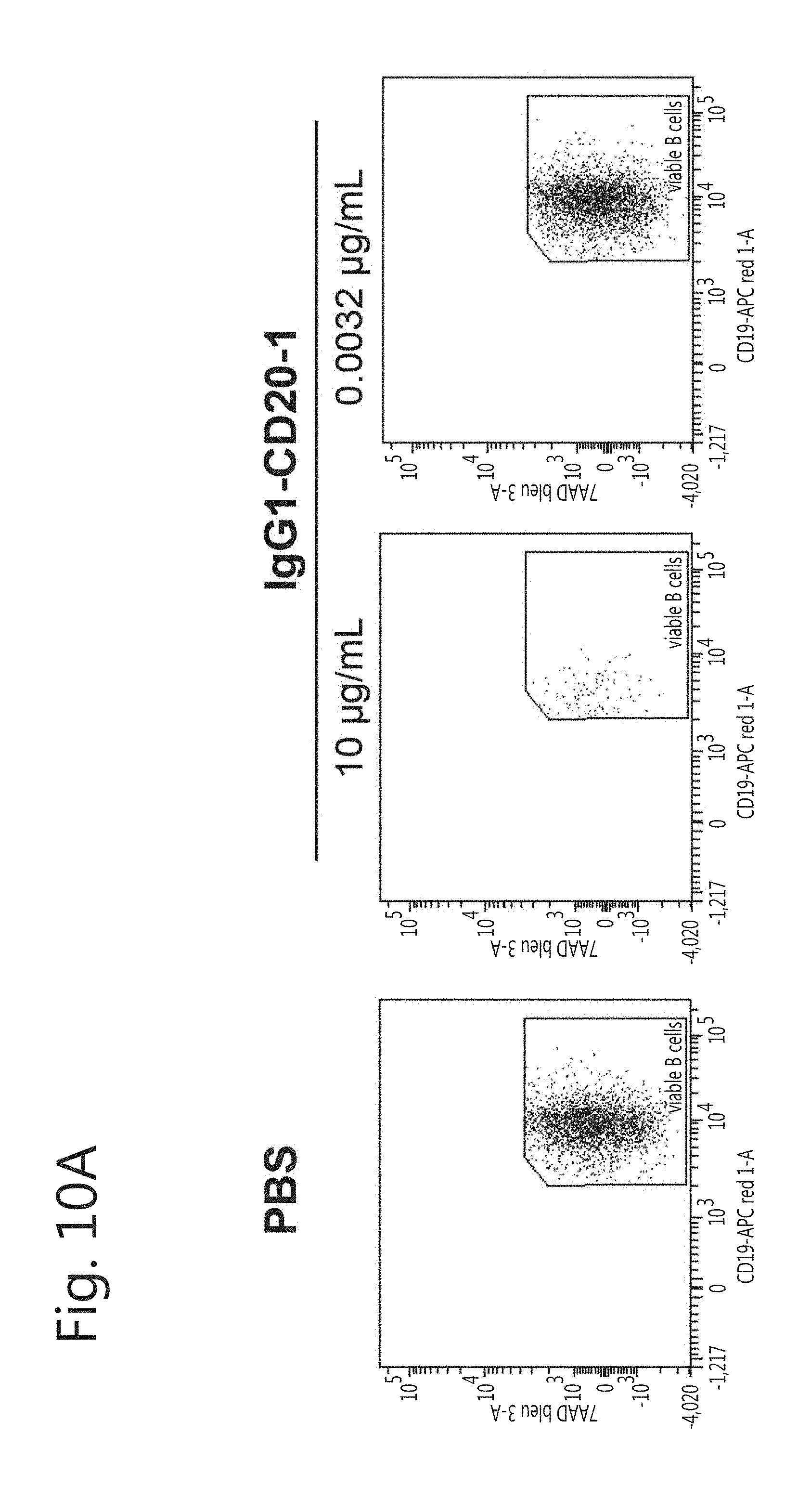
D00017

D00018

D00019

D00020

D00021

D00022

D00023

D00024

D00025

D00026

D00027
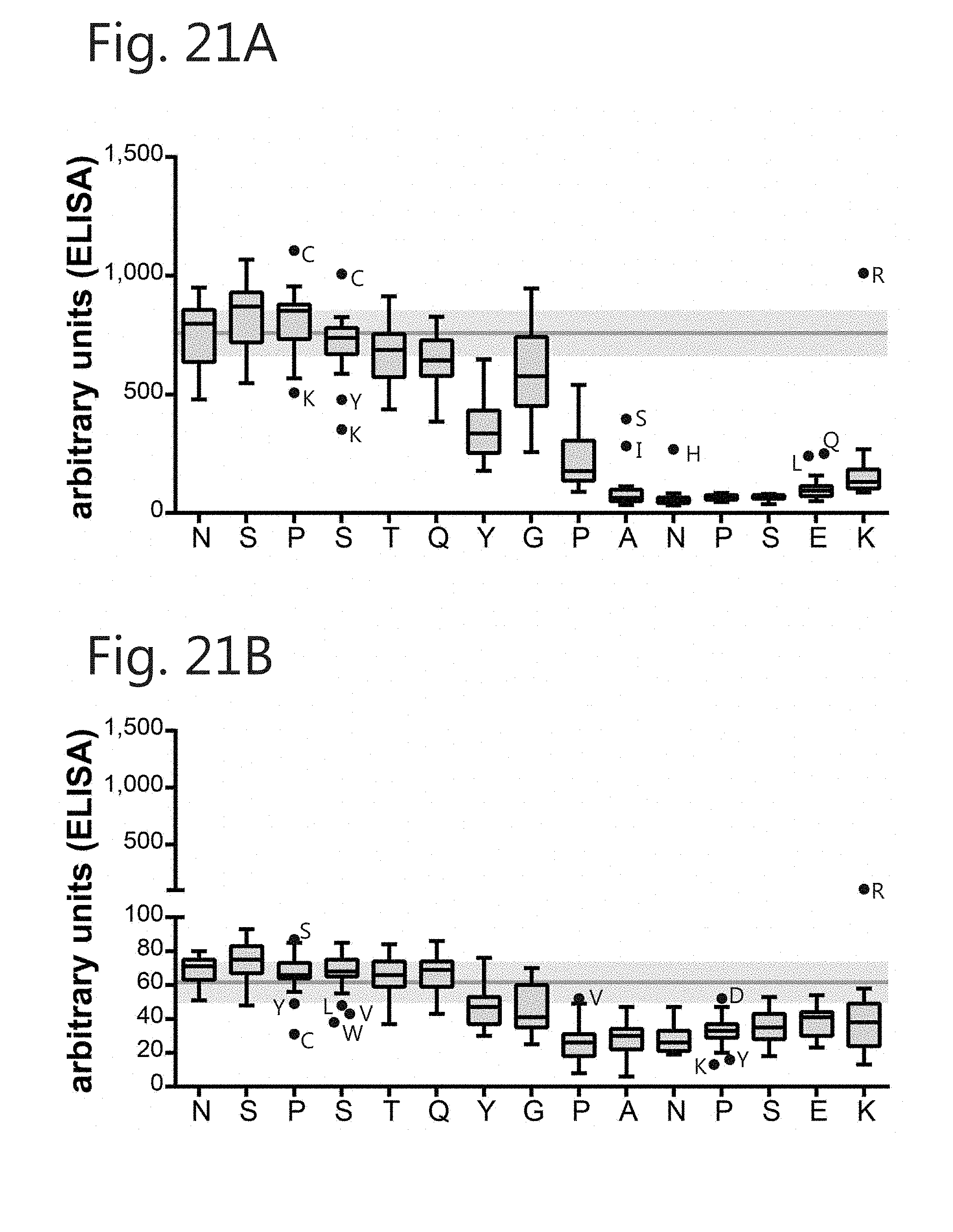
D00028

D00029

D00030

D00031
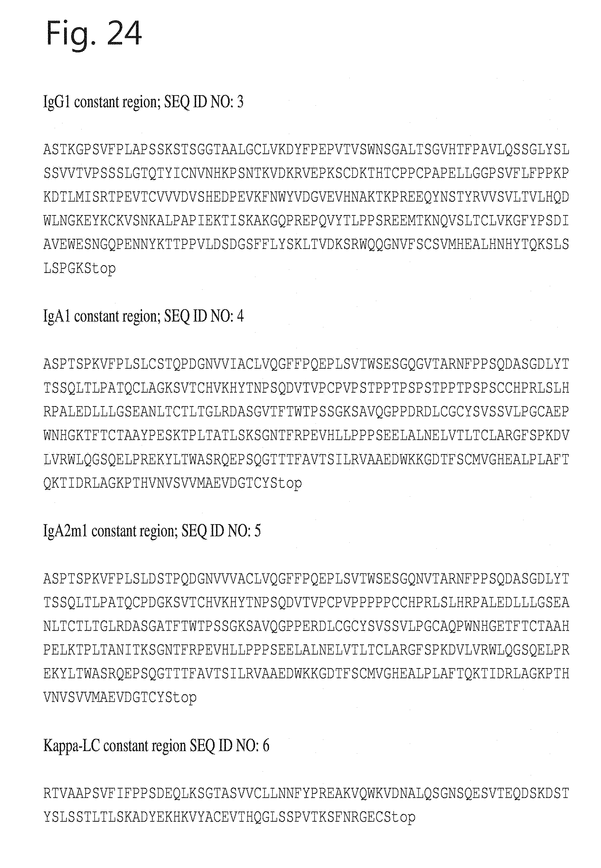
D00032
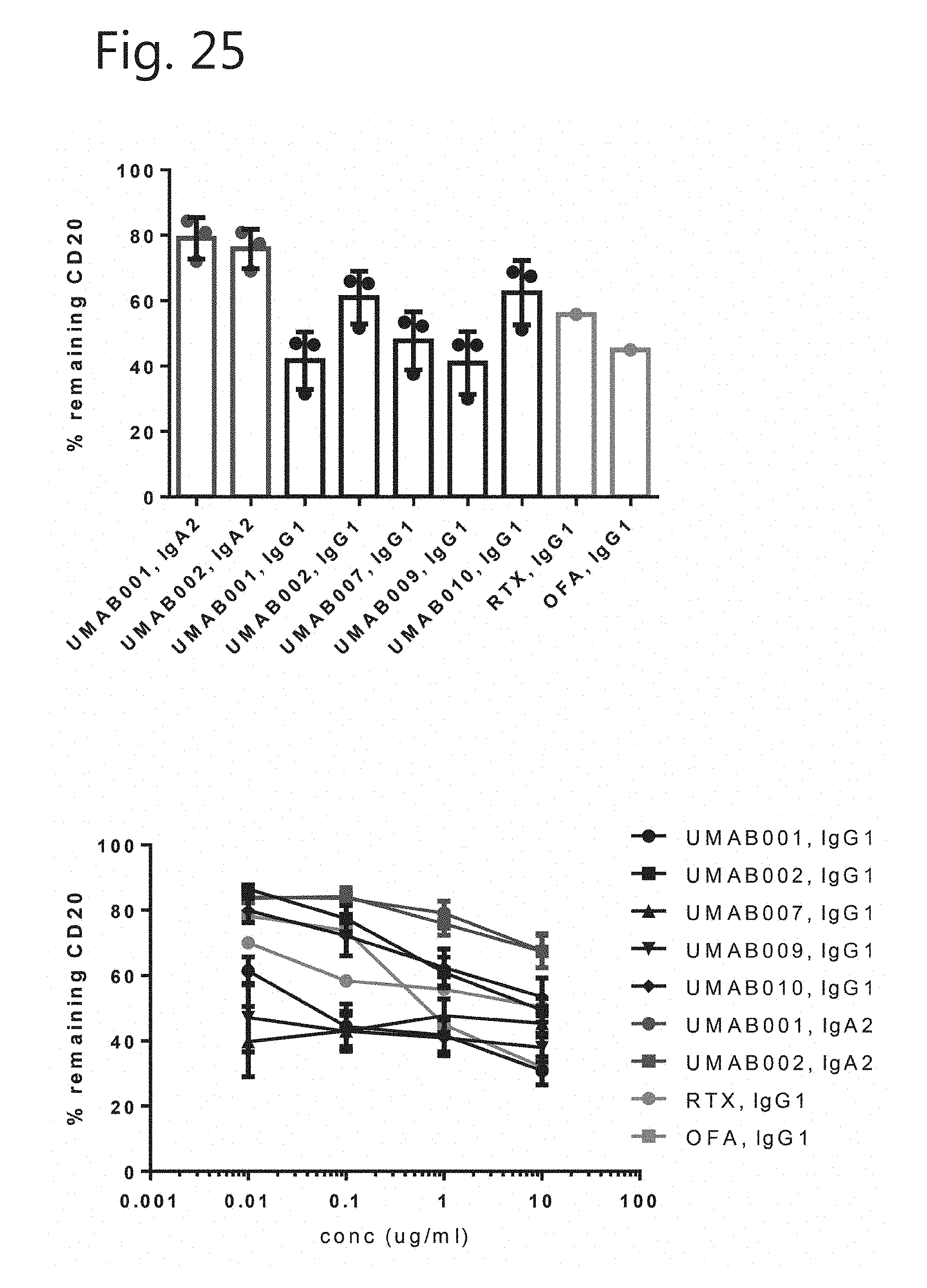
D00033

D00034
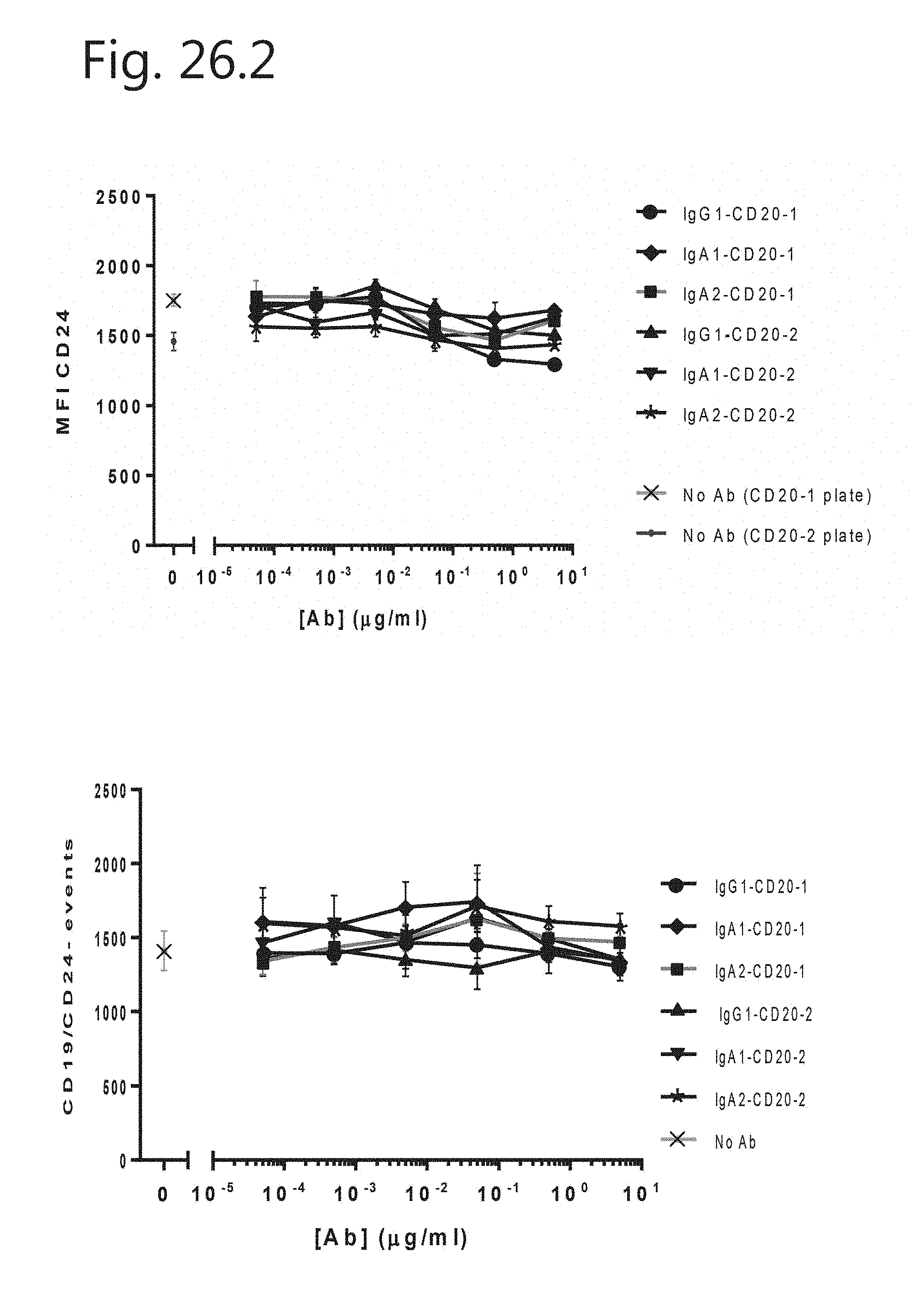
D00035

D00036

D00037
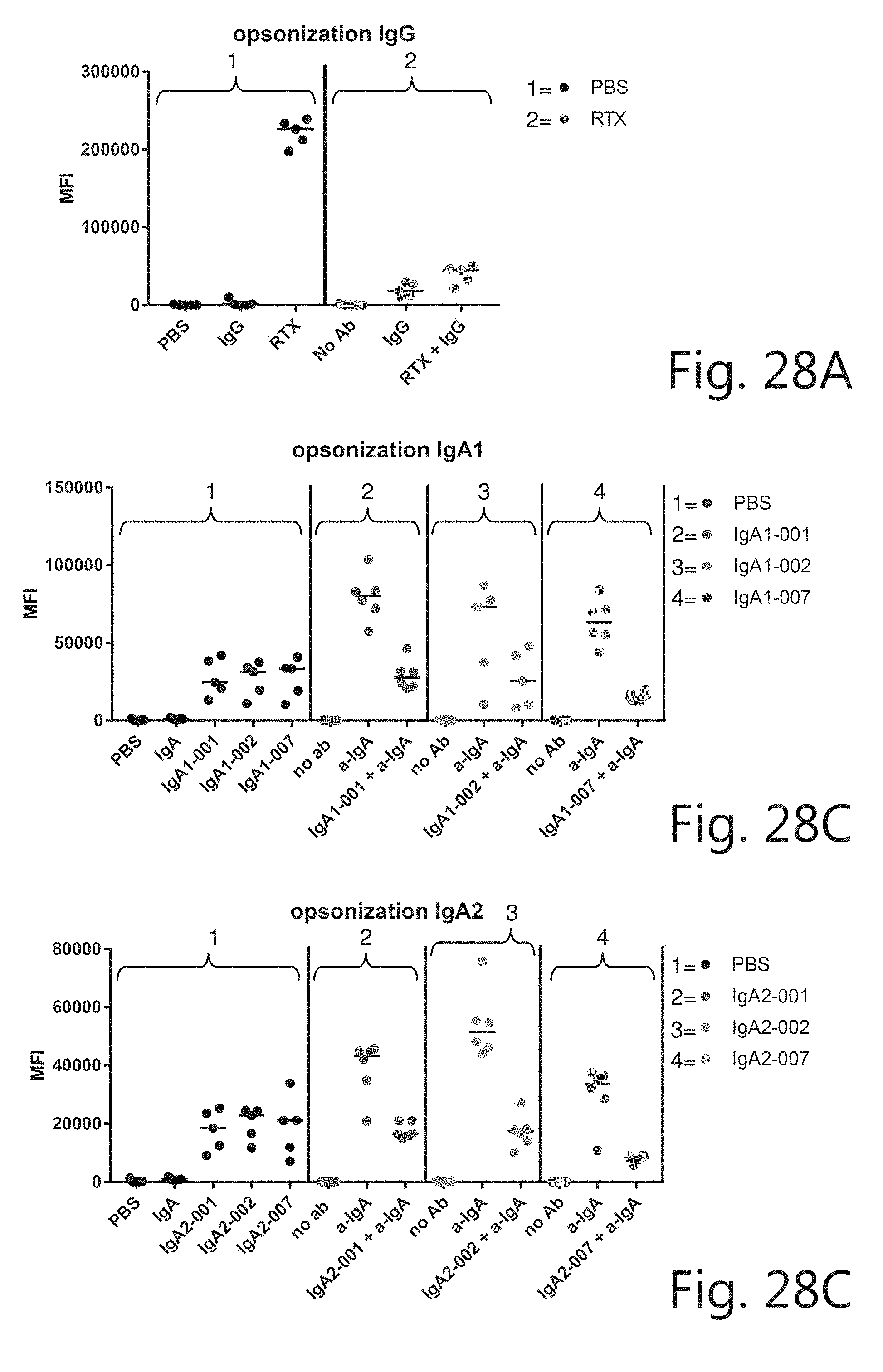
D00038

D00039
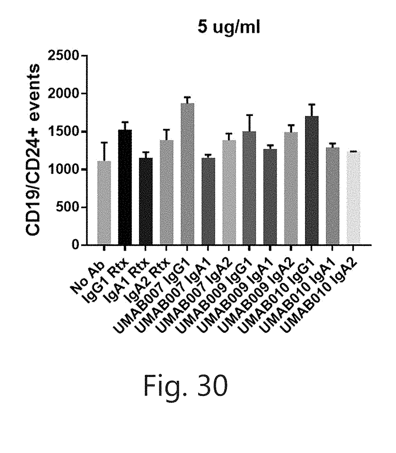


S00001
XML
uspto.report is an independent third-party trademark research tool that is not affiliated, endorsed, or sponsored by the United States Patent and Trademark Office (USPTO) or any other governmental organization. The information provided by uspto.report is based on publicly available data at the time of writing and is intended for informational purposes only.
While we strive to provide accurate and up-to-date information, we do not guarantee the accuracy, completeness, reliability, or suitability of the information displayed on this site. The use of this site is at your own risk. Any reliance you place on such information is therefore strictly at your own risk.
All official trademark data, including owner information, should be verified by visiting the official USPTO website at www.uspto.gov. This site is not intended to replace professional legal advice and should not be used as a substitute for consulting with a legal professional who is knowledgeable about trademark law.