Selective Inhibitor Of Exon 20 Insertion Mutant Egfr
Miyadera; Kazutaka ; et al.
U.S. patent application number 16/345792 was filed with the patent office on 2019-08-29 for selective inhibitor of exon 20 insertion mutant egfr. The applicant listed for this patent is Taiho Pharmaceutical Co., Ltd.. Invention is credited to Yoshimi Aoyagi, Shinichi Hasako, Kazutaka Miyadera.
| Application Number | 20190262345 16/345792 |
| Document ID | / |
| Family ID | 62024991 |
| Filed Date | 2019-08-29 |


View All Diagrams
| United States Patent Application | 20190262345 |
| Kind Code | A1 |
| Miyadera; Kazutaka ; et al. | August 29, 2019 |
SELECTIVE INHIBITOR OF EXON 20 INSERTION MUTANT EGFR
Abstract
An antitumor agent comprising a compound selected from the group consisting of Compounds A to D described in the specification, or a salt thereof, for treating a malignant tumor patient expressing EGFR having exon 20 insertion mutation.
| Inventors: | Miyadera; Kazutaka; (Tsukuba-Shi, JP) ; Aoyagi; Yoshimi; (Tsukuba-Shi, JP) ; Hasako; Shinichi; (Tsukuba-Shi, JP) | ||||||||||
| Applicant: |
|
||||||||||
|---|---|---|---|---|---|---|---|---|---|---|---|
| Family ID: | 62024991 | ||||||||||
| Appl. No.: | 16/345792 | ||||||||||
| Filed: | October 31, 2017 | ||||||||||
| PCT Filed: | October 31, 2017 | ||||||||||
| PCT NO: | PCT/JP2017/037186 | ||||||||||
| 371 Date: | April 29, 2019 |
| Current U.S. Class: | 1/1 |
| Current CPC Class: | A61K 31/519 20130101; A61P 35/00 20180101 |
| International Class: | A61K 31/519 20060101 A61K031/519; A61P 35/00 20060101 A61P035/00 |
Foreign Application Data
| Date | Code | Application Number |
|---|---|---|
| Oct 31, 2016 | JP | 2016-213072 |
Claims
[0169] 1. A method for treating a malignant tumor patient expressing EGFR having an exon 20 insertion mutation, the method comprising administering an effective amount of an antitumor agent, wherein the antitumor agent comprises a compound selected from the group consisting of: (S)--N-(4-amino-6-methyl-5-(quinolin-3-yl)-8,9-dihydropyrimido[5,4-b]indo- lizin-8-yl)acrylamide; (S)--N-(4-amino-6-methylene-5-(quinolin-3-yl)-7,8-dihydro-6H-pyrimido[5,4- -b]pyrrolizin-7-yl)acrylamide; (S,E)-N-(4-amino-6-methylene-5-(quinolin-3-yl)-7,8-dihydro-6H-pyrimido[5,- 4-b]pyrrolizin-7-yl)-3-chloroacrylamide; and (R)--N-(4-amino-6-methyl-5-(quinolin-3-yl)-8,9-dihydropyrimido[5,4-b]indo- lizin-8-yl)-N-methylacrylamide, or a salt thereof.
2. The method according to claim 1, wherein the compound is (S)--N-(4-amino-6-methyl-5-(quinolin-3-yl)-8,9-dihydropyrimido[5,4-b]indo- lizin-8-yl)acrylamide.
3. The method according to claim 1, wherein the malignant tumor patient expressing EGFR having exon 20 insertion mutation is a patient with lung cancer, breast cancer, head and neck cancer, brain tumor, uterine cancer, hematopoietic tumor, or skin cancer.
4. The method according to claim 1, wherein the malignant tumor patient expressing EGFR having exon 20 insertion mutation is a lung cancer patient.
5. The method according to claim 1, wherein the exon 20 insertion mutation is a mutation in which one or more amino acids are inserted in the exon 20 region.
6. The method according to claim 1, wherein the exon 20 insertion mutation is a mutation in which 1 to 7 amino acids are inserted in the exon 20 region.
7. The method according to claim 1, wherein the exon 20 insertion mutation is a mutation in which 1 to 4 amino acids are inserted in the exon 20 region.
8. The method according to claim 1, wherein the exon 20 insertion mutation is V769_D770insASV, D770_N771insSVD, D770_N771insG, H773_V774insNPH, or H773_V774insPH.
9. A method for treating a malignant tumor patient, comprising the step of administering an effective amount of a compound selected from the group consisting of: (S)--N-(4-amino-6-methyl-5-(quinolin-3-yl)-8,9-dihydropyrimido[5,4-b]indo- lizin-8-yl)acrylamide; (S)--N-(4-amino-6-methylene-5-(quinolin-3-yl)-7,8-dihydro-6H-pyrimido[5,4- -b]pyrrolizin-7-yl)acrylamide; (S,E)-N-(4-amino-6-methylene-5-(quinolin-3-yl)-7,8-dihydro-6H-pyrimido[5,- 4-b]pyrrolizin-7-yl)-3-chloroacrylamide; and (R)--N-(4-amino-6-methyl-5-(quinolin-3-yl)-8,9-dihydropyrimido[5,4-b]indo- lizin-8-yl)-N-methylacrylamide, or a salt thereof, to a malignant tumor patient expressing EGFR having an exon 20 insertion mutation.
10-11. (canceled)
12. The method according to claim 1, wherein the antitumor agent further comprises a pharmaceutical carrier.
Description
TECHNICAL FIELD
[0001] The present invention relates to an antitumor agent against cancers, comprising an exon 20 insertion mutant epidermal growth factor receptor (hereinafter referred to as "EGFR").
BACKGROUND ART
[0002] EGFR is a receptor-type tyrosine kinase, exerts its physiological function in normal tissue by being bound to Epidermal Growth Factor (hereinafter also referred to as EGF), which is a ligand, and contributes to growth and apoptosis inhibition in epithelial tissues (NPL 1). Further, somatic mutation of EGFR gene has been known as a cancer-causing gene; for example, EGFR in which the 746th to 750th amino acids in the exon 19 region are deleted (hereinafter also referred to as "exon 19 deletion mutation") and EGFR in which the 858th amino acid in the exon 21 region is mutated from leucine to arginine (hereinafter also referred to as "L858R mutation") constantly induces EGF-independent kinase activity, and contributes to the growth and survival of cancer cells (NPL 2). These mutations are observed, for example, in 30 to 50% of non-small-cell lung cancer in East Asia. The mutations are also observed in about 10% of non-small-cell lung cancer in Europe and the United States, and is regarded as one of the causes of cancers (NPL 3).
[0003] Therefore, research and development of EGFR inhibitor as an antitumor agent have actively been conducted, and introduced into the treatment of EGFR mutation-positive lung cancer. For example, although administration of gefitinib, erlotinib, and afatinib in their therapeutic dose causes, as side effects, digestive tract disorders and skin disorders, which are widely thought to be attributable to inhibition of wild-type EGFR, they exert a high antitumor effect against exon 19 deletion mutant and L858R mutant EGFR-positive lung cancers. The therapeutic effects of these agents are assumed to be derived from selective inhibition against mutant EGFR, compared with wild-type EGFR, by an EGFR inhibitor (NPL 4).
[0004] However, recent studies found that some cancers have EGFR with a mutation in which one or more amino acids are inserted in the exon 20 region (hereinafter also referred to as "exon 20 insertion mutation"), and that these cancers have low sensitivity with respect to previously known EGFR inhibitors. For example, there are clinical reports showing significantly lower antitumor effects of afatinib against EGFR mutation-positive lung cancer with respect to exon 20 insertion mutation, compared with exon 19 deletion mutation or L858R mutation (NPL 5). For this reason, chemotherapies have been used for the patients with these cancers. However, since the treatment options are limited and sufficient therapeutic effects have not been obtained, an antitumor agent with further higher therapeutic effects has been demanded.
[0005] PTL 1 discloses a compound usable for the treatment of diseases characterized by exon 20 insertion mutant EGFR. However, the compound of PTL 1 greatly differs in its structure from the compound according to the present invention, and PTL 1 nowhere discloses selectivity based on a comparison with wild-type EGFR, or efficacy in an in vivo model.
[0006] Further, although PTL 2 discloses a quinoline-substituted compound, PTL 2 nowhere discloses inhibitory activity against exon 20 insertion mutant EGFR.
CITATION LIST
Patent Literature
[0007] PTL 1: WO2015/175632A1 [0008] PTL 2: WO2015/025936A1
Non-Patent Literature
[0008] [0009] NPL 1: Nat. Rev. Cancer, Vol. 6, pp. 803-812 (2006) [0010] NPL 2: Nature Medicine, Vol. 19, pp. 1389-1400 (2013) [0011] NPL 3: Nat. Rev. Cancer, Vol. 7, pp. 169-181 (2007) [0012] NPL 4: Lancet Oncol. Vol. 13, e. 23-31 (2012) [0013] NPL 5: Lancet Oncol. Vol. 16, pp. 830-838 (2015)
SUMMARY OF INVENTION
Technical Problem
[0014] An object of the present invention is to provide an antitumor agent with reduced side effects derived from the inhibition of wild-type EGFR, the antitumor agent serving as an inhibitor that can ensure high selectivity with respect to exon 20 insertion mutant EGFR for which the therapeutic effects of the previously known EGFR inhibitors are insufficient.
Solution to Problem
[0015] The inventors of the present invention conducted extensive research, and found that exon 20 insertion mutant EGFR is an appropriate target in treating cancers, and that EGFR inhibitors conventionally used for the treatments have inferior selectivity between wild-type EGFR and exon 20 insertion mutant EGFR. Further, the inventors also confirmed that a specific compound exerts selectivity with respect to exon 20 insertion mutant EGFR and tumor growth inhibitory effects, and is thus regarded as superior to afatinib, which is a typical EGFR mutation-positive cancer-treating agent. With this finding, the inventors accomplished the present invention.
[0016] Accordingly, the present invention encompasses the following embodiments.
Item 1.
[0017] An antitumor agent for treating a malignant tumor patient expressing EGFR having exon 20 insertion mutation, the antitumor agent comprising a compound selected from the group consisting of: [0018] (S)--N-(4-amino-6-methyl-5-(quinolin-3-yl)-8,9-dihydropyrimido[5,4-b]indo- lizin-8-yl)acrylamide (hereinafter also referred to as Compound A); [0019] (S)--N-(4-amino-6-methylene-5-(quinolin-3-yl)-7,8-dihydro-6H-pyrimido[5,4- -b]pyrrolizin-7-yl) acrylamide (hereinafter also referred to as Compound B); [0020] (S,E)-N-(4-amino-6-methylene-5-(quinolin-3-yl)-7,8-dihydro-6H-pyrimido[5,- 4-b]pyrrolizin-7-yl)-3-chloroacrylamide (hereinafter also referred to as Compound C); and [0021] (R)--N-(4-amino-6-methyl-5-(quinolin-3-yl)-8,9-dihydropyrimido[5,4-b]indo- lizin-8-yl)-N-meth ylacrylamide (hereinafter also referred to as Compound D), or a salt thereof.
Item 2.
[0022] The antitumor agent according to Item 1, wherein the compound is (S)--N-(4-amino-6-methyl-5-(quinolin-3-yl)-8,9-dihydropyrimido[5,4-b]indo- lizin-8-yl)acrylamide.
Item 3.
[0023] The antitumor agent according to Item 1 or 2, wherein the malignant tumor patient expressing EGFR having exon 20 insertion mutation is a patient with lung cancer, breast cancer, head and neck cancer, brain tumor, uterine cancer, hematopoietic tumor, or skin cancer.
Item 4.
[0024] The antitumor agent according to any one of Items 1 to 3, wherein the malignant tumor patient expressing EGFR having exon 20 insertion mutation is a lung cancer patient.
Item 5.
[0025] The antitumor agent according to any one of Items 1 to 4, wherein the exon 20 insertion mutation is a mutation in which one or more amino acids are inserted in the exon 20 region.
Item 6.
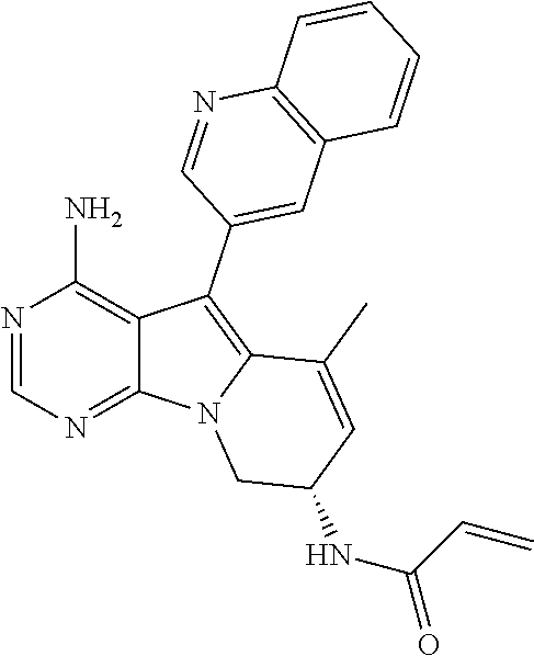
[0026] The antitumor agent according to any one of Items 1 to 5, wherein the exon 20 insertion mutation is a mutation in which 1 to 7 amino acids are inserted in the exon 20 region.
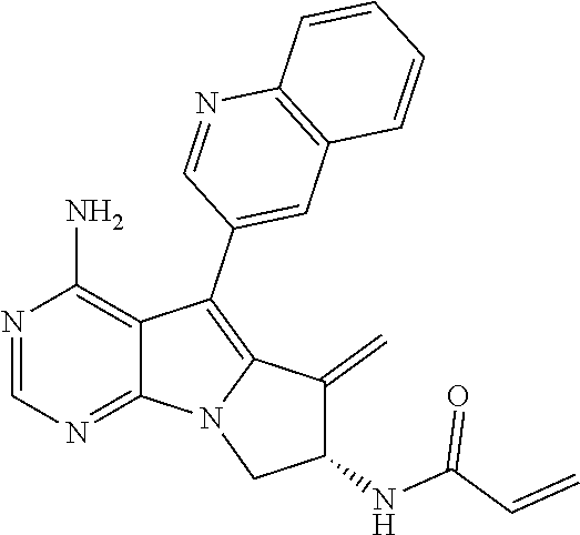
Item 7.
[0027] The antitumor agent according to any one of Items 1 to 6, wherein the exon 20 insertion mutation is a mutation in which 1 to 4 amino acids are inserted in the exon 20 region.
Item 8.
[0028] The antitumor agent according to any one of Items 1 to 7, wherein the exon 20 insertion mutation is A763_Y764insFQEA, V769_D770insASV, D770_N771insSVD, D770_N771insNPG, D770_N771insG, D770>GY, N771_P772insN, P772_R773insPR, H773_V774insNPH, H773_V774insPH, H773_V774insAH, H773_V774insH, V774_C774insHV, or A761_E762insEAFQ.
Item 9.
[0029] The antitumor agent according to any one of Items 1 to 8, wherein the exon 20 insertion mutation is V769_D770insASV, D770_N771insSVD, D770_N771insG, H773_V774insNPH, or H773_V774insPH.
Item 10.
[0030] A method for treating a malignant tumor patient, comprising the step of administering an antitumor agent comprising an effective amount of a compound selected from the group consisting of: [0031] (S)--N-(4-amino-6-methyl-5-(quinolin-3-yl)-8,9-dihydropyrimido[5,4-b]indo- lizin-8-yl)acrylamide; [0032] (S)--N-(4-amino-6-methylene-5-(quinolin-3-yl)-7,8-dihydro-6H-pyrimido[5,4- -b]pyrrolizin-7-yl) acrylamide; [0033] (S,E)-N-(4-amino-6-methylene-5-(quinolin-3-yl)-7,8-dihydro-6H-pyrimido[5,- 4-b]pyrrolizin-7-yl)-3-chloroacrylamide; and [0034] (R)--N-(4-amino-6-methyl-5-(quinolin-3-yl)-8,9-dihydropyrimido[5,4-b]indo- lizin-8-yl)-N-meth ylacrylamide,
[0035] or a salt thereof, to a malignant tumor patient expressing EGFR having exon 20 insertion mutation.
Item 11.
[0036] A compound selected from the group consisting of: [0037] (S)--N-(4-amino-6-methyl-5-(quinolin-3-yl)-8,9-dihydropyrimido[5,4-b]indo- lizin-8-yl)acrylamide; [0038] (S)--N-(4-amino-6-methylene-5-(quinolin-3-yl)-7,8-dihydro-6H-pyrimido[5,4- -b]pyrrolizin-7-yl) acrylamide; [0039] (S,E)-N-(4-amino-6-methylene-5-(quinolin-3-yl)-7,8-dihydro-6H-pyrimido[5,- 4-b]pyrrolizin-7-yl)-3-chloroacrylamide; and [0040] (R)--N-(4-amino-6-methyl-5-(quinolin-3-yl)-8,9-dihydropyrimido[5,4-b]indo- lizin-8-yl)-N-meth ylacrylamide,
[0041] or a salt thereof, to treat a malignant tumor patient expressing EGFR having exon 20 insertion mutation.
Item 12.
[0042] Use of a compound selected from the group consisting of: [0043] (S)--N-(4-amino-6-methyl-5-(quinolin-3-yl)-8,9-dihydropyrimido[5,4-b]indo- lizin-8-yl)acrylamide; [0044] (S)--N-(4-amino-6-methylene-5-(quinolin-3-yl)-7,8-dihydro-6H-pyrimido[5,4- -b]pyrrolizin-7-yl) acrylamide; and [0045] (S,E)-N-(4-amino-6-methylene-5-(quinolin-3-yl)-7,8-dihydro-6H-pyrimido[5,- 4-b]pyrrolizin-7-yl)-3-chloroacrylamide; and [0046] (R)--N-(4-amino-6-methyl-5-(quinolin-3-yl)-8,9-dihydropyrimido[5,4-b]indo- lizin-8-yl)-N-methylacrylamide,
[0047] or a salt thereof for the production of an antitumor agent for treating a malignant tumor patient expressing EGFR having exon 20 insertion mutation.
Advantageous Effects of Invention
[0048] The antitumor agent of the present invention exerts high selectivity with respect to exon 20 insertion mutant EGFR without inhibiting wild-type EGFR. Therefore, the antitumor agent of the present invention is useful in view of providing an antitumor agent that has reduced side effects derived from the inhibition of wild-type EGFR; and that exerts superior therapeutic effects for a malignant tumor patient expressing EGFR having exon 20 insertion mutation, for which the therapeutic effects of the previously known EGFR inhibitors are insufficient.
[0049] The previously known EGFR inhibitors have low selectivity with respect to exon 20 insertion mutant EGFR, compared with wild-type EGFR; therefore, the difference between the dosage for ensuring the antitumor effects and the dosage causing the side effects (skin disorders, digestive tract disorders, etc.) derived from wild-type EGFR inhibition was small. Accordingly, the previously known EGFR inhibitors have difficulty in exerting sufficient therapeutic effects. In contrast, since the antitumor agent of the present invention has high selectivity with respect to exon 20 insertion mutant EGFR, it is possible to increase the dosage without causing side effects derived from wild-type EGFR. Therefore, the antitumor agent of the present invention exerts superior therapeutic effects for a malignant tumor patient expressing EGFR having exon 20 insertion mutation.
BRIEF DESCRIPTION OF DRAWINGS
[0050] FIG. 1 illustrates IC.sub.50 ratios of wild-type EGFR to EGFR exon 20 insertion mutations calculated from the results of a test for cell growth inhibition on wild-type EGFR-expressing cell lines and mutant EGFR-expressing cell lines by compounds A, B, C, and D, a comparative compound, gefitinib, erlotinib, and afatinib.
[0051] FIG. 2 illustrates GI.sub.50 ratios of wild-type EGFR to EGFR exon 20 insertion mutations calculated from the results of a test for cell growth inhibition on wild-type EGFR-expressing human cell lines and mutant EGFR-expressing human cell lines by compound A, a comparative compound, gefitinib, erlotinib, and afatinib.
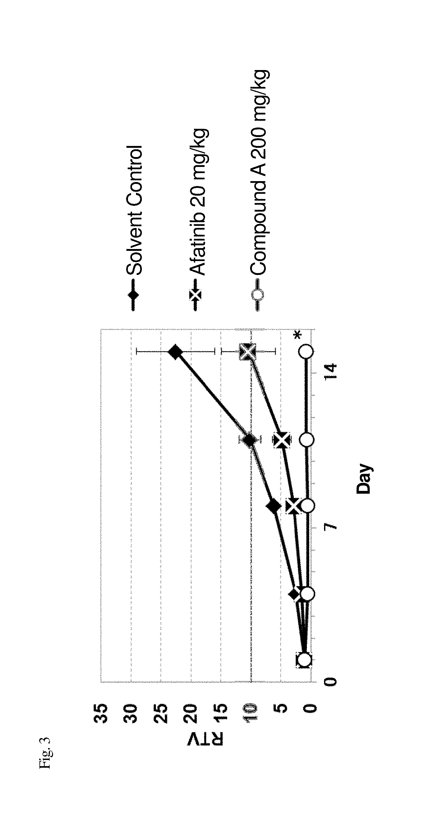
[0052] FIG. 3 illustrates the relative tumor volume (which hereinafter also referred to as "RTV") of mouse models that were subcutaneously transplanted with mutant EGFR-expressing cell lines (NIH3T3-EGFRinsASV cells) to measure the antitumor effect of compound A.
[0053] FIG. 4 illustrates the body weight change after grouping of mouse models that were subcutaneously transplanted with mutant EGFR-expressing cell lines (NIH3T3-EGFRinsASV cells) to measure the toxicity of compound A.
[0054] FIG. 5 illustrates the relative tumor volume of mouse models that were subcutaneously transplanted with mutant EGFR-expressing cell lines (NIH3T3-EGFRinsSVD cells) to measure the antitumor effect of compound A.
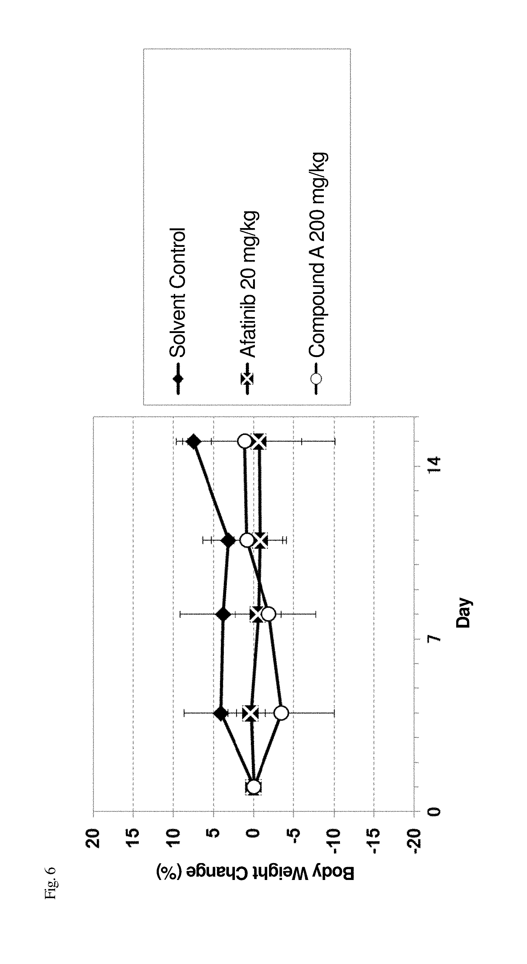
[0055] FIG. 6 illustrates the body weight after grouping of mouse models that were subcutaneously transplanted with mutant EGFR-expressing cell lines (NIH3T3-EGFRinsSVD cells) to measure the toxicity of compound A.
[0056] FIG. 7 illustrates the relative tumor volume of mouse models that were subcutaneously transplanted with mutant EGFR-expressing cell lines (H1975-EGFRinsSVD cells) to measure the antitumor effect of compound A.
[0057] FIG. 8 illustrates the body weight after grouping of mouse models that were subcutaneously transplanted with mutant EGFR-expressing cell lines (H1975-EGFRinsSVD cells) to measure the toxicity of compound A.
[0058] FIG. 9 illustrates the tumor volume of mouse models that were subcutaneously transplanted with mutant EGFR-expressing cell lines (NIH3T3-EGFRinsNPH) to measure the antitumor effect of compound A.
[0059] FIG. 10 illustrates the body weight after grouping of mouse models that were subcutaneously transplanted with mutant EGFR-expressing cell lines (NIH3T3-EGFRinsNPH) to measure the toxicity of compound A.
[0060] FIG. 11 illustrates the tumor volume of rat models that were subcutaneously transplanted with mutant EGFR-expressing cell lines (H1975-EGFRinsSVD cells) to measure the antitumor effect of compound A.
[0061] FIG. 12 illustrates the body weight after grouping of rat models that were subcutaneously transplanted with mutant EGFR-expressing cell lines (H1975-EGFRinsSVD cells) to measure the toxicity of compound A.
[0062] FIG. 13 illustrates the tumor volume of mouse models that were subcutaneously transplanted with a tumor derived from a lung cancer patient who was positive for EGFR with mutation V769_D770insASV to measure the antitumor effect of compound A.
[0063] FIG. 14 illustrates the body weight after grouping of mouse models that were subcutaneously transplanted with a tumor derived from a lung cancer patient who was positive for EGFR with mutation V769_D770insASV to measure the toxicity of compound A of the present invention.
[0064] FIG. 15 illustrates the amino acid sequence of wild-type EGFR (SEQ ID NO: 1).
DESCRIPTION OF EMBODIMENTS
[0065] Preferable examples of various definitions in the scope of the present invention used in this specification are explained below in detail.
[0066] In this specification, "EGFR" refers to a human epidermal growth factor receptor protein, and is also referred to as ErbB-1 or HER1.
[0067] In this specification, "wild-type EGFR" refers to EGFR free of somatic mutation, which is a protein comprising the amino acid sequence represented by SEQ ID NO: 1 (GenBank accession number: NP 005219.2).
[0068] In this specification, "exon 20 insertion mutation" refers to a mutation in which one or more amino acids (preferably 1 to 7, more preferably 1 to 4) are inserted in the exon 20 region (the 761st to 823rd amino acid sequence in SEQ ID NO: 1) of EGFR, and is preferably a mutation in which amino acid sequence FQEA (phenylalanine, glutamine, glutamic acid, and alanine in this order from the N-terminus) is inserted between the 763rd alanine and 764th tyrosine in the exon 20 region (A763_Y764insFQEA); a mutation in which amino acid sequence ASV (alanine, serine, and valine in this order from the N-terminus) is inserted between the 769th valine and 770th aspartic acid in the exon region (V769_D770insASV); a mutation in which amino acid sequence SVD (serine, valine, and aspartic acid in this order from the N-terminus) is inserted between the 770th aspartic acid and 771st asparagine in the exon 20 region (D770_N771insSVD); a mutation in which amino acid sequence NPG (asparagine, proline, and glycine in this order from the N-terminus) is inserted between the 770th aspartic acid and 771st asparagine in the exon 20 region (D770_N771insNPG); a mutation in which amino acid G (glycine) is inserted between the 770th aspartic acid and 771st asparagine (D770_N771insG); a mutation in which the 770th aspartic acid in the exon 20 region is deleted, and amino acid sequence GY (glycine and tyrosine in this order from the N-terminus) is inserted instead (D770>GY); a mutation in which amino acid N (asparagine) is inserted between the 771st asparagine and 772nd proline in the exon 20 region (N771_P772insN); a mutation in which amino acid sequence PR (proline and arginine in this order from the N-terminus) is inserted between the 772nd proline and 773rd histidine in the exon 20 region (P772_R773insPR); a mutation in which amino acid sequence NPH (asparagine, proline, and histidine in this order from the N-terminus) is inserted between the 773rd histidine and 774th valine in the exon 20 region (H773_V774insNPH); a mutation in which amino acid sequence PH (proline and histidine in this order from the N-terminus) is inserted between the 773rd histidine and 774th valine in the exon 20 region (H773_V774insPH); a mutation in which amino acid sequence AH (alanine and histidine in this order from the N-terminus) is inserted between the 773rd histidine and 774th valine in the exon 20 region (H773_V774insAH); a mutation in which amino acid H (histidine) is inserted between the 773rd histidine and 774th valine in the exon 20 region (H773_V774insH); a mutation in which amino acid sequence HV (histidine and valine in this order from the N-terminus) is inserted between the 774th valine and 775th cysteine in the exon 20 region (V774_C775insHV); a mutation in which amino acid sequence EAFQ (glutamic acid, alanine, phenylalanine, and glutamine in this order from the N-terminus) is inserted between the 761st alanine and 762nd glutamic acid in the exon 20 region (A761_E762insEAFQ); and the like. More preferable mutations include a mutation in which amino acid sequence ASV (alanine, serine, and valine in this order from the N-terminus) is inserted between the 769th valine and 770th aspartic acid in the exon 20 region (V769_D770insASV); a mutation in which amino acid sequence SVD (serine, valine, and aspartic acid in this order from the N-terminus) is inserted between the 770th aspartic acid and 771st asparagine in the exon 20 region (D770_N771insSVD); a mutation in which amino acid G (glycine) is inserted between the 770th aspartic acid and 771th asparagine in the exon 20 region (D770_N771insG); a mutation in which amino acid sequence NPH (asparagine, proline, and histidine in this order from the N-terminus) is inserted between the 773rd histidine and 774th valine in the exon 20 region (H773_V774insNPH); and a mutation in which amino acid sequence PH (proline and histidine in this order from the N-terminus) is inserted between the 773rd histidine and 774th valine in the exon 20 region (H773_V774insPH). More preferable mutations include a mutation in which amino acid sequence SVD (serine, valine, and aspartic acid in this order from the N-terminus) is inserted between the 770th aspartic acid and 771st asparagine in the exon 20 region (D770_N771insSVD); and a mutation in which amino acid G (glycine) is inserted between the 770th aspartic acid and 771st asparagine in the exon 20 region (D770_N771insG).
[0069] In this specification, the "malignant tumor patient expressing EGFR having exon 20 insertion mutation" refers to a malignant tumor patient expressing EGFR having exon 20 insertion mutation in at least one part of the exon 20 region of EGFR. The EGFR may have exon 20 insertion mutation in two or more different parts, but preferably one part thereof. Further, the EGFR may also have a mutation other than exon 20 insertion mutation (such as exon 19 deletion mutation, L858R mutation, or L790M mutation).
[0070] In the present invention, the method for detecting exon 20 insertion mutation of EGFR expressed in a malignant tumor patient is not particularly limited insofar as the method is capable of detecting the mutation, and any known detection methods may be used. The detection target in the detection of exon 20 insertion mutation may be any of genome sequence of EGFR gene, transcriptional product of EGFR gene, and EGFR protein.
[0071] The sample used in the detection of exon 20 insertion mutation is not particularly limited as long as the sample is a biological sample isolated from a malignant tumor patient, in particular, a sample that is obtained from a malignant tumor patient and contains malignant tumor cells. Examples of biological samples include body fluids (e.g., blood, urine, etc.), tissues, the extracts thereof, and the cultures of obtained tissues. The method for isolating a biological sample can be suitably selected depending on the type of biological sample.
[0072] The biological sample is prepared by being appropriately treated according to the detection method. Further, the reagent used for the detection (e.g., a reagent containing primer or probe) may be prepared by a conventional method according to the detection method.
[0073] In one embodiment of the present invention, the step for detecting the presence of exon 20 insertion mutation of EGFR expressed in a malignant tumor patient may be performed before the administration of antitumor agent to a malignant tumor patient.
[0074] Compounds A to D (Compounds A, B, C, and D) (in this specification, these compounds may also be generally referred to as a "compound of the present invention" or a "compound according to the present invention") and the production method thereof are explained below.
[0075] Compound A ((S)--N-(4-amino-6-methyl-5-(quinolin-3-yl)-8,9-dihydropyrimido[5,4-b]ind- olizin-8-yl)acrylamide) is represented by the following chemical formula.
##STR00001##
[0076] Compound B ((S)--N-(4-amino-6-methylene-5-(quinolin-3-yl)-7,8-dihydro-6H-pyrimido[5,- 4-b]pyrrolizin-7-yl)acrylamide) is represented by the following chemical formula.
##STR00002##
[0077] Compound C ((S,E)-N-(4-amino-6-methylene-5-(quinolin-3-yl)-7,8-dihydro-6H-pyrimido[5- ,4-b]pyrrolizin-7-yl)-3-chloroacrylamide) is represented by the following chemical formula.
##STR00003##
[0078] Compound D ((R)--N-(4-amino-6-methyl-5-(quinolin-3-yl)-8,9-dihydropyrimido[5,4-b]ind- olizin-8-yl)-N-methylacrylamide) is represented by the following chemical formula.
##STR00004##
[0079] Compounds A to D may be produced, for example, through the production method disclosed in WO2015/025936A1, the methods described in the Examples, and the like. However, the production methods of Compounds A to D are not limited to these reaction examples.
[0080] When Compounds A to D of the present invention have isomers such as optical isomers, stereoisomers, rotational isomers, and tautomers, any of the isomers and mixtures thereof are included within the scope of the compound of the present invention, unless otherwise specified. For example, when Compounds A to D of the present invention have optical isomers, racemic mixtures and the optical isomers separated from a racemic mixture are also included within the scope of the compound of the present invention, unless otherwise specified.
[0081] The salts of Compounds A to D refer to any pharmaceutically acceptable salts; examples include base addition salts and acid addition salts.
[0082] Examples of base addition salts include alkali metal salts such as sodium salts and potassium salts; alkaline earth metal salts such as calcium salts and magnesium salts; ammonium salts; and organic amine salts such as trimethylamine salts, triethylamine salts, dicyclohexylamine salts, ethanolamine salts, diethanolamine salts, triethanolamine salts, procaine salts, and N,N'-dibenzylethylenediamine salts.
[0083] Examples of acid addition salts include inorganic acid salts such as hydrochlorides, sulfates, nitrates, phosphates, and perchlorates; organic acid salts such as acetates, formates, maleates, fumarates, tartrates, citrates, ascorbates, and trifluoroacetates; and sulfonates such as methanesulfonates, isethionates, benzenesulfonates, and p-toluenesulfonates.
[0084] Compounds A to D and salts thereof also encompass prodrugs thereof. A prodrug refers to a compound that can be converted to Compounds A to D or a salt thereof through a reaction with an enzyme, gastric acid, or the like, under physiological conditions in vivo, i.e., a compound that can be converted to the compound of the present invention or a salt thereof by enzymatic oxidation, reduction, hydrolysis, or the like; or a compound that can be converted to Compounds A to D or a salt thereof by hydrolysis or the like with gastric acid or the like. Further, the prodrug may be compounds that can be converted to Compounds A to D or a salt thereof under physiological conditions, such as those described in "Iyakuhin no Kaihatsu [Development of Pharmaceuticals]," Vol. 7, Molecular Design, published in 1990 by Hirokawa Shoten Co., pp. 163-198.
Description of Diseases
[0085] Specific examples of tumors targeted in the present invention include, but are not particularly limited to, head and neck cancer, gastrointestinal cancer (esophageal cancer, stomach cancer, duodenal cancer, liver cancer, biliary cancer (e.g., gallbladder and bile duct cancer), pancreatic cancer, colorectal cancer (e.g., colon cancer, and rectal cancer), etc.), lung cancer (e.g., non-small-cell lung cancer, small-cell lung cancer, and mesothelioma), breast cancer, genital cancer (ovarian cancer, uterine cancer (e.g., cervical cancer, and endometrial cancer), etc.), urological cancer (e.g., kidney cancer, bladder cancer, prostate cancer, and testicular tumor), hematopoietic tumor (e.g., leukemia, malignant lymphoma, and multiple myeloma), osteosarcoma, soft-tissue sarcoma, skin cancer, brain tumor, and the like. Preferable examples include lung cancer, breast cancer, head and neck cancer, brain tumor, uterine cancer, hematopoietic tumor, or skin cancer.
[0086] When Compounds A to D or a salt thereof are used as a pharmaceutical agent, a pharmaceutical carrier can be added, if required, thereby forming a suitable dosage form according to prevention and treatment purposes. Examples of the dosage form include oral preparations, injections, suppositories, ointments, patches, and the like. Oral preparations are preferable. Such dosage forms can be formed by methods conventionally known to persons skilled in the art.
[0087] As the pharmaceutical carrier, various conventional organic or inorganic carrier materials used as preparation materials may be blended as an excipient, binder, disintegrant, lubricant, or colorant in solid preparations; or as a solvent, solubilizing agent, suspending agent, isotonizing agent, buffer, or soothing agent in liquid preparations. Moreover, pharmaceutical preparation additives, such as antiseptics, antioxidants, colorants, sweeteners, and stabilizers, may also be used, if required.
[0088] Oral solid preparations are prepared as follows. After an excipient is added optionally with a binder, disintegrant, lubricant, colorant, taste-masking or flavoring agent, etc., to Compounds A to D, the resulting mixture is formulated into tablets, coated tablets, granules, powders, capsules, or the like by ordinary methods.
[0089] Examples of excipients include lactose, sucrose, D-mannitol, glucose, starch, calcium carbonate, kaolin, microcrystalline cellulose, and silicic acid anhydride. Examples of binders include water, ethanol, 1-propanol, 2-propanol, simple syrup, liquid glucose, liquid .alpha.-starch, liquid gelatin, D-mannitol, carboxymethyl cellulose, hydroxypropyl cellulose, hydroxypropyl starch, methyl cellulose, ethyl cellulose, shellac, calcium phosphate, polyvinylpyrrolidone, and the like. Examples of disintegrators include dry starch, sodium alginate, powdered agar, sodium hydrogen carbonate, calcium carbonate, sodium lauryl sulfate, stearic acid monoglyceride, lactose, and the like. Examples of lubricants include purified talc, sodium stearate, magnesium stearate, borax, polyethylene glycol, and the like. Examples of colorants include titanium oxide, iron oxide, and the like. Examples of taste-masking or flavoring agents include sucrose, bitter orange peel, citric acid, tartaric acid, and the like.
[0090] When a liquid preparation for oral administration is prepared, a taste-masking agent, a buffer, a stabilizer, a flavoring agent, and the like may be added to Compounds A to D; and the resulting mixture may be formulated into an oral liquid preparation, syrup, elixir, etc., according to an ordinary method.
[0091] Examples of taste-masking or flavoring agents include those mentioned above. Examples of buffer agents include sodium citrate and the like. Examples of stabilizers include tragacanth, gum arabic, gelatin, and the like. As necessary, these preparations for oral administration may be coated according to methods known in the art with an enteric coating or other coating for the purpose of, for example, persistence of effects. Examples of such coating agents include hydroxypropyl methylcellulose, ethyl cellulose, hydroxymethyl cellulose, hydroxypropyl cellulose, polyoxyethylene glycol, and Tween 80 (registered trademark).
[0092] When an injection agent is prepared, a pH regulator, a buffer, a stabilizer, an isotonizing agent, a local anesthetic, and the like, may be added to Compounds A to D; and the mixture may be formulated into a subcutaneous, intramuscular, or intravenous injection according to an ordinary method.
[0093] Examples of the pH adjuster and the buffer used herein include sodium citrate, sodium acetate, and sodium phosphate. Examples of the stabilizer include sodium pyrosulfite, EDTA, thioglycolic acid, and thiolactic acid. Examples of the local anesthetic include procaine hydrochloride and lidocaine hydrochloride. Examples of the tonicity agent include sodium chloride, glucose, D-mannitol, and glycerol.
[0094] When a suppository is prepared, pharmaceutically acceptable carriers known by a person skilled in the art, such as polyethylene glycol, lanolin, cacao butter, and fatty acid triglyceride; and as necessary, surfactants such as Tween 80 (registered trademark), may be added to Compounds A to D, and the resulting mixture may be formulated into a suppository according to an ordinary method.
[0095] When an ointment is prepared, a commonly used base, stabilizer, wetting agent, preservative, and the like, may be blended into Compounds A to D, as necessary; and the obtained mixture may be mixed and formulated into an ointment according to an ordinary method.
[0096] Examples of the base include liquid paraffin, white petrolatum, white beeswax, octyl dodecyl alcohol, and paraffin.
[0097] Examples of the preservative include methyl paraoxybenzoate, ethyl paraoxybenzoate, and propyl paraoxybenzoate.
[0098] When a patch is prepared, the above-described ointment, cream, gel, paste, or the like, may be applied to an ordinary substrate according to an ordinary method.
[0099] Examples of substrates include woven fabrics or non-woven fabrics comprising cotton, staple fibers, or chemical fibers; and films or foam sheets of soft vinyl chloride, polyethylene, polyurethane, etc., are suitable.
[0100] The amount of Compounds A to D to be incorporated in each of such dosage unit forms depends on the condition of the patient to whom the compound is administered, the dosage form thereof, etc. In general, in the case of an oral agent, the amount of the compound is preferably 0.05 to 1000 mg per dosage unit form. In the case of an injection, the amount of the compound is preferably 0.01 to 500 mg per dosage unit form; and in the case of a suppository, the amount of the compound is preferably 1 to 1000 mg per dosage unit form.
[0101] Further, the daily dose of the medicine in such a dosage form depends on the condition, body weight, age, sex, etc., of the patient, and cannot be generalized. For example, the daily dose for an adult (body weight: 50 kg) of Compounds A to D as an active ingredient may be generally 0.05 to 5000 mg, and preferably 0.1 to 1000 mg; and is preferably administered in one dose, or in two to three divided doses, per day.
[0102] The present invention also provides a method for treating a malignant tumor patient, comprising the step of administering an effective amount of an antitumor agent comprising a compound selected from the group consisting of Compounds A to D or a salt thereof to a malignant tumor patient expressing EGFR having exon 20 insertion mutation.
[0103] The present invention also provides a compound selected from the group consisting of Compounds A to D, or a salt thereof for treating a malignant tumor patient expressing EGFR having exon 20 insertion mutation.
[0104] The present invention also provides use of a compound selected from the group consisting of Compounds A to D or a salt thereof for treating a malignant tumor patient expressing EGFR having exon 20 insertion mutation.
[0105] The present invention also provides use of a compound selected from the group consisting of Compounds A to D or a salt thereof for the production of an antitumor agent for treating a malignant tumor patient expressing EGFR having exon 20 insertion mutation.
[0106] The present invention is also a method for predicting therapeutic effects of chemotherapy using an antitumor agent comprising, as an active ingredient, a compound selected from the group consisting of Compounds A to D or a salt thereof in a malignant tumor patient, the method comprising steps (1) and (2) below:
(1) a step of detecting the presence or absence of mutation of EGFR gene contained in a biological sample obtained from the patient; and (2) a step of predicting that the chemotherapy is highly likely to exhibit sufficient therapeutic effects with respect to the patient when the results of the detection in step (1) found that the EGFR gene has exon 20 insertion mutation.
[0107] The present invention is also a method for treating a malignant tumor patient, the method comprising steps (1) to (3) below:
(1) a step of detecting the presence or absence of mutation of EGFR gene contained in a biological sample obtained from the patient; (2) a step of predicting that the chemotherapy using an antitumor agent comprising a compound selected from the group consisting of Compounds A to D or a salt thereof is highly likely to exhibit sufficient therapeutic effects with respect to the patient when the results of the detection in step (1) found that the EGFR gene has exon 20 insertion mutation; and (3) a step of administering the antitumor agent to a patient who was predicted highly likely to sufficiently respond to the chemotherapy in step (2).
[0108] The base sequence of EGFR gene is publicly known. The GenBank accession number of the base sequence of cDNA is NM_005228.4.
[0109] The "therapeutic effects" can be evaluated by tumor shrinkage effects, relapse-suppressing effects, life-prolonging effects, and the like. The relapse-suppressing effects may be shown as degree of the extension of non-relapse period and/or the degree of the improvement in relapse rate; and the life-prolonging effects may be shown as the degree of the entire survival time and/or the degree of the extension of the median of progression-free survival, or the like. The "sufficient therapeutic effects" of the chemotherapy using an antitumor agent comprising, as an active ingredient, Compound A or a salt thereof means that superior therapeutic effects are obtained by the administration of the antitumor agent comprising, as an active ingredient, Compound A or a salt thereof, such as significant extension of survival time, significant suppression of relapse, and the like, compared with non-administration.
EXAMPLES
[0110] The following describes the present invention in more detail with reference to the following Test Examples. However, the present invention is not limited to these Examples (Test Examples).
Test Example 1
In Vitro Drug Efficacy Test
Evaluation of Cell Growth Inhibitory Effect on Wild-Type EGFR- or Mutant EGFR-Expressing Cell Lines (1)
[0111] The inhibitory activity of compounds against wild-type EGFR and mutant EGFR was evaluated using Ba/F3 cells (mouse B-lymphocyte precursor cell lines) to which human EGFR genes were introduced. The Ba/F3 cells were maintained in an RPMI-1640 medium (Thermo Fisher Scientific) containing 10% fetal bovine serum (FBS), 100 U/mL penicillin/100m/mL streptomycin (Thermo Fisher Scientific), and 1 ng/mL mouse interleukin-3 (mlL-3) (CST). A PB-CMV-MCS-EF1-GFP+Puro vector or PB-CMV-MCS-EF1-RFP+Puro vector into which a human EGFR gene (wild-type (WT), V769_D770insASV (insASV), D770_N771insSVD (insSVD), D770_N771insG (insG), H773_V774insNPH (insNPH), or H773_V774insPH (insPH)) was encoded was introduced to the cells, together with a Super PiggyBac transposase expression vector, by electroporation using an Amaxa (trademark) Cell Line Nucleofector (trademark) Kit V, followed by selection using puromycin (SIGMA). Ba/F3 cells expressing wild-type EGFR (which hereinafter also referred to as "Ba/F3-EGFR_WT") exhibited mlL-3-independent growth in the presence of 50 ng/mL
[0112] EGF (R&D Systems); and Ba/F3 cells expressing EGFR exon 20 insertion mutation (which hereinafter also referred to as "Ba/F3-EGFRinsASV," "Ba/F3-EGFRinsSVD," "Ba/F3-EGFRinsG," "Ba/F3-EGFRinsNPH," or "Ba/F3-EGFRinsPH") exhibited mlL-3-independent growth in the absence of EGF.
[0113] To evaluate the cell growth inhibitory effect, Ba/F3-EGFR_WT cells were suspended in an RPMI-1640 medium containing 10% FBS, 100 U/mL penicillin, 100m/mL streptomycin, and 50 ng/mL EGF; and the cell suspension was seeded in each well of a 96-well flat-bottom microplate such that the cell count per well was 30,000. The Ba/F3 cells expressing EGFR exon 20 insertion mutation were suspended in an RPMI-1640 medium containing 10% FBS, 100 U/mL penicillin, and 100 .mu.g/mL streptomycin; and the cell suspension was seeded in each well of a 96-well flat-bottom microplate such that the cell count per well was 15,000. Subsequently, [0114] (S)--N-(4-amino-6-methyl-5-(quinolin-3-yl)-8,9-dihydropyrimido[5,4- -b]indolizin-8-yl)acrylamide (compound A), [0115] (S)--N-(4-amino-6-methylene-5-(quinolin-3-yl)-7,8-dihydro-6H-pyrimido[5,4- -b]pyrrolizin-7-yl)acrylamide (compound B), [0116] (S,E)-N-(4-amino-6-methylene-5-(quinolin-3-yl)-7,8-dihydro-6H-pyrimido[5,- 4-b]pyrrolizin-7-yl)-3-chloroacrylamide (compound C), and [0117] (R)--N-(4-amino-6-methyl-5-(quinolin-3-yl)-8,9-dihydropyrimido [5,4-b]indolizin-8-yl)-N-methylacrylamide (compound D) prepared in accordance with the production method disclosed in PTL 2, and (S)--N-(4-amino-5-(quinolin-3-yl)-6,7,8,9-tetrahydropyrimido[5,4-b]indoli- zin-8-yl)acrylamide prepared in accordance with the production method disclosed in WO2013/125709A1 (the compound of Example 1 in WO2013/125709A1, which hereinafter also referred to as "comparative compound") were dissolved in DMSO, and diluted with DMSO or the medium used for suspending the cells. These compounds were individually added to each well of the culture plate of the cells, and incubated in a 5% CO.sub.2 gas-containing incubator at 37.degree. C. for 3 days. The cell count after incubation was measured using a CellTiter-Glo (trademark) Luminescent Cell Viability Assay (Promega Corporation) in accordance with the manufacturer's recommended protocol. The growth rate was calculated using the following formula, and the concentration of each test compound for 50% inhibition (IC.sub.50 (.mu.M)) was determined.
[0117] Growth Rate (%)=T/C.times.100
T: the luminescence intensity of a well to which a test compound was added. C: the luminescence intensity of a well to which the test compound was not added.
[0118] Additionally, the ratio of IC.sub.50 between wild-type EGFR and EGFR exon 20 insertion mutation was determined using the following formula. FIG. 1 illustrates the results.
IC.sub.50 Ratio=IC.sub.50 (WT)/IC.sub.50 (ex20ins) IC.sub.50 (WT): IC.sub.50 for wild-type EGFR IC.sub.50 (ex20ins): IC.sub.50 for EGFR exon 20 insertion mutation
[0119] As is clear from FIG. 1, compounds A to D exhibited a cell growth inhibitory effect on cell lines expressing EGFR exon 20 insertion mutations; and their mutation selectivity was higher than that of the comparative compound, gefitinib, erlotinib, and afatinib.
Test Example 2
Cell Growth Inhibitory Effect on Wild-Type EGFR- or Mutant EGFR-Expressing Human Cell Lines (2)
[0120] To evaluate the inhibitory activity of compounds against wild-type EGFR and mutant EGFR, the following cells were used: NCI-H1975 cells, which are human pulmonary adenocarcinoma cell lines expressing EGFR with mutation D770_N771insSVD by gene modification (which hereinafter also referred to as "H1975-EGFRinsSVD"); and A431 cells, which are human epithelial cancer cell lines expressing wild-type EGFR. H1975-EGFRinsSVD cells were prepared as follows. A PB-CMV-MCS-EF1-RFP+Puro vector into which D770_N771insSVD (insSVD) was encoded was introduced into NCI-H1975 cells, together with a Super PiggyBac Transposase expression vector, by electroporation using an Amaxa (trademark) Cell Line Nucleofector (trademark) Kit R, followed by selection using puromycin (SIGMA). XTN (trademark) TALENs Site-Specific Nucleases (Transposagen) were introduced into the cells by electroporation using the Amaxa (trademark) Cell Line Nucleofector (trademark) Kit R, and endogenous-EGFR (T790M/L858R)-knockout cells were selected by sequencing.
[0121] To evaluate the cell growth inhibitory effect, individual types of cells were suspended in a medium recommended by ATCC. The cell suspensions were seeded in each well of respective 96-well flat-bottom plates such that the cell count per well was 3,000, and incubated in a 5% CO.sub.2-containing incubator at 37.degree. C. for 1 day. Compound A, the comparative compound, gefitinib, erlotinib, and afatinib were individually dissolved in DMSO, and diluted with DMSO such that these test compounds have a concentration 200 times higher than the final concentration. These DMSO solutions of the test compounds were diluted with the medium used for suspending the cells, and added to each well of the culture plates of the cells such that DMSO has a final concentration of 0.5%, and the cells were incubated in a 5% CO.sub.2-containing incubator at 37.degree. C. for 3 days. The cell count at the time incubation started (day 0) and the cell count after incubation (day 3) were measured using a CellTiter-Glo (trademark) Luminescent Cell Viability Assay (Promega Corporation) in accordance with the manufacturer's recommended protocol. The growth rate was calculated using the following formula, and the concentration of each test compound for 50% inhibition (GI.sub.50 (.mu.M)) was determined. Table 1 illustrates the results.
1) If T on day 3.gtoreq.C on day 0:
[0122] Growth Rate (%)=(T on day 3-C on day 0)/(C on day 3-C on day 0).times.100
T: the luminescence intensity of a well to which a test compound was added. C: the luminescence intensity of a well to which the test compound was not added. Day 0: the day on which a test compound was added. Day 3: the day on which evaluation was performed.
2) If T on day 3<C on day 0:
[0123] Growth Rate (%)=(T on day 3-C on day 0)/(C on day 0).times.100
T: the luminescence intensity of a well to which a test compound was added. C: the luminescence intensity of a well to which the test compound was not added. Day 0: the day on which a test compound was added. Day 3: the day on which evaluation was performed.
TABLE-US-00001 TABLE 1 GI.sub.50 (.mu.M) A431 H1975 EGFRinsSVD Compound A 0.396 0.031 Comparative Compound 0.543 0.364 Gefitinib 0.310 1.903 Erlotinib 0.612 2.775 Afatinib 0.023 0.189 Osimertinib 0.321 0.194
[0124] Additionally, the ratio of GI.sub.50between wild-type EGFR and EGFR exon 20 insertion mutation was determined using the following formula. Table 2 illustrates the results.
GI.sub.50 Ratio=GI.sub.50 (A431)/GI.sub.50 (H1975 EGFRinsSVD) GI.sub.50 (A431): GI.sub.50 for wild-type EGFR GI.sub.50 (H1975 EGFRinsSVD): GI.sub.50 for EGFR exon 20 insertion mutation
[0125] As is clear from Table 1 and FIG. 2, compound A exhibited an cell growth inhibitory effect on cell lines expressing EGFR exon 20 insertion mutation, and its mutation selectivity was higher than that of the comparative compound, gefitinib, erlotinib, afatinib, and osimertinib.
Test Example 3
Evaluation of Phosphorylated EGFR Inhibitory Activity Against Wild-Type EGFR- or Mutant EGFR-Expressing Cell Lines (1)
[0126] A431 cells, which are human epithelial cancer cell lines overexpressing wild-type EGFR and H1975-EGFRinsSVD cells, which are human pulmonary adenocarcinoma cell lines expressing EGFR with mutation D770_N771insSVD by gene modification, were suspended in respective mediums. These cell suspensions were individually seeded into a 60-mm dish, and incubated in a 5% CO.sub.2-containing incubator at 37.degree. C. for 1 day. Compound A was dissolved in DMSO, and diluted with DMSO such that the test compound has a concentration 1000 times higher than the final concentration. The DMSO solution of the test compound was diluted with each medium used for suspending the cells, and each diluted solution was added to respective culture dishes of the cells such that DMS 0 has a final concentration of 0.1%, followed by incubation in a 5% CO.sub.2-containing incubator at 37.degree. C. for 6 hours. After incubation, the cells were collected, and stored at -80.degree. C. in the form of pellets until use. A RIPA buffer (Thermo Fisher Scientific) containing a protease inhibitor cocktail (Thermo Fisher Scientific) was added to the pellets, and proteins within the cells were extracted. The concentration of the proteins were measured using a BCA protein assay kit (Thermo Fisher Scientific), and each sample was adjusted so as to have a protein concentration suitable for measurement of phosphorylated EGFR expression. The phosphorylated EGFR expression was measured using a Simple Western (trademark) assay system
(ProteinSimple) in accordance with the manufacturer's recommended protocol. The primary antibody used in measurement was a Phospho-EGF Receptor (Tyr1068) #3777 (CST) diluted to 1/50.
[0127] For each type of cells, a calibration curve of the protein concentration (x axis) and the phosphorylated EGFR expression level (y axis) was prepared, and the phosphorylated EGFR expression level of each sample was converted to a protein concentration based on the calibration curve. The phosphorylated EGFR rate was calculated using the following formula to determine the concentration of the test compound at which phosphorylated EGFR was inhibited by 50% (IC.sub.50 (.mu.M)).
Phosphorylated EGFR Rate (%)=T/C.times.100
T: an equivalent amount for the protein concentration of a sample to which the test compound was added. C: an equivalent amount for the protein concentration of a sample to which the test compound was not added.
[0128] Additionally, the selectivity for wild-type EGFR and EGFR exon 20 insertion mutation was calculated using the following formula. Table 2 illustrates the results.
IC.sub.50Ratio=IC.sub.50 (A431)/IC.sub.50 (H1975 EGFRinsSVD) IC.sub.50 (A431): IC.sub.50 for wild-type EGFR IC.sub.50 (H1975 EGFRinsSVD): IC.sub.50 for EGFR exon 20 insertion mutation
TABLE-US-00002 TABLE 2 IC.sub.50 (.mu.M) A431 H1975 EGFRinsSVD IC.sub.50 (.mu.M) 0.535 0.023 IC.sub.50 Ratio -- 23.3
[0129] As is clear from Table 2, compound A exhibited a selective inhibition activity against EGFR exon 20 insertion mutation.
Test Example 4
Evaluation of Phosphorylated EGFR Inhibitory Activity Against Wild-Type EGFR- or Mutant EGFR-Expressing Cell Lines (2)
[0130] The autophosphorylation inhibitory activity of a compound against wild-type EGFR and mutant EGFR was evaluated using NIH-3T3 cells, which are mouse fibroblast cell lines to which human EGFR gene was introduced. NIH-3T3 cells were maintained in a D-MEM (high-glucose) medium (Wako Pure Chemical Industries, Ltd.) containing 10% newborn calf serum (NBCS), 1,500 mg/L sodium hydrogen carbonate, and 100 U/mL penicillin/100m/mL streptomycin (Thermo Fisher Scientific). A PB-CMV-MCS-EF1-RFP+Puro vector into which a human EGFR gene (WT, insASV, insSVD, insG, insNPH, or insPH) was encoded was introduced into the cells, together with a Super PiggyBac Transposase expression vector, by electroporation using an Amaxa (trademark) Cell Line Nucleofector (trademark) Kit R, followed by selection using puromycin (SIGMA). NIH-3T3 cells expressing wild-type EGFR (which hereinafter also referred to as "NIH3T3-EGFR_WT") exhibited growth in the presence of 50 ng/mL EGF (R&D Systems) under 1% NBCS conditions. NIH-3T3 cells expressing EGFR exon 20 insertion mutation (which hereinafter also referred to as "NIH3T3-EGFRinsASV," "NIH3T3-EGFRinsSVD," "NIH3T3-EGFRinsG," "NIH3T3-EGFRinsNPH," or "NIH3T3-EGFRinsPH") exhibited growth in the absence of EGF under 1% NBCS conditions.
[0131] To evaluate EGFR-autophosphorylation inhibitory activity, NIH3T3 cells to which human EGFR was introduced were suspended in respective mediums. These cell suspensions were individually seeded into a 60-mm dish or 6-well flat-bottom plate, and incubated in a 5% CO.sub.2-containing incubator at 37.degree. C. for 1 day. Compound A was dissolved in DMSO, and diluted with DMSO such that the test compound has a concentration 400 times higher than the final concentration. The DMSO solutions of the test compound were diluted with the medium used for suspending the cells, and added to the culture dishes of the cells such that DMSO has a final concentration of 0.25%. Further, EGF was added to the culture dish of NIH3T3-EGFR_WT cells to give a final concentration of 50 ng/mL. The culture dishes were all subjected to incubation in a 5% CO.sub.2-containing incubator at 37.degree. C. for 6 hours. After incubation, the cells were collected, and stored at -80.degree. C. in the form of pellets until use. A RIPA buffer (Thermo Fisher Scientific) containing a protease inhibitor cocktail (Thermo Fisher Scientific) was added to the pellets, and proteins within the cells were extracted. The protein concentration was measured using a BCA protein assay kit (Thermo Fisher Scientific), and each sample was adjusted to a protein concentration suitable for measurement of phosphorylated EGFR expression. The phosphorylated EGFR expression was measured using a Simple Western (trademark) assay system (ProteinSimple) in accordance with the manufacturer's recommended protocol. The primary antibody used in measurement was a Phospho-EGF Receptor (Tyr1068) #3777 (CST) diluted to 1/50.
[0132] For each type of cells, a calibration curve of the protein concentration (x axis) and the phosphorylated EGFR expression level (y axis) was prepared, and the phosphorylated EGFR expression level of each sample was converted to a protein concentration based on the calibration curve. The phosphorylated EGFR inhibitory rate was calculated using the following formula to determine the concentration of the test compound at which phosphorylated EGFR was inhibited by 50% (IC.sub.50 (.mu.M)).
Phosphorylated EGFR Inhibitory Rate (%)=T/C.times.100
T: an equivalent amount for the protein concentration of a sample to which the test compound was added. C: an equivalent amount for the protein concentration of a sample to which the test compound was not added.
[0133] Additionally, the selectivity for wild-type EGFR and EGFR exon 20 insertion mutation was calculated using the following formula. Table 3 illustrates the results.
IC.sub.50 Ratio=IC.sub.50 (WT)/IC.sub.50 (EGFR exon 20 insertion mutation)
TABLE-US-00003 TABLE 3 WT insASV insSVD insG insNPH insPH IC.sub.50 (.mu.M) 0.571 0.197 0.033 0.073 0.083 0.159 IC.sub.50 Ratio -- 2.9 17.1 7.9 6.9 3.6
[0134] As is clear from Table 3, compound A exhibited a selective inhibition activity against various EGFR exon 20 insertion mutations.
[0135] As is clear from the results of Test Examples 1 to 4, compounds A to D exhibited a cell growth inhibitory effect, accompanied by an EGFR inhibitory effect, on cell lines expressing EGFR exon 20 insertion mutation; and the effect and mutation selectivity were higher than those of the comparative compound, gefitinib, erlotinib, afatinib, and osimertinib.
Test Example 5
In Vivo Drug Efficacy Test
[0136] Evaluation of Antitumor Effect on Model Subcutaneously Transplanted with Mutant EGFR-Expressing Cell Lines
[0137] Nude mice were subcutaneously transplanted with NIH3T3-EGFRinsASV cells, NIH3T3-EGFRinsSVD cells, or H1975-EGFRinsSVD cells to which human mutant EGFR was introduced. At the point at which the tumor volume of the tumor engrafted in the nude mice grew to about 100 to 200 mm.sup.3, the mice were allocated into groups, 5 to 6 mice for each group, by stratified randomization such that the average tumor volume between the groups was uniform. The mice were then orally administered compound A or afatinib once daily for 14 consecutive days.
[0138] The dose of afatinib was 20 mg/kg/day, which is the maximum tolerated dose (the highest dose at which the weight loss during a dosing period is less than 20%) for 14 days, a dosing period of this test; and the dose of compound A was 200 mg/kg/day (maximum tolerated dose). The maximum tolerated dose was determined in accordance with the "Guidelines Involving Experimental Neoplasia Proposals in Mice and Rats" of the National Cancer Institute (NCI), from a humanitarian perspective.
[0139] To compare the changes in growth of tumor over time due to administration of the individual test compounds, a relative tumor volume (which hereinafter also referred to as "RTV") was calculated based on the tumor volume at the time the mice were divided into groups (which is taken as 1 for the tumor growth ratio), using the following formula. For a toxicity index, the body weight was measured over time, and the average body weight change (which hereinafter also referred to as "BWC (%)") from the day on which the mice were divided into groups was calculated in accordance with the following formula. FIGS. 3 to 8 illustrate changes in the average RTV and the average BWC of the mice.
RTV=(the tumor volume on the day a tumor volume was measured)/(the tumor volume on the day mice were divided into groups) BWC (%)=(the body weight measured on body weight measurement day)/(the body weight on the day mice were divided into groups)
[0140] When the average RTV of the group administered with compound A on the final evaluation day was smaller than the average RTV of the group administered with afatinib, while also exhibiting a statistically significant difference (Student's t-test, p<0.05), compound A was determined to be significantly more effective than afatinib. Such a case is indicated by the symbol "*" in the figures. The T/C (%) on the final evaluation day was calculated in accordance with the following formula. Table 4 illustrates the results.
TABLE-US-00004 TABLE 4 Transplanted Tumor Compound 20 mg/kg 200 mg/kg NIH3T3 Compound A of the N.D. 3 EGFRinsASV Present Invention Afatinib 46 N.D. NIH3T3 Compound A of the N.D. 5 EGFRinsSVD Present Invention Afatinib 39 N.D. H1975 Compound A of the N.D. 6 EGFRinsSVD Present Invention Afatinib 79 N.D. N.D.: No data are available.
[0141] As is clear from the results of FIGS. 3 to 8 and Table 4, compound A exhibited a remarkable antitumor effect on cell lines expressing EGFR exon 20 insertion mutation subcutaneously transplanted into nude mice. The effect was also higher than that of afatinib, without symptoms such as serious weight loss, abnormal feces, or abnormal skin in mice.
Test Example 6
In Vivo Drug Efficacy Test
[0142] Evaluation of Antitumor Effect on Model Subcutaneously Transplanted with Mutant EGFR-Expressing Cell Lines
[0143] Nude mice were subcutaneously transplanted with NIH3T3-EGFRinsNPH cells into which human mutant EGFR was introduced. At the point at which the tumor volume of the tumor engrafted in the nude mice grew to about 100 to 200 mm.sup.3, the mice were allocated into groups, 6 mice for each group, by stratified randomization such that the average tumor volume between the groups was uniform. The mice were then orally administered compound A or afatinib once daily for 10 consecutive days.
[0144] The dose of afatinib was 20 mg/kg/day, which is the maximum tolerated dose (the highest dose at which the weight loss during a dosing period is less than 20%); and the dose of compound A was 100, and 200 mg/kg/day. The maximum tolerated dose was determined in accordance with the "Guidelines Involving Experimental Neoplasia Proposals in Mice and Rats" of the National Cancer Institute (NCI), from a humanitarian perspective.
[0145] To compare the changes in growth of tumor over time due to administration of the individual test compounds, the tumor volume (which hereinafter also referred to as "TV") of each mouse was calculated using the following formula. For a toxicity index, the body weight was measured over time, and the body weight change (which hereinafter also referred to as "BWC (%)") from the day on which the mice were divided into groups was calculated in accordance with the following formula. FIGS. 9 and 10 illustrate changes in the average TV and the average BWC of the mice.
TV (mm.sup.3)=(the major axis x the short axis.sup.2)/2 BWC (%)=(the body weight measured on body weight measurement day)/(the body weight on the day mice were divided into groups)
[0146] When the average TV of the group administered with compound A on the day following the final administration was smaller than the average TV of the control group, while also exhibiting a statistically significant difference (Dunnett's test, p<0.05), compound A was determined to be effective in antitumor effect. Such a case is indicated by the symbol "*" in the figures. The T/C (%) on the final evaluation day was calculated in accordance with the following formula. Table 5 illustrates the results.
T/C (%)=(the tumor volume of the group administered with a test compound)/(the tumor volume of the control group)
TABLE-US-00005 TABLE 5 Transplanted Tumor Compound 20 mg/kg 200 mg/kg NIH3T3 Compound A N.D. 2 EGFRinsNPH Afatinib 59 N.D. N.D.: No data are available.
[0147] As is clear from FIGS. 9 and 10, and Table 5, compound A of the present invention exhibited a remarkable antitumor effect on cell lines expressing EGFR exon 20 insertion mutation subcutaneously transplanted into nude mice, accompanied by tumor growth inhibition or regression of tumor. In the evaluation, the mice also did not show serious weight loss.
Test Example 7
[0148] Evaluation of Antitumor Effect on Rat Model Subcutaneously Transplanted with Mutant EGFR-Expressing Cell Lines
[0149] Nude rats were subcutaneously transplanted with H1975-EGFRinsSVD cells into which human mutant EGFR was introduced. At the point at which the tumor volume of the tumor engrafted in the nude rats grew to about 200 to 500 mm.sup.3, the rats were allocated into groups, 6 rats for each group, by stratified randomization such that the average tumor volume between the groups was uniform. The rats were then orally administered compound A once daily for 14 consecutive days.
[0150] The dose was 20 or 40 mg/kg/day, which is less than the maximum tolerated dose (the highest dose at which the weight loss during a dosing period is less than 20%) for 14 days, a dosing period of this test. The maximum tolerated dose was determined in accordance with the "Guidelines Involving Experimental Neoplasia Proposals in Mice and Rats" of the National Cancer Institute (NCI), from a humanitarian perspective.
[0151] To compare the changes in growth of tumor over time due to administration of the test compound, the tumor volume (which hereinafter also referred to as "TV") of each rat was calculated using the following formula. For a toxicity index, the body weight was measured over time, and the body weight change (which hereinafter also referred to as "BWC (%)") from the day on which the rats were divided into groups was calculated in accordance with the following formula. FIGS. 11 and 12 illustrate changes in the average TV and the average BWC of the rats.
TV (mm.sup.3)=(the major axis x the short axis.sup.2)/2 BWC (%)=(the body weight measured on body weight measurement day)/(the body weight on the day rats were divided into groups)
[0152] When the average TV of the group administered with compound A on the final evaluation day was smaller than the average TV of the control group, while also exhibiting a statistically significant difference (Dunnett's test, p<0.05), compound A was determined to be effective in antitumor effect. Such a case is indicated by the symbol "*" in the figures. The T/C (%) on the final evaluation day was calculated in accordance with the following formula. Table 6 illustrates the results.
T/C (%)=(the tumor volume of the group administered with a test compound)/(the tumor volume of the control group)
TABLE-US-00006 TABLE 6 Transplanted Tumor Compound 20 mg/kg 40 mg/kg H1975 Compound A 7 3 EGFRinsSVD
[0153] As is clear from FIGS. 11 and 12, and Table 6, compound A exhibited a remarkable antitumor effect on cell lines expressing EGFR exon 20 insertion mutation subcutaneously transplanted into nude rats, accompanied by tumor growth inhibition or regression of tumor. In the evaluation, the rats did not show serious weight loss.
Test Example 8
[0154] Evaluation of Antitumor Effect on Mouse Model Subcutaneously Transplanted with Tumor Derived from Mutant EGFR-Positive Lung Cancer Patient
[0155] Nude mice were subcutaneously transplanted with LXF 2478, which is a tumor derived from a human lung cancer patient who was positive for EGFR with mutation V769_D770insASV. At the point at which the tumor volume of the tumor engrafted in the nude mice grew to about 100 to 200 mm.sup.3, the mice were allocated into groups, 8 mice for each group, by stratified randomization such that the average tumor volume between the groups was uniform. The mice were then orally administered compound A or afatinib once daily for 28 consecutive days, and a two-week observation period was set.
[0156] The dose of afatinib was 20 mg/kg/day, which is the maximum tolerated dose (the highest dose at which the weight loss during a dosing period is less than 20%); and the dose of compound A was 100, and 200 mg/kg/day. The maximum tolerated dose was determined in accordance with the "Guidelines Involving Experimental Neoplasia Proposals in Mice and Rats" of the National Cancer Institute (NCI), from a humanitarian perspective.
[0157] To compare the changes in growth of tumor over time due to administration of the individual test compounds, a relative tumor volume (which hereinafter also referred to as "RTV") was calculated based on the tumor volume at the time the mice were divided into groups (which is taken as 1 for the tumor growth ratio) using the following formula. For a toxicity index, the body weight was measured over time, and the body weight change (which hereinafter also referred to as "BWC (%)") from the day on which the mice were divided into groups was calculated in accordance with the following formula. FIGS. 13 and 14 illustrate changes in the average RTV and the average BWC of the mice.
RTV=(the tumor volume on the day a tumor volume was measured)/(the tumor volume on the day mice were divided into groups) BWC (%)=(the body weight measured on body weight measurement day)/(the body weight on the day mice were divided into groups)
[0158] When the average RTV of the group administered with compound A on the day following the final administration (day 28) was smaller than the average RTV of the control group, while also exhibiting a statistically significant difference (Dunnett's test, p<0.05), compound A was determined to be effective. Such a case is indicated by the symbol "*" in the figures. The T/C (%) on the day following the final administration (day 28) was calculated in accordance with the following formula. Table 7 illustrates the results.
T/C (%)=(RTV of the group administered with a test compound)/(RTV of the control group)
TABLE-US-00007 TABLE 7 Transplanted Tumor Compound 20 mg/kg 100 mg/kg 200 mg/kg LXF2478 Compound A N.D. 2.0 0.1 Afatinib 16.8 N.D. N.D.
[0159] As is clear from FIGS. 13 and 14, and Table 7, compound A exhibited a remarkable antitumor effect on the tumor derived from a lung cancer patient who was positive for EGFR exon 20 insertion mutation subcutaneously transplanted into nude mice, accompanied by regression of tumor. The effect persisted over the observation period, and the mice did not show serious weight loss.
Test Example 9
[0160] Evaluation of Life-Extending Effect on Model Transplanted with Mutant EGFR-Expressing Cell Lines in Lung
[0161] H1975-EGFRinsSVD-Luc strain was established by introducing a luciferase into H1975-EGFRinsSVD, which is a human mutant EGFR-introduced cell line. A pJTI (trademark) Fast DEST vector, which was prepared by encoding a Luciferase into NCI-H1975-EGFRinsSVD cells, was introduced into H1975-EGFRinsSVD-Luc cells, together with a pJTI (trademark) PhiC31 integrase expression vector, by electroporation using an Amaxa (trademark) Cell Line Nucleofector (trademark) Kit R, followed by selection using hygromycin B (Nacalai Tesque Inc.).
[0162] In evaluation of the life-extending effect, an equivalent amount of Matrigel was added to a suspension of cultured H1975-EGFRinsSVD-Luc cells to prepare a cell suspension, and the cell suspension was transplanted into the right lung of nude mice. On day 6 after transplantation, all of the living mice were administered a luciferin through the tail vein, and allocated into groups, 9 mice for each group, by stratified randomization such that the average luminescence intensity between the groups was uniform. The mice were then orally administered compound A or afatinib once daily on consecutive days. The dose of afatinib was 20 mg/kg/day, which is the maximum tolerated dose (the highest dose at which the weight loss during a dosing period is less than 20%); and the dose of compound A was 100, and 200 mg/kg/day. The maximum tolerated dose was determined in accordance with the "Guidelines Involving Experimental Neoplasia Proposals in Mice and Rats" of the National Cancer Institute (NCI), from a humanitarian perspective.
[0163] To evaluate the life-extending effect, the survival period after transplantation was observed, and the survival time of each mouse was determined. From the survival time, the median survival time (which hereinafter also referred to as "MST") of each group was calculated, and the survival period-extending effect (i.e., an increase in lifespan, which hereinafter also referred to as "I.L.S. (%)") was calculated based on MST of the control group and the group administered with a test compound, using the following formula. For a toxicity index, the body weight was measured over time, and the body weight change (which hereinafter also referred to as "BWC (%)") from the day on which the mice were divided into groups was calculated in accordance with the following formula.
I.L.S. (%)=(T/C-1).times.100
[0164] T: MST of the group administered with a test compound C: MST of the control group BWC (%)=(the body weight measured on body weight measurement day)/(the body weight on the day mice were divided into groups)
[0165] When the MST of the group administered with compound A was larger than the MST of the control group, while exhibiting a statistically significant difference (Wilcoxon test, p<0.05), compound A was determined to be effective in a life-extending effect. Table 8 illustrates the results.
TABLE-US-00008 TABLE 8 Transplanted Tumor Compound MST I.L.S. (%) p value H1975 Solvent Control 44 N.A. N.A. EGFR Compound A 70 59 <0.01 insSVD 100 mg/kg Compound A 89 102 <0.01 200 mg/kg Afatinib 54 23 N.S. 20 mg/kg N.A.: Analysis was not applicable. N.S.: No significant difference was observed.
[0166] As is clear from Table 8, compound A exhibited a remarkable life-extending effect on the nude mouse models transplanted in the same part of their lung with cell lines expressing EGFR exon 20 insertion mutation. However, afatinib did not exhibit such a life-extending effect on the mouse models. The mice administered with compound A also did not show serious weight loss.
Test Example 10
Evaluation of Phosphorylated-EGFR Inhibitory Activity in Transplanted Tumor and Mouse Skin Tissue
[0167] Nude mice were subcutaneously transplanted with NIH3T3-EGFRinsSVD cells into which human mutant EGFR was introduced. At the point at which the tumor volume of the tumor engrafted in the nude mice grew to about 250 to 500 mm.sup.3, the mice were allocated into groups, 3 mice for each group, by stratified randomization such that the average tumor volume between the groups was uniform. The mice were then orally administered compound A or afatinib once. One hour and three hours after administration, which are respectively around the time at which the maximum blood concentration of compound A and afatinib is achieved, their tumor and skin tissue were collected. The collected tissue was subjected to flash-freezing with liquid nitrogen, and stored at -80.degree. C. until use. The tumor and skin tissue were homogenized, with a RIPA buffer (Thermo Fisher Scientific) containing a protease inhibitor cocktail (Thermo Fisher Scientific) added, and proteins within the cells were extracted. The protein concentration was measured with a BCA protein assay kit (Thermo Fisher Scientific), and each sample was adjusted to a protein concentration suitable for measurement of phosphorylated EGFR expression. The proteins were separated by SDS-PAGE, and transferred onto a PVDF membrane. After blocking, Phospho-EGF Receptor (Tyr1068)#2234 (CST), which is a primary antibody, was diluted with a 0.1% TBS-T buffer to 1/1000, and allowed to react at 4.degree. C. overnight. Thereafter, an HRP-labeled anti-rabbit antibody #NA9340V (GE Healthcare), which is a secondary antibody, was diluted to 1/2500 with a 5% skim milk solution adjusted with a 0.1% TBS-T buffer, and allowed to react at room temperature for 40 minutes. After reaction with ECL-Prime (GE Healthcare), detection was performed with an LAS-3000 image analyzer (GE Healthcare).
[0168] The test results reveal that compound A selectively inhibits mutant EGFR in the tumor over wild-type EGFR in the skin.
SEQUENCE LISTING
CLN-027US_SequenceListing.txt.
Sequence CWU 1
1
111210PRTHomo sapiens 1Met Arg Pro Ser Gly Thr Ala Gly Ala Ala Leu
Leu Ala Leu Leu Ala1 5 10 15Ala Leu Cys Pro Ala Ser Arg Ala Leu Glu
Glu Lys Lys Val Cys Gln 20 25 30Gly Thr Ser Asn Lys Leu Thr Gln Leu
Gly Thr Phe Glu Asp His Phe 35 40 45Leu Ser Leu Gln Arg Met Phe Asn
Asn Cys Glu Val Val Leu Gly Asn 50 55 60Leu Glu Ile Thr Tyr Val Gln
Arg Asn Tyr Asp Leu Ser Phe Leu Lys65 70 75 80Thr Ile Gln Glu Val
Ala Gly Tyr Val Leu Ile Ala Leu Asn Thr Val 85 90 95Glu Arg Ile Pro
Leu Glu Asn Leu Gln Ile Ile Arg Gly Asn Met Tyr 100 105 110Tyr Glu
Asn Ser Tyr Ala Leu Ala Val Leu Ser Asn Tyr Asp Ala Asn 115 120
125Lys Thr Gly Leu Lys Glu Leu Pro Met Arg Asn Leu Gln Glu Ile Leu
130 135 140His Gly Ala Val Arg Phe Ser Asn Asn Pro Ala Leu Cys Asn
Val Glu145 150 155 160Ser Ile Gln Trp Arg Asp Ile Val Ser Ser Asp
Phe Leu Ser Asn Met 165 170 175Ser Met Asp Phe Gln Asn His Leu Gly
Ser Cys Gln Lys Cys Asp Pro 180 185 190Ser Cys Pro Asn Gly Ser Cys
Trp Gly Ala Gly Glu Glu Asn Cys Gln 195 200 205Lys Leu Thr Lys Ile
Ile Cys Ala Gln Gln Cys Ser Gly Arg Cys Arg 210 215 220Gly Lys Ser
Pro Ser Asp Cys Cys His Asn Gln Cys Ala Ala Gly Cys225 230 235
240Thr Gly Pro Arg Glu Ser Asp Cys Leu Val Cys Arg Lys Phe Arg Asp
245 250 255Glu Ala Thr Cys Lys Asp Thr Cys Pro Pro Leu Met Leu Tyr
Asn Pro 260 265 270Thr Thr Tyr Gln Met Asp Val Asn Pro Glu Gly Lys
Tyr Ser Phe Gly 275 280 285Ala Thr Cys Val Lys Lys Cys Pro Arg Asn
Tyr Val Val Thr Asp His 290 295 300Gly Ser Cys Val Arg Ala Cys Gly
Ala Asp Ser Tyr Glu Met Glu Glu305 310 315 320Asp Gly Val Arg Lys
Cys Lys Lys Cys Glu Gly Pro Cys Arg Lys Val 325 330 335Cys Asn Gly
Ile Gly Ile Gly Glu Phe Lys Asp Ser Leu Ser Ile Asn 340 345 350Ala
Thr Asn Ile Lys His Phe Lys Asn Cys Thr Ser Ile Ser Gly Asp 355 360
365Leu His Ile Leu Pro Val Ala Phe Arg Gly Asp Ser Phe Thr His Thr
370 375 380Pro Pro Leu Asp Pro Gln Glu Leu Asp Ile Leu Lys Thr Val
Lys Glu385 390 395 400Ile Thr Gly Phe Leu Leu Ile Gln Ala Trp Pro
Glu Asn Arg Thr Asp 405 410 415Leu His Ala Phe Glu Asn Leu Glu Ile
Ile Arg Gly Arg Thr Lys Gln 420 425 430His Gly Gln Phe Ser Leu Ala
Val Val Ser Leu Asn Ile Thr Ser Leu 435 440 445Gly Leu Arg Ser Leu
Lys Glu Ile Ser Asp Gly Asp Val Ile Ile Ser 450 455 460Gly Asn Lys
Asn Leu Cys Tyr Ala Asn Thr Ile Asn Trp Lys Lys Leu465 470 475
480Phe Gly Thr Ser Gly Gln Lys Thr Lys Ile Ile Ser Asn Arg Gly Glu
485 490 495Asn Ser Cys Lys Ala Thr Gly Gln Val Cys His Ala Leu Cys
Ser Pro 500 505 510Glu Gly Cys Trp Gly Pro Glu Pro Arg Asp Cys Val
Ser Cys Arg Asn 515 520 525Val Ser Arg Gly Arg Glu Cys Val Asp Lys
Cys Asn Leu Leu Glu Gly 530 535 540Glu Pro Arg Glu Phe Val Glu Asn
Ser Glu Cys Ile Gln Cys His Pro545 550 555 560Glu Cys Leu Pro Gln
Ala Met Asn Ile Thr Cys Thr Gly Arg Gly Pro 565 570 575Asp Asn Cys
Ile Gln Cys Ala His Tyr Ile Asp Gly Pro His Cys Val 580 585 590Lys
Thr Cys Pro Ala Gly Val Met Gly Glu Asn Asn Thr Leu Val Trp 595 600
605Lys Tyr Ala Asp Ala Gly His Val Cys His Leu Cys His Pro Asn Cys
610 615 620Thr Tyr Gly Cys Thr Gly Pro Gly Leu Glu Gly Cys Pro Thr
Asn Gly625 630 635 640Pro Lys Ile Pro Ser Ile Ala Thr Gly Met Val
Gly Ala Leu Leu Leu 645 650 655Leu Leu Val Val Ala Leu Gly Ile Gly
Leu Phe Met Arg Arg Arg His 660 665 670Ile Val Arg Lys Arg Thr Leu
Arg Arg Leu Leu Gln Glu Arg Glu Leu 675 680 685Val Glu Pro Leu Thr
Pro Ser Gly Glu Ala Pro Asn Gln Ala Leu Leu 690 695 700Arg Ile Leu
Lys Glu Thr Glu Phe Lys Lys Ile Lys Val Leu Gly Ser705 710 715
720Gly Ala Phe Gly Thr Val Tyr Lys Gly Leu Trp Ile Pro Glu Gly Glu
725 730 735Lys Val Lys Ile Pro Val Ala Ile Lys Glu Leu Arg Glu Ala
Thr Ser 740 745 750Pro Lys Ala Asn Lys Glu Ile Leu Asp Glu Ala Tyr
Val Met Ala Ser 755 760 765Val Asp Asn Pro His Val Cys Arg Leu Leu
Gly Ile Cys Leu Thr Ser 770 775 780Thr Val Gln Leu Ile Thr Gln Leu
Met Pro Phe Gly Cys Leu Leu Asp785 790 795 800Tyr Val Arg Glu His
Lys Asp Asn Ile Gly Ser Gln Tyr Leu Leu Asn 805 810 815Trp Cys Val
Gln Ile Ala Lys Gly Met Asn Tyr Leu Glu Asp Arg Arg 820 825 830Leu
Val His Arg Asp Leu Ala Ala Arg Asn Val Leu Val Lys Thr Pro 835 840
845Gln His Val Lys Ile Thr Asp Phe Gly Leu Ala Lys Leu Leu Gly Ala
850 855 860Glu Glu Lys Glu Tyr His Ala Glu Gly Gly Lys Val Pro Ile
Lys Trp865 870 875 880Met Ala Leu Glu Ser Ile Leu His Arg Ile Tyr
Thr His Gln Ser Asp 885 890 895Val Trp Ser Tyr Gly Val Thr Val Trp
Glu Leu Met Thr Phe Gly Ser 900 905 910Lys Pro Tyr Asp Gly Ile Pro
Ala Ser Glu Ile Ser Ser Ile Leu Glu 915 920 925Lys Gly Glu Arg Leu
Pro Gln Pro Pro Ile Cys Thr Ile Asp Val Tyr 930 935 940Met Ile Met
Val Lys Cys Trp Met Ile Asp Ala Asp Ser Arg Pro Lys945 950 955
960Phe Arg Glu Leu Ile Ile Glu Phe Ser Lys Met Ala Arg Asp Pro Gln
965 970 975Arg Tyr Leu Val Ile Gln Gly Asp Glu Arg Met His Leu Pro
Ser Pro 980 985 990Thr Asp Ser Asn Phe Tyr Arg Ala Leu Met Asp Glu
Glu Asp Met Asp 995 1000 1005Asp Val Val Asp Ala Asp Glu Tyr Leu
Ile Pro Gln Gln Gly Phe 1010 1015 1020Phe Ser Ser Pro Ser Thr Ser
Arg Thr Pro Leu Leu Ser Ser Leu 1025 1030 1035Ser Ala Thr Ser Asn
Asn Ser Thr Val Ala Cys Ile Asp Arg Asn 1040 1045 1050Gly Leu Gln
Ser Cys Pro Ile Lys Glu Asp Ser Phe Leu Gln Arg 1055 1060 1065Tyr
Ser Ser Asp Pro Thr Gly Ala Leu Thr Glu Asp Ser Ile Asp 1070 1075
1080Asp Thr Phe Leu Pro Val Pro Glu Tyr Ile Asn Gln Ser Val Pro
1085 1090 1095Lys Arg Pro Ala Gly Ser Val Gln Asn Pro Val Tyr His
Asn Gln 1100 1105 1110Pro Leu Asn Pro Ala Pro Ser Arg Asp Pro His
Tyr Gln Asp Pro 1115 1120 1125His Ser Thr Ala Val Gly Asn Pro Glu
Tyr Leu Asn Thr Val Gln 1130 1135 1140Pro Thr Cys Val Asn Ser Thr
Phe Asp Ser Pro Ala His Trp Ala 1145 1150 1155Gln Lys Gly Ser His
Gln Ile Ser Leu Asp Asn Pro Asp Tyr Gln 1160 1165 1170Gln Asp Phe
Phe Pro Lys Glu Ala Lys Pro Asn Gly Ile Phe Lys 1175 1180 1185Gly
Ser Thr Ala Glu Asn Ala Glu Tyr Leu Arg Val Ala Pro Gln 1190 1195
1200Ser Ser Glu Phe Ile Gly Ala 1205 1210
D00000
D00001
D00002
D00003
D00004
D00005
D00006
D00007
D00008

D00009

D00010

D00011

D00012

D00013
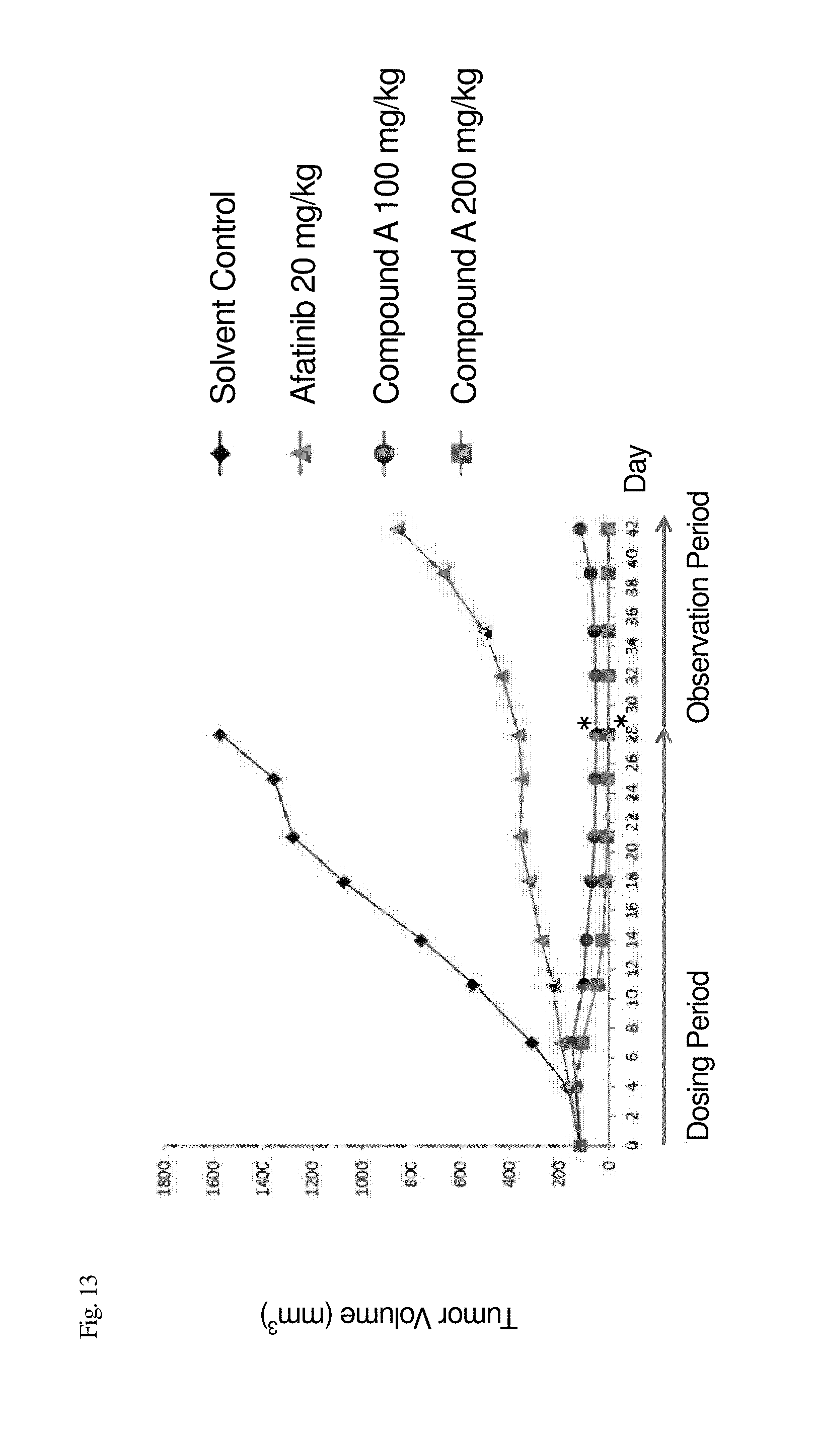
D00014

D00015
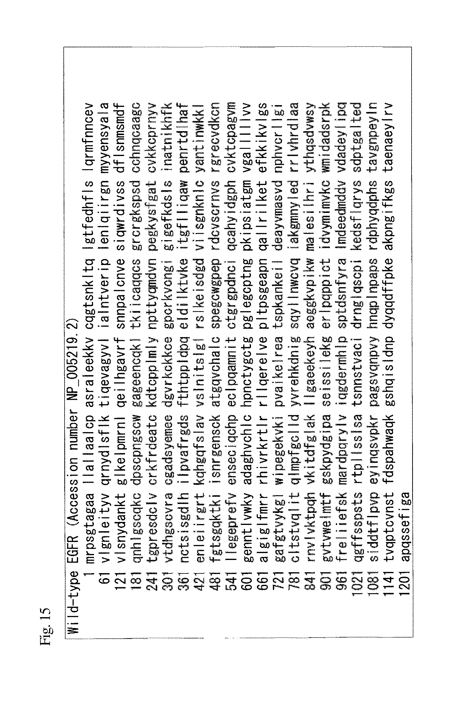
S00001
XML
uspto.report is an independent third-party trademark research tool that is not affiliated, endorsed, or sponsored by the United States Patent and Trademark Office (USPTO) or any other governmental organization. The information provided by uspto.report is based on publicly available data at the time of writing and is intended for informational purposes only.
While we strive to provide accurate and up-to-date information, we do not guarantee the accuracy, completeness, reliability, or suitability of the information displayed on this site. The use of this site is at your own risk. Any reliance you place on such information is therefore strictly at your own risk.
All official trademark data, including owner information, should be verified by visiting the official USPTO website at www.uspto.gov. This site is not intended to replace professional legal advice and should not be used as a substitute for consulting with a legal professional who is knowledgeable about trademark law.