Swellable and Structurally Homogenous Hydrogels and Methods of Use Thereof
Gao; Ruixuan ; et al.
U.S. patent application number 16/267849 was filed with the patent office on 2019-08-22 for swellable and structurally homogenous hydrogels and methods of use thereof. The applicant listed for this patent is Massachusetts Institute of Technology. Invention is credited to Edward Stuart Boyden, Linyi Gao, Ruixuan Gao, Chih-Chieh Yu.
| Application Number | 20190256633 16/267849 |
| Document ID | / |
| Family ID | 65635795 |
| Filed Date | 2019-08-22 |


View All Diagrams
| United States Patent Application | 20190256633 |
| Kind Code | A1 |
| Gao; Ruixuan ; et al. | August 22, 2019 |
Swellable and Structurally Homogenous Hydrogels and Methods of Use Thereof
Abstract
The invention encompasses hydrogels, monomer precursors of the hydrogels, methods for the preparation thereof, and methods of use therefor. The linking of monomers can take place using non-radical, bioorthogonal reactions such as copper-free click-chemistry.
| Inventors: | Gao; Ruixuan; (Cambridge, MA) ; Gao; Linyi; (Cambridge, MA) ; Yu; Chih-Chieh; (Cambridge, MA) ; Boyden; Edward Stuart; (Chestnut Hill, MA) | ||||||||||
| Applicant: |
|
||||||||||
|---|---|---|---|---|---|---|---|---|---|---|---|
| Family ID: | 65635795 | ||||||||||
| Appl. No.: | 16/267849 | ||||||||||
| Filed: | February 5, 2019 |
Related U.S. Patent Documents
| Application Number | Filing Date | Patent Number | ||
|---|---|---|---|---|
| 62626920 | Feb 6, 2018 | |||
| Current U.S. Class: | 1/1 |
| Current CPC Class: | C08J 3/075 20130101; C08F 220/04 20130101; C08F 2438/01 20130101; C08G 65/2624 20130101; C12Q 1/6841 20130101; G01N 1/36 20130101; C08G 2210/00 20130101; C08F 8/12 20130101; C08F 8/12 20130101; C08F 8/44 20130101; C08G 81/025 20130101; C12Q 1/68 20130101; C08F 220/30 20130101; C08F 8/30 20130101; C08J 2400/206 20130101; C08F 8/44 20130101; C08F 8/12 20130101; C08J 2300/206 20130101; C08F 220/34 20130101; G01N 15/0205 20130101; C12Q 1/6869 20130101; G01N 33/545 20130101; C12Q 1/6834 20130101; C12Q 2543/101 20130101; C08F 8/00 20130101; C08G 65/32 20130101; C12Q 2527/125 20130101; C08F 20/18 20130101; C08F 8/30 20130101; C08F 8/30 20130101; C08F 20/18 20130101; C08F 20/18 20130101; C12Q 2543/101 20130101; C08F 8/30 20130101; C08F 8/04 20130101; C12Q 2521/107 20130101; C08F 20/18 20130101; C12Q 2527/125 20130101; C12Q 2525/191 20130101; C08F 8/00 20130101; C08F 8/44 20130101; C08F 220/346 20200201; G01N 1/30 20130101; C12Q 1/6869 20130101; C08F 220/303 20200201; C12Q 2525/191 20130101; C08F 8/30 20130101; C08F 8/00 20130101; C12Q 1/6869 20130101; C12Q 2523/101 20130101; C08J 3/246 20130101; C08F 8/04 20130101; C12Q 2521/107 20130101 |
| International Class: | C08F 220/30 20060101 C08F220/30; C12Q 1/6834 20060101 C12Q001/6834; C12Q 1/6841 20060101 C12Q001/6841; C08F 220/34 20060101 C08F220/34; C08G 65/26 20060101 C08G065/26 |
Goverment Interests
GOVERNMENT SUPPORT
[0002] This invention was made with government support under Grant No. 6936173 awarded by ARO and under Grant No. 6934416 awarded by the National Institutes of Health. The government has certain rights in the invention.
Claims
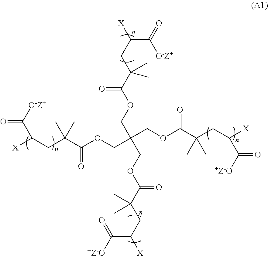
1. A hydrogel that is the product of a non-radical polymerization reaction between a monomer of Formula (A1): ##STR00012## and a monomer of Formula (B1): ##STR00013## wherein: each n is an integer greater than or equal to 1; each p is an integer greater than or equal to 1; Z.sup.- is a counter cation; X and Y.sub.1 are each crosslinkable moieties, and X and Y.sub.1 covalently crosslink to end-link the monomers.
2. (canceled)
3. (canceled)
4. The hydrogel of claim 1, wherein X is a moiety comprising a terminal azide group and Y.sub.1 is a moiety comprising a terminal alkyne, and wherein X and Y.sub.1 crosslink by copper-free azide-alkyne cycloaddition.
5-8. (canceled)
9. The hydrogel of claim 1, wherein X and Y.sub.1 crosslink by amine-NETS ester reaction.
10. (canceled)
11. The hydrogel of any claim 1, wherein X and Y.sub.1 crosslink by maleimide-thiol reaction.
12. The hydrogel of claim 1, wherein X and Y.sub.1 crosslink by trans-cyclooctene (TCO)-tetrazine reaction.
13. The hydrogel of claim 1, wherein the hydrogel is labelled.
14-16. (canceled)
17. A composite comprising a biological sample and the hydrogel of claim 1.
18-20. (canceled)
21. A method of preparing the composite of claim 17, comprising permeating the biological sample with a monomer of Formula (A1) or a monomer of Formula (A3), and a monomer of Formula (B 1) under conditions suitable to form a hydrogel by non-radical polymerization.
22-26. (canceled)
27. A method of microscopy comprising: a. permeating the biological sample with a monomer of Formula (A1): ##STR00014## and a monomer of Formula (B 1): ##STR00015## under conditions suitable to form a hydrogel according to claim 1 by non-radical polymerization; b. isotropically expanding the composite by contacting it with an aqueous solution; and c. viewing the expanded composite using microscopy; wherein: each n is an integer greater than or equal to 1; each p is an integer greater than or equal to 1; Z.sup.+ is a counter cation X and Y.sub.1 are each crosslinkable moieties, and X and Y.sub.1 covalently crosslink to end-link the monomers.
28. (canceled)
29. (canceled)
30. A method for in-situ sequencing of target nucleic acids present in a biological sample comprising the steps of: a. attaching target nucleic acids present in the sample with a molecule linker or nucleic acid adapter; b. permeating the sample with a monomer of Formula (A1): ##STR00016## and a monomer of Formula (B 1): ##STR00017## under conditions suitable to form a hydrogel according to claim 1 by non-radical polymerization and thereby forming a sample-hydrogel complex, wherein the small molecule linker or nucleic acid adaptor is attached both to the target nucleic acids present in the sample and to the hydrogel; c. digesting proteins present in the sample; d. expanding the complex to form a first enlarged sample; e. re-embedding the first enlarged sample in a non-swellable material to form a re-embedded complex; f. modifying the target nucleic acids or the nucleic acid adaptor to form a target nucleic acids or a nucleic acid adaptor; and g. sequencing the nucleic acids present in the re-embedded complex; wherein: each n is an integer greater than or equal to 1; each p is an integer greater than or equal to 1; Z.sup.+ is a counter cation; X and Y.sub.1 are each crosslinkable moieties, and X and Y.sub.1 covalently crosslink to end-link the monomers.
31-36. (canceled)
37. A method for enlarging a biological sample for microscopy, the method comprising the steps of: a) permeating a sample with a first hydrogel, wherein the sample is anchored to the swellable material; b) swelling the swellable material resulting in a first expanded sample; c) optionally permeating the first expanded sample with a second hydrogel; and d) optionally swelling the second hydrogel resulting in a second expanded sample; wherein the first hydrogel and/or the second hydrogel is the hydrogel of claim 1.
38-40. (canceled)
41. A hydrogel that is the product of a non-radical polymerization reaction between a monomer of Formula (A2): ##STR00018## and a monomer of Formula (B2) ##STR00019## wherein: each n is an integer greater than or equal to 1; each q is an integer greater than or equal to 1; Y.sub.2 is a moiety comprising a terminal dibenzocyclooctyl (DBCO) or a terminal bicyclononyne; X.sub.1 is a moiety comprising a terminal azide group; Z.sup.+ is a counter cation; and X.sub.1 and Y.sub.2 crosslink by copper-free azide-alkyne cycloaddition.
42-45. (canceled)
46. A composite comprising a biological sample and the hydrogel of claim 41.
47-49. (canceled)
50. A method of preparing the composite of claim 46, comprising permeating the sample with a monomer of Formula (A2) or Formula (A4), and a monomer of Formula (B2) under conditions suitable to form a hydrogel by non-radical polymerization.
51-55. (canceled)
56. A method of microscopy comprising: a. permeating the sample with a monomer of Formula (A2): ##STR00020## and a monomer of Formula (B2): ##STR00021## under conditions suitable to form a hydrogel according to claim 41 by non-radical polymerization; b. isotropically expanding the composite by contacting it with an aqueous solution; and c. viewing the expanded composite using microscopy; wherein: each n is an integer greater than or equal to 1; each q is an integer greater than or equal to 1; Y.sub.2 is a moiety comprising a terminal dibenzocyclooctyl (DBCO) or a terminal bicyclononyne; X.sub.1 is a moiety comprising a terminal azide group; Z.sup.+ is a counter cation; and X.sub.1 and Y.sub.2 crosslink by copper-free azide-alkyne cycloaddition.
57. (canceled)
58. A method for in-situ sequencing of target nucleic acids present in a biological sample comprising the steps of: a. attaching target nucleic acids present in the sample with a molecule linker or nucleic acid adapter; b. permeating the sample with a monomer of Formula (A2): ##STR00022## and a monomer of Formula (B2): ##STR00023## under conditions suitable to form a hydrogel according to claim 41 by non-radical polymerization and thereby forming a sample-hydrogel complex, wherein the small molecule linker or nucleic acid adaptor is attached both to the target nucleic acids present in the sample and to the hydrogel; c. digesting proteins present in the sample; d. expanding the complex to form a first enlarged sample; e. re-embedding the first enlarged complex in a non-swellable material to form a re-embedded complex; f. modifying the target nucleic acids or the nucleic acid adaptor to form a target nucleic acids or a nucleic acid adaptor; and g. sequencing the nucleic acids present in the re-embedded complex; wherein: each n is an integer greater than or equal to 1; each q is an integer greater than or equal to 1; Y.sub.2 is a moiety comprising a terminal dibenzocyclooctyl (DBCO) or a terminal bicyclononyne; X.sub.1 is a moiety comprising a terminal azide group; Z.sup.+ is a counter cation; and X.sub.1 and Y.sub.2 crosslink by copper-free azide-alkyne cycloaddition.
59-62. (canceled)
63. A method for enlarging a biological sample for microscopy, the method comprising the steps of: a) permeating a biological sample with a first hydrogel comprising a first cleavable crosslinker, wherein the sample is anchored to the first hydrogel; b) swelling the first hydrogel resulting in a first expanded sample; c) optionally, permeating the first expanded sample with a second hydrogel comprising a second cleavable crosslinker that is different from the first cleavable crosslinker and wherein the first and second cleavable crosslinkers are cleavable under different conditions; and d) optionally swelling the second hydrogel resulting in a second expanded sample; wherein the first hydrogel and/or the second hydrogel is the hydrogel of claim 41.
64. (canceled)
65. (canceled)
66. A hydrogel that is the product of a non-radical polymerization reaction between a monomer of Formula (A5): ##STR00024## and a monomer of Formula (B 1): ##STR00025## wherein: each n is an integer greater than or equal to 1; each p is an integer greater than or equal to 1; E is a moiety comprising a charged functional group; Z is a counter ion; X and Y.sub.1 are each crosslinkable moieties, and X and Y.sub.1 covalently crosslink to end-link the monomers.
67-69. (canceled)
70. A hydrogel that is the product of a non-radical polymerization reaction between a monomer of Formula (A6): ##STR00026## and a monomer of Formula (B2) ##STR00027## wherein: each n is an integer greater than or equal to 1; each q is an integer greater than or equal to 1; E is a moiety comprising a charged functional group; Z is a counter ion; Y.sub.2 is a moiety comprising a terminal dibenzocyclooctyl (DBCO) or a terminal bicyclononyne; X.sub.1 is a moiety comprising a terminal azide group; Z.sup.+ is a counter cation; and X.sub.1 and Y.sub.2 crosslink by copper-free azide-alkyne cycloaddition.
71. A monomer of Formula (A) selected from the group consisting of: ##STR00028## ##STR00029## wherein: each n is an integer greater than or equal to 1; Z is a counter ion; Z.sup.+ is a counter cation; X is a crosslinkable moiety; X.sub.1 is a moiety comprising a terminal azide group; E is a moiety comprising a charged functional group.
72. (canceled)
73. A monomer of Formula (B1) or Formula (B2): ##STR00030## wherein: Y.sub.1 is a crosslinkable moiety; Y.sub.2 is a moiety comprising a terminal dibenzocyclooctyl (DBCO) or a terminal bicyclononyne; each p is an integer greater than or equal to 1; and each q is an integer greater than or equal to 1.
Description
RELATED APPLICATION
[0001] This application claims the benefit of U.S. Provisional Application No. 62/626,920, filed on Feb. 6, 2018. The entire teachings of the above application are incorporated herein by reference.
BACKGROUND OF THE INVENTION
[0003] Expansion microscopy (ExM), described for example in WO2015127183 and Chen et al., Science, 347, 543 (2015), is a technique that allows for three-dimensional (3D) nanoscale imaging of biological samples by physically expanding the specimens.sup.1-4. In ExM, hydrogels are synthesized within the biological samples. During the gelation process, biomolecules or tags are anchored to the hydrogel matrix. The hydrogel-specimen composite then goes through a 3D expansion, physically separating the biomolecules or tags.
[0004] In all of the ExM processes reported, the swellable hydrogel is synthesized by radical polymerization, a reaction known to introduce structural inhomogeneities at nanoscopic length scales.sup.5-7. The structural inhomogeneity is mainly caused by two factors: (a) local fluctuation of reagent concentrations during gelation and (b) topological defects such as loops and entanglements of polymer chains.
[0005] Therefore, there is a need in the art for a swellable hydrogel that is structurally homogenous down to the nanoscopic length scale.
SUMMARY OF THE INVENTION
[0006] The invention encompasses hydrogels, monomer precursors of the hydrogels, composites comprising the hydrogel and a biological sample, methods for the preparation of the hydrogels and the composites, and methods of using the hydrogels and the composites. As described in more detail below, the hydrogels are designed to be structurally homogenous down to the nanoscopic length scale. The linking of the monomers described herein can take place using non-radical, bio-orthogonal reactions such as copper-free click-chemistry.
[0007] In some aspects, the hydrogel is the product of a non-radical polymerization reaction between a monomer of Formula A, wherein the monomer of Formula A is a monomer of Formula A1, Formula A2, Formula A3, Formula A4, Formula A5, or Formula A6.
[0008] In some aspects, the hydrogel is the product of a non-radical polymerization reaction between a monomer of Formula (A1):
##STR00001##
and a monomer of Formula (B1):
##STR00002##
wherein: each n is an integer greater than or equal to 1; each p is an integer greater than or equal to 1; X and Y.sub.1 are each crosslinkable moieties; Z.sup.+ is a counter cation; and X and Y.sub.1 covalently crosslink to end-link the monomers. In preferred aspects, the non-radical polymerization is bio-orthogonal. In certain aspects, X is a moiety comprising a terminal azide group and Y.sub.1 is a moiety comprising a terminal alkyne and X and Y.sub.1 crosslink by copper-free azide-alkyne cycloaddition.
[0009] In additional aspects, the hydrogel is the product of a non-radical polymerization reaction between a monomer of Formula (A2):
##STR00003##
and a monomer of Formula (B2)
##STR00004##
wherein: each n is an integer greater than or equal to 1; each q is an integer greater than or equal to 1; Y.sub.2 is a moiety comprising a terminal dibenzocyclooctyl (DBCO) or a terminal bicyclononyne; X.sub.1 is a moiety comprising a terminal azide group; Z.sup.+ is a counter cation; and X.sub.1 and Y.sub.2 crosslink by copper-free azide-alkyne cycloaddition. In preferred aspects, the non-radical polymerization is bio-orthogonal. In certain aspects, Z.sup.+ is Na.sup.+ or K.sup.+.
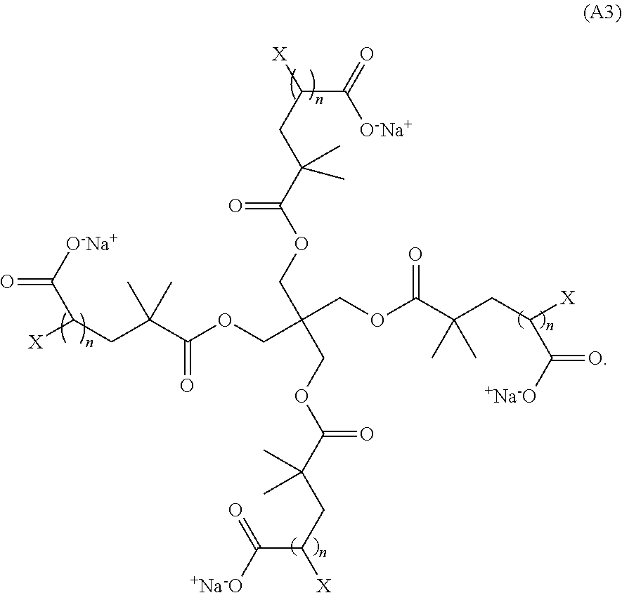
[0010] In certain additional aspects, Z.sup.+ is Na.sup.+ or K.sup.+. A non-limiting example of a monomer of Formula (A1) has the Formula (A3):
##STR00005##
Thus, the invention also encompasses a hydrogel that is the product of a non-radical polymerization reaction between a monomer of Formula (A3) and a monomer of Formula (B1).
[0011] A non-limiting example of a monomer of Formula (A2) has the Formula (A4):
##STR00006##
The invention also includes a hydrogel that is the product of a non-radical polymerization reaction between a monomer of Formula (A4) and a monomer of Formula (B2).
[0012] The invention additionally encompasses a hydrogel that is the product of a non-radical polymerization reaction between a monomer of Formula (A5):
##STR00007##
and the monomer of Formula (B1) as described above, wherein E is a moiety comprising a charged functional group and Z is a counter ion (for example, a counter cation or counter anion depending on the charge of E); and X and n are as defined above for Formula (A1). In certain aspects, E is a charged functional group; for example, E is selected from a carboxylic acid group, an ammonium group, and a sulfate group.
[0013] The invention additionally encompasses a hydrogel is the product of a non-radical polymerization reaction between a monomer of Formula (A6):
##STR00008##
and the monomer of Formula (B2) as described above, wherein E and Z are as defined above for Formula (A5); and X and n are as defined above for Formula (A1). In certain aspects, E is selected from a carboxylic acid group, an ammonium group, and a sulfate group.
[0014] In certain additional aspects, the invention is directed to a composite comprising a biological sample and a hydrogel that is the product of a non-radical polymerization reaction between the monomer of Formula (A1), (A2), (A3), (A4), (A5) or (A6) (collectively referred to herein as Formula (A)) and the monomer of Formula (B1).The invention also encompasses a method of preparing the composite comprising permeating the biological sample with the monomer of Formula (A1), (A2), (A3), (A4), (A5) or (A6) and the monomer of Formula (B1) under conditions suitable to form a hydrogel by non-radical polymerization.
[0015] In certain additional aspects, the invention is directed to a composite comprising a biological sample and a hydrogel that is the product of a non-radical polymerization reaction between the monomer of Formula (A2), (A4), or (A6) and the monomer of Formula (B2). The invention also encompasses a method of preparing the composite comprising permeating the biological sample with the monomer of Formula (A2), (A4), or (A6), and the monomer of Formula (B2) under conditions suitable to form a hydrogel by non-radical polymerization. In yet additional aspects, the invention is directed to a method of microscopy comprising: [0016] a. permeating the biological sample with a monomer of Formula (A1), (A2), (A3), (A4), (A5) or (A6) and a monomer of Formula (B1) under conditions suitable to form a hydrogel by non-radical polymerization; [0017] b. isotropically expanding the composite by contacting it with an aqueous solution; and [0018] c. viewing the expanded composite using microscopy.
[0019] In further aspects, the invention is directed to a method of microscopy comprising: [0020] a. permeating the biological sample with a monomer of Formula (A2), (A4), or (A6), and a monomer of Formula (B2) under conditions suitable to form a hydrogel by non-radical polymerization; [0021] b. isotropically expanding the composite by contacting it with an aqueous solution; and [0022] c. viewing the expanded composite using microscopy.
BRIEF DESCRIPTION OF THE DRAWINGS
[0023] The patent or application file contains at least one drawing executed in color. Copies of this patent or patent application publication with color drawings will be provided to the Office upon request and payment of the necessary fee.
[0024] The foregoing and other objects, features and advantages of the invention will be apparent from the following more particular description of preferred embodiments of the invention, as illustrated in the accompanying drawings in which like reference characters refer to the same parts throughout the different views. The drawings are not necessarily to scale, emphasis instead being placed upon illustrating the principles of the invention.
[0025] FIGS. 1A and 1B: Design of swellable, structurally homogenous hydrogels based on tetrahedral monomers. (a) Tetrahedral monomers A and B have functional end groups that specifically and complementarily bind to each other. (b) One implementation of Monomer A with repeated sodium acrylate units.
[0026] FIG. 2: Synthesis of one version of Monomer A (shown in FIG. 1B) based on the 4-arm sodium polyacrylate structure. The synthesis starts with arm elongation via atom transfer radical polymerization (synthetic details shown as "*") of tent-butyl acrylate. The bromide end groups are further modified to azide end groups for copper click-chemistry used to link the monomers. The tert-butyl polyacrylate arms are then deprotected to yield Monomer A.
[0027] FIG. 3A and 3B: Synthesis of one version of Monomer B based on 4-arm PEGs. (a) Using 4-arm PEG-amines as the starting material, different end groups such as (i) dibenzocyclooctyl (DBCO), (ii) bicyclononyne (BCN) or (iii) dibenzocyclooctyl-disulfide (DBCO-SS) groups can be added to the arm ends via amine/NHS ester reaction. (b) An intermediate in the Monomer A synthesis (in FIG. 2) can be further modified into a 4-arm sodium polyacrylate species with DBCO end groups and can be used as Monomer B.
[0028] FIG. 4: Synthesis and expansion of hydrogel (termed as "tetragel") formed by reacting Monomer A and Monomer B. Fluorescence images of tetragels after expansion using three different Monomers B with the end groups specified in FIG. 3a-i (DBCO), 3a-ii (BCN) and 3a-iii (DBCO-SS) are shown. Fluorescent image of the tetragel before expansion (the pre-expansion size is the same for all three versions of Monomer B) is shown on the left for comparison. About 1 mol % fluorescein amine was anchored to the hydrogel matrix with NHS-azide to fluorescently visualize the gels. Scale bar, 5 mm.
[0029] FIG. 5A: In situ synthesis and expansion of brain tissue with tetragel. Fluorescence images of the same Thy1-YFP mouse brain slice before (left) and after (right) expansion with tetragel. The post-expansion sample was immunostained by anti-GFP primary antibodies and dye-conjugated secondary antibodies. Scale bar: 2 mm (left) and 6 mm (right).
[0030] FIG. 5B: Post-expansion Thy1-YFP mouse brain slices. Scale bar, 10 .mu.m (right, 30 .mu.m)
[0031] FIG. 6A-6G: In situ synthesis and expansion of cultured cells with tetragel. (a, c) Fluorescence image of expanded HEK 293 cells immunostained with anti-a-tubulin primary antibodies and dye-conjugated secondary antibodies. Scale bar, 20 .mu.m. (b, d) Fluorescence image of the same cells before expansion. Scale bar, 20 .mu.m. (e) Line profile of the image specified by the solid line in (c). (f) Line profile of the image specified by the solid line in (d). (g) RMS error curve for the HEK293 cell expansion (blue line, mean; shaded area, standard deviation; n=8 cells). Inset, non-rigidly registered and overlaid pre-expansion (green) and post-expansion (magenta) images used for the RMS error estimation.
[0032] FIG. 7A-7C: In situ synthesis and expansion of nuclear pore complex with tetragel. (a) Pre-expansion live fluorescent image of nuclear pore complex with GFP fusion. Scale bar, 10 .mu.m. (b), (c) Post-expansion fluorescent image of (b) GFP and (c) Cy3-conjugated secondary antibody (Nup133 primary antibody). Scale bars, 20 .mu.m.
[0033] FIG. 8A and 8B: Retention of far-red dye in tetragel. (a) Fluorescence images of mouse brain slices stained with far-red dyes before and after expansion with tetragel. The brain slices were immunostained with Tom 20 primary antibodies and far-red dye conjugated secondary antibodies. (b) Retention of far-red dyes (Alexa Fluor 647 (AF647) and Cyanine 5 (Cy5)) with Stock X gel (used for conventional ExM) and tetragel.
[0034] FIG. 9A: Pre-expansion (left column) and post-expansion (right column) Thy1-YFP mouse brain slices immunostained with TOMM20 primary antibodies and AF647-conjugated secondary antibodies, using polyacrylamide/sodium polyacrylate gels (PAAs, top row) and TGs (bottom row). Scale bars, 300 .mu.m (top right, 1.18 mm; bottom right, 815 .mu.m).
[0035] FIG. 9B: Fluorescence retention of far-red and infra-red dyes (AF647, Cy5 and AF680) using PAAs and TGs.
[0036] FIG. 10: Design and synthesis of structurally homogenous hydrogel matrix for expansion of nanoscale biological structures. a, Cell/tissue-hydrogel composites formed by in situ free-radical polymerization have structural inhomogeneities at 10-100 nm length scale due to (1) the local fluctuation of monomer and cross-linking density, and (2) the dangling ends and (3) loops in the polymer chains. b, Design of monomers 1 and 2 (monomers A and B; and referred to in the Examples as "monomer 1" and "monomer 2") with tetrahedral symmetry and reactive terminal groups. Modification of monomer 2 terminal groups (2, 2' and 2'') allows fine tuning of the reactivity between the monomers and additional functionality of, for example, cleavability. c, Formation and expansion of tetra-gels (TGs) via click chemistry-based terminal-linking of monomers 1 and 2 (or 2', 2''). Inset, projected planar view of the polymerized network.
[0037] FIG. 11A-11D: Tetragel (TG)-based iterative expansion enables nanoscopically isotropic expansion at 10-100 nm length scale. a, Short DNA-oligos (22 base pairs) were directly anchored to the envelope proteins of an HSV-1 virion via hydrazone formation. The DNA-oligos were used for signal transferring, amplification, and fluorescence readout in the subsequent iterative expansion process. b, HSV-1 virions with oligo-labeled envelope proteins, expanded by TG-based (left) and PAA-based (right) two-round iterative expansion. Scale bar, 1 .mu.m (10.3 .mu.m and 15.3 .mu.m for TG and PAA, respectively). c, Left, averaged single-particle images of HSV-1 virions by TG-based (top) and PAA-based (bottom) expansion. Scale bars, 100 nm. Right, normalized fluorescence intensity along the dotted lines. d, 3D rendered images of an HSV-1 virion particle with the oligo-labeled envelope proteins, expanded by TG-based three-round iterative expansion. The raw data (white, left) overlaid with the fitted centroids (red, left) and the extracted centroids (colored, right) are shown. Scale bars, 100 nm (3.83 .mu.m). Inset, maximum intensity projection (MIP) of the same virion particle over a .about.65 nm range close to the particle center (as shown between the two lines in the rendered image). Scale bars, 100 nm (3.83 .mu.m).
[0038] FIG. 12: HSV-1 virions with oligo-labeled envelope proteins (white) and DAPI-labeled DNAs (blue). The virions were expanded by TG-based 10-fold iterative expansion. Scale bar, 1 .mu.m (10.7 .mu.m).
DETAILED DESCRIPTION OF THE INVENTION
[0039] A description of preferred embodiments of the invention follows.
[0040] As used herein, the words "a" and "an" are meant to include one or more unless otherwise specified. For example, the term "a cell" encompasses both a single cell and a combination of two or more cells.
[0041] As will be apparent to those of skill in the art, each of the individual embodiments described and illustrated herein has discrete components and features which can be readily separated from or combined with the features of any of the other several embodiments without departing from the scope or spirit of the present teachings. Any recited method can be carried out in the order of events recited or in any other order which is logically possible.
[0042] As used herein, the terms "specimen" or "sample" are used interchangeably herein and include, but are not limited to tissues, including but not limited to, liver, spleen, kidney, lung, intestine, thymus, colon, tonsil, testis, skin, brain, heart, muscle and pancreas tissue. Other exemplary biological samples include, but are not limited to, biopsies, bone marrow samples, organ samples, skin fragments and organisms. Materials obtained from clinical or forensic settings are also within the intended meaning of the term biological sample. In one embodiment, the sample is derived from a human, animal, or plant. In one embodiment, the biological sample is a tissue sample, preferably an organ tissue sample. In one embodiment, samples are human. The sample can be obtained, for example, from autopsy, biopsy or from surgery. It can be a solid tissue such as, for example, parenchyme, connective or fatty tissue, heart or skeletal muscle, smooth muscle, skin, brain, nerve, kidney, liver, spleen, breast, carcinoma (e.g., bowel, nasopharynx, breast, lung, stomach etc.), cartilage, lymphoma, meningioma, placenta, prostate, thymus, tonsil, umbilical cord or uterus. The tissue can be a tumor (benign or malignant), cancerous or precancerous tissue. The sample can be obtained from an animal or human subject affected by disease or other pathology or suspected of same (normal or diseased), or considered normal or healthy. The biological sample can, for example, be a cell sample. In certain aspects, the biological sample is a virus or virion. The term "biological sample" can a biological sample that comprises, or is believed to comprise, nucleic acid sequences including, but not limited to cDNA, mRNA and genomic DNA.
[0043] Tissue specimens suitable for use with the methods and systems described herein generally include any type of tissue specimens collected from living or dead subjects, such as, e.g., biopsy specimens and autopsy specimens. Tissue specimens may be collected and processed using the methods and systems described herein and subjected to microscopic analysis immediately following processing, or may be preserved and subjected to microscopic analysis at a future time, e.g., after storage for an extended period of time. In some embodiments, the methods described herein may be used to preserve tissue specimens in a stable, accessible and fully intact form for future analysis. For example, tissue specimens, such as, e.g., human brain tissue specimens, may be processed as described above and cleared to remove a plurality of cellular components, such as, e.g., lipids, and then stored for future analysis.
[0044] Tissues that have been preserved, or fixed, contain a variety of chemical modifications that can reduce the detectability of proteins in biomedical procedures. In some embodiments, the methods and systems described herein may be used to analyze a previously-preserved or stored tissue specimen. Previously preserved tissue specimens include, for example, clinical samples used in pathology including formalin-fixed paraffin-embedded (FFPE), hematoxylin and eosin (H&E)-stained, and/or fresh frozen tissue specimens. If the previously preserved sample has a coverslip, the coverslip should be removed. The sample is treated to remove the mounting medium. Such methods for removing the mounting medium are well known in the art. For example, treating the sample with xylene to remove paraffin or other hydrophobic mounting medium. Alternatively, if the sample is mounted in a water-based mounting medium, the sample is treated with water. The sample is then rehydrated and subjected to antigen-retrieval. The term "antigen retrieval" refers to any technique in which the masking of an epitope is reversed and epitope-antibody binding is restored such as, but not limited to, enzyme induced epitope retrieval, heat induced epitope retrieval (HIER), or proteolytic induced epitope retrieval (PIER). For example, the antigen retrieval treatment can be performed in a 10 mM sodium citrate buffer as well as the commercially available Target Retrieval Solution (DakoCytomation) or such.
[0045] The term "bio-orthogonal" in reference to a chemical reaction refers to a chemical reaction that does not interfere with any other chemical moieties in the natural or native surroundings.
[0046] The term "sequencing," as used herein, refers to a method by which the identity of at least 10 consecutive nucleotides (e.g., the identity of at least 20, at least 50, at least 100 or at least 200 or more consecutive nucleotides) of a polynucleotide is obtained.
[0047] Monomers of Formulae (A1), (A2), (A3), (A4), (A5) and (A6) can collectively be referred to as monomers of Formula (A). Monomers of Formula (B1) and (B2) can collectively be referred to as monomers of Formula (B).
[0048] The term "hydrogel AB 1" is used to refer to a hydrogel that is the product of a reaction between a monomer of Formula (A) and a monomer of Formula (B1), and the term "hydrogel AB2" is used to refer to a hydrogel that is the product of a reaction between a monomer of Formula (A) and a monomer of Formula (B2). Other hydrogel that are the product of a reaction between a monomer of Formula (A) and a monomer of Formula (B1) or (B2) can be similarly referred to.
[0049] The invention encompasses hydrogels, monomer precursors of the hydrogels, methods for the preparation of the hydrogels, and methods of using the hydrogel, for example, in expansion microscopy and/or in situ sequencing, wherein the hydrogel is the product of a non-radical polymerization reaction between a monomer of Formula (A) and a monomer of Formula (B1), or the product of a non-radical polymerization reaction between a monomer of Formula (A2), (A4), or (A6) and a monomer of Formula (B2).
[0050] In certain aspects, the invention is directed to a hydrogel that is the product of a non-radical polymerization reaction between a monomer of Formula (A1), (A2), (A3), (A4), (A5), or (A6) and a monomer of Formula (B1). In certain additional aspects, the X of Formula (A1), (A3), and/or (A5) or X.sub.1 of Formula (A2), (A4), or (A6) is azide (--N.sub.3) and the Y.sub.1 of Formula (B1) is a cyclic alkyne.
[0051] In additional aspects, the invention is directed to a hydrogel that is the product of a non-radical polymerization reaction between a monomer of Formula (A2), (A4), or (A6), and a monomer of Formula (B2).
[0052] The invention also encompasses a monomer of Formula (A1), (A3), and (A5), in certain embodiments, X is azide. The invention additionally encompasses a monomer of Formula (A2), (A4), and (A6), wherein X.sub.1 is azide. The invention additionally encompasses a monomer of Formula (B1); in certain aspects, Y.sub.1 is a cyclic alkyne such as dibenzocyclooctyl (DBCO) or a bicyclononyne. In further aspects, the invention encompasses a monomer of Formula (B2); in certain aspects, X is azide.
[0053] In certain aspects, E of Formula (A5) or Formula (A6) is a negatively charged functional group. Exemplary negatively-charged groups include, without limitation, carboxylic (e.g., acetic) group, sulfo group, sulfino group, phosphate group and phosphono group. In yet additional aspects, E of Formula (A5) or Formula (A6) is a positively charged functional group. Examples of positively-charged functional groups include, without limitation, amino (amine) groups that can be protonated to form an ammonium group. In certain aspects, a charged functional group is one that exhibits a charge at, or near, neutral pH (pH of about 5 to about 9 or about 6 to about 8) in an aqueous medium.
[0054] Z is a counter ion. For example, in Formulae (A5) and (A6), if E comprises a negatively charged functional group, then Z is a counter cation, and if E comprises a positively charged functional group, then Z is a counter anion.
[0055] Z.sup.+ is a counter cation such as an alkali metal atom, an alkali earth metal atom, or substituted or unsubstituted ammonium. Non-limiting examples of counter cations are potassium, sodium, mercury, lithium, magnesium, calcium, butylammonium, trimethylammonium, and tetramethyl ammonium. In certain aspects, the counter cation is sodium or potassium (Na+ or K+). In yet additional aspects, the counter cation is (Na+). In certain aspects, the monomer of Formula (A1) has the Formula (A3):
##STR00009##
[0056] The hydrogels described herein are swellable and can be used in expansion microscopy (ExM). In ExM, chemically fixed and permeabilized tissue (or other biological sample) is infused with swellable material, undergoes polymerization, and the tissue-polymer composite is treated to homogenize its mechanical characteristics. Next, dialysis in water or aqueous solution results in isotropic expansion, thereby achieving super-resolution with diffraction-limited microscopes, and enabling rapid image acquisition and large field of view (Chen et al., Science, 347, 543 (2015)). Expansion allows individual nucleic acids, normally densely packed, to be resolved spatially in a high-throughput manner. Furthermore, the expanded environment is 99% water, facilitating enzyme access and creating "quasi-in vitro" environment while retaining spatial information. In some examples, fixation of the biological sample can be effected by embedding the sample in a swellable material that has been perfused throughout the sample as described by Chen et al. (Chen et al., Science, 347, 543 (2015) and U.S. Patent Publication Nos. US 20160116384-A1; US 20160305856-A1; US 20160304952-A1; and U.S. Patent Publication Nos. US 20170067096 A1 and US 20170089811 A1, each corresponding to U.S. patent application Ser. Nos. 15/229,539 and 15/229,545, respectively, the contents of each of which are incorporated herein by reference in their entirety. Briefly, a sample, such as tissue, can be permeabilized. A permeabilized sample, or tissue, can be infused with monomers or precursors of a swellable material and then causing the monomers or precursors to undergo polymerization within the sample to form the swellable material. During or after polymerization, the swellable material can be anchored to the sample. The sample-hydrogel complex (or composite) is optionally treated to homogenize the mechanical characteristics of the sample. The sample-swellable material complex can then be treated by dialysis in a solvent or liquid, such as in water, resulting in isotropic physical expansion of the sample. In this manner, the fixed biological sample is physically "enlarged", or "expanded", as compared to the biological sample before swelling.
[0057] The swellable hydrogels currently being used in expansion microscopy (ExM) are synthesized by radical polymerization which is known to introduce structural inhomogeneities at nanoscopic length scale. The structural inhomogeneity is mainly caused by two factors: (a) local fluctuation of reagent concentrations during the gelation and (b) topological defects such as loops and entanglements of polymer chains. To eliminate these intrinsic structural inhomogeneities, the hydrogels described herein have been designed which are structurally homogenous down to the nanoscopic length scale. For example, two types of pre-synthesized tetrahedral monomers are linked in a diamond lattice-like structure. The linking of the monomers takes place using non-radical, bio-orthogonal reactions including, but not limited to, copper-free click-chemistry. In this new hydrogel design, the homogeneity in monomer shape and size mitigates the effect of reagent concentration variations described in (a) above, resulting in a more uniform distribution of monomers and cross-links throughout the gel. Furthermore, the specific and complementary linking chemistry between the monomers reduces topological defects caused by (b) and thus facilitates formation of a homogeneous and isotropic polymer network.
[0058] It has been shown that a non-swellable hydrogel with similar diamond lattice-like structure to the hydrogel described herein is structurally homogeneous and has nearly zero defects'. Specifically, Sakai and colleagues demonstrated that end-linked tetrahedral monomers based on polyethylene glycol (PEG) can form highly homogeneous hydrogel networks free of defects'. These hydrogels, however, exhibit minimal expansion (-1.3x volume) and thus cannot be directly used in ExM. Oshima and colleagues investigated expandable hydrogels based on charged tetrahedral polyacrylate monomers, finding that this network is structurally superior to polyacrylate gels formed via radical polymerization in terms of sol fractions, dangling chains and trapped entaglements.sup.9. However, this strategy for gel formation required (1) copper catalysis and (2) treatment with trifluoroacetic acid, which are unlikely to be compatible with biological specimens.
[0059] In contrast to the gels of Sakai and Oshima, the hydrogels of the present invention are capable of isotropically expanding and are prepared using non-radical, bio-orthogonal reactions to end-link monomers. As described above, the lengths between cross-linkers (i.e., the "mesh size") need to be uniform in order to eliminate structural inhomogeneities in hydrogels. In addition, topological defects, such as entanglements, loops and dangling chains, need to be significantly reduced. The use of pre-synthesized monomers of defined arm lengths which are linked in a periodic fashion results in the formation of a hydrogel with structural homogeneity. For instance, two kinds of homogeneous tetrahedral monomers (Monomer A and Monomer B shown in FIG. 1A) can be linked to form a diamond lattice-like polymer network (FIG. 1A). In this design, monomers of Formula (A) and monomers of Formula (B1) or (B2) have specific and complementary linkers that bind two arms in between the two monomers. The linking needs to be covalent so that when the hydrogel physically expands in water, the monomers will stay linked and the diamond lattice-like structure will be maintained.
[0060] In swellable hydrogels (in particular, those swelling in water), the polymer chains commonly have charged functional groups so that the charges can repel each other and pull the polymer chains apart. For instance, in the polyacrylamide/sodium polyacrylate hydrogels that have been described for use in ExM, the polymer chains have carboxylate groups that are negatively charged in water ("polyelectrolyte"). The repulsion between the negative charges help to keep the hydrogel expanded in water. In an example of the present design of tetrahedral monomers, the monomers (specifically, the monomers of Formula (A) and/or monomers of Formula (B2)) include repeats of sodium acrylate groups in each arm to make the monomer charged in water. For example, a monomer of Formula (A) and/or a monomer of Formula (B2) can include repeats of sodium acrylate units in each arm of the monomer (FIG. 1B). The hydrogels can also be prepared using a monomer of Formula (A) that includes repeats of ionized or charged functional groups other than carboxylate. Such monomers are encompassed by Formulae (A5) and (A6). For example, the repeating functional group can be ammonium or sulfate.
[0061] The mesh size of the hydrogel can be systematically controlled by synthetically changing the arm lengths of the monomers ("n" in the Formula (A), "p" in Formulae (B1) and "q" in Formula (B2)). In certain aspects, n of the Formulae (A) is 1 to 100. In yet additional aspects, n is 4, or 10, or 20, or 40. In yet additional aspects, the monomer of Formula (A) has a molecular weight from about 0.5 kDa to about 50 kDa, or about 1 kDa to about 40 kDa, or about 1 kDa to about 25 kDa. By way of example, the monomer of Formula (A3), wherein n is 4, 10, 20 or 40, has an approximate molecular weight of 2 kDa, 5 kDa, 10 kDa, and 20 kDa, respectively.
[0062] In certain additional aspects, p of Formula (B1) is 1 to 100. In yet additional aspects, p of Formula (B1) is 36, or 72, or 144. In yet additional aspects, the monomer of Formula (B1) has a molecular weight from about 10 kDa to about 300 kDa, or about 15 kDa to about 200 kDa, or about 15 kDa to about 150 kDa. By way of example, the monomer of Formula (B1), wherein p is 36, 72 and 144, the monomer has an approximate molecular weight of about 5 kDa, 10 kDa, and 20 kDa, respectively.
[0063] In certain additional aspects, q of Formula (B2) is 1 to 100. In yet additional aspects, q is 36, 72, or 144.
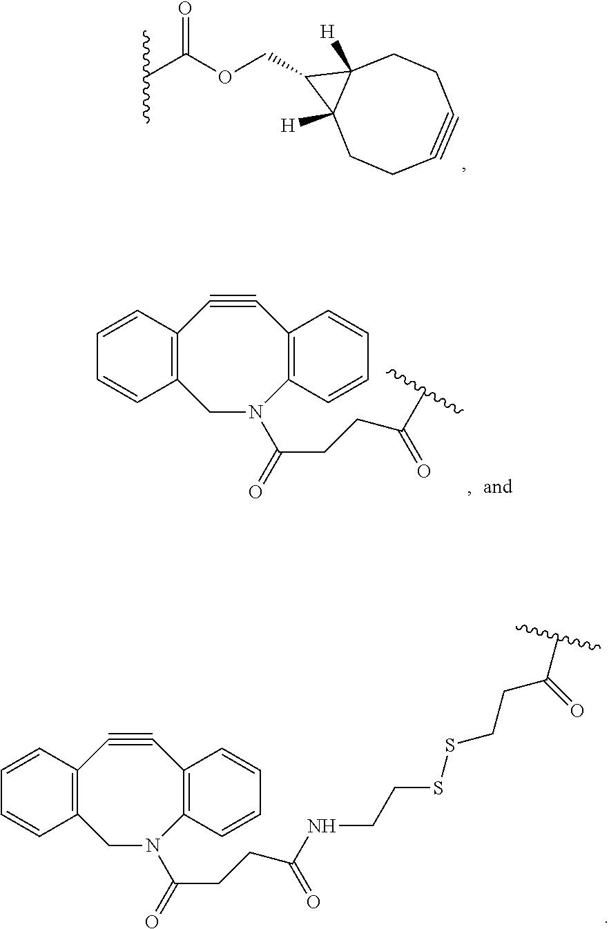
[0064] As described above, the two monomers (a monomer of Formula (A) and a monomer of Formulae (B1) or (B2)) are end-linked to form a lattice-like polymer network. Each of X and Y.sub.1, X.sub.1 and Y.sub.1, X and Y.sub.2, and X.sub.1 and Y.sub.2 are complementary, reactive end-groups that are capable of forming a covalent bond. For example, in certain aspects, X is a moiety comprising a terminal azide group and Y.sub.1 is a moiety comprising a terminal alkyne, for example, a cyclic alkyne, and X and Y.sub.1 crosslink by copper-free azide-alkyne cycloaddition. In certain additional aspects, X or X.sub.1 is azide (--N3). Y.sub.1 can, for example, be a cyclic alkyne moiety such as a moiety comprising a terminal dibenzocyclooctyl (DBCO) or a terminal bicyclononyne. Exemplary cyclic alkyne moieties include, but are not limited to, DBCO, DBCO-SS (dibenzocyclooctyl-disulfide), DBCO-amine, DBCO-N-hydroxsuccinimidyl ester, (1R,8S,9s)-bicyclo[6.1.0]non-4-yn-9-ylmethyl N-succinimidyl carbonate and DBCO-maleimide. In yet additional aspects, Y.sub.1 is selected from the group consisting of:
##STR00010##
[0065] The linking chemistry between the monomers is not limited to the catalyst-free click-chemistry described above. Other linking chemistries include other bio-orthogonal reactions including, but not limited to, amine-NETS reaction, maleimide-thiol reaction, and trans-cyclooctene/s-tetrazine reaction. Therefore, in some embodiments, wherein X and Y.sub.1 crosslink by amine-NETS ester reaction. For example, X can be a moiety comprising a terminal amine and Y.sub.1 can be a terminal N-hydroxysuccinimide ester group. Alternatively, X can be a moiety comprising a terminal N-hydroxysuccinimide ester group and Y.sub.1 can be a terminal amine. In another example, X and Y.sub.1 crosslink by maleimide-thiol reaction. For example, X can be a moiety comprising a terminal sulfhydryl group and Y.sub.1 can be a moiety comprising a terminal maleimide group. Alternatively, X can be a moiety comprising a terminal maleimide group and Y.sub.1 can be a moiety comprising a terminal sulfhydryl group. In yet an additional example, X and Y.sub.1 crosslink by trans-cyclooctene (TCO)-tetrazine reaction.
[0066] In one example, the Monomer A (a monomer of Formula (A)) as shown in FIG. 1B can be synthesized via atom transfer radical polymerization (ATRP) and subsequent synthetic modifications (FIG. 2).sup.9. First, tent-butyl acrylates are added to a 4-arm tetrahedral core to the desired arm length via ATRP ("*"in FIG. 2). In contrast to the conventional radical polymerization, ATRP offers site-specific and controlled addition of acrylate units to the growing polymer chains. The end groups of the monomer arms are then modified from bromide to azide for copper-free click chemistry. Finally, tert-butyl groups on the monomer arms are deprotected by a strong acid to yield Monomer A with 4-arm sodium polyacrylate structure.
[0067] Monomer B (a monomer of Formula (B1)) can be synthesized by end-functionalization of commercially available 4-arm PEG-amines (FIG. 3A). The end groups of the 4-arm PEG monomers can be modified to, for example, (i) dibenzocyclooctyl (DBCO), (ii) bicyclononyne (BCN) or (iii) dibenzocyclooctyl-disulfide (DBCO-SS) groups, all of which are reactive to the azide groups of Monomer A via copper-free click chemistry. BCN reacts more slowly with azide than DBCO or DBCO-SS and thus is more suitable for biological samples that require longer diffusion time through the specimens before the gelation begins, such as intact tissue samples. DBCO-SS can be cleaved by, for example, dithiothreitol (DTT). Therefore, the synthesized gel can be broken down at monomer level after expansion, making it compatible with processes that require breaking of the first hydrogel, such as the iterative ExM (iExM) process.sup.4 described, for example in U.S. Pat. App. Pub. No. 20160305856A1, the contents of which are expressly incorporated by reference herein. Other monomers of Formula (B1) can be prepared using a similar process.
[0068] A modified version of 4-arm sodium polyacrylate (a monomer of Formula (B2)) can also be used as Monomer B when its end groups are modified to functional groups that are reactive to azide, such as DBCO (FIG. 3B). Other monomers of Formula (B2) can be prepared using a similar process. Using a monomer of Formula (B2), the expansion factor of the hydrogel can potentially be even larger than a hydrogel using a monomer of Formula (B1) due to the increased negative charges on both monomers in water.
[0069] The synthesis of the hydrogel takes advantage of the specific and complementary reaction between the monomers. For example, using Monomer A in FIG. 2B and Monomer B in FIG. 3A, a hydrogel (also termed a "tetragel") can be formed by simply mixing the two monomers in water. The click-reaction between the two monomers proceeds without copper catalyst. As an example, 10 uL of 4-arm sodium polyacrylate with azide end groups (200 mg/mL water, molecular weight .about.10 kDa, shown in FIG. 2B), 10 uL of 4-arm PEG with BCN end groups (200 mg/mL DMSO, molecular weight .about.10 kDa, shown in FIG. 3a-i), and 40 uL of water are mixed and then gelled at 37.degree. C. for 2 hours. The synthesized hydrogel expands by a factor of about three-times in water (FIG. 4). As described above, the linking chemistry between the monomers is not limited to the catalyst-free click-chemistry described above. Other linking chemistries include, but are not limited to, amine-NETS reaction, maleimide-thiol reaction, and trans-cyclooctene/s-tetrazine reaction.
[0070] The invention encompasses a composite comprising a biological sample and a hydrogel described herein (e.g., the hydrogel AB1 or the hydrogel AB2). In certain aspects, a biological sample can be embedded in a hydrogel as described herein. "Embedding" a sample in a swellable material or a swellable hydrogel comprises permeating (such as, perfusing, infusing, soaking, adding or other intermixing) the sample with the swellable material, preferably by adding precursors thereof. The sample may be permeated (such as, perfusing, infusing, soaking, adding or other intermixing) with the precursors of the swellable material, wherein the sample is saturated with precursors of the swellable material, which flow between and around biomolecules throughout the specimen. Polymerizing the monomers or precursors is initiated to form the swellable material or polymer in situ, wherein the polymer network is formed within and throughout the specimen. In this manner the biological sample is embedded in the swellable material.
[0071] The invention encompasses a method of preparing a composite comprising a biological sample and a hydrogel described herein comprising permeating the sample, for example, a biological sample, with a monomer of Formula (A), for example a monomer of formula (A1), (A2), (A3), (A4), (A5), or (A6), and a monomer of Formula (B1), or a monomer of Formula (A2), (A4) or (A6), and a monomer of Formula (B2) under conditions suitable to form a hydrogel by non-radical polymerization; and isotropically expanding the composite by contacting it with an aqueous solution. The monomer of Formula (A) and Formula (B1) or Formula (B2) can be added to the sample in separate solutions or as part of a single solution (for example, similar to the gelling solution described in the Examples). Permeating the sample entails, for example, perfusing, infusing, soaking, adding, or otherwise intermixing) with the monomers or with the precursors of the hydrogel. In order to prepare the composite, the precursors (for example, a monomer of Formula (A) and monomer of Formula (B1)) can be reacted to form a hydrogel in situ. The monomers, can for example, be in solution, such as an aqueous solution. The solution can be a high concentration solution, such as about 50% or more saturation (defined herein as the percentage of solids present in the aqueous solvent in the same ratio as would result in precipitation under the conditions of permeation). In certain aspects, the solution is at high concentration, such as about 75% or more saturation, or 90% or more saturation.
[0072] In certain embodiments, the biological sample is permeated with the monomers, solutions comprising the monomers or hydrogel precursors, or a solution comprising the monomers (a monomer of Formula (A) and a monomer of Formula (B1), or a monomer of Formula (A) and a monomer of Formula (B2)) which are reacted to form a swellable polymer.
[0073] The hydrogels described herein are swellable. As used herein, the term "swellable" in reference to a hydrogel generally refers to a hydrogel that expands when contacted with a liquid, such as water or other solvent. In one embodiment, the swellable hydrogel of the present invention uniformly expands in three (3) dimensions. Additionally or alternatively, the hydrogel can be transparent such that, upon expansion, light can pass through the sample. In one embodiment, the swellable hydrogel is formed in situ from precursors thereof.
[0074] In certain embodiments, the biological sample, or a labeled sample (as described in more detail below), can, optionally, be treated with a detergent prior to being contacted with the precursors or monomers. The use of a detergent can improve the wettability of the sample or disrupt the sample to allow the precursor or monomers to permeate throughout sample.
[0075] An expandable biological sample can be prepared by contacting the sample with a bi-functional linker comprising a binding moiety and an anchor, wherein the binding moiety binds to biomolecules in the sample; permeating the sample with a composition comprising precursors of a swellable hydrogel; and initiating polymerization to form a swellable hydrogel, wherein the biomolecules are anchored to the swellable hydrogel to form a sample-swellable hydrogel complex. The precursors of a swellable hydrogel comprise the monomers as described herein (i.e., a monomer of Formula (A) and a monomer of Formula (B1), or a monomer of Formula (A) and a monomer of Formula (B2)), which are reacted to form a swellable hydrogel.
[0076] In one embodiment, the method for preparing an expandable biological specimen comprises the steps of treating the specimen with a bifunctional crosslinker; permeating the specimen with precursors of a swellable polymer; polymerizing the precursors to form a swellable polymer within the specimen; and incubating the specimen with a non-specific protease in a buffer comprising a metal ion chelator, a nonionic surfactant, and a monovalent salt. In one embodiment, the method can further comprise the step contacting the sample with macromolecules that will bind to biomolecules within the sample. The precursors of a swellable hydrogel comprise the monomers as described herein (i.e., a monomer of Formula (A) and a monomer of Formula (B1), or a monomer of Formula (A) and a monomer of Formula (B2)), which are reacted to form a swellable hydrogel.
[0077] The expandable specimen can be expanded by contacting the swellable polymer with a solvent or liquid to cause the swellable polymer to swell.
[0078] In one embodiment, prior to the treating step, the sample is subjected to any suitable antigen retrieval process known to one of skill in the art and as further described below.
[0079] In one embodiment, the method comprises incubating the specimen with about 1 to about 100 U/ml of a non-specific protease in a buffer having a pH between about 4 and about 12, the buffer comprising about 5 mM to about 100 mM of a metal ion chelator; about 0.1% to about 1.0% of a nonionic surfactant; and about 0.05 M to about 1.0 M monovalent salt. In one embodiment, the sample is incubated for about 0.5 to about 3 hours at about 50.degree. C. to about 70.degree. C.
[0080] By "biomolecules" it is generally meant, but not limited to, proteins, lipids, steroids, nucleic acids, and sub-cellular structures within a tissue or cell.
[0081] By "macromolecules" is meant proteins, nucleic acids, or small molecules that target biomolecules within the specimen. These macromolecules are used to detect biomolecules within the specimen and/or anchor the biolmolecules to the swellable polymer. For example, macromolecules may be provided that promote the visualization of particular cellular biomolecules, e.g., proteins, lipids, steroids, nucleic acids, etc. and sub-cellular structures. In some embodiments, the macromolecules are diagnostic. In some embodiments, the macromolecules are prognostic. In some embodiments, the macromolecules are predictive of responsiveness to a therapy. In some embodiments, the macromolecules are candidate agents in a screen, e.g., a screen for agents that will aid in the diagnosis and/or prognosis of disease, in the treatment of a disease, and the like.
[0082] As used herein a bi-functional linker comprises reactive groups to functional groups (e.g., primary amines or sulfhydryls) on biomolecules within the sample and a swellable hydrogel reactive group.
[0083] The bi-functional linker may be used to chemically modify the functional group of biomolecules with a swellable hydrogel functional group, which enables biomolecules within the sample to be directly anchored to, or incorporated into, the swellable hydrogel. In one embodiment, the bifunctional linker is a hetero-bifunctional linker. Hetero-bifunctional linkers possess different reactive groups at either end of a spacer arm, i.e., atoms, spacers or linkers separating the reactive groups. These reagents not only allow for single-step conjugation of molecules that have the respective target functional group, but they also allow for sequential (two-steps) conjugations that minimize undesirable polymerization or self-conjugation. The bi-functional linker may be a small molecule linker or a nucleic acid adaptor. In some embodiments the bifunctional linker is cleavable and can be referred to herein as a cleavable crosslinker.
[0084] The anchor may be a physical, biological, or chemical moiety that attaches or crosslinks the sample to the hydrogel. This may be accomplished by crosslinking the anchor with the swellable hydrogel, such as during or after the polymerization, i.e., in situ formation of the swellable hydrogel. The anchor may comprise a polymerizable moiety. The anchor may include, but is not limited to, vinyl or vinyl monomers such as styrene and its derivatives (e.g., divinyl benzene), acrylamide and its derivatives, butadiene, acrylonitrile, vinyl acetate, or acrylates and acrylic acid derivatives. The polymerizable moiety may be, for example, an acrylamide modified moiety that may be covalently fixed within a swellable hydrogel.
[0085] As used herein, a "nucleic acid adaptor" is a nucleic acid sequence having a binding moiety capable of attaching to a nucleic acid and an anchor moiety capable of attaching to the swellable hydrogel. Attaching the nucleic acid adaptor to a nucleic acid may be accomplished by hybridization or by ligation in situ. For example, DNA adaptors may be ligated to the 3' ends of the RNAs in the sample with RNA ligases, such as T4 RNA ligase, or may be attached via a chemical linker such as a reactive amine group capable of reacting with nucleic acid. Acrylamide modified oligonucleotide primers may be covalently fixed within a swellable hydrogel such as a polyacrylate gel. As used herein, the term "acrylamide modified" in reference to an oligonucleotide means that the oligonucleotide has an acrylamide moiety attached to the 5' end of the molecule.
[0086] As used herein, a "small molecule linker" is a small molecule having a binding moiety capable of attaching to a biomolecule within the sample and an anchor moiety capable of attaching to the swellable hydrogel. Attaching the small molecule linker to the biomolecules may be accomplished by hybridization or by a chemical reactive group capable of covalently binding. For example, LABEL-IT.RTM. Amine (MirusBio) is a small molecule with alkylating group that primarily reacts to the N7 of guanine, thereby allowing covalent binding of RNA and DNA. The small molecule linker may be, for example, acrylamide modified and therefore may be covalently fixed within a swellable hydrogel. As used herein, the term "acrylamide modified" in reference to a small molecule linker means that the small molecule linker has an acrylamide moiety.
[0087] In one embodiment, the bifunctional crosslinker comprises a protein-reactive chemical moiety and a swellable hydrogel-reactive chemical moiety. The protein-reactive chemical group includes, but is not limited to, N-hydroxysuccinimide (NHS) ester, thiol, amine, maleimide, imidoester, pyridyldithiol, hydrazide, phthalimide, diazirine, aryl azide, isocyanate, or carboxylic acid, which, for example, can be reacted with amino or carboxylic acid groups on proteins or peptides. In one embodiment, the protein-reactive groups include, but are not limited to, N-succinimidyl ester, pentafluorophenyl ester, carboxylic acid, or thiol. The gel-reactive groups include, but are not limited to, vinyl or vinyl monomers such as styrene and its derivatives (e.g., divinyl benzene), acrylamide and its derivatives, butadiene, acrylonitrile, vinyl acetate, or acrylates and acrylic acid derivatives.
[0088] In one embodiment, the chemical to anchor proteins directly to any swellable polymer is a succinimidyl ester of 6-((acryloyl)amino)hexanoic acid (acryloyl-X, SE; abbreviated "AcX"; Life Technologies). Treatment with AcX modifies amines on proteins with an acrylamide functional group. The acrylamide functional groups allows for proteins to be anchored to the swellable polymer as it is synthesized in situ.
[0089] As used herein, the term "attach" or "attached" refers to both covalent interactions and noncovalent interactions. In certain embodiments of the invention, covalent attachment may be used, but generally all that is required is that the bi-functional linker remain attached to the biomolecules. The term "attach" may be used interchangeably herein with the terms, "anchor(ed)", affix(ed), link(ed) and immobilize(d).
[0090] In certain embodiments, the biological sample can be labelled or tagged with a detectable label. Typically, the label will bind chemically (e.g., covalently, hydrogen bonding or ionic bonding) to the sample, or a component thereof. The detectable label can be selective for a specific target (e.g., a biomarker or class of molecule), as can be accomplished with an antibody or other target specific binder. The detectable label preferably comprises a visible component, as is typical of a dye or fluorescent molecule; however, any signaling means used by the label is also contemplated. A fluorescently labeled biological sample, for example, is a biological sample labeled through techniques such as, but not limited to, immunofluorescence, immunohistochemical or immunocytochemical staining to assist in microscopic analysis. Thus, the detectable label is preferably chemically attached to the biological sample, or a targeted component thereof. In one embodiment, the detectable label is an antibody and/or fluorescent dye wherein the antibody and/or fluorescent dye, further comprises a physical, biological, or chemical anchor or moiety that attaches or crosslinks the sample to the composition, hydrogel or other swellable material. In one embodiment, the detectable label is attached to the nucleic acid adaptor. The labeled sample may furthermore include more than one label. For example, each label can have a particular or distinguishable fluorescent property, e.g., distinguishable excitation and emission wavelengths. Further, each label can have a different target specific binder that is selective for a specific and distinguishable target in, or component of the sample.
[0091] The biological sample can be anchored to a swellable hydrogel before expansion. The anchoring can be accomplished by anchoring the bifunctional crosslinker with the swellable hydrogel, such as during or after the polymerization or in situ formation of the swellable hydrogel.
[0092] In some embodiments, the bifunctional crosslinker is attached to the X or X.sub.1 moiety of the monomer of Formula (A), Y.sub.1 of the monomer of Formula (B1), or Y.sub.2 of the monomer of Formula (B2). The bifunctional crosslinker may comprise a small molecule linker capable of attaching to the biological sample and to the hydrogel. Examples of small molecule linkers include NHS-azide and NHS-DBCO which can react with the arms of the monomers of the hydrogel. For example, a protein and/or an antibody can be anchored to the hydrogel with NHS-azide or DBCO-NHS. In the case of NHS-azide, the NHS moiety binds to the label and the azide reacts with an alkyne group in the hydrogel (e.g., Y.sub.1 or Y.sub.2 of Formulae (B1) and (B2)) (for example, DBCO) by click chemistry. In the case of NHS-DBCO, the NHS moiety binds to the label and the DBCO reacts with an azide (e.g., X of Formula (A)) in the hydrogel by click chemistry.
[0093] FIG. 5 demonstrates an implementation of such anchoring. Yellow fluorescent proteins (YFPs) in a Thy1-YFP mouse brain slice are retained by a small-molecular linker, NHS-azide. The YFP molecules were first reacted with NHS-azide and then anchored to the hydrogel by click-reaction. The gel was expanded and immunostained by antibodies (anti-GFP) post-expansion for visualization. FIG. 6 shows expansion of antibody-stained HEK cells using the tetragel. The dye-conjugated secondary antibodies are anchored to the tetragel by NHS-azide. Other molecules such as RNAs and lipids, for example, can be similarly anchored to the gel through similarly designed small-molecule linkers.
[0094] In another implementation, both GFP and Cy3-conjugated secondary antibodies are linked into the tetragel using NHS-azide. FIG. 7 shows pre- and post-expansion images of nuclear pore complex with simultaneous GFP labeling and antibody staining.
[0095] In some embodiments, after the sample has been anchored to the swellable hydrogel, the sample is, optionally, subjected to a disruption of the endogenous biological molecules or the physical structure of the biological sample, leaving the linkers intact and anchored to the swellable material. In this way, the mechanical properties of the sample-swellable material complex are rendered more spatially uniform, allowing isotropic expansion with minimal artifacts.
[0096] As used herein, the "disruption of the endogenous physical structure of the sample" or the "disruption of the endogenous biological molecules" of the biological sample generally refers to the mechanical, physical, chemical, biochemical or, enzymatic digestion, disruption or break up of the sample so that it will not resist expansion. In one embodiment, a protease enzyme is used to homogenize the sample-hydrogel complex. It is preferable that the disruption does not impact the structure of the hydrogel but disrupts the structure of the sample. Thus, the sample disruption should be substantially inert to the hydrogel. The degree of digestion can be sufficient to compromise the integrity of the mechanical structure of the sample or it can be complete to the extent that the sample-hydrogel complex is rendered substantially free of the sample. In one embodiment, the disruption of the physical structure of the sample is protein digestion of the proteins contained in the biological sample. The sample-hydrogel complex is then isoptropically expanded. In one embodiment, a solvent or liquid is added to the complex which is then absorbed by the hydrogel and causes swelling. Where the hydrogel is water swellable, an aqueous solution can be used.
[0097] As described herein, the expanded and/or labelled sample can be viewed using microscopy. The sample can be imaged using an optical microscope, allowing effective imaging of features below the classical diffraction limit. Where the resultant specimen is transparent, custom microscopes capable of large volume, widefield of view, 3D scanning can also be used in conjunction with the expanded sample.
[0098] In one embodiment, the addition of water an aqueous solution allows for the embedded sample to expand relative to its original size in three-dimensions. Thus, the sample can be increased 100-fold or more in volume. This is because the polymer is embedded throughout the sample, therefore, as the polymer swells (grows) it expands the tissue as well. Thus, the tissue sample itself becomes bigger. As the material swells isotropically, the anchored tags maintain their relative spatial relationship.
[0099] Use of the tetragel system, as described here, for ExM may eliminate radicals during the in situ polymerization. In radical polymerization, radical species are known to damage organic molecules such as fluorescent dyes and tags in biological samples (e.g., molecules that have C--C double bonds). As described, tetragels are formed by linking monomers with a non-radical, bio-orthogonal reaction. Therefore, the hydrogel formation introduces much less chemical damage to biomolecules and tags, reserving the chemical structures as well as increasing the retention after expansion. For example, the C-C double bonds present in some of the far-red dyes can be robustly retained after gelation and expansion with tetragel (FIG. 8).
[0100] In addition to a single round of expansion, the hydrogels described herein can be used in the iterative ExM (iExM) process. The process of iteratively expanding the samples can be applied to samples that have been already expanded using the techniques described herein one or more additional times to iteratively expand them such that, for example, a 5-fold expanded sample can be expanded again 3- to 4-fold, resulting in as much as a 17- to 19-fold or more linear expansion. The iExM procedure begins with first conducting ExM on a sample and further provides one or more additional and iterative expansions of the sample by forming, for example, another hydrogel inside an expanded hydrogel such as the first expanded hydrogel of the ExM method. The iterative ExM methods of the present claims comprising using the hydrogel described herein (formed by the reaction of a monomer of Formula (A) with a monomer of Formula (B1) or (B2) for any steps including the first gel, the second gel, or both first and the second gel.
[0101] In one embodiment, in iExM, the first swellable material and the non-swelling material are made with a different crosslinker compared to the second swellable material in order to selectively digest the first swellable material and the non-swellable re-embedding material while the second swellable material remains intact. Selective digestions of each successive swellable material depends on the conditions under which the cross-linkers of the target swellable material cleavable. For example, swellable materials crosslinked with DHEBA, may be cleaved and dissolved by treatment with 0.2M sodium hydroxide for 1 hour. Swellable materials made with BAC can be dissolved and the crosslinker cleaved by treatment with Tris(2-carboxyethyl)phosphine hydrochloride (TCEP).
[0102] In some embodiments, the method comprises: [0103] a) permeating a biological sample with a first hydrogel, wherein the sample is anchored to the hydrogel; [0104] b) swelling the first hydrogel resulting in a first expanded sample; [0105] c) permeating the first expanded sample with a second hydrogel; and [0106] d) swelling the second hydrogel resulting in a second expanded sample; wherein the first hydrogel and/or the second hydrogel is the product a non-radical polymerization reaction between a monomer of Formula (A) and a monomer of Formula (B1) or (B2).
[0107] In some embodiments, the invention provides method for enlarging a sample of interest for microscopy, the method comprising the steps of: [0108] a) embedding a sample in a first hydrogel; [0109] b) swelling the first hydrogel to form a first enlarged sample; [0110] c) re-embedding the first enlarged sample in a non-swellable material; [0111] d) embedding the first enlarged sample in a second hydrogel; and [0112] e) swelling the second hydrogel to form a second enlarged sample that is enlarged as compared to the first enlarged sample; wherein the first hydrogel and/or the second hydrogel is the product a non-radical polymerization reaction between a monomer of Formula (A) and a monomer of Formula (B1) or (B2).
[0113] The term "re-embedding" comprises permeating (such as, perfusing, infusing, soaking, adding or other intermixing) the sample with a swellable or non-swellable material, preferably by adding precursors thereof. Alternatively or additionally, embedding the sample in a non-swellable material comprises permeating one or more monomers or other precursors throughout the sample and polymerizing the monomers or precursors to form the non-swellable material or polymer. In this manner, the first enlarged sample, for example, is embedded in the non-swellable material. Embedding the expanded sample in a non-swellable material prevents conformational changes during sequencing despite salt concentration variation. The non-swellable material can be charge-neutral hydrogels. For example, it can be polyacrylamide hydrogel, composed of acrylamide monomers, bisacrylamide crosslinker, ammonium persulfate (APS) initiator and tetramethylethylenediamine (TEMED) accelerator.
[0114] In one embodiment, the cleavable version of the tetragel (e.g., Monomer A in FIG. 2B; Monomer B in FIG. 3A) can be used as the first gel for iExM. After gelation, the cleavable tetragel can be then reembedded, gelled and then cleaved for the 2.sup.nd round of expansion. In this iExM process, the linker may have a functional group that can be anchored to the tetragel, such as a DBCO or azide group.
[0115] In one embodiment, the biological sample and each enlarged sample thereafter is permeated with one or more monomers or a solution comprising one or more monomers or precursors which are then reacted to form a swellable or non-swellable polymerized gel depending on what step of the method is being performed. For example, if the biological sample is to be embedded in hydrogel AB2, a solution comprising the monomer of Formula (A) and a monomer of Formula (B2) can be perfused throughout the sample.
[0116] The invention also includes a method for preparing and amplifying nucleic acids in situ and methods for in-situ sequencing of target nucleic acids in an expanded composite comprising biological sample and a hydrogel as described herein. Methods of preparing and amplifying nucleic acids and sequencing using expanded or enlarged composites ("expansion sequencing" or "ExSEQ") have been described, for example, in U.S. Pat. App. Pub. No. 2016/0304952 and U.S. patent application Ser. No. 15/789,419, the contents of which are expressly incorporated by reference herein. Expanding specimens before sequencing separates sequencing targets by a programmable volumetric factor, enabling detection of multiple species within a diffraction-limited spot in a pre-expansion space, using diffraction limited microscopy in a post-expansion space. In addition, expansion results in homogenization of the chemical environment (which is .about.99% water throughout the specimen in the expanded state) and providing optical clarity. In certain aspects, the method comprises the steps of: [0117] a. attaching target nucleic acids present in the sample with a bifunctional crosslinker; [0118] b. permeating the sample with a monomer of Formula (A) and a monomer of Formula (B1) or (B2) under conditions suitable to form a hydrogel by non-radical polymerization and thereby forming a sample--hydrogel complex, wherein the bifunctional crosslinker is attached both to the target nucleic acids present in the sample and to the hydrogel; [0119] c. digesting proteins present in the sample; and [0120] d. expanding the complex to form a first enlarged composite.
[0121] In one embodiment, the method further comprises re-embedding the first enlarged sample in a non-swellable material to form a re-embedded sample.
[0122] In one embodiment, the method further comprises modifying the target nucleic acids or the nucleic acid adaptor to form a target nucleic acids or a nucleic acid adaptor useful for sequencing. In this manner, the nucleic acids present in the re-embedded composite may be sequenced.
[0123] "Modifying the target nucleic acids or the nucleic acid adapter" can, for example, refer to biochemical modification, for example, contacting the target nucleic acids or the nucleic acid adapter with reverse transcriptase. In certain examples, the nucleic acid adaptors are attached to target nucleic acids via ligation to the target nucleic acid. In one embodiment, the nucleic acid adaptors are attached to target nucleic acids via a chemical reagent capable of reacting with amine groups on the target nucleic acid.
[0124] The term "nucleic acid" and "polynucleotide" are used interchangeably herein to refer to a polymer having multiple nucleotide monomers. A nucleic acid can be single- or double-stranded, and can be DNA (e.g., cDNA or genomic DNA), RNA, or hybrid polymers (e.g., DNA/RNA). Nucleic acids can be chemically or biochemically modified and/or can contain non-natural or derivatized nucleotide bases. "Nucleic acid" does not refer to any particular length of polymer e.g., greater than about 2 bases, greater than about 10 bases, greater than about 100 bases, greater than about 500 bases, greater than 1000 bases, greater than 10,000 bases, greater than 100,000 bases, greater than about 1,000,000, up to about 10.sup.10 or more bases composed of nucleotides. Additionally, a polynucleotide can be native to the sample (for example, present in the sample at the time the sample is obtained from the original organism). Alternatively, a polynucleotide can be artificial or synthetic, such as when the polynucleotide is added to the sample to cause hybridization to a target nucleic acid. The term "polynucleotide" is intended to include polynucleotides comprising naturally occurring nucleotides and/or non-naturally occurring nucleotides. Non-naturally occurring nucleotides can include chemical modifications of natural nucleotides. In this case, it is preferred that the synthetic polynucleotides can hybridize to the tagged genomic fragments.
[0125] The term "sequence," in reference to a nucleic acid, refers to a contiguous series of nucleotides that are joined by covalent bonds (e.g., phosphodiester bonds).
[0126] The term "target nucleic acid" refers to a nucleic acid whose presence in a sample may be identified and sequenced. A target nucleic acid can be any nucleic to be selected and, optionally, amplified or sequenced preferably in combination with the nucleic acid adaptor. Target nucleic acids for use in the provided methods may be obtained from any biological sample using known, routine methods.
[0127] In one embodiment, the method further comprises the step of passivating the first swellable material. As used herein the term "gel passivation" refers to the process for rendering a gel less reactive with the components contained within the gel such as by functionalizing the gel with chemical reagents to neutralize charges within the gel. For example, the carboxylic groups of sodium acrylate, which may be used in the swellable gel, can inhibit downstream enzymatic reactions. Treating the swellable gel composed of sodium acrylate with 1-Ethyl-3-(3-dimethylaminopropyl)carbodiimide (EDC) and N-Hydroxysuccinimide (NHS) allows primary amines to covalently bind the carboxylic groups to form charge neutral amides and passivate the swellable gel. After re-embedding in the non-swellable gel, the swellable gel may also be partially or completely degraded chemically, provided that the target nucleic acids can either stay anchored or can be transferred to the non-swellable gel.
[0128] As described above, the biological sample can be labelled or tagged, for example, with a detectable label. Also as described above, the biological sample can be treated with a detergent prior to being contacted with the hydrogel precursor(s).
[0129] In accordance with the invention, chemically fixed and permeabilized biological specimens are expanded. The expanded gel may be converted to a non-expanding state, by re-embedding in a non-expanding material. RNA or DNA molecules present in the sample may be sequenced using methods known to those familiar with the art, including sequencing by hybridization, ligation, and synthesis. Sequencing can be carried out by any method known in the art including, but not limited to, sequencing by hybridization, sequencing by ligation or sequencing by synthesis. General sequencing methods known in the art, such as sequencing by extension with reversible terminators, fluorescent in situ sequencing (FISSEQ), pyrosequencing, massively parallel signature sequencing (MPSS) and the like are suitable for use in the methods of the invention. Reversible termination methods use step-wise sequencing-by-synthesis biochemistry that coupled with reversible termination and removable fluorescence.
[0130] FISSEQ is a method whereby DNA is extended by adding a single type of fluorescently labelled nucleotide triphosphate to the reaction; washing away unincorporated nucleotide, detecting incorporation of the nucleotide by measuring fluorescence, and repeating the cycle. At each cycle, the fluorescence from previous cycles is bleached or digitally subtracted or the fluorophore is cleaved from the nucleotide and washed away. FISSEQ is described, for example in, (Lee et al., Science. 343, 1360-3 (2014).
[0131] Pyrosequencing is a method in which the pyrophosphate (PPi) released during each nucleotide incorporation event (i.e., when a nucleotide is added to a growing polynucleotide sequence). The PPi released in the DNA polymerase-catalyzed reaction is detected by ATP sulfurylase and luciferase in a coupled reaction which can be visibly detected. The added nucleotides are continuously degraded by a nucleotide-degrading enzyme. After the first added nucleotide has been degraded, the next nucleotide can be added. As this procedure is repeated, longer stretches of the template sequence are deduced. Pyrosequencing is described further in Ronaghi et al. (1998) Science 281:363.
[0132] MPSS utilizes ligation-based DNA sequencing simultaneously. A mixture of labelled adaptors comprising all possible overhangs is annealed to a target sequence of four nucleotides. The label is detected upon successful ligation of an adaptor. A restriction enzyme is then used to cleave the DNA template to expose the next four bases. MPSS is described further in Brenner et al., (2000) Nat. Biotech. 18: 630.
[0133] The invention is illustrated by the following examples which are not meant to be limiting in any way.
Exemplification
Materials and Methods
1. Synthesis of Monomer A
##STR00011##
[0134] Synthesis of Br-Terminated Monomer (1):
[0135] Synthesis of (1) was carried out with a modified procedure of as described in Oshima et al. Before use, t-butyl acrylate was purified with an inhibitor removal column to remove trace amounts of 4-methoxyphenol. To 640 mg of CuBr and 48 mg of CuBr.sub.2 was added 128 mL of t-butyl acrylate, and the mixture was bubbled with dry nitrogen at 50.degree. C. A total of 1.03 mL of PMDETA was then added dropwise. After 5-10 min, a solution of 1.6 g of star core (pentaerythritol tetrakis(2-bromoisobutyrate)) dissolved in 16 mL acetone was added dropwise. The reaction was carried out for 90 min at 50.degree. C., with dry nitrogen bubbling for the first .about.10 min. Unreacted t-butyl acrylate was then removed by rotary evaporation. The crude product mix was then dissolved in DMF and precipitated with water. Precipitation was repeated an additional two times, yielding 15.3 g of a white powder.
Synthesis of N3-Terminated Monomer (2):
[0136] A total of 15.3 g of Br-terminated monomer (1) was dissolved in 80 mL of DMF. Excess sodium azide (exceeding its solubility limit in DMF) was added to the mixture, and the reaction was carried out overnight at room temperature. The supernatant was subsequently decanted and precipitated with water, yielding 11 g of a white powder.
Synthesis of Deprotected Monomer (3):
[0137] A total of 5.04 g of (2) was dissolved in 30 mL of CH.sub.2Cl.sub.2, followed by addition of 15 mL of trifluoroacetic acid. The reaction was carried out at 4.degree. C., resulting the gradual precipitation of a white powder. After 24-48 h, the product was collected by centrifugation, washed with acetone, and dried in a low-humidity chamber. The product was re-suspended in a solution of NaOH/H.sub.2O to a final concentration of 200 mg/mL and pH 7.
[0138] Monomer (3) is referred to as "monomer 1" in the section below.
2. Synthesis of Monomer B
[0139] Amine-terminated PEG monomer (10 kDa; purchased from NOF) was dissolved in DMSO to a concentration of 100-200 mg/mL. BCN-NHS, DBCO-NHS or DBCO-sulfo-NHS (referred to as Monomer B as well as monomers 2, 2', and 2'', respectively, below) (1:1 molar ratio to amines) was then added to the solution. The conjugation reaction was carried out overnight. The product solution was used for gelation reactions without further work-up.
3. Gelation
[0140] Cultured cells and brain slices were fixed and immunostained as previously described in Tillberg et. al. Unless otherwise noted, single-round expansion of cells and tissues were carried out using the subsequent standard gelation protocol. Fixed biological samples were soaked in 0.1 mg/mL NHS-azide in 1.times. PBS overnight and washed with 1.times. PBS twice immediately before the gelation.
[0141] Monomer A and Monomer B solutions were mixed with a 1:1 molar ratio and additional water was added to a final concentration of .about.3.3 wt % for Monomer A to yield the gelling solution. In a typical implementation, 10 uL of Monomer A (200 mg/mL), 10 uL of Monomer B (200 mg/mL), and 40 ul of water were mixed and vortexed to give the gelling solution. The samples were gelled in the gelling solution at 4.degree. C. for 8 hours. For example, 10 .mu.L of monomer 1 (200 mg/mL), 10 .mu.L of monomer 2 (200 mg/mL), and 40 .mu.l of water were mixed. After applying the gelling solution to the samples in the gelation chamber as previously described, gelation was carried out overnight at 4.degree. C. The amount of monomers 1 and 2 (or 2', 2'') and the mixed gelling solution was scaled up and down proportionally according to the size and number of the samples.
4. Digestion and Expansion
[0142] Gelled samples were digested overnight in digestion buffer with proteinase K (8 units/mL) as described in Tillberg, et. al. The samples were washed in excessive amount of water 3 times for 20 min each time for the expansion.
[0143] For fluorescein visualization of tetragels (TGs) (FIGS. 4-6 and 8-9), a trace amount of fluorescein amine was mixed in the gelling solution before gelation for 1-2 hours at 37.degree. C. Briefly, a stock solution of .about.50 mM fluorescein-azide was prepared by adding 5 .mu.L of 100 mM fluorescein-amine in DMSO to 5 .mu.L of 20 mg/mL NHS-azide in DMSO. .about.3 .mu.L of the fluorescein-azide stock solution was then added to .about.60 .mu.L of the gelling solution (with monomers 2 or 2' or 2'') before gelation in circular molds of .about.3 mm diameters.
5. Imaging
[0144] All samples were imaged with an Andor spinning disk (CSU-W1 Yokogawa) confocal system on a Nikon Eclipse Ti-E microscope body or a Nikon Eclipse Ti-E widefield microscope. High resolution images were collected on the spinning disk confocal system using a CFI Apo LambdaS LWD 40x, 1.15 NA water-immersion objective.
6. HeLa Cell Culture. HeLa Cell Expansion (Post-Expansion Staining and Multi-Round Iterative Expansion).
High-Temperature Treatment for Post-Gelation Antibody Staining
[0145] After gelation, sample chambers were gently opened with a razor blade. Empty gels surrounding the circular coverslips containing HeLa cultures were trimmed away. Circular coverslips were placed into autoclave-safe glass vials containing 1 mL of 1.times. PBS+1M NaCl, and incubated at RT for 30 min. Samples were incubated in MAP Buffer (200 mM SDS+200 mM NaCl+50 mM Tris, pH to 9.0) overnight at 37.degree. C., followed by a 3 hr incubation at 70.degree. C. and 1 hr incubation at 95.degree. C. Following the high-temperature treatment, samples were cooled to RT and washed 4 times in PBST (1.times. PBS+0.1% Triton X-100) for 30 min each. Samples were stained sequentially with sheep anti-tubulin primary antibody and anti-sheep secondary antibody. Each staining step was performed in PBST at RT with overnight incubation, followed by 3 washes in PBST for 1 hr each.
7. HSV-1 Virion Expansion (Direct Labeling and Multi-Round Iterative Expansion)
Immobilization and Fixation
[0146] Purified HSV-1 virion stock was prepared by the Viral Core Facility at the Massachusetts General Hospital (MGH) as previously described. The HSV stock was diluted to a functional titer of 2.5.times.10.sup.8 functional virions/mL in PBS and kept on ice until immobilization. A #0 circular 12-mm coverslip was treated with oxygen plasma for 1 min. Immediately after the treatment, 30 uL of the diluted HSV-1 solution was drop-casted to the coverslip and incubated for 15 min at room temperature. The immobilized viruses were fixed in PBS+4% PFA for 10 min, and then washed with PBS twice, each time for 5 min.
Oligo Conjugation to Envelope Proteins
[0147] Envelope proteins on the fixed viruses were conjugated to a DNA oligo with the SoluLink bioconjugation chemistry as previously described. The oligo provided a molecular handle for signal anchoring, transfer and amplification through the iterative expansion process. Briefly, a 19-bp oligo (sequence B1') with a 5' amine modification (Integrated DNA Technologies) was purified with ethanol precipitation and reacted with Sulfo-S-4FB overnight in Buffer A (150 mM NaCl, 100 mM Na.sub.2HPO.sub.4, pH 7.4) at a molar ratio of 1:15. The S4FB-reacted oligo was purified with a size exclusion filter, and then stored at 4.degree. C. Fixed viruses immobilized on the coverslip were washed in Buffer A for 5 min, and then incubated with 100 uL of 160 mM of S-HyNic in Buffer A for 2 hours at room temperature. The S-HyNic-reacted viruses were washed with Buffer C (150 mM NaCl, 100 mM Na.sub.2HPO.sub.4, pH 6.0) twice, each time for 5 min. Oligo conjugation solution was prepared by first adding 50 nmol of purified S4FB-reacted oligo to 100 uL of Buffer C, and then adding an amount of 10.times.TurboLink Catalyst Buffer that equals to 1/9 of the combined volume. The S-HyNic-reacted viruses were incubated in the oligo conjugation buffer overnight at room temperature in a humidified chamber. Finally, the oligo-conjugated viruses were washed 3 times with PBS, each time for 10 min, and then incubated in detergent-free hybridization buffer (10% Dextran sulfate, 1 mg/mL yeast tRNA, 5% NDS, 2.times.SSC) for 3 hours at room temperature. Viruses were incubated with 4 nmol of oligo B1-acrydite or oligo B1-azide (for PAA or TG, respectively) in 300 uL of detergent-free hybridization buffer, overnight at room temperature, and then washed 3 times in PBS, each time for 10 min.
Gelation and Digestion
[0148] As described in the previous section, cleavable TG gelling solution was prepared by mixing monomer 1 (200 mg/mL) and monomer 2'' (200 mg/mL) at a molar ratio of .about.1:1 and adding water to adjust the final concentration of monomer 1 to .about.3.3% (w/v). BAC-crosslinked cleavable PAA gelling solution was prepared as previously described. A gelation chamber was constructed around the virus-immobilized coverslip by the following steps. First, the coverslip was transferred to the center of a glass slide. Spacers consisting of a stack of a #0 and a #1 coverslip were placed on either side of the virus-immobilized coverslip. 50 uL of freshly prepared TG or PAA gelling solution was added to the coverslip, and the chamber was closed by placing a rectangular coverslip on top of the spacers. The gelling solution was further added from the side of the chamber until the chamber was completely filled. The gelation chambers were incubated for 2 hours at 37.degree. C. After gelation, the chambers were partially opened using a diamond scribe to remove portions of the top cover glass that were not directly above the virus-immobilized coverslip. The chambers were then placed into a rectangular 4-well dish and incubated in digestion buffer with Proteinase K at 8 U/mL (New England Bio Labs; 1:100 dilution) overnight at room temperature with gentle shaking. After digestion, the top cover glass was removed. The diameter of the indentation casted by the circular 12-mm coverslip was measured for downstream estimation of the overall expansion factor. Regions inside of the circular 12-mm coverslip (i.e. regions with the immobilized viruses) were trimmed into a parallelogram, whose shape can be used to indicate the side of the gel that the viruses were located. The side lengths of the parallelogram were measured. Finally, trimmed gels were washed twice in PBS, each time for 10 min. To de-hybridize B1' and B1 oligos, the gels were incubated in 80% formamide at room temperature with gentle shaking, and then washed 3 times in PBS, each time for 10 min.
Re-Embedding Into a BAC-Crosslinked Non-Expanding 2.sup.nd Gel
[0149] The gels were transferred (with the virus side down, as deduced from the shape of the parallelogram) into a rectangular 4-well dish that carries a glass slide in each well, and expanded in water 3 times, each time for 30 min. Gels were then incubated in 3 mL of BAC non-expanding gelling solution (10% acrylamide, 0.2% BAC, 0.05% TEMED, 0.05% APS) for 5 min with gentle shaking. After the incubation, the non-expanding gelling solution was removed from the well, and the glass slide carrying the expanded gel was transferred to a gelation chamber. Spaces consisting of a stack of #1.5 cover glasses were placed on either side of the gel, and the chamber was closed with a rectangular cover glass. The non-expanding gelling solution was added from the side of the chamber until the chamber was completely filled. The gelation chambers were incubated for 2 hours at 37.degree. C. After gelation, the chambers were opened by removing the top cover glass. Side lengths of the parallelogram were measured. The gels were trimmed to leave only the portion inside the parallelogram, while preserving the shape of the parallelogram. Side lengths of the trimmed gels were measured. Finally, the trimmed gels were washed twice in PBS, each time for 10 min.
1.sup.st Linker Hybridization
[0150] The gels were incubated in hybridization buffer (4.times.SSC+20% formamide) for 30 min at room temperature. For readout after 2-round expansion (.about.10-20.times.expansion factor), the gels were incubated with 1 nmol of oligo 5'Ac-B1'-4xB2' in 500 uL of hybridization buffer overnight at room temperature. For readout after 3-round expansion (.about.40-80.times.expansion factor), the gels were incubated with 1 nmol of oligo 5'Ac-B1'-A2' in 500 uL of hybridization buffer overnight at room temperature. After incubation, the gels were washed in hybridization buffer 3 times, each time for 1 hour, and then overnight, all with gentle shaking. The gels were then washed 3 times in PBS, each time for 5 min.
Re-Embedding Into a DATD-Crosslinked Expanding 3.sup.rd Gel
[0151] The gels were incubated in DATD expanding gelling solution (7.5% sodium acrylate, 2.5% acrylamide, 0.5% DATD, PBS, 2M NaCl, 0.01% 4-HT, 0.2% TEMED, 0.2% APS) for 30 min at 4.degree. C. The gels (with the virus side down) were enclosed in gelation chambers, incubated for 2 hours at 37.degree. C., size-measured, trimmed, size-re-measured, and washed as described in "Re-embedding into a BAC-crosslinked non-expanding 2.sup.nd gel".
Cleaving BAC-Crosslinked 1.sup.st and 2.sup.nd Gels
[0152] The gels were incubated in BAC-cleaving buffer (0.25M TCEP-HCl, 0.75M Tris-HCl, pH 8.0) overnight at room temperature. The gels were then washed 4 times in PBS, each time for 30 min. For samples designated for 3-round expansion, the gels were incubated in thiol-blocking buffer (100 mM maleimide, 100 mM MOPS, pH 7.0) for 2 hours at room temperature. The thiol-blocked gels were washed 3 times in PBS, each time for 10 min.
LNA Hybridization for Readout After 2-Round Expansion
[0153] For samples designated for 2-round expansion, the gels were incubated with 1 nmol of LNA_B2-Atto647N in 500 uL of hybridization buffer. The LNA-hybridized gels were washed in hybridization buffer 3 times, each time for 1 hour, and then overnight, all with gentle shaking. The gels were then washed 3 times in PBS, each time for 5 min.
Gel Expansion, Immobilization, and Imaging for 2-Round Expanded Samples
[0154] The gels were trimmed into smaller pieces (.about.5.times.5mm) while preserving the shape of the parallelogram. First, the gels were transferred (with the virus side down, as deduced from the shape of the parallelogram) into a rectangular 4-well dish that carries a glass slide in each well, and expanded in water 3 times, each time for 30 min. Wells in a glass-bottom 6-well plate were modified with poly-lysine as previously described. The expanded gels were then gently transferred to the poly-lysine modified glass-bottom plate for imaging as previously described.
Re-Embedding into a DATD-Crosslinked Non-Expanding 4.sup.th Gel (for 3-round expansion)
[0155] Thiol-blocked gels in "Cleaving BAC-crosslinked 1.sup.st and 2.sup.nd gels" were subsequently trimmed into smaller pieces (.about.5.times.5mm) while preserving the shape of the parallelogram. The gels were transferred (with the virus side down, as deduced from the shape of the parallelogram) into a rectangular 4-well dish that carries a glass slide in each well, and expanded in water 3 times, each time for 30 min. The gels were transferred onto a slide glass and trimmed in the z-direction into a thickness of 1 mm. Briefly, the glass slide (with 1-mm thickness) carrying the expanded sample was placed between two stacks of 1-mm-glass slides, and a cryostat blade was pushed slowly through the expanded gel. The bottom gel, which carries the virus at the bottom side, was transferred back to the 4-well plate. The z-trimmed gels were then incubated in DATD non-expanding gelling solution (10% acrylamide, 0.5% DATD, 0.05% TEMED, 0.05% APS) for 30 min at 4.degree. C. The gels were enclosed in gelation chambers, incubated for 2 hours at 37.degree. C., size-measured, trimmed, size-re-measured, and washed as described in "Re-embedding into a BAC-crosslinked non-expanding 2.sup.nd gel".
2.sup.nd Linker Hybridization
[0156] The gels were incubated in hybridization buffer (4.times.SSC+20% formamide) for 30 min at room temperature. The gels were incubated with 0.5 nmol of oligo 5'Ac-A2-4xB2' in 1 mL of hybridization buffer overnight at room temperature. After incubation, gels were washed in hybridization buffer 3 times, each time for 1 hour, and then overnight, all with gentle shaking. The gels were then washed 2 times in PBS, each time for 30 min.
Re-Embedding Into a Bis-Crosslinked Expanding 5.sup.th Gel
[0157] The gels were incubated in bis expanding gelling solution (7.5% sodium acrylate, 2.5% acrylamide, 0.15% bis, PBS, 2M NaCl, 0.01% 4-HT, 0.2% TEMED, 0.2% APS) for 30 min at 4.degree. C. The gels (with the virus side down) were enclosed in gelation chambers, incubated for 2 hours at 37.degree. C., size-measured, trimmed, size-re-measured, and washed in the same way as described in "Re-embedding into a BAC-crosslinked non-expanding 2.sup.nd gel".
Cleaving DATD-Crosslinked 4.sup.th and 5.sup.th Gels
[0158] The gels were incubated in DATD-cleaving buffer (20 mM sodium periodate, PBS, pH 5.5) for 30 min at room temperature. The gels were then washed 3 times in PBS, each time for 30 min, and then overnight with gentle shaking.
LNA Hybridization for Readout After 3-Round Expansion
[0159] The gels were hybridized with LNA_B1_Atto647N as described in "LNA hybridization for readout after 2-round expansion".
Gel Expansion, Immobilization, and Imaging
[0160] The gels were trimmed, expanded, immobilized and imaged as described in "Gel expansion, immobilization, and imaging for 2-round expanded samples".
Expansion Factor Estimation
[0161] Side lengths of the gels were recorded before and after each trimming step (for example, after every re-embedding step and before every immobilization step) and immediately before imaging. Single-stage expansion factor was calculated by taking the average quotient between the pre-trimming size of the current step and the post-trimming size of the previous step. Overall expansion factor was calculated from the product of all the previous single-step expansion factors till a specific step.
HSV-1 Virion Diameter Analysis.
[0162] Diameters of the HSV-1 virion envelope protein layer were measured with a semi-automated analysis pipeline implemented on MATLAB ("Particle Analysis Assistant"). The MATLAB code for "Particle Analysis Assistant" is available for download. Briefly, within an acquired image z-stack, all round objects with a local minimum inside the object were identified as the virions and were analyzed. First, the center of each virion was determined manually within the image z-slice that had the largest virion diameter. Next, the line profile of the virion was automatically measured across the virion center along the x-axis and the distance between the two local maxima in the line profile was recorded as the virion diameter (along the x-axis). When the automated diameter measurement failed due to fluorescent signals from adjacent virions or unspecific fluorescent label readouts, manual correction of the peak locations was performed. Virions with repeated measurement failures were manually rejected from the final statistics. Student's two-tailed t-test was used to determine the statistical significance between the mean diameters derived from the TG- and PAA-based iteratively expanded HSV-1 virions.
Averaged Single Particle Images of HSV-1 Virions.
[0163] Single HSV-1 virion particle images were generated using a semi-automated analysis pipeline implemented on MATLAB ("Particle Analysis Assistant"). First, the center of the virions in the previous diameter analysis was inspected and re-aligned manually. During the inspection, a small portion (<10%) of the virions, which had significant overlaps with the neighboring virions, were rejected for averaging. Next, the single virion image around each virion center was automatically cropped, calibrated with the expansion factor, and then averaged.
TABLE-US-00001 TABLE 1 DNA oligo sequences. Oligo Name Purpose Sequence (IDT format) Modification 5'Amine-B1' Pre-G1 conjugation AAT ACG CCC TAA GAA TCC GAA 5' Amino to envelope proteins C Modifier C6 5'Acrydite- Pre-G1 adaptor for GTT CGG ATT CTT AGG GCG TA 5' Acrydite B1 PAA-based iExM 3'Azide-B1 Pre-G1 adaptor for GTT CGG ATT CTT AGG GCG TA 3' Azide TG-based iExM 5'Acrydite- Post-G2 linker for 2- TAC GCC CTA AGA ATC CGA ACA 5' Acrydite B1'-4xB2' round iExM TGC ATT ACA GCC CTC AAT GCA TTA CAG CCC TCA ATG CAT TAC AGC CCT CAA TGC ATT ACA GCC CTC A 5'Acrydite- Post-G2 linker for 3- TAC GCC CTA AGA ATC CGA ACA 5' Acrydite B1'-A2' round iExM TGG TGA CAG GCA TCT CAA TCT 5'Acrydite- Post-G4 linker for 3- AGA TTG AGA TGC CTG TCA CCA 5' Acrydite A2-4xB2' round iExM TGC ATT ACA GCC CTC AAT GCA TTA CAG CCC TCA ATG CAT TAC AGC CCT CAA TGC ATT ACA GCC CTC A LNA_B2- Post-G3 or Post-G5 TGAGGGCTGTAATGC 3' Atto Atto647N readout 647N, LNAs (underlined)
TABLE-US-00002 TABLE 2 Recipes of hydrogel gelling solutions. Gel Name Purpose Recipe TG System Monomer 2'' Monomer H.sub.2O (200 mg/mL) 1 (200 mL/mL) Cleavable tetra-gel 1.sup.st Gel for TG- 2 parts 1 part 3 parts based iExM PAA System Sodium Cross- Acrylamide acrylate linker PBS NaCl BAC-crosslinked 1.sub.st Gel for 2.5% 7.5% 0.2% BAC 1x 2M expanding gel PAA-based iExM BAC-crosslinked 2.sub.nd Gel 10% 0 0.2% BAC 0 0 non-expanding gel DATD-crosslinked 3.sub.rd Gel 2.5% 7.5% 0.5% 1x 2M expanding gel DATD DATD-crosslinked 4.sub.th Gel 10% 0 0.5% 0 0 non-expanding gel DATD Bis-crosslinked 5.sub.th Gel 2.5% 7.5% 0.15% bis 1x 2M expanding gel
Results and Discussion
[0164] To test if the synthesized monomers form hydrogels, we first mixed a stoichiometrically equal amount of monomer 1 and monomer 2 (or 2 or 2'') and casted the monomer solution to a circular mold (as discussed above). It was found that, the monomer mixtures indeed formed hydrogels that are optically transparent and mechanically elastic after a 1- to-2-hour incubation at 37.degree. C. To track the overall shapes and sizes of the gels, we mixed to the monomer solutions a trace amount of fluorescent dyes, and imaged the gels using fluorescence microscopy. Similar to radically polymerized polyacrylamide/sodium polyacrylate hydrogels (PAAs), TGs swelled substantially after eluting the salt with ample amount of double-distilled water (here and after, water specifically means double-distilled water) (FIG. 4). We found that the linear expansion factor of TGs was in the range of 3.0-3.5, slightly smaller than that of the PAAs.
[0165] Next, we tested if biomolecules, such as antibodies and FPs, can be anchored to TGs and physically pulled apart by the swelling polymer networks. As analog to protein retention ExM (proExM), we first infused the cells and tissue slices with a small molecule linker (NHS-azide) so that the residual primary amines of proteins and antibodies can be covalently bound to the polymer chains (as discussed above). We then formed TGs in situ and digested the samples by soaking in proteinase K (proK), the same strong proteolysis used in proExM. We note that the small molecular linker used in our experiment can be replaced by other molecules as long as they bind both the biomolecules of interest and the terminal or side functional groups of the TG networks.
[0166] Using this proExM analog, we embedded and expanded with TGs cultured HEK cells, which were fluorescent-labeled by antibodies (FIG. 6). The immunostained microtubules showed <4% RMS error before and after the expansion, comparable to the previously reported value of PAA gels using a similar metrics for the global isotropy (FIG. 6). These results suggest that TGs stay mechanical integrated during the handling and the imposed deformation is comparable to that to the PAAs.
[0167] In addition to the cultured cells, we were also able to embed and expand with TGs thinly-sectioned mouse brain slices, which were fluorescent-labeled by endogenously expressed fluorescent proteins and subsequent post-expansion immunostaining (FIG. 5A). We found that TGs had better preserved dye molecules that were susceptible to free radicals, such as the cyanine dyes (FIG. 9). The drastically increased fluorescence retention of red and far-red dyes demonstrates one of the advantages to move away from the radical polymerization to better preserve both exogeneous and endogenous molecules (FIG. 9).
[0168] Finally, we used HSV-1 virions to evaluate the nanoscopic isotropy of TGs. This is because that HSV-1 virions had (a) well-defined layered protein structures that had been extensively characterized by conventional high-resolution imaging methods such as electron microscopy (EM) and super-resolution microscopy, (b) the right size to validate the local isotropy at .about.10-100 nm length scale, and (c) established purification and immobilization methods there were easily accessible to us.
[0169] Before expansion, we directly conjugated short DNA-oligos to the HSV-1 envelope proteins (diameter=.about.50-300 nm, width=.about.50 nm) for the subsequent labeling transfer and fluorescence read-outs. With this new direct-labeling strategy, we were able to achieve a high-density labeling on the virion surface with significantly reduced size compared with that used in iExM, which was a combination of primary antibodies and oligo-conjugated secondary antibodies (FIG. 11). The HSV-1 virions were first iteratively expanded for two rounds (FIG. 11) using the TG-based (1st round: TG; 2nd and 3rd round: PAA; TG-iExM) or PAA-based (1st round: PAA; 2nd and 3rd round: PAA; PAA-iExM) iterative expansion process.
[0170] As result, the TG-expanded virion particles showed continuous envelopes with significantly higher labeling density and signal-to-background ratio, compared with the PAA-expanded virion particles. To validate the local isotropy of the expansion at 10-100 nm length scale, we compared the widths of the virion envelope by generating an averaged single virion particle image from over 350 virion particles (FIG. 11). We found that the FWHM of the averaged virion envelope width was 74.5 nm (n=405) and 115.0 nm (n=357) for TG-iExM and PAA-iExM, respectively. In addition, we found that the mean diameter of the expanded virions expansion was 193.1 nm (n=429) and 210.9 nm (n=395) for TG-iExM and PAA-iExM, respectively (P<0.0001). TG-iExM showed a virion diameter closer to the previously reported value using STORM. We also note that by applying the DAPI staining post-expansion, we were able to simultaneously visualize the envelope proteins and DNAs of the HSV-1 virions (FIG. 12).
[0171] Finally, we applied 3-round TG-based iterative expansion to the HSV-1 virions, which yielded a 38.about.40-fold expanded virion with an effective lateral resolution of .about.7 nm (=Abbe's diffraction limit/40=640/(2*1.15)/40=6.96 nm) and axial resolution of .about.20 nm (=axial length of the confocal PSFs/40=.about.800 nm/40=.about.20 nm) (FIG. 11). We note that there are a few factors that could contribute to the sparsity of the surface protein labeling: (a) labeling density at pre-expansion state, (b) mesh size of the TG networks, and (c) loss of labeling during the iterative process. Future investigation awaits to address these issues to achieve high labeling density for high-order expansion.
[0172] In ExM, biomolecules and fluorescent tags are anchored to a swellable hydrogel, and then pulled apart by the expanding polymer networks. Recently, multiple variants of ExM have been developed to retain proteins and RNAs in the specimen [proExM, UW version of proExM, ExFISH, ExPath, MAP, and U-ExM], and to achieve higher effective resolution by applying the expansion process iteratively [iExM] or switching to a highly swellable hydrogel [DMAA]. However, as discussed above, these ExM variants suffer from nanoscopic structural inhomogeneities introduced by free-radical polymerization which imposes an intrinsic limitation on the local expansion isotropy at 10-100 nm length scale. Here we show a new class of swellable hydrogels that are tailored to overcome the local density fluctuations of monomers and cross-linkers and the topological defects introduced by radical polymerization. By design, the polymer chain length and the cross-linking density of TGs are uniform throughout the gel, due to the uniform monomer size and the complementary, self-limiting polymerization mechanism between the two types of the monomers. In addition, loops and dangling ends, typical for radical polymerization, are substantially reduced due to the highly specific and stochiometric terminal linking of the monomers. Formed by click-chemistry based terminal-linking of tetrahedral monomers, and termed as tetra-gels, these non-radically polymerized hydrogels swell in water and expand cell and tissue samples up to 3.about.3.5-fold. Combining the tetra-gel-based iterative expansion and a direct-labeling strategy of virion envelop proteins, we have expanded HSV-1 virions 10- and 40-fold with two or three rounds of expansions, respectively. We have found that the tetra-gel-expanded virions have .about.0.6 times smaller envelope widths compared with those expanded with polyacrylamide/polyacrylate hydrogels, validating that the tetra-gels are able to capture 10-100 nm biological structures with superior local isotropy. Our approach and finding serve as a guiding principle for materials design to realize ideal expansion of biological structures and open up potential applications in fields such as structural biology.
REFERENCES
[0173] 1. Chen, F., Tillberg, P. W. & Boyden, E. S. Optical imaging. Expansion microscopy. Science 347, 543-548, doi: 10.1126/science.1260088 (2015). [0174] 2. Chen, F. et al. Nanoscale imaging of RNA with expansion microscopy. Nat Methods 13, 679-684, doi: 10.1038/nmeth.3899 (2016). [0175] 3. Tillberg, P. W. et al. Protein-retention expansion microscopy of cells and tissues labeled using standard fluorescent proteins and antibodies. Nat Biotechnol 34, 987-992, doi: 10.1038/nbt.3625 (2016). [0176] 4. Chang, J. B. et al. Iterative expansion microscopy. Nat Methods, doi: 10.1038/nmeth.4261 (2017). [0177] 5. Di Lorenzo, F. & Seiffert, S. Nanostructural heterogeneity in polymer networks and gels. Polym Chem-Uk 6, 5515-5528, doi: 10.1039/c4py01677g (2015). [0178] 6. Orakdogen, N. & Okay, O. Correlation between crosslinking efficiency and spatial inhomogeneity in poly(acrylamide) hydrogels. Polym Bull 57, 631-641, doi: 10.1007/s00289-006-0624-1 (2006). [0179] 7. Yazici, I. & Okay, O. Spatial inhomogeneity in poly(acrylic acid) hydrogels. Polymer 46, 2595-2602, doi:10.1016/j.polymer.2005.01.079 (2005). [0180] 8. Sakai, T. et al. Design and fabrication of a high-strength hydrogel with ideally homogeneous network structure from tetrahedron-like macromonomers. Macromolecules 41, 5379-5384, doi:10.1021/ma800476x (2008). [0181] 9. Oshima, K., Fujimoto, T., Minami, E. & Mitsukami, Y. Model Polyelectrolyte Gels Synthesized by End-Linking of Tetra-Arm Polymers with Click Chemistry: Synthesis and Mechanical Properties. Macromolecules 47, 7573-7580, doi:10.1021/ma501786h (2014).
[0182] While this invention has been particularly shown and described with references to preferred embodiments thereof, it will be understood by those skilled in the art that various changes in form and details may be made therein without departing from the scope of the invention encompassed by the appended claims.
* * * * *


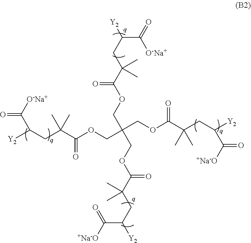


D00000

D00001

D00002

D00003

D00004
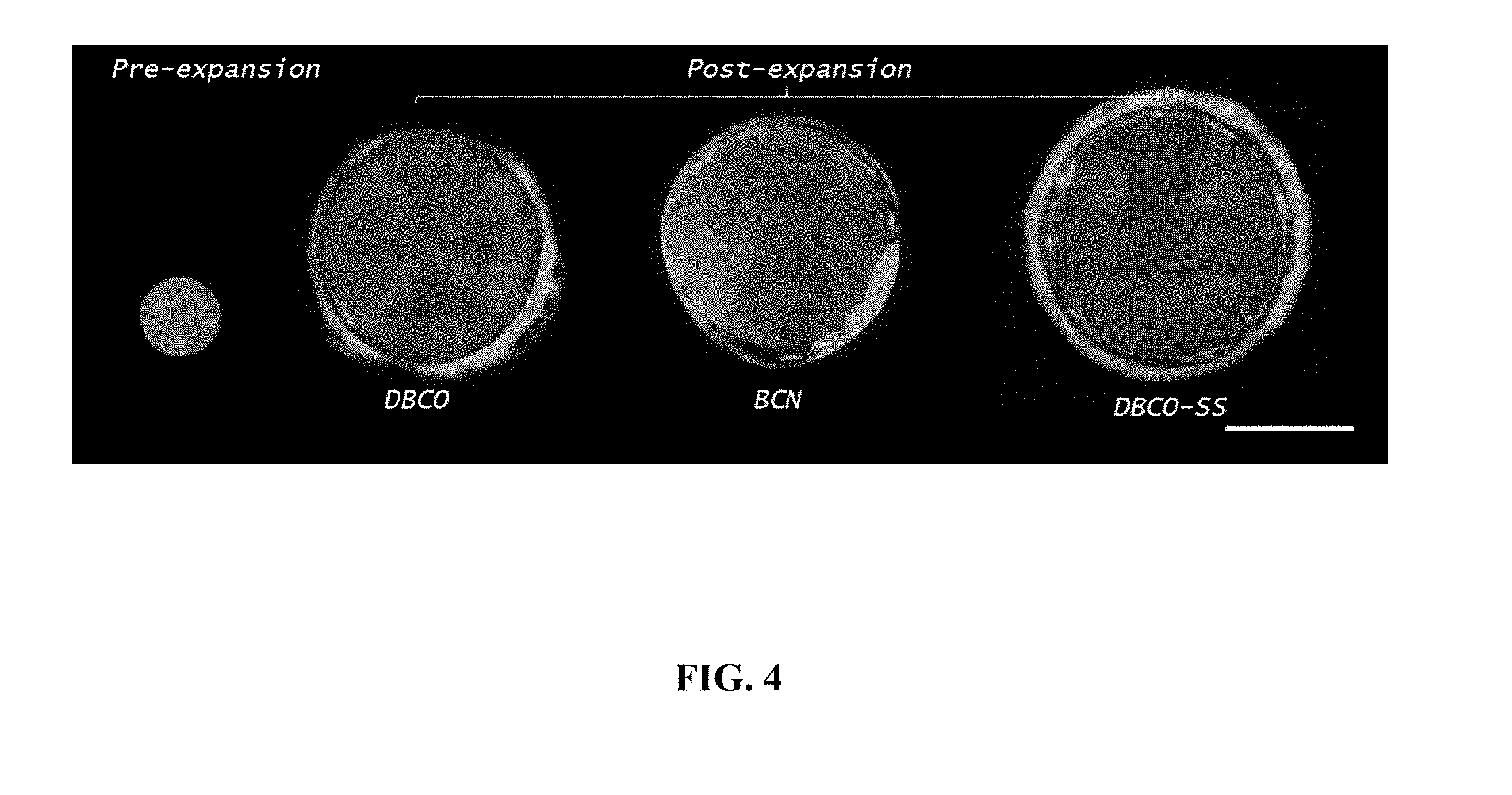
D00005

D00006
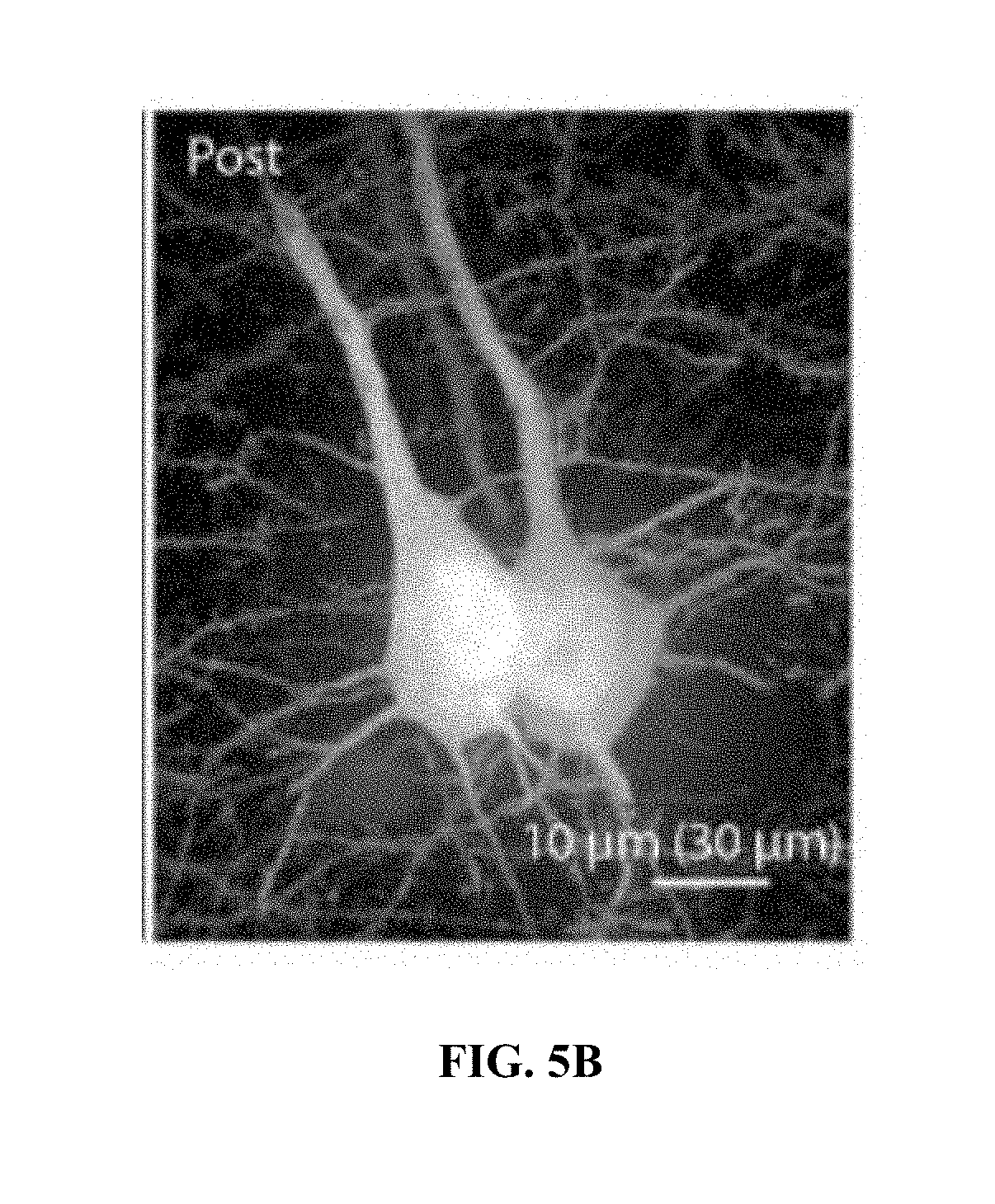
D00007

D00008

D00009

D00010

D00011

D00012
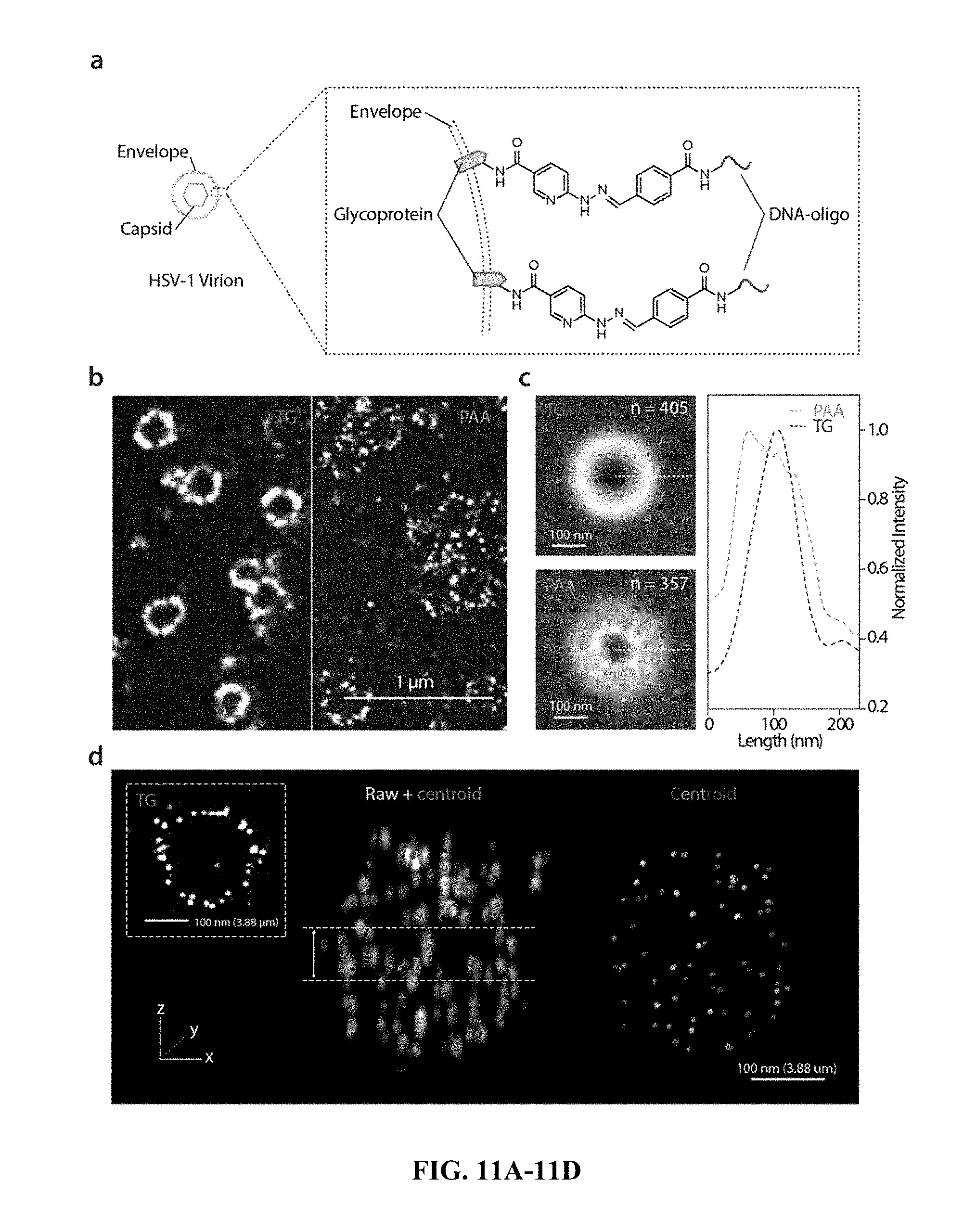
D00013

XML
uspto.report is an independent third-party trademark research tool that is not affiliated, endorsed, or sponsored by the United States Patent and Trademark Office (USPTO) or any other governmental organization. The information provided by uspto.report is based on publicly available data at the time of writing and is intended for informational purposes only.
While we strive to provide accurate and up-to-date information, we do not guarantee the accuracy, completeness, reliability, or suitability of the information displayed on this site. The use of this site is at your own risk. Any reliance you place on such information is therefore strictly at your own risk.
All official trademark data, including owner information, should be verified by visiting the official USPTO website at www.uspto.gov. This site is not intended to replace professional legal advice and should not be used as a substitute for consulting with a legal professional who is knowledgeable about trademark law.