Antibody Specifically Binding To Il-17a And Functional Fragment Thereof
Song; Nanmeng ; et al.
U.S. patent application number 16/332900 was filed with the patent office on 2019-08-22 for antibody specifically binding to il-17a and functional fragment thereof. The applicant listed for this patent is Beijing Hanmi Pharm. Co., Ltd.. Invention is credited to Mengxie Jin, Jiawang Liu, Nanmeng Song, Dongge Yang, Yang Yang, Yaping Yang, Hongjuan Zhang.
| Application Number | 20190256588 16/332900 |
| Document ID | / |
| Family ID | 60980760 |
| Filed Date | 2019-08-22 |


| United States Patent Application | 20190256588 |
| Kind Code | A1 |
| Song; Nanmeng ; et al. | August 22, 2019 |
ANTIBODY SPECIFICALLY BINDING TO IL-17A AND FUNCTIONAL FRAGMENT THEREOF
Abstract
An antibody specifically binding to IL-17A or a functional fragment thereof. The antibody or functional fragment thereof includes an IL-17A chimeric antibody and a functional fragment thereof, and an IL-17A humanized antibody and a functional fragment thereof.
| Inventors: | Song; Nanmeng; (Beijing, CN) ; Liu; Jiawang; (Beijing, CN) ; Yang; Yaping; (Beijing, CN) ; Yang; Yang; (Beijing, CN) ; Yang; Dongge; (Beijing, CN) ; Zhang; Hongjuan; (Beijing, CN) ; Jin; Mengxie; (Beijing, CN) | ||||||||||
| Applicant: |
|
||||||||||
|---|---|---|---|---|---|---|---|---|---|---|---|
| Family ID: | 60980760 | ||||||||||
| Appl. No.: | 16/332900 | ||||||||||
| Filed: | September 8, 2017 | ||||||||||
| PCT Filed: | September 8, 2017 | ||||||||||
| PCT NO: | PCT/CN2017/101083 | ||||||||||
| 371 Date: | March 13, 2019 |
| Current U.S. Class: | 1/1 |
| Current CPC Class: | C07K 2317/24 20130101; A61K 2039/505 20130101; A61P 31/04 20180101; A61P 13/12 20180101; A61P 37/06 20180101; A61P 37/08 20180101; A61P 19/02 20180101; A61P 43/00 20180101; A61P 1/12 20180101; C07K 16/24 20130101; A61P 17/14 20180101; Y02A 50/30 20180101; A61K 39/395 20130101; A61K 49/221 20130101; A61K 49/16 20130101; A61P 9/02 20180101; C07K 2317/52 20130101; A61P 17/00 20180101; C07K 2317/567 20130101; C07K 2317/76 20130101; A61P 7/06 20180101; A61P 1/00 20180101; A61K 47/68 20170801; Y02A 50/414 20180101; A61K 47/6845 20170801; C07K 2317/565 20130101; C07K 2317/92 20130101; A61P 27/02 20180101; A61P 11/00 20180101; A61P 19/08 20180101; A61P 25/00 20180101; A61P 29/00 20180101; A61P 11/06 20180101; C07K 2317/94 20130101; A61P 17/06 20180101; C07K 16/244 20130101; A61P 1/04 20180101; A61P 37/02 20180101; A61K 51/1021 20130101; A61K 49/0058 20130101 |
| International Class: | C07K 16/24 20060101 C07K016/24; A61K 47/68 20060101 A61K047/68; A61K 51/10 20060101 A61K051/10; A61K 49/16 20060101 A61K049/16; A61K 49/22 20060101 A61K049/22; A61K 49/00 20060101 A61K049/00 |
Foreign Application Data
| Date | Code | Application Number |
|---|---|---|
| Sep 14, 2016 | CN | 201610827097.2 |
Claims
1. An antibody capable of specifically binding to IL-17A or a functional fragment capable of specifically binding to IL-17A thereof, wherein the antibody or the functional fragment thereof comprise a light chain and a heavy chain; the light chain comprises a light chain CDR consisting of CDR-L1, CDR-L2 and CDR-L3; the heavy chain comprises a heavy chain CDR consisting of CDR-H1, CDR-H2 and CDR-H3; the amino acid sequences of the CDR-L1, CDR-L2 and CDR-L3 are respectively set forth in SEQ ID NO: 1, 2 and 3; the amino acid sequences of the CDR-H1, CDR-H2 and CDR-H3 are respectively set forth in SEQ ID NO: 4, 5 and 6; preferably, the antibody or the functional fragment thereof includes an IL-17A chimeric antibody and a functional fragment thereof, and an IL-17A humanized antibody and a functional fragment thereof.
2. The antibody or the functional fragment thereof according to claim 1, wherein the antibody comprises a constant region sequence of any one selected from the group consisting of human IgG1, IgG2, IgG3, IgG4, IgA, IgM, IgE and IgD.
3. The antibody or the functional fragment thereof according to claim 1, wherein the functional fragment comprises one or more selected from the group consisting of F(ab').sub.2, Fab', Fab, Fv, scFv, bispecific antibody and antibody minimal recognition unit.
4. The antibody or the functional fragment thereof according to claim 1, wherein the amino acid sequences of light chain variable region and heavy chain variable region of the IL-17A chimeric antibody or the functional fragment thereof are respectively set forth in SEQ ID NO: 7 and SEQ ID NO:8; preferably, the amino acid sequence of the light chain constant region and the heavy chain constant region of the IL-17A chimeric antibody and functional fragment thereof are respectively set forth in SEQ ID NO: 9 and SEQ ID NO: 10.
5. The antibody or the functional fragment thereof according to claim 1, wherein light chain framework region of the IL-17A humanized antibody or the functional fragment thereof comprises FR-L1, FR-L2, FR-L3 and FR-L4, and heavy chain framework region of the IL-17A humanized antibody or the functional fragment thereof comprises FR-H1, FR-H2, FR-H3 and FR-H4; the FR-L1 is selected from the amino acid sequence set forth in SEQ ID NO: 11 and the amino sequence having the following substitution or a combination thereof: the 1.sup.st amino acid D is replaced by I; the 2.sup.nd amino acid V is replaced by I; the FR-L2 is selected from the amino acid sequence set forth in SEQ ID NO: 12 and the amino sequence having the following substitution or a combination thereof: the 4.sup.th amino acid F is replaced by Y; the 14.sup.th amino acid R is replaced by L; the FR-L3 is selected from the amino acid sequence set forth in SEQ ID NO: 13 and the amino sequence having the following substitution or a combination thereof: the 35.sup.th amino acid Y is replaced by F; the FR-L4 is selected from the amino acid sequence set forth in SEQ ID NO: 14; the FR-H1 is selected from the amino acid sequence set forth in SEQ ID NO: 15 and the amino sequence having the following substitution or a combination thereof: the 4.sup.th amino acid L is replaced by V; the FR-H2 is selected from the amino acid sequence set forth in SEQ ID NO: 16 and the amino sequence having the following substitution or a combination thereof: the 15.sup.th amino acid I is replaced by M; the FR-H3 is selected from the amino acid sequence set forth in SEQ ID NO: 17 and the amino sequence having the following substitution or a combination thereof: the 2.sup.nd amino acid V is replaced by I; the 6.sup.th amino acid V is replaced by R; the FR-H4 is selected from the amino acid sequence set forth in SEQ ID NO: 18; preferably, light chain variable region sequence of the IL-17A humanized antibody or the functional fragment thereof is one selected from SEQ ID NO: 19-26; preferably, heavy chain variable region sequence of the IL-17A humanized antibody or the functional fragment thereof is one selected from SEQ ID NO: 27-34; more preferably, the light chain variable region sequence of the IL-17A humanized antibody or the functional fragment thereof is set forth in SEQ ID NO: 19; the corresponding heavy chain variable region sequence is set forth in SEQ ID NO: 27; alternatively, the light chain variable region sequence of the IL-17A humanized antibody or the functional fragment thereof is set forth in SEQ ID NO: 20; the corresponding heavy chain variable region sequence is set forth in SEQ ID NO: 28; alternatively, the light chain variable region sequence of the IL-17A humanized antibody or the functional fragment thereof is set forth in SEQ ID NO: 21; the corresponding heavy chain variable region sequence is set forth in SEQ ID NO: 29; alternatively, the light chain variable region sequence of the IL-17A humanized antibody or the functional fragment thereof is set forth in SEQ ID NO: 22; the corresponding heavy chain variable region sequence is set forth in SEQ ID NO: 30; alternatively, the light chain variable region sequence of the IL-17A humanized antibody or the functional fragment thereof is set forth in SEQ ID NO: 22; the corresponding heavy chain variable region sequence is set forth in SEQ ID NO: 31; alternatively, the light chain variable region sequence of the IL-17A humanized antibody or the functional fragment thereof is set forth in SEQ ID NO: 23; the corresponding heavy chain variable region sequence is set forth in SEQ ID NO: 32; alternatively, the light chain variable region sequence of the IL-17A humanized antibody or the functional fragment thereof is set forth in SEQ ID NO: 24; the corresponding heavy chain variable region sequence is set forth in SEQ ID NO: 33; alternatively, the light chain variable region sequence of the IL-17A humanized antibody or the functional fragment thereof is set forth in SEQ ID NO: 25; the corresponding heavy chain variable region sequence is set forth in SEQ ID NO: 32; alternatively, the light chain variable region sequence of the IL-17A humanized antibody or the functional fragment thereof is set forth in SEQ ID NO: 26; the corresponding heavy chain variable region sequence is set forth in SEQ ID NO: 34; more preferably, the amino acid sequences of the light chain constant region and the heavy chain constant region of the IL-17A humanized antibody or the functional fragment thereof are respectively set forth in SEQ ID NO: 9 and SEQ ID NO: 10.
6. An isolated nucleic acid molecule selected from: A) DNA or RNA, encoding the antibody or the functional fragment thereof according to claim 1; and B) a nucleic acid complementary to the nucleic acid as defined in A).
7. A composition, comprising the antibody or the functional fragment thereof according to claim 1, or a compound of the antibody or the functional fragment thereof together with other components, as an active ingredient.
8. The composition according to claim 7, wherein the antibody or the functional fragment thereof are coupled to at least one diagnostic agent and/or therapeutic agent to form an immunoconjugate.
9. The composition according to claim 8, wherein the diagnostic agent is one or more selected from the group consisting of a radionuclide, a radioactive contrast agent, a paramagnetic ion, a metal, a fluorescent label, a chemiluminescent label, an ultrasound contrast agent, and a photosensitizer; preferably, the radionuclide is one or more selected from the group consisting of .sup.110In, .sup.111In, .sup.177Lu, .sup.18F, .sup.52Fe, .sup.62Cu, .sup.64Cu, .sup.67Cu, .sup.67Ga, .sup.68Ga, .sup.86Y, .sup.90Y, .sup.89Zr, .sup.94mTc, .sup.94Tc, .sup.99mTc, .sup.120I, .sup.123I, .sup.124I, .sup.125I, .sup.131I, .sup.154-158Gd, .sup.32P, .sup.11C, .sup.13N, .sup.15O, .sup.186Re, .sup.188Re, .sup.51Mn, .sup.52mMn, .sup.55Co, .sup.72As, .sup.75Br, .sup.76Br, .sup.82mRb and .sup.83Sr; preferably, the paramagnetic ion is one or more selected from the group consisting of chromium (III), manganese (II), iron (III), iron (II), cobalt (II), nickel (II), copper (II), neodymium (III), samarium (III), ytterbium (III), gadolinium (III), vanadium (II), terbium (III), dysprosium (III), holmium (III) and erbium (III); preferably, the fluorescent label is one or more selected from the group consisting of Alexa 350, Alexa 405, Alexa 430, Alexa 488, Alexa 555, Alexa 647, AMCA, aminoacridine, BODIPY 630/650, BODIPY 650/665, BODIPY-FL, BODIPY-R6G, BODIPY-TMR, BODIPY-TRX, 5-carboxy-4',5'-dichloro-2',7'-dimethoxyfluorescein, 5-carboxy-2',4',5',7'-tetrachlorofluorescein, 5-carboxyfluorescein, 5-carboxyrhodamine, 6-carboxyrhodamine, 6-carboxytetramethylrhodamine, Cascade Blue, Cy2, Cy3, Cy5, Cy7, 6-FAM, dansyl chloride, fluorescein, HEX, 6-JOE, NBD (7-nitrobenzo-2-oxa-1,3-diazole), Oregon Green 488, Oregon Green 500, Oregon Green 514, Pacific Blue, phthalic acid, terephthalic acid, isophthalic acid, cresyl fast violet, cresyl violet, brilliant cresyl blue, 4-aminobenzoic acid, erythrosine, phthalocyanine, azomethine, cyanine, xanthine, succinyl fluorescein, rare earth metal cryptate, tri-bipyridyldiamine europium, europium cryptate compound or chelate, diamine, dicyanine, La Jolla blue dye, allophycocyanin, allococyanin B, phycocyanin C, phycocyanin R, thiamine, R-phycoerythrin, C-Phycocyanin, phycoerythrin R, REG, rhodamine green, rhodamine isothiocyanate, rhodamine red, ROX, TAMRA, TET, TRIT (tetramethylrhodamine isothiol), tetramethylrhodamine and Texas Red.
10. The composition according to claim 8, wherein the therapeutic agent is one or more selected from the group consisting of a naked antibody, a cytotoxic agent, a drug, a radionuclide, a boron atom, an immunomodulator, an anti-apoptotic agent, a photosensitizing therapeutic, an immunoconjugate and an oligonucleotide; preferably, the drug is one or more selected from the group consisting of dexamethasone, diclofenac, nabumetone, meloxicam, celecoxib, methotrexate, cyclophosphamide, azathioprine, cyclosporine A and vincristine; preferably, the oligonucleotide is one or more selected from the group consisting of shRNA, miRNA and siRNA; preferably, the immunomodulator is one or more selected from the group consisting of a cytokine, a chemokine, a stem cell growth factor, a lymphotoxin, a hematopoietic factor, a colony stimulating factor (CSF), an interferon, an erythropoietin, a thrombopoietin, an interleukin (IL), granulocyte-colony stimulating factor (G-CSF), granulocyte macrophage-colony stimulating factor (GM-CSF) and stem cell growth factor; preferably, the radionuclide is one or more selected from the group consisting of .sup.111In, .sup.111At, .sup.177Lu, .sup.211Bi, .sup.212Bi, .sup.213Bi, .sup.211At, .sup.62Cu, .sup.67Cu, .sup.90Y, .sup.125I, .sup.131I, .sup.133I, .sup.32P, .sup.33P, .sup.47Sc, .sup.111Ag, .sup.67Ga, .sup.153Sm, .sup.161Tb, .sup.152Dy, .sup.166Dy, .sup.161Ho, .sup.166Ho, .sup.186Re, .sup.188Re, .sup.189Re, .sup.211Pb, .sup.212Pb, .sup.223Ra, .sup.225Ac, .sup.77As, .sup.89Sr, .sup.99Mo, .sup.105Rh, .sup.149Pm, .sup.169Er, .sup.194Ir, .sup.58Co, .sup.80mBr, .sup.99mTc, .sup.103mRh, .sup.109Pt, .sup.119Sb, .sup.189mOs, .sup.192Ir, .sup.219Rn, .sup.215Po, .sup.221Fr, .sup.255Fm, .sup.11C, .sup.13N, .sup.15O, .sup.75Br, .sup.198Au, .sup.199Au, .sup.224Ac, .sup.77Br, .sup.113mIn, .sup.95Ru, .sup.97Ru, .sup.103Ru, .sup.105Ru, .sup.107Hg, .sup.203Hg, .sup.121mTe, .sup.122mTe, .sup.125mTe, .sup.165Tm, .sup.167Tm, .sup.168Tm, .sup.197Pt, .sup.109Pd, .sup.142Pr, .sup.143Pr, .sup.161Tb, .sup.57Co, .sup.58Co, .sup.51Cr, .sup.59Fe, .sup.75Se, .sup.201Tl, .sup.76Br and .sup.169Yb.
11. A method of preventing and/or treating a disease associated with overexpression and/or over-release of IL-17A, comprising administering the composition according to claim 7 to a subject in need thereof; preferably, the disease is one or more selected from the group consisting of airway inflammation, asthma, bronchial asthma, allergic asthma, chronic obstructive pulmonary disease, idiopathic pulmonary fibrosis, rheumatoid arthritis, osteoarthritis, ankylosing spondylitis, psoriatic arthritis, psoriasis, multiple sclerosis, systemic sclerosis, systemic lupus erythematosus, lupus nephritis, scleroderma, ulcerative colitis, inflammatory bowel disease, uveitis, Helicobacter pylori-associated gastritis, osteoporosis, bone erosion, intraperitoneal abscess and adhesions, Addison's disease, gamma globulin deficiency, alopecia areata, celiac disease, Chagas disease, Crohn's disease, allograft rejection, Behcet's disease, sepsis, septic or endotoxin shock and ischemia.
12. A method of preventing and/or treating a disease associated with overexpression and/or over-release of IL-17A, comprising administering the antibody capable of specifically binding to IL-17A or the functional fragment capable of specifically binding to IL-17A thereof according to claim 1 to a subject in need thereof; preferably, the disease is one or more selected from the group consisting of airway inflammation, asthma, bronchial asthma, allergic asthma, chronic obstructive pulmonary disease, idiopathic pulmonary fibrosis, rheumatoid arthritis, osteoarthritis, ankylosing spondylitis, psoriatic arthritis, psoriasis, multiple sclerosis, systemic sclerosis, systemic lupus erythematosus, lupus nephritis, scleroderma, ulcerative colitis, inflammatory bowel disease, uveitis, Helicobacter pylori-associated gastritis, osteoporosis, bone erosion, intraperitoneal abscess and adhesions, Addison's disease, gamma globulin deficiency, alopecia areata, celiac disease, Chagas disease, Crohn's disease, allograft rejection, Behcet's disease, sepsis, septic or endotoxin shock and ischemia.
13. A drug for prevention and/or treatment of a disease associated with overexpression and/or over-release of IL-17A, comprising the antibody capable of specifically binding to IL-17A or the functional fragment capable of specifically binding to IL-17A thereof according to claim 1, and a pharmaceutically acceptable carrier; preferably, the disease is one or more selected from the group consisting of airway inflammation, asthma, bronchial asthma, allergic asthma, chronic obstructive pulmonary disease, idiopathic pulmonary fibrosis, rheumatoid arthritis, osteoarthritis, ankylosing spondylitis, psoriatic arthritis, psoriasis, multiple sclerosis, systemic sclerosis, systemic lupus erythematosus, lupus nephritis, scleroderma, ulcerative colitis, inflammatory bowel disease, uveitis, Helicobacter pylori-associated gastritis, osteoporosis, bone erosion, intraperitoneal abscess and adhesions, Addison's disease, gamma globulin deficiency, alopecia areata, celiac disease, Chagas disease, Crohn's disease, allograft rejection, Behcet's disease, sepsis, septic or endotoxin shock and ischemia.
14. A method of preventing and/or treating a disease associated with overexpression and/or over-release of IL-17A, comprising administering the drug according to claim 13 to a subject in need thereof.
Description
[0001] The present application claims priority to Chinese Patent Application No. CN201610827097.2, filed on Sep. 14, 2016 with State Intellectual Property Office and entitled "ANTIBODY SPECIFICALLY BINDING TO IL-17A AND FUNCTIONAL FRAGMENT THEREOF", the entire content of which is incorporated herein by reference.
FIELD
[0002] The present disclosure relates to the field of medical biotechnology and humanized antibody engineering research, and in particular to an antibody specifically binding to IL-17A and functional fragments thereof.
BACKGROUND
[0003] Interleukin-17A (IL-17 or IL-17A) is a pro-inflammatory cytokine produced primarily by Th17 cells and is the most representative member of the IL-17 family (Miossec P, Kolls J K. Nat. Rev. Drug. Discov., 2012, 11: 763-776). After binding to IL-17A receptor (IL-17RA), IL-17A may induce the expression of inflammatory cytokines and chemokines in a variety of cells (such as fibroblasts, epithelial cells, and endothelial cells), playing an important role in body immunological defense (Lin Y, Ritchea S, Logar A. Immunity, 2009, 31:799-810).
[0004] However, over-expressed IL-17A may cause many inflammatory diseases. For example, IL-17A has effects on macrophages and DC cells, inducing high expression of IL-1, IL-6, TNF and CRP, resulting in inflammatory reaction, and involving in the pathological processes of psoriasis and transplant rejection. IL-17A has effects on endothelial cells, inducing high expression of IL-6, MMP and coagulation factors, resulting in vascular activation, and involving in pathological processes of reperfusion injury, thrombosis and atherosclerosis. IL-17A has effects on fibroblasts, inducing high expression of IL-6, chemokines, growth factors and MMP, resulting in matrix destruction, and involving in the pathological process of multiple sclerosis and Crohn's disease. IL-17A has effects on osteoblasts and chondrocytes, inducing RANKL, MMP and osteoclast genesis, resulting in bone erosion and cartilage damage, and involving in the pathological process of rheumatoid arthritis and periodontal disease (N Engl J Med. 2009 Aug. 27; 361(9):888-98. Nat Rev Drug Discov. 2012 October; 11(10):763-76. Semin Arthritis Rheum. 2013 October; 43(2):158-70. Trends Mol Med. 2016 March; 22(3):230-41).
[0005] IL-17A neutralizing antibody can inhibit the high expression of IL-17A in synovial tissue of patients with rheumatoid arthritis and reduce the production of important inflammatory factor IL-6 (Chabaud M, Durand J M, Buchs N, et al. Arthritis Rheum. 1999, 42: 963-70). It has been demonstrated by many animal model experiments of autoimmune diseases that the use of antibodies to neutralize IL-17A can effectively inhibit the pathological development of inflammation (Lubberts E, Koenders M I, Oppers-Walgreen B, et al. Arthritis Rheum., 2004, 50: 650-659).
[0006] Currently, IL-17A-related antibody drugs, Secukinumab (IL-17A targeting antibody) and Ixekizumab (IL-17A targeting antibody), have been approved for marketing, which are both used for the treatment of psoriasis. However, results of drug clinical trial have shown that these drugs do not show the expected therapeutic effect for certain chronic inflammatory diseases such as rheumatoid arthritis (Kellner H, Ther Adv Musculoskelet Dis., 2013, 5: 141-152).
[0007] In view of this, the present disclosure has been specifically proposed.
SUMMARY
[0008] The present disclosure is based on an obtained parental anti-human IL-17A murine monoclonal antibody having the ability to specifically bind to human IL-17A protein, by cloning, identification and gene structure analysis to determine its CDR region, construct corresponding chimeric antibody and humanized antibody, establish corresponding eukaryotic cell expression system and produce and purify the chimeric antibody and the humanized antibody.
[0009] In order to achieve the above-mentioned goal of the present disclosure, the following technical solutions are specially adopted:
[0010] An antibody capable of specifically binding to IL-17A and a functional fragment thereof, wherein the antibody or the functional fragment thereof comprise a light chain and a heavy chain;
[0011] the light chain comprises a light chain CDR consisting of CDR-L1, CDR-L2 and CDR-L3; the heavy chain comprises a heavy chain CDR consisting of CDR-H1, CDR-H2 and CDR-H3;
[0012] the amino acid sequences of the CDR-L1, CDR-L2 and CDR-L3 are respectively set forth in SEQ ID NO: 1, 2 and 3; the amino acid sequences of the CDR-H1, CDR-H2 and CDR-H3 are respectively set forth in SEQ ID NO: 4, 5 and 6.
[0013] Preferably, the antibody or the functional fragment thereof includes an IL-17A chimeric antibody and a functional fragment thereof, and an IL-17A humanized antibody and a functional fragment thereof.
[0014] Preferably, the antibody or the functional fragment thereof includes an IL-17A chimeric antibody and a functional fragment thereof, and an IL-17A humanized antibody and a functional fragment thereof.
[0015] That is, the antibody or the functional fragment thereof includes an IL-17A chimeric antibody and a functional fragment thereof, or the antibody or the functional fragment thereof includes an IL-17A humanized antibody and a functional fragment thereof.
[0016] It is well known in the art that both the binding specificity and affinity of an antibody are mainly determined by the CDR, and the amino acid sequence of the non-CDR region can be easily changed according to the well-known existing techniques to obtain a variant having similar biological activities. In the present disclosure, the monoclonal antibody variants have CDR sequences identical to the CDR sequences of above-mentioned humanized antibodies, thus, they have similar biological activities.
[0017] Preferably, the antibody and the functional fragment thereof as described above, wherein the antibody comprises a constant region sequence of any one selected from the group consisting of human IgG1, IgG2, IgG3, IgG4, IgA, IgM, IgE and IgD.
[0018] Preferably, the antibody and the functional fragment thereof as described above, wherein the functional fragment comprises one or more selected from the group consisting of F(ab').sub.2, Fab', Fab, Fv, scFv, bispecific antibody and antibody minimal recognition unit.
[0019] The "functional fragment" of the present disclosure specifically refers to an antibody fragment having the same specificity to IL-17A as that of the parent antibody. In addition to the above mentioned functional fragments, any fragment of which half-life has been increased may be also included.
[0020] scFv (sc=single strand), bispecific antibody (diabodies).
[0021] These functional fragments typically have the same binding specificity as the antibody from which they are derived. One ordinary skill in the art can learn from what is described in the specification of the present disclosure that the antibody fragment of the present disclosure and obtain the above mentioned function fragment by a method such as enzymatic digestion (including pepsin or papain) and/or a method of chemically reducing split disulfide bonds.
[0022] The antibody fragments can also be obtained by peptide synthesis by recombinant genetic techniques, which are also known to those having ordinary skill in the art, or by automated peptide synthesizers such as an automated peptide synthesizer sold by such as Applied BioSystems.
[0023] Preferably, the antibody and the functional fragment thereof as described above, wherein the amino acid sequences of light chain variable region and heavy chain variable region of the IL-17A chimeric antibody and the functional fragment thereof are respectively set forth in SEQ ID NO: 7 and SEQ ID NO:8;
[0024] Further preferably, the antibody and the functional fragment thereof as described above, wherein the amino acid sequence of the light chain constant region and the heavy chain constant region of the IL-17A chimeric antibody and functional fragment thereof are respectively set forth in SEQ ID NO: 9 and SEQ ID NO: 10.
[0025] Preferably, the antibody and the functional fragment thereof as described above, wherein light chain framework region of the IL-17A humanized antibody and the functional fragment thereof comprises FR-L1, FR-L2, FR-L3 and FR-L4, and heavy chain framework region of the IL-17A humanized antibody and the functional fragment thereof comprises FR-H1, FR-H2, FR-H3 and FR-H4.
[0026] The amino acid sequences of FR-L1, FR-L2, FR-L3 and FR-L4 are set forth in SEQ ID NO: 11 to 14 respectively.
[0027] The amino acid sequences of the FR-H1, FR-H2, FR-H3 and FR-H4 are set forth in SEQ ID NO: 15 to 18 respectively.
[0028] Usually, when transplanting CDRs of a murine antibody to a human framework, selection of a human framework with high sequence homology will have a certain success rate. However, studies have shown that many CDR grafts require a back mutation to restore certain antibody activity. How to choose the right human source framework is the major bottleneck.
[0029] The CDR is the major relevant site for antigen binding, but in most cases, the FR (framework region) has a significant influence on the conformation of the binding site. In order to obtain a high affinity humanized antibody, in the present disclosure, a suitable FR region is selected and the relevant FR residue is reversed back to the original murine amino acid or a amino acid presented in human and having the same function.
[0030] Preferably, the FR-L1 is selected from the amino acid sequence set forth in SEQ ID NO: 11 and the amino sequence having the following substitution or a combination thereof:
[0031] the 1.sup.st amino acid D is replaced by I;
[0032] the 2.sup.nd amino acid V is replaced by I;
[0033] the FR-L2 is selected from the amino acid sequence set forth in SEQ ID NO: 12 and the amino sequence having the following substitution or a combination thereof:
[0034] the 4.sup.th amino acid F is replaced by Y;
[0035] the 14.sup.th amino acid R is replaced by L;
[0036] the FR-L3 is selected from the amino acid sequence set forth in SEQ ID NO: 13 and the amino sequence having the following substitution or a combination thereof:
[0037] the 35.sup.th amino acid Y is replaced by F;
[0038] the FR-L4 is selected from the amino acid sequence set forth in SEQ ID NO: 14;
[0039] the FR-H1 is selected from the amino acid sequence set forth in SEQ ID NO: 15 and the amino sequence having the following substitution or a combination thereof:
[0040] the 4.sup.th amino acid L is replaced by V;
[0041] the FR-H2 is selected from the amino acid sequence set forth in SEQ ID NO: 16 and the amino sequence having the following substitution or a combination thereof:
[0042] the 15.sup.th amino acid I is replaced by M;
[0043] the FR-H3 is selected from the amino acid sequence set forth in SEQ ID NO: 17 and the amino sequence having the following substitution or a combination thereof:
[0044] the 2.sup.nd amino acid V is replaced by I;
[0045] the 6.sup.th amino acid V is replaced by R;
[0046] the FR-H4 is selected from the amino acid sequence set forth in SEQ ID NO: 18;
[0047] preferably, light chain variable region sequence of the IL-17A humanized antibody and the functional fragment thereof is one selected from SEQ ID NO: 19-26;
[0048] preferably, heavy chain variable region sequence of the IL-17A humanized antibody and the functional fragment thereof is one selected from SEQ ID NO: 27-34;
[0049] more preferably, the light chain variable region sequence of the IL-17A humanized antibody and the functional fragment thereof is set forth in SEQ ID NO: 19; the corresponding heavy chain variable region sequence is set forth in SEQ ID NO: 27;
[0050] alternatively, the light chain variable region sequence of the IL-17A humanized antibody and the functional fragment thereof is set forth in SEQ ID NO: 20; the corresponding heavy chain variable region sequence is set forth in SEQ ID NO: 28;
[0051] alternatively, the light chain variable region sequence of the IL-17A humanized antibody and the functional fragment thereof is set forth in SEQ ID NO: 21; the corresponding heavy chain variable region sequence is set forth in SEQ ID NO: 29;
[0052] alternatively, the light chain variable region sequence of the IL-17A humanized antibody and the functional fragment thereof is set forth in SEQ ID NO: 22; the corresponding heavy chain variable region sequence is set forth in SEQ ID NO: 30;
[0053] alternatively, the light chain variable region sequence of the IL-17A humanized antibody and the functional fragment thereof is set forth in SEQ ID NO: 22; the corresponding heavy chain variable region sequence is set forth in SEQ ID NO: 31;
[0054] alternatively, the light chain variable region sequence of the IL-17A humanized antibody and the functional fragment thereof is set forth in SEQ ID NO: 23; the corresponding heavy chain variable region sequence is set forth in SEQ ID NO: 32;
[0055] alternatively, the light chain variable region sequence of the IL-17A humanized antibody and the functional fragment thereof is set forth in SEQ ID NO: 24; the corresponding heavy chain variable region sequence is set forth in SEQ ID NO: 33;
[0056] alternatively, the light chain variable region sequence of the IL-17A humanized antibody and the functional fragment thereof is set forth in SEQ ID NO: 25; the corresponding heavy chain variable region sequence is set forth in SEQ ID NO: 32;
[0057] alternatively, the light chain variable region sequence of the IL-17A humanized antibody and the functional fragment thereof is set forth in SEQ ID NO: 26; the corresponding heavy chain variable region sequence is set forth in SEQ ID NO: 34;
[0058] more preferably, the amino acid sequences of the light chain constant region and the heavy chain constant region of the IL-17A humanized antibody and the functional fragment thereof are respectively set forth in SEQ ID NO: 9 and SEQ ID NO: 10.
[0059] The light chain constant region of the IL-17A humanized antibody and the functional fragment thereof is selected from the light chain constant region of human Kappa antibody, of which the amino acid sequence is set forth in SEQ ID NO: 9.
[0060] The heavy chain constant region of the IL-17A humanized antibody and the functional fragment thereof is selected from the heavy chain constant region of human IgG1 antibody, of which the amino acid sequence is set forth in SEQ ID NO: 10.
[0061] It should be noted that, in addition to the above-mentioned amino acid sequences in the present application, the production of chimeric antibodies and humanized antibodies can be achieved by any method known by those having ordinary skill in the art, such as by designing recombinant humanized antibody based on sequenced CDRs of murine antibodies, the murine antibody is secreted by myeloma cells from immunized mice or by myeloma cells fused to splenocytes of other species which fused to myeloma cells. The immunized animal may include a transgenic mouse having a human immunoglobulin locus which can directly produce a human antibody. Another possible embodiment may include screening the library using phage display technology.
[0062] An isolated nucleic acid molecule selected from:
[0063] A) DNA or RNA, encoding the antibody and the functional fragment thereof as described above; and
[0064] B) a nucleic acid complementary to the nucleic acid as defined in A).
[0065] A vector, which contains a nucleic acid molecule as described above.
[0066] The present disclosure further provides at least one nuclear construct encoding a nucleic acid molecule as described above, preferably a vector, more preferably an expression vector, such as a plasmid, which is described in one embodiment of the present application.
[0067] A host cell, which is transformed with a vector as described above.
[0068] The host cell is a eukaryotic cell, such as a mammalian cell.
[0069] A method of producing an antibody capable of specifically binding to IL-17A and a functional fragment thereof includes the following steps: [0070] culturing host cells as described above in a medium and under suitable culture conditions; and [0071] recovering produced antibody and its functional fragments from the culture medium or from the cultured host cells.
[0072] A composition, comprising the antibody and/or the functional fragment thereof, or a compound of the antibody and/or the functional fragment thereof together with other components, as an active ingredient.
[0073] Preferably, the composition as described above, the antibody and the functional fragment thereof are coupled to at least one diagnostic agent and/or therapeutic agent to form an immunoconjugate.
[0074] Preferably, the diagnostic agent is one or more selected from the group consisting of a radionuclide, a radioactive contrast agent, a paramagnetic ion, a metal, a fluorescent label, a chemiluminescent label, an ultrasound contrast agent, and a photosensitizer.
[0075] Preferably, the radionuclide is one or more selected from the group consisting of .sup.110In, .sup.111In, .sup.177Lu, .sup.18F, .sup.52Fe, .sup.62Cu, .sup.64Cu, .sup.67Cu, .sup.67Ga, .sup.68Ga, .sup.86Y, .sup.90Y, .sup.89Zr, .sup.94mTc, .sup.94Tc, .sup.99mTc, .sup.120I, .sup.123I, .sup.124I, .sup.125I, .sup.131I, .sup.154-158Gd, .sup.32P, .sup.11C, .sup.13N, .sup.15O, .sup.186Re, .sup.188Re, .sup.51Mn, .sup.52mMn, .sup.55Co, .sup.72As, .sup.75Br, .sup.76Br, .sup.82mRb and .sup.83Sr.
[0076] Preferably, the paramagnetic ion is one or more selected from the group consisting of chromium (III), manganese (II), iron (III), iron (II), cobalt (II), nickel (II), copper (II), neodymium (III), samarium (III), ytterbium (III), gadolinium (III), vanadium (II), terbium (III), dysprosium (III), holmium (III) and erbium (III).
[0077] Preferably, the fluorescent label is one or more selected from the group consisting of Alexa 350, Alexa 405, Alexa 430, Alexa 488, Alexa 555, Alexa 647, AMCA, aminoacridine, BODIPY 630/650, BODIPY 650/665, BODIPY-FL, BODIPY-R6G, BODIPY-TMR, BODIPY-TRX, 5-carboxy-4',5'-dichloro-2',7'-dimethoxyfluorescein, 5-carboxy-2',4',5',7'-tetrachlorofluorescein, 5-carboxyfluorescein, 5-carboxyrhodamine, 6-carboxyrhodamine, 6-carboxytetramethylrhodamine, Cascade Blue, Cy2, Cy3, Cy5, Cy7, 6-FAM, dansyl chloride, fluorescein, HEX, 6-JOE, NBD (7-nitrobenzo-2-oxa-1,3-diazole), Oregon Green 488, Oregon Green 500, Oregon Green 514, Pacific Blue, phthalic acid, terephthalic acid, isophthalic acid, cresol fast purple, cresyl violet, brilliant cresyl blue, 4-Aminobenzoic acid, erythrosine, phthalocyanine, azomethine, cyanine, xanthine, succinyl fluorescein, rare earth metal cryptate, tri-bipyridyldiamine oxime, europium cryptate compound or chelate, diamine, dicyanine, La Jolla blue dye, allophycocyanin, allococyanin B, phycocyanin C, phycocyanin R, thiamine, R-phycoerythrin, C-Phycocyanin, phycoerythrin R, REG, rhodamine green, rhodamine isothiocyanate, rhodamine red, ROX, TAMRA, TET, TRIT (tetramethylrhodamine isothiol), tetramethylrhodamine and Texas Red.
[0078] Preferably, the therapeutic agent is one or more selected from the group consisting of a naked antibody, a cytotoxic agent, a drug, a radionuclide, a boron atom, an immunomodulator, an anti-apoptotic agent, a photosensitizing therapeutic, an immunoconjugates and a oligonucleotide.
[0079] Preferably, the drug is one or more selected from the group consisting of methotrexate, fluorouracil, mercaptopurine, hydroxyurea, cytarabine, nitrogen mustard, cyclophosphamide, thiotepa, cisplatin, mitomycin, bleomycin, camptothecin, podophyllotoxin, actinomycin D, doxorubicin, daunorubicin, vinblastine, paclitaxel, cephalotaxus alkaloids and L-asparaginase.
[0080] Preferably, the oligonucleotide is one or more selected from the group consisting of shRNA, miRNA and siRNA.
[0081] Preferably, the immunomodulator is one or more selected from the group consisting of a cytokine, a chemokine, a stem cell growth factor, a lymphotoxin, a hematopoietic factor, a colony stimulating factor (CSF), an interferon, an erythropoietin, a thrombopoietin, an interleukin (IL), granulocyte-colony stimulating factor (G-CSF), granulocyte macrophage-colony stimulating factor (GM-CSF) and stem cell growth factor.
[0082] Wherein, the cytokine is preferably one or more selected from the group consisting of human growth hormone, N-methionyl human growth hormone, bovine growth hormone, parathyroid hormone, thyroxine, insulin, proinsulin, relaxin, prorelaxin, follicle-stimulating hormone (FSH), thyroid stimulating hormone (TSH), luteinizing hormone (LH), liver growth factor, prostaglandin, fibroblast growth factor, prolactin, placental lactogen, OB protein, tumor necrosis factor-.alpha., tumor necrosis factor-.beta., Mullerian inhibitor, mouse gonadotropin-related peptide, inhibin, activin, vascular endothelial growth factor, integrin, thrombopoietin (TPO), NGF-.beta., platelet-growth factor, TGF-.alpha., TGF-.beta., insulin-like growth factor-I, insulin-like growth factor-II, erythropoietin (EPO), osteoinductive factor, interferon-.alpha., interferon-.beta., interferon-.gamma., macrophage-CSF (M-CSF), IL-1, IL-1.alpha., IL-2, IL-3, IL-4, IL-5, IL-6, IL-7, IL-8, IL-9, IL-10, IL-11, IL-12, IL-13, IL-14, IL-15, IL-16, IL-17, IL-18, IL-21, IL-25, LIF, FLT-3, angiostatin, thrombospondin, endostatin, tumor necrosis factor and LT.
[0083] The chemokine is preferably one or more selected from the group consisting of RANTES, MCAF, MIP1-.alpha., MIP1-.beta., and IP-10.
[0084] Preferably, the radionuclide is one or more selected from the group consisting of .sup.111In, .sup.111At, .sup.177Lu, .sup.211Bi, .sup.212Bi, .sup.213Bi, .sup.211At, .sup.62Cu, .sup.67Cu, .sup.90Y, .sup.125I, .sup.131I, .sup.133I, .sup.32P, .sup.33P, .sup.47Sc, .sup.111Ag, .sup.67Ga, .sup.153Sm, .sup.161Tb, .sup.152Dy, .sup.166Dy, .sup.161Ho, .sup.166Ho, .sup.86Re, .sup.188Re, .sup.189Re, .sup.211Pb, .sup.212Pb, .sup.223Ra, .sup.225Ac, .sup.77As, .sup.89Sr, .sup.99Mo, .sup.105Rh, .sup.149Pm, .sup.169Er, .sup.194Ir, .sup.58Co, .sup.80mBr, .sup.99mTc, .sup.103mRh, .sup.109Pt, .sup.119Sb, .sup.189mOs, .sup.192Ir, .sup.219Rn, .sup.215Po, .sup.221Fr .sup.255Fm, .sup.11C, .sup.13N, .sup.15O, .sup.75Br, .sup.198Au, .sup.199Au .sup.224Ac, .sup.77Br .sup.113mIn, .sup.95Ru, .sup.97Ru, .sup.103Ru .sup.105Ru, .sup.107Hg, .sup.203Hg, .sup.121mTe, .sup.122mTe, .sup.125mTe, .sup.165Tm, .sup.167Tm, .sup.168Tm, .sup.197Pt, .sup.109Pd, .sup.142Pr, .sup.143Pr, .sup.161Tb, .sup.57Co, .sup.58Co, .sup.51Cr, .sup.59Fe, .sup.75Se, .sup.201Tl, .sup.76Br and .sup.169Yb.
[0085] Use of the composition as described above for the manufacture of a medicament in prevention and/or treatment of a disease associated with overexpression and/or over-release of IL-17A.
[0086] Preferably, the disease is one or more selected from the group consisting of airway inflammation, asthma, bronchial asthma, allergic asthma, chronic obstructive pulmonary disease, idiopathic pulmonary fibrosis, rheumatoid arthritis, osteoarthritis, ankylosing spondylitis, psoriatic arthritis, psoriasis, multiple sclerosis, systemic sclerosis, systemic lupus erythematosus, lupus nephritis, scleroderma, ulcerative colitis, inflammatory bowel disease, uveitis, Helicobacter pylori-associated gastritis, osteoporosis, bone erosion, intraperitoneal abscess and adhesions, Addison's disease, gamma globulin deficiency, alopecia areata, celiac disease, Chagas disease, Crohn's disease, allograft rejection, Behcet's disease, sepsis, septic or endotoxin shock and ischemia.
[0087] Use of the antibody capable of specifically binding to IL-17A and the functional fragment thereof as described above for the manufacture of a medicament in prevention and/or treatment of a disease associated with overexpression and/or over-release of IL-17A.
[0088] A drug for prevention and/or treatment of a disease associated with overexpression and/or over-release of IL-17A, comprising the antibody capable of specifically binding to IL-17A and the functional fragment thereof, and a pharmaceutically acceptable carrier.
[0089] Alternatively, the drug comprises the composition as described above and a pharmaceutically acceptable carrier.
[0090] Herein, the term "pharmaceutically acceptable" means that the compound is physiologically acceptable when the compound is administered to a human, and does not cause an allergic reaction such as a gastrointestinal disorder, dizziness or other allergic reaction, or a systemic allergic reaction similar to these allergic reactions.
[0091] In the present disclosure, "pharmaceutically acceptable carrier", includes, but is not limited to, binders (such as microcrystalline cellulose, alginates, gelatin and polyvinylpyrrolidone), fillers (such as starch, sucrose, glucose and anhydrous lactic acid), disintegrants (such as cross-linked PVP, cross-linked carboxymethyl sodium starch, croscarmellose sodium and low-substituted hydroxypropyl cellulose), lubricants (magnesium stearate, aluminum stearate, talc, polyethylene glycol, sodium benzoate), wetting agent (such as glycerin), surfactants (such as cetyl alcohol), and absorption enhancers, flavoring agents, sweeteners, diluents, coating agents, etc.
[0092] The disease as described above is one or more selected from the group consisting of airway inflammation, asthma, bronchial asthma, allergic asthma, chronic obstructive pulmonary disease, idiopathic pulmonary fibrosis, rheumatoid arthritis, osteoarthritis, ankylosing spondylitis, psoriatic arthritis, psoriasis, multiple sclerosis, systemic sclerosis, systemic lupus erythematosus, lupus nephritis, scleroderma, ulcerative colitis, inflammatory bowel disease, uveitis, Helicobacter pylori-associated gastritis, osteoporosis, bone erosion, intraperitoneal abscess and adhesions, Addison's disease, gamma globulin deficiency, alopecia areata, celiac disease, Chagas disease, Crohn's disease, allograft rejection, Behcet's disease, sepsis, septic or endotoxin shock and ischemia.
[0093] A method of preventing and/or treating a disease associated with overexpression and/or over-release of IL-17A, comprising administering the drug as described above to a subject in need thereof.
[0094] Preferably, the above-mentioned individual is a human being, and further, the above-mentioned individual is a human being in need of preventing and/or treating of a disease associated with overexpression and/or over-release of IL-17A.
[0095] The disease as described above is one or more selected from the group consisting of airway inflammation, asthma, bronchial asthma, allergic asthma, chronic obstructive pulmonary disease, idiopathic pulmonary fibrosis, rheumatoid arthritis, osteoarthritis, ankylosing spondylitis, psoriatic arthritis, psoriasis, multiple sclerosis, systemic sclerosis, systemic lupus erythematosus, lupus nephritis, scleroderma, ulcerative colitis, inflammatory bowel disease, uveitis, Helicobacter pylori-associated gastritis, osteoporosis, bone erosion, intraperitoneal abscess and adhesions, Addison's disease, gamma globulin deficiency, alopecia areata, celiac disease, Chagas disease, Crohn's disease, allograft rejection, Behcet's disease, sepsis, septic or endotoxin shock and ischemia.
BRIEF DESCRIPTION OF DRAWINGS
[0096] In order to more clearly illustrate the specific embodiments of the present disclosure or the technical solutions in the conventional art, the drawings used in the specific embodiments or the description of the conventional art will be briefly described below, and it is obvious that the drawings in the following description are some embodiments of the present disclosure and a person having ordinary skill in the art can obtain other drawings based on these drawings without any creative work.
[0097] FIG. 1 shows the human IL-17A binding activity of the monoclonal antibody secreted by Clone No. 88 in Example 1;
[0098] FIG. 2 shows the neutralization activity of the monoclonal antibody secreted by Clone No. 88 in Example 1 against human IL-17A-stimulated secretion of IL-6 by HFF-1 cells;
[0099] FIG. 3 shows the species specificity of the anti-human IL-17A chimeric monoclonal antibody in Example 4;
[0100] FIG. 4 shows the binding specificity of the anti-human IL-17A chimeric monoclonal antibody in Example 4;
[0101] FIG. 5 shows the in vitro neutralization activity of the anti-human IL-17A chimeric monoclonal antibody in Example 5;
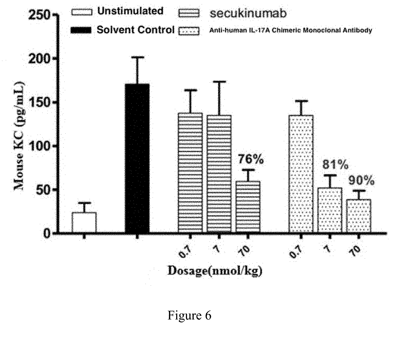
[0102] FIG. 6 shows the in vivo neutralization activity of the anti-human IL-17A chimeric monoclonal antibody in Example 6;
[0103] FIG. 7 shows the concentration-time curve after the single intravenous injection in Example 7;
[0104] FIG. 8 shows the antigen binding activity of the anti-human IL-17A humanized monoclonal antibody in Example 9;
[0105] FIG. 9 shows the in vitro neutralization activity of the anti-human IL-17A humanized monoclonal antibody in Example 10;
[0106] FIG. 10 shows the concentration-time curve after the single subcutaneous injection in Example 15.
DETAILED DESCRIPTION
[0107] The embodiments of the present disclosure will be described in detail below with reference to the embodiments. However, a person having ordinary skill in the art will understand that the following embodiments are merely to illustrate present disclosure and are not intended to limit the scope of the disclosure. For those embodiments in which specific conditions are not specified, they were carried out according to the conventional conditions or the conditions recommended by the manufacturer. For those used reagents or instruments of which the manufacturers are not indicated, they were all commercially available conventional products.
Example 1. Preparation of Murine Anti-Human IL-17A Monoclonal Antibody
1.1. Immunization of Animal
[0108] Female BALB/c mice, 6 to 8 weeks old, purchased from Beijing Huafukang Biotechnology Co., Ltd., were used as experimental animals. One week after the mice were acclimated to the environment, immunization began. The recombinant human IL-17A protein was expressed in E. coli, and the inclusion bodies were collected and subjected to denaturation and refolding treatment to obtain soluble IL-17A protein. For the initial immunization, 100 .mu.g of recombinant human IL-17A-Fc protein was thoroughly mixed with Freund's complete adjuvant (Sigma-Aldrich, Catalog Number F5881) to form an emulsion, which was intraperitoneally injected into the mice. Two weeks later, booster immunizations were performed. For the booster immunization, 50 .mu.g of recombinant human IL-17A-Fc protein was thoroughly mixed with Freund's incomplete adjuvant (Sigma-Aldrich, Catalog Number F5806) to form an emulsion, which was intraperitoneally injected into the mice. The immunization was boosted in the same way every 2 weeks, for a total 3 times. On the seventh day after the last immunization, blood was collected from retro orbital venous plexus of the mice and centrifuged to separate serum, and the antibody titer was determined by ELISA. Mice with high titers were selected for hybridization to make hybridomas. Three days before the hybridization, 50 .mu.g of recombinant human IL-17A-Fc protein was intraperitoneally injected into mice without adjuvant. On the day of hybridization, the spleen was aseptically removed to prepare a single spleen cell suspension for use.
1.2. Preparation of Hybridomas
[0109] Myeloma cells SP2/0 in logarithmic growth phase were centrifuged at 1000 rpm for 5 minutes, the supernatant was discarded, and the cells were suspended in incomplete DMEM medium (Gibco, cat No. 11965) and counted. The cells needed were taken, washed twice with an incomplete culture medium. At the same time, a spleen cell suspension prepared from a mouse after immunization was washed twice with an incomplete culture medium. The myeloma cells and the spleen cells were mixed at a ratio of 1:10 or 1:5, and washed once with an incomplete culture medium in a 50 mL plastic centrifuge tube, and then centrifuged at 1200 rpm for 8 minutes. The supernatant was discarded and a Pasteur pipette was used to remove residual liquid. The centrifuge tube was gently tapped on palm to make the precipitated cells loose and even, and then the tube was placed in 40.degree. C. water bath to preheat. 1 mL of 45% PEG-4000 (pH 8.0, Sigma, cat No. P7181) preheated to 40.degree. C. was added with 1 mL pipette at about 1 minute (with an optimum time of 45 seconds), stirred gently with a pipette when adding (stirred with a pipette), visible particles should be seen with the naked eyes. 20 to 30 mL of incomplete medium preheated to 37.degree. C. was added to the tube with 10 mL pipette within 90 seconds to terminate PEG action, and allowed to stand at 20 to 37.degree. C. for 10 minutes. The tube was centrifuged at 1000 rpm for 5 minutes, and the supernatant was discarded. 5 mL of HAT medium (DMEM+HAT, Sigma, cat No. 1 H0262-10VL) was added, and the precipitated cells were mixed gently (remember not to blow vigorously so as not to separate the fused cells) to make a well mixed suspension. Additional HAT medium was added until 80 to 100 mL (the spleen cell concentration was made to be 1 to 2.times.10.sup.6/mL). The suspension was dispensed into a 96-well cell culture plate, 0.1 mL per well; and a 24-well plate, 1.0 to 1.5 mL per well. The plates were incubated at 37.degree. C. incubator with 6% CO.sub.2. Generally, six 96-well plates were used. After 5 days, 1/2 medium was replaced with fresh HAT medium. After 7 to 10 days, the HAT medium was replaced with HT medium (DMEM+HT, Sigma cat No. H0137-10VL). The growth of hybridoma cells was observed routinely, and the supernatant was collected for antibody detection after the confluence of the cells reached 1/10 or more. The positive colonies were expanded and frozen.
1.3. Clone Screening and Identification
[0110] ELISA was used to screen anti-human IL-17A antibody from hybridoma culture supernatants. Recombinant human IL-17A was coated on a 96-well high-absorbing ELISA plate with a carbonate buffer solution with pH 9.6, the coating concentration was 1 .mu.g/mL, the coating amount was 100 .mu.L per well, and the coating was carried out at 4.degree. C. overnight. The plate was washed five times with PBST, blocked with 300 .mu.L/well of PBST containing 1% BSA, and then incubated at 25.degree. C. for 1 hour. The plate was washed five times with PBST. 100 .mu.L culture supernatant samples and the positive serum control were added to each well respectively, and then the plated was incubated at 25.degree. C. for 1 hour. The plate was washed five times with PBST. Then, 100 .mu.L horseradish peroxidase-labeled anti-mouse IgG antibody (Abeam, Catalog Number Ab7068) 1:10000 diluted in PBST containing 1% BSA was added to each well, and then the plated was incubated at 25.degree. C. for 1 hour. The plate was washed five times with PBST. 100 .mu.L/well of colorimetric substrate TMB was added and incubated at room temperature for 10 minutes. Color development was terminated by adding 100 .mu.L/well of 1 M H.sub.2SO.sub.4. The absorbance at 450 nm was read on a microplate reader. Positive clones capable of producing anti-human IL-17A antibody were selected based on the reading value at OD 450 nm.
[0111] Whether anti-human IL-17A antibody secreted by positive clone was a neutralizing antibody was determined by cell assays. The anti-human IL-17A antibody sample was diluted in DMEM complete medium (GIBCO, Catalog Number 11995-073) containing 10% FBS (Hyclone, Catalog Number SH30084.03). The starting concentration of the antibody was 160 nM and the final concentration was 40 nM in the medium. The antibody was subjected to 5 fold serial dilution, and then added to a cell culture plate, 50 .mu.L per well. 20 ng/mL of human IL-17A (final concentration 5 ng/mL) was diluted with the same complete medium and added to the cell culture plate at 50 .mu.L per well. The plate was incubated at 37.degree. C. for 1 hour in an incubator with 5% CO.sub.2. The HFF-1 cells were resuspended in complete medium and seeded into a 96-well cell culture plate at 100 .mu.L per well, 5000 cells per well. The cells were incubated at 37.degree. C. for 24 hours in an incubator with 5% CO.sub.2. After the completion of the incubation, the cell culture plate was centrifuged at 250.times.g for 5 minutes, and the culture supernatant was removed, and the human IL-6 level was detected using human IL-6 ELISA kit (R&D systems, Catalog Number S6050) according to the instructions. The antibody capable of inhibiting the human IL-17A-stimulated secretion of human IL-6 by HFF-1 cells was anti-human IL-17A neutralizing antibody. Positive clone capable of secreting anti-human IL-17A neutralizing antibody was selected based on the strength of neutralization.
[0112] The result is shown in FIG. 1. Clone No. 88 has a strong human IL-17A binding activity. According to what is shown in FIG. 2, Clone No. 88 also has pretty strong human IL-17A neutralization activity.
1.4. Sequencing of Monoclonal Antibody
[0113] The clones having both antigen-binding activity and antigen-neutralization activity obtained by screening were subjected to sequencing of antibody DNA sequence. Cellular mRNA was first extracted using RNAprep Pure Kit (Tiangen, DP430). The steps were as follows: 1.times.10.sup.7 cells were centrifuged at 300.times.g for 5 minutes and collected into a centrifuge tube, and all supernatant was carefully aspirated. The lysis step was carried out immediately. The bottom of the centrifuge tube was flicked to loose the cell pellet, 600 .mu.L of lysis buffer RL was added with vortex. All solution was transferred to a filtration column CS (the filtration column CS was placed in a collection tube), centrifuged at 12,000 rpm (.about.13,400.times.g) for 2 minutes, and the filtrate was collected. One fold volume of 70% ethanol (usually 350 .mu.L or 600 .mu.L) was added to the filtrate, well mixed, the obtained solution and precipitate were transferred into an adsorption column CR3 (the adsorption column CR3 was put into a collection tube), centrifuged at 12,000 rpm (.about.13,400.times.g) for 30 to 60 seconds, the liquid waste in the collection tube was removed, the adsorption column CR3 was put back into the collection tube. 350 .mu.L of deproteinized solution RW1 was added to the adsorption column CR3, centrifuged at 12,000 rpm (.about.13,400.times.g) for 30 to 60 seconds, the liquid waste in the collection tube was removed, the adsorption column CR3 was put back into the collection tube. 80 .mu.L of DNase I working solution was added to the center of the adsorption column CR3 and the column CR3 was allowed to stand at room temperature for 15 minutes. 350 .mu.L of deproteinized solution RW1 was added to the adsorption column CR3, centrifuged at 12,000 rpm (.about.13,400.times.g) for 30 to 60 seconds, the liquid waste in the collection tube was removed, the adsorption column CR3 was put back into the collection tube. 500 .mu.L of rinsing solution RW was added to the adsorption column CR3 (checked whether ethanol had been added before use), the column CR3 was allowed to stand at room temperature for 2 minutes, centrifuged at 12,000 rpm (.about.13,400.times.g) for 30 to 60 seconds, the liquid waste in the collection tube was removed, the adsorption column CR3 was put back into the collection tube. The column CR3 was centrifuged at 12,000 rpm (.about.13,400.times.g) for 2 minutes, and the waste was removed. The adsorption column CR3 was left at room temperature for a few minutes to let the residual rinsing solution in the adsorbent material thoroughly dry. The adsorption column CR3 was transferred into a new RNase-Free centrifuge tube, 30 to 100 .mu.L of RNase-Free ddH.sub.2O was added, the tube was allowed to stand at room temperature for 2 minutes, and then centrifuged at 12,000 rpm (.about.13,400.times.g) for 2 minutes to obtain a RNA solution.
[0114] The first strand of cDNA was synthesized using the QuantScript RT kit (Tiangen, KR103). The steps are as follows: the template RNA was thawed on ice; the primer, 10.times.RT mix (containing RNasin and DTT), Super pure dNTP mixture, RNase-Free ddH.sub.2O were thawed at room temperature (15 to 25.degree. C.), and placed on ice immediately after thawing. Each solution was well mixed by vortexer before use, the tube was centrifuged briefly to collect residual liquid on the side of the tube. Reverse transcription system mixture (Tiangen Bio Quant cDNA First-Strand Synthesis Kit, Catalog Number KR103-04; 10.times. Reverse Transcription Buffer 2 .mu.L, Ultra-Pure dNTP 2 .mu.L, Random Primer 2 .mu.L, Reverse Transcription Enzyme 1 .mu.L) was prepared according to Table 1. The mixture was mixed thoroughly, the duration of vortex was no more than 5 minutes; and then centrifuged briefly and placed on ice. Finally, the template RNA (50 ng to 2 .mu.g) was added to the mixture, mixed thoroughly, the duration of vortex was no more than 5 seconds, centrifuged briefly to collect residual liquid on the sides of the tube, incubated at 37.degree. C. for 60 minutes. The first strand of cDNA produced by reverse transcription was used for subsequent PCR reaction.
[0115] The primers used in the PCR reaction are shown in Table 1.
TABLE-US-00001 TABLE 1 PCR Primers VH primer F1: GAGGTGAAGCTGCAGGAGTCAGGACCTAGCCTGGTG R1: AGGT(C/G)(A/C)AACTGCAG(C/G)AGTC(A/T)GG R2: AGGT(C/G)(A/C)AGCTGCAG(C/G)AGTC(A/T)GG R3: AGGT(C/G)CAGCTGCAG(C/G)AGTC(A/T)GG R4: CCAGGGGCCAGTGGATAGACAAGCTTGGGTGTCGTTTT F2: ATAGACAGATGGGGGTGTCGTTTTGGC F3: CTTGACCAGGCATCCTAGAGTCA F4: AGGGGCCAGTGGATAGACTGATGG F5: AGGGACCAAGGGATAGACAGATGG R5: (G/C)A(A/G)GT(A/T/C/G)(A/C)AGCTG(G/C)AG (G/C)AGTC R6: (G/C)A(A/G)GT(A/T/C/G)(A/C)AGCTG(G/C)AG (G/C)AGTC(A/T)GG VL primer R1: GGTGATATCGTGAT(A/G)AC(C/A)CA(G/A)GATGAACTCTC R2: GGTGATATC(A/T)TG(A/C)TGACCCAA(A/T)CTCCACTCTC R3: GGTGATATCGT(G/T)CTCAC(C/T)CA(A/G)TCTCCAGCAAT F1: GGGAAGATGGATCCAGTTGGTGCAGCATCAGC F2: GGATACAGTTGGTGCAGCATC R4: GA(C/T)ATTGTG(A/C)T(G/C)AC(A/C)CA(A/G)(A/T) CT(A/C)CA
[0116] When primers were used, any upstream primer of the VH primers could be used with any downstream primer; in the same way, any upstream primer of the VL primers could also be used with any downstream primer. The target band obtained by PCR amplification was cloned into the pGEM-T vector. A single clone was picked for DNA sequencing.
Example 2. Preparation of Chimeric Anti-Human IL-17A Monoclonal Antibody
[0117] The nucleic acid sequence encoding the above mentioned antibody light chain (the full-length of the light chain was SEQ ID NO: 7 linked to SEQ ID NO: 9) and the heavy chain (the full-length of the heavy chain was SEQ ID NO: 8 linked to SEQ ID NO: 10) was cloned into a eukaryotic expression vector XOGC (wherein the full-length nucleic acid sequence of the light chain of the antibody is set forth in SEQ ID NO: 23, the full-length nucleic acid sequence of the heavy chain of the antibody is set forth in SEQ ID NO: 24), then the expression vector was transfected into a 293F cell line (FreeStyle.TM. 293-F Cells, Catalog Number R79007, invitrogen). Cells were inoculated one day prior to transfection. Cells were harvested by centrifugation on the day of transfection. The cells were resuspended in fresh FreeStyle.TM. 293 Expression Medium (FreeStyle.TM. 293 Expression Medium, Catalog Number 12338001, Gibco), the cell density was 200.times.10.sup.5 cells/mL. Plasmid was added according to the transfection volume, the final concentration was 36.67 .mu.g/mL, mixed gently; then linear PEI (polyethyleneimine, linear, M.W. 25000, Catalog Number 43896, Alfa Aesar) was added, the final concentration was 55 .mu.g/mL, mixed gently. Thereafter, the cells were placed in a 120 rpm shaker and incubated at 37.degree. C. for 1 hour. A 19-fold transfection volume of fresh medium was then added. Continued to incubate at 37.degree. C. on a 120 rpm shaker. The cell culture supernatant transfected for 5 to 6 days was collected by centrifugation.
[0118] The amino acid sequence of the light chain variable region of the antibody obtained by PCR amplification is set forth in SEQ ID NO: 7, and the amino acid sequence of the heavy chain variable region of antibody is set forth in SEQ ID NO: 8. The sequence of the complementarity-determining region can be obtained by excluding the sequence of the framework region from the mouse variable region sequence; wherein the amino acid sequences of the three complementarity-determining regions CDR-L1, CDR-L2, CDR-L3 of the light chain are set forth in SEQ ID NO: 1, 2 and 3, respectively; the amino acid sequences of the three complementarity-determining regions CDR-H1, CDR-H2, CDR-H3 of the heavy chain are set forth in SEQ ID NO: 4, 5 and 6, respectively. The amino acid sequence of the light chain constant region of the antibody is set forth in SEQ ID NO: 9, and the amino acid sequence of the heavy chain constant region of the antibody is set forth in SEQ ID NO: 10. The nucleic acid sequence encoding the above mentioned antibody light chain (the full-length of the light chain was SEQ ID NO: 7 linked to SEQ ID NO: 9) and the heavy chain (the full-length of the heavy chain was SEQ ID NO: 8 linked to SEQ ID NO: 10) were cloned into eukaryotic expression vector XOGC (wherein the full-length nucleic acid sequence of the light chain of the antibody is set forth in SEQ ID NO: 23, the full-length nucleic acid sequence of the heavy chain of the antibody is set forth in SEQ ID NO: 24), then the expression vector was transfected into 293F cell line (FreeStyle.TM. 293-F Cells, Catalog Number R79007, Invitrogen). Cells were subcultured one day prior to transfection. Cells On the day of transfection, cells were harvested by centrifugation and then resuspended in fresh FreeStyle.TM. 293 Expression Medium (FreeStyle.TM. 293 Expression Medium, Catalog Number 12338001, Gibco) at a density of 200.times.10.sup.5 cells/mL. Plasmids were added based on the transfection volume to a final concentration of 36.67 .mu.g/mL, mixed gently; then linear PEI (polyethyleneimine, linear, M.W. 25000, Catalog Number 43896, Alfa Aesar) was added to a final concentration of 55 .mu.g/mL, mixed gently. Thereafter, the cells were placed in a shaker at 120 rpm and incubated at 37.degree. C. for 1 hour. 19-fold transfection volume of fresh medium was then added and the cells were continually cultured at 37.degree. C. in a shaker at 120 rpm. The culture supernatant 5 to 6 days after transfection was collected by centrifugation.
Example 3. Kinetics of the Binding of Chimeric Anti-Human IL-17A Monoclonal Antibody to Human IL-17A
[0119] The kinetics of the binding of anti-human IL-17A chimeric monoclonal antibody to antigen human IL-17A was detected using Biacore 3000 Instrument. The instrument utilizes an optical surface plasmon resonance technique to detect association and dissociation between a molecule coupled on a sensor chip and an analyte. CM5 chips (GE Healthcare, BR-1000-12) were used. Brief experiment procedure was as follow: human IL-17A was dissolved in sodium acetate buffer (pH 5.0) and coupled to CM chip by injecting at a speed of 10 .mu.L/min. 1 M ethanolamine was injected at a speed of 10 .mu.L/min for blocking. In the association phase, different concentrations of anti-human IL-17A chimeric monoclonal antibody and control were respectively injected at a speed of 30 .mu.L/min for 180 seconds, and during the dissociation phase, PBS buffer was injected at a speed of 30 .mu.L/min for 600 seconds. 10 mM glycine solution (pH 2.0) was used for regeneration. Association rate constants and dissociation rate constants were analyzed and calculated by Biacore 3000 control software. The association rate constant, dissociation rate constant and dissociation equilibrium constant of the anti-human IL-17A chimeric antibody are shown in Table 2. Compared with Secukinumab, the anti-human IL-17A mAb has smaller dissociation equilibrium constant, stronger affinity, especially after binding to IL-17A antigen, it could maintain the binding state for a longer time and is not easy to be dissociated, which contributes to its biological functions.
TABLE-US-00002 TABLE 2 Binding Kinetics of Anti-Human IL- 17A Chimeric Antibody to Human IL-17A Sample Detected K.sub.on(1/Ms) K.sub.off (1/s) K.sub.D (nM) Secukinumab 2.52E+05 7.96E-05 0.32 Anti-human IL-17A 5.41E+04 1.49E-06 0.03 Chimeric Antibody
Example 4. Species Specificity and Binding Specificity of Chimeric Anti-Human IL-17A Monoclonal Antibody
[0120] The species specificity of the anti-human IL-17A chimeric monoclonal antibody was determined by ELISA. Recombinant human IL-17A, monkey IL-17A, rat IL-17A and mouse IL-17A (all purchased from Sino Biological Inc.), were coated on a 96-well high-absorbing ELISA plate with a carbonate buffer solution with pH 9.6, the coating concentration was 1 g/mL, the coating amount was 100 .mu.L per well, and the coating was carried out at 4.degree. C. overnight. The plate was washed five times with PBST and blocked with 300 .mu.L/well of PBST containing 1% BSA, and then incubated at 25.degree. C. for 1 hour. The plate was washed five times with PBST. The control and the anti-human IL-17A chimeric monoclonal antibody sample serially diluted in PBST containing 1% BSA were added, 100 .mu.L per well, incubated at 25.degree. C. for 1 hour. The plate was washed five times with PBST. Then, horseradish peroxidase-labeled anti-human IgG antibody (Chemicon, Catalog Number AP309P) 1:2000 diluted in PBST containing 1% BSA was added, 100 .mu.L per well, incubated at 25.degree. C. for 1 hour. The plate was washed five times with PBST. 100 .mu.L/well of colorimetric substrate TMB was added and incubated at room temperature for 10 minutes. Color development was terminated by adding 100 .mu.L/well of 1 M H.sub.2SO.sub.4. The absorbance at 450 nm was read on a microplate reader.
[0121] The binding specificity of the anti-human IL-17A chimeric monoclonal antibody was determined by ELISA. Recombinant human IL-17A, IL-17B, IL-17C, IL-17D, IL-17E, IL-17F, IL-1, IL-2, IL-6, IL-8, IL-21, IL-22, IL-23, IFN-g and TNF.alpha. (purchased from Sino Biological Inc. or R&D systems) were coated on a 96-well high-absorbing ELISA plate with a carbonate buffer solution with pH 9.6, the coating concentration was 1 .mu.g/mL, the coating amount was 100 .mu.L per well, and the coating was carried ax 4.degree. C. out overnight. The plate was washed five times with PBST and blocked with 300 .mu.L/well of PBST containing 1% BSA and incubated at 25.degree. C. for 1 hour. The plate was washed five times with PBST. The control and the anti-human IL-17A chimeric monoclonal antibody sample diluted in PBST containing 1% BSA were added, 100 .mu.L per well, incubated at 25.degree. C. for 1 hour. The plate was washed five times with PBST. Then, horseradish peroxidase-labeled anti-human IgG antibody (Chemicon, Catalog Number AP309P) 1:2000 diluted in PBST containing 1% BSA was added, 100 .mu.L was added to each well, incubated at 25.degree. C. for 1 hour. The plate was washed five times with PBST. 100 .mu.L/well of colorimetric substrate TMB was added and incubated at room temperature for 10 minutes. Color development was terminated by adding 100 .mu.L/well of 1 M H.sub.2SO.sub.4. The absorbance at 450 nm was read on a microplate reader.
[0122] The binding specificity of the anti-human IL-17A chimeric monoclonal antibody was determined by ELISA. Recombinant human IL-17A, IL-17B, IL-17C, IL-17D, IL-17E, IL-17F, IL-1, IL-2, IL-6, IL-8, IL-21, IL-22, IL-23, IFN-g and TNF.alpha. (purchased from Sino Biological Inc. or R&D systems) were coated on a 96-well high-absorbing ELISA plate with a carbonate buffer solution with pH 9.6, the coating concentration was 1 .mu.g/mL, the coating amount was 100 .mu.L per well, and the coating was carried at 4.degree. C. out overnight. Washed five times with PBST. Blocked with 300 .mu.L/well of PBST containing 1% BSA and incubated at 25.degree. C. for 1 hour. Washed five times with PBST. The control and the anti-human IL-17A chimeric monoclonal antibody sample diluted in PBST containing 1% BSA were added, 100 .mu.L was added to each well, incubated at 25.degree. C. for 1 hour. Washed five times with PBST. Then, horseradish peroxidase-labeled anti-human IgG antibody (Chemicon, Catalog Number AP309P) 1:2000 diluted in PBST containing 1% BSA was added, 100 .mu.L was added to each well, incubated at 25.degree. C. for 1 hour. Washed five times with PBST. 100 .mu.L/well of colorimetric substrate TMB was added, developed at room temperature for 10 minutes. Color development was terminated by adding 100 .mu.L/well of 1 M H.sub.2SO.sub.4. The absorbance at 450 nm was read on a microplate reader.
[0123] The result is shown in FIG. 3. The anti-human IL-17A chimeric monoclonal antibody bound to human and monkey IL-17A, but not rat or mouse IL-17A, indicating the antibody was species-specific. In addition, as shown in FIG. 4, the anti-human IL-17A chimeric monoclonal antibody also has strong binding specificity, which only bound to IL-17A but not other cytokines of IL-17 family or unrelated cytokines.
Example 5. Neutralization Activity of Chimeric Anti-Human IL-17A Monoclonal Antibody In Vitro
[0124] The anti-human IL-17A antibody sample was diluted in DMEM complete medium (GIBCO, Catalog Number 11995-073) containing 10% FBS (Hyclone, Catalog Number SH30084.03). The starting concentration of the antibody was 160 nM and the final concentration was 40 nM in the medium. The antibody was subjected to 5 fold serial dilution, and then added to a cell culture plate, 50 .mu.L per well. 20 ng/mL of human IL-17A (final concentration 5 ng/mL) was diluted with the same complete medium and added to the cell culture plate at 50 .mu.L per well. The plate was incubated at 37.degree. C. for 1 hour in an incubator with 5% CO.sub.2. The HFF-1 cells were resuspended in complete medium and seeded into a 96-well cell culture plate at 100 .mu.L per well, 5000 cells per well. The cells were incubated at 37.degree. C. for 24 hours in an incubator with 5% CO.sub.2. After the completion of the incubation, the cell culture plate was centrifuged at 250.times.g for 5 minutes, and the culture supernatant was removed, and the human IL-6 level was detected using human IL-6 ELISA kit (R&D systems, Catalog Number S6050) according to the instructions.
[0125] The result is shown in FIG. 5. Compared to Secukinumab, the anti-human IL-17A chimeric monoclonal antibody has a stronger IL-17A neutralization activity in vitro, and a better effect on inhibiting the human IL-17A-stimulated secretion of human IL-6 by HFF-1 cells.
Example 6. Neutralization Activity of Chimeric Anti-Human IL-17A Monoclonal Antibody In Vivo
[0126] Female BALB/c mice, 6 to 8 weeks old, purchased from Beijing Huafukang Biotechnology Co., Ltd., were used as experimental animals. One week after the mice were acclimated to the environment, the mice were randomly divided into groups, 6 per group. Each group was given anti-human IL-17A chimeric monoclonal antibody or control monoclonal antibody Secukinumab, at three doses of 0.7 nmol/kg, 7 nmol/kg or 70 nmol/kg, intravenous injection, single administration. One hour after the administration, human IL-17A was injected subcutaneously, 10 pg per mouse. Two hours later, retro-orbital blood sample was collected without anticoagulation, and the blood sample was allowed to stand at room temperature for 30 minutes to 1 hour. After the coagulation of blood, the sample was centrifuged at 3000 rpm for 10 minutes to obtain a serum sample. The concentration of mouse CXCL1 (C-X-C Motif Chemokine Ligand, also known as KC) in the serum was detected according to the instructions using a mouse CXCL1 ELISA kit (RayBiotech, Catalog Number ELM-KC).
[0127] The result is shown in FIG. 6. The anti-human IL-17A chimeric monoclonal antibody inhibited the secretion of CXCL1 in human IL-17A-stimulated mice, showing a stronger activity than the control monoclonal antibody Secukinumab.
Example 7. Pharmacokinetics Study of Chimeric Anti-Human IL-17A Monoclonal Antibody in Rats
[0128] Female SD rats, 6 to 8 weeks old, purchased from Beijing Huafukang Biotechnology Co., Ltd., were used as experimental animals. One week after the rats were acclimated to the environment, the rats were randomly divided into groups, 3 rats per group. Anti-human IL-17A chimeric monoclonal antibody and control monoclonal antibody Secukinumab were administered respectively at a dose of 20 nmol/kg by intravenous injection, single dose. At 0, 5 minutes, 30 minutes, 1 hour, 4 hours, 8 hours, 24 hours, 48 hours, 72 hours, 96 hours, 120 hours, 168 hours, 216 hours, 264 hours, 312 hours after administration, the retro-orbital blood sample was collected without anticoagulation, and the blood sample was allowed to stand at room temperature for 30 minutes to 1 hour; after coagulation, the blood sample was centrifuged at 3,000 rpm for 10 minutes, the obtained serum sample was frozen at -80.degree. C. and stored for testing.
[0129] The concentrations of anti-human IL-17A chimeric monoclonal antibody and control monoclonal antibody Secukinumab in the serum were determined by ELISA. Briefly, human recombinant IL-17A protein was coated on a high-absorbing ELISA plate with a carbonate buffer solution with pH 9.6 at 4.degree. C. overnight. The plate was washed with PBST. To prevent non-specific binding, the plate was blocked with PBST containing 5% nonfat milk powder, and then washed with PBST. Then, the serum sample to be tested diluted with PBST containing 10% mixed rat serum and 1% BSA was added and incubated at 25.degree. C. for 1 hour, and the plate was washed with PBST. Horseradish peroxidase-labeled anti-human IgG antibody (Chemicon, Catalog Number AP309P) diluted in PBST containing 5% skimmed milk powder was added, incubated at 25.degree. C. for 1 hour, the then plate was washed with PBST. Finally, color development was carried out using the colorimetric substrate TMB at room temperature for 10 minutes. Color development was terminated by adding 100 .mu.L/well of 1 M H.sub.2SO.sub.4. The absorbance at 450 nm was read on a microplate reader.
[0130] The result is shown in FIG. 7. A single intravenous injection dose of 20 nmol/kg of anti-human IL-17A chimeric monoclonal antibody or control monoclonal antibody Secukinumab showed similar concentration-time curves and pharmacokinetic features in rats. The pharmacological parameters of the anti-human IL-17A chimeric monoclonal antibody are as follows: half-life t.sub.1/2 was 458 hours; the area under the concentration-time curve AUC.sub.last was 44,286 nMhr; the estimated initial concentration Co was 413 nM; the apparent volume of distribution Vd was 115 mL/kg, the clearance rate CL was 0.17 mL/hr/kg; the mean residence time MRT.sub.last was 140 hours.
Example 8. Preparation of Humanized Anti-Human IL-17A Monoclonal Antibody
[0131] The humanized form of the anti-IL-17 antibody was obtained according to the method of Leung et al. (1995, Molecule Immunol 32: 1413-27).
[0132] The humanized template that best matches murine non-CDR region was selected from Germline database. The template for the heavy chain variable region was IGVH4-59*01 and the sequence is set forth in SEQ ID NO: 35. The template for the light chain variable region was IGKV2-30*02 and the sequence is set forth in SEQ ID NO: 36. The CDR region of murine antibody was grafted onto the selected humanized template, replacing the CDR region of the human template. The obtained grafted humanized antibody heavy chain variable region has a sequence set forth in SEQ ID NO: 37, and the grafted humanized antibody light chain variable region has a sequence set forth in SEQ ID NO: 38. Nine positions selected by sequence alignment were subjected to back mutations, including 4 positions on heavy chains: L4V, I49M, V68I, V72R, and 5 positions on light chains: D1I, V2I, F41Y, R51L, Y92F. Different humanized sequences were constructed by reducing the number of back mutations, and the heavy chain sequences and the light chain variable region sequences are shown in Table 3. The heavy chain variable region (SEQ ID NO: 27-34) of the humanized anti-human IL-17A monoclonal antibody was linked to the heavy chain constant region (SEQ ID NO: 10) of human antibody IgG1 to obtain corresponding full-length sequence of heavy chain. The light chain variable region (SEQ ID NO: 19-26) of the humanized anti-human IL-17A monoclonal antibody was linked to the light chain constant region (SEQ ID NO: 9) of the human Kappa antibody to obtain corresponding full-length sequence of light chain. The full-length sequence of the heavy chain was combined with the full-length sequence of the light chain to obtain a full-length sequence of the humanized antibody. The full-length sequence was digested with EcoRI and HindIII, and then inserted into XOGC vector.
TABLE-US-00003 TABLE 3 The Sequences of the Heavy Chain Variable Region and Light Chain Variable Region of Anti-human IL-17A Humanized Antibody SEQ ID NO: VH Grafted 27 BM 28 AS15799 29 AS15802 30 AS15803 31 AS15805/AS15815 32 AS15810 33 AS15820 34 VL Grafted 19 BM 20 AS15799 21 AS15802/AS15803 22 AS15805 23 AS15810 24 AS15815 25 AS15820 26
Example 9. Antigen Binding Activity of Humanized Anti-Human IL-17A Monoclonal Antibody
[0133] The antigen binding activity of humanized anti-human IL-17A monoclonal antibody was determined by ELISA. Recombinant human IL-17A (purchased from Sino Biological Inc.) was coated on a 96-well high-absorbing ELISA plate with a carbonate buffer solution with pH 9.6, the coating concentration was 1 .mu.g/mL, the coating amount was 100 .mu.L per well, and the coating was carried at 4.degree. C. out overnight. The plate was washed five times with PBST and blocked with 300 .mu.L/well of PBST containing 1% BSA and incubated at 25.degree. C. for 1 hour. The plate was washed five times with PBST. The control and the anti-human IL-17A chimeric monoclonal antibody sample diluted in PBST containing 1% BSA were added, 100 .mu.L per well, incubated at 25.degree. C. for 1 hour. The plate was washed five times with PBST. Then, horseradish peroxidase-labeled anti-human IgG antibody (Chemicon, Catalog Number AP309P) 1:2000 diluted in PBST containing 1% BSA was added, 100 .mu.L was added to each well, incubated at 25.degree. C. for 1 hour. The plate was washed five times with PBST. 100 .mu.L/well of colorimetric substrate TMB was added and incubated at room temperature for 10 minutes. Color development was terminated by adding 100 .mu.L/well of 1 M H2SO.sub.4. The absorbance at 450 nm was read on a microplate reader.
[0134] The result is shown in FIG. 8. All of the anti-human IL-17A humanized monoclonal antibodies AS15799, AS15802, AS15803, AS15805, AS15810, AS15815 and AS15820 could bind human IL-17A with a high affinity activity.
Example 10. Neutralization Activity of Humanized Anti-Human IL-17A Monoclonal Antibody In Vitro
[0135] The anti-human IL-17A antibody sample was diluted in DMEM complete medium (GIBCO, Catalog Number 11995-073) containing 10% FBS (Hyclone, Catalog Number SH30084.03). The starting concentration of the antibody was 160 nM and the final concentration was 40 nM in the medium. The antibody was subjected to 5 fold serial dilution, and then added to a cell culture plate, 50 .mu.L per well. 20 ng/mL of human IL-17A (final concentration 5 ng/mL) was diluted with the same complete medium and added to the cell culture plate at 50 .mu.L per well. The plate was incubated at 37.degree. C. for 1 hour in an incubator with 5% CO.sub.2. The HFF-1 cells were resuspended in complete medium and seeded into a 96-well cell culture plate at 100 .mu.L per well, 5000 cells per well. The cells were incubated at 37.degree. C. for 24 hours in an incubator with 5% CO.sub.2. After the completion of the incubation, the cell culture plate was centrifuged at 250.times.g for 5 minutes, and the culture supernatant was removed, and the human IL-6 level was detected using human IL-6 ELISA kit (R&D systems, Catalog Number S6050) according to the instructions.
[0136] The result is shown in FIG. 9. Compared to Secukinumab, anti-human IL-17A humanized monoclonal antibodies AS15799, AS15802, AS15803, AS15805, AS15810, AS15815 and AS15820 have stronger IL-17A neutralization activity and stronger effect on inhibiting the human IL-17A-stimulated secretion of human IL-6 by HFF-1 cells in vitro.
Example 11. Detection of Purity and Thermal Stability of Humanized Anti-Human IL-17A Monoclonal Antibody by Size-Exclusion High-Performance Liquid. Chromatography (SE-HPLC)
[0137] TSKgel SuperSW3000 chromatography column (Catalog Number: 0018675) was used. The mobile phase was 0.1 mol/l of phosphate buffer (NaH.sub.2PO.sub.4--Na.sub.2HPO.sub.4), 0.1 mol/l of sodium sulfate buffer, pH 6.7; the flow rate was 0.35 mL/min; the column temperature was 25.degree. C.; sample pool temperature was 4.degree. C.; detection wavelength was 280 nm. The sample was diluted with sample buffer to 1 mg/mL, and the injection volume was 5 .mu.L. The experiment result was processed by Agilent High Performance Liquid Chromatograph 1260 System Workstation, and purity was calculated by the percentage of the main peak using area normalization method. The humanized anti-human IL-17A monoclonal antibody prepared above was subjected to SE-HPLC purity assay. To determine the thermal stability of these monoclonal antibodies, the samples were placed under high temperature conditions of 40.degree. C., and the samples were subjected to SE-HPLC assay at week 2 and week 4 respectively to observe thermal stability, and the result is shown in Table 4 below. All of the humanized anti-human IL-17A antibodies showed good and considerable stability.
TABLE-US-00004 TABLE 4 Thermal Stability of Humanized Anti-human IL- 17A Monoclonal Antibody at 40.degree. C. by SE-HPLC Humanized Anti-human IL- SE-HPLC Purity 17A Monoclonal Antibody T = 0 Week 2 Week 4 AS15802 99.9% 98.5 97.3 AS15803 99.9% 98.3 96.9 AS15810 98.0% 96.9 95.9 AS15815 98.1% 97.2 96.5 AS15820 96.5% 95.7 94.5
Example 12. Determination of Tm Value of Humanized Anti-Human IL-17A Monoclonal Antibody
[0138] The melting temperature (Tm) of the humanized anti-human IL-17A monoclonal antibody was determined by Differential Scanning Fluorimetry (DSF). DSF is a method for detecting the thermal denaturation process of proteins in a sample by using the fluorescence intensity change of the fluorescent indicator to determine the protein denaturation temperature. The reagent used was SYPRO Orange Protein Fluorescent Dye (Sigma-Aldrich, USA, Catalog Number S5692; 5000.times. concentration, in DMSO). AB 7500 Real Time PCR machine was purchased from Applied Biosystems, Inc., USA. The protein fluorescent dye was diluted 1:50 with sample buffer, and 1 .mu.L of the diluted dye was mixed with 19 .mu.L of protein solution, so the final dilution of the fluorescent dye was 1:1000. The diluted fluorescent dye was added to a 96-well plate, and three parallel wells were set for each sample. The plate was sealed with an optical sealing film, centrifuged at 1000 rpm for 2 minutes to remove air bubbles. The RT-PCR program was set as follows: melting curve was set in continuous mode, scanning temperature range was 25 to 99.degree. C., heating rate was 1% (about 1.degree. C./min), and then 25.degree. C. for 2 min. Data was collected during heating, the reporter group was set as "ROX", the quenching group was set as "None", and the reaction volume was 20 .mu.L. The sample concentration was 1 mg/mL, and the reference solution was sample buffer. Fluorescence curves and the first derivative were plotted using Protein Thermal Shift.TM. Software v1.3 software. In the DSF test, the midpoint temperature of the first transition of the protein is usually considered as the denaturation temperature of the thermal stability of the protein. The Tm values of the humanized anti-human IL-17A monoclonal antibody were measured and the result is shown in Table 5 below. All of the humanized anti-human IL-17A monoclonal antibodies have pretty good Tm value.
TABLE-US-00005 TABLE 5 Tm Value of Humanized Anti-human IL-17A Monoclonal Antibody Humanized Anti-human IL- 17A Monoclonal Antibodies Tm Value AS15802 67.2.degree. C. AS15803 68.3.degree. C. AS15810 67.9.degree. C. AS15815 69.3.degree. C. AS15820 67.7.degree. C.
Example 13. Detection of Charge Isomers of Humanized Anti-Human IL-17A Monoclonal Antibody by Cation Exchange Chromatography (CEX)
[0139] Cation exchange chromatography column MabPac SCX-10 was used, 4 mm.times.250 mm (Catalog Number: 78655). 20 mmol/L of 2-(N-morpholine) ethanesulfonic acid (MES) (pH 5.6) and 60 mmol/L of sodium chloride were used as mobile phase A; 20 mmol/L of MES (pH 5.6) and 300 mmol/L of sodium chloride were used as mobile phase B. The flow rate was 0.5 mL/min; the column temperature was 25.degree. C.; sample pool temperature was 4.degree. C.; detection wavelength was 280 nm; the sample loading volume was 50 .mu.L (1 mg/mL); the elution was carried out in a linear gradient from 5 to 50% over 60 minutes. The experiment result was processed by Agilent High Performance Liquid Chromatograph 1260 System Workstation, and the percentage of the peak area was calculated by the area normalization method. The humanized anti-human IL-17A monoclonal antibodies were subjected to CEX detection. To determine the chemical stability of these monoclonal antibodies, the above samples were put under high temperature conditions of 40.degree. C., and the samples were taken out at week 2 and week 4 respectively for CEX detection and the changes in the proportion of charge variants was observed. The result is shown in Table 6. All of the humanized anti-human IL-17A antibodies have a relatively low proportion of charge variants.
TABLE-US-00006 TABLE 6 Changes in Charge Variants of Humanized Anti-human IL-17A Monoclonal Antibody at 40.degree. C. by CEX Changes in Charge Variants T = 0 Week 2 AS15802 64.9 15.5 19.6 53.5 32.7 13.8 AS15803 61.8 17.5 20.7 51.7 37.5 10.8 AS15810 59.7 14 26.3 49.9 36.7 13.5 AS15815 60.3 14.4 25.3 47 40.4 12.6 AS15820 58.7 16.6 24.7 49.1 35.6 15.3
Example 14. Stability of Humanized Anti-Human IL-17A Monoclonal Antibody Under Low pH Condition for Virus Inactivation
[0140] 200 .mu.g of the test sample was adjusted to pH 3.4.+-.0.05 with 1 mol/l citric acid mother solution, and the final concentration of the sample was 1 mg/mL. The sample was left at room temperature and sampled at 0, 1, 2, 4 and 6 hours respectively, and the pH was adjusted to 7.5 with 2 mol/l of Tris-HCl pH 9.5 mother solution. The samples were analyzed by the above SEC method, and the result is shown in Table 7. The humanized anti-human IL-17A antibody could tolerant low pH condition for virus inactivation for at least 6 hours, indicating a good stability.
TABLE-US-00007 TABLE 7 Stability of Humanized Anti-human IL-17A Monoclonal Antibody under Low pH Condition detected by SE-HPLC Humanized Anti-human IL-17A SE-HPLC Purity Monoclonal Antibody T = 0 1.sup.st Hour 2.sup.nd Hour 4.sup.th Hour 6.sup.th Hour AS15802 99.9% 99.8 99.9 99.9 99.8 AS15803 99.9% 99.8 99.8 99.9 99.9 AS15810 98.0% 98.1 98.1 98.0 98.2 AS15815 98.1% 98.4 98.3 98.3 98.5 AS15820 96.5% 96.4 96.4 96.5 96.7
Example 15 Pharmacokinetics Study of Humanized Anti-Human IL-17A Monoclonal Antibody in Rats
[0141] Female SD rats, 6 to 8 weeks old, purchased from Beijing Huafukang Biotechnology Co., Ltd., were used as experimental animals. One week after the rats were acclimated to the environment, the rats were randomly divided into groups, 3 rats per group. Anti-human IL-17A chimeric monoclonal antibody and control antibody were administered respectively at a dose of 15 nmol/kg by subcutaneous injection, single administration. At 0, 1 hour, 4 hours, 8 hours, 24 hours, 48 hours, 72 hours, 96 hours, 120 hours, 168 hours, 216 hours, 264 hours, 312 hours, 360 hours, 408 hours and 480 hours after administration, the retro-orbital blood sample was collected without anticoagulation, and the blood sample was allowed to stand at room temperature for 30 minutes to 1 hour; after coagulation, the blood sample was centrifuged at 3,000 rpm for 10 minutes, the obtained serum sample was frozen at -80.degree. C. and stored for testing.
[0142] The concentrations of anti-human IL-17A chimeric monoclonal antibody and control antibody in the serum were determined by ELISA. Briefly, human recombinant IL-17A protein was coated on a high-absorbing ELISA plate with a carbonate buffer solution with pH 9.6 at 4.degree. C. overnight. The plate was washed with PBST. To prevent non-specific binding, the plate was blocked with PBST containing 5% nonfat milk powder, and then washed with PBST. Then, the serum sample to be tested diluted with PBST containing 10% mixed rat serum and 1% BSA was added and incubated at 25.degree. C. for 1 hour, and the plate was washed with PBST. Horseradish peroxidase-labeled anti-human IgG antibody (Chemicon, Catalog Number AP309P) diluted in PBST containing 5% skimmed milk powder was added, incubated at 25.degree. C. for 1 hour, the then plate was washed with PBST. Finally, color development was carried out using the colorimetric substrate TMB at room temperature for 10 minutes. Color development was terminated by adding 100 .mu.L/well of 1 M H.sub.2SO.sub.4. The absorbance at 450 nm was read on a microplate reader.
[0143] The result is shown in FIG. 10. The single subcutaneous injection of 15 nmol/kg of different anti-human IL-17A humanized monoclonal antibody, control monoclonal antibody Secukinumab, and control monoclonal antibody Ixekizumab showed similar concentration-time curves and pharmacokinetic features in vivo.
[0144] Finally, it should be understood that the above embodiments are only used to illustrate the technical solution of the present disclosure instead of limiting it; although the present disclosure has been described in detail with reference to the foregoing embodiments, it should be understood by those having ordinary skill in the art that the technical solutions described in the foregoing embodiments may be modified, or some or all of the technical features may be equivalently replaced; and the modifications or replacements, however, would not make the substances of the corresponding technical solutions depart from the scope of the technical solutions of the embodiments of the present disclosure.
[0145] Industrial Applicability: the antibody and functional fragment thereof provided by the present disclosure can specifically bind to IL-17A, and can be used for prevention and/or treatment of a disease associated with overexpression and/or over-release of IL-17A, for example, airway inflammation, asthma, bronchial asthma, allergic asthma, chronic obstructive pulmonary disease, idiopathic pulmonary fibrosis, rheumatoid arthritis, osteoarthritis, ankylosing spondylitis, psoriatic arthritis, psoriasis, multiple sclerosis, systemic sclerosis, systemic lupus erythematosus, lupus nephritis, scleroderma, ulcerative colitis, inflammatory bowel disease, uveitis, Helicobacter pylori-associated gastritis, osteoporosis, bone erosion, intraperitoneal abscess and adhesions, Addison's disease, gamma globulin deficiency, alopecia areata, celiac disease, Chagas disease, Crohn's disease, allograft rejection, Behcet's disease, sepsis, septic or endotoxin shock and ischemia.
Sequence CWU 1
1
38111PRTArtificialCDR-L1 1Gln Ser Leu Val His Ser Asn Gly Asn Thr
Tyr1 5 1027PRTArtificialCDR-L2 2Lys Val Tyr Asn Arg Phe Ser1
537PRTArtificialCDR-L3 3Gln Ser Thr His Phe Pro Thr1
548PRTArtificialCDR-H1 4Asn Ser Ile Thr Ser Tyr Tyr Ala1
557PRTArtificialCDR-H2 5Thr Tyr Ser Gly Thr Thr Ser1
5613PRTArtificialCDR-H3 6Ala Arg Glu Glu Tyr Asp Asp Ile Tyr Ala
Val Asp Tyr1 5 107111PRTArtificialLight chain variable region 7Ile
Ile Val Met Thr Gln Thr Pro Leu Ser Leu Pro Val Ser Leu Gly1 5 10
15Asp Gln Ala Ser Ile Ser Cys Arg Ser Ser Gln Ser Leu Val His Ser
20 25 30Asn Gly Asn Thr Tyr Leu His Trp Tyr Leu Gln Lys Pro Gly Gln
Ser 35 40 45Pro Lys Leu Leu Ile Tyr Lys Val Tyr Asn Arg Phe Ser Gly
Val Pro 50 55 60Asp Arg Phe Ser Gly Ser Gly Ser Gly Thr Asp Phe Thr
Leu Lys Ile65 70 75 80Ser Arg Val Glu Ala Glu Asp Leu Gly Val Tyr
Phe Cys Ser Gln Ser 85 90 95Thr His Phe Pro Thr Phe Gly Gly Gly Thr
Lys Leu Glu Ile Lys 100 105 1108120PRTArtificialHeavy chain
variable region 8Asp Val Gln Val Gln Glu Ser Gly Pro Gly Leu Val
Lys Pro Ser Gln1 5 10 15Ser Leu Ser Leu Thr Cys Thr Val Thr Gly Asn
Ser Ile Thr Ser Tyr 20 25 30Tyr Ala Trp Asn Trp Ile Arg Gln Phe Pro
Gly Asn Lys Leu Glu Trp 35 40 45Met Gly Tyr Ile Thr Tyr Ser Gly Thr
Thr Ser Tyr Asn Pro Ser Leu 50 55 60Lys Ser Arg Ile Ser Ile Thr Arg
Asp Thr Ser Lys Asn Gln Phe Phe65 70 75 80Leu Gln Leu Asn Ser Val
Thr Thr Glu Asp Thr Ala Thr Tyr Tyr Cys 85 90 95Ala Arg Glu Glu Tyr
Asp Asp Ile Tyr Ala Val Asp Tyr Trp Gly Gln 100 105 110Gly Thr Ser
Val Thr Val Ser Ser 115 1209107PRTArtificialLight chain constant
region 9Arg Thr Val Ala Ala Pro Ser Val Phe Ile Phe Pro Pro Ser Asp
Glu1 5 10 15Gln Leu Lys Ser Gly Thr Ala Ser Val Val Cys Leu Leu Asn
Asn Phe 20 25 30Tyr Pro Arg Glu Ala Lys Val Gln Trp Lys Val Asp Asn
Ala Leu Gln 35 40 45Ser Gly Asn Ser Gln Glu Ser Val Thr Glu Gln Asp
Ser Lys Asp Ser 50 55 60Thr Tyr Ser Leu Ser Ser Thr Leu Thr Leu Ser
Lys Ala Asp Tyr Glu65 70 75 80Lys His Lys Val Tyr Ala Cys Glu Val
Thr His Gln Gly Leu Ser Ser 85 90 95Pro Val Thr Lys Ser Phe Asn Arg
Gly Glu Cys 100 10510330PRTArtificialHeavy chain constant region
10Ala Ser Thr Lys Gly Pro Ser Val Phe Pro Leu Ala Pro Ser Ser Lys1
5 10 15Ser Thr Ser Gly Gly Thr Ala Ala Leu Gly Cys Leu Val Lys Asp
Tyr 20 25 30Phe Pro Glu Pro Val Thr Val Ser Trp Asn Ser Gly Ala Leu
Thr Ser 35 40 45Gly Val His Thr Phe Pro Ala Val Leu Gln Ser Ser Gly
Leu Tyr Ser 50 55 60Leu Ser Ser Val Val Thr Val Pro Ser Ser Ser Leu
Gly Thr Gln Thr65 70 75 80Tyr Ile Cys Asn Val Asn His Lys Pro Ser
Asn Thr Lys Val Asp Lys 85 90 95Lys Val Glu Pro Lys Ser Cys Asp Lys
Thr His Thr Cys Pro Pro Cys 100 105 110Pro Ala Pro Glu Leu Leu Gly
Gly Pro Ser Val Phe Leu Phe Pro Pro 115 120 125Lys Pro Lys Asp Thr
Leu Met Ile Ser Arg Thr Pro Glu Val Thr Cys 130 135 140Val Val Val
Asp Val Ser His Glu Asp Pro Glu Val Lys Phe Asn Trp145 150 155
160Tyr Val Asp Gly Val Glu Val His Asn Ala Lys Thr Lys Pro Arg Glu
165 170 175Glu Gln Tyr Asn Ser Thr Tyr Arg Val Val Ser Val Leu Thr
Val Leu 180 185 190His Gln Asp Trp Leu Asn Gly Lys Glu Tyr Lys Cys
Lys Val Ser Asn 195 200 205Lys Ala Leu Pro Ala Pro Ile Glu Lys Thr
Ile Ser Lys Ala Lys Gly 210 215 220Gln Pro Arg Glu Pro Gln Val Tyr
Thr Leu Pro Pro Ser Arg Asp Glu225 230 235 240Leu Thr Lys Asn Gln
Val Ser Leu Thr Cys Leu Val Lys Gly Phe Tyr 245 250 255Pro Ser Asp
Ile Ala Val Glu Trp Glu Ser Asn Gly Gln Pro Glu Asn 260 265 270Asn
Tyr Lys Thr Thr Pro Pro Val Leu Asp Ser Asp Gly Ser Phe Phe 275 280
285Leu Tyr Ser Lys Leu Thr Val Asp Lys Ser Arg Trp Gln Gln Gly Asn
290 295 300Val Phe Ser Cys Ser Val Met His Glu Ala Leu His Asn His
Tyr Thr305 310 315 320Gln Lys Ser Leu Ser Leu Ser Pro Gly Lys 325
3301126PRTArtificialLight chain framework region FR-L1 11Asp Val
Val Met Thr Gln Ser Pro Leu Ser Leu Pro Val Thr Leu Gly1 5 10 15Gln
Pro Ala Ser Ile Ser Cys Arg Ser Ser 20 251217PRTArtificialLight
chain framework region FR-L2 12Leu His Trp Phe Gln Gln Arg Pro Gly
Gln Ser Pro Arg Arg Leu Ile1 5 10 15Tyr1336PRTArtificialLight chain
framework region FR-L3 13Asn Arg Phe Ser Gly Val Pro Asp Arg Phe
Ser Gly Ser Gly Ser Gly1 5 10 15Thr Asp Phe Thr Leu Lys Ile Ser Arg
Val Glu Ala Glu Asp Val Gly 20 25 30Val Tyr Tyr Cys
351410PRTArtificialLight chain framework region FR-L4 14Phe Gly Gln
Gly Thr Arg Leu Glu Ile Lys1 5 101525PRTArtificialHeavy chain
framework region FR-H1 15Gln Val Gln Leu Gln Glu Ser Gly Pro Gly
Leu Val Lys Pro Ser Glu1 5 10 15Thr Leu Ser Leu Thr Cys Thr Val Ser
20 251617PRTArtificialHeavy chain framework region FR-H2 16Trp Asn
Trp Ile Arg Gln Pro Pro Gly Lys Gly Leu Glu Trp Ile Gly1 5 10
15Tyr1730PRTArtificialHeavy chain framework region FR-H3 17Arg Val
Thr Ile Ser Val Asp Thr Ser Lys Asn Gln Phe Ser Leu Lys1 5 10 15Leu
Ser Ser Val Thr Ala Ala Asp Thr Ala Val Tyr Tyr Cys 20 25
301811PRTArtificialHeavy chain framework region FR-H4 18Trp Gly Gln
Gly Thr Leu Val Thr Val Ser Ser1 5 1019111PRTArtificialLight chain
variable region 19Asp Val Val Met Thr Gln Ser Pro Leu Ser Leu Pro
Val Thr Leu Gly1 5 10 15Gln Pro Ala Ser Ile Ser Cys Arg Ser Ser Gln
Ser Leu Val His Ser 20 25 30Asn Gly Asn Thr Tyr Leu His Trp Phe Gln
Gln Arg Pro Gly Gln Ser 35 40 45Pro Arg Arg Leu Ile Tyr Lys Val Tyr
Asn Arg Phe Ser Gly Val Pro 50 55 60Asp Arg Phe Ser Gly Ser Gly Ser
Gly Thr Asp Phe Thr Leu Lys Ile65 70 75 80Ser Arg Val Glu Ala Glu
Asp Val Gly Val Tyr Tyr Cys Ser Gln Ser 85 90 95Thr His Phe Pro Thr
Phe Gly Gln Gly Thr Arg Leu Glu Ile Lys 100 105
11020111PRTArtificialLight chain variable region 20Ile Ile Val Met
Thr Gln Thr Pro Leu Ser Leu Pro Val Ser Leu Gly1 5 10 15Asp Gln Ala
Ser Ile Ser Cys Arg Ser Ser Gln Ser Leu Val His Ser 20 25 30Asn Gly
Asn Thr Tyr Leu His Trp Tyr Leu Gln Lys Pro Gly Gln Ser 35 40 45Pro
Lys Leu Leu Ile Tyr Lys Val Tyr Asn Arg Phe Ser Gly Val Pro 50 55
60Asp Arg Phe Ser Gly Ser Gly Ser Gly Thr Asp Phe Thr Leu Lys Ile65
70 75 80Ser Arg Val Glu Ala Glu Asp Leu Gly Val Tyr Phe Cys Ser Gln
Ser 85 90 95Thr His Phe Pro Thr Phe Gly Gly Gly Thr Lys Leu Glu Ile
Lys 100 105 11021111PRTArtificialLight chain variable region 21Ile
Ile Val Met Thr Gln Ser Pro Leu Ser Leu Pro Val Thr Leu Gly1 5 10
15Gln Pro Ala Ser Ile Ser Cys Arg Ser Ser Gln Ser Leu Val His Ser
20 25 30Asn Gly Asn Thr Tyr Leu His Trp Tyr Gln Gln Arg Pro Gly Gln
Ser 35 40 45Pro Arg Arg Leu Ile Tyr Lys Val Tyr Asn Arg Phe Ser Gly
Val Pro 50 55 60Asp Arg Phe Ser Gly Ser Gly Ser Gly Thr Asp Phe Thr
Leu Lys Ile65 70 75 80Ser Arg Val Glu Ala Glu Asp Val Gly Val Tyr
Tyr Cys Ser Gln Ser 85 90 95Thr His Phe Pro Thr Phe Gly Gln Gly Thr
Arg Leu Glu Ile Lys 100 105 11022111PRTArtificialLight chain
variable region 22Ile Ile Val Met Thr Gln Ser Pro Leu Ser Leu Pro
Val Thr Leu Gly1 5 10 15Gln Pro Ala Ser Ile Ser Cys Arg Ser Ser Gln
Ser Leu Val His Ser 20 25 30Asn Gly Asn Thr Tyr Leu His Trp Tyr Gln
Gln Arg Pro Gly Gln Ser 35 40 45Pro Arg Leu Leu Ile Tyr Lys Val Tyr
Asn Arg Phe Ser Gly Val Pro 50 55 60Asp Arg Phe Ser Gly Ser Gly Ser
Gly Thr Asp Phe Thr Leu Lys Ile65 70 75 80Ser Arg Val Glu Ala Glu
Asp Val Gly Val Tyr Phe Cys Ser Gln Ser 85 90 95Thr His Phe Pro Thr
Phe Gly Gln Gly Thr Arg Leu Glu Ile Lys 100 105
11023111PRTArtificialLight chain variable region 23Asp Ile Val Met
Thr Gln Ser Pro Leu Ser Leu Pro Val Thr Leu Gly1 5 10 15Gln Pro Ala
Ser Ile Ser Cys Arg Ser Ser Gln Ser Leu Val His Ser 20 25 30Asn Gly
Asn Thr Tyr Leu His Trp Tyr Gln Gln Arg Pro Gly Gln Ser 35 40 45Pro
Arg Arg Leu Ile Tyr Lys Val Tyr Asn Arg Phe Ser Gly Val Pro 50 55
60Asp Arg Phe Ser Gly Ser Gly Ser Gly Thr Asp Phe Thr Leu Lys Ile65
70 75 80Ser Arg Val Glu Ala Glu Asp Val Gly Val Tyr Tyr Cys Ser Gln
Ser 85 90 95Thr His Phe Pro Thr Phe Gly Gln Gly Thr Arg Leu Glu Ile
Lys 100 105 11024111PRTArtificialLight chain variable region 24Asp
Val Val Met Thr Gln Ser Pro Leu Ser Leu Pro Val Thr Leu Gly1 5 10
15Gln Pro Ala Ser Ile Ser Cys Arg Ser Ser Gln Ser Leu Val His Ser
20 25 30Asn Gly Asn Thr Tyr Leu His Trp Tyr Gln Gln Arg Pro Gly Gln
Ser 35 40 45Pro Arg Leu Leu Ile Tyr Lys Val Tyr Asn Arg Phe Ser Gly
Val Pro 50 55 60Asp Arg Phe Ser Gly Ser Gly Ser Gly Thr Asp Phe Thr
Leu Lys Ile65 70 75 80Ser Arg Val Glu Ala Glu Asp Val Gly Val Tyr
Tyr Cys Ser Gln Ser 85 90 95Thr His Phe Pro Thr Phe Gly Gln Gly Thr
Arg Leu Glu Ile Lys 100 105 11025111PRTArtificialLight chain
variable region 25Asp Val Val Met Thr Gln Ser Pro Leu Ser Leu Pro
Val Thr Leu Gly1 5 10 15Gln Pro Ala Ser Ile Ser Cys Arg Ser Ser Gln
Ser Leu Val His Ser 20 25 30Asn Gly Asn Thr Tyr Leu His Trp Phe Gln
Gln Arg Pro Gly Gln Ser 35 40 45Pro Arg Leu Leu Ile Tyr Lys Val Tyr
Asn Arg Phe Ser Gly Val Pro 50 55 60Asp Arg Phe Ser Gly Ser Gly Ser
Gly Thr Asp Phe Thr Leu Lys Ile65 70 75 80Ser Arg Val Glu Ala Glu
Asp Val Gly Val Tyr Tyr Cys Ser Gln Ser 85 90 95Thr His Phe Pro Thr
Phe Gly Gln Gly Thr Arg Leu Glu Ile Lys 100 105
11026111PRTArtificialLight chain variable region 26Asp Ile Val Met
Thr Gln Ser Pro Leu Ser Leu Pro Val Thr Leu Gly1 5 10 15Gln Pro Ala
Ser Ile Ser Cys Arg Ser Ser Gln Ser Leu Val His Ser 20 25 30Asn Gly
Asn Thr Tyr Leu His Trp Tyr Gln Gln Arg Pro Gly Gln Ser 35 40 45Pro
Arg Leu Leu Ile Tyr Lys Val Tyr Asn Arg Phe Ser Gly Val Pro 50 55
60Asp Arg Phe Ser Gly Ser Gly Ser Gly Thr Asp Phe Thr Leu Lys Ile65
70 75 80Ser Arg Val Glu Ala Glu Asp Val Gly Val Tyr Phe Cys Ser Gln
Ser 85 90 95Thr His Phe Pro Thr Phe Gly Gln Gly Thr Arg Leu Glu Ile
Lys 100 105 11027120PRTArtificialHeavy chain variable region 27Gln
Val Gln Leu Gln Glu Ser Gly Pro Gly Leu Val Lys Pro Ser Glu1 5 10
15Thr Leu Ser Leu Thr Cys Thr Val Ser Gly Asn Ser Ile Thr Ser Tyr
20 25 30Tyr Ala Trp Asn Trp Ile Arg Gln Pro Pro Gly Lys Gly Leu Glu
Trp 35 40 45Ile Gly Tyr Ile Thr Tyr Ser Gly Thr Thr Ser Tyr Asn Pro
Ser Leu 50 55 60Lys Ser Arg Val Thr Ile Ser Val Asp Thr Ser Lys Asn
Gln Phe Ser65 70 75 80Leu Lys Leu Ser Ser Val Thr Ala Ala Asp Thr
Ala Val Tyr Tyr Cys 85 90 95Ala Arg Glu Glu Tyr Asp Asp Ile Tyr Ala
Val Asp Tyr Trp Gly Gln 100 105 110Gly Thr Leu Val Thr Val Ser Ser
115 12028120PRTArtificialHeavy chain variable region 28Asp Val Gln
Val Gln Glu Ser Gly Pro Gly Leu Val Lys Pro Ser Gln1 5 10 15Ser Leu
Ser Leu Thr Cys Thr Val Thr Gly Asn Ser Ile Thr Ser Tyr 20 25 30Tyr
Ala Trp Asn Trp Ile Arg Gln Phe Pro Gly Asn Lys Leu Glu Trp 35 40
45Met Gly Tyr Ile Thr Tyr Ser Gly Thr Thr Ser Tyr Asn Pro Ser Leu
50 55 60Lys Ser Arg Ile Ser Ile Thr Arg Asp Thr Ser Lys Asn Gln Phe
Phe65 70 75 80Leu Gln Leu Asn Ser Val Thr Thr Glu Asp Thr Ala Thr
Tyr Tyr Cys 85 90 95Ala Arg Glu Glu Tyr Asp Asp Ile Tyr Ala Val Asp
Tyr Trp Gly Gln 100 105 110Gly Thr Ser Val Thr Val Ser Ser 115
12029120PRTArtificialHeavy chain variable region 29Gln Val Gln Val
Gln Glu Ser Gly Pro Gly Leu Val Lys Pro Ser Glu1 5 10 15Thr Leu Ser
Leu Thr Cys Thr Val Ser Gly Asn Ser Ile Thr Ser Tyr 20 25 30Tyr Ala
Trp Asn Trp Ile Arg Gln Pro Pro Gly Lys Gly Leu Glu Trp 35 40 45Met
Gly Tyr Ile Thr Tyr Ser Gly Thr Thr Ser Tyr Asn Pro Ser Leu 50 55
60Lys Ser Arg Val Thr Ile Ser Arg Asp Thr Ser Lys Asn Gln Phe Ser65
70 75 80Leu Lys Leu Ser Ser Val Thr Ala Ala Asp Thr Ala Val Tyr Tyr
Cys 85 90 95Ala Arg Glu Glu Tyr Asp Asp Ile Tyr Ala Val Asp Tyr Trp
Gly Gln 100 105 110Gly Thr Leu Val Thr Val Ser Ser 115
12030120PRTArtificialHeavy chain variable region 30Gln Val Gln Leu
Gln Glu Ser Gly Pro Gly Leu Val Lys Pro Ser Glu1 5 10 15Thr Leu Ser
Leu Thr Cys Thr Val Ser Gly Asn Ser Ile Thr Ser Tyr 20 25 30Tyr Ala
Trp Asn Trp Ile Arg Gln Pro Pro Gly Lys Gly Leu Glu Trp 35 40 45Met
Gly Tyr Ile Thr Tyr Ser Gly Thr Thr Ser Tyr Asn Pro Ser Leu 50 55
60Lys Ser Arg Val Thr Ile Ser Arg Asp Thr Ser Lys Asn Gln Phe Ser65
70 75 80Leu Lys Leu Ser Ser Val Thr Ala Ala Asp Thr Ala Val Tyr Tyr
Cys 85 90 95Ala Arg Glu Glu Tyr Asp Asp Ile Tyr Ala Val Asp Tyr Trp
Gly Gln 100 105 110Gly Thr Leu Val Thr Val Ser Ser 115
12031120PRTArtificialHeavy chain variable region 31Gln Val Gln Val
Gln
Glu Ser Gly Pro Gly Leu Val Lys Pro Ser Glu1 5 10 15Thr Leu Ser Leu
Thr Cys Thr Val Ser Gly Asn Ser Ile Thr Ser Tyr 20 25 30Tyr Ala Trp
Asn Trp Ile Arg Gln Pro Pro Gly Lys Gly Leu Glu Trp 35 40 45Met Gly
Tyr Ile Thr Tyr Ser Gly Thr Thr Ser Tyr Asn Pro Ser Leu 50 55 60Lys
Ser Arg Ile Thr Ile Ser Val Asp Thr Ser Lys Asn Gln Phe Ser65 70 75
80Leu Lys Leu Ser Ser Val Thr Ala Ala Asp Thr Ala Val Tyr Tyr Cys
85 90 95Ala Arg Glu Glu Tyr Asp Asp Ile Tyr Ala Val Asp Tyr Trp Gly
Gln 100 105 110Gly Thr Leu Val Thr Val Ser Ser 115
12032120PRTArtificialHeavy chain variable region 32Gln Val Gln Val
Gln Glu Ser Gly Pro Gly Leu Val Lys Pro Ser Glu1 5 10 15Thr Leu Ser
Leu Thr Cys Thr Val Ser Gly Asn Ser Ile Thr Ser Tyr 20 25 30Tyr Ala
Trp Asn Trp Ile Arg Gln Pro Pro Gly Lys Gly Leu Glu Trp 35 40 45Met
Gly Tyr Ile Thr Tyr Ser Gly Thr Thr Ser Tyr Asn Pro Ser Leu 50 55
60Lys Ser Arg Ile Thr Ile Ser Arg Asp Thr Ser Lys Asn Gln Phe Ser65
70 75 80Leu Lys Leu Ser Ser Val Thr Ala Ala Asp Thr Ala Val Tyr Tyr
Cys 85 90 95Ala Arg Glu Glu Tyr Asp Asp Ile Tyr Ala Val Asp Tyr Trp
Gly Gln 100 105 110Gly Thr Leu Val Thr Val Ser Ser 115
12033120PRTArtificialHeavy chain variable region 33Gln Val Gln Val
Gln Glu Ser Gly Pro Gly Leu Val Lys Pro Ser Glu1 5 10 15Thr Leu Ser
Leu Thr Cys Thr Val Ser Gly Asn Ser Ile Thr Ser Tyr 20 25 30Tyr Ala
Trp Asn Trp Ile Arg Gln Pro Pro Gly Lys Gly Leu Glu Trp 35 40 45Met
Gly Tyr Ile Thr Tyr Ser Gly Thr Thr Ser Tyr Asn Pro Ser Leu 50 55
60Lys Ser Arg Val Thr Ile Ser Val Asp Thr Ser Lys Asn Gln Phe Ser65
70 75 80Leu Lys Leu Ser Ser Val Thr Ala Ala Asp Thr Ala Val Tyr Tyr
Cys 85 90 95Ala Arg Glu Glu Tyr Asp Asp Ile Tyr Ala Val Asp Tyr Trp
Gly Gln 100 105 110Gly Thr Leu Val Thr Val Ser Ser 115
12034120PRTArtificialHeavy chain variable region 34Gln Val Gln Leu
Gln Glu Ser Gly Pro Gly Leu Val Lys Pro Ser Glu1 5 10 15Thr Leu Ser
Leu Thr Cys Thr Val Ser Gly Asn Ser Ile Thr Ser Tyr 20 25 30Tyr Ala
Trp Asn Trp Ile Arg Gln Pro Pro Gly Lys Gly Leu Glu Trp 35 40 45Met
Gly Tyr Ile Thr Tyr Ser Gly Thr Thr Ser Tyr Asn Pro Ser Leu 50 55
60Lys Ser Arg Val Thr Ile Ser Val Asp Thr Ser Lys Asn Gln Phe Ser65
70 75 80Leu Lys Leu Ser Ser Val Thr Ala Ala Asp Thr Ala Val Tyr Tyr
Cys 85 90 95Ala Arg Glu Glu Tyr Asp Asp Ile Tyr Ala Val Asp Tyr Trp
Gly Gln 100 105 110Gly Thr Leu Val Thr Val Ser Ser 115
1203598PRTArtificialIGVH4-59*01 (Germline
database)MISC_FEATURE(98)..(98)Xaa can be any naturally occurring
amino acid 35Gln Val Gln Leu Gln Glu Ser Gly Pro Gly Leu Val Lys
Pro Ser Glu1 5 10 15Thr Leu Ser Leu Thr Cys Thr Val Ser Gly Gly Ser
Ile Ser Ser Tyr 20 25 30Tyr Trp Ser Trp Ile Arg Gln Pro Pro Gly Lys
Gly Leu Glu Trp Ile 35 40 45Gly Tyr Ile Tyr Tyr Ser Gly Ser Thr Asn
Tyr Asn Pro Ser Leu Lys 50 55 60Ser Arg Val Thr Ile Ser Val Asp Thr
Ser Lys Asn Gln Phe Ser Leu65 70 75 80Lys Leu Ser Ser Val Thr Ala
Ala Asp Thr Ala Val Tyr Tyr Cys Ala 85 90 95Arg
Xaa36101PRTArtificialIGKV2-30*02 (Germline database) 36Asp Val Val
Met Thr Gln Ser Pro Leu Ser Leu Pro Val Thr Leu Gly1 5 10 15Gln Pro
Ala Ser Ile Ser Cys Arg Ser Ser Gln Ser Leu Val His Ser 20 25 30Asp
Gly Asn Thr Tyr Leu Asn Trp Phe Gln Gln Arg Pro Gly Gln Ser 35 40
45Pro Arg Arg Leu Ile Tyr Lys Val Ser Asn Arg Asp Ser Gly Val Pro
50 55 60Asp Arg Phe Ser Gly Ser Gly Ser Gly Thr Asp Phe Thr Leu Lys
Ile65 70 75 80Ser Arg Val Glu Ala Glu Asp Val Gly Val Tyr Tyr Cys
Met Gln Gly 85 90 95Thr His Trp Pro Pro
10037120PRTArtificialGrafted humanized antibody heavy chain
variable region 37Gln Val Gln Leu Gln Glu Ser Gly Pro Gly Leu Val
Lys Pro Ser Glu1 5 10 15Thr Leu Ser Leu Thr Cys Thr Val Ser Gly Asn
Ser Ile Thr Ser Tyr 20 25 30Tyr Ala Trp Asn Trp Ile Arg Gln Pro Pro
Gly Lys Gly Leu Glu Trp 35 40 45Ile Gly Tyr Ile Thr Tyr Ser Gly Thr
Thr Ser Tyr Asn Pro Ser Leu 50 55 60Lys Ser Arg Val Thr Ile Ser Val
Asp Thr Ser Lys Asn Gln Phe Ser65 70 75 80Leu Lys Leu Ser Ser Val
Thr Ala Ala Asp Thr Ala Val Tyr Tyr Cys 85 90 95Ala Arg Glu Glu Tyr
Asp Asp Ile Tyr Ala Val Asp Tyr Trp Gly Gln 100 105 110Gly Thr Leu
Val Thr Val Ser Ser 115 12038111PRTArtificialGrafted humanized
antibody light chain variable region 38Asp Val Val Met Thr Gln Ser
Pro Leu Ser Leu Pro Val Thr Leu Gly1 5 10 15Gln Pro Ala Ser Ile Ser
Cys Arg Ser Ser Gln Ser Leu Val His Ser 20 25 30Asn Gly Asn Thr Tyr
Leu His Trp Phe Gln Gln Arg Pro Gly Gln Ser 35 40 45Pro Arg Arg Leu
Ile Tyr Lys Val Tyr Asn Arg Phe Ser Gly Val Pro 50 55 60Asp Arg Phe
Ser Gly Ser Gly Ser Gly Thr Asp Phe Thr Leu Lys Ile65 70 75 80Ser
Arg Val Glu Ala Glu Asp Val Gly Val Tyr Tyr Cys Ser Gln Ser 85 90
95Thr His Phe Pro Thr Phe Gly Gln Gly Thr Arg Leu Glu Ile Lys 100
105 110
D00001
D00002
D00003
D00004
D00005
D00006
D00007
D00008
D00009
S00001
XML
uspto.report is an independent third-party trademark research tool that is not affiliated, endorsed, or sponsored by the United States Patent and Trademark Office (USPTO) or any other governmental organization. The information provided by uspto.report is based on publicly available data at the time of writing and is intended for informational purposes only.
While we strive to provide accurate and up-to-date information, we do not guarantee the accuracy, completeness, reliability, or suitability of the information displayed on this site. The use of this site is at your own risk. Any reliance you place on such information is therefore strictly at your own risk.
All official trademark data, including owner information, should be verified by visiting the official USPTO website at www.uspto.gov. This site is not intended to replace professional legal advice and should not be used as a substitute for consulting with a legal professional who is knowledgeable about trademark law.