Compositions And Methods For Treatment Of Bone Associated Diseases
PAPO; Niv ; et al.
U.S. patent application number 16/326456 was filed with the patent office on 2019-08-15 for compositions and methods for treatment of bone associated diseases. The applicant listed for this patent is B. G. Negev Technologies and Applications Ltd., at Ben-Gurion University, The National Institute for Biotechnology in the Negev LTD.. Invention is credited to Noam LEVAOT, Niv PAPO.
| Application Number | 20190248845 16/326456 |
| Document ID | / |
| Family ID | 57907555 |
| Filed Date | 2019-08-15 |
View All Diagrams
| United States Patent Application | 20190248845 |
| Kind Code | A1 |
| PAPO; Niv ; et al. | August 15, 2019 |
COMPOSITIONS AND METHODS FOR TREATMENT OF BONE ASSOCIATED DISEASES
Abstract
Mutant M-CSF protein, comprising .alpha..sub.v.beta..sub.3 integrin binding motif and pharmaceutical compositions comprising same, are provided. Further, use of the composition for the treatment and or prevention of diseases associated with increased bone resorption are provided.
| Inventors: | PAPO; Niv; (Ra'anana, IL) ; LEVAOT; Noam; (Lehavim, IL) | ||||||||||
| Applicant: |
|
||||||||||
|---|---|---|---|---|---|---|---|---|---|---|---|
| Family ID: | 57907555 | ||||||||||
| Appl. No.: | 16/326456 | ||||||||||
| Filed: | August 17, 2017 | ||||||||||
| PCT Filed: | August 17, 2017 | ||||||||||
| PCT NO: | PCT/IL2017/050921 | ||||||||||
| 371 Date: | February 19, 2019 |
| Current U.S. Class: | 1/1 |
| Current CPC Class: | C07K 14/435 20130101; C07K 14/535 20130101; A61K 38/19 20130101; C12N 15/11 20130101; A61K 38/00 20130101; C07K 14/52 20130101; A61P 19/00 20180101 |
| International Class: | C07K 14/435 20060101 C07K014/435; A61K 38/19 20060101 A61K038/19; A61P 19/00 20060101 A61P019/00; C12N 15/11 20060101 C12N015/11 |
Foreign Application Data
| Date | Code | Application Number |
|---|---|---|
| Aug 18, 2016 | IL | 247369 |
Claims
1. A polypeptide comprising an amino acid sequence having at least 90% homology to SEQ ID NO: 1, said polypeptide comprising at least one mutation selected from: mutation of the methionine residue at position 27 of SEQ ID NO: 1, or substitution of amino acids 25-32 or amino acids 64-71 of SEQ ID NO: 1 with an RGD motif, said RGD motif comprises the amino acid sequence as set forth in SEQ ID NO: 6.
2. The polypeptide of claim 1 wherein said mutation of the methionine residue at position 27 of SEQ ID NO: 1, is a substitution to an amino acid selected from Arg, His and Lys.
3. The polypeptide of claim 1 wherein said mutation of the methionine residue at position 27 of SEQ ID NO: 1, is a substitution to Arg.
4. The polypeptide of claim 1, wherein said RGD motif comprises at least 9 amino acid residues.
5. The polypeptide of claim 4, wherein said RGD motif comprises the amino acid sequence as set forth in SEQ ID NO: 7 (XXXRGDXXX), wherein X is, independently, any amino acid.
6. The polypeptide of claim 5, wherein said RGD motif is selected from the group consisting of: SEQ ID NO: 8 (QTSRGDSPS), SEQ ID NO: 9 (TYPRGDMCS), and SEQ ID NO: 10 (EPVRGDNIN).
7. The polypeptide of claim 3, comprising a substitution of any one of: (i) amino acids 25-32 of SEQ ID NO: 1 with said RGD motif; and (ii) substitution of amino acids 64-71 of SEQ ID NO: 1 with said RGD motif.
8. The polypeptide of claim 7, wherein said RGD motif is selected from the group consisting of: SEQ ID NO: 8 (QTSRGDSPS), and SEQ ID NO: 9 (TYPRGDMCS).
9. (canceled)
10. The polypeptide of claim 7, wherein said RGD motif comprises the amino acid sequence as set forth in SEQ ID NO: 10 (EPVRGDNIN).
11. The polypeptide of claim 1, wherein said polypeptide is characterized by any one of: (i) a polypeptide incapable of forming homodimers; and (ii) a polypeptide capable of polypeptide binding c-FMS and .alpha..sub.v.beta..sub.3integrin.
12. (canceled)
13. The polypeptide of claim 1, wherein said polypeptide comprises an amino acid sequence having at least 97% homology to the amino acid sequence as set forth in formula I: (EEVSEYCSHMIGSGHLQSLQRLID(SQMETSSQ).sub.b(X.sub.1X.sub.2X.sub.3RGDX.sub.4- X.sub.5S).sub.(1-b)ITFEFVDQ EQLKDPVCYLKKAFLLVQDIMED(TMRFRDNT).sub.(1-b)(EPVRGDNIN).sub.bPNAIAIVQLQEL SLRLKSCFTKDYEEHDKACVRTFYETPLQLLEKVKNVFNETKNLLDKDWNIFSKNC NNSFAECSS), wherein b is an integer selected from 0 and 1, and wherein: X.sub.1 is Q or T, X.sub.2 is T or Y, X.sub.3 is S or P, X.sub.4 is S or M, and X.sub.5 is P or C, wherein the polypeptide is incapable of forming homodimers, and wherein said mutant binds c-FMS and .alpha..sub.v.beta..sub.3integrin.
14. The polypeptide of claim 13, comprising an amino acid sequence selected from the group consisting of: SEQ ID NO: 12, SEQ ID NO: 13, SEQ ID NO: 14, SEQ ID NO: 16, and SEQ ID NO: 17.
15. (canceled)
16. The polypeptide of claim 1, comprising an amino acid sequence selected from the group consisting of: SEQ ID NO: 11-17.
17. An isolated nucleic acid molecule encoding the polypeptide of claim 13.
18. An expression vector comprising the nucleic acid of claim 17.
19. A cell transformed or transfected with the expression vector of claim 18.
20. A pharmaceutical composition comprising the polypeptide of claim 1 and a pharmaceutical acceptable carrier.
21. The pharmaceutical composition of claim 20 for inhibition or reduction of osteoclast activity.
22. (canceled)
23. (canceled)
24. (canceled)
25. (canceled)
26. A method for treating a disease associated with unregulated osteoclast activity or increased bone resorption in a subject in need thereof, the method comprising the step of administering to said subject a therapeutically effective amount of a pharmaceutical composition comprising the polypeptide of claim 1 and a pharmaceutical acceptable carrier, thereby treating a disease associated with increased bone resorption in a subject in need thereof.
27. The method of claim 26, wherein said disease associated with increased bone resorption is osteoporosis.
Description
CROSS-REFERENCE TO RELATED APPLICATIONS
[0001] This application claims the benefit of IL Patent Application No. 247369 filed Aug. 18, 2016, the contents of which are incorporated herein by reference in their entirety.
FIELD OF INVENTION
[0002] The present invention is directed to, inter alia, modified polypeptides and use thereof such as in the therapy of bone associated diseases.
BACKGROUND OF THE INVENTION
[0003] Osteoporosis, a common chronic skeletal disorder in both women and men beyond the age of 50, is the underlying cause of more than 8.9 million fractures annually worldwide, with the obvious resulting social and economic costs. The drugs initially used for the management of osteoporosis in women were based on estrogens, but long-term administration of these drugs was associated with increased risk of breast cancer, cardiovascular disease, and dementia. To date, the most commonly prescribed drugs for osteoporosis are bisphosphonates, but these drugs, too, have nonspecific adverse side effects such as gastrointestinal toxicity, renal toxicity, hypercalcemia, osteonecrosis of the jaw and more. Thus, there is a pressing need for new efficient and highly specific drugs for the management of osteoporosis.
[0004] Excessive bone resorption by osteoclasts is central to the pathogenesis of osteoporosis. Osteoclasts differentiate from cells of the monocyte/macrophage lineage upon stimulation of two essential factors, the monocyte/macrophage colony stimulating factor (M-CSF) and the receptor for activation of the NF-.eta.B ligand (RANKL). In recent years, antibodies targeting M-CSF and RANKL have proved efficient for the inhibition of osteoclast differentiation and hence for the treatment of osteoporosis, but the expression of these ligands on cells other than osteoclasts has raised concerns as to their safety.
[0005] The importance of M-CSF and its receptor c-FMS in osteoclast function has been previously illustrated in a study showing that both M-CSF- and c-FMS-deficient mice suffer from retarded skeletal growth and osteopetrosis. Likewise, .alpha.v.beta.3 integrin is essential for osteoclast functioning; interaction of .alpha.v.beta.3 integrin with the bone matrix induces cytoskeleton organization that polarizes the osteoclast's resorptive machinery to the bone-cell interface, where it creates an isolated resorptive compartment consisting of an actin ring surrounding a ruffled border. This is reflected by the observation that integrin .beta.3 knockout mice have increased bone mass due to a functional defect in osteoclasts.
[0006] Interestingly, in addition to the distinctive roles of c-FMS and .alpha.v.beta.3 integrin in osteoclast activity, it has been shown that the two factors play a cooperative role in bone resorption. M-CSF signaling regulates bone resorption by cross-talk through its receptor c-FMS, with signaling being activated by .alpha.v.beta.3 integrin. c-FMS plays a significant role in regulating bone resorption by collaborating with .alpha.v.beta.3 integrin in osteoclast cytoskeleton re-arrangement, as both factors stimulate the same c-Src-initiated signaling complex. Finally, c-FMS alters the conformation of .alpha.v.beta.3 from a low- to a high-affinity state, and it activates Rac in an integrin-.alpha.v.beta.3-dependent manner. Importantly, it was shown that both c-FMS and .alpha.v.beta.3 integrin co-localize to the osteoclast cytoskeleton suggesting that the crosstalk between these receptors takes place at the same cellular sites. Thus, the functional and spatial coupling of c-FMS and .alpha.v.beta.3 integrin in osteoclast differentiation and function together with the exclusive presentation of both of these receptors suggest that an antagonist that binds simultaneously to both of these target receptors could serve as a highly specific and effective anti-resorptive drug.
[0007] Some work on targeting individually c-FMS and .alpha.v.beta.3 integrin for osteoporosis therapy has already been started, but it has not developed towards clinical application. For example, several studies utilizing two anti c-FMS antibodies, designated AFS98 and M279, and a recently developed c-FMS humanized anti c-FMS monoclonal antibody (mAb) addressed the functional role of M-CSF signaling in cancer pathology but their efficacy in the inhibition of osteoclast activity in vivo has not been explored. An additional--and perhaps more important--problem is that the development of low-molecular-weight kinase domain inhibitors that target c-FMS is particularly challenging in light of factors such as toxicity, drug resistance, or off-target effects.
[0008] Recent research has focused on antibodies to .alpha.v.beta.3 integrin, peptidomimetics of the Arg-Gly-Asp ("RGD") tripeptide motif, and small-molecule .alpha.v.beta.3 integrin antagonists, which have been shown to be efficacious in in vitro and in vivo models of bone resorption, indicating that inhibitors of .alpha.v.beta.3 integrin would be suitable agents for the treatment of osteoporosis.
SUMMARY OF THE INVENTION
[0009] Unless otherwise defined, all technical and/or scientific terms used herein have the same meaning as commonly understood by one of ordinary skill in the art to which the invention pertains. Although methods and materials similar or equivalent to those described herein can be used in the practice or testing of embodiments of the invention, exemplary methods and/or materials are described below. In case of conflict, the patent specification, including definitions, will control. In addition, the materials, methods, and examples are illustrative only and are not intended to be necessarily limiting.
[0010] According to one aspect, there is provided a polypeptide comprising an amino acid sequence having at least 90% homology to SEQ ID NO: 1, said polypeptide comprising at least one mutation selected from: mutation of the methionine residue at position 27 of SEQ ID NO: 1, or substitution of amino acids 25-32 or amino acids 64-71 of SEQ ID NO: 1 with an RGD motif, said RGD motif comprises the amino acid sequence as set forth in SEQ ID NO: 6.
[0011] According to another embodiment, said mutation of the methionine residue at position 27 of SEQ ID NO: 1, is a substitution to an amino acid selected from arginine (Arg), histidine (His) or lysine (Lys). According to another embodiment, said polypeptide comprises the amino acid sequence as set forth in SEQ ID NO: 11.
[0012] According to another embodiment, said mutation of the methionine residue at position 27 of SEQ ID NO: 1, is a substitution to arginine (Arg). According to another embodiment, said polypeptide comprises the amino acid sequence as set forth in SEQ ID NO: 15.
[0013] According to another embodiment, said RGD motif comprises at least 9 amino acid residues. According to another embodiment, said RGD motif comprises the amino acid sequence as set forth in SEQ ID NO: 7 (XXXRGDXXX), wherein X is, independently, any amino acid. According to another embodiment, said RGD motif is selected from the group consisting of: SEQ ID NO: 8 (QTSRGDSPS), SEQ ID NO: 9 (TYPRGDMCS), and SEQ ID NO: 10 (EPVRGDNIN).
[0014] According to another embodiment, said polypeptide comprises a substitution of amino acids 25-32 of SEQ ID NO: 1 with said RGD motif. According to another embodiment, said RGD motif is selected from the group consisting of: SEQ ID NO: 8 (QTSRGDSPS), and SEQ ID NO: 9 (TYPRGDMCS).
[0015] According to another embodiment, said polypeptide comprises a substitution of amino acids 64-71 of SEQ ID NO: 1 with said RGD motif. According to another embodiment, said RGD motif comprises the amino acid sequence as set forth in SEQ ID NO: 10 (EPVRGDNIN).
[0016] According to another embodiment, said polypeptide is incapable of forming homodimers. According to another embodiment, said polypeptide binds c-FMS and .alpha..sub.v.beta..sub.3integrin.
[0017] According to another embodiment, said polypeptide comprises an amino acid sequence having at least 97% homology to the amino acid sequence as set forth in formula I (EEVSEYCSHMIGSGHLQSLQRLID(SQMETSSQ).sub.b(X.sub.1X.sub.2X.sub.3RGDX.sub.4- X.sub.5S).sub.(1-b)ITFEFVDQEQL KDPVCYLKKAFLLVQDIMED(TMRFRDNT).sub.(1-b)(EPVRGDNIN).sub.bPNAIAIVQLQELSLRL- KS CFTKDYEEHDKACVRTFYETPLQLLEKVKNVFNETKNLLDKDWNIFSKNCNNSFAECSS), wherein b is an integer selected from 0 and 1, and wherein: X.sub.1 is Q or T, X.sub.2 is T or Y, X.sub.3 is S or P, X.sub.4 is S or M, and X.sub.5 is P or C, wherein the polypeptide is incapable of forming homodimers, and wherein said mutant binds c-FMS and .alpha..sub.v.beta..sub.3 integrin.
[0018] According to another embodiment, said polypeptide comprises an amino acid sequence selected from the group consisting of: SEQ ID NO: 12, SEQ ID NO: 13, SEQ ID NO: 14, SEQ ID NO: 16, and SEQ ID NO: 17.
[0019] According to another aspect, there is provided a polypeptide comprising the amino acid sequence as set forth in formula I: (EEVSEYCSHMIGSGHLQSLQRLID(SQMETSSQ).sub.b(X.sub.1X.sub.2X.sub.3RGDX.sub.4- X.sub.5S).sub.(1-b)ITFEFVDQEQL KDPVCYLKKAFLLVQDIMED(TMRFRDNT).sub.(1-b)(EPVRGDNIN).sub.bPNAIAIVQLQELSLRL- KS CFTKDYEEHDKACVRTFYETPLQLLEKVKNVFNETKNLLDKDWNIFSKNCNNSFAECSS), wherein b is an integer selected from 0 and 1, and wherein: X.sub.1 is Q or T, X.sub.2 is T or Y, X.sub.3 is S or P, X.sub.4 is S or M, and X.sub.5 is P or C.
[0020] According to another aspect, there is provided a polypeptide comprising an amino acid sequence selected from the group consisting of: SEQ ID NO: 11, SEQ ID NO: 12, SEQ ID NO: 13, SEQ ID NO: 14, SEQ ID NO: 16 and SEQ ID NO: 17.
[0021] According to another aspect, there is provided an isolated nucleic acid molecule encoding the polypeptide of the present invention. According to another aspect, there is provided an expression vector comprising the nucleic acid of the present invention. According to another aspect, there is provided a cell transformed or transfected with the expression vector of the present invention.
[0022] According to another aspect, there is provided a pharmaceutical composition comprising the polypeptide of the present invention and a pharmaceutical acceptable carrier. In some embodiments, said composition is for inhibition or reduction of osteoclast activity.
[0023] According to another aspect, there is provided a pharmaceutical composition comprising a therapeutically effective amount of the polypeptide of the present invention, and a pharmaceutical acceptable carrier, for use in the treatment or prevention of a disease associated with unregulated osteoclast activity in a subject in need thereof.
[0024] According to another aspect, there is provided use of a pharmaceutical composition comprising a therapeutically effective amount of the polypeptide of the present invention, and a pharmaceutical acceptable carrier, in the preparation of a medicament for the treatment or prevention of a disease associated with unregulated osteoclast activity in a subject in need thereof.
[0025] According to another aspect, there is provided a method for treating a disease associated with increased bone resorption in a subject in need thereof, the method comprising the step of administering to said subject a pharmaceutical composition comprising the polypeptide the present invention and a pharmaceutical acceptable carrier, thereby treating a disease associated with increased bone resorption in a subject in need thereof.
[0026] In some embodiments, said disease is associated with increased bone resorption. In some embodiments, said disease associated with increased bone resorption is osteoporosis.
[0027] Further embodiments and the full scope of applicability of the present invention will become apparent from the detailed description given hereinafter. However, it should be understood that the detailed description and specific examples, while indicating preferred embodiments of the invention, are given by way of illustration only, since various changes and modifications within the spirit and scope of the invention will become apparent to those skilled in the art from this detailed description.
BRIEF DESCRIPTION OF THE DRAWINGS
[0028] FIGS. 1A-1D demonstrate amino acid sequences of M-CSF, M-CSF.sub.RGD libraries and the three M-CSF.sub.RGD variants that were chosen after affinity maturation process. FIGS. 1A-1C show amino acids sequences of: (1A) M-CSF with C31 required for dimerization marked by asterisk, the two flexible loops in the dimerization interface are underlined with one line (loop AB, residues 25-32) and with two lines (loop 3, residues 64-71), (1B) M-CSF.sub.RGD library 1, where residues 25-32 were replaced with an RGD motif having three random amino acids on each side, and (1C) M-CSF.sub.RGD library 2, where residues 64-71 where replaced with an RGD motif with three random amino acids on each side and C31 was replaced with serine to inhibit disulfide-linked homo-dimerization; FIG. 1D is a table presenting sequences of the mutated loop of the three M-CSF.sub.RGD clones that were selected after four (4.22 and 4.24) and five (5.6) rounds of the affinity maturation process;
[0029] FIG. 2 is a graph showing a binding titration curve of YSD M-CSF.sub.M.
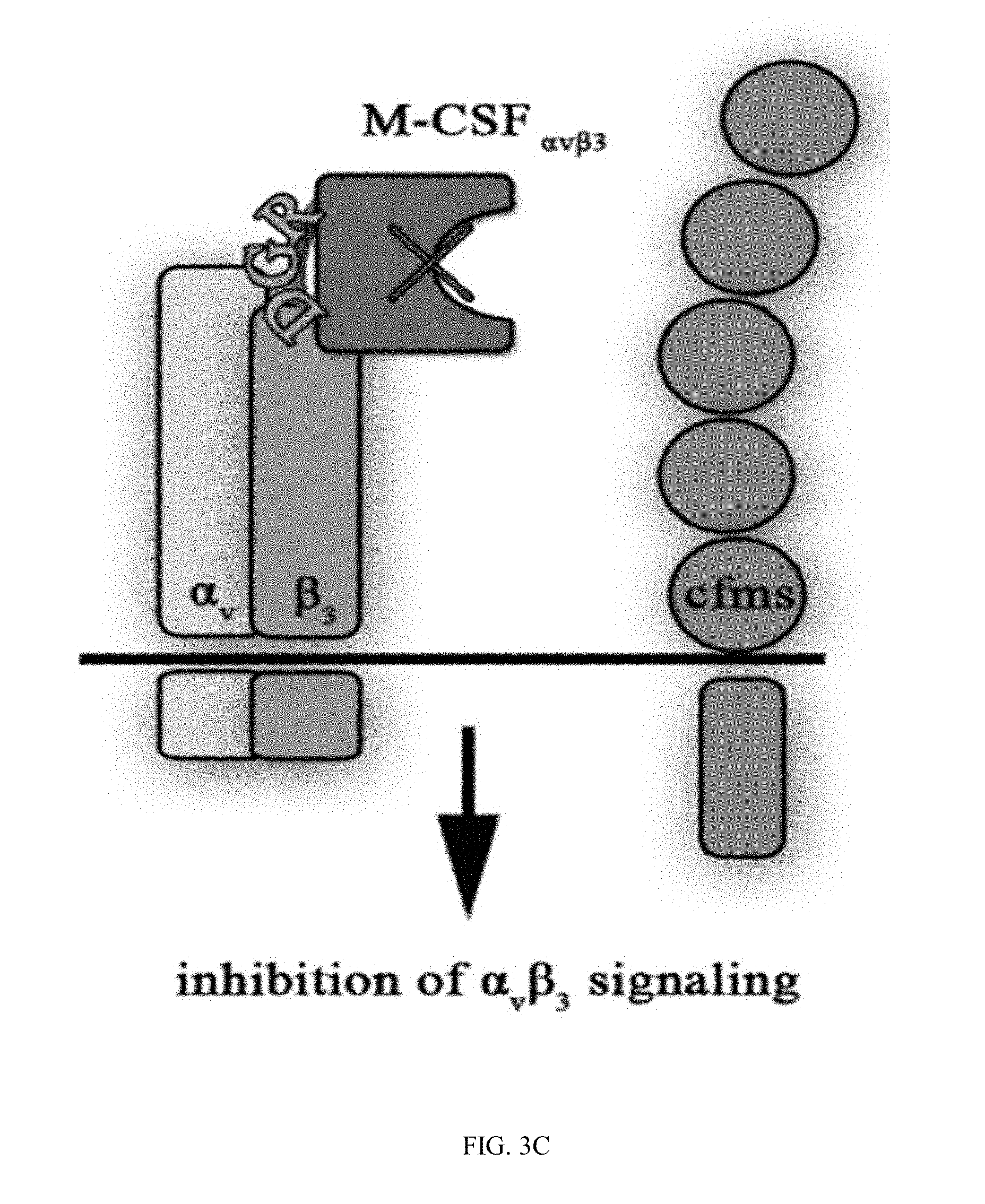
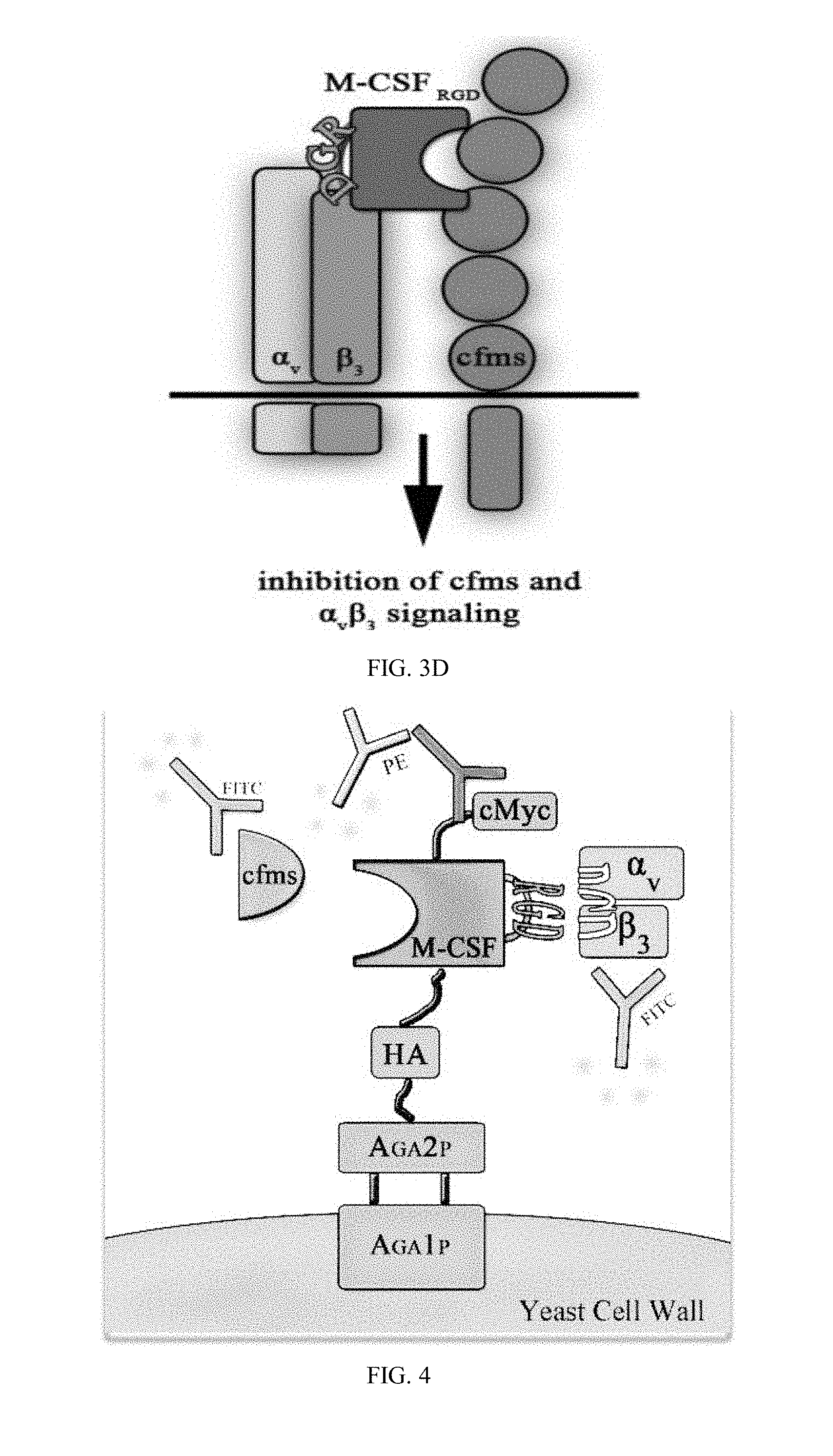
[0030] FIGS. 3A-3D are schematic illustrations of the different M-CSF constructs and their resulting signaling events: (3A) M-CSF.sub.WT is a disulfide-bond-linked homodimer which binds and activates cFMS, (3B) M-CSF monomer (designated M-CSF.sub.M) designed with a cysteine to serine substitution in position 31 to prevent dimerization via a disulfide bond, which binds to c-FMS and inhibits its mediated signaling, (3C) Mono-specific M-CSF that can bind integrin .alpha..sub.v.beta..sub.3 (via an RGD motif) but not c-FMS (designated M-CSF.sub..alpha.c.beta.3) due to mutations in positions 9 and 15 that prevent M-CSF binding to its receptor, and (3D) M-CSF.sub.RGD which was created by substituting one of the two loops (AB loop or CD loop) on the M-CSF dimerization site to an RGD motif with 3 random amino acids on each side that enables the M-CSF.sub.RGD to bind .alpha..sub.v.beta..sub.3 integrin as well as c-FMS and inhibit their mediated signaling events;
[0031] FIG. 4 is a schematic illustration of yeast surface display (YSD) construct.
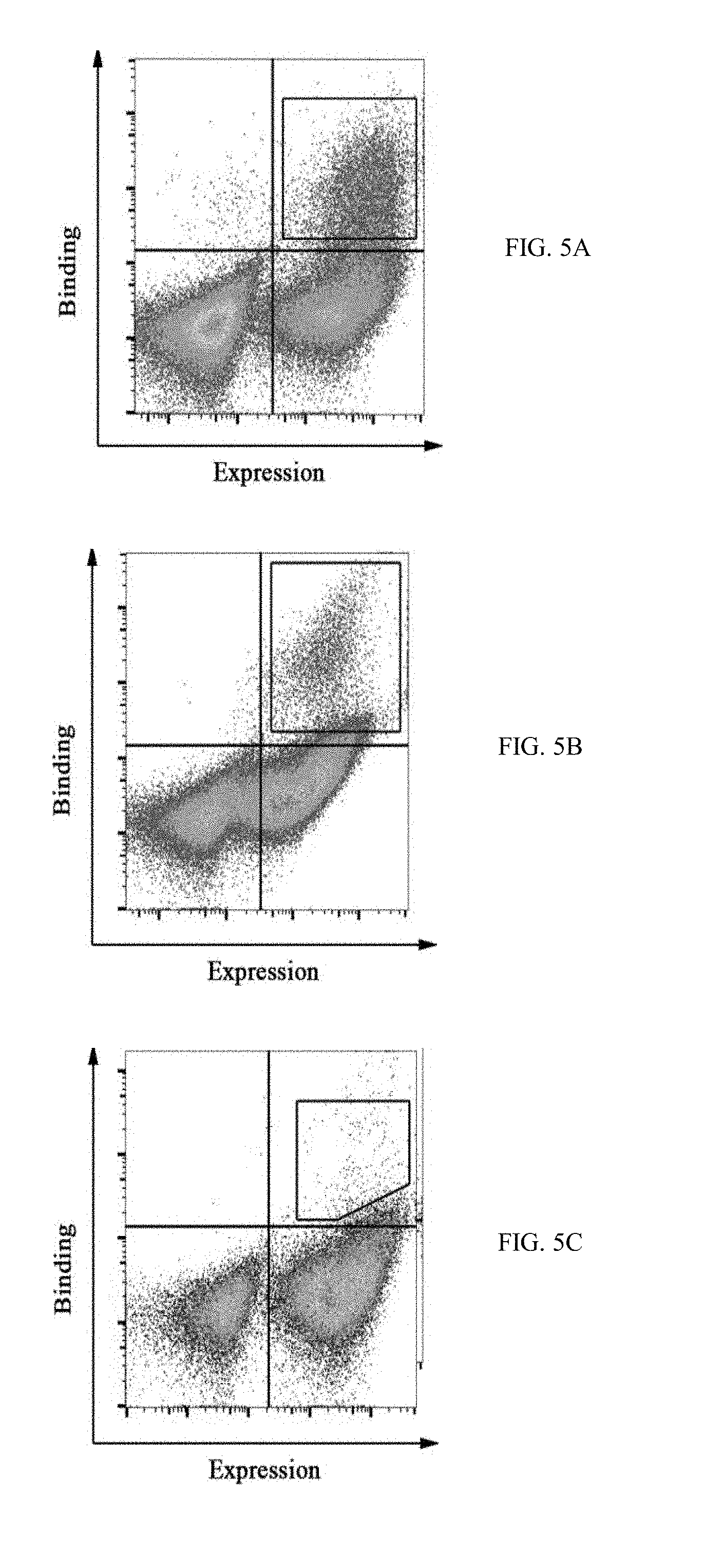
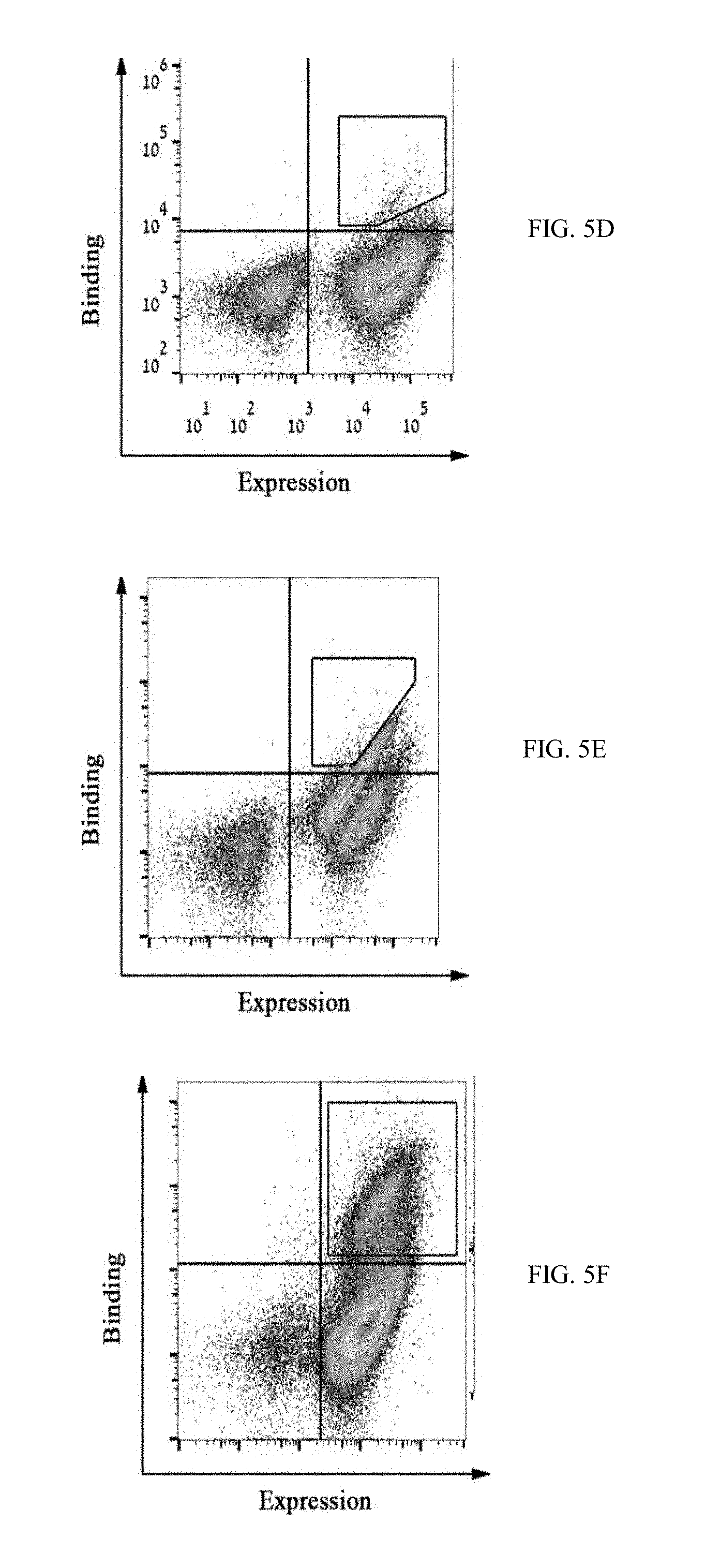
[0032] FIGS. 5A-5F show FACS dot plots of M-CSF.sub.RGD affinity maturation process in which yeast displayed mutant pools were tested for binding to (5A) 200 nM c-FMS, (5B) 500 nM .alpha..sub.v.beta..sub.3 integrin, (5C) 250 nM .alpha..sub.v.crclbar..sub.3 integrin, (5D) 100 nM .alpha..sub.v.beta..sub.3 integrin, (5E) 20 nM .alpha..sub.v.beta..sub.3 integrin, and (5F) 50 nM c-FMS, high target binders were collected as demonstrated in each figure with black polygon shape gates;
[0033] FIGS. 6A-6D are bar graphs showing normalized binding values of individual YSD M-CSF.sub.RGD clones to .alpha..sub.v.beta..sub.3 integrin and c-FMS: twenty-five different clones from each of sorts four (6A) and five (6C) were tested for binding to 20 nM of .alpha..sub.v.beta..sub.3 integrin, results are normalized to the lowest binder, Next, the best 15 .alpha..sub.v.beta..sub.3 integrin M-CSF.sub.RGD binders from sort four (6B) and the best ten .alpha..sub.v.beta..sub.3 integrin M-CSF.sub.RGD binders from sort five (6D), were tested for binding to 50 nM of c-FMS, these results are normalized to M-CSF.sub.M, the chosen clones (4.22, 4.24 and 5.6) are marked by arrows.
[0034] FIGS. 7A-7D depict the protein purification process of M-CSF.sub.M and M-CSF.sub.RGD variants: (7A) is a graph representing results of size exclusion chromatography of non-glycosylated M-CSF.sub.RGD clone 4.22 eluted at the size of 21 kDa with high molecular weight standards, (7B) is a graph showing CD spectra of non-glycosylated M-CSF.sub.M, non-glycosylated 4.22, non-glycosylated 4.24 and non-glycosylated 5.6. (7C) is a graph representing results of mass spectrometry spectrum for non-glycosylated 5.6. (7D) is a photograph of SDS PAGE for all purified proteins: glycosylated M-CSF.sub.M (lane 1), non-glycosylated M-CSF.sub.M (lane 2), non-glycosylated 4.22 (lane 3), non-glycosylated 4.24 (lane 4) and non-glycosylated 5.6 (lane 5);
[0035] FIG. 8 shows photographs of gels presenting monomers and dimers of the purified variants M-CSF.sub..alpha.v.beta.3 (4.22, 4.24, 5.6), M-CSF.sub..alpha.v.beta.3, and M-CFS.sub.M following incubation with increasing concentrations of the cross linker B S.sup.3;
[0036] FIGS. 9A-9J show SPR sensorgrams of the different M-CSF purified proteins: binding of M-CSF.sub.M (9A and 9E), M-CSF.sub..alpha.v.beta.3 (9B and 9G) 4.22 (9C and 9H), 4.24 (9D and 9I) and 5.6 (9E and 9J) to c-FMS (9A-9E) and .alpha..sub.v.beta..sub.3 integrin (9F-9J) at concentrations of 12.5 nM, 25 nM, 50 nM, 100 nM and 200 nM;
[0037] FIGS. 10A-10E show graphs demonstrating binding specificity of M-CSF.sub.RGD variants for RGD-binding integrins. .alpha.v.beta.3 (10A), .alpha.v.beta.5 (10B), .alpha.3.beta.1 (10C), .alpha.4.beta.7 (10D) and .alpha.5.beta.1 (10E) integrins were immobilized on the surface of the chip, and the three M-CSF.sub.RGD variants 4.22, 4.24 and 5.6 were allowed to flow over the surface of the chip at a concentration of 1 .mu.M.
[0038] FIGS. 11A-11F show graphs demonstrating results of direct cell binding assay with flow cytometry. Purified M-CSF.sub.RGD variants and M-CSF.sub.M proteins were tested for binding to murine primary cells (FIGS. 11A, 11B and 11C) and MDA-231 breast cancer cell line (FIGS. 11C, 11D and 11F). The black histograms represent the negative control and the gray histograms represent protein binding or receptor expression. The cells express c-FMS (FIGS. 11A, 11C) and .alpha..sub.v.beta..sub.3 integrin (FIGS. 11B, 11D). The mouse primary cells (FIG. 11E) and MDA-231 cells (FIG. 11F) showed binding in a dose dependent manner to 1 .mu.M, 2.5 .mu.M and 7.5 .mu.M, each, of M-CSF.sub.M, 4.22 and 5.6; and
[0039] FIGS. 12A-12D show photographs (FIG. 12) and bar graphs (FIG. 12B-D) demonstrating the effects of different M-CSF.sub.RGD variants and M-CSF.sub.M on osteoclast differentiation. Primary mouse bone marrow monocytes were cultured for 96 h in .alpha.-MEM growth medium containing recombinant mouse M-CSF (20 ng/ml), RANKL (20 ng/ml) and different concentrations of inhibitors. Positive control contained .alpha.-MEM growth medium, recombinant M-CSF and RANKL but not inhibitors. Negative control contains .alpha.-MEM growth medium and recombinant M-CSF and PBS control has 10% PBS with recombinant M-CSF and RANKL. (FIG. 12A) Cells were fixed and stained for Tartrate-resistant acid phosphatase (TRAP). (FIGS. 12B-D) Cells were examined for: (FIG. 12B) number of mature osteoclasts, (FIG. 12C) bone marrow monocytes. Results were normalized to the positive control.
[0040] FIG. 13 shows the effect of M-CSFM (containing only C31S mutation) and M-CSFM, M27R (containing both C31S and M27R) on the differentiation of primary murine cells to osteoclasts.
[0041] FIGS. 14A-14D show the structure of the M-CSF.sub.C31S mutant, compared to the M-CSF wild-type structure. FIG. 14A depicts the solved X-ray structure of the dimeric M-CSF.sub.C31S mutant, shown in ribbon diagram with S31 visualized as sticks. FIG. 14B depicts a close-up of the C31-C31 disulfide bond in the wild-type M-CSF structure (PDB ID 3UF2), visualized as spheres and colored by atom. The structure is rotated 90.degree. about X in relation to the M-CSF.sub.C31S mutant shown in 14A. FIG. 14C is an overlay of the M-CSF.sub.C31S mutant structure on the M-CSF wild-type structure, with the two C31 residues from the latter shown as spheres. FIG. 14D is a close-up of the S31 residues in the M-CSF.sub.C31S mutant dimer, visualized as in 14B.
[0042] FIGS. 15A-15C show the significant contribution of the M-CSF.sub.C31S residues to the interaction across the M-CSF.sub.C31S dimer interface. FIG. 15A. M-CSF.sub.C31S residues calculated to contribute significantly to interactions across the dimer interface (see Table 1), shown as sticks and colored by the type of their energy contribution as follows: magenta (polar/electrostatic contribution from the side chain+a non-polar contribution), cyan (polar/electrostatic contribution from the main chain+a non-polar contribution), green (non-polar contribution only). The opposing monomer is shown as surface representation colored wheat. FIG. 15B is similar to 15A, rotated 180.degree. about X. FIG. 15C illustrates the orientation of Q26 and M27, which contribute significantly and were deemed to be particularly influential on dimer formation and therefore chosen for further mutagenesis, shown as sticks (Q26 from both monomers and one of the M27 residues) and spheres (the M27 residue from the opposing monomer).
[0043] FIGS. 16A-16D show biophysical assays to evaluate the oligomeric state of the M-CSF variants. FIG. 16A is an SDS-PAGE gel depicting different M-CSF variants crosslinked with BS.sup.3 reagent, the quantities of monomer (20 kDa) and dimer (40 kDa) were visualized on SDS-PAGE gel for human M-CSF (upper left), murine M-CSF (lower left), M-CSF.sub.C31S (upper right) and M-CSF.sub.C31S,M27R (lower right). FIG. 16B shows distribution of hydration radii for M-CSF.sub.WT (solid line), M-CSF.sub.C31S (dashed line) and M-CSF.sub.C31S,M27R (dotted line) as measured by DLS. The calculated hydration radius is presented for each variant. FIG. 16C shows Radii of gyration determined by in-house script for M-CSF.sub.WT and its two variants (M-CSF.sub.C31S and M-CSF.sub.C31S,M27R). The dashed lines represent the theoretical R.sub.g values for the M-CSF monomer (R.sub.g=17 .ANG.) and M-CSF dimer (R.sub.g=26 .ANG.) calculated from the crystal structure [PDB ID: 3UF2] using CRYSOL. FIG. 16D is a small angle X-ray scattering (SAXS) structures of M-CSF.sub.WT and its two variants (M-CSF.sub.C31S and M-CSF.sub.C31S,M27R). Ab initio models were reconstructed from SAXS data using the computer program GASBOR and were averaged by the computer program DAMAVER. The crystal structure of the M-CSF dimer (blue and red) and the structure of the monomer (extracted from the crystal structure of M-CSF, red) were aligned with the obtained SAXS models (yellow, M-CSF.sub.WT; green, M-CSF.sub.C31S; cyan, M-CSF.sub.C31S,M27R) using PyMOL (http://www.pymol.org).
[0044] FIGS. 17A-17B depict the affinity of M-CSF.sub.WT and M-CSF.sub.C31S,M27R to the c-FMS receptor, as detected by SPR. Sensogram of M-CSF.sub.WT binding to c-FMS in concentrations up to 70 nM (17A) and M-CSF.sub.C31S,M27R binding to c-FMS in concentrations up to 20 nM. N=3 (17B). Values are means.+-.SD.
[0045] FIGS. 18A-18B show the evaluation of c-FMS phosphorylation in response to M-CSF variants. (18A) Phosphorylation of murine c-FMS on BMMs after incubation with 0.5 nM murine M-CSF (positive control, +), no M-CSF (negative control, -) or 0.5 nM murine M-CSF+1 .mu.M of M-CSF.sub.C31S or M-CSF.sub.C31S,M27R. (18B) Phosphorylation of human c-FMS on CD14.sup.+ cells after incubation with 0.5 nM human M-CSF (positive control, +), no M-CSF (negative control, -) or 0.5 nM murine M-CSF+100 nM of M-CSF.sub.C31S or M-CSF.sub.C31S,M27R. All signals were normalized to those of .beta.-actin, c-FMS expression and the positive control in each experiment. Statistical analysis was calculated using a t-test, each sample was compared to the positive control. N=3. Values are means.+-.SEM. *, p<0.05; **, p<0.01; ***, p<0.005.
[0046] FIGS. 19A-19B. M-CSF.sub.C31S,M27R inhibits monocyte differentiation into osteoclasts. (19A) Differentiation of bone marrow derived monocytes (BMMs) in the presence of murine M-CSF+RANKL (positive control), M-CSF without RANKL (negative control), and M-CSF+RANKL+M-CSF.sub.C31S or M-CSF.sub.C31S,M27R at concentrations of 50 nM, 1 .mu.M was evaluated. Cells were stained using TRAP staining and photographed (upper panels). The number of osteoclasts (lower left) and number of nuclei in osteoclasts (lower right) were quantified. (19B) Differentiation of CD14.sup.+ in the presence of human M-CSF+murine RANKL (positive control), M-CSF without RANKL (negative control), and M-CSF+RANKL+M-CSF.sub.C31S or M-CSF.sub.C31S,M27R at concentrations of 50 nM, 250 nM and 1 .mu.M was evaluated. Cells were stained using TRAP staining and cells were photographed (upper panels). The number of osteoclasts (lower left) and number of nuclei in osteoclasts (lower right) were quantified. Statistical analysis was performed using a t-test, each sample was compared to the positive control. N=3. Values are means.+-.SEM. *, p<0.05; **, p<0.01; ***, p<0.005.
[0047] FIGS. 20A-20C. M-CSF variants activate c-FMS with different potencies. (20A) Quantification of phosphorylated murine c-FMS in the presence of 0.5 nM murine M-CSF (positive control), no M-CSF (negative control), M-CSF.sub.WT (0.5, 5 and 10 nM), M-CSF.sub.C31S and M-CSF.sub.C31S,M27R (0.5, 5, 10, 50, 1000 and 5000 nM) in the absence of murine M-CSF. All signals were normalized to those of .beta.-actin, c-FMS expression and the positive control in each experiment. Statistical analysis was calculated using a t-test, each sample was compared to the positive control. N=3. Values are means.+-.SEM. (20B) An illustration showing c-FMS activation at different concentration of M-CSF.sub.WT, M-CSF.sub.C31S and M-CSF.sub.C31S,M27R. (20C) An illustration of the hypothesized inhibitory mechanism of M-CSF.sub.WT, M-CSF.sub.C31S and M-CSF.sub.C31S,M27R at different oligomerization states as a function of their concentrations.
[0048] FIG. 21 is a dot plot showing CTX-I serum-concentrations of M-CSF5.6 treated ovariectomized mice. Each black solid dot represents the CTX-I serum-concentration of one animal.
[0049] FIGS. 22A-22B are bar graphs showing Mean CTX-I serum-concentrations of M-CSF5.6 treated ovariectomized mice (22A) and (22B) show the mean CTX-I serum concentrations with or without haemolytic samples, respectively. The error bars represent the standard error of the sample mean.
[0050] FIG. 23 are example pictures showing Actin ring formation in mature osteoclasts incubated with M-CSF.sub.RGD variants. Murine BMMs were allowed to differentiate into osteoclasts in the presence of M-CSF and RANKL for 72 h. Then, the cells were incubated for additional 24 h without (positive control) or with inhibitors (5 .mu.M) followed by fixation and staining for F-actin and nuclei. Cells were able to form a solid actin ring (white arrowheads), scattered actin ring (white arrows) or amorphous actin distribution (barbed arrowheads). Pictures are representatives of 35 images acquired from five different wells per sample.
DETAILED DESCRIPTION OF THE INVENTION
[0051] The present invention provides a modified M-CSF protein and composition or kits comprising same. The invention further provides methods of treatment using said modified M-CSF. In some embodiments, the modified M-CSF of some embodiments of the invention is unable to undergo M-CSF homo-dimerization and has enhanced binding affinity to .alpha..sub.v.beta..sub.3 and c-FMS. In some embodiment, the modified M-CSF comprises an RGD motif. In some embodiment, the modified M-CSF comprises a mutation of cysteine (Cys) at position 31 and methionine (Met) at position 27 of SEQ ID NO:1.
[0052] Advantageously, the modified M-CSF protein of some embodiments of the invention acts as an antagonist for both c-FMS and .alpha.v.beta.3 integrin. As known in the art, c-FMS and .alpha.v.beta.3 are typically expressed on membranes of osteoclasts and their activation is required for osteoclast differentiation. Hence, the modified M-CSF protein of the present invention is, in some embodiments, capable of inhibiting or reducing osteoclast differentiation. The modified M-CSF protein of the invention is, in additional embodiments, capable of reducing bone resorption. In some embodiments, the modified M-CSF of the invention is practically useful in the treatment and/or prevention of osteoporosis.
[0053] The present invention is based, in part, on the surprising finding that the modified M-CSF disclosed herein bind .alpha..sub.v.beta..sub.3 integrin and c-FMS and inhibit activity thereof. The present invention is further based, in part, on the surprising finding that the modified M-CSF disclosed herein inhibits osteoclast differentiation.
[0054] As exemplified in the example section below inhibition is achieved using modified M-CSF comprising substitution of cysteine 31 and an RGD motif inserted into a loop AB or a loop CD thereof. As further exemplified in the example section below, modified M-CSF comprising mutations in positions 9 and 15 of SEQ ID NO: 1 are incapable of binding c-FMS.
Modified M-CSF
[0055] According to one aspect, the invention provides a polypeptide comprising an amino acid sequence having at least 90% homology to SEQ ID NO: 1, said polypeptide comprising an RGD motif comprising the amino acid sequence as set forth in SEQ ID NO: 6 (RGD).
[0056] In some embodiments, the polypeptide comprises an amino acid sequence having at least 90%, 91%, 92%, 93%, or 94% homology to SEQ ID NO: 1 (EEVSEYCSHMIGSGHLQSLQRLIDSQMETSXQITFEFVDQEQLKDPVCYLKKAFLLVQDI MEDTMRFRDNTEPVRGDNINPNAIAIVQLQELSLRLKSCFTKDYEEHDKACVRTFYETPL QLLEKVKNVFNETKNLLDKDWNIFSKNCNNSFAECSS), wherein X is any amino acid other than cysteine.
[0057] One skilled in the art will appreciate that monocyte/macrophage colony stimulating factor (M-CSF) have the Uniprot accession no. P09603 and comprise an AB loop (amino acids 25-32), a BC loop (amino acids 64-71), and a CD loop (amino acids 90-103).
[0058] In some embodiments, at least one loop selected from the AB loop and the CD loop comprises a modification of 3-10 contiguous amino acid residues compared to the corresponding AB loop or CD loop of SEQ ID NO: 1. In some embodiments the modification is selected from: deletion, substitution and addition of amino acid residues.
[0059] In some embodiments, at least one loop selected from the AB loop and the CD loop comprises the RGD motif. In some embodiments, the AB loop comprises the RGD motif. In some embodiments, the CD loop comprises the RGD motif.
[0060] In some embodiments, the polypeptide comprises one or more amino acid sequences selected from the group consisting of: SEQ ID NO: 3 (EEVSEYCSHMIGSGHLQSLQRLID), SEQ ID NO: 4 (ITFEFVDQEQLKDPVCYLKKAFLLVQDIMED), and SEQ ID NO: 5 (PNAIAIVQLQELSLRLKSCFTKDYEEHDKACVRTFYETPLQLLEKVKNVFNETKNLLDK DWNIFSKNCNNSFAECSS).
[0061] In some embodiments, the mutant polypeptide is incapable of forming homodimers. In some embodiments, the polypeptide binds c-FMS and .alpha..sub.v.beta..sub.3integrin.
[0062] In some embodiments, the polypeptide comprises an amino acid sequence as set forth in formula I (EEVSEYCSHMIGSGHLQSLQRLID(SQMETSSQ).sub.b(X.sub.1X.sub.2X.sub.3RGDX.sub.4- X.sub.5S).sub.(1-b)ITFEFVDQEQL KDPVCYLKKAFLLVQDIMED(TMRFRDNT).sub.(1-b)(EPVRGDNIN).sub.bPNAIAIVQLQELSLRL- KS CFTKDYEEHDKACVRTFYETPLQLLEKVKNVFNETKNLLDKDWNIFSKNCNNSFAECSS), wherein b is an integer selected from 0 and 1, and wherein: X.sub.1 is Q or T, X.sub.2 is T or Y, X.sub.3 is S or P, X.sub.4 is S or M, and X.sub.5 is P or C.
[0063] In some embodiments, the polypeptide comprises an amino acid sequence as set forth in
TABLE-US-00001 SEQ ID NO: 12 (EEVSEYCSHMIGSGHLQSLQRLIDQTPRGDSPSITFEFVDQEQLKDP VCYLKKAFLLVQDIMEDTMRFRDNTPNAIAIVQLQELSLRLKSCFTKD YEEHDKACVRTFYETPLQLLEKVKNVFNETKNLLDKDWNIFSKNCNNS FAECSS).
[0064] In some embodiments, the polypeptide comprises an amino acid sequence as set forth in
TABLE-US-00002 SEQ ID NO: 13 (EEVSEYCSHMIGSGHLQSLQRLIDTYPRGDMCSITFEFVDQEQLKDP VCYLKKAFLLVQDIMEDTMRFRDNTPNAIAIVQLQELSLRLKSCFTKD YEEHDKACVRTFYETPLQLLEKVKNVFNETKNLLDKDWNIFSKNCNNS FAECSS).
[0065] In some embodiments, the polypeptide comprises an amino acid sequence as set forth in
TABLE-US-00003 SEQ ID NO: 14 (EEVSEYCSHMIGSGHLQSLQRLIDSQMETSSQITFEFVDQEQLKDPV CYLKKAFLLVQDIMEDEPVRGDNINPNAIAIVQLQELSLRLKSCFTKD YEEHDKACVRTFYETPLQLLEKVKNVFNETKNLLDKDWNIFSKNCNNS FAECSS).
[0066] In some embodiments, the polypeptide comprises an amino acid sequence as set forth in formula II: EEVSEYCSHMIGSGHLQSLQRLID(SQMETSSQ).sub.b(X.sub.1X.sub.2X.sub.3RGDX.sub.4X- .sub.5S).sub.(1-b)ITFEFVDQEQLK DPVCYLKKAFLLVQDIMED(TMRFRDNT).sub.(1-b)(EPVRGDNIN).sub.bPNAIAIVQLQELSLRLK- SCF TKDYEEHDKACVRTFYETPLQLLEKVKNVFNETKNLLDKDWNIFSKNCNNSFAECSSQD VVTKPDCNCLYPKAIPSSDPASVSPHQPLAPSMAPVAGLTWEDSEGTEGSSLLPGEQPLH TVDPGSAKQRPPRSTCQSFEPPETPVVKDSTIGGSPQPRPSVGAFNPGMEDILDSAMGTN WVPEEASGEASEIPVPQGTELSPSRPGGGSMQTEPARPSNFLSASSPLPASAKGQQPADVT GTALPRVGPVRPTGQDWNHTPQKTDHPSALLRDPPEPGSPRISSLRPQGLSNPSTLSAQPQ LSRSHSSGSVLPLGELEGRRSTRDRRSPAEPEGGPASEGAARPLPRFNSVPLTDTGHERQS EGS, wherein b is an integer selected from 0 and 1, and wherein: X.sub.1 is Q or T, X.sub.2 is T or Y, X.sub.3 is S or P, X.sub.4 is S or M, and X.sub.5 is P or C.
[0067] As used herein "b" and "1-b" represent the number of repetitions of a sequence enclosed within the preceding adjacent brackets. The number of repetitions may vary between 0 and 1. For a non-limiting example when b equals 0, the preceding sequence within the brackets is not part of the sequence. "1-b" represents a mathematical equation resulting in an integer which equals 1 or 0.
[0068] In some embodiments, the polypeptide comprises an amino acid sequence as set forth in
TABLE-US-00004 SEQ ID NO: 16 (EEVSEYCSHMIGSGHLQSLQRLIDX.sub.1X.sub.2X.sub.3RGDX.sub.4X.sub.5SITFEFVDQEQ- L KDPVCYLKKAFLLVQDIMEDTMRFRDNTPNAIAIVQLQELSLRLKSCF TKDYEEHDKACVRTFYETPLQLLEKVKNVFNETKNLLDKDWNIFSKNC NNSFAECSSQDVVTKPDCNCLYPKAIPSSDPASVSPHQPLAPSMAPVA GLTWEDSEGTEGSSLLPGEQPLHTVDPGSAKQRPPRSTCQSFEPPETP VVKDSTIGGSPQPRPSVGAFNPGMEDILDSAMGTNWVPEEASGEASEI PVPQGTELSPSRPGGGSMQTEPARPSNFLSASSPLPASAKGQQPADVT GTALPRVGPVRPTGQDWNHTPQKTDHPSALLRDPPEPGSPRISSLRPQ GLSNPSTLSAQPQLSRSHSSGSVLPLGELEGRRSTRDRRSPAEPEGGP ASEGAARPLPRFNSVPLTDTGHERQSEGS)
wherein: X.sub.1 is Q or T, X.sub.2 is T or Y, X.sub.3 is S or P, X.sub.4 is S or M, and X.sub.5 is P or C.
[0069] In some embodiments, the polypeptide comprises an amino acid sequence as set forth in
TABLE-US-00005 SEQ ID NO: 17 (EEVSEYCSHMIGSGHLQSLQRLIDSQMETSSQITFEFVDQEQLKDPV CYLKKAFLLVQDIMEDEPVRGDNINPNAIAIVQLQELSLRLKSCFTKD YEEHDKACVRTFYETPLQLLEKVKNVFNETKNLLDKDWNIFSKNCNNS FAECSSQDVVTKPDCNCLYPKAIPSSDPASVSPHQPLAPSMAPVAGLT WEDSEGTEGSSLLPGEQPLHTVDPGSAKQRPPRSTCQSFEPPETPVVK DSTIGGSPQPRPSVGAFNPGMEDILDSAMGTNWVPEEASGEASEIPVP QGTELSPSRPGGGSMQTEPARPSNFLSASSPLPASAKGQQPADVTGTA LPRVGPVRPTGQDWNHTPQKTDHPSALLRDPPEPGSPRISSLRPQGLS NPSTLSAQPQLSRSHSSGSVLPLGELEGRRSTRDRRSPAEPEGGPASE GAARPLPRFNSVPLTDTGHERQSEGS).
[0070] As used herein, the terms "peptide", "polypeptide" and "protein" are used interchangeably to refer to a polymer of amino acid residues. The terms "peptide", "polypeptide" and "protein" as used herein encompass native peptides, peptidomimetics (typically including non-peptide bonds or other synthetic modifications) and the peptide analogs peptoids and semi-peptoids or any combination thereof. In another embodiment, the terms "peptide", "polypeptide" and "protein" apply to amino acid polymers in which one or more amino acid residue is an artificial chemical analog of a corresponding naturally occurring amino acid.
[0071] In one embodiment, the polypeptide of the invention comprises or consists of the amino acid sequence as set forth in SEQ ID NO: 11, 12, 13, 14, 15, 16 or 17, or a sequence derived therefrom.
[0072] As used herein, the term "derived from" or "corresponding to" refers to construction of an amino acid sequence based on the knowledge of a sequence using any one of the suitable means known to one skilled in the art, e.g. chemical synthesis in accordance with standard protocols in the art.
[0073] One of skill in the art will recognize that individual substitutions, deletions or additions to a peptide, or protein sequence which alters, adds or deletes a single amino acid or a small percentage of amino acids in the encoded sequence is a conservatively modified variant where the alteration results in the substitution of an amino acid with a similar charge, size, and/or hydrophobicity characteristics, such as, for example, substitution of a glutamic acid (E) to aspartic acid (D).
[0074] In some embodiments, the polypeptide of the invention comprises a sequence derived from SEQ ID NO: 11, 12, 13, 14, 15, 16 or 17 with one or more conservative substitution. In some embodiments, the mutant polypeptide of the invention comprises a sequence derived from SEQ ID NO: 11, 12, 13, 14, 15, 16 or 17 with at most 1, 2, 3, 4, 5, 6, or 7 conservative substitutions. Each possibility represents a separate embodiment of the present invention. According to another embodiment of the invention, the polypeptide of the invention comprises a sequence homologous to SEQ ID NO: 11, 12, 13, 14, 15, 16, or 17. According to another embodiment of the invention, the polypeptide of the invention comprises a sequence having greater than 70%, 75%, 80%, 85%, 90%, 91%, 92%, 93%, 94%, 95%, 96%, 97%, 98%, or 99% homology to SEQ ID NO: 11, 12, 13, 14, 15, 16 or 17. Each possibility represents a separate embodiment of the present invention.
[0075] In some embodiments, the polypeptide of the invention comprises a sequence having at least 94%, 95%, 96%, 97%, 98%, or 99% homology to SEQ ID NO: 11, 12, 13, 14, 15, 16 or 17, wherein the mutant M-CSF is incapable of forming homodimers, wherein the mutant M-CSF binds c-FMS and wherein the mutant M-CSF binds .alpha..sub.v.beta..sub.3 integrin. Each possibility represents a separate embodiment of the present invention.
[0076] In some embodiments, the polypeptide of the invention comprises a sequence having at least 94%, 95%, 96%, 97%, 98%, or 99% homology to a sequence selected from the group consisting of: SEQ ID NOs: 11, 12, 13, 14, 15, 16 and 17, wherein the polypeptide of the invention is incapable of forming homodimers, wherein the polypeptide of the invention binds c-FMS and wherein the polypeptide of the invention binds .alpha..sub.v.beta..sub.3integrin. Each possibility represents a separate embodiment of the present invention.
[0077] As used herein, the term "analog" includes any peptide having an amino acid sequence substantially identical to one of the sequences specifically shown herein in which one or more residues have been conservatively substituted with a functionally similar residue and which displays the abilities as described herein. Examples of conservative substitutions include the substitution of one non-polar (hydrophobic) residue such as isoleucine, valine, leucine or methionine for another, the substitution of one polar (hydrophilic) residue for another such as between arginine and lysine, between glutamine and asparagine, between glycine and serine, the substitution of one basic residue such as lysine, arginine or histidine for another, or the substitution of one acidic residue, such as aspartic acid or glutamic acid for another. Each possibility represents a separate embodiment of the present invention.
[0078] As used herein, the phrase "conservative substitution" also includes the use of a chemically derivatized residue in place of a non-derivatized residue provided that such peptide displays the requisite function as specified herein.
[0079] In one embodiment, the polypeptide is a variant of SEQ ID NO: 11, 12, 13, 14, 15, 16 or 17.
[0080] In another embodiment, the term "variant" refers to a polypeptide or nucleotide sequence which comprises a modification of one or more amino acids or nucleotides as compared to another polypeptide or polynucleotide, respectively. In some embodiments, the modifications are substitution, deletion, and/or insertion of one or more amino acids or nucleotides as compared to another polypeptide or polynucleotide, respectively. In some embodiments, the changes may be of minor nature, such as conservative amino acid substitutions or for nucleotide sequence resulting in conservative amino acid substitutions that do not significantly affect the activity of the polypeptide.
[0081] In some embodiments, the changes may be substitution of an amino acid molecule, resulting in an addition of a glycosylation site, thereby increasing glycosylation of the polypeptide.
[0082] Typically, the present invention encompasses derivatives of the polypeptides. The term "derivative" or "chemical derivative" includes any chemical derivative of the polypeptide having one or more residues chemically derivatized by reaction of side chains or functional groups. Such derivatized molecules include, for example, those molecules in which free amino groups have been derivatized to form amine hydrochlorides, p-toluene sulfonyl groups, carbobenzoxy groups, t-butyloxycarbonyl groups, chloroacetyl groups or formyl groups. Free carboxyl groups may be derivatized to form salts, methyl and ethyl esters or other types of esters or hydrazides. Free hydroxyl groups may be derivatized to form O-acyl or O-alkyl derivatives. The imidazole nitrogen of histidine may be derivatized to form N-im-benzylhistidine. Also included as chemical derivatives are those peptides, which contain one or more naturally occurring amino acid derivatives of the twenty standard amino acid residues. For example: 4-hydroxyproline may be substituted for proline; 5-hydroxylysine may be substituted for lysine; 3-methylhistidine may be substituted for histidine; homoserine may be substituted or serine; and ornithine may be substituted for lysine.
[0083] In addition, a peptide derivative can differ from the natural sequence of the peptides of the invention by chemical modifications including, but are not limited to, terminal-NH2 acylation, acetylation, or thioglycolic acid amidation, and by terminal-carboxlyamidation, e.g., with ammonia, methylamine, and the like. Peptides can be either linear, cyclic or branched and the like, which conformations can be achieved using methods well known in the art.
[0084] The peptide derivatives and analogs according to the principles of the present invention can also include side chain bond modifications, including but not limited to --CH2-NH--, --CH2-S--, --CH2-S.dbd.0, OC--NH--, --CH2-O--, --CH2-CH2-, S.dbd.C--NH--, and --CH.dbd.CH--, and backbone modifications such as modified peptide bonds. Peptide bonds (--CO--NH--) within the peptide can be substituted, for example, by N-methylated bonds (--N(CH3)-CO--); ester bonds (--C(R)H-C-0-0-C(R)H--N); ketomethylene bonds (--CO--CH2-); a-aza bonds (--NH--N(R)--CO--), wherein R is any alkyl group, e.g., methyl; carba bonds (--CH2-NH--); hydroxyethylene bonds (--CH(OH)--CH2-); thioamide bonds (--CS--NH); olefmic double bonds (--CH.dbd.CH--); and peptide derivatives (--N(R)--CH2-CO--), wherein R is the "normal" side chain, naturally presented on the carbon atom. These modifications can occur at one or more of the bonds along the peptide chain and even at several (e.g., 2-3) at the same time.
[0085] The present invention also encompasses peptide derivatives and analogs in which free amino groups have been derivatized to form amine hydrochlorides, p-toluene sulfonylamino groups, carbobenzoxyamino groups, t-butyloxycarbonylamino groups, chloroacetylamino groups or formylamino groups. Free carboxyl groups may be derivatized to form, for example, salts, methyl and ethyl esters or other types of esters or hydrazides. The imidazole nitrogen of histidine can be derivatized to form N-im-benzylhistidine.
[0086] As used herein the term "salts" refers to both salts of carboxyl groups and to acid addition salts of amino or guanido groups of the peptide molecule. Salts of carboxyl groups may be formed by means known in the art and include inorganic salts, for example sodium, calcium, ammonium, ferric or zinc salts, and the like, and salts with organic bases such as salts formed for example with amines such as triethanolamine, piperidine, procaine, and the like. Acid addition salts include, for example, salts with mineral acids such as, for example, acetic acid or oxalic acid. Salts describe here also ionic components added to the peptide solution to enhance hydrogel formation and/or mineralization of calcium minerals.
[0087] The peptide analogs can also contain non-natural amino acids. Examples of non-natural amino acids include, but are not limited to, sarcosine (Sar), norleucine, ornithine, citrulline, diaminobutyric acid, homoserine, isopropyl Lys, 3-(2'-naphtyl)-Ala, nicotinyl Lys, amino isobutyric acid, and 3-(3'-pyridyl-Ala).
[0088] Furthermore, the peptide analogs can contain other derivatized amino acid residues including, but not limited to, methylated amino acids, N-benzylated amino acids, O-benzylated amino acids, N-acetylated amino acids, O-acetylated amino acids, carbobenzoxy-substituted amino acids and the like. Specific examples include, but are not limited to, methyl-Ala (Me Ala), MeTyr, MeArg, MeGlu, MeVal, MeHis, N-acetyl-Lys, O-acetyl-Lys, carbobenzoxy-Lys, Tyr-O-Benzyl, Glu-O-Benzyl, Benzyl-His, Arg-Tosyl, t-butylglycine, t-butylalanine, phenylglycine, cyclohexylalanine, and the like.
[0089] The invention further includes peptide analogs, which can contain one or more D-isomer forms of the amino acids. Production of retro-inverso D-amino acid peptides where at least one amino acid and perhaps all amino acids are D-amino acids is well known in the art. When all of the amino acids in the peptide are D-amino acids, and the N- and C-terminals of the molecule are reversed, the result is a molecule having the same structural groups being at the same positions as in the L-amino acid form of the molecule. However, the molecule is more stable to proteolytic degradation and is therefore useful in many of the applications recited herein. Diastereomeric peptides may be highly advantageous over all L- or all D-amino acid peptides having the same amino acid sequence because of their higher water solubility, lower immunogenicity, and lower susceptibility to proteolytic degradation. The term "diastereomeric peptide" as used herein refers to a peptide comprising both L-amino acid residues and D-amino acid residues. The number and position of D-amino acid residues in a diastereomeric peptide of the preset invention may be variable so long as the peptide is capable of displaying the function of disclosed by the modified M-SCF of the invention.
PEGylation
[0090] In some embodiments, the polypeptides of the present invention are conjugated to polyethylene glycol (PEG). Conjugation of PEG to polypeptides is known to prolong in-vivo half-life of polypeptides. PEGs are non-toxic polymers of ethylene oxide that are widely used in protein medications and other fields.
RGD Motifs
[0091] In some embodiment, the RGD motif comprises the amino acid sequence Arg-Gly-Asp as set forth in SEQ ID NO: 6 (RGD). In some embodiments, the RGD motif further comprises one or more randomized amino acid residues flanking either side or both sides of the RGD sequence. In some embodiments, the integrin binding motif comprises the amino acid sequence as set forth in SEQ ID NO: 7 (XXXRGDXXX), wherein each X individually represents any amino acid residue (e.g., a randomized amino acid residue). In some embodiments, the RGD motif exhibits a binding specificity to .alpha..sub.v.beta..sub.3. In some embodiments, the RGD motif has significantly higher binding affinity to .alpha..sub.v.beta..sub.3 than to other integrins (e.g., .alpha..sub.4.beta..sub.7, .alpha..sub.2.beta..sub.2b, .alpha..sub.5.beta..sub.1 and .alpha..sub.v.beta..sub.5).
[0092] In some embodiments, the RGD motif comprising the amino acid sequence as set forth in SEQ ID NO: 6 has a length of less than 30, 20, 18, 15, 12, 9, 8, 7, 6, 5, or 4 amino acids. Each possibility represents a separate embodiment of the present invention. In another embodiment, the RGD motif comprising the amino acid sequence as set forth in SEQ ID NO: 6 has a length of at least 3, 4, 5, 6, 7, 8, 9, 10, 11, 12, 13, 14, 15, 16, 17, 18, 19, 20, 21, 22, 23, 24, 25, 26, 27, 28, 29, or 30 amino acids derived from SEQ ID NO: 2. In another embodiment, the RGD motif comprising the amino acid sequence as set forth in SEQ ID NO: 6 has a length of 3 to 6, 3 to 7, 3 to 8, 3 to 9, 3 to 10, 3 to 11, 3 to 12, 3 to 14, 3 to 15, 3 to 16, 3 to 17, 3 to 18, 3 to 19, or 3 to 20. Each possibility represents a separate embodiment of the present invention. In some embodiments, the RGD motif comprises at least 9 amino acid residues. In some embodiments, the RGD motif comprises or consists of 9 amino acid residues. In some embodiments, the RGD motif has 3 to 20 amino acids, comprising the amino acid sequence as set forth in SEQ ID NO: 6 (RGD).
[0093] In some embodiments, the RGD motif comprises or consists of the amino acid sequence selected from the group consisting of: SEQ ID NO: 8 (QTSRGDSPS), SEQ ID NO: 9 (TYPRGDMCS), and SEQ ID NO: 10 (EPVRGDNIN). In some embodiments, the RGD motif is selected from the group consisting of: SEQ ID NO: 8 (QTSRGDSPS), SEQ ID NO: 9 (TYPRGDMCS).
[0094] In some embodiments, the RGD motif comprises or consists of the amino acid sequence as set forth in SEQ ID NO: 8 (QTSRGDSPS). In some embodiments, the RGD motif comprises or consists of the amino acid sequence as set forth in SEQ ID NO: 9 (TYPRGDMCS). In some embodiments, the RGD motif comprises or consists of the amino acid sequence as set forth in SEQ ID NO: 10 (EPVRGDNIN).
[0095] In some embodiments, the AB loop of the polypeptide comprises an RGD motif selected from the group consisting of: SEQ ID NO: 8 (QTSRGDSPS), SEQ ID NO: 9 (TYPRGDMCS). In some embodiments, the CD loop of the polypeptide comprises an RGD motif comprising or consisting of the amino acid sequence as set forth in SEQ ID NO: 10 (EPVRGDNIN).
Synthesizing the Polypeptide
[0096] According to one embodiment, the polypeptides of the present invention may be synthesized or prepared by any method and/or technique known in the art for peptide synthesis. According to another embodiment, the polypeptides may be synthesized by a solid phase peptide synthesis method of Merrifield (see J. Am. Chem. Soc, 85:2149, 1964). According to another embodiment, the polypeptides of the present invention can be synthesized using standard solution methods well known in the art (see, for example, Bodanszky, M., Principles of Peptide Synthesis, Springer-Verlag, 1984).
[0097] In general, the synthesis methods comprise sequential addition of one or more amino acids or suitably protected amino acids to a growing peptide chain bound to a suitable resin. Normally, either the amino or carboxyl group of the first amino acid is protected by a suitable protecting group. The protected or derivatized amino acid can then be either attached to an inert solid support (resin) or utilized in solution by adding the next amino acid in the sequence having the complimentary (amino or carboxyl) group suitably protected, under conditions conductive for forming the amide linkage. The protecting group is then removed from this newly added amino acid residue and the next amino acid (suitably protected) is added, and so forth. After all the desired amino acids have been linked in the proper sequence, any remaining protecting groups are removed sequentially or concurrently, and the peptide chain, if synthesized by the solid phase method, is cleaved from the solid support to afford the final peptide.
[0098] In the solid phase peptide synthesis method, the alpha-amino group of the amino acid is protected by an acid or base sensitive group. Such protecting groups should have the properties of being stable to the conditions of peptide linkage formation, while being readily removable without destruction of the growing peptide chain. Suitable protecting groups are t-butyloxycarbonyl (BOC), benzyloxycarbonyl (Cbz), biphenylisopropyloxycarbonyl, t-amyloxycarbonyl, isobornyloxycarbonyl, (alpha, alpha)-dimethyl-3,5 dimethoxybenzyloxycarbonyl, o-nitrophenylsulfenyl, 2-cyano-t-butyloxycarbonyl, 9-fluorenylmethyloxycarbonyl (FMOC) and the like. In the solid phase peptide synthesis method, the C-terminal amino acid is attached to a suitable solid support. Suitable solid supports useful for the above synthesis are those materials, which are inert to the reagents and reaction conditions of the stepwise condensation-deprotection reactions, as well as being insoluble in the solvent media used. Suitable solid supports are chloromethylpolystyrene-divinylbenzene polymer, hydroxymethyl-polystyrene-divinylbenzene polymer, and the like. The coupling reaction is accomplished in a solvent such as ethanol, acetonitrile, N,N-dimethylformamide (DMF), and the like. The coupling of successive protected amino acids can be carried out in an automatic polypeptide synthesizer as is well known in the art.
[0099] In another embodiment, polypeptides of the invention may be synthesized such that one or more of the bonds, which link the amino acid residues of the peptides are non-peptide bonds. In another embodiment, the non-peptide bonds include, but are not limited to, imino, ester, hydrazide, semicarbazide, and azo bonds, which can be formed by reactions well known to one skilled in the art.
[0100] The invention further encompasses a polynucleotide sequence comprising a nucleic acid encoding any of the polypeptides of the invention. In another embodiment, the nucleic acid sequence encoding the polypeptide is at least 70%, or alternatively at least 80%, or alternatively at least 90%, or alternatively at least 95%, or alternatively at least 99% homologous to the nucleic acid sequence encoding the nucleic acid sequence of the polypeptides of the invention or a fragment thereof.
[0101] In some embodiment, the invention provides a polynucleotide encoding the polypeptides of the invention.
[0102] In some embodiments, the polynucleotide of the present invention is ligated into an expression vector, comprising a transcriptional control of a cis-regulatory sequence (e.g., promoter sequence). In some embodiments, the cis-regulatory sequence is suitable for directing constitutive expression of the polypeptide of the present invention. In some embodiments, the cis-regulatory sequence is suitable for directing tissue-specific expression of the polypeptide of the present invention. In some embodiments, the cis-regulatory sequence is suitable for directing inducible expression of the polypeptide of the present invention.
[0103] The term "polynucleotide" refers to a nucleic acid (e.g., DNA or RNA) sequence that comprises coding sequences necessary for the production of a polypeptide. In one embodiment, a polynucleotide refers to a single or double stranded nucleic acid sequence which is isolated and provided in the form of an RNA sequence, a complementary polynucleotide sequence (cDNA), a genomic polynucleotide sequence and/or a composite polynucleotide sequences (e.g., a combination of the above).
[0104] In one embodiment, "complementary polynucleotide sequence" refers to a sequence, which results from reverse transcription of messenger RNA using a reverse transcriptase or any other RNA dependent DNA polymerase. In one embodiment, the sequence can be subsequently amplified in vivo or in vitro using a DNA polymerase.
[0105] In one embodiment, "genomic polynucleotide sequence" refers to a sequence derived (isolated) from a chromosome and thus it represents a contiguous portion of a chromosome.
[0106] In one embodiment, "composite polynucleotide sequence" refers to a sequence, which is at least partially complementary and at least partially genomic. In one embodiment, a composite sequence can include some exonal sequences required to encode the polypeptide of the present invention, as well as some intronic sequences interposing there between. In one embodiment, the intronic sequences can be of any source, including of other genes, and typically will include conserved splicing signal sequences. In one embodiment, intronic sequences include cis acting expression regulatory elements.
[0107] In some embodiments, polynucleotides of the present invention are prepared using PCR techniques as described in Example 1, or any other method or procedure known to one skilled in the art. In some embodiments, the procedure involves the ligation of two different DNA sequences (See, for example, "Current Protocols in Molecular Biology", eds. Ausubel et al., John Wiley & Sons, 1992).
[0108] In one embodiment, polynucleotides of the present invention are inserted into expression vectors (i.e., a nucleic acid construct) to enable expression of the recombinant polypeptide. In one embodiment, the expression vector of the present invention includes additional sequences which render this vector suitable for replication and integration in prokaryotes. In one embodiment, the expression vector of the present invention includes additional sequences which render this vector suitable for replication and integration in eukaryotes. In one embodiment, the expression vector of the present invention includes a shuttle vector which renders this vector suitable for replication and integration in both prokaryotes and eukaryotes. In some embodiments, cloning vectors comprise transcription and translation initiation sequences (e.g., promoters, enhancers) and transcription and translation terminators (e.g., polyadenylation signals).
[0109] In one embodiment, a variety of prokaryotic or eukaryotic cells can be used as host-expression systems to express the polypeptide of the present invention. In some embodiments, these include, but are not limited to, microorganisms, such as bacteria transformed with a recombinant bacteriophage DNA, plasmid DNA or cosmid DNA expression vector containing the polypeptide coding sequence; yeast transformed with recombinant yeast expression vectors containing the polypeptide coding sequence; plant cell systems infected with recombinant virus expression vectors (e.g., cauliflower mosaic virus, CaMV; tobacco mosaic virus, TMV) or transformed with recombinant plasmid expression vectors, such as Ti plasmid, containing the polypeptide coding sequence.
[0110] In some embodiments, non-bacterial expression systems are used (e.g. mammalian expression systems) to express the polypeptide of the present invention. In one embodiment, the expression vector is used to express polynucleotides of the present invention in mammalian cells.
[0111] In some embodiments, in bacterial systems of the present invention, a number of expression vectors can be advantageously selected depending upon the use intended for the polypeptide expressed. In one embodiment, large quantities of polypeptide are desired. In one embodiment, vectors that direct the expression of high levels of the protein product, possibly as a fusion with a hydrophobic signal sequence, which directs the expressed product into the periplasm of the bacteria or the culture medium where the protein product is readily purified are desired. In one embodiment, certain fusion protein engineered with a specific cleavage site to aid in recovery of the polypeptide. In one embodiment, vectors adaptable to such manipulation include, but are not limited to, the pET series of E. coli expression vectors [Studier et al., Methods in Enzymol. 185:60-89 (1990)].
[0112] In one embodiment, yeast expression systems are used. In one embodiment, a number of vectors containing constitutive or inducible promoters can be used in yeast as disclosed in U.S. Pat. No. 5,932,447. In another embodiment, vectors which promote integration of foreign DNA sequences into the yeast chromosome are used.
[0113] In one embodiment, the expression vector of the present invention may further include additional polynucleotide sequences that allow, for example, the translation of several proteins from a single mRNA such as an internal ribosome entry site (IRES).
[0114] In some embodiments, mammalian expression vectors include, but are not limited to, pcDNA3, pcDNA3.1 (.+-.), pGL3, pZeoSV2(.+-.), pSecTag2, pDisplay, pEF/myc/cyto, pCMV/myc/cyto, pCR3.1, pSinRep5, DH26S, DHBB, pNMT1, pNMT41, pNMT81, which are available from Invitrogen, pCI which is available from Promega, pMbac, pPbac, pBK-RSV and pBK-CMV which are available from Strategene, pTRES which is available from Clontech, and their derivatives.
[0115] In some embodiments, expression vectors containing regulatory elements from eukaryotic viruses such as retroviruses are used by the present invention. SV40 vectors include pSVT7 and pMT2. In some embodiments, vectors derived from bovine papilloma virus include pBV-1MTHA, and vectors derived from Epstein Bar virus include pHEBO, and p2O5. Other exemplary vectors include pMSG, pAV009/A+, pMTO10/A+, pMAMneo-5, baculovirus pDSVE, and any other vector allowing expression of proteins under the direction of the SV-40 early promoter, SV-40 later promoter, metallothionein promoter, murine mammary tumor virus promoter, Rous sarcoma virus promoter, polyhedrin promoter, or other promoters shown effective for expression in eukaryotic cells.
[0116] In some embodiments, recombinant viral vectors, which offer advantages such as lateral infection and targeting specificity, are used for in vivo expression of the polypeptide of the present invention. In one embodiment, lateral infection is inherent in the life cycle of, for example, retrovirus and is the process by which a single infected cell produces many progeny virions that bud off and infect neighboring cells. In one embodiment, the result is that a large area becomes rapidly infected, most of which was not initially infected by the original viral particles. In one embodiment, viral vectors are produced that are unable to spread laterally. In one embodiment, this characteristic can be useful if the desired purpose is to introduce a specified gene into only a localized number of targeted cells.
[0117] Various methods can be used to introduce the expression vector of the present invention into cells. Such methods are generally described in Sambrook et al., Molecular Cloning: A Laboratory Manual, Cold Springs Harbor Laboratory, New York (1989, 1992), in Ausubel et al., Current Protocols in Molecular Biology, John Wiley and Sons, Baltimore, Md. (1989), Chang et al., Somatic Gene Therapy, CRC Press, Ann Arbor, Mich. (1995), Vega et al., Gene Targeting, CRC Press, Ann Arbor Mich. (1995), Vectors: A Survey of Molecular Cloning Vectors and Their Uses, Butterworths, Boston Mass. (1988) and Gilboa et at. [Biotechniques 4 (6): 504-512, 1986] and include, for example, stable or transient transfection, lipofection, electroporation and infection with recombinant viral vectors. In addition, see U.S. Pat. Nos. 5,464,764 and 5,487,992 for positive-negative selection methods.
[0118] A person with skill in the art will appreciate that the polypeptide of the present invention can also be expressed from a nucleic acid construct administered to the individual employing any suitable mode of administration, described hereinabove (i.e., in vivo gene therapy). In one embodiment, the nucleic acid construct is introduced into a suitable cell via an appropriate gene delivery vehicle/method (transfection, transduction, homologous recombination, etc.) and an expression system as needed and then the modified cells are expanded in culture and returned to the individual (i.e., ex vivo gene therapy).
[0119] In one embodiment, in vivo gene therapy using a cytokine has been attempted in animal models such as rodents [Bohl et al., Blood. 2000; 95:2793-2798], primates [Gao et al., Blood, 2004, Volume 103, Number 9] and has proven successful in human clinical trials for patients with chronic renal failure [Lippin et al Blood 2005, 106, Number 7].
[0120] In one embodiment, plant expression vectors are used. In one embodiment, the expression of a polypeptide coding sequence is driven by a number of promoters. In some embodiments, viral promoters such as the 35S RNA and 19S RNA promoters of CaMV [Brisson et al., Nature 310:511-514 (1984)], or the coat protein promoter to TMV [Takamatsu et al., EMBO J. 6:307-311 (1987)] are used. In another embodiment, plant promoters are used such as, for example, the small subunit of RUBISCO [Coruzzi et al., EMBO J. 3:1671-1680 (1984); and Brogli et al., Science 224:838-843 (1984)] or heat shock promoters, e.g., soybean hsp17.5-E or hsp17.3-B [Gurley et al., Mol. Cell. Biol. 6:559-565 (1986)]. In one embodiment, constructs are introduced into plant cells using Ti plasmid, Ri plasmid, plant viral vectors, direct DNA transformation, microinjection, electroporation and other techniques well known to the skilled artisan. See, for example, Weissbach & Weissbach [Methods for Plant Molecular Biology, Academic Press, NY, Section VIII, pp 421-463 (1988)]. Other expression systems such as insects and mammalian host cell systems, which are well known in the art, can also be used by the present invention.
[0121] It will be appreciated that other than containing the necessary elements for the transcription and translation of the inserted coding sequence (encoding the polypeptide), the expression construct of the present invention can also include sequences engineered to optimize stability, production, purification, yield or activity of the expressed polypeptide.
[0122] In some embodiments, transformed cells are cultured under effective conditions, which allow for the expression of high amounts of recombinant polypeptide. In some embodiments, effective culture conditions include, but are not limited to, effective media, bioreactor, temperature, pH and oxygen conditions that permit protein production. In one embodiment, an effective medium refers to any medium in which a cell is cultured to produce the recombinant polypeptide of the present invention. In some embodiments, a medium typically includes an aqueous solution having assimilable carbon, nitrogen and phosphate sources, and appropriate salts, minerals, metals and other nutrients, such as vitamins. In some embodiments, cells of the present invention can be cultured in conventional fermentation bioreactors, shake flasks, test tubes, microtiter dishes and petri plates. In some embodiments, culturing is carried out at a temperature, pH and oxygen content appropriate for a recombinant cell. In some embodiments, culturing conditions are within the expertise of one of ordinary skill in the art.
[0123] In some embodiments, depending on the vector and host system used for production, resultant polypeptides of the present invention either remain within the recombinant cell, secreted into the fermentation medium, secreted into a space between two cellular membranes, such as the periplasmic space in E. coli; or retained on the outer surface of a cell or viral membrane. In one embodiment, following a predetermined time in culture, recovery of the recombinant polypeptide is affected.
[0124] In one embodiment, the phrase "recovering the recombinant polypeptide" used herein refers to collecting the whole fermentation medium containing the polypeptide and need not imply additional steps of separation or purification.
[0125] In one embodiment, polypeptides of the present invention are purified using a variety of standard protein purification techniques, such as, but not limited to, affinity chromatography, ion exchange chromatography, filtration, electrophoresis, hydrophobic interaction chromatography, gel filtration chromatography, reverse phase chromatography, concanavalin A chromatography, chromatofocusing and differential solubilization.
[0126] In one embodiment, to facilitate recovery, the expressed coding sequence can be engineered to encode the polypeptide of the present invention and fused cleavable moiety. In one embodiment, a fusion protein can be designed so that the polypeptide can be readily isolated by affinity chromatography; e.g., by immobilization on a column specific for the cleavable moiety. In one embodiment, a cleavage site is engineered between the polypeptide and the cleavable moiety, and the polypeptide can be released from the chromatographic column by treatment with an appropriate enzyme or agent that specifically cleaves the fusion protein at this site [e.g., see Booth et al., Immunol. Lett. 19:65-70 (1988); and Gardella et al., J. Biol. Chem. 265:15854-15859 (1990)].
[0127] In one embodiment, the polypeptide of the present invention is retrieved in "substantially pure" form that allows for the effective use of the protein in the applications described herein.
[0128] As used herein, the term "substantially pure" describes a peptide/polypeptide or other material which has been separated from its native contaminants. Typically, a monomeric peptide is substantially pure when at least about 60 to 75% of a sample exhibits a single peptide backbone. Minor variants or chemical modifications typically share the same peptide sequence. A substantially pure peptide can comprise over about 85 to 90% of a peptide sample, and can be over 95% pure, over 97% pure, or over about 99% pure. Purity can be measured on a polyacrylamide gel, with homogeneity determined by staining. Alternatively, for certain purposes high resolution may be necessary and HPLC or a similar means for purification can be used. For most purposes, a simple chromatography column or polyacrylamide gel can be used to determine purity.
[0129] The term "purified" does not require the material to be present in a form exhibiting absolute purity, exclusive of the presence of other compounds. Rather, it is a relative definition. A peptide is in the "purified" state after purification of the starting material or of the natural material by at least one order of magnitude, 2 or 3, or 4 or 5 orders of magnitude.
[0130] In one embodiment, the polypeptides of the present invention are substantially free of naturally-associated host cell components. The term "substantially free of naturally-associated host cell components" describes a peptide or other material which is separated from the native contaminants which accompany it in its natural host cell state. Thus, a peptide which is chemically synthesized or synthesized in a cellular system different from the host cell from which it naturally originates will be free from its naturally-associated host cell components.
[0131] In one embodiment, the polypeptide of the present invention can also be synthesized using in vitro expression systems. In one embodiment, in vitro synthesis methods are well known in the art and the components of the system are commercially available.
Pharmaceutical Compositions
[0132] According to another aspect, the invention provides a pharmaceutical composition comprising as an active ingredient a therapeutically effective amount of the polypeptide of the present invention, and a pharmaceutically acceptable carrier and/or diluent. In some embodiments, the pharmaceutical composition facilitates administration of a compound to an organism.
[0133] In another embodiment, the pharmaceutical compositions of the invention may be formulated in the form of a pharmaceutically acceptable salt of the polypeptides of the present invention or their analogs, or derivatives thereof. In another embodiment, pharmaceutically acceptable salts include those salts formed with free amino groups such as salts derived from non-toxic inorganic or organic acids such as hydrochloric, phosphoric, acetic, oxalic, tartaric acids, and the like, and those salts formed with free carboxyl groups such as salts derived from non-toxic inorganic or organic bases such as sodium, potassium, ammonium, calcium, ferric hydroxides, isopropylamine, triethylamine, 2-ethylamino ethanol, histidine, procaine, and the like.
[0134] As used herein, the term "carrier" refers to a diluent, adjuvant, excipient, or vehicle with which the therapeutic compound is administered. Such pharmaceutical carriers can be sterile liquids, such as water and oils, including those of petroleum, animal, vegetable or synthetic origin, such as peanut oil, soybean oil, mineral oil, sesame oil and the like, polyethylene glycols, glycerin, propylene glycol or other synthetic solvents. Water is a preferred carrier when the pharmaceutical composition is administered intravenously. Saline solutions and aqueous dextrose and glycerol solutions can also be employed as liquid carriers, particularly for injectable solutions. Suitable pharmaceutical excipients include starch, glucose, lactose, sucrose, gelatin, malt, rice, flour, chalk, silica gel, sodium stearate, glycerol monostearate, talc, sodium chloride, dried skim milk, glycerol, propylene glycol, water, ethanol and the like. The composition, if desired, can also contain minor amounts of wetting or emulsifying agents, or pH buffering agents such as acetates, citrates or phosphates. Antibacterial agents such as benzyl alcohol or methyl parabens; antioxidants such as ascorbic acid or sodium bisulfate; and agents for the adjustment of tonicity such as sodium chloride or dextrose are also envisioned. The carrier may comprise, in total, from about 0.1% to about 99.99999% by weight of the pharmaceutical compositions presented herein.
[0135] As used herein, the term "pharmaceutically acceptable" means suitable for administration to a subject, e.g., a human. For example, the term "pharmaceutically acceptable" can mean approved by a regulatory agency of the Federal or a state government or listed in the U.S. Pharmacopeia or other generally recognized pharmacopeia for use in animals, and more particularly in humans.
[0136] In another embodiment, the compositions of the invention take the form of solutions, suspensions, emulsions, tablets, pills, capsules, powders, gels, creams, ointments, foams, pastes, sustained-release formulations and the like. In another embodiment, the compositions of the invention can be formulated as a suppository, with traditional binders and carriers such as triglycerides, microcrystalline cellulose, gum tragacanth or gelatin. Oral formulation can include standard carriers such as pharmaceutical grades of mannitol, lactose, starch, magnesium stearate, sodium saccharine, cellulose, magnesium carbonate, etc. Examples of suitable pharmaceutical carriers are described in: Remington's Pharmaceutical Sciences" by E. W. Martin, the contents of which are hereby incorporated by reference herein. Such compositions will contain a therapeutically effective amount of the polypeptide of the invention, preferably in a substantially purified form, together with a suitable amount of carrier so as to provide the form for proper administration to the subject.
[0137] According to an embodiment of the invention, pharmaceutical compositions contain 0.1%-95% of the polypeptide(s) of the present invention, derivatives, or analogs thereof. According to another embodiment of the invention, pharmaceutical compositions contain 1%-70% of the polypeptide(s) derivatives, or analogs thereof. According to another embodiment of the invention, the composition or formulation to be administered may contain a quantity of polypeptide(s), derivatives, or analogs thereof, according to embodiments of the invention in an amount effective to treat the condition or disease of the subject being treated.
[0138] An embodiment of the invention relates to polypeptides of the present invention, derivatives, or analogs thereof, presented in unit dosage form and prepared by any of the methods well known in the art of pharmacy. In an embodiment of the invention, the unit dosage form is in the form of a tablet, capsule, lozenge, wafer, patch, ampoule, vial or pre-filled syringe. In addition, in vitro assays may optionally be employed to help identify optimal dosage ranges. The precise dose to be employed in the formulation will also depend on the route of administration, and the nature of the disease or disorder, and should be decided according to the judgment of the practitioner and each patient's circumstances. Effective doses can be extrapolated from dose-response curves derived from in-vitro or in-vivo animal model test bioassays or systems.
[0139] According to one embodiment, the compositions of the present invention are administered in the form of a pharmaceutical composition comprising at least one of the active components of this invention together with a pharmaceutically acceptable carrier or diluent. In another embodiment, the compositions of this invention can be administered either individually or together in any conventional oral, parenteral or transdermal dosage form.
[0140] As used herein, the terms "administering," "administration," and like terms refer to any method which, in sound medical practice, delivers a composition containing an active agent to a subject in such a manner as to provide a therapeutic effect.
[0141] Depending on the location of the tissue of interest, the polypeptide of the present invention can be administered in any manner suitable for the provision of the polypeptides to cells within the tissue of interest. Thus, for example, a composition containing the polypeptides of the present invention can be introduced, for example, into the systemic circulation, which will distribute the peptide to the tissue of interest. Alternatively, a composition can be applied topically to the tissue of interest (e.g., injected, or pumped as a continuous infusion, or as a bolus within a tissue, applied to all or a portion of the surface of the skin, etc.).
[0142] In some embodiments, the pharmaceutical compositions comprising the polypeptides are administered via oral, rectal, vaginal, topical, nasal, ophthalmic, transdermal, subcutaneous, intramuscular, intraperitoneal or intravenous routes of administration. The route of administration of the pharmaceutical composition will depend on the disease or condition to be treated. Suitable routes of administration include, but are not limited to, parenteral injections, e.g., intradermal, intravenous, intramuscular, intralesional, subcutaneous, intrathecal, and any other mode of injection as known in the art. Although the bioavailability of peptides administered by other routes can be lower than when administered via parenteral injection, by using appropriate formulations it is envisaged that it will be possible to administer the compositions of the invention via transdermal, oral, rectal, vaginal, topical, nasal, inhalation and ocular modes of treatment. In addition, it may be desirable to introduce the pharmaceutical compositions of the invention by any suitable route, including intraventricular and intrathecal injection; intraventricular injection may be facilitated by an intraventricular catheter, for example, attached to a reservoir. Pulmonary administration can also be employed, e.g., by use of an inhaler or nebulizer.
[0143] For topical application, a peptide of the present invention, derivative, analog or a fragment thereof can be combined with a pharmaceutically acceptable carrier so that an effective dosage is delivered, based on the desired activity. The carrier can be in the form of, for example, and not by way of limitation, an ointment, cream, gel, paste, foam, aerosol, suppository, pad or gelled stick.
[0144] For oral applications, the pharmaceutical composition may be in the form of tablets or capsules, which can contain any of the following ingredients, or compounds of a similar nature: a binder such as microcrystalline cellulose, gum tragacanth or gelatin; an excipient such as starch or lactose; a disintegrating agent such as alginic acid, Primogel, or corn starch; a lubricant such as magnesium stearate; or a glidant such as colloidal silicon dioxide. When the dosage unit form is a capsule, it can contain, in addition to materials of the above type, a liquid carrier such as fatty oil. In addition, dosage unit forms can contain various other materials which modify the physical form of the dosage unit, for example, coatings of sugar, shellac, or other enteric agents. The tablets of the invention can further be film coated.
[0145] For purposes of parenteral administration, solutions in sesame or peanut oil or in aqueous propylene glycol can be employed, as well as sterile aqueous solutions of the corresponding water-soluble salts. Such aqueous solutions may be suitably buffered, if necessary, and the liquid diluent first rendered isotonic with sufficient saline or glucose. These aqueous solutions are especially suitable for intravenous, intramuscular, subcutaneous and intraperitoneal injection purposes.
[0146] According to some embodiments, the peptides of the present invention, derivatives, or analogs thereof can be delivered in a controlled release system. In another embodiment, an infusion pump can be used to administer the peptide such as the one that is used, for example, for delivering insulin or chemotherapy to specific organs or tumors. In another embodiment, the peptides of the invention are administered in combination with a biodegradable, biocompatible polymeric implant, which releases the peptide over a controlled period of time at a selected site. Examples of preferred polymeric materials include, but are not limited to, polyanhydrides, polyorthoesters, polyglycolic acid, polylactic acid, polyethylene vinyl acetate, copolymers and blends thereof (See, Medical applications of controlled release, Langer and Wise (eds.), 1974, CRC Pres., Boca Raton, Fla., the contents of which are hereby incorporated by reference in their entirety). In yet another embodiment, a controlled release system can be placed in proximity to a therapeutic target, thus requiring only a fraction of the systemic dose.
[0147] The presently described peptides, derivatives, or analogs thereof may also be contained in artificially created structures such as liposomes, ISCOMS, slow-releasing particles, and other vehicles which increase the half-life of the peptides or polypeptides in serum. Liposomes include emulsions, foams, micelles, insoluble monolayers, liquid crystals, phospholipid dispersions, lamellar layers and the like. Liposomes for use with the presently described peptides are formed from standard vesicle-forming lipids which generally include neutral and negatively charged phospholipids and a sterol, such as cholesterol. The selection of lipids is generally determined by considerations such as liposome size and stability in the blood. A variety of methods are available for preparing liposomes as reviewed, for example, by Coligan, J. E. et al, Current Protocols in Protein Science, 1999, John Wiley & Sons, Inc., New York, and see also U.S. Pat. Nos. 4,235,871, 4,501,728, 4,837,028, and 5,019,369.
[0148] The compositions also include incorporation of the active material into or onto particulate preparations of polymeric compounds such as polylactic acid, polglycolic acid, hydrogels, etc, or onto liposomes, microemulsions, micelles, unilamellar or multilamellar vesicles, erythrocyte ghosts, or spheroplasts.) Such compositions will influence the physical state, solubility, stability, rate of in vivo release, and rate of in vivo clearance.
[0149] In one embodiment, the present invention provides combined preparations. In one embodiment, "a combined preparation" defines especially a "kit of parts" in the sense that the combination partners as defined above can be dosed independently or by use of different fixed combinations with distinguished amounts of the combination partners i.e., simultaneously, concurrently, separately or sequentially. In some embodiments, the parts of the kit of parts can then, e.g., be administered simultaneously or chronologically staggered, that is at different time points and with equal or different time intervals for any part of the kit of parts. The ratio of the total amounts of the combination partners, in some embodiments, can be administered in the combined preparation. In one embodiment, the combined preparation can be varied, e.g., in order to cope with the needs of a patient subpopulation to be treated or the needs of the single patient which different needs can be due to a particular disease, severity of a disease, age, sex, or body weight as can be readily made by a person skilled in the art.
[0150] In one embodiment, it will be appreciated that the peptides of the present invention can be provided to the individual with additional active agents to achieve an improved therapeutic effect as compared to treatment with each agent by itself. In another embodiment, measures (e.g., dosing and selection of the complementary agent) are taken to adverse side effects which are associated with combination therapies.
[0151] In one embodiment, depending on the severity and responsiveness of the condition to be treated, dosing can be of a single or a plurality of administrations, with course of treatment lasting from several days to several weeks or until cure is effected or diminution of the disease state is achieved.
[0152] In some embodiments, the peptides are administered in a therapeutically safe and effective amount. As used herein, the term "safe and effective amount" refers to the quantity of a component which is sufficient to yield a desired therapeutic response without undue adverse side effects (such as toxicity, irritation, or allergic response) commensurate with a reasonable benefit/risk ratio when used in the presently described manner. In another embodiment, a therapeutically effective amount of the polypeptide is the amount of the polypeptide necessary for the in vivo measurable expected biological effect. The actual amount administered, and the rate and time-course of administration, will depend on the nature and severity of the condition being treated. Prescription of treatment, e.g. decisions on dosage, timing, etc., is within the responsibility of general practitioners or specialists, and typically takes account of the disorder to be treated, the condition of the individual patient, the site of delivery, the method of administration and other factors known to practitioners. Examples of techniques and protocols can be found in Remington: The Science and Practice of Pharmacy, 21st Ed., Lippincott Williams & Wilkins, Philadelphia, Pa., (2005). In some embodiments, preparation of effective amount or dose can be estimated initially from in vitro assays. In one embodiment, a dose can be formulated in animal models and such information can be used to more accurately determine useful doses in humans.
[0153] In one embodiment, toxicity and therapeutic efficacy of the active ingredients described herein can be determined by standard pharmaceutical procedures in vitro, in cell cultures or experimental animals. In one embodiment, the data obtained from these in vitro and cell culture assays and animal studies can be used in formulating a range of dosage for use in human. In one embodiment, the dosages vary depending upon the dosage form employed and the route of administration utilized. In one embodiment, the exact formulation, route of administration and dosage can be chosen by the individual physician in view of the patient's condition. [See e.g., Fingl, et al., (1975) "The Pharmacological Basis of Therapeutics", Ch. 1 p.1].
[0154] Pharmaceutical compositions containing the presently described polypeptide as the active ingredient can be prepared according to conventional pharmaceutical compounding techniques. See, for example, Remington's Pharmaceutical Sciences, 18th Ed., Mack Publishing Co., Easton, Pa. (1990). See also, Remington: The Science and Practice of Pharmacy, 21st Ed., Lippincott Williams & Wilkins, Philadelphia, Pa. (2005).
[0155] In one embodiment, compositions including the preparation of the present invention formulated in a compatible pharmaceutical carrier are prepared, placed in an appropriate container, and labeled for treatment of an indicated condition.
[0156] In one embodiment, compositions of the present invention are presented in a pack or dispenser device, such as an FDA approved kit, which contain one or more unit dosages forms containing the active ingredient. In one embodiment, the pack, for example, comprises metal or plastic foil, such as a blister pack. In one embodiment, the pack or dispenser device is accompanied by instructions for administration. In one embodiment, the pack or dispenser is accommodated by a notice associated with the container in a form prescribed by a governmental agency regulating the manufacture, use or sale of pharmaceuticals, which notice is reflective of approval by the agency of the form of the compositions or human or veterinary administration. Such notice, in one embodiment, is labeling approved by the U.S. Food and Drug Administration for prescription drugs or of an approved product insert.
Use of the Compositions
[0157] According to some aspects, there is provided a method for treating, ameliorating, reducing and/or preventing a condition associated with increased bone resorption in a subject in need thereof, the method comprising the step of: administering to a subject a pharmaceutical composition comprising an effective amount of the polypeptide of the invention, thereby treating, ameliorating, reducing and/or preventing a condition associated with increased bone resorption in a subject in need thereof.
[0158] In some embodiments, there is provided a method for treating a disease associated with increased bone resorption in a subject in need thereof, the method comprising the step of administering to said subject a pharmaceutical composition comprising an effective amount of an amino acid molecule comprising the amino acid sequence selected from the group consisting of SEQ ID NOs: 11-17 and a pharmaceutical acceptable carrier, thereby treating, ameliorating, reducing and/or preventing a disease associated with increased bone resorption in a subject in need thereof.
[0159] In another embodiment, the polypeptide of the invention or a composition comprising the polypeptide is for use in treatment, amelioration, reduction, and/or prevention of a condition associated with increased bone resorption in a subject in need thereof. In some embodiments, there is provided a composition comprising an effective amount of polypeptide for use in the treatment or prevention of a disease associated with increased bone resorption in a subject in need thereof In some embodiments, there is provided a composition comprising an effective amount of an amino acid molecule comprising the amino acid sequence selected from the group consisting of SEQ ID NOs: 11-17 for use in the treatment or prevention of a disease associated with increased bone resorption in a subject in need thereof.
[0160] In some embodiments, there is provided a use of a composition comprising an effective amount of polypeptide in the preparation of a medicament for the treatment, amelioration, reduction, or prevention of a disease associated with increased bone resorption in a subject in need thereof. In some embodiments, the invention provides use of a composition comprising an effective amount of an amino acid molecule comprising the amino acid sequence selected from the group consisting of SEQ ID NOs: 11-17 in the preparation of a medicament for the treatment of a disease associated with increased bone resorption in a subject in need thereof.
[0161] In one embodiment, the polypeptide of the present invention is provided to the subject per se. In one embodiment, the polypeptide of the present invention is provided to the subject as part of a pharmaceutical composition where it is mixed with a pharmaceutically acceptable carrier.
[0162] Non-limiting examples of disease associated with increased bone resorption include Paget's disease, osteoporosis, and tumor-linked bone resorption disease.
[0163] In some embodiments, the disease associated with increased bone resorption is osteoporosis.
[0164] The term "subject" as used herein refers to an animal, more particularly to non-human mammals and human organism. Non-human animal subjects may also include prenatal forms of animals, such as, e.g., embryos or fetuses. Non-limiting examples of non-human animals include: horse, cow, camel, goat, sheep, dog, cat, non-human primate, mouse, rat, rabbit, hamster, guinea pig, pig. In one embodiment, the subject is a human. Human subjects may also include fetuses. In one embodiment, a subject in need thereof is a subject afflicted with and/or at risk of being afflicted with a condition associated with increased bone resorption.
[0165] As used herein, the terms "treatment" or "treating" of a disease, disorder, or condition encompasses alleviation of at least one symptom thereof, a reduction in the severity thereof, or inhibition of the progression thereof. Treatment need not mean that the disease, disorder, or condition is totally cured. To be an effective treatment, a useful composition herein needs only to reduce the severity of a disease, disorder, or condition, reduce the severity of symptoms associated therewith, or provide improvement to a patient or subject's quality of life.
[0166] As used herein, the term "prevention" of a disease, disorder, or condition encompasses the delay, prevention, suppression, or inhibition of the onset of a disease, disorder, or condition. As used in accordance with the presently described subject matter, the term "prevention" relates to a process of prophylaxis in which a subject is exposed to the presently described peptides prior to the induction or onset of the disease/disorder process. This could be done where an individual has a genetic pedigree indicating a predisposition toward occurrence of the disease/disorder to be prevented. For example, this might be true of an individual whose ancestors show a predisposition toward certain types of, for example, inflammatory disorders. The term "suppression" is used to describe a condition wherein the disease/disorder process has already begun but obvious symptoms of the condition have yet to be realized. Thus, the cells of an individual may have the disease/disorder but no outside signs of the disease/disorder have yet been clinically recognized. In either case, the term prophylaxis can be applied to encompass both prevention and suppression. Conversely, the term "treatment" refers to the clinical application of active agents to combat an already existing condition whose clinical presentation has already been realized in a patient.
[0167] As used herein, the term "condition" includes anatomic and physiological deviations from the normal that constitute an impairment of the normal state of the living animal or one of its parts, that interrupts or modifies the performance of the bodily functions.
[0168] Any concentration ranges, percentage range, or ratio range recited herein are to be understood to include concentrations, percentages or ratios of any integer within that range and fractions thereof, such as one tenth and one hundredth of an integer, unless otherwise indicated.
[0169] Any number range recited herein relating to any physical feature, such as polymer subunits, size or thickness, are to be understood to include any integer within the recited range, unless otherwise indicated.
[0170] As used herein, the terms "subject" or "individual" or "animal" or "patient" or "mammal," refers to any subject, particularly a mammalian subject, for whom therapy is desired, for example, a human.
[0171] In the discussion unless otherwise stated, adjectives such as "substantially" and "about" modifying a condition or relationship characteristic of a feature or features of an embodiment of the invention, are understood to mean that the condition or characteristic is defined to within tolerances that are acceptable for operation of the embodiment for an application for which it is intended. Unless otherwise indicated, the word "or" in the specification and claims is considered to be the inclusive "or" rather than the exclusive or, and indicates at least one of, or any combination of items it conjoins.
[0172] It should be understood that the terms "a" and "an" as used above and elsewhere herein refer to "one or more" of the enumerated components. It will be clear to one of ordinary skill in the art that the use of the singular includes the plural unless specifically stated otherwise. Therefore, the terms "a," "an" and "at least one" are used interchangeably in this application.
[0173] For purposes of better understanding the present teachings and in no way limiting the scope of the teachings, unless otherwise indicated, all numbers expressing quantities, percentages or proportions, and other numerical values used in the specification and claims, are to be understood as being modified in all instances by the term "about." Accordingly, unless indicated to the contrary, the numerical parameters set forth in the following specification and attached claims are approximations that may vary depending upon the desired properties sought to be obtained. At the very least, each numerical parameter should at least be construed in light of the number of reported significant digits and by applying ordinary rounding techniques.
[0174] In the description and claims of the present application, each of the verbs, "comprise," "include" and "have" and conjugates thereof, are used to indicate that the object or objects of the verb are not necessarily a complete listing of components, elements or parts of the subject or subjects of the verb.
[0175] Other terms as used herein are meant to be defined by their well-known meanings in the art.
[0176] Additional objects, advantages, and novel features of the present invention will become apparent to one ordinarily skilled in the art upon examination of the following examples, which are not intended to be limiting. Additionally, each of the various embodiments and aspects of the present invention as delineated hereinabove and as claimed in the claims section below finds experimental support in the following examples.
[0177] It is appreciated that certain features of the invention, which are, for clarity, described in the context of separate embodiments, may also be provided in combination in a single embodiment. Conversely, various features of the invention, which are, for brevity, described in the context of a single embodiment, may also be provided separately or in any suitable sub-combination or as suitable in any other described embodiment of the invention. Certain features described in the context of various embodiments are not to be considered essential features of those embodiments, unless the embodiment is inoperative without those elements.
EXAMPLES
[0178] Generally, the nomenclature used herein and the laboratory procedures utilized in the present invention include molecular, biochemical, microbiological and recombinant DNA techniques. Such techniques are thoroughly explained in the literature. See, for example, "Molecular Cloning: A laboratory Manual" Sambrook et al., (1989); "Current Protocols in Molecular Biology" Volumes I-III Ausubel, R. M., ed. (1994); Ausubel et al., "Current Protocols in Molecular Biology", John Wiley and Sons, Baltimore, Maryland (1989); Perbal, "A Practical Guide to Molecular Cloning", John Wiley & Sons, New York (1988); Watson et al., "Recombinant DNA", Scientific American Books, New York; Birren et al. (eds) "Genome Analysis: A Laboratory Manual Series", Vols. 1-4, Cold Spring Harbor Laboratory Press, New York (1998); methodologies as set forth in U.S. Pat. Nos. 4,666,828; 4,683,202; 4,801,531; 5,192,659 and 5,272,057; "Cell Biology: A Laboratory Handbook", Volumes I-III Cellis, J. E., ed. (1994); "Culture of Animal Cells--A Manual of Basic Technique" by Freshney, Wiley-Liss, N. Y. (1994), Third Edition; "Current Protocols in Immunology" Volumes I-III Coligan J. E., ed. (1994); Stites et al. (eds), "Basic and Clinical Immunology" (8th Edition), Appleton & Lange, Norwalk, Conn. (1994); Mishell and Shiigi (eds), "Strategies for Protein Purification and Characterization--A Laboratory Course Manual" CSHL Press (1996); "Bacteriophage Methods and Protocols", Volume 1: Isolation, Characterization, and Interactions, all of which are incorporated by reference. Other general references are provided throughout this document.
[0179] Various embodiments and aspects of the present invention as delineated hereinabove and as claimed in the claims section below find experimental support in the following examples. Reference is now made to the following examples, which together with the above descriptions illustrate some embodiments of the invention in a non-limiting fashion.
Materials and Methods
[0180] Methods Designing and Constructing M-CSF.sub.M, two M-CSF.sub.RGD libraries and M-CSF.sub..alpha.v.beta.3
[0181] Using the 3D structure of the murine c-FMS/M-CSF complex (PDB: 3EJJ) and the 3D structure of human M-CSF (PDB: 1HMC), the binding interface of the complex was identified. The M-CSF monomer, designated M-CSF.sub.M, which is made up of 158 amino acids, was designed with C31S mutation that prevents homo-dimerization of the monomer (see Deng, P., et al., Biochemical and biophysical research communications, 1996. 228(2): p. 557-566). Two libraries of M-CSF.sub.M variants were then designed such that the two loops distant from the receptor binding site (residue numbering according to PDB: 3EJJ 25 to 32 and 64 to 71, respectively) were each replaced with an RGD motif flanked by three random amino acids on each side represented as XXXRGDXXX (designated M-CSF.sub.RGD). To generate the random amino acids in each library, the DNA codons were synthesized as NNS sequences, where N can be any codon and S can be C or G. The M-CSF.sub.M gene and the two M-CSF.sub.RGD libraries were prepared for us by GenScript (Piscataway, N.J., USA) with pCTCON homology sites and ECORI and NheI restriction sites on the amine terminal and AvrII and BamHI restriction sites on the C terminal. The M-CSF.sub.M gene and M-CSF.sub.RGD library DNA sequences were amplified by PCR with Phusion DNA polymerase (New England Biolabs, Ipswich, Mass., USA) and transformed into EBY100 Saccharomyces cerevisiae strain with a modified linear pCTCON plasmid [48] for homologous recombination using a Micropulser electroporator (Bio-Rad, USA). The DNA sequences were recovered in the tryptophan-selective medium SDCAA (2% dextrose, 0.67% yeast nitrogen base, 0.5% bacto casamino acids, 1.47% sodium citrate, 0.429% citric acid monohydrate, pH 4.5). To evaluate the diversities (sizes) of the two M-CSF.sub.RGD libraries, the yeasts were plated on SDCAA plates (0.54% Na.sub.2HPO.sub.4, 0.856% Na.sub.2HPO.sub.4H.sub.2O, 18.2% sorbitol, 1.5% agar, 2% dextrose, 0.67% yeast nitrogen base, 0.5% bacto casamino acids) at several dilutions and were counted manually. Library 1 was made up of 3.6.times.10.sup.6 variants, and library 2 of 8.3.times.10.sup.6 variants. Combining the two libraries into one resulted in 1.1.times.10.sup.7 variants. To evaluate the initial variability of the libraries, 20 clones from each library were sequenced by GenScript. After identifying the best binders for c-FMS and .alpha..sub.v.beta..sub.3 integrin, one of them (4.22) was chosen to be mutated to become a .alpha..sub.v.beta..sub.3 integrin but not a c-FMS binder, referred as M-CSF.sub..alpha.v.beta.3. Two single point mutations were designed in positions 9 and 15 by replacing histidine with alanine. M-CSF.sub..alpha.v.beta.3 gene was purchased on a pCTCON vector from Biomatik (Wilmington, Del., USA).
Flow Cytometry Sorting and Analysis
[0182] For expressing the M-CSF.sub.M gene and the two M-CSF.sub.RGD libraries on the yeast surface, the SDCAA medium was replaced by SGCAA medium (2% galactose, 0.67% yeast nitrogen base, 0.5% bacto casamino acids, 1.47% sodium citrate, 0.429% citric acid monohydrate) and the yeasts were incubated overnight at 30.degree. C. until the culture reached OD.sub.600=10.0 (10.sup.8 cells per ml). A number of cells equal to ten times the library size was added to 1 ml of PBS containing 1% bovine serum albumin (PBSA); the suspension was centrifuged, and the supernatant was removed. For binding analysis of M-CSF.sub.M and the M-CSF.sub.RGD libraries to c-FMS, yeast cells were incubated with mouse anti c-myc antibody, 9E10 (Abcam, Cambridge, Mass., USA) in a 1:50 ratio and different concentrations of soluble Fc conjugated human c-FMS (R&D systems, USA) for 1 h at room temperature. After an additional washing step, cells were labeled with secondary PE anti mouse (Sigma-Aldrich, St. Louis, Mo., USA) and goat anti human Fc FITC antibodies (Sigma-Aldrich) for 30 min at 4.degree. C. in the dark. For binding analysis of M-CSF.sub.RGD libraries to integrin, the experiment was performed with integrin binding buffer (IBB; 20 mM Tris, pH7.5, 2 mM CaCl.sub.2, 100 mMNaCl, 1 mM MgCl.sub.2, and 1 mM MnCl.sub.2)+1% BSA. The yeast cells were incubated with chicken anti c-myc and different concentrations of soluble human .alpha..sub.v.beta..sub.3 integrin (R&D Systems, Minneapolis, Minn., USA) for 1 h. After an additional washing step, cells were labeled with secondary PE-anti-chicken and FITC-anti-human .beta..sub.3 integrin (BioLegend, San Diego, Calif., USA) antibodies for 30 min in the dark. Labeled cells were washed with 1 ml of PBSA or IBB+1% BSA. A total of 5 rounds of affinity maturation sorting were performed; in each sort a diagonal shape was created in order to enrich the high protein binders. In each sort, 0.5% to 2% of the population was collected into a new tube containing SDCC media. The first sort was performed against 500 nM c-FMS (sort 0) and the subsequent sorts against 500 nM .alpha..sub.v.beta..sub.3 integrin (sort 1), 250 nM .alpha..sub.v.beta..sub.3 integrin (sort 2), 100 nM .alpha..sub.v.beta..sub.3 integrin (sort 3), 20 nM .alpha..sub.v.beta..sub.3 integrin (sort 4), and 50 nM of c-FMS (sort 5) The high affinity binders were enriched using a iCyt Synergy FACS apparatus (Sony Biotechnology, San Jose, Calif., USA).
Choosing and Identifying High-Affinity .alpha..sub.v.beta..sub.3 Integrin and c-FMS Binders
[0183] To identify the high .alpha..sub.v.beta..sub.3 integrin and c-FMS binders, 25 different clones from sort 4 and 25 different clones from sort 5 were tested for binding to 20 nM of .alpha..sub.v.beta..sub.3 integrin, as described in the flow cytometry sorting section. Then, 10 .alpha..sub.v.beta..sub.3 integrin binding clones from sort 4 and 10 .alpha..sub.v.beta..sub.3 integrin binding clones from sort 5 having the highest affinities to .alpha..sub.v.beta..sub.3integrin were analyzed for binding to 50 nM of c-FMS. The clones with the highest affinity for both .alpha..sub.v.beta..sub.3 integrin and c-FMS were selected, and DNA was extracted from these clones using Zymoprep.TM. Yeast Plasmid Miniprep I (Zymo Research, Irvine, Calif., USA) according to the manufacturer's protocol. The extracted plasmids were incubated with Escherichia-coli competent cells for 30 min on ice and transferred into 0.2-cm gap cuvettes (Bio-Rad, USA). The cuvettes were inserted into a Micropulser electroporator (Bio-Rad, USA) and pulsed with 2.5 kV. Immediately, 1 ml of warm Luria Broth (LB) medium was added to each cuvette, and the suspensions were incubated at 37.degree. C. for 1 h. The bacteria were seeded and grown overnight on LB agar plates containing 1:1000 ampicillin at 37.degree. C. Colonies were moved to LB medium with ampicillin and grown overnight. The plasmids were extracted from the bacterial culture with HiYield plasmid mini kit (RBC, Bioscience, Taiwan) according to the manufacturer's protocol. The purified plasmids were sequenced to confirm that they contained the desired sequence and to prevent repetitions. To determine the binding specificity to other RGD binding integrins, namely .alpha..sub.4.beta..sub.7, .alpha..sub.2.beta..sub.2b, .alpha..sub.5.beta..sub.1 and .alpha..sub.v.beta..sub.5 (BioLegend, USA), the three chosen M-CSF.sub.RGD variants (4.22, 4.24 and 5.6) were analyzed for binding to 250 nM of each integrin. To detect integrin binding, M-CSF.sub.RGD variants were incubated with APC anti-human CD49d, APC anti-human CD41, FITC anti-human CD49e and FITC anti-human CDS, respectively (BioLegend, USA) and analyzed with Accuri C6 flow cytometry analyzer (BD Biosciences, San Jose, Calif., USA).
Purification of Proteins
[0184] The three M-CSF.sub.RGD variants (4.22, 4.24 and 5.6), M-CSF.sub.M and M-CSF.sub..alpha.v.beta.3 were digested with ECORI and AvrII (New England Biolabs, USA) and ligated with Quick Ligase (New England Biolabs, USA) to the pPICK9K expression plasmid containing a FLAG tag in the N-terminus and 6.times.His tag in the C-terminus of the cloning site. The plasmids were transformed into competent E. coli and sequenced with AOX1 forward and reverse primers as previously described. The plasmids with the desired sequence were linearized with Sad restriction enzyme (New England Biolabs, USA) and transformed into Pichia pastoris strain GS115 using the multi-copy Pichia Expression Kit (Invitrogen, USA). After transformation, the cells were plated on RDB plates (18.6% sorbitol, 2% agar, 2% dextrose, 1.34% yeast nitrogen base, 4.times.10.sup.-5% biotin and 5.times.10.sup.-3% each of L-glutamic acid, L-methionine, L-leucine, L-lysine, and L-isoleucine) for 48 h at 30.degree. C., collected, and plated again on Geneticin (G418 4 mg/ml) plates for an additional 48-72 h. Colonies were moved to 5 ml of BMGY medium (2% peptone, 1% yeast extract, 0.2% K.sub.2H(PO.sub.4), 1.1812% KH.sub.2(PO.sub.4), 1.34% yeast nitrogen base, 4.times.10.sup.-5% biotin, 1% glycerol), incubated overnight at 30.degree. C., and transformed into 5 ml of BMMY medium (2% peptone, 1% yeast extract, 0.23% K.sub.2H(PO.sub.4), 1.1812% KH.sub.2(PO.sub.4), 1.34% yeast nitrogen base, 4.times.10.sup.-5% biotin, 0.5% methanol) for 3 days at 30.degree. C. Each day, methanol was added to the BMMY medium to maintain the concentration at 0.5%. For small-scale protein expression, the GS115 cells were centrifuged at 3800 g for 10 min, and the supernatant was collected for western blot analysis using 1:1000 primary mouse anti-FLAG (Sigma-Aldrich, USA), followed by 1:5000 anti-mouse secondary antibody conjugated to alkaline phosphatase (Jackson ImmunoResearch, West Grove, Pa., USA), and 2 ml of BCIP reagent for signal development (Sigma-Aldrich, USA). The highest expressing culture was grown in 50 ml of BMGY overnight at 30.degree. C. and transferred to 500 ml of BMMY. Each day (for 3 days), methanol was added to the BMMY medium to maintain the concentration at 0.5%. Then, cells were concentrated at 3800 rpm for 10 minutes and the supernatant was filtered using 0.22-.mu.m stericups (Millipore, Ecuador). NaCl was added to the filtrate to a final concentration of 150-300 mM, imidazole was added to a final concentration of 10 mM, and the pH was adjusted to 8.0. The protein solution was incubated for 1 h at 4.degree. C., centrifuged at 3800 g for 10 min, and filtered again. The filtrate was loaded on a HisTrap Ni column (GE Healthcare Life Sciences, UK) using a peristaltic pump. The column was washed with binding buffer (20 mM NaH.sub.2PO.sub.4H.sub.2O, 500 mM NaCl, and 10 mM imidazole; pH 8) and eluted with elution buffer (20 mM NaH.sub.2PO.sub.4H.sub.2O, 500 mM NaCl, and 500 mM imidazole; pH 8). The elution buffer was removed by Vivaspin (GE Healthcare Life Sciences, UK) with a 3,000 Da cutoff, and the buffer was replaced with PBS. Protein N-glycosylations were removed by overnight treatment with the endoglycosidase Endo HF (New England Biolabs, USA) at room temperature. Proteins were further purified on an AKTA pure 150 FPLC with a Superdex 200 16/600 size exclusion column, and the elution times were compared to those for protein standards. Purified protein samples were subjected to mass spectrometry analysis (Ilse Katz Institute for Nanoscale Science and Technology, BGU) and to SDS-PAGE coomassie blue staining with Instant Blue (Expedeon, San Diego, Calif., USA) to evaluate protein purity. Proteins concentrations were measured using an Evolution 260 bio spectrophotometer (Thermo Fisher Scientific, USA) based on protein absorption at 280 nm and extinction coefficient of 13,325 M.sup.-1 cm.sup.-1 for M-CSF.sub.M and M-CSF.sub.RGD variant 4.22. Extinction coefficient for M-CSF.sub..alpha.v.beta.3 and M-CSF.sub.RGD variants 4.24 and 5.6 is 14,815 M.sup.-1 cm.sup.-1.
[0185] Circular Dichroism (CD) Analysis
[0186] Secondary structural analysis of the purified proteins was performed using a J-815 CD spectrometer (JASCO, Tokyo, Japan) with a 1-mm path length quartz cuvette. Spectra of 5 .mu.M of purified protein in 400 .mu.l of PBS were obtained at room temperature. The average of three spectra was normalized to obtain ellipticity (degree.times.cm.sup.2/dmol) and the PBS background was subtracted. Data points with a diode voltage >1000 V were excluded. For determining M-CSF.sub.M melting temperature, 5 .mu.M of M-CSF.sub.M was tested as described and the sample temperature was elevated from 10.degree. C. to 95.degree. C. in a stepwise manner and the spectra was obtained. For protein denaturation that is consisting mostly of a-helix secondary structures the wave length that was observed was 209 nm.
Chemical Cross Linking
[0187] Purified M-CSF.sub.M, M-CSF.sub.RGD variants and M-CSF.sub..alpha.v.beta.3 were incubated with different concentrations of BS.sup.3 [bis(sulfosuccinimidyl)suberate, Thermo Fisher Scientific, Waltham, Mass., USA] cross linker 0-2500 .mu.M for 30 min at room temperature. Then, Tris was added to a final concentration of 30 mM and the mixture was incubated for 15 min at room temperature. Samples were denaturized and loaded on 15% SDS-PAGE. The gels were stained with coomassie blue (InstantBlue, Expedeon, Caslif., USA) and visualized with MiniBis pro (DNR Bio-Imaging Systems, Jerusalem, Israel).
Surface Plasmon Resonance (SPR)
[0188] Determination of binding of the soluble purified proteins (M-CSFM and M-CSF.sub.RGD variants) to c-FMS and .alpha..sub.v.beta..sub.3 integrin was performed on a ProteOn XPR36 (Bio-Rad, USA). The M-CSF.sub.RGD, M-CSF.sub.M and M-CSF.sub..alpha.v.beta.3 protein variants were immobilized on the surface of the chip by using the amine coupling reagents sulfo-NHS (0.1 M N-hydroxysuccinimide) and EDC (0.4 M 1-ethyl-3-(3dimethylaminopropyl)-carbodiimide), Bio-Rad, USA).To attach the M-CSF.sub.RGD, M-CSF.sub.M and M-CSF.sub..alpha.v.beta.3 variants covalently to the chip, 1 .mu.g of each protein and 3 .mu.g of BSA in 10 mM sodium acetate buffer, pH 4.0, were used to give 474, 900 1329, 1272 and 1337 response units (RU) for M-CSF.sub.M, M-CSF.sub..alpha.v.beta.3 4.22, 4.24 and 5.6 respectively. Unbound esters were deactivated with 1 M ethanolamine HCl at pH 8.5, and the temperature was set at 25.degree. C. Then, the chip was rotated, and the human c-FMS soluble proteins (Sino Biological, China) were allowed to flow over the chip at 6 different concentrations (0, 12.5, 25, 50, 100 and 200 nM) at a flow rate of 50 .mu.l/min for 490 s, followed by dissociation for 600 s in PBS+0.005% Tween (PBST). For determining .alpha..sub.v.beta..sub.3 integrin binding, the chip was regenerated using 50 mM NaOH at a flow rate of 100 .mu.l/min and analyzed again for c-FMS binding to make sure that the chip had been regenerated. After making sure the chip is regenerated, and different concentrations of 43 integrin proteins (0, 12.5, 25, 50, 100 and 200 nM) were allowed to flow over the chip. The interactions obtained were normalized to the initial protein binding RUs to the chip surface. Then, for each concentration, the KD was obtained from the equilibrium binding phase of the sensorgram. To determine the .alpha..sub.v.beta..sub.3 integrin specificity for each M-CSF.sub.RGD variant, a new chip was loaded with different RGD binding integrins, as follows: 8.5 .mu.g of .alpha..sub.3.beta..sub.1, 8.5 .mu.g of .alpha..sub.4.beta..sub.7, 6 .mu.g of .alpha..sub.5.beta..sub.1, 8.5 .mu.g of .alpha..sub.v.beta..sub.5, and 8.5 .mu.g of .alpha..sub.v.beta..sub.3 integrin and 3 .mu.g of BSA as a negative control. The proteins were covalently bound to the chip with 10 mM sodium acetate buffer, pH 4.0 as described above. Then, 1 .mu.M of the M-CSF.sub.RGD variant was allowed to flow over the chip at a rate of 50 .mu.l/min for 409 s, followed by 600 s of dissociation with PBS+1% Tween. The interactions obtained were normalized to the initial protein binding RUs to the chip surface. To achieve statistical significance, we made sure that the .chi..sup.2 values were at least 10% or lower than the R.sub.max values.
Direct Cell Binding Assays
[0189] M-CSF.sub.M and two variants of M-CSF.sub.RGD (4.22 and 5.6) were labeled with a molar ratio of 1:1 of DyLight.TM. 488 NHS Ester (Thermo Fisher Scientific, USA) and the purified protein. The solution was incubated for 1 h at room temperature and the residual unbound dye was washed three times with Vivaspin (GE Healthcare life sciences, UK) with a 3,000 Da cutoff. M-CSF.sub.RGD variant 4.24 and M-CSF.sub..alpha.v.beta.3 were not tested for direct cell binding due to low protein purification yields.
[0190] MDA-MB-231 breast cancer cells were used for direct cell binding of the purified proteins. Cells were plated at a density of 10.sup.5 per well in 96-well plates and were washed with 1 ml of PB SA 0.1%. The cells were centrifuged at 150 g for 5 min, and the supernatant was removed; this step was repeated twice. Then, the labeled M-CSF.sub.RGD protein variants and the M-CSF.sub.M monospecific control were added in IBB+1% BSA to the cells at different concentrations (1, 2.5 and 7.5 .mu.M) in a total volume of 100 .mu.l and incubated at 4.degree. C. with gentle agitation for 2 h. For c-FMS expression, the cells were stained with PE-anti human CD115 (BioLegend, USA) at a dilution of 1:50 and for .alpha..sub.v.beta..sub.3 integrin expression with FITC-anti human .beta..sub.3 integrin (BioLegend, USA) at a dilution of 1:25 for 30 min. Then, the cells were washed twice as described above and analyzed with Accuri C6 flow cytometry analyzer (BD Biosciences, USA). The mean fluorescence of the cells was subtracted from the other samples. The experiment was repeated three times at each concentration.
[0191] Murine bone marrow derived monocytes (BMMs) were obtained by flushing the bone marrow from the femur and tibia of WT C57BL6 mice. The cells were treated with ACK red lysis buffer (Thermo Fisher Scientific, USA) and plated on bacterial culture dishes in .alpha.MEM growth medium (Biological Industries, Israel) containing recombinant murine M-CSF (40 ng/ml) (R&D systems, USA). The cells were incubated for 3 days at 37.degree. C. under 5% CO.sub.2 to induce monocyte adhesion and proliferation. The plates were washed with PBS, the cells were detached using a cell scraper, and 10.sup.5 cells were transferred to each well in a 96-well plate. The cells were washed twice with 200 .mu.l of 0.1% PBSA. Binding was determined as described for labeled M-CSF.sub.M and the M-CSF.sub.RGD proteins. For murine c-FMS expression, anti-mouse CD115 (CSF-1R) (BioLegend, USA) was used at 1:50 concentration and detected with goat anti-rat (PE) antibody (Abcam, USA). For .alpha..sub.v.beta..sub.3 integrin expression, the Alexa Fluor.RTM. 488 anti-mouse/rat CD61 (BioLegend, USA) antibody was used as previously described. Samples were analyzed with Accuri C6 flow cytometry analyzer (BD Biosciences, USA). The experiment was repeated three times at each concentration.
[0192] Primary cells differentiation assay--Murine primary cells were obtained as described in the murine primary cells section. Cells were washed with PBS and detached using a cell scraper, and 20,000 cells were plated in each well of a 96-well plate. Osteoclast differentiation was induced by culturing the cells in a-MEM containing 10% of fetal bovine serum (FBS), penicillin and streptomycin, 20 ng/ul M-CSF and 20 ng/ul RANKL (R&D Systems, USA). To determine the influence of the M-CSF.sub.RGD variants and the M-CSF.sub.M on the osteoclasts, the proteins were added to the differentiation medium (with M-CSF and RANKL) in three different concentrations (50 nM, 1 .mu.M and 5 .mu.M). After 72-96 h, once the cells had differentiated fully, they were fixated with 4% paraformaldehyde and stained using the tartrate-resistant acid phosphatase (TRAP) staining kit (Sigma-Aldrich, USA) according to manufacturer's protocol. As a positive control, PBS was used instead of the inhibitors, and negative controls comprised cells incubated in differentiation medium without RANKL and inhibitors. Osteoclast parameters were obtained by analysis of 20 images from random areas in each well; the osteoclasts were observed with an Olympus .times.83 microscope with an automated stage. Cells in each image were counted in a double-blind manner and the number of nuclei in the osteoclasts and the total osteoclast surface area were determined using ImageJ software. Three repeats were performed for every condition, and each repeat was normalized to the positive control values.
[0193] Statistical analysis--The data from the primary cells differentiation assay was analyzed for column statistics with GraphPad Prism version 5.00 for Windows (La Jolla, Calif., USA). Data is shown as mean.+-.SEM. Statistical significance was determined by column statistics and t test analysis. P value<0.05 was considered statistically significant.
[0194] Crystallization and structure determination--M-CSF.sub.C31S was concentrated to 5.5 mg/ml. Initial crystallization-conditions screening was performed using an Index screening kit (Hampton Research) at 293 K. Each drop contained a mixture of 0.3 .mu.l of crystallization solution and 0.3 .mu.l of M-CSF.sub.C31S protein solution. Crystals grew for 9 days and were harvested from a drop containing the following optimized crystallization solution: 0.03 M bis-Tris, pH 6.5; 0.17 M Mg formate; 16.67% PEG 3350; 0.07 M bis-Tris, pH 5.5. The crystals were flash-cooled in liquid nitrogen prior to data collection. A diffraction dataset was collected on beamline BM14 at the European Synchrotron Radiation Facility (ESRF, Grenoble, France) to a maximum resolution of 2 .ANG.. Data was measured at 0.979 .ANG. for 250 images with an oscillation range of 1.degree., an exposure time of 3 s per image and a crystal-to-detector distance of 181.45 mm. Data processing was performed using the HKL2000 program suite (Otwinowski, Z. and Minor, W. (1997) 276, 307-326). Phase acquisitions and structure determination were performed using Phaser (McCoy, A. J., Grosse-Kunstleve, R. W., Adams, P. D., Winn, M. D., Storoni, L. C. and Read, R. J. (2007) Phaser crystallographic software. J. Appl. Crystallogr. 40, 658-674) of the CCP4 Program Suite (Winn, M. D., Ballard, C. C., Cowtan, K. D., Dodson, E. J., Emsley, P., Evans, P. R. et al. (2011) Overview of the CCP 4 suite and current developments. Acta Crystallogr. Sect. D 67, 235-242). The final model was built by Coot (Emsley, P. and Cowtan, K. (2004) Coot: model-building tools for molecular graphics. Acta Crystallogr. Sect. D Biol. Crystallogr. 60, 2126-2132) and refined using Phenix (Adams, P. D., Afonine, P. V., Bunkoczi, G., Chen, V. B., Davis, I. W., Echols, N. et al. (2010) PHENIX: a comprehensive Python-based system for macromolecular structure solution. Acta Crystallogr. Sect. D Biol. Crystallogr. 66, 213-221).
[0195] Energy calculations to identify the M-CSF.sub.C31S residues that contribute significantly to dimer formation--The per-residue contributions of the M-CSF.sub.C31S residues to dimer formation was analyzed, as described in Kosloff, M., et. al. (2011; Nat. Struct. Mol. Biol. 18, 846-853). The finite difference Poisson-Boltzmann (FDPB) method was used to calculate the net electrostatic and polar contributions (.DELTA..DELTA.G.sub.elec) of each residue that is within 15 .ANG. of the dimer interface. Non-polar energy contributions (.DELTA..DELTA.G.sub.np) were calculated as a surface-area proportional term, by multiplying the per-residue surface area buried upon complex formation [calculated using surfv (Sridharan, S., Nicholls, A., Honig, B., Nicholls, A. and Honig, B. (1992). FASEB J.] by a surface tension constant of 0.05 kcal/mol/.ANG. (Sheinerman, F. B., Al-Lazikani, B. and Honig, B. (2003) Sequence, structure and energetic determinants of phosphopeptide selectivity of SH2 domains. J. Mol. Biol. 334, 823-841). Energetically significant residues were defined as those contributing .DELTA..DELTA.G.sub.elec or .DELTA..DELTA.G.sub.np>1 kcal/mol to the interactions.
[0196] Protein expression and purification--The three computationally selected M-CSF variants, namely, M-CSF.sub.C31S,Q26R, M-CSF.sub.C31S,M27R and M-CSF.sub.C31S,Q26R,M27R, as well as M-CSF.sub.C31S and M-CSF.sub.WT, were purified using GS115 Pichia pastoris yeast strain as previously described (Rosenfeld, L., Shirian, J., Zur, Y., Levaot, N., Shifman, J. M. and Papo, N. (2015) Combinatorial and computational approaches to identify interactions of macrophage colony-stimulating factor (M-CSF) and its receptor c-FMS. J. Biol. Chem. 290, 26180-26193). M-CSF.sub.C31S and M-CSF.sub.WT were ultimately purified using a Superdex 200 16/600 column (GE Healthcare), while M-CSF.sub.C31S,M27R, M-CSF.sub.C31S,Q26R and M-CSF.sub.C31S,Q26R,M27R were purified using a Superdex 75 10/300 column (GE Healthcare).
[0197] BS.sup.3 crosslinking assay--Human (4 .mu.g) and murine M-CSF.sub.WT (1 .mu.g), M-CSF.sub.C31S (1 .mu.g) and M-CSF.sub.C31S,M27R (2.5 .mu.g) were incubated with different concentrations of BS.sup.3 [bis(sulfosuccinimidyl)suberate] (ThermoFisher Scientific, MA, USA) cross linker (0, 25 .mu.M, 100 .mu.M, 250 .mu.M, 500 .mu.M, 1000 .mu.M and 2500 .mu.M) for 30 min at room temperature. Then, bis-Tris buffer was added to a final concentration of 30 mM, and the mixture was incubated for 15 min at room temperature. Samples were denaturized with sample buffer, boiled, and loaded on 15% SDS-PAGE for protein separation. The gels were stained with InstantBlue staining (Expedeon, Calif., USA) for 40 min, followed by two washing steps with double distilled water. The gels were visualized with MiniBis pro (DNR Bio-Imaging Systems, Jerusalem, Israel).
[0198] Dynamic light scattering (DLS)--The hydrodynamic radii of M-CSF.sub.WT, M-CSF.sub.C31S and M-CSF.sub.C31S,M27R were determined using DLS. The proteins, in a concentration of 0.5 mg/ml, were filtered to remove aggregates and contaminants. Light scattering was measured at an angle of 90.degree. three times, in two different experiments, resulting in a total of six measurements. Another measurement was performed at an angle of 60.degree. to verify that the radius did not change with the measurement angle. An analysis of the solution was conducted to verify that the most abundant specie was in the size range of 1-10 nm. The peak for this specie was compared for the three different proteins, and the hydrodynamic radius of each was determined as the maximum of the peak.
[0199] Small angle X-ray scattering (SAXS) analysis--SAXS data were collected on SAXLAB GANESHA 300 XL system, possessing a Genix 3D Cu-source with an integrated monochromator, three pinholes collimation and a two-dimensional Pilatus 300K detector. The scattering intensity was recorded in the interval 0.012<q<0.7 .ANG.-1. The measurements were performed under vacuum at 25.degree. C. All three M-CSF variants were measured at concentrations of 3, 5 and 7 mg/ml. The scattering of the buffer was also measured and subtracted from the scattering of the samples. The magnitude of the scattering vector is described by the following equation:
q = 4 .pi. sin .theta. .lamda. ##EQU00001##
where 2.theta. is the scattering angle, and .lamda. is the wavelength.
[0200] Values for the radius of gyration (R.sub.g) were derived from the small angle part of the SAXS profile (Guinier region, qR.sub.g<1.0), in PRIMUS [43]. In this region, Guinier approximation is applicable:
I ( q ) = I ( 0 ) e - R g 2 q 2 3 ##EQU00002##
[0201] R.sub.g values were also derived using in-house scripts [44] designed to perform an automatic search for the best fitting parameters using GNOM (Svergun, D. I. (1992). J. Appl. Crystallogr. 25, 495-503). CRYSOL (Barberato, C., Barberato, C. and Koch, M. H. J. (1995) J. Appl. Crystallogr. 28, 768-773) was used to compute the theoretical SAXS spectra based on the M-CSF.sub.WT crystal structure (PDB ID code: 3UF2). These spectra served as a reference for reconstruction of the experimental SAXS data. GASBOR (Svergun, D. I., Petoukhov, M. V. and Koch, M. H. J. (2001) Biophys. J. 80, 2946-2953) was used to reconstruct the molecular envelope based on the best GNOM fit obtained from the in-house script. Ten models were calculated for each sample and averaged using DAMAVER (Volkov, V. V. and Svergun, D. I. (2003) J. Appl. Crystallogr. 36, 860-864).
[0202] Surface plasmon resonance (SPR)--The ability of M-CSF.sub.WT and M-CSF.sub.C31S,M27R to bind the c-FMS receptor was determined by SPR spectroscopy on a ProteOn XPR36 (Bio-Rad, CA, USA). M-CSF variants were immobilized on the surface of the chip by using the amine coupling reagents, sulfo-NHS (0.1 M N-hydroxysuccinimide) and EDC (0.4 M 1-ethyl-3-(3-dimethylaminopropyl)-carbodiimide). For chip activation, followed by 1 .mu.g of the proteins in 10 mM sodium acetate buffer, pH 4.0, to give 1063 and 551 response units (RU) for M-CSF.sub.WT and M-CSF.sub.C31S,M27R, respectively. BSA (3 .mu.g; 3762 RU) was immobilized on the chip as a negative control. Unbound esters were deactivated with 1 M ethanolamine HCl at pH 8.5. Before each binding assay, the temperature was set at 25.degree. C. Soluble human c-FMS receptor (extracellular domains, residues Met1-Glu512) (Sino Biological, China) was then allowed to flow over the surface-bound M-CSF, at concentrations of 4.375, 8.75, 17.5, 35 and 70 nM and a flow rate of 25 .mu.l/min for 16 min 21 s, and during this time the interactions between M-CSF and c-FMS were measured. The dissociation of the proteins was measured, while allowing PBST (phosphate buffered saline+0.005% Tween) to flow over the surface for 6 min and 50 s at a flow rate of 50 .mu.l/min. This process was repeated three times, with a regeneration step between runs. The regeneration was conducted with 50 mM NaOH at a flow rate of 100 .mu.l/min. For each protein complex, a sensorgram was generated from the RUs measured during the course of the protein-protein interaction minus the values of the BSA channel. The dissociation constant (K.sub.D) was determined from the sensorgram of the equilibrium binding phase.
[0203] Phosphorylation assay--The experiment was performed on two cell types, BMMs and human peripheral blood CD14.sup.- monocytes, as follows: (i) BMMs from wild-type C57BL6 mice were purified by flushing the bone marrow from the femur and tibia as previously described (Levaot, N., Simoncic, P. D., Dimitriou, I. D., Scoffer, A., La Rose, J., Ng, A. H. M. et al. (2011) J. Clin. Invest. 121, 3244-3257). The cells were treated with ACK red cells lysis buffer (ThermoFisher Scientific, USA) and grown in complete .alpha.-MEM growth medium (Sigma-Aldrich, USA) containing 10% fetal bovine serum (FBS) (selected to not contain LPS to prevent macrophage differentiation), penicillin, streptomycin and L-glutamine. Recombinant murine M-CSF was added at a concentration of 40 ng/ml (Peprotech, Israel) for three days at 37.degree. C. to induce adhesion and proliferation of monocytes. Then, 7.times.10.sup.5 cells were transferred into a 6-well plate with complete .alpha.-MEM and 20 ng/ml murine M-CSF and RANKL (R&D Systems, USA) for 48 h. (ii) CD14.sup.+ monocytes (Lonza, Switzerland) were grown in complete .alpha.-MEM medium for five days in the presence of human M-CSF (20 ng/mL). Then, 3.5.times.10.sup.5 cells were transferred to a 24-well plate with complete .alpha.-MEM medium, 20 ng/.mu.l human M-CSF (R&D Systems, USA) and 20 ng/ml murine RANKL for 72 h. At this point, the medium was replaced with a starvation medium (.alpha.-MEM without FBS) for 4 h. After starvation, the cells were washed with PBS and incubated in a starvation medium containing 0.5 nM murine (BMMs) or human (CD14.sup.+) M-CSF and 100 nM (CD14.sup.- cells) or 1 .mu.M (BMMs) of either M-CSF.sub.C31S or M-CSF.sub.C31S,M27R for 1 min. To test the phosphorylation levels in the absence of murine M-CSF.sub.WT, we incubated 0.5, 5, 10, 50, 1000 and 5000 nM of M-CSF.sub.C31S or M-CSF.sub.C31S,M27R and 0.5,5,10 nM of human M-CSF.sub.WT with the cells. The positive control contained 0.5 nM murine (BMMs) or human (CD14.sup.+) M-CSF, and the negative control was incubated in starvation medium without any added protein. The cells were transferred to ice, and lysis buffer (deoxycholate 0.5%, 25 nM NaF, 10 mM NaPO.sub.4, 1 mM sodium orthovanadate, 5 mM EDTA, pH 7.4, 5 mM EGTA, pH 7.4, 100 mM NaCl, 2% Triton X-100) was added. The cells were detached, collected, incubated on ice for 10 min, centrifuged at 14,000 g for 30 min and the supernatants were transferred to a fresh tube. Western blot was performed on all samples, with anti-c-FMS, anti-phosphorylated c-FMS, or anti-.beta.-actin antibody produced in rabbit as a primary antibody (Cell Signaling Technologies, MA, USA). A secondary HRP-linked anti-rabbit antibody was then added, and the signal was developed using EZ-ECL kit (Biological Industries, Israel). The chemiluminescent signal was imaged with Fusion FX (Vilber Lourmat, Germany). The images were quantified using ImageJ. The quantified intensity values of the phosphorylated c-FMS for each sample were divided by the total c-FMS intensity, and then by the .beta.-actin expression intensity. The value of the positive control was set as 1, and other samples were normalized according to it.
[0204] Differentiation assay--BMMs and CD14+ cells were obtained and grown as described above. The plates containing the monocytes were washed with PBS; the cells were detached using a cell scraper for BMMs or Accutase (Biological industries, Israel) for CD14.sup.+; and 2.times.10.sup.4 BMMs or 1.times.10.sup.4 CD14.sup.+ cells were transferred to each well in a 96-well plate. Osteoclast differentiation was induced as described in the `phosphorylation assay` section. To determine the influence of M-CSF.sub.C31S and the M-CSF.sub.C31S,M27R on osteoclast differentiation, the proteins were added to the differentiation medium (with M-CSF and RANKL) in different concentrations (50 nM, 1 .mu.M and 5 .mu.M for BMMs and 50 nM, 250 nM and 1 .mu.M for CD14.sup.-). To determine the influence of human M-CSF.sub.WT on the differentiation, it was added to the cells in different concentrations (50 nM 1 .mu.M and 5 .mu.M) without adding murine M-CSF.sub.WT. Once the cells were fully differentiated, they were fixed with 4% paraformaldehyde and stained using the tartrate-resistant acid phosphatase (TRAP) staining kit (Sigma-Aldrich, USA) according to the manufacturer's protocol. As the positive control, cells were incubated with M-CSF and RANKL without inhibitors, and the negative control was comprised of cells that were incubated in differentiation medium without RANKL. Osteoclast parameters were obtained by analysis of 20 images from random areas in each well; the osteoclasts were observed with an Olympus .times.83 microscope with an automated stage. Cells in each image were counted in a double-blind manner, and the number of nuclei in the osteoclasts was determined using ImageJ software. For the positive control of BMMs, an average of 28.33 osteoclasts and 135 nuclei per well were counted. For CD14.sup.+ positive control, the average numbers of osteoclasts and nuclei were 14.5 and 75, respectively, per well. The results for each parameter were normalized to the positive control values.
[0205] The data from the c-FMS phosphorylation assay and cell differentiation assays was analyzed for column statistics with GraphPad Prism version 5.00 for Windows (La Jolla, Calif., USA). Data is shown as means.+-.SEM or means.+-.SD. Statistical significance was determined by column statistics and t-test analysis. A P value <0.05 was considered statistically significant.
Example 1
Engineering a Monomeric M-CSF that Binds c-FMS
[0206] As known in the art, M-CSF glycoprotein is secreted as a homodimer that, upon binding to two c-FMS receptors, induces c-FMS auto-phosphorylation, followed by activation of downstream signaling pathways. Further, it was previously disclosed that cysteine in position 31 of M-CSF plays a critical role in M-CSF dimerization and activation of c-FMS (see Deng, P., et al., Biochemical and biophysical research communications, 1996. 228(2): p. 557-566).
[0207] In order to generate an M-CSF which acts as antagonist of c-FMS, the ability of M-CSF to dimerize was impaired. A mutant M-CSF with diminished dimerization capability (designated M-CSF.sub.M) was generated by replacing the cysteine at position 31 of a 158 amino acid long M-CSF (see SEQ ID NO: 18, FIG. 1A) with a serine residue as set forth in SEQ ID NO: 2
TABLE-US-00006 (EEVSEYCSHMIGSGHLQSLQRLIDSQMETSSQITFEFVDQEQLKDPV CYLKKAFLLVQDIMEDTMRFRDNTPNAIAIVQLQELSLRLKSCFTKDY EEHDKACVRTFYETPLQLLEKVKNVFNETKNLLDKDWNIFSKNCNNSF AECSSQGHERQSEGS).
[0208] The compatibility of the M-CSF.sub.M/c-FMS system with the yeast surface display (YSD) method was evaluated by transforming the M-CSF.sub.M gene into Saccharomyces cerevisiae and evaluating proteins expression and binding.
[0209] In order to evaluate the binding affinity of M-CSF.sub.M for c-FMS on the YSD system, apparent binding of M-CSF.sub.M to different concentrations of soluble c-FMS was examined. Cells expressing M-CSF.sub.M on the cell wall were incubated with 10 different concentrations of c-FMS-Fc (0.5 nM to 2000 nM) and were tested for binding with flow cytometry. The resulting curve has a good fit for a single site-binding curve and the apparent KD is 20 nM (FIG. 2). The resulting K.sub.D value (k.sub.D=20 nM) resembled a K.sub.D value which was previously disclosed in the art (K.sub.D=13.6 nM, see Elegheert, J., et al., Nat Struct Mol Biol, 2012. 19(9): p. 938-47).
[0210] Next, a binding specificity of the M-CSF.sub.M to c-FMS was evaluated. For this purpose, the ability of M-CSF.sub.M to bind a c-kit receptor, which is a c-FMS structural homolog, was utilized. Result demonstrated that M-CSF.sub.M did not bind c-kit receptor even at high concentrations of 200 nM (data not shown). These results demonstrated that M-CSF.sub.M exhibits high binding and high specificity to c-FMS.
Example 2
Engineering and Selection of Monomeric M-CSF and M-CSF Harboring RGD Motif Proteins with High Affinity for c-FMS and .alpha.v.beta.3 Integrin
[0211] Previous studies disclosed that M-CSF signaling through its receptor c-FMS regulates bone resorption by a cross-talk with signaling activated by .alpha.v.beta.3 integrin. Therefore, experiments to rule out an ability of M-CSF.sub.M to bind .alpha.v.beta.3 integrin, were conducted. Results demonstrated that the M-CSF.sub.M did not bind .alpha.v.beta.3 integrin, even at high concentrations of 200 nM.
[0212] In order to construct a dual-specific M-CSF monomer that can bind both c-FMS and .alpha.v.beta.3 integrin, one of two loops on the M-CSF.sub.M (SEQ ID NO: 2) scaffold, were replaced with RGD, a motif crucial for .alpha.v.beta.3 integrin ligand binding, flanked by three random amino acids on each side, namely, by XXXRGDXXX, where X represents any random amino acid (designated M-CSF.sub.RGD). Library 1 is a library of constructs in which residues 25-32 were replaced with the RGD motif (SEQ ID NO: 19, FIG. 1B). Library 2 represents a library of constructs in which residues 64-71, were replaced with RGD motif. The different constructs and their properties are illustrated in FIGS. 3A-D.
[0213] In order to identify and isolate M-CSF variants with a high affinity for both c-FMS and .alpha..sub.v.beta..sub.3 integrin, a YSD complex, that enables separation high-affinity clones from low-affinity clones by using flow cytometry, was constructed. The M-CSF.sub.RGD library was covalently linked to Aga1p and the yeast cell wall. Binding for c-FMS was determined with c-FMS-Fc recombinant protein and goat anti human Fc FITC conjugated secondary antibody and the expression levels were measured with a mouse anti c-myc primary antibody and PE anti mouse secondary antibody. For .alpha..sub.v.beta..sub.3 integrin binding, yeasts were incubated with recombinant .alpha..sub.v.beta..sub.3 integrin and mouse anti human CD49d FITC secondary antibody and the expression levels were measured with chicken anti c-myc primary antibody and PE goat anti chicken secondary antibody (FIG. 4).
[0214] Following verification that M-CSF.sub.RGD was well expressed on the surface of the yeast cell wall and that the libraries did indeed bind 500 nM soluble c-FMS and 500 nM .alpha..sub.v.beta..sub.3 integrin, the two libraries were merged (in 1:1 ratio in terms of expression) and considered as a single library that consisted of 1.1.times.10.sup.7 variants. In order to enrich a population of high affinity .alpha..sub.v.beta..sub.3 integrin binders, the YSD library was incubated with different concentrations of .alpha..sub.v.beta..sub.3 integrin and specific antibody (anti c-myc) to detect expression. Next, fluorescent labeled secondary antibodies (fluorescein isothiocyanate (FITC) anti human .beta..sub.3) were added to detect integrin binding and protein complex expression (phycoerythrin (PE)). Subsequently, the high affinity binders were isolated into a fresh tube by FACS using a diagonal shape to normalize protein binding to their copy number expression on the yeast cell wall. The library was enriched and the process was repeated a total of five times. In order to enrich for .alpha..sub.v.beta..sub.3 integrin and c-FMS binding, the first sort was done with 500 nM c-FMS to make sure that library binding to c-FMS is not lost, and the four subsequent sorts with 500 nM, 250 nM, 100 nM and 20 nM .alpha..sub.v.beta..sub.3 integrin to isolate the best integrin binders. Following the fourth sort, an improvement in .alpha..sub.v.beta..sub.3 integrin binding and reduction in c-FMS binding was observed. Subsequently, in order to restore c-FMS binding, last sort was performed against 50 nM of c-FMS (FIGS. 5A-F).
[0215] In order to identify specific variants that would bind both c-FMS and .alpha..sub.v.beta..sub.3 integrin, .alpha..sub.v.beta..sub.3 integrin binding of 50 different variants from sorts four and five was first determined. The resulting best 25 .alpha..sub.v.beta..sub.3 integrin binders were next tested for c-FMS binding (FIGS. 6A-D). Three unique binders that showed both high c-FMS binding and high .alpha..sub.v.beta..sub.3 integrin binding These M-CSF.sub.RGD variants were designated 4.22, 4.24 (both from sort four), and 5.6 (from sort five) (FIG. 1D). The sequences of the three selected variants (4.22, 4.24, and 5.6) did not have any mutation other than the RGD binding loop.
[0216] To test whether these variants selectively bind only .alpha..sub.v.beta..sub.3 integrin, binding of the three M-CSF.sub.RGD variants to .alpha..sub.v.beta..sub.3, .alpha..sub.4.beta..sub.7, .alpha..sub.2.beta.2.sub.b and .alpha..sub.5.beta..sub.1 integrins (250 nM) was tested. All three variants showed high specificity for .alpha..sub.v.beta..sub.3 integrin and were therefore regarded as good candidates for osteoclast-specific drug development, since osteoclasts express high levels of .alpha..sub.v.beta..sub.3 integrin as early as 48 hours of differentiation.
[0217] Results further demonstrated that variant 5.6 had a higher affinity for c-FMS than for .alpha..sub.v.beta..sub.3 integrin; variant 4.24 had a higher affinity for .alpha..sub.v.beta..sub.3 integrin than for c-FMS; and variant 4.22 had intermediate binding affinities for both c-FMS and .alpha..sub.v.beta..sub.3 integrin. In addition, 4.22 and 5.6 were representatives from a loop 1 libraries and 4.24 was a representative from a loop 2 library. Using the YSD system, it was further demonstrated that the three M-CSF.sub.RGD variants (4.22, 4.24 and 5.6) could simultaneously bind c-FMS and .alpha..sub.v.beta..sub.3 integrin by showing that their binding intensities to c-FMS and .alpha..sub.v.beta..sub.3 integrin each alone was equal to the intensity of binding to the two receptors simultaneously (data not shown). The fact that the binding intensity of the M-CSF.sub.RGD variants to c-FMS and .alpha..sub.v.beta..sub.3 integrin does not decrease when binding both compared to binding of one of them suggests that one receptor does not interfere sterically with the binding of the other and thus the binding of the developed inhibitor to one target will not inhibit its binding to the other target.
Example 3
Production, Purification and Evaluation of M-CSF.sub.M and M-CSF.sub.RGD Variants
[0218] M-CSF.sub.M and the M-CSF.sub.RGD variants were purified and characterized in their soluble forms. Experiments were conducted to confirm that M-CSF glycosylation do not affect protein binding and signaling. Glycosylated variants of M-CSF.sub.M and the M-CSF.sub.RGD (i.e., having two glycosylation sites in positions 122 and 140) and non-glycosylated variants of M-CSF.sub.M and the M-CSF.sub.RGD (obtained from an endoglycosidase reaction) were expressed in a Pichia pastoris and purified by using affinity chromatography as a first step. Next, the glycosylated and non-glycosylated M-CSF.sub.M and M-CSF.sub.RGD variants were loaded on a size exclusion chromatography column, and eluted in the expected size of approximately 21 kDa (FIG. 7A). SDS-PAGE analysis of the purified proteins showed that the non-glycosylated M-CSF.sub.M migrated with the expected size of .about.21 kDa. Three bands were obtained for the glycosylated M-CSF.sub.M, one of 21 kDa and two higher ones for the two glycosylation sites. Circular dichroism (CD) spectra of the purified proteins showed similar curves corresponding to a protein that is consists of mostly a-helix motifs, meaning that the glycosylation and the RGD loops did not change the protein secondary structure. Moreover, the mutation C31S in M-CSF.sub.M did not change the protein secondary structure. To reduce immunogenicity that might interfere in the in vitro and in vivo studies, the M-CSF.sub.RGD proteins were therefore purified in the non-glycosylated form. The exact mass of the purified proteins was determined used mass spectrometry (MS): the expected molecular weight of 21.6 kDa was obtained for M-CSF.sub.M; the molecular weights for the M-CSF.sub.RGD variants were: 21.7 kDa for 4.22, 21.7 kDa for 4.24 and 22 kDa for 5.6.
Example 4
Purified M-CSF.sub.M Maintains Partial Dimerization and the M-CSF.sub.RGD and M-CSF.sub..alpha.v.beta.3 Variants Dimerization is Abolished
[0219] Experiments were conducted in order to verify that the purified variants are incapable of dimerizing. The purified variants were incubated with a cross linker reagent (BS.sup.3 [bis(sulfosuccinimidyl)suberate]) to enhance and visualize non-covalent intermolecular interactions. Following incubation, the reactions were stopped and the samples were loaded in denaturized conditions on a protein gel. As expected, the three M-CSF.sub.RGD variants (4.22, 4.24, and 5.6) and the M-CSF.sub..alpha.v.beta.3 did not show any dimerization even at a very high BS.sup.3 concentration of 2500 .mu.M. In contrast, M-CSF.sub.M did still exhibit some dimerization interactions at all BS.sup.3 concentrations (FIG. 8). This finding suggests that M-CSF.sub.M maintained some dimerization capabilities and therefore is not suitable for development as a c-FMS inhibitor. The other purified proteins, M-CSF.sub.RGD and M-CSF.sub..alpha.v.beta.3, did not show any dimerization and therefore have potential for development as good antagonists to c-FMS signaling.
Example 5
Purified Proteins Bind c-FMS and .alpha..sub.v.beta..sub.3 Integrin with Different Affinities
[0220] Typically, a good competitor for native protein receptors should exhibit a higher binding affinity (K.sub.D) than the binding affinities of their natural ligands. The K.sub.D of M-CSF to c-FMS receptor lies in the low nano molar range (13.6 nM). The binding affinities of .alpha..sub.v.beta..sub.3 integrin are 40.7 nM and 157.8 nM to vitronectin (one of its ligands) and to the inhibitor, cRGDfV, respectively. In order to demonstrate that the purified proteins bind their targets and to determine the binding affinities to their targets, .alpha..sub.v.beta..sub.3 integrin and c-FMS, surface plasmon resonance (SPR) was used. First, to determine c-FMS binding, the purified proteins (4.22, 4.24, 5.6, M-CSF.sub.M and M-CSF.sub..alpha.v.beta.3) were immobilized on the surface of the chip and different concentrations of c-FMS were tested for binding at different concentrations (0, 12.5, 25, 50, 100, and 200 nM) (FIG. 9A-J). The K.sub.D values for glycosylated and non-glycosylated M-CSF.sub.M were similar, namely, 42.6 nM and 31.6 nM, respectively. As expected, the binding affinities of the M-CSF.sub.RGD proteins to c-FMS were decreased because of the RGD loop substitution, resulting in K.sub.D values of 152 nM for 4.22, 219 nM for 4.24 and 96 nM for 5.6. As expected, M-CSF.sub..alpha.v.beta.3 didn't show any binding to c-FMS. To determine the dissociation constants for .alpha..sub.v.beta..sub.3 integrin binding, the purified M-CSF.sub.RGD, M-CSF.sub.M and M-CSF.sub..alpha.v.beta.3 were immobilized on the surface of the chip and different concentrations of .alpha..sub.v.beta..sub.3 integrin (200, 100, 50, 25, 21.5 and 0 nM) were allowed to flow over the chip surface. As expected, M-CSF.sub.M did not bind .alpha..sub.v.beta..sub.3 integrin. The binding affinities for the M-CSF.sub.RGD proteins were 199 nM for 4.22, 108 nM for 4.24 and 245 nM for 5.6 and the K.sub.D of M-CSF.sub..alpha.v.beta.3 was measured at 231 nM. Thus, similar to the results obsered on the YSD, the three M-CSF.sub.RGD proteins demonstrated different affinities for .alpha..sub.v.beta..sub.3 integrin and c-FMS: 4.24 had a high K.sub.D for .alpha..sub.v.beta..sub.3 integrin and a lower K.sub.D for c-FMS, 5.6 was a high c-FMS binder and a lower .alpha..sub.v.beta..sub.3 integrin binder, and 4.22 had intermediate values for the two receptors. This heterogeneity in binding affinities enabled reaching the most sensitive balance and optimal effect on the osteoclast activity and differentiation.
Example 6
M-CSF.sub.RGD Variants Bind Soluble .alpha.v.beta.5 Integrin and .alpha.v.beta.3 Integrin but no other RGD-Binding Integrins
[0221] To determine the specificity of the three M-CSF.sub.RGD variants for integrin .alpha..sub.v.beta..sub.3 vis-a-vis the other integrins produced in the human body, five different RGD-binding integrin (.alpha..sub.3.beta..sub.1, .alpha..sub.4.beta..sub.7, .alpha..sub.5.beta..sub.1, .alpha..sub.v.beta..sub.5 and .alpha..sub.v.beta..sub.3) were immobilized on the surface of the chip, and the M-CSF.sub.RGD variants were tested for integrin binding at a concentration of 1 .mu.M. Integrin .alpha..sub.3.beta..sub.1, .alpha..sub.4.beta..sub.7 and .alpha..sub.5.beta..sub.1 did not bind the three M-CSF.sub.RGD proteins (FIGS. 10C-E). As previously described, integrin .alpha..sub.v.beta..sub.3 binds the soluble M-CSF.sub.RGD proteins with high affinity (FIG. 10A). Moreover, the integrin .alpha..sub.v.beta..sub.5 binds the M-CSF.sub.RGD proteins as well (FIG. 10B). Although .alpha..sub.v.beta..sub.3 integrin is not expressed solemnly on osteoclasts, it is regulated by c-FMS signaling as .alpha..sub.v.beta..sub.3 integrin as well. During osteoclast differentiation and c-FMS signaling, the expression levels of .alpha..sub.v.beta..sub.3 integrin increases and .alpha..sub.v.beta..sub.5 integrin reduces. Corresponding with the SPR .alpha..sub.v.beta..sub.3 integrin binding experiment, the binding affinity of M-CSF.sub.RGD proteins to .alpha..sub.v.beta..sub.3 integrin is in the same order of .alpha..sub.v.beta..sub.5 integrin binding, 4.24 is the best .alpha..sub.v.beta..sub.5 integrin followed by 4.22 and 5.6.
Example 7
Direct Cell Binding of M-CSF.sub.M and M-CSF.sub.RGD Variants
[0222] Assays for direct cell binding were conducted on breast cancer cell line MDA-MB-231 and murine primary cells with flow cytometer. MDA-MB-231 was used because the cells express .alpha..sub.v.beta..sub.3 integrin and c-FMS on the cell membrane, a protein expression pattern that imitates the osteoclast state just before reaching full differentiation and maturation. This cell line showed binding to 1 .mu.M, 2.5 .mu.M and 7.5 .mu.M of M-CSF.sub.M and to the 4.22 and 5.6 M-CSF.sub.RGD variants (FIG. 11F). M-CSF.sub.RGD variant 5.6 showed the best binding ability and M-CSF.sub.M is the weakest binder. Murine primary cells that were obtained from bone marrow were tested for binding without any induction of differentiation (t=0). Such cells express high levels of c-FMS and low levels of .alpha..sub.v.beta..sub.3 integrin (FIG. 11A and 11B), a state that imitates an early osteoclast differentiation stage. For these cells, M-CSF.sub.M showed stronger cell binding than 5.6 and 4.22 (FIG. 6E), probably due to its higher affinity to c-FMS, which is the dominant target on the cell membrane.
Example 8
M-CSF.sub.M and M-CSF.sub.RGD Variants Inhibit in Vitro Osteoclast Differentiation
[0223] Murine bone marrow monocytes were purified from the bone marrow of eight- to twelve-week-old mice and were seeded for osteoclast differentiation in the presence of recombinant M-CSF and RANKL for 72-96 h. To assess the ability of the M-CSF.sub.RGD variants, M-CSF.sub.M and M-CSF.sub..alpha.v.beta.3 to inhibit osteoclast differentiation three different concentrations of the inhibitors were added to the differentiation medium: 50 nM, 1 .mu.M and 5 .mu.M. At the end of the differentiation period, osteoclasts were fixed and stained for Tartrate resistant acid phosphatase activity (TRAP). Osteoclast differentiation and spreading were evaluated in terms of three parameters: number of osteoclasts, number of nuclei in osteoclasts and osteoclast surface area. Surprisingly, all the tested M-CSF.sub.RGD variants inhibited osteoclast differentiation in a dose dependent manner, showing significant inhibition at concentration as low as 50 nM and completely abolishing the appearance of multinucleated cells at concentrations higher than 1 .mu.M. Osteoclast surface area, a good indicator of .alpha..sub.v.beta..sub.3 integrin activity, was also markedly decreased compared to the positive control, indicating .alpha..sub.v.beta..sub.3 integrin inhibition (FIGS. 12A-D). M-CSF.sub.M markedly enhanced osteoclast differentiation, this may indicate that some M-CSF.sub.M dimerization took place at high concentrations. These results show that all the M-CSF.sub.RGD variants inhibited osteoclast differentiation and spreading even at low concentrations and they therefore hold good promise for further development, including testing in in vivo experiments.
Example 9
Osteoclast Formation Inhibition Using a M-CSF Double Mutant (M-CSFM, M27R)
[0224] The C31S mutation prevents M-CSF from forming the disulfide bond between two M-CSF monomers, while still allowing it to dimerize non-covalently. When M-CSF dimerizes, each methionine in position 27 binds to a hydrophobic pocket in the opposite monomer. The addition of M27R mutation inhibits the non-covalent dimerization as well, since arginine is too large and positive to fit in this pocket.
[0225] When M-CSF binds to c-FMS in its monomeric form, it prevents the two c-FMS monomers from dimerizing, thus inhibiting the downstream signaling pathways followed by c-FMS phosphorylation, including proliferation and differentiation of mononuclear macrophages to osteoclasts.
[0226] FIG. 13 shows the effect of M-CSFM (containing only C31S mutation) and M-CSFM,M27R (containing both C31S and M27R) on the differentiation of primary murine cells to osteoclasts. The positive control contains a differentiation medium with both M-CSF WT and RANKL. The negative control contains only RANKL, which keeps the cells alive though not differentiated. Varying concentrations of M-CSFM and M-CSFM,M27R were added to the cells, that were grown for five days. Then the cells were fixated and stained using TRAP staining for differentiated osteoclasts.
[0227] M-CSFM acts as an inducer for osteoclast differentiation, generating more osteoclasts than the positive control, probably due to its ability to dimerize, while M-CSFM, M27R inhibits osteoclast formation in a dose dependent manner, leading to small and poorly differentiated cells.
Example 10
Crystallization of M-CSF.sub.C31S and Computational Analysis of Residues Contributing to Dimer Formation
[0228] The M-CSF.sub.C31S,M27R, variant, albeit monospecific (targeting only c-FMS and not integrins), is highly potent both in vitro and in cell based assays against osteoclasts. This mutation makes the protein a monomer (vs the M-CSF-wt which is a dimer) and thus a very potent antagonist in-vitro.
[0229] The C31S mutation prevents M-CSF from forming the disulfide bond between two M-CSF monomers, while still allowing it to dimerize non-covalently. When M-CSF dimerizes, each methionine in position 27 binds to a hydrophobic pocket in the opposite monomer. The addition of M27R mutation inhibits the non-covalent dimerization as well since arginine is too large and positive to fit in this pocket. When M-CSF binds to c-FMS in its monomeric form, it prevents the two c-FMS monomers from dimerizing, thus inhibiting the downstream signaling pathways followed by c-FMS phosphorylation, including proliferation and differentiation of mononuclear macrophages to osteoclasts.
[0230] To generate a monomeric M-CSF variant, the M-CSF.sub.C31S variant, which cannot form an intramolecular disulfide bond at position 31, was first crystallized. Purified M-CSF.sub.C31S was subjected to several crystallization trials using the sitting drop vapor diffusion method, and crystals formed after nine days. The data set used for structure determination was collected at BM14 beamline at the European Synchrotron Radiation Facility (ESRF; Grenoble, France). The crystal structure was solved to a maximum resolution of 2.0 .ANG. (FIG. 14A, PDB ID: 5LXF) by molecular replacement, using as a search model PDB ID: 3UF2.
[0231] Surprisingly, M-CSF.sub.C31S showed only minor tertiary structural differences relative to wild-type M-CSF (M-CSF.sub.WT) and essentially no differences in the quaternary dimeric structure (FIGS. 14A-D). In the same location of the C31-C31 disulfide bond in the M-CSF wildtype (FIG. 14B) a S31-S31 hydrogen bond was observed between the serine side chains (FIG. 14D). The M-CSF.sub.WT and M-CSF.sub.C31S mutant structures superimposed with an RMSD of .about.1 .ANG. over the full length of the structures, and were very similar across the entire chain (FIG. 14C). The dimer interface of the mutant was highly similar to that of the wild type, suggesting that the M-CSF.sub.C31S mutation is not sufficient to prevent the formation of an M-CSF dimer and that further mutagenesis is required to abolish dimerization.
[0232] Energy-based approach was applied to identify critical positions as candidates for mutagenesis to perturb the dimer interface. Using the M-CSF.sub.C31S dimeric structure as input, energy calculations were applied to identify the residues that contribute significantly to the M-CSF.sub.C31S-M-CSF.sub.C31S dimer interface. Finite difference Poisson-Boltzmann (FDPB) method was used to calculate the net electrostatic and polar contributions (.DELTA..DELTA.G.sub.elec) of each M-CSF.sub.C31S residue that is within 15 .ANG. of the dimer interface. Non-polar energetic contributions (.DELTA..DELTA.G.sub.np) were calculated as a surface-area proportional term by multiplying the per-residue surface area buried upon complex formation by a surface tension constant of 0.05 kcal/mol/.ANG.. Energetically significant residues were defined as those contributing .DELTA..DELTA.G.sub.elec or .DELTA..DELTA.G.sub.np.gtoreq.1 kcal/mol to the interactions.
[0233] This approach identified twelve interfacial M-CSF.sub.C31S residues that made significant contributions to the intermolecular interactions between the two M-CSF molecules (Table 1, FIGS. 15A,B). These residues reside in two segments of M-CSF. The main segment includes most of the residues between D24-I33 (FIG. 15A). The second shorter segment includes a hydrophobic residue (F67) and three charged/polar residues--R66, R68, and N73 (FIG. 15B).
TABLE-US-00007 TABLE 1 Per-residue energy contributions to interaction across the M-CSF dimer interface Residue M-CSF adjacent residue Energy contribution to M-CSF dimer formation to c-FMS D24 sc + mc + np S25 Np Q26 sc + np (symmetry contact with corresponding Q26 across the dimer interface) M27 np (~150.ANG..sup.2 buried in adjacent monomer, no intramolecular interactions) E28 np T29 np S31 np + sc I33 np R66 mc + np + F67 np + R68 sc + mc + np + N73 mc + np + Calculations as described in Methods. np = non-polar, sc = side-chain electrostatic contribution, mc = main-chain electrostatic contribution.
[0234] As opposed to the other identified residues, residue M27 fulfilled the following four conditions: 1) Especially strong contributions to dimer formation, 2) Lack of involvement in receptor binding, 3) Minimal intramolecular interactions that could affect monomer stability, and 4) A predicted introduction of strong steric interference between the two M-CSF monomers upon mutation. The M27 residue extends deep into the opposing M-CSF.sub.C31S monomer (FIG. 15C). As a result, about 150 .ANG..sup.2 of surface area is buried within the opposing monomer due to each M27, more than any other M-CSF.sub.C31S residue. This residue was mutated to arginine, with the aim to introduce both a steric hindrance and a positive charge into a hydrophobic environment.
Example 11
Analysis of the Dimerization of M-CSF.sub.WT, M-CSF.sub.C31S, and M-CSF.sub.C31S,M27R
[0235] To validate the structural and computational analysis and to verify that M-CSF.sub.C31S,M27R is indeed a monomer, several biophysical assays were conducted. First, purified murine and human M-CSF.sub.WT, M-CSF.sub.C31S and M-CSF.sub.C31S,M27R were crosslinked via lysine residues using different amounts of BS.sup.3 reagent [bis(sulfosuccinimidyl)suberate], and the proteins sizes were evaluated using SDS-PAGE (FIG. 16A). Murine M-CSF.sub.WT served as positive control, since it has a lysine in the dimerization site (K68), which facilitates the intermolecular cross-linking of the two monomers in the dimer. Indeed, murine M-CSF.sub.WT was observed almost entirely in a band with a molecular weight that fits the dimer size (.about.40 kDa) upon addition of the crosslinker. In contrast, human M-CSF.sub.WT, which has an arginine in the same position, produced only a faint band in a size that corresponds to a dimer, even at high concentrations of BS.sup.3. M-CSF.sub.C31S gave a mixture of monomer and dimer bands (.about.20 and .about.40 kDa). The dimer band became more prominent at higher BS.sup.3 concentrations. It is likely that the non-covalent dimerization allows other lysines in M-CSF.sub.C31S to come into close proximity and crosslink. Importantly, M-CSF.sub.C31S,M27R did not produce any band corresponding to a dimer, even at high BS.sup.3 concentrations. These results are in agreement with the structure-based computational redesign.
[0236] Next, the size of the M-CSF variants was evaluated in solution. M-CSF.sub.WT, M-CSF.sub.C31S and M-CSF.sub.C31S,M27R at concentrations of 0.5 mg/ml were subjected to dynamic light scattering (DLS) analysis at an angle of 90.degree.. The peaks for the most abundant species in each sample were compared (FIG. 16B), and an estimated hydrodynamic radius was calculated from each peak. The hydrodynamic radii were found to be 3.73.+-.0.19, 3.56.+-.0.24 and 2.94.+-.0.10 nm for M-CSF.sub.WT, M-CSF.sub.C31S, and M-CSF.sub.C31S,M27R, respectively. Since the proteins are not completely globular, the calculated radii do not represent the actual size of the molecules, but they do enable comparing the sizes of the proteins. M-CSF.sub.C31S,M27R was indeed found to be significantly smaller than the other two variants, supporting the hypothesis that the former is a monomer. To confirm that the oligomeric state of M-CSF.sub.C31S,M27R is not concentration dependent, all M-CSF variants were subjected to SAXS measurements at concentrations of 3, 5 and 7 mg/ml. The radii of gyration (R.sub.g) determined from SAXS data are presented in FIG. 16C. Of note is that the R.sub.g values remained within a similar range for M-CSF.sub.WT and M-CSF.sub.C31S. There was a slight increase in the R.sub.g value of M-CSF.sub.C31S,M27R at higher concentrations, presumably due to an increase in interparticle interactions. Still, the R.sub.g value at the lowest M-CSF.sub.C31S,M27R concentration was significantly lower than the corresponding values of M-CSF.sub.WT and M-CSF.sub.C31S, and close to the theoretical R.sub.g for the M-CSF monomer (FIG. 16B, dashed line, R.sub.g=17 .ANG.). To illustrate the oligomeric states of these three proteins in solution, low-resolution structures were reconstructed from the SAXS data (FIG. 16D). The R.sub.g values extracted from the SAXS profiles and the corresponding SAXS reconstructed structures support the premise that M-CSF.sub.WT and M-CSF.sub.C31S exist as dimers in solution, whereas M-CSF.sub.C31S,M27R is a monomer.
Example 12
M-CSF.sub.C31S,M27R Retains Binding to the c-FMS Receptor in Vitro
[0237] In order to test whether M-CSF.sub.C31S,M27R could still bind the extracellular domains of c-FMS receptor (residues Met1-Glu512) the K.sub.D of M-CSF.sub.WT and M-CSF.sub.C31S,M27R to the receptor was measured by using SPR spectroscopy. The different M-CSF proteins were attached to the chip, and binding to different concentrations of c-FMS in solution was measured (FIG. 17A-B). The K.sub.D binding constants between c-FMS and M-CSF.sub.WT, M-CSF.sub.C31S and M-CSF.sub.C31S,M27R were: 25.1.+-.7.50, 31.6.+-.1.11 and 61.5.+-.12.7 nM, respectively. The observed decrease in c-FMS affinity for M-CSF.sub.C31S,M27R is indicative of the biological activity.
Example 13
M-CSF.sub.C31S,M27E Inhibits c-FMS Phosphorylation
[0238] To determine whether M-CSF.sub.C31S,M27R binds and also antagonizes c-FMS, the phosphorylation of the receptor following incubation with different M-CSF variants was evaluated. First, murine bone marrow derived monocytes (BMMs) were used, as murine c-FMS were previously shown to bind human M-CSF due to the high sequence identity between the two ligands. BMMs were grown for 48 h in differentiation medium containing murine M-CSF.sub.WT (20 ng/ml) and RANKL (20 ng/ml). Then, the cells were starved and exposed to both murine M-CSF.sub.WT (0.5 nM) and either M-CSF.sub.C31S or M-CSF.sub.C31S,M27R (1 .mu.M). The cells were lysed, and the expression and phosphorylation levels of c-FMS were evaluated using western blot. C-FMS phosphorylation level was quantified and normalized. Cells that were exposed to M-CSF.sub.C31S exhibited increased levels of phosphorylation compared to those that were exposed to M-CSF.sub.WT, which served as the control. This finding indicates that the M-CSF.sub.C31S variant remains an agonist to the receptor (FIG. 18A). In contrast, cells that were incubated with M-CSF.sub.C31S,M27R were found to have lower levels of phosphorylated c-FMS, i.e., M-CSF.sub.C31S,M27R acts as an antagonist to the receptor (FIG. 18A).
[0239] Another experiment was conducted with human peripheral blood CD14.sup.+ monocytes to determine whether M-CSF.sub.C31S,M27R has the same effect on human c-FMS. CD14.sup.+ cells were grown in differentiation medium containing human M-CSF.sub.WT and murine RANKL for 72 h, and after starvation were exposed to human M-CSF.sub.WT (0.5 nM) in combination with either M-CSF.sub.C31S,M27R or M-CSF.sub.C31S (100 nM). Again, the presence of M-CSF.sub.C31S,M27R resulted in decreased phosphorylation of c-FMS (FIG. 18B), while M-CSF.sub.C31S acted as an agonist, inducing phosphorylation (FIG. 18B). The effects of the different M-CSF variants on the phosphorylation of c-FMS may result from their dimerization tendency. While M-CSF.sub.C31S is a dimer and allows c-FMS dimerization and phosphorylation, the monomeric M-CSF.sub.C31S,M27R would probably not promote c-FMS dimerization, and therefore its subsequent phosphorylation is reduced.
Example 14
M-CSF.sub.C31S,M27R Impairs Differentiation of Monocytes to Osteoclasts
[0240] In light of the above-described decrease in phosphorylation, it was hypothesized that incubation of M-CSF.sub.C31S,M27R with differentiating monocytes would result in decreased formation of osteoclasts. To test this premise, either BMMs or CD14.sup.+ cells were incubated with murine or human M-CSF.sub.WT (20 ng/ml), respectively, and murine RANKL (20 ng/ml) together with M-CSF.sub.C31S and M-CSF.sub.C31S,M27R at concentrations of 50 and 1000 and 5000 nM for BMMs and 50, 250, 1000 nM for CD14.sup.+ cells. The cells were allowed to differentiate for 96 h or until they reached full differentiation. In BMMs incubated with M-CSF.sub.C31S, osteoclasts formed rapidly, and therefore they differentiated only for 72 h. Then, the cells were stained with a tartrate-resistant acid phosphatase (TRAP) staining kit (FIGS. 19A, B). It was observed that in the presence of M-CSF.sub.C31S, osteoclast formation was highly accelerated in the two lower concentrations and was significantly inhibited, in a dose-dependent manner, in the presence of M-CSF.sub.C31S,M27R. The number of osteoclasts and the number of nuclei for each sample were quantified--exposure to M-CSF.sub.C31S,M27R led to a decrease in both parameters, in a dose-dependent manner, while M-CSF.sub.C31S produced the opposite result for 50 and 1000 nM concentrations (FIGS. 19A, B). Surprisingly, M-CSF.sub.C31S at a concentration of 5000 nM showed a reduction in both osteoclasts and nuclei number, suggesting that its agonistic activity occurs only at a certain concentration range. This inhibition of differentiation behavior was observed also for M-CSF.sub.WT. These results are in agreement with the phosphorylation assay, i.e., the binding of M-CSF.sub.C31S,M27R to c-FMS prevented c-FMS dimerization and phosphorylation, which, in turn, prevented the differentiation of monocytes into osteoclasts.
[0241] In order to examine whether M-CSF.sub.C31S agonizes c-FMS in the same manner as human M-CSF.sub.WT, another BMMs differentiation assay was performed in the presence of 50, 1000 and 5000 nM human M-CSF.sub.WT without the presence of murine M-CSF.sub.WT. Human M-CSF.sub.WT, in this concentration range, inhibits both the numbers of osteoclasts and their nuclei, in a dose dependent manner. Interestingly, in all the concentrations tested, human M-CSF.sub.WT was more active than M-CSF.sub.C31S in inhibiting cell differentiation suggesting that the latter is a better c-FMS agonist.
Example 15
M-CSF.sub.WT.M-CSF.sub.C31S and M-CSF.sub.C31S,M27R show Different c-FMS Activation/Inhibition Potencies
[0242] In order to elucidate the underlying mechanisms and dose-response nature of each M-CSF variant, a phosphorylation assay was performed using different concentrations of the M-CSF proteins. M-CSF.sub.WT. M-CSF.sub.C31S and M-CSF.sub.C31S,M27R were incubated with BMMs for 1 minute without murine M-CSF. The concentrations of M-CSF.sub.WT and M-CSF.sub.C31S (0.5-10 nM and 0.5-5000 nM, respectively) were chosen to allow bell-shaped curve activation, as seen for the differentiation assay. As expected, the bell-shaped curve occurred for M-CSF.sub.WT at lower concentrations than M-CSF.sub.C31S peaking at 5 nM and 10 nM, respectively. Interestingly, at the peak concentration, M-CSF.sub.C31S led to 10-fold increased phosphorylation compared to M-CSF.sub.WT and 40-fold compared to the positive control. In contrast, M-CSF.sub.C31S,M27R showed very limited c-FMS phosphorylation at any concentration other than 5 .mu.M. At this concentration it is likely that M-CSF.sub.C31S,M27R starts to dimerize, allowing c-FMS activation (FIG. 20A).
[0243] In order to shed light on the agonistic/antagonistic properties of each of the M-CSF variants we have conducted a phosphorylation experiment using a broad range of concentrations of these proteins. The range of concentrations we used was selected to allow the formation of a bell-shaped curve typical for M-CSF/c-FMS activation and inhibition. As expected, M-CSF.sub.WT and M-CSF.sub.C31S showed a similar curve in different protein concentrations with M-CSF.sub.WT phosphorylating c-FMS at lower concentrations. Unexpectedly, M-CSF.sub.C31S prompted much higher phosphorylation levels than M-CSF.sub.WT, with a 10-fold increase in phosphorylated c-FMS at 10 nM. M-CSF.sub.C31S,M27R did not exhibit phosphorylation capabilities in concentrations lower than 5.mu.M (FIGS. 20A, B).
[0244] With this in mind it was hypothesized that the mechanism causing this c-FMS activation curve is based on the combination of M-CSF ligand dimerization tendencies and c-FMS molecules that are occupied by the ligand. Therefore, a model for c-FMS activation by these different variants (FIG. 20C) was proposed. For example, it is assumed that in the case of M-CSF.sub.WT at concentrations higher than 5 nM, many c-FMS molecules are bound to a covalently dimeric ligand preventing two c-FMS receptor from coming in close proximity, therefore abolishing dimerization and phosphorylation of the receptor. On the other hand, in the case of M-CSF.sub.C31S in the same concentration, c-FMS molecules are bound to a monomeric M-CSF.sub.C31S, thus increasing the local concentration and the driving force for M-CSF dimerization and c-FMS phosphorylation. In the presence of high M-CSF.sub.C31S concentrations it becomes a non-covalent dimer, thus leading to the same effect as high M-CSF.sub.WT concentrations.
[0245] The present study allows better understanding of M-CSF-M-CSF interactions and their influence on M-CSF/c-FMS function. The application of these insights led to the rational engineering of an M-CSF mutant that exhibits promise for further development as a therapeutic. We thus have at our disposal new tools for studying the molecular mechanisms and cell signaling pathways that are related to M-CSF/c-FMS ligand/receptor interactions and, perhaps more importantly, similar biological processes as well.
Example 16
Assessment of the Effect M-CSF5.6 Variant on Bone Resorption
[0246] In order to assess the effect of M-CSF variant on bone resorption, 10 weeks old C57 Rcc mice were ovariectomized resulting in a decrease in estrogen secretion similar to what occurs in osteoporosis. Two weeks post-surgery the mice were treated with M-CSF5.6 variant in three concentrations-0.1 mg/kg, 0.5 mg/kg and 15 mg/kg, in order to test the effect on bone resorption and to find the optimal dosage. For a period of 4 days, 0.1 and 0.5 mg/kg M-CSF5.6 treated mice received subcutaneous injections twice a day, while 15 mg/kg M-CSF5.6 treated mice received a daily subcutaneous injection. Mice were sacrificed 18-24 hours after the final injection. The amounts of type I collagen fragments in the blood were measured using CTX-I (carboxy-terminal collagen crosslinks) ELISA.
[0247] The results are shown in table 2 below and in FIG. 21.
TABLE-US-00008 TABLE 2 CTX-I serum-concentration of M-CSF5.6 treated ovariectomized mice. 0.1 mg/kg M-CSF 5.6 n SHAM PBS Alendronate 0.1 mg/kg 0.5 mg/kg 15 mg/kg 1 64.45* 58.02 27.86* 195.2* 64.67 46.82 2 62.20 38.40 19.1 53.32 70.37* 30.85 3 39.02 38.39 20.9 46.69{circumflex over ( )} 83.09* 40.68 4 43.17 184.4* 25.4 58.19 53.33 33.94 5 43.61 156.10 20.03* 44.06 209.7+ 33.49 6 36.41 41.64 18.21* 130.4* 47.65*{circumflex over ( )} 214.7{circumflex over ( )}+ Animals were randomly divided into 2 control groups (SHAM, PBS) and 4 treatment groups (0.1, 0.5 and 15 mg/kg M-CSF5.6 and 0.1 mg/kg Alendronate as positive control). Except for the SHAM group, all animals were ovariectomized. *, {circumflex over ( )}, + represent a Haemolytic-serum sample, an unsuccessful ovariectomy procedure (as determined post mortem) and an outlier, respectively. CTX-I serum-concentration values of mice with unsuccessful ovariectomy were excluded from the analysis.
[0248] The results support the notion that M-CSF 5.6 is able to reduce CTX-I in vivo. Mice treated with 15 mg/kg M-CSF 5.6 showed 2-fold decrease in CTX-I levels, compared to the PBS treated animals (FIG. 22A). The effect was even more prominent in the hemolyzed samples (FIG. 15B). Since the ovariectomy did not cause a significant change in CTX-I levels (FIG. 22A, B) it is expected that in a true pathological state, the effect of M-CSF 5.6 will be more prominent and significant.
[0249] Importantly, independently of the hemolytic status of the sample, a significant decrease in CTX-I levels in animals that received 0.1 mg/kg Alendronate, compared to the PBS treated animals was observed (FIG. 22A, B). This result is expected since Alendronate treatment is known to reduce CTX-I levels. In addition, when the hemolytic samples are included in the analysis (FIG. 22A), there is a clear dose-response effect which shows that lower doses of M-CSF 5.6 (0.1 and 0.5 mg/kg) are ineffective. Taking into account that CTX-I levels were not affected by the ovariectomy operation (FIG. 22A, B).
Example 17
M-CSF.sub.RGD Inhibits Actin Ring Formation in Mature Osteoclasts
[0250] For a mature osteoclast to resorb bone properly, it must form unique structure termed "sealing zone"; this structure consists of an actin ring made from a dense actin mesh connected by adhesion complexes called podosomes. The formation of the actin ring involves several stages: at the beginning, podosomes are formed throughout the cell in a scattered manner, then groups of podosomes form small actin structures named belts and upon osteoclast maturation, the belts are moving to the cell periphery, cluster, and form the actin ring. Podosomes contain high levels of .alpha..sub.v.beta..sub.3 integrin which is essential for the formation of a proper actin ring with condensed podosome network. To test whether inhibition of .alpha..sub.v.beta..sub.3 integrin by bispecific M-CSF.sub.RGD variants effect the formation of actin rings, mouse BMMs were allowed to differentiate into mature osteoclasts to the point that multinucleated cells were formed and exhibited an actin ring structure. At this point, the inhibitors were added, and the cells were incubated for an additional 24 h before they were fixed and stained. Cells incubated only with M-CSF and RANKL without any inhibitor (i.e., positive control) formed a dense continuous actin ring structure. Addition of the monospecific M-CSF.sub.c-FMS did not drastically inhibit actin ring formation, and osteoclasts presenting dense continuous actin rings were observed; nevertheless, scattered podosomes presenting immature rings were more abundant as compared to the positive controls. Addition of either cyclic RGD peptide, a well-established .alpha..sub.v.beta..sub.3 integrin inhibitor, or the monospecific M-CSF.sub..alpha.v.beta.3 decreased the condensation and density of podosomes, and actin rings appeared less pronounced, wider and diffusible with high distribution of scattered podosomes. The bispecific M-CSF.sub.RGD variant 5.6, which exhibited the lowest affinity to .alpha..sub.v.beta..sub.3 integrin, showed the same phenotype as cRGD and M-CSF.sub..alpha.v.beta.3. Strikingly, when the bispecific M-CSF.sub.RGD variants 4.22 and 4.24 were added, osteoclasts did not form any actin rings or even scattered podosomes, and instead, the actin formed diffusible structures which spread randomly all around the cell surface (FIG. 23).
[0251] Although the invention has been described in conjunction with specific embodiments thereof, it is evident that many alternatives, modifications and variations will be apparent to those skilled in the art. Accordingly, it is intended to embrace all such alternatives, modifications and variations that fall within the spirit and broad scope of the appended claims.
Sequence CWU 1
1
191157PRTArtificial SequenceSynthetic
peptideMISC_FEATURE(31)..(31)X is any amino acid other than
cysteineMISC_FEATURE(31)..(31)X is any amino acid other than
Cysteine 1Glu Glu Val Ser Glu Tyr Cys Ser His Met Ile Gly Ser Gly
His Leu1 5 10 15Gln Ser Leu Gln Arg Leu Ile Asp Ser Gln Met Glu Thr
Ser Xaa Gln 20 25 30Ile Thr Phe Glu Phe Val Asp Gln Glu Gln Leu Lys
Asp Pro Val Cys 35 40 45Tyr Leu Lys Lys Ala Phe Leu Leu Val Gln Asp
Ile Met Glu Asp Thr 50 55 60Met Arg Phe Arg Asp Asn Thr Glu Pro Val
Arg Gly Asp Asn Ile Asn65 70 75 80Pro Asn Ala Ile Ala Ile Val Gln
Leu Gln Glu Leu Ser Leu Arg Leu 85 90 95Lys Ser Cys Phe Thr Lys Asp
Tyr Glu Glu His Asp Lys Ala Cys Val 100 105 110Arg Thr Phe Tyr Glu
Thr Pro Leu Gln Leu Leu Glu Lys Val Lys Asn 115 120 125Val Phe Asn
Glu Thr Lys Asn Leu Leu Asp Lys Asp Trp Asn Ile Phe 130 135 140Ser
Lys Asn Cys Asn Asn Ser Phe Ala Glu Cys Ser Ser145 150
1552158PRTArtificial SequenceSynthetic peptide 2Glu Glu Val Ser Glu
Tyr Cys Ser His Met Ile Gly Ser Gly His Leu1 5 10 15Gln Ser Leu Gln
Arg Leu Ile Asp Ser Gln Met Glu Thr Ser Ser Gln 20 25 30Ile Thr Phe
Glu Phe Val Asp Gln Glu Gln Leu Lys Asp Pro Val Cys 35 40 45Tyr Leu
Lys Lys Ala Phe Leu Leu Val Gln Asp Ile Met Glu Asp Thr 50 55 60Met
Arg Phe Arg Asp Asn Thr Pro Asn Ala Ile Ala Ile Val Gln Leu65 70 75
80Gln Glu Leu Ser Leu Arg Leu Lys Ser Cys Phe Thr Lys Asp Tyr Glu
85 90 95Glu His Asp Lys Ala Cys Val Arg Thr Phe Tyr Glu Thr Pro Leu
Gln 100 105 110Leu Leu Glu Lys Val Lys Asn Val Phe Asn Glu Thr Lys
Asn Leu Leu 115 120 125Asp Lys Asp Trp Asn Ile Phe Ser Lys Asn Cys
Asn Asn Ser Phe Ala 130 135 140Glu Cys Ser Ser Gln Gly His Glu Arg
Gln Ser Glu Gly Ser145 150 155324PRTArtificialSynthetic peptide
3Glu Glu Val Ser Glu Tyr Cys Ser His Met Ile Gly Ser Gly His Leu1 5
10 15Gln Ser Leu Gln Arg Leu Ile Asp 20431PRTArtificial
SequenceSynthetic peptide 4Ile Thr Phe Glu Phe Val Asp Gln Glu Gln
Leu Lys Asp Pro Val Cys1 5 10 15Tyr Leu Lys Lys Ala Phe Leu Leu Val
Gln Asp Ile Met Glu Asp 20 25 30577PRTArtificialSynthetic peptide
5Pro Asn Ala Ile Ala Ile Val Gln Leu Gln Glu Leu Ser Leu Arg Leu1 5
10 15Lys Ser Cys Phe Thr Lys Asp Tyr Glu Glu His Asp Lys Ala Cys
Val 20 25 30Arg Thr Phe Tyr Glu Thr Pro Leu Gln Leu Leu Glu Lys Val
Lys Asn 35 40 45Val Phe Asn Glu Thr Lys Asn Leu Leu Asp Lys Asp Trp
Asn Ile Phe 50 55 60Ser Lys Asn Cys Asn Asn Ser Phe Ala Glu Cys Ser
Ser65 70 7563PRTArtificialSynthetic peptide 6Arg Gly
Asp179PRTArtificialSynthetic peptideMISC_FEATURE(1)..(3)X is any
amino acidMISC_FEATURE(7)..(9)X is any amino acid 7Xaa Xaa Xaa Arg
Gly Asp Xaa Xaa Xaa1 589PRTArtificialSynthetic peptide 8Gln Thr Ser
Arg Gly Asp Ser Pro Ser1 599PRTArtificialSynthetic peptide 9Thr Tyr
Pro Arg Gly Asp Met Cys Ser1 5109PRTArtificialSynthetic peptide
10Glu Pro Val Arg Gly Asp Asn Ile Asn1 511157PRTArtificialSynthetic
peptideMISC_FEATURE(27)..(27)X is Arg, His or
LysMISC_FEATURE(31)..(31)X is any amino acid other than Cysteine
11Glu Glu Val Ser Glu Tyr Cys Ser His Met Ile Gly Ser Gly His Leu1
5 10 15Gln Ser Leu Gln Arg Leu Ile Asp Ser Gln Met Glu Thr Ser Xaa
Gln 20 25 30Ile Thr Phe Glu Phe Val Asp Gln Glu Gln Leu Lys Asp Pro
Val Cys 35 40 45Tyr Leu Lys Lys Ala Phe Leu Leu Val Gln Asp Ile Met
Glu Asp Thr 50 55 60Met Arg Phe Arg Asp Asn Thr Glu Pro Val Arg Gly
Asp Asn Ile Asn65 70 75 80Pro Asn Ala Ile Ala Ile Val Gln Leu Gln
Glu Leu Ser Leu Arg Leu 85 90 95Lys Ser Cys Phe Thr Lys Asp Tyr Glu
Glu His Asp Lys Ala Cys Val 100 105 110Arg Thr Phe Tyr Glu Thr Pro
Leu Gln Leu Leu Glu Lys Val Lys Asn 115 120 125Val Phe Asn Glu Thr
Lys Asn Leu Leu Asp Lys Asp Trp Asn Ile Phe 130 135 140Ser Lys Asn
Cys Asn Asn Ser Phe Ala Glu Cys Ser Ser145 150
15512149PRTArtificialSynthetic peptide 12Glu Glu Val Ser Glu Tyr
Cys Ser His Met Ile Gly Ser Gly His Leu1 5 10 15Gln Ser Leu Gln Arg
Leu Ile Asp Gln Thr Pro Arg Gly Asp Ser Pro 20 25 30Ser Ile Thr Phe
Glu Phe Val Asp Gln Glu Gln Leu Lys Asp Pro Val 35 40 45Cys Tyr Leu
Lys Lys Ala Phe Leu Leu Val Gln Asp Ile Met Glu Asp 50 55 60Thr Met
Arg Phe Arg Asp Asn Thr Pro Asn Ala Ile Ala Ile Val Gln65 70 75
80Leu Gln Glu Leu Ser Leu Arg Leu Lys Ser Cys Phe Thr Lys Asp Tyr
85 90 95Glu Glu His Asp Lys Ala Cys Val Arg Thr Phe Tyr Glu Thr Pro
Leu 100 105 110Gln Leu Leu Glu Lys Val Lys Asn Val Phe Asn Glu Thr
Lys Asn Leu 115 120 125Leu Asp Lys Asp Trp Asn Ile Phe Ser Lys Asn
Cys Asn Asn Ser Phe 130 135 140Ala Glu Cys Ser
Ser14513149PRTArtificialSynthetic peptide 13Glu Glu Val Ser Glu Tyr
Cys Ser His Met Ile Gly Ser Gly His Leu1 5 10 15Gln Ser Leu Gln Arg
Leu Ile Asp Thr Tyr Pro Arg Gly Asp Met Cys 20 25 30Ser Ile Thr Phe
Glu Phe Val Asp Gln Glu Gln Leu Lys Asp Pro Val 35 40 45Cys Tyr Leu
Lys Lys Ala Phe Leu Leu Val Gln Asp Ile Met Glu Asp 50 55 60Thr Met
Arg Phe Arg Asp Asn Thr Pro Asn Ala Ile Ala Ile Val Gln65 70 75
80Leu Gln Glu Leu Ser Leu Arg Leu Lys Ser Cys Phe Thr Lys Asp Tyr
85 90 95Glu Glu His Asp Lys Ala Cys Val Arg Thr Phe Tyr Glu Thr Pro
Leu 100 105 110Gln Leu Leu Glu Lys Val Lys Asn Val Phe Asn Glu Thr
Lys Asn Leu 115 120 125Leu Asp Lys Asp Trp Asn Ile Phe Ser Lys Asn
Cys Asn Asn Ser Phe 130 135 140Ala Glu Cys Ser
Ser14514149PRTArtificialSynthetic peptide 14Glu Glu Val Ser Glu Tyr
Cys Ser His Met Ile Gly Ser Gly His Leu1 5 10 15Gln Ser Leu Gln Arg
Leu Ile Asp Ser Gln Met Glu Thr Ser Ser Gln 20 25 30Ile Thr Phe Glu
Phe Val Asp Gln Glu Gln Leu Lys Asp Pro Val Cys 35 40 45Tyr Leu Lys
Lys Ala Phe Leu Leu Val Gln Asp Ile Met Glu Asp Glu 50 55 60Pro Val
Arg Gly Asp Asn Ile Asn Pro Asn Ala Ile Ala Ile Val Gln65 70 75
80Leu Gln Glu Leu Ser Leu Arg Leu Lys Ser Cys Phe Thr Lys Asp Tyr
85 90 95Glu Glu His Asp Lys Ala Cys Val Arg Thr Phe Tyr Glu Thr Pro
Leu 100 105 110Gln Leu Leu Glu Lys Val Lys Asn Val Phe Asn Glu Thr
Lys Asn Leu 115 120 125Leu Asp Lys Asp Trp Asn Ile Phe Ser Lys Asn
Cys Asn Asn Ser Phe 130 135 140Ala Glu Cys Ser
Ser14515157PRTArtificialSynthetic peptide 15Glu Glu Val Ser Glu Tyr
Cys Ser His Met Ile Gly Ser Gly His Leu1 5 10 15Gln Ser Leu Gln Arg
Leu Ile Asp Ser Gln Arg Glu Thr Ser Ser Gln 20 25 30Ile Thr Phe Glu
Phe Val Asp Gln Glu Gln Leu Lys Asp Pro Val Cys 35 40 45Tyr Leu Lys
Lys Ala Phe Leu Leu Val Gln Asp Ile Met Glu Asp Thr 50 55 60Met Arg
Phe Arg Asp Asn Thr Glu Pro Val Arg Gly Asp Asn Ile Asn65 70 75
80Pro Asn Ala Ile Ala Ile Val Gln Leu Gln Glu Leu Ser Leu Arg Leu
85 90 95Lys Ser Cys Phe Thr Lys Asp Tyr Glu Glu His Asp Lys Ala Cys
Val 100 105 110Arg Thr Phe Tyr Glu Thr Pro Leu Gln Leu Leu Glu Lys
Val Lys Asn 115 120 125Val Phe Asn Glu Thr Lys Asn Leu Leu Asp Lys
Asp Trp Asn Ile Phe 130 135 140Ser Lys Asn Cys Asn Asn Ser Phe Ala
Glu Cys Ser Ser145 150 15516457PRTArtificialSynthetic
peptideMISC_FEATURE(25)..(25)X is Gln or ThrMISC_FEATURE(26)..(26)X
is Thr or TyrMISC_FEATURE(27)..(27)X is Ser or
ProMISC_FEATURE(31)..(31)X is Seq or MetMISC_FEATURE(31)..(31)X is
Ser or MetMISC_FEATURE(32)..(32)X is Pro or Cys 16Glu Glu Val Ser
Glu Tyr Cys Ser His Met Ile Gly Ser Gly His Leu1 5 10 15Gln Ser Leu
Gln Arg Leu Ile Asp Xaa Xaa Xaa Arg Gly Asp Xaa Xaa 20 25 30Ser Ile
Thr Phe Glu Phe Val Asp Gln Glu Gln Leu Lys Asp Pro Val 35 40 45Cys
Tyr Leu Lys Lys Ala Phe Leu Leu Val Gln Asp Ile Met Glu Asp 50 55
60Thr Met Arg Phe Arg Asp Asn Thr Pro Asn Ala Ile Ala Ile Val Gln65
70 75 80Leu Gln Glu Leu Ser Leu Arg Leu Lys Ser Cys Phe Thr Lys Asp
Tyr 85 90 95Glu Glu His Asp Lys Ala Cys Val Arg Thr Phe Tyr Glu Thr
Pro Leu 100 105 110Gln Leu Leu Glu Lys Val Lys Asn Val Phe Asn Glu
Thr Lys Asn Leu 115 120 125Leu Asp Lys Asp Trp Asn Ile Phe Ser Lys
Asn Cys Asn Asn Ser Phe 130 135 140Ala Glu Cys Ser Ser Gln Asp Val
Val Thr Lys Pro Asp Cys Asn Cys145 150 155 160Leu Tyr Pro Lys Ala
Ile Pro Ser Ser Asp Pro Ala Ser Val Ser Pro 165 170 175His Gln Pro
Leu Ala Pro Ser Met Ala Pro Val Ala Gly Leu Thr Trp 180 185 190Glu
Asp Ser Glu Gly Thr Glu Gly Ser Ser Leu Leu Pro Gly Glu Gln 195 200
205Pro Leu His Thr Val Asp Pro Gly Ser Ala Lys Gln Arg Pro Pro Arg
210 215 220Ser Thr Cys Gln Ser Phe Glu Pro Pro Glu Thr Pro Val Val
Lys Asp225 230 235 240Ser Thr Ile Gly Gly Ser Pro Gln Pro Arg Pro
Ser Val Gly Ala Phe 245 250 255Asn Pro Gly Met Glu Asp Ile Leu Asp
Ser Ala Met Gly Thr Asn Trp 260 265 270Val Pro Glu Glu Ala Ser Gly
Glu Ala Ser Glu Ile Pro Val Pro Gln 275 280 285Gly Thr Glu Leu Ser
Pro Ser Arg Pro Gly Gly Gly Ser Met Gln Thr 290 295 300Glu Pro Ala
Arg Pro Ser Asn Phe Leu Ser Ala Ser Ser Pro Leu Pro305 310 315
320Ala Ser Ala Lys Gly Gln Gln Pro Ala Asp Val Thr Gly Thr Ala Leu
325 330 335Pro Arg Val Gly Pro Val Arg Pro Thr Gly Gln Asp Trp Asn
His Thr 340 345 350Pro Gln Lys Thr Asp His Pro Ser Ala Leu Leu Arg
Asp Pro Pro Glu 355 360 365Pro Gly Ser Pro Arg Ile Ser Ser Leu Arg
Pro Gln Gly Leu Ser Asn 370 375 380Pro Ser Thr Leu Ser Ala Gln Pro
Gln Leu Ser Arg Ser His Ser Ser385 390 395 400Gly Ser Val Leu Pro
Leu Gly Glu Leu Glu Gly Arg Arg Ser Thr Arg 405 410 415Asp Arg Arg
Ser Pro Ala Glu Pro Glu Gly Gly Pro Ala Ser Glu Gly 420 425 430Ala
Ala Arg Pro Leu Pro Arg Phe Asn Ser Val Pro Leu Thr Asp Thr 435 440
445Gly His Glu Arg Gln Ser Glu Gly Ser 450
45517457PRTArtificialSynthetic peptide 17Glu Glu Val Ser Glu Tyr
Cys Ser His Met Ile Gly Ser Gly His Leu1 5 10 15Gln Ser Leu Gln Arg
Leu Ile Asp Ser Gln Met Glu Thr Ser Ser Gln 20 25 30Ile Thr Phe Glu
Phe Val Asp Gln Glu Gln Leu Lys Asp Pro Val Cys 35 40 45Tyr Leu Lys
Lys Ala Phe Leu Leu Val Gln Asp Ile Met Glu Asp Glu 50 55 60Pro Val
Arg Gly Asp Asn Ile Asn Pro Asn Ala Ile Ala Ile Val Gln65 70 75
80Leu Gln Glu Leu Ser Leu Arg Leu Lys Ser Cys Phe Thr Lys Asp Tyr
85 90 95Glu Glu His Asp Lys Ala Cys Val Arg Thr Phe Tyr Glu Thr Pro
Leu 100 105 110Gln Leu Leu Glu Lys Val Lys Asn Val Phe Asn Glu Thr
Lys Asn Leu 115 120 125Leu Asp Lys Asp Trp Asn Ile Phe Ser Lys Asn
Cys Asn Asn Ser Phe 130 135 140Ala Glu Cys Ser Ser Gln Asp Val Val
Thr Lys Pro Asp Cys Asn Cys145 150 155 160Leu Tyr Pro Lys Ala Ile
Pro Ser Ser Asp Pro Ala Ser Val Ser Pro 165 170 175His Gln Pro Leu
Ala Pro Ser Met Ala Pro Val Ala Gly Leu Thr Trp 180 185 190Glu Asp
Ser Glu Gly Thr Glu Gly Ser Ser Leu Leu Pro Gly Glu Gln 195 200
205Pro Leu His Thr Val Asp Pro Gly Ser Ala Lys Gln Arg Pro Pro Arg
210 215 220Ser Thr Cys Gln Ser Phe Glu Pro Pro Glu Thr Pro Val Val
Lys Asp225 230 235 240Ser Thr Ile Gly Gly Ser Pro Gln Pro Arg Pro
Ser Val Gly Ala Phe 245 250 255Asn Pro Gly Met Glu Asp Ile Leu Asp
Ser Ala Met Gly Thr Asn Trp 260 265 270Val Pro Glu Glu Ala Ser Gly
Glu Ala Ser Glu Ile Pro Val Pro Gln 275 280 285Gly Thr Glu Leu Ser
Pro Ser Arg Pro Gly Gly Gly Ser Met Gln Thr 290 295 300Glu Pro Ala
Arg Pro Ser Asn Phe Leu Ser Ala Ser Ser Pro Leu Pro305 310 315
320Ala Ser Ala Lys Gly Gln Gln Pro Ala Asp Val Thr Gly Thr Ala Leu
325 330 335Pro Arg Val Gly Pro Val Arg Pro Thr Gly Gln Asp Trp Asn
His Thr 340 345 350Pro Gln Lys Thr Asp His Pro Ser Ala Leu Leu Arg
Asp Pro Pro Glu 355 360 365Pro Gly Ser Pro Arg Ile Ser Ser Leu Arg
Pro Gln Gly Leu Ser Asn 370 375 380Pro Ser Thr Leu Ser Ala Gln Pro
Gln Leu Ser Arg Ser His Ser Ser385 390 395 400Gly Ser Val Leu Pro
Leu Gly Glu Leu Glu Gly Arg Arg Ser Thr Arg 405 410 415Asp Arg Arg
Ser Pro Ala Glu Pro Glu Gly Gly Pro Ala Ser Glu Gly 420 425 430Ala
Ala Arg Pro Leu Pro Arg Phe Asn Ser Val Pro Leu Thr Asp Thr 435 440
445Gly His Glu Arg Gln Ser Glu Gly Ser 450
45518158PRTArtificialSynthetic peptide 18Glu Glu Val Ser Glu Tyr
Cys Ser His Met Ile Gly Ser Gly His Leu1 5 10 15Gln Ser Leu Gln Arg
Leu Ile Asp Ser Gln Met Glu Thr Ser Cys Gln 20 25 30Ile Thr Phe Glu
Phe Val Asp Gln Glu Gln Leu Lys Asp Pro Val Cys 35 40 45Tyr Leu Lys
Lys Ala Phe Leu Leu Val Gln Asp Ile Met Glu Asp Thr 50 55 60Met Arg
Phe Arg Asp Asn Thr Pro Asn Ala Ile Ala Ile Val Gln Leu65 70 75
80Gln Glu Leu Ser Leu Arg Leu Lys Ser Cys Phe Thr Lys Asp Tyr Glu
85 90 95Glu His Asp Lys Ala Cys Val Arg Thr Phe Tyr Glu Thr Pro Leu
Gln 100 105 110Leu Leu Glu Lys Val Lys Asn Val Phe Asn Glu Thr Lys
Asn Leu Leu 115 120 125Asp Lys Asp Trp Asn Ile Phe Ser Lys Asn Cys
Asn Asn Ser Phe Ala 130 135 140Glu Cys Ser Ser Gln Gly His Glu Arg
Gln Ser Glu Gly Ser145 150 15519159PRTArtificialSynthetic
peptidemisc_feature(25)..(27)Xaa can be any naturally occurring
amino
acidmisc_feature(31)..(33)Xaa can be any naturally occurring amino
acid 19Glu Glu Val Ser Glu Tyr Cys Ser His Met Ile Gly Ser Gly His
Leu1 5 10 15Gln Ser Leu Gln Arg Leu Ile Asp Xaa Xaa Xaa Arg Gly Asp
Xaa Xaa 20 25 30Xaa Ile Thr Phe Glu Phe Val Asp Gln Glu Gln Leu Lys
Asp Pro Val 35 40 45Cys Tyr Leu Lys Lys Ala Phe Leu Leu Val Gln Asp
Ile Met Glu Asp 50 55 60Thr Met Arg Phe Arg Asp Asn Thr Pro Asn Ala
Ile Ala Ile Val Gln65 70 75 80Leu Gln Glu Leu Ser Leu Arg Leu Lys
Ser Cys Phe Thr Lys Asp Tyr 85 90 95Glu Glu His Asp Lys Ala Cys Val
Arg Thr Phe Tyr Glu Thr Pro Leu 100 105 110Gln Leu Leu Glu Lys Val
Lys Asn Val Phe Asn Glu Thr Lys Asn Leu 115 120 125Leu Asp Lys Asp
Trp Asn Ile Phe Ser Lys Asn Cys Asn Asn Ser Phe 130 135 140Ala Glu
Cys Ser Ser Gln Gly His Glu Arg Gln Ser Glu Gly Ser145 150 155
References
D00000
D00001
D00002
D00003
D00004
D00005
D00006
D00007
D00008
D00009
D00010
D00011
D00012
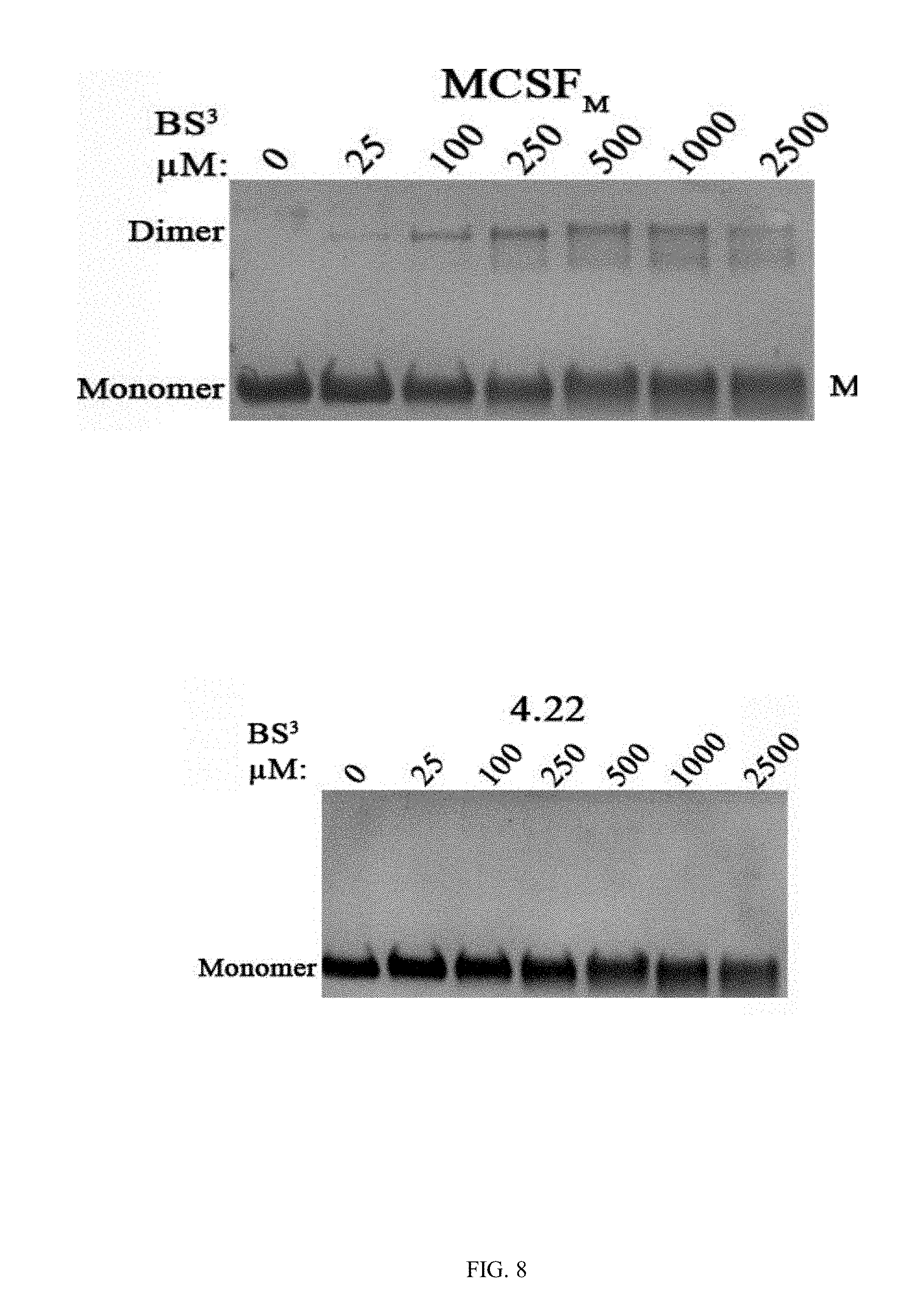
D00013
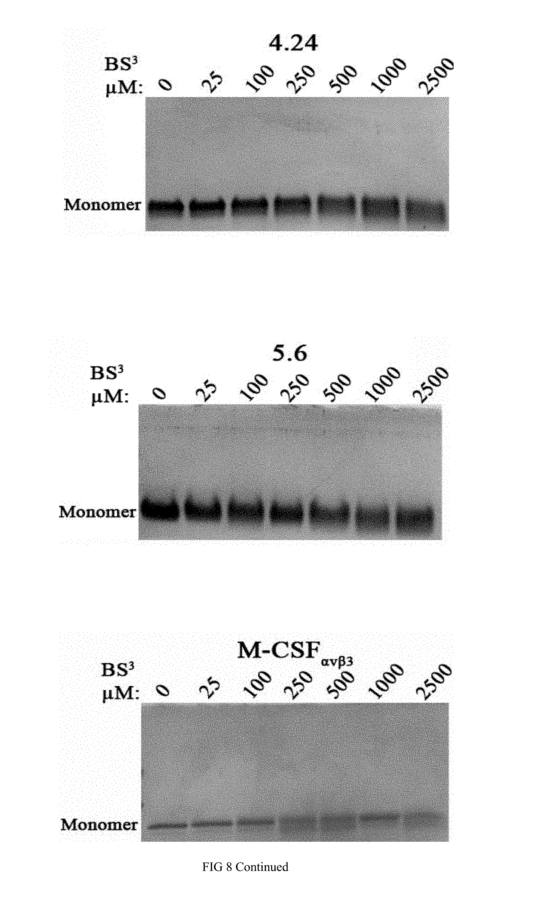
D00014
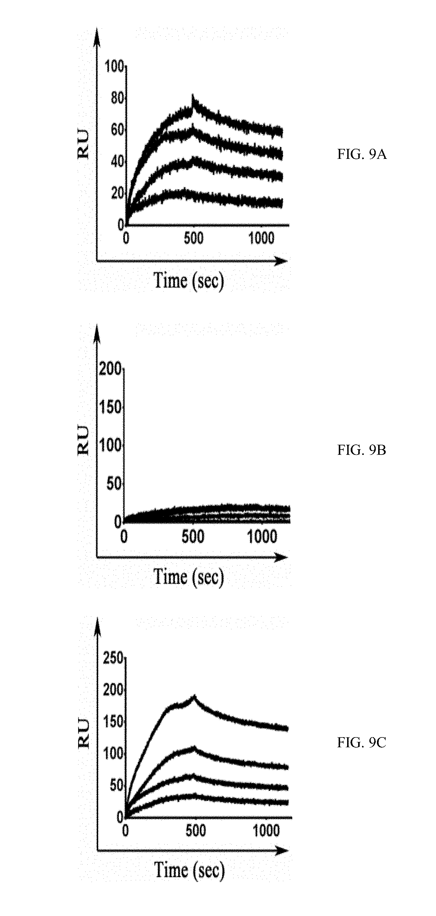
D00015
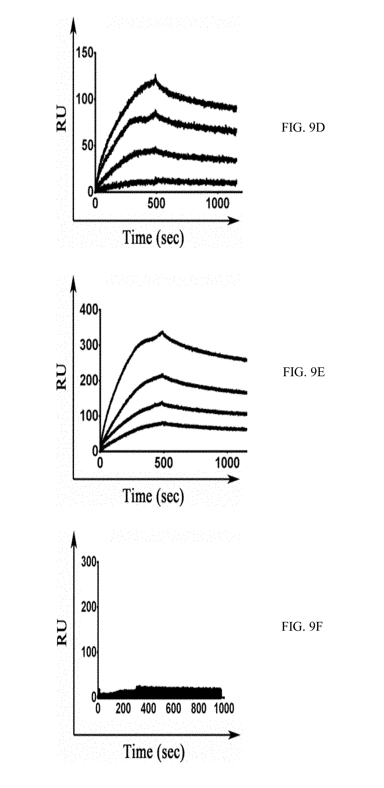
D00016
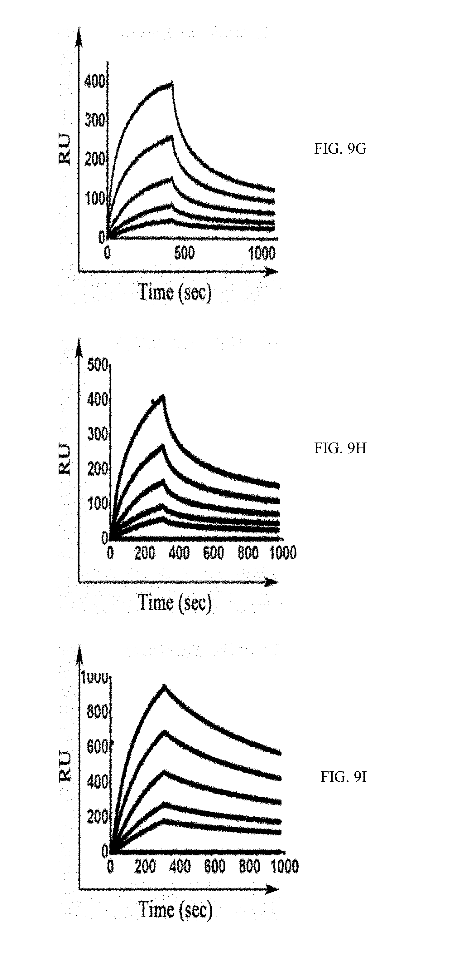
D00017
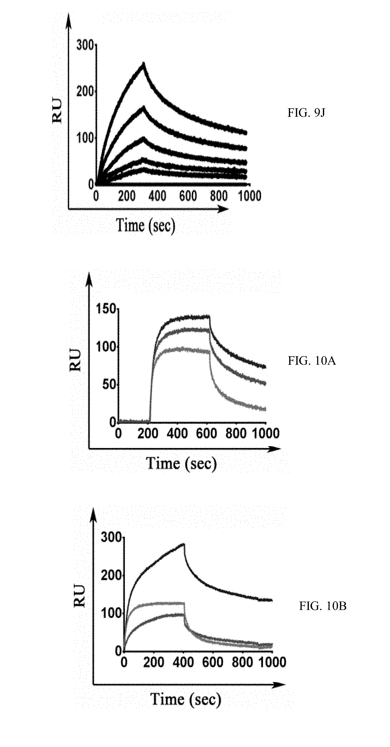
D00018
D00019
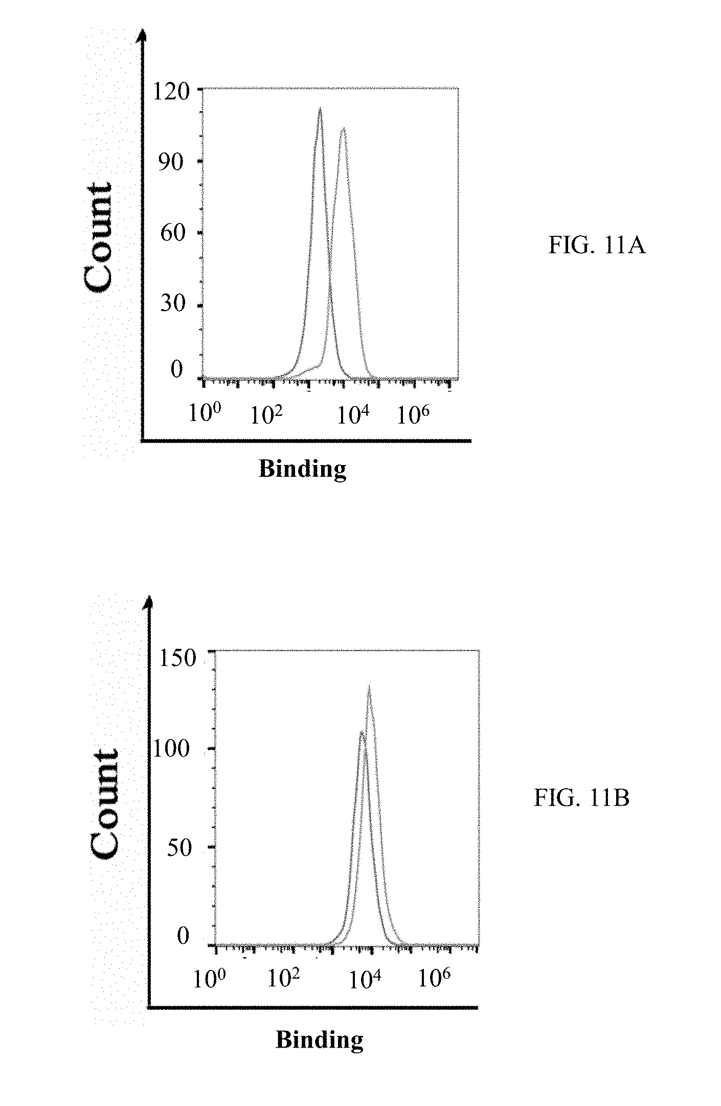
D00020
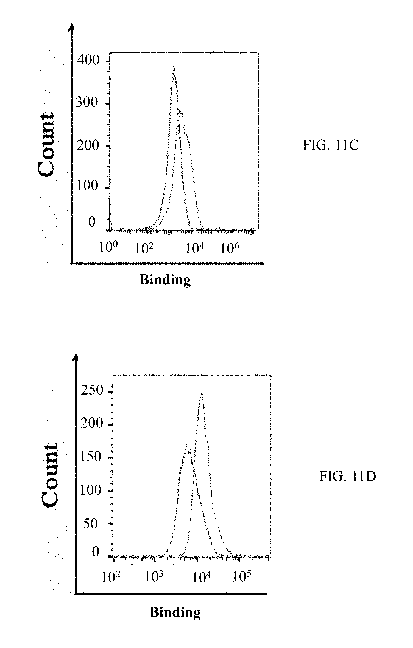
D00021
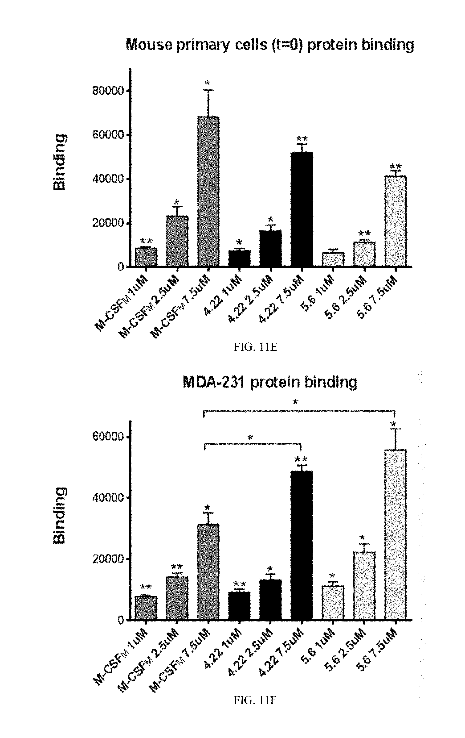
D00022
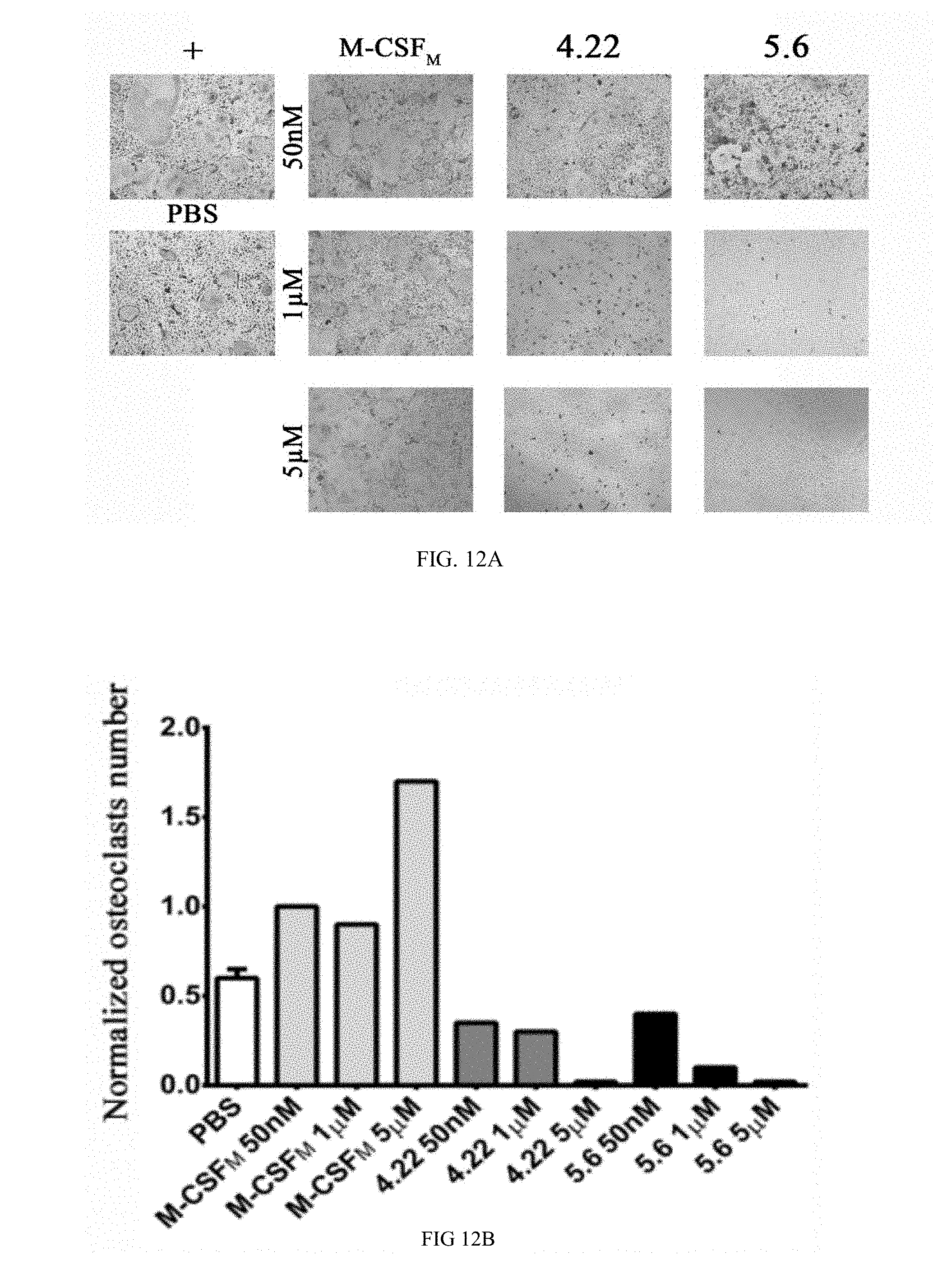
D00023
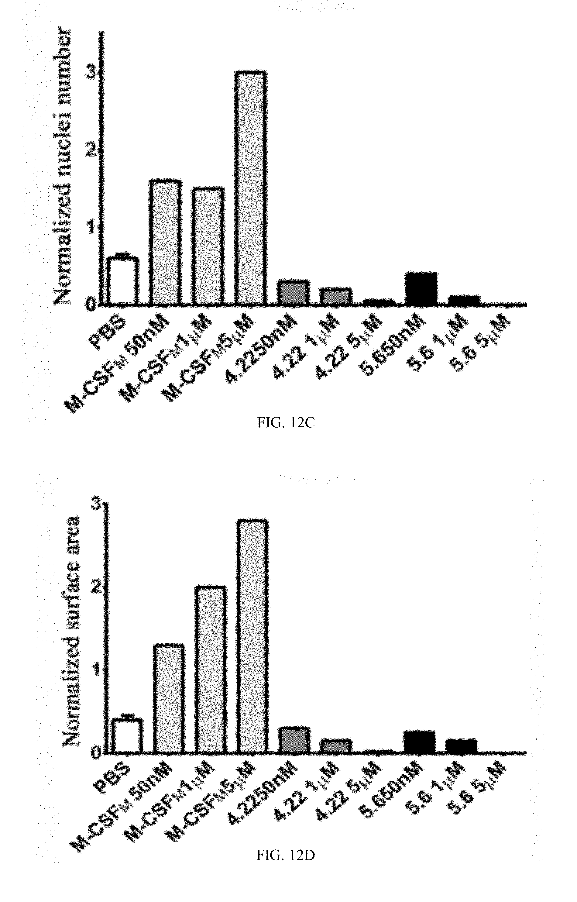
D00024
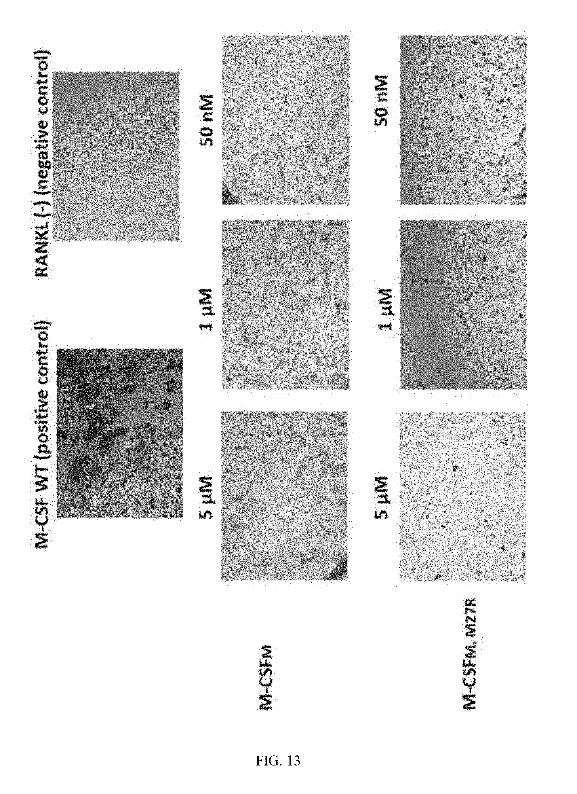
D00025
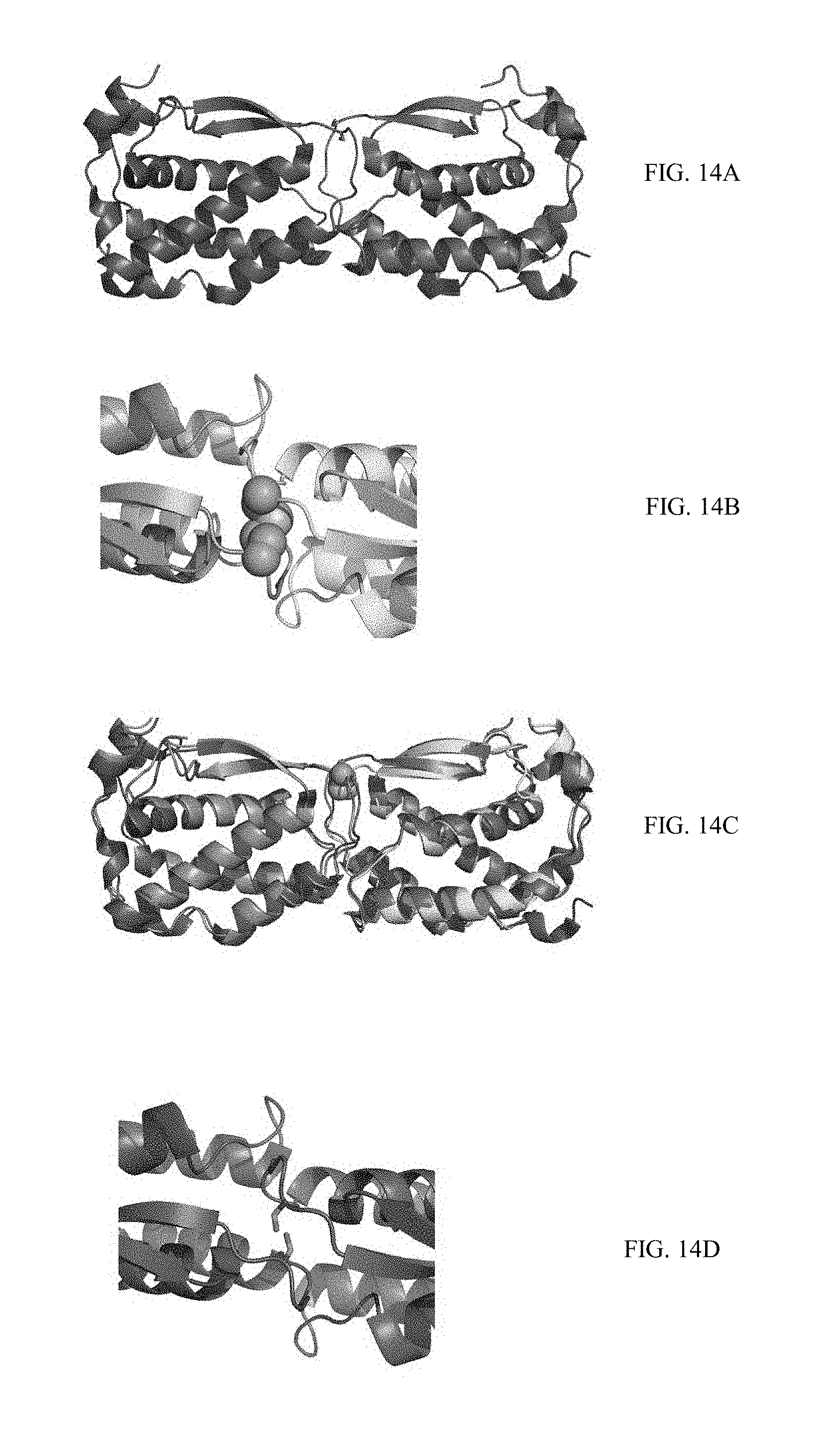
D00026
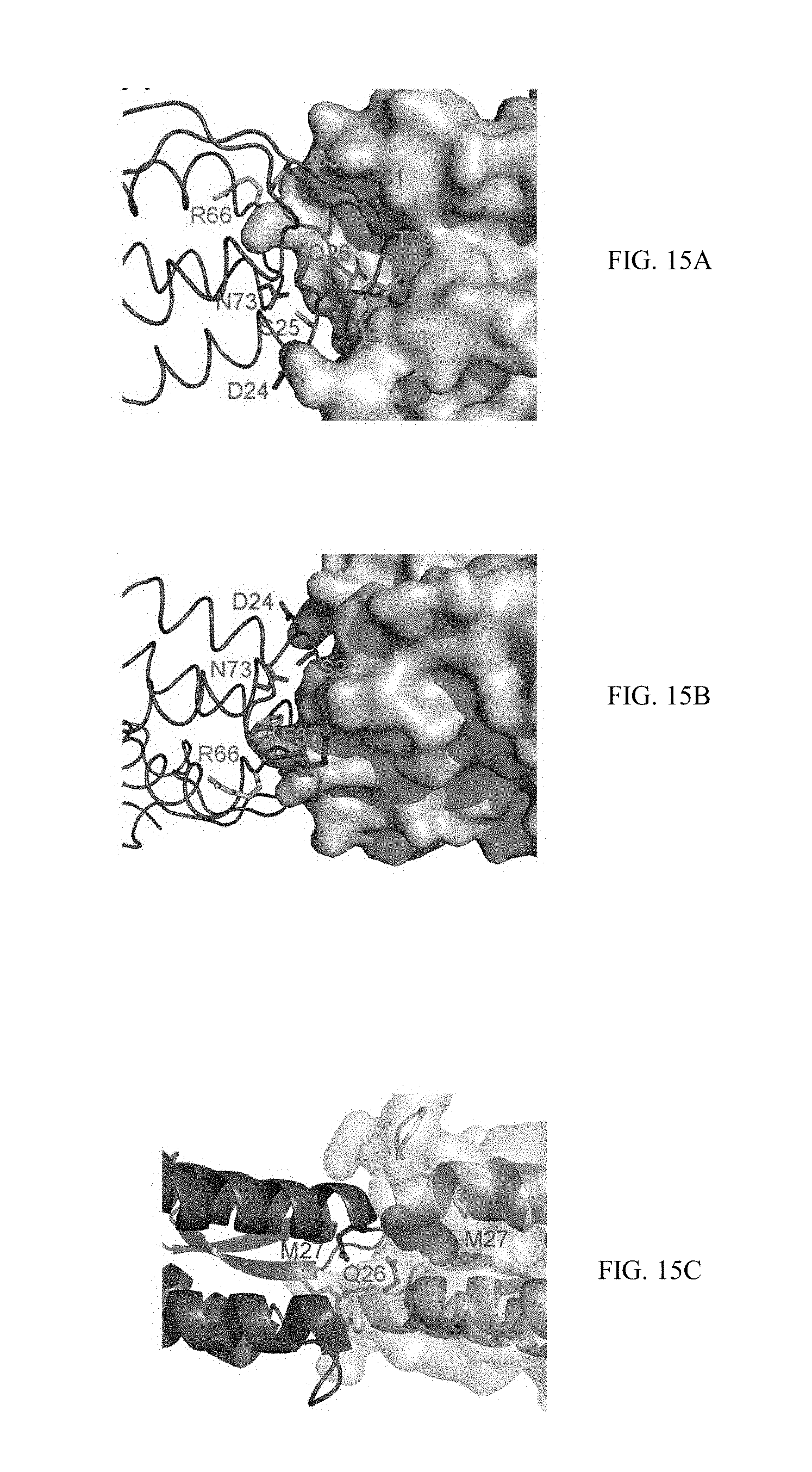
D00027
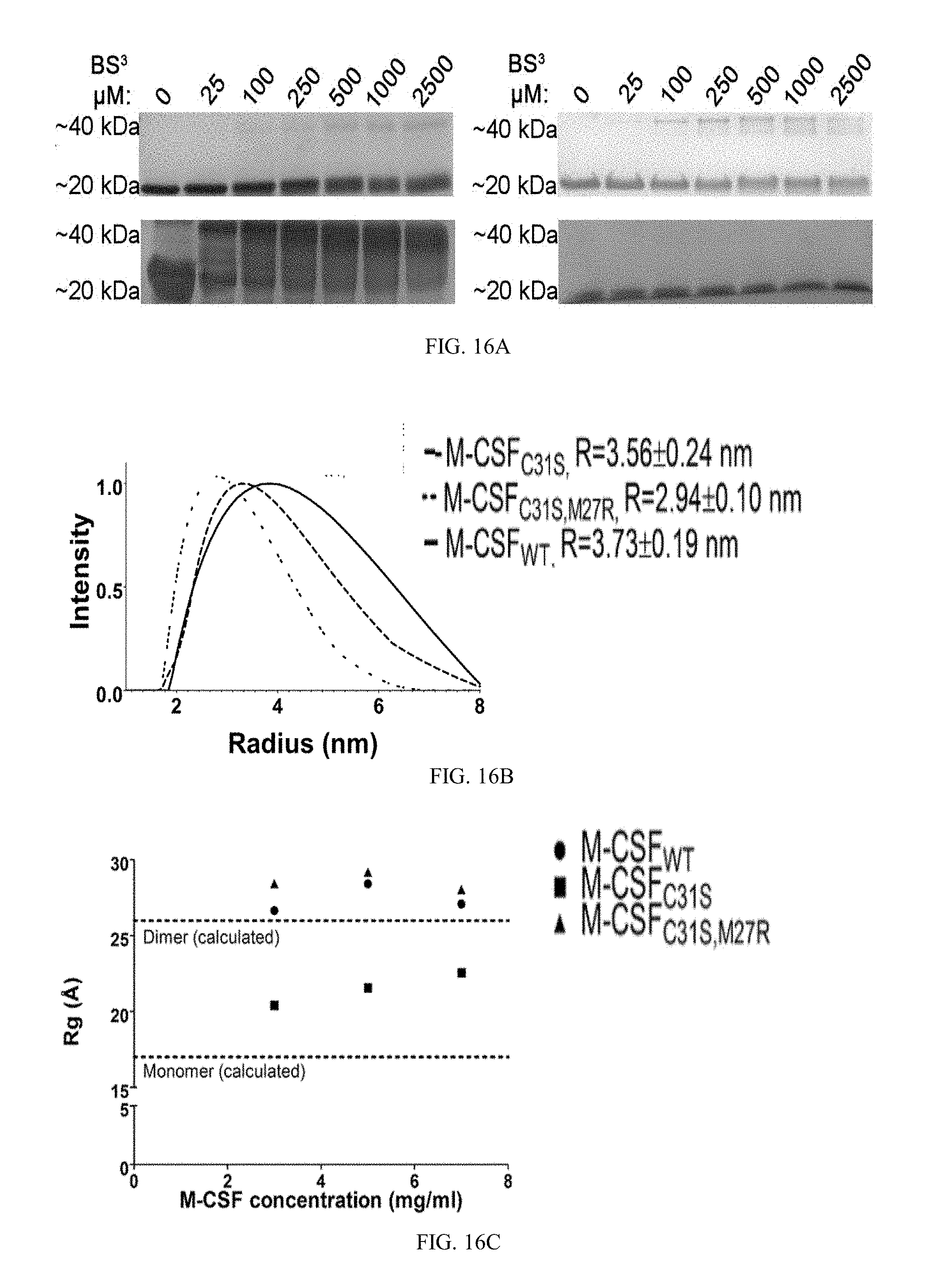
D00028
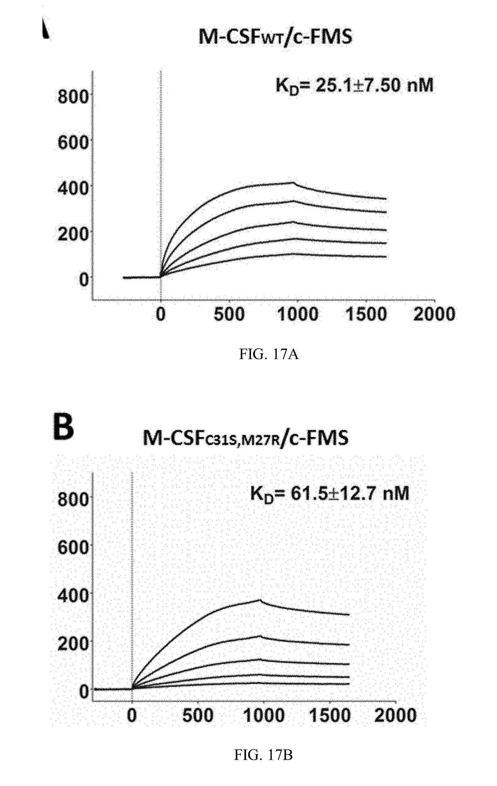
D00029
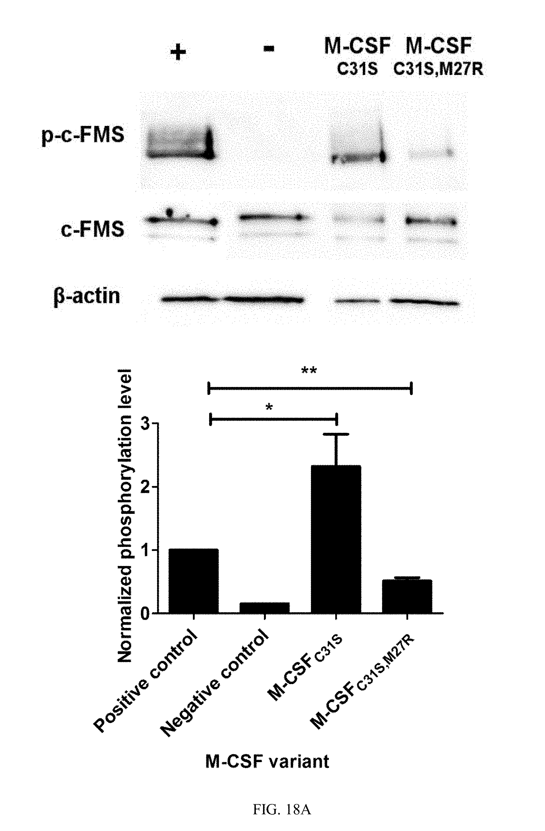
D00030
D00031
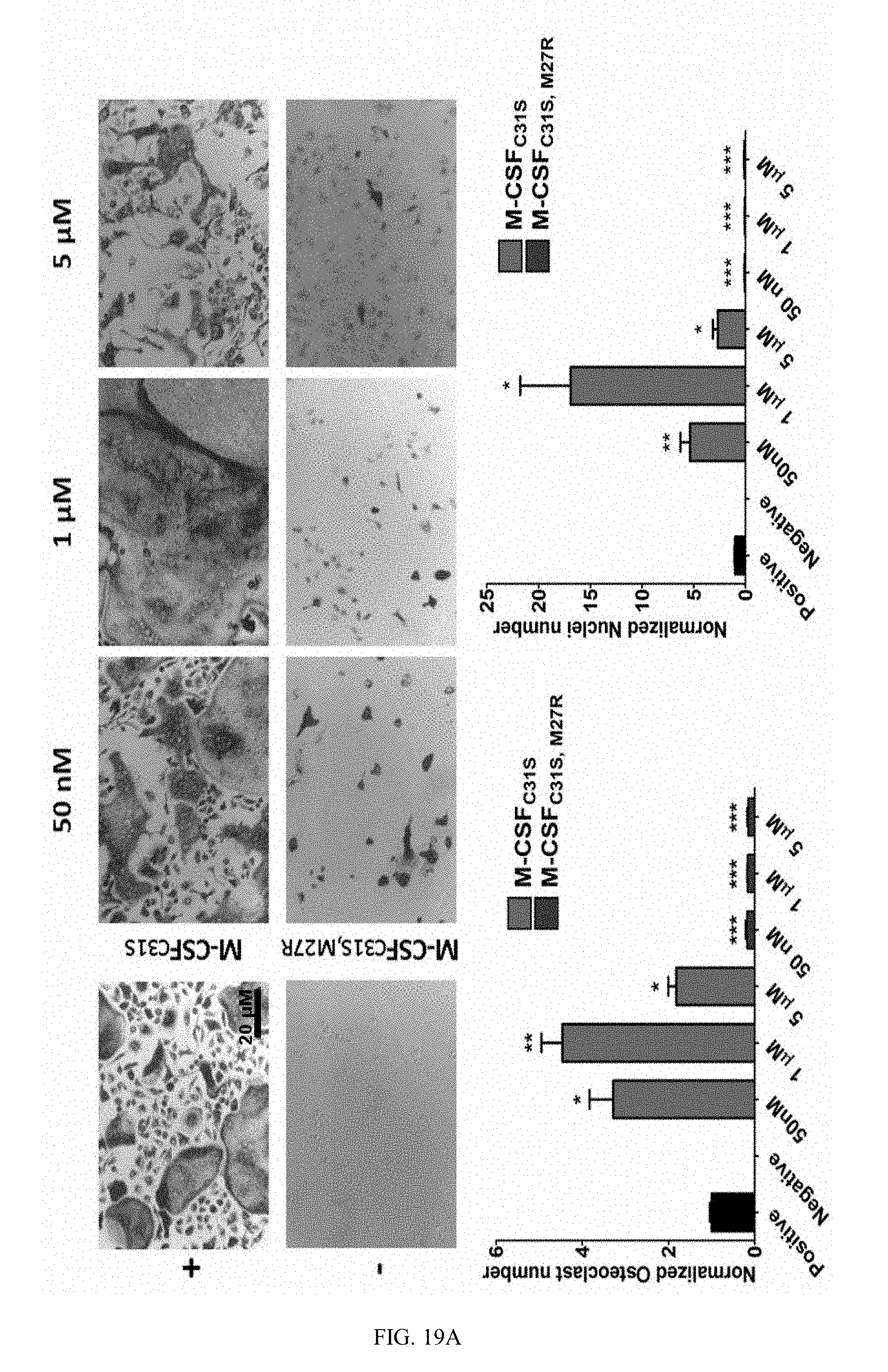
D00032

D00033
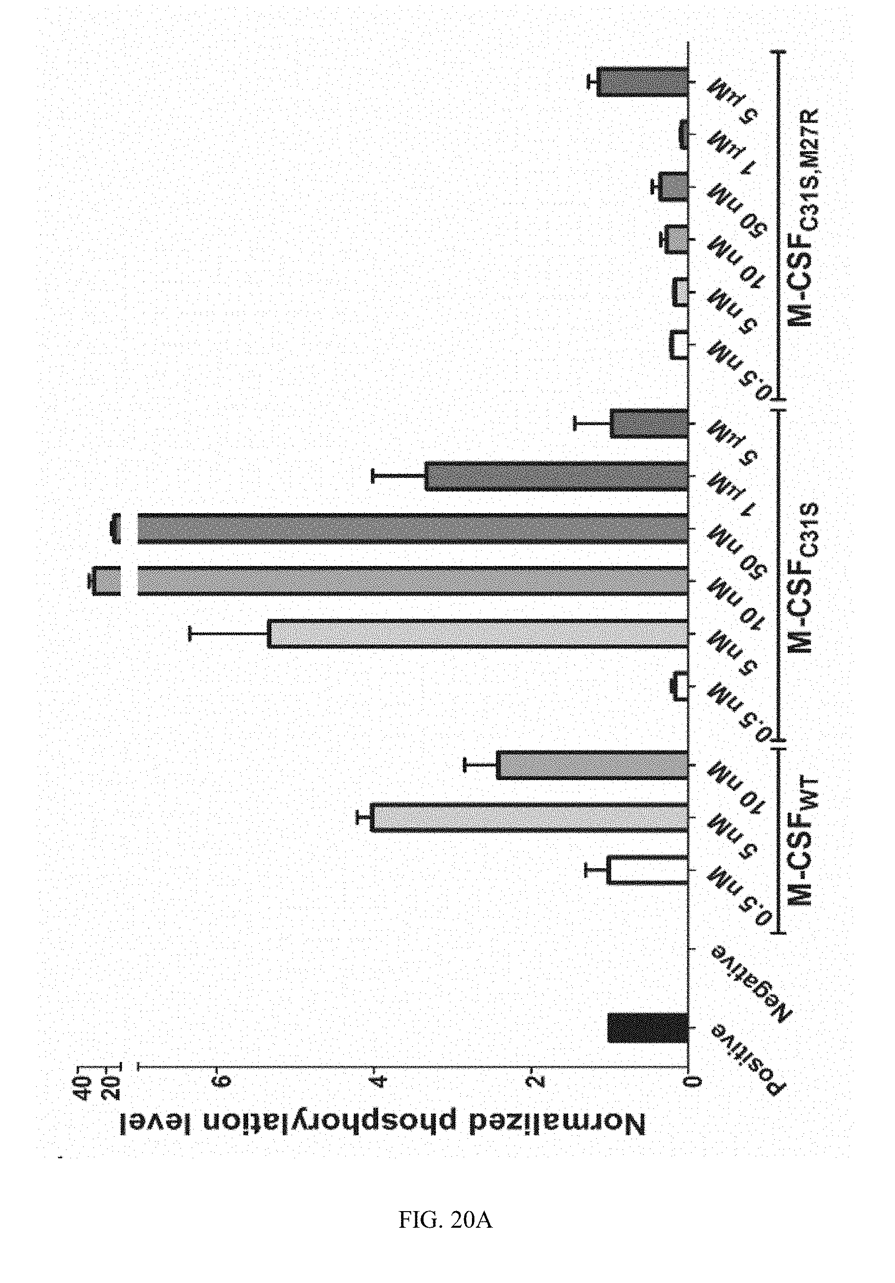
D00034
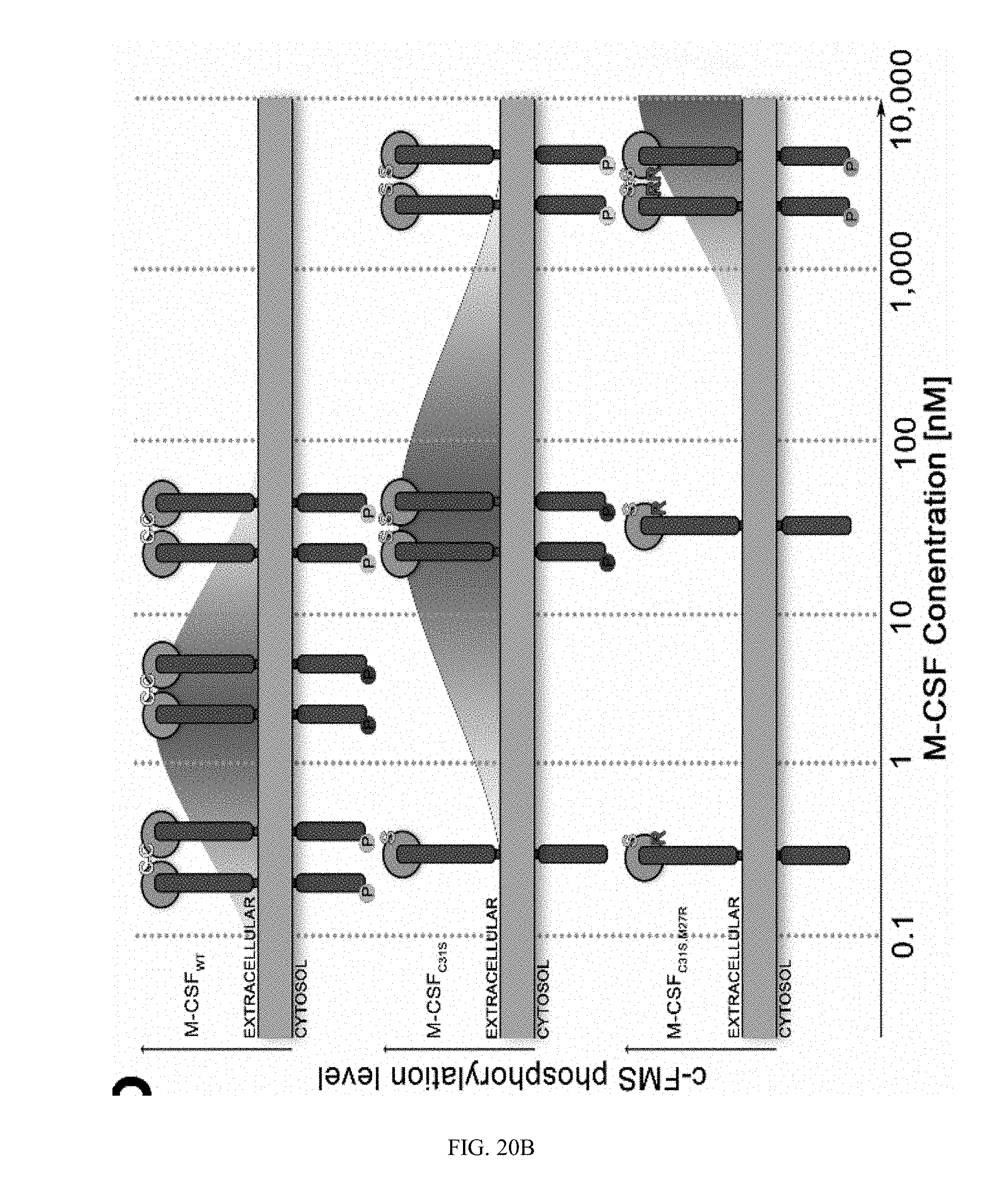
D00035
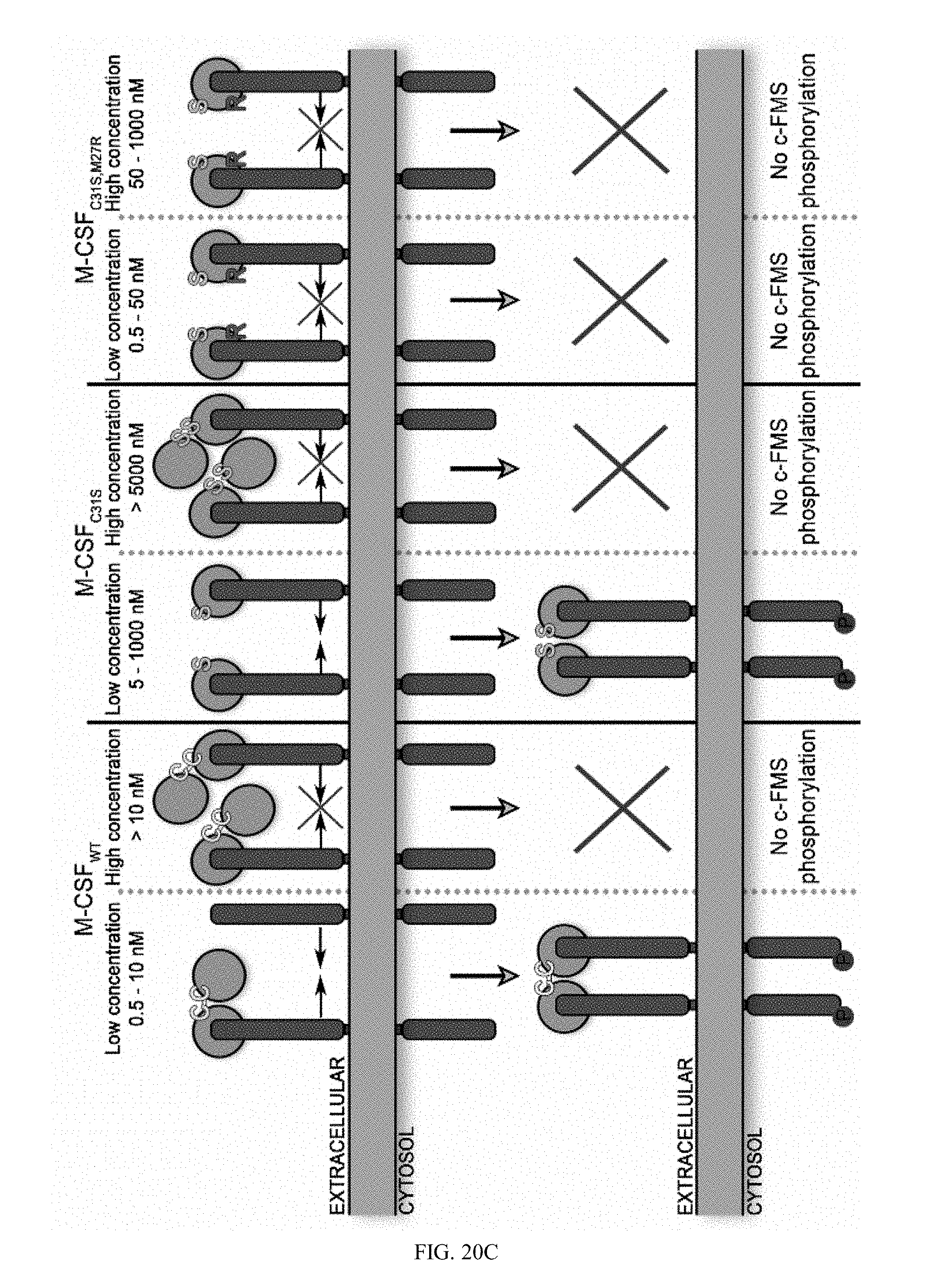
D00036
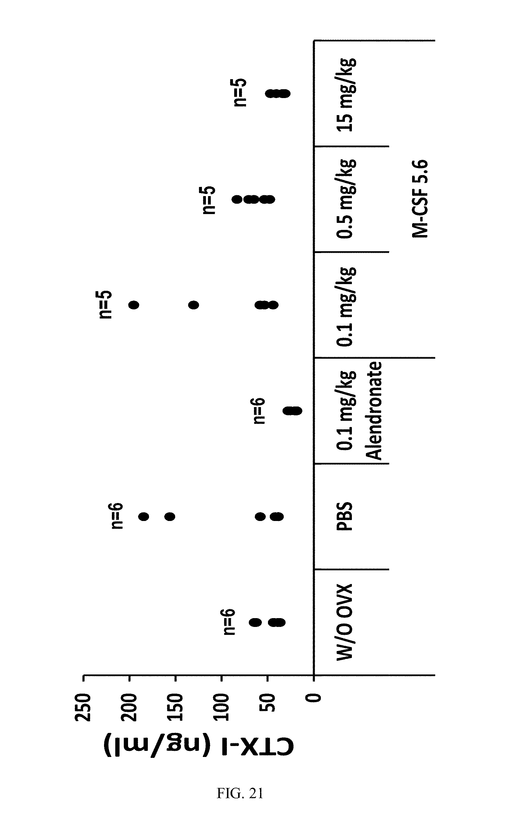
D00037
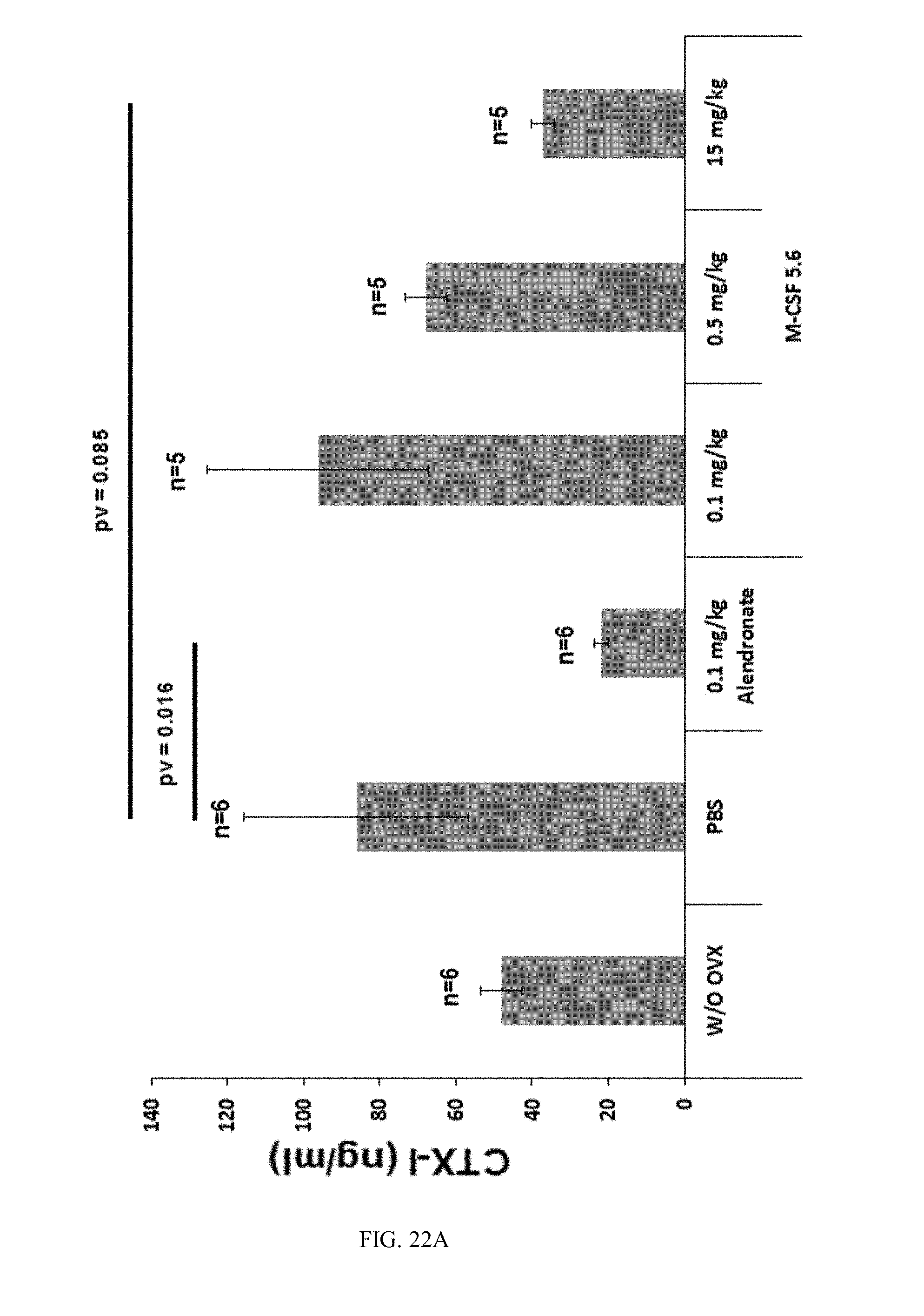
D00038
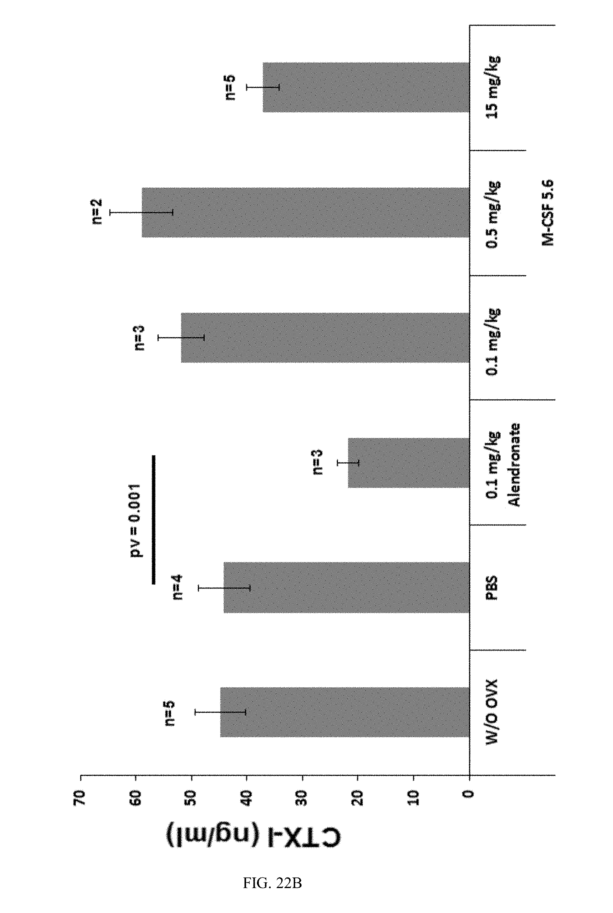
D00039
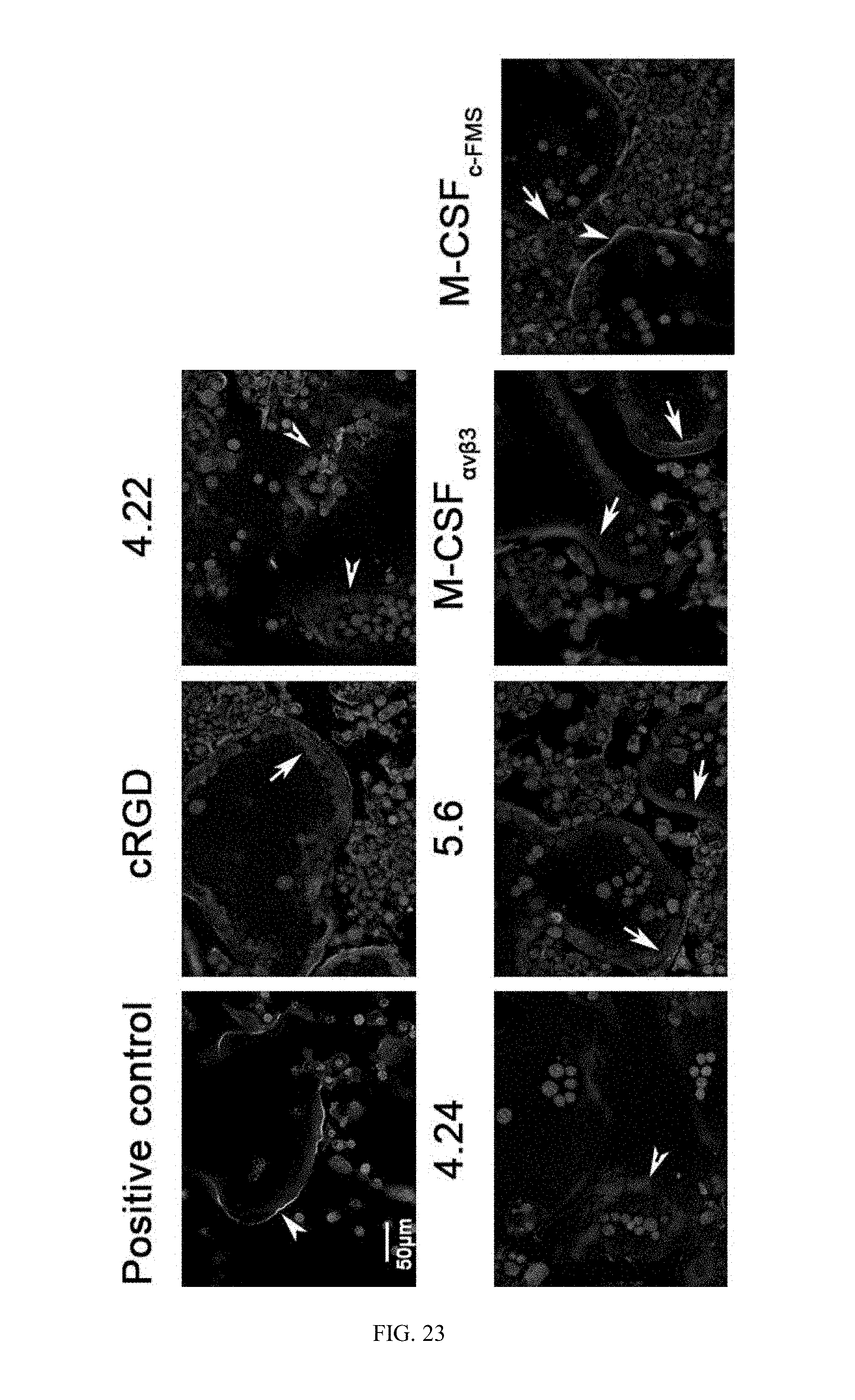


S00001
XML
uspto.report is an independent third-party trademark research tool that is not affiliated, endorsed, or sponsored by the United States Patent and Trademark Office (USPTO) or any other governmental organization. The information provided by uspto.report is based on publicly available data at the time of writing and is intended for informational purposes only.
While we strive to provide accurate and up-to-date information, we do not guarantee the accuracy, completeness, reliability, or suitability of the information displayed on this site. The use of this site is at your own risk. Any reliance you place on such information is therefore strictly at your own risk.
All official trademark data, including owner information, should be verified by visiting the official USPTO website at www.uspto.gov. This site is not intended to replace professional legal advice and should not be used as a substitute for consulting with a legal professional who is knowledgeable about trademark law.