Method Of Treating Heterotopic Ossification
Levi; Benjamin ; et al.
U.S. patent application number 16/397029 was filed with the patent office on 2019-08-15 for method of treating heterotopic ossification. The applicant listed for this patent is THE REGENTS OF THE UNIVERSITY OF MICHIGAN. Invention is credited to Shailesh Agarwal, Benjamin Levi.
| Application Number | 20190247411 16/397029 |
| Document ID | / |
| Family ID | 59088608 |
| Filed Date | 2019-08-15 |


| United States Patent Application | 20190247411 |
| Kind Code | A1 |
| Levi; Benjamin ; et al. | August 15, 2019 |
METHOD OF TREATING HETEROTOPIC OSSIFICATION
Abstract
The invention provides a method of treating heterotopic ossification in a subject in need thereof. The method comprises administering a hypoxia inducible factor-1.alpha. (Hif-1.alpha.) inhibitor to the subject. In various embodiments, the Hif-1.alpha. inhibitor is PX-478, rapamycin, or digoxin.
| Inventors: | Levi; Benjamin; (Ann Arbor, MI) ; Agarwal; Shailesh; (Ypsilanti, MI) | ||||||||||
| Applicant: |
|
||||||||||
|---|---|---|---|---|---|---|---|---|---|---|---|
| Family ID: | 59088608 | ||||||||||
| Appl. No.: | 16/397029 | ||||||||||
| Filed: | April 29, 2019 |
Related U.S. Patent Documents
| Application Number | Filing Date | Patent Number | ||
|---|---|---|---|---|
| 15373959 | Dec 9, 2016 | 10278982 | ||
| 16397029 | ||||
| 62387439 | Dec 24, 2015 | |||
| Current U.S. Class: | 1/1 |
| Current CPC Class: | A61P 19/08 20180101; A61K 31/198 20130101; A61K 31/7048 20130101; A61K 31/436 20130101 |
| International Class: | A61K 31/7048 20060101 A61K031/7048; A61K 31/436 20060101 A61K031/436; A61K 31/198 20060101 A61K031/198 |
Goverment Interests
STATEMENT OF GOVERNMENT INTEREST
[0002] This invention was made with government support under GM109105 awarded by the National Institutes of Health. The Government has certain rights in the invention.
Claims
1. A method of treating heterotopic ossification in a subject in need thereof, the method comprising administering a hypoxia inducible factor-1.alpha. (Hif-1.alpha.) inhibitor to the subject.
2. The method of claim 1, wherein the heterotopic ossification is genetic.
3. The method of claim 2, wherein the subject suffers from fibrodysplasia ossificans progressiva.
4. The method of claim 1, wherein the heterotopic ossification results from trauma.
5. The method of any one of claims 1-4, wherein the Hif-1.alpha. inhibitor is PX-478.
6. The method of any one of claims 1-4, wherein the Hif-1.alpha. inhibitor is rapamycin.
7. The method of any one of claims 1-4, wherein the Hif-1.alpha. inhibitor is digoxin.
8. A method of treating heterotopic ossification in a subject in need thereof, the method comprising administering a mechanistic target of rapamycin (mTor) inhibitor to the subject.
9. The method of claim 8, wherein the heterotopic ossification is genetic.
10. The method of claim 9, wherein the subject suffers from fibrodysplasia ossificans progressiva.
11. The method of claim 8, wherein the heterotopic ossification results from trauma.
Description
CROSS-REFERENCE TO RELATED APPLICATIONS
[0001] This application claims priority to U.S. Provisional Patent Application No. 62/387,439, filed Dec. 24, 2015, the disclosure of which is incorporated by reference.
TECHNICAL FIELD OF THE INVENTION
[0003] The disclosure relates to methods for treating heterotopic ossification.
INCORPORATION BY REFERENCE OF MATERIAL SUBMITTED ELECTRONICALLY
[0004] This application contains, as a separate part of the disclosure, a Sequence Listing in computer-readable form which is incorporated by reference in its entirety and identified as follows: Filename: 50313A_Seqlisting.txt; Size: 1,488 bytes, created: Dec. 6, 2016.
BACKGROUND OF THE INVENTION
[0005] Heterotopic ossification (HO) is the pathologic formation of extra-skeletal bone in soft tissues. Vanden Bossche and Vanderstraeten, J Rehabil Med 27, 129 (2005). This process occurs in patient populations with severe trauma including large-surface area burns, musculoskeletal injury, orthopedic operations, and spinal cord injury, and in patient populations with a genetic disease known as fibrodysplasia ossificans progressiva (FOP). FOP is caused by a hyper-activating mutation in the type I bone morphogenetic protein (BMP) receptor ACVR1, and patients with FOP develop ectopic bone lesions in the absence of any substantial trauma. The clinical sequela of these pathologic ectopic bone formations, whether in the setting of trauma or genetic mutations, include non-healing wounds, chronic pain, and joint immobility. In the case of FOP, progressive ossification may lead to death due to loss of thoracic cage compliance.
[0006] Treatment options for HO are limited as bone often recurs following surgical resection, and some patients may have non-resectable HO due to its sensitive location. The risk of an operation may outweigh the benefits of excision, especially in the face of recurrence. There is a need for therapeutic options which can prevent HO before its initial occurrence in at-risk patients.
SUMMARY OF THE INVENTION
[0007] The invention includes, for example, a method of treating heterotopic ossification in a subject in need thereof, the method comprising administering a hypoxia inducible factor-1.alpha. (Hif-1.alpha.) inhibitor to the subject. In various embodiments, the Hif-1.alpha. inhibitor is PX-478, rapamycin, or digoxin.
[0008] The invention also includes a method of treating heterotopic ossification in a subject in need thereof, wherein the method comprises administering a mechanistic target of rapamycin (mTor) inhibitor to the subject.
[0009] In various embodiments, the heterotopic ossification is genetic, and the subject optionally suffers from fibrodysplasia ossificans progressiva. In an alternative embodiment, the heterotopic ossification results from trauma, such as burns, musculoskeletal injury, orthopedic operations (e.g., post total hip replacements, post joint arthroplasty), spinal cord injury, stroke, poliomyelitis, myelodysplasia, carbon monoxide poisoning, spinal cord tumors, syringomyelia, tetanus, or multiple sclerosis.
DESCRIPTION OF THE FIGURES
[0010] FIG. 1A illustrates ingenuity pathway analysis of mRNA transcripts isolated from adipose tissue from burn or unburned "control" patients. FIG. 1B is a diagram illustrating up-regulation of the pro-vasculogenic pathway including HIF1.alpha., vWF, PECAM, FLT1, CDH5, and VEGF. FIG. 1C is a chart showing the numeric-fold increase in gene expression of burn patients when compared with unburned, "control" patients.
[0011] FIG. 2A illustrates a trauma-induced model of HO in which mice receive a 30% total body surface-area partial-thickness dorsal burn injury with hindlimb Achilles' tendon transection, resulting in HO formation along the calcaneus as well as proximally within the soft tissue. FIG. 2B illustrates a hybrid model of HO wherein Ad.cre and cardiotoxin are injected into the gastrocnemius muscle of caACVR1fl:fl mice resulting in intramuscular HO formation. FIG. 2C illustrates the Nfatc1-Cre/caACVR1fl:wt mouse model, wherein mice develop HO generally localized to the joints 4-5 days after birth in a model of genetic HO.
[0012] FIG. 3A depicts three-dimensional reconstruction and serial cross-sections of microCT scans of PX-478 and control-treated burn/tenotomy mice. FIG. 3B are bar graphs illustrating soft tissue HO volume (y-axis (mm.sup.3)) in a burn/tenotomy model (9 week volume: 0.90 mm.sup.3 v. 0.00 mm.sup.3, p=0.05; 9 week normalized volume: 1.0 v. 0.0, p=0.05; yes/no: .chi.2=9.5, p<0.01) and total HO volume (y-axis (mm.sup.3)) in burn/tenotomy mice (5 week volume: 4.3 mm.sup.3 v. 1.5 mm.sup.3, p<0.05; 9 week volume: 5.8 mm.sup.3 v. 2.3 mm.sup.3, p<0.05; 9 week normalized volume: 1.0 v. 0.4, p<0.05), in control-treated (left-hand bar) and PX-478-treated (right-hand bar) mice. PX-478 significantly decreased total HO volume and eliminated soft tissue HO. FIG. 3C depicts three-dimensional reconstruction and serial cross-sections of microCT scans of rapamycin and control-treated burn/tenotomy mice. FIG. 3D are bar graphs illustrating soft tissue HO volume and total HO volume (y-axis (mm.sup.3)) in a burn/tenotomy model of control-treated (left-hand bar) and rapamycin-treated (right-hand bar) mice. Rapamycin treatment significantly reduced de novo HO formation (9 week volume: 1.60 mm.sup.3 v. 0.81 mm.sup.3, p<0.05; 9 week normalized volume: 1.0 v. 0.51, p<0.05). *p<0.05 for volumetric measurements; .dagger. p<0.05 for binary analysis (yes/no). Arrow="soft tissue" heterotopic bone; light hashed circle=calcaneal heterotopic bone; n=3 for PX-478-treated mice; n=1 for PX-478 control mice; n=5 for rapamycin-treated mice; n=4 for rapamycin control mice.
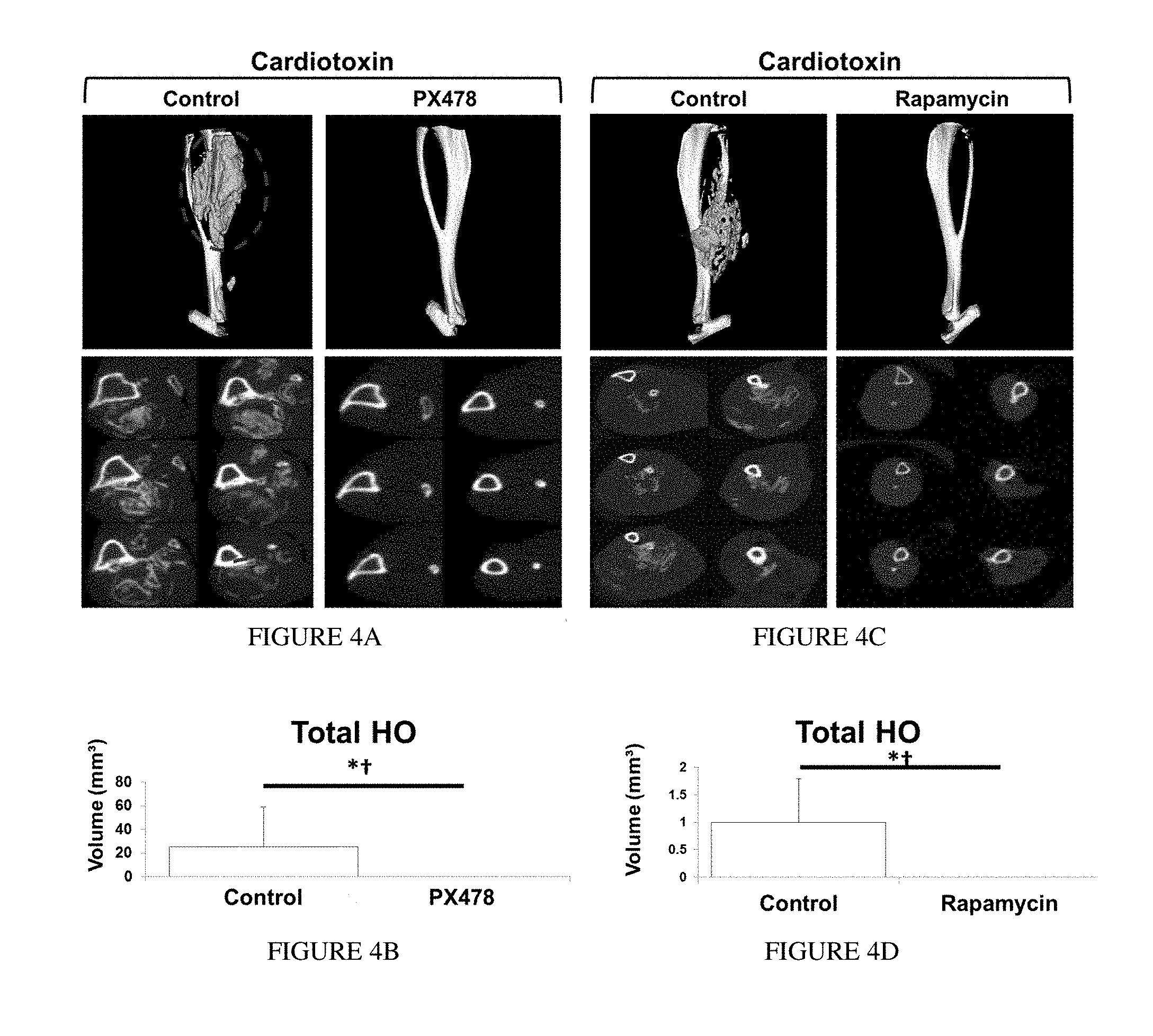
[0013] FIG. 4A depicts three-dimensional reconstructions and cross-sections of microCT scans of PX-478 and control-treated hybrid model mice. FIG. 4B is a bar graph illustrating total HO volume (y-axis) in control and PX-478-treated mice. PX-478 treated hybrid model mice produced almost no evidence of HO on microCT when compared with control-treated mice (Control: n=12 legs, PX-478: n=12 legs) (Volume: 18.1 mm.sup.3 v. 0.01 mm.sup.3, p=0.01; Normalized volume: 1.0 v. 0.0, p=0.01; yes/no: .chi.2=13.6, p<0.001). FIG. 4C depicts three-dimensional reconstructions and cross-sections of microCT scans of rapamycin and control-treated hybrid model mice. FIG. 4D is a bar graph illustrating total HO volume (y-axis) in control and rapamycin-treated mice. Rapamycin treated hybrid model mice produced no evidence of HO on microCT when compared with control-treated mice (Control: n=8 legs, Rapamycin: n=10 legs) (Volume: 17.5 mm.sup.3v. 0.00 mm.sup.3, p<0.001; Normalized volume: 1.0 v. 0.0, p<0.05; yes/no: .chi.2=14.3, p<0.001). FIG. 4E illustrates ankle reconstructions of control and PX-478-treated genetic HO mice. FIG. 4F is a bar graph illustrating ankle extra-skeletal bone volume quantification (volume) of genetic HO mice treated with control or PX-478. (800 Hounsfield Units) (Volume: 6.8 mm.sup.3 v. 2.2 mm.sup.3, p<0.01; normalized volume: 1.0 v. 0.32, p<0.01; n=4 control treated legs; n=4 PX-478 treated legs). *p<0.05 for volumetric measurements; .dagger. p<0.05 for binary analysis (yes/no); arrow=heterotopic bone.
DETAILED DESCRIPTION OF THE INVENTION
[0014] The invention is predicated, at least in part, on the discovery that Hif1.alpha. inhibitors, such as rapamycin or PX-478, potently diminish extra-skeletal bone formation in different models of HO. A method of treating heterotopic ossification in a subject in need thereof is provided by the disclosure. The method comprises administering a hypoxia inducible factor-1.alpha. (Hif-1.alpha.) inhibitor to the subject. HIF-1.alpha. is a component of the HIF transcription factor heterodimer and has been well characterized. See, e.g., Prior et al., Physiological Genomics 15(1), 20 (2003). Examples of Hif-1.alpha. inhibitors include, but are not limited to, PX-478 (S-2-amino-3-[4'-N,N,-bis(chloroethyl)amino]phenyl propionic acid N-oxide dihydrochloride, described in, e.g., Koh et al., Mol Cancer Ther 7(1), 90 (2008)), rapamycin, and digoxin. Other Hif-1.alpha. inhibitors include, e.g., CJ-3k and derivatives thereof (described in, e.g., Massoud et al., Anticancer Res. 35(7), 3849-59 (2015)), echinomycin, topotecan, LAQ824 (described in, e.g., Qian et al., Cancer Res 66, 8814 (2006)), metformin, imatinib, ouabain, and proscillaridin, and additional inhibitors are known in the art. In various embodiments, the Hif-1.alpha. inhibitor is not siRNA or an antisense oligonucleotide. The Hif-1.alpha. inhibitor need not completely inhibit Hif-1.alpha. to achieve a therapeutic effect; the inhibitor preferably inhibits Hif-1.alpha. activity by at least 50% (e.g., 50%-99%), at least 60%, at least 70%, at least 80%, at least 90%, or at least 95%. The inhibitor may inhibit or reduce Hif-1.alpha. protein levels, activation, deubiquitination, and the like to inhibit Hif-1.alpha. activity.
[0015] Additionally, the invention includes a method of treating heterotopic ossification in a subject in need thereof, wherein the method comprises administering a mechanistic target of rapamycin (mTor) inhibitor to the subject. mTor is a serine/threonine protein kinase of the phosphatidylinositol 3-kinase-related kinase protein family, and is well characterized. See, e.g., Laplante et al., Cell 149(2), 274 (2012); Sauer et al., Biochem Soc Trans 41, 889 (2013). Examples of mTor inhibitors include, but are not limited to, rapamycin, temsirolimus, everolimus, deforolimus, vincristine, zotarolimus, 32 deoxy-rapamycin, NVP-BEZ235, BGT226, SF1126, PKI-587, INK128, AZD8055, AZD2014, GNE477 (a thienopyrimidine), PI-103 (a tricyclic pyridofuropyrimidine), XL765, WJD008 (a 5-cyano-6-morpholino-4-substituted pyrimidine analogue), PP242, PP30, Torin1, WYE-354, WAY-600, WYE-687, Ku-0063794, curcumin, resveratrol, epigallocatechin gallate, genistein, 3,3-diindolylmethane, and caffeine.
[0016] In various embodiments, the heterotopic ossification is genetic, and the subject optionally suffers from fibrodysplasia ossificans progressiva. In an alternative embodiment, the heterotopic ossification results from trauma, such as burns, musculoskeletal injury, orthopedic operations, or spinal cord injury. Thus, in various aspects, the subject is suffering from heterotopic ossification or is at risk of suffering from heterotopic ossification (e.g., is genetically predisposed to formation of ectopic bone lesions, has been subjected to trauma, etc.). In various embodiments, the method comprises administering to the subject the Hif-1.alpha. inhibitor (and/or an mTor inhibitor) in an amount and under conditions effective to treat, in whole or in part, heterotopic ossification. "Treating" heterotopic ossification includes (but is not limited to) the prevention or slowing of the formation or progression of heterotopic ossification in a subject and/or reduction or delaying the onset of symptoms associated with heterotopic ossification. It will be appreciated that the method need not completely prevent or halt the progression of heterotopic ossification to achieve a beneficial (e.g., therapeutic) effect. Any inhibition of the onset or progression of heterotopic ossification or severity of symptoms associated with heterotopic ossification is contemplated. For example, any reduction in the formation of lamellar bone inside soft-tissue structures where bone does not normally exist is contemplated.
[0017] Methods of determining the efficacy of the method in treating heterotopic ossification are known in the art and described herein. For example, the progression of heterotopic ossification is monitored using x-ray, bone scan, subject range of motion, prostaglandin E2 excretion in 24-hour urine, ultrasonography, and/or evaluating joint swelling or pain.
[0018] The invention further includes use of a Hif-1.alpha. inhibitor and/or mTor inhibitor in the treatment of heterotopic ossification. For example, a Hif-1.alpha. inhibitor and/or mTor inhibitor can be used in the manufacture of a medicament for the treatment of heterotopic ossification, as described in detail herein.
[0019] The Hif-1.alpha. inhibitor can be administered in combination with other active agents or therapeutic modalities, including, but not limited to, anti-inflammatory agents, pain relievers, radiation therapy, and physical therapy. Similarly, an mTor inhibitor can be administered in combination with other active agents or therapeutic modalities, including, but not limited to, anti-inflammatory agents, pain relievers, radiation therapy, and physical therapy.
EXAMPLE
[0020] This Example demonstrates the ability of HIF-1.alpha. inhibitors to treat both trauma-induced and genetic-based heterotopic ossification in clinically relevant animal models and provides further description of the method of the invention.
Materials and Methods
[0021] Patient Enrollment and Sampling for Gene Expression Profiling:
[0022] Patient enrollment and sample collection for patients have been described previously. Cobb, J. P., et al. Proceedings of the National Academy of Sciences of the United States of America 102, 4801-4806 (2005). 244 burn patients were enrolled between 2000 and 2009 at one of four burn centers. If admission occurred within 96 hours post-injury, at least 20% of the TBSA was affected, and at least one excision and grafting procedure was required. Additionally, 35 healthy control subjects (16-55 y) were recruited between 2004 and 2007. In both the burn patients and the control patients, adipose tissue was collected and analyzed for RNA transcript levels. Using a fine scissor or scalpel, 80 mg of adipose tissue was obtained and immediately placed on an iced petri dish and cut into a 2-5 mm cube. The sample was placed in a cryogenic tube containing 2 ml RNAlater to stabilize the tissue according to Standard Operating Procedure (SOP) B001.03, the tissue was processed to total cellular RNA using a commercial RNA purification kit (RNeasy, Qiagen, Valencia, Calif.) according to SOP G026.01. Biotinylated cRNA was generated from 4 .mu.g of total cellular RNA, hybridized onto HU133 Plus 2.0 GeneChips (Santa Clara, Calif.), stained and washed according to the manufacturer's recommendations. A total of 25,000 genes were queried of which 3,500 were significantly changed with a false discovery rate (FDR)<0.001 and defined fold change.gtoreq.1.5.
[0023] Analysis of Time-Course Gene Expression Data:
[0024] Specimens were immediately stabilized using RNAlater (Ambion). Total cellular RNA was extracted from the remaining specimens with good quality using a commercial RNA purification kit (RNeasy, Qiagen). Biotinylated cRNA was generated from 1 .mu.g of total cellular RNA using the 3' IVT Express Kit and protocol of Affymetrix, and hybridized onto an HU133 Plus 2.0 GeneChip (Affymetrix). EDGE (Extraction of Differential Gene Expression) was used to estimate the significance of expression changes for each gene by 1,000 random permutations. Significant genes were selected by FDR<0.001 and fold change.gtoreq.1.5. These genes were further analyzed using Ingenuity Pathway Analysis36.
[0025] Animals:
[0026] Mice included for extra-skeletal bone evaluation were wild type C57BL/6 (Charles River Laboratory), Cdh5-Cre/tdTomatofl/wt, Prx-Cre/Hif1.alpha.fl/fl, Prx-Cre/ROSA26mTmG, caAcvr1fl/fl, Nfatc1-Cre/caAcvr1fl/wt, or littermate controls. Tail genomic DNA was used for genotyping. Mice used for bioluminescent imaging were homozygous for the ODD-luc transgene. In these mice, the C-terminal portion of the hypoxia-inducible factor 1 alpha oxygen-dependent degradation domain (ODD) is fused to the firefly luciferase (luc) gene. Hypoxia causes stabilization of the fusion protein thereby increasing fluorescence upon luciferin administration.
[0027] Extra-Skeletal Bone Models:
[0028] All mice received pre-surgical analgesia consisting of 0.1 mg/kg buprenorphine, followed by anesthesia with inhaled isoflurane, and close post-operative monitoring with analgesic administration. Burn/tenotomy mice received a 30% total body surface area (TBSA) partial-thickness burn on the shaved dorsum followed by left hindlimb Achilles' tendon transection. The dorsum was burned using a metal block heated to 60.degree. C. in a water bath and applied to the dorsum for 18 seconds continuously. The tenotomy site was closed with a single 5-0 vicryl stitch placed through the skin only. caAcvr1fl:fl mice received hindlimb cardiotoxin and Ad.cre injection at P24. Mice were then euthanized after 22 days (PX-478) or 15 days (rapamycin). Separate controls were used for each drug treatment to account for differences in the day of euthanization. Nfatc1-Cre/caAcvr1fl:wt mice were generated by crossing Nfatc1-Cre+ mice with caAcvr1fl:wt mice. Resulting mutants developed extra-skeletal bone by P4-5.
[0029] Drug Treatment:
[0030] Burn/tenotomy or hybrid HO mice were administered PX-478 (100 mg/kg) or rapamycin (5 mg/kg) in PBS solution via intraperitoneal injection. Mice received injections every other day for the duration of the study. Nfatc1-Cre/caACVR1fl:wt mice were administered PX-478 (100 mg/kg) every other day for a total of 2 weeks.
[0031] Isolation and Culture of Mesenchymal Stem Cells:
[0032] Mouse mesenchymal stem cells (MSCs) were harvested from the tendon transection site originating from the calcaneus to the confluence of the fibula and tibia wild-type mice. All tissue was mechanically minced, and digested with collagenase A and dispase and subsequently plated. To test drug treatment on Hif1.alpha. expression, cells were cultured in a hypoxia chamber with 0.5% oxygen. Cell treatment with PX-478 (10 .mu.M) or rapamycin (5 .mu.M) was initiated 24 hours before hypoxia treatment and re-dosed in hypoxia for 24 hours. Protein was harvested and analyzed using Western blot for Hif1.alpha. and .alpha.-tubulin. To test effect of PX-478 treatment on chondrogenesis, cells isolated from the tendon were cultured in chondrogenic differentiation medium (PT-3925 & PT-4121, Lonza, Basel, Switzerland). All in vitro experiments were performed in biologic and technical triplicate.
[0033] Histology and Immunofluorescence:
[0034] Histologic evaluation was performed at indicated time points in burn/tenotomy, Ad.cre/cardiotoxin, or Nfatc1-Cre/ca-Acvr1fl:wt mutants. Hind limbs were fixed in formalin overnight at 4.degree. C. and subsequently decalcified in 19% EDTA solution for 3-5 weeks at 4.degree. C. until x-ray verification of decalcification. Hind limbs were paraffin- or cryo-embedded, and 5-7 m sections were cut and mounted on Superfrost plus slides (Fisher) and stored at room temperature. Haematoxylin/eosin and Movat's pentachrome staining were performed of the ankle region. Immunostaining staining of extra-skeletal ectopic bone was performed on rehydrated wax sections with the following primary antibodies: mouse anti-mouse anti-Hif1.alpha. (Santa Cruz, Cat No. 53546), goat anti-mouse anti-Cdh5 (Santa Cruz, Cat No. 6458), goat anti-mouse anti-pSmad 1/5 (Santa Cruz, Cat No. 12353), goat anti-mouse anti-CD31 (Santa Cruz, Cat No. 1506), rabbit anti-mouse anti-Sox9 (Santa Cruz, Cat No. 20095), or anti-mouse PDGFR.alpha.. Appropriate dilutions were determined prior to achieving final images. The appropriate fluorescent secondary antibody was applied and visualized using fluorescent microscopy. Secondary antibodies consisted of anti-rabbit or anti-goat Alexafluor-488(green) or -594(red). All mouse sections were taken 3 weeks after burn/tenotomy. All counts were performed by blinded observer with 15 high-power fields for each sample.
[0035] Fluorescent and Bioluminescent Imaging:
[0036] All fluorescent and bioluminescent imaging was acquired using a PerkinElmer IVIS Spectrum system. Wild type C57BL/6 mice were used for fluorescent imaging to assess vascular perfusion. Mice were administered Angiosense 750 EX via tail vein injection. Fluorescent imaging was acquired 24 hours after injection at 770 nm wavelength. ODD-luc were used for all bioluminescent imaging. Mice received luciferin i.p. injection ten minutes prior to imaging.
[0037] Quantitative PCR:
[0038] Tissue was harvested from the tenotomy site of burn/tenotomy mice, or from the corresponding contralateral, control hindlimb at indicated time points. RNA was collected from tissue using RNeasy Mini Kit (Qiagen, Germantown, Md.) according to manufacturer's specifications. Reverse transcription was performed with 1 .mu.g RNA using Taqman Reverse Transcription Reagents (Applied Biosystems, Foster City, Calif.). Quantitative real-time PCR was carried out using the Applied Biosystems Prism 7900HT Sequence Detection System and Sybr Green PCR Master Mix (Applied Biosystems). Specific primers for these genes were chosen based on their PrimerBank sequence (Table 1).
TABLE-US-00001 TABLE 1 Quantitative PCR primers. SEQ Accession ID Species Name Number Primer Sequence (5'.fwdarw.3')* NO Mouse Hif1.alpha. NM_176958 F-GTCCCAGCTACGAAGTTACAGC 1 R-CAGTGCAGGATACACAAGGTTT 2 Sox9 NM_011448 F-AGTACCCGCATCTGCACAAC 3 R-ACGAAGGGTCTCTTCTCGCT 4 Acan NM_007424 F-ACTGCGACATCTGGAGTGAC 5 R-CTGTCCACTGCCAAAGAGAA 6 *"F" denotes a forward primer. "R" denotes a reverse primer.
[0039] .mu.CT and Nano-CT Analysis:
[0040] .mu.CT scans (Siemens Inveon using 80 kVp, 80 mA, and 1,100 ms exposure) were used to quantify extra-skeletal bone growth in burn/tenotomy, Ad.cre/cardiotoxin, or mutant Nfatc1-cre/caAcvr1fl:wt mice. Burn/tenotomy mice received scans at 5 and 9 weeks after tenotomy. Ad.cre/cardiotoxin mice received .mu.CT scans at day 22 after induction with Ad.cre and cardiotoxin injection. Nfatc1-cre/caAcvr1fl:wt mice and littermate controls received .mu.CT scans at day 13 after birth. Images were reconstructed and HO volume quantified using a calibrated imaging protocol as previously described with the MicroView .mu.CT viewer (Parallax Innovations, Ilderton, Canada).
[0041] Microscopy:
[0042] All fluorescently stained images were taken using an Olympus BX-51 upright light microscope equipped with standard DAPI, 488 nm, and TRITC cubes attached to an Olympus DP-70 high resolution digital camera. Each site was imaged in all channels and overlaid in DPViewer before examination in Adobe Photoshop.
[0043] Statistical Analysis:
[0044] A power analysis was first performed to determine how many mice were needed for our PX-478 treatment groups. For power analysis, the primary outcome of interest is differences in HO volume with treatment. To confirm a 50% decrease in HO volume with power of 0.8, assuming a s.d. of 1.5 mm.sup.3 and mean HO volume of 7.5 mm.sup.3 in untreated mice, three mice per group were required. Means and SDs were calculated from numerical data, as presented in the text, figures, and figure legends. In figures, bar graphs represent means, whereas error bars represent one SD. Statistical analysis was performed using an appropriate analysis of variance when more than two groups were compared, followed by a post hoc Student's t test (with a Bonferroni correction) to directly compare two groups. Inequality of SDs was excluded by using the Levene's test. Outliers were excluded using the Grubb's test for outliers. p values are included in the figure legends.
Results
[0045] Human Trauma Patients Exhibit Up-Regulation of HIF1.alpha. and Related Downstream Vascular Signaling Mediators:
[0046] A genomic database of 244 patients at high risk for HO due to large surface-area burns to compare with unburned "control" patients was examined. A total of 25,000 genes were queried, of which 3,500 were noted to be significantly different in tissue from burn patients when compared to control patients. A total of 25,000 genes were queried, of which 3,500 were noted to be significantly different in tissue from burn patients when compared to control patients. In particular, a significant up-regulation of HIF1.alpha. was observed, placing it within the top fifty up-regulated gene transcripts. In addition, related downstream gene transcripts including vWF, PECAM, FLT1, CDH5, and VEGF were up-regulated (FIG. 1A-C). In the evaluation of HIF1.alpha., the Ingenuity Pathway Knowledgebase was queried for HIF1.alpha. downstream genes for which expression levels are previously known to be changed by activation of HIF1.alpha.. Rajicic, N., et al. PloS one 5, e14380 (2010); Desai, K. H., et al. PLoS medicine 8, e1001093 (2011). The expression level of HIF1.alpha. was significantly up-regulated after burns (fold change=2.103, FDR<0.05) with pathway activation z-score of 4.965 placing it within the top 50 up-regulated genes.
[0047] HO in Three Separate Animal Models is Characterized by Elevated Hif1.alpha. Expression:
[0048] Three separate models of HO were studied--1) burn/tenotomy, 2) Ad.cre/cardiotoxin-inducible caACVR1 expression, and 3) congenital HO (Nfatc1-cre/caACVR1fl/wt) (FIG. 2A-C). Yu, P. B., et al. Nature medicine 14, 1363-1369 (2008); Peterson, J. R., et al. Science translational medicine 6, 255ra132 (2014); Peterson, J. R., et al. Annals of surgery 259, 993-998 (2014); Asai, S., et al. Stem cells 32, 3266-3277 (2014). Notably, when burn/tenotomy was performed in ODD-Luc Hif1.alpha. reporter mice, a highly positive signal at the tenotomy site was observed compared to the uninjured side, which indicates that the tenotomy site becomes highly hypoxic. Immunostaining for Hif1.alpha. confirmed its expression in each of these three models. Importantly, Hif1.alpha. was present during the pre-cartilage and immature HO phases in the burn/tenotomy model and slowly receded with the formation of mature HO. In the burn/tenotomy model, Hif1.alpha. co-localized with the chondrogenic marker Sox9, suggesting its intimate role in cartilage formation in this model. Within mature HO observed 9 weeks after trauma, Hif1.alpha. expression was present only within the marrow space of the heterotopic bone, but was no longer present within the osteoid or along the periphery of the HO lesion.
[0049] Similarly, HO lesions that developed in the Ad.cre/cardiotoxin model demonstrated a similar pattern of Hif1.alpha. expression with co-localization with Sox9 and pSmad 1/5, a known regulator of bone development.
[0050] To understand whether Hif1.alpha. plays a role in the formation of HO in the absence of inflammatory trauma, as in patients with hyperactive ACVR1, a model in which HO develops spontaneously due to constitutive activity of ACVR1 (Nfatc1-Cre/caACVR1fl:wt) was employed. Agarwal, S., et al. "BMP signaling mediated by constitutively active Activin type 1 receptor (ACVR1) results in ectopic bone formation localized to distal extremity joints." Developmental biology (2015). These mice spontaneously develop HO lesions within 4-5 days after birth without concomitant trauma or Ad.Cre or cardiotoxin injections. Lesions are generally localized to the joints including ankles, knees, elbows, and digits. Immunostaining confirmed robust Hif1.alpha. expression within immature HO also in this model, which indicates that Hif1.alpha. plays a role in HO formation in the setting of hyperactive BMP receptor signaling despite absence of inflammatory trauma. Again robust co-localization of Hif1.alpha. with Sox9 and pSmad 1/5 was noted.
[0051] Taken together, the data demonstrate that Hif1.alpha. expression is a common denominator in trauma-induced and genetic models of HO, and precedes cartilage formation and cartilage ossification, thereby validating it as a therapeutic target.
[0052] Pharmacologic Inhibition of Hif1.alpha. Limits HO after Burn/Tenotomy:
[0053] The ability of Hif1.alpha. inhibition to prevent HO was characterized using a representative Hif1.alpha. inhibitor, the drug PX-478, which has been shown to inhibit Hif1.alpha. transcription and translation. Zhao, T., et al. Oncotarget 6, 2250-2262 (2015). In vitro treatment of cells derived from the tenotomy site 3 weeks after injury (3WLST) and cultured in hypoxic conditions showed diminished levels of the Hif1.alpha. transcript and of the chondrogenic gene transcripts Sox9 and Acan upon treatment with PX-478. Additionally, PX-478 and rapamycin, also a Hif1.alpha. inhibitor, significantly diminished Hif1.alpha. a produced by mesenchymal cells isolated from tendon, confirming again that these drugs affect Hif1.alpha. a levels in cells local to the future HO site.
[0054] Next, it was determined that treatment with PX-478 decreased Hif1.alpha. expression and cartilage formation in vivo, and consequently inhibited overall development of HO. Mice received burn/tenotomy and were subsequently treated with PX-478; histologic evaluation after 3 weeks confirmed a substantial decrease in the cartilage anlagen, which is typically present after 3 weeks. Furthermore, Hif1.alpha. expression diminished 3 weeks after injury. Consistent with these data, expression of Sox9 was considerably diminished in the PX-478 treated group. Moreover, burn/tenotomy mice treated with PX-478 demonstrated a significant reduction in total HO volume at 5 weeks (4.3 mm.sup.3 v. 1.5 mm.sup.3, p<0.05) and 9 weeks (5.8 mm.sup.3 v. 2.3 mm.sup.3, p<0.05) after injury. FIGS. 3A and 3B. Lastly, PX-478 treatment completely inhibited "soft tissue" HO--extra-skeletal bone, which forms within the proximal transected tendon and distal gastrocnemius but away from the calcaneus--after 9 weeks, as shown by binary analysis (yes/no: .chi.2=9.5, p<0.01) and quantitative comparison (0.90 mm.sup.3 v. 0.00 mm.sup.3, p=0.05). FIG. 3B. This is notable, as "soft tissue" HO likely forms de novo without the influence of adjacent cartilage, bone, or periosteum normally located in close proximity to extra-skeletal bone at the calcaneus. Taken altogether, these findings suggest that Hif1.alpha. is a permissive factor for chondrogenesis and its inhibition can prevent transition of non-osteochondro progenitor lineage cells into cells forming cartilage and ultimately extra-skeletal bone. Notably, no adverse effects of PX-478 were observed on wound healing of the burn or at the hindlimb tenotomy sites. To test a second Hif1.alpha. inhibitor, mice were treated with rapamycin, resulting in significantly diminished de novo HO formation (1.60 mm.sup.3 v. 0.81 mm.sup.3, p<0.05). FIGS. 3C and 3D. Zhang, H., et al. Proceedings of the National Academy of Sciences of the United States of America 105, 19579-19586 (2008).
[0055] Pharmacologic Inhibition of Hif1.alpha. Limits HO Caused by ACVR1 Constitutive Activity:
[0056] The findings described above were confirmed in models of constitutive ACVR1 activity caused by expression of the caACVR1 (ACVR1 Q207D) mutation. caACVR1fl/fl mice injected with cardiotoxin and Ad.cre develop robust HO and this model has been used to study inhibitors of ACVR1 signaling. caACVR1fl/fl mice treated with PX-478 demonstrated near elimination of cartilage or bone based on pentachrome staining after Ad.cre/cardiotoxin induction. Similarly, there was elimination of Hif1.alpha. and Sox9 based on immunostaining. Finally, microCT analysis confirmed the complete absence of HO in the PX-478 treated group based on binary analysis (yes/no; .chi.2=13.6, p<0.001) and quantitative comparison (18.1 mm.sup.3 v. 0.01 mm.sup.3, p=0.01). FIGS. 4A and 4B. These findings were striking due to the substantially improved efficacy over other BMP inhibitors in the literature. Pentachrome staining confirmed absence of cartilage and bone (FIG. 4A), and immunostaining further confirmed absence of Hif1.alpha. and Sox9 expression (FIG. 4B). Again, these findings were replicated using rapamycin which showed complete absence of HO in treated mice (17.5 mm.sup.3 v. 0.0 mm.sup.3, p<0.001; yes/no; .chi.2=14.3, p<0.001). FIGS. 4C and 4D.
[0057] Lastly, PX-478 was administered to mice with congenital HO (Nfatc1-cre/caACVR1fl/fl) every other day starting from birth for two weeks. Treated mice had significantly less ectopic bone at the ankle joints when compared with mutant mice treated with vehicle (6.8 mm.sup.3 v. 2.2 mm.sup.3, p<0.01). FIGS. 4E and 4F.
[0058] Genetic loss of in mesenchymal progenitors prevents formation of heterotopic ossifications: A conditional Hif1.alpha. knockout mouse model also was examined since global Hif1.alpha. knockout is embryonic lethal. It was first established that heterotopic ossification following burn/tenotomy consists of cells from the Prx-lineage using a series of lineage tracing experiments with Prx-cre/ROSA26mTmG mice. Importantly, it was observed that HO that formed within the soft tissue and more proximally within the soft tissue both exhibited nearly 100% presence of Prx-cre cells. In fact, it was observed that the only non-Prx-cre cells present were those forming the marrow space of the mature HO.
[0059] A mouse model of conditional Hif1.alpha. knockout in Prx-cre cells (Prx-cre/Hif1.alpha.fl/fl) was employed. These mice exhibit defective normal cartilage development. Provot, S., et al. The Journal of cell biology 177, 451-464 (2007). However, the impact on pathologic heterotopic ossification has not been demonstrated; therefore, burn/tenotomy was performed in Prx-cre/Hif1.alpha.fl/fl mice. Mutant mice developed minimal HO only around the calcaneus, and even these lesions were substantially smaller than in controls (5.02 mm.sup.3 v. 0.18 mm.sup.3, p<0.01). Additionally, "soft tissue" HO was nearly completely abolished in mutant mice, consistent with our findings using PX-478 treatment based on volume (0.32 mm.sup.3 v. 0.01 mm.sup.3) and binary analysis (yes/no; .chi.2=3.7, p=0.05).
[0060] Recognizing that the conditional Hif1.alpha. knockout mouse used in this study demonstrates growth plate abnormalities, the uninjured Achilles' tendon and tibia were evaluated first. The uninjured mutant tendon cross-sectional area was 60% of the littermate control, while the uninjured mutant tibial length was 32% of the littermate control. Importantly, the histologic appearance of the tendon appeared normal, as did the tibial cortex. Due to these differences in phenotype, HO values were normalized to either cross-sectional tendon area or tibial length. As expected, the differences remained significant due to the nearly complete absence of HO in the mutant model.
[0061] On histologic evaluation with serial pentachrome stains, we were unable to identify any regions of heterotopic ossification, either near the calcaneus or within the soft tissue. Therefore, histologic evaluation showed no evidence of cartilage presence, nearly complete absence of Hif1.alpha., and only minimal expression of Sox9 or pSmad 1/5 in mutant mice. These findings indicate that genetic loss of Hif1.alpha. in mesenchymal progenitor cells is sufficient to prevent formation of extra-skeletal bone.
[0062] Loss of Hif1.alpha. Prevents Formation of Mesenchymal Condensations in HO Models:
[0063] The HO mesenchyme of conditional Hif1.alpha. knockout mice and littermate controls was evaluated three weeks after burn/tenotomy injury. Routine histologic evaluation using H&E demonstrated absence of a mesenchymal condensation in the knockout mouse. Furthermore, immunostaining for PDGFR.alpha., a previously described marker of mesenchymal cells within mesenchymal condensations, and SOX9 confirmed the absence of mesenchymal condensation. Similar to Hif1.alpha. knockout, PX-478 and rapamycin treatment substantially diminished the presence of mesenchymal progenitor cells and the formation of mesenchymal condensation as shown by H&E staining, as well as PDGFR.alpha. and/or SOX9 immunostaining. PDGFR.alpha.+/SOX9+ cells in sites outside of the developing HO, including the tendon-calcaneal insertion site of the uninjured hindlimb of treated or untreated mice, and Cre-conditional Hif1.alpha. knockout mice were analyzed. To confirm the effect of Hif1.alpha. inhibition on the Ad.cre/cardiotoxin model, similar staining was performed for PDGFR.alpha./SOX9+ cells. Again, significantly diminished number of these cells in the setting of treatment two weeks after induction was observed. To demonstrate a similar decrease in these cells after early injury, Analysis of sections from the Ad.cre/cardiotoxin model 5 days after injury showed similar results, i.e. a similar decrease in PDGFR.alpha./SOX9+ cells after injury.
Discussion
[0064] Heterotopic ossification (HO) is a pathologic process in two separate patient populations--those with severe burns and musculoskeletal trauma, and those with genetic mutations in the ACVR1 gene conferring hyperactivity. To date, emphasis has been placed on the treatment of patients with the ACVR1 mutation, and a common signaling mediator between the two forms of HO has not been identified and evaluated for therapeutic efficacy. The data described above demonstrate that Hif1.alpha. represents a common target for both forms of ectopic bone formation. Genetic loss or pharmacologic inhibition of Hif1.alpha. significantly and consistently reduced or eliminated HO. The findings are consistent in a model of trauma-induced HO with burn/tenotomy, and in two different models of genetic HO--one in which the constitutively active ACVR1 gene is activated with exogenous Ad.cre injection and cardiotoxin to stimulate inflammation (caACVR1fl/fl), and a second non-trauma model in which the constitutively active ACVR1 gene is conditionally expressed from birth (Nfatc1-cre/caACVR1fl/fl).
[0065] Transcriptome analysis showed significant up-regulation of Hif1.alpha. in adipose tissue isolated from burn patients. Mesenchymal cells with the capacity for osteogenic differentiation and which may serve as HO progenitor cells reside within adipose tissues making this an appropriate tissue type to assay. Additionally, burn patients are a critical patient population at risk for trauma-induced HO development. Patients with burn TBSA>30% have 23 times higher odds of developing HO when compared with patients with smaller surface-area burns, validating use of this cohort of patients with >20% TBSA burns for transcriptome analysis. Levi, B., et al. The journal of trauma and acute care surgery 79, 870-876 (2015). Ingenuity pathway analysis showed that the Hif1.alpha. c signaling pathway is up-regulated in the adipose tissues of burn patients indicating downstream consequences.
[0066] In the setting of trauma-induced HO, the first step is a fibroproliferative stage that mimics the condensation event occurring during development. Both genetic loss of Hif1.alpha. in mesenchymal cells and pharmacologic inhibition of Hif1.alpha. severely impairs HO formation by preventing formation of mesenchymal condensations. In the model of conditional Hif1.alpha. knockout in Prx-cre cells, minimal HO formation was observed with almost no mesenchymal condensations or cartilage formation. Using Prx-cre/ROSA26mTmG mice, it was determined that Prx-cre positive cells form HO throughout development from early chondrogenesis to late ossification. This lineage was used to evaluate how genetic loss of Hif1.alpha. in this lineage affects HO using Prx-cre/Hif1.alpha.fl/fl mice. The absence of chondrogenesis following burn/tenotomy is consistent with studies demonstrating impaired growth plate formation in these mice. Based on the published data however, the complete absence of mesenchymal condensations at the tendon transection site was unexpected but striking. Additionally, gene knockout reduced the number of mesenchymal progenitor cells, as determined by expression of PDGFR.alpha.. Treatment or gene knockout did not alter the presence of condensing mesenchymal cells in uninjured sites (e.g. tendon-calcaneal insertion or enthesis), as these cells are not present in the absence of injury. Histologic evaluation with H&E and immunostaining for PDGFR.alpha.17-19 and SOX97 confirmed the absence of the mesenchymal condensations. Both PDGFR.alpha. and SOX9 were previously described as markers of the developing mesenchyme while H&E can also been used to identify the mesenchymal condensation. Barna, M. et al. Nature 436, 277-281 (2005)
[0067] In the study described herein, two different drugs, PX-478 and rapamycin, which inhibit Hif1.alpha. via different mechanisms, demonstrated a therapeutic effect on HO. PX-478 decreases Hif1.alpha. both in vitro and in vivo by decreasing Hif1.alpha. mRNA levels and blocking Hif1.alpha. mRNA translation. Constitutive VEGF signaling abrogates the effect of PX-478 on downstream angiogenic signaling confirming that its effect is up-stream of VEGF. PX-478 does not appear to alter retinoic acid signaling, a pathway previously shown to affect HO formation. Land, S. C. & Tee, A. R. The Journal of biological chemistry 282, 20534-20543 (2007). Rapamycin inhibits Hif1.alpha. through the mammalian target of rapamycin (mTOR). Zhang, H., et al. Proceedings of the National Academy of Sciences of the United States of America 105, 19579-19586 (2008). When the drugs were tested in vitro on mesenchymal cells isolated from the tendon and cultured in hypoxia, a significant decrease in Hif1.alpha. was observed. Although both PX-478 and rapamycin may have off-target effects, their shared effect on Hif1.alpha. along with results from the conditional Hif1.alpha. knockout mouse described herein indicate that pharmacologic inhibition of Hif1.alpha. prevents HO. Similar to the effect of genetic loss, pharmacologic inhibition of Hif1.alpha. significantly diminished or eliminated de novo heterotopic bone, and diminished the number of mesenchymal progenitor cells and mesenchymal condensations.
[0068] Strikingly, in the setting of ACVR1 mutation, pharmacologic Hif1.alpha. inhibition with PX-478 or rapamycin again prevented HO formation. This suggests that constitutive ACVR1 activity alone is not sufficient to induce HO, and is consistent with our clinical knowledge that patients with fibrodysplasia ossificans progressiva who have a hyperactivating mutation in ACVR1 (ACVR1 R206H) develop ectopic bone lesions following minor trauma. Similar to the burn/tenotomy model, it was observed that mesenchymal cells marked by co-expression of PDGFR.alpha. and Sox9 were present in the developing lesion of untreated mice, but eliminated in the setting of therapeutic Hif1.alpha. inhibition.
[0069] For the first time, a common target is revealed between trauma-induced and genetic HO. The findings described herein demonstrate that Hif1.alpha. inhibitors, such as PX-478 or rapamycin, are therapeutic options even for HO caused by hyperactive ACVR1 signaling.
[0070] This entire document is intended to be related as a unified disclosure, and it should be understood that all combinations of features described herein are contemplated, even if the combination of features are not found together in the same sentence, or paragraph, or section of this document. The invention also includes, for instance, all embodiments of the invention narrower in scope in any way than the variations specifically mentioned above. With respect to aspects of the invention described as a genus, all individual species are considered separate aspects of the invention. With respect to aspects of the invention described or claimed with "a" or "an," it should be understood that these terms mean "one or more" unless context unambiguously requires a more restricted meaning. If aspects of the invention are described as "comprising" a feature, embodiments also are contemplated "consisting of" or "consisting essentially of" the feature.
[0071] All publications, patents and patent applications cited in this specification are herein incorporated by reference as if each individual publication or patent application were specifically and individually indicated to be incorporated by reference.
Sequence CWU 1
1
6122DNAMus musculusmisc_featureHif1 alpha- forward primer
1gtcccagcta cgaagttaca gc 22222DNAMus musculusmisc_featureHif1
alpha- reverse primer 2cagtgcagga tacacaaggt tt 22320DNAMus
musculusmisc_featureSox9- forward primer 3agtacccgca tctgcacaac
20420DNAMus musculusmisc_featureSox9- reverse primer 4acgaagggtc
tcttctcgct 20520DNAMus musculusmisc_featureAcan- forward primer
5actgcgacat ctggagtgac 20620DNAMus musculusmisc_featureAcan-
reverse primer 6ctgtccactg ccaaagagaa 20
D00001
D00002
D00003
D00004
D00005
D00006
S00001
XML
uspto.report is an independent third-party trademark research tool that is not affiliated, endorsed, or sponsored by the United States Patent and Trademark Office (USPTO) or any other governmental organization. The information provided by uspto.report is based on publicly available data at the time of writing and is intended for informational purposes only.
While we strive to provide accurate and up-to-date information, we do not guarantee the accuracy, completeness, reliability, or suitability of the information displayed on this site. The use of this site is at your own risk. Any reliance you place on such information is therefore strictly at your own risk.
All official trademark data, including owner information, should be verified by visiting the official USPTO website at www.uspto.gov. This site is not intended to replace professional legal advice and should not be used as a substitute for consulting with a legal professional who is knowledgeable about trademark law.