Polypeptides And Uses Thereof As A Drug For Treatment Of Multiple Sclerosis, Rheumatoid Arthritis And Other Autoimmune Disorders
ROTMAN; Galit ; et al.
U.S. patent application number 16/161017 was filed with the patent office on 2019-08-01 for polypeptides and uses thereof as a drug for treatment of multiple sclerosis, rheumatoid arthritis and other autoimmune disorders. The applicant listed for this patent is COMPUGEN LTD.. Invention is credited to Iris HECHT, Zurit LEVINE, Stephen D. Miller, Joseph R. PODOJIL, Galit ROTMAN.
| Application Number | 20190231848 16/161017 |
| Document ID | / |
| Family ID | 44511108 |
| Filed Date | 2019-08-01 |


View All Diagrams
| United States Patent Application | 20190231848 |
| Kind Code | A1 |
| ROTMAN; Galit ; et al. | August 1, 2019 |
POLYPEPTIDES AND USES THEREOF AS A DRUG FOR TREATMENT OF MULTIPLE SCLEROSIS, RHEUMATOID ARTHRITIS AND OTHER AUTOIMMUNE DISORDERS
Abstract
This invention relates to a protein C1ORF32 and its variants and fragments and fusion proteins thereof, and methods of use thereof for immunotherapy, and drug development, including but not limited to as immune modulators and for immune therapy, including for autoimmune disorders.
| Inventors: | ROTMAN; Galit; (Herzliyya, IL) ; HECHT; Iris; (Tel Aviv-Yafo, IL) ; LEVINE; Zurit; (Herzliyya, IL) ; PODOJIL; Joseph R.; (Chicago, IL) ; Miller; Stephen D.; (Chicago, IL) | ||||||||||
| Applicant: |
|
||||||||||
|---|---|---|---|---|---|---|---|---|---|---|---|
| Family ID: | 44511108 | ||||||||||
| Appl. No.: | 16/161017 | ||||||||||
| Filed: | October 15, 2018 |
Related U.S. Patent Documents
| Application Number | Filing Date | Patent Number | ||
|---|---|---|---|---|
| 15143510 | Apr 30, 2016 | |||
| 16161017 | ||||
| 13806841 | Dec 25, 2012 | |||
| PCT/IB2011/052877 | Jun 30, 2011 | |||
| 15143510 | ||||
| 61360011 | Jun 30, 2010 | |||
| Current U.S. Class: | 1/1 |
| Current CPC Class: | A61P 13/12 20180101; A61P 25/00 20180101; A61P 1/18 20180101; A61P 1/00 20180101; A61P 9/14 20180101; A61P 37/02 20180101; A61P 27/06 20180101; Y02A 50/40 20180101; A61P 37/00 20180101; A61P 1/16 20180101; A61P 3/10 20180101; A61P 27/02 20180101; Y02A 50/401 20180101; A61P 19/02 20180101; A61P 25/28 20180101; A61K 38/1709 20130101; A61P 29/00 20180101; C07K 14/4713 20130101; A61P 17/06 20180101; Y02A 50/30 20180101; A61P 19/04 20180101; A61P 19/06 20180101 |
| International Class: | A61K 38/17 20060101 A61K038/17 |
Claims
1. A method for inhibiting symptoms, ameliorating symptoms or a combination thereof, for treating an immune disorder in a subject in need of such treatment, wherein the subject has at least one symptom of the immune disorder before said treating said immune disorder, the method comprising treatment of said immune disorder by administering an isolated polypeptide comprising SEQ ID NO:19 to the subject, wherein said immune disorder is rheumatoid arthritis, characterized in that said treatment induces one or both of the following conditions in the subject: treatment without global immunosuppression, or induction of immune tolerance.
2. The method according to claim 1, where the isolated polypeptide is fused to a heterologous sequence, directly or indirectly via a linker peptide, a polypeptide sequence or a chemical linker to form a fusion protein.
3. (canceled)
4. The method of claim 2, wherein the heterologous sequence comprises at least a portion of an immunoglobulin constant domain.
5. The method of claim 4 wherein the fusion protein comprises an immunoglobulin heavy chain constant region corresponding to an antibody isotype selected from the group consisting of an IgG1, IgG2, IgG3, IgG4, IgM, IgE, IgA and IgD.
6. The method of claim 5, wherein the immunoglobulin constant domain comprises the hinge, CH2 and CH3 regions of a human IgG immunoglobulin, selected from the group consisting of C.gamma.1, C.gamma.2, C.gamma.3 and C.gamma.4 chain.
7. The method of claim 2, wherein the fusion protein further comprises a domain that mediates dimerization or multimerization of the fusion protein to form homodimers, heterodimers, homomultimers, or heteromultimers.
8. The method of claim 7, wherein the domain that mediates dimerization or multimerization is selected from the group consisting of one or more cysteines that are capable of forming an intermolecular disulfide bond with a cysteine on the partner fusion protein, a coiled-coil domain, an acid patch, a zinc finger domain, a calcium hand domain, a CHI region, a CL region, a leucine zipper domain, an SH2 (src homology 2) domain, an SH3 (src Homology 3) domain, a PTB (phosphotyrosine binding) domain, a WW domain, a PDZ domain, a 14-3-3 domain, a WD40 domain, an EH domain, a Lim domain, an isoleucine zipper domain, and a dimerization domain of a receptor dimer pair.
9. (canceled)
10. (canceled)
11. (canceled)
12. The method of claim 1, wherein the protein is administered in the form of a pharmaceutical composition, and a pharmaceutically acceptable diluent or carrier, adapted for treatment of immune related disorder.
13. The method of claim 1, wherein the protein is attached to a detectable or therapeutic moiety.
14. The method of claim 12, wherein administering an effective amount of the protein or pharmaceutical composition to the subject inhibits or reduces differentiation of, proliferation of, activity of, and/or cytokine production and/or secretion by an immune cell selected from the group consisting of Th1, Th17, Th22, other cells that secrete, or cells that cause other cells to secrete, inflammatory molecules.
15. The method of claim 14, wherein the protein or pharmaceutical composition is administered in an effective amount to inhibit or reduce differentiation of, proliferation of, activity of, and/or cytokine production and/or secretion by Th1, Th17 and/or Th22 cells.
16. The method of claim 1, wherein the protein or pharmaceutical composition is administered in an effective amount to enhance the suppressive or immunomodulatory effect of Tregs and/or Th2 cells on Th1 or Th17 cells.
17. The method of claim 1, wherein the protein or pharmaceutical composition is administered in an effective amount to promote or enhance IL-10 production.
18. The method of claim 1, wherein the protein or pharmaceutical composition is administered in an effective amount to increase cell numbers or increase populations of any of Tregs and/or Th2 cells.
19. The method of claim 1, wherein the protein or pharmaceutical composition is administered in an effective amount to inhibit the Th1 and/or Th17 pathways and to enhance the activity of Tregs and/or Th2 cells on the Th1 and Th17 pathways and/or to promote or enhance IL-10 secretion.
20. The method of claim 1, wherein the protein or pharmaceutical composition is administered in an effective amount for reducing proinflammatory molecule production in a subject.
21. The method of claim 1, further comprising administering a second therapeutic agent effective for treatment of immune related disorder.
22. A method for inhibiting symptoms, ameliorating symptoms or a combination thereof, for treating an immune disorder in a subject in need of such treatment, wherein the subject has at least one symptom of the immune disorder before said treating said immune disorder, the method comprising treatment of said immune disorder by administering an isolated polypeptide comprising SEQ ID NO:19 to the subject, wherein said immune disorder comprises one or more of multiple sclerosis, type I diabetes, or psoriasis, characterized in that said treatment induces one or both of the following conditions in the subject: treatment without global immunosuppression, or induction of immune tolerance.
23. The method of claim 22, wherein said immune disorder comprises one or more of benign multiple sclerosis, relapsing remitting multiple sclerosis, secondary progressive multiple sclerosis, primary progressive multiple sclerosis, progressive relapsing multiple sclerosis, chronic progressive multiple sclerosis, transitional/progressive multiple sclerosis, rapidly worsening multiple sclerosis, clinically-definite multiple sclerosis, malignant multiple sclerosis, also known as Marburg's Variant, and acute multiple sclerosis, or conditions relating to multiple sclerosis, selected from the group consisting of Devic's disease, also known as neuromyelitis optica; acute disseminated encephalomyelitis, acute demyelinating optic neuritis, demyelinative transverse myelitis, Miller-Fisher syndrome, encephalomyelradiculoneuropathy, acute demyelinative polyneuropathy, tumefactive multiple sclerosis and Balo's concentric sclerosis.
24. The method of claim 22, wherein said immune disorder comprises psoriasis selected from the group consisting of non-pustular psoriasis including one or more of psoriasis vulgaris and psoriatic erythroderma (erythrodermic psoriasis), pustular psoriasis including one or more of generalized pustular psoriasis (pustular psoriasis of von Zumbusch), pustulosis palmaris et plantaris (persistent palmoplantar pustulosis, pustular psoriasis of the Barber type, pustular psoriasis of the extremities), annular pustular psoriasis, acrodermatitis continua, impetigo herpetiformis; drug-induced psoriasis, inverse psoriasis, napkin psoriasis, seborrheic-like psoriasis, guttate psoriasis, nail psoriasis, and psoriasis arthritis.
25. The method of claim 22, wherein said immune disorder comprises a type 1 diabetes selected from the group consisting of idiopathic diabetes, juvenile type 1 diabetes, latent autoimmune diabetes in adults; neuropathy associated with or caused by one or more of idiopathic diabetes, juvenile type 1 diabetes, or latent autoimmune diabetes in adults, including polyneuropathy, mononeuropathy, peripheral neuropathy and autonomic neuropathy; glaucoma, cataracts, or retinopathy associated with or caused by one or more of idiopathic diabetes, juvenile type 1 diabetes, or latent autoimmune diabetes in adults.
26. A method for treating an immune related disorder in a subject in need of such treatment, wherein the subject has at least one symptom of the immune related disorder before said treating said immune related disorder, comprising treatment of said immune related disorder by administering an isolated polypeptide consisting of a polypeptide of an amino acid sequence depicted in SEQ ID NO:19, to the subject, wherein said immune related disorder comprises one or more of multiple sclerosis, rheumatoid arthritis, type I diabetes, or psoriasis, characterized in that the subject did not previously respond to treatment with TNF (tumor necrosis factor) blockers and said treatment has one or both of the following features: treatment without global immunosuppression, or induction of immune tolerance.
Description
FIELD OF THE INVENTION
[0001] This invention relates to a novel protein, and its variants, fragments and fusion proteins thereof, and methods of use thereof for immunotherapy, and drug development.
BACKGROUND OF THE INVENTION
[0002] Induction of immune tolerance has long been considered the "holy grail" for autoimmune disease therapy. The immune system has the reciprocal tasks to protect the host against invading pathogens, but simultaneously to prevent damage resulting from unwanted reactions to self antigens. The latter part is known as immune tolerance and performed by a complex set of interactive and complementary pathways, which regulate immune responses. T cells have the ability to react to a variety of antigens, both self and nonself. Therefore, there are many mechanisms that exist naturally to eliminate potentially self-reactive responses--this is known as natural tolerance. The main mechanism for eliminating potential auto-reactive T cells occurs in the thymus and is known as central tolerance. Some potentially autoreactive T cells escape central tolerance and, therefore, peripheral tolerance mechanisms also exist. Despite these mechanisms, some self-reactive T cells may `escape` and be present in the repertoire; it is believed that their activation may lead to autoimmune disease.
[0003] Studies on therapeutic tolerance have attempted to induce and amplify potent physiological mechanisms of tolerance in order to eliminate or neutralize self-reactive T cells and prevent or treat autoimmune diseases. One way to induce tolerance is by manipulation of the interaction between costimulatory ligands and receptors on antigen presenting cells (APCs) and lymphocytes.
[0004] CTLA-4 is the most extensively studied costimulatory molecule which down-regulates immune responses. The attributes of immunosuppressive qualities and capacity to induce tolerance have made its recognition as a potential immuno--Therapeutic agent for autoimmune mediated inflammatory disorders. Abatacept (commercial name: Orencia.RTM.) is a fusion protein composed of the ECD (extracellular domain) of CTLA-4 fused to the Fc fragment of hIgG1. Abatacept is believed to induce costimulation blockade, which has been approved for treating patients with rheumatoid arthritis, by effectively interfering with the inflammatory cascade.
[0005] Induction of disease control with the current therapies, followed by progressive withdrawal in parallel with re-establishing immune tolerance, may be an attractive approach in the future of autoimmune therapies. Furthermore, due to their immune specificity, in the absence of global immunosuppression, such therapies should be safe for chronic use.
[0006] Multiple sclerosis (MS) is a chronic, inflammatory, demyelinating disorder of the central nervous system (CNS), which involves autoimmune responses to myelin antigens. It is characterized by lesions within the CNS and demyelination is a key feature of these lesions. Autoreactive T cells are thought to initiate an autoimmune response directed against components of CNS myelin. The main targets of the autoimmune reactions are thought to be myelin basic protein (MBP), proteolipid protein (PLP) and myelin oligodendrocyte glycoprotein (MOG). Experimental autoimmune encephalomyelitis (EAE), an animal model of MS induced by immunization with myelin components in adjuvant, shows comparable neuronal pathology. Without wishing to be limited by a single hypothesis, studies in EAE have provided convincing evidence that T cells specific for self-antigens mediate pathology in these diseases.
[0007] T helper type 1 (Th1) cells are induced by IL-12 and produce IFN-.gamma., while T helper type 2 (Th2) cells secrete IL-4, IL-5 and IL-13. Th1 cells can mediate proinflammatory or cell-mediated immune responses, whereas Th2 cells mainly promote certain types of humoral immunity. Some immune related diseases, such as autoimmune reactions, inflammation, chronic infection and sepsis, are characterized by a dysregulation of the pro-versus anti-inflammatory tendencies of the immune system, as well as an imbalance in the Th1 versus Th2 cytokine balance. During inflammation, induction of a shift in the balance from Th1 to Th2 protects the organism from systemic `overshooting` with Th1/pro-inflammatory cytokines, by reducing the inflammatory tendencies of the immune system. Immunomodulatory therapies that are associated with a Th1 to Th2 immune shift have protective effects in Th1-mediated autoimmune diseases, such as multiple sclerosis and rheumatoid arthritis. For example, laquinimod, which has demonstrated efficacy in animal models of several autoimmune diseases including MS, shows immunomodulatory effects through Th1/Th2 shift, and does not lead to immunosuppression. Glatiramer acetate (Copaxone.RTM.) also induces Th1/Th2 shift with decreased secretion of proinflammatory cytokines, and increased secretion of anti-inflammatory cytokines. Furthermore, glatiramer acetate-specific Th2 cells are able to migrate across the blood-brain barrier and cause in situ bystander suppression of autoaggressive Th1 T cells.
[0008] Certain immune cells and immune cell signal transduction pathways are promising targets for new agents for treating immune disorders. For example, Th1, Th17, Th2 and regulatory T cells (Tregs) play important roles in modulating autoimmunity and inflammation. Mounting evidence from numerous studies shows the importance of these immune cells in disorders such as rheumatoid arthritis, inflammatory bowel disease, multiple sclerosis, psoriasis, lupus erythematosus, type 1 diabetes and uveitis. Most existing therapies target only one pathway at a time.
BRIEF SUMMARY OF THE INVENTION
[0009] The background art fails to provide therapies that target multiple cells and pathways involved in autoimmunity and inflammation, such as Th1, Th17, Th22, Th2, Tregs, or other cells that secrete, or influence other cells that secrete, inflammatory molecules such as cytokines, metalloproteases, chemokines and other molecules.
[0010] The present invention is of novel protein, and its variants, fragments and fusion proteins thereof, and methods of use thereof for immunotherapy, and drug development, including without limitation methods of treatment for immune related diseases.
[0011] As used herein the term "immune related diseases" includes any of the below listed types and subtypes of the following diseases: multiple sclerosis, rheumatoid arthritis, type I diabetes, psoriasis, systemic lupus erythematosus, inflammatory bowel disease, uveitis, or Sjogren's syndrome.
[0012] As used herein, "multiple sclerosis" comprises one or more of multiple sclerosis, benign multiple sclerosis, relapsing remitting multiple sclerosis, secondary progressive multiple sclerosis, primary progressive multiple sclerosis, progressive relapsing multiple sclerosis, chronic progressive multiple sclerosis, transitional/progressive multiple sclerosis, rapidly worsening multiple sclerosis, clinically-definite multiple sclerosis, malignant multiple sclerosis, also known as Marburg's variant, and acute multiple sclerosis. Optionally, "conditions relating to multiple sclerosis" include, e.g., Devic's disease, also known as neuromyelitis optica; acute disseminated encephalomyelitis, acute demyelinating optic neuritis, demyelinative transverse myelitis, Miller-Fisher syndrome, encephalomyelradiculoneuropathy, acute demyelinative polyneuropathy, tumefactive multiple sclerosis and Balo's concentric sclerosis.
[0013] As used herein, "rheumatoid arthritis" comprises one or more of rheumatoid arthritis, gout and pseudo-gout, juvenile idiopathic arthritis, juvenile rheumatoid arthritis, Still's disease, ankylosing spondylitis, rheumatoid vasculitis. Optionally, conditions relating to rheumatoid arthritis include, e.g., osteoarthritis, sarcoidosis, Henoch-Schonlein purpura, psoriatic arthritis, reactive arthritis, spondyloarthropathy, septic arthritis, haemochromatosis, hepatitis, vasculitis, Wegener's granulomatosis, Lyme disease, familial Mediterranean fever, hyperimmunoglobulinemia D with recurrent fever, TNF receptor associated periodic syndrome, and enteropathic arthritis associated with inflammatory bowel disease.
[0014] As used herein, "uveitis" comprises one or more of uveitis, anterior uveitis (or iridocyclitis), intermediate uveitis (pars planitis), posterior uveitis (or chorioretinitis) and the panuveitic form.
[0015] As used herein, "inflammatory bowel disease" comprises one or more of inflammatory bowel disease Crohn's disease, ulcerative colitis (UC), collagenous colitis, lymphocytic colitis, ischaemic colitis, diversion colitis, Behcet's disease, indeterminate colitis.
[0016] As used herein, "psoriasis" comprises one or more of psoriasis, nonpustular psoriasis including psoriasis vulgaris and psoriatic erythroderma (erythrodermic psoriasis), pustular psoriasis including generalized pustular psoriasis (pustular psoriasis of von Zumbusch), pustulosis palmaris et plantaris (persistent palmoplantar pustulosis, pustular psoriasis of the Barber type, pustular psoriasis of the extremities), annular pustular psoriasis, acrodermatitis continua, impetigo herpetiformis. Optionally, conditions relating to psoriasis include, e.g., drug-induced psoriasis, inverse psoriasis, napkin psoriasis, seborrheic-like psoriasis, guttate psoriasis, nail psoriasis, and psoriatic arthritis.
[0017] As used herein, "type 1 diabetes" comprises one or more of type 1 diabetes, insulin-dependent diabetes mellitus, idiopathic diabetes, juvenile type ldiabetes, maturity onset diabetes of the young, latent autoimmune diabetes in adults, gestational diabetes. Conditions relating to type 1 diabetes include, neuropathy including polyneuropathy, mononeuropathy, peripheral neuropathy and autonomicneuropathy; eye complications: glaucoma, cataracts, retinopathy.
[0018] As used herein, "Sjogren's syndrome" comprises one or more of Sjogren's syndrome, primary Sjogren's syndrome and secondary Sjogren's syndrome, as well as conditions relating to Sjogren's syndrome including connective tissue disease, such as rheumatoid arthritis, systemic lupus erythematosus, or scleroderma. Other complications include pneumonia, pulmonary fibrosis, interstitial nephritis, inflammation of the tissue around the kidney's filters, glomerulonephritis, renal tubular acidosis, carpal tunnel syndrome, peripheral neuropathy, cranial neuropathy, primary biliary cirrhosis (PBC), cirrhosis, inflammation in the esophagus, stomach, pancreas, and liver (including hepatitis), polymyositis, Raynaud's phenomenon, vasculitis, autoimmune thyroid problems, lymphoma.
[0019] As used herein, "systemic lupus erythematosus", comprises one or more of systemic lupus erythematosus, discoid lupus, lupus arthritis, lupus pneumonitis, lupus nephritis.
[0020] Conditions relating to systemic lupus erythematosus include osteoarticular tuberculosis, antiphospholipid antibody syndrome, inflammation of various parts of the heart, such as pericarditis, myocarditis, and endocarditis, lung and pleura inflammation, pleuritis, pleural effusion, chronic diffuse interstitial lung disease, pulmonary hypertension, pulmonary emboli, pulmonary hemorrhage, shrinking lung syndrome, lupus headache, Guillain-Barre syndrome, aseptic meningitis, demyelinating syndrome, mononeuropathy, mononeuritis multiplex, myasthenia gravis, myelopathy, cranial neuropathy, polyneuropathy, and vasculitis.
[0021] According to at least some embodiments of the present invention, there are provided C1ORF32 polypeptides, optionally provided as fusion proteins containing a C1ORF32 polypeptide. C1ORF32 fusion polypeptides optionally have a first fusion partner comprising all or a part of a C1ORF32 soluble polypeptide, or a polypeptide comprising all or part of the extracellular domain of H19011_1_P8 (SEQ ID NO:4), H19011_1_P8_V1 (SEQ ID NO:5), H19011_1_P9 (SEQ ID NO:6) or H19011_1_P9_V1 (SEQ ID NO:34), or a sequence homologous thereto, and a second fusion partner composed of a heterologous sequence (respectively non-C1ORF32), fused together directly or indirectly via a peptide linker sequence or a chemical linker.
[0022] According to at least some embodiments, the isolated polypeptide is at least 80, 90, 95, 96, 97, 98 or 99% homologous to a polypeptide comprising all or part of the extracellular domain of H19011_1_P8 (SEQ ID NO:4), H19011_1_P8_V1 (SEQ ID NO:5), H19011_1_P9 (SEQ ID NO:6) or H19011_1_P9_V1 (SEQ ID NO:34).
[0023] According to at least some embodiments, the isolated polypeptide at least 80, 90, 95, 96, 97, 98 or 99% homologous to a polypeptide comprising all or part of the extracellular domain of H19011_1_P8 (SEQ IDNO:4), H19011_1_P8_V1 (SEQIDNO:5), H19011_1_P9 (SEQ ID NO:6) or H19011_1_P9_V1 (SEQ ID NO:34) has at least one of the SNP variations, as described herein, for example in Example 1. The C1ORF32 polypeptide may be of any species of origin. In further embodiments, the C1ORF32 polypeptide is of murine, non-human primate or human origin.
[0024] Without wishing to be limited by a single hypothesis, according to at least some embodiments the C1ORF32 fusion protein inhibits the inflammatory activity of Th1, Th17, Th22, or other cells that secrete, or cause other cells to secrete, inflammatory molecules, including, but not limited to, IL-1beta, TNF-alpha, TGF-beta, IFN-gamma, IL-17, IL-6, IL-23, IL-22, IL-21, and MMPs. Again without wishing to be limited by a single hypothesis, according to at least some embodiments the C1ORF32 fusion protein can also increase the suppressive capacity of Tregs or the immunomodulatory activity of Th2 cells. The C1ORF32 fusion protein can also increase the production of anti-inflammatory molecules such as the cytokine IL-10.
[0025] According to at least some embodiments, the C1ORF32 fusion protein may optionally include the full extracellular domain (ECD) of C1ORF32, or a fragment, or a homolog thereof. In one embodiment, the C1ORF32 polypeptide is a soluble fragment of full-length C1ORF32 ECD. Such fragments optionally include those that retain the ability to bind to their natural receptors and incorporate some, or all, of the extracellular domain of the C1ORF32 polypeptide, and lack some or all of the intracellular and/or transmembrane domains. In one embodiment, C1ORF32 polypeptide fragments include the entire extracellular domain of the C1ORF32 polypeptide. In other embodiments, the soluble fragments of C1ORF32 polypeptides are fragments of the extracellular domain that retain C1ORF32 biological activity.
[0026] C1ORF32 polypeptide extracellular domains can include 1, 2, 3, 4, 5 or more contiguous amino acids from the transmembrane domain, and/or 1, 2, 3, 4, 5 or more contiguous amino acids from the signal sequence. Alternatively, the extracellular domain can have 1, 2, 3, 4, 5, or more contiguous amino acids removed from the C-terminus; N-terminus, or both. Biologically active variants and/or homologs of C1ORF32 polypeptides and fragments thereof may also be used.
[0027] According to at least some embodiments of the present invention, there is provided use of a fusion protein comprising an isolated C1ORF32 polypeptide as described herein, optionally in a pharmaceutical composition comprising a pharmaceutically acceptable diluent or carrier, for treatment of an immune related disorder.
[0028] In one embodiment, the C1ORF32 polypeptide may optionally be fused to one or more domains of an Ig heavy chain constant region, preferably having an amino acid sequence corresponding to the hinge, CH2 and CH3 regions of a human immunoglobulin C.gamma.1, C.gamma.2, C.gamma.3 or C.gamma.4 chains or to the hinge, CH2 and CH3 regions of a murine immunoglobulin C.gamma.2a chain.
[0029] The fusion proteins may optionally be dimerized or multimerized to form homodimers, heterodimers, homomultimers or heteromultimers.
[0030] Dimerization/multimerization partners can be arranged either in parallel or antiparallel orientations. Optionally the fusion protein has the sequence set forth in any one of SEQ ID NOs: 8, 22, 23, 38, 29.
[0031] According to at least some embodiments of the present invention, there is provided a pharmaceutical composition comprising an isolated soluble C1ORF32 polypeptide, or fragment or variant or homolog thereof, or fusion protein containing same, capable of inhibiting T cell activation, and a pharmaceutically acceptable diluent or carrier. Optionally, the pharmaceutical composition comprises the soluble C1ORF32 polypeptide comprising the extracellular domain of H19011_1_P8 (SEQ ID NO:4), H19011_1_P8_V1 (SEQ ID NO:5), H19011_1_P9 (SEQ IDNO:6) or H19011_1_P9_V1 (SEQ ID NO:34) or fragment thereof, and a pharmaceutically acceptable diluent or carrier. According to at least some embodiments of the present invention, there is provided a pharmaceutical composition comprising an isolated soluble C1ORF32 polypeptide, or fragment or variant or homolog or fusion protein containing same, and a pharmaceutically acceptable diluent or carrier, adapted for treatment of inflammation by any one or more of the following: inhibiting or reducing differentiation of Th1, Th17, Th22, and/or other cells that secrete, or cause other cells to secrete, inflammatory molecules; inhibiting or reducing activity of Th1, Th17, Th22, and/or other cells that secrete, or cause other cells to secrete, inflammatory molecules; inhibiting or reducing the Th1 and/or Th17 pathways; inhibiting or reducing the Th1 and/or Th17 pathways while promoting Th2 pathways/activity/differentiation; inhibiting or reducing inflammatory molecule production and/or secretion by Th1, Th17, Th22, and/or other cells that secrete, or cause other cells to secrete, inflammatory molecules; inhibiting or reducing proliferation of Th1, Th17, Th22, and/or other cells that secrete, or cause other cells to secrete, inflammatory molecules; interacting with Tregs; enhancing Treg activity; enhancing IL-10 secretion by Tregs; increasing the number of Tregs; increasing the suppressive capacity of Tregs; interacting with Th2 cells; enhancing Th2 activity, enhancing the immunomodulatory capacity of Th2 cells, increasing the number of Th2 cells, enhancing production of IL-4, IL-5 or IL-10 by Th2 cells; or combinations thereof.
[0032] According to at least some embodiments of the present invention, there is provided a pharmaceutical composition comprising an isolated soluble C1ORF32 polypeptide, fragment, variant, or homolog or fusion protein or conjugate containing same, and a pharmaceutically acceptable diluent or carrier, adapted for treatment of immune related disorder.
[0033] In one embodiment but without wishing to be limited by a single hypothesis, C1ORF32 polypeptides or fusion proteins or pharmaceutical composition containing same, enhance the suppressive activity of Tregs on the immune system. Tregs can suppress differentiation, proliferation, activity, and/or cytokine production and/or secretion by Th1, Th17, Th22, and/or other cells that secrete, or cause other cells to secrete, inflammatory molecules. In one embodiment the C1ORF32 polypeptides or fusion proteins or pharmaceutical composition containing same, enhance the suppressive activity of Tregs on naive T cells to inhibit or reduce naive T cells from differentiating into Th1, Th17, Th22 cells and thereby reduce the number of Th1, Th17, Th22, and/or other cells that secrete, or cause other cells to secrete, inflammatory molecules, including, but not limited to, IL-1beta, TNF-alpha, TGF-beta, IFN-gamma, IL-17, IL-6, IL-23, IL-22, IL-21, and MMPs in a subject.
[0034] In one embodiment, C1ORF32 polypeptides or fusion proteins or pharmaceutical composition containing same, enhance the activity of Th2 immune responses or to increase the number of Th2 cells. Th2 cells can modulate the differentiation, proliferation, activity, and/or cytokine production and/or secretion by Th1, Th17, Th22, and/or other cells that secrete, or cause other cells to secrete, inflammatory molecules, resulting in inhibition of Th1 and/or Th17 responses, and in immune modulation via a Th1/Th2 shift. In one embodiment the C1ORF32 polypeptides or fusion proteins or pharmaceutical composition containing same, enhance the immunomodulatory activity of Th2 on naive T cells to inhibit or reduce naive T cells from differentiating into Th1, Th17, Th22 cells and thereby reduce the number of Th1, Th17, Th22, and/or other cells that secrete, or cause other cells to secrete, inflammatory molecules, including, but not limited to, IL-1beta, TNF-alpha, TGF-beta, IFN-gamma, IL-17, IL-6, IL-23, IL-22, IL-21, and MMPs in a subject. In one embodiment the C1ORF32 polypeptides or fusion proteins or pharmaceutical composition containing same, promote or enhance production of IL-4, IL-5 or IL-10 by Th2 cells. Optionally the composition is used for treatment of immune related disorders According to at least some embodiments of the present invention, there is provided a use of an isolated soluble C1ORF32 polypeptide, or fragment or variant or homolog or a fusion protein or conjugate containing same, or a polypeptide comprising the extracellular domain of H19011_1_P8 (SEQ ID NO:4), H19011_1_P8_V1 (SEQ ID NO:5), H19011_1_P9 (SEQ ID NO:6) or H19011_1_P9_V1 (SEQ ID NO:34), or fragment or variant or homolog thereof or a fusion protein or conjugate containing same, or a pharmaceutical composition containing any of the foregoing, adapted for treatment of immune related disorder.
[0035] Optionally the polypeptide comprises a sequence of amino acid residues having at least 95% sequence identity with amino acid residues 21-186 of H19011_1_P8 (SEQ ID NO:4), corresponding to amino acid sequence depicted in SEQ ID NO:14, or residues 21-186 of H19011_1_P8_V1 (SEQ ID NO:5), corresponding to amino acid sequence depicted in SEQ ID NO:35, or residues 21-169 of H19011_1_P9 (SEQ ID NO:6), corresponding to amino acid sequence depicted in SEQ ID NO:15, or residues 21-169 of H19011_1_P9_V1 (SEQ ID NO:34), corresponding to amino acid sequence depicted in SEQ ID NO:36, or residues 1-184 of the sequence H19011_1_P8 (SEQ ID NO:4), corresponding to amino acid sequence depicted in SEQ ID NO:37, or residues 1-184 of the sequence H19011_1_P8_V1 (SEQ ID NO:5), corresponding to amino acid sequence depicted in SEQ ID NO:19, or residues 1-169 of H19011_1_P9 (SEQ ID NO:6), corresponding to amino acid sequence depicted in SEQ ID NO:28, or residues 1-169 of H19011_1_P9_V1 (SEQ ID NO:34), corresponding to amino acid sequence depicted in SEQ ID NO:30, or a fragment, or a variant, or a homolog thereof, adapted for treatment of immune related disorder. Optionally the polypeptide is attached to a detectable or therapeutic moiety.
[0036] According to at least one embodiment there is provided a method to inhibit or reduce epitope spreading in a subject by administering to the subject an effective amount of soluble C1ORF32 polypeptide, fragment, variant, homolog, fusion protein or conjugate thereof, or a pharmaceutical composition thereof. Further embodiments provide a method of administering an effective amount of soluble C1ORF32 polypeptide, fragment, variant, homolog, fusion protein or conjugate thereof, or a pharmaceutical composition thereof to inhibit or reduce epitope spreading in patients with immune related disorder C1ORF32 polypeptides, fragments, variants, homologs, fusion proteins and/or conjugates thereof can be administered in combination with one or more additional therapeutic agents, including, but not limited to, antibodies against other lymphocyte surface markers (e.g., CD40, alpha-4 integrin) or against cytokines, other fusion proteins, e.g. CTLA4-Ig (Orencia.RTM., belatacept), TNFR-Ig (Enbrel.RTM.), TNF-alpha blockers such as Remicade.RTM., Cimzia.RTM. and Humira.RTM., CD73-Ig, cyclophosphamide (CTX) (i.e. Endoxan.RTM., Cytoxan.RTM., Neosar.RTM., Procytox.RTM., Revimmune.TM.), methotrexate (MTX) (i.e. Rheumatrex.RTM., Trexall.RTM.), belimumab (i.e. Benlysta.RTM.), Tysabri.RTM. or other immunosuppressive drugs, antiproliferatives, cytotoxic agents, or other compounds that may assist in immunosuppression.
[0037] In one embodiment, the additional therapeutic agent targets a different pathway involved in immune activation. In a further embodiment, the additional therapeutic agent is a CTLA-4 fusion protein, such as CTLA-4 Ig (abatacept). In a further embodiment, the additional therapeutic agent is a CTLA4-Ig fusion protein known as belatacept that contains two amino acid substitutions (L104E and A29Y) that markedly increases its avidity to CD86 in vivo. In another embodiment, the second therapeutic agent is cyclophosphamide (CTX).
[0038] In a further embodiment, C1ORF32 polypeptides, fragments or fusion proteins thereof and CTX are coadministered in an effective amount to treat a chronic autoimmune disease or disorder such as systemic lupus erythematosus (SLE).
[0039] In another embodiment, the second therapeutic agent is methotrexate (MTX). In a further embodiment, C1ORF32 polypeptides, fragments or fusion proteins thereof and MTX are coadministered in an effective amount to treat a chronic autoimmune disease or disorder such as rheumatoid arthritis (RA). In another embodiment, the second therapeutic agent increases the amount of adenosine in the serum.
[0040] In a further embodiment, the second therapeutic is CD73-Ig, recombinant CD73, or another agent (e.g. a cytokine or monoclonal antibody or small molecule) that increases the expression of CD73. In another embodiment the second therapeutic agent is Interferon-beta.
[0041] In another embodiment, the second therapeutic is Tysabri.RTM. or another therapeutic for MS. In a further embodiment, C1ORF32 polypeptides, fragments or fusion proteins thereof is cycled with Tysabri.RTM. or used during a drug holiday in order to allow less frequent dosing with the second therapeutic and reduce the risk of side effects such as PML and to prevent resistance to the second therapeutic. In another embodiment, the second therapeutic agent is a small molecule that inhibits or reduces differentiation, proliferation, activity, and/or cytokine production and/or secretion by Th1, Th17, Th22, and/or other cells that secrete, or cause other cells to secrete, inflammatory molecules. In another embodiment, the second therapeutic agent is a small molecule that interacts with Tregs, enhances Treg activity, promotes or enhances IL-10 secretion by Tregs, increases the number of Tregs, increases the suppressive capacity of Tregs, or combinations thereof. In one embodiment, the small molecule is retinoic acid or a derivative thereof. In another embodiment, the second therapeutic agent is a small molecule that interacts with Th2 cells, enhances Th2 activity, promotes or enhances IL-10, IL-4 or IL-5 production by Th2 cells, increases the number of Th2 cells, increases the immunomodulatory capacity of Th2 cells, or combinations thereof.
[0042] According to at least some embodiments of the present invention, there is provided use of a combination of a C1ORF32 soluble polypeptide, as recited herein, and a known therapeutic agent effective for treating immune related disorder.
[0043] According to at least some embodiments of the present invention, there is provided a method for treating immune related disorder, comprising administering to a subject in need thereof a pharmaceutical composition comprising: a soluble molecule having the extracellular domain of C1ORF32 polypeptide, or a fragment or a variant or a homolog thereof; or a fusion protein or a conjugate thereof; or polypeptide, comprising amino acid residues 21-186 of H19011_1_P8 (SEQ ID NO:4), corresponding to amino acid sequence depicted in SEQ ID NO:14, or residues 21-186 of H19011_1_P8_V1 (SEQ ID NO:5), corresponding to amino acid sequence depicted in SEQ ID NO:35, or residues 21-169 of H19011_1_P9 (SEQ ID NO:6), corresponding to amino acid sequence depicted in SEQ ID NO:15, or residues 21-169 of H19011_1_P9_V1 (SEQ ID NO:34), corresponding to amino acid sequence depicted in SEQ ID NO:36, or residues 1-184 of the sequence H19011_1_P8 (SEQ ID NO:4), corresponding to amino acid sequence depicted in SEQ ID NO:37, or residues 1-184 of the sequence H19011_1_P8_V1 (SEQ ID NO:5), corresponding to amino acid sequence depicted in SEQ ID NO:19, or residues 1-169 of H19011_1_P9 (SEQ ID NO:6), corresponding to amino acid sequence depicted in SEQ ID NO:28, or residues 1-169 of H19011_1_P9_V1 (SEQ ID NO:34), corresponding to amino acid sequence depicted in SEQ ID NO:30, or residues 21-167 of the sequence H19011_1_P8_V1 (SEQ ID NO:5), or a fragment, or a variant, or a homolog thereof.
[0044] According to at least some embodiments of the present invention, there is provided a method for prevention of damage to the myelin coat of neural cells in the central nervous system in MS patients comprising administering to a subject in need thereof a pharmaceutical composition comprising: a soluble molecule having the extracellular domain of C1ORF32 polypeptide, or a fragment, variant, a homolog, a fusion protein or a conjugate thereof; or a polypeptide, comprising amino acid residues 21-186 of H19011_1_P8 (SEQ ID NO:4), corresponding to amino acid sequence depicted in SEQ ID NO:14, or residues 21-186 of H19011_1_P8_V1 (SEQ ID NO:5), corresponding to amino acid sequence depicted in SEQ ID NO:35, or residues 21-169 of H19011_1_P9 (SEQ ID NO:6), corresponding to amino acid sequence depicted in SEQ ID NO:15, or residues 21-169 of H19011_1_P9_V1 (SEQ ID NO:34), corresponding to amino acid sequence depicted in SEQ ID NO:36, or residues 1-184 of the sequence H19011_1_P8 (SEQ ID NO:4), corresponding to amino acid sequence depicted in SEQ ID NO:37, or residues 1-184 of the sequence H19011_1_P8_V1 (SEQ ID NO:5), corresponding to amino acid sequence depicted in SEQ ID NO:19, or residues 1-169 of H19011_1_P9 (SEQ ID NO:6), corresponding to amino acid sequence depicted in SEQ ID NO:28, or residues 1-169 of H19011_1_P9_V1 (SEQ ID NO:34), corresponding to amino acid sequence depicted in SEQ ID NO:30, or a fragment or a variant or a homolog thereof; optionally provided as a pharmaceutical composition.
[0045] According to at least some embodiments of the present invention, there is provided a method for treating immune related disorder, wherein the treatment does not cause a global immunosuppression of the immune system in the subject, comprising administering to a subject in need thereof a pharmaceutical composition comprising: a soluble molecule having the extracellular domain of C1ORF32 polypeptide, fragment, variant, homolog, fusion protein or conjugate thereof; or polypeptide, comprising amino acid residues 21-186 of H19011_1_P8 (SEQ ID NO:4), corresponding to amino acid sequence depicted in SEQ ID NO:14, or residues 21-186 of H19011_1_P8_V1 (SEQ ID NO:5), corresponding to amino acid sequence depicted in SEQ ID NO:35, or residues 21-169 of H19011_1_P9 (SEQ ID NO:6), corresponding to amino acid sequence depicted in SEQ ID NO:15, or residues 21-169 of H19011_1_P9_V1 (SEQ ID NO:34), corresponding to amino acid sequence depicted in SEQ ID NO:36, or residues 1-184 of the sequence H19011_1_P8 (SEQ ID NO:4), corresponding to amino acid sequence depicted in SEQ ID NO:37, or residues 1-184 of the sequence H19011_1_P8_V1 (SEQ ID NO:5), corresponding to amino acid sequence depicted in SEQ ID NO:19, or residues 1-169 of H19011_1_P9 (SEQ ID NO:6), corresponding to amino acid sequence depicted in SEQ ID NO:28, or residues 1-169 of H19011_1_P9_V1 (SEQ ID NO:34), corresponding to amino acid sequence depicted in SEQ ID NO:30, or a fragment or a variant or a homolog thereof; optionally provided as a pharmaceutical composition thereof.
[0046] According to at least some embodiments of the present invention, there is provided an isolated soluble C1ORF32 polypeptide, fragment, variant, or homolog thereof; optionally as a fusion protein or conjugate, wherein said polypeptide or said fusion protein or conjugate is used for anti-immune related condition immunotherapy for an immune related condition as described herein, optionally provided as a pharmaceutical composition.
[0047] Optionally treating comprises one or more of preventing, curing, managing, reversing, attenuating, alleviating, minimizing, suppressing, managing, or halting the deleterious effects of the above-described diseases.
[0048] Optionally, managing comprises reducing the severity of the disease, reducing the frequency of episodes of the disease, reducing the duration of such episodes, or reducing the severity of such episodes or a combination thereof.
[0049] In another embodiment, the C1ORF32 polypeptides, fragments or variants or homologs thereof, fusion proteins or conjugates comprising same, or pharmaceutical composition comprising same, can be used to treat patients who do not respond to TNF blockers.
BRIEF DESCRIPTION OF THE DRAWINGS
[0050] FIG. 1A shows alignment of H19011_1_P8 (SEQ ID NO:4) protein to known proteins Q71H61_HUMAN and NP_955383 (SEQ ID NO:3).
[0051] FIG. 1B shows alignment of H19011_1_P9 (SEQ ID NO:6) protein to known proteins Q71H61_HUMAN and NP_955383 (SEQ ID NO:3).
[0052] FIG. 2 shows the C1ORF32_T8_P8_V1_ECD_mFc (also referred to herein as C1ORF32_ECD_mFc) DNA sequence (1287 bp) (SEQ ID NO:7). The ECD sequence is marked in bold faced, TEV cleavage site sequence is underlined, mFc sequence is unbold italic and signal peptide sequence is bold italic.
[0053] FIG. 3 shows the C1ORF32_T8_P8_V1_ECD_mFc (also referred to herein as C1ORF32_ECD_mFc) amino acid sequence (428aa) (SEQ ID NO:8). The ECD sequence is marked in bold faced, TEV cleavage site sequence is underlined and is surrounded by a GS linker on the N-Ter of the TEV sequence and a SG on the C-Ter end of the TEV sequence, mFc sequence is unbold italic and signal peptide sequence is bold italic.
[0054] FIGS. 4A-1, 4A-2, 4B-1, 4B-2, and 4C-1, 4C-2 show the effect of six administrations of C1ORF32-P8-V1-ECD-mFc (SEQ ID NO:8) given in a preventive mode starting from day of disease induction at two doses (30 and 100 microg/mouse) and two frequencies (daily or 3 times per week) on clinical symptoms in the mouse R-EAE model, demonstrated as mean clinical score (FIG. 4A-1), cumulative clinical score (FIG. 4B-2) and as relapse frequency (FIG. 4C-1). In this study the effect of C1ORF32-P8-V1-ECD-mFc (SEQ ID NO:8) was studied in comparison to Ig control (100 microg/mouse) and NM-anti-CD3 (50 microg/mouse) that were administered on 6 consecutive days; while FIGS. 4A-2, 4B-2 and 4C-2 show the respective keys.
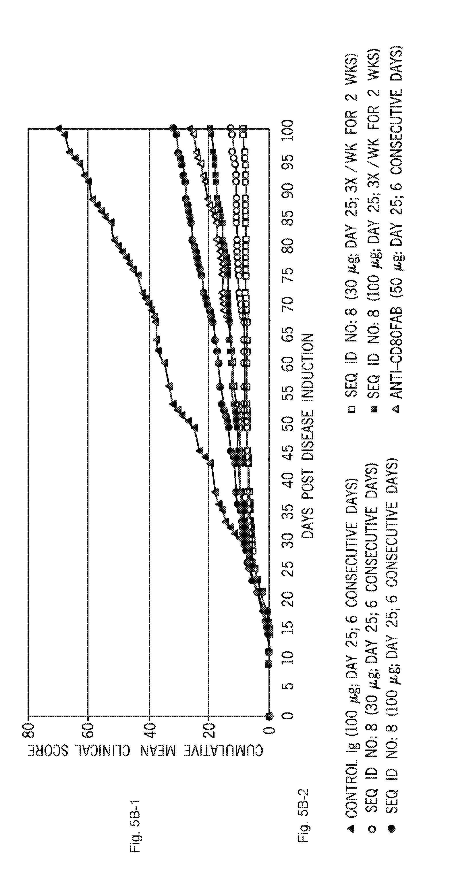
[0055] FIGS. 5A-1, 5A-2, 5B-1, 5B-2 and 5C-1, 5C-2 show the effect of six administrations of C1ORF32-P8-V1-ECD-mFc (SEQ ID NO:8) given in a therapeutic mode at the onset of disease remission (on day 25), at two doses (30 and 100 microg/mouse) and two frequencies (daily or 3 times per week) on clinical symptoms in the PLP139-151-induced R-EAE model in SJL mice demonstrated as mean clinical score (FIG. 5A-1), cumulative clinical score (FIG. 5B-1) and as relapse frequency (FIG. 5C-1); while FIGS. 5A-2, 5B-2 and 5C-2 show the respective keys. In this study the effect of C1ORF32-P8-V1-ECD-mFc (SEQ ID NO:8) was studied in comparison to Ig control (100 microg/mouse) and anti-CD80FaB that were administered on 6 consecutive days.
[0056] FIGS. 6-A and 6B show the effect of C1ORF32-ECD-mFc (SEQ ID NO:8) treatment in comparison to control Ig and B7-H4-Ig on proliferation of T cells derived from SJL or BALB/c mice; FIG. 6-B shows the key.
[0057] FIGS. 7A-1, 7A-2, 7B-1, 7B-2, 7C-1, 7C-2, 7D-1, 7D-2, 7E-1, 7E-2, 7F-1 and 7F-2 show the in vitro effect of C1ORF32-ECD-mFc (SEQ ID NO:8) presented as soluble or bound to either plate or beads, on the proliferation and cytokine secretion of Naive CD4.sup.+ T cells isolated from SJL mice. FIG. 7A-1 shows the effect on SJL proliferation. FIG. 7B-1 shows the effect on IL-2 secretion. FIG. 7C-1 shows the effect on IFN-gamma secretion. FIG. 7D-1 shows the effect on IL-17 secretion. FIG. 7E-1 shows the effect on IL-10 secretion. FIG. 7F-1 shows the effect on TNF-alpha secretion. FIGS. 7A-2, 7B-2, 7C-2, 7D-2, 7E-2 and 7F-2 show the respective keys.
[0058] FIGS. 8A-1 and 8A-2, 8B-1 and 8B-2, 8C-1 and 8C-2, 8D-1 and 8D-2, 8E-1 and 8E-2, and 8F-1 and 8F-2 show the in vitro effect of C1ORF32-ECD-mFc (SEQ ID NO:8) presented as soluble or bound to either plate or beads, on the proliferation and cytokine secretion of Naive CD4.sup.+ T cells isolated from BALB/c mice. FIG. 8A-1 shows the effect on BALB proliferation. FIG. 8B-1 shows the effect on IL-2 secretion. FIG. 8C-1 shows the effect on IFN-gamma secretion. FIG. 8D-1 shows the effect on IL-17 secretion. FIG. 8E-1 shows the effect on IL-10 secretion. FIG. 8F-1 shows the effect on TNF-alpha secretion. FIGS. 8A-2, 8B-2, 8C-2, 8D-2, 8E-2 and 8F-2 show the respective keys.
[0059] FIGS. 9A-1 and 9A-2, 9B-1 and 9B-2, 9C-1 and 9C-2, 9D-1 and 9D-2, 9E-1 and 9E-2, 9F-1 and 9F-2, and 9G-1 and 9G-2 show the effect of C1ORF32-ECD-mFc (SEQ ID NO:8) on T cell activation and differentiation under Th0, Th1, Th2 and Th17-promoting conditions, when presented as bead-bound together with anti-CD3 and anti-CD28, as well as when added as a soluble protein to irradiated APC isolated from DO.11 mice+OVA323-339. FIG. 9A-1 shows the effect on proliferation. FIG. 9B-1 shows the effect on IL-2 secretion. FIG. 9C-1 shows the effect on IFN-gamma secretion. FIG. 9D-1 shows the effect on IL-17 secretion. FIG. 9E-1 shows the effect on IL-4 secretion. FIG. 9F-1 shows the effect on IL-5 secretion. FIG. 9G-1 shows the effect on IL-10 secretion. FIGS. 9A-2, 9B-2, 9C-2, 9D-2, 9E-2, 9F-2 and 9G-2 show the respective keys.
[0060] FIGS. 10A1 and 10A-2, 10B-1 and 10B-2, and 10C-1 and 10C-2 show the effect of C1ORF32-ECD-mFc (SEQ ID NO:8) produced in HEK-293 in the mouse R-EAE model. C1ORF32-ECD-mFc (SEQ ID NO:8) was administered in a preventive mode via i.p. injection on days 0-5 post disease induction. FIG. 10A-1 shows the effect on mean clinical score. FIG. 10B-1 shows the effect on cumulative mean clinical score. FIG. 10C-1 shows the effect on relapse frequency. FIGS. 10A-2, 10B-2 and 10C-2 show the respective keys.
[0061] FIGS. 11A-1 and 11A-2, 11B-1 and 11B-2, 11C-1 and 11C-2, 11D-1 and 11D-2, and 11E-1 and 11E-2 show a comparison of the in-vitro activity of C1ORF32-ECD-mFc (SEQ ID NO:8) produced in CHO and in HEK-293, on CD4+ T cell proliferation and cytokine production under Th0, Th1, Th2 and Th17 deriving conditions. FIG. 11A-1 shows the effect on proliferation. FIG. 11B-1 shows the effect on IFN-gamma secretion. FIG. 11C-1 shows the effect on IL-17 secretion. FIG. 11D-1 shows the effect on IL-4 secretion. FIG. 11E-1 shows the effect on IL-5 secretion. FIGS. 11A-2, 11B-2, 11C-2, 11D-2 and 11E-2 show the respective keys.
[0062] FIGS. 12A1, 12A-2, 12A3, 12B-1 and 12B-2, 12C-1 and 12C-2, 12D-1 and 12D-2, 12E1a and 12E1b, 12E2a and 12E2b, 12E3a and 12E3B, 12E4a and 12E4b, and 12F-1 and 12F-2 show the effect of C1ORF32-ECD-mFc (SEQ ID NO:8) administered in a therapeutic mode, i.p, 3 times per week for two weeks in PLP139-151-induced R-EAE in SJL mice. Demonstrated are effects on mean clinical score, cumulative clinical score and relapse frequency (FIGS. 12A1a, 12A2a and 12A3a); recruitment of immune cells to the spleen, lymph nodes and CNS (FIG. 12B-1); immune cell populations infiltrating the CNS (FIG. 12C-1); recall responses of splenocytes to initiating and spread epitopes via proliferation (FIG. 12D-1); recall responses of splenocytes to initiating and spread epitopes via cytokine secretion (FIGS. 12E1a, 12E2a, 12E3a and 12E4a); recall responses of cervical lymph node cells to initiating epitope via proliferation (FIG. 12F-1). In this study the effect of C1ORF32-P8-V1-ECD-mFc (SEQ ID NO:8) was studied in comparison to Ig control (100 microg/mouse) and anti-CD80 Fab (50 microg/mouse). FIGS. 12A1b, 12A2b, 12A3b, 12B-2, 12C-2, 12D-2, 12E1b, 12E2b, 12E3b, 12E4b and 12F-2 show the respective keys.
[0063] FIGS. 13A-1 and 13A-2 show a dose response effect of therapeutic treatment of C1ORF32-ECD-mFc (SEQ ID NO:8) at 100, 30 and 10 microg/mouse, i.p, 3 times per week for 2 weeks in PLP139-151-induced R-EAE in SJL mice, in comparison to Ig control. Presented is clinical efficacy manifested as mean clinical score (FIG. 13A-1). FIG. 13A-2 shows the key.
[0064] FIGS. 13B-1 and 13B-2, 13C-1 and 13C-2, 13D-1 and 13D-2, and 13E-1 and 13E-2 show an estimation of the effect of C1ORF32-ECD-mFc (SEQ ID NO:8), administered as above at 100 and 30 microg/mouse, on DTH responses to inducing epitope (PLP139-151) or spread epitope (PLP178-191), carried out on day 35 after R-EAE induction (FIG. 13B-1), on recall responses at day 35 after R-EAE induction, manifested as proliferation of lymph node cells (FIG. 13C-1) or spleen cells (FIG. 13D-1) in response to inducing epitope (PLP139-151), spread epitope (PLP178-191) and anti CD3; on DTH responses to spread epitopes (PLP178-191 and MBP84-104), carried out on day 65 from R-EAE induction (FIG. 13E-1), and on recall responses at day 65 after R-EAE induction, manifested as proliferation of splenocytes in response to anti CD3, OVA 323-339, inducing epitope (PLP139-151) and spread epitopes (PLP178-191 and MBP84-104). FIGS. 13B-2, 13C-2, 13D-2 and 13E-2 show the respective keys.
[0065] FIGS. 14A-1, 14A-2, 14B-1, 14B-2, 14C, 14D and 14E show the effect of C1ORF32-ECD-mFc (SEQ ID NO:8) or control Ig treatment on R-EAE development (FIG. 14A-1) and recruitment of total immune cells (FIG. 14B-1), and autoreactive T cells (FIG. 14C) to the spleen, lymph nodes and CNS in adoptive transfer model upon administration at time of cell transfer. FIG. 14D shows recruitment of autoreactive T cells to the lymph nodes. FIG. 14E shows recruitment of autoreactive T cells to the CNS. FIGS. 14A-2 and 14B-2 show the respective keys.
[0066] FIGS. 15A-15F and 16A-16D show the effect of C1ORF32-ECD-mFc (SEQ ID NO:8) on activation human T cell from various human donors, manifested in cell proliferation and IFN.gamma. secretion. Activation was carried out using beads coated with C1ORF32-ECD-mFc (SEQ ID NO:8), anti CD3 and anti CD28 in the one-step method (FIGS. 15A-15F) or the two-step method (FIGS. 16A-16D).
[0067] FIGS. 17A and 17B show the dose dependency of the effect of C1ORF32-ECD-mFc (SEQ ID NO:8) on human T cell proliferation from two different donors.
[0068] FIGS. 18A and 18B show the effect of C1ORF32-ECD-mFc (SEQ ID NO:8) and of Control Ig on proliferation of purified human T cells from different donors activated by beads coated with anti-CD3 and anti-CD28 antibodies together with C1ORF32-ECD-mFc (SEQ ID NO:8). FIG. 18B shows the key.
[0069] FIGS. 19A and 19B show the effect of C1ORF32-ECD-mFc (SEQ ID NO:8) and of Control Ig on proliferation of purified human T cells from different donors activated by irradiated autologous PBMCs and anti-CD3 and anti-CD28 antibodies. FIG. 19B shows the key.
[0070] FIGS. 20A-1 and 20A-2, 20B-1 and 20B-2, and 20C-1 and 20C-2 show the therapeutic effect of C1ORF32-ECD-mFc (SEQ ID NO:8) administered at 100 or 30 microg/mouse, i.p, 3 times per week for 10 days in collagen induced arthritis (CIA) model of rheumatoid arthritis. Measured are clinical score (A-1), paw swelling (B-1) and number of affected paws (C-1). Enbrel.RTM. (TNF-R-Ig, 100 microg/mouse) was used as a positive control while control Ig (100 microg/mouse) was used as negative control. FIGS. 20A-2, 20B-2 and 20C-2 show the respective keys.
[0071] FIGS. 21A, 21B-1, 21B-2, 21C-1 and 21C-2 show the effect of C1ORF32-P8-V1-ECD-mFc (SEQ ID NO:8) at 1.5 and 5 mg/kg on DTH in the trans vivo assay as manifested as the change (delta) in paw thickness in comparison to isotype control (mIgG2a, 5 mg/kg) or vehicle (PBS) treated mice, as detailed in the figure. FK506 was used as a positive control at doses of 3 and 30 mg/kg. Results obtained from injection of PBMCs pooled from 4 different donors are shown on FIG. 21A, individual data obtained upon injection of PBMCs from each donor are presented in FIGS. 21B-1 and 21C-2. FIGS. 21B-2 and 21C-2 show the respective keys.
[0072] FIGS. 22A-1 and 22A-2, 22B-1 and 22B-2, 22C-1 and 22C-2, 22D-1 and 22D-2, 22E-1 and 22E-2, and 22F-1 and 22F-2 show the effect of C1ORF32-P8-V1-ECD-mFc (SEQ ID NO:8) on IFN.gamma. (A-1 and B-1), IL-2 (C-1 and D-1) and IL-4 (E-1 and F-1) production by splenocytes which were stimulated in the presence of plate-bound anti-CD3 and soluble anti-CD28 for 24 hours. FK506 and B7-H4-Ig were used as positive control. mIgG2a was used as negative control. For each cytokine, the results of two studies are presented, labeled on the drawings as studies 1 and 2. FIGS. 22A-2, 22B-2, 22C-2, 22D-2, 22E-2 and 22F-2 show the respective keys.
DETAILED DESCRIPTION OF THE INVENTION
[0073] The present invention, in at least some embodiments, relates to any one of the proteins referred to as C1ORF32, fragments, variants and homologs thereof and fusion proteins and conjugates containing same, and pharmaceutical compositions comprising same, and nucleic acid sequences encoding same, and the use thereof as a therapeutic agent for treatment of immune related disorder as described herein, including without limitation use of the ECD (extracellular domain) of a C1ORF32 protein, fragments and/or variants and/or homologs thereof (alone or as part of a fusion protein or conjugate).
[0074] In order that the present invention may be more readily understood, certain terms are first defined. Additional definitions are set forth throughout the detailed description.
[0075] As used herein the term "isolated" refers to a compound of interest (for example a polynucleotide or a polypeptide) that is in an environment different from that in which the compound naturally occurs e.g. separated from its natural milieu such as by concentrating a peptide to a concentration at which it is not found in nature. "Isolated" includes compounds that are within samples that are substantially enriched for the compound of interest and/or in which the compound of interest is partially or substantially purified.
[0076] An "immune cell" refers to any cell from the hemopoietic origin including but not limited to T cells, B cells, monocytes, dendritic cells, and macrophages.
[0077] As used herein, the term "polypeptide" refers to a chain of amino acids of any length, regardless of modification (e.g., phosphorylation or glycosylation). As used herein, a "costimulatory polypeptide" or "costimulatory molecule" is a polypeptide that, upon interaction with a cell-surface molecule on T cells, modulates T cell responses.
[0078] As used herein, a "costimulatory signaling" is the signaling activity resulting from the interaction between costimulatory polypeptides on antigen presenting cells and their receptors on T cells during antigen-specific T cell responses. Without wishing to be limited by a single hypothesis, the antigen-specific T cell response is believed to be mediated by two signals: 1) engagement of the T cell receptor (TCR) with antigenic peptide presented in the context of MHC (signal 1), and 2) a second antigen-independent signal delivered by contact between different costimulatory receptor/ligand pairs (signal 2). Without wishing to be limited by a single hypothesis, this "second signal" is critical in determining the type of T cell response (activation vs inhibition) as well as the strength and duration of that response, and is regulated by both positive and negative signals from costimulatory molecules, such as the B7 family of proteins.
[0079] As used herein, the term "B7" polypeptide means a member of the B7 family of proteins that costimulate T cells including, but not limited to B7-1, B7-2, B7-DC, B7-H5, B7-H1, B7-H2, B7-H3, B7-H4, B7-H6, B7-S3 and biologically active fragments and/or variants thereof. Representative biologically active fragments include the extracellular domain or fragments of the extracellular domain that costimulate T cells.
[0080] As used herein, "inflammatory molecules" refers to molecules that induce inflammatory responses (directly or indirectly) including, but not limited to, cytokines and metalloproteases such as including, but not limited to, IL-1beta, TNF-alpha, TGF-beta, IFN-gamma, IL-17, IL-6, IL-23, IL-22, IL-21, and MMPs.
[0081] As used herein, a "vector" is a replicon, such as a plasmid, phage, or cosmid, into which another DNA segment may be inserted so as to bring about the replication of the inserted segment. The vectors described herein can be expression vectors. As used herein, an "expression vector" is a vector that includes one or more expression control sequences. As used herein, an "expression control sequence" is a DNA sequence that controls and regulates the transcription and/or translation of another DNA sequence. "Operably linked" refers to an arrangement of elements wherein the components so described are configured so as to perform their usual or intended function. Thus, two different polypeptides operably linked together retain their respective biological functions while physically linked together. As used herein, "valency" refers to the number of binding sites available per molecule.
[0082] As used herein, a "variant" polypeptide contains at least one amino acid sequence alteration as compared to the amino acid sequence of the corresponding wild-type polypeptide.
[0083] As used herein, "conservative" amino acid substitutions are substitutions wherein the substituted amino acid has similar structural or chemical properties. As used herein, the term "host cell" refers to prokaryotic and eukaryotic cells into which a recombinant vector can be introduced.
[0084] As used herein, "transformed" and "transfected" encompass the introduction of a nucleic acid (e.g. a vector) into a cell by a number of techniques known in the art. As used herein, the terms "immunologic", "immunological" or "immune" response is the development of a beneficial humoral (antibody mediated) and/or a cellular (mediated by antigen-specific T cells or their secretion products) response directed against a peptide in a recipient patient. Such a response can be an active response induced by administration of immunogen or a passive response induced by administration of antibody or primed T-cells. Without wishing to be limited by a single hypothesis, acellular immune response is elicited by the presentation of polypeptide epitopes in association with Class I or Class II MHC molecules to activate antigen-specific CD4+T helper cells and/or CD8+ cytotoxic T cells. The response may also involve activation of monocytes, macrophages, NK cells, basophils, dendritic cells, astrocytes, microglia cells, eosinophils, activation or recruitment of neutrophils or other components of innate immunity. The presence of a cell-mediated immunological response can be determined by proliferation assays (CD4+ T cells) or CTL (cytotoxic T lymphocyte) assays. The relative contributions of humoral and cellular responses to the protective or therapeutic effect of an immunogen can be distinguished by separately isolating antibodies and T-cells from an immunized syngeneic animal and measuring protective or therapeutic effect in a second subject. An "immunogenic agent" or "immunogen" is capable of inducing an immunological response against itself on administration to a mammal, optionally in conjunction with an adjuvant.
[0085] As used herein, the term "C1ORF32" refers to the protein encoded by any one of the H19011_1_T8 (SEQ ID NO:1), H19011_1_T9 (SEQ ID NO:2) transcripts reported herein, particularly to proteins as set forth in any one of H19011_1_P8 (SEQ ID NO:4), H19011_1_P8_V1 (SEQ ID NO:5), H19011_1_P9 (SEQ ID NO:6) or H19011_1_P9_V1 (SEQ ID NO:34), variants and fragments thereof, which can have therapeutic effect on an of immune related disorder. As used herein, the terms C1ORF32 fragments and/or C1ORF32 variants and/or C1ORF32 homologs refer to portions of C1ORF32 comprising amino acid sequence having a biological activity of inhibition of T cell activation.
[0086] As used herein the term "soluble C1ORF32" or "soluble ectodomain (ECD)" or "ectodomain" or "soluble C1ORF32 proteins/molecules" refers to fragments of C1ORF32 that include some or all of the extracellular domain of the C1ORF32 polypeptide, and lack some or all of the intracellular and/or transmembrane domains, wherein said fragments retain a biological activity of inhibition of T cell activation. In one embodiment, soluble C1ORF32 polypeptide fragments include the entire extracellular domain of the C1ORF32 polypeptide. In other embodiments, the soluble fragments of C1ORF32 polypeptides include fragments of the extracellular domain.
[0087] As used herein, the term "soluble C1ORF32" or "soluble ectodomain (ECD)" or "ectodomain" or "soluble C1ORF32 proteins/molecules" further means non-cell-surface-bound (i.e. circulating) C1ORF32 molecules or any portion of a C1ORF32 molecule including, but not limited to: C1ORF32 polypeptides, fragments or fusion proteins thereof fusion proteins, wherein the extracellular domain of C1ORF32 is fused to an immunoglobulin (Ig) moiety rendering the fusion molecule soluble, or fragments and derivatives thereof, proteins with the extracellular domain of C1ORF32 fused or joined with a portion of a biologically active or chemically active protein such as the papillomavirus E7 gene product, melanoma-associated antigen p97 or HIV env protein, or fragments and derivatives thereof; hybrid (chimeric) fusion proteins such as C1ORF32 polypeptides, fragments or fusion proteins thereof, or fragments and derivatives thereof. "Soluble C1ORF32 proteins/molecules" also include C1ORF32 molecules with the transmembrane domain removed to render the protein soluble, or fragments and derivatives thereof; and soluble C1ORF32 mutant molecules. The soluble C1ORF32 molecules used in the methods of the invention may or may not include a signal (leader) peptide sequence.
[0088] The term the "soluble ectodomain (ECD)" or "ectodomain" or "soluble" form of C1ORF32 refers also to the nucleic acid sequences encoding the corresponding proteins of C1ORF32 "soluble ectodomain (ECD)" or "ectodomain" or "soluble C1ORF32 proteins/molecules"). Optionally, the C1ORF32 ECD refers to any one of the polypeptide sequences below or fragments thereof:
TABLE-US-00001 >H19011_1_P8 residues 21 to 186, corresponding to amino acid sequence depicted in SEQ ID NO: 14 (SEQ ID NO: 4) LQVTVPDKKKVAMLFQPTVLRCHFSTSSHQPAVVQWKFKSYCQDRMGESL GMSSTRAQSLSKRNLEWDPYLDCLDSRRTVRVVASKQGSTVTLGDFYRGR EITIVHDADLQIGKLMWGDSGLYYCIITTPDDLEGKNEGSLGLLVLGRTG LLADLLPSFAVEIMPE; >H19011_1_P8_V1 residues 21-186, corresponding to amino acid sequence depicted in SEQ ID NO: 35 (SEQ ID NO: 5) LQVTVPDKKKVAMLFQPTVLRCHFSTSSHQPAVVQWKFKSYCQDRMGESL GMSSTRAQSLSKRNLEWDPYLDCLDSRRTVRVVASKQGSTVTLGDFYRGR EITIVHDADLQIGKLMWGDSGLYYCIITTPDDLEGKNEDSVELLVLGRTG LLADLLPSFAVEIMPE; >H19011_1_P9 residues 21 to 169, corresponding to amino acid sequence depicted in SEQ ID NO: 15 (SEQ ID NO: 6) LQVTVPDKKKVAMLFQPTVLRCHFSTSSHQPAVVQWKFKSYCQDRMGESL GMSSTRAQSLSKRNLEWDPYLDCLDSRRTVRVVASKQGSTVTLGDFYRGR EITIVHDADLQIGKLMWGDSGLYYCIITTPDDLEGKNEGSLGLLVLEWV; >H19011_1_P9_V1 residues 21 to 169, corresponding to amino acid sequence depicted in SEQ ID NO: 36 (SEQ ID NO: 34) LQVTVPDKKKVAMLFQPTVLRCHFSTSSHQPAVVQWKFKSYCQDRMGESL GMSSTRAQSLSKRNLEWDPYLDCLDSRRTVRVVASKQGSTVTLGDFYRGR EITIVHDADLQIGKLMWGDSGLYYCIITTPDDLEGKNEDSVELLVLEWV; >H19011_1_P8_V1 residues 1 to 184, corresponding to amino acid sequence depicted in SEQ ID NO: 19 (SEQ ID NO: 5) MDRVLLRWISLFWLTAMVEGLQVTVPDKKKVAMLFQPTVLRCHFSTSSHQ PAVVQWKFKSYCQDRMGESLGMSSTRAQSLSKRNLEWDPYLDCLDSRRTV RVVASKQGSTVTLGDFYRGREITIVHDADLQIGKLMWGDSGLYYCIITTP DDLEGKNEDSVELLVLGRTGLLADLLPSFAVEIM; >H19011_1_P8 residues 1 to 184, corresponding to amino acid sequence depicted in SEQ ID NO: 37 (SEQ ID NO: 4) MDRVLLRWISLFWLTAMVEGLQVTVPDKKKVAMLFQPTVLRCHFSTSSHQ PAVVQWKFKSYCQDRMGESLGMSSTRAQSLSKRNLEWDPYLDCLDSRRTV RVVASKQGSTVTLGDFYRGREITIVHDADLQIGKLMWGDSGLYYCIITTP DDLEGKNEGSLGLLVLGRTGLLADLLPSFAVEIM; >H19011_1_P9 residues 1-169, corresponding to amino acid sequence depicted in SEQ ID NO: 28 (SEQ ID NO: 6) MDRVLLRWISLFWLTAMVEGLQVTVPDKKKVAMLFQPTVLRCHFSTSSHQ PAVVQWKFKSYCQDRMGESLGMSSTRAQSLSKRNLEWDPYLDCLDSRRTV RVVASKQGSTVTLGDFYRGREITIVHDADLQIGKLMWGDSGLYYCIITTP DDLEGKNEGSLGLLVLEWV; >H19011_1_P9_V1 residues 1-169, corresponding to amino acid sequence depicted in SEQ ID NO: 30 (SEQ ID NO: 34) MDRVLLRWISLFWLTAMVEGLQVTVPDKKKVAMLFQPTVLRCHFSTSSHQ PAVVQWKFKSYCQDRMGESLGMSSTRAQSLSKRNLEWDPYLDCLDSRRTV RVVASKQGSTVTLGDFYRGREITIVHDADLQIGKLMWGDSGLYYCIITTP DDLEGKNEDSVELLVLEWV,
[0089] and variants thereof possessing at least 80% sequence identity, more preferably at least 90% sequence identity therewith and even more preferably at least 95, 96, 97, 98 or 99% sequence identity therewith. According to at least some embodiments, the isolated polypeptide at least 80, 90, 95, 96, 97, 98 or 99% homologous to a polypeptide comprising all or part of the extracellular domain of H19011_1_P8 (SEQ ID NO:4), H19011_1_P8_V1 (SEQ ID NO:5), H19011_1_P9 (SEQ ID NO:6) or H19011_1_P9_V1 (SEQ ID NO:34) has at least one of the SNP variations, as described herein in Example 1.
[0090] The C1ORF32 extracellular domain can contain one or more amino acids from the signal peptide or the putative transmembrane domain of C1ORF32. During secretion, the number of amino acids of the signal peptide that are cleaved can vary depending on the expression system and the host. Additionally, or alternatively, fragments of C1ORF32 extracellular domain missing one or more amino acids from the C-terminus or the N-terminus that retain the ability to bind to the C1ORF32 receptor can be used as a fusion partner for the disclosed fusion proteins.
Variants of C1ORF32 Polypeptides
[0091] Useful variants of such C1ORF32 polypeptides include those that increase biological activity, as indicated by any of the assays described herein, or that increase half-life or stability of the protein. Soluble C1ORF32 polypeptides and C1ORF32 fragments, or fusions thereof having C1ORF32 activity, can be engineered to increase biological activity. In a further embodiment, the C1ORF32 polypeptide or fusion protein has been modified with at least one amino acid substitution, deletion, or insertion that increases the binding of the molecule to an immune cell, for example a T cell, and transmits an inhibitory signal into the T cell.
[0092] Other optional variants are those C1ORF32 polypeptides that are engineered to selectively bind to one type of T cell versus other immune cells. For example, the C1ORF32 polypeptide can be engineered to bind optionally to Tregs, Th0, Th1, Th17, Th2 or Th22 cells. Preferential binding refers to binding that is at least 10%, 20%, 30%, 40%, 50%, 60% f 70%, 80%, 90%, 95%, or greater for one type of cell over another type of cell. Still other variants of C1ORF32 can be engineered to have reduced binding to immune cells relative to wildtype C1ORF32. These variants can be used in combination with variants having stronger binding properties to modulate the immune response with a moderate impact.
[0093] Also optionally, variant C1ORF32 polypeptides can be engineered to have an increased half-life relative to wildtype. These variants typically are modified to resist enzymatic degradation. Exemplary modifications include modified amino acid residues and modified peptide bonds that resist enzymatic degradation. Various modifications to achieve this are known in the art.
[0094] Fusion Proteins
[0095] According to at least some embodiments, C1ORF32 fusion polypeptides have a first fusion partner comprising all or a part of a C1ORF32 protein fused to a second polypeptide directly or via a linker peptide sequence or a chemical linker useful to connect the two proteins. The C1ORF32 polypeptide may optionally be fused to a second polypeptide to form a fusion protein as described herein. The presence of the second polypeptide can alter the solubility, stability, affinity and/or valency of the C1ORF32 fusion polypeptide. As used herein, "valency" refers to the number of binding sites available per molecule. In one embodiment the second polypeptide is a polypeptide from a different source or different protein.
[0096] According to at least some embodiments, the C1ORF32 protein or fragment is selected for its activity for the treatment of immune related disorder as described herein.
[0097] In one embodiment, the second polypeptide contains one or more domains of an immunoglobulin heavy chain constant region, preferably having an amino acid sequence corresponding to the hinge, CH2 and CH3 regions of a human immunoglobulin C.gamma.1, C.quadrature.2, C.quadrature.3 or C.quadrature.4 chain or to the hinge, CH2 and CH3 regions of a murine immunoglobulin C.gamma.2a chain. SEQ ID NO: 20 provides exemplary sequence for the hinge, CH2 and CH3 regions of a human immunoglobulin C.gamma.1.
[0098] According to at least some embodiments, the fusion protein is a dimeric fusion protein. In an optional dimeric fusion protein, the dimer results from the covalent bonding of Cys residue in the hinge region of two of the Ig heavy chains that are the same Cys residues that are disulfide linked in dimerized normal Ig heavy chains. Such proteins are referred to as C1ORF32 polypeptides, fragments or fusion proteins thereof.
[0099] In one embodiment, the immunoglobulin constant domain may contain one or more amino acid insertions, deletions or substitutions that enhance binding to specific cell types, increase the bioavailability, or increase the stability of the C1ORF32 polypeptides, fusion proteins, or fragments thereof. Suitable amino acid substitutions include conservative and non-conservative substitutions, as described above.
[0100] The fusion proteins optionally contain a domain that functions to dimerize or multimerize two or more fusion proteins. The peptide/polypeptide linker domain can either be a separate domain, or alternatively can be contained within one of the other domains (C1ORF32 polypeptide or second polypeptide) of the fusion protein. Similarly, the domain that functions to dimerize or multimerize the fusion proteins can either be a separate domain, or alternatively can be contained within one of the other domains (C1ORF32 polypeptide, second polypeptide or peptide/polypeptide linker domain) of the fusion protein. In one embodiment, the dimerization/multimerization domain and the peptide/polypeptide linker domain are the same. Further specific, illustrative and non-limiting examples of dimerization/multimerization domains and linkers are given below.
[0101] Fusion proteins disclosed herein according to at least some embodiments of the present invention are of formula I: N--R1-R2-R3-C wherein "N" represents the N-terminus of the fusion protein, "C" represents the C-terminus of the fusion protein. In the further embodiment, "R1" is a C1ORF32 polypeptide, "R2" is an optional peptide/polypeptide or chemical linker domain, and "R3" is a second polypeptide. Alternatively, R3 may be a C1ORF32 polypeptide and R1 may be a second polypeptide.
[0102] Optionally, the fusion protein comprises the C1ORF32 polypeptide fragments as described herein, fused, optionally by a linker peptide of one or more amino acids (e.g. GS) to one or more "half-life extending moieties". A "half-life extending moiety" is any moiety, for example, a polypeptide, small molecule or polymer, that, when appended to protein, extends the in vivo half-life of that protein in the body of a subject (e.g., in the plasma of the subject). For example, a half-life extending moiety is, in an embodiment of the invention, polyethylene glycol (PEG), monomethoxy PEG (mPEG) or an immunoglobulin (Ig). In an embodiment of the invention, PEG is a 5, 10, 12, 20, 30, 40 or 50 kDa moiety or larger or comprises about 12000 ethylene glycol units (PEG12000).
[0103] Dimerization or multimerization can occur between or among two or more fusion proteins through dimerization or multimerization domains. Alternatively, dimerization or multimerization of fusion proteins can occur by chemical crosslinking. The dimers or multimers that are formed can be homodimeric/homomultimeric or heterodimeric/heteromultimeric. The second polypeptide "partner" in the C1ORF32 fusion polypeptides may be comprised of one or more other proteins, protein fragments or peptides as described herein, including but not limited to any immunoglobulin (Ig) protein or portion thereof, preferably the Fc region, or a portion of a biologically or chemically active protein such as the papillomavirus E7 gene product, melanoma-associated antigen p97), and HIV env protein (gp120). The "partner" is optionally selected to provide a soluble dimer/multimer and/or for one or more other biological activities as described herein.
[0104] Dimerization or multimerization can occur between or among two or more fusion proteins through dimerization or multimerization domains, including those described above. Alternatively, dimerization or multimerization of fusion proteins can occur by chemical crosslinking. Fusion protein dimers can be homodimers or heterodimers. Fusion protein multimers can be homomultimers or heteromultimers. Fusion protein dimers as disclosed herein are of formula II:
N--R1-R2-R3-C
N--R4-R5-R6-C or, alternatively, are of formula III:
N--R1-R2-R3-C
C-R4-R5-R6-N wherein the fusion proteins of the dimer provided by formula II are defined as being in a parallel orientation and the fusion proteins of the dimer provided by formula III are defined as being in an antiparallel orientation. Parallel and antiparallel dimers are also referred to as cis and trans dimers, respectively. "N" and "C" represent the N- and C-termini of the fusion protein, respectively. The fusion protein constituents "R1", "R2" and "R3" are as defined above with respect to formula I. With respect to both formula II and formula III, "R4" is a C1ORF32 polypeptide or a second polypeptide, "R5" is an optional peptide/polypeptide linker domain, and "R6" is a C1ORF32 polypeptide or a second polypeptide, wherein "R6" is a C1ORF32 polypeptide when "R4" is a second polypeptide, and "R6" is a second polypeptide when "R4" is a C1ORF32 polypeptide. In one embodiment, "R1" is a C1ORF32 polypeptide, "R4" is also a C1ORF32 polypeptide, and "R3" and "R6" are both second polypeptides.
[0105] Fusion protein dimers of formula II are defined as homodimers when "R1"="R4", "R2"="R5" and "R3"="R6". Similarly, fusion protein dimers of formula III are defined as homodimers when "R1"="R6", "R2"="R5" and "R3"="R4". Fusion protein dimers are defined as heterodimers when these conditions are not met for any reason. For example, heterodimers may contain domain orientations that meet these conditions (i.e., for a dimer according to formula II, "R1" and "R4" are both C1ORF32 polypeptides, "R2" and "R5" are both peptide/polypeptide linker domains and "R3" and "R6" are both second polypeptides), however the species of one or more of these domains is not identical. For example, although "R3" and "R6" may both be C1ORF32 polypeptides, one polypeptide may contain a wild-type C1ORF32 amino acid sequence while the other polypeptide may be a variant C1ORF32 polypeptide. An exemplary variant C1ORF32 polypeptide is C1ORF32 polypeptide that has been modified to have increased or decreased binding to a target cell, increased activity on immune cells, increased or decreased half-life or stability. Dimers of fusion proteins that contain either a CHI or CL region of an immunoglobulin as part of the polypeptide linker domain preferably form heterodimers wherein one fusion protein of the dimer contains a CHI region and the other fusion protein of the dimer contains a CL region.
[0106] Fusion proteins can also be used to form multimers. As with dimers, multimers may be parallel multimers, in which all fusion proteins of the multimer are aligned in the same orientation with respect to their N- and C-termini. Multimers may be antiparallel multimers, in which the fusion proteins of the multimer are alternatively aligned in opposite orientations with respect to their N- and C-termini. Multimers (parallel or antiparallel) can be either homomultimers or heteromultimers. The fusion protein is optionally produced in dimeric form; more preferably, the fusion is performed at the genetic level as described below, by joining polynucleotide sequences corresponding to the two (or more) proteins, portions of proteins and/or peptides, such that a joined or fused protein is produced by a cell according to the joined polynucleotide sequence. A description of preparation for such fusion proteins is described with regard to U.S. Pat. No. 5,851,795 to Linsley et al, which is hereby incorporated by reference as if fully set forth herein as a non-limiting example only.
[0107] The fusion protein may also optionally be prepared by chemical synthetic methods and the "join" effected chemically, either during synthesis or post-synthesis. Cross-linking and other such methods may optionally be used (optionally also with the above described genetic level fusion methods), as described for example in U.S. Pat. No. 5,547,853 to Wallner et al, which is hereby incorporated by reference as if fully set forth herein as a non-limiting example only.
[0108] According to the present invention, a fusion protein may be prepared from a protein of the invention by fusion with a portion of an immunoglobulin comprising a constant region of an immunoglobulin. More preferably, the portion of the immunoglobulin comprises a heavy chain constant region which is optionally and more preferably a human heavy chain constant region. The heavy chain constant region is most preferably an IgG heavy chain constant region, and optionally and most preferably is an Fc chain, most preferably an IgG Fc fragment that comprises the hinge, CH2 and CH3 domains. The Fc chain may optionally be a known or "wild type" Fc chain, or alternatively may be mutated or truncated. The Fc portion of the fusion protein may optionally be varied by isotype or subclass, may be a chimeric or hybrid, and/or may be modified, for example to improve effector functions, control of half-life, tissue accessibility, augment biophysical characteristics such as stability, and improve efficiency of production (and less costly). Many modifications useful in construction of disclosed fusion proteins and methods for making them are known in the art, see for example Mueller, et al, MoI. Immun., 34(6):441-452 (1997), Swann, et al., Cur. Opin. Immun., 20:493-499 (2008), and Presta, Cur. Opin. Immun. 20:460-470 (2008). In some embodiments the Fc region is the native IgG1, IgG2, or IgG4 Fc region. In some embodiments the Fc region is a hybrid, for example a chimeric consisting of IgG2/IgG4 Fc constant regions.
[0109] Modifications to the Fc region include, but are not limited to, IgG4 modified to prevent binding to Fc gamma receptors and complement, IgG1 modified to improve binding to one or more Fc gamma receptors, IgG1 modified to minimize effector function (amino acid changes), IgG1 with altered/no glycan (typically by changing expression host), and IgG1 with altered pH-dependent binding to FcRn. The Fc region may include the entire hinge region, or less than the entire hinge region.
[0110] In another embodiment, the Fc domain may contain one or more amino acid insertions, deletions or substitutions that reduce binding to the low affinity inhibitory Fc receptor CD32B (Fc.gamma.RIIB) and retain wild-type levels of binding to or enhance binding to the low affinity activating Fc receptor CD16A (Fc.gamma.RIIIA)
[0111] Another embodiment includes IgG2-4 hybrids and IgG4 mutants that have reduced binding to FcR (Fc receptor) which increase their half-life. Representative IgG2-4 hybrids and IgG4 mutants are described in Angal, S. et al., Molecular Immunology, 30(1):105-108 (1993); Mueller, J. et al., Molecular Immunology, 34(6): 441-452 (1997); and U.S. Pat. No. 6,982,323 to Wang et al. In some embodiments the IgG1 and/or IgG2 domain is deleted; for example, Angal et al. describe IgG1 and IgG2 having serine 241 replaced with a proline.
[0112] In a further embodiment, the Fc domain contains amino acid insertions, deletions or substitutions that enhance binding to CD16A. A large number of substitutions in the Fc domain of human IgG1 that increase binding to CD16A and reduce binding to CD32B are known in the art and are described in Stavenhagen, et al., Cancer Res., 57(18):8882-90 (2007). Exemplary variants of human IgG1 Fc domains with reduced binding to CD32B and/or increased binding to CD16A contain F243L, R929P, Y300L, V3051 or P296L substitutions. These amino acid substitutions may be present in a human IgG1 Fc domain in any combination.
[0113] In one embodiment, the human IgG1 Fc domain variant contains a F243L, R929P and Y300L substitution. In another embodiment, the human IgG1 Fc domain variant contains a F243L, R929P, Y300L, V3051 and P296L substitution. In another embodiment, the human IgG1 Fc domain variant contains an N297A/Q substitution, as these mutations abolish Fc.quadrature.R binding. Non-limiting, illustrative, exemplary types of mutations are described in US Patent Application No. 20060034852, published on Feb. 16, 2006, hereby incorporated by reference as if fully set forth herein. The term "Fc chain" also optionally comprises any type of Fc fragment.
[0114] Several of the specific amino acid residues that are important for antibody constant region-mediated activity in the IgG subclass have been identified. Inclusion, substitution or exclusion of these specific amino acids therefore allows for inclusion or exclusion of specific immunoglobulin constant region-mediated activity. Furthermore, specific changes may result in aglycosylation for example and/or other desired changes to the Fc chain. At least some changes may optionally be made to block a function of Fc which is considered to be undesirable, such as an undesirable immune system effect, as described in greater detail below.
[0115] Non-limiting, illustrative examples of mutations to Fc which may be made to modulate the activity of the fusion protein include the following changes (given with regard to the Fc sequence nomenclature as given by Kabat, from Kabat E A et al: Sequences of Proteins of Immunological Interest. US Department of Health and Human Services, NIH,1991): 220C->S; 233-238 ELLGGP->EAEGAP; 265D->A, preferably in combination with 434N->A; 297N->A (for example to block N-glycosylation); 318-322 EYKCK->AYACA; 330-331AP->SS; or a combination thereof (see for example M. Clark, "Chemical Immunol and Antibody Engineering", pp 1-31 for a description of these mutations and their effect). The construct for the Fc chain which features the above changes optionally and preferably comprises a combination of the hinge region with the CH2 and CH3 domains.
[0116] The above mutations may optionally be implemented to enhance desired properties or alternatively to block non-desired properties. For example, aglycosylation of antibodies was shown to maintain the desired binding functionality while blocking depletion of T-cells or triggering cytokine release, which may optionally be undesired functions (see M. Clark, "Chemical Immunol and Antibody Engineering", pp 1-31). Substitution of 331proline for serine may block the ability to activate complement, which may optionally be considered an undesired function (see M. Clark, "Chemical Immunol and Antibody Engineering", pp 1-31). Changing 330alanine to serine in combination with this change may also enhance the desired effect of blocking the ability to activate complement.
[0117] Residues 235 and 237 were shown to be involved in antibody-dependent cell-mediated cytotoxicity (ADCC), such that changing the block of residues from 233-238 as described may also block such activity if ADCC is considered to be an undesirable function. Residue 220 is normally a cysteine for Fc from IgG1, which is the site at which the heavy chain forms a covalent linkage with the light chain. Optionally, this residue may be changed to a serine, to avoid any type of covalent linkage (see M. Clark, "Chemical Immunol and Antibody Engineering", pp 1-31).
[0118] The above changes to residues 265 and 434 may optionally be implemented to reduce or block binding to the Fc receptor, which may optionally block undesired functionality of Fc related to its immune system functions (see "Binding site on Human IgG1 for Fc Receptors", Shields et al, Vol 276, pp 6591-6604, 2001).
[0119] The above changes are intended as illustrations only of optional changes and are not meant to be limiting in any way. Furthermore, the above explanation is provided for descriptive purposes only, without wishing to be bound by a single hypothesis.
[0120] Exemplary fusion proteins are set forth in SEQ ID NOs: 8, 22, 23, 38, 29. The aforementioned exemplary fusion proteins can incorporate any combination of the variants described herein. In another embodiment the terminal lysine of the aforementioned exemplary fusion proteins is deleted.
[0121] The disclosed fusion proteins can be isolated using standard molecular biology techniques. For example, an expression vector containing a DNA sequence encoding a C1ORF32 polypeptides, fragments or fusion proteins thereof fusion protein is transfected into 293 cells by calcium phosphate precipitation and cultured in serum-free DMEM. The supernatant is collected at 72 h and the fusion protein is purified by protein g, or preferably protein a SEPHAROSE.RTM. columns (Pharmacia, Uppsala, Sweden). Optionally, a DNA sequence encoding a C1ORF32 polypeptides, fragments or fusion proteins thereof fusion protein is transfected into GPEx.RTM. retrovectors and expressed in CHO-S cells following four rounds of retrovector transduction. The protein is clarified from supernatants using protein A chromatography.
[0122] In another embodiment the second polypeptide may have a conjugation domain through which additional molecules can be bound to the C1ORF32 fusion proteins. In one such embodiment, the conjugated molecule is capable of targeting the fusion protein to a particular organ or tissue; further specific, illustrative, non-limiting examples of such targeting domains and/or molecules are given below.
[0123] In another such embodiment the conjugated molecule is another immunomodulatory agent that can enhance or augment the effects of the C1ORF32 fusion protein. In another embodiment the conjugated molecule is Polyethylene Glycol (PEG).
[0124] Peptide or polypeptide linker domain The disclosed C1ORF32 fusion proteins optionally contain a peptide or polypeptide linker domain that separates the C1ORF32 polypeptide from the second polypeptide. In one embodiment, the linker domain contains the hinge region of an immunoglobulin. In a further embodiment, the hinge region is derived from a human immunoglobulin. Suitable human immunoglobulins that the hinge can be derived from include IgG, IgD and IgA. In a further embodiment, the hinge region is derived from human IgG. Amino acid sequences of immunoglobulin hinge regions and other domains are well known in the art. In one embodiment, C1ORF32 fusion polypeptides contain the hinge, CH2 and CH3 regions of a human immunoglobulin C.gamma.1 chain having at least 85%, 90%, 95%, 99% or 100% sequence homology to amino acid sequence set forth in SEQ ID NO:20:
TABLE-US-00002 EPKSCDKTHTCPPCPAPELLGGPSVFLEPPKPKDTLMISRTPEVTCVVVD VSHEDPEVKFNWYVDGVEVHNAKT KPREEQYNSTYRVVSVLTVLHQDWLNGKEYKCKVSNKALPAPIEKTISKA KGQPREPQVYTLPPSRDELTKNQV SLTCLVKGFYPSDIAVEWESNGQPENNYKTTPPVLDSDGSFELYSKLTVD KSRWQQGNVFSCSVMHEALHNHYT QKSLSLSPGK
[0125] The hinge can be further shortened to remove amino acids 1, 2, 3, 4, 5, or combinations thereof of SEQ ID NO: 20. In one embodiment, amino acids 1-5 of SEQ ID NO: 20 are deleted.
[0126] In another embodiment, C1ORF32 fusion polypeptides contain the, CH2 and CH3 regions of a human immunoglobulin C.gamma.1 chain having at least 85%, 90%, 95%, 99% or 100% sequence homology to amino acid sequence set forth in SEQ ID NO:21:
TABLE-US-00003 APELLGGPSVFLEPPKPKDTLMISRTPEVTCVVVDVSHEDPEVKFNWYVD GVEVHNAKTKPREEQYNSTYRVVS VLTVLHQDWLNGKEYKCKVSNKALPAPIEKTISKAKGQPREPQVYTLPPS RDELTKNQVSLTCLVKGFYPSDIA VEWESNGQPENNYKTTPPVLDSDGSFFLYSKLTVDKSRWQQGNVFSCSVM HEALHNHYTQKSLSLSPGK
[0127] In another embodiment, the C1ORF32 fusion polypeptides contain the hinge, CH2 and CH3 regions of a murine immunoglobulin C.gamma.2a chain at least 85%, 90%, 95%, 99% or 100% sequence homology to amino acid sequence set forth in SEQ ID NO: 31:
TABLE-US-00004 EPRGPTIKPCPPCKCPAPNLLGGPSVFIFPPKIKDVLMISLSPIVTCVVV DVSEDDPDVQISWFVNNVEVHTAQTQTHREDYNSTLRVVSALPIQHQDWM SGKEFKCKVNNKDLPAPIERTISKPKGSVRAPQVYVLPPPEEEMTKKQVT LTCMVTDFMPEDIYVEWTNNGKTELNYKNTEPVLDSDGSYFMYSKLRVEK KNWVERNSYSCSVVHEGLHNHHTTKSFSRTPGK.
In another embodiment, the linker domain contains a hinge region of an immunoglobulin as described above, and further includes one or more additional immunoglobulin domains.
[0128] Other suitable peptide/polypeptide linker domains include naturally occurring or non-naturally occurring peptides or polypeptides. Peptide linker sequences are at least 2 amino acids in length. Optionally the peptide or polypeptide domains are flexible peptides or polypeptides. A "flexible linker" herein refers to a peptide or polypeptide containing two or more amino acid residues joined by peptide bond(s) that provides increased rotational freedom for two polypeptides linked thereby than the two linked polypeptides would have in the absence of the flexible linker. Such rotational freedom allows two or more antigen binding sites joined by the flexible linker to each access target antigen(s) more efficiently.
[0129] Exemplary flexible peptides/polypeptides include, but are not limited to, the amino acid sequences Gly-Ser (SEQ ID NO:24), Gly-Ser-Gly-Ser (SEQ ID NO:25), Ala-Ser (SEQ ID NO:26), Gly-Gly-Gly-Ser (SEQ ID NO:27), Gly4-Ser (SEQ ID NO:39), (Gly4-Ser)2 (SEQ ID NO:40), (Gly4-Ser)3 (SEQ ID NO:32) and (Gly4-Ser)4 (SEQ ID NO: 33). Additional flexible peptide/polypeptide sequences are well known in the art.
[0130] Dimerization, Multimerization and Targeting Domains
[0131] The fusion proteins disclosed herein optionally contain a dimerization or multimerization domain that functions to dimerize or multimerize two or more fusion proteins. The domain that functions to dimerize or multimerize the fusion proteins can either be a separate domain, or alternatively can be contained within one of the other domains (C1ORF32 polypeptide, second polypeptide, or peptide/polypeptide linker domain) of the fusion protein.
[0132] A "dimerization domain" is formed by the association of at least two amino acid residues or of at least two peptides or polypeptides (which may have the same, or different, amino acid sequences). The peptides or polypeptides may interact with each other through covalent and/or non-covalent associations). Optional dimerization domains contain at least one cysteine that is capable of forming an intermolecular disulfide bond with a cysteine on the partner fusion protein. The dimerization domain can contain one or more cysteine residues such that disulfide bond(s) can form between the partner fusion proteins. In one embodiment, dimerization domains contain one, two or three to about ten cysteine residues. In a further embodiment, the dimerization domain is the hinge region of an immunoglobulin.
[0133] Additional exemplary dimerization domains can be any known in the art and include, but not limited to, coiled coils, acid patches, zinc fingers, calcium hands, a CH1-CL pair, an "interface" with an engineered "knob" and/or "protuberance" as described in U.S. Pat. No. 5,821,333, leucine zippers (e.g., from jun and/or fos) (U.S. Pat. No. 5,932,448), SH2 (src homology 2), SH3 (src Homology 3) (Vidal, et al, Biochemistry, 43, 7336-44 ((2004)), phosphotyrosine binding (PTB) (Zhou, et al., Nature, 378:584-592 (1995)), WW (Sudol, Prog, Biochys. MoL Bio., 65:113-132 (1996)), PDZ (Kim, et al., Nature, 378: 85-88 (1995); Komau, et al, Science, 269.1737-1740 (1995)) 14-3-3, WD40 (Hu5 et al., J Biol Chem., 273, 33489-33494 (1998)) EH, Lim, an isoleucine zipper, a receptor dimer pair (e.g., interleukin-8 receptor (IL-8R); and integrin heterodimers such as LFA-I and GPIIIb/IIIa), or the dimerization region(s) thereof, dimeric ligand polypeptides (e.g. nerve growth factor (NGF), neurotrophin-3 (NT-3), interleukin-8 (IL-8), vascular endothelial growth factor (VEGF), VEGF-C, VEGF-D, PDGF members, and brain-derived neurotrophic factor (BDNF) (Arakawa, et al., J Biol. Chem., 269(45): 27833-27839 (1994) and Radziejewski, et al., Biochem., 32(48): 1350 (1993)) and can also be variants of these domains in which the affinity is altered. The polypeptide pairs can be identified by methods known in the art, including yeast two hybrid screens. Yeast two hybrid screens are described in U.S. Pat. Nos. 5,283,173 and 6,562,576. Affinities between a pair of interacting domains can be determined using methods known in the art, including as described in Katahira, et at, J. Biol Chem, 277, 9242-9246 (2002)). Alternatively, a library of peptide sequences can be screened for heterodimerization, for example, using the methods described in WO 01/00814. Useful methods for protein-protein interactions are also described in U.S. Pat. No. 6,790,624.
[0134] A "multimerization domain" is a domain that causes three or more peptides or polypeptides to interact with each other through covalent and/or non-covalent association(s). Suitable multimerization domains include, but are not limited to, coiled-coil domains. A coiled-coil is a peptide sequence with a contiguous pattern of mainly hydrophobic residues spaced 3 and 4 residues apart, usually in a sequence of seven amino acids (heptad repeat) or eleven amino acids (undecad repeat), which assembles (folds) to form a multimeric bundle of helices. Coiled-coils with sequences including some irregular distribution of the 3 and 4 residues spacing are also contemplated. Hydrophobic residues are in particular the hydrophobic amino acids Val, Leu, Met, Tyr, Phe, Ile and Trp. "Mainly hydrophobic" means that at least 50% of the residues must be selected from the mentioned hydrophobic amino acids.
[0135] The coiled coil domain may be derived from laminin. In the extracellular space, the heterotrimeric coiled coil protein laminin plays an important role in the formation of basement membranes. Apparently, the multifunctional oligomeric structure is required for laminin function. Coiled coil domains may also be derived from the thrombospondins in which three (TSP-I and TSP-2) or five (TSP-3, TSP-4 and TSP-5) chains are connected, or from COMP (COMPcc) (Guo, et at., EMBO J, 1998, 17: 5265-5272) which folds into a parallel five-stranded coiled coil (Malashkevich, et al., Science, 274: 761-765 (1996)). Additional coiled-coil domains derived from other proteins, and other domains that mediate polypeptide multimerization are known in the art and are suitable for use in the disclosed fusion proteins.
[0136] In another embodiment, C1ORF32 polypeptides, fusion proteins, or fragments thereof can be induced to form multimers by binding to a second multivalent polypeptide, such as an antibody. Antibodies suitable for use to multimerize C1ORF32 polypeptides, fusion proteins, or fragments thereof include, but are not limited to, IgM antibodies and cross-linked, multivalent IgG, IgA, IgD, or IgE complexes.
[0137] Targeting Domains
[0138] The C1ORF32 polypeptides and fusion proteins can contain a targeting domain to target the molecule to specific sites in the body. Optional targeting domains target the molecule to areas of inflammation. Exemplary targeting domains are antibodies, or antigen binding fragments thereof that are specific for inflamed tissue or to a proinflammatory cytokine including but not limited to IL17, IL-4, IL-6, IL-12, IL-21, IL-22, and IL-23. In the case of neurological disorders such as multiple sclerosis, the targeting domain may target the molecule to the CNS or may bind to VCAM-I on the vascular epithelium. Additional targeting domains can be peptide aptamers specific for a proinflammatory molecule. In other embodiments, the C1ORF32 fusion protein can include a binding partner specific for a polypeptide displayed on the surface of an immune cell, for example a T cell. In still other embodiments, the targeting domain specifically targets activated immune cells. Optional immune cells that are targeted include Th0, Th1, Th 17, Th2 and Th22 T cells, other cells that secrete, or cause other cells to secrete inflammatory molecules including, but not limited to, IL-1beta, TNF-alpha, TGF-beta, IFN-gamma, IL-17, IL-6, IL-23, IL-22, IL-21, and MMPs, and Tregs. For example, a targeting domain for Tregs may bind specifically to CD25.
[0139] The terms "individual", "host", "subject", and "patient" are used interchangeably herein, and refer any human or nonhuman animal. The term "nonhuman animal" includes all vertebrates, e.g., mammals and non-mammals, such as nonhuman primates, sheep, dogs, cats, horses, cows, chickens, amphibians, reptiles, etc. Various aspects of the invention are described in further detail in the following subsections.
[0140] Nucleic Acids
[0141] A "nucleic acid fragment" or an "oligonucleotide" or a "polynucleotide" are used herein interchangeably to refer to a polymer of nucleic acid residues. A polynucleotide sequence of the present invention refers to a single or double stranded nucleic acid sequences which is isolated and provided in the form of an RNA sequence, a complementary polynucleotide sequence (cDNA), a genomic polynucleotide sequence and/or a composite polynucleotide sequences (e.g., a combination of the above).
[0142] Thus, the present invention encompasses nucleic acid sequences described hereinabove; fragments thereof, sequences hybridizable therewith, sequences homologous thereto [e.g., at least 90%, at least 95, 96, 97, 98 or 99% or more identical to the nucleic acid sequences set forth herein, sequences encoding similar polypeptides with different codon usage, altered sequences characterized by mutations, such as deletion, insertion or substitution of one or more nucleotides, either naturally occurring or man induced, either randomly or in a targeted fashion. The present invention also encompasses homologous nucleic acid sequences (i.e., which form a part of a polynucleotide sequence of the present invention), which include sequence regions unique to the polynucleotides of the present invention.
[0143] In cases where the polynucleotide sequences of the present invention encode previously unidentified polypeptides, the present invention also encompasses novel polypeptides or portions thereof, which are encoded by the isolated polynucleotide and respective nucleic acid fragments thereof described hereinabove and/or degenerative variants thereof.
[0144] Thus, the present invention also encompasses polypeptides encoded by the polynucleotide sequences of the present invention. The present invention also encompasses homologues of these polypeptides, such homologues can be at least 90%, at least 95, 96, 97, 98 or 99% or more homologous to the amino acid sequences set forth below, as can be determined using BlastP software of the National Center of Biotechnology Information (NCBI) using default parameters. Finally, the present invention also encompasses fragments of the above described polypeptides and polypeptides having mutations, such as deletions, insertions or substitutions of one or more amino acids, either naturally occurring or man induced, either randomly or in a targeted fashion.
[0145] As mentioned hereinabove, biomolecular sequences of the present invention can be efficiently utilized as tissue or pathological markers and as putative drugs or drug targets for treating or preventing a disease.
[0146] Oligonucleotides designed for carrying out the methods of the present invention for any of the sequences provided herein (designed as described above) can be generated according to any oligonucleotide synthesis method known in the art such as enzymatic synthesis or solid phase synthesis. Equipment and reagents for executing solid-phase synthesis are commercially available from, for example, Applied Biosystems. Any other means for such synthesis may also be employed; the actual synthesis of the oligonucleotides is well within the capabilities of one skilled in the art.
[0147] Oligonucleotides used according to this aspect of the present invention are those having a length selected from a range of about 10 to about 200 bases preferably about 15 to about 150 bases, more preferably about 20 to about 100 bases, most preferably about 20 to about 50 bases.
[0148] The oligonucleotides of the present invention may comprise heterocyclic nucleosides consisting of purines and the pyrimidines bases, bonded in a 3' to 5' phosphodiester linkage. Preferable oligonucleotides are those modified in either backbone, internucleoside linkages or bases, as is broadly described herein under. Such modifications can oftentimes facilitate oligonucleotide uptake and resistivity to intracellular conditions.
[0149] Specific examples of preferred oligonucleotides useful according to this aspect of the present invention include oligonucleotides containing modified backbones or non-natural internucleoside linkages. Oligonucleotides having modified backbones include those that retain a phosphorus atom in the backbone, as disclosed in U.S. Pat. Nos. 4,469,863; 4,476,301; 5,023,243; 5,177,196; 5,188,897; 5,264,423; 5,276,019; 5,278,302; 5,286,717; 5,321,131; 5,399,676; 5,405,939; 5,453,496; 5,455,233; 5,466, 677; 5,476,925; 5,519,126; 5,536,821; 5,541,306; 5,550,111; 5,563,253; 5,571,799; 5,587,361; and 5,625,050.
[0150] Preferred modified oligonucleotide backbones include, for example, phosphorothioates, chiral phosphorothioates, phosphorodithioates, phosphotriesters, aminoalkyl phosphotriesters, methyl and other alkyl phosphonates including 3'-alkylene phosphonates and chiral phosphonates, phosphinates, phosphoramidates including 3'-amino phosphoramidate and aminoalkylphosphoramidates, thionophosphoramidates, thionoalkylphosphonates, thionoalkylphosphotriesters, and boranophosphates having normal 3'-5' linkages, 2'-5' linked analogs of these, and those having inverted polarity wherein the adjacent pairs of nucleoside units are linked 3'-5' to 5'-3' or 2'-5' to 5'-2'. Various salts, mixed salts and free acid forms can also be used.
[0151] Alternatively, modified oligonucleotide backbones that do not include a phosphorus atom therein have backbones that are formed by short chain alkyl or cycloalkyl internucleoside linkages, mixed heteroatom and alkyl or cycloalkyl internucleoside linkages, or one or more short chain heteroatomic or heterocyclic internucleoside linkages. These include those having morpholino linkages (formed in part from the sugar portion of a nucleoside); siloxane backbones; sulfide, sulfoxide and sulfone backbones; formacetyl and thioformacetyl backbones; methylene formacetyl and thioformacetyl backbones; alkene containing backbones; sulfamate backbones; methyleneimino and methylenehydrazino backbones; sulfonate and sulfonamide backbones; amide backbones; and others having mixed N, O, S and CH2 component parts, as disclosed in U.S. Pat. Nos. 5,034,506; 5,166,315; 5,185,444; 5,214,134; 5,216,141; 5,235,033; 5,264,562; 5,264,564; 5,405,938; 5,434,257; 5,466,677; 5,470,967; 5,489,677; 5,541,307; 5,561,225; 5,596,086; 5,602,240; 5,610,289; 5,602,240; 5,608,046; 5,610,289; 5,618,704; 5,623,070; 5,663,312; 5,633,360; 5,677,437; and 5,677,439.
[0152] Other oligonucleotides which can be used according to the present invention, are those modified in both sugar and the internucleoside linkage, i.e., the backbone, of the nucleotide units are replaced with novel groups. The base units are maintained for complementation with the appropriate polynucleotide target. An example for such an oligonucleotide mimetic, includes peptide nucleic acid (PNA). A PNA oligonucleotide refers to an oligonucleotide where the sugar-backbone is replaced with an amide containing backbone, in particular an aminoethylglycine backbone. The bases are retained and are bound directly or indirectly to aza nitrogen atoms of the amide portion of the backbone. United States patents that teach the preparation of PNA compounds include, but are not limited to, U.S. Pat. Nos. 5,539,082; 5,714,331; and 5,719,262, each of which is herein incorporated by reference. Other backbone modifications, which can be used in the present invention are disclosed in U.S. Pat. No. 6,303,374.
[0153] Oligonucleotides of the present invention may also include base modifications or substitutions. As used herein, "unmodified" or "natural" bases include the purine bases adenine (A) and guanine (G), and the pyrimidine bases thymine (T), cytosine (C) and uracil (U). Modified bases include but are not limited to other synthetic and natural bases such as 5-methylcytosine (5-me-C), 5-hydroxymethyl cytosine, xanthine, hypoxanthine, 2-aminoadenine, 6-methyl and other alkyl derivatives of adenine and guanine, 2-propyl and other alkyl derivatives of adenine and guanine, 2-thiouracil, 2-thiothymine and 2-thiocytosine, 5-halouracil and cytosine, 5-propynyl uracil and cytosine, 6-azo uracil, cytosine and thymine, 5-uracil (pseudouracil), 4-thiouracil, 8-halo, 8-amino, 8-thiol, 8-thioalkyl, 8-hydroxyl and other 8-substituted adenines and guanines, 5-halo particularly 5-bromo, 5-trifluoromethyl and other 5-substituted uracils and cytosines, 7-methylguanine and 7-methyladenine, 8-azaguanine and 8-azaadenine, 7-deazaguanine and 7-deazaadenine and 3-deazaguanine and 3-deazaadenine. Further bases include those disclosed in U.S. Pat. No. 3,687,808, those disclosed in The Concise Encyclopedia Of Polymer Science and Engineering, pages 858-859, Kroschwitz, J. I., ed. John Wiley & Sons, 1990, those disclosed by Englisch et al., Angewandte Chemie, International Edition, 1991, 30, 613, and those disclosed by Sanghvi, Y. S., Chapter 15, Antisense Research and Applications, pages 289-302, Crooke, S. T. and Lebleu, B., ed., CRC Press, 1993. Such bases are particularly useful for increasing the binding affinity of the oligomeric compounds of the invention. These include 5-substituted pyrimidines, 6-azapyrimidines and N-2, N-6 and 0-6 substituted purines, including 2-aminopropyladenine, 5-propynyluracil and 5-propynylcytosine. 5-methylcytosine substitutions have been shown to increase nucleic acid duplex stability by 0.6-1.2.degree. C. [Sanghvi Y S et al. (1993) Antisense Research and Applications, CRC Press, Boca Raton 276-278] and are presently preferred base substitutions, even more particularly when combined with 2'-O-methoxyethyl sugar modifications.
[0154] Another modification of the oligonucleotides of the invention involves chemically linking to the oligonucleotide one or more moieties or conjugates, which enhance the activity, cellular distribution or cellular uptake of the oligonucleotide. Such moieties include but are not limited to lipid moieties such as a cholesterol moiety, cholic acid, a thioether, e.g., hexyl-S-tritylthiol, a thiocholesterol, an aliphatic chain, e.g., dodecandiol or undecyl residues, a phospholipid, e.g., di-hexadecyl-rac-glycerol or triethylammonium 1,2-di-O-hexadecyl-rac-glycero-3-H-phosphonate, a polyamine or a polyethylene glycol chain, or adamantane acetic acid, a palmityl moiety, or an octadecylamine or hexylamino-carbonyl-oxycholesterol moiety, as disclosed in U.S. Pat. No. 6,303,374.
[0155] It is not necessary for all positions in a given oligonucleotide molecule to be uniformly modified, and in fact more than one of the aforementioned modifications may be incorporated in a single compound or even at a single nucleoside within an oligonucleotide.
[0156] Peptides
[0157] The terms "polypeptide," "peptide" and "protein" are used interchangeably herein to refer to a polymer of amino acid residues. The terms apply to amino acid polymers in which one or more amino acid residue is an analog or mimetic of a corresponding naturally occurring amino acid, as well as to naturally occurring amino acid polymers. Polypeptides can be modified, e.g., by the addition of carbohydrate residues to form glycoproteins. The terms "polypeptide," "peptide" and "protein" include glycoproteins, as well as non-glycoproteins.
[0158] Polypeptide products can be biochemically synthesized such as by employing standard solid phase techniques. Such methods include exclusive solid phase synthesis, partial solid phase synthesis methods, fragment condensation, classical solution synthesis.
[0159] These methods are preferably used when the peptide is relatively short (i.e., 10 kDa) and/or when it cannot be produced by recombinant techniques (i.e., not encoded by a nucleic acid sequence) and therefore involves different chemistry.
[0160] Solid phase polypeptide synthesis procedures are well known in the art and further described by John Morrow Stewart and Janis Dillaha Young, Solid Phase Peptide Syntheses (2nd Ed., Pierce Chemical Company, 1984).
[0161] Synthetic polypeptides can be purified by preparative high performance liquid chromatography [Creighton T. (1983) Proteins, structures and molecular principles. WH Freeman and Co. N.Y.] and the composition of which can be confirmed via amino acid sequencing.
[0162] In cases where large amounts of a polypeptide are desired, it can be generated using recombinant techniques such as described by Bitter et al., (1987) Methods in Enzymol. 153:516-544, Studier et al. (1990) Methods in Enzymol. 185:60-89, Brisson et al. (1984) Nature 310:511-514, Takamatsu et al. (1987) EMBO J. 6:307-311, Coruzzi et al. (1984) EMBO J. 3:1671-1680 and Brogli et al., (1984) Science 224:838-843, Gurley et al. (1986) Mol. Cell. Biol. 6:559-565 and Weissbach & Weissbach, 1988, Methods for Plant Molecular Biology, Academic Press, NY, Section VIII, pp 421-463.
[0163] It will be appreciated that peptides identified according to the teachings of the present invention may be degradation products, synthetic peptides or recombinant peptides as well as peptidomimetics, typically, synthetic peptides and peptoids and semipeptoids which are peptide analogs, which may have, for example, modifications rendering the peptides more stable while in a body or more capable of penetrating into cells. Such modifications include, but are not limited to N terminus modification, C terminus modification, peptide bond modification, including, but not limited to, CH2-NH, CH2-S, CH2-S=O, O.dbd.C--NH, CH2-O, CH2-CH2, S.dbd.C--NH, CH.dbd.CH or CF.dbd.CH, backbone modifications, and residue modification. Methods for preparing peptidomimetic compounds are well known in the art and are specified, for example, in Quantitative Drug Design, C. A. Ramsden Gd., Chapter 17.2, F. Choplin Pergamon Press (1992), which is incorporated by reference as if fully set forth herein. Further details in this respect are provided herein under.
[0164] Peptide bonds (--CO--NH--) within the peptide may be substituted, for example, by N-methylated bonds (--N(CH3)-CO--), ester bonds (--C(R)H--C--O--O--C(R)--N--), ketomethylen bonds (--CO--CH2-), .quadrature.-aza bonds (--NH--N(R)--CO--), wherein R is any alkyl, e.g., methyl, carba bonds (--CH2-NH--), hydroxyethylene bonds (--CH(OH)--CH2-), thioamide bonds (--CS--NH--), olefinic double bonds (--CH.dbd.CH--), retro amide bonds (--NH--CO--), peptide derivatives (--N(R)--CH2-CO--), wherein R is the "normal" side chain, naturally presented on the carbon atom.
[0165] These modifications can occur at any of the bonds along the peptide chain and even at several (2-3) at the same time.
[0166] Natural aromatic amino acids, Trp, Tyr and Phe, may be substituted by synthetic non-natural acid such as Phenylglycine, TIC, naphthylelanine (Nol), ring-methylated derivatives of Phe, halogenated derivatives of Phe or o-methyl-Tyr.
[0167] In addition to the above, the peptides of the present invention may also include one or more modified amino acids or one or more non-amino acid monomers (e.g. fatty acids, complex carbohydrates etc).
[0168] As used herein in the specification and in the claims section below the term "amino acid" or "amino acids" is understood to include the 20 naturally occurring amino acids; those amino acids often modified post-translationally in vivo, including, for example, hydroxyproline, phosphoserine and phosphothreonine; and other unusual amino acids including, but not limited to, 2-aminoadipic acid, hydroxylysine, isodesmosine, nor-valine, nor-leucine and ornithine. Furthermore, the term "amino acid" includes both D- and L-amino acids.
[0169] Since the peptides of the present invention are preferably utilized in therapeutics which require the peptides to be in soluble form, the peptides of the present invention preferably include one or more non-natural or natural polar amino acids, including but not limited to serine and threonine which are capable of increasing peptide solubility due to their hydroxyl-containing side chain.
[0170] In cases where large amounts of the peptides of the present invention are desired, the peptides of the present invention can be generated using recombinant techniques such as described by Bitter et al., (1987) Methods in Enzymol. 153:516-544, Studier et al. (1990) Methods in Enzymol. 185:60-89, Brisson et al. (1984) Nature 310:511-514, Takamatsu et al. (1987) EMBO J. 6:307-311, Coruzzi et al. (1984) EMBO J. 3:1671-1680 and Brogli et al., (1984) Science 224:838-843, Gurley et al. (1986) Mol. Cell. Biol. 6:559-565 and Weissbach & Weissbach, 1988, Methods for Plant Molecular Biology, Academic Press, NY, Section VIII, pp 421-463.
[0171] Expression Systems
[0172] To enable cellular expression of the polynucleotides of the present invention, a nucleic acid construct according to the present invention may be used, which includes at least a coding region of one of the above nucleic acid sequences, and further includes at least one cis acting regulatory element. As used herein, the phrase "cis acting regulatory element" refers to a polynucleotide sequence, preferably a promoter, which binds a trans acting regulator and regulates the transcription of a coding sequence located downstream thereto.
[0173] Any suitable promoter sequence can be used by the nucleic acid construct of the present invention.
[0174] Preferably, the promoter utilized by the nucleic acid construct of the present invention is active in the specific cell population transformed. Examples of cell type-specific and/or tissue-specific promoters include promoters such as albumin that is liver specific [Pinkert et al., (1987) Genes Dev. 1:268-277], lymphoid specific promoters [Calame et al., (1988) Adv. Immunol. 43:235-275]; in particular promoters of T-cell receptors [Winoto et al., (1989) EMBO J. 8:729-733] and immunoglobulins; [Banerji et al. (1983) Cell 33729-740], neuron-specific promoters such as the neurofilament promoter [Byrne et al. (1989) Proc. Natl. Acad. Sci. USA 86:5473-5477], pancreas-specific promoters [Edlunch et al. (1985) Science 230:912-916] or mammary gland-specific promoters such as the milk whey promoter (U.S. Pat. No. 4,873,316 and European Application Publication No. 264,166). The nucleic acid construct of the present invention can further include an enhancer, which can be adjacent or distant to the promoter sequence and can function in up regulating the transcription therefrom.
[0175] The nucleic acid construct of the present invention preferably further includes an appropriate selectable marker and/or an origin of replication. Preferably, the nucleic acid construct utilized is a shuttle vector, which can propagate both in E. coli (wherein the construct comprises an appropriate selectable marker and origin of replication) and be compatible for propagation in cells, or integration in a gene and a tissue of choice. The construct according to the present invention can be, for example, a plasmid, a bacmid, a phagemid, a cosmid, a phage, a virus or an artificial chromosome. Examples of suitable constructs include, but are not limited to, pcDNA3, pcDNA3.1 (+/-), pGL3, PzeoSV2 (+/-), pDisplay, pEF/myc/cyto, pCMV/myc/cyto each of which is commercially available from Invitrogen Co. (www.invitrogen.com). Examples of retroviral vector and packaging systems are those sold by Clontech, San Diego, Calif., including Retro-X vectors pLNCX and pLXSN, which permit cloning into multiple cloning sites and the transgene is transcribed from CMV promoter. Vectors derived from Mo-MuLV are also included such as pBabe, where the transgene will be transcribed from the 5'LTR promoter.
[0176] Currently preferred in vivo nucleic acid transfer techniques include transfection with viral or non-viral constructs, such as adenovirus, lentivirus, Herpes simplex I virus, or adeno-associated virus (AAV) and lipid-based systems. Useful lipids for lipid-mediated transfer of the gene are, for example, DOTMA, DOPE, and DC-Chol [Tonkinson et al., Cancer Investigation, 14(1): 54-65 (1996)]. The most preferred constructs for use in gene therapy are viruses, most preferably adenoviruses, AAV, lentiviruses, or retroviruses. A viral construct such as a retroviral construct includes at least one transcriptional promoter/enhancer or locus-defining elements, or other elements that control gene expression by other means such as alternate splicing, nuclear RNA export, or post-translational modification of messenger. Such vector constructs also include a packaging signal, long terminal repeats (LTRs) or portions thereof, and positive and negative strand primer binding sites appropriate to the virus used, unless it is already present in the viral construct. In addition, such a construct typically includes a signal sequence for secretion of the peptide from a host cell in which it is placed. Preferably the signal sequence for this purpose is a mammalian signal sequence or the signal sequence of the polypeptides of the present invention. Optionally, the construct may also include a signal that directs polyadenylation, as well as one or more restriction sites and a translation termination sequence. By way of example, such constructs will typically include a 5' LTR, a tRNA binding site, a packaging signal, an origin of second-strand DNA synthesis, and a 3' LTR or a portion thereof. Other vectors can be used that are non-viral, such as cationic lipids, polylysine, and dendrimers.
[0177] Recombinant Expression Vectors and Host Cells
[0178] Another aspect of the invention pertains to vectors, preferably expression vectors, containing a nucleic acid encoding a protein of the invention, or derivatives, fragments, analogs or homologs thereof. As used herein, the term "vector" refers to a nucleic acid molecule capable of transporting another nucleic acid to which it has been linked. One type of vector is a "plasmid", which refers to a circular double stranded DNA loop into which additional DNA segments can be ligated. Another type of vector is a viral vector, wherein additional DNA segments can be ligated into the viral genome. Certain vectors are capable of autonomous replication in a host cell into which they are introduced (e.g., bacterial vectors having a bacterial origin of replication and episomal mammalian vectors). Other vectors (e.g., non-episomal mammalian vectors) are integrated into the genome of a host cell upon introduction into the host cell, and thereby are replicated along with the host genome. Moreover, certain vectors are capable of directing the expression of genes to which they are operatively-linked. Such vectors are referred to herein as "expression vectors". In general, expression vectors of utility in recombinant DNA techniques are often in the form of plasmids. In the present specification, "plasmid" and "vector" can be used interchangeably as the plasmid is the most commonly used form of vector. However, the invention is intended to include such other forms of expression vectors, such as viral vectors (e.g., replication defective retroviruses, adenoviruses and adeno-associated viruses), which serve equivalent functions.
[0179] The recombinant expression vectors of the invention comprise a nucleic acid of the invention in a form suitable for expression of the nucleic acid in a host cell, which means that the recombinant expression vectors include one or more regulatory sequences, selected on the basis of the host cells to be used for expression, that is operatively-linked to the nucleic acid sequence to be expressed. Within a recombinant expression vector, "operably-linked" is intended to mean that the nucleotide sequence of interest is linked to the regulatory sequences in a manner that allows for expression of the nucleotide sequence (e.g., in an in vitro transcription/translation system or in a host cell when the vector is introduced into the host cell).
[0180] The term "regulatory sequence" is intended to include promoters, enhancers and other expression control elements (e.g., polyadenylation signals). Such regulatory sequences are described, for example, in Goeddel, Gene Expression Technology: Methods in Enzymology 185, Academic Press, San Diego, Calif. (1990). Regulatory sequences include those that direct constitutive expression of a nucleotide sequence in many types of host cell and those that direct expression of the nucleotide sequence only in certain host cells (e.g., tissue-specific regulatory sequences). It will be appreciated by those skilled in the art that the design of the expression vector can depend on such factors as the choice of the host cell to be transformed, the level of expression of protein desired, etc. The expression vectors of the invention can be introduced into host cells to thereby produce proteins or peptides, including fusion proteins or peptides, encoded by nucleic acids as described herein.
[0181] The recombinant expression vectors of the invention can be designed for production of variant proteins in prokaryotic or eukaryotic cells. For example, proteins of the invention can be expressed in bacterial cells such as Escherichia coli, insect cells (using baculovirus expression vectors) yeast cells or mammalian cells. Suitable host cells are discussed further in Goeddel, Gene Expression Technology: Methods in Enzymology 185, Academic Press, San Diego, Calif. (1990). Alternatively, the recombinant expression vector can be transcribed and translated in vitro, for example using T7 promoter regulatory sequences and T7 polymerase.
[0182] Expression of proteins in prokaryotes is most often carried out in Escherichia coli with vectors containing constitutive or inducible promoters directing the expression of either fusion or non-fusion proteins. Fusion vectors add a number of amino acids to a protein encoded therein, to the amino or C terminus of the recombinant protein. Such fusion vectors typically serve three purposes: (i) to increase expression of recombinant protein; (ii) to increase the solubility of the recombinant protein; and (iii) to aid in the purification of the recombinant protein by acting as a ligand in affinity purification. Often, in fusion expression vectors, a proteolytic cleavage site is introduced at the junction of the fusion moiety and the recombinant protein to enable separation of the recombinant protein from the fusion moiety subsequent to purification of the fusion protein. Such enzymes, and their cognate recognition sequences, include Factor Xa, thrombin, PreScission, TEV and enterokinase. Typical fusion expression vectors include pGEX (Pharmacia Biotech Inc; Smith and Johnson, 1988. Gene 67: 31-40), pMAL (New England Biolabs, Beverly, Mass.) and pRIT5 (Pharmacia, Piscataway, N.J.) that fuse glutathione S-transferase (GST), maltose E binding protein, or protein A, respectively, to the target recombinant protein.
[0183] Examples of suitable inducible non-fusion E. coli expression vectors include pTrc (Amrann et al., (1988) Gene 69:301-315) and pET 11d (Studier et al., Gene Expression Technology: Methods in Enzymology 185, Academic Press, San Diego, Calif. (1990) 60-89)--not accurate, pET11a-d have N terminal T7 tag.
[0184] One strategy to maximize recombinant protein expression in E. coli is to express the protein in a host bacterium with an impaired capacity to proteolytically cleave the recombinant protein. See, e.g., Gottesman, Gene Expression Technology: Methods in Enzymology 185, Academic Press, San Diego, Calif. (1990) 119-128. Another strategy is to alter the nucleic acid sequence of the nucleic acid to be inserted into an expression vector so that the individual codons for each amino acid are those preferentially utilized in E. coli (see, e.g., Wada, et al., 1992. Nucl. Acids Res. 20: 2111-2118). Such alteration of nucleic acid sequences of the invention can be carried out by standard DNA synthesis techniques. Another strategy to solve codon bias is by using BL21-codon plus bacterial strains (Invitrogen) or Rosetta bacterial strain (Novagen), these strains contain extra copies of rare E. coli tRNAgenes.
[0185] In another embodiment, the expression vector encoding for the protein of the invention is a yeast expression vector. Examples of vectors for expression in yeast Saccharomyces cerevisiae include pYepSecl (Baldari, et al., 1987. EMBO J. 6: 229-234), pMFa (Kurjan and Herskowitz, 1982. Cell 30: 933-943), pJRY88 (Schultz et al., 1987. Gene 54: 113-123), pYES2 (Invitrogen Corporation, San Diego, Calif.), and picZ (InVitrogen Corp, San Diego, Calif.).
[0186] Alternatively, polypeptides of the present invention can be produced in insect cells using baculovirus expression vectors. Baculovirus vectors available for expression of proteins in cultured insect cells (e.g., SF9 cells) include the pAc series (Smith, et al., 1983. Mol. Cell. Biol. 3: 2156-2165) and the pVL series (Lucklow and Summers, 1989. Virology 170:31-39).
[0187] In yet another embodiment, a nucleic acid of the invention is expressed in mammalian cells using a mammalian expression vector. Examples of mammalian expression vectors include pCDM8 (Seed, 1987. Nature 329: 840) and pMT2PC (Kaufman, et al., 1987. EMBO J. 6: 187-195), pIRESpuro (Clontech), pUB6 (Invitrogen), pCEP4 (Invitrogen) pREP4 (Invitrogen), pcDNA3 (Invitrogen). When used in mammalian cells, the expression vector's control functions are often provided by viral regulatory elements. For example, commonly used promoters are derived from polyoma, adenovirus 2, cytomegalovirus, Rous Sarcoma Virus, and simian virus 40. For other suitable expression systems for both prokaryotic and eukaryotic cells see, e.g., Chapters 16 and 17 of Sambrook, et al., Molecular Cloning: A Laboratory Manual. 2nd ed., Cold Spring Harbor Laboratory, Cold Spring Harbor Laboratory Press, Cold Spring Harbor, N.Y., 1989.
[0188] In another embodiment, the recombinant mammalian expression vector is capable of directing expression of the nucleic acid preferentially in a particular cell type (e.g., tissue-specific regulatory elements are used to express the nucleic acid). Tissue-specific regulatory elements are known in the art. Non-limiting examples of suitable tissue-specific promoters include the albumin promoter (liver-specific; Pinkert, et al., 1987. Genes Dev. 1: 268-277), lymphoid-specific promoters (Calame and Eaton, 1988. Adv. Immunol. 43: 235-275), in particular promoters of T cell receptors (Winoto and Baltimore, 1989. EMBO J. 8: 729-733) and immunoglobulins (Banerji, et al., 1983. Cell 33: 729-740; Queen and Baltimore, 1983. Cell 33: 741-748), neuron-specific promoters (e.g., the neurofilament promoter; Byrne and Ruddle, 1989. Proc. Natl. Acad. Sci. USA 86: 5473-5477), pancreas-specific promoters (Edlund, et al., 1985. Science 230: 912-916), and mammary gland-specific promoters (e.g., milk whey promoter; U.S. Pat. No. 4,873,316 and European Application Publication No. 264,166). Developmentally-regulated promoters are also encompassed, e.g., the murine hox promoters (Kessel and Gruss, 1990. Science 249: 374-379) and the alpha-fetoprotein promoter (Campes and Tilghman, 1989. Genes Dev. 3: 537-546).
[0189] The present invention in at least some embodiments further provides a recombinant expression vector comprising a DNA molecule of the invention cloned into the expression vector in an antisense orientation. That is, the DNA molecule is operatively-linked to a regulatory sequence in a manner that allows for expression (by transcription of the DNA molecule) of an RNA molecule that is antisense to mRNA encoding for protein of the invention. Regulatory sequences operatively linked to a nucleic acid cloned in the antisense orientation can be chosen that direct the continuous expression of the antisense RNA molecule in a variety of cell types, for instance viral promoters and/or enhancers, or regulatory sequences can be chosen that direct constitutive, tissue specific or cell type specific expression of antisense RNA. The antisense expression vector can be in the form of a recombinant plasmid, phagemid or attenuated virus in which antisense nucleic acids are produced under the control of a high efficiency regulatory region, the activity of which can be determined by the cell type into which the vector is introduced. For a discussion of the regulation of gene expression using antisense genes see, e.g., Weintraub, et al., "Antisense RNA as a molecular tool for genetic analysis," Reviews-Trends in Genetics, Vol. 1(1) 1986. Another aspect of the invention pertains to host cells into which a recombinant expression vector of the invention has been introduced. The terms "host cell" and "recombinant host cell" are used interchangeably herein. It is understood that such terms refer not only to the particular subject cell but also to the progeny or potential progeny of such a cell. Because certain modifications may occur in succeeding generations due to either mutation or environmental influences, such progeny may not, in fact, be identical to the parent cell, but are still included within the scope of the term as used herein.
[0190] A host cell can be any prokaryotic or eukaryotic cell. For example, protein of the invention can be produced in bacterial cells such as E. coli, insect cells, yeast, plant or mammalian cells (such as Chinese hamster ovary cells (CHO) or COS or 293 cells). Other suitable host cells are known to those skilled in the art.
[0191] Vector DNA can be introduced into prokaryotic or eukaryotic cells via conventional transformation or transfection techniques. As used herein, the terms "transformation" and "transfection" are intended to refer to a variety of art-recognized techniques for introducing foreign nucleic acid (e.g., DNA) into a host cell, including calcium phosphate or calcium chloride co-precipitation, DEAE-dextran-mediated transfection, lipofection, or electroporation. Suitable methods for transforming or transfecting host cells can be found in Sambrook, et al. (Molecular Cloning: A Laboratory Manual. 2nd ed., Cold Spring Harbor Laboratory, Cold Spring Harbor Laboratory Press, Cold Spring Harbor, N.Y., 1989), and other laboratory manuals.
[0192] For stable transfection of mammalian cells, it is known that, depending upon the expression vector and transfection technique used, only a small fraction of cells may integrate the foreign DNA into their genome. In order to identify and select these integrants, a gene that encodes a selectable marker (e.g., resistance to antibiotics) is generally introduced into the host cells along with the gene of interest. Various selectable markers include those that confer resistance to drugs, such as G418, hygromycin, puromycin, blasticidin and methotrexate. Nucleic acids encoding a selectable marker can be introduced into a host cell on the same vector as that encoding protein of the invention or can be introduced on a separate vector. Cells stably transfected with the introduced nucleic acid can be identified by drug selection (e.g., cells that have incorporated the selectable marker gene will survive, while the other cells die).
[0193] A host cell of the invention, such as a prokaryotic or eukaryotic host cell in culture, can be used to produce (i.e., express) protein of the invention. Accordingly, the present invention in at least some embodiments further provides methods for producing proteins of the invention using the host cells of the invention. In one embodiment, the method comprises culturing the host cell of the present invention (into which a recombinant expression vector encoding protein of the invention has been introduced) in a suitable medium such that the protein of the invention is produced. In another embodiment, the method further comprises isolating protein of the invention from the medium or the host cell.
[0194] For efficient production of the protein, it is preferable to place the nucleotide sequences encoding the protein of the invention under the control of expression control sequences optimized for expression in a desired host. For example, the sequences may include optimized transcriptional and/or translational regulatory sequences (such as altered Kozak sequences).
[0195] It should be noted, that according to at least some embodiments of the present invention the C1ORF32 polypeptides as described herein may optionally be isolated as naturally-occurring polypeptides, or from any source whether natural, synthetic, semi-synthetic or recombinant. Accordingly, the C1ORF32 proteins may be isolated as naturally-occurring proteins from any species, particularly mammalian, including bovine, ovine, porcine, murine, equine, and preferably human. Alternatively, the C1ORF32 proteins may be isolated as recombinant polypeptides that are expressed in prokaryote or eukaryote host cells, or isolated as a chemically synthesized polypeptide. A skilled artisan can readily employ standard isolation methods to obtain isolated C1ORF32 proteins. The nature and degree of isolation will depend on the source and the intended use of the isolated molecules.
[0196] Gene Therapy
[0197] According to at least some embodiments of the present invention, nucleic acid sequences encoding soluble C1ORF32 polypeptides as described herein can be used in gene therapy for treatment of any immune related disorder as described herein.
[0198] As used herein, "gene therapy" is a process to treat a disease by genetic manipulation so that a sequence of nucleic acid is transferred into a cell, the cell then expressing any genetic product encoded by the nucleic acid. For example, as is well known by those skilled in the art, nucleic acid transfer may be performed by inserting an expression vector containing the nucleic acid of interest into cells ex vivo or in vitro by a variety of methods including, for example, calcium phosphate precipitation, diethyaminoethyl dextran, polyethylene glycol (PEG), electroporation, direct injection, lipofection or viral infection (Sambrook et al., Molecular Cloning: A Laboratory Manual (Cold Spring Harbor Laboratory Press 1989); Kriegler M. Gene Transfer ad Expression: A Laboratory Manual (W. H. Freeman and Co, New York, N.Y., 1993) and Wu, Methods in Enzymology (Academic Press, New York, 1993). Alternatively, nucleic acid sequences of interest may be transferred into a cell in vivo in a variety of vectors and by a variety of methods including, for example, direct administration of the nucleic acid into a subject, or insertion of the nucleic acid into a viral vector and infection of the subject with the virus. Other methods used for in vivo transfer include encapsulation of the nucleic acid into liposomes, and direct transfer of the liposomes, or liposomes combined with a hemagglutinating Sendai virus, to a subject. The transfected or infected cells express the protein products encoded by the nucleic acid in order to ameliorate a disease or the symptoms of a disease.
[0199] Protein Chemical Modifications
[0200] In the present invention any part of a protein of the invention may optionally be chemically modified, i.e. changed by addition of functional groups. For example the side amino acid residues appearing in the native sequence may optionally be modified, although as described below alternatively other parts of the protein may optionally be modified, in addition to or in place of the side amino acid residues. The modification may optionally be performed during synthesis of the molecule if a chemical synthetic process is followed, for example by adding a chemically modified amino acid. However, chemical modification of an amino acid when it is already present in the molecule ("in situ" modification) is also possible.
[0201] The amino acid of any of the sequence regions of the molecule can optionally be modified according to any one of the following exemplary types of modification (in the peptide conceptually viewed as "chemically modified"). Non-limiting exemplary types of modification include carboxymethylation, acylation, phosphorylation, glycosylation or fatty acylation. Ether bonds can optionally be used to join the serine or threonine hydroxyl to the hydroxyl of a sugar. Amide bonds can optionally be used to join the glutamate or aspartate carboxyl groups to an amino group on a sugar (Garg and Jeanloz, Advances in Carbohydrate Chemistry and Biochemistry, Vol. 43, Academic Press (1985); Kunz, Ang. Chem. Int. Ed. English 26:294-308 (1987)). Acetal and ketal bonds can also optionally be formed between amino acids and carbohydrates. Fatty acid acyl derivatives can optionally be made, for example, by acylation of a free amino group (e.g., lysine) (Toth et al., Peptides: Chemistry, Structure and Biology, Rivier and Marshal, eds., ESCOM Publ., Leiden, 1078-1079 (1990)).
[0202] As used herein the term "chemical modification", when referring to a protein or peptide according to the present invention, refers to a protein or peptide where at least one of its amino acid residues is modified either by natural processes, such as processing or other post-translational modifications, or by chemical modification techniques which are well known in the art. Examples of the numerous known modifications typically include, but are not limited to: acetylation, acylation, amidation, ADP-ribosylation, glycosylation, GPI anchor formation, covalent attachment of a lipid or lipid derivative, methylation, myristylation, pegylation, prenylation, phosphorylation, ubiquitination, or any similar process.
[0203] Other types of modifications optionally include the addition of a cycloalkane moiety to a biological molecule, such as a protein, as described in PCT Application No. WO 2006/050262, hereby incorporated by reference as if fully set forth herein. These moieties are designed for use with biomolecules and may optionally be used to impart various properties to proteins.
[0204] Furthermore, optionally any point on a protein may be modified. For example, pegylation of a glycosylation moiety on a protein may optionally be performed, as described in PCT Application No. WO 2006/050247, hereby incorporated by reference as if fully set forth herein. One or more polyethylene glycol (PEG) groups may optionally be added to O-- linked and/or N-linked glycosylation. The PEG group may optionally be branched or linear. Optionally any type of water-soluble polymer may be attached to a glycosylation site on a protein through a glycosyl linker.
[0205] Altered Glycosylation Protein Modification
[0206] Proteins of the invention may be modified to have an altered glycosylation pattern (i.e., altered from the original or native glycosylation pattern). As used herein, "altered" means having one or more carbohydrate moieties deleted, and/or having at least one glycosylation site added to the original protein.
[0207] Glycosylation of proteins is typically either N-linked or O-linked. N-linked refers to the attachment of the carbohydrate moiety to the side chain of an asparagine residue. The tripeptide sequences, asparagine-X-serine and asparagine-X-threonine, where X is any amino acid except proline, are the recognition sequences for enzymatic attachment of the carbohydrate moiety to the asparagine side chain. Thus, the presence of either of these tripeptide sequences in a polypeptide creates a potential glycosylation site. 0-linked glycosylation refers to the attachment of one of the sugars N-acetylgalactosamine, galactose, or xylose to a hydroxyamino acid, most commonly serine or threonine, although 5-hydroxyproline or 5-hydroxylysine may also be used.
[0208] Addition of glycosylation sites to proteins of the invention is conveniently accomplished by altering the amino acid sequence of the protein such that it contains one or more of the above-described tripeptide sequences (for N-linked glycosylation sites). The alteration may also be made by the addition of, or substitution by, one or more serine or threonine residues in the sequence of the original protein (for O-linked glycosylation sites). The protein's amino acid sequence may also be altered by introducing changes at the DNA level.
[0209] Another means of increasing the number of carbohydrate moieties on proteins is by chemical or enzymatic coupling of glycosides to the amino acid residues of the protein. Depending on the coupling mode used, the sugars may be attached to (a) arginine and histidine, (b) free carboxyl groups, (c) free sulfhydryl groups such as those of cysteine, (d) free hydroxyl groups such as those of serine, threonine, or hydroxyproline, (e) aromatic residues such as those of phenylalanine, tyrosine, or tryptophan, or (f) the amide group of glutamine. These methods are described in WO 87/05330, and in Aplin and Wriston, CRC Crit. Rev. Biochem., 22: 259-306 (1981).
[0210] Removal of any carbohydrate moieties present on proteins of the invention may be accomplished chemically, enzymatically or by introducing changes at the DNA level. Chemical deglycosylation requires exposure of the protein to trifluoromethanesulfonic acid, or an equivalent compound. This treatment results in the cleavage of most or all sugars except the linking sugar (N-acetylglucosamine or N-acetylgalactosamine), leaving the amino acid sequence intact.
[0211] Chemical deglycosylation is described by Hakimuddin et al., Arch. Biochem. Biophys., 259: 52 (1987); and Edge et al., Anal. Biochem., 118: 131 (1981). Enzymatic cleavage of carbohydrate moieties on proteins can be achieved by the use of a variety of endo- and exo-glycosidases as described by Thotakura et al., Meth. Enzymol., 138: 350 (1987).
[0212] Methods of Treatment
[0213] As mentioned hereinabove the C1ORF32 proteins and polypeptides of the present invention or nucleic acid sequence or fragments thereof especially the ectodomain or secreted forms of C1ORF32 proteins, can be used to treat any immune related disorder as described herein.
[0214] Thus, according to an additional aspect of the present invention there is provided a method of treating immune related disorder.
[0215] As used herein the term "treating" refers to preventing, delaying the onset of, curing, reversing, attenuating, alleviating, minimizing, suppressing or halting the deleterious effects of the above-described diseases, disorders or conditions. It also includes managing the disease as described above. By "manage" it is meant reducing the severity of the disease, reducing the frequency of episodes of the disease, reducing the duration of such episodes, reducing the severity of such episodes and the like.
[0216] Treating, according to the present invention, can be effected by specifically upregulating the amount and/or the expression of at least one of the polypeptides of the present invention in the subject.
[0217] Optionally, upregulation may be effected by administering to the subject at least one of the polypeptides of the present invention (e.g., recombinant or synthetic) or an active portion thereof, as described herein. However, since the bioavailability of large polypeptides may potentially be relatively small due to high degradation rate and low penetration rate, administration of polypeptides is preferably confined to small peptide fragments (e.g., about 100 amino acids). The polypeptide or peptide may optionally be administered in as part of a pharmaceutical composition, described in more detail below.
[0218] It will be appreciated that treatment of the above-described diseases according to at least some embodiments of the present invention may be combined with other treatment methods known in the art (i.e., combination therapy), as described herein.
[0219] Thus, treatment of multiple sclerosis using the agents according to at least some embodiments of the present invention may be combined with, for example, any known therapeutic agent or method for treating multiple sclerosis. Non-limiting examples of such known therapeutic agent or method for treating multiple sclerosis include interferon class, IFN-beta-1a (REBIF.RTM.. AVONEX.RTM. and CINNOVEX.RTM.) and IFN-beta-lb (BETASERON.RTM., EXTAVIA.RTM., BETAFERON.RTM., ZIFERON.RTM.); glatiramer acetate (COPAXONE.RTM.), a polypeptide; natalizumab (TYSABRI.RTM.); and mitoxantrone (NOVANTRONE.RTM.), a cytotoxic agent, Fampridine (AMPYRA.RTM.). Other drugs include corticosteroids, methotrexate, cyclophosphamide, azathioprine, and intravenous immunoglobulin (IVIG), inosine, Ocrelizumab.RTM. (R1594), Mylinax.RTM. (Caldribine), alemtuzumab (Campath.RTM.), daclizumab (Zenapax.RTM.), Panaclar.RTM./dimethyl fumarate (BG-12), Teriflunomide.RTM. (HMR1726), fingolimod (FTY720), laquinimod (ABR216062), as well as haematopoietic stem cell transplantation, Neurovax.RTM., Rituximab (Rituxan.RTM.), BCG vaccine, low dose naltrexone, helminthic therapy, angioplasty, venous stents, and alternative therapies, such as vitamin D, polyunsaturated fats, and medical marijuana.
[0220] Thus, treatment of rheumatoid arthritis, using the agents according to at least some embodiments of the present invention may be combined with, for example, any known therapeutic agent or method for treating rheumatoid arthritis. Non-limiting examples of such known therapeutic agents or methods for treating rheumatoid arthritis include glucocorticoids, nonsteroidal anti-inflammatory drug (NSAID) such as salicylates, or cyclooxygenase-2 inhibitors, ibuprofen and naproxen, diclofenac, indomethacin, etodolac; disease-modifying antirheumatic drugs (DMARDs)--Oral DMARDs: Auranofin (Ridaura.RTM.), Azathioprine (Imuran.RTM.), cyclosporine, D-penicillamine (Cuprimine.RTM.), hydroxychloroquine (Plaquenil.RTM.), IM gold sodium thiomalate (Myochrysine.RTM.), aurothioglucose (Solganal.RTM.), leflunomide (Arava.RTM.), methotrexate (Rheumatrex.RTM.), minocycline (Minocin.RTM.), staphylococcal protein A immunoadsorption (Prosorba.RTM. column), sulfasalazine (Azulfidine.RTM.); biologic DMARDs include but are not limited to: TNF-.alpha. blockers including adalimumab (Humira.RTM.), etanercept (Enbrel.RTM.), infliximab (Remicade.RTM.), golimumab (Simponi.RTM.), certolizumab pegol (Cimzia.RTM.), and other biological DMARDs, such as anakinra (Kineret.RTM.), rituximab (Rituxan.RTM.), tocilizumab (Actemra.RTM.), CD28 inhibitors including abatacept (Orencia.RTM.) and belatacept.
[0221] Thus, treatment of IBD, using the agents according to at least some embodiments of the present invention may be combined with, for example, any known therapeutic agent or method for treating IBD. Non-limiting examples of such known therapeutic agents or methods for treating IBD include immunosuppression to control the symptom, such as prednisone, mesalazine (including Asacol.RTM., Pentasa.RTM., Lialda.RTM., or Aspiro.RTM.), azathioprine (Imuran.RTM.), methotrexate, or 6-mercaptopurine, steroids, ondansetron, TNF-.alpha. blockers (including infliximab, adalimumab golimumab, certolizumab pegol), Orencia.RTM. (abatacept), ustekinumab (Stelara.RTM.), briakinumab (ABT-874), certolizumab pegol (Cimzia.RTM.), ITF2357 (givinostat), natalizumab (Tysabri.RTM.), firategrast (SB-683699), Remicade.RTM. (infliximab), vedolizumab (MLN0002), other drugs including GSK1605786 CCX282-B (Traficet-EN), AJM300, Stelara.RTM. (ustekinumab), semapimod (CNI-1493) tasocitinib (CP-690550), LMW heparin MMX, budesonide MMX, Simponi.RTM. (golimumab), MultiStem.RTM., Gardasil.RTM. HPV vaccine, Epaxal Berna.RTM. (virosomal hepatitis A vaccine), surgery, such as bowel resection, strictureplasty or a temporary or permanent colostomy or ileostomy; antifungal drugs such as nystatin (a broad spectrum gut antifungal) and either itraconazole (Sporanox.RTM.) or fluconazole (Diflucan.RTM.); alternative medicine, prebiotics and probiotics, cannabis, helminthic therapy or ova of the trichuris suis helminth.
[0222] Thus, treatment of psoriasis, using the agents according to at least some embodiments of the present invention may be combined with, for example, any known therapeutic agent or method for treating psoriasis. Non-limiting examples of such known therapeutics for treating psoriasis include topical agents, typically used for mild disease, phototherapy for moderate disease, and systemic agents for severe disease. Non-limiting examples of topical agents: bath solutions and moisturizers, mineral oil, and petroleum jelly; ointment and creams containing coal tar, dithranol (anthralin), corticosteroids like desoximetasone (Topicort.RTM.), betamethasone, fluocinonide, vitamin D3 analogues (for example, calcipotriol), and retinoids. Non-limiting examples of phototherapy: sunlight; wavelengths of 311-313 nm, psoralen and ultraviolet A phototherapy (PUVA). Non-limiting examples of systemic agents: biologics, such as interleukin antagonists, TNF-.alpha. blockers including antibodies such as infliximab (Remicade.RTM.), adalimumab (Humira.RTM.), golimumab, certolizumab pegol, and recombinant TNF-.alpha. decoy receptor, etanercept (Enbrel.RTM.); drugs that target T cells, such as efalizumab (Xannelim.RTM./Raptiva.RTM.), alefacept (Ameviv.RTM.), dendritic cells such efalizumab; monoclonal antibodies (mAbs) targeting cytokines, including anti-IL-12/IL-23 (ustekinumab (brand name Stelara.RTM.)) and anti-interleukin-17; briakinumab (ABT-874); small molecules, including but not limited to ISA247; immunosuppressants, such as methotrexate, cyclosporine; vitamin A and retinoids (synthetic forms of vitamin A); and alternative therapy, such as changes in diet and lifestyle, fasting periods, low energy diets and vegetarian diets, diets supplemented with fish oil rich in vitamin A and vitamin D (such as cod liver oil), fish oils rich in the two omega-3 fatty acids eicosapentaenoic acid (EPA) and docosahexaenoic acid (DHA) and containing vitamin E; ichthyotherapy, hypnotherapy, and cannabis.
[0223] Thus, treatment of type 1 diabetes, using the agents according to at least some embodiments of the present invention may be combined with, for example, any known therapeutic agent or method for treating type 1 diabetes. Non-limiting examples of such known therapeutics for treating type 1 diabetes include insulin, insulin analogs, islet transplantation, stem cell therapy including PROCHYMAL.RTM., non-insulin therapies such as il-1beta inhibitors including anakinra (Kineret.RTM.), abatacept (Orencia.RTM.), diamyd, alefacept (Ameviv.RTM.), otelixizumab, DiaPep277 (Hsp60 derived peptide), alpha 1-antitrypsin, prednisone, azathioprine, ciclosporin, E1-INT (an injectable islet neogenesis therapy comprising an epidermal growth factor analog and a gastrin analog), statins including Zocor.RTM., Simlup.RTM., Simcard.RTM., Simvacor.RTM., Sitagliptin.RTM. (dipeptidyl peptidase (DPP-4) inhibitor), anti-CD3 mAb (e.g., teplizumab); CTLA4-Ig (abatacept), anti IL-1Beta (canakinumab), anti-CD20 mAb (e.g, rituximab).
[0224] Thus, treatment of uveitis, using the agents according to at least some embodiments of the present invention may be combined with, for example, any known therapeutic agent or method for treating uveitis. Non-limiting examples of such known therapeutics for treating uveitis include corticosteroids, topical cycloplegics, such as atropine or homatropine, or injection of PSTTA (posterior subtenon triamcinolone acetate), antimetabolite medications, such as methotrexate, TNF-.alpha. blockers (including infliximab, adalimumab, etanercept, golimumab, certolizumabpegol). Thus, treatment for Sjogren's syndrome, using the agents according to at least some embodiments of the present invention may be combined with, for example, any known therapeutic agent or method for treating for Sjogren's syndrome. Non-limiting examples of such known therapeutics for treating for Sjogren's syndrome include cyclosporine, pilocarpine (Salagen.RTM.) and cevimeline (Evoxac.RTM.), hydroxychloroquine (Plaquenil.RTM.), cortisone (prednisone and others) and/or azathioprine (Imuran) or cyclophosphamide (Cytoxan.RTM.), dexamethasone, thalidomide, dehydroepiandrosterone, NGX267, rebamipide, FID 114657, Etanercept.RTM., Raptiva.RTM., belimumab, MabThera.RTM. (rituximab); anakinra, intravenous immune globulin (IVIG), allogeneic mesenchymal stem cells (AlloMSC), automatic neuro-electrostimulation by "Saliwell Crown".
[0225] Thus, treatment for systemic lupus erythematosus, using the agents according to at least some embodiments of the present invention may be combined with, for example, any known therapeutic agent or method for treating for systemic lupus erythematosus. Non-limiting examples of such known therapeutics for treating for systemic lupus erythematosus include corticosteroids and disease-modifying antirheumatic drugs (DMARDs), commonly anti-malarial drugs such as plaquenil and immunosuppressants (e.g. methotrexate and azathioprine) hydroxychloroquine, cytotoxic drugs (e.g., cyclophosphamide and mycophenolate), hydroxychloroquine (HCQ), Benlysta.RTM. (belimumab), nonsteroidal anti-inflammatory drugs, prednisone, cellcept, prograf, atacicept, lupuzor, intravenous immunoglobulins (IVIGs), CellCept.RTM. (mycophenolate mofetil), Orencia.RTM., CTLA4-IgG4m (RG2077), rituximab, ocrelizumab, epratuzumab, CNTO 136, Sifalimumab (MEDI-545), A-623 (formerly AMG 623), AMG 557, rontalizumab, paquinimod (ABR-215757), LY2127399, CEP-33457, dehydroepiandrosterone, levothyroxine, abetimus sodium (LJP 394), memantine, opiates, rapamycin, renal transplantation, and stem cell transplantation.
[0226] The present invention in at least some embodiments also encompasses the use of the compositions of the invention according to at least some embodiments together with other pharmaceutical agents to treat immune system diseases. For example, MS disease may be treated with molecules of the invention in conjunction with, but not limited to, immunosuppressants such as corticosteroids, cyclosporin, prednisone, azathioprine, methotrexate, TNF-alpha blockers or antagonists, or any other biological agent targeting any inflammatory cytokine, nonsteroidal antiinflammatory drugs/Cox-2 inhibitors, hydroxychloroquine, sulphasalazopryine, gold salts, etanercept, infliximab, rapamycin, mycophenolate mofetil, azathioprine, tacrolismus, basiliximab, cytoxan, interferon beta-1a, interferon beta-lb, glatiramer acetate, mitoxantrone hydrochloride, anakinra and/or other biologics. The C1ORF32 polypeptides, fragments or fusion proteins thereof can also be used in combination with one or more of the following agents to regulate an immune response: soluble gp39 (also known as CD40 ligand (CD40L), CD154, T-BAM, TRAP), soluble CD29, soluble CD40, soluble CD80 (e.g. ATCC 68627), soluble CD86, soluble CD28 (e.g. 68628), soluble CD56, soluble Thy-1, soluble CD3, soluble TCR, soluble VLA-4, soluble VCAM-1, soluble LECAM-1, soluble ELAM-1, soluble CD44, antibodies reactive with gp39 (e.g. ATCC HB-10916, ATCC HB-12055 and ATCC HB-12056), antibodies reactive with CD40 (e.g. ATCC HB-9110), antibodies reactive with B7 (e.g. ATCC HB-253, ATCC CRL-2223, ATCC CRL-2226, ATCC HB-301, ATCC HB-11341, etc), antibodies reactive with CD28 (e.g. ATCC HB-11944 or mAb 9.3), antibodies reactive with LFA-1 (e.g. ATCC HB-9579 and ATCC TIB-213), antibodies reactive with LFA-2, antibodies reactive with IL-2, antibodies reactive with IL-12, antibodies reactive with IFN-gamma, antibodies reactive with CD2, antibodies reactive with CD48, antibodies reactive with any ICAM (e.g., ICAM-1 (ATCC CRL-2252), ICAM-2 and ICAM-3), antibodies reactive with CTLA4 (e.g. ATCC HB-304), antibodies reactive with Thy-1, antibodies reactive with CD56, antibodies reactive with CD3, antibodies reactive with CD29, antibodies reactive with TCR, antibodies reactive with VLA-4, antibodies reactive with VCAM-1, antibodies reactive with LECAM-1, antibodies reactive with ELAM-1, antibodies reactive with CD44. In certain embodiments, monoclonal antibodies are preferred. In other embodiments, antibody fragments are preferred. As persons skilled in the art will readily understand, the combination can include the C1ORF32 polypeptides, fragments or fusion proteins thereof with one other immunosuppressive agent, with two other immunosuppressive agents, with three other immunosuppressive agents, etc. The determination of the optimal combination and dosages can be determined and optimized using methods well known in the art.
[0227] The C1ORF32 polypeptides, fragments or fusion proteins thereof can also be used in combination with one or more of the following agents: L104EA29YIg, CD80 monoclonal antibodies (mAbs), CD86 mAbs, gp39 mAbs, CD40 mAbs, CD28 mAbs; anti-LFA1 mAbs, antibodies or other agents targeting mechanisms of the immune system such as CD52 (alemtuzumab), CD25 (daclizumab), VLA-4 (natalizumab), CD20 (rituximab), IL2R (daclizumab) and MS4A1 (ocrelizumab); novel oral immunomodulating agents have shown to prevent lymphocyte recirculation from lymphoid organs such as fingolimod (FTY720) or leading to lymphocyte depletion such as mylinax (oral cladribine) or teriflunomide; and agents that prevent immunoactivation such as panaclar (dimethyl fumarate BG-12) or laquinimod (ABR216062). Other combinations will be readily appreciated and understood by persons skilled in the art.
[0228] The soluble C1ORF32 polypeptides, fragments or fusion proteins thereof may be administered as the sole active ingredient or together with other drugs in immunomodulating regimens or other anti-inflammatory agents e.g. for the treatment or prevention of allo- or xenograft acute or chronic rejection or inflammatory or autoimmune disorders, or to induce tolerance. For example, it may be used in combination with a calcineurin inhibitor, e.g. cyclosporin A or FK506; an immunosuppressive macrolide, e.g. rapamycine or a derivative thereof; e.g. 40-O-(2-hydroxy)ethyl-rapamycin, a lymphocyte homing agent, e.g. FTY720 or an analog thereof, corticosteroids; cyclophosphamide; azathioprene; methotrexate; leflunomide or an analog thereof; mizoribine; mycophenolic acid; mycophenolate mofetil; 15-deoxyspergualine or an analog thereof; immunosuppressive monoclonal antibodies, e.g., monoclonal antibodies to leukocyte receptors, e.g., MHC, CD2, CD3, CD4, CD 11a/CD18, CD7, CD25, CD27, B7, CD40, CD45, CD58, CD 137, ICOS, CD150 (SLAM), OX40, 4-1BB or their ligands; or other immunomodulatory compounds, e.g. CTLA4/CD28-Ig, or other adhesion molecule inhibitors, e.g. mAbs or low molecular weight inhibitors including LFA-1 antagonists, Selectin antagonists and VLA-4 antagonists.
[0229] Where the C1ORF32 polypeptides, fragments or fusion proteins thereof are administered in conjunction with other immunosuppressive/immunomodulatory or anti-inflammatory therapy, e.g. as hereinabove specified, dosages of the co-administered immunosuppressant, immunomodulatory or anti-inflammatory compound will of course vary depending on the type of co-drug employed, e.g. whether it is a steroid or a cyclosporin, on the specific drug employed, on the condition being treated and so forth. Methods of Therapeutic Use The C1ORF32 polypeptides, or fragments, or fusions thereof disclosed herein are useful as therapeutic agents. According to at least some embodiments, immune cells, preferably T cells, can be contacted in vivo or ex vivo with C1ORF32 fusion polypeptides to decrease or inhibit immune responses including, but not limited to inflammation. The T cells contacted with C1ORF32 fusion polypeptides can be any cell which expresses the T cell receptor, including .alpha./.beta. and .gamma./.quadrature. T cell receptors. T-cells include all cells which express CD3, including T-cell subsets which also express CD4 and CDS. T-cells include both naive and memory cells and effector cells such as CTL. T-cells also include cells such as Th1, Tc1, Th2, Tc2, Th3, Th17, Th22, Treg, and Tr1 cells. T-cells also include NKT-cells and similar unique classes of the T-cell lineage. For example the compositions can be used to modulate Th1, Th17, Th22, or other cells that secrete, or cause other cells to secrete, inflammatory molecules, including, but not limited to, IL-1beta, TNF-alpha, TGF-beta, IFN-gamma, IL-17, IL-6, IL-23, IL-22, IL-21, and MMPs. The compositions can also be used to increase or promote the activity of Tregs, increase the production of cytokines such as IL-10 from Tregs, increase the differentiation of Tregs, increase the number of Tregs, or increase the survival of Tregs. The compositions can also be used to increase or promote the activity of Th2 cells, increase the production of cytokines such as IL-10 or IL-4 from Th2 cells, increase the differentiation of Th2 cells, increase the number of Th2 cells, or increase the survival of Th2 cells.
[0230] In some embodiments, the disclosed C1ORF32 polypeptides, or fragments, or fusions thereof are administered in combination with a second therapeutic. Combination therapies may be useful in immune modulation. In some embodiments, C1ORF32 polypeptides, or fragments, or fusions can be used to attenuate or reverse the activity of a pro-inflammatory drug, and/or limit the adverse effects of such drugs. Other immune cells that can be treated with the disclosed C1ORF32 polypeptides, fragments or fusion thereof include T cell precursors, antigen presenting cells such as dendritic cells and monocytes or their precursors, B cells or combinations thereof. The C1ORF32 compositions can be used to modulate the production of antibodies by B cells by contacting the B cells with an effective amount of the C1ORF32 composition to inhibit or reduce antibody production by the B cell relative to a control. The C1ORF32 compositions can also modulate the production of cytokines by the B cells.
[0231] Methods of Treating Inflammatory Responses
[0232] The C1ORF32 polypeptides, fragments or fusion proteins thereof inhibit T cell activation, as manifested by T cell proliferation and cytokine secretion. Specifically, the proteins inhibit Th1 and Th17 responses, while promoting Th2 responses.
[0233] The C1ORF32 polypeptides, fragments or fusion proteins thereof are potentially used for therapy of diseases that require down-regulation of costimulatory pathways and or such that require downregulation of Th1 and/or Th17 responses.
[0234] A further embodiment provides methods for treating or alleviating one or more symptoms of inflammation. In a further embodiment, the compositions and methods disclosed are useful for treating chronic and persistent inflammation. Inflammation in general can be treated using the disclosed C1ORF32 polypeptides or fragment or fusions thereof.
[0235] An immune response including inflammation can be inhibited or reduced in a subject, preferably a human, by administering an effective amount of C1ORF32 polypeptide or fragment, or fusion thereof to inhibit or reduce the biological activity of an immune cell or to reduce the amounts of proinflammatory molecules at a site of inflammation. Exemplary proinflammatory molecules include, but are not limited to, IL-1beta, TNF-alpha, TGF-beta, IFN-gamma, IL-17, IL-6, IL-23, IL-22, IL-21, and MMPs.
[0236] Th1 and Th17 are exemplary T cells that can be targeted for inhibition by C1ORF32 polypeptides, fusion proteins or fragments thereof to inhibit or reduce inflammation.
[0237] Without wishing to be limited by a single hypothesis for this biological mechanism or any other biological mechanism described herein, the C1ORF32 polypeptides, fragments or fusion proteins thereof are useful for treating inflammation by any or all of the following: inhibiting or reducing differentiation of Th1, Th17, Th22, and/or other cells that secrete, or cause other cells to secrete, inflammatory molecules, including, but not limited to, IL-1beta, TNF-alpha, TGF-beta, IFN-gamma, IL-17, IL-6, IL-23, IL-22, IL-21, and MMPs; inhibiting or reducing activity of ThI, Th 17, Th22, and/or other cells that secrete, or cause other cells to secrete, inflammatory molecules, including, but not limited to, IL-1beta, TNF-alpha, TGF-beta, IFN-gamma, IL-17, IL-6, IL-23, IL-22, IL-21, and MMPs; inhibiting or reducing the Th1 and/or Th17 pathways; inhibiting or reducing cytokine production and/or secretion by Th1, Th17, Th22, and/or other cells that secrete, or cause other cells to secrete, inflammatory molecules, including, but not limited to, IL-1beta, TNF-alpha, TGF-beta, IFN-gamma, IL-17, IL-6 IL-23, IL-22, IL-21, and MMPs; inhibiting or reducing proliferation of Th1, Th17, Th22, and/or other cells that secrete, or cause other cells to secrete, inflammatory molecules, including, but not limited to, IL-1beta, TNF-alpha, TGF-beta, IFN-gamma, IL-17, IL-6, IL-23, IL-22, IL-21, and MMPs.
[0238] Additionally, C1ORF32 polypeptides, fragments or fusion proteins thereof can also enhance Th2 immune responses. C1ORF32 polypeptides, fragments or fusion proteins thereof can also act directly on Th2 cells to promote or enhance production of IL-4, IL-5 or IL-10, or to increase the number of Th2 cells, resulting in inhibition of Th1 and/or Th17, and in immune modulation via a Th1/Th2 shift.
[0239] Additionally, C1ORF32 polypeptides, fragments or fusion proteins thereof can cause Tregs to have an enhanced suppressive effect on an immune response. Tregs can suppress differentiation, proliferation, activity, and/or cytokine production and/or secretion by Th1, Th17, Th22, and/or other cells that secrete, or cause other cells to secrete, inflammatory molecules, including, but not limited to, IL-1beta, TNF-alpha, TGF-beta, IFN-gamma, IL-17, IL-6, IL-23, IL-22, IL-21, and MMPs. For example, C1ORF32 polypeptides, fragments or fusion proteins thereof can cause Tregs to have an enhanced suppressive effect on Th1 and/or Th17 cells to reduce the level of IFN-gamma and IL-17 produced, respectively. C1ORF32 polypeptides, fragments or fusion proteins thereof can also act directly on Tregs to promote or enhance production of IL-10 to suppress the Th1 and/or Th17 pathway, and/or to increase the number of Tregs.
[0240] Additionally, C1ORF32 polypeptides, fragments or fusion proteins thereof can cause Th2 to have an enhanced modulatory effect on an immune response. Th2 cells can modulate differentiation, proliferation, activity, and/or cytokine production and/or secretion by Th1, Th17, Th22, and/or other cells that secrete, or cause other cells to secrete, inflammatory molecules, including, but not limited to, IL-1beta, TNF-alpha, TGF-beta, IFN-gamma, IL-17, IL-6, IL-23, IL-22, IL-21, and MMPs. For example, C1ORF32 polypeptides, fragments or fusion proteins thereof can cause Th2 cells to have an enhanced modulatory effect on Th1 and/or Th17 cells to reduce the level of IFN-gamma and IL-17 produced, respectively. C1ORF32 polypeptides, fragments or fusion proteins thereof can also act directly on Th2 cells to promote or enhance production of IL-10 to suppress the Th1 and/or Th17 pathway, and/or to increase the number of Th2 cells.
[0241] Without wishing to be limited by a single hypothesis, it is believed that C1ORF32 polypeptides, fragments or fusion proteins thereof acts at multiple points in multiple T cell pathways. For example, C1ORF32 polypeptides, fragments or fusion proteins thereof can inhibit the differentiation of naive T cells into either Th1 or Th17 cells. Alternatively, C1ORF32 polypeptides, fragments or fusion proteins thereof can interact with Th1 cells or Th17 cells, or both to inhibit or reduce the production of proinflammatory molecules.
[0242] Additionally, C1ORF32 polypeptides, fragments or fusion proteins thereof may increase the differentiation of and/or promote Th2 responses resulting in an immunomdulatory effect on the Th1 and/or Th17 pathways to reduce the level of INF-gamma and/or IL-17 produced. C1ORF32 polypeptides, fragments or fusion proteins thereof enhances the production of IL-10 from cells such as Th2 and/or Tregs, which in turn inhibits the activity of Th1 and/or Th17 cells.
[0243] Additionally, C1ORF32 polypeptides, fragments or fusion proteins thereof can affect Tregs to have an enhanced suppressive effect on Th1 and/or Th17 pathways to reduce the level of INF-gamma and/or IL-17 produced. Additionally, C1ORF32 polypeptides, fragments or fusion proteins thereof can enhance the production of IL-10 which inhibits the activity of Th1 and/or Th17 cells.
[0244] Inhibition of Th1 Responses
[0245] a. Inhibition of Th1 Development One method for inhibiting or reducing inflammation includes administering an effective amount of a C1ORF32 polypeptide, fusion protein, variants thereof, or fragments thereof to inhibit Th1 development in a subject in need thereof. Inflammation can be inhibited or reduced by blocking naive T cells from differentiating into Th1 cells by administering C1ORF32 polypeptides, fusion proteins, fragments thereof or variants thereof. In one embodiment, the C1ORF32 polypeptides or fusion protein thereof may inhibit or reduce proliferation of Th1 cells. C1ORF32 polypeptides, fragments or fusion proteins thereof may also reduce naive T cells from differentiating into Th1 cells, by blocking antigen presenting cell maturation. Alternatively, C1ORF32 polypeptides, fragments or fusion proteins thereof increase the differentiation of Th2 cells and thereby reduce the number of Th1 cells in a subject. By restricting the number of Th1 cells that can develop in the subject, the amount of proinflammatory molecules such as INF-gamma can be reduced or contained. INF-gamma stimulates the production or release of other proinflammatory molecules including IL-1beta, TNF-alpha, and MMPs. Thus, by controlling the number of Th1 cells in a subject, the levels of these other proinflammatory molecules can be controlled, thereby reducing inflammatory responses.
[0246] b. Inhibition of Proinflammatory Molecules
[0247] Another embodiment provides a method of inhibiting or reducing inflammation in a subject by administering to the subject an effective amount of a C1ORF32 polypeptide, fusion protein thereof, or fragment thereof to inhibit or reduce production of proinflammatory molecules by Th1 cells. Exemplary proinflammatory molecules produced by Th1 cells includes IFN-gamma. In this embodiment the C1ORF32 polypeptide, fusion protein thereof, or fragment thereof can interact directly with the Th1 cell and inhibit or reduce IFN-gamma production by the Th1 cells. In this embodiment, the amount of proinflammatory molecules is regulated rather than the population of Th1 cells.
[0248] Inhibition of Th17 Responses
[0249] a. Inhibition of Th17 Development
[0250] Inflammation can also be inhibited or reduced in a subject by administering an effective amount of a C1ORF32 polypeptide, fragment or fusion thereof, to inhibit or block naive T cells from developing into Th17 cells. In one embodiment, the C1ORF32 polypeptide or fusion protein increases the suppressive activity of Tregs on the differentiation of naive T cells into Th17 cells by an amount sufficient to reduce the number of Th17 cells in a subject. Alternatively, the C1ORF32 polypeptide or fusion protein thereof inhibits or reduces proliferation of Th17 cells. C1ORF32 polypeptides or fusion proteins thereof may also reduce naive T cells from differentiating into Th17 cells, by blocking antigen presenting cell maturation. By reducing the population of Th17 cells in a subject, the amount of IL-17 can be reduced, as well as IL-22 and IL-21. IL-17 is a proinflammatory cytokine that causes increases in other proinflammatory molecules such as IL-1beta, TNF-alpha, and MMPs. Thus, by reducing the amount of IL-17 these other proinflammatory molecules can be reduced, thereby reducing or inhibiting inflammation.
[0251] b. Inhibition of IL-17 Production
[0252] Still another embodiment provides a method for treating inflammation in a subject by administering an effective amount of C1ORF32 polypeptide, fusion protein thereof, or fragments thereof, to inhibit production of IL-17 by Th17 cells, as well as IL-22 and IL-21. In this embodiment, the C1ORF32 polypeptide or fusion protein can act directly on Th17 cells, for example by binding to Th17 cells resulting in inhibition of IL-17 (or IL-22 and IL-21) production by those Th17 cells. As noted above, inhibition or reduction of IL-17 (and IL-22 or IL-21) leads to the reduction of other proinflammatory molecules, thereby reducing or inhibiting inflammation.
[0253] Inhibiting Th1 and Th17 Responses
[0254] The disclosed C1ORF32 polypeptides, fusion proteins, and fragments thereof can be used to inhibit both the Th1 and Th17 pathways simultaneously. Using one anti-inflammatory agent to inhibit two separate pathways provides more robust inhibition or reduction of the immune response.
[0255] Promoting Th2 Responses and IL-10 Production.
[0256] Inflammation can also be treated by administering C1ORF32 polypeptides, fusion proteins thereof, or fragments thereof to a subject in an amount effective to enhance Th2 responses, and the suppressive activity of IL-10 producing cells, and to enhance suppressive or modulatory activity on the Th1 and/or Th17 pathways. In this embodiment the disclosed C1ORF32 polypeptides and fusion proteins cause an increased suppressive effect on IFN-gamma and/or IL-17 production. Another embodiment provides a method for treating inflammation by administering an effective amount of C1ORF32 polypeptide, fusion proteins thereof, or fragments thereof to increase production of IL-10 by Th2, Tregs or other immune cells.
[0257] Increased production of IL-10 results in the decreased production of IL-17 by Th17 cells and deceased production of IFN-gamma by Th1 cells. In this embodiment, the C1ORF32 polypeptides, fusion proteins, and fragments thereof can interact directly with with immune cells to increase IL-10 production.
[0258] Still another embodiment provides a method for treating inflammation by administering an effective amount of C1ORF32 polypeptides, fusion proteins thereof, and fragments thereof to inhibit or interfere with the Th1 pathway and Th17 pathway, and to enhance the suppressive effect on the Th1 and/or Th17 pathways by Th2 cells.
[0259] The C1ORF32 polypeptides, fusion proteins thereof and fragments thereof can also be administered to a subject in an amount effective to increase Th2 cell populations or numbers.
[0260] IL-10 production can be increased relative to a control by contacting Th2 cells, Tregs or other immune cells with an effective amount of C1ORF32 polypeptides, C1ORF32 fusion proteins, or fragments thereof having C1ORF32 activity. The increase can occur in vitro or in vivo,
[0261] Inflammatory Disease to be Treated
[0262] Immune related diseases and disorders that may be treated using C1ORF32 fusion polypeptides are described herein.
[0263] C1ORF32 acts at multiple points in the inflammatory pathway as a master regulator to control the expression and/or activity of effectory cytokines such as IFN-gamma and TNF-alpha. Therefore, the C1ORF32 compositions described herein are particularly useful for treating patients that do not respond to TNF-alpha blockers such as Enbrel, Remicade, Cimzia and Humira, or where TNF-alpha blockers are not safe or effective. In addition, because of its activity as a master regulator in the inflammatory pathway, the C1ORF32 compositions disclosed are particularly useful for treating chronic and persistent inflammation. In a further embodiment, the C1ORF32 compositions described herein are used to treat relapsing and/or remitting multiple sclerosis.
[0264] Inhibition of Epitope Spreading
[0265] Epitope spreading refers to the ability of B and T cell immune response to diversify both at the level of specificity, from a single determinant to many sites on an auto antigen, and at the level of V gene usage (Monneaux, F. et al., Arthritis & Rheumatism, 46(6): 1430-1438 (2002). Epitope spreading is not restricted to systemic autoimmune disease. It has been described in T cell dependent organ specific diseases such as Diabetes mellitus type 1 and multiple sclerosis in humans, and EAE induced experimental animals with a variety of myelin proteins.
[0266] Epitope spreading involves the acquired recognition of new epitopes in the same self molecule as well as epitopes residing in proteins that are associated in the same macromolecular complex. Epitope spreading can be assessed by measuring delayed-type hypersensitivity (DTH) responses, methods of which are known in the art.
[0267] One embodiment provides a method for inhibiting or reducing epitope spreading in a subject by administering to the subject an effective amount of C1ORF32 polypeptide, fragment or fusion protein thereof. In a further embodiment the C1ORF32 polypeptide, fragment or fusion protein thereof inhibits epitope spreading in individuals with multiple sclerosis. Preferably, the C1ORF32 polypeptide or fusion thereof inhibits or blocks multiple points of the inflammation pathway.
[0268] Yet another embodiment provides a method for inhibiting or reducing epitope spreading in subjects with multiple sclerosis by administering to a subject an effective amount of C1ORF32 polypeptide, fragment or fusion protein thereof to inhibit or reduce differentiation of, proliferation of, activity of, and/or cytokine production and/or secretion by Th1, Th17, Th22, and/or other cells that secrete, or cause other cells to secrete, inflammatory molecules, including, but not limited to, IL-1 beta, TNF-alpha, TGF-beta, IFN-gamma, IL-17, IL-6, IL-23, IL-22, IL-21, and MMPs. Another embodiment provides a method for treating multiple sclerosis by administering to a subject an effective amount of C1ORF32 polypeptide, fragment or fusion protein thereof to interact with Tregs, enhance Treg activity, promote or enhances IL-10 secretion by Tregs, increase the number of Tregs, increase the suppressive capacity of Tregs, or combinations thereof. Another embodiment provides a method for treating multiple sclerosis by administering to a subject an effective amount of C1ORF32 polypeptide, fragment or fusion protein thereof to interact with Th2 cells, enhance Th2 activity, promote or enhance IL-10 secretion by Th2 cells, increase the number of Th2 cells, increase the modulatory capacity of Th2 cells, or combinations thereof.
[0269] Induction of Immune Tolerance
[0270] In one embodiment, the present invention provides a method for inducing or re-establishing immune tolerance in a subject by administering to the subject an effective amount of C1ORF32 polypeptide, fragment or fusion protein thereof. In a further embodiment the C1ORF32 polypeptide, fragment or fusion protein thereof induces tolerance in individuals with immune related diseases. In a specific embodiment the C1ORF32 polypeptide, fragment or fusion protein thereof induces tolerance in individuals with multiple sclerosis. Preferably, the C1ORF32 polypeptide or fusion thereof inhibits or blocks multiple points of the inflammation pathway. In another specific embodiment, the C1ORF32 polypeptide, fragment or fusion protein thereof induces tolerance in individuals with rheumatoid arthritis. Another embodiment provides a method for treating immune related diseases by administering to a subject an effective amount of C1ORF32 polypeptide, fragment or fusion protein thereof to induce immune tolerance by interacting with Tregs, enhancing Treg activity, increasing the number of Tregs, increase the suppressive capacity of Tregs, or combinations thereof. Another embodiment provides a method for treating immune related diseases by administering to a subject an effective amount of C1ORF32 polypeptide, fragment or fusion protein thereof to promote or enhance IL-10 secretion by immune cells.
[0271] Combination Therapy
[0272] C1ORF32 fusion polypeptides can be used alone or in combination with additional therapeutic agents. The additional therapeutic agents include, but are not limited to, immunosuppressive agents (e.g., antibodies against other lymphocyte surface markers (e.g., CD40, alpha-4 integrin) or against cytokines), other fusion proteins (e.g., CTLA-4-Ig (Orencia.RTM.), TNFR-Ig (Enbrel.RTM.)), TNF-alpha blockers such as Enbrel.RTM., Remicade.RTM., Cimzia.RTM. and Humira.RTM., cyclophosphamide (CTX) (i.e. Endoxan.RTM., Cytoxan.RTM., Neosar.RTM., Procytox.RTM., Revimmune.TM.), methotrexate (MTX) (i.e. Rheumatrex.RTM., Trexall.RTM.), belimumab (i.e. Benlysta.RTM.), or other immunosuppressive drugs (e.g., cyclosporin A, FK506-like compounds, rapamycin compounds, or steroids), anti-proliferatives, cytotoxic agents, or other compounds that may assist in immunosuppression.
[0273] In a further embodiment, the additional therapeutic agent functions to inhibit or reduce T cell activation through a separate pathway. In one such embodiment, the additional therapeutic agent is a CTLA-4 fusion protein, such as CTLA-4-Ig (abatacept). CTLA-4-Ig fusion proteins compete with the co-stimulatory receptor, CD28, on T cells for binding to CD80/CD86 (B7-1/B7-2) on antigen presenting cells, and thus function to inhibit T cell activation. In another embodiment, the additional therapeutic agent is a CTLA-4-Ig fusion protein known as belatacept. Belatacept contains two amino acid substitutions (L104E and A29Y) that markedly increase its avidity to CD86 in vivo. In another embodiment, the additional therapeutic agent is Maxy-4.
[0274] In another embodiment, the second therapeutic agent is cyclophosphamide (CTX). Cyclophosphamide (the generic name for Endoxan.RTM., Cytoxan.RTM., Neosar.RTM., Procytox.RTM., Revimmune.TM.), also known as cytophosphane, is a nitrogen mustard alkylating agent from the oxazophorines group. It is used to treat various types of cancer and some autoimmune disorders. In a further embodiment, C1ORF32 polypeptides, fragments or fusion proteins thereof and CTX are coadministered in effective amount to prevent or treat a chronic autoimmune disease or disorder such as Systemic lupus erythematosus (SLE).
[0275] Cyclophosphamide (CTX) is the primary drug used for diffuse proliferative glomerulonephritis in patients with renal lupus. In some embodiments the combination therapy is administered in an effective amount to reduce the blood or serum levels of anti-double stranded DNA (anti-ds DNA) auto antibodies and/or to reduce proteinuria in a patient in need thereof.
[0276] In another embodiment, the second therapeutic agent increases the amount of adenosine in the serum, see, for example, WO 08/147482. In a further embodiment, the second therapeutic is CD73-Ig, recombinant CD73, or another agent (e.g. a cytokine or monoclonal antibody or small molecule) that increases the expression of CD73, see for example WO 04/084933. In another embodiment the second therapeutic agent is interferon-beta.
[0277] In another embodiment, the second therapeutic is Tysabri.RTM. or another therapeutic for MS. In a further embodiment, C1ORF32 polypeptides, fragments or fusion proteins thereof is cycled with Tysabri.RTM. or used during a drug holiday in order to allow less frequent dosing with the second therapeutic and reduce the risk of side effects such as PML and to prevent resistance to the second therapeutic.
[0278] In another embodiment, the second therapeutic agent preferentially treats chronic inflammation, whereby the treatment regimen targets both acute and chronic inflammation. In a further embodiment the second therapeutic is a TNF-alpha blocker.
[0279] In another embodiment, the second therapeutic agent is a small molecule that inhibits or reduces differentiation, proliferation, activity, and/or cytokine production and/or secretion by Th1, Th17, Th22, and/or other cells that secrete, or cause other cells to secrete, inflammatory molecules, including, but not limited to, IL-1 beta, TNF-alpha, TGF-beta, IFN-gamma, IL-17, IL-6, IL-23, IL-22, IL-21, and MMPs. In another embodiment, the second therapeutic agent is a small molecule that interacts with Tregs, enhances Treg activity, promotes or enhances IL-10 secretion by Tregs, increases the number of Tregs, increases the suppressive capacity of Tregs, or combinations thereof.
[0280] Typically useful small molecules are organic molecules, preferably small organic compounds having a molecular weight of more than 100 and less than about 2,500 daltons, more preferably between 100 and 2000, more preferably between about 100 and about 1250, more preferably between about 100 and about 1000, more preferably between about 100 and about 750, more preferably between about 200 and about 500 daltons. Small molecules comprise functional groups necessary for structural interaction with proteins, particularly hydrogen bonding, and typically include at least an amine, carbonyl, hydroxyl or carboxyl group, preferably at least two of the functional chemical groups. The small molecules often comprise cyclical carbon or heterocyclic structures and/or aromatic or polyaromatic structures substituted with one or more of the above functional groups. Small molecules also include biomolecules including peptides, saccharides, fatty acids, steroids, purines, pyrimidines, derivatives, structural analogs or combinations thereof. In one embodiment, the small molecule is retinoic acid or a derivative thereof. The examples below demonstrate that retinoic acid inhibits or reduces differentiation and/or activity of ThI 7 cells. In a further embodiment, the compositions are used in combination or succession with compounds that increase Treg activity or production. Exemplary Treg enhancing agents include but are not limited to glucocorticoid fluticasone, salmeteroal, antibodies to IL-12, IFN-gamma, and IL-4; vitamin D3, and dexamethasone, and combinations thereof. Antibodies to other proinflammatory molecules can also be used in combination or alternation with the disclosed C1ORF32 polypeptides, fusion proteins, or fragments thereof. Preferred antibodies bind to IL-6, IL-23, IL-22 or IL-21.
[0281] As used herein the term "rapamycin compound" includes the neutral tricyclic compound rapamycin, rapamycin derivatives, rapamycin analogs, and other macrolide compounds which are thought to have the same mechanism of action as rapamycin (e.g., inhibition of cytokine function). The language "rapamycin compounds" includes compounds with structural similarity to rapamycin, e.g., compounds with a similar macrocyclic structure, which have been modified to enhance their therapeutic effectiveness. Exemplary Rapamycin compounds are known in the art. The language "FK506-Hke compounds" includes FK506, and FK506 derivatives and analogs, e.g., compounds with structural similarity to FK506, e.g., compounds with a similar macrocyclic structure which have been modified to enhance their therapeutic effectiveness. Examples of FK506-like compounds include, for example, those described in WO 00101385. Preferably, the language "rapamycin compound" as used herein does not include FK506-like compounds. Other suitable therapeutics include, but are not limited to, anti-inflammatory agents. The anti-inflammatory agent can be non-steroidal, steroidal, or a combination thereof. One embodiment provides oral compositions containing about 1% (w/w) to about 5% (w/w), typically about 2.5% (w/w) or an anti-inflammatory agent. Representative examples of non-steroidal anti-inflammatory agents include, without limitation, oxicams, such as piroxicam, isoxicam, tenoxicam, sudoxicam; salicylates, such as aspirin, disalcid, benorylate, trilisate, safapryn, solprin, diflunisal, and fendosal; acetic acid derivatives, such as diclofenac, fenclofenac, indomethacin, sulindac, tolmetin, isoxepac, furofenac, tiopinac, zidometacin, acematacin, fentiazac, zomepirac, clmdanac, oxepinac, felbmac, and ketorolac; fenamates, such as mefenamic, meclofenamic, flufenamic, niflumic, and tolfenamic acids; propionic acid derivatives, such as ibuprofen, naproxen, benoxaprofen, flurbiprofen, ketoprofen, fenoprofen, fenbufen, indopropfen, pirprofen, carprofen, oxaprozin, pranoprofen, miroprofen, tioxaprofen, suprofen, alminoprofen, and tiaprofenic; pyrazoles, such as phenylbutazone, oxyphenbutazone, feprazone, azapropazone, and trimethazone.
[0282] Mixtures of these non-steroidal anti-inflammatory agents may also be employed. Representative examples of steroidal anti-inflammatory drugs include, without limitation, corticosteroids such as hydrocortisone, hydroxyl-triamcinolone, alpha-methyl dexamethasone, dexamethasone-phosphate, beclomethasone dipropionates, clobetasol valerate, desonide, desoxymethasone, desoxycorticosterone acetate, dexamethasone, dichlorisone, diflorasone diacetate, diflucortolone valerate, fluadrenolone, fluclorolone acetonide, fludrocortisone, flumethasone pivalate, fiuosinolone acetonide, fluocinonide, flucortine butylesters, fluocortolone, fluprednidene (fluprednylidene) acetate, flurandrenolone, halcinonide, hydrocortisone acetate, hydrocortisone butyrate, methylprednisolone, triamcinolone acetonide, cortisone, cortodoxone, flucetonide, fludrocortisone, difluorosone diacetate, fluradrenolone, fludrocortisone, diflurosone diacetate, fluradrenolone acetonide, medry sone, amcinafel, amcinafide, betamethasone and the balance of its esters, chloroprednisone, chlorprednisone acetate, clocortelone, clescinolone, dichlorisone, diflurprednate, flucloronide, flunisolide, fluoromethalone, fluperolone, fluprednisolone, hydrocortisone valerate, hydrocortisone cyclopentylpropionate, hydrocortamate, meprednisone, paramethasone, prednisolones prednisone, beclomethasone dipropionate, triamcinolone, and mixtures thereof.
[0283] Pharmaceutical Compositions
[0284] The present invention, in some embodiments, features a pharmaceutical composition comprising a therapeutically effective amount of a therapeutic agent according to the present invention. According to the present invention the therapeutic agent could be any one of soluble C1ORF32 protein, C1ORF32 ectodomain, or a fragment or variant thereof, or a fusion protein or a corresponding nucleic acid sequence encoding. The pharmaceutical composition according to the present invention is further used for the treatment of autoimmunity and preferably for treating an immune related disorder as described herein. The therapeutic agents of the present invention can be provided to the subject alone, or as part of a pharmaceutical composition where they are mixed with a pharmaceutically acceptable carrier.
[0285] As used herein, "pharmaceutically acceptable carrier" includes any and all solvents, dispersion media, coatings, antibacterial and antifungal agents, isotonic and absorption delaying agents, and the like that are physiologically compatible. Preferably, the carrier is suitable for intravenous, intramuscular, subcutaneous, parenteral, spinal or epidermal administration (e.g., by injection or infusion). Depending on the route of administration, the active compound, i.e., soluble C1ORF32 protein, C1ORF32 ectodomain, or a fragment or variant thereof, or a fusion protein or a corresponding nucleic acid sequence encoding the pharmaceutical compounds according to at least some embodiments of the present invention may include one or more pharmaceutically acceptable salts. A "pharmaceutically acceptable salt" refers to a salt that retains the desired biological activity of the parent compound and does not impart any undesired toxicological effects (see e.g., Berge, S. M., et al. (1977) J. Pharm. Sci. 66: 1-19). Examples of such salts include acid addition salts and base addition salts. Acid addition salts include those derived from nontoxic inorganic acids, such as hydrochloric, nitric, phosphoric, sulfuric, hydrobromic, hydroiodic, phosphorous and the like, as well as from nontoxic organic acids such as aliphatic mono- and dicarboxylic acids, phenyl-substituted alkanoic acids, hydroxy alkanoic acids, aromatic acids, aliphatic and aromatic sulfonic acids and the like. Base addition salts include those derived from alkaline earth metals, such as sodium, potassium, magnesium, calcium and the like, as well as from nontoxic organic amines, such as N,N'-dibenzylethylenediamine, N-methylglucamine, chloroprocaine, choline, diethanolamine, ethylenediamine, procaine and the like.
[0286] A pharmaceutical composition according to at least some embodiments of the present invention also may include a pharmaceutically acceptable anti-oxidant. Examples of pharmaceutically acceptable antioxidants include: (1) water soluble antioxidants, such as ascorbic acid, cysteine hydrochloride, sodium bisulfate, sodium metabisulfite, sodium sulfite and the like; (2) oil-soluble antioxidants, such as ascorbyl palmitate, butylated hydroxyanisole (BHA), butylated hydroxytoluene (BHT), lecithin, propyl gallate, alpha-tocopherol, and the like; and (3) metal chelating agents, such as citric acid, ethylenediamine tetraacetic acid (EDTA), sorbitol, tartaric acid, phosphoric acid, and the like. A pharmaceutical composition according to at least some embodiments of the present invention also may include additives such as detergents and solubilizing agents (e.g., TWEEN 20 (polysorbate-20), TWEEN 80 (polysorbate-80)) and preservatives (e.g., Thimersol, benzyl alcohol) and bulking substances (e.g., lactose, mannitol).
[0287] Examples of suitable aqueous and nonaqueous carriers that may be employed in the pharmaceutical compositions according to at least some embodiments of the present invention include water, buffered saline of various buffer content (e.g., Tris-HCl, acetate, phosphate), pH and ionic strength, ethanol, polyols (such as glycerol, propylene glycol, polyethylene glycol, and the like), and suitable mixtures thereof, vegetable oils, such as olive oil, and injectable organic esters, such as ethyl oleate.
[0288] Proper fluidity can be maintained, for example, by the use of coating materials, such as lecithin, by the maintenance of the required particle size in the case of dispersions, and by the use of surfactants.
[0289] These compositions may also contain adjuvants such as preservatives, wetting agents, emulsifying agents and dispersing agents. Prevention of presence of microorganisms may be ensured both by sterilization procedures, supra, and by the inclusion of various antibacterial and antifungal agents, for example, paraben, chlorobutanol, phenol sorbic acid, and the like. It may also be desirable to include isotonic agents, such as sugars, sodium chloride, and the like into the compositions. In addition, prolonged absorption of the injectable pharmaceutical form may be brought about by the inclusion of agents which delay absorption such as aluminum monostearate and gelatin.
[0290] Pharmaceutically acceptable carriers include sterile aqueous solutions or dispersions and sterile powders for the extemporaneous preparation of sterile injectable solutions or dispersion. The use of such media and agents for pharmaceutically active substances is known in the art. Except insofar as any conventional media or agent is incompatible with the active compound, use thereof in the pharmaceutical compositions according to at least some embodiments of the present invention is contemplated. Supplementary active compounds can also be incorporated into the compositions.
[0291] Therapeutic compositions typically must be sterile and stable under the conditions of manufacture and storage. The composition can be formulated as a solution, microemulsion, liposome, or other ordered structure suitable to high drug concentration.
[0292] The carrier can be a solvent or dispersion medium containing, for example, water, ethanol, polyol (for example, glycerol, propylene glycol, and liquid polyethylene glycol, and the like), and suitable mixtures thereof. The proper fluidity can be maintained, for example, by the use of a coating such as lecithin, by the maintenance of the required particle size in the case of dispersion and by the use of surfactants. In many cases, it will be preferable to include isotonic agents, for example, sugars, polyalcohols such as mannitol, sorbitol, or sodium chloride in the composition. Prolonged absorption of the injectable compositions can be brought about by including in the composition an agent that delays absorption, for example, monostearate salts and gelatin. Sterile injectable solutions can be prepared by incorporating the active compound in the required amount in an appropriate solvent with one or a combination of ingredients enumerated above, as required, followed by sterilization microfiltration. Generally, dispersions are prepared by incorporating the active compound into a sterile vehicle that contains a basic dispersion medium and the required other ingredients from those enumerated above. In the case of sterile powders for the preparation of sterile injectable solutions, the preferred methods of preparation are vacuum drying and freeze-drying (lyophilization) that yield a powder of the active ingredient plus any additional desired ingredient from a previously sterile-filtered solution thereof.
[0293] Sterile injectable solutions can be prepared by incorporating the active compound in the required amount in an appropriate solvent with one or a combination of ingredients enumerated above, as required, followed by sterilization microfiltration. Generally, dispersions are prepared by incorporating the active compound into a sterile vehicle that contains a basic dispersion medium and the required other ingredients from those enumerated above. In the case of sterile powders for the preparation of sterile injectable solutions, the preferred methods of preparation are vacuum drying and freeze-drying (lyophilization) that yield a powder of the active ingredient plus any additional desired ingredient from a previously sterile-filtered solution thereof.
[0294] The amount of active ingredient which can be combined with a carrier material to produce a single dosage form will vary depending upon the subject being treated, and the particular mode of administration. The amount of active ingredient which can be combined with a carrier material to produce a single dosage form will generally be that amount of the composition which produces a therapeutic effect. Generally, out of one hundred percent, this amount will range from about 0.01 percent to about ninety-nine percent of active ingredient, preferably from about 0.1 percent to about 70 percent, most preferably from about 1 percent to about 30 percent of active ingredient in combination with a pharmaceutically acceptable carrier.
[0295] Dosage regimens are adjusted to provide the optimum desired response (e.g., a therapeutic response). For example, a single bolus may be administered, several divided doses may be administered over time or the dose may be proportionally reduced or increased as indicated by the exigencies of the therapeutic situation. It is especially advantageous to formulate parenteral compositions in dosage unit form for ease of administration and uniformity of dosage. Dosage unit form as used herein refers to physically discrete units suited as unitary dosages for the subjects to be treated; each unit contains a predetermined quantity of active compound calculated to produce the desired therapeutic effect in association with the required pharmaceutical carrier. The specification for the dosage unit forms according to at least some embodiments of the present invention are dictated by and directly dependent on (a) the unique characteristics of the active compound and the particular therapeutic effect to be achieved, and (b) the limitations inherent in the art of compounding such an active compound for the treatment of sensitivity in individuals.
[0296] A composition of the present invention can be administered via one or more routes of administration using one or more of a variety of methods known in the art. As will be appreciated by the skilled artisan, the route and/or mode of administration will vary depending upon the desired results. Preferred routes of administration for therapeutic agents according to at least some embodiments of the present invention include intravascular delivery (e.g. injection or infusion), intravenous, intramuscular, intradermal, intraperitoneal, subcutaneous, spinal, oral, enteral, rectal, pulmonary (e.g. inhalation), nasal, topical (including transdermal, buccal and sublingual), intravesical, intravitreal, intraperitoneal, vaginal, brain delivery (e.g. intra-cerebroventricular, intra-cerebral, and convection enhanced diffusion), CNS delivery (e.g. intrathecal, perispinal, and intra-spinal) or parenteral (including subcutaneous, intramuscular, intraperitoneal, intravenous (IV) and intradermal), transdermal (either passively or using iontophoresis or electroporation), transmucosal (e.g., sublingual administration, nasal, vaginal, rectal, or sublingual), administration or administration via an implant, or other parenteral routes of administration, for example by injection or infusion, or other delivery routes and/or forms of administration known in the art. The phrase "parenteral administration" as used herein means modes of administration other than enteral and topical administration, usually by injection, and includes, without limitation, intravenous, intramuscular, intraarterial, intrathecal, intracapsular, intraorbital, intracardiac, intradermal, intraperitoneal, transtracheal, subcutaneous, subcuticular, intraarticular, subcapsular, subarachnoid, intraspinal, epidural and intrasternal injection and infusion or using bioerodible inserts, and can be formulated in dosage forms appropriate for each route of administration. In a specific embodiment, a protein, a therapeutic agent or a pharmaceutical composition according to at least some embodiments of the present invention can be administered intraperitoneally or intravenously.
[0297] Compositions of the present invention can be delivered to the lungs while inhaling and traverse across the lung epithelial lining to the blood stream when delivered either as an aerosol or spray dried particles having an aerodynamic diameter of less than about 5 microns. A wide range of mechanical devices designed for pulmonary delivery of therapeutic products can be used, including but not limited to nebulizers, metered dose inhalers, and powder inhalers, all of which are familiar to those skilled in the art. Some specific examples of commercially available devices are the Ultravent nebulizer (Mallinckrodt Inc., St. Louis, Mo.); the Acorn II nebulizer (Marquest Medical Products, Englewood, Colo.); the Ventolin metered dose inhaler (Glaxo Inc., Research Triangle Park, N.C.); and the Spinhaler powder inhaler (Fisons Corp., Bedford, Mass.). Nektar, Alkermes and Mannkind all have inhalable insulin powder preparations approved or in clinical trials where the technology could be applied to the formulations described herein.
[0298] In some in vivo approaches, the compositions disclosed herein are administered to a subject in a therapeutically effective amount. As used herein the term "effective amount" or "therapeutically effective amount" means a dosage sufficient to treat, inhibit, or alleviate one or more symptoms of the disorder being treated or to otherwise provide a desired pharmacologic and/or physiologic effect. The precise dosage will vary according to a variety of factors such as subject-dependent variables (e.g., age, immune system health, etc.), the disease, and the treatment being effected. For the polypeptide compositions disclosed herein and nucleic acids encoding the same, as further studies are conducted, information will emerge regarding appropriate dosage levels for treatment of various conditions in various patients, and the ordinary skilled worker, considering the therapeutic context, age, and general health of the recipient, will be able to ascertain proper dosing. The selected dosage depends upon the desired therapeutic effect, on the route of administration, and on the duration of the treatment desired. For polypeptide compositions, generally dosage levels of 0.0001 to 100 mg/kg of body weight daily are administered to mammals and more usually 0.001 to 20 mg/kg. For example dosages can be 0.3 mg/kg body weight, 1 mg/kg body weight, 3 mg/kg body weight, 5 mg/kg body weight or 10 mg/kg body weight or within the range of 1-10 mg/kg. An exemplary treatment regime entails administration once per week, once every two weeks, once every three weeks, once every four weeks, once a month, once every 3 months or once every three to 6 months. Generally, for intravenous injection or infusion, dosage may be lower. Dosage regimens are adjusted to provide the optimum desired response (e.g., a therapeutic response). For example, a single bolus may be administered, several divided doses may be administered over time or the dose may be proportionally reduced or increased as indicated by the exigencies of the therapeutic situation. It is especially advantageous to formulate parenteral compositions in dosage unit form for ease of administration and uniformity of dosage. Dosage unit form as used herein refers to physically discrete units suited as unitary dosages for the subjects to be treated; each unit contains a predetermined quantity of active compound calculated to produce the desired therapeutic effect in association with the required pharmaceutical carrier. The specification for the dosage unit forms according to at least some embodiments of the present invention are dictated by and directly dependent on (a) the unique characteristics of the active compound and the particular therapeutic effect to be achieved, and (b) the limitations inherent in the art of compounding such an active compound for the treatment of sensitivity in individuals.
[0299] Optionally the polypeptide formulation may be administered in an amount between 0.0001 to 100 mg/kg weight of the patient/day, preferably between 0.001 to 20.0 mg/kg/day, according to any suitable timing regimen. A therapeutic composition according to at least some embodiments according to at least some embodiments of the present invention can be administered, for example, three times a day, twice a day, once a day, three times weekly, twice weekly or once weekly, once every two weeks or 3, 4, 5, 6, 7 or 8 weeks. Moreover, the composition can be administered over a short or long period of time (e.g., 1 week, 1 month, 1 year, 5 years).
[0300] Alternatively, therapeutic agent can be administered as a sustained release formulation, in which case less frequent administration is required. Dosage and frequency vary depending on the half-life of the therapeutic agent in the patient. In general, human antibodies show the longest half life, followed by humanized antibodies, chimeric antibodies, and nonhuman antibodies. The half-life for fusion proteins may vary widely. The dosage and frequency of administration can vary depending on whether the treatment is prophylactic or therapeutic. In prophylactic applications, a relatively low dosage is administered at relatively infrequent intervals over a long period of time. Some patients continue to receive treatment for the rest of their lives. In therapeutic applications, a relatively high dosage at relatively short intervals is sometimes required until progression of the disease is reduced or terminated, and preferably until the patient shows partial or complete amelioration of symptoms of disease. Thereafter, the patient can be administered a prophylactic regime.
[0301] Actual dosage levels of the active ingredients in the pharmaceutical compositions of the present invention may be varied so as to obtain an amount of the active ingredient which is effective to achieve the desired therapeutic response for a particular patient, composition, and mode of administration, without being toxic to the patient. The selected dosage level will depend upon a variety of pharmacokinetic factors including the activity of the particular compositions of the present invention employed, the route of administration, the time of administration, the rate of excretion of the particular compound being employed, the duration of the treatment, other drugs, compounds and/or materials used in combination with the particular compositions employed, the age, sex, weight, condition, general health and prior medical history of the patient being treated, and like factors well known in the medical arts.
[0302] A "therapeutically effective dosage" of C1ORF32 soluble protein or C1ORF32 ectodomain or C1ORF32 fusion protein containing same, preferably results in a decrease in severity of disease symptoms, an increase in frequency and duration of disease symptom-free periods, an increase in lifespan, disease remission, or a prevention or reduction of impairment or disability due to the disease affliction.
[0303] One of ordinary skill in the art would be able to determine a therapeutically effective amount based on such factors as the subject's size, the severity of the subject's symptoms, and the particular composition or route of administration selected. In certain embodiments, the polypeptide compositions are administered locally, for example by injection directly into a site to be treated. Typically, the injection causes an increased localized concentration of the polypeptide compositions which is greater than that which can be achieved by systemic administration. For example, in the case of a neurological disorder like multiple sclerosis, the protein may be administered locally to a site near the CNS. In another example, as in the case of an arthritic disorder like rheumatoid arthritis, the protein may be administered locally to the synovium in the affected joint. The polypeptide compositions can be combined with a matrix as described above to assist in creating a increased localized concentration of the polypeptide compositions by reducing the passive diffusion of the polypeptides out of the site to be treated.
[0304] Pharmaceutical compositions of the present invention may be administered with medical devices known in the art. For example, in an optional embodiment, a pharmaceutical composition according to at least some embodiments of the present invention can be administered with a needles hypodermic injection device, such as the devices disclosed in U.S. Pat. Nos. 5,399,163; 5,383,851; 5,312,335; 5,064,413; 4,941,880; 4,790,824; or 4,596,556. Examples of well-known implants and modules useful in the present invention include: U.S. Pat. No. 4,487,603, which discloses an implantable micro-infusion pump for dispensing medication at a controlled rate; U.S. Pat. No. 4,486,194, which discloses a therapeutic device for administering medicaments through the skin; U.S. Pat. No. 4,447,233, which discloses a medication infusion pump for delivering medication at a precise infusion rate; U.S. Pat. No. 4,447,224, which discloses a variable flow implantable infusion apparatus for continuous drug delivery; U.S. Pat. No. 4,439,196, which discloses an osmotic drug delivery system having multi-chamber compartments; and U.S. Pat. No. 4,475,196, which discloses an osmotic drug delivery system. These patents are incorporated herein by reference. Many other such implants, delivery systems, and modules are known to those skilled in the art.
[0305] The active compounds can be prepared with carriers that will protect the compound against rapid release, such as a controlled release formulation, including implants, transdermal patches, and microencapsulated delivery systems. Biodegradable, biocompatible polymers can be used, such as ethylene vinyl acetate, polyanhydrides, polyglycolic acid, collagen, polyorthoesters, and polylactic acid. Many methods for the preparation of such formulations are patented or generally known to those skilled in the art. See, e.g., Sustained and Controlled Release Drug Delivery Systems, J. R. Robinson, ed., Marcel Dekker, Inc., New York, 1978.
[0306] Therapeutic compositions can be administered with medical devices known in the art. For example, in an optional embodiment, a therapeutic composition according to at least some embodiments of the present invention can be administered with a needles hypodermic injection device, such as the devices disclosed in U.S. Pat. Nos. 5,399,163; 5,383,851; 5,312,335; 5,064,413; 4,941,880; 4,790,824; or 4,596,556. Examples of well-known implants and modules useful in the present invention include: U.S. Pat. No. 4,487,603, which discloses an implantable micro-infusion pump for dispensing medication at a controlled rate; U.S. Pat. No. 4,486,194, which discloses a therapeutic device for administering medicaments through the skin; U.S. Pat. No. 4,447,233, which discloses a medication infusion pump for delivering medication at a precise infusion rate; U.S. Pat. No. 4,447,224, which discloses a variable flow implantable infusion apparatus for continuous drug delivery; U.S. Pat. No. 4,439,196, which discloses an osmotic drug delivery system having multi-chamber compartments; and U.S. Pat. No. 4,475,196, which discloses an osmotic drug delivery system. These patents are incorporated herein by reference. Many other such implants, delivery systems, and modules are known to those skilled in the art.
[0307] In certain embodiments, C1ORF32 soluble proteins, C1ORF32 ectodomains, C1ORF32 fusion proteins, other proteins or other therapeutic agents according to at least some embodiments of the present invention can be formulated to ensure proper distribution in vivo. For example, the blood-brain barrier (BBB) excludes many highly hydrophilic compounds. To ensure that the therapeutic compounds according to at least some embodiments of the present invention cross the BBB (if desired), they can be formulated, for example, in liposomes. For methods of manufacturing liposomes, see, e.g., U.S. Pat. Nos. 4,522,811; 5,374,548; and 5,399,331. The liposomes may comprise one or more moieties which are selectively transported into specific cells or organs, thus enhance targeted drug delivery (see, e.g., V. V. Ranade (1989) J. Clin. Pharmacol. 29:685). Exemplary targeting moieties include folate or biotin (see, e.g., U.S. Pat. No. 5,416,016 to Low et al.); mannosides (Umezawa et al., (1988) Biochem. Biophys. Res. Commun. 153:1038); antibodies (P. G. Bloeman et al. (1995) FEBS Lett. 357:140; M. Owais et al. (1995) Antimicrob. Agents Chemother. 39:180); surfactant protein A receptor (Briscoe et al. (1995) Am. J Physiol. 1233:134); p120 (Schreier et al. (1994) J. Biol. Chem. 269:9090); see also K. Keinanen; M. L. Laukkanen (1994) FEBS Lett. 346:123; J. J. Killion; I. J. Fidler (1994)Immunomethods 4:273.
[0308] Formulations for Parenteral Administration
[0309] In a further embodiment, compositions disclosed herein, including those containing peptides and polypeptides, are administered in an aqueous solution, by parenteral injection. The formulation may also be in the form of a suspension or emulsion. In general, pharmaceutical compositions are provided including effective amounts of a peptide or polypeptide, and optionally include pharmaceutically acceptable diluents, preservatives, solubilizers, emulsifiers, adjuvants and/or carriers. Such compositions optionally include one or more for the following: diluents, sterile water, buffered saline of various buffer content (e.g., Tris-HCl, acetate, phosphate), pH and ionic strength; and additives such as detergents and solubilizing agents (e.g., TWEEN 20 (polysorbate-20), TWEEN 80 (polysorbate-80)), anti-oxidants (e.g., water soluble antioxidants such as ascorbic acid, sodium metabisulfite, cysteine hydrochloride, sodium bisulfate, sodium metabisulfite, sodium sulfite; oil-soluble antioxidants, such as ascorbyl palmitate, butylated hydroxyanisole (BHA), butylated hydroxytoluene (BHT), lecithin, propyl gallate, alpha-tocopherol; and metal chelating agents, such as citric acid, ethylenediamine tetraacetic acid (EDTA), sorbitol, tartaric acid, phosphoric acid), and preservatives (e.g., Thimersol, benzyl alcohol) and bulking substances (e.g., lactose, mannitol). Examples of non-aqueous solvents or vehicles are ethanol, propylene glycol, polyethylene glycol, vegetable oils, such as olive oil and corn oil, gelatin, and injectable organic esters such as ethyl oleate. The formulations may be freeze dried (lyophilized) or vacuum dried and redissolved/resuspended immediately before use. The formulation may be sterilized by, for example, filtration through a bacteria retaining filter, by incorporating sterilizing agents into the compositions, by irradiating the compositions, or by heating the compositions.
[0310] Formulations for Topical Administration
[0311] C1ORF32 polypeptides, fragments, fusion polypeptides, nucleic acids, and vectors disclosed herein can be applied topically. Topical administration does not work well for most peptide formulations, although it can be effective especially if applied to the lungs, nasal, oral (sublingual, buccal), vaginal, or rectal mucosa.
[0312] Compositions can be delivered to the lungs while inhaling and traverse across the lung epithelial lining to the blood stream when delivered either as an aerosol or spray dried particles having an aerodynamic diameter of less than about 5 microns.
[0313] A wide range of mechanical devices designed for pulmonary delivery of therapeutic products can be used, including but not limited to nebulizers, metered dose inhalers, and powder inhalers, all of which are familiar to those skilled in the art. Some specific examples of commercially available devices are the Ultravent nebulizer (Mallinckrodt Inc., St. Louis, Mo.); the Acorn II nebulizer (Marquest Medical Products, Englewood, Colo.); the Ventolin metered dose inhaler (Glaxo Inc., Research Triangle Park, N.C.); and the Spinhaler powder inhaler (Fisons Corp., Bedford, Mass.). Nektar, Alkermes and Mannkind all have inhalable insulin powder preparations approved or in clinical trials where the technology could be applied to the formulations described herein.
[0314] Formulations for administration to the mucosa will typically be spray dried drug particles, which may be incorporated into a tablet, gel, capsule, suspension or emulsion. Standard pharmaceutical excipients are available from any formulator. Oral formulations may be in the form of chewing gum, gel strips, tablets or lozenges.
[0315] Transdermal formulations may also be prepared. These will typically be ointments, lotions, sprays, or patches, all of which can be prepared using standard technology. Transdermal formulations will require the inclusion of penetration enhancers.
[0316] Controlled Delivery Polymeric Matrices
[0317] C1ORF32 polypeptides, fragments, fusion polypeptides, nucleic acids, and vectors disclosed herein may also be administered in controlled release formulations. Controlled release polymeric devices can be made for long term release systemically following implantation of a polymeric device (rod, cylinder, film, disk) or injection (microparticles). The matrix can be in the form of microparticles such as microspheres, where peptides are dispersed within a solid polymeric matrix or microcapsules, where the core is of a different material than the polymeric shell, and the peptide is dispersed or suspended in the core, which may be liquid or solid in nature. Unless specifically defined herein, microparticles, microspheres, and microcapsules are used interchangeably. Alternatively, the polymer may be cast as a thin slab or film, ranging from nanometers to four centimeters, a powder produced by grinding or other standard techniques, or even a gel such as a hydrogel.
[0318] Either non-biodegradable or biodegradable matrices can be used for delivery of polypeptides or nucleic acids encoding the polypeptides, although biodegradable matrices are preferred. These may be natural or synthetic polymers, although synthetic polymers are preferred due to the better characterization of degradation and release profiles. The polymer is selected based on the period over which release is desired. In some cases linear release may be most useful, although in others a pulse release or "bulk release" may provide more effective results. The polymer may be in the form of a hydrogel (typically in absorbing up to about 90% by weight of water), and can optionally be crosslinked with multivalent ions or polymers.
[0319] The matrices can be formed by solvent evaporation, spray drying, solvent extraction and other methods known to those skilled in the art. Bioerodible microspheres can be prepared using any of the methods developed for making microspheres for drug delivery, for example, as described by Mathiowitz and Langer, J. Controlled Release, 5:13-22 (1987); Mathiowitz, et al., Reactive Polymers, 6:275-283 (1987); and Mathiowitz, et al., J. Appl Polymer ScL, 35:755-774 (1988).
[0320] The devices can be formulated for local release to treat the area of implantation or injection [0321] which will typically deliver a dosage that is much less than the dosage for treatment of an entire body--or systemic delivery. These can be implanted or injected subcutaneously, into the muscle, fat, or swallowed.
[0322] Diagnostic Uses of C1ORF32
[0323] Soluble polypeptides according to at least some embodiments of the present invention may also be modified with a label capable of providing a detectable signal, either directly or indirectly, including, but not limited to, radioisotopes and fluorescent compounds. Such labeled polypeptides can be used for various uses, including but not limited to, prognosis, prediction, screening, early diagnosis, determination of progression, therapy selection and treatment monitoring of disease and/or an indicative condition, as detailed above.
[0324] According to at least some embodiments, the present invention provides a method for imaging an organ or tissue, the method comprising: (a) administering to a subject in need of such imaging, a labeled polypeptide; and (b) detecting the labeled polypeptide to determine where the labeled polypeptide is concentrated in the subject. When used in imaging applications, the labeled polypeptides according to at least some embodiments of the present invention typically have an imaging agent covalently or noncovalently attached thereto. Suitable imaging agents include, but are not limited to, radionuclides, detectable tags, fluorophores, fluorescent proteins, enzymatic proteins, and the like. One of skill in the art will be familiar with other methods for attaching imaging agents to polypeptides. For example, the imaging agent can be attached via site-specific conjugation, e.g., covalent attachment of the imaging agent to a peptide linker such as a polyarginine moiety having five to seven arginines present at the carboxyl-terminus of and Fc fusion molecule. The imaging agent can also be directly attached via non-site specific conjugation, e.g., covalent attachment of the imaging agent to primary amine groups present in the polypeptide. One of skill in the art will appreciate that an imaging agent can also be bound to a protein via noncovalent interactions (e.g., ionic bonds, hydrophobic interactions, hydrogen bonds, Van der Waals forces, dipole-dipole bonds, etc.).
[0325] In certain instances, the polypeptide is radiolabeled with a radionuclide by directly attaching the radionuclide to the polypeptide. In certain other instances, the radionuclide is bound to a chelating agent or chelating agent-linker attached to the polypeptide. Suitable radionuclides for direct conjugation include, without limitation, 18 F, 124 I, 125 I, 131 I, and mixtures thereof. Suitable radionuclides for use with a chelating agent include, without limitation, 47 Sc, 64 Cu, 67 Cu, 89 Sr, 86 Y, 87 Y, 90 Y, 105 Rh, 111 Ag, 111 In, 117m Sn, 149 Pm, 153 Sm, 166 Ho, 177 Lu, 186 Re, 188 Re, 211 At, 212 Bi, and mixtures thereof. Preferably, the radionuclide bound to a chelating agent is 64 Cu, 90 Y, 111 In, or mixtures thereof. Suitable chelating agents include, but are not limited to, DOTA, BAD, TETA, DTPA, EDTA, NTA, HDTA, their phosphonate analogs, and mixtures thereof. One of skill in the art will be familiar with methods for attaching radionuclides, chelating agents, and chelating agent-linkers to polypeptides of the present invention. In particular, attachment can be conveniently accomplished using, for example, commercially available bifunctional linking groups (generally heterobifunctional linking groups) that can be attached to a functional group present in a non-interfering position on the polypeptide and then further linked to a radionuclide, chelating agent, or chelating agent-linker.
[0326] Non-limiting examples of fluorophores or fluorescent dyes suitable for use as imaging agents include Alexa Fluor.RTM. dyes (Invitrogen Corp.; Carlsbad, Calif.), fluorescein, fluorescein isothiocyanate (FITC), Oregon Green.TM.; rhodamine, Texas red, tetrarhodamine isothiocynate (TRITC), CyDye.TM. fluors (e.g., C.gamma.2, C.gamma.3, C.gamma.5), and the like.
[0327] Examples of fluorescent proteins suitable for use as imaging agents include, but are not limited to, green fluorescent protein, red fluorescent protein (e.g., DsRed), yellow fluorescent protein, cyan fluorescent protein, blue fluorescent protein, and variants thereof (see, e.g., U.S. Pat. Nos. 6,403,374, 6,800,733, and 7,157,566). Specific examples of GFP variants include, but are not limited to, enhanced GFP (EGFP), destabilized EGFP, the GFP variants described in Doan et al., Mol. Microbiol., 55:1767-1781 (2005), the GFP variant described in Crameri et al., Nat. Biotechnol., 14:315-319 (1996), the cerulean fluorescent proteins described in Rizzo et al., Nat. Biotechnol, 22:445 (2004) and Tsien, Annu. Rev. Biochem., 67:509 (1998), and the yellow fluorescent protein described in Nagal et al., Nat. Biotechnol., 20:87-90 (2002). DsRed variants are described in, e.g., Shaner et al., Nat. Biotechnol., 22:1567-1572 (2004), and include mStrawberry, mCherry, morange, mBanana, mHoneydew, and mTangerine. Additional DsRed variants are described in, e.g., Wang et al., Proc. Natl. Acad. Sci. U.S.A., 101:16745-16749 (2004) and include mRaspberry and mPlum. Further examples of DsRed variants include mRFPmars described in Fischer et al., FEBS Lett., 577:227-232 (2004) and mRFPruby described in Fischer et al., FEBS Lett., 580:2495-2502 (2006).
[0328] In other embodiments, the imaging agent that is bound to a polypeptide according to at least some embodiments of the present invention comprises a detectable tag such as, for example, biotin, avidin, streptavidin, or neutravidin. In further embodiments, the imaging agent comprises an enzymatic protein including, but not limited to, luciferase, chloramphenicol acetyltransferase, .beta.-galactosidase, .beta.-glucuronidase, horseradish peroxidase, xylanase, alkaline phosphatase, and the like.
[0329] Any device or method known in the art for detecting the radioactive emissions of radionuclides in a subject is suitable for use in the present invention. For example, methods such as Single Photon Emission Computerized Tomography (SPECT), which detects the radiation from a single photon gamma-emitting radionuclide using a rotating gamma camera, and radionuclide scintigraphy, which obtains an image or series of sequential images of the distribution of a radionuclide in tissues, organs, or body systems using a scintillation gamma camera, may be used for detecting the radiation emitted from a radiolabeled polypeptide of the present invention. Positron emission tomography (PET) is another suitable technique for detecting radiation in a subject. Miniature and flexible radiation detectors intended for medical use are produced by Intra-Medical LLC (Santa Monica, Calif.). Magnetic Resonance Imaging (MRI) or any other imaging technique known to one of skill in the art is also suitable for detecting the radioactive emissions of radionuclides. Regardless of the method or device used, such detection is aimed at determining where the labeled polypeptide is concentrated in a subject, with such concentration being an indicator of disease activity.
[0330] Non-invasive fluorescence imaging of animals and humans can also provide in vivo diagnostic information and be used in a wide variety of clinical specialties. For instance, techniques have been developed over the years for simple ocular observations following UV excitation to sophisticated spectroscopic imaging using advanced equipment (see, e.g., Andersson-Engels et al., Phys. Med. Biol., 42:815-824 (1997)). Specific devices or methods known in the art for the in vivo detection of fluorescence, e.g., from fluorophores or fluorescent proteins, include, but are not limited to, in vivo near-infrared fluorescence (see, e.g., Frangioni, Curr. Opin. Chem. Biol., 7:626-634 (2003)), the Maestro.TM. in vivo fluorescence imaging system (Cambridge Research & Instrumentation, Inc.; Woburn, Mass.), in vivo fluorescence imaging using a flying-spot scanner (see, e.g., Ramanujam et al., IEEE Transactions on Biomedical Engineering, 48:1034-1041 (2001), and the like.
[0331] Other methods or devices for detecting an optical response include, without limitation, visual inspection, CCD cameras, video cameras, photographic film, laser-scanning devices, fluorometers, photodiodes, quantum counters, epifluorescence microscopes, scanning microscopes, flow cytometers, fluorescence microplate readers, or signal amplification using photomultiplier tubes.
[0332] The present invention is further illustrated by the below examples related to C1ORF32 antigen, its domains and expression data as well as prophetic examples describing the manufacture of fully human antibodies thereto. This information and examples is illustrative and should not be construed as further limiting. The contents of all figures and all references, patents and published patent applications cited throughout this application are expressly incorporated herein by reference.
EXAMPLES
Example 1
[0333] Description for Cluster H19011_1 (H19011)
[0334] The present invention relates to C1ORF32 polypeptides, and diagnostics and therapeutics based thereon.
[0335] It should be noted that these variants were originally disclosed in PCT Application No. WO2009/032845, owned in common with the present application, which is hereby incorporated by reference as if fully set forth herein.
[0336] Cluster H19011_1 (internal ID 76432827) features 2 transcripts and 5 segments of interest, the names for which are given in Tables 1 and 2, respectively. The selected protein variants are given in table 3.
TABLE-US-00005 TABLE 1 Transcripts of interest Transcript Name H19011_1_T8 (SEQ ID NO: 1) H19011_1_T9 (SEQ ID NO: 2)
TABLE-US-00006 TABLE 2 Segments of interest Segment Name H19011_1_N13 (SEQ ID NO: 9) H19011_1_N8 (SEQ ID NO: 10) H19011_1_N10 (SEQ ID NO: 11) H19011_1_N11 (SEQ ID NO: 12) H19011_1_N12 (SEQ ID NO: 13)
TABLE-US-00007 TABLE 3 Proteins of interest Protein Name Corresponding Transcripts H19011_1_P8 (SEQ ID NO: 4) H19011_1_T8 (SEQ ID NO: 1) H19011_1_P9 (SEQ ID NO: 6) H19011_1_T9 (SEQ ID NO: 2)
[0337] These sequences are variants of the known protein hypothetical protein LOC387597 (RefSeq accession identifier NP_955383 (SEQ ID NO: 3), synonims: C1ORF32, NP_955383; LISCH-like;RP4-782G3.2; dJ782G3.1; ILDR2), referred to herein as the previously known protein.
[0338] C1ORF32 is a hypothetical protein that was computationally discovered during the annotation of chromosome 1 (Gregory S G et al. 2006, Nature 441 (7091) 315-321). Its closest annotated homolog belongs to the LISCH7 family, a subfamily of the immunoglobulin super family. One of the annotated members of this family is the lipolysis-stimulated lipoprotein receptor which has a probable role in the clearance of triglyceride-rich lipoprotein from blood (Swissprot annotation of accession Q86X29). Another homolog of C1ORF32 is the immunoglobulin-like domain containing receptor 1 (ILDR1), which may function as a multimeric receptor at the cell surface (annotation of NCBI gene id 286676).
[0339] According to the present invention, C1ORF32 was predicted to be a novel member of the B7 family of costimulatory proteins based on the presence of an IgV domain, in its extracellular domain (ECD) in addition of its being a type I membrane protein, like other known B7 members. Also, there are also two alternatively spliced variants of the present invention (H19011_1_P8 (SEQ ID NO:4) and H19011_1_P9 (SEQ ID NO:6)), which share only the first 5 exons with the known C1ORF32 (NP_955383), and also have an IgV domain, and transmembrane domain.
[0340] As noted above, cluster H19011 features 2 transcripts, which were listed in Table 1 above. These transcripts encode for proteins which are variants of protein hypothetical protein LOC387597 (SEQ ID NO:3). A description of each variant protein according to the present invention is now provided.
[0341] Variant protein H19011_1_P8 (SEQ ID NO:4) according to the present invention has an amino acid sequence as encoded by transcript H19011_1_T8 (SEQ ID NO:1). Alignments to one or more previously published protein sequences are shown in FIG. 1A. A brief description of the relationship of the variant protein according to the present invention to each such aligned protein is as follows:
[0342] Comparison report between H19011_1_P8 (SEQ ID NO:4) and known proteins Q71H61_HUMAN and NP_955383 (SEQ ID NO: 3) (FIG. 1A):
[0343] A. An isolated chimeric polypeptide encoding for H19011_1_P8 (SEQ ID NO:4), comprising a first amino acid sequence being at least 90% homologous to
TABLE-US-00008 MDRVLLRWISLFWLTAMVEGLQVTVPDKKKVAMLFQPTVLRCHFSTSSHQ PAVVQWKFKSYCQDRMGESLGMSSTRAQSLSKRNLEWDPYLDCLDSRRTV RVVASKQGSTVTLGDFYRGREITIVHDADLQIGKLMWGDSGLYYCIITTP DDLEGKNE
corresponding to amino acids 1-158 of known proteins Q71H61_HUMAN and NP_955383 (SEQ ID NO: 3), which also corresponds to amino acids 1-158 of H19011_1_P8 (SEQ ID NO:4), a bridging amino acid G corresponding to amino acid 159 of H19011_1_P8 (SEQ ID NO:4), a second amino acid sequence being at least 90% homologous to S corresponding to amino acids 160-160 of known proteins Q71H61_HUMAN and NP_955383 (SEQ ID NO: 3), which also corresponds to amino acids 160-160 of H19011_1_P8 (SEQ ID NO:4), bridging amino acids LG corresponding to amino acid 161-162 of H19011_1_P8 (SEQ ID NO:4), a third amino acid sequence being at least 90% homologous to LLVLGRTGLLADLLPSFAVEIMPEWVFVGLVLLGVFLFFVLVGICWCQCCPHSCCC YVRCPCCPDSC corresponding to amino acids 163-229 of known proteins Q71H61_HUMAN and NP_955383 (SEQ ID NO: 3), which also corresponds to amino acids 163-229 of H19011_1_P8 (SEQ ID NO:4), a bridging amino acid W corresponding to amino acid 230 of H19011_1_P8 (SEQ ID NO:4), a fourth amino acid sequence being at least 90% homologous to CPQA corresponding to amino acids 231-234 of known proteins Q71H61_HUMAN and NP_955383 (SEQ ID NO:3), which also corresponds to amino acids 231-234 of H19011_1_P8 (SEQ ID NO:4), and a fifth amino acid sequence being at least 70%, optionally at least 80%, preferably at least 85%, more preferably at least 90% and most preferably at least 95, 96, 97, 98 or 99% homologous to a polypeptide having the sequence CEYSDRWGDRAIERNVYLST (SEQ ID NO:18) corresponding to amino acids 235-254 of H19011_1_P8 (SEQ ID NO:4), wherein said first amino acid sequence, bridging amino acid, second amino acid sequence, bridging amino acid, third amino acid sequence, bridging amino acid, fourth amino acid sequence and fifth amino acid sequence are contiguous and in a sequential order.
[0344] B. An isolated polypeptide encoding for an edge portion of H19011_1_P8 (SEQ ID NO:4), comprising an amino acid sequence being at least 70%, optionally at least about 80%, preferably at least about 85%, more preferably at least about 90% and most preferably at least about 95, 96, 97, 98 or 99% homologous to the sequence
TABLE-US-00009 CEYSDRWGDRAIERNVYLST (SEQ ID NO: 18) of H19011_ 1_P8 (SEQ ID NO: 4).
[0345] The localization of the variant protein was determined according to results from a number of different software programs and analyses, including analyses from SignalP and other specialized programs. The variant protein is believed to be located as follows with regard to the cell: membrane.
[0346] Variant protein H19011_1_P8 (SEQ ID NO:4) also has the following non-silent SNPs (Single Nucleotide Polymorphisms) as listed in Table 4, (given according to their positions on the amino acid sequence, with the alternative amino acids listed). An example of such a deduced sequence, with alternative amino-acids, that was produced (using part of the SNPs below), is given under the name H19011_1_P8_V1 (SEQ ID NO:5).
TABLE-US-00010 TABLE 4 Amino acid mutations SNP positions on Alternative amino Amino acid sequence acid 159 G -> D 161 L -> V 162 G -> E 202 V -> D 202 V -> G 230 W -> C
[0347] Variant protein H19011_1_P8 (SEQ ID NO:4) is encoded by the transcript H19011_1_T8 (SEQ ID NO:1), for which the coding portion starts at position 181 and ends at position 942. The transcript also has the following SNPs as listed in Table 5 (given according to their position on the nucleotide sequence, with the alternative nucleic acid listed).
TABLE-US-00011 TABLE 5 Nucleic acid SNPs Polymorphism SNP positions on nucleotide sequence G -> A 656 C -> G 661 G -> A 665 T -> A 785 T -> G 785 G -> C 870
[0348] The genomic structure of protein H19011_1_P8 (SEQ ID NO:4) (number of exons relevant to the extra-cellular region of the protein, the length of these exons, the frame of the codon in which the introns are inserted and the location of the protein features and domains in the gene structure) is characteristic to the ligands of the B7/co-stimulatory protein family.
[0349] Variant protein H19011_1_P9 (SEQ ID NO:6) according to the present invention has an amino acid sequence as encoded by transcript H19011_1_T9 (SEQ ID NO:2). Alignments to one or more previously published protein sequences are shown in FIG. 1B. A brief description of the relationship of the variant protein according to the present invention to each such aligned protein is as follows:
[0350] Comparison report between H19011_1_P9 (SEQ ID NO:6) and known proteins Q71H61_HUMAN and NP_955383 (SEQ ID NO:3) (FIG. 1B):
[0351] A. An isolated chimeric polypeptide encoding for H19011_1_P9 (SEQ ID NO:6), comprising a first amino acid sequence being at least 90% homologous to
TABLE-US-00012 MDRVLLRWISLFWLTAMVEGLQVTVPDKKKVAMLFQPTVLRCHFSTSSHQ PAVVQWKFKSYCQDRMGESLGMSSTRAQSLSKRNLEWDPYLDCLDSRRTV RVVASKQGSTVTLGDFYRGREITIVHDADLQIGKLMWGDSGLYYCIITTP DDLEGKNE
corresponding to amino acids 1-158 of known proteins Q71H61_HUMAN and NP_955383 (SEQ ID NO:3), which also corresponds to amino acids 1-158 of H19011_1_P9 (SEQ ID NO:6), a bridging amino acid G corresponding to amino acid 159 of H19011_1_P9 (SEQ ID NO:6), a second amino acid sequence being at least 90% homologous to S corresponding to amino acids 160-160 of known proteins Q71H61_HUMAN and NP_955383 (SEQ ID NO:3), which also corresponds to amino acids 160-160 of H19011_1_P9 (SEQ ID NO:6), bridging amino acids LG corresponding to amino acid 161-162 of H19011_1_P9 (SEQ ID NO:6), a third amino acid sequence being at least 90% homologous to LLVL corresponding to amino acids 163-166 of known proteins Q71H61_HUMAN and NP_955383 (SEQ ID NO:3), which also corresponds to amino acids 163-166 of H19011_1_P9 (SEQ ID NO:6), a fourth amino acid sequence being at least 90% homologous to EWVFVGLVLLGVFLFFVLVGICWCQCCPHSCCCYVRCPCCPDSC corresponding to amino acids 186-229 of known proteins Q71H61_HUMAN and NP_955383 (SEQ ID NO:3), which also corresponds to amino acids 167-210 of H19011_1_P9 (SEQ ID NO:6), a bridging amino acid W corresponding to amino acid 211 of H19011_1_P9 (SEQ ID NO:6), a fifth amino acid sequence being at least 90% homologous to CPQA corresponding to amino acids 231-234 of known proteins Q71H61_HUMAN and NP_955383 (SEQ ID NO:3), which also corresponds to amino acids 212-215 of H19011_1_P9 (SEQ ID NO:6), and a sixth amino acid sequence being at least 70%, optionally at least 80%, preferably at least 85%, more preferably at least 90% and most preferably at least 95, 96, 97, 98 or 99% homologous to a polypeptide having the sequence CEYSDRWGDRAIERNVYLST (SEQ ID NO:18) corresponding to amino acids 216-235 of H19011_1_P9 (SEQ ID NO:6), wherein said first amino acid sequence, bridging amino acid, second amino acid sequence, bridging amino acid, third amino acid sequence, fourth amino acid sequence, bridging amino acid, fifth amino acid sequence and sixth amino acid sequence are contiguous and in a sequential order.
[0352] B. An isolated chimeric polypeptide encoding for an edge portion of H19011_1_P9 (SEQ ID NO:6), comprising a polypeptide having a length "n", wherein n is at least about 10 amino acids in length, optionally at least about 20 amino acids in length, preferably at least about 30 amino acids in length, more preferably at least about 40 amino acids in length and most preferably at least about 50 amino acids in length, wherein at least two amino acids comprise LE, having a structure as follows: a sequence starting from any of amino acid numbers 166-x to 166; and ending at any of amino acid numbers 167+((n-2)-x), in which x varies from 0 to n-2.
[0353] C. An isolated polypeptide encoding for an edge portion of H19011_1_P9 (SEQ ID NO:6), comprising an amino acid sequence being at least 70%, optionally at least about 80%, preferably at least about 85%, more preferably at least about 90% and most preferably at least about 95, 96, 97, 98 or 99% homologous to the sequence CEYSDRWGDRAIERNVYLST (SEQ ID NO:18) of H19011_1_P9 (SEQ ID NO:6).
[0354] The localization of the variant protein was determined according to results from a number of different software programs and analyses, including analyses from SignalP and other specialized programs. The variant protein is believed to be located as follows with regard to the cell: membrane.
[0355] Variant protein H19011_1_P9 (SEQ ID NO:6) also has the following non-silent SNPs (Single Nucleotide Polymorphisms) as listed in Table 6, (given according to their positions on the amino acid sequence, with the alternative amino acids listed). An example of such a deduced sequence, with alternative amino-acids, that was produced (using part of the SNPs below), is given under the name H19011_1_P9_V1 (SEQ ID NO:34).
TABLE-US-00013 TABLE 6 Amino acid mutations SNP positions on Alternative amino Amino acid sequence acid 159 G -> D 161 L -> V 162 G -> E 183 V -> D 183 V -> G 211 W -> C
[0356] Variant protein H19011_1_P9 (SEQ ID NO:6) is encoded by the transcript H19011_1_T9 (SEQ ID NO:2), for which the coding portion starts at position 181 and ends at position 885. The transcript also has the following SNPs as listed in Table 7 (given according to their position on the nucleotide sequence, with the alternative nucleic acid listed).
TABLE-US-00014 TABLE 7 Nucleic acid SNPs SNP positions on nucleotide Polymorphism Sequence G -> A 656 C -> G 661 G -> A 665 T -> A 728 T -> G 728 G -> C 813
Example 2
[0357] Cloning and Expression of C1ORF32 Extra Cellular Domain (ECD) Fused to Mouse Fc
[0358] The purpose of this analysis was to clone the C1ORF32 ECDs fused via its corresponding C' terminus to mouse Fc (mFc), and to express the fused ECDs in HEK293T cells (ATCC-CRL-11268), in order to be further used for functional assessment of C1ORF32 ECD.
[0359] The coordinates of the cloned ECD are described in table 8:
TABLE-US-00015 TABLE 8 Full ECD protein Transcript Protein length Coordinates name No. No. (aa) (aa) SEQ ID C1ORF32 T8 (SEQ P8 (SEQ 254 1-184 SEQ ID ID NO: 1) ID NO: 4) No. 19
[0360] The cloning of the fusion proteins (ECD_mFc) was done in two steps:
[0361] 1. Cloning of ECD to pIRESpuro3 (a bicistronic mammalian expression vector, Clontech catalog number: 631619).
[0362] 2. Subcloning of the mouse Fc IgG2a comprising hinge, CH2 and CH3 regions of murine immunoglobulin C.gamma.2a chain in frame to the C' terminus of the ECD previously cloned into pIRESpuro3, from step 1.
[0363] Cloning of ECD to pIRESpuro3
[0364] Cloning of the ECD (corresponding to amino acids 1-184) to pIRESpuro3 was carried out by PCR using its full length sequence as a template, and forward primer corresponding to amino acids 1-6 with NheI sequence site upstream, and a reverse primer corresponding aa 178-184aa with BamHI sequence site downstream, as listed in table 9.
TABLE-US-00016 TABLE 9 ECD cloning details candidate Primer restriction name primer ID primer sequence orientation site C1ORF32 100-746 CTAGCTAGCCACCAT Forward NheI SEQ ID NO: 16 GGATAGGG TCTTGCTGAG 100-851 CGCGGATCCCATAAT Reverse BamHI SEQ ID NO: 17 CTCCACAG CAAAAC
[0365] In the primer sequences shown in Table 9, the bold letters represent the gene specific sequence while the restriction site extensiuons utilized for cloning purposes are Italic and Kozak sequence is underlined.
[0366] The PCR products were purified and digested with the appropriate restriction enzymes as described in table 9. PCR products C1ORF32 were ligated into pIRESpuro3. The ligation mixture was transformed into DH5a competent cells. Positive transformants were screened and verified by DNA sequencing.
[0367] Cloning of ECD-mFc pIRESpuro3
[0368] Mouse Fc (IgG2a) (Accession-CAA49868 aa 237-469) protein sequence followed by TEV cleavage site sequence was codon optimized to boost protein expression in mammalian system. The optimized sequence was synthesized by GeneArt (Germany) with flanking BamHI restriction site at the N' terminus and NotI restriction site at the C' terminus. The DNA fragment was digested with BamHI/NotI and ligated in frame into ECD_pIRESpuro3 construct previously digested with the same enzymes to give ECD_mFc_pIRESpuro3. The ligation mixture was transformed into DH5a competent cells. Positive transformants were screened and verified by DNA sequencing.
[0369] The nucleotide sequences of the resulting ECD_mFc ORFs are shown in FIG. 2: gene specific sequence correspond to the ECD sequence is marked in bold faced, TEV cleavage site sequence is underlined, mFc sequence is unbold Italic and signal peptide sequence is bold Italic. FIG. 2 shows the C1ORF32 P8_V1_ECD_mFc DNA sequence (1287 bp) (SEQ ID NO:7).
[0370] The sequence of the resulting ECD_mFc fusion proteins are shown in FIG. 3; gene specific sequence correspond to the ECD sequence is marked in bold faced, TEV cleavage site sequence is underlined, mFc sequence is unbold Italic and signal peptide sequence is bold Italic. FIG. 3 shows the C1ORF32 P8_V1_ECD_mFc amino acid sequence (428aa) (SEQ ID NO: 8).
[0371] To generate C1ORF32 P8-V1_ECD_mFc expressing cells, HEK-293T cells were transfected with the above described construct corresponding to C1ORF32 extra cellular domain fused to mouse Fc. Stable pools were generated as follows: 48 hrs post transfection, the cells were trypsinized and transferred to T75 flask containing selection medium (DMEM 10% FCS and 5 .mu.g/ml puromycin) for obtaining stable pool. Media was changed every 3 to 4 days until colonies formation.
[0372] To verify the identity of cells, genomic PCR was performed, indicating the correct sequences integrated into the cell genome (data not shown).
Example 3
[0373] Protein Production of C1ORF32 Extra Cellular Domain (ECD) Fused to Mouse Fc (C1ORF32 P8-V1_ECD_mFc)
[0374] Production in HEK293T cells: To produce C1ORF32 ECD fused to mouse Fc (C1ORF32_P8-V1_ECD_mFc), pool of transfected HEK293T cells stably transfected with the corresponding constructs described herein above, were used. The transfected cells, usually maintained in 10% serum supplemented medium, were transferred into serum free medium (EX-CELL293, SAFC) supplemented with 4 mM glutamine and selection antibiotics (5 ug/ml puromycin), and grown in suspension in shake flasks at 37.degree. C., with agitation. The culture volume was increased by sequential dilutions until a production phase of 3-4 days carried out in 2 L spinners flasks. Medium from the spinners was harvested, cleared from cells by centrifugation, filtered through a 0.22 filter and kept at -20.degree. C.
[0375] The C1ORF32_P8_V1_ECD_mFc protein was purified using nProtein A--affinity chromatography as described below.
[0376] Harvests were concentrated approximately 10 fold using PALL ultrafiltration system on two 10 kD cassettes. The concentrate was then adjusted to pH 7.5, by the addition of 5M NaOH and filtrated through 0.2 .mu.m Stericup filter.
[0377] Purification process was carried out using AKTA Explorer (GE Healthcare). 2 ml of nProtein A Sepharose.TM., Fast Flow resin (cat#17-5280-02) were washed on Poly-prep chromatograohy column under vacumn with 10 column volumes (CV) of 70% ethanol, 10 CV WFI (Sterile Water for Irrigation (TEVA)) followed by 10CV buffer A. 2 ml resin were transffered into two 500 ml tubes (1 ml each) and the concentrated harvest was added. The tube was incubated overnight at 4.degree. C. on a roller to allow binding of the protein. Bound resin was then transfered and packed under constant flow into XK16 column (GE Healthcare, cat#18-8773-01). The column was washed with 20CV buffer A (100 Mm Tris pH 7.4) and elution was carried out in one step using 100% buffer B (Citrate/Phosphate pH 3.0). The fractions were titrated with 12.5% (v/v) buffer C (2M Tris pH 8.5) to adjust the pH to -7.5 and pooled.
[0378] The final buffer was exchanged to DPBS (Dulbecco's Phosphate bufferes saline pH 7.4, /o Ca, w/o Mg) pH 7.4 w/o Ca, w/o Mg using a 53 ml HiPrep.TM. (GE Healthcare, cat#17-5087-01) desalting column. The protein was filtered through 0.22 .mu.m filter, aliquoted under sterile conditions, and stored at -80.degree. C.
[0379] The final protein concentration was determined by BCA total protein assay and protein was analyzed by coomassie stained reducing SDS/PAGE (data not shown). Endotoxin level was determined by colorimetric LAL assay (Limulus Amebocyte Lysate, QCL-1000, Cambrex). The identities of the specific proteins were verified by MS (at the Smoler Proteomics Center, Technion, Haifa, data not shown).
[0380] The resulting protein analysis is summarized in table 10. Table 10
TABLE-US-00017 Concentration Purity Endotoxins Protein (mg/ml) (%) (EU/mg) C1ORF32-P8-V1-ECD-mFc 0.9 >90 1.04 (SEQ ID NO: 8)
[0381] In addition to the HEK293T produced protein, C1ORF32-P8-V1-ECD-mFc (SEQ ID NO:8) was also produced at Catalent (Middleton, Wis.), in CHO cells.
[0382] The cDNA sequence of the insert of the previously described cDNA sequence of C1ORF32-P8-V1-ECD-mFc (SEQ ID NO:8) in the vector pIRESpuro3 was verified by Catalent and was used to construct GPEx.RTM. retrovectors, followed by four rounds of retrovector transduction into Catalent's CHO-S cell line. A pooled population was produced and expanded and gene copy index was 2.7. Cell culture supernatants were analyzed by Catalent's Fc ELISA assay and relative productivity of the 4.times. transduced pool was 28 .mu.g/ml.
[0383] The protein was produced in 5 L wave bioreactors, and purified according to their in-house process. Endotoxin levels were tested, and estimated at 0.25-0.5 EU/ml. A total of -400 mg were obtained from .about.10L of cell pool.
Example 4--Use of C1ORF32-P8-V1-ECD-mFc (SEQ ID NO:8) for Treatment in PLP139-151-Induced EAE (R-EAE), a Model of Multiple Sclerosis--Modulation of Disease Induction and Progression
[0384] The fusion protein C1ORF32-P8-V1-ECD-mFc (SEQ ID NO:8) produced in CHO cells, as previously described, was tested in R-EAE, an animal model of multiple sclerosis, as well as in related in vitro test models, and was found to be highly effective. This fusion protein, as described above, comprises the extracellular domain (ECD) of C1ORF32-P8_V1 (also referred to herein as H19011_1_P8_V1 (SEQ ID NO:5)), fused to mouse Fc of IgG2a. Two sets of studies were performed: the first set described herein (FIG. 5) shows that the fusion protein can effectively treat on going disease in the R-EAE. The second set, described in this Example (FIG. 4), showed that preventive treatment with C1ORF32-P8-V1-ECD-mFc (SEQ ID NO:8) administed at priming (i.e. at the time of disease induction, which is before disease onset), reduces the level of disease severity and disease relapse frequency. Similar results were observed using a protein that was produced in HEK-293 cells as described in Example 5 Study D (FIG. 10).
[0385] This Example shows the efficacy of the CHO produced C1ORF32-P8-V1-ECD-mFc (SEQ ID NO:8) in the R-EAE model [Theien B E, et al., (2003) Blood 102(13):4464-71] upon administration in preventive or therapeutic mode of different doses and frequencies. This model is a relapsing model of multiple sclerosis, based upon the Experimental Autoimmune Encephalomyelitis (EAE) model, which is an inflammatory demyelinating disease of the central nervous system (CNS). Animals in which R-EAE is induced present with a relapsing-remitting disease which may be used to test treatments for relapsing-remitting multiple sclerosis and other sub types of multiple sclerosis.
[0386] In this Example, R-EAE was induced through administration of the PLP139-151 peptide to SJL mice. Among the aspects of treatment that were studied include an exemplary, illustrative optimal schedule for C1ORF32-P8-V1-ECD-mFc (SEQ ID NO:8) treatment at the time of disease induction (i.e. preventive administration) and upon onset of disease remission (i.e. therapeutic administration). In addition, the efficacy of C1ORF32-P8-V1-ECD-mFc (SEQ ID NO:8) treatment was studied at 30 .mu.g/mouse and 100 .mu.g/mouse, and at two dosing frequencies, i.e. 3 times per week for two weeks or daily administration for 6 days.
[0387] Animal Methods:
[0388] Female SJL mice 6 weeks old were purchased from Harlan and maintained in the CCM central animal care facility (termed herein CCM) for 1 week prior to beginning the experiment. Mice were randomly assigned into groups of 10 animals and primed with 50 micro-g PLP139-151/CFA (Complete Freund's Adjuvant) on day 0. Mice received 6 i.p. injections of Control Ig (mIgG2a), C1ORF32-P8-V1-ECD-mFc (SEQ ID NO:8), anti-CD80 Fab, or non-mitogenic anti-CD3 (via i.v. injection) as listed below. Mice were followed for disease score over a 55 or 100 days period (for preventive and therapeutic administration, respectively) and scored on a 0-5 scale as follows: 0, no abnormality; 1, limp tail; 2, limp tail and hind limb weakness; 3, hind limb paralysis; 4, hind limb paralysis and forelimb weakness; and 5, moribund.
[0389] Groups: (n=10 per group) In the first set of groups (1-6), treatment began at the time of disease induction (i.e. priming) (day 0):
Group 1: Control Ig (mIgG2a) (100 micro-g/dose; 6 daily consecutive doses) Group 2: C1ORF32-P8-V1-ECD-mFc (SEQ ID NO:8) (30 micro-g/dose; 6 daily consecutive doses) Group 3: C1ORF32-P8-V1-ECD-mFc (SEQ ID NO:8) (100 micro-g/dose; 6 daily consecutive doses) Group 4: C1 ORF32-P8-V1-ECD-mFc (SEQ ID NO:8) (30 micro-g/dose; 3 doses per week for 2 weeks) Group 5: C1 ORF32-P8-V1-ECD-mFc (SEQ ID NO:8) (100 micro-g/dose; 3 doses per week for 2 weeks) Group 6: Non-mitogenic anti-CD3 (50 micro-g/dose; 6 daily consecutive doses) In the second set of groups (7-12), treatment began at the onset of disease remission (day 20 post disease induction): Group 7: Control Ig (mIgG2a) (100 micro-g/dose; 6 daily consecutive doses) Group 8: C1ORF32-P8-V1-ECD-mFc (SEQ ID NO:8) (30 micro-g/dose; 6 daily consecutive doses) Group 9: C1ORF32-P8-V1-ECD-mFc (SEQ ID NO:8) (100 micro-g/dose; 6 daily consecutive doses) Group 10: C1ORF32-P8-V1-ECD-mFc (SEQ ID NO:8) (30 micro-g/dose; 3 doses per week for 2 weeks) Group 11: C1ORF32-P8-V1-ECD-mFc (SEQ ID NO:8) (100 micro-g/dose; 3 doses per week for 2 weeks) Group 12: Anti-CD80 Fab (50 micro-g/dose; 6 daily consecutive doses)
[0390] Results:
[0391] Preventive administration beginning on day 0 of C1ORF32-P8-V1-ECD-mFc (SEQ ID NO:8) resulted in potent amelioration of disease state in the acute phase by all regimens used, as demonstrated by the decreased clinical score and delayed disease onset (FIG. 4). In addition, a dramatic decrease in relapse rate was observed, manifested as amelioration of the disease symptoms in the relapses, with a most pronounce disease abolishment from day 43, achieved by administration of either 30 ug/mouse or 100 ug/mouse C1ORF32-P8-ECD-mFc (SEQ ID NO:8) 3 times per week. This beneficial effect was long lasting and persisted throughout the observation period of the study. All groups treated with C1ORF32-P8-ECD-mFc (SEQ ID NO:8) showed better efficacy than the positive control group, treated with non-mitogenic anti CD3.
[0392] FIG. 4A shows that C1ORF32-P8-V1-ECD-mFc (SEQ ID NO:8) administered in a preventive mode at disease priming, provides potent efficacy in the R-EAE model manifested in amelioration of disease state by all regimens used, as demonstrated by the decrease in clinical scores and delayed disease onset. Shown are Mean Clinical Score (FIG. 4A), Cumulative Mean Clinical Score (FIG. 4B), and Relapse Frequency (FIG. 4C).
[0393] In addition, administration of C1ORF32-P8-V1-ECD-mFc (SEQ ID NO:8) in a therapeutic mode, at the onset of disease remission, on day 20, exhibited a dramatic amelioration of the disease symptoms by all regimens of C1ORF32-P8-V1-ECD-mFc (SEQ ID NO:8) (FIG. 5). This treatment resulted in a complete abolishment of the relapses and elimination of disease signs for up to 100 days, i.e. 66 days after last administration of C1ORF32-P8-V1-ECD-mFc (SEQ ID NO:8). The therapeutic efficacy of C1ORF32-P8-V1-ECD-mFc (SEQ ID NO:8) is also superior over that of the positive control, anti-CD80 Fab.
[0394] FIG. 5 shows that administration of C1ORF32-P8-V1-ECD-mFc (SEQ ID NO:8) in the R-EAE model in a therapeutic mode, at the onset of disease remission (on day 20), exhibited a dramatic and long lasting amelioration of the disease symptoms by all regimens as manifested by reduction in Mean Clinical Score (FIG. 5A), Cumulative Mean Clinical Score (FIG. 5B), and Relapse Frequency (FIG. 5C), throughout the study duration (up to day 100).
[0395] Overall, the long lasting beneficial effect of C1ORF32-P8-V1-ECD-mFc (SEQ ID NO:8) indicates that C1ORF32-P8-V1-ECD-mFc (SEQ ID NO:8) may induce tolerance to the myelin epitopes driving disease progression. Further support for tolerance induction is provided in Example 6 Studies A and B.
Example 5--Modulation of T Cell Activity by C1ORF32-P8-V1-ECD-mFc (SEQ ID NO:8) and Use for Prevention of Multiple Sclerosis
[0396] USING HEK-293 or CHO-DERIVED PROTEIN This Example shows the modulatory effect of C1ORF32-P8-V1-ECD-mFc on T cell activity in vitro and that preventive treatment with C1ORF32-P8-V1-ECD-mFc (SEQ ID NO:8) (i.e. at the time of disease induction and before disease onset) reduces the level of disease severity and disease relapse frequency. In vitro, C1ORF32-P8-V1-ECD-mFc (SEQ ID NO:8) produced in HEK-293 cells exhibited a unique ability to inhibit T cell activation manifested in cell proliferation and cytokine secretion (FIGS. 6-8). In addition, both HEK-293 and CHO-produced C1ORF32-P8-V1-ECD-mFc (SEQ ID NO:8) exhibited the unique ability to inhibit differentiation of pro-inflammatory Th1 and Th17 responses while skewing the immune response towards the regulatory Th2-responses (FIGS. 9 and 11). The CHO-produced protein also inhibited the activation of mouse splenocytes, exhibited by inhibition of cytokine secretion (FIG. 22). Preventive administration of HEK-293-derived C1ORF32-P8-V1-ECD-mFc (SEQ ID NO:8) in mouse PLP-139-151 induced R-EAE, exhibited efficacy manifested as decrease in disease score and relapse frequency (FIG. 10). These studies are provided below in six subsections (Studies A-F). The in vitro data, presented in Studies A, B, E, and F indicates that C1ORF32-P8-V1-ECD-mFc (SEQ ID NO:8) produced in either HEK-293 or CHO cells, inhibits the activation of mouse T cells or splenocytes activated in the presence of anti-CD3/anti-CD28 or in the presence of a cognate antigenic peptide. Furthermore, data presented in Study C and in Study E indicates that the addition of C1ORF32-P8-V1-ECD-mFc (SEQ ID NO:8) to naive CD4+ T cells activated in the presence of a cognate antigenic peptide under Th0 cell-, Th1 cell-, Th2 cell- or Th17 cell-promoting conditions, inhibits Th1 and Th17 responses, and promotes Th2 responses, skewing the cytokine profile towards a Th2 cell phenotype. Study D indicates that treatment of mice at the time of PLP139-151 R-EAE disease induction with C1ORF32-P8-V1-ECD-mFc (SEQ ID NO:8) reduces the level of disease severity as determined by Mean Clinical Score, Cumulative Mean Clinical Score, and Disease Relapse Frequency.
[0397] Study A: Determine the Effect and Dose Response of C1ORF32-P8-V1-ECD-mFc (SEQ ID NO:8) In Vitro as Determined by Cellular Proliferation Following CD4.sup.+ T Cell Activation
[0398] PURPOSE: The goal of the present experimental plan was to investigate the ability of C1ORF32-P8-V1-ECD-mFc (SEQ ID NO:8) produced in HEK-293 cells to inhibit CD4.sup.+ T cell activation and the ideal concentration of C1ORF32-P8-V1-ECD-mFc (SEQ ID NO:8) to be used in the future in vitro experiments.
[0399] METHODS: To this end the ability of various doses of C1ORF32-P8-V1-ECD-mFc (SEQ ID NO:8) to inhibit CD4.sup.+ T cell proliferation was tested. Naive CD4.sup.+ T cells were isolated from wildtype mice (SJL form Harlan and BALB/c from Jackson) and activated in vitro in the presence of anti-CD3/anti-CD28. The rationale for testing both strains of mice was to ensure that there is not a mouse strain-to-strain difference. Naive CD4.sup.+ T cells were isolated from total lymph node and splenocyte populations, and activated in vitro in the presence of anti-CD3/anti-CD28 in the presence of C1ORF32-P8-V1-ECD-mFc (SEQ ID NO:8), Control Ig (mIgG2a, as negative control), or B7-H4 Ig (as positive control). Cellular proliferation was determined via tritiated-thymidine incorporation.
[0400] GROUPS:
[0401] BALB/c or SJL T cells+Bead bound Control Ig (mIgG2a) (1, 2.5, 5 and 10 .quadrature.g/ml) BALB/c or SJL T cells+Bead bound C1ORF32-P8-V1-ECD-mFc (SEQ ID NO:8) (1, 2.5, 5 and 10 .quadrature.g/ml)
[0402] BALB/c or SJL T cells+Bead bound B7-H4 Ig (1, 2.5, 5 and 10 .quadrature.g/ml)
[0403] RESULTS: Results shown in FIG. 6 indicate that C1ORF32-P8-V1-ECD-mFc (SEQ ID NO:8) treatment decreased the level of cell proliferation, in T cells derived from SJL or BALB/c mice. The effect was similar to that of the positive control, B7-H4, and 2.5-5.quadrature.g/ml of C1ORF32-P8-V1-ECD-mFc (SEQ ID NO:8) appears to be optimal concentration of C1ORF32-P8-V1-ECD-mFc (SEQ ID NO:8) in this assay.
[0404] Study B: Determine the Effect of C1ORF32-P8-V1-ECD-MFC (SEQ ID NO:8) on CD4.sup.+ T Cell Activation, Proliferation and Cytokine Production In Vitro
[0405] PURPOSE: The goal of the present experiment was to determine the ability of C1ORF32-P8-V1-ECD-mFc (SEQ ID NO:8) produced in HEK-293 cells, when bound to beads, plate bound, or added to cultures in soluble form to inhibit T cell activation, as determined by cell proliferation and cytokine production.
[0406] METHODS: The ability of C1ORF32-P8-V1-ECD-mFc (SEQ ID NO:8) to inhibit CD4.sup.+ T cell proliferation and cytokine production in the absence of specific CD4.sup.+ effector T cell driving conditions was tested. To do so, naive CD4.sup.+ T cells were isolated from wildtype mice (SJL and BALB/c) and activated in vitro in the presence of anti-CD3/anti-CD28. The optimal concentration of C1ORF32-P8-V1-ECD-mFc (SEQ ID NO:8) (5 ug/ml) from Study A was used in the present study. Naive CD4.sup.+ T cells were isolated from total lymph node and splenocyte populations, and activated in vitro in the presence of anti-CD3/anti-CD28 in the presence of C1ORF32-P8-V1-ECD-mFc (SEQ ID NO:8), Control Ig (negative control), or B7-H4 Ig (positive control). Two identical sets of cultures were set up, for proliferation and cytokine production. Cellular proliferation was determined via tritiated-thymidine incorporation, and culture supernatants were collected and analyzed via LiquiChip. The level of IL-2, IFN-gamma, IL-17, IL-10, and TFN-alpha produced is shown. The levels of IL-4, IL-5, and IL-12 were also analyzed, but these three cytokines were below the level of detection.
[0407] Groups:
[0408] T cells+Soluble Control Ig (mIgG2a)
[0409] T cells+Plate bound Control Ig (mIgG2a) T
[0410] cells+Bead bound Control Ig (mIgG2a)
[0411] T cells+Soluble C1ORF32-P8-V1-ECD-mFc (SEQ ID NO:8)
[0412] T cells+Plate bound C1ORF32-P8-V1-ECD-mFc (SEQ ID NO:8)
[0413] T cells+Bead bound C1 ORF32-P8-V1-ECD-mFc (SEQ ID NO:8)
[0414] T cells+Soluble B7-H4 Ig
[0415] T cells+Plate bound B7-H4 Ig T
[0416] cells+Bead bound B7-H4 Ig
[0417] Results and Conclusions:
[0418] The results obtained on T cell proliferation and cytokine secretion are presented in FIG. 7 (SJL Naive CD4.sup.+ T cells) and FIG. 8 (BALB/c Naive CD4.sup.+ T cells), showing inhibition of of T cell activation, as manifested by cell proliferation and cytokine secretion, upon treatment with C1ORF32-P8-V1-ECD-mFc (SEQ ID NO:8).
[0419] Similar results were obtained for T cells from both strains of mice. C1ORF32-P8-V1-ECD-mFc (SEQ ID NO:8) decreases the level of CD4.sup.+ T cell proliferation in a concentration-dependent manner when it is added in soluble form or as plate-bound or bead-bound forms to the cultures. However, the bead-bound form of C1ORF32-P8-V1-ECD-mFc (SEQ ID NO:8) is much more effective at inhibiting cellular proliferation and cytokine production, and the soluble form of the protein is the least effective. From the present data 5 ug/ml of C1ORF32-P8-V1-ECD-mFc (SEQ ID NO:8) coated on a bead is the optimal concentration.
[0420] Study C: Determine the Effect of C1ORF32-P8-V1-ECD-MFC (SEQ ID NO:81 on CD4.sup.+ T Cell Differentiation and Cytokine Production
[0421] In Vitro
[0422] Purpose:
[0423] The goal of the present experimental plan was to investigate the ability of C1ORF32-P8-V1-ECD-mFc (SEQ ID NO:8) produced in HEK-293 cells to inhibit CD4.sup.+ T cell activation and differentiation.
[0424] Methods:
[0425] The ability of C1ORF32-P8-V1-ECD-mFc (SEQ ID NO:8) to inhibit CD4.sup.+ T cell differentiation, cytokine production and proliferation was tested. To do so, naive CD4.sup.+ T cells were isolated from D011.10 mice (transgenic mice in which all of the CD4.sup.+ T cells express a T cell receptor (TCR) that is specific for OVA323-339 peptide). The rationale for the use of D011.10 CD4.sup.+ T cells is that both polyclonal (anti-CD3/anti-CD28 mAbs) and peptide-specific CD4.sup.+ T cell activation may be studied on the same population of CD4.sup.+ T cells. Experimentally, naive CD4.sup.+ cells were activated in the presence of Th0 cell- (IL-2), Th1 cell- (IL-2+IL-12), Th2 cell- (IL-2+IL-4), or Th17 cell- (TGF-.beta.+IL-6+IL-23+anti-IL-2) promoting conditions. To activate the CD4.sup.+ T cells, the cells were cultured in the presence of anti-CD3/anti-CD28 coated beads or OVA323-339 peptide plus irradiated BALB/c splenocytes in the presence of C1ORF32-P8-V1-ECD-mFc (SEQ ID NO:8), Control Ig, or B7-H4 Ig. Two side-by-side culture sets were set up; one culture being pulsed at 24 hours with tritiated-thymidine and harvested at 72 hours while the second plate was harvested at 96 hours for cytokine production. The levels of IL-2, IL-4, IL-5, IL-10, IL-12, IL-17, IFN-.gamma., and TNF-.alpha. were tested via LiquiChip. The level of IL-12 and TNF-.alpha. was also analyzed, but these two cytokines were below the level of detection.
[0426] Groups:
[0427] Group 1: Control Ig (mIgG2a)
[0428] Th0 with anti-CD3/28 plus Bead Bound Th1 with anti-CD3/28 plus Bead Bound Th2 with anti-CD3/28 plus Bead Bound Th17 with anti-CD3/28 plus Bead Bound Th0 with OVA+APC plus Soluble Th1 with OVA+APC plus Soluble Th2 with OVA+APC plus Soluble Th17 with OVA+APC plus Soluble Group 2: C1ORF32-P8-V1-ECD-mFc (SEQ ID NO:8)--Same as for Control Ig
[0429] Group 3: B7-H4 Ig--Same as for Control Ig RESULTS: The results shown in FIG. 9 indicate that C1ORF32-P8-V1-ECD-mFc (SEQ ID NO:8) decreases the level of CD4.sup.+ T cell proliferation when it is bound to a bead with anti-CD3/anti-CD28, as well as when the soluble protein is added to irradiated APC+OVA323-339 activating conditions. C1ORF32-P8-V1-ECD-mFc (SEQ ID NO:8) decreases the level of IFN-.quadrature. and IL-17 produced by CD4.sup.+ T cells activated in the presence of Th1 cell- and Th17 cell-promoting conditions, respectively. In addition, C1ORF32-P8-V1-ECD-mFc (SEQ ID NO:8) appears to promote the level of IL-4 and IL-5 produced by CD4.sup.+ T cells activated in the presence of Th2 cell-promoting conditions. Interestingly, C1 ORF32-P8-V1-ECD-mFc (SEQ ID NO:8) appears to also promote the level of the anti-inflammatory cytokine IL-10 produced by CD4.sup.+ T cells activated in the presence of Th2 or Th17 cell-promoting conditions.
[0430] Study D: To Determine the Efficacy of C1ORF32-P8-V1-ECD-MFC (SEQ ID NO:8)
[0431] Treatment at the Time of Disease Induction in PLP139-151-Induced R-EAE in SJL Mice.
[0432] Purpose:
[0433] To determine the efficacy of C1ORF32-P8-V1-ECD-mFc (SEQ ID NO:8) treatment at the time of R-EAE induction in SJL mice with PLP139-151.
[0434] Methods:
[0435] Female SJL mice 6 weeks old were purchased from Harlan and maintained in the CCM facility as previously described for 1 week prior to beginning the experiment. Mice were randomly assigned into groups of 10 animals and primed with 50 .quadrature.g PLP139-151/CFA on day 0. Mice received 6 i.p. injections of either Control Ig or C1ORF32-P8-V I ECD-mFc (SEQ ID NO:8), or 6 i.v. injections of non-mitogenic anti-CD3 (as a positive control for a decrease in disease induction) beginning on day 0 (day of priming). Mice were followed for disease over a 45 day period.
[0436] Groups:
[0437] Control Ig (mIgG2a)
[0438] C1ORF32-P8-V1-ECD-mFc (SEQ ID NO:8) produced in HEK-293 cells Non-mitogenic anti-CD3
[0439] Results:
[0440] The results, presented in FIG. 10, show that treatment of mice with C1ORF32-P8-V1-ECD-mFc (SEQ ID NO:8) via i.p. daily injection on days 0-5 post disease induction decrease the level of clinical disease, as determined by the Mean Clinical Score and the Cumulative Mean Clinical Score, and this protective effect was more pronounced than that of the positive control. The effect of treatment appears to be lost by or around day+30 post disease induction, and this is shown in the Relapse Frequency data. The loss of disease protection by day+30 post disease induction is to be expected in the actively induced disease model, since there is a bolus of PLP139-151/CFA still on the back of the mice allowing for the activation of new PLP139-151-specific CD4+ T cells over this studied time course.
[0441] Study E: Comparing the Effect of Two Sources of C1ORF32-P8-V1; ECD-MFC (SEQ ID NO:8) on CD4.sup.+ T Cell Proliferation and Cytokine
[0442] Production In Vitro
[0443] Purpose:
[0444] To investigate the ability of the new C1ORF32-P8-V1-ECD-mFc (SEQ ID NO:8) protein produced in CHO cells to inhibit CD4.sup.+ T cell activation, and compare it to that of the old C1ORF32-P8-V1-ECD-mFc (SEQ ID NO:8) protein, produced in HEK-293 cells.
[0445] Methods:
[0446] Naive CD4.sup.+ T cells were activated in the presence of beads coated with anti-CD3 (0.5 ug/ml), anti-CD28 (2 .quadrature.g/ml), and 5 ug/ml of Control Ig, C1ORF32-P8-V1-ECD-mFc (SEQ ID NO:8) produced in CHO cells, or C1ORF32-P8-V1-ECD-mFc (SEQ ID NO:8) produced in HEK-293 cells, at a ratio of 1:2 (beads to T cells). Cells were activated in the presence of Th0 cell-, Th1 cell-, Th2 cell-, and Th17 cell-promoting conditions.
[0447] Groups:
[0448] SJL T cells+Control Ig (mIgG2a) (1 ug/ml)
[0449] SJL T cells+Control Ig (mIgG2a) (5 ug/ml)
[0450] SJL T cells+Control Ig (mIgG2a) (10 ug/ml)
[0451] SJL T cells+new C1ORF32-P8-V1-ECD-mFc (SEQ ID NO:8) (1 ug/ml)+Control Ig (9 ug/ml)
[0452] SJL T cells+new C1ORF32-P8-V1-ECD-mFc (SEQ ID NO:8) (5 ug/ml)+Control Ig (5 ug/ml)
[0453] SJL T cells+new C1ORF32-P8-V1-ECD-mFc (SEQ ID NO:8) (10 ug/ml)+Control Ig (0 ug/ml)
[0454] SJL T cells+old C1ORF32-P8-V1-ECD-mFc (SEQ ID NO:8) (1 ug/ml)+Control Ig (9 ug/ml)
[0455] SJL T cells+old C1ORF32-P8-V1-ECD-mFc (SEQ ID NO:8) (5 ug/ml)+Control Ig (5 ug/ml)
[0456] SJL T cells+old C1 ORF32-P8-V1-ECD-mFc (SEQ ID NO:8) (10 ug/ml)+Control Ig (0 ug/ml)
[0457] Results:
[0458] The results presented in FIG. 11 show that C1ORF32-P8-V1-ECD-mFc (SEQ ID NO:8) produced in CHO has similar effects to that produced in HEK-293, and is able to inhibit CD4+ T cell differentiation in the presence of Th1 cell- and Th17 cell- promoting conditions, while enhancing Th2 cell differentiation.
[0459] Study F: Effect of C1ORF32-P8-V1-ECD-mFc (SEQ ID NO:8) In-Vitro on Mouse Splenocytes Activation
[0460] Methods
[0461] Spleens were harvested from anesthetized mice. Splenocytes were isolated by centrifugation of mashed spleens in Histopaque-(Sigma 1119) and suspended at 1.0.times.10.sup.6 cells/ml. C1ORF32-P8-V1-ECD-mFc (SEQ ID NO:8) was added to splenocytes as soluble protein to final concentrations of 1, 5 and 10 .mu.g/ml. FK506 and B7-H4 Ig were used as positive controls and mIgG2a were used as a negative control at similar concentrations as C1ORF32-P8-V1-ECD-mFc (SEQ ID NO:8). Splenocytes were spiked with anti-CD28 to obtain a final concentration of 1 .mu.g/ml in a 100 .mu.l/well sample onto anti-CD3 pre-coated wells (1 .mu.g/ml in PBS, overnight at 4.degree. C.) in 96 well clear bottom plates. Plates were incubated at 37.degree. C. with 5.0% humidity for 24 hrs and analyzed for cytokine secretion by standard ELISA.
[0462] Results:
[0463] Results of two studies carried out are shown in FIG. 22 and indicate that C1ORF32-P8-V1-ECD-mFc (SEQ ID NO:8) treatment decreased the level of IFN.gamma. (FIGS. 22 A and B), IL-2 (FIGS. 22 C and D) and IL-4 (FIGS. 22 E and F) production by activated splenocytes in a dose dependent manner. The results of each study are labeled as "study 1" and "study 2", respectively. The effect of C1ORF32-P8-V1-ECD-mFc (SEQ ID NO:8) was roughly similar to that of the positive control, B7-H4 but lower than that of the immunosuppressive FK506. The negative control IgG2a, had essentially no effect. As shown the study was repeated twice with similar results.
Example 6--Mode of Action of the Therapeutic Effect of C1ORF32-P8-V1-ECD-mFc (SEQ ID NO:8) in PLP139-151-Induced R-EAE in SJL Mice
[0464] The relapsing nature of multiple sclerosis results from infiltration of encephalitogenic autoreactive T cells into the CNS which attack endogenous myelin epitopes. This response to newly exposed, relapse-associated myelin epitopes, is known as "epitope spreading". The mode of action underlying the beneficial effect of C1ORF32-P8-V1-ECD-mFc (SEQ ID NO:8) in the R-EAE model was studied by analyzing its effect on immune cell populations in secondary lymphoid organs and the CNS, and by testing recall responses of splenocytes and lymph node cells to spread epitopes during disease.
[0465] Study A--Effect of C1ORF32-P8-V1-ECD-mFc (SEQ ID NO:8) on Recall Responses to Spread Epitopes in Spleen Cells Ex Vivo, and on Cell Trafficking within the CNS, Spleen, and Lymph Nodes
[0466] Purpose:
[0467] To further establish the efficacy of C1ORF32-P8-V1-ECD-mFc (SEQ ID NO:8) treatment during disease remission in PLP139-151-induced R-EAE in SJL mice, and to study the effect of C1ORF32-P8-V1-ECD-mFc (SEQ ID NO:8)treatment on immune cell populations within the CNS and secondary lymphoid organs.
[0468] Method
[0469] Female SJL mice 6 weeks old were purchased from Harlan and maintained in the CCM facility for 1 week prior to beginning the experiment. Mice were randomly assigned into 4 groups (10 mice per group), and primed with 50 ug PLP139-151/CFA on Day 0. Beginning during disease remission (day 18 post disease induction), mice received i.p. injections of either Control Ig, C1ORF32-P8-V1-ECD-mFc (SEQ ID NO:8), or anti-CD80 Fab as indicated in the list of experimental groups listed below. Mice were followed for clinical disease over a 50 day time course. During this time course (day 35 post disease induction) 5 representative mice from each treatment group were assessed for epitope spreading via ex vivo recall responses to spread epitopes via proliferation, and cytokine secretion. In addition, the number and phenotype of cells within the CNS, spleen, and lymph nodes was evaluated For in vitro recall responses, total splenocytes from individual mice were activated in the presence of medium alone, the disease inducing peptide (PLP139-151), spread epitope peptides (PLP178-191 and MBP84-104), and anti-CD3.
[0470] GROUPS: (n=10 mice per group)
[0471] Group 1: Control Ig (100 ug/dose; 3 doses/week for 2 weeks) (mouse IgG2a; BioXCell Cat. BE0085)
[0472] Group 2: C1ORF32-P8-V1-ECD-mFc (SEQ ID NO:8) (30 ug/dose; 3 doses/week for 2 weeks)
[0473] Group 3: C1ORF32-P8-V1-ECD-mFc (SEQ ID NO:8) (100 ug/dose; 3 doses/week for 2 weeks)
[0474] Group 4: Anti-CD80 Fab (50 mg/dose; 5 consecutive doses) (Fab of Clone 16-10A1; BioXCell Cat. BE0024)
[0475] Results
[0476] The present Example shows similar disease modulatory effect of C1ORF32-P8-V1-ECD-mFc (SEQ ID NO:8) treatment in the R-EAE model, for both the lower dose and the higher dose (30 and 100 ug/dose, respectively) as previously described (Example 4). As shown in FIG. 12A, treatment of SJL mice with PLP139-151-induced R-EAE, using C1ORF32-P8-V1-ECD-mFc (SEQ ID NO:8), at either 30 ug/dose or 100 ug/dose beginning at onset of disease remission, decreases disease severity to a significant level.
[0477] C1ORF32-P8-V1-ECD-mFc (SEQ ID NO:8) treatment affected the total cell number and the composition of immune cell populations in the CNS and secondary lymphoid organs. Most prominent is the robust decrease in the number of infiltrating CD4+ T cells within the CNS, which strongly correlate with the reduction in the level of disease severity (FIGS. 12B and 12C).
[0478] A slight increase in immune cells recruitment to the spleen and lymph node was observed only upon treatment with 100 microg/mouse C1ORF32-P8-V1-ECD-mFc (SEQ ID NO:8).
[0479] C1ORF32-P8-V1-ECD-mFc (SEQ ID NO:8) treatment of R-EAE mice also dramatically inhibited recall responses of spleen cells to PLP139-151 (disease inducing epitope) or PLP178-191 (spread epitope), which indicate inhibition of myelin-specific T cell responses by C1ORF32-P8-V1-ECD-mFc (SEQ ID NO:8) (FIG. 12D). Specifically, these findings indicate inhibition of epitope spreading and thus support the notion that C1ORF32-P8-V1-ECD-mFc (SEQ ID NO:8) treatment leads to induction of tolerance.
[0480] Furthermore, C1ORF32-P8-V1-ECD-mFc (SEQ ID NO:8) treatment inhibited Th1/Th17 responses to PLP178-191, which was accompanied by promotion of Th2 responses and by up-regulation of the anti-inflammatory IL-10 cytokine (FIG. 12E). These results are in high correlation with in-vitro effects on Th1/Th17 and Th2 responses observed upon treatment of T-cells with C1ORF32-P8-ECD-mFc (SEQ ID NO:8) in previous studies (see Example 5).
[0481] C1ORF32-P8-V1-ECD-mFc (SEQ ID NO:8) treatment also inhibited lymph node cells recall responses, as manifested in cell proliferation in response to the inducing epitope (PLP139-151) (FIG. 12F). The absence of recall responses to spread epitopes by lymph node cells as opposed to splenocytes is to be expected, since splenocytes better reflect the response within the CNS as compared to cervical lymph node cells.
[0482] Overall but without wishing to be limited by a single hypothesis, these results indicate that C1ORF32-P8-V1-ECD-mFc (SEQ ID NO:8) exerts its beneficial effects by interfering with the transmigration of encephalitogenic T cells into the CNS, as well as via immune modulation, whereby activation and differentiation of Th1/Th17 cells specific for myelin-associated epitopes is inhibited, while Th2 responses are promoted, limiting the expansion and CNS infiltration of autoreactive Th1 cells. Tolerance induction is suggested by the inhibition of epitope spreading, and is also evident in the long term amelioration of disease symptoms and abolishment of relapses following short term administration of C1ORF32-P8-V1-ECD-mFc (SEQ ID NO:8), as observed in this and in previous studies.
[0483] Study B--Dose Response Analysis of C1ORF32-P8-V1-ECD-mFc (SEQ ID NO:8) IN R-EAE and its Effect on In Vivo and Ex Vivo Recall Responses to Spread Epitopes
[0484] Purpose:
[0485] To further study the therapeutic effect of C1ORF32-P8-V1-ECD-mFc (SEQ ID NO:8) at a wider dose range (10-100 ug/mouse) and to further establish the observed effect of C1ORF32-P8-V1-ECD-mFc (SEQ ID NO:8) on tolerance induction.
[0486] In this study, tolerance induction was studied in vivo using DTH (delayed type hypersensitivity) response to the inducing peptide (PLP139-151), and to spread epitope peptides (PLP178-191 and MBP84-104), at the peak of the first relapse (day 35) and 3 weeks after last treatment, a time point in which C1ORF32-P8-V1-ECD-mFc (SEQ ID NO:8) is expected to be cleared and no longer present in the circulation of mice (day 65). Tolerance was also studied ex vivo using recall responses of total splenocytes and lymph node cells to spread epitopes.
[0487] Method:
[0488] Female SJL mice 6 weeks old were purchased from Harlan and maintained in the CCM facility for 1 week prior to beginning the experiment. Mice were randomly assigned into groups as described below, and primed with 50 .mu.g PLP139-151/CFA on day 0. Mice received 6 i.p. injections, 3 times per week for 2 weeks, of C1ORF32-P8-V1-ECD-mFc (SEQ ID NO:8) or mIgG2a isotype control. Mice were treated at the onset of disease remission, every other day and were followed for disease over a period of 65 days. On day 35 (during relapse peak, when histological damage should be high), 5 mice from groups treated with either C1ORF32-P8-V1-ECD-mFc (SEQ ID NO:8) (30 or 100 .mu.g) or isotype control were assayed for a DTH response to the inducing peptide PLP139-151 and the spread epitope peptide PLP178-191 via injection of bug of PLP139-151 in one ear and PLP178-191 into the opposite ear. The level of ear swelling was assayed at 24 hours post challenge. After assaying DTH, mice were sacrificed and spleens and cervical lymph nodes collected for ex vivo recall responses to PLP139-151 and PLP178-191, assaying total cellular proliferation.
[0489] Three weeks after the day of last treatment, 5 representative mice from each group were chosen for DTH response to the spread epitope peptide PLP178-191 and to the later spread epitope, MBP84-104, via injection of bug of PLP178-191 in one ear and MBP84-104 into the opposite ear. The level of ear swelling was assayed at 24 hours post challenge.
[0490] Groups:
[0491] Group 1: Control Ig (mIgG2a) 100 .mu.g/dose n=15
[0492] Group 2: C1ORF32-P8-V1-ECD-mFc (SEQ ID NO:8) 10 .mu.g/dose n=10
[0493] Group 3: C1ORF32-P8-V1-ECD-mFc (SEQ ID NO:8) 30 .mu.g/dose n=15
[0494] Group 4: C1ORF32-P8-V1-ECD-mFc (SEQ ID NO:8) 100.mu. g/dose n=15
[0495] Results:
[0496] The present Example shows a pronounced decrease in disease severity of R-EAE-induced mice upon C1ORF32-P8-V1-ECD-mFc (SEQ ID NO:8) treatment in a therapeutic mode with 30 and 100 ug/dose at 3 times per week, while a lower efficacy was observed for the 10 .mu.g/dose as shown in FIG. 13A.
[0497] C1ORF32-P8-V1-ECD-mFc (SEQ ID NO:8) treatment of R-EAE mice dramatically inhibited DTH responses to the disease inducing (PLP139-151) and relapse-associated epitopes-PLP178-191 and MBP84-104 at day 35 and 65 (FIGS. 13B and 13F, respectively).
[0498] In addition, C1ORF32-P8-V1-ECD-mFc (SEQ ID NO:8) treatment resulted in reduction of in vitro recall responses on both day 35 and 65. Proliferation of day 35 lymph node cells was inhibited in response to PLP139-151 and to a lesser extent also to anti-CD3 (general activation) and PLP178-191 (FIG. 13C). A more pronounced inhibition of proliferation was observed in day 35 splenocytes in response to anti-CD3, PLP139-151 and PLP178-191(FIG. 13D). A dose dependent inhibition of proliferation also observed in day 65 splenocytes in response to PLP139-151, PLP178-191 and MBP84-104 (FIG. 13F).
[0499] Altogether, these results showing inhibition of epitope spreading are in correlation with the results described above in Study F and provide further support for induction of tolerance by C1ORF32-P8-V1-ECD-mFc (SEQ ID NO:8) via inhibition of myelin-specific T cell responses.
[0500] Additionally, in one study of PLP 139-151-induced R-EAE in which Treg cells were functionally inactive following anti-CD25 treatment, CGEN-15001 treatment resulted in decrease in disease severity similarly to mice that were not treated with anti-CD25 (data not shown). In one separate EAE study, CGEN-15001 treatment beginning on Day 10 post disease induction with PLP 139-151 did not alter the number of Treg cells or their function as demonstrated by the ability of these cells to proliferate upon co-incubation with effector cells (data not shown). Further studies are carried out in order to fully elucidate the effect of CGEN-15001 on different subtypes of Tregs, and in various animal models.
Example 7--Effect of C1ORF32-P8-V1-ECD-mFc (SEQ ID NO:8) in Adoptive Transfer R-EAE Model
[0501] Purpose:
[0502] To determine the effect of C1ORF32-P8-V1-ECD-mFc (SEQ ID NO:8) treatment on the onset and severity of R-EAE upon transfer of highly activated cells harvested from R-EAE mice to healthy recipient mice, upon administration at the time of cell transfer.
[0503] Method:
[0504] Donor and recipient female SJL mice (6 weeks old) were purchased from Harlan (Israel) and maintained in the CCM facility for 1 week prior to beginning the experiment. Donor mice were primed with PLP139-151/CFA on Day 0. Draining lymph nodes from donor mice were harvested on day 8 post priming, and total lymph node cells were activated ex vivo in the presence of PLP139-151 for 3 days. After culture cells were stained with PBSE and 10 recipient mice per group received 5.times.10.sup.6 blast cells via i.v. injection. At the time of cell transfer, mice were administered i.p. with either Control Ig or C1ORF32-P8-V1-ECD-mFc (SEQ ID NO:8) (100 ug/dose, 3 times per week for two weeks, each). On day 14, five mice from each treatment group were scarified and the rest were followed for clinical score until day 30. Spleens, LN and CNS were analyzed for difference in total cell counts and trafficking of transferred cells (PBSE+).
[0505] Results:
[0506] Administration of C1ORF32-P8-V1-ECD-mFc (SEQ ID NO:8) at time of cell transfer resulted in abrogation of disease development. As shown in FIG. 14A, mice treated with control Ig developed a severe, relapsing remitting disease manifested by onset on day 10 and reaching pronounced disease score by day 14. C1ORF32-P8-V1-ECD-mFc (SEQ ID NO:8) treated mice were devoid of disease symptoms during the time of observasion (30 days). This abrogation of disease development was accompanied by a decrease in immune cell infiltration into the CNS (FIG. 14B) particularly reduced trafficking of transferred autoreactive T cells to the CNS (FIG. 14C).
[0507] These data are in high correlation with the therapeutic effect afforded by C1ORF32-P8-V1-ECD-mFc (SEQ ID NO:8) which was described in Example 4 and with the inhibition of infiltration of relapse associated encephalitogenic autoreactive T cells into the CNS as described on Example 6, and thus provide further support to the therapeutic capabilities of C1ORF32-P8-V1-ECD-mFc (SEQ ID NO:8) via tolerance induction.
Example 8--Effect of C1ORF32-P8-V1-ECD-mFc (SEQ ID NO:8) on Human T-Cell Activation
[0508] Study I--Activation of human T cells with anti-CD3 and anti-CD28-coated beads is inhibited by C1ORF32-P8-V1-ECD-mFc The effect of C1ORF32-P8-V1-ECD-mFc (SEQ ID NO:8) on human T cell response was tested by two different in vitro assays using purified human T cells. In the first, human T cells were activated by anti-CD3 and anti-CD28 coated beads, and in the other activation was carried out using anti-CD3 and anti-CD28 antibodies in the presence of autologous, irradiated PBMCs. The regulatory activity of C1ORF32-P8-V1-ECD-mFc (SEQ ID NO:8) on human T cell activation, was evaluated by measuring cell proliferation and cytokine release.
[0509] Materials and Methods
[0510] T cells were isolated from blood of healthy human donors, and activated in-vitro in the presence of beads coated with anti-CD3 and anti-CD28 antibodies. The effect of C1ORF32-P8-V1-ECD-mFc (SEQ ID NO:8) on T cell activation was evaluated by coating the beads in the presence of either C1ORF32-P8-V1-ECD-mFc (SEQ ID NO:8) or control Ig (as negative control). Cell proliferation and IFN.gamma. release were measured after 72 hr.
[0511] Detailed Method
[0512] Human T cells were purified from whole blood by positive selection using microbeads conjugated to monoclonal anti-human CD3 antibodies (MACS Whole Blood CD3 Microbeads #130-090-874).
[0513] In the `one step` coating method, Dynabeads (M-450 Epoxy Dynabeads, Invitrogen cat. No. 140.11) were coated with anti-CD3 & anti CD28 (0.5 & 2 ug/ml) in the presence or absence of control Ig or C1ORF32-P8-V1-ECD-mFc (SEQ ID NO:8) (at the concentrations described in the figure). In the `two step` coating method, Dynalbeads were first coated with anti-CD3 & anti-CD28 (at 0.5 & 2 ug/ml), and then the Fc fused protein or controls were added.
[0514] Purified CD3 T cells were activated with anti-CD3+anti-CD28, coated beads. The cells were seeded at 2.times.10e5 per well in the presence or absence of CD3+CD28-coated beads, at 2:1 cells to bead ratio.
[0515] After 72 hours cell proliferation was measured by H3-thymidine incorporation and supernatants were collected and tested by ELISA for IFN.gamma. levels.
[0516] Results
[0517] C1ORF32-P8-V1-ECD-mFc (SEQ ID NO:8) displays strong inhibitory effect on T cell activation, manifested in the reduction of cell proliferation and IFN.gamma. secretion in T cells from various human donors, upon coating of beads in the one-step method (FIG. 15) or the two-step method (FIG. 16). FIGS. 15A and B show T cell proliferation and IFN.gamma. production (respectively), from donors 091608A and 091608B, while FIG. 15C shows T cell proliferation from donors 092308A and 092308B.
[0518] FIG. 16 shows the results of using the two-step method. The cells were activated with Dynabeads coated in the `two step` coating method, with anti-CD3 & anti-CD28 (0.5 and 2 ug/ml, respectively), followed by either control IgG or C1ORF32-P8-V1-ECD-mFc (SEQ ID NO:8) (10 micro-g/ml). As positive control, the known negative costimulator B7H4 was used. FIGS. 15A and B show T cell proliferation and IFN.gamma. release (respectively) from donors 091608A and 091608B. The extent of this effect was somewhat variable in the different experiments, which might be due to donor variability.
[0519] The inhibitory effect on human T cell proliferation was dose dependent as shown in FIG. 17. The cells were activated with Dynabeads coated in the `two step` coating method, with anti-CD3 & anti-CD28 (0.5 and 2 ug/ml, respectively), followed by either control IgG or C1ORF32-P8-V1-ECD-mFc (SEQ ID NO:8) (at 1, 2.5 or 5 micro-g/ml). As positive control, known negative costimulator (B7-H4-Ig) was used. Mouse IgG was used as control for the Fc portion. Graphs show T cell proliferation from donors 100708A and 100708B. The effect of C1ORF32-P8-V1-ECD-mFc (SEQ ID NO:8), which appeared to be optimal at 10 .mu.g/ml, was comparable to that of B7-H4 a known inhibitory protein of the B7 family of costimulatory molecules. The dose dependent inhibition of T cells proliferation by C1ORF32-P8-V1-ECD-mFc (SEQ ID NO:8) was reproduced in a separate laboratory using similar methodology (FIG. 18).
[0520] Without wishing to be limited by a single hypothesis, overall, these results, which were reproducible in cells taken from several donors, support the notion that C1ORF32-P8-V1-ECD-mFc (SEQ ID NO:8) is a negative costimulatory protein, and confirm that the inhibitory effects exhibited by C1ORF32-P8-V1-ECD-mFc (SEQ ID NO:8) are transduced via the ECD portion and not via the Fc portion.
[0521] Study II--Activation of Human T Cells with Irradiated Autologous PBMCs is Inhibited by C1ORF32-P8-V1-ECD-mFc
[0522] The effect of C1ORF32-P8-V1-ECD-mFc (SEQ ID NO:8) on human T cell activation was also tested using purified human T cells activated by autologous irradiated PBMCs with anti-CD3 and anti-CD28 antibodies. In this set up, the anti-CD3, anti-CD28 and either C1ORF32-P8-V1-ECD-mFc or control Ig are presented to the T cells by the irradiated PBMCs. The effect on activation was evaluated by measuring T cell proliferation via H.sup.3-thymidine incorporation.
[0523] Method
[0524] Total PBMC was isolated from fresh blood of healthy human donors using ficoll gradient. 10.times.10.sup.6 total PBMCs were resuspended in Ex-Vivo 20 medium, and irradiated at 3000 rad. These cells are to be used as total irradiated APCs to activated isolated T cells in vitro. The rest of PBMCs were used for T cells isolation via use of the CD4+ T cell Isolation Kit II from Miltenyi.
[0525] For activation, 5.times.10.sup.5 isolated T cells were cultured in the presence of 5.times.10.sup.5 autologous irradiate PBMCs Anti-CD3 (0.5 .mu.g/ml), and anti-CD28 (2 .mu.g/ml) were added in a soluble form. The cultures were pulsed with 1 uCi of triated thymidine at 24 hrs, and proliferation was measured at 72 hours.
[0526] Results
[0527] C1ORF32-P8-ECD-mFc (SEQ ID NO:8) inhibited proliferation of human T cells activated with anti-CD3 and anti-CD28 in the presence of autologous irradiated PBMCs (FIG. 19) isolated from from various human donors.
[0528] The inhibitory effect of C1ORF32-P8-V1-ECD-mFc (SEQ ID NO:8) on T cell activation, manifested in reduction of T cell proliferation and release of the pro-inflammatory cytokine IFN.gamma., is similar to that of other negative costimulatory B7 proteins. Without wishing to be limited by a single hypothesis, these observations support the possibility that C1ORF32-P8-V1-ECD-mFc (SEQ ID NO:8) belongs to the B7 family.
[0529] Furthermore, the inhibitory activity of C1ORF32-P8-V1-ECD-mFc (SEQ ID NO:8) supports its potential as an anti-inflammatory agent.
Example 9--Therapeutic Effect of C1ORF32-P8-V1-ECD-mFc (SEQ ID NO:8) in Collagen Induced Arthritis (CIA) Model of Rheumatoid Arthritis
[0530] The therapeutic potential of C1ORF32-P8-V1-ECD-mFc (SEQ ID NO:8) for rheumatoid arthritis was studied using the mouse CIA model.
[0531] Method
[0532] Arthritis was induced in male DBA/1 mice by immunisation with type II collagen emulsified in complete Freund's adjuvant. Mice were monitored on a daily basis for signs of arthritis; treatment with C1ORF32-P8_V1-ECD-mFc (SEQ ID NO:8) was initiated on day 1 of arthritis and continued for 10 days. 8-10 mice were included per treatment group as described below in Table 11:
TABLE-US-00018 TABLE 11 treatment groups Group Treatment Time of treatment 1 C1ORF32-P8-V1-ECD-mFc (SEQ ID 3 times/week NO: 8) (100 .mu.g/mouse) 2 control IgG2a (100 .mu.g/mouse) 3 times/week 3 PBS 3 times/week 4 Enbrel (100 .mu.g/mouse) 3 times/week
[0533] Hind footpad swelling (using microcalipers), as well as the number and degree of joint involvement in all four limbs were routinely measured. Joints were scored as follows: 0-normal joint; 1-slight swelling and/or erythema; 2-pronounced swelling; 3-joint stiffness.
[0534] Results:
[0535] Treatment of mice with established CIA with C1ORF32-P8-V1-ECD-mFc (SEQ ID NO:8) 3 times/week for 10 days resulted in potent reduction of clinical score (FIG. 20A) and paw swelling (FIG. 20B) at both 100 and 30 ug/doses. Furthermore, C1ORF32-P8-V1-ECD-mFc (SEQ ID NO:8) treatment inhibited spread of disease as manifested in number of paws developing arthritis (FIG. 20C). The efficacy of C1ORF32-P8-V1-ECD-mFc (SEQ ID NO:8) was similar to that obtained with Enbrel (TNFalphaR-Ig, etanercept), which served as a positive control in this study (FIG. 20).
Example 10-Determine the Effect of C1ORF32-P8-V1-ECD-mFc (SEQ ID NO:8) on B Cells Class-Switch and Antibody Secretion
[0536] Resting B cells are isolated from unprimed C57BL/6 mice and activated in vitro in the presence of anti-CD40 plus either no exogenous cytokine, IL-4, or IFN-.quadrature.. The cell cultures receive Control Ig (mIgG2a), anti-CD86 mAb (as a positive control for increased Ig production), or C1ORF32-P8-V1-ECD-mFc (SEQ ID NO:8) at the time of culture set up, and are cultured for 5 days. C1ORF32-P8-V1-ECD-mFc (SEQ ID NO:8) are tested at three concentrations. At the end of culture, supernatants are tested for the presence of IgM, IgG1, and IgG2a via ELISA. mFcIf there appears to be an alteration in the ability of the B cells to class-switch to one isotype of antibody versus another, then the number of B cells that have class switched is determined via ELISPOT. If there is an alteration in the number of antibody producing cells, then it can also be determined if there is an alteration in the level of .quadrature.1- and .quadrature.2a-sterile transcripts versus the mature transcripts for IgG1 and IgG2a. C1ORF32-P8-V1-ECD-mFc (SEQ ID NO:8) is expected to show an inhibitory effect on B cell activation, which could be demonstrated for example by reduction in class switching and/or antibody secretion.
Example 11
A-Determine the Effects of C1ORF32-P8-V1-ECD-MFC (SEQ ID NO:8) on Differentiation of Human CD4+ T Cells Using the Fc-Fused C1ORF32-P8-ECD-MFC (SEQ ID NO:8)
[0537] Naive CD4+ T cells are isolated from 5 human donors. Naive CD4+cells are activated in the presence of Th0 cell- (IL-2), Th1 cell- (IL-2+IL-12), Th2 cell- (IL-2+IL-4), or Th17 cell- (TGF-.beta.+IL-1 .quadrature.+IL-6+IL-23+IL-1.beta.+anti-IL-2) promoting conditions. To activate the CD4+ T cells, the cells are cultured in the presence of anti-CD3 mAb/anti-CD28 mAb coated beads in the absence or presence of C1ORF32-P8-V1-ECD-mFc (SEQ ID NO:8), B7-H4-Ig or mIgG2a isotype control. Two side-by-side culture sets are set up; one culture being pulsed at 24 hours with tritiated-thymidine and harvested at 72 hours while the second plate is harvested at 96 hours for cytokine production. IL-2, IL-4, IL-5, IL-10, IL-12, IL-17, IFN-.gamma., and TNF-.alpha. are tested via LiquiChip. C1ORF32-P8-V1-ECD-mFc (SEQ ID NO:8) mFc reduces the responses of the Th1 and Th17 lineages and promotes the Th2 lineage responses, as was observed using murine T cells.
[0538] B--Determine the Effects of C1ORF32 on Activation and Differentiation of Human CD4+ T Cells Using T Cell Stimulator Cells Expressing C1ORF32 on their Surface.
[0539] For the assessment of the functional role of C1ORF32 molecules, `T cell stimulator cells` are used, expressing high levels of two variants of C1ORF32 ((SEQ ID NO:3) and (SEQ ID NO:5)) molecules or appropriate control molecules.
[0540] T cell stimulator cells are based on murine thymoma line cells, Bw5147, which were engineered to express membrane-bound anti-human CD3 antibody fragments. They can trigger the TCR-complex on human T cells thereby generating Signal 1. Upon expression of putative costimulatory or coinhibitory ligands on these cells, their role during human T cell activation can readily be evaluated.
[0541] Method
[0542] 1. The Effect of C1ORF32 on Human T Cell Activation
[0543] T cell stimulator cells expressing high levels of C1ORF32 molecules are generated by cloning cDNA of C1ORF32 into a retroviral vector. Expression of C1ORF32 is validated using a specific antibody or using a bi-cistronic vector encoding green fluorescent protein (GFP) downstream of these molecules. Control stimulator cells expressing activating costimulatory ligands (e.g. CD80, CD58 or 4-1BBL) or inhibitory costimulatory molecules (e.g B7-H3 or PD-L2) are produced in parallel as well as cells expressing neither activating nor inhibitory human costimulatory molecules (empty vector and/or GFP T cell stimulator cells).
[0544] Primary human T cells as well as T cell subsets e.g CD4 or CD8 cells or naive or memory (antigen experienced) T cells, regulatory T cells and MNCs are purified from fresh blood taken from healthy volunteer donors by sorting (MACS).
[0545] T cells are activated for different periods of time and analysed for T cell proliferation (.sup.3H-thymidine incorporation and CFSE labelling in conjunction with FACS analysis) and cytokine production. MNCs are analysed by LUMINEX-based multiplex assay focusing on secretion of IFN-gamma, IL-2, IL-4, IL-10, IL-13 and IL-17. C1ORF32-P8-V1-ECD-mFc (SEQ ID NO:8) shows a reduction in IFN-gamma, IL-2, and IL-17 secretion. C1ORF32-P8-V1-ECD-mFc (SEQ ID NO:8) shows an increase in IL-4, IL-13 and IL-10, in accordance with the promotion of IL-10 and Th-2 responses.
[0546] In addition, the effects of C1ORF32 on T cell activation that receive stronger stimuli provided by exogenous cytokines (e.g. rhIL-2 or IL-15) or costimulatory signals (e.g. upon addition of stimulating anti-CD28 antibodies at different concentrations) are being studied.
[0547] 2. The effect of C1ORF32 on human T cell differentiation Naive human CD4 T cells are stimulated under conditions that promote differentiation towards a Th1, Th2 or Th17 phenotype. The effects of C1ORF32 molecules are evaluated using T cell stimulator cells expressing C1ORF32 molecules or control stimulator cells to provide Signal 1. The phenotype of the resulting T helper cells is evaluated by cytokine measurement in the culture supernatants and by FACS analysis of permeabilized T cells, and by measuring the expression of lineage specific transcription factors (T-bet, GATA-3, and ROR.quadrature.t-) by qPCR. T cell stimulator cells expressing C1ORF32 molecules induce Th2 related cytokines and transctiption factors and reduce Th1 and/or Th17-related cytokines and transcription factors.
Example 12-Determine the Mechanism of Action Underlying the Efficacy of C1ORF32-P8-V1-ECD-MFC (SEQ ID NO:8) in CIA in a Dose Dependent Manner
[0548] It was previously shown that C1ORF32-P8-V1-ECD-mFc (SEQ ID NO:8) reduces disease symptoms in the CIA model upon treatment with 100 or 30 .mu.g/mouse dose. In this Example the range of therapeutic doses administered is widened (10-100m/mouse), and the mechanism of action of C1ORF32-P8-V1-ECD-mFc (SEQ ID NO:8) paying particular attention to events within the joints is studied.
[0549] Treatment groups (n=8-10)
[0550] 1. C1ORF32-P8-V1-ECD-mFc (SEQ ID NO: 8), 100 .mu.g/mouse
[0551] 2. C1ORF32-P8-V1-ECD-mFc (SEQ ID NO:8), 30 .mu.g/mouse
[0552] 3. C1ORF32-P8-V1-ECD-mFc (SEQ ID NO:8), 10 .mu.g/mouse
[0553] 4. Control IgG2a, 100 .mu.g/mouse
[0554] On day 10 post onset, a proportion of the C1ORF32-P8-V1-ECD-mFc (SEQ ID NO:8) treated mice is bled, and the inguinal lymph nodes and affected joints are removed.
[0555] Cells from the blood, lymph nodes, and joints are stimulated in vitro for analysis of T cell intracellular cytokine expression by flow cytometry.
[0556] The blood, joints and lymph nodes are also examined to ascertain numbers of CD4+ and CD8+ regulatory T cells (FOXP3 expression). Anti-collagen antibody levels are measured by ELISA.
[0557] To understand changes in gene expression during therapy with C1ORF32-P8-V1-ECD-mFc (SEQ ID NO:8), joints not used for cell population analysis are homogenised and gene expression changes are measured by qPCR.
[0558] Lymph node cells are also cultured in the presence or absence of type II collagen to quantify changes in antigen-stimulated cytokine expression following therapy by ELISA. Recent research has shown that regulatory T cells are defective in RA and anti-TNF therapy restores their function. After further assessment of the numbers of regulatory cells after treatment (see above), the suppressive effects of these regulatory T cells is assayed. In brief, graded numbers of FoxP3+cells from C1ORF32-P8-V1-ECD-mFc (SEQ ID NO:8) treated or untreated mice are co-cultured with FoxP3- effector T cells from immunised mice plus APC. The cultures are then stimulated with anti-CD3 or collagen and proliferation responses of the effector cells measured.
[0559] Disease symptoms decline upon C1ORF32-P8-V1-ECD-mFc (SEQ ID NO:8) treatment with at least one of the above described doses. Without being limited to a single hypothesis, this effect is accompanied by one or more of reduction in histological damage to bone and/or cartilage, and decrease in the Th1 related IgG2a anti collagen II antibodies.
Example 13-Determine Long Term Efficacy of C1ORF32-P8-V1-ECD-MFC (SEQ ID NO:8) in Chronic CIA Model
[0560] C57BL/6 mice are treated from onset of disease with C1ORF32-P8-V1-ECD-mFc (SEQ ID NO:8), control IgG2a or Enbrel with 3 doses as in previous studies, in groups of 8-10 mice. At day 10, no further treatment is given and the mice are continuously monitored for 20-30 days in order to establish the time taken for the disease to flare again. This assesses the efficacy of C1ORF32-P8-V1-ECD-mFc (SEQ ID NO:8) in the chronic CIA model and the duration of its biological effect mFc in rheumatoid arthritis. Long term efficacy is observed in this model. Without being bound by a single hypothesis, a decrease in disease severity is accompanied by decrease in anti-collagen antibody levels as measured for example by ELISA.
Example 14-Effect on Tolerance Induction by C1ORF32-P8-V1-ECD-MFC (SEQ ID NO:8) in Transfer Model of CIA
[0561] To further understand the effect of C1ORF32-P8-V1-ECD-mFc (SEQ ID NO:8) on immune regulation, the ability of C1ORF32-P8-V1-ECD-mFc (SEQ ID NO:8) to induce tolerance in a transfer model of arthritis is analysed.
[0562] In brief, spleen and LN cells from arthritic DBA/1 mice treated for 10 days with C1ORF32-P8-V1-ECD-mFc (SEQ ID NO:8) or control Ig2a are removed and injected i.p. into T-cell deficient C.B-17 SCID recipients. The mice then receive an injection of 100 .mu.g type II collagen (without CFA), necessary for successful transfer of arthritis. Arthritis is then monitored in the SCID mice; it is determined that the C1ORF32-P8-V1-ECD-mFc (SEQ ID NO:8) treatment confers long-term disease protection. Histology is performed and anti-collagen antibody levels are measured to support this determination.
Example 15--The Effect of C1ORF32-P8-V1-ECD-MFC (SEQ ID NO:8) in Modulation of Type 1 Diabetes in Nod Mice, CD28-KO Nod, and B7-2-KO Nod
[0563] The effect of C1ORF32-P8-V1-ECD-mFc (SEQ ID NO:8) is studied in mouse models of type 1 diabetes using NOD mice as well as two KO mice: CD-28-KO NOD mice [0564] providing CD28-independent model of autoimmune disease and immunity which develop accelerated diabetes, and B7-2KO NOD mice that develop peripheral neuropathy.
[0565] These mice are treated with C1ORF32-P8-V1-ECD-mFc (SEQ ID NO:8) before or after disease onset to examine the effects of these compounds on disease pathogenesis and to demonstrate that such treatment reduces disease onset and ameliorates pathogenesis.
[0566] Upon effect of the tested compounds, the mechanism of disease modification is studied by examination of individual immune cell types (including Tregs, Th cells and CD8 T cells, DCs and B cells); cytokines (Th1 and Th2, IL-10 and TGFb) and histology. To study the effect of C1ORF32-P8-V1-ECD-mFc (SEQ ID NO:8) treatment on Insulitis, blood glucose levels are measured 3 times/week, for up to 25 weeks (Fife et al., J Exp Med. 2006 27; 203(12):2737-47).
[0567] Mode of action is studied by experimental evaluation of individual immune cell types: Pancreas, pancreatic LN and spleen will be harvested to obtain Tregs, Th cells and CD8 T cells, DCs and B cells. Effect on cytokines secretion from cells isolated from pancreas, pancreatic LN and spleen is analysed. Histologycal analysis is carried out on multiple 5-.mu.m sections from pancreases, stained with H&E. Sections are scored for severity of insulitis as follows: [0568] Peri-Insulitis--lymphocytes surrounding, but not infiltrating the architecture of the islets; [0569] Moderate insulitis-f less than half of the islet architecture is infiltrated with lymphocytes; [0570] Severe insulitis if more than half of the islet architecture is infiltrated with lymphocytes.
[0571] Subsequent testing supported these assertions, demonstrating that the protein does have the desired effect, as shown in PCT Application No. PCT/IL2015/050854, filed on Aug. 26 2015, hereby incorporated by reference as if fully set forth herein to the extent necessary to support the present application. All references described herein relating to this application include example numbers, figure numbers and SEQ ID Nos from this application. Example 1 (FIGS. 1-3) from the above application showed that NOD mice (non-obese diabetic mice, a model of type I diabetes) benefitted significantly from treatment with C1ORF32 ECD-Fc (SEQ ID NO:42). Treatment began after the NOD mice had developed peri-insulitis and .beta.-cell loss, such that the autoimmune disease had clearly become entrenched. Treatment of the root disease is very difficult at this point, because the autoimmune disease process is well under way, with a cascade of different pathological effects. Surprisingly, treatment with C1ORF32 ECD-Fc (SEQ ID NO:42) halted the disease in its tracks and prevented nearly all of the mice (77%) from developing full blown autoimmune diabetes. Moreover, the effect of treatment with C1ORF32 ECD-Fc (SEQ ID NO:42) was durable and lasted long after cessation of treatment. These results also examplify the beneficial effect of "pre-disease" treatment (i.e. treatment of individuals that are prone to develop a disease based on acceptable biomarkers but which don't manifest disease symptoms) with CIORF ECD-Fc.
[0572] Induction of immune tolerance by CIORF ECD-Fc is further supported by the data presented in Examples 1-3 of the above application, showing prevention of T1D development in NOD mice following a short course treatment with CIORF ECD-Fc (SEQ ID NO:42) NOD mice spontaneously develop T1D around the age of 15-30 weeks.
[0573] Induction of immune tolerance is further supported by example 4 which exemplify abolishment of transplant rejection upon treatment with by CIORF ECD-Fc (SEQ ID NO:42). In this example, the abolishment of transplant rejection was accompanied by an increase in Tregs supporting a profound biological effect of Tregs in immune tolerance induction by CIORF ECD-Fc.
Example 16--The Effect of C1ORF32-P8-V1-ECD-MFC (SEQ ID NO:8) in Modulation of Type 1 Diabetes in Adoptive Transfer Model
[0574] Diabetes is induced by the transfer of activated CD4+CD62L+CD25-BDC2.5 T cells (transgenic for TCR recognizing islet specific peptide 1040-p31 activated by incubation with 1040-p31) to NOD recipients. Mice are treated with C1ORF32-P8-V1-ECD-mFc (SEQ ID NO:8), control IgG2a or positive control. Treatments begin 1 day following transfer. Mice are followed for glucose levels 10-28 days post transfer (Bour-Jordan et al., J Clin Invest. 2004; 114(7):979-87 and Fife et al., J Exp Med. 2006 Nov. 27; 203(12):2737-47).
[0575] Study 1:
[0576] The BDC2.5 T cells are labeled with CF SE before transfer for assessment of in vivo proliferation of these cells in the spleen, LN and pancreatic LNs.
[0577] Study 2:
[0578] Seven days post treatment pancreas, spleen, pancreatic LN and peripheral lymph node cells are extracted and examined for different immune cell populations. In addition, recall responses are measured by testing ex-vivo proliferation and cytokine secretion in response to p31 peptide.
[0579] Both studies show that C1ORF32-P8-V1-ECD-mFc prevents or reduces disease onset or the severity thereof.
Example 17--The Effect of C1ORF32-P8-V1-ECD-MFC (SEQ ID NO:8) in MRL/LPR
[0580] Lupus Mouse Model
[0581] Materials and Methods
[0582] MRL/lpr mice at 4 weeks of age are used in this experiment. Cyclophosphamide (CTX) is the primary drug used for diffuse proliferative glomerulonephritis in patients with renal lupus, Daikh and Wofsy reported that combination treatment with CTX and CTLA4-Ig was more effective than either agent alone in reducing renal disease and prolonging survival of NZB/NZW F1 lupus mice with advanced nephritis (Daikh and Wofsy, J Immunol, 166(5):2913-6 (2001)). In the proof-of-concept study, treatments with C1ORF32-P8-V1-ECD-mFc (SEQ ID NO:8) and CTX either alone or in combination are tested.
[0583] Blood samples are collected from the submandibular vein 3 days before the protein treatment and then every other week during and after treatments for plasma anti-dsDNA autoantibody analysis by ELISA. Glomerulonephritis is evaluated by histological analysis. For this, mouse kidneys are harvested and fixed in 10% formalin. Sections are stained via standard H&E staining. Proteinuria is measured by testing fresh urine samples using urinalysis dipsticks.
[0584] C1ORF32-P8-V1-ECD-mFc has a beneficial effect in at least ameliorating renal lupus.
Example 18--The Effect of C1ORF32-P8-V1-ECD-MFC (SEQ ID NO:8) in Adoptive Transfer Mouse Model of Inflammatory Bowel Disease
[0585] SCID mice are reconstituted by i.p. injection of syngeneic CD45R.sup.Bhigh-CD4.sup.+ T cells either alone or cotransferred with syngeneic CD45RB.sup.low-CD4.sup.+ or CD25.sup.+ CD4.sup.+ cells (4.times.10.sup.5/mouse of each cell population). Colitic SCID mice, reconstituted with syngeneic CD45RB.sup.highCD4.sup.+ T cells from spleen of normal mice, are treated i.p. with either C1ORF32-P8-V1-ECD-mFc (SEQ ID NO:8) or Ig isotype control or positive control, twice a week starting at the beginning of T cell transfer up to 8 wk. All mice are monitored weekly for weight, soft stool or diarrhea, and rectal prolapse. All mice are sacrificed 8 wk after T cell transfer or when they exhibited a loss of 20% of original body weight. Colonic tissues are collected for histologic and cytologic examinations (Liu et al., J Immunol. 2001; 167(3): 1830-8). C1ORF32-P8-V1-ECD-mFc has a beneficial effect in at least ameliorating inflammatory bowel disease.
Example 19--The Effect of C1ORF32-P8-V1-ECD-MFC (SEQ ID NO:8) in Mouse Models of Psoriasis
[0586] Study I: Establishment of Psoriasis Scid Xenograft Model.
[0587] Human psoriasis plaques are transplanted on to the SCID mice. Shave biopsies (2.5_2.5 cm) are taken from patients with generalized plaque psoriasis involving 5-10% of the total skin that did not receive any systemic treatment for psoriasis or phototherapy for 6 months and did not receive any topical preparations other than emollients for 6 weeks. The biopsies are obtained from active plaques located on the thigh or arm. Each piece of biopsy is divided into four equal parts of approximately 1 cm2 size. Each piece is transplanted to a separate mouse.
[0588] Under general anesthesia, a graft bed of approximately 1 cm2 is created on the shaved area of the back of a 7- to 8-week-old CB17 SCID mouse by removing a full-thickness skin sample, keeping the vessel plexus intact on the fascia covering the underlying back muscles. The partial thickness human skin obtained by shave biopsy is then orthotopically transferred onto the graft bed. Nexaband, a liquid veterinary bandage (Veterinary Products Laboratories, Phoenix, Ariz.) is used to attach the human skin to the mouse skin and an antibiotic ointment (bacitracin) is applied. Mice are treated intraperitoneally three times per week for 4 weeks with C1ORF32-P8-V1-ECD-mFc (SEQ ID NO:8), isotype control or CTLA4-Ig (positive control).
[0589] Punch biopsies (2 mm) are obtained on day 0 (before treatment) and day 28 (after treatment) of the study period. Biopsies are snap frozen and cryosections for histopathological and immunohistochemical studies. Therapeutic efficacy is determined by comparing pre- and post treatment data: (i) rete peg lengths to determine the effect on epidermal thickness and (ii) the level of lymphomononuclear cell infiltrates to determine the effect on inflammatory cellular infiltrates. (Raychaudhuri et al. 2008, J Invest Dermatol.; 128(8):1969-76; Boehncke et al., 1999 Arch Dermatol Res 291:104-6).
[0590] C1ORF32-P8-V1-ECD-mFc has a beneficial effect in at least ameliorating psoriasis.
[0591] Study II: Psoriasis and Colitis Model by Adoptive Transfer of CD45RBHI CD4+ T Cells in Scid Mice
[0592] Immunocompromised mice are injected intraveneously (i.v.) with 0.3_10.sup.6 CD4+CD45RBhi cells. On the day following the adoptive transfer of cells, mice are injected intraperitoneally (i.p.) with 10 microg of staphylococcal enterotoxin B. Recipient mice are treated with with C1ORF32-P8-V1-ECD-mFc (SEQ ID NO:8), isotype control or CTLA4-Ig (positive control). Mice are evaluated once a week for 8 weeks for weight loss and presence of skin lesions.
[0593] Obtained results are similar to those described above.
Example 20-Effect of C1ORF32-P8-V1-ECD-MFC (SEQ ID NO:8) in Trans Vivo Delayed Type Hypersensitivity (DTH) Assay
[0594] This experiment is performed to determine the effect of C1ORF32-P8-V1-ECD-mFc (SEQ ID NO:8) treatment on T cell responses and transplant rejection. Human allograft acceptance is associated with immune regulation, characterized by donor-antigen-linked suppression of delayed-type hypersensitivity (DTH). The `trans-vivo` DTH is a mouse model for testing compounds that can affect allograft acceptance using human cells. This model can assess donor-reactive cell-mediated immune responses. The response requires presentation of tetanus toxoid antigen by human antigen presenting cells to human T cells injected into mouse foot pads. The relevance of utilizing a delayed type hypersensitivity protocol is that this reaction is regarded as a hallmark of Th1 mediated autoimmune diseases and transplant rejection.
[0595] Methods: Animals
[0596] Female C57BL/6 mice were purchased from Harlan Sprague Dawley, Inc. (Indianapolis, Ind.) at 6-8 weeks of age and used within four weeks of arrival.
[0597] Isolation of Human Peripheral Blood Mononuclear Cells (PBMCs)
[0598] Blood was collected by venipuncture from normal human donors that are known to be good tetanus responders. One hundred ml whole blood was drawn into CPT Vacutainer tubes and centrifuged at 1800 RCF for 30 min. The buffy layer containing mononuclear cells and platelets was separated, washed three times, and resuspended in phosphate-buffered saline (PBS) and counted. Platelet contamination was minimized by multiple washes in PBS. No more than a 1:1 ratio of platelets to PBMCs was allowed. The cells were immediately injected into the mouse footpads.
[0599] Tetanus Toxoid
[0600] Aluminum phosphate-adsorbed Tetanus toxoid (TT-Tetguard, BI-Vetmedica, Inc. St. Joseph, Mo.) was used at a concentration of 0.25 Lf per injection site (Lf unit is the flocculation value, the amount of toxoid which when mixed with one International Unit of antitoxin produces an optimal flocculating mixture).
[0601] DTH
[0602] 7-10.times.10.sup.6 PBMCs mixed with 0.25 Lf units of TT in a total volume of 50 micro-liter, was injected into the hind footpads of mice. Footpad thickness was measured prior to injection and 24 hours post-injection, using a dial thickness gauge (Mitutoyo, Aurora, Ill.). Pre-injection thickness was subtracted from post-injection thickness to obtain the change in paw thickness. All measurements were made in inches.
[0603] Testing of C1ORF32-P8-V1-ECD-mFc (SEQ ID NO:8) Compounds
[0604] C1ORF32-P8-V1-ECD-mFc (SEQ ID NO:8) was administered to mice at a dosage of 1.5 and 5 mg per kg by intraperitoneal administration, 2-3 hrs before footpad injections with PBMCs with or without TT. Each dose of compound was tested on PBMCs from four different donors and two mice per treatment per donor were used. FK506 was used as a positive control and mIgG2a was used as a negative control.
[0605] Results:
[0606] Treatment resulted in a pronounced decrease in DTH response as manifested by the reduction in paw swelling (represented as delta paw thickness) upon treatment of mice with C1ORF32-P8-V1-ECD-mFc (SEQ ID NO:8) prior to injection of human PBMCs into the hind footpads. Paw swelling in response to PBMCs from 4 different human donors was reduced by an average of 49% and 65% upon treatment with 1.5 and 5 mg/kg C1ORF32-P8-V1-ECD-mFc (SEQ ID NO:8), respectively in comparison to mice treated with mIgG2a isotype control at 5 mg/kg (FIG. 21A, individual donor data are shown in FIGS. 21B and 21C). Pronounced inhibition was also observed upon treatment with the positive control FK506. mIgG2a did not have any effect on paw swelling as shown when tested in comparison to PBS administration (FIG. 21C). These results further support the role of C1ORF32-P8-V1-ECD-mFc (SEQ ID NO:8) in T cell responses and its clinical relevance for treatment of autoimmune diseases and transplantation.
Example 21--Efficacy of Proteins in Rheumatoid Arthritis Model
[0607] Materials and Methods
[0608] Preparation of BCII/CFA emulsion
[0609] Emulsion of type II bovine collagen (BCII) in CFA was prepared by mixing 100 mg Mycobacterium tuberculosis (DIFCO Labs cat.no 231141) with 30 ml of IFA (BD Biosciences cat. no 263910) to make a 30 ml CFA at 3.33 mg/ml. This was then emulsified 1:1 with type II bovine collagen (4 mg/ml in 0.1M acetic acid) to generate BCII/CFA emulsion. The type II bovine collagen was isolated and purified in house using the same methodology previously described by our laboratory for preparation of chicken type II collagen (Inglis et al., Nature Protocols 3, 612-618, 2008).
[0610] CIA Induction
[0611] DBA/1 mice weighing 25 g (n=8-10/group) were immunized subcutaneously with 100 .mu.l of BCII/CFA emulsion at two sites, one at the base of the tail and one at a more anterior location.
[0612] Treatment Proteins (Proteins were Provided Blindly for the Studies) [0613] C1ORF32-ECD-Fc [0614] C1ORF32-LD-ECD-Fc, [0615] Enbrel (Etanercept, TNFR-Fc, Wyeth batch No.P322505) [0616] Abatacept (Orencia, Bristol-Myers Squibb Pharmaceuticals Ltd. [0617] PBS control (Biological Industries Cat #62-023-1A).
[0618] Efficacy Studies Following Treatment from Disease Onset (Therapeutic Mode)
[0619] Treatments were given i.p. in a therapeutic mode from the day of disease onset (day 1) for each mouse individually, and then on days 3, 6 and 8 (total of 4 administrations) with C1ORF32-ECD-Fc, C1ORF32-LD-ECD-Fc (which carries 17a.a. deletion at the C' terminus of the ECD), Enbrel or PBS control. All proteins were administered intraperitoneally at 100 or 20 ug/mouse (=4 or 0.8 mg/kg, respectively) as detailed in Table 1. These therapeutic studies were terminated on day 10 after disease onset.
TABLE-US-00019 TABLE 1 Treatment groups of therapeutic study 1 Time of treatment Group Treatment (from disease onset) 1 PBS 2 C1ORF32-ECD-Fc (100 .mu.g/mouse) Day 0, 3, 6, 8 3 C1ORF32-ECD-Fc (20 .mu.g/mouse) Day 0, 3, 6, 8 4 C1ORF32-LD-ECD-Fc (100 .mu.g/mouse) Day 0, 3, 6, 8 5 C1ORF32-LD-ECD-Fc (20 .mu.g/mouse) Day 0, 3, 6, 8 6 Enbrel (100 .mu.g/mouse) Day 0, 3, 6, 8
[0620] Clinical Scoring
[0621] Arthritis severity was assessed using a clinical scoring system, and by measurement of paw swelling using calipers. The mice were scored as follows: 0=normal, 1=slight swelling and/or erythema, 2=pronounced edematous swelling and 3=joint rigidity. Each limb was graded, thus allowing a maximum score of 12 per mouse.
[0622] Histological Analysis
[0623] The animals were sacrificed at day 10 post onset of disease. All paws from each mouse were removed and placed in 10% buffered formalin and processed for histological analysis. The paws were decalcified in 10% EDTA for 3 weeks, sectioned using a cryostat and stained with haematoxylin and eosin. The sections were then scored as follows: 0=healthy, 1=mild inflammation with no joint erosion, 2=moderate inflammation with some joint erosion, and 3=severe inflammation and loss of joint architecture. All scoring was performed in a blinded manner on 3 sections from the same paw, and the mean score was calculated.
[0624] Statistical Analysis
[0625] Data was analysed by one way ANOVA followed by Dunnett's multiple comparison test using JMP software.
[0626] Results
[0627] Effect of C1ORF32-ECD-Fc and C1ORF32-LD-ECD-Fc on Arthritis Scores in Established CIA Following Therapeutic Treatment
[0628] Treatment of mice from the day of onset of arthritis 3 times/week for 10 days with C1ORF32-ECD-Fc at the higher dose, 100 ug/mouse, resulted in reduced clinical score in comparison to PBS control group, which was similar to that observed for the positive control Enbrel at 100 ug/dose (p values 0.0792 and 0.0476, respectively, on day 10 of treatment) (FIG. 1 A). This reduction in clinical score reflects inhibition of the progression of the clinical symptoms in the joints, including joint swelling, erythema, and stiffness which continued until the end of the experiment on day 10. Mice treated with C1ORF32-ECD-Fc at the lower dose of 20 ug or with C1ORF32-LD-ECD-Fc at the higher 100 ug dose also showed reduction in symptoms, but this did not reach statistical significance (p values 0.286 and 0.2085, respectively, on day 10 of treatment). Treatment with C1ORF32-LD-ECD-Fc at the lower 20 ug dose showed no effect and was comparable to the PBS control (FIG. 1A)
[0629] C1ORF32-ECD-Fc treatment also alleviated swelling of the first affected paw as compared to the control PBS group (p value=0.0947) (FIG. 1B). This effect was similar to that of Enbrel (p value=0.0951). Furthermore, all treatment groups, with the exception of C1ORF32-LD-ECD-Fc at the 20 ug dose, exhibited inhibition of spread of the disease compared to the control PBS group, as manifested in reduction in the number of paws that developed arthritis during the disease course (FIG. 1C).
[0630] The results presented in these examples, including the efficacy of C1ORF32-P8-V1-ECD-mFc (SEQ ID NO:8) in the R-EAE and CIA animal models, the inhibition of T cell activation, immunomodulation afforded by skewing the immune response from Th1/Th17 towards Th2, and the indications of induction of tolerance spreading, all support potential therapeutic advantage for C1ORF32-P8-V1-ECD-mFc (SEQ ID NO:8) for treatment of autoimmune diseases with strong Th1 and Th17 components, such as multiple sclerosis, rheumatoid arthritis, Crohn's disease, psoriasis and type 1 diabetes or any other immune related disorder as described herein.
[0631] In a further example taken from PCT Application No. PCT/IL2015/050854, filed on Aug. 26 2015, hereby incorporated by reference as if fully set forth herein to the extent necessary to support the present application, the beneficial effect of C1ORF32 ECD-Fc in treatment of autoimmune diseases, particularly rheumatoid arthritis was further supported with data obtained with blood cells taken from human rheumatoid arthritis patients, in which C1ORF32 ECD-Fc (SEQ ID NO:43) showed a strong beneficial effect similar to that observed in the related animal models of these diseases (Examples 14-15). All references described herein relating to this application include example numbers, figure numbers and SEQ ID Nos from this application. Thus, C1ORF32 ECD-Fc (SEQ ID NO:42 or SEQ ID NO:43) has been shown to provide significant therapeutic benefit in rheumatoid arthritis.
[0632] In a further example of a direct benefit to human autoimmune disease patients, Example 16 from the same application shows an immunomodulatory activity of C1ORF32 ECD-Fc on peripheral blood cells from patients with active (relapsing/remitting) multiple sclerosis, indicating that the fusion protein is able to reduce or stop the immune processes leading to disease symptoms. This data supports the therapeutic potential of C1ORF32 ECD-Fc in treating autoimmune diseases and in particular multiple sclerosis. The demonstrated induction of TGF-beta and IL-10 further support the effect of C1ORF32 ECD-Fc on regulatory immune functions including Tregs.
[0633] Furthermore, such ex-vivo evaluation of the profile of cytokines secreted by patients' peripheral blood cells in response to C1ORF32 ECD-Fc could be used to identify patients that will potentially benefit from treatment with C1ORF32 ECD-Fc.
[0634] Furthermore, in these examples from the above application, individuals whose blood cells respond in a favorable manner to ex-vivo treatment with C1ORF32 ECD-Fc (SEQ ID NO:43) (i.e. decrease in Th1, Th17 and the pro-inflammatory cytokines such as for example GM-CSF and incresease in Th2 cytokines, IL-10 and TGFbeta) could be identified as individuals with high chances of benefitting from treatment with C1ORF32 ECD-Fc. Thus, such ex-vivo test for cytokine responses can serve for identifying responsive patients.
[0635] It will be appreciated that various features of the invention which are, for clarity, described in the contexts of separate embodiments may also be provided in combination in a single embodiment. Conversely, various features of the invention which are, for brevity, described in the context of a single embodiment may also be provided separately or in any suitable sub-combination. It will also be appreciated by persons skilled in the art that the present invention is not limited by what has been particularly shown and described hereinabove. Rather the scope of the invention is defined only by the claims which follow.
Sequence CWU 1
1
4011407DNAHomo Sapiens 1ccggcggcgc gatccagccc ccggccccgc ctgcgcggcc
ggcccggcgg gcgctgcgcc 60cagggacgcc cggtgcccgc cgctccgccg ccgcccgctg
ccgcggggtg acagcgatcc 120ttctgttcca gccatttccc actttcctca
ctccgtaatt cggctgggaa gttggggaag 180atggataggg tcttgctgag
gtggatttct ctcttctggc taacagccat ggtcgaaggc 240cttcaggtca
cagtgcccga caagaagaag gtggccatgc tcttccagcc cactgtgctt
300cgctgccact tctcaacatc ctcccatcag cctgcagttg tgcagtggaa
gttcaagtcc 360tactgccagg atcgcatggg agaatccttg ggcatgtcct
ctacccgggc ccaatctctc 420agcaagagaa acctggaatg ggacccctac
ttggattgtt tggacagcag gaggactgtt 480cgagtagtag cttcaaaaca
gggctcgact gtcaccctgg gagatttcta caggggcaga 540gagatcacga
ttgttcatga tgcagatctt caaattggaa agcttatgtg gggagacagc
600ggactctatt actgtattat caccacccca gatgacctgg aggggaaaaa
tgagggctca 660ctgggactgc tggtgttggg caggacaggg ctgcttgctg
atctcttgcc cagttttgct 720gtggagatta tgccagagtg ggtgtttgtt
ggcctggtgc tcctgggcgt cttcctcttc 780ttcgtcctgg tggggatctg
ctggtgccag tgctgccctc acagctgctg ctgctatgtc 840cgctgcccat
gctgcccaga ttcctgctgg tgccctcaag cctgtgagta cagtgaccgc
900tggggagaca gagcgatcga gagaaatgtc tacctctcta cctgacagct
gtgtgcgctg 960ggttcctcct ccacctcctg tcctgccacc cccaagattg
gtcattccag actcttctcc 1020gctgggtgcc cctggcctca gggatgacca
ttctcatttg ccttttcacc tacatacacc 1080tctccacact tcttatccat
atctatcact ccatgcattt ggaattctca tggacactat 1140tgataaaatg
gaagggcagg tttggcgtgg tgaggttgtg gtgtaagact gttccctctc
1200cctggggcat tcaaactaga ggaaaccttc tctggtcgtt cccttcccat
gcagagaagt 1260tcctttttat atgagaagag tgtgcaaact gtggcctttg
ggcacccacc cagccacaga 1320tttgttttat ttactcccat gatgacatgg
gccacaatag ggcctagttc ttatttgagg 1380attcacaatt tttaccttac tggccaa
140721350DNAHomo Sapiens 2ccggcggcgc gatccagccc ccggccccgc
ctgcgcggcc ggcccggcgg gcgctgcgcc 60cagggacgcc cggtgcccgc cgctccgccg
ccgcccgctg ccgcggggtg acagcgatcc 120ttctgttcca gccatttccc
actttcctca ctccgtaatt cggctgggaa gttggggaag 180atggataggg
tcttgctgag gtggatttct ctcttctggc taacagccat ggtcgaaggc
240cttcaggtca cagtgcccga caagaagaag gtggccatgc tcttccagcc
cactgtgctt 300cgctgccact tctcaacatc ctcccatcag cctgcagttg
tgcagtggaa gttcaagtcc 360tactgccagg atcgcatggg agaatccttg
ggcatgtcct ctacccgggc ccaatctctc 420agcaagagaa acctggaatg
ggacccctac ttggattgtt tggacagcag gaggactgtt 480cgagtagtag
cttcaaaaca gggctcgact gtcaccctgg gagatttcta caggggcaga
540gagatcacga ttgttcatga tgcagatctt caaattggaa agcttatgtg
gggagacagc 600ggactctatt actgtattat caccacccca gatgacctgg
aggggaaaaa tgagggctca 660ctgggactgc tggtgttgga gtgggtgttt
gttggcctgg tgctcctggg cgtcttcctc 720ttcttcgtcc tggtggggat
ctgctggtgc cagtgctgcc ctcacagctg ctgctgctat 780gtccgctgcc
catgctgccc agattcctgc tggtgccctc aagcctgtga gtacagtgac
840cgctggggag acagagcgat cgagagaaat gtctacctct ctacctgaca
gctgtgtgcg 900ctgggttcct cctccacctc ctgtcctgcc acccccaaga
ttggtcattc cagactcttc 960tccgctgggt gcccctggcc tcagggatga
ccattctcat ttgccttttc acctacatac 1020acctctccac acttcttatc
catatctatc actccatgca tttggaattc tcatggacac 1080tattgataaa
atggaagggc aggtttggcg tggtgaggtt gtggtgtaag actgttccct
1140ctccctgggg cattcaaact agaggaaacc ttctctggtc gttcccttcc
catgcagaga 1200agttcctttt tatatgagaa gagtgtgcaa actgtggcct
ttgggcaccc acccagccac 1260agatttgttt tatttactcc catgatgaca
tgggccacaa tagggcctag ttcttatttg 1320aggattcaca atttttacct
tactggccaa 13503639PRTHomo Sapiens 3Met Asp Arg Val Leu Leu Arg Trp
Ile Ser Leu Phe Trp Leu Thr Ala1 5 10 15Met Val Glu Gly Leu Gln Val
Thr Val Pro Asp Lys Lys Lys Val Ala 20 25 30Met Leu Phe Gln Pro Thr
Val Leu Arg Cys His Phe Ser Thr Ser Ser 35 40 45His Gln Pro Ala Val
Val Gln Trp Lys Phe Lys Ser Tyr Cys Gln Asp 50 55 60Arg Met Gly Glu
Ser Leu Gly Met Ser Ser Thr Arg Ala Gln Ser Leu65 70 75 80Ser Lys
Arg Asn Leu Glu Trp Asp Pro Tyr Leu Asp Cys Leu Asp Ser 85 90 95Arg
Arg Thr Val Arg Val Val Ala Ser Lys Gln Gly Ser Thr Val Thr 100 105
110Leu Gly Asp Phe Tyr Arg Gly Arg Glu Ile Thr Ile Val His Asp Ala
115 120 125Asp Leu Gln Ile Gly Lys Leu Met Trp Gly Asp Ser Gly Leu
Tyr Tyr 130 135 140Cys Ile Ile Thr Thr Pro Asp Asp Leu Glu Gly Lys
Asn Glu Asp Ser145 150 155 160Val Glu Leu Leu Val Leu Gly Arg Thr
Gly Leu Leu Ala Asp Leu Leu 165 170 175Pro Ser Phe Ala Val Glu Ile
Met Pro Glu Trp Val Phe Val Gly Leu 180 185 190Val Leu Leu Gly Val
Phe Leu Phe Phe Val Leu Val Gly Ile Cys Trp 195 200 205Cys Gln Cys
Cys Pro His Ser Cys Cys Cys Tyr Val Arg Cys Pro Cys 210 215 220Cys
Pro Asp Ser Cys Cys Cys Pro Gln Ala Leu Tyr Glu Ala Gly Lys225 230
235 240Ala Ala Lys Ala Gly Tyr Pro Pro Ser Val Ser Gly Val Pro Gly
Pro 245 250 255Tyr Ser Ile Pro Ser Val Pro Leu Gly Gly Ala Pro Ser
Ser Gly Met 260 265 270Leu Met Asp Lys Pro His Pro Pro Pro Leu Ala
Pro Ser Asp Ser Thr 275 280 285Gly Gly Ser His Ser Val Arg Lys Gly
Tyr Arg Ile Gln Ala Asp Lys 290 295 300Glu Arg Asp Ser Met Lys Val
Leu Tyr Tyr Val Glu Lys Glu Leu Ala305 310 315 320Gln Phe Asp Pro
Ala Arg Arg Met Arg Gly Arg Tyr Asn Asn Thr Ile 325 330 335Ser Glu
Leu Ser Ser Leu His Glu Glu Asp Ser Asn Phe Arg Gln Ser 340 345
350Phe His Gln Met Arg Ser Lys Gln Phe Pro Val Ser Gly Asp Leu Glu
355 360 365Ser Asn Pro Asp Tyr Trp Ser Gly Val Met Gly Gly Ser Ser
Gly Ala 370 375 380Ser Arg Gly Pro Ser Ala Met Glu Tyr Asn Lys Glu
Asp Arg Glu Ser385 390 395 400Phe Arg His Ser Gln Pro Arg Ser Lys
Ser Glu Met Leu Ser Arg Lys 405 410 415Asn Phe Ala Thr Gly Val Pro
Ala Val Ser Met Asp Glu Leu Ala Ala 420 425 430Phe Ala Asp Ser Tyr
Gly Gln Arg Pro Arg Arg Ala Asp Gly Asn Ser 435 440 445His Glu Ala
Arg Gly Gly Ser Arg Phe Glu Arg Ser Glu Ser Arg Ala 450 455 460His
Ser Gly Phe Tyr Gln Asp Asp Ser Leu Glu Glu Tyr Tyr Gly Gln465 470
475 480Arg Ser Arg Ser Arg Glu Pro Leu Thr Asp Ala Asp Arg Gly Trp
Ala 485 490 495Phe Ser Pro Ala Arg Arg Arg Pro Ala Glu Asp Ala His
Leu Pro Arg 500 505 510Leu Val Ser Arg Thr Pro Gly Thr Ala Pro Lys
Tyr Asp His Ser Tyr 515 520 525Leu Gly Ser Ala Arg Glu Arg Gln Ala
Arg Pro Glu Gly Ala Ser Arg 530 535 540Gly Gly Ser Leu Glu Thr Pro
Ser Lys Arg Ser Ala Gln Leu Gly Pro545 550 555 560Arg Ser Ala Ser
Tyr Tyr Ala Trp Ser Pro Pro Gly Thr Tyr Lys Ala 565 570 575Gly Ser
Ser Gln Asp Asp Gln Glu Asp Ala Ser Asp Asp Ala Leu Pro 580 585
590Pro Tyr Ser Glu Leu Glu Leu Thr Arg Gly Pro Ser Tyr Arg Gly Arg
595 600 605Asp Leu Pro Tyr His Ser Asn Ser Glu Lys Lys Arg Lys Lys
Glu Pro 610 615 620Ala Lys Lys Thr Asn Asp Phe Pro Thr Arg Met Ser
Leu Val Val625 630 6354254PRTHomo Sapiens 4Met Asp Arg Val Leu Leu
Arg Trp Ile Ser Leu Phe Trp Leu Thr Ala1 5 10 15Met Val Glu Gly Leu
Gln Val Thr Val Pro Asp Lys Lys Lys Val Ala 20 25 30Met Leu Phe Gln
Pro Thr Val Leu Arg Cys His Phe Ser Thr Ser Ser 35 40 45His Gln Pro
Ala Val Val Gln Trp Lys Phe Lys Ser Tyr Cys Gln Asp 50 55 60Arg Met
Gly Glu Ser Leu Gly Met Ser Ser Thr Arg Ala Gln Ser Leu65 70 75
80Ser Lys Arg Asn Leu Glu Trp Asp Pro Tyr Leu Asp Cys Leu Asp Ser
85 90 95Arg Arg Thr Val Arg Val Val Ala Ser Lys Gln Gly Ser Thr Val
Thr 100 105 110Leu Gly Asp Phe Tyr Arg Gly Arg Glu Ile Thr Ile Val
His Asp Ala 115 120 125Asp Leu Gln Ile Gly Lys Leu Met Trp Gly Asp
Ser Gly Leu Tyr Tyr 130 135 140Cys Ile Ile Thr Thr Pro Asp Asp Leu
Glu Gly Lys Asn Glu Gly Ser145 150 155 160Leu Gly Leu Leu Val Leu
Gly Arg Thr Gly Leu Leu Ala Asp Leu Leu 165 170 175Pro Ser Phe Ala
Val Glu Ile Met Pro Glu Trp Val Phe Val Gly Leu 180 185 190Val Leu
Leu Gly Val Phe Leu Phe Phe Val Leu Val Gly Ile Cys Trp 195 200
205Cys Gln Cys Cys Pro His Ser Cys Cys Cys Tyr Val Arg Cys Pro Cys
210 215 220Cys Pro Asp Ser Cys Trp Cys Pro Gln Ala Cys Glu Tyr Ser
Asp Arg225 230 235 240Trp Gly Asp Arg Ala Ile Glu Arg Asn Val Tyr
Leu Ser Thr 245 2505254PRTHomo Sapiens 5Met Asp Arg Val Leu Leu Arg
Trp Ile Ser Leu Phe Trp Leu Thr Ala1 5 10 15Met Val Glu Gly Leu Gln
Val Thr Val Pro Asp Lys Lys Lys Val Ala 20 25 30Met Leu Phe Gln Pro
Thr Val Leu Arg Cys His Phe Ser Thr Ser Ser 35 40 45His Gln Pro Ala
Val Val Gln Trp Lys Phe Lys Ser Tyr Cys Gln Asp 50 55 60Arg Met Gly
Glu Ser Leu Gly Met Ser Ser Thr Arg Ala Gln Ser Leu65 70 75 80Ser
Lys Arg Asn Leu Glu Trp Asp Pro Tyr Leu Asp Cys Leu Asp Ser 85 90
95Arg Arg Thr Val Arg Val Val Ala Ser Lys Gln Gly Ser Thr Val Thr
100 105 110Leu Gly Asp Phe Tyr Arg Gly Arg Glu Ile Thr Ile Val His
Asp Ala 115 120 125Asp Leu Gln Ile Gly Lys Leu Met Trp Gly Asp Ser
Gly Leu Tyr Tyr 130 135 140Cys Ile Ile Thr Thr Pro Asp Asp Leu Glu
Gly Lys Asn Glu Asp Ser145 150 155 160Val Glu Leu Leu Val Leu Gly
Arg Thr Gly Leu Leu Ala Asp Leu Leu 165 170 175Pro Ser Phe Ala Val
Glu Ile Met Pro Glu Trp Val Phe Val Gly Leu 180 185 190Val Leu Leu
Gly Val Phe Leu Phe Phe Val Leu Val Gly Ile Cys Trp 195 200 205Cys
Gln Cys Cys Pro His Ser Cys Cys Cys Tyr Val Arg Cys Pro Cys 210 215
220Cys Pro Asp Ser Cys Cys Cys Pro Gln Ala Cys Glu Tyr Ser Asp
Arg225 230 235 240Trp Gly Asp Arg Ala Ile Glu Arg Asn Val Tyr Leu
Ser Thr 245 2506235PRTHomo Sapiens 6Met Asp Arg Val Leu Leu Arg Trp
Ile Ser Leu Phe Trp Leu Thr Ala1 5 10 15Met Val Glu Gly Leu Gln Val
Thr Val Pro Asp Lys Lys Lys Val Ala 20 25 30Met Leu Phe Gln Pro Thr
Val Leu Arg Cys His Phe Ser Thr Ser Ser 35 40 45His Gln Pro Ala Val
Val Gln Trp Lys Phe Lys Ser Tyr Cys Gln Asp 50 55 60Arg Met Gly Glu
Ser Leu Gly Met Ser Ser Thr Arg Ala Gln Ser Leu65 70 75 80Ser Lys
Arg Asn Leu Glu Trp Asp Pro Tyr Leu Asp Cys Leu Asp Ser 85 90 95Arg
Arg Thr Val Arg Val Val Ala Ser Lys Gln Gly Ser Thr Val Thr 100 105
110Leu Gly Asp Phe Tyr Arg Gly Arg Glu Ile Thr Ile Val His Asp Ala
115 120 125Asp Leu Gln Ile Gly Lys Leu Met Trp Gly Asp Ser Gly Leu
Tyr Tyr 130 135 140Cys Ile Ile Thr Thr Pro Asp Asp Leu Glu Gly Lys
Asn Glu Gly Ser145 150 155 160Leu Gly Leu Leu Val Leu Glu Trp Val
Phe Val Gly Leu Val Leu Leu 165 170 175Gly Val Phe Leu Phe Phe Val
Leu Val Gly Ile Cys Trp Cys Gln Cys 180 185 190Cys Pro His Ser Cys
Cys Cys Tyr Val Arg Cys Pro Cys Cys Pro Asp 195 200 205Ser Cys Trp
Cys Pro Gln Ala Cys Glu Tyr Ser Asp Arg Trp Gly Asp 210 215 220Arg
Ala Ile Glu Arg Asn Val Tyr Leu Ser Thr225 230
23571287DNAArtificial SequenceSynthetic polynucleotide 7atggataggg
tcttgctgag gtggatttct ctcttctggc taacagccat ggtcgaaggc 60cttcaggtca
cagtgcccga caagaagaag gtggccatgc tcttccagcc cactgtgctt
120cgctgccact tctcaacatc ctcccatcag cctgcagttg tgcagtggaa
gttcaagtcc 180tactgccagg atcgcatggg agaatccttg ggcatgtcct
ctacccgggc ccaatctctc 240agcaagagaa acctggaatg ggacccctac
ttggattgtt tggacagcag gaggactgtt 300cgagtagtag cttcaaaaca
gggctcgact gtcaccctgg gagatttcta caggggcaga 360gagatcacga
ttgttcatga tgcagatctt caaattggaa agcttatgtg gggagacagc
420ggactctatt actgtattat caccacccca gatgacctgg aggggaaaaa
tgaggactca 480gtggaactgc tggtgttggg caggacaggg ctgcttgctg
atctcttgcc cagttttgct 540gtggagatta tgggatccga gaacctgtac
tttcagggca gcggcgagcc cagaggcccc 600accatcaagc cctgcccccc
ctgcaagtgc ccagccccta acctgctggg cggacccagc 660gtgttcatct
tcccccccaa gatcaaggac gtgctgatga tcagcctgag ccccatcgtg
720acctgcgtgg tggtggacgt gagcgaggac gaccccgacg tgcagatcag
ctggttcgtg 780aacaacgtgg aggtgcacac cgcccagacc cagacccacc
gggaggacta caacagcacc 840ctgcgggtgg tgtccgccct gcccatccag
caccaggact ggatgagcgg caaagaattc 900aagtgcaagg tgaacaacaa
ggacctgcct gcccccatcg agcggaccat cagcaagccc 960aagggcagcg
tgagagcccc ccaggtgtac gtgctgcccc ctcccgagga agagatgacc
1020aagaaacagg tgaccctgac ctgcatggtg accgacttca tgcccgagga
catctacgtg 1080gagtggacca acaacggcaa gaccgagctg aactacaaga
acaccgagcc cgtgctggac 1140agcgacggca gctacttcat gtatagcaag
ctgagagtcg agaagaaaaa ctgggtggag 1200cggaacagct acagctgcag
cgtggtgcac gagggcctgc acaaccacca caccaccaag 1260agcttcagcc
ggacccccgg caagtga 12878428PRTArtificial SequenceSynthetic
polypeptide 8Met Asp Arg Val Leu Leu Arg Trp Ile Ser Leu Phe Trp
Leu Thr Ala1 5 10 15Met Val Glu Gly Leu Gln Val Thr Val Pro Asp Lys
Lys Lys Val Ala 20 25 30Met Leu Phe Gln Pro Thr Val Leu Arg Cys His
Phe Ser Thr Ser Ser 35 40 45His Gln Pro Ala Val Val Gln Trp Lys Phe
Lys Ser Tyr Cys Gln Asp 50 55 60Arg Met Gly Glu Ser Leu Gly Met Ser
Ser Thr Arg Ala Gln Ser Leu65 70 75 80Ser Lys Arg Asn Leu Glu Trp
Asp Pro Tyr Leu Asp Cys Leu Asp Ser 85 90 95Arg Arg Thr Val Arg Val
Val Ala Ser Lys Gln Gly Ser Thr Val Thr 100 105 110Leu Gly Asp Phe
Tyr Arg Gly Arg Glu Ile Thr Ile Val His Asp Ala 115 120 125Asp Leu
Gln Ile Gly Lys Leu Met Trp Gly Asp Ser Gly Leu Tyr Tyr 130 135
140Cys Ile Ile Thr Thr Pro Asp Asp Leu Glu Gly Lys Asn Glu Asp
Ser145 150 155 160Val Glu Leu Leu Val Leu Gly Arg Thr Gly Leu Leu
Ala Asp Leu Leu 165 170 175Pro Ser Phe Ala Val Glu Ile Met Gly Ser
Glu Asn Leu Tyr Phe Gln 180 185 190Gly Ser Gly Glu Pro Arg Gly Pro
Thr Ile Lys Pro Cys Pro Pro Cys 195 200 205Lys Cys Pro Ala Pro Asn
Leu Leu Gly Gly Pro Ser Val Phe Ile Phe 210 215 220Pro Pro Lys Ile
Lys Asp Val Leu Met Ile Ser Leu Ser Pro Ile Val225 230 235 240Thr
Cys Val Val Val Asp Val Ser Glu Asp Asp Pro Asp Val Gln Ile 245 250
255Ser Trp Phe Val Asn Asn Val Glu Val His Thr Ala Gln Thr Gln Thr
260 265 270His Arg Glu Asp Tyr Asn Ser Thr Leu Arg Val Val Ser Ala
Leu Pro 275 280 285Ile Gln His Gln Asp Trp Met Ser Gly Lys Glu Phe
Lys Cys Lys Val 290 295 300Asn Asn Lys Asp Leu Pro Ala Pro Ile Glu
Arg Thr Ile Ser Lys Pro305 310 315 320Lys Gly Ser Val Arg Ala Pro
Gln Val Tyr Val Leu Pro Pro Pro Glu 325 330 335Glu Glu Met Thr Lys
Lys Gln Val Thr Leu Thr Cys Met Val Thr Asp 340 345 350Phe Met Pro
Glu Asp Ile Tyr Val Glu Trp Thr Asn Asn Gly Lys Thr 355 360 365Glu
Leu Asn Tyr Lys Asn Thr Glu Pro Val Leu Asp Ser Asp Gly Ser 370 375
380Tyr Phe Met Tyr Ser Lys Leu Arg Val Glu
Lys Lys Asn Trp Val Glu385 390 395 400Arg Asn Ser Tyr Ser Cys Ser
Val Val His Glu Gly Leu His Asn His 405 410 415His Thr Thr Lys Ser
Phe Ser Arg Thr Pro Gly Lys 420 4259524DNAArtificial
SequenceSynthetic polynucleotide 9gtgagtacag tgaccgctgg ggagacagag
cgatcgagag aaatgtctac ctctctacct 60gacagctgtg tgcgctgggt tcctcctcca
cctcctgtcc tgccaccccc aagattggtc 120attccagact cttctccgct
gggtgcccct ggcctcaggg atgaccattc tcatttgcct 180tttcacctac
atacacctct ccacacttct tatccatatc tatcactcca tgcatttgga
240attctcatgg acactattga taaaatggaa gggcaggttt ggcgtggtga
ggttgtggtg 300taagactgtt ccctctccct ggggcattca aactagagga
aaccttctct ggtcgttccc 360ttcccatgca gagaagttcc tttttatatg
agaagagtgt gcaaactgtg gcctttgggc 420acccacccag ccacagattt
gttttattta ctcccatgat gacatgggcc acaatagggc 480ctagttctta
tttgaggatt cacaattttt accttactgg ccaa 5241057DNAArtificial
SequenceSynthetic polynucleotide 10gcaggacagg gctgcttgct gatctcttgc
ccagttttgc tgtggagatt atgccag 571161DNAArtificial SequenceSynthetic
polynucleotide 11agtgggtgtt tgttggcctg gtgctcctgg gcgtcttcct
cttcttcgtc ctggtgggga 60t 611266DNAArtificial SequenceSynthetic
polynucleotide 12ctgctggtgc cagtgctgcc ctcacagctg ctgctgctat
gtccgctgcc catgctgccc 60agattc 661320DNAArtificial
SequenceSynthetic polynucleotide 13ctgctggtgc cctcaagcct
2014166PRTArtificial SequenceSynthetic polypeptide 14Leu Gln Val
Thr Val Pro Asp Lys Lys Lys Val Ala Met Leu Phe Gln1 5 10 15Pro Thr
Val Leu Arg Cys His Phe Ser Thr Ser Ser His Gln Pro Ala 20 25 30Val
Val Gln Trp Lys Phe Lys Ser Tyr Cys Gln Asp Arg Met Gly Glu 35 40
45Ser Leu Gly Met Ser Ser Thr Arg Ala Gln Ser Leu Ser Lys Arg Asn
50 55 60Leu Glu Trp Asp Pro Tyr Leu Asp Cys Leu Asp Ser Arg Arg Thr
Val65 70 75 80Arg Val Val Ala Ser Lys Gln Gly Ser Thr Val Thr Leu
Gly Asp Phe 85 90 95Tyr Arg Gly Arg Glu Ile Thr Ile Val His Asp Ala
Asp Leu Gln Ile 100 105 110Gly Lys Leu Met Trp Gly Asp Ser Gly Leu
Tyr Tyr Cys Ile Ile Thr 115 120 125Thr Pro Asp Asp Leu Glu Gly Lys
Asn Glu Gly Ser Leu Gly Leu Leu 130 135 140Val Leu Gly Arg Thr Gly
Leu Leu Ala Asp Leu Leu Pro Ser Phe Ala145 150 155 160Val Glu Ile
Met Pro Glu 16515149PRTArtificial SequenceSynthetic polypeptide
15Leu Gln Val Thr Val Pro Asp Lys Lys Lys Val Ala Met Leu Phe Gln1
5 10 15Pro Thr Val Leu Arg Cys His Phe Ser Thr Ser Ser His Gln Pro
Ala 20 25 30Val Val Gln Trp Lys Phe Lys Ser Tyr Cys Gln Asp Arg Met
Gly Glu 35 40 45Ser Leu Gly Met Ser Ser Thr Arg Ala Gln Ser Leu Ser
Lys Arg Asn 50 55 60Leu Glu Trp Asp Pro Tyr Leu Asp Cys Leu Asp Ser
Arg Arg Thr Val65 70 75 80Arg Val Val Ala Ser Lys Gln Gly Ser Thr
Val Thr Leu Gly Asp Phe 85 90 95Tyr Arg Gly Arg Glu Ile Thr Ile Val
His Asp Ala Asp Leu Gln Ile 100 105 110Gly Lys Leu Met Trp Gly Asp
Ser Gly Leu Tyr Tyr Cys Ile Ile Thr 115 120 125Thr Pro Asp Asp Leu
Glu Gly Lys Asn Glu Gly Ser Leu Gly Leu Leu 130 135 140Val Leu Glu
Trp Val1451633DNAArtificial SequenceSynthetic polynucleotide
16ctagctagcc accatggata gggtcttgct gag 331729DNAArtificial
SequenceSynthetic polynucleotide 17cgcggatccc ataatctcca cagcaaaac
291820PRTArtificial SequenceSynthetic polypeptide 18Cys Glu Tyr Ser
Asp Arg Trp Gly Asp Arg Ala Ile Glu Arg Asn Val1 5 10 15Tyr Leu Ser
Thr 2019184PRTArtificial SequenceSynthetic polypeptide 19Met Asp
Arg Val Leu Leu Arg Trp Ile Ser Leu Phe Trp Leu Thr Ala1 5 10 15Met
Val Glu Gly Leu Gln Val Thr Val Pro Asp Lys Lys Lys Val Ala 20 25
30Met Leu Phe Gln Pro Thr Val Leu Arg Cys His Phe Ser Thr Ser Ser
35 40 45His Gln Pro Ala Val Val Gln Trp Lys Phe Lys Ser Tyr Cys Gln
Asp 50 55 60Arg Met Gly Glu Ser Leu Gly Met Ser Ser Thr Arg Ala Gln
Ser Leu65 70 75 80Ser Lys Arg Asn Leu Glu Trp Asp Pro Tyr Leu Asp
Cys Leu Asp Ser 85 90 95Arg Arg Thr Val Arg Val Val Ala Ser Lys Gln
Gly Ser Thr Val Thr 100 105 110Leu Gly Asp Phe Tyr Arg Gly Arg Glu
Ile Thr Ile Val His Asp Ala 115 120 125Asp Leu Gln Ile Gly Lys Leu
Met Trp Gly Asp Ser Gly Leu Tyr Tyr 130 135 140Cys Ile Ile Thr Thr
Pro Asp Asp Leu Glu Gly Lys Asn Glu Asp Ser145 150 155 160Val Glu
Leu Leu Val Leu Gly Arg Thr Gly Leu Leu Ala Asp Leu Leu 165 170
175Pro Ser Phe Ala Val Glu Ile Met 18020232PRTArtificial
SequenceSynthetic polypeptide 20Glu Pro Lys Ser Cys Asp Lys Thr His
Thr Cys Pro Pro Cys Pro Ala1 5 10 15Pro Glu Leu Leu Gly Gly Pro Ser
Val Phe Leu Phe Pro Pro Lys Pro 20 25 30Lys Asp Thr Leu Met Ile Ser
Arg Thr Pro Glu Val Thr Cys Val Val 35 40 45Val Asp Val Ser His Glu
Asp Pro Glu Val Lys Phe Asn Trp Tyr Val 50 55 60Asp Gly Val Glu Val
His Asn Ala Lys Thr Lys Pro Arg Glu Glu Gln65 70 75 80Tyr Asn Ser
Thr Tyr Arg Val Val Ser Val Leu Thr Val Leu His Gln 85 90 95Asp Trp
Leu Asn Gly Lys Glu Tyr Lys Cys Lys Val Ser Asn Lys Ala 100 105
110Leu Pro Ala Pro Ile Glu Lys Thr Ile Ser Lys Ala Lys Gly Gln Pro
115 120 125Arg Glu Pro Gln Val Tyr Thr Leu Pro Pro Ser Arg Asp Glu
Leu Thr 130 135 140Lys Asn Gln Val Ser Leu Thr Cys Leu Val Lys Gly
Phe Tyr Pro Ser145 150 155 160Asp Ile Ala Val Glu Trp Glu Ser Asn
Gly Gln Pro Glu Asn Asn Tyr 165 170 175Lys Thr Thr Pro Pro Val Leu
Asp Ser Asp Gly Ser Phe Phe Leu Tyr 180 185 190Ser Lys Leu Thr Val
Asp Lys Ser Arg Trp Gln Gln Gly Asn Val Phe 195 200 205Ser Cys Ser
Val Met His Glu Ala Leu His Asn His Tyr Thr Gln Lys 210 215 220Ser
Leu Ser Leu Ser Pro Gly Lys225 23021217PRTArtificial
SequenceSynthetic polypeptide 21Ala Pro Glu Leu Leu Gly Gly Pro Ser
Val Phe Leu Phe Pro Pro Lys1 5 10 15Pro Lys Asp Thr Leu Met Ile Ser
Arg Thr Pro Glu Val Thr Cys Val 20 25 30Val Val Asp Val Ser His Glu
Asp Pro Glu Val Lys Phe Asn Trp Tyr 35 40 45Val Asp Gly Val Glu Val
His Asn Ala Lys Thr Lys Pro Arg Glu Glu 50 55 60Gln Tyr Asn Ser Thr
Tyr Arg Val Val Ser Val Leu Thr Val Leu His65 70 75 80Gln Asp Trp
Leu Asn Gly Lys Glu Tyr Lys Cys Lys Val Ser Asn Lys 85 90 95Ala Leu
Pro Ala Pro Ile Glu Lys Thr Ile Ser Lys Ala Lys Gly Gln 100 105
110Pro Arg Glu Pro Gln Val Tyr Thr Leu Pro Pro Ser Arg Asp Glu Leu
115 120 125Thr Lys Asn Gln Val Ser Leu Thr Cys Leu Val Lys Gly Phe
Tyr Pro 130 135 140Ser Asp Ile Ala Val Glu Trp Glu Ser Asn Gly Gln
Pro Glu Asn Asn145 150 155 160Tyr Lys Thr Thr Pro Pro Val Leu Asp
Ser Asp Gly Ser Phe Phe Leu 165 170 175Tyr Ser Lys Leu Thr Val Asp
Lys Ser Arg Trp Gln Gln Gly Asn Val 180 185 190Phe Ser Cys Ser Val
Met His Glu Ala Leu His Asn His Tyr Thr Gln 195 200 205Lys Ser Leu
Ser Leu Ser Pro Gly Lys 210 21522416PRTArtificial SequenceSynthetic
polypeptide 22Met Asp Arg Val Leu Leu Arg Trp Ile Ser Leu Phe Trp
Leu Thr Ala1 5 10 15Met Val Glu Gly Leu Gln Val Thr Val Pro Asp Lys
Lys Lys Val Ala 20 25 30Met Leu Phe Gln Pro Thr Val Leu Arg Cys His
Phe Ser Thr Ser Ser 35 40 45His Gln Pro Ala Val Val Gln Trp Lys Phe
Lys Ser Tyr Cys Gln Asp 50 55 60Arg Met Gly Glu Ser Leu Gly Met Ser
Ser Thr Arg Ala Gln Ser Leu65 70 75 80Ser Lys Arg Asn Leu Glu Trp
Asp Pro Tyr Leu Asp Cys Leu Asp Ser 85 90 95Arg Arg Thr Val Arg Val
Val Ala Ser Lys Gln Gly Ser Thr Val Thr 100 105 110Leu Gly Asp Phe
Tyr Arg Gly Arg Glu Ile Thr Ile Val His Asp Ala 115 120 125Asp Leu
Gln Ile Gly Lys Leu Met Trp Gly Asp Ser Gly Leu Tyr Tyr 130 135
140Cys Ile Ile Thr Thr Pro Asp Asp Leu Glu Gly Lys Asn Glu Asp
Ser145 150 155 160Val Glu Leu Leu Val Leu Gly Arg Thr Gly Leu Leu
Ala Asp Leu Leu 165 170 175Pro Ser Phe Ala Val Glu Ile Met Glu Pro
Lys Ser Cys Asp Lys Thr 180 185 190His Thr Cys Pro Pro Cys Pro Ala
Pro Glu Leu Leu Gly Gly Pro Ser 195 200 205Val Phe Leu Phe Pro Pro
Lys Pro Lys Asp Thr Leu Met Ile Ser Arg 210 215 220Thr Pro Glu Val
Thr Cys Val Val Val Asp Val Ser His Glu Asp Pro225 230 235 240Glu
Val Lys Phe Asn Trp Tyr Val Asp Gly Val Glu Val His Asn Ala 245 250
255Lys Thr Lys Pro Arg Glu Glu Gln Tyr Asn Ser Thr Tyr Arg Val Val
260 265 270Ser Val Leu Thr Val Leu His Gln Asp Trp Leu Asn Gly Lys
Glu Tyr 275 280 285Lys Cys Lys Val Ser Asn Lys Ala Leu Pro Ala Pro
Ile Glu Lys Thr 290 295 300Ile Ser Lys Ala Lys Gly Gln Pro Arg Glu
Pro Gln Val Tyr Thr Leu305 310 315 320Pro Pro Ser Arg Asp Glu Leu
Thr Lys Asn Gln Val Ser Leu Thr Cys 325 330 335Leu Val Lys Gly Phe
Tyr Pro Ser Asp Ile Ala Val Glu Trp Glu Ser 340 345 350Asn Gly Gln
Pro Glu Asn Asn Tyr Lys Thr Thr Pro Pro Val Leu Asp 355 360 365Ser
Asp Gly Ser Phe Phe Leu Tyr Ser Lys Leu Thr Val Asp Lys Ser 370 375
380Arg Trp Gln Gln Gly Asn Val Phe Ser Cys Ser Val Met His Glu
Ala385 390 395 400Leu His Asn His Tyr Thr Gln Lys Ser Leu Ser Leu
Ser Pro Gly Lys 405 410 41523411PRTArtificial SequenceSynthetic
polypeptide 23Met Asp Arg Val Leu Leu Arg Trp Ile Ser Leu Phe Trp
Leu Thr Ala1 5 10 15Met Val Glu Gly Leu Gln Val Thr Val Pro Asp Lys
Lys Lys Val Ala 20 25 30Met Leu Phe Gln Pro Thr Val Leu Arg Cys His
Phe Ser Thr Ser Ser 35 40 45His Gln Pro Ala Val Val Gln Trp Lys Phe
Lys Ser Tyr Cys Gln Asp 50 55 60Arg Met Gly Glu Ser Leu Gly Met Ser
Ser Thr Arg Ala Gln Ser Leu65 70 75 80Ser Lys Arg Asn Leu Glu Trp
Asp Pro Tyr Leu Asp Cys Leu Asp Ser 85 90 95Arg Arg Thr Val Arg Val
Val Ala Ser Lys Gln Gly Ser Thr Val Thr 100 105 110Leu Gly Asp Phe
Tyr Arg Gly Arg Glu Ile Thr Ile Val His Asp Ala 115 120 125Asp Leu
Gln Ile Gly Lys Leu Met Trp Gly Asp Ser Gly Leu Tyr Tyr 130 135
140Cys Ile Ile Thr Thr Pro Asp Asp Leu Glu Gly Lys Asn Glu Asp
Ser145 150 155 160Val Glu Leu Leu Val Leu Gly Arg Thr Gly Leu Leu
Ala Asp Leu Leu 165 170 175Pro Ser Phe Ala Val Glu Ile Met Asp Lys
Thr His Thr Cys Pro Pro 180 185 190Cys Pro Ala Pro Glu Leu Leu Gly
Gly Pro Ser Val Phe Leu Phe Pro 195 200 205Pro Lys Pro Lys Asp Thr
Leu Met Ile Ser Arg Thr Pro Glu Val Thr 210 215 220Cys Val Val Val
Asp Val Ser His Glu Asp Pro Glu Val Lys Phe Asn225 230 235 240Trp
Tyr Val Asp Gly Val Glu Val His Asn Ala Lys Thr Lys Pro Arg 245 250
255Glu Glu Gln Tyr Asn Ser Thr Tyr Arg Val Val Ser Val Leu Thr Val
260 265 270Leu His Gln Asp Trp Leu Asn Gly Lys Glu Tyr Lys Cys Lys
Val Ser 275 280 285Asn Lys Ala Leu Pro Ala Pro Ile Glu Lys Thr Ile
Ser Lys Ala Lys 290 295 300Gly Gln Pro Arg Glu Pro Gln Val Tyr Thr
Leu Pro Pro Ser Arg Asp305 310 315 320Glu Leu Thr Lys Asn Gln Val
Ser Leu Thr Cys Leu Val Lys Gly Phe 325 330 335Tyr Pro Ser Asp Ile
Ala Val Glu Trp Glu Ser Asn Gly Gln Pro Glu 340 345 350Asn Asn Tyr
Lys Thr Thr Pro Pro Val Leu Asp Ser Asp Gly Ser Phe 355 360 365Phe
Leu Tyr Ser Lys Leu Thr Val Asp Lys Ser Arg Trp Gln Gln Gly 370 375
380Asn Val Phe Ser Cys Ser Val Met His Glu Ala Leu His Asn His
Tyr385 390 395 400Thr Gln Lys Ser Leu Ser Leu Ser Pro Gly Lys 405
410242PRTArtificial SequenceSynthetic polypeptide 24Gly
Ser1254PRTArtificial SequenceSynthetic polypeptide 25Gly Ser Gly
Ser1262PRTArtificial SequenceSynthetic polypeptide 26Ala
Ser1274PRTArtificial SequenceSynthetic polypeptide 27Gly Gly Gly
Ser128169PRTArtificial SequenceSynthetic polypeptide 28Met Asp Arg
Val Leu Leu Arg Trp Ile Ser Leu Phe Trp Leu Thr Ala1 5 10 15Met Val
Glu Gly Leu Gln Val Thr Val Pro Asp Lys Lys Lys Val Ala 20 25 30Met
Leu Phe Gln Pro Thr Val Leu Arg Cys His Phe Ser Thr Ser Ser 35 40
45His Gln Pro Ala Val Val Gln Trp Lys Phe Lys Ser Tyr Cys Gln Asp
50 55 60Arg Met Gly Glu Ser Leu Gly Met Ser Ser Thr Arg Ala Gln Ser
Leu65 70 75 80Ser Lys Arg Asn Leu Glu Trp Asp Pro Tyr Leu Asp Cys
Leu Asp Ser 85 90 95Arg Arg Thr Val Arg Val Val Ala Ser Lys Gln Gly
Ser Thr Val Thr 100 105 110Leu Gly Asp Phe Tyr Arg Gly Arg Glu Ile
Thr Ile Val His Asp Ala 115 120 125Asp Leu Gln Ile Gly Lys Leu Met
Trp Gly Asp Ser Gly Leu Tyr Tyr 130 135 140Cys Ile Ile Thr Thr Pro
Asp Asp Leu Glu Gly Lys Asn Glu Gly Ser145 150 155 160Leu Gly Leu
Leu Val Leu Glu Trp Val 16529411PRTArtificial SequenceSynthetic
polypeptide 29Met Asp Arg Val Leu Leu Arg Trp Ile Ser Leu Phe Trp
Leu Thr Ala1 5 10 15Met Val Glu Gly Leu Gln Val Thr Val Pro Asp Lys
Lys Lys Val Ala 20 25 30Met Leu Phe Gln Pro Thr Val Leu Arg Cys His
Phe Ser Thr Ser Ser 35 40 45His Gln Pro Ala Val Val Gln Trp Lys Phe
Lys Ser Tyr Cys Gln Asp 50 55 60Arg Met Gly Glu Ser Leu Gly Met Ser
Ser Thr Arg Ala Gln Ser Leu65 70 75 80Ser Lys Arg Asn Leu Glu Trp
Asp Pro Tyr Leu Asp Cys Leu Asp Ser 85 90 95Arg Arg Thr Val Arg Val
Val Ala Ser Lys Gln Gly Ser Thr Val Thr 100 105 110Leu Gly Asp Phe
Tyr Arg Gly Arg Glu Ile Thr Ile Val His Asp Ala 115 120 125Asp Leu
Gln Ile Gly Lys Leu Met Trp Gly Asp Ser
Gly Leu Tyr Tyr 130 135 140Cys Ile Ile Thr Thr Pro Asp Asp Leu Glu
Gly Lys Asn Glu Gly Ser145 150 155 160Leu Gly Leu Leu Val Leu Gly
Arg Thr Gly Leu Leu Ala Asp Leu Leu 165 170 175Pro Ser Phe Ala Val
Glu Ile Met Asp Lys Thr His Thr Cys Pro Pro 180 185 190Cys Pro Ala
Pro Glu Leu Leu Gly Gly Pro Ser Val Phe Leu Phe Pro 195 200 205Pro
Lys Pro Lys Asp Thr Leu Met Ile Ser Arg Thr Pro Glu Val Thr 210 215
220Cys Val Val Val Asp Val Ser His Glu Asp Pro Glu Val Lys Phe
Asn225 230 235 240Trp Tyr Val Asp Gly Val Glu Val His Asn Ala Lys
Thr Lys Pro Arg 245 250 255Glu Glu Gln Tyr Asn Ser Thr Tyr Arg Val
Val Ser Val Leu Thr Val 260 265 270Leu His Gln Asp Trp Leu Asn Gly
Lys Glu Tyr Lys Cys Lys Val Ser 275 280 285Asn Lys Ala Leu Pro Ala
Pro Ile Glu Lys Thr Ile Ser Lys Ala Lys 290 295 300Gly Gln Pro Arg
Glu Pro Gln Val Tyr Thr Leu Pro Pro Ser Arg Asp305 310 315 320Glu
Leu Thr Lys Asn Gln Val Ser Leu Thr Cys Leu Val Lys Gly Phe 325 330
335Tyr Pro Ser Asp Ile Ala Val Glu Trp Glu Ser Asn Gly Gln Pro Glu
340 345 350Asn Asn Tyr Lys Thr Thr Pro Pro Val Leu Asp Ser Asp Gly
Ser Phe 355 360 365Phe Leu Tyr Ser Lys Leu Thr Val Asp Lys Ser Arg
Trp Gln Gln Gly 370 375 380Asn Val Phe Ser Cys Ser Val Met His Glu
Ala Leu His Asn His Tyr385 390 395 400Thr Gln Lys Ser Leu Ser Leu
Ser Pro Gly Lys 405 41030169PRTArtificial SequenceSynthetic
polypeptide 30Met Asp Arg Val Leu Leu Arg Trp Ile Ser Leu Phe Trp
Leu Thr Ala1 5 10 15Met Val Glu Gly Leu Gln Val Thr Val Pro Asp Lys
Lys Lys Val Ala 20 25 30Met Leu Phe Gln Pro Thr Val Leu Arg Cys His
Phe Ser Thr Ser Ser 35 40 45His Gln Pro Ala Val Val Gln Trp Lys Phe
Lys Ser Tyr Cys Gln Asp 50 55 60Arg Met Gly Glu Ser Leu Gly Met Ser
Ser Thr Arg Ala Gln Ser Leu65 70 75 80Ser Lys Arg Asn Leu Glu Trp
Asp Pro Tyr Leu Asp Cys Leu Asp Ser 85 90 95Arg Arg Thr Val Arg Val
Val Ala Ser Lys Gln Gly Ser Thr Val Thr 100 105 110Leu Gly Asp Phe
Tyr Arg Gly Arg Glu Ile Thr Ile Val His Asp Ala 115 120 125Asp Leu
Gln Ile Gly Lys Leu Met Trp Gly Asp Ser Gly Leu Tyr Tyr 130 135
140Cys Ile Ile Thr Thr Pro Asp Asp Leu Glu Gly Lys Asn Glu Asp
Ser145 150 155 160Val Glu Leu Leu Val Leu Glu Trp Val
16531233PRTArtificial SequenceSynthetic polypeptide 31Glu Pro Arg
Gly Pro Thr Ile Lys Pro Cys Pro Pro Cys Lys Cys Pro1 5 10 15Ala Pro
Asn Leu Leu Gly Gly Pro Ser Val Phe Ile Phe Pro Pro Lys 20 25 30Ile
Lys Asp Val Leu Met Ile Ser Leu Ser Pro Ile Val Thr Cys Val 35 40
45Val Val Asp Val Ser Glu Asp Asp Pro Asp Val Gln Ile Ser Trp Phe
50 55 60Val Asn Asn Val Glu Val His Thr Ala Gln Thr Gln Thr His Arg
Glu65 70 75 80Asp Tyr Asn Ser Thr Leu Arg Val Val Ser Ala Leu Pro
Ile Gln His 85 90 95Gln Asp Trp Met Ser Gly Lys Glu Phe Lys Cys Lys
Val Asn Asn Lys 100 105 110Asp Leu Pro Ala Pro Ile Glu Arg Thr Ile
Ser Lys Pro Lys Gly Ser 115 120 125Val Arg Ala Pro Gln Val Tyr Val
Leu Pro Pro Pro Glu Glu Glu Met 130 135 140Thr Lys Lys Gln Val Thr
Leu Thr Cys Met Val Thr Asp Phe Met Pro145 150 155 160Glu Asp Ile
Tyr Val Glu Trp Thr Asn Asn Gly Lys Thr Glu Leu Asn 165 170 175Tyr
Lys Asn Thr Glu Pro Val Leu Asp Ser Asp Gly Ser Tyr Phe Met 180 185
190Tyr Ser Lys Leu Arg Val Glu Lys Lys Asn Trp Val Glu Arg Asn Ser
195 200 205Tyr Ser Cys Ser Val Val His Glu Gly Leu His Asn His His
Thr Thr 210 215 220Lys Ser Phe Ser Arg Thr Pro Gly Lys225
2303215PRTArtificial SequenceSynthetic polypeptide 32Gly Gly Gly
Gly Ser Gly Gly Gly Gly Ser Gly Gly Gly Gly Ser1 5 10
153320PRTArtificial SequenceSynthetic polypeptide 33Gly Gly Gly Gly
Ser Gly Gly Gly Gly Ser Gly Gly Gly Gly Ser Gly1 5 10 15Gly Gly Gly
Ser 2034235PRTArtificial SequenceSynthetic polypeptide 34Met Asp
Arg Val Leu Leu Arg Trp Ile Ser Leu Phe Trp Leu Thr Ala1 5 10 15Met
Val Glu Gly Leu Gln Val Thr Val Pro Asp Lys Lys Lys Val Ala 20 25
30Met Leu Phe Gln Pro Thr Val Leu Arg Cys His Phe Ser Thr Ser Ser
35 40 45His Gln Pro Ala Val Val Gln Trp Lys Phe Lys Ser Tyr Cys Gln
Asp 50 55 60Arg Met Gly Glu Ser Leu Gly Met Ser Ser Thr Arg Ala Gln
Ser Leu65 70 75 80Ser Lys Arg Asn Leu Glu Trp Asp Pro Tyr Leu Asp
Cys Leu Asp Ser 85 90 95Arg Arg Thr Val Arg Val Val Ala Ser Lys Gln
Gly Ser Thr Val Thr 100 105 110Leu Gly Asp Phe Tyr Arg Gly Arg Glu
Ile Thr Ile Val His Asp Ala 115 120 125Asp Leu Gln Ile Gly Lys Leu
Met Trp Gly Asp Ser Gly Leu Tyr Tyr 130 135 140Cys Ile Ile Thr Thr
Pro Asp Asp Leu Glu Gly Lys Asn Glu Asp Ser145 150 155 160Val Glu
Leu Leu Val Leu Glu Trp Val Phe Val Gly Leu Val Leu Leu 165 170
175Gly Val Phe Leu Phe Phe Val Leu Val Gly Ile Cys Trp Cys Gln Cys
180 185 190Cys Pro His Ser Cys Cys Cys Tyr Val Arg Cys Pro Cys Cys
Pro Asp 195 200 205Ser Cys Cys Cys Pro Gln Ala Cys Glu Tyr Ser Asp
Arg Trp Gly Asp 210 215 220Arg Ala Ile Glu Arg Asn Val Tyr Leu Ser
Thr225 230 23535166PRTArtificial SequenceSynthetic polypeptide
35Leu Gln Val Thr Val Pro Asp Lys Lys Lys Val Ala Met Leu Phe Gln1
5 10 15Pro Thr Val Leu Arg Cys His Phe Ser Thr Ser Ser His Gln Pro
Ala 20 25 30Val Val Gln Trp Lys Phe Lys Ser Tyr Cys Gln Asp Arg Met
Gly Glu 35 40 45Ser Leu Gly Met Ser Ser Thr Arg Ala Gln Ser Leu Ser
Lys Arg Asn 50 55 60Leu Glu Trp Asp Pro Tyr Leu Asp Cys Leu Asp Ser
Arg Arg Thr Val65 70 75 80Arg Val Val Ala Ser Lys Gln Gly Ser Thr
Val Thr Leu Gly Asp Phe 85 90 95Tyr Arg Gly Arg Glu Ile Thr Ile Val
His Asp Ala Asp Leu Gln Ile 100 105 110Gly Lys Leu Met Trp Gly Asp
Ser Gly Leu Tyr Tyr Cys Ile Ile Thr 115 120 125Thr Pro Asp Asp Leu
Glu Gly Lys Asn Glu Asp Ser Val Glu Leu Leu 130 135 140Val Leu Gly
Arg Thr Gly Leu Leu Ala Asp Leu Leu Pro Ser Phe Ala145 150 155
160Val Glu Ile Met Pro Glu 16536149PRTArtificial SequenceSynthetic
polypeptide 36Leu Gln Val Thr Val Pro Asp Lys Lys Lys Val Ala Met
Leu Phe Gln1 5 10 15Pro Thr Val Leu Arg Cys His Phe Ser Thr Ser Ser
His Gln Pro Ala 20 25 30Val Val Gln Trp Lys Phe Lys Ser Tyr Cys Gln
Asp Arg Met Gly Glu 35 40 45Ser Leu Gly Met Ser Ser Thr Arg Ala Gln
Ser Leu Ser Lys Arg Asn 50 55 60Leu Glu Trp Asp Pro Tyr Leu Asp Cys
Leu Asp Ser Arg Arg Thr Val65 70 75 80Arg Val Val Ala Ser Lys Gln
Gly Ser Thr Val Thr Leu Gly Asp Phe 85 90 95Tyr Arg Gly Arg Glu Ile
Thr Ile Val His Asp Ala Asp Leu Gln Ile 100 105 110Gly Lys Leu Met
Trp Gly Asp Ser Gly Leu Tyr Tyr Cys Ile Ile Thr 115 120 125Thr Pro
Asp Asp Leu Glu Gly Lys Asn Glu Asp Ser Val Glu Leu Leu 130 135
140Val Leu Glu Trp Val14537184PRTArtificial SequenceSynthetic
polypeptide 37Met Asp Arg Val Leu Leu Arg Trp Ile Ser Leu Phe Trp
Leu Thr Ala1 5 10 15Met Val Glu Gly Leu Gln Val Thr Val Pro Asp Lys
Lys Lys Val Ala 20 25 30Met Leu Phe Gln Pro Thr Val Leu Arg Cys His
Phe Ser Thr Ser Ser 35 40 45His Gln Pro Ala Val Val Gln Trp Lys Phe
Lys Ser Tyr Cys Gln Asp 50 55 60Arg Met Gly Glu Ser Leu Gly Met Ser
Ser Thr Arg Ala Gln Ser Leu65 70 75 80Ser Lys Arg Asn Leu Glu Trp
Asp Pro Tyr Leu Asp Cys Leu Asp Ser 85 90 95Arg Arg Thr Val Arg Val
Val Ala Ser Lys Gln Gly Ser Thr Val Thr 100 105 110Leu Gly Asp Phe
Tyr Arg Gly Arg Glu Ile Thr Ile Val His Asp Ala 115 120 125Asp Leu
Gln Ile Gly Lys Leu Met Trp Gly Asp Ser Gly Leu Tyr Tyr 130 135
140Cys Ile Ile Thr Thr Pro Asp Asp Leu Glu Gly Lys Asn Glu Gly
Ser145 150 155 160Leu Gly Leu Leu Val Leu Gly Arg Thr Gly Leu Leu
Ala Asp Leu Leu 165 170 175Pro Ser Phe Ala Val Glu Ile Met
18038416PRTArtificial SequenceSynthetic polypeptide 38Met Asp Arg
Val Leu Leu Arg Trp Ile Ser Leu Phe Trp Leu Thr Ala1 5 10 15Met Val
Glu Gly Leu Gln Val Thr Val Pro Asp Lys Lys Lys Val Ala 20 25 30Met
Leu Phe Gln Pro Thr Val Leu Arg Cys His Phe Ser Thr Ser Ser 35 40
45His Gln Pro Ala Val Val Gln Trp Lys Phe Lys Ser Tyr Cys Gln Asp
50 55 60Arg Met Gly Glu Ser Leu Gly Met Ser Ser Thr Arg Ala Gln Ser
Leu65 70 75 80Ser Lys Arg Asn Leu Glu Trp Asp Pro Tyr Leu Asp Cys
Leu Asp Ser 85 90 95Arg Arg Thr Val Arg Val Val Ala Ser Lys Gln Gly
Ser Thr Val Thr 100 105 110Leu Gly Asp Phe Tyr Arg Gly Arg Glu Ile
Thr Ile Val His Asp Ala 115 120 125Asp Leu Gln Ile Gly Lys Leu Met
Trp Gly Asp Ser Gly Leu Tyr Tyr 130 135 140Cys Ile Ile Thr Thr Pro
Asp Asp Leu Glu Gly Lys Asn Glu Gly Ser145 150 155 160Leu Gly Leu
Leu Val Leu Gly Arg Thr Gly Leu Leu Ala Asp Leu Leu 165 170 175Pro
Ser Phe Ala Val Glu Ile Met Glu Pro Lys Ser Cys Asp Lys Thr 180 185
190His Thr Cys Pro Pro Cys Pro Ala Pro Glu Leu Leu Gly Gly Pro Ser
195 200 205Val Phe Leu Phe Pro Pro Lys Pro Lys Asp Thr Leu Met Ile
Ser Arg 210 215 220Thr Pro Glu Val Thr Cys Val Val Val Asp Val Ser
His Glu Asp Pro225 230 235 240Glu Val Lys Phe Asn Trp Tyr Val Asp
Gly Val Glu Val His Asn Ala 245 250 255Lys Thr Lys Pro Arg Glu Glu
Gln Tyr Asn Ser Thr Tyr Arg Val Val 260 265 270Ser Val Leu Thr Val
Leu His Gln Asp Trp Leu Asn Gly Lys Glu Tyr 275 280 285Lys Cys Lys
Val Ser Asn Lys Ala Leu Pro Ala Pro Ile Glu Lys Thr 290 295 300Ile
Ser Lys Ala Lys Gly Gln Pro Arg Glu Pro Gln Val Tyr Thr Leu305 310
315 320Pro Pro Ser Arg Asp Glu Leu Thr Lys Asn Gln Val Ser Leu Thr
Cys 325 330 335Leu Val Lys Gly Phe Tyr Pro Ser Asp Ile Ala Val Glu
Trp Glu Ser 340 345 350Asn Gly Gln Pro Glu Asn Asn Tyr Lys Thr Thr
Pro Pro Val Leu Asp 355 360 365Ser Asp Gly Ser Phe Phe Leu Tyr Ser
Lys Leu Thr Val Asp Lys Ser 370 375 380Arg Trp Gln Gln Gly Asn Val
Phe Ser Cys Ser Val Met His Glu Ala385 390 395 400Leu His Asn His
Tyr Thr Gln Lys Ser Leu Ser Leu Ser Pro Gly Lys 405 410
415395PRTArtificial SequenceSynthetic polypeptide 39Gly Gly Gly Gly
Ser1 54010PRTArtificial SequenceSynthetic polypeptide 40Gly Gly Gly
Gly Ser Gly Gly Gly Gly Ser1 5 10
D00001
D00002
D00003
D00004
D00005
D00006
D00007
D00008
D00009
D00010
D00011
D00012
D00013
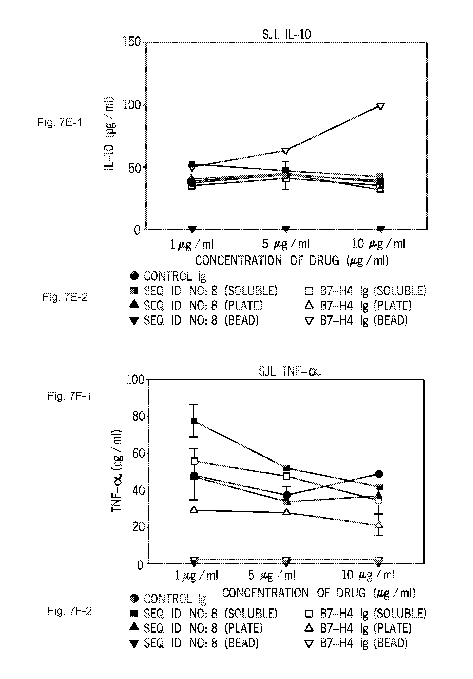
D00014

D00015

D00016

D00017

D00018

D00019

D00020

D00021

D00022

D00023

D00024

D00025

D00026

D00027

D00028

D00029

D00030

D00031

D00032

D00033

D00034

D00035

D00036

D00037

D00038

D00039

D00040

D00041

D00042

D00043

D00044

D00045

D00046

D00047

D00048

D00049

D00050

D00051

D00052

D00053
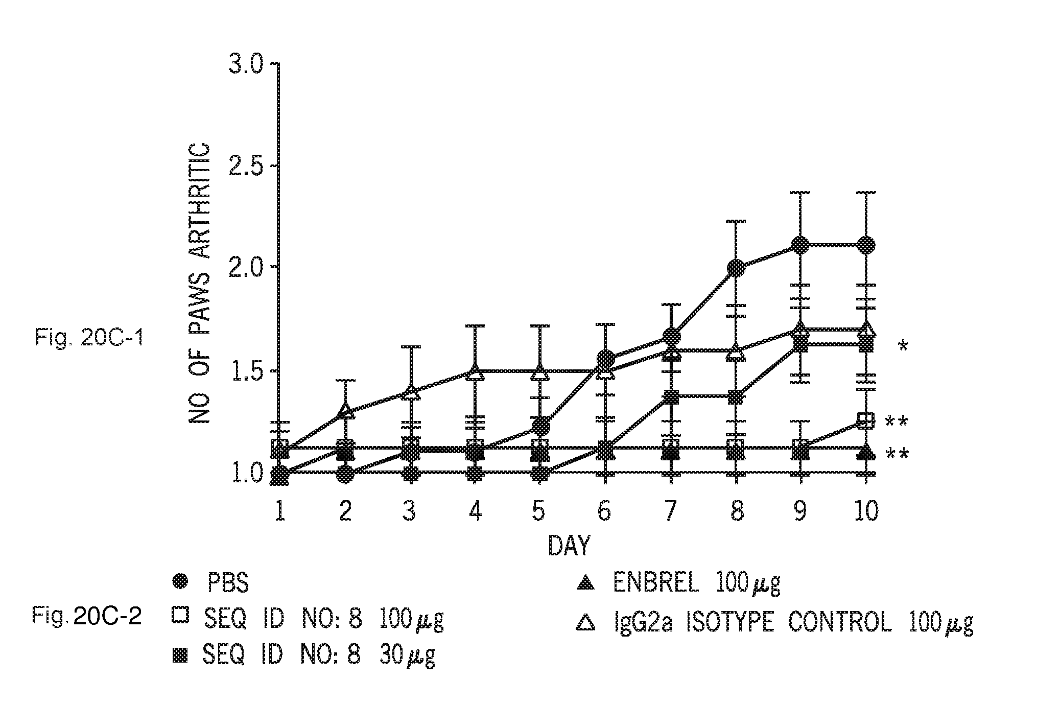
D00054

D00055

D00056
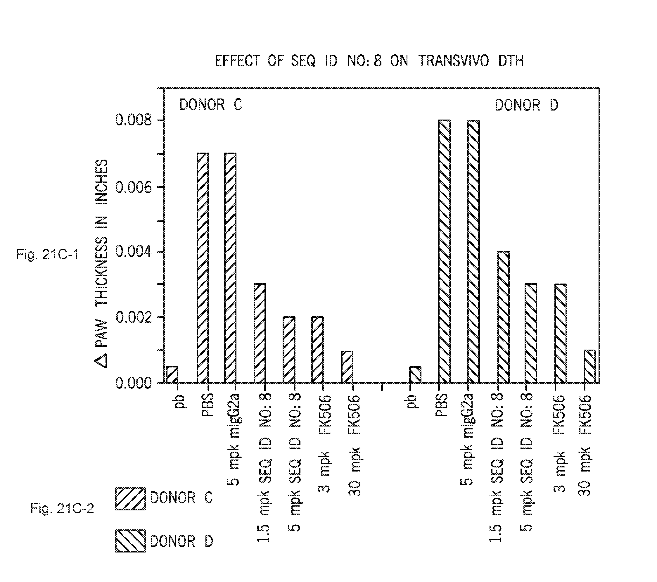
D00057

D00058

D00059

S00001
XML
uspto.report is an independent third-party trademark research tool that is not affiliated, endorsed, or sponsored by the United States Patent and Trademark Office (USPTO) or any other governmental organization. The information provided by uspto.report is based on publicly available data at the time of writing and is intended for informational purposes only.
While we strive to provide accurate and up-to-date information, we do not guarantee the accuracy, completeness, reliability, or suitability of the information displayed on this site. The use of this site is at your own risk. Any reliance you place on such information is therefore strictly at your own risk.
All official trademark data, including owner information, should be verified by visiting the official USPTO website at www.uspto.gov. This site is not intended to replace professional legal advice and should not be used as a substitute for consulting with a legal professional who is knowledgeable about trademark law.