Therapeutic Agents for Neurological and Psychiatric Disorders
DE WIT; Joris ; et al.
U.S. patent application number 16/318480 was filed with the patent office on 2019-07-25 for therapeutic agents for neurological and psychiatric disorders. This patent application is currently assigned to VIB VZW. The applicant listed for this patent is KATHOLIEKE UNIVERSITEIT LEUVEN, K.U.LEUVEN R&D, VIB VZW. Invention is credited to Bart DE STROOPER, Joris DE WIT, Heather RICE.
| Application Number | 20190225662 16/318480 |
| Document ID | / |
| Family ID | 56511366 |
| Filed Date | 2019-07-25 |


View All Diagrams
| United States Patent Application | 20190225662 |
| Kind Code | A1 |
| DE WIT; Joris ; et al. | July 25, 2019 |
Therapeutic Agents for Neurological and Psychiatric Disorders
Abstract
The present invention relates to the field of disorders of the central nervous system, in particular neurological and psychiatric disorders, and the prevention and/or treatment thereof. In particular, the present invention relates to the finding that soluble amyloid precursor protein .alpha. (sAPP.alpha.) presents a particular binding site, which allows for binding to the GABA.sub.BR1a receptor, thereby causing an agonistic effect through specific binding to Sushi domain 1 of GABA.sub.BR1a. As a result, the frequencies of excitatory and inhibitory postsynaptic currents are reduced. Accordingly, the invention provides compounds able to interfere with the association of sAPP.alpha. with Sushi domain 1 of GABA.sub.BR1a and as such with selective impairment of GABA.sub.BR1a beneficial in neurological and psychiatric disorders. The invention as well provides methods and (high content) screening assays for the production of said compounds.
| Inventors: | DE WIT; Joris; (Kessel-Lo, BE) ; RICE; Heather; (Leuven, BE) ; DE STROOPER; Bart; (Leuven, BE) | ||||||||||
| Applicant: |
|
||||||||||
|---|---|---|---|---|---|---|---|---|---|---|---|
| Assignee: | VIB VZW Gent BE Katholieke Universiteit Leuven, K.U.Leuven R&D Leuven BE |
||||||||||
| Family ID: | 56511366 | ||||||||||
| Appl. No.: | 16/318480 | ||||||||||
| Filed: | July 14, 2017 | ||||||||||
| PCT Filed: | July 14, 2017 | ||||||||||
| PCT NO: | PCT/EP2017/067859 | ||||||||||
| 371 Date: | January 17, 2019 |
| Current U.S. Class: | 1/1 |
| Current CPC Class: | G01N 2500/02 20130101; G01N 2500/04 20130101; G01N 33/566 20130101; G01N 33/5008 20130101; A61K 38/00 20130101; C07K 14/4711 20130101; C07K 14/00 20130101 |
| International Class: | C07K 14/47 20060101 C07K014/47; G01N 33/566 20060101 G01N033/566; G01N 33/50 20060101 G01N033/50 |
Foreign Application Data
| Date | Code | Application Number |
|---|---|---|
| Jul 20, 2016 | EP | 16180433.1 |
Claims
1. A polypeptide comprising a sequence having at least 95% sequence identity with SEQ ID NO: 1.
2. A fragment of the polypeptide of claim 1, the peptide fragment consisting of an amino acid sequence having at least 95% sequence identity to SEQ ID NO:2 or SEQ ID NO: 3.
3. A peptidomimetic which mimics the polypeptide of claim 1.
4. A molecule comprising the polypeptide of claim 1, the molecule further comprising a half-life extension entity and/or an entity that facilitates crossing of the blood brain barrier.
5. The polypeptide of claim 1, further comprising at least one D-alanine at the N-terminus and/or the C-terminus.
6.-8. (canceled)
9. A method of treating cognitive impairments, anxiety, depression, epilepsy, dystonia, neuropathic pain, narcolepsy or spasticity, the method comprising: administering to a subject suffering from cognitive impairments, anxiety, depression, epilepsy, dystonia, neuropathic pain, narcolepsy or spasticity the polypeptide of claim 1.
10. A method of detecting a test compound that modulates the activity of a GABA.sub.B1a receptor, the method comprising: a. b. administering the test compound to a cell expressing a functional GABA.sub.B1a receptor, c. detecting binding of the test compound to a sushi domain 1 of the GABA.sub.B1a receptor, and detecting a statistically significant change in the activity of the GABA.sub.B1a receptor in the presence of the test compound as compared to the activity of the GABA.sub.B1a receptor in the absence of the test compound.
11. A method to detecting an inhibitor of sAPP.alpha. binding to a GABA.sub.B1a receptor, the method comprising: a. In cells expressing a functional GABA.sub.B1a receptor, exposing the GABA.sub.B1a receptor to the polypeptide of claim 1 and measuring a reduction in excitatory postsynaptic currents (EPSC) frequency and/or inhibitory postsynaptic currents (IPSC) frequency by sAPP.alpha. or fragments thereof; b. applying a test compound to said cells; c. detecting that the test compound blocks the reduction in EPSC frequency or IPSC frequency.
12. The method according to claim 10, wherein the cell is selected from a recombinant cell, a neuronal cell, and a primary neuron.
13. The method according to claim 12, wherein the primary neuron is present in an acute brain slice obtained from a non-human mammal.
14. The method according to claim 10, wherein the statistically significant change in activity is an increase in the activity of the receptor.
15. The method according to claim 10, wherein the statistically significant change in activity is a reduction in the activity of the receptor.
16. The method according to claim 10, wherein the activity of the GABA.sub.BR1a receptor is detected via calcium release, synaptic transmission, and/or cAMP.
Description
FIELD OF THE INVENTION
[0001] The present invention relates to the field of disorders of the central and peripheral nervous system, in particular neurological and psychiatric disorders, and the prevention and/or treatment thereof. In particular, the present invention relates to the finding that soluble amyloid precursor protein .alpha. (sAPP.alpha.) presents a particular binding site, which allows for binding to the GABA.sub.BR1a receptor, thereby causing an agonistic effect through specific binding to sushi domain 1 of GABA.sub.BR1a. As a result, the frequencies of excitatory and inhibitory postsynaptic currents are reduced. Accordingly, the invention provides compounds able to interfere with the association of sAPP.alpha. with Sushi domain 1 of GABA.sub.BR1a and as such with selective impairment of GABA.sub.BR1a beneficial in neurological and psychiatric disorders. The invention as well provides methods and (high content) screening assays for the production of said compounds.
BACKGROUND OF THE INVENTION
[0002] GABA.sub.B receptors are the G-protein coupled receptors for .gamma.-aminobutyric acid (GABA), the main inhibitory neurotransmitter in the CNS. GABA.sub.B receptors mediate pre- and postsynaptic inhibition in the nervous system and are implicated in a variety of neurological and psychiatric disorders, including cognitive impairments, anxiety, depression, schizophrenia, epilepsy, obsessive compulsive disorder, addiction and pain (Calver et al., Neurosignals 11, 2002; Bettler et al., Physiol Rev 84, 2004).
[0003] Presynaptic GABA.sub.B receptors inhibit the release of GABA (autoreceptors) and other neurotransmitters (heteroreceptors), while postsynaptic GABA.sub.B receptors inhibit neuronal excitability by activating K+ channels. Receptor subtypes are based on the subunit isoforms GABA.sub.B1a and GABA.sub.B1b, both of which combine with GABA.sub.B2 subunits to form two heteromeric receptors, GABA.sub.B(1a, 2) and GABA.sub.B(1b, 2) (Marshall et al., Trends Pharmacol Sci 20, 1999). Most if not all neurons in the CNS coexpress GABA.sub.B(1a, 2) and GABA.sub.B(1b, 2) receptors. The GABA.sub.B1a and GABA.sub.B1b subunit isoforms derive from the same gene by alternative promoter usage and solely differ in their N-terminal ectodomains (Kaupmann et al., Nature 386, 1997; Steiger et al., J Neurosci 24, 2004). GABA.sub.B1a contains at its N terminus two sushi domains (SDs) that are lacking in GABA.sub.B1b (Hawrot et al., FEBS Lett 432, 1998). SDs, also known as complement control protein modules or short consensus repeats, are conserved protein interaction motifs present in proteins of the complement system, in adhesion molecules and in G-protein-coupled receptors (Lehtinen et al., J Mol Biol 344, 2004; Perrin et al., Acad Sci 1070, 2006).
[0004] Pharmacological tools that distinguish GABA.sub.B(1a, 2) and GABA.sub.B(1b, 2) receptors are lacking; however, the native roles of GABA.sub.B1a and GABA.sub.B1b were dissociated using GABA.sub.B1a-/-(1a-/-) and GABA.sub.B1b-/-(1b-/-) mice, which express one or the other isoform (Vigot, R. et al, Neuron 50, 2006). These mice revealed that heteroreceptors incorporate the GABA.sub.B1a subunit, whereas autoreceptors and postsynaptic GABA.sub.B receptors incorporate GABA.sub.B1a or GABA.sub.B1b subunits (Vigot, R. et al, Neuron 50, 2006; Shaban, H. et al., Nat. Neurosci. 9, 2006; Ulrich, D., and Bettler, B., Curr. Opin. Neurobiol. 17, 2007). This suggests that the SDs of GABA.sub.B1a bind to protein(s) that localize heteroreceptors at glutamatergic terminals.
[0005] The non-selective impairment of GABA.sub.B1a and GABA.sub.B1b receptors by baclofen as the prototypical GABA.sub.BR agonist in clinical use has been described by Jacobson et al. (Jacobson et al., J Neurosci., 2006). This study suggests that GABA.sub.B1a and GABA.sub.B1b isoforms are functionally relevant molecular variants of the GABA.sub.B1 receptor subunit, which are differentially involved in specific neurophysiological processes and behaviors. The GABA.sub.B receptor agonists baclofen and .gamma.-hydroxybutyrate are unable to pharmacologically discriminate between the two isoforms though. Despite the reported involvement of GABA.sub.B receptors in mental health disorders, the clinical use of GABA.sub.BR agonists is currently limited to the treatment of narcolepsy, neuropathic pain, spasticity and dystonia (Gassmann, M. and Bettler, B., Nature Reviews Neuroscience 13, 2012). One reason for this is that the main therapeutic effect of baclofen is muscle relaxation, which is an unwanted, adverse effect for psychiatric indications. As the sequence of the GABA.sub.B1a and GABA.sub.B1b isoforms differ primarily in the N terminus, and not in the region coding for the ligand binding domain, there is a need for future studies focusing on strategies to uncover novel interaction sites at either receptor isoform to enable specific pharmaceutical intervention.
[0006] We recently unraveled how to specifically modulate the GABA.sub.B1a receptor. Surprisingly, we found that the soluble amyloid precursor protein .alpha. (sAPP.alpha.) is able to activate this receptor. Our findings showed that sAPP.alpha. specifically interacts with the sushi domain 1 of GABA.sub.BR1a via its extension domain. We successfully minimized the number of amino acids being crucial for interaction and based on this epitope, specific agonists, antagonists and modulators of the GABA.sub.B1a receptor can be provided for the first time. Our approach displays a major step forward in the development of specific modulators of the GABA.sub.B1a receptor and can directly be utilized for the development of novel therapeutics for the treatment of neurological and psychiatric disorders.
BRIEF DESCRIPTION OF THE FIGURES
[0007] The following drawings are illustrative of one or more embodiments of the invention and not illustrative of the invention.
[0008] FIG. 1: APP.alpha. interacts with GABA.sub.BR1a. A-B. Schematic representation of GABA.sub.BR subunits and isoforms. GABA.sub.BR1a (A) differs from GABA.sub.BR1b (B) by the presence of the sushi motifs. C-D. Confocal images (C) and quantification of the interaction between Fc-sAPP.alpha. and GABA.sub.BR isoforms. Purified Fc-sAPP.alpha. were exogenously applied to GABA.sub.BR-transfected HEK293 cells and bound Fc-sAPP.alpha. was determined by immunofluorescence. Fc-sAPP.alpha. only interacts with GABA.sub.BR1a and the sushi domains in GABA.sub.BR1a mediate its binding to the APP ectodomain. E. Immunoblot of rat brain fractionations probed for APP family members and pre-(Syp) or post-(PSD-95 and NR2A) synaptic markers. F. Super-resolution images of mouse hippocampal sections immunostained for APP with presynaptic (VGLUT1--excitatory; VGAT--inhibitory) and postsynaptic (PSD-95--excitatory; Gephyrin--inhibitory) markers.
[0009] FIG. 2: Sushi 1 of GABA.sub.BR1a is sufficient to bind sAPP.alpha.. A. Using BioLayer Interferometry we demonstrated direct binding of sAPP.alpha. to the sushi domains in GABA.sub.BR1a and that the sushi 1 domain (72 aa) is sufficient for binding sAPP.alpha.. B. Binding of purified sushi 1 domain and sAPP.alpha. proteins by ITC.
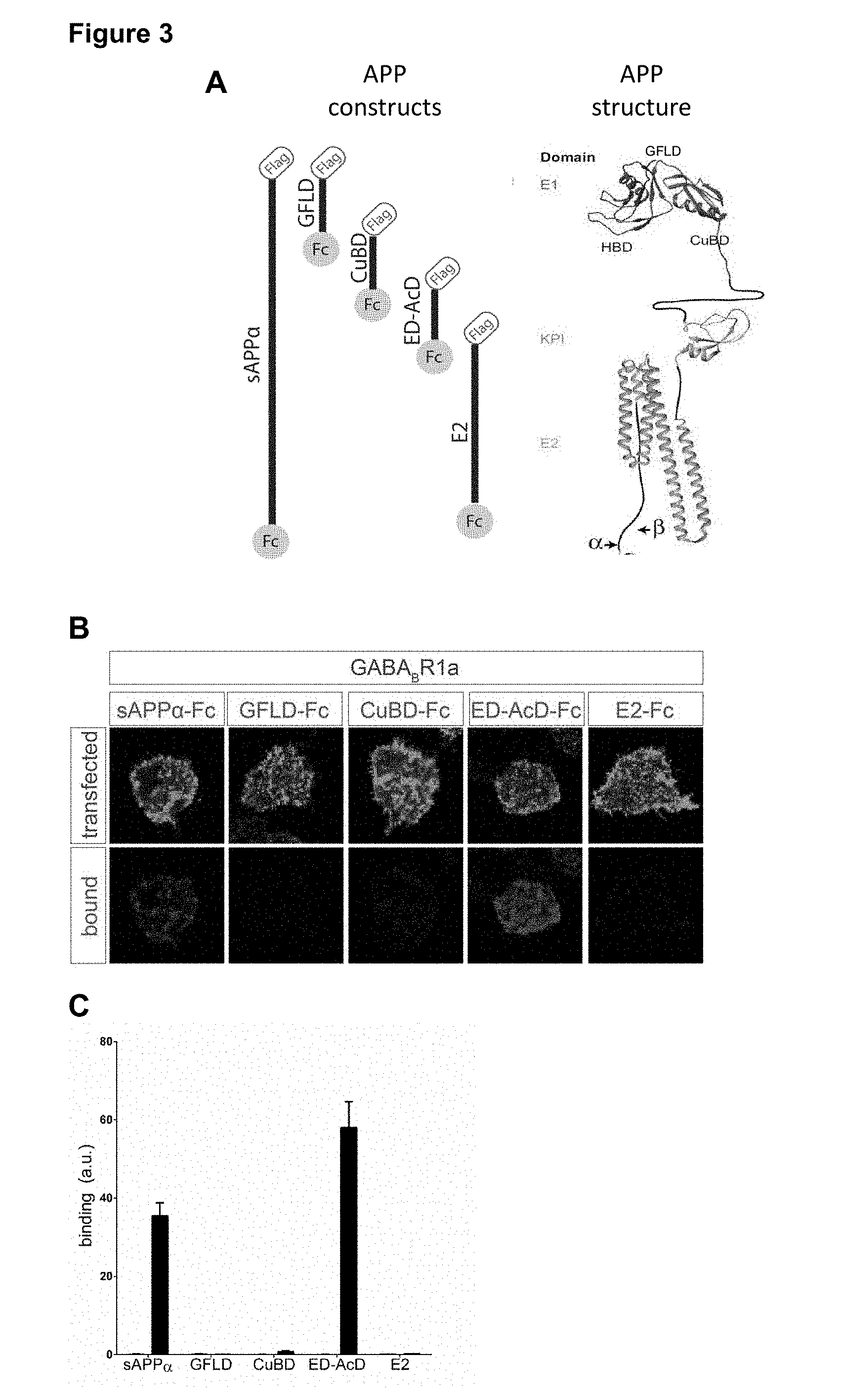
[0010] FIG. 3: Binding of the Fc-proteins of the various APP domains detected by immunofluorescence. A. Schematic representation of the different sAPP.alpha. domains, APP construct and APP structure. B-C. Confocal images (Bb) and quantifications (C) of immunostaining for sAPP.alpha.-Fc, GELD-Fc, CuBD-Fc, ED-AcD-Fc, or E2-Fc (red) binding to GFP- or GABA.sub.BR1a-expressing HEK293 cells (green) (n=16-32). We did not detect binding of the growth factor like domain (GELD), Copper binding domain (CURD), or E2 domain while interaction of the extension domain-acidic region (ED-AcD) of APP with GABA.sub.BR1a was demonstrated.
[0011] FIG. 4: Binding affinity of the different APP.alpha. domains to GABA.sub.BR1a. A. Schematic representation of the different sAPP.alpha. domains. B-C Confocal images (B) and quantifications (C) of immunostaining for sAPP.alpha. ED-AcD-Fc, ED-Fc or AcD-Fc (red) binding to GFP- or GABA.sub.BR1a expressing HEK293 cells (green) (n=14-16). The extension domain-acidic region (ED-AcD) of APP shows the highest interaction.
[0012] FIG. 5: The extension domain (ED or ExD) of APP.alpha. specifically interacts with GABA.sub.BR1a. A-B Confocal images (A) and quantifications (B) of immunostaining for sAPP.alpha.-Fc or sAPP.alpha..DELTA.ExD-Fc (red) binding to GFP- or GABA.sub.BR1a-expressing HEK293 cells (green) (n=26). The 33 aa extension domain was sufficient for binding.
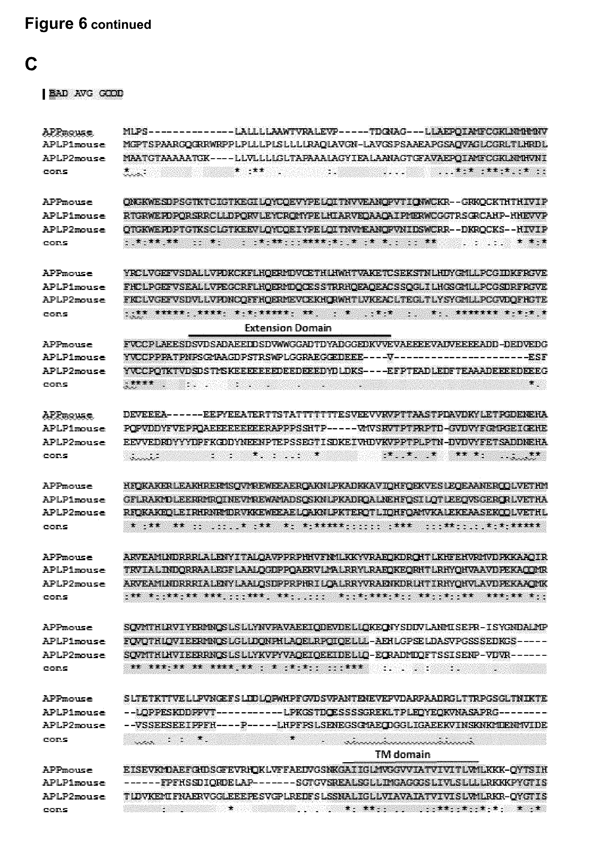
[0013] FIG. 6: APP.alpha. but not its family members specifically interacts with GABA.sub.BR1a. A-B, Confocal images (A) and quantifications (B) of immunostaining for sAPP.alpha.-Fc, sAPLP1-Fc, of sAPLP2-Fc binding to GFP or GABA.sub.BR1a expressing HEK293 cells (green) (n=24). C. Sequence alignment of APP, APLP1 and APLP2. D. Sequence alignment for the extension domain of human APP with APLPs and with 7 vertebrate APP sequences.
[0014] FIG. 7, A. Binding of purified sushi1 and sAPP.alpha. proteins by ITC. B-D. ITC binding experiments of purified sushi 1 and synthetic peptides within the ExD corresponding (B) 204-220AA (C) 204-212AA, or (D) 211-220AA of APP695. (Error bars represent s.e.m. The number of cells from 2-4 independent experiments is defined by n. Two-way ANOVA with Bonferroni's post hoc analysis; ***P<0.001).
[0015] FIG. 8, sAPP.alpha. reduces presynaptic release via GABA.sub.BRs in cultured hippocampal neurons. a,b, Example traces of mEPSCs (green arrowheads) and MIPSCs (red arrowheads) (a) and average mEPSC frequency normalized to baseline (b) recorded from wild type neurons before (baseline) and after treatment with baclofen, a GABA.sub.BR agonist. (n=12, paired t-test). c-e, Example traces of mEPSCs (green arrowheads) and mIPSCs (red arrowheads) (c) and average mEPSC frequency (d) and amplitude (e) normalized to baseline recorded from wild type neurons before (baseline) and after treatment with sAPP.alpha., (n=13, paired t-test). f,g, Example traces of mEPSCs (green arrowheads) and mIPSCs (red arrowheads) (f) and quantification of the effect of sAPP.alpha. on mEPSC frequency normalized to baseline (g) either without (blue) or with (green) preincubation with CGP55845 (CGP), a GABA.sub.BR antagonist. Dotted line denotes baseline. (n=14-17, unpaired t-test). h,i, Example traces of mEPSCs (green arrowheads) and mIPSCs (red arrowheads) and average mEPSC frequency normalized to baseline (i) recorded from wildtype neurons before (baseline) and after treatment with either sAPP.alpha. ExD-AcD, 17mer (APP695 204-220AA), sAPP.alpha..DELTA.ExD, or sAPLP1. (n=17-20, one way ANOVA with Dunnett's post hoc analysis). j,k, Average mEPSC frequency normalized to baseline recorded from App/Ap1p1 dKO primary hippocampal neurons before (baseline) and after treatment with either sAPP.alpha. (j) or baclofen (k). Dotted line denotes effect in wildtype neurons. (n=14, paired t-test). 1, High-magnification .DELTA.F images before and after application of 1 .mu.M sAPP.alpha.. m,n, Representative .DELTA.F histograms before (control, Cnt) and after either sAPP.alpha. (m) or sAPP.alpha..DELTA.ED (n) application. o, Summary of the dose-dependent inhibitory effect of sAPP.alpha. on the presynaptic vesicle recycling (S, normalized to Cnt). (N=5-8, one way ANOVA analysis with post hoc Tukey's analysis). p, High-magnification .DELTA.F images before and after application of sAPP.alpha. in the presence of a GABA.sub.BR. antagonist, CGP54626 (CGP). q. Representative .DELTA.F histograms before and after application of 1 .mu.M sAPP.alpha. in the presence of CGP. r, Summary of sAPP.alpha. effect on presynaptic vesicle recycling in hippocampal neurons with (N=8) or without (N=8) COP (normalized to Cnt). (Error bars represent s.e.m. The number of neurons is defined as n29 from 2 (b,k) or 3 (d,e,g,I,j) independent experiments. The number of experiments is defined by N (o,r). ***P<0.001).
[0016] FIG. 9, sAPP.alpha. reduces mIPSC frequency via GABA.sub.BRs in cultured hippocampal neurons. a, Average mIPSC frequency normalized to baseline recorded from wild type neurons before (baseline) and after treatment with baclofen, a GABA.sub.BR agonist. (n=12, paired t-test). b,c, Average mIPSC frequency (b) and amplitude (c) normalized to baseline recorded from wild type neurons before (baseline) and after treatment with sAPP.alpha., (n=13, paired t-test). d. Quantification of the effect of sAPP.alpha. on mIPSC frequency normalized to baseline either without (blue) or with (green) preincubation with CGP55845 (CGP), a GABA.sub.BR antagonist. Dotted line denotes baseline. (n=14-17, unpaired t-test). e, Average mIPSC frequency normalized to baseline recorded from wildtype neurons before (baseline) and after treatment with either sAPP.alpha. ExD-AcD, 17mer (APP695 204-220AA), sAPP.alpha..DELTA.ExD, or sAPLP1. (n=17-20, one way ANOVA with Dunnett's post hoc analysis). f,g, Average mIPSC frequency normalized to baseline recorded from App/Ap1p1 dKO primary hippocampal neurons before (baseline) and after treatment with either sAPP.alpha. (j) or baclofen (k). Dotted line denotes effect in wildtype neurons. (n=14, paired t-test). (Error bars represent s.e.m. The number of neurons is defined as n from 2 (a,g) or 3 (b,c,d,e,f) independent experiments. *P<0.05; ***P<0.001).
[0017] FIG. 10. sAPP.alpha. reduces basal synaptic transmission and increases short-term plasticity via GABA.sub.BR at Schaffer collaterals. a,b, Representative traces of fEPSPs (a) and input-output curves (b) recorded at SCs from hippocampal slices incubated without (grey) or with sAPP.alpha. (blue). (Cnt, n=9, N=7: sAPP.alpha., n=12, N=7). c-e, Representative traces (upper) and average fEPSP amplitude (lower) in response to high-frequency burst stimulation at 20 Hz (c), 50 Hz (d), and 100 Hz (e) (for each frequency: n=10, N=7 for Cnt; n=12, N=7 for sAPP.alpha.) in slices incubated without (grey) or with sAPP.alpha. (blue). fEPSPs were normalized to the peak amplitude of the first response. f,g, Representative traces of fEPSPs (f) and input-output curves (g) recorded from hippocampal slices incubated with CGP 54626 (CGP) alone (grey) and slices incubated with CGP+sAPP.alpha. (green). (CGP, n=9, N=4; CGP+sAPP.alpha., n=8, N=4). h-j, Representative traces (upper) and average fEPSP amplitude (lower) in response to high-frequency burst stimulation at 20 Hz (h), 50 Hz (i), and 100 Hz (j) (for each frequency: n=9, N=4 for CGP; n=8, N=4 for CGP+sAPP.alpha.) from slices incubated with CGP alone (grey) or with CGP+sAPP.alpha. (green). fEPSPs were normalized to the peak amplitude of the first response. (Error bars shown represent s.e.m. The number of slices is defined by n, the number of mice by N. Two-way ANOVA analysis; *P<0.05; **P<0.01; ***P<0.001).
DETAILED DESCRIPTION
[0018] The present invention will be described with respect to particular embodiments and with reference to certain drawings but the invention is not limited thereto but only by the claims. Any reference signs in the claims shall not be construed as limiting the scope. The drawings described are only schematic and are non-limiting. In the drawings, the size of some of the elements may be exaggerated and not drawn on scale for illustrative purposes. Where the term "comprising" is used in the present description and claims, it does not exclude other elements or steps. Where an indefinite or definite article is used when referring to a singular noun e.g. "a" or "an", "the", this includes a plural of that noun unless something else is specifically stated. Furthermore, the terms first, second, third and the like in the description and in the claims, are used for distinguishing between similar elements and not necessarily for describing a sequential or chronological order. It is to be understood that the terms so used are interchangeable under appropriate circumstances and that the embodiments of the invention described herein are capable of operation in other sequences than described or illustrated herein.
[0019] The following terms or definitions are provided solely to aid in the understanding of the invention. Unless specifically defined herein, all terms used herein have the same meaning as they would to one skilled in the art of the present invention. Practitioners are particularly directed to Sambrook et al., Molecular Cloning: A Laboratory Manual, 4.sup.th ed., Cold Spring Harbor Press, Plainsview, N.Y. (2012); and Ausubel et al., Current Protocols in Molecular Biology (Supplement 114), John Wiley & Sons, New York (2016), for definitions and terms of the art. The definitions provided herein should not be construed to have a scope less than understood by a person of ordinary skill in the art.
[0020] "Homologue", "Homologues" of a protein encompass peptides, oligopeptides, polypeptides, proteins and enzymes having amino acid substitutions, deletions and/or insertions relative to the unmodified protein in question and having similar biological and functional activity as the unmodified protein from which they are derived.
[0021] The term "amino acid identity" as used herein refers to the extent that sequences are identical on an amino acid-by-amino acid basis over a window of comparison. Thus, a "percentage of sequence identity" is calculated by comparing two optimally aligned sequences over the window of comparison, determining the number of positions at which the identical amino acid residue (e.g., Ala, Pro, Ser, Thr, Gly, Val, Leu, Ile, Phe, Tyr, Trp, Lys, Arg, His, Asp, Glu, Asn, Gln, Cys and Met) occurs in both sequences to yield the number of matched positions, dividing the number of matched positions by the total number of positions in the window of comparison (i.e., the window size), and multiplying the result by 100 to yield the percentage of sequence identity.
[0022] By "isolated" is meant material that is substantially or essentially free from components that normally accompany it in its native state. For example, a "peptide sequence", as used herein, refers to a peptide, which has been purified from the molecules which flank it in a naturally-occurring state, e.g., an extension domain of APP which has been removed from the molecules present in the production host that are adjacent to said peptide. An isolated extension domain of APP can be generated by amino acid chemical synthesis or can be generated by recombinant production. Another example concerns an isolated neuronal cell, which refers to a neuronal cell which has been extracted and purified from the naturally-occurring state, involving tissue. An isolated neuronal cell preparation can be obtained from several neuronal tissue types using for example specialized commercial kits that make use of proteases to digest intercellular protein junctions followed by gentle mechanical disruption to liberate individual cells, or for instance but not limited to the exemplified method.
[0023] The term "treatment" or "treating" or "treat" can be used interchangeably and are defined by a therapeutic intervention that slows, interrupts, arrests, controls, stops, reduces, or reverts the progression or severity of a sign, symptom, disorder, condition, or disease, but does not necessarily involve a total elimination of all disease-related signs, symptoms, conditions, or disorders. However, it will be understood that the aforementioned terms do not imply that symptoms are present.
[0024] The terms "subject" as used herein, refers to any subject, particularly a vertebrate subject, and even more particularly a mammalian subject, for whom therapy or prophylaxis is desired. A "subject" as used herein refers to an animal that can develop neurological and psychiatric disorders. Typically, the animal is a mammal. Most particularly, the subject is a human.
[0025] The term "antibody" as used herein, refers to an immunoglobulin molecule which specifically binds with an antigen. Antibodies can be intact immunoglobulins derived from natural sources or from recombinant sources and can be immunoreactive portions of intact immunoglobulins.
[0026] Antibodies are typically tetramers of immunoglobulin molecules. The antibodies in the present invention may exist in a variety of forms including, for example, polyclonal antibodies, monoclonal antibodies, Fv, Fab and F(ab).sub.2, as well as single chain antibodies and humanized antibodies (Harlow et al., 1999, In: Using Antibodies: A Laboratory Manual, Cold Spring Harbor Laboratory Press, NY; Harlow et al., 1989, In: Antibodies: A Laboratory Manual, Cold Spring Harbor, N.Y.; Worn and Pluckthun, 2001; Koerber et al., 2015.).
[0027] By the term "specifically binds," as used herein with respect to an antibody, is meant an antibody which recognizes a specific antigen, but does not substantially recognize or bind other molecules in a sample. For example, an antibody that specifically binds to an antigen from one species may also bind to that antigen from one or more species. But, such cross-species reactivity does not itself alter the classification of an antibody as specific. In some instances, the terms "specific binding" or "specifically binding," can be used in reference to the interaction of an antibody, a protein, or a peptide with a second chemical species, to mean that the interaction is dependent upon the presence of a particular structure (e.g., an antigenic determinant or epitope) on the chemical species; for example, an antibody recognizes and binds to a specific protein structure rather than to proteins generally. If an antibody is specific for epitope "A", the presence of a molecule containing epitope A (or free, unlabeled A), in a reaction containing labeled "A" and the antibody, will reduce the amount of labeled A bound to the antibody.
[0028] A "single-domain antibody", herein referred to as "nanobody", is an antibody fragment consisting of a single monomeric variable antibody domain. Like a whole antibody, it is able to bind selectively to a specific antigen. With a molecular weight of only 12-15 kDa, single-domain antibodies are much smaller than common antibodies (150-160 kDa) which are composed of two heavy protein chains and two light chains, and even smaller than Fab fragments (.about.50 kDa, one light chain and half a heavy chain) and single-chain variable fragments (.about.25 kDa, two variable domains, one from a light and one from a heavy chain).
[0029] As used herein, the term "method" comprises a "high content screening (HCS)" of suitable test compounds. In some instances, HCS is a screening method that uses an in vitro system to perform a series of experiments as the basis for high throughput compound discovery. Typically, HCS is an automated system to enhance the throughput of the screening process. However, the present invention is not limited to the speed or automation of the screening process. In another embodiment of the invention, the HCS assay provides for a high throughput assay. Preferably, the assay provides automated screening of thousands of test compounds.
[0030] "Compound" means any chemical or biological compound, including simple or complex organic and inorganic molecules, peptides, peptido-mimetics, proteins, antibodies, carbohydrates, nucleic acids or derivatives thereof. The term "compound" or "test compound" is used herein in the context of a "drug candidate compound" or a "candidate compound for Lead optimization" in therapeutics, described in connection with the methods of the present invention. As such, these compounds comprise organic or inorganic compounds, derived synthetically or from natural resources. The compounds include polynucleotides, lipids or hormone analogs that are characterized by low molecular weights. Other biopolymeric organic test compounds include small peptides or peptide-like molecules (peptidomimetics) comprising from about 2 to about 40 amino acids and larger polypeptides comprising from about 40 to about 500 amino acids, such as antibodies or antibody conjugates.
[0031] It is an object of the invention to provide therapeutic agents, particularly pharmaceutical compositions, comprising compounds specifically impairing the GABA.sub.B1a receptor. Additional compounds can be identified by monitoring the activity of a functional GABA.sub.B1a receptor expressed in a cell when a test compound is administered to said cell. Therefore, according to a first aspect, the application provides a peptide as depicted in SEQ ID NO: 1 or a homologue thereof of at least 95% amino acid identity. SEQ ID NO: 1 depicts the amino acid sequence of the extension domain of human sAPP.alpha..
TABLE-US-00001 SEQ ID NO: 1: Extension domain of human sAPP.alpha. (33 amino acids): NVDSADAEEDDSDVWWGGADTDYADGSEDKVVE
[0032] According to one embodiment, the peptide sequence depicted in SEQ ID NO: 1 or a homologue thereof of at least 95% amino acid identity refers to an isolated peptide. According to another embodiment, a peptide sequence depicted in SEQ ID NO: 1 or a homologue thereof of at least 95% amino acid identity refers to an isolated peptide obtained by purification from APP. In another embodiment a peptide sequence depicted in SEQ ID NO: 1 or a homologue thereof of at least 95% amino acid identity is generated by chemical amino acid synthesis. According to another embodiment SEQ ID NO: 1 or a homologue thereof of at least 95% amino acid identity is generated by recombinant production.
[0033] In a second aspect, the application provides a peptide fragment derived from a peptide as depicted in SEQ ID NO: 1 or a homologue thereof of at least 95% amino acid identity, said peptide fragment consist of a sequence as depicted in SEQ ID NO: 2 or a homologue thereof of at least 95% amino acid identity or as depicted in SEQ ID NO: 3 or a homologue thereof of at least 95% amino acid identity. SEQ ID NO: 2 depicts a 17 amino acid long sequence fragment (D204-G220) of the extension domain of human sAPP.alpha., while SEQ ID NO: 3 depicts a 9 amino acid long sequence fragment (D204-G212) of the extension domain of human sAPP.alpha..
TABLE-US-00002 SEQ ID NO: 2: Fragment (D204-G220) of the extension domain of human sAPP.alpha.: DDSDVWWGGADTDYADG SEQ ID NO: 3: Fragment (D204-G212) of the extension domain of human sAPP.alpha.: DDSDVWWGG SEQ ID NO: 4: Fragment (G210-G220) of the extension domain of human sAPP.alpha.: GGADTDYADG
[0034] In a third aspect, the application provides a peptidomimetic generated from the peptide as depicted in SEQ ID NO: 1 or a homologue thereof of at least 95% amino acid identity. In one embodiment the application provides a peptidomimetic generated from a peptide fragment derived from the peptide as depicted in SEQ ID NO: 1 or a homologue thereof of at least 95% amino acid identity, said peptide fragment consists of a sequence as depicted in SEQ ID NO: 2 or a homologue thereof of at least 95% amino acid identity or as depicted in SEQ ID NO: 3 or a homologue thereof of at least 95% amino acid identity.
[0035] According to the present application, the degree of identity, between a given reference amino acid sequence and an amino acid sequence which is a homologue of said given amino acid sequence will preferably be at least 60%, 65%, 70%, 75%, 80%, 81%, 82%, 83%, 84%, 85%, 86%, 87%, 88%, 89%, 90%, 91%, 92%, 93%, 94%, 95%, 96%, 97%, 98%, or 99%. The degree of identity is given preferably for an amino acid region which is at least 10%, at least 20%, at least 30%, at least 40%, at least 50%, at least 60%, at least 70%, at least 80%, at least 90% or 100% of the entire length of the reference amino acid sequence. For example, if the reference amino acid sequence consists of 200 amino acids, the degree of identity is given preferably for at least 20, at least 40, at least 60, at least 80, at least 100, at least 120, at least 140, at least 160, at least 180, or 200 amino acids, preferably continuous amino acids. In the embodiments, the degree/percentage of similarity or identity is given for the entire length of the reference amino acid sequence.
[0036] Thus in a particular embodiment, a peptide sequence depicted in SEQ ID NO: 1 or a homologue thereof of at least 99%, 98%, 97%, 96, 95%, 94%, 93%, 92%, 91%, 90%, 89%, 88%, 87%, 86%, 85%, 84%, 83%, 82%, 81%, or 80% amino acid identity is provided. In further particular embodiments, the application provides a peptide fragment derived from the peptide sequence depicted in SEQ ID NO: 1 or a homologue thereof of at least 95% amino acid identity, said peptide sequence fragment is depicted in SEQ ID NO: 2 or a homologue thereof of at least 99%, 98%, 97%, 96, 95%, 94%, 93%, 92%, 91%, 90%, 89%, 88%, 87%, 86%, 85%, 84%, 83%, 82%, 81%, or 80% amino acid identity or as depicted in SEQ ID NO: 3 or a homologue thereof of at least 99%, 98%, 97%, 96, 95%, 94%, 93%, 92%, 91%, 90%, 89%, 88%, 87%, 86%, 85%, 84%, 83%, 82%, 81%, or 80% amino acid identity. In particular embodiments, a peptidomimetic generated from a peptide sequence as depicted in SEQ ID NO: 1 or a homologue thereof of at least 99%, 98%, 97%, 96, 95%, 94%, 93%, 92%, 91%, 90%, 89%, 88%, 87%, 86%, 85%, 84%, 83%, 82%, 81%, or 80% amino acid identity is provided. In yet another particular embodiment, a peptidomimetic generated from a peptide fragment derived from a peptide sequence as depicted in SEQ ID NO: 1 or a homologue thereof of at least 95% amino acid identity in provided, wherein said peptide sequence fragment is depicted in SEQ ID NO: 2 or a homologue thereof of at least 99%, 98%, 97%, 96, 95%, 94%, 93%, 92%, 91%, 90%, 89%, 88%, 87%, 86%, 85%, 84%, 83%, 82%, 81%, or 80% amino acid identity or as depicted in SEQ ID NO: 3 or a homologue thereof of at least 99%, 98%, 97%, 96, 95%, 94%, 93%, 92%, 91%, 90%, 89%, 88%, 87%, 86%, 85%, 84%, 83%, 82%, 81%, or 80% amino acid identity.
[0037] Peptidomimetics or non-natural peptides provide an alternative source of potent and selective Protein-Protein Interaction (PPI) modulators and occupy the chemical gap between small molecules and biologics, such as antibodies. Moreover, peptidomimetics suitably cover the chemical space required to modulate PPIs. Envisaged herein are peptidomimetics which mimic in the structure to peptide and the biological activity while bioavailability, bio-stability, bioefficiency as well as the half-life of the activity can be increased compared to the peptide they refer to. Preferably, peptidomimetics according to the invention provide enhanced bioavailability, bio-stability, bioefficiency and half-life activity when compared to the peptide they refer to. Peptidomimetics in the scope of the present invention allow for greater distribution within the target tissues such as the brain for improved therapeutic efficacy, higher stability at ambient temperature leading to better storage properties, lower cost of goods and easier regulatory clearance due to lack of issues related to purity such as contamination by cellular materials. Preferably, peptidomimetics of the present invention show high permeability across the blood-brain barrier. Peptidomimetics according to the invention offer advantages, such as high stability and low toxicity and immunogenicity, when compared to currently used therapeutic approaches.
[0038] In a fourth aspect, a molecule is provided wherein said molecule comprises the peptide as depicted in SEQ ID NO: 1 or a homologue thereof of at least 95% amino acid identity or comprises a peptidomimetic generated from said peptide as depicted in SEQ ID NO: 1 or a homologue thereof of at least 95% amino acid identity, said molecule further comprises a half-life extension entity and/or an entity that facilitates said molecule to cross the blood brain barrier. In one embodiment, a molecule is provided wherein said molecule comprises a peptide fragment derived from SEQ ID NO: 1 or a homologue thereof of at least 95% amino acid identity or comprises a peptidomimetic generated from a peptide fragment derived from SEQ ID NO: 1 or a homologue thereof of at least 95% amino acid identity, wherein said peptide fragment is depicted in SEQ ID NO:2 or a homologue thereof of at least 95% amino acid identity or depicted in SEQ ID NO:3 or a homologue thereof of at least 95% amino acid identity and wherein said molecule further comprises a half-life extension entity and/or an entity that facilitates said molecule to cross the blood brain barrier. In particular embodiments, said molecule is a chimeric molecule, a chimeric protein, a dimeric protein, a fusion protein, a composition, a combination, a peptide or a polypeptide.
[0039] In a more particular embodiment, the application provides a peptide as depicted in SEQ ID NO: 1 or a homologue from a peptide sequence depicted in SEQ ID NO: 1 or a homologue thereof of at least 95% amino acid identity comprising a molecule which increases the half-life extension. In an alternative embodiment, the application provides a peptidomimetic generated from a peptide sequence depicted in SEQ ID NO: 1 or a homologue thereof of at least 95% amino acid identity comprising a molecule which increases the half-life extension. In another particular embodiment the application provides a peptide fragment derived from a peptide sequence depicted in SEQ ID NO: 1 or a homologue thereof of at least 95% amino acid identity, wherein said peptide fragment is depicted in SEQ ID NO: 2 or a homologue thereof of at least 95% amino acid identity or depicted in SEQ ID NO:3 or a homologue thereof of at least 95% amino acid identity, said peptide fragment further comprising a molecule which increases the half-life extension. In a particular embodiment the invention provides a peptidomimetic generated from a peptide fragment derived from a peptide as depicted in SEQ ID NO: 1 or a homologue thereof of at least 95% amino acid identity, wherein said peptide fragment is depicted in SEQ ID NO: 2 or a homologue thereof of at least 95% amino acid identity or depicted in SEQ ID NO:3 or a homologue thereof of at least 95% amino acid identity, said peptidomimetic further comprising a molecule which increases the half-life extension.
[0040] Peptide sequences and peptidomimetics of the invention can be provided as such or can be prepared as a chimeric molecule, dimeric molecule or as a fusion protein. For example, they can be linked to the whole or partial Fc to express in the appropriate peptide sequences, peptidomimetics and antibodies of this invention. Peptide sequences, peptidomimetics and antibodies of this invention can be expressed in the N-terminal or C-terminal of the Fc gene. Covalently modified peptide sequences, peptidomimetics and antibodies are also included in this invention. Chemically covalent modification includes modifying N- or C-terminal or adding a chemical molecule to other amino acids. Peptides, peptidomimetics and antibodies of the invention can be fused or conjugated to any half-life extension molecule. The term "a half-life extension entity" as used above is equivalent to said half-life extension molecule. Such half-life extension molecules are known by a person skilled in the art and include, for example, not only an Fc region/domain of an immunoglobulin but also albumin, an albumin-binding domain, an immunoglobulin-binding domain, an FcRn-binding motif, and a polymer. Particularly preferred polymers include polyethylene glycol (PEG), hydroxyethyl starch (HES), hyaluronic acid, polysialic acid and PEG-mimetic peptide sequences. For modification of peptides, peptidomimetics and antibodies of the invention activated PEG with molecular weight of 5,000-100,000 can be used for the purpose of prolonging their half-life time. Detailed protocols can be seen in Greenwald et al., Bioorg. Med. Chem. Lett. 1994, 4, 2465; Caliceti et al., IL Farmaco, 1993, 48,919; Zalipsky and Lee, Polyethylene Glycol Chemistry: Biotechnical and Biomedical Applications, J. M. Harris, Plenus Press, New York (1992). Multi-arm branched PEG is preferred. Also within the scope of this application are modifications preventing aggregation of the peptides, peptidomimetics and antibodies. These modifications are also known to the skilled person and include, for example, the substitution of one or more hydrophobic amino acids, preferably surface-exposed hydrophobic amino acids, with one or more hydrophilic amino acids. In one embodiment, the peptides, peptidomimetics and antibodies according to the invention or the immunogenic variant thereof or the immunogenic fragment of any of the foregoing, comprises the substitution of up to 10, 9, 8, 7, 6, 5, 4, 3 or 2, preferably 5, 4, 3 or 2, hydrophobic amino acids, preferably surface-exposed hydrophobic amino acids, with hydrophilic amino acids. Preferably, other properties of the peptides, peptidomimetics and antibodies according to the invention, e.g., their immunogenicity, are not compromised by such substitution. Still other techniques of formulation as nanotechnology and aerosol and inhalant are also within the scope of this invention.
[0041] In a particular embodiment the invention provides a peptide as depicted in SEQ ID NO: 1 or a homologue thereof of at least 95% amino acid identity, said peptide further comprising a molecule which enables the peptide to cross the blood brain barrier.
[0042] A molecule also referred to as an element herein, which enables the peptides, peptidomimetics and antibodies of the invention to cross the blood brain barrier can be a cell penetrant carrier, wherein said cell penetrant carrier can enter a cell through a sequence which mediates cell penetration (or cell translocation). In the latter case said peptides, peptidomimetics and antibodies of the invention are modified through the recombinant or synthetic attachment of a cell penetration sequence. Thus, the molecule (e.g. as a peptide) may be further fused or chemically coupled to a sequence facilitating transduction of the fusion or chemical coupled proteins into prokaryotic or eukaryotic cells. Sequences facilitating protein transduction are known to the person skilled in the art and include, but are not limited to Protein Transduction Domains. It has been shown that a series of small protein domains, termed protein transduction domains (PTDs), cross biological membranes efficiently and independently of transporters or specific receptors, and promote the delivery of peptides and proteins into cells. Preferably, said sequence is selected from the group comprising TAT protein from human immunodeficiency virus (HIV-1), a polyarginine sequence, penetratin and a short amphipathic peptide carrier, Pep-1. Still other commonly used cell-permeable peptides (both natural and artificial peptides) are disclosed in Joliot A. and Prochiantz A. (2004) Nature Cell Biol. 6 (3) 189-193. The list of molecules enabling the peptides, peptidomimetis and antibodies of this invention to cross the blood brain barrier is not limited by the above given examples and references. Any molecule or element which enables the peptides, peptidomimetics and antibodies of the invention to cross the blood brain barrier is in the scope of the present invention. Such a molecule or element is referred to as "entity that facilitates a molecule to cross the blood brain barrier", as used above.
[0043] In a particular embodiment the invention provides a peptide fragment derived from a peptide as depicted in SEQ ID NO: 1 or a homologue thereof of at least 95% amino acid identity, said peptide fragment is depicted in SEQ ID NO: 2 or a homologue thereof of at least 95% amino acid identity, or depicted in SEQ ID NO: 3 or a homologue thereof of at least 95% amino acid identity, further comprising a molecule which enables the peptide to cross the blood brain barrier. In a more particular embodiment, said peptide fragment further comprises a molecule which increases the half-life extension of said peptide sequence.
[0044] In another particular embodiment the invention provides a peptidomimetic generated from a peptide as depicted in SEQ ID NO: 1 or a homologue thereof of at least 95% amino acid identity, said peptidomimetic further comprising a molecule which enables the peptidomimetic to cross the blood brain barrier. In a more particular embodiment, said peptidomimetic further comprises a molecule which increases the half-life extension of said peptidomimetic.
[0045] In another particular embodiment the invention provides a peptidomimetic generated from a peptide fragment derived from a peptide as depicted in SEQ ID NO: 1 or a homologue thereof of at least 95% amino acid identity, said peptide sequence fragment is depicted in SEQ ID NO: 2 or a homologue thereof of at least 95% amino acid identity or depicted in SEQ ID NO: 3 or a homologue thereof of at least 95% amino acid identity, said peptidomimetic further comprising a molecule which enables the peptidomimetic to cross the blood brain barrier. In a more particular embodiment, said peptidomimetic further comprises a molecule which increases the half-life extension of peptidomimetic.
[0046] In a fifth aspect, the application provides a peptide as depicted in SEQ ID NO: 1 or a homologue thereof of at least 95% amino acid identity further comprising at least one D-alanine at the N-terminus and/or the C-terminus. In a more particular embodiment, said peptide sequence further comprises a molecule which increases the half-life extension of said peptide sequence and/or a molecule which enables the peptide sequence to cross the blood brain barrier.
[0047] In a particular embodiment the invention provides a peptide fragment derived from a peptide sequence depicted in SEQ ID NO: 1 or a homologue thereof of at least 95% amino acid identity, said peptide fragment is depicted in SEQ ID NO: 2 or a homologue thereof of at least 95% amino acid identity or depicted in SEQ ID NO: 3 or a homologue thereof of at least 95% amino acid identity further comprising at least one D-alanine at the N-terminus and/or the C-terminus. In a more particular embodiment, said peptide fragment further comprises a molecule which increases the half-life extension of said peptide and/or a molecule which enables the peptide fragment to cross the blood brain barrier.
[0048] In another particular embodiment the invention provides a peptidomimetic generated from a peptide as depicted in SEQ ID NO: 1 or a homologue thereof of at least 95% amino acid identity further comprising at least one D-alanine at the N-terminus and/or the C-terminus. In a more particular embodiment, said peptidomimetic further comprises a molecule which increases the half-life extension of said peptidomimetic and/or a molecule which enables the peptidomimetic to cross the blood brain barrier.
[0049] In another particular embodiment the invention provides a peptidomimetic generated from a peptide fragment derived from a peptide as depicted in SEQ ID NO: 1 or a homologue thereof of at least 95% amino acid identity, said peptide fragment is depicted in SEQ ID NO: 2 or a homologue thereof of at least 95% amino acid identity, or depicted in SEQ ID NO: 3 or a homologue thereof of at least 95% amino acid identity further comprising at least one D-alanine at the N-terminus and/or the C-terminus. In a more particular embodiment, said peptidomimetic further comprises a molecule which increases the half-life extension of said peptidomimetic and/or a molecule which enables the peptidomimetic to cross the blood brain barrier.
[0050] In a sixth aspect, a nucleic acid molecule is provided, wherein said nucleic acid molecule encodes one of the above described peptides. In particular embodiments, a nucleic acid molecule is provided, wherein said nucleic acid molecule encodes one of the above described peptide fragments.
[0051] As used herein, the terms "nucleic acid", "polynucleotide", "polynucleic acid" are used interchangeably and refer to a polymeric form of nucleotides of any length, either deoxyribonucleotides or ribonucleotides, or analogs thereof. Polynucleotides may have any three-dimensional structure, and may perform any function, known or unknown. Non-limiting examples of polynucleotides include a gene, a gene fragment, exons, introns, messenger RNA (mRNA), transfer RNA, ribosomal RNA, ribozymes, cDNA, recombinant polynucleotides, branched polynucleotides, plasmids, vectors, isolated DNA of any sequence, control regions, isolated RNA of any sequence, nucleic acid probes, and primers. The polynucleotide molecule may be linear or circular. The polynucleotide may comprise a promoter, an intron, an enhancer region, a polyadenylation site, a translation initiation site, 5' or 3' untranslated regions, a reporter gene, a selectable marker or the like. The polynucleotide may comprise single stranded or double stranded DNA or RNA. The polynucleotide may comprise modified bases or a modified backbone. A nucleic acid that is up to about 100 nucleotides in length, is often also referred to as an oligonucleotide.
[0052] In a seventh aspect, the invention provides a pharmaceutical composition comprising a peptide as depicted in SEQ ID NO: 1 or a homologue thereof of at least 95% amino acid identity and a pharmaceutically acceptable carrier.
[0053] Pharmaceutical compositions comprising peptides, peptidomimetics and antibodies of the invention and a pharmaceutically acceptable carrier can be utilized to achieve the desired pharmacological effect by administration to a patient in need thereof. A patient, for the purpose of this invention, is a mammal, particularly a human, in need of treatment for the particular condition or disease. Therefore, the present invention includes pharmaceutical compositions that are comprised of a pharmaceutically acceptable carrier and a pharmaceutically effective amount of peptides, peptidomimetics and antibodies of the invention and a pharmaceutically acceptable carrier. A pharmaceutically acceptable carrier is preferably a carrier that is relatively non-toxic and innocuous to a patient at concentrations consistent with effective activity of the active ingredient so that any side effects ascribable to the carrier do not vitiate the beneficial effects of the active ingredient. A pharmaceutically effective amount of peptides, peptidomimetics and antibodies of the invention and a pharmaceutically acceptable carrier is preferably that amount which produces a result or exerts an influence on the particular condition being treated. The peptides, peptidomimetics and antibodies of the invention and a pharmaceutically acceptable carrier can be administered with pharmaceutically acceptable carriers well known in the art using any effective conventional dosage form, including immediate, slow and timed release preparations, orally, parenterally, topically, nasally, ophthalmically, intrathecally, intracerebroventricularly, sublingually, rectally, vaginally, and the like. Still other techniques of formulation as nanotechnology and aerosol and inhalant are also within the scope of this invention.
[0054] The peptides, peptidomimetics and antibodies of this invention can be used as a medicament. One skilled in the art can prepare a pharmaceutically effective formulation according to common methods, which contains effective amount of the peptide sequences, peptidomimetics and antibodies as well as pharmaceutically acceptable carriers.
[0055] When prepared as lyophilization or liquid, physiologically acceptable carrier, excipient, stabilizer need to be added into the pharmaceutical composition of the invention (Remington's Pharmaceutical Sciences 22th edition, Ed. Allen, Loyd V, Jr. (2012). The dosage and concentration of the carrier, excipient and stabilizer should be safe to the subject (human, mice and other mammals), including buffers such as phosphate, citrate, and other organic acid; antioxidant such as vitamin C, small polypeptide, protein such as serum albumin, gelatin or immunoglobulin; hydrophilic polymer such as PVP, amino acid such as amino acetate, glutamate, asparagine, arginine, lysine; glycose, disaccharide, and other carbohydrate such as glucose, mannose or dextrin, chelate agent such as EDTA, sugar alcohols such as mannitol, sorbitol; counterions such as Na+, and/or surfactant such as TWEEN.TM., PLURONICS.TM. or PEG and the like.
[0056] The preparation containing the peptides, peptidomimetics and antibodies of this invention should be sterilized before injection. This procedure can be done using sterile filtration membranes before or after lyophilization and reconstitution.
[0057] The pharmaceutical composition is usually filled in a container with sterile access port, such as an i.v. solution bottle with a cork. The cork can be penetrated by hypodermic needle.
[0058] The pharmaceutical compositions of this invention can be administrated through normal ways, including but not limited to intravenous injection or infusion, intra-abdominal injection, intracerebroventricular injection, intramuscular injection, intra-arterial injection or infusion, locally or through sustained release systems.
[0059] The dosage and concentration can be adjusted according to actual situation. One skilled in the art should know how to choose proper dosage and injection means according to actual situation. The animal experiments in this invention show credible instructions for the effective amount in human body.
[0060] The principle for adjusting between different species such as mice and human can be seen in Mordenti, J. and Chappell, W. "The use of interspecies scaling in toxicokinetics" In Toxicokinetics and New Drug Development, Yacobi et al.; Pergamon Press, New York 1989, pp. 42-96.
[0061] The dosage should be adjusted according to different injection means. Direction for certain specific dosage and way of administration can be seen in U.S. Pat. No. 4,657,760, 5,206,344 or 5,225,212.
[0062] In a specific embodiment the micro-capsule containing the peptides, peptidomimetics and antibodies of the invention can also be used as a sustained release system. The sustained release system of peptides, peptidomimetics and antibodies of this invention can be prepared with PLGA which has good biologically compatibility and degradability. Lactic acid and glycolic acid, the degrading products of PLGA, can be cleared quickly in human body. Furthermore, the degradability of the polymer can vary from several months to several years according to its molecular weight and composition (Lewis, "Controlled release of bioactive agents form lactide/glycolide polymer," in: M. Chasin and R. Langer (Eds.), Biodegradable Polymers as Drug Delivery Systems (Marcel Dekker: New York, 1990), pp. 1-41)).
[0063] In a particular embodiment the invention provides a pharmaceutical composition comprising a peptide fragment derived from a peptide as depicted in SEQ ID NO: 1 or a homologue thereof of at least 95% amino acid identity and a pharmaceutically acceptable carrier, said peptide fragment is depicted in SEQ ID NO: 2 or a homologue thereof of at least 95% amino acid identity or as depicted in SEQ ID NO: 3 or a homologue thereof of at least 95% amino acid identity.
[0064] In another particular embodiment, the application provides a pharmaceutical composition comprising a peptidomimetic and a pharmaceutically acceptable carrier, said peptidomimetic is generated from the peptide as depicted in SEQ ID NO: 1 or a homologue thereof of at least 95% amino acid identity. In one embodiment the application provides a pharmaceutical composition comprising a peptidomimetic and a pharmaceutically acceptable carrier, said peptidomimetic is generated from a peptide fragment derived from the peptide as depicted in SEQ ID NO: 1 or a homologue thereof of at least 95% amino acid identity, said peptide fragment consists of a sequence as depicted in SEQ ID NO: 2 or a homologue thereof of at least 95% amino acid identity or as depicted in SEQ ID NO: 3 or a homologue thereof of at least 95% amino acid identity.
[0065] In particular embodiments, a pharmaceutical composition is provided comprising a pharmaceutical acceptable carrier and a peptide as depicted in SEQ ID NO: 1 or a homologue thereof of at least 95% amino acid identity further comprising at least one D-alanine at the N-terminus and/or the C-terminus and/or further comprising a molecule which increases the half-life extension of said peptide and/or further comprising a molecule which enables the peptide to cross the blood brain barrier.
[0066] In other particular embodiments, the invention provides a pharmaceutical composition comprising a pharmaceutical acceptable carrier and a peptide fragment derived from a peptide as depicted in SEQ ID NO: 1 or a homologue thereof of at least 95% amino acid identity, said peptide fragment is depicted in SEQ ID NO: 2 or a homologue thereof of at least 95% amino acid identity or depicted in SEQ ID NO: 3 or a homologue thereof of at least 95% amino acid identity, said peptide fragment further comprising at least one D-alanine at the N-terminus and/or the C-terminus and/or further comprising a molecule which increases the half-life extension of said peptide and/or further comprising a molecule which enables the peptide fragment to cross the blood brain barrier.
[0067] In particular embodiments, the invention provides a pharmaceutical composition comprising a pharmaceutical acceptable carrier and a peptidomimetic, said peptidomimetic is generated from a peptide as depicted in SEQ ID NO: 1 or a homologue thereof of at least 95% amino acid identity, said peptidomimetic further comprises at least one D-alanine at the N-terminus and/or the C-terminus and/or further comprises a molecule which increases the half-life extension of said peptidomimetic and/or further comprises a molecule which enables the peptidomimetic to cross the blood brain barrier.
[0068] In another particular embodiment the invention provides a pharmaceutical composition comprising a pharmaceutical acceptable carrier and a peptidomimetic, said peptidomimetic generated from a peptide fragment derived from a peptide as depicted in SEQ ID NO: 1 or a homologue thereof of at least 95% amino acid identity, said peptide fragment is depicted in SEQ ID NO: 2 or a homologue thereof of at least 95% amino acid identity, or depicted in SEQ ID NO: 3 or a homologue thereof of at least 95% amino acid identity, said peptidomimetic further comprising at least one D-alanine at the N-terminus and/or the C-terminus and/or further comprising a molecule which increases the half-life extension of said peptidomimetic and/or further comprising a molecule which enables the peptidomimetic to cross the blood brain barrier.
[0069] In particular embodiments, a pharmaceutical composition is provided comprising a pharmaceutical acceptable carrier and one of the peptides, or peptide fragments, or peptidomimetics, or antibodies of the application.
[0070] In an eight aspect, the application provides a peptide as depicted in SEQ ID NO: 1 or a homologue thereof of at least 95% amino acid identity for use as a medicament. In one embodiment, said peptide for use as a medicament further comprises a molecule that increases the half-life extension. In another embodiment, said peptide for use as a medicament further comprises a molecule that enables said peptide to cross the blood-brain-barrier. In another embodiment, said peptide for use as a medicament further comprises at least one D-alanine at the N-terminus and/or the C-terminus. In another embodiment, said peptide for use as a medicament further comprises a molecule that increases the half-life extension and/or a molecule that enables said peptide to cross the blood-brain-barrier and/or at least one D-alanine at the N-terminus and/or the C-terminus.
[0071] In another embodiment, the invention provides a peptide fragment derived from a peptide as depicted in SEQ ID NO: 1 or a homologue thereof of at least 95% amino acid identity, said peptide fragment is depicted in SEQ ID NO: 2 or a homologue thereof of at least 95% amino acid identity, or depicted in SEQ ID NO: 3 or a homologue thereof of at least 95% amino acid identity for use as a medicament. In one particular embodiment, said peptide fragment for use as a medicament further comprises a molecule that increases the half-life extension. In another particular embodiment, said peptide fragment for use as a medicament further comprises a molecule that enables said peptide fragment to cross the blood-brain-barrier. In another particular embodiment, said peptide fragment for use as a medicament further comprises at least one D-alanine at the N-terminus and/or the C-terminus. In another embodiment, said peptide fragment for use as a medicament further comprises a molecule that increases the half-life extension and/or a molecule that enables said peptide fragment to cross the blood-brain-barrier and/or at least one D-alanine at the N-terminus and/or the C-terminus.
[0072] In another embodiment, the invention provides a peptidomimetic generated from a peptide as depicted in SEQ ID NO: 1 or a homologue thereof of at least 95% amino acid identity for use as a medicament. In another embodiment, the invention provides a peptidomimetic generated from a peptide fragment derived from a peptide as depicted in SEQ ID NO: 1 or a homologue thereof of at least 95% amino acid identity, or from a peptide as depicted in SEQ ID NO: 2 or a homologue thereof of at least 95% amino acid identity or as depicted in SEQ ID NO: 3 or a homologue thereof of at least 95% amino acid identity for use as a medicament. In particular embodiments, said peptidomimetic for use as a medicament further comprises a molecule that increases the half-life extension. In other particular embodiments, said peptidomimetic for use as a medicament further comprises a molecule that enables said peptidomimetic to cross the blood-brain-barrier. In other particular embodiment, said peptidomimetic for use as a medicament further comprises at least one D-alanine at the N-terminus and/or the C-terminus. In other embodiments, said peptidomimetic for use as a medicament further comprises a molecule that increases the half-life extension and/or a molecule that enables said peptidomimetic to cross the blood-brain-barrier and/or at least one D-alanine at the N-terminus and/or the C-terminus.
[0073] In another embodiment, peptides, peptidomimetics and antibodies according to the invention comprising a cell penetrant carrier are used as a medicament. Said medicament is needed in a therapeutically effective amount. One of ordinary skill in the art will recognize that the potency and, therefore, an "effective amount" can vary for the inhibitory agents of the present invention. One skilled in the art can readily assess the potency of the inhibitory agent. A medicament to prevent and/or to treat an individual with a neurological and/or psychiatric disorder, relates to a composition comprising agents as described in the application present and a pharmaceutically acceptable carrier or excipient (both terms can be used interchangeably) to treat or to prevent neurological and psychiatric disorders, such as cognitive impairments, anxiety, depression, epilepsy, dystonia, neuropathic pain, narcolepsy or spasticity, as described herein.
[0074] In a ninth aspect, the application provides a peptide as depicted in SEQ ID NO: 1 or a homologue thereof of at least 95% amino acid identity for use to treat cognitive impairments, anxiety, depression, epilepsy, dystonia, neuropathic pain, narcolepsy or spasticity.
[0075] Peptides, peptidomimetics and antibodies according to the invention can be useful to treat any neurological disorder. Neurological disorders are diseases of the central and peripheral nervous system comprising the brain, spinal cord, cranial nerves, peripheral nerves, nerve roots, autonomic nervous system, neuromuscular junction, or muscles. These disorders comprise epilepsy, Alzheimer disease and other dementias, cerebrovascular diseases including stroke, migraine and other headache disorders, multiple sclerosis, Parkinson's disease, neuroinfections, brain tumours, traumatic disorders of the nervous system due to head trauma, and neurological disorders as a result of malnutrition.
[0076] Peptides, peptidomimetics and antibodies of the application can be used to treat psychiatric disorders. Psychiatric disorders comprise a broad range of problems, with different symptoms. However, they are generally characterized by some combination of abnormal thoughts, emotions, behaviour and relationships with others. A non-limiting list of examples comprises schizophrenia, depression, intellectual disabilities and disorders due to drug abuse.
[0077] The above given list of disorders is a non-limiting list which does not limit the present application.
[0078] In another embodiment, the invention provides a peptide fragment derived from a peptide as depicted in SEQ ID NO: 1 or a homologue thereof of at least 95% amino acid identity, said peptide fragment is depicted in SEQ ID NO: 2 or a homologue thereof of at least 95% amino acid identity, or depicted in SEQ ID NO: 3 or a homologue thereof of at least 95% amino acid identity for use to treat cognitive impairments, anxiety, depression, epilepsy, dystonia, neuropathic pain, narcolepsy or spasticity. In one particular embodiment, said peptide fragment for use to treat cognitive impairments, anxiety, depression, epilepsy, dystonia, neuropathic pain, narcolepsy or spasticity further comprises a molecule that increases the half-life extension. In another particular embodiment, said peptide fragment for use to treat cognitive impairments, anxiety, depression, epilepsy, dystonia, neuropathic pain, narcolepsy or spasticity further comprises a molecule that enables said peptide fragment to cross the blood-brain-barrier. In another particular embodiment, said peptide fragment for use to treat cognitive impairments, anxiety, depression, epilepsy, dystonia, neuropathic pain, narcolepsy or spasticity further comprises at least one D-alanine at the N-terminus and/or the C-terminus. In another embodiment, said peptide fragment for use to treat cognitive impairments, anxiety, depression, epilepsy, dystonia, neuropathic pain, narcolepsy or spasticity further comprises a molecule that increases the half-life extension and/or a molecule that enables said peptide fragment to cross the blood-brain-barrier and/or at least one D-alanine at the N-terminus and/or the C-terminus.
[0079] In another embodiment, the invention provides a peptidomimetic generated from a peptide as depicted in SEQ ID NO: 1 or a homologue thereof of at least 95% amino acid identity for use to treat cognitive impairments, anxiety, depression, epilepsy, dystonia, neuropathic pain, narcolepsy or spasticity. In another embodiment, the invention provides a peptidomimetic generated from a peptide fragment derived from a peptide as depicted in SEQ ID NO: 1 or a homologue thereof of at least 95% amino acid identity, or from a peptide as depicted in SEQ ID NO: 2 or a homologue thereof of at least 95% amino acid identity or as depicted in SEQ ID NO: 3 or a homologue thereof of at least 95% amino acid identity for use to treat cognitive impairments, anxiety, depression, epilepsy, dystonia, neuropathic pain, narcolepsy or spasticity. In particular embodiments, said peptidomimetic for use to treat cognitive impairments, anxiety, depression, epilepsy, dystonia, neuropathic pain, narcolepsy or spasticity further comprises a molecule that increases the half-life extension. In other particular embodiments, said peptidomimetic for use to treat cognitive impairments, anxiety, depression, epilepsy, dystonia, neuropathic pain, narcolepsy or spasticity further comprises a molecule that enables said peptidomimetic to cross the blood-brain-barrier. In other particular embodiment, said peptidomimetic for use to treat cognitive impairments, anxiety, depression, epilepsy, dystonia, neuropathic pain, narcolepsy or spasticity further comprises at least one D-alanine at the N-terminus and/or the C-terminus. In other embodiments, said peptidomimetic for use to treat cognitive impairments, anxiety, depression, epilepsy, dystonia, neuropathic pain, narcolepsy or spasticity further comprises a molecule that increases the half-life extension and/or a molecule that enables said peptidomimetic to cross the blood-brain-barrier and/or at least one D-alanine at the N-terminus and/or the C-terminus.
[0080] In a tenth aspect, the application provides an antibody specifically binding to the sushi domain 1 of the GABA.sub.B1a receptor. The amino acid sequence of sushi domain 1 of the human GABA.sub.B1a receptor is depicted in SEQ ID NO: 5.
TABLE-US-00003 SEQ ID NO: 5: Sushi domain 1 of the human GABA.sub.B1a receptor: TSEGCQIIHPPWEGGIRYRGLTRDQVKAINFLPVDYEIEYVCRGEREVVG PKVRKCLANGSWTDMDTPSRCV
[0081] Also envisaged by the term "antibody" are antibody derivatives referring to a molecule comprising at least one antibody variable domain, but not having the overall structure of an antibody such as IgA, IgD, IgE, IgG, IgM, IgY or IgW, although still being capable of binding a target molecule. Said derivatives may be, but are not limited to functional (i.e. target binding, particularly specifically target binding) antibody fragments, such as Fab, Fab2, scFv, Fv, or parts thereof, or other derivatives or combinations of the immunoglobulins such as nanobodies, diabodies, minibodies, camelid single domain antibodies, single domains or Fab fragments, domains of the heavy and light chains of the variable region (such as Fd, VL, including Vlambda and Vkappa, VH, VHH) as well as mini-domains consisting of two beta-strands of an immunoglobulin domain connected by at least two structural loops. Preferably, the antibody derivative is monovalent. More preferably, the derivative is a single chain antibody, most preferably having the structure VL-peptide linker-VH or VH-peptide linker-VL.
[0082] In yet another embodiment the invention provides an antibody against the extension domain of sAPP.alpha.. This is equivalent as saying that an antibody is provided specifically binding to SEQ ID NO: 1 or a homologue thereof of at least 95% amino acid identity. In a particular embodiment the invention provides an antibody specifically binding to SEQ ID NO: 1 or a homologue thereof of at least 99%, 98%, 97%, 96, 95%, 94%, 93%, 92%, 91%, 90%, 89%, 88%, 87%, 86%, 85%, 84%, 83%, 82%, 81%, or 80% amino acid identity. In another particular embodiment the invention provides an antibody specifically binding to a sequence fragment derived from SEQ ID NO: 1 or a homologue thereof of at least 95% amino acid identity, said sequence fragment is depicted in SEQ ID NO: 2, or a homologue thereof of at least 95% amino acid identity, or depicted in SEQ ID NO: 3 or a homologue thereof of at least 95% amino acid identity. In particular embodiments, an antibody specifically binding to a sequence fragment derived from SEQ ID NO: 1 or a homologue thereof of at least 95% amino acid identity, said sequence fragment is depicted in SEQ ID NO: 2 or a homologue thereof of at least 99%, 98%, 97%, 96, 95%, 94%, 93%, 92%, 91%, 90%, 89%, 88%, 87%, 86%, 85%, 84%, 83%, 82%, 81%, or 80% amino acid identity, or depicted in SEQ ID NO: 3 or a homologue thereof of at least 99%, 98%, 97%, 96, 95%, 94%, 93%, 92%, 91%, 90%, 89%, 88%, 87%, 86%, 85%, 84%, 83%, 82%, 81%, or 80% amino acid identity is provided. In particular embodiments, said antibody comprises an element to cross the blood brain barrier. In more particular embodiments, said antibody comprises an element increases the half-life of said antibody. In yet another particular embodiment, said antibody is provided for use as a medicament. In a more particular embodiment, said antibody is provided for use to treat cognitive impairments, anxiety, depression, epilepsy, dystonia, neuropathic pain, narcolepsy or spasticity. In an even more particular embodiment, said antibody is a single domain antibody or even more particular a VHH or a nanobody. According to particular embodiments, a pharmaceutical composition is provided comprising said antibody, or single domain antibody, or VHH or nanobody and a pharmaceutically acceptable carrier.
[0083] In another aspect, the application provides an antibody specifically binding to the sushi domain 1 of the GABA.sub.B1a receptor. In a particular embodiment, said sushi domain 1 is the peptide depicted in SEQ ID NO: 5. In another embodiment, said antibody specifically binding to the sushi domain 1 of the GABA.sub.B1a receptor comprises an element to cross the blood brain barrier and/or an element that increases the half-life of said antibody. In a particular embodiment, said antibody against said sushi domain is provided for use as a medicament or more particularly for use to treat cognitive impairments, anxiety, depression, epilepsy, dystonia, neuropathic pain, narcolepsy or spasticity. In an even more particular embodiment, said antibody against said sushi domain is a single domain antibody or even more particular a VHH or a nanobody. According to particular embodiments, a pharmaceutical composition is provided comprising above described antibody, or single domain antibody, or VHH or nanobody and a pharmaceutically acceptable carrier.
[0084] In a twelfth aspect, a method to produce or identify a compound which can modulate the activity of the GABA.sub.B1a receptor is provided, said method comprises the following steps: [0085] a. Providing a cell expressing a functional GABA.sub.B1a receptor, [0086] b. Administering a test compound to said cell, [0087] c. Monitoring the activity of said receptor and identifying a compound which modulates the activity of said receptor, wherein under the same conditions in the same cell without the test compound, a difference in the activity of said receptor identifies a test compound.
[0088] Also, a method is provided to produce or identify a compound which can modulate the activity of the GABA.sub.B1a receptor through binding to the sushi domain 1 of GABA.sub.BR1a, said method comprises the following steps: [0089] a. Providing a cell expressing a functional GABA.sub.B1a receptor, [0090] b. Administering a test compound to said cell, [0091] c. Monitoring the activity of said receptor and identifying a compound which modulates the activity of said receptor, wherein under the same conditions in the same cell without the test compound, a difference in the activity of said receptor identifies a test compound.
[0092] Assays can be performed in an in vitro system. Therefore, in one embodiment, said cell expressing a functional GABA.sub.B1a receptor is an in vitro system comprising neuronal cells expressing a functional GABA.sub.B1a receptor. Neuronal cells or neurons are a type of cell in the central nervous system, which receive, integrate, and pass along information by releasing neurotransmitters. Said neurotransmitters are chemicals that cross-over from the terminal button at the end of an axon over the synapse to the neighbouring neuron. Non-limiting examples of neuron cells are primary cortical neurons, primary basal forebrain cholinergic neurons, primary neural stem cells, sensory neurons (e.g. retinal cells, olfactory epithelium cells), motor neurons (e.g. spinal motor neurons, pyramidal neurons, Purkinje cells) and interneurons (e.g. dorsal root ganglia cells).
[0093] In another embodiment, said cell expressing a functional GABA.sub.B1a receptor is selected from a recombinant cell, a neuronal cell or a primary neuron. In a particular embodiment, said cell expressing a functional GABA.sub.B1a receptor is a neuron present in an acute brain slice derived from a non-human mammal.
[0094] In more particular embodiments, said activity monitoring of the GABA.sub.B1a receptor is done via calcium release measuring, synaptic transmission measuring and cAMP.
[0095] In particular embodiments of the twelfth aspect and of its embodiments, said compound modulating the activity of the GABA.sub.B1a receptor, is a compound that increases the activity of said receptor. In most particular embodiments, said compounds increases the activity of said GABA.sub.B1a receptor with at least 10%, at least 25%, at least 50%, at least 75%, at least 100%, at least 2-fold, at least 5-fold or at least 10-fold. In other particular embodiments of the twelfth aspect and of its embodiments, said compound modulating the activity of the GABA.sub.B1a receptor, is a compound that decreased the activity of said receptor. In most particular embodiments, said compounds decreases the activity of said GABA.sub.B1a receptor with at least 10%, at least 25%, at least 50%, at least 75%, at least 100%, at least 2-fold, at least 5-fold or at least 10-fold.
[0096] Compounds tested in the screening method of the present application are not limited to a specific type of a compound. In one embodiment, entire compound libraries are screened. Compound libraries are a large collection of stored compounds utilized for high throughput screening. Compounds in a compound library can have no relation to one another, or alternatively have a common characteristic. For example, a hypothetical compound library may contain all known compounds known to bind to a specific binding region. As would be understood by one skilled in the art, the methods of the application are not limited to the types of compound libraries screened. For high-content screening, compound libraries may be used. Examples include, but are not limited to, natural compound libraries, allosteric compound libraries, peptide libraries, antibody fragment libraries, synthetic compound libraries, etc. In one embodiment, high throughput screening methods involve providing a library containing a large number of compounds (candidate compounds) potentially having the desired activity. Such "combinatorial chemical libraries" are then screened in one or more assays, as described herein, to identify those library members (particular chemical species or subclasses) that display a desired characteristic activity. The compounds identified can serve as conventional "hit compounds" or can themselves be used as potential or actual therapeutics.
[0097] In specific embodiments, immune-based assays are comprised in steps a) and/or c) of the claimed method, more specifically "immune-based assays" or "immune-based detection" for monitoring the expression of a functional GABA.sub.B1a receptor and/or monitoring the activity of said receptor in a method for producing or identifying a compound that impairs the activity of said receptor.
[0098] In said embodiments, "immune-based assays" comprise the most broadly used bio-detection technologies that are based on the use of antibodies, and are well known in the art. Antibodies are highly suited for detecting small quantities of target proteins in the presence of complex mixtures of proteins. As used herein, an "immune-based assay", "immunoassay" or "immune-based detection" (each of these terms can be used interchangeably) refers to a biochemical binding assay involving binding between antibodies and antigen, which measures the presence or concentration of a substance in a sample, such as a biological sample, using the reaction of an antibody to its cognate antigen, for example the specific binding of an antibody to a protein. Both the presence of the antigen or the amount of the antigen present can be measured.
[0099] Examples of immunoassays are enzyme linked immunosorbent assays (ELISAs), enzyme linked immunospot assay (ELISPOT), immunobead capture assays, Western blotting, gel-shift assays, protein arrays, multiplexed bead arrays, magnetic capture, fluorescence resonance energy transfer (FRET), a sandwich assay, a competitive assay, an immunoassay using a biosensor, an immunoprecipitation assay etc. Examples of assays which can require these detection methods for producing compounds in the context of the present invention are described in the Example section, without the purpose of being limitative. It should be clear to the skilled artisan that the present screening methods might be based on a combination or a series of measurements, particularly when establishing the link between the impairment of the activity of the GABA.sub.B1a receptor by specific test compounds. Also, it should be clear that there is no specific order in performing these measurements while practicing the present invention.
[0100] In general, immune-based assays involve contacting a sample suspected of containing a molecule of interest (such as the test compound) with an antibody to the molecule of interest or contacting an antibody to a molecule of interest (such as antibodies to sushi domain 1 of GABA.sub.B1a) with a molecule that can be bound by the antibody, as the case may be, under conditions effective to allow the formation of immune complexes. Contacting a sample with the antibody to the molecule of interest or with the molecule that can be bound by an antibody to the molecule of interest under conditions effective and for a period of time sufficient to allow the formation of immune complexes (primary immune complexes) is generally a matter of simply bringing into contact the molecule or antibody and the sample and incubating the mixture for a period of time long enough for the antibodies to form immune complexes with, i.e., to bind to, any molecules (e.g., antigens) present to which the antibodies can bind. In many forms of immunoassay, the sample-antibody composition, such as a tissue section, ELISA plate, dot blot or Western blot, can then be washed to remove any non-specifically bound antibody species, allowing only those antibodies specifically bound within the primary immune complexes to be detected.
[0101] Immunoassays can include methods for detecting or quantifying the amount of a molecule of interest in a sample, which methods generally involve the detection or quantitation of any immune complexes formed during the binding process. In general, the immune-based detection is well known in the art and can be achieved through the application of numerous approaches. These methods are generally based upon the detection of a label or marker, such as any radioactive, fluorescent, biological or enzymatic tags or any other known label. See, for example, U.S. Pat. Nos. 3,817,837; 3,850,752; 3,939,350; 3,996,345; 4,277,437; 4,275,149 and 4,366,241, each of which is incorporated herein by reference in its entirety and specifically for teachings regarding immune-based detection methods and labels. As used herein, a label can include a fluorescent dye, a member of a binding pair, such as biotin/streptavidin, a metal (e.g., gold), or an epitope tag that can specifically interact with a molecule that can be detected, such as by producing a coloured substrate or fluorescence. Substances suitable for detectably labelling proteins include fluorescent dyes (also known herein as fluorochromes and fluorophores) and enzymes that react with colorimetric substrates (e.g., horseradish peroxidase). The use of fluorescent dyes is generally preferred in the practice of the invention as they can be detected at very low amounts. Furthermore, in the case where multiple antigens are reacted with a single array, each antigen can be labelled with a distinct fluorescent compound for simultaneous detection. Labelled spots on the array are detected using a fluorimeter, the presence of a signal indicating an antigen bound to a specific antibody. Fluorophores are compounds or molecules that luminesce. Typically, fluorophores absorb electromagnetic energy at one wavelength and emit electromagnetic energy at a second wavelength.
[0102] A variety of immunoassays can be used to detect one or more of the proteins disclosed or incorporated by reference herein. ELISA is a heterogeneous immunoassay, which can be used in the methods disclosed herein. The assay can be used to detect protein antigens in various formats. In the "sandwich" format the antigen being assayed is held between two different antibodies. In this method, a solid surface is first coated with a solid phase antibody. The in vitro system composition comprises the antigen, to which the test compound is added, allows binding of the test compound, and therefore reduces the detection of the antigen via reaction with the bound antibody. Any unbound antigen is washed away. A known amount of enzyme-labelled antibody is then allowed to react with the bound antigen. Any excess unbound enzyme-linked antibody is washed away after the reaction. The substrate for the enzyme used in the assay is then added and the reaction between the substrate and the enzyme produces a colour change. The amount of visual colour change is a direct measurement of specific enzyme-conjugated bound antibody, and consequently the antigen present in the sample tested. ELISA can also be used as a competitive assay. In the competitive assay format, the test specimen containing the antigen to be determined is mixed with a precise amount of enzyme-labelled antigen and both compete for binding to an anti-antigen antibody attached to a solid surface. Excess free enzyme-labelled antigen is washed off before the substrate for the enzyme is added. The amount of colour intensity resulting from the enzyme-substrate interaction is a measure of the amount of antigen in the sample tested. A heterogeneous immunoassay, such as an ELISA, can be used to detect any of the proteins disclosed or incorporated by reference herein. In many immunoassays, as described elsewhere herein, detection of antigen is made with the use of antigens specific antibodies as detector molecules. However, immunoassays and the systems and methods of the present invention are not limited to the use of antibodies as detector molecules. Any substance that can bind or capture the antigen within a given sample may be used. Aside from antibodies, suitable substances that can also be used as detector molecules include but are not limited to enzymes, peptides, proteins, and nucleic acids. Further, there are many detection methods known in the art in which the captured antigen may be detected. In some assays, enzyme-linked antibodies produce a colour change. In other assays, detection of the captured antigen is made through detecting fluorescent, luminescent, chemiluminescent, or radioactive signals. The system and methods of the current invention is not limited to the particular types of detectable signals produced in an immunoassay.
[0103] In a thirteenth aspect, a method to produce or identify a compound is provided, wherein said compound modulates the activity of the GABA.sub.B1a receptor or wherein said compound modulated the activity of the GABA.sub.B1a receptor through binding to the sushi domain 1 of said GABA.sub.B1a receptor, said method comprises the following steps: [0104] a. Providing a cell expressing a functional GABA.sub.B1a receptor, [0105] b. Administering a test compound to said cell, [0106] c. Identifying said test compound as a compound which modulates the activity of said GABA.sub.B1a receptor, if the activity of said receptor in the presence of said compound is statistically significantly different from the activity of said receptor in the absence of said test compound.
[0107] In one embodiment, said cell expressing a functional GABA.sub.B1a receptor is an in vitro system comprising neuronal cells expressing a functional GABA.sub.B1a receptor. In another embodiment, said cell expressing a functional GABA.sub.B1a receptor is selected from a recombinant cell, a neuronal cell or a primary neuron. In a particular embodiment, said cell expressing a functional GABA.sub.B1a receptor is a neuron present in an acute brain slice derived from a non-human mammal.
[0108] In more particular embodiments, the identification of said test compound is performed monitoring the activity of the GABA.sub.B1a receptor, wherein said monitoring is done via calcium release measuring, synaptic transmission measuring and cAMP.
[0109] In particular embodiments of the thirteenth aspect and of its embodiments, said compound modulating the activity of the GABA.sub.B1a receptor, is a compound that increases the activity of said receptor. In most particular embodiments, said compounds increases the activity of said GABA.sub.B1a receptor with at least 10%, at least 25%, at least 50%, at least 75%, at least 100%, at least 2-fold, at least 5-fold or at least 10-fold. In other particular embodiments of the thirteenth aspect and of its embodiments, said compound modulating the activity of the GABA.sub.B1a receptor, is a compound that decreased the activity of said receptor. In most particular embodiments, said compounds decreases the activity of said GABA.sub.B1a receptor with at least 10%, at least 25%, at least 50%, at least 75%, at least 100%, at least 2-fold, at least 5-fold or at least 10-fold.
[0110] The term "statistically significantly different" is well known by the person skilled in the art. Statistical significance plays a pivotal role in statistical hypothesis testing. It is used to determine whether the null hypothesis should be rejected or retained. The null hypothesis is the default assumption that nothing happened or changed. Regarding the above described thirteenth aspect, the null hypothesis is the default assumption that there is no difference in the activity of the GABA.sub.B1a receptor in the presence of said test compound compared to the activity of the GABA.sub.B1a receptor in the absence of said test compound. For the null hypothesis to be rejected, an observed result has to be statistically significant, i.e. the observed p-value is less than the pre-specified significance level .alpha.. The p-value of a result, p, is the probability of obtaining a result at least as extreme, given that the null hypothesis were true. The activity of the GABA.sub.B1a receptor in the presence of said test compound is statistically significantly different compared to the activity of the GABA.sub.B1a receptor in the absence of said test compound, when p<.alpha.. In one embodiment, a is 0.05. In a more particular embodiment, a is 0.01. In an even more particular embodiment, a is 0.001.
[0111] In a fourteenth aspect, a method to produce or identify an inhibitor of the sAPP.alpha. binding to the GABA.sub.B1a receptor, said method comprising: [0112] a. Measuring the induction of long-term potentiation by sAPP.alpha. or fragments thereof in cells expressing a functional GABA.sub.B1a receptor; [0113] b. Applying a test compound to said cells; [0114] c. Identifying said test compound as a compound that inhibits the sAPP.alpha. binding to the GABA.sub.B1a receptor if said test compound blocks said induction of the long-term potentiation by sAPP.alpha..
[0115] In particular embodiments, said compound that inhibits the sAPP.alpha. binding to the GABA.sub.B1a receptor blocks said induction with at least 10%, at least 20%, at least 30%, at least 40%, at least 50%, at least 60%, at least 70%; at least 80%, at least 90%, or at least 95%.
[0116] "Long-term potentiation" or "LTP" as used herein is a persistent strengthening of synapses based on recent patterns of activity. These are patterns of synaptic activity that produce a long-lasting increase in signal transmission between two neurons. Long-term potentiation (LTP) is induced by stimulation of Schaffer collateral fibers and the response of CA1 pyramidal neurons is measured by extracellular recordings. For measuring antagonists, a primed burst stimulation protocol is used to activate GABA.sub.BR receptors. Under this protocol, an antagonist blocks induction of LTP. For measuring agonists, a non-primed high frequency stimulation protocol is used. Under this protocol, an agonist facilitates LTP.
[0117] In another aspect, a method to produce or identify an inhibitor of the sAPP.alpha. binding to the GABA.sub.B1a receptor, said method comprising: [0118] a. Measuring the reduction in EPSC frequency and/or IPSC frequency by sAPP.alpha. or fragments thereof in cells expressing a functional GABA.sub.B1a receptor; [0119] b. Applying a test compound to said cells; [0120] c. Identifying said test compound as a compound that inhibits the sAPP.alpha. binding to the GABA.sub.B1a receptor if said test compound blocks said reduction in EPSC frequency or IPSC frequency by sAPP.alpha..
[0121] In one embodiment, said EPSC is mEPSC and said IPSC is mIPSC. In particular embodiments, said compound that inhibits the sAPP.alpha. binding to the GABA.sub.B1a receptor blocks said reduction with at least 10%, at least 20%, at least 30%, at least 40%, at least 50%, at least 60%, at least 70%; at least 80%, at least 90%, or at least 95%.
[0122] Excitatory postsynaptic currents (EPSC) or miniature EPSC (mEPSC) and inhibitory postsynaptic currents (IPSC) or miniature IPSC (mIPSC) are known by the person skilled in the art of neuroscience. mEPSCs and mIPSCs can be measured in cultured primary hippocampal neurons.
[0123] In particular embodiments of the fourteenth aspect and of its embodiments, said fragments thereof (i.e. fragments of sAPP.alpha.) are any of the peptides of the application, or any of the peptide fragments of the application or any of the peptidomimetics of the application or any of the molecules of the application.
[0124] In other particular embodiments of the fourteenth aspect and of its embodiments, said cell expressing a functional GABA.sub.B1a receptor is an in vitro system comprising neuronal cells expressing a functional GABA.sub.B1a receptor. In another embodiment, said cell expressing a functional GABA.sub.B1a receptor is selected from a recombinant cell, a neuronal cell or a primary neuron. In a particular embodiment, said cell expressing a functional GABA.sub.B1a receptor is a neuron present in an acute brain slice derived from a non-human mammal. In more particular embodiments, the identification of said test compound is performed monitoring the activity of the GABA.sub.B1a receptor, wherein said monitoring is done via calcium release measuring, synaptic transmission measuring and cAMP.
[0125] The various aspects of the invention are further described by the following examples, which are not intended to limit the invention in any manner. It is to be understood that although particular embodiments, specific configurations as well as materials and/or molecules, have been discussed herein for cells and methods according to the present invention, various changes or modifications in form and detail may be made without departing from the scope and spirit of this invention. The following examples are provided to better illustrate particular embodiments, and they should not be considered limiting the application. The application is limited only by the claims.
EXAMPLES
Example 1
Identification of APP as Binding Partner of Sushi Domain 1 of the GABA.sub.B1a Receptor
[0126] Enrichment of APP at presynaptic terminals was determined through biochemical fractionation and super-resolution microscopy. APP and its family members, APP-like protein 1 (APLP1) and APP-like protein 2 (APLP2), were enriched in the synaptic fraction compared to the crude membrane fraction (FIG. 1E). Further, APP and the APLPs were strongly enriched in the Triton-soluble fraction containing the presynaptic protein synaptophysin (Syp), and were largely absent in the postsynaptic protein-containing Triton-insoluble fraction where the postsynaptic density protein 95 (PSD-95) and the NR2A subunit of the NMDA receptor are found (FIG. 1E). Using super-resolution structured illumination microscopy, we found that APP co-localized with excitatory and inhibitory presynaptic markers (vesicular glutamate transporter 1 (VGLUT1) and vesicular GABA transporter (VGAT), respectively), but not with excitatory and inhibitory postsynaptic markers (PSD-95 and gephyrin, respectively), in the mouse hippocampal CA1 region (FIG. 1F). Together, our data indicate that APP is enriched at presynaptic terminals of excitatory and inhibitory synapses.
[0127] Next, we aimed to identify synaptic binding partners for the APP ectodomain at the cell surface and performed an unbiased shotgun proteomics screen using sAPP.alpha.-Fc as bait and synaptosome extracts as prey. We performed the screen as described in Savas et al., Nature Protocols, 2014. sAPP coupled to an Fc fragment (sAPP.alpha.-Fc) was expressed and purified from HEK293 cells, was coupled to protein G sepharose beads and incubated with synaptosome extracts, prepared from rat whole brain, for batch binding. The sAPP.alpha.-Fc baits with bound prey from synaptosome extracts was then captured, washed, and eluted. The purified material was digested to peptides and analyzed by multidimensional liquid chromatographic tandem mass spectrometry (LCLC-MSMS). Bioinformatics was performed to exclude any proteins found in Fc control and to include only proteins with predicted transmembrane domains. From three experimental repeats of the screen, the GABA.sub.B Receptor was the top cell-surface protein identified with a summed peptide count of 35 and a summed spectra count of 22.
Example 2
Identification of the 33 Aa Domain in APP.alpha. and of the 72 aa Domain in the GABA.sub.BR
[0128] To confirm the APP.alpha.-GABA.sub.BR interaction and identify the interacting domains, we used a cell-based binding assay. Purified Fc-sAPP.alpha. (or its domains) were exogenously applied to GABA.sub.BR-transfected HEK293 cells. Bound Fc-sAPP.alpha. was determined by immunofluorescence.
[0129] We first determined which GABA.sub.B subunit and isoform sAPP.alpha. bound. The GABA.sub.BR is comprised of two subunits, the GABA.sub.BR1 and the GABA.sub.BR2 subunit. GABA.sub.BR1 is present as two main isoforms (1a and 1b), with the only difference between these isoforms being the presence of two protein-binding sushi repeats in the ectodomain of GABA.sub.BR1a (FIG. 1A-B). We found that Fc-sAPP.alpha. only interacts with GABA.sub.BR1a which suggests that the sushi domains in GABA.sub.BR1a mediate its binding to the APP ectodomain (FIG. 1C-D). Using BioLayer Interferometry we demonstrated direct binding and that the Sushi 1. domain (72 aa) is sufficient for binding sAPP.alpha. (FIG. 2A) and using isothermal titration calorimetry (ITC) we determined that the dissociation constant (KD) is 431 nM (FIG. 2B). These data show that sAPP.alpha. binds directly and selectively to the sushi1 domain of GABA.sub.BR1a with sub-micromolar affinity. The sequence of the Sushi domain 1 is TSEGCQIIHPPWEGGIRYRGLTRDQVKAINFLPVDYEIEY VCRGEREVVGPKVRKCLANGSWTDMDTPSRCV.
[0130] To narrow down the binding site within APP.alpha., we generated Fc-fusion proteins for the various APP.alpha. domains (FIG. 3A). We did not detect binding of the growth factor like domain (GFLD), Copper binding domain (CuBD), Acidic domain (AcD), or E2 domain (FIG. 3B-C, FIG. 4B-C). Instead we found that the 33 aa extension domain (ED) was sufficient for binding to GABA.sub.BR1a (FIG. 4B-C, FIG. 5A-B). The sequence of the extension domain is NVDSADAEEDDSDVWWGGADTDYADGSEDKVVE.
Example 3
Restriction of the Binding Domain in APP.alpha.
[0131] We found that GABA.sub.BR1a specifically interacts with APP.alpha. but not with its family members (FIG. 6A-B). The specific binding domain within the APP.alpha. extension domain (APP ExD or APP ED) is further narrowed down to sequence DDSDVWWGGADTDYADG. The APP ExD is not conserved in APLP1 and APLP2 (FIG. 6C-D) and, accordingly, sAPLP1-Fc and sAPLP2-Fc fail to bind GABA.sub.BR1a-expressing cells (FIG. 6A-B). The ExD-AcD (FIG. 7A) fragment has similar binding affinity as sAPP.alpha. (FIG. 2B) in ITC experiments at an approximate molar ratio of 1. The two Trp residues and the DYAD motive are shown to mediate protein--peptide interaction. The flanking regions of the extension domain are low complexity regions not prominent in a specific protein--peptide interaction.
[0132] Alignment of the APP ExD from 7 vertebrate species reveals the strongest conservation within a 17AA stretch (FIG. 6D). This synthetic 17mer peptide binds sushi1 of GABA.sub.BR1 with a KD of 810 nM (Figure B) similar to the binding affinity of the entire linker region (FIG. 7A). Shortening the peptide to APP695 residues 204-212 lowers the KD to 2.3 .mu.M (FIG. 7C); whereas residues 211-220 fails to bind (FIG. 7D). Thus, a conserved 17AA sequence within the sAPP ExD is sufficient for direct binding to the sushi1 domain of GABA.sub.BR1a.
Example 4
[0133] Measuring Miniature Excitatory and Inhibitory Postsynaptic Currents in Hippocampal Cultures (mEPSCs & mIPSCs)
[0134] mEPSCs & mIPSCs are measured in cultured primary hippocampal neurons at baseline and following acute application of agonists. Excitatory terminals selectively express the sushi domain-containing GABA.sub.BR1a isoform (ref18), where it functions to inhibit neurotransmitter release (ref 24). Acute exposure of primary hippocampal neurons from wild type mice to 30 .mu.M baclofen, a GABA.sub.BR agonist, or 250 nM sAPP.alpha. (Fc cleaved) reduces the frequency of miniature excitatory postsynaptic currents (mEPSCs) by 68% (FIG. 8 a,b) and 39% (FIG. 8 c,d), respectively, with no effect on mEPSC amplitude (FIG. 8e). Pretreatment with the GABA.sub.BR antagonist CGP55845 (CGP, 5 .mu.M) blocks the effect of sAPP.alpha. on mEPSC frequency (FIG. 8 f,g). Acute application of the 17mer peptide or APP695 ExD-AcD fragment, but not sAPP.alpha..DELTA.ExD or sAPLP1, reduces mEPSC frequency to a similar degree as sAPP.alpha. (FIG. 8 h,i). sAPP.alpha. binding to GABA.sub.BRs located at GABAergic synapses should also modulate inhibitory synaptic transmission. Acute application of 30 .mu.M baclofen or 250 nM purified sAPP.alpha. reduces the frequency of miniature inhibitory postsynaptic currents (mIPSCs) by 63% (FIGS. 8a, 9a) and 44% (FIG. 8c, 9b), respectively, which is blocked by pretreatment with the GABA.sub.BR antagonist CGP55845 (CGP, 5 .mu.M) (FIG. 8f, 9d). Application of sAPP.alpha. causes a minor (14%) reduction on mIPSC amplitude (FIG. 8e, 9c). Again, the APP695 ExD-AcD fragment or the 17mer peptide, but not sAPP.alpha..DELTA.ExD, reduce mIPSC frequency to a similar extent as sAPP.alpha. (FIG. 8i, 9e). Finally, sAPLP1, which does not bind GABA.sub.BR1a, causes a minor (17%) reduction in mIPSC frequency (FIG. 8h, 9e). Taken together, these data show that sAPP.alpha. reduces both glutamatergic and GABAergic quantal synaptic transmission through a GABA.sub.BR1a isoform-dependent mechanism. Baclofen is known to reduce the frequencies of mEPSCs and mIPSCs, which we have also confirmed. Agonists show a similar effect in reducing mEPSC and mIPSC frequency, which we have confirmed for a 107aa binding region of APP. To confirm that the agonist is acting through GABA.sub.BR1a, the agonist is tested in cultures with shRNA mediated knockdown of GABA.sub.BR1a or antisense oligonucleotides mediated exon skipping of the exon encoding the Sushi domain 1 of GABA.sub.BR1a. Antagonists are tested by treatment in combination with the agonist, Baclofen. A peptide with the extension domain sequence (NVDSADAEEDDSDVWWGGADTDYADGSEDKVVE) reduces the frequencies of mEPSCs and mIPSCs and therefore acts as an agonist. As an antagonist we test an antibody against the sushi domain sequence (SEQ ID No: 5): TSEGCQIIHPPWEGGIRYRGLTRDQVKAINFLPVDYEIEYVCRGEREVVGPKVRKCLANG SWTDMDTPSRCV. An antibody specifically binding to this domain which acts as an antagonist increases the frequencies of mEPSCs and mIPSCs.
[0135] sAPP.alpha. also reduces mEPSC and mIPSC frequency in App/Ap1p1 dKO cultures (FIG. 8j, 9f), excluding the possibility that sAPP.alpha. interferes with a complex of full-length APP and GABA.sub.BR1 to exert its effect on GABA.sub.BR signaling. Furthermore, baclofen has similar effects on mEPSC and mIPSC frequency in App/Ap1p1 dKO cultures (FIG. 8k, 9g) as in wild type cultures (FIG. 8b, 9a), indicating that full-length APP is not needed for normal GABA.sub.BR function under basal conditions.
Example 5
[0136] GABA.sub.BR1a Mediates Presynaptic Inhibition Induced by sAPP.alpha..
[0137] The decrease in mEPSC frequency but not amplitude following sAPP.alpha. treatment suggests a decrease in release probability. We assessed the effect of sAPP.alpha. on synaptic vesicle recycling (ref6) using the activity-dependent dye FM1-43. We measured density (D) and intensity (.DELTA.F) of FM1-43 uptake in presynaptic vesicles turned over by stimulation of 30 action potentials (APs) at a rate of 1 Hz in cultured hippocampal neurons. The total presynaptic strength (S=.DELTA.F.times.D) 15 min after addition of sAPP.alpha. substantially decreases across synaptic populations (FIG. 8l,m) in a dose dependent manner (FIG. 8o), reaching 57% reduction at 1 .mu.M sAPP.alpha. (FIG. 8m,o). Deletion of the ExD (sAPP.alpha..DELTA.ExD, 1 .mu.M) completely abolishes the ability of sAPP.alpha. to inhibit presynaptic vesicle recycling (FIG. 8n,o), and the effect of sAPP.alpha. on presynaptic strength is occluded by the GABA.sub.BR antagonist CGP54626 (CGP, FIG. 8p-r), indicating that GABA.sub.BR1a mediates the presynaptic inhibition induced by sAPP.alpha..
Example 6
Measuring Long-Term Potentiation in Hippocampal Slices
[0138] Long-term potentiation (LTP) is induced by stimulation of Schaffer collateral fibers and the response of CA1 pyramidal neurons is measured by extracellular recordings. For measuring antagonists, a primed burst stimulation protocol is used to activate GABA.sub.BR receptors. Under this protocol, an antagonist blocks induction of LTP. For measuring agonists, a non-primed high frequency stimulation protocol is used. Under this protocol, an agonist facilitates LTP.
[0139] We assessed the effect of sAPP.alpha. on the synaptic properties of Schaffer collateral (SC) synapses in mouse hippocampus. Field EPSPs (fEPSPs) in the stratum radiatum of the CA1 area are elicited using a gradient of stimulation intensities (30-150 .mu.A) after 2 h incubation with or without 1 .mu.M sAPP.alpha. (FIG. 10a). sAPP.alpha. affects basal synaptic transmission evoked by low frequency stimulation of 0.1 Hz at the SC synapses, reducing fEPSP amplitude and causing a 23% decrease in the slope of the input/output curve (FIG. 10b). As change in probability of neurotransmitter release inversely correlates with a change in short-term facilitation, we applied a burst of 5 stimuli at 3 different frequencies (20, 50, and 100 Hz) to induce short-term facilitation at the SC. Facilitation was significantly higher for each frequency tested in sAPP.alpha.-incubated compared to control slices (FIG. 10c-e). Preincubation with the GABA.sub.BR antagonist CGP54626 (CGP, 10 .mu.M) for 20 min before the incubation with sAPP.alpha. blocked its effects on the peak amplitude of the fESPS and the input/output slopes (FIG. 10f-g). Accordingly, CGP occluded sAPP.alpha.-induced augmentation of short-term facilitation at 20, 50, and 100 Hz burst frequencies (FIG. 10h-j). Altogether, these results suggest that sAPP.alpha. inhibits glutamate release by acting on presynaptic GABA.sub.BRs at the SC synapses.
[0140] As a confirmation that these effects are mediated through GABA.sub.BR1a shRNA mediated knockdown of GABA.sub.BR1a is employed. Alternatively, antisense oligonucleotides mediated exon skipping of the exon encoding the Sushi 1 domain of GABA.sub.BR1a is considered.
[0141] A peptide with the extension domain sequence (NVDSADAEEDDSDVWWGGADTDYADGSEDKVVE) facilitates LTP and therefore acts as an agonist. As an antagonist we test an antibody against the sushi domain 1 sequence TSEGCQIIHPPWEGGIRYRGLTRDQVKAINFLPVDYEIEYVCRGEREVVGPKVRKC LANGSWTDMDTPSRCV. An antibody specifically binding to this domain which acts as an antagonist blocks induction of LTP.
Example 7
Generation of Nanobodies Specifically Binding to the Sushi Domain 1 of the GABA.sub.B1a Receptor
[0142] Nanobodies are generated as described before (Vincke, C. & Muyldermans, S. Introduction to heavy chain antibodies and derived nanobodies. Methods Mol Biol 911, 15-26 (2012)). In brief, an alpaca and a dromedary are injected subcutaneously on days 0, 7, 14, 21, 28, 35 with about 250 .mu.g of human sushi domain 1 of the GABA.sub.B1a receptor (sequence is SEQ ID No: 5: TSEGCQIIHPPWEGGIRYRGLTRDQV KAINFLPVDYEIEYVCRGEREVVGPKVRKCLANGSWTDMDTPSRCV) per injection. After these six rounds of immunization, antibodies of different IgG subclasses are obtained by successive affinity chromatography on protein A and protein G columns. Total plasma and three purified IgG subclasses (IgG1, IgG2 and IgG3) from both alpaca and dromedary are tested by ELISA to assess the immune response to sushi domain 1 of the GABA.sub.B1a receptor. In the dromedary, there is immune response in all IgG subclasses with best response in IgG1. The immune response raised in alpaca is very low. Two VHH libraries (one from the alpaca and one from the dromedary immunized with sushi domain 1 of the GABA.sub.B1a receptor) are constructed using conventional methods (Hoogenboom, H. R., et al. Antibody phage display technology and its applications. Immunotechnology 4, 1-20 (1998); Winter, G., Griffiths, A. D., Hawkins, R. E. & Hoogenboom, H. R. Making antibodies by phage display technology. Annu Rev Immunol 12, 433-455 (1994)) and screened for the presence of sushi domain 1 of the GABA.sub.B1a receptor-specific nanobodies. To this end, total RNA from peripheral blood lymphocytes is used as template for first strand cDNA synthesis with oligo(dT) primer. Using this cDNA, the VHH encoding sequences are amplified by PCR, digested with PstI and NotI, and cloned into the PstI and NotI sites of the phagemid vector pHEN4. From the alpaca, a VHH library with a high number of independent transformants is obtained. About 70% of these transformants harboured the vector with the right insert size. In a similar way, from the dromedary, a VHH library with many independent transformants is obtained. About 80% of the transformants from the dromedary library harboured the vector with the right insert size.
[0143] Each library is subject to four consecutive rounds of panning, performed on solid-phase coated antigen (concentration: 100 .mu.g/ml, 10 .mu.g/well). The enrichment for antigen-specific phages after each round of panning is assessed by comparing the number of phages eluted from antigen-coated wells with the number of phages eluted from only-blocked wells. The enrichment is also evaluated by polyclonal phage ELISA. Based on these assays, the library obtained from alpaca is enriched for antigen-specific phages only after 4.sup.th round of panning. In contrast, the library from dromedary is enriched for antigen-specific phages after 2.sup.nd, 3.sup.rd and 4.sup.th rounds, with best enrichment factors after 2.sup.nd and 3.sup.rd rounds.
[0144] From the alpaca library, about 180 individual colonies identified after the 4.sup.th round of panning are randomly selected and analyzed by ELISA for the presence of sushi domain 1 of the GABA.sub.B1a receptor-specific nanobodies in their periplasmic extracts. Out of 180 colonies, 80% scored positive in this assay. Sequencing of 50 of these positive colonies identified a number of different nanobodies. All these nanobodies belong to the same family.
[0145] From dromedary library, 140 individual colonies (47 from the 2.sup.nd and 95 from the 4.sup.th round of panning) are randomly selected and analyzed by ELISA for their specificity for sushi domain 1 of the GABA.sub.B1a receptor. Out of these 140 colonies, 50% of the colonies (70% thereof from 2.sup.nd round and 30% thereof from 4.sup.th round) score positive in this assay. Sequencing of 30 positive colonies identified a number of different nanobodies representing 3 different families. These mutations are likely derived from PCR errors during construction of libraries. From these two libraries, one nanobody (Nb1) is selected from alpaca library, and three nanobodies (Nb2, Nb3, Nb4) are selected from dromedary library, each representing an individual family.
[0146] The 4 constructs are expressed in E. coli by subcloning into BamHI/XhoI sites of or pET30a (Novagen), to obtain His.sub.6-tagged peptides. These are purified using conventional Ni-affinity purification protocol (Qiagen). Briefly, proteins are overexpressed in C41 (DE3) cells overnight at 25.degree. C. in TB medium after induction with 1 mM IPTG. Cells are lysed by high-pressure cell cracker in lysis buffer (TBS containing 15 mM imidazole), and supernatant is cleared by centrifugation at 12,000 rpm for 20 minutes. Supernatant is incubated with Ni-agarose for 30 minutes, followed by washes with 200 volumes of lysis buffer, and eluted in TBS containing 250 mM imidazole. In a second step, nanobodies are purified by size-exclusion chromatography on Superdex S-75 columns in TBS buffer and concentrated using Centricon units (Millipore).
Isothermal Titration Calorimetrics
[0147] The heat of binding of selected nanobodies to sushi domain 1 of the GABA.sub.B1a receptor is measured using the Omega isothermal titration calorimeter (Microcal). Samples containing sushi domain 1 of the GABA.sub.B1a receptor in TBS are titrated with selected nanobodies in TBS at in isothermal chamber kept at the constant temperature of 25.degree. C. Samples are filtered through 0.2 mM syringe and degassed before measurements. Aliquots (10 .mu.L) of nanobodies are added consequently each 10 minutes (28 aliquotes in total) to allow for the chamber to equilibrate. The resulting change in the heat required to equilibrate the chamber to the constant temperature is recorded and processed using the single-site binding equation (Wiseman, T., Williston, S., Brandts, J. F. & Lin, L. N. Rapid measurement of binding constants and heats of binding using a new titration calorimeter. Analytical biochemistry 179, 131-137 (1989)) in the Origin 7.0 software (Microcal).
Transmission Electron Microscopy
[0148] The proteins for TEM studies are expressed in E. coli and purified as described above. Samples containing either sushi domain 1 of the GABA.sub.B1a receptor (0.2 mM) alone or sushi domain 1 of the GABA.sub.B1a receptor with equimolar concentrations of selected nanobodies are imaged after incubation for 4 weeks with shaking in 50 mM Tris-HCl (pH 8) at 25.degree. C. Aliquots (5 .mu.L) of the incubated protein preparations are adsorbed to carbon-coated FormVar film on 400-mesh copper grids (Plano GmbH, Germany) for 1 min. The grids are blotted, washed twice in 50 .mu.L droplets of Milli-Q water, and stained with 1% (wt/vol) uranylacetate (Sigma). Samples are studied with a JEOL JEM-2100 microscope at 200 kV. Images are processed using iTEM software.
Materials and Methods:
Animals
[0149] All animal experiments were conducted according to the KU Leuven and Tel Aviv University ethical guidelines and approved by the KU Leuven or the Tel Aviv University Committee on Animal Care.
Plasmids
[0150] APP-Fc constructs were generated by PCR-amplifying the following regions of mouse APP695: sAPP.alpha.=18-612aa; sAPP.beta.=18-596aa; GFLD=18-128aa; CuBD=129-194aa; AcD-Exd=195-298aa; ExD=195-227aa; AcD=228-298aa; E2=299-494aa; sAPP.alpha..DELTA.ExD=19-194aa & 228-596aa. APLP-Fc constructs were generated by PCR-amplifying the ectodomain without the signal sequence of mouse APLP1 (38-583aa) and mouse APLP2 (32-636aa). Each of the PCR fragments were subcloned between and in frame with the prolactin signal peptide and human Fc in the pCMV6-XL4 vector using Gibson Assembly (NEB). The cDNA clone for human GABA.sub.BR2 was obtained from the cDNA Resource Center and the cDNA clone for human GABA.sub.BR1b human was obtained from origene. The N-terminal domain lacking the signal sequence was synthesized for GABA.sub.BR1a or generated by PCR-amplification for GABA.sub.BR1b and GABA.sub.BR2. The fragments were subcloned into pDisplay (Invitrogen), making a fusion protein with the transmembrane domain of the platelet derived growth factor receptor and an N-terminal HA epitope tag.
Biochemical Fractionation
[0151] Seven P21 rat brains were homogenized in homogenization buffer (0.32 M Sucrose, 1 mM NaHCO.sub.3, 1 mM MgCl2, 0.5 mM Cacl2) with protease inhibitor using a glass Dounce homogenizer. "Homogenates" were centrifuged at 1000.times.g for 15 minutes at 4.degree. C. Postnuclear supernatants were centrifuged at 10,000.times.g for 20 minutes. The pellet P2 containing "crude membranes" was resuspended in Solution B (0.32 M sucrose, 1 mM NaHCO.sub.3, with protease inhibitors) and loaded onto sucrose gradient (1.2M, 1M, 0.5M sucrose) and centrifuged at 32,500.times.g for 2 hrs. Pure "synaptosome" was collected from between the 1.2M and 1M sucrose interphase. Synaptosomes were diluted in Buffer B and 0.5% Triton X-100, incubated for 30 minutes at 4.degree. C. to enrich for presynaptic proteins34, and centrifuged at 32,500.times.g for 25 mins to yield a supernantent with "triton soluble" synaptosomes. Pellet was resuspended in Buffer B and loaded on a second sucrose gradient (2M, 1.5M, 1M sucrose) and centrifuged at 200,000 g for 2 hrs. Triton insoluble fraction was collected from between the 1.5M and 2M sucrose interface and centrifuged at 200,000 g for 20 mins. The pellet was then resuspended as the final "triton insoluble" fraction. Protein content was quantified in each fraction by Pierce BCA protein assay (Thermo Fisher) and equal protein amounts were loaded onto SDS-PAGE and immunoblotted using the following primary antibodies: rabbit anti-APP (c-terminal, B63,35), rabbit anti-APLP1 (W1CT, gift of Dominic Walsh36); rabbit anti-APLP2 (W2CT, gift of Dominic Walsh36), guinea pig anti-vGLUT1 (Millipore), mouse anti-synaptophysin (Sigma), mouse anti-PSD-95 (Thermo Scientific), and mouse anti-NR2A (BD Biosciences).
Immunohistochemistry
[0152] P35 C57/B16 wild type were transcardially perfused with 4% paraformaldehyde. Brains were dissected, post fixed with 4% paraformaldehyde for 1 hour, cryopreserved in 30% sucrose solution, and embedded in Tissue-Tek.RTM. OCT for freezing. Coronal cryosections were prepared with 16 .mu.m thickness. Sections were permeabilized and blocked at RT for 2 hour in PBS, 0.5% Triton X-100, 10% normal horse serum, and incubated with the primary antibody at 4.degree. C. O/N followed by 2 hr incubation with Fluorophore-conjugated secondary antibodies (Jackson ImmunoResearch or Invitrogen). The following primary antibodies were used: rabbit anti-APP (c-terminal, B63,35), guinea pig anti-vGLUT1 (Millipore), mouse anti-PSD-95(Thermo Scientific), guinea pig anti-VGAT (Synaptic Systems), mouse anti-Gephyrin (Synaptic Systems). Fluorophore-conjugated secondary antibodies were from Jackson ImmunoResearch or Invitrogen. Images were acquired by super-resolution structured illumination microscopy on a Zeis Elyra S.1.
Protein Purifications
[0153] Secreted Fc-tagged proteins were expressed by stable or transient transfection (using PEI transfection vector) in HEK293 cells and collected in serum-free Opti-MEM (Thermo Fisher Scientific, Inc.). For Fc-tagged proteins used in the proteomics screen and cell-surface binding assays, conditioned medium was run on an affinity column packed with Protein-G Plus Agarose fast flow resin (Pierce) using a gravity-flow system. Affinity column was washed with 250 ml wash buffer (50 mM HEPES pH 7.4, 300 mM NaCl) and eluted with 10 ml IgG elution buffer (Pierce). For non-Fc proteins used in functional and in vitro binding assays, following passage of conditioned medium through the column packed with Protein-G Agarose, the column was washed with 250 mL wash buffer (50 mM Tris pH 8.0, 450 mM NaCl, 1 mM EDTA), the Fc tag was cleaved by O/N incubation with GST-tagged 3C PreScission Protease (GE Healthcare) in cleavage buffer (50 mM Tris pH 8.0, 150 mM NaCl, 1 mM EDTA, 1 mM DTT), and the cleaved protein was collected in the eluate. The protease was subsequently separated from the eluted proteins using a Glutathione Sepharose (GE Healthcare) packed column. Proteins were concentrated using Amicon Ultra 10 kDa MWCO centrifugal filter units (Millipore), dialyzed against PBS, and protein concentration determined by Bradford assay (Bio-Rad).
Affinity Chromatography for Mass Spectrometric Identification of sAPP-Binding Proteins
[0154] Affinity chromatography for mass spectrometric identification of binding partners was performed as described previously 37,38. For each Fc bait, three rat brains were homogenized in homogenization buffer (4 mM HEPES, 0.32 M sucrose) with protease inhibitors using a glass Dounce homogenizer. Homogenates were centrifuged at 1000.times.g for 25 mins at 4.degree. C. Supernatants were centrifuged for at 14,000.times.g for 25 mins. The pellet P2 containing crude synaptosomes was resuspended in homogenization buffer and centrifuged at 10.000 g for 20 minutes, yielding pellet P2' containing washed crude synaptosomes. Pellet P2 was extracted in 20 mM Tris pH 8.0, 0.1 mM CaCl2 and 1% Triton X-100 for 2.5 hours at 4.degree. C. The extracts were centrifuged at 100,000.times.g for 1 hour, and the final supernatants collected for affinity chromatography. Protein-G Plus Agarose fast flow resin (Pierce) (Pierce, 500 .mu.l slurry) pre-coupled to 100 .mu.g human Fc control protein, sAPP.alpha.-Fc or sAPP.beta.-Fc was added to synaptosome extracts and rotated 0/N at 4.degree. C. The agarose resin with bound proteins were then packed into Poly-Prep chromatography columns (BioRad) and washed with 50 ml of high-salt wash buffer (50 mM Hepes pH 7.4, 300 mM NaCl, 1 mM EDTA) with protease inhibitors, followed by a wash with 10 ml low salt wash buffer (50 mM Hepes pH 7.4, 150 mM NaCl, 1 mM EDTA) with protease inhibitors). Bound proteins were eluted from the beads by incubation with Pierce elution buffer and TCA precipitated 0/N. For the MS analysis only proteins with more than two spectra counts from a single pull-down were included, and any proteins that had one or more spectra counts in the Fc controls were excluded. Finally, the dataset was filtered to only include transmembrane, cell-surface proteins using Panther and Uniprot databases.
MudPIT (LCLC-MS/MS) LTQ XL Mass Spectrometry Analysis
[0155] Protein precipitates were solubilized in 8M urea (8 M) and processed with ProteasMAX (Promega) per the manufacturer's instruction. The samples were subsequently reduced by TCEP (tris(2carboxyethyl)phosphine, 5 mM, room temperature, 20 min), alkylated in the dark by 10 mM iodoacetamide (10 mM, 20 min), digested with Sequencing Grade Modified Trypsin (Promega) overnight at 37.degree. C., and the reaction was stopped by acidification to 5% final with formic acid. The entire protein digest was pressure-loaded into a 250-.mu.m i.d capillary packed with 2.5 cm of 10 .mu.m Jupiter C18 resin (Phenomenex) followed by an additional 2.5 cm of 5-.mu.m Partisphere strong cation exchanger (Whatman).sub.39,40. The column was washed with buffer containing 95% water, 5% acetonitrile, and 0.1% formic acid. After washing, a 100-.mu.m i.d capillary with a 5-.mu.m pulled tip packed with 15 cm of 4-.mu.m Jupiter C18 resin (Phenomenex) was attached to the filter union and the entire split-column (desalting column-union-analytical column) was placed in line with an Agilent 1200 quaternary HPLC and analyzed using a modified 6-step separation described previously 41. The buffer solutions used were 5% acetonitrile/0.1% formic acid (buffer A), 80% acetonitrile/0.1% formic acid (buffer B), and 500 mM ammonium acetate/5% acetonitrile/0.1% formic acid (buffer C). MS analysis was performed on a LTQ XL mass spectrometer using a standard data dependent acquisition strategy with the following settings. MS1 scan range was from 300-2000 M/Z. We used CID fragmentation with a minimal signal required for selection for MS/MS of 1000, an isolation width 2.0, and Normalized collision energy of 35.0. The default charge state setting was set to 2, we rejected charge 1 ions and activation (Q) of 0.25 with an activation time of 30.0. the top 5 most intense peaks were considered for MS/MS.
Analysis of Tandem Mass Spectra
[0156] Protein identification and quantification and analysis were done with Integrated Proteomics Pipeline--IP2 (Integrated Proteomics Applications, Inc., San Diego, Calif. (http://www.integratedproteomics.com/) using ProLuCID, DTASelect2, Census, and QuantCompare. Spectrum raw files were extracted into ms1 and ms2 files using RawExtract 1.9.9 (http://fields.scripps.edu/downloads.php), and the tandem mass spectra were searched against Uniprot mouse protein databases (downloaded on Apr. 1, 2013). In order to accurately estimate peptide probabilities and false discovery rates, we used a target/decoy database containing the reversed sequences of all the proteins appended to the target database 42. Tandem mass spectra were matched to sequences using the ProLuCID (modified Sequest) algorithm with 3000 ppm peptide mass tolerance for precursor ions and 600 ppm for fragment ions. ProLuCID searches were done on an Intel Xeon cluster running under the Linux operating system. The search space included all fully- and half-tryptic peptide candidates that fell within the mass tolerance window with no miscleavage constraint. Carbamidomethylation (+57.02146 Da) of cysteine was considered as a static modification. The validity of peptide/spectrum matches (PSMs) was assessed in DTASelect43,44, using two SEQUEST45 defined parameters, the crosscorrelation score (XCorr), and normalized difference in cross-correlation scores (DeltaCN). The search results were grouped by charge state (+1, +2, +3, and greater than +3) and tryptic status (fully tryptic, half-tryptic, and nontryptic), resulting in 12 distinct sub-groups. In each one of these sub-groups, the distribution of Xcorr, DeltaCN, and DeltaMass values for (a) direct and (b) decoy database PSMs was obtained, then the direct and decoy subsets were separated by discriminant analysis. Full separation of the direct and decoy PSM subsets is not generally possible; therefore, peptide match probabilities were calculated based on a nonparametric fit of the direct and decoy score distributions. A peptide confidence of 0.95 was set as the minimum threshold. The false discovery rate was calculated as the percentage of reverse decoy PSMs among all the PSMs that passed the confidence threshold. Each protein identified was required to have a minimum of two peptides and have at least one tryptic terminus. After this last filtering step, we estimate that both the protein false discovery rates were below 1% for each sample analysis.
Cell Surface Binding Assay
[0157] HEK293T cells were transfected with GFP (as negative control) or pdisplay-GABA.sub.BR-1a, -1b, or -2 plasmids using Fugene6 (Promega). Twenty-four hours after transfection, the cells were incubated with Fc (as negative control) or the various Fc-tagged APP proteins (500 nM, in Dulbecco's modified Eagle's medium [DMEM] supplemented with 20 mM HEPES [pH 7.4]) for 1 hr at RT. After three brief washes with DMEM/20 mM HEPES (pH 7.4), cells were fixed in 4% paraformaldehyde, 4% sucrose in PBS. Cells were blocked in 3% BSA in PBS, and staining was performed in detergent-free conditions without cell permeabilization. Primary antibody mouse anti-HA (Covance) was used to detect HA-tagged GABA.sub.BR transfected cells. Cy3-conjugated donkey anti-human IgG (Jackson ImmunoResearch) was used to detect bound Fc proteins. Fluorophore-conjugated secondary antibodies were from Jackson ImmunoResearch or Invitrogen. Images were captured on a Leica SP5 confocal microscope (Leica Microsystems, Bannockburn, Ill.). Image thresholding was set with ImageJ software using constant settings per experiment and the area of Fc binding was measured relative to cell area.
Isothermal Titration Calorimetry (ITC)
[0158] All ITC experiments were carried out on a MicroCal iTC200 system. For ITC experiments involving APP constructs expressed in HEK293 cells, the purified GABA.sub.BR1a-Sushi1 domain, sAPP.alpha., CuBDAcD, AcD and CuBD constructs were buffer-exchanged by size exclusion chromatography in 20 mM Na-HEPES pH 7.0, 150 mM NaCl supplemented with 5 mM CaCl2. Concentrated samples were diluted and degassed before the experiment at the concentrations reported in the Fig. legends. sAPP fragments (all of them at 30 .mu.M) were placed in the MicroCal sample cell and matching buffer was placed in the reference cell. Sushi1 (300 .mu.M) was in the syringe and was injected into the cell in a series of 1 .mu.L injections at 25.degree. C. All the datasets were subtracted with a reference data consisting of serial injections of Sushi 1 in the cell, containing buffer only under the same conditions. For ITC experiments involving synthetic APP peptides, the Sushi-1 protein was dialysed overnight to PBS buffer. The 17-mer peptide was resuspended in H2O:acetonitrile (5:1) at a stock concentration of 3 mM, and diluted in PBS to 300 uM. In order to avoid buffer-buffer mismatches, the same amount of H2O:acetonitrile mixture was also added when diluting the protein to a 30 uM concentration. The 9-mer peptide was resuspended in PBS. Titrations comprised 26.times.1.5 .mu.L injections of peptide into the protein, with 90 s intervals. An initial injection of ligand (0.5 .mu.L) was made and discarded during data analysis. The raw ITC data were fitted to a single binding site model using the Microcal LLC ITC200 Origin software provided by the manufacturer.
Primary Neurons
[0159] Hippocampal neurons were cultured from E18 C57/B16 wild type mice or APP/APLP1 dKO mice (provided by Ulrike Muller 46) and plated on poly-D-lysine (Millipore), and laminin (Invitrogen) coated coverslips (Nalge Nunc International). Neurons were maintained in Neurobasal medium (Invitrogen) supplemented with B27, glucose, glutamax, penicillin/streptomycin (Invitrogen) and 25 .mu.M .beta.-mercaptoethanol.
Electrophysiological Recordings of Cultured Neurons
[0160] Single neurons from wild type or APP/APLP1 null mutant embryos (E18) were recorded within the large neuronal network at DIV 12-15. The intracellular whole-cell pipette medium contained (in mM): 136 KCl, 18 HEPES, 4 Na-ATP, 4.6 MgCl2, 15 Creatine Phosphate, 1 EGTA and 50 U/ml Phospocreatine Kinase (300 mOsm, pH 7.30). Regular external solution contained 2 mM/2 mM Ca2+/Mg2+ (in mM: 140 NaCl, 2.4 KCl, 2 CaCl2, 2 MgCl2, 10 HEPES, 14 Glucose (300 mOsm, pH 7.30)) and TTX (1 .mu.M). Pharmacological reagents (30 .mu.M baclofen, 5 .mu.M CGP), sAPLP1, full length sAPP and sAPP derived peptides (250 nM each) were bath applied (dissolved in external medium described above) using a separate gravity driven application inlet. Recordings were done in whole cell voltage clamp configuration at -70 mV with a double EPC-10 amplifier (HEKA Elektronik) under control of Patchmaster v2.times.32 software (HEKA Elektronik). Currents were low-pass filtered at 3 kHz and stored at 20 kHz. Patch pipettes were pulled from borosilicate glass using a multi-step puller (P-1000; Sutter Instruments). Pipette resistance ranged from 3 to 5 M.OMEGA. and was compensated to 75-80%. Only cells with series resistances <15 M.OMEGA. were included in analysis. All recordings were done at room temperature. Spontaneous events were detected using Mini Analysis program (Synaptosoft). mEPSCs and mIPSCs were separated on the basis of their distinct decay kinetics, using a threshold of 5 ms47. Baseline was determined from an average of 60 sec of recordings prior to protein or drug treatment. Effect of treatment was determined from an average of 30 sec recordings after 140 sec of protein or drug treatment.
FM1-43 Dye Labeling
[0161] The experiments were performed in mature (15-28 days in vitro) cultures. Hippocampal neurons were imaged using a FV1000 spectral Olympus confocal microscope using a 60.times.1.2 NA water immersion objective. The experiments were conducted at room temperature in extracellular Tyrode solution containing (in mM): NaCl, 145; KCl, 3; glucose, 15; HEPES, 10; MgCl2, 1.2; CaCl2, 1.2; pH adjusted to 7.4 with NaOH. For FM-based imaging and analysis, activity-dependent FM1-43 (10 .mu.M) styryl dye was used to estimate basal synaptic vesicle recycling using previously described protocol 48. Briefly, action potentials were elicited by passing 50 mA constant current for 1 ms through two platinum wires, separated by .about.7 mm and close to the surface of the coverslip. The extracellular medium contained non-selective blocker of glutamate receptors (0.5 mM kynurenic acid) to block recurrent neuronal activity. 30 stimuli at 1 Hz were applied during FM1-43 loading, while 800 stimuli at 2 Hz during unloading. The fluorescence of individual synapses was determined from the difference between images obtained after staining and after destining (.DELTA.F). Detection of signals was done using custom-written scripts in MATLAB (Mathworks) as described before 48.
Slice Preparation and Electrophysiology
[0162] On the day of recording the brain of a 2-month-old Balb/c male mouse was quickly removed and 400 .mu.m-thick horizontal slices were prepared in an ice-cold oxygenated buffer containing (in mM): sucrose, 182; KCl, 2.5; MgSO4, 2; NaH2PO4, 1.25; NaHCO.sub.3, 25; CaCl2, 0.8; MgCl2, 5; glucose, 25; ascorbate, 1; HEPES, 20. The slicing procedure was performed using a Leica VT1200 vibrating microtome. Slices were then transferred to a submerged recovery chamber at room temperature containing oxygenated (95% O2 and 5% CO2) storage artificial cerebrospinal fluid (ACSF) for 30 min before the incubations (see below). The storage ACSF contained, in mM: NaCl, 100; KCl, 2.5; MgSO4, 2; NaH2PO4, 1.25; NaHCO.sub.3, 25; CaCl2, 1.2; MgCl2, 3; glucose, 20; ascorbate, 1; sodium pyruvate, 3 and HEPES, 20. The slices were incubated in the incubation chambers perfused with oxygenated storage ACSF containing the experimental agents for 90 min before performing field recordings: In the incubation chamber, control slices were perfused with normal storage ACSF while sAPP.alpha. slices were perfused with storage ACSF containing 1 .mu.M sAPP.alpha.. CGP slices were perfused with storage ACSF containing 10 .mu.M CGP54626 (Tocris). CGP+sAPP.alpha. slices were preincubated in the chamber perfused with ACSF+CGP before being transferred into the chamber perfused with storage ACSF implemented with 10 .mu.M CGP54626+1 .mu.M sAPP.alpha.. All recordings were performed at 32-33o C in a recording chamber perfused with ACSF (4 ml/min) on the stage of an Olympus BX51WI microscope equipped with IR optics and oblique illumination. Recording ACSF contains, in mM: NaCl, 129; KCl, 2.5; CaCl2, 1.2; MgCl2, 1.2; NaHCO.sub.3, 25; NaH2PO4, 1.25; glucose, 15. Stimulation of the Schaffer-collateral was delivered through a glass suction electrode (10-20 .mu.m tip) filled with ACSF. fEPSPs were recorded using a glass pipette containing ACSF (1-2 MS2) from proximal synapses in the CA1 stratum radiatum. Field recording experiments were analyzed using Clampfit.
Sequence CWU 1
1
18133PRTHomo sapiens 1Asn Val Asp Ser Ala Asp Ala Glu Glu Asp Asp
Ser Asp Val Trp Trp1 5 10 15Gly Gly Ala Asp Thr Asp Tyr Ala Asp Gly
Ser Glu Asp Lys Val Val 20 25 30Glu217PRTHomo sapiens 2Asp Asp Ser
Asp Val Trp Trp Gly Gly Ala Asp Thr Asp Tyr Ala Asp1 5 10
15Gly39PRTHomo sapiens 3Asp Asp Ser Asp Val Trp Trp Gly Gly1
5410PRTHomo sapiens 4Gly Gly Ala Asp Thr Asp Tyr Ala Asp Gly1 5
10572PRTHomo sapiens 5Thr Ser Glu Gly Cys Gln Ile Ile His Pro Pro
Trp Glu Gly Gly Ile1 5 10 15Arg Tyr Arg Gly Leu Thr Arg Asp Gln Val
Lys Ala Ile Asn Phe Leu 20 25 30Pro Val Asp Tyr Glu Ile Glu Tyr Val
Cys Arg Gly Glu Arg Glu Val 35 40 45Val Gly Pro Lys Val Arg Lys Cys
Leu Ala Asn Gly Ser Trp Thr Asp 50 55 60Met Asp Thr Pro Ser Arg Cys
Val65 706657PRTMus musculus 6Met Leu Pro Ser Leu Ala Leu Leu Leu
Leu Ala Ala Trp Thr Val Arg1 5 10 15Ala Leu Glu Val Pro Thr Asp Gly
Asn Ala Gly Leu Leu Ala Glu Pro 20 25 30Gln Ile Ala Met Phe Cys Gly
Lys Leu Asn Met His Met Asn Val Gln 35 40 45Asn Gly Lys Trp Glu Ser
Asp Pro Ser Gly Thr Lys Thr Cys Ile Gly 50 55 60Thr Lys Glu Gly Ile
Leu Gln Tyr Cys Gln Glu Val Tyr Pro Glu Leu65 70 75 80Gln Ile Thr
Asn Val Val Glu Ala Asn Gln Pro Val Thr Ile Gln Asn 85 90 95Trp Cys
Lys Arg Gly Arg Lys Gln Cys Lys Thr His Thr His Ile Val 100 105
110Ile Pro Tyr Arg Cys Leu Val Gly Glu Phe Val Ser Asp Ala Leu Leu
115 120 125Val Pro Asp Lys Cys Lys Phe Leu His Gln Glu Arg Met Asp
Val Cys 130 135 140Glu Thr His Leu His Trp His Thr Val Ala Lys Glu
Thr Cys Ser Glu145 150 155 160Lys Ser Thr Asn Leu His Asp Tyr Gly
Met Leu Leu Pro Cys Gly Ile 165 170 175Asp Lys Phe Arg Gly Val Glu
Phe Val Cys Cys Pro Leu Ala Glu Glu 180 185 190Ser Asp Ser Val Asp
Ser Ala Asp Ala Glu Glu Asp Asp Ser Asp Val 195 200 205Trp Trp Gly
Gly Ala Asp Thr Asp Tyr Ala Asp Gly Gly Glu Asp Lys 210 215 220Val
Val Glu Val Ala Glu Glu Glu Glu Val Ala Asp Val Glu Glu Glu225 230
235 240Glu Ala Asp Asp Asp Glu Asp Val Glu Asp Gly Asp Glu Val Glu
Glu 245 250 255Glu Ala Glu Glu Pro Tyr Glu Glu Ala Thr Glu Arg Thr
Thr Ser Thr 260 265 270Ala Thr Thr Thr Thr Thr Thr Thr Glu Ser Val
Glu Glu Val Val Arg 275 280 285Val Pro Thr Thr Ala Ala Ser Thr Pro
Asp Ala Val Asp Lys Tyr Leu 290 295 300Glu Thr Pro Gly Asp Glu Asn
Glu His Ala His Phe Gln Lys Ala Lys305 310 315 320Glu Arg Leu Glu
Ala Lys His Arg Glu Arg Met Ser Gln Val Met Arg 325 330 335Glu Trp
Glu Glu Ala Glu Arg Gln Ala Lys Asn Leu Pro Lys Ala Asp 340 345
350Lys Lys Ala Val Ile Gln His Phe Gln Glu Lys Val Glu Ser Leu Glu
355 360 365Gln Glu Ala Ala Asn Glu Arg Gln Gln Leu Val Glu Thr His
Met Ala 370 375 380Arg Val Glu Ala Met Leu Asn Asp Arg Arg Arg Leu
Ala Leu Glu Asn385 390 395 400Tyr Ile Thr Ala Leu Gln Ala Val Pro
Pro Arg Pro His His Val Phe 405 410 415Asn Met Leu Lys Lys Tyr Val
Arg Ala Glu Gln Lys Asp Arg Gln His 420 425 430Thr Leu Lys His Phe
Glu His Val Arg Met Val Asp Pro Lys Lys Ala 435 440 445Ala Gln Ile
Arg Ser Gln Val Met Thr His Leu Arg Val Ile Tyr Glu 450 455 460Arg
Met Asn Gln Ser Leu Ser Leu Leu Tyr Asn Val Pro Ala Val Ala465 470
475 480Glu Glu Ile Gln Asp Glu Val Asp Glu Leu Leu Gln Lys Glu Gln
Asn 485 490 495Tyr Ser Asp Asp Val Leu Ala Asn Met Ile Ser Glu Pro
Arg Ile Ser 500 505 510Tyr Gly Asn Asp Ala Leu Met Pro Ser Leu Thr
Glu Thr Lys Thr Thr 515 520 525Val Glu Leu Leu Pro Val Asn Gly Glu
Phe Ser Leu Asp Asp Leu Gln 530 535 540Pro Trp His Pro Phe Gly Val
Asp Ser Val Pro Ala Asn Thr Glu Asn545 550 555 560Glu Val Glu Pro
Val Asp Ala Arg Pro Ala Ala Asp Arg Gly Leu Thr 565 570 575Thr Arg
Pro Gly Ser Gly Leu Thr Asn Ile Lys Thr Glu Glu Ile Ser 580 585
590Glu Val Lys Met Asp Ala Glu Phe Gly His Asp Ser Gly Phe Glu Val
595 600 605Arg His Gln Lys Leu Val Phe Phe Ala Glu Asp Val Gly Ser
Asn Lys 610 615 620Gly Ala Ile Ile Gly Leu Met Val Gly Gly Val Val
Ile Ala Thr Val625 630 635 640Ile Val Ile Thr Leu Val Met Leu Lys
Lys Lys Gln Tyr Thr Ser Ile 645 650 655His7616PRTMus musculus 7Met
Gly Pro Thr Ser Pro Ala Ala Arg Gly Gln Gly Arg Arg Trp Arg1 5 10
15Pro Pro Leu Pro Leu Leu Leu Pro Leu Ser Leu Leu Leu Leu Arg Ala
20 25 30Gln Leu Ala Val Gly Asn Leu Ala Val Gly Ser Pro Ser Ala Ala
Glu 35 40 45Ala Pro Gly Ser Ala Gln Val Ala Gly Leu Cys Gly Arg Leu
Thr Leu 50 55 60His Arg Asp Leu Arg Thr Gly Arg Trp Glu Pro Asp Pro
Gln Arg Ser65 70 75 80Arg Arg Cys Leu Leu Asp Pro Gln Arg Val Leu
Glu Tyr Cys Arg Gln 85 90 95Met Tyr Pro Glu Leu His Ile Ala Arg Val
Glu Gln Ala Ala Gln Ala 100 105 110Ile Pro Met Glu Arg Trp Cys Gly
Gly Thr Arg Ser Gly Arg Cys Ala 115 120 125His Pro His His Glu Val
Val Pro Phe His Cys Leu Pro Gly Glu Phe 130 135 140Val Ser Glu Ala
Leu Leu Val Pro Glu Gly Cys Arg Phe Leu His Gln145 150 155 160Glu
Arg Met Asp Gln Cys Glu Ser Ser Thr Arg Arg His Gln Glu Ala 165 170
175Gln Glu Ala Cys Ser Ser Gln Gly Leu Ile Leu His Gly Ser Gly Met
180 185 190Leu Leu Pro Cys Gly Ser Asp Arg Phe Arg Gly Val Glu Tyr
Val Cys 195 200 205Cys Pro Pro Pro Ala Thr Pro Asn Pro Ser Gly Met
Ala Ala Gly Asp 210 215 220Pro Ser Thr Arg Ser Trp Pro Leu Gly Gly
Arg Ala Glu Gly Gly Glu225 230 235 240Asp Glu Glu Glu Val Glu Ser
Phe Pro Gln Pro Val Asp Asp Tyr Phe 245 250 255Val Glu Pro Pro Gln
Ala Glu Glu Glu Glu Glu Glu Glu Glu Glu Arg 260 265 270Ala Pro Pro
Pro Ser Ser His Thr Pro Val Met Val Ser Arg Val Thr 275 280 285Pro
Thr Pro Arg Pro Thr Asp Gly Val Asp Val Tyr Phe Gly Met Pro 290 295
300Gly Glu Ile Gly Glu His Glu Gly Phe Leu Arg Ala Lys Met Asp
Leu305 310 315 320Glu Glu Arg Arg Met Arg Gln Ile Asn Glu Val Met
Arg Glu Trp Ala 325 330 335Met Ala Asp Ser Gln Ser Lys Asn Leu Pro
Lys Ala Asp Arg Gln Ala 340 345 350Leu Asn Glu His Phe Gln Ser Ile
Leu Gln Thr Leu Glu Glu Gln Val 355 360 365Ser Gly Glu Arg Gln Arg
Leu Val Glu Thr His Ala Thr Arg Val Ile 370 375 380Ala Leu Ile Asn
Asp Gln Arg Arg Ala Ala Leu Glu Gly Phe Leu Ala385 390 395 400Ala
Leu Gln Gly Asp Pro Pro Gln Ala Glu Arg Val Leu Met Ala Leu 405 410
415Arg Arg Tyr Leu Arg Ala Glu Gln Lys Glu Gln Arg His Thr Leu Arg
420 425 430His Tyr Gln His Val Ala Ala Val Asp Pro Glu Lys Ala Gln
Gln Met 435 440 445Arg Phe Gln Val Gln Thr His Leu Gln Val Ile Glu
Glu Arg Met Asn 450 455 460Gln Ser Leu Gly Leu Leu Asp Gln Asn Pro
His Leu Ala Gln Glu Leu465 470 475 480Arg Pro Gln Ile Gln Glu Leu
Leu Leu Ala Glu His Leu Gly Pro Ser 485 490 495Glu Leu Asp Ala Ser
Val Pro Gly Ser Ser Ser Glu Asp Lys Gly Ser 500 505 510Leu Gln Pro
Pro Glu Ser Lys Asp Asp Pro Pro Val Thr Leu Pro Lys 515 520 525Gly
Ser Thr Asp Gln Glu Ser Ser Ser Ser Gly Arg Glu Lys Leu Thr 530 535
540Pro Leu Glu Gln Tyr Glu Gln Lys Val Asn Ala Ser Ala Pro Arg
Gly545 550 555 560Phe Pro Phe His Ser Ser Asp Ile Gln Arg Asp Glu
Leu Ala Pro Ser 565 570 575Gly Thr Gly Val Ser Arg Glu Ala Leu Ser
Gly Leu Leu Ile Met Gly 580 585 590Ala Gly Gly Gly Ser Leu Ile Val
Leu Ser Leu Leu Leu Leu Arg Lys 595 600 605Lys Lys Pro Tyr Gly Thr
Ile Ser 610 6158669PRTMus musculus 8Met Ala Ala Thr Gly Thr Ala Ala
Ala Ala Ala Thr Gly Lys Leu Leu1 5 10 15Val Leu Leu Leu Leu Gly Leu
Thr Ala Pro Ala Ala Ala Leu Ala Gly 20 25 30Tyr Ile Glu Ala Leu Ala
Ala Asn Ala Gly Thr Gly Phe Ala Val Ala 35 40 45Glu Pro Gln Ile Ala
Met Phe Cys Gly Lys Leu Asn Met His Val Asn 50 55 60Ile Gln Thr Gly
Lys Trp Glu Pro Asp Pro Thr Gly Thr Lys Ser Cys65 70 75 80Leu Gly
Thr Lys Glu Glu Val Leu Gln Tyr Cys Gln Glu Ile Tyr Pro 85 90 95Glu
Leu Gln Ile Thr Asn Val Met Glu Ala Asn Gln Pro Val Asn Ile 100 105
110Asp Ser Trp Cys Arg Arg Asp Lys Arg Gln Cys Lys Ser His Ile Val
115 120 125Ile Pro Phe Lys Cys Leu Val Gly Glu Phe Val Ser Asp Val
Leu Leu 130 135 140Val Pro Asp Asn Cys Gln Phe Phe His Gln Glu Arg
Met Glu Val Cys145 150 155 160Glu Lys His Gln Arg Trp His Thr Leu
Val Lys Glu Ala Cys Leu Thr 165 170 175Glu Gly Leu Thr Leu Tyr Ser
Tyr Gly Met Leu Leu Pro Cys Gly Val 180 185 190Asp Gln Phe His Gly
Thr Glu Tyr Val Cys Cys Pro Gln Thr Lys Thr 195 200 205Val Asp Ser
Asp Ser Thr Met Ser Lys Glu Glu Glu Glu Glu Glu Glu 210 215 220Asp
Glu Glu Asp Glu Glu Glu Asp Tyr Asp Leu Asp Lys Ser Glu Phe225 230
235 240Pro Thr Glu Ala Asp Leu Glu Asp Phe Thr Glu Ala Ala Ala Asp
Glu 245 250 255Glu Glu Glu Asp Glu Glu Glu Gly Glu Glu Val Val Glu
Asp Arg Asp 260 265 270Tyr Tyr Tyr Asp Pro Phe Lys Gly Asp Asp Tyr
Asn Glu Glu Asn Pro 275 280 285Thr Glu Pro Ser Ser Glu Gly Thr Ile
Ser Asp Lys Glu Ile Val His 290 295 300Asp Val Lys Val Pro Pro Thr
Pro Leu Pro Thr Asn Asp Val Asp Val305 310 315 320Tyr Phe Glu Thr
Ser Ala Asp Asp Asn Glu His Ala Arg Phe Gln Lys 325 330 335Ala Lys
Glu Gln Leu Glu Ile Arg His Arg Asn Arg Met Asp Arg Val 340 345
350Lys Lys Glu Trp Glu Glu Ala Glu Leu Gln Ala Lys Asn Leu Pro Lys
355 360 365Thr Glu Arg Gln Thr Leu Ile Gln His Phe Gln Ala Met Val
Lys Ala 370 375 380Leu Glu Lys Glu Ala Ala Ser Glu Lys Gln Gln Leu
Val Glu Thr His385 390 395 400Leu Ala Arg Val Glu Ala Met Leu Asn
Asp Arg Arg Arg Ile Ala Leu 405 410 415Glu Asn Tyr Leu Ala Ala Leu
Gln Ser Asp Pro Pro Arg Pro His Arg 420 425 430Ile Leu Gln Ala Leu
Arg Arg Tyr Val Arg Ala Glu Asn Lys Asp Arg 435 440 445Leu His Thr
Ile Arg His Tyr Gln His Val Leu Ala Val Asp Pro Glu 450 455 460Lys
Ala Ala Gln Met Lys Ser Gln Val Met Thr His Leu His Val Ile465 470
475 480Glu Glu Arg Arg Asn Gln Ser Leu Ser Leu Leu Tyr Lys Val Pro
Tyr 485 490 495Val Ala Gln Glu Ile Gln Glu Glu Ile Asp Glu Leu Leu
Gln Glu Gln 500 505 510Arg Ala Asp Met Asp Gln Phe Thr Ser Ser Ile
Ser Glu Asn Pro Val 515 520 525Asp Val Arg Val Ser Ser Glu Glu Ser
Glu Glu Ile Pro Pro Phe His 530 535 540Pro Leu His Pro Phe Pro Ser
Leu Ser Glu Asn Glu Asp Thr Gln Pro545 550 555 560Glu Leu Tyr His
Pro Met Lys Lys Gly Ser Gly Met Ala Glu Gln Asp 565 570 575Gly Gly
Leu Ile Gly Ala Glu Glu Lys Val Ile Asn Ser Lys Asn Lys 580 585
590Met Asp Glu Asn Met Val Ile Asp Glu Thr Leu Asp Val Lys Glu Met
595 600 605Ile Phe Asn Ala Glu Arg Val Gly Gly Leu Glu Glu Glu Pro
Glu Ser 610 615 620Val Gly Pro Leu Arg Glu Asp Phe Ser Leu Ser Ser
Asn Ala Leu Ile625 630 635 640Gly Leu Leu Val Ile Ala Val Ala Ile
Ala Thr Val Ile Val Ile Ser 645 650 655Leu Val Met Leu Arg Lys Arg
Gln Tyr Gly Thr Ile Ser 660 665936PRTHomo sapiens 9Glu Glu Ser Asp
Asn Val Asp Ser Ala Asp Ala Glu Glu Asp Asp Ser1 5 10 15Asp Val Trp
Trp Gly Gly Ala Asp Thr Asp Tyr Ala Asp Gly Ser Glu 20 25 30Asp Lys
Val Val 351024PRTHomo sapiens 10Gly Thr Pro Asp Pro Ser Gly Thr Ala
Val Gly Asp Pro Ser Thr Arg1 5 10 15Ser Trp Pro Pro Gly Ser Arg Val
201132PRTHomo sapiens 11Lys Ile Ile Gly Ser Val Ser Lys Glu Glu Glu
Glu Glu Asp Glu Glu1 5 10 15Glu Glu Glu Glu Glu Asp Glu Glu Glu Asp
Tyr Asp Val Tyr Lys Ser 20 25 301236PRTSus scrofa 12Glu Glu Ser Asp
Asn Ile Asp Ser Ala Asp Ala Glu Glu Asp Asp Ser1 5 10 15Asp Val Trp
Trp Gly Gly Ala Asp Thr Asp Tyr Ala Asp Gly Ser Glu 20 25 30Asp Lys
Val Val 351336PRTGallus gallus 13Glu Glu Ser Asp Asn Leu Asp Ser
Ala Asp Ala Glu Asp Asp Asp Ser1 5 10 15Asp Val Trp Trp Gly Gly Ala
Asp Ala Asp Tyr Ala Asp Gly Ser Asp 20 25 30Asp Lys Val Val
351436PRTRattus norvegicus 14Glu Glu Ser Asp Ser Ile Asp Ser Ala
Asp Ala Glu Glu Asp Asp Ser1 5 10 15Asp Val Trp Trp Gly Gly Ala Asp
Thr Asp Tyr Ala Asp Gly Gly Glu 20 25 30Asp Lys Val Val
351536PRTMus musculus 15Glu Glu Ser Asp Ser Val Asp Ser Ala Asp Ala
Glu Glu Asp Asp Ser1 5 10 15Asp Val Trp Trp Gly Gly Ala Asp Thr Asp
Tyr Ala Asp Gly Gly Glu 20 25 30Asp Lys Val Val 351635PRTXenopus
laevis 16Glu Glu Ser Glu Ser Phe Asp Ser Ala Asp Ala Glu Asp Asp
Ser Asp1 5 10 15Ala Trp Trp Gly Gly Ala Asp Ala Asp Tyr Val Asp Arg
Ser Asp Asp 20 25 30Lys Ala Val 351735PRTDanio rerio 17Ala Gly Lys
Glu Ser Glu Ser Ala Ala Val Glu Glu Asp Asp Ser Asp1 5 10 15Val Trp
Trp Gly Gly Ala Glu Ala Asp Tyr Thr Glu Asn Ser Met Thr 20 25 30Arg
Asp Ala 351835PRTDanio rerio 18Glu Gln Lys Asp Leu Asp Ser Glu Glu
Gln Glu Glu Ala Asn Ser Asp1 5 10 15Val Trp Trp Gly Gly Ala Glu Thr
Glu Tyr Thr Asp Ala Ser Val Leu 20 25 30Lys Glu Gln 35
References
D00001
D00002
D00003
D00004
D00005
D00006
D00007
D00008
D00009
D00010
D00011
D00012
D00013

D00014

D00015

D00016

D00017

S00001
XML
uspto.report is an independent third-party trademark research tool that is not affiliated, endorsed, or sponsored by the United States Patent and Trademark Office (USPTO) or any other governmental organization. The information provided by uspto.report is based on publicly available data at the time of writing and is intended for informational purposes only.
While we strive to provide accurate and up-to-date information, we do not guarantee the accuracy, completeness, reliability, or suitability of the information displayed on this site. The use of this site is at your own risk. Any reliance you place on such information is therefore strictly at your own risk.
All official trademark data, including owner information, should be verified by visiting the official USPTO website at www.uspto.gov. This site is not intended to replace professional legal advice and should not be used as a substitute for consulting with a legal professional who is knowledgeable about trademark law.