Anti-pd-l1 Antibodies And Methods Of Use
Bjorck; Pia ; et al.
U.S. patent application number 16/250779 was filed with the patent office on 2019-07-18 for anti-pd-l1 antibodies and methods of use. The applicant listed for this patent is Apexigen, Inc.. Invention is credited to Pia Bjorck, Erin Filbert, Christine Tan, Xiaodong Yang.
| Application Number | 20190218296 16/250779 |
| Document ID | / |
| Family ID | 67213585 |
| Filed Date | 2019-07-18 |


View All Diagrams
| United States Patent Application | 20190218296 |
| Kind Code | A1 |
| Bjorck; Pia ; et al. | July 18, 2019 |
ANTI-PD-L1 ANTIBODIES AND METHODS OF USE
Abstract
Provided are anti-programmed death-ligand 1 (PD-L1) monoclonal antibodies and related compositions, which may be used in any of a variety of therapeutic methods for the treatment of inflammatory and oncological diseases. Specifically, high affinity, humanized anti-PD-L1 antibodies are provided.
| Inventors: | Bjorck; Pia; (Menlo Park, CA) ; Tan; Christine; (San Mateo, CA) ; Filbert; Erin; (Menlo Park, CA) ; Yang; Xiaodong; (Palo Alto, CA) | ||||||||||
| Applicant: |
|
||||||||||
|---|---|---|---|---|---|---|---|---|---|---|---|
| Family ID: | 67213585 | ||||||||||
| Appl. No.: | 16/250779 | ||||||||||
| Filed: | January 17, 2019 |
Related U.S. Patent Documents
| Application Number | Filing Date | Patent Number | ||
|---|---|---|---|---|
| 62618497 | Jan 17, 2018 | |||
| Current U.S. Class: | 1/1 |
| Current CPC Class: | C07K 2317/565 20130101; C07K 2317/24 20130101; C07K 2317/31 20130101; C07K 2317/75 20130101; C07K 2317/76 20130101; C07K 2317/92 20130101; A61P 35/00 20180101; C07K 2317/33 20130101; C07K 2317/52 20130101; C07K 2317/522 20130101; C07K 16/2827 20130101; C07K 2317/55 20130101 |
| International Class: | C07K 16/28 20060101 C07K016/28; A61P 35/00 20060101 A61P035/00 |
Claims
1. An isolated antibody, or an antigen-binding fragment thereof, that binds to PD-L1, comprising (i) a heavy chain variable (VH) region comprising a VHCDR1, a VHCDR2, and a VHCDR3 selected from SEQ ID NOs: 20-22, 29-31, and 38-40; and (ii) a light chain variable (VL) region comprising a VLCDR1, a VLCDR2, and a VLCDR3 selected from SEQ ID NOs: 23-25, 32-34, and 41-43; or a variant of said antibody, or an antigen-binding fragment thereof, comprising heavy and light chain variable regions identical to the heavy and light chain variable regions of (i) and (ii) except for up to 1, 2, 3, 4, 5, 6, 7, or 8 amino acid substitutions in said CDR regions.
2. The isolated antibody, or antigen-binding fragment thereof, of claim 1 wherein the heavy chain variable (VH) region comprises an amino acid sequence selected from SEQ ID NOs: 1, 3, 5, 7, 9, 11, 13, 15, 17-18, 26-27, and 35.
3. The isolated antibody, or antigen-binding fragment thereof, of claim 1 wherein the light chain variable (VL) region comprises an amino acid sequence selected from SEQ ID NOs: 2, 4, 6, 8, 10, 12, 14, 16, 19, 28, 36, and 37.
4. The isolated antibody, or an antigen-binding fragment thereof, of claim 1 comprising: a heavy chain variable region comprising a VHCDR1, a VHCDR2, and a VHCDR3 respectively set forth in SEQ ID NOs: 20-22, and a light chain variable region comprising a VLCDR1, a VLCDR2, and a VLCDR3 respectively set forth in SEQ ID NOs: 23-25; a heavy chain variable region comprising a VHCDR1, a VHCDR2, and a VHCDR3 respectively set forth in SEQ ID NOs: 29-31, and a light chain variable region comprising a VLCDR1, a VLCDR2, and a VLCDR3 respectively set forth in SEQ ID NOs: 32-34; or a heavy chain variable region comprising a VHCDR1, a VHCDR2, and a VHCDR3 respectively set forth in SEQ ID NOs: 38-40, and a light chain variable region comprising a VLCDR1, a VLCDR2, and a VLCDR3 respectively set forth in SEQ ID NOs: 41-43.
5. An isolated antibody, or an antigen-binding fragment thereof, that binds to human PD-L1, comprising a heavy chain variable (VH) region which comprises an amino acid sequence having at least 90% identity to a sequence selected from SEQ ID NOs: 1, 3, 5, 7, 9, 11, 13, 15, 17, 18, 26, 27, and 35.
6. The isolated antibody, or antigen-binding fragment thereof, of claim 5 comprising a light chain variable (VL) region which comprises an amino acid sequence having at least 90% identity to a sequence selected from SEQ ID NO: 2, 4, 6, 8, 10, 12, 14, 16, 19, 28, 36, and 37.
7. An isolated antibody, or an antigen-binding fragment thereof, that binds to human PD-L1, comprising a light chain variable (VL) region which comprises an amino acid sequence having at least 90% identity to a selected from SEQ ID NO: 2, 4, 6, 8, 10, 12, 14, 16, 19, 28, 36, and 37.
8. The isolated antibody, or antigen binding fragment thereof, of claim 7 comprising a heavy chain variable (VH) region which comprises an amino acid sequence having at least 90% identity to a sequence selected from SEQ ID NOs: 1, 3, 5, 7, 9, 11, 13, 15, 17, 18, 26, 27, and 35.
9. The isolated antibody of claim 1, wherein: the VH region comprises SEQ ID NO: 1 and the VL region comprises SEQ ID NO: 2; the VH region comprises SEQ ID NO: 3 and the VL region comprises SEQ ID NO: 4; the VH region comprises SEQ ID NO: 5 and the VL region comprises SEQ ID NO: 6; the VH region comprises SEQ ID NO: 7 and the VL region comprises SEQ ID NO: 8; the VH region comprises SEQ ID NO: 9 and the VL region comprises SEQ ID NO: 10; the VH region comprises SEQ ID NO: 11 and the VL region comprises SEQ ID NO: 12; the VH region comprises SEQ ID NO: 13 and the VL region comprises SEQ ID NO: 14; the VH region comprises SEQ ID NO: 15 and the VL region comprises SEQ ID NO: 16; the VH region comprises SEQ ID NO: 17 or 18 and the VL region comprises SEQ ID NO: 19; the VH region comprises SEQ ID NO: 26 or 27 and the VL region comprises SEQ ID NO: 28; or the VH region comprises SEQ ID NO: 35 and the VL region comprises SEQ ID NO: 36 or 37.
10. The isolated antibody of claim 1, wherein the antibody is humanized.
11. The isolated antibody of claim 1, wherein the antibody is selected from the group consisting of a single chain antibody, a scFv, a univalent antibody lacking a hinge region, a minibody, a Fab or a Fab' fragment, a F(ab').sub.2 fragment, and a whole antibody.
12-14.
15. The isolated antibody of claim 1, comprising a human IgG constant domain.
16. The isolated antibody of claim 15, wherein the IgG constant domain comprises an IgG1 CH1 domain.
17. The isolated antibody of claim 15, wherein the IgG constant domain comprises an IgG1 Fc region.
18. The isolated antibody, or antigen-binding fragment thereof, of claim 1 that binds PD-L1 with a K.sub.D of 0.4 nM or lower.
19. The isolated antibody, or antigen-binding fragment thereof, of claim 1, wherein the isolated antibody, or antigen-binding fragment thereof: (a) increases T cell activation; (b) increases T cell proliferation; (c) enhances interferon-gamma (IFN-.gamma.) secretion; or (d) a combination of any one or more of (a)-(c).
20. The isolated antibody, or antigen-binding fragment thereof, of claim 1, that is a PD-L1 antagonist.
21. The isolated antibody, or antigen-binding fragment thereof, of claim 1, that is a PD-L1 agonist.
22. The isolated antibody, or antigen-binding fragment thereof, of claim 1, that is a bi-specific antibody.
23. An isolated polynucleotide encoding the isolated antibody, or antigen-binding fragment thereof, according to claim 1, an expression vector comprising the isolated polynucleotide, or a host cell comprising the vector.
24-25. (canceled)
26. A composition comprising a physiologically acceptable carrier and a therapeutically effective amount of the isolated antibody or antigen-binding fragment thereof according to claim 1.
27. A method for treating a patient having a cancer associated with aberrant PD-L1 expression or PD-L1-mediated immune suppression, comprising administering to the patient the composition of claim 26, thereby treating the cancer associated with aberrant PD-L1 expression.
28. (canceled)
Description
CROSS-REFERENCE TO RELATED APPLICATIONS
[0001] This application claims priority to U.S. Application No. 62/618,497, filed Jan. 17, 2018, which is incorporated by reference in its entirety.
STATEMENT REGARDING SEQUENCE LISTING
[0002] The Sequence Listing associated with this application is provided in text format in lieu of a paper copy, and is hereby incorporated by reference into the specification. The name of the text file containing the Sequence Listing is APEX_020_01US_ST25.txt. The text file is 32 KB, created on Jan. 16, 2019, and is being submitted electronically via EFS-Web.
BACKGROUND
Technical Field
[0003] The present disclosure relates generally to anti-PD-L1 antibodies, and related compositions and methods of using and manufacturing same. Such antibodies are useful, for example, in methods for treating a variety of inflammatory and oncological diseases.
Description of the Related Art
[0004] Negative checkpoint regulators, or inhibitory immune checkpoints, play an important role in tempering immune responses; however, negative checkpoint regulators, such as programmed cell death protein 1 (PD-1), programmed death-ligand 1 (PD-L1), and cytotoxic T lymphocyte associated protein 4 (CTLA-4), can provide an environment in which tumors can thrive. Immune checkpoint inhibitors, including blocking antibodies specific for PD-1, PD-L1, and CTLA-4, have been identified as promising therapeutic agents for cancer therapies in an effort to reverse immunosuppression induced by these negative checkpoint regulators.
[0005] Accordingly, there remains a need in the art for therapeutic compositions and related methods of treating cancer that activate T cells and antigen presenting cells (APCs) and enhance immune surveillance to provide improved anti-cancer properties.
BRIEF SUMMARY
[0006] The present disclosure relates to antibodies and antigen-binding fragments thereof the specifically bind to programmed death-ligand 1 (PD-L1) and methods of use thereof. One aspect provides an isolated antibody, or an antigen-binding fragment thereof, that binds to PD-L1, including human PD-L1, comprising (i) a heavy chain variable region comprising a VHCDR1, a VHCDR2, and a VHCDR3 selected from SEQ ID NOs: 20-22, 29-31, and 38-40; and (ii) a light chain variable region comprising a VLCDR1, a VLCDR2, and a VLCDR3 selected from SEQ ID NOs: 23-25, 32-34, and 41-43; or a variant of said antibody, or an antigen-binding fragment thereof, comprising heavy and light chain variable regions identical to the heavy and light chain variable regions of (i) and (ii) except for up to 1, 2, 3, 4, 5, 6, 7, or 8 amino acid substitutions in said CDR regions.
[0007] In some embodiments, the heavy chain variable (VH) region comprises an amino acid sequence selected from SEQ ID NOs: 1, 3, 5, 7, 9, 11, 13, 15, 17-18, 26-27, and 35. In some embodiments, the light chain variable (VL) region comprises an amino acid sequence selected from SEQ ID NOs: 2, 4, 6, 8, 10, 12, 14, 16, 19, 28, 36, and 37.
[0008] Certain antibodies, or antigen-binding fragments thereof, comprise: a heavy chain variable region comprising a VHCDR1, a VHCDR2, and a VHCDR3 respectively set forth in SEQ ID NOs: 20-22, and a light chain variable region comprising a VLCDR1, a VLCDR2, and a VLCDR3 respectively set forth in SEQ ID NOs: 23-25; a heavy chain variable region comprising a VHCDR1, a VHCDR2, and a VHCDR3 respectively set forth in SEQ ID NOs: 29-31, and a light chain variable region comprising a VLCDR1, a VLCDR2, and a VLCDR3 respectively set forth in SEQ ID NOs: 32-34; or a heavy chain variable region comprising a VHCDR1, a VHCDR2, and a VHCDR3 respectively set forth in SEQ ID NOs: 38-40, and a light chain variable region comprising a VLCDR1, a VLCDR2, and a VLCDR3 respectively set forth in SEQ ID NOs: 41-43.
[0009] Also included are isolated antibodies, or an antigen-binding fragments thereof, that bind to human PD-L1, comprising a heavy chain variable (VH) region which comprises an amino acid sequence having at least 90% identity to a sequence selected from SEQ ID NOs: 1, 3, 5, 7, 9, 11, 13, 15, 17-18, 26-27, and 35. Some isolated antibodies, or antigen-binding fragments thereof, comprise a light chain variable (VL) region which comprises an amino acid sequence having at least 90% identity to a sequence selected from SEQ ID NO: 2, 4, 6, 8, 10, 12, 14, 16, 19, 28, 36, and 37.
[0010] Also included are isolated antibodies, or an antigen-binding fragments thereof, that bind to human PD-L1, comprising a light chain variable (VL) region which comprises an amino acid sequence having at least 90% identity to a selected from SEQ ID NO: 2, 4, 6, 8, 10, 12, 14, 16, 19, 28, 36, and 37. Some isolated antibodies, or antigen-binding fragments thereof, comprise a heavy chain variable (VH) region which comprises an amino acid sequence having at least 90% identity to a sequence selected from SEQ ID NOs: 1, 3, 5, 7, 9, 11, 13, 15, 17-18, 26-27, and 35.
[0011] In some embodiments: the VH region comprises SEQ ID NO: 1 and the VL region comprises SEQ ID NO: 2; the VH region comprises SEQ ID NO: 3 and the VL region comprises SEQ ID NO: 4; the VH region comprises SEQ ID NO: 5 and the VL region comprises SEQ ID NO: 6; the VH region comprises SEQ ID NO: 7 and the VL region comprises SEQ ID NO: 8; the VH region comprises SEQ ID NO: 9 and the VL region comprises SEQ ID NO: 10; the VH region comprises SEQ ID NO: 11 and the VL region comprises SEQ ID NO: 12; the VH region comprises SEQ ID NO: 13 and the VL region comprises SEQ ID NO: 14; the VH region comprises SEQ ID NO: 15 and the VL region comprises SEQ ID NO: 16; the VH region comprises SEQ ID NO: 17 or 18 and the VL region comprises SEQ ID NO: 19; the VH region comprises SEQ ID NO: 26 or 27 and the VL region comprises SEQ ID NO: 28; or the VH region comprises SEQ ID NO: 35 and the VL region comprises SEQ ID NO: 36 or 37.
[0012] In particular embodiments, the antibody is humanized.
[0013] In some embodiments, the antibody is selected from the group consisting of a single chain antibody, a scFv, a univalent antibody lacking a hinge region, and a minibody. In some embodiments, the antibody is a Fab or a Fab' fragment. In some embodiments, the antibody is a F(ab')2 fragment. In certain embodiments, the antibody is a whole antibody.
[0014] In certain embodiments, the antibody comprises a human IgG constant domain. In some embodiments, the IgG constant domain comprises an IgG1 CH1 domain. In certain embodiments, the IgG constant domain comprises an IgG1 Fc region.
[0015] In some embodiments, the isolated antibody, or antigen-binding fragment thereof, of claim 1 that binds PD-L1 with a KD of 0.4 nM or lower. In some embodiments, the isolated antibody, or antigen-binding fragment thereof: (a) increases T cell activation; (b) increases T cell proliferation; (c) enhances interferon-gamma (IFN-.gamma.) secretion; or (d) a combination of any one or more of (a)-(c).
[0016] In certain embodiments, the isolated antibody, or antigen-binding fragment thereof, is a PD-L1 antagonist. In certain embodiments, the isolated antibody, or antigen-binding fragment thereof, is a PD-L1 agonist. In some embodiments, the isolated antibody, or antigen-binding fragment thereof, is a bi-specific antibody.
[0017] Another aspect provides an isolated polynucleotide encoding the isolated antibody, or antigen-binding fragment thereof. One aspect of the disclosure provides an expression vector comprising the isolated polynucleotide. A related aspect provides an isolated host cell comprising the vector.
[0018] One aspect provides a composition comprising a physiologically acceptable carrier and a therapeutically effective amount of the isolated antibody or antigen-binding fragment thereof. In some embodiments, a method for treating a patient having a cancer associated with aberrant PD-L1 expression, comprises administering to the patient the composition, thereby treating the cancer associated with aberrant PD-L1 expression. In some embodiments, a method for treating a patient having a cancer associated with PD-L1-mediated immune suppression, comprises administering to the patient the composition, thereby treating the cancer associated with PD-L1-mediated immune suppression.
BRIEF DESCRIPTION OF THE SEVERAL VIEWS OF THE DRAWINGS
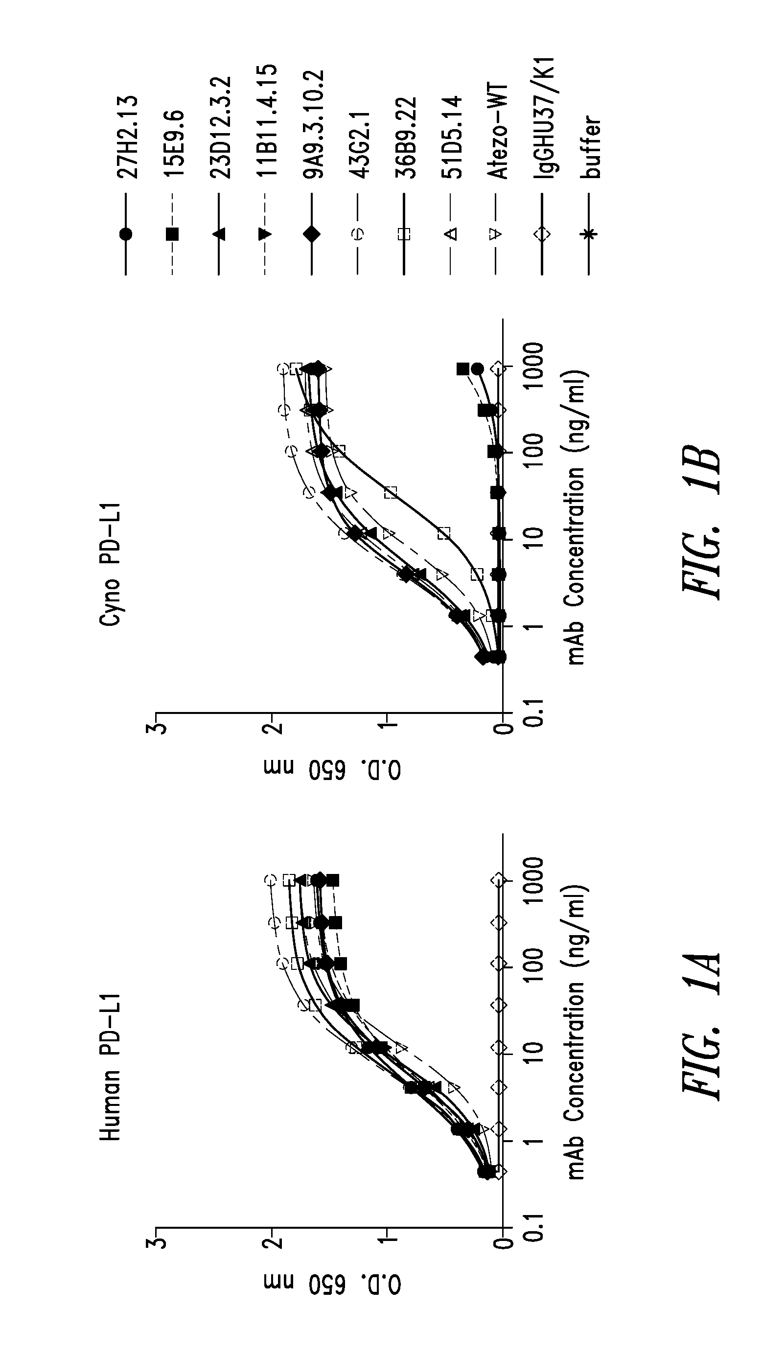
[0019] FIGS. 1A-1B are a set of two line graphs that show binding of chimeric antibodies to human (1A) and cynomolgus macaque (1B) PD-L1 by direct ELISA.
[0020] FIG. 2 is a line graph that shows certain PD-L1 antibodies interfere with binding of Atezolizumab to PD-L1 as measured by competition ELISA
[0021] FIG. 3 is a line graph that show PD-L1 antibodies blocking the binding of PD-1 to PD-L1 by ELISA
[0022] FIG. 4 is a line graph showing the binding of chimeric anti-PD-L1 antibodies to HEK-PD-L1 cells.
[0023] FIGS. 5A-5B are a set of line graphs showing the binding of chimeric anti-PD-L1 antibodies to HEK-PD-L1 cells.
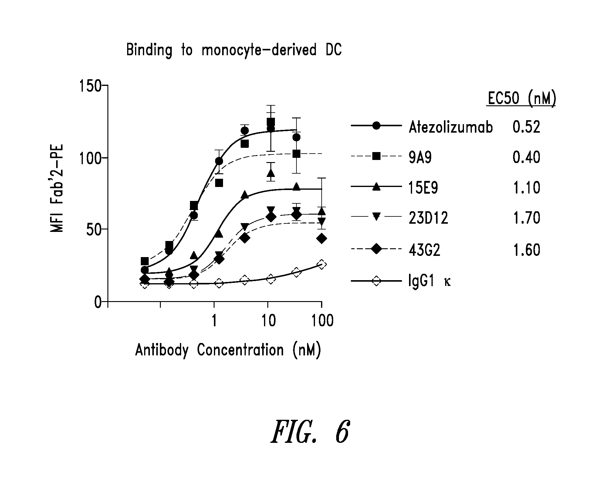
[0024] FIG. 6 is a line graph showing the binding of chimeric anti-PD-L1 antibodies to monocyte derived dendritic cells.
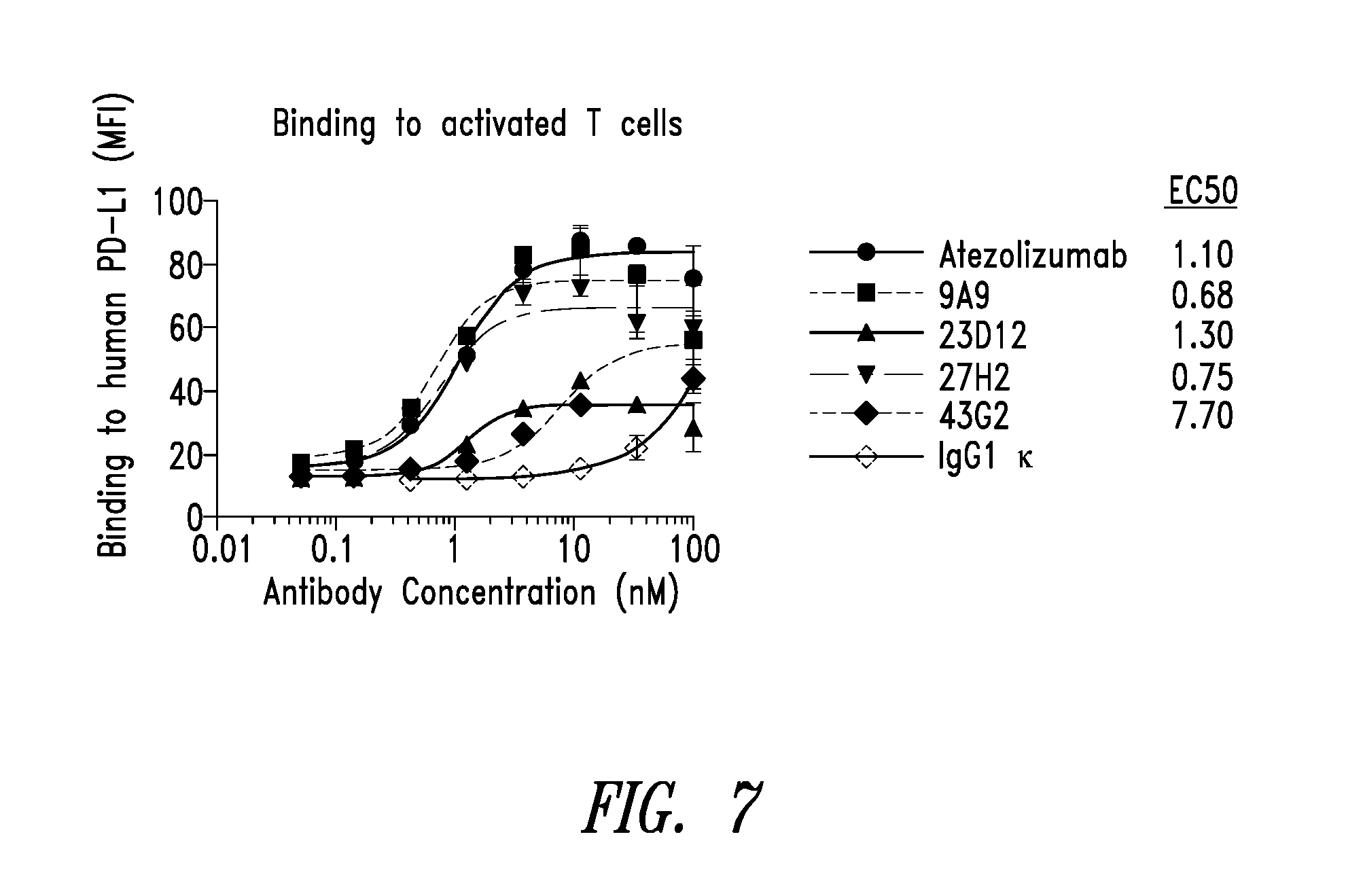
[0025] FIG. 7 is a line graph showing the binding of chimeric anti-PD-L1 antibodies to CD4+ T cells activated with anti-CD3 monoclonal antibody.
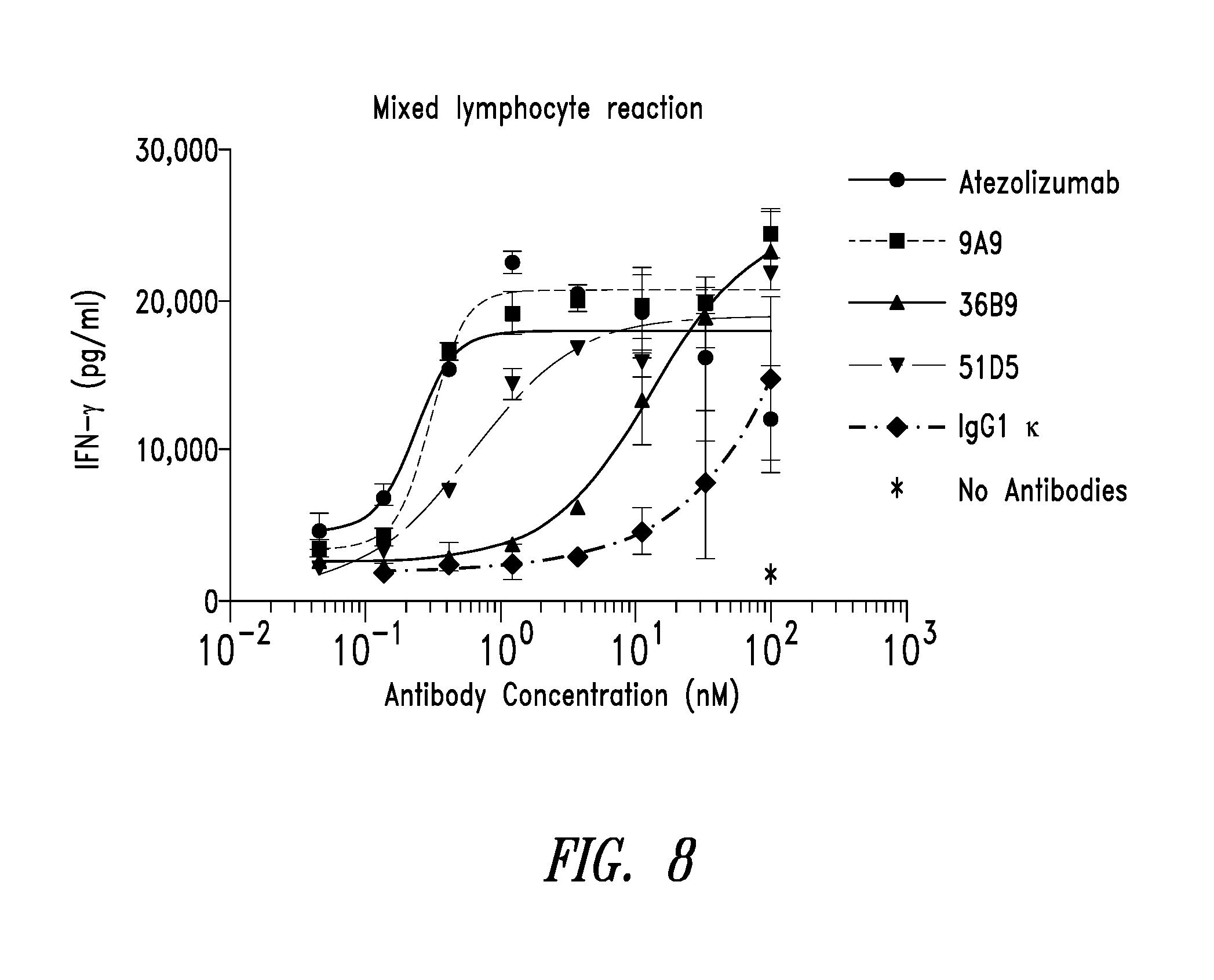
[0026] FIG. 8 is a line graph showing IFN-.gamma. secretion from CD4 T cells in a mixed lymphocyte reaction.
[0027] FIG. 9 is a line graph that shows binding of chimeric and humanized antibodies to human PD-L1 by ELISA.
[0028] FIGS. 10A-10B are a set of line graphs showing the binding of chimeric and humanized anti-PD-L1 antibodies to HEK-PD-L1 cells.
[0029] FIGS. 11A-11B are a set of line graphs showing the binding of chimeric and humanized anti-PD-L1 antibodies to myeloid-derived dendritic cells.
[0030] FIGS. 12A-12B are a set of line graphs showing the binding of chimeric and humanized anti-PD-L1 antibodies to activated CD4+ T cells.
[0031] FIG. 13 is a line graph showing IFN-.gamma. secretion from CD4 T cells in a mixed lymphocyte reaction with humanized anti-PD-L1 antibodies.
[0032] FIG. 14 is a set of line graphs showing IFN-.gamma. secretion in a viral recall response assay using a CMV lysate.
[0033] FIG. 15 is a set of line graphs showing IFN-.gamma. secretion in a SEB assay.
DETAILED DESCRIPTION
[0034] The present disclosure relates to antibodies and antigen-binding fragments thereof the specifically bind to programmed death-ligand 1 (PD-L1 or CD274 or B7 homolog 1), in particular antibodies having specific epitopic specificity and functional properties. Some embodiments encompasses specific humanized antibodies and fragments thereof capable of binding to PD-L1, blocking PD-L1 binding with its ligand, programmed cell death protein 1 (PD-1), and inhibiting induced downstream cell signaling and biological effects. In certain embodiments, an anti-PD-L1 antibody, or antigen-binding fragment thereof, is a PD-L1 antagonist or inhibitor. An antagonist of PD-L1 enhances immune responses by blocking the immunosuppressive actions of PD-L1. PD-L1 antagonist antibodies described herein are useful in the treatment and prevention of, e.g., cancer, especially PD-L1-expressing cancers. In some embodiments, an anti-PD-L1 antibody, or antigen-binding fragment thereof, is a PD-L1 agonist or activator. An agonist of PD-L1 suppresses immune responses by increasing the immunosuppressive actions of PD-L1. PD-L1 agonist antibodies described herein are useful in the treatment and prevention of, e.g., autoimmune diseases and disorders, transplants (e.g., graft-versus-host disease and transplant rejection), and inflammatory diseases and disorders.
[0035] Some embodiments pertain to the use of anti-PD-L1 antibodies or antigen-binding fragments thereof for the diagnosis, assessment and treatment of diseases and disorders associated with PD-L1 or aberrant expression thereof. The subject antibodies are used in the treatment or prevention of cancer among other diseases. Certain embodiments pertain to the use of anti-PD-L1 antibodies or antigen-binding fragments thereof for the diagnosis, assessment and treatment of diseases and disorders associated with aberrant or unwanted inflammatory T cell responses.
[0036] The practice of the present disclosure will employ, unless indicated specifically to the contrary, conventional methods of virology, immunology, microbiology, molecular biology and recombinant DNA techniques within the skill of the art, many of which are described below for the purpose of illustration. Such techniques are explained fully in the literature. See, e.g., Current Protocols in Molecular Biology or Current Protocols in Immunology, John Wiley & Sons, New York, N.Y. (2009); Ausubel et al., Short Protocols in Molecular Biology, 3.sup.rd ed., Wiley & Sons, 1995; Sambrook and Russell, Molecular Cloning: A Laboratory Manual (3rd Edition, 2001); Maniatis et al. Molecular Cloning: A Laboratory Manual (1982); DNA Cloning: A Practical Approach, vol. I & II (D. Glover, ed.); Oligonucleotide Synthesis (N. Gait, ed., 1984); Nucleic Acid Hybridization (B. Hames & S. Higgins, eds., 1985); Transcription and Translation (B. Hames & S. Higgins, eds., 1984); Animal Cell Culture (R. Freshney, ed., 1986); Perbal, A Practical Guide to Molecular Cloning (1984) and other like references.
[0037] As used in this specification and the appended claims, the singular forms "a," "an" and "the" include plural references unless the content clearly dictates otherwise.
[0038] Throughout this specification, unless the context requires otherwise, the word "comprise", or variations such as "comprises" or "comprising", will be understood to imply the inclusion of a stated element or integer or group of elements or integers but not the exclusion of any other element or integer or group of elements or integers.
[0039] Each embodiment in this specification is to be applied mutatis mutandis to every other embodiment unless expressly stated otherwise.
[0040] Standard techniques may be used for recombinant DNA, oligonucleotide synthesis, and tissue culture and transformation (e.g., electroporation, lipofection). Enzymatic reactions and purification techniques may be performed according to manufacturer's specifications or as commonly accomplished in the art or as described herein. These and related techniques and procedures may be generally performed according to conventional methods well known in the art and as described in various general and more specific references that are cited and discussed throughout the present specification. Unless specific definitions are provided, the nomenclature utilized in connection with, and the laboratory procedures and techniques of, molecular biology, analytical chemistry, synthetic organic chemistry, and medicinal and pharmaceutical chemistry described herein are those well-known and commonly used in the art. Standard techniques may be used for recombinant technology, molecular biological, microbiological, chemical syntheses, chemical analyses, pharmaceutical preparation, formulation, and delivery, and treatment of patients.
[0041] Some embodiments relate to antibodies that bind to PD-L1. In particular, the antibodies described herein specifically bind to PD-L1 with unexpectedly high affinity, block binding of PD-L1 to antigen presenting cells (APCs) and/or T cells, block PD-L1 activity and have therapeutic utility for the treatment of diseases associated with aberrant expression of PD-L1. The antibodies described herein also have advantageous properties such as the ability to inhibit a variety of PD-L1-mediated biological effects (e.g., inhibition of T cell proliferation, inhibition of T cell activation, and other PD-L1-mediated effects known to the skilled person). The anti-PD-L1 antibodies described herein may block binding of PD-L1 to PD-1. In some embodiments, use of an anti-PD-L1 antibody results in T cell proliferation. In some embodiments, use of an anti-PD-L1 antibody results in T cell activation. In some embodiments, use of an anti-PD-L1 antibody results in increased secretion of IFN-.gamma..
[0042] Amino acid sequences of illustrative antibodies, or antigen-binding fragments, or complementarity determining regions (CDRs) thereof, are provided in Table A1 below.
TABLE-US-00001 TABLE A1 SEQ ID NO: NAME SEQUENCE 1 9A9H1, QSVEESGGRLVTPGTPLTLTCTVSGIDLSGYWMSWVRQAPGKGLEYIGIVD rabbit VH NDGSAYYASWVNGRFTISKTSTTVDLKITSPTTEDTATYFCARQSGDWTFD region LWGPGTLVTVSS 2 9A9L1, ADVVMTQTPASVSEPVGGTVTIKCQASQSIGRWLSWYQQKPGQPPKLLIYQ rabbit VL ASKLASGVPSRFSGSGSGTEFTLTISDLECADAATYYCQNNYDITAYGNAF region GGGTEVVVK 3 11B11H2, QSVEESGGRLVTPGGTLTLTCTASGFSLSNYTMAWVRQAAGKGLEWIGYIN rabbit VH TGGSPYYASWAKGRFTISRSSTTVDLKMTSLTTEDTATYFCARNVDGAWNY region LNLWGPGTLVTVSS 4 11B11L1, AYDMTQTPASVEVVVGGTVTIKCQASETINTYLSWYQQKPGQRPRLLIYAA rabbit VL SNLASGVPSRFSGSGSGTEFTLTISGVQCADAATYYCQQGINSNVGNTFGG region GTEVVVK 5 15E9H1, QSVEESGGRLVTPGTPLTLTCTVSGFDISNYWMSWVRQAPGKGLEYIGIIS rabbit VH TGGNTWYASWVKGRFTISKTSTTVDLKMTSLTTEDTATYFCVRVVIYGGTT region SSFNLWGPGTLVTVSS 6 15E9L2, QVLTQTASPVSAAVGGTVTINCQSSQNVYNKIRLSWFQQKSGQPPKELIYA rabbit VL ASSLASGVPSRFKGSGSGTEFTLTISDLECDDAATYYCQGGYSGPIYAFGG region GTEVVVK 7 23D12H2, QSVEESGGRLVTPGTPLTLTCTVSGFSLSNYVMGWVRQAPGKGLEYIGIIS rabbit VH NSDNTYYATWATGRFTISKTSTTVDLKMTSLTTEDTATYFCARVDTYYNDF region NLWGPGTLVTVSS 8 23D12L1, QVLTQTPSSVEAAVGGTVTIKCQASEDIERYLAWYQQKPGQPPKQLIYDAS rabbit VL KLASGVSSRFKGSGSGTEFTLTISGVQCDDAATYYCQHGYIYSSGNPFGGG region TEVVVK 9 27H2-H1, QSLEESGGDLVKPGASLTLTCTASGFSFSSGDYMCWVRQAPGKGLEGIACI rabbit VH YGGSSGSTYYANWAKGRFTISKTSSTTVTLQMTSLAAADTATYFCARDFRD region VGYGYASFRLWGPGILVTVSS 10 27H2-L2, ADIVMTQTPASVSEPVGGTVTIKCQASQSISSWLSWYQQKPGQPPKLLIYG rabbit VL ASNLESGVSSRFKGSGSGTEYTLTISDLECADAATYYCQSNYGSSSSSLNA region FGGGTEVVVK 11 39B9H2, QSVEESGGRLVTPGTPLTLTCTVSGFSLSNYWMSWVRQAPGKGLEWIGYIN rabbit VH GGSGGTYYASWAKGRFTSSKTSTTVDLKITSPTTEDTATYFCARGGESDDY region IYGRYGFDPWGPGTLVTVSS 12 39B9L1, AYDMTQTPASVSEPVGGTVTIKCQASQSISSWLSWYQQKPGQPPKLLIYQA rabbit VL SKLASGVSSRFKGSGSGTDFTLTIIGVECADAATYYCQQDYASYNVDNTFG region GGTEVVVK 13 43G2H1, QSVEESGGRLVTPGTPLTLTCTVSGFSLSSYAMGWVRQAPGEGLEYIGFID rabbit VH THGSAYYATWAKGRFTISRTSTTVDLKITSPTTEDTATYFCARNVWTNAGY region FNLWGPGTLVTVSS 14 43G2L2, ADVVMTQTPSSVSAAVGGTVTIKCQASQNIYTNLAWYQQKPGQPPKLLIYS rabbit VL ASTLASGVPSRFKGSGSGTQFTLTISDLECADAATYYCQTYGALIINGGAF region GGGTEVVVK 15 51D5H2, QSVEESGGRLVTPGTPLTLTCIVSGFSLSSYWMSWVRRAPGKGLEWIGYIN rabbit VH GGSGNPYYASWAKGRFTISKTSTTVDLKITSPTTEDTATYFCARGGESDDY region VYGRYGLDPWGPGTLVTVSS 16 51D5L, AYDMTQTPASVSEPVGGTVTIKCQASQSISSWLSWYQQKPGQPPKLLIYDA rabbit VL SKLASGVPSRFSGSGSGTQFTLTISDLECDDAATYYCQQDYSSYNVDNTFG region GGTEVVVK 17 9A9H1 EVQLVESGGGLVQPGGSLRLSCAASGIDLSGYWMSWVRQAPGKGLEWVGIV Humanized DNDGSAYYASWVNGRFTISRDNSKNTLYLQMNSLRAEDTAVYYCARQSGDW HZD1 VH TFDLWGQGTLVTVSS region 18 9A9H1 EVQLVESGGGLVQPGGSLRLSCAASGFDVSGYWMSWVRQAPGKGLEWVGIV Humanized DNDGSAYYASWVNGRFTISRDNSKNTLYLQMNSLRAEDTAVYYCARQSGDW HZD2 VH TFDLWGQGTLVTVSS region 19 9A9L1 DIQMTQSPSTLSASVGDRVTITCQASQSIGRWLSWYQQKPGKAPKLLIYQA humanized SKLASGVPSRFSGSGSGTEFTLTISSLQPDDFATYYCQNNYDITAYGNAFG HZD1 and GGTKVEIK HZD2 VL 20 9A9H1, GYWMS humanized VHCDR1 21 9A9H1, IVDNDGSAYYASWVNG humanized VHCDR2 22 9A9H1, QSGDWTFDL humanized VHCDR3 23 9A9L1, QASQSIGRWLS humanized VLCDR1 24 9A9L1, QASKLAS humanized VLCDR2 25 9A9L1, QNNYDIIAYGNA humanized VLCDR3 26 51D5H2 EVQLVESGGGLVQPGGSLRLSCAASGFSLSSYWMSWVRRAPGKGLEWVGYI humanized NGGSGNPYYASWAKGRFTISRDNSKNTLYLQMNSLRAEDTAVYYCARGGES HZD1 VH DDYVYGRYGLDPWGQGTLVTVSS 27 51D5H2 EVQLVESGGGLVQPGGSLRLSCAASGFSLSSYWMSWVRQAPGKGLEWVGYI humanized NGGSGNPYYASWAKGRFTISRDNSKNTLYLQMNSLRAEDTAVYYCARGGES HZD2 VH DDYVYGRYGLDPWGQGTLVTVSS 28 51D5L DIQMTQSPSTLSASVGDRVTITCQASQSISSWLSWYQQKPGKAPKLLIYDA humanized SKLASGVPSRFSGSGSGTEFTLTISSLQPDDFATYYCQQDYSSYNVDNTFG HZD1 and GGTKVEIK HZD2 VL 29 51D5H2, SYWMS humanized VHCDR1 30 51D5H2, YINGGSGNPYYASWAKG humanized VHCDR2 31 51D5H2, GGESDDYVYGRYGLDP humanized VHCDR3 32 51D5L, QASQSISSWLS humanized VLCDR1 33 51D5L, DASKLAS humanized VLCDR2 34 51D5L, QQDYSSYNVDNT humanized VLCDR3 35 15E9H1 EVQLVESGGGLVQPGGSLRLSCAASGFDISNYWMSWVRQAPGKGLEWVGII humanized STGGNTWYASWVKGRFTISRDNSKNTLYLQMNSLRAEDTAVYYCARVVIYG HZD1 and 2 GTTSSFNLWGQGTLVTVSS VH 36 15E9L2 DIQMTQSPSSLSASVGDRVTITCQSSQNVYNKIRLSWYQQKPGKAPKELIY humanized AASSLASGVPSRFSGSGSGTEFTLTISSLQPEDFATYYCQGGYSGPIYAFG HZD1 VL GGTKVEIK 37 15E9L2 DIQMTQSPSSLSASVGDRVTITCQSSQNVYNKIRLSWYQQKPGKAPKRLIY humanized AASSLASGVPSRFSGSGSGTEFTLTISSLQPEDFATYYCQGGYSGPIYAFG HZD2 VL GGTKVEIK 38 15E9H1, NYWMS humanized VHCDR1 39 15E9H1, IISTGGNTWYASWVKG humanized VHCDR2 40 15E9H1, VVIYGGTTSSFNL humanized VHCDR3 41 15E9L2, QSSQNVYNKIRLS humanized VLCDR1 42 15E9L2, AASSLAS humanized VLCDR2 43 15E9L2, QGGYSGPIYA humanized VLCDR3
[0043] As is well known in the art, an antibody is an immunoglobulin molecule capable of specific binding to a target, such as a carbohydrate, polynucleotide, lipid, polypeptide, etc., through at least one epitope recognition site, located in the variable region of the immunoglobulin molecule. As used herein, the term encompasses not only intact polyclonal or monoclonal antibodies, but also fragments thereof (such as dAb, Fab, Fab', F(ab').sub.2, Fv), single chain (scFv), synthetic variants thereof, naturally occurring variants, fusion proteins comprising an antibody portion with an antigen-binding fragment of the required specificity, humanized antibodies, chimeric antibodies, and any other modified configuration of the immunoglobulin molecule that comprises an antigen-binding site or fragment (epitope recognition site) of the required specificity. "Diabodies", multivalent or multispecific fragments constructed by gene fusion (WO94/13804; P. Holliger et al., Proc. Natl. Acad. Sci. USA 90 6444-6448, 1993) are also a particular form of antibody contemplated herein. Minibodies comprising a scFv joined to a CH3 domain are also included herein (S. Hu et al., Cancer Res., 56, 3055-3061, 1996). See e.g., Ward, E. S. et al., Nature 341, 544-546 (1989); Bird et al., Science, 242, 423-426, 1988; Huston et al., PNAS USA, 85, 5879-5883, 1988); PCT/US92/09965; WO94/13804; P. Holliger et al., Proc. Natl. Acad. Sci. USA 90 6444-6448, 1993; Y. Reiter et al., Nature Biotech, 14, 1239-1245, 1996; S. Hu et al., Cancer Res., 56, 3055-3061, 1996.
[0044] The term "antigen-binding fragment" as used herein refers to a polypeptide fragment that contains at least one CDR of an immunoglobulin heavy and/or light chains that binds to the antigen of interest, in particular to PD-L1. In this regard, an antigen-binding fragment of the herein described antibodies may comprise 1, 2, 3, 4, 5, or all 6 CDRs of a VH and VL sequence set forth herein from antibodies that bind PD-L1. An antigen-binding fragment of the PD-L1-specific antibodies described herein is capable of binding to PD-L1. In certain embodiments, an antigen-binding fragment or an antibody comprising an antigen-binding fragment, prevents or inhibits PD-L1 binding to T cells and/or APCs and subsequent signaling events. In certain embodiments, the antigen-binding fragment binds specifically to and/or inhibits or modulates the biological activity of human PD-L1. In certain embodiments, the antigen-binding fragment binds specifically to and/or enhances or upregulates the biological activity of human PD-L1.
[0045] The term "antigen" refers to a molecule or a portion of a molecule capable of being bound by a selective binding agent, such as an antibody, and additionally capable of being used in an animal to produce antibodies capable of binding to an epitope of that antigen. An antigen may have one or more epitopes.
[0046] The term "epitope" includes any determinant, preferably a polypeptide determinant, capable of specific binding to an immunoglobulin or T-cell receptor. An epitope is a region of an antigen that is bound by an antibody. In certain embodiments, epitope determinants include chemically active surface groupings of molecules such as amino acids, sugar side chains, phosphoryl or sulfonyl, and may in certain embodiments have specific three-dimensional structural characteristics, and/or specific charge characteristics. In certain embodiments, an antibody is said to specifically bind an antigen when it preferentially recognizes its target antigen in a complex mixture of proteins and/or macromolecules. An antibody is said to specifically bind an antigen when the equilibrium dissociation constant is .ltoreq.10.sup.-7 or 10.sup.-8 M. In some embodiments, the equilibrium dissociation constant may be .ltoreq.10.sup.-9 M or .ltoreq.10.sup.-10 M.
[0047] In certain embodiments, antibodies and antigen-binding fragments thereof as described herein include a heavy chain and a light chain CDR set, respectively interposed between a heavy chain and a light chain framework region (FR) set which provide support to the CDRs and define the spatial relationship of the CDRs relative to each other. As used herein, the term "CDR set" refers to the three hypervariable regions of a heavy or light chain V region. Proceeding from the N-terminus of a heavy or light chain, these regions are denoted as "CDR1," "CDR2," and "CDR3" respectively. An antigen-binding site, therefore, includes six CDRs, comprising the CDR set from each of a heavy and a light chain V region. A polypeptide comprising a single CDR, (e.g., a CDR1, CDR2 or CDR3) is referred to herein as a "molecular recognition unit." Crystallographic analysis of a number of antigen-antibody complexes has demonstrated that the amino acid residues of CDRs form extensive contact with bound antigen, wherein the most extensive antigen contact is with the heavy chain CDR3. Thus, the molecular recognition units are primarily responsible for the specificity of an antigen-binding site.
[0048] As used herein, the term "FR set" refers to the four flanking amino acid sequences which frame the CDRs of a CDR set of a heavy or light chain V region. Some FR residues may contact bound antigen; however, FRs are primarily responsible for folding the V region into the antigen-binding site, particularly the FR residues directly adjacent to the CDRs. Within FRs, certain amino residues and certain structural features are very highly conserved. In this regard, all V region sequences contain an internal disulfide loop of around 90 amino acid residues. When the V regions fold into a binding-site, the CDRs are displayed as projecting loop motifs which form an antigen-binding surface. It is generally recognized that there are conserved structural regions of FRs which influence the folded shape of the CDR loops into certain "canonical" structures--regardless of the precise CDR amino acid sequence. Further, certain FR residues are known to participate in non-covalent interdomain contacts which stabilize the interaction of the antibody heavy and light chains.
[0049] The structures and locations of immunoglobulin variable domains may be determined by reference to Kabat, E. A. et al., Sequences of Proteins of Immunological Interest. 4th Edition. US Department of Health and Human Services. 1987, and updates thereof, now available on the Internet (immuno.bme.nwu.edu).
[0050] A "monoclonal antibody" refers to a homogeneous antibody population wherein the monoclonal antibody is comprised of amino acids (naturally occurring and non-naturally occurring) that are involved in the selective binding of an epitope. Monoclonal antibodies are highly specific, being directed against a single epitope. The term "monoclonal antibody" encompasses not only intact monoclonal antibodies and full-length monoclonal antibodies, but also fragments thereof (such as Fab, Fab', F(ab').sub.2, Fv), single chain (scFv), variants thereof, fusion proteins comprising an antigen-binding portion, humanized monoclonal antibodies, chimeric monoclonal antibodies, and any other modified configuration of the immunoglobulin molecule that comprises an antigen-binding fragment (epitope recognition site) of the required specificity and the ability to bind to an epitope. It is not intended to be limited as regards the source of the antibody or the manner in which it is made (e.g., by hybridoma, phage selection, recombinant expression, transgenic animals, etc.). The term includes whole immunoglobulins as well as the fragments etc. described above under the definition of "antibody".
[0051] The proteolytic enzyme papain preferentially cleaves IgG molecules to yield several fragments, two of which (the F(ab) fragments) each comprise a covalent heterodimer that includes an intact antigen-binding site. The enzyme pepsin is able to cleave IgG molecules to provide several fragments, including the F(ab').sub.2 fragment which comprises both antigen-binding sites. An Fv fragment for use according to certain embodiments can be produced by preferential proteolytic cleavage of an IgM, and on rare occasions of an IgG or IgA immunoglobulin molecule. Fv fragments are, however, more commonly derived using recombinant techniques known in the art. The Fv fragment includes a non-covalent V.sub.H::V.sub.L heterodimer including an antigen-binding site which retains much of the antigen recognition and binding capabilities of the native antibody molecule. Inbar et al. (1972) Proc. Nat. Acad. Sci. USA 69:2659-2662; Hochman et al. (1976) Biochem 15:2706-2710; and Ehrlich et al. (1980) Biochem 19:4091-4096.
[0052] In certain embodiments, single chain Fv or scFv antibodies are contemplated. For example, Kappa bodies (III et al., Prot. Eng. 10: 949-57 (1997); minibodies (Martin et al., EMBO J 13: 5305-9 (1994); diabodies (Holliger et al., PNAS 90: 6444-8 (1993); or Janusins (Traunecker et al., EMBO J 10: 3655-59 (1991) and Traunecker et al., Int. J. Cancer Suppl. 7: 51-52 (1992), may be prepared using standard molecular biology techniques following the teachings of the present application with regard to selecting antibodies having the desired specificity. In some embodiments, bispecific or chimeric antibodies may be made that encompass the ligands of the present disclosure. For example, a chimeric antibody may comprise CDRs and framework regions from different antibodies, while bispecific antibodies may be generated that bind specifically to PD-L1 through one binding domain and to a second molecule through a second binding domain. These antibodies may be produced through recombinant molecular biological techniques or may be physically conjugated together.
[0053] A single chain Fv (scFv) polypeptide is a covalently linked V.sub.H::V.sub.L heterodimer which is expressed from a gene fusion including V.sub.H- and V.sub.L-encoding genes linked by a peptide-encoding linker. Huston et al. (1988) Proc. Nat. Acad. Sci. USA 85(16):5879-5883. A number of methods have been described to discern chemical structures for converting the naturally aggregated--but chemically separated--light and heavy polypeptide chains from an antibody V region into an scFv molecule which will fold into a three dimensional structure substantially similar to the structure of an antigen-binding site. See, e.g., U.S. Pat. Nos. 5,091,513 and 5,132,405, to Huston et al.; and U.S. Pat. No. 4,946,778, to Ladner et al.
[0054] In certain embodiments, a PD-L1 binding antibody as described herein is in the form of a diabody. Diabodies are multimers of polypeptides, each polypeptide comprising a first domain comprising a binding region of an immunoglobulin light chain and a second domain comprising a binding region of an immunoglobulin heavy chain, the two domains being linked (e.g. by a peptide linker) but unable to associate with each other to form an antigen binding site: antigen binding sites are formed by the association of the first domain of one polypeptide within the multimer with the second domain of another polypeptide within the multimer (WO94/13804).
[0055] A dAb fragment of an antibody consists of a VH domain (Ward, E. S. et al., Nature 341, 544-546 (1989)).
[0056] Where bispecific antibodies are to be used, these may be conventional bispecific antibodies, which can be manufactured in a variety of ways (Holliger, P. and Winter G. Current Opinion Biotechnol. 4, 446-449 (1993)), e.g. prepared chemically or from hybrid hybridomas, or may be any of the bispecific antibody fragments mentioned above. Diabodies and scFv can be constructed without an Fc region, using only variable domains, potentially reducing the effects of anti-idiotypic reaction.
[0057] Bispecific diabodies, as opposed to bispecific whole antibodies, may also be particularly useful because they can be readily constructed and expressed in E. coli. Diabodies (and many other polypeptides such as antibody fragments) of appropriate binding specificities can be readily selected using phage display (WO94/13804) from libraries. If one arm of the diabody is to be kept constant, for instance, with a specificity directed against antigen X, then a library can be made where the other arm is varied and an antibody of appropriate specificity selected. Bispecific whole antibodies may be made by knobs-into-holes engineering (J. B. B. Ridgeway et al., Protein Eng., 9, 616-621, 1996).
[0058] In certain embodiments, the antibodies described herein may be provided in the form of a UniBody.RTM.. A UniBody.RTM. is an IgG4 antibody with the hinge region removed (see GenMab Utrecht, The Netherlands; see also, e.g., US20090226421). This proprietary antibody technology creates a stable, smaller antibody format with an anticipated longer therapeutic window than current small antibody formats. IgG4 antibodies are considered inert and thus do not interact with the immune system. Fully human IgG4 antibodies may be modified by eliminating the hinge region of the antibody to obtain half-molecule fragments having distinct stability properties relative to the corresponding intact IgG4 (GenMab, Utrecht). Halving the IgG4 molecule leaves only one area on the UniBody.RTM. that can bind to cognate antigens (e.g., disease targets) and the UniBody.RTM. therefore binds univalently to only one site on target cells. For certain cancer cell surface antigens, this univalent binding may not stimulate the cancer cells to grow as may be seen using bivalent antibodies having the same antigen specificity, and hence UniBody.RTM. technology may afford treatment options for some types of cancer that may be refractory to treatment with conventional antibodies. The small size of the UniBody.RTM. can be a great benefit when treating some forms of cancer, allowing for better distribution of the molecule over larger solid tumors and potentially increasing efficacy.
[0059] In certain embodiments, the antibodies of the present disclosure may take the form of a Nanobody.RTM.. Nanobodies.RTM. are encoded by single genes and are efficiently produced in almost all prokaryotic and eukaryotic hosts e.g. E. coli (see e.g. U.S. Pat. No. 6,765,087), molds (for example Aspergillus or Trichoderma) and yeast (for example Saccharomyces, Kluyvermyces, Hansenula or Pichia (see e.g. U.S. Pat. No. 6,838,254). The production process is scalable and multi-kilogram quantities of Nanobodies.RTM. have been produced. Nanobodies may be formulated as a ready-to-use solution having a long shelf life. The Nanoclone.RTM. method (see, e.g., WO 06/079372) is a proprietary method for generating Nanobodies against a desired target, based on automated high-throughput selection of B-cells.
[0060] In certain embodiments, the anti-PD-L1 antibodies or antigen-binding fragments thereof as disclosed herein are humanized. This refers to a chimeric molecule, generally prepared using recombinant techniques, having an antigen-binding site derived from an immunoglobulin from a non-human species and the remaining immunoglobulin structure of the molecule based upon the structure and/or sequence of a human immunoglobulin. The antigen-binding site may comprise either complete variable domains fused onto constant domains or only the CDRs grafted onto appropriate framework regions in the variable domains. Epitope binding sites may be wild type or modified by one or more amino acid substitutions. This eliminates the constant region as an immunogen in human individuals, but the possibility of an immune response to the foreign variable region remains (LoBuglio, A. F. et al., (1989) Proc Natl Acad Sci USA 86:4220-4224; Queen et al., PNAS (1988) 86:10029-10033; Riechmann et al., Nature (1988) 332:323-327). Illustrative methods for humanization of the anti-PD-L1 antibodies disclosed herein include the methods described in U.S. Pat. No. 7,462,697. Illustrative humanized antibodies according to certain embodiments comprise the humanized sequences provided in Table A1.
[0061] Another approach focuses not only on providing human-derived constant regions, but modifying the variable regions as well so as to reshape them as closely as possible to human form. It is known that the variable regions of both heavy and light chains contain three complementarity-determining regions (CDRs) which vary in response to the epitopes in question and determine binding capability, flanked by four framework regions (FRs) which are relatively conserved in a given species and which putatively provide a scaffolding for the CDRs. When nonhuman antibodies are prepared with respect to a particular epitope, the variable regions can be "reshaped" or "humanized" by grafting CDRs derived from nonhuman antibody on the FRs present in the human antibody to be modified. Application of this approach to various antibodies has been reported by Sato, K., et al., (1993) Cancer Res 53:851-856. Riechmann, L., et al., (1988) Nature 332:323-327; Verhoeyen, M., et al., (1988) Science 239:1534-1536; Kettleborough, C. A., et al., (1991) Protein Engineering 4:773-3783; Maeda, H., et al., (1991) Human Antibodies Hybridoma 2:124-134; Gorman, S. D., et al., (1991) Proc Natl Acad Sci USA 88:4181-4185; Tempest, P. R., et al., (1991) Bio/Technology 9:266-271; Co, M. S., et al., (1991) Proc Natl Acad Sci USA 88:2869-2873; Carter, P., et al., (1992) Proc Natl Acad Sci USA 89:4285-4289; and Co, M. S. et al., (1992) J Immunol 148:1149-1154. In some embodiments, humanized antibodies preserve all CDR sequences (for example, a humanized rabbit antibody which contains all six CDRs from the rabbit antibody). In certain embodiments, humanized antibodies have one or more CDRs (one, two, three, four, five, six) which are altered with respect to the original antibody, which are also termed one or more CDRs "derived from" one or more CDRs from the original antibody.
[0062] In certain embodiments, the antibodies of the present disclosure may be chimeric antibodies. In this regard, a chimeric antibody is comprised of an antigen-binding fragment of an anti-PD-L1 antibody operably linked or otherwise fused to a heterologous Fc portion of a different antibody. In certain embodiments, the heterologous Fc domain is of human origin. In some embodiments, the heterologous Fc domain may be from a different Ig class from the parent antibody, including IgA (including subclasses IgA1 and IgA2), IgD, IgE, IgG (including subclasses IgG1, IgG2, IgG3, and IgG4), and IgM. In some embodiments, the heterologous Fc domain may be comprised of CH2 and CH3 domains from one or more of the different Ig classes. As noted above with regard to humanized antibodies, the anti-PD-L1 antigen-binding fragment of a chimeric antibody may comprise only one or more of the CDRs of the antibodies described herein (e.g., 1, 2, 3, 4, 5, or 6 CDRs of the antibodies described herein), or may comprise an entire variable domain (VL, VH or both).
[0063] In certain embodiments, a PD-L1-binding antibody comprises one or more of the CDRs of the antibodies described herein. In this regard, it has been shown in some cases that the transfer of only the VHCDR3 of an antibody can be performed while still retaining desired specific binding (Barbas et al., PNAS (1995) 92: 2529-2533). See also, McLane et al., PNAS (1995) 92:5214-5218, Barbas et al., J. Am. Chem. Soc. (1994) 116:2161-2162.
[0064] Marks et al (Bio/Technology, 1992, 10:779-783) describe methods of producing repertoires of antibody variable domains in which consensus primers directed at or adjacent to the 5' end of the variable domain area are used in conjunction with consensus primers to the third framework region of human VH genes to provide a repertoire of VH variable domains lacking a CDR3. Marks et al further describe how this repertoire may be combined with a CDR3 of a particular antibody. Using analogous techniques, the CDR3-derived sequences of the presently described antibodies may be shuffled with repertoires of VH or VL domains lacking a CDR3, and the shuffled complete VH or VL domains combined with a cognate VL or VH domain to provide an antibody or antigen-binding fragment thereof that binds PD-L1. The repertoire may then be displayed in a suitable host system such as the phage display system of WO 92/01047 so that suitable antibodies or antigen-binding fragments thereof may be selected. A repertoire may consist of at least from about 10.sup.4 individual members and upwards by several orders of magnitude, for example, to about from 10.sup.6 to 10.sup.8 or 10.sup.10 or more members. Analogous shuffling or combinatorial techniques are also disclosed by Stemmer (Nature, 1994, 370:389-391), who describes the technique in relation to a .beta.-lactamase gene but observes that the approach may be used for the generation of antibodies.
[0065] A further alternative is to generate novel VH or VL regions carrying one or more CDR-derived sequences of the herein described embodiments using random mutagenesis of one or more selected VH and/or VL genes to generate mutations within the entire variable domain. Such a technique is described by Gram et al (1992, Proc. Natl. Acad. Sci., USA, 89:3576-3580), who used error-prone PCR. Another method which may be used is to direct mutagenesis to CDR regions of VH or VL genes. Such techniques are disclosed by Barbas et al., (1994, Proc. Natl. Acad. Sci., USA, 91:3809-3813) and Schier et al (1996, J. Mol. Biol. 263:551-567).
[0066] In certain embodiments, a specific VH and/or VL of the antibodies described herein may be used to screen a library of the complementary variable domain to identify antibodies with desirable properties, such as increased affinity for PD-L1. Such methods are described, for example, in Portolano et al., J. Immunol. (1993) 150:880-887; Clarkson et al., Nature (1991) 352:624-628.
[0067] Other methods may also be used to mix and match CDRs to identify antibodies having desired binding activity, such as binding to PD-L1. For example: Klimka et al., British Journal of Cancer (2000) 83: 252-260, describe a screening process using a mouse VL and a human VH library with CDR3 and FR4 retained from the mouse VH. After obtaining antibodies, the VH was screened against a human VL library to obtain antibodies that bound antigen. Beiboer et al., J. Mol. Biol. (2000) 296:833-849 describe a screening process using an entire mouse heavy chain and a human light chain library. After obtaining antibodies, one VL was combined with a human VH library with the CDR3 of the mouse retained. Antibodies capable of binding antigen were obtained. Rader et al., PNAS (1998) 95:8910-8915 describe a process similar to Beiboer et al above.
[0068] These just-described techniques are, in and of themselves, known as such in the art. The skilled person will, however, be able to use such techniques to obtain antibodies or antigen-binding fragments thereof according to several embodiments described herein, using routine methodology in the art.
[0069] Also disclosed herein is a method for obtaining an antibody antigen binding domain specific for PD-L1 antigen, the method comprising providing by way of addition, deletion, substitution or insertion of one or more amino acids in the amino acid sequence of a VH domain set out herein a VH domain which is an amino acid sequence variant of the VH domain, optionally combining the VH domain thus provided with one or more VL domains, and testing the VH domain or VH/VL combination or combinations to identify a specific binding member or an antibody antigen binding domain specific for PD-L1 and optionally with one or more desired properties. The VL domains may have an amino acid sequence which is substantially as set out herein. An analogous method may be employed in which one or more sequence variants of a VL domain disclosed herein are combined with one or more VH domains.
[0070] An epitope that "specifically binds" or "preferentially binds" (used interchangeably herein) to an antibody or a polypeptide is a term well understood in the art, and methods to determine such specific or preferential binding are also well known in the art. A molecule is said to exhibit "specific binding" or "preferential binding" if it reacts or associates more frequently, more rapidly, with greater duration and/or with greater affinity with a particular cell or substance than it does with alternative cells or substances. An antibody "specifically binds" or "preferentially binds" to a target if it binds with greater affinity, avidity, more readily, and/or with greater duration than it binds to other substances. For example, an antibody that specifically or preferentially binds to a PD-L1 epitope is an antibody that binds one PD-L1 epitope with greater affinity, avidity, more readily, and/or with greater duration than it binds to other PD-L1 epitopes or non-PD-L1 epitopes. It is also understood by reading this definition that, for example, an antibody (or moiety or epitope) that specifically or preferentially binds to a first target may or may not specifically or preferentially bind to a second target. As such, "specific binding" or "preferential binding" does not necessarily require (although it can include) exclusive binding. Generally, but not necessarily, reference to binding means preferential binding.
[0071] Immunological binding generally refers to the non-covalent interactions of the type which occur between an immunoglobulin molecule and an antigen for which the immunoglobulin is specific, for example by way of illustration and not limitation, as a result of electrostatic, ionic, hydrophilic and/or hydrophobic attractions or repulsion, steric forces, hydrogen bonding, van der Waals forces, and other interactions. The strength, or affinity of immunological binding interactions can be expressed in terms of the dissociation constant (K.sub.d) of the interaction, wherein a smaller K.sub.d represents a greater affinity. Immunological binding properties of selected polypeptides can be quantified using methods well known in the art. One such method entails measuring the rates of antigen-binding site/antigen complex formation and dissociation, wherein those rates depend on the concentrations of the complex partners, the affinity of the interaction, and on geometric parameters that equally influence the rate in both directions. Thus, both the "on rate constant" (K.sub.on) and the "off rate constant" (K.sub.off) can be determined by calculation of the concentrations and the actual rates of association and dissociation. The ratio of K.sub.off/K.sub.on enables cancellation of all parameters not related to affinity, and is thus equal to the dissociation constant K.sub.d. See, generally, Davies et al. (1990) Annual Rev. Biochem. 59:439-473.
[0072] In certain embodiments, the anti-PD-L1 antibodies described herein have an affinity of about 100, 150, 155, 160, 170, 175, 180, 185, 190, 191, 192, 193, 194, 195, 196, 197, 198 or 199 picomolar, and in some embodiments, the antibodies may have even higher affinity for PD-L1.
[0073] The term "immunologically active", with reference to an epitope being or "remaining immunologically active", refers to the ability of an antibody (e.g., anti-PD-L1 antibody) to bind to the epitope under different conditions, for example, after the epitope has been subjected to reducing and denaturing conditions.
[0074] An antibody or antigen-binding fragment thereof according to certain preferred embodiments of the present application may be one that competes for binding to PD-L1 with any antibody described herein which both (i) specifically binds to the antigen and (ii) comprises a VH and/or VL domain disclosed herein, or comprises a VH CDR3 disclosed herein, or a variant of any of these. Competition between antibodies may be assayed easily in vitro, for example using ELISA and/or by tagging a specific reporter molecule to one antibody which can be detected in the presence of other untagged antibodies, to enable identification of specific antibodies which bind the same epitope or an overlapping epitope. Thus, there is provided herein a specific antibody or antigen-binding fragment thereof, comprising a human antibody antigen-binding site which competes with an antibody described herein that binds to PD-L1.
[0075] In this regard, as used herein, the terms "competes with", "inhibits binding" and "blocks binding" (e.g., referring to inhibition/blocking of binding of PD-1 to PD-L1 or referring to inhibition/blocking of binding of an anti-PD-L1 antibody to PD-L1) are used interchangeably and encompass both partial and complete inhibition/blocking. The inhibition/blocking of PD-1 to PD-L1 preferably reduces or alters the normal level or type of cell signaling that occurs when PD-1 binds to PD-L1 without inhibition or blocking. Inhibition and blocking are also intended to include any measurable decrease in the binding of PD-1 to PD-L1 when in contact with an anti-PD-L1 antibody as disclosed herein as compared to PD-1 not in contact with an anti-PD-L1 antibody, e.g., the blocking of PD-1 to PD-L1 by at least about 10%, 20%, 30%, 40%, 50%, 60%, 65%, 70%, 75%, 80%, 85%, 90%, 91%, 92%, 93%, 94%, 95%, 96%, 97%, 98%, 99%, or 100%.
[0076] The constant regions of immunoglobulins show less sequence diversity than the variable regions, and are responsible for binding a number of natural proteins to elicit important biochemical events. In humans there are five different classes of antibodies including IgA (which includes subclasses IgA1 and IgA2), IgD, IgE, IgG (which includes subclasses IgG1, IgG2, IgG3, and IgG4), and IgM. The distinguishing features between these antibody classes are their constant regions, although subtler differences may exist in the V region.
[0077] The Fc region of an antibody interacts with a number of Fc receptors and ligands, imparting an array of important functional capabilities referred to as effector functions. In some embodiments, an anti-PD-L1 antibody comprises an Fc region. For IgG the Fc region comprises Ig domains CH2 and CH3 and the N-terminal hinge leading into CH2. An important family of Fc receptors for the IgG class are the Fc gamma receptors (Fc.gamma.Rs). These receptors mediate communication between antibodies and the cellular arm of the immune system (Raghavan et al., 1996, Annu Rev Cell Dev Biol 12:181-220; Ravetch et al., 2001, Annu Rev Immunol 19:275-290). In humans this protein family includes Fc.gamma.RI (CD64), including isoforms Fc.gamma.RIa, Fc.gamma.RIb, and Fc.gamma.RIc; Fc.gamma.RII (CD32), including isoforms Fc.gamma.RIIa (including allotypes H131 and R131), Fc.gamma.RIIb (including Fc.gamma.RIIb-1 and Fc.gamma.RIIb-2), and Fc.gamma.RIIc; and Fc.gamma.RIII (CD16), including isoforms Fc.gamma.RIIIa (including allotypes V158 and F158) and Fc.gamma.RIIIb (including allotypes Fc.gamma.RIIIb-NA1 and Fc.gamma.RIIIb-NA2) (Jefferis et al., 2002, Immunol Lett 82:57-65). These receptors typically have an extracellular domain that mediates binding to Fc, a membrane spanning region, and an intracellular domain that may mediate some signaling event within the cell. These receptors are expressed in a variety of immune cells including monocytes, macrophages, neutrophils, dendritic cells, eosinophils, mast cells, platelets, B cells, large granular lymphocytes, Langerhans' cells, natural killer (NK) cells, and T cells. Formation of the Fc/Fc.gamma.R complex recruits these effector cells to sites of bound antigen, typically resulting in signaling events within the cells and important subsequent immune responses such as release of inflammation mediators, B cell activation, endocytosis, phagocytosis, and cytotoxic attack.
[0078] The ability to mediate cytotoxic and phagocytic effector functions is a potential mechanism by which antibodies destroy targeted cells. The cell-mediated reaction wherein nonspecific cytotoxic cells that express Fc.gamma.Rs recognize bound antibody on a target cell and subsequently cause lysis of the target cell is referred to as antibody dependent cell-mediated cytotoxicity (ADCC) (Raghavan et al., 1996, Annu Rev Cell Dev Biol 12:181-220; Ghetie et al., 2000, Annu Rev Immunol 18:739-766; Ravetch et al., 2001, Annu Rev Immunol 19:275-290). The cell-mediated reaction wherein nonspecific cytotoxic cells that express Fc.gamma.Rs recognize bound antibody on a target cell and subsequently cause phagocytosis of the target cell is referred to as antibody dependent cell-mediated phagocytosis (ADCP). All Fc.gamma.Rs bind the same region on Fc, at the N-terminal end of the Cg2 (CH2) domain and the preceding hinge. This interaction is well characterized structurally (Sondermann et al., 2001, J Mol Biol 309:737-749), and several structures of the human Fc bound to the extracellular domain of human Fc.gamma.RIIIb have been solved (pdb accession code 1E4K) (Sondermann et al., 2000, Nature 406:267-273.) (pdb accession codes 1IIS and 1IIX) (Radaev et al., 2001, J Biol Chem 276:16469-16477.)
[0079] The different IgG subclasses have different affinities for the Fc.gamma.Rs, with IgG1 and IgG3 typically binding substantially better to the receptors than IgG2 and IgG4 (Jefferis et al., 2002, Immunol Lett 82:57-65). All Fc.gamma.Rs bind the same region on IgG Fc, yet with different affinities: the high affinity binder Fc.gamma.RI has a K.sub.d for IgG1 of 10.sup.-8 M.sup.-1, whereas the low affinity receptors Fc.gamma.RII and Fc.gamma.RIII generally bind at 10.sup.-6 and 10.sup.-5 respectively. The extracellular domains of Fc.gamma.RIIIa and Fc.gamma.RIIIb are 96% identical; however, Fc.gamma.RIIIb does not have an intracellular signaling domain. Furthermore, whereas Fc.gamma.RI, Fc.gamma.RIIa/c, and Fc.gamma.RIIIa are positive regulators of immune complex-triggered activation, characterized by having an intracellular domain that has an immunoreceptor tyrosine-based activation motif (ITAM), Fc.gamma.RIIb has an immunoreceptor tyrosine-based inhibition motif (ITIM) and is therefore inhibitory. Thus the former are referred to as activation receptors, and Fc.gamma.RIIb is referred to as an inhibitory receptor. The receptors also differ in expression pattern and levels on different immune cells. Yet another level of complexity is the existence of a number of Fc.gamma.R polymorphisms in the human proteome. A particularly relevant polymorphism with clinical significance is V158/F158 Fc.gamma.RIIIa. Human IgG1 binds with greater affinity to the V158 allotype than to the F158 allotype. This difference in affinity, and presumably its effect on ADCC and/or ADCP, has been shown to be a significant determinant of the efficacy of the anti-CD20 antibody rituximab (Rituxan.RTM., a registered trademark of IDEC Pharmaceuticals Corporation). Patients with the V158 allotype respond favorably to rituximab treatment; however, patients with the lower affinity F158 allotype respond poorly (Cartron et al., 2002, Blood 99:754-758). Approximately 10-20% of humans are V158/V158 homozygous, 45% are V158/F158 heterozygous, and 35-45% of humans are F158/F158 homozygous (Lehrnbecher et al., 1999, Blood 94:4220-4232; Cartron et al., 2002, Blood 99:754-758). Thus 80-90% of humans are poor responders, that is, they have at least one allele of the F158 Fc.gamma.RIIIa.
[0080] The Fc region is also involved in activation of the complement cascade. In the classical complement pathway, C1 binds with its C1q subunits to Fc fragments of IgG or IgM, which has formed a complex with antigen(s). In certain embodiments, modifications to the Fc region comprise modifications that alter (either enhance or decrease) the ability of a PD-L1-specific antibody as described herein to activate the complement system (see e.g., U.S. Pat. No. 7,740,847). To assess complement activation, a complement-dependent cytotoxicity (CDC) assay may be performed (See, e.g., Gazzano-Santoro et al., J. Immunol. Methods, 202:163 (1996)).
[0081] Thus, certain embodiments provide anti-PD-L1 antibodies having a modified Fc region with altered functional properties, such as reduced or enhanced CDC, ADCC, or ADCP activity, or enhanced binding affinity for a specific Fc.gamma.R or increased serum half-life. Other modified Fc regions contemplated herein are described, for example, in issued U.S. Pat. Nos. 7,317,091; 7,657,380; 7,662,925; 6,538,124; 6,528,624; 7,297,775; 7,364,731; Published U.S. Applications US2009092599; US20080131435; US20080138344; and published International Applications WO2006/105338; WO2004/063351; WO2006/088494; WO2007/024249.
[0082] Thus, in certain embodiments, antibody variable domains with the desired binding specificities are fused to immunoglobulin constant domain sequences. In certain embodiments, the fusion is with an Ig heavy chain constant domain, comprising at least part of the hinge, C.sub.H2, and C.sub.H3 regions. It is preferred to have the first heavy-chain constant region (C.sub.H1) containing the site necessary for light chain bonding, present in at least one of the fusions. DNAs encoding the immunoglobulin heavy chain fusions and, if desired, the immunoglobulin light chain, are inserted into separate expression vectors, and are co-transfected into a suitable host cell. This provides for greater flexibility in adjusting the mutual proportions of the three polypeptide fragments in embodiments when unequal ratios of the three polypeptide chains used in the construction provide the optimum yield of the desired bispecific antibody. It is, however, possible to insert the coding sequences for two or all three polypeptide chains into a single expression vector when the expression of at least two polypeptide chains in equal ratios results in high yields or when the ratios have no significant effect on the yield of the desired chain combination.
[0083] Antibodies described herein (and antigen-binding fragments and variants thereof) may also be modified to include an epitope tag or label, e.g., for use in purification or diagnostic applications. There are many linking groups known in the art for making antibody conjugates, including, for example, those disclosed in U.S. Pat. No. 5,208,020 or EP Patent 0 425 235 B1, and Chari et al., Cancer Research 52: 127-131 (1992). The linking groups include disulfide groups, thioether groups, acid labile groups, photolabile groups, peptidase labile groups, or esterase labile groups, as disclosed in the above-identified patents, disulfide and thioether groups being preferred.
[0084] In another contemplated embodiment, a PD-L1-specific antibody as described herein may be conjugated or operably linked to another therapeutic compound, referred to herein as a conjugate. The conjugate may be a cytotoxic agent, a chemotherapeutic agent, a cytokine, an anti-angiogenic agent, a tyrosine kinase inhibitor, a toxin, a radioisotope, or other therapeutically active agent. Chemotherapeutic agents, cytokines, anti-angiogenic agents, tyrosine kinase inhibitors, and other therapeutic agents have been described above, and all of these aforementioned therapeutic agents may find use as antibody conjugates.
[0085] In an alternate embodiment, the antibody is conjugated or operably linked to a toxin, including but not limited to small molecule toxins and enzymatically active toxins of bacterial, fungal, plant or animal origin, including fragments and/or variants thereof. Small molecule toxins include but are not limited to saporin (Kuroda K, et al., The Prostate 70:1286-1294 (2010); Lip, W L. et al., 2007 Molecular Pharmaceutics 4:241-251; Quadros E V., et al., 2010 Mol Cancer Ther; 9(11); 3033-40; Polito L., et al. 2009 British Journal of Haematology, 147, 710-718), calicheamicin, maytansine (U.S. Pat. No. 5,208,020), trichothene, and CC1065. Toxins include but are not limited to RNase, gelonin, enediynes, ricin, abrin, diptheria toxin, cholera toxin, gelonin, Pseudomonas exotoxin (PE40), Shigella toxin, Clostridium perfringens toxin, and pokeweed antiviral protein.
[0086] In some embodiments, an antibody or antigen-binding fragment thereof of the disclosure is conjugated to one or more maytansinoid molecules. Maytansinoids are mitototic inhibitors that act by inhibiting tubulin polymerization. Maytansine was first isolated from the east African shrub Maytenus serrata (U.S. Pat. No. 3,896,111). Subsequently, it was discovered that certain microbes also produce maytansinoids, such as maytansinol and C-3 maytansinol esters (U.S. Pat. No. 4,151,042). Synthetic maytansinol and derivatives and analogues thereof are disclosed, for example, in U.S. Pat. Nos. 4,137,230; 4,248,870; 4,256,746; 4,260,608; 4,265,814; 4,294,757; 4,307,016; 4,308,268; 4,308,269; 4,309,428; 4,313,946; 4,315,929; 4,317,821; 4,322,348; 4,331,598; 4,361,650; 4,364,866; 4,424,219; 4,450,254; 4,362,663; and 4,371,533.
[0087] Immunoconjugates containing maytansinoids and their therapeutic use are disclosed, for example, in U.S. Pat. Nos. 5,208,020, 5,416,064 and European Patent EP 0 425 235 B1. Liu et al., Proc. Natl. Acad. Sci. USA 93:8618-8623 (1996) described immunoconjugates comprising a maytansinoid designated DM1 linked to the monoclonal antibody C242 directed against human colorectal cancer. The conjugate was found to be highly cytotoxic towards cultured colon cancer cells, and showed antitumor activity in an in vivo tumor growth assay.
[0088] Antibody-maytansinoid conjugates are prepared by chemically linking an antibody to a maytansinoid molecule without significantly diminishing the biological activity of either the antibody or the maytansinoid molecule. An average of 3-4 maytansinoid molecules conjugated per antibody molecule has shown efficacy in enhancing cytotoxicity of target cells without negatively affecting the function or solubility of the antibody, although even one molecule of toxin/antibody would be expected to enhance cytotoxicity over the use of naked antibody. Maytansinoids are well known in the art and can be synthesized by known techniques or isolated from natural sources. Suitable maytansinoids are disclosed, for example, in U.S. Pat. No. 5,208,020 and in the other patents and non-patent publications referred to hereinabove. Preferred maytansinoids are maytansinol and maytansinol analogues modified in the aromatic ring or at other positions of the maytansinol molecule, such as various maytansinol esters.
[0089] Another conjugate of interest comprises an antibody conjugated to one or more calicheamicin molecules. The calicheamicin family of antibiotics is capable of producing double-stranded DNA breaks at sub-picomolar concentrations. Structural analogues of calicheamicin that may also be used (Hinman et al., 1993, Cancer Research 53:3336-3342; Lode et al., 1998, Cancer Research 58:2925-2928) (U.S. Pat. Nos. 5,714,586; 5,712,374; 5,264,586; 5,773,001). Dolastatin 10 analogs such as auristatin E (AE) and monomethylauristatin E (MMAE) may find use as conjugates for the presently disclosed antibodies, or variants thereof (Doronina et al., 2003, Nat Biotechnol 21(7):778-84; Francisco et al., 2003 Blood 102(4):1458-65). Useful enzymatically active toxins include but are not limited to diphtheria A chain, nonbinding active fragments of diphtheria toxin, exotoxin A chain (from Pseudomonas aeruginosa), ricin A chain, abrin A chain, modeccin A chain, alpha-sarcin, Aleurites fordii proteins, dianthin proteins, Phytolaca americana proteins (PAPI, PAPII, and PAP-S), momordica charantia inhibitor, curcin, crotin, sapaonaria officinalis inhibitor, gelonin, mitogellin, restrictocin, phenomycin, enomycin and the tricothecenes. See, for example, PCT WO 93/21232. The present disclosure further contemplates embodiments in which a conjugate or fusion is formed between a PD-L1-specific antibody as described herein and a compound with nucleolytic activity, for example a ribonuclease or DNA endonuclease such as a deoxyribonuclease (DNase).
[0090] In an alternate embodiment, a herein-disclosed antibody may be conjugated or operably linked to a radioisotope to form a radioconjugate. A variety of radioactive isotopes are available for the production of radioconjugate antibodies. Examples include, but are not limited to .sup.90Y, .sup.123I, .sup.125I, .sup.131I, .sup.186Re, .sup.188Re, .sup.211At, and .sup.212Bi.
[0091] Antibodies described herein may in certain embodiments be conjugated to a therapeutic moiety such as a cytotoxin (e.g., a cytostatic or cytocidal agent), a therapeutic agent or a radioactive element (e.g., alpha-emitters, gamma-emitters, etc.). Cytotoxins or cytotoxic agents include any agent that is detrimental to cells. Examples include paclitaxel/paclitaxol, cytochalasin B, gramicidin D, ethidium bromide, emetine, mitomycin, etoposide, tenoposide, vincristine, vinblastine, colchicin, doxorubicin, daunorubicin, dihydroxy anthracin dione, mitoxantrone, mithramycin, actinomycin D, 1-dehydrotestosterone, glucocorticoids, procaine, tetracaine, lidocaine, propranolol, and puromycin and analogs or homologs thereof. One exemplary cytotoxin is saporin (available from Advanced Targeting Systems, San Diego, Calif.). Therapeutic agents include, but are not limited to, antimetabolites (e.g., methotrexate, 6-mercaptopurine, 6-thioguanine, cytarabine, 5-fluorouracil decarbazine), alkylating agents (e.g., mechlorethamine, thioepa chlorambucil, melphalan, carmustine (BSNU) and lomustine (CCNU), cyclothosphamide, busulfan, dibromomannitol, streptozotocin, mitomycin C, and cisdichlorodiamine platinum (II) (DDP) cisplatin), anthracyclines (e.g., daunorubicin (formerly daunomycin) and doxorubicin), antibiotics (e.g., dactinomycin (formerly actinomycin), bleomycin, mithramycin, and anthramycin (AMC), and anti-mitotic agents (e.g., vincristine and vinblastine).
[0092] Moreover, a PD-L1-specific antibody (including a functional fragment thereof as provided herein such as an antigen-binding fragment) may in certain embodiments be conjugated to therapeutic moieties such as a radioactive materials or macrocyclic chelators useful for conjugating radiometal ions. In certain embodiments, the macrocyclic chelator is 1,4,7,10-tetraazacyclododecane-N,N',N'',N'''-tetraacetic acid (DOTA) which can be attached to the antibody via a linker molecule. Such linker molecules are commonly known in the art and described in Denardo et al., 1998, Clin Cancer Res. 4:2483-90; Peterson et al., 1999, Bioconjug. Chem. 10:553; and Zimmerman et al., 1999, Nucl. Med. Biol. 26:943-50.
[0093] In certain embodiments, an antibody may be conjugated to a "receptor" (such as streptavidin) for utilization in tumor pretargeting wherein the antibody-receptor conjugate is administered to the patient, followed by removal of unbound conjugate from the circulation using a clearing agent and then administration of a "ligand" (e.g. avidin) which is conjugated to a cytotoxic agent (e.g. a radionucleotide). In some embodiments, the antibody is conjugated or operably linked to an enzyme in order to employ Antibody Dependent Enzyme Mediated Prodrug Therapy (ADEPT). ADEPT may be used by conjugating or operably linking the antibody to a prodrug-activating enzyme that converts a prodrug (e.g. a peptidyl chemotherapeutic agent, see PCT WO 81/01145) to an active anti-cancer drug. See, for example, PCT WO 88/07378 and U.S. Pat. No. 4,975,278. The enzyme component of the immunoconjugate useful for ADEPT includes any enzyme capable of acting on a prodrug in such a way so as to convert it into its more active, cytotoxic form. Enzymes that are useful in the method of these and related embodiments include but are not limited to alkaline phosphatase useful for converting phosphate-containing prodrugs into free drugs; arylsulfatase useful for converting sulfate-containing prodrugs into free drugs; cytosine deaminase useful for converting non-toxic 5-fluorocytosine into the anti-cancer drug, 5-fluorouracil; proteases, such as serratia protease, thermolysin, subtilisin, carboxypeptidases and cathepsins (such as cathepsins B and L), that are useful for converting peptide-containing prodrugs into free drugs; D-alanylcarboxypeptidases, useful for converting prodrugs that contain D-amino acid substituents; carbohydrate-cleaving enzymes such as .quadrature.-galactosidase and neuramimidase useful for converting glycosylated prodrugs into free drugs; beta-lactamase useful for converting drugs derivatized with .quadrature.-lactams into free drugs; and penicillin amidases, such as penicillin V amidase or penicillin G amidase, useful for converting drugs derivatized at their amine nitrogens with phenoxyacetyl or phenylacetyl groups, respectively, into free drugs. Alternatively, antibodies with enzymatic activity, also known in the art as "abzymes", may be used to convert prodrugs into free active drugs (see, for example, Massey, 1987, Nature 328: 457-458). Antibody-abzyme conjugates can be prepared for delivery of the abzyme to a tumor cell population.
[0094] Immunoconjugates may be made using a variety of bifunctional protein coupling agents such as N-succinimidyl-3-(2-pyridyldithio)propionate (SPDP), succinimidyl-4-(N-maleimidomethyl)cyclohexane-1-carboxylate, iminothiolane (IT), bifunctional derivatives of imidoesters (such as dimethyl adipimidate HCL), active esters (such as disuccinimidyl suberate), aldehydes (such as glutareldehyde), bis-azido compounds (such as bis (p-azidobenzoyl)hexanediamine), bis-diazonium derivatives (such as bis-(p-diazoniumbenzoyl)-ethylenediamine), diisocyanates (such as toluene 2,6-diisocyanate), and bis-active fluorine compounds (such as 1,5-difluoro-2,4-dinitrobenzene). Particular coupling agents include N-succinimidyl-3-(2-pyridyldithio)propionate (SPDP) (Carlsson et al., Biochem. J. 173:723-737 [1978]) and N-succinimidyl-4-(2-pyridylthio)pentanoate (SPP) to provide for a disulfide linkage. The linker may be a "cleavable linker" facilitating release of one or more cleavable components. For example, an acid-labile linker may be used (Cancer Research 52: 127-131 (1992); U.S. Pat. No. 5,208,020).
[0095] Other modifications of the antibodies (and polypeptides) described herein are also included. For example, the antibody may be linked to one of a variety of nonproteinaceous polymers, e.g., polyethylene glycol, polypropylene glycol, polyoxyalkylenes, or copolymers of polyethylene glycol and polypropylene glycol. The antibody also may be entrapped in microcapsules prepared, for example, by coacervation techniques or by interfacial polymerization (for example, hydroxymethylcellulose or gelatin-microcapsules and poly-(methylmethacylate)microcapsules, respectively), in colloidal drug delivery systems (for example, liposomes, albumin microspheres, microemulsions, nano-particles and nanocapsules), or in macroemulsions. Such techniques are disclosed in Remington's Pharmaceutical Sciences, 16th edition, Oslo, A., Ed., (1980).
[0096] "Carriers" as used herein include pharmaceutically acceptable carriers, excipients, or stabilizers that are nontoxic to the cell or mammal being exposed thereto at the dosages and concentrations employed. Often the physiologically acceptable carrier is an aqueous pH buffered solution. Examples of physiologically acceptable carriers include buffers such as phosphate, citrate, and other organic acids; antioxidants including ascorbic acid; low molecular weight (less than about 10 residues) polypeptide; proteins, such as serum albumin, gelatin, or immunoglobulins; hydrophilic polymers such as polyvinylpyrrolidone; amino acids such as glycine, glutamine, asparagine, arginine or lysine; monosaccharides, disaccharides, and other carbohydrates including glucose, mannose, or dextrins; chelating agents such as EDTA; sugar alcohols such as mannitol or sorbitol; salt-forming counterions such as sodium; and/or nonionic surfactants such as polysorbate 20 (TWEEN.TM.) polyethylene glycol (PEG), and poloxamers (PLURONICS.TM.) and the like.
[0097] The desired functional properties of anti-PD-L1 antibodies may be assessed using a variety of methods known to the skilled person affinity/binding assays (for example, surface plasmon resonance, competitive inhibition assays); cytotoxicity assays, cell viability assays, cell proliferation or differentiation assays, cancer cell and/or tumor growth inhibition using in vitro or in vivo models. Other assays may test the ability of antibodies described herein to block normal PD-L1-mediated responses, such as inhibition of T cell proliferation, inhibition of T cell activation, inhibition of T cell cytokine production (e.g., IFN-.gamma. and IL-2), and inducing IL-6, IL-8, IL-1E, and TNF.quadrature. production by monocytes. The antibodies described herein may also be tested for in vitro and in vivo efficacy. Such assays may be performed using well-established protocols known to the skilled person (see e.g., Current Protocols in Molecular Biology (Greene Publ. Assoc. Inc. & John Wiley & Sons, Inc., NY, N.Y.); Current Protocols in Immunology (Edited by: John E. Coligan, Ada M. Kruisbeek, David H. Margulies, Ethan M. Shevach, Warren Strober 2001 John Wley & Sons, NY, N.Y.); or commercially available kits.
[0098] Also included are isolated nucleic acids encoding an antibody or antigen-binding fragment thereof as described herein, for instance, a nucleic acid which codes for a CDR or VH or VL domain as described herein. Nucleic acids include DNA and RNA. These and related embodiments may include polynucleotides encoding antibodies that bind PD-L1 as described herein. The term "isolated polynucleotide" as used herein shall mean a polynucleotide of genomic, cDNA, or synthetic origin or some combination thereof, which by virtue of its origin the isolated polynucleotide (1) is not associated with all or a portion of a polynucleotide in which the isolated polynucleotide is found in nature, (2) is linked to a polynucleotide to which it is not linked in nature, or (3) does not occur in nature as part of a larger sequence.
[0099] The term "operably linked" means that the components to which the term is applied are in a relationship that allows them to carry out their inherent functions under suitable conditions. For example, a transcription control sequence "operably linked" to a protein coding sequence is ligated thereto so that expression of the protein coding sequence is achieved under conditions compatible with the transcriptional activity of the control sequences.
[0100] The term "control sequence" as used herein refers to polynucleotide sequences that can affect expression, processing or intracellular localization of coding sequences to which they are ligated or operably linked. The nature of such control sequences may depend upon the host organism. In particular embodiments, transcription control sequences for prokaryotes may include a promoter, ribosomal binding site, and transcription termination sequence. In other particular embodiments, transcription control sequences for eukaryotes may include promoters comprising one or a plurality of recognition sites for transcription factors, transcription enhancer sequences, transcription termination sequences and polyadenylation sequences. In certain embodiments, "control sequences" can include leader sequences and/or fusion partner sequences.
[0101] The term "polynucleotide" as referred to herein means single-stranded or double-stranded nucleic acid polymers. In certain embodiments, the nucleotides comprising the polynucleotide can be ribonucleotides or deoxyribonucleotides or a modified form of either type of nucleotide. Said modifications include base modifications such as bromouridine, ribose modifications such as arabinoside and 2',3'-dideoxyribose and internucleotide linkage modifications such as phosphorothioate, phosphorodithioate, phosphoroselenoate, phosphorodiselenoate, phosphoroanilothioate, phoshoraniladate and phosphoroamidate. The term "polynucleotide" specifically includes single and double stranded forms of DNA.
[0102] The term "naturally occurring nucleotides" includes deoxyribonucleotides and ribonucleotides. The term "modified nucleotides" includes nucleotides with modified or substituted sugar groups and the like. The term "oligonucleotide linkages" includes oligonucleotide linkages such as phosphorothioate, phosphorodithioate, phosphoroselenoate, phosphorodiselenoate, phosphoroanilothioate, phoshoraniladate, phosphoroamidate, and the like. See, e.g., LaPlanche et al., 1986, Nucl. Acids Res., 14:9081; Stec et al., 1984, J. Am. Chem. Soc., 106:6077; Stein et al., 1988, Nucl. Acids Res., 16:3209; Zon et al., 1991, Anti-Cancer Drug Design, 6:539; Zon et al., 1991, OLIGONUCLEOTIDES AND ANALOGUES: A PRACTICAL APPROACH, pp. 87-108 (F. Eckstein, Ed.), Oxford University Press, Oxford England; Stec et al., U.S. Pat. No. 5,151,510; Uhlmann and Peyman, 1990, Chemical Reviews, 90:543, the disclosures of which are hereby incorporated by reference for any purpose. An oligonucleotide can include a detectable label to enable detection of the oligonucleotide or hybridization thereof.
[0103] The term "vector" is used to refer to any molecule (e.g., nucleic acid, plasmid, or virus) used to transfer coding information to a host cell. The term "expression vector" refers to a vector that is suitable for transformation of a host cell and contains nucleic acid sequences that direct and/or control expression of inserted heterologous nucleic acid sequences. Expression includes, but is not limited to, processes such as transcription, translation, and RNA splicing, if introns are present.
[0104] As will be understood by those skilled in the art, polynucleotides may include genomic sequences, extra-genomic and plasmid-encoded sequences and smaller engineered gene segments that express, or may be adapted to express, proteins, polypeptides, peptides and the like. Such segments may be naturally isolated, or modified synthetically by the skilled person.
[0105] As will be also recognized by the skilled artisan, polynucleotides may be single-stranded (coding or antisense) or double-stranded, and may be DNA (genomic, cDNA or synthetic) or RNA molecules. RNA molecules may include HnRNA molecules, which contain introns and correspond to a DNA molecule in a one-to-one manner, and mRNA molecules, which do not contain introns. Additional coding or non-coding sequences may, but need not, be present within a polynucleotide according to the present disclosure, and a polynucleotide may, but need not, be linked to other molecules and/or support materials. Polynucleotides may comprise a native sequence or may comprise a sequence that encodes a variant or derivative of such a sequence.
[0106] Therefore, according to these and related embodiments, the present disclosure also provides polynucleotides encoding the anti-PD-L1 antibodies described herein. In certain embodiments, polynucleotides are provided that comprise some or all of a polynucleotide sequence encoding an antibody as described herein and complements of such polynucleotides.
[0107] In other related embodiments, polynucleotide variants may have substantial identity to a polynucleotide sequence encoding an anti-PD-L1 antibody described herein. For example, a polynucleotide may be a polynucleotide comprising at least 70% sequence identity, preferably at least 75%, 80%, 85%, 90%, 95%, 96%, 97%, 98%, or 99% or higher, sequence identity compared to a reference polynucleotide sequence such as a sequence encoding an antibody described herein, using the methods described herein, (e.g., BLAST analysis using standard parameters, as described below). One skilled in this art will recognize that these values can be appropriately adjusted to determine corresponding identity of proteins encoded by two nucleotide sequences by taking into account codon degeneracy, amino acid similarity, reading frame positioning and the like.
[0108] Typically, polynucleotide variants will contain one or more substitutions, additions, deletions and/or insertions, preferably such that the binding affinity of the antibody encoded by the variant polynucleotide is not substantially diminished relative to an antibody encoded by a polynucleotide sequence specifically set forth herein.
[0109] In certain other related embodiments, polynucleotide fragments may comprise or consist essentially of various lengths of contiguous stretches of sequence identical to or complementary to a sequence encoding an antibody as described herein. For example, polynucleotides are provided that comprise or consist essentially of at least about 5, 6, 7, 8, 9, 10, 11, 12, 13, 14, 15, 16, 17, 18, 19, 20, 21, 22, 23, 24, 25, 26, 27, 28, 29, 30, 31, 32, 33, 34, 35, 36, 37, 38, 39, 40, 45, 50, 55, 60, 65, 70, 75, 80, 85, 90, 95, 100, 110, 120, 130, 140, 150, 200, 300, 400, 500 or 1000 or more contiguous nucleotides of a sequences the encodes an antibody, or antigen-binding fragment thereof, disclosed herein as well as all intermediate lengths there between. It will be readily understood that "intermediate lengths", in this context, means any length between the quoted values, such as 50, 51, 52, 53, etc.; 100, 101, 102, 103, etc.; 150, 151, 152, 153, etc.; including all integers through 200-500; 500-1,000, and the like. A polynucleotide sequence as described here may be extended at one or both ends by additional nucleotides not found in the native sequence. This additional sequence may consist of 1, 2, 3, 4, 5, 6, 7, 8, 9, 10, 11, 12, 13, 14, 15, 16, 17, 18, 19, or 20 nucleotides at either end of a polynucleotide encoding an antibody described herein or at both ends of a polynucleotide encoding an antibody described herein.
[0110] In some embodiments, polynucleotides are provided that are capable of hybridizing under moderate to high stringency conditions to a polynucleotide sequence encoding an antibody, or antigen-binding fragment thereof, provided herein, or a fragment thereof, or a complementary sequence thereof. Hybridization techniques are well known in the art of molecular biology. For purposes of illustration, suitable moderately stringent conditions for testing the hybridization of a polynucleotide as provided herein with other polynucleotides include prewashing in a solution of 5.times.SSC, 0.5% SDS, 1.0 mM EDTA (pH 8.0); hybridizing at 50.degree. C.-60.degree. C., 5.times.SSC, overnight; followed by washing twice at 65.degree. C. for 20 minutes with each of 2.times., 0.5.times. and 0.2.times.SSC containing 0.1% SDS. One skilled in the art will understand that the stringency of hybridization can be readily manipulated, such as by altering the salt content of the hybridization solution and/or the temperature at which the hybridization is performed. For example, in some embodiments, suitable highly stringent hybridization conditions include those described above, with the exception that the temperature of hybridization is increased, e.g., to 60.degree. C.-65.degree. C. or 65.degree. C.-70.degree. C.
[0111] In certain embodiments, the polynucleotides described above, e.g., polynucleotide variants, fragments and hybridizing sequences, encode antibodies that bind PD-L1, or antigen-binding fragments thereof. In some embodiments, such polynucleotides encode antibodies or antigen-binding fragments, or CDRs thereof, that bind to PD-L1 at least about 50%, at least about 70%, and in certain embodiments, at least about 90% as well as an antibody sequence specifically set forth herein. In certain embodiments, such polynucleotides encode antibodies or antigen-binding fragments, or CDRs thereof, that bind to PD-L1 with greater affinity than the antibodies set forth herein, for example, that bind quantitatively at least about 105%, 106%, 107%, 108%, 109%, or 110% as well as an antibody sequence specifically set forth herein.
[0112] As described elsewhere herein, determination of the three-dimensional structures of representative polypeptides (e.g., variant PD-L1-specific antibodies as provided herein, for instance, an antibody protein having an antigen-binding fragment as provided herein) may be made through routine methodologies such that substitution, addition, deletion or insertion of one or more amino acids with selected natural or non-natural amino acids can be virtually modeled for purposes of determining whether a so derived structural variant retains the space-filling properties of presently disclosed species. A variety of computer programs are known to the skilled artisan for determining appropriate amino acid substitutions (or appropriate polynucleotides encoding the amino acid sequence) within an antibody such that, for example, affinity is maintained or better affinity is achieved.
[0113] The polynucleotides described herein, or fragments thereof, regardless of the length of the coding sequence itself, may be combined with other DNA sequences, such as promoters, polyadenylation signals, additional restriction enzyme sites, multiple cloning sites, other coding segments, and the like, such that their overall length may vary considerably. It is therefore contemplated that a nucleic acid fragment of almost any length may be employed, with the total length preferably being limited by the ease of preparation and use in the intended recombinant DNA protocol. For example, illustrative polynucleotide segments with total lengths of about 10,000, about 5000, about 3000, about 2,000, about 1,000, about 500, about 200, about 100, about 50 base pairs in length, and the like, (including all intermediate lengths) are contemplated to be useful.
[0114] When comparing polynucleotide sequences, two sequences are said to be "identical" if the sequence of nucleotides in the two sequences is the same when aligned for maximum correspondence, as described below. Comparisons between two sequences are typically performed by comparing the sequences over a comparison window to identify and compare local regions of sequence similarity. A "comparison window" as used herein, refers to a segment of at least about 20 contiguous positions, usually 30 to about 75, 40 to about 50, in which a sequence may be compared to a reference sequence of the same number of contiguous positions after the two sequences are optimally aligned.
[0115] Optimal alignment of sequences for comparison may be conducted using the Megalign.TM. program in the Lasergene.RTM. suite of bioinformatics software (DNASTAR, Inc., Madison, Wis.), using default parameters. This program embodies several alignment schemes described in the following references: Dayhoff, M. O. (1978) A model of evolutionary change in proteins--Matrices for detecting distant relationships. In Dayhoff, M. O. (ed.) Atlas of Protein Sequence and Structure, National Biomedical Research Foundation, Washington D.C. Vol. 5, Suppl. 3, pp. 345-358; Hein J., Unified Approach to Alignment and Phylogenes, pp. 626-645 (1990); Methods in Enzymology vol. 183, Academic Press, Inc., San Diego, Calif.; Higgins, D. G. and Sharp, P. M., CABIOS 5:151-153 (1989); Myers, E. W. and Muller W., CABIOS 4:11-17 (1988); Robinson, E. D., Comb. Theor 11:105 (1971); Santou, N. Nes, M., Mol. Biol. Evol. 4:406-425 (1987); Sneath, P. H. A. and Sokal, R. R., Numerical Taxonomy--the Principles and Practice of Numerical Taxonomy, Freeman Press, San Francisco, Calif. (1973); Wilbur, W. J. and Lipman, D. J., Proc. Natl. Acad., Sci. USA 80:726-730 (1983).
[0116] Alternatively, optimal alignment of sequences for comparison may be conducted by the local identity algorithm of Smith and Waterman, Add. APL. Math 2:482 (1981), by the identity alignment algorithm of Needleman and Wunsch, J. Mol. Biol. 48:443 (1970), by the search for similarity methods of Pearson and Lipman, Proc. Natl. Acad. Sci. USA 85: 2444 (1988), by computerized implementations of these algorithms (GAP, BESTFIT, BLAST, FASTA, and TFASTA in the Wisconsin Genetics Software Package, Genetics Computer Group (GCG), 575 Science Dr., Madison, Wis.), or by inspection.
[0117] One preferred example of algorithms that are suitable for determining percent sequence identity and sequence similarity are the BLAST and BLAST 2.0 algorithms, which are described in Altschul et al., Nucl. Acids Res. 25:3389-3402 (1977), and Altschul et al., J. Mol. Biol. 215:403-410 (1990), respectively. BLAST and BLAST 2.0 can be used, for example with the parameters described herein, to determine percent sequence identity among two or more the polynucleotides. Software for performing BLAST analyses is publicly available through the National Center for Biotechnology Information. In one illustrative example, cumulative scores can be calculated using, for nucleotide sequences, the parameters M (reward score for a pair of matching residues; always >0) and N (penalty score for mismatching residues; always <0). Extension of the word hits in each direction are halted when: the cumulative alignment score falls off by the quantity X from its maximum achieved value; the cumulative score goes to zero or below, due to the accumulation of one or more negative-scoring residue alignments; or the end of either sequence is reached. The BLAST algorithm parameters W, T and X determine the sensitivity and speed of the alignment. The BLASTN program (for nucleotide sequences) uses as defaults a word length (W) of 11, and expectation (E) of 10, and the BLOSUM62 scoring matrix (see Henikoff and Henikoff, Proc. Natl. Acad. Sci. USA 89:10915 (1989)) alignments, (B) of 50, expectation (E) of 10, M=5, N=-4 and a comparison of both strands.
[0118] In certain embodiments, the "percentage of sequence identity" is determined by comparing two optimally aligned sequences over a window of comparison of at least 20 positions, wherein the portion of the polynucleotide sequence in the comparison window may comprise additions or deletions (i.e., gaps) of 20 percent or less, usually 5 to 15 percent, or 10 to 12 percent, as compared to the reference sequences (which does not comprise additions or deletions) for optimal alignment of the two sequences. The percentage is calculated by determining the number of positions at which the identical nucleic acid bases occurs in both sequences to yield the number of matched positions, dividing the number of matched positions by the total number of positions in the reference sequence (i.e., the window size) and multiplying the results by 100 to yield the percentage of sequence identity.
[0119] It will be appreciated by those of ordinary skill in the art that, as a result of the degeneracy of the genetic code, there are many nucleotide sequences that encodes an antibody as described herein. Some of these polynucleotides bear minimal sequence identity to the nucleotide sequence of the native or original polynucleotide sequence that encode antibodies that bind to PD-L1. Nonetheless, polynucleotides that vary due to differences in codon usage are expressly contemplated by the present disclosure. In certain embodiments, sequences that have been codon-optimized for mammalian expression are specifically contemplated.
[0120] Therefore, in some embodiments, a mutagenesis approach, such as site-specific mutagenesis, may be employed for the preparation of variants and/or derivatives of the antibodies described herein. By this approach, specific modifications in a polypeptide sequence can be made through mutagenesis of the underlying polynucleotides that encode them. These techniques provides a straightforward approach to prepare and test sequence variants, for example, incorporating one or more of the foregoing considerations, by introducing one or more nucleotide sequence changes into the polynucleotide.
[0121] Site-specific mutagenesis allows the production of mutants through the use of specific oligonucleotide sequences which encode the DNA sequence of the desired mutation, as well as a sufficient number of adjacent nucleotides, to provide a primer sequence of sufficient size and sequence complexity to form a stable duplex on both sides of the deletion junction being traversed. Mutations may be employed in a selected polynucleotide sequence to improve, alter, decrease, modify, or otherwise change the properties of the polynucleotide itself, and/or alter the properties, activity, composition, stability, or primary sequence of the encoded polypeptide.
[0122] In certain embodiments, the inventors contemplate the mutagenesis of the polynucleotide sequences that encode an antibody disclosed herein, or an antigen-binding fragment thereof, to alter one or more properties of the encoded polypeptide, such as the binding affinity of the antibody or the antigen-binding fragment thereof, or the function of a particular Fc region, or the affinity of the Fc region for a particular Fc.gamma.R. The techniques of site-specific mutagenesis are well-known in the art, and are widely used to create variants of both polypeptides and polynucleotides. For example, site-specific mutagenesis is often used to alter a specific portion of a DNA molecule. In such embodiments, a primer comprising typically about 14 to about 25 nucleotides or so in length is employed, with about 5 to about 10 residues on both sides of the junction of the sequence being altered.
[0123] As will be appreciated by those of skill in the art, site-specific mutagenesis techniques have often employed a phage vector that exists in both a single stranded and double stranded form. Typical vectors useful in site-directed mutagenesis include vectors such as the M13 phage. These phage are readily commercially-available and their use is generally well-known to those skilled in the art. Double-stranded plasmids are also routinely employed in site directed mutagenesis that eliminates the step of transferring the gene of interest from a plasmid to a phage.
[0124] In general, site-directed mutagenesis in accordance herewith is performed by first obtaining a single-stranded vector or melting apart of two strands of a double-stranded vector that includes within its sequence a DNA sequence that encodes the desired peptide. An oligonucleotide primer bearing the desired mutated sequence is prepared, generally synthetically. This primer is then annealed with the single-stranded vector, and subjected to DNA polymerizing enzymes such as E. coli polymerase I Klenow fragment, in order to complete the synthesis of the mutation-bearing strand. Thus, a heteroduplex is formed wherein one strand encodes the original non-mutated sequence and the second strand bears the desired mutation. This heteroduplex vector is then used to transform appropriate cells, such as E. coli cells, and clones are selected which include recombinant vectors bearing the mutated sequence arrangement.
[0125] The preparation of sequence variants of the selected peptide-encoding DNA segments using site-directed mutagenesis provides a means of producing potentially useful species and is not meant to be limiting as there are other ways in which sequence variants of peptides and the DNA sequences encoding them may be obtained. For example, recombinant vectors encoding the desired peptide sequence may be treated with mutagenic agents, such as hydroxylamine, to obtain sequence variants. Specific details regarding these methods and protocols are found in the teachings of Maloy et al., 1994; Segal, 1976; Prokop and Bajpai, 1991; Kuby, 1994; and Maniatis et al., 1982, each incorporated herein by reference, for that purpose.
[0126] As used herein, the term "oligonucleotide directed mutagenesis procedure" refers to template-dependent processes and vector-mediated propagation which result in an increase in the concentration of a specific nucleic acid molecule relative to its initial concentration, or in an increase in the concentration of a detectable signal, such as amplification. As used herein, the term "oligonucleotide directed mutagenesis procedure" is intended to refer to a process that involves the template-dependent extension of a primer molecule. The term template dependent process refers to nucleic acid synthesis of an RNA or a DNA molecule wherein the sequence of the newly synthesized strand of nucleic acid is dictated by the well-known rules of complementary base pairing (see, for example, Watson, 1987). Typically, vector mediated methodologies involve the introduction of the nucleic acid fragment into a DNA or RNA vector, the clonal amplification of the vector, and the recovery of the amplified nucleic acid fragment. Examples of such methodologies are provided by U.S. Pat. No. 4,237,224, specifically incorporated herein by reference in its entirety.
[0127] In another approach for the production of polypeptide variants, recursive sequence recombination, as described in U.S. Pat. No. 5,837,458, may be employed. In this approach, iterative cycles of recombination and screening or selection are performed to "evolve" individual polynucleotide variants having, for example, increased binding affinity. Certain embodiments also provide constructs in the form of plasmids, vectors, transcription or expression cassettes which comprise at least one polynucleotide as described herein.
[0128] In many embodiments, the nucleic acids encoding a subject monoclonal antibody are introduced directly into a host cell, and the cell incubated under conditions sufficient to induce expression of the encoded antibody. The antibodies of this disclosure are prepared using standard techniques well known to those of skill in the art in combination with the polypeptide and nucleic acid sequences provided herein. The polypeptide sequences may be used to determine appropriate nucleic acid sequences encoding the particular antibody disclosed thereby. The nucleic acid sequence may be optimized to reflect particular codon "preferences" for various expression systems according to standard methods well known to those of skill in the art.
[0129] According to certain related embodiments there is provided a recombinant host cell which comprises one or more constructs as described herein; a nucleic acid encoding any antibody, CDR, VH or VL domain, or antigen-binding fragment thereof; and a method of production of the encoded product, which method comprises expression from encoding nucleic acid therefor. Expression may conveniently be achieved by culturing under appropriate conditions recombinant host cells containing the nucleic acid. Following production by expression, an antibody or antigen-binding fragment thereof, may be isolated and/or purified using any suitable technique, and then used as desired.
[0130] Antibodies or antigen-binding fragments thereof as provided herein, and encoding nucleic acid molecules and vectors, may be isolated and/or purified, e.g. from their natural environment, in substantially pure or homogeneous form, or, in the case of nucleic acid, free or substantially free of nucleic acid or genes of origin other than the sequence encoding a polypeptide with the desired function. Nucleic acid may comprise DNA or RNA and may be wholly or partially synthetic. Reference to a nucleotide sequence as set out herein encompasses a DNA molecule with the specified sequence, and encompasses a RNA molecule with the specified sequence in which U is substituted for T, unless context requires otherwise.
[0131] Systems for cloning and expression of a polypeptide in a variety of different host cells are well known. Suitable host cells include bacteria, mammalian cells, yeast and baculovirus systems. Mammalian cell lines available in the art for expression of a heterologous polypeptide include Chinese hamster ovary cells, HeLa cells, baby hamster kidney cells, NSO mouse melanoma cells and many others. A common, preferred bacterial host is E. coli.
[0132] The expression of antibodies and antigen-binding fragments in prokaryotic cells such as E. coli is well established in the art. For a review, see for example Pluckthun, A. Bio/Technology 9: 545-551 (1991). Expression in eukaryotic cells in culture is also available to those skilled in the art as an option for production of antibodies or antigen-binding fragments thereof, see recent reviews, for example Ref, M. E. (1993) Curr. Opinion Biotech. 4: 573-576; Trill J. J. et al. (1995) Curr. Opinion Biotech 6: 553-560.
[0133] Suitable vectors can be chosen or constructed, containing appropriate regulatory sequences, including promoter sequences, terminator sequences, polyadenylation sequences, enhancer sequences, marker genes and other sequences as appropriate. Vectors may be plasmids, viral e.g. phage, or phagemid, as appropriate. For further details see, for example, Molecular Cloning: a Laboratory Manual: 2nd edition, Sambrook et al., 1989, Cold Spring Harbor Laboratory Press. Many known techniques and protocols for manipulation of nucleic acid, for example in preparation of nucleic acid constructs, mutagenesis, sequencing, introduction of DNA into cells and gene expression, and analysis of proteins, are described in detail in Current Protocols in Molecular Biology, Second Edition, Ausubel et al. eds., John Wley & Sons, 1992, or subsequent updates thereto.
[0134] The term "host cell" is used to refer to a cell into which has been introduced, or which is capable of having introduced into it, a nucleic acid sequence encoding one or more of the herein described antibodies, and which further expresses or is capable of expressing a selected gene of interest, such as a gene encoding any herein described antibody. The term includes the progeny of the parent cell, whether or not the progeny are identical in morphology or in genetic make-up to the original parent, so long as the selected gene is present. Accordingly there is also contemplated a method comprising introducing such nucleic acid into a host cell. The introduction may employ any available technique. For eukaryotic cells, suitable techniques may include calcium phosphate transfection, DEAE-Dextran, electroporation, liposome-mediated transfection and transduction using retrovirus or other virus, e.g. vaccinia or, for insect cells, baculovirus. For bacterial cells, suitable techniques may include calcium chloride transformation, electroporation and transfection using bacteriophage. The introduction may be followed by causing or allowing expression from the nucleic acid, e.g. by culturing host cells under conditions for expression of the gene. In some embodiments, the nucleic acid is integrated into the genome (e.g. chromosome) of the host cell. Integration may be promoted by inclusion of sequences which promote recombination with the genome, in accordance-with standard techniques.
[0135] Also provided are methods which comprise using a construct as stated herein in an expression system in order to express a particular polypeptide such as a PD-L1-specific antibody as described herein. The term "transduction" is used to refer to the transfer of genes from one bacterium to another, usually by a phage. "Transduction" also refers to the acquisition and transfer of eukaryotic cellular sequences by retroviruses. The term "transfection" is used to refer to the uptake of foreign or exogenous DNA by a cell, and a cell has been "transfected" when the exogenous DNA has been introduced inside the cell membrane. A number of transfection techniques are well known in the art and are disclosed herein. See, e.g., Graham et al., 1973, Virology 52:456; Sambrook et al., 2001, MOLECULAR CLONING, A LABORATORY MANUAL, Cold Spring Harbor Laboratories; Davis et al., 1986, BASIC METHODS IN MOLECULAR BIOLOGY, Elsevier; and Chu et al., 1981, Gene 13:197. Such techniques can be used to introduce one or more exogenous DNA moieties into suitable host cells.
[0136] The term "transformation" as used herein refers to a change in a cell's genetic characteristics, and a cell has been transformed when it has been modified to contain a new DNA. For example, a cell is transformed where it is genetically modified from its native state. Following transfection or transduction, the transforming DNA may recombine with that of the cell by physically integrating into a chromosome of the cell, or may be maintained transiently as an episomal element without being replicated, or may replicate independently as a plasmid. A cell is considered to have been stably transformed when the DNA is replicated with the division of the cell. The term "naturally occurring" or "native" when used in connection with biological materials such as nucleic acid molecules, polypeptides, host cells, and the like, refers to materials which are found in nature and are not manipulated by a human. Similarly, "non-naturally occurring" or "non-native" as used herein refers to a material that is not found in nature or that has been structurally modified or synthesized by a human.
[0137] The terms "polypeptide" "protein" and "peptide" and "glycoprotein" are used interchangeably and mean a polymer of amino acids not limited to any particular length. The term does not exclude modifications such as myristylation, sulfation, glycosylation, phosphorylation and addition or deletion of signal sequences. The terms "polypeptide" or "protein" means one or more chains of amino acids, wherein each chain comprises amino acids covalently linked by peptide bonds, and wherein said polypeptide or protein can comprise a plurality of chains non-covalently and/or covalently linked together by peptide bonds, having the sequence of native proteins, that is, proteins produced by naturally-occurring and specifically non-recombinant cells, or genetically-engineered or recombinant cells, and comprise molecules having the amino acid sequence of the native protein, or molecules having deletions from, additions to, and/or substitutions of one or more amino acids of the native sequence. The terms "polypeptide" and "protein" specifically encompass the antibodies that bind to PD-L1 of the present disclosure, or sequences that have deletions from, additions to, and/or substitutions of one or more amino acid of an anti-PD-L1 antibody. Thus, a "polypeptide" or a "protein" can comprise one (termed "a monomer") or a plurality (termed "a multimer") of amino acid chains.
[0138] The term "isolated protein" referred to herein means that a subject protein (1) is free of at least some other proteins with which it would typically be found in nature, (2) is essentially free of other proteins from the same source, e.g., from the same species, (3) is expressed by a cell from a different species, (4) has been separated from at least about 50 percent of polynucleotides, lipids, carbohydrates, or other materials with which it is associated in nature, (5) is not associated (by covalent or noncovalent interaction) with portions of a protein with which the "isolated protein" is associated in nature, (6) is operably associated (by covalent or noncovalent interaction) with a polypeptide with which it is not associated in nature, or (7) does not occur in nature. Such an isolated protein can be encoded by genomic DNA, cDNA, mRNA or other RNA, of may be of synthetic origin, or any combination thereof. In certain embodiments, the isolated protein is substantially free from proteins or polypeptides or other contaminants that are found in its natural environment that would interfere with its use (therapeutic, diagnostic, prophylactic, research or otherwise).
[0139] The term "polypeptide fragment" refers to a polypeptide, which can be monomeric or multimeric, that has an amino-terminal deletion, a carboxyl-terminal deletion, and/or an internal deletion or substitution of a naturally-occurring or recombinantly-produced polypeptide. In certain embodiments, a polypeptide fragment can comprise an amino acid chain at least 5 to about 500 amino acids long. It will be appreciated that in certain embodiments, fragments are at least 5, 6, 7, 8, 9, 10, 11, 12, 13, 14, 15, 16, 17, 18, 19, 20, 21, 22, 23, 24, 25, 26, 27, 28, 29, 30, 31, 32, 33, 34, 35, 36, 37, 38, 39, 40, 41, 42, 43, 44, 45, 46, 47, 48, 49, 50, 55, 60, 65, 70, 75, 80, 85, 90, 95, 100, 110, 150, 200, 250, 300, 350, 400, or 450 amino acids long. Particularly useful polypeptide fragments include functional domains, including antigen-binding domains or fragments of antibodies. In the case of an anti-PD-L1 antibody, useful fragments include, but are not limited to: a CDR region, especially a CDR3 region of the heavy or light chain; a variable region of a heavy or light chain; a portion of an antibody chain or just its variable region including two CDRs; and the like.
[0140] Polypeptides may comprise a signal (or leader) sequence at the N-terminal end of the protein, which co-translationally or post-translationally directs transfer of the protein. Any polypeptide amino acid sequences provided herein that include a signal peptide are also contemplated for any use described herein without such a signal or leader peptide. As would be recognized by the skilled person, the signal peptide is usually cleaved during processing and is not included in the active antibody protein. The polypeptide may also be fused in-frame or conjugated to a linker or other sequence for ease of synthesis, purification or identification of the polypeptide (e.g., poly-His), or to enhance binding of the polypeptide to a solid support.
[0141] A peptide linker/spacer sequence may also be employed to separate multiple polypeptide components by a distance sufficient to ensure that each polypeptide folds into its secondary and/or tertiary structures, if desired. Such a peptide linker sequence can be incorporated into a fusion polypeptide using standard techniques well known in the art.
[0142] Certain peptide spacer sequences may be chosen, for example, based on: (1) their ability to adopt a flexible extended conformation; (2) their inability to adopt a secondary structure that could interact with functional epitopes on the first and second polypeptides; and/or (3) the lack of hydrophobic or charged residues that might react with the polypeptide functional epitopes.
[0143] In one illustrative embodiment, peptide spacer sequences contain, for example, Gly, Asn and Ser residues. Other near neutral amino acids, such as Thr and Ala, may also be included in the spacer sequence.
[0144] Other amino acid sequences which may be usefully employed as spacers include those disclosed in Maratea et al., Gene 40:39 46 (1985); Murphy et al., Proc. Natl. Acad. Sci. USA 83:8258 8262 (1986); U.S. Pat. Nos. 4,935,233 and 4,751,180.
[0145] Other illustrative spacers may include, for example, Glu-Gly-Lys-Ser-Ser-Gly-Ser-Gly-Ser-Glu-Ser-Lys-Val-Asp (SEQ ID NO: 44) (Chaudhary et al., 1990, Proc. Natl. Acad. Sci. U.S.A. 87:1066-1070) and Lys-Glu-Ser-Gly-Ser-Val-Ser-Ser-Glu-Gln-Leu-Ala-Gln-Phe-Arg-Ser-Leu-Asp (SEQ ID NO: 45) (Bird et al., 1988, Science 242:423-426).
[0146] In some embodiments, spacer sequences are not required when the first and second polypeptides have non-essential N-terminal amino acid regions that can be used to separate the functional domains and prevent steric interference. Two coding sequences can be fused directly without any spacer or by using a flexible polylinker composed, for example, of the pentamer Gly-Gly-Gly-Gly-Ser (SEQ ID NO: 46) repeated 1 to 3 times. Such a spacer has been used in constructing single chain antibodies (scFv) by being inserted between VH and VL (Bird et al., 1988, Science 242:423-426; Huston et al., 1988, Proc. Natl. Acad. Sci. U.S.A. 85:5979-5883).
[0147] A peptide spacer, in certain embodiments, is designed to enable the correct interaction between two beta-sheets forming the variable region of the single chain antibody.
[0148] In certain illustrative embodiments, a peptide spacer is between 1 to 5 amino acids, between 5 to 10 amino acids, between 5 to 25 amino acids, between 5 to 50 amino acids, between 10 to 25 amino acids, between 10 to 50 amino acids, between 10 to 100 amino acids, or any intervening range of amino acids.
[0149] In other illustrative embodiments, a peptide spacer comprises about 1, 5, 10, 15, 20, 25, 30, 35, 40, 45, 50 or more amino acids in length.
[0150] Amino acid sequence modification(s) of the antibodies described herein are contemplated. For example, it may be desirable to improve the binding affinity and/or other biological properties of the antibody. For example, amino acid sequence variants of an antibody may be prepared by introducing appropriate nucleotide changes into a polynucleotide that encodes the antibody, or a chain thereof, or by peptide synthesis. Such modifications include, for example, deletions from, and/or insertions into and/or substitutions of, residues within the amino acid sequences of the antibody. Any combination of deletion, insertion, and substitution may be made to arrive at the final antibody, provided that the final construct possesses the desired characteristics (e.g., high affinity binding to PD-L1). The amino acid changes also may alter post-translational processes of the antibody, such as changing the number or position of glycosylation sites. Any of the variations and modifications described above for polypeptides may be included in antibodies described herein.
[0151] The present disclosure provides variants of the antibodies disclosed herein. In certain embodiments, such variant antibodies or antigen-binding fragments, or CDRs thereof, bind to PD-L1 at least about 50%, at least about 70%, and in certain embodiments, at least about 90% as well as an antibody sequence specifically set forth herein. In some embodiments, such variant antibodies or antigen-binding fragments, or CDRs thereof, bind to PD-L1 with greater affinity than the antibodies set forth herein, for example, that bind quantitatively at least about 105%, 106%, 107%, 108%, 109%, or 110% as well as an antibody sequence specifically set forth herein.
[0152] In particular embodiments, a subject antibody may have: a) a heavy chain variable region having an amino acid sequence that is at least 80% identical, at least 95% identical, at least 90%, at least 95% or at least 98% or 99% identical, to the heavy chain variable region of an anti-PD-L1 antibody described herein; and b) a light chain variable region having an amino acid sequence that is at least 80% identical, at least 85%, at least 90%, at least 95% or at least 98% or 99% identical, to the light chain variable region of an anti-PD-L1 antibody described herein. The amino acid sequence of illustrative heavy and light chain regions are provided in Table A1.
[0153] In particular embodiments, the antibody may comprise: a) a heavy chain variable region comprising: i. a CDR1 region that is identical in amino acid sequence to the heavy chain CDR1 region of a selected antibody described herein; ii. a CDR2 region that is identical in amino acid sequence to the heavy chain CDR2 region of the selected antibody; and iii. a CDR3 region that is identical in amino acid sequence to the heavy chain CDR3 region of the selected antibody; and b) a light chain variable domain comprising: i. a CDR1 region that is identical in amino acid sequence to the light chain CDR1 region of the selected antibody; ii. a CDR2 region that is identical in amino acid sequence to the light chain CDR2 region of the selected antibody; and iii. a CDR3 region that is identical in amino acid sequence to the light chain CDR3 region of the selected antibody; wherein the antibody specifically binds a selected target (e.g., PD-L1). In some embodiments, the antibody, or antigen-binding fragment thereof, is a variant antibody wherein the variant comprises a heavy and light chain identical to the selected antibody except for up to 8, 9, 10, 11, 12, 13, 14, 15, or more amino acid substitutions in the CDR regions of the VH and VL regions. In this regard, there may be 1, 2, 3, 4, 5, 6, 7, 8, or in certain embodiments, 9, 10, 11, 12, 13, 14, 15 more amino acid substitutions in the CDR regions of the selected antibody. Substitutions may be in CDRs either in the VH and/or the VL regions. (See e.g., Muller, 1998, Structure 6:1153-1167).
[0154] Determination of the three-dimensional structures of representative polypeptides (e.g., variant PD-L1-specific antibodies as provided herein, for instance, an antibody protein having an antigen-binding fragment as provided herein) may be made through routine methodologies such that substitution, addition, deletion or insertion of one or more amino acids with selected natural or non-natural amino acids can be virtually modeled for purposes of determining whether a so derived structural variant retains the space-filling properties of presently disclosed species. See, for instance, Donate et al., 1994 Prot. Sci. 3:2378; Bradley et al., Science 309: 1868-1871 (2005); Schueler-Furman et al., Science 310:638 (2005); Dietz et al., Proc. Nat. Acad. Sci. USA 103:1244 (2006); Dodson et al., Nature 450:176 (2007); Qian et al., Nature 450:259 (2007); Raman et al. Science 327:1014-1018 (2010). Some additional non-limiting examples of computer algorithms that may be used for these and related embodiments, such as for rational design of PD-L1-specific antibodies antigen-binding domains thereof as provided herein, include VMD which is a molecular visualization program for displaying, animating, and analyzing large biomolecular systems using 3-D graphics and built-in scripting (see the website for the Theoretical and Computational Biophysics Group, University of Illinois at Urbana-Champagne, at ks.uiuc.edu/Research/vmd/. Many other computer programs are known in the art and available to the skilled person and which allow for determining atomic dimensions from space-filling models (van der Waals radii) of energy-minimized conformations; GRID, which seeks to determine regions of high affinity for different chemical groups, thereby enhancing binding, Monte Carlo searches, which calculate mathematical alignment, and CHARMM (Brooks et al. (1983) J. Comput. Chem. 4:187-217) and AMBER (Weiner et al (1981) J. Comput. Chem. 106: 765), which assess force field calculations, and analysis (see also, Eisenfield et al. (1991) Am. J. Physiol. 261:C376-386; Lybrand (1991) J. Pharm. Belg. 46:49-54; Froimowitz (1990) Biotechniques 8:640-644; Burbam et al. (1990) Proteins 7:99-111; Pedersen (1985) Environ. Health Perspect. 61:185-190; and Kini et al. (1991) J. Biomol. Struct. Dyn. 9:475-488). A variety of appropriate computational computer programs are also commercially available, such as from Schrodinger (Munich, Germany).
[0155] In some embodiments, the anti-PD-L1 antibodies and humanized versions thereof are derived from rabbit monoclonal antibodies, and in particular are generated using RabMAb.RTM. technology. These antibodies are advantageous as they require minimal sequence modifications, thereby facilitating retention of functional properties after humanization using mutational lineage guided (MLG) humanization technology (see e.g., U.S. Pat. No. 7,462,697). Thus, illustrative methods for making the anti-PD-L1 antibodies of the present disclosure include the RabMab.RTM. rabbit monoclonal antibody technology described, for example, in U.S. Pat. Nos. 5,675,063 and 7,429,487. In this regard, in certain embodiments, the anti-PD-L1 antibodies of the disclosure are produced in rabbits. In particular embodiments, a rabbit-derived immortal B-lymphocyte capable of fusion with a rabbit splenocyte or peripheral B lymphocyte is used to produce a hybrid cell that produces an antibody. The immortal B-lymphocyte does not detectably express endogenous immunoglobulin heavy chain and may contain, in certain embodiments, an altered immunoglobulin heavy chain-encoding gene.
Compositions and Methods of Use
[0156] The present disclosure provides compositions comprising the PD-L1-specific antibodies, or antigen-binding fragments thereof, and administration of such composition in a variety of therapeutic settings.
[0157] Administration of the PD-L1-specific antibodies described herein, in pure form or in an appropriate pharmaceutical composition, can be carried out via any of the accepted modes of administration of agents for serving similar utilities. The pharmaceutical compositions can be prepared by combining an antibody or antibody-containing composition with an appropriate physiologically acceptable carrier, diluent or excipient, and may be formulated into preparations in solid, semi-solid, liquid or gaseous forms, such as tablets, capsules, powders, granules, ointments, solutions, suppositories, injections, inhalants, gels, microspheres, and aerosols. In addition, other pharmaceutically active ingredients (including other anti-cancer agents as described elsewhere herein) and/or suitable excipients such as salts, buffers and stabilizers may, but need not, be present within the composition. Administration may be achieved by a variety of different routes, including oral, parenteral, nasal, intravenous, intradermal, subcutaneous or topical. Preferred modes of administration depend upon the nature of the condition to be treated or prevented. An amount that, following administration, reduces, inhibits, prevents or delays the progression and/or metastasis of a cancer is considered effective.
[0158] In certain embodiments, the amount administered is sufficient to result in tumor regression, as indicated by a statistically significant decrease in the amount of viable tumor, for example, at least a 50% decrease in tumor mass, or by altered (e.g., decreased with statistical significance) scan dimensions.
[0159] The precise dosage and duration of treatment is a function of the disease being treated and may be determined empirically using known testing protocols or by testing the compositions in model systems known in the art and extrapolating therefrom. Controlled clinical trials may also be performed. Dosages may also vary with the severity of the condition to be alleviated. A pharmaceutical composition is generally formulated and administered to exert a therapeutically useful effect while minimizing undesirable side effects. The composition may be administered one time, or may be divided into a number of smaller doses to be administered at intervals of time. For any particular subject, specific dosage regimens may be adjusted over time according to the individual need.
[0160] The PD-L1-specific antibody-containing compositions may be administered alone or in combination with other known cancer treatments, such as radiation therapy, chemotherapy, transplantation, immunotherapy, hormone therapy, photodynamic therapy, etc. The compositions may also be administered in combination with antibiotics.
[0161] Typical routes of administering these and related pharmaceutical compositions thus include, without limitation, oral, topical, transdermal, inhalation, parenteral, sublingual, buccal, rectal, vaginal, intravitreal, and intranasal. The term parenteral as used herein includes subcutaneous injections, intravenous, intramuscular, intrasternal injection or infusion techniques. Pharmaceutical compositions according to certain embodiments are formulated so as to allow the active ingredients contained therein to be bioavailable upon administration of the composition to a patient. Compositions that will be administered to a subject or patient may take the form of one or more dosage units, where for example, a tablet may be a single dosage unit, and a container of a herein described PD-L1-specific antibody in aerosol form may hold a plurality of dosage units. Actual methods of preparing such dosage forms are known, or will be apparent, to those skilled in this art; for example, see Remington: The Science and Practice of Pharmacy, 20th Edition (Philadelphia College of Pharmacy and Science, 2000). The composition to be administered will, in any event, contain a therapeutically effective amount of an antibody of the present disclosure, for treatment of a disease or condition of interest in accordance with teachings herein.
[0162] A pharmaceutical composition may be in the form of a solid or liquid. In some embodiments, the carrier(s) are particulate, so that the compositions are, for example, in tablet or powder form. The carrier(s) may be liquid, with the compositions being, for example, an oral oil, injectable liquid or an aerosol, which is useful in, for example, inhalatory administration. When intended for oral administration, the pharmaceutical composition is preferably in either solid or liquid form, where semi-solid, semi-liquid, suspension and gel forms are included within the forms considered herein as either solid or liquid.
[0163] As a solid composition for oral administration, the pharmaceutical composition may be formulated into a powder, granule, compressed tablet, pill, capsule, chewing gum, wafer or the like. Such a solid composition will typically contain one or more inert diluents or edible carriers. In addition, one or more of the following may be present: binders such as carboxymethylcellulose, ethyl cellulose, microcrystalline cellulose, gum tragacanth or gelatin; excipients such as starch, lactose or dextrins, disintegrating agents such as alginic acid, sodium alginate, Primogel, corn starch and the like; lubricants such as magnesium stearate or Sterotex; glidants such as colloidal silicon dioxide; sweetening agents such as sucrose or saccharin; a flavoring agent such as peppermint, methyl salicylate or orange flavoring; and a coloring agent. When the pharmaceutical composition is in the form of a capsule, for example, a gelatin capsule, it may contain, in addition to materials of the above type, a liquid carrier such as polyethylene glycol or oil.
[0164] The pharmaceutical composition may be in the form of a liquid, for example, an elixir, syrup, solution, emulsion or suspension. The liquid may be for oral administration or for delivery by injection, as two examples. When intended for oral administration, preferred composition contain, in addition to the present compounds, one or more of a sweetening agent, preservatives, dye/colorant and flavor enhancer. In a composition intended to be administered by injection, one or more of a surfactant, preservative, wetting agent, dispersing agent, suspending agent, buffer, stabilizer and isotonic agent may be included.
[0165] The liquid pharmaceutical compositions, whether they be solutions, suspensions or other like form, may include one or more of the following adjuvants: sterile diluents such as water for injection, saline solution, preferably physiological saline, Ringer's solution, isotonic sodium chloride, fixed oils such as synthetic mono or diglycerides which may serve as the solvent or suspending medium, polyethylene glycols, glycerin, propylene glycol or other solvents; antibacterial agents such as benzyl alcohol or methyl paraben; antioxidants such as ascorbic acid or sodium bisulfite; chelating agents such as ethylenediaminetetraacetic acid; buffers such as acetates, citrates or phosphates and agents for the adjustment of tonicity such as sodium chloride or dextrose. The parenteral preparation can be enclosed in ampoules, disposable syringes or multiple dose vials made of glass or plastic. Physiological saline is a preferred adjuvant. An injectable pharmaceutical composition is preferably sterile.
[0166] A liquid pharmaceutical composition intended for either parenteral or oral administration should contain an amount of a PD-L1-specific antibody as herein disclosed such that a suitable dosage will be obtained. Typically, this amount is at least 0.01% of the antibody in the composition. When intended for oral administration, this amount may be varied to be between 0.1 and about 70% of the weight of the composition. Certain oral pharmaceutical compositions contain between about 4% and about 75% of the antibody. In certain embodiments, pharmaceutical compositions and preparations are prepared so that a parenteral dosage unit contains between 0.01 to 10% by weight of the antibody prior to dilution.
[0167] The pharmaceutical composition may be intended for topical administration, in which case the carrier may suitably comprise a solution, emulsion, ointment or gel base. The base, for example, may comprise one or more of the following: petrolatum, lanolin, polyethylene glycols, bee wax, mineral oil, diluents such as water and alcohol, and emulsifiers and stabilizers. Thickening agents may be present in a pharmaceutical composition for topical administration. If intended for transdermal administration, the composition may include a transdermal patch or iontophoresis device. The pharmaceutical composition may be intended for rectal administration, in the form, for example, of a suppository, which will melt in the rectum and release the drug. The composition for rectal administration may contain an oleaginous base as a suitable nonirritating excipient. Such bases include, without limitation, lanolin, cocoa butter and polyethylene glycol.
[0168] The pharmaceutical composition may include various materials, which modify the physical form of a solid or liquid dosage unit. For example, the composition may include materials that form a coating shell around the active ingredients. The materials that form the coating shell are typically inert, and may be selected from, for example, sugar, shellac, and other enteric coating agents. Alternatively, the active ingredients may be encased in a gelatin capsule. The pharmaceutical composition in solid or liquid form may include an agent that binds to an antibody described herein and thereby assists in the delivery of the compound. Suitable agents that may act in this capacity include other monoclonal or polyclonal antibodies, one or more proteins or a liposome. The pharmaceutical composition may consist essentially of dosage units that can be administered as an aerosol. The term aerosol is used to denote a variety of systems ranging from those of colloidal nature to systems consisting of pressurized packages. Delivery may be by a liquefied or compressed gas or by a suitable pump system that dispenses the active ingredients. Aerosols may be delivered in single phase, bi-phasic, or tri-phasic systems in order to deliver the active ingredient(s). Delivery of the aerosol includes the necessary container, activators, valves, subcontainers, and the like, which together may form a kit. One of ordinary skill in the art, without undue experimentation may determine preferred aerosols.
[0169] The pharmaceutical compositions may be prepared by methodology well known in the pharmaceutical art. For example, a pharmaceutical composition intended to be administered by injection can be prepared by combining a composition that comprises a PD-L1-specific antibody as described herein and optionally, one or more of salts, buffers and/or stabilizers, with sterile, distilled water so as to form a solution. A surfactant may be added to facilitate the formation of a homogeneous solution or suspension. Surfactants are compounds that non-covalently interact with the antibody composition so as to facilitate dissolution or homogeneous suspension of the antibody in the aqueous delivery system.
[0170] The compositions may be administered in a therapeutically effective amount, which will vary depending upon a variety of factors including the activity of the specific compound (e.g., PD-L1-specific antibody) employed; the metabolic stability and length of action of the compound; the age, body weight, general health, sex, and diet of the patient; the mode and time of administration; the rate of excretion; the drug combination; the severity of the particular disorder or condition; and the subject undergoing therapy. Generally, a therapeutically effective daily dose is (for a 70 kg mammal) from about 0.001 mg/kg (i.e., 0.07 mg) to about 100 mg/kg (i.e., 7.0 g); preferably a therapeutically effective dose is (for a 70 kg mammal) from about 0.01 mg/kg (i.e., 0.7 mg) to about 50 mg/kg (i.e., 3.5 g); more preferably a therapeutically effective dose is (for a 70 kg mammal) from about 1 mg/kg (i.e., 70 mg) to about 25 mg/kg (i.e., 1.75 g).
[0171] Compositions comprising the PD-L1-specific antibodies of the present disclosure may also be administered simultaneously with, prior to, or after administration of one or more other therapeutic agents. Such combination therapy may include administration of a single pharmaceutical dosage formulation which contains one or more antibodies described herein and one or more additional active agents, as well as administration of compositions comprising antibodies and each active agent in its own separate pharmaceutical dosage formulation. For example, an antibody as described herein and the other active agent can be administered to the patient together in a single oral dosage composition such as a tablet or capsule, or each agent administered in separate oral dosage formulations. Similarly, an antibody as described herein and the other active agent can be administered to the patient together in a single parenteral dosage composition such as in a saline solution or other physiologically acceptable solution, or each agent administered in separate parenteral dosage formulations. Where separate dosage formulations are used, the compositions comprising antibodies and one or more additional active agents can be administered at essentially the same time, i.e., concurrently, or at separately staggered times, i.e., sequentially and in any order; combination therapy is understood to include all these regimens.
[0172] Thus, in certain embodiments, also contemplated is the administration of anti-PD-L1 antibody compositions of this disclosure in combination with one or more other therapeutic agents. Such therapeutic agents may be accepted in the art as a standard treatment for a particular disease state as described herein, such as rheumatoid arthritis, inflammation or cancer. Exemplary therapeutic agents contemplated include cytokines, growth factors, steroids, NSAIDs, DMARDs, anti-inflammatories, chemotherapeutics, radiotherapeutics, or other active and ancillary agents.
[0173] In certain embodiments, the anti-PD-L1 antibodies disclosed herein are administered in conjunction with one or more immune checkpoint inhibitors. In certain embodiments, the immune checkpoint inhibitor is selected from an anti-PD-1 antibody, an anti-VISTA antibody, an anti-CTLA-4 antibody, and a combination thereof. Examples of PD-1 inhibitors include, but are not limited to, nivolumab and pembrolizumab. An example of a VISTA inhibitor includes, but is not limited to, JNJ-61610588. An example of a CTLA-4 inhibitor includes, but is not limited to, ipilumumab.
[0174] In certain embodiments, the anti-PD-L1 antibodies disclosed herein are administered in conjunction with one or more immune checkpoint activators. In some embodiments, the immune checkpoint activator is a CD40 agonist. In some embodiments, the CD40 agonist is an anti-CD40 antibody. Examples of CD40 agonists include, but are not limited to, APX005 (see, e.g., US 2012/0301488) and APX005M (see, e.g., US 2014/0120103).
[0175] In certain embodiments, the anti-PD-L1 antibodies disclosed herein may be administered in conjunction with any number of chemotherapeutic agents. Examples of chemotherapeutic agents include alkylating agents such as thiotepa and cyclophosphamide (CYTOXAN.TM.); alkyl sulfonates such as busulfan, improsulfan and piposulfan; aziridines such as benzodopa, carboquone, meturedopa, and uredopa; ethylenimines and methylamelamines including altretamine, triethylenemelamine, trietylenephosphoramide, triethylenethiophosphaoramide and trimethylolomelamine; nitrogen mustards such as chlorambucil, chlornaphazine, cholophosphamide, estramustine, ifosfamide, mechlorethamine, mechlorethamine oxide hydrochloride, melphalan, novembichin, phenesterine, prednimustine, trofosfamide, uracil mustard; nitrosureas such as carmustine, chlorozotocin, fotemustine, lomustine, nimustine, ranimustine; antibiotics such as aclacinomysins, actinomycin, authramycin, azaserine, bleomycins, cactinomycin, calicheamicin, carabicin, carminomycin, carzinophilin, chromomycins, dactinomycin, daunorubicin, detorubicin, 6-diazo-5-oxo-L-norleucine, doxorubicin, epirubicin, esorubicin, idarubicin, marcellomycin, mitomycins, mycophenolic acid, nogalamycin, olivomycins, peplomycin, potfiromycin, puromycin, quelamycin, rodorubicin, streptonigrin, streptozocin, tubercidin, ubenimex, zinostatin, zorubicin; antimetabolites such as methotrexate and 5-fluorouracil (5-FU); folic acid analogues such as denopterin, methotrexate, pteropterin, trimetrexate; purine analogs such as fludarabine, 6-mercaptopurine, thiamiprine, thioguanine; pyrimidine analogs such as ancitabine, azacitidine, 6-azauridine, carmofur, cytarabine, dideoxyuridine, doxifluridine, enocitabine, floxuridine, 5-FU; androgens such as calusterone, dromostanolone propionate, epitiostanol, mepitiostane, testolactone; anti-adrenals such as aminoglutethimide, mitotane, trilostane; folic acid replenisher such as frolinic acid; aceglatone; aldophosphamide glycoside; aminolevulinic acid; amsacrine; bestrabucil; bisantrene; edatraxate; defofamine; demecolcine; diaziquone; elformithine; elliptinium acetate; etoglucid; gallium nitrate; hydroxyurea; lentinan; lonidamine; mitoguazone; mitoxantrone; mopidamol; nitracrine; pentostatin; phenamet; pirarubicin; podophyllinic acid; 2-ethylhydrazide; procarbazine; PSK.RTM.; razoxane; sizofiran; spirogermanium; tenuazonic acid; triaziquone; 2,2',2''-trichlorotriethylamine; urethan; vindesine; dacarbazine; mannomustine; mitobronitol; mitolactol; pipobroman; gacytosine; arabinoside ("Ara-C"); cyclophosphamide; thiotepa; taxoids, e.g. paclitaxel (TAXOL.RTM., Bristol-Myers Squibb Oncology, Princeton, N.J.) and doxetaxel (TAXOTERE.RTM., Rhne-Poulenc Rorer, Antony, France); chlorambucil; gemcitabine; 6-thioguanine; mercaptopurine; methotrexate; platinum analogs such as cisplatin and carboplatin; vinblastine; platinum; etoposide (VP-16); ifosfamide; mitomycin C; mitoxantrone; vincristine; vinorelbine; navelbine; novantrone; teniposide; daunomycin; aminopterin; xeloda; ibandronate; CPT-11; topoisomerase inhibitor RFS 2000; difluoromethylomithine (DMFO); retinoic acid derivatives such as Targretin.TM. (bexarotene), Panretin.TM. (alitretinoin); ONTAK.TM. (denileukin diftitox); esperamicins; capecitabine; and pharmaceutically acceptable salts, acids or derivatives of any of the above. Also included in this definition are anti-hormonal agents that act to regulate or inhibit hormone action on tumors such as anti-estrogens including for example tamoxifen, raloxifene, aromatase inhibiting 4(5)-imidazoles, 4-hydroxytamoxifen, trioxifene, keoxifene, LY117018, onapristone, and toremifene (Fareston); and anti-androgens such as flutamide, nilutamide, bicalutamide, leuprolide, and goserelin; and pharmaceutically acceptable salts, acids or derivatives of any of the above.
[0176] A variety of other therapeutic agents may be used in conjunction with the anti-PD-L1 antibodies described herein. In some embodiments, the antibody is administered with an anti-inflammatory agent. Anti-inflammatory agents or drugs include, but are not limited to, steroids and glucocorticoids (including betamethasone, budesonide, dexamethasone, hydrocortisone acetate, hydrocortisone, hydrocortisone, methylprednisolone, prednisolone, prednisone, triamcinolone), nonsteroidal anti-inflammatory drugs (NSAIDS) including aspirin, ibuprofen, naproxen, methotrexate, sulfasalazine, leflunomide, anti-TNF medications, cyclophosphamide and mycophenolate.
[0177] Exemplary NSAIDs are chosen from the group consisting of ibuprofen, naproxen, naproxen sodium, Cox-2 inhibitors such as VIOXX.RTM. (rofecoxib) and CELEBREX.RTM. (celecoxib), and sialylates. Exemplary analgesics are chosen from the group consisting of acetaminophen, oxycodone, tramadol of proporxyphene hydrochloride. Exemplary glucocorticoids are chosen from the group consisting of cortisone, dexamethasone, hydrocortisone, methylprednisolone, prednisolone, or prednisone. Exemplary biological response modifiers include molecules directed against cell surface markers (e.g., CD4, CD5, etc.), cytokine inhibitors, such as the TNF antagonists (e.g., etanercept (ENBREL.RTM.), adalimumab (HUMIRA.RTM.) and infliximab (REMICADE.RTM.)), chemokine inhibitors and adhesion molecule inhibitors. The biological response modifiers include monoclonal antibodies as well as recombinant forms of molecules. Exemplary DMARDs include azathioprine, cyclophosphamide, cyclosporine, methotrexate, penicillamine, leflunomide, sulfasalazine, hydroxychloroquine, Gold (oral (auranofin) and intramuscular) and minocycline.
[0178] In certain embodiments, the antibodies described herein are administered in conjunction with a cytokine. By "cytokine" as used herein is meant a generic term for proteins released by one cell population that act on another cell as intercellular mediators. Examples of such cytokines are lymphokines, monokines, and traditional polypeptide hormones. Included among the cytokines are growth hormones such as human growth hormone, N-methionyl human growth hormone, and bovine growth hormone; parathyroid hormone; thyroxine; insulin; proinsulin; relaxin; prorelaxin; glycoprotein hormones such as follicle stimulating hormone (FSH), thyroid stimulating hormone (TSH), and luteinizing hormone (LH); hepatic growth factor; fibroblast growth factor; prolactin; placental lactogen; tumor necrosis factor-alpha and -beta; mullerian-inhibiting substance; mouse gonadotropin-associated peptide; inhibin; activin; vascular endothelial growth factor; integrin; thrombopoietin (TPO); nerve growth factors such as NGF-beta; platelet-growth factor; transforming growth factors (TGFs) such as TGF-alpha and TGF-beta; insulin-like growth factor-I and -II; erythropoietin (EPO); osteoinductive factors; interferons such as interferon-alpha, beta, and -gamma; colony stimulating factors (CSFs) such as macrophage-CSF (M-CSF); granulocyte-macrophage-CSF (GM-CSF); and granulocyte-CSF (G-CSF); interleukins (ILs) such as IL-1, IL-1alpha, IL-2, IL-3, IL-4, IL-5, IL-6, IL-7, IL-8, IL-9, IL-10, IL-11, IL-12; IL-15, a tumor necrosis factor such as TNF-alpha or TNF-beta; and other polypeptide factors including LIF and kit ligand (KL). As used herein, the term cytokine includes proteins from natural sources or from recombinant cell culture, and biologically active equivalents of the native sequence cytokines.
[0179] The compositions comprising herein described PD-L1-specific antibodies may be administered to an individual afflicted with a disease as described herein, including, but not limited to cancer, autoimmune diseases, and inflammatory diseases. Cancers include, but are not limited to, non-Hodgkin's lymphomas, Hodgkin's lymphoma, chronic lymphocytic leukemias, hairy cell leukemias, acute lymphoblastic leukemias, multiple myeloma, carcinomas of the pancreas, colon, gastric intestine, prostate, bladder, kidney ovary, cervix, breast, lung, nasopharynx, and malignant melanoma.
[0180] Autoimmune diseases include, but are not limited to, arthritis (including rheumatoid arthritis, reactive arthritis), systemic lupus erythematosus (SLE), psoriasis and inflammatory bowel disease (IBD), encephalomyelitis, uveitis, myasthenia gravis, multiple sclerosis, insulin dependent diabetes, Addison's disease, celiac disease, chronic fatigue syndrome, autoimmune hepatitis, autoimmune alopecia, ankylosing spondylitis, ulcerative colitis, Crohn's disease, fibromyalgia, pemphigus vulgaris, Sjogren's syndrome, Kawasaki's Disease, hyperthyroidism/Graves' disease, hypothyroidism/Hashimoto's disease, endometriosis, scleroderma, pernicious anemia, Goodpasture syndrome, Guillain-Barre syndrome, Wegener's disease, glomerulonephritis, aplastic anemia (including multiply transfused aplastic anemia patients), paroxysmal nocturnal hemoglobinuria, myelodysplastic syndrome, idiopathic thrombocytopenic purpura, autoimmune hemolytic anemia, Evan's syndrome, Factor VIII inhibitor syndrome, systemic vasculitis, dermatomyositis, polymyositis and rheumatic fever, autoimmune lymphoproliferative syndrome (ALPS), autoimmune bullous pemphigoid, Parkinson's disease, sarcoidosis, vitiligo, primary biliary cirrhosis, and autoimmune myocarditis.
[0181] Inflammatory diseases include, but are not limited to, Crohn's disease, colitis, dermatitis, psoriasis, diverticulitis, hepatitis, irritable bowel syndrome (IBS), lupus erythematous, nephritis, Parkinson's disease, ulcerative colitis, multiple sclerosis (MS), Alzheimer's disease, arthritis, rheumatoid arthritis, asthma, and various cardiovascular diseases such as atherosclerosis and vasculitis. In certain embodiments, the inflammatory disease is selected from the group consisting of rheumatoid arthritis, diabetes, gout, cryopyrin-associated periodic syndrome, and chronic obstructive pulmonary disorder.
[0182] In this regard, certain embodiments provide a method of treating, reducing the severity of or preventing inflammation or an inflammatory disease by administering to a patient in need thereof a therapeutically effective amount of a herein disclosed composition comprising agonistic anti-PD-L1 antibodies. Some embodiments provide a method of treating, reducing the severity of or preventing graft-versus-host disease by administering to a transplant patient in need thereof a therapeutically effective amount of a herein disclosed composition comprising agonistic anti-PD-L1 antibodies. Particular embodiments provide a method of treating, reducing the severity of or preventing graft rejection by administering to a transplant patient in need thereof a therapeutically effective amount of a herein disclosed composition comprising agonistic anti-PD-L1 antibodies.
[0183] For in vivo use for the treatment of human disease, the antibodies described herein are generally incorporated into a pharmaceutical composition prior to administration. A pharmaceutical composition comprises one or more of the antibodies described herein in combination with a physiologically acceptable carrier or excipient as described elsewhere herein. To prepare a pharmaceutical composition, an effective amount of one or more of the compounds is mixed with any pharmaceutical carrier(s) or excipient known to those skilled in the art to be suitable for the particular mode of administration. A pharmaceutical carrier may be liquid, semi-liquid or solid. Solutions or suspensions used for parenteral, intradermal, subcutaneous or topical application may include, for example, a sterile diluent (such as water), saline solution, fixed oil, polyethylene glycol, glycerin, propylene glycol or other synthetic solvent; antimicrobial agents (such as benzyl alcohol and methyl parabens); antioxidants (such as ascorbic acid and sodium bisulfite) and chelating agents (such as ethylenediaminetetraacetic acid (EDTA)); buffers (such as acetates, citrates and phosphates). If administered intravenously, suitable carriers include physiological saline or phosphate buffered saline (PBS), and solutions containing thickening and solubilizing agents, such as glucose, polyethylene glycol, polypropylene glycol and mixtures thereof.
[0184] The compositions comprising PD-L1-specific antibodies as described herein may be prepared with carriers that protect the antibody against rapid elimination from the body, such as time release formulations or coatings. Such carriers include controlled release formulations, such as, but not limited to, implants and microencapsulated delivery systems, and biodegradable, biocompatible polymers, such as ethylene vinyl acetate, polyanhydrides, polyglycolic acid, polyorthoesters, polylactic acid and others known to those of ordinary skill in the art.
[0185] Provided herein are methods of treatment using the antibodies that bind PD-L1. In some embodiments, an antibody described herein is administered to a patient having a disease involving inappropriate expression of PD-L1, which is meant in the context of the present disclosure to include diseases and disorders characterized by aberrant PD-L1 expression or activity, due for example to alterations (e.g., statistically significant increases or decreases) in the amount of a protein present, or the presence of a mutant protein, or both. An overabundance may be due to any cause, including but not limited to overexpression at the molecular level, prolonged or accumulated appearance at the site of action, or increased (e.g., in a statistically significant manner) activity of PD-L1 relative to that which is normally detectable. Such an overabundance of PD-L1 can be measured relative to normal expression, appearance, or activity of PD-L1 signaling events, and said measurement may play an important role in the development and/or clinical testing of the antibodies described herein.
[0186] In particular, the present antibodies are useful for the treatment of a variety of cancers associated with the expression of PD-L1. For example, some embodiments provide a method for the treatment of a cancer including, but not limited to, non-Hodgkin's lymphomas, Hodgkin's lymphoma, chronic lymphocytic leukemias, hairy cell leukemias, acute lymphoblastic leukemias, multiple myeloma, carcinomas of the pancreas, colon, gastric intestine, prostate, bladder, kidney, ovary, cervix, breast, lung, nasopharynx, and malignant melanoma, by administering to a cancer patient a therapeutically effective amount of a herein disclosed PD-L1-specific antibody. An amount that, following administration, inhibits, prevents or delays the progression and/or metastasis of a cancer in a statistically significant manner (i.e., relative to an appropriate control as will be known to those skilled in the art) is considered effective.
[0187] Some embodiments provide a method for preventing metastasis of a cancer including, but not limited to, non-Hodgkin's lymphomas, Hodgkin's lymphoma, chronic lymphocytic leukemias, hairy cell leukemias, acute lymphoblastic leukemias, multiple myeloma, carcinomas of the pancreas, colon, gastric intestine, prostate, bladder, kidney, ovary, cervix, breast, lung, nasopharynx, and malignant melanoma, by administering to a cancer patient a therapeutically effective amount of a herein disclosed PD-L1-specific antibody (e.g., an amount that, following administration, inhibits, prevents or delays metastasis of a cancer in a statistically significant manner, i.e., relative to an appropriate control as will be known to those skilled in the art).
[0188] Certain embodiments provide a method for preventing a cancer including, but not limited to, non-Hodgkin's lymphomas, Hodgkin's lymphoma, chronic lymphocytic leukemias, hairy cell leukemias, acute lymphoblastic leukemias, multiple myeloma, carcinomas of the pancreas, colon, gastric intestine, prostate, bladder, kidney, ovary, cervix, breast, lung, nasopharynx, and malignant melanoma, by administering to a cancer patient a therapeutically effective amount of a herein disclosed PD-L1-specific antibody.
[0189] Some embodiments provide a method for treating, inhibiting the progression of or prevention of non-Hodgkin's lymphomas, Hodgkin's lymphoma, chronic lymphocytic leukemias, hairy cell leukemias, acute lymphoblastic leukemias, multiple myeloma, carcinomas of the pancreas, colon, gastric intestine, prostate, bladder, kidney, ovary, cervix, breast, lung, nasopharynx, or malignant melanoma by administering to a patient afflicted by one or more of these diseases a therapeutically effective amount of a herein disclosed PD-L1-specific antibody.
[0190] In some embodiments, anti-PD-L1 antibodies described herein are used to determine the structure of bound antigen, e.g., conformational epitopes, which structure may then be used to develop compounds having or mimicking this structure, e.g., through chemical modeling and SAR methods.
[0191] Some embodiments relate, in part, to diagnostic applications for detecting the presence of cells or tissues expressing PD-L1. Thus, the present disclosure provides methods of detecting PD-L1 in a sample, such as detection of cells or tissues expressing PD-L1. Such methods can be applied in a variety of known detection formats, including, but not limited to immunohistochemistry (IHC), immunocytochemistry (ICC), in situ hybridization (ISH), whole-mount in situ hybridization (WISH), fluorescent DNA in situ hybridization (FISH), flow cytometry, enzyme immuno-assay (EIA), and enzyme linked immuno-assay (ELISA).
[0192] ISH is a type of hybridization that uses a labeled complementary DNA or RNA strand (i.e., primary binding agent) to localize a specific DNA or RNA sequence in a portion or section of a cell or tissue (in situ), or if the tissue is small enough, the entire tissue (whole mount ISH). One having ordinary skill in the art would appreciate that this is distinct from immunohistochemistry, which localizes proteins in tissue sections using an antibody as a primary binding agent. DNA ISH can be used on genomic DNA to determine the structure of chromosomes. Fluorescent DNA ISH (FISH) can, for example, be used in medical diagnostics to assess chromosomal integrity. RNA ISH (hybridization histochemistry) is used to measure and localize mRNAs and other transcripts within tissue sections or whole mounts.
[0193] In various embodiments, the antibodies described herein are conjugated to a detectable label that may be detected directly or indirectly. In this regard, an antibody "conjugate" refers to an anti-PD-L1 antibody that is covalently linked to a detectable label. In some embodiments, DNA probes, RNA probes, monoclonal antibodies, antigen-binding fragments thereof, and antibody derivatives thereof, such as a single-chain-variable-fragment antibody or an epitope tagged antibody, may all be covalently linked to a detectable label. In "direct detection", only one detectable antibody is used, i.e., a primary detectable antibody. Thus, direct detection means that the antibody that is conjugated to a detectable label may be detected, per se, without the need for the addition of a second antibody (secondary antibody).
[0194] A "detectable label" is a molecule or material that can produce a detectable (such as visually, electronically or otherwise) signal that indicates the presence and/or concentration of the label in a sample. When conjugated to an antibody, the detectable label can be used to locate and/or quantify the target to which the specific antibody is directed. Thereby, the presence and/or concentration of the target in a sample can be detected by detecting the signal produced by the detectable label. A detectable label can be detected directly or indirectly, and several different detectable labels conjugated to different specific-antibodies can be used in combination to detect one or more targets.
[0195] Examples of detectable labels, which may be detected directly, include fluorescent dyes and radioactive substances and metal particles. In contrast, indirect detection requires the application of one or more additional antibodies, i.e., secondary antibodies, after application of the primary antibody. Thus, the detection is performed by the detection of the binding of the secondary antibody or binding agent to the primary detectable antibody. Examples of primary detectable binding agents or antibodies requiring addition of a secondary binding agent or antibody include enzymatic detectable binding agents and hapten detectable binding agents or antibodies.
[0196] In some embodiments, the detectable label is conjugated to a nucleic acid polymer which comprises the first binding agent (e.g., in an ISH, WISH, or FISH process). In some embodiments, the detectable label is conjugated to an antibody which comprises the first binding agent (e.g., in an IHC process).
[0197] Examples of detectable labels which may be conjugated to antibodies used in the methods of the present disclosure include fluorescent labels, enzyme labels, radioisotopes, chemiluminescent labels, electrochemiluminescent labels, bioluminescent labels, polymers, polymer particles, metal particles, haptens, and dyes.
[0198] Examples of fluorescent labels include 5-(and 6)-carboxyfluorescein, 5- or 6-carboxyfluorescein, 6-(fluorescein)-5-(and 6)-carboxamido hexanoic acid, fluorescein isothiocyanate, rhodamine, tetramethylrhodamine, and dyes such as Cy2, Cy3, and Cy5, optionally substituted coumarin including AMCA, PerCP, phycobiliproteins including R-phycoerythrin (RPE) and allophycoerythrin (APC), Texas Red, Princeton Red, green fluorescent protein (GFP) and analogues thereof, and conjugates of R-phycoerythrin or allophycoerythrin, inorganic fluorescent labels such as particles based on semiconductor material like coated CdSe nanocrystallites.
[0199] Examples of polymer particle labels include micro particles or latex particles of polystyrene, PMMA or silica, which can be embedded with fluorescent dyes, or polymer micelles or capsules which contain dyes, enzymes or substrates.
[0200] Examples of metal particle labels include gold particles and coated gold particles, which can be converted by silver stains. Examples of haptens include DNP, fluorescein isothiocyanate (FITC), biotin, and digoxigenin. Examples of enzymatic labels include horseradish peroxidase (HRP), alkaline phosphatase (ALP or AP), .beta.-galactosidase (GAL), glucose-6-phosphate dehydrogenase, .beta.-N-acetylglucosamimidase, .beta.-glucuronidase, invertase, Xanthine Oxidase, firefly luciferase and glucose oxidase (GO). Examples of commonly used substrates for horseradishperoxidase include 3,3'-diaminobenzidine (DAB), diaminobenzidine with nickel enhancement, 3-amino-9-ethylcarbazole (AEC), Benzidine dihydrochloride (BDHC), Hanker-Yates reagent (HYR), Indophane blue (IB), tetramethylbenzidine (TMB), 4-chloro-1-naphtol (CN), .alpha.-naphtol pyronin (.alpha.-NP), o-dianisidine (OD), 5-bromo-4-chloro-3-indolylphosphate (BCIP), Nitro blue tetrazolium (NBT), 2-(p-iodophenyl)-3-p-nitrophenyl-5-phenyl tetrazolium chloride (INT), tetranitro blue tetrazolium (TNBT), 5-bromo-4-chloro-3-indoxyl-beta-D-galactoside/ferro-ferricyanide (BCIG/FF).
[0201] Examples of commonly used substrates for Alkaline Phosphatase include Naphthol-AS-B 1-phosphate/fast red TR (NABP/FR), Naphthol-AS-MX-phosphate/fast red TR (NAMP/FR), Naphthol-AS-B1-phosphate/-fast red TR (NABP/FR), Naphthol-AS-MX-phosphate/fast red TR (NAMP/FR), Naphthol-AS-B1-phosphate/new fuschin (NABP/NF), bromochloroindolyl phosphate/nitroblue tetrazolium (BCIP/NBT), 5-Bromo-4-chloro-3-indolyl-b-d-galactopyranoside (BCIG).
[0202] Examples of luminescent labels include luminol, isoluminol, acridinium esters, 1,2-dioxetanes and pyridopyridazines. Examples of electrochemiluminescent labels include ruthenium derivatives. Examples of radioactive labels include radioactive isotopes of iodide, cobalt, selenium, tritium, carbon, sulfur and phosphorous.
[0203] Detectable labels may be linked to the antibodies described herein or to any other molecule that specifically binds to a biological marker of interest, e.g., an antibody, a nucleic acid probe, or a polymer. Furthermore, one of ordinary skill in the art would appreciate that detectable labels can also be conjugated to second, and/or third, and/or fourth, and/or fifth binding agents or antibodies, etc. Moreover, the skilled artisan would appreciate that each additional binding agent or antibody used to characterize a biological marker of interest may serve as a signal amplification step. The biological marker may be detected visually using, e.g., light microscopy, fluorescent microscopy, electron microscopy where the detectable substance is for example a dye, a colloidal gold particle, a luminescent reagent. Visually detectable substances bound to a biological marker may also be detected using a spectrophotometer. Where the detectable substance is a radioactive isotope detection can be visually by autoradiography, or non-visually using a scintillation counter. See, e.g., Larsson, 1988, Immunocytochemistry: Theory and Practice, (CRC Press, Boca Raton, Fla.); Methods in Molecular Biology, vol. 80 1998, John D. Pound (ed.) (Humana Press, Totowa, N.J.).
[0204] Also provided are kits for detecting PD-L1 or cells or tissues expressing PD-L1 in a sample, wherein the kits contain at least one antibody, polypeptide, polynucleotide, vector or host cell as described herein. In certain embodiments, a kit may comprise buffers, enzymes, labels, substrates, beads or other surfaces to which the antibodies described herein are attached, and the like, and instructions for use.
EXAMPLES
Example 1
Production and Humanization of Anti-Pd-L1 Antibodies
[0205] New Zealand white rabbits were immunized with recombinant human PD-L1 and transfected HEK293T PD-L1 cells. All rabbits had high serum titers of specific binding to human PD-L1 by ELISA and flow cytometry and were sacrificed for cell fusion. Twenty-five hybridomas that were positive for binding to PD-L1 as measured by an ELISA binding assay and flow cytometry were generated. Eight clones of IgG subclass were selected for functional screening.
Functional Screening of Recombinant Anti-PD-L1 Antibodies
[0206] Numerous assays were used to characterize the potency of the selected anti-PD-L1 antibodies identified above. The selected antibodies were converted from rabbit mAb to chimeric mAb with rabbit Fab and human IgG1. As described in these Examples, the term "chimeric antibody" refers to an antibody comprising rabbit Fab and human IgG1, and the clone names may be denoted with a "c" at the beginning. Similarly, following humanization, the resulting antibodies are referred to as "humanized antibodies", and the clone names may be denoted with an "h" at the beginning. An analog of the anti-PD-L1 IgG1 antibody, Atezolizumab, was chosen as the benchmark for comparison studies. Atezolizumab is described in U.S. Pat. No. 8,217,149.
[0207] To assess specificity of the selected clones, a direct ELISA for human and cynomolgus monkey PD-L1 was performed. ELISA plates were coated with recombinant human or cynomolgus monkey PD-L1. An analog of the benchmark antibody Atezolizumab (IgG1) was purchased from Invivogen (San Diego, Calif.). Serial dilutions of chimeric anti-PD-L1 antibodies were added, and a secondary HRP-conjugated, anti-human Fc-specific antibody was added. After washing, TMB substrate was added and plates were read at OD 650 nm. As shown in FIG. 1, all antibodies could bind to human and cynomolgus monkey PD-L1, except antibodies 15E9 and 27H2, which only bound to human PD-L1.
[0208] To determine if the antibodies bound to the same epitope as Atezolizumab, PD-L1 protein was coated to ELISA plates after which serial dilutions of anti-PD-L1 antibodies were added. Biotinylated Atezolizumab was added at 1 .mu.g/ml, and detected with streptavidin-HRP. Competition ELISA revealed that the antibodies could be divided into 3 different groups based on their ability to block the binding of Atezolizumab to PD-L1 (FIG. 2). Only 9A9 overlapped with Atezolizumab (benchmark Ab, group C). However, unlike Atezolizumab, 9A9 did not bind to mouse PD-L1. Another group of antibodies (B), consisting of 27H2, 15E9 and 11B11, bound to a partially overlapping epitope, where a third group (A), bound to an epitope different from that of Atezolizumab.
[0209] In a next step, it was assessed whether the anti-PD-L1 antibodies could block binding to the receptor, PD-1 using a competition ELISA. Plates were coated with 100 .mu.l of 1 .mu.g/ml human PD-L1-his (Sino Biologics, Beijing, China) in carbonate-bicarbonate buffer overnight. After washing, plates were blocked with PBS 0.05% Tween-20 for 1 hour, and serial dilutions of anti-PD-L1 human chimeric antibody or IgG1 isotype control antibody was added. Without washing, a human PD-1-mouse-Fc construct (Speed Biosystems, Gaitherburg, Md., 1 .mu.g/ml) was added and plates were incubated for 1 hour. After washing, goat anti-mouse IgG (H+L)-HRP antibody (Jackson ImmunoResearch West Grove, Pa.) and plates were further incubated for 1 hour. After washing, TMB substrate (SurModics, Eden Prairie, Minn.) was added. The reaction was stopped using stop solution (SurModics) and plates were read at OD=650 nanometer. Only 9A9 and 27H2, in addition to the benchmark Atezolizumab, were able to block binding of PD-1 to PD-L1 (FIG. 3).
[0210] Stable human PD-L1 transfected HEK 293 cells (Crown Bio, San Diego, Calif.) were added to 96-well plates (100,000 cells/well) and serial dilution of anti-PD-L1 antibodies or IgG1 isotype control antibody was added in duplicates and plates were incubated for 30 minutes on ice. After washing, a PE-conjugated donkey anti-human Fc specific antibody (Jackson Immunoresearch, West Grove, Pa. 1:200 dilution) was added and plates were further incubated for 15 minutes on ice. After washing, cells were analyzed using a MACS quant flow cytometer. 15E9 bound well to PD-L1-expressing HEK 293 cells (FIG. 4), but did not block binding of PD-1 to PD-L1, as shown by ELISA in FIG. 3.
[0211] As shown in FIG. 5, chimeric 9A9 and to a lesser extent chimeric 51D5 and 36B9 could bind to PD-L1-expressing HEK 293 cells. c9A9 bound with a better EC50 and K.sub.d than Atezolizumab.
[0212] The chimeric PD-L1 antibodies were further assessed for their ability to bind to human myeloid-derived dendritic cells. CD14 monocytes were isolated from PBMC using directly conjugated magnetic beads (Miltenyi) and cultured for 7 days in the presence of GM-CSF (100 ng/ml) and IL-4 (50 ng/ml) (Peprotech) to generate myeloid dendritic cells (mDC). mDC were added to 96-well plates (100,000 cells/well) and serial dilution of anti-PD-L1 antibodies or IgG1 isotype control antibody was added in duplicates and plates were incubated for 30 min on ice. After washing, a PE-conjugated donkey anti-human Fc specific antibody (Jackson Immunoresearch, 1:200 dilution) was added and plates were further incubated for 15 minutes on ice. After washing, cells were analyzed using a MACS quant flow cytometer. As shown in FIG. 6, c9A9 bound with better EC 50 than Atezolizumab.
[0213] Human CD4 T cells were isolated from PBMC using directly anti-CD4 conjugated magnetic beads (Miltenyi). CD4 T cells were further activated for 48 hours on anti-CD3 Ab coated plates (OKT3, 10 .mu.g/ml). T cells were examined for PD-L1 expression as above. FIG. 7 shows the results.
[0214] In order to determine if chimeric PD-L1 antibodies promote IFN-.gamma. secretion from CD4 T cells, a mixed lymphocyte reaction (MLR) was used. CD14 monocytes were isolated by magnetic beads as described above, and cultured for 7 days in the presence of GM-CSF and IL-4 to generate myeloid-derived dendritic cells. Allogenic CD4 T cells were isolated and 200,000 T cells and 50,000 DC were cultured in the presence or absence of serial dilutions of anti-PD-L1 Abs or IgG isotype control antibodies. After 6 days, culture supernatants were harvested and assessed for IFN-.gamma. content. As shown in FIG. 8, the chimeric anti-PD-L1 antibody 9A9 enhanced IFN-.gamma. secretion to levels similar to Atezolizumab in a mixed lymphocyte reaction.
Functional Screening of Humanized Candidate Anti-PD-L1 Antibodies
[0215] Three of the clones, 15E9, 51D5, and 9A9, were humanized using a mutational lineage guided (MLG) humanization technology (see e.g., U.S. Pat. No. 7,462,697) and purified using an AKTA protein purification system. Antibodies were subsequently assessed for binding to human PD-L1. After humanization, both 9A9 and 51D5 could bind to PD-L1, whereas 15E9 binding was reduced, as shown in FIG. 9.
[0216] As shown in FIG. 10, humanized h9A9 and h51D5 showed lower binding to PD-L1 transfected HEK 293 cells than their chimeric counterparts. However, h9A9 showed binding comparable to the bench mark antibody Atezolizumab.
[0217] Binding to myeloid-derived DCs was also compared between chimeric and humanized antibodies. As shown in FIG. 11, humanized h9A9 bound better than chimeric c9A9 to myeloid-derived DC. h9A9 bound better than h51D5.
[0218] Binding to activated CD4+ T cells was compared between chimeric and humanized antibodies. Humanized h9A9 bound better than chimeric c9A9 to activated CD4 T cells, and h9A9 bound better than h51D5 (FIG. 12A). With respect to the K.sub.d values, h51D5 was better than h9A9 (FIG. 12B).
[0219] h9A9 and h51D5 were then compared for their ability to promote IFN-.gamma. secretion from CD4 T cells in a mixed lymphocyte reaction. h9A9 showed similar potency as the benchmark antibody Atezolizumab, whereas h51D5 showed less effect (FIG. 13). Atezolizumab unmutated (IgG1) and Atezolizumab N298A (IgG1 (both from Invivogen, San Diego Calif.) are the same except the latter has a mutation in the Fc region (N298A) abolishing binding to Fcgamma receptor IIIa and thereby preventing antibody-dependent cell-mediated cytotoxity (ADCC) by NK cells.
[0220] The ability of h9A9 to enhance IFN-.gamma. secretion in a viral recall response assay using a CMV lysate was used next. Cytomegalovirus (CMV) lysate was purchased from Astarte Biologics (Bothell, Wash.) and used to stimulate CMV-responsive PBMC (250,000/well). The CMV lysate was titrated (5 ug/ml, 5-fold dilutions) alone or together with a fixed concentration (100 nM) of anti-PD-L1 antibody. After 4 days of culture, supernantants were harvested and assessed for IFN-.gamma. by ELISA. As shown in FIG. 14, h9A9 could enhance IFN-.gamma. secretion induced by CMV lysate alone.
[0221] The ability of h9A9 to enhance IFN-.gamma. secretion was also examined using a Staphylococcus enterotoxin B (SEB) assay. Staphylococcus enterotoxin B (SEB, Toxin Technologies, Sarasota, Fla.) serial dilutions (5000 ng/ml, 10-fold dilutions) was added to human PBMC (100,000 cells/well) in round-bottomed 96-well plates and h9A9 or isotype control antibody was added at 100 nM. Cells were cultured for 5 days, and supernatants were harvested and assessed for IFN-.gamma.. As shown in FIG. 15, h9A9 could significantly enhance IFN-.gamma. secretion from cells compared with isotype control, or SEB alone.
[0222] The various embodiments described above can be combined to provide further embodiments. All of the U.S. patents, U.S. patent application publications, U.S. patent application, foreign patents, foreign patent application and non-patent publications referred to in this specification and/or listed in the Application Data Sheet, are incorporated herein by reference, in their entirety. Aspects of the embodiments can be modified, if necessary to employ concepts of the various patents, application and publications to provide yet further embodiments.
[0223] These and other changes can be made to the embodiments in light of the above-detailed description. In general, in the following claims, the terms used should not be construed to limit the claims to the specific embodiments disclosed in the specification and the claims, but should be construed to include all possible embodiments along with the full scope of equivalents to which such claims are entitled. Accordingly, the claims are not limited by the disclosure.
Sequence CWU 1
1
461114PRTOryctolagus cuniculus 1Gln Ser Val Glu Glu Ser Gly Gly Arg
Leu Val Thr Pro Gly Thr Pro1 5 10 15Leu Thr Leu Thr Cys Thr Val Ser
Gly Ile Asp Leu Ser Gly Tyr Trp 20 25 30Met Ser Trp Val Arg Gln Ala
Pro Gly Lys Gly Leu Glu Tyr Ile Gly 35 40 45Ile Val Asp Asn Asp Gly
Ser Ala Tyr Tyr Ala Ser Trp Val Asn Gly 50 55 60Arg Phe Thr Ile Ser
Lys Thr Ser Thr Thr Val Asp Leu Lys Ile Thr65 70 75 80Ser Pro Thr
Thr Glu Asp Thr Ala Thr Tyr Phe Cys Ala Arg Gln Ser 85 90 95Gly Asp
Trp Thr Phe Asp Leu Trp Gly Pro Gly Thr Leu Val Thr Val 100 105
110Ser Ser2111PRTOryctolagus cuniculus 2Ala Asp Val Val Met Thr Gln
Thr Pro Ala Ser Val Ser Glu Pro Val1 5 10 15Gly Gly Thr Val Thr Ile
Lys Cys Gln Ala Ser Gln Ser Ile Gly Arg 20 25 30Trp Leu Ser Trp Tyr
Gln Gln Lys Pro Gly Gln Pro Pro Lys Leu Leu 35 40 45Ile Tyr Gln Ala
Ser Lys Leu Ala Ser Gly Val Pro Ser Arg Phe Ser 50 55 60Gly Ser Gly
Ser Gly Thr Glu Phe Thr Leu Thr Ile Ser Asp Leu Glu65 70 75 80Cys
Ala Asp Ala Ala Thr Tyr Tyr Cys Gln Asn Asn Tyr Asp Ile Ile 85 90
95Ala Tyr Gly Asn Ala Phe Gly Gly Gly Thr Glu Val Val Val Lys 100
105 1103116PRTOryctolagus cuniculus 3Gln Ser Val Glu Glu Ser Gly
Gly Arg Leu Val Thr Pro Gly Gly Thr1 5 10 15Leu Thr Leu Thr Cys Thr
Ala Ser Gly Phe Ser Leu Ser Asn Tyr Thr 20 25 30Met Ala Trp Val Arg
Gln Ala Ala Gly Lys Gly Leu Glu Trp Ile Gly 35 40 45Tyr Ile Asn Thr
Gly Gly Ser Pro Tyr Tyr Ala Ser Trp Ala Lys Gly 50 55 60Arg Phe Thr
Ile Ser Arg Ser Ser Thr Thr Val Asp Leu Lys Met Thr65 70 75 80Ser
Leu Thr Thr Glu Asp Thr Ala Thr Tyr Phe Cys Ala Arg Asn Val 85 90
95Asp Gly Ala Trp Asn Tyr Leu Asn Leu Trp Gly Pro Gly Thr Leu Val
100 105 110Thr Val Ser Ser 1154109PRTOryctolagus cuniculus 4Ala Tyr
Asp Met Thr Gln Thr Pro Ala Ser Val Glu Val Val Val Gly1 5 10 15Gly
Thr Val Thr Ile Lys Cys Gln Ala Ser Glu Thr Ile Asn Thr Tyr 20 25
30Leu Ser Trp Tyr Gln Gln Lys Pro Gly Gln Arg Pro Arg Leu Leu Ile
35 40 45Tyr Ala Ala Ser Asn Leu Ala Ser Gly Val Pro Ser Arg Phe Ser
Gly 50 55 60Ser Gly Ser Gly Thr Glu Phe Thr Leu Thr Ile Ser Gly Val
Gln Cys65 70 75 80Ala Asp Ala Ala Thr Tyr Tyr Cys Gln Gln Gly Ile
Asn Ser Asn Val 85 90 95Gly Asn Thr Phe Gly Gly Gly Thr Glu Val Val
Val Lys 100 1055118PRTOryctolagus cuniculus 5Gln Ser Val Glu Glu
Ser Gly Gly Arg Leu Val Thr Pro Gly Thr Pro1 5 10 15Leu Thr Leu Thr
Cys Thr Val Ser Gly Phe Asp Ile Ser Asn Tyr Trp 20 25 30Met Ser Trp
Val Arg Gln Ala Pro Gly Lys Gly Leu Glu Tyr Ile Gly 35 40 45Ile Ile
Ser Thr Gly Gly Asn Thr Trp Tyr Ala Ser Trp Val Lys Gly 50 55 60Arg
Phe Thr Ile Ser Lys Thr Ser Thr Thr Val Asp Leu Lys Met Thr65 70 75
80Ser Leu Thr Thr Glu Asp Thr Ala Thr Tyr Phe Cys Val Arg Val Val
85 90 95Ile Tyr Gly Gly Thr Thr Ser Ser Phe Asn Leu Trp Gly Pro Gly
Thr 100 105 110Leu Val Thr Val Ser Ser 1156109PRTOryctolagus
cuniculus 6Gln Val Leu Thr Gln Thr Ala Ser Pro Val Ser Ala Ala Val
Gly Gly1 5 10 15Thr Val Thr Ile Asn Cys Gln Ser Ser Gln Asn Val Tyr
Asn Lys Ile 20 25 30Arg Leu Ser Trp Phe Gln Gln Lys Ser Gly Gln Pro
Pro Lys Glu Leu 35 40 45Ile Tyr Ala Ala Ser Ser Leu Ala Ser Gly Val
Pro Ser Arg Phe Lys 50 55 60Gly Ser Gly Ser Gly Thr Glu Phe Thr Leu
Thr Ile Ser Asp Leu Glu65 70 75 80Cys Asp Asp Ala Ala Thr Tyr Tyr
Cys Gln Gly Gly Tyr Ser Gly Pro 85 90 95Ile Tyr Ala Phe Gly Gly Gly
Thr Glu Val Val Val Lys 100 1057115PRTOryctolagus cuniculus 7Gln
Ser Val Glu Glu Ser Gly Gly Arg Leu Val Thr Pro Gly Thr Pro1 5 10
15Leu Thr Leu Thr Cys Thr Val Ser Gly Phe Ser Leu Ser Asn Tyr Val
20 25 30Met Gly Trp Val Arg Gln Ala Pro Gly Lys Gly Leu Glu Tyr Ile
Gly 35 40 45Ile Ile Ser Asn Ser Asp Asn Thr Tyr Tyr Ala Thr Trp Ala
Thr Gly 50 55 60Arg Phe Thr Ile Ser Lys Thr Ser Thr Thr Val Asp Leu
Lys Met Thr65 70 75 80Ser Leu Thr Thr Glu Asp Thr Ala Thr Tyr Phe
Cys Ala Arg Val Asp 85 90 95Thr Tyr Tyr Asn Asp Phe Asn Leu Trp Gly
Pro Gly Thr Leu Val Thr 100 105 110Val Ser Ser
1158108PRTOryctolagus cuniculus 8Gln Val Leu Thr Gln Thr Pro Ser
Ser Val Glu Ala Ala Val Gly Gly1 5 10 15Thr Val Thr Ile Lys Cys Gln
Ala Ser Glu Asp Ile Glu Arg Tyr Leu 20 25 30Ala Trp Tyr Gln Gln Lys
Pro Gly Gln Pro Pro Lys Gln Leu Ile Tyr 35 40 45Asp Ala Ser Lys Leu
Ala Ser Gly Val Ser Ser Arg Phe Lys Gly Ser 50 55 60Gly Ser Gly Thr
Glu Phe Thr Leu Thr Ile Ser Gly Val Gln Cys Asp65 70 75 80Asp Ala
Ala Thr Tyr Tyr Cys Gln His Gly Tyr Ile Tyr Ser Ser Gly 85 90 95Asn
Pro Phe Gly Gly Gly Thr Glu Val Val Val Lys 100
1059123PRTOryctolagus cuniculus 9Gln Ser Leu Glu Glu Ser Gly Gly
Asp Leu Val Lys Pro Gly Ala Ser1 5 10 15Leu Thr Leu Thr Cys Thr Ala
Ser Gly Phe Ser Phe Ser Ser Gly Asp 20 25 30Tyr Met Cys Trp Val Arg
Gln Ala Pro Gly Lys Gly Leu Glu Gly Ile 35 40 45Ala Cys Ile Tyr Gly
Gly Ser Ser Gly Ser Thr Tyr Tyr Ala Asn Trp 50 55 60Ala Lys Gly Arg
Phe Thr Ile Ser Lys Thr Ser Ser Thr Thr Val Thr65 70 75 80Leu Gln
Met Thr Ser Leu Ala Ala Ala Asp Thr Ala Thr Tyr Phe Cys 85 90 95Ala
Arg Asp Phe Arg Asp Val Gly Tyr Gly Tyr Ala Ser Phe Arg Leu 100 105
110Trp Gly Pro Gly Ile Leu Val Thr Val Ser Ser 115
12010112PRTOryctolagus cuniculus 10Ala Asp Ile Val Met Thr Gln Thr
Pro Ala Ser Val Ser Glu Pro Val1 5 10 15Gly Gly Thr Val Thr Ile Lys
Cys Gln Ala Ser Gln Ser Ile Ser Ser 20 25 30Trp Leu Ser Trp Tyr Gln
Gln Lys Pro Gly Gln Pro Pro Lys Leu Leu 35 40 45Ile Tyr Gly Ala Ser
Asn Leu Glu Ser Gly Val Ser Ser Arg Phe Lys 50 55 60Gly Ser Gly Ser
Gly Thr Glu Tyr Thr Leu Thr Ile Ser Asp Leu Glu65 70 75 80Cys Ala
Asp Ala Ala Thr Tyr Tyr Cys Gln Ser Asn Tyr Gly Ser Ser 85 90 95Ser
Ser Ser Leu Asn Ala Phe Gly Gly Gly Thr Glu Val Val Val Lys 100 105
11011122PRTOryctolagus cuniculus 11Gln Ser Val Glu Glu Ser Gly Gly
Arg Leu Val Thr Pro Gly Thr Pro1 5 10 15Leu Thr Leu Thr Cys Thr Val
Ser Gly Phe Ser Leu Ser Asn Tyr Trp 20 25 30Met Ser Trp Val Arg Gln
Ala Pro Gly Lys Gly Leu Glu Trp Ile Gly 35 40 45Tyr Ile Asn Gly Gly
Ser Gly Gly Thr Tyr Tyr Ala Ser Trp Ala Lys 50 55 60Gly Arg Phe Thr
Ser Ser Lys Thr Ser Thr Thr Val Asp Leu Lys Ile65 70 75 80Thr Ser
Pro Thr Thr Glu Asp Thr Ala Thr Tyr Phe Cys Ala Arg Gly 85 90 95Gly
Glu Ser Asp Asp Tyr Ile Tyr Gly Arg Tyr Gly Phe Asp Pro Trp 100 105
110Gly Pro Gly Thr Leu Val Thr Val Ser Ser 115
12012110PRTOryctolagus cuniculus 12Ala Tyr Asp Met Thr Gln Thr Pro
Ala Ser Val Ser Glu Pro Val Gly1 5 10 15Gly Thr Val Thr Ile Lys Cys
Gln Ala Ser Gln Ser Ile Ser Ser Trp 20 25 30Leu Ser Trp Tyr Gln Gln
Lys Pro Gly Gln Pro Pro Lys Leu Leu Ile 35 40 45Tyr Gln Ala Ser Lys
Leu Ala Ser Gly Val Ser Ser Arg Phe Lys Gly 50 55 60Ser Gly Ser Gly
Thr Asp Phe Thr Leu Thr Ile Ile Gly Val Glu Cys65 70 75 80Ala Asp
Ala Ala Thr Tyr Tyr Cys Gln Gln Asp Tyr Ala Ser Tyr Asn 85 90 95Val
Asp Asn Thr Phe Gly Gly Gly Thr Glu Val Val Val Lys 100 105
11013116PRTOryctolagus cuniculus 13Gln Ser Val Glu Glu Ser Gly Gly
Arg Leu Val Thr Pro Gly Thr Pro1 5 10 15Leu Thr Leu Thr Cys Thr Val
Ser Gly Phe Ser Leu Ser Ser Tyr Ala 20 25 30Met Gly Trp Val Arg Gln
Ala Pro Gly Glu Gly Leu Glu Tyr Ile Gly 35 40 45Phe Ile Asp Thr His
Gly Ser Ala Tyr Tyr Ala Thr Trp Ala Lys Gly 50 55 60Arg Phe Thr Ile
Ser Arg Thr Ser Thr Thr Val Asp Leu Lys Ile Thr65 70 75 80Ser Pro
Thr Thr Glu Asp Thr Ala Thr Tyr Phe Cys Ala Arg Asn Val 85 90 95Trp
Thr Asn Ala Gly Tyr Phe Asn Leu Trp Gly Pro Gly Thr Leu Val 100 105
110Thr Val Ser Ser 11514111PRTOryctolagus cuniculus 14Ala Asp Val
Val Met Thr Gln Thr Pro Ser Ser Val Ser Ala Ala Val1 5 10 15Gly Gly
Thr Val Thr Ile Lys Cys Gln Ala Ser Gln Asn Ile Tyr Thr 20 25 30Asn
Leu Ala Trp Tyr Gln Gln Lys Pro Gly Gln Pro Pro Lys Leu Leu 35 40
45Ile Tyr Ser Ala Ser Thr Leu Ala Ser Gly Val Pro Ser Arg Phe Lys
50 55 60Gly Ser Gly Ser Gly Thr Gln Phe Thr Leu Thr Ile Ser Asp Leu
Glu65 70 75 80Cys Ala Asp Ala Ala Thr Tyr Tyr Cys Gln Thr Tyr Gly
Ala Leu Ile 85 90 95Ile Asn Gly Gly Ala Phe Gly Gly Gly Thr Glu Val
Val Val Lys 100 105 11015122PRTOryctolagus cuniculus 15Gln Ser Val
Glu Glu Ser Gly Gly Arg Leu Val Thr Pro Gly Thr Pro1 5 10 15Leu Thr
Leu Thr Cys Ile Val Ser Gly Phe Ser Leu Ser Ser Tyr Trp 20 25 30Met
Ser Trp Val Arg Arg Ala Pro Gly Lys Gly Leu Glu Trp Ile Gly 35 40
45Tyr Ile Asn Gly Gly Ser Gly Asn Pro Tyr Tyr Ala Ser Trp Ala Lys
50 55 60Gly Arg Phe Thr Ile Ser Lys Thr Ser Thr Thr Val Asp Leu Lys
Ile65 70 75 80Thr Ser Pro Thr Thr Glu Asp Thr Ala Thr Tyr Phe Cys
Ala Arg Gly 85 90 95Gly Glu Ser Asp Asp Tyr Val Tyr Gly Arg Tyr Gly
Leu Asp Pro Trp 100 105 110Gly Pro Gly Thr Leu Val Thr Val Ser Ser
115 12016110PRTOryctolagus cuniculus 16Ala Tyr Asp Met Thr Gln Thr
Pro Ala Ser Val Ser Glu Pro Val Gly1 5 10 15Gly Thr Val Thr Ile Lys
Cys Gln Ala Ser Gln Ser Ile Ser Ser Trp 20 25 30Leu Ser Trp Tyr Gln
Gln Lys Pro Gly Gln Pro Pro Lys Leu Leu Ile 35 40 45Tyr Asp Ala Ser
Lys Leu Ala Ser Gly Val Pro Ser Arg Phe Ser Gly 50 55 60Ser Gly Ser
Gly Thr Gln Phe Thr Leu Thr Ile Ser Asp Leu Glu Cys65 70 75 80Asp
Asp Ala Ala Thr Tyr Tyr Cys Gln Gln Asp Tyr Ser Ser Tyr Asn 85 90
95Val Asp Asn Thr Phe Gly Gly Gly Thr Glu Val Val Val Lys 100 105
11017117PRTArtificial SequenceMade in Lab - Humanized HZD1 variable
heavy region 17Glu Val Gln Leu Val Glu Ser Gly Gly Gly Leu Val Gln
Pro Gly Gly1 5 10 15Ser Leu Arg Leu Ser Cys Ala Ala Ser Gly Ile Asp
Leu Ser Gly Tyr 20 25 30Trp Met Ser Trp Val Arg Gln Ala Pro Gly Lys
Gly Leu Glu Trp Val 35 40 45Gly Ile Val Asp Asn Asp Gly Ser Ala Tyr
Tyr Ala Ser Trp Val Asn 50 55 60Gly Arg Phe Thr Ile Ser Arg Asp Asn
Ser Lys Asn Thr Leu Tyr Leu65 70 75 80Gln Met Asn Ser Leu Arg Ala
Glu Asp Thr Ala Val Tyr Tyr Cys Ala 85 90 95Arg Gln Ser Gly Asp Trp
Thr Phe Asp Leu Trp Gly Gln Gly Thr Leu 100 105 110Val Thr Val Ser
Ser 11518117PRTArtificial SequenceMade in Lab - Humanized HZD2
variable heavy region 18Glu Val Gln Leu Val Glu Ser Gly Gly Gly Leu
Val Gln Pro Gly Gly1 5 10 15Ser Leu Arg Leu Ser Cys Ala Ala Ser Gly
Phe Asp Val Ser Gly Tyr 20 25 30Trp Met Ser Trp Val Arg Gln Ala Pro
Gly Lys Gly Leu Glu Trp Val 35 40 45Gly Ile Val Asp Asn Asp Gly Ser
Ala Tyr Tyr Ala Ser Trp Val Asn 50 55 60Gly Arg Phe Thr Ile Ser Arg
Asp Asn Ser Lys Asn Thr Leu Tyr Leu65 70 75 80Gln Met Asn Ser Leu
Arg Ala Glu Asp Thr Ala Val Tyr Tyr Cys Ala 85 90 95Arg Gln Ser Gly
Asp Trp Thr Phe Asp Leu Trp Gly Gln Gly Thr Leu 100 105 110Val Thr
Val Ser Ser 11519110PRTArtificial SequenceMade in Lab - Humanized
HZD1 and HZD2 variable light region 19Asp Ile Gln Met Thr Gln Ser
Pro Ser Thr Leu Ser Ala Ser Val Gly1 5 10 15Asp Arg Val Thr Ile Thr
Cys Gln Ala Ser Gln Ser Ile Gly Arg Trp 20 25 30Leu Ser Trp Tyr Gln
Gln Lys Pro Gly Lys Ala Pro Lys Leu Leu Ile 35 40 45Tyr Gln Ala Ser
Lys Leu Ala Ser Gly Val Pro Ser Arg Phe Ser Gly 50 55 60Ser Gly Ser
Gly Thr Glu Phe Thr Leu Thr Ile Ser Ser Leu Gln Pro65 70 75 80Asp
Asp Phe Ala Thr Tyr Tyr Cys Gln Asn Asn Tyr Asp Ile Ile Ala 85 90
95Tyr Gly Asn Ala Phe Gly Gly Gly Thr Lys Val Glu Ile Lys 100 105
110205PRTArtificial SequenceMade in Lab - humanized varible heavy
CDR1 20Gly Tyr Trp Met Ser1 52116PRTArtificial SequenceMade in Lab
- humanized varible heavy CDR2 21Ile Val Asp Asn Asp Gly Ser Ala
Tyr Tyr Ala Ser Trp Val Asn Gly1 5 10 15229PRTArtificial
SequenceMade in Lab - humanized varible heavy CDR3 22Gln Ser Gly
Asp Trp Thr Phe Asp Leu1 52311PRTArtificial SequenceMade in Lab -
humanized varible light CDR1 23Gln Ala Ser Gln Ser Ile Gly Arg Trp
Leu Ser1 5 10247PRTArtificial SequenceMade in Lab - humanized
varible light CDR2 24Gln Ala Ser Lys Leu Ala Ser1
52512PRTArtificial SequenceMade in Lab - humanized varible light
CDR3 25Gln Asn Asn Tyr Asp Ile Ile Ala Tyr Gly Asn Ala1 5
1026125PRTArtificial SequenceMade in Lab - humanized HZD1 variable
heavy region 26Glu Val Gln Leu Val Glu Ser Gly Gly Gly Leu Val Gln
Pro Gly Gly1 5 10 15Ser Leu Arg Leu Ser Cys Ala Ala Ser Gly Phe Ser
Leu Ser Ser Tyr 20 25 30Trp Met Ser Trp Val Arg Arg Ala Pro Gly Lys
Gly Leu Glu Trp Val 35 40 45Gly Tyr Ile Asn Gly Gly Ser Gly Asn Pro
Tyr Tyr Ala Ser Trp Ala 50 55 60Lys Gly Arg Phe Thr Ile
Ser Arg Asp Asn Ser Lys Asn Thr Leu Tyr65 70 75 80Leu Gln Met Asn
Ser Leu Arg Ala Glu Asp Thr Ala Val Tyr Tyr Cys 85 90 95Ala Arg Gly
Gly Glu Ser Asp Asp Tyr Val Tyr Gly Arg Tyr Gly Leu 100 105 110Asp
Pro Trp Gly Gln Gly Thr Leu Val Thr Val Ser Ser 115 120
12527125PRTArtificial SequenceMade in Lab - humanized HZD2 variable
heavy region 27Glu Val Gln Leu Val Glu Ser Gly Gly Gly Leu Val Gln
Pro Gly Gly1 5 10 15Ser Leu Arg Leu Ser Cys Ala Ala Ser Gly Phe Ser
Leu Ser Ser Tyr 20 25 30Trp Met Ser Trp Val Arg Gln Ala Pro Gly Lys
Gly Leu Glu Trp Val 35 40 45Gly Tyr Ile Asn Gly Gly Ser Gly Asn Pro
Tyr Tyr Ala Ser Trp Ala 50 55 60Lys Gly Arg Phe Thr Ile Ser Arg Asp
Asn Ser Lys Asn Thr Leu Tyr65 70 75 80Leu Gln Met Asn Ser Leu Arg
Ala Glu Asp Thr Ala Val Tyr Tyr Cys 85 90 95Ala Arg Gly Gly Glu Ser
Asp Asp Tyr Val Tyr Gly Arg Tyr Gly Leu 100 105 110Asp Pro Trp Gly
Gln Gly Thr Leu Val Thr Val Ser Ser 115 120 12528110PRTArtificial
SequenceMade in Lab - humanized HZD1 and HZD2 variable light region
28Asp Ile Gln Met Thr Gln Ser Pro Ser Thr Leu Ser Ala Ser Val Gly1
5 10 15Asp Arg Val Thr Ile Thr Cys Gln Ala Ser Gln Ser Ile Ser Ser
Trp 20 25 30Leu Ser Trp Tyr Gln Gln Lys Pro Gly Lys Ala Pro Lys Leu
Leu Ile 35 40 45Tyr Asp Ala Ser Lys Leu Ala Ser Gly Val Pro Ser Arg
Phe Ser Gly 50 55 60Ser Gly Ser Gly Thr Glu Phe Thr Leu Thr Ile Ser
Ser Leu Gln Pro65 70 75 80Asp Asp Phe Ala Thr Tyr Tyr Cys Gln Gln
Asp Tyr Ser Ser Tyr Asn 85 90 95Val Asp Asn Thr Phe Gly Gly Gly Thr
Lys Val Glu Ile Lys 100 105 110295PRTArtificial SequenceMade in Lab
- humanized varible heavy CDR1 29Ser Tyr Trp Met Ser1
53017PRTArtificial SequenceMade in Lab - humanized varible heavy
CDR2 30Tyr Ile Asn Gly Gly Ser Gly Asn Pro Tyr Tyr Ala Ser Trp Ala
Lys1 5 10 15Gly3116PRTArtificial SequenceMade in Lab - humanized
varible heavy CDR3 31Gly Gly Glu Ser Asp Asp Tyr Val Tyr Gly Arg
Tyr Gly Leu Asp Pro1 5 10 153211PRTArtificial SequenceMade in Lab -
humanized varible light CDR1 32Gln Ala Ser Gln Ser Ile Ser Ser Trp
Leu Ser1 5 10337PRTArtificial SequenceMade in Lab - humanized
varible light CDR2 33Asp Ala Ser Lys Leu Ala Ser1
53412PRTArtificial SequenceMade in Lab - humanized varible light
CDR3 34Gln Gln Asp Tyr Ser Ser Tyr Asn Val Asp Asn Thr1 5
1035121PRTArtificial SequenceMade in Lab - humanized HZD1 and HZD2
variable heavy region 35Glu Val Gln Leu Val Glu Ser Gly Gly Gly Leu
Val Gln Pro Gly Gly1 5 10 15Ser Leu Arg Leu Ser Cys Ala Ala Ser Gly
Phe Asp Ile Ser Asn Tyr 20 25 30Trp Met Ser Trp Val Arg Gln Ala Pro
Gly Lys Gly Leu Glu Trp Val 35 40 45Gly Ile Ile Ser Thr Gly Gly Asn
Thr Trp Tyr Ala Ser Trp Val Lys 50 55 60Gly Arg Phe Thr Ile Ser Arg
Asp Asn Ser Lys Asn Thr Leu Tyr Leu65 70 75 80Gln Met Asn Ser Leu
Arg Ala Glu Asp Thr Ala Val Tyr Tyr Cys Ala 85 90 95Arg Val Val Ile
Tyr Gly Gly Thr Thr Ser Ser Phe Asn Leu Trp Gly 100 105 110Gln Gly
Thr Leu Val Thr Val Ser Ser 115 12036110PRTArtificial SequenceMade
in Lab - humanized HZD1 variable light region 36Asp Ile Gln Met Thr
Gln Ser Pro Ser Ser Leu Ser Ala Ser Val Gly1 5 10 15Asp Arg Val Thr
Ile Thr Cys Gln Ser Ser Gln Asn Val Tyr Asn Lys 20 25 30Ile Arg Leu
Ser Trp Tyr Gln Gln Lys Pro Gly Lys Ala Pro Lys Glu 35 40 45Leu Ile
Tyr Ala Ala Ser Ser Leu Ala Ser Gly Val Pro Ser Arg Phe 50 55 60Ser
Gly Ser Gly Ser Gly Thr Glu Phe Thr Leu Thr Ile Ser Ser Leu65 70 75
80Gln Pro Glu Asp Phe Ala Thr Tyr Tyr Cys Gln Gly Gly Tyr Ser Gly
85 90 95Pro Ile Tyr Ala Phe Gly Gly Gly Thr Lys Val Glu Ile Lys 100
105 11037110PRTArtificial SequenceMade in Lab - humanized HZD2
variable light region 37Asp Ile Gln Met Thr Gln Ser Pro Ser Ser Leu
Ser Ala Ser Val Gly1 5 10 15Asp Arg Val Thr Ile Thr Cys Gln Ser Ser
Gln Asn Val Tyr Asn Lys 20 25 30Ile Arg Leu Ser Trp Tyr Gln Gln Lys
Pro Gly Lys Ala Pro Lys Arg 35 40 45Leu Ile Tyr Ala Ala Ser Ser Leu
Ala Ser Gly Val Pro Ser Arg Phe 50 55 60Ser Gly Ser Gly Ser Gly Thr
Glu Phe Thr Leu Thr Ile Ser Ser Leu65 70 75 80Gln Pro Glu Asp Phe
Ala Thr Tyr Tyr Cys Gln Gly Gly Tyr Ser Gly 85 90 95Pro Ile Tyr Ala
Phe Gly Gly Gly Thr Lys Val Glu Ile Lys 100 105 110385PRTArtificial
SequenceMade in Lab - humanized varible heavy CDR1 38Asn Tyr Trp
Met Ser1 53916PRTArtificial SequenceMade in Lab - humanized varible
heavy CDR2 39Ile Ile Ser Thr Gly Gly Asn Thr Trp Tyr Ala Ser Trp
Val Lys Gly1 5 10 154013PRTArtificial SequenceMade in Lab -
humanized varible heavy CDR3 40Val Val Ile Tyr Gly Gly Thr Thr Ser
Ser Phe Asn Leu1 5 104113PRTArtificial SequenceMade in Lab -
humanized varible light CDR1 41Gln Ser Ser Gln Asn Val Tyr Asn Lys
Ile Arg Leu Ser1 5 10427PRTArtificial SequenceMade in Lab -
humanized varible light CDR2 42Ala Ala Ser Ser Leu Ala Ser1
54310PRTArtificial SequenceMade in Lab - humanized varible light
CDR3 43Gln Gly Gly Tyr Ser Gly Pro Ile Tyr Ala1 5
104414PRTArtificial SequenceSynthesized linker 44Glu Gly Lys Ser
Ser Gly Ser Gly Ser Glu Ser Lys Val Asp1 5 104518PRTArtificial
SequenceSynthesized linker 45Lys Glu Ser Gly Ser Val Ser Ser Glu
Gln Leu Ala Gln Phe Arg Ser1 5 10 15Leu Asp465PRTArtificial
SequenceSynthesized linker 46Gly Gly Gly Gly Ser1 5
D00001
D00002
D00003
D00004
D00005
D00006
D00007
D00008
D00009
D00010
D00011
D00012
D00013

D00014

D00015
S00001
XML
uspto.report is an independent third-party trademark research tool that is not affiliated, endorsed, or sponsored by the United States Patent and Trademark Office (USPTO) or any other governmental organization. The information provided by uspto.report is based on publicly available data at the time of writing and is intended for informational purposes only.
While we strive to provide accurate and up-to-date information, we do not guarantee the accuracy, completeness, reliability, or suitability of the information displayed on this site. The use of this site is at your own risk. Any reliance you place on such information is therefore strictly at your own risk.
All official trademark data, including owner information, should be verified by visiting the official USPTO website at www.uspto.gov. This site is not intended to replace professional legal advice and should not be used as a substitute for consulting with a legal professional who is knowledgeable about trademark law.