Oncolytic Rhabdovirus Expressing Il12
Alkayyal; Almohanad ; et al.
U.S. patent application number 16/324490 was filed with the patent office on 2019-07-18 for oncolytic rhabdovirus expressing il12. The applicant listed for this patent is Almohanad Alkayyal, Rebecca Auer, John Cameron Bell. Invention is credited to Almohanad Alkayyal, Rebecca Auer, John Cameron Bell.
| Application Number | 20190216868 16/324490 |
| Document ID | / |
| Family ID | 61161033 |
| Filed Date | 2019-07-18 |


View All Diagrams
| United States Patent Application | 20190216868 |
| Kind Code | A1 |
| Alkayyal; Almohanad ; et al. | July 18, 2019 |
ONCOLYTIC RHABDOVIRUS EXPRESSING IL12
Abstract
Disclosed herein is an oncolytic recombinant Maraba virus whose genome comprises one or more nucleic acid sequences that, in combination, encode an interleukin-12 (IL12) protein or a functional portion thereof. A method for treating a cancer in a patient using the oncolytic recombinant Maraba virus is also disclosed. The present disclosure also provides a tumour cell infected with an oncolytic rhabdovirus whose genome comprises one or more nucleic acid sequences that, in combination, encode an interleukin-12 (IL12) protein or a functional portion thereof, for use as an infected cell vaccine (ICV) for the treatment of a cancer. A method for treating a cancer in a patient using the infected cell vaccine is also disclosed.
| Inventors: | Alkayyal; Almohanad; (Ottawa, CA) ; Auer; Rebecca; (Ottawa, CA) ; Bell; John Cameron; (Ottawa, CA) | ||||||||||
| Applicant: |
|
||||||||||
|---|---|---|---|---|---|---|---|---|---|---|---|
| Family ID: | 61161033 | ||||||||||
| Appl. No.: | 16/324490 | ||||||||||
| Filed: | August 9, 2017 | ||||||||||
| PCT Filed: | August 9, 2017 | ||||||||||
| PCT NO: | PCT/CA2017/050941 | ||||||||||
| 371 Date: | February 8, 2019 |
Related U.S. Patent Documents
| Application Number | Filing Date | Patent Number | ||
|---|---|---|---|---|
| 62372406 | Aug 9, 2016 | |||
| Current U.S. Class: | 1/1 |
| Current CPC Class: | C12N 2760/20041 20130101; C07K 14/54 20130101; C07K 14/5434 20130101; A61P 35/00 20180101; C12N 2760/20243 20130101; A61K 35/766 20130101; C12N 5/10 20130101; A61K 9/0019 20130101; A61K 35/768 20130101; C07K 14/145 20130101; C12N 2760/20232 20130101; C12N 15/86 20130101; A61K 38/208 20130101; A61K 38/20 20130101 |
| International Class: | A61K 35/766 20060101 A61K035/766; A61K 38/20 20060101 A61K038/20; C07K 14/54 20060101 C07K014/54; A61K 9/00 20060101 A61K009/00; C12N 5/10 20060101 C12N005/10; A61P 35/00 20060101 A61P035/00; C07K 14/145 20060101 C07K014/145; C12N 15/86 20060101 C12N015/86 |
Claims
1-46. (canceled)
47. An oncolytic Maraba virus comprising one or more nucleic acid sequences that, alone or in combination, are capable of expressing an interleukin-12 (IL12) protein or a functional portion thereof.
48. The Maraba virus of claim 47, wherein the virus comprises a substitution at amino acid 242 of the wild type G protein.
49. The Maraba virus of claim 48, wherein the amino acid at position 242 is an arginine.
50. The Maraba virus of claim 47, wherein the virus comprises a substitution at amino acid 123 of the wild type M protein.
51. The Maraba virus of claim 50, wherein the amino acid at position 123 is a tryptophan.
52. The Maraba virus of claim 49, wherein the Maraba virus is the MG1 Maraba virus.
53. The Maraba virus of claim 47, wherein the IL12 protein comprises an amino acid sequence that is at least 80% identical to the wild type human IL12 sequence.
54. The Maraba virus of claim 53, wherein the IL12 protein comprises an amino acid sequence that is at least 90% identical to the wild type human IL12 sequence.
55. The Maraba virus of claim 54, wherein the IL12 protein comprises an amino acid sequence that is identical to the wild type human IL12 sequence.
56. The Maraba virus of claim 47, wherein the IL12 protein comprises the p40 and p35 subunits.
57. The Maraba virus of claim 56, wherein the p40 protein comprises the amino acid sequence that is at least 80% identical to the amino acid sequence of SEQ ID NO: 1 or 3.
58. The Maraba virus of claim 57, wherein the p40 protein comprises the amino acid sequence of SEQ ID NO: 1 or 3.
59. The Maraba virus of claim 56, wherein the p35 protein comprises the amino acid sequence that is at least 80% identical to the amino acid sequence of SEQ ID NO: 2 or 4.
60. The Maraba virus of claim 56, wherein the p35 protein comprises the amino acid sequence of SEQ ID NO: 2 or 4.
61. The Maraba virus of claim 47, wherein the functional portion of the IL12 protein is a portion that is capable of stimulating the growth of T cells or NK cells.
62. The Maraba virus of claim 47, wherein the functional portion of the IL12 protein is capable of stimulating the production of IFN-gamma.
63. A method for treating a cancer in a patient, the method comprising administering to the patient an oncolytic Maraba virus whose genome comprises one or more nucleic acid sequences that, alone or in combination, are capable of expressing an interleukin-12 (IL12) protein or a functional portion thereof.
64. The method of claim 63, wherein the virus comprises a substitution at amino acid 242 of the wild type G protein.
65. The method of claim 64, wherein the amino acid at position 242 is an arginine.
66. The method of claim 65, wherein the virus comprises a substitution at amino acid 123 of the wild type M protein.
67. The method of claim 66, wherein the amino acid at position 123 is a tryptophan.
68. The method of claim 67, wherein the Maraba virus is the MG1 Maraba virus.
69. The method of claim 68, wherein the IL12 protein comprises an amino acid sequence that is at least 80% identical to the wildtype human IL12 sequence.
70. The method of claim 69, wherein the IL12 protein comprises an amino acid sequence that is at least 90% identical to the wild type human IL12 sequence.
71. The method of claim 70, wherein the IL12 protein comprises an amino acid sequence that is identical to the wild type human IL12 sequence.
72. The method of claim 71, wherein the IL12 protein comprises the p40 and p35 subunits.
73. The method of claim 72, wherein the p40 protein comprises the amino acid sequence that is at least 80% identical to the amino acid sequence of SEQ ID NO: 1 or 3.
74. The method of claim 73, wherein the p40 protein comprises the amino acid sequence of SEQ ID NO: 1 or 3.
75. The method of claim 72, wherein the p35 protein comprises the amino acid sequence that is at least 80% identical to the amino acid sequence of SEQ ID NO: 2 or 4.
76. The method of claim 75, wherein the p35 protein comprises the amino acid sequence of SEQ ID NO: 2 or 4.
77. The method of claim 63, wherein the functional portion of the IL12 protein is a portion that is capable of stimulating the growth of T cells or NK cells.
78. The method of claim 63, wherein the functional portion of the IL12 protein is capable of stimulating the production of IFN-gamma.
79. The method of claim 63, wherein the virus is administered intravenously, intraperitonealy, intratumourily, intrathecally, intracranially, subcutaneously or intrathoracically.
80. The method of claim 79, wherein the virus is administered intravenously.
Description
CROSS REFERENCE TO RELATED APPLICATIONS
[0001] This application claims the benefit of priority of U.S. Provisional Patent Application No. 62/372,406, filed Aug. 9, 2016, the entire contents of which is hereby incorporated by reference.
FIELD
[0002] The present disclosure relates to recombinant oncolytic rhabdoviruses expressing interleukin-12.
BACKGROUND
[0003] The following paragraphs are not an admission that anything discussed in them is prior art or part of the knowledge of persons skilled in the art.
[0004] Oncolytic Viruses (OVs) are promising anti-cancer therapeutics engineered or selected to infect and multiply in tumour cells while having attenuated replication capacity in normal tissues. One feature important to the efficacy of some OVs is the ability to stimulate an anti-tumour immune response.
[0005] Vaccination of patients with their own cancer cells (autologous cell vaccine) has been tried in the past with variable success (1, 2). Most have employed mixing the whole cell vaccine with non-specific adjuvants, such as Bacillus Colmette-Guerin (BCG), but difficulties in overcoming immune suppression within the tumour microenvironment have yielded limited results (3). Nonetheless, clinical trials have consistently shown that survival is significantly better in those patients that are able to mount an immune response to the whole cell vaccine, suggesting that when an immune response is generated, prognosis is improved (4).
[0006] Cytokines, such as IL-12 have also been used to direct an anti-tumour immune response but the short half-life of these cytokines, when administered as proteins, and the dose-limiting toxicities encountered following systemic administration have diminished their potential effectiveness (5). That said, the strong immunological rationale for cytokine based vaccines continues to drive the development of novel experimental approaches in numerous laboratories world-wide.
INTRODUCTION
[0007] The following introduction is intended to introduce the reader to this specification but not to define any invention. One or more inventions may reside in a combination or sub-combination of the elements or method steps described below or in other parts of this document. The inventors do not waive or disclaim their rights to any invention or inventions disclosed in this specification merely by not describing such other invention or inventions in the claims.
[0008] A common problem in the use of oncolytic viruses for treatment of cancer is insufficient anti-tumour immune activation using the virus as a single agent.
[0009] There is a need in the field for means of treating cancer with OVs capable of anti-tumour immune activation. Such OVs may be used in an autologous cell vaccine having an improved immune response in the tumour microenvironment.
[0010] In one aspect, the present disclosure attempts to address or ameliorate one or more shortcomings involved with the oncolytic virus treatment of cancer by providing a Maraba virus whose genome includes a nucleic acid sequence that encodes, or nucleic acid sequences that encode, interleukin-12 (IL12) or a functional portion thereof. Expression of the IL12 protein or functional portion thereof may enhance the immunogenicity of tumour cells infected with the Maraba virus.
[0011] Interleukin 12 is also known as natural killer cell stimulatory factor (NKSF) or cytotoxic lymphocyte maturation factor (CLMF). IL-12 is a heterodimeric cytokine containing two disulfide-linked subunits, p35 and p40. The sequences of the human p35 and p40 proteins are shown in SEQ ID NOs: 1 and 2, respectively. The sequences of the murine p35 and p40 proteins are shown in SEQ ID NOs: 3 and 4, respectively. Human and murine IL-12 share 60% and 70% amino acid sequence identity in their p35 and p40 subunits, respectively. The disulfide-linked murine p40 homodimer can bind to IL-12 receptors and can act as an antagonist of IL-12 activities in vitro. The murine p40 monomer may still act as an IL-12 antagonist, though at a reduced activity in comparison to the activity of the homodimer. In the context of the present disclosure, such monomers and homodimers of p40 would be considered to be functional portions of IL12.
[0012] The IL12 or functional portion thereof encoded by the Maraba virus may have sequences that substantially correspond to the human p35 and p40 sequences. The IL12 or functional portion thereof encoded by the Maraba virus may have sequences that substantially correspond to the murine p35 and p40 sequences. The IL12 or functional portion thereof encoded by the Maraba virus may have a sequence that is at least 60% identical to the wildtype human IL12, so long as the IL12 or functional portion thereof is able to: stimulate growth of T cells, NK cells, or both; enhance the lytic activity of human NK/lymphokine-activated killer cells; stimulate the production of IFN-gamma by resting human peripheral blood mononuclear cells (PBMCs); or any combination thereof.
[0013] The IL12 may have a sequence that is a chimera of sequences of IL12 from different origins. For example, the IL12 encoded by the Maraba virus may have a sequence that substantially corresponds to the sequence of the human p35 monomer, and a sequence that substantially corresponds to the sequence of the murine p40 monomer. The resulting heterodimer is a chimera of human and mouse IL12 subunits. As illustrated in the examples, murine IL12 expressed by a Maraba virus retains its stimulative properties with human NK cells despite the murine IL12 and human IL12 sharing only 70% and 60% amino acid sequence identity in their p40 and p35 subunits, respectively.
[0014] In the context of the present disclosure, expressions such as `the IL12 sequence` should be understood to refer to the totality of the sequences of the subunits making up the dimeric protein, regardless of what order the subunits are listed in. Accordingly, it should be understood that the discussion above about the IL12 encoded by the Maraba virus having a sequence that is at least 60% identical to the wildtype human IL12 compares the combination of sequences of p35 and p40 encoded by the Maraba virus with the combination of sequences of p35 and p40 of the wildtype human IL12.
[0015] The rhabdovirus may be a variant of wildtype Maraba virus. The wildtype Maraba virus genome has a nucleotide sequence that encodes, from the 3' end to the 5' end, an N, a P, an M, a G and an L protein. Maraba virus (SEQ ID Nos: 1-6 of U.S. Pat. No. 8,481,023, incorporated herein by reference; HQ660076.1) A variant of wildtype Maraba virus according to the present disclosure may have a genome that has a nucleotide sequence that encodes the N, P, M, G and L proteins in a different order than 3'-N, P, M, G, L-5'. A variant of Maraba virus may include: a mutation of the G protein at a position corresponding to position 242 of the wildtype sequence such as Q242R; and/or a mutation of the matrix (M) protein at a position corresponding to position 123 of the wildtype sequence such as L123W. A variant of wildtype Maraba virus that includes both the Q242R mutation in the wildtype G protein, and the L123W mutation in the wildtype M protein, may be referred to herein as "MG1". A Maraba virus containing a substitution at amino acid 242 of the G protein and/or at amino acid 123 of the M protein as described at col. 2, lines 24-42 of U.S. Pat. No. 9,045,729, the entire contents of which are incorporated herein by reference. The oncolytic Maraba MG1 virus has been genetically modified with mutations in both the G and M proteins that make it hyper virulent in cancer cells with attenuation in normal cells (6). A variant of wildtype Maraba virus may include both a genome having a nucleotide sequence with re-ordered N, P, M, G and L proteins, and one or both of the mutations of the G and M proteins discussed above.
[0016] The IL12 may be encoded by one or more nucleotide sequences that are positioned: before the nucleotide sequences encoding the N, P, M, G and L proteins; after the nucleotide sequences encoding the N, P, M, G and L proteins; or in between any of the nucleotide sequences encoding the N, P, M, G and L proteins. For example, a nucleotide sequence encoding both the p35 and p40 proteins may be positioned between the nucleotide sequences encoding a mutated G protein and an L protein. In another example, a nucleotide sequence encoding p35 may be positioned between the nucleotide sequences encoding an M protein and a mutated G protein, while a nucleotide sequence encoding the p40 protein may be positioned between the nucleotide sequences encoding the mutated G protein and an L protein.
[0017] For clarity, it should be understood that the present disclosure contemplates any combination of: (i) changing the order of the proteins encoded by a Maraba virus; (ii) including mutations in the G and/or M proteins, as described above; and (iii) changing the position of the nucleotide sequence or sequences encoding IL12 or a functional portion thereof.
[0018] In another aspect, the present disclosure provides an infected cell vaccine (ICV) where tumour cells from a patient are infected with an oncolytic rhabdovirus expressing IL12 or functional portion thereof. In an infected cell vaccine, tumour cells are removed from a patient and infected with the oncolytic rhabdovirus in vitro. The virus infected cells may then be administered to the patient as the Infected Cell Vaccine (ICV). The oncolytic rhabdovirus may be a Maraba virus whose genome includes a nucleic acid sequence that encodes interleukin-12 (IL12) or functional portion thereof, as discussed herein. The ICV may be administered intraperitoneally; intravenously; intranasally, intracranially, subcutaneously, intrathecally, intradermally, intrathoracically or intramuscularly.
[0019] For clarity, it should be understood that the present disclosure contemplates that the IL12 or functional portion thereof expressed by the oncolytic rhabdovirus may be the same as the IL12 or functional portion thereof expressed by the Maraba virus discussed above.
[0020] For clarity, it should also be understood that the present disclosure contemplates any combination of: (i) changing the order of the proteins encoded by a wildtype rhadovirus; and (ii) positioning the nucleotide sequence or sequences encoding IL12 or a functional portion thereof between any of the genes encoding the rhabdovirus proteins.
[0021] In other aspects, the present disclosure provides a method for treating a cancer by administration of the disclosed Maraba virus, or the disclosed infected cell vaccine. The present disclosure also provides the corresponding use of the disclosed Maraba virus, or the disclosed infected cell vaccine, for treating a cancer.
BRIEF DESCRIPTION OF THE DRAWINGS
[0022] Embodiments of the present disclosure will now be described, by way of example only, with reference to the attached Figures.
[0023] FIG. 1 is a diagram that shows the insertion of the IL12 gene between the G and L genes of MG1 to form a recombinant MG1-IL12 oncolytic rhabdovirus.
[0024] FIG. 2 is an example demonstrating that the recombinant MG1 expressing IL12 is as cytotoxic to B16F10 tumour cells at the indicated MOIs as MG1 alone.
[0025] FIG. 3 is an example demonstrating that the recombinant MG1 expressing IL12 is as cytotoxic to CT26lacZ tumour cells at the indicated MOIs as MG1 alone.
[0026] FIG. 4 shows that an exemplary ICV comprising irradiated B16F10 tumour cells infected with recombinant MG1-IL12 produce the IL12 cytokine as measured by ELISA.
[0027] FIG. 5 shows that an exemplary ICV comprising irradiated CT26lacZ tumour cells infected with recombinant MG1-IL12 produce the IL12 cytokine as measured by ELISA.
[0028] FIG. 6 demonstrates that lung homogenates of tumour naive mice administered an exemplary ICV comprising irradiated B16F10 tumour cells infected with recombinant MG1-IL12 have increased levels of the IL12 cytokine as measured by ELISA.
[0029] FIG. 7 demonstrates that lung homogenates of tumour naive mice administered an exemplary ICV comprising irradiated B16F10 tumour cells infected with recombinant MG1-IL12 have increased levels of the IFN.gamma. cytokine as measured by ELISA.
[0030] FIG. 8 shows the total number of CD3.sup.+ T cells does not change in lungs of tumour naive mice treated with an exemplary ICV comprising irradiated B16F10 tumour cells infected with recombinant MG1-IL12 as measured by flow cytometry.
[0031] FIG. 9 shows the total number of NK1.1.sup.+ cells increases in lungs of tumour naive mice treated with an exemplary ICV comprising irradiated B16F10 tumour cells infected with recombinant MG1-IL12 as measured by flow cytometry.
[0032] FIG. 10 shows the total number of NK1.1.sup.+ IFN.gamma.+ cells increases in lungs of tumour naive mice treated with an exemplary ICV comprising irradiated B16F10 tumour cells infected with recombinant MG1-IL12 as measured by flow cytometry.
[0033] FIG. 11 shows the total number of NK1.1.sup.+ granzymeB+ cells increases in lungs of tumour naive mice treated with an exemplary ICV comprising irradiated B16F10 tumour cells infected with recombinant MG1-IL12 as measured by flow cytometry.
[0034] FIG. 12 shows results of an ex vivo chromium release cytotoxicity assay of splenocytes isolated from tumour naive mice following treatment with MG1-IL12 ICV, demonstrating increased killing of target YAC1 cells.
[0035] FIG. 13 shows a decrease in B16F10 lung metastasis following IV administration of MG1-IL12 ICV to mice compared to administration of MG1 ICV without IL12, and to uninfected B16F10 cells.
[0036] FIG. 14 shows an increase in recruitment of NK (NK1.1.sup.- CD3.sup.-) cells in the peritoneal cavity of mice vaccinated with MG1-IL12 ICV.
[0037] FIG. 15 shows that the accumulating NK (NK1.1.sup.- CD3.sup.-) cells in the peritoneal cavity of mice vaccinated with MG1-IL12 ICV are activated as measured by CD69 staining.
[0038] FIG. 16 shows that mice vaccinated with MG1-IL12 ICV exhibit an increase in the percentage of rejection when challenged with RMA-S tumour cells.
[0039] FIG. 17 shows an increase in survival of mice with peritoneal B16F10 tumours that have been vaccinated with MG1-IL12 ICV.
[0040] FIG. 18 demonstrates that the increased survival of tumour-bearing mice following vaccination with MG1-IL12 ICV is NK cell dependent as this effect is abrogated by NK cell depletion using an anti-NK1.1 antibody.
[0041] FIG. 19 shows increased IFN.gamma. secretion by splenocytes isolated from naive mice and co-cultured with isolated Dendritic Cells (DCs) that have been stimulated with MG1-IL12 ICV.
[0042] FIG. 20 shows that the increase in IFN.gamma. secretion by splenocytes isolated from naive mice and co-cultured with isolated DCs that have been stimulated with MG1-IL12 ICV is primarily due to the NK cell population.
[0043] FIG. 21 shows the results of a chemotaxis assay demonstrating an increase in migration of isolated naive NK cells into cell free media conditioned with isolated DCs that have been stimulated with MG1-IL12 ICV.
[0044] FIG. 22 demonstrates that IP-10 chemokine is released into culture media following co-culture of DCs stimulated with MG1-IL12 ICV.
[0045] FIG. 23 demonstrates that neutralization of IP-10 with IP-10 neutralizing antibodies abrogates NK cell chemotaxis induced in conditioned media with DCs stimulated by MG1-IL12 ICV.
[0046] FIG. 24 illustrates the beneficial effect on survival of mice after initial tumour seeding and following vaccination with MG1-IL12 ICV versus MG1 ICV or treatment with either virus MG1 or recombinant virus MG1-IL12 alone.
[0047] FIG. 25 demonstrates that the increased survival following vaccination with MG1-IL12 is accompanied by a corresponding decrease in the gross weight of peritoneal organs (including associated tumour burden).
[0048] FIG. 26 illustrates the beneficial effect on survival of mice with large advanced tumours vaccinated with MG1-IL12 ICV biweekly for a period of three weeks.
[0049] FIG. 27 shows MRI scans for one representative mouse vaccinated with MG1-IL12 ICV demonstrating shrinking tumour burden overtime.
[0050] FIG. 28 shows the increased activation of human NK cells in PBMCs cultured with SW620 cells infected with MG1-IL12 ICV.
[0051] FIG. 29 demonstrates that the stimulatory effects of cell free conditioned media prepared from PBMCs cultured with SW620 cells infected with MG1-IL12 ICV elicit increased migration in human NK cells.
[0052] FIG. 30 shows increased IP-10 chemokine production following co-culture of human PBMCs with SW620 cells infected with MG1-IL12 ICV.
[0053] FIG. 31 shows an enhanced ex vivo cytotoxicity of isolated NK cells from human cancer patients that were cultured with SW620 cells infected with MV1-IL12 ICV at an effector:target ratio of 50:1.
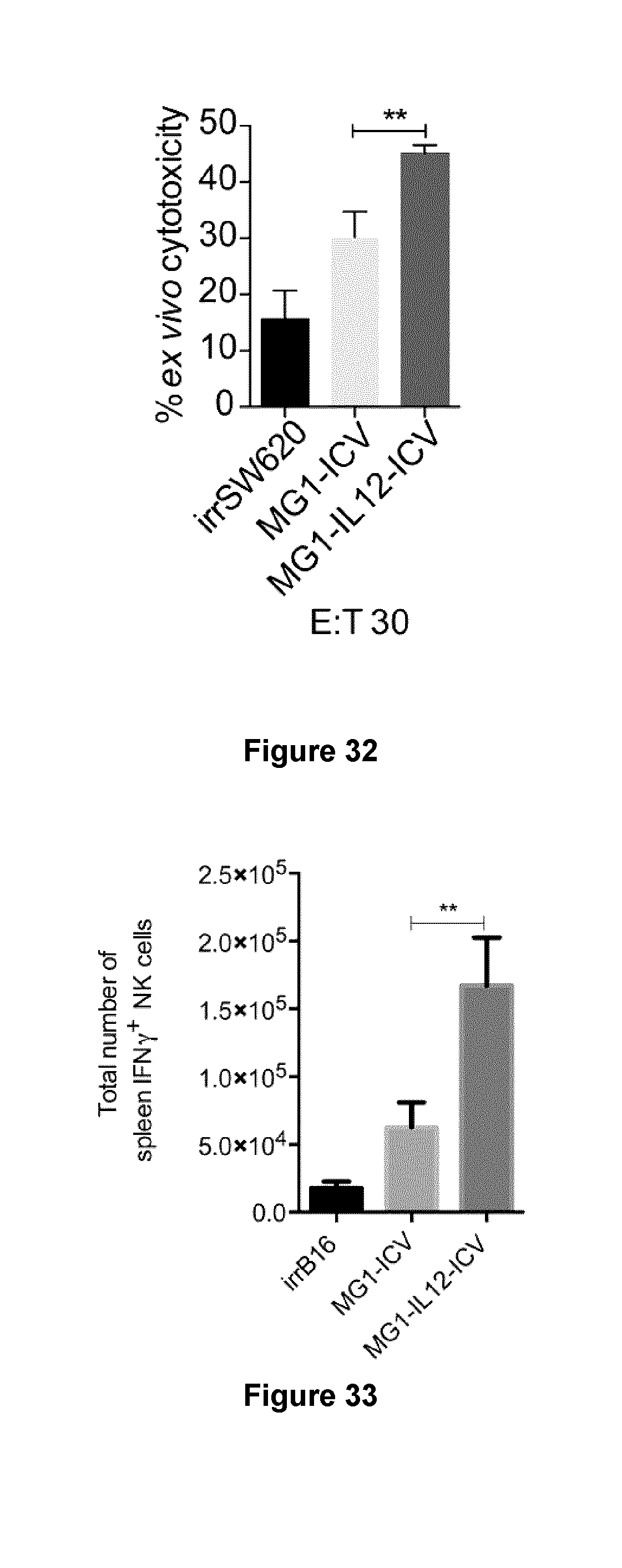
[0054] FIG. 32 shows an enhanced ex vivo cytotoxicity of isolated NK cells from human cancer patients that were cultured with SW620 cells infected with MG1-IL12 ICV at an effector:target ratio of 30:1.
[0055] FIG. 33 shows an increase in the total number of NK1.1.sup.+ IFN.gamma..sup.+ cells in the spleen of tumour naive mice after systemic IV treatment with MG1-IL12 ICV.
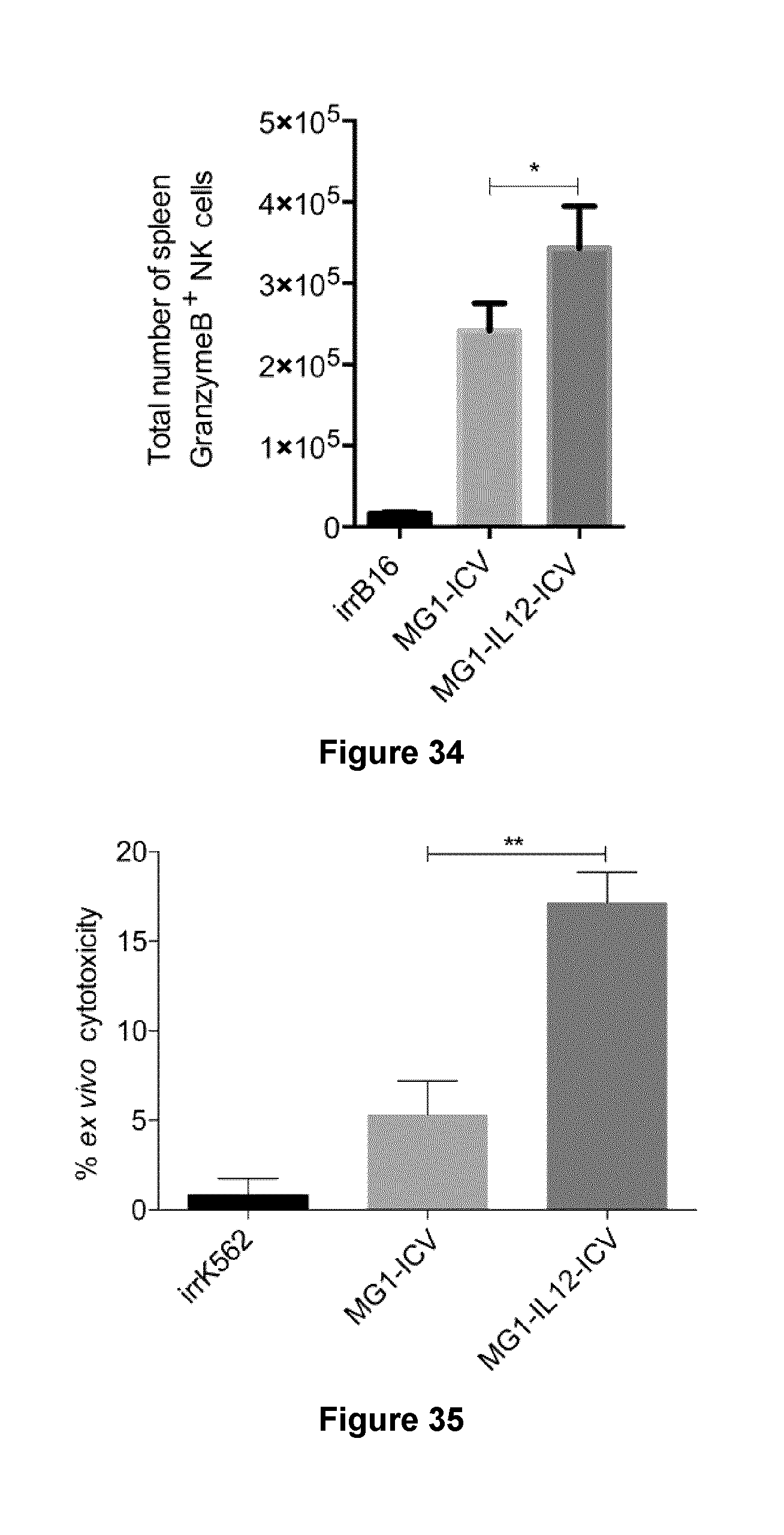
[0056] FIG. 34 shows an increase in the total number of NK1.1.sup.+ GramzymeB.sup.+ cells in the spleen of tumour naive mice after systemic IV treatment with MG1-IL12 ICV.
[0057] FIG. 35 shows results of an ex vivo chromium release cytotoxicity assay using an individual healthy patient PBMCs cultured with K562 cells infected with MG1-IL12.
[0058] FIG. 36 shows results of an ex vivo chromium release cytotoxicity assay using an individual healthy patient PBMCs cultured with K562 cells infected with MG1-IL12.
[0059] FIG. 37 shows results of an ex vivo chromium release cytotoxicity assay using an individual cancer patient PBMCs cultured with K562 cells infected with MG1-IL12.
[0060] FIG. 38 shows results of an ex vivo chromium release cytotoxicity assay using an individual cancer patient PBMCs cultured with K562 cells infected with MG1-IL12.
[0061] FIG. 39 shows the differential efficacy of using different routes of MG1-IL12 ICV administration in BALB/c mice following tumour seeding.
[0062] FIG. 40 illustrates a proposed model for the mechanism of NK cell recruitment and activation following vaccination with MG1-IL12 ICV.
DETAILED DESCRIPTION
[0063] Generally, the present disclosure provides: a Maraba virus whose genome includes a transgene sequence that encodes the cytokine IL12; an infected cell vaccine (ICV) where autologous tumour cells from a patient are infected ex vivo with an oncolytic recombinant rhabdovirus expressing IL12; a method to treat a cancer through administration of the Maraba virus or the infected cell vaccine; and use of the Maraba virus or the infected cell vaccine for treating a cancer. Without wishing to be bound by theory, the authors of the present disclosure believe that expression of the IL12 may enhance the anti-tumour immune response of the administered oncolytic virus.
[0064] Peritoneal carcinomatosis (PC) is one of the most common and problematic sites of metastases for abdominal malignancies, including gastrointestinal and ovarian cancers (7). It is a common cancer metastases that is associated with a significantly reduced quality of life, median survival rate and poor prognosis that requires new treatment options. PC poses challenges to the use of traditional chemotherapy, which cannot be used for patients who develop complications such as bowel obstruction (8). Another challenge in treating PC is the difficulty in delivering a therapeutic agent. Another obstacle to effective therapy is the toxicity and short half-life of immunomodulating agents used systemically or delivered directly to the target site.
[0065] One aspect of the present disclosure may overcome one or more of these challenges by enhancing the anti tumour immune response of an oncolytic virus. By way of example, in one particular embodiment, the patients' tumour cells are infected ex vivo with an oncolytic rhabdovirus expressing the cytokine IL12. These infected cells are then re-administered to the patient as an Infected Cell Vaccine (ICV). Without wishing to be bound by theory, the authors of the present disclosure believe that the infected tumour cells provide an immunostimulatory environment that is supplemented by the production of IL12. Expression of IL12 in situ reduces the half-life and/or toxicity drawbacks associated with high dose administration of IL12. The authors of the present disclosure believe that expression of the IL12 acts to recruit and stimulate NK cells to the tumour site, and reduce the size of the tumour. The activation of NK cells, the adaptive arm of the immune response, may confer a long-term memory and thereby reduce the possibility that the tumour will return.
[0066] Material and Methods:
[0067] Cell Lines and Mice:
[0068] Murine CT26 colon carcinoma, murine B16F10 F10 melanoma, human SW620 colorectal adenocarcinoma, human HCT15 colorectal adenocarcinoma, human A549 lung carcinoma, murine YAC-1 lymphoma, human K562 leukemic cell lines (all from American Type Tissue Collection) were propagated in Dulbecco's modified Eagle's medium (Hyclone) for the adherent cell lines, or Roswell Park Memorial Institutes Media (Hyclone) for non-adherent cell lines supplemented with 10% fetal calf serum (Cansera, Etobicoke, Ontario, Canada). Rauscher murine leukemia virus-induced T-cell lymphoma (RMA) and RMA-S (MHC-deficient variant of RMA) were obtained from Dr. A. Veillette (Institut de Recherches Clinique, Montreal, Quebec, Canada). Female Balb/C and C57BL/6 mice 6- to 8 weeks old were purchased from Charles River Laboratories (Wilmington, Mass.). Animals were housed in pathogen-free conditions and all experiments were conducted with the approval of the University of Ottawa Animal Care and Veterinary Service.
[0069] MG1-IL12 construction:
[0070] Murine IL12 was PCR amplified from pORF-mIL-12 (IL-12elasti(p35::p40)) (InvivoGen, San Diego, Calif., USA) to add MluI (5') and (3') cloning sites to facilitate cloning into Maraba MG1 (9). The recombinant MG1-IL12 virus was rescued as described previously (10). Briefly, A549 were infected with vaccinia virus expressing T7 polymerase and subsequently transfected using Lipofectamine 2000 (Invitrogen, Burlington, ON, Canada) with 2 mg of MG1-IL-12 DNA plasmid together with pCI-Neo plasmids encoding for Maraba N, P and L (1, 1.25, 0.25 mg, respectively). The rescued virus was passaged on SNB19 cells, then plaque purified, amplified and titered on Vero cells.
[0071] Viability Assays:
[0072] B16lacZ, CT26lacZ, SW620 and HCT15 cell lines were seeded into 96-well plates (2.times.10.sup.4 cells/well). 24 hours later, cells were infected with MG1 or MG1-IL12 viruses at Multiplicity of Infection (MOI) of 0.001-10 pfu/cell. Alamar Blue (Sigma-Aldrich, St Louis, Mo.) was added following 48 hours of incubation to a final concentration of 20 .mu.g/ml. The absorbance was read at a wavelength of 570 nm after 6-hour incubation.
[0073] Antibodies and FACS Analysis:
[0074] For splenic and lung lymphocyte population analyses, organs were harvested from mice and red blood cells lysed using ammonium chloride-potassium lysis (ACK) buffer. The following monoclonal antibodies were used: anti-TCR-b (H57-597), anti-NK1.1 (PK136), both from eBiosciences. Spleen and lung NK cell IFN-.gamma. and Granzyme B secretion were analysed following a 1 hour GolgiPlug (BD Biosciences) incubation using: anti-CD3 (17A2), anti-NK1.1 (PK136), anti-IFN-.gamma. (XMG1.2) and anti-Granzyme B (16G6) all from BD Biosciences. The monoclonal antibodies were used for human NK cell migration and activation are; anti-CD56 (HCD56) from Biolegend, anti-CD3 (UCHT1) and anti-CD69 (FN50) both are from eBiosciences. Fluorescence-activated cell sorting (FACS) acquisitions were conducted on a CyAn-ADP using Summit software (Beckman Coulter, Mississauga, Canada) and data were analyzed with Kaluza software (Beckman Coulter).
[0075] Ex Vivo Splenocytes Cytotoxicity Assay
[0076] The .sup.51Cr-release assay was performed as previously described (11). Briefly, splenocytes were harvested from treated and control mice two days after treatment. ACK buffer treated splenocytes were resuspended and mixed with chromium labelled YAC-1 cells at specified effector-to-target (E:T) ratios.
[0077] In Vivo Tumour Rejection Assay
[0078] The in vivo rejection assay was performed as described previously (11). Briefly, RMA and RMA-S were labeled with 5 and 0.5 mmol/L CFSE, respectively. 1.times.10.sup.6 cells containing a 1:1 mixture of each cell type was injected i.p. into C57BL/6 mice 24 hrs following ICV treatment. Peritoneal cells were collected the following day (24 hr) by washing the peritoneum with 5 mL of PBS containing 2 mmol/L EDTA. Collected cells were analysed by flow cytometry for the presence of CFSE-labeling.
[0079] Virus Infection of B16F10 Cells and Co-Culture with Bone Marrow-Derived DCs for Chemotaxis and Chemokines Analysis
[0080] B16F10 cells infected with MG1 or MG1-IL12 (MOI=0.1 pfu/cell) were harvested 18 hrs after infection and cultured with bone marrow-derived dendritic cells (DCs) described elsewhere at a 3:1 ratio in DC medium (1% FBS) (complete RPMI supplemented with 1.times. of 2-Mercapoethanol (cat #21985-023, Gibco, life technologies) in 96-wells plates (12). Media was collected after 24 hours and stored at -80.degree. C. until further analysis.
[0081] Cytokine and Chemokine Analyses
[0082] Murine IFN.gamma. from DCs co-culture supernatant were detected by FlowCytomix (eBioscience) kits as per manufacturer's instructions. For lungs IL12 and IFN.gamma. expression, lungs from C57Bl/6 mice treated with irrB16, MG1 ICV or MG1-IL12 ICV at 5.times.10.sup.5 cells/100 ul/mouse i.v., were resected and homogenized in 1 ml PBS 24-hours after treatment. Murine MCP-1, SDF-1 and IP-10 chemokines were assayed 18 hours post ICV treatment from the peritoneal fluids of C57Bl/6 mice (in vivo) or from tissue culture supernatant using ELISArray kits (SABiosciences) as per manufacturers instructions.
[0083] Murine Transwell Chemotaxis Assay
[0084] Tissue culture supernatants for assessment of chemokines or chemotaxis assay were generated in DC media. Chemotaxis of NK cells was assessed using a Transwell system as described previously (13). Briefly, 500 ul of conditioned media from DC cultures was added to the lower chamber of Transwell plates with 5-um pores (Costar, Corning). 3.times.10.sup.5 of DX5.sup.+ sorted NK cells were added to the upper chamber, and plates were incubated for 3 hours at 37.degree. C. Cells in the lower chambers were harvested, stained with trypan blue and counted. A migration percentage was calculated as (total number NK cells in bottom chamber/total number NK cell input).times.100. To calculate NK cell index: (NK cell migration percentage/NK cell migration percentage from media alone group).
[0085] Human Transwell Chemotaxis Assay
[0086] Conditioned media were generated in DC media through direct ICV-PBMCs co-culture at 3:1 ratio for 18 hours. 1.times.10.sup.6 of PBMCs were added to the upper chamber, and plates were incubated for 3 hours at 37.degree. C. Cells in the lower chambers were harvested, stained with anti-CD56 (HCD56) and anti-CD3 (UCHT1) and quantified by FACS. A migration percentage was calculated as (total number NK cells in bottom chamber/total number NK cell input).times.100. To calculate NK cell index: (NK cell migration percentage/NK cell migration percentage from media alone group).
[0087] DC-MG1-IL-12-ICV/Splenocytes Co-Cultures
[0088] DC-MG1-IL-12-ICV were isolated by MACS CD11c.sup.+ selection (Miltenyi Biotec) and co-cultured with naive splenocytes at 1:5 ratio in DC medium, at 2.times.10.sup.5 splenocytes/well in 96-well plate format. Twenty-four hours later, cell-free supernatant was stored at -80.degree. C. for measurement of IFN.gamma.. Intracellular IFN.gamma. staining on splenocytes by intracellular FACS was also performed as described above.
[0089] Mouse Models:
[0090] Therapeutic Treatment Model.
[0091] CT26 and B16F10 Peritoneal carcinomatosis in BALB/c and C57Bl/6 mice, respectively were treated with 1.times.10.sup.4 ICV on day 3 after seeding 5.times.10.sup.5 tumour cells intraperitonealy. For the CT26 bulky tumour model, 5.times.10.sup.5 tumour cells were seeded within the peritoneum and the treatment regimen of six doses of ICV was initiated following Magnetic Resonance (MR) scan confirmation of a tumour with a size of >3 mm. Animals were sedated with isoflurane gas and MR scanning was performed with a 7 Tesla GE/Agilent MR 901 (GE Healthcare, Chicago, USA). For each mouse, three MR pulse sequences were used: one localizer and two fast spin echo (FSE) sequences in the coronal and axial planes. The parameters for the FSE sequences were: echo train length 8, bandwidth=16 kHz, echo time=42 ms, repetition time=1500 ms, field of view=35 mm, matrix 256.times.256, slice thickness=1 mm. The total MR scan time per mouse was approximately 15 minutes. Follow-up MR scans were performed one week, six weeks and thirteen weeks post-treatment start using the same MR scan parameters.
[0092] Prophylactic Treatment Model.
[0093] C57Bl/6 mice were vaccinated with single dose of 1.times.10.sup.3 irrB16, MG1 ICV or MG1-IL-12-ICV ip The following day, mice were challenged with 3.times.10.sup.5 B16F10-LacZ cells IV, sacrificed at 4 days after tumour cells injection followed by staining and quantification of lung metastases with X-gal (Bioshop, Burlington, Canada) as described previously (14). The total number of lung surface metastases was determined on all lung lobes using a stereomicroscope (Leica Microsystems, Concord, Canada).
[0094] Statistical Analysis
[0095] All statistical analyses were determined using GraphPad Prism 6.0 software. Statistical significance was determined by the Student t test with a cut off P=0.05. Data are presented as .+-.SD.
[0096] Characterization of an MG1 Oncolytic Virus Encoding Murine IL12 (MG1-IL12)
[0097] A murine IL12 transgene (p70), which is composed of p35 and p40 subunits, was incorporated into the backbone of the oncolytic Maraba virus variant MG1 to create MG1-IL12 (FIG. 1). This replication competent oncolytic virus was found to infect both murine and human tumour cell lines with an efficiency comparable to parental MG1 and expression of IL12 did not negatively impact viral replication or spreading (FIGS. 2 and 3). Furthermore, IL12 was detected in the culture media of B16F10 (22 pg/cell) and CT26 (180 pg/cell) cells infected with MG1-IL12 (FIGS. 4 and 5). Together these results demonstrate that MG1-IL-12 can successfully infect murine tumour cells resulting in viral replication and IL-12 secretion, resulting in an MG1-IL12 infected cell vaccine (ICV).
Examples
[0098] MG1-IL12 ICV Enhances NK Cell-Mediated Tumour Rejection.
[0099] The authors of the present disclosure have previously demonstrated that infecting autologous tumor cells ex vivo with oncolytic viruses can elicit a robust immune response against established, non-permissive, tumors in vivo (15). To determine whether MG1 and MG1-IL12 could similarly induce an immune response when used as an ICV, the authors intravenously (i.v.) injected 5.times.10.sup.5 -irradiated B16F10 cells either mock infected or infected with MG1 or MG1-IL12. The authors have previously shown that i.v. administration of ICVs is associated with a rapid and dose-dependent accumulation of injected cells which persist in the lung for up to 1 day in tumor free animals (16). Following ICV delivery, significantly higher levels of IL12 were detected in lung homogenates from mice receiving MG1-IL12 ICV in comparison to animals receiving cells alone or MG1 ICV (FIG. 6, t=24 hr). To determine whether the increased concentrations of IL12 had any functional effect, the levels of the IL12 responsive cytokine IFN.gamma. were measured. In agreement with the increase in IL12, levels of IFN.gamma. were also elevated in the lungs of mice treated with MG1-IL12 ICV compared to mice receiving MG1 ICV or irradiated cells (FIG. 7). Since IL12 targets both NK and T cells to promote IFN.gamma. secretion (17), the authors next sought to determine which cell types were responding to treatment with our MG1-IL12 ICV. Interestingly, vaccination with MG1-IL12 ICV was not found to impact the total number of T cells in the lung, however, a 3-fold increase in the total number of NK cells present in the lung was observed suggesting that MG1-IL12 ICV enhances NK cell recruitment (FIGS. 8 and 9). In addition, the total number of IFN.gamma. and granzyme B positive NK cells was increased approximately 7 and 4-fold respectively following injection of MG1-IL12 ICV indicating an increase in NK cell activation (FIGS. 10 and 11). To further examine the effect, the cytotoxic activity of NK cells against YAC-1 target cells was measured ex vivo and it was observed that splenocytes isolated from MG1-IL-12 ICV treated mice exhibited a significantly higher level of YAC-1 killing (FIG. 12). These data supported a role for MG1-IL12 ICV in promoting NK cell recruitment to the site of delivery and a concomitant systemic activation of splenic NK cells (FIGS. 33 and 34). In order to determine if this effect translated into improved tumour clearance, B16F10 lung metastases were treated with either irradiated cells, MG1-IL12 ICV, or MG1 ICV alone by i.v. delivery (FIG. 13). Systemic delivery of MG1-IL12 ICV was sufficient to significantly attenuate the number of detectable lung metastasis in comparison to treatment with MG1 ICV or irradiated cells. These results suggest that MG1-IL12 ICV can stimulate NK cell recruitment and effector function to significantly improve the antitumour efficacy of the infected cell vaccines.
[0100] MG1-IL12 ICV Enhances NK Cell Activation and Improves Survival in a Model of Peritoneal Carcinomatosis.
[0101] The initial findings suggest that the improved anti-tumor response elicited by MG1-IL12 ICV in comparison to MG1 ICV are in part due to potent chemotactic properties of IL12 which contribute to the enhanced recruitment of cytotoxic NK cells to the site of delivery (FIGS. 9,10 and 11). Therefore, the authors next sought to assess whether vaccinating mice i.p. with MG1-IL12 ICV could improve clearance of tumors within the peritoneal cavity and promote improved survival. Similar to their previous observations with i.v. vaccination, the authors observed an increased proportion of NK cells (19% vs 49%, p=0.0073) in the peritoneum 24 hours after i.p. vaccination as compared to MG1 ICV (FIG. 14). The infiltrating NK cells also displayed a significant upregulation of the activation marker CD69 indicating that the NK cells accumulating in the peritoneum in MG1-IL12 ICV vaccinated mice were more highly activated (FIG. 15) (18, 19). To complement these findings, an in-vivo NK cell cytotoxicity assay was performed by challenging vaccinated mice with the NK-sensitive RMA-S and parental RMA tumour cell lines to investigate whether the activated NK cells that migrated into the peritoneal cavity were tumorcidal. Following vaccination with MG1-IL12 ICV, tumour cell clearance was significantly improved demonstrating that ICV-mediated recruitment and activation of NK cells can effectively promote tumor cell clearance from the peritoneum (FIG. 16). In support of this conclusion, the protective effect of vaccinating mice bearing B16F10 peritoneal tumors with MG1-IL12 ICV was completely abrogated upon depletion of NK cells further suggesting that the therapeutic benefit of this treatment strategy is dependent upon NK cell recruitment and activation (FIGS. 17 and 18).
[0102] NK Cell Activation and Migration in Response to MG1-IL12 ICV is Partly Dependent Upon the Secretion of IP10 from Dendritic Cells.
[0103] The data clearly establish for the ability of MG1-IL12 ICV to promote NK cell activation, migration and function, however, it was unclear whether dendritic cells (DCs), a key mediator of NK cell function in vivo were involved in this process. To understand the interaction between NK cells and DC in the presence of MG1-IL12, the authors quantified IFN.gamma. production from splenocytes cultured in the presence of bone marrow derived DCs, which were either untreated or cultured with mock, MG1, or MG1-IL12 infected B16F10 cells. Notably, the authors found that splenocytes cultured with DCs previously exposed to MG1-IL12 ICV resulted in a significant increase in NK cell-specific IFN.gamma. secretion suggesting DCs promote NK cell cytokine secretion (FIGS. 19 and 20). A subsequent step was to investigate the ability of DCs to promote NK cell migration using an in vitro transwell chemotaxis assay. The migration of NK cells across a 5 um membrane was found to be significantly increased by either MG1 ICV or MG1-IL-12 ICV, however MG1-IL12 ICV resulted in a higher percentage of migrating NK cells (FIG. 21). NK cell migration was further increased by media conditioned in the presence of DCs suggesting that DCs provide the stimuli for increased NK cell activation and migration. Next, the authors sought to identify which chemokines commonly secreted by DCs were mediating the observed effects. Despite the inability to detect any effect on MCP-1 (monocytic chemotactic protein-1) and SDF-1 (stromal cell-derived factor-1) secretion, MG1-IL12 ICV was found to induce a significant increase in IP-10 (IFN-inducible Protein-10) (FIG. 22). The neutralization of IP-10 in conditioned media derived from DCs cultured with MG1-IL12 ICV significantly inhibited the migratory capacity of NK cells in vitro confirming its central role (FIG. 23).
[0104] MG1-IL12 ICV is Effective in Treating Established Peritoneal Disease in Mice.
[0105] Together the findings suggest that the MG1-IL12 ICV can significantly slow the outgrowth of B16F10 tumours within the peritoneal compartment by stimulating the recruitment of activated NK cells. Since peritoneal carcinomatosis is a common presentation for late stage gastrointestinal and gynecological malignancies, the authors of the present disclosure sought to determine whether the MG1-IL12 ICV could provide therapeutic benefit in a clinically relevant model of colon cancer (CT26) with peritoneal disease at time of treatment. To accomplish this BALB/c mice were seeded with CT26 tumour cells (FIG. 24). Three days later mice were treated with a single dose of irradiated cells alone, virus alone or the infected cell vaccines. Mice treated with irradiated CT26 cells, MG1, MG1-IL12 and MG1 ICV all had significantly lower median survival times and increased peritoneal tumour burden in comparison to mice receiving MG1-IL12 ICV (>90% 26 cured/28 of mice survived have survived >200 days, FIGS. 24 and 25). Interestingly, the cured mice developed a long lasting immunity such that when the surviving mice were re-challenged with 5.times.10.sup.5 CT26 cells on the flank, 148 days after treatment, they rejected the tumors (5/5 mice). However, this anti-tumor memory immune response was specific to CT26 tumors and all mice developed tumors (5/5 mice) when challenged with syngeneic 4T1 tumor cells on the opposite flank. Surprisingly, the route of vaccination plays an import role in MG1-IL12 ICV conferred efficacy, in that treatment given intraperitoneally has superior efficacy compared to intravenous or subcutaneous injections (FIG. 39).
[0106] Next, the authors sought to measure the effects of treatment in established bulky tumours. Between day 10 and 17 following implantation, tumors were visualized by MRI and mice bearing significant tumour masses (Class 1>8 mm and Class 2>3 mm) were randomly allocated into a treatment group prior to treatment with 6 doses of irradiated cells, MG1 ICV or MG1-IL12 ICV administered over a two week period (FIG. 26). Despite the lethal tumour burden, evident by the loss of all animals treated with irradiated cells by day 15, MG1-IL12 ICV provided complete protection (21/21 survived>80 days, study ongoing). Strikingly, follow-up MRI scans which confirmed the presence of large tumour masses at the early stages of treatment were dramatically reduced at later time points (FIG. 27). Collectively, these results demonstrate that MG1-IL12 ICV is an effective approach for promoting the clearance of large, established tumours within the peritoneum in a murine model of peritoneal carcinomatosis.
[0107] MG1-IL-12 ICV Enhances Human NK Cell Cytoxicity and Migratory Capacity.
[0108] Given the fact that murine p40 and p35 subunits of IL-12 share 70% and 60% homology with their human counterparts respectively, they are able to functionally activate human NK and T cells (20). The authors next sought to confirm that the vaccine could elicit a similar effect on human NK cells ex vivo. To accomplish this, irradiated SW620 colon cancer cells were infected with MG1 or MG1-IL12 and cultured with peripheral blood mononuclear cells (PBMCs) isolated from a healthy donor as part of a (Perioperative Blood Collection Protocol approved by the Ottawa Health Science Network Research Ethics Board #2011884). In agreement with previous findings, MG1-IL12 ICV resulted in a significant increase in the expression of CD69, an established marker of NK cell activation, in the NK cell (CD56.sup.+ CD3.sup.-) subset of PBMCs (FIG. 28). In addition, IP-10 chemotactic protein secretion was also significantly increased in the supernatant of PBMCs co-culture with MG1-IL12 ICV. This supernatant enhanced the migration of NK cells in the ex vivo transwell system suggesting that MG1-IL12 ICV vaccine elicits similar responses from NK cells of human and murine origin (FIGS. 29 and 30). Finally, stimulating PBMCs with MG1-IL12 ICV resulted in an increased cytotoxic activity towards K562 target tumour cells suggesting that the activation and enhanced migratory capacity of human NK cells cultured in the presence of MG1-IL12 ICV is associated with increased ability of to eradicate tumour cells (FIGS. 31, 32, 35, 36, 37 and 38). Together these results provide support for the hypothesis that autologous infected tumour cell vaccines may provide a much needed therapeutic benefit in the treatment of patients with PC.
[0109] While the above examples demonstrate the efficacy of a particular Maraba virus in mice, the authors believe that Maraba viruses and ICVs according to the present disclosure will also address or ameliorate one or more shortcomings involved with oncolytic virus treatment of cancer in humans.
[0110] Peritoneal carcinamatosis is used as an example of a cancer presentation that can be treated using a Maraba virus according to the present disclosure. The authors believe that other tumour types, and tumours in other locations, would also be amenable to treatment with Maraba viruses and ICVs according to the present disclosure.
[0111] In the preceding description, for purposes of explanation, numerous details are set forth in order to provide a thorough understanding of the examples. However, it will be apparent to one skilled in the art that these specific details are not required. Accordingly, what has been described is merely illustrative of the application of the described examples and numerous modifications and variations are possible in light of the above teachings.
[0112] Since the above description provides examples, it will be appreciated that modifications and variations can be effected to the particular examples by those of skill in the art. Accordingly, the scope of the claims should not be limited by the particular examples set forth herein, but should be construed in a manner consistent with the specification as a whole.
REFERENCES
[0113] 1. de Gruijl T D, van den Eertwegh A J, Pinedo H M, Scheper R J: Whole-cell cancer vaccination: from autologous to allogeneic tumor- and dendritic cell-based vaccines. Cancer Immunol Immunother 2008, 57(10):1569-1577. [0114] 2. Chiang C L, Coukos G, Kandalaft L E: Whole Tumor Antigen Vaccines: Where Are We? Vaccines (Basel) 2015, 3(2):344-372. [0115] 3. Srivatsan S, Patel J M, Bozeman E N, Imasuen I E, He S, Daniels D, Selvaraj P: Allogeneic tumor cell vaccines: the promise and limitations in clinical trials. Hum Vaccin Immunother 2014, 10(1):52-63. [0116] 4. Campbell C T, Gulley J L, Oyelaran O, Hodge J W, Schlom J, Gildersleeve J C: Humoral response to a viral glycan correlates with survival on PROSTVAC-VF. Proc Natl Acad Sci USA 2014, 111(17):E1749-1758. [0117] 5. Tugues S, Burkhard S H, Ohs I, Vrohlings M, Nussbaum K, Vom Berg J, Kulig P, Becher B: New insights into IL-12-mediated tumor suppression. Cell Death Differ 2015, 22(2):237-246. [0118] 6. Brun J, McManus D, Lefebvre C, Hu K, Falls T, Atkins H, Bell J C, McCart J A, Mahoney D, Stojdl D F: Identification of genetically modified Maraba virus as an oncolytic rhabdovirus. Mol Ther 2010, 18(8):1440-1449. [0119] 7. Mohamed F, Cecil T, Moran B, Sugarbaker P: A new standard of care for the management of peritoneal surface malignancy. Curr Oncol 2011, 18(2):e84-96. [0120] 8. Aoyagi T, Terracina K P, Raza A, Takabe K: Current treatment options for colon cancer peritoneal carcinomatosis. World J Gastroenterol 2014, 20(35):12493-12500. [0121] 9. Labbe A, Nelles M, Walia J, Jia L, Furlonger C, Nonaka T, Medin J A, Paige C J: IL-12 immunotherapy of murine leukaemia: comparison of systemic versus gene modified cell therapy. J Cell Mol Med 2009, 13(8B):1962-1976. [0122] 10. Vaillant J C, Nordlinger B, Deuffic S, Arnaud J P, Pelissier E, Favre J P, Jaeck D, Fourtanier G, Grandjean J P, Marre P et al: Adjuvant intraperitoneal 5-fluorouracil in high-risk colon cancer: A multicenter phase III trial. Ann Surg 2000, 231(4):449-456. [0123] 11. Mortarini R, Borri A, Tragni G, Bersani I, Vegetti C, Bajetta E, Pilotti S, Cerundolo V, Anichini A: Peripheral burst of tumor-specific cytotoxic T lymphocytes and infiltration of metastatic lesions by memory CD8+ T cells in melanoma patients receiving interleukin 12. Cancer Res 2000, 60(13):3559-3568. [0124] 12. Lutz M B, Kukutsch N, Ogilvie A L, Rossner S, Koch F, Romani N, Schuler G: An advanced culture method for generating large quantities of highly pure dendritic cells from mouse bone marrow. J Immunol Methods 1999, 223(1):77-92. [0125] 13. Cheng M, Chen Y, Xiao W, Sun R, Tian Z: NK cell-based immunotherapy for malignant diseases. Cell Mol Immunol 2013, 10(3):230-252. [0126] 14. Ardolino M, Azimi C S, lannello A, Trevino T N, Horan L, Zhang L, Deng W, Ring A M, Fischer S, Garcia K C et al: Cytokine therapy reverses NK cell anergy in MHC-deficient tumors. J Clin Invest 2014, 124(11):4781-4794. [0127] 15. Lemay C G, Rintoul J L, Kus A, Paterson J M, Garcia V, Falls T J, Ferreira L, Bridle B W, Conrad D P, Tang V A et al: Harnessing oncolytic virus-mediated antitumor immunity in an infected cell vaccine. Mol Ther 2012, 20(9):1791-1799. [0128] 16. Power A T, Wang J, Falls T J, Paterson J M, Parato K A, Lichty B D, Stojdl D F, Forsyth P A, Atkins H, Bell J C: Carrier cell-based delivery of an oncolytic virus circumvents antiviral immunity. Mol Ther 2007, 15(1):123-130. [0129] 17. Watford W T, Moriguchi M, Morinobu A, O'Shea J J: The biology of IL-12: coordinating innate and adaptive immune responses. Cytokine Growth Factor Rev 2003, 14(5):361-368. [0130] 18. Hara T, Jung L K, Bjorndahl J M, Fu S M: Human T cell activation. Ill. Rapid induction of a phosphorylated 28 kD/32 kD disulfide-linked early activation antigen (EA 1) by 12-o-tetradecanoyl phorbol-13-acetate, mitogens, and antigens. J Exp Med 1986, 164(6):1988-2005. [0131] 19. Lanier L L, Buck D W, Rhodes L, Ding A, Evans E, Barney C, Phillips J H: Interleukin 2 activation of natural killer cells rapidly induces the expression and phosphorylation of the Leu-23 activation antigen. J Exp Med 1988, 167(5):1572-1585. [0132] 20. Schoenhaut D S, Chua A O, Wolitzky A G, Quinn P M, Dwyer C M, McComas W, Familletti P C, Gately M K, Gubler U: Cloning and expression of murine IL-12. J Immunol 1992, 148(11):3433-3440.
TABLE-US-00001 [0132] APPENDIX A Sequences: SEQ ID NO: 1-Human IL12 p35 subunit mwppgsasqp ppspaaatgl hpaarpvslq crlsmcpars lllvatlvll dhlslarnlp 61 vatpdpgmfp clhhsqnllr avsnmlqkar qtlefypcts eeidheditk dktstveacl 121 pleltknesc lnsretsfit ngsclasrkt sfmmalclss iyedlkmyqv efktmnakll 181 mdpkrqifld qnmlavidel mqalnfnset vpqkssleep dfyktkiklc illhafrira 241 vtidrvmsyl nas SEQ ID NO: 2-Human IL12 p40 subunit mchqqlvisw fslvflaspl vaiwelkkdv yvveldwypd apgemvvltc dtpeedgitw 61 tldqssevlg sgktltiqvk efgdagqytc hkggevlshs llllhkkedg iwstdilkdq 121 kepknktflr ceaknysgrf tcwwlttist dltfsvkssr gssdpqgvtc gaatlsaerv 181 rgdnkeyeys vecqedsacp aaeeslpiev mvdavhklky enytssffir diikpdppkn 241 lqlkplknsr qvevsweypd twstphsyfs ltfcvqvqgk skrekkdrvf tdktsatvic 301 rknasisvra qdryysssws ewasvpcs SEQ ID NO: 3-Mouse IL12 p35 subunit mvsvptasps asssssqcrs smcqsryllf latlallnhl slarvipvsg parclsqsrn 61 llkttddmvk tareklkhys ctaedidhed itrdqtstlk tclplelhkn esclatrets 121 sttrgsclpp qktslmmtlc lgsiyedlkm yqtefqaina alqnhnhqqi ildkgmlvai 181 delmqslnhn getlrqkppv geadpyrvkm klcillhafs trvvtinrvm gylssa SEQ ID NO: 4-Mouse IL12 p40 subunit mcpqkltisw faivllvspl mamwelekdv yvvevdwtpd apgetvnltc dtpeedditw 61 tsdqrhgvig sgktltitvk efldagqytc hkggetlshs hlllhkkeng iwsteilknf 121 knktflkcea pnysgrftcs wlvqrnmdlk fniksssssp dsravtcgma slsaekvtld 181 qrdyekysvs cqedvtcpta eetlpielal earqqnkyen ystsffirdi ikpdppknlq 241 mkplknsqve vsweypdsws tphsyfslkf fvriqrkkek mketeegcnq kgaflvekts 301 tevqckggnv cvqaqdryyn sscskwacvp crvrs
Sequence CWU 1
1
41253PRTHomo sapiens 1Met Trp Pro Pro Gly Ser Ala Ser Gln Pro Pro
Pro Ser Pro Ala Ala1 5 10 15Ala Thr Gly Leu His Pro Ala Ala Arg Pro
Val Ser Leu Gln Cys Arg 20 25 30Leu Ser Met Cys Pro Ala Arg Ser Leu
Leu Leu Val Ala Thr Leu Val 35 40 45Leu Leu Asp His Leu Ser Leu Ala
Arg Asn Leu Pro Val Ala Thr Pro 50 55 60Asp Pro Gly Met Phe Pro Cys
Leu His His Ser Gln Asn Leu Leu Arg65 70 75 80Ala Val Ser Asn Met
Leu Gln Lys Ala Arg Gln Thr Leu Glu Phe Tyr 85 90 95Pro Cys Thr Ser
Glu Glu Ile Asp His Glu Asp Ile Thr Lys Asp Lys 100 105 110Thr Ser
Thr Val Glu Ala Cys Leu Pro Leu Glu Leu Thr Lys Asn Glu 115 120
125Ser Cys Leu Asn Ser Arg Glu Thr Ser Phe Ile Thr Asn Gly Ser Cys
130 135 140Leu Ala Ser Arg Lys Thr Ser Phe Met Met Ala Leu Cys Leu
Ser Ser145 150 155 160Ile Tyr Glu Asp Leu Lys Met Tyr Gln Val Glu
Phe Lys Thr Met Asn 165 170 175Ala Lys Leu Leu Met Asp Pro Lys Arg
Gln Ile Phe Leu Asp Gln Asn 180 185 190Met Leu Ala Val Ile Asp Glu
Leu Met Gln Ala Leu Asn Phe Asn Ser 195 200 205Glu Thr Val Pro Gln
Lys Ser Ser Leu Glu Glu Pro Asp Phe Tyr Lys 210 215 220Thr Lys Ile
Lys Leu Cys Ile Leu Leu His Ala Phe Arg Ile Arg Ala225 230 235
240Val Thr Ile Asp Arg Val Met Ser Tyr Leu Asn Ala Ser 245
2502328PRTHomo sapiens 2Met Cys His Gln Gln Leu Val Ile Ser Trp Phe
Ser Leu Val Phe Leu1 5 10 15Ala Ser Pro Leu Val Ala Ile Trp Glu Leu
Lys Lys Asp Val Tyr Val 20 25 30Val Glu Leu Asp Trp Tyr Pro Asp Ala
Pro Gly Glu Met Val Val Leu 35 40 45Thr Cys Asp Thr Pro Glu Glu Asp
Gly Ile Thr Trp Thr Leu Asp Gln 50 55 60Ser Ser Glu Val Leu Gly Ser
Gly Lys Thr Leu Thr Ile Gln Val Lys65 70 75 80Glu Phe Gly Asp Ala
Gly Gln Tyr Thr Cys His Lys Gly Gly Glu Val 85 90 95Leu Ser His Ser
Leu Leu Leu Leu His Lys Lys Glu Asp Gly Ile Trp 100 105 110Ser Thr
Asp Ile Leu Lys Asp Gln Lys Glu Pro Lys Asn Lys Thr Phe 115 120
125Leu Arg Cys Glu Ala Lys Asn Tyr Ser Gly Arg Phe Thr Cys Trp Trp
130 135 140Leu Thr Thr Ile Ser Thr Asp Leu Thr Phe Ser Val Lys Ser
Ser Arg145 150 155 160Gly Ser Ser Asp Pro Gln Gly Val Thr Cys Gly
Ala Ala Thr Leu Ser 165 170 175Ala Glu Arg Val Arg Gly Asp Asn Lys
Glu Tyr Glu Tyr Ser Val Glu 180 185 190Cys Gln Glu Asp Ser Ala Cys
Pro Ala Ala Glu Glu Ser Leu Pro Ile 195 200 205Glu Val Met Val Asp
Ala Val His Lys Leu Lys Tyr Glu Asn Tyr Thr 210 215 220Ser Ser Phe
Phe Ile Arg Asp Ile Ile Lys Pro Asp Pro Pro Lys Asn225 230 235
240Leu Gln Leu Lys Pro Leu Lys Asn Ser Arg Gln Val Glu Val Ser Trp
245 250 255Glu Tyr Pro Asp Thr Trp Ser Thr Pro His Ser Tyr Phe Ser
Leu Thr 260 265 270Phe Cys Val Gln Val Gln Gly Lys Ser Lys Arg Glu
Lys Lys Asp Arg 275 280 285Val Phe Thr Asp Lys Thr Ser Ala Thr Val
Ile Cys Arg Lys Asn Ala 290 295 300Ser Ile Ser Val Arg Ala Gln Asp
Arg Tyr Tyr Ser Ser Ser Trp Ser305 310 315 320Glu Trp Ala Ser Val
Pro Cys Ser 3253236PRTMus musculus 3Met Val Ser Val Pro Thr Ala Ser
Pro Ser Ala Ser Ser Ser Ser Ser1 5 10 15Gln Cys Arg Ser Ser Met Cys
Gln Ser Arg Tyr Leu Leu Phe Leu Ala 20 25 30Thr Leu Ala Leu Leu Asn
His Leu Ser Leu Ala Arg Val Ile Pro Val 35 40 45Ser Gly Pro Ala Arg
Cys Leu Ser Gln Ser Arg Asn Leu Leu Lys Thr 50 55 60Thr Asp Asp Met
Val Lys Thr Ala Arg Glu Lys Leu Lys His Tyr Ser65 70 75 80Cys Thr
Ala Glu Asp Ile Asp His Glu Asp Ile Thr Arg Asp Gln Thr 85 90 95Ser
Thr Leu Lys Thr Cys Leu Pro Leu Glu Leu His Lys Asn Glu Ser 100 105
110Cys Leu Ala Thr Arg Glu Thr Ser Ser Thr Thr Arg Gly Ser Cys Leu
115 120 125Pro Pro Gln Lys Thr Ser Leu Met Met Thr Leu Cys Leu Gly
Ser Ile 130 135 140Tyr Glu Asp Leu Lys Met Tyr Gln Thr Glu Phe Gln
Ala Ile Asn Ala145 150 155 160Ala Leu Gln Asn His Asn His Gln Gln
Ile Ile Leu Asp Lys Gly Met 165 170 175Leu Val Ala Ile Asp Glu Leu
Met Gln Ser Leu Asn His Asn Gly Glu 180 185 190Thr Leu Arg Gln Lys
Pro Pro Val Gly Glu Ala Asp Pro Tyr Arg Val 195 200 205Lys Met Lys
Leu Cys Ile Leu Leu His Ala Phe Ser Thr Arg Val Val 210 215 220Thr
Ile Asn Arg Val Met Gly Tyr Leu Ser Ser Ala225 230 2354335PRTMus
musculus 4Met Cys Pro Gln Lys Leu Thr Ile Ser Trp Phe Ala Ile Val
Leu Leu1 5 10 15Val Ser Pro Leu Met Ala Met Trp Glu Leu Glu Lys Asp
Val Tyr Val 20 25 30Val Glu Val Asp Trp Thr Pro Asp Ala Pro Gly Glu
Thr Val Asn Leu 35 40 45Thr Cys Asp Thr Pro Glu Glu Asp Asp Ile Thr
Trp Thr Ser Asp Gln 50 55 60Arg His Gly Val Ile Gly Ser Gly Lys Thr
Leu Thr Ile Thr Val Lys65 70 75 80Glu Phe Leu Asp Ala Gly Gln Tyr
Thr Cys His Lys Gly Gly Glu Thr 85 90 95Leu Ser His Ser His Leu Leu
Leu His Lys Lys Glu Asn Gly Ile Trp 100 105 110Ser Thr Glu Ile Leu
Lys Asn Phe Lys Asn Lys Thr Phe Leu Lys Cys 115 120 125Glu Ala Pro
Asn Tyr Ser Gly Arg Phe Thr Cys Ser Trp Leu Val Gln 130 135 140Arg
Asn Met Asp Leu Lys Phe Asn Ile Lys Ser Ser Ser Ser Ser Pro145 150
155 160Asp Ser Arg Ala Val Thr Cys Gly Met Ala Ser Leu Ser Ala Glu
Lys 165 170 175Val Thr Leu Asp Gln Arg Asp Tyr Glu Lys Tyr Ser Val
Ser Cys Gln 180 185 190Glu Asp Val Thr Cys Pro Thr Ala Glu Glu Thr
Leu Pro Ile Glu Leu 195 200 205Ala Leu Glu Ala Arg Gln Gln Asn Lys
Tyr Glu Asn Tyr Ser Thr Ser 210 215 220Phe Phe Ile Arg Asp Ile Ile
Lys Pro Asp Pro Pro Lys Asn Leu Gln225 230 235 240Met Lys Pro Leu
Lys Asn Ser Gln Val Glu Val Ser Trp Glu Tyr Pro 245 250 255Asp Ser
Trp Ser Thr Pro His Ser Tyr Phe Ser Leu Lys Phe Phe Val 260 265
270Arg Ile Gln Arg Lys Lys Glu Lys Met Lys Glu Thr Glu Glu Gly Cys
275 280 285Asn Gln Lys Gly Ala Phe Leu Val Glu Lys Thr Ser Thr Glu
Val Gln 290 295 300Cys Lys Gly Gly Asn Val Cys Val Gln Ala Gln Asp
Arg Tyr Tyr Asn305 310 315 320Ser Ser Cys Ser Lys Trp Ala Cys Val
Pro Cys Arg Val Arg Ser 325 330 335
D00000
D00001
D00002
D00003
D00004
D00005
D00006
D00007
D00008
D00009
D00010
D00011
D00012

D00013

D00014

D00015

D00016

D00017
D00018
D00019

D00020

D00021

P00001

S00001
XML
uspto.report is an independent third-party trademark research tool that is not affiliated, endorsed, or sponsored by the United States Patent and Trademark Office (USPTO) or any other governmental organization. The information provided by uspto.report is based on publicly available data at the time of writing and is intended for informational purposes only.
While we strive to provide accurate and up-to-date information, we do not guarantee the accuracy, completeness, reliability, or suitability of the information displayed on this site. The use of this site is at your own risk. Any reliance you place on such information is therefore strictly at your own risk.
All official trademark data, including owner information, should be verified by visiting the official USPTO website at www.uspto.gov. This site is not intended to replace professional legal advice and should not be used as a substitute for consulting with a legal professional who is knowledgeable about trademark law.