Delivery Of Nucleic Acids, Proteins And Small Molecules In Vitreous Vesicular Bodies
PENA; John TG ; et al.
U.S. patent application number 16/332315 was filed with the patent office on 2019-07-18 for delivery of nucleic acids, proteins and small molecules in vitreous vesicular bodies. The applicant listed for this patent is CORNELL UNIVERSITY. Invention is credited to Donald J. D'AMICO, Mrinali Patel GUPTA, John TG PENA.
| Application Number | 20190216857 16/332315 |
| Document ID | / |
| Family ID | 61561697 |
| Filed Date | 2019-07-18 |


View All Diagrams
| United States Patent Application | 20190216857 |
| Kind Code | A1 |
| PENA; John TG ; et al. | July 18, 2019 |
DELIVERY OF NUCLEIC ACIDS, PROTEINS AND SMALL MOLECULES IN VITREOUS VESICULAR BODIES
Abstract
The present invention relates to compositions of aqueous humor and/or vitreous humor derived extracellular vesicles and their use for the delivery of therapeutic agents to ocular tissues for the treatment of ophthalmic diseases. Further disclosed are methods of making the compositions. Methods of treating and diagnosing an ocular condition are also disclosed.
| Inventors: | PENA; John TG; (New York, NY) ; GUPTA; Mrinali Patel; (New York, NY) ; D'AMICO; Donald J.; (New York, NY) | ||||||||||
| Applicant: |
|
||||||||||
|---|---|---|---|---|---|---|---|---|---|---|---|
| Family ID: | 61561697 | ||||||||||
| Appl. No.: | 16/332315 | ||||||||||
| Filed: | September 9, 2017 | ||||||||||
| PCT Filed: | September 9, 2017 | ||||||||||
| PCT NO: | PCT/US17/50854 | ||||||||||
| 371 Date: | March 11, 2019 |
Related U.S. Patent Documents
| Application Number | Filing Date | Patent Number | ||
|---|---|---|---|---|
| 62385711 | Sep 9, 2016 | |||
| Current U.S. Class: | 1/1 |
| Current CPC Class: | C12N 15/11 20130101; A61P 27/02 20180101; A61K 35/30 20130101; A61K 9/0048 20130101; A61K 38/385 20130101; C12N 15/1137 20130101; C12N 2320/32 20130101; A61K 38/00 20130101; C12N 15/88 20130101; C12N 2310/14 20130101 |
| International Class: | A61K 35/30 20060101 A61K035/30; A61K 38/38 20060101 A61K038/38; A61K 9/00 20060101 A61K009/00; C12N 15/113 20060101 C12N015/113 |
Goverment Interests
[0002] This invention was made with government support under grant number UL1 TR000457-06 from the National Institutes of Health. The government has certain rights in this invention.
Claims
1. A composition comprising: one or more aqueous humor and/or vitreous humor extracellular vesicle bodies, wherein said extracellular vesicle bodies are modified to contain one or more exogenous agents.
2. The composition of claim 1, wherein the one or more exogenous agents is selected from the group consisting of a nucleic acid molecule, a protein or polypeptide, a small molecule, a hormone, and any combination thereof.
3. The composition of claim 2, wherein the exogenous agent comprises a nucleic acid molecule selected from the group consisting of a ribonucleic acid, small RNA molecule, complementary RNA, a non-coding RNA molecule, siRNA, a pi-RNA molecule, a micro-RNA molecule, a sno-RNA molecule, long non-coding RNA molecule, messenger RNA molecule, ribosomal RNA molecule, an antisense nucleic acid molecule, Locked Nucleic Acid (LNA), antagomir, CRISPR/Cas gene editing RNA, trans-activating crRNA (tracrRNA), short synthetic RNA composed of a "scaffold" sequence (gRNA), Small Cajal body-specific RNAs (scaRNA), natural cis-antisense siRNAs (cis-nat-siRNAs), trans-acting siRNA (tasiRNA), repeat associated small interfering RNA (rasiRNA), 7SK, transfer-messenger RNA (tmRNA), transfer RNA (tRNA), 7SL RNA, signal recognition particle RNA (SRP), and any combination thereof.
4. The composition of claim 2, wherein the exogenous agent comprises a small deoxy-ribonucleic acid (DNA) molecule, a cDNA molecule, an oligonucleotide, a locked Nucleic Acid (LNA), a deoxyribonucleic acid aptamer, a deoxyribonucleic acidzyme, and any combination thereof.
5. The composition of claim 1, wherein the exogenous agent is carried in a viral vector, bacterial vector, plasmid vector, or any combination thereof.
6. The composition of claim 1, wherein the exogenous agent comprises a protein or polypeptide.
7. The composition of claim 1, wherein the exogenous agent comprises a small molecule.
8. The composition of claim 1, wherein the one or more extracellular vesicle bodies are isolated from ocular fluids containing the aqueous humor and/or the vitreous humor of a mammalian subject.
9. The composition of the claim 8, wherein the mammalian subject is a human subject or a bovine subject.
10. The composition of claim 1, wherein the one or more extracellular vesicle bodies are further modified to display a eukaryotic cell-specific targeting molecule on the vesicular body outer surface.
11. The composition of claim 1, wherein the exogenous agent comprises a therapeutic agent, said composition further comprising: a pharmaceutically acceptable carrier.
12. The composition of claim 1, wherein said composition is formulated in a slow or sustained release material.
13. A method of delivering a therapeutic agent to select cells or tissue of a subject, said method comprising: providing the composition of claim 1, wherein said exogenous agent comprises a therapeutic agent and administering said composition to the subject under conditions effective to deliver the aqueous humor and/or vitreous humor extracellular vesicle bodies modified to contain a therapeutic agent to the select cells or tissue of the subject.
14. The method of claim 13 further comprising: selecting a subject having an ocular disease, wherein said administering is carried out to deliver the therapeutic agent to the subject's ocular cells or tissue as a treatment for said ocular disease.
15. The method of claim 14, wherein said administration is selected from topical administration, systemic administration, periocular administration, or intraocular administration.
16. The method of claim 15, wherein said intraocular administration is carried out via intracameral administration, intravitreal administration, or sub retinal administration.
17. The method of claim 15, wherein said periocular administration is carried out via sub-conjunctival injection, sub-Tenon's injection, direct periocular injection, or depot periocular injection.
18. The method of claim 15, wherein said systemic administration is carried out via intravenous administration, oral administration, intraarterial administration, inhalation, intranasal administration, intra-peritoneal administration, intra-abdominal administration, subcutaneous administration, intra-articular administration, intrathecal administration, transdural administration, transdermal administration, submucosal administration, sublingual administration, enteral administration, parenteral administration, percutaneous administration, periarticular administration, or intraventricular administration.
19. A method of making the composition of claim 1: providing a mammalian ocular fluid sample comprising vitreous and/or aqueous humor fluids; isolating extracellular vesicle bodies from said ocular fluid sample; and inserting the one or more exogenous agents into the isolated extracellular vesicle bodies.
20. The method of claim 19, wherein said inserting is carried out using electroporation, transfection, viral-vector delivery, or any combination thereof.
21. The method of claim 19 further comprising: removing the endogenous contents of the isolated extracellular vesicle bodies prior to said inserting.
22. The method of claim 21, wherein said removing is carried out using ultraviolet radiation.
Description
[0001] This application claims the benefit of U.S. Provisional Patent Application Ser. No. 62/385,711 filed Sep. 9, 2016, which is hereby incorporated by reference in its entirety.
FIELD OF THE INVENTION
[0003] The present invention relates to methods and compositions for delivery of therapeutic agents to ocular tissues for the treatment of ophthalmic diseases.
BACKGROUND OF THE INVENTION
[0004] Therapies involving the delivery of nucleic acids (such as genes, mRNA, DNA, siRNA, miRNA, or other noncoding RNA), proteins, and/or small molecules to the intraocular structures have tremendous therapeutic potential in disease, including ocular disease. However, the inability to deliver biologically active molecules directly to their target site is a major limitation in treatment of eye disease. The blood-retinal barrier prevents penetration of most molecules into the retina. Similar limitations exist for other ocular tissues.
[0005] For example, a number of retinal degenerative conditions are due to single gene mutations. Delivery of a normal copy of the mutated gene or the protein encoded by the gene, has the potential to prevent progression of such diseases. Direct delivery of these genes is limited by a number of factors including instability of free genetic material in the extracellular milieu. Direct delivery of proteins can likewise be limited by instability in the extracellular tissues, as well limitations to penetration of the blood-retinal and other natural barriers. Development of a process to bring nucleic acids, proteins or small molecules into cells could transform ocular therapeutics.
[0006] Similarly, a number of retinal diseases such as wet age-related macular degeneration, diabetic retinopathy, macular edema from a number of causes, and others have been linked to elevated vascular endothelial growth factor (VEGF) levels. Intravitreal injection of antibodies or small molecules that inhibit VEGF is an effective therapy for these diseases; however, frequent injections are often required. A number of approaches have been attempted to reduce VEGF through alternative approaches, such as gene therapy using genes encoding naturally occurring anti-VEGF proteins such as sFLT-1. These gene therapy approaches have historically used viral vectors such as adenoviral vectors (or adeno-associated viral (AAV) vectors) for gene delivery. AAV vector-based delivery of genes for retinal disease is limited due to the potential toxicity or immunogenicity of the viral vector itself. Moreover, recent clinical trial attempted to utilize this technology and the trials were stopped due to failure.
[0007] The present invention is directed at overcoming this and other deficiencies of the art.
SUMMARY OF THE INVENTION
[0008] A first aspect of the present invention is directed to a composition comprising one or more aqueous humor and/or vitreous humor extracellular vesicular bodies. The aqueous humor and vitreous vesicular bodies of the composition are modified to contain one or more exogenous agents.
[0009] Another aspect of the present invention is directed to a method of delivering a therapeutic agent to select cells or tissue of a subject. This method involves providing a composition comprising one or more aqueous humor and/or vitreous humor vesicular bodies, where the vesicular bodies of the composition are modified to contain one or more therapeutic agenst. The method further involves administering the composition to the subject under conditions effective to deliver the composition comprising the one or more aqueous humor and/or vitreous humor extracellular vesicular bodies modified to contain the therapeutic agent(s) to the select cells or tissue of the subject.
[0010] Another aspect of the present invention is directed to a method of making a composition comprising one or more aqueous humor and/or vitreous humor vesicular bodies, where the vesicular bodies of the composition are modified to contain one or more exogenous agents. This method involves providing a mammalian ocular fluid sample comprising vitreous and/or aqueous humor fluids, and isolating vesicular bodies from said ocular fluid sample. The method further involves inserting the one or more exogenous agents into the isolated vesicular bodies.
[0011] Described here in are compositions and methods of using vesicular bodies present in the vitreous humor and/or aqueous humor of the eye to deliver genes, proteins, or small molecules for therapeutic purposes. These vesicular bodies can be safely collected from the eye, emptied of their natural contents, and then filled with therapeutic substance (nucleic acid, protein, or small molecule). They can then be administered to the patient through a number of routes including intravenously or through intraocular injection. The vesicular bodies are taken up by the target cell, and the payload is released in a form suitable to exert therapeutic effects. Targeting molecules on the cell surface of the vesicular bodies can be modified to allow targeting of the vesicular body directly to the site of disease, thereby reducing toxicity to bystander tissues. Most importantly, because these vesicular bodies are endogenous, physiologic bodies that are already present in the vitreous, their harvesting, loading, and re-administration for therapeutic purposes can be performed with little toxicity or immunogenicity to the delicate neural structures of the eye.
BRIEF DESCRIPTION OF THE DRAWINGS
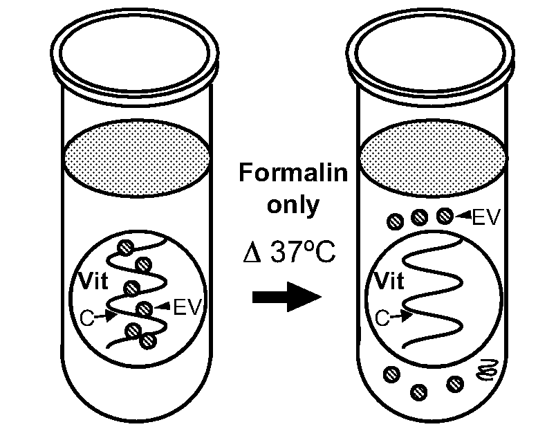
[0012] FIGS. 1A-1G show extracellular vesicles (EV) escape from formalin-fixed bovine vitreous tissues and are retained with 1-ethyl-3-(3-dimethylaminopropyl) carbodiimide (EDC)-formalin fixation. FIG. 1A is a schematic diagram showing formalin-fixed vitreous (Vit) tissue immersed in wash buffer (supernatant) and heated to 37.degree. C. results in escape of EVs (arrowhead) and vitreous collagen (C, closed arrow) into the supernatant. FIGS. 1B-1C are representative transmission electron microscopy (TEM) photomicrographs of supernatant collected from formalin-fixed bovine vitreous tissue after incubation at 37.degree. C. and uranyl acetate (UA) and lead citrate staining show evidence of collagen strands (C, closed arrow) and numerous EVs (arrowhead) that are lost to the wash buffer. FIG. 1D is a schematic diagram showing EDC-formalin-fixed vitreous tissue immersed in wash buffer and heated to 37.degree. C. resulted in retention of EVs in the tissue, with no loss of EVs and minimal loss of vitreous collagen strands into the supernatant. FIG. 1E shows representative TEM photomicrographs of supernatant from EDC-formalin-fixed vitreous tissue after incubation at 37.degree. C. and UA and lead citrate staining showing few collagen strands (C, closed arrow) and no EVs in the supernatant. FIG. 1F shows representative TEM photographs of specificity control, PBS alone, which shows no collagen fibers nor EVs in the supernatant, but does show non-specific punctate staining of electron dense foci measuring less than 20 nm (NS, open arrow). FIG. 1G shows a western blot detecting exosome marker TSG-101 in supernatant (wash buffer) of formalin-fixed vitreous tissue (left lane) and vitreous sample (right lane). Scale bars are (FIG. 1B) 2.5 .mu.m, (FIG. 1C), 500 nm, (FIG. 1E-1F), and 200 nm.
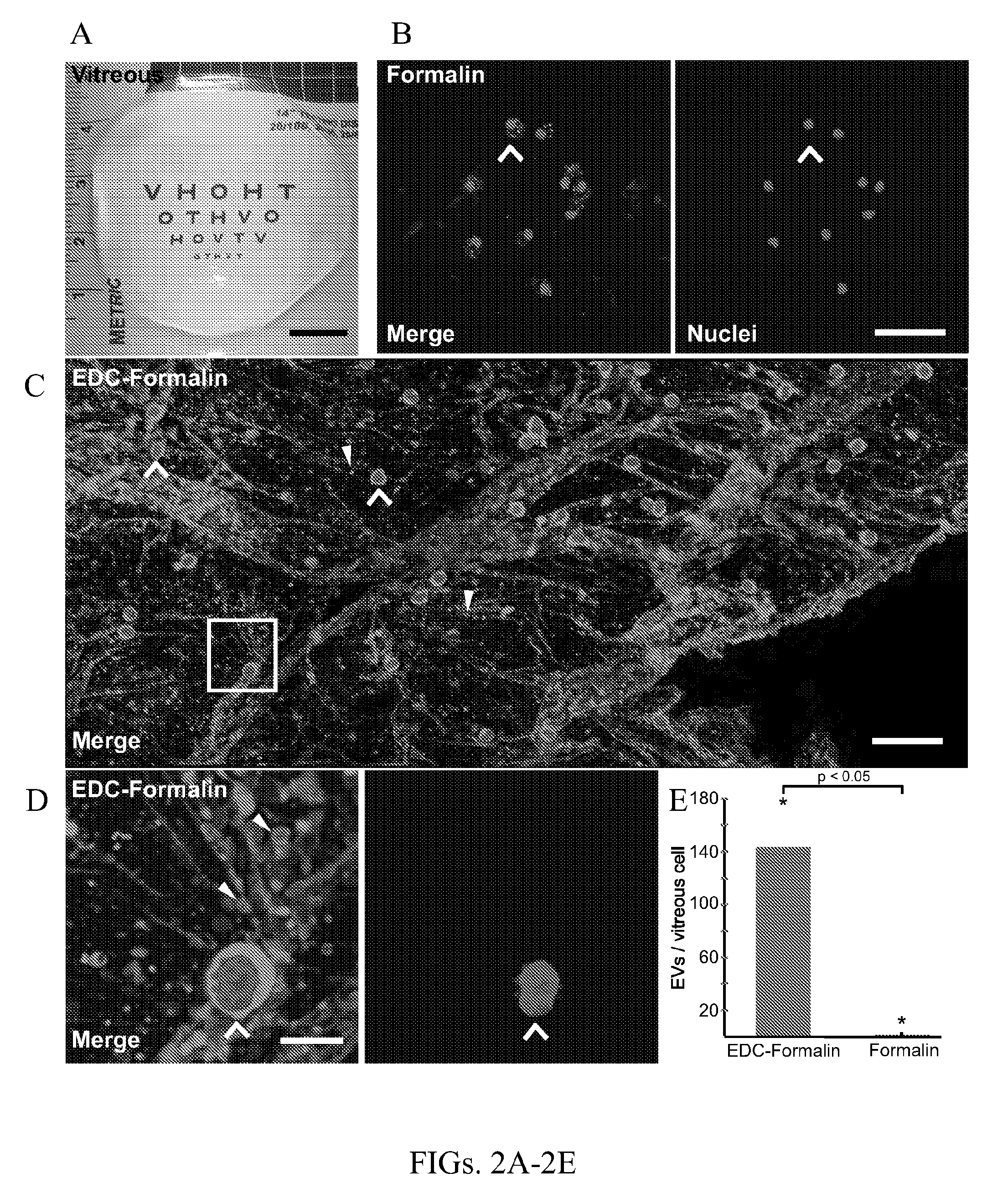
[0013] FIGS. 2A-2F show EDC-formalin fixation of bovine vitreous retains EVs imaged by multifocal microscopy (MPM), when compared to formalin fixation alone. FIG. 2A is a gross image of bovine vitreous placed on a vision testing card that demonstrates the highly transparent, gel-like structure. FIG. 2B shows representative MPM photomicrographs of whole mount bovine vitreous specimens fixed with formalin alone and stained with CFSE to mark protein (orange) and Hoechst to mark nuclei (purple). CFSE signal is observed surrounding the nuclei (FIG. 2B, left panel, open arrow), but not in the extracellular space. Nuclei staining shows no extracellular signal (FIG. 2B, left panel, purple, open arrow). FIG. 2C shows representative MPM photomicrographs of EDC-formalin-fixed vitreous stained with CFSE (orange) and Hoechst (purple). Overlay of image shows positive signal consistent with cell bodies (denoted with open arrow) and foci of extracellular protein signal (arrowheads) consistent in size and shape with EVs. FIG. 2D is an inset of FIG. 2C (white box), which shows multiple round intracellular foci (FIG. 2D, left panel, open arrowhead, orange) surrounding the area of nuclear stains (FIG. 2D, right panel, open arrowhead, purple). Numerous focal extracellular protein signals are observed (FIG. 2D, left panel, closed arrowheads, orange), consistent in size and shape with EVs, and no extracellular DNA is observed. FIG. 2E is a graph representing the mean.+-.standard deviation number of EVs per vitreous cell and shows that EDC-formalin-fixed vitreous exhibit significantly more EVs than formalin-fixed vitreous. FIG. 2F is a graphical representation of frequency distribution of bovine vitreous EV diameter imaged by MPM. EV sizes was measured for 4,000 EVs and the frequency of EVs were plotted against the diameter of the EV. The lower limit of multiphoton microscopy is 200 nm and EVs up to 6000 nm were measured. EVs were distinguished from cells and defined as containing extracellular protein or RNA without extracellular DNA. p-values are <0.05. Scale bars are (FIG. 2A) 1 cm, (FIG. 2B) 40 .mu.m, (FIG. 2C) 50 .mu.m and (FIG. 2D) 10 .mu.m.
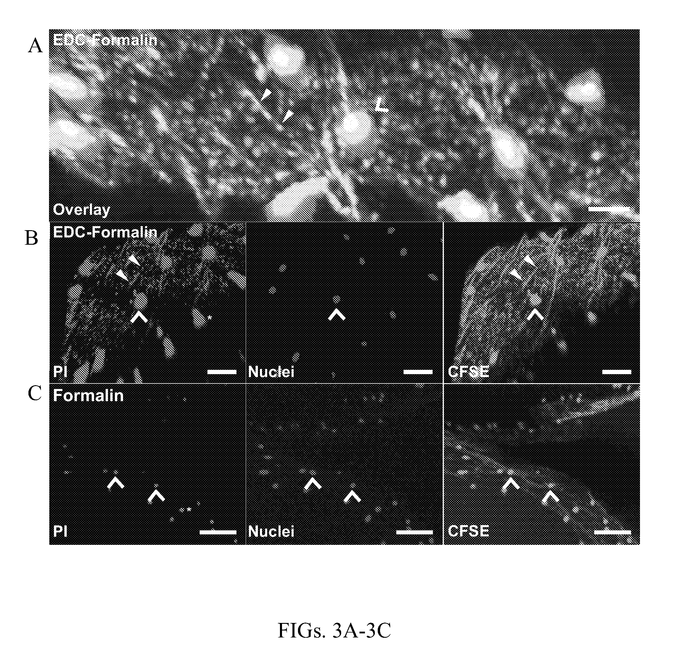
[0014] FIGS. 3A-3C show fixation of bovine vitreous with EDC-formalin retains EVs and extracellular RNA in situ. FIGS. 3A-3C shows representative confocal fluorescent photomicrographs of whole mount bovine vitreous specimens crosslinked with EDC-formalin (FIGS. 3A and 3B) or formalin alone (FIG. 3C), stained with propidium iodide (PI, red) to mark DNA and RNA, Hoechst (blue) to visualize DNA and nuclei, and carboxyfluorescein succinimidyl ester (CFSE, green) to stain for protein. FIG. 3A is an overlay of images from EDC-formalin-fixed bovine vitreous and shows positive signal consistent with cell bodies (FIG. 3A, denoted with open arrow) and foci of extracellular RNA (closed arrowhead) and extracellular protein (closed arrowhead) consistent in size and shape with EVs. FIG. 3B shows representative confocal fluorescent photomicrographs of EDC-formalin-fixed vitreous and shows multiple round cellular foci (FIG. 3B, all panels, open arrowhead) and numerous focal signals of extracellular RNA (FIG. 3B, left panel, PI stain, red) and extracellular protein (FIG. 3B, right panel, CFSE stain, green) between the cells. FIG. 3C shows representative photomicrographs of whole mount bovine vitreous fixed with formalin alone and shows signal for RNA (FIG. 3C, left panel, PI, red) in the nucleus, similar to nuclei staining (FIG. 3C, middle panel, Hoechst, blue). Formalin-only fixed vitreous show no foci of extracellular RNA signal (FIG. 3C, left panel). CFSE stain shows cellular signal (open arrow), but no EV-shaped extracellular protein signal (FIG. 3C, right panel, green, no punctate staining observed between open arrows). The cell size appears smaller in the formalin only fixation, presumably due to EDC-formalin retaining more cytoplasmic RNAs and protein as compared to formalin alone. Scale bars are (FIG. 3A) 25 .mu.m and (FIGS. 3B-3C) 50 .mu.m.
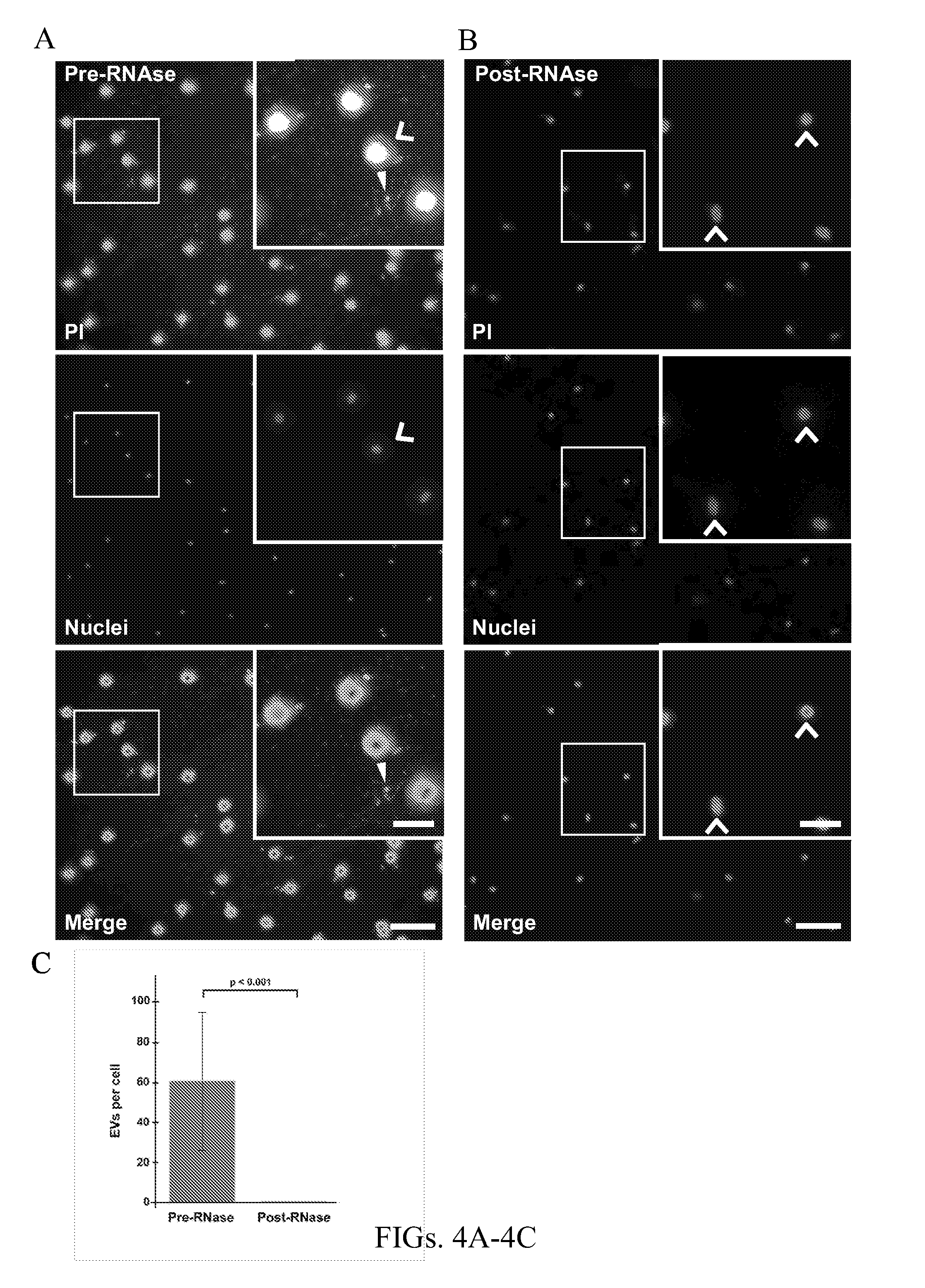
[0015] FIGS. 4A-4C show RNAse treatment of EDC-formalin-fixed bovine vitreous stained with PI show reduced extracellular signal. FIG. 4A shows low-power wide-field fluorescent photomicrographs of whole mount bovine vitreous specimens crosslinked with EDC-formalin and stained with PI (FIG. 4A, top panel, red) and shows signal in the extracellular environment of vitreous tissue (denoted with closed arrowhead, inset), nuclei labeled (FIG. 4A, middle panel, Hoechst, blue) and merged images are shown (FIG. 4A, bottom panel). Vitreous cell nuclei stain positive with PI and Hoechst; colocalized signals are shown in green (FIG. 4A, bottom panel, inset). Cells are denoted with an open arrow and foci of extracellular PI signal are marked with a closed arrowhead (FIG. 4A, top and middle panel, inset). Nuclei were stained, and no extracellular DNA signal is observed (FIG. 4A, bottom panel). FIG. 4B shows photomicrographs of whole mount bovine vitreous fixed with EDC-formalin and treated with RNAse A. Samples stained with PI (FIG. 4B, top panel, red), Hoechst (FIG. 4B, middle panel), and merged images are shown (FIG. 4B, bottom panel). RNAse A treated samples show no evidence of extracellular RNAs as demonstrated by the lack of signal between the cell bodies (FIG. 4B, top and middle panel) and show no signal between two cell nuclei (open arrows). The PI signal for cytoplasmic RNA in RNAse A treated samples (FIG. 4B, top panel) appear smaller than pre-RNAse treated samples (FIG. 4A, top panel), presumably due to EDC-formalin retaining more cytoplasmic RNA. FIG. 4C is a graphical representation of mean.+-.standard deviation foci of extracellular signal for EDC-formalin fixed tissues stained with PI pre-RNAase treatment and after RNAse treatment show significantly fewer EVs after RNAse treatment. Mean+/- standard error for EVs per cell were 60.7+/-35.1 pre-RNAse treatment and 0.03+/-0.04 post RNAse treatment, with significantly more EVs per cell noted pre-RNAse treatment (p<0.001) (FIG. 4C). Scale bars are (FIGS. 4A-4B) 50 .mu.m and (4A inset, 4B inset) 20 .mu.m.
[0016] FIGS. 5A-5B show EDC-formalin fixation of bovine vitreous retains EVs imaged with photomicroscopy. FIGS. 5A-5B show low-power wide field fluorescent photomicrographs of whole mount bovine vitreous specimens crosslinked with EDC-formalin (FIG. 5A) or formalin alone (FIG. 5B). FIG. 5A shows representative photomicrographs of bovine vitreous fixed with EDC-formalin and stained with CFSE to label protein (FIG. 5A, top and middle panel, white) and Hoechst to label nuclei (FIG. 5A, bottom panel, blue) and shows multiple round cellular foci (FIG. 5A, all panels, open arrowhead) with numerous extracellular protein signals (top and middle panels, CSFE, white) consistent with EVs. FIG. 5B shows photomicrographs of whole mount bovine vitreous fixed with formalin only show nuclear stain (FIG. 5B, middle and bottom panels, Hoechst, blue) co-localizing with CFSE (FIG. 5B, top and middle panel, white), consistent with cellular DNA and nucleic acid, respectively. There is no evidence of extracellular protein signal (FIG. 5B, top and middle panel, CSFE, white). The CFSE stained cell size appears smaller in the formalin only fixation (FIG. 5B, middle panel) as compared to EDC-formalin fixation (FIG. 5A, middle panel), presumably due to EDC-formalin retaining more small protein as compared to formalin fixation alone. Scale bars are (FIGS. 5A-5B) 100 .mu.m.
[0017] FIGS. 6A-6I show bovine and human vitreous humor contains EVs. FIG. 6A shows representative transmission electron microscopy (TEM) photomicrographs of bovine vitreous tissue sections stained with uranyl acetate (UA) and lead citrate and shows a substantial number of EVs that are pleomorphic in size (arrowheads) and that contact collagen strands (marked with a "C" and arrow). The inset (upper right corner) is an enlargement of the area-enclosed box in the lower right corner and shows an EV associated with a collagen strand. FIG. 6B shows representative TEM photomicrograph of EVs isolated from bovine vitreous and stained with the electron dense protein stain, CSFE, which depict EV morphology and show numerous EVs pleomorphic in size (smaller EV marked with arrowhead, larger EV with double arrowhead). FIG. 6C shows representative TEM photomicrograph of EVs isolated from bovine vitreous and electron dense nucleic acid stain acridine orange (AO) staining and shows large EVs (double arrowhead) positive nucleic acid signal. FIG. 6D shows multiple EVs (arrowheads) in a network of collagen within whole mounted bovine vitreous stained with ethidium bromide (EtBr), an electron dense and nucleic acid stain. FIG. 6E shows a graphical representation of the mean (black line).+-. standard error (red bars) concentration EVs according to EV diameter, based on nanoparticle tracking analysis of EVs isolated from bovine vitreous. FIG. 6F shows representative TEM photomicrographs of postmortem human eye sections stained with UA and lead citrate show a substantial number of EVs at the vitreous base (Vit), adjacent to the non-pigmented epithelium (NPE) of the ciliary body (smaller EVs marked with arrowhead, larger EVs with double arrowhead). The EVs (FIGS. 6F-6G, arrowheads) contact with collagen strands (arrows). FIG. 6H shows representative TEM photomicrographs of EVs isolated from human vitreous and stained with AO show EVs (arrowhead) with positive nucleic acid signal. FIG. 6I is a graphical representation of frequency distribution of human vitreous EV diameter. Scale bars are (FIG. 6A, FIG. 6G) 100 nm, (FIG. 6B) 50 nm, (FIG. 6C-6D, FIG. 6H) 200 nm, and (FIG. 6F) 2 .mu.m.
[0018] FIGS. 7A-7D show immunohistochemistry staining of EV-specific protein TGS-101 in normal bovine vitreous. FIG. 7A shows representative wide-field fluorescent photomicrographs of whole mount bovine vitreous specimens fixed with formalin and processed at cold temperatures and demonstrates immunohistochemical stain for the EV-associated protein, TGS-101, in the extracellular space (FIG. 7A, top and middle panels, arrowhead, Alexa 488, Green). The inset (FIG. 7A, all panels, top right) is a higher magnification image of the box in the middle (FIG. 7A, all panels). Nuclei are marked with Hoechst counterstain (FIG. 7A, top and bottom, blue, open arrow). Hundreds of punctate extracellular signals were observed (FIG. 7A, top and middle). No evidence of extracellular DNA was observed (FIG. 7A, bottom). FIG. 7B shows representative photomicrographs from specificity controls for TSG-101 immunohistochemistry: whole mount normal bovine vitreous labeled with non-specific IgG antibody (green). The inset (FIG. 7B, all panels, top right) is a higher magnification image of the box in the middle (FIG. 7B, all panels). Signal was observed surrounding the nuclei (FIG. 7B, top and middle, Alexa 488, green). Images show no evidence of extracellular signal (FIG. 7B, top and bottom, Hoechst, blue). FIG. 7C is a graphical representation of mean+/- standard error for TSG-101 signal in extracellular and intracellular spaces, *p<0.05 by Student's unpaired t-tests. FIG. 7D shows positive signal for TSG-101 is observed in the extracellular space of the formalin-fixed vitreous (FIG. 7D, left, green). Nuclei are labeled with Hoechst (FIG. 7D, left, blue) and PI (FIG. 7D, right, red). There is no evidence of extracellular RNA in formalin-fixed samples (FIG. 7D, right, red). Scale bars are (FIGS. 7A-7B) 40 .mu.m and (FIG. 7A inset, FIG. 7B inset and FIG. 7D) 10 .mu.m.
[0019] FIGS. 8A-8B show bovine vitreous is free of cells after low-speed centrifugation. FIG. 8A shows representative low power light microscopy photomicrographs of whole mount bovine vitreous after low-speed centrifugation followed by hematoxylin and eosin staining. These images show eosinophilic signal consistent with vitreous collagen (pink, arrow) without evidence of hematoxylin stained cellular nuclei. FIG. 8B shows images of whole mount vitreous prior to centrifugation. These images show eosinophilic signal consistent with vitreous collagen (pink, arrow) with evidence of hematoxylin stained cellular nuclei (purple, open arrow). Scale bars are (FIGS. 8A-8B) 50 .mu.m.
[0020] FIGS. 9A-9I show human and bovine vitreous EV transfer endogenous RNA into cultured cells. FIGS. 9A-9C show representative confocal photomicrograph images of a human retinal pigment epithelial cells (ARPE-19) after 24 h treatment with a bolus of bovine vitreous EVs that were pre-labeled with the nucleic acid stain acridine orange (AO). Images show uptake of EV-labeled RNA in ARPE-19 cells (FIG. 9A, green). Nuclei are labeled (FIG. 9B, Hoechst, purple) and a merged image (FIG. 9C) shows transfection of ARPE-19 cells, with AO signal in the cytoplasm. FIG. 9D is a graphical representation of transfection efficiency (% of cells transfected) for ARPE-19 cells treated with bovine vitreous EVs (error bars represent standard deviation, n=3, p<0.05). FIGS. 9E-9F show representative confocal photomicrographs of human embryonic kidney (HEK) cells treated with a 24 h bolus of bovine EVs bodies pre-labeled with AO and show staining in the cytoplasm (FIG. 9E). Nuclei were labeled and a merged image is shown (FIG. 9F). FIGS. 9G and 9H are representative low-power fluorescent photomicrograph images of ARPE-19 cells treated for 3 h with a bolus of EVs that were isolated from post-mortem human vitreous and pre-labeled with AO. The image of FIG. 9G shows transfection of cells (FIG. 9G, AO, green). Nuclei were marked (FIG. 9H, Hoechst, blue). FIG. 9I is a graphical representation of transfection efficiency (% of cells transfected) for ARPE-19 cells treated with human vitreous EVs (error bars represent standard deviation, n=3, p<0.05). Scale bars are (FIGS. 9A-9C) 50 .mu.m, (FIGS. 9E-9F) 15 .mu.m, and (FIGS. 9G-9H) 100 .mu.m.
[0021] FIGS. 10A-10F show delivery of recombinant bovine serum albumin (BSA) protein and recombinant green fluorescent protein (GFP) by bovine vitreous extracellular vesicles (EV) to cultured human retinal pigment epithelial (ARPE-19) cells. FIG. 10A are representative photomicrographs of ARPE-19 cells treated with a bolus of bovine vitreous EVs that had been pre-loaded with 1 .mu.g BSA conjugated to fluorescein by electroporation at 300 V. The left image of FIG. 10A shows fluorescein staining (yellow) in the cytoplasm. FIG. 10A, middle image shows nuclei labelled with Hoechst stain (blue), and a merged image (FIG. 10A, right) shows substantial number of cells transfected. FIG. 10B are representative photomicrographs of ARPE-19 cells treated with a bolus of bovine vitreous EVs that had been mixed with BSA-fluorescein without electroporation (0 V, control). FIG. 10B, left image show no fluorescein staining, while FIG. 10B, right image shows nuclei labeling with Hoechst stain (blue). FIG. 10C is a graphical representation of mean.+-.standard deviation transfection efficiency (% of cells transfected) of ARPE-19 cells treated with vitreous EVs loaded with 3 .mu.g, 1 .mu.g, or 0.5 .mu.g BSA-fluorescein by electroporation at 300 V, with EVs loaded with 0.5 .mu.g BSA-fluorescein without electroporation (0 V, control), or with PBS alone without electroporation (0 V, control). p<0.001 for all BSA-fluorescein dosages loaded at 300 V vs. controls at 0 V. FIG. 10D shows representative photomicrographs of ARPE-19 cells after application of a bolus of bovine vitreous EVs that had been pre-loaded with 1 .mu.g of recombinant GFP by electroporation at 300 V. FIG. 10D, left image, shows positive GFP staining (green) in the cytoplasm. FIG. 10D, middle image, shows nuclei labelled with Hoechst stain (blue), and a merged image (FIG. 10D, right) shows substantial number of cells transfected. FIG. 10E, right image, shows no fluorescein staining in a representative photomicrograph of ARPE-19 cells after application of a bolus of bovine vitreous EVs that had been mixed with GFP without electroporation (0 V, control). Nuclei labeling with Hoechst stain (blue) in the control sample is shown FIG. 10E, right image. FIG. 10F is a graphical representation of mean.+-.standard deviation transfection efficiency (% cells transfected) of ARPE-19 cells after application of EVs loaded with 1 .mu.g, 0.5 .mu.g, or 0.25 .mu.g GFP by electroporation at 300 V or 1 .mu.g GFP without electroporation (0 V, control). p<0.05 for all GFP dosages loaded at 300 V vs. control at 0 V. Scale bars (FIGS. 10A-10E) 50 .mu.m.
[0022] FIGS. 11A-11D show bovine vitreous EVs target the retina and deliver recombinant protein in vivo. FIG. 11A are representative wide-field fluorescent photomicrographs of mouse retina tissue sections after injection of a dilute amount of bovine EVs loaded with recombinant bovine serum albumin (BSA) conjugated to fluorescein on day 3 post injection. FIG. 11A, left image shows BSA fluorescein only, FIG. 11A, middle image, shows nuclei staining with Hoeschst only, and FIG. 11A, right image, shows a merged image. The images of FIG. 11A show signal in vitreous that does not penetrate the inner limiting membrane (ILM). FIG. 11B are representative confocal photomicrographs of mouse retina tissues section 3 weeks after injection of BSA-fluorescein showing expression in the retinal outer plexiform layer (OPL) and inner plexiform layer (IPL, arrow). FIG. 11B, left image, shows BSA fluorescein only, FIG. 11B, middle image, shows nuclei staining only, and FIG. 11B, right image, shows a merged image. FIG. 11C are images showing signal in cells traversing the IPL and OPL, as well as, ganglion cells (marked with inset box). The inset box from (FIG. 11C) is shown in higher power in (FIG. 11D) demonstrating positive stain in a cluster of cells in ganglion cell layer (GCL) and retinal nerve fiber layer. FIGS. 11C-D, left image, shows BSA fluorescein only, FIGS. 11C-D, middle, image, show nuclei staining only, and FIGS. 11C-D, right images, show a merged view. Scale bars are 30 .mu.m (FIG. 11A), 50 .mu.m (FIGS. 11B-11C) and 25 .mu.m (FIG. 11D). Photoreceptor segments (ph segments), outer nuclear layer (ONL), inner nuclear layer (ONL).
[0023] FIGS. 12A-12E show bovine vitreous EVs target the cornea, ciliary body, and retina to deliver recombinant protein in vivo. FIG. 12A are representative confocal fluorescent photomicrographs of mouse eye tissue sections after injection of bovine EVs loaded by electroporation (300 V) with recombinant bovine serum albumin (BSA) conjugated to fluorescein (BSA-fluorescein) at 3-weeks post injection showing signal in cornea from endothelial cells and corneal keratocytes (FIG. 12A, left image shows BSA fluorescein only, FIG. 12A, middle image, shows nuclei staining with Hoeschst only, and FIG. 12A, right image, shows a merged image). FIG. 12B are images from control group of bovine EV mixed with BSA-fluorescein without electroporation (0 V) after 3-week injection showin no expression in endothelial cells nor corneal keratocytes, but does show non-specific staining of the corneal epithelium (FIG. 12B, left image shows BSA fluorescein only, FIG. 12B, middle image, shows nuclei staining with Hoeschst only, and FIG. 12B, right image, shows a merged image). FIG. 12C are representative confocal fluorescent photomicrographs from mouse eyes at 3-week post injection of EVs loaded by electroporation (300 V) with BSA-fluorescein that show signal in non-pigmented ciliary epithelial cells (FIG. 12A, left image shows BSA fluorescein only, FIG. 12C, middle image, shows nuclei staining with Hoeschst only, and FIG. 12C, right image, shows a merged image). FIG. 12D are images showing robust expression of BSA-Fluorescein in the photoreceptors, inner plexiform layer (IPL), retinal pigment epithelial (RPE) cells, and choroid (FIG. 12D, left image shows BSA fluorescein only, FIG. 12D, middle image, shows nuclei staining with Hoeschst only, and FIG. 12D, right image, shows a merged image). FIG. 12E are images of the mouse retina photoreceptors and retinal pigment epithelium (RPE) that are transfected with recombinant BSA protein that was delivered by EVs. Scale bars are 25 .mu.m (FIGS. 12A-12E). Corneal epithelium (Epi), corneal endothelium (endo), outer plexiform layer (OPL), outer nuclear layer (ONL), inner plexiform layer (ONL).
[0024] FIGS. 13A-13I show bovine vitreous vesicular bodies loaded with fluorescent labeled siRNAs transfects into human retinal pigment epithelial cells with high efficiency. FIGS. 13A-13C are low-power fluorescent photomicrographs of human retinal pigment epithelial (ARPE-19) cells that show transfection of anti-GAPDH siRNA conjugated to cyanine 3 dye (siRNA-Cy3) after electroporation with bovine vesicular bodies at 350 V (FIG. 13A, yellow, Cy3), nuclei marked with Hoechst dye (FIG. 13B, blue), and merge image of FIG. 13A and FIG. 13B shows substantial number of cells transfected (FIG. 13C). FIGS. 13D-13F are low-power photomicrographs of ARPE-19 cells treated with bovine vitreous vesicular bodies containing siRNA-Cy3 after electroporation at 200 V. FIG. 13D shows siRNA-Cy3 staining in the cytoplasm (yellow). Nuclei were labeled with Hoescht stain (FIG. 13E, blue), and the merged image of FIG. 13F show staining in the cytoplasm with reduced cell staining when compared to 350 V. FIGS. 13G-13H are images showing ARPE-19 cells treated with a bolus of bovine vesicular bodies and anti-GAPDH siRNA-Cy3 without electroporation (0 V). FIG. 13G shows no siRNA-Cy3 staining in ARPE-19 cells, and FIG. 13H shows nuclei marked with Hoechst stain (blue). The graph of 13I shows the percent of cells transfected with siRNA-GAPDH-Cy3 by electroporation voltage. Scale bars are 50 .mu.m (FIGS. 13A-13H).
[0025] FIGS. 14A-14F show the bovine ciliary body non-pigmented epithelium produces abundant vesicular bodies and that are released into intracellular spaces. FIGS. 14A-14C are TEM photomicrograph images from bovine sections of ciliary body nonpigmented epithelium (NPE) stained with uranyl acetate showing multiple vesicular bodies (FIG. 14A, arrowheads) within the lumen of enlarged intercellular spaces (ISP) and budding from the NPE surface (FIG. 14A, asterisk). The orientation of the image is such that the base of the NPE and vitreous base marked (VIT) and internal limiting membrane (ILM) are shown. FIG. 14B is the inset from FIG. 14A, upper box, and shows vesicular bodies within ISP. FIG. 14C shows the lower inset from FIG. 14A and shows a NPE cell with a vesicular body budding into the lumen of the ISP (FIG. 14C, asterisk). FIG. 14D is a TEM photomicrograph of NPE showing electron dense bodies within the cell (FIG. 14D, wedge) and in vesicular bodies in the ISP lumen (FIG. 14D, arrowheads). FIG. 14E is a TEM photomicrograph of ciliary body pigmented epithelium (PE) showing no evidence of budding vesicles. FIG. 14F is a TEM image of bovine vitreous base attached to the ciliary body showing collagen fibers with several vitreous bodies (FIG. 14F, arrow-heads) within the collagen matrix (FIG. 14F, arrows). Scale bars are 1 um (FIG. 14A), 200 nm (FIG. 14B and FIG. 14F), 250 nm (FIG. 14C), and 500 um (FIGS. 14D-14E).
[0026] FIGS. 15A-15I show vitreous vesicular bodies loaded with exogenous protein transfects with high efficiency into human retinal pigment epithelial cells. FIGS. 15A-15C are low-power fluorescent photomicrographs of human retinal pigment epithelial (ARPE-19) cells showing uptake of exogenous bovine serum albumin. Bovine vitreous humor vesicular bodies were electroporated at 350 V with BSA-fluorescein and then a bolus given to ARPE-19 cells which showed substantial staining in the cytoplasm (FIG. 15A, yellow). Nuclei were labeled with Hoechst stain (FIG. 15B, blue), and a merged image of FIG. 15A and FIG. 15B shows a substantial number of ARPE-19 cells stain for fluorescein (FIG. 15C). ARPE-19 cells treated with bovine vitreous vesicular bodies containing BSA-fluorescein (electroporated at 200 V) also show staining in the cytoplasm (FIG. 15D). Nuclei of these cells were labeled with Hoechst stain (FIG. 15E, blue), and the merged images (FIG. 15F) shows a decrease in cytoplasm cytoplasmic staining, which means fewer transfected cells as compared to cells exposed to EV electoporated with 300 V. FIGS. 15G-15H are photomicrographs showing ARPE-19 cells treated with bovine vesicular bodies and BSA-fluorescein without electroporation. No BSA-fluorescein staining in ARPE-19 cells was observed as shown in FIG. 15G). Nuclei were marked with Hoechst stain (FIG. 15H, blue). FIG. 15I is a graph showing protein transfection efficiency (error bars standard deviation of measurements, n=3). Scale bars are 50 .mu.m (FIGS. 15A-15H).
[0027] FIGS. 16A-16E show aqueous humor contains abundant vesicular bodies. FIG. 16A are TEM photomicrographs of whole mount bovine aqueous labeled with acridine orange showing multiple vesicular shaped bodies of various sizes (FIG. 16A, single arrowheads, double arrowheads, and arrow marks small, medium and large vesicle, respectively). FIG. 16B is a TEM photomicrograph depicting vesicular bodies stained with CFSE (FIG. 16B, arrowheads) associated with a collagen stand (FIG. 16B, arrow). FIGS. 16C-16D are TEM photomicrographs of bovine anterior chamber exosomes after isolation by differential ultracentrifugation stained with uranyl acetate (FIG. 16C) and acridine orange (FIG. 16D) showing a cluster of vesicular bodies of various sizes (FIG. 16C). FIG. 16E is an averaged Finite Track Length Adjustment Size/Concentration graph from nanoparticle tracking analysis of ultracentrifuge-isolated exosomes (error bars indicating+/-1 standard error of the mean). The data had a concentration of 1.10 e 8, a mean of 140.8 nm, a standard deviation of 127.9 nm, and peaks at 15 nm, 35 nm, 65 nm, 115 nm, 185 nm, 205 nm, 225 nm, 275 nm, 415 nm, 505 nm, and 615 nm (FIG. 16E). Scale bars, 200 nm (FIG. 16A, FIGS. 16C-16D) 500 nm (FIG. 16B).
[0028] FIGS. 17A-17R show that isolated bovine EVs can transfect human skin cells. Isolated bovine EVs were labeled for endogenous proteins (CFSE) and RNA (acridine orange) and then human skin cells (PAM-212) were transfected in culture. FIGS. 17A-17C are low power wide-field photomicrographs of human skin cells PAM-212 that were transfected with bovine vitreous RNA that was labeled with acridine orange (AO) after 3 h of transfection. FIGS. 17D-17F show PAM-212 cells transfected with AO labeled EVs 24 hours after transfection. Images show a robust transfer of bovine EV RNA to human skin cells. The images of FIGS. 17G-171 show PAM-212 cells transfected bovine vitreous EVs that were previously labeled for all protein using CFSE, EVs were isolated again and then exposed to a bolus of EV-labeled protein to PAM 212 cells. After transfection of 3 h (FIGS. 17G-171) and 24 hours (FIGS. 17J-17L) cells show robust uptake of bovine EV protein. Negative controls show no transfection at 3 h (FIGS. 17M-170) and 24 h (FIGS. 17P-17Q). Scale bar is 100 .mu.m for all images.
DETAILED DESCRIPTION OF THE INVENTION
[0029] The present invention is based on the unexpected discovery of a vesicular network in the vitreous and aqueous humor of the healthy human and bovine eyes. The vesicles of this network are loaded with a cargo of diverse proteins and coding and non-coding RNAs that they transport short and long distances to other ocular tissues. As described and demonstrated herein, these vesicle bodies can be safely isolated from ocular fluids of healthy individuals and modified to serve as therapeutic delivery vehicles.
[0030] Accordingly, a first aspect of the present invention is directed to a composition comprising one or more aqueous humor and/or vitreous humor extracellular vesicle bodies. The aqueous humor and/or vitreous extracellular vesicle bodies of the composition are modified to contain one or more exogenous agents.
[0031] The term "extracellular vesicle" as used herein refers to a nanosized membranous particle secreted by a cell. Extracellular vesicles, which are also referred to as EVs, multivesicular bodies, and ectosomes, are natural transport nanovesicles that have been implicated in intercellular communication via transfer of biomolecules such as proteins, lipids, and RNA from one cell to another. Extracellular vesicles differ from other secreted vesicles, e.g., exosomes and apoptotic bodies, based on their size, i.e., exosomes are typically about 40-100 nm in diameter, extracellular vesicles are typically 100-1000 nm in size, and apoptotic bodies are typically 1-5 .mu.m in size.
[0032] In accordance with the present disclosure, the extracellular vesicles of the vitreous and aqueous humor are characterized by their size, i.e., their diameter. The term "diameter" refers to the maximum dimension of the vesicle, it being understood that the vesicle is not necessarily spherical. Vesicle diameter can be measured using conventional techniques for measuring nanoparticle size, such as microscopy techniques (e.g., transmission electron microscopy or light scattering techniques). In another embodiment, the vesicle diameter is measured using Nanoparticle Tracking Analysis (see WO03/093801 to Carr and Geddess, which is hereby incorporated by reference in its entirety).
[0033] The vesicular bodies of the vitreous humor are heterogenous in size, having a diameter ranging from 100 nm to 6000 nm. In one embodiment, the extracellular vesicles of the composition derived from the vitreous humor have a diameter ranging from 100 nm to 1000 nm. In another embodiment, the extracellular vesicles of the composition derived from the vitreous humor have a diameter of about 150 to 500 nm. In another embodiment, the extracellular vesicles of the composition derived from the vitreous humor have a diameter of about 150 to 300 nm.
[0034] The vesicular bodies of the aqueous humor are also heterogenous in size and generally smaller than the vitreous vesicular bodies. In one embodiment, the extracellular vesicles of the composition derived from the aqueous humor have a diameter ranging from 50 nm to 600 nm. In another embodiment, the extracellular vesicles of the composition derived from the aqueous humor have a diameter of about 50-400 nm. In another embodiment, the extracellular vesicles of the composition derived from the aqueous humor have a diameter of about 50-200 nm.
[0035] In one embodiment, the composition comprising aqueous humor and/or vitreous humor extracellular vesicle bodies comprises a population of vesicle bodies. A "population" of vesicles refers to a set of at least 2 vesicle bodies, at least 5 vesicle bodies, at least 10 vesicle bodies, at least 50 vesicle bodies, at least 100 vesicle bodies, at least 500 vesicle bodies, at least 1000 vesicle bodies, at least 10000 vesicle bodies, at least 100,000 vesicle bodies, at least 1,000,000 vesicle bodies, or more.
[0036] In another embodiment, the vesicle bodies of the vitreous and aqueous humor are characterized by their proteomic signature. For example, the vesicles of the vitreous humor express several known exosome markers, including CD-9, Hsp-90.beta., annexin-II, and TSG-101 proteins. The full list of exosome marker proteins present and enriched in vitreous extracellular vesicles is provided in Table 1 infra. In one embodiment, the composition comprises a population of vitreous humor extracellular vesicles expressing one or more exosome markers listed in Table 1. In another embodiment, the composition comprises a population of vitreous humor extracellular vesicles expressing two, three, four, five, six, seven, eight, nine, or all ten of the exosome markers listed in Table 1.
[0037] The vitreous humor vesicles of the composition of the present disclosure also possess a diverse proteomic signature of eye specific proteins as described in the Examples herein, see Table 2 infra. In one embodiment, the composition comprises a population of vitreous humor extracellular vesicles expressing one or more of the eye specific proteins listed in Table 2. In another embodiment, the composition comprises a population of vitreous humor extracellular vesicles expressing two, three, four, five, six, seven, eight, nine, ten or all eleven of the eye specific proteins listed in Table 2.
[0038] The entire protein signature of the vitreous extracellular vesicles is provided in Table 3 infra. Table 3 indicates the differential expression of the listed protein between the extracellular vesicle fraction of the vitreous humor and the cell free fraction of the vitreous humor. Accordingly, in one embodiment, the composition described herein comprises a population of extracellular vesicles expressing one or more of the proteins listed in Table 3. IN another embodiment, the composition described herein comprises a population of extracellular vesicles expressing one or more of the proteins enriched for in the extracellular fraction of the vitreous fraction. In another embodiment, the composition described herein comprises a population of extracellular vesicles expressing one or more of the proteins identified in Table 3 as being expressed only in the extracellular vesicle fraction. The population of extracellular vesicles described herein can be defined by the expression of any combination of proteins identified as being differentially expressed in only the extracellular vesicle fraction.
[0039] In one embodiment, the aqueous humor and/or vitreous humor vesicular bodies of the composition are isolated vesicular bodies. As used herein, the term "isolated" refers to vesicular bodies that have been removed from a human or animal body, i.e., from ocular fluids of the animal or human, and substantially separated from cell or cellular debris with which they are normally associated in vivo. In one embodiment, the composition comprising the extracellular vesicles is >75%, >80%, >85%, >90%, >95% free of cell or cellular debris normally associated with said vesicle bodies in vivo.
[0040] The extracellular vesicles of the composition may be isolated and/or purified using several techniques. These include filtration, centrifugation, ion-chromatography, or concentration, either alone or in combinations. An exemplary isolation method is described herein and involves a series of low-speed centrifugations. Other exemplary methods of isolating or purifying extracellular vesicles that are known in the art and suitable for use in accordance with the present invention include, without limitation, those disclosed by, e.g., van der Pol et al., "Recent Developments in the Nomenclature, Presence, Isolation, Detection and Clinical Impact of Extracellular Vesicles," J Thromb Haemost 14:48-56 (2016), U.S. Patent Application Publication No. 2016/0216253 to Balaj, WO2015/143113 to Cohen, WO2000/44389 to Dhellin, and WO2001/82958 to Lamparski, which are hereby incorporated by reference in their entirety.
[0041] In one embodiment, the composition as disclosed herein comprises extracellular bodies from the vitreous humor. The vitreous humor or vitreous body is located between the lens and the retina. It is an optically clear, mostly acellular, and gel-like structure with little known biological function. In one embodiment, the extracellular vesicles are obtained from a healthy, normal vitreous body, i.e., from a healthy subject. In another embodiment, the extracellular vesicles are obtained from a vitreous of a subject having an ocular disease. In another embodiment, the composition comprises extracellular bodies from the aqueous humor. The aqueous humor is the clear liquid filling the anterior chamber of the eye, located between the lens and the cornea. In one embodiment, the extracellular vesicles are obtained from a healthy, normal aqueous humor, i.e., from a healthy subject. In another embodiment, the extracellular vesicles are obtained from an aqueous humor of a subject having an ocular disease. In another embodiment, the composition comprises a mixture of extracellular bodies obtained from the aqueous humor and the vitreous humor.
[0042] In one embodiment, the extracellular vesicles as described herein are secreted by the ciliary body, e.g., the ciliary epithelium. In another embodiment, the extracellular vesicles as described herein are secreted by the pigmented ciliary epithelium, non-pigmented ciliary epithelium, ciliary processes. In another embodiment, the extracellular vesicles as described herein are secreted by retinal cells including Muller cells, ganglion cells, amacrine cells, horizontal cells, photoreceptors (rods and cones) bipolar cells, retinal pigment epithelium or retinal endothelial cells. In another embodiment, the extracellular vesicles as described herein are secreted by cells of cornea including corneal epithelium, corneal stroma (keratocytes), corneal endothelium, or limbal stem cells. In another embodiment, the extracellular vesicles as described herein are secreted by cells of iris including pigmented or non-pigmented cells, spindle shaped fibroblasts, macrophages (clump cells of Koganei), smooth muscle of the sphincter muscle, or posterior epithelium. In another embodiment, the extracellular vesicles as described herein are secreted by the trabecular meshwork cells including trabecular meshwork cells or endothelial cell lining of Schlemm's canal. In another embodiment, the extracellular vesicles as described herein are secreted by cells of the lens including lens epithelium, lens fibers, or lens capsule. In another embodiment, the extracellular vesicles as described herein are secreted by cells of choroid including cuboidal epithelial cells, ependymal cell layer, choroid plexus epithelial cells, or choroidal endothelial cells. In another embodiment, the extracellular vesicles as described herein are secreted by cells of the optic nerve including oligodendrocytes, retinal ganglion cell axons, or glial cells. In another embodiment, the extracellular vesicles as described herein are secreted by stem and progenitor cells including mesenchymal stem cells, limbal stem cells, retina stem cells.
[0043] The vitreous and/or aqueous humor extracellular vesicles of the composition as described herein can be any mammalian vitreous or aqueous humor extracellular vesicles. In one embodiment, the composition comprises bovine vitreous and/or aqueous humor extracellular vesicles. In another embodiment, the composition comprises human vitreous and/or aqueous humor extracellular vesicles. In another embodiment, the composition comprises vitreous and/or aqueous humor extracellular vesicles derived from non-human primates, dogs, cats, rodents (e.g., mouse, rat, and guinea pig), horses, cervids, sheep, or pigs.
[0044] As demonstrated herein, the extracellular vesicles of the vitreous and aqueous humor can be isolated, modified to contain one or more exogenous agents, and utilized as a delivery vehicle to delivery the one or more exogenous agents to a target tissue or cell. The exogenous agent can be a therapeutic agent or a diagnostic agent. Suitable therapeutic and diagnostic agents include, without limitation, nucleic acid molecules, proteins and polypeptides, small molecules, hormones, and any combination thereof.
[0045] In one embodiment, the exogenous agent is a therapeutic nucleic acid molecule. The nucleic acid molecule can be single-stranded or double-stranded nucleic acid. Single-stranded nucleic acids include those with phosphodiester, 2'O-methyl, 2' methoxy-ethyl, phosphoramidate, methylphosphonate, and/or phosphorothioate backbone chemistry. In one embodiment, the nucleic acid molecule is a therapeutic nucleic acid molecule selected from a ribonucleic acid molecule (RNA), a deoxyribonucleic acid molecule (DNA), an RNA-DNA hybrid, a modified RNA molecule, modified DNA molecule, or a modified RNA/DNA molecule thereof.
[0046] In one embodiment, the therapeutic nucleic acid molecule is an RNA molecule, such as a small RNA molecule, complementary RNA, a non-coding RNA molecule, siRNA, a pi-RNA molecule, a micro-RNA molecule, a sno-RNA molecule, long non-coding RNA molecule, messenger RNA molecule, ribosomal RNA molecule, an antisense nucleic acid molecule, Locked Nucleic Acid (LNA), antagomir, RNA aptamer, miRNA mimic, miR sponges, CRISPR/Cas gene editing RNA, trans-activating crRNA (tracrRNA), short synthetic RNA composed of a "scaffold" sequence (gRNA), Small Cajal body-specific RNAs (scaRNA), natural cis-antisense siRNAs (cis-nat-siRNAs), trans-acting siRNA (tasiRNA), repeat associated small interfering RNA (rasiRNA), 7SK, transfer-messenger RNA (tmRNA), transfer RNA (tRNA), 7SL RNA, signal recognition particle RNA (SRP), and any combination thereof.
[0047] In one embodiment, the extracellular vesicles are modified to contain a therapeutic RNA that is suitable for the treatment of an ocular disease or condition. Therapeutic RNAs suitable for the treatment of an ocular disease include, without limitation, siRNA targeting the .beta.2-adrenoreceptor (SYL040012) for the treatment of glaucoma (Paneda et al., "Development of SYL040012, a siRNA for treating increased intraocular pressure associated to glaucoma," AOPT 2013 Scientific Meeting 1:96 (2013), which is hereby incorporated by reference in its entirety), siRNA targeting VEGF (bevasiranib) for the treatment of age related macular degeneration (AMD), siRNA targeting VEGF receptor (siRNA-027) for the treatment of AMD (Kaiser et al., "RNAi-based treatment for neovascular age-related macular degeneration by SiRNA-027," Am J Ophthalmol. 150:33-39 (2010), which is hereby incorporated by reference in its entirety), and siRNA targeting RTP801 (PF-655) for the treatment of AMD and diabetic retinopathy (Nguyen et al., "Phase 1 dose-escalation study of a siRNA targeting the RTP801 gene in age-related macular degeneration patients," Eye (Lond) 26:1099-1105(2012) and Nguyen et al., "Dose-ranging evaluation of intravitreal siRNA PF-04523655 for diabetic macular edema (the DEGAS Study)," Invest Ophthalmol Vis Sci. 53:7666-7674 (2012), which are hereby incorporated by reference in their entirety). Other therapeutic RNA molecules suitable for the treatment of ocular diseases that can be introduced to the extracellular vesicles of the composition described herein are described in Guzman-Aranguez et al., "Small-interfering RNAs (siRNAs) as a Promising Tool for Ocular Therapy," Br. J. Pharmacol. 170(4): 730-747 (2013), which is hereby incorporated by reference in its entirety).
[0048] In another embodiment, the isolated extracellular vesicles of the vitreous and/or aqueous humor obtained using the methods described herein, are modified to express or incorporate an mRNA. The mRNA may encode a therapeutic agent that inhibits, down-regulates, reduces a protein expression and/or activity, the excess level of which is associated with an ocular disease, disorder or condition. Such a therapeutic agent may be a peptide, an antibody or other polypeptides or proteins, including any of those described herein. In one embodiment, the mRNA encodes an antibody, a soluble receptor or other binding protein. Typically, a suitable mRNA encodes an antibody that inhibits, down-regulates, or reduces a protein that is present in excess in amount and/or activity in an ocular disease, disorder or condition. In some embodiments, a suitable mRNA encodes an antibody that activates, up-regulates or increases a protein activity that is deficient in an ocular disease, disorder or condition. Exemplary antibodies encoded by mRNAs that can be introduced into the extracellular vesicles of the vitreous and/or aqueous humor as described herein include, but are not limited to, antibodies against VEGF, TNF.alpha., IL-6, ICAM-1, VCAM-1, or soluble receptors such asVEGF receptors (e.g., VEGFR1).
[0049] Other mRNA molecules that are suitable for the treatment of an ocular disease or condition using the extracellular vesicles as described herein, include for example, and without limitation, mRNA molecules encoding the protein or biologically active fragments of endostatin, angiostatin, tissue inhibitor of metalloproteinase 3 (TIMP3), pigment epithelium derived factor (PEDF), or soluble vascular endothelial growth factor receptor (sFlt-1) for the reduction of neovascularization; mRNA molecules encoding the protein or biologically active fragments of Prph2, Rho, cGMP phosphodiesterase .beta.-subunit (BPDE), Bcl2, PEDF, fibroblast growth factor (FGF-2), ciliary neurotrophic factor (CNTF), and c-mer proto-oncogene tyrosine kinase (Mertk) for the treatment of retinitis pigmentosa; mRNA molecules encoding the protein or biologically active fragments of brain-derived neurotrophic factor (BDNF), CNTF, and GDNF for the treatment of glaucoma; mRNA molecules encoding the protein or biologically active fragments of IL-10 and interleukin-1 receptor agonist (IL-1Ra) for the treatment of uveitis; and mRNA molecules encoding the protein or biologically active fragments of IFN-.beta. and thymidine kinase (TK) for the treatment of retinoblastoma.
[0050] In one embodiment, the extracellular vesicles as described herein are modified to carry one or more of the following mRNA therapeutics, mRNA-1440, mRNA-1851, mRNA MRK-1777, mRNA-1388, mRNA-1325, mRNA-1706, mRNA-1647, mRNA-1653, mRNA-4157, mRNA-2416, mRNA-2905, mRNA AZD-8601, MRG-106, MIR-155, MRG-201, MRG-107, and MRG-110.
[0051] In another embodiment, the mRNA molecule loaded into the extracellular vesicles as described herein encodes a vaccine antigen. The mRNA directs the cells to produce and express the antigenic proteins, either secreted or on the cell surface, much like a native infection would do but without the ability to cause disease or spread. For therapeutic vaccines, using the extracellular vesicles described herein to deliver mRNA-based personalized cancer vaccines to prime the immune system to recognize cancer cells and mount a strong, tailored response to each individual patient's cancer. The extracellular vesicle includes a mRNA that encodes a patient's specific neoantigens, or unique mutations present in that specific patient's tumor.
[0052] In another embodiment, the RNA molecule is catalytic RNA. Ribozymes are catalytic RNAs that function as enzymes and do not require proteins for catalysis. Most known natural ribozymes are self-processing RNAs that catalyze RNA cleavage and ligation reactions. Suitable ribozymes therapeutics that can be delivered using the extracellular vesicles as described herein include, but are not limited to angiozyme, Heptazyme, MY-2, RRz1, OZ1 (RRz1), CCR5 ribozyme, L-TR/Tatneo.
[0053] Other RNA therapeutic molecules that are suitable for the treatment of a disease or condition using the extracellular vesicles as described herein, include for example, and without limitation SPC3649 (LNA), Bevasiranib, AGN-745, PF-655, QPI-1007, TD101, SYL040012, SYL1001, Excellair.TM., ALN-RSV01, CEQ508, siG12D LODER, TKM-ApoB, TKM-PLK1, ALN-VSP02, ALN-TTR01, Bcr-Abl siRNA, Atu027, I5NP, CALAA-01, FANG vaccine, iPsiRNA, Tat/Rev shRNA, siRNA-EphA2-DOPC, TD101, Atu027, ND-L02-s0201, DCR-PH1, STP705, ALN-GO1, Fitusiran (ALN-AT3SC), ALN-CC5, ALN-AS1, DCR-MYC, TKM 080301, siG12D-LODER, Inclisiran (ALN-PCSSC), PF-655, SYL1001, Bamosiran (SYL040012), QPI-1007, QPI-1002, Patisiran (ALN-TTR02), ISTH0036, EZN-2968 (R07070179), LErafAON-ETU, AKCEA-APOCIII-LRx, BIIB067 (IONIS-SOD1Rx), AZD5312, Cenersen, IONIS-HTT Rx, IONIS ANGPTL3-LRx, AZD9150, QR-010, SB012, AEG35156, DS-5141b, AKCEA-APO(a)-LRx, Apatorsen (OGX-427), IONIS-HBV Rx, IONIS-GCGR Rx, ASM8, SB010, SB011, G4460, Prexigebersen (BP1001), IONIS-FXI Rx, Aganirsen (GS-101), Eteplirsen (AVI-4658), Alicaforsen, Volanesorsen, IONIS-TTRRx, Custirsen (OGX-011), Lipo-MERIT, IVAC mutanome/warehouse, TNBC-MERIT, CV7201, CV8102, mRNA-1851, mRNA-1440, mRNA MRK-1777, mRNA AZD-8601, mRNA-1325, CV9103, AGS-004, AGS-003-LNG, iHIVARNA-01, AGS-003.
[0054] In one embodiment, isolated extracellular vesicles of the vitreous and/or aqueous humor obtained using the methods described herein, are modified to express or incorporate a nuclease genome editing system useful to edit the genome. Genome editing as described herein may include gene insertions, deletions, modifications (e.g. nucleotide transitions, transversions, insertions or deletions of one or more nucleotides or duplications of any nucleotide sequence), gene activation and gene silencing. As will be appreciated by one of skill in the art, genome editing may be for the purpose of correcting an undesirable gene mutation, introducing a gene mutation, altering a gene sequence (e.g. to improve, enhance or inhibit gene function), inserting a gene sequence (e.g. to activate or inhibit gene expression), and the like. Examples of nuclease genome editing systems include, but are not limited to, Clustered Regularly Interspaced Short Palindromic Repeats (CRISPR) nuclease system, e.g. including a targeting gRNA and a CRISPR-associated (Cas) gene, such as CRISPR-Cas9, Transcription Activator-Like Effector Nucleases (TALEN) and mito-TALEN, Zinc-Finger Nucleases (ZFN), and other therapeutic nucleic acids, e.g. small interfering RNA, micro RNA, anti-microRNA, antagonist, small hairpin RNA, and aptamers (RNA, DNA or peptide based (including affimers)).
[0055] In one embodiment, the extracellular vesicles of the composition described herein are genetically modified to express or incorporate a CRISPR nuclease system, such as a CRISPR/Cas9 Type II genome editing system, including a Cas nuclease, and a guide RNA (gRNA), which comprises a fusion of trans-activating RNA (tracrRNA) and CRISPR RNA (crRNA). CRISPR RNA includes a targeting RNA sequence and a distinctive array of non-coding direct RNA repeats. The crRNA and tracrRNA are related to the selected Cas nuclease. As one of skill in the art will appreciate, the crRNA and tracrRNA (components of the gRNA) and the Cas nuclease are indicated to be "related" which means that the crRNA and tracrRNA are specific for and recognized by one or more particular Cas nucleases.
[0056] In one embodiment, the CRISPR nuclease system is designed to edit one or more gene defects associated with an ocular condition. For example, and without limitation, the CRISPR nuclease system may be designed to edit the VEGF gene that is overexpressed in age-related macular degeneration as described in Kim et al., "Genome Surgery Using Cas9 Ribonucleoproteins for the Treatment of Age-Related Macular Degeneration," Genome Research 27:419-426 (2017), which is hereby incorporated by reference in its entirety. In another embodiment, the CRISPR nuclease system may be designed to inactivate the Nr1 or NR2e3 genes for the purpose of preventing degeneration associated with retinitis pigmentosa as described by Zhu et al., "Gene and Mutation Independent Therapy via CRISPR-Cas9 Mediated Cellular Reprogramming in Rod Photoreceptors," Cell Res. 27:830-833 (2017), which is hereby incorporated by reference in its entirety.
[0057] In another embodiment, the nucleic acid molecule is a DNA molecule. Suitable DNA molecules include, without limitation, a small DNA molecule, a cDNA molecule, an oligonucleotide, a locked Nucleic Acid (LNA), a deoxyribonucleic acid aptamer, a deoxyribonucleic acidzyme (DNAzymes), and any combination thereof.
[0058] In one embodiment, the therapeutic nucleic acid includes genomic sequences, e.g., cDNA sequences and smaller engineered gene segments that express, or may be adapted to express, proteins, polypeptides, fusion proteins, antibodies, and protein/peptide variants. The nucleic acid may comprise a contiguous nucleic acid sequence of about 5 to about 12000 or more nucleotides, nucleosides, or base pairs. Therapeutic nucleic acid molecules in accordance with this aspect of the invention may encode cytokines, enzymes, hormones, natural agonists and antagonists of proteins involved in disease, etc. Therapeutic nucleic acid molecules also include biologically functional equivalents of a therapeutic nucleic acid proven to benefit in the treatment or prevention of a disease or health-related condition. Accordingly, sequences that have about 70% to about 99% sequence identity to a known nucleic acid molecule are suitable therapeutic nucleic acid molecules in accordance with this aspect of the present invention.
[0059] In one embodiment, the extracellular vesicles are modified to contain a therapeutic DNA molecule that is suitable for the treatment of an ocular disease or condition. Suitable therapeutic DNA molecules include for example, and without limitation, DNA molecules encoding the protein or biologically active fragments of endostatin, angiostatin, tissue inhibitor of metalloproteinase 3 (TIMP3), pigment epithelium derived factor (PEDF), or soluble vascular endothelial growth factor receptor (sFlt-1) for the reduction of neovascularization; DNA molecules encoding the protein or biologically active fragments of Prph2, Rho, cGMP phosphodiesterase .beta.-subunit (BPDE), Bcl2, PEDF, fibroblast growth factor (FGF-2), ciliary neurotrophic factor (CNTF), and c-mer proto-oncogene tyrosine kinase (Mertk) for the treatment of retinitis pigmentosa; DNA molecules encoding the protein or biologically active fragments of brain-derived neurotrophic factor (BDNF), CNTF, and GDNF for the treatment of glaucoma; DNA molecules encoding the protein or biologically active fragments of IL-10 and interleukin-1 receptor agonist (IL-1Ra) for the treatment of uveitis; and DNA molecules encoding the protein or biologically active fragments of IFN-.beta. and thymidine kinase (TK) for the treatment of retinoblastoma. Other suitable DNA therapeutics that can be introduced into the extracellular vesicles of the composition as described herein are known in the art, see Liu et al., "Gene Therapy for Ocular Diseases," Postgrad. Med. J. 87(1029): 487-95 (2011), which is hereby incorporated by reference in its entirety.
[0060] In another embodiment the therapeutic DNA molecule suitable for treatment of an ocular disease which is loaded into the extracellular vesicles of the composition described herein is an aptamer. Suitable aptamers include, for example and without limitation, Macugen/pegaptanib (NX1838) targeting the activity of VEGF for the treatment of ocular neovascular diseases, Fovista/pegpleranib (NX1975) targeting the activity of PDGF B-chain for the treatment of age-related macular degeneration, and Zimura/ARC1905 targeting the activity of complement component 5 (C5) for the treatment of age-related macular degeneration (see Drolet et al., "Fit for the Eye: Aptamers in Ocular Disorders," Nucleic Acid Ther. 26(3):127-146 (2016), which is hereby incorporated by reference in its entirety). Other suitable aptamers include RNA aptamer (RB006 or pegnivacogin), ARC19499(BAX499), REG1 (RB006 & RB007), ARC1905, TAR decoy, RRE decoy.
[0061] In one embodiment, a combination of therapeutic RNA and DNA molecules are introduced into the extracellular vesicles of the composition described herein. In one embodiment, the combination of therapeutic RNA and DNA molecules work in concert for the treatment of an ocular disease. For example, and without limitation, siRNA molecules capable of silencing the expression of mutant rhodopsin expression can be administered in combination with a DNA molecule encoding the wildtype rhodopsin gene for the treatment of retinitis pigmentosa (O'Reilly et al., "RNA interference-mediated suppression and replacement of human rhodopsin in vivo," Am J Hum Genet. 81:127-135 (2007), which is hereby incorporated by reference in its entirety).
[0062] In another embodiment, the nucleic acid is a diagnostic nucleic acid. A diagnostic nucleic acid is a nucleic acid that can be applied in the diagnosis of a disease or health-related condition. A diagnostic nucleic acid sequence that encodes one or more reporter proteins. A "reporter protein" refers to an amino acid sequence that, when present in a cell or tissue, is detectable and distinguishable from other genetic sequences or encoded polypeptides present in cells. In some embodiments, a therapeutic nucleic acid molecule may be fused to the diagnostic nucleic acid encoding a reporter protein. For example, the two nucleic acid molecules may be linked to the same promoter by, for example, an internal ribosome entry site, or a bi-directional promoter. Using such techniques, expression of the therapeutic nucleic acid and diagnostic nucleic acid correlate. Thus, when the composition is used in the methods as described herein, one may gauge the location, amount, and duration of expression of a therapeutic nucleic acid.
[0063] Preferably, a reporter sequence encodes a protein that is readily detectable either by its presence, its association with a detectable moiety, or by its activity that results in the generation of a detectable signal. In certain aspects, a detectable moiety may include a radionuclide, a fluorophore, a luminophore, a microparticle, a microsphere, an enzyme, an enzyme substrate, a polypeptide, a polynucleotide, a nanoparticle, and/or a nanosphere, all of which may be coupled to an antibody or a ligand that recognizes and/or interacts with a reporter. Exemplary diagnostic nucleic acid molecules include, without limitation, nucleic acid molecules encoding .beta.-lactamase, .beta.-galactosidase (LacZ), alkaline phosphatase, thymidine kinase, green fluorescent protein (GFP), chloramphenicol acetyltransferase (CAT), luciferase, membrane bound proteins including, for example, G-protein coupled receptors (GPCRs), somatostatin receptors, CD2, CD4, CD8, the influenza hemagglutinin protein, symporters (such as NIS) and others well known in the art.
[0064] In one embodiment, the extracellular vesicles of the composition described herein are modified to include a naked nucleic acid molecule, e.g. naked DNA or naked RNA. In another embodiment, the nucleic acid is packaged in an expression vector suitable for expression in prokaryotes or eukaryotes or both, preferably for expression in mammalian cells. Suitable expression vectors include viral vectors (e.g., adenoviral vector, adeno-associated viral vector, lentiviral vector, vaccina viral vector, retroviral vector, herpes viral vector), bacterial vectors, plasmid vectors, artificial chromosomes, bacteriophages, or any combination thereof. Expression vectors generally contain regulatory sequences and other necessary elements for the translation and/or transcription of the inserted coding sequence. For example, the coding sequence is preferably operably linked to a promoter and/or enhancer to help control the expression of the desired gene product. Promoters used in biotechnology are of different types according to the intended type of control of gene expression. They can be generally divided into constitutive promoters, tissue-specific or development-stage-specific promoters, inducible promoters, and synthetic promoters. Depending on the vector system and host utilized, any number of suitable transcription and translation elements may be used. In mammalian cell systems, promoters from mammalian genes or from mammalian viruses are preferably used.
[0065] Methods of constructing expression vectors containing the desired nucleic acid molecules and appropriate transcriptional and translational control elements are well known in the art. These methods include in vitro recombinant DNA techniques, synthetic techniques, and in vivo genetic recombination. Such techniques are described in Sambrook et al., Molecular Cloning, A Laboratory Manual (Cold Spring Harbor Press, Plainview, N.Y., 1989), and Ausubel et al, Current Protocols in Molecular Biology (John Wiley & Sons, New York, N.Y., 1989), which are hereby incorporated by reference in their entirety.
[0066] The extracellular vesicles can be loaded with the nucleic acid or nucleic acids of interest using techniques known in the art, such as, for example, electroporation. Electroporation involves introducing pores into the vesicles using a pulse of electricity (e.g., 100-400 V/cm), where the nucleic acid(s) enter the vesicles through the pores. The extracellular vesicles can alternatively be loaded with nucleic acid(s) of interest using microinjection or particle bombardment. Alternatively, the extracellular vesicles can be loaded using lipofection or transfection using commercially available kits and reagent, or by transformation using heat shock.
[0067] In another embodiment, the vitreous and/or aqueous humor vesicles are loaded with a therapeutic protein and/or peptide for delivery. In one embodiment, the therapeutic protein is an exogenous protein or peptide. Exogenous refers to a protein or peptide with which the vesicle is not normally associated.
[0068] The protein and/or peptide to be loaded into the vesicles is chosen based the desired effect of that protein and/or peptide on the target cell. A single protein or peptide may be incorporated into the vesicles. Alternatively, more than one protein and/or peptide may be incorporated into the vesicles. The more than one protein and/or peptide may act on the same or different targets to bring about the desired therapeutic and/or preventative effect.
[0069] In one embodiment, the protein and/or peptide to be loaded into the vesicles is an antibody or antibody fragment. The term "antibody" as referred to herein includes whole antibodies (i.e., two heavy chains and two light chains), antibody binding fragments thereof, e.g., single chain antibodies (scFv), single domain antibodies (e.g., nanobodies or Fv), Fab, Fab', F(ab').sub.2, and, variants thereof, e.g., tandem scFv, Fd fragments, diabodies, triabodies. These antibody fragments may be obtained using conventional techniques known to those of skill in the art, and the fragments may be screened for utility in the same manner as intact antibodies
[0070] Antibody and antibody fragments disclosed herein can be mono-valent, bi-valent, or tri-valent with regard to binding domains, and the binding domains may be mono-specific, bi-specific, or tri-specific in binding specificity by design. Suitable antibodies include monoclonal antibodies or a polyclonal antibody mixture. The antibody may be a chimeric antibody, a CDR-grafted antibody, a humanized antibody or an antigen binding portion of any of the foregoing thereof. Therapeutic antibodies may be derived from a variety of species, including, without limitation, mouse, human, camel, llama, goat, rabbit, bovine, and cartilaginous fish.
[0071] In one embodiment, the antibody or antigen binding fragment thereof is one that is suitable for the treatment of an ocular disease or condition. Suitable antibodies or antigen binding fragments thereof include, without limitation, those that bind to and preferentially block or reduce the activity of integrins associated with disease, such as an anti-.alpha..sub.v.beta..sub.3integrin antibody and an anti-.alpha..sub.4.beta..sub.1 integrin antibody. Other suitable antibodies that can be introduced into the extracellular vesicles of the compositions described herein include, for example and without limitation, an anti-epidermal growth factor receptor antibody, anti-vascular endothelial growth factor (VEGF) receptor antibody, anti-VEGF antibodies, e.g., bevacizumab, ranibizumab, anti-TNF.alpha. antibodies, e.g., infliximab and adalimumab, an anti-fibroblast growth factor antibody, an anti-epidermal growth factor antibody, an anti-CD20 antibody, an anti-CD52 antibody, an anti-CD11a antibody, and anti-IL-2 antibody.
[0072] Other suitable antibodies that can be introduced into the extracellular vesicles of the compositions described herein include, for example and without limitation, abciximab (Reopro), adalimumab (Humira, Amjevita), alefacept (Amevive), alemtuzumab (Campath), basiliximab (Simulect), belimumab (Benlysta), bezlotoxumab (Zinplava), canakinumab (Ilaris), certolizumab pegol (Cimzia), cetuximab (Erbitux), daclizumab (Zenapax, Zinbryta), denosumab (Prolia, Xgeva), efalizumab (Raptiva), golimumab (Simponi, Simponi Aria), inflectra (Remicade), ipilimumab (Yervoy), ixekizumab (Taltz), natalizumab (Tysabri), nivolumab (Opdivo), olaratumab (Lartruvo), omalizumab (Xolair), palivizumab (Synagis), panitumumab (Vectibix), pembrolizumab (Keytruda), rituximab (Rituxan), tocilizumab (Actemra), trastuzumab (Herceptin), secukinumab (Cosentyx), ustekinumab (Stelara). Additional anti-angiogenesis protein/peptide therapeutics include, without limitation, ramucirumab, axitinib, axitinib, MGCD516, cediranib, olaparib, lestaurtinib, olaparib, cediranib, pazopanib, docetaxel, pazopanib hydrochloride, TRC105, pazopanib, X4p-001, nivolumab, eribulin mesylate, ketoconazole, therapeutic hydrocortisone, antibody J591, docetaxel, plinabulin, SF1126, carfilzomib, hydroxychloroquine, aldesleukin, bevacizumab, erlotinib, sorafenib, vandetanib, durvalumab, olaparib, cediranib, sapanisertib, ziv-aflibercept, bevacizumab, LY2157299 monohydrate (LY2157299), temozolomide, SGT-53, cediranib maleate, olaparib, bevacizumabosimertinib, regorafenib, itraconazole.
[0073] In another embodiment, the therapeutic protein is an antibody mimetic. An "antibody mimetic" encompasses any organic compound, e.g., a peptide or polypeptide, that can specifically bind an antigen like an antibody and is about 3-20 kDa. In one embodiment, the antibody mimetic comprises a scaffold which binds its target antigen via amino acids in exposed loops similar to the CDR loops of an antibody. These antibody mimetics include, without limitation, adnectins, lipocalins, Kunitz domain-based binders, avimers, knottins, fynomers, atrimers, and cytotoxic T-lymphocyte associated protein-4 (CTLA4)-based binders (reviewed in Weidle et al., "The Emerging Role of New Protein Scaffold-based Agents for the Treatment of Cancer," Cancer Genomics & Proteomics 10:155-168 (2013), which is hereby incorporated by reference in its entirety).
[0074] In another embodiment, the therapeutic protein is a protein or peptide inhibitor. Protein or peptide inhibitors can be full-length proteins or biologically active peptide fragments thereof which naturally antagonize or inhibit the action or activity of one or more receptors, enzymes, hormones, proteases, kinases, growth factors, signal transduction pathways, transcription factors, etc. that are associated with a disease or condition to be treated. The protein or peptide inhibitor may act as a dominant negative receptor or ligand, or a decoy receptor or ligand.
[0075] In one embodiment, the extracellular vesicles of the composition as described herein are modified to contain a protein or peptide inhibitor that is suitable for the treatment of an ocular disease. For example, the protein or peptide inhibitor may be an inhibitor of angiogenesis. Suitable protein/peptide inhibitors of angiogenesis include, without limitation, angiostatin, including full-length angiostatin and biologically active fragments and analogs thereof, and endostatin, including full-length endostatin and biologically active fragments and analogs thereof; other collagen derived peptides, such as tumstatin peptide, tumstatin fragment, and pentastatin; RGD containing peptides, such as Cilengitide, and other fibronectin derived peptides; and peptides derived from laminin, such as C16Y and C16S (see Rosca et al., "Anti-angiogenic Peptides for Cancer Therapeutics," Curr. Pharm. Biotechol. 12(8): 1101-1116 (2011), which is hereby incorporated by reference in its entirety).
[0076] Other suitable protein or peptide inhibitors that can be loaded into the extracellular vesicles include, without limitation, integrin antagonists, e.g., LFA-1, VLA-4, Mac-1, ICAM-1, ICAM-2, VCAM antagonists, chemokine antagonists, e.g., MCP-1, MCP-5, MCP-3, MIP1.alpha., CCR5, RANTES antagonists, and selectin antagonists, e.g., E-selectin, P-selectin, and L-selectin antagonists.
[0077] Other suitable protein or peptide inhibitors that can be loaded into the extracellular vesicles include, without limitation, anti-VEGF agents, Ranibizumab (Lucentis, Genentech, South San Francisco), aflibercept (Eylea, Regeneron Pharmaceuticals, Tarrytown, N.Y.), Lucentis, Bevacizumab (Avastin, Genentech), Corticosteroids, Intravitreal steroids, sustained-release biodegradable dexamethasone implant, Ozurdex (Allergan, Irvine, Calif.), Vitreolytics including Ocriplasmin (Jetrea, ThromboGenics, Leuven, Belgium), anti-PDGF therapies, RTH258, a small, humanized anti-VEGF antibody fragment that inhibits all isoforms of VEGF-A, anti-VEGF DARPin (abicipar pegol), the anti-PDGF agent Fovista (Ophthotech, New York), and Iluvien, a non-biodegradable implant indicated for DME that elutes the steroid fluocinolone acetonide.
[0078] The exogenous protein and/or peptide can be introduced into the vesicles by a number of different techniques. In one embodiment, the vesicles are loaded by electroporation or the use of a transfection reagent. Electroporation conditions may vary depending on the charge and size of the therapeutic cargo. Typical voltages are in the range of 20 V/cm to 1000 V/cm, such as 20 V/cm to 100 V/cm with capacitance typically between 25 .mu.T and 250 .mu.T, such as between 25 .mu.T and 125 .mu.T. A voltage in the range of 150 mV to 250 mV, particularly a voltage of 200 mV is preferred for loading vesicles with an antibody.
[0079] Alternatively, the vesicles may be loaded with exogenous protein and/or peptide using a transfection reagent. Despite the small size of the vesicles, conventional transfection agents may be used for transfection of vesicles with protein and/or peptide. Preferred transfection reagents for use in accordance with the present invention include cationic liposomes.
[0080] In another embodiment, extracellular vesicles may also be loaded by transforming or transfecting a host cell with a nucleic acid construct which expresses therapeutic protein or peptide of interest, such that the therapeutic protein or peptide is taken up into the extracellular vesicles as the vesicles are produced from the cell.
[0081] In another embodiment, the vitreous and/or aqueous humor vesicles are loaded with a therapeutic small molecule for delivery. In one embodiment, the small molecule is a small molecule used to treat ocular disease. Suitable ophthalmic therapeutic agents that can be loaded into the vitreous and/or aqueous humor extracellular vesicles included, without limitation, a carbonic anhydrase inhibitor, e.g., brinzolamide; a .beta. adrenergic blocker, such as betaxolol, carteolol, levobunolol, metipranolol, timolol maleate, and timolol hemihydrate; an .alpha.2 adrenergic agonists, such as Apraclonidine, Lopindine, Brimonidine, and Alphagan; a prostaglandin, such as bimatoprost, loteprednol, and bromfenac; anti-infective agents such as antibiotics, anti-fungal agents, and anti-viral agents; T-cell immune suppression agents like cyclosporine, lipophilic steroids, and antibiotic and steroid combinations; small molecule tyrosine kinase inhibitors (TKI) such as sunitinib and sorafenib.
[0082] Additional therapeutics that can be loaded into the extracellular vitreous and/or aqueous humor vesicles for delivery to ocular tissues as described herein include, without limitation, Iquix (generic name: levofloxacin), natacyn (generic name: natamycin), tobrex (generic name: tobramycin), polytrim (generic name: polymyxin b/trimethoprim), ciloxan (generic name: ciprofloxacin), viroptic (generic name: trifluridine), moxeza (generic name: oxifloxacin), zymar (generic name: gatifloxacin), besivance (generic name: besifloxacin), vigamox (generic name: moxifloxacin), zirgan (generic name: ganciclovir), azasite (generic name: azithromycin), ak-chlor (generic name: chloramphenicol), ak-poly-bac (generic name: bacitracin/polymyxin b), ak-tob (generic name: tobramycin), betadine ophthalmic solution (generic name: povidone iodine), bleph-10 (generic name: sulfacetamide sodium), chloromycetin ophthalmic (generic name: chloramphenicol), chloroptic (generic name: chloramphenicol), dendrid (generic name: idoxuridine), eyemycin (generic name: erythromycin), garamycin ophthalmic (generic name: gentamicin), genoptic (generic name: gentamicin), gentacidin (generic name: gentamicin), gentak (generic name: gentamicin), gentasol (generic name: gentamicin), ilotycin (generic name: erythromycin), isopto cetamide (generic name: sulfacetamide sodium), neo-polycin (generic name: bacitracin/neomycin/polymyxin b), neocidin (generic name: acitracin/neomycin/polymyxin b), neocidin ophthalmic solution (generic name: gramicidin/neomycin/polymyxin b), neosporin ophthalmic (generic name: gramicidin/neomycin/polymyxin b), ocu-chlor (generic name: chloramphenicol), ocu-mycin (generic name: gentamicin), ocu-spore-b (generic name: bacitracin/neomycin/polymyxin), ocu-spore-g (generic name: gramicidin/neomycin/polymyxin b), ocu-tracin (generic name: bacitracin), ocuflox (generic name: ofloxacin), polycin-b (generic name: bacitracin/polymyxin b), quixin (generic name: levofloxacin), roymicin (generic name: erythromycin), sulf-10 (generic name: sulfacetamide sodium), terramycin with polymyxin b sulfate (generic name: oxytetracycline/polymyxin b), tobrasol (generic name: tobramycin), tomycine (generic name: tobramycin), vira-a (generic name: vidarabine), vitrasert (generic name: ganciclovir), zymaxin, atropine, azopt, bacitracin, betadine, betaxolol, betoptic, brinzolamide, bss (balanced salt solution), carbachol, cefazolin, celluvisc, chloramphenicol, ciloxan, ciprofloxacin, cosopt, demecarium, dexamethasone, dipivefrin, dorzolamide, epinephrine, fluorescein, flurbiprofen, physostimine, gentamicin, pilocarpine, goniosol, polymyxin b, gramicidin, prednisolone, humorsol, proparacaine, hylartin, propine, hypertonic nacl, puralube, indocycanine green, rose bengal, itraconazole, sodium hyaluronate, latanoprost, suprofen, mannitol, terramycin, methazolamide, timolol, miconazole, tobramycin, miostat, triamcinolone, muro 128, trifluridine, neomycin, tropicamide, neptazane trusopt, ocuflox, vidarabine, ofloxacin, vira-a, oxytetracycline, viroptic, phenylephrine, xalatan, NVC-422, FST-100, Luveniq, ESBA105, Mapracorat (ZK 245186/BOL-303242-X), Nepafenac 0.3%, DexaSite (or ISV-305), AzaSite Plus (or ISV-502), CF101, and Lifitegrast (SAR 1118).
[0083] Additional therapeutics that can be loaded into the extracellular vitreous and/or aqueous humor vesicles for delivery to ocular tissues for the treatment of glaucome as described herein include, without limitation, prostaglandin analogs include Xalatan.RTM. (latanoprost), Lumigan.RTM. (bimatoprost), Travatan Z.RTM. (travoprost), and Zioptan.TM. (tafluprost), beta blockers such as timolol, alpha agonists [Alphagan.RTM.p (brimonidine), Iopidine.RTM.], carbonic anhydrase inhibitors inculding [Trusopt.RTM. (dorzolamide), Azopt.RTM. (brinzolamide)] as well as diamox (acetazolamide) and Neptazane.RTM. (methazolamide) and brinzolamide, combined medications including Cosopt.RTM., and also as a preservative-free formulation (Cosopt.RTM. pf), combigan, Simbrinza.RTM., Iopidine.RTM., apraclonidine hydrochloride 0.5%, 1%, Alphagan.RTM., brimonidine tartrate 0.1%, 0.15%, timolol maleate usp, timolol maleate 0.5%, Betoptic.RTM., betaxolol hydrochloride 0.25%, 0.5%, Betagan.RTM., levobunolol hydrochloride ophthalmic solution, usp 0.25%, 0.5%, Optipranolol.RTM., metipranolol 0.3% Istalol.RTM. timolol maleate ophthalmic solution 0.5%, Timoptic-Xe.RTM., timolol maleate ophthalmic gel forming solution 0.25%, 0.5%, Betimol.RTM., timolol hemihydrate 0.25%, 0.5%, Azopt.RTM., brinzolamide ophthalmic suspension 1%, Neptazane.RTM., methazolamide, Trusopt.RTM., dorzolamide hydrochloride 2%, Diamox.RTM. Sequels.RTM., acetazolamide, Isopto.RTM. carpine, pilocarpine hydrochloride 1%, 2%, 4%, Isopto.RTM. carbachol, 0.75%, 1.5%, 3%, pilopine HS.RTM. hydrochloride gel 4%, pilocarpine hydrochloride ophthalmic solution usp, pilocarpine hydrochloride 1%, 2%, 4%, Combigan.TM., brimonidine tartrate & timolol maleate, Cosopt.RTM., dorzolamide hydrochloride & timolol maleate, Simbrinza.RTM. suspension, brinzolamide/brimonidine tartrate ophthalmic suspension 1%/0.2%, Travatan Z.RTM., travaprost 0.004%, Lumigan.RTM., bimatoprost 0.01%, 0.03%, Zioptan.TM., tafluprost ophthalmic solution 0.0015%, Xalatan.RTM., latanoprost 0.005%, ROCK inhibitor, Y-27632, ATS907, ATS8535, AR-12286, AR-13324, AMA0076, BOL-303259-X.
[0084] Additional therapeutics that can be loaded into the extracellular vitreous and/or aqueous humor vesicles for delivery to ocular tissues for the treatment of dry eye include, without limitation, restasis ophthalmic, lacrisert ophthalmic, systane ultra ophthalmic, carboxymethylcellulose sodium ophthalmic, soothe xp ophthalmic, systane (propylene glycol) ophthalmic, freshkote ophthalmic, refresh optive advanced ophthalmic, genteal gel ophthalmic, retaine MGD (pf) ophthalmic, clear eyes itchy eye relief, systane balance ophthalmic, refresh tears, refresh liquigel ophthalmic, hypotears, clear eyes redness relief, bion tears (pf), peg 400-propylene glycol ophthalmic, refresh optive sensitive (pf) ophthalmicm, refresh plus ophthalmic, tears naturale free (pf), liquitears, cyclosporine ophthalmic, genteal pm ophthalmic, systane nighttime ophthalmic, genteal severe ophthalmic, systane gel ophthalmic, refresh lacri-lube ophthalmic, refresh p.m. ophthalmic, eye drops, isopto tears, puralube ophthalmic, theratears, polyvinyl alcohol ophthalmic, polyethylene glycol-polyvinyl alcohol eye, tears naturale pm, tears naturale forte, dextran 70-hypromellose ophthalmic, lubrifresh pm ophthalmic, lubricant eye drops, refresh celluvisc ophthalmic, carboxymethylcellulose-glycerin(pf) ophthalmic, lubricant eye (pg-peg 400), systane liquid gel ophthalmic, soothe hydration ophthalmic, refresh classic (pf) ophthalmic, refresh optive ophthalmic, systane ultra (pf) ophthalmic, systane (pf) ophthalmic, soothe lubricant ophthalmic, clear eyes complete ophthalmic, retaine pm ophthalmic, eye drops (with povidone), artificial tears (polyvinyl alcohol), visine totality ophthalmic, opti-clear ophthalmic, tetrahydrozoline-peg ophthalmic, moisture drops ophthalmic, tears again, tears pure, goniosoft ophthalmic, gonak ophthalmic, lubricating drops ophthalmic, polyvinyl alcohol-povidone ophthalmic, white petrolatum-mineral oil ophthalmic, artificial tears (hypromellose), genteal mild ophthalmic, goniotaire ophthalmic, tearfair for the eye, nature's tears, sterile eye drops, ultra fresh ophthalmic, ultra fresh pm ophthalmic, peg 400-hypromellose-glycerin ophthalmic, naphazoline-peg 300 ophthalmic, naphazoline-hypromellose ophthalmic, propylene glycol ophthalmic, all clear AR ophthalmic, propylene glycol-glycerin ophthalmic, pure and gentle eye, for sty relief ophthalmic, artificial tears (dextran-hypromellose-glycerin), sterile lubricant ophthalmic, hydroxypropyl cellulose ophthalmic, eye drops advanced relief, lubricant eye, light mineral oil-mineral oil ophthalmic, dry eye relief, redness relief ophthalmic, tetrahydralazine-dextran70-peg400-povdn ophthalmic, artificial tears with lanolin, eye lubricant combination no. 1, carboxymethylcellulose sod-hypromell ophthalmic, advanced eye relief, naphazoline-glycerin ophthalmic, genteal mild to moderate ophthalmic, artificial tears (hypromellose) (pf), carboxymethylcellulose-glycerin ophthalmic, lubricant redness reliever ophthalmic, artificial tears (glycerin/propylene glycol), naphazoline-zinc sulfate-glycerin ophthalmic, lubricant eye drops (glycerin-propylene glycol), redness reliever lubricant ophthalmic, goniovisc ophthalmic, advanced eye relief (mo-wpet) ophthalmic, refresh contacts ophthalmic, dextran 70-hypromellose (pf) ophthalmic, artificial tears (pf), natural tears (pf), tetrahydrozoline-peg 400-hyprom-glyc ophthalmic, lubricant eye (dextran 70/hypromellose), artificial tears (petrolatum/mineral oil), eye drop tears, povidone ophthalmic, peg 400-propylene glycol (pf) ophthalmic, polyvinyl alcohol-povidone (pf) ophthalmic, advanced formula eye drops, retaine cmc ophthalmic, light mineral oil-mineral oil (pf) ophthalmic, carboxymethylcellulose-glycerin-polysorb 80 ophthalmic, maximum redness relief ophthalmic, lubricant dry eye relief, eq gentle ophthalmic, carboxymethyl-glycerin-polysorb 80-pf ophthalmic, ultra lubricant eye, moisturizing lubricant ophthalmic, lubricating plus ophthalmic, revive plus ophthalmic, naphazo hcl-hyprome-ps 80-zn sulf ophthalmic, akwa tears (polyvinyl alcohol) ophthalmic, visine tears, visine tired eye relief, visine max redness relief, visine advanced redness relief, refresh optive advanced (pf) ophthalmic, tetrahydrozoline-zinc-peg 400-hypromello-glycerin ophthalmic, retaine hpmc ophthalmic, lubricant eye (propylene glycol) ophthalmic, lubricant eye (carboxymethylcellulose-glycerin) ophthalmic, lubricant eye (cmc-glycerin) (pf) ophthalmic, lubricant eye (pg-peg 400) (pf) ophthalmic, lubricating relief ophthalmic, lubricant gel ophthalmic, restore tears ophthalmic, lubricant plus ophthalmic, natural balance tears ophthalmic, clear eyes cooling comfort ophthalmic, clear eyes maximum redness relief ophthalmic, genteal tears ophthalmic, tears again (pva) ophthalmic, artificial tears (dextran 70-hypromellose) ophthalmic, artificial tears (polyvinyl alcohol/povidone) ophthalmic, artificial tears (pg400-hypromellose-glycerin) ophthalmic, genteal tears (dxtrn-hpm-gly) ophthalmic and Xiidra.RTM. (lifitegrast ophthalmic solution) 5% or any other percentage or combination.
[0085] In one embodiment, the vitreous and/or aqueous humor extracellular vesicles of the composition are further modified to express or display a eukaryotic cell-specific targeting molecule or moiety on the outer surface of the vesicular body. In one embodiment, the targeting moiety is a peptide which is expressed as a fusion protein with a transmembrane protein typically expressed on the surface of the extracellular vesicle. Suitable peptides are those which bind to cell surface moieties such as receptors or their ligands found on the cell surface of the cell to be targeted. Examples of suitable targeting moieties are short peptides (typically less than 100 amino acids in length, for example less than 50 amino acids in length, less than 30 amino acids in length, to a minimum length of 10, 5, 3, 2, or 1 amino acid(s)), full-length proteins, antibodies or antigen binding fragments and derivatives thereof (e.g., Fab, Fab', F(ab').sub.2, scFv, Fv, etc.), and complete proteins, so long as the targeting moiety can be expressed on the surface of the extracellular vesicle and does not interfere with insertion of the membrane protein into the extracellular vesicle. Typically the targeting peptide is heterologous to the transmembrane extracellular vesicle protein.
[0086] Targeting moieties can be selected to target the extracellular vesicle to a particular tissue type such as, for example, ocular, muscle, brain, liver, pancreas, lung, etc., or to target a diseased tissue such as a tumour. In a one embodiment of the present invention, the extracellular vesicles are targeted to ocular tissue.
[0087] In one embodiment, extracellular vesicles can be targeted to ocular tissues by expressing moieties or ligands recognized by ocular tissue influx transporters on the outer body surface of the extracellular vesicles. Several amino acid and peptide transporters are expressed on ocular tissue and cells. For example, the amino acid transporter ASCT1 (SLC1A4) is expressed in the cornea and primary corneal epithelial cells, and the amino acid transporter ASCT2 (SLC1A5) is expressed on retinal Muller cells. B.sup.0,+ (SLC6A14) is a neutral and cationic amino acid transporter with broad substrate specificity expressed in corneal epithelium. Latl (SLC7A5) is expressed in human cornea, and LAT2 (SLC7A8) is expressed in retinal pigment epithelial cells. The peptide transporters, PEPT1 and PEPT2 are expressed in corneal epithelium and retinal Muller cells. Other than amino acid and peptide transporters, organic cation/anion (SLC22), monocarboxylate (SLC16), and nucleoside transporters (SLC 28 and 29) have also been identified on various ocular tissues. Accordingly, extracellular vesicles can be decorated with transporter-specific targeting moieties to direct delivery of the therapeutic cargo carried by the extracellular vesicle. Suitable targeting moieties include, without limitation, L-aspartate, gamma-glutamate, and phenylalanine to direct delivery via the B(.sup.0,+) amino acid transporter (see e.g., Majumdar et al., "Transcorneal Permeation of L- and D-aspartate Ester Prodrugs of Acyclovir: Delineation of Passive Diffusion Versus Transporter involvement," Pharm Res. 26(5):1261-9 (2009), Anand et al., "Amino Acid Prodrugs of Acyclovir as Possible Antiviral Agents against Ocular HSV-1 Infections: Interactions with the Neutral and Cationic Amino Acid Transporter on the Corneal Epithelium," Curr Eye Res. 29(2-3):153-66 (2004), and Dun et al., "Functional and Molecular Analysis of D-serine Transport in Retinal Muller Cells," Exp Eye Res. 84(1):191-9 (2007), which are hereby incorporated by reference in their entirety); L-valine, Glycine-Valine, Valine-Valine, Tyrosine-Valine moieties to target delivery via oligopeptide transporters on the retina and cornea (see e.g., Anand and Mitra, "Mechanism of Corneal Permeation of L-valyl Ester of Acyclovir: Targeting the Oligopeptide Transporter on the Rabbit Cornea," Pharm Res. 19(8):1194-202 (2002), Gunda et al., "Corneal Absorption and Anterior Chamber Pharmacokinetics of Dipeptide Monoester Prodrugs of Ganciclovir (GCV): In vivo Comparative Evaluation of these Prodrugs with Val-GCV and GCV in Rabbits," J Ocul Pharmacol Ther. 22(6):465-76 (2006), Majumdar et al. "Dipeptide Monoester Ganciclovir Prodrugs for Treating HSV-1-induced Corneal Epithelial and Stromal Keratitis: In vitro and In vivo Evaluations," J Ocul Pharmacol Ther. 21(6):463-74 (2005), Katragadda et al., "Modulation of P-glycoprotein-mediated Efflux by Prodrug Derivatization: an Approach Involving Peptide Transporter-mediated Influx across Rabbit Cornea," J Ocul Pharmacol Ther. 22(2):110-20 (2006), Kansara et al., "Dipeptide Monoester Ganciclovir Prodrugs for Transscleral Drug Delivery: Targeting the Oligopeptide Transporter on Rabbit Retina," J Ocul Pharmacol Ther. 23(4):321-34 (2007), which are hereby incorporated by reference in their entirety), biotin to target delivery via the sodium-dependent multiple vitamin transporter on the retina (see e.g., Janoria et al., "Vitreal Pharmacokinetics of Biotinylated Ganciclovir: Role of Sodium-dependent Multivitamin Transporter Expressed on Retina," J Ocul Pharmacol Ther. 25(1):39-49 (2009), which is hereby incorporated by reference in its entirety) and glucose to target delivery to GLUT1 receptor on retinal pigment epithelial cells (see e.g., Dalpiaz et al., "Molecular Mechanism Involved in the Transport of a Prodrug Dopamine Glycosyl Conjugate," Int J Pharm. 336(1):133-9 (2007), which is hereby incorporated by reference in its entirety).
[0088] The peptide targeting moiety is expressed on the surface of the extracellular vesicles by expressing it as a fusion protein with an extracellular vesicle transmembrane protein. A number of proteins are known to be associated with extracellular vesicles; that is they are incorporated into the extracellular vesicle as it is formed. The preferred proteins for use in targeting the extracellular vesicles of the present invention are those which are transmembrane proteins. Examples include but are not limited to Lamp-1, flotillin, Syntaxin-3, CD9, CD63, CD81, HLA-DM (MHC II), immunoglobulins, MHC-I or MHC-II components, and tetraspanins.
[0089] In other embodiments, a specific targeting moiety does not need to be included in the extracellular vesicle. For example, extracellular vesicles may be administered directly to the site where therapy is required. Alternatively, delivery by, for example, periocular or intraocular administration may be sufficient to generate the desired response.
[0090] In some embodiments, particularly, where the extracellular vesicles of the composition are modified to contain an exogenous therapeutic agent, the composition further comprises a pharmaceutically acceptable carrier. A "pharmaceutically acceptable carrier" (excipient) is a pharmaceutically acceptable solvent, suspending agent or any other pharmacologically inert vehicle for delivering the extracellular vesicles of the composition to a subject. Typical pharmaceutically acceptable carriers include, but are not limited to, binding agents (e.g., pregelatinised maize starch, polyvinylpyrrolidone or hydroxypropyl methylcellulose, etc); fillers (e.g., lactose and other sugars, microcrystalline cellulose, pectin, gelatin, calcium sulfate, ethyl cellulose, polyacrylates or calcium hydrogen phosphate, etc); lubricants (e.g., magnesium stearate, talc, silica, colloidal silicon dioxide, stearic acid, metallic stearates, hydrogenated vegetable oils, corn starch, polyethylene glycols, sodium benzoate, sodium acetate, etc); disintegrates (e.g., starch, sodium starch glycolate, etc); or wetting agents (e.g., sodium lauryl sulphate, etc).
[0091] The compositions provided herein may additionally contain other adjunct components conventionally found in pharmaceutical compositions. Thus, for example, the compositions may contain additional compatible pharmaceutically-active materials or may contain additional materials useful in physically formulating various dosage forms of the composition of present invention, such as dyes, flavouring agents, preservatives, antioxidants, thickening agents and stabilizers. However, such materials, when added, should not unduly interfere with the biological activities of the components of the compositions provided herein.
[0092] In one embodiment, the composition of vitreous and/or aqueous humor extracellular vesicles is formulated in a slow or sustained release material. For example, in one embodiment, the composition can be formulated to include a thin film coating that slowly releases the extracellular vesicles of the composition to the target area or target tissue. The methods and materials that can be used to prepare coatings suitable for slow or sustained release coatings are well known to those skilled in the art. Suitable coatings should be both biocompatible and compatible with the extracellular vesicular composition. In one embodiment, the thin film is composed of bioabsorbable polymer(s). Examples of suitable bioabsorbable elastomers are described in U.S. Pat. No. 5,468,253 to Bezwada and U.S. Pat. No. 6,627,246 to Mehta. Useful polymers include mixtures of L-lactide, D-lactide, epsilon-caprolactone, and glycolide. The relative composition of these mixtures can be used to control the rate of coating hydrolysis and adsorption, the rate of extracellular vesicle release, and the strength of the film. Other polymeric materials that can be used to prepare thin films suitable for slow release include (but are not limited to) polyamides, polyalkylenes oxalates, poly(amino acids), copoly(ether-esters), poly(iminocarbonates), polyorthoesters, poly(anhydrides), and blends thereof. Naturally occurring polymers that can be degraded in the eye for slow release ocular compositions include hyaluronic acid, absorbable biocompatible polysaccharides such as chitosan or starch, fibrin, elastin, fibrinogen, collagen, and fatty acids (and esters thereof). In one embodiment, polymers containing the composition of extracellular vesicles can be applied to, for example, an implant, by spraying solutions containing dissolved polymer containing the composition of extracellular vesicles, to the surface to be coated or by dipping a portion of the implant in these solutions. Thin films typically provide sustained delivery for a few weeks until the therapeutic in the film is exhausted. The thickness will depend on how long delivery is desired and the concentration of extracellular vesicle loading. Typically, the thickness is 5-30 microns or less, though other thicknesses are allowed.
[0093] Another aspect of the present disclosure is directed to a method of delivering a therapeutic agent to select cells or tissue of a subject. This method involves providing the composition of vitreous humor extracellular vesicles and/or aqueous humor extracellular vesicles modified to contain a therapeutic agent, and administering the composition to the subject under conditions effective to deliver the composition comprising the vitreous and/or aqueous humor extracellular vesicles modified to contain the therapeutic agent to the select cells or tissue of the subject.
[0094] In accordance with this aspect of the present invention, suitable subjects include any mammalian subject. Typically, the subject is human, however, non-human mammals amenable to receiving the composition of extracellular vesicles as described herein include non-human primates, dogs, cats, rodents (e.g., mouse, rat, guinea pig), horses, cervids, cattle and cows, sheep, and pigs.
[0095] In one embodiment, the composition of extracellular vesicles is an autologous composition, i.e., the extracellular vesicles of the composition were isolated from ocular fluids, i.e., vitreous humor and/or aqueous humor of the same subject being administered the composition. In another embodiment, the composition is an allogenic composition where the donor subject that provided the ocular fluids containing the extracellular vesicles and the recipient subject to be treated are the same species but different individuals. In an alternative embodiment, the composition may be xenogenic. In this embodiment, the vitreous and/or aqueous humor vesicles are obtained from a donor subject that is a different species then the recipient species. For example, bovine extracellular vesicles may be isolated and modified to produce a composition suitable for treating a human subject.
[0096] In one embodiment, the subject being administered the composition of extracellular vesicles described herein is a subject having an ocular disease, and the administration of the composition delivers the therapeutic agent to the subject's ocular cells or tissue as a treatment for the ocular disease.
[0097] Ocular diseases that can be treated via administration of the compositions described herein include, without limitation, ocular degenerative diseases, such as dry macular degeneration, macular edema secondary to vascular disorders, retinitis pigmentosa, and wet macular degeneration; all forms of glaucoma, open-angle glaucoma (e.g., low tension and normal tension glaucoma), angle-closure glaucoma, congenital glaucoma, secondary glaucoma, neovascular glaucoma, pigmentary glaucoma, primary juvenile glaucoma, pseudoexfoliation glaucoma, irido corneal endothelial syndrome, and glaucoma of miscellaneous origin (e.g., glaucoma associated with intraocular tumors, retinal detachments, chemical burns, iris atrophy, and toxic glaucoma); inflammatory diseases, such as birdshot retinopathy, diabetic retinopathy, Harada's and Vogt-Koyanagi-Harada syndrome, iritis, multifocal choroiditis and panuveitis, pars planitis, posterior scleritis, sarcoidosis, retinitis due to systemic lupus, erythematosus, sympathetic ophthalmia, subretinal fibrosis, uveitis syndrome, white dot syndrome; ocular disorders associated with neovascularization, including age-related macular degeneration, angioid streaks, branch retinal vein occlusion, choroiditis, corneal trauma-related disorders, diabetes-related iris neovascularization, diabetic retinopathy, idiopathic choroidal neovascularization, pathologic myopia, retinal detachment, retinal tumors, retinopathy of prematurity, and sickle cell retinopathy; ocular infections associated with the choroids, retina, or cornea, such as, cytomegalovirus retinitis, histoplasma, retinochoroiditis, toxoplasma, retinochoroiditis, and tuberculous choroiditis; neoplastic diseases such as abnormal tissue growth in the retina, choroid, uvea, vitreous or cornea, choroidal melanoma, intraocular lymphoma of the choroids, vitreous, or retina, metastatic lesions, retinoblastoma, and vitreous seeding from retinoblastoma; and trauma, such as trauma resulting from injury or surgery or retinal damage resulting from exposure to laser or intense light.
[0098] Particular disorders of the corneal that are suitable for treatment using the methods and compositions described herein include corneal abrasion, corneal dystrophy, corneal ulcer, corneal neovascularization, fuchs' dystrophy, keratitis, keratoconus, allergic conjunctivitis, dry eye syndrome, dry eye, rheumatoid arthritis, sjogren's syndrome, problems following keratoplasty, corneal injury, allergies, bacterial keratitis, viral keratitis, herpes simplex virus (hsv) infections, and the varicella-zoster virus (vzv) causing herpes zoster, ophthalmicus, fungal keratitis (keratomycosis), protozoal keratitis, acanthamoeba, megalocornea, microcornea, cornea plana, keratoglobus, corneal opacities, marginal keratitis, rosacea, keratitis, ulcerative keratitis, pterygium, mooren's ulcer, dellen, phlyctenulosis, terrien's marginal degeneration, arcus senilis, vogt's limbal girdle, cornea guttata, lipid keratopathy, band keratopathy, spheroidal degeneration, salzmann's nodular degeneration, crocodile shagreen, fuchs' endothelial dystrophy, lattice dystrophy, map-dot-fingerprint dystrophy, pellucid marginal degeneration, keratoglobus, iridocorneal endothelial (ice) syndrome, exposure keratopathy, astigmatism, drug-induced keratopathies, thygeson's superficial punctate keratopathy, cystinosis, immunoprotein deposits, mucopolysaccharidoses, and wilson's disease.
[0099] Disorders of the conjunctiva that are suitable for treatment using the methods and compositions described herein include, but are not limited, to acute conjunctivitis, acute atopic conjunctivitis, acute chemical conjunctivitis, chronic allergic conjunctivitis, other chronic allergic conjunctivitis, adenoviral conjunctivitis, viral conjunctivitis, conjunctivochalasis, conjunctival hemorrhage, pingueculum, pingueculitis, serous conjunctivitis.
[0100] Corneal dystrophies that are suitable for treatment using the methods and compositions described herein include, but are not limited, endothelial (fuchs), granular, lattice, macular, other hereditary corneal dystrophies such as anterior basement membrane dystrophy and posterior polymorphous corneal dystrophy, avellino corneal dystrophy, macular corneal dystrophy, gelatinous drop-like dystrophy, schnyder corneal dystrophy, francois-neetans fleck dystrophy, congenital hereditary stromal dystrophy. Also included is corneal edema/opacity/degeneration, bullous keratopathy, corneal edema secondary to contact lens, diopathic corneal edema, secondary corneal edema, rupture in descemet's membrane, central corneal opacity, peripheral corneal opacity, other corneal scars and opacities, minor corneal opacity, arcus senilis, band keratopathy, keratomalacia, nodular corneal degeneration, peripheral corneal degeneration, keratoconus stable, keratoconus unstable, corneal ectasia, descemetocele, corneal transplant, corneal transplant rejection, corneal transplant failure, corneal transplant infection, other complications of corneal transplant. Also suitable for treatment using the methods and compositions described herein include, corneal foreign body/injury/laceration, corneal foreign body, conjunctival foreign body, burn of cornea and conjunctival sac, injury of conjunctiva and corneal abrasion without foreign body, ocular laceration and rupture with prolapse or loss of intraocular tissue, ocular laceration and rupture without prolapse or loss of intraocular tissue, contusion of eyeball and periocular tissues (e.g. traumatic hyphema), herpes simplex, herpes viral keratitis, herpes viral conjunctivitis, other herpes viral diseases, herpes zoster, zoster conjunctivitis, zoster keratitis, zoster scleritis, other herpes zoster, keratitis, central corneal ulcer, ring corneal ulcer, corneal ulcer with hypopyon, marginal corneal ulcer, mooren's corneal ulcer, mycotic corneal ulcer, perforated corneal ulcer, corneal abscess, filamentary, photokeratitis, punctate exposure keratoconjunctivitis keratoconjunctivitis, keratoconjunctivitis sicca, neurotrophic keratoconjunctivitis, sicca syndrome with keratoconjunctivitis, phlyctenular keratoconjunctivitis, interstitial keratitis (e.g. cogan syndrome), localized vascularization of cornea, dry eye, recurrent erosion of cornea, corneal disorder due to contact lens, sjogren's syndrome, sicca syndrome, pterygium, peripheral pterygium, stationary pterygium, progressive pterygium, recurrent pterygium.
[0101] Disorders of the lens including cataracts can also be treated with the methods and compositions described herein.
[0102] Neuro-ophthalmic conditions that can be treated with the methods and compositions described herein include, without limitation, blepharospasm, cranial nerve palsy, facial dystonias, giant cell/temporal arteritis, intracranial hypertension, ischemic optic neuropathy, multiple sclerosis, optic nerve tumors, optic neuritis. optic neuropathy, visual field defects and non-arteritic anterior ischemic optic neuropathy(naion).
[0103] Retinal diseases that can be treated with the methods and compositions described herein include, but are not limited to branch retinal vein occlusion, central retinal vein occlusion, central serous chorioretinopathy, choroidal detachment, complex retinal detachment, congenital x-linked retinoschisis, epiretinal membranes, familial exudative vitreoretinopathy, idiopathic juxtafoveal telangiectasis, infectious retinitis, intraocular lens dislocation, macular edema, macular hole, persistent fetal vasculature, polypoidal choroidal vasculopathy, posterior vitreous detachment, presumed ocular histoplasmosis syndrome, retained lens fragments, retinal artery occlusion, retinitis pigmentosa and retinal prosthesis, retinopathy of prematurity, river blindness/onchocerciasis, vitreomacular traction syndrome, retinoblastoma, macular pucker, macular hole, floaters, bietti's crystalline dystrophy, histoplasmosis, retinoblastoma, usher's syndrome.
[0104] Retinal disorders that can be treated with the methods and compositions described herein include, without limitation, diabetic retinopathy from diabetes mellitus type 1 with or without the following complications; without mention of complication, with mild non-proliferative retinopathy, with macular edema, with mild non-proliferative retinopathy, without macular edema, with moderate non-proliferative retinopathy, with macular edema, with moderate non-proliferative retinopathy, without macular edema, with severe non-proliferative retinopathy, with macular edema; with severe non-proliferative retinopathy, without macular edema, with proliferative retinopathy, with macular edema; with proliferative retinopathy, without macular edema. Diabetic retinopathy from diabetes mellitus type 2; without mention of complication, with mild non-proliferative retinopathy, with macular edema, with mild non-proliferative retinopathy, without macular edema, with moderate non-proliferative retinopathy, with macular edema, with moderate non-proliferative retinopathy, without macular edema, with severe non-proliferative retinopathy, with macular edema, with severe non-proliferative retinopathy, without macular edema, with proliferative retinopathy, with macular edema, with proliferative retinopathy, without macular edema. Other disorders of the retina include degeneration of macula and posterior pole type, nonexudative macular degeneration (dry), exudative macular degeneration (wet) macular cyst, hole, or pseudohole, central serous chorioretinopathy, cystoid macular degeneration (cme), puckering of macula (erm), drusen (degenerative) of macula, vitreomacular traction, cystoid macular edema following cataract surgery, degeneration of vitreous body type including vitreous hemorrhage, vitreous degeneration (e.g., pvd) vitreomacular adhesion (vmt), crystalline deposits in vitreous body, other vitreous opacities (e.g., vitreous oaters), other disorders of vitreous body, disorders of optic nerve type including, coloboma of optic disc, drusen of optic disc, ischemic optic neuropathy, optic papillitis, other optic atrophy, papilledema associated with increased intracranial pressure, primary optic atrophy, retrobulbar neuritis, endophthalmitis, other endophthalmitis, panophthalmitis (acute), panuveitis, purulent endophthalmitis, sympathetic uveitis. Hereditary retinal dystrophies, dystrophies primarily involving the retinal pigment epithelium, other dystrophies primarily involving the sensory retina (e.g., staargardt's disease), pigmentary (e.g., retinitis pigmentosa) dystrophies, vitreoretinal dystrophy, iridocyclitis, chronic iridocyclitis, lens induced iridocyclitis, primary iridocyclitis, recurrent acute iridocyclitis, secondary infectious iridocyclitis, secondary noninfectious iridocyclitis, amaurosis fugax, atrophy of globe (e.g., phthisis bulbi), cataract (lens) fragments in eye following cataract surgery degenerative myopia (e.g. malignant), diplopia (double vision), migraine with aura, not intractable; with status migrainosus migraine with aura, not intractable; without status migrainosus migraine with aura, intractable; with status migrainosus migraine with aura, intractable; without status migrainosus, ocular pain, ophthalmoplegic migraine, not intractable, ophthalmoplegic migraine, intractable, other abnormal glucose (e.g., prediabetes) other migraine, not intractable; with status migrainosus, other migraine, not intractable; without status migrainosus other migraine, intractable; with status migrainosus, other migraine, intractable; without status migrainosus, other long term (current) drug therapy, other visual disturbance (blurred vision), other subjective visual disturbances (e.g., visual halos) rheumatoid arthritis, sudden visual loss, transient visual loss, lupus erythematosus. macula scars of posterior pole (postin ammatory) (post-traumatic), solar retinopathy, chorioretinal inflammation, choroidal hemorrhage, choroidal rupture, benign neoplasm of choroid, chorioretinal scars after surgery for detachment, serous choroidal detachment, hemorrhagic choroidal detachment, hypertensive retinopathy, exudative retinopathy, retinal micro-aneurysms, unspecified retinal neovascularization, unspecified other non-diabetic proliferative retinopathy retinal hemorrhage, retinal edema (e.g., cotton wool spots) retinal ischemia, peripheral retinal degeneration type, lattice degeneration of retina, microcystoid degeneration of retina pavingstone, degeneration of retina, age-related reticular degeneration of retina, secondary vitreoretinal degeneration, retinal detachments, retinal detachment with single break, retinal detachment with multiple breaks, retinal detachment with giant retinal tear, retinal detachment with retinal dialysis, total retinal detachment, other retinal detachments, traction detachment of retina (e.g., PVR w/retinal detachment) unspecified, retinoschisis, other retinoschisis and retinal cysts, serous retinal detachment, serous retinal detachment, retinal tear type, retinal break, horseshoe tear of retina without detachment, round hole of retina without detachment, multiple defects of retina without detachment.
[0105] All types of retinal vascular occlusions can also be treated with the methods and compositions described herein, including central retinal artery occlusion (crao), retinal artery branch occulusion (brao), central retinal vein occlusion (crvo), tributary (branch) retinal vein occulusion (brvo), retinopathy of prematurity (ROP) type retinopathy of prematurity, ROP stage 0, retinopathy of prematurity, stage 1, retinopathy of prematurity stage 2, retinopathy of prematurity stage 3, retinopathy of prematurity stage 4, retinopathy of prematurity stage 5. Separation of retinal layers, including central serous chorioretinopathy (csr), serous detachment of retinal pigment epithelium, and hemorrhagic detachment of retinal pigment epithelium can also be treated in accordance with the methods and compositions described herein.
[0106] Disorders of choroid and retina that can be treated with the methods and EV compositions described herein include, without limitation, chorioretinal inflammation, focal chorioretinal inflammation, focal chorioretinitis, choroiditis, retinitis, retinochoroiditis, disseminated chorioretinal inflammation disseminated: chorioretinitis, choroiditis, retinitis, retinochoroiditis, exudative retinopathy, posterior cyclitis, pars planitis, other chorioretinal inflammations, harada's disease, chorioretinal inflammation, unspecified; chorioretinitis, choroiditis, retinitis, retinochoroiditis, chorioretinal scars, macula scars of posterior pole (postinflammatory) (post-traumatic), solar retinopathy, choroidal degeneration, chorodial atrophy, chorodial sclerosis, angioid streaks, hereditary choroidal dystrophy, choroideremia, dystrophy, choroidal (central areolar) (generalized) (peripapillary), gyrate atrophy, choroid ornithinaemia, choroidal haemorrhage and rupture, choroidal haemorrhage not otherwise specified, expulsive choroidal detachment, other specified disorders of choroid, chorioretinal disorders in diseases classified elsewhere, chorioretinal disorders in diseases classified elsewhere, chorioretinal inflammation in infectious and parasitic diseases classified elsewhere, chorioretinitis:syphilitic, late, toxoplasma, tuberculous, other chorioretinal disorders in diseases classified elsewhere, retinal detachments and breaks, retinal detachment, retinoschisis (including x-linked retinoschisis), retinal artery occlusion, retinal vein occlusion, hypertensive retinopathy, age-related macular degeneration, macular degeneration, epiretinal membrane, peripheral retinal degeneration, hereditary retinal dystrophy, retinitis pigmentosa, central serous retinopathy, retinal detachment, detachment of retinal pigment epithelium, other specified retinal disorders, macular edema, retinal disorder, unspecified, retinal disorders in diseases classified elsewhere.
[0107] Disorders of eyelid and lacrimal system and orbit that can also be treated with the methods and compositions as described herein include, without limitation, ectropion, lagophthalmos, blepharochalasis, ptosis, chalazion, hordioloum, xanthelasma of eyelid, parasitic infestation of eyelid in diseases classified elsewhere, dermatitis of eyelid due to demodex species, parasitic infestation of eyelid including, leishmaniasis, loiasis, onchocerciasis, phthiriasis, involvement of eyelid in other infectious diseases classified elsewhere. Involvement of eyelid in herpesviral (herpes simplex) infection, leprosy, molluscum contagiosum, tuberculosis, herpes zoster, involvement of eyelid in other diseases classified elsewhere, involvement of eyelid in impetigo, acryoadenitis, epiphora, dysthyroid exophthalmos, thyroid eye disease,
[0108] Glaucoma disorders that can be treated with the methods and compositions as described herein include, but are not limited to, preglaucoma open angle with borderline findings, open angle, low risk, anatomical narrow angle primary angle closure suspect, steroid responder, ocular hypertension, primary angle closure without glaucoma damage (pas or high iop with no optic nerve or visual field loss), unspecified open-angle glaucoma, primary open-angle glaucoma, chronic simple glaucoma, low-tension glaucoma, pigmentary glaucoma, capsular glaucoma with pseudo-exfoliation of lens, residual stage of open-angle glaucoma, unspecified primary angle-closure glaucoma, acute angle-closure glaucoma attack, chronic angle-closure glaucoma, intermittent angle-closure glaucoma, residual stage of angle-closure glaucoma, glaucoma secondary to eye trauma, glaucoma secondary to eye inflammation, glaucoma secondary to other eye disorders including, retinal vascular occlusions, diabetes type 1 complicated, diabetes type 2 complicated, disorders of lens, disorders of intraocular lens, disorders after other ocular symptoms, neoplasms, benign neoplasms, or malignant. Also included is glaucoma secondary to drugs, glaucoma with increased episcleral venous pressure, hypersecretion glaucoma, aqueous misdirection malignant glaucoma, glaucoma in diseases classified elsewhere, congenital glaucoma, axenfeld's anomaly, buphthalmos, glaucoma of childhood, glaucoma of newborn, hydrophthalmos, keratoglobus, congenital glaucoma macrocornea with glaucoma, macrophthalmos in congenital glaucoma, megalocornea with glaucoma, absolute glaucoma. Also included are adverse effect of ophthalmological drugs and preparations, acute follicular conjunctivitis, adverse effect of carbonic anhydrase inhibitors, and adverse effect of under dosing of ophthalmological drugs and preparations.
[0109] Disorders of optic nerve that can be treated with the methods and compositions as described herein include, but are not limited to, glaucomatous optic atrophy, optic papillitis, retrobulbar neuritis, unspecified optic atrophy, primary optic atrophy, unspecified optic neuritis, other optic neuritis, pseudopapilledema of optic disc, unspecified papilledema, papilledema, ischemic optic neuropathy, disorders of optic chiasm, disorders of optic chiasm associated with other neoplasms, disorders of optic chiasm associated with vascular disorders, disorders of optic chiasm associated with inflammatory disorders, other disorders of optic nerve, compression of optic nerve, toxic optic neuropathy, nutritional optic neuropathy, hereditary optic atrophy, cortical blindness, granuloma of orbit (e.g. pseudotumor (inflammatory) of orbit), tonic pupil, benign neoplasm of pituitary gland, benign neoplasm of unspecified site of orbit, anisocoria, conversion disorder with sensory symptom, benign neoplasm of cerebral meninges, ocular pain, thyrotoxicosis with diffuse goiter with thyrotoxic crisis or storm (e.g. graves' disease, exophthalmic or toxic goiter not otherwise specified), thyrotoxicosis with diffuse goiter without thyrotoxic crisis or storm (e.g. graves' disease, exophthalmic or toxic goiter not otherwise specified), mydriasis, anisocoria, other specified disorders of binocular movement (e.g. skew deviation), convergence insufficiency internuclear ophthalmoplegia, other giant cell arteritis, tonic pupil, other subjective visual disturbances (e.g. visual halos), elevated erythrocyte sedimentation rate, cerebral infarction, (e.g. stroke) transient cerebral ischemic attack, malignant neoplasm of orbit, progressive external ophthalmoplegia, focal chorioretinal inflammation, juxtapapillary, acquired color vision deficiency, scotoma of blind spot, partial retinal artery occlusion (e.g. hollenhorst's), palsy (spasm) of conjugate gaze, diplopia (double vision) other strabismus type, esophoria, exophoria, vertical strabismus (e.g. hypertropia), palsies type, third ocular motor nerve, fourth ocular motor nerve, sixth ocular motor nerve, ptosis, congenital ptosis, mechanical ptosis, myogenic ptosis, paralytic ptosis, visual field disturbances, transient visual loss (e.g. scintillating scotoma), homonymous bilateral visual field defects, heteronymous bilateral field defects.
[0110] Disorders of the nervous system that can be treated with the methods and compositions as described herein include, but are not limited to, amaurosis fugax, homer's syndrome, blepharospasm, multiple sclerosis, transient cerebral ischemic attack, benign intracranial hypertension, ophthalmoplegic migraine, not intractable, ophthalmoplegic migraine, intractable, myasthenia gravis without (acute) exacerbation, myasthenia gravis with (acute) exacerbation, clonic hemifacial spasm.
[0111] Other conditions amenable to treatment with the composition of vitreous and/or aqueous humor extracellular vesicles modified to contain a therapeutic agent include as described herein, without limitation, hematological malignancies, cutaneous T-cell lymphoma, adult T-cell lymphoma/leukemia, pathologic fibrosis, cutaneous fibrosis, idiopathic pulmonary fibrosis, other fibrotic indications, neurodegeneration, ischemia, acute intermittent porphyria, solid cancer, liver cancer, adrenocortical carcinoma, pancreatic cancer, hypercholesterolemia, diabetic macular edema, acute nonarteritic anterior ischemic optic neuropathy, prevention of acute kidney injury, delayed graft function in kidney transplant recipients, familial amyloid polyneuropathy, advanced cancer, elevated triglycerides, amyotrophic lateral sclerosis, prostate cancer, myelodysplastic syndrome, Huntington's disease, elevated triglycerides/familial hypercholesterolemia, solid cancer, cystic fibrosis, ulcerative colitis, solid cancer, duchenne muscular dystrophy, hyperlipoproteinemia(a), hepatitis B infection, type 2 diabetes, allergen-induced asthma, asthma, atopic dermatitis, liquid cancer, myeloid leukemia, clotting disorders, pouchitis, familial chylomicronemia syndrome, familial partial lipodystrophy, familial amyloid polyneuropathy, prostate cancer, non small cell lung cancer, melanoma, triple negative breast cancer, rabies, RSV, HIV, influenza A, cardiovascular disease, zika, prostate cancer, multiple myeloma, acute myeloid leukemia, non-small cell lung cancer, renal cell carcinoma, solid cancer, Pachyonychia congenita, liver fibrosis, Primary hyperoxaluria type 1, hypertrophic scarring, severe hemophilia A or B, paroxysmal nocturnal hemoglobinuria, liver and lung disease, hemophilia and rare bleeding disorders, hypercholesterolemia, acutes hepatic porphyrias, complement mediated diseases, primary hyperoxaluria type 1, hereditary ATTR amyloidosis, hepatitis B and C virus infection, HCV, AMD/DME, AMD, NAION, Pachyonychia Congenita, FAP/colon cancer, PDAC, CML, AKI and DGF.
[0112] In accordance with this embodiment of the present disclosure, the extracellular vesicles of the composition are modified to contain one or more therapeutic agents that are suitable for treating the ocular disease. Suitable therapeutic agents, i.e., nucleic acid molecules (therapeutic RNAs and DNAs), protein and peptide therapeutics, and small molecule therapeutics are described supra. The selection of a suitable therapeutic for a particular ocular disease is well within the level of skill of a person of skill in the field of ophthalmology.
[0113] In accordance with this aspect of the present disclosure, the composition containing the vitreous and/or aqueous humor extracellular vesicles can be administered to a subject in need thereof using topical administration, systemic administration, periocular administration, or intraocular administration. The particular route of administration selected is dependent on the condition being treated and formulation of the composition.
[0114] In one embodiment, the composition is administered systemically. Systemic administration can be achieved via intravenous administration, oral administration, intraarterial administration, inhalation, intranasal administration, intra-peritoneal administration, intra-abdominal administration, subcutaneous administration, intra-articular administration, intrathecal administration, transdural administration, transdermal administration, submucosal administration, sublingual administration, enteral administration, parenteral administration, percutaneous administration, periarticular administration, or intraventricular administration.
[0115] In another embodiment, the composition is administered locally. In one embodiment, the composition is administered locally to ocular tissue. As referred to herein, ocular tissue refers to the eye, including tissues within the sclera (e.g., the retina) and outside the sclera (e.g., ocular muscles within the orbit). Ocular tissue also includes tissues neurologically connected to (but distinct from) the eye, such as the optic nerve, the geniculate nucleus and the visual cortex. Local administration to ocular tissue can be achieved via intraocular administration. In accordance with this embodiment, intraocular administration can be carried out via intracameral administration, intravitreal administration, or subretinal administration.
[0116] In another embodiment, local administration to ocular tissue can be achieved via periocular administration. Periocular administration can be carried out via sub-conjunctival injection, sub-Tenon's injection, direct periocular injection, or depot periocular injection.
[0117] The target cells and/or tissue of the extracellular vesicles can include any desired cell and/or tissue type. In one embodiment, the target cells are ocular cells. Suitable ocular cells for delivery of the therapeutic agent via the extracellular vesicles as described herein include, without limitation, ciliary epithelium, pigmented ciliary epithelium, non-pigmented ciliary epithelium, ciliary processes, retinal cells including Muller cells, ganglion cells, amacrine cells, horizontal cells, photoreceptors (rods and cones) bipolar cells, retinal pigment epithelium or retinal endothelial cells, cells of the cornea including corneal epithelium, corneal stroma (keratocytes), corneal endothelium, or limbal stem cells, cells of iris including pigmented or non-pigmented cells, spindle shaped fibroblasts, macrophages (clump cells of Koganei), smooth muscle of the sphincter muscle, or posterior epithelium, trabecular meshwork cells including trabecular meshwork cells or endothelial cell lining of Schlemm's canal, cells of the lens including lens epithelium, anterior lens epithelial cell, crystallin-containing lens fiber cell, lens fibers, or lens capsule, cells of choroid including cuboidal epithelial cells, ependymal cell layer, choroid plexus epithelial cells, or choroidal endothelial cells, cells of the optic nerve including oligodendrocytes, retinal ganglion cell axons, or glial cells, stem and progenitor cells including mesenchymal stem cells, limbal stem cells, retina stem cells.
[0118] In another embodiment, the target cells and/or tissue of the extracellular vesicles carrying a therapeutic agent include non-ocular cells. Non-ocular target cells and tissue for therapeutic delivery using the extracellular vesicles as described herein include, without limitation, exocrine secretory cells and tissue including but not limited to, epithelial cells, salivary gland mucous cell (polysaccharide-rich secretion), salivary gland number 1 (glycoprotein enzyme-rich secretion), von ebner's gland cell in tongue (washes taste buds), mammary gland cell (milk secretion), lacrimal gland cell (tear secretion), ceruminous gland cell in ear (earwax secretion), eccrine sweat glandering dark cell (glycoprotein secretion), eccrine sweat gland clear cell (small molecule secretion), apocrine sweat gland cell (odoriferous secretion, sex-hormone sensitive), gland of moll cell in eyelid (specialized sweat gland), sebaceous gland cell (lipid-rich sebum secretion), bowman's gland cell in nose (washes olfactory epithelium), brunner's gland cell in duodenum (enzymes and alkaline mucus), seminal vesicle cell (secretes seminal fluid components, including fructose for swimming sperm), prostate gland cell (secretes seminal fluid components), bulbourethral gland cell (mucus secretion), bartholin's gland cell (vaginal lubricant secretion), gland of littre cell (mucus secretion), uterus endometrium cell (carbohydrate secretion), insolated goblet cell of respiratory and digestive tracts (mucus secretion), stomach lining mucous cell (mucus secretion), gastric gland zymogenic cell (pepsinogen secretion), gastric gland oxyntic cell (hydrochloric acid secretion), pancreatic acinar cell (bicarbonate and digestive enzyme secretion, paneth cell of small intestine (lysozyme secretion), type ii pneumocyte of lung (surfactant secretion), club cell of lung.
[0119] In another embodiment, the target cells and/or tissue of the extracellular vesicles carrying a therapeutic agent include hormone-secreting cells including but not limited to, anterior pituitary cells, somatotropes, lactotropes, thyrotropes, gonadotropes, corticotropes, intermediate pituitary cell, secreting melanocyte-stimulating hormone, magnocellular neurosecretory cells, nonsecreting oxytocin, secreting vasopressin, gut and respiratory tract cells, secreting serotonin, secreting endorphin, secreting somatostatin, secreting gastrin, secreting secretin, nonsecreting cholecystokinin, secreting insulin, secreting glucagon, nonsecreting bombesin, thyroid gland cells, thyroid epithelial cell, parafollicular cell, parathyroid gland cells, parathyroid chief cell, oxyphil cell, adrenal gland cells, chromaffin cells, secreting steroid hormones (mineralocorticoids and gluco corticoids), leydig cell of testes secreting testosterone, theca interna cell of ovarian follicle secreting estrogen, corpus luteum cell of ruptured ovarian follicle secreting progesterone, granulosa lutein cells, theca lutein cells, juxtaglomerular cell (renin secretion), macula densa cell of kidney, peripolar cell of kidney, mesangial cell of kidney, pancreatic islets (islets of langerhans), alpha cells (secreting glucagon), beta cells (secreting insulin and amylin), delta cells (secreting somatostatin), pp cells (gamma cells) (secreting pancreatic polypeptide), epsilon cells (secreting ghrelin).
[0120] In another embodiment, the target cells and/or tissue of the extracellular vesicles carrying a therapeutic agent include cells derived primarily from ectoderm including cells from the integumentary system, keratinizing epithelial cells, epidermal keratinocyte (differentiating epidermal cell), epidermal basal cell (stem cell), keratinocyte of fingernails and toenails, nail bed basal cell (stem cell), medullary hair shaft cell, cortical hair shaft cell, cuticular hair shaft cell, cuticular hair root sheath cell, hair root sheath cell of huxley's layer, hair root sheath cell of henle's layer, external hair root sheath cell, hair matrix cell (stem cell), wet stratified barrier epithelial cells, surface epithelial cell of stratified squamous epithelium of cornea, tongue, oral cavity, esophagus, anal canal, distal urethra and vagina, basal cell (stem cell) of epithelia of cornea, tongue, oral cavity, esophagus, anal canal, distal urethra and vagina, urinary epithelium cell (lining urinary bladder and urinary ducts).
[0121] In another embodiment, the target cells and/or tissue of the extracellular vesicles carrying a therapeutic agent include nervous system cells including but not limited to sensory transducer cells, auditory inner hair cell of organ of corti, auditory inner hair cell of organ of corti, auditory outer hair cell of organ of corti, basal cell of olfactory epithelium (stem cell for olfactory neurons), cold-sensitive primary sensory neurons, heat-sensitive primary sensory neurons, merkel cell of epidermis (touch sensor), olfactory receptor neuron, pain-sensitive primary sensory neurons (various types), photoreceptor cells of retina in eye, photoreceptor rod cells, photoreceptor blue-sensitive cone cell of eye, photoreceptor green-sensitive cone cell of eye, photoreceptor red-sensitive cone cell of eye, proprioceptive primary sensory neurons (various types), touch-sensitive primary sensory neurons (various types), type i carotid body cell (blood ph sensor), type ii carotid body cell (blood ph sensor), type i hair cell of vestibular system of ear (acceleration and gravity), type ii hair cell of vestibular system of ear (acceleration and gravity), type i taste bud cell, autonomic neuron cells, cholinergic neural cell (various types), adrenergic neural cell (various types), peptidergic neural cell (various types), sense organ and peripheral neuron supporting cells, inner pillar cell of organ of corti, outer pillar cell of organ of corti, inner phalangeal cell of organ of corti, outer phalangeal cell of organ of corti, border cell of organ of corti, hensen cell of organ of corti, vestibular apparatus supporting cell, taste bud supporting cell, olfactory epithelium supporting cell, schwann cell, satellite glial cell (encapsulating peripheral nerve cell bodies), enteric glial cell, central nervous system neurons and glial cells, neuron cells (large variety of types, still poorly classified), interneurons, basket cells, cartwheel cells, stellate cells, golgi cells, granule cells, lugaro cells, unipolar brush cells, martinotti cells, chandelier cells, medium spiny neurons, cajal-retzius cells, double-bouquet cells, neurogliaform cells, spinal interneuron, renshaw cells, principal cells, spindle neuron, pyramidal cells, place cells, grid cells, speed cells, head direction cells, betz cells, stellate cells, boundary cells, astrocyte (various types), oligodendrocyte, ependymal cells, and tanycytes.
[0122] In another embodiment, the target cells and/or tissue of the extracellular vesicles carrying a therapeutic agent include cells derived primarily from mesoderm including but not limited to metabolism and storage cells, adipocytes, white fat cell, brown fat cell, liver lipocyte, barrier function cells (lung, gut, exocrine glands and urogenital tract), kidney, kidney parietal cell, kidney glomerulus podocyte, kidney proximal tubule brush border cell, loop of henle thin segment cell, kidney distal tubule cell, kidney collecting duct cell, principal cells, intercalated cells, other, type i pneumocyte (lining air space of lung cell), pancreatic duct cell (centroacinar cell), nonstriated duct cell (of sweat gland, salivary gland, mammary gland, etc.), principal cell, intercalated cell, duct cell (of seminal vesicle, prostate gland, etc.), intestinal brush border cell (with microvilli), exocrine gland striated duct cell, gall bladder epithelial cell, ductulus efferens nonciliated cell, epididymal principal cell, epididymal basal cell, endothelial cells.
[0123] In another embodiment, the target cells and/or tissue of the extracellular vesicles carrying a therapeutic agent include extracellular matrix cells including but not limited to ameloblast epithelial cell (tooth enamel secretion), planum semilunatum epithelial cell of vestibular system of ear (proteoglycan secretion), organ of corti interdental epithelial cell (secreting tectorial membrane covering hair cells), loose connective tissue fibroblasts, corneal fibroblasts (corneal keratocytes), tendon fibroblasts, bone marrow reticular tissue fibroblasts, other nonepithelial fibroblasts, pericyte, nucleus pulposus cell of intervertebral disc, cementoblast/cementocyte (tooth root bonelike ewan cell secretion), odontoblast/odontocyte (tooth dentin secretion), hyaline cartilage chondrocyte, fibrocartilage chondrocyte, elastic cartilage chondrocyte, osteoblast/osteocyte, osteoprogenitor cell (stem cell of osteoblasts), hyalocyte of vitreous body of eye, stellate cell of perilymphatic space of ear, hepatic stellate cell (ito cell), pancreatic stelle cell.
[0124] In another embodiment, the target cells and/or tissue of the extracellular vesicles carrying a therapeutic agent include contractile cells including but not limited to skeletal muscle cell, red skeletal muscle cell (slow), white skeletal muscle cell (fast), intermediate skeletal muscle cell, nuclear bag cell of muscle spindle, nuclear chain cell of muscle spindle, satellite cell (stem cell), heart muscle cells, ordinary heart muscle cell, nodal heart muscle cell, purkinje fiber cell, smooth muscle cell (various types), myoepithelial cell of iris, myoepithelial cell of exocrine glands.
[0125] In another embodiment, the target cells and/or tissue of the extracellular vesicles carrying a therapeutic agent include blood and immune system cells including but not limited to erythrocyte (red blood cell), megakaryocyte (platelet precursor), monocyte (white blood cell), connective tissue macrophage (various types), epidermal langerhans cell, osteoclast (in bone), dendritic cell (in lymphoid tissues), microglial cell (in central nervous system), neutrophil granulocyte, eosinophil granulocyte, basophil granulocyte, hybridoma cell, mast cell, helper T cell, suppressor T cell, cytotoxic T cell, natural killer T-cell, B-cell, natural killer cell, reticulocyte, stem cells and committed progenitors for the blood and immune system (various types), germ cells including but not limited to oogonium/oocyte, spermatid, spermatocyte, spermatogonium cell (stem cell for spermatocyte), spermatozoon, nurse cells including but not limited to ovarian follicle cell, sertoli cell (in testis), thymus epithelial cell, and interstitial cells including interstitial kidney cells.
[0126] In accordance with this aspect of the present disclosure, a subject is administered a therapeutically effective amount of the composition. A therapeutically effective amount is the amount effective to alleviate, inhibit, lessen, delay, and/or prevent at least one symptom or other aspect of the condition being treated. In another embodiment, a therapeutically effective amount is the amount effective to ameliorate the ocular condition being treated. The dose may be determined according to various parameters, especially according to the severity of the condition, age, and weight of the patient to be treated; the route of administration; and the required regimen. A physician will be able to determine the required route of administration and dosage for any particular patient. Optimum dosages may vary depending on the relative potency of the composition being administered, and can generally be estimated based on the half maximal effective concentration (EC50) found to be effective in in vitro and in vivo models. In general, dosage is from 0.01 mg/kg to 100 mg per kg of body weight. A typical daily dose is from about 0.1 to 50 mg per kg, preferably from about 0.1 mg/kg to 10 mg/kg of body weight, according to the potency of the specific construct, the age, weight and condition of the subject to be treated, the severity of the disease and the frequency and route of administration. Different dosages of the construct may be administered depending on whether administration is by systemic administration or local administration.
[0127] Due to therapeutic agent clearance (and breakdown of any targeted therapeutic molecule), the subject may have to be treated repeatedly, for example once or more daily, weekly, monthly or yearly. Persons of ordinary skill in the art can easily estimate repetition rates for dosing based on measured residence times and concentrations of the construct in bodily fluids or tissues. Following successful treatment, it may be desirable to have the patient undergo maintenance therapy, wherein the construct is administered in maintenance doses, ranging from 0.01 mg/kg to 100 mg per kg of body weight, once or more daily, monthly, yearly, etc.
[0128] Another aspect of the present disclosure is directed methods of making the composition comprising vitreous and/or aqueous humor extracellular vesicles as described herein. An exemplary method involves providing a mammalian ocular fluid sample comprising vitreous and/or aqueous humor fluids, and isolating vesicular bodies from the ocular fluid sample. The method further involves inserting the one or more exogenous therapeutic agents into the isolated vesicular bodies.
[0129] In one embodiment, the ocular fluid sample is a human ocular fluid sample. In another embodiment, the ocular fluid sample is a bovine ocular fluid sample. In another embodiment the ocular fluid sample is non-human mammalian ocular fluid sample, such as an ocular fluid sample obtained from a non-human primate, dog, cat, rodent, deer, sheep, pig, etc.
[0130] In one embodiment, the ocular fluid sample is a healthy, normal ocular fluid sample. In another embodiment, the ocular fluid sample is a diseased ocular fluid sample, or obtained from a subject having an ocular disease or condition. Ocular fluid samples can be obtained using methods known in the art and described herein. In one embodiment, the ocular fluid sample is obtained via vitreous biopsy or an aqueous humor biopsy or aspiration.
[0131] Suitable methods of isolating extracellular vesicles from ocular fluids are described herein. In one embodiment, the method of isolating extracellular vesicles involves a series of centrifugation steps. As referred to herein, "ocular fluid" includes, without limitation, fluid from the vitreous humor, fluid from the aqueous humor, or any ocular fluid sample comprising the vitreous and/or aqueous humor fluid.
[0132] As described herein, the extracellular vesicles isolated from the aqueous humor and/or vitreous humor are modified to contain one or more exogenous agents. Methods of inserting the exogenous agent(s) into the extracellular vesicles can be achieved as described herein using methods and techniques readily known and practiced in the art, including, without limitation, electroporation, transfection, viral-vector delivery, or any combination thereof.
[0133] In one embodiment, the endogenous contents of the isolated extracellular vesicles are removed prior to inserting the one or more exogenous agents. Methods of removing the endogenous contents of the extracellular vesicles can be achieved using ultraviolet radiation. Other methods known in the art for emptying the contents of vesicular bodies are also suitable for use in accordance with this aspect of the present disclosure.
[0134] Another aspect of the present disclosure is directed to a method of identifying, detecting, diagnosing, monitoring, or prognosing an ocular disease in a subject. This method involves providing an ocular fluid sample that comprises vitreous and/or aqueous humor fluids from the subject, and isolating extracellular vesicles from the ocular fluid sample. This method further involves analyzing at least one molecular or physical property of the isolated extracellular vesicles, and comparing the at least one analyzed molecular or physical property of the isolated vesicular bodies to the molecular or physical property in isolated vesicular bodies obtained from a reference sample. The presence or absence of an ocular disease is identified, detected, or diagnosed based on that comparison. Alternatively, the comparison provides information regarding the progression or prognosis of the ocular disease or condition. A comprehensive list of ocular conditions that can be detected, diagnosed, and monitored based on the molecular and/or physical properties of the vitreous and/or aqueous humor extracellular vesicles is provided supra.
[0135] Described herein is the discovery of an extensive extracellular vesicle network in the normal, healthy vitreous humor and aqueous humor. A comprehensive proteomic analysis has been conducted to characterize the normal, healthy proteome of this extracellular vesicular network. Changes in this proteomic signature can be utilized as a means to identify, detect, diagnose, prognose, and/or monitor changes in ocular health in an individual. Similarly, other molecular properties of the isolated extracellular vesicles, such as, gene expression and lipid content of the extracellular vesicles in the sample obtained from normal, healthy ocular fluid can also be obtained, and utilized as reference values to track changes in ocular health of an individual overtime. Changes in gene expression and/or lipid content of the extracellular vesicles can be used to identify, detect, diagnose, prognose, and/or monitor changes in ocular health in an individual.
[0136] Accordingly, in one embodiment, an ocular fluid sample comprising aqueous and/or vitreous humor fluid is obtained from a healthy subject and the extracellular vesicles contained therein are isolated or purified. A proteomic, genomic, or lipid analysis is carried out to determine the subject's baseline or reference protein or gene expression signature or lipid content. Subsequently, a second ocular fluid sample comprising the aqueous and/or vitreous humor fluid is obtained, the extracellular vesicles of the aqueous humor and/or vitreous humor are isolated, and a protein expression, gene expression, and/or lipid content profile of the extracellular vesicles is determined. The second ocular fluid sample can be collected from the subject at any time after the first sample was collected. In one embodiment, the second sample is collected at or about the time the subject is experiencing one or more symptoms of an ocular condition. In other embodiment, the second sample is collected at a time that the subject has not yet experienced or exhibited any change in ocular health. The protein expression, gene expression, and/or lipid content of the first collected sample is compared to the protein expression, gene expression and/or lipid content of the second collected sample, respectively, to detect changes to one or more factors, i.e., protein expression, gene expression, and/or lipid content. Any changes in protein expression, gene expression, or lipid content are correlated to known changes in one or more ocular conditions to identify, detect, diagnose, and/or prognose the ocular health for the individual.
[0137] In another embodiment, changes in protein expression, gene expression, and/or lipid content are monitored in extracellular vesicle samples obtained from the aqueous and/or vitreous humor of a subject over time as a means of tracking progression (or lack of progression) of an ocular condition. In another embodiment, changes in protein expression, gene expression, and/or lipid content are monitored in extracellular vesicle samples obtained from the aqueous and/or vitreous humor of a subject over time as a means of tracking or monitoring the effectiveness of a therapeutic intervention. Changes in protein or gene expression or lipid content overtime may indicate the effectiveness of the therapeutic intervention. Likewise, little or no change in protein or gene expression or lipid content over time may serve as an early indicator that the selected therapeutic intervention is ineffective in the monitored individual. Such a finding may warrant a modification to the therapeutic intervention to improve effectiveness and treatment.
[0138] In addition to tracking and/or monitoring changes in one or more molecular properties, such as protein expression, gene expression, and/or lipid content for diagnostic, prognostic, or related purposes, one or more physical properties of the extracellular vesicles derived from the vitreous and/or aqueous humor can be monitored in conjunction with or as an alternative to the one or more molecular properties. Suitable physical properties of the extracellular vesicles that can be measured and monitored include, without limitation, extracellular vesicle size, quantity, shape, and morphology. Methods of measuring such physical properties of extracellular vesicles derived from the vitreous and/or aqueous humor sample are described herein.
[0139] The time between obtaining a first ocular extracellular vesicle sample and a second, or any additional subsequent ocular extracellular vesicle samples can be any desired period of time, for example, weeks, months, years, as determined is suitable by a physician and based on the characteristics of the ocular condition. In one embodiment, the first sample is obtained before treatment and the second sample is obtained after treatment. Alternatively, both samples can be obtained after one or more therapeutic treatments; the second sample being obtained at some point in time later than the first sample.
EXAMPLES
[0140] The examples below are intended to exemplify the practice of the present invention but are by no means intended to limit the scope thereof.
Materials and Methods for Examples
[0141] Tissue Preparation and Processing from Post Mortem Samples.
[0142] Post-mortem human eyes without disease were obtained (The Eye-Bank for Sight Restoration, New York, N.Y.). Bovine eyes were acquired from a local butcher shop (Green Village Packing, Green Village, N.J.). For dissection procedures, eyes were placed in a 100 mm plastic petri dish on ice to prevent RNA and protein degradation. Using a SZX-16 stereo dissecting microscope (Olympus) orbital fat and extraocular muscles attached to the globe were removed. The globe was rinsed with 5 ml of ice-cold Tris Buffered Saline (TBS) containing 50 mM Tris-HCl, 150 mM NaCl and the pH adjusted to 8.0 for 1 minute at 4.degree. C. Vitreous was dissected by making an sclerotomy incision 4 mm or 8 mm posterior to the limbus (human and bovine eye, respectively) using a 16 g needle and then making a circumferential sagittal incision with scissors to separate the globe into an anterior and posterior cup. Scissors were used to cut and remove the formed vitreous and to sever adhesions between vitreous and ocular structures. Care was taken to avoid vitreous contamination of choroid melanocytes and the neural retina. Other ocular tissues including choroid, retina, ciliary body, lens and cornea were identified and dissected. Tissue samples were rinsed with TBS (pH 8.0) for 1 min at 4.degree. C. Specimens collected for electron microscopy and EV isolation were processed immediately without fixation as described below. Samples used for immunohistochemistry, western blot, or EDC-formalin fixation were placed in 15 ml centrifuge tubes and immersed in 10 ml of 4% formalin (also known as formaldehyde, paraformaldehyde, or PFA) diluted in TBS (pH 8.0) for at least 24 h at 4.degree. C. Tissues that were "formalin only," were washed three times in TBS (pH 8.0) for 5 min at 4.degree. C. and not further processed or fixed with EDC. Formalin only tissues were used for immunohistochemistry, western blot or nucleic acid, and protein imaging. EDC-formalin fixed specimens were processed further as described below.
[0143] Human Subject Surgical Vitreous Specimen Collection.
[0144] Institutional Review Board (IRB) approval was obtained from Weill Cornell Medicine, and protocols were in accordance with NIH guidelines, the Healthcare Insurance Portability and Accountability Act, and the tenets put forth by the Declaration of Helsinki. Informed consent was obtained from all subjects. Subjects were patients who were undergoing vitrectomy for an existing medical condition. Methods for vitreous biopsy were previously described (Malecaze et al., "Detection of Vascular Endothelial Growth Factor Messenger RNA and Vascular Endothelial Growth Factor-like Activity in Proliferative Diabetic Retinopathy," Arch Ophthalmol 112:1476-1482 (1994), which is hereby incorporated by reference in its entirety). Briefly, at the beginning of pars plana vitrectomy, 0.5-1 ml of un-dilute vitreous (which is removed during vitrectomy surgery for medical purposes) was collected using the vitrectomy probe connected to a sterile 3-mL syringe for aspiration. All samples were de-identified and coded. The vitreous specimen was immediately placed on ice and transferred to the laboratory for TEM or vitreous vesicle isolation as described below.
[0145] EDC-Formalin Tissue Fixation.
[0146] Methods for EDC-formalin fixation were adapted from previous reports (Valadi et al., "Exosome-mediated Transfer of mRNAs and MicroRNAs is a Novel Mechanism of Genetic Exchange Between Cells," Nat Cell Biol 9:654-659 (2007); Suzuki et al., "DNA Staining for Fluorescence and Laser Confocal Microscopy," J Histochem Cytochem 45:49-53 (1997), which are hereby incorporated by reference in their entirety). A piece of vitreous (1 cm.times.1 cm) was isolated as described and examined under the microscope to ensure the sample was free of contaminating tissues like retina or choroid. The tissue was placed into a 100 mm plastic petri dish and washed two times in 5 ml of TBS (pH 8.0) for 5 min at 4.degree. C. The sample was immersed in 5 ml of 4% formalin diluted in TBS (pH 8.0) for 24 h and stored in a humidified chamber at 4.degree. C. The samples were washed three times in ice-cold TBS (pH 8.0) for 5 min at 4.degree. C. To remove residual phosphate from the tissue, the sample was incubated in 10 ml of a freshly prepared 0.1 M 1-Methylimidazole buffer solution (0.1 M 1-methylimidazole, 300 mM NaCl, with an adjusted pH to 8.0 with 12 N NaOH) for 30 min at 4.degree. C. Next, the EDC fixation solution was prepared. First, 9.6 ml of 0.1 M 1-Methylimidazole buffer solution was made and 130 mg of 5-(Ethylthio)-1H-tetrazole (ETT, Sigma Aldrich, final concentration was 0.1 M) was added. The pH was adjusted to 8.0 with 12 N NaOH. Next, 192 mg of 1-ethyl-3-(3-dimethylaminopropyl) carbodiimide (EDC) (Sigma Aldrich, final concentration 0.10 M) was added to the 1-Methylimidazole-ETT solution. The pH of the solution was readjusted as needed by addition of 12 M HCl to pH 8.0. The piece of vitreous tissue (1 cm.times.1 cm) was then transferred to a 35 mm plastic petri dish and 2 ml of EDC fixation solution was added. The samples were then placed in a humidified chamber and specimens were incubated for 3 h at 37.degree. C. After incubation, the EDC-ETT solution was removed and specimens were washed in 5 ml of 0.2% (w/v) glycine diluted in TBS (pH 7.4). The samples were washed twice in TBS (pH 7.4). Finally, the samples were stained for DNA, RNA, and protein as described below.
[0147] Staining for DNA, RNA and Protein.
[0148] Vitreous tissues fixed with 4% formalin only or EDC-formalin as described above were stained. Tissues were then immersed with various dyes to label DNA, RNA or proteins. To mark DNA, a dissected piece of vitreous (1 cm.times.1 cm) was placed in a 35 mm petri dish and immersed with 1 ml of 0.5 .mu.g/ml of Hoechst 33342 Stain Solution (Sigma Aldrich). Samples were incubated at 37.degree. C. for 15 min at room temperature and then tissues were washed with 5 ml of 1.times.TBS (pH 7.4) for 3 min at room temperature. Wash steps were repeated twice. Samples were stained with secondary marker or mounted for imaging. To label both DNA and RNA with a single dye, propidium iodide (PI, Sigma Aldrich) was used, which intercalates between DNA bases and also binds to RNA, with less affinity (Le Goff and Bishop, "Adult Vitreous Structure and Postnatal Changes," Eye (Loud) 22:1214-1222 (2008), which is hereby incorporated by reference in its entirety). It was found that a solution of 50 .mu.g/ml of PI diluted in TBS (pH 7.4) was the optimal concentration of PI for co-staining DNA and RNA in whole mounted vitreous samples. Therefore, tissues were placed in a 35 mm petri dish and then immersed in 1 ml solution 50 .mu.g/ml of PI (diluted in TBS) for 24 h at 37.degree. C. in a humidified chamber. Samples were washed with TBS (pH 7.4) three times. Samples were stained with another marker or mounted for imaging. To differentiate between DNA and RNA, all tissues were co-stained with Hoechst 33342 Stain Solution. Hoechst has a strong affinity for DNA and does not label RNA. For Hoechst and PI stained samples, the RNA signal was determined by excluding the Hoechst signal. To label cellular and extracellular proteins in whole mount vitreous, the cell permeable and electron dense stain carboxyfluorescein succinimidyl ester (CFSE, Sigma Aldrich) was used, which covalently links to intracellular amines (Ikeda et al., "Extraction and Analysis of Diagnostically Useful Proteins From Formalin-fixed, Paraffin-embedded Tissue Sections," J Histochem Cytochem 46:397-403 (1998), and Tkach and Thery, "Communication by Extracellular Vesicles: Where We Are and Where We Need to Go." Cell 164:1226-1232 (2016), which are hereby incorporated by reference in their entirety). Vitreous tissues were placed in a 35 mm plastic petri dish and then tissues were immersed in 1 ml of 500 .mu.M CFSE diluted in TBS (pH 7.4) and samples were incubated at 37.degree. C. for 24 h in a humidified chamber. After incubation, the CFSE solution was removed and the tissues were placed in a 100 mm plastic petri dish. The tissues were washed in 5 ml of 0.2% (w/v) glycine diluted in TBS (pH 7.4) for 30 min at room temperature. Next, tissues were washed in 10 ml of TBS (pH 7.4) for 5 min at room temperature and wash steps were repeated twice. Finally, samples were counterstained with Hoescht and or PI as described. After staining with the respective dye(s), the samples were then mounted in custom chambers for imaging on the multiphoton, confocal or wide-field fluorescent microscope as described below.
[0149] RNAse Digestion of Extracellular RNA In Situ.
[0150] Vitreous tissues were fixed with EDC-formalin and immersed with 2 ml of RNAse buffer (consisting of 50 mM Tris-C1, pH 8.0, 10 mM EDTA) containing 100 .mu.g/mL RNase A (Sigma Aldrich), and then incubated at 42.degree. C. for 16 hr. Next, the RNAse solution was removed, samples washed, and stained with PI (described above), and imaged with wide-field fluorescent microscopy.
[0151] Light Microscopy, Confocal Microscopy, and Image Processing.
[0152] Color bright field images were captured on a Nikon eclipse an upright e600 microscope (Nikon) equipped with an Axiocam 105 color camera (Zeiss), and images were processed with Zen software (Zeiss, version 4.3). Tissues were mounted on a 60 mm glass bottom dish (20 mm viewing area, MatTek) for fluorescent imaging studies. An Axio Observer Z1 inverted microscope (Zeiss) was used with the following filter sets: Ziess filter set 49 (Ziess) for Hoechst; Ziess filter set 38 (Ziess) for Alexa 488, green fluorescent protein (GFP), and fluorescein; and Ziess filter set 45 (Ziess) for PI. Confocal imaging was conducted with Zeiss LSM880 microscope using the 25.times./0.8 NA lens. Images were captured and processed using imageJ software.
[0153] Multiphoton Imaging.
[0154] Bovine eyes were dissected as described above and vitreous was cut into sections that were approximately 1 cm.times.1 cm. Tissues were fixed with EDC-formalin or formalin only as described. DNA, RNA, and/or protein were labeled with Hoechst, PI and/or CSFE as described above. Whole mount vitreous tissue was mounted on a specialized chamber made of silicone and a glass coverslip, and was placed on top of the chamber. The coverslip was immersed in 1 ml of 1.times.TBS and then imaged using MPM (Olympus FV1000MPE, using a specialized 25.times./1.05 NA water immersion objective, Weill-Cornell Medicine Imaging Core Facility). The vitreous was then imaged in sectors. The images were captured, z-stacks were assembled, and a 2-dimensional reconstruction was constructed (Fiji software (Schneider et al., "NIH Image to ImageJ: 25 years of image analysis," Nat Methods 9:671-675 (2012), which is hereby incorporated by reference in its entirety) and Imaris software (Bitplane), 6-regions imaged per vitreous, n=3). The data was analyzed for staining of extracellular protein. EVs and vitreous cells were measured and counted.
[0155] Defining Vitreous Cells and Extracellular Vessels.
[0156] The goal was to identify extracellular vesicles (EV) and extracellular RNA in the vitreous tissue. To do this, vitreous cells (presumed hyalocytes) and EVs were differentiated by the following method. Multiphoton or confocal images of EDC-formalin fixed bovine vitreous co-stained with Hoechst and CFSE were obtained as described above. Using these images, vitreous cells were identified by identifying the nuclei using the Hoechst signal and then the cell bodies were identified by using the CFSE signal. The diameter of the cell bodies was then measured from over 100 cell diameters (n=3 biological samples, 6 image frames per sample) using ImageJ software (Schneider et al., "NIH Image to ImageJ: 25 years of image analysis," Nat Methods 9:671-675 (2012), which is hereby incorporated by reference in its entirety). The average vitreous cell body diameter and standard deviation (SD) was calculated and the data was presented graphically. It was found that the average vitreous cell size was 10.5 .mu.m.+-.1.77 .mu.m and normally distributed. Thus, an upper limit diameter of 2 SD above the mean (14 .mu.m) would encompass approximately 97.5% of cells. Therefore, in ImageJ software, a 14 .mu.m circle centered on the nuclei was drawn, and considered positive signal within this circle as intracellular protein. Signal outside this 14 .mu.m circle was considered to be extracellular. Two independent and blinded research assistants were used to count EVs. The criteria for counting EVs included round shape, location outside of the cell radius, and size larger than 100 nm and smaller than cells. The data was normalized by dividing the number of EVs counted per frame, by the number of cells in the frame. The data is represented graphically. The size of bovine vitreous EVs was also measured using similar techniques (n=4, and 3 biological replicates).
[0157] Electron Microscopy of Vitreous Humor, Aqueous Human, and Ocular Tissues.
[0158] Human or bovine vitreous tissue was obtained as above. Samples were cleared of cells with low speed centrifugation and whole mount specimens tested with H and E staining and imaging as described below. For vitreous, 2 .mu.L was pipetted onto a block and fixed in a solution of 2.5% glutaraldehyde, 4% paraformaldehyde, 0.02% picric acid in 0.1M sodium cacodylate buffer and incubated at room temperature for 60 min (Raposo et al., "B Lymphocytes Secrete Antigen-presenting Vesicles," J Exp Med 183:1161-1172 (1996), which is hereby incorporated by reference in its entirety). Specimens were washed with excess volume of buffer (pH 7.3) for 5 minutes each at room temperature. Samples were post-fixed with 1% OsO.sub.4-1.5% K-ferricyanide (aqueous) for 60 min at room temperature (Griffith and Hay, "Epithelial-mesenchymal Transformation During Palatal Fusion: Carboxyfluorescein Traces Cells at Light and Electron Microscopic Levels," Development 116:1087-1099 (1992), which is hereby incorporated by reference in its entirety). Samples were washed with buffer 3 times for 5 min each at room temperature. Samples were set en bloc and stained with 1.5% uranyl acetate for 60 min at room temperature. Samples were dehydrated through graded ethanol series and transitioned through acetonitrile. Specimens were infiltrated and embedded in Embed 812 resin (Electron Microscopy Sciences). Tissue sections cut at 60-65 nm using a Diatome diamond knife (Diatome) on Leica Ultracut T ultramicrotome (Leica Microsystems). Sections were contrasted with lead citrate (Dragovic et al., "Sizing and Phenotyping of Cellular Vesicles Using Nanoparticle Tracking Analysis," Nanomedicine 7:780-788 (2011), which is hereby incorporated by reference in its entirety) and viewed on a JEM 1400 electron microscope (JEOL, USA, Inc) operated at 100 kV. Digital images were captured on a Veleta 2K.times.2K CCD camera (Olympus-SIS). Electron microscopy images were recorded and analyzed for size and frequency using ImageJ software.
[0159] For TEM visualization of whole mounts of extracellular vesicles from human or bovine vitreous were obtained after ultracentrifugation, re-suspended in formaldehyde, loaded on Formwar/carbon-coated EM grids, postfixed in 1% glutaraldehyde, and contrasted successively in 2% uranyl acetate, pH 7, and 2% methylcellulose/0.4% uranyl acetate, pH 4, or acridine orange or CFSE.
[0160] Extracellular Vesicle Isolation and Purification.
[0161] Methods for isolating extracellular vesicles from fluids (van der Pol et al., "Recent Developments in the Nomenclature, Presence, Isolation, Detection and Clinical Impact of Extracellular Vesicles,". J Thromb Haemost 14:48-56 (2016), which is hereby incorporated by reference in its entirety) were adapted. For this study, the goal was to have vitreous specimens free of cells. The vitreous was therefore cleared with a series of low-speed centrifugations. Approximately 8 ml of vitreous was placed in 15 ml tubes and centrifuged in Sorvall legend RT Swinging bucket (Sorvall) at 2,000 g (2500 rpm) at 4.degree. C. for 30 minutes. The supernatant was then transferred to a new 15 ml tube. Then the centrifugation step was repeated. The supernatant was then transferred to new tube and centrifuged at 10,000 g in a Sorvall RC-58 centrifuge (Sorvall) using an SS-34 rotor (DuPont) for 30 min at 4.degree. C. For each aliquot of vitreous or aqueous humor, whole mount hematoxylin and eosin staining (H and E) was conducted to survey for cells as described below (FIG. 11). Whole mount slides were then imaged and all cell free samples were further processed. The supernatant was then transferred and the step was repeated. The sample was transferred to an ultracentrifuge tube (Beckman) and in a swinging bucket rotor (SW-41, Beckman) and centrifuged at 100,000 g in an L7-55 ultracentrifuge (Beckman) at 4.degree. C. for 1 hour. The supernatant was transferred to a new tube. The step was repeated. Samples were resuspended in 50 .mu.l of sterile phosphate buffered saline (PBS, pH 7.5) and placed in a siliconized tube. Samples for imaging were immediately processed, and remaining sample was frozen at -80.degree. C.
[0162] Vitreous Histochemical Staining to Confirm Acellularity of Samples.
[0163] To optimize vitreous EV isolation techniques, histochemical stains were applied after low-speed centrifugation to exclude vitreous samples contaminated by cells. Vitreous samples were dissected and collected as above. Acellularity was confirmed by whole mounting centrifuged vitreous onto glass slides and then subjecting the specimen to histochemical staining with hematoxylin and eosin (H and E). Approximately 1 ml of vitreous supernatant was placed on SuperFrost Plus glass slides (Thermo Fisher Scientific) and then dried in a chamber for 16 hours at 4.degree. C. The dried slides were rinsed with 5 mls of 1.times.TBS for 3 min at room temperature, and then washed again. The slides were then stained with H and E using standard procedures. Slides were preserved by mounting glass coverslips and then sealed. Samples were analyzed with light microscopy as described below. Specimens with hematoxylin-stained cells were subjected to repeat centrifugation or discarded. Therefore, all extracellular fractions used for further experiments were free from contaminating vitreous cells.
[0164] Nanoparticle Tracking Analysis.
[0165] The NanoSight NS300 system (Malvern) was used to perform nanoparticle tracking analysis to characterize particles from 30-800 nm in solution. Extracellular vesicles isolated from bovine vitreous were resuspended in 100 .mu.l of phosphate buffered saline (PBS, pH 7.0) at a concentration of approximately 2.5 .mu.g of protein per ml, and then the sample was diluted to a final volume of 2 ml in PBS for analysis. Particles were loaded, the camera was focused, and 5 videos were captured for 60 sec each. Videos were recorded and then analyzed using NanoSight software (Version 3.0) to determine the size distribution and particle concentration of EVs. Graphs were created. The Brownian motion of each particle is tracked between frames, ultimately allowing calculation of the size through application of the Stokes-Einstein equation.
[0166] Extracellular Vesicle Isolation from Formalin-Fixed Tissue.
[0167] Whole bovine vitreous microdissected as described above was placed in a 50 ml conical tube and then submerged in 10 ml of 4% formalin diluted in TBS (pH 7.4) and incubated for 24 h at 4.degree. C. After fixation, tissues were dissected on ice into approximately 1 cm.times.1 cm sections and the weight of vitreous section was recorded. The tissues were then placed in 15 ml centrifuge tubes. The tissues were immersed in 250 .mu.l of TBS and the sample and overlying wash buffer (or supernatant) was incubated at 37.degree. C. for 30 min, 1 hr, 3 hr, 6 hr and 24 hr (n=3). The vitreous tissue and supernatant were collected and placed in separate 1.5 ml tubes for further protein studies. For the formalin fixed vitreous tissue, the specimen was homogenized at 4.degree. C. and then lysed in equal volume of NP-40 lysis buffer. The lysate was transferred to a 1.5 ml tube and centrifuged for 15 min at 12,000 g at 4.degree. C. The aqueous phase was transferred to a new tube without the white pellet. The protein pellet was collected by centrifugation for 15 min at 4.degree. C. at 12,000 g and the supernatant removed. The pellet was then dissolved in 30 .mu.l water and used for Western blotting. For the supernatant, the samples were cleared of cellular debris by centrifugation at 12,000 g for 15 min at 4.degree. C. The protein supernatant was collected lysed with equal volume of NP-40 lysis buffer. The lysate was used for Western blotting.
[0168] Western Blotting.
[0169] Vitreous tissue or vitreous supernatant (250 .mu.l) was collected after incubation at designated times and temperatures. Vitreous supernatant was pre-cleared with centrifugation at 12,000 g for 30 min at 4.degree. C. and then lysed in buffer (50 mM Tris pH 8.0, 250 mM NaCl, 0.5% NP-40, protease inhibitors, Sigma Aldrich). An equal amount of protein (determined by BIO-RAD protein assay) from each sample was separated on SDS-PAGE gels, transferred to protein blotting membrane (Hybond, Amersham GE Healthcare), and blotted following standard procedures. The primary antibodies used included: rabbit monoclonal anti-TSG101 (Systems Bioscience). The monoclonal antibodies were blotted with a secondary antibody, IRDye 680LT Goat anti-Rabbit (LI-COR Inc.), followed by detection with fluorescent imaging system (Odyssey CLx, Li-Cor) according to manufacturers' recommendations.
[0170] Immunohistochemistry of Exosome Marker Proteins In Vitreous.
[0171] Immunohistochemistry was performed on whole mounted 4% formalin-fixed bovine vitreous. To prevent formalin crosslinks from reverting and thus reduce the rate of EV loss, all were conducted at 4.degree. C. for the duration of the experiment, except for microscopic imaging. The bovine vitreous humor was cut into approximately 1 cm.times.1 cm pieces and then the specimen was rinsed in 5 ml of ice-cold TBS (pH 7.4) for 3 minutes at 4.degree. C. Wash steps were repeated twice. Specimens were then examined with a dissecting microscope (SZX-16 Olympus) to remove potentially contaminating tissues. Samples were then immersed in 500 .mu.l of blocking buffer (10% goat serum diluted in TBS) for 1 h at 4.degree. C. The samples were briefly washed in 5 ml of TBS for 3 min at 4.degree. C. The antibody to TSG-101 (System Biosciences, diluted 1:500) was used to immunostain the bovine vitreous overnight at 4.degree. C. The samples were washed in 5 ml of TBS for 3 min at 4.degree. C. Wash steps were repeated twice. IHC staining was visualized using a secondary antibody, goat anti-rabbit IgG conjugated to Alexa Fluor 488 (Abcam). Samples were washed three times. Bovine vitreous was counterstained with Hoechst stain (as described above) to mark nuclei and then washed twice in 5 ml of TBS for 5 min at 4.degree. C. The vitreous was then immediately imaged and photomicrographs were recorded. For negative controls, normal goat serum (1:1000 dilution) was substituted for the primary antibody (secondary antibody only). Antibodies were verified to be specific for TSG101 using western blotting and the expected 45 kD protein band was observed.
[0172] Vitreous Proteome Analysis.
[0173] Bovine vitreous samples were cleared of cells using the above protocol and whole mount samples were determined to be cell free by whole mount H and E staining and subsequent imaging as described above. Samples free of cells were then selected for proteomic analysis. Protein from extracellular vesicle fraction or cell free vitreous fraction was denatured in 8M urea, and cysteines were reduced with dithiothreitol (Sigma Aldrich) prior to alkylation with iodoacetamide (Sigma Aldrich). Proteins were digested with LysC (Wako Chemicals) followed by trypsin (Promega) and desalted with Empore C18 STaGETips (3M) (Skog et al., "Glioblastoma Microvesicles Transport RNA and Proteins That Promote Tumour Growth and Provide Diagnostic Biomarkers" Nat Cell Biol 10:1470-1476 (2008), which is hereby incorporated by reference in its entirety). One .mu.g of total protein was injected for nano-LC-MS/MS analysis (Q-Exactive Plus, Thermo Scientific). Peptides were separated using a 12 cm.times.75 .mu.m C18 column (Nikkyo Technos Co., Ltd. Japan) at a flow rate of 200 nL/min, with a 5-40% gradient over 160 minutes (buffer A 0.1% formic acid, buffer B 0.1% formic acid in acetonitrile). The Q-Exactive Plus was operated in data-dependent mode, with a top 20 method. Nano-LC-MS/MS data were analyzed using MaxQuant (version 1.5) and Perseus software (version 1.4) (Tyanova et al., "The Perseus Computational Platform for Comprehensive Analysis of (Prote)omics Data," Nat Methods 13(9):731-740 (2016), which is hereby incorporated by reference in its entirety), searching against a Uniprot Bos taurus database (downloaded July 14), allowing oxidation of methionine and protein N-terminal acetylation, and filtering at a 1% false discovery rate at the peptide and protein level. Proteins were quantified using iBAQ values. Protein enrichment was compared between vitreous extracellular vesicle fraction and cell free vitreous fraction.
[0174] Cell Culture.
[0175] Human retinal pigmented epithelial cells, ARPE-19 (ATCC) were cultured in DMEM:F12 medium (ThermoFisher Scientific) supplemented with 10% fetal bovine serum, penicillin, and streptomycin. All cells were incubated at 37.degree. C. in 95% air and 5% CO.sub.2 and maintained using standard sterile techniques.
[0176] Loading Recombinant Proteins into EVs.
[0177] Bovine vitreous EVs were obtained as described above and the total protein concentration was measured (Pierce.TM. BCA Protein Assay Kit, Thermo Fisher Scientific). 4 .mu.g of vitreous EVs was used for in vitro treatments and 0.025 .mu.g of bovine vitreous EVs was used for in vivo injections along with the following concentrations of BSA-fluorescein (3 .mu.g, 1 .mu.g, and 0.5 .mu.g) or GFP (0.25 .mu.g, 0.5 .mu.g, and 1 .mu.g). Recombinant protein and EVs were mixed in 300 .mu.l of electroporation buffer (BioRad) and electroporated in a 4 mm cuvette. Electroporation was performed on the EVs using a square wave program under the following conditions; voltage at 300 V, pulse length time of 35 ms, with the number of pulses at 2, and pulse interval of 0.1 sec. For in vitro experiments, 100 .mu.l of the electroporated solution was added to 300 .mu.l of warm media, and the solution was transferred into each well of a 12-well plate (n=3) with APRE-19 cells plated at 70% confluence. Cultures were incubated for 24 h and the media was then replaced with complete media. Cells were fixed with 4% paraformaldehyde and imaged at 48 h after treatment. For in vivo studies, electroporation was performed in 300 .mu.l of electroporation buffer (BioRad) and electroporated in a 4 mm cuvette at 300 V. Samples were desalted after resuspension in balanced salt solution 5 volumes and then concentrated with centrifugal size exclusion filters (Amicon, Millipore Sigma). The re-suspension volume in balanced salt solution (BSS) was 75 .mu.l and 0.5 .mu.l was used per injection.
[0178] In Vitro Application of EVs to Cultured Cells.
[0179] Bovine or post-mortem human vitreous EVs were isolated and loaded with recombinant protein via electroporation as described above. ARPE-19 cells were cultured on a 12-well plate and approximately 70% confluent at the time of EV treatment. Then, 100 .mu.l of the electroporated EV solution was added to 1 ml of complete media. The cells were incubated for 16 h under standard culture conditions and then the media was removed and replaced with complete media. At 48 h post-treatment, cell media was removed and cultures immersed with 1 ml of Hoechst stain and incubated for 15 min at 37.degree. C. The stain was removed and cells were washed with 2 ml of phosphate buffered saline and fixed with 2 mls of 4% formalin diluted in PBS for 10 min at room temperature. Cells were washed with 2 ml of PBS for 5 min. The wash was repeated twice. Cells were evaluated for transfection efficiency with using wide-field fluorescent microscopy.
[0180] In Vivo Injection of Vitreous EVs.
[0181] All procedures were performed in accordance with NIH guidelines and approved by Weill Cornell Medicine's Institutional Animal Care and Use Committee (IACUC). Male, 6-week-old C57BL/6J mice (Jackson Labs) were maintained on a 12-h light/dark cycle at Weill Cornell Medical College's Research Animal Resource Center (RARC). Intravitreal injections of mouse eyes occurred at 8 weeks of age in all experimental variables (n.gtoreq.3). Animals were sedated with a ketamine and xylazine cocktail in accordance with NIH Animal Welfare guidelines. Pupils were dilated with 1 drop of 2.5% phenylephrine, 1 drop of 1% tropicamide, and then a lubricating ophthalmic ointment was applied. After 15 min, animals were prepared for injection. Ophthalmic ointment was removed using a cotton swab and eyes were rinsed with 10 drops of 1.times.TBS. Under a dissecting stereo microscope (Olympus SZX50), a guide track was made in the eye by positioning a 32-gauge needle at the limbus and then traversing from the sclera and into the posterior chamber. Care was taken to avoid disrupting the crystalline lens. Next, the guide needle was withdrawn and the micro-injector (Pneumatic picopump, PV830, World Precision Instruments) was positioned into the guide needle track and the glass pipette tip was inserted into the posterior chamber avoiding the retina. 500 nl of EV solution or control solutions was injected. After completion of the injection, a 10 sec interval was maintained before removing the glass pipette. The glass pipette was removed and ophthalmic antibiotic ointment applied to the injected eye immediately after the intravitreal injection procedure. The animals were then monitored for recovery from anesthesia and then returned to the Weill Cornell Medicine's RARC Facility.
[0182] Evaluation of Bio-Distribution of Intravitreally Injected EVs or Controls in Rodent Eyes.
[0183] The bio-distribution of EV intravitreal injection was analyzed at post injection day 3, week 1, and weeks 2 (n.gtoreq.3). Animals were sedated and euthanized in accordance with NIH Animal Welfare guidelines. The eyes were enucleated and placed in 5 ml of 4% formalin in 1.times.TBS for 16-hr at 4.degree. C. and then immersed in 5 ml of 0.5 M sucrose diluted in TBS for 12 h at 4.degree. C. The tissues were mounted in OCT Compound (Tissue-Tek), frozen in a dry-ice/ethanol bath in a Cryomold (Tissue-Tek), immediately serial sectioned from 5 to 40 .mu.m with a cryostat (Leica 3050 S, Leica) and mounted on SuperFrost Plus glass slides (Thermo Fisher Scientific). Specimens were counterstained with 1 ml of Hoechst stain for 15 min at room temperature. The slides were rinsed in 5 ml of TBS (pH 7.4) for 5 min at room temperature. Wash steps were repeated twice. 300 .mu.l of mounting media was then added and a cover-slip (VWR International LLC) was placed. Slides were imaged with wide field fluorescent microscopy for BSA-fluorescein or bright-field microscopy for H and E stained samples. Unprocessed specimen or mounted slides were stored at -80.degree. C.
[0184] Aqueous EV Isolation.
[0185] Aqueous humor was collected by paracentesis. Briefly, an 18-gauge needle was inserted in the cornea approximately 2 mm anterior to the limbus and then 250 .mu.L of fluid was removed into a 1 ml syringe. The fluid was immediately transferred to a 1.5 ml siliconized microfuge tube and samples placed on ice. EVs were isolated as described for vitreous EVs.
[0186] Statistical Analyses.
[0187] Graph visualization, calculations were performed using Excel (version 2011, Microsoft). All experiments, unless otherwise stated, were performed with n of >3. For nanoparticle tracking analysis particle size, concentration, and distribution was calculated using Stokes-Einstein equation. All error bars are standard deviation and p values <0.05 for all studies.
Example 1--Extracellular Vesicles (EV) Escape from Formalin-Fixed Bovine Vitreous Tissues and are Retained with 1-ethyl-3-(3-dimethylaminopropyl) Carbodiimide (EDC)-Formalin Fixation
[0188] The studies described herein focused on optimizing tissue fixation to retain EVs in the extracellular space. To preserve the histological and morphological structures of tissues, conventional fixation methods employ 10% formalin to create protein-protein crosslinks. The fixation process generally involves processing steps or incubations at or above room temperature; however, elevated temperatures are known to revert formalin protein-protein crosslinks (Shi et al., "Antigen Retrieval in Formalin-fixed, Paraffin-embedded Tissues: An Enhancement Method for Immunohistochemical Staining Based on Microwave Oven Heating of Tissue Sections," J Histochem Cytochem 39:741-748 (1991); Ikeda et al., "Extraction and Analysis of Diagnostically Useful Proteins From Formalin-fixed, Paraffin-embedded Tissue Sections," J Histochem Cytochem 46:397-403 (1998), which are hereby incorporated by reference in their entirety) and RNA-protein crosslinks (Pena et al., "miRNA In Situ Hybridization in Formaldehyde and EDC-fixed Tissues," Nat Methods 6:139-141 (2009), which is hereby incorporated by reference in its entirety). It was hypothesized that the nanometer-sized EVs are lost from formalin-fixed tissue specimens during wash steps at or above room temperature, as shown in a schematic diagram in FIG. 1A. To examine the extent of EV loss from formalin-fixed tissues, formalin-fixed bovine vitreous tissue was immersed in wash buffer at 37.degree. C. for various time points and then the supernatant was collected. The ultrastructural content of the supernatant was imaged using transmission electron microscopy (TEM) and it was found that a substantial number of EVs were present in the wash buffer and had leaked from the formalin-fixed tissue (FIGS. 1B-1C), as early as 30 minutes. Exposure to temperatures above 4.degree. C. also resulted in RNA escape (Pena et al., "miRNA In Situ Hybridization in Formaldehyde and EDC-fixed Tissues," Nat Methods 6:139-141 (2009), which is hereby incorporated by reference in its entirety), and likely protein escape. To permanently retain these nanometer-sized EVs within the tissue and surrounding extracellular space, an additional fixation step was added, in which the water-soluble carbodimide, EDC, creates a non-reversible crosslink between positively charged amino group side chains and carboxyl groups of EV proteins. Thus, two-step fixation was conducted that involves first fixing samples in formalin and then subsequent cross-linking with EDC. After EDC-formalin fixation, vitreous tissues were placed in wash buffer at various temperatures and the supernatant was imaged with TEM (FIG. 1D). EVs were not detected in the supernatant (FIG. 1E). Particulate matter was observed in the EDC-formalin supernatant, as well as the wash buffer control (FIG. 1F); hence EVs did not escape the EDC-formalin-fixed tissue. To quantitate EV loss from vitreous tissue, Western blotting was used to detect a known exosome marker TSG-101. The supernatant of formalin-fixed vitreous showed significant amount of TSG-101 signal in the supernatant (FIG. 1G). These data suggest that formalin fixed tissues lose a substantial amount of EVs to the wash buffer.
Example 2--EDC-Formalin Fixation of Bovine Vitreous Retains EVs Imaged by Multifocal Microscopy (MPM), when Compared to Formalin Fixation Alone
[0189] The goal was to visualize the structural relationship of EVs in the extracellular space of normal vitreous tissue (FIG. 2A), therefore conventional fixation of bovine vitreous (formalin alone) was compared to EDC-formalin, and then an attempt was made to visualize EVs in situ. EVs are known to contain proteins; thus, total protein was labeled in whole mounted samples and then imaged with multiphoton microscopy (FIGS. 2B-2D). To label proteins, a cell permeable fluorescent dye, carboxyfluorescein succinimidyl ester (CFSE) (Bronner-Fraser, M., "Alterations in Neural Crest Migration by a Monoclonal Antibody That Affects Cell Adhesion," J Cell Biol 101:610-617 (1985), which is hereby incorporated by reference in its entirety), was used which covalently links to amines. It was found that formalin-fixed tissues showed positive protein signal near or within the vitreous cells but showed no evidence of extracellular protein signal (FIGS. 2A-2B, n=4). These data suggested that EVs were either not present in vitreous tissue or were lost during processing of formalin-fixed vitreous specimen. In contrast, EDC-formalin fixed samples showed robust signals for protein in the extracellular matrix consistent in size and shape with EVs (FIGS. 2C-2D). Moreover, EDC-formalin fixed tissues stained with CFSE consistently illuminated significantly more EVs (120 fold), when compared to formalin alone (FIG. 2E, p<0.05).
[0190] Bovine EVs imaged by MPM were pleomorphic in size, ranging from approximately 200 to 6000 nm in size, with mean diameter 1513.0 nm (standard error 708.8 nm), and modal size of 800-1400 nm (FIG. 2F). The lower limit of resolution of the multiphoton microscope limited ability to resolve EVs smaller than 200 nm.
Example 3--Fixation of Bovine Vitreous with EDC-Formalin Retains EVs and Extracellular RNA In Situ
[0191] EVs are also known to contain extracellular RNA (Valadi et al., "Exosome-mediated Transfer of mRNAs and MicroRNAs is a Novel Mechanism of Genetic Exchange Between Cells," Nat Cell Biol 9:654-659 (2007), which is hereby incorporated by reference in its entirety), therefore, it was sought to visualize extracellular RNA in vitreous tissues. Bovine vitreous nucleic acids were labeled with propidium iodide (PI), which stains DNA as well as RNA (Suzuki et al., "DNA Staining for Fluorescence and Laser Confocal Microscopy," J Histochem Cytochem 45:49-53 (1997), which is hereby incorporated by reference in its entirety), albeit with a lower affinity. Imaging EDC-formalin fixed tissues with confocal microscopy showed signals positive for extracellular RNA and extracellular protein, however no extracellular DNA was detected (FIGS. 3A-3B). Signals for extracellular RNA were found to co-localize within the EV protein signal (FIG. 3A), suggesting that extracellular RNA is within the vesicle. In contrast, fixation with formalin alone resulted in substantially less extracellular RNA and protein signal (FIG. 3C). It was also noted that substantially more RNA was retained within the cytoplasm of vitreous cells in EDC-formalin fixed tissues when compared to conventional fixation. To verify that extracellular PI signal was indeed RNA, EDC-formalin fixed samples were treated with RNAse and a significant reduction in extracellular signal was noted (FIGS. 4A-4B). To determine if improvements in EV signals could be observed using a standard fluorescent microscope, images from formalin and EDC-formalin-fixed vitreous samples that were stained with CFSE and PI and then captured with a wide-field fluorescent microscope were compared. The data show EDC-formalin fixed samples demonstrated a strong signal for extracellular protein and RNA, while formalin-fixed specimens failed to show extracellular protein signal (FIGS. 5A-5B). Taken together, these data suggest that EDC-formalin fixation is superior to formalin fixation alone for retaining EV proteins and extracellular RNAs in tissues. Moreover, this technique allows one to determine the spatial relationship of EVs within the vitreous tissue in situ.
Example 4--Bovine and Human Vitreous Humor Contains EVs
[0192] To correlate the findings observed in the micrographs from EDC-formalin fixed tissues with other methods used to visualize EVs, the ultrastructure of vitreous EVs was studied with TEM (Raposo et al., "B Lymphocytes Secrete Antigen-presenting Vesicles," J Exp Med 183:1161-1172 (1996), which is hereby incorporated by reference in its entirety). Bovine vitreous specimens were negatively stained with uranyl acetate and lead citrate, and the images showed a substantial amount of EVs that were pleomorphic in size (FIG. 6A). Next, EVs isolated from bovine vitreous were labeled with CFSE, an electron dense dye that covalently links to protein amines (Raposo et al., "B Lymphocytes Secrete Antigen-presenting Vesicles," J Exp Med 183:1161-1172 (1996), which is hereby incorporated by reference in its entirety), and images showed an abundance of EVs with dense intra-vesicular staining (FIG. 6B). Since EVs are known to contain RNAs (Valadi et al., "Exosome-mediated Transfer of mRNAs and MicroRNAs is a Novel Mechanism of Genetic Exchange Between Cells," Nat Cell Biol 9:654-659 (2007), which is hereby incorporated by reference in its entirety), EVs isolated from bovine vitreous were imaged after staining with an electron dense and nucleic acid selective dye, acridine orange (AO), that showed positive signal within the EVs (FIG. 6C). Staining whole mount bovine vitreous with ethidium bromide, another electron dense nucleic acid stain, also showed positive signal within the EVs (FIG. 6D). To determine the concentration and size distribution of bovine vitreous EVs, nanoparticle-tracking analysis (NTA) (Dragovic et al., "Sizing and Phenotyping of Cellular Vesicles Using Nanoparticle Tracking Analysis," Nanomedicine 7:780-788 (2011), which is hereby incorporated by reference in its entirety) was used, and it was found that the concentration of extracellular vesicles was at least 2.98.times.10.sup.7 particles per ml (s.e.m.+-.8.98.times.10.sup.6 particles per ml), corresponding to over 2 billion EVs per bovine eye (FIG. 6E). The data show a heterogeneous extracellular vesicle size, with a mean of 212 nm (s.e.m.+-.10 nm), mode of 143 nm (s.e.m.+-.20.4 nm), peaks at 125 nm and 215 nm, and some extracellular vesicles measuring up to 550 nm (FIG. 6E). EV size measured by NTA differed from EV size observed by multiphoton microscopy, which is likely the result of ultracentrifugation-based isolation methods that removed larger EVs (van der Pol et al., "Recent Developments in the Nomenclature, Presence, Isolation, Detection and Clinical Impact of Extracellular Vesicles,". J Thromb Haemost 14:48-56 (2016), which is hereby incorporated by reference in its entirety). To determine the distribution of vitreous EVs in the whole eye, TEM was performed on post-mortem human eyes and demonstrated numerous vitreous EVs in high concentrations near the vitreous base and ciliary body (FIG. 6F-6G). EVs purified from post-mortem human vitreous specimens and stained with AO also revealed size and shape consistent with EVs (FIG. 6H-6I). These data show that the vitreous EVs are indeed present, are abundant in number and heterogeneous in size, and positively stain with CFSE and nucleic acid selective dyes.
Example 5--Immunohistochemistry Staining of EV-Specific Protein TGS-101 in Normal Bovine Vitreous
[0193] To determine if vitreous EVs expressed EV-associated proteins, proteomic analysis was conducted using liquid chromatography mass spectrometry (LC-MS) and bovine vitreous (cleared of cells with low-speed centrifugation) was compared with the EV isolated fraction (n=6 of bovine vitreous, samples were pooled). The vitreous and EV isolated fraction showed a total of 1686 protein in the combined proteomic inventory, with 682 and 464 proteins enriched in whole vitreous fraction or EV fraction, respectively, and 540 proteins that were similar in abundance for both. A comprehensive listing of the 1779 proteins detected in the EV and whole vitreous fraction is provided in Table 3, infra. The listing of Table 3 identifies the proteins by their protein name (column 1) and protein identifier, which includes their UniProtKB Accession number and name. For each protein listed in Table 3, the log.sub.2 difference in protein amount in the EV fraction compared to cell-free vitreous fraction is listed in column 5, which is based on the amount of protein quantified by label free quantification (LFQ) intensity in the EV-enriched fraction (column 3) and in the cell-free vitreous fraction (column 4). Proteins with enrichment in the EV-fraction are denoted as "EV fraction only" (column 5). The proteins total intensity is represented by the iBAQ value (column 6).
[0194] Further analysis of the proteome data showed that several known EV-associated proteins are enriched in the EV fraction, including TSG-101 (iBAQ value, which represents protein abundance was 2.30 E+05), CD-9 (iBAQ value, 2.80 E+06), HSP 9013 (iBAQ value, 3.00E+08), and annexin II protein (iBAQ value, 9.40 E+05), as shown in Table 1.
TABLE-US-00001 TABLE 1 Exosome marker proteins present and enriched in bovine vitreous EVs when compared to cell-free vitreous. Vesicular body fraction compared iBAQ to vitreous iBAQ vesicular Protein Name Log2 ratio Vitreous body **Refs. CD9 antigen Vesicular body 2.80E+06 2.80E+06 1-5 fraction Annexin II Vesicular body 9.40E+05 9.40E+05 2, 3, 5 fraction *TSG101 protein Vesicular body 2.30E+05 2.30E+05 3, 5 fraction .sup..dagger.HSP 90-beta 1.755888 3.00E+08 3.00E+08 3, 5 .sup..dagger.HSP 90-alpha 0.7320843 1.20E+09 1.20E+09 3, 5 Pyruvate kinase 0.8703709 1.30E+09 1.30E+09 5 L-lactate 1.020498 1.20E+09 1.20E+09 5 dehydrogenase A chain Elongation factor 1- 2.292728 7.20E+07 7.20E+07 2, 5 alpha 1 Clathrin heavy chain 1 3.026251 8.60E+06 8.60E+06 5 Alpha-enolase 0.4244232 5.20E+09 5.20E+09 5 *TSG101 = Tumor Susceptibility Gene 101; .sup..dagger.HSP = Heat shock protein
REFERENCES FOR TABLE 1 WHICH ARE HEREBY INCORPORATED BY REFERENCE IN THEIR ENTIRETY
[0195] 1. Conde-Vancells, J. et al. J Proteome Res 7, 5157-5166 (2008). [0196] 2. Thery, C. et al. J Cell Biol 147, 599-610 (1999). [0197] 3. Vlassov, A. et al. Biochim Biophys Acta 1820, 940-948 (2012). [0198] 4. Higashiyama, S. et al. J Cell Biol 128, 929-938 (1995). [0199] 5. Keerthikumar, S. et al. J Mol Biol 428, 688-692 (2016).
[0200] The analysis also showed that several eye specific proteins are also enriched in EV as compared to the cell free vitreous fraction. These eye specific proteins are listed in Table 2 below.
TABLE-US-00002 TABLE 2 Eye specific proteins enriched in bovine vitreous EVs Vesicular body fraction compared LFQ intensity Protein Name to vitreous vitreous/exosome iBaQ References Retinaldehyde- 2.08453 32.20889 1.50E+09 1-3 binding protein 1 Fibrillin-1 2.192545 32.26072 1.20E+08 4-6 Opticin 2.649029 31.54315 1.10E+09 7-8 Arrestin-C 2.369839 26.13197 1.40E+07 9 Retinol-binding 2.399818 36.24624 5.40E+09 10-11 protein 3 Phakinin Vesicular body 22.0284 1.60E+05 12-13 fraction only 11-cis retinol Vesicular body 24.73975 1.60E+06 14-15 dehydrogenase fraction only Fibulin 5 Vesicular body 25.49948 5.00E+06 16 fraction only RPE-retinal G- Vesicular body 26.91809 1.80E+07 17-18 protein coupled fraction only receptor Retinoid Vesicular body 23.69232 6.20E+05 19-20 isomerohydrolase fraction only (RPE65) Rhodopsin Vesicular body 23.91497 2.30E+06 21-23 fraction only
REFERENCES FOR TABLE 2, WHICH ARE HEREBY INCORPORATED BY REFERENCE IN THEIR ENTIRETY
[0201] 1. Saari J C, Bredberg D L. J Biol Chem 262, 7618-7622 (1987). [0202] 2. Crabb et al., Protein Sci 7. 746-757 (1998). [0203] 3. Maw et al., Nat Genet 17, 198-200 (1997). [0204] 4. Wheatley et al., Arch Ophthalmol 113, 103-109 (1996). [0205] 5. Faivre et al., J Med Genet 40, 34-36 (2003). [0206] 6. Hubmacher et al., Invest Ophthalmol Vis Sci 55, 7934-7944 (2014). [0207] 7. Friedman et al., Hum Mol Genet 11, 1333-1342 (2002). [0208] 8. Reardon et al., J Biol Chem 275, 2123-2129 (2000). [0209] 9. Sakuma et al., Gene 224, 87-95 (1998). [0210] 10. Li et al., J Biol Chem 288, 11395-11406 (2013). [0211] 11. den Hollander et al., Invest Ophthalmol Vis Sci 50, 1864-1872 (2009). [0212] 12. Merdes et al., J Cell Biol 123, 1507-1516 (1993). [0213] 13. Carter et al., Biochem Biophys Res Commun 270, 432-436 (2000). [0214] 14. Yamamoto et al., Nat Genet 22, 188-191 (1999). [0215] 15. Liden et al., J Biol Chem 276, 49251-49257 (2001). [0216] 16. Stone et al., N Engl J Med 351, 346-353 (2004). [0217] 17. Shen et al., Biochemistry 33, 13117-13125 (1994). [0218] 18. Morimura et al., Nat Genet 23, 393-394 (1999). [0219] 19. Nicoletti et al., Hum Mol Genetic 4, 641-649 (1995). [0220] 20. Moiseyev et al., Proc Natl Acad Sci 102, 12413-12418 (2005). [0221] 21. Wald et al., Science 111, 179-181 (1950). [0222] 22. Dryja et al., Nature 343, 354-366 (1990). [0223] 23. Dryja et al., N Eng J Med 323, 1302-1307 (1990).
[0224] To confirm that protein signals observed in the EDC-formalin-fixed vitreous were indeed EVs, immunohistochemistry (IHC) was conducted to visualize in situ distribution of a known exosome protein, TSG-101. EDC-formalin fixation of tissues was incompatible with IHC staining. Moreover, TSG-101 signal was not reliably detectable in formalin-fixed tissues when conducting experiments at room temperature, presumably because EVs were lost to the wash buffer. However, reversal of formalin crosslinks are known to be temperature dependent (Ikeda et al., "Extraction and Analysis of Diagnostically Useful Proteins From Formalin-fixed, Paraffin-embedded Tissue Sections," J Histochem Cytochem 46:397-403 (1998), which is hereby incorporated by reference in its entirety) and reversion occurs at a slower rate at colder temperatures. Therefore, IHC was performed on formalin-fixed bovine vitreous specimens at 4.degree. C. for all processing steps, except for imaging. IHC showed an abundant amount of punctate TSG-101-positive signal in the extracellular space (FIG. 7A), consistent with the EV spatial distribution in EDC-formalin-fixed tissues stained with CFSE (FIG. 2C). Specificity controls showed no extracellular signal (FIG. 7B). It was found that TSG-101 was 136-fold more likely to be found in the vitreous extracellular space than within vitreous cell bodies (FIG. 7C). Formalin-fixed IHC samples were also co-stained for RNA using PI, but extracellular RNA was unable to be detected (FIG. 7D), further suggesting that EDC-formalin fixation is necessary for retaining EV-associated RNAs. These data demonstrate that vitreous EVs contain EV protein markers and can be imaged with IHC under low temperature conditions.
Example 6--Vitreous EVs Transfer Endogenous RNA Recombinant Bovine Serum Albumin (BSA), and Green Fluorescent Protein (GFP) into Cultured Cells
[0225] Whether vitreous EVs have biological activity similar to other EVs, which are known to transfer their RNA and protein cargo into target cells, was investigated. Early studies showed that EVs carry mRNAs and microRNAs into cells in vitro (Valadi et al., "Exosome-mediated Transfer of mRNAs and MicroRNAs is a Novel Mechanism of Genetic Exchange Between Cells," Nat Cell Biol 9:654-659 (2007); Skog et al., "Glioblastoma Microvesicles Transport RNA and Proteins That Promote Tumour Growth and Provide Diagnostic Biomarkers" Nat Cell Biol 10:1470-1476 (2008), which are hereby incorporated by reference in its entirety). Therefore, bovine and post-mortem human vitreous EVs' capacity to transfer their endogenous RNA into cultured cells was tested. Bovine or human vitreous EV RNA were labeled with AO fluorescent dye, the EV fraction was purified (FIGS. 8A-8B), and retinal pigment epithelial cells (ARPE-19) were exposed to a bolus of the labeled EVs. For bovine EV-RNA, transfection rate of up to 96.2%.+-.01.9% was observed at 48 hours in cultured ARPE-19 cells (FIGS. 9A-9C), which was significantly more than controls. Human embryonic kidney cells were also transfected successfully (FIG. 9D-9F). Isolated post-mortem human vitreous EVs were also capable of transferring labelled EV-RNA at 96%.+-.3.8% in ARPE-19 cells at 24 hours, significantly more when compared to the controls (FIGS. 9G-9I).
[0226] EVs are also known to be a vector capable of delivering recombinant protein to target cells. Therefore, bovine serum albumin (BSA, 66 kD protein) conjugated to fluorophore (fluorescein) was loaded into 3 .mu.g of bovine vitreous EVs via electropermeabilization at 300 V, the BSA-fluorescein-loaded EVs were repurified, and then cultured ARPE-19 cells were treated with the vector. It was observed that cells were transfected at 97.6%.+-.0.85%, 95.3%.+-.2.428%, and 88.9%.+-.1.745% for concentrations of BSA-fluorescein of 3 .mu.g, 1 .mu.g, and 0.5 .mu.g, respectively. The controls, PBS alone or EVs mixed with BSA-fluorescein without electroporation, did not result in transfection of ARPE-19 cells (FIGS. 10A-10C), and these groups were statistically different than test groups (p<0.05, n=3). These control data demonstrated that the EV vector was necessary for ARPE-19 cell uptake of BSA-fluorescein. To evaluate whether vitreous EVs are capable of transfecting a functional protein, which must retain its conformational state to fluoresce, recombinant green fluorescent protein (GFP) was loaded into 3 .mu.g vitreous EVs. The data showed that ARPE-19 cells were transfected at 88.3%.+-.4.2%, 81.4%.+-.4.8%, and 72.9%.+-.3.9% for concentrations of GFP at 0.25 .mu.g, 0.5 .mu.g, and 1 .mu.g, respectively (FIGS. 10D-10F). The controls showed no signal, significantly less than the test groups (p<0.05, n=3). These data show that vitreous EVs are capable of transferring their endogenous RNA as well as exogenous, recombinant protein to cells in vitro.
[0227] Bovine vitreous and aqueous EVs are capable of transferring their endogenous protein and RNA into human cells other than ocular cells, such as skin cells. Bovine vitreous EVs endogenous RNAs and endogenous proteins were labeled and transferred into human skin cells at high efficiency as shown in FIGS. 17A-17L. No transfer was observed under control conditions (FIGS. 17M-17R). These data suggest that vitreous EVs have a broad tropism, and can be used as therapeutic delivery vehicles throughout the body for a broad variety of conditions.
Example 7--Bovine Vitreous EVs Target the Retina and Deliver Recombinant Protein in Vivo
[0228] An attempt was made to validate vitreous EV transfection efficiency in vivo and determine the target cells in the eye. A dilute amount of EVs (0.025 .mu.g) that were loaded with 0.5 .mu.g of BSA-fluorescein via electroporation were administered to rodent eyes through intravitreal injection, a common technique used for intraocular delivery. On day 3 post-treatment, EVs show no evidence of crossing into the retina and do not penetrate the inner limiting membrane (FIG. 11A). At three weeks post injection, transfection in cells of multiple retinal cell layers was noted (FIG. 11B-11D). For specificity controls, no evidence of transfection was observed with PBS alone or with EV samples mixed with BSA-fluorescein without electropermeabilization. Together, these data show that the vitreous EVs are biologically active and function as a vector to deliver recombinant proteins in vivo. Moreover, vitreous EVs target retinal cells and maintain a sustained transfection for up to 3 weeks.
Example 8--Bovine Vitreous EVs Target the Cornea, Ciliary Body, and Retina to Deliver Recombinant Protein In Vivo
[0229] Bovine EVs loaded by electroporation (300 V) with recombinant bovine serum albumin (BSA) conjugated to fluorescein (BSA-fluorescein) were injected into mice eyes. At 3-week post injection mouse eye sections were examined for BSA-fluorescein delivery, and delivery was obserbed in cornea from endothelial cells and corneal keratocytes as shown in the photomicrographs of FIG. 12A. Images from the control group of bovine EV mixed with BSA-fluorescein without electroporation (0 V) after 3-week injection show no expression in endothelial cells nor corneal keratocytes, but does show non-specific staining of the corneal epithelium (FIG. 12B). FIG. 12C are representative confocal fluorescent photomicrographs from mouse eyes at 3-week post injection of EVs loaded by electroporation (300 V) with BSA-fluorescein show signal in ciliary body, in the non-pigmented ciliary epithelial cells. The images of FIG. 12D show robust expression of BSA-Fluorescein in the photoreceptors, inner plexiform layer (IPL), retinal pigment epithelial (RPE) cells, and choroid. Nuclei in all tissue sections were stained with Hoechst blue, and these are shown in the middle panels of FIGS. 12A-12D. Merged images are shown in the far left panels of FIGS. 12A-12D. The images of FIG. 12E show expression of BSA-fluorescein in the retinal pigment epithelial cells (RPE) and choroid.
Example 9--Bovine Vitreous Vesicular Bodies Loaded with Fluorescent Labeled siRNAs Transfects into Human Retinal Pigment Epithelial Cells with High Efficiency
[0230] To determine if vitreous vesicular bodies are capable of modification with exogenous small interfering RNAs, an anti-GAPDH siRNA conjugated to cyanine 3 was introduced into vesicular bodies using electroporation. ARPE-19 cells were exposed to anti-GAPDH siRNA-Cy3 loaded into vesicular bodies at various electroporation voltages and it was found that siRNA loaded vesicles transfected the cells at greater efficiency at higher voltages (FIGS. 13A-13C) and less efficiency at lower voltages (FIGS. 13D-13F), and was undetectable without electroporation (FIGS. 13G-13H). FIG. 13I is a graph showing the transfection efficiency by electroporation voltage.
Example 10--Bovine Ciliary Body Non-Pigmented Epithelium Produces Abundant Vesicular Bodies that are Released into Intracellular Spaces
[0231] It was next sought to identify the origin of the ocular fluid vesicular bodies. It was hypothesized the vesicular bodies are produced in the ciliary body since; 1) the vitreous humor collagen fibrils are highest in concentration near the vitreous base, which is behind the lens and juxtaposed to the ciliary body and; 2) The ciliary body is known to have a high surface area and produces the aqueous humor. Therefore, TEM was conducted on tissue sections of bovine ciliary body stained with uranyl acetate and budding vesicles were found in the non-pigmented epithelium into intercellular spaces (FIGS. 14A-14D). Vesicular body budding in the pigmented epithelium was not observed (FIG. 14E). These data suggest that the ciliary body is at least in part, a source of vesicular bodies in the eye.
Example 11--Bovine Vitreous Vesicular Bodies Deliver Proteins in Human Retinal Pigment Epithelial Cells with High Efficiency at Various Voltages
[0232] As discussed supra, bovine vesicular bodies contain a diverse proteome. It was reasoned these vesicles could be loaded with exogenous proteins and used as a tool to deliver proteins to target cells. Therefore, whether vitreous vesicular bodies are capable of modification with exogenous proteins was examined. Bovine serum albumin conjugated to fluorescein (BSA-fluorescein) was introduced into vesicular bodies using electroporation at various voltages and ARPE-19 cells were exposed to the loaded BSA-fluorescein vesicles. It was found that BSA-fluorescein loaded vesicles transfected ARPE-19 cells at a substantially higher efficiency when loaded with protein using 350 V (FIGS. 15A-15C) as compared to when loaded with protein using 100 V (FIGS. 15D-15F). No transfection was observed in the absence of electroporation (FIGS. 15G-15H).
Example 12--Bovine Aqueous Humor Contains Abundant Vesicular Bodies
[0233] The high levels of vitreous vesicular bodies led to the hypothesis that vesicular bodies were also likely located in the anterior chamber of the eye within the aqueous humor. Therefore, the aqueous humor was examined with TEM imaging and a wide distribution of vesicular bodies was found (FIGS. 16A-16C). The vesicular body fraction was isolated and vesicular body size and concentration was determined with nanoparticle tracking analysis, which showed a concentration of at least 1.10.times.10.sup.8 particles per ml (FIG. 16E, s.e.m.+-.9.25.times.10.sup.7 particles per ml, n=10), and a total of 2.7.times.10.sup.8 particles per ml per eye (FIG. 16E, n=10), with a mean size of 155 nm (s.e.m.+-.27.9 nm) and mode of 88.7 nm (s.e.m.+-.34.1) nm. Interestingly, the vesicular body size distribution in the aqueous humor was substantially smaller than vitreous vesicular bodies.
Discussion of Examples
[0234] In summary, conventional formalin fixation based techniques result in escape of a substantial amount of EVs from mammalian tissues, which results in inconsistent or negative visualization of EVs in situ. However, EDC-formalin fixation significantly improves retention of EVs in tissues and allows for robust EV imaging in situ. This method illuminated a previously unidentified network of EVs in the normal vitreous humor of the eye, a tissue long considered to have few biological functions. Moreover, the data presented herein demonstrates that vitreous EVs can be manipulated as a vector to deliver recombinant proteins and nucleic acids molecules in vitro and in vivo. In conclusion, this method opens up new possibilities for studying the structure and function of EVs in normal or disease tissues specimens including a wide range of diseases thought to be mediated by EVs such as ophthalmic diseases, neurological disorders, and cancers.
[0235] Although preferred embodiments have been depicted and described in detail herein, it will be apparent to those skilled in the relevant art that various modifications, additions, substitutions, and the like can be made without departing from the spirit of the invention and these are therefore considered to be within the scope of the invention as defined in the claims which follow.
TABLE-US-00003 TABLE 3 Proteins expressed in the extracellular vesicles fraction and the cell free vitreous fraction. EV/ LFQ whole LFQ intensity vitreous intensity total Log2 Protein Name Protein IDs vitreous-EV vitreous difference iBAQ Superoxide dismutase sp|P00442|SODC_BOVIN; tr|F1MNQ4| 22.602 30.464 -7.862 2.90E+09 [Cu--Zn] F1MNQ4_BOVIN Alpha-1-acid tr|Q5GN72|Q5GN72_BOVIN; sp| 23.646 31.172 -7.526 2.70E+09 glycoprotein Q3SZR3|A1AG_BOVIN; CON_Q3SZR3 Carbonic sp|P00921|CAH2_BOVIN 22.362 29.354 -6.992 6.20E+08 anhydrase 2 Uncharacterized tr|F1MIL3|F1MIL3_BOVIN 23.282 30.226 -6.943 4.80E+08 protein Plasminogen tr|E1B726|E1B726_BOVIN; tr|A7E350| 23.421 30.323 -6.901 3.60E+08 A7E350_BOVIN Beta-crystallin B2 sp|P02522|CRBB2_BOVIN 25.394 32.077 -6.682 4.10E+09 Vitamin D-binding protein tr|F1N5M2|F1N5M2_BOVIN; CON_ENSEMBL: 25.276 31.824 -6.548 2.10E+09 ENSBTAP00000018229; sp|Q3MHN5|VTDB_BOVIN; CON_Q3MHN5 Contactin-1 tr|F1MVI0|F1MVI0_BOVIN; sp|Q28106| 22.155 28.658 -6.503 1.00E+08 CNTN1_BOVIN N(G),N(G)- sp|P56965|DDAH1_BOVIN 23.851 30.220 -6.369 1.00E+09 dimethylarginine dimethylaminohydrolase 1 Beta-2-glycoprotein 1 sp|P17690|APOH_BOVIN; CON_P17690 22.838 29.009 -6.171 4.10E+08 Isoform 2 of Prosaposin sp|P26779-2|SAP_BOVIN 24.161 30.297 -6.136 6.40E+08 Beta-crystallin A3 sp|P11843|CRBA1_BOVIN; sp|P11843-2| 23.426 29.561 -6.135 9.70E+08 CRBA1_BOVIN Triosephosphate sp|Q5E956|TPIS_BOVIN 25.211 31.324 -6.113 2.20E+09 isomerase Aspartate sp|P33097|AATC_BOVIN 37.271 31.166 -6.105 1.30E+09 aminotransferase, cytoplasmic Nucleoside diphosphate sp|P52175|NDKA2_BOVIN; sp|P52174| 23.196 29.102 -5.906 7.10E+08 kinase A 2 NDKA1_BOVIN; tr|G1K1A3| G1K1A3_BOVIN Beta-2-microglobulin sp|P01888|B2MG_BOVIN 22.905 28.765 -5.860 1.00E+09 Fascin tr|Q3MHK9|Q3MHK9_BOVIN 22.848 28.699 -5.851 2.00E+08 Retinol-binding protein 4 sp|P18902|RET4_BOVIN; tr|G1K122| 24.465 30.314 -5.849 2.00E+09 G1K122_BOVIN Ectonucleotide sp|A1A4K5|ENPP2_BOVIN 24.627 30.447 -5.820 4.40E+08 pyrophosphatase/phosphodiesterase family member 2 Uncharacterized tr|G5E604|G5E604_BOVIN 24.911 30.127 -5.217 3.20E+09 protein (Fragment) DKK3 protein tr|A6QL81|A6QL81_BOVIN 26.859 32.061 -5.202 3.20E+09 Phosphatidylethanolamine- sp|P13696|PEBP1_BOVIN 26.793 31.988 -5.195 4.50E+09 binding protein 1 12 kDa protein CON_ENSEMBL:ENSBTAP00000014147; 25.722 30.836 -5.115 4.40E+09 tr|G3N2D7|G3N2D7_BOVIN Beta A4 crystallin tr|Q6DTZ8|Q6DTZ8_BOVIN; sp|P11842| 23.947 29.042 -5.095 8.30E+08 CRBA4_BOVIN SPARC-like 1 (Hevin) tr|Q3SYW7|Q3SYW7_BOVIN 25.349 30.359 -5.009 5.80E+08 Transferrin CON_Q0IIK2; CON_Q29443; sp| 29.333 34.318 -4.986 6.80E+09 Q29443|TRFE_BOVIN; sp|P24627| TRFL_BOVIN Secernin-1 sp|P83939|SCRN1_BOVIN 23.902 28.854 -4.952 4.40E+08 Amyloid beta (A4) protein tr|Q08E54|Q08E54_BOVIN 27.302 32.122 -4.820 2.20E+09 Protein HP-20 homolog sp|Q2KIT0|HP20_BOVIN; CON_Q2KIT0 24.398 29.126 -4.728 7.30E+08 Fructose-bisphosphate tr|A6QLL8|A6QLL8_BOVIN 26.352 30.909 -4.557 1.20E+09 aldolase Hemopexin sp|Q3SZV7|HEMO_BOVIN 27.158 31.679 -4.520 2.00E+09 Complement factor H sp|Q28085|CFAH_BOVIN; CON_Q28085; 25.819 30.259 -4.441 2.80E+08 tr|E1BFN5|E1BFN5_BOVIN Ubiquitin-60S sp|P63048|RL40_BOVIN; sp|P62992| 27.095 31.526 -4.430 6.80E+09 ribosomal protein L40 RS27A_BOVIN; tr|E1B9K1|E1B9K1_BOVIN; sp|P0CG53|UBB_BOVIN; sp|P0CH28|UBC_BOVIN Prostaglandin-H2 sp|O02853|PTGDS_BOVIN 27.751 32.132 -4.382 1.20E+10 D-isomerase Uncharacterized protein tr|G3MYY6|G3MYY6_BOVIN 23.753 28.122 -4.369 7.00E+07 Peptidyl-prolyl sp|P62935|PPIA_BOVIN; tr|G3X8B1| 25.766 30.133 -4.367 1.60E+09 cis-trans isomerase A G3X8B1_BOVIN; tr|G3MZS9| G3MZS9_BOVIN; sp|A4FV72|PPIE_BOVIN Uncharacterized protein tr|G8JKW7|G8JKW7_BOVIN 25.222 29.501 -4.279 5.30E+08 Gamma-synuclein sp|Q9NZ50|SYUG_BOVIN 23.391 27.653 -4.262 3.60E+08 Alpha-1-antiproteinase sp|P34955|A1AT_BOVIN; CON_P34955 26.848 31.033 -4.185 1.50E+09 Uncharacterized protein tr|F1N4M7|F1N4M7_BOVIN; CON_Q32PI4 26.330 30.462 -4.132 5.30E+08 Fructose-bisphosphate tr|Q3ZBY4|Q3ZBY4_BOVIN; tr|A5PK73| 27.423 31.539 -4.116 1.90E+09 aldolase A5PK73_BOVIN; sp|Q3T0S5| ALDOB_BOVIN Serpin A3-7 sp|A2I7N3|SPA37_BOVIN; CON_A2I7N3 26.452 30.558 -4.106 1.20E+09 Insulin-like growth tr|F1MUK3|F1MUK3_BOVIN; sp| 24.771 28.773 -4.002 5.70E+08 factor-binding protein 6 Q05718|IBP6_BOVIN Uncharacterized protein tr|E1BLM2|E1BLM2_BOVIN 25.797 29.650 -3.852 1.90E+08 Superoxide dismutase tr|A3KLR9|A3KLR9_BOVIN 23.538 27.381 -3.843 2.40E+08 [Cu--Zn] Serpin A3-1 sp|Q9TTE1|SPA31_BOVIN; 28.309 32.074 -3.766 3.60E+09 CON_Q9TTE1 Protein FAM3C sp|A5PKI3|FAM3C_BOVIN 25.873 29.552 -3.679 8.50E+08 Complement factor B CON_Q3KUS7; sp|P81187| 27.471 31.129 -3.658 9.10E+08 CFAB_BOVIN Fatty acid-binding protein, sp|P55052|FABP5_BOVIN; 25.962 29.509 -3.547 1.30E+09 epidermal tr|G3N269|G3N269_BOVIN Uncharacterized protein tr|F1N226|F1N226_BOVIN 28.827 32.351 -3.524 2.40E+09 (Fragment) Leucine-rich tr|Q2KIF2|Q2KIF2_BOVIN; 24.082 27.599 -3.517 1.70E+08 alpha-2-glycoprotein 1 CON_Q2KIF2 44 kDa protein CON_ENSEMBL: 26.445 29.937 -3.492 8.40E+08 ENSBTAP00000024466 Complement tr|F1N045|F1N045_BOVIN; 23.540 26.987 -3.447 3.30E+07 component C7 sp|Q29RQ1|CO7_BOVIN; CON_Q29RQ1 Transketolase tr|A7Z014|A7Z014_BOVIN; 27.980 31.413 -3.433 1.60E+09 tr|A7E3W4|A7E3W4_BOVIN Apolipoprotein A-IV tr|F1N3Q7|F1N3Q7_BOVIN; 25.676 29.107 -3.431 3.90E+08 sp|Q32PJ2|APOA4_BOVIN; CON_Q32PJ2 Rho GDP- sp|P19803|GDIR1_BOVIN; 23.758 27.156 -3.398 2.30E+08 dissociation inhibitor 1 tr|Q58DT6|Q58DT6_BOVIN Uncharacterized tr|F1MW33|F1MW33_BOVIN 27.383 30.695 -3.312 5.50E+08 protein (Fragment) Spondin-1 sp|Q9GLX9|SPON1_BOVIN 25.918 29.188 -3.270 2.20E+08 Versican core protein tr|F1MZ85|F1MZ85_BOVIN; 25.871 29.079 -3.208 6.00E+07 sp|P81282|CSPG2_BOVIN; tr|F1MZ83| F1MZ83_BOVIN; sp|P81282- 3|CSPG2_BOVIN; tr|F1N6I5|F1N6I5_BOVIN; sp|P81282- 2|CSPG2_BOVIN; tr|F1N6I7|F1N6I7_BOVIN; sp|P81282- 4|CSPG2_BOVIN Microtubule-associated tr|F1MEW3|F1MEW3_BOVIN 25.105 28.293 -3.189 4.90E+07 protein Hemoglobin subunit sp|P01966|HBA_BOVIN; CON_P01966; 28.801 31.916 -3.115 6.30E+09 alpha tr|F1MMI1|F1MMI1_BOVIN; tr|E1BAP8|E1BAP8_BOVIN Pigment epithelium- sp|Q95121|PEDF_BOVIN; 29.317 32.298 -2.981 3.20E+09 derived factor CON_Q95121 Metalloproteinase tr|F1N430|F1N430_BOVIN; 24.231 27.203 -2.972 1.90E+08 inhibitor 2 sp|P16368|TIMP2_BOVIN Metalloproteinase sp|P20414|TIMP1_BOVIN 24.511 27.440 -2.930 3.20E+08 inhibitor 1 Malate dehydrogenase, sp|Q3T145|MDHC_BOVIN 27.167 30.066 -2.899 1.10E+09 cytoplasmic Rab GDP dissociation sp|P21856|GDIA_BOVIN 27.525 30.403 -2.879 8.10E+08 inhibitor alpha Isoaspartyl peptidase/L- sp|Q32LE5|ASGL1_BOVIN 23.567 26.410 -2.842 9.40E+07 asparaginase Phosphoprotein enriched in tr|Q0VCY8|Q0VCY8_BOVIN 24.651 27.444 -2.793 3.60E+08 astrocytes 15 Isoform 3 of Neurexin-3-beta sp|Q28143-3|NRX3B_BOVIN; 23.933 26.721 -2.788 1.10E+08 sp|Q28143|NRX3B_BOVIN; tr|F1MR33| F1MR33_BOVIN; sp|Q28143- 4|NRX3B_BOVIN; sp|Q28143- 2|NRX3B_BOVIN; tr|G3X794|G3X794_BOVIN; tr|F6RU36|F6RU36_BOVIN Glucose-6-phosphate sp|Q3ZBD7|G6PI_BOVIN; 28.915 31.682 -2.768 1.80E+09 isomerase CON_Q3ZBD7 Latent-transforming growth tr|F1MF86|F1MF86_BOVIN; 25.914 28.631 -2.717 7.90E+07 factor beta-binding protein 2 sp|Q28019|LTBP2_BOVIN Cathepsin B sp|P07688|CATB_BOVIN 24.321 27.005 -2.684 1.10E+08 Macrophage migration sp|P80177|MIF_BOVIN 25.576 28.244 -2.668 8.40E+08 inhibitory factor Alpha-crystallin A chain sp|P02470|CRYAA_BOVIN 30.314 32.963 -2.649 1.10E+10 similar to endopin 2B CON_REFSEQ: XP_001252647 28.386 31.016 -2.630 1.70E+09 Haptoglobin sp|Q2TBU0|HPT_BOVIN; 26.301 28.923 -2.622 2.60E+08 tr|G3X6K8|G3X6K8_BOVIN Uncharacterized protein tr|F1MI18|F1MI18_BOVIN; 23.474 26.063 -2.589 1.30E+07 tr|F1MJK3|F1MJK3_BOVIN Beta-crystallin S sp|P06504|CRBS_BOVIN 24.716 27.285 -2.569 2.60E+08 WAP, Kazal, tr|F1MRR8|F1MRR8_BOVIN; 26.327 28.853 -2.526 3.30E+08 immunoglobulin, Kunitz and sp|Q08E66|WFKN2_BOVIN NTR domain-containing protein 2 Histidine triad nucleotide- sp|P62958|HINT1_BOVIN 25.815 28.317 -2.502 7.60E+08 binding protein 1 Seizure protein 6 homolog tr|F1N649|F1N649_BOVIN; 27.474 29.972 -2.498 4.50E+08 sp|A0JNA2|SEZ6_BOVIN Glutathione S-transferase tr|A1A4L7|A1A4L7_BOVIN 24.255 26.706 -2.451 1.00E+08 Mu 1 Beta-crystallin A2 sp|P26444|CRBA2_BOVIN 24.556 26.966 -2.411 1.60E+08 Serum albumin sp|P02769|ALBU_BOVIN; 33.997 36.325 -2.329 3.20E+10 CON_P02769 Protein CutA tr|F1MTI7|F1MTI7_BOVIN; 25.272 27.584 -2.312 4.70E+08 tr|F1N5T0|F1N5T0_BOVIN; sp|P69678| CUTA_BOVIN Uncharacterized protein tr|F1MZ96|F1MZ96_BOVIN; 26.892 29.189 -2.297 7.90E+08 tr|F1MH40|F1MH40_BOVIN; CON_Q05B55 Apolipoprotein E sp|Q03247|APOE_BOVIN; 28.872 31.167 -2.295 2.30E+09 CON_Q03247 Neural cell adhesion tr|F1MMJ2|F1MMJ2_BOVIN; 26.725 28.952 -2.227 2.10E+08 molecule 1 (Fragment) sp|P31836|NCAM1_BOVIN Chondroadherin tr|F1MYE4|F1MYE4_BOVIN; 25.400 27.601 -2.202 1.90E+08 sp|Q27972|CHAD_BOVIN Protein DJ-1 sp|Q5E946|PARK7_BOVIN 27.638 29.820 -2.182 1.00E+09 Nucleoside diphosphate sp|Q3T0Q4|NDKB_BOVIN; 24.330 26.507 -2.177 1.40E+08 kinase B tr|F1MPL4|F1MPL4_BOVIN Calbindin sp|P04467|CALB1_BOVIN 25.302 27.430 -2.128 1.70E+08 Protein AMBP tr|F1MMK9|F1MMK9_BOVIN; 25.095 27.186 -2.090 1.30E+08 sp|P00978|AMBP_BOVIN; CON_P00978 Uncharacterized protein tr|F1MD95|F1MD95_BOVIN 29.489 31.555 -2.066 1.30E+09 (Fragment) Protein S100-A1 sp|P02639|S10A1_BOVIN; 27.359 29.412 -2.053 3.80E+09 tr|H9KUV1|H9KUV1_BOVIN Isoform 3 of Microtubule- sp|P36225-3|MAP4_BOVIN; 25.476 27.522 -2.045 4.80E+07 associated protein 4 tr|F1MAZ3|F1MAZ3_BOVIN; sp|P36225- 4|MAP4_BOVIN; tr|G3N2G7|G3N2G7_BOVIN; tr|F1MAZ1|F1MAZ1_BOVIN; sp|P36225|MAP4_BOVIN; sp|P36225-
2|MAP4_BOVIN; tr|F1N0J2|F1N0J2_BOVIN Uncharacterized protein tr|E1BBY7|E1BBY7_BOVIN 24.581 26.626 -2.045 3.00E+07 Poly(RC) binding protein 3 tr|Q17QV0|Q17QV0_BOVIN; 29.413 27.398 2.015 2.00E+08 sp|Q0VCU0|PCBP4_BOVIN Uncharacterized protein tr|E1BNY5|E1BNY5_BOVIN; 27.656 25.618 2.038 3.30E+07 tr|G3N1P4|G3N1P4_BOVIN; tr|F6R352| F6R352_BOVIN; tr|E1BMQ2| E1BMQ2_BOVIN; tr|F1MEH0|F1MEH0_BOVIN Uncharacterized protein tr|F1MZU2|F1MZU2_BOVIN; tr|G3N178| 30.598 28.541 2.056 1.30E+08 G3N178_BOVIN Retinaldehyde-binding sp|P10123|RLBP1_BOVIN 32.209 30.124 2.085 1.50E+09 protein 1 Cullin-associated NEDD8- sp|A7MBJ5|CAND1_BOVIN; tr|E1BNE2| 29.951 27.856 2.095 6.30E+07 dissociated protein 1 E1BNE2_BOVIN; tr|G3N157| G3N157_BOVIN SNX12 protein tr|A6QR61|A6QR61_BOVIN 27.838 25.738 2.099 7.10E+07 Uncharacterized protein tr|E1BHR3|E1BHR3_BOVIN 27.565 25.465 2.100 1.20E+08 Lactadherin sp|Q95114|MFGM_BOVIN; 25.996 23.886 2.109 9.40E+06 tr|F1MXX6|F1MXX6_BOVIN; sp| Q95114- 2|MFGM_BOVIN; tr|G3MYW7|G3MYW7_BOVIN >P35527 SWISS- CON_P35527; tr|G3X7W8|G3X7W8_BOVIN; 28.971 26.835 2.136 7.20E+07 PROT: P35527 Tax_Id = 9606 CON_Q99456; tr|G3MX98| Gene_Symbol = KRT9 G3MX98_BOVIN Keratin, type I cytoskeletal 9 Dynamin-1-like protein sp|Q2KIA5|DNM1L_BOVIN 28.918 26.778 2.140 3.60E+07 EGF-containing fibulin-like tr|A2VE41|A2VE41_BOVIN 31.663 29.517 2.146 6.20E+08 extracellular matrix protein 1 Synaptojanin-1 tr|E1BD68|E1BD68_BOVIN; tr|F1MC12| 27.268 25.112 2.156 8.50E+06 F1MC12_BOVIN; sp|O18964| SYNJ1_BOVIN Dynamin-1 sp|Q08DF4|DYN1_BOVIN 29.583 27.425 2.158 5.40E+07 Fibrillin-1 tr|F1N4K8|F1N4K8_BOVIN; sp|P98133| 32.261 30.068 2.193 1.20E+08 FBN1_BOVIN; tr|F1MTZ4| F1MTZ4_BOVIN SUB1 protein tr|A7YWC6|A7YWC6_BOVIN 26.815 24.615 2.200 9.50E+07 Uncharacterized protein tr|F1MJ70|F1MJ70_BOVIN; tr|A6QLJ2| 29.150 26.935 2.215 7.80E+07 (Fragment) A6QLJ2_BOVIN Oxidation resistance tr|F1MR28|F1MR28_BOVIN; sp|A5PKL1| 26.788 24.498 2.290 1.10E+07 protein 1 (Fragment) OXR1_BOVIN; tr|Q0II63| Q0II133_BOVIN Elongation factor 1-alpha 1 sp|P68103|EF1A1_BOVIN; tr|E1B9F6| 28.821 26.528 2.293 7.20E+07 E1B9F6_BOVIN; tr|G3N0P6| G3N0P6_BOVIN; tr|E1BED8|E1BED8_BOVIN; tr|E1B7J1|E1B7J1_BOVIN; tr|G3N2F0|G3N2F0_BOVIN; tr|E1BPF4|E1BPF4_BOVIN Uncharacterized protein tr|F1MPE5|F1MPE5_BOVIN; tr|F1N074| 27.638 25.338 2.300 9.30E+06 (Fragment) F1N074_BOVIN Syntaxin-binding protein 1 tr|F6R0H3|F6R0H3_BOVIN 30.466 28.153 2.313 1.90E+08 Uncharacterized protein tr|E1BFV0|E1BFVO_BOVIN 26.789 24.451 2.337 1.10E+07 Eukaryotic initiation factor sp|Q3SZ65|IF4A2_BOVIN 30.167 27.826 2.340 1.60E+08 4A-II Calcyclin-binding protein sp|Q3T168|CYBP_BOVIN 27.032 24.684 2.348 3.90E+07 Mitogen-activated protein sp|P46196|MK01_BOVIN; tr|F1MI27| 28.682 26.331 2.350 8.60E+07 kinase 1 F1MI27_BOVIN; tr|F1MVV5|F1MVV5_BOVIN; tr|G5E577|G5E577_BOVIN Uncharacterized protein tr|F1N5F9|F1N5F9_BOVIN 27.727 25.376 2.351 3.10E+07 Poly(RC) binding protein 2 tr|Q3SYT9|Q3SYT9_BOVIN 26.633 24.280 2.352 2.80E+07 Arrestin-C sp|Q9N0H5|ARRC_BOVIN; tr|F1MCQ1| 26.132 23.762 2.370 1.40E+07 F1MCQ1_BOVIN V-type proton ATPase sp|P31408|VATB2_BOVIN; tr|F1N688| 27.441 25.051 2.389 2.10E+07 subunit B, brain isoform F1N688_BOVIN; sp|P31407| VATB1_BOVIN Retinol-binding protein 3 sp|P12661|RET3_BOVIN 36.246 33.846 2.400 5.40E+09 Uncharacterized protein tr|F1MKZ3|F1MKZ3_BOVIN 29.234 26.796 2.438 2.70E+07 (Fragment) Isoform Beta of V-type sp|O46563- 25.964 23.504 2.460 7.70E+06 proton ATPase subunit H 2|VATH_BOVIN; sp|O46563|VATH_BOVIN; tr|F1MZL6|F1MZL6_BOVIN; tr|F1MZL8|F1MZL8_BOVIN >P15636 SWISS- CON_P15636 33.776 31.310 2.466 1.60E+09 PROT: P15636 Protease I precursor Lysyl endopeptidase Achromobacter lyticus. Histone H3.3 sp|Q5E9F8|H33_BOVIN; tr|G3MYD7| 27.521 25.025 2.496 8.70E+07 G3MYD7_BOVIN; sp|A5PK61| H3C_BOVIN; tr|G3N2P2|G3N2P2_BOVIN; tr|E1BGN3|E1BGN3_BOVIN; sp|P84227|H32_BOVIN; sp| P68432|H31_BOVIN; sp|Q3SZB8| H3CL_BOVIN ADP-ribosylation factor 4 sp|Q3SZF2|ARF4_BOVIN 27.440 24.914 2.525 4.80E+07 Elongation factor 1-alpha 2 sp|Q32PH8|EF1A2_BOVIN 31.216 28.670 2.546 3.40E+08 Phosphoribosylaminoimidazole tr|Q2HJ26|Q2HJ26_BOVIN; tr|F1MN04| 27.307 24.756 2.551 2.70E+07 carboxylase, F1MN04_BOVIN phosphoribosylaminoimidazole succinocarboxamide synthetase ADP-ribosylation factor 3 sp|Q5E9I6|ARF3_BOVIN 30.361 27.800 2.561 3.60E+08 Uncharacterized protein tr|F2Z4C1|F2Z4C1_BOVIN; sp|Q3ZCJ7| 29.832 27.246 2.586 1.70E+08 TBA1C_BOVIN; tr|F1MNF8| F1MNF8_BOVIN; tr|F2Z4K0|F2Z4K0_BOVIN; tr|F6RP72|F6RP72_BOVIN; sp|Q32KN8|TBA3_BOVIN Uncharacterized protein tr|F1MVT7|F1MVT7_BOVIN 27.080 24.460 2.620 1.50E+07 Opticin sp|P58874|OPT_BOVIN 31.543 28.894 2.649 1.10E+09 Uncharacterized protein tr|E1BMU2|E1BMU2_BOVIN 27.283 24.606 2.677 2.90E+07 Uncharacterized protein tr|F1MQ37|F1MQ37_BOVIN 25.781 23.103 2.678 2.00E+06 SERPIND1 protein tr|A6QPP2|A6QPP2_BOVIN; CON_ENSEMBL: 30.392 27.707 2.685 1.60E+08 ENSBTAP00000018574 Uncharacterized protein tr|E1BKM4|E1BKM4_BOVIN 27.286 24.592 2.695 9.60E+06 (Fragment) Glyceraldehyde-3- sp|P10096|G3P_BOVIN; sp|Q2KJE5| 34.591 31.867 2.724 4.90E+09 phosphate dehydrogenase G3PT_BOVIN; tr|E1BH84|E1BH84_BOVIN cAMP-dependent protein sp|P05131|KAPCB_BOVIN; tr|E1BEN2| 26.843 24.113 2.730 1.60E+07 kinase catalytic subunit E1BEN2_BOVIN; sp|P05131- beta 2|KAPCB_BOVIN T-complex protein 1 sp|Q3T0K2|TCPG_BOVIN; tr|F1N5P4| 25.906 23.121 2.784 5.80E+06 subunit gamma F1N5P4_BOVIN Spliceosome RNA sp|Q3T147|DX39B_BOVIN; tr|Q5E970| 29.131 26.341 2.790 8.70E+07 helicase DDX39B Q5E970_BOVIN Acid ceramidase sp|Q17QB3|ASAH1_BOVIN 25.522 22.707 2.815 6.30E+06 GSK3A protein tr|A6QLB8|A6QLB8_BOVIN 26.003 23.144 2.859 1.00E+07 APPL1 protein tr|A5PKI0|A5PKI0_BOVIN; tr|Q1RMW4| 25.936 23.025 2.910 3.70E+06 Q1RMW4_BOVIN T-complex protein 1 tr|F1MWR8|F1MWR8_BOVIN; sp| 25.932 22.997 2.936 5.20E+06 subunit eta (Fragment) Q2NKZ1|TCPH_BOVIN Leukocyte cell-derived sp|P17404|LECT1_BOVIN 29.516 26.574 2.942 1.20E+08 chemotaxin 1 Clathrin heavy chain 1 sp|P49951|CLH1_BOVIN; tr|F1MPU0| 28.006 24.980 3.026 8.60E+06 F1MPU0_BOVIN RAB14 protein tr|Q3ZBG1|Q3ZBG1_BOVIN 26.733 23.664 3.068 1.70E+07 Uncharacterized protein tr|E1BFD5|E1BFD5_BOVIN; tr|F1N3S4| 26.362 23.255 3.107 4.00E+06 (Fragment) F1N3S4_BOVIN Uncharacterized protein tr|F1MQI1|F1MQI1_BOVIN 26.483 23.371 3.112 1.80E+06 N-acylneuraminate sp|Q3SZM5|NEUA_BOVIN; tr|E1B9W3| 28.044 24.454 3.590 2.50E+07 cytidylyltransferase E1B9W3_BOVIN Uncharacterized protein tr|F1N1S8|F1N1S8_BOVIN 26.724 23.077 3.647 9.30E+06 (Fragment) Isoform IB of Synapsin-1 sp|P17599- 27.294 23.644 3.649 8.80E+06 2|SYN_BOVIN; sp|P17599|SYN1_BOVIN Fatty acid synthase tr|F1N647|F1N647_BOVIN 29.253 25.490 3.763 1.10E+07 Uncharacterized protein tr|E1BE98|E1BE98_BOVIN 27.384 23.552 3.831 6.00E+06 Exportin-2 tr|F1MWN1|F1MWN1_BOVIN; sp| 26.849 22.975 3.874 4.40E+06 A5D785|XPO2_BOVIN; tr|Q58DL4| Q58DL4_BOVIN T-complex protein 1 sp|Q3ZBH0|TCPB_BOVIN 26.928 22.900 4.028 7.10E+06 subunit beta MGC139254 protein tr|A5D7E1|A5D7E1_BOVIN; tr|E1BJV0| 27.409 22.675 4.734 9.90E+06 E1BJV0_BOVIN Platelet-activating factor sp|Q28017|PAFA_BOVIN; tr|Q1RML9| 28.048 22.764 5.284 1.80E+07 acetylhydrolase Q1RML9_BOVIN Ras-related protein Rab-21 sp|Q17R06|RAB21_BOVIN 20.130 NaN EV fraction 8.80E+04 only Chromosome 14 open tr|Q3T0S7|Q3T0S7_BOVIN 20.158 NaN EV fraction 7.30E+04 reading frame 166 ortholog only Uncharacterized protein tr|F1MEP1|F1MEP1_BOVIN; sp| 20.318 NaN EV fraction 5.30E+06 Q1JP73|CI064_BOVIN only Copine I tr|Q08DB4|Q08DB4_BOVIN 20.398 NaN EV fraction 6.00E+04 only Uncharacterized protein tr|F1MVC0|F1MVCO_BOVIN 20.517 NaN EV fraction 1.50E+04 only Uncharacterized protein tr|G3MZK0|G3MZK0_BOVIN 20.538 NaN EV fraction 9.50E+04 only Basal cell adhesion sp|Q9MZ08|BCAM_BOVIN 20.565 NaN EV fraction 5.30E+04 molecule only Ubiquitin carboxyl-terminal tr|Q0IIM6|Q0IIM6_BOVIN 20.718 NaN EV fraction 3.30E+04 hydrolase only Uncharacterized protein tr|G3N3D4|G3N3D4_BOVIN 20.722 NaN EV fraction 1.20E+05 only Uncharacterized protein tr|E1BFB0|E1BFB0_BOVIN 20.766 NaN EV fraction 1.20E+04 only Uncharacterized protein tr|F1MLQ5|F1MLQ5_BOVIN 20.857 NaN EV fraction 5.40E+04 (Fragment) only ATP-dependent RNA sp|Q3ZBV2|DD19A_BOVIN; tr|F1MUT6| 20.898 NaN EV fraction 7.00E+04 helicase DDX19A F1MUT6_BOVIN; tr|Q58DE5| only Q58DE5_BOVIN 60S ribosomal protein L24 sp|Q862I1|RL24_BOVIN 20.938 NaN EV fraction 2.50E+05 only Phosphomannomutase 2 sp|Q3SZJ9|PMM2_BOVIN 20.968 NaN EV fraction 1.30E+05 only Eukaryotic translation sp|Q5E9D0|IF2B_BOVIN 21.116 NaN EV fraction 1.10E+05 initiation factor 2 subunit 2 only 26S proteasome non- tr|F1MXE4|F1MXE4_BOVIN; sp| 21.119 NaN EV fraction 9.10E+04 ATPase regulatory subunit 6 Q3T0B2|PSMD6_BOVIN only Uncharacterized protein tr|F1MJJ9|F1MJJ9_BOVIN 21.148 NaN EV fraction 7.50E+04 only Uncharacterized protein tr|F1MLE8|F1MLE8_BOVIN 21.156 NaN EV fraction 1.80E+05 only RFK protein (Fragment) tr|Q3SZP4|Q3SZP4_BOVIN 21.203 NaN EV fraction 2.20E+05 only 40S ribosomal protein S18 sp|Q3T0R1|RS18_BOVIN 21.227 NaN EV fraction 2.70E+05 only Coronin tr|F1MK51|F1MK51_BOVIN; tr|H7BWW0| 21.237 NaN EV fraction 1.90E+05
H7BWW0_BOVIN only Nucleosome assembly tr|F1N7X3|F1N7X3_BOVIN; sp|Q2TA40| 21.250 NaN EV fraction 1.60E+05 protein 1-like 4 NP1L4_BOVIN only Vacuolar protein sorting- tr|G3X6P5|G3X6P5_BOVIN; sp|Q3T0M0| 21.252 NaN EV fraction 2.50E+05 associated protein 29 VPS29_BOVIN only (Fragment) 60S ribosomal protein L6 tr|G3N2R1|G3N2R1_BOVIN; sp| 21.302 NaN EV fraction 2.20E+05 Q58DQ3|RL6_BOVIN only Golgi phosphoprotein 3-like sp|A6H7F6|GLP3L_BOVIN; tr|Q1RMW9| 21.370 NaN EV fraction 1.60E+05 Q1RMW9_BOVIN only Hemoglobin fetal subunit sp|P02081|HBBF_BOVIN; CON_Q3SX09; 21.380 NaN EV fraction 2.50E+05 beta tr|G3MZ21|G3MZ21_BOVIN; only tr|G3N1Y3|G3N1Y3_BOVIN; tr|E1BEL8|E1BEL8_BOVIN; sp| P06643|HBE4_BOVIN; sp|IP06642| HBE2_BOVIN Keratin, type II cytoskeletal 5 tr|M0QVZ6|M0QVZ6_BOVIN; CON_Q922U2; 21.381 NaN EV fraction 8.80E+04 tr|A5D7M6|A5D7M6_BOVIN; only sp|Q5XQN5|K2C5_BOVIN; CON_Q5XQN5; sp|Q08D91| K2C75_BOVIN; CON_Q8BGZ7; CON_P50446; tr|G3MXL3|G3MXL3_BOVIN GALE protein tr|Q3T105|Q3T105_BOVIN 21.392 NaN EV fraction 1.60E+05 only Uncharacterized protein tr|E1BAB9|E1BAB9_BOVIN 21.399 NaN EV fraction 4.00E+05 only Dystrobrevin tr|E1BMA8|E1BMA8_BOVIN; tr|E1BJB8| 21.405 NaN EV fraction 6.20E+04 E1BJB8_BOVIN only Uncharacterized protein tr|G3X696|G3X696_BOVIN 21.418 NaN EV fraction 4.40E+04 only Dehydrogenase/reductase sp|Q3ZBV9|DHR11_BOVIN 21.497 NaN EV fraction 2.00E+05 SDR family member 11 only Uncharacterized protein tr|F1N5V9|F1N5V9_BOVIN 21.514 NaN EV fraction 5.00E+05 only IST1 homolog tr|F1MXJ5|F1MXJ5_BOVIN; sp|Q3ZBV1| 21.557 NaN EV fraction 2.80E+05 IST1_BOVIN only Methylthioribose-1- sp|Q2NL31|MTNA_BOVIN 21.568 NaN EV fraction 1.60E+05 phosphate isomerase only ADP-ribosylation factor 2 sp|P84081|ARF2_BOVIN 21.594 NaN EV fraction 3.20E+05 only PPCS protein tr|A6QPS1|A6QPS1_BOVIN 21.596 NaN EV fraction 3.20E+05 only Uncharacterized protein tr|F1MJZ4|F1MJZ4_BOVIN 21.601 NaN EV fraction 9.60E+04 (Fragment) only Eukaryotic translation sp|Q3T0V3|EIF3K_BOVIN 21.663 NaN EV fraction 3.00E+05 initiation factor 3 subunit K only Uncharacterized protein tr|F6QK60|F6QK60_BOVIN 21.715 NaN EV fraction 1.50E+05 only DnaJ homolog subfamily A sp|Q2HJ94|DNJA2_BOVIN 21.741 NaN EV fraction 1.60E+05 member 2 only Uncharacterized protein tr|F1N605|F1N605_BOVIN 21.751 NaN EV fraction 1.60E+05 (Fragment) only DMD protein tr|A8WFL6|A8WFL6_BOVIN 21.753 NaN EV fraction 1.30E+05 only Uncharacterized protein tr|F1ML33|F1ML33_BOVIN; sp|Q08DM5| 21.757 NaN EV fraction 5.10E+05 CS012_BOVIN only Protein phosphatase 2, tr|A2VDZ0|A2VDZ0_BOVIN 21.839 NaN EV fraction 1.60E+05 regulatory subunit B, alpha only isoform ADP-ribosylation factor tr|A6QR32|A6QR32_BOVIN; sp|A1L520| 21.878 NaN EV fraction 1.40E+05 GTPase-activating protein 2 ARFG2_BOVIN only Uncharacterized protein tr|E1BMF5|E1BMF5_BOVIN; tr|A6QR52| 21.901 NaN EV fraction 1.20E+05 (Fragment) A6QR52_BOVIN only Isoform 2 of Cell division sp|Q2KJ93- 21.940 NaN EV fraction 4.50E+05 control protein 42 homolog 2|CDC42_BOVIN; tr|F1N5L2|F1N5L2_BOVIN only Isoform B of Phosphate sp|P12234- 21.968 NaN EV fraction 2.30E+05 carrier protein, mitochondrial 2|MPCP_BOVIN; sp|P12234|MPCP_BOVIN only Uncharacterized protein tr|F1MZX3|F1MZX3_BOVIN 21.994 NaN EV fraction 1.90E+06 (Fragment) only Uncharacterized protein tr|E1BMW2|E1BMW2_BOVIN; tr| 21.998 NaN EV fraction 1.30E+06 (Fragment) G5E675|G5E675_BOVIN; sp|Q0VCK5| only AP2A2_BOVIN LRRC47 protein tr|A6QR01|A6QR01_BOVIN 22.020 NaN EV fraction 1.50E+05 only Phakinin sp|Q28177|BFSP2_BOVIN; tr|F1MR92| 22.028 NaN EV fraction 1.60E+05 F1MR92_BOVIN only Uncharacterized protein tr|G3N2H3|G3N2H3_BOVIN; tr|F1MIK1| 22.037 NaN EV fraction 5.80E+04 F1MIK1_BOVIN; tr|E1BLC8| only E1BLC8_BOVIN Protein kinase C delta type tr|F1MZX0|F1MZX0_BOVIN; tr|E1BMG4| 22.040 NaN EV fraction 1.10E+05 E1BMG4_BOVIN only TSG101 protein tr|A3KN51|A3KN51_BOVIN 22.053 NaN EV fraction 2.30E+05 only 26S protease regulatory tr|F1MLV1|F1MLV1_BOVIN; sp|Q2KIW6| 22.056 NaN EV fraction 2.10E+05 subunit 10B PRS10_BOVIN only Uncharacterized protein tr|G5E6N8|G5E6N8_BOVIN 22.072 NaN EV fraction 1.90E+05 only Uncharacterized protein tr|E1BKE3|E1BKE3_BOVIN; tr|E1B999| 22.082 NaN EV fraction 3.80E+06 E1B999_BOVIN; tr|F1N020| only F1N020_BOVIN Uncharacterized protein tr|E1BIE5|E1BIE5_BOVIN 22.102 NaN EV fraction 2.50E+05 only LGI4 protein tr|A6QLD0|A6QLD0_BOVIN 22.115 NaN EV fraction 1.60E+05 only Very-long-chain enoyl-CoA sp|Q3ZCD7|TECR_BOVIN 22.143 NaN EV fraction 3.60E+05 reductase only Uncharacterized protein tr|E1BLS8|E1BLS8_BOVIN 22.162 NaN EV fraction 9.20E+04 only Ubiquitin-like modifier tr|Q0P5I7|Q0P5I7_BOVIN 22.171 NaN EV fraction 2.10E+05 activating enzyme 3 only Phosphorylase (Fragment) tr|F1MU24|F1MU24_BOVIN 22.185 NaN EV fraction 1.20E+05 only Uncharacterized protein tr|F1MD34|F1MD34_BOVIN 22.218 NaN EV fraction 1.40E+05 (Fragment) only Serine threonine kinase 39 tr|Q32LA8|Q32LA8_BOVIN 22.244 NaN EV fraction 5.50E+05 (STE20/SPS1 homolog, only yeast) Uncharacterized protein tr|E1BM26|E1BM26_BOVIN 22.261 NaN EV fraction 3.90E+05 only Similar to Immunoglobulin CON_Q1RMN8 22.314 NaN EV fraction 5.80E+05 lambda-like polypeptide 1 only Sulfurtransferase tr|Q3MHG3|Q3MHG3_BOVIN 22.325 NaN EV fraction 3.50E+05 only EH-domain containing 2 tr|Q2KJ47|Q2KJ47_BOVIN 22.328 NaN EV fraction 1.80E+05 only Selenocysteine lyase sp|A2VDS1|SCLY_BOVIN 22.376 NaN EV fraction 2.40E+05 only Oxysterol-binding protein tr|E1BLV1|E1BLV1_BOVIN; tr|Q58DI6| 22.404 NaN EV fraction 1.10E+05 Q58DI6_BOVIN; tr|F1MYP7| only F1MYP7_BOVIN; tr|E1BL67|E1BL67_BOVIN Uncharacterized protein tr|F1MCY0|F1MCY0_BOVIN 22.444 NaN EV fraction 1.90E+05 (Fragment) only Kinesin-associated protein 3 tr|Q3MHI2|Q3MHI2_BOVIN 22.496 NaN EV fraction 1.70E+05 only Uncharacterized protein tr|F1MCK4|F1MCK4_BOVIN 22.500 NaN EV fraction 4.20E+05 only Calcium/calmodulin- sp|Q3MHJ9|KCC2B_BOVIN; tr|F1MG86| 22.509 NaN EV fraction 2.20E+05 dependent protein kinase F1MG86_BOVIN; tr|F1MVF1| only type II subunit beta F1MVF1_BOVIN Protein kinase C and casein tr|Q1RMR9|Q1RMR9_BOVIN 22.568 NaN EV fraction 2.50E+05 kinase substrate in neurons 2 only Alpha-soluble NSF tr|A5D7S0|A5D7S0_BOVIN; sp|P81125| 22.585 NaN EV fraction 3.50E+05 attachment protein SNAA_BOVIN only Uncharacterized protein tr|F1N2Y2|F1N2Y2_BOVIN 22.590 NaN EV fraction 8.50E+04 only Protein kinase C epsilon tr|F1MDC9|F1MDC9_BOVIN; tr|F1MY82| 22.595 NaN EV fraction 1.50E+05 type F1MY82_BOVIN only Oxysterol-binding protein tr|E1BPW1|E1BPW1_BOVIN 22.596 NaN EV fraction 1.70E+05 (Fragment) only Uncharacterized protein tr|F1N6X2|F1N6X2_BOVIN 22.606 NaN EV fraction 2.50E+05 only Coronin-1A sp|Q92176|COR1A_BOVIN 22.614 NaN EV fraction 3.40E+05 only 60S ribosomal protein L3 sp|P39872|RL3_BOVIN 22.616 NaN EV fraction 3.40E+05 only Transportin-1 (Fragment) tr|F1MMY6|F1MMY6_BOVIN; sp| 22.630 NaN EV fraction 1.80E+05 Q3SYU7|TNPO1_BOVIN; tr|F1MBJ7| only F1MBJ7_BOVIN; tr|Q2KI57| Q2KI57_BOVIN Vesicle-associated sp|A2VDZ9|VAPB_BOVIN; sp|Q0VCY1| 22.634 NaN EV fraction 1.20E+06 membrane protein- VAPA_BOVIN only associated protein B Protein S100-A14 sp|Q3MHP3|S10AE_BOVIN 22.636 NaN EV fraction 8.20E+05 only THUMP domain-containing sp|Q24K03|THUM1_BOVIN 22.677 NaN EV fraction 3.00E+05 protein 1 only Malate dehydrogenase, sp|Q32LG3|MDHM_BOVIN; tr|G1K1H1| 22.681 NaN EV fraction 5.10E+06 mitochondrial G1K1H1_BOVIN only Guanine nucleotide-binding tr|G3MYD3|G3MYD3_BOVIN; sp| 22.684 NaN EV fraction 1.70E+06 protein subunit gamma P63217|GBG5_BOVIN only Uncharacterized protein tr|F1MLD0|F1MLD0_BOVIN 22.692 NaN EV fraction 1.60E+05 only TAR DNA binding protein tr|Q2KJ45|Q2KJ45_BOVIN; tr|G3MX91| 22.701 NaN EV fraction 5.20E+05 G3MX91_BOVIN only Uncharacterized protein tr|F1N2J6|F1N2J6_BOVIN 22.739 NaN EV fraction 2.50E+05 (Fragment) only ADP-ribosylation factor-like sp|Q0IIM2|ARL6_BOVIN 22.744 NaN EV fraction 5.90E+05 protein 6 only Tubulin-specific chaperone C sp|Q3SZE9|TBCC_BOVIN 22.751 NaN EV fraction 4.20E+05 only Isoform 2 of 26S sp|Q2KI42- 22.767 NaN EV fraction 3.00E+05 proteasome non-ATPase 2|PSD11_BOVIN; sp|Q2KI42|PSD11_BOVIN only regulatory subunit 11 LANCL2 protein tr|A6QPG6|A6QPG6_BOVIN 22.782 NaN EV fraction 3.00E+06 only GTP-binding protein Rheb tr|F2Z4I1|F2Z4I1_BOVIN; sp|Q56JV3| 22.784 NaN EV fraction 8.00E+05 RHEB_BOVIN only Uncharacterized protein tr|E1BG62|E1BG62_BOVIN 22.785 NaN EV fraction 2.30E+05 (Fragment) only Diamine acetyltransferase 2 sp|Q7PCJ8|SAT2_BOVIN 22.801 NaN EV fraction 7.30E+05 only ATP-dependent (S)- sp|E1BNQ4|NNRD_BOVIN 22.802 NaN EV fraction 3.70E+05 NAD(P)H-hydrate only dehydratase Uncharacterized protein tr|F1N579|F1N579_BOVIN; tr|F1N4F8| 22.833 NaN EV fraction 2.10E+06
F1N4F8_BOVIN; tr|F1MYK3| only F1MYK3_BOVIN Serine/threonine-protein tr|F1N6B7|F1N6B7_BOVIN 22.835 NaN EV fraction 4.20E+05 phosphatase (Fragment) only Metalloproteinase inhibitor 3 tr|A5PKB4|A5PKB4_BOVIN; sp|P79121| 22.839 NaN EV fraction 4.60E+06 TIMP3_BOVIN only Lysosome-associated sp|Q05204|LAMP1_BOVIN 22.863 NaN EV fraction 3.80E+05 membrane glycoprotein 1 only Syntaxin-binding protein 1 sp|P61763|STXB1_BOVIN 22.908 NaN EV fraction 2.50E+05 only Guanidinoacetate N- sp|Q2TBQ3|GAMT_BOVIN 22.914 NaN EV fraction 7.90E+05 methyltransferase only TNPO3 protein tr|A5D7C4|A5D7C4_BOVIN 22.920 NaN EV fraction 7.70E+05 only 26S proteasome non- sp|Q2KJ25|PSD12_BOVIN 22.921 NaN EV fraction 1.60E+06 ATPase regulatory subunit only 12 Ubiquitin-conjugating sp|Q32LN1|UBE2H_BOVIN 22.922 NaN EV fraction 1.10E+06 enzyme E2 H only Uncharacterized protein tr|F1MF68|F1MF68_BOVIN; tr|G3MZH4| 22.933 NaN EV fraction 2.90E+06 G3MZH4_BOVIN only Uncharacterized protein tr|F1MBY5|F1MBY5_BOVIN 22.942 NaN EV fraction 2.10E+05 only Uncharacterized protein tr|F1MXF3|F1MXF3_BOVIN; tr|F1MGT1| 22.962 NaN EV fraction 1.90E+05 F1MGT1_BOVIN only Uncharacterized protein tr|A1A4I8|A1A4I8_BOVIN 22.977 NaN EV fraction 1.50E+05 only Uncharacterized protein tr|F1MSJ9|F1MSJ9_BOVIN 22.977 NaN EV fraction 6.40E+06 only Cysteine and glycine-rich sp|Q3MHY1|CSRP1_BOVIN 22.979 NaN EV fraction 6.90E+05 protein 1 only 60S ribosomal protein L7a sp|Q2TBQ5|RL7A_BOVIN 22.985 NaN EV fraction 8.30E+05 only CUL2 protein tr|Q08DE9|Q08DE9_BOVIN 22.995 NaN EV fraction 2.00E+05 only CTP synthase 2 sp|Q1RMS2|PYRG2_BOVIN; tr|A0JNE9| 23.000 NaN EV fraction 2.60E+05 A0JNE9_BOVIN only PGRMC2 protein tr|A5PJQ6|A5PJQ6_BOVIN 23.007 NaN EV fraction 1.10E+06 only O-acetyl-ADP-ribose sp|Q1LZ74|OARD1_BOVIN 23.010 NaN EV fraction 8.40E+05 deacetylase 1 only Fibronectin type III and sp|Q051384|FSD1_BOVIN 23.011 NaN EV fraction 2.20E+06 SPRY domain-containing only protein 1 Uncharacterized protein tr|F1MYV9|F1MYV9_BOVIN 23.018 NaN EV fraction 3.50E+05 only Bisphosphoglycerate tr|F1MX69|F1MX69_BOVIN; sp|Q3T014| 23.019 NaN EV fraction 2.80E+06 mutase PMGE_BOVIN only Uncharacterized protein tr|F1MIC9|F1MIC9_BOVIN 23.021 NaN EV fraction 5.20E+06 only Sulfotransferase 1A1 tr|E1B9K8|E1B9K8_BOVIN; sp|P50227| 23.026 NaN EV fraction 4.40E+06 ST1A1_BOVIN only Ras-related protein Rab-4A sp|Q2TBH7|RAB4A_BOVIN 23.052 NaN EV fraction 6.20E+05 only Inositol-tetrakisphosphate 1- sp|P0C0T1|ITPK1_BOVIN 23.066 NaN EV fraction 5.50E+05 kinase only Uncharacterized protein tr|G3N126|G3N126_BOVIN; tr|G3N3E4| 23.068 NaN EV fraction 3.30E+05 G3N3E4_BOVIN; tr|E1BB91| only E1BB91_BOVIN Uncharacterized protein tr|G3X787|G3X787_BOVIN 23.076 NaN EV fraction 9.60E+06 only Uncharacterized protein tr|F1MYP4|F1MYP4_BOVIN; sp| 23.079 NaN EV fraction 4.90E+05 Q0VCQ1|CTBP2_BOVIN; tr|F1N053| only F1N053_BOVIN; sp|Q0VCQ1- 2|CTBP2_BOVIN Ras-related protein Rap-1A sp|P62833|RAP1A_BOVIN; sp|P61223| 23.080 NaN EV fraction 5.50E+06 RAP1B_BOVIN only Uncharacterized protein tr|F1MWE0|F1MWE0_BOVIN 23.082 NaN EV fraction 9.50E+05 only 26S proteasome non- tr|G8JKV6|G8JKV6_BOVIN; sp|Q5E964| 23.091 NaN EV fraction 1.40E+06 ATPase regulatory subunit PSD13_BOVIN only 13 (Fragment) N-alpha-acetyltransferase sp|Q0IIJ0|NAA50_BOVIN 23.092 NaN EV fraction 4.30E+06 50 only V-type proton ATPase tr|F1N270|F1N270_BOVIN; sp|P39942| 23.094 NaN EV fraction 6.90E+05 subunit D VATD_BOVIN only Uncharacterized protein tr|G3MXE4|G3MXE4_BOVIN; tr|E1BG60| 23.097 NaN EV fraction 1.60E+05 (Fragment) E1BG60_BOVIN only Eukaryotic translation sp|Q9N0T5|IF4E_BOVIN; tr|F1N030| 23.100 NaN EV fraction 1.10E+07 initiation factor 4E F1N030_BOVIN only Adenylyl cyclase-associated tr|F1N715|F1N715_BOVIN 23.114 NaN EV fraction 3.00E+05 protein only Inosine triphosphate sp|Q2KIC5|ITPA_BOVIN 23.152 NaN EV fraction 9.30E+05 pyrophosphatase only Barrier-to-autointegration sp|P61283|BAF_BOVIN 23.183 NaN EV fraction 1.60E+06 factor only 26S protease regulatory sp|P62194|PRS8_BOVIN 23.196 NaN EV fraction 4.00E+05 subunit 8 only UDP-N-acetyl-alpha-D- tr|Q0IIK7|Q0IIK7_BOVIN 23.202 NaN EV fraction 1.50E+06 galactosamine:polypeptide only N- acetylgalactosaminyltransferase- like 4 Putative tyrosine-protein tr|F1MPP5|F1MPP5_BOVIN; sp| 23.203 NaN EV fraction 2.30E+05 phosphatase auxilin Q27974|AUXI_BOVIN; tr|F1MIB2| only F1MIB2_BOVIN Uncharacterized protein tr|G3N0W8|G3N0W8_BOVIN 23.208 NaN EV fraction 5.40E+06 only Interferon-induced protein tr|Q17QZ9|Q17QZ9_BOVIN 23.209 NaN EV fraction 4.20E+05 with tetratricopeptide only repeats 5 Protein transport protein tr|F1MVW5|F1MVW5_BOVIN; sp| 23.219 NaN EV fraction 4.70E+06 Sec23A A2VDL8|SC23A_BOVIN only TP53RK protein tr|A5PK80|A5PK80_BOVIN 23.229 NaN EV fraction 7.60E+05 only Uncharacterized protein tr|F1MEW4|F1MEW4_BOVIN 23.245 NaN EV fraction 1.90E+06 only Porphobilinogen deaminase sp|Q2KIN5|HEM3_BOVIN 23.254 NaN EV fraction 1.90E+07 only Plastin-3 sp|A7E3Q8|PLST_BOVIN; tr|F1MSB7| 23.270 NaN EV fraction 1.40E+06 F1MSB7_BOVIN; sp|A6H742| only PLSI_BOVIN AP-2 complex subunit mu sp|Q3ZC13|AP2M1_BOVIN 23.281 NaN EV fraction 1.60E+06 only GNB5 protein tr|A5PJS1|A5PJS1_BOVIN 23.300 NaN EV fraction 2.50E+06 only Uncharacterized protein tr|F6RSR1|F6RSR1_BOVIN 23.303 NaN EV fraction 8.50E+06 (Fragment) only Ubiquitin carboxyl-terminal tr|F1N1Z2|F1N1Z2_BOVIN 23.317 NaN EV fraction 1.50E+05 hydrolase (Fragment) only Protein FAM160B1 tr|G3X6C8|G3X6C8_BOVIN; sp|A0JNG7| 23.326 NaN EV fraction 2.80E+05 F16B1_BOVIN only Uncharacterized protein tr|G3N1T2|G3N1T2_BOVIN 23.336 NaN EV fraction 8.10E+05 only Vacuolar protein-sorting- tr|F1MT69|F1MT69_BOVIN; sp|A5PK00| 23.339 NaN EV fraction 4.40E+05 associated protein 36 VPS36_BOVIN only Uncharacterized protein tr|F1MYN0|F1MYN0_BOVIN 23.346 NaN EV fraction 1.70E+07 (Fragment) only Proteasome inhibitor PI31 sp|Q3SX30|PSMF1_BOVIN 23.357 NaN EV fraction 1.10E+06 subunit only Uncharacterized protein tr|F1N1L4|F1N1L4_BOVIN 23.364 NaN EV fraction 7.50E+05 only Uncharacterized protein tr|E1BIU7|E1BIU7_BOVIN 23.370 NaN EV fraction 1.40E+05 (Fragment) only Ceramide synthase 4 sp|Q5E9R6|CERS4_BOVIN 23.371 NaN EV fraction 7.80E+06 only cAMP-dependent protein sp|P00517|KAPCA_BOVIN 23.377 NaN EV fraction 6.10E+05 kinase catalytic subunit only alpha Uncharacterized protein tr|F2Z4F0|F2Z4F0_BOVIN; sp|A4IFE3| 23.385 NaN EV fraction 5.50E+05 ACTY_BOVIN; tr|G3N132|G3N132_BOVIN only Isoform 1 of Aquaporin-4 sp|O77750- 23.400 NaN EV fraction 1.80E+06 2|AQP4_BOVIN; sp|O77750|AQP4_BOVIN only Uncharacterized protein tr|F1MNP9|F1MNP9_BOVIN; tr|G3N1N5| 23.401 NaN EV fraction 1.20E+06 (Fragment) G3N1N5_BOVIN only Uncharacterized protein tr|F1MNK0|F1MNK0_BOVIN 23.411 NaN EV fraction 1.00E+06 only Uncharacterized protein tr|F2Z4H6|F2Z4H6_BOVIN 23.412 NaN EV fraction 1.10E+07 only Isoform Smooth muscle of sp|P60661- 23.419 NaN EV fraction 6.70E+06 Myosin light polypeptide 6 2|MYL6_BOVIN; sp|P60661|MYL6_BOVIN only Olfactomedin-like protein 2B sp|A6QLD2|OLM2B_BOVIN 23.440 NaN EV fraction 2.70E+06 only Adenosylhomocysteinase tr|A6QLP2|A6QLP2_BOVIN 23.441 NaN EV fraction 3.90E+05 only Sorting nexin-5 sp|Q3ZBM5|SNX5_BOVIN 23.441 NaN EV fraction 4.70E+05 only Uncharacterized protein tr|F1MIC3|F1MIC3_BOVIN 23.450 NaN EV fraction 1.40E+06 (Fragment) only Uncharacterized protein tr|E1B8Q8|E1B8Q8_BOVIN 23.471 NaN EV fraction 2.60E+05 only ERI1 exoribonuclease 3 tr|F1ML00|F1ML00_BOVIN; sp|A6QLH5| 23.482 NaN EV fraction 1.30E+06 ERI3_BOVIN only Uncharacterized protein tr|F1N1X8|F1N1X8_BOVIN 23.497 NaN EV fraction 8.50E+05 only Eukaryotic translation sp|Q3T148|EIF3M_BOVIN; tr|F1N5F7| 23.500 NaN EV fraction 3.30E+06 initiation factor 3 subunit M F1N5F7_BOVIN only ELAV (Embryonic lethal, tr|A2VDK5|A2VDK5_BOVIN; tr|F1N4V6| 23.502 NaN EV fraction 6.60E+05 abnormal vision, Drosophila)- F1N4V6_BOVIN; tr|G3N063| only like 4 (Hu antigen D) G3N063_BOVIN ATCAY protein tr|A2VE75|A2VE75_BOVIN; tr|F1N6R4| 23.510 NaN EV fraction 9.20E+05 F1N6R4_BOVIN only ADP-ribosylation factor-like sp|Q2TA37|ARL2_BOVIN 23.512 NaN EV fraction 1.30E+06 protein 2 only Cerebellin-1 sp|P86437|CBLN1_BOVIN 23.523 NaN EV fraction 1.50E+07 only Uncharacterized protein tr|F1MWB9|F1MWB9_BOVIN 23.548 NaN EV fraction 2.00E+07 only Uncharacterized protein tr|F1MHR4|F1MHR4_BOVIN; tr|F1MHT2| 23.555 NaN EV fraction 6.80E+05 F1MHT2_BOVIN only Ankyrin repeat and MYND sp|Q0VCS9|ANKY2_BOVIN 23.555 NaN EV fraction 5.40E+05 domain-containing protein 2 only Serine carboxypeptidase 1 tr|Q2NKZ9|Q2NKZ9_BOVIN 23.563 NaN EV fraction
1.60E+06 only Proteasome (Prosome, tr|A4FUZ3|A4FUZ3_BOVIN 23.564 NaN EV fraction 2.00E+06 macropain) 26S subunit, only ATPase, 1 Uncharacterized protein tr|F1N3I3|F1N3I3_BOVIN; tr|A3KMX3| 23.571 NaN EV fraction 2.30E+05 A3KMX3_BOVIN only Uncharacterized protein tr|F1MHQ3|F1MHQ3_BOVIN; tr|A7YY64| 23.574 NaN EV fraction 1.70E+06 (Fragment) A7YY64_BOVIN only PI-PLC X domain-containing tr|F1N4B7|F1N4B7_BOVIN; sp|A6QNU9| 23.579 NaN EV fraction 1.00E+06 protein 3 PLCX3_BOVIN only Active breakpoint cluster sp|A6QNS3|ABR_BOVIN; sp|A6QNS3- 23.582 NaN EV fraction 2.90E+05 region-related protein 2|ABR_BOVIN; tr|F1MBT5|F1MBT5_BOVIN only 1-phosphatidylinositol 4,5- sp|P10894|PLCB1_BOVIN 23.600 NaN EV fraction 6.60E+05 bisphosphate only phosphodiesterase beta-1 Uncharacterized protein tr|F1MX04|F1MX04_BOVIN 23.613 NaN EV fraction 2.00E+05 (Fragment) only Fructose-1,6- sp|Q3SZB7|F16P1_BOVIN 23.615 NaN EV fraction 7.10E+05 bisphosphatase 1 only Ethanolamine-phosphate sp|Q5EA75|PCY2_BOVIN 23.626 NaN EV fraction 5.40E+05 cytidylyltransferase only Signal transducer and tr|E1BPP7|E1BPP7_BOVIN 23.631 NaN EV fraction 2.10E+05 activator of transcription only Histone H1.2 sp|P02253|H12_BOVIN; sp|G3N131| 23.632 NaN EV fraction 1.40E+06 H11_BOVIN; tr|G3MWV5|G3MWV5_BOVIN; only sp|A7MAZ5|H13_BOVIN Uncharacterized protein tr|E1BJP5|E1BJP5_BOVIN 23.636 NaN EV fraction 1.00E+07 only Uncharacterized protein tr|E1BIB4|E1BIB4_BOVIN 23.661 NaN EV fraction 6.60E+05 only MAPK10 protein tr|A4FV00|A4FV00_BOVIN; tr|Q2KI72| 23.663 NaN EV fraction 1.50E+06 Q2KI72_BOVIN; tr|F1MM73| only F1MM73_BOVIN Developmentally-regulated sp|Q58D56|DRG2_BOVIN 23.668 NaN EV fraction 6.30E+05 GTP-binding protein 2 only Calponin-3 sp|Q32L92|CNN3_BOVIN; sp|Q3SYU6| 23.678 NaN EV fraction 8.90E+05 CNN2_BOVIN only UNC45A protein tr|A5PKJ5|A5PKJ5_BOVIN 23.678 NaN EV fraction 2.50E+05 only Eukaryotic translation sp|Q2KHU8|IF2G_BOVIN; tr|G3N0A9| 23.680 NaN EV fraction 2.40E+06 initiation factor 2 subunit 3 G3N0A9_BOVIN; tr|G3MWU5| only G3MWU5_BOVIN Septin-5 (Fragment) tr|F1MEZ1|F1MEZ1_BOVIN; sp| 23.688 NaN EV fraction 6.40E+05 Q0VC68|SEPT5_BOVIN only Retinoid isomerohydrolase sp|Q28175|RPE65_BOVIN 23.692 NaN EV fraction 6.20E+05 only COP9 signalosome complex sp|Q2KI56|CSN7B_BOVIN 23.696 NaN EV fraction 6.50E+06 subunit 7b only COPS8 protein tr|A4FV74|A4FV74_BOVIN 23.706 NaN EV fraction 1.70E+06 only Uncharacterized protein tr|E1BNY9|E1BNY9_BOVIN 23.717 NaN EV fraction 1.00E+06 only Uncharacterized protein tr|F1MU99|F1MU99_BOVIN 23.721 NaN EV fraction 3.50E+07 only Eukaryotic translation sp|P68102|IF2A_BOVIN 23.731 NaN EV fraction 1.00E+07 initiation factor 2 subunit 1 only Uncharacterized protein tr|F1MVZ2|F1MVZ2_BOVIN; tr|F1MXK4| 23.734 NaN EV fraction 3.60E+05 (Fragment) F1MXK4_BOVIN only Voltage-dependent anion- sp|P68002|VDAC2_BOVIN 23.736 NaN EV fraction 8.70E+05 selective channel protein 2 only Eukaryotic translation tr|Q32LJ9|Q32LJ9_BOVIN; sp|Q5E938| 23.740 NaN EV fraction 2.80E+06 initiation factor 1B EIF1_BOVIN only Uncharacterized protein tr|E1BA29|E1BA29_BOVIN; sp|P38409| 23.747 NaN EV fraction 7.40E+05 GNA11_BOVIN; sp|P38408| only GNA14_BOVIN; tr|G5E6P3|G5E6P3_BOVIN; tr|G3N0K2|G3N0K2_BOVIN; tr|E1BIL1|E1BIL1_BOVIN Ras suppressor protein 1 sp|Q5E9C0|RSU1_BOVIN 23.753 NaN EV fraction 1.10E+06 only Ubiquitin-fold modifier- sp|Q5E953|UFC1_BOVIN 23.761 NaN EV fraction 9.50E+06 conjugating enzyme 1 only Ubiquitin-like domain- sp|Q2KJD7|UBCP1_BOVIN 23.772 NaN EV fraction 1.90E+06 containing CTD only phosphatase 1 Protein O-linked mannose tr|Q3T015|Q3T015_BOVIN; sp|Q5EAB6| 23.772 NaN EV fraction 1.00E+06 beta1,2-N- PMGT1_BOVIN only acetylglucosaminyltransferase Uncharacterized protein tr|F1MWU9|F1MWU9_BOVIN 23.784 NaN EV fraction 4.00E+05 only Uncharacterized protein tr|F1MY44|F1MY44_BOVIN; tr|F1MYI2| 23.785 NaN EV fraction 3.10E+05 F1MYI2_BOVIN only Uncharacterized protein tr|G3MZE2|G3MZE2_BOVIN 23.790 NaN EV fraction 5.60E+05 only Isochorismatase domain- sp|A6QLY4|ISOC1_BOVIN 23.796 NaN EV fraction 6.30E+06 containing protein 1 only Uncharacterized protein tr|F1MSB5|F1MSB5_BOVIN 23.807 NaN EV fraction 1.00E+06 only Uncharacterized protein tr|G5E5T2|G5E5T2_BOVIN; tr|A4IFF4| 23.808 NaN EV fraction 5.40E+05 (Fragment) A4IFF4_BOVIN; tr|F1MZW8| only F1MZW8_BOVIN LRRC24 protein tr|A4IFI5|A4IFI5_BOVIN 23.812 NaN EV fraction 6.40E+05 only 60S ribosomal protein L14 tr|G8JKV5|G8JKV5_BOVIN; sp|Q3T0U2| 23.815 NaN EV fraction 3.00E+06 RL14_BOVIN only Leukocyte elastase inhibitor sp|Q1JPB0|ILEU_BOVIN; tr|G1K1L8| 23.821 NaN EV fraction 2.60E+06 G1K1L8_BOVIN; sp|Q5BIR5| only SPB8_BOVIN; tr|A6QPW6|A6QPW6_BOVIN Uncharacterized protein tr|F1MM21|F1MM21_BOVIN; sp| 23.822 NaN EV fraction 6.40E+05 (Fragment) O46470|RGS7_BOVIN only Uncharacterized protein tr|F6QYV9|F6QYV9_BOVIN 23.823 NaN EV fraction 4.90E+05 only Uncharacterized protein tr|F1MCI9|F1MCI9_BOVIN 23.835 NaN EV fraction 2.80E+05 (Fragment) only Uncharacterized protein tr|E1BA27|E1BA27_BOVIN 23.836 NaN EV fraction 1.30E+06 only Importin subunit alpha-7 tr|G5E536|G5E536_BOVIN; sp|Q0V7M0| 23.866 NaN EV fraction 6.60E+05 (Fragment) IMA7_BOVIN; tr|F1N1K5| only F1N1K5_BOVIN; sp|A2VE08|IMA5_BOVIN Uncharacterized protein tr|F1N1G7|F1N1G7_BOVIN 23.904 NaN EV fraction 6.40E+05 only Protein kinase C alpha type sp|P04409|KPCA_BOVIN; tr|F1MJX9| 23.908 NaN EV fraction 4.80E+05 F1MJX9_BOVIN; sp|P05126| only KPCB_BOVIN; sp|P05126- 2|KPCB_BOVIN Ras-related protein Rab-3C tr|E1BF18|E1BF18_BOVIN; sp|P10949| 23.910 NaN EV fraction 1.40E+06 RAB3C_BOVIN only ACSS2 protein tr|A7YWF1|A7YWF1_BOVIN 23.914 NaN EV fraction 4.80E+05 only Rhodopsin sp|P02699|OPSD_BOVIN 23.915 NaN EV fraction 2.30E+06 only Uncharacterized protein tr|F1N4V5|F1N4V5_BOVIN 23.924 NaN EV fraction 5.10E+05 only UBX domain-containing tr|F1MES6|F1MES6_BOVIN; sp|Q2KIJ6| 23.926 NaN EV fraction 2.10E+06 protein 6 UBXN6_BOVIN only TWF2 protein tr|A2VDX0|A2VDX0_BOVIN 23.932 NaN EV fraction 8.40E+05 only Uncharacterized protein tr|F1MK10|F1MK10_BOVIN 23.936 NaN EV fraction 1.30E+06 only ACP1 protein tr|A5PK96|A5PK96_BOVIN; sp|P11064| 23.940 NaN EV fraction 2.40E+07 PPAC_BOVIN only Tubulin-folding cofactor B sp|Q5E951|TBCB_BOVIN 23.958 NaN EV fraction 7.20E+06 only Serine/arginine-rich splicing sp|Q3MHR5|SRSF2_BOVIN 23.961 NaN EV fraction 1.80E+06 factor 2 only Tubulin alpha-8 chain sp|Q2HJB8|TBA8_BOVIN 23.962 NaN EV fraction 7.80E+05 only Calmegin sp|Q3SYT6|CLGN_BOVIN 23.967 NaN EV fraction 4.70E+05 only CD9 antigen (Fragment) tr|G8JKX6|G8JKX6_BOVIN; sp|P30932| 23.980 NaN EV fraction 2.80E+06 CD9_BOVIN only Uncharacterized protein tr|F1MLY0|F1MLY0_BOVIN 23.986 NaN EV fraction 1.80E+05 only C8G protein tr|A8YXZ2|A8YXZ2_BOVIN 24.000 NaN EV fraction 2.20E+07 only RPRD1B protein tr|A6QLW3|A6QLW3_BOVIN; sp| 24.000 NaN EV fraction 1.20E+06 Q0P5J9- only 2|RPR1A_BOVIN; sp|Q0P5J9|RPR1A_BOVIN Uncharacterized protein tr|F1MS62|F1MS62_BOVIN; tr|G3MZI2| 24.004 NaN EV fraction 1.10E+06 G3MZI2_BOVIN only Uncharacterized protein tr|F1N2Q9|F1N2Q9_BOVIN 24.007 NaN EV fraction 2.50E+06 only Protein Hikeshi sp|Q56JY0|HIKES_BOVIN 24.016 NaN EV fraction 3.40E+06 only Uncharacterized protein tr|E1BDS4|E1BDS4_BOVIN 24.024 NaN EV fraction 1.60E+07 only Midkine tr|Q9N0E6|QP9N0E6_BOVIN 24.039 NaN EV fraction 2.50E+06 only Protein prune homolog sp|Q5E9Y6|PRUNE_BOVIN 24.040 NaN EV fraction 7.20E+05 only Uncharacterized protein tr|F1MS94|F1MS94_BOVIN 24.042 NaN EV fraction 1.40E+06 only TBC1 domain family tr|F1N534|F1N534_BOVIN; sp|Q29RJ2| 24.046 NaN EV fraction 6.40E+05 member 24 TBC24_BOVIN only AP2-associated protein tr|H2XJE8|H2XJE8_BOVIN; sp|F1MH24| 24.075 NaN EV fraction 4.80E+05 kinase 1 (Fragment) AAK1_BOVIN only Pre-B-cell leukemia sp|A6QLY7|PB1P1_BOVIN 24.090 NaN EV fraction 6.40E+05 transcription factor- only interacting protein 1 Uncharacterized protein tr|F1MYJ4|F1MYJ4_BOVIN; tr|G3X7B5| 24.093 NaN EV fraction 5.60E+06 G3X7B5_BOVIN; sp|Q01314| only AKT1_BOVIN Isoform 2 of Catechol O- sp|A7MBI7- 24.106 NaN EV fraction 1.40E+06 methyltransferase 2|COMT_BOVIN; sp|A7MBI7|COMT_BOVIN only Heterogeneous nuclear tr|A5D9B4|A5D9B4_BOVIN; sp|Q3SZF3| 24.107 NaN EV fraction 1.30E+07 ribonucleoprotein H2 HNRH2_BOVIN; tr|E1BF20| only E1BF20_BOVIN Vacuolar protein sorting- tr|F1N318|F1N318_BOVIN; sp|Q32L63| 24.115 NaN EV fraction 1.50E+06 associated protein VTA1 VTA1_BOVIN only homolog Mannose-1-phosphate tr|F1N7H5|F1N7H5_BOVIN; sp|Q2YDJ9| 24.124 NaN EV fraction 1.10E+06 guanyltransferase beta GMPPB_BOVIN only Signal transducer and sp|Q9TUM3|STA5B_BOVIN; sp| 24.130 NaN EV fraction 4.70E+05
activator of transcription 5B Q95115|STA5A_BOVIN; tr|F1MER9| only F1MER9_BOVIN Excitatory amino acid sp|P46411|EAA1_BOVIN 24.145 NaN EV fraction 1.20E+06 transporter 1 only GNAI2 protein tr|A7MBH9|A7MBH9_BOVIN; tr|Q3ZCA7| 24.153 NaN EV fraction 1.10E+06 Q3ZCA7_BOVIN; sp|P63097| only GNAI1_BOVIN Haloacid dehalogenase-like tr|Q2KJ86|Q2KJ86_BOVIN 24.160 NaN EV fraction 1.60E+06 hydrolase domain containing only 1A Protein FAM49A sp|Q17QT7|FA49A_BOVIN 24.161 NaN EV fraction 1.00E+06 only Uncharacterized protein tr|F1MMA0|F1MMA0_BOVIN; sp| 24.172 NaN EV fraction 5.30E+05 Q08E27|STRBP_BOVIN only Uncharacterized protein tr|E1BM42|E1BM42_BOVIN 24.173 NaN EV fraction 3.30E+05 only MGAT1 protein tr|Q5E9I4|Q5E9I4_BOVIN 24.181 NaN EV fraction 9.50E+05 only UPF0468 protein C16orf80 sp|Q6B857|CP080_BOVIN 24.191 NaN EV fraction 1.60E+06 homolog only Uncharacterized protein tr|E1BNG8|E1BNG8_BOVIN; tr|Q2KJ23| 24.245 NaN EV fraction 1.90E+06 Q2KJ23_BOVIN only Uncharacterized protein tr|F1MHJ5|F1MHJ5_BOVIN; tr|F1MR64| 24.265 NaN EV fraction 9.20E+05 (Fragment) F1MR64_BOVIN only ADP-ribosylation factor 1 sp|P84080|ARF1_BOVIN 24.266 NaN EV fraction 2.00E+06 only Uncharacterized protein tr|F1MFT4|F1MFT4_BOVIN 24.269 NaN EV fraction 5.90E+05 (Fragment) only Uncharacterized protein tr|F1N2D5|F1N2D5_BOVIN 24.272 NaN EV fraction 7.50E+06 only Uncharacterized protein tr|E1B9H5|E1B9H5_BOVIN 24.287 NaN EV fraction 1.40E+06 only PLAA protein tr|A7Z055|A7Z055_BOVIN 24.288 NaN EV fraction 4.50E+05 only Integrin beta-2 sp|P32592|ITB2_BOVIN 24.290 NaN EV fraction 3.80E+06 only Very-long-chain (3R)-3- sp|A7YY55|HACD3_BOVIN 24.297 NaN EV fraction 1.10E+06 hydroxyacyl-CoA only dehydratase 3 Uncharacterized protein tr|F1MJ80|F1MJ80_BOVIN 24.311 NaN EV fraction 7.20E+05 only MOB1, Mps One Binder tr|Q0VCJ5|Q0VCJ5_BOVIN; tr|E1BMG1| 24.315 NaN EV fraction 2.10E+06 kinase activator-like 1B E1BMG1_BOVIN only (Yeast) Protein FAM49B sp|Q2KJI3|FA49B_BOVIN 24.332 NaN EV fraction 1.10E+06 only Isoform 2 of sp|P48452- 24.348 NaN EV fraction 3.90E+06 Serine/threonine-protein 2|PP2BA_BOVIN; sp|P48452|PP2BA_BOVIN; only phosphatase 2B catalytic tr|G3MYK1|G3MYK1_BOVIN subunit alpha isoform Uncharacterized protein tr|F1N7U2|F1N7U2_BOVIN 24.352 NaN EV fraction 7.40E+05 only FTO protein tr|A5D798|A5D798_BOVIN 24.363 NaN EV fraction 7.20E+05 only Annexin A2 sp|P04272|ANXA2_BOVIN 24.372 NaN EV fraction 9.40E+05 only Uncharacterized protein tr|F1MX43|F1MX43_BOVIN 24.381 NaN EV fraction 1.80E+06 only Uncharacterized Protein tr|F2Z4E7|F2Z4E7_BOVIN 24.385 NaN EV fraction 1.20E+06 only Protein NDRG3 sp|A7MB28|NDRG3_BOVIN 24.385 NaN EV fraction 1.50E+06 only Ribosome maturation sp|Q3SWZ6|SBDS_BOVIN 24.390 NaN EV fraction 1.30E+06 protein SBDS only Uncharacterized protein tr|E1BMX0|E1BMX0_BOVIN; tr|G3MXH2| 24.400 NaN EV fraction 2.50E+06 G3MXH2_BOVIN; tr|A5D7R9| only A5D7R9_BOVIN; tr|G3N1U2| G3N1U2_BOVIN Uncharacterized protein tr|F1MSM4|F1MSM4_BOVIN 24.403 NaN EV fraction 1.10E+06 (Fragment) only Transcription elongation sp|Q2KII4|ELOC_BOVIN 24.410 NaN EV fraction 9.50E+06 factor B polypeptide 1 only Uncharacterized protein tr|F6QE33|F6QE33_BOVIN; tr|F1MDK0| 24.413 NaN EV fraction 1.50E+06 F1MDK0_BOVIN only Uncharacterized protein tr|F1MX83|F1MX83_BOVIN 24.437 NaN EV fraction 2.90E+07 (Fragment) only Uncharacterized protein tr|E1B763|E1B763_BOVIN 24.442 NaN EV fraction 1.00E+06 only Monocarboxylate transporter 1 sp|Q3MHW6|MOT1_BOVIN 24.442 NaN EV fraction 1.60E+06 only >P07477 SWISS- CON_P07477 24.461 NaN EV fraction 9.50E+06 PROT: P07477 Tax_Id = 9606 only Gene_Symbol = PRSS1 Trypsin-1 precursor Uncharacterized protein tr|F1N025|F1N025_BOVIN 24.470 NaN EV fraction 8.00E+05 (Fragment) only Uncharacterized protein tr|F1N6Y7|F1N6Y7_BOVIN 24.476 NaN EV fraction 4.60E+05 (Fragment) only BLVRA protein tr|A5D7K0|A5D7K0_BOVIN 24.486 NaN EV fraction 1.50E+06 only RAB10 protein tr|A6QLS9|A6QLS9_BOVIN 24.489 NaN EV fraction 7.00E+06 only Uncharacterized protein tr|F1N7F4|F1N7F4_BOVIN 24.520 NaN EV fraction 1.20E+07 only Prolargin tr|F1MX63|F1MX63_BOVIN; sp|Q9GKN8| 24.537 NaN EV fraction 7.30E+06 PRELP_BOVIN only NHP2-like protein 1 sp|Q3B8S0|NH2L1_BOVIN 24.547 NaN EV fraction 3.10E+06 only Tubulin-specific chaperone E sp|Q32KS0|TBCE_BOVIN 24.561 NaN EV fraction 2.10E+06 only Uncharacterized protein tr|F1MUP9|F1MUP9_BOVIN 24.563 NaN EV fraction 3.60E+06 only Lipopolysaccharide-binding tr|F1MNN7|F1MNN7_BOVIN; sp| 24.572 NaN EV fraction 1.30E+06 protein Q2TBI0|LBP_BOVIN only Uncharacterized protein tr|F1MX60|F1MX60_BOVIN; tr|E1BN47| 24.590 NaN EV fraction 3.70E+05 E1BN47_BOVIN only CaM kinase-like vesicle- tr|Q08DK8|Q08DK8_BOVIN 24.623 NaN EV fraction 3.10E+06 associated only Uncharacterized protein tr|F1MYK4|F1MYK4_BOVIN 24.632 NaN EV fraction 1.80E+06 only Uncharacterized protein tr|E1BBK6|E1BBK6_BOVIN 24.636 NaN EV fraction 4.30E+06 only T-complex protein 1 subunit tr|G5E531|G5E531_BOVIN; sp|Q32L40| 24.637 NaN EV fraction 5.90E+06 alpha TCPA_BOVIN only Uncharacterized protein tr|E1BDM8|E1BDM8_BOVIN 24.650 NaN EV fraction 9.40E+05 only Septin-6 sp|Q3SZN0|SEPT6_BOVIN; sp|A2VE99| 24.677 NaN EV fraction 4.40E+06 SEP11_BOVIN only ATP synthase subunit alpha tr|F1MLB8|F1MLB8_BOVIN; sp|P19483| 24.685 NaN EV fraction 8.70E+05 ATPA_BOVIN only TIP41, TOR signaling tr|Q29RT7|Q29RT7_BOVIN 24.690 NaN EV fraction 6.50E+06 pathway regulator-like (S. cerevisiae) only Leucine carboxyl sp|Q3T0H0|LCMT1_BOVIN 24.694 NaN EV fraction 3.40E+06 methyltransferase 1 only Isoform 2 of Reticulon-3 sp|Q08D83- 24.695 NaN EV fraction 4.50E+06 2|RTN3_BOVIN; tr|G3X7U3|G3X7U3_BOVIN; only tr|G8JKY8|G8JKY8_BOVIN; sp|Q08D83|RTN3_BOVIN Ras-related protein Rab-6B sp|A6QR46|RAB6B_BOVIN 24.697 NaN EV fraction 2.10E+06 only Monocyte differentiation sp|Q95122|CD14_BOVIN; tr|A6QNL0| 24.698 NaN EV fraction 4.30E+06 antigen CD14 A6QNL0_BOVIN only Coronin tr|A2VDN8|A2VDN8_BOVIN; tr|G3MWI5| 24.698 NaN EV fraction 5.00E+06 G3MWI5_BOVIN only KH domain containing, RNA tr|Q29RQ2|Q29RQ2_BOVIN 24.706 NaN EV fraction 1.70E+07 binding, signal transduction only associated 1 Plexin domain containing 2 tr|A0JN47|A0JN47_BOVIN 24.707 NaN EV fraction 8.50E+06 only Annexin tr|F6QVC9|F6QVC9_BOVIN; sp| 24.710 NaN EV fraction 1.20E+06 P81287|ANXA5_BOVIN only AP-2 complex subunit beta sp|P63009|AP2B1_BOVIN; sp|Q08DS7| 24.715 NaN EV fraction 1.40E+06 AP1B1_BOVIN; tr|G3X7G4| only G3X7G4_BOVIN Uncharacterized protein tr|E1BLV6|E1BLV6_BOVIN 24.716 NaN EV fraction 6.10E+05 (Fragment) only Basigin tr|Q3ZBX0|Q3ZBX0_BOVIN 24.718 NaN EV fraction 1.80E+06 only RAB2A, member RAS tr|Q148J4|Q148J4_BOVIN 24.722 NaN EV fraction 9.30E+06 oncogene family only Asparagine-tRNA ligase, tr|F1MKH6|F1MKH6_BOVIN; sp| 24.730 NaN EV fraction 3.80E+06 cytoplasmic Q2KJG3|SYNC_BOVIN; tr|G3MXW8| only G3MXW8_BOVIN 11-cis retinol sp|Q27979|RDH1_BOVIN 24.740 NaN EV fraction 1.60E+06 dehydrogenase only Uncharacterized protein tr|F1MW66|F1MW66_BOVIN 24.742 NaN EV fraction 5.20E+06 (Fragment) only Ubiquitin-like modifier- sp|A7MAZ3|UBA5_BOVIN 24.742 NaN EV fraction 1.70E+06 activating enzyme 5 only cAMP-dependent protein tr|F6Q9S4|F6Q9S4_BOVIN; sp|P31322| 24.761 NaN EV fraction 1.20E+06 kinase type II-beta KAP3_BOVIN only regulatory subunit Uncharacterized protein tr|F1MZK4|F1MZK4_BOVIN; tr|A6H7D7| 24.788 NaN EV fraction 1.00E+06 A6H7D7_BOVIN only Suppressor of G2 allele of sp|Q2KIK0|SUGT1_BOVIN; tr|G1K147| 24.807 NaN EV fraction 3.50E+06 SKP1 homolog G1K147_BOVIN only Uncharacterized protein tr|E1BPN4|E1BPN4_BOVIN 24.822 NaN EV fraction 4.60E+06 only Uncharacterized protein tr|F1MZT1|F1MZT1_BOVIN 24.834 NaN EV fraction 5.60E+06 only Adenylosuccinate lyase tr|F1MHP6|F1MHP6_BOVIN; sp| 24.837 NaN EV fraction 1.80E+06 A3KN12|PUR8_BOVIN only Ganglioside-induced tr|Q1JPF3|Q1JPF3_BOVIN 24.858 NaN EV fraction 1.50E+06 differentiation-associated only protein 1-like 1 Rhophilin-2 tr|F1MZ22|F1MZ22_BOVIN; sp|A4FUC9| 24.878 NaN EV fraction 7.70E+05 RHPN2_BOVIN only Ubiquitin-conjugating sp|Q3SZ52|UB2V1_BOVIN 24.885 NaN EV fraction 4.80E+07 enzyme E2 variant 1 only Cell division control protein sp|Q2KJ93|CDC42_BOVIN; tr|F1MND1| 24.895 NaN EV fraction 1.10E+07 42 homolog F1MND1_BOVIN; tr|Q1RMI2| only Q1RMI2_BOVIN; tr|E1BLE9|E1BLE9_BOVIN; tr|F1MK42|F1MK42_BOVIN; tr|A4FV20|A4FV20_BOVIN Uncharacterized protein tr|E1B820|E1B820_BOVIN 24.916 NaN EV fraction 7.40E+05 only Annexin tr|F1N650|F1N650_BOVIN; sp|P46193| 24.954 NaN EV fraction 1.60E+06 ANXA1_BOVIN only
Voltage-dependent anion- sp|P45879|VDAC1_BOVIN; tr|F1MIN1| 24.961 NaN EV fraction 1.90E+06 selective channel protein 1 F1MIN1_BOVIN only Actin-related protein 2/3 sp|Q3T035|ARPC3_BOVIN 24.975 NaN EV fraction 1.70E+07 complex subunit 3 only Uncharacterized protein tr|F1MPZ9|F1MPZ9_BOVIN 24.976 NaN EV fraction 5.70E+05 (Fragment) only Replication protein A1, tr|Q0VCV0|Q0VCV0_BOVIN 24.994 NaN EV fraction 1.00E+06 70 kDa only Twinfilin-1 sp|Q56JV6|TWF1_BOVIN 24.996 NaN EV fraction 1.50E+06 only Ras-related protein Rab-5A sp|Q0IIG7|RAB5A_BOVIN; tr|G3N2V0| 25.008 NaN EV fraction 3.10E+06 G3N2V0_BOVIN only DEK protein tr|A5PJQ1|A5PJQ1_BOVIN 25.025 NaN EV fraction 1.60E+06 only Tripartite motif-containing sp|A4IF63|TRIM2_BOVIN 25.033 NaN EV fraction 2.60E+06 protein 2 only ADP-dependent glucokinase sp|A2VE47|ADPGK_BOVIN 25.035 NaN EV fraction 2.00E+06 only Uncharacterized protein tr|F1MYD0|F1MYD0_BOVIN; tr|G3N0Q3| 25.050 NaN EV fraction 7.60E+05 G3N0Q3_BOVIN only ADP-ribosylation factor-like sp|Q2YDM1|ARL1_BOVIN 25.058 NaN EV fraction 3.90E+06 protein 1 only Uncharacterized protein tr|G5E631|G5E631_BOVIN; tr|D3IVZ2| 25.069 NaN EV fraction 9.00E+05 D3IVZ2_BOVIN only Proteasome (Prosome, tr|Q2KJE7|Q2KJE7_BOVIN; sp|Q4U5R3| 25.072 NaN EV fraction 6.70E+06 macropain) activator subunit PSME1_BOVIN only 1 (PA28 alpha) Uncharacterized protein tr|E1BIP3|E1BIP3_BOVIN 25.072 NaN EV fraction 4.20E+06 only Synaptobrevin homolog sp|Q3T000|YKT6_BOVIN 25.077 NaN EV fraction 2.40E+06 YKT6 only Uncharacterized protein tr|F1MM57|F1MM57_BOVIN 25.108 NaN EV fraction 1.30E+06 only Serine/threonine-protein sp|Q08E52|PAK1_BOVIN 25.123 NaN EV fraction 1.40E+06 kinase PAK 1 only ADP-ribosylation factor-like tr|F2Z4I5|F2Z4I5_BOVIN; sp|Q2KI07| 25.134 NaN EV fraction 3.30E+06 protein 8B ARL8B_BOVIN only Uncharacterized protein tr|F1N5K2|F1N5K2_BOVIN 25.194 NaN EV fraction 1.10E+06 only Cyclin-dependent kinase 5 sp|Q02399|CDK5_BOVIN; tr|G3N0Y1| 25.224 NaN EV fraction 8.30E+06 G3N0Y1_BOVIN; sp|Q5E9Y0| only CDK2_BOVIN; sp|Q32KY4|CDK4_BOVIN; tr|A5PJJ9|A5PJJ9_BOVIN; tr|E1BC36|E1BC36_BOVIN; sp|Q5EAB2|CDK9_BOVIN; tr|A3KMY7| A3KMY7_BOVIN; tr|A6QR30| A6QR30_BOVIN; tr|F1MN42| F1MN42_BOVIN; sp|E1BB50|CDK12_BOVIN; tr|G5E518|G5E518_BOVIN; sp|E1BB52|CDK13_BOVIN Uncharacterized protein tr|F1MJ53|F1MJ53_BOVIN; tr|G3MXD5| 25.248 NaN EV fraction 1.30E+06 (Fragment) G3MXD5_BOVIN; tr|F1N061| only F1N061_BOVIN COP9 signalosome complex sp|Q3SZA0|CSN4_BOVIN 25.275 NaN EV fraction 3.10E+06 subunit 4 only KIF1-binding protein sp|Q3SYS9|KBP_BOVIN 25.297 NaN EV fraction 1.30E+06 only ADP/ATP translocase 2 sp|Q8SQH5|ADT2_BOVIN; sp|P02722| 25.320 NaN EV fraction 2.10E+06 ADT1_BOVIN; tr|G3N3W3| only G3N3W3_BOVIN; sp|P32007|ADT3_BOVIN; tr|F1MDK8|F1MDK8_BOVIN; sp|Q2YDD9|ADT4_BOVIN Uncharacterized protein tr|G3N3N1|G3N3N1_BOVIN 25.322 NaN EV fraction 5.20E+06 only Uncharacterized protein tr|G5E6L2|G5E6L2_BOVIN 25.323 NaN EV fraction 9.60E+06 only Calcium/calmodulin- tr|A5D9F0|A5D9F0_BOVIN; sp|Q2HJF7| 25.326 NaN EV fraction 6.20E+06 dependent protein kinase II KCC2D_BOVIN; tr|Q08E45| only delta Q08E45_BOVIN Uncharacterized protein tr|F6RWK1|F6RWK1_BOVIN 25.327 NaN EV fraction 1.50E+06 only Uncharacterized protein tr|F1N7B5|F1N7B5_BOVIN 25.336 NaN EV fraction 2.00E+06 only Uncharacterized protein tr|E1B7J7|E1B7J7_BOVIN 25.346 NaN EV fraction 3.60E+06 only Neurocalcin-delta sp|P61602|NCALD_BOVIN; sp|Q4PL64| 25.370 NaN EV fraction 7.10E+06 HPCA_BOVIN; sp|P29105| only HPCL1_BOVIN Epoxide hydrolase 1, tr|Q3ZCJ6|Q3ZCJ6_BOVIN 25.388 NaN EV fraction 1.50E+06 microsomal (Xenobiotic) only Uncharacterized protein tr|F1MDV3|F1MDV3_BOVIN 25.400 NaN EV fraction 1.30E+06 (Fragment) only Uncharacterized protein tr|F1ME38|F1ME38_BOVIN 25.408 NaN EV fraction 7.80E+05 only Isoform sp|Q9BGI1- 25.417 NaN EV fraction 1.10E+07 Cytoplasmic + peroxisomal of 2|PRDX5_BOVIN; sp|Q9BGI1|PRDX5_BOVIN only Peroxiredoxin-5, mitochondrial Phosphatidylinositol transfer sp|Q9TR36|PIPNB_BOVIN 25.423 NaN EV fraction 2.50E+06 protein beta isoform only AP-3 complex subunit delta-1 sp|Q865S1|AP3D1_BOVIN 25.448 NaN EV fraction 8.30E+05 only Ribose-phosphate sp|Q2HJ58|PRPS1_BOVIN; tr|G3MY14| 25.452 NaN EV fraction 1.20E+07 pyrophosphokinase 1 G3MY14_BOVIN; tr|F6RJ91| only F6RJ91_BOVIN Uncharacterized protein tr|E1BBG4|E1BBG4_BOVIN 25.483 NaN EV fraction 7.80E+06 only Fibulin 5 tr|Q2KJ89|Q2KJ89_BOVIN; sp|Q5EA62| 25.499 NaN EV fraction 5.00E+06 FBLN5_BOVIN only Prostamide/prostaglandin F sp|Q58CY6|PGFS_BOVIN 25.510 NaN EV fraction 4.80E+06 synthase only Sulfotransferase family 4A, tr|Q17QV7|Q17QV7_BOVIN 25.526 NaN EV fraction 1.60E+07 member 1 only Ubiquitin-conjugating sp|P61085|UBE2K_BOVIN 25.541 NaN EV fraction 4.10E+06 enzyme E2 K only MTHFD1 protein tr|A4FUD0|A4FUD0_BOVIN 25.548 NaN EV fraction 9.60E+05 only Guanylate cyclase soluble sp|P19687|GCYA1_BOVIN 25.553 NaN EV fraction 1.30E+06 subunit alpha-1 only Beta-galactoside alpha-2,6- sp|A5D7T4|SIAT2_BOVIN 25.587 NaN EV fraction 4.70E+06 sialyltransferase 2 only ATP-dependent RNA sp|Q0IIK5|DDX1_BOVIN 25.590 NaN EV fraction 1.80E+06 helicase DDX1 only AMPH protein tr|A5D783|A5D783_BOVIN 25.593 NaN EV fraction 3.00E+06 only Zinc finger Ran-binding tr|A7YWH2|A7YWH2_BOVIN 25.596 NaN EV fraction 2.80E+06 domain-containing protein 2 only Uncharacterized protein tr|E1BQ15|E1BQ15_BOVIN; tr|E1BGU2| 25.678 NaN EV fraction 1.20E+06 E1BGU2_BOVIN; tr|G3N2Y5| only G3N2Y5_BOVIN DDX17 protein tr|A7E307|A7E307_BOVIN 25.702 NaN EV fraction 2.90E+06 only DCLK1 protein tr|A8E644|A8E644_BOVIN; tr|G3MYK0| 25.711 NaN EV fraction 5.80E+06 G3MYK0_BOVIN; tr|E1BBU3| only E1BBU3_BOVIN Interferon-inducible double- sp|Q2HJ92|PRKRA_BOVIN 25.780 NaN EV fraction 6.40E+06 stranded RNA-dependent only protein kinase activator A Beta-soluble NSF sp|P81126|SNAB_BOVIN 25.785 NaN EV fraction 5.70E+06 attachment protein only Uncharacterized protein tr|F1MP10|F1MP10_BOVIN 25.809 NaN EV fraction 3.30E+06 (Fragment) only Sorting nexin-2 tr|F1MZN7|F1MZN7_BOVIN; sp| 25.824 NaN EV fraction 2.20E+06 Q2TBW7|SNX2_BOVIN; tr|F1MYH6| only F1MYH6_BOVIN; tr|F1N399|F1N399_BOVIN; sp|Q05B62|SNX1_BOVIN Uncharacterized protein tr|F1MC86|F1MC86_BOVIN 25.825 NaN EV fraction 2.80E+06 (Fragment) only Serine/threonine-protein tr|F1MG56|F1MG56_BOVIN; sp| 25.848 NaN EV fraction 4.60E+06 phosphatase 2A 55 kDa Q5E9Q7|2ABB_BOVIN; tr|F1MY94| only regulatory subunit B F1MY94_BOVIN Canx protein tr|A7Z066|A7Z066_BOVIN 25.849 NaN EV fraction 1.90E+06 only Ataxin-10 tr|F1MH20|F1MH20_BOVIN; sp| 25.850 NaN EV fraction 3.50E+06 Q2TBW0|ATX10_BOVIN only Endoplasmin sp|Q95M18|ENPL_BOVIN 25.859 NaN EV fraction 3.50E+06 only TBCD protein tr|A7MB50|A7MB50_BOVIN; sp| 25.865 NaN EV fraction 1.40E+06 Q28205|TBCD_BOVIN only Sorting nexin-3 sp|Q1RMH8|SNX3_BOVIN 25.870 NaN EV fraction 8.90E+06 only 6-phosphofructokinase tr|E1BCW3|E1BCW3_BOVIN 25.876 NaN EV fraction 1.90E+06 only Histidine triad nucleotide- sp|Q8SQ21|HINT2_BOVIN 25.896 NaN EV fraction 1.60E+07 binding protein 2, only mitochondrial Uncharacterized protein tr|F6R173|F6R173_BOVIN; tr|F1MDA8| 25.896 NaN EV fraction 3.50E+06 F1MDA8_BOVIN only GTP-binding protein SAR1a sp|Q3T0D7|SAR1A_BOVIN 25.911 NaN EV fraction 6.30E+06 only MAP2K4 protein tr|A5PJP8|A5PJP8_BOVIN 25.913 NaN EV fraction 2.70E+06 only MAP2K2 protein tr|Q17QH2|Q17QH2_BOVIN 25.915 NaN EV fraction 6.40E+06 only ARF5 protein tr|A4IFP7|A4IFP7_BOVIN 25.923 NaN EV fraction 1.40E+07 only 55 kDa erythrocyte sp|Q17QN6|EM55_BOVIN 25.958 NaN EV fraction 4.10E+06 membrane protein only Guanylate cyclase soluble tr|G3N145|G3N145_BOVIN; sp|P16068| 25.974 NaN EV fraction 2.40E+06 subunit beta-1 GCYB1_BOVIN only Serine/threonine-protein sp|A4FV68|2A5E_BOVIN; tr|Q08DP7| 25.990 NaN EV fraction 6.10E+06 phosphatase 2A 56 kDa Q08DP7_BOVIN only regulatory subunit epsilon isoform Guanine nucleotide-binding sp|P08239|GNAO_BOVIN; tr|G8JKZ5| 26.064 NaN EV fraction 1.00E+07 protein G(o) subunit alpha G8JKZ5_BOVIN; tr|F1N461| only F1N461_BOVIN; sp|P04896- 2|GNAS2_BOVIN; sp|P04896|GNAS2_BOVIN T-complex protein 1 subunit sp|Q3ZCI9|TCPQ_BOVIN; tr|G3X861| 26.065 NaN EV fraction 8.30E+06 theta G3X861_BOVIN only Uncharacterized protein tr|E1BFC3|E1BFC3_BOVIN 26.093 NaN EV fraction 5.90E+06 only Eukaryotic initiation factor sp|Q3SZ54|IF4A1_BOVIN 26.121 NaN EV fraction 1.10E+07 4A-I only V-type proton ATPase sp|P21282|VATC1_BOVIN 26.189 NaN EV fraction 4.10E+06 subunit C 1 only Uncharacterized protein tr|F6RJG0|F6RJG0_BOVIN 26.224 NaN EV fraction 4.70E+06 (Fragment) only Uncharacterized protein tr|E1BEI0|E1BEI0_BOVIN 26.229 NaN EV fraction 7.20E+06
(Fragment) only Annexin A6 sp|P79134|ANXA6_BOVIN 26.312 NaN EV fraction 1.90E+06 only tRNA-splicing ligase RtcB sp|Q5E9T9|RTCB_BOVIN 26.380 NaN EV fraction 3.60E+06 homolog only Uncharacterized protein tr|F1MU05|F1MU05_BOVIN 26.380 NaN EV fraction 2.70E+06 (Fragment) only Histone H2A.J sp|Q3ZBX9|H2AJ_BOVIN; tr|G8JL00| 26.413 NaN EV fraction 3.10E+07 G8JL00_BOVIN; tr|F2Z4J1|F2Z4J1_BOVIN; only tr|F2Z4G5|F2Z4G5_BOVIN; tr|A4IFU5|A4IFU5_BOVIN; sp|P0C0S9|H2A1_BOVIN; sp| A1A4R1|H2A2C_BOVIN; tr|F2Z4I6| F2Z4I6_BOVIN; tr|Q17QG8|Q17QG8_BOVIN; sp|Q32LA7|H2AV_BOVIN; sp|P0C0S4|H2AZ_BOVIN; tr|F1MLQ1|F1MLQ1_BOVIN; tr| F1MRN2|F1MRN2_BOVIN; tr|E1BH22| E1BH22_BOVIN; tr|F1MT45| F1MT45_BOVIN Uncharacterized protein tr|E1BGE5|E1BGE5_BOVIN; tr|E1B8Q9| 26.421 NaN EV fraction 5.40E+06 E1B8Q9_BOVIN only COP9 signalosome complex sp|A6H7B5|CSN3_BOVIN 26.440 NaN EV fraction 5.10E+06 subunit 3 only RAP1 GTPase activating tr|Q08E64|Q08E64_BOVIN; tr|F1N2P1| 26.453 NaN EV fraction 5.10E+06 protein F1N2P1_BOVIN only Galactokinase sp|A6H768|GALK1_BOVIN; tr|G1K1R6| 26.458 NaN EV fraction 6.60E+06 G1K1R6_BOVIN only NDRG family member 4 tr|Q0VCK8|Q0VCK8_BOVIN 26.527 NaN EV fraction 9.10E+06 only Glutamate--cysteine ligase sp|Q2T9Y6|GSH0_BOVIN 26.568 NaN EV fraction 1.10E+07 regulatory subunit only AHA1, activator of heat tr|Q3T0G3|Q3T0G3_BOVIN 26.574 NaN EV fraction 1.30E+07 shock 90 kDa protein only ATPase homolog 1 (Yeast) Uncharacterized protein tr|E1BIN5|E1BIN5_BOVIN 26.776 NaN EV fraction 3.90E+06 only Uncharacterized protein tr|F1N102|F1N102_BOVIN; tr|F1MX86| 26.837 NaN EV fraction 1.00E+07 F1MX86_BOVIN only RPE-retinal G protein- sp|P47803|RGR_BOVIN 26.918 NaN EV fraction 1.80E+07 coupled receptor only Solute carrier family 2, sp|P27674|GTR1_BOVIN 26.928 NaN EV fraction 1.10E+07 facilitated glucose only transporter member 1 Uncharacterized protein tr|G3N2N1|G3N2N1_BOVIN 26.929 NaN EV fraction 9.30E+06 only Histone H2B 26.997 NaN EV fraction 2.90E+07 BGW2|E1BGW2_BOVIN; tr|G8JL06| only G8JL06_BOVIN; tr|E1B8G9|E1B8G9_BOVIN; tr|Q32S29|Q32S29_BOVIN; tr|Q2KII5|Q2KII5_BOVIN; tr|F2Z4F9|F2Z4F9_BOVIN; tr| F2Z4E8|F2Z4E8_BOVIN; tr|F1MUD2| F1MUD2_BOVIN; sp|Q2M2T1| H2B1K_BOVIN; sp|P62808|H2B1_BOVIN; tr|G3N080|G3N080_BOVIN; tr|G3N0F3|G3N0F3_BOVIN; tr|F1MIF8|F1MIF8_BOVIN; tr| G5E6I9|G5E6I9_BOVIN; tr|G3N3L9| G3N3L9_BOVIN; tr|G3N1C9| G3N1C9_BOVIN; tr|G3N068|G3N068_BOVIN; tr|G3N011|G3N011_BOVIN; tr|G3MYV4|G3MYV4_BOVIN; tr|G3MWH4|G3MWH4_BOVIN; tr|F1MUU9|F1MUU9_BOVIN; sp| Q32L48|H2B1N_BOVIN; tr|G3N053| G3N053_BOVIN; tr|E1BK75| E1BK75_BOVIN; tr|G3MXP6|G3MXP6_BOVIN; tr|F1MV26|F1MV26_BOVIN; tr|A6QQ28|A6QQ28_BOVIN; tr|G3MZL8|G3MZL8_BOVIN; tr|G3MX03|G3MX03_BOVIN; tr >P35908 SWISS- CON_P35908; CON_Q7Z794; tr| 27.001 NaN EV fraction 4.50E+06 PROT: P35908 Tax_Id = 9606 G3X8G9|G3X8G9_BOVIN; sp|P05786| only Gene_Symbol = KRT2 K2C8_BOVIN; CON_Q9H552; Keratin, type II cytoskeletal CON_Q9R0H5; tr|G3MYU2| 2 epidermal G3MYU2_BOVIN; sp|P04262|K2CB_BOVIN; sp|P04261|K2C3_BOVIN; sp|P04260|K2C4_BOVIN; tr| A4IFN4|A4IFN4_BOVIN; tr|E1BIX9| E1BIX9_BOVIN; tr|F1MU12|F1MU12_BOVIN; CON_Q14CN4- 1; CON_Q6IME9; CON_Q3SY84; sp|Q148H8|K2C72_BOVIN; sp| Q148H5|K2C71_BOVIN; CON_Q7RTS7; CON_Q32MB2; sp|A3KN27| K2C74_BOVIN; tr|E1B991| E1B991_BOVIN; tr|G3MZ71|G3MZ71_BOVIN General vesicular transport sp|P41541|USO1_BOVIN 27.009 NaN EV fraction 3.10E+07 factor p115 only Histone H4 (Fragment) tr|G3X807|G3X807_BOVIN; tr|E1BLC2| 27.033 NaN EV fraction 4.10E+07 E1BLC2_BOVIN; sp|P62803| only H4_BOVIN; tr|E1BBP7|E1BBP7_BOVIN; tr|G3N2B8|G3N2B8_BOVIN; tr|E1B9M9|E1B9M9_BOVIN; tr|E1B7N2|E1B7N2_BOVIN; tr|G3N081| G3N081_BOVIN; tr|G3MYX0| G3MYX0_BOVIN Uncharacterized protein tr|F1MZX2|F1MZX2_BOVIN 27.040 NaN EV fraction 9.70E+06 only Signal transducer and tr|B0JYL6|B0JYL6_BOVIN 27.060 NaN EV fraction 4.60E+06 activator of transcription only Uncharacterized protein tr|E1BPK6|E1BPK6_BOVIN 27.067 NaN EV fraction 3.40E+06 only Uncharacterized protein tr|E1BB36|E1BB36_BOVIN 27.135 NaN EV fraction 8.70E+06 (Fragment) only Aspartate--tRNA ligase, tr|F1MTX7|F1MTX7_BOVIN 27.282 NaN EV fraction 5.30E+06 cytoplasmic only Uncharacterized protein tr|F1MBF6|F1MBF6_BOVIN; tr|G3X7D3| 27.330 NaN EV fraction 4.60E+07 G3X7D3_BOVIN; tr|F1MX39| only F1MX39_BOVIN Sodium/potassium- tr|F1MI32|F1MI32_BOVIN; sp|Q08DA1| 27.405 NaN EV fraction 3.90E+06 transporting ATPase subunit AT1A1_BOVIN; sp|A2VDL6| only alpha-1 AT1A2_BOVIN; tr|F1MR06|F1MR06_BOVIN; tr|E1B8N5|E1B8N5_BOVIN; tr|F1N1K4|F1N1K4_BOVIN; tr|G3N2I3|G3N2I3_BOVIN; tr| F1MXW4|F1MXW4_BOVIN Uncharacterized protein tr|F1MIF2|F1MIF2_BOVIN 27.572 NaN EV fraction 4.50E+06 only Tubulin beta-2B chain sp|Q6B856|TBB2B_BOVIN; tr|G3N1W7| 27.788 NaN EV fraction 1.20E+07 G3N1W7_BOVIN only Microfibrillar-associated tr|F1MWK6|F1MWK6_BOVIN; sp|P27424| 28.384 NaN EV fraction 4.00E+08 protein 2 MFAP2_BOVIN only Cystatin-C sp|P01035|CYTC_BOVIN 28.772 NaN EV fraction 1.20E+08 only Estradiol 17-beta- tr|A6H7H3|A6H7H3_BOVIN 29.508 NaN EV fraction 4.80E+07 dehydrogenase 12 only Uncharacterized protein tr|E1BDX8|E1BDX8_BOVIN 30.705 NaN EV fraction 6.90E+06 only Protein FAM188A sp|Q0IIH8|F188A_BOVIN NaN 19.040 Total 4.80E+05 vitreous Coagulation factor V tr|F1N0I3|F1N0I3_BOVIN; sp|Q28107| NaN 19.050 Total 9.30E+04 FA5_BOVIN; CON_Q28107 vitreous Fructose-2,6- sp|Q1JQA7|TIGAR_BOVIN NaN 19.685 Total 8.10E+05 bisphosphatase TIGAR vitreous Non-histone chromosomal tr|F2Z4H2|F2Z4H2_BOVIN; sp|P02313| NaN 19.875 Total 6.50E+06 protein HMG-17 HMGN2_BOVIN vitreous Heterochromatin protein 1- sp|Q08DU9|HP1B3_BOVIN NaN 20.120 Total 7.70E+05 binding protein 3 vitreous Uncharacterized protein tr|E1B9Y0|E1B9Y0_BOVIN NaN 20.142 Total 6.20E+05 vitreous Protein pelota homolog sp|Q58DV0|PELO_BOVIN NaN 20.144 Total 7.40E+05 vitreous Eukaryotic initiation factor sp|Q2NL22|IF4A3_BOVIN NaN 20.172 Total 9.40E+05 4A-III vitreous Calpain small subunit 1 sp|P13135|CPNS1_BOVIN NaN 20.247 Total 1.50E+06 vitreous Calsequestrin tr|Q3MHM1|Q3MHM1_BOVIN NaN 20.279 Total 1.00E+06 vitreous Protein mago nashi homolog sp|Q3ZBV3|MGN_BOVIN; sp|Q0VC92| NaN 20.303 Total 1.70E+06 MGN2_BOVIN vitreous TNFRSF6B protein tr|A6QPW7|A6QPW7_BOVIN NaN 20.341 Total 1.80E+06 vitreous Uncharacterized protein tr|E1BMN6|E1BMN6_BOVIN NaN 20.347 Total 6.20E+05 vitreous Uncharacterized protein tr|E1BJW3|E1BJW3_BOVIN NaN 20.404 Total 4.30E+05 vitreous Uncharacterized protein tr|F1MIZ7|F1MIZ7_BOVIN NaN 20.453 Total 6.70E+05 vitreous U6 snRNA-associated Sm- sp|Q3ZBK6|LSM4_BOVIN NaN 20.459 Total 2.20E+06 like protein LSm4 vitreous DnaJ homolog subfamily A sp|Q5E954|DNJA1_BOVIN; tr|A6QM13| NaN 20.497 Total 1.00E+06 member 1 A6QM13_BOVIN vitreous Uncharacterized protein tr|E1BIG6|E1BIG6_BOVIN NaN 20.501 Total 6.20E+05 vitreous 15 kDa selenoprotein sp|A8YXY3|SEP15_BOVIN NaN 20.564 Total 2.80E+06 vitreous FAM151B protein tr|A5PKK0|A5PKK0_BOVIN NaN 20.600 Total 1.80E+06 vitreous Uncharacterized protein tr|F1MBQ8|F1MBQ8_BOVIN NaN 20.626 Total 6.80E+05 vitreous PC4 and SFRS1-interacting tr|E1BP00|E1BP00_BOVIN; sp|Q8MJG1| NaN 20.661 Total 7.20E+05 protein PSIP1_BOVIN vitreous Uncharacterized protein tr|F1N3R6|F1N3R6_BOVIN NaN 20.729 Total 6.00E+05 (Fragment) vitreous Septin-8 sp|Q0VCP4|SEPT8_BOVIN; tr|F1MDV8| NaN 20.732 Total 1.40E+06 F1MDV8_BOVIN; sp|Q2KJB1| vitreous SEP10_BOVIN Uncharacterized protein tr|F1MHX0|F1MHX0_BOVIN NaN 20.784 Total 6.10E+05 vitreous Costars family protein sp|Q3ZBN0|ABRAL_BOVIN NaN 20.795 Total 4.90E+06 ABRACL vitreous ADAM23 protein tr|A4FUX7|A4FUX7_BOVIN NaN 20.803 Total 7.10E+05 vitreous Cholinesterase sp|P32749|CHLE_BOVIN NaN 20.809 Total 8.80E+05 vitreous Uncharacterized protein tr|F1MG74|F1MG74_BOVIN NaN 20.840 Total 1.20E+06 (Fragment) vitreous Phosphorylase tr|F1MJ28|F1MJ28_BOVIN; sp|P79334| NaN 20.874 Total 7.40E+05 PYGM_BOVIN vitreous CRISPLD1 protein tr|A6QR60|A6QR60_BOVIN NaN 20.886 Total 1.00E+06 vitreous Adenylosuccinate sp|A7MBG0|PURA2_BOVIN NaN 20.926 Total 1.30E+06 synthetase isozyme 2 vitreous Uncharacterized protein tr|E1BIS6|E1BIS6_BOVIN NaN 20.968 Total 3.30E+05 vitreous Uncharacterized protein tr|F1N0C0|F1N0C0_BOVIN NaN 20.980 Total 4.60E+05 (Fragment) vitreous Uncharacterized protein tr|F1MU85|F1MU85_BOVIN NaN 20.992 Total 1.70E+06 vitreous Ubiquitin-like protein 5 tr|G3MY86|G3MY86_BOVIN; sp| NaN 21.003 Total 7.10E+06 Q3T0Z3|UBL5_BOVIN vitreous Metallo-beta-lactamase tr|F1MBD2|F1MBD2_BOVIN; sp| NaN 21.006 Total 2.60E+06 domain-containing protein 1 Q2HJB0|MBLC1_BOVIN vitreous Uncharacterized protein tr|G3X7R5|G3X7R5_BOVIN; tr|F1MBI6| NaN 21.007 Total 1.50E+06 F1MBI6_BOVIN; tr|G3N2N4| vitreous G3N2N4_BOVIN; tr|F1MS84|F1MS84_BOVIN Uncharacterized protein tr|F1MZD0|F1MZD0_BOVIN NaN 21.013 Total 2.30E+05 (Fragment) vitreous Uncharacterized protein tr|E1BL29|E1BL29_BOVIN NaN 21.026 Total 1.10E+06
vitreous CHMP4B protein tr|Q08E32|Q08E32_BOVIN NaN 21.035 Total 2.10E+06 vitreous UBX domain-containing sp|Q32KW2|UBXN1_BOVIN NaN 21.040 Total 1.90E+06 protein 1 vitreous Cathepsin S sp|P25326|CATS_BOVIN NaN 21.193 Total 2.50E+06 vitreous Signal-regulatory protein tr|Q2TA28|Q2TA28_BOVIN NaN 21.207 Total 2.20E+06 delta vitreous Ecdysoneless homolog tr|Q2KI67|Q2KI67_BOVIN NaN 21.234 Total 1.00E+06 (Drosophila) vitreous Uncharacterized protein tr|F1MC13|F1MC13_BOVIN NaN 21.243 Total 2.00E+05 (Fragment) vitreous Uncharacterized protein tr|E1BPZ4|E1BPZ4_BOVIN NaN 21.267 Total 5.70E+06 vitreous NHL repeat-containing sp|A4IF69|NHLC2_BOVIN NaN 21.281 Total 1.40E+06 protein 2 vitreous Lambda-crystallin homolog sp|Q8SPX7|CRYL1_BOVIN NaN 21.296 Total 2.10E+06 vitreous Complexin-1 sp|Q0IIL7|CPLX1_BOVIN NaN 21.304 Total 7.00E+06 vitreous Uncharacterized protein tr|F1MZ93|F1MZ93_BOVIN NaN 21.308 Total 9.70E+05 (Fragment) vitreous Transcription elongation tr|F1MIT2|F1MIT2_BOVIN; sp|Q29RL9| NaN 21.313 Total 2.20E+06 factor A protein 1 TCEA1_BOVIN; sp|Q148K0| vitreous (Fragment) TCEA2_BOVIN; tr|G1K224|G1K224_BOVIN FBXO22 protein tr|A5PJX0|A5PJX0_BOVIN NaN 21.319 Total 2.10E+06 vitreous Uncharacterized protein tr|F1MIX9|F1MIX9_BOVIN NaN 21.322 Total 8.80E+05 vitreous Uncharacterized protein tr|F1MFY9|F1MFY9_BOVIN NaN 21.328 Total 2.20E+06 (Fragment) vitreous Regakine-1 sp|P82943|REG1 _BOVIN NaN 21.358 Total 6.00E+06 vitreous CHN1 protein tr|A7Z037|A7Z037_BOVIN; tr|Q0VD41| NaN 21.367 Total 1.50E+06 Q0VD41_BOVIN vitreous Uncharacterized protein tr|E1BGJ4|E1BGJ4_BOVIN NaN 21.372 Total 7.00E+05 vitreous Uncharacterized protein tr|F1MFI5|F1MFI5_BOVIN NaN 21.388 Total 4.70E+05 (Fragment) vitreous Iron-sulfur cluster scaffold tr|Q17QE6|Q17QE6_BOVIN NaN 21.403 Total 3.10E+06 homolog (E. coli) vitreous Splicing factor 1 tr|A2VDM7|A2VDM7_BOVIN NaN 21.424 Total 2.40E+06 vitreous Uncharacterized protein tr|F1MG94|F1MG94_BOVIN; tr|A7Z085| NaN 21.429 Total 2.60E+06 A7Z085_BOVIN vitreous Polyadenylate-binding sp|P61286|PABP1 _BOVIN; tr|A4IFC3| NaN 21.441 Total 1.10E+06 protein 1 A4IFC3_BOVIN vitreous Alanyl-tRNA-editing protein tr|F1MZR1|F1MZR1_BOVIN; sp| NaN 21.453 Total 1.70E+06 Aarsd1 Q32LK1|AASD1_BOVIN; tr|E1BP20| vitreous E1BP20_BOVIN Uncharacterized protein tr|F1N761|F1N761_BOVIN NaN 21.491 Total 2.70E+06 vitreous PKIB protein tr|Q0VCK2|Q0VCK2_BOVIN NaN 21.521 Total 1.00E+07 vitreous WD repeat-containing tr|G3X7E3|G3X7E3_BOVIN; sp|Q9XSC3| NaN 21.539 Total 7.80E+05 protein 44 WDR44_BOVIN vitreous Rap1 GTPase-GDP sp|Q04173|GDS1_BOVIN NaN 21.539 Total 1.50E+06 dissociation stimulator 1 vitreous Uncharacterized protein tr|F1MDD8|F1MDD8_BOVIN NaN 21.544 Total 1.80E+06 (Fragment) vitreous Transforming growth factor- tr|F1MBS3|F1MBS3_BOVIN NaN 21.564 Total 1.30E+06 beta-induced protein ig-h3 vitreous (Fragment) Phytanoyl-CoA hydroxylase- sp|Q0VD34|PHYIP_BOVIN NaN 21.577 Total 2.60E+06 interacting protein vitreous Ubiquitin carboxyl-terminal tr|G5E630|G5E630_BOVIN NaN 21.594 Total 3.80E+05 hydrolase vitreous Selenide, water dikinase 1 sp|Q0VC82|SPS1_BOVIN NaN 21.598 Total 3.80E+06 vitreous Collagen alpha-1(IV) chain sp|Q7SIB2|CO4A1_BOVIN; tr|G1K238| NaN 21.604 Total 4.80E+06 (Fragment) G1K238_BOVIN vitreous Uncharacterized protein tr|G3N286|G3N286_BOVIN NaN 21.606 Total 1.10E+07 (Fragment) vitreous Chitinase-3-like protein 1 sp|P30922|CH3L1_BOVIN; tr|G3X7D2| NaN 21.609 Total 2.10E+06 G3X7D2_BOVIN vitreous FAS-associated death sp|Q645M6|FADD_BOVIN NaN 21.614 Total 3.60E+06 domain protein vitreous Uncharacterized protein tr|F1N4Y5|F1N4Y5_BOVIN NaN 21.617 Total 2.30E+06 vitreous Epsilon-sarcoglycan tr|F1MYZ0|F1MYZ0_BOVIN; sp| NaN 21.618 Total 2.20E+06 Q29S03|SGCE_BOVIN vitreous Protein dpy-30 homolog sp|Q2NKU6|DPY30_BOVIN NaN 21.625 Total 1.10E+07 vitreous Adenosine deaminase-like tr|F1N1T1|F1N1T1_BOVIN; sp|Q0VC13| NaN 21.632 Total 2.60E+06 protein ADAL_BOVIN vitreous Cytosol aminopeptidase tr|G3N0I4|G3N0I4_BOVIN; sp|P0727- NaN 21.638 Total 1.60E+06 2|AMPL_BOVIN; sp|P00727|AMPL_BOVIN vitreous Uncharacterized protein tr|F1N7G0|F1N7G0_BOVIN NaN 21.659 Total 1.90E+06 vitreous Calpastatin tr|G3N2N7|G3N2N7_BOVIN; tr|F1MD74| NaN 21.663 Total 2.20E+06 F1MD74_BOVIN; sp|P20811| vitreous ICAL_BOVIN; tr|F1N161|F1N161_BOVIN; tr|F1MR96|F1MR96_BOVIN Leucine zipper transcription tr|F1MNG7|F1MNG7_BOVIN; sp| NaN 21.667 Total 3.60E+06 factor-like protein 1 Q3ZBL4|LZTL1_BOVIN vitreous (Fragment) MARCKS-related protein tr|G3MZA0|G3MZA0_BOVIN; sp| NaN 21.683 Total 6.50E+06 Q0VBZ9|MRP_BOVIN; tr|G3MY11| vitreous G3MY11_BOVIN Uncharacterized protein tr|F1MX44|F1MX44_BOVIN; tr|E1BGC0| NaN 21.692 Total 4.20E+06 E1BGC0_BOVIN; sp|Q28035| vitreous GSTA1_BOVIN; sp|O18879|GSTA2_BOVIN Uncharacterized protein tr|F1N1F8|F1N1F8_BOVIN NaN 21.692 Total 2.40E+05 vitreous Carbonyl reductase 3 tr|Q0VC97|Q0VC97_BOVIN NaN 21.697 Total 3.10E+06 vitreous Uncharacterized protein tr|F1N5W4|F1N5W4_BOVIN NaN 21.708 Total 3.10E+06 (Fragment) vitreous Aspartate aminotransferase, sp|P12344|AATM_BOVIN NaN 21.715 Total 1.80E+06 mitochondrial vitreous Integrin-linked kinase- sp|Q0IIF0|ILKAP_BOVIN NaN 21.723 Total 2.20E+06 associated serine/threonine vitreous phosphatase 2C Uroporphyrinogen tr|E1BEX4|E1BEX4_BOVIN NaN 21.723 Total 3.10E+06 decarboxylase vitreous Heterogeneous nuclear tr|Q3ZC44|Q3ZC44_BOVIN NaN 21.734 Total 3.60E+06 ribonucleoprotein A/B vitreous ARSB protein tr|A6QLZ3|A6QLZ3_BOVIN NaN 21.751 Total 1.70E+06 vitreous Uncharacterized protein tr|E1BCU8|E1BCU8_BOVIN NaN 21.755 Total 3.20E+06 vitreous Uncharacterized protein tr|E1BGE9|E1BGE9_BOVIN NaN 21.803 Total 4.50E+06 vitreous Uncharacterized protein tr|E1BKX1|E1BKX1_BOVIN NaN 21.817 Total 7.50E+05 vitreous Uncharacterized protein tr|F6PZ08|F6PZ08_BOVIN NaN 21.838 Total 4.20E+06 vitreous Uncharacterized protein tr|F1N1A3|F1N1A3_BOVIN; tr|Q95M59| NaN 21.849 Total 3.90E+06 Q95M59_BOVIN vitreous Desmocollin-3 (Fragment) tr|F1N5L6|F1N5L6_BOVIN; sp|Q28060- NaN 21.851 Total 1.10E+06 2|DSC3_BOVIN; tr|E1BB21|E1BB21_BOVIN; vitreous tr|F1MXJ3|F1MXJ3_BOVIN; sp|Q28060|DSC3_BOVIN Uncharacterized protein tr|F1MB84|F1MB84_BOVIN NaN 21.879 Total 3.10E+06 vitreous Endothelial differentiation- sp|Q3T0V7|EDF1_BOVIN NaN 21.892 Total 5.80E+06 related factor 1 vitreous Xaa-Pro aminopeptidase 1 sp|Q1JPJ2|XPP1_BOVIN NaN 21.901 Total 1.60E+06 vitreous Follistatin-related protein 3 sp|Q1LZB9|FSTL3_BOVIN NaN 21.904 Total 4.80E+06 vitreous Catalase sp|P00432|CATA_BOVIN NaN 21.913 Total 1.80E+06 vitreous Calcium regulated heat tr|Q2NKU4|Q2NKU4_BOVIN NaN 21.921 Total 7.70E+06 stable protein 1, 24 kDa vitreous Semaphorin-4A tr|F1MY79|F1MY79_BOVIN; sp|Q5EA85| NaN 21.922 Total 1.90E+06 SEM4A_BOVIN vitreous Tissue specific tr|Q2KIT8|Q2KIT8_BOVIN NaN 21.937 Total 3.60E+06 transplantation antigen vitreous P35B Craniofacial development tr|F1MS40|F1MS40_BOVIN; sp|O02751| NaN 21.955 Total 1.50E+06 protein 2 CFDP2_BOVIN vitreous Eukaryotic translation sp|Q9TU47|IF6_BOVIN NaN 21.967 Total 6.10E+06 initiation factor 6 vitreous Vacuolar protein sorting- sp|Q0VD48|VPS4B_BOVIN NaN 21.969 Total 2.30E+06 associated protein 4B vitreous N(G),N(G)-dimethylarginine sp|Q3SX44|DDAH2_BOVIN NaN 21.969 Total 3.30E+06 dimethylaminohydrolase 2 vitreous Uncharacterized protein tr|E1BKZ1|E1BKZ1_BOVIN NaN 21.969 Total 3.10E+06 (Fragment) vitreous Uncharacterized protein tr|F1N048|F1N048_BOVIN NaN 21.979 Total 2.10E+06 (Fragment) vitreous Nascent polypeptide- sp|Q5E9A1|NACA_BOVIN NaN 21.997 Total 9.40E+06 associated complex subunit vitreous alpha Malignant T-cell-amplified tr|A6QLG1|A6QLG1_BOVIN; sp| NaN 21.997 Total 5.60E+06 sequence Q2KIE4|MCTS1_BOVIN vitreous Alpha-2,8-sialyltransferase tr|A2BCP4|A2BCP4_BOVIN NaN 22.004 Total 2.80E+06 ST8Sia II vitreous Homeodomain-only protein sp|Q8MJD5|HOP_BOVIN NaN 22.006 Total 1.90E+07 vitreous Uncharacterized protein tr|F1MXN8|F1MXN8_BOVIN NaN 22.007 Total 3.30E+06 vitreous Non-histone chromosomal sp|P02316|HMGN1_BOVIN NaN 22.015 Total 1.40E+07 protein HMG-14 vitreous Uncharacterized protein tr|F1MVS4|F1MVS4_BOVIN NaN 22.015 Total 6.40E+06 vitreous Uncharacterized protein tr|E1BJV1|E1BJV1_BOVIN NaN 22.059 Total 2.90E+05 (Fragment) vitreous ATP-citrate synthase sp|Q32PF2|ACLY_BOVIN NaN 22.059 Total 1.50E+06 vitreous Selenium-binding protein 1 sp|Q2KJ32|SBP1_BOVIN NaN 22.060 Total 1.90E+06 vitreous Uncharacterized protein tr|F1MYC4|F1MYC4_BOVIN; sp| NaN 22.066 Total 3.50E+06 (Fragment) O62829|PPM1A_BOVIN vitreous 78 kDa glucose-regulated sp|Q0VCX2|GRP78_BOVIN; tr|F1N614| NaN 22.071 Total 2.10E+06 protein F1N614_BOVIN vitreous Alpha-aminoadipic tr|E1BFG0|E1BFG0_BOVIN; sp| NaN 22.088 Total 2.40E+06 semialdehyde Q2KJC9|AL7A1_BOVIN vitreous dehydrogenase Poly(ADP-ribose) tr|F1MWJ3|F1MWJ3_BOVIN; sp| NaN 22.100 Total 4.00E+06 glycohydrolase ARH3 Q3SYV9|ARHL2_BOVIN vitreous LOX protein tr|A5D7S7|A5D7S7_BOVIN; sp|P33072| NaN 22.109 Total 3.10E+06 LYOX_BOVIN vitreous Proteasome subunit beta sp|Q3T108|PSB4_BOVIN NaN 22.127 Total 5.60E+06 type-4 vitreous Glutamate decarboxylase 1 sp|Q0VCA1|DCE1_BOVIN NaN 22.132 Total 2.60E+06 vitreous Uncharacterized protein tr|E1BHY6|E1BHY6_BOVIN NaN 22.140 Total 2.50E+06 vitreous TOM1L2 protein tr|A5PK10|A5PK10_BOVIN NaN 22.144 Total 3.90E+06 vitreous Uncharacterized protein tr|E1BIU8|E1BIU8_BOVIN NaN 22.159 Total 7.50E+05 vitreous Pro-cathepsin H sp|Q3T0I2|CATH_BOVIN NaN 22.167 Total 3.70E+06 vitreous A disintegrin and tr|F1MRZ2|F1MRZ2_BOVIN NaN 22.171 Total 1.10E+06 metalloproteinase with vitreous thrombospondin motifs 5 NmrA-like family domain- sp|Q0VCN1|NMRL1_BOVIN NaN 22.178 Total 4.20E+06 containing protein 1 vitreous Uncharacterized protein tr|F1MHF7|F1MHF7_BOVIN NaN 22.186 Total 9.20E+06 vitreous Mitochondrial fission 1 sp|Q3T0I5|FIS1_BOVIN NaN 22.209 Total 1.00E+07 protein vitreous Proteasome activator sp|Q5E9G3|PSME2_BOVIN; tr|F1MU19| NaN 22.212 Total 5.40E+06 complex subunit 2 F1MU19_BOVIN vitreous F-actin-capping protein sp|A4FUA8|CAZA1_BOVIN NaN 22.221 Total 5.10E+06 subunit alpha-1 vitreous Proteasome subunit alpha sp|Q3T0Y5|PSA2_BOVIN NaN 22.238 Total 5.10E+06
type-2 vitreous Uncharacterized protein tr|F1N4I2|F1N4I2_BOVIN; tr|Q08DL9| NaN 22.246 Total 4.40E+06 (Fragment) Q08DL9_BOVIN vitreous GFRA1 protein tr|A7YY41|A7YY41_BOVIN NaN 22.258 Total 3.40E+06 vitreous Uncharacterized protein tr|E1BKZ9|E1BKZ9_BOVIN NaN 22.263 Total 2.10E+06 vitreous Cystatin-B sp|P25417|CYTB_BOVIN NaN 22.266 Total 1.10E+07 vitreous Uncharacterized protein tr|E1B9D4|E1B9D4_BOVIN; tr|A2VDV0| NaN 22.271 Total 7.90E+06 A2VDV0_BOVIN vitreous Uncharacterized protein tr|E1BE14|E1BE14_BOVIN NaN 22.277 Total 6.90E+06 vitreous Protein phosphatase 1G sp|P79126|PPM1G_BOVIN NaN 22.279 Total 3.00E+06 vitreous Superoxide dismutase [Mn], sp|P41976|SODM_BOVIN NaN 22.280 Total 8.60E+06 mitochondrial vitreous Target of myb1 tr|Q5BIP4|Q5BIP4_BOVIN NaN 22.285 Total 3.30E+06 vitreous Uncharacterized protein tr|E1BAK6|E1BAK6_BOVIN NaN 22.295 Total 7.70E+06 (Fragment) vitreous Prefoldin subunit 2 sp|A1A4P5|PFD2_BOVIN NaN 22.295 Total 6.90E+06 vitreous Uncharacterized protein tr|E1BBU4|E1BBU4_BOVIN NaN 22.299 Total 2.00E+06 vitreous Harmonin (Fragment) tr|E1BK26|E1BK26_BOVIN; tr|F1MIQ5| NaN 22.308 Total 1.20E+06 F1MIQ5_BOVIN; sp|Q3MHQ0| vitreous USH1C_BOVIN Beta-enolase sp|Q3ZC09|ENOB_BOVIN NaN 22.310 Total 3.50E+06 vitreous Hornerin CON_Q86YZ3 NaN 22.313 Total 1.10E+06 vitreous Uncharacterized protein tr|E1B8G8|E1B8G8_BOVIN; tr|G3N3S9| NaN 22.322 Total 1.20E+06 (Fragment) G3N3S9_BOVIN vitreous Protein phosphatase 1B sp|O62830|PPM1B_BOVIN NaN 22.324 Total 3.10E+06 vitreous Dihydrofolate reductase sp|P00376|DYR_BOVIN NaN 22.324 Total 5.90E+06 vitreous Uncharacterized protein tr|F1MX14|F1MX14_BOVIN NaN 22.342 Total 1.50E+06 (Fragment) vitreous Uncharacterized protein tr|F1MBV2|F1MBV2_BOVIN NaN 22.346 Total 5.70E+06 vitreous Uncharacterized protein tr|E1BDI4|E1BDI4_BOVIN NaN 22.349 Total 9.50E+05 vitreous 3-phosphoadenosine 5- tr|Q3T0J0|Q3T0J0_BOVIN NaN 22.353 Total 2.30E+06 phosphosulfate synthase 1 vitreous Uncharacterized protein tr|E1BPX9|E1BPX9_BOVIN NaN 22.392 Total 8.20E+05 vitreous Uncharacterized protein tr|G3N019|G3N019_BOVIN NaN 22.403 Total 1.40E+06 (Fragment) vitreous Growth factor receptor- tr|Q3T0F9|Q3T0F9_BOVIN NaN 22.419 Total 5.40E+06 bound protein 2 vitreous Density-regulated protein sp|Q2HJ47|DENR_BOVIN; tr|E1BIK7| NaN 22.428 Total 8.50E+06 E1BIK7_BOVIN vitreous Probable tRNA N6- sp|Q0VCI1|OSGEP_BOVIN NaN 22.429 Total 5.10E+06 adenosine vitreous threonylcarbamoyltransferase Uncharacterized protein tr|F1MLU5|F1MLU5_BOVIN NaN 22.440 Total 1.20E+06 vitreous similar to Pregnancy zone CON_ENSEMBL: ENSBTAP00000037665 NaN 22.440 Total 2.70E+06 protein, partial vitreous Uncharacterized protein tr|F1N611|F1N611_BOVIN NaN 22.447 Total 1.80E+06 vitreous Uncharacterized protein tr|E1BEI7|E1BEI7_BOVIN NaN 22.451 Total 3.90E+06 (Fragment) vitreous Ras-related protein Rab-5C sp|Q58DS9|RAB5C_BOVIN NaN 22.455 Total 7.90E+06 vitreous Cathepsin Z tr|F1MW68|F1MW68_BOVIN; sp| NaN 22.461 Total 7.10E+06 P05689|CATZ_BOVIN vitreous Neuropeptide-like protein sp|A0JNN8|CD048_BOVIN NaN 22.466 Total 1.10E+07 C4orf48 homolog vitreous Fibroblast growth factor tr|A4IFL5|A4IFL5_BOVIN NaN 22.477 Total 2.30E+06 receptor vitreous Sialyltransferase 4A tr|A5D960|A5D960_BOVIN NaN 22.496 Total 4.40E+06 vitreous 26S proteasome non- sp|P56701|PSMD2_BOVIN NaN 22.499 Total 1.60E+06 ATPase regulatory subunit 2 vitreous Uncharacterized protein tr|F1MZC0|F1MZC0_BOVIN; tr|F1N6I4| NaN 22.499 Total 7.50E+06 (Fragment) F1N6I4_BOVIN vitreous Uncharacterized protein tr|E1BIT8|E1BIT8_BOVIN NaN 22.502 Total 2.50E+06 vitreous Protein disulfide-isomerase tr|A6H7J6|A6H7J6_BOVIN; sp|P05307| NaN 22.518 Total 2.50E+06 PDIA1_BOVIN vitreous U6 snRNA-associated Sm- sp|Q32PE9|LSM3_BOVIN NaN 22.525 Total 2.70E+07 like protein LSm3 vitreous GATS-like protein 3 sp|Q0V8A3|GATL3_BOVIN NaN 22.532 Total 8.20E+06 vitreous Translation machinery- sp|A1A4Q4|TMA7_BOVIN NaN 22.542 Total 4.10E+07 associated protein 7 vitreous Nucleobindin 2 tr|Q0IIH5|Q0IIH5_BOVIN NaN 22.549 Total 3.90E+06 vitreous Uncharacterized protein tr|E1BIR7|E1BIR7_BOVIN NaN 22.550 Total 6.60E+06 vitreous Optineurin tr|Q3ZC32|Q3ZC32_BOVIN NaN 22.552 Total 2.50E+06 vitreous Uncharacterized protein tr|G3MXA7|G3MXA7_BOVIN NaN 22.558 Total 2.80E+07 (Fragment) vitreous Hepatocyte growth factor tr|Z4YHD9|Z4YHD9_BOVIN; sp| NaN 22.567 Total 1.20E+06 receptor Q769I5|MET_BOVIN vitreous cAMP-dependent protein sp|P00514|KAP0_BOVIN NaN 22.576 Total 4.70E+06 kinase type I-alpha vitreous regulatory subunit GSTM1 protein tr|A4IFG0|A4IFG0_BOVIN NaN 22.585 Total 5.30E+06 vitreous Uncharacterized protein tr|F1MMW5|F1MMW5_BOVIN NaN 22.587 Total 1.60E+06 vitreous Moesin sp|Q2HJ49|MOES_BOVIN NaN 22.595 Total 2.60E+06 vitreous mRNA cap guanine-N7 tr|F1MHQ5|F1MHQ5_BOVIN NaN 22.617 Total 3.90E+06 methyltransferase vitreous Uncharacterized protein tr|F1MBV6|F1MBV6_BOVIN; tr|Q3ZCA8| NaN 22.631 Total 1.30E+07 Q3ZCA8_BOVIN; tr|E1BE76| vitreous E1BE76_BOVIN Serine/threonine-protein tr|Q2KIC7|Q2KIC7_BOVIN NaN 22.648 Total 5.50E+06 phosphatase vitreous Septin-2 sp|Q2NKY7|SEPT2_BOVIN; tr|E1BKU2| NaN 22.653 Total 4.80E+06 E1BKU2_BOVIN vitreous CHST10 protein tr|A5D799|A5D799_BOVIN NaN 22.666 Total 5.60E+06 vitreous Uncharacterized protein tr|G5E5H2|G5E5H2_BOVIN NaN 22.709 Total 1.80E+07 vitreous Uncharacterized protein tr|E1BFZ0|E1BFZ0_BOVIN; tr|E1BIC8| NaN 22.712 Total 4.10E+06 E1BIC8_BOVIN vitreous Bucentaur-2 tr|A0JBZ9|A0JBZ9_BOVIN NaN 22.726 Total 2.50E+06 vitreous Twisted gastrulation tr|Q0VD44|Q0VD44_BOVIN NaN 22.736 Total 9.40E+06 homolog 1 (Drosophila) vitreous Aldose 1-epimerase sp|Q5EA79|GALM_BOVIN NaN 22.746 Total 5.90E+06 vitreous Cation-independent tr|F1MIE6|F1MIE6_BOVIN; sp|P08169| NaN 22.776 Total 7.90E+05 mannose-6-phosphate MPRI_BOVIN vitreous receptor Inositol monophosphatase 3 sp|Q2KJ53|IMPA3_BOVIN NaN 22.778 Total 7.80E+06 vitreous Reticulon (Fragment) tr|E1BP30|E1BP30_BOVIN NaN 22.801 Total 3.20E+06 vitreous S-methyl-5-thioadenosine sp|Q3MHF7|MTAP_BOVIN; tr|H9KUV2| NaN 22.802 Total 5.50E+06 phosphorylase H9KUV2_BOVIN vitreous Calpain-2 catalytic subunit sp|Q27971|CAN2_BOVIN NaN 22.804 Total 3.60E+06 vitreous TSC22 domain family sp|Q3MHL6|T22D1_BOVIN; tr|E1BCC2| NaN 22.805 Total 1.40E+07 protein 1 E1BCC2_BOVIN vitreous Uncharacterized protein tr|F1MEG3|F1MEG3_BOVIN NaN 22.808 Total 6.50E+05 vitreous Uncharacterized protein tr|F1MUH4|F1MUH4_BOVIN; tr|E1B7G3| NaN 22.812 Total 4.70E+06 E1B7G3_BOVIN vitreous GDNF family receptor alpha-2 tr|F1MF66|F1MF66_BOVIN; sp|Q5E9X0| NaN 22.814 Total 4.50E+06 GFRA2_BOVIN vitreous Uncharacterized protein tr|E1BLR9|E1BLR9_BOVIN NaN 22.833 Total 1.90E+06 vitreous Acidic leucine-rich nuclear sp|Q3SZC6|AN32B_BOVIN; tr|F1MZ46| NaN 22.845 Total 1.00E+07 phosphoprotein 32 family F1MZ46_BOVIN vitreous member B Inhibin beta A chain sp|P07995|INHBA_BOVIN NaN 22.849 Total 4.10E+06 vitreous Uncharacterized protein tr|F1MLR4|F1MLR4_BOVIN NaN 22.850 Total 6.00E+06 vitreous Uncharacterized protein tr|E1BA13|E1BA13_BOVIN; sp|P62894| NaN 22.854 Total 1.70E+07 CYC_BOVIN; CON_P62894 vitreous Uncharacterized protein tr|F2Z4H3|F2Z4H3_BOVIN NaN 22.857 Total 1.10E+07 vitreous Uncharacterized protein tr|E1BMJ0|E1BMJ0_BOVIN NaN 22.860 Total 6.10E+06 vitreous Lysosomal Pro-X tr|F1MAU4|F1MAU4_BOVIN; sp| NaN 22.868 Total 5.90E+06 carboxypeptidase Q2TA14|PCP_BOVIN vitreous Uncharacterized protein tr|F1MDE4|F1MDE4_BOVIN NaN 22.870 Total 5.40E+06 (Fragment) vitreous PCDHGC3 protein tr|A5D7F4|A5D7F4_BOVIN NaN 22.872 Total 2.30E+06 vitreous Complement C1q sp|Q2KIV9|C1QB_BOVIN NaN 22.874 Total 7.00E+06 subcomponent subunit B vitreous Cellular nucleic acid-binding sp|Q3T0Q6|CNBP_BOVIN NaN 22.876 Total 8.70E+06 protein vitreous Cadherin 11, type 2, OB- tr|A2VDQ6|A2VDQ6_BOVIN NaN 22.877 Total 2.80E+06 cadherin (Osteoblast) vitreous Uveal autoantigen with tr|F1MKQ9|F1MKQ9_BOVIN; sp| NaN 22.878 Total 1.20E+06 coiled-coil domains and Q8HYY4|UACA_BOVIN; tr|F1N101| vitreous ankyrin repeats protein F1N101_BOVIN Glucosidase 2 subunit beta sp|Q28034|GLU2B_BOVIN NaN 22.881 Total 5.50E+06 vitreous Uncharacterized protein tr|F1MJU6|F1MJU6_BOVIN NaN 22.897 Total 3.40E+06 vitreous Peptidyl-prolyl cis-trans tr|E1B9G4|E1B9G4_BOVIN NaN 22.902 Total 9.60E+06 isomerase (Fragment) vitreous Uncharacterized protein tr|F1MME1|F1MME1_BOVIN NaN 22.908 Total 1.70E+06 vitreous Uncharacterized protein tr|F1N0M5|F1N0M5_BOVIN NaN 22.946 Total 7.80E+06 (Fragment) vitreous Prefoldin subunit 1 sp|Q3SZE2|PFD1_BOVIN NaN 22.966 Total 1.40E+07 vitreous Uncharacterized protein tr|F1N0F7|F1N0F7_BOVIN NaN 22.986 Total 5.10E+06 vitreous T-cell surface protein tactile tr|F1N360|F1N360_BOVIN; sp|Q3MHP9| NaN 22.988 Total 4.00E+06 TACT_BOVIN vitreous Uncharacterized protein tr|E1BEC6|E1BEC6_BOVIN NaN 22.997 Total 4.20E+06 vitreous Uncharacterized protein tr|F1MW03|F1MW03_BOVIN NaN 23.001 Total 1.90E+07 vitreous Phosphotriesterase-related sp|A6QLJ8|PTER_BOVIN NaN 23.003 Total 6.00E+06 protein vitreous Glycylpeptide N- tr|F1MZK0|F1MZK0_BOVIN; sp|P31717| NaN 23.004 Total 5.40E+06 tetradecanoyltransferase NMT1_BOVIN; sp|Q9N181| vitreous NMT2_BOVIN; tr|G3N0U1|G3N0U1_BOVIN Serine-threonine kinase sp|Q5E959|STRAP_BOVIN NaN 23.009 Total 5.40E+06 receptor-associated protein vitreous HMT1 hnRNP tr|Q5E949|Q5E949_BOVIN; tr|F1MP07| NaN 23.011 Total 5.30E+06 methyltransferase-like 2 F1MP07_BOVIN vitreous isoform 3 NPC1 protein tr|B0JYK2|B0JYK2_BOVIN NaN 23.015 Total 3.00E+06 vitreous Microtubule-associated sp|O41515|MLP3B_BOVIN NaN 23.036 Total 2.90E+07 proteins 1A/1B light chain vitreous 3B Paraoxonase 1 tr|Q2KIW1|Q2KIW1_BOVIN NaN 23.049 Total 6.90E+06 vitreous Small acidic protein sp|Q3MHL8|SMAP_BOVIN NaN 23.055 Total 2.40E+07 vitreous Corticosteroid-binding sp|E1BF81|CBG_BOVIN; CON_ENSEMBL: NaN 23.056 Total 5.70E+06 globulin ENSBTAP00000023402 vitreous Uncharacterized protein tr|F1MPJ7|F1MPJ7_BOVIN NaN 23.069 Total 1.50E+07 vitreous Uncharacterized protein tr|F1MBT2|F1MBT2_BOVIN; tr|G3N1F0| NaN 23.074 Total 5.40E+06
G3N1F0_BOVIN vitreous Cocaine- and amphetamine- sp|Q68RJ9|CART_BOVIN NaN 23.079 Total 2.40E+07 regulated transcript protein vitreous UDP-Gal:betaGlcNAc beta tr|Q32LF7|Q32LF7_BOVIN NaN 23.079 Total 7.70E+06 1,4-galactosyltransferase, vitreous polypeptide 4 Phosphoinositide-3-kinase- sp|Q1RMT9|P3IP1_BOVIN NaN 23.083 Total 1.70E+07 interacting protein 1 vitreous Heme-binding protein 1 sp|Q148C9|HEBP1_BOVIN; tr|G5E6G2| NaN 23.086 Total 1.00E+07 G5E6G2_BOVIN vitreous Carbonic anhydrase-related sp|A0JN41|CAH10_BOVIN NaN 23.088 Total 9.30E+06 protein 10 vitreous ATPase, H+ transporting, tr|Q0VCV6|Q0VCV6_BOVIN; sp| NaN 23.097 Total 1.70E+07 lysosomal 13 kDa, V1 P79251|VATG1_BOVIN vitreous subunit G2 Uncharacterized protein tr|F1MJM5|F1MJM5_BOVIN; tr|F1N444| NaN 23.107 Total 2.10E+06 F1N444_BOVIN vitreous Heat shock factor-binding sp|Q3ZC22|HSBP1_BOVIN NaN 23.129 Total 2.50E+07 protein 1 vitreous Uncharacterized protein tr|F1N381|F1N381_BOVIN NaN 23.147 Total 3.00E+06 vitreous Uncharacterized protein tr|F1MHT1|F1MHT1_BOVIN NaN 23.161 Total 1.40E+06 vitreous Serine/threonine-protein tr|F1N719|F1N719_BOVIN NaN 23.171 Total 4.60E+06 phosphatase vitreous Uncharacterized protein tr|F1N4K1|F1N4K1_BOVIN NaN 23.177 Total 2.50E+06 vitreous Uncharacterized protein tr|E1BDS6|E1BDS6_BOVIN NaN 23.181 Total 1.50E+06 vitreous Uncharacterized protein tr|F1MC76|F1MC76_BOVIN; sp|A3KMX8| NaN 23.185 Total 5.40E+06 CF211_BOVIN vitreous Uncharacterized protein tr|G3MXB5|G3MXB5_BOVIN NaN 23.187 Total 2.60E+07 (Fragment) vitreous Uncharacterized protein tr|E1BA44|E1BA44_BOVIN; tr|G3MZT8| NaN 23.192 Total 1.90E+06 G3MZT8_BOVIN vitreous Uncharacterized protein tr|F1MHR6|F1MHR6_BOVIN NaN 23.194 Total 3.90E+06 (Fragment) vitreous Uncharacterized protein tr|F1N7G8|F1N7G8_BOVIN NaN 23.214 Total 1.80E+06 vitreous Procollagen-lysine,2- sp|O77588|PLOD1_BOVIN; tr|G8JKV7| NaN 23.225 Total 3.30E+06 oxoglutarate 5-dioxygenase 1 G8JKV7_BOVIN vitreous Thimet oligopeptidase sp|Q1JPJ8|THOP1_BOVIN NaN 23.238 Total 3.70E+06 vitreous Thy-1 cell surface antigen tr|Q3SX33|Q3SX33_BOVIN NaN 23.245 Total 1.90E+07 vitreous Uncharacterized protein tr|G5E5K5|G5E5K5_BOVIN NaN 23.247 Total 2.20E+07 (Fragment) vitreous LSM2 protein tr|A6QQV3|A6QQV3_BOVIN NaN 23.248 Total 2.30E+07 vitreous Uncharacterized protein tr|F1N764|F1N764_BOVIN NaN 23.255 Total 7.50E+06 (Fragment) vitreous U6 snRNA-associated Sm- sp|Q3ZCE0|LSM8_BOVIN NaN 23.258 Total 1.90E+07 like protein LSm8 vitreous Beta-hexosaminidase sp|Q0V8R6|HEXA_BOVIN NaN 23.282 Total 6.40E+06 subunit alpha vitreous Cysteine-rich protein 2 sp|Q0VFX8|CRIP2_BOVIN NaN 23.283 Total 1.50E+07 vitreous Uncharacterized protein tr|F1N2Y8|F1N2Y8_BOVIN NaN 23.299 Total 3.10E+06 vitreous Proteasome subunit beta sp|Q5E9K0|PSB2_BOVIN NaN 23.305 Total 1.40E+07 type-2 vitreous Melanoma inhibitory activity tr|G5E5L5|G5E5L5_BOVIN; sp|Q0VC16| NaN 23.319 Total 1.30E+06 protein 3 (Fragment) MIA3_BOVIN vitreous Uncharacterized protein tr|E1B7B0|E1B7B0_BOVIN NaN 23.334 Total 4.80E+06 vitreous Glucosamine-6-phosphate sp|A4FV08|GNPI1_BOVIN; sp|Q17QL1| NaN 23.338 Total 8.90E+06 isomerase 1 GNPI2_BOVIN vitreous Uncharacterized protein tr|E1BEG2|E1BEG2_BOVIN NaN 23.343 Total 8.10E+06 vitreous Peptidyl-prolyl cis-trans sp|Q5E992|PPIL1_BOVIN NaN 23.349 Total 1.80E+07 isomerase-like 1 vitreous Hepatocyte growth factor- sp|Q0V8S0|HGS_BOVIN NaN 23.349 Total 6.60E+06 regulated tyrosine kinase vitreous substrate Mitochondrial peptide sp|P54149|MSRA_BOVIN NaN 23.358 Total 9.70E+06 methionine sulfoxide vitreous reductase Syntaxin-1B sp|P61267|STX1B_BOVIN NaN 23.379 Total 9.20E+06 vitreous Uridine diphosphate glucose sp|Q05B60|NUD14_BOVIN NaN 23.381 Total 1.30E+07 pyrophosphatase vitreous Prosaposin sp|P26779|SAP_BOVIN; tr|F1MXP8| NaN 23.382 Total 5.30E+06 F1MXP8_BOVIN vitreous Serine protease 23 sp|Q1LZE9|PRS23_BOVIN NaN 23.390 Total 6.40E+06 vitreous Neuron specific gene family tr|Q08DZ1|Q08DZ1_BOVIN NaN 23.394 Total 1.50E+07 member 1 vitreous Gamma-aminobutyric acid tr|F1MFF1|F1MFF1_BOVIN; sp|P60519| NaN 23.395 Total 2.00E+07 receptor-associated protein- GBRL2_BOVIN vitreous like 2 Insulin-like growth factor- sp|Q05716|IBP4_BOVIN NaN 23.399 Total 1.20E+07 binding protein 4 vitreous Uncharacterized protein tr|F1N4K6|F1N4K6_BOVIN NaN 23.421 Total 1.60E+06 vitreous Prefoldin subunit 3 tr|G5E5K1|G5E5K1_BOVIN; sp|Q2TBX2| NaN 23.433 Total 1.00E+07 PFD3_BOVIN vitreous Uncharacterized protein tr|G3MYZ3|G3MYZ3_BOVIN; CON_REFSEQ: NaN 23.445 Total 4.70E+06 XP_585019 vitreous Uncharacterized protein tr|F1N3V8|F1N3V8_BOVIN NaN 23.467 Total 3.10E+07 (Fragment) vitreous 3-hydroxybutyrate tr|F1MLA4|F1MLA4_BOVIN; sp|Q3T046| NaN 23.471 Total 1.30E+07 dehydrogenase type 2 BDH2_BOVIN vitreous Glypican-1 sp|G3X745|GPC1_BOVIN NaN 23.483 Total 5.90E+06 vitreous Uncharacterized protein tr|F1MXM6|F1MXM6_BOVIN NaN 23.518 Total 1.10E+07 (Fragment) vitreous Purkinje cell protein 4-like sp|A8R4Q8|PC4L1_BOVIN NaN 23.528 Total 4.10E+07 protein 1 vitreous Beta-1,3-N- sp|Q2KJ92|LFNG_BOVIN NaN 23.550 Total 8.70E+06 acetylglucosaminyltransferase vitreous lunatic fringe Uncharacterized protein tr|E1B8P9|E1B8P9_BOVIN NaN 23.552 Total 1.20E+07 vitreous Uncharacterized protein tr|F1N3N6|F1N3N6_BOVIN NaN 23.567 Total 7.30E+06 vitreous Uncharacterized protein tr|G5E5W4|G5E5W4_BOVIN; tr|F1MYL5| NaN 23.568 Total 3.00E+06 (Fragment) F1MYL5_BOVIN; tr|F1MES2| vitreous F1MES2_BOVIN Drebrin-like protein sp|A6H7G2|DBNL_BOVIN NaN 23.586 Total 8.10E+06 vitreous Uncharacterized protein tr|F6PZ29|F6PZ29_BOVIN NaN 23.587 Total 2.40E+07 vitreous Uncharacterized protein tr|G3N0B6|G3N0B6_BOVIN NaN 23.604 Total 6.10E+06 vitreous Uncharacterized protein tr|F1N789|F1N789_BOVIN NaN 23.644 Total 2.70E+06 vitreous Uncharacterized protein tr|F1MXN4|F1MXN4_BOVIN; tr|E1BFN8| NaN 23.645 Total 8.00E+06 (Fragment) E1BFN8_BOVIN vitreous 2-deoxynucleoside 5- tr|E1BM28|E1BM28_BOVIN NaN 23.656 Total 2.00E+07 phosphate N-hydrolase 1 vitreous Integral membrane protein tr|F1N026|F1N026_BOVIN; sp|Q3T0P7| NaN 23.664 Total 1.30E+07 2B ITM2B_BOVIN vitreous Uncharacterized protein tr|F1MC06|F1MC06_BOVIN; tr|G3MYW6| NaN 23.673 Total 1.60E+07 (Fragment) G3MYW6_BOVIN vitreous Uncharacterized protein tr|F1MUE3|F1MUE3_BOVIN NaN 23.678 Total 7.90E+06 vitreous Proteasome subunit alpha sp|Q3ZCK9|PSA4_BOVIN NaN 23.681 Total 1.40E+07 type-4 vitreous Neural cell adhesion tr|F1N1W7|F1N1W7_BOVIN NaN 23.683 Total 5.00E+06 molecule 1 (Fragment) vitreous Microtubule-associated sp|Q2HJ23|MLP3A_BOVIN NaN 23.687 Total 4.60E+07 proteins 1A/1B light chain vitreous 3A Lysyl oxidase homolog 1 sp|Q95L39|LOXL1_BOVIN; tr|A0JNB6| NaN 23.690 Total 6.80E+06 A0JNB6_BOVIN vitreous Calcium-binding protein 39 sp|Q29RI6|CAB39_BOVIN NaN 23.690 Total 1.10E+07 vitreous Histidine--tRNA ligase, sp|Q2KI84|SYHC_BOVIN NaN 23.696 Total 6.10E+06 cytoplasmic vitreous LIM and SH3 domain protein 1 sp|Q3B7M5|LASP1_BOVIN NaN 23.696 Total 1.30E+07 vitreous NAD(P)H dehydrogenase, tr|Q3SZT2|Q3SZT2_BOVIN NaN 23.700 Total 1.40E+07 quinone 2 vitreous Uncharacterized protein tr|F1MET0|F1MET0_BOVIN NaN 23.705 Total 4.30E+06 vitreous Prefoldin subunit 5 sp|Q8HYI9|PFD5_BOVIN NaN 23.717 Total 2.40E+07 vitreous Collectin-12 sp|A6QP79|COL12_BOVIN NaN 23.739 Total 5.10E+06 vitreous Chromosome 10 open tr|Q2NKR5|Q2NKR5_BOVIN NaN 23.740 Total 6.30E+07 reading frame 116 ortholog vitreous Dynamin-2 sp|A6H7I5|DYN2_BOVIN; tr|F1MW91| NaN 23.755 Total 4.70E+06 F1MW91_BOVIN vitreous RAN binding protein 6 tr|Q3B7N3|Q3B7N3_BOVIN NaN 23.757 Total 8.30E+06 vitreous Uncharacterized protein tr|E1BER5|E1BER5_BOVIN; tr|F1MHZ5| NaN 23.759 Total 3.70E+06 F1MHZ5_BOVIN vitreous Uncharacterized protein tr|G3N3R5|G3N3R5_BOVIN; tr|G3MWN4| NaN 23.772 Total 5.90E+06 G3MWN4_BOVIN; tr|E1BAB5| vitreous E1BAB5_BOVIN Alpha-synuclein sp|Q3T0G8|SYUA_BOVIN NaN 23.786 Total 3.30E+07 vitreous Uncharacterized protein tr|G3N1H5|G3N1H5_BOVIN; tr|G3N1R1| NaN 23.805 Total 2.80E+07 G3N1R1_BOVIN; tr|G3MWT1| vitreous G3MWT1_BOVIN; tr|G3N148| G3N148_BOVIN Hexokinase 1 tr|Q5W5U3|Q5W5U3_BOVIN; tr|F1MZV1| NaN 23.808 Total 4.20E+06 F1MZV1_BOVIN; sp|P27595| vitreous HXK1_BOVIN; tr|E1BME6|E1BME6_BOVIN; tr|F1MIM3|F1MIM3_BOVIN Transketolase sp|Q6B855|TKT_BOVIN NaN 23.812 Total 7.40E+06 vitreous Uncharacterized protein tr|E1BCE0|E1BCE0_BOVIN NaN 23.829 Total 1.70E+07 (Fragment) vitreous Translin sp|Q08DM8|TSN_BOVIN NaN 23.838 Total 1.80E+07 vitreous Fas apoptotic inhibitory sp|Q0IIF6|FAIM1_BOVIN NaN 23.840 Total 2.00E+07 molecule 1 vitreous ST6 (Alpha-N-acetyl- tr|Q148L9|Q148L9_BOVIN NaN 23.846 Total 2.10E+07 neuraminyl-2,3-beta- vitreous galactosyl-1,3)-N- acetylgalactosaminide alpha- 2,6-sialyltransferase 2 DUSP3 protein tr|A7YY43|A7YY43_BOVIN; tr|Q2T9T7| NaN 23.858 Total 2.70E+07 Q2T9T7_BOVIN vitreous Secretagogin tr|F1N1I2|F1N1I2_BOVIN; sp|A5PJN0| NaN 23.859 Total 1.30E+07 SEGN_BOVIN vitreous Uncharacterized protein tr|E1BNX1|E1BNX1_BOVIN NaN 23.865 Total 2.00E+07 vitreous Complement factor tr|Q17QC8|Q17QC8_BOVIN NaN 23.866 Total 1.30E+07 properdin vitreous Isoform Tau-E of sp|P29172- NaN 23.887 Total 1.10E+07 Microtubule-associated 5|TAU_BOVIN; sp|P29172- vitreous protein tau 4|TAU_BOVIN; sp|P29172- 10|TAU_BOVIN; sp|P29172- 16|TAU_BOVIN; sp|P29172- 8|TAU_BOVIN; tr|G3N2J1|G3N2J1_BOVIN; sp|P29172- 7|TAU_BOVIN; sp|P29172- 13|TAU_BOVIN; sp|P29172- 12|TAU_BOVIN; sp|P29172- 2|TAU_BOVIN; sp|P29172- 19|TAU_BOVIN; sp|P29172|TAU_BOVIN; sp|P29172- 18|TAU_BOVIN; sp|P29172- 9|TAU_BOVIN; sp|P29172- 15|TAU_BOVIN UPF0454 protein C12orf49 sp|Q17QN8|CL049_BOVIN NaN 23.889 Total 2.10E+07 homolog vitreous Poliovirus receptor-related 2 tr|Q17QC7|Q17QC7_BOVIN NaN 23.898 Total 8.20E+06
(Herpesvirus entry mediator vitreous B) Actin-related protein 2/3 sp|Q148J6|ARPC4_BOVIN NaN 23.899 Total 2.70E+07 complex subunit 4 vitreous Uncharacterized protein tr|E1BL59|E1BL59_BOVIN NaN 23.902 Total 3.70E+06 vitreous Putative phospholipase B- tr|F1MIH9|F1MIH9_BOVIN; sp|Q2KIY5| NaN 23.903 Total 7.30E+06 like 2 PLBL2_BOVIN vitreous Endoplasmic reticulum sp|Q3T0L2|ERP44_BOVIN NaN 23.904 Total 1.10E+07 resident protein 44 vitreous Cysteine and histidine-rich sp|Q29RL2|CHRD1_BOVIN NaN 23.912 Total 1.60E+07 domain-containing protein 1 vitreous Uncharacterized protein tr|F1N0D3|F1N0D3_BOVIN NaN 23.915 Total 5.80E+06 vitreous Glucose-6-phosphate 1- tr|F1MMK2|F1MMK2_BOVIN; REV_tr| NaN 23.918 Total 6.90E+06 dehydrogenase E1BIG2|E1BIG2_BOVIN vitreous Maturin sp|A7MBJ1|MTURN_BOVIN NaN 23.920 Total 7.30E+07 vitreous Tyrosine-tRNA ligase, tr|F1MHM5|F1MHM5_BOVIN; sp| NaN 23.927 Total 5.80E+06 cytoplasmic Q29465|SYYC_BOVIN vitreous Uncharacterized protein tr|E1BIB8|E1BIB8_BOVIN NaN 23.928 Total 3.40E+07 (Fragment) vitreous Coagulation factor IX tr|F1MBC5|F1MBC5_BOVIN; sp| NaN 23.934 Total 1.60E+07 P00741|FA9_BOVIN; tr|F1MFL4| vitreous F1MFL4_BOVIN SH3 domain-binding sp|A4IFC4|SH3L2_BOVIN; tr|F1MR08| NaN 23.935 Total 5.50E+07 glutamic acid-rich-like F1MR08_BOVIN vitreous protein 2 Uncharacterized protein tr|E1BCA0|E1BCA0_BOVIN NaN 23.942 Total 3.50E+06 vitreous Peptidase inhibitor 16 sp|Q58D34|PI16_BOVIN NaN 23.952 Total 9.10E+06 vitreous Thymosin beta-10 sp|P21752|TYB10_BOVIN; CON_P21752 NaN 24.003 Total 1.10E+08 vitreous Kininogen-1 sp|P01044|KNG1_BOVIN; CON_P01044- NaN 24.017 Total 6.50E+06 1; sp|P01044- vitreous 2|KNG1_BOVIN Cadherin-13 (Fragment) tr|F1MKP6|F1MKP6_BOVIN; sp| NaN 24.022 Total 7.70E+06 Q3B7N0|CAD13_BOVIN vitreous CD59 molecule, tr|Q32PA1|Q32PA1_BOVIN NaN 24.028 Total 5.90E+07 complement regulatory vitreous protein Dentin matrix acidic tr|F1MJM1|F1MJM1_BOVIN NaN 24.028 Total 8.90E+06 phosphoprotein 1 vitreous Uncharacterized protein tr|F6S1Q0|F6S1Q0_BOVIN NaN 24.037 Total 1.10E+07 vitreous Protein S100-A12 sp|P79105|S10AC_BOVIN; tr|G3N2H5| NaN 24.043 Total 3.90E+07 G3N2H5_BOVIN vitreous Parvalbumin alpha sp|Q0VCG3|PRVA_BOVIN NaN 24.044 Total 2.90E+07 vitreous Biotinidase tr|F1MJM4|F1MJM4_BOVIN; sp| NaN 24.051 Total 1.10E+07 A6QQ07|BTD_BOVIN vitreous SCRG1 protein tr|A6QPC2|A6QPC2_BOVIN NaN 24.054 Total 3.90E+07 vitreous Uncharacterized protein tr|F1MZX8|F1MZX8_BOVIN NaN 24.063 Total 8.20E+06 vitreous Vitamin K-dependent protein sp|P00745|PROC_BOVIN NaN 24.069 Total 8.30E+06 C (Fragment) vitreous Proteasome subunit beta sp|Q32KL2|PSB5_BOVIN NaN 24.086 Total 1.50E+07 type-5 vitreous Acyl-CoA-binding domain- sp|Q3SZF0|ACBD7_BOVIN NaN 24.090 Total 4.00E+07 containing protein 7 vitreous Cortactin tr|Q1RMR3|Q1RMR3_BOVIN NaN 24.097 Total 8.60E+06 vitreous Uncharacterized protein tr|F1ME46|F1ME46_BOVIN NaN 24.102 Total 1.20E+07 vitreous Procollagen C- tr|Q2HJB6|Q2HJB6_BOVIN NaN 24.113 Total 1.20E+07 endopeptidase enhancer vitreous Proline synthase co- sp|Q3T0G5|PROSC_BOVIN NaN 24.120 Total 1.80E+07 transcribed bacterial vitreous homolog protein Putative uncharacterized tr|A1A4H7|A1A4H7_BOVIN NaN 24.121 Total 1.00E+07 protein MGC139448 vitreous Uncharacterized protein tr|E1BJ82|E1BJ82_BOVIN NaN 24.123 Total 1.20E+08 vitreous Uncharacterized protein tr|F1N104|F1N104_BOVIN; tr|G3X7N7| NaN 24.124 Total 1.10E+07 (Fragment) G3X7N7_BOVIN; tr|F6Q4B1| vitreous F6Q4B1_BOVIN Uncharacterized protein tr|E1BD36|E1BD36_BOVIN NaN 24.135 Total 5.00E+06 vitreous Uncharacterized protein tr|F2Z4F5|F2Z4F5_BOVIN NaN 24.136 Total 6.50E+06 vitreous Uncharacterized protein tr|F1MX87|F1MX87_BOVIN NaN 24.138 Total 9.50E+06 vitreous Cadherin-3 tr|E1BGT1|E1BGT1_BOVIN NaN 24.150 Total 7.40E+06 vitreous Alpha-amylase tr|F1MJQ3|F1MJQ3_BOVIN; CON_Q3MHH8; NaN 24.152 Total 1.00E+07 tr|F1MP21|F1MP21_BOVIN vitreous Isoamyl acetate-hydrolyzing sp|Q3SZ16|IAH1_BOVIN NaN 24.155 Total 2.10E+07 esterase 1 homolog vitreous Tripeptidyl-peptidase 1 tr|F1MK08|F1MK08_BOVIN; sp|Q0V8B6| NaN 24.158 Total 1.10E+07 TPP1_BOVIN vitreous Uncharacterized protein tr|E1B7S3|E1B7S3_BOVIN; tr|F1MCU7| NaN 24.174 Total 5.50E+06 F1MCU7_BOVIN; sp|Q9N179| vitreous 41_BOVIN; tr|F1MCU4|F1MCU4_BOVIN Uncharacterized protein tr|E1B805|E1B805_BOVIN NaN 24.175 Total 3.10E+06 (Fragment) vitreous Chemokine (C-C motif) tr|Q29RR9|Q29RR9_BOVIN NaN 24.175 Total 4.30E+07 ligand 14 vitreous Desmocollin-2 tr|F1MZF2|F1MZF2_BOVIN; sp|P33545- NaN 24.192 Total 6.30E+06 2|DSC2_BOVIN; sp|P33545|DSC2_BOVIN vitreous Uncharacterized protein tr|F1N6H1|F1N6H1_BOVIN NaN 24.216 Total 1.10E+06 (Fragment) vitreous Angiopoietin-related protein 7 sp|Q5EA66|ANGL7_BOVIN NaN 24.249 Total 1.80E+07 vitreous Small glutamine-rich sp|Q32LM2|SGTA_BOVIN NaN 24.254 Total 2.10E+07 tetratricopeptide repeat- vitreous containing protein alpha Pyridoxine-5-phosphate sp|Q5E9K3|PNPO_BOVIN NaN 24.254 Total 2.20E+07 oxidase vitreous Cathepsin L2 sp|Q5E998|CATL2_BOVIN; sp|P25975| NaN 24.270 Total 1.50E+07 CATL1_BOVIN; sp|Q5E968| vitreous CATK_BOVIN; tr|A4IFS7|A4IFS7_BOVIN Heat shock protein 105 kDa sp|Q0IIM3|HS105_BOVIN NaN 24.278 Total 5.80E+06 vitreous Glutathione peroxidase 1 sp|P00435|GPX1_BOVIN NaN 24.291 Total 1.90E+07 vitreous Ubiquitin-conjugating sp|Q3ZCF7|UB2D3_BOVIN; sp|Q1RMX2| NaN 24.303 Total 7.60E+07 enzyme E2 D3 UB2D2_BOVIN; tr|G3MZ57| vitreous G3MZ57_BOVIN; tr|G3MY62| G3MY62_BOVIN; tr|G3MXY4|G3MXY4_BOVIN Glandular kallikrein tr|Q6H320|Q6H320_BOVIN NaN 24.317 Total 3.10E+07 vitreous CD166 antigen tr|F1MHN8|F1MHN8_BOVIN; sp| NaN 24.321 Total 9.10E+06 Q9BH13|CD166_BOVIN vitreous Uncharacterized protein tr|F1N554|F1N554_BOVIN; tr|F1MLM4| NaN 24.324 Total 1.00E+07 (Fragment) F1MLM4_BOVIN vitreous Beta-mannosidase tr|A6QLB0|A6QLB0_BOVIN; sp|Q29444| NaN 24.330 Total 6.00E+06 MANBA_BOVIN vitreous Uncharacterized protein tr|G3N2S8|G3N2S8_BOVIN NaN 24.333 Total 7.60E+07 (Fragment) vitreous Proteasome subunit alpha tr|G5E5C3|G5E5C3_BOVIN; sp| NaN 24.334 Total 1.70E+07 type Q2YDE4|PSA6_BOVIN vitreous Proteasome subunit alpha sp|Q58DU5|PSA3_BOVIN NaN 24.345 Total 2.20E+07 type-3 vitreous Diphosphoinositol sp|A2VE79|NUDT3_BOVIN NaN 24.347 Total 3.20E+07 polyphosphate vitreous phosphohydrolase 1 Uncharacterized protein tr|F6QD94|F6QD94_BOVIN NaN 24.349 Total 7.20E+07 vitreous FXYD domain-containing ion sp|Q3MHZ5|FXYD6_BOVIN NaN 24.351 Total 7.20E+07 transport regulator 6 vitreous Adiponectin sp|Q3Y5Z3|ADIPO_BOVIN; CON_Q3Y5Z3 NaN 24.360 Total 2.30E+07 vitreous Uncharacterized protein tr|F1MYZ4|F1MYZ4_BOVIN NaN 24.365 Total 7.70E+06 (Fragment) vitreous Programmed cell death sp|Q2HJH9|PDCD5_BOVIN NaN 24.371 Total 2.90E+07 protein 5 vitreous Nucleosome assembly sp|A6H767|NP1L1_BOVIN; tr|G3X7M5| NaN 24.378 Total 2.30E+07 protein 1-like 1 G3X7M5_BOVIN vitreous Gelsolin tr|F1MJH1|F1MJH1_BOVIN; sp|Q3SX14| NaN 24.387 Total 9.50E+06 GELS_BOVIN vitreous Fatty acid-binding protein, sp|Q09139|FABP7_BOVIN NaN 24.388 Total 3.70E+07 brain vitreous Phosphatidylinositol-glycan- sp|P80109|PHLD_BOVIN NaN 24.398 Total 7.90E+06 specific phospholipase D vitreous Uncharacterized protein tr|G1K1Z9|G1K1Z9_BOVIN; sp|Q2TA21| NaN 24.405 Total 1.50E+07 CN037_BOVIN vitreous Protein disulfide-isomerase tr|A5D7E8|A5D7E8_BOVIN; sp|P38657| NaN 24.415 Total 9.70E+06 PDIA3_BOVIN vitreous Peptidyl-prolyl cis-trans sp|Q5BIN5|PIN1_BOVIN NaN 24.420 Total 3.00E+07 isomerase NIMA-interacting 1 vitreous Serotransferrin tr|G3X6N3|G3X6N3_BOVIN NaN 24.431 Total 9.50E+06 vitreous Uncharacterized protein tr|F1MZK8|F1MZK8_BOVIN NaN 24.455 Total 8.20E+06 vitreous Plasminogen sp|P06868|PLMN_BOVIN; CON_P06868 NaN 24.472 Total 6.30E+06 vitreous G protein-coupled receptor tr|Q0VD43|Q0VD43_BOVIN NaN 24.482 Total 1.10E+07 37 (Endothelin receptor type vitreous B-like) Uncharacterized protein tr|F1N2I5|F1N2I5_BOVIN NaN 24.490 Total 2.40E+07 vitreous Receptor-type tyrosine- sp|A7MBJ4|PTPRF_BOVIN NaN 24.494 Total 3.20E+06 protein phosphatase F vitreous Uncharacterized protein tr|F1N0N6|F1N0N6_BOVIN; sp|Q32P66| NaN 24.501 Total 3.20E+07 CA123_BOVIN vitreous Uncharacterized protein tr|F1N678|F1N678_BOVIN NaN 24.514 Total 1.50E+07 vitreous Prothymosin alpha sp|P01252|PTMA_BOVIN NaN 24.524 Total 1.10E+08 vitreous 72 kDa type IV collagenase tr|F1MKH8|F1MKH8_BOVIN; sp| NaN 24.532 Total 1.10E+07 Q9GLE5|MMP2_BOVIN vitreous Uncharacterized protein tr|F1MCM7|F1MCM7_BOVIN NaN 24.540 Total 6.60E+06 (Fragment) vitreous F-actin-capping protein sp|Q5E997|CAZA2_BOVIN NaN 24.542 Total 2.80E+07 subunit alpha-2 vitreous Heterogeneous nuclear sp|Q2HJ60|ROA2_BOVIN NaN 24.547 Total 1.80E+07 ribonucleoproteins A2/B1 vitreous Proteasome subunit alpha sp|Q3T0X5|PSA1_BOVIN NaN 24.575 Total 2.00E+07 type-1 vitreous 121 kDa protein CON_ENSEMBL:ENSBTAP00000031900 NaN 24.575 Total 6.60E+06 vitreous N-acetylglucosamine-6- tr|F1MXZ0|F1MXZ0_BOVIN; sp| NaN 24.576 Total 1.50E+07 sulfatase Q1LZH9|GNS_BOVIN vitreous Proteasome subunit alpha sp|Q3ZBG0|PSA7_BOVIN; tr|E1BD83| NaN 24.579 Total 2.40E+07 type-7 E1BD83_BOVIN vitreous Cochlin tr|F1N103|F1N103_BOVIN; sp|Q5EA64| NaN 24.586 Total 1.20E+07 COCH_BOVIN vitreous Inactive serine protease 35 sp|Q5E9X7|PRS35_BOVIN NaN 24.591 Total 1.90E+07 vitreous Histidine-rich glycoprotein sp|P33433|HRG_BOVIN NaN 24.628 Total 1.90E+07 (Fragments) vitreous Parathymosin sp|P08814|PTMS_BOVIN NaN 24.639 Total 1.20E+08 vitreous DNA damage-binding sp|A1A4K3|DDB1_BOVIN NaN 24.640 Total 6.40E+06 protein 1 vitreous Heat shock protein beta-6 sp|Q148F8|HSPB6_BOVIN NaN 24.641 Total 5.00E+07 vitreous Uncharacterized protein tr|F1N1R9|F1N1R9_BOVIN NaN 24.643 Total 1.60E+07 (Fragment) vitreous Guanine nucleotide-binding sp|P02698|GBG1_BOVIN NaN 24.660 Total 1.20E+08 protein G(T) subunit gamma- vitreous T1 Coactosin-like protein sp|Q2HJ57|COTL1_BOVIN NaN 24.677 Total 4.00E+07 vitreous Prolyl endopeptidase tr|F6QHN4|F6QHN4_BOVIN; sp| NaN 24.681 Total 7.90E+06
(Fragment) Q9XTA2|PPCE_BOVIN vitreous Cathepsin D sp|P80209|CATD_BOVIN NaN 24.683 Total 2.00E+07 vitreous Legumain sp|Q95M12|LGMN_BOVIN NaN 24.688 Total 2.00E+07 vitreous Retinol-binding protein 3 tr|F1MLW4|F1MLW4_BOVIN NaN 24.691 Total 6.90E+06 vitreous Gamma-aminobutyric acid sp|Q8HYB6|GBRL1_BOVIN; sp| NaN 24.697 Total 1.20E+08 receptor-associated protein- Q9GJW7|GBRAP_BOVIN vitreous like 1 Ran-specific GTPase- sp|Q3T0M7|RANG_BOVIN; tr|G5E582| NaN 24.699 Total 4.60E+07 activating protein G5E582_BOVIN; tr|G3MX11| vitreous G3MX11_BOVIN Serpin A3-2 sp|A2I7M9|SPA32_BOVIN NaN 24.706 Total 2.10E+07 vitreous Uncharacterized protein tr|F1MJR2|F1MJR2_BOVIN NaN 24.713 Total 4.20E+06 (Fragment) vitreous Ras-related C3 botulinum sp|P62998|RAC1_BOVIN; tr|F1MNG3| NaN 24.722 Total 5.40E+07 toxin substrate 1 F1MNG3_BOVIN; sp|Q9TU25| vitreous RAC2_BOVIN; tr|F1MCR0|F1MCR0_BOVIN Retinoic acid receptor sp|Q29RS5|RARR2_BOVIN NaN 24.731 Total 4.70E+07 responder protein 2 vitreous S-adenosylmethionine tr|A7E3T7|A7E3T7_BOVIN; tr|G5E5U7| NaN 24.757 Total 2.20E+07 synthase G5E5U7_BOVIN; sp|Q2KJC6| vitreous METK1_BOVIN NSFL1 cofactor p47 sp|Q3SZC4|NSF1C_BOVIN NaN 24.767 Total 1.80E+07 vitreous Acetylcholinesterase sp|P23795|ACES_BOVIN; sp|P23795- NaN 24.773 Total 1.50E+07 2|ACES_BOVIN; tr|F1MIM4|F1MIM4_BOVIN vitreous Uncharacterized protein tr|E1BL08|E1BL08_BOVIN NaN 24.777 Total 8.60E+06 vitreous Threonine--tRNA ligase, sp|Q3ZBV8|SYTC_BOVIN NaN 24.788 Total 9.60E+06 cytoplasmic vitreous Thioredoxin reductase 1, tr|G1K1Q2|G1K1Q2_BOVIN; sp| NaN 24.793 Total 1.60E+07 cytoplasmic O62768|TRXR1_BOVIN; tr|G3MWU1| vitreous G3MWU1_BOVIN; tr|F1MN10| F1MN10_BOVIN; sp|Q9N2I8|TRXR2_BOVIN Calsyntenin-3 sp|Q0VCN6|CSTN3_BOVIN NaN 24.802 Total 1.10E+07 vitreous Tropomyosin alpha-1 chain tr|G3X7S7|G3X7S7_BOVIN; sp|Q5KR47- NaN 24.804 Total 3.00E+07 (Fragment) 2|TPM3_BOVIN; tr|A6QR15|A6QR15_BOVIN; vitreous sp|Q5KR49|TPM1_BOVIN; sp|Q5KR48- 2|TPM2_BOVIN; sp|Q5KR48|TPM2_BOVIN; sp|Q5KR47|TPM3_BOVIN; CON_Q3SX28; tr|F1MV90| F1MV90_BOVIN Eukaryotic translation tr|A7MBG9|A7MBG9_BOVIN; sp| NaN 24.805 Total 3.30E+07 initiation factor 4H Q1JPH61|IF4H_BOVIN vitreous Fibroblast growth factor 1 sp|P03968|FGF1_BOVIN NaN 24.808 Total 5.00E+07 vitreous Uncharacterized protein tr|F1MGN0|F1MGN0_BOVIN NaN 24.810 Total 9.80E+06 vitreous Uncharacterized protein tr|F1N468|F1N468_BOVIN; tr|F1MX22| NaN 24.814 Total 2.30E+07 F1MX22_BOVIN vitreous Putative GTP sp|Q051389|GTPC1_BOVIN NaN 24.831 Total 2.20E+07 cyclohydrolase 1 type 2 vitreous NIF3L1 Uncharacterized protein tr|F1MLW8|F1MLW8_BOVIN NaN 24.831 Total 4.00E+07 vitreous High mobility group sp|Q3ZBV4|HMGN3_BOVIN NaN 24.836 Total 1.40E+08 nucleosome-binding domain- vitreous containing protein 3 Metallothionein tr|Q2NKV6|Q2NKV6_BOVIN; tr|Q17QH0| NaN 24.860 Total 1.40E+08 Q17QH0_BOVIN; sp|P68301| vitreous MT2_BOVIN; sp|P67983|MT1A_BOVIN; sp|P58280|MT1_BOVIN; sp|P55943|MT2H_BOVIN; sp| P55942|MT1H_BOVIN; CON_P67983 Uncharacterized protein tr|F1MU84|F1MU84_BOVIN NaN 24.873 Total 1.40E+07 (Fragment) vitreous Mth938 domain-containing sp|Q32PA8|AAMDC_BOVIN NaN 24.928 Total 4.80E+07 protein vitreous Complement component C6 tr|F1MM86|F1MM86_BOVIN; sp| NaN 24.934 Total 8.30E+06 Q29RU4|CO6_BOVIN vitreous Serpin A3-6 sp|A2I7N2|SPA36_BOVIN NaN 24.971 Total 3.10E+07 vitreous Uncharacterized protein tr|E1B919|E1B919_BOVIN; tr|G3MWU4| NaN 24.988 Total 9.10E+06 G3MWU4_BOVIN vitreous Proteasome subunit beta tr|G5E589|G5E589_BOVIN; sp|Q2TBX6| NaN 24.997 Total 3.50E+07 type PSB1_BOVIN vitreous Uncharacterized protein tr|F1MVT9|F1MVT9_BOVIN NaN 24.999 Total 3.20E+07 (Fragment) vitreous Glia maturation factor beta sp|P60984|GMFB_BOVIN; sp|Q56JZ9| NaN 25.014 Total 5.20E+07 GMFG_BOVIN vitreous Peroxiredoxin-4 sp|Q9BGI2|PRDX4_BOVIN NaN 25.020 Total 3.10E+07 vitreous Protein phosphatase tr|F1MTZ0|F1MTZ0_BOVIN; sp|Q3SZX2| NaN 25.029 Total 6.70E+07 inhibitor 2 IPP2_BOVIN vitreous Farnesyl pyrophosphate sp|Q8WMY2|FPPS_BOVIN; tr|F1N431| NaN 25.044 Total 3.20E+07 synthase F1N431_BOVIN; tr|F1N0Q7| vitreous F1N0Q7_BOVIN; tr|G3MZT9|G3MZT9_BOVIN Glial fibrillary acidic protein sp|Q28115|GFAP_BOVIN NaN 25.065 Total 1.70E+07 vitreous PGM2 protein (Fragment) tr|A6QQ11|A6QQ11_BOVIN NaN 25.091 Total 1.50E+07 vitreous UV excision repair protein sp|A3KMV2|RD23A_BOVIN NaN 25.108 Total 3.80E+07 RAD23 homolog A vitreous Uncharacterized protein tr|E1B8P3|E1B8P3_BOVIN NaN 25.123 Total 4.50E+07 vitreous Uncharacterized protein tr|F1MY31|F1MY31_BOVIN NaN 25.129 Total 1.30E+07 (Fragment) vitreous Leukotriene A-4 hydrolase sp|Q3SZH7|LKHA4_BOVIN NaN 25.152 Total 1.50E+07 vitreous Major prion protein sp|P10279|PRIO_BOVIN NaN 25.169 Total 4.20E+07 vitreous Neurofilament medium tr|F1MDZ2|F1MDZ2_BOVIN; sp| NaN 25.170 Total 1.10E+07 polypeptide O77788|NFM_BOVIN vitreous G1 to S phase transition 1 tr|F6Q087|F6Q087_BOVIN NaN 25.184 Total 1.80E+07 vitreous Uncharacterized protein tr|F1MRZ5|F1MRZ5_BOVIN; tr|F1MRZ6| NaN 25.188 Total 5.90E+06 F1MRZ6_BOVIN vitreous Branched-chain-amino-acid tr|A4IFQ7|A4IFQ7_BOVIN NaN 25.204 Total 2.90E+07 aminotransferase vitreous Isopentenyl-diphosphate sp|Q1LZ95|IDI1_BOVIN; sp|Q1LZ95- NaN 25.216 Total 4.00E+07 Delta-isomerase 1 2|IDI1_BOVIN vitreous Pikachurin tr|F1N606|F1N606_BOVIN; sp|A3KN33| NaN 25.218 Total 1.20E+07 EGFLA_BOVIN vitreous Argininosuccinate lyase tr|F1MTV7|F1MTV7_BOVIN; sp| NaN 25.218 Total 2.10E+07 Q3SZJ0|ARLY_BOVIN vitreous Lysosomal-associated tr|Q3SZJ7|Q3SZJ7_BOVIN; tr|G3MXJ5| NaN 25.219 Total 3.30E+07 membrane protein 2 G3MXJ5_BOVIN vitreous Uncharacterized protein tr|E1BPC9|E1BPC9_BOVIN NaN 25.224 Total 1.10E+07 vitreous 45 kDa calcium-binding sp|Q3ZBZ1|CAB45_BOVIN; tr|F1MKI5| NaN 25.230 Total 3.10E+07 protein F1MK15_BOVIN vitreous Epiphycan sp|P79119|EPYC_BOVIN NaN 25.231 Total 4.40E+07 vitreous Reticulon tr|F1N405|F1N405_BOVIN; tr|Q7YRW9| NaN 25.232 Total 1.50E+07 Q7YRW9_BOVIN; tr|Q1RMR8| vitreous Q1RMR8_BOVIN Mannose-6-phosphate tr|F1N327|F1N327_BOVIN; sp|Q3SZI0| NaN 25.232 Total 2.40E+07 isomerase MPI_BOVIN vitreous Uncharacterized protein tr|F1MS23|F1MS23_BOVIN NaN 25.239 Total 5.30E+07 vitreous RNASET2 protein tr|Q0III8|Q0III8_BOVIN NaN 25.240 Total 3.10E+07 (Fragment) vitreous Enolase-phosphatase E1 sp|Q0VD27|ENOPH_BOVIN NaN 25.264 Total 4.60E+07 vitreous Copper transport protein sp|Q3T0E0|ATOX1_BOVIN NaN 25.284 Total 1.40E+08 ATOX1 vitreous Tyrosine-protein sp|O46631|SHPS1_BOVIN; tr|F1MD75| NaN 25.289 Total 2.40E+07 phosphatase non-receptor F1MD75_BOVIN; tr|F1MRG6| vitreous type substrate 1 F1MRG6_BOVIN; tr|G3X6M9| G3X6M9_BOVIN Uncharacterized protein tr|E1BGX4|E1BGX4_BOVIN NaN 25.292 Total 5.20E+07 vitreous Insulin-like growth factor II sp|P07456|IGF2_BOVIN NaN 25.309 Total 6.20E+07 vitreous Uncharacterized protein tr|F1MWN3|F1MWN3_BOVIN NaN 25.311 Total 9.20E+06 vitreous Kininogen-2 sp|P01045|KNG2_BOVIN; CON_P01045- NaN 25.315 Total 1.80E+07 1; sp|P01045- vitreous 2|KNG2_BOVIN ECM1 protein tr|A5PJT7|A5PJT7_BOVIN NaN 25.317 Total 1.90E+07 vitreous Complement component 1, r tr|A5D9E9|A5D9E9_BOVIN; tr|Q3SYT3| NaN 25.326 Total 1.60E+07 subcomponent Q3SYT3_BOVIN; tr|F1N0N3| vitreous F1N0N3_BOVIN Uncharacterized protein tr|E1BA06|E1BA06_BOVIN NaN 25.329 Total 4.10E+07 vitreous Ezrin sp|P31976|EZRI_BOVIN; tr|F1MJJ8| NaN 25.380 Total 1.90E+07 F1MJJ8_BOVIN; sp|Q32LP2|RADI_BOVIN vitreous Uncharacterized protein tr|F1N6Y1|F1N6Y1_BOVIN NaN 25.390 Total 1.20E+07 vitreous Angiotensin-converting tr|F1MQJ0|F1MQJ0_BOVIN NaN 25.391 Total 9.00E+06 enzyme (Fragment) vitreous Uncharacterized protein tr|F6QJQ2|F6QJQ2_BOVIN NaN 25.408 Total 1.40E+07 vitreous Peptidyl-prolyl cis-trans sp|P26882|PPID_BOVIN NaN 25.408 Total 2.60E+07 isomerase D vitreous Uncharacterized protein tr|E1BKN2|E1BKN2_BOVIN NaN 25.452 Total 1.10E+07 vitreous Uncharacterized protein tr|E1BP81|E1BP81_BOVIN NaN 25.455 Total 1.20E+07 vitreous Complement factor H tr|F1MC45|F1MC45_BOVIN NaN 25.455 Total 1.50E+07 (Fragment) vitreous Complement factor D sp|Q3T0A3|CFAD_BOVIN; tr|G3N1I8| NaN 25.463 Total 4.50E+07 G3N1I8_BOVIN vitreous Uncharacterized protein tr|F1MNS8|F1MNS8_BOVIN NaN 25.470 Total 9.00E+07 vitreous Uncharacterized protein tr|F1MND9|F1MND9_BOVIN NaN 25.474 Total 1.30E+07 vitreous Galectin-1 sp|P11116|LEG1_BOVIN NaN 25.496 Total 8.10E+07 vitreous F-box only protein 2 sp|Q17QK6|FBX2_BOVIN; tr|F1MFN6| NaN 25.505 Total 5.40E+07 F1MFN6_BOVIN vitreous Collagen alpha-1(XI) chain tr|F1N0K0|F1N0K0_BOVIN NaN 25.510 Total 7.00E+06 vitreous Neurofilament light sp|P02548|NFL_BOVIN NaN 25.566 Total 1.80E+07 polypeptide vitreous Serpin peptidase inhibitor, tr|Q3SYR0|Q3SYR0_BOVIN; sp| NaN 25.570 Total 3.20E+07 clade A (Alpha-1 Q9TT36|THBG_BOVIN; CON_Q9TT36 vitreous antiproteinase, antitrypsin), member 7 Uncharacterized protein tr|Q3T0Z0|Q3T0Z0_BOVIN; tr|G3MX65| NaN 25.572 Total 8.40E+07 G3MX65_BOVIN vitreous Proliferation-associated tr|Q3ZBH5|Q3ZBH5_BOVIN NaN 25.574 Total 3.10E+07 2G4, 38 kDa vitreous RAB1A, member RAS tr|A1L528|A1L528_BOVIN; tr|Q3ZBD1| NaN 25.578 Total 4.70E+07 oncogene family Q3ZBD1_BOVIN; tr|G3MYN4| vitreous G3MYN4_BOVIN; sp|Q1RMR4| RAB15_BOVIN Complexin-3 tr|F1MWQ0|F1MWQ0_BOVIN; sp| NaN 25.594 Total 7.60E+07 Q0IIE0|CPLX3_BOVIN vitreous Serpin A3-4 sp|A2I7N0|SPA34_BOVIN; CON_A2I7N0; NaN 25.613 Total 3.50E+07 sp|Q3ZEJ6|SPA33_BOVIN vitreous Small ubiquitin-related tr|G5E5Y5|G5E5Y5_BOVIN; sp|P61955| NaN 25.619 Total 3.50E+08 modifier 2 (Fragment) SUMO2_BOVIN; sp|Q17QV3| vitreous SUMO3_BOVIN; tr|G3N303|G3N303_BOVIN Uncharacterized protein tr|E1BIM6|E1BIM6_BOVIN NaN 25.630 Total 3.30E+07 vitreous
Myristoylated alanine-rich C- tr|F1N2N5|F1N2N5_BOVIN; sp|P12624| NaN 25.648 Total 5.10E+07 kinase substrate MARCS_BOVIN; tr|E1B916| vitreous E1B916_BOVIN DNA-(apurinic or sp|P23196|APEX1_BOVIN NaN 25.656 Total 5.10E+07 apyrimidinic site) lyase vitreous Coagulation factor X sp|P00743|FA10_BOVIN NaN 25.658 Total 3.40E+07 vitreous Phospholysine sp|Q0VD18|LHPP_BOVIN NaN 25.670 Total 5.10E+07 phosphohistidine inorganic vitreous pyrophosphate phosphatase Tubulin polymerization- sp|Q27957|TPPP_BOVIN NaN 25.671 Total 6.10E+07 promoting protein vitreous Oligodendrocyte myelin tr|Q0IIH3|Q0IIH3_BOVIN NaN 25.672 Total 3.80E+07 glycoprotein vitreous Phosphoglycolate sp|Q2T9S4|PGP_BOVIN NaN 25.676 Total 3.90E+07 phosphatase vitreous Follistatin-related protein 1 sp|Q58D84|FSTL1_BOVIN NaN 25.678 Total 3.30E+07 vitreous ADM sp|O62827|ADML_BOVIN NaN 25.688 Total 6.10E+07 vitreous Nuclear transport factor 2 sp|Q32KP9|NTF2_BOVIN NaN 25.690 Total 1.00E+08 vitreous Insulin-like growth factor- sp|P13384|IBP2_BOVIN; tr|F1N2P8| NaN 25.691 Total 4.60E+07 binding protein 2 F1N2P8_BOVIN vitreous Glutaminyl-peptide tr|Q0P598|Q0P598_BOVIN; sp|Q28120| NaN 25.702 Total 4.30E+07 cyclotransferase QPCT_BOVIN vitreous Uncharacterized protein tr|F1MQJ3|F1MQJ3_BOVIN NaN 25.706 Total 3.20E+07 (Fragment) vitreous Uncharacterized protein tr|G3MYU9|G3MYU9_BOVIN NaN 25.729 Total 3.30E+07 vitreous Beta-crystallin A3 tr|F1N5Q6|F1N5Q6_BOVIN NaN 25.742 Total 6.90E+07 vitreous Ubiquitin-conjugating tr|F1MC72|F1MC72_BOVIN; sp| NaN 25.764 Total 9.80E+07 enzyme E2 L3 (Fragment) Q3MHP1|UB2L3_BOVIN vitreous NAD(P)H dehydrogenase, tr|Q3ZBH2|Q3ZBH2_BOVIN NaN 25.774 Total 5.50E+07 quinone 1 vitreous Uncharacterized protein tr|F1MUA1|F1MUA1_BOVIN NaN 25.774 Total 6.50E+07 (Fragment) vitreous Heterogeneous nuclear sp|P09867|ROA1_BOVIN; tr|G5E5V7| NaN 25.815 Total 4.40E+07 ribonucleoprotein A1 G5E5V7_BOVIN; tr|F1MH29| vitreous F1MH29_BOVIN; tr|F1MTY3|F1MTY3_BOVIN UMP-CMP kinase sp|Q2KIW9|KCY_BOVIN NaN 25.869 Total 7.60E+07 vitreous Destrin sp|Q5E9D5|DEST_BOVIN NaN 25.880 Total 8.00E+07 vitreous Uncharacterized protein tr|F1MCF1|F1MCF1_BOVIN; tr|F1N7I0| NaN 25.902 Total 1.20E+08 (Fragment) F1N7I0_BOVIN vitreous Methionine sp|Q29RI9|MAT2B_BOVIN NaN 25.906 Total 4.50E+07 adenosyltransferase 2 vitreous subunit beta NEDD8 (Fragment) tr|G8JKV8|G8JKV8_BOVIN; sp|P61282| NaN 25.913 Total 2.80E+08 NEDD8_BOVIN vitreous PCSK1N protein tr|A4IFR2|A4IFR2_BOVIN NaN 25.914 Total 7.10E+07 vitreous Uncharacterized protein tr|E1BI82|E1BI82_BOVIN; CON_Q2HJF0 NaN 25.928 Total 2.30E+07 vitreous Uncharacterized protein tr|F1MCF8|F1MCF8_BOVIN NaN 25.932 Total 1.20E+08 vitreous C--X--C motif chemokine 16 sp|Q29RT9|CXL16_BOVIN NaN 25.941 Total 1.10E+08 vitreous Uncharacterized protein tr|F1MYW3|F1MYW3_BOVIN NaN 25.959 Total 2.10E+07 (Fragment) vitreous Uncharacterized protein tr|G3MXG6|G3MXG6_BOVIN; CON_ENSEMBL: NaN 25.967 Total 1.30E+08 (Fragment) ENSBTAP00000033053; vitreous tr|G3MZH0|G3MZH0_BOVIN; CON_ENSEMBL: ENSBTAP00000011227; tr|G3MXD9|G3MXD9_BOVIN; tr|G3N3Q3|G3N3Q3_BOVIN Apolipoprotein A-II sp|P81644|APOA2_BOVIN; CON_P81644 NaN 25.973 Total 1.80E+08 vitreous Cystatin-B tr|F6QEL0|F6QEL0_BOVIN NaN 25.978 Total 2.20E+08 vitreous Serpin A3-8 sp|A6QPQ2|SPA38_BOVIN NaN 26.000 Total 3.50E+07 vitreous Uncharacterized protein tr|F1MIQ2|F1MIQ2_BOVIN NaN 26.033 Total 2.10E+07 vitreous Uncharacterized protein tr|F1MF04|F1MF04_BOVIN NaN 26.035 Total 2.80E+07 (Fragment) vitreous Tubulin-specific chaperone A sp|P48427|TBCA_BOVIN NaN 26.060 Total 1.60E+08 vitreous Stathmin sp|Q3T0C7|STMN1_BOVIN; tr|F1N1C2| NaN 26.062 Total 1.10E+08 F1N1C2_BOVIN; tr|Q3MHJ8| vitreous Q3MHJ8_BOVIN Neurosecretory protein VGF tr|F2Z4E0|F2Z4E0_BOVIN; sp|P86435| NaN 26.085 Total 3.40E+07 VGF_BOVIN vitreous Uncharacterized protein tr|E1BLA8|E1BLA8_BOVIN NaN 26.102 Total 4.00E+07 vitreous Peptidyl-prolyl cis-trans sp|P18203|FKB1A_BOVIN; tr|Q2NKS8| NaN 26.137 Total 3.30E+08 isomerase FKBP1A Q2NKS8_BOVIN vitreous Uncharacterized protein tr|F1MPD1|F1MPD1_BOVIN NaN 26.161 Total 1.80E+07 (Fragment) vitreous Ephrin-A1 sp|Q3ZC64|EFNA1_BOVIN NaN 26.169 Total 1.10E+08 vitreous 14 kDa phosphohistidine sp|Q32PA4|PHP14_BOVIN NaN 26.169 Total 1.10E+08 phosphatase vitreous Profilin tr|E1BHJ0|E1BHJ0_BOVIN; sp|P02584| NaN 26.169 Total 1.10E+08 PROF1_BOVIN; CON_P02584 vitreous Uncharacterized protein tr|F6QDM0|F6QDM0_BOVIN NaN 26.197 Total 9.40E+07 (Fragment) vitreous Heterogeneous nuclear tr|A5D9H5|A5D9H5_BOVIN; tr|A6H6Y0| NaN 26.209 Total 7.50E+07 ribonucleoprotein D A6H6Y0_BOVIN; tr|F1N2T0| vitreous F1N2T0_BOVIN Isoform ACY1 of sp|P41500- NaN 26.220 Total 1.50E+08 Acylphosphatase-1 2|ACYP1_BOVIN; sp|P41500|ACYP1_BOVIN vitreous Mast/stem cell growth factor sp|P43481|KIT_BOVIN NaN 26.233 Total 2.00E+07 receptor Kit vitreous Uncharacterized protein tr|F1MFJ3|F1MFJ3_BOVIN; tr|G3MXS7| NaN 26.239 Total 1.80E+07 (Fragment) G3MXS7_BOVIN vitreous Myotrophin sp|Q3T0F7|MTPN_BOVIN NaN 26.249 Total 2.00E+08 vitreous Uncharacterized protein tr|E1BE12|E1BE12_BOVIN NaN 26.260 Total 2.40E+07 (Fragment) vitreous Uncharacterized protein tr|F1N739|F1N739_BOVIN; tr|F1N279| NaN 26.290 Total 1.50E+07 F1N279_BOVIN vitreous MSLN protein tr|A6QP39|A6QP39_BOVIN NaN 26.293 Total 5.10E+07 vitreous Uncharacterized protein tr|F1MDC2|F1MDC2_BOVIN NaN 26.318 Total 2.80E+07 vitreous Aldehyde dehydrogenase tr|F1N015|F1N015_BOVIN; sp|P30907| NaN 26.320 Total 5.60E+07 AL3A1_BOVIN vitreous Similar to hemopexin CON_Q3SZV7 NaN 26.325 Total 5.90E+07 vitreous Uncharacterized protein tr|F1MNL4|F1MNL4_BOVIN NaN 26.329 Total 1.90E+07 (Fragment) vitreous Interphotoreceptor matrix sp|Q9GMS5|IMPG1_BOVIN; tr|G3N2H2| NaN 26.419 Total 3.10E+07 proteoglycan 1 G3N2H2_BOVIN; tr|F1MY68| vitreous F1MY68_BOVIN SH3 domain-binding sp|Q3ZCL8|SH3L3_BOVIN; tr|G3X6S5| NaN 26.432 Total 3.10E+08 glutamic acid-rich-like G3X6S5_BOVIN vitreous protein 3 Uncharacterized protein tr|F1MEH3|F1MEH3_BOVIN NaN 26.438 Total 6.50E+07 vitreous Uncharacterized protein tr|G3MYP5|G3MYP5_BOVIN; tr|F1N3M8| NaN 26.441 Total 9.50E+06 F1N3M8_BOVIN vitreous LMAN2 protein tr|A6QP36|A6QP36_BOVIN NaN 26.464 Total 6.30E+07 vitreous Uncharacterized protein tr|F1MGK5|F1MGK5_BOVIN NaN 26.486 Total 1.60E+07 vitreous Retinol-binding protein 1 sp|P02694|RET1_BOVIN NaN 26.519 Total 1.40E+08 vitreous Beta-crystallin B1 sp|P07318|CRBB1_BOVIN; tr|E1BFK5| NaN 26.545 Total 8.30E+07 E1BFK5_BOVIN vitreous CALR protein tr|A5D7J6|A5D7J6_BOVIN; sp|P52193| NaN 26.599 Total 6.00E+07 CALR_BOVIN vitreous Pantetheinase sp|Q58CQ9|VNN1_BOVIN NaN 26.609 Total 6.90E+07 vitreous Ester hydrolase C11orf54 sp|Q2HJH3|CK054_BOVIN NaN 26.615 Total 9.90E+07 homolog vitreous Uncharacterized protein tr|E1BBM1|E1BBM1_BOVIN NaN 26.636 Total 6.70E+07 vitreous Cadherin-6 sp|Q3SWX5|CADH6_BOVIN NaN 26.638 Total 4.50E+07 vitreous Fatty acid-binding protein, sp|P10790|FABPH_BOVIN; tr|F1MHQ4| NaN 26.657 Total 1.50E+08 heart F1MHQ4_BOVIN; sp|P48035| vitreous FABP4_BOVIN; sp|P02690|MYP2_BOVIN Uncharacterized protein tr|G3N3P6|G3N3P6_BOVIN NaN 26.674 Total 3.20E+08 (Fragment) vitreous CSTB protein tr|A6QPZ0|A6QPZ0_BOVIN; sp|P35478| NaN 26.695 Total 2.40E+08 CYTX_BOVIN vitreous Uncharacterized protein tr|H9GW42|H9GW42_BOVIN NaN 26.697 Total 1.60E+08 (Fragment) vitreous Alpha-N- tr|Q1RMM9|Q1RMM9_BOVIN; sp| NaN 26.701 Total 6.40E+07 acetylgalactosaminidase Q58DH9|NAGAB_BOVIN vitreous Arylsulfatase A sp|Q08DD1|ARSA_BOVIN NaN 26.726 Total 1.40E+08 vitreous Malic enzyme tr|F1N3V0|F1N3V0_BOVIN NaN 26.785 Total 4.90E+07 vitreous Uncharacterized protein tr|G8JKW3|G8JKW3_BOVIN; sp| NaN 26.866 Total 8.20E+08 P62326|TYB4_BOVIN vitreous Uncharacterized protein tr|F1N1S2|F1N1S2_BOVIN NaN 26.883 Total 1.70E+07 vitreous Zinc-alpha-2-glycoprotein sp|Q3ZCH5|ZA2G_BOVIN NaN 26.958 Total 1.50E+08 vitreous Dihydropteridine reductase sp|Q3T0Z7|DHPR_BOVIN NaN 26.975 Total 1.30E+08 vitreous Isocitrate dehydrogenase sp|Q9XSG3|IDHC_BOVIN; sp|Q04467| NaN 26.978 Total 6.60E+07 [NADP] cytoplasmic IDHP_BOVIN vitreous CD44 antigen tr|F1MHC3|F1MHC3_BOVIN; sp| NaN 26.984 Total 9.90E+07 Q29423|CD44_BOVIN; tr|Q0VD03| vitreous Q0VD03_BOVIN; tr|F1MQT9|F1MQT9_BOVIN Gastrin-releasing peptide sp|Q863C3|GRP_BOVIN NaN 27.001 Total 2.30E+08 vitreous Uncharacterized protein tr|E1BF00|E1BF00_BOVIN NaN 27.036 Total 2.70E+07 vitreous Carbonic anhydrase 2 tr|F1N0H3|F1N0H3_BOVIN NaN 27.060 Total 1.30E+08 (Fragment) vitreous Uncharacterized protein tr|F1MET4|F1MET4_BOVIN NaN 27.065 Total 4.70E+07 vitreous SPARC sp|P13213|SPRC_BOVIN NaN 27.081 Total 1.50E+08 vitreous Transaldolase tr|G5E5C8|G5E5C8_BOVIN; sp| NaN 27.122 Total 1.10E+08 Q2TBL6|TALDO_BOVIN; tr|Q58DR3| vitreous Q58DR3_BOVIN Uncharacterized protein tr|F1MSA4|F1MSA4_BOVIN NaN 27.173 Total 4.30E+07 vitreous Translationally-controlled sp|Q5E984|TCTP_BOVIN NaN 27.189 Total 2.90E+08 tumor protein vitreous Brain ribonuclease sp|P39873|RNBR_BOVIN; sp|P61823| NaN 27.196 Total 3.50E+08 RNAS1_BOVIN; sp|P00669| vitreous RNS_BOVIN Uncharacterized protein tr|F1MEW6|F1MEW6_BOVIN NaN 27.211 Total 1.40E+08 vitreous Isoform Tau-F of sp|P29172- NaN 27.221 Total 1.20E+08 Microtubule-associated 6|TAU_BOVIN; sp|P29172- vitreous protein tau 11|TAU_BOVIN; sp|P29172- 17|TAU_BOVIN; sp|P29172- 14|TAU_BOVIN; sp|P29172- 3|TAU_BOVIN; sp|P29172- 20|TAU_BOVIN Ribonuclease 4 tr|Q58DP6|Q58DP6_BOVIN; sp|P15467| NaN 27.227 Total 3.00E+08 RNAS4_BOVIN vitreous Uncharacterized protein tr|F1MVK1|F1MVK1_BOVIN; tr|F1MP09| NaN 27.246 Total 2.70E+07 (Fragment) F1MP09_BOVIN vitreous Purine nucleoside sp|P55859|PNPH_BOVIN; tr|G3X8C8| NaN 27.287 Total 1.40E+08 phosphorylase G3X8C8_BOVIN vitreous Secretogranin V (7B2 tr|Q2HJG0|Q2HJG0_BOVIN NaN 27.306 Total 2.50E+08 protein) vitreous Serpin B6 tr|F1N2A2|F1N2A2_BOVIN; sp|O02739| NaN 27.312 Total 1.00E+08 SPB6_BOVIN; tr|F1N0T3| vitreous F1N0T3_BOVIN
CDKL1 protein tr|A6QLF0|A6QLF0_BOVIN NaN 27.312 Total 1.10E+08 vitreous Protein S100-A4 sp|P35466|S10A4_BOVIN NaN 27.327 Total 3.80E+08 vitreous Thioredoxin (Fragment) tr|G8JKZ8|G8JKZ8_BOVIN; sp|O97680| NaN 27.383 Total 3.40E+08 THIO_BOVIN vitreous Primary amine oxidase, liver sp|Q29437|AOCX_BOVIN; tr|E1BC10| NaN 27.383 Total 7.40E+07 isozyme E1BC10_BOVIN; sp|Q9TTK6| vitreous AOC3_BOVIN; tr|E1BD43|E1BD43_BOVIN; sp|Q9TTK6- 2|AOC3_BOVIN; tr|E1BJN3|E1BJN3_BOVIN; sp|O46406|AOCY_BOVIN; tr|G3MX04|G3MX04_BOVIN; tr|E1BC09|E1BC09_BOVIN Creatine kinase M-type sp|Q9XSC6|KCRM_BOVIN NaN 27.408 Total 1.50E+08 vitreous Cell adhesion molecule 1 tr|Q2TBL2|Q2TBL2_BOVIN NaN 27.419 Total 1.10E+08 vitreous SPOCK2 protein tr|A7MB04|A7MB04_BOVIN NaN 27.445 Total 1.10E+08 vitreous Purkinje cell protein 4 sp|Q148C4|PCP4_BOVIN NaN 27.504 Total 6.40E+08 vitreous Heat shock 70 kDa protein sp|Q27975|HS71A_BOVIN; sp|Q27965| NaN 27.529 Total 7.70E+07 1A HS71B_BOVIN vitreous Isoform 2 of sp|Q3B7M2- NaN 27.535 Total 1.90E+08 Hydroxyacylglutathione 2|GLO2_BOVIN; sp|Q3B7M2|GLO2_BOVIN vitreous hydrolase, mitochondrial Neurotrimin sp|Q58DA5|NTRI_BOVIN NaN 27.541 Total 1.50E+08 vitreous Uncharacterized protein tr|E1BPW7|E1BPW7_BOVIN NaN 27.620 Total 1.80E+08 vitreous Beta-synuclein tr|F1MS41|F1MS41_BOVIN; sp|P33567| NaN 27.637 Total 4.70E+08 SYUB_BOVIN vitreous Uncharacterized protein tr|G5E5V1|G5E5V1_BOVIN NaN 27.673 Total 7.20E+08 vitreous Seizure 6-like protein 2 sp|Q29RN8|SE6L2_BOVIN NaN 27.699 Total 1.10E+08 vitreous Uncharacterized protein tr|F1MZ78|F1MZ78_BOVIN NaN 27.701 Total 6.30E+07 (Fragment) vitreous Protein HP-25 homolog 2 sp|Q2KIU3|HP252_BOVIN; CON_Q2KIU3 NaN 27.760 Total 4.00E+08 vitreous Secretogranin-1 sp|P23389|SCG1_BOVIN NaN 27.817 Total 9.40E+07 vitreous Alcohol dehydrogenase sp|Q3ZCJ2|AK1A1_BOVIN NaN 27.873 Total 1.40E+08 [NADP(+)] vitreous Acidic leucine-rich nuclear sp|P51122|AN32A_BOVIN NaN 27.880 Total 3.70E+08 phosphoprotein 32 family vitreous member A Uncharacterized protein tr|G3X7F3|G3X7F3_BOVIN NaN 27.882 Total 1.00E+08 (Fragment) vitreous Protein HP-25 homolog 1 sp|Q2KIX7|HP251_BOVIN NaN 27.902 Total 4.80E+08 vitreous Tubulin alpha-1D chain sp|Q2HJ86|TBA1D_BOVIN NaN 28.087 Total 2.30E+08 vitreous Aldo-keto reductase family tr|Q5E962|Q5E962_BOVIN; sp|P16116| NaN 28.196 Total 2.10E+08 1, member B1 ALDR_BOVIN; tr|E1BNW1| vitreous E1BNW1_BOVIN Carbonic anhydrase 3 sp|Q3SZX4|CAH3_BOVIN NaN 28.197 Total 2.60E+08 vitreous Uncharacterized protein tr|F1MPP2|F1MPP2_BOVIN NaN 28.290 Total 3.00E+08 vitreous Uncharacterized protein tr|E1BAU6|E1BAU6_BOVIN NaN 28.323 Total 1.70E+08 vitreous Insulin-like growth factor- sp|Q05717|IBP5_BOVIN NaN 28.330 Total 3.80E+08 binding protein 5 vitreous Uncharacterized protein tr|E1BNR9|E1BNR9_BOVIN NaN 28.337 Total 1.40E+08 vitreous Tetranectin sp|Q2KIS7|TETN_BOVIN; CON_Q2KIS7 NaN 28.373 Total 5.20E+08 vitreous Epididymal secretory protein sp|P79345|NPC2_BOVIN NaN 28.512 Total 5.70E+08 E1 vitreous Beta-crystallin B3 sp|P19141|CRBB3_BOVIN NaN 28.584 Total 4.50E+08 vitreous Acyl-CoA-binding protein sp|P07107|ACBP_BOVIN NaN 28.692 Total 1.20E+09 vitreous Zeta-crystallin sp|O97764|QOR_BOVIN NaN 28.709 Total 3.00E+08 vitreous Nucleobindin-1 sp|Q0P569|NUCB1_BOVIN NaN 28.771 Total 2.20E+08 vitreous GLO1 protein tr|A4FUZ1|A4FUZ1_BOVIN NaN 28.857 Total 6.00E+08 vitreous >P00761 SWISS- CON_P00761 NaN 29.035 Total 1.20E+09 PROT: P00761 vitreous >P31096 SWISS- CON_P31096; tr|F1MI46|F1MI46_BOVIN; NaN 29.081 Total 7.30E+08 PROT: P31096 Osteopontin- sp|P31096|OSTP_BOVIN; vitreous Bos taurus (Bovine). sp|P31098|OSTK_BOVIN; tr| Q58DM6|Q58DM6_BOVIN Myoglobin sp|P02192|MYG_BOVIN NaN 29.235 Total 9.50E+08 vitreous Uncharacterized protein tr|F6QLF1|F6QLF1_BOVIN; tr|F1N2J5| NaN 29.258 Total 3.90E+08 F1N2J5_BOVIN vitreous Alpha-1B-glycoprotein sp|Q2KJF1|A1BG_BOVIN; CON_Q2KJF1 NaN 29.537 Total 5.90E+08 vitreous Secretogranin-2 sp|P20616|SCG2_BOVIN NaN 29.797 Total 3.60E+08 vitreous Aldehyde dehydrogenase 18 tr|Q2KJH7|Q2KJH7_BOVIN NaN 29.803 Total 2.60E+08 family, member A1 vitreous Secretogranin-3 sp|A6QLI2|SCG3_BOVIN NaN 30.079 Total 6.50E+08 vitreous Chromogranin-A sp|P05059|CMGA_BOVIN NaN 30.124 Total 5.30E+08 vitreous 14-3-3 protein sigma sp|Q0VC36|1433S_BOVIN NaN 30.380 Total 1.10E+09 vitreous indicates data missing or illegible when filed
* * * * *
D00000
D00001
D00002
D00003
D00004
D00005
D00006
D00007
D00008
D00009
D00010
D00011
D00012
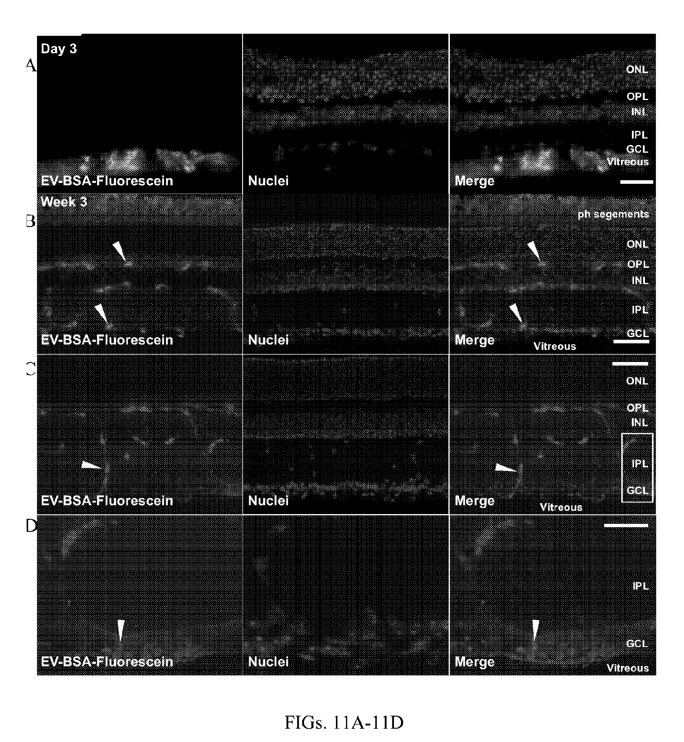
D00013

D00014
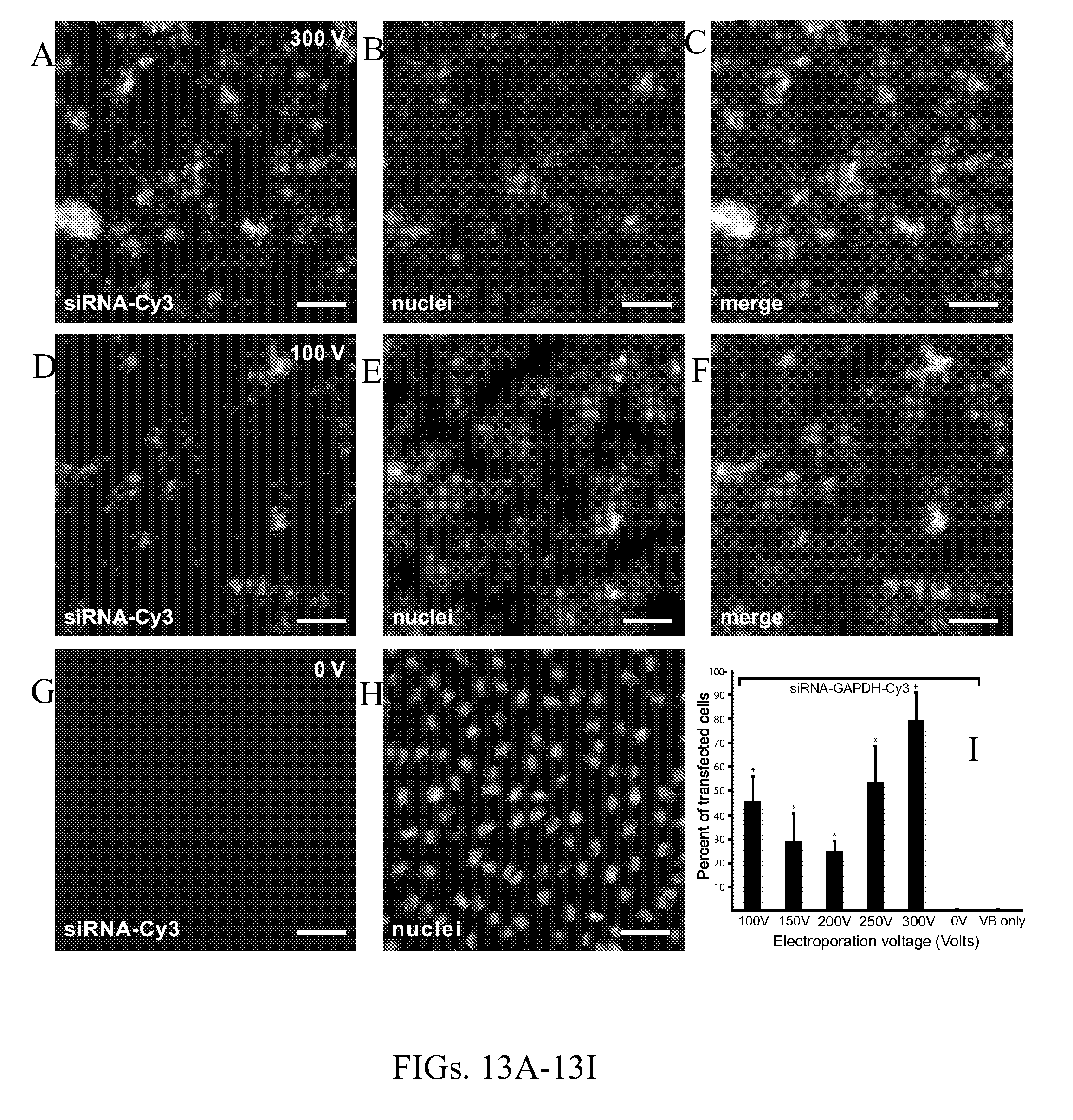
D00015

D00016

D00017

D00018

P00899

XML
uspto.report is an independent third-party trademark research tool that is not affiliated, endorsed, or sponsored by the United States Patent and Trademark Office (USPTO) or any other governmental organization. The information provided by uspto.report is based on publicly available data at the time of writing and is intended for informational purposes only.
While we strive to provide accurate and up-to-date information, we do not guarantee the accuracy, completeness, reliability, or suitability of the information displayed on this site. The use of this site is at your own risk. Any reliance you place on such information is therefore strictly at your own risk.
All official trademark data, including owner information, should be verified by visiting the official USPTO website at www.uspto.gov. This site is not intended to replace professional legal advice and should not be used as a substitute for consulting with a legal professional who is knowledgeable about trademark law.