Isoform-specific, Context-permissive Tgfb1 Inhibitors And Use Thereof
Schurpf; Thomas ; et al.
U.S. patent application number 16/361486 was filed with the patent office on 2019-07-11 for isoform-specific, context-permissive tgfb1 inhibitors and use thereof. The applicant listed for this patent is Scholar Rock, Inc.. Invention is credited to Alan Buckler, Gregory J. Carven, Abhishek Datta, Ashish Kalra, Kimberly Long, Constance Martin, Thomas Schurpf.
| Application Number | 20190209682 16/361486 |
| Document ID | / |
| Family ID | 61198888 |
| Filed Date | 2019-07-11 |


View All Diagrams
| United States Patent Application | 20190209682 |
| Kind Code | A1 |
| Schurpf; Thomas ; et al. | July 11, 2019 |
ISOFORM-SPECIFIC, CONTEXT-PERMISSIVE TGFB1 INHIBITORS AND USE THEREOF
Abstract
Disclosed herein are therapeutic use of isoform-specific, context-permissive inhibitors of TGF.beta.1 in the treatment of disease that involve TGF.beta.1 dysregulation.
| Inventors: | Schurpf; Thomas; (Cambridge, MA) ; Datta; Abhishek; (Boston, MA) ; Carven; Gregory J.; (Maynard, MA) ; Martin; Constance; (Arlington, MA) ; Kalra; Ashish; (Belmont, MA) ; Long; Kimberly; (Boston, MA) ; Buckler; Alan; (Arlington, MA) | ||||||||||
| Applicant: |
|
||||||||||
|---|---|---|---|---|---|---|---|---|---|---|---|
| Family ID: | 61198888 | ||||||||||
| Appl. No.: | 16/361486 | ||||||||||
| Filed: | March 22, 2019 |
Related U.S. Patent Documents
| Application Number | Filing Date | Patent Number | ||
|---|---|---|---|---|
| 15863564 | Jan 5, 2018 | |||
| 16361486 | ||||
| 62588626 | Nov 20, 2017 | |||
| 62587964 | Nov 17, 2017 | |||
| 62585227 | Nov 13, 2017 | |||
| 62558311 | Sep 13, 2017 | |||
| 62549767 | Aug 24, 2017 | |||
| 62529616 | Jul 7, 2017 | |||
| 62514417 | Jun 2, 2017 | |||
| 62452866 | Jan 31, 2017 | |||
| 62443615 | Jan 6, 2017 | |||
| Current U.S. Class: | 1/1 |
| Current CPC Class: | A61P 35/00 20180101; A61P 35/04 20180101; A61K 39/39541 20130101; A61P 37/00 20180101; C07K 16/22 20130101; C07K 2317/76 20130101; G01N 33/574 20130101; A61K 2039/505 20130101; G01N 2800/60 20130101; C07K 2317/92 20130101 |
| International Class: | A61K 39/395 20060101 A61K039/395; C07K 16/22 20060101 C07K016/22; G01N 33/574 20060101 G01N033/574; A61P 35/04 20060101 A61P035/04 |
Claims
1. A method for identifying a TGF.beta.1 inhibitor for therapeutic use, the method comprising the step of: selecting an antibody, or an antigen-binding fragment thereof, that binds at least one type of extracellular matrix (ECM)-associated proTGF.beta.1 complex and at least one type of cell-associated proTGF.beta.1 complex, wherein the antibody, or the antigen-binding fragment thereof, inhibits activation of TGF.beta.1; and, wherein the antibody, or the antigen-binding fragment thereof, binds a hLTBP1-proTGF.beta.1 complex and/or a hLTBP3-proTGF.beta.1 complex with a K.sub.D of .ltoreq.0.5 nM as measured by bio-layer interferometry.
2. The method of claim 1, wherein the ECM-associated proTGF.beta.1 complex comprises hLTBP1 or hLTBP3.
3. The method of claim 1, wherein the cell-associated proTGF.beta.1 complex comprises hGARP or hLRRC33.
4. The method of claim 1, wherein the antibody, or the antigen-binding fragment thereof, is capable of specifically binding each of the following complexes: a hLTBP1-proTGF.beta.1 complex, a hLTBP3-proTGF.beta.1 complex, a hGARP-proTGF.beta.1 complex, and a hLRRC33-proTGF.beta.1 complex.
5. The method of claim 1, wherein the antibody, or the antigen-binding fragment thereof, preferentially binds the hLTBP1-proTGF.beta.1 complex and/or the hLTBP3-proTGF.beta.1 complex over a hGARP-proTGF.beta.1 complex.
6. The method of claim 5, wherein the antibody, or the antigen-binding fragment thereof, binds the hGARP-proTGF.beta.1 complex with a KD of .gtoreq.4 nM, as measured by bio-layer interferometry.
7. The method of claim 1, further comprising the steps of: carrying out a preclinical study that comprises administration of a therapeutic dose of the antibody, or the antigen-binding fragment thereof, to evaluate in vivo efficacy; and carrying out a toxicology/tolerability study in an animal model to evaluate in vivo safety; wherein the therapeutic dose is shown to be both safe and efficacious in vivo.
8. The method of claim 7, wherein the administration of the therapeutic dose is sufficient to reduce expression of one or more genes selected from the group consisting of: Serpine 1, MCP-1/CCL2, Col1a1, Col3a1, FN1, TGFB1, CTGF, and ACTA2.
9. The method of claim 7, wherein the administration of the therapeutic dose is sufficient to reduce phosphorylation of SMAD2/3.
10. The method of claim 7, wherein the toxicology/tolerability study evaluates cardiovascular toxicity, gastrointestinal toxicity, immunotoxicity, bone toxicity, cartilage toxicity, reproductive system toxicity, and/or renal toxicity.
11. The method of claim 7, wherein the toxicology/tolerability of the antibody, or the antigen-binding fragment thereof, is evaluated at a dosage of at least up to 100 mg/kg/week.
12. The method of claim 7, wherein no test article-related toxicities are observed when the antibody, or the antigen-binding fragment thereof, is administered at 100 mg/kg/week for 4 weeks.
13. The method of claim 1, wherein the antibody, or the antigen-binding fragment thereof, does not bind TGF.beta.2 or proTGF.beta.2.
14. The method of claim 13, wherein the antibody, or the antigen-binding fragment thereof, does not bind TGF.beta.3 or proTGF.beta.3.
15. A method for making a pharmaceutical composition comprising a TGF.beta.1 inhibitor, the method comprising the step of: formulating the antibody or the antigen-binding fragment identified in the method of claim 1 into a pharmaceutical composition with one or more pharmaceutically acceptable excipients.
Description
RELATED APPLICATIONS
[0001] This application is a continuation of U.S. patent application Ser. No. 15/836,564, filed Jan. 5, 2018, which claims priority to and benefit under 35 U.S.C. .sctn. 119(e) of the following applications: U.S. Provisional Application No. 62/443,615, filed on Jan. 6, 2017; U.S. Provisional Application No. 62/452,866, filed on Jan. 31, 2017; U.S. Provisional Application No. 62/514,417, filed on Jun. 2, 2017; U.S. Provisional Application 62/529,616, filed on Jul. 7, 2017, U.S. Provisional Application No. 62/549,767, filed on Aug. 24, 2017, U.S. Provisional Application No. 62/558,311, filed on Sep. 13, 2017, U.S. Provisional Application No. 62/585,227 filed on Nov. 13, 2017, U.S. Provisional Application No. 62/587,964 filed on Nov. 17, 2017, and U.S. Provisional Application No. 62/588,626 filed on Nov. 20, 2017, the contents of each of which are expressly incorporated herein by reference in their entireties.
SEQUENCE LISTING
[0002] The instant application contains a Sequence Listing which has been submitted electronically in ASCII format and is hereby incorporated by reference in its entirety. Said ASCII copy, created on Jan. 5, 2018, is named 2018_01_05_127036-02008_ST25.txt and is 221,825 bytes in size.
BACKGROUND OF THE INVENTION
[0003] Transforming growth factor .beta. (TGF.beta.) superfamily of growth factors are involved in a number of signaling cascades that regulate diverse biological processes including, but not limited to: inhibition of cell growth, tissue homeostasis, extracellular matrix (ECM) remodeling, endothelial to mesenchymal transition (EMT), cell migration and invasion, and immune modulation/suppression, as well as mesenchymal to epithelial transition. In relation to ECM remodeling, TGF.beta. signaling may increase fibroblast populations and ECM deposition (e.g., collagens). In the immune system, TGF.beta. ligand modulates T regulatory cell function and maintenance of immune precursor cell growth and homeostasis. In normal epithelial cells, TGF.beta. is a potent growth inhibitor and promoter of cellular differentiation. However, as tumors develop and progress, they frequently lose their negative growth response to TGF.beta.. In this setting, TGF.beta. may become a promoter of tumor development due to its ability to stimulate angiogenesis, alter the stromal environment, and induce local and systemic immunosuppression. For these and other reasons, TGF.beta. has been a therapeutic target for a number of clinical indications. Despite much effort made to date by a number of groups, successful clinical development of a TGF.beta. therapeutic has been challenging.
[0004] Observations from preclinical studies, including in rats and dogs, have revealed certain toxicities associated with inhibiting TGF.beta. in vivo. Moreover, although several TGF.beta. inhibitors have been developed to date, most clinical programs targeting TGF.beta. have been discontinued due to side effects (summarized, for example, in WO 2017/156500). Thus, despite lines of direct and indirect evidence pointing to the involvement of TGF.beta. signaling in the progression of diseases such as cancer and fibrosis, there is no TGF.beta. therapeutics available in the market which are safe and efficacious.
[0005] Among proliferative disorders, dysregulation of TGF.beta. has also been implicated in myelofibrosis, which is a bone marrow disorder characterized by clonal myeloproliferation, aberrant cytokine production, extramedullary hematopoiesis, and bone marrow fibrosis. Although somatic mutations in JAK2, MPL and CALR have been identified in the pathogenesis of the disease, Ruxolitinib (Jakafi), which is a JAK1/JAK2 inhibitor approved by the FDA for the treatment of myelofibrosis, has not demonstrated efficacy in ameliorating established bone marrow fibrosis in patients.
[0006] Thus, improved methods and compositions for inhibiting TGF.beta. signaling are needed that can be used to effectively and safely treat diseases and disorders involving TGF.beta.1, including, for example, proliferative disorders (e.g., cancer), fibrosis and inflammation.
SUMMARY OF THE INVENTION
[0007] The present invention encompasses the recognition that blocking TGF.beta. activation at multiple sources may provide greater clinical effects in treating a number of diseases involving both an ECM aspect and an immune aspect of TGF.beta. dysregulation. Accordingly, provided herein are improved methods for treating such diseases with TGF.beta.1 inhibitors which are superior to conventional TGF.beta. antagonists with respect to their isoform selectivity, breadth of molecular targets within a disease niche, durability of effects and safety.
[0008] A body of evidence supports the notion that many diseases manifest complex perturbations of TGF.beta. signaling, which likely involve participation of heterogeneous cell types that confer different effects of TGF.beta. function, which are mediated by its interactions with so-called presenting molecules. At least four such presenting molecules have been identified, which can "present" TGF.beta. in various extracellular niches to enable its activation in response to local stimuli. In one category, TGF.beta. is deposited into the ECM in association with ECM-associated presenting molecules, such as LTBP1 and LTBP3, which mediate ECM-associated TGF.beta. activities. In another category, TGF.beta. is tethered onto the surface of immune cells, via presenting molecules such as GARP and LRRC33, which mediate certain immune function. These presenting molecules show differential expression, localization and/or function in various tissues and cell types, indicating that triggering events and outcome of TGF.beta. activation will vary, depending on the microenvironment. Based on the notion that many TGF.beta. effects may interact and contribute to disease progression, therapeutic agents that can antagonize multiple facets of TGF.beta. function may provide greater efficacy.
[0009] Previously, the inventors recognized that isoform-specific inhibition (as opposed to pan-inhibition) of TGF.beta. may render improved safety profiles of antagonizing TGF.beta. in vivo (see WO 2017/156500). Taking this into consideration, the inventors have sought to develop TGF.beta.1 inhibitors that are both i) isoform-specific; and, ii) capable of broadly targeting multiple TGF.beta.1 signaling complexes that are associated with different presenting molecules, as therapeutic agents for conditions driven by multifaceted TGF.beta.1 effects and dysregulation thereof.
[0010] Accordingly, the present disclosure provides isoform-specific inhibitory agents capable of targeting both ECM-associated TGF.beta.1 and immune cell-associated TGF.beta.1, thereby blocking multiple sources of TGF.beta.1 presented in multiple contexts. Such inhibitory agents are referred herein to as "isoform-specific, context-permissive" inhibitors of TGF.beta.1. The invention also provides use of these agents as a therapeutic in the treatment of conditions that are characterized by dysregulation of TGF.beta.1 signaling associated with multiple aspects of TGF.beta.1 function. Such inhibitors may function as multifunctional agents to antagonize multiple TGF.beta.1 activities (e.g., TGF.beta.1 from multiple sources or contexts) to enhance clinical effects in the context of fibrosis, myelofibrosis, cancer, and other conditions.
[0011] The rationale for the advantageous use of context-permissive (such as context-independent) inhibitors of TGF.beta.1 over context-specific inhibitors of TGF.beta.1 as a therapeutic to treat certain diseases (as described in further detail herein) include the following:
[0012] Involvement of Heterogeneous TGF.beta.1 Complexes in a Disease Environment:
[0013] First, various diseases involve heterogeneous populations of cells as multiple sources of TGF.beta.1 that collectively contribute to the pathogenesis and/or progression of the disease. More than one types of TGF.beta.1-containing complexes ("contexts") likely coexist within the same disease microenvironment. In particular, such diseases may involve both an ECM component of TGF.beta.1 signaling and an immune component of TGF.beta.1 signaling. In such situations, selective targeting of a single TGF.beta.1 context (e.g., TGF.beta.1 associated with one type of presenting molecule) may offer limited relief. By contrast, context-permissive inhibitors of TGF.beta.1 are advantageously aimed to more broadly target inactive (pro/latent) TGF.beta.1 complexes and prevent activation of the growth factor at multiple sources before mature TGF.beta.1 can be released for receptor binding to trigger downstream signaling, while maintaining the isoform selectivity to minimize toxicities.
[0014] Common mechanisms underlining various diseases: Second, notable similarities in tissue/cellular characteristics are observed between the tumor stroma and fibrotic tissues. Indicating crosstalk between and among: i) TGF.beta.1-dependent pro-fibrotic phenotypes; ii) TGF.beta.1-dependent pro-tumor phenotypes; and, iii) TGF.beta.-dependent immunosuppressive phenotypes, observed in a number of pathological conditions. Thus, the use of context-permissive inhibitors that broadly act upon many of these constituents may provide optimal therapeutic effects across a diverse types of disease conditions. For example, clinical manifestations of primary myelofibrosis include abnormal proliferation of certain cell populations and fibrosis in the bone marrow.
[0015] Countering drug resistance: Third, a number of studies have reported cancer/tumors which are resistant to anti-cancer therapies, such as immuno checkpoint inhibitors. In some cases, such resistance appears intrinsic to the particular cancer/tumor-type against the patient's background (typically referred to as innate resistance, primary resistance, intrinsic resistance, or inherent resistance; these terms are used interchangeably herein). Such resistance may be represented in a subject of patients poorly responsive to cancer therapies such as immune checkpoint inhibitors and possibly reflect immune-excluded environment. This is likely mediated at least in part by a TGF.beta.1-dependent pathway. Thus, isoform-selective inhibitor described herein may render the resistant cancers more responsive to such therapies.
[0016] Alternatively, resistance may develop over time such that patients who show material clinical responsiveness to a treatment become poorly responsive (i.e., adaptive or acquired resistance). For example, it has been reported that PD-1 therapy can lead to adaptive resistance which is correlated with upregulation of other T cell antigens (e.g., TCR components) suggesting that cancer cells evolve to evade the PD-1 blockade via another mechanism. Subsequently, a second checkpoint inhibitor that targets a different T cell receptor component such as TIM3 can restore responsiveness to the immunotherapy. These observations suggest that blocking multiple pathways to counter adaptive responses of cancer cells may reduce the likelihood of cancer cells' ability to evade host immunity. Context-permissive inhibitors of TGF.beta.1 which are capable of targeting multiple TGF.beta.1 contexts may advantageously circumvent acquired drug resistance by providing blockade at multiple points of the TGF.beta.1 function.
[0017] Withstanding expression plasticity: And finally, based on the notion that expression of various presenting molecules may vary over time, for example, in response to local cues (e.g., cytokines, chemokines, ECM environment, etc.) and/or with changes in a disease microenvironment, it is reasoned that context-permissive inhibitors of TGF.beta.1 such as those described herein may be used to withstand such plasticity and provide broad, durable inhibitory effects even when abnormal changes in expression of the presenting molecules occur.
[0018] In any of these scenarios, the context-permissive inhibitors of TGF.beta.1 are advantageously aimed to target the pro/latent forms of TGF.beta.1 in association with various presenting molecules, all of which or different combinations of which are present in a disease microenvironment(s). More specifically, in one modality, the inhibitor targets ECM-associated TGF.beta.1 (LTBP1/3-TGF.beta.1 complexes). In another modality, the inhibitor targets immune cell-associated TGF.beta.1. This includes GARP-presented TGF.beta.1, such as GARP-TGF.beta.1 complexes expressed on Treg cells and LRRC33-TGF.beta.1 complexes expressed on macrophages and other myeloid/lymphoid cells, as well as certain cancer cells.
[0019] Such antibodies include isoform-specific inhibitors of TGF.beta.1 that bind and prevent activation (or release) of mature TGF.beta.1 growth factor from a pro/latent TGF.beta.1 complex in a context-permissive (or context-independent) manner, such that the antibodies can inhibit activation (or release) of TGF.beta.1 associated with multiple types of presenting molecules. In particular, the present invention provides antibodies capable of blocking at least one context of ECM-associated TGF.beta.1 (LTBP-presented and/or LTBP3-presented) and at least one context of cell-associated TGF.beta.1 (GARP-presented and/or LRRC33-presented).
[0020] Various disease conditions have been suggested to involve dysregulation of TGF.beta. signaling as a contributing factor. Indeed, the pathogenesis and/or progression of certain human conditions appear to be predominantly driven by or dependent on TGF.beta.1 activities. In particular, many such diseases and disorders appear to involve both an ECM component and an immune component of TGF.beta.1 function, suggesting that TGF.beta.1 activation in multiple contexts (e.g., mediated by more than one type of presenting molecules) is involved. Moreover, it is contemplated that there is crosstalk among TGF.beta.1-responsive cells. In some cases, interplays between multifaceted activities of the TGF.beta.1 axis may lead to disease progression, aggravation, and/or suppression of the host's ability to combat disease. For example, certain disease microenvironments, such as tumor microenvironment (TME), may be associated with TGF.beta.1 presented by multiple different presenting molecules, e.g., LTBP1-proTGF.beta.1, LTBP3-proTGF.beta.1, GARP-proTGF.beta.1, LRRC33-proTGF.beta.1, and any combinations thereof. TGF.beta.1 activities of one context may in turn regulate or influence TGF.beta.1 activities of another context, raising the possibility that when dysregulated, this may result in exacerbation of disease conditions. Therefore, it is desirable to broadly inhibit across multiple modes of TGF.beta.1 function (i.e., multiple contexts) while selectively limiting such inhibitory effects to the TGF.beta.1 isoform. The aim is not to perturb homeostatic TGF.beta. signaling mediated by the other isoforms, including TGF.beta.3, which plays an important role in would healing.
[0021] To address this, the inventors of the present disclosure sought to generate isoform-specific, context-permissive inhibitors of TGF.beta.1 which may be particularly advantageous for therapeutic use in the treatment of diseases that are driven by or dependent on TGF.beta.1 signaling or dysregulation thereof. The approach taken to meet the criteria for such inhibitors is: i) the ability to inhibit TGF.beta.1 signaling in an isoform-specific manner (without interfering with TGF.beta.2 and/or TGF.beta.3 activities); and, ii) the ability to inhibit both an ECM-associated and an immune cell-associated TGF.beta.1 signaling. The rationale for this approach is to balance the effectiveness (hence clinical efficacy) of TGF.beta.1 inhibition against potential toxicities. More specifically, achieving selectivity towards TGF.beta.1 at therapeutic dosage over the other isoforms is aimed to reduce or minimize possible toxicities (e.g., unwanted side effects and adverse events) associated with pan-inhibition of TGF.beta. in vivo, some of which may be required for normal biological functions (such as wound healing). On the other hand, inclusion of multiple contexts of TGF.beta.1 as therapeutic target is aimed at ensuring or to optimizing clinical efficacy in a disease that involves dysregulation of multiple aspects of TGF.beta.1 signaling. Various embodiments of clinical applications and treatment regimens are encompassed by the invention.
[0022] Accordingly, in one aspect, provided herein are isoform-specific, context-permissive inhibitors of TGF.beta.1, characterized in that such inhibitors have the ability to inhibit both an ECM-associated TGF.beta.1 signaling and an immune cell-associated TGF.beta.1 signaling. Specifically, such inhibitors can block TGF.beta.1 presented in multiple contexts, i.e., TGF.beta.1 activities mediated by two or more types of presenting molecules, while maintaining TGF.beta.2 and TGF.beta.3 activities intact. Thus, the TGF.beta.1 activities which can be inhibited by such inhibitors include two or more of the following: i) TGF.beta.1 signaling associated with GARP-presented TGF.beta.1; ii) TGF.beta.1 signaling associated with LRRC33-presented TGF.beta.1; iii) TGF.beta.1 signaling associated with LTBP1-presented TGF.beta.1; and, iv) TGF.beta.1 signaling associated with LTBP3-presented TGF.beta.1. In some embodiments, such inhibitors target at least two, or, at least three of pro-protein forms of the following complexes: i) TGF.beta.1-GARP; ii) TGF.beta.1-LRRC33; iii) TGF.beta.1-LTBP1; and, iv) TGF.beta.1-LTBP3. In some embodiments, such inhibitors are monoclonal antibodies that specifically bind and inhibit i) TGF.beta.1-GARP; iii) TGF.beta.1-LTBP1; and, iv) TGF.beta.1-LTBP3. In some embodiments, such monoclonal antibodies specifically bind and inhibit it ii) TGF.beta.1-LRRC33; iii) TGF.beta.1-LTBP1; and, iv) TGF.beta.1-LTBP3. In some embodiments, such monoclonal antibodies specifically bind and inhibit i) TGF.beta.1-GARP; ii) TGF.beta.1-LRRC33; and iii) TGF.beta.1-LTBP1. In some embodiments, such monoclonal antibodies specifically bind and inhibit i) TGF.beta.1-GARP; ii) TGF.beta.1-LRRC33; and iv) TGF.beta.1-LTBP3. In some embodiments, such monoclonal antibodies specifically inhibit all of the following complexes: i) TGF.beta.1-GARP; ii) TGF.beta.1-LRRC33; iii) TGF.beta.1-LTBP1; and, iv) TGF.beta.1-LTBP3. In some embodiments, such monoclonal antibodies do not bind mature TGF.beta.1 that is free TGF.beta.1 (e.g., growth factor that is released from or not complexed with a presenting molecule). The aspect of the invention includes compositions comprising such an inhibitor, including for example, pharmaceutical compositions which are suitable for administration in human and non-human subjects to be treated. Such pharmaceutical compositions are typically sterile. In some embodiments, such pharmaceutical compositions may also comprise at least one pharmaceutically acceptable excipient, such as a buffer and a surfactant (e.g., polysorbates). Kits comprising such a pharmaceutical composition are also encompassed by the invention.
[0023] Isoform-specific, context-permissive inhibitors described herein are suitable for use in the treatment of disease or disorder involving multiple biological functions of TGF.beta.1 and dysregulation thereof. In particular, such disease or disorder involves both an ECM component of TGF.beta.1 function and an immune component of TGF.beta.1 function. Administration of such an inhibitor can therefore inhibit each axis of the TGF.beta.1 signaling pathway in vivo, e.g., multiple TGF.beta.1 targets associated with the disease or disorder, enhancing therapeutic effects. Accordingly, in another aspect, the invention includes therapeutic use of such inhibitors in a method for treating a subject who suffers from a disease associated with TGF.beta.1 dysregulation. Isoform-specific, context-permissive or context-independent inhibitors of TGF.beta.1 signaling are particularly suitable for treating a disease that is driven or dependent on multiple functions (e.g., both an ECM component and an immune component) of TGF.beta.1. Typically, such diseases involve multiple cell types or cell status in which TGF.beta.1 is presented with multiple types of presenting molecules (e.g., multiple contexts).
[0024] In a related aspect, the invention provides screening, production and manufacture methods for isoform-specific, context-permissive TGF.beta.1 inhibitors with an improved safety profile (e.g., reduced in vivo toxicity). Such methods require that candidate agents be tested and selected for the TGF.beta.1 isoform specificity, e.g., candidate agents are selected for inhibitory activities against TGF.beta.1 signaling, and not TGF.beta.2 and/or TGF.beta.3 signaling. According to the invention, such isoform-specific inhibitors of TGF.beta.1 activities can inhibit multiple contexts of TGF.beta.1 function (see below).
[0025] In some embodiments, such agents are antibodies or antigen-binding fragments thereof that specifically bind and block activation of TGF.beta.1, but not TGF.beta.2 and/or TGF.beta.3. In some embodiments, such antibodies or antigen-binding fragments thereof do not bind free mature TGF.beta.1 growth factor that is not associated with a pro/latent complex. Thus, relevant production methods may include a screening step in which candidate agents (such as candidate antibodies or fragments thereof) are evaluated for their ability to inhibit TGF.beta.1 that is associated with particular presenting molecules, e.g., GARP, LRRC33, LTBP1, and/or LTBP3. In some embodiments, inactive (e.g., latent) precursor complex, such as GARP-proTGF.beta.1, LRRC33-proTGF.beta.1, LTBP1-proTGF.beta.1 and LTBP3-proTGF.beta.1, may be utilized to assay for activation of mature, active TGF.beta.1 growth factor. TGF.beta.1 activation, in the presence or absence of a test agent (i.e., candidate inhibitor) may be measured by any suitable means, including but not limited to in vitro assays and cell-based assays. Similar screening step can be utilized to test isoform specificity by the use of TGF.beta.2 and/or TGF.beta.3 counterparts. Such screening step can be carried out to identify candidate agents (such as candidate antibodies or fragments thereof) for their ability to inhibit TGF.beta.1 signaling in: i) an isoform-specific manner; and, ii) a context-permissive or context-independent manner.
[0026] Certain diseases are associated with dysregulation of multiple biological roles of TGF.beta. signaling that are not limited to a single context of TGF.beta. function. In such situations, it may be beneficial to modulate TGF.beta. effects across multiple contexts involved in the onset and/or during the course of disease progression. Thus, in some embodiments, the invention provides methods for targeting and broadly inhibiting multiple TGF.beta.1 contexts but in an isoform-specific manner. Such agents are herein referred to as "isoform-specific, context-permissive" TGF.beta.1 inhibitors. Thus, context-permissive TGF.beta.1 inhibitors target multiple contexts (e.g., multiple types of pro/latent-TGF.beta.1 complexes). Preferably, such inhibitors target at least one type (or "context") of TGF.beta.1 pre-activation complex that is associated with the ECM (i.e., pro/latent TGF.beta.1 complex presented by an ECM-associated presenting molecule) and additionally at least one type (or "context") of TGF.beta.1 pre-activation complex tethered to cell surface (i.e., pro/latent TGF.beta.1 complex presented by a cell or membrane-associated presenting molecule). In some embodiments, context-permissive TGF.beta.1 modulators target all types of pro/latent TGF.beta.1 complexes (e.g., GARP-associated, LRRC33-associated, LTBP-associated, etc.) so as to encompass all contexts irrespective of particular presenting molecule(s).
[0027] Whilst context-permissive TGF.beta.1 inhibitors are capable of targeting more than one types of pro/latent-TGF.beta.1 complexes (i.e., with different presenting molecules), in some embodiments, such inhibitors may favor (or show bias towards) one or more context over the other(s). Thus, in some embodiments, a context-permissive antibody that inhibits the activation of TGF.beta.1 may preferentially inhibit TGF.beta.1 activation mediated by one presenting molecule over another presenting molecule, even if such antibody is capable of binding to both types of pro/latent complexes. In some embodiments, such antibody is a monoclonal antibody that binds and inhibits activation of LTBP1/3-associated TGF.beta.1, GARP-associated TGF.beta.1, and LRRC33-associated TGF.beta.1, but with preferential inhibitory activities toward LTBP1/3-associated TGF.beta.1. In some embodiments, such antibody is a monoclonal antibody that binds and inhibits activation of LTBP1-associated TGF.beta.1, LTBP3-associated TGF.beta.1, GARP-associated TGF.beta.1, and LRRC33-associated TGF.beta.1, but with preferential inhibitory activities toward LTBP1- and LTBP-3-associated TGF.beta.1. In some embodiments, such antibody is a monoclonal antibody that binds and inhibits activation of LTBP1-associated TGF.beta.1, LTBP3-associated TGF.beta.1, GARP-associated TGF.beta.1, and LRRC33-associated TGF.beta.1, but with preferential inhibitory activities toward GARP-associated TGF.beta.1 and LRRC33-associated TGF.beta.1. In some embodiments, such antibody is a monoclonal antibody that binds and inhibits activation of GARP-associated TGF.beta.1 and LRRC33-associated TGF.beta.1, but with preferential inhibitory activities toward GARP-associated TGF.beta.1. In some embodiments, such antibody is a monoclonal antibody that binds and inhibits activation of GARP-associated TGF.beta.1 and LRRC33-associated TGF.beta.1, but with preferential inhibitory activities toward LRRC33-associated TGF.beta.1.
[0028] Thus, according to the invention, varying degrees of selectivity may be generated in order to target subset of TGF.beta. effects. Isoform-specific inhibitors of TGF.beta. (which target a single isoform of TGF.beta.) provide greater selectivity than so-called pan-TGF.beta. inhibitors (which target multiple or all isoforms of TGF.beta.).
[0029] The invention includes use of such TGF.beta.1 inhibitors in methods for treating a disease associated with TGF.beta.1 dysregulation. The use of such inhibitors is particularly advantageous in conditions where the TGF.beta.1 isoform plays a dominant role (over TGF.beta.2/3) in driving the disease, and where the disease involves both an ECM component and an immune component of TGF.beta.1 signaling. This approach aims to preserve normal or homeostatic TGF.beta. functions, while preferentially targeting disease-associated TGF.beta. function.
[0030] Such inhibitor is preferably a TGF.beta.1 activation inhibitor (i.e., inhibitor of the TGF.beta.1 activation step). In preferred embodiments, such inhibitor is capable of targeting the inactive forms of TGF.beta.1 (e.g., pro/latent-TGF.beta.1 complexes) prior to activation to effectuate more durable inhibition as compared to targeting a transient, already activated, soluble/free form of the growth factor that has been released from the latent complex. Determination of the source/context of disease-associated TGF.beta.1 may be carried out with the use of antibodies that specifically bind TGF.beta.1 latent complex that includes a particular presenting molecule of interest (e.g., GARP, LRRC33, LTBP1, LTBP3, etc.).
[0031] Aspects of the present disclosure relate to immunoglobulins, such as antibodies, or antigen binding portions thereof, that specifically bind at least three of the following complexes: a GARP-TGF.beta.1 complex, a LTBP1-TGF.beta.1 complex, a LTBP3-TGF.beta.1 complex and a LRRC33-TGF.beta.1 complex. According to the invention, such immunoglobulins specifically bind at least one type of ECM-associated (e.g., ECM-tethered) TGF.beta.1 complexes (e.g., LTBP1- and/or LTBP3-associated TGF.beta.1 complexes) and at least one type of cell-associated (e.g., cell surface-tethered) TGF.beta.1 complexes (e.g., GARP- and/or LRRC33-associated TGF.beta.1 complexes) to effectuate broad inhibitory action on multiple contexts. The antibodies, or antigen binding portions thereof, described herein, specifically bind to an epitope of TGF.beta.1 (e.g., LAP) or a component(s) of a protein complex comprising the TGF.beta.1 (e.g., LAP), that is available for binding by the antibodies, or antigen binding portions thereof, when the TGF.beta.1 is present in a GARP-TGF.beta.1 complex, a LTBP1-TGF.beta.1 complex, a LTBP3-TGF.beta.1 complex and/or a LRRC33-TGF.beta.1.
[0032] In some embodiments, the epitope is available for binding by the antibody when the TGF.beta.1 is present in two or more of the following protein complexes: a GARP-TGF.beta.1 complex, a LTBP1-TGF.beta.1 complex, a LTBP3-TGF.beta.1 complex, and a LRRC33-TGF.beta.1 complex; and wherein the antibody does not bind free mature TGF.beta.1 growth factor that is not in association with the pro/latent complex.
[0033] In some embodiments, the TGF.beta.1 is proTGF.beta.1 and/or latent TGF.beta.1 (e.g., pro/latent TGF.beta.1). In some embodiments, the TGF.beta.1 is latent TGF.beta.1. In some embodiments, the TGF.beta.1 is proTGF.beta.1.
[0034] The isoform-specific TGF.beta.1 inhibitors according to the invention do not bind TGF.beta.2. The isoform-specific TGF.beta.1 inhibitors according to the invention do not bind TGF.beta.3. In some embodiments, such inhibitors do not bind pro/latent TGF.beta.2. In some embodiments, such inhibitors do not bind pro/latent TGF.beta.3. In some embodiments, the antibody, or antigen binding portion thereof, does not prevent the ability of TGF.beta.1 to bind to integrin.
[0035] In some embodiments, the antibody, or antigen binding portion thereof, comprises a heavy chain variable region comprising a CDR3 having the amino acid sequence of SEQ ID NO: 87 and a light chain variable region comprising a CDR3 having the amino acid sequence of SEQ ID NO: 90. In some embodiments, the antibody, or antigen binding portion thereof, comprises a heavy chain variable region comprising a CDR2 having the amino acid sequence of SEQ ID NO: 86 and a light chain variable region comprising a CDR2 having the amino acid sequence of SEQ ID NO: 89. In some embodiments, the antibody, or antigen binding portion thereof, comprises a heavy chain variable region comprising a CDR1 having the amino acid sequence of SEQ ID NO: 85 and a light chain variable region comprising a CDR1 having the amino acid sequence of SEQ ID NO: 88.
[0036] In some embodiments, the antibody comprises a heavy chain polypeptide sequence that is at least 90% identical to the amino acid sequence set forth in SEQ ID NO: 99. In some embodiments, the antibody comprises a light chain polypeptide sequence that is at least 90% identical to the amino acid sequence set forth in SEQ ID NO: 100. In some embodiments, the antibody comprises a heavy chain polypeptide sequence that is at least 90% identical to the amino acid sequence set forth in SEQ ID NO: 99 and a light chain polypeptide sequence that is at least 90% identical to the amino acid sequence set forth in SEQ ID NO: 100. In some embodiments, such antibody comprises CDRs as set forth in SEQ ID NOs: 85-90. In some embodiments, the antibody consists of two polypeptides of SEQ ID NO: 99 and two polypeptides of SEQ ID NO:100.
[0037] In some embodiments, the antibody, or antigen binding portion thereof, comprises a heavy chain variable domain comprising an amino acid sequence having at least 70%, 75%, 80%, 85%, 90%, 95%, 96%, 97%, 98%, or 99% identity to the amino acid sequence set forth in SEQ ID NO: 95 and a light chain variable domain comprising an amino acid sequence having at least 70%, 75%, 80%, 85%, 90%, 95%, 96%, 97%, 98%, or 99% identity to the amino acid sequence set forth in SEQ ID NO: 97.
[0038] In some embodiments, the antibody, or antigen binding portion thereof, comprises a heavy chain variable domain comprising an amino acid sequence set forth in SEQ ID NO: 95 and a light chain variable domain comprising an amino acid sequence set forth in SEQ ID NO: 97.
[0039] In some embodiments, the antibody, or antigen binding portion thereof, inhibits TGF.beta.1 activation, but not TGF.beta.2 activation or TGF.beta.3 activation.
[0040] In some embodiments, the antibody, or antigen binding portion thereof, inhibits the release of mature TGF.beta.1 from the GARP-TGF.beta.1 complex, the LTBP1-TGF.beta.1 complex, the LTBP3-TGF.beta.1 complex, and/or the LRRC33-TGF.beta.1 complex.
[0041] In one aspect, provided herein is a pharmaceutical composition comprising an antibody, or antigen binding portion thereof, as described herein, and a pharmaceutically acceptable carrier. Such pharmaceutical compositions are typically sterile and are suitable for administration in human subjects. In some embodiments, such pharmaceutical compositions may be provided as kits, which are encompassed by the invention.
[0042] In another aspect, provided herein is a method for inhibiting TGF.beta.1 activation, the method comprising exposing a GARP-TGF.beta.1 complex, a LTBP1-TGF.beta.1 complex, a LTBP3-TGF.beta.1 complex, or a LRRC33-TGF.beta.1 complex to an antibody, an antigen binding portion thereof, or a pharmaceutical composition described herein.
[0043] In some embodiments, the antibody, or antigen binding portion thereof, inhibits the release of mature TGF.beta.1 from the GARP-TGF.beta.1 complex, the LTBP1-TGF.beta.1 complex, a LTBP3-TGF.beta.1 complex, or the LRRC33-TGF.beta.1 complex.
[0044] In some embodiments, the method is performed in vitro. In some embodiments, the method is performed in vivo.
[0045] Thus, the invention includes a method for treating a disease associated with dysregulation of TGF.beta.1 signaling in a human subject. Such method comprises a step of: administering to a human subject in need thereof a pharmaceutical composition provided herein, in an amount effective to treat the disease, wherein the amount achieves statistically significant clinical efficacy and safety when administered to a patient population having the disease.
[0046] In yet another aspect, provided herein is a TGF.beta. inhibitor for use in reducing adverse effects in a subject, wherein the TGF.beta. inhibitor is isoform-selective. In some embodiments, the TGF.beta. inhibitor is an antibody that specifically inhibits TGF.beta.1 while broadly targeting multiple contexts.
[0047] In some embodiments, the cell expressing the GARP-TGF.beta.1 complex or the LRRC33-TGF.beta.1 complex is a T-cell, a fibroblast, a myofibroblast, a macrophage, a monocyte, a dendritic cell, an antigen presenting cell, a neutrophil, a myeloid-derived suppressor cell (MDSC), a lymphocyte, a mast cell, a megakaryocyte, a natural killer (NK) cell, a microglia, or a progenitor cell of any one of such cells. In some embodiments, the cell expressing the GARP-TGF.beta.1 complex or the LRRC33-TGF.beta.1 complex is a hematopoietic stem cell. In some embodiments, the cell expressing the GARP-TGF.beta.1 complex or the LRRC33-TGF.beta.1 complex is a neural crest-derived cell. The T-cell may be a regulatory T cell (e.g., immunosuppressive T cell). The T cell may be a CD4-positive (CD4+) T cell and/or CD8-positive (CD8+) T cell. The neuprophil may be an activated neutrophil. The macrophage may be a polarized macrophage, including profibrotic and/or tumor-associated macrophages (TAM), e.g., M2c subtype and M2d subtype macrophages. The macrophage may be activated by one or more soluble factors, such as growth factors, cytokines, chemokines and/or other molecules that are present in a particular disease microenvironment (e.g., TME), which may work in an autocrine, paracrine, and/or endocrine fashion. In some embodiments, the macrophage is activated by M-CSF, such as M-CSF secreted by a solid tumor. In some embodiments, the macrophage is activated by TGF.beta.1.
[0048] In some embodiments, the cell expressing the GARP-TGF.beta.1 complex or the LRRC33-TGF.beta.1 complex is a cancer cell, e.g., circulating cancer cells and tumor cells. In some embodiments, the cell expressing the GARP-TGF.beta.1 complex or the LRRC33-TGF.beta.1 complex is recruited to a disease site, such as TME (e.g., tumor infiltrate). In some embodiments, the expression of the GARP-TGF.beta.1 complex or the LRRC33-TGF.beta.1 complex is induced by a disease microenvironment (e.g., TME). In some embodiments, a solid tumor comprises elevated leukocyte infiltrates, e.g., CD45+. It is contemplated that tumor-associated CD45+ cells include GARP-expressing and/or LRRC33-expressing cells.
[0049] In some embodiments, the LTBP1-TGF.beta.1 complex or the LTBP3-TGF.beta.1 complex is bound to an extracellular matrix (i.e., components of the ECM). In some embodiments, the extracellular matrix comprises fibrillin and/or fibronectin. In some embodiments, the extracellular matrix comprises a protein comprising an RGD motif. In some embodiments, cells that produce and deposit the LTBP1-TGF.beta.1 complex or the LTBP3-TGF.beta.1 complex are present in a solid tumor, such as cancer cells and stromal cells. In some embodiments, cells that produce and deposit the LTBP1-TGF.beta.1 complex or the LTBP3-TGF.beta.1 complex are present in a fibrotic tissue. In some embodiments, cells that produce and deposit the LTBP1-TGF.beta.1 complex or the LTBP3-TGF.beta.1 complex are present in a bone marrow. In some embodiments, cells that produce and deposit the LTBP1-TGF.beta.1 complex or the LTBP3-TGF.beta.1 complex are myofibroblasts or myofibroblast-like cells, including, for example, cancer-associated fibroblasts (CAFs).
[0050] In another aspect, provided herein is a method for reducing TGF.beta.1 activation in a subject, the method comprising administering to the subject an effective amount of an antibody, an antigen binding portion thereof, or a pharmaceutical composition, as described herein, thereby reducing TGF.beta.1 activation in the subject.
[0051] In some embodiments, the subject has or is at risk of having fibrotic disorder. In some embodiments, the fibrotic disorder comprises chronic inflammation of the affected tissue/organ. In some embodiments, the subject has a muscular dystrophy. In some embodiments, the subject has Duchenne muscular dystrophy (DMD). In some embodiments, the subject has or is at risk of having liver fibrosis, kidney fibrosis, lung fibrosis (e.g., idiopathic pulmonary fibrosis), endometriosis or uterine fibrosis. In some embodiments, the subject has or is at risk of having cancer (e.g., solid tumor, blood cancer, and myelofibrosis). In some embodiments, the subject has or is at risk of having dementia.
[0052] In some embodiments, the subject further receives an additional therapy. In some embodiments, the additional therapy is selected from the group consisting of a myostatin inhibitor, a VEGF agonist, an IGF1 agonist, an FXR agonist, a CCR2 inhibitor, a CCR5 inhibitor, a dual CCR2/CCR5 inhibitor, a lysyl oxidase-like-2 inhibitor, an ASK1 inhibitor, an Acetyl-CoA Carboxylase (ACC) inhibitor, a p38 kinase inhibitor, Pirfenidone, Nintedanib, a GDF11 inhibitor, JAK inhibitor (e.g., JAK2 inhibitor), or any combination thereof.
[0053] In some embodiments, the antibody, or the antigen binding portion thereof, reduces the suppressive activity of regulatory T cells (Tregs).
[0054] In some embodiments, the antibody, or the antigen binding portion thereof, does not induce organ toxicity in the subject. In some embodiments, the organ toxicity comprises cardiovascular toxicity, gastrointestinal toxicity, immunotoxicity, bone toxicity, cartilage toxicity, reproductive system toxicity, or renal toxicity.
[0055] In one aspect, provided herein is a method for treating cancer in a subject in need thereof, the method comprising administering to the subject an effective amount of an antibody, an antigen binding portion thereof, or a pharmaceutical composition, as described herein, thereby treating cancer in the subject.
[0056] In another aspect, provided herein is a method of reducing tumor growth in a subject in need thereof, the method comprising administering to the subject an effective amount of an antibody, an antigen binding portion thereof, or a pharmaceutical composition, as described herein, thereby reducing tumor growth in the subject.
[0057] In some embodiments, the antibody, or antigen binding portion thereof, is administered in combination with an additional agent or an additional therapy. In some embodiments, the additional agent is a checkpoint inhibitor. In some embodiments, the additional agent is selected from the group consisting of a PD-1 antagonist, a PDL1 antagonist, a PD-L1 or PDL2 fusion protein, a CTLA4 antagonist, etc. Such combination therapies may advantageously utilize lower dosages of the administered therapeutic agents, thus avoiding possible toxicities or complications associated with the various monotherapies or conventional combination therapies that lack the degree of selectivity/specificity achieved by the present invention.
[0058] In some embodiments, the method further comprises determining (e.g., testing or confirming) the involvement of TGF.beta.1 in the disease, relative to TGF.beta.2 and TGF.beta.3. In some embodiments, the method further comprises a step of: identifying a source (or context) of disease-associated TGF.beta.1. In some embodiments, the source/context is assessed by determining the expression of TGF.beta. presenting molecules, e.g., LTBP1, LTBP3, GARP and LRRC33 in a clinical sample taken from patients.
[0059] In yet another aspect, provided herein is a method for making (e.g., producing, manufacturing) a pharmaceutical composition for inhibiting TGF.beta. signaling, the method comprising steps of: providing one or more agents that inhibit signaling of at least one isoform of TGF.beta.; measuring activities of the one or more agents towards all isoforms of TGF.beta.; selecting an agent that is selective for TGF.beta.1; formulating into a pharmaceutical composition comprising an isoform-specific TGF.beta.1 inhibitor and a pharmaceutically acceptable excipient, such as a suitable buffer. Also provided is a pharmaceutical composition produced by such method. In some embodiments, the method further comprises a step of determining (e.g., measuring, assaying) context-dependent inhibitory activities of one or more agents.
[0060] The subject matter of the present disclosure also relates to that of PCT/US2013/068613, filed Nov. 6, 2013; PCT/US2014/036933, filed May 6, 2014; and PCT/US2017/021972, filed Mar. 10, 2017, the entire contents of each of which are incorporated herein by reference.
BRIEF DESCRIPTION OF THE FIGURES
[0061] FIG. 1 provides a schematic depicting TGF.beta.1 within a latent complex in the tissue microenvironment.
[0062] FIGS. 2A-2C illustrate multiple contexts of TGF.beta.1 function: GARP-presented TGF.beta.1 is expressed on regulatory T cells, which is involved in immune regulation (FIG. 2A); LTBP1/3-presented TGF.beta.1 is deposited by fibroblasts and other cells into the ECM (FIG. 2B); and, LRRC33-presented TGF.beta.1 is expressed on myeloid cells, including macrophages (FIG. 2C).
[0063] FIG. 3 illustrates a protein expression platform for making a GARP-TGF.beta.1 complex and a LTBP-TGF.beta.1 complex. The HEK293-based expression system uses Ni-NTA affinity purification and gel filtration to obtain multimilligram quantities of purified protein. Schematics of wild-type proTGF.beta.1, LTPB1, sGARP, and proTGF .beta.1 C4S are shown.
[0064] FIG. 4A depicts specific binding of Ab3 to latent TGF.beta.1. FIG. 4B shows binding specificity of exemplary monoclonal antibodies. FIG. 4B depicts that Ab1 and Ab2 specifically bind proTGF.beta.1 as measured by ELISA, but not proTGF.beta.2, proTGF.beta.3, or mature TGF.beta.1. FIG. 4C depicts an example of an antibody which binds (as measured by ELISA) specifically to the LTBP1-proTGF.beta.1 complex.
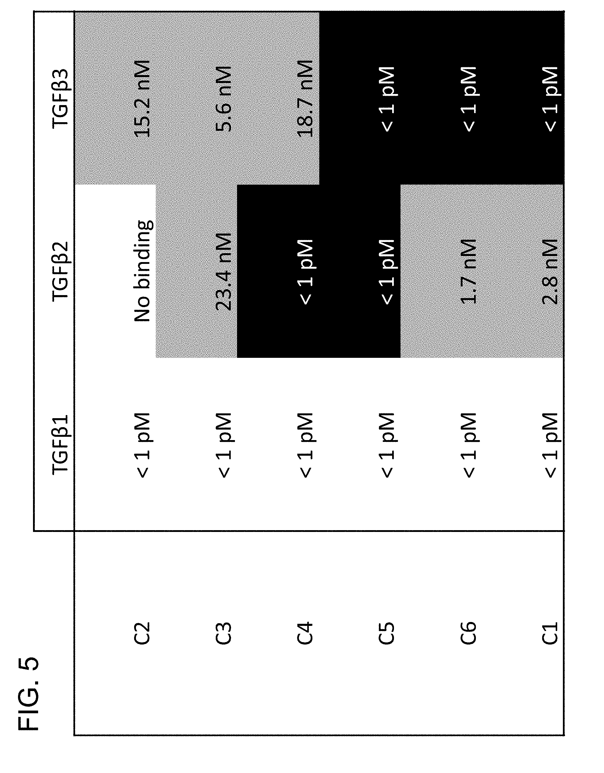
[0065] FIG. 5 provides a panel of prior art antibodies made against mature TGF.beta. growth factor, and their respective binding profiles for all three isoforms.
[0066] FIGS. 6A-6B provide binding profiles, as measured by Octet, of Ab1, Ab2 and Ab3, which are isoform-specific, context-permissive/independent TGF.beta.1 inhibitors.
[0067] FIGS. 7A-7H provide cell-based inhibition assays.
[0068] FIG. 8 shows inhibitory effects of Ab3 on Kallikrein-induced activation of TGF.beta.1 in vitro.
[0069] FIGS. 9A-9B show inhibitory effects of Ab1 and Ab3 on regulatory T cell-dependent suppression of effector T cell proliferation.
[0070] FIGS. 10A-10C show upregulation of cell surface LRRC33 expression in polarized macrophages.
[0071] FIG. 11 provides results from a T cell co-transfer colitis model.
[0072] FIGS. 12A-12K show inhibitory effects of Ab2 on TGFb1-dependent mechanistic disease model of UUO.
[0073] FIGS. 13A-13C show inhibitory effects of Ab3 on TGFb1-dependent mechanistic disease model of UUO.
[0074] FIG. 14 provides inhibitory effects of Ab3 on carbon tetrachloride-induced fibrosis model.
[0075] FIG. 15 provides inhibitory effects of Ab3 on a translational model of fibrosis in Alport mice.
[0076] FIG. 16 shows inhibitory effects of Ab2 on tumor growth in MC38 carcinoma.
[0077] FIG. 17 provides effects of Ab3 in combination with a PD-1 antagonist on survival in EMT-6 tumor model.
[0078] FIGS. 18A-18F provide toxicology/tolerability data showing improved safety profiles of Ab2 in rats.
[0079] FIGS. 19A-19B provide toxicology/tolerability data showing improved safety profiles of Ab3 in rats.
[0080] FIG. 20 provides data showing in vivo isoform-selectivity of Ab3 in homeostatic rat BAL cells.
[0081] FIGS. 21A-21D provide relative expression of TGF.beta. isoforms. FIG. 21A shows TGF.beta. isoform expression vs. normal comparator (by cancer type). FIG. 21B shows frequency of TGF.beta. Isoform Expression by Human Cancer Type. FIG. 21C shows TGF.beta. isoform expression in individual tumor samples, by cancer type. FIG. 21D shows TGF.beta. isoform expression in mouse syngeneic cancer cell model lines.
[0082] FIG. 22 depicts microscopic heart findings from a pan-TGF.beta. antibody from a 1-week study.
DETAILED DESCRIPTION OF CERTAIN EMBODIMENTS
[0083] In mammals, the transforming growth factor-beta (TGF.beta.) superfamily is comprised of at least 33 gene products. These include the bone morphogenic proteins (BMPs), activins, growth and differentiation factors (GDFs), and the three isoforms of the TGF.beta. family: TGF.beta.1, TGF.beta.2, and TGF.beta.3. The TGF.beta.s are thought to play key roles in diverse processes, such as inhibition of cell proliferation, extracellular matrix (ECM) remodeling, and immune homeostasis. The importance of TGF.beta.1 for T cell homeostasis is demonstrated by the observation that TGF.beta.1-/- mice survive only 3-4 weeks, succumbing to multiorgan failure due to massive immune activation (Kulkarni, A. B., et al., Proc Natl Acad Sci USA, 1993. 90(2): p. 770-4; Shull, M. M., et al., Nature, 1992. 359(6397): p. 693-9). The roles of TGF.beta.2 and TGF.beta.3 are less clear. Whilst the three TGF.beta. isoforms have distinct temporal and spatial expression patterns, they signal through the same receptors, TGF.beta.RI and TGF.beta.RII, although in some cases, for example for TGF.beta.2 signaling, type III receptors such as betaglycan are also required (Feng, X. H. and R. Derynck, Annu Rev Cell Dev Biol, 2005. 21: p. 659-93; Massague, J., Annu Rev Biochem, 1998. 67: p. 753-91). Ligand-induced oligomerization of TGF.beta.RI/II triggers the phosphorylation of SMAD transcription factors, resulting in the transcription of target genes, such as Col1a1, Col3a1, ACTA2, and SERPINE1 (Massague, J., J. Seoane, and D. Wotton, Genes Dev, 2005. 19(23): p. 2783-810). SMAD-independent TGF.beta. signaling pathways have also been described, for example in cancer or in the aortic lesions of Marfan mice (Derynck, R. and Y. E. Zhang, Nature, 2003. 425(6958): p. 577-84; Holm, T. M., et al., Science, 2011. 332(6027): p. 358-61).
[0084] The biological importance of the TGF.beta. pathway in humans has been validated by genetic diseases. Camurati-Engelman disease results in bone dysplasia due to an autosomal dominant mutation in the TGFB1 gene, leading to constitutive activation of TGF.beta.1 signaling (Janssens, K., et al., J Med Genet, 2006. 43(1): p. 1-11). Patients with Loeys/Dietz syndrome carry autosomal dominant mutations in components of the TGF.beta. signaling pathway, which cause aortic aneurism, hypertelorism, and bifid uvula (Van Laer, L., H. Dietz, and B. Loeys, Adv Exp Med Biol, 2014. 802: p. 95-105). As TGF.beta. pathway dysregulation has been implicated in multiple diseases, several drugs that target the TGF.beta. pathway have been developed and tested in patients, but with limited success.
[0085] Dysregulation of the TGF.beta. signaling has been associated with a wide range of human diseases. Indeed, in a number of disease conditions, such dysregulation may involve multiple facets of TGF.beta. function. Diseased tissue, such as fibrotic and/or inflamed tissues and tumors, may create a local environment in which TGF.beta. activation can cause exacerbation or progression of the disease, which may be at least in part mediated by interactions between multiple TGF.beta.-responsive cells, which are activated in an autocrine and/or paracrine fashion, together with a number of other cytokines, chemokines and growth factors that play a role in a particular disease setting. For example, a tumor microenvironment (TME) contains multiple cell types expressing TGF.beta.1, such as activated myofibroblast-like fibroblasts, stromal cells, infiltrating macrophages, MDSCs and other immune cells, in addition to cancer (i.e., malignant) cells. Thus, the TME represents a heterogeneous population of cells expressing and/or responsive to TGF.beta.1 but in association with more than one types of presenting molecules, e.g., LTBP1, LTBP3, LRRC33 and GARP, within the niche.
[0086] To effectively inhibit dysregulated or disease-driving TGF.beta.1 activities involving multiple cell types and signaling "contexts," the inventors of the present disclosure sought to develop a class of agents that has the ability to inhibit multiple TGF.beta.1 functions but in an isoform-specific manner. Such agents are referred to as "isoform-specific, context-permissive" inhibitors of TGF.beta.1, as defined herein. In some embodiments, such inhibitors are isoform-specific, context-independent inhibitors of TGF.beta.1. It is contemplated that use of an isoform-specific, context-permissive or context-independent inhibitor of TGF.beta.1 can exert its inhibitory effects upon multiple modes of TGF.beta.1 function in a disease that involve an interplay of various cell types that express and/or respond to TGF.beta.1 signaling, thereby enhancing therapeutic effects by targeting multiple types of TGF.beta.1 precursor complexes. Accordingly, the therapeutic targets of such an inhibitor include at least three of the following complexes: i) proTGF.beta.1 presented by GARP; ii) proTGF.beta.1 presented by LRRC33; iii) proTGF.beta.1 presented by LTBP1; and iv) proTGF.beta.1 presented by LTBP3. Typically, complexes (i) and (ii) above are present on cell surface because both GARP and LRRC33 are transmembrane proteins capable of presenting TGF.beta.1 on the extracellular face, whilst complexes (iii) and (iv) are components of the extracellular matrix. A number of studies have shed light on the mechanisms of TGF.beta.1 activation. Three integrins, .alpha.V.beta.6, .alpha.V.beta.8, and .alpha.V.beta.1 have been demonstrated to be key activators of latent TGF.beta.1 (Reed, N. I., et al., Sci Transl Med, 2015. 7(288): p. 288ra79; Travis, M. A. and D. Sheppard, Annu Rev Immunol, 2014. 32: p. 51-82; Munger, J. S., et al., Cell, 1999. 96(3): p. 319-28). .alpha.V integrins bind the RGD sequence present in TGF.beta.1 and TGF.beta.1 LAPs with high affinity (Dong, X., et al., Nat Struct Mol Biol, 2014. 21(12): p. 1091-6). Transgenic mice with a mutation in the TGF.beta.1 RGD site that prevents integrin binding, but not secretion, phenocopy the TGF.beta.1-/- mouse (Yang, Z., et al., J Cell Biol, 2007. 176(6): p. 787-93). Mice that lack both 136 and 138 integrins recapitulate all essential phenotypes of TGF.beta.1 and TGF.beta.3 knockout mice, including multiorgan inflammation and cleft palate, confirming the essential role of these two integrins for TGF.beta.1 activation in development and homeostasis (Aluwihare, P., et al., J Cell Sci, 2009. 122(Pt 2): p. 227-32). Key for integrin-dependent activation of latent TGF.beta.1 is the covalent tether to presenting molecules; disruption of the disulfide bonds between GARP and TGF.beta.1 LAP by mutagenesis does not impair complex formation, but completely abolishes TGF.beta.1 activation by .alpha.V.beta.6 (Wang, R., et al., Mol Biol Cell, 2012. 23(6): p. 1129-39). The recent structure of latent TGF.beta.1 illuminates how integrins enable release of active TGF.beta.1 from the latent complex: the covalent link of latent TGF.beta.1 to its presenting molecule anchors latent TGF.beta.1, either to the ECM through LTBPs, or to the cytoskeleton through GARP or LRRC33. Integrin binding to the RGD sequence results in a force-dependent change in the structure of LAP, allowing active TGF.beta.1 to be released and bind nearby receptors (Shi, M., et al., Nature, 2011. 474(7351): p. 343-9). The importance of integrin-dependent TGF.beta.1 activation in disease has also been well validated. A small molecular inhibitor of .alpha.V.beta.1 protects against bleomycin-induced lung fibrosis and carbon tetrachloride-induced liver fibrosis (Reed, N. I., et al., Sci Transl Med, 2015. 7(288): p. 288ra79), and .alpha.V.beta.6 blockade with an antibody or loss of integrin 36 expression suppresses bleomycin-induced lung fibrosis and radiation-induced fibrosis (Munger, J. S., et al., Cell, 1999. 96(3): p. 319-28); Horan, G. S., et al., Am J Respir Crit Care Med, 2008. 177(1): p. 56-65). In addition to integrins, other mechanisms of TGF.beta.1 activation have been implicated, including thrombospondin-1 and activation by proteases such as matrix metalloproteinases (MMPs), cathepsin D or kallikrein. However, the majority of these studies were performed in vitro using purified proteins; there is less evidence for the role of these molecules from in vivo studies. Knockout of thrombospondin-1 recapitulates some aspects of the TGF.beta.1-/- phenotype in some tissues, but is not protective in bleomycin-induced lung fibrosis, known to be TGF.beta.-dependent (Ezzie, M. E., et al., Am J Respir Cell Mol Biol, 2011. 44(4): p. 556-61). Additionally, knockout of candidate proteases did not result in a TGF.beta.1 phenotype (Worthington, J. J., J. E. Klementowicz, and M. A. Travis, Trends Biochem Sci, 2011. 36(1): p. 47-54). This could be explained by redundancies or by these mechanisms being critical in specific diseases rather than development and homeostasis.
[0087] Thus, the isoform-specific, context permissive inhibitors of TGF.beta.1 described herein include inhibitors that work by preventing the step of TGF.beta.1 activation. In some embodiments, such inhibitors can inhibit integrin-dependent (e.g., mechanical or force-driven) activation of TGF.beta.1 (see FIG. 2). In some embodiments, such inhibitors can inhibit protease-dependent or protease-induced activation of TGF.beta.1. The latter includes inhibitors that inhibit the TGF.beta.1 activation step in an integrin-independent manner. In some embodiments, such inhibitors can inhibit TGF.beta.1 activation irrespective of the mode of activation, e.g., inhibit both integrin-dependent activation and protease-dependent activation of TGF.beta.1. Non-limiting examples of proteases which may activate TGF.beta.1 include serine proteases, such as Kallikreins, Chemotrypsin, Trypsin, Elastases, Plasmin, as well as zinc metalloproteases (MMP family) such as MMP-2, MMP-9 and MMP-13. Kallikreins include plasma-Kallikreins and tissue Kallikreins, such as KLK1, KLK2, KLK3, KLK4, KLK5, KLK6, KLK7, KLK8, KLK9, KLK10, KLK11, KLK12, KLK13, KLK14 and KLK15. FIG. 8 provides one example of an isoform-specific, context-independent inhibitor of TGF.beta.1, which can inhibit Kallikrein-dependent activation of TGF.beta.1 in vitro. In some embodiments, inhibitors of the present invention prevent release or dissociation of active (mature) TGF.beta.1 growth factor from the latent complex. In some embodiment, such inhibitors may work by stabilizing the inactive (e.g., latent) conformation of the complex.
[0088] TGF.beta. has been implicated in a number of biological processes, including fibrosis, immune-modulation and cancer progression. TGF.beta.1 was the first identified member of the TGF.beta. superfamily of proteins. Like other members of the TGF.beta. superfamily, TGF.beta.1 and the isoforms TGF.beta.2 and TGF.beta.3, are initially expressed as inactive precursor pro-protein forms (termed proTGF.beta.). TGF.beta. proteins (e.g., TGF.beta.1, TGF.beta.2 and TGF.beta.3) are proteolytically cleaved by proprotein convertases (e.g., furin) to yield the latent form (termed latent TGF.beta.). In some embodiments, a pro-protein form or latent form of a TGF.beta. protein (e.g., TGF.beta.1, TGF.beta.2 and TGF.beta.3) may be referred to as "pro/latent TGF.beta. protein". TGF.beta.1 may be presented to other molecules in complex with multiple molecules including, for example, GARP (to form a GARP-TGF.beta.1 complex), LRRC33 (to form a LRRC33-TGF.beta.1 complex), LTBP1 (to form a LTBP1-TGF.beta.1 complex), and/or LTBP3 (to form a LTBP3-TGF.beta.1 complex). The TGF.beta.1 present in these complexes may be in either latent form (latent TGF.beta.1) or in precursor form (proTGF.beta.1).
[0089] The invention is particularly useful for therapeutic use for certain diseases that are associated with multiple biological roles of TGF.beta.1 signaling that are not limited to a single context of TGF.beta.1 function. In such situations, it may be beneficial to inhibit TGF.beta.1 effects across multiple contexts. Thus, in some embodiments, the invention provides methods for targeting and inhibiting TGF.beta.1 in an isoform-specific manner, rather than in a context-specific manner. Such agents may be referred to as "isoform-specific, context-permissive" TGF.beta.1 modulators. In some embodiments, context-permissive TGF.beta.1 modulators target multiple contexts (e.g., multiple types of pro/latent-TGF.beta.1 complexes). In some embodiments, context-permissive TGF.beta.1 modulators target all types of pro/latent TGF.beta.1 complexes (e.g., GARP-associated, LRRC33-associated, LTBP-associated, etc.) so as to encompass all contexts.
[0090] Whilst context-permissive TGF.beta.1 inhibitors are capable of targeting more than one types of pro/latent-TGF.beta.1 complexes (i.e., with different presenting molecules), in some embodiments, such inhibitors may favor one or more context over the other. Thus, in some embodiments, a context-permissive antibody that inhibits the activation of TGF.beta.1 may preferentially inhibit TGF.beta.1 activation mediated by one presenting molecule over another presenting molecule, even if such antibody is capable of binding to both types of pro/latent complexes. In some embodiments, such antibody is a monoclonal antibody that binds and inhibits activation of LTBP-associated TGF.beta.1, GARP-associated TGF.beta.1, and LRRC33-associated TGF.beta.1, but with preferential inhibitory activities toward LTBP-associated TGF.beta.1. In some embodiments, such antibody is a monoclonal antibody that binds and inhibits activation of LTBP1-associated TGF.beta.1, LTBP3-associated TGF.beta.1, GARP-associated TGF.beta.1, and LRRC33-associated TGF.beta.1, but with preferential inhibitory activities toward LTBP1- and LTBP-3-associated TGF.beta.1. In some embodiments, such antibody is a monoclonal antibody that binds and inhibits activation of LTBP1-associated TGF.beta.1, LTBP3-associated TGF.beta.1, GARP-associated TGF.beta.1, and LRRC33-associated TGF.beta.1, but with preferential inhibitory activities toward GARP-associated TGF.beta.1 and LRRC33-associated TGF.beta.1. In some embodiments, such antibody is a monoclonal antibody that binds and inhibits activation of GARP-associated TGF.beta.1 and LRRC33-associated TGF.beta.1, but with preferential inhibitory activities toward GARP-associated TGF.beta.1. In some embodiments, such antibody is a monoclonal antibody that binds and inhibits activation of GARP-associated TGF.beta.1 and LRRC33-associated TGF.beta.1, but with preferential inhibitory activities toward LRRC33-associated TGF.beta.1.
[0091] Thus, according to the invention, varying degrees of selectivity may be generated in order to target subset of TGF.beta. effects. Isoform-specific inhibitors of TGF.beta.1 (which target a single isoform of TGF.beta., e.g., TGF.beta.1) provide greater selectivity than pan-TGF.beta. inhibitors (which target multiple or all isoforms of TGF.beta.). Isoform-specific, context-permissive inhibitors of TGF.beta.1 (which target multiple contexts of a single isoform of TGF.beta.1) provide greater selectivity than isoform-specific inhibitors. Isoform-specific, context-independent inhibitors of TGF.beta.1 (which target and inhibit TGF.beta.1 functions regardless of which presenting molecule is associated with) provides isoform specificity while allowing broader coverage of inhibitory effects across multiple activities of TGF.beta.1.
Definitions
[0092] In order that the disclosure may be more readily understood, certain terms are first defined. These definitions should be read in light of the remainder of the disclosure and as understood by a person of ordinary skill in the art. Unless defined otherwise, all technical and scientific terms used herein have the same meaning as commonly understood by a person of ordinary skill in the art. Additional definitions are set forth throughout the detailed description.
[0093] Antibody: The term "antibody" encompasses any naturally-occurring, recombinant, modified or engineered immunoglobulin or immunoglobulin-like structure or antigen-binding fragment or portion thereof, or derivative thereof, as further described elsewhere herein. Thus, the term refers to an immunoglobulin molecule that specifically binds to a target antigen, and includes, for instance, chimeric, humanized, fully human, and bispecific antibodies. An intact antibody will generally comprise at least two full-length heavy chains and two full-length light chains, but in some instances can include fewer chains such as antibodies naturally occurring in camelids which can comprise only heavy chains. Antibodies can be derived solely from a single source, or can be "chimeric," that is, different portions of the antibody can be derived from two different antibodies. Antibodies, or antigen binding portions thereof, can be produced in hybridomas, by recombinant DNA techniques, or by enzymatic or chemical cleavage of intact antibodies. The term antibodies, as used herein, includes monoclonal antibodies, bispecific antibodies, minibodies, domain antibodies, synthetic antibodies (sometimes referred to herein as "antibody mimetics"), chimeric antibodies, humanized antibodies, human antibodies, antibody fusions (sometimes referred to herein as "antibody conjugates"), respectively. In some embodiments, the term also encompasses peptibodies.
[0094] Antigen: The term "antigen" refers to a molecular structure that provides an epitope, e.g., a molecule or a portion of a molecule, or a complex of molecules or portions of molecules, capable of being bound by a selective binding agent, such as an antigen binding protein (including, e.g., an antibody). Thus, a selective binding agent may specifically bind to an antigen that is formed by two or more components in a complex. In some embodiments, the antigen is capable of being used in an animal to produce antibodies capable of binding to that antigen. An antigen can possess one or more epitopes that are capable of interacting with different antigen binding proteins, e.g., antibodies. Antigen-binding portion/fragment: The terms "antigen-binding portion" or "antigen-binding fragment" of an antibody, as used herein, refers to one or more fragments of an antibody that retain the ability to specifically bind to an antigen (e.g., TGF.beta.1). Antigen binding portions include, but are not limited to, any naturally occurring, enzymatically obtainable, synthetic, or genetically engineered polypeptide or glycoprotein that specifically binds an antigen to form a complex. In some embodiments, an antigen-binding portion of an antibody may be derived, e.g., from full antibody molecules using any suitable standard techniques such as proteolytic digestion or recombinant genetic engineering techniques involving the manipulation and expression of DNA encoding antibody variable and optionally constant domains. Non-limiting examples of antigen-binding portions include: (i) Fab fragments, a monovalent fragment consisting of the VL, VH, CL and CH1 domains; (ii) F(ab')2 fragments, a bivalent fragment comprising two Fab fragments linked by a disulfide bridge at the hinge region; (iii) Fd fragments consisting of the VH and CH1 domains; (iv) Fv fragments consisting of the VL and VH domains of a single arm of an antibody; (v) single-chain Fv (scFv) molecules (see, e.g., Bird et al. (1988) SCIENCE 242:423-426; and Huston et al. (1988) PROC. NAT'L. ACAD. SCI. USA 85:5879-5883); (vi) dAb fragments (see, e.g., Ward et al. (1989) NATURE 341: 544-546); and (vii) minimal recognition units consisting of the amino acid residues that mimic the hypervariable region of an antibody (e.g., an isolated complementarity determining region (CDR)). Other forms of single chain antibodies, such as diabodies are also encompassed. The term antigen binding portion of an antibody includes a "single chain Fab fragment" otherwise known as an "scFab," comprising an antibody heavy chain variable domain (VH), an antibody constant domain 1 (CH1), an antibody light chain variable domain (VL), an antibody light chain constant domain (CL) and a linker, wherein said antibody domains and said linker have one of the following orders in N-terminal to C-terminal direction: a) VH-CH1-linker-VL-CL, b) VL-CL-linker-VH-CH1, c) VH-CL-linker-VL-CH1 or d) VL-CH1-linker-VH-CL; and wherein said linker is a polypeptide of at least 30 amino acids, preferably between 32 and 50 amino acids.
[0095] Cancer: The term "cancer" as used herein refers to the physiological condition in multicellular eukaryotes that is typically characterized by unregulated cell proliferation and malignancy. Thus, the term broadly encompasses, solid tumors, blood cancers (e.g., leukemias), as well as myelofibrosis and multiple myeloma.
[0096] Cell-associated TGF.beta.1: The term refers to TGF.beta.1 or its signaling complex (e.g., pro/latent TGF.beta.1) that is membrane-bound (e.g., tethered to cell surface). Typically, such cell is an immune cell. TGF.beta.1 that is presented by GARP or LRRC33 is a cell-associated TGF.beta.1.
[0097] Checkpoint inhibitor: In the context of this disclosure, checkpoint inhibitors refer to immune checkpoint inhibitors and carries the meaning as understood in the art. Typically, target is a receptor molecule on T cells or NK cells, or corresponding cell surface ligand on antigen-presenting cells (APCs) or tumor cells. Immune checkpoints are activated in immune cells to prevent inflammatory immunity developing against the "self". Therefore, changing the balance of the immune system via checkpoint inhibition should allow it to be fully activated to detect and eliminate the cancer. The best known inhibitory receptors implicated in control of the immune response are cytotoxic T-lymphocyte antigen-4 (CTLA-4), programmed cell death protein 1 (PD-1), T-cell immunoglobulin domain and mucin domain-3 (TIM3), lymphocyte-activation gene 3 (LAG3), killer cell immunoglobulin-like receptor (KIR), glucocorticoid-induced tumor necrosis factor receptor (GITR) and V-domain immunoglobulin (Ig)-containing suppressor of T-cell activation (VISTA). Non-limiting examples of checkpoint inhibitors include: Nivolumab, Pembrolizumab, BMS-936559, Atezolizumab, Avelumab, Durvalumab, Ipilimumab, Tremelimumab, IMP-321, BMS-986016, and Lirilumab.
[0098] Clinical benefit: As used herein, the term "clinical benefits" is intended to include both efficacy and safety of a therapy. Thus, therapeutic treatment that achieves a desirable clinical benefit is both efficacious and safe (e.g., with tolerable or acceptable toxicities or adverse events).
[0099] Combination therapy: "Combination therapy" refers to treatment regimens for a clinical indication that comprise two or more therapeutic agents. Thus, the term refers to a therapeutic regimen in which a first therapy comprising a first composition (e.g., active ingredient) is administered in conjunction with a second therapy comprising a second composition (active ingredient) to a patient, intended to treat the same or overlapping disease or clinical condition. The first and second compositions may both act on the same cellular target, or discrete cellular targets. The phrase "in conjunction with," in the context of combination therapies, means that therapeutic effects of a first therapy overlaps temporarily and/or spatially with therapeutic effects of a second therapy in the subject receiving the combination therapy. Thus, the combination therapies may be formulated as a single formulation for concurrent administration, or as separate formulations, for sequential administration of the therapies.
[0100] Combinatory or combinatorial epitope: In some embodiments, inhibitory antibodies of the invention may bind an epitope formed by two or more components (e.g., portions or segments) of a pro/latent TGF.beta.1 complex. Such an epitope is referred to as a combinatory or combinatorial epitope. Thus, a combinatory epitope may comprise amino acid residue(s) from a first component of the complex, and amino acid residue(s) from a second component of the complex, and so on. Each component may be of a single protein or of two or more proteins of an antigenic complex. Binding of an antibody to a combinatory epitope does not merely depend on a primary amino acid sequence of the antigen. Rather, a combinatory epitope is formed with structural contributions from two or more components (e.g., portions or segments, such as amino acid residues) of an antigen or antigen complex.
[0101] Compete or cross-compete: The term "compete" when used in the context of antigen binding proteins (e.g., an antibody or antigen binding portion thereof) that compete for the same epitope means competition between antigen binding proteins as determined by an assay in which the antigen binding protein being tested prevents or inhibits (e.g., reduces) specific binding of a reference antigen binding protein to a common antigen (e.g., TGF.beta.1 or a fragment thereof). Numerous types of competitive binding assays can be used to determine if one antigen binding protein competes with another, for example: solid phase direct or indirect radioimmunoassay (RIA), solid phase direct or indirect enzyme immunoassay (EIA), sandwich competition assay; solid phase direct biotin-avidin EIA; solid phase direct labeled assay, and solid phase direct labeled sandwich assay. Usually, when a competing antigen binding protein is present in excess, it will inhibit (e.g., reduce) specific binding of a reference antigen binding protein to a common antigen by at least 40-45%, 45-50%, 50-55%, 55-60%, 60-65%, 65-70%, 70-75% or 75% or more. In some instances, binding is inhibited by at least 80-85%, 85-90%, 90-95%, 95-97%, or 97% or more. In some embodiments, a first antibody or antigen-binding portion thereof and a second antibody or antigen-binding portion thereof cross-block with each other with respect to the same antigen, for example, as assayed by Biacor or Octet, using standard test conditions, e.g., according to the manufacturer's instructions (e.g., binding assayed at room temperature, .about.20-25.degree. C.). In some embodiments, the first antibody or fragment thereof and the second antibody or fragment thereof may have the same epitope. In other embodiments, the first antibody or fragment thereof and the second antibody or fragment thereof may have non-identical but overlapping epitopes. In yet further embodiments, the first antibody or fragment thereof and the second antibody or fragment thereof may have separate (different) epitopes which are in close proximity in a three-dimensional space, such that antibody binding is cross-blocked via steric hinderance. "Cross-block" means that binding of the first antibody to an antigen prevents binding of the second antibody to the same antigen, and similarly, binding of the second antibody to an antigen prevents binding of the first antibody to the same antigen.
[0102] Complementary determining region: As used herein, the term "CDR" refers to the complementarity determining region within antibody variable sequences. There are three CDRs in each of the variable regions of the heavy chain and the light chain, which are designated CDR1, CDR2 and CDR3, for each of the variable regions. The term "CDR set" as used herein refers to a group of three CDRs that occur in a single variable region that can bind the antigen. The exact boundaries of these CDRs have been defined differently according to different systems. The system described by Kabat (Kabat et al. (1987; 1991) Sequences of Proteins of Immunological Interest (National Institutes of Health, Bethesda, Md.) not only provides an unambiguous residue numbering system applicable to any variable region of an antibody, but also provides precise residue boundaries defining the three CDRs. These CDRs may be referred to as Kabat CDRs. Chothia and coworkers (Chothia & Lesk (1987) J. Mol. Biol. 196: 901-917; and Chothia et al. (1989) Nature 342: 877-883) found that certain sub-portions within Kabat CDRs adopt nearly identical peptide backbone conformations, despite having great diversity at the level of amino acid sequence. These sub-portions were designated as L1, L2 and L3 or H1, H2 and H3, where the "L" and the "H" designate the light chain and the heavy chain regions, respectively. These regions may be referred to as Chothia CDRs, which have boundaries that overlap with Kabat CDRs. Other boundaries defining CDRs overlapping with the Kabat CDRs have been described by Padlan (1995) FASEB J. 9: 133-139 and MacCallum (1996) J. Mol. Biol. 262(5): 732-45. Still other CDR boundary definitions may not strictly follow one of the herein systems, but will nonetheless overlap with the Kabat CDRs, although they may be shortened or lengthened in light of prediction or experimental findings that particular residues or groups of residues or even entire CDRs do not significantly impact antigen binding. The methods used herein may utilize CDRs defined according to any of these systems, although certain embodiments use Kabat or Chothia defined CDRs.
[0103] Conformational epitope: In some embodiments, inhibitory antibodies of the invention may bind an epitope which is conformation-specific. Such an epitope is referred to as a conformational epitope, conformation-specific epitope, conformation-dependent epitope, or conformation-sensitive epitope. A corresponding antibody or fragment thereof that specifically binds such an epitope may be referred to as conformation-specific antibody, conformation-selective antibody, or conformation-dependent antibody. Binding of an antigen to a conformational epitope depends on the three-dimensional structure (conformation) of the antigen or antigen complex.
[0104] Constant region: An immunoglobulin constant domain refers to a heavy or light chain constant domain. Human IgG heavy chain and light chain constant domain amino acid sequences are known in the art.
[0105] Context-permissive; context-independent: "Context-permissive" and "context-independent" TGF.beta. inhibitors are broad-context inhibitors which can act upon more than one modes of TGF.beta. function. A "context-permissive inhibitor" of TGF.beta. is an agent capable of inhibiting multiple contexts of TGF.beta. function, e.g., TGF.beta. activities associated with at least two of the following: GARP (also referred to as LRRC32), LRRC33, LTBP1, and LTBP3. Among context-permissive inhibitors, where an agent is capable of inhibiting TGF.beta. activities irrespective of specific presenting molecules, such an inhibitor is referred to as a "context-independent" inhibitor. Thus, a context-independent inhibitor of TGF.beta. can inhibit TGF.beta. activities associated with all of the following: GARP, LRRC33, LTBP1, and LTBP3. In some embodiments, context-permissive and context-independent inhibitors may exert preferential or biased inhibitory activities towards one or more contexts over others.
[0106] ECM-associated TGF1: The term refers to TGF.beta.1 or its signaling complex (e.g., pro/latent TGF.beta.1) that is a component of (e.g., deposited into) the extracellular matrix. TGF.beta.1 that is presented by LTBP1 or LTBP3 is an ECM-associated TGF.beta.1.
[0107] Effective amount: An "effective amount" (or therapeutically effective amount) is a dosage or dosing regimen that achieves statistically significant clinical benefits in a patient population.
[0108] Epitope: The term "epitope" includes any molecular determinant (e.g., polypeptide determinant) that can specifically bind to a binding agent, immunoglobulin or T-cell receptor. In certain embodiments, epitope determinants include chemically active surface groupings of molecules, such as amino acids, sugar side chains, phosphoryl, or sulfonyl, and, in certain embodiments, may have specific three-dimensional structural characteristics, and/or specific charge characteristics. An epitope is a region of an antigen that is bound by a binding protein. An epitope thus consists of the amino acid residues of a region of an antigen (or fragment thereof) known to bind to the complementary site on the specific binding partner. An antigenic fragment can contain more than one epitope. In certain embodiments, an antibody is the to specifically bind an antigen when it recognizes its target antigen in a complex mixture of proteins and/or macromolecules. For example, antibodies are said to "bind to the same epitope" if the antibodies cross-compete (one prevents the binding or modulating effect of the other). In addition, structural definitions of epitopes (overlapping, similar, identical) are informative, but functional definitions are often more relevant as they encompass structural (binding) and functional (modulation, competition) parameters.
[0109] Fibrosis: The term "fibrosis" or "fibrotic condition/disorder" refers to the process or manifestation characterized by the pathological accumulation of extracellular matrix (ECM) components, such as collagens, within a tissue or organ.
[0110] GARP-TGF.beta.1 complex: As used herein, the term "GARP-TGF.beta.1 complex" refers to a protein complex comprising a pro-protein form or latent form of a transforming growth factor-.beta.1 (TGF.beta.1) protein and a glycoprotein-A repetitions predominant protein (GARP) or fragment or variant thereof. In some embodiments, a pro-protein form or latent form of TGF.beta.1 protein may be referred to as "pro/latent TGF.beta.1 protein". In some embodiments, a GARP-TGF.beta.1 complex comprises GARP covalently linked with pro/latent TGF.beta.1 via one or more disulfide bonds. In other embodiments, a GARP-TGF.beta.1 complex comprises GARP non-covalently linked with pro/latent TGF.beta.1. In some embodiments, a GARP-TGF.beta.1 complex is a naturally-occurring complex, for example a GARP-TGF.beta.1 complex in a cell. An exemplary GARP-TGF.beta.1 complex is shown in FIG. 3.
[0111] Human antibody: The term "human antibody," as used herein, is intended to include antibodies having variable and constant regions derived from human germline immunoglobulin sequences. The human antibodies of the present disclosure may include amino acid residues not encoded by human germline immunoglobulin sequences (e.g., mutations introduced by random or site-specific mutagenesis in vitro or by somatic mutation in vivo), for example in the CDRs and in particular CDR3. However, the term "human antibody," as used herein, is not intended to include antibodies in which CDR sequences derived from the germline of another mammalian species, such as a mouse, have been grafted onto human framework sequences.
[0112] Humanized antibody: The term "humanized antibody" refers to antibodies, which comprise heavy and light chain variable region sequences from a non-human species (e.g., a mouse) but in which at least a portion of the VH and/or VL sequence has been altered to be more "human-like," i.e., more similar to human germline variable sequences. One type of humanized antibody is a CDR-grafted antibody, in which human CDR sequences are introduced into non-human VH and VL sequences to replace the corresponding nonhuman CDR sequences. Also "humanized antibody" is an antibody, or a variant, derivative, analog or fragment thereof, which immunospecifically binds to an antigen of interest and which comprises an FR region having substantially the amino acid sequence of a human antibody and a CDR region having substantially the amino acid sequence of a non-human antibody. As used herein, the term "substantially" in the context of a CDR refers to a CDR having an amino acid sequence at least 80%, at least 85%, at least 90%, at least 95%, at least 98% or at least 99% identical to the amino acid sequence of a non-human antibody CDR. A humanized antibody comprises substantially all of at least one, and typically two, variable domains (Fab, Fab', F(ab')2, FabC, Fv) in which all or substantially all of the CDR regions correspond to those of a non-human immunoglobulin (i.e., donor antibody) and all or substantially all of the FR regions are those of a human immunoglobulin consensus sequence. In an embodiment a humanized antibody also comprises at least a portion of an immunoglobulin Fc region, typically that of a human immunoglobulin. In some embodiments a humanized antibody contains the light chain as well as at least the variable domain of a heavy chain. The antibody also may include the CH1, hinge, CH2, CH3, and CH4 regions of the heavy chain. In some embodiments a humanized antibody only contains a humanized light chain. In some embodiments a humanized antibody only contains a humanized heavy chain. In specific embodiments a humanized antibody only contains a humanized variable domain of a light chain and/or humanized heavy chain.
[0113] Isoform-specific: The term "isoform specificity" refers to an agent's ability to discriminate one isoform over other structurally related isoforms (i.e., selectivity). An isoform-specific TGF.beta. inhibitor exerts its inhibitory activity towards one isoform of TGF.beta. but not the other isoforms of TGF.beta. at a given concentration. For example, an isoform-specific TGF.beta.1 antibody selectively binds TGF.beta.1. A TGF.beta.1-specific inhibitor (antibody) preferentially targets (binds thereby inhibits) the TGF.beta.1 isoform over TGF.beta.2 or TGF.beta.3 with substantially greater affinity. For example, the selectivity in this context may refer to at least a 500-1000-fold difference in respective affinities as measured by an in vitro binding assay such as Octet and Biacor. In some embodiments, the selectivity is such that the inhibitor when used at a dosage effective to inhibit TGF.beta.1 in vivo does not inhibit TGF.beta.2 and TGF.beta.3. For instance, an antibody may preferentially bind TGF.beta.1 at affinity of .about.1 pM, while the same antibody may bind TGF.beta.2 and/or TGF.beta.3 at .about.0.5-50 nM. For such an inhibitor to be useful as a therapeutic, dosage to achieve desirable effects (e.g., therapeutically effective amounts) must fall within the window within which the inhibitor can effectively inhibit the TGF.beta.1 isoform without inhibiting TGF.beta.2 or TGF.beta.3.
[0114] Isolated: An "isolated" antibody as used herein, refers to an antibody that is substantially free of other antibodies having different antigenic specificities. In some embodiments, an isolated antibody is substantially free of other unintended cellular material and/or chemicals.
[0115] Localized: In the context of the present disclosure, the term "localized" (as in "localized tumor") refers to anatomically isolated or isolatable abnormalities, such as solid malignancies, as opposed to systemic disease. Certain leukemia, for example, may have both a localized component (for instance the bone marrow) and a systemic component (for instance circulating blood cells) to the disease.
[0116] LRRC33-TGF.beta.1 complex: As used herein, the term "LRRC33-TGF.beta.1 complex" refers to a complex between a pro-protein form or latent form of transforming growth factor-.beta.1 (TGF.beta.1) protein and a Leucine-Rich Repeat-Containing Protein 33 (LRRC33; also known as Negative Regulator Of Reactive Oxygen Species or NRROS) or fragment or variant thereof. In some embodiments, a LRRC33-TGF.beta.1 complex comprises LRRC33 covalently linked with pro/latent TGF.beta.1 via one or more disulfide bonds. In other embodiments, a LRRC33-TGF.beta.1 complex comprises LRRC33 non-covalently linked with pro/latent TGF.beta.1. In some embodiments, a LRRC33-TGF.beta.1 complex is a naturally-occurring complex, for example a LRRC33-TGF.beta.1 complex in a cell.
[0117] LTBP1-TGF.beta.1 complex: As used herein, the term "LTBP1-TGF.beta.1 complex" refers to a protein complex comprising a pro-protein form or latent form of transforming growth factor-.beta.1 (TGF.beta.1) protein and a latent TGF-beta binding protein 1 (LTBP1) or fragment or variant thereof. In some embodiments, a LTBP1-TGF.beta.1 complex comprises LTBP1 covalently linked with pro/latent TGF.beta.1 via one or more disulfide bonds. In other embodiments, a LTBP1-TGF.beta.1 complex comprises LTBP1 non-covalently linked with pro/latent TGF.beta.1. In some embodiments, a LTBP1-TGF.beta.1 complex is a naturally-occurring complex, for example a LTBP1-TGF.beta.1 complex in a cell. An exemplary LTBP1-TGF.beta.1 complex is shown in FIG. 3.
[0118] LTBP3-TGF.beta.1 complex: As used herein, the term "LTBP3-TGF.beta.1 complex" refers to a protein complex comprising a pro-protein form or latent form of transforming growth factor-.beta.1 (TGF.beta.1) protein and a latent TGF-beta binding protein 3 (LTBP3) or fragment or variant thereof. In some embodiments, a LTBP3-TGF.beta.1 complex comprises LTBP3 covalently linked with pro/latent TGF.beta.1 via one or more disulfide bonds. In other embodiments, a LTBP3-TGF.beta.1 complex comprises LTBP1 non-covalently linked with pro/latent TGF.beta.1. In some embodiments, a LTBP3-TGF.beta.1 complex is a naturally-occurring complex, for example a LTBP3-TGF.beta.1 complex in a cell. An exemplary LTBP3-TGF.beta.1 complex is shown in FIG. 3.
[0119] Myelofibrosis: "Myelofibrosis," also known as osteomyelofibrosis, is a relatively rare bone marrow proliferative disorder (e.g., cancer), which belongs to a group of diseases called myeloproliferative disorders. Myelofibrosis is classified into the Philadelphia chromosome-negative (-) branch of myeloproliferative neoplasms. Myelofibrosis is characterized by the proliferation of an abnormal clone of hematopoietic stem cells in the bone marrow and other sites results in fibrosis, or the replacement of the marrow with scar tissue. The term myelofibrosis, unless otherwise specified, refers to primary myelofibrosis (PMF). This may also be referred to as chronic idiopathic myelofibrosis (cIMF) (the terms idiopathic and primary mean that in these cases the disease is of unknown or spontaneous origin). This is in contrast with myelofibrosis that develops secondary to polycythemia vera or essential thrombocythaemia. Myelofibrosis is a form of myeloid metaplasia, which refers to a change in cell type in the blood-forming tissue of the bone marrow, and often the two terms are used synonymously. The terms agnogenic myeloid metaplasia and myelofibrosis with myeloid metaplasia (MMM) are also used to refer to primary myelofibrosis.
[0120] Pan-TGF.beta. inhibitor: The term "pan-TGF.beta. inhibitor" refers to any agent that is capable of inhibiting or antagonizing multiple isoforms of TGF.beta.. Such an inhibitor may be a small molecule inhibitor of TGF.beta. isoforms. The term includes pan-TGF.beta. antibody which refers to any agent that is capable of binding to more than one isoform of TGF.beta., for example, at least two of TGF.beta.1, TGF.beta.2, and TGF.beta.3. In some embodiments, a pan-TGF.beta. antibody binds all three isoforms, i.e., TGF.beta.1, TGF.beta.2, and TGF.beta.3. In some embodiments, a pan-TGF.beta. antibody binds and neutralizes all three isoforms, i.e., TGF.beta.1, TGF.beta.2, and TGF.beta.3.
[0121] Presenting molecule: The term "presenting molecule" or "presentation molecule" of TGF.beta. is a protein entity that is capable of binding/linking to inactive form(s) of TGF.beta. thereby "presenting" the pro-protein in an extracellular domain. Four TGF.beta. presenting molecules have been identified to date: Latent TGF.beta. Binding Protein-1 (LTBP1) and LTBP3 are deposited into the extracellular matrix (i.e., components of the ECM), while Glycoprotein-A Repetitions Predominant (GARP/LRRC32) and Leucine-Rich Repeat-Containing Protein 33 (LRRC33) contain a transmembrane domain and present latent TGF.beta.1 on the surface of certain cells, such as immune cells. The TGF.beta.1 isoform alone has been implicated in a number of biological processes in both normal and disease conditions. These include, but are not limited to, maintenance of tissue homeostasis, inflammation response, ECM reorganization such as wound healing, and regulation of immune responses, as well as organ fibrosis, cancer, and autoimmunity.
[0122] ProTGF.beta.1: The term "proTGF.beta.1" as used herein is intended to encompass precursor forms of inactive TGF.beta.1 complex that comprises a prodomain sequence of TGF.beta.1 within the complex. Thus, the term can include the pro-, as well as the latent-forms of TGF.beta.1. The expression "pro/latent TGF.beta.1" may be used interchangeably. The "pro" form of TGF.beta.1 exists prior to proteolytic cleavage at the furin site. Once cleaved, the resulting form is said to be the "latent" form of TGF.beta.1. The "latent" complex remains associated until further activation trigger, such as integrin-driven activation event. As illustrated in FIG. 3, the proTGF.beta.1 complex is comprised of dimeric TGF.beta.1 pro-protein polypeptides, linked with disulfide bonds. It should be noted that the adjective "latent" may be used generally to describe the "inactive" state of TGF.beta.1, prior to integrin-mediated or other activation events.
[0123] Regulatory T cells: "Regulatory T cells," or Tregs, are characterized by the expression of the biomarkers CD4, FOXP3, and CD25. Tregs are sometimes referred to as suppressor T cells and represent a subpopulation of T cells that modulate the immune system, maintain tolerance to self-antigens, and prevent autoimmune disease. Tregs are immunosuppressive and generally suppress or downregulate induction and proliferation of effector T (Teff) cells. Tregs can develop in the thymus (so-called CD4+ Foxp3+"natural" Tregs) or differentiate from naive CD4+ T cells in the periphery, for example, following exposure to TGF.beta. or retinoic acid.
[0124] Solid tumor: The term "solid tumor" refers to proliferative disorders resulting in an abnormal growth or mass of tissue that usually does not contain cysts or liquid areas. Solid tumors may be benign (non-cancerous), or malignant (cancerous). Solid tumors may be comprised of cancerous (malignant) cells, stromal cells including CAFs, and infiltrating leukocytes, such as macrophages and lymphocytes.
[0125] Specific binding: As used herein, the term "specific binding" or "specifically binds" means that the interaction of the antibody, or antigen binding portion thereof, with an antigen is dependent upon the presence of a particular structure (e.g., an antigenic determinant or epitope). For example, the antibody, or antigen binding portion thereof, binds to a specific protein rather than to proteins generally. In some embodiments, an antibody, or antigen binding portion thereof, specifically binds to a target, e.g., TGF.beta.1, if the antibody has a KD for the target of at least about 10.sup.-4 M, 10.sup.-5 M, 10.sup.-6 M, 10.sup.-7 M, 10.sup.-8 M, 10.sup.-9 M, 10.sup.-10 M, 10.sup.-11 M, 10.sup.-12 M, 10.sup.-13 M, or less. In some embodiments, the term "specific binding to an epitope of TGF.beta.1", "specifically binds to an epitope of TGF.beta.1", "specific binding to TGF.beta.1", or "specifically binds to TGF.beta.1" as used herein, refers to an antibody, or antigen binding portion thereof, that binds to TGF.beta.1 and has a dissociation constant (KD) of 1.0.times.10.sup.-7 M or less, as determined by surface plasmon resonance. In one embodiment, an antibody, or antigen binding portion thereof, can specifically bind to both human and a non-human (e.g., mouse) orthologues of TGF.beta.1.
[0126] Subject: The term "subject" in the context of therapeutic applications refers to an individual who receives clinical care or intervention, such as treatment, diagnosis, etc. Suitable subjects include vertebrates, including but not limited to mammals (e.g., human and non-human mammals). Where the subject is a human subject, the term "patient" may be used interchangeably. In a clinical context, the term "a patient population" or "patient subpopulation" is used to refer to a group of individuals that falls within a set of criteria, such as clinical criteria (e.g., disease presentations, disease stages, susceptibility to certain conditions, responsiveness to therapy, etc.), medical history, health status, gender, age group, genetic criteria (e.g., carrier of certain mutation, polymorphism, gene duplications, DNA sequence repeats, etc.) and lifestyle factors (e.g., smoking, alcohol consumption, exercise, etc.).
[0127] TGF.beta.1-associated disorder: A "TGF.beta.1-associated disorder" means any disease or disorder, in which at least part of the pathogenesis and/or progression is attributable to TGF.beta.1 signaling or dysregulation thereof.
[0128] TGF.beta. inhibitor: The term "TGF.beta. inhibitor" refers to any agent capable of antagonizing biological activities or function of TGF.beta. growth factor (e.g., TGF.beta.1, TGF.beta.2 and/or TGF.beta.3). The term is not intended to limit its mechanism of action and includes, for example, neutralizing inhibitors, receptor antagonists, soluble ligand traps, and activation inhibitors of TGF.beta..
[0129] The "TGF.beta. family" is a class within the TGF.beta. superfamily and contains three isoforms: TGF.beta.1, TGF.beta.2, and TGF.beta.3, which are structurally similar.
[0130] Toxicity: As used herein, the term "toxicity" or "toxicities" refers to unwanted in vivo effects in patients associated with a therapy administered to the patients, such as undesirable side effects and adverse events. "Tolerability" refers to a level of toxicities associated with a therapy or therapeutic regimen, which can be reasonably tolerated by patients, without discontinuing the therapy due to the toxicities.
[0131] Treat/treatment: The term "treat" or "treatment" includes therapeutic treatments, prophylactic treatments, and applications in which one reduces the risk that a subject will develop a disorder or other risk factor. Thus the term is intended to broadly mean: causing therapeutic benefits in a patient by, for example, enhancing or boosting the body's immunity; reducing or reversing immune suppression; reducing, removing or eradicating harmful cells or substances from the body; reducing disease burden (e.g., tumor burden); preventing recurrence or relapse; prolonging a refractory period, and/or otherwise improving survival. The term includes therapeutic treatments, prophylactic treatments, and applications in which one reduces the risk that a subject will develop a disorder or other risk factor. Treatment does not require the complete curing of a disorder and encompasses embodiments in which one reduces symptoms or underlying risk factors. In the context of combination therapy, the term may also refer to: i) the ability of a second therapeutic to reduce the effective dosage of a first therapeutic so as to reduce side effects and increase tolerability; ii) the ability of a second therapy to render the patient more responsive to a first therapy; and/or iii) the ability to effectuate additive or synergistic clinical benefits.
[0132] Tumor-associated macrophage: "Tumor-associated macrophages (TAMs)" are polarized/activated macrophages with pro-tumor phenotypes. TAMs can be either marrow-originated monocytes/macrophages recruited to the tumor site or tissue-resident macrophages which are derived from erythro-myeloid progenitors. Differentiation of monocytes/macrophages into TAMs is influenced by a number of factors, including local chemical signals such as cytokines, chemokines, growth factors and other molecules that act as ligands, as well as cell-cell interactions between the monocytes/macrophages that are present in the niche (tumor microenvironment). Generally, monocytes/macrophages can be polarized into so-called "M1" or "M2" subtypes, the latter being associated with more pro-tumor phenotype. In a solid tumor, up to 50% of the tumor mass may correspond to macrophages, which are preferentially M2-polarized.
[0133] Tumor microenvironment: The term "tumor microenvironment (TME)" refers to a local disease niche, in which a tumor (e.g., solid tumor) resides in vivo.
[0134] Variable region: The term "variable region" or "variable domain" refers to a portion of the light and/or heavy chains of an antibody, typically including approximately the amino-terminal 120 to 130 amino acids in the heavy chain and about 100 to 110 amino terminal amino acids in the light chain. In certain embodiments, variable regions of different antibodies differ extensively in amino acid sequence even among antibodies of the same species. The variable region of an antibody typically determines specificity of a particular antibody for its target.
[0135] Other than in the operating examples, or where otherwise indicated, all numbers expressing quantities of ingredients or reaction conditions used herein should be understood as modified in all instances by the term "about." The term "about" when used in connection with percentages can mean .+-.1%.
[0136] The indefinite articles "a" and "an," as used herein in the specification and in the claims, unless clearly indicated to the contrary, should be understood to mean "at least one."
[0137] The phrase "and/or," as used herein in the specification and in the claims, should be understood to mean "either or both" of the elements so conjoined, i.e., elements that are conjunctively present in some cases and disjunctively present in other cases. Other elements may optionally be present other than the elements specifically identified by the "and/or" clause, whether related or unrelated to those elements specifically identified unless clearly indicated to the contrary. Thus, as a non-limiting example, a reference to "A and/or B," when used in conjunction with open-ended language such as "comprising" can refer, in one embodiment, to A without B (optionally including elements other than B); in another embodiment, to B without A (optionally including elements other than A); in yet another embodiment, to both A and B (optionally including other elements); etc.
[0138] As used herein in the specification and in the claims, the phrase "at least one," in reference to a list of one or more elements, should be understood to mean at least one element selected from any one or more of the elements in the list of elements, but not necessarily including at least one of each and every element specifically listed within the list of elements and not excluding any combinations of elements in the list of elements. This definition also allows that elements may optionally be present other than the elements specifically identified within the list of elements to which the phrase "at least one" refers, whether related or unrelated to those elements specifically identified. Thus, as a non-limiting example, "at least one of A and B" (or, equivalently, "at least one of A or B," or, equivalently "at least one of A and/or B") can refer, in one embodiment, to at least one, optionally including more than one, A, with no B present (and optionally including elements other than B); in another embodiment, to at least one, optionally including more than one, B, with no A present (and optionally including elements other than A); in yet another embodiment, to at least one, optionally including more than one, A, and at least one, optionally including more than one, B (and optionally including other elements); etc.
[0139] Use of ordinal terms such as "first," "second," "third," etc., in the claims to modify a claim element does not by itself connote any priority, precedence, or order of one claim element over another or the temporal order in which acts of a method are performed, but are used merely as labels to distinguish one claim element having a certain name from another element having a same name (but for use of the ordinal term) to distinguish the claim elements.
[0140] Ranges provided herein are understood to be shorthand for all of the values within the range. For example, a range of 1 to 50 is understood to include any number, combination of numbers, or sub-range from the group consisting of 1, 2, 3, 4, 5, 6, 7, 8, 9, 10, 11, 12, 13, 14, 15, 16, 17, 18, 19, 20, 21, 22, 23, 24, 25, 26, 27, 28, 29, 30, 31, 32, 33, 34, 35, 36, 37, 38, 39, 40, 41, 42, 43, 44, 45, 46, 47, 48, 49, or 50, e.g., 10-20, 1-10, 30-40, etc.
Isoform-Selective, Context-Permissive/Context-Independent Antibodies of TGF.beta.1
[0141] The present invention provides antibodies, and antigen binding portions thereof, that bind two or more of the following complexes comprising pro/latent-TGF.beta.1: a GARP-TGF.beta.1 complex, a LTBP1-TGF.beta.1 complex, a LTBP3-TGF.beta.1 complex, and a LRRC33-TGF.beta.1 complex. Accordingly, some aspects of the invention relate to antibodies, or antigen binding portions thereof, that specifically bind to an epitope within such TGF.beta.1 complex, wherein the epitope is available for binding by the antibody, or antigen-binding portions thereof, when the TGF.beta.1 is present in a GARP-TGF.beta.1 complex, a LTBP1-TGF.beta.1 complex, a LTBP3-TGF.beta.1 complex, and/or a LRRC33-TGF.beta.1 complex. In some embodiments, the epitope is available due to a conformational change in TGF.beta.1 when in complex with GARP, LTBP1, LTBP3, and/or LRRC33. In some embodiments, the epitope in TGF.beta.1 to which the antibodies, or antigen binding portions thereof, bind is not available when TGF.beta.1 is not in complex with GARP, LTBP1, LTBP3, and/or LRRC33. In some embodiments, the antibodies, or antigen binding portions thereof, do not specifically bind to TGF.beta.2. In some embodiments, the antibodies, or antigen binding portions thereof, do not specifically bind to TGF.beta.3. In some embodiments, the antibodies, or antigen binding portions thereof, do not prevent TGF.beta.1 from binding to integrin. For example, in some embodiments, the antibodies, or antigen binding portions thereof, do not mask the integrin-binding site of TGF.beta.1. In some embodiments, the antibodies, or antigen binding portions thereof, inhibit the activation of TGF.beta.1. In some embodiments, the antibodies, or antigen binding portions thereof, inhibit the release of mature TGF.beta.1 from a GARP-TGF.beta.1 complex, a LTBP1-TGF.beta.1 complex, a LTBP3-TGF.beta.1 complex, and/or a LRRC33-TGF.beta.1 complex.
[0142] Antibodies, or antigen binding portions thereof, provided herein specifically bind to an epitope of multiple (i.e., two or more) TGF.beta.1 complexes, wherein the epitope is available for binding by the antibody, or antigen binding portions thereof, when the TGF.beta.1 is present in a GARP-TGF.beta.1 complex, a LTBP1-TGF.beta.1 complex, a LTBP2-TGF.beta.1 complex, LTBP3-TGF.beta.1 complex, LTBP4-TGF.beta.1 complex and/or a LRRC33-TGF.beta.1 complex. In some embodiments, the TGF.beta.1 comprises a naturally occurring mammalian amino acid sequence. In some embodiment, the TGF.beta.1 comprises a naturally occurring human amino acid sequence. In some embodiments, the TGF.beta.1 comprises a human, a monkey, a rat or a mouse amino acid sequence. In some embodiments, an antibody, or antigen binding portion thereof, described herein does not specifically bind to TGF.beta.2. In some embodiments, an antibody, or antigen binding portion thereof, described herein does not specifically bind to TGF.beta.3. In some embodiments, an antibody, or antigen binding portion thereof, described herein does not specifically bind to TGF.beta.2 or TGF.beta.3. In some embodiments, an antibody, or antigen binding portion thereof, described herein specifically binds to a TGF.beta.1 comprising the amino acid sequence set forth in SEQ ID NO: 21. The amino acid sequences of TGF.beta.2, and TGF.beta.3 amino acid sequence are set forth in SEQ ID NOs: 22 and 23, respectively. In some embodiments, an antibody, or antigen binding portion thereof, described herein specifically binds to a TGF.beta.1 comprising a non-naturally-occurring amino acid sequence (otherwise referred to herein as a non-naturally-occurring TGF.beta.1). For example, a non-naturally-occurring TGF.beta.1 may comprise one or more recombinantly generated mutations relative to a naturally-occurring TGF.beta.1 amino acid sequence. In some embodiments, a TGF.beta.1, TGF.beta.2, or TGF.beta.3 amino acid sequence comprises the amino acid sequence as set forth in SEQ ID NOs: 24-35, as shown in Table 1. In some embodiments, a TGF.beta.1, TGF.beta.2, or TGF.beta.3 amino acid sequence comprises the amino acid sequence as set forth in SEQ ID NOs: 36-43, as shown in Table 2.
TABLE-US-00001 TGF.beta.1 (SEQ ID NO: 21) LSTCKTIDMELVKRKRIEAIRGQILSKLRLASPPSQGEVPPGPLPEAVLA LYNSTRDRVAGESAEPEPEPEADYYAKEVTRVLMVETHNEIYDKFKQSTH SIYMFFNTSELREAVPEPVLLSRAELRLLRLKLKVEQHVELYQKYSNNSW RYLSNRLLAPSDSPEWLSFDVTGVVRQWLSRGGEIEGFRLSAHCSCDSRD NTLQVDINGFTTGRRGDLATIHGMNRPFLLLMATPLERAQHLQSSRHRRA LDTNYCFSSTEKNCCVRQLYIDFRKDLGWKWIHEPKGYHANFCLGPCPYI WSLDTQYSKVLALYNQHNPGASAAPCCVPQALEPLPIVYYVGRKPKVEQL SNMIVRSCKCS TGF.beta.2 (SEQ ID NO: 22) SLSTCSTLDMDQFMRKRIEAIRGQILSKLKLTSPPEDYPEPEEVPPEVIS IYNSTRDLLQEKASRRAAACERERSDEEYYAKEVYKIDMPPFFPSENAIP PTFYRPYFRIVRFDVSAMEKNASNLVKAEFRVFRLQNPKARVPEQRIELY QILKSKDLTSPTQRYIDSKVVKTRAEGEWLSFDVTDAVHEWLHHKDRNLG FKISLHCPCCTFVPSNNYIIPNKSEELEARFAGIDGTSTYTSGDQKTIKS TRKKNSGKTPHLLLMLLPSYRLESQQTNRRKKRALDAAYCFRNVQDNCCL RPLYIDFKRDLGWKWIHEPKGYNANFCAGACPYLWSSDTQHSRVLSLYNT INPEASASPCCVSQDLEPLTILYYIGKTPKIEQLSNMIVKSCKCS TGF.beta.3 (SEQ ID NO: 23) SLSLSTCTTLDFGHIKKKRVEAIRGQILSKLRLTSPPEPTVMTHVPYQVL ALYNSTRELLEEMHGEREEGCTQENTESEYYAKEIHKFDMIQGLAEHNEL AVCPKGITSKVFRFNVSSVEKNRTNLFRAEFRVLRVPNPSSKRNEQRIEL FQILRPDEHIAKQRYIGGKNLPTRGTAEWLSFDVTDTVREWLLRRESNLG LEISIHCPCHTFQPNGDILENIHEVMEIKFKGVDNEDDHGRGDLGRLKKQ KDHHNPHLILMMIPPHRLDNPGQGGQRKKRALDTNYCFRNLEENCCVRPL YIDFRQDLGWKWVHEPKGYYANFCSGPCPYLRSADTTHSTVLGLYNTLNP EASASPCCVPQDLEPLTILYYVGRTPKVEQLSNMVVKSCKCS
TABLE-US-00002 TABLE 1 Exemplary TGF.beta.1, TGF.beta.2, and TGF.beta.3 amino acid sequences SEQ ID Protein Sequence NO proTGF.beta.1 LSTCKTIDMELVKRKRIEAIRGQILSKLRLASPPSQGEVPPGPLPE 24 AVLALYNSTRDRVAGESAEPEPEPEADYYAKEVTRVLMVETHN EIYDKFKQSTHSIYMFFNTSELREAVPEPVLLSRAELRLLRLKLKV EQHVELYQKYSNNSWRYLSNRLLAPSDSPEWLSFDVTGVVRQ WLSRGGEIEGFRLSAHCSCDSRDNTLQVDINGFTTGRRGDLATI HGMNRPFLLLMATPLERAQHLQSSRHRRALDTNYCFSSTEKNC CVRQLYIDFRKDLGWKWIHEPKGYHANFCLGPCPYIWSLDTQY SKVLALYNQHNPGASAAPCCVPQALEPLPIVYYVGRKPKVEQLS NMIVRSCKCS proTGF.beta.1 C4S LSTSKTIDMELVKRKRIEAIRGQILSKLRLASPPSQGEVPPGPLPE 25 AVLALYNSTRDRVAGESAEPEPEPEADYYAKEVTRVLMVETHN EIYDKFKQSTHSIYMFFNTSELREAVPEPVLLSRAELRLLRLKLKV EQHVELYQKYSNNSWRYLSNRLLAPSDSPEWLSFDVTGVVRQ WLSRGGEIEGFRLSAHCSCDSRDNTLQVDINGFTTGRRGDLATI HGMNRPFLLLMATPLERAQHLQSSRHRRALDTNYCFSSTEKNC CVRQLYIDFRKDLGWKWIHEPKGYHANFCLGPCPYIWSLDTQY SKVLALYNQHNPGASAAPCCVPQALEPLPIVYYVGRKPKVEQLS NMIVRSCKCS proTGF.beta.1 D2G LSTCKTIDMELVKRKRIEAIRGQILSKLRLASPPSQGEVPPGPLPE 26 AVLALYNSTRDRVAGESAEPEPEPEADYYAKEVTRVLMVETHN EIYDKFKQSTHSIYMFFNTSELREAVPEPVLLSRAELRLLRLKLKV EQHVELYQKYSNNSWRYLSNRLLAPSDSPEWLSFDVTGVVRQ WLSRGGEIEGFRLSAHCSCDSRDNTLQVDINGFTTGRRGDLATI HGMNRPFLLLMATPLERAQHLQSSRHGALDTNYCFSSTEKNCC VRQLYIDFRKDLGWKWIHEPKGYHANFCLGPCPYIWSLDTQYSK VLALYNQHNPGASAAPCCVPQALEPLPIVYYVGRKPKVEQLSNM IVRSCKCS proTGF.beta.1 C4S D2G LSTSKTIDMELVKRKRIEAIRGQILSKLRLASPPSQGEVPPGPLPE 27 AVLALYNSTRDRVAGESAEPEPEPEADYYAKEVTRVLMVETHN EIYDKFKQSTHSIYMFFNTSELREAVPEPVLLSRAELRLLRLKLKV EQHVELYQKYSNNSWRYLSNRLLAPSDSPEWLSFDVTGVVRQ WLSRGGEIEGFRLSAHCSCDSRDNTLQVDINGFTTGRRGDLATI HGMNRPFLLLMATPLERAQHLQSSRHGALDTNYCFSSTEKNCC VRQLYIDFRKDLGWKWIHEPKGYHANFCLGPCPYIWSLDTQYSK VLALYNQHNPGASAAPCCVPQALEPLPIVYYVGRKPKVEQLSNM IVRSCKCS proTGF.beta.2 SLSTCSTLDMDQFMRKRIEAIRGQILSKLKLTSPPEDYPEPEEVP 28 PEVISIYNSTRDLLQEKASRRAAACERERSDEEYYAKEVYKIDMP PFFPSENAIPPTFYRPYFRIVRFDVSAMEKNASNLVKAEFRVFRL QNPKARVPEQRIELYQILKSKDLTSPTQRYIDSKVVKTRAEGEWL SFDVTDAVHEWLHHKDRNLGFKISLHCPCCTFVPSNNYIIPNKSE ELEARFAGIDGTSTYTSGDQKTIKSTRKKNSGKTPHLLLMLLPSY RLESQQTNRRKKRALDAAYCFRNVQDNCCLRPLYIDFKRDLGW KWIHEPKGYNANFCAGACPYLWSSDTQHSRVLSLYNTINPEASA SPCCVSQDLEPLTILYYIGKTPKIEQLSNMIVKSCKCS proTGF.beta.2 C5S SLSTSSTLDMDQFMRKRIEAIRGQILSKLKLTSPPEDYPEPEEVP 29 PEVISIYNSTRDLLQEKASRRAAACERERSDEEYYAKEVYKIDMP PFFPSENAIPPTFYRPYFRIVRFDVSAMEKNASNLVKAEFRVFRL QNPKARVPEQRIELYQILKSKDLTSPTQRYIDSKVVKTRAEGEWL SFDVTDAVHEWLHHKDRNLGFKISLHCPCCTFVPSNNYIIPNKSE ELEARFAGIDGTSTYTSGDQKTIKSTRKKNSGKTPHLLLMLLPSY RLESQQTNRRKKRALDAAYCFRNVQDNCCLRPLYIDFKRDLGW KWIHEPKGYNANFCAGACPYLWSSDTQHSRVLSLYNTINPEASA SPCCVSQDLEPLTILYYIGKTPKIEQLSNMIVKSCKCS proTGF.beta.2 C5S D2G SLSTSSTLDMDQFMRKRIEAIRGQILSKLKLTSPPEDYPEPEEVP 30 PEVISIYNSTRDLLQEKASRRAAACERERSDEEYYAKEVYKIDMP PFFPSENAIPPTFYRPYFRIVRFDVSAMEKNASNLVKAEFRVFRL QNPKARVPEQRIELYQILKSKDLTSPTQRYIDSKVVKTRAEGEWL SFDVTDAVHEWLHHKDRNLGFKISLHCPCCTFVPSNNYIIPNKSE ELEARFAGIDGTSTYTSGDQKTIKSTRKKNSGKTPHLLLMLLPSY RLESQQTNRRKGALDAAYCFRNVQDNCCLRPLYIDFKRDLGWK WIHEPKGYNANFCAGACPYLWSSDTQHSRVLSLYNTINPEASAS PCCVSQDLEPLTILYYIGKTPKIEQLSNMIVKSCKCS proTGF.beta.2 D2G SLSTCSTLDMDQFMRKRIEAIRGQILSKLKLTSPPEDYPEPEEVP 31 PEVISIYNSTRDLLQEKASRRAAACERERSDEEYYAKEVYKIDMP PFFPSENAIPPTFYRPYFRIVRFDVSAMEKNASNLVKAEFRVFRL QNPKARVPEQRIELYQILKSKDLTSPTQRYIDSKVVKTRAEGEWL SFDVTDAVHEWLHHKDRNLGFKISLHCPCCTFVPSNNYIIPNKSE ELEARFAGIDGTSTYTSGDQKTIKSTRKKNSGKTPHLLLMLLPSY RLESQQTNRRKGALDAAYCFRNVQDNCCLRPLYIDFKRDLGWK WIHEPKGYNANFCAGACPYLWSSDTQHSRVLSLYNTINPEASAS PCCVSQDLEPLTILYYIGKTPKIEQLSNMIVKSCKCS proTGF.beta.3 SLSLSTCTTLDFGHIKKKRVEAIRGQILSKLRLTSPPEPTVMTHVP 32 YQVLALYNSTRELLEEMHGEREEGCTQENTESEYYAKEIHKFDM IQGLAEHNELAVCPKGITSKVFRFNVSSVEKNRTNLFRAEFRVLR VPNPSSKRNEQRIELFQILRPDEHIAKQRYIGGKNLPTRGTAEWL SFDVTDTVREWLLRRESNLGLEISIHCPCHTFQPNGDILENIHEV MEIKFKGVDNEDDHGRGDLGRLKKQKDHHNPHLILMMIPPHRLD NPGQGGQRKKRALDTNYCFRNLEENCCVRPLYIDFRQDLGWK WVHEPKGYYANFCSGPCPYLRSADTTHSTVLGLYNTLNPEASA SPCCVPQDLEPLTILYYVGRTPKVEQLSNMVVKSCKCS proTGF.beta.3 C7S SLSLSTSTTLDFGHIKKKRVEAIRGQILSKLRLTSPPEPTVMTHVP 33 YQVLALYNSTRELLEEMHGEREEGCTQENTESEYYAKEIHKFDM IQGLAEHNELAVCPKGITSKVFRFNVSSVEKNRTNLFRAEFRVLR VPNPSSKRNEQRIELFQILRPDEHIAKQRYIGGKNLPTRGTAEWL SFDVTDTVREWLLRRESNLGLEISIHCPCHTFQPNGDILENIHEV MEIKFKGVDNEDDHGRGDLGRLKKQKDHHNPHLILMMIPPHRLD NPGQGGQRKKRALDTNYCFRNLEENCCVRPLYIDFRQDLGWK WVHEPKGYYANFCSGPCPYLRSADTTHSTVLGLYNTLNPEASA SPCCVPQDLEPLTILYYVGRTPKVEQLSNMVVKSCKCS proTGF.beta.3 C7S D2G SLSLSTSTTLDFGHIKKKRVEAIRGQILSKLRLTSPPEPTVMTHVP 34 YQVLALYNSTRELLEEMHGEREEGCTQENTESEYYAKEIHKFDM IQGLAEHNELAVCPKGITSKVFRFNVSSVEKNRTNLFRAEFRVLR VPNPSSKRNEQRIELFQILRPDEHIAKQRYIGGKNLPTRGTAEWL SFDVTDTVREWLLRRESNLGLEISIHCPCHTFQPNGDILENIHEV MEIKFKGVDNEDDHGRGDLGRLKKQKDHHNPHLILMMIPPHRLD NPGQGGQRKGALDTNYCFRNLEENCCVRPLYIDFRQDLGWKW VHEPKGYYANFCSGPCPYLRSADTTHSTVLGLYNTLNPEASASP CCVPQDLEPLTILYYVGRTPKVEQLSNMVVKSCKCS proTGF.beta.3 D2G SLSLSTCTTLDFGHIKKKRVEAIRGQILSKLRLTSPPEPTVMTHVP 35 YQVLALYNSTRELLEEMHGEREEGCTQENTESEYYAKEIHKFDM IQGLAEHNELAVCPKGITSKVFRFNVSSVEKNRTNLFRAEFRVLR VPNPSSKRNEQRIELFQILRPDEHIAKQRYIGGKNLPTRGTAEWL SFDVTDTVREWLLRRESNLGLEISIHCPCHTFQPNGDILENIHEV MEIKFKGVDNEDDHGRGDLGRLKKQKDHHNPHLILMMIPPHRLD NPGQGGQRKGALDTNYCFRNLEENCCVRPLYIDFRQDLGWKW VHEPKGYYANFCSGPCPYLRSADTTHSTVLGLYNTLNPEASASP CCVPQDLEPLTILYYVGRTPKVEQLSNMVVKSCKCS
TABLE-US-00003 TABLE 2 Exemplary non-human amino acid sequences SEQ ID Protein Species Sequence NO proTGF.beta.1 Mouse LSTCKTIDMELVKRKRIEAIRGQILSKLRLASPPSQGEVPPGPL 36 PEAVLALYNSTRDRVAGESADPEPEPEADYYAKEVTRVLMV DRNNAIYEKTKDISHSIYMFFNTSDIREAVPEPPLLSRAELRLQ RLKSSVEQHVELYQKYSNNSWRYLGNRLLTPTDTPEWLSFD VTGVVRQWLNQGDGIQGFRFSAHCSCDSKDNKLHVEINGIS PKRRGDLGTIHDMNRPFLLLMATPLERAQHLHSSRHRRALDT NYCFSSTEKNCCVRQLYIDFRKDLGWKWIHEPKGYHANFCL GPCPYIWSLDTQYSKVLALYNQHNPGASASPCCVPQALEPL PIVYYVGRKPKVEQLSNMIVRSCKCS proTGF.beta.1 Cyno LSTCKTIDMELVKRKRIEAIRGQILSKLRLASPPSQGEVPPGPL 37 PEAVLALYNSTRDRVAGESAEPEPEPEADYYAKEVTRVLMV ETHNEIYDKFKQSTHSIYMFFNTSELREAVPEPVLLSRAELRL LRLKLKVEQHVELYQKYSNNSWRYLSNRLLAPSDSPEWLSF DVTGVVRQWLSRGGEIEGFRLSAHCSCDSKDNTLQVDINGF TTGRRGDLATIHGMNRPFLLLMATPLERAQHLQSSRHRRAL DTNYCFSSTEKNCCVRQLYIDFRKDLGWKWIHEPKGYHANF CLGPCPYIWSLDTQYSKVLALYNQHNPGASAAPCCVPQALE PLPIVYYVGRKPKVEQLSNMIVRSCKCS TGF.beta.1 LAP Mouse LSTSKTIDMELVKRKRIEAIRGQILSKLRLASPPSQGEVPPGPL 38 C4S PEAVLALYNSTRDRVAGESADPEPEPEADYYAKEVTRVLMV DRNNAIYEKTKDISHSIYMFFNTSDIREAVPEPPLLSRAELRLQ RLKSSVEQHVELYQKYSNNSWRYLGNRLLTPTDTPEWLSFD VTGVVRQWLNQGDGIQGFRFSAHCSCDSKDNKLHVEINGIS PKRRGDLGTIHDMNRPFLLLMATPLERAQHLHSSRHRR TGF.beta.1 LAP Cyno LSTSKTIDMELVKRKRIEAIRGQILSKLRLASPPSQGEVPPGPL 39 C4S PEAVLALYNSTRDRVAGESAEPEPEPEADYYAKEVTRVLMV ETHNEIYDKFKQSTHSIYMFFNTSELREAVPEPVLLSRAELRL LRLKLKVEQHVELYQKYSNNSWRYLSNRLLAPSDSPEWLSF DVTGVVRQWLSRGGEIEGFRLSAHCSCDSKDNTLQVDINGF TTGRRGDLATIHGMNRPFLLLMATPLERAQHLQSSRHRR proTGF.beta.1 Mouse LSTSKTIDMELVKRKRIEAIRGQILSKLRLASPPSQGEVPPGPL 40 C4S D2G PEAVLALYNSTRDRVAGESADPEPEPEADYYAKEVTRVLMV DRNNAIYEKTKDISHSIYMFFNTSDIREAVPEPPLLSRAELRLQ RLKSSVEQHVELYQKYSNNSWRYLGNRLLTPTDTPEWLSFD VTGVVRQWLNQGDGIQGFRFSAHCSCDSKDNKLHVEINGIS PKRRGDLGTIHDMNRPFLLLMATPLERAQHLHSSRHGALDT NYCFSSTEKNCCVRQLYIDFRKDLGWKWIHEPKGYHANFCL GPCPYIWSLDTQYSKVLALYNQHNPGASASPCCVPQALEPL PIVYYVGRKPKVEQLSNMIVRSCKCS proTGF.beta.1 Mouse LSTSKTIDMELVKRKRIEAIRGQILSKLRLASPPSQGEVPPGPL 41 C4S PEAVLALYNSTRDRVAGESADPEPEPEADYYAKEVTRVLMV DRNNAIYEKTKDISHSIYMFFNTSDIREAVPEPPLLSRAELRLQ RLKSSVEQHVELYQKYSNNSWRYLGNRLLTPTDTPEWLSFD VTGVVRQWLNQGDGIQGFRFSAHCSCDSKDNKLHVEINGIS PKRRGDLGTIHDMNRPFLLLMATPLERAQHLHSSRHRRALDT NYCFSSTEKNCCVRQLYIDFRKDLGWKWIHEPKGYHANFCL GPCPYIWSLDTQYSKVLALYNQHNPGASASPCCVPQALEPL PIVYYVGRKPKVEQLSNMIVRSCKCS proTGF.beta.1 Cyno LSTSKTIDMELVKRKRIEAIRGQILSKLRLASPPSQGEVPPGPL 42 C4S PEAVLALYNSTRDRVAGESAEPEPEPEADYYAKEVTRVLMV ETHNEIYDKFKQSTHSIYMFFNTSELREAVPEPVLLSRAELRL LRLKLKVEQHVELYQKYSNNSWRYLSNRLLAPSDSPEWLSF DVTGVVRQWLSRGGEIEGFRLSAHCSCDSKDNTLQVDINGF TTGRRGDLATIHGMNRPFLLLMATPLERAQHLQSSRHRRAL DTNYCFSSTEKNCCVRQLYIDFRKDLGWKWIHEPKGYHANF CLGPCPYIWSLDTQYSKVLALYNQHNPGASAAPCCVPQALE PLPIVYYVGRKPKVEQLSNMIVRSCKCS proTGF.beta.31 Cyno LSTSKTIDMELVKRKRIEAIRGQILSKLRLASPPSQGEVPPGPL 43 C4S D2G PEAVLALYNSTRDRVAGESAEPEPEPEADYYAKEVTRVLMV ETHNEIYDKFKQSTHSIYMFFNTSELREAVPEPVLLSRAELRL LRLKLKVEQHVELYQKYSNNSWRYLSNRLLAPSDSPEWLSF DVTGVVRQWLSRGGEIEGFRLSAHCSCDSKDNTLQVDINGF TTGRRGDLATIHGMNRPFLLLMATPLERAQHLQSSRHGALDT NYCFSSTEKNCCVRQLYIDFRKDLGWKWIHEPKGYHANFCL GPCPYIWSLDTQYSKVLALYNQHNPGASAAPCCVPQALEPL PIVYYVGRKPKVEQLSNMIVRSCKCS LTBP3 CYNO GPAGERGAGGGGALARERFKVVFAPVICKRTCLKGQCRDSC 44 QQGSNMTLIGENGHSTDTLTGSGFRVVVCPLPCMNGGQCS SRNQCLCPPDFTGRFCQVPAGGAGGGTGGSGPGLSRAGAL STGALPPLAPEGDSVASKHAIYAVQVIADPPGPGEGPPAQHA AFLVPLGPGQISAEVQAPPPVVNVRVHHPPEASVQVHRIESS NAEGAAPSQHLLPHPKPSHPRPPTQKPLGRCFQDTLPKQPC GSNPLPGLTKQEDCCGSIGTAWGQSKCHKCPQLQYTGVQK PGPVRGEVGADCPQGYKRLNSTHCQDINECAMPGVCRHGD CLNNPGSYRCVCPPGHSLGPSRTQCIADKPEEKSLCFRLVS PEHQCQHPLTTRLTRQLCCCSVGKAWGARCQRCPADGTAA FKEICPAGKGYHILTSHQTLTIQGESDFSLFLHPDGPPKPQQL PESPSQAPPPEDTEEERGVTTDSPVSEERSVQQSHPTATTS PARPYPELISRPSPPTMRWFLPDLPPSRSAVEIAPTQVTETD ECRLNQNICGHGECVPGPPDYSCHCNPGYRSHPQHRYCVD VNECEAEPCGPGRGICMNTGGSYNCHCNRGYRLHVGAGGR SCVDLNECAKPHLCGDGGFCINFPGHYKCNCYPGYRLKASR PPVCEDIDECRDPSSCPDGKCENKPGSFKCIACQPGYRSQG GGACRDVNECAEGSPCSPGWCENLPGSFRCTCAQGYAPAP DGRSCVDVDECEAGDVCDNGICTNTPGSFQCQCLSGYHLS RDRSHCEDIDECDFPAACIGGDCINTNGSYRCLCPQGHRLV GGRKCQDIDECTQDPGLCLPHGACKNLQGSYVCVCDEGFT PTQDQHGCEEVEQPHHKKECYLNFDDTVFCDSVLATNVTQQ ECCCSLGAGWGDHCEIYPCPVYSSAEFHSLCPDGKGYTQD NNIVNYGIPAHRDIDECMLFGAEICKEGKCVNTQPGYECYCK QGFYYDGNLLECVDVDECLDESNCRNGVCENTRGGYRCAC TPPAEYSPAQRQCLSPEEMDVDECQDPAACRPGRCVNLPG SYRCECRPPWVPGPSGRDCQLPESPAERAPERRDVCWSQ RGEDGMCAGPQAGPALTFDDCCCRQGRGWGAQCRPCPPR GAGSQCPTSQSESNSFWDTSPLLLGKPRRDEDSSEEDSDE CRCVSGRCVPRPGGAVCECPGGFQLDASRARCVDIDECRE LNQRGLLCKSERCVNTSGSFRCVCKAGFARSRPHGACVPQ RRR LTBP3 Mouse GPAGERGTGGGGALARERFKVVFAPVICKRTCLKGQCRDSC 45 QQGSNMTLIGENGHSTDTLTGSAFRVVVCPLPCMNGGQCS SRNQCLCPPDFTGRFCQVPAAGTGAGTGSSGPGLARTGAM STGPLPPLAPEGESVASKHAIYAVQVIADPPGPGEGPPAQHA AFLVPLGPGQISAEVQAPPPVVNVRVHHPPEASVQVHRIEGP NAEGPASSQHLLPHPKPPHPRPPTQKPLGRCFQDTLPKQPC GSNPLPGLTKQEDCCGSIGTAWGQSKCHKCPQLQYTGVQK PVPVRGEVGADCPQGYKRLNSTHCQDINECAMPGNVCHGD CLNNPGSYRCVCPPGHSLGPLAAQCIADKPEEKSLCFRLVST EHQCQHPLTTRLTRQLCCCSVGKAWGARCQRCPADGTAAF KEICPGKGYHILTSHQTLTIQGESDFSLFLHPDGPPKPQQLPE SPSRAPPLEDTEEERGVTMDPPVSEERSVQQSHPTTTTSPP RPYPELISRPSPPTFHRFLPDLPPSRSAVEIAPTQVTETDECR LNQNICGHGQCVPGPSDYSCHCNAGYRSHPQHRYCVDVNE CEAEPCGPGKGICMNTGGSYNCHCNRGYRLHVGAGGRSCV DLNECAKPHLCGDGGFCINFPGHYKCNCYPGYRLKASRPPI CEDIDECRDPSTCPDGKCENKPGSFKCIACQPGYRSQGGGA CRDVNECSEGTPCSPGWCENLPGSYRCTCAQYEPAQDGLS CIDVDECEAGKVCQDGICTNTPGSFQCQCLSGYHLSRDRSR CEDIDECDFPAACIGGDCINTNGSYRCLCPLGHRLVGGRKCK KDIDECSQDPGLCLPHACENLQGSYVCVCDEGFTLTQDQHG CEEVEQPHHKKECYLNFDDTVFCDSVLATNVTQQECCCSLG AGWGDHCEIYPCPVYSSAEFHSLVPDGKRLHSGQQHCELCI PAHRDIDECILFGAEICKEGKCVNTQPGYECYCKQGFYYDGN LLECVDVDECLDESNCRNGVCENTRGGYRCACTPPAEYSPA QAQCLIPERWSTPQRDVKCAGASEERTACVWGPWAGPALT FDDCCCRQPRLGTQCRPCPPRGTGSQCPTSQSESNSFWDT SPLLLGKSPRDEDSSEEDSDECRCVSGRCVPRPGGAVCEC PGGFQLDASRARCVDIDECRELNQRGLLCKSERCVNTSGSF RCVCKAGFTRSRPHGPACLSAAADDAAIAHTSVIDHRGYFH LTBP1S Cyno NHTGRIKVVFTPSICKVTCTKGSCQNSCEKGNTTTLISENGHA 46 ADTLTATNFRVVLCHLPCMNGGQCSSRDKCQCPPNFTGKLC QIPVHGASVPKLYQHSQQPGKALGTHVIHSTHTLPLTVTSQQ GVKVKFPPNIVNIHVKHPPEASVQIHQVSRIDGPTGQKTKEAQ PGQSQVSYQGLPVQKTQTIHSTYSHQQVIPHVYPVAAKTQL GRCFQETIGSQCGKALPGLSKQEDCCGTVGTSWGFNKCQK CPKKPSYHGYNQMMECLPGYKRVNNTFCQDINECQLQGVC PNGECLNTMGSYRCTCKIGFGPDPTFSSCVPDPPVISEEKGP CYRLVSSGRQCMHPLSVHLTKQLCCCSVGKAWGPHCEKCP LPGTAAFKEICPGGMGYTVSGVHRRRPIHHHVGKGPVFVKP KNTQPVAKSTHPPPLPAKEEPVEALTFSREHGPGVAEPEVAT APPEKEIPSLDQEKTKLEPGQPQLSPGISTIHLHPQFPVVIEKT SPPVPVEVAPEASTSSASQVIAPTQVTEINECTVNPDICGAGH CINLPVRYTCICYEGYKFSEQQRKCVDIDECTQVQHLCSQGR CENTEGSFLCICPAGFMASEEGTNCIDVDECLRPDVCGEGH CVNTVGAFRCEYCDSGYRMTQRGRCEDIDECLNPSTCPDE QCVNSPGSYQCVPCTEGFRGWNGQCLDVDECLEPNVCTN GDCSNLEGSYMCSCHKGYTRTPDHKHCKDIDECQQGNLCV NGQCKNTEGSFRCTCGQGYQLSAAKDQCEDIDECQHHHLC AHGQCRNTEGSFQCVCDQGYRASGLGDHCEDINECLEDKS VCQRGDCINTAGSYDCTCPDGFQLDDNKTCQDINECEHPGL CGPQGECLNTEGSFHCVCQQGFSISADGRTCEDIDECVNNT VCDSHGFCDNTAGSFRCLCYQGFQAPQDGQGCVDVNECEL LSGVCGEAFCENVEGSFLCVCADENQEYSPMTGQCRSRTS TDLDVEQPKEEKKECYYNLNDASLCDNVLAPNVTKQECCCT SGAGWGDNCEIFPCPVLGTAEFTEMCPKGKGFVPAGESSSE AGGENYKDADECLLFGQEICKNGFCLNTRPGYECYCKQGTY YDPVKLQCFDMDECQDPSSCIDGQCVNTEGSYNCFCTHPM VLDASEKRCIRPAESNEQIEETDVYQDLCWEHLSDEYVCSRP LVGKQTTYTECCCLYGEAWGMQCALCPMKDSDDYAQLCNI PVTGRRQPYGRDALVDFSEQYAPEADPYFIQDRFLNSFEEL QAEECGILNGCENGRCVRVQEGYTCDCFDGYHLDTAKMTC VDVNECDELNNRMSLCKNAKCINTEGSYKCLCLPGYVPSDK PNYCTPLNTALNLEKDSDLE LTBP1S mouse NHTGRIKVVFTPSICKVTCTKGNCQNSCQKGNTTTLISENGH 47 AADTLTATNFRVVICHLPCMNGGQCSSRDKCQCPPNFTGKL CQIPVLGASMPKLYQHAQQQGKALGSHVIHSTHTLPLTMTSQ QGVKVKFPPNIVNIHVKHPPEASVQIHQVSRIDSPGGQKVKE AQPGQSQVSYQGLPVQKTQTVHSTYSHQQLIPHVYPVAAKT QLGRCFQETIGSQCGKALPGLSKQEDCCGTVGTSWGFNKC QKCPKKQSYHGYTQMMECLQGYKRVNNTFCQDINECQLQG VCPNGECLNTMGSYRCSCKMGFGPDPTFSSCVPDPPVISEE KGPCYRLVSPGRHCMHPLSVHLTKQICCCSVGKAWGPHCE KCPLPGTAAFKEICPGGMGYTVSGVHRRRPIHQHIGKEAVYV KPKNTQPVAKSTHPPPLPAKEEPVEALTSSWEHGPRGAEPE VVTAPPEKEIPSLDQEKTRLEPGQPQLSPGVSTIHLHPQFPVV VEKTSPPVPVEVAPEASTSSASQVIAPTQVTEINECTVNPDIC GAGHCINLPVRYTCICYEGYKFSEQLRKCVDIDECAQVRHLC SQGRCENTEGSFLCVCPAGFMASEEGTNCIDVDECLRPDMC RDGRCINTAGAFRCEYCDSGYRMSRRGYCEDIDECLKPSTC PEEQCVNTPGSYQCVPCTEGFRGWNGQCLDVDECLQPKVC TNGSCTNLEGSYMCSCHRGYSPTPDHRHCQDIDECQQGNL CMNGQCRNTDGSFRCTCGQGYQLSAAKDQCEDIDECEHHH LCSHGQCRNTEGSFQCVCNQGYRASVLGDHCEDINECLED SSVCQGGDCINTAGSYDCTCPDGFQLNDNKGCQDINECAQP GLCGSHGECLNTQGSFHCVCEQGFSISADGRTCEDIDECVN NTVCDSHGFCDNTAGSFRCLCYQGFQAPQDGQGCVDVNEC ELLSGVCGEAFCENVEGSFLCVCADENQEYSPMTGQCRSR VTEDSGVDRQPREEKKECYYNLNDASLCDNVLAPNVTKQEC CCTSGAGWGDNCEIFPCPVQGTAEFTEMCPRGKGLVPAGE SSYDTGGENYKDADECLLFGEEICKNGYCLNTQPGYECYCK QGTYYDPVKLQCFDMDECQDPNSCIDGQCVNTEGSYNCFC THPMVLDASEKRCVQPTESNEQIEETDVYQDLCWEHLSEEY VCSRPLVGKQTTYTECCCLYGEAWGMQCALCPMKDSDDYA QLCNIPVTGRRRPYGRDALVDFSEQYGPETDPYFIQDRFLNS FEELQAEECGILNGCENGRCVRVQEGYTCDCFDGYHLDMAK MTCVDVNECSELNNRMSLCKNAKCINTEGSYKCLCLPGYIPS DKPNYCTPLNSALNLDKESDLE GARP mouse ISQRREQVPCRTVNKEALCHGLGLLQVPSVLSLDIQALYLSG 48 NQLQSILVSPLGFYTALRHLDLSDNQISFLQAGVFQALPYLEH LNLAHNRLATGMALNSGGLGRLPLLVSLDLSGNSLHGNLVER LLGETPRLRTLSLAENSLTRLARHTFWGMPAVEQLDLHSNVL MDIEDGAFEALPHLTHLNLSRNSLTCISDFSLQQLQVLDLSCN SIEAFQTAPEPQAQFQLAWLDLRENKLLHFPDLAVFPRLIYLN VSNNLIQLPAGLPRGSEDLHAPSEGWSASPLSNPSRNASTH PLSQLLNLDLSYNEIELVPASFLEHLTSLRFLNLSRNCLRSFEA RQVDSLPCLVLLDLSHNVLEALELGTKVLGSLQTLLLQDNALQ ELPPYTFASLASLQRLNLQGNQVSPCGGPAEPGPPGCVDFS GIPTLHVLNMAGNSMGMLRAGSFLHTPLTELDLSTNPGLDVA TGALVGLEASLEVLELQGNGLTVLRVDLPCFLRLKRLNLAEN QLSHLPAWTRAVSLEVLDLRNNSFSLLPGNAMGGLETSLRRL YLQGNPLSCCGNGWLAAQLHQGRVDVDATQDLICRFGSQE ELSLSLVRPEDCEKGGLKNVNLILLLSFTLVSAIVLTTLATICFL RRQKLSQQYKA sGARP mouse ISQRREQVPCRTVNKEALCHGLGLLQVPSVLSLDIQALYLSG 49 NQLQSILVSPLGFYTALRHLDLSDNQISFLQAGVFQALPYLEH LNLAHNRLATGMALNSGGLGRLPLLVSLDLSGNSLHGNLVER LLGETPRLRTLSLAENSLTRLARHTFWGMPAVEQLDLHSNVL MDIEDGAFEALPHLTHLNLSRNSLTCISDFSLQQLQVLDLSCN SIEAFQTAPEPQAQFQLAWLDLRENKLLHFPDLAVFPRLIYLN VSNNLIQLPAGLPRGSEDLHAPSEGWSASPLSNPSRNASTH PLSQLLNLDLSYNEIELVPASFLEHLTSLRFLNLSRNCLRSFEA RQVDSLPCLVLLDLSHNVLEALELGTKVLGSLQTLLLQDNALQ ELPPYTFASLASLQRLNLQGNQVSPCGGPAEPGPPGCVDFS GIPTLHVLNMAGNSMGMLRAGSFLHTPLTELDLSTNPGLDVA TGALVGLEASLEVLELQGNGLTVLRVDLPCFLRLKRLNLAEN QLSHLPAWTRAVSLEVLDLRNNSFSLLPGNAMGGLETSLRRL YLQGNPLSCCGNGWLAAQLHQGRVDVDATQDLICRFGSQE ELSLSLVRPEDCEKGGLKNVN
[0143] In some embodiments, antigenic protein complexes (e.g., a LTBP-TGF.beta.1 complex) may comprise one or more LTBP proteins (e.g., LTBP1, LTBP2, LTBP3, and LTBP4) or fragment(s) thereof. In some embodiments, an antibody, or antigen binding portion thereof, as described herein, is capable of binding to a LTBP1-TGF.beta.1 complex. In some embodiments, the LTBP1 protein is a naturally-occurring protein or fragment thereof. In some embodiments, the LTBP1 protein is a non-naturally occurring protein or fragment thereof. In some embodiments, the LTBP1 protein is a recombinant protein. Such recombinant LTBP1 protein may comprise LTBP1, alternatively spliced variants thereof and/or fragments thereof. Recombinant LTBP1 proteins may also be modified to comprise one or more detectable labels. In some embodiments, the LTBP1 protein comprises a leader sequence (e.g., a native or non-native leader sequence). In some embodiments, the LTBP1 protein does not comprise a leader sequence (i.e., the leader sequence has been processed or cleaved). Such detectable labels may include, but are not limited to biotin labels, polyhistidine tags, myc tags, HA tags and/or fluorescent tags. In some embodiments, the LTBP1 protein is a mammalian LTBP1 protein. In some embodiments, the LTBP1 protein is a human, a monkey, a mouse, or a rat LTBP1 protein. In some embodiments, the LTBP1 protein comprises an amino acid sequence as set forth in SEQ ID NOs: 46 and 47 in Table 2. In some embodiments, the LTBP1 protein comprises an amino acid sequence as set forth in SEQ ID NO: 50 in Table 3.
[0144] In some embodiments, an antibody, or antigen binding portion thereof, as described herein, is capable of binding to a LTBP3-TGF.beta.1 complex. In some embodiments, the LTBP3 protein is a naturally-occurring protein or fragment thereof. In some embodiments, the LTBP3 protein is a non-naturally occurring protein or fragment thereof. In some embodiments, the LTBP3 protein is a recombinant protein. Such recombinant LTBP3 protein may comprise LTBP3, alternatively spliced variants thereof and/or fragments thereof. In some embodiments, the LTBP3 protein comprises a leader sequence (e.g., a native or non-native leader sequence). In some embodiments, the LTBP3 protein does not comprise a leader sequence (i.e., the leader sequence has been processed or cleaved). Recombinant LTBP3 proteins may also be modified to comprise one or more detectable labels. Such detectable labels may include, but are not limited to biotin labels, polyhistidine tags, myc tags, HA tags and/or fluorescent tags. In some embodiments, the LTBP3 protein is a mammalian LTBP3 protein. In some embodiments, the LTBP3 protein is a human, a monkey, a mouse, or a rat LTBP3 protein. In some embodiments, the LTBP3 protein comprises an amino acid sequence as set forth in SEQ ID NOs: 44 and 45 in Table 2. In some embodiments, the LTBP1 protein comprises an amino acid sequence as set forth in SEQ ID NO: 51 in Table 3.
[0145] In some embodiments, an antibody, or antigen binding portion thereof, as described herein, is capable of binding to a GARP-TGF.beta.1 complex. In some embodiments, the GARP protein is a naturally-occurring protein or fragment thereof. In some embodiments, the GARP protein is a non-naturally occurring protein or fragment thereof. In some embodiments, the GARP protein is a recombinant protein. Such a GARP may be recombinant, referred to herein as recombinant GARP. Some recombinant GARPs may comprise one or more modifications, truncations and/or mutations as compared to wild type GARP. Recombinant GARPs may be modified to be soluble. In some embodiments, the GARP protein comprises a leader sequence (e.g., a native or non-native leader sequence). In some embodiments, the GARP protein does not comprise a leader sequence (i.e., the leader sequence has been processed or cleaved). In other embodiments, recombinant GARPs are modified to comprise one or more detectable labels. In further embodiments, such detectable labels may include, but are not limited to biotin labels, polyhistidine tags, flag tags, myc tags, HA tags and/or fluorescent tags. In some embodiments, the GARP protein is a mammalian GARP protein. In some embodiments, the GARP protein is a human, a monkey, a mouse, or a rat GARP protein. In some embodiments, the GARP protein comprises an amino acid sequence as set forth in SEQ ID NOs: 48-49 in Table 2. In some embodiments, the GARP protein comprises an amino acid sequence as set forth in SEQ ID NOs: 52 and 53 in Table 4. In some embodiments, the antibodies, or antigen binding portions thereof, described herein do not bind to TGF.beta.1 in a context-dependent manner, for example binding to TGF.beta.1 would only occur when the TGF.beta.1 molecule was complexed with a specific presenting molecule, such as GARP. Instead, the antibodies, and antigen-binding portions thereof, bind to TGF.beta.1 in a context-independent manner. In other words, the antibodies, or antigen-binding portions thereof, bind to TGF.beta.1 when bound to any presenting molecule: GARP, LTBP1, LTBP3, and/or LRCC33.
[0146] In some embodiments, an antibody, or antigen binding portion thereof, as described herein, is capable of binding to a LRRC33-TGF.beta.1 complex. In some embodiments, the LRRC33 protein is a naturally-occurring protein or fragment thereof. In some embodiments, the LRRC33 protein is a non-naturally occurring protein or fragment thereof. In some embodiments, the LRRC33 protein is a recombinant protein. Such a LRRC33 may be recombinant, referred to herein as recombinant LRRC33. Some recombinant LRRC33 proteins may comprise one or more modifications, truncations and/or mutations as compared to wild type LRRC33. Recombinant LRRC33 proteins may be modified to be soluble. For example, in some embodiments, the ectodomain of LRRC33 may be expressed with a C-terminal His-tag in order to express soluble LRRC33 protein (sLRRC33; see, e.g., SEQ ID NO: 84). In some embodiments, the LRRC33 protein comprises a leader sequence (e.g., a native or non-native leader sequence). In some embodiments, the LRRC33 protein does not comprise a leader sequence (i.e., the leader sequence has been processed or cleaved). In other embodiments, recombinant LRRC33 proteins are modified to comprise one or more detectable labels. In further embodiments, such detectable labels may include, but are not limited to biotin labels, polyhistidine tags, flag tags, myc tags, HA tags and/or fluorescent tags. In some embodiments, the LRRC33 protein is a mammalian LRRC33 protein. In some embodiments, the LRRC33 protein is a human, a monkey, a mouse, or a rat LRRC33 protein. In some embodiments, the LRRC33 protein comprises an amino acid sequence as set forth in SEQ ID NOs: 83, 84, and 101 in Table 4.
TABLE-US-00004 TABLE 3 Exemplary LTBP amino acid sequences SEQ ID Protein Sequence NO LTBP1S NHTGRIKVVFTPSICKVTCTKGSCQNSCEKGNTTTLISENGHA 50 ADTLTATNFRVVICHLPCMNGGQCSSRDKCQCPPNFTGKLCQ IPVHGASVPKLYQHSQQPGKALGTHVIHSTHTLPLTVTSQQGV KVKFPPNIVNIHVKHPPEASVQIHQVSRIDGPTGQKTKEAQPG QSQVSYQGLPVQKTQTIHSTYSHQQVIPHVYPVAAKTQLGRC FQETIGSQCGKALPGLSKQEDCCGTVGTSWGFNKCQKCPKK PSYHGYNQMMECLPGYKRVNNTFCQDINECQLQGVCPNGEC LNTMGSYRCTCKIGFGPDPTFSSCVPDPPVISEEKGPCYRLVS SGRQCMHPLSVHLTKQLCCCSVGKAWGPHCEKCPLPGTAAF KEICPGGMGYTVSGVHRRRPIHHHVGKGPVFVKPKNTQPVAK STHPPPLPAKEEPVEALTFSREHGPGVAEPEVATAPPEKEIPS LDQEKTKLEPGQPQLSPGISTIHLHPQFPVVIEKTSPPVPVEVA PEASTSSASQVIAPTQVTEINECTVNPDICGAGHCINLPVRYTC ICYEGYRFSEQQRKCVDIDECTQVQHLCSQGRCENTEGSFLC ICPAGFMASEEGTNCIDVDECLRPDVCGEGHCVNTVGAFRCE YCDSGYRMTQRGRCEDIDECLNPSTCPDEQCVNSPGSYQCV PCTEGFRGWNGQCLDVDECLEPNVCANGDCSNLEGSYMCS CHKGYTRTPDHKHCRDIDECQQGNLCVNGQCKNTEGSFRCT CGQGYQLSAAKDQCEDIDECQHRHLCAHGQCRNTEGSFQCV CDQGYRASGLGDHCEDINECLEDKSVCQRGDCINTAGSYDCT CPDGFQLDDNKTCQDINECEHPGLCGPQGECLNTEGSFHCV CQQGFSISADGRTCEDIDECVNNTVCDSHGFCDNTAGSFRCL CYQGFQAPQDGQGCVDVNECELLSGVCGEAFCENVEGSFLC VCADENQEYSPMTGQCRSRTSTDLDVDVDQPKEEKKECYYN LNDASLCDNVLAPNVTKQECCCTSGVGWGDNCEIFPCPVLGT AEFTEMCPKGKGFVPAGESSSEAGGENYKDADECLLFGQEIC KNGFCLNTRPGYECYCKQGTYYDPVKLQCFDMDECQDPSSC IDGQCVNTEGSYNCFCTHPMVLDASEKRCIRPAESNEQIEETD VYQDLCWEHLSDEYVCSRPLVGKQTTYTECCCLYGEAWGMQ CALCPLKDSDDYAQLCNIPVTGRRQPYGRDALVDFSEQYTPE ADPYFIQDRFLNSFEELQAEECGILNGCENGRCVRVQEGYTC DCFDGYHLDTAKMTCVDVNECDELNNRMSLCKNAKCINTDGS YKCLCLPGYVPSDKPNYCTPLNTALNLEKDSDLE LTBP3 GPAGERGAGGGGALARERFKVVFAPVICKRTCLKGQCRDSC 51 QQGSNMTLIGENGHSTDTLTGSGFRVVVCPLPCMNGGQCSS RNQCLCPPDFTGRFCQVPAGGAGGGTGGSGPGLSRTGALST GALPPLAPEGDSVASKHAIYAVQVIADPPGPGEGPPAQHAAFL VPLGPGQISAEVQAPPPVVNVRVHHPPEASVQVHRIESSNAE SAAPSQHLLPHPKPSHPRPPTQKPLGRCFQDTLPKQPCGSNP LPGLTKQEDCCGSIGTAWGQSKCHKCPQLQYTGVQKPGPVR GEVGADCPQGYKRLNSTHCQDINECAMPGVCRHGDCLNNPG SYRCVCPPGHSLGPSRTQCIADKPEEKSLCFRLVSPEHQCQH PLTTRLTRQLCCCSVGKAWGARCQRCPTDGTAAFKEICPAGK GYHILTSHQTLTIQGESDFSLFLHPDGPPKPQQLPESPSQAPP PEDTEEERGVTTDSPVSEERSVQQSHPTATTTPARPYPELISR PSPPTMRWFLPDLPPSRSAVEIAPTQVTETDECRLNQNICGH GECVPGPPDYSCHCNPGYRSHPQHRYCVDVNECEAEPCGP GRGICMNTGGSYNCHCNRGYRLHVGAGGRSCVDLNECAKP HLCGDGGFCINFPGHYKCNCYPGYRLKASRPPVCEDIDECRD PSSCPDGKCENKPGSFKCIACQPGYRSQGGGACRDVNECAE GSPCSPGWCENLPGSFRCTCAQGYAPAPDGRSCLDVDECEA GDVCDNGICSNTPGSFQCQCLSGYHLSRDRSHCEDIDECDFP AACIGGDCINTNGSYRCLCPQGHRLVGGRKCQDIDECSQDPS LCLPHGACKNLQGSYVCVCDEGFTPTQDQHGCEEVEQPHHK KECYLNFDDTVFCDSVLATNVTQQECCCSLGAGWGDHCEIYP CPVYSSAEFHSLCPDGKGYTQDNNIVNYGIPAHRDIDECMLFG SEICKEGKCVNTQPGYECYCKQGFYYDGNLLECVDVDECLDE SNCRNGVCENTRGGYRCACTPPAEYSPAQRQCLSPEEMDVD ECQDPAACRPGRCVNLPGSYRCECRPPWVPGPSGRDCQLP ESPAERAPERRDVCWSQRGEDGMCAGPLAGPALTFDDCCC RQGRGWGAQCRPCPPRGAGSHCPTSQSESNSFWDTSPLLL GKPPRDEDSSEEDSDECRCVSGRCVPRPGGAVCECPGGFQ LDASRARCVDIDECRELNQRGLLCKSERCVNTSGSFRCVCKA GFARSRPHGACVPQRRR
TABLE-US-00005 TABLE 4 Exemplary GARP and LRRC33 amino acid sequences SEQ ID Protein Sequence NO GARP AQHQDKVPCKMVDKKVSCQVLGLLQVPSVLPPDTETLDLSGNQ 52 LRSILASPLGFYTALRHLDLSTNEISFLQPGAFQALTHLEHLSLAH NRLAMATALSAGGLGPLPRVTSLDLSGNSLYSGLLERLLGEAPS LHTLSLAENSLTRLTRHTFRDMPALEQLDLHSNVLMDIEDGAFE GLPRLTHLNLSRNSLTCISDFSLQQLRVLDLSCNSIEAFQTASQP QAEFQLTWLDLRENKLLHFPDLAALPRLIYLNLSNNLIRLPTGPP QDSKGIHAPSEGWSALPLSAPSGNASGRPLSQLLNLDLSYNEIE LIPDSFLEHLTSLCFLNLSRNCLRTFEARRLGSLPCLMLLDLSHN ALETLELGARALGSLRTLLLQGNALRDLPPYTFANLASLQRLNLQ GNRVSPCGGPDEPGPSGCVAFSGITSLRSLSLVDNEIELLRAGA FLHTPLTELDLSSNPGLEVATGALGGLEASLEVLALQGNGLMVL QVDLPCFICLKRLNLAENRLSHLPAWTQAVSLEVLDLRNNSFSLL PGSAMGGLETSLRRLYLQGNPLSCCGNGWLAAQLHQGRVDVD ATQDLICRFSSQEEVSLSHVRPEDCEKGGLKNINLIIILTFILVSAIL LTTLAACCCVRRQKFNQQYKA sGARP AQHQDKVPCKMVDKKVSCQVLGLLQVPSVLPPDTETLDLSGNQ 53 LRSILASPLGFYTALRHLDLSTNEISFLQPGAFQALTHLEHLSLAH NRLAMATALSAGGLGPLPRVTSLDLSGNSLYSGLLERLLGEAPS LHTLSLAENSLTRLTRHTFRDMPALEQLDLHSNVLMDIEDGAFE GLPRLTHLNLSRNSLTCISDFSLQQLRVLDLSCNSIEAFQTASQP QAEFQLTWLDLRENKLLHFPDLAALPRLIYLNLSNNLIRLPTGPP QDSKGIHAPSEGWSALPLSAPSGNASGRPLSQLLNLDLSYNEIE LIPDSFLEHLTSLCFLNLSRNCLRTFEARRLGSLPCLMLLDLSHN ALETLELGARALGSLRTLLLQGNALRDLPPYTFANLASLQRLNLQ GNRVSPCGGPDEPGPSGCVAFSGITSLRSLSLVDNEIELLRAGA FLHTPLTELDLSSNPGLEVATGALGGLEASLEVLALQGNGLMVL QVDLPCFICLKRLNLAENRLSHLPAWTQAVSLEVLDLRNNSFSLL PGSAMGGLETSLRRLYLQGNPLSCCGNGWLAAQLHQGRVDVD ATQDLICRFSSQEEVSLSHVRPEDCEKGGLKNIN LRRC33 (also known MELLPLWLCLGFHFLTVGWRNRSGTATAASQGVCKLVGGAAD 83 as NRROS; Uniprot CRGQSLASVPSSLPPHARMLTLDANPLKTLWNHSLQPYPLLESL Accession No. SLHSCHLERISRGAFQEQGHLRSLVLGDNCLSENYEETAAALHA Q86YC3) LPGLRRLDLSGNALTEDMAALMLQNLSSLRSVSLAGNTIMRLDD SVFEGLERLRELDLQRNYIFEIEGGAFDGLAELRHLNLAFNNLPCI VDFGLTRLRVLNVSYNVLEWFLATGGEAAFELETLDLSHNQLLF FPLLPQYSKLRTLLLRDNNMGFYRDLYNTSSPREMVAQFLLVDG NVTNITTVSLWEEFSSSDLADLRFLDMSQNQFQYLPDGFLRKMP SLSHLNLHQNCLMTLHIREHEPPGALTELDLSHNQLSELHLAPGL ASCLGSLRLFNLSSNQLLGVPPGLFANARNITTLDMSHNQISLCP LPAASDRVGPPSCVDFRNMASLRSLSLEGCGLGALPDCPFQGT SLTYLDLSSNWGVLNGSLAPLQDVAPMLQVLSLRNMGLHSSFM ALDFSGFGNLRDLDLSGNCLTTFPRFGGSLALETLDLRRNSLTA LPQKAVSEQLSRGLRTIYLSQNPYDCCGVDGWGALQHGQTVAD WAMVTCNLSSKIIRVTELPGGVPRDCKWERLDLGLLYLVLILPSC LTLLVACTVIVLTFKKPLLQVIKSRCHWSSVY * Native signal peptide is depicted in bold font. soluble LRRC33 MDMRVPAQLLGLLLLWFSGVLGWRNRSGTATAASQGVCKLVG 84 (sLRRC33) GAADCRGQSLASVPSSLPPHARMLTLDANPLKTLWNHSLQPYP LLESLSLHSCHLERISRGAFQEQGHLRSLVLGDNCLSENYEETA AALHALPGLRRLDLSGNALTEDMAALMLQNLSSLRSVSLAGNTI MRLDDSVFEGLERLRELDLQRNYIFEIEGGAFDGLAELRHLNLAF NNLPCIVDFGLTRLRVLNVSYNVLEWFLATGGEAAFELETLDLSH NQLLFFPLLPQYSKLRTLLLRDNNMGFYRDLYNTSSPREMVAQF LLVDGNVTNITTVSLWEEFSSSDLADLRFLDMSQNQFQYLPDG LRKMPSLSHLNLHQNCLMTLHIREHEPPGALTELDLSHNQLSEL HLAPGLASCLGSLRLFNLSSNQLLGVPPGLFANARNITTLDMSH NQISLCPLPAASDRVGPPSCVDFRNMASLRSLSLEGCGLGALPD CPFQGTSLTYLDLSSNWGVLNGSLAPLQDVAPMLQVLSLRNMG LHSSFMALDFSGFGNLRDLDLSGNCLTTFPRFGGSLALETLDLR RNSLTALPQKAVSEQLSRGLRTIYLSQNPYDCCGVDGWGALQH GQTVADWAMVTCNLSSKIIRVTELPGGVPRDCKWERLDLGLHH HHHH * Modified human kappa light chain signal peptide is depicted in bold font. ** Histidine tag is underlined. Human LRRC33- MDMRVPAQLLGLLLLWFSGVLGWRNRSGTATAASQGVCKLVG 101 GARP chimera GAADCRGQSLASVPSSLPPHARMLTLDANPLKTLWNHSLQPYP LLESLSLHSCHLERISRGAFQEQGHLRSLVLGDNCLSENYEETA AALHALPGLRRLDLSGNALTEDMAALMLQNLSSLRSVSLAGNTI MRLDDSVFEGLERLRELDLQRNYIFEIEGGAFDGLAELRHLNLAF NNLPCIVDFGLTRLRVLNVSYNVLEWFLATGGEAAFELETLDLSH NQLLFFPLLPQYSKLRTLLLRDNNMGFYRDLYNTSSPREMVAQF LLVDGNVTNITTVSLWEEFSSSDLADLRFLDMSQNQFQYLPDGF LRKMPSLSHLNLHQNCLMTLHIREHEPPGALTELDLSHNQLSEL HLAPGLASCLGSLRLFNLSSNQLLGVPPGLFANARNITTLDMSH NQISLCPLPAASDRVGPPSCVDFRNMASLRSLSLEGCGLGALPD CPFQGTSLTYLDLSSNWGVLNGSLAPLQDVAPMLQVLSLRNMG LHSSFMALDFSGFGNLRDLDLSGNCLTTFPRFGGSLALETLDLR RNSLTALPQKAVSEQLSRGLRTIYLSQNPYDCCGVDGWGALQH GQTVADWAMVTCNLSSKIIRVTELPGGVPRDCKWERLDLGLLIII LTFILVSAILLTTLAACCCVRRQKFNQQYKA * Modified human kappa light chain signal peptide is depicted in bold font. ** LRRC33 ectodomain is underlined. # GARP transmembrane domain is italicized. ## GARP intracellular tail is double underlined.
TGF.beta.1 Antagonists
[0147] To carry out the methods of the present invention, any suitable inhibitory agents of TGF.beta.1 may be employed, provided that the such agents inhibit or antagonize TGF.beta.1 across multiple biological effects (e.g., TGF.beta.1 from multiple cellular sources) with sufficient selectivity for the TGF.beta.1 isoform. Preferably, such inhibitory agents of TGF.beta.1 have no measurable inhibitory activities towards TGF.beta.2 and TGF.beta.3 at dosage that provides clinical benefits (e.g., therapeutic efficacy and acceptable toxicity profiles) when administered to human subjects. Suitable inhibitory agents include small molecules, nucleic acid-based agents, biologics (e.g., polypeptide-based agents such as antibodies and other finding-agents), and any combinations thereof. In some embodiments, such agents are antibodies or fragments thereof, as further described below. These include neutralizing antibodies that bind TGF.beta.1 growth factor thereby neutralizing its action.
Functional Antibodies that Selectively Inhibit TGF.beta.1
[0148] The present invention in one aspect encompasses the use of functional antibodies. As used herein, "a functional antibody" confers one or more biological activities by virtue of its ability to bind an antigen. Functional antibodies therefore include those capable of modulating the activity/function of target molecules (i.e., antigen). Such modulating antibodies include inhibiting antibodies (or inhibitory antibodies) and activating antibodies. The present disclosure includes TGF.beta. antibodies which can inhibit a biological process mediated by TGF.beta.1 signaling associated with multiple contexts of TGF.beta.1. Inhibitory agents used to carry out the present invention, such as the antibodies described herein, are intended to be TGF.beta.1-selective and not to target or interfere with TGF.beta.2 and TGF.beta.3 when administered at a therapeutically effective dose (dose at which sufficient efficacy is achieved within acceptable toxicity levels).
[0149] Building upon the earlier recognition by the applicant of the present disclosure (see PCT/US2017/021972) that lack of isoform-specificity of conventional TGF.beta. antagonists may underlie the source of toxicities associated with TGF.beta. inhibition, the present inventors sought to further achieve broad-spectrum TGF.beta.1 inhibition for treating various diseases that manifest multifaceted TGF.beta.1 dysregulation, while maintaining the safety/tolerability aspect of isoform-selective inhibitors.
[0150] In a broad sense, the term "inhibiting antibody" refers to an antibody that antagonizes or neutralizes the target function, e.g., growth factor activity. Advantageously, preferred inhibitory antibodies of the present disclosure are capable of inhibiting mature growth factor release from a latent complex, thereby reducing growth factor signaling. Inhibiting antibodies include antibodies targeting any epitope that reduces growth factor release or activity when associated with such antibodies. Such epitopes may lie on prodomains of TGF.beta. proteins (e.g. TGF.beta.1), growth factors or other epitopes that lead to reduced growth factor activity when bound by antibody. Inhibiting antibodies of the present invention include, but are not limited to, TGF.beta.1-inhibiting antibodies. In some embodiments, inhibitory antibodies of the present disclosure specifically bind a combinatory epitope, i.e., an epitope formed by two or more components/portions of an antigen or antigen complex. For example, a combinatorial epitope may be formed by contributions from multiple portions of a single protein, i.e., amino acid residues from more than one non-contiguous segments of the same protein. Alternatively, a combinatorial epitope may be formed by contributions from multiple protein components of an antigen complex. In some embodiments, inhibitory antibodies of the present disclosure specifically bind a conformational epitope (or conformation-specific epitope), e.g., an epitope that is sensitive to the three-dimensional structure (i.e., conformation) of an antigen or antigen complex.
[0151] Traditional approaches to antagonizing TGF.beta. signaling have been to i) directly neutralize the mature growth factor after it has already become active so as to deplete free ligands (e.g., released from its latent precursor complex) that are available for receptor binding; ii) employ soluble receptor fragments capable of sequestering free ligands (e.g., so-called ligand traps); or, iii) target its cell-surface receptor(s) to block ligand-receptor interactions. Each of these conventional approaches requires the antagonist to compete against endogenous counterparts. Moreover, the first two approaches (i and ii) above target the active ligand, which is a transient species. Therefore, such antagonist must be capable of kinetically outcompeting the endogenous receptor during the brief temporal window. The third approach may provide a more durable effect in comparison but inadvertently results in unwanted inhibitory effects (hence possible toxicities) because many growth factors (e.g., up to .about.20) signal via the same receptor(s).
[0152] To provide solutions to these drawbacks, and to further enable greater selectivity and localized action, the preferred mechanism of action underlining the inhibitory antibodies such as those described herein acts upstream of TGF.beta.1 activation and ligand-receptor interaction. Thus, it is contemplated that isoform-specific, context-permissive inhibitors of TGF.beta.1 suitable for carrying out the present invention should preferably target the inactive (e.g., latent) precursor TGF.beta.1 complex (e.g., a complex comprising pro/latent TGF.beta.1) prior to its activation, in order to block the activation step at its source (such as in a disease microenvironment). According to preferred embodiments of the invention, such inhibitors target ECM-associated and/or cell surface-tethered pro/latent TGF.beta.1 complexes, rather than free ligands that are transiently available for receptor binding.
[0153] Accordingly, some embodiments of the present invention employ agents that specifically bind to an TGF.beta.1-containing complexes, thereby inhibiting the function of TGF.beta.1 in an isoform-selective manner. Such agents are preferably antibodies that bind an epitope within a protein complex comprising pro/latent TGF.beta.1 (e.g., inactive TGF.beta.1 precursor). In some embodiments, the epitope is available for binding by the antibody when the TGF.beta.1 is present in two or more of the following: a GARP-TGF.beta.1 complex, a LTBP1-TGF.beta.1 complex, a LTBP3-TGF.beta.1 complex, and a LRRC33-TGF.beta.1 complex. In some embodiments, such antibodies bind two or more of the TGF.beta.1-containing complexes provided above (e.g., "context-permissive"), while in other embodiments, such antibodies bind all four of the TGF.beta.1-containing complexes provided above (e.g., "context-independent"). In some embodiments, any of such antibodies may show differential species selectivity. The epitope may be within the pro-domain of the TGF.beta.1 complex. The epitope may be a combinatory epitope, such that the epitope is formed by two or more portions/segments (e.g., amino acid residues) of one or more component(s) of the complex. The epitope may be a conformational epitope, such that the epitope is sensitive to a particular three-dimensional structure of an antigen (e.g., the TGF.beta.1 complex). An antibody or a fragment thereof that specifically binds to a conformational epitope is referred as a conformational antibody or conformation-specific antibody.
[0154] Embodiments of the present disclosure include methods of using inhibiting antibodies in solution, in cell culture and/or in subjects to modify growth factor signaling, including for purposes of conferring clinical benefits to patients.
[0155] Exemplary antibodies and corresponding nucleic acid sequences that encode the antibodies useful for carrying out the present invention include one or more of the CDR amino acid sequences shown in Table 5.
TABLE-US-00006 TABLE 5 Complementary determining regions of the heavy chain (CDRHs) and the light chain (CDRLs) as determined using the Kabat numbering scheme are shown for antibodies Ab1, Ab2 and Ab3 Antibody Ab1 Ab2 Ab3 CDRH1 SYGMH SDWIG NYAMS (SEQ ID NO: 1) (SEQ ID NO: 2) (SEQ ID NO: 85) CDRH2 VISYDGSNKYYADSVKG VIYPGDSDTRYSASFQG SISGSGGATYYADSVKG (SEQ ID NO: 3) (SEQ ID NO: 4) (SEQ ID NO: 86) CDRH3 DIRPYGDYSAAFDI AAGIAAAGHVTAFDI ARVSSGHWDFDY (SEQ ID NO: 5) (SEQ ID NO: 6) (SEQ ID NO: 87) CDRL1 TGSSGSIASNYVQ KSSQSVLYSSNNKNYLA RASQSISSYLN (SEQ ID NO: 7) (SEQ ID NO: 8) (SEQ ID NO: 88) CDRL2 EDNQRPS WASTRES SSLQS (SEQ ID NO: 9) (SEQ ID NO: 10) (SEQ ID NO: 89) CDRL3 QSYDSSNHGGV QQYYSTPVT QQSYSAPFT (SEQ ID NO: 11) (SEQ ID NO: 12) (SEQ ID NO: 90)
[0156] In some embodiments, antibodies of the present invention that specifically bind to GARP-TGF.beta.1 complex, a LTBP1-TGF.beta.1 complex, a LTBP3-TGF.beta.1 complex, and/or a LRRC33-TGF.beta.1 complex include any antibody, or antigen binding portion thereof, comprising a CDRH1, CDRH2, CDRH3, CDRL1, CDRL2, or CDRL3, or combinations thereof, as provided for any one of the antibodies shown in Table 5. In some embodiments, antibodies that specifically bind to GARP-TGF.beta.1 complex, a LTBP1-TGF.beta.1 complex, a LTBP3-TGF.beta.1 complex, and/or a LRRC33-TGF.beta.1 complex include the CDRH1, CDRH2, CDRH3, CDRL1, CDRL2, and CDRL3 of any one of the antibodies shown in Table 5. The present invention also provides any nucleic acid sequence that encodes a molecule comprising a CDRH1, CDRH2, CDRH3, CDRL1, CDRL2, or CDRL3 as provided for any one of the antibodies shown in Table 5. Antibody heavy and light chain CDR3 domains may play a particularly important role in the binding specificity/affinity of an antibody for an antigen. Accordingly, the antibodies that specifically bind to GARP-TGF.beta.1 complex, a LTBP1-TGF.beta.1 complex, a LTBP3-TGF.beta.1 complex, and/or a LRRC33-TGF.beta.1 complex of the disclosure, or the nucleic acid molecules encoding these antibodies, or antigen binding portions thereof, may include at least the heavy and/or light chain CDR3s of the antibodies as shown in Table 5.
[0157] Aspects of the invention relate to a monoclonal antibody, or antigen binding portion thereof, that binds specifically to a GARP-TGF.beta.1 complex, a LTBP1-TGF.beta.1 complex, a LTBP3-TGF.beta.1 complex, and/or a LRRC33-TGF.beta.1 complex, and that comprises six complementarity determining regions (CDRs): CDRH1, CDRH2, CDRH3, CDRL1, CDRL2, and CDRL3.
[0158] In some embodiments, CDRH1 comprises a sequence as set forth in any one of SEQ ID NOs: 1, 2 and 85. In some embodiments, CDRH2 comprises a sequence as set forth in any one of SEQ ID NOs: 3, 4 and 86. In some embodiments, CDRH3 comprises a sequence as set forth in any one of SEQ ID NOs: 5, 6 and 87. CDRL1 comprises a sequence as set forth in any one of SEQ ID NOs: 7, 8 and 88. In some embodiments, CDRL2 comprises a sequence as set forth in any one of SEQ ID NOs: 9, 10 and 89. In some embodiments, CDRL3 comprises a sequence as set forth in any one of SEQ ID NOs: 11, 12 and 90.
[0159] In some embodiments (e.g., as for antibody Ab1, shown in Table 5), the antibody or antigen binding portion thereof, that specifically binds to a GARP-TGF.beta.1 complex, a LTBP1-TGF.beta.1 complex, a LTBP3-TGF.beta.1 complex, and/or a LRRC33-TGF.beta.1 complex comprises: a CDRH1 comprising an amino acid sequence as set forth in SEQ ID NO: 1, a CDRH2 comprising an amino acid sequence as set forth in SEQ ID NO: 3, a CDRH3 comprising an amino acid sequence as set forth in SEQ ID NO: 5, a CDRL1 comprising an amino acid sequence as set forth in SEQ ID NO: 7, a CDRL2 comprising an amino acid sequence as set forth in SEQ ID NO: 9, and a CDRL3 comprising an amino acid sequence as set forth in SEQ ID NO: 11.
[0160] In some embodiments, the antibody, or antigen binding portion thereof, comprises a heavy chain variable region comprising a complementarity determining region 3 (CDR3) having the amino acid sequence of SEQ ID NO: 5 and a light chain variable region comprising a CDR3 having the amino acid sequence of SEQ ID NO: 11. In some embodiments, the antibody, or antigen binding portion thereof, comprises a heavy chain variable region comprising a complementarity determining region 2 (CDR2) having the amino acid sequence of SEQ ID NO: 3 and a light chain variable region comprising a CDR2 having the amino acid sequence of SEQ ID NO: 9. In some embodiments, the antibody, or antigen binding portion thereof, comprises a heavy chain variable region comprising a complementarity determining region 1 (CDR1) having the amino acid sequence of SEQ ID NO: 1 and a light chain variable region comprising a CDR1 having the amino acid sequence of SEQ ID NO: 7.
[0161] In some embodiments, the antibody, or antigen binding portion thereof, comprises a heavy chain variable domain comprising an amino acid sequence having at least 70%, 75%, 80%, 85%, 90%, 95%, 96%, 97%, 98%, or 99% identity to the amino acid sequence set forth in SEQ ID NO: 13 and a light chain variable domain comprising an amino acid sequence having at least 70%, 75%, 80%, 85%, 90%, 95%, 96%, 97%, 98%, or 99% identity to the amino acid sequence set forth in SEQ ID NO: 14. In some embodiments, the antibody, or antigen binding portion thereof, comprises a heavy chain variable domain comprising an amino acid sequence set forth in SEQ ID NO: 13 and a light chain variable domain comprising an amino acid sequence set forth in SEQ ID NO: 14.
[0162] In some embodiments, the antibody or antigen binding portion thereof, that specifically binds to a GARP-TGF.beta.1 complex, a LTBP1-TGF.beta.1 complex, a LTBP3-TGF.beta.1 complex, and/or a LRRC33-TGF.beta.1 complex comprises a heavy chain variable domain amino acid sequence encoded by a nucleic acid sequence having at least 70%, 75%, 80%, 85%, 90%, 95%, 96%, 97%, 98%, or 99% identity to the nucleic acid sequence set forth in SEQ ID NO: 91, and a light chain variable domain amino acid sequence encoded by a nucleic acid sequence having at least 70%, 75%, 80%, 85%, 90%, 95%, 96%, 97%, 98%, or 99% identity to the nucleic acid sequence set forth in SEQ ID NO: 92. In some embodiments, the antibody or antigen binding portion thereof, comprises a heavy chain variable domain amino acid sequence encoded by the nucleic acid sequence set forth in SEQ ID NO: 91, and a light chain variable domain amino acid sequence encoded by the nucleic acid sequence set forth in SEQ ID NO: 92.
[0163] In some embodiments (e.g., as for antibody Ab2, shown in Table 5), the antibody or antigen binding portion thereof, that specifically binds to a GARP-TGF.beta.1 complex, a LTBP1-TGF.beta.1 complex, a LTBP3-TGF.beta.1 complex, and/or a LRRC33-TGF.beta.1 complex comprises a CDRH1 comprising an amino acid sequence as set forth in SEQ ID NO: 2, a CDRH2 comprising an amino acid sequence as set forth in SEQ ID NO: 3, a CDRH3 comprising an amino acid sequence as set forth in SEQ ID NO: 6, a CDRL1 comprising an amino acid sequence as set forth in SEQ ID NO: 8, a CDRL2 comprising an amino acid sequence as set forth in SEQ ID NO: 10, and a CDRL3 comprising an amino acid sequence as set forth in SEQ ID NO: 12.
[0164] In some embodiments, the antibody, or antigen binding portion thereof, comprises a heavy chain variable region comprising a CDR3 having the amino acid sequence of SEQ ID NO: 6 and a light chain variable region comprising a CDR3 having the amino acid sequence of SEQ ID NO: 12. In some embodiments, the antibody, or antigen binding portion thereof, comprises a heavy chain variable region comprising a CDR2 having the amino acid sequence of SEQ ID NO: 4 and a light chain variable region comprising a CDR2 having the amino acid sequence of SEQ ID NO: 10. In some embodiments, the antibody, or antigen binding portion thereof, comprises a heavy chain variable region comprising a CDR1 having the amino acid sequence of SEQ ID NO: 2 and a light chain variable region comprising a CDR1 having the amino acid sequence of SEQ ID NO: 8.
[0165] In some embodiments, the antibody, or antigen binding portion thereof, comprises a heavy chain variable domain comprising an amino acid sequence having at least 70%, 75%, 80%, 85%, 90%, 95%, 96%, 97%, 98%, or 99% identity to the amino acid sequence set forth in SEQ ID NO: 15 and a light chain variable domain comprising an amino acid sequence having at least 70%, 75%, 80%, 85%, 90%, 95%, 96%, 97%, 98%, or 99% identity to the amino acid sequence set forth in SEQ ID NO: 16. In some embodiments, the antibody, or antigen binding portion thereof, comprises a heavy chain variable domain comprising an amino acid sequence set forth in SEQ ID NO: 15 and a light chain variable domain comprising an amino acid sequence set forth in SEQ ID NO: 16.
[0166] In some embodiments, the antibody or antigen binding portion thereof, that specifically binds to a GARP-TGF.beta.1 complex, a LTBP1-TGF.beta.1 complex, a LTBP3-TGF.beta.1 complex, and/or a LRRC33-TGF.beta.1 complex comprises a heavy chain variable domain amino acid sequence encoded by a nucleic acid sequence having at least 70%, 75%, 80%, 85%, 90%, 95%, 96%, 97%, 98%, or 99% identity to the nucleic acid sequence set forth in SEQ ID NO: 93, and a light chain variable domain amino acid sequence encoded by a nucleic acid sequence having at least 70%, 75%, 80%, 85%, 90%, 95%, 96%, 97%, 98%, or 99% identity to the nucleic acid sequence set forth in SEQ ID NO: 94. In some embodiments, the antibody or antigen binding portion thereof, comprises a heavy chain variable domain amino acid sequence encoded by the nucleic acid sequence set forth in SEQ ID NO: 93, and a light chain variable domain amino acid sequence encoded by the nucleic acid sequence set forth in SEQ ID NO: 94.
[0167] In some embodiments (e.g., as for antibody Ab3, shown in Table 5), the antibody or antigen binding portion thereof, that specifically binds to a GARP-TGF.beta.1 complex, a LTBP1-TGF.beta.1 complex, a LTBP3-TGF.beta.1 complex, and/or a LRRC33-TGF.beta.1 complex comprises a CDRH1 comprising an amino acid sequence as set forth in SEQ ID NO: 85, a CDRH2 comprising an amino acid sequence as set forth in SEQ ID NO: 86, a CDRH3 comprising an amino acid sequence as set forth in SEQ ID NO: 87, a CDRL1 comprising an amino acid sequence as set forth in SEQ ID NO: 88, a CDRL2 comprising an amino acid sequence as set forth in SEQ ID NO: 89, and a CDRL3 comprising an amino acid sequence as set forth in SEQ ID NO: 90.
[0168] In some embodiments, the antibody, or antigen binding portion thereof, comprises a heavy chain variable region comprising a CDR3 having the amino acid sequence of SEQ ID NO: 87 and a light chain variable region comprising a CDR3 having the amino acid sequence of SEQ ID NO: 90. In some embodiments, the antibody, or antigen binding portion thereof, comprises a heavy chain variable region comprising a CDR2 having the amino acid sequence of SEQ ID NO: 86 and a light chain variable region comprising a CDR2 having the amino acid sequence of SEQ ID NO: 89. In some embodiments, the antibody, or antigen binding portion thereof, comprises a heavy chain variable region comprising a CDR1 having the amino acid sequence of SEQ ID NO: 85 and a light chain variable region comprising a CDR1 having the amino acid sequence of SEQ ID NO: 88.
[0169] In some embodiments, the antibody, or antigen binding portion thereof, comprises a heavy chain variable domain comprising an amino acid sequence having at least 70%, 75%, 80%, 85%, 90%, 95%, 96%, 97%, 98%, or 99% identity to the amino acid sequence set forth in SEQ ID NO: 95 and a light chain variable domain comprising an amino acid sequence having at least 70%, 75%, 80%, 85%, 90%, 95%, 96%, 97%, 98%, or 99% identity to the amino acid sequence set forth in SEQ ID NO: 97. In some embodiments, the antibody, or antigen binding portion thereof, comprises a heavy chain variable domain comprising an amino acid sequence set forth in SEQ ID NO: 95 and a light chain variable domain comprising an amino acid sequence set forth in SEQ ID NO: 97.
[0170] In some embodiments, the antibody or antigen binding portion thereof, that specifically binds to a GARP-TGF.beta.1 complex, a LTBP1-TGF.beta.1 complex, a LTBP3-TGF.beta.1 complex, and/or a LRRC33-TGF.beta.1 complex comprises a heavy chain variable domain amino acid sequence encoded by a nucleic acid sequence having at least 70%, 75%, 80%, 85%, 90%, 95%, 96%, 97%, 98%, or 99% identity to the nucleic acid sequence set forth in SEQ ID NO: 96, and a light chain variable domain amino acid sequence encoded by a nucleic acid sequence having at least 70%, 75%, 80%, 85%, 90%, 95%, 96%, 97%, 98%, or 99% identity to the nucleic acid sequence set forth in SEQ ID NO: 98. In some embodiments, the antibody or antigen binding portion thereof, comprises a heavy chain variable domain amino acid sequence encoded by the nucleic acid sequence set forth in SEQ ID NO: 96, and a light chain variable domain amino acid sequence encoded by the nucleic acid sequence set forth in SEQ ID NO: 98.
[0171] In some examples, any of the antibodies of the disclosure that specifically bind to a GARP-TGF.beta.1 complex, a LTBP1-TGF.beta.1 complex, a LTBP3-TGF.beta.1 complex, and/or a LRRC33-TGF.beta.1 complex include any antibody (including antigen binding portions thereof) having one or more CDR (e.g., CDRH or CDRL) sequences substantially similar to CDRH1, CDRH2, CDRH3, CDRL1, CDRL2, and/or CDRL3. For example, the antibodies may include one or more CDR sequences as shown in Table 5 (SEQ ID NOs: 1-12 and 85-90) containing up to 5, 4, 3, 2, or 1 amino acid residue variations as compared to the corresponding CDR region in any one of SEQ ID NOs: 1-12 and 85-90. The complete amino acid sequences for the heavy chain variable region and light chain variable region of the antibodies listed in Table 5 (e.g., Ab1, Ab2 and Ab3), as well as nucleic acid sequences encoding the heavy chain variable region and light chain variable region of the antibodies are provided below:
TABLE-US-00007 Ab1 - Heavy chain variable region amino acid sequence (SEQ ID NO: 13) EVQLVESGGGLVQPGRSLRLSCAASGFTFSSYGMHWVRQAPGKGLEWVAV ISYDGSNKYYADSVKGRFTISRDNSKNTLYLQMNSLRAEDTAVYYCARDI RPYGDYSAAFDIWGQGTLVTVSS Ab1 - Heavy chain variable region nucleic acid sequence (SEQ ID NO: 91) GAGGTGCAACTCGTGGAGTCAGGCGGTGGACTTGTTCAGCCTGGGCGAAG TCTGAGACTCTCATGTGCAGCAAGTGGATTCACTTTCTCCAGTTACGGCA TGCACTGGGTGAGACAGGCGCCTGGAAAGGGTTTGGAATGGGTCGCTGTG ATCTCTTACGACGGGTCAAACAAATATTACGCGGATTCAGTGAAAGGGCG GTTCACTATTTCACGGGATAACTCCAAGAACACCCTGTATCTGCAGATGA ATAGCCTGAGGGCAGAGGACACCGCTGTGTACTATTGTGCCCGGGACATA AGGCCTTACGGCGATTACAGCGCCGCATTTGATATTTGGGGACAAGGCAC CCTTGTGACAGTATCTTCT Ab1 - Light chain variable region amino acid sequence (SEQ ID NO: 14) NFMLTQPHSVSESPGKTVTISCTGSSGSIASNYVQWYQQRPGSAPSIVIF EDNQRPSGAPDRFSGSIDSSSNSASLTISGLKTEDEADYYCQSYDSSNHG GVFGGGTQLTVL Ab1 - Light chain variable region nucleic acid sequence (SEQ ID NO: 92) AATTTTATGCTTACCCAACCACATAGTGTGAGTGAGTCTCCCGGCAAGAC TGTAACAATTTCATGTACCGGCAGCAGTGGCTCCATCGCTAGCAATTATG TGCAATGGTACCAACAGCGCCCCGGGAGCGCACCTTCAATAGTGATATTC GAGGATAACCAACGGCCTAGTGGGGCTCCCGATAGATTTAGTGGGAGTAT AGATAGCTCCTCCAACTCTGCCTCTCTCACCATTAGCGGGCTGAAAACAG AGGATGAAGCCGACTATTACTGCCAAAGCTATGATTCTAGCAACCACGGC GGAGTGTTTGGCGGAGGAACACAGCTGACAGTCCTAGG Ab1 - Heavy chain amino acid sequence (SEQ ID NO: 15) EVQLVESGGGLVQPGRSLRLSCAASGFTFSSYGMHWVRQAPGKGLEWVAV ISYDGSNKYYADSVKGRFTISRDNSKNTLYLQMNSLRAEDTAVYYCARDI RPYGDYSAAFDIWGQGTLVTVSSASTKGPSVFPLAPCSRSTSESTAALGC LVKDYFPEPVTVSWNSGALTSGVHTFPAVLQSSGLYSLSSVVTVPSSSLG TKTYTCNVDHKPSNTKVDKRVESKYGPPCPPCPAPEFLGGPSVFLFPPKP KDTLMISRTPEVTCVVVDVSQEDPEVQFNWYVDGVEVHNAKTKPREEQFN STYRVVSVLTVLHQDWLNGKEYKCKVSNKGLPSSIEKTISKAKGQPREPQ VYTLPPSQEEMTKNQVSLTCLVKGFYPSDIAVEWESNGQPENNYKTTPPV LDSDGSFFLYSRLTVDKSRWQEGNVFSCSVMHEALHNHYTQKSLSLSLG Ab1 - Light chain amino acid sequence (SEQ ID NO: 16) NFMLTQPHSVSESPGKTVTISCTGSSGSIASNYVQWYQQRPGSAPSIVIF EDNQRPSGAPDRFSGSIDSSSNSASLTISGLKTEDEADYYCQSYDSSNHG GVFGGGTQLTVLGQPKAAPSVTLFPPSSEELQANKATLVCLISDFYPGAV TVAWKADSSPVKAGVETTTPSKQSNNKYAASSYLSLTPEQWKSHRSYSCQ VTHEGSTVEKTVAPTECS Ab2 - Heavy chain variable region amino acid sequence (SEQ ID NO: 17) EVQLVQSGAEMKKPGESLKISCKGSGYNFASDWIGWVRQTPGKGLEWMGV IYPGDSDTRYSASFQGQVTISADKSINTAYLQWSSLKASDTAMYYCASAA GIAAAGHVTAFDIWGQGTMVTVSS Ab2 - Heavy chain variable region nucleic acid sequence (SEQ ID NO: 93) GAGGTGCAACTGGTGCAATCCGGAGCCGAGATGAAAAAGCCAGGGGAGAG CCTGAAGATCTCTTGTAAGGGCTCTGGCTATAACTTCGCTAGTGATTGGA TCGGATGGGTGAGGCAAACCCCCGGAAAGGGCCTCGAGTGGATGGGCGTG ATCTACCCCGGCGACTCCGACACACGCTATAGCGCCTCATTCCAGGGCCA GGTCACCATAAGTGCTGATAAATCAATAAATACAGCCTACTTGCAATGGT CAAGTCTGAAAGCCTCAGATACTGCCATGTACTATTGTGCCTCTGCCGCC GGCATTGCCGCGGCCGGTCACGTCACCGCCTTCGACATTTGGGGTCAGGG CACTATGGTCACTGTAAGCTCC Ab2 - Light chain variable region amino acid sequence (SEQ ID NO: 18) DIVMTQSPDSLAVSLGERATINCKSSQSVLYSSNNKNYLAWYQQKPGQPP KLLIYWASTRESGVPDRFSGSGSGTDFTLTISSLQAEDVAVYYCQQYYST PVTFGQGTKLEIK Ab2 - Light chain variable region nucleic acid sequence (SEQ ID NO: 94) GACATAGTCATGACCCAGTCACCTGACTCTTTGGCCGTGTCTCTGGGGGA GAGAGCCACAATAAATTGCAAGTCATCACAGAGCGTCCTGTACTCCTCCA ATAATAAAAATTACCTGGCCTGGTACCAGCAAAAGCCCGGGCAACCCCCC AAATTGTTGATTTACTGGGCTAGTACAAGGGAATCTGGAGTGCCAGACCG GTTTTCTGGTTCTGGATCTGGTACTGACTTCACCCTGACAATCAGCTCCC TGCAGGCCGAAGACGTGGCTGTGTACTATTGTCAGCAGTACTATAGTACA CCAGTTACTTTCGGCCAAGGCACTAAACTCGAAATCAAG Ab2 - Heavy chain amino acid sequence (SEQ ID NO: 19) EVQLVQSGAEMKKPGESLKISCKGSGYNFASDWIGWVRQTPGKGLEWMGV IYPGDSDTRYSASFQGQVTISADKSINTAYLQWSSLKASDTAMYYCASAA GIAAAGHVTAFDIWGQGTMVTVSSASTKGPSVFPLAPCSRSTSESTAALG CLVKDYFPEPVTVSWNSGALTSGVHTFPAVLQSSGLYSLSSVVTVPSSSL GTKTYTCNVDHKPSNTKVDKRVESKYGPPCPPCPAPEFLGGPSVFLFPPK PKDTLMISRTPEVTCVVVDVSQEDPEVQFNWYVDGVEVHNAKTKPREEQF NSTYRVVSVLTVLHQDWLNGKEYKCKVSNKGLPSSIEKTISKAKGQPREP QVYTLPPSQEEMTKNQVSLTCLVKGFYPSDIAVEWESNGQPENNYKTTPP VLDSDGSFFLYSRLTVDKSRWQEGNVFSCSVMHEALHNHYTQKSLSLSLG Ab2 - Light chain amino acid sequence (SEQ ID NO: 20) DIVMTQSPDSLAVSLGERATINCKSSQSVLYSSNNKNYLAWYQQKPGQPP KLLIYWASTRESGVPDRFSGSGSGTDFTLTISSLQAEDVAVYYCQQYYST PVTFGQGTKLEIKRTVAAPSVFIFPPSDEQLKSGTASVVCLLNNFYPREA KVQWKVDNALQSGNSQESVTEQDSKDSTYSLSSTLTLSKADYEKHKVYAC EVTHQGLSSPVTKSFNRGEC Ab3 - Heavy chain variable region amino acid sequence (SEQ ID NO: 95) EVQLLESGGGLVQPGGSLRLSCAASGFTFRNYAMSWVRQAPGKGLEWVSS ISGSGGATYYADSVKGRFTISRDNSKNTLYLQMNSLRAEDTAVYYCARVS SGHWDFDYWGQGTLVTVSS Ab3 - Heavy chain variable region nucleic acid sequence (SEQ ID NO: 96) GAGGTTCAGCTTCTGGAGAGCGGCGGTGGTCTTGTACAACCTGGAGGATC ACTCAGGTTGTCATGTGCCGCAAGCGGGTTTACATTCAGGAACTATGCAA TGAGCTGGGTCAGACAGGCTCCCGGCAAGGGACTTGAGTGGGTATCTTCC ATCAGCGGATCTGGAGGAGCAACATATTATGCAGATAGTGTCAAAGGCAG GTTCACAATAAGCCGCGACAATTCTAAAAATACTCTTTATCTTCAAATGA ATAGCCTTAGGGCTGAGGATACGGCGGTGTATTATTGTGCCCGCGTCTCA AGCGGGCATTGGGACTTCGATTATTGGGGGCAGGGTACTCTGGTTACTGT TTCCTCC Ab3 - Light chain variable region amino acid sequence (SEQ ID NO: 97) DIQMTQSPSSLSASVGDRVTITCRASQSISSYLNWYQQKPGKAPKLLIYD ASSLQSGVPSRFSGSGSGTDFTLTISSLQPEDFATYYCQQSYSAPFTFGQ GTKVEIK Ab3 - Light chain variable region nucleic acid sequence (SEQ ID NO: 98) GACATCCAAATGACACAGAGCCCGTCTTCCCTCTCAGCTTCAGTCGGTGA TCGAGTGACGATTACGTGCCGCGCCAGCCAAAGCATCTCCTCCTATCTTA ACTGGTATCAGCAGAAACCCGGAAAGGCCCCAAAGTTGCTTATTTACGAC GCATCCTCCCTTCAATCTGGTGTGCCCAGCAGGTTCTCAGGCAGCGGTTC AGGAACGGATTTTACTCTTACCATTTCTAGTCTTCAACCTGAGGATTTTG CGACGTATTACTGTCAACAGAGCTACAGTGCGCCGTTCACCTTTGGGCAG GGTACTAAGGTTGAGATAAAGC Ab3 - Heavy chain amino acid sequence (SEQ ID NO: 99) EVQLLESGGGLVQPGGSLRLSCAASGFTFRNYAMSWVRQAPGKGLEWVSS ISGSGGATYYADSVKGRFTISRDNSKNTLYLQMNSLRAEDTAVYYCARVS SGHWDFDYWGQGTLVTVSSASTKGPSVFPLAPCSRSTSESTAALGCLVKD YFPEPVTVSWNSGALTSGVHTFPAVLQSSGLYSLSSVVTVPSSSLGTKTY TCNVDHKPSNTKVDKRVESKYGPPCPPCPAPEFLGGPSVFLFPPKPKDTL MISRTPEVTCVVVDVSQEDPEVQFNWYVDGVEVHNAKTKPREEQFNSTYR VVSVLTVLHQDWLNGKEYKCKVSNKGLPSSIEKTISKAKGQPREPQVYTL PPSQEEMTKNQVSLTCLVKGFYPSDIAVEWESNGQPENNYKTTPPVLDSD GSFFLYSRLTVDKSRWQEGNVFSCSVMHEALHNHYTQKSLSLSLG Ab3 - Light chain amino acid sequence (SEQ ID NO: 100) DIQMTQSPSSLSASVGDRVTITCRASQSISSYLNWYQQKPGKAPKLLIYD
ASSLQSGVPSRFSGSGSGTDFTLTISSLQPEDFATYYCQQSYSAPFTFGQ GTKVEIKRTVAAPSVFIFPPSDEQLKSGTASVVCLLNNFYPREAKVQWKV DNALQSGNSQESVTEQDSKDSTYSLSSTLTLSKADYEKHKVYACEVTHQG LSSPVTKSFNRGEC
[0172] In some embodiments, antibodies of the disclosure that specifically bind to a GARP-TGF.beta.1 complex, a LTBP1-TGF.beta.1 complex, a LTBP3-TGF.beta.1 complex, and a LRRC33-TGF.beta.1 complex include any antibody that includes a heavy chain variable domain of SEQ ID NO: 13, 17 or 95, or a light chain variable domain of SEQ ID NO: 14, 18 or 97. In some embodiments, antibodies of the disclosure that specifically bind to a GARP-TGF.beta.1 complex, a LTBP1-TGF.beta.1 complex, a LTBP3-TGF.beta.1 complex, and a LRRC33-TGF.beta.1 complex include any antibody that includes the heavy chain variable and light chain variable pairs of SEQ ID NOs: 13 and 14; 17 and 18; and 95 and 97.
[0173] Aspects of the disclosure provide antibodies that specifically bind to two or more of the following complexes: a GARP-TGF.beta.1 complex, a LTBP1-TGF.beta.1 complex, a LTBP3-TGF.beta.1 complex, and a LRRC33-TGF.beta.1 complex, having a heavy chain variable and/or a light chain variable amino acid sequence homologous to any of those described herein. In some embodiments, the antibody that that specifically binds to a GARP-TGF.beta.1 complex, a LTBP1-TGF.beta.1 complex, a LTBP3-TGF.beta.1 complex, and a LRRC33-TGF.beta.1 complex comprises a heavy chain variable sequence or a light chain variable sequence that is at least 75% (e.g., 80%, 81%, 82%, 83%, 84%, 85%, 86%, 87%, 88%, 89%, 90%, 91%, 92%, 93%, 94%, 95%, 96%, 97%, 98%, or 99%) identical to the heavy chain variable amino acid sequence of SEQ ID NO: 13, 17 or 95, or a light chain variable sequence of SEQ ID NO: 14, 18 or 97. In some embodiments, the homologous heavy chain variable and/or a light chain variable amino acid sequences do not vary within any of the CDR sequences provided herein. For example, in some embodiments, the degree of sequence variation (e.g., 80%, 81%, 82%, 83%, 84%, 85%, 86%, 87%, 88%, 89%, 90%, 91%, 92%, 93%, 94%, 95%, 96%, 97%, 98%, or 99%) may occur within a heavy chain variable and/or a light chain variable amino acid sequence excluding any of the CDR sequences provided herein.
[0174] In some embodiments, antibodies of the disclosure that specifically bind to two or more of: a GARP-TGF.beta.1 complex, a LTBP1-TGF.beta.1 complex, a LTBP3-TGF.beta.1 complex, and a LRRC33-TGF.beta.1 complex include any antibody, or antigen binding portion thereof, that includes a heavy chain of SEQ ID NO: 15 or 19, or a light chain of SEQ ID NO: 16 or 20. In some embodiments, antibodies of the disclosure that specifically bind to a GARP-TGF.beta.1 complex, a LTBP1-TGF.beta.1 complex, a LTBP3-TGF.beta.1 complex, and/or a LRRC33-TGF.beta.1 complex include any antibody that includes the heavy chain and light chain pairs of SEQ ID NOs: 15 and 16; or 19 and 20.
[0175] Aspects of the disclosure provide antibodies that specifically bind to two or more of: a GARP-TGF.beta.1 complex, a LTBP1-TGF.beta.1 complex, a LTBP3-TGF.beta.1 complex, and a LRRC33-TGF.beta.1 complex having a heavy chain and/or a light chain amino acid sequence homologous to any of those described herein. In some embodiments, the antibody that specifically binds to a GARP-TGF.beta.1 complex, a LTBP1-TGF.beta.1 complex, a LTBP3-TGF.beta.1 complex, and/or a LRRC33-TGF.beta.1 complex comprises a heavy chain sequence or a light chain sequence that is at least 75% (e.g., 80%, 81%, 82%, 83%, 84%, 85%, 86%, 87%, 88%, 89%, 90%, 91%, 92%, 93%, 94%, 95%, 96%, 97%, 98%, or 99%) identical to the heavy chain sequence of SEQ ID NO: 15, or 19, or a light chain amino acid sequence of SEQ ID NO: 16, or 20. In some embodiments, the homologous heavy chain and/or a light chain amino acid sequences do not vary within any of the CDR sequences provided herein. For example, in some embodiments, the degree of sequence variation (e.g., 80%, 81%, 82%, 83%, 84%, 85%, 86%, 87%, 88%, 89%, 90%, 91%, 92%, 93%, 94%, 95%, 96%, 97%, 98%, or 99%) may occur within a heavy chain and/or a light chain amino acid sequence excluding any of the CDR sequences provided herein.
[0176] In some embodiments, antibodies of the disclosure that specifically bind to two or more of: a GARP-TGF.beta.1 complex, a LTBP1-TGF.beta.1 complex, a LTBP3-TGF.beta.1 complex, and/or a LRRC33-TGF.beta.1 complex include any antibody, or antigen binding portion thereof, that includes a heavy chain of SEQ ID NO: 15 or 19, or a light chain of SEQ ID NO: 16 or 20. In some embodiments, antibodies of the disclosure that specifically bind to a GARP-TGF.beta.1 complex, a LTBP1-TGF.beta.1 complex, a LTBP3-TGF.beta.1 complex, and/or a LRRC33-TGF.beta.1 complex include any antibody that includes the heavy chain and light chain pairs of SEQ ID NOs: 15 and 16; or 19 and 20.
[0177] Aspects of the disclosure provide antibodies that specifically bind to two or more of: a GARP-TGF.beta.1 complex, a LTBP1-TGF.beta.1 complex, a LTBP3-TGF.beta.1 complex, and/or a LRRC33-TGF.beta.1 complex having a heavy chain and/or a light chain amino acid sequence homologous to any of those described herein. In some embodiments, the antibody that that specifically binds to a GARP-TGF.beta.1 complex, a LTBP1-TGF.beta.1 complex, a LTBP3-TGF.beta.1 complex, and/or a LRRC33-TGF.beta.1 complex comprises a heavy chain sequence or a light chain sequence that is at least 75% (e.g., 80%, 81%, 82%, 83%, 84%, 85%, 86%, 87%, 88%, 89%, 90%, 91%, 92%, 93%, 94%, 95%, 96%, 97%, 98%, or 99%) identical to the heavy chain sequence of SEQ ID NO: 15 or 19, or a light chain amino acid sequence of SEQ ID NO: 16 or 20. In some embodiments, the homologous heavy chain and/or a light chain amino acid sequences do not vary within any of the CDR sequences provided herein. For example, in some embodiments, the degree of sequence variation (e.g., 80%, 81%, 82%, 83%, 84%, 85%, 86%, 87%, 88%, 89%, 90%, 91%, 92%, 93%, 94%, 95%, 96%, 97%, 98%, or 99%) may occur within a heavy chain and/or a light chain amino acid sequence excluding any of the CDR sequences provided herein.
[0178] In some embodiments, the "percent identity" of two amino acid sequences is determined using the algorithm of Karlin and Altschul Proc. Natl. Acad. Sci. USA 87:2264-68, 1990, modified as in Karlin and Altschul Proc. Natl. Acad. Sci. USA 90:5873-77, 1993. Such an algorithm is incorporated into the NBLAST and XBLAST programs (version 2.0) of Altschul, et al. J. Mol. Biol. 215:403-10, 1990. BLAST protein searches can be performed with the XBLAST program, score=50, word length=3 to obtain amino acid sequences homologous to the protein molecules of interest. Where gaps exist between two sequences, Gapped BLAST can be utilized as described in Altschul et al., Nucleic Acids Res. 25(17):3389-3402, 1997. When utilizing BLAST and Gapped BLAST programs, the default parameters of the respective programs (e.g., XBLAST and NBLAST) can be used.
[0179] In any of the antibodies or antigen-binding fragments described herein, one or more conservative mutations can be introduced into the CDRs or framework sequences at positions where the residues are not likely to be involved in an antibody-antigen interaction. In some embodiments, such conservative mutation(s) can be introduced into the CDRs or framework sequences at position(s) where the residues are not likely to be involved in interacting with a GARP-TGF.beta.1 complex, a LTBP1-TGF.beta.1 complex, a LTBP3-TGF.beta.1 complex, and a LRRC33-TGF.beta.1 complex as determined based on the crystal structure. In some embodiments, likely interface (e.g., residues involved in an antigen-antibody interaction) may be deduced from known structural information on another antigen sharing structural similarities.
[0180] As used herein, a "conservative amino acid substitution" refers to an amino acid substitution that does not alter the relative charge or size characteristics of the protein in which the amino acid substitution is made. Variants can be prepared according to methods for altering polypeptide sequence known to one of ordinary skill in the art such as are found in references which compile such methods, e.g., Molecular Cloning: A Laboratory Manual, J. Sambrook, et al., eds., Second Edition, Cold Spring Harbor Laboratory Press, Cold Spring Harbor, N.Y., 1989, or Current Protocols in Molecular Biology, F. M. Ausubel, et al., eds., John Wiley & Sons, Inc., New York. Conservative substitutions of amino acids include substitutions made amongst amino acids within the following groups: (a) M, I, L, V; (b) F, Y, W; (c) K, R, H; (d) A, G; (e) S, T; (f) Q, N; and (g) E, D.
[0181] In some embodiments, the antibodies provided herein comprise mutations that confer desirable properties to the antibodies. For example, to avoid potential complications due to Fab-arm exchange, which is known to occur with native IgG4 mAbs, the antibodies provided herein may comprise a stabilizing `Adair` mutation (Angal et al., "A single amino acid substitution abolishes the heterogeneity of chimeric mouse/human (IgG4) antibody," Mol Immunol 30, 105-108; 1993), where serine 228 (EU numbering; residue 241 Kabat numbering) is converted to proline resulting in an IgG1-like (CPPCP (SEQ ID NO: 54)) hinge sequence. Accordingly, any of the antibodies may include a stabilizing `Adair` mutation or the amino acid sequence CPPCP (SEQ ID NO: 54).
[0182] Isoform-specific, context-permissive inhibitors (which encompass context-independent inhibitors) of TGF.beta.1 of the present disclosure, e.g., antibodies that specifically bind to two or more of: a GARP-TGF.beta.1 complex, a LTBP1-TGF.beta.1 complex, a LTBP3-TGF.beta.1 complex, and a LRRC33-TGF.beta.1 complex, may optionally comprise antibody constant regions or parts thereof. For example, a VL domain may be attached at its C-terminal end to a light chain constant domain like CK or CA. Similarly, a VH domain or portion thereof may be attached to all or part of a heavy chain like IgA, IgD, IgE, IgG, and IgM, and any isotype subclass. Antibodies may include suitable constant regions (see, for example, Kabat et al., Sequences of Proteins of Immunological Interest, No. 91-3242, National Institutes of Health Publications, Bethesda, Md. (1991)). Therefore, antibodies within the scope of this may disclosure include VH and VL domains, or an antigen binding portion thereof, combined with any suitable constant regions.
[0183] Additionally or alternatively, such antibodies may or may not include the framework region of the antibodies of SEQ ID NOs: 13-20. In some embodiments, antibodies that specifically bind to a GARP-TGF.beta.1 complex, a LTBP1-TGF.beta.1 complex, a LTBP3-TGF.beta.1 complex, and/or a LRRC33-TGF.beta.1 complex are murine antibodies and include murine framework region sequences.
[0184] In some embodiments, such antibodies bind to a GARP-TGF.beta.1 complex, a LTBP1-TGF.beta.1 complex, a LTBP3-TGF.beta.1 complex, and/or a LRRC33-TGF.beta.1 complex with relatively high affinity, e.g., with a KD less than 10.sup.-6 M, 10.sup.-7 M, 10.sup.-8 M, 10.sup.-9 M, 10.sup.-10 M, 10.sup.-11 M or lower. For example, such antibodies may bind a GARP-TGF.beta.1 complex, a LTBP1-TGF.beta.1 complex, a LTBP3-TGF.beta.1 complex, and/or a LRRC33-TGF.beta.1 complex with an affinity between 5 pM and 500 nM, e.g., between 50 pM and 100 nM, e.g., between 500 pM and 50 nM. The disclosure also includes antibodies or antigen binding fragments that compete with any of the antibodies described herein for binding to a GARP-TGF.beta.1 complex, a LTBP1-TGF.beta.1 complex, a LTBP3-TGF.beta.1 complex, and/or a LRRC33-TGF.beta.1 complex and that have an affinity of 50 nM or lower (e.g., 20 nM or lower, 10 nM or lower, 500 pM or lower, 50 pM or lower, or 5 pM or lower). The affinity and binding kinetics of the antibodies that specifically bind to a GARP-TGF.beta.1 complex, a LTBP1-TGF.beta.1 complex, a LTBP3-TGF.beta.1 complex, and/or a LRRC33-TGF.beta.1 complex can be tested using any suitable method including but not limited to biosensor technology (e.g., OCTET or BIACORE).
[0185] In some embodiments, inhibitors of cell-associated TGF.beta.1 (e.g., GARP-presented TGF.beta.1 and LRRC33-presented TGF.beta.1) according to the invention include antibodies or fragments thereof that specifically bind such complex (e.g., GARP-pro/latent TGF.beta.1 and LRRC33-pro/latent TGF.beta.1) and trigger internalization of the complex. This mode of action causes removal or depletion of the inactive TGF.beta.1 complexes from the cell surface (e.g., Treg, macropahges, etc.), hence reducing TGF.beta.1 available for activation. In some embodiments, such antibodies or fragments thereof bind the target complex in a pH-dependent manner such that binding occurs at a neutral or physiological pH, but the antibody dissociates from its antigen at an acidic pH. Such antibodies or fragments thereof may function as recycling antibodies.
Polypeptides
[0186] Some aspects of the disclosure relate to a polypeptide having a sequence selected from the group consisting of SEQ ID NO: 13, SEQ ID NO: 17, SEQ ID NO: 95, SEQ ID NO: 15, and SEQ ID NO: 19. In some embodiments, the polypeptide is a variable heavy chain domain or a heavy chain domain. In some embodiments, the polypeptide is at least 75% (e.g., 80%, 81%, 82%, 83%, 84%, 85%, 86%, 87%, 88%, 89%, 90%, 91%, 92%, 93%, 94%, 95%, 96%, 97%, 98%, or 99%) identical to any one of the amino acid sequences set forth in SEQ ID NO: 13, SEQ ID NO: 17, SEQ ID NO: 95, SEQ ID NO: 15, and SEQ ID NO: 19.
[0187] Some aspects of the disclosure relate to a polypeptide having a sequence selected from the group consisting of SEQ ID NO: 14, SEQ ID NO: 18, SEQ ID NO: 97, SEQ ID NO: 16, and SEQ ID NO: 20. In some embodiments, the polypeptide is a variable light chain domain or a light chain domain. In some embodiments, the polypeptide is at least 75% (e.g., 80%, 81%, 82%, 83%, 84%, 85%, 86%, 87%, 88%, 89%, 90%, 91%, 92%, 93%, 94%, 95%, 96%, 97%, 98%, or 99%) identical to any one of the amino acid sequences set forth in SEQ ID NO: 14, SEQ ID NO: 18, SEQ ID NO: 97, SEQ ID NO: 16, and SEQ ID NO: 20.
Antibodies Competing with Isoform-Specific, Context-Permissive Inhibitory Antibodies of TGF.beta.1
[0188] Aspects of the disclosure relate to antibodies that compete or cross-compete with any of the antibodies provided herein. The term "compete", as used herein with regard to an antibody, means that a first antibody binds to an epitope (e.g., an epitope of a GARP-TGF.beta.1 complex, a LTBP1-TGF.beta.1 complex, a LTBP3-TGF.beta.1 complex, and/or a LRRC33-TGF.beta.1 complex) in a manner sufficiently similar to the binding of a second antibody, such that the result of binding of the first antibody with its epitope is detectably decreased in the presence of the second antibody compared to the binding of the first antibody in the absence of the second antibody. The alternative, where the binding of the second antibody to its epitope is also detectably decreased in the presence of the first antibody, can, but need not be the case. That is, a first antibody can inhibit the binding of a second antibody to its epitope without that second antibody inhibiting the binding of the first antibody to its respective epitope. However, where each antibody detectably inhibits the binding of the other antibody with its epitope or ligand, whether to the same, greater, or lesser extent, the antibodies are said to "cross-compete" with each other for binding of their respective epitope(s). Both competing and cross-competing antibodies are within the scope of this disclosure. Regardless of the mechanism by which such competition or cross-competition occurs (e.g., steric hindrance, conformational change, or binding to a common epitope, or portion thereof), the skilled artisan would appreciate that such competing and/or cross-competing antibodies are encompassed and can be useful for the methods and/or compositions provided herein.
[0189] Aspects of the disclosure relate to antibodies that compete or cross-compete with any of the specific antibodies, or antigen binding portions thereof, as provided herein. In some embodiments, an antibody, or antigen binding portion thereof, binds at or near the same epitope as any of the antibodies provided herein. In some embodiments, an antibody, or antigen binding portion thereof, binds near an epitope if it binds within 15 or fewer amino acid residues of the epitope. In some embodiments, any of the antibody, or antigen binding portion thereof, as provided herein, binds within 1, 2, 3, 4, 5, 6, 7, 8, 9, 10, 11, 12, 13, 14 or 15 amino acid residues of an epitope that is bound by any of the antibodies provided herein.
[0190] In another embodiment, provided herein is an antibody, or antigen binding portion thereof, competes or cross-competes for binding to any of the antigens provided herein (e.g., a GARP-TGF.beta.1 complex, a LTBP1-TGF.beta.1 complex, a LTBP3-TGF.beta.1 complex, and/or a LRRC33-TGF.beta.1 complex) with an equilibrium dissociation constant, KD, between the antibody and the protein of less than 10.sup.-6M. In other embodiments, an antibody competes or cross-competes for binding to any of the antigens provided herein with a KD in a range from 10.sup.-11 M to 10.sup.-6 M. In some embodiments, provided herein is an anti-TGF.beta.1 antibody, or antigen binding portion thereof, that competes for binding with an antibody, or antigen binding portion thereof, described herein. In some embodiments, provided herein is an anti-TGF.beta.1 antibody, or antigen binding portion thereof, that binds to the same epitope as an antibody, or antigen binding portion thereof, described herein.
[0191] Any of the antibodies provided herein can be characterized using any suitable methods. For example, one method is to identify the epitope to which the antigen binds, or "epitope mapping." There are many suitable methods for mapping and characterizing the location of epitopes on proteins, including solving the crystal structure of an antibody-antigen complex, competition assays, gene fragment expression assays, and synthetic peptide-based assays, as described, for example, in Chapter 11 of Harlow and Lane, Using Antibodies, a Laboratory Manual, Cold Spring Harbor Laboratory Press, Cold Spring Harbor, N.Y., 1999. In an additional example, epitope mapping can be used to determine the sequence to which an antibody binds. The epitope can be a linear epitope, i.e., contained in a single stretch of amino acids, or a conformational epitope formed by a three-dimensional interaction of amino acids that may not necessarily be contained in a single stretch (primary structure linear sequence). In some embodiments, the epitope is a TGF.beta.1 epitope that is only available for binding by the antibody, or antigen binding portion thereof, described herein, when the TGF.beta.1 is in a GARP-TGF.beta.1 complex, a LTBP1-TGF.beta.1 complex, a LTBP3-TGF.beta.1 complex, and/or a LRRC33-TGF.beta.1 complex. Peptides of varying lengths (e.g., at least 4-6 amino acids long) can be isolated or synthesized (e.g., recombinantly) and used for binding assays with an antibody. In another example, the epitope to which the antibody binds can be determined in a systematic screen by using overlapping peptides derived from the target antigen sequence and determining binding by the antibody. According to the gene fragment expression assays, the open reading frame encoding the target antigen is fragmented either randomly or by specific genetic constructions and the reactivity of the expressed fragments of the antigen with the antibody to be tested is determined. The gene fragments may, for example, be produced by PCR and then transcribed and translated into protein in vitro, in the presence of radioactive amino acids. The binding of the antibody to the radioactively labeled antigen fragments is then determined by immunoprecipitation and gel electrophoresis. Certain epitopes can also be identified by using large libraries of random peptide sequences displayed on the surface of phage particles (phage libraries). Alternatively, a defined library of overlapping peptide fragments can be tested for binding to the test antibody in simple binding assays. In an additional example, mutagenesis of an antigen binding domain, domain swapping experiments and alanine scanning mutagenesis can be performed to identify residues required, sufficient, and/or necessary for epitope binding. For example, domain swapping experiments can be performed using a mutant of a target antigen in which various fragments of the GARP-TGF.beta.1 complex, a LTBP1-TGF.beta.1 complex, a LTBP3-TGF.beta.1 complex, and/or a LRRC33-TGF.beta.1 complex have been replaced (swapped) with sequences from a closely related, but antigenically distinct protein, such as another member of the TGF.beta. protein family (e.g., GDF11). By assessing binding of the antibody to the mutant of the a GARP-TGF.beta.1 complex, a LTBP1-TGF.beta.1 complex, a LTBP3-TGF.beta.1 complex, and/or a LRRC33-TGF.beta.1 complex, the importance of the particular antigen fragment to antibody binding can be assessed.
[0192] Alternatively, competition assays can be performed using other antibodies known to bind to the same antigen to determine whether an antibody binds to the same epitope as the other antibodies. Competition assays are well known to those of skill in the art.
[0193] Further, the interaction of the any of the antibodies provided herein with one or more residues in a GARP-TGF.beta.1 complex, a LTBP1-TGF.beta.1 complex, a LTBP3-TGF.beta.1 complex, and/or a LRRC33-TGF.beta.1 complex can be determined by routine technology. For example, a crystal structure can be determined, and the distances between the residues in a GARP-TGF.beta.1 complex, a LTBP1-TGF.beta.1 complex, a LTBP3-TGF.beta.1 complex, and/or a LRRC33-TGF.beta.1 complex and one or more residues in the antibody can be determined accordingly. Based on such distance, whether a specific residue in a GARP-TGF.beta.1 complex, a LTBP1-TGF.beta.1 complex, a LTBP3-TGF.beta.1 complex, and/or a LRRC33-TGF.beta.1 complex interacts with one or more residues in the antibody can be determined. Further, suitable methods, such as competition assays and target mutagenesis assays can be applied to determine the preferential binding of a candidate antibody.
Various Modifications and Variations of Antibodies
[0194] Non-limiting variations, modifications, and features of any of the antibodies or antigen-binding fragments thereof encompassed by the present disclosure are briefly discussed below. Embodiments of related analytical methods are also provided.
[0195] Naturally-occurring antibody structural units typically comprise a tetramer. Each such tetramer typically is composed of two identical pairs of polypeptide chains, each pair having one full-length "light" (in certain embodiments, about 25 kDa) and one full-length "heavy" chain (in certain embodiments, about 50-70 kDa). The amino-terminal portion of each chain typically includes a variable region of about 100 to 110 or more amino acids that typically is responsible for antigen recognition. The carboxy-terminal portion of each chain typically defines a constant region that can be responsible for effector function. Human antibody light chains are typically classified as kappa and lambda light chains. Heavy chains are typically classified as mu, delta, gamma, alpha, or epsilon, and define the isotype of the antibody. An antibody can be of any type (e.g., IgM, IgD, IgG, IgA, IgY, and IgE) and class (e.g., IgG.sub.1, IgG.sub.2, IgG.sub.3, IgG.sub.4, IgM.sub.1, IgM.sub.2, IgA.sub.1, and IgA.sub.2). Within full-length light and heavy chains, typically, the variable and constant regions are joined by a "J" region of about 12 or more amino acids, with the heavy chain also including a "D" region of about 10 more amino acids (see, e.g., Fundamental Immunology, Ch. 7 (Paul, W., ed., 2nd ed. Raven Press, N.Y. (1989)) (incorporated by reference in its entirety)). The variable regions of each light/heavy chain pair typically form the antigen binding site.
[0196] The variable regions typically exhibit the same general structure of relatively conserved framework regions (FR) joined by three hyper variable regions, also called complementarity determining regions or CDRs. The CDRs from the two chains of each pair typically are aligned by the framework regions, which can enable binding to a specific epitope. From N-terminal to C-terminal, both light and heavy chain variable regions typically comprise the domains FR1, CDR1, FR2, CDR2, FR3, CDR3 and FR4. The assignment of amino acids to each domain is typically in accordance with the definitions of Kabat Sequences of Proteins of Immunological Interest (National Institutes of Health, Bethesda, Md. (1987 and 1991)), or Chothia & Lesk (1987) J. Mol. Biol. 196: 901-917; Chothia et al. (1989) Nature 342: 878-883. The CDRs of a light chain can also be referred to as CDR-L1, CDR-L2, and CDR-L3, and the CDRs of a heavy chain can also be referred to as CDR-H1, CDR-H2, and CDR-H3. In some embodiments, an antibody can comprise a small number of amino acid deletions from the carboxy end of the heavy chain(s). In some embodiments, an antibody comprises a heavy chain having 1-5 amino acid deletions in the carboxy end of the heavy chain. In certain embodiments, definitive delineation of a CDR and identification of residues comprising the binding site of an antibody is accomplished by solving the structure of the antibody and/or solving the structure of the antibody-ligand complex. In certain embodiments, that can be accomplished by any of a variety of techniques known to those skilled in the art, such as X-ray crystallography. In some embodiments, various methods of analysis can be employed to identify or approximate the CDR regions. Examples of such methods include, but are not limited to, the Kabat definition, the Chothia definition, the AbM definition, and the contact definition.
[0197] An "affinity matured" antibody is an antibody with one or more alterations in one or more CDRs thereof, which result an improvement in the affinity of the antibody for antigen compared to a parent antibody, which does not possess those alteration(s). Exemplary affinity matured antibodies will have nanomolar or even picomolar affinities for the target antigen. Affinity matured antibodies are produced by procedures known in the art. Marks et al. (1992) Bio/Technology 10: 779-783 describes affinity maturation by VH and VL domain shuffling. Random mutagenesis of CDR and/or framework residues is described by Barbas, et al. (1994) Proc Nat. Acad. Sci. USA 91: 3809-3813; Schier et al. (1995) Gene 169: 147-155; Yelton et al., (1995) J. Immunol. 155: 1994-2004; Jackson et al. (1995) J. Immunol. 154(7): 3310-9; and Hawkins et al. (1992) J. Mol. Biol. 226: 889-896; and selective mutation at selective mutagenesis positions, contact or hypermutation positions with an activity enhancing amino acid residue is described in U.S. Pat. No. 6,914,128.
[0198] The term "CDR-grafted antibody" refers to antibodies, which comprise heavy and light chain variable region sequences from one species but in which the sequences of one or more of the CDR regions of VH and/or VL are replaced with CDR sequences of another species, such as antibodies having murine heavy and light chain variable regions in which one or more of the murine CDRs (e.g., CDR3) has been replaced with human CDR sequences.
[0199] The term "chimeric antibody" refers to antibodies, which comprise heavy and light chain variable region sequences from one species and constant region sequences from another species, such as antibodies having murine heavy and light chain variable regions linked to human constant regions.
[0200] As used herein, the term "framework" or "framework sequence" refers to the remaining sequences of a variable region minus the CDRs. Because the exact definition of a CDR sequence can be determined by different systems, the meaning of a framework sequence is subject to correspondingly different interpretations. The six CDRs (CDR-L1, -L2, and -L3 of light chain and CDR-H1, -H2, and -H3 of heavy chain) also divide the framework regions on the light chain and the heavy chain into four sub-regions (FR1, FR2, FR3 and FR4) on each chain, in which CDR1 is positioned between FR1 and FR2, CDR2 between FR2 and FR3, and CDR3 between FR3 and FR4. Without specifying the particular sub-regions as FR1, FR2, FR3 or FR4, a framework region, as referred by others, represents the combined FR's within the variable region of a single, naturally occurring immunoglobulin chain. As used herein, a FR represents one of the four sub-regions, and FRs represents two or more of the four sub-regions constituting a framework region.
[0201] In some embodiments, the antibody, or antigen binding portion thereof, comprises a heavy chain immunoglobulin constant domain of a human IgM constant domain, a human IgG constant domain, a human IgG1 constant domain, a human IgG2 constant domain, a human IgG2A constant domain, a human IgG2B constant domain, a human IgG2 constant domain, a human IgG3 constant domain, a human IgG3 constant domain, a human IgG4 constant domain, a human IgA constant domain, a human IgA1 constant domain, a human IgA2 constant domain, a human IgD constant domain, or a human IgE constant domain. In some embodiments, the antibody, or antigen binding portion thereof, comprises a heavy chain immunoglobulin constant domain of a human IgG1 constant domain or a human IgG4 constant domain. In some embodiments, the antibody, or antigen binding portion thereof, comprises a heavy chain immunoglobulin constant domain of a human IgG4 constant domain. In some embodiments, the antibody, or antigen binding portion thereof, comprises a heavy chain immunoglobulin constant domain of a human IgG4 constant domain having a backbone substitution of Ser to Pro that produces an IgG1-like hinge and permits formation of inter-chain disulfide bonds.
[0202] In some embodiments, the antibody or antigen binding portion thereof, further comprises a light chain immunoglobulin constant domain comprising a human Ig lambda constant domain or a human Ig kappa constant domain.
[0203] In some embodiments, the antibody is an IgG having four polypeptide chains which are two heavy chains and two light chains.
[0204] In some embodiments, wherein the antibody is a humanized antibody, a diabody, or a chimeric antibody. In some embodiments, the antibody is a humanized antibody. In some embodiments, the antibody is a human antibody. In some embodiments, the antibody comprises a framework having a human germline amino acid sequence.
[0205] In some embodiments, the antigen binding portion is a Fab fragment, a F(ab')2 fragment, a scFab fragment, or an scFv fragment.
[0206] As used herein, the term "germline antibody gene" or "gene fragment" refers to an immunoglobulin sequence encoded by non-lymphoid cells that have not undergone the maturation process that leads to genetic rearrangement and mutation for expression of a particular immunoglobulin (see, e.g., Shapiro et al. (2002) Crit. Rev. Immunol. 22(3): 183-200; Marchalonis et al. (2001) Adv. Exp. Med. Biol. 484: 13-30). One of the advantages provided by various embodiments of the present disclosure stems from the recognition that germline antibody genes are more likely than mature antibody genes to conserve essential amino acid sequence structures characteristic of individuals in the species, hence less likely to be recognized as from a foreign source when used therapeutically in that species.
[0207] As used herein, the term "neutralizing" refers to counteracting the biological activity of an antigen when a binding protein specifically binds to the antigen. In an embodiment, the neutralizing binding protein binds to the antigen/target, e.g., cytokine, kinase, growth factor, cell surface protein, soluble protein, phosphatase, or receptor ligand, and reduces its biologically activity by at least about 20%, 40%, 60%, 80%, 85%, 90%, 95%. 96%, 97%. 98%, 99% or more.
[0208] The term "binding protein" as used herein includes any polypeptide that specifically binds to an antigen (e.g., TGF.beta.1), including, but not limited to, an antibody, or antigen binding portions thereof, a DVD-Ig.TM., a TVD-Ig, a RAb-Ig, a bispecific antibody and a dual specific antibody.
[0209] The term "monoclonal antibody" or "mAb" when used in a context of a composition comprising the same may refer to an antibody preparation obtained from a population of substantially homogeneous antibodies, i.e., the individual antibodies comprising the population are identical except for possible naturally occurring mutations that may be present in minor amounts. Monoclonal antibodies are highly specific, being directed against a single antigen. Furthermore, in contrast to polyclonal antibody preparations that typically include different antibodies directed against different determinants (epitopes), each mAb is directed against a single determinant on the antigen. The modifier "monoclonal" is not to be construed as requiring production of the antibody by any particular method.
[0210] The term "recombinant human antibody," as used herein, is intended to include all human antibodies that are prepared, expressed, created or isolated by recombinant means, such as antibodies expressed using a recombinant expression vector transfected into a host cell (described further in Section II C, below), antibodies isolated from a recombinant, combinatorial human antibody library (Hoogenboom, H. R. (1997) TIB Tech. 15: 62-70; Azzazy, H. and Highsmith, W. E. (2002) Clin. Biochem. 35: 425-445; Gavilondo, J. V. and Larrick, J. W. (2002) BioTechniques 29: 128-145; Hoogenboom, H. and Chames, P. (2000) Immunol. Today 21: 371-378, incorporated herein by reference), antibodies isolated from an animal (e.g., a mouse) that is transgenic for human immunoglobulin genes (see, Taylor, L. D. et al. (1992) Nucl. Acids Res. 20: 6287-6295; Kellermann, S-A. and Green, L. L. (2002) Cur. Opin. in Biotechnol. 13: 593-597; Little, M. et al. (2000) Immunol. Today 21: 364-370) or antibodies prepared, expressed, created or isolated by any other means that involves splicing of human immunoglobulin gene sequences to other DNA sequences. Such recombinant human antibodies have variable and constant regions derived from human germline immunoglobulin sequences. In certain embodiments, however, such recombinant human antibodies are subjected to in vitro mutagenesis (or, when an animal transgenic for human Ig sequences is used, in vivo somatic mutagenesis) and thus the amino acid sequences of the VH and VL regions of the recombinant antibodies are sequences that, while derived from and related to human germline VH and VL sequences, may not naturally exist within the human antibody germline repertoire in vivo.
[0211] As used herein, "Dual Variable Domain Immunoglobulin" or "DVD-Ig.TM." and the like include binding proteins comprising a paired heavy chain DVD polypeptide and a light chain DVD polypeptide with each paired heavy and light chain providing two antigen binding sites. Each binding site includes a total of 6 CDRs involved in antigen binding per antigen binding site. A DVD-Ig.TM. is typically has two arms bound to each other at least in part by dimerization of the CH3 domains, with each arm of the DVD being bispecific, providing an immunoglobulin with four binding sites. DVD-Ig.TM. are provided in US Patent Publication Nos. 2010/0260668 and 2009/0304693, each of which are incorporated herein by reference including sequence listings.
[0212] As used herein, "Triple Variable Domain Immunoglobulin" or "TVD-Ig" and the like are binding proteins comprising a paired heavy chain TVD binding protein polypeptide and a light chain TVD binding protein polypeptide with each paired heavy and light chain providing three antigen binding sites. Each binding site includes a total of 6 CDRs involved in antigen binding per antigen binding site. A TVD binding protein may have two arms bound to each other at least in part by dimerization of the CH3 domains, with each arm of the TVD binding protein being trispecific, providing a binding protein with six binding sites.
[0213] As used herein, "Receptor-Antibody Immunoglobulin" or "RAb-Ig" and the like are binding proteins comprising a heavy chain RAb polypeptide, and a light chain RAb polypeptide, which together form three antigen binding sites in total. One antigen binding site is formed by the pairing of the heavy and light antibody variable domains present in each of the heavy chain RAb polypeptide and the light chain RAb polypeptide to form a single binding site with a total of 6 CDRs providing a first antigen binding site. Each the heavy chain RAb polypeptide and the light chain RAb polypeptide include a receptor sequence that independently binds a ligand providing the second and third "antigen" binding sites. A RAb-Ig is typically has two arms bound to each other at least in part by dimerization of the CH3 domains, with each arm of the RAb-Ig being trispecific, providing an immunoglobulin with six binding sites. RAb-Igs are described in US Patent Application Publication No. 2002/0127231, the entire contents of which including sequence listings are incorporated herein by reference).
[0214] The term "bispecific antibody," as used herein, and as differentiated from a "bispecific half-Ig binding protein" or "bispecific (half-Ig) binding protein", refers to full-length antibodies that are generated by quadroma technology (see Milstein, C. and Cuello, A. C. (1983) Nature 305(5934): p. 537-540), by chemical conjugation of two different monoclonal antibodies (see Staerz, U. D. et al. (1985) Nature 314(6012): 628-631), or by knob-into-hole or similar approaches, which introduce mutations in the Fc region that do not inhibit CH3-CH3 dimerization (see Holliger, P. et al. (1993) Proc. Natl. Acad. Sci USA 90(14): 6444-6448), resulting in multiple different immunoglobulin species of which only one is the functional bispecific antibody. By molecular function, a bispecific antibody binds one antigen (or epitope) on one of its two binding arms (one pair of HC/LC), and binds a different antigen (or epitope) on its second arm (a different pair of HC/LC). By this definition, a bispecific antibody has two distinct antigen binding arms (in both specificity and CDR sequences), and is monovalent for each antigen it binds to.
[0215] The term "dual-specific antibody," as used herein, and as differentiated from a bispecific half-Ig binding protein or bispecific binding protein, refers to full-length antibodies that can bind two different antigens (or epitopes) in each of its two binding arms (a pair of HC/LC) (see PCT Publication No. WO 02/02773). Accordingly, a dual-specific binding protein has two identical antigen binding arms, with identical specificity and identical CDR sequences, and is bivalent for each antigen to which it binds.
[0216] The term "Kon," as used herein, is intended to refer to the on rate constant for association of a binding protein (e.g., an antibody) to the antigen to form the, e.g., antibody/antigen complex as is known in the art. The "Kon" also is known by the terms "association rate constant," or "ka," as used interchangeably herein. This value indicating the binding rate of an antibody to its target antigen or the rate of complex formation between an antibody and antigen also is shown by the equation: Antibody ("Ab")+Antigen ("Ag").fwdarw.Ab-Ag.
[0217] The term "Koff," as used herein, is intended to refer to the off rate constant for dissociation of a binding protein (e.g., an antibody) from the, e.g., antibody/antigen complex as is known in the art. The "Koff" also is known by the terms "dissociation rate constant" or "kd" as used interchangeably herein. This value indicates the dissociation rate of an antibody from its target antigen or separation of Ab-Ag complex over time into free antibody and antigen as shown by the equation: Ab+Ag.rarw.Ab-Ag.
[0218] The terms "equilibrium dissociation constant" or "KD," as used interchangeably herein, refer to the value obtained in a titration measurement at equilibrium, or by dividing the dissociation rate constant (koff) by the association rate constant (kon). The association rate constant, the dissociation rate constant, and the equilibrium dissociation constant are used to represent the binding affinity of a binding protein, e.g., antibody, to an antigen. Methods for determining association and dissociation rate constants are well known in the art. Using fluorescence-based techniques offers high sensitivity and the ability to examine samples in physiological buffers at equilibrium. Other experimental approaches and instruments, such as a BIAcore.RTM. (biomolecular interaction analysis) assay, can be used (e.g., instrument available from BIAcore International AB, a GE Healthcare company, Uppsala, Sweden). Additionally, a KinExA.RTM. (Kinetic Exclusion Assay) assay, available from Sapidyne Instruments (Boise, Id.), can also be used.
[0219] The terms "crystal" and "crystallized" as used herein, refer to a binding protein (e.g., an antibody), or antigen binding portion thereof, that exists in the form of a crystal. Crystals are one form of the solid state of matter, which is distinct from other forms such as the amorphous solid state or the liquid crystalline state. Crystals are composed of regular, repeating, three-dimensional arrays of atoms, ions, molecules (e.g., proteins such as antibodies), or molecular assemblies (e.g., antigen/antibody complexes). These three-dimensional arrays are arranged according to specific mathematical relationships that are well-understood in the field. The fundamental unit, or building block, that is repeated in a crystal is called the asymmetric unit. Repetition of the asymmetric unit in an arrangement that conforms to a given, well-defined crystallographic symmetry provides the "unit cell" of the crystal. Repetition of the unit cell by regular translations in all three dimensions provides the crystal. See Giege, R. and Ducruix, A. Barrett, Crystallization of Nucleic Acids and Proteins, a Practical Approach, 2nd ea., pp. 201-16, Oxford University Press, New York, N.Y., (1999). The term "linker" is used to denote polypeptides comprising two or more amino acid residues joined by peptide bonds and are used to link one or more antigen binding portions. Such linker polypeptides are well known in the art (see, e.g., Holliger, P. et al. (1993) Proc. Natl. Acad. Sci. USA 90: 6444-6448; Poljak, R. J. et al. (1994) Structure 2:1121-1123). Exemplary linkers include, but are not limited to, ASTKGPSVFPLAP (SEQ ID NO: 55), ASTKGP (SEQ ID NO: 56); TVAAPSVFIFPP (SEQ ID NO: 57); TVAAP (SEQ ID NO: 58); AKTTPKLEEGEFSEAR (SEQ ID NO: 59); AKTTPKLEEGEFSEARV (SEQ ID NO: 60); AKTTPKLGG (SEQ ID NO: 61); SAKTTPKLGG (SEQ ID NO: 62); SAKTTP (SEQ ID NO: 63); RADAAP (SEQ ID NO: 64); RADAAPTVS (SEQ ID NO: 65); RADAAAAGGPGS (SEQ ID NO: 66); RADAAAA(G4S)4 (SEQ ID NO: 67); SAKTTPKLEEGEFSEARV (SEQ ID NO: 68); ADAAP (SEQ ID NO: 69); ADAAPTVSIFPP (SEQ ID NO: 70); QPKAAP (SEQ ID NO: 71); QPKAAPSVTLFPP (SEQ ID NO: 72); AKTTPP (SEQ ID NO: 73); AKTTPPSVTPLAP (SEQ ID NO: 74); AKTTAP (SEQ ID NO: 75); AKTTAPSVYPLAP (SEQ ID NO: 76); GGGGSGGGGSGGGGS (SEQ ID NO: 77); GENKVEYAPALMALS (SEQ ID NO: 78); GPAKELTPLKEAKVS (SEQ ID NO: 79); GHEAAAVMQVQYPAS (SEQ ID NO: 80); TVAAPSVFIFPPTVAAPSVFIFPP (SEQ ID NO: 81); and ASTKGPSVFPLAPASTKGPSVFPLAP (SEQ ID NO: 82).
[0220] "Label" and "detectable label" or "detectable moiety" mean a moiety attached to a specific binding partner, such as an antibody or an analyte, e.g., to render the reaction between members of a specific binding pair, such as an antibody and an analyte, detectable, and the specific binding partner, e.g., antibody or analyte, so labeled is referred to as "detectably labeled." Thus, the term "labeled binding protein" as used herein, refers to a protein with a label incorporated that provides for the identification of the binding protein. In an embodiment, the label is a detectable marker that can produce a signal that is detectable by visual or instrumental means, e.g., incorporation of a radiolabeled amino acid or attachment to a polypeptide of biotinyl moieties that can be detected by marked avidin (e.g., streptavidin containing a fluorescent marker or enzymatic activity that can be detected by optical or colorimetric methods). Examples of labels for polypeptides include, but are not limited to, the following: radioisotopes or radionuclides (e.g., 3H, 14C, 35S, 90Y, 99Tc, 111In, 125I, 131I, 177Lu, 166Ho, and 153Sm); chromogens; fluorescent labels (e.g., FITC, rhodamine, and lanthanide phosphors); enzymatic labels (e.g., horseradish peroxidase, luciferase, and alkaline phosphatase); chemiluminescent markers; biotinyl groups; predetermined polypeptide epitopes recognized by a secondary reporter (e.g., leucine zipper pair sequences, binding sites for secondary antibodies, metal binding domains, and epitope tags); and magnetic agents, such as gadolinium chelates. Representative examples of labels commonly employed for immunoassays include moieties that produce light, e.g., acridinium compounds, and moieties that produce fluorescence, e.g., fluorescein. Other labels are described herein. In this regard, the moiety itself may not be detectably labeled but may become detectable upon reaction with yet another moiety. Use of "detectably labeled" is intended to encompass the latter type of detectable labeling.
[0221] In some embodiments, the binding affinity of an antibody, or antigen binding portion thereof, to an antigen (e.g., protein complex), such as a GARP-TGF.beta.1 complex, a LTBP1-TGF.beta.1 complex, a LTBP3-TGF.beta.1 complex, and/or a LRRC33-TGF.beta.1 complex is determined using an Octet assay. In some embodiments, an Octet assay is an assay that determines one or more a kinetic parameters indicative of binding between an antibody and antigen. In some embodiments, an Octet.RTM. system (ForteBio, Menlo Park, Calif.) is used to determine the binding affinity of an antibody, or antigen binding portion thereof, to a GARP-TGF.beta.1 complex, a LTBP1-TGF.beta.1 complex, a LTBP3-TGF.beta.1 complex, and/or a LRRC33-TGF.beta.1 complex. For example, binding affinities of antibodies may be determined using the forteBio Octet QKe dip and read label free assay system utilizing bio-layer interferometry. In some embodiments, antigens are immobilized to biosensors (e.g., streptavidin-coated biosensors) and the antibodies and complexes (e.g., biotinylated GARP-TGF.beta.1 complexes and biotinylated LTBP-TGF.beta.1 complexes) are presented in solution at high concentration (50 .mu.g/mL) to measure binding interactions. In some embodiments, the binding affinity of an antibody, or antigen binding portion thereof, to a GARP-TGF.beta.1 complex, a LTBP1-TGF.beta.1 complex, a LTBP3-TGF.beta.1 complex, and/or a LRRC33-TGF.beta.1 complex is determined using the protocol outlined in Table 6. The term "surface plasmon resonance," as used herein, refers to an optical phenomenon that allows for the analysis of real-time bispecific interactions by detection of alterations in protein concentrations within a biosensor matrix, for example, using the BIAcore.RTM. system (BIAcore International AB, a GE Healthcare company, Uppsala, Sweden and Piscataway, N.J.). For further descriptions, see Jonsson, U. et al. (1993) Ann. Biol. Clin. 51: 19-26; Jinsson, U. et al. (1991) Biotechniques 11: 620-627; Johnsson, B. et al. (1995) J. Mol. Recognit. 8: 125-131; and Johnnson, B. et al. (1991) Anal. Biochem. 198: 268-277.
Identification and Production/Manufacture of Isoform-Specific, Context-Permissive Inhibitors of TGF.beta.1
[0222] The invention encompasses screening methods, production methods and manufacture processes of antibodies or fragments thereof which bind two or more of: a GARP-TGF.beta.1 complex, a LTBP1-TGF.beta.1 complex, a LTBP3-TGF.beta.1 complex, and/or a LRRC33-TGF.beta.1 complex, and pharmaceutical compositions and related kits comprising the same.
[0223] Numerous methods may be used for obtaining antibodies, or antigen binding fragments thereof, of the disclosure. For example, antibodies can be produced using recombinant DNA methods. Monoclonal antibodies may also be produced by generation of hybridomas (see e.g., Kohler and Milstein (1975) Nature, 256: 495-499) in accordance with known methods. Hybridomas formed in this manner are then screened using standard methods, such as enzyme-linked immunosorbent assay (ELISA) and surface plasmon resonance (e.g., OCTET or BIACORE) analysis, to identify one or more hybridomas that produce an antibody that specifically binds to a specified antigen. Any form of the specified antigen may be used as the immunogen, e.g., recombinant antigen, naturally occurring forms, any variants or fragments thereof, as well as antigenic peptide thereof (e.g., any of the epitopes described herein as a linear epitope or within a scaffold as a conformational epitope). One exemplary method of making antibodies includes screening protein expression libraries that express antibodies or fragments thereof (e.g., scFv), e.g., phage or ribosome display libraries. Phage display is described, for example, in Ladner et al., U.S. Pat. No. 5,223,409; Smith (1985) Science 228:1315-1317; Clackson et al. (1991) Nature, 352: 624-628; Marks et al. (1991) J. Mol. Biol., 222: 581-597; WO 92/18619; WO 91/17271; WO 92/20791; WO 92/15679; WO 93/01288; WO 92/01047; WO 92/09690; and WO 90/02809.
[0224] In addition to the use of display libraries, the specified antigen (e.g., a GARP-TGF.beta.1 complex, a LTBP1-TGF.beta.1 complex, a LTBP3-TGF.beta.1 complex, and/or a LRRC33-TGF.beta.1 complex) can be used to immunize a non-human host, e.g., rabbit, guinea pig, rat, mouse, hamster, sheep, goat, chicken, camelid, as well as non-mammalian hosts such as shark. In one embodiment, the non-human animal is a mouse.
[0225] In another embodiment, a monoclonal antibody is obtained from the non-human animal, and then modified, e.g., chimeric, using suitable recombinant DNA techniques. A variety of approaches for making chimeric antibodies have been described. See e.g., Morrison et al., Proc. Natl. Acad. Sci. U.S.A. 81:6851, 1985; Takeda et al., Nature 314:452, 1985, Cabilly et al., U.S. Pat. No. 4,816,567; Boss et al., U.S. Pat. No. 4,816,397; Tanaguchi et al., European Patent Publication EP171496; European Patent Publication 0173494, United Kingdom Patent GB 2177096B.
[0226] For additional antibody production techniques, see Antibodies: A Laboratory Manual, eds. Harlow et al., Cold Spring Harbor Laboratory, 1988. The present disclosure is not necessarily limited to any particular source, method of production, or other special characteristics of an antibody.
[0227] Some aspects of the present disclosure relate to host cells transformed with a polynucleotide or vector. Host cells may be a prokaryotic or eukaryotic cell. The polynucleotide or vector which is present in the host cell may either be integrated into the genome of the host cell or it may be maintained extrachromosomally. The host cell can be any prokaryotic or eukaryotic cell, such as a bacterial, insect, fungal, plant, animal or human cell. In some embodiments, fungal cells are, for example, those of the genus Saccharomyces, in particular those of the species S. cerevisiae. The term "prokaryotic" includes all bacteria which can be transformed or transfected with a DNA or RNA molecules for the expression of an antibody or the corresponding immunoglobulin chains. Prokaryotic hosts may include gram negative as well as gram positive bacteria such as, for example, E. coli, S. typhimurium, Serratia marcescens and Bacillus subtilis. The term "eukaryotic" includes yeast, higher plants, insects and vertebrate cells, e.g., mammalian cells, such as NSO and CHO cells. Depending upon the host employed in a recombinant production procedure, the antibodies or immunoglobulin chains encoded by the polynucleotide may be glycosylated or may be non-glycosylated. Antibodies or the corresponding immunoglobulin chains may also include an initial methionine amino acid residue.
[0228] In some embodiments, once a vector has been incorporated into an appropriate host, the host may be maintained under conditions suitable for high level expression of the nucleotide sequences, and, as desired, the collection and purification of the immunoglobulin light chains, heavy chains, light/heavy chain dimers or intact antibodies, antigen binding fragments or other immunoglobulin forms may follow; see, Beychok, Cells of Immunoglobulin Synthesis, Academic Press, N.Y., (1979). Thus, polynucleotides or vectors are introduced into the cells which in turn produce the antibody or antigen binding fragments. Furthermore, transgenic animals, preferably mammals, comprising the aforementioned host cells may be used for the large scale production of the antibody or antibody fragments.
[0229] The transformed host cells can be grown in fermenters and cultured using any suitable techniques to achieve optimal cell growth. Once expressed, the whole antibodies, their dimers, individual light and heavy chains, other immunoglobulin forms, or antigen binding fragments, can be purified according to standard procedures of the art, including ammonium sulfate precipitation, affinity columns, column chromatography, gel electrophoresis and the like; see, Scopes, "Protein Purification", Springer Verlag, N.Y. (1982). The antibody or antigen binding fragments can then be isolated from the growth medium, cellular lysates, or cellular membrane fractions. The isolation and purification of the, e.g., microbially expressed antibodies or antigen binding fragments may be by any conventional means such as, for example, preparative chromatographic separations and immunological separations such as those involving the use of monoclonal or polyclonal antibodies directed, e.g., against the constant region of the antibody.
[0230] Aspects of the disclosure relate to a hybridoma, which provides an indefinitely prolonged source of monoclonal antibodies. As an alternative to obtaining immunoglobulins directly from the culture of hybridomas, immortalized hybridoma cells can be used as a source of rearranged heavy chain and light chain loci for subsequent expression and/or genetic manipulation. Rearranged antibody genes can be reverse transcribed from appropriate mRNAs to produce cDNA. In some embodiments, heavy chain constant region can be exchanged for that of a different isotype or eliminated altogether. The variable regions can be linked to encode single chain Fv regions. Multiple Fv regions can be linked to confer binding ability to more than one target or chimeric heavy and light chain combinations can be employed. Any appropriate method may be used for cloning of antibody variable regions and generation of recombinant antibodies.
[0231] In some embodiments, an appropriate nucleic acid that encodes variable regions of a heavy and/or light chain is obtained and inserted into an expression vectors which can be transfected into standard recombinant host cells. A variety of such host cells may be used. In some embodiments, mammalian host cells may be advantageous for efficient processing and production. Typical mammalian cell lines useful for this purpose include CHO cells, 293 cells, or NSO cells. The production of the antibody or antigen binding fragment may be undertaken by culturing a modified recombinant host under culture conditions appropriate for the growth of the host cells and the expression of the coding sequences. The antibodies or antigen binding fragments may be recovered by isolating them from the culture. The expression systems may be designed to include signal peptides so that the resulting antibodies are secreted into the medium; however, intracellular production is also possible.
[0232] The disclosure also includes a polynucleotide encoding at least a variable region of an immunoglobulin chain of the antibodies described herein. In some embodiments, the variable region encoded by the polynucleotide comprises at least one complementarity determining region (CDR) of the VH and/or VL of the variable region of the antibody produced by any one of the above described hybridomas.
[0233] Polynucleotides encoding antibody or antigen binding fragments may be, e.g., DNA, cDNA, RNA or synthetically produced DNA or RNA or a recombinantly produced chimeric nucleic acid molecule comprising any of those polynucleotides either alone or in combination. In some embodiments, a polynucleotide is part of a vector. Such vectors may comprise further genes such as marker genes which allow for the selection of the vector in a suitable host cell and under suitable conditions.
[0234] In some embodiments, a polynucleotide is operatively linked to expression control sequences allowing expression in prokaryotic or eukaryotic cells. Expression of the polynucleotide comprises transcription of the polynucleotide into a translatable mRNA. Regulatory elements ensuring expression in eukaryotic cells, preferably mammalian cells, are well known to those skilled in the art. They may include regulatory sequences that facilitate initiation of transcription and optionally poly-A signals that facilitate termination of transcription and stabilization of the transcript. Additional regulatory elements may include transcriptional as well as translational enhancers, and/or naturally associated or heterologous promoter regions. Possible regulatory elements permitting expression in prokaryotic host cells include, e.g., the PL, Lac, Trp or Tac promoter in E. coli, and examples of regulatory elements permitting expression in eukaryotic host cells are the AOX1 or GAL1 promoter in yeast or the CMV-promoter, SV40-promoter, RSV-promoter (Rous sarcoma virus), CMV-enhancer, SV40-enhancer or a globin intron in mammalian and other animal cells.
[0235] Beside elements which are responsible for the initiation of transcription such regulatory elements may also include transcription termination signals, such as the SV40-poly-A site or the tk-poly-A site, downstream of the polynucleotide. Furthermore, depending on the expression system employed, leader sequences capable of directing the polypeptide to a cellular compartment or secreting it into the medium may be added to the coding sequence of the polynucleotide and have been described previously. The leader sequence(s) is (are) assembled in appropriate phase with translation, initiation and termination sequences, and preferably, a leader sequence capable of directing secretion of translated protein, or a portion thereof, into, for example, the extracellular medium. Optionally, a heterologous polynucleotide sequence can be used that encode a fusion protein including a C- or N-terminal identification peptide imparting desired characteristics, e.g., stabilization or simplified purification of expressed recombinant product.
[0236] In some embodiments, polynucleotides encoding at least the variable domain of the light and/or heavy chain may encode the variable domains of both immunoglobulin chains or only one. Likewise, polynucleotides may be under the control of the same promoter or may be separately controlled for expression. Furthermore, some aspects relate to vectors, particularly plasmids, cosmids, viruses and bacteriophages used conventionally in genetic engineering that comprise a polynucleotide encoding a variable domain of an immunoglobulin chain of an antibody or antigen binding fragment; optionally in combination with a polynucleotide that encodes the variable domain of the other immunoglobulin chain of the antibody.
[0237] In some embodiments, expression control sequences are provided as eukaryotic promoter systems in vectors capable of transforming or transfecting eukaryotic host cells, but control sequences for prokaryotic hosts may also be used. Expression vectors derived from viruses such as retroviruses, vaccinia virus, adeno-associated virus, herpes viruses, or bovine papilloma virus, may be used for delivery of the polynucleotides or vector into targeted cell population (e.g., to engineer a cell to express an antibody or antigen binding fragment). A variety of appropriate methods can be used to construct recombinant viral vectors. In some embodiments, polynucleotides and vectors can be reconstituted into liposomes for delivery to target cells. The vectors containing the polynucleotides (e.g., the heavy and/or light variable domain(s) of the immunoglobulin chains encoding sequences and expression control sequences) can be transferred into the host cell by suitable methods, which vary depending on the type of cellular host.
[0238] The screening methods may include a step of evaluating or confirming desired activities of the antibody or fragment thereof. In some embodiments, the step comprises selecting for the ability to inhibit target function, e.g., inhibition of release of mature TGF.beta.1 from a latent complex. In some embodiments, the step comprises selecting for antibodies or fragments thereof that promote internalization and subsequent removal of antibody-antigen complexes from the cell surface. In some embodiments, the step comprises selecting for antibodies or fragments thereof that induce ADCC. In some embodiments, the step comprises selecting for antibodies or fragments thereof that accumulate to a desired site(s) in vivo (e.g., cell type, tissue or organ). In some embodiments, the step comprises selecting for antibodies or fragments thereof with the ability to cross the blood brain barrier. The methods may optionally include a step of optimizing one or more antibodies or fragments thereof to provide variant counterparts that possess desirable profiles, as determined by criteria such as stability, binding affinity, functionality (e.g., inhibitory activities, Fc function, etc.), immunogenicity, pH sensitivity and developability (e.g., high solubility, low self-association, etc.). Such step may include affinity maturation of an antibody or fragment thereof. The resulting optimized antibody is preferably a fully human antibody or humanized antibody suitable for human administration. Manufacture process for a pharmaceutical composition comprising such an antibody or fragment thereof may comprise the steps of purification, formulation, sterile filtration, packaging, etc. Certain steps such as sterile filtration, for example, are performed in accordance with the guidelines set forth by relevant regulatory agencies, such as the FDA. Such compositions may be made available in a form of single-use containers, such as pre-filled syringes, or multi-dosage containers, such as vials.
Modifications
[0239] Antibodies, or antigen binding portions thereof, of the disclosure may be modified with a detectable label or detectable moiety, including, but not limited to, an enzyme, prosthetic group, fluorescent material, luminescent material, bioluminescent material, radioactive material, positron emitting metal, nonradioactive paramagnetic metal ion, and affinity label for detection and isolation of a GARP-TGF.beta.1 complex, a LTBP1-TGF.beta.1 complex, a LTBP3-TGF.beta.1 complex, and/or a LRRC33-TGF.beta.1 complex. The detectable substance or moiety may be coupled or conjugated either directly to the polypeptides of the disclosure or indirectly, through an intermediate (such as, for example, a linker (e.g., a cleavable linker)) using suitable techniques. Non-limiting examples of suitable enzymes include horseradish peroxidase, alkaline phosphatase, D-galactosidase, glucose oxidase, or acetylcholinesterase; non-limiting examples of suitable prosthetic group complexes include streptavidin/biotin and avidin/biotin; non-limiting examples of suitable fluorescent materials include biotin, umbelliferone, fluorescein, fluorescein isothiocyanate, rhodamine, dichlorotriazinylamine fluorescein, dansyl chloride, or phycoerythrin; an example of a luminescent material includes luminol; non-limiting examples of bioluminescent materials include luciferase, luciferin, and aequorin; and examples of suitable radioactive material include a radioactive metal ion, e.g., alpha-emitters or other radioisotopes such as, for example, iodine (131I, 125I, 123I, 121I), carbon (14C), sulfur (35S), tritium (3H), indium (115mIn, 113mIn, 1121n, 1111n), and technetium (99Tc, 99mTc), thallium (201Ti), gallium (68Ga, 67Ga), palladium (103Pd), molybdenum (99Mo), xenon (133Xe), fluorine (18F), 153Sm, Lu, 159Gd, 149Pm, 140La, 175Yb, 166Ho, 90Y, 47Sc, 86R, 188Re, 142Pr, 105Rh, 97Ru, 68Ge, 57Co, 65Zn, 85Sr, 32P, 153Gd, 169Yb, 51Cr, 54Mn, 75Se, and tin (113Sn, 117Sn). The detectable substance may be coupled or conjugated either directly to the antibodies of the disclosure that bind specifically to a GARP-TGF.beta.1 complex, a LTBP1-TGF.beta.1 complex, a LTBP3-TGF.beta.1 complex, and/or a LRRC33-TGF.beta.1 complex, or indirectly, through an intermediate (such as, for example, a linker) using suitable techniques. Any of the antibodies provided herein that are conjugated to a detectable substance may be used for any suitable diagnostic assays, such as those described herein.
[0240] In addition, antibodies, or antigen binding portions thereof, of the disclosure may also be modified with a drug. The drug may be coupled or conjugated either directly to the polypeptides of the disclosure, or indirectly, through an intermediate (such as, for example, a linker (e.g., a cleavable linker)) using suitable techniques.
Targeting Agents
[0241] In some embodiments methods of the present disclosure comprise the use of one or more targeting agents to target an antibody, or antigen binding portion thereof, as disclosed herein, to a particular site in a subject for purposes of modulating mature TGF.beta. release from a GARP-TGF.beta.1 complex, a LTBP1-TGF.beta.1 complex, a LTBP3-TGF.beta.1 complex, and/or a LRRC33-TGF.beta.1 complex. For example, LTBP1-TGF.beta.1 and LTBP3-TGF.beta.1 complexes are typically localized to extracellular matrix. Thus, in some embodiments, antibodies disclosed herein can be conjugated to extracellular matrix targeting agents for purposes of localizing the antibodies to sites where LTBP1-TGF.beta.1 and LTBP3-TGF.beta.1 complexes reside. In such embodiments, selective targeting of antibodies leads to selective modulation of LTBP1-TGF.beta.1 and/or LTBP3-TGF.beta.1 complexes. In some embodiments, selective targeting of antibodies leads to selective inhibition of LTBP1-TGF.beta.1 and/or LTBP3-TGF.beta.1 complexes (e.g., for purposes of treating fibrosis). In some embodiments, extracellular matrix targeting agents include heparin binding agents, matrix metalloproteinase binding agents, lysyl oxidase binding domains, fibrillin-binding agents, hyaluronic acid binding agents, and others.
[0242] Similarly, GARP-TGF.beta.1 complexes are typically localized to the surface of cells, e.g., activated FOXP3+ regulatory T cells (Tregs). Thus, in some embodiments, antibodies disclosed herein can be conjugated to immune cell (e.g., Treg cell) binding agents for purposes of localizing antibodies to sites where GARP-TGF.beta.1 complexes reside. In such embodiments, selective targeting of antibodies leads to selective modulation of GARP-TGF.beta.1 complexes. In some embodiments, selective targeting of antibodies leads to selective inhibition of GARP-TGF.beta.1 complexes (e.g., selective inhibition of the release of mature TGF.beta.1 for purposes of immune modulation, e.g., in the treatment of cancer). In such embodiments, Treg cell targeting agents may include, for example, CCL22 and CXCL12 proteins or fragments thereof.
[0243] In some embodiments, bispecific antibodies may be used having a first portion that selectively binds GARP-TGF.beta.1 complex and a LTBP-TGF.beta.1 complex and a second portion that selectively binds a component of a target site, e.g., a component of the ECM (e.g., fibrillin) or a component of a Treg cell (e.g., CTLA-4).
Pharmaceutical Compositions
[0244] The invention further provides pharmaceutical compositions used as a medicament suitable for administration in human and non-human subjects. One or more antibodies that specifically binds a GARP-TGF.beta.1 complex, a LTBP1-TGF.beta.1 complex, a LTBP3-TGF.beta.1 complex, and/or a LRRC33-TGF.beta.1 complex can be formulated or admixed with a pharmaceutically acceptable carrier (excipient), including, for example, a buffer, to form a pharmaceutical composition. Such formulations may be used for the treatment of a disease or disorder that involves TGF.beta. signaling. In some embodiments, such disease or disorder associated with TGF.beta. signaling involves one or more contexts, i.e., the TGF.beta. is associated with a particular type or types of presenting molecules. In some embodiments, such context occurs in a cell type-specific and/or tissue-specific manner. In some embodiments, for example, such context-dependent action of TGF.beta. signaling is mediated in part via GARP, LRRC33, LTBP1 and/or LTBP3.
[0245] In some embodiments, the antibody of the present invention binds specifically to two or more contexts of TGF.beta., such that the antibody binds TGF.beta. in a complex with presenting molecules selected from two or more of: GARP, LRRC33, LTBP1 and LTBP3. Thus, such pharmaceutical compositions may be administered to patients for alleviating a TGF.beta.-related indication (e.g., fibrosis, immune disorders, and/or cancer). "Acceptable" means that the carrier is compatible with the active ingredient of the composition (and preferably, capable of stabilizing the active ingredient) and not deleterious to the subject to be treated. Examples of pharmaceutically acceptable excipients (carriers), including buffers, would be apparent to the skilled artisan and have been described previously. See, e.g., Remington: The Science and Practice of Pharmacy 20th Ed. (2000) Lippincott Williams and Wilkins, Ed. K. E. Hoover. In one example, a pharmaceutical composition described herein contains more than one antibody that specifically binds a GARP-TGF.beta.1 complex, a LTBP1-TGF.beta.1 complex, a LTBP3-TGF.beta.1 complex, and/or a LRRC33-TGF.beta.1 complex where the antibodies recognize different epitopes/residues of the a GARP-TGF.beta.1 complex, a LTBP1-TGF.beta.1 complex, a LTBP3-TGF.beta.1 complex, and/or a LRRC33-TGF.beta.1 complex.
[0246] The pharmaceutical compositions to be used in the present methods can comprise pharmaceutically acceptable carriers, excipients, or stabilizers in the form of lyophilized formulations or aqueous solutions (Remington: The Science and Practice of Pharmacy 20th Ed. (2000) Lippincott Williams and Wilkins, Ed. K. E. Hoover). Acceptable carriers, excipients, or stabilizers are nontoxic to recipients at the dosages and concentrations used, and may comprise buffers such as phosphate, citrate, and other organic acids; antioxidants including ascorbic acid and methionine; preservatives (such as octadecyldimethylbenzyl ammonium chloride; hexamethonium chloride; benzalkonium chloride, benzethonium chloride; phenol, butyl or benzyl alcohol; alkyl parabens such as methyl or propyl paraben; catechol; resorcinol; cyclohexanol; 3-pentanol; and m-cresol); low molecular weight (less than about 10 residues) polypeptides; proteins, such as serum albumin, gelatin, or immunoglobulins; hydrophilic polymers such as polyvinylpyrrolidone; amino acids such as glycine, glutamine, asparagine, histidine, arginine, or lysine; monosaccharides, disaccharides, and other carbohydrates including glucose, mannose, or dextrans; chelating agents such as EDTA; sugars such as sucrose, mannitol, trehalose or sorbitol; salt-forming counter-ions such as sodium; metal complexes (e.g. Zn-protein complexes); and/or non-ionic surfactants such as TWEEN.TM., PLURONICS.TM. or polyethylene glycol (PEG). Pharmaceutically acceptable excipients are further described herein.
[0247] In some examples, the pharmaceutical composition described herein comprises liposomes containing an antibody that specifically binds a GARP-TGF.beta.1 complex, a LTBP1-TGF.beta.1 complex, a LTBP3-TGF.beta.1 complex, and/or a LRRC33-TGF.beta.1 complex, which can be prepared by any suitable method, such as described in Epstein et al., Proc. Natl. Acad. Sci. USA 82:3688 (1985); Hwang et al. Proc. Natl. Acad. Sci. USA 77:4030 (1980); and U.S. Pat. Nos. 4,485,045 and 4,544,545. Liposomes with enhanced circulation time are disclosed in U.S. Pat. No. 5,013,556. Particularly useful liposomes can be generated by the reverse phase evaporation method with a lipid composition comprising phosphatidylcholine, cholesterol and PEG-derivatized phosphatidylethanolamine (PEG-PE). Liposomes are extruded through filters of defined pore size to yield liposomes with the desired diameter.
[0248] In some embodiments, pharmaceutical compositions of the invention may comprise or may be used in conjunction with an adjuvant. It is contemplated that certain adjuvant can boost the subject's immune responses to, for example, tumor antigens, and facilitate Teffector function, DC differentiation from monocytes, enhanced antigen uptake and presentation by APCs, etc. Suitable adjuvants include but are not limited to retinoic acid-based adjuvants and derivatives thereof, oil-in-water emulsion-based adjuvants, such as MF59 and other squalene-containing adjuvants, Toll-like receptor (TRL) ligands, .alpha.-tocopherol (vitamin E) and derivatives thereof.
[0249] The antibodies that specifically bind a GARP-TGF.beta.1 complex, a LTBP1-TGF.beta.1 complex, a LTBP3-TGF.beta.1 complex, and/or a LRRC33-TGF.beta.1 complex may also be entrapped in microcapsules prepared, for example, by coacervation techniques or by interfacial polymerization, for example, hydroxymethylcellulose or gelatin-microcapsules and poly-(methylmethacylate) microcapsules, respectively, in colloidal drug delivery systems (for example, liposomes, albumin microspheres, microemulsions, nano-particles and nanocapsules) or in macroemulsions. Exemplary techniques have been described previously, see, e.g., Remington, The Science and Practice of Pharmacy 20th Ed. Mack Publishing (2000).
[0250] In other examples, the pharmaceutical composition described herein can be formulated in sustained-release format. Suitable examples of sustained-release preparations include semipermeable matrices of solid hydrophobic polymers containing the antibody, which matrices are in the form of shaped articles, e.g. films, or microcapsules. Examples of sustained-release matrices include polyesters, hydrogels (for example, poly(2-hydroxyethyl-methacrylate), or poly(vinylalcohol)), polylactides (U.S. Pat. No. 3,773,919), copolymers of L-glutamic acid and 7 ethyl-L-glutamate, non-degradable ethylene-vinyl acetate, degradable lactic acid-glycolic acid copolymers such as the LUPRON DEPOT.TM. (injectable microspheres composed of lactic acid-glycolic acid copolymer and leuprolide acetate), sucrose acetate isobutyrate, and poly-D-(-)-3-hydroxybutyric acid.
[0251] The pharmaceutical compositions to be used for in vivo administration must be sterile. This is readily accomplished by, for example, filtration through sterile filtration membranes. Therapeutic antibody compositions are generally placed into a container having a sterile access port, for example, an intravenous solution bag or vial having a stopper pierceable by a hypodermic injection needle.
[0252] The pharmaceutical compositions described herein can be in unit dosage forms such as tablets, pills, capsules, powders, granules, solutions or suspensions, or suppositories, for oral, parenteral or rectal administration, or administration by inhalation or insufflation.
[0253] For preparing solid compositions such as tablets, the principal active ingredient can be mixed with a pharmaceutical carrier, e.g., conventional tableting ingredients such as corn starch, lactose, sucrose, sorbitol, talc, stearic acid, magnesium stearate, dicalcium phosphate or gums, and other pharmaceutical diluents, e.g., water, to form a solid preformulation composition containing a homogeneous mixture of a compound of the present disclosure, or a non-toxic pharmaceutically acceptable salt thereof. When referring to these preformulation compositions as homogeneous, it is meant that the active ingredient is dispersed evenly throughout the composition so that the composition may be readily subdivided into equally effective unit dosage forms such as tablets, pills and capsules. This solid preformulation composition is then subdivided into unit dosage forms of the type described above containing from 0.1 mg to about 500 mg of the active ingredient of the present disclosure. The tablets or pills of the novel composition can be coated or otherwise compounded to provide a dosage form affording the advantage of prolonged action. For example, the tablet or pill can comprise an inner dosage and an outer dosage component, the latter being in the form of an envelope over the former. The two components can be separated by an enteric layer that serves to resist disintegration in the stomach and permits the inner component to pass intact into the duodenum or to be delayed in release. A variety of materials can be used for such enteric layers or coatings, such materials including a number of polymeric acids and mixtures of polymeric acids with such materials as shellac, cetyl alcohol and cellulose acetate.
[0254] Suitable surface-active agents include, in particular, non-ionic agents, such as polyoxyethylenesorbitans (e.g. Tween.TM. 20, 40, 60, 80 or 85) and other sorbitans (e.g. Span.TM. 20, 40, 60, 80 or 85). Compositions with a surface-active agent will conveniently comprise between 0.05 and 5% surface-active agent, and can be between 0.1 and 2.5%. It will be appreciated that other ingredients may be added, for example mannitol or other pharmaceutically acceptable vehicles, if necessary.
[0255] Suitable emulsions may be prepared using commercially available fat emulsions, such as Intralipid.TM., Liposyn.TM., Infonutrol.TM., Lipofundin.TM. and Lipiphysan.TM.. The active ingredient may be either dissolved in a pre-mixed emulsion composition or alternatively it may be dissolved in an oil (e.g. soybean oil, safflower oil, cottonseed oil, sesame oil, corn oil or almond oil) and an emulsion formed upon mixing with a phospholipid (e.g. egg phospholipids, soybean phospholipids or soybean lecithin) and water. It will be appreciated that other ingredients may be added, for example glycerol or glucose, to adjust the tonicity of the emulsion. Suitable emulsions will typically contain up to 20% oil, for example, between 5 and 20%.
[0256] The emulsion compositions can be those prepared by mixing an antibody that specifically binds a GARP-TGF.beta.1 complex, a LTBP1-TGF.beta.1 complex, a LTBP3-TGF.beta.1 complex, and/or a LRRC33-TGF.beta.1 complex with Intralipid.TM. or the components thereof (soybean oil, egg phospholipids, glycerol and water).
[0257] Pharmaceutical compositions for inhalation or insufflation include solutions and suspensions in pharmaceutically acceptable, aqueous or organic solvents, or mixtures thereof, and powders. The liquid or solid compositions may contain suitable pharmaceutically acceptable excipients as set out above. In some embodiments, the compositions are administered by the oral or nasal respiratory route for local or systemic effect.
[0258] Compositions in preferably sterile pharmaceutically acceptable solvents may be nebulised by use of gases. Nebulised solutions may be breathed directly from the nebulising device or the nebulising device may be attached to a face mask, tent or intermittent positive pressure breathing machine. Solution, suspension or powder compositions may be administered, preferably orally or nasally, from devices which deliver the formulation in an appropriate manner.
Selection of Therapeutic Indications and/or Subjects Likely to Respond to a Therapy Comprising a TGF.beta.1-Selective, Broadly-Inhibiting Agent
[0259] Two inquiries may be made as to the identification/selection of suitable indications and/or patient populations for which isoform-specific context-permissive inhibitors of TGF.beta.1, such as those described herein, are likely to have advantageous effects: i) whether the disease is driven by or dependent on predominantly the TGF.beta.1 isoform over the other isoforms in human; and, ii) whether the disease involves dysregulation of multiple aspects of TGF.beta.1 function.
[0260] Differential expression of the three known TGF.beta. isoforms, namely, TGF.beta.1, TGF.beta.2, and TGF.beta.3, has been observed under normal (healthy; homeostatic) as well as disease conditions in various tissues. Nevertheless, the concept of isoform selectivity has neither been fully exploited nor achieved with conventional approaches that favor pan-inhibition of TGF.beta. across multiple isoforms. Moreover, expression patterns of the isoforms may be differentially regulated, not only in normal (homeostatic) vs, abnormal (pathologic) conditions, but also in different subpopulations of patients. Because most preclinical studies are conducted in a limited number of animal models, data obtained with the use of such models may be biased, resulting in misinterpretations of data or misleading conclusions as to the applicability to human conditions.
[0261] Accordingly, the present invention includes the recognition that differential expression of TGF.beta. isoforms must be taken into account in predicting effectiveness of particular inhibitors, as well as in interpretating preclinical data as to the translationability into human conditions. As exemplified in FIG. 21, TGF.beta.1 and TGF.beta.3 are co-dominant in certain murine syngeneic cancer models (e.g., EMT-6 and 4T1) that are widely used in preclinical studies. By contrast, numerous other cancer models (e.g., S91, B16 and MBT-2) express almost exclusively TGF.beta.1, similar to that observed in many human tumors, in which TGF.beta.1 appears to be more frequently the dominant isoform over TGF.beta.2/3. Furthermore, the TGF.beta. isoform(s) predominantly expressed under homeostatic conditions may not be the disease-associated isoform(s). For example, in normal lung tissues in healthy rats, tonic TGF.beta. signaling appears to be mediated mainly by TGF.beta.3. However, TGF.beta.1 appears to become markedly upregulated in disease conditions, such as lung fibrosis. Taken together, it is beneficial to test or confirm relative expression of TGF.beta. isoforms in clinical samples so as to select suitable therapeutics to which the patient is likely to respond.
[0262] As described herein, the isoform-selective TGF.beta.1 inhibitors are particularly advantageous for the treatment of diseases in which the TGF.beta.1 isoform is predominantly expressed relative to the other isoforms. As an example, FIG. 21D provides a non-limiting list of human cancer clinical samples with relative expression levels of TGF1 (left), TGF.beta.2 (center) and TGF.beta.3 (right). Each horizontal lime across the three isoforms represents a single patient. As can be seen, overall TGF.beta.1 expression is significantly higher in most of these human tumors than the other two isoforms across many tumor types, suggesting that TGF.beta.1-selective inhibition may be beneficial. Certain exceptions should be noted, however. First, such trend is not always applicable in certain individual patients. That is, even in a type of cancer that shows almost uniformly TGF.beta.1-dominance over TGF.beta.2/3, there are a few individuals that do not follow this general rule. Patients that fall within the minority subpopulation therefore may not respond to an isoform-specific inhibitor therapy in the way that works for a majority of patients. Second, there are a few cancer types in which TGF.beta.1 is co-dominant with another isoform or in which TGF.beta.2 and/or TGF.beta.3 expression is significantly greater than TGF.beta.1. In these situations, TGF.beta.1-selective inhibitors such as those described herein are not likely to be efficatious. Therefore, it is beneficial to test or confirm relative expression levels of the three TGF.beta. isoforms (i.e., TGF.beta.1, TGF.beta.2 and TGF.beta.3) in clinical samples collected from individual patients. Such information may provide better prediction as to the effectiveness of a particular therapy in individual patients, which can help ensure selection of appropriate treatment (e.g., individualized treatment) in order to increase the likelihood of a clinical response.
[0263] Accordingly, the invention includes a method for selecting a patient population or a subject who is likely to respond to a therapy comprising an isoform-specific, context-permissive TGF.beta.1 inhibitor. Such method comprises the steps of: providing a biological sample (e.g., clinical sample) collected from a subject, determining (e.g., measuring or assaying) relative levels of TGF.beta.1, TGF.beta.2 and TGF.beta.3 in the sample, and, administering to the subject a composition comprising an isoform-specific, context-permissive TGF.beta.1 inhibitor, if TGF.beta.1 is the dominant isoform over TGF.beta.2 and TGF.beta.3; and/or, if TGF.beta.1 is significantly overexpressed or upregulated as compared to control. Relative levels of the isoforms may be determined by RNA-based assays and/or protein-based assays, which are well-known in the art. In some embodiments, the step of administration may also include another therapy, such as immune checkpoint inhibitors, or other agents provided elsewhere herein. Such methods may optionally include a step of evaluating a therapeutic response by monitoring changes in relative levels of TGF.beta.1, TGF.beta.2 and TGF.beta.3 at two or more time points. In some embodiments, clinical samples (such as biopsies) are collected both prior to and following administration. In some embodiments, clinical samples (such as biopsies) are collected multiple times following treatment to assess in vivo effects over time.
[0264] In addition to the first inquiry drawn to the aspect of isoform sepectivity, the second inquiry interrogates the breadth of TGF.beta.1 function involved in a particular disease. This may be represented by the number of TGF.beta.1 contexts, namely, which presenting molecule(s) mediate disease-associated TGF.beta.1 function. TGF.beta.1-specific, broad-context inhibitors, such as context-permissive and context-independent inhibitors, are advantageous for the treatment of diseases that involve both an ECM component and an immune component of TGF.beta.1 function. Such disease may be associated with dysregulation in the ECM as well as perturbation in immune cell function or immune response. Thus, the TGF.beta.1 inhibitors described herein are capable of targeting ECM-associated TGF.beta.1 (e.g., presented by LTBP1 or LTBP3) as well as immune cell-associated TGF.beta.1 (e.g., presented by GARP or LRRC33). In some embodiments, such inhibitors target at least three of the following therapeutic targets (e.g., "context-permissive" inhibitors): GARP-associated pro/latent TGF.beta.1; LRRC33-associated pro/latent TGF.beta.1; LTBP1-associated pro/latent TGF.beta.1; and, LTBP3-associated pro/latent TGF.beta.1. In some embodiments, such inhibitors inhibit all four of the therapeutic targets (e.g., "context-independent" inhibitors): GARP-associated pro/latent TGF.beta.1; LRRC33-associated pro/latent TGF.beta.1; LTBP1-associated pro/latent TGF.beta.1; and, LTBP3-associated pro/latent TGF.beta.1, so as to broadly inhibit TGF.beta.1 function in these contexts.
[0265] Whether or not a particular condition of a patient involves or is driven by multiple aspects of TGF.beta.1 function may be assessed by evaluating expression profiles of the presenting molecules, in a clinical sample collected from the patient. Various assays are known in the art, including RNA-based assays and protein-based assays, which may be performed to obtain expression profiles. Relative expression levels (and/or changes/alterations thereof) of LTBP1, LTBP3, GARP, and LRRC33 in the sample(s) may indicate the source and/or context of TGF.beta.1 activities associated with the condition. For instance, a biopsy sample taken from a solid tumor may exhibit high expression of all four presenting molecules. For example, LTBP1 and LTBP3 may be highly expressed in CAFs within the tumor stroma, while GARP and LRRC33 may be highly expressed by tumor-associated immune cells, such as Tregs and leukocyte infiltrate, respectively.
[0266] Accordingly, the invention includes a method for determining (e.g., testing or confirming) the involvement of TGF.beta.1 in the disease, relative to TGF.beta.2 and TGF.beta.3. In some embodiments, the method further comprises a step of: identifying a source (or context) of disease-associated TGF.beta.1. In some embodiments, the source/context is assessed by determining the expression of TGF.beta. presenting molecules, e.g., LTBP1, LTBP3, GARP and LRRC33 in a clinical sample taken from patients.
[0267] Isoform-selective TGF.beta.1 inhibitors, such as those described herein, may be used to treat a wide variety of diseases, disorders and/or conditions that are associated with TGF.beta.1 dysregulation (i.e., TGF.beta.1-related indications) in human subjects, As used herein, "disease (disorder or condition) associated with TGF.beta.1 dysregulation" or "TGF.beta.1-related indication" means any disease, disorder and/or condition related to expression, activity and/or metabolism of a TGF.beta.1 or any disease, disorder and/or condition that may benefit from inhibition of the activity and/or levels TGF.beta.1.
[0268] Accordingly, the present invention includes the use of an isoform-specific, context-permissive TGF.beta.1 inhibitor in a method for treating a disease associated with TGF.beta.1 dysregulation in a human subject. Such inhibitor is typically formulated into a pharmaceutical composition that further comprises a pharmaceutically acceptable excipient. Advantageously, the inhibitor targets both ECM-associated TGF.beta.1 and immune cell-associated TGF.beta.1 but does not target TGF.beta.2 or TGF.beta.3 in vivo. In some embodiments, the inhibitor inhibits the activation step of TGF.beta.1. The disease is characterized by dysregulation or impairment in at least two of the following attributes: a) regulatory T cells (Treg); b) effector T cell (Teff) proliferation or function; c) myeloid cell proliferation or differentiation; d) monocyte recruitment or differentiation; e) macrophage function; f) epithelial-to-mesencymal transition (EMT) and/or endothelial-to-mesenchymal transition (EndMT); g) gene expression in one or more of marker genes selected from the group consisting of: PAI-1, ACTA2, CCL2, Col1a1, Col3a1, FN-1, CTGF, and TGF.beta.1; h) ECM components or function; i) fibroblast differentiation. A therapeutically effective amount of such inhibitor is administered to the subject suffering from or diagnosed with the disease.
[0269] In some embodiments, the disease involves dysregulation or impairment of ECM components or function comprises that show increased collagen I deposition.
[0270] In some embodiments, the dysregulation or impairment of fibroblast differentiation comprises increased myofibroblasts or myofibroblast-like cells. In some embodiments, the myofibroblasts or myofibroblast-like cells are cancer-associated fibroblasts (CAFs). In some embodiments, the CAFs are associated with a tumor stroma and may produce CCL2/MCP-1 and/or CXCL12/SDF-1.
[0271] In some embodiments, the dysregulation or impairment of regulatory T cells comprises increased Treg activity.
[0272] In some embodiments, the dysregulation or impairment of effector T cell (Teff) proliferation or function comprises suppressed CD4+/CD8+ cell proliferation.
[0273] In some embodiments, the dysregulation or impairment of myeloid cell proliferation or differentiation comprises increased proliferation of myeloid progenitor cells. The increased proliferation of myeloid cells may occur in a bone marrow,
[0274] In some embodiments, the dysregulation or impairment of monocyte differentiation comprises increased differentiation of bone marrow-derived and/or tissue resident monocytes into macrophages at a disesase site, such as a fibrotic tissue and/or a solid tumor.
[0275] In some embodiments, the dysregulation or impairment of monocyte recruitment comprises increased bone marrow-derived monocyte recruitment into a disease site such as TME, leading to increased macrophage differentiation and M2 polarization, followed by increased TAMs.
[0276] In some embodiments, the dysregulation or impairment of macrophage function comprises increased polarization of the macrophages into M2 phenotypes.
[0277] In some embodiments, the dysregulation or impairment of myeloid cell proliferation or differentiation comprises an increased number of Tregs, MDSCs and/or TANs.
[0278] TGF.beta.-related indications may include conditions comprising an immune-excluded disease microenvironment, such as tumor or cancerous tissue that suppresses the body's normal defense mechanism/immunity in part by excluding effector immune cells (e.g., CD4+ and/or CD8+ T cells). In some embodiments, such immune-excluding conditions are associated with poor responsiveness to treatment. Without intending to be bound by particular theory, it is contemplated that TGF.beta. inhibitors, such as those described herein, may help counter the tumor's ability to exclude anti-cancer immunity by restoring T cell access.
[0279] Non-limiting examples of TGF.beta.-related indications include: fibrosis, including organ fibrosis (e.g., kidney fibrosis, liver fibrosis, cardiac/cardiovascular fibrosis and lung fibrosis), scleroderma, Alport syndrome, cancer (including, but not limited to: blood cancers such as leukemia, myelofibrosis, multiple myeloma, colon cancer, renal cancer, breast cancer, malignant melanoma, glioblastoma), fibrosis associated with solid tumors (e.g., cancer desmoplasia, such as desmoplastic melanoma, pancreatic cancer-associated desmoplasia and breast carcinoma desmoplasia), stromal fibrosis (e.g., stromal fibrosis of the breast), radiation-induced fibrosis (e.g., radiation fibrosis syndrome), facilitation of rapid hematopoiesis following chemotherapy, bone healing, wound healing, dementia, myelofibrosis, myelodysplasia (e.g., myelodysplasic syndromes or MDS), a renal disease (e.g., end-stage renal disease or ESRD), unilateral ureteral obstruction (UUO), tooth loss and/or degeneration, endothelial proliferation syndromes, asthma and allergy, gastrointestinal disorders, anemia of the aging, aortic aneurysm, orphan indications (such as Marfan's syndrome and Camurati-Engelmann disease), obesity, diabetes, arthritis, multiple sclerosis, muscular dystrophy, amyotrophic lateral sclerosis (ALS), Parkinson's disease, osteoporosis, osteoarthritis, osteopenia, metabolic syndromes, nutritional disorders, organ atrophy, chronic obstructive pulmonary disease (COPD), and anorexia. Additional indications may include any of those disclosed in US Pub. No. 2013/0122007, U.S. Pat. No. 8,415,459 or International Pub. No. WO 2011/151432, the contents of each of which are herein incorporated by reference in their entirety.
[0280] In preferred embodiments, antibodies, antigen binding portions thereof, and compositions of the disclosure may be used to treat a wide variety of diseases, disorders and/or conditions associated with TGF.beta.1 signaling. In some embodiment, target tissues/cells preferentially express the TGF.beta.1 isoform over the other isoforms. Thus, the invention includes methods for treating such a condition associated with TGF.beta.1 expression (e.g., dysregulation of TGF.beta.1 signaling and/or upregulation of TGF.beta.1 expression) using a pharmaceutical composition that comprises an antibody or antigen-binding portion thereof described herein.
[0281] In some embodiments, the disease involves TGF.beta.1 associated with (e.g., presented on or deposited from) multiple cellular sources. In some embodiments, such disease involves both an immune component and an ECM component of TGF.beta.1 function. In some embodiments, such disease involves: i) dysregulation of the ECM (e.g., overproduction/deposition of ECM components such as collagens and proteases; altered stiffness of the ECM substrate; abnormal or pathological activation or differentiation of fibroblasts, such as myofibroblasts and CAFs); ii) immune suppression due to increased Tregs and/or suppressed effector T cells (Teff), e.g., elevated ratios of Treg/Teff; increased leukocyte infiltrate (e.g., macrophage and MDSCs) that causes suppression of CD4 and/or CD8 T cells; and/or iii) abnormal or pathological activation, differentiation, and/or recruitment of myeloid cells, such as macrophages (e.g., bone marrow-derived monocytic/macrophages and tissue resident macropahges), monocytes, myeloid-derived suppresser cells (MDSCs), neutrophils, dendritic cells, and NK cells.
[0282] In some embodiments, the condition involves TGF.beta.1 presented by more than one types of presenting molecules (e.g., two or more of: GAPR, LRRC33, LTBP1 and/or LTBP3). In some embodiments, an affected tissues/organs/cells that include TGF.beta.1 from multiple cellular sources. To give but one example, a solid tumor (which may also include a proliferative disease involving the bone marrow, e.g., myelofibrosis and multiple myeloma) may include TGF.beta.1 from multiple sources, such as the cancer cells, stromal cells, surrounding healthy cells, and/or infiltrating immune cells (e.g., CD45+ leukocytes), involving different types of presenting molecules. Relevant immune cells include but are not limited to myeloid cells and lymphoid cells, for example, neutrophils, eosinophils, basophils, lymphocytes (e.g., B cells, T cells, and NK cells), and monocytes. Context-independent or context-permissive inhibitors of TGF.beta.1 may be useful for treating such conditions.
[0283] Non-limiting examples of conditions or disorders that may be treated with isoform-specific context-permissive inhibitors of TGF.beta.1, such as antibodies or fragments thereof described herein, are provided below.
Diseases with Aberrant Gene Expression:
[0284] It has been observed that abnormal activation of the TGF.beta.1 signal transduction pathway in various disease conditions is associated with altered gene expression of a number of markers. These gene expression markers (e.g., as measured by mRNA) include, but are not limited to: Serpine 1 (encoding PAI-1), MCP-1 (also known as CCL2), Col1a1, Col3a1, FN1, TGF.beta.1, CTGF, and ACTA2 (encoding .alpha.-SMA). Interestingly, many of these genes are implicated to play a role in a diverse set of disease conditions, including various types of organ fibrosis, as well as in many cancers, which include myelofibrosis. Indeed, pathophysiological link between fibrotic conditions and abnormal cell proliferation, tumorigenesis and metastasis has been suggested. See for example, Cox and Erler (2014) Clinical Cancer Research 20(14): 3637-43 "Molecular pathways: connecting fibrosis and solid tumor metastasis"; Shiga et al. (2015) Cancers 7:2443-2458 "Cancer-associated fibroblasts: their characteristics and their roles in tumor growth"; Wynn and Barron (2010) Semin. Liver Dis. 30(3): 245-257 "Macrophages: master regulators of inflammation and fibrosis", contents of which are incorporated herein by reference. Without wishing to be bound by a particular theory, the inventors of the present disclosure contemplate that the TGF.beta.1 signaling pathway may in fact be a key link between these broad pathologies.
[0285] For example, MCP-1/CCL2 is thought to play a role in both fibrosis and cancer. MCP-1/CCL2 is characterized as a profibrotic chemokine and is a monocyte chemoattractant, and evidence suggests that it may be involved in both initiation and progression of cancer. In fibrosis, MCP-1/CCL2 has been shown to play an important role in the inflammatory phase of fibrosis. For example, neutralization of MCP-1 resulted in a dramatic decrease in glomerular crescent formation and deposition of type I collagen.
[0286] The ability of MCP-1/CCL2 to recruit monocytes/macrophages has crucial consequences in cancer progression. Tumor-derived MCP-1/CCL2 can promote "pro-cancer" phenotypes in macrophages. For example, in lung cancer, MCP-1/CCL2 has been shown to be produced by stromal cells and promote metastasis. In human pancreatic cancer, tumors secrete CCL2, and immunosuppressive CCR2-positive macrophages infiltrate these tumors. Patients with tumors that exhibit high CCL2 expression/low CD8 T-cell infiltrate have significantly decreased survival.
[0287] Similarly, involvement of PAI-1/Serpine1 has been implicated in a variety of cancers, angiogenesis, inflammation, neurodegenerative diseases (e.g., Alzheimer's Disease). Elevated expression of PAI-1 in tumor and/or serum is correlated with poor prognosis (e.g., shorter survival, increased metastasis) in various cancers, such as breast cancer and bladder cancer (e.g., transitional cell carcinoma) as well as myelofibrosis. In the context of fibrotic conditions, PAI-1 has been recognized as an important downstream effector of TGF.beta.1-induced fibrosis, and increased PAI-1 expression has been observed in various forms of tissue fibrosis, including lung fibrosis (such as IPF), kidney fibrosis, liver fibrosis and scleroderma.
[0288] In some embodiments, in vivo effects of the TGF.beta.1 inhibitor therapy may be assessed by measuring changes in gene markers. Suitable markers include TGF.beta. (e.g., TGF.beta.1, TGF.beta.2, and TGF.beta.3). In some embodiments, suitable markers include mesenchymal transition genes (e.g., AXL, ROR2, WNT5A, LOXL2, TWIST2, TAGLN, and/or FAP), immunosuppressive genes (e.g., IL10, VEGFA, VEGFC), monocyte and macrophage chemotactic genes (e.g., CCL2, CCL7, CCL8 and CCL13), and/or various fibrotic markers discussed herein. Preferred markers are plasma markers.
[0289] As shown in the Example herein, isoform-specific, context-independent inhibitors of TGF.beta.1 described herein can reduce expression levels of many of these markers in a mechanistic animal model, such as UUO, which has been shown to be TGF.beta.1-dependent. Therefore, such inhibitors may be used to treat a disease or disorder characterized by abnormal expression (e.g., overexpression/upregulation or underexpression/downregulation) of one or more of the gene expression markers.
[0290] Thus, in some embodiments, an isoform-specific, context-permissive or context-independent inhibitor of TGF.beta.1 is used in the treatment of a disease associated with overexpression of one or more of the following: PAI-1 (also known as Serpine1), MCP-1 (also known as CCL2), Col1a1, Col3a1, FN1, TGF.beta.1, CTGF, .alpha.-SMA, ITGA11, and ACTA2, wherein the treatment comprises administration of the inhibitor to a subject suffering from the disease in an amount effective to treat the disease. In some embodiments, the inhibitor is used to treat a disease associated with overexpression of PAI-1, MCP-1/CCL2, CTGF, and/or .alpha.-SMA. In some embodiments, the disease is myelofibrosis. In some embodiments, the disease is cancer, for example, cancer comprising a solid tumor. In some embodiments, the disease is organ fibrosis, e.g., fibrosis of the liver, the kidney, the lung and/or the cardiac or cardiovascular tissue.
Diseases Involving Proteases:
[0291] Activation of TGF.beta. from its latent complex may be triggered by integrin in a force-dependent manner, and/or by proteases. Evidence suggests that certain classes of proteases may be involved in the process, including but are not limited to Ser/Thr proteases such as Kallikreins, chemotrypsin, elastases, plasmin, as well as zinc metalloproteases of MMP family, such as MMP-2, MMP-9 and MMP-13. MMP-2 degrades the most abundant component of the basement membrane, Collagen IV, raising the possibility that it may play a role in ECM-associated TGF.beta.1 regulation. MMP-9 has been implicated to play a central role in tumor progression, angiogenesis, stromal remodeling and metastasis. Thus, protease-dependent activation of TGF.beta.1 in the ECM may be important for treating cancer.
[0292] Kallikreins (KLKs) are trypsin- or chymotrypsin-like serine proteases that include plasma Kallikreins and tissue Kallikreins. The ECM plays a role in tissue homeostasis acting as a structural and signaling scaffold and barrier to suppress malignant outgrowth. KLKs may play a role in degrading ECM proteins and other components which may facilitate tumor expansion and invasion. For example, KLK1 is highly upregulated in certain breast cancers and can activate pro-MMP-2 and pro-MMP-9. KLK2 activates latent TGF.beta.1, rendering prostate cancer adjacent to fibroblasts permissive to cancer growth. KLK3 has been widely studied as a diagnostic marker for prostate cancer (PSA). KLK3 may directly activate TGF.beta.1 by processing plasminogen into plasmin, which proteolytically cleaves LAP. KLK6 may be a potential marker for Alzheimer's disease.
[0293] Moreover, data provided in Example 8 indicate that such proteases may be a Kallikrein. Thus, the invention encompasses the use of an isoform-specific, context-independent or permissive inhibitor of TGF.beta. in a method for treating a disease associated with Kallikrein or a Kallikrein-like protease.
[0294] Known activators of TGF.beta.1, such as plasmin, TSP-1 and .alpha.V.beta.6 integrin, all interact directly with LAP. It is postulated that proteolytic cleavage of LAP may destabilize the LAP-TGF.beta. interaction, thereby releasing active TGF.beta.1. It has been suggested that the region containing 54-LSKLRL-59 is important for maintaining TGF.beta.1 latency. Thus, agents (e.g., antibodies) that stabilize the interaction, or block the proteolytic cleavage of LAP may prevent TGF.beta. activation.
Diseases Involving Epithelial-to-Mesenchymal Transition (EMT):
[0295] EMT (epithelial mesenchymal transition) is the process by which epithelial cells with tight junctions switch to mesenchymal properties (phenotypes) such as loose cell-cell contacts. The process is observed in a number of normal biological processes as well as pathological situations, including embryogenesis, wound healing, cancer matastasis and fibrosis (reviewed in, for example, Shiga et al. (2015) "Cancer-Associated Fibroblasts: Their Characteristics and Their Roles in Tumor Growth." Cancers, 7: 2443-2458). Generally, it is believed that EMT signals are induced mainly by TGF.beta.. Many types of cancer, for example, appear to involve transdifferentiation of cells towards mesenchymal phenotype (such as CAFs) which correlate with poorer prognosis. Thus, isoform-specific, context-permissive inhibitors of TGF.beta.1, such as those described herein, may be used to treat a disease that is initiated or driven by EMT. Indeed, data exemplified herein (e.g., FIGS. 12 and 13) show that such inhibitors have the ability to suppress expression of CAF markers in vivo, such as .alpha.-SMA, Col1 (Type I collagen), and FN (fibronectin).
Diseases Involving Endothelial-to-Mesenchymal Transition (EndMT):
[0296] Similarly, TGF.beta. is also a key regulator of the endothelial-mesenchymal transition (EndMT) observed in normal development, such as heart formation. However, the same or similar phenomenon is also seen in many diseases, such as cancer stroma. In some disease processes, endothelial markers such as CD31 become downregulated upon TGF.beta.1 exposure and instead the expression of mesenchymal markers such as FSP-1, .alpha.-SMA and fibronectin becomes induced. Indeed, stromal CAFs may be derived from vascular endothelial cells. Thus, isoform-specific, context-permissive inhibitors of TGF.beta.1, such as those described herein, may be used to treat a disease that is initiated or driven by EndMT.
Diseases Involving Matrix Stiffening and Remodeling:
[0297] Progression of fibrotic conditions involves increased levels of matrix components deposited into the ECM and/or maintenance/remodeling of the ECM. TGF.beta.1 at least in part contributes to this process. This is supported, for example, by the observation that increased deposition of ECM components such as collagens can alter the mechanophysical properties of the ECM (e.g., the stiffness of the matrix/substrate) and this phenomenon is associated with TGF.beta.1 signaling. To confirm this notion, the present inventors have evaluated the role of matrix stiffness in affecting integrin-dependent activation of TGF.beta. in primary fibroblasts transfected with proTGF.beta. and LTBP1, and grown on silicon-based substrates with defined stiffness (e.g., 5 kPa, 15 kPa or 100 kPa). As summarized in the Example section below, matrices with greater stiffness enhance TGF.beta.1 activation, and this can be suppressed by isoform-specific, context-permissive inhibitors of TGF.beta.1, such as those described herein. These observations suggest that TGF.beta.1 influences ECM properties (such as stiffness), which in turn can further induce TGF.beta.1 activation, reflective of disease progression. Thus, isoform-specific, context-permissive inhibitors of TGF.beta.1, such as those described herein may be used to block this process to counter disease progression involving ECM alterations, such as fibrosis, tumor growth, invasion, metastasis and desmoplasia. The LTBP-arm of such inhibitors can directly block ECM-associated pro/latent TGF.beta. complexes which are presented by LTBP1 and/or LTBP3, thereby preventing activation/release of the growth factor from the complex in the disease niche. In some embodiments, the isoform-specific, context-permissive TGF.beta.1 inhibitors such as those described herein may normalize ECM stiffness to treat a disease that involves integrin-dependent signaling. In some embodiments, the integrin comprises an al 1 chain, 31 chain, or both.
Fibrosis:
[0298] According to the invention, isoform-specific, context-permissive inhibitors TGF.beta.1 such as those described herein are used in the treatment of fibrosis (e.g., fibrotic indications, fibrotic conditions) in a subject. Suitable inhibitors to carry out the present invention include antibodies and/or compositions according to the present disclosure which may be useful for altering or ameliorating fibrosis. More specifically, such antibodies and/or compositions are selective antagonists of TGF.beta.1 that are capable of targeting TGF.beta.1 presented by various types of presenting molecules. TGF.beta.1 is recognized as the central orchestrator of the fibrotic response. Antibodies targeting TGF.beta. decrease fibrosis in numerous preclinical models. Such antibodies and/or antibody-based compounds include LY2382770 (Eli Lilly, Indianapolis, Ind.). Also included are those described in U.S. Pat. Nos. 6,492,497, 7,151,169, 7,723,486 and U.S. Appl. Publ. No. 2011/0008364, the contents of each of which are herein incorporated by reference in their entirety. Prior art TGF.beta. antagonists include, for example, agents that target and block integrin-dependent activation of TGF.beta..
[0299] However, evidence suggests that such prior art agents may not mediate isoform-specific inhibition and may cause unwanted effects by inadvertently blocking normal function of TGF.beta.2 and/or TGF.beta.3. Indeed, data presented herein support this notion. Normal (undiseased) lung tissues contain relatively low but measurable levels of TGF.beta.2 and TGF.beta.3, but notably less TGF.beta.1. In comparison, in certain disease conditions such as fibrosis, TGF.beta.1 becomes preferentially upregulated relative to the other isoforms. Preferably, TGF.beta. antagonists for use in the treatment of such conditions exert their inhibitory activities only towards the disease-induced or disease-associated isoform, while preserving the function of the other isoforms that are normally expressed to mediate tonic signaling in the tissue. Advantageously, as demonstrated in Example 20 below, an isoform-specific, context-permissive TGF.beta.1 inhibitor encompassed by the present disclosure shows little effect in bronchoalveolar lavage (BAL) of healthy rats, supporting the notion that tonic TGF.beta. signaling (e.g., TGF.beta.2 and/or TGF.beta.3) is unperturbed. By contrast, prior art inhibitors (LY2109761, a small molecule TGF.beta. receptor antagonist, and a monoclonal antibody that targets .alpha.V.beta.6 integrin) both are shown to inhibit TGF.beta. downstream tonic signaling in non-diseased rat BAL, raising the possibility that these inhibitors may cause unwanted side effects. Alternatively or additionally, agents that target and block integrin-dependent activation of TGF.beta. may be capable of blocking only a subset of integrins responsible for disease-associated TGF.beta.1 activation, among numerous integrin types that are expressed by various cell types and play a role in the pathogenesis. Furthermore, even where such antagonists may selectively block integrin-mediated activation of the TGF.beta.1 isoform, it may be ineffective in blocking TGF.beta.1 activation triggered by other modes, such as protease-dependent activation. By contrast, the isoform-specific, context-permissive inhibitors of TGF.beta.1 such as those described herein are aimed to prevent the activation step of TGF.beta.1 regardless of the particular mode of activation, while maintaining isoform selectivity.
[0300] Fibrotic indications for which antibodies and/or compositions of the present disclosure may be used therapeutically include, but are not limited to lung indications (e.g. idiopathic pulmonary fibrosis (IPF), chronic obstructive pulmonary disorder (COPD), allergic asthma, acute lung injury, eosinophilic esophagitis, pulmonary arterial hypertension and chemical gas-injury), kidney indications (e.g., diabetic glomerulosclerosis, focal segmental glomeruloclerosis (FSGS), chronic kidney disease (CKD), fibrosis associated with kidney transplantation and chronic rejection, IgA nephropathy, and hemolytic uremic syndrome), liver fibrosis (e.g., non-alcoholic steatohepatitis (NASH), chronic viral hepatitis, parasitemia, inborn errors of metabolism, toxin-mediated fibrosis, such as alcohol fibrosis, non-alcoholic steatohepatitis-hepatocellular carcinoma (NASH-HCC), primary biliary cirrhosis, and sclerosing cholangitis), cardiovascular fibrosis (e.g., cardiomyopathy, hypertrophic cardiomyopathy, atherosclerosis and restenosis) systemic sclerosis, skin fibrosis (e.g. skin fibrosis in systemic sclerosis, diffuse cutaneous systemic sclerosis, scleroderma, pathological skin scarring, keloid, post-surgical scarring, scar revision surgery, radiation-induced scarring and chronic wounds) and cancers or secondary fibrosis (e.g. myelofibrosis, head and neck cancer, M7 acute megakaryoblastic leukemia and mucositis). Other diseases, disorders or conditions related to fibrosis (including degenerative disorders) that may be treated using compounds and/or compositions of the present disclosure, include, but are not limited to adenomyosis, endometriosis, Marfan's syndrome, stiff skin syndrome, scleroderma, rheumatoid arthritis, bone marrow fibrosis, Crohn's disease, ulcerative colitis, systemic lupus erythematosus, muscular dystrophy (such as DMD), Parkinson's disease, ALS, Dupuytren's contracture, Camurati-Engelmann disease, neural scarring, dementia, proliferative vitreoretinopathy, corneal injury, complications after glaucoma drainage surgery, and multiple sclerosis. Many such fibrotic indications are also associated with inflammation of the affected tissue(s), indicating involvement of an immune component.
[0301] In some embodiments, fibrotic indications that may be treated with the compositions and/or methods described herein include organ fibrosis, such as fibrosis of the lung (e.g., IPF), fibrosis of the kidney (e.g., fibrosis associated with CKD), fibrosis of the liver, fibrosis of the heart or cardiac tissues, fibrosis of the skin (e.g., scleroderma), fibrosis of the uterus (e.g., endometrium, myometrium), and fibrosis of the bone marrow. In some embodiments, such therapy may reduce or delay the need for organ transplantation in patients. In some embodiments, such therapy may prolong the survival of the patients.
[0302] To treat IPF, patients who may benefit from the treatment include those with familial IPF and those with sporadic IPF. Administration of a therapeutically effective amount of an isoform-specific, context-permissive inhibitor of TGF.beta.1 may reduce myofibroblast accumulation in the lung tissues, reduce collagen deposits, reduce IPF symptoms, improve or maintain lung function, and prolong survival. In some embodiments, the inhibitor blocks activation of ECM-associated TGF.beta.1 (e.g., pro/latent TGF.beta.1 presented by LTBP1/3) within the fibrotic environment of IPF. The inhibitor may optionally further block activation of macrophage-associated TGF.beta.1 (e.g., pro/latent TGF.beta.1 presented by LRRC33), for example, alveolar macrophages. As a result, the inhibitor may suppress fibronectin release and other fibrosis-associated factors.
[0303] The isoform-specific, context-permissive TGF.beta.1 inhibitors such as those provided herein may be used to treat fibrotic conditions of the liver, such as fatty liver (e.g., NASH). The fatty liver may or may not be inflamed. Inflammation of the liver due to fatty liver (i.e., steatohepatitis) may develop into scarring (fibrosis), which then often progresses to cirrhosis (scarring that distorts the structure of the liver and impairs its function). The inhibitor may therefore be used to treat such conditions. In some embodiments, the inhibitor blocks activation of ECM-associated TGF.beta.1 (e.g., pro/latent TGF.beta.1 presented by LTBP1/3) within the fibrotic environment of the liver. The inhibitor may optionally further block activation of macrophage-associated TGF.beta.1 (e.g., pro/latent TGF.beta.1 presented by LRRC33), for example, Kupffer cells (also known as stellate macrophages) as well as infiltrating monocyte-derived macrophages and MDSCs. As a result, the inhibitor may suppress fibrosis-associated factors. Administration of the inhibitor in a subject with such conditions may reduce one or more symptoms, prevent or retard progression of the disease, reduce or stabilize fat accumulations in the liver, reduce disease-associated biomarkers (such as serum collagen fragments), reduce liver scarring, reduce liver stiffness, and/or otherwise produce clinically meaningful outcome in a patient population treated with the inhibitor, as compared to a control population not treated with the inhibitor. In some embodiments, an effective amount of the inhibitor may achieve both reduced liver fat and reduced fibrosis (e.g., scarring) in NASH patients. In some embodiment, an effective amount of the inhibitor may achieve improvement in fibrosis by at least one stage with no worsening steatohepatitis in NASH patients. In some embodiments, an effective amount of the inhibitor may reduce the rate of occurrence of liver failure and/or liver cancer in NASH patients. In some embodiments, an effective amount of the inhibitor may normalize, as compared to control, the levels of multiple inflammatory or fibrotic serum biomarkers as assessed following the start of the therapy, at, for example, 12-36 weeks. In some embodiments in NASH patients, the isoform-specific, context-permissive TGF.beta.1 inhibitors may be administered in patients who receive one or more additional therapies, including, but are not limited to myostatin inhibitors, which may generally enhance metabolic regulation in patients with clinical manifestation of metabolic syndrome, including NASH.
[0304] The isoform-specific, context-permissive TGF.beta.1 inhibitors such as those provided herein may be used to treat fibrotic conditions of the kidney, e.g., diseases characterized by extracellular matrix accumulation (IgA nephropathy, focal and segmental glomerulosclerosis, crescentic glomerulonephritis, lupus nephritis and diabetic nephropathy) in which significantly increased expression of TGF.beta. in glomeruli and the tubulointerstitium has been observed. While glomerular and tubulointerstitial deposition of two matrix components induced by TGF.beta., fibronectin EDA+ and PAI-1, was significantly elevated in all diseases with matrix accumulation, correlation analysis has revealed a close relationship primarily with the TGF.beta.1 isoform. Accordingly, the isoform-specific, context-permissive TGF.beta.1 inhibitors are useful as therapeutic for a spectrum of human glomerular disorders, in which TGF.beta. is associated with pathological accumulation of extracellular matrix.
[0305] In some embodiments, the fibrotic condition of the kidney is associated with chronic kidney disease (CKD). CKD is caused primarily by high blood pressure or diabetes and claims more than one million lives each year. CKD patients require lifetime medical care that ranges from strict diets and medications to dialysis and transplants. In some embodiments, the TGF.beta.1 inhibitor therapy described herein may reduce or delay the need for dialysis and/or transplantation. In some embodiments, such therapy may reduce the need (e.g., dosage, frequency) for other treatments. In some embodiments, the isoform-specific, context-permissive TGF.beta.1 inhibitors may be administered in patients who receive one or more additional therapies, including, but are not limited to myostatin inhibitors, which may generally enhance metabolic regulation in patients with CKD.
[0306] The organ fibrosis which may be treated with the methods provided herein includes cardiac (e.g., cardiovascular) fibrosis. In some embodiments, the cardiac fibrosis is associated with heart failure, e.g., chronic heart failure (CHF). In some embodiments, the heart failure may be associated with myocardial diseases and/or metabolic diseases. In some embodiments, the isoform-specific, context-permissive TGF.beta.1 inhibitors may be administered in patients who receive one or more additional therapies, including, but are not limited to myostatin inhibitors in patients with cardiac dysfunction that involves heart fibrosis and metabolic disorder.
[0307] In some embodiments, fibrotic conditions that may be treated with the compositions and/or methods described herein include desmoplasia. Desmoplasia may occur around a neoplasm, causing dense fibrosis around the tumor (e.g., desmoplastic stroma), or scar tissue within the abdomen after abdominal surgery. In some embodiments, desmoplasia is associated with malignant tumor. Due to its dense formation surrounding the malignancy, conventional anti-cancer therapeutics (e.g., chemotherapy) may not effectively penetrate to reach cancerous cells for clinical effects. Isoform-specific, context-permissive inhibitors of TGF.beta.1 such as those described herein may be used to disrupt the desmoplasia, such that the fibrotic formation can be loosened to aid effects of anti-cancer therapy. In some embodiments, the isoform-specific, context-permissive inhibitors of TGF.beta.1 can be used as monotherapy (more below).
[0308] To treat patients with fibrotic conditions, TGF.beta.1 isoform-specific, context-permissive inhibitors are administered to a subject in an amount effective to treat the fibrosis. The effective amount of such an antibody is an amount effective to achieve both therapeutic efficacy and clinical safety in the subject. In some embodiments, the inhibitor is a context-permissive antibody that can block activation of an LTBP-mediated TGF.beta.1 localized (e.g., tethered) in the ECM and GARP-mediated TGF.beta.1 localized in (e.g., tethered on) immune cells. In some embodiments, antibody is a context-permissive antibody that can block activation of an LTBP-mediated TGF.beta.1 localized in the ECM and LRRC33-mediated TGF.beta.1 localized in (e.g., tethered on) monocytes/macrophages. In some embodiments, the LTBP is LTBP1 and/or LTBP3. In some embodiments, targeting and inhibiting TGF.beta.1 presented by LRRC33 on profibrotic, M2-like macrophages in the fibrotic microenvironment may be beneficial.
[0309] Assays useful in determining the efficacy of the antibodies and/or compositions of the present disclosure for the alteration of fibrosis include, but are not limited to, histological assays for counting fibroblasts and basic immunohistochemical analyses known in the art.
Myelofibrosis:
[0310] Myelofibrosis, also known as osteomyelofibrosis, is a relatively rare bone marrow proliferative disorder (cancer), which belongs to a group of diseases called myeloproliferative disorders. Myelofibrosis is classified into the Philadelphia chromosome-negative (-) branch of myeloproliferative neoplasms. Myelofibrosis is characterized by clonal myeloproliferation, aberrant cytokine production, extramedullary hematopoiesis, and bone marrow fibrosis. The proliferation of an abnormal clone of hematopoietic stem cells in the bone marrow and other sites results in fibrosis, or the replacement of the marrow with scar tissue. The term myelofibrosis, unless otherwise specified, refers to primary myelofibrosis (PMF). This may also be referred to as chronic idiopathic myelofibrosis (cIMF) (the terms idiopathic and primary mean that in these cases the disease is of unknown or spontaneous origin). This is in contrast with myelofibrosis that develops secondary to polycythemia vera or essential thrombocythaemia. Myelofibrosis is a form of myeloid metaplasia, which refers to a change in cell type in the blood-forming tissue of the bone marrow, and often the two terms are used synonymously. The terms agnogenic myeloid metaplasia and myelofibrosis with myeloid metaplasia (MMM) are also used to refer to primary myelofibrosis. In some embodiments, the hematologic proliferative disorders which may be treated in accordance with the present invention include myeloproliferative disorders, such as myelofibrosis. So-called "classical" group of BCR-ABL (Ph) negative chronic myeloproliferative disorders includes essential thrombocythemia (ET), polycythemia vera (PV) and primary myelofibrosis (PMF).
[0311] Myelofibrosis disrupts the body's normal production of blood cells. The result is extensive scarring in the bone marrow, leading to severe anemia, weakness, fatigue and often an enlarged spleen. Production of cytokines such as fibroblast growth factor by the abnormal hematopoietic cell clone (particularly by megakaryocytes) leads to replacement of the hematopoietic tissue of the bone marrow by connective tissue via collagen fibrosis. The decrease in hematopoietic tissue impairs the patient's ability to generate new blood cells, resulting in progressive pancytopenia, a shortage of all blood cell types. However, the proliferation of fibroblasts and deposition of collagen is thought to be a secondary phenomenon, and the fibroblasts themselves may not be part of the abnormal cell clone.
[0312] Myelofibrosis may be caused by abnormal blood stem cells in the bone marrow. The abnormal stem cells produce mature and poorly differentiated cells that grow quickly and take over the bone marrow, causing both fibrosis (scar tissue formation) and chronic inflammation.
[0313] Primary myelofibrosis is associated with mutations in Janus kinase 2 (JAK2), thrombopoietin receptor (MPL) and calreticulin (CALR), which can lead to constitutive activation of the JAK-STAT pathway, progressive scarring, or fibrosis, of the bone marrow occurs. Patients may develop extramedullary hematopoiesis, i.e., blood cell formation occurring in sites other than the bone marrow, as the haemopoetic cells are forced to migrate to other areas, particularly the liver and spleen. This causes an enlargement of these organs. In the liver, the abnormal size is called hepatomegaly. Enlargement of the spleen is called splenomegaly, which also contributes to causing pancytopenia, particularly thrombocytopenia and anemia. Another complication of extramedullary hematopoiesis is poikilocytosis, or the presence of abnormally shaped red blood cells.
[0314] The principal site of extramedullary hematopoiesis in myelofibrosis is the spleen, which is usually markedly enlarged in patients suffering from myelofibrosis. As a result of massive enlargement of the spleen, multiple subcapsular infarcts often occur in the spleen, meaning that due to interrupted oxygen supply to the spleen partial or complete tissue death happens. On the cellular level, the spleen contains red blood cell precursors, granulocyte precursors and megakaryocytes, with the megakaryocytes prominent in their number and in their abnormal shapes. Megakaryocytes may be involved in causing the secondary fibrosis seen in this condition.
[0315] It has been suggested that TGF.beta. may be involved in the fibrotic aspect of the pathogenesis of myelofibrosis (see, for example, Agarwal et al., "Bone marrow fibrosis in primary myelofibrosis: pathogenic mechanisms and the role of TGF.beta." (2016) Stem Cell Investig 3:5). Bone marrow pathology in primary myelofibrosis is characterized by fibrosis, neoangeogenesis and osteosclerosis, and the fibrosis is associated with an increase in production of collagens deposited in the ECM.
[0316] A number of biomarkers have been described, alternations of which are indicative of or correlate with the disease. In some embodiments, the biomarkers are cellular markers. Such disease-associated biomarkers are useful for the diagnosis and/or monitoring of the disease progression as well as effectiveness of therapy (e.g., patients' responsiveness to the therapy). These biomarkers include a number of fibrotic markers, as well as cellular markers. In lung cancer, for example, TGF.beta.1 concentrations in the bronchoalveolar lavages (BAL) fluid are reported to be significantly higher in patients with lung cancer compared with patients with benign diseases (2+ fold increase), which may also serve as a biomarker for diagnosing and/or monitoring the progression or treatment effects of lung cancer.
[0317] Because primary myelofibrosis is associated with abnormal megakaryocyte development, certain cellular markers of megakaryocytes as well as their progenitors of the stem cell lineage may serve as markers to diagnose and/or monitor the disease progression as well as effectiveness of therapy. In some embodiments, useful markers include, but are not limited to: cellular markers of differentiated megakaryocytes (e.g., CD41, CD42 and Tpo R), cellular markers of megakaryocyte-erythroid progenitor cells (e.g., CD34, CD38, and CD45RA-), cellular markers of common myeloid progenitor cells (e.g., IL-3a/CD127, CD34, SCF R/c-kit and Flt-3/Flk-2), and cellular markers of hematopoietic stem cells (e.g., CD34, CD38-, Flt-3/Flk-2). In some embodiments, useful biomarkers include fibrotic markers. These include, without limitation: TGF.beta.1, PAI-1 (also known as Serpine1), MCP-1 (also known as CCL2), Col1a1, Col3a1, FN1, CTGF, .alpha.-SMA, ACTA2, Timp1, Mmp8, and Mmp9. In some embodiments, useful biomarkers are serum markers (e.g., proteins or fragments found and detected in serum samples).
[0318] Based on the finding that TGF.beta. is a component of the leukemic bone marrow niche, it is contemplated that targeting the bone marrow microenvironment with TGF.beta. inhibitors may be a promising approach to reduce leukemic cells expressing presenting molecules that regulate local TGF.beta. availability in the effected tissue.
[0319] Indeed, due to the multifaceted nature of the pathology which manifests TGF.beta.-dependent dysregulation in both myelo-proliferative and fibrotic aspects (as the term "myelofibrosis" itself suggests), isoform-specific, context-permissive inhibitors of TGF.beta.1, such as those described herein, may provide particularly advantageous therapeutic effects for patients suffering from myelofibrosis. It is contemplated that the LTBP-arm of such inhibitor can target ECM-associated TGF.beta.1 complex in the bone marrow, whilst the LRRC33-arm of the inhibitor can block myeloid cell-associated TGF.beta.1. In addition, abnormal megakaryocyte biology associated with myelofibrosis may involve both GARP- and LTBP-mediated TGF.beta.1 activities. The isoform-specific, context-permissive inhibitor of TGF.beta.1 is capable of targeting such complexes thereby inhibiting release of active TGF.beta.1 in the niche.
[0320] Thus, such TGF.beta.1 inhibitors are useful for treatment of patients with polycythemia vera who have had an inadequate response to or are intolerant of other (or standard-of-care) treatments, such as hydroxyurea and JAK inhibitors. Such inhibitors are also useful for treatment of patients with intermediate or high-risk myelofibrosis (MF), including primary MF, post-polycythemia vera MF and post-essential thrombocythemia MF.
[0321] Accordingly, one aspect of the invention relates to methods for treating primary myelofibrosis. The method comprises administering to a patient suffering from primary myelofibrosis a therapeutically effective amount of a composition comprising a TGF.beta. inhibitor that causes reduced TGF.beta. availability. In some embodiments, an isoform-specific, context-permissive monoclonal antibody inhibitor of TGF.beta.1 activation is administered to patients with myelofibrosis. Such antibody may be administered at dosages ranging between 0.1 and 100 mg/kg, such as between 1 and 30 mg, e.g., 1 mg/kg, 3 mg/kg, 5 mg/kg, 10 mg/kg, 15 mg/kg, 20 mg/kg, etc. Preferred routes of administration of a pharmaceutical composition comprising the antibody is intravenous or subcutaneous administration. When the composition is administered intravenously, the patient may be given the therapeutic over a suitable duration of time, e.g., approximately 60 minutes, per treatment, and then repeated every several weeks, e.g., 3 weeks, 4 weeks, 6 weeks, etc., for a total of several cycles, e.g., 4 cycles, 6, cycles, 8 cycles, 10 cycles, 12 cycles, etc. In some embodiments, patients are treated with a composition comprising the inhibitory antibody at dose level of 1-10 mg/kg (e.g., 1, 2, 3, 4, 5, 6, 7, 8, 9, or 10 mg/kg) via intravenous administration every 28 days (4 weeks) for 6 cycles or 12 cycles. In some embodiments, such treatment is administered as a chronic (long-term) therapy (e.g., to be continued indefinitely, as long as deemed beneficial) in lieu of discontinuing following a set number of cycles of administration.
[0322] In some embodiments, the TGF.beta. inhibitor is an antibody or antigen-binding portion thereof that binds an inactive (e.g., latent) proTGF.beta. complex, thereby preventing the release of active or mature TGF.beta. from the complex, effectively inhibiting the activation step. In some embodiments, such an antibody or antigen-binding portion specifically binds a proTGF.beta. complex that is associated with LRRC33, GARP, LTBP1, LTBP3 or any combination thereof. In some embodiments, such an antibody or antigen-binding portion specifically binds a cell-tethered proTGF.beta. complex. In some embodiments, the antibody or portion thereof selectively binds a proTGF.beta. complex that is associated with either LRRC33 and/or GARP (but not with LTBP1 or LTBP3). In some embodiments, the antibody or portion thereof specifically binds a proTGF.beta. complex that is associated with LRRC33. In some embodiments, the antibody or portion thereof specifically binds a proTGF.beta. complex that is associated with GARP. In some embodiments, the antibody or portion thereof specifically binds a proTGF.beta. complex that is associated with LRRC33 as well as a proTGF.beta. complex that is associated with GARP.
[0323] Alternatively or additionally to the embodiments discussed above, the TGF.beta. inhibitor is an antibody or antigen-binding portion thereof that binds LRRC33 and/or GARP and comprises a domain for additional effector functions. In some embodiments, the domain for additional effector function may be an Fc or Fc-like domain to mediate ADCC in target cells. Preferably, ADCC-inducing antibody does not trigger or facilitate internalization so as to sufficiently allow ADCC-mediated target cell killing.
[0324] Alternatively or additionally to the embodiments discussed above, the antibody or antigen-binding portion thereof may include an additional moiety for carrying "a payload" of interest (e.g., antibody-drug conjugates, or ADC). Examples of suitable payload include, but are not limited to: therapeutics/drugs, toxins, markers and detection/imaging labels, etc. Such payload may be chemical entities, small molecules, polypeptides, nucleic acids, radio-isotopes, etc. Preferably, antibodies that are suitable for ADC-mediated mechanism of action can upon binding to cell-surface target, trigger effective internalization of the antigen-antibody complex so as to deliver the payload into the cell.
[0325] Because myelofibrosis is a progressive disease that manifests many facets of pathology in multiple affected tissues or organs, therapeutic approach may vary depending on the disease progression. For example, at the primary site of the disease (the bone marrow), it is contemplated that suitable therapy includes an LRRC33 inhibitor described herein, which can target hematopoietic cells expressing LRRC33. This may be achieved by administration of a composition comprising an antibody that binds an LRRC33-presented proTGF.beta. complex and inhibits activation of TGF.beta. in the patient. It can also be achieved by administration of a composition comprising an antibody that binds an LRRC33 and inducing killing of target cells in the patient. Alternatively, these approaches may be combined to use an antibody that is a TGF.beta. activation inhibitor and also contains an additional moiety to mediate cellular cytotoxicity. For example, the additional moiety may be an Fc or Fc-like domain to induce ADCC or a toxin conjugated to the antibody as a payload (e.g., antibody-drug conjugates, or ADC).
[0326] While myelofibrosis may be considered a type of leukemia, it is characterized by the manifestation of fibrosis. Because TGF.beta. is known to regulate aspects of ECM homeostasis, the dysregulation of which can lead to tissue fibrosis, it is contemplated that in some embodiments, it is desirable to inhibit TGF.beta. activities associated with the ECM. Accordingly, antibodies or fragments thereof that bind and inhibit proTGF.beta. presented by LTBPs (such as LTBP1 and LTBP3) are encompassed by this invention. In some embodiments, antibodies or fragments thereof suitable for treating myelofibrosis are "context-permissive" in that they can bind multiple contexts of proTGF.beta. complex, such as those associated with LRRC33, GARP, LTBP1, LTBP3, or any combination thereof. In some embodiments, such antibody is a context-independent inhibitor of TGF.beta. activation, characterized in that the antibody can bind and inhibit any of the following latent complexes: LTBP1-proTGF.beta., LTBP3-proTGF.beta., GARP-proTGF.beta. and LRRC33-proTGF.beta.. In some embodiments, such an antibody is an isoform-specific antibody that binds and inhibits such latent complexes that comprise one but not the other isoforms of TGF.beta.. These include, for example, LTBP1-proTGF.beta.1, LTBP3-proTGF.beta.1, GARP-proTGF.beta.1 and LRRC33-proTGF.beta.1. In some embodiments, such antibody is an isoform-selective antibody that p referentially binds and inhibits one or more isoforms of TGF.beta.. It is contemplated that antibodies that can inhibit TGF.beta.1 activation in a context-permissive or context-independent manner are advantageous for use in the treatment of myelofibrosis.
[0327] Suitable patient populations of myeloproliferative neoplasms who may be treated with the compositions and methods described herein may include, but are not limited to: a) a patient population that is Philadelphia (+); b) a patient population that is Philadelphia (-); c) a patient population that is categorized "classical" (PV, ET and PMF); d) a patient population carrying the mutation JAK2V617F(+); e) a patient population carrying JAK2V617F(-); f) a patient population with JAK2 exon 12(+); g) a patient population with MPL(+); and h) a patient population with CALR(+).
[0328] In some embodiments, the patient population includes patients with intermediate-2 or high-risk myelofibrosis. In some embodiments, the patient population comprises subjects with myelofibrosis who are refractory to or not candidates for available therapy. In some embodiments, the subject has platelet counts between 100-200.times.10.sup.9/L. In some embodiments, the subject has platelet counts >200.times.10.sup.9/L prior to receiving the treatment.
[0329] In some embodiments, a subject to receive (and who may benefit from receiving) an isoform-specific, context-permissive TGF.beta.1 inhibitor therapy is diagnosed with intermediate-1 or higher primary myelofibrosis (PMF), or post-polycythemia vera/essential thrombocythemia myelofibrosis (post-PV/ET MF). In some embodiments, the subject has documented bone marrow fibrosis prior to the treatment. In some embodiments, the subject has MF-2 or higher as assessed by the European consensus grading score and grade 3 or higher by modified Bauermeister scale prior to the treatment. In some embodiments, the subject has the ECOG performance status of 1 prior to the treatment. In some embodiments, the subject has white blood cell count (10.sup.9/L) ranging between 5 and 120 prior to the treatment. In some embodiments, the subject has the JAK2V617F allele burden that ranges between 10-100%.
[0330] In some embodiments, a subject to receive (and who may benefit from receiving) an isoform-specific, context-permissive TGF.beta.1 inhibitor therapy is transfusion-dependent (prior to the treatment) characterized in that the subject has a history of at least two units of red blood cell transfusions in the last month for a hemoglobin level of less than 8.5 g/dL that is not associated with clinically overt bleeding.
[0331] In some embodiments, a subject to receive (and who may benefit from receiving) an isoform-specific, context-permissive TGF.beta.1 inhibitor therapy previously received a therapy to treat myelofibrosis. In some embodiments, the subject has been treated with one or more of therapies, including but are not limited to: AZD1480, panobinostat, EPO, IFN.alpha., hydroxyurea, pegylated interferon, thalidomide, prednisone, and JAK2 inhibitor (e.g., Lestaurtinib, CEP-701).
[0332] In some embodiments, the patient has extramedullary hematopoiesis. In some embodiments, the extramedullary hematopoiesis is in the liver, lung, spleen, and/or lymph nodes. In some embodiments, the pharmaceutical composition of the present invention is administered locally to one or more of the localized sites of disease manifestation.
[0333] The isoform-specific, context-permissive TGF.beta.1 inhibitor is administered to patients in an amount effective to treat myelofibrosis. The therapeutically effective amount is an amount sufficient to relieve one or more symptoms and/or complications of myelofibrosis in patients, including but are not limited to: excessive deposition of ECM in bone marrow stroma, neoangiogenesis, osteosclerosis, splenomegaly, hematomegaly, anemia, bleeding, bone pain and other bone-related morbidity, extramedullary hematopoiesis, thrombocytosis, leukopenia, cachexia, infections, thrombosis and death.
[0334] In some embodiments, the amount is effective to reduce TGF.beta.1 expression and/or secretion (such as of megakaryocytic cells) in patients. Such inhibitor may therefore reduce TGF.beta.1 mRNA levels in treated patients. In some embodiments, such inhibitor reduces TGF.beta.1 mRNA levels in bone marrow, such as in mononuclear cells. PMF patients typically show elevated plasma TGF.beta.1 levels of above .about.2,500 pg/mL, e.g., above 3,000, 3,500, 4,000, 4,500, 5,000, 6,000, 7,000, 8,000, 9,000, and 10,000 pg/mL (contrast to normal ranges of .about.600-2,000 pg/mL as measured by ELISA) (see, for example, Mascaremhas et al. (Leukemia & Lymphoma, 2014, 55(2): 450-452)). Zingariello (Blood, 2013, 121(17): 3345-3363) quantified bioactive and total TGF.beta.1 contents in the plasma of PMF patients and control individuals. According to this reference, the median bioactive TGF.beta.1 in PMF patients was 43 ng/mL (ranging between 4-218 ng/mL) and total TGF.beta.1 was 153 ng/mL (32-1000 ng/mL), while in control counterparts, the values were 18 (0.05-144) and 52 (8-860), respectively. Thus, based on these reports, plasma TGF.beta.1 contents in PMF patients are elevated by several fold, e.g., 2-fold, 3-fold, 4-fold, 5-fold, etc., as compared to control or healthy plasma samples. Treatment with the inhibitor, e.g., following 4-12 cycles of administration (e.g., 2, 4, 6, 8, 10, 12 cycles) or chronic or long-term treatment, for example every 4 weeks, at dosage of 0.1-100 mg/kg, for example, 1-30 mg/kg monoclonal antibody) described herein may reduce the plasma TGF.beta.1 levels by at least 10% relative to the corresponding baseline (pre-treatment), e.g., at least 15%, 20%, 25%, 30%, 35%, 40%, 45%, and 50%.
[0335] Some of the therapeutic effects may be observed relatively rapidly following the commencement of the treatment, for example, after 1 week, 2 weeks, 3 weeks, 4 weeks, 5 weeks or 6 weeks. For example, the inhibitor may effectively increase the number of stem cells and/or precursor cells within the bone marrow of patients treated with the inhibitor within 1-8 weeks. These include hematopoietic stem cells and blood precursor cells. A bone marrow biopsy may be performed to assess changes in the frequencies/number of marrow cells. Correspondingly, the patient may show improved symptoms such as bone pain and fatigue.
[0336] One of the morphological hallmarks of myelofibrosis is fibrosis in the bone marrow (e.g., marrow stroma), characterized in part by aberrant ECM. In some embodiments, the amount is effective to reduce excessive collagen deposition, e.g., by mesenchymal stromal cells. In some embodiments, the inhibitor is effective to reduce the number of CD41-positive cells, e.g., megakaryocytes, in treated subjects, as compared to control subjects that do not receive the treatment. In some embodiments, baseline frequencies of megakaryocytes in PMF bone marrow may range between 200-700 cells per square millimeters (mm.sup.2), and between 40-300 megakaryocites per square-millimeters (mm.sup.2) in PMF spleen, as determined with randomly chosen sections. In contrast, megakaryocyte frequencies in bone marrow and spleen of normal donors are fewer than 140 and fewer than 10, respectively. Treatment with the inhibitor may reduce the number (e.g., frequencies) of megakaryocytes in bone marrow and/or spleen. In some embodiments, treatments with the inhibitor can cause reduced levels of downstream effector signaling, such as phosphorylation of SMAD2/3.
[0337] Patients with myelofibrosis may suffer from enlarged spleen. Thus, clinical effects of a therapeutic may be evaluated by monitoring changes in spleen size. Spleen size may be examined by known techniques, such as assessment of the spleen length by palpation and/or assessment of the spleen volume by ultrasound. In some embodiments, the subject to be treated with an isoform-specific, context-permissive inhibitor of TGF.beta.1 has a baseline spleen length (prior to the treatment) of 5 cm or greater, e.g., ranging between 5 and 30 cm as assessed by palpation. In some embodiments, the subject to be treated with an isoform-specific, context-permissive inhibitor of TGF.beta.1 has a baseline spleen volume (prior to the treatment) of 300 mL or greater, e.g., ranging between 300-1500 mL, as assessed by ultrasound. Treatment with the inhibitor, e.g., following 4-12 cycles of administration (e.g., 2, 4, 6, 8, 10, 12 cycles), for example every 4 weeks, at dosage of 0.1-30 mg/kg monoclonal antibody) described herein may reduce spleen size in the subject. In some embodiments, the effective amount of the inhibitor is sufficient to reduce spleen size in a patient population that receives the inhibitor treatment by at least 10%, 20%, 30%, 35%, 40%, 50%, and 60%, relative to corresponding baseline values. For example, the treatment is effective to achieve a .gtoreq.35% reduction in spleen volume from baseline in 12-24 weeks as measured by MRI or CT scan, as compare to placebo control. In some embodiments, the treatment is effective to achieve a .gtoreq.35% reduction in spleen volume from baseline in 24-48 weeks as measured by MRI or CT scan, as compare to best available therapy control. Best available therapy may include hydroxyurea, glucocorticoids, as well as no medication, anagrelide, epoetin alfa, thalidomide, lenalidomide, mercaptopurine, thioguanine, danazol, peginterferon alfa-2a, interferon-.alpha., melphalan, acetylsalicylic acid, cytarabine, and colchicine.
[0338] In some embodiments, a patient population treated with an isoform-specific, context-permissive TGF.beta.1 inhibitor such as those described herein, shows a statistically improved treatment response as assessed by, for example, International Working Group for Myelofibrosis Research and Treatment (IWG-MRT) criteria, degree of change in bone marrow fibrosis grade measured by the modified Bauermeister scale and European consensus grading system after treatment (e.g., 4, 6, 8, or 12 cycles), symptom response using the Myeloproliferative Neoplasm Symptom Assessment Form (MPN-SAF).
[0339] In some embodiments, the treatment with an isoform-specific, context-permissive TGF.beta.1 inhibitor such as those described herein, achieves a statistically improved treatment response as assessed by, for example, modified Myelofibrosis Symptom Assessment Form (MFSAF), in which symptoms are measured by the MFSAF tool (such as v2.0), a daukt diary capturing the debilitating symptoms of myelofibrosis (abdominal discomfort, early satiety, pain under left ribs, pruritus, night sweats, and bone/muscle pain) using a scale of 0 to 10, where 0 is absent and 10 is the worst imaginable. In some embodiments, the treatment is effective to achieve a 50%.gtoreq.reduction in total MFSAF score from the baseline in, for example, 12-24 weeks. In some embodiments, a significant fraction of patients who receive the therapy achieves a .gtoreq.50% improvement in Total Symptom Score, as compared to patients taking placebo. For example, the fraction of the patient pool to achieve .gtoreq.50% improvement may be over 40%, 50%, 55%, 60%, 65%, 70%, 75% or 80%.
[0340] In some embodiments, the therapeutically effective amount of the inhibitor is an amount sufficient to attain clinical improvement as assessed by an anemia response. For example, an improved anemia response may include longer durations of transfusion-independence, e.g., 8 weeks or longer, following the treatment of 4-12 cycles, e.g., 6 cycles.
[0341] In some embodiments, the therapeutically effective amount of the inhibitor is an amount sufficient to maintain stable disease for a duration of time, e.g., 6 weeks, 8 weeks, 12 weeks, six months, etc. In some embodiments, progression of the disease may be evaluated by changes in overall bone marrow cellularity, the degree of reticulin or collagen fibrosis, and/or a change in JAK2V617F allele burden.
[0342] In some embodiments, a patient population treated with an isoform-specific, context-permissive TGF.beta.1 inhibitor such as those described herein, shows statistically improved survival, as compared to a control population that does not receive the treatment. For example, in control groups, median survival of PMF patients is approximately six years (approximately 16 months in high-risk patients), and fewer than 20% of the patients are expected to survive 10 years or longer post-diagnosis. Treatment with the isoform-specific, context-permissive TGF.beta.1 inhibitor such as those described herein, may prolong the survival time by, at least 6 months, 12 months, 18 months, 24 months, 30 months, 36 months, or 48 months. In some embodiments, the treatment is effective to achieve improved overall survival at 26 weeks, 52 weeks, 78 weeks, 104 weeks, 130 weeks, 144 weeks, or 156 weeks, as compared to patients who receive placebo.
[0343] Clinical benefits of the therapy, such as those exemplified above, may be seen in patients with or without new onset anemia.
[0344] One of the advantageous features of the isoform-specific, context-permissive TGF.beta.1 inhibitors is that they maintain improved safety profiles enabled by isoform selectivity, as compared to conventional TGF.beta. antagonists that lack the selectivity. Therefore, it is anticipated that treatment with an isoform-specific, context-permissive inhibitor, such as those described herein, may reduce adverse events in a patient population, in comparison to equivalent patient populations treated with conventional TGF.beta. antagonists, with respect to the frequency and/or severity of such events. Thus, the isoform-specific, context-permissive TGF.beta.1 inhibitors may provide a greater therapeutic window as to dosage and/or duration of treatment.
[0345] Adverse events may be graded by art-recognized suitable methods, such as Common Terminology Criteria for Adverse Events (CTCAE) version 4. Previously reported adverse events in human patients who received TGF.beta. antagonists, such as GC1008, include: leukocytosis (grade 3), fatigue (grade 3), hypoxia (grade 3), asystole (grade 5), leukopenia (grade 1), recurrent, transient, tender erythematous, nodular skin lesions, suppurative dermatitis, and herpes zoster.
[0346] The isoform-specific, context-permissive TGF.beta.1 inhibitor therapy may cause less frequent and/or less severe adverse events (side effects) as compared to JAK inhibitor therapy in myelofibrosis patients, with respect to, for example, anemia, thrombocytopenia, neutropenia, hypercholesterolemia, elevated alanine transaminase (ALT), elevated aspartate transaminase (AST), bruising, dizziness, and headache, thus offering a safer treatment option.
[0347] It is contemplated that inhibitors of TGF.beta.1 signaling may be used in conjunction with one or more therapeutics for the treatment of myelofibrosis as a combination therapy. In some embodiments, an inhibitor of TGF.beta.1 activation described herein is administered to patients suffering from myelofibrosis, who have received a JAK1 inhibitor, JAK2 inhibitor or JAK1/JAK2 inhibitor. In some embodiments, such patients are responsive to the JAK1 inhibitor, JAK2 inhibitor or JAK1/JAK2 inhibitor therapy, while in other embodiments such patients are poorly responsive or not responsive to the JAK1 inhibitor, JAK2 inhibitor or JAK1/JAK2 inhibitor therapy. In some embodiments, use of an isoform-specific inhibitor of TGF.beta.1 described herein may render those who are poorly responsive or not responsive to the JAK1 inhibitor, JAK2 inhibitor or JAK1/JAK2 inhibitor therapy more responsive. In some embodiments, use of an isoform-specific inhibitor of TGF.beta.1 described herein may allow reduced dosage of the JAK1 inhibitor, JAK2 inhibitor or JAK1/JAK2 inhibitor which still produces equivalent clinical efficacy in patients but fewer or lesser degrees of drug-related toxicities or adverse events (such as those listed above). In some embodiments, treatment with the inhibitor of TGF.beta.1 activation described herein used in conjunction with JAK1 inhibitor, JAK2 inhibitor or JAK1/JAK2 inhibitor therapy may produce synergistic or additive therapeutic effects in patients. In some embodiments, treatment with the inhibitor of TGF.beta.1 activation described herein may boost the benefits of JAK1 inhibitor, JAK2 inhibitor or JAK1/JAK2 inhibitor or other therapy given to treat myelofibrosis. In some embodiments, patients may additionally receive a therapeutic to address anemia associated with myelofibrosis.
Cancer:
[0348] Various cancers involve TGF.beta.1 activities and may be treated with antibodies and/or compositions of the present disclosure. As used herein, the term "cancer" refers to any of various malignant neoplasms characterized by the proliferation of anaplastic cells that tend to invade surrounding tissue and metastasize to new body sites and also refers to the pathological condition characterized by such malignant neoplastic growths. Cancers may be localized (e.g., solid tumors) or systemic. In the context of the present disclosure, the term "localized" (as in "localized tumor") refers to anatomically isolated or isolatable abnormalities, such as solid malignancies, as opposed to systemic disease. Certain cancers, such as certain leukemia (e.g., myelofibrosis) and multiple myeloma, for example, may have both a localized component (for instance the bone marrow) and a systemic component (for instance circulating blood cells) to the disease. In some embodiments, cancers may be systemic, such as hematological malignancies. Cancers that may be treated according to the present disclosure include but are not limited to, all types of lymphomas/leukemias, carcinomas and sarcomas, such as those cancers or tumors found in the anus, bladder, bile duct, bone, brain, breast, cervix, colon/rectum, endometrium, esophagus, eye, gallbladder, head and neck, liver, kidney, larynx, lung, mediastinum (chest), mouth, ovaries, pancreas, penis, prostate, skin, small intestine, stomach, spinal marrow, tailbone, testicles, thyroid and uterus. In cancer, TGF.beta. (e.g., TGF.beta.1) may be either growth promoting or growth inhibitory. As an example, in pancreatic cancers, SMAD4 wild type tumors may experience inhibited growth in response to TGF.beta., but as the disease progresses, constitutively activated type II receptor is typically present. Additionally, there are SMAD4-null pancreatic cancers. In some embodiments, antibodies, antigen binding portions thereof, and/or compositions of the present disclosure are designed to selectively target components of TGF.beta. signaling pathways that function uniquely in one or more forms of cancer. Leukemias, or cancers of the blood or bone marrow that are characterized by an abnormal proliferation of white blood cells, i.e., leukocytes, can be divided into four major classifications including acute lymphoblastic leukemia (ALL), chronic lymphocytic leukemia (CLL), acute myelogenous leukemia or acute myeloid leukemia (AML) (AML with translocations between chromosome 10 and 11 [t(10, 11)], chromosome 8 and 21 [t(8;21)], chromosome 15 and 17 [t(15;17)], and inversions in chromosome 16 [inv(16)]; AML with multilineage dysplasia, which includes patients who have had a prior myelodysplastic syndrome (MDS) or myeloproliferative disease that transforms into AML; AML and myelodysplastic syndrome (MDS), therapy-related, which category includes patients who have had prior chemotherapy and/or radiation and subsequently develop AML or MDS; d) AML not otherwise categorized, which includes subtypes of AML that do not fall into the above categories; and e) acute leukemias of ambiguous lineage, which occur when the leukemic cells cannot be classified as either myeloid or lymphoid cells, or where both types of cells are present); and chronic myelogenous leukemia (CML).
[0349] Isoform-specific, context-permissive inhibitors of TGF.beta.1, such as those described herein, may be used to treat multiple myeloma. Multiple myeloma is a cancer of B lymphocytes (e.g., plasma cells, plasmablasts, memory B cells) that develops and expands in the bone marrow, causing destructive bone lesions (i.e., osteolytic lesion). Typically, the disease manifests enhanced osteoclastic bone resorption, suppressed osteoblast differentiation (e.g., differentiation arrest) and impaired bone formation, characterized in part, by osteolytic lesions, osteopenia, osteoporosis, hypercalcemia, as well as plasmacytoma, thrombocytopenia, neutropenia and neuropathy. The TGF.beta.1-selective, context-permissive inhibitor therapy described herein may be effective to ameliorate one or more such clinical manifestations or symptoms in patients. The TGF.beta.1 inhibitor may be administered to patients who receive additional therapy or therapies to treat multiple myeloma, including those listed elsewhere herein. In some embodiments, multiple myeloma may be treated with a TGF.beta.1 inhibitor (such as an isoform-specific context-permissive inhibitor) in combination with a myostatin inhibitor or an IL-6 inhibitor. In some embodiments, the TGF.beta.1 inhibitor may be used in conjunction with traditional multiple myeloma therapies, such as bortezomib, lenalidomide, carfilzomib, pomalidomide, thalidomide, doxorubicin, corticosteroids (e.g., dexamethasone and prednisone), chemotherapy (e.g., melphalan), radiation therapy, stem cell transplantation, plitidepsin, Elotuzumab, Ixazomib, Masitinib, and/or Panobinostat.
[0350] The types of carcinomas which may be treated by the methods of the present invention include, but are not limited to, papilloma/carcinoma, choriocarcinoma, endodermal sinus tumor, teratoma, adenoma/adenocarcinoma, melanoma, fibroma, lipoma, leiomyoma, rhabdomyoma, mesothelioma, angioma, osteoma, chondroma, glioma, lymphoma/leukemia, squamous cell carcinoma, small cell carcinoma, large cell undifferentiated carcinomas, basal cell carcinoma and sinonasal undifferentiated carcinoma.
[0351] The types of sarcomas include, but are not limited to, soft tissue sarcoma such as alveolar soft part sarcoma, angiosarcoma, dermatofibrosarcoma, desmoid tumor, desmoplastic small round cell tumor, extraskeletal chondrosarcoma, extraskeletal osteosarcoma, fibrosarcoma, hemangiopericytoma, hemangiosarcoma, Kaposi's sarcoma, leiomyosarcoma, liposarcoma, lymphangiosarcoma, lymphosarcoma, malignant fibrous histiocytoma, neurofibrosarcoma, rhabdomyosarcoma, synovial sarcoma, and Askin's tumor, Ewing's sarcoma (primitive neuroectodermal tumor), malignant hemangioendothelioma, malignant schwannoma, osteosarcoma, and chondrosarcoma.
[0352] Isoform-selective, context-permissive/independent inhibitors of TGF.beta.1 activation, such as those described herein, may be suited for treating malignancies involving cells of neural crest origin. Cancers of the neural crest lineage (i.e., neural crest-derived tumors) include, but are not limited to: melanoma (cancer of melanocytes), neuroblastoma (cancer of sympathoadrenal precursors), ganglioneuroma (cancer of peripheral nervous system ganglia), medullary thyroid carcinoma (cancer of thyroid C cells), pheochromocytoma (cancer of chromaffin cells of the adrenal medulla), and MPNST (cancer of Schwann cells). In some embodiments, antibodies and methods of the disclosure may be used to treat one or more types of cancer or cancer-related conditions that may include, but are not limited to colon cancer, renal cancer, breast cancer, malignant melanoma and glioblastomas (Schlingensiepen et al., 2008; Ouhtit et al., 2013).
[0353] Increasing lines of evidence suggest the role of macrophages in tumor/cancer progression. The present invention encompasses the notion that this is in part mediated by TGF.beta.1 activation in the disease environment, such as TME. Bone marrow-derived monocytes (e.g., CD1 b+) are recruited to tumor sites in response to tumor-derived cytokines/chemokines, where monocytes undergo differentiation and polarization to acquire pro-cancer phenotype (e.g., M2-biased, TAMs or TAM-like cells). As demonstrated in the Examples provided in the present disclosure, monocytes isolated from human PBMCs can be induced to polarize into different subtypes of macrophages, e.g., M1 (pro-fibrotic, anti-cancer) and M2 (pro-cancer). A majority of TAMs in many tumors are M2-biased. Among the M2-like macrophages, M2c and M2d subtypes, but not M1, are found to express elevated LRRC33 on the cell surface. Moreover, macrophages can be further skewed or activated by an M-CSF exposure, resulting in a marked increase in LRRC33 expression, which coincides with TGF.beta.1 expression. Increased circulating M-CSF (i.e., serum M-CSF concentrations) in patients with myeloproliferative disease (e.g., myelofibrosis) has also been observed. Generally, tumors with high macrophage (TAM) and/or MDSC infiltrate are associated with poor prognosis. Similarly, elevated levels of M-CSF are also indicative of poor prognosis.
[0354] As mentioned above, context-permissive/independent inhibitors of TGF.beta.1 activation may be used in the treatment of Melanoma. The types of melanoma that may be treated with such inhibitors include, but are not limited to: Lentigo maligna; Lentigo maligna melanoma; Superficial spreading melanoma; Acral lentiginous melanoma; Mucosal melanoma; Nodular melanoma; Polypoid melanoma and Desmoplastic melanoma. In some embodiments, the melanoma is a metastatic melanoma.
[0355] More recently, immune checkpoint inhibitors have been used to effectively treat advanced melanoma patients. In particular, anti-programmed death (PD)-1 antibodies (e.g., nivolumab and pembrolizumab) have now become the standard of care for certain types of cancer such as advanced melanoma, which have demonstrated significant activity and durable response with a manageable toxicity profile. However, effective clinical application of PD-1 antagonists is encumbered by a high rate of innate resistance (.about.60-70%) (see Hugo et al. (2016) Cell 165: 35-44), illustrating that ongoing challenges continue to include the questions of patient selection and predictors of response and resistance as well as optimizing combination strategies (Perrot et al. (2013) Ann Dermatol 25(2): 135-144). Moreover, studies have suggested that approximately 25% of melanoma patients who initially responded to an anti-PD-1 therapy eventually developed acquired resistance (Ribas et al. (2016) JAMA 315: 1600-9).
[0356] The number of tumor-infiltrating CD8+ T cells expressing PD-1 and/or CTLA-4 appears to be a key indicator of success with checkpoint inhibition, and both PD-1 and CTLA-4 blockade may increase the infiltrating T cells. In patients with higher macrophage infiltration, however, anti-cancer effects of the CD8 cells may be suppressed.
[0357] It is contemplated that LRRC33-expressing cells, such as myeloid cells, including myeloid precursors, MDSCs and TAMs, may create or support an immunosuppressive environment (such as TME and myelofibrotic bone marrow) by inhibiting T cells (e.g., T cell depletion), such as CD4 and/or CD8 T cells, which may at least in part underline the observed anti-PD-1 resistance in certain patient populations. Indeed, evidence suggests that resistance to anti-PD-1 monotherapy was marked by failure to accumulate CD8+ cytotoxic T cells and rescued Teff/Treg ratio. Notably, the present inventors have recognized that there is a bifurcation among certain cancer patients, such as a melanoma patient population, with respect to LRRC33 expression levels: one group exhibits high LRRC33 expression (LRRC33.sup.high), while the other group exhibits relatively low LRRC33 expression (LRRC33.sup.low). Thus, the invention includes the notion that the LRRC33.sup.high patient population may represent those who are poorly responsive to or resistant to immuno checkpoint inhibitor therapy. Accordingly, agents that inhibit LRRC33, such as those described herein, may be particularly beneficial for the treatment of cancer, such as melanoma, lymphoma, and myeloproliferative disorders, that is resistant to checkpoint inhibitor therapy (e.g., anti-PD-1).
[0358] In some embodiments, cancer/tumor is intrinsically resistant to or unresponsive to an immune checkpoint inhibitor. To give but one example, certain lymphomas appear poorly responsive to immune checkpoint inhibition such as anti-PD-1 therapy. Similarly, a subset of melanoma patient population is known to show resistance to immune checkpoint inhibitors. Without intending to be bound by particular theory, the inventors of the present disclosure contemplate that this may be at least partly due to upregulation of TGF.beta.1 signaling pathways, which may create an immunosuppressive microenvironment where checkpoint inhibitors fail to exert their effects. TGF.beta.1 inhibition may render such cancer more responsive to checkpoint inhibitor therapy. Non-limiting examples of cancer types which may benefit from a combination of an immune checkpoint inhibitor and a TGF.beta.1 inhibitor include: myelofibrosis, melanoma, renal cell carcinoma, bladder cancer, colon cancer, hematologic malignancies, non-small cell carcinoma, non-small cell lung cancer (NSCLC), lymphoma (classical Hodgkin's and non-Hodgkin's), head and neck cancer, urothelial cancer, cancer with high microsatellite instability, cancer with mismatch repair deficiency, gastric cancer, renal cancer, and hepatocellular cancer. However, any cancer (e.g., patients with such cancer) in which TGF.beta.1 is overexpressed or is the dominant isoform over TGF.beta.2/3, as determined by, for example biopsy, may be treated with an isoform-selective inhibitor of TGF.beta.1 in accordance with the present disclosure.
[0359] In some embodiments, a cancer/tumor becomes resistant over time. This phenomenon is referred to as acquired resistance or adaptive resistance. Like intrinsic resistance, in some embodiments, acquired resistance is at least in part mediated by TGF.beta.1-dependent pathways, Isoform-specific TGF.beta.1 inhibitors described herein may be effective in restoring anti-cancer immunity in these cases.
[0360] In some embodiments, combination therapy comprising an immuno checkpoint inhibitor and an LRRC33 inhibitor (such as those described herein) may be effective to treat such cancer. In addition, high LRRC33-positive cell infiltrate in tumors, or otherwise sites/tissues with abnormal cell proliferation, may serve as a biomarker for host immunosuppression and immuno checkpoint resistance. Similarly, effector T cells may be precluded from the immunosuppressive niche which limits the body's ability to combat cancer. Moreover, as demonstrated in the Example section below, Tregs that express GARP-presented TGF.beta.1 suppress effector T cell proliferation. Together, TGF.beta.1 is likely a key driver in the generation and maintenance of an immune inhibitory disease microenvironment (such as TME), and multiple TGF.beta.1 presentation contexts are relevant for tumors. In some embodiments, the combination therapy may achieve more favorable Teff/Treg ratios.
[0361] In some embodiments, the antibodies, or antigen binding portions thereof, that specifically bind a GARP-TGF.beta.1 complex, a LTBP1-TGF.beta.1 complex, a LTBP3-TGF.beta.1 complex, and/or a LRRC33-TGF.beta.1 complex, as described herein, may be used in methods for treating cancer in a subject in need thereof, said method comprising administering the antibody, or antigen binding portion thereof, to the subject such that the cancer is treated. In certain embodiments, the cancer is colon cancer.
[0362] In some embodiments, the antibodies, or antigen binding portions thereof, that specifically bind a GARP-TGF.beta.1 complex, a LTBP1-TGF.beta.1 complex, a LTBP3-TGF.beta.1 complex, and/or a LRRC33-TGF.beta.1 complex, as described herein, may be used in methods for treating solid tumors. In some embodiments, solid tumors may be desmoplastic tumors, which are typically dense and hard for therapeutic molecules to penetrate. By targeting the ECM component of such tumors, such antibodies may "loosen" the dense tumor tissue to disintegrate, facilitating therapeutic access to exert its anti-cancer effects. Thus, additional therapeutics, such as any known anti-tumor drugs, may be used in combination.
[0363] Additionally or alternatively, isoform-specific, context-permissive antibodies for fragments thereof that are capable of inhibiting TGF.beta.1 activation, such as those disclosed herein, may be used in conjunction with the chimeric antigen receptor T-cell ("CAR-T") technology as cell-based immunotherapy, such as cancer immunotherapy for combating cancer.
[0364] In some embodiments, the antibodies, or antigen binding portions thereof, that specifically bind a GARP-TGF.beta.1 complex, a LTBP1-TGF.beta.1 complex, a LTBP3-TGF.beta.1 complex, and/or a LRRC33-TGF.beta.1 complex, as described herein, may be used in methods for inhibiting or decreasing solid tumor growth in a subject having a solid tumor, said method comprising administering the antibody, or antigen binding portion thereof, to the subject such that the solid tumor growth is inhibited or decreased. In certain embodiments, the solid tumor is a colon carcinoma tumor. In some embodiments, the antibodies, or antigen binding portions thereof useful for treating a cancer is an isoform-specific, context-permissive inhibitor of TGF.beta.1 activation. In some embodiments, such antibodies target a GARP-TGF.beta.1 complex, a LTBP1-TGF.beta.1 complex, a LTBP3-TGF.beta.1 complex, and a LRRC33-TGF.beta.1 complex. In some embodiments, such antibodies target a GARP-TGF.beta.1 complex, a LTBP1-TGF.beta.1 complex, and a LTBP3-TGF.beta.1 complex. In some embodiments, such antibodies target a LTBP1-TGF.beta.1 complex, a LTBP3-TGF.beta.1 complex, and a LRRC33-TGF.beta.1 complex. In some embodiments, such antibodies target a GARP-TGF.beta.1 complex and a LRRC33-TGF.beta.1 complex.
[0365] The invention includes the use of context-permissive (context-independent), isoform-specific inhibitors of TGF.beta.1 in the treatment of cancer comprising a solid tumor in a subject. In some embodiments, such context permissive (context-independent), isoform-specific inhibitor may inhibit the activation of TGF.beta.1. In preferred embodiments, such activation inhibitor is an antibody or antigen-binding portion thereof that binds a proTGF.beta.1 complex. The binding can occur when the complex is associated with any one of the presenting molecules, e.g., LTBP1, LTBP3, GARP or LRRC33, thereby inhibiting release of mature TGF.beta.1 growth factor from the complex. In some embodiments, the solid tumor is characterized by having stroma enriched with CD8+ T cells making direct contact with CAFs and collagen fibers. Such a tumor may create an immuno-suppressive environment that prevents anti-tumor immune cells (e.g., effector T cells) from effectively infiltrating the tumor, limiting the body's ability to fight cancer. Instead, such cells may accumulate within or near the tumor stroma. These features may render such tumors poorly responsive to an immune checkpoint inhibitor therapy. As discussed in more detail below, TGF.beta.1 inhibitors disclosed herein may unblock the suppression so as to allow effector cells to reach and kill cancer cells, for example, used in conjunction with an immune checkpoint inhibitor.
[0366] TGF.beta.1 is contemplated to play multifaceted roles in a tumor microenvironment, including tumor growth, host immune suppression, malignant cell proliferation, vascularity, angiogenesis, migration, invasion, metastatis, and chemo-resistance. Each "context" of TGF.beta.1 presentation in the environment may therefore participate in the regulation (or dysregyulation) of disease progression. For example, the GARP axis is particularly important in Treg response that regulates effector T cell response for mediating host immune response to combat cancer cells. The LTBP1/3 axis may regulate the ECM, including the stroma, where cancer-associated fibroblasts (CAFs) play a role in the pathogenesis and progression of cancer. The LRRC33 axis may play a crucial role in recruitment of circulating monocytes to the tumor microenvironment, subsequent differentiation into tumor-associated macrophages (TAMs), infiltration into the tumor tissue and exacerbation of the disease.
[0367] In some embodiments, TGF.beta.1-expressing cells infiltrate the tumor, creating an immunosuppressive local environment. The degree by which such infiltration is observed may correlate with worse prognosis. In some embodiments, higher infiltration is indicative of poorer treatment response to another cancer therapy, such as immune checkpoint inhibitors. In some embodiments, TGF.beta.1-expressing cells in the tumor microenvironment comprise Tregs and/or myeloid cells. In some embodiments, the myeloid cells include, but are not limited to: macrophages, monocytes (tissue resident or bone marrow-derived), and MDSCs.
[0368] In some embodiments, LRRC33-expressing cells in the TME are myeloid-derived suppressor cells (MDSCs). MDSC infiltration (e.g., solid tumor infiltrate) may underline at least one mechanism of immune escape, by creating an immunosuppressive niche from which host's anti-tumor immune cells become excluded. Evidence suggest that MDSCs are mobilized by inflammation-associated signals, such as tumor-associated inflammatory factors, Opon mobilization, MDSCs can influence immunosuppressive effects by impairing disease-combating cells, such as CD8+ T cells and NK cells. In addition, MDSCs may induce differentiation of Tregs by secreting TGF.beta. and IL-10. Thus, an isoform-specific, context-permissive TGF.beta.1 inhibitor, such as those described herein, may be administered to patients with immune evasion (e.g., compromised immune surveillance) to restore or boost the body's ability to fight the disease (such as tumor). As described in more detail herein, this may further enhance (e.g., restore or potentiate) the body's responsiveness or sensitivity to another therapy, such as cancer therapy.
[0369] In some embodiments, elevated frequencies (e.g., number) of circulating MDSCs in patients are predictive of poor responsiveness to checkpoint blockade therapies, such as PD-1 antagonists and PD-L1 antagonists. For example, biomarker studies showed that circulating pre-treatment HLA-DR lo/CD14+/CD11b+ myeloid-derived suppressor cells (MDSC) were associated with progression and worse OS (p=0.0001 and 0.0009). In addition, resistance to PD-1 checkpoint blockade in inflamed head and neck carcinoma (HNC) associates with expression of GM-CSF and Myeloid Derived Suppressor Cell (MDSC) markers. This observation suggested that strategies to deplete MDSCs, such as chemotherapy, should be considered in combination or sequentially with anti-PD-1. LRRC33 or LRRC33-TGF.beta. complexes represent a novel target for cancer immunotherapy due to selective expression on immunosuppressive myeloid cells. Therefore, without intending to be bound by particular theory, targeting this complex may enhance the effectiveness of standard-of-care checkpoint inhibitor therapies in the patient population.
[0370] The invention therefore provides the use of an isoform-specific, context-permissive or context-independent TGF.beta.1 inhibitor described herein for the treatment of cancer that comprises a solid tumor. Such treatment comprises administration of the isoform-specific, context-permissive or context-independent TGF.beta.1 inhibitor to a subject diagnosed with cancer that includes at least one localized tumor (solid tumor) in an amount effective to treat the cancer.
[0371] Evidence suggests that cancer progression (e.g., tumor proliferation/growth, invasion, angiogenesis and metastasis) may be at least in part driven by tumor-stroma interaction. In particular, CAFs may contribute to this process by secretion of various cytokines and growth factors and ECM remodeling. Factors involved in the process include but are not limited to stromal-cell-derived factor 1 (SCD-1), MMP2, MMP9, MMP3, MMP-13, TNF-.alpha., TGF.beta.1, VEGF, IL-6, M-CSF. In addition, CAFs may recruit TAMs by secreting factors such as CCL2/MCP-1 and SDF-1/CXCL12 to a tumor site; subsequently, a pro-TAM niche (e.g., hyaluronan-enriched stromal areas) is created where TAMs preferentially attach. Since TGF.beta.1 has been suggested to promote activation of normal fibroblasts into myofibroblast-like CAFs, administration of an isoform-specific, context-permissive or context-independent TGF.beta.1 inhibitor such as those described herein may be effective to counter cancer-promoting activities of CAFs. Indeed, data presented herein suggest that an isoform-specific context-independent antibody that blocks activation of TGF.beta.1 can inhibit UUO-induced upregulation of maker genes such as CCL2/MCP-1, .alpha.-SMA. FN1 and Col1, which are also implicated in many cancers.
[0372] In certain embodiments, the antibodies, or antigen binding portions thereof, that specifically bind a GARP-TGF.beta.1 complex, a LTBP1-TGF.beta.1 complex, a LTBP3-TGF.beta.1 complex, and/or a LRRC33-TGF.beta.1 complex, as described herein, are administered to a subject having cancer or a tumor, either alone or in combination with an additional agent, e.g., an anti-PD-1 antibody (e.g, an anti-PD-1 antagonist). Other combination therapies which are included in the invention are the administration of an antibody, or antigen binding portion thereof, described herein, with radiation, or a chemotherapeutic agent. Exemplary additional agents include, but are not limited to, a PD-1 antagonist, a PDL1 antagonist, a PD-L1 or PDL2 fusion protein, a CTLA4 antagonist, a GITR agonist, an anti-ICOS antibody, an anti-ICOSL antibody, an anti-B7H3 antibody, an anti-B7H4 antibody, an anti-TIM3 antibody, an anti-LAG3 antibody, an anti-OX40 antibody, an anti-CD27 antibody, an anti-CD70 antibody, an anti-CD47 antibody, an anti-41 BB antibody, an anti-PD-1 antibody, an anti-CD20 antibody, an oncolytic virus, and a PARP inhibitor.
[0373] In some embodiments, determination or selection of therapeutic approach for combination therapy that suits particular cancer types or patient population may involve the following: a) considerations regarding cancer types for which a standard-of-care therapy is available (e.g., immunotherapy-approved indications); b) considerations regarding treatment-resistant subpopylations; and c) considerations regarding cancers/tumors that are "TGF.beta.1 pathway-active" or otherwise at least in part TGF.beta.1-dependent (e.g., TGF.beta.1 inhibition-sensitive). For example, many cancer samples show that TGF.beta.1 is the predominant isoform by, for instance, TCGA RNAseq analysis. In some embodiments, over 50% (e.g., over 50%, 60%, 70%, 80% and 90%) of samples from each tumor type are positive for TGF.beta.1 isoform expression. In some embodiments, the cancers/tumors that are "TGF.beta.1 pathway-active" or otherwise at least in part TGF.beta.1-dependent (e.g., TGF.beta.1 inhibition-sensitive) contain at least one Ras mutation, such as mutations in K-ras, N-ras and/or H-ras. In some embodiments, the cancer/tumor comprises at least one K-ras mutation.
[0374] In some embodiments, the isoform-specific, context-permissive TGF.beta.1 inhibitor is administered in conjunction with checkpoint inhibitory therapy to patients diagnosed with cancer for which one or more checkpoint inhibitor therapies are approved. These include, but are not limited to: bladder urothelial carcinoma, squamous cell carcinoma (such as head & neck), kidney clear cell carcinoma, kidney papillary cell carcinoma, liver hepatocellular carcinoma, lung adenocarcinoma, skin cutaneous melanoma, and stomack adenocarcinoma. In preferred embodiments, such patients are poorly responsive or non-responsive to the checkpoint inhibitor therapy.
Role of TGF.beta. in Musculoskeletal Conditions:
[0375] In musculoskeletal system, which is comprised of the bones of the skeleton, muscles, cartilage, tendons, ligaments, joints, and other connective tissue that supports and binds tissues and organs together, TGF.beta. plays a variety of roles including inhibition of proliferation and differentiation, induction of atrophy, and development of fibrosis. TGF.beta. reduces satellite cell proliferation and prevents differentiation (via inhibition of MyoD and myogenin) (Allen, R. E. and L. K. J Cell Physiol, 1987. 133(3): p. 567-72; Brennan, T. J., et al., Proc Natl Acad Sci USA, 1991. 88(9): p. 3822-6; Massague, J., et al., Proc Natl Acad Sci USA, 1986. 83(21): p. 8206-10; Olson, E. N., et al., J Cell Biol, 1986. 103(5): p. 1799-805). The isoform of TGF.beta. (i.e., TGF.beta.1, 2, or 3) is not specified in these early papers, but is presumed to be TGF.beta.1. TGF.beta. also contributes to muscle fibrosis; direct injection of recombinant TGF.beta.1 results in skeletal muscle fibrosis, and pan-TGF.beta. inhibition decreases fibrosis in acute and chronically injured muscle (Li, Y., et al., Am J Pathol, 2004. 164(3): p. 1007-19; Mendias, C. L., et al., Muscle Nerve, 2012. 45(1): p. 55-9; Nelson, C. A., et al., Am J Pathol, 2011. 178(6): p. 2611-21). TGF.beta.1 is expressed by myofibers, macrophages, regulatory T cells, fibroblasts, and fibrocytes within the skeletal muscle (Li, Y., et al., Am J Pathol, 2004. 164(3): p. 1007-19; Lemos, D. R., et al., Nat Med, 2015. 21(7): p. 786-94; Villalta, S. A., et al., Sci Transl Med, 2014. 6(258): p. 258ra142; Wang, X., et al., J Immunol, 2016. 197(12): p. 4750-4761); and expression is increased upon injury and in disease (Li, Y., et al., Am J Pathol, 2004. 164(3): p. 1007-19; Nelson, C. A., et al., Am J Pathol, 2011. 178(6): p. 2611-21; Bernasconi, P., et al., J Clin Invest, 1995. 96(2): p. 1137-44; Ishitobi, M., et al., Neuroreport, 2000. 11(18): p. 4033-5). TGF.beta.2 and TGF.beta.3 are also upregulated (at the mRNA level) in mdx muscle, although to a lesser extent than TGF.beta.1 (Nelson, C. A., et al., Am J Pathol, 2011. 178(6): p. 2611-21; Zhou, L., et al., Neuromuscul Disord, 2006. 16(1): p. 32-8). Pessina, et al., recently used lineage tracing experiments to show that cells of multiple origins within dystrophic muscle adopt a fibrogenic fate via a TGF.beta.-dependent pathway (Pessina, P., et al., Stem Cell Reports, 2015. 4(6): p. 1046-60).
[0376] The bone is the largest storehouse of TGF.beta. in the body. Indeed, the TGF.beta. pathway is thought to play an important role in bone homeostasis and remodeling at least in part by regulating osteoblast differentiation and/or osteoclastic bone resorption. This process is involved in both normal and abnormal situations, which, when dysregulated, may cause or exacerbate disease, such as bone-related conditions and cancer. Thus, TGF.beta.1-selective inhibitors such as those described herein may be used to treat such conditions. In some embodiments, administration of such inhibitors is effective to restore or normalize bone formation-resorption balance. In some embodiments, the TGF.beta.1 inhibitor is administered to subjects in conjunction with another therapy, such as a myostatin inhibitor and/or bone-enhancing agents, as combination therapy.
[0377] Bone conditions (e.g., skeletal diseases) include osteoporosis, dysplasia and bone cancer. In addition to primary bone cancer that originates in the bone, many malignancies are known to metastasize to bone; these include, but are not limited to. breast cancer, lung cancer (e.g., squamous cell carcinoma), thyroid cancer, testicular cancer, renal cell carcinoma, prostate cancer, and multiple myeloma.
[0378] In some embodiments, such conditions are associated with muscle weakness.
[0379] TGF.beta.1 may play a role in fibrotic conditions that accompany chronic inflammation of the affected tissue, such as human muscular dystrophies. Duchenne muscular dystrophy (DMD) is a severe, progressive, and ultimately fatal disease caused by the absence of dystrophin (Bushby, K., et al., Lancet Neurol, 2010. 9(1): p. 77-93). Lack of dystrophin results in increased susceptibility to contraction-induced injury, leading to continual muscle degeneration (Petrof, B. J., et al., Proc Natl Acad Sci USA, 1993. 90(8): p. 3710-4; Dellorusso, C., et al., J Muscle Res Cell Motil, 2001. 22(5): p. 467-75; Pratt, S. J., et al., Cell Mol Life Sci, 2015. 72(1): p. 153-64). Repeated rounds of repair contribute to chronic inflammation, fibrosis, exhaustion of the satellite cell pool, eventual loss of mobility and death (Bushby, K., et al., Lancet Neurol, 2010. 9(1): p. 77-93; McDonald, C. M., et al., Muscle Nerve, 2013. 48(3): p. 343-56). Expression of TGF.beta.1 is significantly increased in patients with DMD and correlates with the extent of fibrosis observed in these patients (Bernasconi, P., et al., J Clin Invest, 1995. 96(2): p. 1137-44; Chen, Y. W., et al., Neurology, 2005. 65(6): p. 826-34). Excessive ECM deposition has detrimental effects on the contractile properties of the muscle and can limit access to nutrition as the myofibers are isolated from their blood supply (Klingler, W., et al., Acta Myol, 2012. 31(3): p. 184-95). Recently, additional data has further implicated TGF.beta.1 in muscular dystrophies. Variants in LTBP4 have been found to modify disease severity in mouse and human. In mouse, a variant of LTBP4 is protective in mice lacking dystrophin or .gamma.-sarcoglycan (Coley, W. D., et al., Hum Mol Genet, 2016. 25(1): p. 130-45; Heydemann, A., et al., J Clin Invest, 2009. 119(12): p. 3703-12). In humans, two groups independently identified a variant of LTBP4 as protective in DMD, delaying loss of ambulation by several years (Flanigan, K. M., et al., Ann Neurol, 2013. 73(4): p. 481-8; van den Bergen, J. C., et al., J Neurol Neurosurg Psychiatry, 2015. 86(10): p. 1060-5). Although the nature of the genetic variants in mouse and human differs, in both species the protective variant results in decreased TGF.beta. signaling (Heydemann, A., et al., J Clin Invest, 2009. 119(12): p. 3703-12); Ceco, E., et al., Sci Transl Med, 2014. 6(259): p. 259ra144). Many of the functions of TGF.beta.1 in skeletal muscle biology have been inferred from experiments in which purified active growth factor is injected into animals or added to cells in culture (Massague, J., et al., Proc Natl Acad Sci USA, 1986. 83(21): p. 8206-10; Li, Y., et al., Am J Pathol, 2004. 164(3): p. 1007-19; Mendias, C. L., et al., Muscle Nerve, 2012. 45(1): p. 55-9). Given the importance of cellular context for specific functions of TGF.beta.1 (see, for example, Hinck et al., Cold Spring Harb. Perspect. Biol, 2016. 8(12)) it is possible that some of the effects observed in these experiments do not reflect the endogenous role(s) of the cytokine in vivo. For example, treatment of human dermal fibroblasts with recombinant TGF.beta.1, myostatin, or GDF11 results in nearly identical changes in gene expression in these cells, although in vivo the roles of these proteins are quite different (Tanner, J. W., Khalil, A., Hill, J., Franti, M., MacDonnell, S. M., Growth Differentiation Factor 11 Potentiates Myofibroblast Activation, in Fibrosis: From Basic Mechanisms to Targeted therapies. 2016: Keystone, Colo.).
[0380] Multiple investigators have used inhibitors of TGF.beta. to clarify the role of the growth factor in vivo. Treatment of mdx mice with the pan-TGF.beta. neutralizing antibody 1D11 clearly results in reduced fibrosis (by histology and hydroxyproline content), reduced muscle damage (reduced serum creatine kinase and greater myofiber density), and improved muscle function (by plethysmography, force generation of isolated EDL muscles, and increased forelimb grip strength) (Nelson, C. A., et al., Am J Pathol, 2011. 178(6): p. 2611-21; Andreetta, F., et al., J Neuroimmunol, 2006. 175(1-2): p. 77-86; Gumucio, J. P., et al., J Appl Physiol (1985), 2013. 115(4): p. 539-45). In addition, myofiber-specific expression of a dominant negative TGF.beta. type II receptor protects against muscle damage after cardiotoxin injury and in .delta.-sarcoglycan-/- mice (Accornero, F., et al., Hum Mol Genet, 2014. 23(25): p. 6903-15). The proteoglycan decorin, which is abundant in skeletal muscle and inhibits TGF.beta. activity, decreases muscle fibrosis in mdx mice and following laceration injury (Li, Y., et al., Mol Ther, 2007. 15(9): p. 1616-22; Gosselin, L. E., et al., Muscle Nerve, 2004. 30(5): p. 645-53). Other molecules with TGF.beta. inhibitory activity, such as suramin (an anti-neoplastic agent) and losartan (an angiotensin receptor blocker) have been effective in improving muscle pathology and reducing fibrosis in mouse models of injury, Marfan's syndrome, and muscular dystrophy (Spurney, C. F., et al., J Cardiovasc Pharmacol Ther, 2011. 16(1): p. 87-95; Taniguti, A. P., et al., Muscle Nerve, 2011. 43(1): p. 82-7; Bedair, H. S., et al., Am J Sports Med, 2008. 36(8): p. 1548-54; Cohn, R. D., et al., Nat Med, 2007. 13(2): p. 204-10). While all of the therapeutic agents described above do inhibit TGF.beta.1 or its signaling, none of them is specific for the TGF.beta.1 isoform. For example, 1D11 binds to and inhibits the TGF.beta.1, 2, and 3 isoforms (Dasch, J. R., et al., J Immunol, 1989. 142(5): p. 1536-41). Suramin inhibits the ability of multiple growth factors to bind to their receptors, including PDGF, FGF, and EGF, in addition to TGF.beta.1 (Hosang, M., J Cell Biochem, 1985. 29(3): p. 265-73; Olivier, S., et al., Eur J Cancer, 1990. 26(8): p. 867-71; Scher, H. I. and W. D. Heston, Cancer Treat Res, 1992. 59: p. 131-51). Decorin also inhibits myostatin activity, both by direct binding and through upregulation of follistatin, a myostatin inhibitor (Miura, T., et al., Biochem Biophys Res Commun, 2006. 340(2): p. 675-80; Brandan, E., C. Cabello-Verrugio, and C. Vial, Matrix Biol, 2008. 27(8): p. 700-8; Zhu, J., et al., J Biol Chem, 2007. 282(35): p. 25852-63). Losartan affects additional signaling pathways through its effects on the renin-angiotensin-aldosterone system, including the IGF-1/AKT/mTOR pathway (Burks, T. N., et al., Sci Transl Med, 2011. 3(82): p. 82ra37; Sabharwal, R. and M. W. Chapleau, Exp Physiol, 2014. 99(4): p. 627-31; McIntyre, M., et al., Pharmacol Ther, 1997. 74(2): p. 181-94). Therefore, all of these therapies inhibit additional molecules which may contribute to their therapeutic effects, as well as toxicities.
[0381] Considering the postulated role of TGF.beta. in muscle homeostasis, repair, and regeneration, agents, such as monoclonal antibodies described herein, that selectively modulate TGF.beta.1 signaling may be effective for treating damaged muscle fibers, such as in chronic/genetic muscular dystrophies and acute muscle injuries, without the toxicities associated with more broadly-acting TGF.beta. inhibitors developed to date.
[0382] Accordingly, the present invention provides methods for treating damaged muscle fibers using an agent that preferentially modulates a subset, but not all, of TGF.beta. effects in vivo. Such agents can selectively modulate TGF.beta.1 signaling ("isoform-specific modulation").
Muscle Fiber Repair in Chronic Muscular Diseases:
[0383] The invention encompasses methods to improve muscle quality and function in DMD patients, by limiting fibrosis and contributing to a normalization of muscle morphology and function. As TGF.beta.1 also inhibits myogenesis, TGF.beta.1 blockade may promote regeneration in dystrophic muscle, adding further therapeutic benefit. TGF.beta.1 inhibitors may be used in combination with dystrophin upregulating therapies, such as Exondys 51 (Eteplirsen). Given the potential therapeutic benefits of TGF.beta.1 inhibition in muscular dystrophy, it is critical to (1) differentiate the role(s) of TGF.beta.1 from those of TGF.beta.2 and TGF.beta.3, and (2) clarify in which molecular context(s) TGF.beta.1 inhibition would be most beneficial. As mentioned above, pan-TGF.beta. inhibitors have been associated with significant toxicities, limiting the clinical use of these compounds (Anderton, M. J., et al., Toxicol Pathol, 2011. 39(6): p. 916-24; Stauber, A., et al., Clinical Toxicology, 2014. 4(3): p. 1-10). It is unclear which of the TGF.beta. isoform(s) causes these toxicities. Some of the described toxicities may be due to TGF.beta.1 inhibition in the immune system. For example, while 1D11 significantly reduced levels of fibrosis in the diaphragm, treatment also increased numbers of CD4+ and CD8+ T cells in the muscle, suggesting an increased inflammatory response upon pan-TGF.beta. inhibition which could be detrimental with long-term treatment (Andreetta, F., et al., J Neuroimmunol, 2006. 175(1-2): p. 77-86). Indeed, depletion of T cells from muscle improves the muscle pathology of mdx mice, suggesting T-cell mediated inflammatory responses are detrimental to dystrophic muscle (Spencer, M. J., et al., Clin Immunol, 2001. 98(2): p. 235-43). Increases in T cell numbers upon 1D11 administration are likely due to the effects of TGF.beta.1 on regulatory T (Treg) cells. Tregs present TGF.beta.1 on their cell surface via GARP, and release of TGF.beta.1 from this complex enhances Treg suppressive activity, thus limiting T cell mediated inflammation (Wang, R., et al., Mol Biol Cell, 2012. 23(6): p. 1129-39; Edwards, J. P., A. M. Thornton, and E. M. Shevach, J Immunol, 2014. 193(6): p. 2843-9; Nakamura, K., et al., J Immunol, 2004. 172(2): p. 834-42; Nakamura, K., A. Kitani, and W. Strober, J Exp Med, 2001. 194(5): p. 629-44). Indeed, depletion of Tregs using the PC61 antibody resulted in increased inflammation and muscle damage in the diaphragm of mdx mice, while augmentation of Treg numbers and activity reduced muscle damage (Villalta, S. A., et al., Sci Transl Med, 2014. 6(258): p. 258ra142). Interestingly, an additional population of immunosuppressive T cells, Tr1 cells, has recently been identified. These cells produce large amounts of TGF.beta.3, which is required for their suppressive activity (Gagliani, N., et al., Nat Med, 2013. 19(6): p. 739-46; Okamura, T., et al., Proc Natl Acad Sci USA, 2009. 106(33): p. 13974-9; Okamura, T., et al., Nat Commun, 2015. 6: p. 6329). While the role of Tr1 cells in skeletal muscle is unknown, the possibility exists that inhibition of both TGF.beta.1 and TGF.beta.3 by 1D11 could have additive pro-inflammatory effects by inhibiting both Tregs and Tr1 cells.
[0384] The structural insights described above regarding TGF.beta.1 latency and activation allow for novel approaches to drugs discovery that specifically target activation of TGF.beta.1 (Shi, M., et al., Nature, 2011. 474(7351): p. 343-9). The high degree of sequence identity shared between the three mature TGF.beta. growth factors is not shared by the latent complexes, allowing for the discovery of antibodies that are exquisitely specific to proTGF.beta.1. Using proprietary approaches to antibody discovery, the instant inventors have identified antibodies (Ab1, Ab2 and Ab3) which specifically bind to proTGF.beta.1 (see for example FIG. 4B). Using an in vitro co-culture system these antibodies were demonstrated to inhibit integrin-mediated release of TGF.beta.1. In this system, fibroblasts derived from human skin or mouse skeletal muscles are the source of latent TGF.beta.1, a cell line expressing .alpha.V.beta.6 allows for release of active TGF.beta.1, which is then measured using a third cell line expressing a SMAD2/3 responsive luciferase reporter (FIGS. 7G-7H). One of these antibodies, Ab1, has been tested in vivo and shown efficacy in the UUO (unilateral ureteral obstruction) mouse model of kidney fibrosis. In this model, treatment of mice (n=10) with 9 mg/kg/week Ab1 prevented upregulation of TGF.beta.1-responsive genes (FIGS. 12A-12J) and reduced the extent of fibrosis following injury (by picrosirius red staining) (FIG. 12K). TGF.beta.1 specific therapies may have improved efficacy and safety profiles compared to pan-TGF.beta. inhibitors, a critical aspect for a therapeutic which would be used long term as in the DMD population. TGF.beta.1 inhibitory antibodies can be used to determine if specific TGF.beta.1 inhibition has potential as a therapeutic for DMD or other muscle diseases, and to clarify the role of TGF.beta.1 in skeletal muscle regeneration.
Chronic Vs. Acute Myofiber Injuries and Selection of Optimal Therapeutics:
[0385] In normal, but regenerating muscle following an acute injury (such as traumatic injury to otherwise healthy muscles or motor neurons), it is believed that the initial infiltration of inflammatory macrophages is required to clear out the damaged tissue and to secrete factors (e.g., cytokines) necessary for satellite cell activation. Subsequently, these cells switch to the M2 phenotype to drive wound resolution.
[0386] By contrast, in chronic conditions, such as diseases including DMD, the pro-inflammatory macrophages predominated at all time, and that switch to M2 does not happen (or at least not efficiently enough), and the pro-inflammatory macrophages continue to drive inflammation and muscle damage. In DMD, the NFkB pathway is perpetually active, resulting in constitutive inflammation. In some embodiments, therefore, an NFkB inhibitor may be administered to DMD patients in order to reduce the chronic inflammation.
[0387] Thus, in chronic conditions such as DMD, therapeutic focus may be on muscle repair as opposed to muscle regeneration. This is because DMD muscle fibers are defective but not destroyed--they are damaged by tears in the membrane, dysregulation of calcium transients, and ROS damage from the macrophages. In comparison, in cases of injuries to healthy muscles, therapeutic focus may be on regeneration. For example, in cardiotoxin models, muscle fibers are killed and have to be regenerated. This simulates the process of recovery after a traumatic injury, such as crush injury.
[0388] Evidence suggests that LRRC33 is expressed in thioglycollate-induced peritoneal macrophages, which have an M2-like phenotype (characterized in that they express high levels of Arginase, no iNOS, and high levels of CD206).
[0389] In situations where LRRC33 is expressed primarily on the M2 cells and where its presentation of TGF.beta.1 ("context") is important for the pro-wound healing effects of these cells, it may be beneficial to activate LRRC33-mediated TGF.beta.1 to promote repair and/or myogenesis. On the other hand, in situations where LRRC33 is also expressed on the pro-inflammatory M1 cells, then it may be beneficial to inhibit LRRC33-mediated TGF.beta.1, given that inflammation drives the fibrosis, especially in the dystrophic setting, such as DMD. Thus, identifying the source/context of disease-associated TGF.beta.1 can be an important step in selecting the right modulator of the TGF.beta. signaling, which will inform what level of selectivity should be considered (e.g., isoform-specific, context-permissive TGF.beta.1 modulators, or, context-specific TGF.beta.1 modulators; TGF.beta.1 inhibitors or activators, etc.).
[0390] Apart from chronic inflammation, the hallmark of DMD is excessive, and progressive, fibrosis. In advanced disease the fibrosis is so severe that it can actually isolate individual muscle fibers from their blood supply. It also alters the contractile properties of the muscle. In human patients, there is a strong correlation between the extent of TGF.beta.1 upregulation and fibrosis, and a strong link between the extent of fibrosis and negative mobility outcomes. Therefore, in some embodiments, LTBP-proTGF.beta.1 inhibitors may be administered to dystrophic patients for the prevention and/or reduction of fibrosis to selectively target the ECM-associated TGF.beta.1 effects in the disease. In some embodiments, various isoform- and/or context-selective agents described herein can be employed to achieve inhibition of TGF.beta.1 signaling to prevent fibrosis and promote myogenesis, but without having unwanted effects on the immune system (e.g., through GARP or LRRC33).
Treatments, Administration
[0391] To practice the method disclosed herein, an effective amount of the pharmaceutical composition described above can be administered to a subject (e.g., a human) in need of the treatment via a suitable route, such as intravenous administration, e.g., as a bolus or by continuous infusion over a period of time, by intramuscular, intraperitoneal, intracerebrospinal, subcutaneous, intra-articular, intrasynovial, intrathecal, oral, inhalation or topical routes. Commercially available nebulizers for liquid formulations, including jet nebulizers and ultrasonic nebulizers are useful for administration. Liquid formulations can be directly nebulized and lyophilized powder can be nebulized after reconstitution. Alternatively, antibodies, or antigen binding portions thereof, that specifically bind a GARP-TGF.beta.1 complex, a LTBP1-TGF.beta.1 complex, a LTBP3-TGF.beta.1 complex, and/or a LRRC33-TGF.beta.1 complex can be aerosolized using a fluorocarbon formulation and a metered dose inhaler, or inhaled as a lyophilized and milled powder.
[0392] The subject to be treated by the methods described herein can be a mammal, more preferably a human. Mammals include, but are not limited to, farm animals, sport animals, pets, primates, horses, dogs, cats, mice and rats. A human subject who needs the treatment may be a human patient having, at risk for, or suspected of having a TGF.beta.-related indication, such as those noted above. A subject having a TGF.beta.-related indication can be identified by routine medical examination, e.g., laboratory tests, organ functional tests, CT scans, or ultrasounds. A subject suspected of having any of such indication might show one or more symptoms of the indication. A subject at risk for the indication can be a subject having one or more of the risk factors for that indication.
[0393] As used herein, the terms "effective amount" and "effective dose" refer to any amount or dose of a compound or composition that is sufficient to fulfill its intended purpose(s), i.e., a desired biological or medicinal response in a tissue or subject at an acceptable benefit/risk ratio. For example, in certain embodiments of the present invention, the intended purpose may be to inhibit TGF.beta.-1 activation in vivo, to achieve clinically meaningful outcome associated with the TGF.beta.-1 inhibition. Effective amounts vary, as recognized by those skilled in the art, depending on the particular condition being treated, the severity of the condition, the individual patient parameters including age, physical condition, size, gender and weight, the duration of the treatment, the nature of concurrent therapy (if any), the specific route of administration and like factors within the knowledge and expertise of the health practitioner. These factors are well known to those of ordinary skill in the art and can be addressed with no more than routine experimentation. It is generally preferred that a maximum dose of the individual components or combinations thereof be used, that is, the highest safe dose according to sound medical judgment. It will be understood by those of ordinary skill in the art, however, that a patient may insist upon a lower dose or tolerable dose for medical reasons, psychological reasons or for virtually any other reasons.
[0394] Empirical considerations, such as the half-life, generally will contribute to the determination of the dosage. For example, antibodies that are compatible with the human immune system, such as humanized antibodies or fully human antibodies, may be used to prolong half-life of the antibody and to prevent the antibody being attacked by the host's immune system. Frequency of administration may be determined and adjusted over the course of therapy, and is generally, but not necessarily, based on treatment and/or suppression and/or amelioration and/or delay of a TGF.beta.-related indication. Alternatively, sustained continuous release formulations of an antibody that specifically binds a GARP-TGF.beta.1 complex, a LTBP1-TGF.beta.1 complex, a LTBP3-TGF.beta.1 complex, and/or a LRRC33-TGF.beta.1 complex may be appropriate. Various formulations and devices for achieving sustained release would be apparent to the skilled artisan and are within the scope of this disclosure.
[0395] In one example, dosages for an antibody that specifically binds a GARP-TGF.beta.1 complex, a LTBP1-TGF.beta.1 complex, a LTBP3-TGF.beta.1 complex, and/or a LRRC33-TGF.beta.1 complex as described herein may be determined empirically in individuals who have been given one or more administration(s) of the antibody. Individuals are given incremental dosages of the antagonist. To assess efficacy, an indicator of the TGF.beta.-related indication can be followed. For example, methods for measuring for myofiber damage, myofiber repair, inflammation levels in muscle, and/or fibrosis levels in muscle are well known to one of ordinary skill in the art.
[0396] The present invention encompasses the recognition that agents capable of modulating the activation step of TGF.beta.s in an isoform-specific manner may provide improved safety profiles when used as a medicament. Accordingly, the invention includes antibodies and antigen-binding fragments thereof that specifically bind and inhibit activation of TGF.beta.1, but not TGF.beta.2 or TGF.beta.3, thereby conferring specific inhibition of the TGF.beta.1 signaling in vivo while minimizing unwanted side effects from affecting TGF.beta.2 and/or TGF.beta.3 signaling.
[0397] In some embodiments, the antibodies, or antigen binding portions thereof, as described herein, are not toxic when administered to a subject. In some embodiments, the antibodies, or antigen binding portions thereof, as described herein, exhibit reduced toxicity when administered to a subject as compared to an antibody that specifically binds to both TGF.beta.1 and TGF.beta.2. In some embodiments, the antibodies, or antigen binding portions thereof, as described herein, exhibit reduced toxicity when administered to a subject as compared to an antibody that specifically binds to both TGF.beta.1 and TGF.beta.3. In some embodiments, the antibodies, or antigen binding portions thereof, as described herein, exhibit reduced toxicity when administered to a subject as compared to an antibody that specifically binds to TGF.beta.1, TGF.beta.2 and TGF.beta.3.
[0398] Generally, for administration of any of the antibodies described herein, an initial candidate dosage can be about 2 mg/kg. For the purpose of the present disclosure, a typical daily dosage might range from about any of 0.1 .mu.g/kg to 3 .mu.g/kg to 30 .mu.g/kg to 300 .mu.g/kg to 3 mg/kg, to 30 mg/kg to 100 mg/kg or more, depending on the factors mentioned above. For repeated administrations over several days or longer, depending on the condition, the treatment is sustained until a desired suppression of symptoms occurs or until sufficient therapeutic levels are achieved to alleviate a TGF.beta.-related indication, or a symptom thereof. An exemplary dosing regimen comprises administering an initial dose of about 2 mg/kg, followed by a weekly maintenance dose of about 1 mg/kg of the antibody, or followed by a maintenance dose of about 1 mg/kg every other week. However, other dosage regimens may be useful, depending on the pattern of pharmacokinetic decay that the practitioner wishes to achieve. For example, dosing from one-four times a week is contemplated. In some embodiments, dosing ranging from about 3 .mu.g/mg to about 2 mg/kg (such as about 3 .mu.g/mg, about 10 .mu.g/mg, about 30 .mu.g/mg, about 100 .mu.g/mg, about 300 .mu.g/mg, about 1 mg/kg, and about 2 mg/kg) may be used. Pharmacokinetics experiments have shown that the serum concentration of an antibody disclosed herein (e.g., Ab2) remains stable for at least 7 days after administration to a preclinical animal model (e.g., a mouse model). Without wishing to be bound by any particular theory, this stability post-administration may be advantageous since the antibody may be administered less frequently while maintaining a clinically effective serum concentration in the subject to whom the antibody is administered (e.g., a human subject). In some embodiments, dosing frequency is once every week, every 2 weeks, every 4 weeks, every 5 weeks, every 6 weeks, every 7 weeks, every 8 weeks, every 9 weeks, or every 10 weeks; or once every month, every 2 months, or every 3 months, or longer. The progress of this therapy is easily monitored by conventional techniques and assays. The dosing regimen (including the antibody used) can vary over time.
[0399] In some embodiments, for an adult patient of normal weight, doses ranging from about 0.3 to 5.00 mg/kg may be administered. The particular dosage regimen, e.g., dose, timing and repetition, will depend on the particular individual and that individual's medical history, as well as the properties of the individual agents (such as the half-life of the agent, and other relevant considerations).
[0400] For the purpose of the present disclosure, the appropriate dosage of an antibody that specifically binds a GARP-TGF.beta.1 complex, a LTBP1-TGF.beta.1 complex, a LTBP3-TGF.beta.1 complex, and/or a LRRC33-TGF.beta.1 complex will depend on the specific antibody (or compositions thereof) employed, the type and severity of the indication, whether the antibody is administered for preventive or therapeutic purposes, previous therapy, the patient's clinical history and response to the antagonist, and the discretion of the attending physician. In some embodiments, a clinician will administer an antibody that specifically binds a GARP-TGF.beta.1 complex, a LTBP1-TGF.beta.1 complex, a LTBP3-TGF.beta.1 complex, and/or a LRRC33-TGF.beta.1 complex, until a dosage is reached that achieves the desired result. Administration of an antibody that specifically binds a GARP-TGF.beta.1 complex, a LTBP1-TGF.beta.1 complex, a LTBP3-TGF.beta.1 complex, and/or a LRRC33-TGF.beta.1 complex can be continuous or intermittent, depending, for example, upon the recipient's physiological condition, whether the purpose of the administration is therapeutic or prophylactic, and other factors known to skilled practitioners. The administration of antibody that specifically binds a GARP-TGF.beta.1 complex, a LTBP1-TGF.beta.1 complex, a LTBP3-TGF.beta.1 complex, and/or a LRRC33-TGF.beta.1 complex may be essentially continuous over a preselected period of time or may be in a series of spaced dose, e.g., either before, during, or after developing a TGF.beta.-related indication.
[0401] As used herein, the term "treating" refers to the application or administration of a composition including one or more active agents to a subject, who has a TGF.beta.-related indication, a symptom of the indication, or a predisposition toward the indication, with the purpose to cure, heal, alleviate, relieve, alter, remedy, ameliorate, improve, or affect the indication, the symptom of the indication, or the predisposition toward the indication.
[0402] Alleviating a TGF.beta.-related indication with an antibody that specifically binds a GARP-TGF.beta.1 complex, a LTBP1-TGF.beta.1 complex, a LTBP3-TGF.beta.1 complex, and/or a LRRC33-TGF.beta.1 complex includes delaying the development or progression of the indication, or reducing indication's severity. Alleviating the indication does not necessarily require curative results. As used therein, "delaying" the development of an indication associated with a TGF.beta.-related indication means to defer, hinder, slow, retard, stabilize, and/or postpone progression of the indication. This delay can be of varying lengths of time, depending on the history of the indication and/or individuals being treated. A method that "delays" or alleviates the development of an indication, or delays the onset of the indication, is a method that reduces probability of developing one or more symptoms of the indication in a given time frame and/or reduces extent of the symptoms in a given time frame, when compared to not using the method. Such comparisons are typically based on clinical studies, using a number of subjects sufficient to give a statistically significant result.
[0403] DBA2/J mice have a 40 bp deletion in the LTBP4 allele. Dysregulation of the ECM to which latent TGFb1 is associated may expose the epitope to which Ab1 binds. There may be diseases in which the epitope to which Ab1 binds gets exposed, and those diseases may be therapeutic opportunities for Ab1 if TGFb1 inhibition is indicated.
Combination Therapies
[0404] The disclosure further encompasses pharmaceutical compositions and related methods used as combination therapies for treating subjects who may benefit from TGF.beta. inhibition in vivo. In any of these embodiments, such subjects may receive combination therapies that include a first composition comprising at least one TGF.beta. inhibitor, e.g., antibody or antigen-binding portion thereof, described herein, in conjunction with a second composition comprising at least one additional therapeutic intended to treat the same or overlapping disease or clinical condition. The first and second compositions may both act on the same cellular target, or discrete cellular targets. In some embodiments, the first and second compositions may treat or alleviate the same or overlapping set of symptoms or aspects of a disease or clinical condition. In some embodiments, the first and second compositions may treat or alleviate a separate set of symptoms or aspects of a disease or clinical condition. To give but one example, the first composition may treat a disease or condition associated with TGF.beta. signaling, while the second composition may treat inflammation or fibrosis associated with the same disease, etc. Such combination therapies may be administered in conjunction with each other. The phrase "in conjunction with," in the context of combination therapies, means that therapeutic effects of a first therapy overlaps temporarily and/or spatially with therapeutic effects of a second therapy in the subject receiving the combination therapy. Thus, the combination therapies may be formulated as a single formulation for concurrent administration, or as separate formulations, for sequential administration of the therapies.
[0405] In preferred embodiments, combination therapies produce synergistic effects in the treatment of a disease. The term "synergistic" refers to effects that are greater than additive effects (e.g., greater efficacy) of each monotherapy in aggregate.
[0406] In some embodiments, combination therapies comprising a pharmaceutical composition described herein produce efficacy that is overall equivalent to that produced by another therapy (such as monotherapy of a second agent) but are associated with fewer unwanted adverse effect or less severe toxicity associated with the second agent, as compared to the monotherapy of the second agent. In some embodiments, such combination therapies allow lower dosage of the second agent but maintain overall efficacy. Such combination therapies may be particularly suitable for patient populations where a long-term treatment is warranted and/or involving pediatric patients.
[0407] Accordingly, the invention provides pharmaceutical compositions and methods for use in combination therapies for the reduction of TGF.beta.1 protein activation and the treatment or prevention of diseases or conditions associated with TGF.beta.1 signaling, as described herein. Accordingly, the methods or the pharmaceutical compositions further comprise a second therapy. In some embodiments, the second therapy may be useful in treating or preventing diseases or conditions associated with TGF.beta.1 signaling. The second therapy may diminish or treat at least one symptom(s) associated with the targeted disease. The first and second therapies may exert their biological effects by similar or unrelated mechanisms of action; or either one or both of the first and second therapies may exert their biological effects by a multiplicity of mechanisms of action.
[0408] It should be understood that the pharmaceutical compositions described herein may have the first and second therapies in the same pharmaceutically acceptable carrier or in a different pharmaceutically acceptable carrier for each described embodiment. It further should be understood that the first and second therapies may be administered simultaneously or sequentially within described embodiments.
[0409] The one or more anti-TGF.beta. antibodies, or antigen binding portions thereof, of the invention may be used in combination with one or more of additional therapeutic agents. Examples of the additional therapeutic agents which can be used with an anti-TGF.beta. antibody of the invention include, but are not limited to: a modulator of a member of the TGF.beta. superfamily, such as a myostatin inhibitor and a GDF11 inhibitor; a VEGF agonist; an IGF1 agonist; an FXR agonist; a CCR2 inhibitor; a CCR5 inhibitor; a dual CCR2/CCR5 inhibitor; a lysyl oxidase-like-2 inhibitor; an ASK1 inhibitor; an Acetyl-CoA Carboxylase (ACC) inhibitor; a p38 kinase inhibitor; Pirfenidone; Nintedanib; an M-CSF inhibitor (e.g., M-CSF receptor antagonist and M-CSF neutralizing agents); a MAPK inhibitor (e.g., Erk inhibitor), an immune checkpoint agonist or antagonist; an IL-11 antagonist; and IL-6 antagonist, and the like. Other examples of the additional therapeutic agents which can be used with the TGF.beta. inhibitors include, but are not limited to, an indoleamine 2,3-dioxygenase (IDO) inhibitor, a tyrosine kinase inhibitor, Ser/Thr kinase inhibitor, a dual-specific kinase inhibitor. In some embodiments, such an agent may be a PI3K inhibitor, a PKC inhibitor, or a JAK inhibitor.
[0410] In some embodiments, the additional agent is a checkpoint inhibitor. In some embodiments, the additional agent is selected from the group consisting of a PD-1 antagonist, a PDL1 antagonist, a PD-L1 or PDL2 fusion protein, a CTLA4 antagonist, a GITR agonist, an anti-ICOS antibody, an anti-ICOSL antibody, an anti-B7H3 antibody, an anti-B7H4 antibody, an anti-TIM3 antibody, an anti-LAG3 antibody, an anti-OX40 antibody, an anti-CD27 antibody, an anti-CD70 antibody, an anti-CD47 antibody, an anti-41 BB antibody, an anti-PD-1 antibody, an oncolytic virus, and a PARP inhibitor.
[0411] In some embodiments, the additional agent binds a T-cell costimulation molecule, such as inhibitory costimulation molecules and activating costimulation molecules. In some embodiments, the additional agent is selected from the group consisting of an anti-CD40 antibody, an anti-CD38 antibody, an anti-KIR antibody, an anti-CD33 antibody, an anti-CD137 antibody, and an anti-CD74 antibody.
[0412] In some embodiments, the additional therapy is radiation. In some embodiments, the additional agent is a chemotherapeutic agent. In some embodiments, the chemotherapeutic agent is Taxol. In some embodiments, the additional agent is an anti-inflammatory agent. In some embodiments, the additional agent inhibits the process of monocyte/macrophage recruitment and/or tissue infiltration. In some embodiments, the additional agent is an inhibitor of hepatic stellate cell activation. In some embodiments, the additional agent is a chemokine receptor antagonist, e.g., CCR2 antagonists and CCR5 antagonists. In some embodiments, such chemokine receptor antagonist is a dual specific antagonist, such as a CCR2/CCR5 antagonist. In some embodiments, the additional agent to be administered as combination therapy is or comprises a member of the TGF.beta. superfamily of growth factors or regulators thereof. In some embodiments, such agent is selected from modulators (e.g., inhibitors and activators) of GDF8/myostatin and GDF11. In some embodiments, such agent is an inhibitor of GDF8/myostatin signaling. In some embodiments, such agent is a monoclonal antibody that specifically binds a pro/latent myostatin complex and blocks activation of myostatin. In some embodiments, the monoclonal antibody that specifically binds a pro/latent myostatin complex and blocks activation of myostatin does not bind free, mature myostatin.
[0413] In some embodiments, an additional therapy comprises CAR-T therapy.
[0414] Such combination therapies may advantageously utilize lower dosages of the administered therapeutic agents, thus avoiding possible toxicities or complications associated with the various monotherapies. In some embodiments, use of an isoform-specific inhibitor of TGF.beta.1 described herein may render those who are poorly responsive or not responsive to a therapy (e.g., standard of care) more responsive. In some embodiments, use of an isoform-specific inhibitor of TGF.beta.1 described herein may allow reduced dosage of the therapy (e.g., standard of care) which still produces equivalent clinical efficacy in patients but fewer or lesser degrees of drug-related toxicities or adverse events.
Inhibition of TGF.beta.1 Activity
[0415] Methods of the present disclosure include methods of inhibiting TGF.beta.1 growth factor activity in one or more biological system. Such methods may include contacting one or more biological system with an antibody and/or composition of the disclosure. In some cases, these methods include modifying the level of free growth factor in a biological system (e.g. in a cell niche or subject). Antibodies and/or compositions according to such methods may include, but are not limited to biomolecules, including, but not limited to recombinant proteins, protein complexes and/or antibodies, or antigen portions thereof, described herein.
[0416] In some embodiments, methods of the present disclosure may be used to reduce or eliminate growth factor activity, termed "inhibiting methods" herein. Some such methods may comprise mature growth factor retention in a TGF.beta. complex (e.g., a TGF.beta.1 complexed with GARP, LTBP1, LTBP3 and/or LRRC33) and/or promotion of reassociation of growth factor into a TGF.beta. complex. In some cases, inhibiting methods may comprise the use of an antibody that specifically binds a GARP-TGF.beta.1 complex, a LTBP1-TGF.beta.1 complex, a LTBP3-TGF.beta.1 complex, and/or a LRRC33-TGF.beta.1 complex. According to some inhibiting methods, one or more inhibiting antibody is provided.
[0417] In some embodiments, antibodies, antigen binding portions thereof, and compositions of the disclosure may be used for inhibiting TGF.beta.1 activation. In some embodiments, provided herein is a method for inhibiting TGF.beta.1 activation comprising exposing a GARP-TGF.beta.1 complex, a LTBP1-TGF.beta.1 complex, a LTBP3-TGF.beta.1 complex, and/or a LRRC33-TGF.beta.1 complex to an antibody, an antigen binding portion thereof, or a pharmaceutical composition described herein. In some embodiments, the antibody, antigen binding portion thereof, or pharmaceutical composition, inhibits the release of mature TGF.beta.1 from the GARP-TGF.beta.1 complex, the LTBP1-TGF.beta.1 complex, a LTBP3-TGF.beta.1 complex, and/or the LRRC33-TGF.beta.1 complex. In some embodiments, the method is performed in vitro. In some embodiments, the method is performed in vivo. In some embodiments, the method is performed ex vivo.
[0418] In some embodiments, the GARP-TGF.beta.1 complex or the LRRC33-TGF.beta.1 complex is present at the outer surface of a cell.
[0419] In some embodiments, the cell expressing the GARP-TGF.beta.1 complex or the LRRC33-TGF.beta.1 complex is a T-cell, a fibroblast, a myofibroblast, a macrophage, a monocyte, a dendritic cell, an antigen presenting cell, a neutrophil, a myeloid-derived suppressor cell (MDSC), a lymphocyte, a mast cell, or a microglia. The T-cell may be a regulatory T cell (e.g., immunosuppressive T cell). The neuprophil may be an activated neutrophil. The macrophage may be an activated (e.g., polarized) macrophage, including profibrotic and/or tumor-associated macrophages (TAM), e.g., M2c subtype and M2d subtype macrophages. In some embodiments, macrophages are exposed to tumor-derived factors (e.g., cytokines, growth factors, etc.) which may further induce pro-cancer phenotypes in macrophages. In some embodiments, such tumor-derived factor is CSF-1/M-CSF.
[0420] In some embodiments, the cell expressing the GARP-TGF.beta.1 complex or the LRRC33-TGF.beta.1 complex is a cancer cell, e.g., circulating cancer cells and tumor cells.
[0421] In some embodiments, the LTBP1-TGF.beta.1 complex or the LTBP3-TGF.beta.1 complex is bound to an extracellular matrix (i.e., components of the ECM). In some embodiments, the extracellular matrix comprises fibrillin and/or fibronectin. In some embodiments, the extracellular matrix comprises a protein comprising an RGD motif.
[0422] LRRC33 is expressed in selective cell types, in particular those of myeloid lineage, including monocytes and macrophages. Monocytes originated from progenitors in the bone marrow and circulate in the bloodstream and reach peripheral tissues. Circulating monocytes can then migrate into tissues where they become exposed to the local environment (e.g., tissue-specific, disease-associated, etc.) that includes a panel of various factors, such as cytokines and chemokines, triggering differentiation of monocytes into macrophages, dendritic cells, etc. These include, for example, alveolar macrophages in the lung, osteoclasts in bone marrow, microglia in the CNS, histiocytes in connective tissues, Kupffer cells in the liver, and brown adipose tissue macrophages in brown adipose tissues. In a solid tumor, infiltrated macrophages may be tumor-associated macrophages (TAMs), tumor-associated neutrophils (TANs), and myeloid-derived suppressor cells (MDSCs), etc. Such macrophages may activate and/or be associated with activated fibroblasts, such as carcinoma-associated (or cancer-associated) fibroblasts (CAFs) and/or the stroma. Thus, inhibitors of TGF.beta.1 activation described herein which inhibits release of mature TGF.beta.1 from LRRC33-containing complexes can target any of these cells expressing LRRC33-proTGF.beta.1 on cell surface.
[0423] In some embodiments, the LRRC33-TGF.beta.1 complex is present at the outer surface of profibrotic (M2-like) macrophages. In some embodiments, the profibrotic (M2-like) macrophages are present in the fibrotic microenvironment. In some embodiments, targeting of the LRRC33-TGF.beta.1 complex at the outer surface of profibrotic (M2-like) macrophages provides a superior effect as compared to solely targeting LTBP1-TGF.beta.1 and/or LTBP1-TGF.beta.1 complexes. In some embodiments, M2-like macrophages, are further polarized into multiple subtypes with differential phenotypes, such as M2c and M2d TAM-like macrophages. In some embodiments, macrophages may become activated by various factors (e.g., growth factors, chemokines, cytokines and ECM-remodeling molecules) present in the tumor microenvironment, including but are not limited to TGF.beta.1, CCL2 (MCP-1), CCL22, SDF-1/CXCL12, M-CSF (CSF-1), IL-6, IL-8, IL-10, IL-11, CXCR4, VEGF, PDGF, prostaglandin-regulating agents such as arachidonic acid and cyclooxygenase-2 (COX-2), parathyroid hormone-related protein (PTHrP), RUNX2, HIF1.alpha., and metalloproteinases. Exposures to one or more of such factors may further drive monocytes/macrophages into pro-tumor phenotypes. In turn, these activated tumor-associated cells may also facilitate recruitment and/or differentiation of other cells into pro-tumor cells, e.g., CAFs, TANs, MDSCs, and the like. Stromal cells may also respond to macrophage activation and affect ECM remodeling, and ultimately vascularization, invasion, and metastasis.
[0424] In some embodiments, the GARP-TGF.beta.1 complex, the LTBP1-TGF.beta.1 complex, the LTBP3-TGF.beta.1 complex, and/or the LRRC33-TGF.beta.1 complex is bound to an extracellular matrix. In some embodiments, the extracellular matrix comprises fibrillin. In some embodiments, the extracellular matrix comprises a protein comprising an RGD motif.
[0425] In some embodiments, provided herein is a method for reducing TGF.beta.1 protein activation in a subject comprising administering an antibody, an antigen binding portion thereof, or a pharmaceutical composition described herein to the subject, thereby reducing TGF.beta.1 protein activation in the subject. In some embodiments, the subject has or is at risk of having fibrosis. In some embodiments, the subject has or is at risk of having cancer. In some embodiments, the subject has or is at risk of having dementia.
[0426] In some embodiments, the antibodies, or the antigen binding portions thereof, as described herein, reduce the suppressive activity of regulatory T cells (Tregs).
Kits for Use in Alleviating Diseases/Disorders Associated with a TGF.beta.-Related Indication
[0427] The present disclosure also provides kits for use in alleviating diseases/disorders associated with a TGF.beta.-related indication. Such kits can include one or more containers comprising an antibody, or antigen binding portion thereof, that specifically binds to a GARP-TGF.beta.1 complex, a LTBP1-TGF.beta.1 complex, a LTBP3-TGF.beta.1 complex, and/or a LRRC33-TGF.beta.1 complex, e.g., any of those described herein.
[0428] In some embodiments, the kit can comprise instructions for use in accordance with any of the methods described herein. The included instructions can comprise a description of administration of the antibody, or antigen binding portion thereof, that specifically binds a GARP-TGF.beta.1 complex, a LTBP1-TGF.beta.1 complex, a LTBP3-TGF.beta.1 complex, and/or a LRRC33-TGF.beta.1 complex to treat, delay the onset, or alleviate a target disease as those described herein. The kit may further comprise a description of selecting an individual suitable for treatment based on identifying whether that individual has the target disease. In still other embodiments, the instructions comprise a description of administering an antibody, or antigen binding portion thereof, to an individual at risk of the target disease.
[0429] The instructions relating to the use of antibodies, or antigen binding portions thereof, that specifically binds a GARP-TGF.beta.1 complex, a LTBP1-TGF.beta.1 complex, a LTBP3-TGF.beta.1 complex, and/or a LRRC33-TGF.beta.1 complex generally include information as to dosage, dosing schedule, and route of administration for the intended treatment. The containers may be unit doses, bulk packages (e.g., multi-dose packages) or sub-unit doses. Instructions supplied in the kits of the disclosure are typically written instructions on a label or package insert (e.g., a paper sheet included in the kit), but machine-readable instructions (e.g., instructions carried on a magnetic or optical storage disk) are also acceptable.
[0430] The label or package insert indicates that the composition is used for treating, delaying the onset and/or alleviating a disease or disorder associated with a TGF.beta.-related indication. Instructions may be provided for practicing any of the methods described herein.
[0431] The kits of this disclosure are in suitable packaging. Suitable packaging includes, but is not limited to, vials, bottles, jars, flexible packaging (e.g., sealed Mylar or plastic bags), and the like. Also contemplated are packages for use in combination with a specific device, such as an inhaler, nasal administration device (e.g., an atomizer) or an infusion device such as a minipump. A kit may have a sterile access port (for example the container may be an intravenous solution bag or a vial having a stopper pierceable by a hypodermic injection needle). The container may also have a sterile access port (for example the container may be an intravenous solution bag or a vial having a stopper pierceable by a hypodermic injection needle). At least one active agent in the composition is an antibody, or antigen binding portion thereof, that specifically binds a GARP-TGF.beta.1 complex, a LTBP1-TGF.beta.1 complex, a LTBP3-TGF.beta.1 complex, and/or a LRRC33-TGF.beta.1 complex as those described herein.
[0432] Kits may optionally provide additional components such as buffers and interpretive information. Normally, the kit comprises a container and a label or package insert(s) on or associated with the container. In some embodiments, the disclosure provides articles of manufacture comprising contents of the kits described above.
Assays for Detecting a GARP-TGF.beta.1 Complex, a LTBP1-TGF.beta.1 Complex, a LTBP3-TGF.beta.1 Complex, and/or a LRRC33-TGF.beta.1 Complex
[0433] In some embodiments, methods and compositions provided herein relate to a method for detecting a GARP-TGF.beta.1 complex, a LTBP1-TGF.beta.1 complex, a LTBP3-TGF.beta.1 complex, and/or a LRRC33-TGF.beta.1 complex in a sample obtained from a subject. As used herein, a "subject" refers to an individual organism, for example, an individual mammal. In some embodiments, the subject is a human. In some embodiments, the subject is a non-human mammal. In some embodiments, the subject is a non-human primate. In some embodiments, the subject is a rodent. In some embodiments, the subject is a sheep, a goat, a cattle, poultry, a cat, or a dog. In some embodiments, the subject is a vertebrate, an amphibian, a reptile, a fish, an insect, a fly, or a nematode. In some embodiments, the subject is a research animal. In some embodiments, the subject is genetically engineered, e.g., a genetically engineered non-human subject. The subject may be of either sex and at any stage of development. In some embodiments, the subject is a patient or a healthy volunteer.
[0434] In some embodiments, a method for detecting a GARP-TGF.beta.1 complex, a LTBP1-TGF.beta.1 complex, a LTBP3-TGF.beta.1 complex, and/or a LRRC33-TGF.beta.1 complex in a sample obtained from a subject involves (a) contacting the sample with an antibody that specifically binds a GARP-TGF.beta.1 complex, a LTBP1-TGF.beta.1 complex, a LTBP3-TGF.beta.1 complex, and/or a LRRC33-TGF.beta.1 complex under conditions suitable for binding of the antibody to the antigen, if the antigen is present in the sample, thereby forming binding complexes; and (b) determining the level of the antibody bound to the antigen (e.g., determining the level of the binding complexes).
[0435] In one embodiment, a screening assay that utilizes biotinylated latent TGF.beta.1 complexes immobilized onto a surface, which allows for the activation of latent TGF.beta. by integrins by providing tether. Other, non-integrin activators could also be tested in that system. Readout can be through reporter cells or other TGF.beta.-dependent cellular responses.
Cell-Based Assays for Measuring TGF.beta. Activation
[0436] Activation of TGF.beta. (and inhibition thereof by a TGF.beta. test inhibitor, such as an antibody) may be measured by any suitable method known in the art. For example, integrin-mediated activation of TGF.beta. can be utilized in a cell-based assay, such as the "CAGA12" luciferase assay, described in more detail herein. As shown, such an assay system may comprise the following components: i) a source of TGF.beta. (recombinant, endogenous or transfected); ii) a source of activator such as integrin (recombinant, endogenous, or transfected); and iii) a reporter system that responds to TGF.beta. activation, such as cells expressing TGF.beta. receptors capable of responding to TGF.beta. and translating the signal into a readable output (e.g., luciferase activity in CAGA12 cells or other reporter cell lines). In some embodiments, the reporter cell line comprises a reporter gene (e.g., a luciferase gene) under the control of a TGF.beta.-responsive promoter (e.g., a PAI-1 promoter). In some embodiments, certain promoter elements that confer sensitivity may be incorporated into the reporter system. In some embodiments, such promoter element is the CAGA12 element. Reporter cell lines that may be used in the assay have been described, for example, in Abe et al. (1994) Anal Biochem. 216(2): 276-84, incorporated herein by reference. In some embodiments, each of the aforementioned assay components are provided from the same source (e.g., the same cell). In some embodiments, two of the aforementioned assay components are provided from the same source, and a third assay component is provided from a different source. In some embodiments, all three assay components are provided from different sources. For example, in some embodiments, the integrin and the latent TGF.beta. complex (proTGF.beta. and a presenting molecule) are provided for the assay from the same source (e.g., the same transfected cell line). In some embodiments, the integrin and the TGF are provided for the assay from separate sources (e.g., two different cell lines, a combination of purified integrin and a transfected cell). When cells are used as the source of one or more of the assay components, such components of the assay may be endogenous to the cell, stably expressed in the cell, transiently transfected, or any combination thereof. The results from a non-limiting exemplary embodiment of a cell-based assay for measuring TGF.beta. activation demonstrating the inhibition of either GARP-proTGF.beta.1 complex or LRRC33-proTGF.beta.1 complex using antibodies Ab1 and Ab2 is disclosed herein. In this exemplary assay, the IC50 (.mu.g/mL) of Ab1 for the GARP-TGF.beta.1 complex was 0.445, and the IC50 (.mu.g/mL) of Ab1 for the LRRC33-TGF.beta.1 complex was 1.325.
[0437] A skilled artisan could readily adapt such assays to various suitable configurations. For instance, a variety of sources of TGF.beta. may be considered. In some embodiments, the source of TGF.beta. is a cell that expresses and deposits TGF.beta. (e.g., a primary cell, a propagated cell, an immortalized cell or cell line, etc.). In some embodiments, the source of TGF.beta. is purified and/or recombinant TGF.beta. immobilized in the assay system using suitable means. In some embodiments, TGF.beta. immobilized in the assay system is presented within an extracellular matrix (ECM) composition on the assay plate, with or without de-cellularization, which mimics fibroblast-originated TGF.beta.. In some embodiments, TGF.beta. is presented on the cell surface of a cell used in the assay. Additionally, a presenting molecule of choice may be included in the assay system to provide suitable latent-TGF.beta. complex. One of ordinary skill in the art can readily determine which presenting molecule(s) may be present or expressed in certain cells or cell types. Using such assay systems, relative changes in TGF.beta. activation in the presence or absence of a test agent (such as an antibody) may be readily measured to evaluate the effects of the test agent on TGF.beta. activation in vitro. Data from exemplary cell-based assays are provided in the Example section below.
[0438] Such cell-based assays may be modified or tailored in a number of ways depending on the TGF.beta. isoform being studied, the type of latent complex (e.g., presenting molecule), and the like. In some embodiments, a cell known to express integrin capable of activating TGF.beta. may be used as the source of integrin in the assay. Such cells include SW480/136 cells (e.g., clone 1E7). In some embodiments, integrin-expressing cells may be co-transfected with a plasmid encoding a presenting molecule of interest (such as GARP, LRRC33, LTBP (e.g., LTBP1 or LTBP3), etc.) and a plasmid encoding a pro-form of the TGF.beta. isoform of interest (such as proTGF.beta.1). After transfection, the cells are incubated for sufficient time to allow for the expression of the transfected genes (e.g., about 24 hours), cells are washed, and incubated with serial dilutions of a test agent (e.g., an antibody). Then, a reporter cell line (e.g., CAGA12 cells) is added to the assay system, followed by appropriate incubation time to allow TGF.beta. signaling. After an incubation period (e.g., about 18-20 hours) following the addition of the test agent, signal/read-out (e.g., luciferase activity) is detected using suitable means (e.g., for luciferase-expressing reporter cell lines, the Bright-Glo reagent (Promega) can be used). In some embodiments, Luciferase fluorescence may be detected using a BioTek (Synergy H1) plate reader, with autogain settings.
[0439] Representative results of cell-based TGF.beta. assays are provided in FIG. 7 herein. Data demonstrate that exemplary antibodies of the invention which are capable of selectively inhibiting the activation of TGF.beta.1 in a context-independent manner.
Nucleic Acids
[0440] In some embodiments, antibodies, antigen binding portions thereof, and/or compositions of the present disclosure may be encoded by nucleic acid molecules. Such nucleic acid molecules include, without limitation, DNA molecules, RNA molecules, polynucleotides, oligonucleotides, mRNA molecules, vectors, plasmids and the like. In some embodiments, the present disclosure may comprise cells programmed or generated to express nucleic acid molecules encoding compounds and/or compositions of the present disclosure. In some cases, nucleic acids of the disclosure include codon-optimized nucleic acids. Methods of generating codon-optimized nucleic acids are known in the art and may include, but are not limited to those described in U.S. Pat. Nos. 5,786,464 and 6,114,148, the contents of each of which are herein incorporated by reference in their entirety.
[0441] The present invention is further illustrated by the following examples, which are not intended to be limiting in any way. The entire contents of all references, patents and published patent applications cited throughout this application, as well as the Figures, are hereby incorporated herein by reference.
[0442] This invention is further illustrated by the following examples which should not be construed as limiting.
EXAMPLES
Example 1: Inhibition of TGF.beta.1
[0443] The TGF.beta. superfamily includes propeptides complexed with active growth factors (FIG. 1). Selection strategies to obtain antibodies that stabilize the complex, resulting in more selective and potent inhibition, were developed.
[0444] Using a HEK293-based expression system, NiNTA affinity and gel filtration were performed to obtain multimilligram quantities of purified protein, which were used to generate TGF.beta.1 complexed to LTBP (LTBP-TGF.beta.1 complex) and TGF.beta.1 complexed to GARP (GARP-TGF.beta.1 complex) (FIG. 3). The diversity of proteins manufactured enabled the testing of species cross-reactivity and epitope mapping.
[0445] The candidate antibodies were tested using an in vitro luminescence assays. In the screen, antibodies that inhibited growth factor release turned reporter cells "off" when faced with a stimulus for normal activation. Ab1 and Ab2 were shown to be inhibitors of activation of latent TGF.beta.1 complexes and were cross-reactive to mouse.
[0446] Initial dose-response analysis curves of Ab1 in cells expressing human TGF.beta.1 showed TGF.beta.1 activity inhibition. Using a more sensitive CAGA12 reporter cell line, Ab1 showed similar inhibition of human proTGF.beta.1 activity. Furthermore, the inhibition of a GARP complex was shown to block the suppressive activity of T regulatory cells (Tregs) as measured by the percent of dividing T effector cells (Teff) in T cells isolated from healthy donor blood (FIG. 9A). Similar results were observed for Ab3. Dose-response analysis curves of Ab3 in human hepatic stellate cells and human skin fibroblasts showed TGF.beta.1 activity inhibition (FIG. 7F) and Ab3 was also shown to inhibit suppressive Treg activity (FIG. 9B).
[0447] The affinity of GARP-proTGF.beta.1 inhibitors was measured by Octet assay on human GARP-proTGF.beta.1 cells, while activity was measured by CAGA12 reporter cells testing human GARP-proTGF.beta.1 inhibition. The protocol used to measure the affinity of antibodies Ab1 and Ab2 to the complexes provided herein is summarized in Table 6. The results are shown in Table 7.
TABLE-US-00008 TABLE 6 Protocol for performing Octet binding assay Materials: 96 well black polypropylene plates Streptavidin-coated tips for Octet 10x kinetics buffer (diluted 1:10 in PBS) 1. Soak required amount of streptavidin tips in 1X kinetics buffer; place in machine to equilibrate 2. Load sample plate: 200 .mu.l of buffer or antibody dilution to each well a. Column 1-baseline (buffer) b. Column 2-biotinylated protein (e.g., sGARP-proTGF.beta.1 or LTBP1-proTGF.beta.1); diluted to 5 .mu.g/mL c. Column 3-baseline 2 (buffer) d. Column 4-antibody association for Ab1 e. Column 5-antibody association for Ab2 f. Column 6-dissociation Ab 1 (buffer) g. Column 7-dissociation Ab2 (buffer) 3. Make dilutions in the 96 well plate: a. Dilute both antibodies to 50 .mu.g/mL in 300 .mu.l of 1x buffer in row A. b. Add 200 .mu.l of buffer to the rest of each column c. Transfer 100 .mu.l down the column to make 3-fold dilutions 4. Place the sample plate in the machine next to the tips plate 5. Set up the software a. Indicate buffer, load, sample (one assay per antibody tested) b. Indicate steps of the protocol (baseline, load, association, dissociation) for set amounts of time: Baseline: 60 seconds Loading: 300 seconds Baseline 2: 60 seconds Association: 300 seconds Dissociation: 600 seconds 6. Analyze data a. Subtract baseline from reference well b. Set normalization to last five seconds of baseline c. Align to dissociation d. Analyze to association and dissociation (1:1 binding model, fit curves) e. Determine the best R.sup.2 values; include concentrations with best R.sup.2 values f. Select global fit g. Set colors of samples by sensor type h. Analyze i. Save table and export
TABLE-US-00009 TABLE 7 Affinity and Activity of GARP-proTGF.beta.1 Inhibitors Affinity for GARP- Inhibition (IC50) of proTGF.beta.1 GARP-proTGF.beta.1 Max effect Clone (nM .+-. SEM) (nM; 95% CI) (% inhibition) Ab1 0.046 .+-. 0.043 3.4 (2.1-5.4) 75% Ab2 0.561 .+-. 0.014 3.9 (1.5-10.3) 50%
[0448] The clones were further screened for binding selectivity (Table 8) and species cross-reactivity (Table 9). Ab1 and Ab2 did not bind to TGF.beta.1, TGF.beta.2, or TGF.beta.3, but did bind the proTGF.beta.1 complexes and showed species cross-reactivity.
TABLE-US-00010 TABLE 8 Selectivity of GARP-proTGF.beta.1 Inhibitors Clone GARP-proTGF.beta.1 LTBP1-proTGF.beta.1 LTBP3-proTGF.beta.1 Ab1 +++ +++ +++ Ab2 +++ +++ +++
TABLE-US-00011 TABLE 9 Species Cross-Reactivity of GARP-proTGF.beta.1 Inhibitors Clone huGARP-proTGF.beta.1 muGARP-proTGF.beta.1 cyGARP-proTGF.beta.1 Ab1 +++ ++ +++ Ab2 +++ +++ +++ +++ KD < 1 nM, ++ KD 1-10 nM + KD 10-100 nM - No binding
[0449] Binding specificity for Ab3 was further tested by Octet binding assay. As demonstrated in FIG. 4A, Ab3 bound specifically to latent TGF.beta.1, but not to latent TGF.beta.2 or latent TGF.beta.3, whereas pan-TGFbeta antibodies are not isoform specific (FIG. 5). These data demonstrate that Ab3 binds to TGF.beta. in an isoform specific manner.
Example 2: Ab1, Ab2 and Ab3 Specifically Bind to proTGF.beta.1 Complexes from Multiple Species
[0450] To determine if Ab1, Ab2 and Ab3 are capable of specifically binding to proTGF.beta.1 complexes from multiple species, Octet binding assays were performed as described in Table 6. As shown in Table 10 (below), all three antibodies (i.e., Ab1, Ab2 and Ab3) specifically bound to human and murine LTBP1-proTGF.beta.1 complexes, human LTBP3-proTGF.beta.1 complexes, and human GARP-proTGF.beta.1 complexes. However, only Ab2 and Ab3 specifically bound to rat LTBP1-proTGF.beta.1 complexes.
TABLE-US-00012 TABLE 10 Affinity of Ab1, Ab2 and Ab3 for proTGF.beta.1 Complexes from Multiple Species Ab1 (K.sub.D) Ab2 (K.sub.D) Ab3 (K.sub.D) human LTBP1-proTGF.beta.1 16 .+-. 1.3 5.8 .+-. 0.6 1.1 .+-. 0.07 human LTBP3-proTGF.beta.1 85 .+-. 5.0 122 .+-. 3.9 0.12 .+-. 0.04 mouse LTBP1-proTGF.beta.1 203 .+-. 13 61 .+-. 4.0 0.68 .+-. 0.06 rat LTBP1-proTGF.beta.1 No binding detected 38 .+-. 6.8 0.93 .+-. 0.03 human GARP-proTGF.beta.1 293 .+-. 22 58 .+-. 6.2 4.9 .+-. 0.11
Example 3: Ab2 and Ab3 Bind to LRRC33-Pro TGF.beta.1
[0451] To determine whether Ab1, Ab2 and Ab3 bind to proTGF.beta.1 that is complexed with LRRC33, Octet binding assays were performed. As shown in FIG. 12C, Ab1, Ab2 and Ab3 are capable of binding to the LRRC33-proTGF.beta.1 protein complex. However, Ab1 shows a slow on-rate for binding the LRRC33-proTGF.beta.1 protein complex. Binding of Ab1, Ab2 and Ab3 to the LRRC33-proTGF.beta.1 protein complex was further confirmed using ELISA.
Example 4: Ab1, Ab2 and Ab3 Inhibit the Activity of Both GARP-proTGF.beta.1 and LRRC33-proTGF.beta.1
[0452] To determine whether Ab1, Ab2 and Ab3 inhibit the activity of GARP-proTGF-31 and/or LRRC33-proTGF-131, an in vitro cell-based assay was performed. In this assay system, an engineered human colon cancer cell line (SW480/136 cells) stably transfected with 136 integrin was co-transfected with a construct to express proTGF-131 and a construct to express a presenting molecule (i.e., GARP or LRRC33). To express the presenting molecules, constructs encoding chimeric LRRC33-GARP (SEQ ID NO: 101) or GARP were employed. The transfected cells were incubated to allow for sufficient expression and deposition of the components (integrins and proTGF.beta.1 complexed with a respective presenting molecule). Activation of TGF131 in the presence or absence of Ab1 or Ab2 or Ab3 was assayed using reporter cells (CAGA12 cells) expressing TGF.beta. receptors coupled to its downstream signal transduction pathway, to measure the inhibitory activity of the antibody. As shown in FIGS. 7A and 7B, Ab1, Ab2 and Ab3 inhibited both GARP-proTGF-31 and LRRC33-proTGF-131.
[0453] An additional cell-based assay was performed to detect inhibition of either GARP-proTGF11 complex or LRRC33-proTGF11 complex using antibodies Ab1 and Ab2. Ab1 and Ab2 inhibited both GARP-proTGF-31 and LRRC33-proTGF-11. In this assay, the IC50 (.mu.g/mL) of Ab1 for the GARP-TGF131 complex was 0.445, and the IC50 (.mu.g/mL) of Ab1 for the LRRC33-TGF11 complex was 1.325.
Example 5: Assays for Detecting a LTBP-TGF.beta.1-Specific Activation
[0454] In some embodiments, methods and compositions provided herein relate to a method for detecting a LTBP-TGF11 complex, e.g., a LTBP1- or LTBP3-TGF11 complex, in a sample.
[0455] A. Activation of Latent TGF.beta.1 Deposited in the ECM
[0456] In this assay, presenting molecules are co-transfected with proTGF11 in integrin-expressing cells. Transiently transfected cells are seeded in assay plates in the presence of inhibitors. Latent LTBP-proTGF11 complex is embedded in the ECM. TGF1 reporter cells are then added to the system; free growth factor (released by integrin) signals and is detected by luciferase assay.
[0457] The following protocol is one example for measuring extracellular matrix (LTBP presented) activation by integrin cells. Materials include: MvLu1-CAGA12 cells (Clone 4A4); SW480/.beta.6 cells (Clone 1E7) (.alpha.V subunit is endogenously expressed at high levels; 136 subunit is stably overexpressed); LN229 cell line (high levels of endogenous .alpha.V.beta.8 integrin); Costar white walled TC treated 96 well assay plate #3903; Greiner Bio-One High Binding white uclear 96 well assay plate #655094; Human Fibronectin (Corning #354008); P200 multichannel pipet; P20, P200, and P1000 pipets with sterile filter tips for each; sterile microfuge tubes and rack; sterile reagent reservoirs; 0.4% trypan blue; 2 mL, 5 mL, 10 mL, and 25 mL sterile pipets; tissue culture treated 100 mm or 150 mm plates; 70% ethanol; Opti-MEM reduced serum media (Life Tech #31985-070); Lipofectamine 3000 (Life Tech #L3000015); Bright-Glo luciferase assay reagent (Promega #E2620); 0.25% Tryspin+0.53 mM EDTA; proTGFb1 expression plasmid, human (SR005); LTBP1S expression plasmid, human (SR044); LTBP3 expression plasmid, human (SR117); LRRC32 (GARP) expression plasmid, human (SR116); and LRRC33 expression plasmid, human (SR386). Equipment utilized includes: BioTek Synergy H1 plate reader; TC hood; Bench top centrifuge; CO.sub.2 incubator 37.degree. C. 5% CO.sub.2; 37.degree. C. water/bead bath; platform shaker; microscope; and hemocytometer/countess.
[0458] "CAGA12 4A4 cells" are a derivative of MvLu1 cells (Mink Lung Epithelial Cells), stably transfected with CAGA12 synthetic promoter, driving luciferase gene expression. "DMEM-0.1% BSA" is an assay media; base media is DMEM (Gibco Cat#11995-065), media also contains BSA diluted to 0.1% w/v, penicillin/streptinomycin, and 4 mM glutamine. "D10" refers to DMEM 10% FBS, P/S, 4 mM glutamine, 1% NEAA, 1.times. GlutaMAX (Gibco Cat#35050061). "SW480/(6 Media" refers to D10+1000 ug/mL G-418. "CAGA12 (4A4) media" refers to D10+0.75 ug/mL puromycin.
[0459] On Day 0, cells are seeded for transfection. SW480/136 (clone 1E7) cells are detached with trypsin and pelleted (spin 5 min @ 200.times.g). Cell pellet is re-suspended in D10 media and viable cells per ml are counted. Cells are seeded at 5.0e6 cells/12 ml/100 mm TC dish. For CAGA12 cells, cells are passaged at a density of 1.0 million per T75 flask, to be used for the assay on Day 3. Cultures are incubated at 37.degree. C. and 5% CO.sub.2.
[0460] On Day 1, integrin-expressing cells are transfected. Manufacturer's protocol for transfection with Lipofectamine 3000 reagent is followed. Briefly, the following are diluted into OptiMEM I, for 125 ul per well: 7.5 ug DNA (presenting molecule)+7.5 ug DNA (proTGF.beta.1), 30 ul P3000, and up to 125 ul with OptiMEM I. The well is mixed by pipetting DNA together, then OptiMEM is added. P3000 is added, and everything is mixed well by pipetting. A master mix of Lipofectamine3000 is made, to be added to DNA mixes: for the LTBP1 assay: 15 ul Lipofectamine3000, up to 125 ul in OptiMEM I, per well; for the LTBP3 assay: 45 ul Lipofectamine3000, up to 125 ul in OptiMEM I, per well. Diluted Lipofectamine3000 is added to DNA, mixed well by pipetting, and incubated at room temp for 15 min. After the incubation, the solution is mixed a few times by pipetting, and then 250 ul of DNA:Lipofectamine3000 (2.times.125 ul) per dish is added dropwise. Each dish is gently swirled to mix and the dish is returned to the tissue culture incubator for 24 hrs.
[0461] Equivalent amounts of each plasmid are typically optimal for co-transfection. However, co-transfection may be optimized by changing the ratio of plasmid DNAs for presenting molecule and proTGF.beta.1.
[0462] On Days 1-2, the assay plates are coated with human fibronectin. Specifically, lyophilized fibronectin is diluted to 1 mg/ml in ultra-pure distilled water (sterile). 1 mg/ml stock solution is diluted to 19.2 ug/ml in PBS (sterile). 50 ul/well is added to assay plate (high binding) and incubated O/N in tissue culture incubator (37.degree. C. and 5% CO.sub.2). Final concentration is 3.0 ug/cm.sup.2.
[0463] On Day 2, transfected cells are plated for assay and inhibitor addition. First, the fibronectin coating is washed by adding 200 ul/well PBS to the fibronectin solution already in the assay plate. Wash is removed manually with multichannel pipette. Wash is repeated for two washes total. The plate is allowed to dry at room temperature with lid off prior to cell addition. The cells are then plated by detaching with trypsin and pellet (spin 5 min @ 200.times.g). The pellet is resuspended in assay media and viable cells were counted per ml. For the LTBP1 assay cells are diluted to 0.10e6 cells/ml and seed 50 ul per well (5,000 cells per well). For the LTBP3 assay, cells are diluted to 0.05e6 cells/ml and seeded 50 ul per well (2,500 cells per well). To prepare functional antibody dilutions, antibodies are pre-diluted to a consistent working concentration in vehicle. Stock antibodies are serially diluted in vehicle (PBS is optimal, avoid sodium citrate buffer). Each point of serial dilution is diluted into assay media for a 4.times. final concentration of antibody. 25 ul per well of 4.times. antibody is added and cultures are incubated at 37.degree. C. and 5% CO.sub.2 for 24 hours.
[0464] On Day 3, the TGF.beta. reporter cells are added. CAGA12 (clone 4A4) cells for the assay are detached with trypsin and pelleted (spin 5 min @ 200.times.g.). The pellet is resuspended in assay media and viable cells per ml are counted. Cells are diluted to 0.4e.sup.6 cells/ml and seed 50 ul per well (20,000 cells per well). Cells are returned to incubator.
[0465] On Day 4, the assay is read (16-20 hours after antibody and/or reporter cell addition). Bright-Glo reagent and test plate are allowed to come to room temperature before reading. Read settings on BioTek Synergy H1 are set using TMLC_std protocol--this method has an auto-gain setting. Positive control wells are selected for autoscale (high). 100 uL of Bright-Glo reagent is added per well. Incubate for 2 min with shaking, at room temperature; protect plate from light. The plate is read on BioTek Synergy H1.
[0466] Data generated from this assay reflects LTBP1-TGF.beta.1 and/or LTBP3-TGF.beta.1 binding activity in cell supernatants.
[0467] B. Activation of Latent TGF.beta.1 Presented on the Cell Surface
[0468] To detect activation of latent TGF.beta.1 present on the cell surface, presenting molecules are co-transfected with proTGF.beta.1 in integrin-expressing cells. Latent TGF.beta.1 is expressed on the cell surface by GARP or LRRC33. TGF.beta. reporter cells and inhibitors are then added to the system; free growth factor (released by integrin) signals and is detected by luciferase assay. This assay, or "direct-transfection" protocol, is optimal for cell-surface presented TGF.beta.1 (GARP or LRRC33 presenter) activation by integrin cells.
[0469] Materials used included: MvLu1-CAGA12 cells (Clone 4A4); SW480/(6 cells (Clone 1E7) (.alpha.V subunit is endogenously expressed at high levels; 136 subunit is stably overexpressed); LN229 cell line (high levels of endogenous .alpha.V.beta.8 integrin); Costar white walled TC treated 96 well assay plate #3903; Greiner Bio-One High Binding white uclear 96 well assay plate #655094; Human Fibronectin (Corning #354008); P200 multichannel pipet; P20, P200, and P1000 pipets with sterile filter tips for each; sterile microfuge tubes and rack; sterile reagent reservoirs; 0.4% trypan blue; 2 mL, 5 mL, 10 mL, and 25 mL sterile pipets; tissue culture treated 100 mm or 150 mm plates; 70% ethanol; Opti-MEM reduced serum media (Life Tech #31985-070); Lipofectamine 3000 (Life Tech #L3000015); Bright-Glo luciferase assay reagent (Promega #E2620); 0.25% Tryspin+0.53 mM EDTA; proTGFb1 expression plasmid, human (SR005); LTBP1S expression plasmid, human (SR044); LTBP3 expression plasmid, human (SR117); LRRC32 (GARP) expression plasmid, human (SR116); and LRRC33 expression plasmid, human (SR386).
[0470] Equipment used includes: BioTek Synergy H1 plate reader; TC hood; bench top centrifuge; CO.sub.2 incubator 37.degree. C. 5% CO.sub.2; 37.degree. C. water/bead bath; platform shaker; microscope; hemocytometer/countess.
[0471] The term "CAGA12 4A4 cells" refers to a derivative of MvLu1 cells (Mink Lung Epithelial Cells), stably transfected with CAGA12 synthetic promoter, driving luciferase gene expression. "DMEM-0.1% BSA" refers to an assay media; base media is DMEM (Gibco Cat#11995-065), media also contains BSA diluted to 0.1% w/v, penicillin/streptinomycin, and 4 mM glutamine. "D10" refers to DMEM 10% FBS, P/S, 4 mM glutamine, 1% NEAA, 1.times. GlutaMAX (Gibco Cat#35050061). "SW480/.beta.6 Media" refers to D10+1000 ug/mL G-418. "CAGA12 (4A4) media" refers to D10+0.75 ug/mL puromycin.
[0472] On Day 0, integrin expressing cells are seeded for transfection. Cells are detached with trypsin and pelleted (spin 5 min @ 200.times.g). Cell pellet is resuspended in D10 media and viable cells per ml are counted. Cells are diluted to 0.1e.sup.6 cells/ml and seeded 100 ul per well (10,000 cells per well) in an assay plate. For CAGA12 cells, passage at a density of 1.5 million per T75 flask, to be used for the assay on Day 2. Cultures are incubated at 37.degree. C. and 5% CO.sub.2.
[0473] On Day 1, cells are transfected. The manufacturer's protocol is followed for transfection with Lipofectamine 3000 reagent. Briefly, the following is diluted into OptiMEM I, for 5 ul per well: 0.1 ug DNA (presenting molecule)+0.1 ug DNA (proTGF.beta.1), 0.4 ul P3000, and up to 5 ul with OptiMEM I. The well is mixed by pipetting DNA together, then OptiMEM is added. P3000 is added, and everything is mixed well by pipetting. A master is was made with Lipofectamine3000, to be added to DNA mixes: 0.2 ul Lipofectamine3000, up to 5 ul in OptiMEM I, per well. Diluted Lipofectamine3000 is added to DNA, mixed well by pipetting, and incubated at room temp for 15 min. After the incubation, the solution is mixed a few times by pipetting, and then 10 ul per well of DNA:Lipofectamine3000 (2.times.5 ul) was added. The cell plate is returned to the tissue culture incubator for 24 hrs.
[0474] On Day 2, the antibody and TGF.beta. reporter cells are added. In order to prepare functional antibody dilutions, stock antibody in vehicle (PBS is optimal) is serially diluted. Then each point is diluted into assay media for 2.times. final concentration of antibody. After preparing antibodies, the cell plate is wished twice with assay media, by aspirating (vacuum aspirator) followed by the addition of 100 ul per well assay media. After second wash, the assay media is replaced with 50 ul per well of 2.times. antibody. The cell plate is returned to the incubator for 15-20 min.
[0475] In order to prepare the CAGA12 (clone 4A4) cells for the assay, the cells are detached with trypsin and pelleted (spin 5 min @ 200.times.g.). The pellet is resuspended in assay media and viable cells per ml are counted. Cells are diluted to 0.3e.sup.6 cells/ml and seeded 50 ul per well (15,000 cells per well). Cells are returned to incubator.
[0476] On Day 3, the assay is read about 16-20 hours after the antibody and/or reporter cell addition. Bright-Glo reagent and test plate are allowed to come to room temperature before reading. The read settings on BioTek Synergy H1 are set to use TMLC_std protocol--this method has an auto-gain setting. Positive control wells are set for autoscale (high). 100 uL of Bright-Glo reagent is added per well. Incubate for 2 min with shaking, at room temperature; protect plate from light. The plate is read on BioTek Synergy H1.
[0477] Data generated from this assay reflects TGF.beta.1 activity in cell supernatants. Raw data units are relative light units (RLU). Samples with high RLU values contain high amounts of free TGF.beta.1, samples with low RLU values contain low levels of TGF.beta.1.
Example 6: Ab1 and Ab2 Inhibit Endogenous TGF.beta.1 in Human and Murine Fibroblasts
[0478] To determine if Ab1 and Ab2 were capable of inhibiting endogenous TGF-.beta.1 secreted by primary cultured fibroblasts of different origin, a quantitative in vitro assay was performed in which the activity of secreted TGF-.beta.1 was determined by measuring luciferase levels produced by mink lung epithelial cells that were stably transfected with a nucleic acid comprising a luciferase reporter gene fused to a CAGA12 synthetic promoter, and co-cultured with fibroblasts treated with either Ab1 or Ab2. As shown in FIGS. 7G and 7H, both Ab1 and Ab2 were inhibited endogenous TGF-.beta.1 secreted by normal human dermal fibroblasts, murine C57BL.6J lung fibroblasts, and DBA2/J muscle fibroblasts. Differences in the maximal inhibition observed with each antibody were cell line-specific.
Example 7: Role of Matrix Stiffness and Effects of TGF.beta.1-Specific, Context-Independent Antibodies on Integrin-Induced Activation of TGF.beta.1 In Vitro
[0479] To examine whether substrates with different degrees of stiffness can modulate TGF.beta.1 activation, silicon-based substrates of controlled stiffness (5 kPa 15 kPa, and 100 kPa) were used to measure integrin-dependent activation of TGF.beta.1 in primary fibroblasts plated thereon. Briefly, SW480 cells were co-transfected with proTGF.beta.1 and LTBP1 to allow extracellular presentation of the latent TGF.beta.1 complex. Cells overexpressing .alpha.v.beta.6 integrin were added to the assay system to trigger activation of TGF.beta.1. TGF.beta.1 activation was determined by measuring TGF.beta.-responsive reporter gene activation. In this setting, .alpha.v.beta.6 integrin caused approximately two-fold increase in LTBP1-mediated TGF.beta.1 activation in cells plated on silicon substrates of high stiffness (100 kPa) tested, as compared to cells cultured on silicon substrates with lower (5 or 15 kPa) stiffness, under otherwise identical conditions. The present inventors have found that isoform-specific, context-permissive inhibitors of TGF.beta.1 activation, such as those described herein, can suppress this effect, reducing TGF.beta.1 activation to approximately half the level, as compared to no antibody control at all stiffness tested.
Example 8: Effects of TGF.beta.1-Specific, Context-Independent Antibodies on Protease-Induced Activation of TGF.beta.1 in Vitro
[0480] To test integrin-independent, protease-dependent activation of TGF.beta.1 in vitro, purified recombinant LTBP3-proTGF.beta.1 complex was incubated with Kallikrein (KLK), and TGF.beta.1 activation was measured using a reporter cell system as described. TGF.beta.1 was released from the latent complex following incubation with KLK but not with vehicle alone, suggesting that ECM-associated TGF.beta.1 activity may be triggered in a protease-dependent manner.
[0481] To further test the ability of an isoform-specific, context-independent inhibitor antibody to inhibit an alternate mode (e.g., integrin-independent) of TGF.beta.1 activation, an in vitro assay was established to evaluate Kallikrein-activation of TGF.beta.1.
[0482] Briefly, CAGA reporter cells were seeded 24 hours prior to the start of the assay. ProTGF.beta.-C4S was titered onto CAGA cells. Plasma-KLK protease was added at a fixed concentration of 1 microgram per mL or 500 nanogram per mL. The assay mixture was incubated for approximately 18 hours. TGF.beta. activation was measured by Luciferase assay. Data are shown in FIG. 8. In the presence of KLK, proTGF.beta.1 was activated (positive control). This TGF.beta. activation was effectively inhibited by the addition of Ab3, indicating that, in addition to integrin-dependent activation of TGF.beta.1, the isoform-specific, context-independent inhibitory antibody can also block KLK-dependent activation of TGF.beta.1 in vitro. Similarly, inhibition of KLK-activated TGF.beta.1 was also observed with addition of Ab1 (data not shown).
Example 9: LRRC33 Expression in Polarized and Activated Macrophages
[0483] It was previously described that TGF.beta. signaling is involved in maturation and differentiation of and eventual phenotypes of macrophages. Monocyte-derived macrophages have been suggested to express LRRC33. Further studies of polarized macrophages have revealed that not all polarized macrophages express LRRC33. We found that so-called classic M1-type macrophages show low expression of LRRC33, while M2 macrophages showed elevated LRRC33 expression. Unexpectedly, among the subtypes of M2 macrophages, we observed LRRC33 expression only in M2c and M2d, TAM-like macrophages. The former is so-called "pro-fibrotic" macrophages, and the latter is "TAM-like" or mimicking tumor-associated phenotype. These results show that LRRC33 expression is restricted to a selective subset of polarized macrophages.
[0484] Evidence suggests that tumor cells and/or surrounding tumor stromal cells secrete a number of cytokines, growth factors and chemokines, which may influence the phenotypes (e.g., activation, differentiation) of various cells in the TME. For example, macrophage colony-stimulating factor (M-CSF also referred to as CSF-1) is a known tumor-derived factor, which may regulate TAM activation and phenotype.
[0485] Fluorescence-activated cell sorting (FACS) analyses were carried out to examine effects of an M-CSF exposure on LRRC33 expression in macrophages. Briefly, human PBMCs were collected from healthy donors. The primary cells were cultured for one week, in a medium containing 10% human serum, plus GM-CSF or M-CSF. To induce various M2 macrophage phenotypes, cells were cultured for additional 2-3 days in the presence of IL-10 and TGF.beta. for the M2c subtype, and IL-6 for the M2d subtype. Antibodies against the cell surface markers as indicated in the figure were used in the FACS analyses. CD14+ immunomagnetic selection indicates monocytes.
[0486] Surprisingly, results showed that upregulation of cell surface LRRC33 on macrophages was significantly augmented upon exposure to M-CSF (also known as CSF-1). FIG. 10A shows that M-CSF-treated macrophages are uniformly that of M2-polarized macrophages. Moreover, M-CSF exposure causes macrophages to uniformly express LRRC33 on the cell surface (see FIG. 10B). As summarized in FIG. 10C, robust LRRC33 expression on M-CSF-activated macrophages was observed. These results suggest that tumor-derived factors such as M-CSF may induce local macrophage activation to support tumor growth.
Example 10: Effect of Ab3 on Regulatory T (Treg) Cell Activity In Vivo
[0487] GARP has been shown to be expressed on regulatory T cells. The effect of Ab3 on regulatory T cell activity in vivo was assessed using a T cell transfer colitis model (Powrie et al., 1993 International Immunology, 5(11): 1464-1474; Powrie et al., 1994 Immunity, 1: 553-562; Powrie et al., 1996 J. Exp. Med., 186: 2669-2674). Transfer of CD45Rbhi T cells into severe combined immune deficiency (SCID) mice is known to induce colitis, and co-transfer of CD45Rblo CD25+ regulatory T cells (Treg) inhibits colitis development and exhibits protective effect on mice. As demonstrated in FIG. 11, mice receiving 30 mg/kg Ab3 eliminated the protective effect demonstrated by co-transfer of CD45Rblo CD25+ Treg. Specifically, mice receiving 30 mg/kg Ab3 demonstrated a significant increase in the proximal colon inflammation score and colon weight to length ratio, and a significant reduction in body weight gain as compared to IgG control. These data demonstrate that Ab3 is capable of suppressing regulatory T cell activity in vivo.
Example 11: Effects of Ab1 and Ab2 Alone or in Combination with Anti-PD-1 Antibody on Tumor Progression in the MC38 Murine Colon Carcinoma Syngeneic Mouse Model
[0488] To evaluate the effects of Ab1 and Ab2, alone or in combination with an anti-PD-1 antibody to decrease colon carcinoma tumor progression, the MC38 murine colon carcinoma C57BL/6 mouse syngeneic model was used.
Tumor Cell Culture
[0489] MC38 murine colon carcinoma cells were grown in Dulbecco's Modified Eagle's Medium (DMEM) containing 10% fetal bovine serum, 100 units/mL penicillin G sodium, 100 .mu.g/mL streptomycin sulfate, 25 .mu.g/mL gentamicin, and 2 mM glutamine. Cell cultures were maintained in tissue culture flasks in a humidified incubator at 37.degree. C., in an atmosphere of 5% CO.sub.2 and 95% air.
In Vivo Implantation and Tumor Growth
[0490] The MC38 cells used for implantation were harvested during log phase growth and resuspended in phosphate buffered saline (PBS). On the day of tumor implant, each test mouse was injected subcutaneously in the right flank with 5.times.10.sup.5 cells (0.1 mL cell suspension), and tumor growth was monitored as the average size approached the target range of 80 to 120 mm.sup.3. Eleven days later, designated as Day 1 of the study, mice were sorted according to calculated tumor size into groups each consisting of twelve animals with individual tumor volumes ranging from 63 to 196 mm.sup.3 and group mean tumor volumes of 95 to 98 mm.sup.3. Tumors were measured in two dimensions using calipers, and volume was calculated using the formula:
Tumor Volume ( mm 3 ) = w 2 .times. l 2 ##EQU00001##
where w=width and l=length, in mm, of the tumor. Tumor weight may be estimated with the assumption that 1 mg is equivalent to 1 mm.sup.3 of tumor volume.
Treatment
[0491] Briefly eight week old female C57BL/6 mice (n=12) bearing subcutaneous MC38 tumors (63-172 mm.sup.3) on Day 1 were administered intraperitoneally (i.p.) twice a week for four weeks either Ab1, Ab2, murine IgG1 control antibody (each at 30 mg/kg in a dosing volume of 10 mL/kg). When tumors reached 150 mm.sup.3 (Day 6) in the control groups, mice were administered either rat anti-mouse PD-1 antibody (RMP1-14) or rat IgG2A control antibody i.p. twice a week for two weeks (each antibody at 5 mg/kg in a dosing volume of 10 mL/kg).
[0492] Group 1 served as tumor growth controls, and received murine IgG1 isotype control antibody in combination with rat IgG2a control antibody. Group 2 received Ab1 in combination with rat IgG2a control antibody. Group 3 received Ab2 in combination with rat IgG2a control antibody. Group 3 received murine IgG1 control antibody in combination with anti-PD-1 antibody. Group 4 received Ab1 in combination with anti-PD-1 antibody. Group 5 received Ab2 in combination with anti-PD-1 antibody. Group 6 (n=16) was not treated and served as a sampling control group.
Endpoint and Tumor Growth Delay (TGD) Analysis
[0493] Tumors were measured using calipers twice per week, and each animal was euthanized when its tumor reached the endpoint volume of 1,000 mm.sup.3 or at the end of the study (Day 60), whichever happened earlier. Mice that exited the study for tumor volume endpoint were documented as euthanized for tumor progression (TP), with the date of euthanasia. The time to endpoint (TTE) for analysis was calculated for each mouse according to the methods described in U.S. Provisional Application No. 62/558,311, filed on Sep. 13, 2017.
MTV and Criteria for Regression Responses
[0494] Treatment efficacy may be determined from the tumor volumes of animals remaining in the study on the last day. The MTV (n) was defined as the median tumor volume on the last day of the study in the number of animals remaining (n) whose tumors had not attained the endpoint volume.
[0495] Treatment efficacy may also be determined from the incidence and magnitude of regression responses observed during the study. Treatment may cause partial regression (PR) or complete regression (CR) of the tumor in an animal. In a PR response, the tumor volume was 50% or less of its Day 1 volume for three consecutive measurements during the course of the study, and equal to or greater than 13.5 mm.sup.3 for one or more of these three measurements. In a CR response, the tumor volume was less than 13.5 mm.sup.3 for three consecutive measurements during the course of the study. An animal with a CR response at the termination of a study was additionally classified as a tumor-free survivor (TFS). Animals were monitored for regression responses.
Tumor Growth Inhibition
[0496] Tumor growth inhibition (TGI) analysis evaluates the difference in median tumor volumes (MTVs) of treated and control mice. For this study, the endpoint for determining TGI was Day 29, which was the day control mice reached the mean tumor volume of 1500 mm.sup.3. The MTV (n), the median tumor volume for the number of animals, n, on the day of TGI analysis, was determined for each group. Percent tumor growth inhibition (% TGI) was defined as the difference between the MTV of the designated control group and the MTV of the drug-treated group, expressed as a percentage of the MTV of the control group:
[0497] The data set for TGI analysis included all mice in a group, except those that died due to treatment-related (TR) or non-treatment-related (NTR) causes prior to the day of TGI analysis.
[0498] In the present study, Ab1 and Ab2 were evaluated alone and in combination with anti-PD-1 in the MC38 murine colon carcinoma C57BL/6 mouse syngeneic model. Mice that were administered Ab2 in combination with anti-PD-1 resulted in significant Day 29 TGI (P<0.05, Mann-Whitney U test), producing survival benefit that was statistically significantly different from vehicle-treated controls using logrank survival analyses (P<0.05, logrank) (see FIG. 16). Mice receiving Ab1 or Ab2 in combination with rat IgG2a control antibody had regression responses of 1 CR and 1 PR respectively. In combination with anti-PD-1 the regressions responses of Ab1 and Ab2 were 1 PR and 1 CR, and 4 CRs, respectively. Ab2 in combination with anti-PD-1 produced significant short-term efficacy on Day 29 and produced overall survival benefit in this 60-day TGD study in the MC38 murine colon carcinoma C57BL/6 mouse syngeneic model.
Example 12: In Vivo Effects of Ab3 on Survival in Combination with PD-1 Inhibitor in TGF.beta.1/3-Model
[0499] EMT-6 is an orthotopic mouse tumor model in which immune checkpoint inhibitor treatment alone has shown limited effects on tumor growth and survival. The inventors have recognized that in certain syngeneic tumor models, multiple isoforms of TGF.beta. are expressed, as assessed by RNAseq. Both TGF.beta.1 and TGF.beta.3 are co-dominant in EMT-6 (see FIG. 21) which are expressed in almost equal amounts. The inventors therefore reasoned that in this particular model, a pan-inhibitor of TGF.beta. isoforms may provide broader in vivo efficacy, as compared to an isoform-selective inhibitor.
Study Design
[0500] To test this hypothesis, 8-12 week old female Balb/c mice were injected with 0.1 mL containing 5.times.10.sup.6 EMT6 breast cancer cells in 0% Matrigel subcutaneously in the flank. Animals were monitored throughout the study biweekly for weight and tumor caliper measurement. Upon tumors reaching the volume of 30-80 mm.sup.3 animals were randomized into 6 groups and dosing began as follows: Group 1: HuNeg-rIgG1/HuNeg-mIgG1; Group 2: anti-PD1-rIgG1/HuNeg-mIgG1; Group 3: anti-PD1-rIgG1/pan-TGF.beta. Ab-mIgG1; Group 4: anti-PD1-rIgG1/Ab3-mIgG1; Group 5: HuNeg-rIgG1/pan-TGF.beta. Ab-mIgG1; and, Group 6: HuNeg-rIgG1/Ab3-mIgG1. The anti-PD1 clone was RMP1-14 (BioXCell) and administered at 5 mg/kg, twice a week. HuNeg-rIgG1 was used as an isotype control and dosed similarly. Ab3-mIgG1 was dosed at 30 mg/kg, once a week and HuNeg-mIgG1 was dosed similarly. Pan-TGF.beta. Ab-mIgG1 was dosed at 5 mg/kg twice a week. All dosing was done intraperitoneally at 10 ml/kg. When tumors exceeded 2000 mm.sup.3 animals were sacrificed, serum was collected, and the tumor was removed and flash frozen for eventual analysis. No animals were sacrificed due to significant body weight loss, and one animal in Group 2 was found dead (not determined to be treatment related).
Results
[0501] EMT6 is a fast progressing syngeneic tumor model. Group 1 and Group 6 animals had a median survival of 18 days, which is typical of no treatment effect in this model. It is known that anti-PD1 has a limited effect in this model, and as such, increased the median survival to 19.5 days when administered alone (Group 2). Group 5 also had a small increase in median survival to 21 days. Group 4 had a modest increase in survival to 25 days with two animals still alive at day 34. Group 3 had only 3 death events by day 34, indicating a significant survival effect of this combination. TGF.beta.1 inhibition via administration of Ab3-mIgG1 alone had no effect on tumor volume growth, however in conjunction with anti-PD1 5 animals showed slower tumor growth and one animal exhibiting complete response. Pan-TGF.beta. Ab alone slowed tumor growth in 3 animals, but in combination with anti-PD1 4 animals saw significantly slower tumor growth and 5 animals exhibiting complete response. These findings are consistant with publically available information, e.g., whole tumor RNAseq database (Crown Bioscience MuBase) showing that EMT6 tumors exhibit near equal levels of TGF.beta.1 and TGF.beta.3 expression.
Example 13: Effects of Ab2 and Ab3 on Renal Biomarkers and Fibrosis in a Unilateral Ureteral Occluded (UUO) Mouse Model
[0502] The unilateral ureteral occluded mouse model has been widely used to study interstitial fibrosis, a common pathological process which may lead to end-stage renal disease (see Isaka et al. (2008) Contrib. Nephrol. 159: 109-21, and Chevalier (1999) Pediatr. Nephrol. 13: 612-9). UUO mice are characterized by renal myofibroblast activation, tubular atrophy and interstitial fibrosis with minimal glomerular lesions (see Lian et al. (2011) Acta Pharmacol. Sin. 32: 1513-21). Increased expression of TGF.beta.1 is considered to play a role in the phenotype observed in UUO mice. To evaluate the effect of Ab2 on the presentation of interstitial fibrosis in the UUO mouse model, the following experiment was performed.
[0503] Briefly, 7-8 week-old male CD-1 mice (Charles River Laboratories) were 4 groups of mice (n=10) were administered either Ab2 (3 mg/kg or 30 mg/kg; dosing volume of 10 mL/kg), murine IgG1 control antibody (30 mg/kg; dosing volume of 10 mL/kg), or PBS, as vehicle control intraperitoneally (i.p.) prior to surgical intervention. Treatments were administered one day prior to surgery (d-1), one day after surgery (dl), and 3 days after surgery (d3). On day 0 (d0), mice were anesthetized with isoflurane anesthesia on a nosecone, and a laparotomy performed followed by permanent right unilateral UUO surgery. An additional control group of mice (n=8) was administered PBS as described above, but solely underwent sham surgery (i.e., laparotomy with no occlusion of the ureter). Immediately following completion of the surgical procedure, all mice received one subcutaneous injection of 0.001 mg/kg buprenorphine. Mice were sacrificed five days after surgery and tissues were harvested for analysis. After harvest, both kidneys were placed in ice-cold 0.9% NaCl, de-encapsulated and weighed. Hydroxyproline levels to assess collagen content of the kidney tissue were assessed. Kidney hydroxyproline levels, a marker of tissue fibrosis and collagen deposition, were significantly increased in mice that received surgical intervention as compared to mice receiving sham surgery.
[0504] A mid traverse section of each right kidney was immersion fixed in 10% neutral buffered formalin for 48 hours, which was then transferred to 70% ethanol for histological processing and analysis. Fixed kidney sections were paraffin embedded, sectioned (three 5 .mu.m serial sections acquired 200-250 .mu.m apart per animal kidney to enable greater sampling and representation of kidney injury), stained with Picrosirius Red, and subjected to quantitative histological analyses using color spectrum segmentation to determine cortical collagen volume fraction (CVF). One composite CVF score was calculated for each animal by determining the average CVF score for each of the three serial sections. Statistical analyses were performed using the unpaired t-test. As shown in FIG. 12K, renal cortical fibrosis, as determined by CVF, was increased in UUO obstructed kidneys as compared to control sham-treated mice. Mice receiving either 3 mg/kg or 30 mg/kg of Ab2 showed a significant attenuation in UUO-induced increases in CVF, as compared to mice receiving either vehicle control (PBS) or IgG control.
[0505] Relative mRNA expression levels of plasminogen activator inhibitor-1 (PAI-1), connective tissue growth factor (CTGF), TGF.beta.1, fibronectin-1, .alpha.-smooth muscle actin (.alpha.-SMA), monocyte chemotactic protein 1 (MCP-1), collagen type I alpha 1 (Col1a1), and collagen type III alpha 1 chain (Col3a1), in the harvested kidney tissue was determined (FIG. 12A-12H). mRNA levels were normalized using the housekeeping gene hypoxanthine phosphoribosyltransferase 1 (HPRT1) mRNA levels. Moreover, in mice receiving either 3 mg/kg or 30 mg/kg of Ab2 prior to surgical intervention, mRNA levels of PAI-1, CTGF, TGF.beta.1, fibronectin 1, Col1a1, and Col3a1 were significantly decreased, as compared to mice receiving 30 mg/kg IgG1 control. Mice receiving 3 mg/kg of Ab2 prior to surgical intervention, mRNA levels of .alpha.-SMA was significantly decreased, as compared to mice receiving 30 mg/kg IgG1 control. Further, mice receiving 30 mg/kg of Ab2 prior to surgical intervention, mRNA levels of MCP-1 were significantly decreased, as compared to mice receiving 30 mg/kg IgG1 control.
[0506] The effect of Ab3 on mRNA expression levels of known fibrosis markers was also evaluated. As shown in FIGS. 121 and 12J, in mice receiving 3 mg/kg or 30 mg/kg of Ab3 prior to surgical intervention, mRNA levels of PAI-1 and Col1a1 were significantly decreased, as compared to mice receiving IgG1 control.
[0507] In summary, significant effects were observed in mice treated with Ab2 or Ab3 in the UUO mouse model, with the exception of hydroxyproline levels. As shown in FIGS. 12A-12H and 12K, Ab2 treatment significantly attenuated UUO-induced increases in CVF, and significantly decreased gene expression of known fibrosis markers, such as PAI-1, CTGF, TGF.beta.1, fibronectin 1, Col1a1, and Col3a1. Similarly, as shown in FIGS. 121-12J, Ab3 treatment significantly decreased gene expression of known fibrosis markers, such as PAI-1 and Col1a1. These data demonstrate that TGF.beta.1 is the major form of TGF.beta. playing a role in renal disease and that, surprisingly, TGF.beta.2 and TGF.beta.3 are likely not involved in pathogenesis.
Example 14: Effect of TGF.beta.1-Specific, Context-Independent Antibody on Murine Alport Model of Renal Fibrosis
[0508] The murine Col4a3 -/- model is an established genetic model of autosomal recessive Alport syndrome. Alport mice lack functional collagen 4 A3 (Col4A3-/-) and therefore cannot form type IV collagen, which requires a3, a4, and a5 chains. Col4a3-/- mice develop fibrosis in the kidney consistent with renal fibrosis in human patients, including glomerulosclerosis, interstitial fibrosis, and tubular atrophy, and all Col4a3-/- mice develop end-stage renal disease (ESRD) between 10 and 30 week of age, depending on the genetic background of the mouse. The structural and functional manifestation of renal pathology in Col4a3-/- mice, combined with the progression to ESRD make Col4a3-/- mice an ideal model to understand kidney fibrosis. Previous reports point to the importance of the TGF.beta. signaling pathway in this process, and treatment with either .alpha.v.beta.6 integrin, a known activator of TGF.beta., or with a TGF.beta. ligand trap has been reported to prevent renal fibrosis and inflammation in Alport mice (Hahm et al. (2007) The American Journal of Pathology, 170(1): 110-125).
[0509] Ab3, which is an isoform-specific, context-independent inhibitor of TGF.beta.1 activation, was tested for its ability to inhibit or mitigate renal fibrosis in Alport mice as follows.
[0510] F1 offspring from 129:B16 heterozigous.times.heterozygous cross (medium progressing model) were employed for the study. Antibody dosing for Ab3 began six weeks after birth, at 5 mg/kg, twice a week (i.e., 10 mg/kg/week) for a test duration of six weeks. A pan-TGF.beta. neutralizing antibody was used as positive control (dosed at 5 mg/kg, twice a week), while IgG was used as negative control. All antibodies were administered via intraperitoneal injection. Following six weeks of antibody treatment (12 weeks after birth), animals were sacrificed, and the kidneys were collected for analyses.
[0511] It is well documented that TGF.beta. receptor activation leads to a downstream signaling cascade of intracellular events, including phosphorylation of Smad2/3. Therefore, effects of the Ab3 antibody treatment were assessed in kidney lysate samples by measuring relative phosphorylation levels of Smad2/3 as assayed by ELISA (Cell Signaling) according to the manufacturer's instructions. FIG. 15 provides a graph showing relative ratios of phosphorylated vs. total (phosphorylated and unphospohrylated) Smad2/3. Whole kidney lysates prepared from samples of animals treated with Ab3 showed a significant reduction in relative phosphorylation of Smad2/3, as compared to negative control. The average ratios were equivalent to those of heterozygous control.
[0512] The 12 week-old Alport F1 mice described above exhibited early evidence of kidney fibrosis at the time of the completion of the study, as measured by both collagen deposits (Picrosirius Red quantification) and accumulation of blood urea nitrogen (BUN), each of which is indicative of fibrosis. Consistent with the inhibitory activities of Ab3 observed in downstream TGF.beta. receptor signaling, Ab3-treated tissues showed reduced signs of fibrosis. For example, the average BUN level for control Alport animals that did not receive Ab3 treatment was over 50 mg/dL, while the average BUN level in Ab3-treated animals was reduced to less than 30 mg/dL, suggesting that Ab3 may be capable of ameliorating fibrosis.
Example 15: Effect of TGF.beta.1-Specific, Context-Independent Antibody on Carbon Tetrachloride-Induced Liver Fibrosis
[0513] TGF.beta. activities have been implicated to play a role in the pathology of organ fibrosis, such as liver fibrosis. It was previously reported that a soluble TGFBRII agent prevents liver fibrosis in the carbon tetrachloride (CCl4) model of liver fibrosis (Yata et al., Hepatology, 2002). Similarly, antisense inhibition of TGF.beta.1 (via adenoviral delivery) ameliorates liver fibrosis due to bile duct ligation (Arias et al., BMC Gastroenterology, 2003). In addition, 1D11, a pan-TGF.beta. antibody that neutralizes all isoforms of TGF.beta., has been shown to reduce liver fibrosis and cholangiocarcinomas in TAA-treated rats (Ling et al., PLoS ONE, 2013).
[0514] Here, carbon tetrachloride (CCl4)-induced liver fibrosis model in mice was used to evaluate effects of a context-independent inhibitor of TGF.beta.1 activation on fibrosis in vivo. Liver fibrosis was induced in male BALB/c mice with CCl4, which was given twice a week for six weeks via i.p. After the first two weeks of CCl4 treatment, animals were treated with therapeutic weekly dosing of Ab3 (30 mg/kg). Therapeutic dosing with antibodies was initiated after two weeks and continued for four weeks.
[0515] Animals were randomized based on blood chemistry data. During the four weeks of Ab3 dosing of the study, blood samples were drawn for serum AST/ALT and total bilirubin analysis. Animals were weighed twice a week to monitor body weight during the study. After the six week study, the liver and spleen were collected and weighed to determine liver:spleen weight ratio. Liver pathology was assessed by histology on picrosirius-red stained liver slices. The extent of liver fibrosis was scored according to Masson or Picosirius red stained sections and viewing under 10 or 20.times. objective lens on entire section with the criteria listed below:
TABLE-US-00013 TABLE 14 Fibrosis Scoring Criteria Fibrosis Central vein Areas of involvement thickening Inter-sinusoidal Portal (NS) Score (CLV) (PS) (PT) Layers of fibers (WS) 0 Normal None None None 1 Slightly thickened Focal Mild amount .ltoreq.6 layers thin and not connected 2 Moderately Moderate amount Moderate amount >6 layers thickened thick and connected 3 Indistiguishable Extensive amount Cirrhosis Nodule formation dense fibrotic tissues 4 -- -- -- >2/3 of the section
[0516] Fibrotic scores were then calculated using the formula SSS.dbd.CLV+PS+PT+2.times.(NS.times.WS), which takes into account Central vein thickening, Inter-sinusoidal, Portal, and affected areas and layers of the tissue.
[0517] As summarized in FIG. 14, four weekly doses of Ab3 treatment significantly reduced CCl4-induced liver fibrosis.
[0518] Similarly, the anti-fibrotic effects of Ab2 and Ab3 at multiple doses (3, 10 and 30 mg/kg) were examined by histological quantification (% area) of Picrosirius Red staining in formalin-fixed, paraffin-embedded sections of a single lobe of the liver. The quantification was performed by a pathologist in a blind manner. Consistent with the observation provided above, liver sections from antibody-treated animals showed significantly reduced CCl4-induced fibrosis as measured by Picrosirius Red staining which corresponds to relative amounts of tissue collagen. Results showed that each of Ab2 and Ab3 was effective in reducing liver fibrosis even at the lowest dose tested (3 mg/kg). More specifically, CCl4-treated animals that received Ab2 treatment at 3 mg/kg reduced collagen volume fraction (% area) to 2.03%, as compared to IgG control (3.356%) (p<0.0005). Similarly, CCl4-treated animals that received Ab3 treatment at 3 mg/kg reduced collagen volume fraction (% area) to 1.92%, as compared to IgG control (3.356%) (p<0.0005). Double negative control animals that received no CCl4 treatment showed a background collagen volume fraction of 1.14%.
[0519] Furthermore, preliminary data indicate that Ab3 treatment caused significant reduction in levels of phosphorylated SMAD2/3, as measured by ELISA as ratios of phospho-to-total SMAD2/3, indicating that TGF.beta. downstream signal transduction pathway was suppressed by administration of the context-independent inhibitor of TGF.beta.1 in vivo.
Example 16: Role of TGF.beta.1 in Muscular Dystrophy
[0520] TGF.beta. plays multiple roles in skeletal muscle function, including inhibition of myogenesis, regulation of inflammation and muscle repair, and promotion of fibrosis. While there is considerable interest in TGF.beta. inhibition as a therapy for a wide range of diseases, including muscular dystrophies, these therapies inhibit TGF.beta.1, TGF.beta.2, and TGF.beta.3 regardless of molecular context. The lack of specificity/selectivity of these inhibitors may result in unwanted side effects leading to clinical doses with insufficient efficacy. While pan-TGF.beta. inhibitory molecules have been reported to improve muscle function and reduce fibrosis in the mdx mouse, whether those effects are due to inactivation of TGF.beta.1, 32, or 133 has yet to be addressed.
[0521] To that end, antibodies have been generated that specifically block the integrin-mediated activation of latent TGF.beta.1, while sparing TGF.beta.2 and 133. D2.mdx mice are treated with proTGF.beta.1-specific antibodies, so as to ascertain the role of TGF.beta.1 specifically in muscle repair in dystrophic muscle. The functional effects of TGF.beta.1 inhibition on protection from contraction-induced injury are assessed, as well as on recovery from the same method of injury. Histological evaluation includes whether treatment affects muscle damage, fibrosis, and inflammation. Additionally, possible toxicities may be assessed to determine whether the observed negative effects reported with pan-TGF inhibition in muscle (e.g., increased inflammation, long-term deficits in muscle function) are due to inhibition of TGF.beta.1 or TGF.beta.2/3. To understand whether inhibition of TGF.beta.1 in specific molecular contexts is more efficacious and/or has fewer negative effects (adverse effects), the efficacy of LTBP-proTGF.beta.1 inhibitors in this model may be assessed in order to deconvolute the role of immune cell presented TGF.beta.1 from that presented in the extracellular matrix (ECM), potentially leading to safer and/or more effective anti-fibrotic therapies.
[0522] Dystrophic muscle is highly susceptible to contraction-induced injury. Following injury, muscle from mdx mice shows a significant reduction in force generation and increased uptake of Evan's Blue dye, indicative of physical injury/damage to the muscle fiber, compared to WT (Lovering, R. M., et al., Arch Phys Med Rehabil, 2007. 88(5): p. 617-25). Therapeutic agents which reduce the extent of contraction-induced injury, or improve recovery following injury, would be of significant clinical benefit to muscular dystrophy patients (Bushby, K., et al., Lancet Neurol, 2010. 9(1): p. 77-93). Test inhibitors, such as Ab1, Ab2 and Ab3 may be evaluated for their ability to i) prevent contraction-induced injury, as well as to ii) promote recovery from injury. The D2.mdx strain may be used for our experiments, as opposed to the traditional mdx strain on the B10 background. These mice, generated by crossing the mdx onto a DBA2/J background, have the non-protective variant of LTBP4 described above, and therefore exhibit disease pathology that is more severe, progressive, and similar to the human disease than the standard mdx strain (Coley, W. D., et al., Hum Mol Genet, 2016. 25(1): p. 130-45). Since the D2.mdx mice are being used, DBA2/J mice can serve as wild-type controls. Since DMD affects primarily males, the studies may focus on male mice.
[0523] To examine the ability of Ab1 and Ab2 to prevent/limit contraction-induced injury, 6 week-old male D2.mdx mice (n=10) are treated with 10 mg/kg/week of either IgG control, Ab1, or Ab2 for 6 weeks. To allow for comparison to published work using a pan-TGF.beta. inhibitor, a fourth group is dosed with 10 mg/kg/week 1D11. All antibodies are mIgG1 isotype and this dose has previously been shown to be effective in the UUO model (FIGS. 12A-12K). A WT group dosed with the IgG control is also included. 24 hours prior to sacrifice, mice are administered 1% Evan's Blue dye (EBD) in PBS (volume 1% of body weight) to allow assessment of myofiber damage by fluorescence microscopy. At the end of treatment, mice are subjected to an in vivo eccentric contraction protocol. Eccentric injury of the gastrocnemius muscle will be may be performed with a 305B muscle lever system (Aurora Scientific) as described (Khairallah, R. J., et al., Sci Signal, 2012. 5(236): p. ra56). Briefly, 20 eccentric contractions with 1-minute pauses in between are performed, and the decrease in peak isometric force before the eccentric phase may be taken as an indication of muscle damage. The extent of force loss and the percent of EBD positive fibers may be determined. DBA2/J mice subjected to this protocol lose 30-40% of initial force after 20 eccentric contractions. In contrast, D2.mdx mice lose 80% of initial force following the same protocol, as previously described (Pratt, S. J., et al., Cell Mol Life Sci, 2015. 72(1): p. 153-64; Khairallah, R. J., et al., Sci Signal, 2012. 5(236): p. ra56). The ability of Ab1 and Ab2 to reduce force loss following injury may be assessed. Mice are sacrificed at the end of the experiment and both the injured and uninjured gastrocnemius muscles may be collected for histological analyses. EBD uptake may be assessed from both muscles. Myofiber cross-sectional area and the extent of fibrosis may be measured. For cross-sectional area determination, sections from the mid-belly of the muscle may be stained with wheat germ agglutinin conjugated to a fluorophore to visualize cell membranes. Sections may be digitized using fluorescent microscopy, cell boundary traced using predictive software and cross-sectional area determined via unbiased automated measurements. For analysis of fibrosis, sections may be stained with picrosirius red (PSR) and the area of PSR+ per slide computed.
[0524] The ability of Ab1, Ab2 or Ab3 to accelerate recovery from contraction-induced injury is assessed. 12 week old DBA2/J and D2.mdx mice may undergo the same eccentric contraction protocol described above. Following injury, mice are divided into treatment groups (n=10) and administered either an IgG control (for WT and D2.mdx mice), 1D11, Ab1, Ab2 or Ab3 (D2.mdx only). Antibodies may be dosed at 10 mg/kg/week for the duration of the experiment. Seven and 14 days post injury, maximal peak isometric force, twitch-to-tetanic ratio, and force-frequency relationship may be measured to evaluate the effect of treatment on recovery from injury. While Ab1, Ab2 and Ab3 inhibit release of TGF.beta.1 regardless of presenting molecule, selective release of TGF.beta.1 from the extracellular matrix (i.e., LTBP-presented) could have greater benefit in DMD due to the preservation of TGF.beta.1 driven Treg activity. To address this question, specific LTBP-proTGF.beta.1 inhibitory antibodies may also be assessed for both the ability to prevent contraction-induced injury and to accelerate recovery from injury.
Example 17: Role of TGF.beta.1 in Skeletal Muscle Following Acute Injury
[0525] The role of TGF.beta.1 specifically in myofiber regeneration following muscle injury may be investigated. TGF.beta.1-specific antibodies may be employed in the cardiotoxin injury model to determine the role of TGF.beta.1 specifically during myofiber regeneration. Regeneration may be assessed histologically and functional assessments of muscle strength and quality may be conducted. Given the potential benefits of TGF.beta.1 inhibition for muscle regeneration, therapies which have beneficial effects without the toxicities observed with pan-TGF.beta. inhibition would be of great benefit. This allows an investigation of the effects of TGF.beta.1-specific inhibition on satellite cell function and may provide insights into satellite cell transplant studies.
[0526] As described above, TGF.beta. appears to have multiple effects on muscle biology, including inhibition of myoblast proliferation and differentiation, as well as promotion of atrophy and fibrosis (Allen, R. E. and L. K. Boxhorn, J Cell Physiol, 1987. 133(3): p. 567-72; Brennan, T. J., et al., Proc Natl Acad Sci USA, 1991. 88(9): p. 3822-6; Massague, J., et al., Proc Natl Acad Sci USA, 1986. 83(21): p. 8206-10; Olson, E. N., et al., J Cell Biol, 1986. 103(5): p. 1799-805; Li, Y., et al., Am J Pathol, 2004. 164(3): p. 1007-19; Mendias, C. L., et al., Muscle Nerve, 2012. 45(1): p. 55-9; Nelson, C. A., et al., Am J Pathol, 2011. 178(6): p. 2611-21). However, these studies either used recombinant TGF.beta.1 in culture or injected into mice which may have non-physiological results as the growth factor is removed from its molecular context. Alternatively, investigators used TGF.beta. inhibitors which are not selective for TGF.beta.1.
[0527] To evaluate isoform-specific, context-permissive effects of TGF.beta.1, multiple proTGF.beta.1 antibodies (e.g., Ab3) may be examined for their ability to affect muscle regeneration following CTX-induced injury. These antibodies are "isoform-specific" and "context-permissive" inhibitors of TGF.beta.1 activation, such that they specifically inhibit release of TGF.beta.1 (as opposed to TGF.beta.2 or TGF.beta.3) from any presenting molecule and do not bind the mature growth factors (FIG. 4B).
[0528] Muscle regeneration may be induced in male DBA2/J mice (n=10) via CTX injection into the right gastrocnemius muscle. One day prior to injury, mice may be administered 10 mg/kg IgG control, 1D11, Ab1, or Ab2. Antibodies are continued to be dosed weekly until end of study. At 7 and 14 days post injury, muscle force measurements may be measured in vivo with a 305C muscle lever system (Aurora Scientific Inc., Aurora, CAN). Briefly, for the plantarflexor muscle group, contractions are elicited by percutaneous electrical stimulation of the sciatic nerve in anaesthetized mice, and a series of stimulations is then performed at increasing frequency of stimulation (0.2 ms pulse, 500 ms train duration): 1, 10, 20, 40, 60, 80, 100, 150 Hz, followed by a final stimulation at 1 Hz. Maximal peak isometric force, twitch-to-tetanic ratio, and force-frequency relationship will be determined. Following force measurements, the injured gastrocnemius and soleus muscles are collected and prepared for histology. Myofiber cross sectional area and % PSR+ area may be determined as described in Example 8 above.
[0529] Treatment with Ab3 may result in reduced fibrosis and improved muscle function. However, given the role of TGF.beta.1 in regulating immune activation, it is possible that we may observe increased inflammation with the antibodies, as has been reported with 1D11 treatment (Andreetta, F., et al., J Neuroimmunol, 2006. 175(1-2): p. 77-86). In the event increased inflammation may limit the therapeutic effects of TGF.beta.1 inhibition, context-specific antibodies may be subsequently evaluated to provide further degree of specificity, which may limit toxicity. For example, antibodies that inhibit release of TGF.beta.1 from LTBPs only may be used, using the readouts and methods described above. These antibodies may limit release of TGF.beta.1 only from the ECM, without affecting release from Tregs or macrophages.
Example 18: Selection of Suitable TGF.beta.1 Inhibitory Agents in Muscular Disorders
[0530] Expression analysis of proTGF.beta.1 and its presenting molecules in healthy, regenerating, and diseased muscle may provide useful information to aid the selection of optimal therapeutic approach. Given the potential benefits of TGF.beta.1 inhibition in muscle regeneration and repair, understanding the context of proTGF.beta.1 presentation (e.g., in the ECM or on immune cells) in skeletal muscle under different conditions (healthy, acutely injured, and chronically injured) can help inform the therapeutic utility of antibodies, and ultimately provide insight into the degree of specificity/selectivity required to achieve both clinical efficacy and safety. The nature of TGF.beta.1 presentation may vary depending on the health status of the muscle and over the course of disease, which could have implications for any TGF.beta.1 targeted therapies. Understanding the expression profiles of these molecules will also aid in selection of appropriate time of dosing for potential therapeutic molecules. Using western blot, immunohistochemistry, and immunoprecipitation, expression of proTGF.beta.1 and its presenting molecules may be assessed in normal, acutely injured (cardiotoxin injury), and chronically regenerating (D2.mdx mouse) muscle. Expression of these molecules may be investigated specifically in key cell types or subset of cell types (e.g., satellite cells, macrophages, fibro-adipogenic progenitors, etc.) in the different conditions described above.
[0531] While expression of TGF.beta. isoforms has been examined in muscles from mdx mice, previous work focused on expression of the mature growth factors (Nelson, C. A., et al., Am J Pathol, 2011. 178(6): p. 2611-21; Zhou, L., et al., Neuromuscul Disord, 2006. 16(1): p. 32-8). Given the target specificity of the TGF.beta.1 antibodies described herein, it is essential that the expression patterns be examined not only for mature and proTGF.beta.1, but those of the presenting molecules as well, which should provide information as to the source and/or context of a pool of TGF.beta.1 of interest. Ideally, it is desirable to gain understanding of the expression patterns of the latent complexes, not merely of each component.
[0532] Antibodies are screened for western blot and IHC for targets of interest. Antibodies against mouse TGF.beta.1-LAP, LTBP1, LTBP3, and LTBP4 are commercially available. The antibody against TGF.beta.1-LAP (clone TW7-16B4) has been extensively characterized and is effective in both flow cytometry and western blot (Oida, T. and H. L. Weiner, PLoS One, 2010. 5(11): p. e15523). Antibodies against LTBP1 (ProteinTech #22065-1-AP) and LTBP3 (Millipore #ABT316) have been validated internally using SW480 cells transfected with LTBP1-proTGF.beta.1 or LTBP3-proTGF.beta.1 and shown to be specific for their targets. The utility of these antibodies for IHC may be determined. Muscles from healthy and D2.mdx mice are sectioned and the antibodies tested on frozen and FFPE sections. Antibodies may be validated by including conditions with 100.times. excess of purified target protein or complex (made in house) to ensure that the signal observed is specific.
[0533] Previous work has identified antibodies which specifically bind a given latent complex but have no inhibitory activity. Antigen binding by these antibodies has been confirmed by ELISA (FIG. 4C) and may also be evaluated for their utility in IHC (given the three-dimensional structure of these epitopes these antibodies are unlikely to be effective as western blot reagents). The presence of latent TGF.beta.1 complexes from bulk tissue may also be assessed by western blot or immunoprecipitation. Latent complexes can be identified by western blot by running the same sample under reducing and non-reducing conditions. Under reducing conditions, TGF.beta.1, LAP and the presenting molecule separate, and the three molecules can be identified on the same blot but using dual-color western blot methods. Under non-reducing conditions, the LAP:presenting molecule complex remains associated while TGF.beta.1 is released; the complex migrates slower than the empty presenting molecule and migrates together with TGF.beta.1-LAP. Various antibodies are also evaluated for their ability to immunoprecipitate latent complexes from muscle to demonstrate direct binding of TGF.beta.1 to specific presenting molecules.
[0534] Once appropriate antibodies have been identified, expression in healthy, regenerating, and dystrophic muscle is assessed, by western and/or IHC, depending on the antibodies available. Tibialis anterior (TA) and diaphragm muscles may be collected from DBA2/J and D2.mdx mice at 4, 8, and 12 weeks of age. For regenerating muscle, cardiotoxin may be injected into the TA of 12 week old DBA2/J mice, and muscles collected 3, 7, and 14 days post injury. Tissue from at least 4 mice may be used for each condition/time point. Co-staining experiments may also be conducted to identify cell populations expressing the various molecules (for example: CD11b for macrophages, FoxP3 for Tregs, MyoD for myogenic cells).
Example 19: Ab2 and Ab3 Exhibit Reduced Toxicity as Compared to the ALK5 Kinase Inhibitor LY2109761 and a Pan-TGF.beta. Antibody
[0535] To evaluate the toxicity of Ab2 and Ab3, as compared to the small molecule TGF-.beta. type I receptor (ALK5) kinase inhibitor LY2109761 and to a pan-TGF.beta. antibody (hIgG4), toxicity studies were performed in rats. The rat was selected as the species for this safety study based on the previous reports that rats are more sensitive to TGF.beta. inhibition as compared to mice. Similar toxicities observed in rats have been also observed in other mammalian species, such as dogs, non-human primates, as well as humans.
[0536] A. Phase I of the Study
[0537] Briefly, female F344/NHsd rats were administered either Ab2 at 3 mg/kg (1 group, n=5), at 30 mg/kg (1 group, n=5), or at 100 mg/kg (1 group, n=5); a pan-TGF.beta. antibody at 3 mg/kg (1 group, n=5), at 30 mg/kg (1 group, n=5), or at 100 mg/kg (1 group, n=5); LY2109761 at 200 mg/kg (1 group, n=5) or 300 mg/kg (1 group, n=5); or PBS (pH 7.4) vehicle control (1 group, n=5). Animals receiving either Ab2, the pan-TGF.beta. antibody, or the vehicle control were dosed once intravenously (at day 1), and the rats receiving LY2109761 were dosed by oral gavage once daily during 7 days (7 doses). Animal body weight was determined at days 1, 3, and 7 of the dosing phase. Animals were sacrificed at day 8 and necropsies performed.
[0538] As shown in the survival data shown FIG. 17A, Ab2 exhibited reduced toxicity as compared to the other treatment groups. All animals administered 300 mg/kg of the ALK5 kinase inhibitor LY2109761 were sacrificed in a moribund condition or found dead on days 3, 6, or 7 of the study. Two of the animals administered 200 mg/kg of LY2109761 were found dead at day 7 of the study. One animal administered 100 mg/kg of the pan-TGF.beta. antibody was found dead at day 6 of the study. All animals administered up to 100 mg/kg of Ab2 survived until terminal sacrifice.
[0539] Similarly, as shown in the survival data shown FIG. 19A, rats treated with Ab3 exhibited reduced toxicity as compared to the other treatment groups. An animal administered 100 mg/kg of the pan-TGF.beta. antibody was found dead at day 6 of the study. All animals administered up to 100 mg/kg of Ab3 survived until terminal sacrifice.
[0540] Further, the toxicity of the treatments was assessed by monitoring the body weights of the animals during the dosing phase. As shown in FIGS. 18B-18E, animals receiving LY2109761 at either 200 mg/kg or 300 mg/kg exhibited decreased body weight during the course of the study.
[0541] Animal organ weight was also assessed post-mortem. As shown in Table 11, Increased heart weights were observed in animals administered .gtoreq.200 mg/kg of LY2109761. Increased heart weights were also observed in animals administered .gtoreq.30 mg/kg of the pan-TGF.beta. antibody. No effects on organ weight were observed in animals administered up to 100 mg/kg of Ab2 or Ab3.
TABLE-US-00014 TABLE 11 Organ Weight Changes in Treatment Groups Treatment Group Vehicle Pan-TGF.beta. Dose Level Control.sup.a LY2109761 Antibody (mg/kg/day) 0 200 300 3 30 100 Heart Absolute Weight (g) 0.4084 112 NE 99 123 119 Body Weight Ratio (%) 0.3952 132 NE 96 122 122 Brain Weight Ratio (%) 26.3420 113 NE 98 123 116 NE = not evaluated due to early mortality. Note: Values for absolute weight and ratio of organ weights (relative to body or brain) for each treatment groups expressed as percentage control mean value. .sup.aVehicle control = phosphate buffered saline (PBS), pH 7.4.
[0542] While no macroscopic findings were observed in animals administered up to 100 mg/kg of Ab2 or of the pan-TGF.beta. antibody, abnormally-shaped sternum was observed in four animals of each treatment group receiving either 200 mg/kg or 300 mg/kg of LY2109761. 2.5 mL of clear fluid in the thoracic cavity and an enlarged thymus due to excess fluid (i.e., edema) was observed in one animal administered 300 mg/kg of LY2109761, which was found dead on Day 3 of the study.
[0543] As shown in Table 12, at the microscopic level, animals administered .gtoreq.200 mg/kg of LY2109761 exhibited heart valve findings (i.e., valvulopathy). Valvulopathy was characterized by heart valve thickening due to hemorrhage, endothelial hyperplasia, mixed inflammatory cell infiltrates, and/or stromal hyperplasia (see FIG. 18F, upper right panel). Most animals had multiple valves affected. Additionally, atrium findings were observed including minimal to slight mixed inflammatory cell infiltrates, minimal hemorrhage, and/or minimal endothelium (endocardium) hyperplasia resulting in increased basophilic staining of the atrium in hematoxylin and eosin-stained sections. Myocardium findings were also observed mostly in the base of the heart and consisted of minimal to slight degeneration/necrosis, slight hemorrhage, and/or slight mixed inflammatory cell infiltrates. One animal administered 300 mg/kg of LY2109761 had slight necrosis with inflammation of a coronary artery. Further, two animals administered 200 mg/kg of LY2109761 had minimal mixed inflammatory cell infiltrates or hemorrhage in the aortic root.
TABLE-US-00015 TABLE 12 Microscopic Heart Findings in Animals Receiving LY2109761 LY2109761 Dose Level (mg/kg/day) 0 200 300 Heart Heart valves Valvulopathy Minimal 0 1 2 Slight 0 3 3 Moderate 0 1 0 Atrium Infiltrate, mixed cell Minimal 0 2 3 Slight 0 0 1 Hyperplasia, endothelium Minimal 0 1 3 Hemorrhage Minimal 0 1 2 Myocardium Degeneration/necrosis Minimal 0 0 1 Slight 0 1 1 Hemorrhage Slight 0 1 0 Infiltrate, mixed cell Slight 0 0 1 Coronary artery Necrosis with inflammation Slight 0 0 1 Aortic root Hemorrhage Minimal 0 1 0 Infiltrate, mixed cell Minimal 0 1 0
[0544] As shown in Table 13 and FIG. 22, animals administered 23 mg/kg of the pan-TGF.beta. antibody exhibited heart valve findings (i.e., valvulopathy) similar to those described in the animals administered LY2109761, as described above (see also FIG. 17F, lower left panel). Animals administered 230 mg/kg of the pan-TGF.beta. antibody exhibited atrium findings similar to those described in animals administered LY2109761. Animals administered 100 mg/kg of the pan-TGF.beta. antibody exhibited myocardium findings similar to those described in animals administered LY2109761, and animals administered 30 mg/kg of pan-TGF.beta. antibody had hemorrhage in the myocardium. One animal administered 100 mg/kg of the pan-TGF.beta. antibody had moderate intramural necrosis with hemorrhage in a coronary artery, which was associated with slight perivascular mixed inflammatory cell infiltrates. Bone findings in animals administered the pan-TGF.beta. antibody and LY2109761 consisted of macroscopic abnormally shaped sternum and microscopic increased thickness of the hypertrophic zone in the endplate of the sternum and physis of the femur and tibia; these findings were of higher incidence and/or severity in animals administered LY2109761 compared with pan-TGF.beta. antibody.
TABLE-US-00016 TABLE 13 Microscopic Heart Findings in Animals Receiving the Pan-TGF.beta. Antibody Pan-TGF.beta. Antibody Dose Level (mg/kg/day) 0 3 30 100 Heart Heart valves Valvulopathy Minimal 0 2 0 0 Slight 0 2 4 5 Moderate 0 0 1 0 Atrium Infiltrate, mixed cell Minimal 0 0 1 2 Slight 0 0 1 1 Hyperplasia, endothelium Minimal 0 0 3 1 Hemorrhage Minimal 0 0 1 0 Myocardium Degeneration/necrosis Slight 0 0 0 2 Hemorrhage Minimal 0 0 2 1 Slight 0 0 1 1 Infiltrate, mixed cell, base Slight 0 0 0 1 Coronary artery Necrosis with hemorrhage Moderate 0 0 0 1 Infiltrate, mixed cell, perivascular Slight 0 0 0 1
[0545] Although minimal or slight heart valve findings occurred in a small number of animals administered Ab2, these findings were considered unlikely test article related due to the low incidence (animal and number of heart valves within an animal), lack of a dose response, and/or lack of concurrent bone findings.
[0546] B. Phase II of the Study
[0547] In a second phase of the study, female rats were assigned to groups and administered either Ab2 at 3 mg/kg (1 group, n=5), at 30 mg/kg (1 group, n=5), or at 100 mg/kg (1 group, n=5); Ab3 at 3 mg/kg (1 group, n=5), 30 mg/kg (1 group, n=5), 100 mg/kg (1 group, n=5), or 60 mg/kg (1 group, n=5); LY2109761 at 200 mg/kg (1 group, n=5); or PBS (pH 7.4) (1 group, n=5), as discussed above. Animals receiving either Ab2, Ab3, or the vehicle control were dosed intravenously once weekly for 4 weeks at a volume of 10 mL/kg, and the rats receiving LY2109761 were dosed by oral gavage once daily for five days. Animals were sacrificed and necropsies performed.
[0548] Similar to the observations in the first phase of the study, the test article-related heart findings occurred for a shorter duration (i.e., 5 days instead of 7 days) in animals administered 200 mg/kg LY2109761. Microscopic heart findings were associated with increased heart weights for animals administered 200 mg/kg LY2109761 or .gtoreq.3 mg/kg pan-TGF.beta. antibody.
[0549] Although minimal or slight heart valve findings occurred in a small number of Phase II animals administered Ab2 or Ab3, these findings were considered unlikely test article related due to the low incidence (animal and number of heart valves within an animal), lack of a dose response, and/or lack of concurrent bone findings.
[0550] Additional tissues were evaluated in Phase II; no microscopic findings were attributed to Ab2 or Ab3. However, microscopic findings occurred in the bones (sternum, femur, and tibia), liver, pancreas (artery), thymus, thyroid, female reproductive tissues (ovary, uterus, cervix, and vagina), and mammary gland of Phase II animals administered 200 mg/kg LY2109761. Thymus findings consisted of minimally to slightly decreased lymphocytes in the cortex, which correlated with macroscopically small thymus and decreased thymus weights. Decreased thymus lymphocytes were consistent with a primary test article effect or were secondary stress effect (i.e., increased endogenous glucocorticoids). Minimal thyroid follicular cell hypertrophy, which correlated with increased thyroid weights, was consistent with liver enzyme induction, which resulted in increased metabolism of thyroxine. Increased liver weights for animals administered LY2109761 were suggestive of liver enzyme induction, but they lacked a microscopic correlate. Microscopic findings in the female reproductive tissues and mammary gland were consistent with decreased estrus cycling and were correlated with decreased uterus weights. Some animals also had mammary gland findings characterized by lobular hyperplasia/hypertrophy of the alveolar and/or ductal epithelial cells (i.e., masculinization), which was consistent with decreased estrogen.
[0551] C. Study Conclusion
[0552] In summary, animals treated with Ab2 and Ab3 at all doses tested (3 mg/kg, 30 mg/kg or 100 mg/kg) over a period of 4 weeks exhibited no toxic effects over background in any of the following parameters: myocardium degeneration or necrosis, atrium hemorrhage, myocardium hemorrhage, valve hemorrhage, valve endothelium hyperplasia, valve stroma hyperplasia, mixed inflammatory cell infiltrates in heart valves, mineralization, necrosis with hemorrhage in coronary artery, necrosis with inflammation in aortic root, necrosis or inflammatory cell infiltrate in cardiomyocyte, and valvulopathy. Thus, treatment with isoform-specific inhibitors of TGF.beta.1 activation surprisingly resulted in significantly improved safety profiles, e.g., reduced mortality and reduced cardiotoxicity as compared to pan-TGF.beta. inhibitor treatment (e.g., the ALK5 kinase inhibitor LY2109761 or the pan-TGF.beta. antibody).
Example 20: Isoform-Selectivity of Ab3 In Vivo
[0553] To confirm isoform-selective inhibition of TGF.beta.1 in vivo, a pharmacodynamics study was conducted in which effects of Ab3 on tonic phospho-SMAD2/3 levels were assessed in bronchoalveolar lavage (BAL) cells collected from healthy rats. It is reported in the literature that under homeostatic conditions, BAL cells predominantly express TGF.beta.2/3, but little TGF.beta.1, while the latter becomes preferentially elevated in pathologic conditions.
[0554] Healthy Sprague Dawley rats (approximately 6-8 weeks old, weighing 200-250 g at the beginning of the study; Charles River) were randomized by bodyweight into study groups and dosed as described below.
[0555] Animals received test antibodies (huNEG-mIgG1, anti-integrin 136 antibody, or Ab3) on Days 1, 8, and 15 by intraperitoneal injection. Animals are euthanized on Day 16 for BAL and serum collections. One group of control animals was dosed with a single oral gavage (PO) dose of LY2109761 (small molecule ALK5 inhibitor) at 100 mg/kg and was euthanized at 2 hours (+/-20 min) post-dosing for BAL collections.
[0556] To collect BAL samples, the whole lung was lavaged three times with 5.0 mL of ice-cold Dulbecco's phosphate buffered saline. Lavagates were pooled and immediately placed on wet ice until processed as follows. A small portion (100-150 .mu.L) from each sample was set aside on ice for cell counts. Remaining samples were centrifuged at 1,300 g (2-8.degree. C.) for .gtoreq.10 minutes. Cell pellets were immediately placed on ice. 250 .mu.L of the freshly prepared, ice-cold pSMAD lysis buffer was used to lyse the pellets. Lysed samples were centrifuged at 14,000 g for 10 minutes (2-8.degree. C.). The resulting supernatant was aliquoted and immediately flash frozen in liquid nitrogen or on dry ice.
[0557] Serum samples were processed by centrifuging at 2,500 g, 2-8.degree. C., for 10 minutes. Serum samples were frozen at -70 to -90.degree. C.
[0558] Phospho-SMAD2/3 assays were performed by ELISA (Cell Signaling Technologies) according to the manufacturer's instructions. Results were assessed by phosphorylated-to-total SMAD2/3 ratios. As shown in FIG. 20, tonic SMAD2/3 signaling was significantly suppressed in animals treated either with the small molecule pan-TGF.beta. inhibitor, LY2109761, or a monoclonal antibody against the 36 chain of integrin, which blocks integrin-mediated activation of TGF.beta.1/3. By comparison, animals treated with the TGF.beta.1 isoform-specific antibody, Ab3, maintained the tonic phosphorylation levels in BAL cells, supporting the notion that Ab3 is capable of selectively inhibiting TGF.beta.1 activation without perturbing the homeostatic function of TGF.beta.2 or TGF.beta.3 in vivo.
Example 21: Ab3: A Novel and Highly Specific TGF.beta.1 Inhibiting Antibody with Antifibrotic Activity
[0559] Transforming growth factor-.beta.1 (TGF.beta.1) has diverse biological functions, including regulation of immune responses and tissue homeostasis. Dysregulated TGF.beta.1 activation has been associated with a number of diseases, including kidney fibrosis, where chronic activation is a key disease driver. However, because of high homology between the TGF.beta.1 growth factor and its close relatives TGF.beta.2 and TGF.beta.3, truly TGF.beta.1-specific inhibitors have remained elusive. Pan-TGF.beta. inhibition, on the other hand, can cause dose-limiting heart valvulopathies, leading to toxicity concerns with long-term dosing. TGF.beta.s are expressed as pro-proteins that are proteolytically cleaved into an N-terminal prodomain and a C-terminal growth factor. The prodomain remains noncovalently associated with the growth factor, preventing receptor binding. This latent TGF.beta. complex resides on cells or in the extracellular matrix until the complex is activated by integrins, freeing the growth factor and allowing receptor binding. To identify TGF.beta.1-specific antibodies, the prodomain, which shares much lower homology to TGF.beta.2 and TGF.beta.3 than the growth factor, was targeted. A monoclonal antibody Ab3 that specifically binds to latent TGF.beta.1, with no detectable binding to latent TGF.beta.2 or TGF.beta.3, was identified. Ab3 was shown to block latent TGF.beta.1 activation by .alpha.V.beta.6 or .alpha.V.beta.8 integrins, providing specificity unachieved by biologics that target the TGF.beta.1 growth factor/receptor interaction. Ab3 binds and inhibits latent TGF.beta.1 in complex with all four known TGF.beta.-presenting molecules, allowing targeting of latent TGF.beta.1 in multiple tissues. Ab3 blocks the activation of endogenous TGF.beta.1 in a number of primary cells, including dermal myofibroblasts and hepatic stellate cells. Finally, the in vivo efficacy of TGF.beta.1 inhibition via this novel mechanism was tested in the UUO model of kidney fibrosis, showing that Ab3 suppresses fibrosis markers to levels similar to those achieved in pan-TGF.beta. antibody-treated animals. Taken together, these data demonstrate that inhibition of latent TGF.beta.1 activation is efficacious in a preclinical fibrosis model and has a superior safety profile compared to pan-TGF.beta. inhibition.
Example 22: Highly Specific Inhibition of TGF.beta.1 Activation by Ab1, an Antibody Having Antifibrotic Activity
[0560] Transforming growth factor-.beta.1 (TGF.beta.1) is a cytokine with crucial and diverse biological functions, including regulation of immune responses and tissue homeostasis. TGF.beta.s are expressed as pro-proteins that are proteolytically cleaved into an N-terminal prodomain and a C-terminal growth factor. The secreted growth factor remains noncovalently associated with the prodomain, preventing receptor binding and signaling. Latent TGF.beta.1 is covalently associated with presenting molecules through disulfide bonds that link latent TGF.beta.1 to the extracellular matrix or to the cell surface. To date, four TGF.beta.-presenting molecules (LTBP1, LTBP3, GARP, and LRRC33) have been identified. These presenting molecules play a critical role in the activation of the latent complex, as they provide an anchor for integrins to exert traction force on latent TGF.beta.1, thus releasing the active growth factor. Dysregulated TGF.beta.1 activation has been associated with a number of pathologies, including fibrotic diseases, where chronic TGF.beta.1 activation drives myofibroblast transdifferentiation and overexpression of extracellular matrix proteins. The role of TGF.beta.1 in driving fibrosis has led to the development of multiple therapeutics to inhibit its activity. However, inhibition with potent anti-pan-TGF.beta. antibodies was found to cause dose-limiting heart valvulopathies, leading to concerns about toxicity of this therapeutic approach. The alternative strategy of specifically targeting TGF.beta.1 is complicated by high homology between the TGF.beta.1 growth factor and its close relatives TGF.beta.2 and TGF.beta.3. The TGF.beta.1 prodomain, which has much lower homology to the prodomains of TGF.beta.2 and TGF.beta.3, was targeted and Ab3, a fully human monoclonal antibody that specifically binds to and inhibits activation of latent TGF.beta.1 with no detectable binding to latent TGF.beta.2 or TGF.beta.3, was identified. This novel mechanism allows isoform specificity unachieved by biologics that bind and block the TGF.beta.1 growth factor/receptor interaction and prevents latent TGF.beta.1 activation by both .alpha.V.beta.6 and .alpha.V.beta.8 integrins. Ab3 binds and inhibits latent TGF.beta.1 in complex with all four known TGF.beta.-presenting molecules, allowing targeting of latent TGF.beta.1 in multiple tissues. Ab3 inhibits endogenous TGF.beta.1 in a number of primary cells in vitro, including dermal myofibroblasts and hepatic stellate cells. In addition, the in vivo efficacy of TGF.beta.1 inhibition via this novel mechanism was tested in the unilateral ureteral obstruction model of kidney fibrosis. Ab3 was found to suppress the induction of profibrotic genes to levels similar to those achieved in pan-TGF.beta. antibody-treated animals. Taken together, these data demonstrate that inhibition of latent TGF.beta.1 activation is efficacious in a preclinical fibrosis model and has a potentially superior safety profile as compared to pan-TGF.beta. inhibition.
Example 23: Bioinformatic Analysis of Relative Expressions of TGF.beta.1, TGF.beta.2 and TGF.beta.3
[0561] To evaluate the expression of TGF.beta. isoforms in cancerous tumors, gene expression (RNAseq) data from publically available datasets was examined. Using a publically available online interface tool (Firebrowse) to examine expression of TGF.beta. isoforms in The Cancer Genome Atlas (TCGA), the differential expression of RNA encoding TGF.beta. isoforms in both normal and cancerous tissue were first examined. All tumor RNAseq datasets in the TCGA database for which there were normal tissue comparators were selected, and expression of the TGFB1, TGFB2, and TGFB3 genes was examined (FIG. 21A). Data from the Firebrowse interface are represented as log 2 of reads per kilobase million (RPKM).
[0562] These data suggest that in most tumor types (gray), TGFB1 is the most abundantly expressed transcript of the TGF.beta. isoforms, with log 2(RPKM) values generally in the range of 4-6, vs. 0-2 for TGFB2 and 2-4 for TGFB3. We also note that in several tumor types, the average level of both TGFB1 and TGFB3 expression are elevated relative to normal comparator samples (black), suggesting that increased expression of these TGF.beta. isoforms may be associated with cancerous cells. Because of the potential role of TGF.beta. signaling in suppressing the host immune system in the cancer microenvironment, we were interested to note that TGFB1 transcripts were elevated in cancer types for which anti-PD1 or anti-PDL1 therapies are approved--these indications are labeled in gray on FIG. 21A.
[0563] Note that while RPKM >1 is generally considered to be the minimum value associated with biologically relevant gene expression (Hebenstreit et al., 2011; Wagner et al., 2013), however for subsequent analyses, more stringent cutoffs of RPKM (or of the related measure FPKM (see Conesa et al, 2016)) >10 or >30 to avoid false positives were used. For comparison, all three of those thresholds are indicated on FIG. 21A.
[0564] The large interquartile ranges in FIG. 21A indicate significant variability in TGF.beta. isoform expression among individual patients. To identify cancers where at least a subset of the patient population have tumors that differentially express the TGFB1 isoform, RNAseq data from individual tumor samples in the TCGA dataset was analyzed, calculating the number of fragments per kilobase million (FPKM). RPKM and FPKM are roughly equivalent, though FPKM corrects for double-counting reads at opposite ends of the same transcript (Conesa et al., 2016). Tumor samples were scored as positive for TGFB1, TGFB2, or TGFB3 expression if the FPKM value the transcript was >30 and the fraction of patients (expressed as %) of each cancer type that expressed each TGF.beta. isoform were calculated (FIG. 21B).
[0565] As shown in FIG. 21B, a majority of tumor types in the TGCA dataset show a significant percentage of individual samples that are TGFB1 positive, with some cancer types, including acute myeloid leukemia, diffuse large B-cell lymphoma, and head and neck squamous cell carcinoma, expressing TGFB1 in more than 80% of all tumor samples. Consistent with the data in FIG. 21A, fewer cancer types are positive for TGFB2 or TGFB3, though several cancers show an equal or greater percentage of tumor samples that are TGFB3 positive, including breast invasive carcinoma, mesothelioma, and sarcoma. These data suggest that cancer types may be stratified for TGF.beta. isoform expression, and that such stratification may be useful in identifying patients who are candidates for treatment with TGF.beta. isoform-specific inhibitors.
[0566] To further investigate this hypothesis, the log 2(FPKM) RNAseq data from a subset of individual tumor samples was plotted in a heat map (FIG. 21C), setting the color threshold to reflect FPKM >30 as a minimum transcript level to be scored TGFB isoform-positive.
[0567] Each sample is represented as a single row in the heat map, and samples are arranged by level of TGFB1 expression (highest expression levels at top). Consistent with the analysis in FIG. 21B, a significant number of samples in each cancer type are positive for TGFB1 expression. However, this representation also highlights the fact that many tumors express solely TGFB1 transcripts, particularly in the esophageal carcinoma, bladder urothelial, lung adenocarcinoma, and cutaneous melanoma cancer types. Interestingly, such TGFB1 skewing is not a feature of all cancers, as samples from breast invasive carcinoma show a much larger number of samples that are TGFB3-positive than are TGFB1 positive. Nonetheless, this analysis indicates that the 31 isoform is the predominant, and in most cases, the only, TGF.beta. family member present in tumors from a large number of cancer patients. Taken together with data suggesting that TGF.beta. signaling plays a significant role in immunosuppression in the cancer microenvironment, these findings also point to the utility of TGF.beta.1-specific inhibition in treatment of these tumors.
[0568] To identify mouse models in which to test the efficacy of TGF.beta.1-specific inhibition as a cancer therapeutic, TGF.beta. isoform expression in RNAseq data from a variety of cell lines used in mouse syngeneic tumor models was analyzed. For this analysis, two representations of the data were generated. First, similar to the data in FIG. 3, we generated a heat map of the log 2(FPKM) values for tumors derived from each cell line (FIG. 21D, left). Because this analysis was used to identify syngeneic models expressing high TGFB1 that are TGFB2 and TGFB3 negative, we were primarily concerned with avoiding false negatives, and we set our "positive" threshold at FPKM>1, well below that in the representations in FIGS. 21B and 21C.
[0569] As the data representation in FIG. 21D (left) makes clear, a number of syngeneic tumors commonly, including MC-38, 4T-1, and EMT6, express significant levels of both TGF.beta.1 and TGF.beta.. In contrast, the A20 and EL4 models express TGF.beta.1 almost exclusively, and the S91 and P815 tumors show a strong bias for TGFB1 expression.
[0570] To further evaluate the differential expression of TGFB1 vs TGFB2 and/or TGFB3, the min.DELTA.TGFB1 was calculated, defined as the smaller value of log 2(FPKM.sub.TGFB1)-log 2(FPKM.sub.TGFB2) or log 2(FPKM.sub.TGFB1)-log 2(FPKM.sub.TGFB3). The min.DELTA.TGFB1 for each model is shown as a heat map in FIG. 21D (right), and underscores the conclusion from FIG. 21D (left) that syngeneic tumors from the A20, EL4, S91, and/or P815 cell lines may represent excellent models in which to test the efficacy of TGF.beta.1-specific inhibitors.
[0571] The various features and embodiments of the present invention, referred to in individual sections above apply, as appropriate, to other sections, mutatis mutandis. Consequently, features specified in one section may be combined with features specified in other sections, as appropriate.
[0572] Those skilled in the art will recognize, or be able to ascertain using no more than routine experimentation, many equivalents to the specific embodiments of the invention described herein. Such equivalents are intended to be encompassed by the following claims.
Sequence CWU 1
1
10115PRTArtificial SequenceSynthetic Ab1 CDRH1 1Ser Tyr Gly Met
His1 525PRTArtificial SequenceSynthetic Ab2 CDRH1 2Ser Asp Trp Ile
Gly1 5317PRTArtificial SequenceSynthetic Ab1 CDRH2 3Val Ile Ser Tyr
Asp Gly Ser Asn Lys Tyr Tyr Ala Asp Ser Val Lys1 5 10
15Gly417PRTArtificial SequenceSynthetic Ab2 CDRH2 4Val Ile Tyr Pro
Gly Asp Ser Asp Thr Arg Tyr Ser Ala Ser Phe Gln1 5 10
15Gly514PRTArtificial SequenceSynthetic Ab1 CDRH3 5Asp Ile Arg Pro
Tyr Gly Asp Tyr Ser Ala Ala Phe Asp Ile1 5 10615PRTArtificial
SequenceSynthetic Ab2 CDRH3 6Ala Ala Gly Ile Ala Ala Ala Gly His
Val Thr Ala Phe Asp Ile1 5 10 15713PRTArtificial SequenceSynthetic
Ab1 CDRL1 7Thr Gly Ser Ser Gly Ser Ile Ala Ser Asn Tyr Val Gln1 5
10817PRTArtificial SequenceSynthetic Ab2 CDRL1 8Lys Ser Ser Gln Ser
Val Leu Tyr Ser Ser Asn Asn Lys Asn Tyr Leu1 5 10
15Ala97PRTArtificial SequenceSynthetic Ab1 CDRL2 9Glu Asp Asn Gln
Arg Pro Ser1 5107PRTArtificial SequenceSynthetic Ab2 CDRL2 10Trp
Ala Ser Thr Arg Glu Ser1 51111PRTArtificial SequenceSynthetic Ab1
CDRL3 11Gln Ser Tyr Asp Ser Ser Asn His Gly Gly Val1 5
10129PRTArtificial SequenceSynthetic Ab2 CDRL3 12Gln Gln Tyr Tyr
Ser Thr Pro Val Thr1 513123PRTArtificial SequenceSynthetic Ab1
Heavy chain variable region amino acid sequence 13Glu Val Gln Leu
Val Glu Ser Gly Gly Gly Leu Val Gln Pro Gly Arg1 5 10 15Ser Leu Arg
Leu Ser Cys Ala Ala Ser Gly Phe Thr Phe Ser Ser Tyr 20 25 30Gly Met
His Trp Val Arg Gln Ala Pro Gly Lys Gly Leu Glu Trp Val 35 40 45Ala
Val Ile Ser Tyr Asp Gly Ser Asn Lys Tyr Tyr Ala Asp Ser Val 50 55
60Lys Gly Arg Phe Thr Ile Ser Arg Asp Asn Ser Lys Asn Thr Leu Tyr65
70 75 80Leu Gln Met Asn Ser Leu Arg Ala Glu Asp Thr Ala Val Tyr Tyr
Cys 85 90 95Ala Arg Asp Ile Arg Pro Tyr Gly Asp Tyr Ser Ala Ala Phe
Asp Ile 100 105 110Trp Gly Gln Gly Thr Leu Val Thr Val Ser Ser 115
12014112PRTArtificial SequenceSynthetic Ab1 Light chain variable
region amino acid sequence 14Asn Phe Met Leu Thr Gln Pro His Ser
Val Ser Glu Ser Pro Gly Lys1 5 10 15Thr Val Thr Ile Ser Cys Thr Gly
Ser Ser Gly Ser Ile Ala Ser Asn 20 25 30Tyr Val Gln Trp Tyr Gln Gln
Arg Pro Gly Ser Ala Pro Ser Ile Val 35 40 45Ile Phe Glu Asp Asn Gln
Arg Pro Ser Gly Ala Pro Asp Arg Phe Ser 50 55 60Gly Ser Ile Asp Ser
Ser Ser Asn Ser Ala Ser Leu Thr Ile Ser Gly65 70 75 80Leu Lys Thr
Glu Asp Glu Ala Asp Tyr Tyr Cys Gln Ser Tyr Asp Ser 85 90 95Ser Asn
His Gly Gly Val Phe Gly Gly Gly Thr Gln Leu Thr Val Leu 100 105
11015449PRTArtificial SequenceSynthetic Ab1 Heavy chain amino acid
sequence 15Glu Val Gln Leu Val Glu Ser Gly Gly Gly Leu Val Gln Pro
Gly Arg1 5 10 15Ser Leu Arg Leu Ser Cys Ala Ala Ser Gly Phe Thr Phe
Ser Ser Tyr 20 25 30Gly Met His Trp Val Arg Gln Ala Pro Gly Lys Gly
Leu Glu Trp Val 35 40 45Ala Val Ile Ser Tyr Asp Gly Ser Asn Lys Tyr
Tyr Ala Asp Ser Val 50 55 60Lys Gly Arg Phe Thr Ile Ser Arg Asp Asn
Ser Lys Asn Thr Leu Tyr65 70 75 80Leu Gln Met Asn Ser Leu Arg Ala
Glu Asp Thr Ala Val Tyr Tyr Cys 85 90 95Ala Arg Asp Ile Arg Pro Tyr
Gly Asp Tyr Ser Ala Ala Phe Asp Ile 100 105 110Trp Gly Gln Gly Thr
Leu Val Thr Val Ser Ser Ala Ser Thr Lys Gly 115 120 125Pro Ser Val
Phe Pro Leu Ala Pro Cys Ser Arg Ser Thr Ser Glu Ser 130 135 140Thr
Ala Ala Leu Gly Cys Leu Val Lys Asp Tyr Phe Pro Glu Pro Val145 150
155 160Thr Val Ser Trp Asn Ser Gly Ala Leu Thr Ser Gly Val His Thr
Phe 165 170 175Pro Ala Val Leu Gln Ser Ser Gly Leu Tyr Ser Leu Ser
Ser Val Val 180 185 190Thr Val Pro Ser Ser Ser Leu Gly Thr Lys Thr
Tyr Thr Cys Asn Val 195 200 205Asp His Lys Pro Ser Asn Thr Lys Val
Asp Lys Arg Val Glu Ser Lys 210 215 220Tyr Gly Pro Pro Cys Pro Pro
Cys Pro Ala Pro Glu Phe Leu Gly Gly225 230 235 240Pro Ser Val Phe
Leu Phe Pro Pro Lys Pro Lys Asp Thr Leu Met Ile 245 250 255Ser Arg
Thr Pro Glu Val Thr Cys Val Val Val Asp Val Ser Gln Glu 260 265
270Asp Pro Glu Val Gln Phe Asn Trp Tyr Val Asp Gly Val Glu Val His
275 280 285Asn Ala Lys Thr Lys Pro Arg Glu Glu Gln Phe Asn Ser Thr
Tyr Arg 290 295 300Val Val Ser Val Leu Thr Val Leu His Gln Asp Trp
Leu Asn Gly Lys305 310 315 320Glu Tyr Lys Cys Lys Val Ser Asn Lys
Gly Leu Pro Ser Ser Ile Glu 325 330 335Lys Thr Ile Ser Lys Ala Lys
Gly Gln Pro Arg Glu Pro Gln Val Tyr 340 345 350Thr Leu Pro Pro Ser
Gln Glu Glu Met Thr Lys Asn Gln Val Ser Leu 355 360 365Thr Cys Leu
Val Lys Gly Phe Tyr Pro Ser Asp Ile Ala Val Glu Trp 370 375 380Glu
Ser Asn Gly Gln Pro Glu Asn Asn Tyr Lys Thr Thr Pro Pro Val385 390
395 400Leu Asp Ser Asp Gly Ser Phe Phe Leu Tyr Ser Arg Leu Thr Val
Asp 405 410 415Lys Ser Arg Trp Gln Glu Gly Asn Val Phe Ser Cys Ser
Val Met His 420 425 430Glu Ala Leu His Asn His Tyr Thr Gln Lys Ser
Leu Ser Leu Ser Leu 435 440 445Gly16218PRTArtificial
SequenceSynthetic Ab1 Light chain amino acid sequence 16Asn Phe Met
Leu Thr Gln Pro His Ser Val Ser Glu Ser Pro Gly Lys1 5 10 15Thr Val
Thr Ile Ser Cys Thr Gly Ser Ser Gly Ser Ile Ala Ser Asn 20 25 30Tyr
Val Gln Trp Tyr Gln Gln Arg Pro Gly Ser Ala Pro Ser Ile Val 35 40
45Ile Phe Glu Asp Asn Gln Arg Pro Ser Gly Ala Pro Asp Arg Phe Ser
50 55 60Gly Ser Ile Asp Ser Ser Ser Asn Ser Ala Ser Leu Thr Ile Ser
Gly65 70 75 80Leu Lys Thr Glu Asp Glu Ala Asp Tyr Tyr Cys Gln Ser
Tyr Asp Ser 85 90 95Ser Asn His Gly Gly Val Phe Gly Gly Gly Thr Gln
Leu Thr Val Leu 100 105 110Gly Gln Pro Lys Ala Ala Pro Ser Val Thr
Leu Phe Pro Pro Ser Ser 115 120 125Glu Glu Leu Gln Ala Asn Lys Ala
Thr Leu Val Cys Leu Ile Ser Asp 130 135 140Phe Tyr Pro Gly Ala Val
Thr Val Ala Trp Lys Ala Asp Ser Ser Pro145 150 155 160Val Lys Ala
Gly Val Glu Thr Thr Thr Pro Ser Lys Gln Ser Asn Asn 165 170 175Lys
Tyr Ala Ala Ser Ser Tyr Leu Ser Leu Thr Pro Glu Gln Trp Lys 180 185
190Ser His Arg Ser Tyr Ser Cys Gln Val Thr His Glu Gly Ser Thr Val
195 200 205Glu Lys Thr Val Ala Pro Thr Glu Cys Ser 210
21517124PRTArtificial SequenceSynthetic Ab2 Heavy chain variable
region amino acid sequence 17Glu Val Gln Leu Val Gln Ser Gly Ala
Glu Met Lys Lys Pro Gly Glu1 5 10 15Ser Leu Lys Ile Ser Cys Lys Gly
Ser Gly Tyr Asn Phe Ala Ser Asp 20 25 30Trp Ile Gly Trp Val Arg Gln
Thr Pro Gly Lys Gly Leu Glu Trp Met 35 40 45Gly Val Ile Tyr Pro Gly
Asp Ser Asp Thr Arg Tyr Ser Ala Ser Phe 50 55 60Gln Gly Gln Val Thr
Ile Ser Ala Asp Lys Ser Ile Asn Thr Ala Tyr65 70 75 80Leu Gln Trp
Ser Ser Leu Lys Ala Ser Asp Thr Ala Met Tyr Tyr Cys 85 90 95Ala Ser
Ala Ala Gly Ile Ala Ala Ala Gly His Val Thr Ala Phe Asp 100 105
110Ile Trp Gly Gln Gly Thr Met Val Thr Val Ser Ser 115
12018113PRTArtificial SequenceSynthetic Ab2 Light chain variable
region amino acid sequence 18Asp Ile Val Met Thr Gln Ser Pro Asp
Ser Leu Ala Val Ser Leu Gly1 5 10 15Glu Arg Ala Thr Ile Asn Cys Lys
Ser Ser Gln Ser Val Leu Tyr Ser 20 25 30Ser Asn Asn Lys Asn Tyr Leu
Ala Trp Tyr Gln Gln Lys Pro Gly Gln 35 40 45Pro Pro Lys Leu Leu Ile
Tyr Trp Ala Ser Thr Arg Glu Ser Gly Val 50 55 60Pro Asp Arg Phe Ser
Gly Ser Gly Ser Gly Thr Asp Phe Thr Leu Thr65 70 75 80Ile Ser Ser
Leu Gln Ala Glu Asp Val Ala Val Tyr Tyr Cys Gln Gln 85 90 95Tyr Tyr
Ser Thr Pro Val Thr Phe Gly Gln Gly Thr Lys Leu Glu Ile 100 105
110Lys19450PRTArtificial SequenceSynthetic Ab2 Heavy chain amino
acid sequence 19Glu Val Gln Leu Val Gln Ser Gly Ala Glu Met Lys Lys
Pro Gly Glu1 5 10 15Ser Leu Lys Ile Ser Cys Lys Gly Ser Gly Tyr Asn
Phe Ala Ser Asp 20 25 30Trp Ile Gly Trp Val Arg Gln Thr Pro Gly Lys
Gly Leu Glu Trp Met 35 40 45Gly Val Ile Tyr Pro Gly Asp Ser Asp Thr
Arg Tyr Ser Ala Ser Phe 50 55 60Gln Gly Gln Val Thr Ile Ser Ala Asp
Lys Ser Ile Asn Thr Ala Tyr65 70 75 80Leu Gln Trp Ser Ser Leu Lys
Ala Ser Asp Thr Ala Met Tyr Tyr Cys 85 90 95Ala Ser Ala Ala Gly Ile
Ala Ala Ala Gly His Val Thr Ala Phe Asp 100 105 110Ile Trp Gly Gln
Gly Thr Met Val Thr Val Ser Ser Ala Ser Thr Lys 115 120 125Gly Pro
Ser Val Phe Pro Leu Ala Pro Cys Ser Arg Ser Thr Ser Glu 130 135
140Ser Thr Ala Ala Leu Gly Cys Leu Val Lys Asp Tyr Phe Pro Glu
Pro145 150 155 160Val Thr Val Ser Trp Asn Ser Gly Ala Leu Thr Ser
Gly Val His Thr 165 170 175Phe Pro Ala Val Leu Gln Ser Ser Gly Leu
Tyr Ser Leu Ser Ser Val 180 185 190Val Thr Val Pro Ser Ser Ser Leu
Gly Thr Lys Thr Tyr Thr Cys Asn 195 200 205Val Asp His Lys Pro Ser
Asn Thr Lys Val Asp Lys Arg Val Glu Ser 210 215 220Lys Tyr Gly Pro
Pro Cys Pro Pro Cys Pro Ala Pro Glu Phe Leu Gly225 230 235 240Gly
Pro Ser Val Phe Leu Phe Pro Pro Lys Pro Lys Asp Thr Leu Met 245 250
255Ile Ser Arg Thr Pro Glu Val Thr Cys Val Val Val Asp Val Ser Gln
260 265 270Glu Asp Pro Glu Val Gln Phe Asn Trp Tyr Val Asp Gly Val
Glu Val 275 280 285His Asn Ala Lys Thr Lys Pro Arg Glu Glu Gln Phe
Asn Ser Thr Tyr 290 295 300Arg Val Val Ser Val Leu Thr Val Leu His
Gln Asp Trp Leu Asn Gly305 310 315 320Lys Glu Tyr Lys Cys Lys Val
Ser Asn Lys Gly Leu Pro Ser Ser Ile 325 330 335Glu Lys Thr Ile Ser
Lys Ala Lys Gly Gln Pro Arg Glu Pro Gln Val 340 345 350Tyr Thr Leu
Pro Pro Ser Gln Glu Glu Met Thr Lys Asn Gln Val Ser 355 360 365Leu
Thr Cys Leu Val Lys Gly Phe Tyr Pro Ser Asp Ile Ala Val Glu 370 375
380Trp Glu Ser Asn Gly Gln Pro Glu Asn Asn Tyr Lys Thr Thr Pro
Pro385 390 395 400Val Leu Asp Ser Asp Gly Ser Phe Phe Leu Tyr Ser
Arg Leu Thr Val 405 410 415Asp Lys Ser Arg Trp Gln Glu Gly Asn Val
Phe Ser Cys Ser Val Met 420 425 430His Glu Ala Leu His Asn His Tyr
Thr Gln Lys Ser Leu Ser Leu Ser 435 440 445Leu Gly
45020220PRTArtificial SequenceSynthetic Ab2 Light chain amino acid
sequence 20Asp Ile Val Met Thr Gln Ser Pro Asp Ser Leu Ala Val Ser
Leu Gly1 5 10 15Glu Arg Ala Thr Ile Asn Cys Lys Ser Ser Gln Ser Val
Leu Tyr Ser 20 25 30Ser Asn Asn Lys Asn Tyr Leu Ala Trp Tyr Gln Gln
Lys Pro Gly Gln 35 40 45Pro Pro Lys Leu Leu Ile Tyr Trp Ala Ser Thr
Arg Glu Ser Gly Val 50 55 60Pro Asp Arg Phe Ser Gly Ser Gly Ser Gly
Thr Asp Phe Thr Leu Thr65 70 75 80Ile Ser Ser Leu Gln Ala Glu Asp
Val Ala Val Tyr Tyr Cys Gln Gln 85 90 95Tyr Tyr Ser Thr Pro Val Thr
Phe Gly Gln Gly Thr Lys Leu Glu Ile 100 105 110Lys Arg Thr Val Ala
Ala Pro Ser Val Phe Ile Phe Pro Pro Ser Asp 115 120 125Glu Gln Leu
Lys Ser Gly Thr Ala Ser Val Val Cys Leu Leu Asn Asn 130 135 140Phe
Tyr Pro Arg Glu Ala Lys Val Gln Trp Lys Val Asp Asn Ala Leu145 150
155 160Gln Ser Gly Asn Ser Gln Glu Ser Val Thr Glu Gln Asp Ser Lys
Asp 165 170 175Ser Thr Tyr Ser Leu Ser Ser Thr Leu Thr Leu Ser Lys
Ala Asp Tyr 180 185 190Glu Lys His Lys Val Tyr Ala Cys Glu Val Thr
His Gln Gly Leu Ser 195 200 205Ser Pro Val Thr Lys Ser Phe Asn Arg
Gly Glu Cys 210 215 22021361PRTArtificial SequenceSynthetic
TGF-beta-1 21Leu Ser Thr Cys Lys Thr Ile Asp Met Glu Leu Val Lys
Arg Lys Arg1 5 10 15Ile Glu Ala Ile Arg Gly Gln Ile Leu Ser Lys Leu
Arg Leu Ala Ser 20 25 30Pro Pro Ser Gln Gly Glu Val Pro Pro Gly Pro
Leu Pro Glu Ala Val 35 40 45Leu Ala Leu Tyr Asn Ser Thr Arg Asp Arg
Val Ala Gly Glu Ser Ala 50 55 60Glu Pro Glu Pro Glu Pro Glu Ala Asp
Tyr Tyr Ala Lys Glu Val Thr65 70 75 80Arg Val Leu Met Val Glu Thr
His Asn Glu Ile Tyr Asp Lys Phe Lys 85 90 95Gln Ser Thr His Ser Ile
Tyr Met Phe Phe Asn Thr Ser Glu Leu Arg 100 105 110Glu Ala Val Pro
Glu Pro Val Leu Leu Ser Arg Ala Glu Leu Arg Leu 115 120 125Leu Arg
Leu Lys Leu Lys Val Glu Gln His Val Glu Leu Tyr Gln Lys 130 135
140Tyr Ser Asn Asn Ser Trp Arg Tyr Leu Ser Asn Arg Leu Leu Ala
Pro145 150 155 160Ser Asp Ser Pro Glu Trp Leu Ser Phe Asp Val Thr
Gly Val Val Arg 165 170 175Gln Trp Leu Ser Arg Gly Gly Glu Ile Glu
Gly Phe Arg Leu Ser Ala 180 185 190His Cys Ser Cys Asp Ser Arg Asp
Asn Thr Leu Gln Val Asp Ile Asn 195 200 205Gly Phe Thr Thr Gly Arg
Arg Gly Asp Leu Ala Thr Ile His Gly Met 210 215 220Asn Arg Pro Phe
Leu Leu Leu Met Ala Thr Pro Leu Glu Arg Ala Gln225 230 235 240His
Leu Gln Ser Ser Arg His Arg Arg Ala Leu Asp Thr Asn Tyr Cys 245 250
255Phe Ser Ser Thr Glu Lys Asn Cys Cys Val Arg Gln Leu Tyr Ile Asp
260 265 270Phe Arg Lys Asp Leu Gly Trp Lys Trp Ile His Glu Pro Lys
Gly Tyr 275 280 285His Ala Asn Phe Cys Leu Gly Pro Cys Pro Tyr Ile
Trp Ser Leu Asp 290 295 300Thr Gln Tyr Ser Lys Val Leu Ala Leu Tyr
Asn Gln His Asn Pro Gly305 310 315 320Ala Ser Ala Ala Pro Cys Cys
Val Pro Gln Ala Leu Glu Pro Leu Pro 325 330 335Ile Val Tyr Tyr Val
Gly Arg Lys Pro Lys Val Glu Gln Leu Ser Asn 340 345 350Met Ile Val
Arg Ser Cys Lys Cys Ser 355
36022395PRTArtificial SequenceSynthetic TGF-beta-2 22Ser Leu Ser
Thr Cys Ser Thr Leu Asp Met Asp Gln Phe Met Arg Lys1 5 10 15Arg Ile
Glu Ala Ile Arg Gly Gln Ile Leu Ser Lys Leu Lys Leu Thr 20 25 30Ser
Pro Pro Glu Asp Tyr Pro Glu Pro Glu Glu Val Pro Pro Glu Val 35 40
45Ile Ser Ile Tyr Asn Ser Thr Arg Asp Leu Leu Gln Glu Lys Ala Ser
50 55 60Arg Arg Ala Ala Ala Cys Glu Arg Glu Arg Ser Asp Glu Glu Tyr
Tyr65 70 75 80Ala Lys Glu Val Tyr Lys Ile Asp Met Pro Pro Phe Phe
Pro Ser Glu 85 90 95Asn Ala Ile Pro Pro Thr Phe Tyr Arg Pro Tyr Phe
Arg Ile Val Arg 100 105 110Phe Asp Val Ser Ala Met Glu Lys Asn Ala
Ser Asn Leu Val Lys Ala 115 120 125Glu Phe Arg Val Phe Arg Leu Gln
Asn Pro Lys Ala Arg Val Pro Glu 130 135 140Gln Arg Ile Glu Leu Tyr
Gln Ile Leu Lys Ser Lys Asp Leu Thr Ser145 150 155 160Pro Thr Gln
Arg Tyr Ile Asp Ser Lys Val Val Lys Thr Arg Ala Glu 165 170 175Gly
Glu Trp Leu Ser Phe Asp Val Thr Asp Ala Val His Glu Trp Leu 180 185
190His His Lys Asp Arg Asn Leu Gly Phe Lys Ile Ser Leu His Cys Pro
195 200 205Cys Cys Thr Phe Val Pro Ser Asn Asn Tyr Ile Ile Pro Asn
Lys Ser 210 215 220Glu Glu Leu Glu Ala Arg Phe Ala Gly Ile Asp Gly
Thr Ser Thr Tyr225 230 235 240Thr Ser Gly Asp Gln Lys Thr Ile Lys
Ser Thr Arg Lys Lys Asn Ser 245 250 255Gly Lys Thr Pro His Leu Leu
Leu Met Leu Leu Pro Ser Tyr Arg Leu 260 265 270Glu Ser Gln Gln Thr
Asn Arg Arg Lys Lys Arg Ala Leu Asp Ala Ala 275 280 285Tyr Cys Phe
Arg Asn Val Gln Asp Asn Cys Cys Leu Arg Pro Leu Tyr 290 295 300Ile
Asp Phe Lys Arg Asp Leu Gly Trp Lys Trp Ile His Glu Pro Lys305 310
315 320Gly Tyr Asn Ala Asn Phe Cys Ala Gly Ala Cys Pro Tyr Leu Trp
Ser 325 330 335Ser Asp Thr Gln His Ser Arg Val Leu Ser Leu Tyr Asn
Thr Ile Asn 340 345 350Pro Glu Ala Ser Ala Ser Pro Cys Cys Val Ser
Gln Asp Leu Glu Pro 355 360 365Leu Thr Ile Leu Tyr Tyr Ile Gly Lys
Thr Pro Lys Ile Glu Gln Leu 370 375 380Ser Asn Met Ile Val Lys Ser
Cys Lys Cys Ser385 390 39523392PRTArtificial SequenceSynthetic
TGF-beta-3 23Ser Leu Ser Leu Ser Thr Cys Thr Thr Leu Asp Phe Gly
His Ile Lys1 5 10 15Lys Lys Arg Val Glu Ala Ile Arg Gly Gln Ile Leu
Ser Lys Leu Arg 20 25 30Leu Thr Ser Pro Pro Glu Pro Thr Val Met Thr
His Val Pro Tyr Gln 35 40 45Val Leu Ala Leu Tyr Asn Ser Thr Arg Glu
Leu Leu Glu Glu Met His 50 55 60Gly Glu Arg Glu Glu Gly Cys Thr Gln
Glu Asn Thr Glu Ser Glu Tyr65 70 75 80Tyr Ala Lys Glu Ile His Lys
Phe Asp Met Ile Gln Gly Leu Ala Glu 85 90 95His Asn Glu Leu Ala Val
Cys Pro Lys Gly Ile Thr Ser Lys Val Phe 100 105 110Arg Phe Asn Val
Ser Ser Val Glu Lys Asn Arg Thr Asn Leu Phe Arg 115 120 125Ala Glu
Phe Arg Val Leu Arg Val Pro Asn Pro Ser Ser Lys Arg Asn 130 135
140Glu Gln Arg Ile Glu Leu Phe Gln Ile Leu Arg Pro Asp Glu His
Ile145 150 155 160Ala Lys Gln Arg Tyr Ile Gly Gly Lys Asn Leu Pro
Thr Arg Gly Thr 165 170 175Ala Glu Trp Leu Ser Phe Asp Val Thr Asp
Thr Val Arg Glu Trp Leu 180 185 190Leu Arg Arg Glu Ser Asn Leu Gly
Leu Glu Ile Ser Ile His Cys Pro 195 200 205Cys His Thr Phe Gln Pro
Asn Gly Asp Ile Leu Glu Asn Ile His Glu 210 215 220Val Met Glu Ile
Lys Phe Lys Gly Val Asp Asn Glu Asp Asp His Gly225 230 235 240Arg
Gly Asp Leu Gly Arg Leu Lys Lys Gln Lys Asp His His Asn Pro 245 250
255His Leu Ile Leu Met Met Ile Pro Pro His Arg Leu Asp Asn Pro Gly
260 265 270Gln Gly Gly Gln Arg Lys Lys Arg Ala Leu Asp Thr Asn Tyr
Cys Phe 275 280 285Arg Asn Leu Glu Glu Asn Cys Cys Val Arg Pro Leu
Tyr Ile Asp Phe 290 295 300Arg Gln Asp Leu Gly Trp Lys Trp Val His
Glu Pro Lys Gly Tyr Tyr305 310 315 320Ala Asn Phe Cys Ser Gly Pro
Cys Pro Tyr Leu Arg Ser Ala Asp Thr 325 330 335Thr His Ser Thr Val
Leu Gly Leu Tyr Asn Thr Leu Asn Pro Glu Ala 340 345 350Ser Ala Ser
Pro Cys Cys Val Pro Gln Asp Leu Glu Pro Leu Thr Ile 355 360 365Leu
Tyr Tyr Val Gly Arg Thr Pro Lys Val Glu Gln Leu Ser Asn Met 370 375
380Val Val Lys Ser Cys Lys Cys Ser385 39024361PRTArtificial
SequenceSynthetic proTGF-beta-1 24Leu Ser Thr Cys Lys Thr Ile Asp
Met Glu Leu Val Lys Arg Lys Arg1 5 10 15Ile Glu Ala Ile Arg Gly Gln
Ile Leu Ser Lys Leu Arg Leu Ala Ser 20 25 30Pro Pro Ser Gln Gly Glu
Val Pro Pro Gly Pro Leu Pro Glu Ala Val 35 40 45Leu Ala Leu Tyr Asn
Ser Thr Arg Asp Arg Val Ala Gly Glu Ser Ala 50 55 60Glu Pro Glu Pro
Glu Pro Glu Ala Asp Tyr Tyr Ala Lys Glu Val Thr65 70 75 80Arg Val
Leu Met Val Glu Thr His Asn Glu Ile Tyr Asp Lys Phe Lys 85 90 95Gln
Ser Thr His Ser Ile Tyr Met Phe Phe Asn Thr Ser Glu Leu Arg 100 105
110Glu Ala Val Pro Glu Pro Val Leu Leu Ser Arg Ala Glu Leu Arg Leu
115 120 125Leu Arg Leu Lys Leu Lys Val Glu Gln His Val Glu Leu Tyr
Gln Lys 130 135 140Tyr Ser Asn Asn Ser Trp Arg Tyr Leu Ser Asn Arg
Leu Leu Ala Pro145 150 155 160Ser Asp Ser Pro Glu Trp Leu Ser Phe
Asp Val Thr Gly Val Val Arg 165 170 175Gln Trp Leu Ser Arg Gly Gly
Glu Ile Glu Gly Phe Arg Leu Ser Ala 180 185 190His Cys Ser Cys Asp
Ser Arg Asp Asn Thr Leu Gln Val Asp Ile Asn 195 200 205Gly Phe Thr
Thr Gly Arg Arg Gly Asp Leu Ala Thr Ile His Gly Met 210 215 220Asn
Arg Pro Phe Leu Leu Leu Met Ala Thr Pro Leu Glu Arg Ala Gln225 230
235 240His Leu Gln Ser Ser Arg His Arg Arg Ala Leu Asp Thr Asn Tyr
Cys 245 250 255Phe Ser Ser Thr Glu Lys Asn Cys Cys Val Arg Gln Leu
Tyr Ile Asp 260 265 270Phe Arg Lys Asp Leu Gly Trp Lys Trp Ile His
Glu Pro Lys Gly Tyr 275 280 285His Ala Asn Phe Cys Leu Gly Pro Cys
Pro Tyr Ile Trp Ser Leu Asp 290 295 300Thr Gln Tyr Ser Lys Val Leu
Ala Leu Tyr Asn Gln His Asn Pro Gly305 310 315 320Ala Ser Ala Ala
Pro Cys Cys Val Pro Gln Ala Leu Glu Pro Leu Pro 325 330 335Ile Val
Tyr Tyr Val Gly Arg Lys Pro Lys Val Glu Gln Leu Ser Asn 340 345
350Met Ile Val Arg Ser Cys Lys Cys Ser 355 36025361PRTArtificial
SequenceSynthetic proTGF-beta-1 C4S 25Leu Ser Thr Ser Lys Thr Ile
Asp Met Glu Leu Val Lys Arg Lys Arg1 5 10 15Ile Glu Ala Ile Arg Gly
Gln Ile Leu Ser Lys Leu Arg Leu Ala Ser 20 25 30Pro Pro Ser Gln Gly
Glu Val Pro Pro Gly Pro Leu Pro Glu Ala Val 35 40 45Leu Ala Leu Tyr
Asn Ser Thr Arg Asp Arg Val Ala Gly Glu Ser Ala 50 55 60Glu Pro Glu
Pro Glu Pro Glu Ala Asp Tyr Tyr Ala Lys Glu Val Thr65 70 75 80Arg
Val Leu Met Val Glu Thr His Asn Glu Ile Tyr Asp Lys Phe Lys 85 90
95Gln Ser Thr His Ser Ile Tyr Met Phe Phe Asn Thr Ser Glu Leu Arg
100 105 110Glu Ala Val Pro Glu Pro Val Leu Leu Ser Arg Ala Glu Leu
Arg Leu 115 120 125Leu Arg Leu Lys Leu Lys Val Glu Gln His Val Glu
Leu Tyr Gln Lys 130 135 140Tyr Ser Asn Asn Ser Trp Arg Tyr Leu Ser
Asn Arg Leu Leu Ala Pro145 150 155 160Ser Asp Ser Pro Glu Trp Leu
Ser Phe Asp Val Thr Gly Val Val Arg 165 170 175Gln Trp Leu Ser Arg
Gly Gly Glu Ile Glu Gly Phe Arg Leu Ser Ala 180 185 190His Cys Ser
Cys Asp Ser Arg Asp Asn Thr Leu Gln Val Asp Ile Asn 195 200 205Gly
Phe Thr Thr Gly Arg Arg Gly Asp Leu Ala Thr Ile His Gly Met 210 215
220Asn Arg Pro Phe Leu Leu Leu Met Ala Thr Pro Leu Glu Arg Ala
Gln225 230 235 240His Leu Gln Ser Ser Arg His Arg Arg Ala Leu Asp
Thr Asn Tyr Cys 245 250 255Phe Ser Ser Thr Glu Lys Asn Cys Cys Val
Arg Gln Leu Tyr Ile Asp 260 265 270Phe Arg Lys Asp Leu Gly Trp Lys
Trp Ile His Glu Pro Lys Gly Tyr 275 280 285His Ala Asn Phe Cys Leu
Gly Pro Cys Pro Tyr Ile Trp Ser Leu Asp 290 295 300Thr Gln Tyr Ser
Lys Val Leu Ala Leu Tyr Asn Gln His Asn Pro Gly305 310 315 320Ala
Ser Ala Ala Pro Cys Cys Val Pro Gln Ala Leu Glu Pro Leu Pro 325 330
335Ile Val Tyr Tyr Val Gly Arg Lys Pro Lys Val Glu Gln Leu Ser Asn
340 345 350Met Ile Val Arg Ser Cys Lys Cys Ser 355
36026360PRTArtificial SequenceSynthetic proTGF-beta-1 D2G 26Leu Ser
Thr Cys Lys Thr Ile Asp Met Glu Leu Val Lys Arg Lys Arg1 5 10 15Ile
Glu Ala Ile Arg Gly Gln Ile Leu Ser Lys Leu Arg Leu Ala Ser 20 25
30Pro Pro Ser Gln Gly Glu Val Pro Pro Gly Pro Leu Pro Glu Ala Val
35 40 45Leu Ala Leu Tyr Asn Ser Thr Arg Asp Arg Val Ala Gly Glu Ser
Ala 50 55 60Glu Pro Glu Pro Glu Pro Glu Ala Asp Tyr Tyr Ala Lys Glu
Val Thr65 70 75 80Arg Val Leu Met Val Glu Thr His Asn Glu Ile Tyr
Asp Lys Phe Lys 85 90 95Gln Ser Thr His Ser Ile Tyr Met Phe Phe Asn
Thr Ser Glu Leu Arg 100 105 110Glu Ala Val Pro Glu Pro Val Leu Leu
Ser Arg Ala Glu Leu Arg Leu 115 120 125Leu Arg Leu Lys Leu Lys Val
Glu Gln His Val Glu Leu Tyr Gln Lys 130 135 140Tyr Ser Asn Asn Ser
Trp Arg Tyr Leu Ser Asn Arg Leu Leu Ala Pro145 150 155 160Ser Asp
Ser Pro Glu Trp Leu Ser Phe Asp Val Thr Gly Val Val Arg 165 170
175Gln Trp Leu Ser Arg Gly Gly Glu Ile Glu Gly Phe Arg Leu Ser Ala
180 185 190His Cys Ser Cys Asp Ser Arg Asp Asn Thr Leu Gln Val Asp
Ile Asn 195 200 205Gly Phe Thr Thr Gly Arg Arg Gly Asp Leu Ala Thr
Ile His Gly Met 210 215 220Asn Arg Pro Phe Leu Leu Leu Met Ala Thr
Pro Leu Glu Arg Ala Gln225 230 235 240His Leu Gln Ser Ser Arg His
Gly Ala Leu Asp Thr Asn Tyr Cys Phe 245 250 255Ser Ser Thr Glu Lys
Asn Cys Cys Val Arg Gln Leu Tyr Ile Asp Phe 260 265 270Arg Lys Asp
Leu Gly Trp Lys Trp Ile His Glu Pro Lys Gly Tyr His 275 280 285Ala
Asn Phe Cys Leu Gly Pro Cys Pro Tyr Ile Trp Ser Leu Asp Thr 290 295
300Gln Tyr Ser Lys Val Leu Ala Leu Tyr Asn Gln His Asn Pro Gly
Ala305 310 315 320Ser Ala Ala Pro Cys Cys Val Pro Gln Ala Leu Glu
Pro Leu Pro Ile 325 330 335Val Tyr Tyr Val Gly Arg Lys Pro Lys Val
Glu Gln Leu Ser Asn Met 340 345 350Ile Val Arg Ser Cys Lys Cys Ser
355 36027360PRTArtificial SequenceSynthetic proTGF-beta-1 C4S D2G
27Leu Ser Thr Ser Lys Thr Ile Asp Met Glu Leu Val Lys Arg Lys Arg1
5 10 15Ile Glu Ala Ile Arg Gly Gln Ile Leu Ser Lys Leu Arg Leu Ala
Ser 20 25 30Pro Pro Ser Gln Gly Glu Val Pro Pro Gly Pro Leu Pro Glu
Ala Val 35 40 45Leu Ala Leu Tyr Asn Ser Thr Arg Asp Arg Val Ala Gly
Glu Ser Ala 50 55 60Glu Pro Glu Pro Glu Pro Glu Ala Asp Tyr Tyr Ala
Lys Glu Val Thr65 70 75 80Arg Val Leu Met Val Glu Thr His Asn Glu
Ile Tyr Asp Lys Phe Lys 85 90 95Gln Ser Thr His Ser Ile Tyr Met Phe
Phe Asn Thr Ser Glu Leu Arg 100 105 110Glu Ala Val Pro Glu Pro Val
Leu Leu Ser Arg Ala Glu Leu Arg Leu 115 120 125Leu Arg Leu Lys Leu
Lys Val Glu Gln His Val Glu Leu Tyr Gln Lys 130 135 140Tyr Ser Asn
Asn Ser Trp Arg Tyr Leu Ser Asn Arg Leu Leu Ala Pro145 150 155
160Ser Asp Ser Pro Glu Trp Leu Ser Phe Asp Val Thr Gly Val Val Arg
165 170 175Gln Trp Leu Ser Arg Gly Gly Glu Ile Glu Gly Phe Arg Leu
Ser Ala 180 185 190His Cys Ser Cys Asp Ser Arg Asp Asn Thr Leu Gln
Val Asp Ile Asn 195 200 205Gly Phe Thr Thr Gly Arg Arg Gly Asp Leu
Ala Thr Ile His Gly Met 210 215 220Asn Arg Pro Phe Leu Leu Leu Met
Ala Thr Pro Leu Glu Arg Ala Gln225 230 235 240His Leu Gln Ser Ser
Arg His Gly Ala Leu Asp Thr Asn Tyr Cys Phe 245 250 255Ser Ser Thr
Glu Lys Asn Cys Cys Val Arg Gln Leu Tyr Ile Asp Phe 260 265 270Arg
Lys Asp Leu Gly Trp Lys Trp Ile His Glu Pro Lys Gly Tyr His 275 280
285Ala Asn Phe Cys Leu Gly Pro Cys Pro Tyr Ile Trp Ser Leu Asp Thr
290 295 300Gln Tyr Ser Lys Val Leu Ala Leu Tyr Asn Gln His Asn Pro
Gly Ala305 310 315 320Ser Ala Ala Pro Cys Cys Val Pro Gln Ala Leu
Glu Pro Leu Pro Ile 325 330 335Val Tyr Tyr Val Gly Arg Lys Pro Lys
Val Glu Gln Leu Ser Asn Met 340 345 350Ile Val Arg Ser Cys Lys Cys
Ser 355 36028395PRTArtificial SequenceSynthetic proTGF-beta-2 28Ser
Leu Ser Thr Cys Ser Thr Leu Asp Met Asp Gln Phe Met Arg Lys1 5 10
15Arg Ile Glu Ala Ile Arg Gly Gln Ile Leu Ser Lys Leu Lys Leu Thr
20 25 30Ser Pro Pro Glu Asp Tyr Pro Glu Pro Glu Glu Val Pro Pro Glu
Val 35 40 45Ile Ser Ile Tyr Asn Ser Thr Arg Asp Leu Leu Gln Glu Lys
Ala Ser 50 55 60Arg Arg Ala Ala Ala Cys Glu Arg Glu Arg Ser Asp Glu
Glu Tyr Tyr65 70 75 80Ala Lys Glu Val Tyr Lys Ile Asp Met Pro Pro
Phe Phe Pro Ser Glu 85 90 95Asn Ala Ile Pro Pro Thr Phe Tyr Arg Pro
Tyr Phe Arg Ile Val Arg 100 105 110Phe Asp Val Ser Ala Met Glu Lys
Asn Ala Ser Asn Leu Val Lys Ala 115 120 125Glu Phe Arg Val Phe Arg
Leu Gln Asn Pro Lys Ala Arg Val Pro Glu 130 135 140Gln Arg Ile Glu
Leu Tyr Gln Ile Leu Lys Ser Lys Asp Leu Thr Ser145 150 155 160Pro
Thr Gln Arg Tyr Ile Asp Ser Lys Val Val Lys Thr Arg Ala Glu 165 170
175Gly Glu Trp Leu Ser Phe Asp Val Thr Asp Ala Val His Glu Trp Leu
180 185 190His His Lys Asp Arg Asn Leu Gly Phe Lys Ile
Ser Leu His Cys Pro 195 200 205Cys Cys Thr Phe Val Pro Ser Asn Asn
Tyr Ile Ile Pro Asn Lys Ser 210 215 220Glu Glu Leu Glu Ala Arg Phe
Ala Gly Ile Asp Gly Thr Ser Thr Tyr225 230 235 240Thr Ser Gly Asp
Gln Lys Thr Ile Lys Ser Thr Arg Lys Lys Asn Ser 245 250 255Gly Lys
Thr Pro His Leu Leu Leu Met Leu Leu Pro Ser Tyr Arg Leu 260 265
270Glu Ser Gln Gln Thr Asn Arg Arg Lys Lys Arg Ala Leu Asp Ala Ala
275 280 285Tyr Cys Phe Arg Asn Val Gln Asp Asn Cys Cys Leu Arg Pro
Leu Tyr 290 295 300Ile Asp Phe Lys Arg Asp Leu Gly Trp Lys Trp Ile
His Glu Pro Lys305 310 315 320Gly Tyr Asn Ala Asn Phe Cys Ala Gly
Ala Cys Pro Tyr Leu Trp Ser 325 330 335Ser Asp Thr Gln His Ser Arg
Val Leu Ser Leu Tyr Asn Thr Ile Asn 340 345 350Pro Glu Ala Ser Ala
Ser Pro Cys Cys Val Ser Gln Asp Leu Glu Pro 355 360 365Leu Thr Ile
Leu Tyr Tyr Ile Gly Lys Thr Pro Lys Ile Glu Gln Leu 370 375 380Ser
Asn Met Ile Val Lys Ser Cys Lys Cys Ser385 390
39529395PRTArtificial SequenceSynthetic proTGF-beta-2 C5S 29Ser Leu
Ser Thr Ser Ser Thr Leu Asp Met Asp Gln Phe Met Arg Lys1 5 10 15Arg
Ile Glu Ala Ile Arg Gly Gln Ile Leu Ser Lys Leu Lys Leu Thr 20 25
30Ser Pro Pro Glu Asp Tyr Pro Glu Pro Glu Glu Val Pro Pro Glu Val
35 40 45Ile Ser Ile Tyr Asn Ser Thr Arg Asp Leu Leu Gln Glu Lys Ala
Ser 50 55 60Arg Arg Ala Ala Ala Cys Glu Arg Glu Arg Ser Asp Glu Glu
Tyr Tyr65 70 75 80Ala Lys Glu Val Tyr Lys Ile Asp Met Pro Pro Phe
Phe Pro Ser Glu 85 90 95Asn Ala Ile Pro Pro Thr Phe Tyr Arg Pro Tyr
Phe Arg Ile Val Arg 100 105 110Phe Asp Val Ser Ala Met Glu Lys Asn
Ala Ser Asn Leu Val Lys Ala 115 120 125Glu Phe Arg Val Phe Arg Leu
Gln Asn Pro Lys Ala Arg Val Pro Glu 130 135 140Gln Arg Ile Glu Leu
Tyr Gln Ile Leu Lys Ser Lys Asp Leu Thr Ser145 150 155 160Pro Thr
Gln Arg Tyr Ile Asp Ser Lys Val Val Lys Thr Arg Ala Glu 165 170
175Gly Glu Trp Leu Ser Phe Asp Val Thr Asp Ala Val His Glu Trp Leu
180 185 190His His Lys Asp Arg Asn Leu Gly Phe Lys Ile Ser Leu His
Cys Pro 195 200 205Cys Cys Thr Phe Val Pro Ser Asn Asn Tyr Ile Ile
Pro Asn Lys Ser 210 215 220Glu Glu Leu Glu Ala Arg Phe Ala Gly Ile
Asp Gly Thr Ser Thr Tyr225 230 235 240Thr Ser Gly Asp Gln Lys Thr
Ile Lys Ser Thr Arg Lys Lys Asn Ser 245 250 255Gly Lys Thr Pro His
Leu Leu Leu Met Leu Leu Pro Ser Tyr Arg Leu 260 265 270Glu Ser Gln
Gln Thr Asn Arg Arg Lys Lys Arg Ala Leu Asp Ala Ala 275 280 285Tyr
Cys Phe Arg Asn Val Gln Asp Asn Cys Cys Leu Arg Pro Leu Tyr 290 295
300Ile Asp Phe Lys Arg Asp Leu Gly Trp Lys Trp Ile His Glu Pro
Lys305 310 315 320Gly Tyr Asn Ala Asn Phe Cys Ala Gly Ala Cys Pro
Tyr Leu Trp Ser 325 330 335Ser Asp Thr Gln His Ser Arg Val Leu Ser
Leu Tyr Asn Thr Ile Asn 340 345 350Pro Glu Ala Ser Ala Ser Pro Cys
Cys Val Ser Gln Asp Leu Glu Pro 355 360 365Leu Thr Ile Leu Tyr Tyr
Ile Gly Lys Thr Pro Lys Ile Glu Gln Leu 370 375 380Ser Asn Met Ile
Val Lys Ser Cys Lys Cys Ser385 390 39530394PRTArtificial
SequenceSynthetic proTGF-beta-2 C5S D2G 30Ser Leu Ser Thr Ser Ser
Thr Leu Asp Met Asp Gln Phe Met Arg Lys1 5 10 15Arg Ile Glu Ala Ile
Arg Gly Gln Ile Leu Ser Lys Leu Lys Leu Thr 20 25 30Ser Pro Pro Glu
Asp Tyr Pro Glu Pro Glu Glu Val Pro Pro Glu Val 35 40 45Ile Ser Ile
Tyr Asn Ser Thr Arg Asp Leu Leu Gln Glu Lys Ala Ser 50 55 60Arg Arg
Ala Ala Ala Cys Glu Arg Glu Arg Ser Asp Glu Glu Tyr Tyr65 70 75
80Ala Lys Glu Val Tyr Lys Ile Asp Met Pro Pro Phe Phe Pro Ser Glu
85 90 95Asn Ala Ile Pro Pro Thr Phe Tyr Arg Pro Tyr Phe Arg Ile Val
Arg 100 105 110Phe Asp Val Ser Ala Met Glu Lys Asn Ala Ser Asn Leu
Val Lys Ala 115 120 125Glu Phe Arg Val Phe Arg Leu Gln Asn Pro Lys
Ala Arg Val Pro Glu 130 135 140Gln Arg Ile Glu Leu Tyr Gln Ile Leu
Lys Ser Lys Asp Leu Thr Ser145 150 155 160Pro Thr Gln Arg Tyr Ile
Asp Ser Lys Val Val Lys Thr Arg Ala Glu 165 170 175Gly Glu Trp Leu
Ser Phe Asp Val Thr Asp Ala Val His Glu Trp Leu 180 185 190His His
Lys Asp Arg Asn Leu Gly Phe Lys Ile Ser Leu His Cys Pro 195 200
205Cys Cys Thr Phe Val Pro Ser Asn Asn Tyr Ile Ile Pro Asn Lys Ser
210 215 220Glu Glu Leu Glu Ala Arg Phe Ala Gly Ile Asp Gly Thr Ser
Thr Tyr225 230 235 240Thr Ser Gly Asp Gln Lys Thr Ile Lys Ser Thr
Arg Lys Lys Asn Ser 245 250 255Gly Lys Thr Pro His Leu Leu Leu Met
Leu Leu Pro Ser Tyr Arg Leu 260 265 270Glu Ser Gln Gln Thr Asn Arg
Arg Lys Gly Ala Leu Asp Ala Ala Tyr 275 280 285Cys Phe Arg Asn Val
Gln Asp Asn Cys Cys Leu Arg Pro Leu Tyr Ile 290 295 300Asp Phe Lys
Arg Asp Leu Gly Trp Lys Trp Ile His Glu Pro Lys Gly305 310 315
320Tyr Asn Ala Asn Phe Cys Ala Gly Ala Cys Pro Tyr Leu Trp Ser Ser
325 330 335Asp Thr Gln His Ser Arg Val Leu Ser Leu Tyr Asn Thr Ile
Asn Pro 340 345 350Glu Ala Ser Ala Ser Pro Cys Cys Val Ser Gln Asp
Leu Glu Pro Leu 355 360 365Thr Ile Leu Tyr Tyr Ile Gly Lys Thr Pro
Lys Ile Glu Gln Leu Ser 370 375 380Asn Met Ile Val Lys Ser Cys Lys
Cys Ser385 39031394PRTArtificial SequenceSynthetic proTGF-beta-2
D2G 31Ser Leu Ser Thr Cys Ser Thr Leu Asp Met Asp Gln Phe Met Arg
Lys1 5 10 15Arg Ile Glu Ala Ile Arg Gly Gln Ile Leu Ser Lys Leu Lys
Leu Thr 20 25 30Ser Pro Pro Glu Asp Tyr Pro Glu Pro Glu Glu Val Pro
Pro Glu Val 35 40 45Ile Ser Ile Tyr Asn Ser Thr Arg Asp Leu Leu Gln
Glu Lys Ala Ser 50 55 60Arg Arg Ala Ala Ala Cys Glu Arg Glu Arg Ser
Asp Glu Glu Tyr Tyr65 70 75 80Ala Lys Glu Val Tyr Lys Ile Asp Met
Pro Pro Phe Phe Pro Ser Glu 85 90 95Asn Ala Ile Pro Pro Thr Phe Tyr
Arg Pro Tyr Phe Arg Ile Val Arg 100 105 110Phe Asp Val Ser Ala Met
Glu Lys Asn Ala Ser Asn Leu Val Lys Ala 115 120 125Glu Phe Arg Val
Phe Arg Leu Gln Asn Pro Lys Ala Arg Val Pro Glu 130 135 140Gln Arg
Ile Glu Leu Tyr Gln Ile Leu Lys Ser Lys Asp Leu Thr Ser145 150 155
160Pro Thr Gln Arg Tyr Ile Asp Ser Lys Val Val Lys Thr Arg Ala Glu
165 170 175Gly Glu Trp Leu Ser Phe Asp Val Thr Asp Ala Val His Glu
Trp Leu 180 185 190His His Lys Asp Arg Asn Leu Gly Phe Lys Ile Ser
Leu His Cys Pro 195 200 205Cys Cys Thr Phe Val Pro Ser Asn Asn Tyr
Ile Ile Pro Asn Lys Ser 210 215 220Glu Glu Leu Glu Ala Arg Phe Ala
Gly Ile Asp Gly Thr Ser Thr Tyr225 230 235 240Thr Ser Gly Asp Gln
Lys Thr Ile Lys Ser Thr Arg Lys Lys Asn Ser 245 250 255Gly Lys Thr
Pro His Leu Leu Leu Met Leu Leu Pro Ser Tyr Arg Leu 260 265 270Glu
Ser Gln Gln Thr Asn Arg Arg Lys Gly Ala Leu Asp Ala Ala Tyr 275 280
285Cys Phe Arg Asn Val Gln Asp Asn Cys Cys Leu Arg Pro Leu Tyr Ile
290 295 300Asp Phe Lys Arg Asp Leu Gly Trp Lys Trp Ile His Glu Pro
Lys Gly305 310 315 320Tyr Asn Ala Asn Phe Cys Ala Gly Ala Cys Pro
Tyr Leu Trp Ser Ser 325 330 335Asp Thr Gln His Ser Arg Val Leu Ser
Leu Tyr Asn Thr Ile Asn Pro 340 345 350Glu Ala Ser Ala Ser Pro Cys
Cys Val Ser Gln Asp Leu Glu Pro Leu 355 360 365Thr Ile Leu Tyr Tyr
Ile Gly Lys Thr Pro Lys Ile Glu Gln Leu Ser 370 375 380Asn Met Ile
Val Lys Ser Cys Lys Cys Ser385 39032392PRTArtificial
SequenceSynthetic proTGF-beta-3 32Ser Leu Ser Leu Ser Thr Cys Thr
Thr Leu Asp Phe Gly His Ile Lys1 5 10 15Lys Lys Arg Val Glu Ala Ile
Arg Gly Gln Ile Leu Ser Lys Leu Arg 20 25 30Leu Thr Ser Pro Pro Glu
Pro Thr Val Met Thr His Val Pro Tyr Gln 35 40 45Val Leu Ala Leu Tyr
Asn Ser Thr Arg Glu Leu Leu Glu Glu Met His 50 55 60Gly Glu Arg Glu
Glu Gly Cys Thr Gln Glu Asn Thr Glu Ser Glu Tyr65 70 75 80Tyr Ala
Lys Glu Ile His Lys Phe Asp Met Ile Gln Gly Leu Ala Glu 85 90 95His
Asn Glu Leu Ala Val Cys Pro Lys Gly Ile Thr Ser Lys Val Phe 100 105
110Arg Phe Asn Val Ser Ser Val Glu Lys Asn Arg Thr Asn Leu Phe Arg
115 120 125Ala Glu Phe Arg Val Leu Arg Val Pro Asn Pro Ser Ser Lys
Arg Asn 130 135 140Glu Gln Arg Ile Glu Leu Phe Gln Ile Leu Arg Pro
Asp Glu His Ile145 150 155 160Ala Lys Gln Arg Tyr Ile Gly Gly Lys
Asn Leu Pro Thr Arg Gly Thr 165 170 175Ala Glu Trp Leu Ser Phe Asp
Val Thr Asp Thr Val Arg Glu Trp Leu 180 185 190Leu Arg Arg Glu Ser
Asn Leu Gly Leu Glu Ile Ser Ile His Cys Pro 195 200 205Cys His Thr
Phe Gln Pro Asn Gly Asp Ile Leu Glu Asn Ile His Glu 210 215 220Val
Met Glu Ile Lys Phe Lys Gly Val Asp Asn Glu Asp Asp His Gly225 230
235 240Arg Gly Asp Leu Gly Arg Leu Lys Lys Gln Lys Asp His His Asn
Pro 245 250 255His Leu Ile Leu Met Met Ile Pro Pro His Arg Leu Asp
Asn Pro Gly 260 265 270Gln Gly Gly Gln Arg Lys Lys Arg Ala Leu Asp
Thr Asn Tyr Cys Phe 275 280 285Arg Asn Leu Glu Glu Asn Cys Cys Val
Arg Pro Leu Tyr Ile Asp Phe 290 295 300Arg Gln Asp Leu Gly Trp Lys
Trp Val His Glu Pro Lys Gly Tyr Tyr305 310 315 320Ala Asn Phe Cys
Ser Gly Pro Cys Pro Tyr Leu Arg Ser Ala Asp Thr 325 330 335Thr His
Ser Thr Val Leu Gly Leu Tyr Asn Thr Leu Asn Pro Glu Ala 340 345
350Ser Ala Ser Pro Cys Cys Val Pro Gln Asp Leu Glu Pro Leu Thr Ile
355 360 365Leu Tyr Tyr Val Gly Arg Thr Pro Lys Val Glu Gln Leu Ser
Asn Met 370 375 380Val Val Lys Ser Cys Lys Cys Ser385
39033392PRTArtificial SequenceSynthetic proTGF-beta-3 C7S 33Ser Leu
Ser Leu Ser Thr Ser Thr Thr Leu Asp Phe Gly His Ile Lys1 5 10 15Lys
Lys Arg Val Glu Ala Ile Arg Gly Gln Ile Leu Ser Lys Leu Arg 20 25
30Leu Thr Ser Pro Pro Glu Pro Thr Val Met Thr His Val Pro Tyr Gln
35 40 45Val Leu Ala Leu Tyr Asn Ser Thr Arg Glu Leu Leu Glu Glu Met
His 50 55 60Gly Glu Arg Glu Glu Gly Cys Thr Gln Glu Asn Thr Glu Ser
Glu Tyr65 70 75 80Tyr Ala Lys Glu Ile His Lys Phe Asp Met Ile Gln
Gly Leu Ala Glu 85 90 95His Asn Glu Leu Ala Val Cys Pro Lys Gly Ile
Thr Ser Lys Val Phe 100 105 110Arg Phe Asn Val Ser Ser Val Glu Lys
Asn Arg Thr Asn Leu Phe Arg 115 120 125Ala Glu Phe Arg Val Leu Arg
Val Pro Asn Pro Ser Ser Lys Arg Asn 130 135 140Glu Gln Arg Ile Glu
Leu Phe Gln Ile Leu Arg Pro Asp Glu His Ile145 150 155 160Ala Lys
Gln Arg Tyr Ile Gly Gly Lys Asn Leu Pro Thr Arg Gly Thr 165 170
175Ala Glu Trp Leu Ser Phe Asp Val Thr Asp Thr Val Arg Glu Trp Leu
180 185 190Leu Arg Arg Glu Ser Asn Leu Gly Leu Glu Ile Ser Ile His
Cys Pro 195 200 205Cys His Thr Phe Gln Pro Asn Gly Asp Ile Leu Glu
Asn Ile His Glu 210 215 220Val Met Glu Ile Lys Phe Lys Gly Val Asp
Asn Glu Asp Asp His Gly225 230 235 240Arg Gly Asp Leu Gly Arg Leu
Lys Lys Gln Lys Asp His His Asn Pro 245 250 255His Leu Ile Leu Met
Met Ile Pro Pro His Arg Leu Asp Asn Pro Gly 260 265 270Gln Gly Gly
Gln Arg Lys Lys Arg Ala Leu Asp Thr Asn Tyr Cys Phe 275 280 285Arg
Asn Leu Glu Glu Asn Cys Cys Val Arg Pro Leu Tyr Ile Asp Phe 290 295
300Arg Gln Asp Leu Gly Trp Lys Trp Val His Glu Pro Lys Gly Tyr
Tyr305 310 315 320Ala Asn Phe Cys Ser Gly Pro Cys Pro Tyr Leu Arg
Ser Ala Asp Thr 325 330 335Thr His Ser Thr Val Leu Gly Leu Tyr Asn
Thr Leu Asn Pro Glu Ala 340 345 350Ser Ala Ser Pro Cys Cys Val Pro
Gln Asp Leu Glu Pro Leu Thr Ile 355 360 365Leu Tyr Tyr Val Gly Arg
Thr Pro Lys Val Glu Gln Leu Ser Asn Met 370 375 380Val Val Lys Ser
Cys Lys Cys Ser385 39034391PRTArtificial SequenceSynthetic
proTGF-beta-3 C7S D2G 34Ser Leu Ser Leu Ser Thr Ser Thr Thr Leu Asp
Phe Gly His Ile Lys1 5 10 15Lys Lys Arg Val Glu Ala Ile Arg Gly Gln
Ile Leu Ser Lys Leu Arg 20 25 30Leu Thr Ser Pro Pro Glu Pro Thr Val
Met Thr His Val Pro Tyr Gln 35 40 45Val Leu Ala Leu Tyr Asn Ser Thr
Arg Glu Leu Leu Glu Glu Met His 50 55 60Gly Glu Arg Glu Glu Gly Cys
Thr Gln Glu Asn Thr Glu Ser Glu Tyr65 70 75 80Tyr Ala Lys Glu Ile
His Lys Phe Asp Met Ile Gln Gly Leu Ala Glu 85 90 95His Asn Glu Leu
Ala Val Cys Pro Lys Gly Ile Thr Ser Lys Val Phe 100 105 110Arg Phe
Asn Val Ser Ser Val Glu Lys Asn Arg Thr Asn Leu Phe Arg 115 120
125Ala Glu Phe Arg Val Leu Arg Val Pro Asn Pro Ser Ser Lys Arg Asn
130 135 140Glu Gln Arg Ile Glu Leu Phe Gln Ile Leu Arg Pro Asp Glu
His Ile145 150 155 160Ala Lys Gln Arg Tyr Ile Gly Gly Lys Asn Leu
Pro Thr Arg Gly Thr 165 170 175Ala Glu Trp Leu Ser Phe Asp Val Thr
Asp Thr Val Arg Glu Trp Leu 180 185 190Leu Arg Arg Glu Ser Asn Leu
Gly Leu Glu Ile Ser Ile His Cys Pro 195 200 205Cys His Thr Phe Gln
Pro Asn Gly Asp Ile Leu Glu Asn Ile His Glu 210 215 220Val Met Glu
Ile Lys Phe Lys Gly Val Asp Asn Glu Asp Asp His Gly225 230 235
240Arg Gly Asp Leu Gly Arg Leu Lys Lys Gln Lys Asp His His Asn Pro
245 250 255His Leu Ile Leu Met Met Ile Pro Pro His Arg Leu Asp Asn
Pro Gly 260 265 270Gln Gly Gly Gln Arg Lys Gly Ala Leu Asp
Thr Asn Tyr Cys Phe Arg 275 280 285Asn Leu Glu Glu Asn Cys Cys Val
Arg Pro Leu Tyr Ile Asp Phe Arg 290 295 300Gln Asp Leu Gly Trp Lys
Trp Val His Glu Pro Lys Gly Tyr Tyr Ala305 310 315 320Asn Phe Cys
Ser Gly Pro Cys Pro Tyr Leu Arg Ser Ala Asp Thr Thr 325 330 335His
Ser Thr Val Leu Gly Leu Tyr Asn Thr Leu Asn Pro Glu Ala Ser 340 345
350Ala Ser Pro Cys Cys Val Pro Gln Asp Leu Glu Pro Leu Thr Ile Leu
355 360 365Tyr Tyr Val Gly Arg Thr Pro Lys Val Glu Gln Leu Ser Asn
Met Val 370 375 380Val Lys Ser Cys Lys Cys Ser385
39035391PRTArtificial SequenceSynthetic proTGF-beta-3 D2G 35Ser Leu
Ser Leu Ser Thr Cys Thr Thr Leu Asp Phe Gly His Ile Lys1 5 10 15Lys
Lys Arg Val Glu Ala Ile Arg Gly Gln Ile Leu Ser Lys Leu Arg 20 25
30Leu Thr Ser Pro Pro Glu Pro Thr Val Met Thr His Val Pro Tyr Gln
35 40 45Val Leu Ala Leu Tyr Asn Ser Thr Arg Glu Leu Leu Glu Glu Met
His 50 55 60Gly Glu Arg Glu Glu Gly Cys Thr Gln Glu Asn Thr Glu Ser
Glu Tyr65 70 75 80Tyr Ala Lys Glu Ile His Lys Phe Asp Met Ile Gln
Gly Leu Ala Glu 85 90 95His Asn Glu Leu Ala Val Cys Pro Lys Gly Ile
Thr Ser Lys Val Phe 100 105 110Arg Phe Asn Val Ser Ser Val Glu Lys
Asn Arg Thr Asn Leu Phe Arg 115 120 125Ala Glu Phe Arg Val Leu Arg
Val Pro Asn Pro Ser Ser Lys Arg Asn 130 135 140Glu Gln Arg Ile Glu
Leu Phe Gln Ile Leu Arg Pro Asp Glu His Ile145 150 155 160Ala Lys
Gln Arg Tyr Ile Gly Gly Lys Asn Leu Pro Thr Arg Gly Thr 165 170
175Ala Glu Trp Leu Ser Phe Asp Val Thr Asp Thr Val Arg Glu Trp Leu
180 185 190Leu Arg Arg Glu Ser Asn Leu Gly Leu Glu Ile Ser Ile His
Cys Pro 195 200 205Cys His Thr Phe Gln Pro Asn Gly Asp Ile Leu Glu
Asn Ile His Glu 210 215 220Val Met Glu Ile Lys Phe Lys Gly Val Asp
Asn Glu Asp Asp His Gly225 230 235 240Arg Gly Asp Leu Gly Arg Leu
Lys Lys Gln Lys Asp His His Asn Pro 245 250 255His Leu Ile Leu Met
Met Ile Pro Pro His Arg Leu Asp Asn Pro Gly 260 265 270Gln Gly Gly
Gln Arg Lys Gly Ala Leu Asp Thr Asn Tyr Cys Phe Arg 275 280 285Asn
Leu Glu Glu Asn Cys Cys Val Arg Pro Leu Tyr Ile Asp Phe Arg 290 295
300Gln Asp Leu Gly Trp Lys Trp Val His Glu Pro Lys Gly Tyr Tyr
Ala305 310 315 320Asn Phe Cys Ser Gly Pro Cys Pro Tyr Leu Arg Ser
Ala Asp Thr Thr 325 330 335His Ser Thr Val Leu Gly Leu Tyr Asn Thr
Leu Asn Pro Glu Ala Ser 340 345 350Ala Ser Pro Cys Cys Val Pro Gln
Asp Leu Glu Pro Leu Thr Ile Leu 355 360 365Tyr Tyr Val Gly Arg Thr
Pro Lys Val Glu Gln Leu Ser Asn Met Val 370 375 380Val Lys Ser Cys
Lys Cys Ser385 39036361PRTArtificial SequenceSynthetic
proTGF-beta-1 36Leu Ser Thr Cys Lys Thr Ile Asp Met Glu Leu Val Lys
Arg Lys Arg1 5 10 15Ile Glu Ala Ile Arg Gly Gln Ile Leu Ser Lys Leu
Arg Leu Ala Ser 20 25 30Pro Pro Ser Gln Gly Glu Val Pro Pro Gly Pro
Leu Pro Glu Ala Val 35 40 45Leu Ala Leu Tyr Asn Ser Thr Arg Asp Arg
Val Ala Gly Glu Ser Ala 50 55 60Asp Pro Glu Pro Glu Pro Glu Ala Asp
Tyr Tyr Ala Lys Glu Val Thr65 70 75 80Arg Val Leu Met Val Asp Arg
Asn Asn Ala Ile Tyr Glu Lys Thr Lys 85 90 95Asp Ile Ser His Ser Ile
Tyr Met Phe Phe Asn Thr Ser Asp Ile Arg 100 105 110Glu Ala Val Pro
Glu Pro Pro Leu Leu Ser Arg Ala Glu Leu Arg Leu 115 120 125Gln Arg
Leu Lys Ser Ser Val Glu Gln His Val Glu Leu Tyr Gln Lys 130 135
140Tyr Ser Asn Asn Ser Trp Arg Tyr Leu Gly Asn Arg Leu Leu Thr
Pro145 150 155 160Thr Asp Thr Pro Glu Trp Leu Ser Phe Asp Val Thr
Gly Val Val Arg 165 170 175Gln Trp Leu Asn Gln Gly Asp Gly Ile Gln
Gly Phe Arg Phe Ser Ala 180 185 190His Cys Ser Cys Asp Ser Lys Asp
Asn Lys Leu His Val Glu Ile Asn 195 200 205Gly Ile Ser Pro Lys Arg
Arg Gly Asp Leu Gly Thr Ile His Asp Met 210 215 220Asn Arg Pro Phe
Leu Leu Leu Met Ala Thr Pro Leu Glu Arg Ala Gln225 230 235 240His
Leu His Ser Ser Arg His Arg Arg Ala Leu Asp Thr Asn Tyr Cys 245 250
255Phe Ser Ser Thr Glu Lys Asn Cys Cys Val Arg Gln Leu Tyr Ile Asp
260 265 270Phe Arg Lys Asp Leu Gly Trp Lys Trp Ile His Glu Pro Lys
Gly Tyr 275 280 285His Ala Asn Phe Cys Leu Gly Pro Cys Pro Tyr Ile
Trp Ser Leu Asp 290 295 300Thr Gln Tyr Ser Lys Val Leu Ala Leu Tyr
Asn Gln His Asn Pro Gly305 310 315 320Ala Ser Ala Ser Pro Cys Cys
Val Pro Gln Ala Leu Glu Pro Leu Pro 325 330 335Ile Val Tyr Tyr Val
Gly Arg Lys Pro Lys Val Glu Gln Leu Ser Asn 340 345 350Met Ile Val
Arg Ser Cys Lys Cys Ser 355 36037361PRTArtificial SequenceSynthetic
proTGF-beta-1 37Leu Ser Thr Cys Lys Thr Ile Asp Met Glu Leu Val Lys
Arg Lys Arg1 5 10 15Ile Glu Ala Ile Arg Gly Gln Ile Leu Ser Lys Leu
Arg Leu Ala Ser 20 25 30Pro Pro Ser Gln Gly Glu Val Pro Pro Gly Pro
Leu Pro Glu Ala Val 35 40 45Leu Ala Leu Tyr Asn Ser Thr Arg Asp Arg
Val Ala Gly Glu Ser Ala 50 55 60Glu Pro Glu Pro Glu Pro Glu Ala Asp
Tyr Tyr Ala Lys Glu Val Thr65 70 75 80Arg Val Leu Met Val Glu Thr
His Asn Glu Ile Tyr Asp Lys Phe Lys 85 90 95Gln Ser Thr His Ser Ile
Tyr Met Phe Phe Asn Thr Ser Glu Leu Arg 100 105 110Glu Ala Val Pro
Glu Pro Val Leu Leu Ser Arg Ala Glu Leu Arg Leu 115 120 125Leu Arg
Leu Lys Leu Lys Val Glu Gln His Val Glu Leu Tyr Gln Lys 130 135
140Tyr Ser Asn Asn Ser Trp Arg Tyr Leu Ser Asn Arg Leu Leu Ala
Pro145 150 155 160Ser Asp Ser Pro Glu Trp Leu Ser Phe Asp Val Thr
Gly Val Val Arg 165 170 175Gln Trp Leu Ser Arg Gly Gly Glu Ile Glu
Gly Phe Arg Leu Ser Ala 180 185 190His Cys Ser Cys Asp Ser Lys Asp
Asn Thr Leu Gln Val Asp Ile Asn 195 200 205Gly Phe Thr Thr Gly Arg
Arg Gly Asp Leu Ala Thr Ile His Gly Met 210 215 220Asn Arg Pro Phe
Leu Leu Leu Met Ala Thr Pro Leu Glu Arg Ala Gln225 230 235 240His
Leu Gln Ser Ser Arg His Arg Arg Ala Leu Asp Thr Asn Tyr Cys 245 250
255Phe Ser Ser Thr Glu Lys Asn Cys Cys Val Arg Gln Leu Tyr Ile Asp
260 265 270Phe Arg Lys Asp Leu Gly Trp Lys Trp Ile His Glu Pro Lys
Gly Tyr 275 280 285His Ala Asn Phe Cys Leu Gly Pro Cys Pro Tyr Ile
Trp Ser Leu Asp 290 295 300Thr Gln Tyr Ser Lys Val Leu Ala Leu Tyr
Asn Gln His Asn Pro Gly305 310 315 320Ala Ser Ala Ala Pro Cys Cys
Val Pro Gln Ala Leu Glu Pro Leu Pro 325 330 335Ile Val Tyr Tyr Val
Gly Arg Lys Pro Lys Val Glu Gln Leu Ser Asn 340 345 350Met Ile Val
Arg Ser Cys Lys Cys Ser 355 36038249PRTArtificial SequenceSynthetic
TGF-beta-1 LAP C4S 38Leu Ser Thr Ser Lys Thr Ile Asp Met Glu Leu
Val Lys Arg Lys Arg1 5 10 15Ile Glu Ala Ile Arg Gly Gln Ile Leu Ser
Lys Leu Arg Leu Ala Ser 20 25 30Pro Pro Ser Gln Gly Glu Val Pro Pro
Gly Pro Leu Pro Glu Ala Val 35 40 45Leu Ala Leu Tyr Asn Ser Thr Arg
Asp Arg Val Ala Gly Glu Ser Ala 50 55 60Asp Pro Glu Pro Glu Pro Glu
Ala Asp Tyr Tyr Ala Lys Glu Val Thr65 70 75 80Arg Val Leu Met Val
Asp Arg Asn Asn Ala Ile Tyr Glu Lys Thr Lys 85 90 95Asp Ile Ser His
Ser Ile Tyr Met Phe Phe Asn Thr Ser Asp Ile Arg 100 105 110Glu Ala
Val Pro Glu Pro Pro Leu Leu Ser Arg Ala Glu Leu Arg Leu 115 120
125Gln Arg Leu Lys Ser Ser Val Glu Gln His Val Glu Leu Tyr Gln Lys
130 135 140Tyr Ser Asn Asn Ser Trp Arg Tyr Leu Gly Asn Arg Leu Leu
Thr Pro145 150 155 160Thr Asp Thr Pro Glu Trp Leu Ser Phe Asp Val
Thr Gly Val Val Arg 165 170 175Gln Trp Leu Asn Gln Gly Asp Gly Ile
Gln Gly Phe Arg Phe Ser Ala 180 185 190His Cys Ser Cys Asp Ser Lys
Asp Asn Lys Leu His Val Glu Ile Asn 195 200 205Gly Ile Ser Pro Lys
Arg Arg Gly Asp Leu Gly Thr Ile His Asp Met 210 215 220Asn Arg Pro
Phe Leu Leu Leu Met Ala Thr Pro Leu Glu Arg Ala Gln225 230 235
240His Leu His Ser Ser Arg His Arg Arg 24539249PRTArtificial
SequenceSynthetic TGF-beta-1 LAP C4S 39Leu Ser Thr Ser Lys Thr Ile
Asp Met Glu Leu Val Lys Arg Lys Arg1 5 10 15Ile Glu Ala Ile Arg Gly
Gln Ile Leu Ser Lys Leu Arg Leu Ala Ser 20 25 30Pro Pro Ser Gln Gly
Glu Val Pro Pro Gly Pro Leu Pro Glu Ala Val 35 40 45Leu Ala Leu Tyr
Asn Ser Thr Arg Asp Arg Val Ala Gly Glu Ser Ala 50 55 60Glu Pro Glu
Pro Glu Pro Glu Ala Asp Tyr Tyr Ala Lys Glu Val Thr65 70 75 80Arg
Val Leu Met Val Glu Thr His Asn Glu Ile Tyr Asp Lys Phe Lys 85 90
95Gln Ser Thr His Ser Ile Tyr Met Phe Phe Asn Thr Ser Glu Leu Arg
100 105 110Glu Ala Val Pro Glu Pro Val Leu Leu Ser Arg Ala Glu Leu
Arg Leu 115 120 125Leu Arg Leu Lys Leu Lys Val Glu Gln His Val Glu
Leu Tyr Gln Lys 130 135 140Tyr Ser Asn Asn Ser Trp Arg Tyr Leu Ser
Asn Arg Leu Leu Ala Pro145 150 155 160Ser Asp Ser Pro Glu Trp Leu
Ser Phe Asp Val Thr Gly Val Val Arg 165 170 175Gln Trp Leu Ser Arg
Gly Gly Glu Ile Glu Gly Phe Arg Leu Ser Ala 180 185 190His Cys Ser
Cys Asp Ser Lys Asp Asn Thr Leu Gln Val Asp Ile Asn 195 200 205Gly
Phe Thr Thr Gly Arg Arg Gly Asp Leu Ala Thr Ile His Gly Met 210 215
220Asn Arg Pro Phe Leu Leu Leu Met Ala Thr Pro Leu Glu Arg Ala
Gln225 230 235 240His Leu Gln Ser Ser Arg His Arg Arg
24540360PRTArtificial SequenceSynthetic proTGF-beta-1 C4S D2G 40Leu
Ser Thr Ser Lys Thr Ile Asp Met Glu Leu Val Lys Arg Lys Arg1 5 10
15Ile Glu Ala Ile Arg Gly Gln Ile Leu Ser Lys Leu Arg Leu Ala Ser
20 25 30Pro Pro Ser Gln Gly Glu Val Pro Pro Gly Pro Leu Pro Glu Ala
Val 35 40 45Leu Ala Leu Tyr Asn Ser Thr Arg Asp Arg Val Ala Gly Glu
Ser Ala 50 55 60Asp Pro Glu Pro Glu Pro Glu Ala Asp Tyr Tyr Ala Lys
Glu Val Thr65 70 75 80Arg Val Leu Met Val Asp Arg Asn Asn Ala Ile
Tyr Glu Lys Thr Lys 85 90 95Asp Ile Ser His Ser Ile Tyr Met Phe Phe
Asn Thr Ser Asp Ile Arg 100 105 110Glu Ala Val Pro Glu Pro Pro Leu
Leu Ser Arg Ala Glu Leu Arg Leu 115 120 125Gln Arg Leu Lys Ser Ser
Val Glu Gln His Val Glu Leu Tyr Gln Lys 130 135 140Tyr Ser Asn Asn
Ser Trp Arg Tyr Leu Gly Asn Arg Leu Leu Thr Pro145 150 155 160Thr
Asp Thr Pro Glu Trp Leu Ser Phe Asp Val Thr Gly Val Val Arg 165 170
175Gln Trp Leu Asn Gln Gly Asp Gly Ile Gln Gly Phe Arg Phe Ser Ala
180 185 190His Cys Ser Cys Asp Ser Lys Asp Asn Lys Leu His Val Glu
Ile Asn 195 200 205Gly Ile Ser Pro Lys Arg Arg Gly Asp Leu Gly Thr
Ile His Asp Met 210 215 220Asn Arg Pro Phe Leu Leu Leu Met Ala Thr
Pro Leu Glu Arg Ala Gln225 230 235 240His Leu His Ser Ser Arg His
Gly Ala Leu Asp Thr Asn Tyr Cys Phe 245 250 255Ser Ser Thr Glu Lys
Asn Cys Cys Val Arg Gln Leu Tyr Ile Asp Phe 260 265 270Arg Lys Asp
Leu Gly Trp Lys Trp Ile His Glu Pro Lys Gly Tyr His 275 280 285Ala
Asn Phe Cys Leu Gly Pro Cys Pro Tyr Ile Trp Ser Leu Asp Thr 290 295
300Gln Tyr Ser Lys Val Leu Ala Leu Tyr Asn Gln His Asn Pro Gly
Ala305 310 315 320Ser Ala Ser Pro Cys Cys Val Pro Gln Ala Leu Glu
Pro Leu Pro Ile 325 330 335Val Tyr Tyr Val Gly Arg Lys Pro Lys Val
Glu Gln Leu Ser Asn Met 340 345 350Ile Val Arg Ser Cys Lys Cys Ser
355 36041361PRTArtificial SequenceSynthetic proTGF-beta-1 C4S 41Leu
Ser Thr Ser Lys Thr Ile Asp Met Glu Leu Val Lys Arg Lys Arg1 5 10
15Ile Glu Ala Ile Arg Gly Gln Ile Leu Ser Lys Leu Arg Leu Ala Ser
20 25 30Pro Pro Ser Gln Gly Glu Val Pro Pro Gly Pro Leu Pro Glu Ala
Val 35 40 45Leu Ala Leu Tyr Asn Ser Thr Arg Asp Arg Val Ala Gly Glu
Ser Ala 50 55 60Asp Pro Glu Pro Glu Pro Glu Ala Asp Tyr Tyr Ala Lys
Glu Val Thr65 70 75 80Arg Val Leu Met Val Asp Arg Asn Asn Ala Ile
Tyr Glu Lys Thr Lys 85 90 95Asp Ile Ser His Ser Ile Tyr Met Phe Phe
Asn Thr Ser Asp Ile Arg 100 105 110Glu Ala Val Pro Glu Pro Pro Leu
Leu Ser Arg Ala Glu Leu Arg Leu 115 120 125Gln Arg Leu Lys Ser Ser
Val Glu Gln His Val Glu Leu Tyr Gln Lys 130 135 140Tyr Ser Asn Asn
Ser Trp Arg Tyr Leu Gly Asn Arg Leu Leu Thr Pro145 150 155 160Thr
Asp Thr Pro Glu Trp Leu Ser Phe Asp Val Thr Gly Val Val Arg 165 170
175Gln Trp Leu Asn Gln Gly Asp Gly Ile Gln Gly Phe Arg Phe Ser Ala
180 185 190His Cys Ser Cys Asp Ser Lys Asp Asn Lys Leu His Val Glu
Ile Asn 195 200 205Gly Ile Ser Pro Lys Arg Arg Gly Asp Leu Gly Thr
Ile His Asp Met 210 215 220Asn Arg Pro Phe Leu Leu Leu Met Ala Thr
Pro Leu Glu Arg Ala Gln225 230 235 240His Leu His Ser Ser Arg His
Arg Arg Ala Leu Asp Thr Asn Tyr Cys 245 250 255Phe Ser Ser Thr Glu
Lys Asn Cys Cys Val Arg Gln Leu Tyr Ile Asp 260 265 270Phe Arg Lys
Asp Leu Gly Trp Lys Trp Ile His Glu Pro Lys Gly Tyr 275 280 285His
Ala Asn Phe Cys Leu Gly Pro Cys Pro Tyr Ile Trp Ser Leu Asp 290 295
300Thr Gln Tyr Ser Lys Val Leu Ala Leu Tyr Asn Gln His Asn Pro
Gly305 310 315 320Ala Ser Ala Ser Pro Cys Cys Val Pro Gln Ala Leu
Glu Pro Leu Pro 325 330 335Ile Val Tyr Tyr Val Gly Arg Lys Pro Lys
Val Glu Gln Leu Ser Asn
340 345 350Met Ile Val Arg Ser Cys Lys Cys Ser 355
36042361PRTArtificial SequenceSynthetic proTGF-beta-1 C4S 42Leu Ser
Thr Ser Lys Thr Ile Asp Met Glu Leu Val Lys Arg Lys Arg1 5 10 15Ile
Glu Ala Ile Arg Gly Gln Ile Leu Ser Lys Leu Arg Leu Ala Ser 20 25
30Pro Pro Ser Gln Gly Glu Val Pro Pro Gly Pro Leu Pro Glu Ala Val
35 40 45Leu Ala Leu Tyr Asn Ser Thr Arg Asp Arg Val Ala Gly Glu Ser
Ala 50 55 60Glu Pro Glu Pro Glu Pro Glu Ala Asp Tyr Tyr Ala Lys Glu
Val Thr65 70 75 80Arg Val Leu Met Val Glu Thr His Asn Glu Ile Tyr
Asp Lys Phe Lys 85 90 95Gln Ser Thr His Ser Ile Tyr Met Phe Phe Asn
Thr Ser Glu Leu Arg 100 105 110Glu Ala Val Pro Glu Pro Val Leu Leu
Ser Arg Ala Glu Leu Arg Leu 115 120 125Leu Arg Leu Lys Leu Lys Val
Glu Gln His Val Glu Leu Tyr Gln Lys 130 135 140Tyr Ser Asn Asn Ser
Trp Arg Tyr Leu Ser Asn Arg Leu Leu Ala Pro145 150 155 160Ser Asp
Ser Pro Glu Trp Leu Ser Phe Asp Val Thr Gly Val Val Arg 165 170
175Gln Trp Leu Ser Arg Gly Gly Glu Ile Glu Gly Phe Arg Leu Ser Ala
180 185 190His Cys Ser Cys Asp Ser Lys Asp Asn Thr Leu Gln Val Asp
Ile Asn 195 200 205Gly Phe Thr Thr Gly Arg Arg Gly Asp Leu Ala Thr
Ile His Gly Met 210 215 220Asn Arg Pro Phe Leu Leu Leu Met Ala Thr
Pro Leu Glu Arg Ala Gln225 230 235 240His Leu Gln Ser Ser Arg His
Arg Arg Ala Leu Asp Thr Asn Tyr Cys 245 250 255Phe Ser Ser Thr Glu
Lys Asn Cys Cys Val Arg Gln Leu Tyr Ile Asp 260 265 270Phe Arg Lys
Asp Leu Gly Trp Lys Trp Ile His Glu Pro Lys Gly Tyr 275 280 285His
Ala Asn Phe Cys Leu Gly Pro Cys Pro Tyr Ile Trp Ser Leu Asp 290 295
300Thr Gln Tyr Ser Lys Val Leu Ala Leu Tyr Asn Gln His Asn Pro
Gly305 310 315 320Ala Ser Ala Ala Pro Cys Cys Val Pro Gln Ala Leu
Glu Pro Leu Pro 325 330 335Ile Val Tyr Tyr Val Gly Arg Lys Pro Lys
Val Glu Gln Leu Ser Asn 340 345 350Met Ile Val Arg Ser Cys Lys Cys
Ser 355 36043360PRTArtificial SequenceSynthetic proTGF-beta-1 C4S
D2G 43Leu Ser Thr Ser Lys Thr Ile Asp Met Glu Leu Val Lys Arg Lys
Arg1 5 10 15Ile Glu Ala Ile Arg Gly Gln Ile Leu Ser Lys Leu Arg Leu
Ala Ser 20 25 30Pro Pro Ser Gln Gly Glu Val Pro Pro Gly Pro Leu Pro
Glu Ala Val 35 40 45Leu Ala Leu Tyr Asn Ser Thr Arg Asp Arg Val Ala
Gly Glu Ser Ala 50 55 60Glu Pro Glu Pro Glu Pro Glu Ala Asp Tyr Tyr
Ala Lys Glu Val Thr65 70 75 80Arg Val Leu Met Val Glu Thr His Asn
Glu Ile Tyr Asp Lys Phe Lys 85 90 95Gln Ser Thr His Ser Ile Tyr Met
Phe Phe Asn Thr Ser Glu Leu Arg 100 105 110Glu Ala Val Pro Glu Pro
Val Leu Leu Ser Arg Ala Glu Leu Arg Leu 115 120 125Leu Arg Leu Lys
Leu Lys Val Glu Gln His Val Glu Leu Tyr Gln Lys 130 135 140Tyr Ser
Asn Asn Ser Trp Arg Tyr Leu Ser Asn Arg Leu Leu Ala Pro145 150 155
160Ser Asp Ser Pro Glu Trp Leu Ser Phe Asp Val Thr Gly Val Val Arg
165 170 175Gln Trp Leu Ser Arg Gly Gly Glu Ile Glu Gly Phe Arg Leu
Ser Ala 180 185 190His Cys Ser Cys Asp Ser Lys Asp Asn Thr Leu Gln
Val Asp Ile Asn 195 200 205Gly Phe Thr Thr Gly Arg Arg Gly Asp Leu
Ala Thr Ile His Gly Met 210 215 220Asn Arg Pro Phe Leu Leu Leu Met
Ala Thr Pro Leu Glu Arg Ala Gln225 230 235 240His Leu Gln Ser Ser
Arg His Gly Ala Leu Asp Thr Asn Tyr Cys Phe 245 250 255Ser Ser Thr
Glu Lys Asn Cys Cys Val Arg Gln Leu Tyr Ile Asp Phe 260 265 270Arg
Lys Asp Leu Gly Trp Lys Trp Ile His Glu Pro Lys Gly Tyr His 275 280
285Ala Asn Phe Cys Leu Gly Pro Cys Pro Tyr Ile Trp Ser Leu Asp Thr
290 295 300Gln Tyr Ser Lys Val Leu Ala Leu Tyr Asn Gln His Asn Pro
Gly Ala305 310 315 320Ser Ala Ala Pro Cys Cys Val Pro Gln Ala Leu
Glu Pro Leu Pro Ile 325 330 335Val Tyr Tyr Val Gly Arg Lys Pro Lys
Val Glu Gln Leu Ser Asn Met 340 345 350Ile Val Arg Ser Cys Lys Cys
Ser 355 360441260PRTArtificial SequenceSynthetic LTBP3 44Gly Pro
Ala Gly Glu Arg Gly Ala Gly Gly Gly Gly Ala Leu Ala Arg1 5 10 15Glu
Arg Phe Lys Val Val Phe Ala Pro Val Ile Cys Lys Arg Thr Cys 20 25
30Leu Lys Gly Gln Cys Arg Asp Ser Cys Gln Gln Gly Ser Asn Met Thr
35 40 45Leu Ile Gly Glu Asn Gly His Ser Thr Asp Thr Leu Thr Gly Ser
Gly 50 55 60Phe Arg Val Val Val Cys Pro Leu Pro Cys Met Asn Gly Gly
Gln Cys65 70 75 80Ser Ser Arg Asn Gln Cys Leu Cys Pro Pro Asp Phe
Thr Gly Arg Phe 85 90 95Cys Gln Val Pro Ala Gly Gly Ala Gly Gly Gly
Thr Gly Gly Ser Gly 100 105 110Pro Gly Leu Ser Arg Ala Gly Ala Leu
Ser Thr Gly Ala Leu Pro Pro 115 120 125Leu Ala Pro Glu Gly Asp Ser
Val Ala Ser Lys His Ala Ile Tyr Ala 130 135 140Val Gln Val Ile Ala
Asp Pro Pro Gly Pro Gly Glu Gly Pro Pro Ala145 150 155 160Gln His
Ala Ala Phe Leu Val Pro Leu Gly Pro Gly Gln Ile Ser Ala 165 170
175Glu Val Gln Ala Pro Pro Pro Val Val Asn Val Arg Val His His Pro
180 185 190Pro Glu Ala Ser Val Gln Val His Arg Ile Glu Ser Ser Asn
Ala Glu 195 200 205Gly Ala Ala Pro Ser Gln His Leu Leu Pro His Pro
Lys Pro Ser His 210 215 220Pro Arg Pro Pro Thr Gln Lys Pro Leu Gly
Arg Cys Phe Gln Asp Thr225 230 235 240Leu Pro Lys Gln Pro Cys Gly
Ser Asn Pro Leu Pro Gly Leu Thr Lys 245 250 255Gln Glu Asp Cys Cys
Gly Ser Ile Gly Thr Ala Trp Gly Gln Ser Lys 260 265 270Cys His Lys
Cys Pro Gln Leu Gln Tyr Thr Gly Val Gln Lys Pro Gly 275 280 285Pro
Val Arg Gly Glu Val Gly Ala Asp Cys Pro Gln Gly Tyr Lys Arg 290 295
300Leu Asn Ser Thr His Cys Gln Asp Ile Asn Glu Cys Ala Met Pro
Gly305 310 315 320Val Cys Arg His Gly Asp Cys Leu Asn Asn Pro Gly
Ser Tyr Arg Cys 325 330 335Val Cys Pro Pro Gly His Ser Leu Gly Pro
Ser Arg Thr Gln Cys Ile 340 345 350Ala Asp Lys Pro Glu Glu Lys Ser
Leu Cys Phe Arg Leu Val Ser Pro 355 360 365Glu His Gln Cys Gln His
Pro Leu Thr Thr Arg Leu Thr Arg Gln Leu 370 375 380Cys Cys Cys Ser
Val Gly Lys Ala Trp Gly Ala Arg Cys Gln Arg Cys385 390 395 400Pro
Ala Asp Gly Thr Ala Ala Phe Lys Glu Ile Cys Pro Ala Gly Lys 405 410
415Gly Tyr His Ile Leu Thr Ser His Gln Thr Leu Thr Ile Gln Gly Glu
420 425 430Ser Asp Phe Ser Leu Phe Leu His Pro Asp Gly Pro Pro Lys
Pro Gln 435 440 445Gln Leu Pro Glu Ser Pro Ser Gln Ala Pro Pro Pro
Glu Asp Thr Glu 450 455 460Glu Glu Arg Gly Val Thr Thr Asp Ser Pro
Val Ser Glu Glu Arg Ser465 470 475 480Val Gln Gln Ser His Pro Thr
Ala Thr Thr Ser Pro Ala Arg Pro Tyr 485 490 495Pro Glu Leu Ile Ser
Arg Pro Ser Pro Pro Thr Met Arg Trp Phe Leu 500 505 510Pro Asp Leu
Pro Pro Ser Arg Ser Ala Val Glu Ile Ala Pro Thr Gln 515 520 525Val
Thr Glu Thr Asp Glu Cys Arg Leu Asn Gln Asn Ile Cys Gly His 530 535
540Gly Glu Cys Val Pro Gly Pro Pro Asp Tyr Ser Cys His Cys Asn
Pro545 550 555 560Gly Tyr Arg Ser His Pro Gln His Arg Tyr Cys Val
Asp Val Asn Glu 565 570 575Cys Glu Ala Glu Pro Cys Gly Pro Gly Arg
Gly Ile Cys Met Asn Thr 580 585 590Gly Gly Ser Tyr Asn Cys His Cys
Asn Arg Gly Tyr Arg Leu His Val 595 600 605Gly Ala Gly Gly Arg Ser
Cys Val Asp Leu Asn Glu Cys Ala Lys Pro 610 615 620His Leu Cys Gly
Asp Gly Gly Phe Cys Ile Asn Phe Pro Gly His Tyr625 630 635 640Lys
Cys Asn Cys Tyr Pro Gly Tyr Arg Leu Lys Ala Ser Arg Pro Pro 645 650
655Val Cys Glu Asp Ile Asp Glu Cys Arg Asp Pro Ser Ser Cys Pro Asp
660 665 670Gly Lys Cys Glu Asn Lys Pro Gly Ser Phe Lys Cys Ile Ala
Cys Gln 675 680 685Pro Gly Tyr Arg Ser Gln Gly Gly Gly Ala Cys Arg
Asp Val Asn Glu 690 695 700Cys Ala Glu Gly Ser Pro Cys Ser Pro Gly
Trp Cys Glu Asn Leu Pro705 710 715 720Gly Ser Phe Arg Cys Thr Cys
Ala Gln Gly Tyr Ala Pro Ala Pro Asp 725 730 735Gly Arg Ser Cys Val
Asp Val Asp Glu Cys Glu Ala Gly Asp Val Cys 740 745 750Asp Asn Gly
Ile Cys Thr Asn Thr Pro Gly Ser Phe Gln Cys Gln Cys 755 760 765Leu
Ser Gly Tyr His Leu Ser Arg Asp Arg Ser His Cys Glu Asp Ile 770 775
780Asp Glu Cys Asp Phe Pro Ala Ala Cys Ile Gly Gly Asp Cys Ile
Asn785 790 795 800Thr Asn Gly Ser Tyr Arg Cys Leu Cys Pro Gln Gly
His Arg Leu Val 805 810 815Gly Gly Arg Lys Cys Gln Asp Ile Asp Glu
Cys Thr Gln Asp Pro Gly 820 825 830Leu Cys Leu Pro His Gly Ala Cys
Lys Asn Leu Gln Gly Ser Tyr Val 835 840 845Cys Val Cys Asp Glu Gly
Phe Thr Pro Thr Gln Asp Gln His Gly Cys 850 855 860Glu Glu Val Glu
Gln Pro His His Lys Lys Glu Cys Tyr Leu Asn Phe865 870 875 880Asp
Asp Thr Val Phe Cys Asp Ser Val Leu Ala Thr Asn Val Thr Gln 885 890
895Gln Glu Cys Cys Cys Ser Leu Gly Ala Gly Trp Gly Asp His Cys Glu
900 905 910Ile Tyr Pro Cys Pro Val Tyr Ser Ser Ala Glu Phe His Ser
Leu Cys 915 920 925Pro Asp Gly Lys Gly Tyr Thr Gln Asp Asn Asn Ile
Val Asn Tyr Gly 930 935 940Ile Pro Ala His Arg Asp Ile Asp Glu Cys
Met Leu Phe Gly Ala Glu945 950 955 960Ile Cys Lys Glu Gly Lys Cys
Val Asn Thr Gln Pro Gly Tyr Glu Cys 965 970 975Tyr Cys Lys Gln Gly
Phe Tyr Tyr Asp Gly Asn Leu Leu Glu Cys Val 980 985 990Asp Val Asp
Glu Cys Leu Asp Glu Ser Asn Cys Arg Asn Gly Val Cys 995 1000
1005Glu Asn Thr Arg Gly Gly Tyr Arg Cys Ala Cys Thr Pro Pro Ala
1010 1015 1020Glu Tyr Ser Pro Ala Gln Arg Gln Cys Leu Ser Pro Glu
Glu Met 1025 1030 1035Asp Val Asp Glu Cys Gln Asp Pro Ala Ala Cys
Arg Pro Gly Arg 1040 1045 1050Cys Val Asn Leu Pro Gly Ser Tyr Arg
Cys Glu Cys Arg Pro Pro 1055 1060 1065Trp Val Pro Gly Pro Ser Gly
Arg Asp Cys Gln Leu Pro Glu Ser 1070 1075 1080Pro Ala Glu Arg Ala
Pro Glu Arg Arg Asp Val Cys Trp Ser Gln 1085 1090 1095Arg Gly Glu
Asp Gly Met Cys Ala Gly Pro Gln Ala Gly Pro Ala 1100 1105 1110Leu
Thr Phe Asp Asp Cys Cys Cys Arg Gln Gly Arg Gly Trp Gly 1115 1120
1125Ala Gln Cys Arg Pro Cys Pro Pro Arg Gly Ala Gly Ser Gln Cys
1130 1135 1140Pro Thr Ser Gln Ser Glu Ser Asn Ser Phe Trp Asp Thr
Ser Pro 1145 1150 1155Leu Leu Leu Gly Lys Pro Arg Arg Asp Glu Asp
Ser Ser Glu Glu 1160 1165 1170Asp Ser Asp Glu Cys Arg Cys Val Ser
Gly Arg Cys Val Pro Arg 1175 1180 1185Pro Gly Gly Ala Val Cys Glu
Cys Pro Gly Gly Phe Gln Leu Asp 1190 1195 1200Ala Ser Arg Ala Arg
Cys Val Asp Ile Asp Glu Cys Arg Glu Leu 1205 1210 1215Asn Gln Arg
Gly Leu Leu Cys Lys Ser Glu Arg Cys Val Asn Thr 1220 1225 1230Ser
Gly Ser Phe Arg Cys Val Cys Lys Ala Gly Phe Ala Arg Ser 1235 1240
1245Arg Pro His Gly Ala Cys Val Pro Gln Arg Arg Arg 1250 1255
1260451228PRTArtificial SequenceSynthetic LTBP3 45Gly Pro Ala Gly
Glu Arg Gly Thr Gly Gly Gly Gly Ala Leu Ala Arg1 5 10 15Glu Arg Phe
Lys Val Val Phe Ala Pro Val Ile Cys Lys Arg Thr Cys 20 25 30Leu Lys
Gly Gln Cys Arg Asp Ser Cys Gln Gln Gly Ser Asn Met Thr 35 40 45Leu
Ile Gly Glu Asn Gly His Ser Thr Asp Thr Leu Thr Gly Ser Ala 50 55
60Phe Arg Val Val Val Cys Pro Leu Pro Cys Met Asn Gly Gly Gln Cys65
70 75 80Ser Ser Arg Asn Gln Cys Leu Cys Pro Pro Asp Phe Thr Gly Arg
Phe 85 90 95Cys Gln Val Pro Ala Ala Gly Thr Gly Ala Gly Thr Gly Ser
Ser Gly 100 105 110Pro Gly Leu Ala Arg Thr Gly Ala Met Ser Thr Gly
Pro Leu Pro Pro 115 120 125Leu Ala Pro Glu Gly Glu Ser Val Ala Ser
Lys His Ala Ile Tyr Ala 130 135 140Val Gln Val Ile Ala Asp Pro Pro
Gly Pro Gly Glu Gly Pro Pro Ala145 150 155 160Gln His Ala Ala Phe
Leu Val Pro Leu Gly Pro Gly Gln Ile Ser Ala 165 170 175Glu Val Gln
Ala Pro Pro Pro Val Val Asn Val Arg Val His His Pro 180 185 190Pro
Glu Ala Ser Val Gln Val His Arg Ile Glu Gly Pro Asn Ala Glu 195 200
205Gly Pro Ala Ser Ser Gln His Leu Leu Pro His Pro Lys Pro Pro His
210 215 220Pro Arg Pro Pro Thr Gln Lys Pro Leu Gly Arg Cys Phe Gln
Asp Thr225 230 235 240Leu Pro Lys Gln Pro Cys Gly Ser Asn Pro Leu
Pro Gly Leu Thr Lys 245 250 255Gln Glu Asp Cys Cys Gly Ser Ile Gly
Thr Ala Trp Gly Gln Ser Lys 260 265 270Cys His Lys Cys Pro Gln Leu
Gln Tyr Thr Gly Val Gln Lys Pro Val 275 280 285Pro Val Arg Gly Glu
Val Gly Ala Asp Cys Pro Gln Gly Tyr Lys Arg 290 295 300Leu Asn Ser
Thr His Cys Gln Asp Ile Asn Glu Cys Ala Met Pro Gly305 310 315
320Asn Val Cys His Gly Asp Cys Leu Asn Asn Pro Gly Ser Tyr Arg Cys
325 330 335Val Cys Pro Pro Gly His Ser Leu Gly Pro Leu Ala Ala Gln
Cys Ile 340 345 350Ala Asp Lys Pro Glu Glu Lys Ser Leu Cys Phe Arg
Leu Val Ser Thr 355 360 365Glu His Gln Cys Gln His Pro Leu Thr Thr
Arg Leu Thr Arg Gln Leu 370 375 380Cys Cys Cys Ser Val Gly Lys Ala
Trp Gly Ala Arg Cys Gln Arg Cys385 390 395 400Pro Ala Asp Gly Thr
Ala Ala Phe Lys Glu Ile Cys Pro Gly Lys Gly 405 410 415Tyr His Ile
Leu Thr Ser His Gln Thr Leu Thr Ile Gln Gly Glu Ser 420 425 430Asp
Phe Ser Leu Phe Leu His Pro Asp Gly Pro Pro Lys Pro
Gln Gln 435 440 445Leu Pro Glu Ser Pro Ser Arg Ala Pro Pro Leu Glu
Asp Thr Glu Glu 450 455 460Glu Arg Gly Val Thr Met Asp Pro Pro Val
Ser Glu Glu Arg Ser Val465 470 475 480Gln Gln Ser His Pro Thr Thr
Thr Thr Ser Pro Pro Arg Pro Tyr Pro 485 490 495Glu Leu Ile Ser Arg
Pro Ser Pro Pro Thr Phe His Arg Phe Leu Pro 500 505 510Asp Leu Pro
Pro Ser Arg Ser Ala Val Glu Ile Ala Pro Thr Gln Val 515 520 525Thr
Glu Thr Asp Glu Cys Arg Leu Asn Gln Asn Ile Cys Gly His Gly 530 535
540Gln Cys Val Pro Gly Pro Ser Asp Tyr Ser Cys His Cys Asn Ala
Gly545 550 555 560Tyr Arg Ser His Pro Gln His Arg Tyr Cys Val Asp
Val Asn Glu Cys 565 570 575Glu Ala Glu Pro Cys Gly Pro Gly Lys Gly
Ile Cys Met Asn Thr Gly 580 585 590Gly Ser Tyr Asn Cys His Cys Asn
Arg Gly Tyr Arg Leu His Val Gly 595 600 605Ala Gly Gly Arg Ser Cys
Val Asp Leu Asn Glu Cys Ala Lys Pro His 610 615 620Leu Cys Gly Asp
Gly Gly Phe Cys Ile Asn Phe Pro Gly His Tyr Lys625 630 635 640Cys
Asn Cys Tyr Pro Gly Tyr Arg Leu Lys Ala Ser Arg Pro Pro Ile 645 650
655Cys Glu Asp Ile Asp Glu Cys Arg Asp Pro Ser Thr Cys Pro Asp Gly
660 665 670Lys Cys Glu Asn Lys Pro Gly Ser Phe Lys Cys Ile Ala Cys
Gln Pro 675 680 685Gly Tyr Arg Ser Gln Gly Gly Gly Ala Cys Arg Asp
Val Asn Glu Cys 690 695 700Ser Glu Gly Thr Pro Cys Ser Pro Gly Trp
Cys Glu Asn Leu Pro Gly705 710 715 720Ser Tyr Arg Cys Thr Cys Ala
Gln Tyr Glu Pro Ala Gln Asp Gly Leu 725 730 735Ser Cys Ile Asp Val
Asp Glu Cys Glu Ala Gly Lys Val Cys Gln Asp 740 745 750Gly Ile Cys
Thr Asn Thr Pro Gly Ser Phe Gln Cys Gln Cys Leu Ser 755 760 765Gly
Tyr His Leu Ser Arg Asp Arg Ser Arg Cys Glu Asp Ile Asp Glu 770 775
780Cys Asp Phe Pro Ala Ala Cys Ile Gly Gly Asp Cys Ile Asn Thr
Asn785 790 795 800Gly Ser Tyr Arg Cys Leu Cys Pro Leu Gly His Arg
Leu Val Gly Gly 805 810 815Arg Lys Cys Lys Lys Asp Ile Asp Glu Cys
Ser Gln Asp Pro Gly Leu 820 825 830Cys Leu Pro His Ala Cys Glu Asn
Leu Gln Gly Ser Tyr Val Cys Val 835 840 845Cys Asp Glu Gly Phe Thr
Leu Thr Gln Asp Gln His Gly Cys Glu Glu 850 855 860Val Glu Gln Pro
His His Lys Lys Glu Cys Tyr Leu Asn Phe Asp Asp865 870 875 880Thr
Val Phe Cys Asp Ser Val Leu Ala Thr Asn Val Thr Gln Gln Glu 885 890
895Cys Cys Cys Ser Leu Gly Ala Gly Trp Gly Asp His Cys Glu Ile Tyr
900 905 910Pro Cys Pro Val Tyr Ser Ser Ala Glu Phe His Ser Leu Val
Pro Asp 915 920 925Gly Lys Arg Leu His Ser Gly Gln Gln His Cys Glu
Leu Cys Ile Pro 930 935 940Ala His Arg Asp Ile Asp Glu Cys Ile Leu
Phe Gly Ala Glu Ile Cys945 950 955 960Lys Glu Gly Lys Cys Val Asn
Thr Gln Pro Gly Tyr Glu Cys Tyr Cys 965 970 975Lys Gln Gly Phe Tyr
Tyr Asp Gly Asn Leu Leu Glu Cys Val Asp Val 980 985 990Asp Glu Cys
Leu Asp Glu Ser Asn Cys Arg Asn Gly Val Cys Glu Asn 995 1000
1005Thr Arg Gly Gly Tyr Arg Cys Ala Cys Thr Pro Pro Ala Glu Tyr
1010 1015 1020Ser Pro Ala Gln Ala Gln Cys Leu Ile Pro Glu Arg Trp
Ser Thr 1025 1030 1035Pro Gln Arg Asp Val Lys Cys Ala Gly Ala Ser
Glu Glu Arg Thr 1040 1045 1050Ala Cys Val Trp Gly Pro Trp Ala Gly
Pro Ala Leu Thr Phe Asp 1055 1060 1065Asp Cys Cys Cys Arg Gln Pro
Arg Leu Gly Thr Gln Cys Arg Pro 1070 1075 1080Cys Pro Pro Arg Gly
Thr Gly Ser Gln Cys Pro Thr Ser Gln Ser 1085 1090 1095Glu Ser Asn
Ser Phe Trp Asp Thr Ser Pro Leu Leu Leu Gly Lys 1100 1105 1110Ser
Pro Arg Asp Glu Asp Ser Ser Glu Glu Asp Ser Asp Glu Cys 1115 1120
1125Arg Cys Val Ser Gly Arg Cys Val Pro Arg Pro Gly Gly Ala Val
1130 1135 1140Cys Glu Cys Pro Gly Gly Phe Gln Leu Asp Ala Ser Arg
Ala Arg 1145 1150 1155Cys Val Asp Ile Asp Glu Cys Arg Glu Leu Asn
Gln Arg Gly Leu 1160 1165 1170Leu Cys Lys Ser Glu Arg Cys Val Asn
Thr Ser Gly Ser Phe Arg 1175 1180 1185Cys Val Cys Lys Ala Gly Phe
Thr Arg Ser Arg Pro His Gly Pro 1190 1195 1200Ala Cys Leu Ser Ala
Ala Ala Asp Asp Ala Ala Ile Ala His Thr 1205 1210 1215Ser Val Ile
Asp His Arg Gly Tyr Phe His 1220 1225461373PRTArtificial
SequenceSynthetic LTBP1S 46Asn His Thr Gly Arg Ile Lys Val Val Phe
Thr Pro Ser Ile Cys Lys1 5 10 15Val Thr Cys Thr Lys Gly Ser Cys Gln
Asn Ser Cys Glu Lys Gly Asn 20 25 30Thr Thr Thr Leu Ile Ser Glu Asn
Gly His Ala Ala Asp Thr Leu Thr 35 40 45Ala Thr Asn Phe Arg Val Val
Leu Cys His Leu Pro Cys Met Asn Gly 50 55 60Gly Gln Cys Ser Ser Arg
Asp Lys Cys Gln Cys Pro Pro Asn Phe Thr65 70 75 80Gly Lys Leu Cys
Gln Ile Pro Val His Gly Ala Ser Val Pro Lys Leu 85 90 95Tyr Gln His
Ser Gln Gln Pro Gly Lys Ala Leu Gly Thr His Val Ile 100 105 110His
Ser Thr His Thr Leu Pro Leu Thr Val Thr Ser Gln Gln Gly Val 115 120
125Lys Val Lys Phe Pro Pro Asn Ile Val Asn Ile His Val Lys His Pro
130 135 140Pro Glu Ala Ser Val Gln Ile His Gln Val Ser Arg Ile Asp
Gly Pro145 150 155 160Thr Gly Gln Lys Thr Lys Glu Ala Gln Pro Gly
Gln Ser Gln Val Ser 165 170 175Tyr Gln Gly Leu Pro Val Gln Lys Thr
Gln Thr Ile His Ser Thr Tyr 180 185 190Ser His Gln Gln Val Ile Pro
His Val Tyr Pro Val Ala Ala Lys Thr 195 200 205Gln Leu Gly Arg Cys
Phe Gln Glu Thr Ile Gly Ser Gln Cys Gly Lys 210 215 220Ala Leu Pro
Gly Leu Ser Lys Gln Glu Asp Cys Cys Gly Thr Val Gly225 230 235
240Thr Ser Trp Gly Phe Asn Lys Cys Gln Lys Cys Pro Lys Lys Pro Ser
245 250 255Tyr His Gly Tyr Asn Gln Met Met Glu Cys Leu Pro Gly Tyr
Lys Arg 260 265 270Val Asn Asn Thr Phe Cys Gln Asp Ile Asn Glu Cys
Gln Leu Gln Gly 275 280 285Val Cys Pro Asn Gly Glu Cys Leu Asn Thr
Met Gly Ser Tyr Arg Cys 290 295 300Thr Cys Lys Ile Gly Phe Gly Pro
Asp Pro Thr Phe Ser Ser Cys Val305 310 315 320Pro Asp Pro Pro Val
Ile Ser Glu Glu Lys Gly Pro Cys Tyr Arg Leu 325 330 335Val Ser Ser
Gly Arg Gln Cys Met His Pro Leu Ser Val His Leu Thr 340 345 350Lys
Gln Leu Cys Cys Cys Ser Val Gly Lys Ala Trp Gly Pro His Cys 355 360
365Glu Lys Cys Pro Leu Pro Gly Thr Ala Ala Phe Lys Glu Ile Cys Pro
370 375 380Gly Gly Met Gly Tyr Thr Val Ser Gly Val His Arg Arg Arg
Pro Ile385 390 395 400His His His Val Gly Lys Gly Pro Val Phe Val
Lys Pro Lys Asn Thr 405 410 415Gln Pro Val Ala Lys Ser Thr His Pro
Pro Pro Leu Pro Ala Lys Glu 420 425 430Glu Pro Val Glu Ala Leu Thr
Phe Ser Arg Glu His Gly Pro Gly Val 435 440 445Ala Glu Pro Glu Val
Ala Thr Ala Pro Pro Glu Lys Glu Ile Pro Ser 450 455 460Leu Asp Gln
Glu Lys Thr Lys Leu Glu Pro Gly Gln Pro Gln Leu Ser465 470 475
480Pro Gly Ile Ser Thr Ile His Leu His Pro Gln Phe Pro Val Val Ile
485 490 495Glu Lys Thr Ser Pro Pro Val Pro Val Glu Val Ala Pro Glu
Ala Ser 500 505 510Thr Ser Ser Ala Ser Gln Val Ile Ala Pro Thr Gln
Val Thr Glu Ile 515 520 525Asn Glu Cys Thr Val Asn Pro Asp Ile Cys
Gly Ala Gly His Cys Ile 530 535 540Asn Leu Pro Val Arg Tyr Thr Cys
Ile Cys Tyr Glu Gly Tyr Lys Phe545 550 555 560Ser Glu Gln Gln Arg
Lys Cys Val Asp Ile Asp Glu Cys Thr Gln Val 565 570 575Gln His Leu
Cys Ser Gln Gly Arg Cys Glu Asn Thr Glu Gly Ser Phe 580 585 590Leu
Cys Ile Cys Pro Ala Gly Phe Met Ala Ser Glu Glu Gly Thr Asn 595 600
605Cys Ile Asp Val Asp Glu Cys Leu Arg Pro Asp Val Cys Gly Glu Gly
610 615 620His Cys Val Asn Thr Val Gly Ala Phe Arg Cys Glu Tyr Cys
Asp Ser625 630 635 640Gly Tyr Arg Met Thr Gln Arg Gly Arg Cys Glu
Asp Ile Asp Glu Cys 645 650 655Leu Asn Pro Ser Thr Cys Pro Asp Glu
Gln Cys Val Asn Ser Pro Gly 660 665 670Ser Tyr Gln Cys Val Pro Cys
Thr Glu Gly Phe Arg Gly Trp Asn Gly 675 680 685Gln Cys Leu Asp Val
Asp Glu Cys Leu Glu Pro Asn Val Cys Thr Asn 690 695 700Gly Asp Cys
Ser Asn Leu Glu Gly Ser Tyr Met Cys Ser Cys His Lys705 710 715
720Gly Tyr Thr Arg Thr Pro Asp His Lys His Cys Lys Asp Ile Asp Glu
725 730 735Cys Gln Gln Gly Asn Leu Cys Val Asn Gly Gln Cys Lys Asn
Thr Glu 740 745 750Gly Ser Phe Arg Cys Thr Cys Gly Gln Gly Tyr Gln
Leu Ser Ala Ala 755 760 765Lys Asp Gln Cys Glu Asp Ile Asp Glu Cys
Gln His His His Leu Cys 770 775 780Ala His Gly Gln Cys Arg Asn Thr
Glu Gly Ser Phe Gln Cys Val Cys785 790 795 800Asp Gln Gly Tyr Arg
Ala Ser Gly Leu Gly Asp His Cys Glu Asp Ile 805 810 815Asn Glu Cys
Leu Glu Asp Lys Ser Val Cys Gln Arg Gly Asp Cys Ile 820 825 830Asn
Thr Ala Gly Ser Tyr Asp Cys Thr Cys Pro Asp Gly Phe Gln Leu 835 840
845Asp Asp Asn Lys Thr Cys Gln Asp Ile Asn Glu Cys Glu His Pro Gly
850 855 860Leu Cys Gly Pro Gln Gly Glu Cys Leu Asn Thr Glu Gly Ser
Phe His865 870 875 880Cys Val Cys Gln Gln Gly Phe Ser Ile Ser Ala
Asp Gly Arg Thr Cys 885 890 895Glu Asp Ile Asp Glu Cys Val Asn Asn
Thr Val Cys Asp Ser His Gly 900 905 910Phe Cys Asp Asn Thr Ala Gly
Ser Phe Arg Cys Leu Cys Tyr Gln Gly 915 920 925Phe Gln Ala Pro Gln
Asp Gly Gln Gly Cys Val Asp Val Asn Glu Cys 930 935 940Glu Leu Leu
Ser Gly Val Cys Gly Glu Ala Phe Cys Glu Asn Val Glu945 950 955
960Gly Ser Phe Leu Cys Val Cys Ala Asp Glu Asn Gln Glu Tyr Ser Pro
965 970 975Met Thr Gly Gln Cys Arg Ser Arg Thr Ser Thr Asp Leu Asp
Val Glu 980 985 990Gln Pro Lys Glu Glu Lys Lys Glu Cys Tyr Tyr Asn
Leu Asn Asp Ala 995 1000 1005Ser Leu Cys Asp Asn Val Leu Ala Pro
Asn Val Thr Lys Gln Glu 1010 1015 1020Cys Cys Cys Thr Ser Gly Ala
Gly Trp Gly Asp Asn Cys Glu Ile 1025 1030 1035Phe Pro Cys Pro Val
Leu Gly Thr Ala Glu Phe Thr Glu Met Cys 1040 1045 1050Pro Lys Gly
Lys Gly Phe Val Pro Ala Gly Glu Ser Ser Ser Glu 1055 1060 1065Ala
Gly Gly Glu Asn Tyr Lys Asp Ala Asp Glu Cys Leu Leu Phe 1070 1075
1080Gly Gln Glu Ile Cys Lys Asn Gly Phe Cys Leu Asn Thr Arg Pro
1085 1090 1095Gly Tyr Glu Cys Tyr Cys Lys Gln Gly Thr Tyr Tyr Asp
Pro Val 1100 1105 1110Lys Leu Gln Cys Phe Asp Met Asp Glu Cys Gln
Asp Pro Ser Ser 1115 1120 1125Cys Ile Asp Gly Gln Cys Val Asn Thr
Glu Gly Ser Tyr Asn Cys 1130 1135 1140Phe Cys Thr His Pro Met Val
Leu Asp Ala Ser Glu Lys Arg Cys 1145 1150 1155Ile Arg Pro Ala Glu
Ser Asn Glu Gln Ile Glu Glu Thr Asp Val 1160 1165 1170Tyr Gln Asp
Leu Cys Trp Glu His Leu Ser Asp Glu Tyr Val Cys 1175 1180 1185Ser
Arg Pro Leu Val Gly Lys Gln Thr Thr Tyr Thr Glu Cys Cys 1190 1195
1200Cys Leu Tyr Gly Glu Ala Trp Gly Met Gln Cys Ala Leu Cys Pro
1205 1210 1215Met Lys Asp Ser Asp Asp Tyr Ala Gln Leu Cys Asn Ile
Pro Val 1220 1225 1230Thr Gly Arg Arg Gln Pro Tyr Gly Arg Asp Ala
Leu Val Asp Phe 1235 1240 1245Ser Glu Gln Tyr Ala Pro Glu Ala Asp
Pro Tyr Phe Ile Gln Asp 1250 1255 1260Arg Phe Leu Asn Ser Phe Glu
Glu Leu Gln Ala Glu Glu Cys Gly 1265 1270 1275Ile Leu Asn Gly Cys
Glu Asn Gly Arg Cys Val Arg Val Gln Glu 1280 1285 1290Gly Tyr Thr
Cys Asp Cys Phe Asp Gly Tyr His Leu Asp Thr Ala 1295 1300 1305Lys
Met Thr Cys Val Asp Val Asn Glu Cys Asp Glu Leu Asn Asn 1310 1315
1320Arg Met Ser Leu Cys Lys Asn Ala Lys Cys Ile Asn Thr Glu Gly
1325 1330 1335Ser Tyr Lys Cys Leu Cys Leu Pro Gly Tyr Val Pro Ser
Asp Lys 1340 1345 1350Pro Asn Tyr Cys Thr Pro Leu Asn Thr Ala Leu
Asn Leu Glu Lys 1355 1360 1365Asp Ser Asp Leu Glu
1370471374PRTArtificial SequenceSynthetic LTBP1S 47Asn His Thr Gly
Arg Ile Lys Val Val Phe Thr Pro Ser Ile Cys Lys1 5 10 15Val Thr Cys
Thr Lys Gly Asn Cys Gln Asn Ser Cys Gln Lys Gly Asn 20 25 30Thr Thr
Thr Leu Ile Ser Glu Asn Gly His Ala Ala Asp Thr Leu Thr 35 40 45Ala
Thr Asn Phe Arg Val Val Ile Cys His Leu Pro Cys Met Asn Gly 50 55
60Gly Gln Cys Ser Ser Arg Asp Lys Cys Gln Cys Pro Pro Asn Phe Thr65
70 75 80Gly Lys Leu Cys Gln Ile Pro Val Leu Gly Ala Ser Met Pro Lys
Leu 85 90 95Tyr Gln His Ala Gln Gln Gln Gly Lys Ala Leu Gly Ser His
Val Ile 100 105 110His Ser Thr His Thr Leu Pro Leu Thr Met Thr Ser
Gln Gln Gly Val 115 120 125Lys Val Lys Phe Pro Pro Asn Ile Val Asn
Ile His Val Lys His Pro 130 135 140Pro Glu Ala Ser Val Gln Ile His
Gln Val Ser Arg Ile Asp Ser Pro145 150 155 160Gly Gly Gln Lys Val
Lys Glu Ala Gln Pro Gly Gln Ser Gln Val Ser 165 170 175Tyr Gln Gly
Leu Pro Val Gln Lys Thr Gln Thr Val His Ser Thr Tyr 180 185 190Ser
His Gln Gln Leu Ile Pro His Val Tyr Pro Val Ala Ala Lys Thr 195 200
205Gln Leu Gly Arg Cys Phe Gln Glu Thr Ile Gly Ser Gln Cys Gly Lys
210 215 220Ala Leu Pro Gly Leu Ser Lys Gln Glu Asp Cys Cys Gly Thr
Val Gly225 230 235 240Thr Ser Trp Gly Phe Asn Lys Cys Gln Lys Cys
Pro Lys Lys Gln Ser 245 250 255Tyr His Gly Tyr Thr Gln Met Met Glu
Cys Leu Gln Gly Tyr Lys Arg 260 265 270Val Asn Asn Thr Phe Cys Gln
Asp Ile Asn Glu Cys Gln Leu Gln Gly 275 280 285Val Cys Pro Asn Gly
Glu Cys Leu Asn
Thr Met Gly Ser Tyr Arg Cys 290 295 300Ser Cys Lys Met Gly Phe Gly
Pro Asp Pro Thr Phe Ser Ser Cys Val305 310 315 320Pro Asp Pro Pro
Val Ile Ser Glu Glu Lys Gly Pro Cys Tyr Arg Leu 325 330 335Val Ser
Pro Gly Arg His Cys Met His Pro Leu Ser Val His Leu Thr 340 345
350Lys Gln Ile Cys Cys Cys Ser Val Gly Lys Ala Trp Gly Pro His Cys
355 360 365Glu Lys Cys Pro Leu Pro Gly Thr Ala Ala Phe Lys Glu Ile
Cys Pro 370 375 380Gly Gly Met Gly Tyr Thr Val Ser Gly Val His Arg
Arg Arg Pro Ile385 390 395 400His Gln His Ile Gly Lys Glu Ala Val
Tyr Val Lys Pro Lys Asn Thr 405 410 415Gln Pro Val Ala Lys Ser Thr
His Pro Pro Pro Leu Pro Ala Lys Glu 420 425 430Glu Pro Val Glu Ala
Leu Thr Ser Ser Trp Glu His Gly Pro Arg Gly 435 440 445Ala Glu Pro
Glu Val Val Thr Ala Pro Pro Glu Lys Glu Ile Pro Ser 450 455 460Leu
Asp Gln Glu Lys Thr Arg Leu Glu Pro Gly Gln Pro Gln Leu Ser465 470
475 480Pro Gly Val Ser Thr Ile His Leu His Pro Gln Phe Pro Val Val
Val 485 490 495Glu Lys Thr Ser Pro Pro Val Pro Val Glu Val Ala Pro
Glu Ala Ser 500 505 510Thr Ser Ser Ala Ser Gln Val Ile Ala Pro Thr
Gln Val Thr Glu Ile 515 520 525Asn Glu Cys Thr Val Asn Pro Asp Ile
Cys Gly Ala Gly His Cys Ile 530 535 540Asn Leu Pro Val Arg Tyr Thr
Cys Ile Cys Tyr Glu Gly Tyr Lys Phe545 550 555 560Ser Glu Gln Leu
Arg Lys Cys Val Asp Ile Asp Glu Cys Ala Gln Val 565 570 575Arg His
Leu Cys Ser Gln Gly Arg Cys Glu Asn Thr Glu Gly Ser Phe 580 585
590Leu Cys Val Cys Pro Ala Gly Phe Met Ala Ser Glu Glu Gly Thr Asn
595 600 605Cys Ile Asp Val Asp Glu Cys Leu Arg Pro Asp Met Cys Arg
Asp Gly 610 615 620Arg Cys Ile Asn Thr Ala Gly Ala Phe Arg Cys Glu
Tyr Cys Asp Ser625 630 635 640Gly Tyr Arg Met Ser Arg Arg Gly Tyr
Cys Glu Asp Ile Asp Glu Cys 645 650 655Leu Lys Pro Ser Thr Cys Pro
Glu Glu Gln Cys Val Asn Thr Pro Gly 660 665 670Ser Tyr Gln Cys Val
Pro Cys Thr Glu Gly Phe Arg Gly Trp Asn Gly 675 680 685Gln Cys Leu
Asp Val Asp Glu Cys Leu Gln Pro Lys Val Cys Thr Asn 690 695 700Gly
Ser Cys Thr Asn Leu Glu Gly Ser Tyr Met Cys Ser Cys His Arg705 710
715 720Gly Tyr Ser Pro Thr Pro Asp His Arg His Cys Gln Asp Ile Asp
Glu 725 730 735Cys Gln Gln Gly Asn Leu Cys Met Asn Gly Gln Cys Arg
Asn Thr Asp 740 745 750Gly Ser Phe Arg Cys Thr Cys Gly Gln Gly Tyr
Gln Leu Ser Ala Ala 755 760 765Lys Asp Gln Cys Glu Asp Ile Asp Glu
Cys Glu His His His Leu Cys 770 775 780Ser His Gly Gln Cys Arg Asn
Thr Glu Gly Ser Phe Gln Cys Val Cys785 790 795 800Asn Gln Gly Tyr
Arg Ala Ser Val Leu Gly Asp His Cys Glu Asp Ile 805 810 815Asn Glu
Cys Leu Glu Asp Ser Ser Val Cys Gln Gly Gly Asp Cys Ile 820 825
830Asn Thr Ala Gly Ser Tyr Asp Cys Thr Cys Pro Asp Gly Phe Gln Leu
835 840 845Asn Asp Asn Lys Gly Cys Gln Asp Ile Asn Glu Cys Ala Gln
Pro Gly 850 855 860Leu Cys Gly Ser His Gly Glu Cys Leu Asn Thr Gln
Gly Ser Phe His865 870 875 880Cys Val Cys Glu Gln Gly Phe Ser Ile
Ser Ala Asp Gly Arg Thr Cys 885 890 895Glu Asp Ile Asp Glu Cys Val
Asn Asn Thr Val Cys Asp Ser His Gly 900 905 910Phe Cys Asp Asn Thr
Ala Gly Ser Phe Arg Cys Leu Cys Tyr Gln Gly 915 920 925Phe Gln Ala
Pro Gln Asp Gly Gln Gly Cys Val Asp Val Asn Glu Cys 930 935 940Glu
Leu Leu Ser Gly Val Cys Gly Glu Ala Phe Cys Glu Asn Val Glu945 950
955 960Gly Ser Phe Leu Cys Val Cys Ala Asp Glu Asn Gln Glu Tyr Ser
Pro 965 970 975Met Thr Gly Gln Cys Arg Ser Arg Val Thr Glu Asp Ser
Gly Val Asp 980 985 990Arg Gln Pro Arg Glu Glu Lys Lys Glu Cys Tyr
Tyr Asn Leu Asn Asp 995 1000 1005Ala Ser Leu Cys Asp Asn Val Leu
Ala Pro Asn Val Thr Lys Gln 1010 1015 1020Glu Cys Cys Cys Thr Ser
Gly Ala Gly Trp Gly Asp Asn Cys Glu 1025 1030 1035Ile Phe Pro Cys
Pro Val Gln Gly Thr Ala Glu Phe Thr Glu Met 1040 1045 1050Cys Pro
Arg Gly Lys Gly Leu Val Pro Ala Gly Glu Ser Ser Tyr 1055 1060
1065Asp Thr Gly Gly Glu Asn Tyr Lys Asp Ala Asp Glu Cys Leu Leu
1070 1075 1080Phe Gly Glu Glu Ile Cys Lys Asn Gly Tyr Cys Leu Asn
Thr Gln 1085 1090 1095Pro Gly Tyr Glu Cys Tyr Cys Lys Gln Gly Thr
Tyr Tyr Asp Pro 1100 1105 1110Val Lys Leu Gln Cys Phe Asp Met Asp
Glu Cys Gln Asp Pro Asn 1115 1120 1125Ser Cys Ile Asp Gly Gln Cys
Val Asn Thr Glu Gly Ser Tyr Asn 1130 1135 1140Cys Phe Cys Thr His
Pro Met Val Leu Asp Ala Ser Glu Lys Arg 1145 1150 1155Cys Val Gln
Pro Thr Glu Ser Asn Glu Gln Ile Glu Glu Thr Asp 1160 1165 1170Val
Tyr Gln Asp Leu Cys Trp Glu His Leu Ser Glu Glu Tyr Val 1175 1180
1185Cys Ser Arg Pro Leu Val Gly Lys Gln Thr Thr Tyr Thr Glu Cys
1190 1195 1200Cys Cys Leu Tyr Gly Glu Ala Trp Gly Met Gln Cys Ala
Leu Cys 1205 1210 1215Pro Met Lys Asp Ser Asp Asp Tyr Ala Gln Leu
Cys Asn Ile Pro 1220 1225 1230Val Thr Gly Arg Arg Arg Pro Tyr Gly
Arg Asp Ala Leu Val Asp 1235 1240 1245Phe Ser Glu Gln Tyr Gly Pro
Glu Thr Asp Pro Tyr Phe Ile Gln 1250 1255 1260Asp Arg Phe Leu Asn
Ser Phe Glu Glu Leu Gln Ala Glu Glu Cys 1265 1270 1275Gly Ile Leu
Asn Gly Cys Glu Asn Gly Arg Cys Val Arg Val Gln 1280 1285 1290Glu
Gly Tyr Thr Cys Asp Cys Phe Asp Gly Tyr His Leu Asp Met 1295 1300
1305Ala Lys Met Thr Cys Val Asp Val Asn Glu Cys Ser Glu Leu Asn
1310 1315 1320Asn Arg Met Ser Leu Cys Lys Asn Ala Lys Cys Ile Asn
Thr Glu 1325 1330 1335Gly Ser Tyr Lys Cys Leu Cys Leu Pro Gly Tyr
Ile Pro Ser Asp 1340 1345 1350Lys Pro Asn Tyr Cys Thr Pro Leu Asn
Ser Ala Leu Asn Leu Asp 1355 1360 1365Lys Glu Ser Asp Leu Glu
137048646PRTArtificial SequenceSynthetic GARP 48Ile Ser Gln Arg Arg
Glu Gln Val Pro Cys Arg Thr Val Asn Lys Glu1 5 10 15Ala Leu Cys His
Gly Leu Gly Leu Leu Gln Val Pro Ser Val Leu Ser 20 25 30Leu Asp Ile
Gln Ala Leu Tyr Leu Ser Gly Asn Gln Leu Gln Ser Ile 35 40 45Leu Val
Ser Pro Leu Gly Phe Tyr Thr Ala Leu Arg His Leu Asp Leu 50 55 60Ser
Asp Asn Gln Ile Ser Phe Leu Gln Ala Gly Val Phe Gln Ala Leu65 70 75
80Pro Tyr Leu Glu His Leu Asn Leu Ala His Asn Arg Leu Ala Thr Gly
85 90 95Met Ala Leu Asn Ser Gly Gly Leu Gly Arg Leu Pro Leu Leu Val
Ser 100 105 110Leu Asp Leu Ser Gly Asn Ser Leu His Gly Asn Leu Val
Glu Arg Leu 115 120 125Leu Gly Glu Thr Pro Arg Leu Arg Thr Leu Ser
Leu Ala Glu Asn Ser 130 135 140Leu Thr Arg Leu Ala Arg His Thr Phe
Trp Gly Met Pro Ala Val Glu145 150 155 160Gln Leu Asp Leu His Ser
Asn Val Leu Met Asp Ile Glu Asp Gly Ala 165 170 175Phe Glu Ala Leu
Pro His Leu Thr His Leu Asn Leu Ser Arg Asn Ser 180 185 190Leu Thr
Cys Ile Ser Asp Phe Ser Leu Gln Gln Leu Gln Val Leu Asp 195 200
205Leu Ser Cys Asn Ser Ile Glu Ala Phe Gln Thr Ala Pro Glu Pro Gln
210 215 220Ala Gln Phe Gln Leu Ala Trp Leu Asp Leu Arg Glu Asn Lys
Leu Leu225 230 235 240His Phe Pro Asp Leu Ala Val Phe Pro Arg Leu
Ile Tyr Leu Asn Val 245 250 255Ser Asn Asn Leu Ile Gln Leu Pro Ala
Gly Leu Pro Arg Gly Ser Glu 260 265 270Asp Leu His Ala Pro Ser Glu
Gly Trp Ser Ala Ser Pro Leu Ser Asn 275 280 285Pro Ser Arg Asn Ala
Ser Thr His Pro Leu Ser Gln Leu Leu Asn Leu 290 295 300Asp Leu Ser
Tyr Asn Glu Ile Glu Leu Val Pro Ala Ser Phe Leu Glu305 310 315
320His Leu Thr Ser Leu Arg Phe Leu Asn Leu Ser Arg Asn Cys Leu Arg
325 330 335Ser Phe Glu Ala Arg Gln Val Asp Ser Leu Pro Cys Leu Val
Leu Leu 340 345 350Asp Leu Ser His Asn Val Leu Glu Ala Leu Glu Leu
Gly Thr Lys Val 355 360 365Leu Gly Ser Leu Gln Thr Leu Leu Leu Gln
Asp Asn Ala Leu Gln Glu 370 375 380Leu Pro Pro Tyr Thr Phe Ala Ser
Leu Ala Ser Leu Gln Arg Leu Asn385 390 395 400Leu Gln Gly Asn Gln
Val Ser Pro Cys Gly Gly Pro Ala Glu Pro Gly 405 410 415Pro Pro Gly
Cys Val Asp Phe Ser Gly Ile Pro Thr Leu His Val Leu 420 425 430Asn
Met Ala Gly Asn Ser Met Gly Met Leu Arg Ala Gly Ser Phe Leu 435 440
445His Thr Pro Leu Thr Glu Leu Asp Leu Ser Thr Asn Pro Gly Leu Asp
450 455 460Val Ala Thr Gly Ala Leu Val Gly Leu Glu Ala Ser Leu Glu
Val Leu465 470 475 480Glu Leu Gln Gly Asn Gly Leu Thr Val Leu Arg
Val Asp Leu Pro Cys 485 490 495Phe Leu Arg Leu Lys Arg Leu Asn Leu
Ala Glu Asn Gln Leu Ser His 500 505 510Leu Pro Ala Trp Thr Arg Ala
Val Ser Leu Glu Val Leu Asp Leu Arg 515 520 525Asn Asn Ser Phe Ser
Leu Leu Pro Gly Asn Ala Met Gly Gly Leu Glu 530 535 540Thr Ser Leu
Arg Arg Leu Tyr Leu Gln Gly Asn Pro Leu Ser Cys Cys545 550 555
560Gly Asn Gly Trp Leu Ala Ala Gln Leu His Gln Gly Arg Val Asp Val
565 570 575Asp Ala Thr Gln Asp Leu Ile Cys Arg Phe Gly Ser Gln Glu
Glu Leu 580 585 590Ser Leu Ser Leu Val Arg Pro Glu Asp Cys Glu Lys
Gly Gly Leu Lys 595 600 605Asn Val Asn Leu Ile Leu Leu Leu Ser Phe
Thr Leu Val Ser Ala Ile 610 615 620Val Leu Thr Thr Leu Ala Thr Ile
Cys Phe Leu Arg Arg Gln Lys Leu625 630 635 640Ser Gln Gln Tyr Lys
Ala 64549611PRTArtificial SequenceSynthetic sGARP 49Ile Ser Gln Arg
Arg Glu Gln Val Pro Cys Arg Thr Val Asn Lys Glu1 5 10 15Ala Leu Cys
His Gly Leu Gly Leu Leu Gln Val Pro Ser Val Leu Ser 20 25 30Leu Asp
Ile Gln Ala Leu Tyr Leu Ser Gly Asn Gln Leu Gln Ser Ile 35 40 45Leu
Val Ser Pro Leu Gly Phe Tyr Thr Ala Leu Arg His Leu Asp Leu 50 55
60Ser Asp Asn Gln Ile Ser Phe Leu Gln Ala Gly Val Phe Gln Ala Leu65
70 75 80Pro Tyr Leu Glu His Leu Asn Leu Ala His Asn Arg Leu Ala Thr
Gly 85 90 95Met Ala Leu Asn Ser Gly Gly Leu Gly Arg Leu Pro Leu Leu
Val Ser 100 105 110Leu Asp Leu Ser Gly Asn Ser Leu His Gly Asn Leu
Val Glu Arg Leu 115 120 125Leu Gly Glu Thr Pro Arg Leu Arg Thr Leu
Ser Leu Ala Glu Asn Ser 130 135 140Leu Thr Arg Leu Ala Arg His Thr
Phe Trp Gly Met Pro Ala Val Glu145 150 155 160Gln Leu Asp Leu His
Ser Asn Val Leu Met Asp Ile Glu Asp Gly Ala 165 170 175Phe Glu Ala
Leu Pro His Leu Thr His Leu Asn Leu Ser Arg Asn Ser 180 185 190Leu
Thr Cys Ile Ser Asp Phe Ser Leu Gln Gln Leu Gln Val Leu Asp 195 200
205Leu Ser Cys Asn Ser Ile Glu Ala Phe Gln Thr Ala Pro Glu Pro Gln
210 215 220Ala Gln Phe Gln Leu Ala Trp Leu Asp Leu Arg Glu Asn Lys
Leu Leu225 230 235 240His Phe Pro Asp Leu Ala Val Phe Pro Arg Leu
Ile Tyr Leu Asn Val 245 250 255Ser Asn Asn Leu Ile Gln Leu Pro Ala
Gly Leu Pro Arg Gly Ser Glu 260 265 270Asp Leu His Ala Pro Ser Glu
Gly Trp Ser Ala Ser Pro Leu Ser Asn 275 280 285Pro Ser Arg Asn Ala
Ser Thr His Pro Leu Ser Gln Leu Leu Asn Leu 290 295 300Asp Leu Ser
Tyr Asn Glu Ile Glu Leu Val Pro Ala Ser Phe Leu Glu305 310 315
320His Leu Thr Ser Leu Arg Phe Leu Asn Leu Ser Arg Asn Cys Leu Arg
325 330 335Ser Phe Glu Ala Arg Gln Val Asp Ser Leu Pro Cys Leu Val
Leu Leu 340 345 350Asp Leu Ser His Asn Val Leu Glu Ala Leu Glu Leu
Gly Thr Lys Val 355 360 365Leu Gly Ser Leu Gln Thr Leu Leu Leu Gln
Asp Asn Ala Leu Gln Glu 370 375 380Leu Pro Pro Tyr Thr Phe Ala Ser
Leu Ala Ser Leu Gln Arg Leu Asn385 390 395 400Leu Gln Gly Asn Gln
Val Ser Pro Cys Gly Gly Pro Ala Glu Pro Gly 405 410 415Pro Pro Gly
Cys Val Asp Phe Ser Gly Ile Pro Thr Leu His Val Leu 420 425 430Asn
Met Ala Gly Asn Ser Met Gly Met Leu Arg Ala Gly Ser Phe Leu 435 440
445His Thr Pro Leu Thr Glu Leu Asp Leu Ser Thr Asn Pro Gly Leu Asp
450 455 460Val Ala Thr Gly Ala Leu Val Gly Leu Glu Ala Ser Leu Glu
Val Leu465 470 475 480Glu Leu Gln Gly Asn Gly Leu Thr Val Leu Arg
Val Asp Leu Pro Cys 485 490 495Phe Leu Arg Leu Lys Arg Leu Asn Leu
Ala Glu Asn Gln Leu Ser His 500 505 510Leu Pro Ala Trp Thr Arg Ala
Val Ser Leu Glu Val Leu Asp Leu Arg 515 520 525Asn Asn Ser Phe Ser
Leu Leu Pro Gly Asn Ala Met Gly Gly Leu Glu 530 535 540Thr Ser Leu
Arg Arg Leu Tyr Leu Gln Gly Asn Pro Leu Ser Cys Cys545 550 555
560Gly Asn Gly Trp Leu Ala Ala Gln Leu His Gln Gly Arg Val Asp Val
565 570 575Asp Ala Thr Gln Asp Leu Ile Cys Arg Phe Gly Ser Gln Glu
Glu Leu 580 585 590Ser Leu Ser Leu Val Arg Pro Glu Asp Cys Glu Lys
Gly Gly Leu Lys 595 600 605Asn Val Asn 610501375PRTArtificial
SequenceSynthetic LTBP1S 50Asn His Thr Gly Arg Ile Lys Val Val Phe
Thr Pro Ser Ile Cys Lys1 5 10 15Val Thr Cys Thr Lys Gly Ser Cys Gln
Asn Ser Cys Glu Lys Gly Asn 20 25 30Thr Thr Thr Leu Ile Ser Glu Asn
Gly His Ala Ala Asp Thr Leu Thr 35 40 45Ala Thr Asn Phe Arg Val Val
Ile Cys His Leu Pro Cys Met Asn Gly 50 55 60Gly Gln Cys Ser Ser Arg
Asp Lys Cys Gln Cys Pro Pro Asn Phe Thr65 70 75 80Gly Lys Leu Cys
Gln Ile Pro Val His Gly Ala Ser Val Pro Lys Leu 85 90 95Tyr Gln His
Ser Gln Gln Pro Gly Lys Ala Leu Gly Thr His Val Ile 100 105 110His
Ser Thr His Thr Leu
Pro Leu Thr Val Thr Ser Gln Gln Gly Val 115 120 125Lys Val Lys Phe
Pro Pro Asn Ile Val Asn Ile His Val Lys His Pro 130 135 140Pro Glu
Ala Ser Val Gln Ile His Gln Val Ser Arg Ile Asp Gly Pro145 150 155
160Thr Gly Gln Lys Thr Lys Glu Ala Gln Pro Gly Gln Ser Gln Val Ser
165 170 175Tyr Gln Gly Leu Pro Val Gln Lys Thr Gln Thr Ile His Ser
Thr Tyr 180 185 190Ser His Gln Gln Val Ile Pro His Val Tyr Pro Val
Ala Ala Lys Thr 195 200 205Gln Leu Gly Arg Cys Phe Gln Glu Thr Ile
Gly Ser Gln Cys Gly Lys 210 215 220Ala Leu Pro Gly Leu Ser Lys Gln
Glu Asp Cys Cys Gly Thr Val Gly225 230 235 240Thr Ser Trp Gly Phe
Asn Lys Cys Gln Lys Cys Pro Lys Lys Pro Ser 245 250 255Tyr His Gly
Tyr Asn Gln Met Met Glu Cys Leu Pro Gly Tyr Lys Arg 260 265 270Val
Asn Asn Thr Phe Cys Gln Asp Ile Asn Glu Cys Gln Leu Gln Gly 275 280
285Val Cys Pro Asn Gly Glu Cys Leu Asn Thr Met Gly Ser Tyr Arg Cys
290 295 300Thr Cys Lys Ile Gly Phe Gly Pro Asp Pro Thr Phe Ser Ser
Cys Val305 310 315 320Pro Asp Pro Pro Val Ile Ser Glu Glu Lys Gly
Pro Cys Tyr Arg Leu 325 330 335Val Ser Ser Gly Arg Gln Cys Met His
Pro Leu Ser Val His Leu Thr 340 345 350Lys Gln Leu Cys Cys Cys Ser
Val Gly Lys Ala Trp Gly Pro His Cys 355 360 365Glu Lys Cys Pro Leu
Pro Gly Thr Ala Ala Phe Lys Glu Ile Cys Pro 370 375 380Gly Gly Met
Gly Tyr Thr Val Ser Gly Val His Arg Arg Arg Pro Ile385 390 395
400His His His Val Gly Lys Gly Pro Val Phe Val Lys Pro Lys Asn Thr
405 410 415Gln Pro Val Ala Lys Ser Thr His Pro Pro Pro Leu Pro Ala
Lys Glu 420 425 430Glu Pro Val Glu Ala Leu Thr Phe Ser Arg Glu His
Gly Pro Gly Val 435 440 445Ala Glu Pro Glu Val Ala Thr Ala Pro Pro
Glu Lys Glu Ile Pro Ser 450 455 460Leu Asp Gln Glu Lys Thr Lys Leu
Glu Pro Gly Gln Pro Gln Leu Ser465 470 475 480Pro Gly Ile Ser Thr
Ile His Leu His Pro Gln Phe Pro Val Val Ile 485 490 495Glu Lys Thr
Ser Pro Pro Val Pro Val Glu Val Ala Pro Glu Ala Ser 500 505 510Thr
Ser Ser Ala Ser Gln Val Ile Ala Pro Thr Gln Val Thr Glu Ile 515 520
525Asn Glu Cys Thr Val Asn Pro Asp Ile Cys Gly Ala Gly His Cys Ile
530 535 540Asn Leu Pro Val Arg Tyr Thr Cys Ile Cys Tyr Glu Gly Tyr
Arg Phe545 550 555 560Ser Glu Gln Gln Arg Lys Cys Val Asp Ile Asp
Glu Cys Thr Gln Val 565 570 575Gln His Leu Cys Ser Gln Gly Arg Cys
Glu Asn Thr Glu Gly Ser Phe 580 585 590Leu Cys Ile Cys Pro Ala Gly
Phe Met Ala Ser Glu Glu Gly Thr Asn 595 600 605Cys Ile Asp Val Asp
Glu Cys Leu Arg Pro Asp Val Cys Gly Glu Gly 610 615 620His Cys Val
Asn Thr Val Gly Ala Phe Arg Cys Glu Tyr Cys Asp Ser625 630 635
640Gly Tyr Arg Met Thr Gln Arg Gly Arg Cys Glu Asp Ile Asp Glu Cys
645 650 655Leu Asn Pro Ser Thr Cys Pro Asp Glu Gln Cys Val Asn Ser
Pro Gly 660 665 670Ser Tyr Gln Cys Val Pro Cys Thr Glu Gly Phe Arg
Gly Trp Asn Gly 675 680 685Gln Cys Leu Asp Val Asp Glu Cys Leu Glu
Pro Asn Val Cys Ala Asn 690 695 700Gly Asp Cys Ser Asn Leu Glu Gly
Ser Tyr Met Cys Ser Cys His Lys705 710 715 720Gly Tyr Thr Arg Thr
Pro Asp His Lys His Cys Arg Asp Ile Asp Glu 725 730 735Cys Gln Gln
Gly Asn Leu Cys Val Asn Gly Gln Cys Lys Asn Thr Glu 740 745 750Gly
Ser Phe Arg Cys Thr Cys Gly Gln Gly Tyr Gln Leu Ser Ala Ala 755 760
765Lys Asp Gln Cys Glu Asp Ile Asp Glu Cys Gln His Arg His Leu Cys
770 775 780Ala His Gly Gln Cys Arg Asn Thr Glu Gly Ser Phe Gln Cys
Val Cys785 790 795 800Asp Gln Gly Tyr Arg Ala Ser Gly Leu Gly Asp
His Cys Glu Asp Ile 805 810 815Asn Glu Cys Leu Glu Asp Lys Ser Val
Cys Gln Arg Gly Asp Cys Ile 820 825 830Asn Thr Ala Gly Ser Tyr Asp
Cys Thr Cys Pro Asp Gly Phe Gln Leu 835 840 845Asp Asp Asn Lys Thr
Cys Gln Asp Ile Asn Glu Cys Glu His Pro Gly 850 855 860Leu Cys Gly
Pro Gln Gly Glu Cys Leu Asn Thr Glu Gly Ser Phe His865 870 875
880Cys Val Cys Gln Gln Gly Phe Ser Ile Ser Ala Asp Gly Arg Thr Cys
885 890 895Glu Asp Ile Asp Glu Cys Val Asn Asn Thr Val Cys Asp Ser
His Gly 900 905 910Phe Cys Asp Asn Thr Ala Gly Ser Phe Arg Cys Leu
Cys Tyr Gln Gly 915 920 925Phe Gln Ala Pro Gln Asp Gly Gln Gly Cys
Val Asp Val Asn Glu Cys 930 935 940Glu Leu Leu Ser Gly Val Cys Gly
Glu Ala Phe Cys Glu Asn Val Glu945 950 955 960Gly Ser Phe Leu Cys
Val Cys Ala Asp Glu Asn Gln Glu Tyr Ser Pro 965 970 975Met Thr Gly
Gln Cys Arg Ser Arg Thr Ser Thr Asp Leu Asp Val Asp 980 985 990Val
Asp Gln Pro Lys Glu Glu Lys Lys Glu Cys Tyr Tyr Asn Leu Asn 995
1000 1005Asp Ala Ser Leu Cys Asp Asn Val Leu Ala Pro Asn Val Thr
Lys 1010 1015 1020Gln Glu Cys Cys Cys Thr Ser Gly Val Gly Trp Gly
Asp Asn Cys 1025 1030 1035Glu Ile Phe Pro Cys Pro Val Leu Gly Thr
Ala Glu Phe Thr Glu 1040 1045 1050Met Cys Pro Lys Gly Lys Gly Phe
Val Pro Ala Gly Glu Ser Ser 1055 1060 1065Ser Glu Ala Gly Gly Glu
Asn Tyr Lys Asp Ala Asp Glu Cys Leu 1070 1075 1080Leu Phe Gly Gln
Glu Ile Cys Lys Asn Gly Phe Cys Leu Asn Thr 1085 1090 1095Arg Pro
Gly Tyr Glu Cys Tyr Cys Lys Gln Gly Thr Tyr Tyr Asp 1100 1105
1110Pro Val Lys Leu Gln Cys Phe Asp Met Asp Glu Cys Gln Asp Pro
1115 1120 1125Ser Ser Cys Ile Asp Gly Gln Cys Val Asn Thr Glu Gly
Ser Tyr 1130 1135 1140Asn Cys Phe Cys Thr His Pro Met Val Leu Asp
Ala Ser Glu Lys 1145 1150 1155Arg Cys Ile Arg Pro Ala Glu Ser Asn
Glu Gln Ile Glu Glu Thr 1160 1165 1170Asp Val Tyr Gln Asp Leu Cys
Trp Glu His Leu Ser Asp Glu Tyr 1175 1180 1185Val Cys Ser Arg Pro
Leu Val Gly Lys Gln Thr Thr Tyr Thr Glu 1190 1195 1200Cys Cys Cys
Leu Tyr Gly Glu Ala Trp Gly Met Gln Cys Ala Leu 1205 1210 1215Cys
Pro Leu Lys Asp Ser Asp Asp Tyr Ala Gln Leu Cys Asn Ile 1220 1225
1230Pro Val Thr Gly Arg Arg Gln Pro Tyr Gly Arg Asp Ala Leu Val
1235 1240 1245Asp Phe Ser Glu Gln Tyr Thr Pro Glu Ala Asp Pro Tyr
Phe Ile 1250 1255 1260Gln Asp Arg Phe Leu Asn Ser Phe Glu Glu Leu
Gln Ala Glu Glu 1265 1270 1275Cys Gly Ile Leu Asn Gly Cys Glu Asn
Gly Arg Cys Val Arg Val 1280 1285 1290Gln Glu Gly Tyr Thr Cys Asp
Cys Phe Asp Gly Tyr His Leu Asp 1295 1300 1305Thr Ala Lys Met Thr
Cys Val Asp Val Asn Glu Cys Asp Glu Leu 1310 1315 1320Asn Asn Arg
Met Ser Leu Cys Lys Asn Ala Lys Cys Ile Asn Thr 1325 1330 1335Asp
Gly Ser Tyr Lys Cys Leu Cys Leu Pro Gly Tyr Val Pro Ser 1340 1345
1350Asp Lys Pro Asn Tyr Cys Thr Pro Leu Asn Thr Ala Leu Asn Leu
1355 1360 1365Glu Lys Asp Ser Asp Leu Glu 1370
1375511260PRTArtificial SequenceSynthetic LTBP3 51Gly Pro Ala Gly
Glu Arg Gly Ala Gly Gly Gly Gly Ala Leu Ala Arg1 5 10 15Glu Arg Phe
Lys Val Val Phe Ala Pro Val Ile Cys Lys Arg Thr Cys 20 25 30Leu Lys
Gly Gln Cys Arg Asp Ser Cys Gln Gln Gly Ser Asn Met Thr 35 40 45Leu
Ile Gly Glu Asn Gly His Ser Thr Asp Thr Leu Thr Gly Ser Gly 50 55
60Phe Arg Val Val Val Cys Pro Leu Pro Cys Met Asn Gly Gly Gln Cys65
70 75 80Ser Ser Arg Asn Gln Cys Leu Cys Pro Pro Asp Phe Thr Gly Arg
Phe 85 90 95Cys Gln Val Pro Ala Gly Gly Ala Gly Gly Gly Thr Gly Gly
Ser Gly 100 105 110Pro Gly Leu Ser Arg Thr Gly Ala Leu Ser Thr Gly
Ala Leu Pro Pro 115 120 125Leu Ala Pro Glu Gly Asp Ser Val Ala Ser
Lys His Ala Ile Tyr Ala 130 135 140Val Gln Val Ile Ala Asp Pro Pro
Gly Pro Gly Glu Gly Pro Pro Ala145 150 155 160Gln His Ala Ala Phe
Leu Val Pro Leu Gly Pro Gly Gln Ile Ser Ala 165 170 175Glu Val Gln
Ala Pro Pro Pro Val Val Asn Val Arg Val His His Pro 180 185 190Pro
Glu Ala Ser Val Gln Val His Arg Ile Glu Ser Ser Asn Ala Glu 195 200
205Ser Ala Ala Pro Ser Gln His Leu Leu Pro His Pro Lys Pro Ser His
210 215 220Pro Arg Pro Pro Thr Gln Lys Pro Leu Gly Arg Cys Phe Gln
Asp Thr225 230 235 240Leu Pro Lys Gln Pro Cys Gly Ser Asn Pro Leu
Pro Gly Leu Thr Lys 245 250 255Gln Glu Asp Cys Cys Gly Ser Ile Gly
Thr Ala Trp Gly Gln Ser Lys 260 265 270Cys His Lys Cys Pro Gln Leu
Gln Tyr Thr Gly Val Gln Lys Pro Gly 275 280 285Pro Val Arg Gly Glu
Val Gly Ala Asp Cys Pro Gln Gly Tyr Lys Arg 290 295 300Leu Asn Ser
Thr His Cys Gln Asp Ile Asn Glu Cys Ala Met Pro Gly305 310 315
320Val Cys Arg His Gly Asp Cys Leu Asn Asn Pro Gly Ser Tyr Arg Cys
325 330 335Val Cys Pro Pro Gly His Ser Leu Gly Pro Ser Arg Thr Gln
Cys Ile 340 345 350Ala Asp Lys Pro Glu Glu Lys Ser Leu Cys Phe Arg
Leu Val Ser Pro 355 360 365Glu His Gln Cys Gln His Pro Leu Thr Thr
Arg Leu Thr Arg Gln Leu 370 375 380Cys Cys Cys Ser Val Gly Lys Ala
Trp Gly Ala Arg Cys Gln Arg Cys385 390 395 400Pro Thr Asp Gly Thr
Ala Ala Phe Lys Glu Ile Cys Pro Ala Gly Lys 405 410 415Gly Tyr His
Ile Leu Thr Ser His Gln Thr Leu Thr Ile Gln Gly Glu 420 425 430Ser
Asp Phe Ser Leu Phe Leu His Pro Asp Gly Pro Pro Lys Pro Gln 435 440
445Gln Leu Pro Glu Ser Pro Ser Gln Ala Pro Pro Pro Glu Asp Thr Glu
450 455 460Glu Glu Arg Gly Val Thr Thr Asp Ser Pro Val Ser Glu Glu
Arg Ser465 470 475 480Val Gln Gln Ser His Pro Thr Ala Thr Thr Thr
Pro Ala Arg Pro Tyr 485 490 495Pro Glu Leu Ile Ser Arg Pro Ser Pro
Pro Thr Met Arg Trp Phe Leu 500 505 510Pro Asp Leu Pro Pro Ser Arg
Ser Ala Val Glu Ile Ala Pro Thr Gln 515 520 525Val Thr Glu Thr Asp
Glu Cys Arg Leu Asn Gln Asn Ile Cys Gly His 530 535 540Gly Glu Cys
Val Pro Gly Pro Pro Asp Tyr Ser Cys His Cys Asn Pro545 550 555
560Gly Tyr Arg Ser His Pro Gln His Arg Tyr Cys Val Asp Val Asn Glu
565 570 575Cys Glu Ala Glu Pro Cys Gly Pro Gly Arg Gly Ile Cys Met
Asn Thr 580 585 590Gly Gly Ser Tyr Asn Cys His Cys Asn Arg Gly Tyr
Arg Leu His Val 595 600 605Gly Ala Gly Gly Arg Ser Cys Val Asp Leu
Asn Glu Cys Ala Lys Pro 610 615 620His Leu Cys Gly Asp Gly Gly Phe
Cys Ile Asn Phe Pro Gly His Tyr625 630 635 640Lys Cys Asn Cys Tyr
Pro Gly Tyr Arg Leu Lys Ala Ser Arg Pro Pro 645 650 655Val Cys Glu
Asp Ile Asp Glu Cys Arg Asp Pro Ser Ser Cys Pro Asp 660 665 670Gly
Lys Cys Glu Asn Lys Pro Gly Ser Phe Lys Cys Ile Ala Cys Gln 675 680
685Pro Gly Tyr Arg Ser Gln Gly Gly Gly Ala Cys Arg Asp Val Asn Glu
690 695 700Cys Ala Glu Gly Ser Pro Cys Ser Pro Gly Trp Cys Glu Asn
Leu Pro705 710 715 720Gly Ser Phe Arg Cys Thr Cys Ala Gln Gly Tyr
Ala Pro Ala Pro Asp 725 730 735Gly Arg Ser Cys Leu Asp Val Asp Glu
Cys Glu Ala Gly Asp Val Cys 740 745 750Asp Asn Gly Ile Cys Ser Asn
Thr Pro Gly Ser Phe Gln Cys Gln Cys 755 760 765Leu Ser Gly Tyr His
Leu Ser Arg Asp Arg Ser His Cys Glu Asp Ile 770 775 780Asp Glu Cys
Asp Phe Pro Ala Ala Cys Ile Gly Gly Asp Cys Ile Asn785 790 795
800Thr Asn Gly Ser Tyr Arg Cys Leu Cys Pro Gln Gly His Arg Leu Val
805 810 815Gly Gly Arg Lys Cys Gln Asp Ile Asp Glu Cys Ser Gln Asp
Pro Ser 820 825 830Leu Cys Leu Pro His Gly Ala Cys Lys Asn Leu Gln
Gly Ser Tyr Val 835 840 845Cys Val Cys Asp Glu Gly Phe Thr Pro Thr
Gln Asp Gln His Gly Cys 850 855 860Glu Glu Val Glu Gln Pro His His
Lys Lys Glu Cys Tyr Leu Asn Phe865 870 875 880Asp Asp Thr Val Phe
Cys Asp Ser Val Leu Ala Thr Asn Val Thr Gln 885 890 895Gln Glu Cys
Cys Cys Ser Leu Gly Ala Gly Trp Gly Asp His Cys Glu 900 905 910Ile
Tyr Pro Cys Pro Val Tyr Ser Ser Ala Glu Phe His Ser Leu Cys 915 920
925Pro Asp Gly Lys Gly Tyr Thr Gln Asp Asn Asn Ile Val Asn Tyr Gly
930 935 940Ile Pro Ala His Arg Asp Ile Asp Glu Cys Met Leu Phe Gly
Ser Glu945 950 955 960Ile Cys Lys Glu Gly Lys Cys Val Asn Thr Gln
Pro Gly Tyr Glu Cys 965 970 975Tyr Cys Lys Gln Gly Phe Tyr Tyr Asp
Gly Asn Leu Leu Glu Cys Val 980 985 990Asp Val Asp Glu Cys Leu Asp
Glu Ser Asn Cys Arg Asn Gly Val Cys 995 1000 1005Glu Asn Thr Arg
Gly Gly Tyr Arg Cys Ala Cys Thr Pro Pro Ala 1010 1015 1020Glu Tyr
Ser Pro Ala Gln Arg Gln Cys Leu Ser Pro Glu Glu Met 1025 1030
1035Asp Val Asp Glu Cys Gln Asp Pro Ala Ala Cys Arg Pro Gly Arg
1040 1045 1050Cys Val Asn Leu Pro Gly Ser Tyr Arg Cys Glu Cys Arg
Pro Pro 1055 1060 1065Trp Val Pro Gly Pro Ser Gly Arg Asp Cys Gln
Leu Pro Glu Ser 1070 1075 1080Pro Ala Glu Arg Ala Pro Glu Arg Arg
Asp Val Cys Trp Ser Gln 1085 1090 1095Arg Gly Glu Asp Gly Met Cys
Ala Gly Pro Leu Ala Gly Pro Ala 1100 1105 1110Leu Thr Phe Asp Asp
Cys Cys Cys Arg Gln Gly Arg Gly Trp Gly 1115 1120 1125Ala Gln Cys
Arg Pro Cys Pro Pro Arg Gly Ala Gly Ser His Cys 1130 1135 1140Pro
Thr Ser Gln Ser Glu Ser Asn Ser Phe Trp Asp Thr Ser Pro 1145 1150
1155Leu Leu Leu Gly Lys Pro Pro Arg Asp Glu Asp Ser Ser Glu Glu
1160 1165 1170Asp Ser Asp Glu Cys Arg Cys Val Ser Gly Arg Cys Val
Pro Arg 1175 1180 1185Pro Gly Gly Ala Val Cys Glu Cys Pro Gly Gly
Phe Gln Leu Asp
1190 1195 1200Ala Ser Arg Ala Arg Cys Val Asp Ile Asp Glu Cys Arg
Glu Leu 1205 1210 1215Asn Gln Arg Gly Leu Leu Cys Lys Ser Glu Arg
Cys Val Asn Thr 1220 1225 1230Ser Gly Ser Phe Arg Cys Val Cys Lys
Ala Gly Phe Ala Arg Ser 1235 1240 1245Arg Pro His Gly Ala Cys Val
Pro Gln Arg Arg Arg 1250 1255 126052645PRTArtificial
SequenceSynthetic GARP 52Ala Gln His Gln Asp Lys Val Pro Cys Lys
Met Val Asp Lys Lys Val1 5 10 15Ser Cys Gln Val Leu Gly Leu Leu Gln
Val Pro Ser Val Leu Pro Pro 20 25 30Asp Thr Glu Thr Leu Asp Leu Ser
Gly Asn Gln Leu Arg Ser Ile Leu 35 40 45Ala Ser Pro Leu Gly Phe Tyr
Thr Ala Leu Arg His Leu Asp Leu Ser 50 55 60Thr Asn Glu Ile Ser Phe
Leu Gln Pro Gly Ala Phe Gln Ala Leu Thr65 70 75 80His Leu Glu His
Leu Ser Leu Ala His Asn Arg Leu Ala Met Ala Thr 85 90 95Ala Leu Ser
Ala Gly Gly Leu Gly Pro Leu Pro Arg Val Thr Ser Leu 100 105 110Asp
Leu Ser Gly Asn Ser Leu Tyr Ser Gly Leu Leu Glu Arg Leu Leu 115 120
125Gly Glu Ala Pro Ser Leu His Thr Leu Ser Leu Ala Glu Asn Ser Leu
130 135 140Thr Arg Leu Thr Arg His Thr Phe Arg Asp Met Pro Ala Leu
Glu Gln145 150 155 160Leu Asp Leu His Ser Asn Val Leu Met Asp Ile
Glu Asp Gly Ala Phe 165 170 175Glu Gly Leu Pro Arg Leu Thr His Leu
Asn Leu Ser Arg Asn Ser Leu 180 185 190Thr Cys Ile Ser Asp Phe Ser
Leu Gln Gln Leu Arg Val Leu Asp Leu 195 200 205Ser Cys Asn Ser Ile
Glu Ala Phe Gln Thr Ala Ser Gln Pro Gln Ala 210 215 220Glu Phe Gln
Leu Thr Trp Leu Asp Leu Arg Glu Asn Lys Leu Leu His225 230 235
240Phe Pro Asp Leu Ala Ala Leu Pro Arg Leu Ile Tyr Leu Asn Leu Ser
245 250 255Asn Asn Leu Ile Arg Leu Pro Thr Gly Pro Pro Gln Asp Ser
Lys Gly 260 265 270Ile His Ala Pro Ser Glu Gly Trp Ser Ala Leu Pro
Leu Ser Ala Pro 275 280 285Ser Gly Asn Ala Ser Gly Arg Pro Leu Ser
Gln Leu Leu Asn Leu Asp 290 295 300Leu Ser Tyr Asn Glu Ile Glu Leu
Ile Pro Asp Ser Phe Leu Glu His305 310 315 320Leu Thr Ser Leu Cys
Phe Leu Asn Leu Ser Arg Asn Cys Leu Arg Thr 325 330 335Phe Glu Ala
Arg Arg Leu Gly Ser Leu Pro Cys Leu Met Leu Leu Asp 340 345 350Leu
Ser His Asn Ala Leu Glu Thr Leu Glu Leu Gly Ala Arg Ala Leu 355 360
365Gly Ser Leu Arg Thr Leu Leu Leu Gln Gly Asn Ala Leu Arg Asp Leu
370 375 380Pro Pro Tyr Thr Phe Ala Asn Leu Ala Ser Leu Gln Arg Leu
Asn Leu385 390 395 400Gln Gly Asn Arg Val Ser Pro Cys Gly Gly Pro
Asp Glu Pro Gly Pro 405 410 415Ser Gly Cys Val Ala Phe Ser Gly Ile
Thr Ser Leu Arg Ser Leu Ser 420 425 430Leu Val Asp Asn Glu Ile Glu
Leu Leu Arg Ala Gly Ala Phe Leu His 435 440 445Thr Pro Leu Thr Glu
Leu Asp Leu Ser Ser Asn Pro Gly Leu Glu Val 450 455 460Ala Thr Gly
Ala Leu Gly Gly Leu Glu Ala Ser Leu Glu Val Leu Ala465 470 475
480Leu Gln Gly Asn Gly Leu Met Val Leu Gln Val Asp Leu Pro Cys Phe
485 490 495Ile Cys Leu Lys Arg Leu Asn Leu Ala Glu Asn Arg Leu Ser
His Leu 500 505 510Pro Ala Trp Thr Gln Ala Val Ser Leu Glu Val Leu
Asp Leu Arg Asn 515 520 525Asn Ser Phe Ser Leu Leu Pro Gly Ser Ala
Met Gly Gly Leu Glu Thr 530 535 540Ser Leu Arg Arg Leu Tyr Leu Gln
Gly Asn Pro Leu Ser Cys Cys Gly545 550 555 560Asn Gly Trp Leu Ala
Ala Gln Leu His Gln Gly Arg Val Asp Val Asp 565 570 575Ala Thr Gln
Asp Leu Ile Cys Arg Phe Ser Ser Gln Glu Glu Val Ser 580 585 590Leu
Ser His Val Arg Pro Glu Asp Cys Glu Lys Gly Gly Leu Lys Asn 595 600
605Ile Asn Leu Ile Ile Ile Leu Thr Phe Ile Leu Val Ser Ala Ile Leu
610 615 620Leu Thr Thr Leu Ala Ala Cys Cys Cys Val Arg Arg Gln Lys
Phe Asn625 630 635 640Gln Gln Tyr Lys Ala 64553610PRTArtificial
SequenceSynthetic sGARP 53Ala Gln His Gln Asp Lys Val Pro Cys Lys
Met Val Asp Lys Lys Val1 5 10 15Ser Cys Gln Val Leu Gly Leu Leu Gln
Val Pro Ser Val Leu Pro Pro 20 25 30Asp Thr Glu Thr Leu Asp Leu Ser
Gly Asn Gln Leu Arg Ser Ile Leu 35 40 45Ala Ser Pro Leu Gly Phe Tyr
Thr Ala Leu Arg His Leu Asp Leu Ser 50 55 60Thr Asn Glu Ile Ser Phe
Leu Gln Pro Gly Ala Phe Gln Ala Leu Thr65 70 75 80His Leu Glu His
Leu Ser Leu Ala His Asn Arg Leu Ala Met Ala Thr 85 90 95Ala Leu Ser
Ala Gly Gly Leu Gly Pro Leu Pro Arg Val Thr Ser Leu 100 105 110Asp
Leu Ser Gly Asn Ser Leu Tyr Ser Gly Leu Leu Glu Arg Leu Leu 115 120
125Gly Glu Ala Pro Ser Leu His Thr Leu Ser Leu Ala Glu Asn Ser Leu
130 135 140Thr Arg Leu Thr Arg His Thr Phe Arg Asp Met Pro Ala Leu
Glu Gln145 150 155 160Leu Asp Leu His Ser Asn Val Leu Met Asp Ile
Glu Asp Gly Ala Phe 165 170 175Glu Gly Leu Pro Arg Leu Thr His Leu
Asn Leu Ser Arg Asn Ser Leu 180 185 190Thr Cys Ile Ser Asp Phe Ser
Leu Gln Gln Leu Arg Val Leu Asp Leu 195 200 205Ser Cys Asn Ser Ile
Glu Ala Phe Gln Thr Ala Ser Gln Pro Gln Ala 210 215 220Glu Phe Gln
Leu Thr Trp Leu Asp Leu Arg Glu Asn Lys Leu Leu His225 230 235
240Phe Pro Asp Leu Ala Ala Leu Pro Arg Leu Ile Tyr Leu Asn Leu Ser
245 250 255Asn Asn Leu Ile Arg Leu Pro Thr Gly Pro Pro Gln Asp Ser
Lys Gly 260 265 270Ile His Ala Pro Ser Glu Gly Trp Ser Ala Leu Pro
Leu Ser Ala Pro 275 280 285Ser Gly Asn Ala Ser Gly Arg Pro Leu Ser
Gln Leu Leu Asn Leu Asp 290 295 300Leu Ser Tyr Asn Glu Ile Glu Leu
Ile Pro Asp Ser Phe Leu Glu His305 310 315 320Leu Thr Ser Leu Cys
Phe Leu Asn Leu Ser Arg Asn Cys Leu Arg Thr 325 330 335Phe Glu Ala
Arg Arg Leu Gly Ser Leu Pro Cys Leu Met Leu Leu Asp 340 345 350Leu
Ser His Asn Ala Leu Glu Thr Leu Glu Leu Gly Ala Arg Ala Leu 355 360
365Gly Ser Leu Arg Thr Leu Leu Leu Gln Gly Asn Ala Leu Arg Asp Leu
370 375 380Pro Pro Tyr Thr Phe Ala Asn Leu Ala Ser Leu Gln Arg Leu
Asn Leu385 390 395 400Gln Gly Asn Arg Val Ser Pro Cys Gly Gly Pro
Asp Glu Pro Gly Pro 405 410 415Ser Gly Cys Val Ala Phe Ser Gly Ile
Thr Ser Leu Arg Ser Leu Ser 420 425 430Leu Val Asp Asn Glu Ile Glu
Leu Leu Arg Ala Gly Ala Phe Leu His 435 440 445Thr Pro Leu Thr Glu
Leu Asp Leu Ser Ser Asn Pro Gly Leu Glu Val 450 455 460Ala Thr Gly
Ala Leu Gly Gly Leu Glu Ala Ser Leu Glu Val Leu Ala465 470 475
480Leu Gln Gly Asn Gly Leu Met Val Leu Gln Val Asp Leu Pro Cys Phe
485 490 495Ile Cys Leu Lys Arg Leu Asn Leu Ala Glu Asn Arg Leu Ser
His Leu 500 505 510Pro Ala Trp Thr Gln Ala Val Ser Leu Glu Val Leu
Asp Leu Arg Asn 515 520 525Asn Ser Phe Ser Leu Leu Pro Gly Ser Ala
Met Gly Gly Leu Glu Thr 530 535 540Ser Leu Arg Arg Leu Tyr Leu Gln
Gly Asn Pro Leu Ser Cys Cys Gly545 550 555 560Asn Gly Trp Leu Ala
Ala Gln Leu His Gln Gly Arg Val Asp Val Asp 565 570 575Ala Thr Gln
Asp Leu Ile Cys Arg Phe Ser Ser Gln Glu Glu Val Ser 580 585 590Leu
Ser His Val Arg Pro Glu Asp Cys Glu Lys Gly Gly Leu Lys Asn 595 600
605Ile Asn 610545PRTArtificial SequenceSynthetic peptide 54Cys Pro
Pro Cys Pro1 55513PRTArtificial SequenceSynthetic Linker 55Ala Ser
Thr Lys Gly Pro Ser Val Phe Pro Leu Ala Pro1 5 10566PRTArtificial
SequenceSynthetic Linker 56Ala Ser Thr Lys Gly Pro1
55712PRTArtificial SequenceSynthetic Linker 57Thr Val Ala Ala Pro
Ser Val Phe Ile Phe Pro Pro1 5 10585PRTArtificial SequenceSynthetic
Linker 58Thr Val Ala Ala Pro1 55916PRTArtificial SequenceSynthetic
Linker 59Ala Lys Thr Thr Pro Lys Leu Glu Glu Gly Glu Phe Ser Glu
Ala Arg1 5 10 156017PRTArtificial SequenceSynthetic Linker 60Ala
Lys Thr Thr Pro Lys Leu Glu Glu Gly Glu Phe Ser Glu Ala Arg1 5 10
15Val619PRTArtificial SequenceSynthetic Linker 61Ala Lys Thr Thr
Pro Lys Leu Gly Gly1 56210PRTArtificial SequenceSynthetic Linker
62Ser Ala Lys Thr Thr Pro Lys Leu Gly Gly1 5 10636PRTArtificial
SequenceSynthetic Linker 63Ser Ala Lys Thr Thr Pro1
5646PRTArtificial SequenceSynthetic Linker 64Arg Ala Asp Ala Ala
Pro1 5659PRTArtificial SequenceSynthetic Linker 65Arg Ala Asp Ala
Ala Pro Thr Val Ser1 56612PRTArtificial SequenceSynthetic Linker
66Arg Ala Asp Ala Ala Ala Ala Gly Gly Pro Gly Ser1 5
106727PRTArtificial SequenceSynthetic Linker 67Arg Ala Asp Ala Ala
Ala Ala Gly Gly Gly Gly Ser Gly Gly Gly Gly1 5 10 15Ser Gly Gly Gly
Gly Ser Gly Gly Gly Gly Ser 20 256818PRTArtificial
SequenceSynthetic Linker 68Ser Ala Lys Thr Thr Pro Lys Leu Glu Glu
Gly Glu Phe Ser Glu Ala1 5 10 15Arg Val695PRTArtificial
SequenceSynthetic Linker 69Ala Asp Ala Ala Pro1 57012PRTArtificial
SequenceSynthetic Linker 70Ala Asp Ala Ala Pro Thr Val Ser Ile Phe
Pro Pro1 5 10716PRTArtificial SequenceSynthetic Linker 71Gln Pro
Lys Ala Ala Pro1 57213PRTArtificial SequenceSynthetic Linker 72Gln
Pro Lys Ala Ala Pro Ser Val Thr Leu Phe Pro Pro1 5
10736PRTArtificial SequenceSynthetic Linker 73Ala Lys Thr Thr Pro
Pro1 57413PRTArtificial SequenceSynthetic Linker 74Ala Lys Thr Thr
Pro Pro Ser Val Thr Pro Leu Ala Pro1 5 10756PRTArtificial
SequenceSynthetic Linker 75Ala Lys Thr Thr Ala Pro1
57613PRTArtificial SequenceSynthetic Linker 76Ala Lys Thr Thr Ala
Pro Ser Val Tyr Pro Leu Ala Pro1 5 107715PRTArtificial
SequenceSynthetic Linker 77Gly Gly Gly Gly Ser Gly Gly Gly Gly Ser
Gly Gly Gly Gly Ser1 5 10 157815PRTArtificial SequenceSynthetic
Linker 78Gly Glu Asn Lys Val Glu Tyr Ala Pro Ala Leu Met Ala Leu
Ser1 5 10 157915PRTArtificial SequenceSynthetic Linker 79Gly Pro
Ala Lys Glu Leu Thr Pro Leu Lys Glu Ala Lys Val Ser1 5 10
158015PRTArtificial SequenceSynthetic Linker 80Gly His Glu Ala Ala
Ala Val Met Gln Val Gln Tyr Pro Ala Ser1 5 10 158124PRTArtificial
SequenceSynthetic Linker 81Thr Val Ala Ala Pro Ser Val Phe Ile Phe
Pro Pro Thr Val Ala Ala1 5 10 15Pro Ser Val Phe Ile Phe Pro Pro
208226PRTArtificial SequenceSynthetic Linker 82Ala Ser Thr Lys Gly
Pro Ser Val Phe Pro Leu Ala Pro Ala Ser Thr1 5 10 15Lys Gly Pro Ser
Val Phe Pro Leu Ala Pro 20 2583692PRTHomo sapiensmisc_featureLRRC33
(also known as NRROS; Uniprot Accession No. Q86YC3) 83Met Glu Leu
Leu Pro Leu Trp Leu Cys Leu Gly Phe His Phe Leu Thr1 5 10 15Val Gly
Trp Arg Asn Arg Ser Gly Thr Ala Thr Ala Ala Ser Gln Gly 20 25 30Val
Cys Lys Leu Val Gly Gly Ala Ala Asp Cys Arg Gly Gln Ser Leu 35 40
45Ala Ser Val Pro Ser Ser Leu Pro Pro His Ala Arg Met Leu Thr Leu
50 55 60Asp Ala Asn Pro Leu Lys Thr Leu Trp Asn His Ser Leu Gln Pro
Tyr65 70 75 80Pro Leu Leu Glu Ser Leu Ser Leu His Ser Cys His Leu
Glu Arg Ile 85 90 95Ser Arg Gly Ala Phe Gln Glu Gln Gly His Leu Arg
Ser Leu Val Leu 100 105 110Gly Asp Asn Cys Leu Ser Glu Asn Tyr Glu
Glu Thr Ala Ala Ala Leu 115 120 125His Ala Leu Pro Gly Leu Arg Arg
Leu Asp Leu Ser Gly Asn Ala Leu 130 135 140Thr Glu Asp Met Ala Ala
Leu Met Leu Gln Asn Leu Ser Ser Leu Arg145 150 155 160Ser Val Ser
Leu Ala Gly Asn Thr Ile Met Arg Leu Asp Asp Ser Val 165 170 175Phe
Glu Gly Leu Glu Arg Leu Arg Glu Leu Asp Leu Gln Arg Asn Tyr 180 185
190Ile Phe Glu Ile Glu Gly Gly Ala Phe Asp Gly Leu Ala Glu Leu Arg
195 200 205His Leu Asn Leu Ala Phe Asn Asn Leu Pro Cys Ile Val Asp
Phe Gly 210 215 220Leu Thr Arg Leu Arg Val Leu Asn Val Ser Tyr Asn
Val Leu Glu Trp225 230 235 240Phe Leu Ala Thr Gly Gly Glu Ala Ala
Phe Glu Leu Glu Thr Leu Asp 245 250 255Leu Ser His Asn Gln Leu Leu
Phe Phe Pro Leu Leu Pro Gln Tyr Ser 260 265 270Lys Leu Arg Thr Leu
Leu Leu Arg Asp Asn Asn Met Gly Phe Tyr Arg 275 280 285Asp Leu Tyr
Asn Thr Ser Ser Pro Arg Glu Met Val Ala Gln Phe Leu 290 295 300Leu
Val Asp Gly Asn Val Thr Asn Ile Thr Thr Val Ser Leu Trp Glu305 310
315 320Glu Phe Ser Ser Ser Asp Leu Ala Asp Leu Arg Phe Leu Asp Met
Ser 325 330 335Gln Asn Gln Phe Gln Tyr Leu Pro Asp Gly Phe Leu Arg
Lys Met Pro 340 345 350Ser Leu Ser His Leu Asn Leu His Gln Asn Cys
Leu Met Thr Leu His 355 360 365Ile Arg Glu His Glu Pro Pro Gly Ala
Leu Thr Glu Leu Asp Leu Ser 370 375 380His Asn Gln Leu Ser Glu Leu
His Leu Ala Pro Gly Leu Ala Ser Cys385 390 395 400Leu Gly Ser Leu
Arg Leu Phe Asn Leu Ser Ser Asn Gln Leu Leu Gly 405 410 415Val Pro
Pro Gly Leu Phe Ala Asn Ala Arg Asn Ile Thr Thr Leu Asp 420 425
430Met Ser His Asn Gln Ile Ser Leu Cys Pro Leu Pro Ala Ala Ser Asp
435 440 445Arg Val Gly Pro Pro Ser Cys Val Asp Phe Arg Asn Met Ala
Ser Leu 450 455 460Arg Ser Leu Ser Leu Glu Gly Cys Gly Leu Gly Ala
Leu Pro Asp Cys465 470 475 480Pro Phe Gln Gly Thr Ser Leu Thr Tyr
Leu Asp Leu Ser Ser Asn Trp 485 490 495Gly Val Leu Asn Gly Ser Leu
Ala Pro Leu Gln Asp Val Ala Pro Met 500 505 510Leu Gln Val Leu Ser
Leu Arg Asn Met Gly Leu His Ser Ser Phe Met 515 520 525Ala Leu Asp
Phe Ser Gly Phe Gly Asn Leu Arg Asp Leu Asp Leu Ser 530 535 540Gly
Asn Cys Leu Thr Thr Phe Pro Arg Phe Gly Gly Ser Leu Ala Leu545 550
555 560Glu Thr Leu Asp Leu Arg Arg Asn Ser Leu Thr Ala Leu Pro Gln
Lys 565 570 575Ala Val Ser Glu Gln Leu Ser Arg Gly Leu Arg Thr Ile
Tyr Leu Ser 580 585 590Gln Asn Pro Tyr Asp Cys Cys Gly Val Asp Gly
Trp Gly Ala Leu Gln 595
600 605His Gly Gln Thr Val Ala Asp Trp Ala Met Val Thr Cys Asn Leu
Ser 610 615 620Ser Lys Ile Ile Arg Val Thr Glu Leu Pro Gly Gly Val
Pro Arg Asp625 630 635 640Cys Lys Trp Glu Arg Leu Asp Leu Gly Leu
Leu Tyr Leu Val Leu Ile 645 650 655Leu Pro Ser Cys Leu Thr Leu Leu
Val Ala Cys Thr Val Ile Val Leu 660 665 670Thr Phe Lys Lys Pro Leu
Leu Gln Val Ile Lys Ser Arg Cys His Trp 675 680 685Ser Ser Val Tyr
69084660PRTHomo sapiensmisc_featuresoluble LRRC33 (sLRRC33) 84Met
Asp Met Arg Val Pro Ala Gln Leu Leu Gly Leu Leu Leu Leu Trp1 5 10
15Phe Ser Gly Val Leu Gly Trp Arg Asn Arg Ser Gly Thr Ala Thr Ala
20 25 30Ala Ser Gln Gly Val Cys Lys Leu Val Gly Gly Ala Ala Asp Cys
Arg 35 40 45Gly Gln Ser Leu Ala Ser Val Pro Ser Ser Leu Pro Pro His
Ala Arg 50 55 60Met Leu Thr Leu Asp Ala Asn Pro Leu Lys Thr Leu Trp
Asn His Ser65 70 75 80Leu Gln Pro Tyr Pro Leu Leu Glu Ser Leu Ser
Leu His Ser Cys His 85 90 95Leu Glu Arg Ile Ser Arg Gly Ala Phe Gln
Glu Gln Gly His Leu Arg 100 105 110Ser Leu Val Leu Gly Asp Asn Cys
Leu Ser Glu Asn Tyr Glu Glu Thr 115 120 125Ala Ala Ala Leu His Ala
Leu Pro Gly Leu Arg Arg Leu Asp Leu Ser 130 135 140Gly Asn Ala Leu
Thr Glu Asp Met Ala Ala Leu Met Leu Gln Asn Leu145 150 155 160Ser
Ser Leu Arg Ser Val Ser Leu Ala Gly Asn Thr Ile Met Arg Leu 165 170
175Asp Asp Ser Val Phe Glu Gly Leu Glu Arg Leu Arg Glu Leu Asp Leu
180 185 190Gln Arg Asn Tyr Ile Phe Glu Ile Glu Gly Gly Ala Phe Asp
Gly Leu 195 200 205Ala Glu Leu Arg His Leu Asn Leu Ala Phe Asn Asn
Leu Pro Cys Ile 210 215 220Val Asp Phe Gly Leu Thr Arg Leu Arg Val
Leu Asn Val Ser Tyr Asn225 230 235 240Val Leu Glu Trp Phe Leu Ala
Thr Gly Gly Glu Ala Ala Phe Glu Leu 245 250 255Glu Thr Leu Asp Leu
Ser His Asn Gln Leu Leu Phe Phe Pro Leu Leu 260 265 270Pro Gln Tyr
Ser Lys Leu Arg Thr Leu Leu Leu Arg Asp Asn Asn Met 275 280 285Gly
Phe Tyr Arg Asp Leu Tyr Asn Thr Ser Ser Pro Arg Glu Met Val 290 295
300Ala Gln Phe Leu Leu Val Asp Gly Asn Val Thr Asn Ile Thr Thr
Val305 310 315 320Ser Leu Trp Glu Glu Phe Ser Ser Ser Asp Leu Ala
Asp Leu Arg Phe 325 330 335Leu Asp Met Ser Gln Asn Gln Phe Gln Tyr
Leu Pro Asp Gly Phe Leu 340 345 350Arg Lys Met Pro Ser Leu Ser His
Leu Asn Leu His Gln Asn Cys Leu 355 360 365Met Thr Leu His Ile Arg
Glu His Glu Pro Pro Gly Ala Leu Thr Glu 370 375 380Leu Asp Leu Ser
His Asn Gln Leu Ser Glu Leu His Leu Ala Pro Gly385 390 395 400Leu
Ala Ser Cys Leu Gly Ser Leu Arg Leu Phe Asn Leu Ser Ser Asn 405 410
415Gln Leu Leu Gly Val Pro Pro Gly Leu Phe Ala Asn Ala Arg Asn Ile
420 425 430Thr Thr Leu Asp Met Ser His Asn Gln Ile Ser Leu Cys Pro
Leu Pro 435 440 445Ala Ala Ser Asp Arg Val Gly Pro Pro Ser Cys Val
Asp Phe Arg Asn 450 455 460Met Ala Ser Leu Arg Ser Leu Ser Leu Glu
Gly Cys Gly Leu Gly Ala465 470 475 480Leu Pro Asp Cys Pro Phe Gln
Gly Thr Ser Leu Thr Tyr Leu Asp Leu 485 490 495Ser Ser Asn Trp Gly
Val Leu Asn Gly Ser Leu Ala Pro Leu Gln Asp 500 505 510Val Ala Pro
Met Leu Gln Val Leu Ser Leu Arg Asn Met Gly Leu His 515 520 525Ser
Ser Phe Met Ala Leu Asp Phe Ser Gly Phe Gly Asn Leu Arg Asp 530 535
540Leu Asp Leu Ser Gly Asn Cys Leu Thr Thr Phe Pro Arg Phe Gly
Gly545 550 555 560Ser Leu Ala Leu Glu Thr Leu Asp Leu Arg Arg Asn
Ser Leu Thr Ala 565 570 575Leu Pro Gln Lys Ala Val Ser Glu Gln Leu
Ser Arg Gly Leu Arg Thr 580 585 590Ile Tyr Leu Ser Gln Asn Pro Tyr
Asp Cys Cys Gly Val Asp Gly Trp 595 600 605Gly Ala Leu Gln His Gly
Gln Thr Val Ala Asp Trp Ala Met Val Thr 610 615 620Cys Asn Leu Ser
Ser Lys Ile Ile Arg Val Thr Glu Leu Pro Gly Gly625 630 635 640Val
Pro Arg Asp Cys Lys Trp Glu Arg Leu Asp Leu Gly Leu His His 645 650
655His His His His 660855PRTArtificial SequenceSynthetic Ab3 CDRH1
85Asn Tyr Ala Met Ser1 58617PRTArtificial SequenceSynthetic Ab3
CDRH2 86Ser Ile Ser Gly Ser Gly Gly Ala Thr Tyr Tyr Ala Asp Ser Val
Lys1 5 10 15Gly8712PRTArtificial SequenceSynthetic Ab3 CDRH3 87Ala
Arg Val Ser Ser Gly His Trp Asp Phe Asp Tyr1 5 108811PRTArtificial
SequenceSynthetic Ab3 CDRL1 88Arg Ala Ser Gln Ser Ile Ser Ser Tyr
Leu Asn1 5 10895PRTArtificial SequenceSynthetic Ab3 CDRL2 89Ser Ser
Leu Gln Ser1 5909PRTArtificial SequenceSynthetic Ab3 CDRL3 90Gln
Gln Ser Tyr Ser Ala Pro Phe Thr1 591369DNAArtificial
SequenceSynthetic Ab1 Heavy chain variable region nucleic acid
sequence 91gaggtgcaac tcgtggagtc aggcggtgga cttgttcagc ctgggcgaag
tctgagactc 60tcatgtgcag caagtggatt cactttctcc agttacggca tgcactgggt
gagacaggcg 120cctggaaagg gtttggaatg ggtcgctgtg atctcttacg
acgggtcaaa caaatattac 180gcggattcag tgaaagggcg gttcactatt
tcacgggata actccaagaa caccctgtat 240ctgcagatga atagcctgag
ggcagaggac accgctgtgt actattgtgc ccgggacata 300aggccttacg
gcgattacag cgccgcattt gatatttggg gacaaggcac ccttgtgaca 360gtatcttct
36992338DNAArtificial SequenceSynthetic Ab1 Light chain variable
region nucleic acid sequence 92aattttatgc ttacccaacc acatagtgtg
agtgagtctc ccggcaagac tgtaacaatt 60tcatgtaccg gcagcagtgg ctccatcgct
agcaattatg tgcaatggta ccaacagcgc 120cccgggagcg caccttcaat
agtgatattc gaggataacc aacggcctag tggggctccc 180gatagattta
gtgggagtat agatagctcc tccaactctg cctctctcac cattagcggg
240ctgaaaacag aggatgaagc cgactattac tgccaaagct atgattctag
caaccacggc 300ggagtgtttg gcggaggaac acagctgaca gtcctagg
33893372DNAArtificial SequenceSynthetic Ab2 Heavy chain variable
region nucleic acid sequence 93gaggtgcaac tggtgcaatc cggagccgag
atgaaaaagc caggggagag cctgaagatc 60tcttgtaagg gctctggcta taacttcgct
agtgattgga tcggatgggt gaggcaaacc 120cccggaaagg gcctcgagtg
gatgggcgtg atctaccccg gcgactccga cacacgctat 180agcgcctcat
tccagggcca ggtcaccata agtgctgata aatcaataaa tacagcctac
240ttgcaatggt caagtctgaa agcctcagat actgccatgt actattgtgc
ctctgccgcc 300ggcattgccg cggccggtca cgtcaccgcc ttcgacattt
ggggtcaggg cactatggtc 360actgtaagct cc 37294339DNAArtificial
SequenceSynthetic Ab2 Light chain variable region nucleic acid
sequence 94gacatagtca tgacccagtc acctgactct ttggccgtgt ctctggggga
gagagccaca 60ataaattgca agtcatcaca gagcgtcctg tactcctcca ataataaaaa
ttacctggcc 120tggtaccagc aaaagcccgg gcaacccccc aaattgttga
tttactgggc tagtacaagg 180gaatctggag tgccagaccg gttttctggt
tctggatctg gtactgactt caccctgaca 240atcagctccc tgcaggccga
agacgtggct gtgtactatt gtcagcagta ctatagtaca 300ccagttactt
tcggccaagg cactaaactc gaaatcaag 33995119PRTArtificial
SequenceSynthetic Ab3 Heavy chain variable region amino acid
sequence 95Glu Val Gln Leu Leu Glu Ser Gly Gly Gly Leu Val Gln Pro
Gly Gly1 5 10 15Ser Leu Arg Leu Ser Cys Ala Ala Ser Gly Phe Thr Phe
Arg Asn Tyr 20 25 30Ala Met Ser Trp Val Arg Gln Ala Pro Gly Lys Gly
Leu Glu Trp Val 35 40 45Ser Ser Ile Ser Gly Ser Gly Gly Ala Thr Tyr
Tyr Ala Asp Ser Val 50 55 60Lys Gly Arg Phe Thr Ile Ser Arg Asp Asn
Ser Lys Asn Thr Leu Tyr65 70 75 80Leu Gln Met Asn Ser Leu Arg Ala
Glu Asp Thr Ala Val Tyr Tyr Cys 85 90 95Ala Arg Val Ser Ser Gly His
Trp Asp Phe Asp Tyr Trp Gly Gln Gly 100 105 110Thr Leu Val Thr Val
Ser Ser 11596357DNAArtificial SequenceSynthetic Ab3 Heavy chain
variable region nucleic acid sequence 96gaggttcagc ttctggagag
cggcggtggt cttgtacaac ctggaggatc actcaggttg 60tcatgtgccg caagcgggtt
tacattcagg aactatgcaa tgagctgggt cagacaggct 120cccggcaagg
gacttgagtg ggtatcttcc atcagcggat ctggaggagc aacatattat
180gcagatagtg tcaaaggcag gttcacaata agccgcgaca attctaaaaa
tactctttat 240cttcaaatga atagccttag ggctgaggat acggcggtgt
attattgtgc ccgcgtctca 300agcgggcatt gggacttcga ttattggggg
cagggtactc tggttactgt ttcctcc 35797107PRTArtificial
SequenceSynthetic Ab3 Light chain variable region amino acid
sequence 97Asp Ile Gln Met Thr Gln Ser Pro Ser Ser Leu Ser Ala Ser
Val Gly1 5 10 15Asp Arg Val Thr Ile Thr Cys Arg Ala Ser Gln Ser Ile
Ser Ser Tyr 20 25 30Leu Asn Trp Tyr Gln Gln Lys Pro Gly Lys Ala Pro
Lys Leu Leu Ile 35 40 45Tyr Asp Ala Ser Ser Leu Gln Ser Gly Val Pro
Ser Arg Phe Ser Gly 50 55 60Ser Gly Ser Gly Thr Asp Phe Thr Leu Thr
Ile Ser Ser Leu Gln Pro65 70 75 80Glu Asp Phe Ala Thr Tyr Tyr Cys
Gln Gln Ser Tyr Ser Ala Pro Phe 85 90 95Thr Phe Gly Gln Gly Thr Lys
Val Glu Ile Lys 100 10598322DNAArtificial SequenceSynthetic Ab3
Light chain variable region nucleic acid sequence 98gacatccaaa
tgacacagag cccgtcttcc ctctcagctt cagtcggtga tcgagtgacg 60attacgtgcc
gcgccagcca aagcatctcc tcctatctta actggtatca gcagaaaccc
120ggaaaggccc caaagttgct tatttacgac gcatcctccc ttcaatctgg
tgtgcccagc 180aggttctcag gcagcggttc aggaacggat tttactctta
ccatttctag tcttcaacct 240gaggattttg cgacgtatta ctgtcaacag
agctacagtg cgccgttcac ctttgggcag 300ggtactaagg ttgagataaa gc
32299445PRTArtificial SequenceSynthetic Ab3 Heavy chain amino acid
sequence 99Glu Val Gln Leu Leu Glu Ser Gly Gly Gly Leu Val Gln Pro
Gly Gly1 5 10 15Ser Leu Arg Leu Ser Cys Ala Ala Ser Gly Phe Thr Phe
Arg Asn Tyr 20 25 30Ala Met Ser Trp Val Arg Gln Ala Pro Gly Lys Gly
Leu Glu Trp Val 35 40 45Ser Ser Ile Ser Gly Ser Gly Gly Ala Thr Tyr
Tyr Ala Asp Ser Val 50 55 60Lys Gly Arg Phe Thr Ile Ser Arg Asp Asn
Ser Lys Asn Thr Leu Tyr65 70 75 80Leu Gln Met Asn Ser Leu Arg Ala
Glu Asp Thr Ala Val Tyr Tyr Cys 85 90 95Ala Arg Val Ser Ser Gly His
Trp Asp Phe Asp Tyr Trp Gly Gln Gly 100 105 110Thr Leu Val Thr Val
Ser Ser Ala Ser Thr Lys Gly Pro Ser Val Phe 115 120 125Pro Leu Ala
Pro Cys Ser Arg Ser Thr Ser Glu Ser Thr Ala Ala Leu 130 135 140Gly
Cys Leu Val Lys Asp Tyr Phe Pro Glu Pro Val Thr Val Ser Trp145 150
155 160Asn Ser Gly Ala Leu Thr Ser Gly Val His Thr Phe Pro Ala Val
Leu 165 170 175Gln Ser Ser Gly Leu Tyr Ser Leu Ser Ser Val Val Thr
Val Pro Ser 180 185 190Ser Ser Leu Gly Thr Lys Thr Tyr Thr Cys Asn
Val Asp His Lys Pro 195 200 205Ser Asn Thr Lys Val Asp Lys Arg Val
Glu Ser Lys Tyr Gly Pro Pro 210 215 220Cys Pro Pro Cys Pro Ala Pro
Glu Phe Leu Gly Gly Pro Ser Val Phe225 230 235 240Leu Phe Pro Pro
Lys Pro Lys Asp Thr Leu Met Ile Ser Arg Thr Pro 245 250 255Glu Val
Thr Cys Val Val Val Asp Val Ser Gln Glu Asp Pro Glu Val 260 265
270Gln Phe Asn Trp Tyr Val Asp Gly Val Glu Val His Asn Ala Lys Thr
275 280 285Lys Pro Arg Glu Glu Gln Phe Asn Ser Thr Tyr Arg Val Val
Ser Val 290 295 300Leu Thr Val Leu His Gln Asp Trp Leu Asn Gly Lys
Glu Tyr Lys Cys305 310 315 320Lys Val Ser Asn Lys Gly Leu Pro Ser
Ser Ile Glu Lys Thr Ile Ser 325 330 335Lys Ala Lys Gly Gln Pro Arg
Glu Pro Gln Val Tyr Thr Leu Pro Pro 340 345 350Ser Gln Glu Glu Met
Thr Lys Asn Gln Val Ser Leu Thr Cys Leu Val 355 360 365Lys Gly Phe
Tyr Pro Ser Asp Ile Ala Val Glu Trp Glu Ser Asn Gly 370 375 380Gln
Pro Glu Asn Asn Tyr Lys Thr Thr Pro Pro Val Leu Asp Ser Asp385 390
395 400Gly Ser Phe Phe Leu Tyr Ser Arg Leu Thr Val Asp Lys Ser Arg
Trp 405 410 415Gln Glu Gly Asn Val Phe Ser Cys Ser Val Met His Glu
Ala Leu His 420 425 430Asn His Tyr Thr Gln Lys Ser Leu Ser Leu Ser
Leu Gly 435 440 445100214PRTArtificial SequenceSynthetic Ab3 Light
chain amino acid sequence 100Asp Ile Gln Met Thr Gln Ser Pro Ser
Ser Leu Ser Ala Ser Val Gly1 5 10 15Asp Arg Val Thr Ile Thr Cys Arg
Ala Ser Gln Ser Ile Ser Ser Tyr 20 25 30Leu Asn Trp Tyr Gln Gln Lys
Pro Gly Lys Ala Pro Lys Leu Leu Ile 35 40 45Tyr Asp Ala Ser Ser Leu
Gln Ser Gly Val Pro Ser Arg Phe Ser Gly 50 55 60Ser Gly Ser Gly Thr
Asp Phe Thr Leu Thr Ile Ser Ser Leu Gln Pro65 70 75 80Glu Asp Phe
Ala Thr Tyr Tyr Cys Gln Gln Ser Tyr Ser Ala Pro Phe 85 90 95Thr Phe
Gly Gln Gly Thr Lys Val Glu Ile Lys Arg Thr Val Ala Ala 100 105
110Pro Ser Val Phe Ile Phe Pro Pro Ser Asp Glu Gln Leu Lys Ser Gly
115 120 125Thr Ala Ser Val Val Cys Leu Leu Asn Asn Phe Tyr Pro Arg
Glu Ala 130 135 140Lys Val Gln Trp Lys Val Asp Asn Ala Leu Gln Ser
Gly Asn Ser Gln145 150 155 160Glu Ser Val Thr Glu Gln Asp Ser Lys
Asp Ser Thr Tyr Ser Leu Ser 165 170 175Ser Thr Leu Thr Leu Ser Lys
Ala Asp Tyr Glu Lys His Lys Val Tyr 180 185 190Ala Cys Glu Val Thr
His Gln Gly Leu Ser Ser Pro Val Thr Lys Ser 195 200 205Phe Asn Arg
Gly Glu Cys 210101689PRTArtificial SequenceSynthetic Human
LRRC33-GARP chimera 101Met Asp Met Arg Val Pro Ala Gln Leu Leu Gly
Leu Leu Leu Leu Trp1 5 10 15Phe Ser Gly Val Leu Gly Trp Arg Asn Arg
Ser Gly Thr Ala Thr Ala 20 25 30Ala Ser Gln Gly Val Cys Lys Leu Val
Gly Gly Ala Ala Asp Cys Arg 35 40 45Gly Gln Ser Leu Ala Ser Val Pro
Ser Ser Leu Pro Pro His Ala Arg 50 55 60Met Leu Thr Leu Asp Ala Asn
Pro Leu Lys Thr Leu Trp Asn His Ser65 70 75 80Leu Gln Pro Tyr Pro
Leu Leu Glu Ser Leu Ser Leu His Ser Cys His 85 90 95Leu Glu Arg Ile
Ser Arg Gly Ala Phe Gln Glu Gln Gly His Leu Arg 100 105 110Ser Leu
Val Leu Gly Asp Asn Cys Leu Ser Glu Asn Tyr Glu Glu Thr 115 120
125Ala Ala Ala Leu His Ala Leu Pro Gly Leu Arg Arg Leu Asp Leu Ser
130 135 140Gly Asn Ala Leu Thr Glu Asp Met Ala Ala Leu Met Leu Gln
Asn Leu145 150 155 160Ser Ser Leu Arg Ser Val Ser Leu Ala Gly Asn
Thr Ile Met Arg Leu 165 170 175Asp Asp Ser Val Phe Glu Gly Leu Glu
Arg Leu Arg Glu Leu Asp Leu 180 185 190Gln Arg Asn Tyr Ile Phe Glu
Ile Glu Gly Gly Ala Phe Asp Gly Leu 195 200 205Ala Glu Leu Arg His
Leu Asn Leu Ala Phe Asn Asn Leu Pro Cys Ile 210 215 220Val Asp Phe
Gly Leu
Thr Arg Leu Arg Val Leu Asn Val Ser Tyr Asn225 230 235 240Val Leu
Glu Trp Phe Leu Ala Thr Gly Gly Glu Ala Ala Phe Glu Leu 245 250
255Glu Thr Leu Asp Leu Ser His Asn Gln Leu Leu Phe Phe Pro Leu Leu
260 265 270Pro Gln Tyr Ser Lys Leu Arg Thr Leu Leu Leu Arg Asp Asn
Asn Met 275 280 285Gly Phe Tyr Arg Asp Leu Tyr Asn Thr Ser Ser Pro
Arg Glu Met Val 290 295 300Ala Gln Phe Leu Leu Val Asp Gly Asn Val
Thr Asn Ile Thr Thr Val305 310 315 320Ser Leu Trp Glu Glu Phe Ser
Ser Ser Asp Leu Ala Asp Leu Arg Phe 325 330 335Leu Asp Met Ser Gln
Asn Gln Phe Gln Tyr Leu Pro Asp Gly Phe Leu 340 345 350Arg Lys Met
Pro Ser Leu Ser His Leu Asn Leu His Gln Asn Cys Leu 355 360 365Met
Thr Leu His Ile Arg Glu His Glu Pro Pro Gly Ala Leu Thr Glu 370 375
380Leu Asp Leu Ser His Asn Gln Leu Ser Glu Leu His Leu Ala Pro
Gly385 390 395 400Leu Ala Ser Cys Leu Gly Ser Leu Arg Leu Phe Asn
Leu Ser Ser Asn 405 410 415Gln Leu Leu Gly Val Pro Pro Gly Leu Phe
Ala Asn Ala Arg Asn Ile 420 425 430Thr Thr Leu Asp Met Ser His Asn
Gln Ile Ser Leu Cys Pro Leu Pro 435 440 445Ala Ala Ser Asp Arg Val
Gly Pro Pro Ser Cys Val Asp Phe Arg Asn 450 455 460Met Ala Ser Leu
Arg Ser Leu Ser Leu Glu Gly Cys Gly Leu Gly Ala465 470 475 480Leu
Pro Asp Cys Pro Phe Gln Gly Thr Ser Leu Thr Tyr Leu Asp Leu 485 490
495Ser Ser Asn Trp Gly Val Leu Asn Gly Ser Leu Ala Pro Leu Gln Asp
500 505 510Val Ala Pro Met Leu Gln Val Leu Ser Leu Arg Asn Met Gly
Leu His 515 520 525Ser Ser Phe Met Ala Leu Asp Phe Ser Gly Phe Gly
Asn Leu Arg Asp 530 535 540Leu Asp Leu Ser Gly Asn Cys Leu Thr Thr
Phe Pro Arg Phe Gly Gly545 550 555 560Ser Leu Ala Leu Glu Thr Leu
Asp Leu Arg Arg Asn Ser Leu Thr Ala 565 570 575Leu Pro Gln Lys Ala
Val Ser Glu Gln Leu Ser Arg Gly Leu Arg Thr 580 585 590Ile Tyr Leu
Ser Gln Asn Pro Tyr Asp Cys Cys Gly Val Asp Gly Trp 595 600 605Gly
Ala Leu Gln His Gly Gln Thr Val Ala Asp Trp Ala Met Val Thr 610 615
620Cys Asn Leu Ser Ser Lys Ile Ile Arg Val Thr Glu Leu Pro Gly
Gly625 630 635 640Val Pro Arg Asp Cys Lys Trp Glu Arg Leu Asp Leu
Gly Leu Leu Ile 645 650 655Ile Ile Leu Thr Phe Ile Leu Val Ser Ala
Ile Leu Leu Thr Thr Leu 660 665 670Ala Ala Cys Cys Cys Val Arg Arg
Gln Lys Phe Asn Gln Gln Tyr Lys 675 680 685Ala
D00000
D00001
D00002
D00003
D00004
D00005
D00006
D00007
D00008
D00009
D00010
D00011
D00012

D00013

D00014

D00015

D00016

D00017

D00018

D00019

D00020

D00021

D00022

D00023

D00024

D00025

D00026

D00027

D00028

D00029

D00030

D00031

D00032

D00033

D00034

D00035

D00036
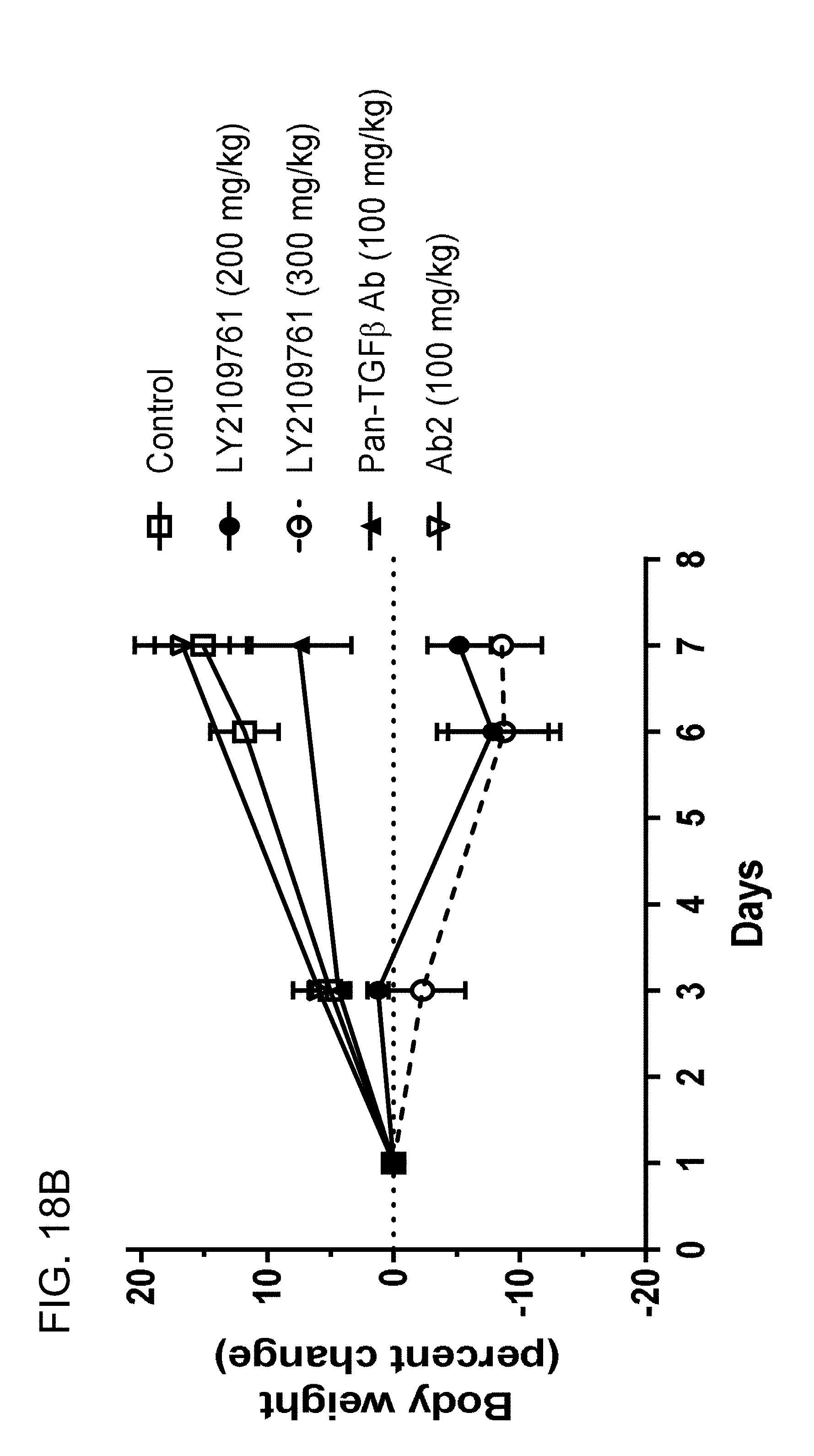
D00037

D00038

D00039

D00040

D00041

D00042

D00043

D00044

D00045

D00046


S00001
XML
uspto.report is an independent third-party trademark research tool that is not affiliated, endorsed, or sponsored by the United States Patent and Trademark Office (USPTO) or any other governmental organization. The information provided by uspto.report is based on publicly available data at the time of writing and is intended for informational purposes only.
While we strive to provide accurate and up-to-date information, we do not guarantee the accuracy, completeness, reliability, or suitability of the information displayed on this site. The use of this site is at your own risk. Any reliance you place on such information is therefore strictly at your own risk.
All official trademark data, including owner information, should be verified by visiting the official USPTO website at www.uspto.gov. This site is not intended to replace professional legal advice and should not be used as a substitute for consulting with a legal professional who is knowledgeable about trademark law.