T Cell Compositions
FRIEDMAN; Kevin
U.S. patent application number 16/060184 was filed with the patent office on 2019-06-27 for t cell compositions. This patent application is currently assigned to bluebird bio, Inc.. The applicant listed for this patent is BLUEBIRD BIO, INC.. Invention is credited to Kevin FRIEDMAN.
| Application Number | 20190194615 16/060184 |
| Document ID | / |
| Family ID | 59012788 |
| Filed Date | 2019-06-27 |


View All Diagrams
| United States Patent Application | 20190194615 |
| Kind Code | A1 |
| FRIEDMAN; Kevin | June 27, 2019 |
T CELL COMPOSITIONS
Abstract
The invention provides improved T cell compositions and methods for manufacturing T cells. More particularly, the invention provides methods of T cell manufacturing that result in adoptive T cell immunotherapies with improved survival, expansion, and persistence in vivo.
| Inventors: | FRIEDMAN; Kevin; (Melrose, MA) | ||||||||||
| Applicant: |
|
||||||||||
|---|---|---|---|---|---|---|---|---|---|---|---|
| Assignee: | bluebird bio, Inc. Cambridge MA |
||||||||||
| Family ID: | 59012788 | ||||||||||
| Appl. No.: | 16/060184 | ||||||||||
| Filed: | December 7, 2015 | ||||||||||
| PCT Filed: | December 7, 2015 | ||||||||||
| PCT NO: | PCT/US2015/064270 | ||||||||||
| 371 Date: | June 7, 2018 |
| Current U.S. Class: | 1/1 |
| Current CPC Class: | A61K 35/17 20130101; A61K 2039/5156 20130101; C12N 2740/10041 20130101; C12N 2740/16043 20130101; C12N 2501/515 20130101; C12N 2740/15041 20130101; C12N 15/86 20130101; A61K 2039/5158 20130101; C07K 14/705 20130101; C12N 5/0636 20130101; C12N 2501/727 20130101; C12N 2501/2302 20130101; A61K 35/00 20130101; C12N 2501/51 20130101; C12N 2510/00 20130101; A61P 35/02 20180101; C12N 2501/2315 20130101; C12N 2501/2307 20130101 |
| International Class: | C12N 5/0783 20060101 C12N005/0783; C12N 15/86 20060101 C12N015/86; C07K 14/705 20060101 C07K014/705 |
Claims
1.-59. (canceled)
60. A method for manufacturing chimeric antigen receptor (CAR) T cells comprising: (a) activating a population of T cells and stimulating the population of T cells to proliferate; (b) transducing the T cells with a viral vector comprising a polynucleotide encoding a CAR; (c) culturing the transduced T cells to proliferate; wherein steps a)-c) are performed in the presence of a phosphatidylinositol-3 kinase (PI3K) inhibitor.
61. The method of claim 60, wherein the method comprises isolating peripheral blood mononuclear cells as the source of T cells.
62. The method of claim 60, wherein activation and stimulation of the T cells comprises contacting the T cells with an anti-CD3 antibody or CD3-binding fragment thereof and an anti-CD28 antibody or a CD28-binding fragment thereof, B7-1 or a CD28-binding fragment thereof, or B7-2 or a CD28-binding fragment thereof.
63. The method of claim 60, wherein stimulation of the T cells comprises contacting the T cells with.
64. The method of claim 60, wherein the cells are transduced with the viral vector after T cell proliferation.
65. The method of claim 60, wherein the vector is a retroviral vector.
66. The method of claim 60, wherein the vector is a lentiviral vector.
67. The method of claim 60, wherein the CAR comprises a) an extracellular domain that binds an antigen selected from the group consisting of: alpha folate receptor, 5T4, .alpha..sub.v.beta..sub.6 integrin, BCMA, B7-H3, B7-H6, CAIX, CD19, CD20, CD22, CD30, CD33, CD44, CD44v6, CD44v7/8, CD70, CD79a, CD79b, CD123, CD138, CD171, CEA, CSPG4, EGFR, EGFR family including ErbB2 (HER2), EGFRvIII, EGP2, EGP40, EPCAM, EphA2, EpCAM, FAP, fetal AchR, FR.alpha., GD2, GD3, Glypican-3 (GPC3), HLA-A1+MAGE1, HLA-A2+MAGE1, HLA-A3+MAGE1, HLA-A1+NY-ESO-1, HLA-A2+NY-ESO-1, HLA-A3+NY-ESO-1, IL-11R.alpha., IL-13R.alpha.2, Lambda, Lewis-Y, Kappa, Mesothelin, Muc1, Muc16, NCAM, NKG2D Ligands, NY-ESO-1, PRAIVIE, PSCA, PSMA, ROR1, SSX, Survivin, TAG72, TEMs, and VEGFR2; b) a transmembrane domain derived from a polypeptide selected from the group consisting of: CD8.alpha.; CD4, CD28, CD45, PD-1, and CD152; c) one or more intracellular costimulatory signaling domains selected from the group consisting of: CD28, CD54 (ICAM), CD134 (OX40), CD137 (41BB), CD152 (CTLA4), CD273 (PD-L2), CD274 (PD-L1), and CD278 (ICOS); and d) a CD3.zeta. signaling domain.
68. The method of claim 67, wherein the extracellular domain comprises an antibody or antigen binding fragment that binds the antigen.
69. The method of claim 68, wherein the antigen binding fragment is an scFv.
70. The method of claim 67, wherein the transmembrane domain is isolated from CD8.alpha. or CD28.
71. The method of claim 67, wherein the one or more costimulatory signaling domains is isolated from a polypeptide selected from the group consisting of: CD28, CD134, and CD137.
72. The method of claim 67, wherein the CAR further comprises a hinge region polypeptide.
73. The method of claim 72, wherein the hinge region polypeptide comprises a hinge region of IgG1 or CD8.alpha..
74. The method of claim 67, wherein the CAR further comprises a signal peptide.
75. The method of claim 74, wherein the signal peptide comprises an IgG1 heavy chain signal polypeptide or a CD8.alpha. signal polypeptide.
76. The method claim 60, wherein the PI3K inhibitor is a pan-PI3K inhibitor selected from the group consisting of: BEZ235, LY294002, TG100713 and GDC-0941.
77. The method claim 60, wherein the PI3K inhibitor is a selective PI3K inhibitor selected from the group consisting of: BYL719, GSK2636771, TGX-221, AS25242, CAL-101, and IPI-145.
78. The method claim 60, wherein the PI3K inhibitor is ZSTK474.
79. The method claim 60, wherein the duration of the method for manufacturing CAR T cells is 10 days.
Description
STATEMENT REGARDING SEQUENCE LISTING
[0001] The Sequence Listing associated with this application is provided in text format in lieu of a paper copy, and is hereby incorporated by reference into the specification. The name of the text file containing the Sequence Listing is BLBD_029_02WO_ST25.txt. The text file is 8 KB, was created on Dec. 4, 2015, and is being submitted electronically via EFS-Web, concurrent with the filing of the specification.
BACKGROUND
Technical Field
[0002] The present invention relates to improved T cell compositions and methods for manufacturing T cells. More particularly, the invention relates to methods of T cell manufacturing that result in adoptive T cell immunotherapies with improved survival, expansion, and persistence in vivo.
Description of the Related Art
[0003] Adoptive immunotherapy is the transfer of T lymphocytes to a subject to provide therapy for a disease. Adoptive immunotherapy has yet unrealized potential for treating a wide variety of diseases including cancer, infectious disease, autoimmune disease, inflammatory disease, and immunodeficiency. However, most, if not all adoptive immunotherapy strategies require T cell activation and expansion steps to generate a clinically effective, therapeutic dose of T cells. Current technologies for generating therapeutic doses of T cells, including engineered T cells, remain limited by cumbersome T cell manufacturing processes. For example, T cell expansion often requires labor intensive and expensive cloning, and/or multiple rounds of activation/expansion to achieve therapeutically relevant T cell numbers. In addition, existing T cell activation/expansion methods are normally coupled with substantial T cell differentiation and usually result in short-lived effects, including short-lived survival and a lack of persistence and lack of in vivo expansion of the transferred T cells. Thus, existing T cell manufacturing processes produce an inferior T cell product that is prone to exhaustion and loss of effector immune cell function.
[0004] To date, clinical efficacy of engineered T cell adoptive immunotherapies is limited by poor T cell expansion and persistence after infusion into patients. Therefore, such therapies are not suitable for widespread clinical use. Accordingly, there is a persistent, unmet need for improvements in T cell manufacturing and therapeutic T cell compositions that survive, expand, and persist in vivo.
BRIEF SUMMARY
[0005] The invention generally provides adoptive T cell immunotherapies comprising persistent and potent anti-tumor T cell compositions and methods of making the same.
[0006] The present invention relates to improved T cell compositions and methods for manufacturing T cells. More particularly, the invention relates to methods of T cell manufacturing that result in with improved survival, expansion, and persistence in vivo.
[0007] In various embodiments, a method for manufacturing T cells comprising: (a) isolating a population of T cells, e.g., tumor infiltrating cytotoxic T lymphocytes (TIL), from a subject; (b) activating the population of T cells and stimulating the population of T cells to proliferate, wherein the activation and stimulation steps are performed in the presence of an inhibitor of PI3K pathway; (c) culturing the T cells to proliferate; wherein the activating and stimulating steps performed in the presence of the inhibitor of the PI3K pathway results in maintaining proliferation of the T cells compared to the proliferation of T cells that were activated and stimulated in the absence of the inhibitor of the PI3K pathway is provided.
[0008] In various embodiments, a method for manufacturing T cells comprising: (a) activating a population of T cells and stimulating the population of T cells to proliferate, wherein the activation and stimulation steps are performed in the presence of an inhibitor of PI3K pathway; (b) transducing the T cells with a viral vector comprising an engineered T cell receptor (TCR) or a chimeric antigen receptor (CAR); (c) culturing the transduced T cells to proliferate; wherein the activating and stimulating steps performed in the presence of the inhibitor of the PI3K pathway results in maintaining proliferation of the transduced T cells compared to the proliferation of transduced T cells that were activated and stimulated in the absence of the inhibitor of the PI3K pathway is provided.
[0009] In various embodiments, a method for manufacturing T cells comprising: (a) isolating a population of T cells, e.g., tumor infiltrating cytotoxic T lymphocytes (TIL), from a subject; (b) activating the population of T cells and stimulating the population of T cells to proliferate, wherein the activation and stimulation steps are performed in the presence of an inhibitor of AKT/mTOR pathway; (c) culturing the T cells to proliferate; wherein the activating and stimulating steps performed in the presence of the inhibitor of the PI3K/AKT/mTOR pathway results in maintaining proliferation of the T cells compared to the proliferation of T cells that were activated and stimulated in the absence of the inhibitor of the PI3K/AKT/mTOR pathway is provided.
[0010] In various embodiments, a method for manufacturing T cells comprising: (a) activating a population of T cells and stimulating the population of T cells to proliferate, wherein the activation and stimulation steps are performed in the presence of an inhibitor of AKT/mTOR pathway; (b) transducing the T cells with a viral vector comprising an engineered T cell receptor (TCR) or a chimeric antigen receptor (CAR); (c) culturing the transduced T cells to proliferate; wherein the activating and stimulating steps performed in the presence of the inhibitor of the PI3K/AKT/mTOR pathway results in maintaining proliferation of the transduced T cells compared to the proliferation of transduced T cells that were activated and stimulated in the absence of the inhibitor of the PI3K/AKT/mTOR pathway is provided. In particular embodiments, the methods contemplated herein comprise isolating peripheral blood mononuclear cells as the source of T cells.
[0011] In certain embodiments, activation of the T cells comprises contacting the T cells with an anti-CD3 antibody or CD3-binding fragment thereof.
[0012] In additional embodiments, stimulation of the T cells comprises contacting the T cells with an anti-CD28 antibody or a CD28-binding fragment thereof, B7-1 or a CD28-binding fragment thereof, or B7-2 or a CD28-binding fragment thereof.
[0013] In some embodiments, the cells are transduced with the viral vector prior to T cell proliferation.
[0014] In certain embodiments, the cells are transduced with the viral vector after T cell proliferation.
[0015] In particular embodiments, the vector is a retroviral vector.
[0016] In further embodiments, the vector is a lentiviral vector.
[0017] In other particular embodiments, the cells comprise a chimeric antigen receptor (CAR).
[0018] In particular embodiments, the CAR comprises: an extracellular domain that binds an antigen selected from the group consisting of: alpha folate receptor, 5T4, .alpha.v.beta.6 integrin, BCMA, B7-H3, B7-H6, CAIX, CD19, CD20, CD22, CD30, CD33, CD44, CD44v6, CD44v7/8, CD70, CD79a, CD79b, CD123, CD138, CD171, CEA, CSPG4, CMV, EBV, EGFR, EGFR family including ErbB2 (HER2), EGFRvIII, EGP2, EGP40, EPCAM, EphA2, EpCAM, FAP, fetal AchR, FR.alpha., GD2, GD3, Glypican-3 (GPC3), HLA-A1+MAGE1, HLA-A2+MAGE1, HLA-A3+MAGE1, HLA-A1+NY-ESO-1, HLA-A2+NY-ESO-1, HLA-A3+NY-ESO-1, HPV, IL-11R.alpha., IL-13R.alpha.2, Lambda, Lewis-Y, Kappa, Mesothelin, Muc1, Muc16, NCAM, NKG2D Ligands, NY-ESO-1, PRAME, PSCA, PSMA, ROR1, SSX, Survivin, TAG72, TEMs, and VEGFR2; a transmembrane domain derived from a polypeptide selected from the group consisting of: CD8.alpha.; CD4, CD28, CD45, PD-1, and CD152; one or more intracellular costimulatory signaling domains selected from the group consisting of: CD28, CD54 (ICAM), CD134 (OX40), CD137 (41BB), CD152 (CTLA4), CD273 (PD-L2), CD274 (PD-L1), and CD278 (ICOS); and a CD3.zeta. signaling domain.
[0019] In additional embodiments, the extracellular domain comprises an antibody or antigen binding fragment that binds the antigen.
[0020] In certain embodiments, the transmembrane domain is derived from CD8.alpha. or CD28.
[0021] In further embodiments, the one or more costimulatory signaling domains selected from the group consisting of: CD28, CD134, and CD137.
[0022] In additional embodiments, the CAR comprises a hinge region polypeptide.
[0023] In particular embodiments, the hinge region polypeptide comprises a hinge region of IgG1 or CD8.alpha..
[0024] In particular embodiments, the CAR comprises a signal peptide.
[0025] In some embodiments, the signal peptide comprises an IgG1 heavy chain signal polypeptide or a CD8.alpha. signal polypeptide.
[0026] In particular embodiments, the inhibitor of the PI3K pathway is a pan-PI3K inhibitor selected from the group consisting of: BEZ235, LY294002, TG100713, and GDC-0941.
[0027] In other particular embodiments, the inhibitor of the PI3K pathway is a selective PI3K inhibitor selected from the group consisting of: BYL719, GSK2636771, TGX-221, AS25242, CAL-101, and IPI-145.
[0028] In other particular embodiments, the inhibitor of the PI3K pathway the PI3K inhibitor ZSTK474.
[0029] In some embodiments, the population of T cells activated and stimulated in the presence of an inhibitor of PI3K pathway have an increased number of T cells expressing one or more markers selected from the group consisting of: CD62L, CD127, CD197, and CD38 compared to a population of T cells activated and stimulated in the absence of the inhibitor of PI3K pathway.
[0030] In some embodiments, the population of T cells activated and stimulated in the presence of an inhibitor of PI3K pathway have an increased number of T cells expressing the markers CD62L, CD127, CD197, and CD38 compared to a population of T cells activated and stimulated in the absence of the inhibitor of PI3K pathway.
[0031] In further embodiments, the population of T cells activated and stimulated in the presence of an inhibitor of PI3K pathway do not express CD57 or KLRG1 or express less CD57 or KLRG1 compared to a population of T cells activated and stimulated in the absence of the inhibitor of PI3K pathway.
[0032] In various embodiments, a method for maintaining the proliferation and decreasing the differentiation of restimulated T cells expressing an engineered TCR or CAR comprising: (a) contacting all or a portion of a population of proliferated T cells comprising an engineered TCR or CAR with an anti-CD3 antibody or CD3-binding fragment thereof, and an anti-CD28 antibody or CD28-binding fragment thereof, which stimulates a CD28 accessory molecule on the surface of the T cells, thereby restimulating the activated T cells to proliferate; wherein the restimulated T cells have maintained proliferation and decreased differentiation compared to the proliferation of T cells that were stimulated or restimulated in the absence of the inhibitor of the PI3K pathway is provided.
[0033] In particular embodiments, the cells comprise an engineered TCR.
[0034] In certain embodiments, the cells comprise a CAR.
[0035] In other particular embodiments, the cells comprise a viral vector encoding an engineered TCR or CAR.
[0036] In additional embodiments, the vector is a retroviral vector.
[0037] In additional embodiments, the vector is a lentiviral vector.
[0038] In particular embodiments, the CAR comprises: an extracellular domain that binds an antigen selected from the group consisting of: alpha folate receptor, 5T4, .alpha.v.beta.6 integrin, BCMA, B7-H3, B7-H6, CAIX, CD19, CD20, CD22, CD30, CD33, CD44, CD44v6, CD44v7/8, CD70, CD79a, CD79b, CD123, CD138, CD171, CEA, CSPG4, EGFR, EGFR family including ErbB2 (HER2), EGFRvIII, EGP2, EGP40, EPCAM, EphA2, EpCAM, FAP, fetal AchR, FR.alpha., GD2, GD3, Glypican-3 (GPC3), HLA-A1+MAGE1, HLA-A2+MAGE1, HLA-A3+MAGE1, HLA-A1+NY-ESO-1, HLA-A2+NY-ESO-1, HLA-A3+NY-ESO-1, IL-11R.alpha., IL-13R.alpha.2, Lambda, Lewis-Y, Kappa, Mesothelin, Muc1, Muc16, NCAM, NKG2D Ligands, NY-ESO-1, PRAME, PSCA, PSMA, ROR1, SSX, Survivin, TAG72, TEMs, and VEGFR2; a transmembrane domain derived from a polypeptide selected from the group consisting of: CD8.alpha.; CD4, CD28, CD45, PD-1, and CD152; one or more intracellular costimulatory signaling domains selected from the group consisting of: CD28, CD54 (ICAM), CD134 (OX40), CD137 (41BB), CD152 (CTLA4), CD273 (PD-L2), CD274 (PD-L1), and CD278 (ICOS); and a CD3.zeta. signaling domain.
[0039] In some embodiments, the extracellular domain comprises an antibody or antigen binding fragment that binds the antigen.
[0040] In certain embodiments, the transmembrane domain is derived from CD8.alpha. or CD28.
[0041] In further embodiments, the one or more costimulatory signaling domains are selected from the group consisting of: CD28, CD134, and CD137.
[0042] In particular embodiments, the CAR comprises a hinge region polypeptide.
[0043] In further embodiments, the hinge region polypeptide comprises a hinge region of IgG1 or CD8.alpha..
[0044] In additional embodiments, the CAR comprises a signal peptide.
[0045] In other particular embodiments, the signal peptide comprises an IgG1 heavy chain signal polypeptide or a CD8.alpha. signal polypeptide.
[0046] In particular embodiments, the inhibitor of the PI3K pathway is a pan-PI3K inhibitor selected from the group consisting of: BEZ235, LY294002, TG100713, and GDC-0941.
[0047] In other particular embodiments, the inhibitor of the PI3K pathway is a selective PI3K inhibitor selected from the group consisting of: BYL719, GSK2636771, TGX-221, AS25242, CAL-101, and IPI-145.
[0048] In other particular embodiments, the inhibitor of the PI3K pathway is the PI3K inhibitor ZSTK474.
[0049] In particular embodiments, the population of activated T cells restimulated in the presence of an inhibitor of PI3K pathway have an increased number of T cells expressing one or more markers selected from the group consisting of: CD62L, CD127, CD197, and CD38 compared to a population of T cells activated and stimulated in the absence of the inhibitor of PI3K pathway.
[0050] In particular embodiments, the population of activated T cells restimulated in the presence of an inhibitor of PI3K pathway have an increased number of T cells expressing the markers CD62L, CD127, CD197, and CD38 compared to a population of T cells activated and stimulated in the absence of the inhibitor of PI3K pathway.
[0051] In additional embodiments, the population of activated T cells restimulated in the presence of an inhibitor of PI3K pathway do not express CD57 or KLRG1 or express less CD57 or KLRG1 compared to a population of T cells activated and stimulated in the absence of the inhibitor of PI3K pathway.
[0052] In various embodiments, a population of T cells comprising a vector comprising an engineered TCR or CAR, wherein the cells have been activated and stimulated to proliferate in the presence of an inhibitor of PI3K pathway is provided.
[0053] In various particular embodiments, a population of T cells comprising a vector comprising an engineered TCR or CAR, wherein the cells have been activated and stimulated to proliferate in the presence of an inhibitor of PI3K pathway and have been restimulated by contacting all or a portion of a population of proliferated immune effector cells with an anti-CD3 antibody or CD3-binding fragment thereof, and an anti-CD28 antibody or CD28-binding fragment thereof, which stimulates a CD28 accessory molecule on the surface of the immune effector cells is provided.
[0054] In certain embodiments, the immune effector cells comprise T cells.
[0055] In one embodiment, the immune effector cells are TILs.
[0056] In various embodiments, the immune effector cells treated with a PI3K inhibitor have increased co-expression of one or more markers selected from the group consisting of: CD62L, CD127, CD197, and CD38 compared to a population of T cells activated and stimulated in the absence of the inhibitor of PI3K pathway.
[0057] In various embodiments, the immune effector cells treated with a PI3K inhibitor have increased co-expression of CD62L, CD127, CD197, and CD38 compared to a population of T cells activated and stimulated in the absence of the inhibitor of PI3K pathway.
[0058] In various embodiments, a composition comprising a population of immune effector cells contemplated herein and a physiologically acceptable excipient is provided.
[0059] In various certain embodiments, a method of treating a cancer in a subject in need thereof, comprising administering to the subject a therapeutically effect amount of a T cell composition contemplated herein is provided.
[0060] In particular embodiments, the cancer is selected from the group consisting of Wilms' tumor, Ewing sarcoma, a neuroendocrine tumor, a glioblastoma, a neuroblastoma, a melanoma, skin cancer, breast cancer, colon cancer, rectal cancer, prostate cancer, liver cancer, renal cancer, pancreatic cancer, lung cancer, biliary cancer, cervical cancer, endometrial cancer, esophageal cancer, gastric cancer, head and neck cancer, medullary thyroid carcinoma, ovarian cancer, glioma, lymphoma, leukemia, myeloma, acute lymphoblastic leukemia, acute myelogenous leukemia, chronic lymphocytic leukemia, chronic myelogenous leukemia, Hodgkin's lymphoma, non-Hodgkin's lymphoma, and urinary bladder cancer.
[0061] In one embodiment, the cancer is associated with or caused by a viral infection, e.g., infection by CMV, HPV, or EBV.
[0062] In additional embodiments, the cancer is pancreatic cancer and the extracellular binding domain of the CAR binds an epitope of PSCA or MUC1
[0063] In some embodiments, the cancer is bladder cancer and the extracellular binding domain of the CAR binds an epitope of PSCA or MUC1
[0064] In further embodiments, the cancer is glioblastoma multiforme and the extracellular binding domain of the CAR binds an epitope of EPHA2, EGFRvIII, or CSPG4.
[0065] In particular embodiments, the cancer is lung cancer and the extracellular binding domain of the CAR binds an epitope of PSCA or GD2.
[0066] In certain embodiments, the cancer is breast cancer and the extracellular binding domain of the CAR binds an epitope of CSPG4 or HER2.
[0067] In additional embodiments, the cancer is melanoma and the extracellular binding domain of the CAR binds an epitope of CSPG4 or GD2.
[0068] In particular embodiments, the cancer is a B-cell malignancy and the binding domain of the CAR binds an epitope of BCMA. In any of the foregoing embodiments, the CAR is anti-BCMA02, wherein the CAR binds an epitope of BCMA.
[0069] In various embodiments, a method of treating a hematological malignancy in a subject in need thereof, comprising administering to the subject a therapeutically effect amount of a T cell composition contemplated herein is provided.
[0070] In certain embodiments, the hematological malignancy is a B-cell malignancy selected from the group consisting of: multiple myeloma (MM), chronic lymphocytic leukemia (CLL), or non-Hodgkin's lymphoma (NHL).
[0071] In particular embodiments, the MM is selected from the group consisting of: overt multiple myeloma, smoldering multiple myeloma, plasma cell leukemia, non-secretory myeloma, IgD myeloma, osteosclerotic myeloma, solitary plasmacytoma of bone, and extramedullary plasmacytoma.
[0072] In certain embodiments, the NHL is selected from the group consisting of: Burkitt lymphoma, chronic lymphocytic leukemia/small lymphocytic lymphoma (CLL/SLL), diffuse large B-cell lymphoma, follicular lymphoma, immunoblastic large cell lymphoma, precursor B-lymphoblastic lymphoma, and mantle cell lymphoma.
BRIEF DESCRIPTION OF THE SEVERAL VIEWS OF THE DRAWINGS
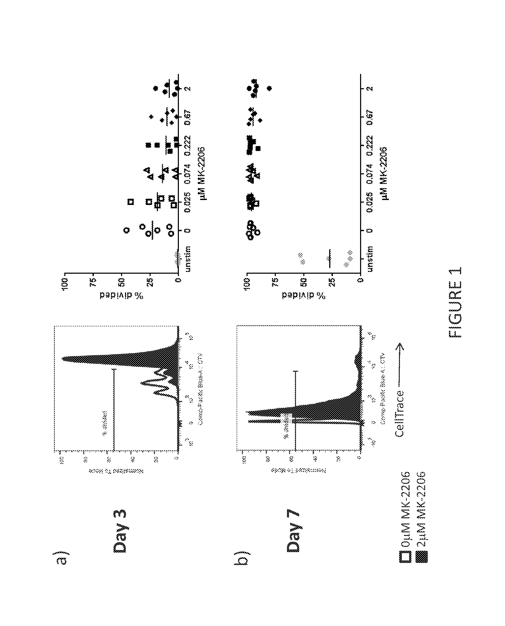
[0073] FIG. 1 shows a representative example of the maintenance of T cell proliferation in T cells treated with an AKT inhibitor. T cells were cultured with various concentrations of the AKT inhibitor, MK-2206 for up to seven days. The right-most panels show the percent divided T cells from T cell cultures initiated from six normal donor PBMC samples. Each symbol in the right-most panel represents a unique culture done in parallel with a titrated MK-2206 dose. The left-most panels show a representative example from these experiments. A) After three days of culture with MK-2206, T cell proliferation was only slightly decreased when compared to the no treatment control. B) After 7 days of culture with MK-2206, T cell proliferation was not substantially different compared to the no treatment control.
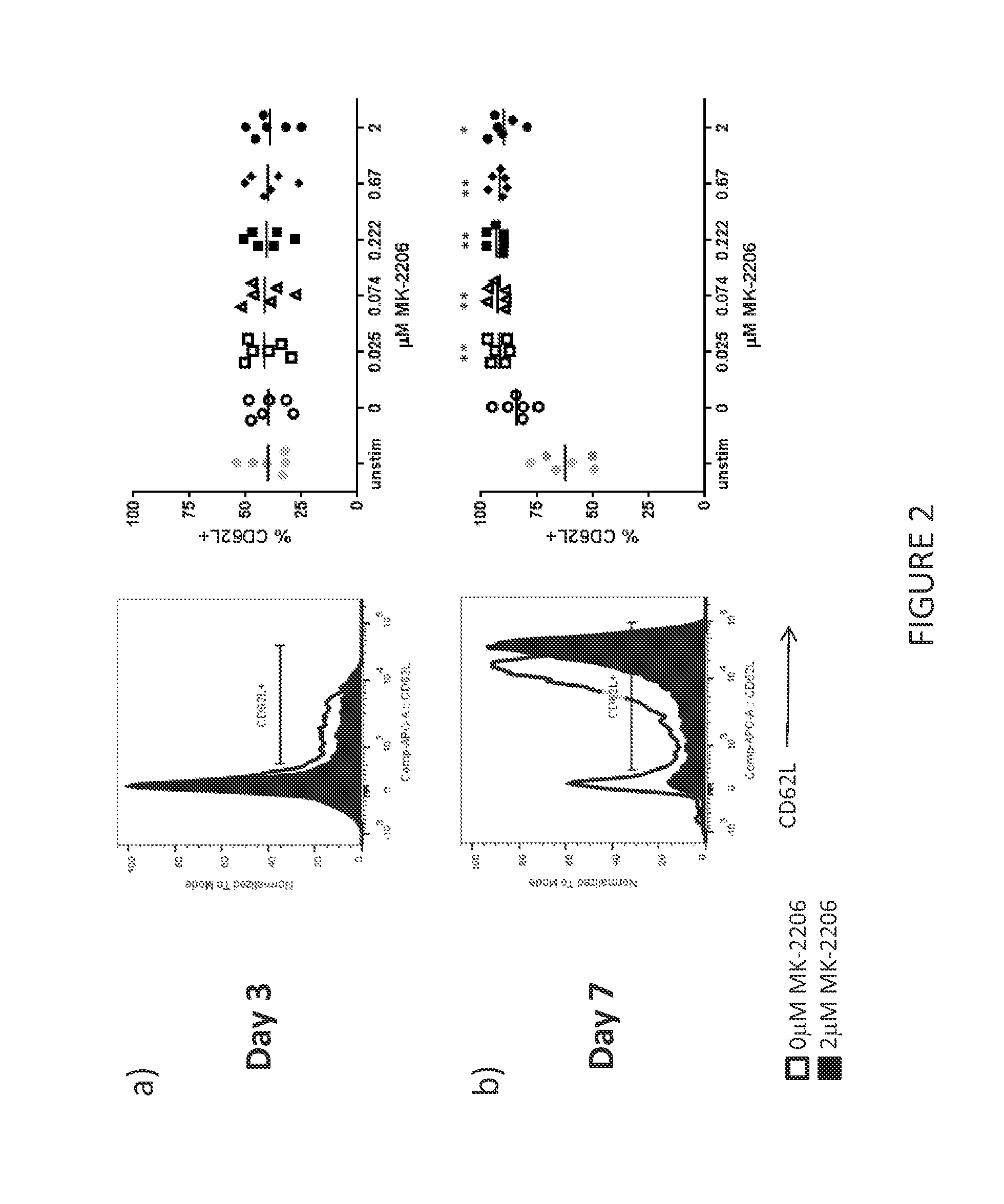
[0074] FIG. 2 shows a representative example of CD62L expression of T cells treated with an AKT inhibitor. T cells were cultured with various concentrations of the AKT inhibitor, MK-2206 for up to seven days. The right-most panels show the percent CD62L expressing T cells from T cell cultures initiated from six normal donor PBMC samples. Each symbol in the right-most panel represents a unique culture done in parallel with a titrated MK-2206 dose. The left-most panels show a representative example from these experiments. A) After three days of culture with MK-2206, CD62L expression on the MK-2206 treated T cells was not substantially different when compared to the no treatment control. B) After 7 days of culture, MK-2206 treated T cells showed significant higher levels of CD62L expression compared to the no treatment control.
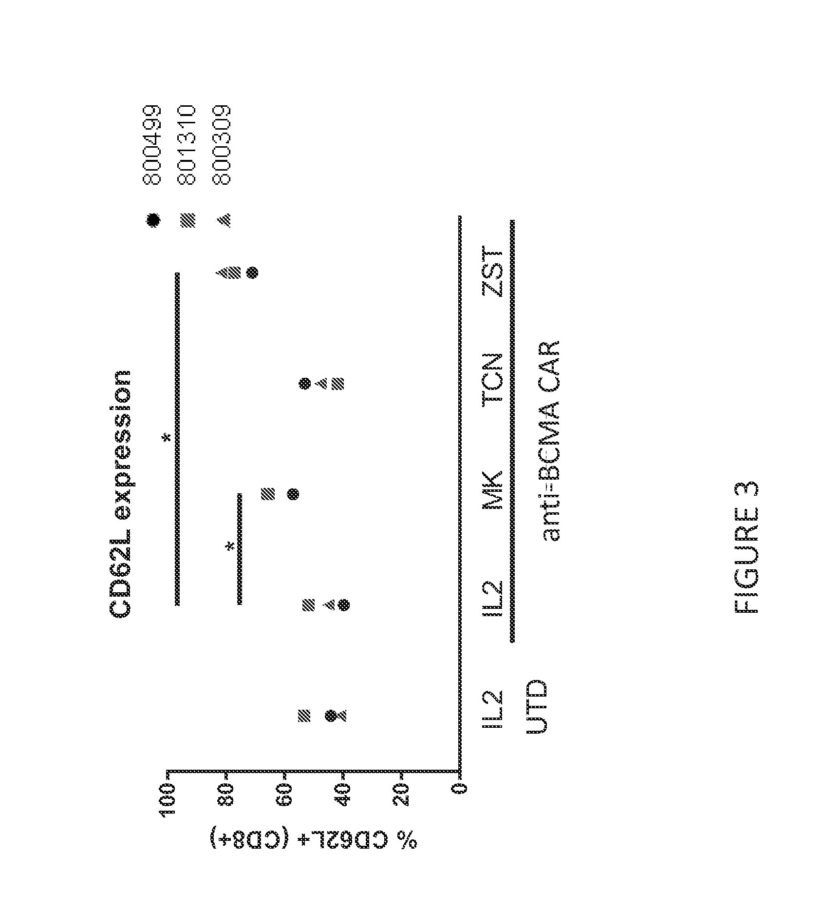
[0075] FIG. 3 shows the expression of CD62L on anti-BCMA CAR T cells assessed by flow cytometry at the end of culture with IL-2 or IL-2 and MK-2206, TCN, or ZSTK474. MK-2206 and ZSTK474 had significantly higher CD62L expression compared CAR T cell cultures treated with IL-2 alone or with TCN.
[0076] FIG. 4 shows the mean tumor volume of multiple myeloma tumors in mice treated with anti-BCMA CAR T cells cultured with IL-2, IL-7 and IL-15, or IL-2 and MK-2206, ZSTK474, or TCN. Anti-BCMA CAR T cells cultured with IL-7 and IL-15, MK-2206, or ZSTK474 showed similar levels of anti-tumor activity as compared to anti-BCMA CAR T cells cultured with standard IL-2 alone. Anti-BCMA CAR T cells cultured with TCN did not show an anti-tumor response.
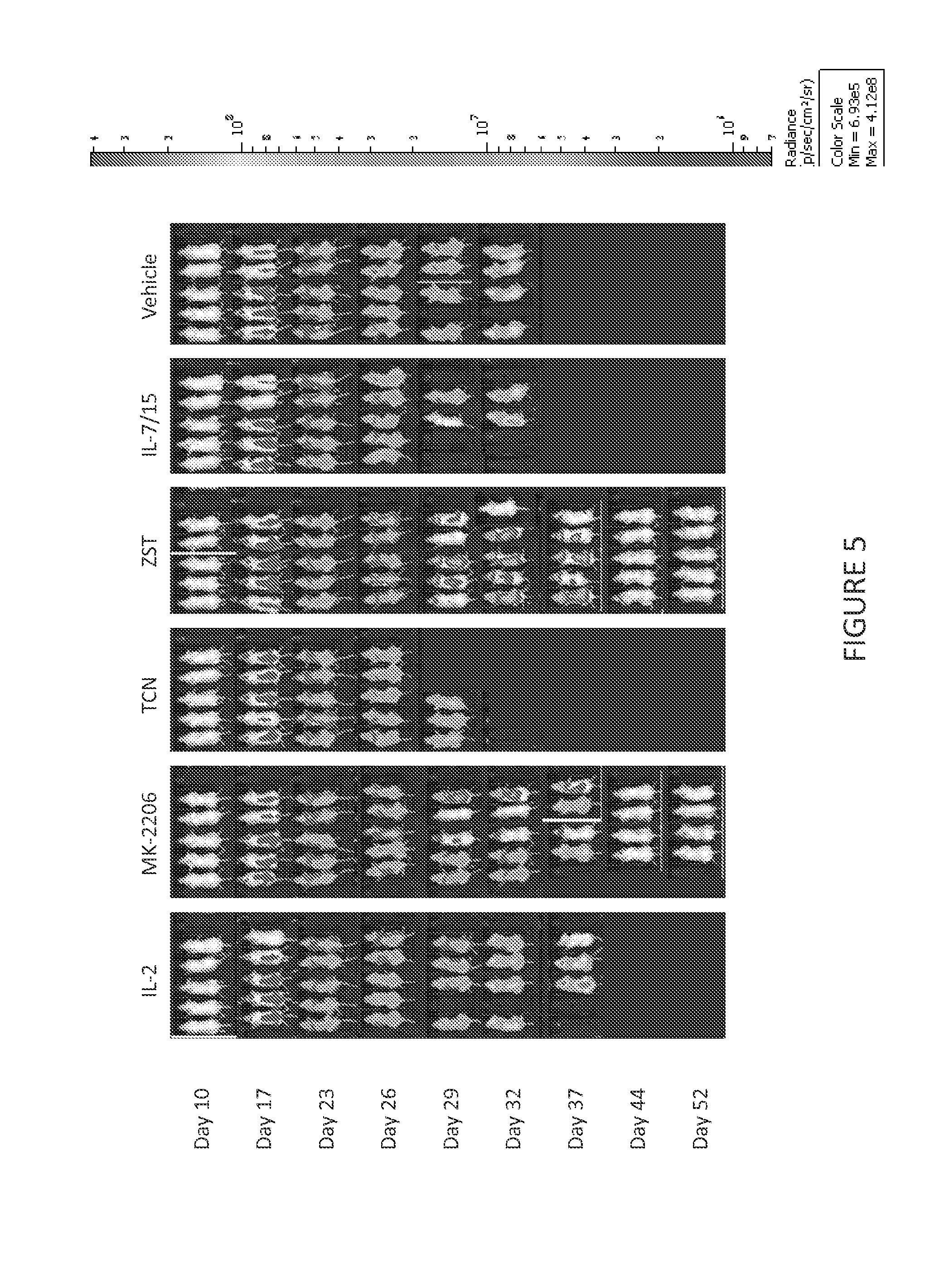
[0077] FIG. 5 shows anti-tumor activity of anti-BCMA CAR T cells treated with IL-2, IL-7/15, or IL-2 and MK-2206, TCN, or ZSTK474 in a Daudi tumor model. Daudi tumor progression was not affected after treatment with IL-2- or IL7/15-cultured anti-BCMA CAR T cells. Anti-BCMA CAR T cells cultured with IL-2 and either MK-2206 or ZSTK474 caused complete tumor regression.
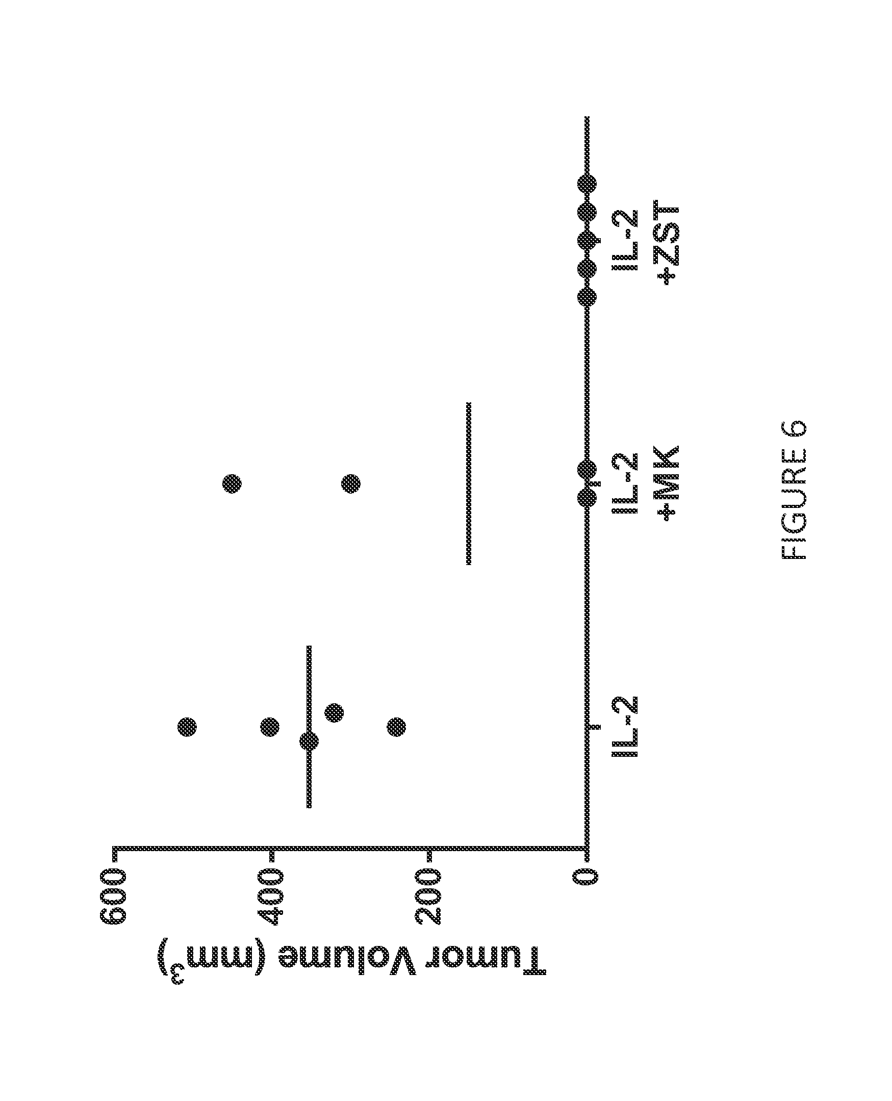
[0078] FIG. 6 shows persistence of ZSTK474-cultured anti-BCMA CAR T cells in animals treated with IL-2-, or IL-2 and MK-2206-, or ZSTK474-cultured anti-BCMA CAR T cells that had completely regressed a 100 mm.sup.3 RPMI-8226 tumor. Animals were rechallenged 13 days after later with RPMI-8226 on the opposite flank. Animals treated with IL-2 cultured CAR T cells were unable to prevent tumor outgrowth. None of the animals treated with ZSTK474-cultured anti-BCMA CAR T cells had any evidence of tumor engraftment.
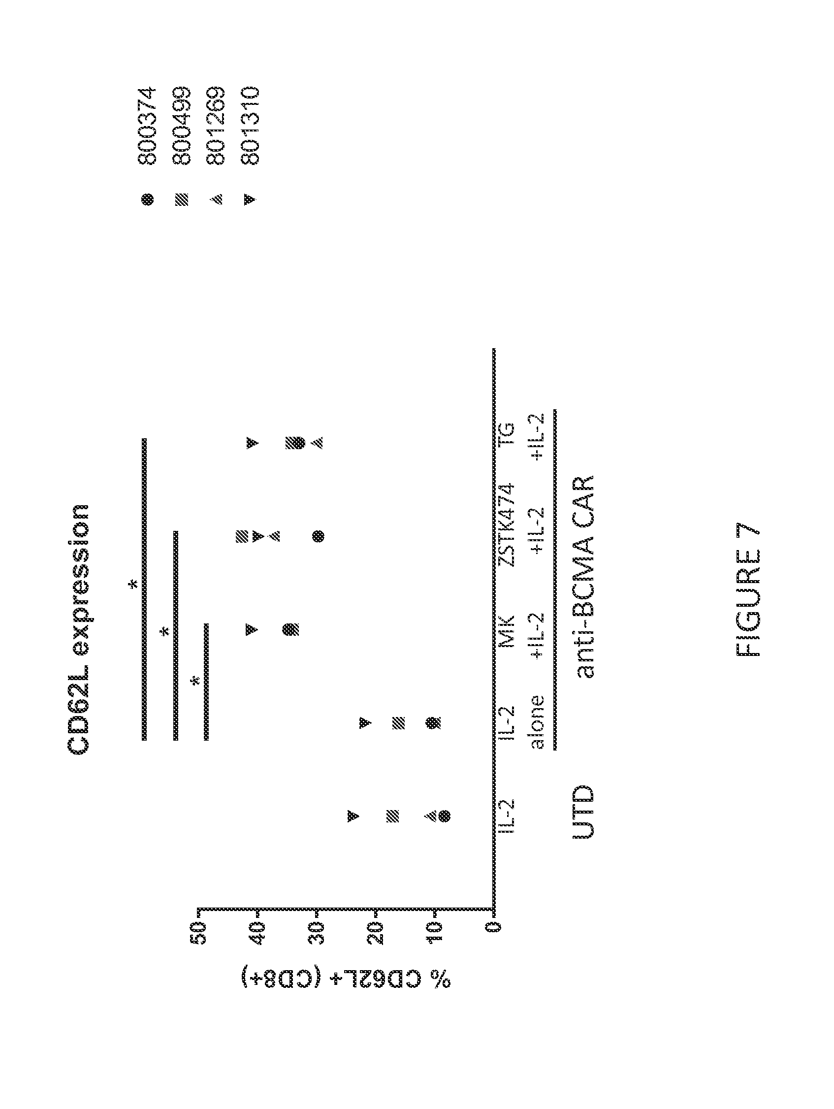
[0079] FIG. 7 shows the expression of CD62L on anti-BCMA CAR T cells assessed by flow cytometry at the end of culture with IL-2, or IL-2 and MK-2206, TCN, ZSTK474, or TG100713. MK-2206, ZSTK474, and TG100713 had significantly higher CD62L expression compared CAR T cell cultures treated with IL-2 alone or with TCN.
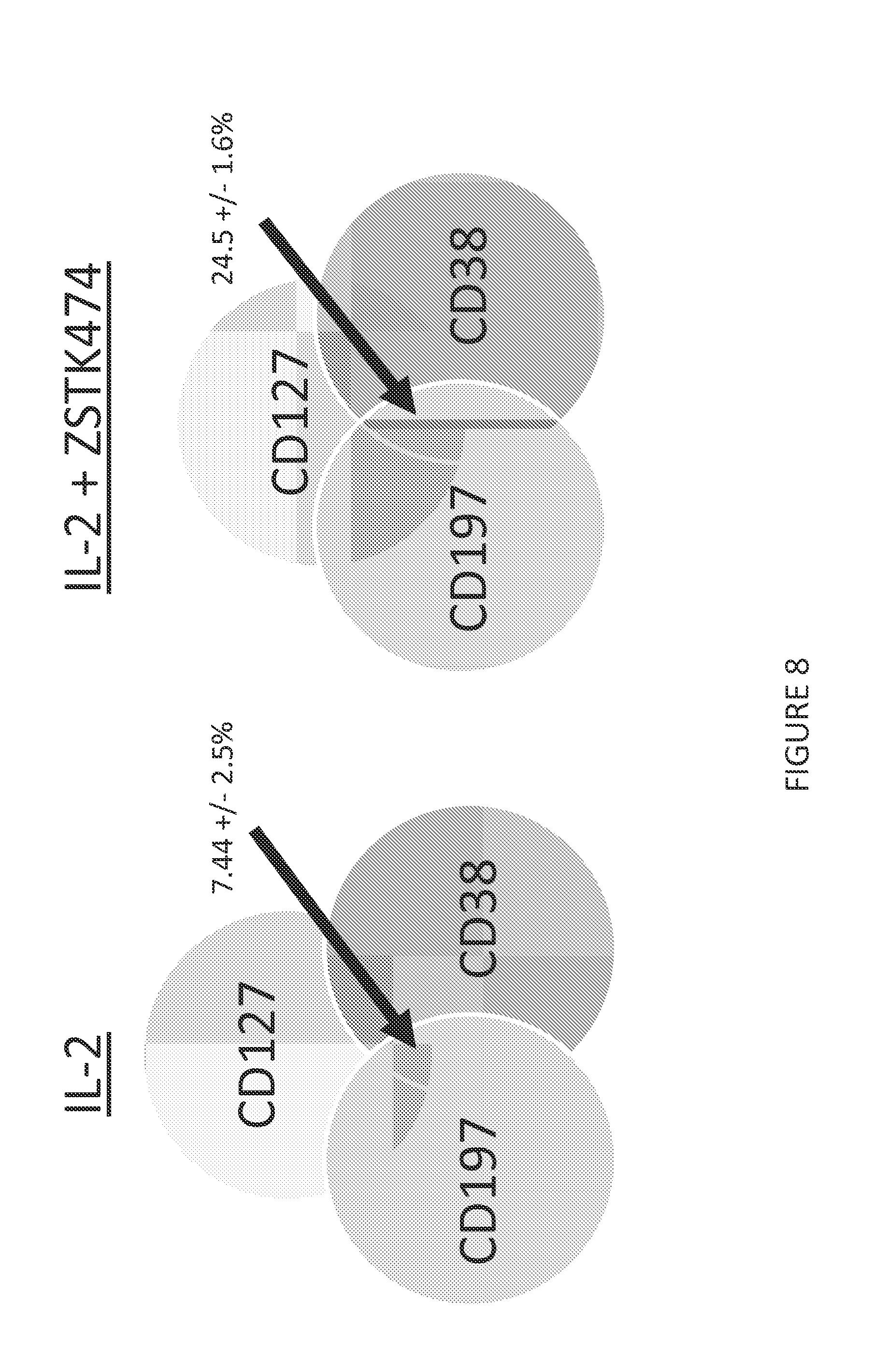
[0080] FIG. 8 shows Venn diagrams for co-expression of CD127, CD197 and CD38 in CD62L positive anti-BCMA T cells that have been cultured in the presence of IL-2 or IL-2 and ZSTK474 for ten days. ZSTK474-treated anti-BCMA CAR T cells showed an increase in the percentage of cells co-expressing CD127, CD197 and CD38 compared to anti-BCMA CAR T cells cultured with IL-2 alone.
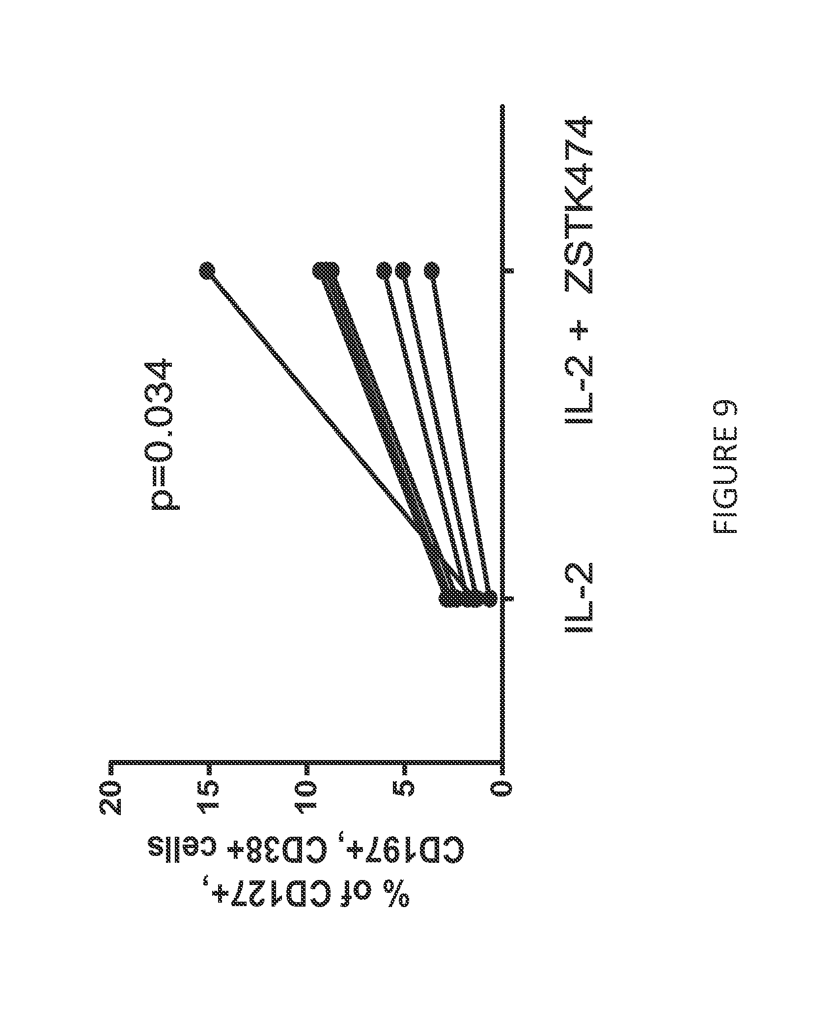
[0081] FIG. 9 shows fluorescence-based flow cytometry staining for CD127, CD197, and CD38 in anti-BCMA CAR T cells that have been cultured in the presence of IL-2 or IL-2 and ZSTK474 for ten days. ZSTK474-treated anti-BCMA CAR T cells showed an increase in the percentage of cells co-expressing CD127, CD197 and CD38 compared to anti-BCMA CAR T cells cultured with IL-2 alone.
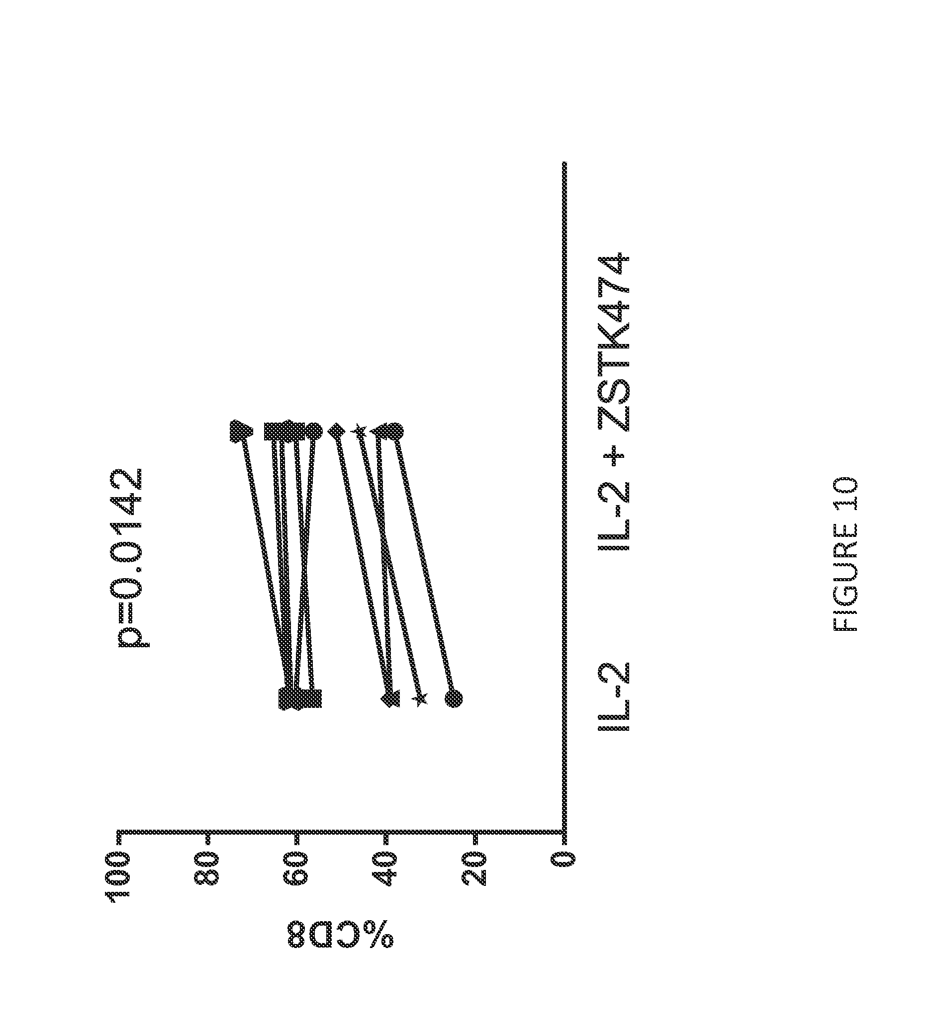
[0082] FIG. 10 shows an increased percentage of CD8 expressing anti-BCMA CAR T cells in cultures treated IL-2 and ZSTK474 (n=7) compared to cultures treated with IL-2 alone. CD8 expression was determined using a fluorescently-labeled anti-CD8 antibody and flow cytometry.
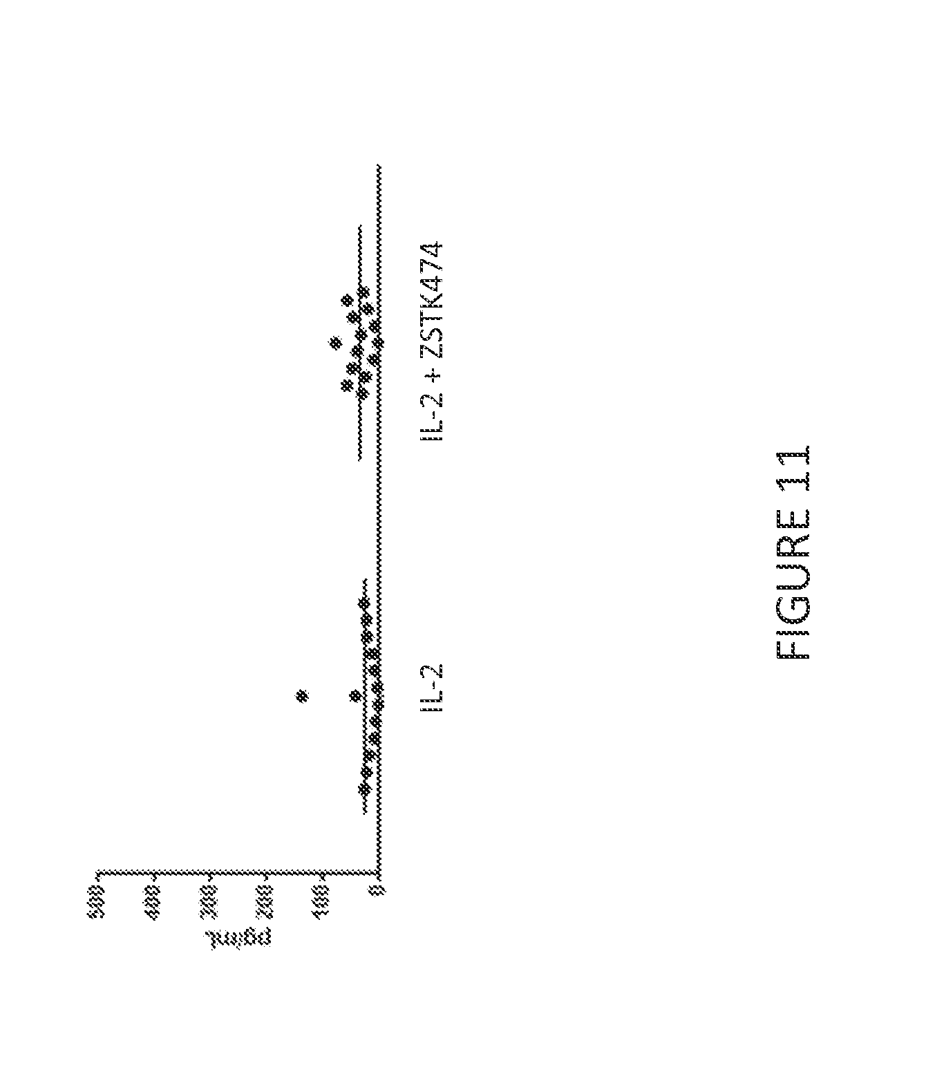
[0083] FIG. 11 shows the amount of IFN-.gamma. released by anti-BCMA CART cells from 14 donors after culture with IL-2 alone or with IL-2 and ZSTK474. At the end of the culture period, an equivalent number of anti-BCMA CAR T cells were re-cultured for 24 hours in media alone. The amount of IFN-.gamma. released in 24 hours was quantified by ELISA. Culture in ZSTK474 did not significantly increase anti-BCMA CAR T cell tonic cytokine release compared to anti-BCMA CAR T cells cultured with IL-2 alone.
[0084] FIG. 12 shows a schematic of a murine B cell maturation antigen (muBCMA02) CAR construct.
[0085] FIG. 13 shows anti-tumor activity of anti-BCMA02 CAR T cells treated with IL-2, or IL-2 and ZSTK474, or a truncated signaling deficient anti-BCMA02 (tBCMA02) CAR T cell treated with IL-2 and ZSTK474 in a highly aggressive Daudi tumor model. Complete tumor regression was observed in 50 percent of mice administered the anti-BCMA02 CAR T cells treated with IL-2 and ZSTK474. .
[0086] FIG. 14 shows anti-tumor activity of anti-BCMA02 CAR T cells treated with IL-2, or IL-2 and ZSTK474 in a multiple myeloma tumor (RPMI-8226) model. Animals treated with IL-2- or IL-2 and ZSTK474-cultured anti-BCMA02 CAR T cells completely prevented tumor outgrowth.
[0087] FIG. 15 vector copy number and CAR expression in anti-BCMA CAR T cells treated with either IL-2 or IL-2 and MK-2206 (AKT inhibitor) or ZSTK474 or TG10073 (PI3K inhibitors). A) Anti-BCMACAR T cells from four normal donors cultured with IL-2 and MK2206 had significantly more integrated copies of the anti-BCMACAR compared to cultures receiving only IL-2. Similar improvements were seen using ZSTK474- or TG10073-containing media. B) Anti-BCMA CAR T cells were specifically identified using recombinant human BCMA-IgG Fc conjugated to PE. Anti-BCMA CAR T cells from four normal donors cultured with IL-2 and MK2206 had significantly higher CAR expression compared to cultures with IL-2 alone. Similar improvements were observed using IL-2 and ZSTK474 or TG10073 during culture.
BRIEF DESCRIPTION OF THE SEQUENCE IDENTIFIERS
[0088] SEQ ID NO: 1-10 set for the amino acid sequence of various linkers.
[0089] SEQ ID NOs: 11-13 set for the amino acid sequence of protease cleavage sites and self-cleaving polypeptide cleavage sites.
[0090] SEQ ID NO: 14 sets for the polypeptide sequence of anti-BCMA02 CAR.
DETAILED DESCRIPTION
A. Overview
[0091] The invention generally relates to improved methods for manufacturing T cell compositions. Without wishing to be bound to any particular theory, the inventive methods contemplated herein uncouple T cell proliferation from differentiation to produce T cells having superior properties, e.g., increased survival, expansion, and persistence in vivo along with a concomitant decrease in differentiation, compared to existing T cell compositions in the art. Accordingly, T cell compositions contemplated herein comprise potent T cells, which have characteristics of young or naive T cell populations that are capable of multiple rounds of expansion with little differentiation. Moreover, expanded cells are able to subsequently differentiate and provide immune effector cell functions.
[0092] In various embodiments, a method for manufacturing T cells is provided that maintains or minimally reduces T cell proliferation and reduces, decreases, or mitigates T cell differentiation during T cell expansion. In particular preferred embodiments, an engineered T cell composition is manufactured by the methods contemplated herein, which may further increase the efficacy of a T cell adoptive immunotherapy. Manufactured T cell compositions contemplated herein are useful in the treatment or prevention of numerous conditions including, but not limited to cancer, infectious disease, autoimmune disease, inflammatory disease, and immunodeficiency. Without wishing to be bound to any particular theory, the present inventor has surprisingly and unexpectedly discovered that modulation of cell signaling pathways in T cells, which pathways are normally associated with proliferation in cancer cells, results in substantially maintaining or insubstantially reducing T cell proliferation and decreasing T cell differentiation during T cell expansion compared to T cells where the cell signaling pathways are not modulated.
[0093] In one embodiment, a method of manufacturing engineered T cells comprises contacting T cells with an agent that inhibits a PI3K pathway in the cells. The cells may be contacted prior to, during, and/or after activation and expansion. The engineered T cell compositions retain sufficient T cell potency such that they may undergo multiple rounds of expansion without a substantial increase in differentiation.
[0094] Accordingly, the methods and compositions contemplated herein represent a quantum improvement compared to existing adoptive cell immunotherapies.
[0095] The practice of the invention will employ, unless indicated specifically to the contrary, conventional methods of chemistry, biochemistry, organic chemistry, molecular biology, microbiology, recombinant DNA techniques, genetics, immunology, and cell biology that are within the skill of the art, many of which are described below for the purpose of illustration. Such techniques are explained fully in the literature. See, e.g., Sambrook, et al., Molecular Cloning: A Laboratory Manual (3rd Edition, 2001); Sambrook, et al., Molecular Cloning: A Laboratory Manual (2nd Edition, 1989); Maniatis et al., Molecular Cloning: A Laboratory Manual (1982); Ausubel et al., Current Protocols in Molecular Biology (John Wiley and Sons, updated July 2008); Short Protocols in Molecular Biology: A Compendium of Methods from Current Protocols in Molecular Biology, Greene Pub. Associates and Wiley-Interscience; Glover, DNA Cloning: A Practical Approach, vol. I & II (IRL Press, Oxford, 1985); Anand, Techniques for the Analysis of Complex Genomes, (Academic Press, New York, 1992); Transcription and Translation (B. Hames & S. Higgins, Eds., 1984); Perbal, A Practical Guide to Molecular Cloning (1984); Harlow and Lane, Antibodies, (Cold Spring Harbor Laboratory Press, Cold Spring Harbor, N.Y., 1998) Current Protocols in Immunology Q. E. Coligan, A. M. Kruisbeek, D. H. Margulies, E. M. Shevach and W. Strober, eds., 1991); Annual Review of Immunology; as well as monographs in journals such as Advances in Immunology.
[0096] All publications, patents and patent applications cited herein are hereby incorporated by reference in their entirety.
B. Definitions
[0097] Unless defined otherwise, all technical and scientific terms used herein have the same meaning as commonly understood by those of ordinary skill in the art to which the invention belongs. Although any methods and materials similar or equivalent to those described herein can be used in the practice or testing of the present invention, preferred embodiments of compositions, methods and materials are described herein. For the purposes of the present invention, the following terms are defined below.
[0098] The articles "a," "an," and "the" are used herein to refer to one or to more than one (i.e., to at least one) of the grammatical object of the article. By way of example, "an element" means one element or more than one element.
[0099] As used herein, the term "about" or "approximately" refers to a quantity, level, value, number, frequency, percentage, dimension, size, amount, weight or length that varies by as much as 30, 25, 20, 25, 10, 9, 8, 7, 6, 5, 4, 3, 2 or 1% to a reference quantity, level, value, number, frequency, percentage, dimension, size, amount, weight or length. In particular embodiments, the terms "about" or "approximately" when preceding a numerical value indicates the value plus or minus a range of 15%, 10%, 5%, or 1%.
[0100] As used herein, the term "substantially" refers to a quantity, level, value, number, frequency, percentage, dimension, size, amount, weight or length that is 80%, 85%, 90%, 91%, 92%, 93%, 94%, 95%, 96%, 97%, 98%, 99% or higher of a reference quantity, level, value, number, frequency, percentage, dimension, size, amount, weight or length. In one embodiment, "substantially the same" refers to a quantity, level, value, number, frequency, percentage, dimension, size, amount, weight or length that produces an effect, e.g., a physiological effect, that is approximately the same as a reference quantity, level, value, number, frequency, percentage, dimension, size, amount, weight or length.
[0101] Throughout this specification, unless the context requires otherwise, the words "comprise", "comprises" and "comprising" will be understood to imply the inclusion of a stated step or element or group of steps or elements but not the exclusion of any other step or element or group of steps or elements. By "consisting of" is meant including, and limited to, whatever follows the phrase "consisting of." Thus, the phrase "consisting of" indicates that the listed elements are required or mandatory, and that no other elements may be present. By "consisting essentially of" is meant including any elements listed after the phrase, and limited to other elements that do not interfere with or contribute to the activity or action specified in the disclosure for the listed elements. Thus, the phrase "consisting essentially of" indicates that the listed elements are required or mandatory, but that no other elements are optional and may or may not be present depending upon whether or not they affect the activity or action of the listed elements
[0102] Reference throughout this specification to "one embodiment," "an embodiment," "a particular embodiment," "a related embodiment," "a certain embodiment," "an additional embodiment," or "a further embodiment" or combinations thereof means that a particular feature, structure or characteristic described in connection with the embodiment is included in at least one embodiment of the present invention. Thus, the appearances of the foregoing phrases in various places throughout this specification are not necessarily all referring to the same embodiment. Furthermore, the particular features, structures, or characteristics may be combined in any suitable manner in one or more embodiments.
[0103] As used herein, the terms "T cell manufacturing" or "methods of manufacturing T cells' or comparable terms refer to the process of producing a therapeutic composition of T cells, which manufacturing methods may comprise one or more of, or all of the following steps: harvesting, stimulation, activation, and expansion.
[0104] The terms "T cell" or "T lymphocyte" are art-recognized and are intended to include thymocytes, naive T lymphocytes, immature T lymphocytes, mature T lymphocytes, resting T lymphocytes, or activated T lymphocytes. A T cell can be a T helper (Th) cell, for example a T helper 1 (Th1) or a T helper 2 (Th2) cell. The T cell can be a helper T cell (HTL; CD4.sup.+ T cell) CD4.sup.+ T cell, a cytotoxic T cell (CTL; CD8.sup.+ T cell), a tumor infiltrating cytotoxic T cell (TIL; CD8.sup.+ T cell), CD4.sup.+CD8.sup.+ T cell, CD4.sup.-CD8.sup.-T cell, or any other subset of T cells. Other illustrative populations of T cells suitable for use in particular embodiments include naive T cells and memory T cells.
[0105] "Potent T cells," and "young T cells," are used interchangeably in particular embodiments and refer to T cell phenotypes wherein the T cell is capable of proliferation and a concomitant decrease in differentiation. In particular embodiments, the young T cell has the phenotype of a "naive T cell." In various embodiments, the manufacturing methods contemplated herein produce young T cells; cells wherein T cell proliferation has been uncoupled from T cell differentiation during T cell stimulation, activation, and expansion. Without wishing to be bound by any particular theory, the potent T cells manufactured with the compositions and methods contemplates possess greater antitumor efficacy after adoptive transfer. In particular embodiments, young T cells comprise one or more of, or all of the following biological markers: CD62L, CCR7, CD28, CD27, CD122, CD127, CD197, and CD38. In one embodiment, young T cells comprise one or more of, or all of the following biological markers: CD62L, CD127, CD197, and CD38. In one embodiment, the young T cells lack expression of CD57, CD244, CD160, PD-1, CTLA4, TIM3, and LAG3.
[0106] As used herein, the term "proliferation" refers to an increase in cell division, either symmetric or asymmetric division of cells. In particular embodiments, "proliferation" refers to the symmetric or asymmetric division of T cells. "Increased proliferation" occurs when there is an increase in the number of cells in a treated sample compared to cells in a non-treated sample.
[0107] As used herein, the term "differentiation" refers to a method of decreasing the potency or proliferation of a cell or moving the cell to a more developmentally restricted state. In particular embodiments, differentiated T cells acquire immune effector cell functions.
[0108] An "immune effector cell," is any cell of the immune system that has one or more effector functions (e.g., cytotoxic cell killing activity, secretion of cytokines, induction of ADCC and/or CDC). The illustrative immune effector cells contemplated herein are T lymphocytes, in particular cytotoxic T cells (CTLs; CD8.sup.+ T cells), TILs, and helper T cells (HTLs; CD4.sup.+ T cells).
[0109] "Modified T cells" refer to T cells that have been modified by the introduction of a polynucleotide encoding an engineered TCR or CAR contemplated herein. Modified T cells include both genetic and non-genetic modifications (e.g., episomal or extrachromosomal).
[0110] As used herein, the term "genetically engineered" or "genetically modified" refers to the addition of extra genetic material in the form of DNA or RNA into the total genetic material in a cell.
[0111] The terms, "genetically modified cells," "modified cells," and, "redirected cells," are used interchangeably.
[0112] As used herein, the term "gene therapy" refers to the introduction of extra genetic material in the form of DNA or RNA into the total genetic material in a cell that restores, corrects, or modifies expression of a gene, or for the purpose of expressing a therapeutic polypeptide, e.g., a TCR or CAR and/or one or more cytokines. In particular embodiments, T cells are modified to express an engineered TCR or CAR without modifying the genome of the cells, e.g., by introducing an episomal vector that expresses the TCR or CAR into the cell.
[0113] The term "ex vivo" refers generally to activities that take place outside an organism, such as experimentation or measurements done in or on living tissue in an artificial environment outside the organism, preferably with minimum alteration of the natural conditions. In particular embodiments, "ex vivo" procedures involve living cells or tissues taken from an organism and cultured or modulated in a laboratory apparatus, usually under sterile conditions, and typically for a few hours or up to about 24 hours, but including up to 48 or 72 hours, depending on the circumstances. In certain embodiments, such tissues or cells can be collected and frozen, and later thawed for ex vivo treatment. Tissue culture experiments or procedures lasting longer than a few days using living cells or tissue are typically considered to be "in vitro," though in certain embodiments, this term can be used interchangeably with ex vivo.
[0114] The term "in vivo" refers generally to activities that take place inside an organism, such as cell self-renewal and expansion of cells. In one embodiment, the term "in vivo expansion" refers to the ability of a cell population to increase in number in vivo.
[0115] The term "stimulation" refers to a primary response induced by binding of a stimulatory molecule (e.g., a TCR/CD3 complex) with its cognate ligand thereby mediating a signal transduction event including, but not limited to, signal transduction via the TCR/CD3 complex.
[0116] A "stimulatory molecule," refers to a molecule on a T cell that specifically binds with a cognate stimulatory ligand.
[0117] A "stimulatory ligand," as used herein, means a ligand that when present on an antigen presenting cell (e.g., an aAPC, a dendritic cell, a B-cell, and the like) can specifically bind with a cognate binding partner (referred to herein as a "stimulatory molecule") on a T cell, thereby mediating a primary response by the T cell, including, but not limited to, activation, initiation of an immune response, proliferation, and the like. Stimulatory ligands include, but are not limited to CD3 ligands, e.g., an anti-CD3 antibody and CD2 ligands, e.g., anti-CD2 antibody, and peptides, e.g., CMV, HPV, EBV peptides.
[0118] The term, "activation" refers to the state of a T cell that has been sufficiently stimulated to induce detectable cellular proliferation. In particular embodiments, activation can also be associated with induced cytokine production, and detectable effector functions. The term "activated T cells" refers to, among other things, T cells that are proliferating. Signals generated through the TCR alone are insufficient for full activation of the T cell and one or more secondary or costimulatory signals are also required. Thus, T cell activation comprises a primary stimulation signal through the TCR/CD3 complex and one or more secondary costimulatory signals. Costimulation can be evidenced by proliferation and/or cytokine production by T cells that have received a primary activation signal, such as stimulation through the CD3/TCR complex or through CD2.
[0119] A "costimulatory signal," refers to a signal, which in combination with a primary signal, such as TCR/CD3 ligation, leads to T cell proliferation, cytokine production, and/or upregulation or downregulation of particular molecules (e.g., CD28).
[0120] A "costimulatory ligand," refers to a molecule that binds a costimulatory molecule. A costimulatory ligand may be soluble or provided on a surface. A "costimulatory molecule" refers to the cognate binding partner on a T cell that specifically binds with a costimulatory ligand (e.g., anti-CD28 antibody).
[0121] "Autologous," as used herein, refers to cells from the same subject.
[0122] "Allogeneic," as used herein, refers to cells of the same species that differ genetically to the cell in comparison.
[0123] "Syngeneic," as used herein, refers to cells of a different subject that are genetically identical to the cell in comparison.
[0124] "Xenogeneic," as used herein, refers to cells of a different species to the cell in comparison. In preferred embodiments, the cells of the invention are allogeneic.
[0125] As used herein, the terms "individual" and "subject" are often used interchangeably and refer to any animal that exhibits a symptom of a cancer that can be treated with the gene therapy vectors, cell-based therapeutics, and methods disclosed elsewhere herein. Suitable subjects (e.g., patients) include laboratory animals (such as mouse, rat, rabbit, or guinea pig), farm animals, and domestic animals or pets (such as a cat or dog). Non-human primates and, preferably, human patients, are included. Typical subjects include human patients that have a cancer, have been diagnosed with a cancer, or are at risk or having a cancer.
[0126] As used herein, the term "patient" refers to a subject that has been diagnosed with a particular indication that can be treated with the gene therapy vectors, cell-based therapeutics, and methods disclosed elsewhere herein.
[0127] As used herein "treatment" or "treating," includes any beneficial or desirable effect on the symptoms or pathology of a disease or pathological condition, and may include even minimal reductions in one or more measurable markers of the disease or condition being treated, e.g., cancer. Treatment can involve optionally either the reduction or amelioration of symptoms of the disease or condition, or the delaying of the progression of the disease or condition. "Treatment" does not necessarily indicate complete eradication or cure of the disease or condition, or associated symptoms thereof.
[0128] As used herein, "prevent," and similar words such as "prevented," "preventing" etc., indicate an approach for preventing, inhibiting, or reducing the likelihood of the occurrence or recurrence of, a disease or condition, e.g., cancer. It also refers to delaying the onset or recurrence of a disease or condition or delaying the occurrence or recurrence of the symptoms of a disease or condition. As used herein, "prevention" and similar words also includes reducing the intensity, effect, symptoms and/or burden of a disease or condition prior to onset or recurrence of the disease or condition.
[0129] As used herein, the term "amount" refers to "an amount effective" or "an effective amount" of a genetically modified therapeutic cell, e.g., T cell, to achieve a beneficial or desired prophylactic or therapeutic result, including clinical results.
[0130] A "prophylactically effective amount" refers to an amount of a genetically modified therapeutic cell effective to achieve the desired prophylactic result. Typically but not necessarily, since a prophylactic dose is used in subjects prior to or at an earlier stage of disease, the prophylactically effective amount is less than the therapeutically effective amount.
[0131] A "therapeutically effective amount" of a genetically modified therapeutic cell may vary according to factors such as the disease state, age, sex, and weight of the individual, and the ability of the T cells to elicit a desired response in the individual. A therapeutically effective amount is also one in which any toxic or detrimental effects of the virus or transduced therapeutic cells are outweighed by the therapeutically beneficial effects. The term "therapeutically effective amount" includes an amount that is effective to "treat" a subject (e.g., a patient). When a therapeutic amount is indicated, the precise amount of the compositions of the present invention to be administered can be determined by a physician with consideration of individual differences in age, weight, tumor size, extent of infection or metastasis, and condition of the patient (subject).
[0132] As used herein, the term "cancer" relates generally to a class of diseases or conditions in which abnormal cells divide without control and can invade nearby tissues.
[0133] As used herein, the term "malignant" refers to a cancer in which a group of tumor cells display one or more of uncontrolled growth (i.e., division beyond normal limits), invasion (i.e., intrusion on and destruction of adjacent tissues), and metastasis (i.e., spread to other locations in the body via lymph or blood). As used herein, the term "metastasize" refers to the spread of cancer from one part of the body to another. A tumor formed by cells that have spread is called a "metastatic tumor" or a "metastasis." The metastatic tumor contains cells that are like those in the original (primary) tumor.
[0134] As used herein, the term "benign" or "non-malignant" refers to tumors that may grow larger but do not spread to other parts of the body. Benign tumors are self-limited and typically do not invade or metastasize.
[0135] A "cancer cell" or "tumor cell" refers to an individual cell of a cancerous growth or tissue. A tumor refers generally to a swelling or lesion formed by an abnormal growth of cells, which may be benign, pre-malignant, or malignant. Most cancers form tumors, but some, e.g., leukemia, do not necessarily form tumors. For those cancers that form tumors, the terms cancer (cell) and tumor (cell) are used interchangeably. The amount of a tumor in an individual is the "tumor burden" which can be measured as the number, volume, or weight of the tumor.
[0136] An "infectious disease" refers to a disease that can be transmitted from person to person or from organism to organism, and is caused by a microbial agent (e.g., common cold). Infectious diseases are known in the art and include, for example, hepatitis, sexually transmitted diseases (e.g., Chlamydia, gonorrhea), tuberculosis, HIV/AIDS, diphtheria, hepatitis B, hepatitis C, cholera, and influenza.
[0137] An "autoimmune disease" refers to a disease in which the body produces an immunogenic (i.e., immune system) response to some constituent of its own tissue. In other words the immune system loses its ability to recognize some tissue or system within the body as "self" and targets and attacks it as if it were foreign. Autoimmune diseases can be classified into those in which predominantly one organ is affected (e.g., hemolytic anemia and anti-immune thyroiditis), and those in which the autoimmune disease process is diffused through many tissues (e.g., systemic lupus erythematosus). For example, multiple sclerosis is thought to be caused by T cells attacking the sheaths that surround the nerve fibers of the brain and spinal cord. This results in loss of coordination, weakness, and blurred vision. Autoimmune diseases are known in the art and include, for instance, Hashimoto's thyroiditis, Grave's disease, lupus, multiple sclerosis, rheumatic arthritis, hemolytic anemia, anti-immune thyroiditis, systemic lupus erythematosus, celiac disease, Crohn's disease, colitis, diabetes, scleroderma, psoriasis, and the like.
[0138] An "immunodeficiency" means the state of a patient whose immune system has been compromised by disease or by administration of chemicals. This condition makes the system deficient in the number and type of blood cells needed to defend against a foreign substance. Immunodeficiency conditions or diseases are known in the art and include, for example, AIDS (acquired immunodeficiency syndrome), SCID (severe combined immunodeficiency disease), selective IgA deficiency, common variable immunodeficiency, X-linked agammaglobulinemia, chronic granulomatous disease, hyper-IgM syndrome, and diabetes.
[0139] By "enhance" or "promote," or "increase" or "expand" refers generally to the ability of a composition contemplated herein to produce, elicit, or cause a greater physiological response (i.e., downstream effects) compared to the response caused by either vehicle or a control molecule/composition. A measurable physiological response may include an increase in T cell expansion, activation, persistence, and/or an increase in cancer cell death killing ability, among others apparent from the understanding in the art and the description herein. An "increased" or "enhanced" amount is typically a "statistically significant" amount, and may include an increase that is 1.1, 1.2, 1.5, 2, 3, 4, 5, 6, 7, 8, 9, 10, 15, 20, 30 or more times (e.g., 500, 1000 times) (including all integers and decimal points in between and above 1, e.g., 1.5, 1.6, 1.7. 1.8, etc.) the response produced by vehicle or a control composition.
[0140] By "decrease" or "lower," or "lessen," or "reduce," or "abate" refers generally to the ability of composition contemplated herein to produce, elicit, or cause a lesser physiological response (i.e., downstream effects) compared to the response caused by either vehicle or a control molecule/composition. A "decrease" or "reduced" amount is typically a "statistically significant" amount, and may include an decrease that is 1.1, 1.2, 1.5, 2, 3, 4, 5, 6, 7, 8, 9, 10, 15, 20, 30 or more times (e.g., 500, 1000 times) (including all integers and decimal points in between and above 1, e.g., 1.5, 1.6, 1.7. 1.8, etc.) the response (reference response) produced by vehicle, a control composition, or the response in a particular cell lineage.
[0141] By "maintain," or "preserve," or "maintenance," or "no change," or "no substantial change," or "no substantial decrease" refers generally to the ability of a composition contemplated herein to produce, elicit, or cause a lesser physiological response (i.e., downstream effects) in a cell, as compared to the response caused by either vehicle, a control molecule/composition, or the response in a particular cell lineage. A comparable response is one that is not significantly different or measurable different from the reference response.
[0142] The terms "specific binding affinity" or "specifically binds" or "specifically bound" or "specific binding" or "specifically targets" as used herein, describe binding of one molecule to another at greater binding affinity than background binding. A binding domain (or a CAR comprising a binding domain or a fusion protein containing a binding domain) "specifically binds" to a target molecule if it binds to or associates with a target molecule with an affinity or K.sub.a (i.e., an equilibrium association constant of a particular binding interaction with units of 1/M) of, for example, greater than or equal to about 10.sup.5 M.sup.-1. In certain embodiments, a binding domain (or a fusion protein thereof) binds to a target with a K.sub.a greater than or equal to about 10.sup.6 M.sup.-1, 10.sup.7 M.sup.-1, 10.sup.8 M.sup.-1, 10.sup.9 M.sup.-1, 10.sup.10 M.sup.-1, 10.sup.11 M.sup.-1, 10.sup.12 M.sup.-1, or 10.sup.13 M.sup.-1. "High affinity" binding domains (or single chain fusion proteins thereof) refers to those binding domains with a K.sub.a of at least 10.sup.7 M.sup.-1, at least 10.sup.8 M.sup.-1, at least 10.sup.9 M.sup.-1, at least 10.sup.10 M.sup.-1, at least 10.sup.11 M.sup.-1, at least 10.sup.12 M.sup.-1, at least 10.sup.13 M.sup.-1, or greater.
[0143] Alternatively, affinity may be defined as an equilibrium dissociation constant (K.sub.d) of a particular binding interaction with units of M (e.g., 10.sup.-1 M to 10.sup.-13 M, or less). Affinities of binding domain polypeptides and CAR proteins according to the present disclosure can be readily determined using conventional techniques, e.g., by competitive ELISA (enzyme-linked immunosorbent assay), or by binding association, or displacement assays using labeled ligands, or using a surface-plasmon resonance device such as the Biacore T100, which is available from Biacore, Inc., Piscataway, N.J., or optical biosensor technology such as the EPIC system or EnSpire that are available from Coming and Perkin Elmer respectively (see also, e.g., Scatchard et al. (1949) Ann. N.Y. Acad. Sci. 51:660; and U.S. Pat. Nos. 5,283,173; 5,468,614, or the equivalent).
[0144] In one embodiment, the affinity of specific binding is about 2 times greater than background binding, about 5 times greater than background binding, about 10 times greater than background binding, about 20 times greater than background binding, about 50 times greater than background binding, about 100 times greater than background binding, or about 1000 times greater than background binding or more.
[0145] An "antigen (Ag)" refers to a compound, composition, or substance that can stimulate the production of antibodies or a T cell response in an animal, including compositions (such as one that includes a tumor-specific protein) that are injected or absorbed into an animal. An antigen reacts with the products of specific humoral or cellular immunity, including those induced by heterologous antigens, such as the disclosed antigens. A "target antigen" or "target antigen or interest" is an antigen that a binding domain of a CAR contemplated herein, is designed to bind.
[0146] An "epitope" or "antigenic determinant" refers to the region of an antigen to which a binding agent binds.
[0147] An "isolated peptide" or an "isolated polypeptide" and the like, as used herein, refer to in vitro isolation and/or purification of a peptide or polypeptide molecule from a cellular environment, and from association with other components of the cell, i.e., it is not significantly associated with in vivo substances. Similarly, an "isolated cell" refers to a cell that has been obtained from an in vivo tissue or organ and is substantially free of extracellular matrix.
[0148] As used herein, "isolated polynucleotide" refers to a polynucleotide that has been purified from the sequences which flank it in a naturally-occurring state, e.g., a DNA fragment that has been removed from the sequences that are normally adjacent to the fragment. An "isolated polynucleotide" also refers to a complementary DNA (cDNA), a recombinant DNA, or other polynucleotide that does not exist in nature and that has been made by the hand of man.
C. T Cell Manufacturing Methods
[0149] The T cells manufactured by the methods contemplated herein provide improved adoptive immunotherapy compositions. Without wishing to be bound to any particular theory, it is believed that the T cell compositions manufactured by the methods contemplated herein are imbued with superior properties, including increased survival, expansion in the relative absence of differentiation, and persistence in vivo. In one embodiment, a method of manufacturing T cells comprises contacting the cells with one or more agents that modulate a PI3K cell signaling pathway. In one embodiment, a method of manufacturing T cells comprises contacting the cells with one or more agents that modulate a PI3K/Akt/mTOR cell signaling pathway. In various embodiments, the T cells may be obtained from any source and contacted with the agent during the activation and/or expansion phases of the manufacturing process. The resulting T cell compositions are enriched in developmentally potent T cells that have the ability to proliferate and express one or more of the following biomarkers: CD62L, CCR7, CD28, CD27, CD122, CD127, CD197, and CD38. In one embodiment, populations of cell comprising T cells, that have been treated with one or more PI3K inhibitors is enriched for a population of CD8.sup.- T cells co-expressing one or more or, or all of, the following biomarkers: CD62L, CD127, CD197, and CD38.
[0150] In one embodiment, modified T cells comprising maintained levels of proliferation and decreased differentiation are manufactured. In a particular embodiment, T cells are manufactured by stimulating T cells to become activated and to proliferate in the presence of one or more stimulatory signals and an agent that is an inhibitor of a PI3K cell signaling pathway.
[0151] The T cells can then be modified to express one or more engineered TCRs or CARs. In one embodiment, the T cells are modified by transducing the T cells with a viral vector comprising an engineered TCR or CAR. In a certain embodiment, the T cells are modified prior to stimulation and activation in the presence of an inhibitor of a PI3K cell signaling pathway. In another embodiment, T cells are modified after stimulation and activation in the presence of an inhibitor of a PI3K cell signaling pathway. In a particular embodiment, T cells are modified within 12 hours, 24 hours, 36 hours, or 48 hours of stimulation and activation in the presence of an inhibitor of a PI3K cell signaling pathway.
[0152] After T cells are activated, the cells are cultured to proliferate. T cells may be cultured for at least 1, 2, 3, 4, 5, 6, or 7 days, at least 2 weeks, at least 1, 2, 3, 4, 5, or 6 months or more with 1, 2, 3, 4, 5, 6, 7, 8, 9, or 10 or more rounds of expansion.
[0153] In various embodiments, T cell compositions are manufactured in the presence of one or more inhibitors of the PI3K pathway. The inhibitors may target one or more activities in the pathway or a single activity. Without wishing to be bound to any particular theory, it is contemplated that treatment or contacting T cells with one or more inhibitors of the PI3K pathway during the stimulation, activation, and/or expansion phases of the manufacturing process preferentially increases young T cells, thereby producing superior therapeutic T cell compositions.
[0154] In a particular embodiment, a method for increasing the proliferation of T cells expressing an engineered T cell receptor is provided. Such methods may comprise, for example, harvesting a source of T cells from a subject, stimulating and activating the T cells in the presence of one or more inhibitors of the PI3K pathway, modification of the T cells to express an engineered TCR or CAR, and expanding the T cells in culture.
[0155] In a certain embodiment, a method for producing populations of T cells enriched for expression of one or more of the following biomarkers: CD62L, CCR7, CD28, CD27, CD122, CD127, CD197, and CD38. In one embodiment, young T cells comprise one or more of, or all of the following biological markers: CD62L, CD127, CD197, and CD38. In one embodiment, the young T cells lack expression of CD57, CD244, CD160, PD-1, CTLA4, TIM3, and LAG3 are provided. As discussed elsewhere herein, the expression levels young T cell biomarkers is relative to the expression levels of such markers in more differentiated T cells or immune effector cell populations.
[0156] In one embodiment, peripheral blood mononuclear cells (PBMCs) are used as the source of T cells in the T cell manufacturing methods contemplated herein. PBMCs form a heterogeneous population of T lymphocytes that can be CD4.sup.+, CD8.sup.+, or CD4.sup.+, and CD8.sup.+, and can include other mononuclear cells such as monocytes, B cells, NK cells and NKT cells. An expression vector comprising a polynucleotide encoding an engineered TCR or CAR contemplated herein can be introduced into a population of human donor T cells, NK cells or NKT cells. Successfully transduced T cells that carry the expression vector can be sorted using flow cytometry to isolate CD3 positive T cells and then further propagated to increase the number of the modified T cells in addition to cell activation using anti-CD3 antibodies and or anti-CD28 antibodies and IL-2, IL-7, and/or IL-15 or any other methods known in the art as described elsewhere herein.
[0157] Manufacturing methods contemplated herein may further comprise cryopreservation of modified T cells for storage and/or preparation for use in a human subject. T cells are cryopreserved such that the cells remain viable upon thawing. When needed, the cryopreserved transformed immune effector cells can be thawed, grown and expanded for more such cells. As used herein, "cryopreserving," refers to the preservation of cells by cooling to sub-zero temperatures, such as (typically) 77 K or -196.degree. C. (the boiling point of liquid nitrogen). Cryoprotective agents are often used at sub-zero temperatures to prevent the cells being preserved from damage due to freezing at low temperatures or warming to room temperature. Cryopreservative agents and optimal cooling rates can protect against cell injury. Cryoprotective agents which can be used include but are not limited to dimethyl sulfoxide (DMSO) (Lovelock and Bishop, Nature, 1959; 183: 1394-1395; Ashwood-Smith, Nature, 1961; 190: 1204-1205), glycerol, polyvinylpyrrolidine (Rinfret, Ann. N.Y. Acad. Sci., 1960; 85: 576), and polyethylene glycol (Sloviter and Ravdin, Nature, 1962; 196: 48). The preferred cooling rate is 1.degree. to 3.degree. C./minute. After at least two hours, the T cells have reached a temperature of -80.degree. C. and can be placed directly into liquid nitrogen (-196.degree. C.) for permanent storage such as in a long-term cryogenic storage vessel.
[0158] 1. T Cells
[0159] The present invention contemplates the manufacture of improved T cell compositions. T cells may be autologous/autogeneic ("self") or non-autologous ("non-self," e.g., allogeneic, syngeneic or xenogeneic). In preferred embodiments, the T cells are obtained from a mammalian subject. In a more preferred embodiment, the T cells are obtained from a primate subject. In the most preferred embodiment, the T cells are obtained from a human subject.
[0160] T cells can be obtained from a number of sources including, but not limited to, peripheral blood mononuclear cells, bone marrow, lymph nodes tissue, cord blood, thymus issue, tissue from a site of infection, ascites, pleural effusion, spleen tissue, and tumors. In certain embodiments, T cells can be obtained from a unit of blood collected from a subject using any number of techniques known to the skilled person, such as sedimentation, e.g., FICOLL.TM. separation. In one embodiment, cells from the circulating blood of an individual are obtained by apheresis. The apheresis product typically contains lymphocytes, including T cells, monocytes, granulocytes, B cells, other nucleated white blood cells, red blood cells, and platelets. In one embodiment, the cells collected by apheresis may be washed to remove the plasma fraction and to place the cells in an appropriate buffer or media for subsequent processing. The cells can be washed with PBS or with another suitable solution that lacks calcium, magnesium, and most, if not all other, divalent cations. As would be appreciated by those of ordinary skill in the art, a washing step may be accomplished by methods known to those in the art, such as by using a semiautomated flowthrough centrifuge. For example, the Cobe 2991 cell processor, the Baxter CytoMate, or the like. After washing, the cells may be resuspended in a variety of biocompatible buffers or other saline solution with or without buffer. In certain embodiments, the undesirable components of the apheresis sample may be removed in the cell directly resuspended culture media.
[0161] In particular embodiments, a population of cells comprising T cells, e.g., PBMCs, is used in the manufacturing methods contemplated herein. In other embodiments, an isolated or purified population of T cells is used in the manufacturing methods contemplated herein. Cells can be isolated from peripheral blood mononuclear cells (PBMCs) by lysing the red blood cells and depleting the monocytes, for example, by centrifugation through a PERCOLL.TM. gradient. In some embodiments, after isolation of PBMC, both cytotoxic and helper T lymphocytes can be sorted into naive, memory, and effector T cell subpopulations either before or after activation, expansion, and/or genetic modification.
[0162] A specific subpopulation of T cells, expressing one or more of the following markers: CD3, CD4, CD8, CD28, CD45RA, CD45RO, CD62, CD127, and HLA-DR can be further isolated by positive or negative selection techniques. In one embodiment, a specific subpopulation of T cells, expressing one or more of the markers selected from the group consisting of CD62L, CCR7, CD28, CD27, CD122, CD127, CD197; or CD38 or CD62L, CD127, CD197, and CD38, is further isolated by positive or negative selection techniques. In various embodiments, the manufactured T cell compositions do not express or do not substantially express one or more of the following markers: CD57, CD244, CD160, PD-1, CTLA4, TIM3, and LAG3.
[0163] In one embodiment, expression of one or more of the markers selected from the group consisting of CD62L, CD127, CD197, and CD38 is increased at least 1.5 fold, at least 2 fold, at least 3 fold, at least 4 fold, at least 5 fold, at least 6 fold, at least 7 fold, at least 8 fold, at least 9 fold, at least 10 fold, at least 25 fold, or more compared to a population of T cells activated and expanded without a PI3K inhibitor.
[0164] In one embodiment, expression of one or more of the markers selected from the group consisting of CD57, CD244, CD160, PD-1, CTLA4, TIM3, and LAG3 is decreased at least 1.5 fold, at least 2 fold, at least 3 fold, at least 4 fold, at least 5 fold, at least 6 fold, at least 7 fold, at least 8 fold, at least 9 fold, at least 10 fold, at least 25 fold, or more compared to a population of T cells activated and expanded with a PI3K inhibitor, AKT, or mTOR inhibitor.
[0165] In one embodiment, the manufacturing methods contemplated herein increase the number T cells comprising one or more markers of naive or developmentally potent T cells. Without wishing to be bound to any particular theory, the present inventors believe that treating a population of cells comprising T cells with one or more PI3K inhibitors results in an increase an expansion of developmentally potent T cells and provides a more robust and efficacious adoptive immunotherapy than existing T cell therapies.
[0166] Illustrative examples of markers of naive or developmentally potent T cells increased in T cells manufactured using the methods contemplated herein include, but are not limited to CD62L, CD127, CD197, and CD38. In particular embodiments, naive T cells do not express do not express or do not substantially express one or more of the following markers: CD57, CD244, CD160, PD-1, BTLA, CD45RA, CTLA4, TIM3, and LAG3.
[0167] With respect to T cells, the T cell populations resulting from the various expansion methodologies contemplated herein may have a variety of specific phenotypic properties, depending on the conditions employed. In various embodiments, expanded T cell populations comprise one or more of the following phenotypic markers: CD62L, CD127, CD197, CD38, and HLA-DR.
[0168] In one embodiment, such phenotypic markers include enhanced expression of one or more of, or all of CD62L, CD127, CD197, and CD38. In particular embodiments, CD8.sup.+ T lymphocytes characterized by the expression of phenotypic markers of naive T cells including CD62L, CD127, CD197, and CD38 are expanded.
[0169] In particular embodiments, T cells characterized by the expression of phenotypic markers of central memory T cells including CD45RO, CD62L, CD127, CD197, and CD38 and negative for granzyme B are expanded. In some embodiments, the central memory T cells are CD45RO.sup.+, CD62L.sup.+, CD8.sup.+ T cells.
[0170] In certain embodiments, CD4.sup.+ T lymphocytes characterized by the expression of phenotypic markers of naive CD4.sup.+, cells including CD62L and negative for expression of CD45RA and/or CD45RO are expanded. In some embodiments, CD4.sup.+, cells characterized by the expression of phenotypic markers of central memory CD4.sup.+, cells including CD62L and CD45RO positive. In some embodiments, effector CD4.sup.+, cells are CD62L positive and CD45RO negative.
[0171] In certain embodiments, the T cells are isolated from an individual and modified without further manipulation ex vivo or in vitro. Such cells can then be directly re-administered into the individual. In further embodiments, the T cells are first activated and stimulated to proliferate in vitro prior to being genetically modified to express an engineered TCR or CAR. In this regard, the T cells may be cultured before and/or after being genetically modified (i.e., transduced or transfected to express an engineered TCR or CAR contemplated herein).
[0172] 2. Activation and Expansion
[0173] In order to achieve sufficient therapeutic doses of T cell compositions, T cells are often subject to one or more rounds of stimulation, activation and/or expansion. T cells can be activated and expanded generally using methods as described, for example, in U.S. Pat. Nos. 6,352,694; 6,534,055; 6,905,680; 6,692,964; 5,858,358; 6,887,466; 6,905,681; 7,144,575; 7,067,318; 7,172,869; 7,232,566; 7,175,843; 5,883,223; 6,905,874; 6,797,514; and 6,867,041, each of which is incorporated herein by reference in its entirety. T cells modified to express an engineered TCR or CAR can be activated and expanded before and/or after the T cells are modified. In addition, T cells may be contacted with one or more agents that modulate the PI3K cell signaling pathway before, during, and/or after activation and/or expansion. In one embodiment, T cells manufactured by the methods contemplated herein undergo one, two, three, four, or five or more rounds of activation and expansion, each of which may include one or more agents that modulate the PI3K, AKT, or mTOR cell signaling pathways.
[0174] In one embodiment, a costimulatory ligand is presented on an antigen presenting cell (e.g., an aAPC, dendritic cell, B cell, and the like) that specifically binds a cognate costimulatory molecule on a T cell, thereby providing a signal which, in addition to the primary signal provided by, for instance, binding of a TCR/CD3 complex, mediates a desired T cell response. Suitable costimulatory ligands include, but are not limited to, CD7, B7-1 (CD80), B7-2 (CD86), PD-L 1, PD-L2, 4-1BBL, OX4OL, inducible costimulatory ligand (ICOS-L), intercellular adhesion molecule (ICAM), CD3OL, CD40, CD70, CD83, HLA-G, MICA, MICB, HVEM, lymphotoxin beta receptor, ILT3, ILT4, an agonist or antibody that binds Toll ligand receptor, and a ligand that specifically binds with B7-H3.
[0175] In a particular embodiment, a costimulatory ligand comprises an antibody or antigen binding fragment thereof that specifically binds to a costimulatory molecule present on a T cell, including but not limited to, CD27, CD28, 4-IBB, OX40, CD30, CD40, PD-1, 1COS, lymphocyte function-associated antigen- 1 (LFA-1), CD7, LIGHT, NKG2C, B7-H3, and a ligand that specifically binds with CD83.
[0176] Suitable costimulatory ligands further include target antigens, which may be provided in soluble form or expressed on APCs or aAPCs that bind engineered TCRs or CARs expressed on modified T cells.
[0177] In various embodiments, a method for manufacturing T cells contemplated herein comprises activating a population of cells comprising T cells and expanding the population of T cells. T cell activation can be accomplished by providing a primary stimulation signal through the T cell TCR/CD3 complex or via stimulation of the CD2 surface protein and by providing a secondary costimulation signal through an accessory molecule, e.g, CD28.
[0178] The TCR/CD3 complex may be stimulated by contacting the T cell with a suitable CD3 binding agent, e.g., a CD3 ligand or an anti-CD3 monoclonal antibody. Illustrative examples of CD3 antibodies include, but are not limited to, OKT3, G19-4, BC3, and 64.1.
[0179] In another embodiment, a CD2 binding agent may be used to provide a primary stimulation signal to the T cells. Illustrative examples of CD2 binding agents include, but are not limited to, CD2 ligands and anti-CD2 antibodies, e.g., the T11.3 antibody in combination with the T11.1 or T11.2 antibody (Meuer, S. C. et al. (1984) Cell 36:897-906) and the 9.6 antibody (which recognizes the same epitope as TI 1.1) in combination with the 9-1 antibody (Yang, S. Y. et al. (1986) J. Immunol. 137:1097-1100). Other antibodies which bind to the same epitopes as any of the above described antibodies can also be used. Additional antibodies, or combinations of antibodies, can be prepared and identified by standard techniques as disclosed elsewhere herein.
[0180] In addition to the primary stimulation signal provided through the TCR/CD3 complex, or via CD2, induction of T cell responses requires a second, costimulatory signal. In particular embodiments, a CD28 binding agent can be used to provide a costimulatory signal. Illustrative examples of CD28 binding agents include but are not limited to: natural CD 28 ligands, e.g., a natural ligand for CD28 (e.g., a member of the B7 family of proteins, such as B7-1(CD80) and B7-2 (CD86); and anti-CD28 monoclonal antibody or fragment thereof capable of crosslinking the CD28 molecule, e.g., monoclonal antibodies 9.3, B-T3, XR-CD28, KOLT-2, 15E8, 248.23.2, and EX5.3D10.
[0181] In one embodiment, the molecule providing the primary stimulation signal, for example a molecule which provides stimulation through the TCR/CD3 complex or CD2, and the costimulatory molecule are coupled to the same surface.
[0182] In certain embodiments, binding agents that provide stimulatory and costimulatory signals are localized on the surface of a cell. This can be accomplished by transfecting or transducing a cell with a nucleic acid encoding the binding agent in a form suitable for its expression on the cell surface or alternatively by coupling a binding agent to the cell surface.
[0183] In another embodiment, the molecule providing the primary stimulation signal, for example a molecule which provides stimulation through the TCR/CD3 complex or CD2, and the costimulatory molecule are displayed on antigen presenting cells.
[0184] In one embodiment, the molecule providing the primary stimulation signal, for example a molecule which provides stimulation through the TCR/CD3 complex or CD2, and the costimulatory molecule are provided on separate surfaces.
[0185] In a certain embodiment, one of the binding agents that provide stimulatory and costimulatory signals is soluble (provided in solution) and the other agent(s) is provided on one or more surfaces.
[0186] In a particular embodiment, the binding agents that provide stimulatory and costimulatory signals are both provided in a soluble form (provided in solution).
[0187] In various embodiments, the methods for manufacturing T cells contemplated herein comprise activating T cells with anti-CD3 and anti-CD28 antibodies.
[0188] T cell compositions manufactured by the methods contemplated herein comprise T cells activated and/or expanded in the presence of one or more agents that inhibit a PI3K cell signaling pathway. T cells modified to express an engineered TCR or CAR can be activated and expanded before and/or after the T cells are modified. In particular embodiments, a population of T cells is activated, modified to express an engineered TCR or CAR, and then cultured for expansion.
[0189] In one embodiment, T cells manufactured by the methods contemplated herein comprise an increased number of T cells expressing markers indicative of high proliferative potential and the ability to self-renew but that do not express or express substantially undetectable markers of T cell differentiation. These T cells may be repeatedly activated and expanded in a robust fashion and thereby provide an improved therapeutic T cell composition.
[0190] In one embodiment, a population of T cells activated and expanded in the presence of one or more agents that inhibit a PI3K cell signaling pathway is expanded at least 1.5 fold, at least 2 fold, at least 3 fold, at least 4 fold, at least 5 fold, at least 6 fold, at least 7 fold, at least 8 fold, at least 9 fold, at least 10 fold, at least 25 fold, at least 50 fold, at least 100 fold, at least 250 fold, at least 500 fold, at least 1000 fold, or more compared to a population of T cells activated and expanded without a PI3K inhibitor.
[0191] In one embodiment, a population of T cells characterized by the expression of markers young T cells are activated and expanded in the presence of one or more agents that inhibit a PI3K cell signaling pathway is expanded at least 1.5 fold, at least 2 fold, at least 3 fold, at least 4 fold, at least 5 fold, at least 6 fold, at least 7 fold, at least 8 fold, at least 9 fold, at least 10 fold, at least 25 fold, at least 50 fold, at least 100 fold, at least 250 fold, at least 500 fold, at least 1000 fold, or more compared the population of T cells activated and expanded without a PI3K inhibitor.
[0192] In one embodiment, expanding T cells activated by the methods contemplated herein further comprises culturing a population of cells comprising T cells for several hours (about 3 hours) to about 7 days to about 28 days or any hourly integer value in between. In another embodiment, the T cell composition may be cultured for 14 days. In a particular embodiment, T cells are cultured for about 21 days. In another embodiment, the T cell compositions are cultured for about 2-3 days. Several cycles of stimulation/activation/expansion may also be desired such that culture time of T cells can be 60 days or more.
[0193] In particular embodiments, conditions appropriate for T cell culture include an appropriate media (e.g., Minimal Essential Media or RPMI Media 1640 or, X-vivo 15, (Lonza)) and one or more factors necessary for proliferation and viability including, but not limited to serum (e.g., fetal bovine or human serum), interleukin-2 (IL-2), insulin, IFN-.gamma., IL-4, IL-7, IL-21, GM-CSF, IL-10, IL-12, IL-15, TGF.beta., and TNF-.alpha. or any other additives suitable for the growth of cells known to the skilled artisan.
[0194] Further illustrative examples of cell culture media include, but are not limited to RPMI 1640, Clicks, AIM-V, DMEM, MEM, a-MEM, F-12, X-Vivo 1 5, and X-Vivo 20, Optimizer, with added amino acids, sodium pyruvate, and vitamins, either serum-free or supplemented with an appropriate amount of serum (or plasma) or a defined set of hormones, and/or an amount of cytokine(s) sufficient for the growth and expansion of T cells.
[0195] Illustrative examples of other additives for T cell expansion include, but are not limited to, surfactant, piasmanate, pH buffers such as HEPES, and reducing agents such as N-acetyl-cysteine and 2-mercaptoethanol
[0196] Antibiotics, e.g., penicillin and streptomycin, are included only in experimental cultures, not in cultures of cells that are to be infused into a subject. The target cells are maintained under conditions necessary to support growth, for example, an appropriate temperature (e.g., 37.degree. C.) and atmosphere (e.g., air plus 5% C02).
[0197] In particular embodiments, PBMCs or isolated T cells are contacted with a stimulatory agent and costimulatory agent, such as anti-CD3 and anti-CD28 antibodies, generally attached to a bead or other surface, in a culture medium with appropriate cytokines, such as IL-2, IL-7, and/or IL-15.
[0198] In other embodiments, artificial APC (aAPC) made by engineering K562, U937, 721.221, T2, and C1R cells to direct the stable expression and secretion, of a variety of costimulatory molecules and cytokines. In a particular embodiment K32 or U32 aAPCs are used to direct the display of one or more antibody-based stimulatory molecules on the AAPC cell surface. Populations of T cells can be expanded by aAPCs expressing a variety of costimulatory molecules including, but not limited to, CD137L (4-1BBL), CD134L (OX40L), and/or CD80 or CD86. Finally, the aAPCs provide an efficient platform to expand genetically modified T cells and to maintain CD28 expression on CD8 T cells. aAPCs provided in WO 03/057171 and US2003/0147869 are hereby incorporated by reference in their entirety.
[0199] 3. Agents
[0200] In various embodiments, a method for manufacturing T cells is provided that expands undifferentiated or developmentally potent T cells comprising contacting T cells with an agent that modulates a PI3K pathway in the cells. In various embodiments, a method for manufacturing T cells is provided that expands undifferentiated or developmentally potent T cells comprising contacting T cells with an agent that modulates a PI3K/AKT/mTOR pathway in the cells. The cells may be contacted prior to, during, and/or after activation and expansion. The T cell compositions retain sufficient T cell potency such that they may undergo multiple rounds of expansion without a substantial increase in differentiation.
[0201] As used herein, the terms "modulate," "modulator," or "modulatory agent" or comparable term refer to an agent's ability to elicit a change in a cell signaling pathway. A modulator may increase or decrease an amount, activity of a pathway component or increase or decrease a desired effect or output of a cell signaling pathway. In one embodiment, the modulator is an inhibitor. In another embodiment, the modulator is an activator.
[0202] An "agent" refers to a compound, small molecule, e.g., small organic molecule, nucleic acid, polypeptide, or a fragment, isoform, variant, analog, or derivative thereof used in the modulation of a PI3K/AKT/mTOR pathway.
[0203] A "small molecule" refers to a composition that has a molecular weight of less than about 5 kD, less than about 4 kD, less than about 3 kD, less than about 2 kD, less than about 1 kD, or less than about 0.5 kD. Small molecules may comprise nucleic acids, peptides, polypeptides, peptidomimetics, peptoids, carbohydrates, lipids, components thereof or other organic or inorganic molecules. Libraries of chemical and/or biological mixtures, such as fungal, bacterial, or algal extracts, are known in the art and can be screened with any of the assays of the invention. Examples of methods for the synthesis of molecular libraries can be found in: (Carell et al., 1994a; Carell et al., 1994b; Cho et al., 1993; DeWitt et al., 1993; Gallop et al., 1994; Zuckermann et al., 1994).
[0204] An "analog" refers to a small organic compound, a nucleotide, a protein, or a polypeptide that possesses similar or identical activity or function(s) as the compound, nucleotide, protein or polypeptide or compound having the desired activity of the present invention, but need not necessarily comprise a sequence or structure that is similar or identical to the sequence or structure of the preferred embodiment.
[0205] A "derivative" refers to either a compound, a protein or polypeptide that comprises an amino acid sequence of a parent protein or polypeptide that has been altered by the introduction of amino acid residue substitutions, deletions or additions, or a nucleic acid or nucleotide that has been modified by either introduction of nucleotide substitutions or deletions, additions or mutations. The derivative nucleic acid, nucleotide, protein or polypeptide possesses a similar or identical function as the parent polypeptide.
[0206] In various embodiments, the agent that modulates a PI3K pathway activates a component of the pathway. An "activator," or "agonist" refers to an agent that promotes, increases, or induces one or more activities of a molecule in a PI3K/AKT/mTOR pathway including, without limitation, a molecule that inhibits one or more activities of a PI3K.
[0207] In various embodiments, the agent that modulates a PI3K pathway inhibits a component of the pathway. An "inhibitor" or "antagonist" refers to an agent that inhibits, decreases, or reduces one or more activities of a molecule in a PI3K pathway including, without limitation, a PI3K. In one embodiment, the inhibitor is a dual molecule inhibitor. In particular embodiment, the inhibitor may inhibit a class of molecules have the same or substantially similar activities (a pan-inhibitor) or may specifically inhibit a molecule's activity (a selective or specific inhibitor). Inhibition may also be irreversible or reversible.
[0208] In one embodiment, the inhibitor has an IC50 of at least 1 nM, at least 2 nM, at least 5 nM, at least 10 nM, at least 50 nM, at least 100 nM, at least 200 nM, at least 500 nM, at least 1 .mu.M, at least 10 .mu.M, at least 50 .mu.M, or at least 100 .mu.M. IC50 determinations can be accomplished using any conventional techniques known in the art. For example, an IC50 can be determined by measuring the activity of a given enzyme in the presence of a range of concentrations of the inhibitor under study. The experimentally obtained values of enzyme activity then are plotted against the inhibitor concentrations used. The concentration of the inhibitor that shows 50% enzyme activity (as compared to the activity in the absence of any inhibitor) is taken as the "IC50" value. Analogously, other inhibitory concentrations can be defined through appropriate determinations of activity.
[0209] In various embodiments, T cells are contacted or treated or cultured with one or more modulators of a PI3K pathway at a concentration of at least 1 nM, at least 2 nM, at least 5 nM, at least 10 nM, at least 50 nM, at least 100 nM, at least 200 nM, at least 500 nM, at least 1 .mu.M, at least 10 .mu.M, at least 50 .mu.M, at least 100 .mu.M, or at least 1 M.
[0210] In particular embodiments, T cells may be contacted or treated or cultured with one or more modulators of a PI3K pathway for at least 12 hours, 18 hours, at least 1, 2, 3, 4, 5, 6, or 7 days, at least 2 weeks, at least 1, 2, 3, 4, 5, or 6 months or more with 1, 2, 3, 4, 5, 6, 7, 8, 9, or 10 or more rounds of expansion.
[0211] a. PI3K/Akt/mTOR Pathway
[0212] The phosphatidyl-inositol-3 kinase/Akt/mammalian target of rapamycin pathway serves as a conduit to integrate growth factor signaling with cellular proliferation, differentiation, metabolism, and survival. PI3Ks are a family of highly conserved intracellular lipid kinases. Class IA PI3Ks are activated by growth factor receptor tyrosine kinases (RTKs), either directly or through interaction with the insulin receptor substrate family of adaptor molecules. This activity results in the production of phosphatidyl-inositol-3,4,5-trisphospate (PIP3) a regulator of the serine/threonine kinase Akt. mTOR acts through the canonical PI3K pathway via 2 distinct complexes, each characterized by different binding partners that confer distinct activities. mTORC1 (mTOR in complex with PRAS40, raptor, and mLST8/GbL) acts as a downstream effector of PI3K/Akt signaling, linking growth factor signals with protein translation, cell growth, proliferation, and survival. mTORC2 (mTOR in complex with rictor, mSIN1, protor, and mLST8) acts as an upstream activator of Akt.
[0213] Upon growth factor receptor-mediated activation of PI3K, Akt is recruited to the membrane through the interaction of its pleckstrin homology domain with PIP3, thus exposing its activation loop and enabling phosphorylation at threonine 308 (Thr308) by the constitutively active phosphoinositide-dependent protein kinase 1 (PDK1). For maximal activation, Akt is also phosphorylated by mTORC2, at serine 473 (Ser473) of its C-terminal hydrophobic motif. DNA-PK and HSP have also been shown to be important in the regulation of Akt activity. Akt activates mTORC1 through inhibitory phosphorylation of TSC2, which along with TSC1, negatively regulates mTORC1 by inhibiting the Rheb GTPase, a positive regulator of mTORC1. mTORC1 has 2 well-defined substrates, p70S6K (referred to hereafter as S6K1) and 4E-BP1, both of which critically regulate protein synthesis. Thus, mTORC1 is an important downstream effector of PI3K, linking growth factor signaling with protein translation and cellular proliferation.
[0214] b. PI3K Inhibitors
[0215] As used herein, the term "PI3K inhibitor" refers to a nucleic acid, peptide, compound, or small organic molecule that binds to and inhibits at least one activity of PI3K. The PI3K proteins can be divided into three classes, class 1 PI3Ks, class 2 PI3Ks, and class 3 PI3Ks. Class 1 PI3Ks exist as heterodimers consisting of one of four p110 catalytic subunits (p110.alpha., p110.beta., p110.delta., and p110.gamma.) and one of two families of regulatory subunits. A PI3K inhibitor of the present invention preferably targets the class 1 PI3K inhibitors. In one embodiment, a PI3K inhibitor will display selectivity for one or more isoforms of the class 1 PI3K inhibitors (i.e., selectivity for p110.alpha., p110.beta., p110.delta., and p110.gamma. or one or more of p110.alpha., p110.delta., and p110.gamma.). In another aspect, a PI3K inhibitor will not display isoform selectivity and be considered a "pan-PI3K inhibitor." In one embodiment, a PI3K inhibitor will compete for binding with ATP to the PI3K catalytic domain.
[0216] In certain embodiments, a PI3K inhibitor can, for example, target PI3K as well as additional proteins in the PI3K-AKT-mTOR pathway. In particular embodiments, a PI3K inhibitor that targets both mTOR and PI3K can be referred to as either an mTOR inhibitor or a PI3K inhibitor. A PI3K inhibitor that only targets PI3K can be referred to as a selective PI3K inhibitor. In one embodiment, a selective PI3K inhibitor can be understood to refer to an agent that exhibits a 50% inhibitory concentration with respect to PI3K that is at least 10-fold, at least 20-fold, at least 30-fold, at least 50-fold, at least 100-fold, at least 1000-fold, or more, lower than the inhibitor's IC50 with respect to mTOR and/or other proteins in the pathway.
[0217] In a particular embodiment, exemplary PI3K inhibitors inhibit PI3K with an IC50 (concentration that inhibits 50% of the activity) of about 200 nM or less, preferably about 100 nm or less, even more preferably about 60 nM or less, about 25 nM, about 10 nM, about 5 nM, about 1 nM, 100 .mu.M, 50 .mu.M, 25 .mu.M, 10 .mu.M, 1 .mu.M, or less. In one embodiment, a PI3K inhibitor inhibits PI3K with an IC50 from about 2 nM to about 100 nm, more preferably from about 2 nM to about 50 nM, even more preferably from about 2 nM to about 15 nM.
[0218] Illustrative examples of PI3K inhibitors suitable for use in the T cell manufacturing methods contemplated herein include, but are not limited to, BKM120 (class 1 PI3K inhibitor, Novartis), XL147 (class 1 PI3K inhibitor, Exelixis), (pan-PI3K inhibitor, GlaxoSmithKline), and PX-866 (class 1 PI3K inhibitor; p110.alpha., p110.beta., and p110.gamma. isoforms, Oncothyreon).
[0219] Other illustrative examples of selective PI3K inhibitors include, but are not limited to BYL719, GSK2636771, TGX-221, AS25242, CAL-101, ZSTK474, and IPI-145.
[0220] Further illustrative examples of pan-PI3K inhibitors include, but are not limited to BEZ235, LY294002, GSK1059615, TG100713, and GDC-0941.
[0221] c. AKT Inhibitors
[0222] As used herein, the term "AKT inhibitor" refers to a nucleic acid, peptide, compound, or small organic molecule that inhibits at least one activity of AKT. AKT inhibitors can be grouped into several classes, including lipid-based inhibitors (e.g., inhibitors that target the pleckstrin homology domain of AKT which prevents AKT from localizing to plasma membranes), ATP-competitive inhibitors, and allosteric inhibitors. In one embodiment, AKT inhibitors act by binding to the AKT catalytic site. In a particular embodiment, Akt inhibitors act by inhibiting phosphorylation of downstream AKT targets such as mTOR. In another embodiment, AKT activity is inhibited by inhibiting the input signals to activate Akt by inhibiting, for example, DNA-PK activation of AKT, PDK-1 activation of AKT, and/or mTORC2 activation of Akt.
[0223] AKT inhibitors can target all three AKT isoforms, AKT1, AKT2, AKT3 or may be isoform selective and target only one or two of the AKT isoforms. In one embodiment, an AKT inhibitor can target AKT as well as additional proteins in the PI3K-AKT-mTOR pathway. An AKT inhibitor that only targets AKT can be referred to as a selective AKT inhibitor. In one embodiment, a selective AKT inhibitor can be understood to refer to an agent that exhibits a 50% inhibitory concentration with respect to AKT that is at least 10-fold, at least 20-fold, at least 30-fold, at least 50-fold, at least 100-fold, at least 1000-fold, or more lower than the inhibitor's IC50 with respect to other proteins in the pathway.
[0224] In a particular embodiment, exemplary AKT inhibitors inhibit AKT with an IC50 (concentration that inhibits 50% of the activity) of about 200 nM or less, preferably about 100 nm or less, even more preferably about 60 nM or less, about 25 nM, about 10 nM, about 5 nM, about 1 nM, 100 .mu.M, 50 .mu.M, 25 .mu.M, 10 .mu.M, 1 .mu.M, or less. In one embodiment, an AKT inhibits AKT with an IC50 from about 2 nM to about 100 nm, more preferably from about 2 nM to about 50 nM, even more preferably from about 2 nM to about 15 nM.
[0225] Illustrative examples of AKT inhibitors for use in combination with auristatin based antibody-drug conjugates include, for example, perifosine (Keryx), MK2206 (Merck), VQD-002 (VioQuest), XL418 (Exelixis), GSK690693, GDC-0068, and PX316 (PROLX Pharmaceuticals).
[0226] An illustrative, non-limiting example of a selective Aktl inhibitor is A-674563.
[0227] An illustrative, non-limiting example of a selective Akt2 inhibitor is CCT128930.
[0228] In particular embodiments, the Akt inhibitor DNA-PK activation of Akt, PDK-1 activation of Akt, mTORC2 activation of Akt, or HSP activation of Akt.
[0229] Illustrative examples of DNA-PK inhibitors include, but are not limited to, NU7441, PI-103, NU7026, PIK-75, and PP-121.
[0230] d. mTOR Inhibitors
[0231] The terms "mTOR inhibitor" or "agent that inhibits mTOR" refers to a nucleic acid, peptide, compound, or small organic molecule that inhibits at least one activity of an mTOR protein, such as, for example, the serine/threonine protein kinase activity on at least one of its substrates (e.g., p70S6 kinase 1, 4E-BP1, AKT/PKB and eEF2). mTOR inhibitors are able to bind directly to and inhibit mTORC1, mTORC2 or both mTORC1 and mTORC2.
[0232] Inhibition of mTORC1 and/or mTORC2 activity can be determined by a reduction in signal transduction of the PI3K/Akt/mTOR pathway. A wide variety of readouts can be utilized to establish a reduction of the output of such signaling pathway. Some non-limiting exemplary readouts include (1) a decrease in phosphorylation of Akt at residues, including but not limited to 5473 and T308; (2) a decrease in activation of Akt as evidenced, for example, by a reduction of phosphorylation of Akt substrates including but not limited to Fox01/O3a T24/32, GSK3.alpha./.beta.; S21/9, and TSC2 T1462; (3) a decrease in phosphorylation of signaling molecules downstream of mTOR, including but not limited to ribosomal S6 S240/244, 70S6K T389, and 4EBP1 T37/46; and (4) inhibition of proliferation of cancerous cells.
[0233] In one embodiment, the mTOR inhibitors are active site inhibitors. These are mTOR inhibitors that bind to the ATP binding site (also referred to as ATP binding pocket) of mTOR and inhibit the catalytic activity of both mTORC1 and mTORC2. One class of active site inhibitors suitable for use in the T cell manufacturing methods contemplated herein are dual specificity inhibitors that target and directly inhibit both PI3K and mTOR. Dual specificity inhibitors bind to both the ATP binding site of mTOR and PI3K. Illustrative examples of such inhibitors include, but are not limited to: imidazoquinazolines, wortmannin, LY294002, PI-103 (Cayman Chemical), SF1126 (Semafore), BGT226 (Novartis), XL765 (Exelixis) and NVP-BEZ235 (Novartis).
[0234] Another class of mTOR active site inhibitors suitable for use in the methods contemplated herein selectively inhibit mTORC1 and mTORC2 activity relative to one or more type I phophatidylinositol 3-kinases, e.g., PI3 kinase .alpha., .beta., .gamma., or .delta.. These active site inhibitors bind to the active site of mTOR but not PI3K. Illustrative examples of such inhibitors include, but are not limited to: pyrazolopyrimidines, Torinl (Guertin and Sabatini), PP242 (2-(4-Amino-1-isopropyl-1H-pyrazolo[3,4-d]pyrimidin-3-yl)-1H-indol-5-ol), PP30, Ku-0063794, WAY-600 (Wyeth), WAY-687 (Wyeth), WAY-354 (Wyeth), and AZD8055 (Liu et al., Nature Review, 8, 627-644, 2009). I
[0235] In one embodiment, a selective mTOR inhibitor refers to an agent that exhibits a 50% inhibitory concentration (IC50) with respect to mTORC1 and/or mTORC2, that is at least 10-fold, at least 20-fold, at least 50-fold, at least 100-fold, at least 1000-fold, or more, lower than the inhibitor's IC50 with respect to one, two, three, or more type I PI3-kinases or to all of the type I PI3-kinases.
[0236] Another class of mTOR inhibitors for use in the present invention are referred to herein as "rapalogs". As used herein the term "rapalogs" refers to compounds that specifically bind to the mTOR FRB domain (FKBP rapamycin binding domain), are structurally related to rapamycin, and retain the mTOR inhibiting properties. The term rapalogs excludes rapamycin. Rapalogs include esters, ethers, oximes, hydrazones, and hydroxylamines of rapamycin, as well as compounds in which functional groups on the rapamycin core structure have been modified, for example, by reduction or oxidation. Pharmaceutically acceptable salts of such compounds are also considered to be rapamycin derivatives. Illustrative examples of rapalogs suitable for use in the methods contemplated herein include, without limitation, temsirolimus (CC1779), everolimus (RAD001), deforolimus (AP23573), AZD8055 (AstraZeneca), and OSI-027 (OSI).
[0237] In one embodiment, the agent is the mTOR inhibitor rapamycin (sirolimus).
[0238] In a particular embodiment, exemplary mTOR inhibitors for use in the present invention inhibit either mTORC1, mTORC2 or both mTORC1 and mTORC2 with an IC50 (concentration that inhibits 50% of the activity) of about 200 nM or less, preferably about 100 nm or less, even more preferably about 60 nM or less, about 25 nM, about 10 nM, about 5 nM, about 1 nM, 100 .mu.M, 50 .mu.M, 25 .mu.M, 10 .mu.M, 1 .mu.M, or less. In one aspect, a mTOR inhibitor for use in the present invention inhibits either mTORC1, mTORC2 or both mTORC1 and mTORC2 with an IC50 from about 2 nM to about 100 nm, more preferably from about 2 nM to about 50 nM, even more preferably from about 2 nM to about 15 nM.
[0239] In one embodiment, exemplary mTOR inhibitors inhibit either PI3K and mTORC1 or mTORC2 or both mTORC1 and mTORC2 and PI3K with an IC50 (concentration that inhibits 50% of the activity) of about 200 nM or less, preferably about 100 nm or less, even more preferably about 60 nM or less, about 25 nM, about 10 nM, about 5 nM, about 1 nM, 100 .mu.M, 50 .mu.M, 25 .mu.M, 10 .mu.M, 1 .mu.M, or less. In one aspect, a mTOR inhibitor for use in the present invention inhibits PI3K and mTORC1 or mTORC2 or both mTORC1 and mTORC2 and PI3K with an IC50 from about 2 nM to about 100 nm, more preferably from about 2 nM to about 50 nM, even more preferably from about 2 nM to about 15 nM.
[0240] Further illustrative examples of mTOR inhibitors suitable for use in particular embodiments contemplated herein include, but are not limited to AZD8055, INK128, rapamycin, PF-04691502, and everolimus.
[0241] mTOR has been shown to demonstrate a robust and specific catalytic activity toward the physiological substrate proteins, p70 S6 ribosomal protein kinase I (p70S6K1) and eIF4E binding protein 1 (4EBP1) as measured by phosphor-specific antibodies in Western blotting.
[0242] In one embodiment, the inhibitor of the PI3K/AKT/mTOR pathway is a s6 kinase inhibitor selected from the group consisting of: BI-D1870, H89, PF-4708671, FMK, and AT7867.
D. Engineered T Cell Receptors and Chimeric Antigen Receptors
[0243] The T cell manufacturing methods contemplated are particularly useful for expanding T cells modified to express high affinity T cell receptors (engineered TCRs) or chimeric antigen receptors (CARs) without a concomitant increase in the differentiation of these modified T cells. In one embodiment, the T cell is genetically modified to express one or more engineered TCRs or CARs. As used herein, T cells modified to express an engineered TCR or CAR contemplated herein may be referred to as, "antigen-specific redirected T cells."
[0244] 1. Engineered TCRs
[0245] Naturally occurring T cell receptors comprise two subunits, an a-subunit and a .beta.-subunit, each of which is a unique protein produced by recombination event in each T cell's genome. Libraries of TCRs may be screened for their selectivity to particular target antigens. In this manner, natural TCRs, which have a high-avidity and reactivity toward target antigens may be selected, cloned, and subsequently introduced into a population of T cells used for adoptive immunotherapy.
[0246] In one embodiment, T cells are modified by introducing a polynucleotide encoding a subunit of a TCR that has the ability to form TCRs that confer specificity to T cells for tumor cells expressing a target antigen. In particular embodiments, the subunits have one or more amino acid substitutions, deletions, insertions, or modifications compared to the naturally occurring subunit, so long as the subunits retain the ability to form TCRs conferring upon transfected T cells the ability to home to target cells, and participate in immunologically-relevant cytokine signaling. The engineered TCRs preferably also bind target cells displaying the relevant tumor-associated peptide with high avidity, and optionally mediate efficient killing of target cells presenting the relevant peptide in vivo.
[0247] The nucleic acids encoding engineered TCRs are preferably isolated from their natural context in a (naturally-occurring) chromosome of a T cell, and can be incorporated into suitable vectors as described elsewhere herein. Both the nucleic acids and the vectors comprising them usefully can be transferred into a cell, which cell is preferably a T cell. The modified T cells are then able to express one or more chains of a TCR (and preferably two chains) encoded by the transduced nucleic acid or nucleic acids. In preferred embodiments, the engineered TCR is an exogenous TCR because it is introduced into T cells that do not normally express the particular TCR. The essential aspect of the engineered TCRs is that it has high avidity for a tumor antigen presented by a major histocompatibility complex (MHC) or similar immunological component. In contrast to engineered TCRs, CARs are engineered to bind target antigens in an MHC independent manner.
[0248] The protein encoded by the inventive nucleic acids can be expressed with additional polypeptides attached to the amino-terminal or carboxyl-terminal portion of the inventive a-chain or .beta.-chainof a TCR so long as the attached additional polypeptide does not interfere with the ability of the .alpha.-chain or .beta.-chainto form a functional T cell receptor and the MHC dependent antigen recognition.
[0249] Antigens that are recognized by the engineered TCRs contemplated herein include, but are not limited to cancer antigens, including antigens on both hematological cancers and solid tumors. Illustrative antigens include, but are not limited to alpha folate receptor, 5T4, .alpha..sub.v.beta..sub.6 integrin, BCMA, B7-H3, B7-H6, CAIX, CD19, CD20, CD22, CD30, CD33, CD44, CD44v6, CD44v7/8, CD70, CD79a, CD79b, CD123, CD138, CD171, CEA, CSPG4, EGFR, EGFR family including ErbB2 (HER2), EGFRvIII, EGP2, EGP40, EPCAM, EphA2, EpCAM, FAP, fetal AchR, FR.alpha., GD2, GD3, Glypican-3 (GPC3), HLA-A1+MAGE1, HLA-A2+MAGE1, HLA-A3+MAGE1, HLA-A1+NY-ESO-1, HLA-A2+NY-ESO-1, HLA-A3+NY-ESO-1, IL-11R.alpha., IL-13R.alpha.2, Lambda, Lewis-Y, Kappa, Mesothelin, Muc1, Muc16, NCAM, NKG2D Ligands, NY-ESO-1, PRAME, PSCA, PSMA, ROR1, SSX, Survivin, TAG72, TEMs, and VEGFR2.
[0250] 2. Chimeric Antigen Receptors (CARs)
[0251] The T cell manufacturing methods contemplated herein include modifying T cells to express one or more CARs as contemplated herein. In various embodiments, the present invention provides T cells genetically engineered with vectors designed to express CARs that redirect cytotoxicity toward tumor cells. CARs are molecules that combine antibody-based specificity for a target antigen (e.g., tumor antigen) with a T cell receptor-activating intracellular domain to generate a chimeric protein that exhibits a specific anti-tumor cellular immune activity. As used herein, the term, "chimeric," describes being composed of parts of different proteins or DNAs from different origins.
[0252] The CARs contemplated herein comprise an extracellular domain that binds to a specific target antigen (also referred to as a binding domain or antigen-specific binding domain), a transmembrane domain and an intracellular signaling domain. The main characteristic of CARs are their ability to redirect immune effector cell specificity, thereby triggering proliferation, cytokine production, phagocytosis or production of molecules that can mediate cell death of the target antigen expressing cell in a major histo compatibility (MHC) independent manner, exploiting the cell specific targeting abilities of monoclonal antibodies, soluble ligands or cell specific coreceptors.
[0253] In particular embodiments, a CAR comprises an extracellular binding domain including but not limited to an antibody or antigen binding fragment thereof, a tethered ligand, or the extracellular domain of a coreceptor, that specifically binds a target antigen selected from the group consisting of: alpha folate receptor, 5T4, .alpha..sub.v.beta..sub.6 integrin, BCMA, B7-H3, B7-H6, CAIX, CD19, CD20, CD22, CD30, CD33, CD44, CD44v6, CD44v7/8, CD70, CD79a, CD79b, CD123, CD138, CD171, CEA, CSPG4, EGFR, EGFR family including ErbB2 (HER2), EGFRvIII, EGP2, EGP40, EPCAM, EphA2, EpCAM, FAP, fetal AchR, FR.alpha., GD2, GD3, Glypican-3 (GPC3), HLA-A1+MAGE1, HLA-A2+MAGE1, HLA-A3+MAGE1, HLA-A1+NY-ESO-1, HLA-A2+NY-ESO-1, HLA-A3+NY-ESO-1, IL-11R.alpha., IL-13R.alpha.2, Lambda, Lewis-Y, Kappa, Mesothelin, Muc1, Muc16, NCAM, NKG2D Ligands, NY-ESO-1, PRAME, PSCA, PSMA, ROR1, SSX, Survivin, TAG72, TEMs, and VEGFR2; one or more hinge domains or spacer domains; a transmembrane domain including, but not limited to, transmembrane domains from CD8.alpha., CD4, CD45, PD-1, and CD152; one or more intracellular costimulatory signaling domains including but not limited to intracellular costimulatory signaling domains from CD28, CD54 (ICAM), CD134 (OX40), CD137 (41BB), CD152 (CTLA4), CD273 (PD-L2), CD274 (PD-L1), and CD278 (ICOS); and a primary signaling domain from CD3.zeta. or FcR.gamma..
[0254] a. Binding Domain
[0255] In particular embodiments, CARs contemplated herein comprise an extracellular binding domain that specifically binds to a target polypeptide, e.g, target antigen, expressed on tumor cell. As used herein, the terms, "binding domain," "extracellular domain," "extracellular binding domain," "antigen-specific binding domain," and "extracellular antigen specific binding domain," are used interchangeably and provide a CAR with the ability to specifically bind to the target antigen of interest. A binding domain may comprise any protein, polypeptide, oligopeptide, or peptide that possesses the ability to specifically recognize and bind to a biological molecule (e.g., a cell surface receptor or tumor protein, lipid, polysaccharide, or other cell surface target molecule, or component thereof). A binding domain includes any naturally occurring, synthetic, semi-synthetic, or recombinantly produced binding partner for a biological molecule of interest.
[0256] In particular embodiments, the extracellular binding domain of a CAR comprises an antibody or antigen binding fragment thereof. An "antibody" refers to a binding agent that is a polypeptide comprising at least a light chain or heavy chain immunoglobulin variable region which specifically recognizes and binds an epitope of a target antigen, such as a peptide, lipid, polysaccharide, or nucleic acid containing an antigenic determinant, such as those recognized by an immune cell. Antibodies include antigen binding fragments thereof. The term also includes genetically engineered forms such as chimeric antibodies (for example, humanized murine antibodies), heteroconjugate antibodies (such as, bispecific antibodies) and antigen binding fragments thereof. See also, Pierce Catalog and Handbook, 1994-1995 (Pierce Chemical Co., Rockford, Ill.); Kuby, J., Immunology, 3rd Ed., W. H. Freeman & Co., New York, 1997.
[0257] In particular embodiments, the target antigen is an epitope of an alpha folate receptor, 5T4, .alpha..sub.v.beta..sub.6 integrin, BCMA, B7-H3, B7-H6, CAIX, CD19, CD20, CD22, CD30, CD33, CD44, CD44v6, CD44v7/8, CD70, CD79a, CD79b, CD123, CD138, CD171, CEA, CSPG4, EGFR, EGFR family including ErbB2 (HER2), EGFRvIII, EGP2, EGP40, EPCAM, EphA2, EpCAM, FAP, fetal AchR, FR.alpha., GD2, GD3, Glypican-3 (GPC3), HLA-A1+MAGE1, HLA-A2+MAGE1, HLA-A3+MAGE1, HLA-A1+NY-ESO-1, HLA-A2+NY-ESO-1, HLA-A3+NY-ESO-1, IL-11R.alpha., IL-13R.alpha.2, Lambda, Lewis-Y, Kappa, Mesothelin, Muc1, Muc16, NCAM, NKG2D Ligands, NY-ESO-1, PRAME, PSCA, PSMA, ROR1, SSX, Survivin, TAG72, TEMs, or VEGFR2 polypeptide.
[0258] Light and heavy chain variable regions contain a "framework" region interrupted by three hypervariable regions, also called "complementarity-determining regions" or "CDRs." The CDRs can be defined or identified by conventional methods, such as by sequence according to Kabat et al (Wu, T T and Kabat, E. A., J Exp Med. 132(2):211-50, (1970); Borden, P. and Kabat E. A., PNAS, 84: 2440-2443 (1987); (see, Kabat et al., Sequences of Proteins of Immunological Interest, U.S. Department of Health and Human Services, 1991, which is hereby incorporated by reference), or by structure according to Chothia et al (Choithia, C. and Lesk, A.M., J MoL Biol., 196(4): 901-917 (1987), Choithia, C. et al, Nature, 342: 877 - 883 (1989)).
[0259] The sequences of the framework regions of different light or heavy chains are relatively conserved within a species, such as humans. The framework region of an antibody, that is the combined framework regions of the constituent light and heavy chains, serves to position and align the CDRs in three-dimensional space. The CDRs are primarily responsible for binding to an epitope of an antigen. The CDRs of each chain are typically referred to as CDR1, CDR2, and CDR3, numbered sequentially starting from the N-terminus, and are also typically identified by the chain in which the particular CDR is located. Thus, the CDRs located in the variable domain of the heavy chain of the antibody are referred to as CDRH1, CDRH2, and CDRH3, whereas the CDRs located in the variable domain of the light chain of the antibody are referred to as CDRL1, CDRL2, and CDRL3. Antibodies with different specificities (i.e., different combining sites for different antigens) have different CDRs. Although it is the CDRs that vary from antibody to antibody, only a limited number of amino acid positions within the CDRs are directly involved in antigen binding. These positions within the CDRs are called specificity determining residues (SDRs).
[0260] References to "V.sub.H" or "VH" refer to the variable region of an immunoglobulin heavy chain, including that of an antibody, Fv, scFv, dsFv, Fab, or other antibody fragment as disclosed herein. References to "V.sub.L" or "VL" refer to the variable region of an immunoglobulin light chain, including that of an antibody, Fv, scFv, dsFv, Fab, or other antibody fragment as disclosed herein.
[0261] A "monoclonal antibody" is an antibody produced by a single clone of B lymphocytes or by a cell into which the light and heavy chain genes of a single antibody have been transfected. Monoclonal antibodies are produced by methods known to those of skill in the art, for instance by making hybrid antibody-forming cells from a fusion of myeloma cells with immune spleen cells. Monoclonal antibodies include humanized monoclonal antibodies.
[0262] A "chimeric antibody" has framework residues from one species, such as human, and CDRs (which generally confer antigen binding) from another species, such as a mouse. In particular preferred embodiments, a CAR contemplated herein comprises antigen-specific binding domain that is a chimeric antibody or antigen binding fragment thereof.
[0263] In certain preferred embodiments, the antibody is a humanized antibody (such as a humanized monoclonal antibody) that specifically binds to a surface protein on a tumor cell. A "humanized" antibody is an immunoglobulin including a human framework region and one or more CDRs from a non-human (for example a mouse, rat, or synthetic) immunoglobulin. Humanized antibodies can be constructed by means of genetic engineering (see for example, U.S. Pat. No. 5,585,089).
[0264] In particular embodiments, the extracellular binding domain of a CAR comprises an antibody or antigen binding fragment thereof, including but not limited to a Camel Ig (a camelid antibody (VHH)), Ig NAR, Fab fragments, Fab' fragments, F(ab)'2 fragments, F(ab)'3 fragments, Fv, single chain Fv antibody ("scFv"), bis-scFv, (scFv)2, minibody, diabody, triabody, tetrabody, disulfide stabilized Fv protein ("dsFv"), and single-domain antibody (sdAb, Nanobody).
[0265] "Camel Ig" or "camelid VHH" as used herein refers to the smallest known antigen-binding unit of a heavy chain antibody (Koch-Nolte, et al, FASEB J., 21: 3490-3498 (2007)). A "heavy chain antibody" or a "camelid antibody" refers to an antibody that contains two VH domains and no light chains (Riechmann L et al, J. Immunol. Methods 231:25-38 (1999); WO94/04678; WO94/25591; U.S. Pat. No. 6,005,079).
[0266] "IgNAR" of "immunoglobulin new antigen receptor" refers to class of antibodies from the shark immune repertoire that consist of homodimers of one variable new antigen receptor (VNAR) domain and five constant new antigen receptor (CNAR) domains.
[0267] Papain digestion of antibodies produces two identical antigen-binding fragments, called "Fab" fragments, each with a single antigen-binding site, and a residual "Fc" fragment, whose name reflects its ability to crystallize readily. The Fab fragment contains the heavy- and light-chain variable domains and also contains the constant domain of the light chain and the first constant domain (CH1) of the heavy chain. Fab' fragments differ from Fab fragments by the addition of a few residues at the carboxy terminus of the heavy chain CH1 domain including one or more cysteines from the antibody hinge region. Fab'-SH is the designation herein for Fab' in which the cysteine residue(s) of the constant domains bear a free thiol group. F(ab')2 antibody fragments originally were produced as pairs of Fab' fragments which have hinge cysteines between them. Other chemical couplings of antibody fragments are also known.
[0268] "Fv" is the minimum antibody fragment which contains a complete antigen-binding site. In a single-chain Fv (scFv) species, one heavy- and one light-chain variable domain can be covalently linked by a flexible peptide linker such that the light and heavy chains can associate in a "dimeric" structure analogous to that in a two-chain Fv species.
[0269] The term "diabodies" refers to antibody fragments with two antigen-binding sites, which fragments comprise a heavy-chain variable domain (VH) connected to a light-chain variable domain (VL) in the same polypeptide chain (VH-VL). By using a linker that is too short to allow pairing between the two domains on the same chain, the domains are forced to pair with the complementary domains of another chain and create two antigen-binding sites. Diabodies may be bivalent or bispecific. Diabodies are described more fully in, for example, EP 404,097; WO 1993/01161; Hudson et al., Nat. Med. 9:129-134 (2003); and Hollinger et al., PNAS USA 90: 6444-6448 (1993). Triabodies and tetrabodies are also described in Hudson et al., Nat. Med. 9:129-134 (2003).
[0270] "Single domain antibody" or "sdAb" or "nanobody" refers to an antibody fragment that consists of the variable region of an antibody heavy chain (VH domain) or the variable region of an antibody light chain (VL domain) (Holt, L., et al, Trends in Biotechnology, 21(11): 484-490).
[0271] "Single-chain Fv" or "scFv" antibody fragments comprise the VH and VL domains of antibody, wherein these domains are present in a single polypeptide chain and in either orientation (e.g., VL-VH or VH-VL). Generally, the scFv polypeptide further comprises a polypeptide linker between the VH and VL domains which enables the scFv to form the desired structure for antigen binding. For a review of scFv, see, e.g., Pluckthiin, in The Pharmacology of Monoclonal Antibodies, vol. 113, Rosenburg and Moore eds., (Springer-Verlag, New York, 1994), pp. 269-315.
[0272] In preferred embodiments, a CAR contemplated herein comprises antigen-specific binding domain that is an scFv (a murine, human or humanized scFv) that binds an antigen expressed on a cancer cell. In a certain embodiment, the scFv binds an alpha folate receptor, 5T4, .alpha..sub.v.beta..sub.6 integrin, BCMA, B7-H3, B7-H6, CAIX, CD19, CD20, CD22, CD30, CD33, CD44, CD44v6, CD44v7/8, CD70, CD79a, CD79b, CD123, CD138, CD171, CEA, CSPG4, EGFR, EGFR family including ErbB2 (HER2), EGFRvIII, EGP2, EGP40, EPCAM, EphA2, EpCAM, FAP, fetal AchR, FR.alpha., GD2, GD3, `Glypican-3 (GPC3), HLA-A1+MAGE1, HLA-A2+MAGE1, HLA-A3+MAGE1, HLA-A1+NY-ESO-1, HLA-A2+NY-ESO-1, HLA-A3+NY-ESO-1, IL-11R.alpha., IL-13R.alpha.2, Lambda, Lewis-Y, Kappa, Mesothelin, Muc1, Muc16, NCAM, NKG2D Ligands, NY-ESO-1, PRAME, PSCA, PSMA, ROR1, SSX, Survivin, TAG72, TEMs, and VEGFR2.
[0273] b. Linkers
[0274] In certain embodiments, the CARs contemplated herein may comprise linker residues between the various domains, e.g., between V.sub.H and V.sub.L domains, added for appropriate spacing and conformation of the molecule. CARs contemplated herein, may comprise one, two, three, four, or five or more linkers. In particular embodiments, the length of a linker is about 1 to about 25 amino acids, about 5 to about 20 amino acids, or about 10 to about 20 amino acids, or any intervening length of amino acids. In some embodiments, the linker is 1, 2, 3, 4, 5, 6, 7, 8, 9, 10, 11, 12, 13, 14, 15, 16, 17, 18, 19, 20, 21, 22, 23, 24, 25, or more amino acids long.
[0275] Illustrative examples of linkers include glycine polymers (G).sub.n; glycine-serine polymers (G.sub.1-5S.sub.1-5).sub.2, where n is an integer of at least one, two, three, four, or five; glycine-alanine polymers; alanine-serine polymers; and other flexible linkers known in the art. Glycine and glycine-serine polymers are relatively unstructured, and therefore may be able to serve as a neutral tether between domains of fusion proteins such as the CARs described herein. Glycine accesses significantly more phi-psi space than even alanine, and is much less restricted than residues with longer side chains (see Scheraga, Rev. Computational Chem. 11173-142 (1992)). The ordinarily skilled artisan will recognize that design of a CAR in particular embodiments can include linkers that are all or partially flexible, such that the linker can include a flexible linker as well as one or more portions that confer less flexible structure to provide for a desired CAR structure.
[0276] Other exemplary linkers include, but are not limited to the following amino acid sequences: GGG; DGGGS (SEQ ID NO: 1); TGEKP (SEQ ID NO: 2) (see, e.g., Liu et al., PNAS 5525-5530 (1997)); GGRR (SEQ ID NO: 3) (Pomerantz et al. 1995, supra); (GGGGS).sub.n wherein=1, 2, 3, 4 or 5 (SEQ ID NO: 4) (Kim et al., PNAS 93, 1156-1160 (1996.); EGKSSGSGSESKVD (SEQ ID NO: 5) (Chaudhary et al., 1990, Proc. Natl. Acad. Sci. U.S.A. 87:1066-1070); KESGSVSSEQLAQFRSLD (SEQ ID NO: 6) (Bird et al., 1988, Science 242:423-426), GGRRGGGS (SEQ ID NO: 7); LRQRDGERP (SEQ ID NO:8); LRQKDGGGSERP (SEQ ID NO: 9); LRQKd(GGGS).sub.2 ERP (SEQ ID NO: 10). Alternatively, flexible linkers can be rationally designed using a computer program capable of modeling both DNA-binding sites and the peptides themselves (Desjarlais & Berg, PNAS 90:2256-2260 (1993), PNAS 91:11099-11103 (1994) or by phage display methods.
[0277] In particular embodiments a CAR comprises a scFV that further comprises a variable region linking sequence. A "variable region linking sequence," is an amino acid sequence that connects a heavy chain variable region to a light chain variable region and provides a spacer function compatible with interaction of the two sub-binding domains so that the resulting polypeptide retains a specific binding affinity to the same target molecule as an antibody that comprises the same light and heavy chain variable regions. In one embodiment, the variable region linking sequence is 1, 2, 3, 4, 5, 6, 7, 8, 9, 10, 11, 12, 13, 14, 15, 16, 17, 18, 19, 20, 21, 22, 23, 24, 25, or more amino acids long. In a particular embodiment, the variable region linking sequence comprises a glycine-serine polymer (G.sub.1-5S.sub.1-5).sub.n, where n is an integer of at least 1, 2, 3, 4, or 5. In another embodiment, the variable region linking sequence comprises a (G.sub.4S).sub.3 amino acid linker.
[0278] c. Spacer domain
[0279] In particular embodiments, the binding domain of the CAR is followed by one or more "spacer domains," which refers to the region that moves the antigen binding domain away from the effector cell surface to enable proper cell/cell contact, antigen binding and activation (Patel et al., Gene Therapy, 1999; 6: 412-419). The spacer domain may be derived either from a natural, synthetic, semi-synthetic, or recombinant source. In certain embodiments, a spacer domain is a portion of an immunoglobulin, including, but not limited to, one or more heavy chain constant regions, e.g., CH2 and CH3. The spacer domain can include the amino acid sequence of a naturally occurring immunoglobulin hinge region or an altered immunoglobulin hinge region.
[0280] In one embodiment, the spacer domain comprises the CH2 and CH3 of IgG1.
[0281] d. Hinge Domain
[0282] The binding domain of the CAR is generally followed by one or more "hinge domains," which plays a role in positioning the antigen binding domain away from the effector cell surface to enable proper cell/cell contact, antigen binding and activation. A CAR generally comprises one or more hinge domains between the binding domain and the transmembrane domain (TM). The hinge domain may be derived either from a natural, synthetic, semi-synthetic, or recombinant source. The hinge domain can include the amino acid sequence of a naturally occurring immunoglobulin hinge region or an altered immunoglobulin hinge region.
[0283] Illustrative hinge domains suitable for use in the CARs described herein include the hinge region derived from the extracellular regions of type 1 membrane proteins such as CD8.alpha., CD4, CD28 and CD7, which may be wild-type hinge regions from these molecules or may be altered. In another embodiment, the hinge domain comprises a CD8.alpha. hinge region.
[0284] e. Transmembrane (TM) Domain
[0285] The "transmembrane domain" is the portion of the CAR that fuses the extracellular binding portion and intracellular signaling domain and anchors the CAR to the plasma membrane of the immune effector cell. The TM domain may be derived either from a natural, synthetic, semi-synthetic, or recombinant source.
[0286] Illustrative TM domains may be derived from (i.e., comprise at least the transmembrane region(s) of) the alpha, beta or zeta chain of the T-cell receptor, CD3 epsilon, CD3 zeta, CD4, CD5, CD9, CD 16, CD22, CD27, CD28, CD33, CD37, CD45, CD64, CD80, CD86, CD134, CD137, CD152, and CD154.
[0287] In one embodiment, the CARs contemplated herein comprise a TM domain derived from CD8.alpha.. In another embodiment, a CAR contemplated herein comprises a TM domain derived from CD8.alpha. and a short oligo- or polypeptide linker, preferably between 1, 2, 3, 4, 5, 6, 7, 8, 9, or 10 amino acids in length that links the TM domain and the intracellular signaling domain of the CAR. A glycine-serine linker provides a particularly suitable linker.
[0288] f Intracellular Signaling Domain
[0289] In particular embodiments, CARs contemplated herein comprise an intracellular signaling domain. An "intracellular signaling domain," refers to the part of a CAR that participates in transducing the message of effective CAR binding to a target antigen into the interior of the immune effector cell to elicit effector cell function, e.g., activation, cytokine production, proliferation and cytotoxic activity, including the release of cytotoxic factors to the CAR-bound target cell, or other cellular responses elicited with antigen binding to the extracellular CAR domain.
[0290] The term "effector function" refers to a specialized function of the cell. Effector function of the T cell, for example, may be cytolytic activity or help or activity including the secretion of a cytokine. Thus, the term "intracellular signaling domain" refers to the portion of a protein which transduces the effector function signal and that directs the cell to perform a specialized function. While usually the entire intracellular signaling domain can be employed, in many cases it is not necessary to use the entire domain. To the extent that a truncated portion of an intracellular signaling domain is used, such truncated portion may be used in place of the entire domain as long as it transduces the effector function signal. The term intracellular signaling domain is meant to include any truncated portion of the intracellular signaling domain sufficient to transducing effector function signal.
[0291] It is known that signals generated through the TCR alone are insufficient for full activation of the T cell and that a secondary or costimulatory signal is also required. Thus, T cell activation can be said to be mediated by two distinct classes of intracellular signaling domains: primary signaling domains that initiate antigen-dependent primary activation through the TCR (e.g., a TCR/CD3 complex) and costimulatory signaling domains that act in an antigen-independent manner to provide a secondary or costimulatory signal. In preferred embodiments, a CAR contemplated herein comprises an intracellular signaling domain that comprises one or more "costimulatory signaling domain" and a "primary signaling domain."
[0292] Primary signaling domains regulate primary activation of the TCR complex either in a stimulatory way, or in an inhibitory way. Primary signaling domains that act in a stimulatory manner may contain signaling motifs which are known as immunoreceptor tyrosine-based activation motifs or ITAMs.
[0293] Illustrative examples of ITAM containing primary signaling domains that are of particular use in the invention include those derived from TCR.zeta., FcR.gamma., FcR.beta., CD3.gamma., CD3.delta., CD3.epsilon., CD3.zeta., CD22, CD79a, CD79b, and CD66d. In particular preferred embodiments, a CAR comprises a CD3.zeta. primary signaling domain and one or more costimulatory signaling domains. The intracellular primary signaling and costimulatory signaling domains may be linked in any order in tandem to the carboxyl terminus of the transmembrane domain.
[0294] CARs contemplated herein comprise one or more costimulatory signaling domains to enhance the efficacy and expansion of T cells expressing CAR receptors. As used herein, the term, "costimulatory signaling domain," or "costimulatory domain", refers to an intracellular signaling domain of a costimulatory molecule.
[0295] Illustrative examples of such costimulatory molecules include CD27, CD28, 4-1BB (CD137), OX40 (CD134), CD30, CD40, PD-1, ICOS (CD278), CTLA4, LFA-1, CD2, CD7, LIGHT, TRIM, LCK3, SLAM, DAP10, LAG3, HVEM, and NKD2C, and CD83. In one embodiment, a CAR comprises one or more costimulatory signaling domains selected from the group consisting of CD28, CD137, and CD134, and a CD3.zeta. primary signaling domain.
[0296] In one embodiment, a CAR comprises an scFv that binds an alpha folate receptor, 5T4, .alpha..sub.v.beta..sub.6integrin, BCMA, B7-H3, B7-H6, CAIX, CD19, CD20, CD22, CD30, CD33, CD44, CD44v6, CD44v7/8, CD70, CD79a, CD79b, CD123, CD138, CD171, CEA, CSPG4, EGFR, EGFR family including ErbB2 (HER2), EGFRvIII, EGP2, EGP40, EPCAM, EphA2, EpCAM, FAP, fetal AchR, FR.alpha., GD2, GD3, Glypican-3 (GPC3), HLA-A1+MAGE1, HLA-A2+MAGE1, HLA-A3+MAGE1, HLA-A1+NY-ESO-1, HLA-A2+NY-ESO-1, HLA-A3+NY-ESO-1, IL-11R.alpha., IL-13R.alpha.2, Lambda, Lewis-Y, Kappa, Mesothelin, Muc1, Muc16, NCAM, NKG2D Ligands, NY-ESO-1, PRAME, PSCA, PSMA, ROR1, SSX, Survivin, TAG72, TEMs, or VEGFR2 polypeptide; a transmembrane domain derived from a polypeptide selected from the group consisting of: CD8.alpha.; CD4, CD45, PD-1, and CD152; and one or more intracellular costimulatory signaling domains selected from the group consisting of: CD28, CD54, CD134, CD137, CD152, CD273, CD274, and CD278; and a CD3.zeta. primary signaling domain.
[0297] In another embodiment, a CAR comprises an scFv that binds an alpha folate receptor, 5T4, .alpha..sub.v.beta..sub.6 integrin, BCMA, B7-H3, B7-H6, CAIX, CD19, CD20, CD22, CD30, CD33, CD44, CD44v6, CD44v7/8, CD70, CD79a, CD79b, CD123, CD138, CD171, CEA, CSPG4, EGFR, EGFR family including ErbB2 (HER2), EGFRvIII, EGP2, EGP40, EPCAM, EphA2, EpCAM, FAP, fetal AchR, FR.alpha., GD2, GD3, Glypican-3 (GPC3), HLA-A1+MAGE1, HLA-A2+MAGE1, HLA-A3+MAGE1, HLA-A1+NY-ESO-1, HLA-A2+NY-ESO-1, HLA-A3+NY-ESO-1, IL-11R.alpha., IL-13R.alpha.2, Lambda, Lewis-Y, Kappa, Mesothelin, Muc1, Muc16, NCAM, NKG2D Ligands, NY-ESO-1, PRAME, PSCA, PSMA, ROR1, SSX, Survivin, TAG72, TEMs, or VEGFR2; a hinge domain selected from the group consisting of: IgG1 hinge/CH2/CH3 and CD8.alpha., and CD8.alpha.; a transmembrane domain derived from a polypeptide selected from the group consisting of: CD8.alpha.; CD4, CD45, PD-1, and CD152; and one or more intracellular costimulatory signaling domains selected from the group consisting of: CD28, CD134, and CD137; and a CD3.zeta. primary signaling domain.
[0298] In yet another embodiment, a CAR comprises an scFv, further comprising a linker, that binds an alpha folate receptor, 5T4, .alpha..sub.v.beta..sub.6integrin, BCMA, B7-H3, B7-H6, CAIX, CD19, CD20, CD22, CD30, CD33, CD44, CD44v6, CD44v7/8, CD70, CD79a, CD79b, CD123, CD138, CD171, CEA, CSPG4, EGFR, EGFR family including ErbB2 (HER2), EGFRvIII, EGP2, EGP40, EPCAM, EphA2, EpCAM, FAP, fetal AchR, FR.alpha., GD2, GD3, Glypican-3 (GPC3), HLA-A1+MAGE1, HLA-A2+MAGE1, HLA-A3+MAGE1, HLA-A1+NY-ESO-1, HLA-A2+NY-ESO-1, HLA-A3+NY-ESO-1, IL-11R.alpha., IL-13R.alpha.2, Lambda, Lewis-Y, Kappa, Mesothelin, Muc1, Muc16, NCAM, NKG2D Ligands, NY-ESO-1, PRAME, PSCA, PSMA, ROR1, SSX, Survivin, TAG72, TEMs, or VEGFR2 polypeptide; a hinge domain selected from the group consisting of: IgG1 hinge/CH2/CH3 and CD8.alpha., and CD8.alpha.; a transmembrane domain comprising a TM domain derived from a polypeptide selected from the group consisting of: CD8.alpha.; CD4, CD45, PD-1, and CD152, and a short oligo- or polypeptide linker, preferably between 1, 2, 3, 4, 5, 6, 7, 8, 9, or 10 amino acids in length that links the TM domain to the intracellular signaling domain of the CAR; and one or more intracellular costimulatory signaling domains selected from the group consisting of: CD28, CD134, and CD137; and a CD3.zeta. primary signaling domain.
[0299] In a particular embodiment, a CAR comprises an scFv that binds an alpha folate receptor, 5T4, .alpha..sub.v.beta..sub.6 integrin, BCMA, B7-H3, B7-H6, CAIX, CD19, CD20, CD22, CD30, CD33, CD44, CD44v6, CD44v7/8, CD70, CD79a, CD79b, CD123, CD138, CD171, CEA, CSPG4, EGFR, EGFR family including ErbB2 (HER2), EGFRvIII, EGP2, EGP40, EPCAM, EphA2, EpCAM, FAP, fetal AchR, FR.alpha., GD2, GD3, Glypican-3 (GPC3), HLA-A1+MAGE1, HLA-A2+MAGE 1, HLA-A3+MAGE1, HLA-A1+NY-ESO-1, HLA-A2+NY-ESO-1, HLA-A3+NY-ESO-1, IL-11R.alpha., IL-13R.alpha.2, Lambda, Lewis-Y, Kappa, Mesothelin, Muc1, Muc16, NCAM, NKG2D Ligands, NY-ESO-1, PRAME, PSCA, PSMA, ROR1, SSX, Survivin, TAG72, TEMs, and VEGFR2 polypeptide; a hinge domain comprising a CD8.alpha.polypeptide; a CD8.alpha. transmembrane domain comprising a polypeptide linker of about 3 amino acids; one or more intracellular costimulatory signaling domains selected from the group consisting of: CD28, CD134, and CD137; and a CD3.zeta. primary signaling domain.
[0300] In one embodiment, the CAR is anti-BCMA02 (SEQ ID NO: 14).
E. Polypeptides
[0301] The present invention contemplates, in part, engineered TCR and CAR polypeptides and fragments thereof, cells and compositions comprising the same, and vectors that express polypeptides. "Polypeptide," "polypeptide fragment," "peptide" and "protein" are used interchangeably, unless specified to the contrary, and according to conventional meaning, i.e., as a sequence of amino acids. Polypeptides are not limited to a specific length, e.g., they may comprise a full length protein sequence or a fragment of a full length protein, and may include post-translational modifications of the polypeptide, for example, glycosylations, acetylations, phosphorylations and the like, as well as other modifications known in the art, both naturally occurring and non-naturally occurring. In various embodiments, the polypeptides contemplated herein comprise a signal (or leader) sequence at the N-terminal end of the protein, which cotranslationally or post-translationally directs transfer of the protein. Illustrative examples of suitable signal sequences useful in disclosed herein include, but are not limited to the IgG1 heavy chain signal sequence and the CD8.alpha. signal sequence. Polypeptides can be prepared using any of a variety of well-known recombinant and/or synthetic techniques. Polypeptides contemplated herein specifically encompass the CARs of the present disclosure, or sequences that have deletions from, additions to, and/or substitutions of one or more amino acid of a polypeptide as contemplated herein.
[0302] Polypeptides include "polypeptide variants." Polypeptide variants may differ from a naturally occurring polypeptide in one or more substitutions, deletions, additions and/or insertions. Such variants may be naturally occurring or may be synthetically generated, for example, by modifying one or more of the above polypeptide sequences. For example, in particular embodiments, it may be desirable to improve the binding affinity and/or other biological properties of the engineered TCRs or CARs by introducing one or more substitutions, deletions, additions and/or insertions. Preferably, polypeptides of the invention include polypeptides having at least about 65%, 70%, 75%, 85%, 90%, 95%, 98%, or 99% amino acid identity thereto.
[0303] Polypeptides include "polypeptide fragments." Polypeptide fragments refer to a polypeptide, which can be monomeric or multimeric, that has an amino-terminal deletion, a carboxyl-terminal deletion, and/or an internal deletion or substitution of a naturally-occurring or recombinantly-produced polypeptide. In certain embodiments, a polypeptide fragment can comprise an amino acid chain at least 5 to about 500 amino acids long. It will be appreciated that in certain embodiments, fragments are at least 5, 6, 7, 8, 9, 10, 11, 12, 13, 14, 15, 16, 17, 18, 19, 20, 21, 22, 23, 24, 25, 26, 27, 28, 29, 30, 31, 32, 33, 34, 35, 36, 37, 38, 39, 40, 41, 42, 43, 44, 45, 46, 47, 48, 49, 50, 55, 60, 65, 70, 75, 80, 85, 90, 95, 100, 110, 150, 200, 250, 300, 350, 400, or 450 amino acids long.
[0304] The polypeptide may also be fused in-frame or conjugated to a linker or other sequence for ease of synthesis, purification or identification of the polypeptide (e.g., poly-His), or to enhance binding of the polypeptide to a solid support.
[0305] As noted above, polypeptides of the invention may be altered in various ways including amino acid substitutions, deletions, truncations, and insertions. Methods for such manipulations are generally known in the art. For example, amino acid sequence variants of a reference polypeptide can be prepared by mutations in the DNA. Methods for mutagenesis and nucleotide sequence alterations are well known in the art. See, for example, Kunkel (1985, Proc. Natl. Acad. Sci. USA. 82: 488-492), Kunkel et al., (1987, Methods in Enzymol, 154: 367-382), U.S. Pat. No. 4,873,192, Watson, J. D. et al., (Molecular Biology of the Gene, Fourth Edition, Benjamin/Cummings, Menlo Park, Calif., 1987) and the references cited therein. Guidance as to appropriate amino acid substitutions that do not affect biological activity of the protein of interest may be found in the model of Dayhoff et al., (1978) Atlas of Protein Sequence and Structure (Natl. Biomed. Res. Found., Washington, D.C.).
[0306] In certain embodiments, a variant will contain conservative substitutions. A "conservative substitution" is one in which an amino acid is substituted for another amino acid that has similar properties, such that one skilled in the art of peptide chemistry would expect the secondary structure and hydropathic nature of the polypeptide to be substantially unchanged. Modifications may be made in the structure of the polynucleotides and polypeptides of the present invention and still obtain a functional molecule that encodes a variant or derivative polypeptide with desirable characteristics.
[0307] Polypeptide variants further include glycosylated forms, aggregative conjugates with other molecules, and covalent conjugates with unrelated chemical moieties (e.g., pegylated molecules). Covalent variants can be prepared by linking functionalities to groups which are found in the amino acid chain or at the N- or C-terminal residue, as is known in the art. Variants also include allelic variants, species variants, and muteins. Truncations or deletions of regions which do not affect functional activity of the proteins are also variants.
[0308] In one embodiment, where expression of two or more polypeptides is desired, the polynucleotide sequences encoding them can be separated by and IRES sequence as discussed elsewhere herein. In another embodiment, two or more polypeptides can be expressed as a fusion protein that comprises one or more self-cleaving polypeptide sequences.
[0309] Polypeptides of the present invention include fusion polypeptides. In preferred embodiments, fusion polypeptides and polynucleotides encoding fusion polypeptides are provided. Fusion polypeptides and fusion proteins refer to a polypeptide having at least two, three, four, five, six, seven, eight, nine, or ten or more polypeptide segments. Fusion polypeptides are typically linked C-terminus to N-terminus, although they can also be linked C-terminus to C-terminus, N-terminus to N-terminus, or N-terminus to C-terminus. The polypeptides of the fusion protein can be in any order or a specified order. Fusion polypeptides or fusion proteins can also include conservatively modified variants, polymorphic variants, alleles, mutants, subsequences, and interspecies homologs, so long as the desired transcriptional activity of the fusion polypeptide is preserved. Fusion polypeptides may be produced by chemical synthetic methods or by chemical linkage between the two moieties or may generally be prepared using other standard techniques. Ligated DNA sequences comprising the fusion polypeptide are operably linked to suitable transcriptional or translational control elements as discussed elsewhere herein.
[0310] In one embodiment, a fusion partner comprises a sequence that assists in expressing the protein (an expression enhancer) at higher yields than the native recombinant protein. Other fusion partners may be selected so as to increase the solubility of the protein or to enable the protein to be targeted to desired intracellular compartments or to facilitate transport of the fusion protein through the cell membrane.
[0311] Fusion polypeptides may further comprise a polypeptide cleavage signal between each of the polypeptide domains described herein. In addition, polypeptide site can be put into any linker peptide sequence. Exemplary polypeptide cleavage signals include polypeptide cleavage recognition sites such as protease cleavage sites, nuclease cleavage sites (e.g., rare restriction enzyme recognition sites, self-cleaving ribozyme recognition sites), and self-cleaving viral oligopeptides (see deFelipe and Ryan, 2004. Traffic, 5(8); 616-26).
[0312] Suitable protease cleavages sites and self-cleaving peptides are known to the skilled person (see, e.g., in Ryan et al., 1997.1 Gener. Virol. 78, 699-722; Scymczak et al. (2004) Nature Biotech. 5, 589-594). Exemplary protease cleavage sites include, but are not limited to the cleavage sites of potyvirus NIa proteases (e.g., tobacco etch virus protease), potyvirus HC proteases, potyvirus P1 (P35) proteases, byovirus NIa proteases, byovirus RNA-2-encoded proteases, aphthovirus L proteases, enterovirus 2A proteases, rhinovirus 2A proteases, picorna 3C proteases, comovirus 24K proteases, nepovirus 24K proteases, RTSV (rice tungro spherical virus) 3C-like protease, PYVF (parsnip yellow fleck virus) 3C-like protease, heparin, thrombin, factor Xa and enterokinase. Due to its high cleavage stringency, TEV (tobacco etch virus) protease cleavage sites are preferred in one embodiment, e.g., EXXYXQ(G/S) (SEQ ID NO: 11), for example, ENLYFQG (SEQ ID NO: 12) and ENLYFQS (SEQ ID NO: 13), wherein X represents any amino acid (cleavage by TEV occurs between Q and G or Q and S).
[0313] In a particular embodiment, self-cleaving peptides include those polypeptide sequences obtained from potyvirus and cardiovirus 2A peptides, FMDV (foot-and-mouth disease virus), equine rhinitis A virus, Thosea asigna virus and porcine teschovirus.
[0314] In certain embodiments, the self-cleaving polypeptide site comprises a 2A or 2A-like site, sequence or domain (Donnelly et al., 2001. J. Gen. Virol. 82:1027-1041).
F. Polynucleotides
[0315] In particular embodiments, polynucleotides encoding one or more engineered TCR or CAR polypeptides contemplated herein are provided. As used herein, the terms "polynucleotide" or "nucleic acid" refers to messenger RNA (mRNA), RNA, genomic RNA (gRNA), plus strand RNA (RNA(+)), minus strand RNA (RNA(-)), genomic DNA (gDNA), complementary DNA (cDNA) or recombinant DNA. Polynucleotides include single and double stranded polynucleotides. Preferably, polynucleotides of the invention include polynucleotides or variants having at least about 50%, 55%, 60%, 65%, 70%, 75%, 80%, 85%, 90%, 95%, 96%, 97%, 98%, 99% or 100% sequence identity to any of the reference sequences described herein (see, e.g., Sequence Listing), typically where the variant maintains at least one biological activity of the reference sequence. In various illustrative embodiments, the present invention contemplates, in part, polynucleotides comprising expression vectors, viral vectors, and transfer plasmids, and compositions, and cells comprising the same.
[0316] In particular embodiments, polynucleotides are provided by this invention that encode at least about 5, 10, 25, 50, 100, 150, 200, 250, 300, 350, 400, 500, 1000, 1250, 1500, 1750, or 2000 or more contiguous amino acid residues of a polypeptide of the invention, as well as all intermediate lengths. It will be readily understood that "intermediate lengths, " in this context, means any length between the quoted values, such as 6, 7, 8, 9, etc., 101, 102, 103, etc.; 151, 152, 153, etc.; 201, 202, 203, etc.
[0317] As used herein, the terms "polynucleotide variant" and "variant" and the like refer to polynucleotides displaying substantial sequence identity with a reference polynucleotide sequence or polynucleotides that hybridize with a reference sequence under stringent conditions that are defined hereinafter. These terms include polynucleotides in which one or more nucleotides have been added or deleted, or replaced with different nucleotides compared to a reference polynucleotide. In this regard, it is well understood in the art that certain alterations inclusive of mutations, additions, deletions and substitutions can be made to a reference polynucleotide whereby the altered polynucleotide retains the biological function or activity of the reference polynucleotide.
[0318] The recitations "sequence identity" or, for example, comprising a "sequence 50% identical to," as used herein, refer to the extent that sequences are identical on a nucleotide-by-nucleotide basis or an amino acid-by-amino acid basis over a window of comparison. Thus, a "percentage of sequence identity" may be calculated by comparing two optimally aligned sequences over the window of comparison, determining the number of positions at which the identical nucleic acid base (e.g., A, T, C, G, I) or the identical amino acid residue (e.g., Ala, Pro, Ser, Thr, Gly, Val, Leu, Ile, Phe, Tyr, Trp, Lys, Arg, His, Asp, Glu, Asn, Gln, Cys and Met) occurs in both sequences to yield the number of matched positions, dividing the number of matched positions by the total number of positions in the window of comparison (i.e., the window size), and multiplying the result by 100 to yield the percentage of sequence identity. Included are nucleotides and polypeptides having at least about 50%, 55%, 60%, 65%, 70%, 75%, 80%, 85%, 90%, 95%, 96%, 97%, 98%, 99% or 100% sequence identity to any of the reference sequences described herein, typically where the polypeptide variant maintains at least one biological activity of the reference polypeptide.
[0319] Terms used to describe sequence relationships between two or more polynucleotides or polypeptides include "reference sequence," "comparison window," "sequence identity," "percentage of sequence identity," and "substantial identity". A "reference sequence" is at least 12 but frequently 15 to 18 and often at least 25 monomer units, inclusive of nucleotides and amino acid residues, in length. Because two polynucleotides may each comprise (1) a sequence (i.e., only a portion of the complete polynucleotide sequence) that is similar between the two polynucleotides, and (2) a sequence that is divergent between the two polynucleotides, sequence comparisons between two (or more) polynucleotides are typically performed by comparing sequences of the two polynucleotides over a "comparison window" to identify and compare local regions of sequence similarity. A "comparison window" refers to a conceptual segment of at least 6 contiguous positions, usually about 50 to about 100, more usually about 100 to about 150 in which a sequence is compared to a reference sequence of the same number of contiguous positions after the two sequences are optimally aligned. The comparison window may comprise additions or deletions (i.e., gaps) of about 20% or less as compared to the reference sequence (which does not comprise additions or deletions) for optimal alignment of the two sequences. Optimal alignment of sequences for aligning a comparison window may be conducted by computerized implementations of algorithms (GAP, BESTFIT, FASTA, and TFASTA in the Wisconsin Genetics Software Package Release 7.0, Genetics Computer Group, 575 Science Drive Madison, Wis., USA) or by inspection and the best alignment (i.e., resulting in the highest percentage homology over the comparison window) generated by any of the various methods selected. Reference also may be made to the BLAST family of programs as for example disclosed by Altschul et al., 1997, Nucl. Acids Res. 25:3389. A detailed discussion of sequence analysis can be found in Unit 19.3 of Ausubel et al., Current Protocols in Molecular Biology, John Wiley & Sons Inc, 1994-1998, Chapter 15.
[0320] The polynucleotides of the present invention, regardless of the length of the coding sequence itself, may be combined with other DNA sequences, such as promoters and/or enhancers, untranslated regions (UTRs), Kozak sequences, polyadenylation signals, additional restriction enzyme sites, multiple cloning sites, internal ribosomal entry sites (IRES), recombinase recognition sites (e.g., LoxP, FRT, and Att sites), termination codons, transcriptional termination signals, and polynucleotides encoding self-cleaving polypeptides, epitope tags, as disclosed elsewhere herein or as known in the art, such that their overall length may vary considerably. It is therefore contemplated that a polynucleotide fragment of almost any length may be employed, with the total length preferably being limited by the ease of preparation and use in the intended recombinant DNA protocol.
[0321] Polynucleotides can be prepared, manipulated and/or expressed using any of a variety of well-established techniques known and available in the art. In order to express a desired polypeptide, a nucleotide sequence encoding the polypeptide, can be inserted into appropriate vector. Examples of vectors are plasmid, autonomously replicating sequences, and transposable elements. Additional exemplary vectors include, without limitation, plasmids, phagemids, cosmids, artificial chromosomes such as yeast artificial chromosome (YAC), bacterial artificial chromosome (BAC), or P1-derived artificial chromosome (PAC), bacteriophages such as lambda phage or M13 phage, and animal viruses. Examples of categories of animal viruses useful as vectors include, without limitation, retrovirus (including lentivirus), adenovirus, adeno-associated virus, herpesvirus (e.g., herpes simplex virus), poxvirus, baculovirus, papillomavirus, and papovavirus (e.g., SV40). Examples of expression vectors are pClneo vectors (Promega) for expression in mammalian cells; pLenti4/V5-DEST.TM., pLenti6/V5-DEST.TM., and pLenti6.2/V5-GW/lacZ (Invitrogen) for lentivirus-mediated gene transfer and expression in mammalian cells. In particular embodiments, he coding sequences of the chimeric proteins disclosed herein can be ligated into such expression vectors for the expression of the chimeric protein in mammalian cells.
[0322] The "control elements" or "regulatory sequences" present in an expression vector are those non-translated regions of the vector--origin of replication, selection cassettes, promoters, enhancers, translation initiation signals (Shine Dalgarno sequence or Kozak sequence) introns, a polyadenylation sequence, 5' and 3' untranslated regions--which interact with host cellular proteins to carry out transcription and translation. Such elements may vary in their strength and specificity. Depending on the vector system and host utilized, any number of suitable transcription and translation elements, including ubiquitous promoters and inducible promoters may be used.
[0323] In particular embodiments, a vector for use in practicing the invention including, but not limited to expression vectors and viral vectors, will include exogenous, endogenous, or heterologous control sequences such as promoters and/or enhancers. An "endogenous" control sequence is one which is naturally linked with a given gene in the genome. An "exogenous" control sequence is one which is placed in juxtaposition to a gene by means of genetic manipulation (i.e., molecular biological techniques) such that transcription of that gene is directed by the linked enhancer/promoter. A "heterologous" control sequence is an exogenous sequence that is from a different species than the cell being genetically manipulated.
[0324] The term "promoter" as used herein refers to a recognition site of a polynucleotide (DNA or RNA) to which an RNA polymerase binds. An RNA polymerase initiates and transcribes polynucleotides operably linked to the promoter. In particular embodiments, promoters operative in mammalian cells comprise an AT-rich region located approximately 25 to 30 bases upstream from the site where transcription is initiated and/or another sequence found 70 to 80 bases upstream from the start of transcription, a CNCAAT region where N may be any nucleotide.
[0325] The term "enhancer" refers to a segment of DNA which contains sequences capable of providing enhanced transcription and in some instances can function independent of their orientation relative to another control sequence. An enhancer can function cooperatively or additively with promoters and/or other enhancer elements. The term "promoter/enhancer" refers to a segment of DNA which contains sequences capable of providing both promoter and enhancer functions.
[0326] The term "operably linked", refers to a juxtaposition wherein the components described are in a relationship permitting them to function in their intended manner. In one embodiment, the term refers to a functional linkage between a nucleic acid expression control sequence (such as a promoter, and/or enhancer) and a second polynucleotide sequence, e.g., a polynucleotide-of-interest, wherein the expression control sequence directs transcription of the nucleic acid corresponding to the second sequence.
[0327] As used herein, the term "constitutive expression control sequence" refers to a promoter, enhancer, or promoter/enhancer that continually or continuously allows for transcription of an operably linked sequence. A constitutive expression control sequence may be a "ubiquitous" promoter, enhancer, or promoter/enhancer that allows expression in a wide variety of cell and tissue types or a "cell specific," "cell type specific," "cell lineage specific," or "tissue specific" promoter, enhancer, or promoter/enhancer that allows expression in a restricted variety of cell and tissue types, respectively.
[0328] Illustrative ubiquitous expression control sequences suitable for use in particular embodiments of the invention include, but are not limited to, a cytomegalovirus (CMV) immediate early promoter, a viral simian virus 40 (SV40) (e.g., early or late), a Moloney murine leukemia virus (MoMLV) LTR promoter, a Rous sarcoma virus (RSV) LTR, a herpes simplex virus (HSV) (thymidine kinase) promoter, H5, P7.5, and P11 promoters from vaccinia virus, an elongation factor 1-alpha (EF1a) promoter, early growth response 1 (EGR1), ferritin H (FerH), ferritin L (FerL), Glyceraldehyde 3-phosphate dehydrogenase (GAPDH), eukaryotic translation initiation factor 4A1 (EIF4A1), heat shock 70 kDa protein 5 (HSPA5), heat shock protein 90 kDa beta, member 1 (HSP90B1), heat shock protein 70 kDa (HSP70), .beta.-kinesin (.beta.-KIN), the human ROSA 26 locus Orions et al., Nature Biotechnology 25, 1477-1482 (2007)), a Ubiquitin C promoter (UBC), a phosphoglycerate kinase-1 (PGK) promoter, a cytomegalovirus enhancer/chicken .beta.-actin (CAG) promoter, a .beta.-actin promoter and a myeloproliferative sarcoma virus enhancer, negative control region deleted, dl1587rev primer-binding site substituted (MND) promoter (Challita et al., J Virol. 69(2):748-55 (1995)).
[0329] In a particular embodiment, it may be desirable to express a polynucleotide comprising an engineered TCR or CAR from a promoter that provides stable and long-term expression in T cells and at sufficient levels to redirect the T cells to cells expressing the target antigen. In a preferred embodiment, the promoter is an EFla promoter or an MND promoter.
[0330] As used herein, "conditional expression" may refer to any type of conditional expression including, but not limited to, inducible expression; repressible expression; expression in cells or tissues having a particular physiological, biological, or disease state, etc. This definition is not intended to exclude cell type or tissue specific expression. Certain embodiments of the invention provide conditional expression of a polynucleotide-of-interest, e.g., expression is controlled by subjecting a cell, tissue, organism, etc., to a treatment or condition that causes the polynucleotide to be expressed or that causes an increase or decrease in expression of the polynucleotide encoded by the polynucleotide-of-interest.
[0331] Illustrative examples of inducible promoters/systems include, but are not limited to, steroid-inducible promoters such as promoters for genes encoding glucocorticoid or estrogen receptors (inducible by treatment with the corresponding hormone), metallothionine promoter (inducible by treatment with various heavy metals), MX-1 promoter (inducible by interferon), the "GeneSwitch" mifepristone-regulatable system (Sirin et al., 2003, Gene, 323:67), the cumate inducible gene switch (WO 2002/088346), tetracycline-dependent regulatory systems, etc.
[0332] Conditional expression can also be achieved by using a site specific DNA recombinase. According to certain embodiments of the invention the vector comprises at least one (typically two) site(s) for recombination mediated by a site specific recombinase. As used herein, the terms "recombinase" or "site specific recombinase" include excisive or integrative proteins, enzymes, cofactors or associated proteins that are involved in recombination reactions involving one or more recombination sites (e.g., two, three, four, five, seven, ten, twelve, fifteen, twenty, thirty, fifty, etc.), which may be wild-type proteins (see Landy, Current Opinion in Biotechnology 3:699-707 (1993)), or mutants, derivatives (e.g., fusion proteins containing the recombination protein sequences or fragments thereof), fragments, and variants thereof. Illustrative examples of recombinases suitable for use in particular embodiments of the present invention include, but are not limited to: Cre, Int, IHF, Xis, Flp, Fis, Hin, Gin, .PHI.C31, Cin, Tn3 resolvase, TndX, XerC, XerD, TnpX, Hjc, Gin, SpCCE1, and ParA.
[0333] G. Viral Vectors
[0334] In particular embodiments, a cell (e.g., T cell) is transduced with a retroviral vector, e.g., a lentiviral vector, encoding an engineered TCR or CAR as contemplated herein. The transduced T cells elicit a stable, long-term, and persistent T-cell response.
[0335] As used herein, the term "retrovirus" refers to an RNA virus that reverse transcribes its genomic RNA into a linear double-stranded DNA copy and subsequently covalently integrates its genomic DNA into a host genome. Illustrative retroviruses suitable for use in particular embodiments, include, but are not limited to: Moloney murine leukemia virus (M-MuLV), Moloney murine sarcoma virus (MoMSV), Harvey murine sarcoma virus (HaMuSV), murine mammary tumor virus (MuMTV), gibbon ape leukemia virus (GaLV), feline leukemia virus (FLV), spumavirus, Friend murine leukemia virus, Murine Stem Cell Virus (MSCV) and Rous Sarcoma Virus (RSV)) and lentivirus.
[0336] As used herein, the term "lentivirus" refers to a group (or genus) of complex retroviruses. Illustrative lentiviruses include, but are not limited to: HIV (human immunodeficiency virus; including HIV type 1, and HIV type 2); visna-maedi virus (VMV) virus; the caprine arthritis-encephalitis virus (CAEV); equine infectious anemia virus (EIAV); feline immunodeficiency virus (FIV); bovine immune deficiency virus (BIV); and simian immunodeficiency virus (SIV). In one embodiment, HIV based vector backbones (i.e., HIV cis-acting sequence elements) are preferred.
[0337] The term "vector" is used herein to refer to a nucleic acid molecule capable transferring or transporting another nucleic acid molecule. The transferred nucleic acid is generally linked to, e.g., inserted into, the vector nucleic acid molecule. A vector may include sequences that direct autonomous replication in a cell, or may include sequences sufficient to allow integration into host cell DNA. Useful vectors include, for example, plasmids (e.g., DNA plasmids or RNA plasmids), transposons, cosmids, bacterial artificial chromosomes, and viral vectors. Useful viral vectors include, e.g., replication defective retroviruses and lentiviruses.
[0338] As will be evident to one of skill in the art, the term "viral vector" is widely used to refer either to a nucleic acid molecule (e.g., a transfer plasmid) that includes virus-derived nucleic acid elements that typically facilitate transfer of the nucleic acid molecule or integration into the genome of a cell or to a viral particle that mediates nucleic acid transfer. Viral particles will typically include various viral components and sometimes also host cell components in addition to nucleic acid(s).
[0339] The term viral vector may refer either to a virus or viral particle capable of transferring a nucleic acid into a cell or to the transferred nucleic acid itself. Viral vectors and transfer plasmids contain structural and/or functional genetic elements that are primarily derived from a virus. The term "retroviral vector" refers to a viral vector or plasmid containing structural and functional genetic elements, or portions thereof, that are primarily derived from a retrovirus. The term "lentiviral vector" refers to a viral vector or plasmid containing structural and functional genetic elements, or portions thereof, including LTRs that are primarily derived from a lentivirus. The term "hybrid vector" refers to a vector, LTR or other nucleic acid containing both retroviral, e.g., lentiviral, sequences and non-lentiviral viral sequences. In one embodiment, a hybrid vector refers to a vector or transfer plasmid comprising retroviral e.g., lentiviral, sequences for reverse transcription, replication, integration and/or packaging.
[0340] In particular embodiments, the terms "lentiviral vector," "lentiviral expression vector" may be used to refer to lentiviral transfer plasmids and/or infectious lentiviral particles. Where reference is made herein to elements such as cloning sites, promoters, regulatory elements, heterologous nucleic acids, etc., it is to be understood that the sequences of these elements are present in RNA form in the lentiviral particles of the invention and are present in DNA form in the DNA plasmids of the invention.
[0341] At each end of the provirus are structures called "long terminal repeats" or "LTRs." The term "long terminal repeat (LTR)" refers to domains of base pairs located at the ends of retroviral DNAs which, in their natural sequence context, are direct repeats and contain U3, R and U5 regions. LTRs generally provide functions fundamental to the expression of retroviral genes (e.g., promotion, initiation and polyadenylation of gene transcripts) and to viral replication. The LTR contains numerous regulatory signals including transcriptional control elements, polyadenylation signals and sequences needed for replication and integration of the viral genome. The viral LTR is divided into three regions called U3, R and U5. The U3 region contains the enhancer and promoter elements. The U5 region is the sequence between the primer binding site and the R region and contains the polyadenylation sequence. The R (repeat) region is flanked by the U3 and U5 regions. The LTR composed of U3, R and U5 regions and appears at both the 5' and 3' ends of the viral genome. Adjacent to the 5' LTR are sequences necessary for reverse transcription of the genome (the tRNA primer binding site) and for efficient packaging of viral RNA into particles (the Psi site).
[0342] As used herein, the term "packaging signal" or "packaging sequence" refers to sequences located within the retroviral genome which are required for insertion of the viral RNA into the viral capsid or particle, see e.g., Clever et al., 1995. J. of Virology, Vol. 69, No. 4; pp. 2101-2109. Several retroviral vectors use the minimal packaging signal (also referred to as the psi [.PSI.] sequence) needed for encapsidation of the viral genome. Thus, as used herein, the terms "packaging sequence," "packaging signal," "psi" and the symbol ".PSI.," are used in reference to the non-coding sequence required for encapsidation of retroviral RNA strands during viral particle formation.
[0343] In various embodiments, vectors comprise modified 5' LTR and/or 3' LTRs. Either or both of the LTR may comprise one or more modifications including, but not limited to, one or more deletions, insertions, or substitutions. Modifications of the 3' LTR are often made to improve the safety of lentiviral or retroviral systems by rendering viruses replication-defective. As used herein, the term "replication-defective" refers to virus that is not capable of complete, effective replication such that infective virions are not produced (e.g., replication-defective lentiviral progeny). The term "replication-competent" refers to wild-type virus or mutant virus that is capable of replication, such that viral replication of the virus is capable of producing infective virions (e.g., replication-competent lentiviral progeny).
[0344] "Self-inactivating" (SIN) vectors refers to replication-defective vectors, e.g., retroviral or lentiviral vectors, in which the right (3') LTR enhancer-promoter region, known as the U3 region, has been modified (e.g., by deletion or substitution) to prevent viral transcription beyond the first round of viral replication. This is because the right (3') LTR U3 region is used as a template for the left (5') LTR U3 region during viral replication and, thus, the viral transcript cannot be made without the U3 enhancer-promoter. In a further embodiment of the invention, the 3' LTR is modified such that the U5 region is replaced, for example, with an ideal poly(A) sequence. It should be noted that modifications to the LTRs such as modifications to the 3' LTR, the 5' LTR, or both 3' and 5' LTRs, are also included in the invention.
[0345] An additional safety enhancement is provided by replacing the U3 region of the 5' LTR with a heterologous promoter to drive transcription of the viral genome during production of viral particles. Examples of heterologous promoters which can be used include, for example, viral simian virus 40 (SV40) (e.g., early or late), cytomegalovirus (CMV) (e.g., immediate early), Moloney murine leukemia virus (MoMLV), Rous sarcoma virus (RSV), and herpes simplex virus (HSV) (thymidine kinase) promoters. Typical promoters are able to drive high levels of transcription in a Tat-independent manner. This replacement reduces the possibility of recombination to generate replication-competent virus because there is no complete U3 sequence in the virus production system. In certain embodiments, the heterologous promoter has additional advantages in controlling the manner in which the viral genome is transcribed. For example, the heterologous promoter can be inducible, such that transcription of all or part of the viral genome will occur only when the induction factors are present. Induction factors include, but are not limited to, one or more chemical compounds or the physiological conditions such as temperature or pH, in which the host cells are cultured.
[0346] In some embodiments, viral vectors comprise a TAR element. The term "TAR" refers to the "trans-activation response" genetic element located in the R region of lentiviral (e.g., HIV) LTRs. This element interacts with the lentiviral trans-activator (tat) genetic element to enhance viral replication. However, this element is not required in embodiments wherein the U3 region of the 5' LTR is replaced by a heterologous promoter.
[0347] The "R region" refers to the region within retroviral LTRs beginning at the start of the capping group (i.e., the start of transcription) and ending immediately prior to the start of the poly A tract. The R region is also defined as being flanked by the U3 and U5 regions. The R region plays a role during reverse transcription in permitting the transfer of nascent DNA from one end of the genome to the other.
[0348] As used herein, the term "FLAP element" refers to a nucleic acid whose sequence includes the central polypurine tract and central termination sequences (cPPT and CTS) of a retrovirus, e.g., HIV-1 or HIV-2. Suitable FLAP elements are described in U.S. Pat. No. 6,682,907 and in Zennou, et al., 2000, Cell, 101:173. During HIV-1 reverse transcription, central initiation of the plus-strand DNA at the central polypurine tract (cPPT) and central termination at the central termination sequence (CTS) lead to the formation of a three-stranded DNA structure: the HIV-1 central DNA flap. While not wishing to be bound by any theory, the DNA flap may act as a cis-active determinant of lentiviral genome nuclear import and/or may increase the titer of the virus. In particular embodiments, the retroviral or lentiviral vector backbones comprise one or more FLAP elements upstream or downstream of the heterologous genes of interest in the vectors. For example, in particular embodiments a transfer plasmid includes a FLAP element. In one embodiment, a vector of the invention comprises a FLAP element isolated from HIV-1.
[0349] In one embodiment, retroviral or lentiviral transfer vectors comprise one or more export elements. The term "export element" refers to a cis-acting post-transcriptional regulatory element which regulates the transport of an RNA transcript from the nucleus to the cytoplasm of a cell. Examples of RNA export elements include, but are not limited to, the human immunodeficiency virus (HIV) rev response element (RRE) (see e.g., Cullen et al., 1991. J. Virol. 65: 1053; and Cullen et al., 1991. Cell 58: 423), and the hepatitis B virus post-transcriptional regulatory element (HPRE). Generally, the RNA export element is placed within the 3' UTR of a gene, and can be inserted as one or multiple copies.
[0350] In particular embodiments, expression of heterologous sequences in viral vectors is increased by incorporating posttranscriptional regulatory elements, efficient polyadenylation sites, and optionally, transcription termination signals into the vectors. A variety of posttranscriptional regulatory elements can increase expression of a heterologous nucleic acid at the protein, e.g., woodchuck hepatitis virus posttranscriptional regulatory element (WPRE; Zufferey et al., 1999, J. Virol., 73:2886); the posttranscriptional regulatory element present in hepatitis B virus (HPRE) (Huang et al., Mol. Cell. Biol., 5:3864); and the like (Liu et al., 1995, Genes Dev., 9:1766). In particular embodiments, vectors of the invention comprise a posttranscriptional regulatory element such as a WPRE or HPRE
[0351] In particular embodiments, vectors of the invention lack or do not comprise a posttranscriptional regulatory element such as a WPRE or HPRE because in some instances these elements increase the risk of cellular transformation and/or do not substantially or significantly increase the amount of mRNA transcript or increase mRNA stability. Therefore, in some embodiments, vectors of the invention lack or do not comprise a WPRE or HPRE as an added safety measure.
[0352] Elements directing the efficient termination and polyadenylation of the heterologous nucleic acid transcripts increases heterologous gene expression. Transcription termination signals are generally found downstream of the polyadenylation signal. In particular embodiments, vectors comprise a polyadenylation sequence 3' of a polynucleotide encoding a polypeptide to be expressed. The term "polyA site" or "polyA sequence" as used herein denotes a DNA sequence which directs both the termination and polyadenylation of the nascent RNA transcript by RNA polymerase II. Polyadenylation sequences can promote mRNA stability by addition of a polyA tail to the 3' end of the coding sequence and thus, contribute to increased translational efficiency. Efficient polyadenylation of the recombinant transcript is desirable as transcripts lacking a poly A tail are unstable and are rapidly degraded. Illustrative examples of polyA signals that can be used in a vector of the invention, includes an ideal polyA sequence (e.g., AATAAA, ATTAAA, AGTAAA), a bovine growth hormone polyA sequence (BGHpA), a rabbit .beta.-globin polyA sequence (r.beta.gpA), or another suitable heterologous or endogenous polyA sequence known in the art.
[0353] In various embodiments, the vectors of the invention comprise a promoter operably linked to a polynucleotide encoding an engineered TCR or CAR polypeptide. The vectors may have one or more LTRs, wherein either LTR comprises one or more modifications, such as one or more nucleotide substitutions, additions, or deletions. The vectors may further comprise one of more accessory elements to increase transduction efficiency (e.g., a cPPT/FLAP), viral packaging (e.g., a Psi (.PSI.) packaging signal, RRE), and/or other elements that increase therapeutic gene expression (e.g., poly (A) sequences), and may optionally comprise a WPRE or HPRE. The skilled artisan would appreciate that many other different embodiments can be fashioned from the existing embodiments of the invention.
[0354] A "host cell" includes cells transfected, infected, or transduced in vivo, ex vivo, or in vitro with a recombinant vector or a polynucleotide of the invention. Host cells may include packaging cells, producer cells, and cells infected with viral vectors. In particular embodiments, host cells infected with viral vector of the invention are administered to a subject in need of therapy. In certain embodiments, the term "target cell" is used interchangeably with host cell and refers to transfected, infected, or transduced cells of a desired cell type. In preferred embodiments, the target cell is a T cell.
[0355] Large scale viral particle production is often necessary to achieve a reasonable viral titer. Viral particles are produced by transfecting a transfer vector into a packaging cell line that comprises viral structural and/or accessory genes, e.g., gag, pol, env, tat, rev, vif, vpr, vpu, vpx, or nef genes or other retroviral genes.
[0356] As used herein, the term "packaging vector" refers to an expression vector or viral vector that lacks a packaging signal and comprises a polynucleotide encoding one, two, three, four or more viral structural and/or accessory genes. Typically, the packaging vectors are included in a packaging cell, and are introduced into the cell via transfection, transduction or infection. Methods for transfection, transduction or infection are well known by those of skill in the art. A retroviral/lentiviral transfer vector of the present invention can be introduced into a packaging cell line, via transfection, transduction or infection, to generate a producer cell or cell line. The packaging vectors of the present invention can be introduced into human cells or cell lines by standard methods including, e.g., calcium phosphate transfection, lipofection or electroporation. In some embodiments, the packaging vectors are introduced into the cells together with a dominant selectable marker, such as neomycin, hygromycin, puromycin, blastocidin, zeocin, thymidine kinase, DHFR, Gln synthetase or ADA, followed by selection in the presence of the appropriate drug and isolation of clones. A selectable marker gene can be linked physically to genes encoding by the packaging vector, e.g., by IRES or self-cleaving viral peptides.
[0357] Viral envelope proteins (env) determine the range of host cells which can ultimately be infected and transformed by recombinant retroviruses generated from the cell lines. In the case of lentiviruses, such as HIV-1, HIV-2, SW, FIV and EIV, the env proteins include gp41 and gp120. Preferably, the viral env proteins expressed by packaging cells of the invention are encoded on a separate vector from the viral gag and pol genes, as has been previously described.
[0358] Illustrative examples of retroviral-derived env genes which can be employed in the invention include, but are not limited to: MLV envelopes, 10A1 envelope, BAEV, FeLV-B, RD114, SSAV, Ebola, Sendai, FPV (Fowl plague virus), and influenza virus envelopes. Similarly, genes encoding envelopes from RNA viruses (e.g., RNA virus families of Picornaviridae, Calciviridae, Astroviridae, Togaviridae, Flaviviridae, Coronaviridae, Paramyxoviridae, Rhabdoviridae, Filoviridae, Orthomyxoviridae, Bunyaviridae, Arenaviridae, Reoviridae, Birnaviridae, Retroviridae) as well as from the DNA viruses (families of Hepadnaviridae, Circoviridae, Parvoviridae, Papovaviridae, Adenoviridae, Herpesviridae, Poxyiridae, and Iridoviridae) may be utilized. Representative examples include , FeLV, VEE, HFVW, WDSV, SFV, Rabies, ALV, BIV, BLV, EBV, CAEV, SNV, ChTLV, STLV, MPMV, SMRV, RAV, FuSV, MH2, AEV, AMV, CT10, and EIAV.
[0359] In other embodiments, envelope proteins for pseudotyping a virus of present invention include, but are not limited to any of the following virus: Influenza A such as H1N1, H1N2, H3N2 and H5N1 (bird flu), Influenza B, Influenza C virus, Hepatitis A virus, Hepatitis B virus, Hepatitis C virus, Hepatitis D virus, Hepatitis E virus, Rotavirus, any virus of the Norwalk virus group, enteric adenoviruses, parvovirus, Dengue fever virus, Monkey pox, Mononegavirales, Lyssavirus such as rabies virus, Lagos bat virus, Mokola virus, Duvenhage virus, European bat virus 1 & 2 and Australian bat virus, Ephemerovirus, Vesiculovirus, Vesicular Stomatitis Virus (VSV), Herpesviruses such as Herpes simplex virus types 1 and 2, varicella zoster, cytomegalovirus, Epstein-Bar virus (EBV), human herpesviruses (HHV), human herpesvirus type 6 and 8, Human immunodeficiency virus (HIV), papilloma virus, murine gammaherpesvirus, Arenaviruses such as Argentine hemorrhagic fever virus, Bolivian hemorrhagic fever virus, Sabia-associated hemorrhagic fever virus, Venezuelan hemorrhagic fever virus, Lassa fever virus, Machupo virus, Lymphocytic choriomeningitis virus (LCMV), Bunyaviridiae such as Crimean-Congo hemorrhagic fever virus, Hantavirus, hemorrhagic fever with renal syndrome causing virus, Rift Valley fever virus, Filoviridae (filovirus) including Ebola hemorrhagic fever and Marburg hemorrhagic fever, Flaviviridae including Kaysanur Forest disease virus, Omsk hemorrhagic fever virus, Tick-borne encephalitis causing virus and Paramyxoviridae such as Hendra virus and Nipah virus, variola major and variola minor (smallpox), alphaviruses such as Venezuelan equine encephalitis virus, eastern equine encephalitis virus, western equine encephalitis virus, SARS-associated coronavirus (SARS-CoV), West Nile virus, any encephaliltis causing virus.
[0360] In one embodiment, the invention provides packaging cells which produce recombinant retrovirus, e.g., lentivirus, pseudotyped with the VSV-G glycoprotein.
[0361] The terms "pseudotype" or "pseudotyping" as used herein, refer to a virus whose viral envelope proteins have been substituted with those of another virus possessing preferable characteristics. For example, HIV can be pseudotyped with vesicular stomatitis virus G-protein (VSV-G) envelope proteins, which allows HIV to infect a wider range of cells because HIV envelope proteins (encoded by the env gene) normally target the virus to CD4+ presenting cells. In a preferred embodiment of the invention, lentiviral envelope proteins are pseudotyped with VSV-G. In one embodiment, the invention provides packaging cells which produce recombinant retrovirus, e.g., lentivirus, pseudotyped with the VSV-G envelope glycoprotein.
[0362] As used herein, the term "packaging cell lines" is used in reference to cell lines that do not contain a packaging signal, but do stably or transiently express viral structural proteins and replication enzymes (e.g., gag, pol and env) which are necessary for the correct packaging of viral particles. Any suitable cell line can be employed to prepare packaging cells of the invention. Generally, the cells are mammalian cells. In a particular embodiment, the cells used to produce the packaging cell line are human cells. Suitable cell lines which can be used include, for example, CHO cells, BHK cells, MDCK cells, C3H 10T1/2 cells, FLY cells, Psi-2 cells, BOSC 23 cells, PA317 cells, WEHI cells, COS cells, BSC 1 cells, BSC 40 cells, BMT 10 cells, VERO cells, W138 cells, MRCS cells, A549 cells, HT1080 cells, 293 cells, 293T cells, B-50 cells, 3T3 cells, NIH3T3 cells, HepG2 cells, Saos-2 cells, Huh? cells, HeLa cells, W163 cells, 211 cells, and 211A cells. In preferred embodiments, the packaging cells are 293 cells, 293T cells, or A549 cells.
[0363] As used herein, the term "producer cell line" refers to a cell line which is capable of producing recombinant retroviral particles, comprising a packaging cell line and a transfer vector construct comprising a packaging signal. The production of infectious viral particles and viral stock solutions may be carried out using conventional techniques. Methods of preparing viral stock solutions are known in the art and are illustrated by, e.g., Y. Soneoka et al. (1995) Nucl. Acids Res. 23:628-633, and N. R. Landau et al. (1992) J. Virol. 66:5110-5113. Infectious virus particles may be collected from the packaging cells using conventional techniques. For example, the infectious particles can be collected by cell lysis, or collection of the supernatant of the cell culture, as is known in the art. Optionally, the collected virus particles may be purified if desired. Suitable purification techniques are well known to those skilled in the art.
[0364] The delivery of a gene(s) or other polynucleotide sequence using a retroviral or lentiviral vector by means of viral infection rather than by transfection is referred to as "transduction." In one embodiment, retroviral vectors are transduced into a cell through infection and provirus integration. In certain embodiments, a target cell, e.g., a T cell, is "transduced" if it comprises a gene or other polynucleotide sequence delivered to the cell by infection using a viral or retroviral vector. In particular embodiments, a transduced cell comprises one or more genes or other polynucleotide sequences delivered by a retroviral or lentiviral vector in its cellular genome.
[0365] In particular embodiments, host cells transduced with viral vector of the invention that expresses one or more polypeptides, are administered to a subject to treat and/or prevent a B-cell malignancy. Other methods relating to the use of viral vectors in gene therapy, which may be utilized according to certain embodiments of the present invention, can be found in, e.g., Kay, M. A. (1997) Chest 111(6 Supp.):1385-142S; Ferry, N. and Heard, J. M. (1998) Hum. Gene Ther. 9:1975-81; Shiratory, Y. et al. (1999) Liver 19:265-74; Oka, K. et al. (2000) Curr. Opin. Lipidol. 11:179-86; Thule, P. M. and Liu, J. M. (2000) Gene Ther. 7:1744-52; Yang, N. S. (1992) Crit. Rev. Biotechnol. 12:335-56; Alt, M. (1995) J. Hepatol. 23:746-58; Brody, S. L. and Crystal, R. G. (1994) Ann. N.Y. Acad. Sci. 716:90-101; Strayer, D. S. (1999) Expert Opin. Investig. Drugs 8:2159-2172; Smith-Arica, J. R. and Bartlett, J. S. (2001) Curr. Cardiol. Rep. 3:43-49; and Lee, H. C. et al. (2000) Nature 408:483-8.
[0366] H. Compositions and Formulations
[0367] The compositions contemplated herein may comprise one or more polypeptides, polynucleotides, vectors comprising same, and T cell compositions, as contemplated herein. Compositions include, but are not limited to pharmaceutical compositions. A "pharmaceutical composition" refers to a composition formulated in pharmaceutically-acceptable or physiologically-acceptable solutions for administration to a cell or an animal, either alone, or in combination with one or more other modalities of therapy. It will also be understood that, if desired, the compositions of the invention may be administered in combination with other agents as well, such as, e.g., cytokines, growth factors, hormones, small molecules, chemotherapeutics, pro-drugs, drugs, antibodies, or other various pharmaceutically-active agents. There is virtually no limit to other components that may also be included in the compositions, provided that the additional agents do not adversely affect the ability of the composition to deliver the intended therapy.
[0368] The phrase "pharmaceutically acceptable" is employed herein to refer to those compounds, materials, compositions, and/or dosage forms which are, within the scope of sound medical judgment, suitable for use in contact with the tissues of human beings and animals without excessive toxicity, irritation, allergic response, or other problem or complication, commensurate with a reasonable benefit/risk ratio.
[0369] As used herein "pharmaceutically acceptable carrier, diluent or excipient" includes without limitation any adjuvant, carrier, excipient, glidant, sweetening agent, diluent, preservative, dye/colorant, flavor enhancer, surfactant, wetting agent, dispersing agent, suspending agent, stabilizer, isotonic agent, solvent, surfactant, or emulsifier which has been approved by the United States Food and Drug Administration as being acceptable for use in humans or domestic animals. Exemplary pharmaceutically acceptable carriers include, but are not limited to, to sugars, such as lactose, glucose and sucrose; starches, such as corn starch and potato starch; cellulose, and its derivatives, such as sodium carboxymethyl cellulose, ethyl cellulose and cellulose acetate; tragacanth; malt; gelatin; talc; cocoa butter, waxes, animal and vegetable fats, paraffins, silicones, bentonites, silicic acid, zinc oxide; oils, such as peanut oil, cottonseed oil, safflower oil, sesame oil, olive oil, corn oil and soybean oil; glycols, such as propylene glycol; polyols, such as glycerin, sorbitol, mannitol and polyethylene glycol; esters, such as ethyl oleate and ethyl laurate; agar; buffering agents, such as magnesium hydroxide and aluminum hydroxide; alginic acid; pyrogen-free water; isotonic saline; Ringer's solution; ethyl alcohol; phosphate buffer solutions; and any other compatible substances employed in pharmaceutical formulations.
[0370] In particular embodiments, compositions of the present invention comprise an amount modified T cells manufactured by the methods contemplated herein. In preferred embodiments, the pharmaceutical T cell compositions comprises potent T cells having one or more of, or all of the following markers: CD62L, CD1 27, CD197, and CD38.
[0371] It can generally be stated that a pharmaceutical composition comprising the T cells manufactured by the methods contemplated herein may be administered at a dosage of 10.sup.2 to 10.sup.10 cells/kg body weight, 10.sup.5 to 10.sup.9 cells/kg body weight, 10.sup.5 to 10.sup.8 cells/kg body weight, 10.sup.5 to 10.sup.7 cells/kg body weight, 10.sup.7 to 10.sup.9 cells/kg body weight, or 10.sup.7 to 10.sup.8 cells/kg body weight, including all integer values within those ranges. The number of cells will depend upon the ultimate use for which the composition is intended as will the type of cells included therein. For uses provided herein, the cells are generally in a volume of a liter or less, can be 500 mL or less, even 250 mL or 100 mL or less. Hence the density of the desired cells is typically greater than 10.sup.6 cells/ml and generally is greater than 10.sup.7 cells/ml, generally 10.sup.8 cells/ml or greater. The clinically relevant number of immune cells can be apportioned into multiple infusions that cumulatively equal or exceed 10.sup.5, 10.sup.6, 10.sup.7, 10.sup.8, 10.sup.9, 10.sup.10, 10.sup.11, or 10.sup.12 cells. In some aspects of the present invention, particularly since all the infused cells will be redirected to a particular target antigen (e.g., BCMA), lower numbers of cells, in the range of 10.sup.6/kilogram (10.sup.6-10.sup.11 per patient) may be administered. T cells modified to express an engineered TCR or CAR may be administered multiple times at dosages within these ranges. The cells may be allogeneic, syngeneic, xenogeneic, or autologous to the patient undergoing therapy. If desired, the treatment may also include administration of mitogens (e.g., PHA) or lymphokines, cytokines, and/or chemokines (e.g., IFN-.gamma., IL-2, IL-7, IL-15, IL-12, TNF-alpha, IL-18, and TNF-beta, GM-CSF, IL-4, IL-13, Flt3-L, RANTES, MIP1.alpha., etc.) as described herein to enhance engraftment and function of infused T cells.
[0372] Generally, compositions comprising the cells activated and expanded as described herein may be utilized in the treatment and prevention of diseases that arise in individuals who are immunocompromised. In particular, compositions comprising the modified T cells manufactured by the methods contemplated herein are used in the treatment of cancer. The modified T cells of the present invention may be administered either alone, or as a pharmaceutical composition in combination with carriers, diluents, excipients, and/or with other components such as IL-2, IL-7, and/or IL-15 or other cytokines or cell populations. In particular embodiments, pharmaceutical compositions contemplated herein comprise an amount of genetically modified T cells, in combination with one or more pharmaceutically or physiologically acceptable carriers, diluents or excipients.
[0373] Pharmaceutical compositions comprising modified T cells contemplated herein may further comprise buffers such as neutral buffered saline, phosphate buffered saline and the like; carbohydrates such as glucose, mannose, sucrose or dextrans, mannitol; proteins; polypeptides or amino acids such as glycine; antioxidants; chelating agents such as EDTA or glutathione; adjuvants (e.g., aluminum hydroxide); and preservatives. Compositions of the present invention are preferably formulated for parenteral administration, e.g., intravascular (intravenous or intraarterial), intraperitoneal or intramuscular administration.
[0374] The liquid pharmaceutical compositions, whether they be solutions, suspensions or other like form, may include one or more of the following: sterile diluents such as water for injection, saline solution, preferably physiological saline, Ringer's solution, isotonic sodium chloride, fixed oils such as synthetic mono or diglycerides which may serve as the solvent or suspending medium, polyethylene glycols, glycerin, propylene glycol or other solvents; antibacterial agents such as benzyl alcohol or methyl paraben; antioxidants such as ascorbic acid or sodium bisulfite; chelating agents such as ethylenediaminetetraacetic acid; buffers such as acetates, citrates or phosphates and agents for the adjustment of tonicity such as sodium chloride or dextrose. The parenteral preparation can be enclosed in ampoules, disposable syringes or multiple dose vials made of glass or plastic. An injectable pharmaceutical composition is preferably sterile.
[0375] In a particular embodiment, compositions contemplated herein comprise an effective amount of an expanded modified T cell composition, alone or in combination with one or more therapeutic agents. Thus, the T cell compositions may be administered alone or in combination with other known cancer treatments, such as radiation therapy, chemotherapy, transplantation, immunotherapy, hormone therapy, photodynamic therapy, etc. The compositions may also be administered in combination with antibiotics. Such therapeutic agents may be accepted in the art as a standard treatment for a particular disease state as described herein, such as a particular cancer. Exemplary therapeutic agents contemplated include cytokines, growth factors, steroids, NSAIDs, DMARDs, anti-inflammatories, chemotherapeutics, radiotherapeutics, therapeutic antibodies, or other active and ancillary agents.
[0376] In certain embodiments, compositions comprising T cells contemplated herein may be administered in conjunction with any number of chemotherapeutic agents. Illustrative examples of chemotherapeutic agents include alkylating agents such as thiotepa and cyclophosphamide (CYTOXAN.TM.); alkyl sulfonates such as busulfan, improsulfan and piposulfan; aziridines such as benzodopa, carboquone, meturedopa, and uredopa; ethylenimines and methylamelamines including altretamine, triethylenemelamine, trietylenephosphoramide, triethylenethiophosphaoramide and trimethylolomelamine resume; nitrogen mustards such as chlorambucil, chlornaphazine, cholophosphamide, estramustine, ifosfamide, mechlorethamine, mechlorethamine oxide hydrochloride, melphalan, novembichin, phenesterine, prednimustine, trofosfamide, uracil mustard; nitrosureas such as carmustine, chlorozotocin, fotemustine, lomustine, nimustine, ranimustine; antibiotics such as aclacinomysins, actinomycin, authramycin, azaserine, bleomycins, cactinomycin, calicheamicin, carabicin, carminomycin, carzinophilin, chromomycins, dactinomycin, daunorubicin, detorubicin, 6-diazo-5-oxo-L-norleucine, doxorubicin, epirubicin, esorubicin, idarubicin, marcellomycin, mitomycins, mycophenolic acid, nogalamycin, olivomycins, peplomycin, potfiromycin, puromycin, quelamycin, rodorubicin, streptonigrin, streptozocin, tubercidin, ubenimex, zinostatin, zorubicin; anti-metabolites such as methotrexate and 5-fluorouracil (5-FU); folic acid analogues such as denopterin, methotrexate, pteropterin, trimetrexate; purine analogs such as fludarabine, 6-mercaptopurine, thiamiprine, thioguanine; pyrimidine analogs such as ancitabine, azacitidine, 6-azauridine, carmofur, cytarabine, dideoxyuridine, doxifluridine, enocitabine, floxuridine, 5-FU; androgens such as calusterone, dromostanolone propionate, epitiostanol, mepitiostane, testolactone; anti-adrenals such as aminoglutethimide, mitotane, trilostane; folic acid replenisher such as frolinic acid; aceglatone; aldophosphamide glycoside; aminolevulinic acid; amsacrine; bestrabucil; bisantrene; edatraxate; defofamine; demecolcine; diaziquone; elformithine; elliptinium acetate; etoglucid; gallium nitrate; hydroxyurea; lentinan; lonidamine; mitoguazone; mitoxantrone; mopidamol; nitracrine; pentostatin; phenamet; pirarubicin; podophyllinic acid; 2-ethylhydrazide; procarbazine; PSK.RTM.; razoxane; sizofiran; spirogermanium; tenuazonic acid; triaziquone; 2, 2',2''-trichlorotriethylamine; urethan; vindesine; dacarbazine; mannomustine; mitobronitol; mitolactol; pipobroman; gacytosine; arabinoside ("Ara-C"); cyclophosphamide; thiotepa; taxoids, e.g. paclitaxel (TAXOL.RTM., Bristol-Myers Squibb Oncology, Princeton, N.J.) and doxetaxel (TAXOTERE.RTM.., Rhne-Poulenc Rorer, Antony, France); chlorambucil; gemcitabine; 6-thioguanine; mercaptopurine; methotrexate; platinum analogs such as cisplatin and carboplatin; vinblastine; platinum; etoposide (VP-16); ifosfamide; mitomycin C; mitoxantrone; vincristine; vinorelbine; navelbine; novantrone; teniposide; daunomycin; aminopterin; xeloda; ibandronate; CPT-11; topoisomerase inhibitor RFS 2000; difluoromethylomithine (DMFO); retinoic acid derivatives such as TargretinTM (bexarotene), PanretinTM (alitretinoin) ; ONTAK.TM. (denileukin diftitox) ; esperamicins; capecitabine; and pharmaceutically acceptable salts, acids or derivatives of any of the above. Also included in this definition are anti-hormonal agents that act to regulate or inhibit hormone action on tumors such as anti-estrogens including for example tamoxifen, raloxifene, aromatase inhibiting 4(5)-imidazoles, 4-hydroxytamoxifen, trioxifene, keoxifene, LY117018, onapristone, and toremifene (Fareston); and anti-androgens such as flutamide, nilutamide, bicalutamide, leuprolide, and goserelin; and pharmaceutically acceptable salts, acids or derivatives of any of the above.
[0377] A variety of other therapeutic agents may be used in conjunction with the compositions described herein. In one embodiment, the composition comprising T cells is administered with an anti-inflammatory agent. Anti-inflammatory agents or drugs include, but are not limited to, steroids and glucocorticoids (including betamethasone, budesonide, dexamethasone, hydrocortisone acetate, hydrocortisone, hydrocortisone, methylprednisolone, prednisolone, prednisone, triamcinolone), nonsteroidal anti-inflammatory drugs (NSAIDS) including aspirin, ibuprofen, naproxen, methotrexate, sulfasalazine, leflunomide, anti-TNF medications, cyclophosphamide and mycophenolate.
[0378] Other exemplary NSAIDs are chosen from the group consisting of ibuprofen, naproxen, naproxen sodium, Cox-2 inhibitors such as VIOXX.RTM. (rofecoxib) and CELEBREX.RTM. (celecoxib), and sialylates. Exemplary analgesics are chosen from the group consisting of acetaminophen, oxycodone, tramadol of proporxyphene hydrochloride. Exemplary glucocorticoids are chosen from the group consisting of cortisone, dexamethasone, hydrocortisone, methylprednisolone, prednisolone, or prednisone. Exemplary biological response modifiers include molecules directed against cell surface markers (e.g., CD4, CD5, etc.), cytokine inhibitors, such as the TNF antagonists (e.g., etanercept (ENBREL.RTM.), adalimumab (HUMIRA.RTM.) and infliximab (REMICADE.RTM.), chemokine inhibitors and adhesion molecule inhibitors. The biological response modifiers include monoclonal antibodies as well as recombinant forms of molecules. Exemplary DMARDs include azathioprine, cyclophosphamide, cyclosporine, methotrexate, penicillamine, leflunomide, sulfasalazine, hydroxychloroquine, Gold (oral (auranofin) and intramuscular) and minocycline.
[0379] Illustrative examples of therapeutic antibodies suitable for combination with the CAR modified T cells contemplated herein, include but are not limited to, abagovomab, adecatumumab, afutuzumab, alemtuzumab, altumomab, amatuximab, anatumomab, arcitumomab, bavituximab, bectumomab, bevacizumab, bivatuzumab, blinatumomab, brentuximab, cantuzumab, catumaxomab, cetuximab, citatuzumab, cixutumumab, clivatuzumab, conatumumab, daratumumab, drozitumab, duligotumab, dusigitumab, detumomab, dacetuzumab, dalotuzumab, ecromeximab, elotuzumab, ensituximab, ertumaxomab, etaracizumab, farietuzumab, ficlatuzumab, figitumumab, flanvotumab, futuximab, ganitumab, gemtuzumab, girentuximab, glembatumumab, ibritumomab, igovomab, imgatuzumab, indatuximab, inotuzumab, intetumumab, ipilimumab, iratumumab, labetuzumab, lexatumumab, lintuzumab, lorvotuzumab, lucatumumab, mapatumumab, matuzumab, milatuzumab, minretumomab, mitumomab, moxetumomab, narnatumab, naptumomab, necitumumab, nimotuzumab, nofetumomab, ocaratuzumab, ofatumumab, olaratumab, onartuzumab, oportuzumab, oregovomab, panitumumab, parsatuzumab, patritumab, pemtumomab, pertuzumab, pintumomab, pritumumab, racotumomab, radretumab, rilotumumab, rituximab, robatumumab, satumomab, sibrotuzumab, siltuximab, simtuzumab, solitomab, tacatuzumab, taplitumomab, tenatumomab, teprotumumab, tigatuzumab, tositumomab, trastuzumab, tucotuzumab, ublituximab, veltuzumab, vorsetuzumab, votumumab, zalutumumab, CC49 and 3F8.
[0380] In certain embodiments, the compositions described herein are administered in conjunction with a cytokine. By "cytokine" as used herein is meant a generic term for proteins released by one cell population that act on another cell as intercellular mediators. Examples of such cytokines are lymphokines, monokines, chemokines, and traditional polypeptide hormones. Included among the cytokines are growth hormones such as human growth hormone, N-methionyl human growth hormone, and bovine growth hormone; parathyroid hormone; thyroxine; insulin; proinsulin; relaxin; prorelaxin; glycoprotein hormones such as follicle stimulating hormone (FSH), thyroid stimulating hormone (TSH), and luteinizing hormone (LH); hepatic growth factor; fibroblast growth factor; prolactin; placental lactogen; tumor necrosis factor-alpha and -beta; mullerian-inhibiting substance; mouse gonadotropin-associated peptide; inhibin; activin; vascular endothelial growth factor; integrin; thrombopoietin (TPO); nerve growth factors such as NGF-beta; platelet-growth factor; transforming growth factors (TGFs) such as TGF-alpha and TGF-beta; insulin-like growth factor-I and -II; erythropoietin (EPO); osteoinductive factors; interferons such as interferon-alpha, beta, and -gamma; colony stimulating factors (CSFs) such as macrophage-CSF (M-CSF); granulocyte-macrophage-CSF (GM-CSF); and granulocyte-CSF (G-CSF); interleukins (ILs) such as IL-1, IL-lalpha, IL-2, IL-3, IL-4, IL-5, IL-6, IL-7, IL-8, IL-9, IL-10, IL-11, IL-12; IL-15, a tumor necrosis factor such as TNF-alpha or TNF-beta; and other polypeptide factors including LIF and kit ligand (KL). As used herein, the term cytokine includes proteins from natural sources or from recombinant cell culture, and biologically active equivalents of the native sequence cytokines.
[0381] In particular embodiments, a composition comprises CAR T cells contemplated herein that are cultured in the presence of a PI3K inhibitor as disclosed herein and express one or more of the following markers: CD3, CD4, CD8, CD28, CD45RA, CD45RO, CD62, CD127, and HLA-DR can be further isolated by positive or negative selection techniques. In one embodiment, a composition comprises a specific subpopulation of T cells, expressing one or more of the markers selected from the group consisting of CD62L, CCR7, CD28, CD27, CD122, CD127, CD197; and CD38 or CD62L, CD127, CD197, and CD38, is further isolated by positive or negative selection techniques. In various embodiments, compositions do not express or do not substantially express one or more of the following markers: CD57, CD244, CD160, PD-1, CTLA4, TIM3, and LAG3.
[0382] In one embodiment, expression of one or more of the markers selected from the group consisting of CD62L, CD127, CD197, and CD38 is increased at least 1.5 fold, at least 2 fold, at least 3 fold, at least 4 fold, at least 5 fold, at least 6 fold, at least 7 fold, at least 8 fold, at least 9 fold, at least 10 fold, at least 25 fold, or more compared to a population of T cells activated and expanded without a PI3K inhibitor.
[0383] In one embodiment, expression of one or more of the markers selected from the group consisting of CD57, CD244, CD160, PD-1, CTLA4, TIM3, and LAG3 is decreased at least 1.5 fold, at least 2 fold, at least 3 fold, at least 4 fold, at least 5 fold, at least 6 fold, at least 7 fold, at least 8 fold, at least 9 fold, at least 10 fold, at least 25 fold, or more compared to a population of T cells activated and expanded with a PI3K inhibitor.
[0384] I. Target Cells and Antigens
[0385] The present invention contemplates, in part, genetically modified immune effector cells redirected to a target cell, e.g., a tumor or cancer cell, and that comprise engineered T cell receptors or CARs having a binding domain that binds to target antigens on the cells. Cancer cells can also spread to other parts of the body through the blood and lymph systems. There are several main types of cancer. Carcinoma is a cancer that begins in the skin or in tissues that line or cover internal organs. Sarcoma is a cancer that begins in bone, cartilage, fat, muscle, blood vessels, or other connective or supportive tissue. Leukemia is a cancer that starts in blood-forming tissue such as the bone marrow, and causes large numbers of abnormal blood cells to be produced and enter the blood. Lymphoma and multiple myeloma are cancers that begin in the cells of the immune system. Central nervous system cancers are cancers that begin in the tissues of the brain and spinal cord.
[0386] In one embodiment, the target cell expresses an antigen, e.g., target antigen, that is not substantially found on the surface of other normal (desired) cells. In one embodiment, the target cell is a pancreatic parenchymal cell, pancreatic duct cell, hepatic cell, cardiac muscle cell, skeletal muscle cell, osteoblast, skeletal myoblast, neuron, vascular endothelial cell, pigment cell, smooth muscle cell, glial cell, fat cell, bone cell, chondrocyte, pancreatic islet cell, CNS cell, PNS cell, liver cell, adipose cell, renal cell, lung cell, skin cell, ovary cell, follicular cell, epithelial cell, immune cell, or an endothelial cell.
[0387] In certain embodiments, the target cell is part of a pancreatic tissue, neural tissue, cardiac tissue, bone marrow, muscle tissue, bone tissue, skin tissue, liver tissue, hair follicles, vascular tissue, adipose tissue, lung tissue, and kidney tissue.
[0388] In a particular embodiment, the target cell is a tumor cell. In another particular embodiment, the target cell is a cancer cell, such as a cell in a patient with cancer. Exemplary cells that can be killed with the disclosed methods include cells of the following tumors: a liquid tumor such as a leukemia, including acute leukemia (such as acute lymphocytic leukemia, acute myelocytic leukemia, and myeloblastic, promyelocytic, myelomonocytic, monocytic and erythroleukemia), chronic leukemias (such as chronic myelocytic (granulocytic) leukemia and chronic lymphocytic leukemia), polycythemia vera, lymphoma, Hodgkin's disease, non-Hodgkin's lymphoma, multiple myeloma, Waldenstrom's macroglobulinemia, heavy chain disease).
[0389] In another embodiment, the cell is a solid tumor cell, such as sarcomas and carcinomas, fibrosarcoma, myxosarcoma, liposarcoma, chondrosarcoma, osteogenic sarcoma, and other sarcomas, synovioma, mesothelioma, Ewing's tumor, leiomyosarcoma, rhabdomyosarcoma, colon carcinoma, pancreatic cancer, breast cancer, ovarian cancer, prostate cancer, hepatocellular carcinomna, lung cancer, colorectal cancer, squamous cell carcinoma, basal cell carcinoma, adenocarcinoma (for example adenocarcinoma of the pancreas, colon, ovary, lung, breast, stomach, prostate, cervix, or esophagus), sweat gland carcinoma, sebaceous gland carcinoma, papillary carcinoma, papillary adenocarcinomas, medullary carcinoma, bronchogenic carcinoma, renal cell carcinoma, hepatoma, bile duct carcinoma, choriocarcinoma, Wilms' tumor, cervical cancer, testicular tumor, bladder carcinoma, CNS tumors (such as a glioma, astrocytoma, medulloblastoma, craniopharyogioma, ependymoma, pinealoma, hemangioblastoma, acoustic neuroma, oligodendroglioma, menangioma, melanoma, neuroblastoma and retinoblastoma).
[0390] In one embodiment, the cancer is selected from the group consisting of: The method of claim 1, wherein the cancer is selected from the group consisting of Wilms' tumor, Ewing sarcoma, a neuroendocrine tumor, a glioblastoma, a neuroblastoma, a melanoma, skin cancer, breast cancer, colon cancer, rectal cancer, prostate cancer, liver cancer, renal cancer, pancreatic cancer, lung cancer, biliary cancer, cervical cancer, endometrial cancer, esophageal cancer, gastric cancer, head and neck cancer, medullary thyroid carcinoma, ovarian cancer, glioma, lymphoma, leukemia, myeloma, acute lymphoblastic leukemia, acute myelogenous leukemia, chronic lymphocytic leukemia, chronic myelogenous leukemia, Hodgkin's lymphoma, non-Hodgkin's lymphoma, and urinary bladder cancer.
[0391] In one embodiment, the target cell is a malignant cell of the liver, pancreas, lung, breast, bladder, brain, bone, thyroid, kidney, skin, and hematopoietic system. In another embodiment, the target cell is a cell in a liver cancer, pancreatic cancer, lung cancer, breast cancer, bladder cancer, brain cancer, bone cancer, thyroid cancer, kidney cancer, skin cancer, or hematological cancer.
[0392] In one embodiment, the target cell is a cell, e.g., a cancer cell infected by a virus, including but not limited to CMV, HPV, and EBV.
[0393] In one embodiment, the target antigen is an epitope of alpha folate receptor, 5T4, .alpha..sub.v.beta..sub.6 integrin, BCMA, B7-H3, B7-H6, CAIX, CD19, CD20, CD22, CD30, CD33, CD44, CD44v6, CD44v7/8, CD70, CD79a, CD79b, CD123, CD138, CD171, CEA, CSPG4, CMV, EBV, EGFR, EGFR family including ErbB2 (HER2), EGFRvIII, EGP2, EGP40, EPCAM, EphA2, EpCAM, FAP, fetal AchR, FR.alpha., GD2, GD3, Glypican-3 (GPC3), HLA-A1+MAGE1, HLA-A2+MAGE1, HLA-A3+MAGE1, HLA-A1+NY-ESO-1, HLA-A2+NY-ESO-1, HLA-A3+NY-ESO-1, HPV, IL-11R.alpha., IL-13R.alpha.2, Lambda, Lewis-Y, Kappa, Mesothelin, Muc1, Muc16, NCAM, NKG2D Ligands, NY-ESO-1, PRAME, PSCA, PSMA, ROR1, SSX, Survivin, TAG72, TEMs, or VEGFR2.
J. Therapeutic Methods
[0394] The modified T cells manufactured by the methods contemplated herein provide improved adoptive immunotherapy for use in the treatment of various conditions including, without limitation, cancer, infectious disease, autoimmune disease, inflammatory disease, and immunodeficiency. In particular embodiments, the specificity of a primary T cell is redirected to tumor or cancer cells by genetically modifying the primary T cell with an engineered TCR or CAR contemplated herein. In one embodiment, the present invention includes a type of cellular therapy where T cells are modified to express an engineered TCR or CAR that targets cancer cells that express a target antigen, and the modified T cell is infused to a recipient in need thereof. The infused cell is able to kill tumor cells in the recipient. Unlike antibody therapies, engineered TCR or CAR modified T cells are able to replicate in vivo; thus, contributing to long-term persistence that can lead to sustained cancer therapy.
[0395] In one embodiment, the engineered TCR and CAR T cells of the invention can undergo robust in vivo T cell expansion and can persist for an extended amount of time. In another embodiment, the engineered TCR or CAR T cells of the invention evolve into specific memory T cells that can be reactivated to inhibit any additional tumor formation or growth.
[0396] In particular embodiments, compositions comprising an immune effector cell genetically modified with a vector comprising a promoter operably linked to a polynucleotide encoding a CAR are used in the treatment of solid tumors or cancers including, without limitation, liver cancer, pancreatic cancer, lung cancer, breast cancer, bladder cancer, brain cancer, bone cancer, thyroid cancer, kidney cancer, or skin cancer.
[0397] In particular embodiments, compositions comprising an immune effector cell genetically modified with a vector comprising a promoter operably linked to a polynucleotide encoding an engineered TCR or CAR that comprises an antigen-specific binding domain that binds an epitope of PSCA or MUC1 are used in the treatment of various cancers including but not limited to pancreatic, bladder, and lung.
[0398] In particular embodiments, compositions comprising an immune effector cell genetically modified with a vector comprising a promoter operably linked to a polynucleotide encoding an engineered TCR or CAR are used in the treatment of liquid tumors, including but a leukemia, including acute leukemia (e.g., ALL, AML, and myeloblastic, promyelocytic, myelomonocytic, monocytic and erythroleukemia), chronic leukemias (e.g., CLL, SLL, CML, HCL), polycythemia vera, lymphoma, Hodgkin's disease, non-Hodgkin's lymphoma, multiple myeloma, Waldenstrom's macroglobulinemia, and heavy chain disease.
[0399] In particular embodiments, compositions comprising an immune effector cell genetically modified with a vector comprising a promoter operably linked to a polynucleotide encoding an engineered TCR or CAR are used in the treatment of B-cell malignancies, including but not limited to multiple myeloma (MM), non-Hodgkin's lymphoma (NHL), and chronic lymphocytic leukemia (CLL).
[0400] Multiple myeloma is a B-cell malignancy of mature plasma cell morphology characterized by the neoplastic transformation of a single clone of these types of cells. These plasma cells proliferate in BM and may invade adjacent bone and sometimes the blood. Variant forms of multiple myeloma include overt multiple myeloma, smoldering multiple myeloma, plasma cell leukemia, non-secretory myeloma, IgD myeloma, osteosclerotic myeloma, solitary plasmacytoma of bone, and extramedullary plasmacytoma (see, for example, Braunwald, et al. (eds), Harrison's Principles of Internal Medicine, 15th Edition (McGraw-Hill 2001)).
[0401] Non-Hodgkin lymphoma encompasses a large group of cancers of lymphocytes (white blood cells). Non-Hodgkin lymphomas can occur at any age and are often marked by lymph nodes that are larger than normal, fever, and weight loss. There are many different types of non-Hodgkin lymphoma. For example, non-Hodgkin's lymphoma can be divided into aggressive (fast-growing) and indolent (slow-growing) types. Although non-Hodgkin lymphomas can be derived from B-cells and T-cells, as used herein, the term "non-Hodgkin lymphoma" and "B-cell non-Hodgkin lymphoma" are used interchangeably. B-cell non-Hodgkin lymphomas (NHL) include Burkitt lymphoma, chronic lymphocytic leukemia/small lymphocytic lymphoma (CLL/SLL), diffuse large B-cell lymphoma, follicular lymphoma, immunoblastic large cell lymphoma, precursor B-lymphoblastic lymphoma, and mantle cell lymphoma. Lymphomas that occur after bone marrow or stem cell transplantation are usually B-cell non-Hodgkin lymphomas.
[0402] Chronic lymphocytic leukemia (CLL) is an indolent (slow-growing) cancer that causes a slow increase in immature white blood cells called B lymphocytes, or B cells. Cancer cells spread through the blood and bone marrow, and can also affect the lymph nodes or other organs such as the liver and spleen. CLL eventually causes the bone marrow to fail. Sometimes, in later stages of the disease, the disease is called small lymphocytic lymphoma.
[0403] In particular embodiments, methods comprising administering a therapeutically effective amount of modified T cells contemplated herein or a composition comprising the same, to a patient in need thereof, alone or in combination with one or more therapeutic agents, are provided. In certain embodiments, the cells of the invention are used in the treatment of patients at risk for developing a cancer. Thus, the present invention provides methods for the treatment or prevention of a cancer comprising administering to a subject in need thereof, a therapeutically effective amount of the modified T cells of the invention.
[0404] In one embodiment, a method of treating a cancer in a subject in need thereof comprises administering an effective amount, e.g., therapeutically effective amount of a composition comprising genetically modified immune effector cells contemplated herein. The quantity and frequency of administration will be determined by such factors as the condition of the patient, and the type and severity of the patient's disease, although appropriate dosages may be determined by clinical trials.
[0405] In one embodiment, the amount of T cells in the composition administered to a subject is at least 0.1.times.10.sup.5 cells, at least 0.5.times.10.sup.5 cells, at least 1.times.10.sup.5 cells, at least 5.times.10.sup.5 cells, at least 1.times.10.sup.6 cells, at least 0.5.times.10.sup.7 cells, at least 1.times.10.sup.7 cells, at least 0.5.times.10.sup.8 cells, at least 1.times.10.sup.8 cells, at least 0.5.times.10.sup.9 cells, at least 1.times.10.sup.9 cells, at least 2.times.10.sup.9 cells, at least 3.times.10.sup.9 cells, at least 4.times.10.sup.9 cells, at least 5.times.10.sup.9 cells, or at least 1.times.10.sup.10 cells. In particular embodiments, about 1.times.10.sup.7 CART cells to about 1.times.10.sup.9 CART cells, about 2.times.10.sup.7 CART cells to about 0.9.times.10.sup.9 CART cells, about 3.times.10.sup.7 CART cells to about 0.8.times.10.sup.9 CART cells, about 4.times.10.sup.7 CAR T cells to about 0.7.times.10.sup.9 CART cells, about 5.times.10.sup.7 CART cells to about 0.6.times.10.sup.9 CART cells, or about 5.times.10.sup.7 CART cells to about 0.5.times.10.sup.9 CART cells are administered to a subject.
[0406] In one embodiment, the amount of T cells in the composition administered to a subject is at least 0.1.times.10.sup.4 cells/kg of bodyweight, at least 0.5.times.10.sup.4 cells/kg of bodyweight, at least 1.times.10.sup.4 cells/kg of bodyweight, at least 5.times.10.sup.4 cells/kg of bodyweight, at least 1.times.10.sup.5 cells/kg of bodyweight, at least 0.5.times.10.sup.6 cells/kg of bodyweight, at least 1.times.10.sup.6 cells/kg of bodyweight, at least 0.5.times.10.sup.7 cells/kg of bodyweight, at least 1.times.10.sup.7 cells/kg of bodyweight, at least 0.5.times.10.sup.8 cells/kg of bodyweight, at least 1.times.10.sup.8 cells/kg of bodyweight, at least 2.times.10.sup.8 cells/kg of bodyweight, at least 3.times.10.sup.8 cells/kg of bodyweight, at least 4.times.10.sup.8 cells/kg of bodyweight, at least 5.times.10.sup.8 cells/kg of bodyweight, or at least 1.times.10.sup.9 cells/kg of bodyweight. In particular embodiments, about 1.times.10.sup.6 CAR T cells/kg of bodyweight to about 1.times.10.sup.8 CART cells/kg of bodyweight, about 2.times.10.sup.6 CART cells/kg of bodyweight to about 0.9.times.10.sup.8 CART cells/kg of bodyweight, about 3.times.10.sup.6 CART cells/kg of bodyweight to about 0.8.times.10.sup.8 CART cells/kg of bodyweight, about 4.times.10.sup.6 CART cells/kg of bodyweight to about 0.7.times.10.sup.8 CART cells/kg of bodyweight, about 5.times.10.sup.6 CART cells/kg of bodyweight to about 0.6.times.10.sup.8 CART cells/kg of bodyweight, or about 5.times.10.sup.6 CART cells/kg of bodyweight to about 0.5.times.10.sup.8 CART cells/kg of bodyweight are administered to a subject.
[0407] One of ordinary skill in the art would recognize that multiple administrations of the compositions of the invention may be required to effect the desired therapy. For example a composition may be administered 1, 2, 3, 4, 5, 6, 7, 8, 9, or 10 or more times over a span of 1 week, 2 weeks, 3 weeks, 1 month, 2 months, 3 months, 4 months, 5 months, 6 months, 1 year, 2 years, 5, years, 10 years, or more.
[0408] In certain embodiments, it may be desirable to administer activated T cells to a subject and then subsequently redraw blood (or have an apheresis performed), activate T cells therefrom according to the present invention, and reinfuse the patient with these activated and expanded T cells. This process can be carried out multiple times every few weeks. In certain embodiments, T cells can be activated from blood draws of from 10 cc to 400 cc. In certain embodiments, T cells are activated from blood draws of 20 cc, 30 cc, 40 cc, 50 cc, 60 cc, 70 cc, 80 cc, 90 cc, 100 cc, 150 cc, 200 cc, 250 cc, 300 cc, 350 cc, or 400 cc or more. Not to be bound by theory, using this multiple blood draw/multiple reinfusion protocol may serve to select out certain populations of T cells.
[0409] The administration of the compositions contemplated herein may be carried out in any convenient manner, including by aerosol inhalation, injection, ingestion, transfusion, implantation or transplantation. In a preferred embodiment, compositions are administered parenterally. The phrases "parenteral administration" and "administered parenterally" as used herein refers to modes of administration other than enteral and topical administration, usually by injection, and includes, without limitation, intravascular, intravenous, intramuscular, intraarterial, intrathecal, intracapsular, intraorbital, intratumoral, intracardiac, intradermal, intraperitoneal, transtracheal, subcutaneous, subcuticular, intraarticular, subcapsular, subarachnoid, intraspinal and intrasternal injection and infusion. In one embodiment, the compositions contemplated herein are administered to a subject by direct injection into a tumor, lymph node, or site of infection.
[0410] In one embodiment, a subject in need thereof is administered an effective amount of a composition to increase a cellular immune response to a cancer in the subject. The immune response may include cellular immune responses mediated by cytotoxic T cells capable of killing infected cells, regulatory T cells, and helper T cell responses. Humoral immune responses, mediated primarily by helper T cells capable of activating B cells thus leading to antibody production, may also be induced. A variety of techniques may be used for analyzing the type of immune responses induced by the compositions of the present invention, which are well described in the art; e.g., Current Protocols in Immunology, Edited by: John E. Coligan, Ada M. Kruisbeek, David H. Margulies, Ethan M. Shevach, Warren Strober (2001) John Wiley & Sons, NY, N.Y.
[0411] In the case of T cell-mediated killing, CAR-ligand binding initiates CAR signaling to the T cell, resulting in activation of a variety of T cell signaling pathways that induce the T cell to produce or release proteins capable of inducing target cell apoptosis by various mechanisms. These T cell-mediated mechanisms include (but are not limited to) the transfer of intracellular cytotoxic granules from the T cell into the target cell, T cell secretion of pro-inflammatory cytokines that can induce target cell killing directly (or indirectly via recruitment of other killer effector cells), and up regulation of death receptor ligands (e.g. FasL) on the T cell surface that induce target cell apoptosis following binding to their cognate death receptor (e.g. Fas) on the target cell.
[0412] In one embodiment, the invention provides a method of treating a subject diagnosed with a cancer, comprising removing immune effector cells from the subject, genetically modifying said immune effector cells with a vector comprising a nucleic acid encoding an engineered TCR or CAR as contemplated herein, thereby producing a population of modified immune effector cells, and administering the population of modified immune effector cells to the same subject. In a preferred embodiment, the immune effector cells comprise T cells.
[0413] In certain embodiments, the present invention also provides methods for stimulating an immune effector cell mediated immune modulator response to a target cell population in a subject comprising the steps of administering to the subject an immune effector cell population expressing a nucleic acid construct encoding an engineered TCR or CAR molecule.
[0414] The methods for administering the cell compositions described herein includes any method which is effective to result in reintroduction of ex vivo genetically modified immune effector cells that either directly express an engineered TCR or CAR in the subject or on reintroduction of the genetically modified progenitors of immune effector cells that on introduction into a subject differentiate into mature immune effector cells that express the engineered TCR or CAR. One method comprises transducing peripheral blood T cells ex vivo with a nucleic acid construct in accordance with the invention and returning the transduced cells into the subject.
[0415] All publications, patent applications, and issued patents cited in this specification are herein incorporated by reference as if each individual publication, patent application, or issued patent were specifically and individually indicated to be incorporated by reference.
[0416] Although the foregoing invention has been described in some detail by way of illustration and example for purposes of clarity of understanding, it will be readily apparent to one of ordinary skill in the art in light of the teachings of this invention that certain changes and modifications may be made thereto without departing from the spirit or scope of the appended claims. The following examples are provided by way of illustration only and not by way of limitation. Those of skill in the art will readily recognize a variety of noncritical parameters that could be changed or modified to yield essentially similar results.
EXAMPLES
Example 1
Proliferation of T Cells Cultured with an Akt Inhibitor
[0417] The purpose of this experiment was to determine the effect of Akt inhibitors on T cell proliferation. The impact of MK-2206 (Selleckchem) on T cell proliferation was assessed by measuring T cell division.
[0418] Peripheral blood mononuclear cells (PBMC) are a heterogeneous cellular population containing T cells. PBMC were harvested from normal donors and labeled with a fluorescent dye (CellTrace.RTM. Violet, Molecular Probes), whose intensity is progressively diluted by a factor of two with each cell division. The labeled PBMCs were used as a source of cells for T cell expansion. T cells were activated and expanded by culturing the labeled PBMCs with CD3 and CD28 antibodies (Miltenyi Biotec) in media containing IL-2 (CellGenix).
[0419] The impact of MK-2206 on cell division was assayed by evaluating CellTrace Violet dilution with flow cytometry after the addition of 0.025 .mu.M, 0.074 .mu.M, 0.222 .mu.M, 0.67 .mu.M, or 2.0 .mu.M, MK-2206 to the T cultures at day 0. Fresh XVIVO-15--based culture medium containing IL-2 and MK-2206 was added to the T cell cultures every two to three days for a total of seven days to permit outgrowth and expansion of the T cells. MK-2206 treatment did not substantially decrease T cell division three days after culture initiation (FIG. 1A). In addition, T cells cultured in the presence of MK-2206 for seven days showed no statistically significant differences in T cell division compared to vehicle or non-treated controls (FIG. 1B). A pairwise t test was performed on all day seven cultures; (each concentration of MK-2206 was compared to 0 .mu.M MK-2206: the cultures were not statistically different at the p.ltoreq.0.05 level).
Example 2
CD62L Expression on T Cells Treated with Mk-2206
[0420] The purpose of this experiment was to determine the effect of Akt inhibitors on T cell markers for T cell potency. The impact of MK-2206 (Selleckchem) on T cell potency was assessed by measuring CD62L expression in the T cell cultures manufactured in Example 1.
[0421] Mouse models have shown that CD62L expression identifies potent T cells; T cells with greater anti-tumor efficacy after adoptive transfer. However, T cells cultured with IL-2 in vitro have been shown to reduce CD62L expression and therefore correlate with reduced anti-tumor potency of tumor-specific T cells. Surprisingly, the present inventor has discovered that the AKT inhibitor MK-2206 significantly increases CD62L expression at all concentrations of MK-2206 tested after seven days of culture and in the presence of IL-2. FIG. 2. A pairwise T test was performed on all day seven cultures (each concentration of MK-2206 was compared to 0 .mu.M MK-2206: *p<0.05, **p<0.01).
Example 3
CD62L Expression on Car T Cells Treated with Mk-2206 or Zstk474
[0422] CAR T cells were cultured with either MK-2206 or triciribine phosphate-TCN (AKT inhibitors) or ZSTK474 (PI3K inhibitor). CAR T cells specific for B cell maturation antigen (BCMA) were used in these set of experiments. CAR T cell cultures were performed using a system directly scalable to large clinical manufacturing processes. Briefly, peripheral blood mononuclear cells (PBMC) were cultured in static flasks in media containing IL-2 (CellGenix) and antibodies specific for CD3 and CD28 (Miltenyi Biotec). 2.times.10.sup.8 transducing units of lentivirus encoding anti-BCMA CARs were added one day after culture initiation. The anti-BCMA CAR T cells were maintained in log-phase by adding fresh media containing IL-2 and an optimized dose of an inhibitor for a total of ten days of culture. At the end of culture, the anti-BCMA CAR T cells were interrogated for phenotype.
[0423] Expression of CD62L on anti-BCMA CAR T cells was assessed by flow cytometry at the end of culture. Anti-BCMA CAR T cells were specifically identified using recombinant human BCMA-IgG Fc conjugated to PE. CD62L expression was determined with a co-stain using an antibody specific to CD62L. Anti-BCMA CAR T cells from three normal donors cultured with IL-2 and MK2206 had significantly higher CD62L expression compared cultures with IL-2 alone. Lentiviral transduction to express anti-BCMA CARs did not reduce the improved phenotype caused MK2206. A similar improvement was observed using ZSTK474 during culture but not with TCN. (FIG. 3).
Example 4
Car T Cells Treated With Mk-2206 Or Zstk474 Show Improved Therapeutic Activity
[0424] Anti-BCMA CAR T cells treated with MK-2206, TCN, or ZSTK474 as described in Example 3 were tested to evaluate whether increased CD62L expression was associated with increased anti-tumor activity. T cells expressing anti-BCMA CARs were generated after culture with IL-2 and either MK2206, TCN, or ZSTK474. An aliquot of CAR T cells was also cultured in media supplemented with IL-2 or IL-7 and IL-15 at doses previously demonstrated to improve the therapeutic efficacy of CAR T cells. Animals with 100 mm.sup.3 sub cutaneous multiple myeloma tumors (RPMI-8226) were infused with equivalent CAR T cell doses (1.times.10.sup.6 CAR-positive cells).
[0425] Anti-BCMA CAR T cells cultured with IL-2 was sufficient to cause complete tumor regression. Anti-BCMA CAR T cells cultured with IL-2, IL-7 and IL-15, or IL-2 and MK-2206 or ZSTK474, showed similar levels of anti-tumor activity as compared to anti-BCMA CAR T cells cultured with standard IL-2 in this multiple myeloma animal model. By contrast, anti-BCMA CAR T cells cultured with TCN completely abolished any anti-tumor responses. The results of these experiments are shown in FIG. 4.
Example 5
Car T Cells Treated with Mk-2206 or Zstk474 Show Improved Therapeutic Activity in an Aggressive Tumor Model
[0426] A more aggressive and difficult to treat tumor model was used to ascertain the anti-tumor activity of CAR T cells treated with IL-2; IL-2 and MK-2206, TCN, or ZSTK474; IL7/15; or vehicle. Daudi tumor cells express a low level of BCMA protein and can be effectively treated using anti-BCMA CAR T cells. Daudi tumor progression was not affected after treatment with IL-2- or IL7/15-cultured anti-BCMA CAR T cells. Strikingly, anti-BCMA CAR T cells cultured with either MK-2206 or ZSTK474 caused complete tumor regression. The results of these experiments are shown in FIG. 5.
Example 6
Car T Cells Treated with Mk-2206 Or Zstk474 Show Improved Persistence
[0427] Clinical data in patients treated with CAR T cells suggest that persistence is associated with objective tumor responses. Persistence of anti-BCMA CAR T cells was evaluated by re-challenging tumors in mice that had complete regression. Animals treated with IL-2-cultured anti-BCMA CAR T cells, or IL-2- and MK-2206-, or ZSTK474-cultured anti-BCMA CAR T cells that had completely regressed 100 mm.sup.3 RPMI-8226 tumors were rechallenged 13 days later with RPMI-8226 on the opposite flank. Animals treated with IL-2 cultured CAR T cells were unable to prevent tumor outgrowth. In contrast, none of the animals treated with IL-2 and ZSTK474-cultured anti-BCMA CAR T cells had any evidence of tumor engraftment. These data show that therapeutically active anti-BCMA CAR T cells persist in IL-2 and ZSTK474-cultured anti-BCMA CAR T cells. The results of these experiments are shown in FIG. 6.
Example 7
CD62L Expression on Car T Cells Treated with Mk-2206, Zstk474, or Tg100713
[0428] CAR T cells were cultured with IL-2 and either MK-2206 or ZSTK474 or TG100713 (PI3K inhibitor). Anti-BCMA CAR T cells cultures were prepared using a system directly scalable to large clinical manufacturing processes. Briefly, peripheral blood mononuclear cells (PBMC) were cultured in static flasks in media containing IL-2 (CellGenix) and antibodies specific for CD3 and CD28 (Miltenyi Biotec). 2.times.10.sup.8 transducing units of lentivirus encoding anti-BCMA CARs were added one day after culture initiation. The anti-BCMA CAR T cells were maintained in log-phase by adding fresh media containing IL-2 and an optimized dose of an inhibitor for a total of ten days of culture. At the end of culture, the anti-BCMA CAR T cells were interrogated for phenotype.
[0429] Expression of CD62L on anti-BCMA CAR T cells was assessed by flow cytometry at the end of culture. Anti-BCMA CAR T cells were specifically identified using recombinant human BCMA-IgG Fc conjugated to PE. CD62L expression was determined with a co-stain using an antibody specific to CD62L. Anti-BCMA CAR T cells from four normal donors cultured with IL-2 and MK2206 had significantly higher CD62L expression compared cultures with IL-2 alone. A similar improvement was observed using IL-2 and ZSTK474 or TG100713 during culture. (FIG. 7).
Example 8
Car T Cells Cultured with Mk-2206 or Zstk474 Exhibit Enhanced Car Expression
[0430] Anti-BCMA CAR T cells were cultured with either IL-2 or IL-2 and MK-2206 (AKT inhibitor) or ZSTK474 or TG10073 (PI3K inhibitors) as described in Example 3. 1.times.10.sup.8 transducing units of lentivirus encoding anti-BCMA CARs were added one day after culture initiation and at the end of culture, the anti-BCMA CAR T cells were interrogated for genomic integration and cell surface expression of the anti-BCMA CAR.
[0431] Integrated copies of the anti-BCMA CAR were assessed by quantitative PCR at the end of culture using primers that detect the vector or the genomic control RNAseP. T cells from four normal donors cultured with IL-2 and MK2206 had significantly more integrated copies of the anti-BCMACAR compared to cultures receiving only IL-2. Similar improvements were seen using ZSTK474- or TG10073-containing media. The results of this experiment are shown in FIG. 15a.
[0432] Expression of the anti-BCMA CAR on the surface of T cells was assessed by flow cytometry at the end of culture. Anti-BCMA CAR T cells were specifically identified using recombinant human BCMA-IgG Fc conjugated to PE. Anti-BCMA CAR T cells from four normal donors cultured with IL-2 and MK2206 had significantly higher CAR expression compared to cultures with IL-2 alone. Similar improvements were observed using IL-2 and ZSTK474 or TG10073 during culture. The results of this experiment are shown in FIG. 15b.
Example 9
CD62L, CD127, CD197, AND CD38 Expression on Cart Cells Treated with IL-2 or IL-2 and ZSTK474
[0433] CAR T cells cultured with IL-2 and ZSTK474 show increased CD62L expression compared to CAR T cells cultured with IL-2 alone. Expression analysis of 29 additional cell surface markers on anti-BCMA CAR T cells cultured with IL-2 and ZSTK474 was performed using multiparameter mass cytometry (CyTOF) and compared with CAR T cells cultured in IL-2 alone. Three additional markers (CD127, CD197, and CD38) showed increased expression in the IL-2 +ZSTK474 treated CAR T cells compared to CAR T cells treated with IL-2 alone. Thus, co-expression of CD62L, CD127, CD197, and CD38 further stratified ZSTK474-cultured CAR T cells. After culture in media containing IL-2, 7.44% of anti-BCMA CART co-expressed CD127, CD197 and CD38 compared to 24.5% of anti-BCMA CAR T cells cultured with IL-2 and ZSTK474. The Venn diagram in FIG. 8 illustrates the co-expression of CD127, CD197 and CD38 in CD62L positive anti-BCMA CAR T cells.
Example 10
ZSTK474 Treatment Enriches a T Cell Population that Co-Expresses CD127, CD197, and CD38
[0434] The mass cytometry data in Example 8 identified a set of cell surface markers that further characterizes the phenotype of CART cells cultured in the presence of ZSTK474. This data was then validated using traditional fluorescence-based flow cytometry. CAR T cells from seven donors cultured in the presence or absence of ZSTK474 were stained with fluorescently labeled antibodies to CD127, CD197, and CD38. Consistent with results from mass cytometry experiments, fluorescence-based flow cytometry showed a significant increase in anti-BCMA CAR T cells that co-express CD127, CD197, and CD38 in ZSTK474-containing cultures. FIG. 9.
Example 11
ZSTK474 Treatment Increases the Frequency of CD8 T cells
[0435] CD8 expression was quantified in anti-BCMA CAR T cells treated with IL-2 alone or IL-2 and ZSTK474. CD8 expression was determined using a fluorescently-labeled anti-CD8 antibody and flow cytometry. Anti-BCMA CAR T cells from seven normal donors cultured with IL-2 and ZSTK474 had significantly higher CD8 expression compared to anti-BCMA CAR T cells cultured with IL-2 alone. FIG. 10.
Example 12
Lack of Antigen-independent Activity in ZSTK474 treated Anti-BCMA Car T Cells
[0436] Tonic activity of CAR T cells in the absence of antigen has been associated with reduced biological activity. Tonic activity of anti-BCMA CAR T cells was assessed by quantifying interferon-y (IFN-.gamma.) release in the absence of antigen after culture in the presence of IL-2 and ZSTK474 compared to standard culture conditions with IL-2 alone. Anti-BCMA CAR T cells cultures were prepared using a system directly scalable to large clinical manufacturing processes. Briefly, peripheral blood mononuclear cells (PBMC) were cultured in static flasks in media containing IL-2 (CellGenix) and antibodies specific for CD3 and CD28 (Miltenyi Biotec). 2.times.10.sup.8 transducing units of lentivirus encoding anti-BCMA CARs were added one day after culture initiation.
[0437] Anti-BCMA CAR T cells were maintained in log-phase by adding fresh media containing IL-2 and an optimized dose of ZSTK474 for a total of ten days of culture. At the end of manufacture, an equivalent number of anti-BCMA CAR T cells were re-cultured for 24 hours in media alone. The amount of IFN-.gamma. released in 24 hours was quantified by ELISA. In this assay IFN-.gamma. levels below 200 pg/mL represent no tonic activity. FIG. 11 shows the amount of IFN-.gamma. released by anti-BCMA CAR T cells from 14 donors is consistent with lacking tonic activity whether or not the CAR T cells are cultured with ZSTK474.
Example 13
ZSTK474 Treated Anti-BCMA02 Car T Cells Show Therapeutic Activity in a Lymphoma Tumor Model
[0438] Daudi tumors were used to interrogate the anti-tumor activity of anti-BCMA02 (FIG. 12; SEQ ID NO: 14) CART cells cultured with IL-2 or IL-2 and ZSTK474. Daudi cells express a low level of BCMA protein and provide an aggressive and difficult to treat lymphoma tumor model.
[0439] 2.times.10.sup.6 Daudi tumor cells were labeled with a firefly luciferase gene and injected into NOD scid IL-2 receptor gamma chain knockout mice (NSG) by intravenous injection. After tumors were allowed to form, lx10.sup.7 CAR T cells were injected in to tumor bearing mice. Mice were injected with i) anti-BCMA02 CAR T cells treated for ten days with IL-2 or IL-2 and ZSTK474; or ii) a truncated signaling deficient anti-BCMA02 (tBCMA02) CAR T cell treated for ten days with IL-2 and ZSTK474. Tumor growth was monitored by bioluminescence using a Xenogen-IVIS Imaging system.
[0440] Complete tumor regression was observed in 50% of mice administered the anti-BCMA02 CAR T cells treated with IL-2 and ZSTK474. FIG. 13.
Example 14
ZSTK474 Treated Car T Cells Show Therapeutic Activity in a Mouse Model of Human Myeloma
[0441] Animals with 100 mm.sup.3 sub cutaneous multiple myeloma tumors (RPMI-8226) were infused with equivalent CAR T cell doses (1.times.10.sup.6 anti-BCMA02 CAR-positive T cells) or unmodified T cells from a matched T cell donor (untransduced). Anti-BCMA CAR T cells were treated with IL-2 or IL-2 and ZSTK474 as described in Example 3.
[0442] Animals treated with IL-2- or IL-2 and ZSTK474-cultured anti-BCMA02 CAR T cells completely prevented tumor outgrowth. FIG. 14. In contrast, animals treated with untransduced or vehicle were unable to control tumor growth. FIG. 14.
Example 15
CD62L, CD127, CD197, AND CD38 Expression on Car T Cells Treated with ZSTK474
[0443] Anti-CD19, anti-ROR1, anti-PSCA, and anti-TAG72 CART cells were cultured with IL-2 or IL-2 and ZSTK474. The CAR T cells cultures were prepared using a system directly scalable to large clinical manufacturing processes. Briefly, peripheral blood mononuclear cells (PBMC) were cultured in static flasks in media containing IL-2 (CellGenix) and antibodies specific for CD3 and CD28 (Miltenyi Biotec). 2.times.10.sup.8 transducing units of lentivirus encoding the CARs were added one day after culture initiation. The CAR T cells were maintained in log-phase by adding fresh media containing IL-2 and an optimized dose of an inhibitor for a total of ten days of culture. At the end of culture, the CAR T cells are interrogated for phenotype.
[0444] Expression of CD62L, CD127, CD197, and CD38 on CART cells is assessed by flow cytometry at the end of culture. Co-expression of CD62L, CD127, CD197, and CD38 on CAR T cells cultured with IL-2 alone is compared to co-expression of these markers on CAR T cells cultured with IL-2 and ZSTK474.
Example 16
Lack of Antigen-Independent Activity in ZSTK474 Treated Car T Cells
[0445] Tonic activity of CAR T cells in the absence of antigen has been associated with reduced biological activity. Tonic activity of anti-CD19, anti-ROR1, anti-PSCA, anti-CSPG4, and anti-TAG72 CAR T cells is assessed by quantifying interferon-.gamma.(IFN-.gamma.) release in the absence of antigen after culture in the presence of IL-2 and ZSTK474 compared to standard culture conditions with IL-2 alone. CAR T cells cultures are prepared using a system directly scalable to large clinical manufacturing processes. Briefly, peripheral blood mononuclear cells (PBMC) are cultured in static flasks in media containing IL-2 (CellGenix) and antibodies specific for CD3 and CD28 (Miltenyi Biotec). 2.times.10.sup.8 transducing units of lentivirus encoding CARs are added one day after culture initiation.
[0446] CAR T cells are maintained in log-phase by adding fresh media containing IL-2 and an optimized dose of ZSTK474 for a total of ten days of culture. At the end of manufacture, an equivalent number of CAR T cells are re-cultured for 24 hours in media alone. The amount of IFN-.gamma. released in 24 hours is quantified by ELISA. In this assay IFN-.gamma. levels below 200 pg/mL represent no tonic activity.
Example 17
ZSTK474 Treated Car T Cells Show Therapeutic Activity in a Tumor Model
[0447] B cell tumor cell lines expressing CD19 (e.g., CD19.sup.+ human ALL cell line Nalm-6) are used to interrogate the anti-tumor activity of anti-CD19 CAR T cells cultured with IL-2 or IL-2 and ZSTK474.
[0448] 2.times.10.sup.6 tumor cells are labeled with a firefly luciferase gene and injected into NOD scid IL-2 receptor gamma chain knockout mice (NSG) by intravenous injection. After tumors are allowed to form, 1.times.10.sup.7 CART cells are injected in to tumor bearing mice. Mice are injected with anti-CD19 CAR T cells treated for ten days with IL-2 or IL-2 and ZSTK474. Tumor growth is monitored by bioluminescence using a Xenogen-IVIS Imaging system.
Example 18
ZSTK474 Treated Car T Cells Show Therapeutic Activity
[0449] T cell populations are transduced with between 4.times.10.sup.8 and 5.times.10.sup.7 transducing units of a lentivirus encoding an anti-CD19, anti-ROR1, anti-PSCA, anti-CSPG4, or anti-TAG72 CAR molecule.
[0450] Anti-CD19 CAR T cells are treated with IL-2 alone or with IL-2 and ZSTK474 as described in Example 3. The reactivity of the anti-CD19 CAR T cells to CD19 positive cell lines and tumors is evaluated. IFN.gamma. release is measured after co-culture of anti-CD19 CAR T cells with K562-CD19 (expressing low or high amounts of CD19) cells, the CD19.sup.- human ALL cell line Nalm-6, and CD19 negative cells. Cytolytic activity of anti-CD19 CAR T cells is measured in CD19 expressing cells after co-culture with anti-CD19 CAR T cells for four hours. IFN.gamma. release and cytotoxicity in cultures containing anti-CD19 CAR T cells treated with IL-2 alone are compared to IFN.gamma. release and cytotoxicity in cultures containing anti-CD19 CAR T cells treated with IL-2 and ZSTK474.
[0451] Anti-ROR1 CAR T cells are treated with IL-2 alone or with IL-2 and ZSTK474 as described in Example 3. The reactivity of the anti-ROR1 CAR T cells to ROR1 positive cell lines and tumors is evaluated. IFN.gamma. release is measured after co-culture of anti-ROR1 CAR T cells with NSCLC cell lines (e.g., A549, NCI-1915), pancreatic cancer cell lines (e.g., pancl, Du145, Capan1, PL45), TNBC cell lines (e.g., MDA-MB-231, MCF7), CLL cell lines (e.g., MEC1), ovarian cancer cell lines (e.g., OvCAR3), bladder cancer cell lines (e.g., HT-1736), and ROR1 negative cells. Cytolytic activity of anti-ROR1 CAR T cells is measured in CD19 expressing cells after co-culture with anti-ROR1 CAR T cells for four hours. IFN.gamma. release and cytotoxicity in cultures containing anti-ROR1 CAR T cells treated with IL-2 alone are compared to IFN.gamma. release and cytotoxicity in cultures containing anti-ROR1 CAR T cells treated with IL-2 and ZSTK474.
[0452] Anti-PSCA CAR T cells are treated with IL-2 alone or with IL-2 and ZSTK474 as described in Example 3. The reactivity of the anti-PSCA CAR T cells to PSCA positive cell lines and tumors is evaluated. IFN.gamma. release is measured after co-culture of anti-PSCA CAR T cells with pancreatic cancer cell lines (e.g., pancl, Du145, Capanl, PL45), and PSCA negative cells. Cytolytic activity of anti-PSCA CAR T cells is measured in PSCA expressing cells after co-culture with anti-PSCA CAR T cells for four hours. IFN.gamma. release and cytotoxicity in cultures containing anti-PSCA CAR T cells treated with IL-2 alone are compared to IFN.gamma. release and cytotoxicity in cultures containing anti-PSCA CAR T cells treated with IL-2 and ZSTK474.
[0453] Anti-CSPG4 CAR T cells are treated with IL-2 alone or with IL-2 and ZSTK474 as described in Example 3. The reactivity of the anti-CSPG4 CAR T cells to CSPG4 positive cell lines and tumors is evaluated. IFN.gamma. release is measured after co-culture of anti-CSPG4 CAR T cells with TNBC cell lines (e.g., MDA-MB-231, MCF7), soft tissue sarcoma cell lines (e.g., HT1080, RD, Hs578), and CSPG4 negative cells. Cytolytic activity of anti-CSPG4 CAR T cells is measured in CSPG4 expressing cells after co-culture with anti-CSPG4 CAR T cells for four hours. IFN.gamma. release and cytotoxicity in cultures containing anti-CSPG4 CAR T cells treated with IL-2 alone are compared to IFN.gamma. release and cytotoxicity in cultures containing anti-CSPG4 CAR T cells treated with IL-2 and ZSTK474.
[0454] Anti-TAG72 CAR T cells are treated with IL-2 alone or with IL-2 and ZSTK474 as described in Example 3. The reactivity of the anti-TAG72 CAR T cells to TAG72 positive cell lines and tumors is evaluated. IFN.gamma. release is measured after co-culture of anti-TAG72 CAR T cells with colon cancer cell lines (e.g., LS174T), NSCLC cell lines (e.g., A549), and TAG72 negative cells. Cytolytic activity of anti-TAG72 CAR T cells is measured in TAG72 expressing cells after co-culture with anti-TAG72 CAR T cells for four hours. IFN.gamma. release and cytotoxicity in cultures containing anti-TAG72 CAR T cells treated with IL-2 alone are compared to IFN.gamma. release and cytotoxicity in cultures containing anti-TAG72 CAR T cells treated with IL-2 and ZSTK474.
[0455] In general, in the following claims, the terms used should not be construed to limit the claims to the specific embodiments disclosed in the specification and the claims, but should be construed to include all possible embodiments along with the full scope of equivalents to which such claims are entitled. Accordingly, the claims are not limited by the disclosure.
Sequence CWU 1
1
1415PRTArtificial SequenceFlexible peptide linker 1Asp Gly Gly Gly
Ser1 525PRTArtificial SequenceFlexible peptide linker 2Thr Gly Glu
Lys Pro1 534PRTArtificial SequenceFlexible peptide linker 3Gly Gly
Arg Arg145PRTArtificial SequenceFlexible peptide linker 4Gly Gly
Gly Gly Ser1 5514PRTArtificial SequenceFlexible peptide linker 5Glu
Gly Lys Ser Ser Gly Ser Gly Ser Glu Ser Lys Val Asp1 5
10618PRTArtificial SequenceFlexible peptide linker 6Lys Glu Ser Gly
Ser Val Ser Ser Glu Gln Leu Ala Gln Phe Arg Ser1 5 10 15Leu
Asp78PRTArtificial SequenceFlexible peptide linker 7Gly Gly Arg Arg
Gly Gly Gly Ser1 589PRTArtificial SequenceFlexible peptide linker
8Leu Arg Gln Arg Asp Gly Glu Arg Pro1 5912PRTArtificial
SequenceFlexible peptide linker 9Leu Arg Gln Lys Asp Gly Gly Gly
Ser Glu Arg Pro1 5 101016PRTArtificial SequenceFlexible peptide
linker 10Leu Arg Gln Lys Asp Gly Gly Gly Ser Gly Gly Gly Ser Glu
Arg Pro1 5 10 15117PRTArtificial SequencePolypeptide cleavage
sequencesMISC_FEATURE(2)..(3)Xaa = Any amino
acidMISC_FEATURE(5)..(5)Xaa = Any amino acidMISC_FEATURE(7)..(7)Xaa
is Gly or Ser 11Glu Xaa Xaa Tyr Xaa Gln Xaa1 5127PRTArtificial
SequencePolypeptide cleavage sequences 12Glu Asn Leu Tyr Phe Gln
Gly1 5137PRTArtificial SequencePolypeptide cleavage sequences 13Glu
Asn Leu Tyr Phe Gln Ser1 514493PRTArtificial SequenceANTI-BCMA02
CAR 14Met Ala Leu Pro Val Thr Ala Leu Leu Leu Pro Leu Ala Leu Leu
Leu1 5 10 15His Ala Ala Arg Pro Asp Ile Val Leu Thr Gln Ser Pro Pro
Ser Leu 20 25 30Ala Met Ser Leu Gly Lys Arg Ala Thr Ile Ser Cys Arg
Ala Ser Glu 35 40 45Ser Val Thr Ile Leu Gly Ser His Leu Ile His Trp
Tyr Gln Gln Lys 50 55 60Pro Gly Gln Pro Pro Thr Leu Leu Ile Gln Leu
Ala Ser Asn Val Gln65 70 75 80Thr Gly Val Pro Ala Arg Phe Ser Gly
Ser Gly Ser Arg Thr Asp Phe 85 90 95Thr Leu Thr Ile Asp Pro Val Glu
Glu Asp Asp Val Ala Val Tyr Tyr 100 105 110Cys Leu Gln Ser Arg Thr
Ile Pro Arg Thr Phe Gly Gly Gly Thr Lys 115 120 125Leu Glu Ile Lys
Gly Ser Thr Ser Gly Ser Gly Lys Pro Gly Ser Gly 130 135 140Glu Gly
Ser Thr Lys Gly Gln Ile Gln Leu Val Gln Ser Gly Pro Glu145 150 155
160Leu Lys Lys Pro Gly Glu Thr Val Lys Ile Ser Cys Lys Ala Ser Gly
165 170 175Tyr Thr Phe Thr Asp Tyr Ser Ile Asn Trp Val Lys Arg Ala
Pro Gly 180 185 190Lys Gly Leu Lys Trp Met Gly Trp Ile Asn Thr Glu
Thr Arg Glu Pro 195 200 205Ala Tyr Ala Tyr Asp Phe Arg Gly Arg Phe
Ala Phe Ser Leu Glu Thr 210 215 220Ser Ala Ser Thr Ala Tyr Leu Gln
Ile Asn Asn Leu Lys Tyr Glu Asp225 230 235 240Thr Ala Thr Tyr Phe
Cys Ala Leu Asp Tyr Ser Tyr Ala Met Asp Tyr 245 250 255Trp Gly Gln
Gly Thr Ser Val Thr Val Ser Ser Ala Ala Ala Thr Thr 260 265 270Thr
Pro Ala Pro Arg Pro Pro Thr Pro Ala Pro Thr Ile Ala Ser Gln 275 280
285Pro Leu Ser Leu Arg Pro Glu Ala Cys Arg Pro Ala Ala Gly Gly Ala
290 295 300Val His Thr Arg Gly Leu Asp Phe Ala Cys Asp Ile Tyr Ile
Trp Ala305 310 315 320Pro Leu Ala Gly Thr Cys Gly Val Leu Leu Leu
Ser Leu Val Ile Thr 325 330 335Leu Tyr Cys Lys Arg Gly Arg Lys Lys
Leu Leu Tyr Ile Phe Lys Gln 340 345 350Pro Phe Met Arg Pro Val Gln
Thr Thr Gln Glu Glu Asp Gly Cys Ser 355 360 365Cys Arg Phe Pro Glu
Glu Glu Glu Gly Gly Cys Glu Leu Arg Val Lys 370 375 380Phe Ser Arg
Ser Ala Asp Ala Pro Ala Tyr Gln Gln Gly Gln Asn Gln385 390 395
400Leu Tyr Asn Glu Leu Asn Leu Gly Arg Arg Glu Glu Tyr Asp Val Leu
405 410 415Asp Lys Arg Arg Gly Arg Asp Pro Glu Met Gly Gly Lys Pro
Arg Arg 420 425 430Lys Asn Pro Gln Glu Gly Leu Tyr Asn Glu Leu Gln
Lys Asp Lys Met 435 440 445Ala Glu Ala Tyr Ser Glu Ile Gly Met Lys
Gly Glu Arg Arg Arg Gly 450 455 460Lys Gly His Asp Gly Leu Tyr Gln
Gly Leu Ser Thr Ala Thr Lys Asp465 470 475 480Thr Tyr Asp Ala Leu
His Met Gln Ala Leu Pro Pro Arg 485 490
D00000
D00001
D00002
D00003
D00004
D00005
D00006
D00007
D00008
D00009
D00010
D00011
D00012

D00013

D00014
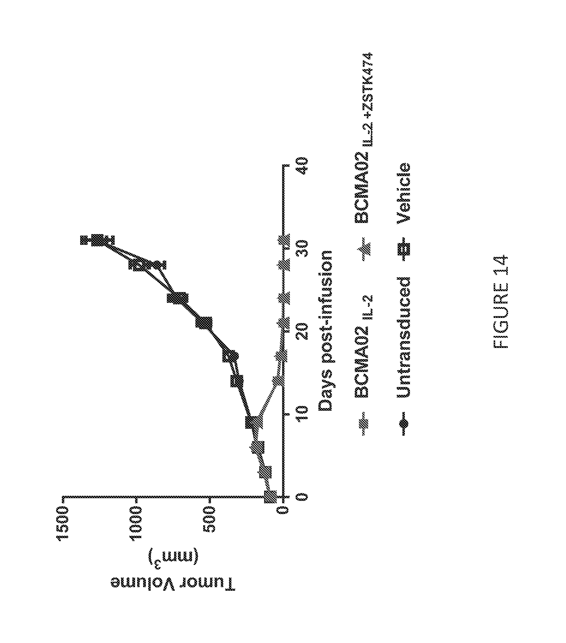
D00015
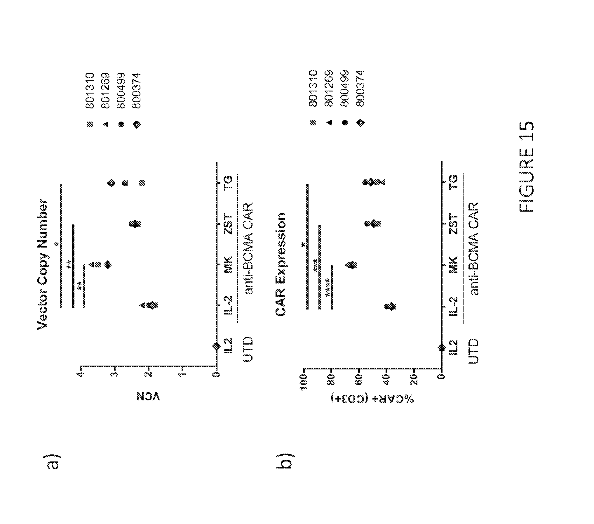
S00001
XML
uspto.report is an independent third-party trademark research tool that is not affiliated, endorsed, or sponsored by the United States Patent and Trademark Office (USPTO) or any other governmental organization. The information provided by uspto.report is based on publicly available data at the time of writing and is intended for informational purposes only.
While we strive to provide accurate and up-to-date information, we do not guarantee the accuracy, completeness, reliability, or suitability of the information displayed on this site. The use of this site is at your own risk. Any reliance you place on such information is therefore strictly at your own risk.
All official trademark data, including owner information, should be verified by visiting the official USPTO website at www.uspto.gov. This site is not intended to replace professional legal advice and should not be used as a substitute for consulting with a legal professional who is knowledgeable about trademark law.