Linker Peptide For Constructing Fusion Protein
LI; Qiang ; et al.
U.S. patent application number 16/326412 was filed with the patent office on 2019-06-20 for linker peptide for constructing fusion protein. The applicant listed for this patent is AMPSOURCE BIOPHARMA SHANGHAI INC.. Invention is credited to Si CHEN, Zhao DONG, Yongjuan GAO, Qiang LI, Yuanli LI, Zirui LI, Xinlu MA, Naichao SUN, Zhu WANG, Lu YANG, Yuncheng ZHENG.
| Application Number | 20190184026 16/326412 |
| Document ID | / |
| Family ID | 61196273 |
| Filed Date | 2019-06-20 |


| United States Patent Application | 20190184026 |
| Kind Code | A1 |
| LI; Qiang ; et al. | June 20, 2019 |
LINKER PEPTIDE FOR CONSTRUCTING FUSION PROTEIN
Abstract
A linker peptide for constructing a fusion protein. The linker peptide comprises a flexible peptide and a rigid peptide. The flexible peptide consists of one or more flexible units. The rigid peptide consists of one or more rigid units. The flexible unit comprises two or more amino acid residues selected from Gly, Ser, Ala, and Thr. The rigid unit comprises a human chorionic gonadotropin .beta.-subunit carboxy-terminal peptide (CTP) bearing a plurality of glycosylation sites. The linker peptide can more effectively eliminate mutual steric hindrance of two fusion molecules, decreasing a reduction/loss of polymerization or activity resulting from improper folding of an active protein or a conformational change. On the other hand, the negatively charged, highly sialylated CTP can resist renal clearance, further prolonging a half-life of a fused molecule and enhancing bioavailability of a fused protein. Furthermore, a protective effect of a glycosylated side chain CTP can lower the protease sensitivity of the linker peptide, making a linker region of the fusion protein less degradable.
| Inventors: | LI; Qiang; (Shanghai, CN) ; LI; Yuanli; (Shanghai, CN) ; CHEN; Si; (Shanghai, CN) ; WANG; Zhu; (Shanghai, CN) ; DONG; Zhao; (Shanghai, CN) ; LI; Zirui; (Shanghai, CN) ; MA; Xinlu; (Shanghai, CN) ; YANG; Lu; (Shanghai, CN) ; GAO; Yongjuan; (Shanghai, CN) ; ZHENG; Yuncheng; (Shanghai, CN) ; SUN; Naichao; (Shanghai, CN) | ||||||||||
| Applicant: |
|
||||||||||
|---|---|---|---|---|---|---|---|---|---|---|---|
| Family ID: | 61196273 | ||||||||||
| Appl. No.: | 16/326412 | ||||||||||
| Filed: | November 16, 2016 | ||||||||||
| PCT Filed: | November 16, 2016 | ||||||||||
| PCT NO: | PCT/CN2016/106011 | ||||||||||
| 371 Date: | February 19, 2019 |
| Current U.S. Class: | 1/1 |
| Current CPC Class: | C07K 14/5418 20130101; A61P 3/04 20180101; C12N 15/62 20130101; C07K 16/468 20130101; C12N 15/85 20130101; C07K 14/755 20130101; A61K 47/65 20170801; C12N 15/63 20130101; C07K 16/065 20130101; C07K 16/2887 20130101; C07K 14/475 20130101; C07K 16/2809 20130101; C07K 19/00 20130101; A61P 3/10 20180101 |
| International Class: | A61K 47/65 20060101 A61K047/65; C12N 15/62 20060101 C12N015/62; A61P 3/10 20060101 A61P003/10; C07K 16/46 20060101 C07K016/46; C07K 16/28 20060101 C07K016/28; C12N 15/85 20060101 C12N015/85; C07K 16/06 20060101 C07K016/06; A61P 3/04 20060101 A61P003/04; C07K 14/755 20060101 C07K014/755; C07K 14/475 20060101 C07K014/475; C07K 14/54 20060101 C07K014/54 |
Foreign Application Data
| Date | Code | Application Number |
|---|---|---|
| Aug 19, 2016 | CN | 201610692679.4 |
| Aug 19, 2016 | CN | 201610694914.1 |
Claims
1.-30. (canceled)
31. A linker peptide for constructing a fusion protein, wherein the linker peptide comprises a flexible peptide and a rigid peptide, wherein the flexible peptide comprises one or more flexible units and the rigid peptide comprises one or more rigid units, wherein the flexible unit comprises two or more amino acid residues each selected from the group consisting of Gly, Ser, Ala, and Thr, and wherein the rigid unit comprises a carboxy-terminal peptide of the human chronic gonadotropin .beta.-subunit and has at least two glycosylation sites.
32. The linker peptide of claim 31, wherein the rigid peptide is located at the C-terminus of the flexible peptide.
33. The linker peptide of claim 31, wherein the flexible unit comprises two or more amino acid residues each selected from the group consisting of Gly and Ser.
34. The linker peptide of claim 33, wherein the flexible unit has the structural formula of (GS).sub.a(GGS).sub.b(GGGS).sub.c(GGGGS).sub.d, wherein each of a, b, c, and d is an integer which is equal to or more than 0, and a+b+c+d.gtoreq.1.
35. The linker peptide of claim 34, wherein the flexible unit comprises an amino acid sequence selected from the group consisting of: TABLE-US-00020 i) GSGGGSGGGGSGGGGS; ii) GSGGGGSGGGGSGGGGSGGGGSGGGGS; iii) GSGGGGSGGGGSGGGGSGGGGSGGGGSGGGGSGGGGS; iv) GSGGGGSGGGGSGGGGS; v) GGGSGGGSGGGSGGGSGGGS; and vi) GGSGGSGGSGGS.
36. The linker peptide of claim 31, wherein the rigid unit comprises an amino acid sequence selected from the group consisting of: TABLE-US-00021 i) SSSSKAPPPSLPSPSRLPGPSDTRILPQ; ii) PRFQDSSSSKAPPPSLPSPSRLPGPSDTRILPQ; iii) SSSSKAPPPS; iv) SRLPGPSDTRILPQ; and v) SSSSKAPPPSLPSPSR.
37. The linker peptide of claim 36, wherein the rigid unit comprises an amino acid sequence having at least 70%, 80%, 90%, or 95% identity to the amino acid sequence of SEQ ID NO:1 or a fragment thereof.
38. The linker peptide of claim 36, wherein the rigid unit comprises the amino acid sequence of SEQ ID NO:1 or a fragment thereof.
39. The linker peptide of claim 31, wherein the rigid peptide comprises 1, 2, 3, 4, or 5 rigid units.
40. The linker peptide of claim 31, comprising an amino acid sequence selected from the group consisting of SEQ ID NOs:2-7.
41. A fusion protein having the structural formula of K1-L-K2 or K2-L-K1, wherein K1 is a first biologically active molecule; L is the linker peptide of claim 1; and K2 is a second biologically active molecule, and wherein K1 or K2 is selected from the group consisting of a protein, a protein domain, a peptide, and an antibody or a fragment thereof.
42. The fusion protein of claim 41, wherein K1 is a soluble or membrane signal molecule, a cytokine, a growth factor, a hormone, a costimulatory molecule, an enzyme, a receptor, or a protein or peptide which is a ligand to a receptor, and wherein K2 is a protein or a protein domain which prolongs the circulation half-life of K1.
43. The fusion protein of claim 42, wherein K2 is selected from the group consisting of a human hemoglobin, an iron Transferrin, and an Fc fragment of an immunoglobulin.
44. The fusion protein of claim 41, wherein K1 is selected from the group consisting of a toxin, an enzyme, a cytokine, a membrane protein, and an immunoregulatory cytokine; K2 is an antibody or a fragment thereof; and K1 is connected to K2 via L to form an antibody fusion protein.
45. The fusion protein of claim 41, wherein K1 is selected from the group consisting of an adenosine A1 receptor, angiotensin-converting enzyme (ACE), Activin family, ADAM family, .alpha.-1-antitrypsin, programmed cell death protein family, nerve growth factor and receptor family, bone morphogenetic protein (BMP) and receptor family, complement factor, calcitonin, cancer associated antigen, cathepsin family, CCL chemokine and receptor family, CD superfamily, CFTR, CXCL chemokine and receptor family, EGF and related receptor family, blood coagulation factor IIa, factor VII, VIII, IX, ferritin, fibroblast growth factor (FGF) and related receptor family, follicle, FZD family, HGF, glucagon, Cardiac myosin, growth hormone, Ig receptor, IgA receptor, IgE receptor, insulin growth factor and its interactors, interleukin and receptor superfamily, interferon family, iNOS, integrin family, kallikrein protein family, laminin, L-selectin, luteinizing hormone, MMP family, mucin-like family, cadherin superfamily, platelet-derived growth factor (PDGF) and related receptor family, parathyroid hormone, serum albumin, T-cell related receptor superfamily, TGF-.alpha., transforming growth factor-.beta. superfamily, thyrotropin, tumor necrosis factor (TGF) superfamily and related receptor superfamily, urokinase, proteins in WNT signal pathway, thymic peptide .alpha.1, thymic peptide .beta.4, and VEGF and related receptor family.
46. The fusion protein of claim 41, wherein K1 is human coagulation factor VII, human coagulation factor VIII, Exendin-4, human interleukin 7, or human growth hormone; L has an amino acid sequence of any one of SEQ ID NOs:2-7; and K2 is an Fc fragment of a human IgG, IgM, or IgA or a variant thereof, and the Fc variant comprises at least one amino acid modification in the wild-type human IgG Fc domain and has reduced effector functions and/or an enhanced binding affinity for a neonatal receptor FcRn.
47. The fusion protein of claim 46, wherein K2 comprises human immune protein Fc or a variant thereof.
48. The fusion protein of claim 46, wherein K2 comprises an amino acid sequence of any one of SEQ ID NOs:8-12.
49. The fusion protein of claim 46, wherein K1 is Exendin-4 having the amino acid sequence of SEQ ID NO:15.
50. A method for the treatment or prophylaxis of type II (non-insulin dependent) diabetes, comprising administering an effective amount of the fusion protein of claim 49 to a subject suffering from type II (non-insulin dependent) diabetes.
51. A method for the treatment or prophylaxis of obesities, comprising administering an effective amount of the fusion protein of claim 49 to a subject suffering from obesities.
52. The fusion protein of claim 46, wherein K1 is human coagulation factor VII (FVII) having the amino acid sequence of SEQ ID NO:13, human coagulation factor VIII (FVIII) having the amino acid sequence of SEQ ID NO:14, interleukin IL-7 having the amino acid sequence of SEQ ID NO:16, or human growth factor having the amino acid sequence of SEQ ID NO:17.
53. The fusion protein of claim 41, wherein K1 comprises one antibody or a fragment thereof; K2 comprises another antibody or a fragment thereof; and K1 is connected to K2 by L to form a bispecific antibody.
54. The fusion protein of claim 53, wherein K1 is a double chain antibody of anti-CD20, and K2 is a single chain antibody of anti-CD3.
55. The fusion protein of claim 54, wherein the double chain antibody of anti-CD20 comprises a heavy chain having the amino acid sequence of SEQ ID NO:18 and a light chain having the amino acid sequence of SEQ ID NO:19; the single chain antibody of anti-CD3 comprises the amino acid sequence of SEQ ID NO:20; and L comprises the amino acid sequence of SEQ ID NO:4 or SEQ ID NO:6.
56. The fusion protein of claim 41, wherein the fusion protein is glycosylated by expression in mammalian cells.
57. The fusion protein of claim 56, wherein the fusion protein is glycosylated by expression in Chinese hamster ovary (CHO) cells.
58. A method of preparing or producing the fusion protein of claim 41, comprising a step of linking K1 and K2 by L.
59. The method of claim 58, comprising the following steps: (a) ligate DNA sequences encoding K1 and K2 by a DNA sequence encoding L to form a fusion gene, (b) introduce the fusion gene obtained in step (a) into a eukaryotic or prokaryotic expression host, (c) culture by fermentation a high yield expression strain obtained in step (b) through screening to express the fusion protein, and (d) harvest the fermentation broth from step (c) and isolate and purify the fusion protein.
Description
FIELD OF THE INVENTION
[0001] The present invention relates to the field of fusion proteins and, more specifically, to a peptide linker for the construction of fusion proteins.
BACKGROUND
[0002] In recent two decades, protein fusion technology has been widely used in the construction of bifunctional antibodies, bifunctional enzymes, and bifunctional proteins. However, a variety of problems have been encountered in the construction of fusion proteins. For example, proteins that fold correctly during expression alone do not fold properly in the fusion protein; the active site is blocked after fusion due to the short distance between the two fused proteins; the fusion protein molecule is easily degraded by proteases when it cannot fold properly or when its conformation has changed; the protein catalytic domain with certain flexibility loses its original function after fusion; and so on. The emergence of these problems often leads to reduction or even complete loss of the activity of the fusion proteins. It is generally believed that the activity of the original protein molecule will decrease to a certain extent after the protein molecule is constructed in the fusion protein. A favorable fusion protein is the one that keeps more than 50% activity of the original protein molecule(s). In order to solve the above problems, researchers conducted many studies and explorations on the design and construction of fusion proteins to improve the activity of fusion proteins. such as changing the linking order of the fused proteins, changing different fusion sites, using different fusion partners, or using a peptide linker, etc.
[0003] Compared with other fusion strategies, the use of a peptide linker has a variety of advantages. First, the amino acids that make up the peptide linker are diverse (20 common amino acids). The length of the peptide linker is also an important tunable parameter, which can lead to rich diversity of peptide linkers (20.sup.n, n is the number of amino acid residues of the peptide linker). The peptide linkers are easy for bioengineering modifications. Second, the peptide linker provides certain spatial spacing, such that the two fused proteins fold correctly without interfering with each other. Third, the peptide linker can also provide more interaction possibilities for two fused proteins, promoting synergic interactions.
[0004] There are currently two kinds of commonly used peptide linkers, helical rigid peptide linkers (such as A(EAAAK).sub.nA) and flexible peptide linkers comprising less hydrophobic and less charged amino acids. The helical rigid peptide linkers can effectively separate different functional regions of the fusion proteins. Examples of flexible peptide linkers include the (GGGGS).sub.3 sequence that Huston designed. In addition, it has been reported that the carboxyl terminal peptide (hereinafter referred to as CTP) of human chorionic gonadotropin (HCG) beta chain can be used as a peptide linker alone, and is mainly used to link different subunits of the same protein. For example, CTP is used as a peptide linker between the beta and alpha subunits of follicle stimulating hormone, as disclosed in Chinese Patent Nos. CN03539860A, CN103539861A, CN103539868A and CN103539869A. In WO2005058953A2, CTP is used as a peptide linker in the fusion protein, linking the glycoprotein beta and alpha subunits. However, because CTP has the effect of prolonging the in vivo half-life of certain proteins, it has been primarily disclosed as a half-life prolonging moiety in the fusion proteins in many other patents. The half-life prolonging moiety can optionally choose CTP, immunoglobulin Fc, or other fusion partners with similar half-life prolonging function.
[0005] A number of literatures have reported the effects of the peptide linker sequences on the construction and expression of fusion proteins. For example, in the construction of the single chain antibody 1F7, it was found that using a conventional Genex212 peptide linker (GSTSGSGKSSEGKG) to link the light and heavy chains did not achieve the original catalytic activity of the protein and the fusion protein was unstable. After a library of 18 amino acid residues with random sequences was constructed and screened, catalytically active single chain antibodies were obtained (Tang Y et al., 1996, J Biol Chem., 271:15682-15686). In studying the helical peptide, Arai et al. found that the fluorescence resonance energy transfer from EBFP to EGFP reduced as the length of the peptide linker increased, suggesting that increasing the linker length could effectively separate the two functional regions (Arai R. et al., 2001, Protein Eng., 14:529-532). In addition, in studying dengue virus NS2B protein, Luo et al. found that inserting a glycine residue between the original sites 173 and 174, or replacing proline 174 with glycine, resulted in a significant decrease in protein activity of both the N- and C-terminal proteins. This indicated that the length and rigidity of the native peptide linker were the result of long-term evolution, and were of great significance to the function of the natural fusion proteins (Luo D. et al., 2010, J Biol Chem., 285:18817-18827). The N-terminus of the NS2B protein was a serine protease, the C-terminus was a RNA helicase, and the peptide linker between them had 11 amino acid residues.
[0006] The present inventors found that the length and amino acid composition of the peptide linker, the presence or absence of glycosylation sites, the compatibility between the peptide linker and the two active molecules, and other factors, could affect the function and stability of the fusion proteins. The inventors believe that the peptide linker should have the following characteristics: 1) can make the linked proteins fold effectively into proper conformations, does not cause molecular dynamics change, and preferably comprises non-immunogenic natural amino acids. 2) should have the ability to prevent protease attack. 3) should try to avoid mutual impacts of the two fused proteins on each other.
[0007] In the absence of clear guidelines for the design of peptide linkers, the present inventors have developed new peptide linkers for the construction of fusion proteins, based on the inventors' long-term research experience, particularly those in studying Fc fusion proteins. Amazingly, the peptide linkers have advantage for maintaining the biological activity of proteins or polypeptides and have broad applicability and portability.
DESCRIPTION OF THE INVENTION
[0008] The object of the present invention is to provide a novel peptide linker for constructing fusion proteins.
[0009] The first aspect of the present invention provides a peptide linker. The peptide linker comprises a flexible peptide and a rigid peptide. The flexible peptide consists of one or more flexible units, and the rigid peptide consists of one or more rigid units, wherein the flexible unit comprises two or more amino acid residues selected from Gly, Ser, Ala and Thr, and the rigid unit comprises the carboxyl terminal peptide (CTP) of human chorionic gonadotropin .beta. subunit.
[0010] Preferably, the peptide linker is glycosylated. More preferably, the glycosylation site is located on CTP. Much more preferably, the glycosylation process is accomplished by expression in mammalian cells, such as Chinese hamster ovary cells, or other expression hosts having a suitable glycosylation modification system.
[0011] Further, the rigid peptide in the peptide linker of the present invention is located at the N-terminus or C-terminus of the flexible peptide. Preferably, the rigid peptide is located at the C-terminus of the flexible peptide. Specifically, the structural formula of the peptide linker of the present invention can be represented as F-R or R-F, wherein F and R represent the flexible peptide and the rigid peptide, respectively. Further, the flexible peptide preferably comprises 1, 2, 3, 4 or 5 flexible units, and the rigid peptide preferably comprises 1, 2, 3, 4 or 5 rigid units.
[0012] More preferably, the flexible unit comprises two or more G and S residues. Much more preferably, the amino acid sequence of the flexible unit has the general formula (GS).sub.a(GGS).sub.b(GGGS).sub.c(GGGGS).sub.d, wherein a, b, c and d represent the number of structural units composed of G and S residues, and are integers greater than or equal to 0, and a+b+c+d.gtoreq.1.
[0013] Illustratively, each flexible unit is represented by F.sub.i and i=1, 2, 3, 4, 5, . . . n. In some embodiments of the invention, the flexible peptide preferably comprises, but is not limited to the following flexible units:
TABLE-US-00001 (i) F1: GSGGGSGGGGSGGGGS; (ii) F2: GSGGGGSGGGGSGGGGSGGGGSGGGGS; (iii) F3: GSGGGGSGGGGSGGGGSGGGGSGGGGSGGGGSGGGGS; (iv) F4: GSGGGGSGGGGSGGGGS; (v) F5: GGGSGGGSGGGSGGGSGGGS; (vi) F6: GGSGGSGGSGGS.
[0014] The rigid unit is selected from the full length or fragment sequence of the C-terminal amino acids 113 to 145 of human chorionic gonadotropin .beta. subunit. Specifically, the rigid unit comprises the amino acid sequence of SEQ ID no: 1 or its truncated form.
[0015] Preferably, CTP contains at least 2 glycosylation sites. For example, in a preferred embodiment of the present invention, CTP contains 2 glycosylation sites. Illustratively, CTP contains the N-terminal 10 amino acids of SEQ ID no: 1, i.e. SSSS*KAPPPS*, or CTP contains the C-terminal 14 amino acids of SEQ ID no: 1, i.e. S*RLPGPS*DTPILPQ. For another example, in another embodiment of the present invention, CTP contains 3 glycosylation sites. Illustratively, CTP contains the N-terminal 16 amino acids of SEQ ID no: 1, i.e. SSSS*KAPPPS*LPSPS*R. For other examples, in other embodiments of the present invention, CTP contains 4 glycosylation sites. Illustratively, CTP contains 28, 29, 30, 31, 32, or 33 amino acids, starting from position 113, 114, 115, 116, 117, or 118 and ending at position 145 of the human chorionic gonadotropin beta subunit. Specifically, CTP contains the N-terminal 28 amino acids of SEQ ID no: 1, i.e. SSSS*KAPPPS*LPSPS*RLPGPS*DTPILPQ. In this text, * represents a glycosylation site. Each possibility represents a separate embodiment of the present invention.
[0016] In other embodiments, the rigid units provided by the present invention have at least 70% amino acid sequence identity to native CTP. In other embodiments, the rigid units provided by the present invention have at least 80% amino acid sequence identity to native CTP. In other embodiments, the rigid units provided by the present invention have at least 90% amino acid sequence identity to native CTP. In other embodiments, the rigid units provided by the present invention have at least 95% amino acid sequence identity to native CTP.
[0017] Illustratively, each rigid unit is represented by R.sub.i and i=1, 2, 3, 4, 5, . . . , n. The rigid peptides described in some embodiments of the present invention may preferably comprise, but are not limited to, the following CTP rigid units.
TABLE-US-00002 (i) R1: SSSSKAPPPSLPSPSRLPGPSDTPILPQ; (ii) R2: PRFQDSSSSKAPPPSLPSPSRLPGPSDTPILPQ; (iii) R3: SSSSKAPPPS; (iv) R4: SRLPGPSDTPILPQ; (v) R5: SSSSKAPPPSLPSPSR.
[0018] The rigid peptides of the present invention may further comprise two or three of the above CTP rigid units. In one embodiment of the present invention, the rigid peptide comprises two R3 rigid units: SSSSKAPPPSSSSSKAPPPS (represented as R3R3). In another embodiment, the rigid peptide comprises three R4 rigid units: SRLPGPSDTPILPQSRLPGPSDTPILPQSRLPGPSDTPILPQ (represented as R4R4R4).
[0019] In some preferred embodiments of the invention, the peptide linker has the amino acid sequence as shown in SEQ ID no: 2
TABLE-US-00003 (represented as F2-R1) (GSGGGGSGGGGSGGGGSGGGGSGGGGSSSSSKAPPPSLPSPSRL PGPSDTPILPQ).
[0020] In another preferred embodiment of the present invention, the peptide linker has the amino acid sequence as shown in SEQ ID no: 3
TABLE-US-00004 (represented as F1-R2) (GSGGGSGGGGSGGGGSPRFQDSSSSKAPPPSLPSPSRLPGPSDT PILPQ).
[0021] In another preferred embodiment of the present invention, the peptide linker has the amino acid sequence as shown in SEQ ID no: 4
TABLE-US-00005 (represented as F4-R1) (GSGGGGSGGGGSGGGGSSSSSKAPPPSLPSPSRLPGPSDTPILP Q).
[0022] In another preferred embodiment of the present invention, the peptide linker has the amino acid sequence as shown in SEQ ID no: 5
TABLE-US-00006 (represented as F5-R5) (GGGSGGGSGGGSGGGSGGGSSSSSKAPPPSLPSPSR).
[0023] In another preferred embodiment of the present invention, the peptide linker has the amino acid sequence as shown in SEQ ID no: 6
TABLE-US-00007 (represented as F5-R5) (GGGSGGGSGGGSGGGSGGGSSSSSKAPPPSLPSPSR).
[0024] In another preferred embodiment of the present invention, the peptide linker has the amino acid sequence as shown in SEQ ID no: 7
TABLE-US-00008 (represented as F6-R4R4R4) (GGSGGSGGSGGSSRLPGPSDTPILPQSRLPGPSDTPILPQSRLP GPSDTPILPQ).
[0025] Yet another aspect of the invention provides a fusion protein containing the peptide linker. The fusion protein comprises two biologically active molecules and a peptide linker linking the two active molecules. The structural formula of the fusion protein is expressed as K1-L-K2 or K2-L-K1, wherein K1 is the first biologically active molecule, L is the above-mentioned peptide linker, and K2 is the second biologically active molecule. The components of the fusion protein are sequentially linked from the N-terminus to C-terminus. Further, the active molecules may be selected from protein or protein domain, polypeptide, antibody or antibody fragment, preferably protein or protein domain, antibody or an antibody fragment.
[0026] Illustratively, the active molecule K1 of the fusion protein comprises protein or protein domain having biological function, polypeptide (e.g., especially soluble or membrane signal molecule), cytokine, growth factor, hormone, costimulatory molecule, enzyme, receptor, protein or polypeptide having ligand function for the receptor. The active molecule K2 is serum protein or protein domain that prolongs the circulation half-life, for example, human serum albumin (HSA), transferrin (TF), antibody/immunoglobulin Fc fragment, and so on.
[0027] Illustratively, the active molecule K1 of the fusion protein comprises toxin, enzyme, cytokine, membrane protein, or immunomodulatory cytokine. The active molecule K2 comprises antibody or antibody fragment. K1 and K2 are linked via the peptide linker to form an antibody fusion protein. For example, K2 is an antibody Fv fragment (VL or VH), and for another example, K2 is a single chain antibody (scFv).
[0028] Further, the biologically active molecule K1 comprises but is not limited to adenosine A1 receptor, angiotensin converting enzyme ACE, activin family, ADAM family, ALK family, .alpha.-1-antitrypsin, programmed cell death associated protein family, nerve growth factor and receptor family, bone morphogenetic protein BMP and receptor family, complement factor, calcitonin, cancer associated antigen, cathepsin family, CCL chemokine and receptor family, CD superfamily, CFTR, CXCL chemokine and receptor family, EGF, epidermal growth factor EGF and receptor family, coagulation factor IIa, factor VII, factor VIII, factor IX, ferritin, fibroblast growth factor FGF and receptor family, follicle stimulating hormone, FZD family, HGF, glucagon, cardiac myosin, growth hormone, Ig, IgA receptor, IgE, insulin-like growth factor IGF and binding protein family, interleukin IL superfamily and its receptor superfamily, interferon INF family, iNOS, integrin family, kallikrein family, laminin, L-selectin, luteinizing hormone, MMP family, mucin family, cadherin superfamily, platelet-derived growth factor PDGF and receptor family, parathyroid hormone, serum albumin, T-cell-related receptor superfamily, TGF-.alpha., transforming growth factor TGF-.beta. superfamily, thyroid stimulating hormone, parathyroid stimulating hormone, tumor necrosis factor TNF superfamily and its receptor TNFRSF superfamily, urokinase, WNT signaling pathway family, thymosin .alpha.1, thymosin .beta.4, VEGF, vascular endothelial growth factor VEGF and its receptor family.
[0029] In the preferred embodiments of the present invention, the active molecule K1 is human coagulation factor VII (FVII), human coagulation factor VIII (FVIII), GLP-1 analogue Exendin-4 (Ex4), human interleukin 7 (IL-7), or human growth hormone (hGH). The peptide linker L of the fusion protein is as shown in SEQ ID no: 2, 3, 4, 5, 6 or 7. The active molecule K2 is selected from the Fc fragments of human immunoglobulins IgG, IgM, and IgA. More preferably, the Fc fragments are derived from human IgG.sub.1, IgG.sub.2, IgG.sub.3 or IgG.sub.4. Furthermore, the Fc fragments may be wild-types or variants. The Fc variants contain at least one amino acid modification in the wild-type human immunoglobulin Fc domain. The variants have reduced effector functions (ADCC or CDC effect) and/or an enhanced binding affinity for the neonatal receptor FcRn. Further, in the preferred embodiments of the present invention, the Fc variants are preferably selected from the following groups: (i) vFc.gamma.1: human IgG.sub.1 hinge, CH2, and CH3 regions containing Leu234Val, Leu235Ala and Pro331Ser mutations (the amino acid sequence is as shown in SEQ ID no: 8); (ii) vFc.gamma.2-1: human IgG.sub.2 hinge, CH2, and CH3 regions containing Pro331Ser mutation (the amino acid sequence is as shown in SEQ ID no: 9); (iii) vFc.gamma.2-2: human IgG.sub.2 hinge, CH2, and CH3 regions containing Thr250Gln and Met428Leu mutations (the amino acid sequence is as shown in SEQ ID no: 10); (iv) vFc.gamma.2-3: human IgG.sub.2 hinge, CH2, and CH3 regions containing Pro331Ser, Thr250Gln and Met428Leu mutations (the amino acid sequence is as shown in SEQ ID no: 11); (v) vFc.gamma.4: human IgG.sub.4 hinge, CH2, and CH3 regions containing Ser228Pro and Leu235Ala mutations (the amino acid sequence is as shown in SEQ ID no: 12).
[0030] More preferably, in one embodiment of the present invention, the FVII-Fc fusion protein comprises, sequentially from the N- to C-terminus, FVII (having the amino acid sequence as shown in SEQ ID no: 13), the peptide linker (having the amino acid sequence as shown in SEQ ID no: 2), and human IgG Fc (having the amino acid sequence as shown in SEQ ID no: 11).
[0031] More preferably, in one embodiment of the present invention, the FVIII-Fc fusion protein comprises, sequentially from the N- to C-terminus, FVIII (having the amino acid sequence as shown in SEQ ID no: 14), the peptide linker (having the amino acid sequence as shown in SEQ ID no: 2), and human IgG Fc (having the amino acid sequence as shown in SEQ ID no: 11).
[0032] More preferably, in one embodiment of the present invention, the Exendin-4-Fc fusion protein comprises, sequentially from the N- to C-terminus, Exendin-4 (having the amino acid sequence as shown in SEQ ID no: 15). the peptide linker (having the amino acid sequence as shown in SEQ ID no: 2, 3, 5 or 7), and human IgG Fc (having the amino acid sequence as shown in SEQ ID no: 11).
[0033] More preferably, in one embodiment of the invention, the IL-7-Fc fusion protein, sequentially from the N- to C-terminus, comprises IL-7 (having the amino acid sequence as shown in SEQ ID no: 16), the peptide linker (having the amino acid sequence as shown in SEQ ID no: 2), and human IgG Fc (having the amino acid sequence as shown in SEQ ID no: 11).
[0034] More preferably, in one embodiment of the present invention, the hGH-Fc fusion protein comprises, sequentially from the N- to C-terminus, hGH (having the amino acid sequence as shown in SEQ ID no: 17), the peptide linker (having the amino acid sequence as shown in SEQ ID no: 2), and human IgG Fc (having the amino acid sequence as shown in SEQ ID no: 11).
[0035] In other preferred embodiments of the invention, the active molecule K1 of the fusion protein is the antibody heavy chain variable region (VH), and K2 is the antibody light chain variable region (VL). K1 and K2 are linked by the peptide linker to form a single chain antibody (scFv).
[0036] In some preferred embodiments of the invention, the active molecule K1 of the fusion protein comprises a first antibody or antibody fragment, and the active molecule K2 comprises a second antibody or antibody fragment. K1 and K2 are linked by the peptide linker to form a bispecific antibody.
[0037] Preferably, in one embodiment of the present invention, K1 is a full-length double-chain anti-CD20 antibody, K2 is a single chain anti-CD3 antibody, and K1 and K2 are linked by the peptide linker to form a bispecific antibody. More preferably, the heavy chain of the anti-CD20 double-chain antibody contained in the bispecific antibody has an amino acid sequence as shown in SEQ ID no: 18 and the corresponding light chain has an amino acid sequence as shown in SEQ ID no: 19. The anti-CD3 single chain antibody contained in the bispecific antibody has an amino acid sequence as shown in SEQ ID no: 20. The peptide linker has an amino acid sequence as shown in SEQ ID no: 4 or 6. The heavy chain of the anti-CD20 double-chain antibody is linked to the anti-CD3 single chain antibody by the peptide linker.
[0038] Yet another aspect of the present invention also provides a method for preparing a fusion protein having the structural formula expressed as: K1-L-K2 or K2-L-K1, wherein K1 is the first biologically active molecule, L is the peptide linker, and K2 is a second biologically active molecule. The components that make up the fusion protein are sequentially linked from the N- to the C-terminus. The active molecules may be selected from the groups including protein or protein domain, polypeptide, antibody or antibody fragment, preferably protein or protein domain, antibody or antibody fragment. The preparation method includes the step of allowing K1 and K2 to be linked by L. In the preferred embodiments of the present invention, the method includes the following steps:
[0039] (a) Ligate the DNA sequences encoding the first active molecule K1 and the second active molecule K2 through the DNA sequence of the peptide linker L to form a fusion gene.
[0040] (b) Introduce the fusion gene obtained in step (a) into a eukaryotic or prokaryotic expression host.
[0041] (c) Culture the high yield expression host screened and obtained in step (b) to express the fusion protein.
[0042] (d) Harvest the fermentation broth of step (c) and isolate and purify the fusion protein.
[0043] Illustratively, the peptide linkers are used to link the active proteins/polypeptides to serum proteins with long circulation half-lives, such as antibody/immunoglobulin Fc fragments, human serum albumin (HSA), transferrin (TF), etc. In one preferred embodiment of the present invention, the preparation method of the FVII-Fc fusion protein includes the step of linking the active molecule FVII and human IgG Fc by the peptide linker (SEQ ID no: 2). In one preferred embodiment of the present invention, the preparation method of the FVIII-Fc fusion protein includes the step of linking the active molecule FVIII and human IgG Fc by the peptide linker (SEQ ID no: 2). In another preferred embodiment of the present invention, the preparation method of the Exendin 4-Fc fusion protein includes the step of linking the active molecule Exendin-4 and human IgG Fc by the peptide linker (SEQ ID no: 2, 3, 5 or 7). In another preferred embodiment of the present invention, the preparation method of the IL-7-Fc fusion protein includes the step of linking the active molecule IL-7 and human IgG Fc by the peptide linker (SEQ ID no: 2). In another preferred embodiment of the present invention, the preparation method of the hGH-Fc fusion protein includes the step of linking the active molecule hGH and human IgG Fc by the peptide linker (SEQ ID no: 2).
[0044] Illustratively, the peptide linkers are used in the construction of bispecific antibodies. In the preferred embodiment of the present invention, the preparation method of the bispecific antibody of anti-CD3.times.CD20 includes the step of linking the double-chain anti-CD20 antibody and the single chain anti-CD3 antibody by the peptide linker (SEQ ID NO: 4 or 6).
[0045] In this present invention, technical novelities can be summarized as follows:
[0046] 1. In the present invention, part of the peptide linker, CTP, contains multiple O-glycosyl side chains. It is capable of forming a relatively stable and rigid three-dimensional structure and thus more effectively separate the two partners of the fusion protein and eliminate the steric hindrance between them. In the construction of a series of fusion proteins composed of the active proteins and the Fc fragment, for example, the FVII-Fc fusion protein, the introduction of the rigid CTP unit into the peptide linker ensures that the N-terminally fused active protein does not affect the binding site of the Fc variant to FcRn, thus having no effect on the circulation half-life of the fusion protein. In addition, the protein A binding site of Fc is important for the purification step, and the introduction of the rigid CTP unit ensures that the N-terminally fused active protein will not "block" the protein A binding site. On the other hand, the introduction of the rigid CTP unit makes the 25 kD size of the Fc fragment not interfere with the folding of the N-terminally fused active protein, which prevents the decline or loss of the biological activity/function of the active protein. Many embodiments of the present invention indicate that the introduction of the rigid CTP unit makes the biological activity of the fusion proteins significantly improved. This can be explained as follows. The rigid CTP polypeptide possesses multiple glycosyl side chains. Compared with irregular coil of a flexible peptide linker such as (GGGGS).sub.n, CTP can form a stable three-dimensional conformation, which effectively increases the distance between the two fusion partners of the fusion protein. The spatial separation facilitates independent folding of the active protein and the Fc segment to form correct three-dimensional conformations, such that the active protein and the Fc segment would have no mutual impact on the biological activity of each other. This reduces the possibility of the decline or loss of the activity of the active protein due to misfolding and conformational alteration. Thus the biological activity of the fusion protein is increased.
[0047] 2. The peptide linkers of the present invention have a wide range of applicability and portability. With a combination of rigid and flexible units, the peptide linkers are conferred with a conformation between completely rigid and fully flexible, and the specific rigidity or flexibility of the polypeptide varies depending on the ratio and arrangement order of the two sequences. As sequences are designed with different combinations of ratio and arrangement order of rigid and flexible units, the rigidity of the peptide linker can be finely regulated to meet different requirements in the construction of the fusion proteins.
[0048] 3. CTP contains glycosylation sites. Highly sialylated and negatively charged CTP can resist the kidney to its clearance and further prolongs the half-life of the fusion protein. CTP can improve pharmacokinetic parameters, such as reducing the clearance rate, reducing the apparent distribution of volume, increasing AUC.sub.(0-t), such that the bioavailability of the fusion protein increases. It is expected that the clinical dose will be reduced.
[0049] 4. The glycosyl side chains of CTP has protective effect, which can reduce the sensitivity of the peptide linker to proteases. The fusion protein is not easy to be degraded in the linking region.
Term Definitions
[0050] "Antibody fragment": refers to an antigen-binding fragment of an antibody or antibody analogue, which typically comprises at least a portion of the antigen-binding region or variable region of the parental antibody, for example, one or more CDRs.
[0051] The "Fc" region: includes two heavy chain fragments, each of which comprises the CH2 and CH3 domains of an antibody. The two heavy chain fragments are held together by two or more interchain disulfide bonds and the inter-CH3 domain hydrophobic interactions.
[0052] The "Fv" region comprises the variable regions from both the heavy and light chains, but lacks constant regions.
[0053] "Single chain Fv antibody" (or scFv antibody): refers to an antibody comprising the VH and VL domains of an antibody, wherein these domains are present in a single polypeptide chain. In general, the Fv polypeptide additionally contains a polypeptide linker between the VH and VL domains that allows scFv to form the desired structure for antigen binding.
[0054] "Bispecific antibody": refers to an antibody that comprises two Fv domains or scFv units such that the resulting antibody recognizes two different antigenic determinants.
[0055] "Antibody fusion protein": refers to a product obtained by fusing an antibody fragment with another bioactive protein using genetic engineering techniques. Owing to many different fused proteins, the antibody fusion proteins have a variety of biological functions.
[0056] For example, an Fv-containing antibody fusion protein. As the Fab or Fv fragment is linked with certain toxins, enzymes, or cytokines, the biologically active molecules can be targeted to specific sites of the targeted cell, forming the so-called "biological missile.
[0057] For example, chimeric receptors. As the scFv antibody is fused with certain cell membrane protein molecules, the fusion proteins, known as chimeric receptors, can be expressed on the cell surface, giving the cells the capability to bind to a specific antigen.
[0058] For example, an Fc-containing antibody fusion protein. The antibody IgG Fc region is fused with the biologically active molecule to form an Fc fusion protein. The Fc fusion protein not only exerts the biological function of the active molecule but also inherits similar properties of the antibody, including prolonged plasma half-life and a series of effector functions specific to the Fc region. For example, on the one hand, the Fc region plays an important role in the eradication of pathogens. The Fc-mediated effector functions of IgG are carried out by two mechanisms: (1) After the Fc regions of IgG molecules bind to the cell surface Fc receptors (Fc.gamma.Rs), pathogens are broken down by phagocytosis or lysis or by killer cells through an antibody-dependent cell-mediated cytotoxicity (ADCC) pathway. (2) After the Fc regions of IgG molecules bind to C1q molecules of the first complement C1 complexes, the complement-dependent cytotoxicity (CDC) pathway is triggered, such that pathogens are lysed. On the other hand, the antibody Fc region bind to the FcRn receptor to prevent the antibody from entering into lysosome to be degraded. The fusion proteins containing the Fc region are endocytosed and protected by FcRn. These fusion proteins are not to be degraded, but again enter into the circulatory system, thereby increasing the in vivo half-lives of these fusion proteins. Moreover, FcRn shows activity in adult epithelial tissues, and it is expressed in epithelial cells of the intestine, pulmonary trachea, nasal cavity, vagina, colon, and rectum. The fusion proteins containing the Fc region can effectively shuttle the epithelial barrier through FcRn-mediated cell transduction.
[0059] "hCG-.beta. carboxy terminal peptide (CTP)": is a short peptide derived from the carboxyl terminus of human chorionic gonadotropin (hCG) beta subunit. Four kinds of reproduction-related polypeptide hormones, follicle stimulating hormone (FSH), luteinizing hormone (LH), thyroid stimulating hormone (TSH), and human chorionic gonadotropin (hCG) comprise the same alpha subunit and respective specific beta subunits. Compared with the other three hormones, hCG has a significantly prolonged half-life, which is mainly due to the specific carboxyl terminal peptide (CTP) on its .beta.-subunit. CTP has 37 amino acid residues, which possesses four O-glycosylation sites terminating with a sialic acid residue. Highly sialylated, negatively charged CTP can resist to the clearance by the kidney, thereby prolonging the in vivo half-life of a protein (Fares F A et al., 1992, Proc Natl Acad Sci USA, 89: 4304-4308).
BRIEF DESCRIPTION OF THE FIGURES
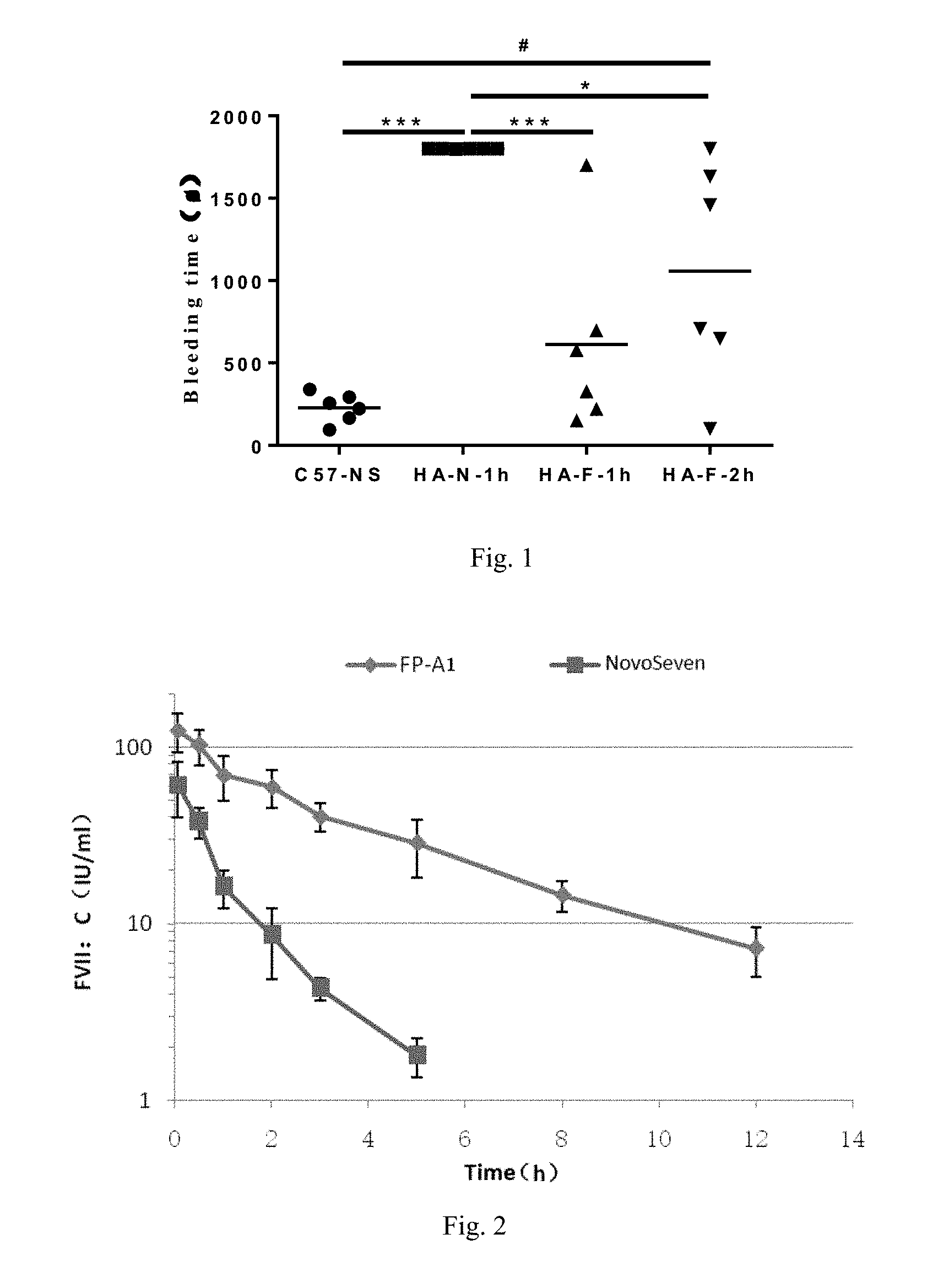
[0060] FIG. 1. Comparison of the bleeding times of HA mice administered with FP-A1 and NovoSeven.RTM. for 1 h and 2 h. Compared with the HA-N-1h group, *P<0.05, .sup.***P<0.01; compared with the C57-NS group, .sup.#P<0.05, .sup.###P<0.01.
[0061] FIG. 2. The active half-lives of FP-A1 and NovoSeven.RTM. in rats administered with warfarin.
[0062] FIG. 3. The curves of the RBG value changes in 0-216 h in db/db diabetic mice given single injection of FP-C1 and FP-C5.
[0063] FIG. 4. The effect of different doses of FP-C1 on HbA1c in db/db diabetes mice. Comparison of the FP-C1 group with the model group, *P<0.05, **P<0.01.
[0064] FIG. 5. RBG value changes of STZ-induced diabetes mice with single injection of FP-C1 during 0-240 h. Comparison of the FP-C1 group with the modle group, *P<0.05, **P<0.01; Comparison of the Duraglutide group with the modle group, .DELTA.P<0.05, .DELTA.P<0.01.
[0065] FIG. 6. The effect of FP-C1 on the body weight of mice fed with high fat diet.
[0066] FIG. 7. The effect of FP-C1 on glucose tolerance in mice fed with high fat diet (mean.+-.SD, n=8). Compared with the normal group, #P<0.05, ##P<0.01; compared with the high fat group, *P<0.05, **P<0.01.
[0067] FIG. 8. The effect of FP-C1 on the serum insulin concentration of mice fed with high fat diet (means.+-.SD, n=8). Compared with the normal group, #P<0.05, ##P<0.01; compared with the high fat group, *P<0.05, **P<0.01.
[0068] FIG. 9. The effect of FP-C1 on the insulin resistance index of mice fed with high fat diet (mean.+-.SD, n=8). Compared with the normal group, #P<0.05, ##P<0.01; compared with the high fat group, *P<0.05, **P<0.01.
[0069] FIG. 10. The effect of FP-C1 on the cross-sectional area of fat cells in mice fed with high fat diet. A: normal group. B: high fat diet group. C: FP-C1 group.
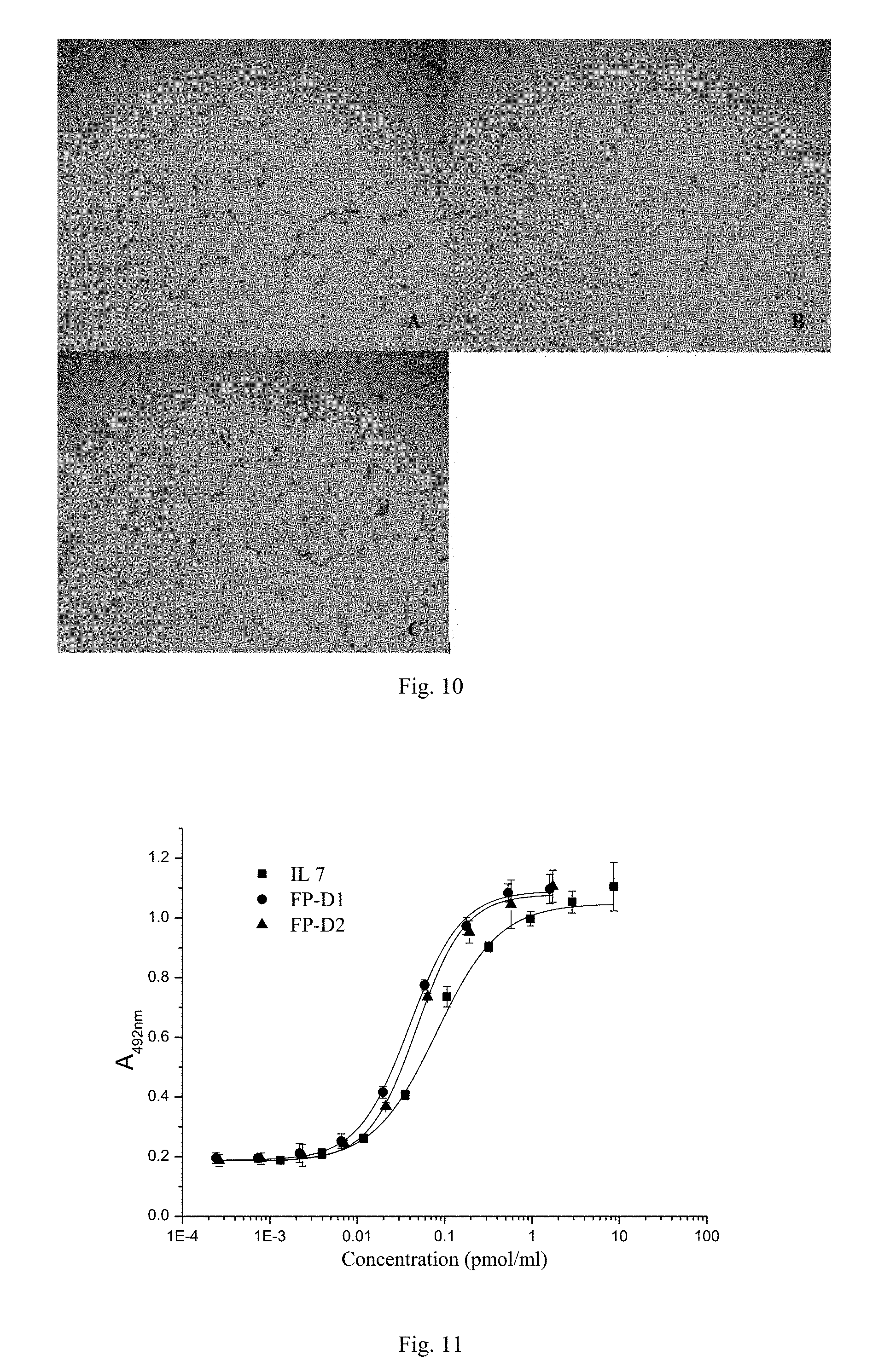
[0070] FIG. 11. The ability of IL7-Fc fusion proteins FP-D1 and FP-D2 to stimulate mouse mononuclear cells to proliferate.
[0071] FIG. 12. The ability of hGH-Fc fusion proteins FP-E1 and FP-E2 to stimulate Nb2 cells to proliferate.
[0072] FIG. 13. The curves of blood drug concentration vs. time of hGH-Fc fusion proteins FP-E1 and FP-E2.
[0073] FIG. 14. The growth curves of all groups of rats administered with the hGH-Fc fusion protein FP-E1.
[0074] FIG. 15. The anti-CD3.times.CD20 bispecific antibody FP-F1 activated human PBMC cells to secret IFN-.gamma. in a concentration-dependent manner.
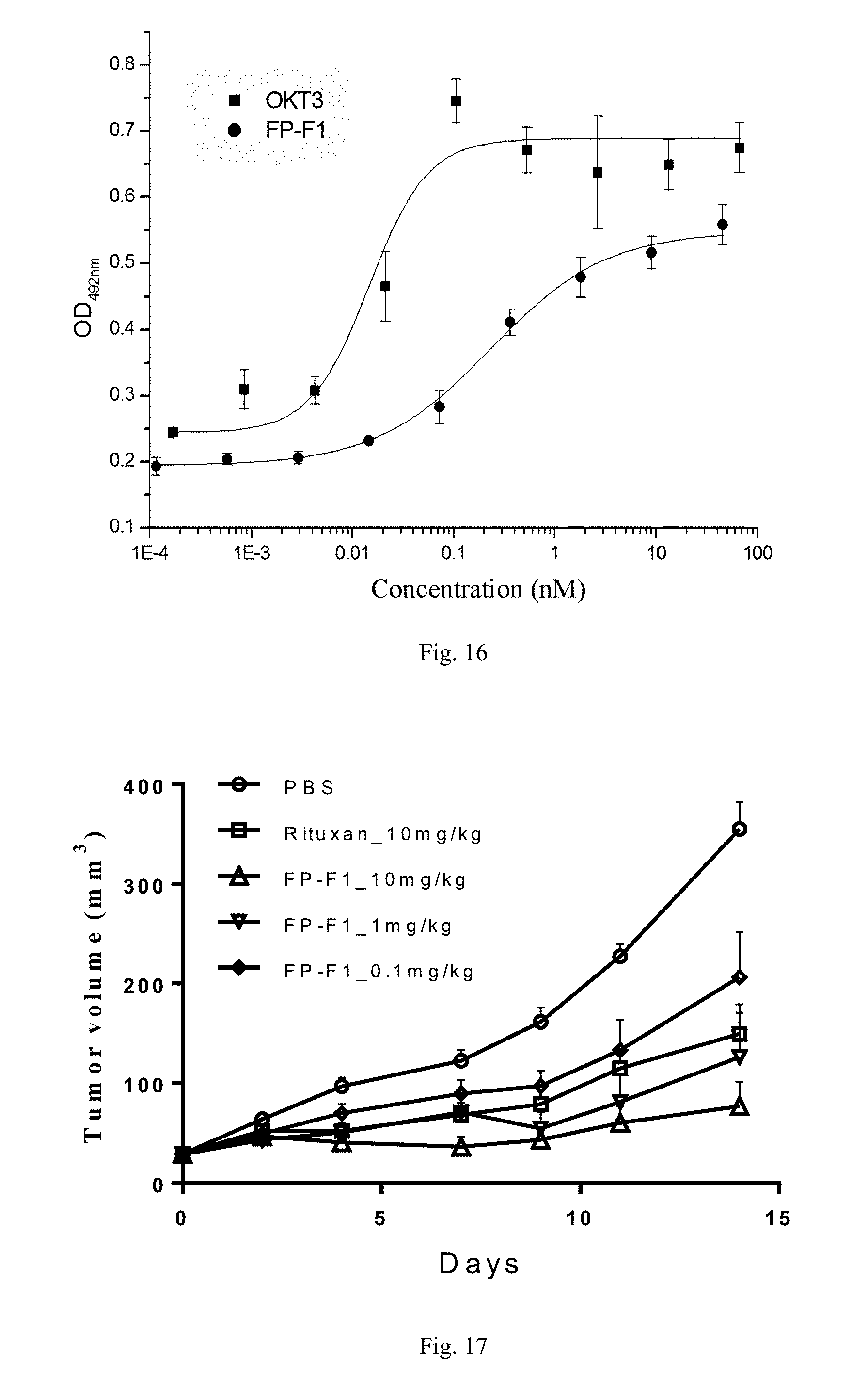
[0075] FIG. 16. The anti-CD3.times.CD20 bispecific antibody FP-F1 activated human PBMC cells in a concentration-dependent manner.
[0076] FIG. 17. The efficacy of anti-CD3.times.CD20 bispecific antibody FP-F1 in killing subcutaneous transplantation tumor.
EXAMPLES
Example 1. Peptide Linkers Used to Construct Fusion Proteins
[0077] The present inventors constructed a series of fusion proteins K1-L-K2 containing peptide linkers. The composition of each fusion protein was shown in Table 1. The DNA sequences encoding the first active molecule K1 and the second active molecule K2 were ligated by the DNA sequence of the peptide linker L to constitute a fusion gene. Preferably, the codons of the DNA sequences were optimized for expression in CHO cells. Preferably, the sequences were generated by chemical synthesis. Two restriction sites, SpeI and EcoRI, were added at the 5' and 3' ends of the synthesized fragment, respectively, to facilitate insertion of the fusion gene obtained above into the specific site of an expression vector. After verified by sequencing, the fusion gene was digested with the corresponding restriction endonucleases and inserted into the corresponding cleavage sites of the expression plasmid PXY1A1, obtained by modifying PCDNA3.1 as a template. The expression plasmid containing the gene of the fusion protein was thus obtained. The PXY1A1 plasmid contained, but was not limited to, the following important expression elements: 1) human cytomegalovirus immediate early promoter and highly exogenous expression enhancer needed by mammalian cells; 2) double screening markers with kanamycin resistance in bacteria and G418 resistance in mammalian cells; 3) murine dihydrofolate reductase (DHFR) gene expression cassette. When the host cell type was DHFR gene deficient, methotrexate (MTX) could co-amplify the fusion gene and the DHFR gene (U.S. Pat. No. 4,399,216). The expression plasmid of the fusion protein was transfected into a mammalian host cell line. The preferred host cell line was DHFR enzyme-deficient CHO cells in order to achieve stable and high levels of expression (U.S. Pat. No. 4,818,679). After two days of transfection, the medium was replaced with a screening medium containing 0.6 mg/mL G418. Cells were seeded in the 96-well plate at a certain concentration (5000-10000 viable cells/well) for 10-14 days until large discrete cell clones appeared. The transfectants resistant to the selecting antibiotic were screened by the ELISA assay. Subclones with high level expression of the fusion protein were isolated by limiting dilution and use of the 96-well culture plate. For a fusion protein already validated as a good one by multiple means, it was appropriate to amplify the DHFR gene by MTX drug inhibition to achieve higher level of expression. In the growth medium containing increasing concentration of MTX, the transfected fusion protein gene was co-amplified with the DHFR gene. The highly expressed monoclonal cell strains obtained were cultured under fed-batch conditions in shake flasks or a 5-liter fermentor. The fusion protein was purified by protein A affinity chromatography and other ion exchange chromatography.
[0078] The inventors constructed a series of fusion proteins containing the present invention's peptide linkers with flexible and rigid units, and also constructed a variety of fusion proteins containing the peptide linkers with only flexible units of different lengths for comparison. For example, FVII-Fc fusion protein (FP-A1 containing CTP; FP-A2 and FP-A3 without CTP), FVIII-Fc fusion protein (FP-B1 containing CTP; FP-B2 and FP-B3 without CTP); Fc-fusion protein of Exendin-4 and its analogue (FP-C1, FP-C2, FP-C3, FP-C4 containing CTP; FP-C5 without CTP), IL7-Fc fusion protein (FP-D1 containing CTP; FP-D2 without CTP), hGH-Fc fusion protein (FP-E1 containing CTP; FP-E2 without CTP). In addition, the inventors also developed anti-CD20.times.CD3 bispecific antibodies (FP-F1 and FP-F2 containing CTP). The composition of each fusion protein was shown in Table 1, and the amino acid sequence of each fusion protein was shown in the sequence listings section.
TABLE-US-00009 TABLE 1 The compositions of various fusion proteins from N- to C-terminus Code for fusion protein K1 L K2 FP-A1 FVII F2-R1 Fc (vFc.gamma..sub.2-3) FP-A2 FVII F1 Fc (vFc.gamma..sub.2-3) FP-A3 FVII F3 Fc (vFc.gamma..sub.2-3) FP-B1 FVIII F2-R1 Fc (vFc.gamma..sub.2-3) FP-B2 FVIII F1 Fc (vFc.gamma..sub.2-3) FP-B3 FVIII F3 Fc (vFc.gamma..sub.2-3) FP-C1 Exendin-4 F2-R1 Fc (vFc.gamma..sub.2-3) FP-C2 Exendin-4 F1-R2 Fc (vFc.gamma..sub.2-3) FP-C3 Exendin-4 F5-R5 Fc (vFc.gamma..sub.2-3) FP-C4 Exendin-4 F6-R4R4R4 Fc (vFc.gamma..sub.2-3) FP-C5 Exendin-4 F1 Fc (vFc.gamma..sub.2-3) FP-D1 IL-7 F2-R1 Fc (vFc.gamma..sub.2-3) FP-D2 IL-7 F1 Fc (vFc.gamma..sub.2-3) FP-E1 hGH F2-R1 Fc (vFc.gamma..sub.2-3) FP-E2 hGH F1 Fc (vFc.gamma..sub.2-3) FP-F1 Anti-CD20 mAb F4-R1 Anti-CD3 ScFv FP-F2 Anti-CD20 mAb F4-R3R3 Anti-CD3 ScFv
Example 2. Preparation of Coagulation Factor FVII-Fc Fusion Proteins and Determination of Biological Activity and In Vivo Active Half-Life
[0079] 2.1 Preparation and Identification of Coagulation Factor FVII-Fc Fusion Proteins
[0080] The stably expressing CHO cell strains of FP-A1, FP-A2 and FP-A3 obtained in Example 1 were cultured in shake flasks under fed-batch conditions for 10-14 days. The fusion proteins were purified by four steps of column chromatography: protein A affinity chromatography, multidimensional chromatography, anion exchange chromatography, and molecular sieve chromatography. The fusion proteins were then self-activated with incubation in a solution. The SDS-PAGE electrophoresis showed the following results. Under reducing conditions, the single-chained molecule of non-activated FP-A2 showed two obvious bands in the vicinity of 70-85 kDa and 40 kDa, indicating that protein degradation occurred and the fraction of the degraded fragments was about 20-30%. Under non-reducing conditions, the non-activated FP-A2 migrated to about 130 kDa together with another band of >200 kDa, indicating that some of the fusion proteins aggregated. Under reducing conditions, the single-chained molecule of non-activated FP-A3 was close to 100 kDa and there were no contaminant bands. Under non-reducing conditions, most of the non-activated FP-A3 proteins migrated to >200 kDa, indicating that FP-A3 was in the form of aggregates. Under reducing conditions, the single-chained molecule of non-activated FP-A1 was of 100-110 kDa and there were no obvious contaminant bands. Under non-reducing conditions, the non-activated FP-A1 migrated to 150 kDa. Under reducing conditions, the activated FP-A1 showed two clear bands, 74.3 kDa HC-L-CTP-Fc and about 24.0 kDa LC, respectively, and no other contaminant bands. Under non-reducing conditions, the activated FP-A1 migrated to 150 kDa, indicating that the fusion protein FP-A1 did not degrade significantly, did not form aggregates remarkably, and had higher thermodynamic stability and stronger ability of anti-proteolytic hydrolysis. This example demonstrated that the peptide linkers containing the rigid CTP unit increased the stability of the fusion proteins, which were less susceptible to degradation, and reduced the formation of aggregates.
[0081] 2.2 Direct Determination of the Biological Activity of Fusion Proteins by the Clotting Assay
[0082] The determination of FVIIa biological activity by the clotting assay was achieved by correcting FVIIa-deficient plasma to prolong the clotting time. The kit (STAGO, Cat. No. 00743) was used for the assay. The assay was first to mix diluted human freeze-dried normal plasma of known FVII activity (Unicalibrator, Cat. No. 00625) with FVII-deficient plasma, measure the prothrombin time (PT), and establish a standard curve. The plasma for the FVII activity to be measured was diluted and then mixed with FVII-deficient plasma for PT measurement. The FVIIa activity of the test sample could be determined from the logarithmic equation between the percentage of activity C (%) and PT time t (s) fitted by the standard curve. The FVIIa activity was expressed as the percentage (%) of that of normal plasma. The corresponding relation between the percentage of activity of the standards (%) provided by the kit and the international unit (IU) of enzyme activity was 100%=1 IU, according to which the specific activity of FVII in the test sample could be calculated in units of IU/mg. The results showed that under optimum experimental conditions, the highest activities of FP-A1, FP-A2 and FP-A3 were about 20000 IU/mg, 4000 IU/mg and 7000 IU/mg, respectively. The experimental results showed that the type and length of the peptide linkers had a great effect on the activity and stability of FVII-Fc. The in vitro bioactivity of the fusion protein FP-A1 containing the peptide linker with the rigid unit was much higher than those of the fusion proteins FP-A2 and FP-A3 without rigid units. The results also showed that if only the length of the flexible peptide was extended, the activity of the fusion protein could not be improved effectively. The steric hindrance between the fused partners comprised the formation of the correct conformation of the fusion proteins, such that the stability of the fusion proteins decreased and the proteins were easy to form aggregates. This example demonstrated that the peptide linker containing a rigid CTP unit reduced the steric hindrance of the Fc domain and increased the activity and stability of the fusion protein.
[0083] 2.3 Determination of In Vivo Bleeding Inhibition of FVII-Fc Fusion Proteins
[0084] The hemostatic effect of FVII-Fc fusion proteins was assessed by using hemophilia mice, which were the tail vein transection (TVT) bleeding model of homozygous hemophilia A mice with knockout of the FVIII factor gene (HA, Shanghai Research Center of the Southern Model Organisms). Male HA mice, 16-20 weeks old, were adaptively fed for one week and randomly divided into 3 groups, 6 mice per group. Two groups were given 300,000 IU/kg of FP-A1 and the other group was given 300,000 IU/kg of NovoSeven.RTM. (Novo Nordisk). Meantime, wild-type male C57BL/6J mice, 16-20 weeks old (Shanghai Research Center of the Southern Model Organisms), were used as the normal control group (n=6), and given an equal volume of physiological saline through tail vein injection. The two groups of HA mice given FP-A1 were subjected to tail-cutting tests at 1 h and 2 h after administration, respectively, and the HA mice given NovoSeven.RTM. were subjected to tail-cutting tests at 1 h after administration. The C57BL/6J normal control group (C57-NS group) was subjected to tail-cutting tests at 2 h after administration. All data were expressed as mean.+-.standard error (.+-.SEM). The t-test analysis was used to compare between the experimental groups. The analysis software was Graphpad Prism 5.0. P<0.05 was considered statistically significant.
[0085] As shown in FIG. 1, after administration of NovoSeven.RTM. for 1 h, the bleeding time of the mice was 30 min, indicating that it had no hemostatic effect (HA-N-1h group). In contrast, after administration of FP-A1 for 1 h (HA-F-1h group) and 2 h (HA-F-2h group), FP-A1 was still effective in hemostasis, and the bleeding times were significantly shorter than that of the NovoSeven.RTM. group (P<0.05). This indicated that FP-A1 had a significantly longer active half-life compared with NovoSeven.RTM..
[0086] 2.4 Determination of In Vivo Active Half-Life of FVII-Fc Fusion Protein
[0087] In this study, we investigated the active half-life of FP-A1 in the warfarin-induced coagulation disorder rat model. According to the method reported in the literature (Joe Salas et al., 2015, Thrombosis Research, 135:970-976 or Gerhard Dickneite et al., 2007, Thrombosis Research, 119:643-651), SD rats (8-12 weeks age and 220 g body weight, Beijing Vital River Laboratory Animal Technology Co., Ltd.) were randomly divided into two groups, eight rats per group. After intragastric administration of warfarin (Orion Corporation, Finland, lot no. 1569755) at a dose of 2.5 mg/kg for 24 h, the rats were given intravenous administration of 10,000 IU/kg of FP-A1 or NovoSeven.RTM. (Novo Nordisk), respectively. Blood was collected after administration for the FP-A1 group at 0.05 h, 0.5 h, 1 h, 2 h, 3 h, 5 h, 8 h, 12 h, respectively, and for the NovoSeven.RTM. group at 0.05 h, 0.5 h, 1 h, 2 h, 3 h, 5 h, respectively. With sodium citrate at a final concentration of 0.013 M as an anticoagulant, blood samples were centrifuged at 3000 rpm for 10 min to obtain the supernatant. The activity was determined by the method in section 2.2 and the active half-life was calculated.
[0088] As shown in FIG. 2, the active half-life of FP-A1 was calculated to be 3.03.+-.0.35 h, and that of NovoSeven.RTM. was 1.01.+-.0.16 h. Compared with NovoSeven.RTM. with equal activity, FP-A1 prolonged the active half-life in rats by about 3-fold. The plasma coagulation activity was about 40% at 3 h after single injection of FP-A1, while the coagulation activity of NovoSeven.RTM. decreased to 3% after 3 h. At 12 h after administration, the plasma coagulation activity of FP-A1 remained 7% or more.
[0089] The results in sections 2.3 and 2.4 showed that the fusion protein of the present invention, containing the peptide linker with the flexible and rigid CTP units, had a significantly prolonged active half-life, indicating that the peptide linker eliminated the blocking effect of the active protein FVII on the binding site of Fc for its receptor FcRn. The results also demonstrated that owning to the introduction of the peptide linker of the present invention, FVII formed the correct three-dimensional conformation, maintained high biological activity, and was not affected by the steric hindrance of the C-terminally fused Fc.
Example 3. Production of Coagulation Factor FVIII-Fc Fusion Proteins and Determination of Biological Activity
[0090] 3.1 Production and Identification of Coagulation Factor FVIII-Fc Fusion Proteins
[0091] The stably expressing CHO cell strains for FP-B1, FP-B2 and FP-B3 obtained in Example 1 were cultured for 7 to 12 days under fed-batch or semi-continuous culture conditions. The supernatant was harvested immediately for purification by protein A and/or VIII-select (GE) affinity chromatography. Under optimum culture conditions, the FP-B2 supernatant was passed through protein A and VIII-select (GE) two-step affinity chromatography columns, and the elute still contained multiple components. Under reducing conditions, the SDS-PAGE electrophoresis showed a main band of 180 kDa and multiple fragments of 40-100 kDa. Under non-reducing conditions, most of the purified proteins migrated to >300 kDa. This indicated that FP-B2 products were mostly in the form of aggregates, unstable and easy to be degradated. Under the same culture conditions, the supernatant of FP-B1 was purified by protein A and VIII-select (GE) two-step affinity chromatography. Under reducing conditions, three clear bands appeared on the gel, which were single chain FVIII-Fc (190 kDa), light chain-Fc (105 kDa) and heavy chain (90 kDa), and there were no contaminant bands. Under non-reducing conditions, purified proteins FP-B1 and FP-B3 migrated to >200 kDa, while most of FP-B3 proteins stayed in the stacking gel, indicating that FP-B3 was also present in the form of aggregates.
[0092] It was reported that the lipid binding region of FVIII (2303-2332) was critical to its function, and that very small conformational changes in the region led to protein aggregation and loss of activity (Gilbert et al., 1993, Biochemistry, 32:9577-9585). The results of the present invention indicated that the peptide linker containing the rigid CTP unit could eliminate the steric hindrance of the C-terminal Fc on the FVIII lipid-binding region, such that the FVIII spatial conformation was almost unaffected. Thus, the protein aggregation was reduced, the protein stability was increased, and the bioactivity of the FVIII-Fc fusion protein was greatly improved.
[0093] 3.2 Direct Determination of the Biological Activity of the FVIII-Fc Fusion Proteins by the Clotting Assay
[0094] The determination of the FVIII biological activity by the clotting assay was achieved by correcting FVIII-deficient plasma to prolong the clotting time. A factor VIII (FVIII) assay kit (the clotting method), STA.RTM.-Deficient FVIII (STAGO, Cat. No. 00725), was used. The method was first to determine the activated partial thromboplastin time (APTT) of normal human freeze-dried plasma (Unicalibrator, Cat No. 00625) of known coagulation factor VIII activity. The test instrument was a STAGO START.RTM. hemostasis analyzer. A standard curve was established. Then the FVIII-Fc fusion protein was mixed with the factor VIII-deficient plasma, and APTT values were determined. The logarithmic equation between the percentage of activity C (%) and APTT time t (s) fitted by the standard curve could be used to determine the activity of the test sample FVIII-Fc. The activity was expressed as the percentage (%) of that of normal plasma. The corresponding relation between the percentage of activity of the standards provided by the assay kit and the international unit (IU) of enzyme activity was 100%=1 IU, according to which the specific activity of FVIII in the test sample could be calculated, expressed as IU/mg. The results showed that under optimum experimental conditions, the highest activities of FP-B1, FP-B2 and FP-B3 were 10000 IU/mg, 150 IU/mg and 1300 IU/mg, respectively. Considering that most of the FP-B2 protein molecules were in the form of inactive aggregates or degraded fragments, the actual specific activities of FP-B2 and FP-B3 that were in active forms were not necessarily very different. This indicated that extending the length of the flexible peptide linker had a limited effect on improving the activity of the fusion protein FVIII-Fc. FP-B1 and FP-B2 showed a significant difference between the specific activities, which indicated that the rigid CTP unit in the peptide linker could reduce the steric hindrance of the Fc domain and improved the activity of the FVIII-Fc fusion protein.
Example 4. Production of Exendin-4-Fc Fusion Proteins and Determination of Biological Activity and In Vivo Active Half-Life
[0095] 4.1 In Vitro Biological Activity
[0096] The stably expressing CHO cell strains of FP-C1, FP-C2, FP-C3, FP-C4 and FP-C5 obtained in Example 1 were cultured in shake flasks under fed-batch conditions for 12-14 days. The fusion proteins were purified by Protein A affinity chromatography, and used for activity analysis. The purity of the fusion proteins was above 95%, and the molecular sizes were as expected. For the in vitro activity assay, referred to the literature (Zlokarnik G et al, 1998, Science, 279:84-88). The method was briefly described as follows. First, the human GLP-1R expression plasmid and the expression plasmid PGL-4.29 (Luc2P/CRE/Hygro) (Promega) carrying the CRE-Luc reporter gene were co-transfected into CHO-K1 cells. Then stable cell strains co-expressing both the plasmids were obtained after screening by antibiotic pressure. For the in vitro activity assay, 16000 cells per well in 200 .mu.L medium were inoculated into the 96-well cell culture plate. The cells were cultured in DMEM medium containing 10% FBS for 16-24 h, until they grew to cover more than 90% of the bottom of the wells. The fusion proteins FP-C1, FP-C2, FP-C3, FP-C4 and FP-C5 were diluted with DMEM medium containing 10% FBS, and 10 .mu.L was added to each well of the 96-well culture plate. The concentration gradients were set to 0.010, 0.020, 0.039, 0.078, 0.156, 0.313, 0.625, 1.25, and 2.5 nM. Meantime an equal concentration gradient of duraglutide (Eli Lilly, Cat. No. 9301897) was set as positive control. After the cells were incubated at 37.degree. C., 5% CO.sub.2 for 5-6 h, the supernatant was aspirated. The cells were washed slowly by adding 300 .mu.L of PBS/well, then PBS was aspirated. 40 .mu.L of lysis buffer was added and shaken for 15 min, and then 40 .mu.L of luciferase substrate (Genomeditech (Shanghai) Co., Cat. No. GM-040501B, luciferase reporter gene detection kit) was added to each well. After 2 min reaction time, the fluorescence was measured at a wavelength of 560 nm using a multi-function microplate reader (SpectraMax M5 system, Molecular Device). A dose response curve was plotted based on the fluorescence values. The EC.sub.50 value was calculated. The results were shown in Table 2. The EC.sub.50 values of FP-C1, FP-C2, FP-C3, FP-C4 and FP-C5 were 0.03086 nM, 0.03156 nM, 0.03684 nM, 0.04012 nM and 0.03586 nM, respectively, and that of duraglutide was 0.02987 nM. The in vitro biological activities of CTP-containing FP-C1, FP-C2, FP-C3, FP-C4 and CTP-free FP-C5 were comparable. As the inventors understood, for a simple small molecule polypeptide such as Exendin-4, the steric hindrance of Fc to Exendin-4 was small, the effect of CTP that eliminated the steric hindrance and increased the activity of the fusion protein was not evident.
TABLE-US-00010 TABLE 2 Comparison of in vitro activity EC.sub.50 values of fusion proteins Fusion protein FP-C1 FP-C2 FP-C3 FP-C4 FP-C5 Duraglutide EC.sub.50 (nM) 0.03086 0.03156 0.03684 0.04012 0.03586 0.02987
[0097] 4.2 Blood Glucose Concentration Changes in db/db Diabetic Mice Given Single Injection of FP-C1 or FP-C5
[0098] Female diabetic db/db mice (Shanghai SLAC Laboratory Animals Co., Ltd.), 8 weeks old, weighing 42.+-.2 g, were randomly divided into 3 groups, with 6 mice per each group. The drug-testing groups were injected subcutaneously with FP-C1 or FP-C5 at a dose of 3 mg/kg. The positive group was injected with 3 mg/kg of duraglutide (Eli Lilly, Cat. No. 9301897). The control group was injected with an equal volume of PBS buffer (10 mL/kg). Blood samples were collected from the tail vein at 0 h (before administration), 1 h, 2 h, 4 h, 6 h, 24 h, 48 h, 72 h, 96 h, 120 h, 144 h, 168 h, 192 h, 216 h (after administration). The blood glucose meter was used to measure random blood glucose (RBG) concentrations for recording the data. The blood glucose data were expressed as mean.+-.standard deviation (mean.+-.SD) and analyzed using the SPSS 18.0 statistical software package.
[0099] At the same dose, FP-C1, FP-C5 and the positive control drug duraglutide all had significant hypoglycemic effects. FIG. 3 showed the curves of 9-day mouse RBG values after administration. The hypoglycemic effect of duraglutide could only be maintained until day 4 (P>0.05). At 120 h after administration, the blood glucose level had no statistical difference compared with that of the control group. In contrast, FP-C1 could lower the blood glucose level of the mice for 168 h after administration, that was, the blood glucose level of the mice on the 7th day after administration was still statistically different from that of the control group (P<0.05). FP-C5 could maintain the hypoglycemic effect for 144 h after administration, that was, on the 6th day after administration the blood glucose level of the mice was still statistically different from that of the control group. Thus, compared with FP-C5, FP-C1 had a longer-term hypoglycemic effect in the diabetic mouse model. This indicated that the rigid CTP unit in the peptide linkers could further extend the in vivo functional half-life of Exendin-4.
[0100] 4.3 Pharmacokinetic Characteristics of Exendin-4-Fc Fusion Proteins in Rats
[0101] Male SPF SD rats (Shanghai SIPPR-BK Laboratory Animal Co. Ltd.), 4 per group, were given a single subcutaneous injection of 0.5 mg/kg FP-C1 or FP-C5 after one week of pre-feeding. Blood was taken at 0 h (before administration), 2 h, 8 h, 24 h, 32 h, 48 h, 56 h, 72 h, 96 h, 120 h, 144 h (after administration), respectively, about 0.3 mL each time. The blood collection time points were marked as T.sub.0, T.sub.2, T.sub.8, T.sub.24, T.sub.32, T.sub.48, T.sub.56, T.sub.72, T.sub.96, T.sub.120, and T.sub.144, respectively. The blood was allowed to settle down. Then the serum was separated by centrifugation at 5000 rpm for 10 min, and the serum samples were stored at -70.degree. C. and analyzed. The concentrations of fusion proteins were determined by the double-antibody sandwich ELISA assay. Used for coating was self-made or commercially available anti-Exendin-4 or GLP-1 N-terminal monoclonal antibody (e.g., Santa Cruz, Cat. No. SC-65389), and used as the detection antibody was self-made or commercially available horseradish peroxidase-labeled mouse anti-human-IgG-Fc monoclonal antibody (e.g., Sino Biological Inc., Cat. No. 10702-MM01E-50). The data were input into the analysis software PKSolver, and pharmacokinetic parameters such as T.sub.1/2, C.sub.max and AUC.sub.(0.about.t) of the tested drug were obtained.
[0102] As shown in Table 3, the circulation half-life T.sub.1/2 of 0.5 mg/kg of FP-C5 in rats was 14.9.+-.1.29 h, whereas the T.sub.1/2 of 0.5 mg/kg of FP-C1 in rats was 21.4.+-.2.51 h. The maximum plasma concentration C.sub.max of FP-C1 was significantly higher than that of FP-C5. In addition, by comparing AUC.sub.0.about.t (t=2 h, 5 h, 8 h, 24 h, 28 h, 32 h or 48 h) at different blood collection time points, we could see that at the same dose, the drug exposure of FP-C1 was significantly higher than that of FP-C5, that was, the absolute bioavailability of FP-C1 in rats was higher than that of CTP-free FP-C5. It was expected that the clinical dose of FP-C1 would also decrease.
TABLE-US-00011 TABLE 3 Pharmacokinetic parameters of single subcutaneous injection of 0.5 mg/kg FP-C1 or FP-C5 in male SD rats T.sub.1/2 (h) T.sub.max (h) C.sub.max (ng/mL) AUC(0~.infin.)ng/mL h FP-C1 21.4 .+-. 2.51 24 700.908 35260.92 .+-. 2041.20 FP-C5 14.9 .+-. 1.29 48 369.167 12535.06 .+-. 909.42
[0103] 4.4 Random Blood Glucose and HbA1c Changes in db/db Diabetes Mouse after 10 Weeks of FP-C1 Treatment.
[0104] Female SPF-db/db mice (Shanghai slack experimental animal company), 8 weeks old, were fed for 1 week and divided into 4 groups with 6 in each group based on the RBG: model group, Low FP-C1 group (0.75 mg/kg), middle FP-C1 group (1.5 mg/kg), high FP-C1 group (3 mg/kg). Corresponding concentrations of the drug has been hypodermic injected, and PBS has been injected in the model group with 10 ml/kg. Mice in each group were treated with drug once a week for 10 weeks and subjected to blood glucose meter to measure the RBG in each time point. Data were recorded. Blood sampling points were 0 d (before drug treatment), 7 d, 14 d, 21 d, 28 d, 35 d, 42 d, 49 d, 56 d, 63 d and 70 d after drug treatment. At the 70.sup.th day, mice in each group were blood sampled from the eyes after a 14h fast and samples were subjected to the HbA1c kit and the corresponding H700 specific protein analyzer to detect the level of HbA1c. Results were shown as the percentage of HbA1c in the total blood proteins.
[0105] Date were presented as mean.+-.SD which was generated by SPSS18.0. Differences between the means in the normal distribution were analysed by single factor variance. Homogeneity of variance was examined by Dunnett t-test and heterogeneity of variance was examined by Dunnett's T.sub.3 test. Abnormal distribution was examined by non-parametric test. P<0.05 showed significant statically difference.
[0106] From the variations of blood glucose of the mice in Table 4, mice in high, middle, low FP-C1 groups showed decreased levels of blood glucose compared with the model group, and their decreased blood glucose levels showed dose-dependent. These data indicated that FP-C1 effectively, continuously maintained the blood glucose level in db/db diabetes mice. Moreover, the hypoglycemic effect of the first administration and the last administration was similar, indicating that no drug resistance reaction occurred, which might lead to the anti-FP-C1 tolerance.
[0107] HbA1c was the product that glucose was bound to hemoglobin in the red blood cell, and showed proportional relationship with the glucose level in the blood. Because the red blood cell has a circulation half-life of 120 days, HbA1c represented the total glucose level in the blood in the 4-12 weeks before blood sampling, making up the shortcoming that the fasting blood sugar only reflected the transient blood sugar. So, HbA1c was the most critical indicator of monitoring blood glucose level and also one of the important factors to be considered in the clinical trial. The result of HbA1c in this example could reliably, stably reflect the blood glucose level 2-3 months before blood sampling. The amount of HbA1c in the mice with 10 weeks' drug treatment was shown in FIG. 4. The amount of HbA1c in the FP-C1 groups decreased significantly compared with that of the model group (P<0.01), showing dose-dependent. The amount of HbA1c in the high FP-C1 group decreased remarkably (6.38.+-.1.63) but was still higher than that in the normal C57BL/6J mice (2.5%-3.5%), indicating that 3 mg/kg of FP-C1 would not lead to the occurence of long-term hypoglycemia. In conclusion, our results showed that FP-C1 chronically, effectively, stably controled the blood sugar level in the mice without increasing the risk of long-term hypoglycemia. It was consistent with the trend of blood glucose changes in Table 4.
TABLE-US-00012 TABLE 4 The effect of FP-C1 on random blood glucose in db/db mice RBG (mmol/L) Group 7 d 14 d 21 d 28 d 35 d Model 19.5 .+-. 3.53 23.84 .+-. 3.28 21.9 .+-. 4.16 24.08 .+-. 2.18 21.92 .+-. 2.39 Low-dose 16.04 .+-. 1.98 18.5 .+-. 1.38** 17.36 .+-. 3.87 19.58 .+-. 3.5* 16.54 .+-. 3.41 Middle-dose 15.19 .+-. 2.65* 17.34 .+-. 2.27* 15.42 .+-. 3.6* 17.82 .+-. 1.54** 14.02 .+-. 3.42* High-dose 11.92 .+-. 1.95** 15.26 .+-. 3.15** 12.54 .+-. 3.97** 15.36 .+-. 3.18** 11.78 .+-. 4.2** RBG (mmol/L) Group 42 d 49 d 56 d 63 d 70 d Model 23.58 .+-. 3.51 23.66 .+-. 1.9 21.2 .+-. 4.72 23 .+-. 4.09 25.04 .+-. 5.24 Low-dose 16.02 .+-. 2.07** 19.26 .+-. 2.17* 13.92 .+-. 3.53* 16.88 .+-. 3.92* 17.46 .+-. 3.0* Middle-dose 14.3 .+-. 1.49** 16.2 .+-. 2.86** 12.6 .+-. 3.53** 14.5 .+-. 4.37** 15.14 .+-. 2.81** High-dose 12.12 .+-. 3.95** 15.38 .+-. 2.43** 9.46 .+-. 3.45** 11.52 .+-. 3.93** 11.76 .+-. 5.35** Note: Compared with model group, *P < 0.05; **P < 0.01.
[0108] 4.5 Pharmacodynamic Study of Single Injection of FP-C1 in STZ Induced Diabetes Mice
[0109] Male SPF mice (Shanghai slack experimental animal company) with the weight of 25.+-.2 g were divided into the diabetes group and the control group based on their weights. Mice were fed for one week and subjected to a 18 h fast. The weights of the mice were recorded. The diabetes group was treated with 150 mg/kg 1% STZ, pH=4.4 by intraperitoneal injection. While the control group was treated the same volume of citric acid sodium citrate buffer. The RBG of the mice was recorded 10 days after the injection. The mice with RBG.gtoreq.16.7h were selected as the diabetes mice. 32 STZ-induced mice were selected and divided into 4 groups with 8 in each group to observe the hypoglycemic effect of test drugs on STZ-induced diabetic mice. 3 mg/kg FP-C1 and 3 mg/kg dulaglutide were injected subcutaneously, respectively. An equal volume (10 mL/kg) of PBS buffer was administered to the diabetes model group and the control group, respectively. The mice in each group are blood sampled from tail vein at 0 h (before drug treatment), 1 h, 2 h, 4 h, 6 h, 24 h, 48 h, 72 h, 96 h, 120 h, 144 h, 168 h, 192 h, 216 h (after drug treatment). Samples were subjected to the blood sugar meter to measure the RBG. Data were presented as mean.+-.sd and analyzed by SPSS18.0. Differences between the means in the normal distribution were analysed by single factor variance. The homogeneity of variance was examined by LSD test and the heterogeneity of variance was examined by Dunnett's T.sub.3 test. Abnormal distribution was examined by non-parametric test. P<0.05 indicated significant statistical difference.
[0110] FIG. 5 showed the curves of the blood glucose concentration changes during 0-240 h in STZ-induced mice after single injection of FP-C1, FP-C1 decreased RBG effectively. The blood glucose level in the FP-C1 group decreased to the minimum at the 24 h after the FP-C1 treatment. Then it went back slowly but still showed significant difference compared with that of the model group (P<0.05).
[0111] 4.6 Study on the Effect of FP-C1 on Weight Loss in Obese Mice Induced by High Fat Diet
[0112] 1. Model Establishment and Drug Treatment
[0113] 24 C57BL/6J male mice (Shanghai Slack Experimental Animal Company, SCXK(HU): 2012-0002) with 7 weeks old. Feeding environment: temperature 22-25.degree. C., relative humidity 45-65%. Lighting time 12h/d. The C57BL/6J mice were fed for one week and then divided into 3 groups based on the weights: NFD group, HFD group, FP-C1 group (HFD+FP-C1 0.3 mg/kg). The HFD group and the FP-C1 group were fed with high-fat diet (D12492 high fat diet, Research Dowts CO. USA). The NFD group was fed with normal fat diet. The FP-C1 group was injected with 0.3 mg/kg FP-C1 every 6 days, while the NFD group and the HFD group were injected with 10 ml/kg PBS buffer. The mice in each group had a 16 h fast after 96 days and the weights and blood sugar levels were recorded. Blood was sampled and subjected to 400 g centrifugation for 15 min to obtain the serum. After blood was taken, the mice were sacrificed by cervical dislocation, and the length from nose to anus (body length) was measured to calculate the Lee's index. The fat tissue around the epididymis was separated and weighed. The same tissue was stocked in 10% formaldehyde solution for further experiment.
[0114] 2. Detection of Parameters
[0115] 2.1 Body Weight and Obesity Degree
[0116] Mice were weighed every 6 days to draw the curve of body weight and the increased amount was recorded. Increased amount=body weight at the final weighing-body weight before grouping. Lee's index was used to describe the obesity degree.
[0117] 2.2 Fat Mass and its Fraction
[0118] Analytical balance was used to weigh the fat tissue around the epididymis. Fat mass fraction was calculated as fat mass fraction=weight of fat tissue (mg)/fasting weight (g).
[0119] 2.3 Oral Glucose Tolerant Test
[0120] After 84 days of experimentation, mice in each group were fasted for 16 hours (17:00 am-9:00 pm). The blood glucose meter was used to measure the fasting blood glucose (FBG). The body weights were recorded. 2 g/kg glucose solution was fed, and the blood sugar levels were detected after 30 min, 60 min, 90 min and 120 min of the gavage. The sugar tolerance curve was drawn and iAUC was calculated based on the modified curve.
[0121] 2.4 Serum Test
[0122] Automatic biochemical analyzer and corresponding kit were used to detect the concentration of TG and TC in the serum.
[0123] 2.5 Parameters of Insulin and Islet Tolerance
[0124] ELISA was used to detect the concentration of insulin in the serum to calculate the insulin tolerance.
[0125] 2.6 Pathological Examination of the Fat Tissue
[0126] Fat tissue around the epididymis was subjected to HE dyeing to visualize the fat tissue cell.
[0127] 3. Statistics and Analysis
[0128] Data were presented as means.+-.SD and analyzed by SPSS18.0. Differences between the means in the Normal distribution were analysed by single factor variance. Homogeneity of variance was examined by LSD test and heterogeneity of variance was examined by Dunnett's T.sub.3 test. Abnormal distribution was examined by non-parametric test. P<0.05 showed significant statistical difference.
[0129] 4. Results
[0130] 4.1 Effect of FP-C1 on Body Weight and Obesity Degree of Mice Fed with High-Fat Diet Compared with those in the NFD group, the body weight, the increased body weight and Lee's index all significantly increased (P<0.01). FP-C1 effectively decreased the body weight, the increased body weight, and Lee's index (P<0.01). Data were shown in Table 5 and FIG. 6.
TABLE-US-00013 TABLE 5 The effect of FP-C1 on body weight and Lee's index in high fat diet induced obese mice Final body Body weight Group weight (g) gain (g) Lee's index NFD 29.14 .+-. 2.31.sup. 5.24 .+-. 1.09.sup. 322.7 .+-. 11.03 HFD 47.72 .+-. 3.74.sup.## 23.9 .+-. 2.57.sup.## 348.78 .+-. 10.21.sup.## FP-C1 33.74 .+-. 4.5** 9.81 .+-. 4.24** 327.67 .+-. 10.28** Note: Compared with normal group, .sup.#P < 0.05, .sup.##P < 0.01; Compared with HFD group, *P < 0.05, **P < 0.01
[0131] 4.2 Effect of FP-C1 on the Fat Mass and Mass Fraction Around the Epididymis
[0132] As shown in Table 6, compared with the NFD group, the mice in HFD group showed more fat mass and mass fraction around the epididymis (P<0.01). Compared with the HFD group, mice in FP-C1 group showed decreased fat mass and mass fraction around the epididymis (P<0.05).
TABLE-US-00014 TABLE 6 The effect of FP-C1 on Epididymal fat and its index Epididymal fat index Group Epididymal fat (g) (mg/g) NFD 0.68 .+-. 0.12 25.29 .+-. 3.83 HFD 2.91 .+-. 0.45.sup.## .sup. 64.1 .+-. 10.85.sup.## FP-C1 1.54 .+-. 0.69* 46.09 .+-. 14.77* Note: Compared with normal group, .sup.#P < 0.05, .sup.##P < 0.01; Compared with HFD group, *P < 0.05, **P < 0.01
[0133] 4.3 Effect of FP-C1 on the Concentrations of TG and TC in the Serum
[0134] Compared with NFD group, the concentrations of TG and TC in the serum in the HFD group increased significantly (P<0.01). Compared with HFD group, the concentrations of TG and TC in the serum in the FP-C1 group decreased significantly (P<0.01). Data shows in Table 7.
TABLE-US-00015 TABLE 7 The effect of FP-C1 on serum TG and TC in high fat diet induced obese mice Group TG (mmol L.sup.-1) TC (mmol L.sup.-1) NFD 1.81 .+-. 0.31.sup. 4.34 .+-. 0.25.sup. HFD 2.93 .+-. 0.33.sup.## 7.77 .+-. 0.85.sup.## FP-C1 1.98 .+-. 0.38** 5.78 .+-. 0.85** Note: Compared with normal group, .sup.#P < 0.05, .sup.##P < 0.01; Compared with HFD group, *P < 0.05, **P < 0.01
[0135] 4.4 Effect of FP-C1 on Glucose Tolerance in Mice Fed with High Fat Diet
[0136] As shown in FIG. 7, iAUC in the HFD group is significantly higher than that in NFD group (P<0.05). Compared with the HFD group, iAUC in the FP-C1 group decreases significantly (P<0.05).
[0137] 4.5 Effect of FP-C1 on Serum Insulin Concentration and Insulin Resistance Index in Mice Fed with High Fat Diet
[0138] Compared with those in the NFD group, the insulin concentration and the insulin resistance index in the HFD group increased significantly, which meant that the mice obviously have already obtained insulin tolerance, and they would secreted more insulin to to produce hyperinsulinemia. Compared with those in the HFD group, FP-C1 decreased the INS concentration (P<0.05) and improved the HOMA-IR (P<0.05) significantly. Results were shown in FIG. 8 and FIG. 9.
[0139] 4.6 Pathomorphological Examination
[0140] HE dyeing showed that the cross-sectional area of fat cells around the epididymis in the HFD group increased significantly compared with the NFD group. Compared with that in the HFD group, the cross-sectional area of fat cells around the epididymis in the FP-C1 group decreased significantly. Results were shown in FIG. 10.
[0141] In conclusion, FP-C1 could control the body weight of the HFD induced fat mice and has the anti-obesity effect.
Example 5. Production of IL-7-Fc Fusion Proteins and Determination of Biological Activity and In Vivo Active Half-Life
[0142] 5.1 In Vitro Biological Activity
[0143] The stably expressing CHO cell strains of FP-D1 and FP-D2 obtained in Example 1 were cultured in shake flasks under fed batch conditions for 12-14 days. The fusion proteins were purified by Protein A affinity chromatography. The purity of both the fusion proteins was above 95%, and the molecular sizes were also as expected. The fusion proteins were then used for activity analysis. The in vitro biological activity analysis of IL-7 and its fusion proteins was performed as follows. The mouse spleen-derived mononuclear cells were activated by concanavalin A (ConA), and then 100 .mu.L of the cells were seeded into each well of a 96-well plate, followed by addition of a series of concentration gradient of FP-D1 or FP-D2. The cells were cultured at 37.degree. C., 5% CO.sub.2 for 72 h, and after addition of 20 .mu.L of MTT reagent, were continued to culture for 4 h. The medium was aspirated and 100 .mu.L of dimethyl sulfoxide (DMSO) was added to each well. The absorbance at 492 nm was used to determine the proliferation of the cells. Three replicates were performed for each concentration gradient, and each replicate was measured twice. Recombinant IL-7 (hIL-7, Sino Biological Inc.) was used as positive control and the medium was used as negative control. FIG. 11 showed the ability of hIL-7, FP-D1 and FP-D2 fusion proteins to stimulate the proliferation of mouse mononuclear cells. Based on the dose response curves plotted, the half maximal effective concentration values (EC.sub.50) of the FP-D1 and FP-D2 fusion proteins were 0.039 and 0.048 nM, respectively. The recombinant fusion proteins exhibited better in vitro bioactivity than hIL-7 that E. coli expressed (EC.sub.50 value of 0.08 nM in terms of molarity).
[0144] 5.2 Determination of Pharmacokinetic Parameters of IL-7-Fc Fusion Proteins
[0145] Male SPF SD rats (Shanghai SIPPR-BK Laboratory Animal Co. Ltd.), 3 per group, were given a single intravenous (iv) or subcutaneous (sc) injection of 2 mg/kg FP-D1 or FP-D2 after one week of pre-feeding. The changes of blood drug concentration with time were investigated. At 0 h, 3 h, 6 h, 24 h, 48 h, 72 h, 96 h, 120 h, 126 h, 144 h, and 172 h after injection, blood was collected, about 0.3 mL each time. The blood was allowed to settle down. Then the serum was separated by centrifugation at 5000 rpm for 10 min and taken for assay. The fusion protein concentration at each time point was determined by an ELISA method specific for human IL-7. The data were analyzed by the PKSolver software, and pharmacokinetic parameters such as T.sub.1/2 and AUC.sub.(0-t) were calculated. The results were shown in Table 8.
TABLE-US-00016 TABLE 8 Pharmacokinetic parameters of fusion proteins AUC 0-inf_obs Vz/F_obs Cl/F_obs T.sub.1/2(h) (ng/mL*h) ((.mu.g)/(ng/mL)) ((.mu.g)/(ng/mL)/h) Drug iv sc iv sc iv sc iv sc FP-D1 18.760 .+-. 19.870 .+-. 24458.54 .+-. 10310.28 .+-. 0.526 .+-. 1.293 .+-. 0.020 .+-. 0.045 .+-. 1.109 2.287 8739.60 1364.23 0.143 0.173 0.006 0.005 FP-D2 12.86 .+-. 13.81 .+-. 12025.16 .+-. 4497.43 .+-. 0.79 .+-. 0.11 .+-. 0.04 .+-. 2.20 .+-. 4.32 4.47 2074.52 571.93 0.32 0.01 0.01 0.75
[0146] The data showed that the elimination half-lives of FP-D2 were 12.86 h (iv) and 13.81 h (sc), respectively, which fit with linear metabolic kinetics. The elimination half-lives of FP-D1 were 18 h (iv) and 19 h (sc), respectively, slightly longer than those of FP-D2. The overall clearance rate (C1) of FP-D1 by intravenous administration was less than that of FP-D2, indicating that after FP-D2 was modified to generate FP-D2, the in vivo clearance rate of FP-D1 was slowed down compared with that of FP-D2. In addition, we also found that due to the introduction of the CTP structure, the apparent volume of distribution of FP-D1 was reduced. This indicated that FP-D1 was less distributed in tissues and had a higher concentration in the blood, thus having a higher in vivo exposure. FP-D1 had the rigid CTP structure compared with FP-D2, which reduced the clearance, reduced the apparent volume of distribution, and increased AUC.sub.(0-t). Therefore, the bioavailability of FP-D1 was higher, and the efficacy of FP-D1 might be better than that of FP-D2. It was expected that the clinical dose of FP-D1 would also decrease. Thus, FP-D1 exhibited excellent performance in terms of biological activity and pharmacokinetics. The results of this experiment indicated that the highly sialylated, negatively charged CTP in the peptide linkers could resist the clearance of the kidney, further prolong the half-lives of the fusion proteins, and increase the bioavailability of the fusion proteins.
Example 6. Production of hGH-Fc Fusion Proteins and Determination of Biological Activity and In Vivo Half-Life
[0147] 6.1 Determination of In Vitro Biological Activity of Fusion Proteins by MTT Assay
[0148] The stably expressing CHO cell strains of FP-E1 and FP-E2 obtained in Example 1 were cultured in shake flasks under fed-batch conditions for 12-14 days. The fusion proteins were purified by Protein A affinity chromatography. The purity of the fusion proteins was above 95%, and the molecular sizes were as expected. The fusion proteins were then used for activity analysis. In vitro hGH biological activity could be determined by measuring proliferation of Nb2 rat lymphoma cells. Since the Nb2 cells responded to the stimulation of hGH on the lactating receptors of the Nb2 cells to proliferate, the Nb2 cell proliferation assay could be used to evaluate the biological activity of growth hormone (Uchida H et al., 1999, J Mol Endocrinol, 23: 347-353).
[0149] Nb2-11 rat lymphoma cells (ATCC) were cultured in Fischer's medium containing 10% FBS. The fusion protein was diluted with serum-free medium, diluted from 1000 ng/mL to 8 gradients at 1:3 ratio, and added to a 96-well plate, 100 .mu.L per well, with the last column using the medium as negative control. The cells growing in the log phase were washed with serum-free medium, and adjusted to a density of 3.times.10.sup.6 cells/mL. 100 .mu.L cell suspension was added to each well of the above 96-well plate. The cells were continued to culture for 48 h in a 37.degree. C., 5% CO.sub.2 incubator, and cell proliferation was measured using a CCK-8 kit (Cell Counting Kit, Cat. No. 40203ES80, Shanghai Yisheng Biotechnology Co., Ltd.). The absorbance at 450 nm was measured using a microplate reader and the OD readings were plotted against the concentrations of the fusion protein. The biological activity of the fusion protein could be determined from the resulting dose response curve.
[0150] FIG. 12 showed the ability of hGH fusion proteins to stimulate the proliferation of Nb2 cells. Table 9 showed the EC.sub.50 values for different fusion proteins. Since the amino acids at the C-terminus of growth hormone were closely related to its function, direct linking of Fc to the C-terminus of hGH might affect its biological activity. As the peptide linker was added between hGH and Fc, the activity of the hGH fusion protein was increased. As can be seen from the results, the activity of FP-E1 was nearly doubled compared with that of FP-E2. This might be due to the fact that CTP peptide did not only play its role in prolonging the half-life of the fusion protein, but also the rigid structure of CTP peptide together with a flexible peptide acted as a linker connecting Fc to the target protein. This novel peptide linker facilitated folding of the fusion protein into a stable three-dimensional structure, thus increasing the biological activity of hGH.
TABLE-US-00017 TABLE 9 EC.sub.50 values of hGH fusion proteins Fusion protein hGH FP-E1 FP-E2 EC.sub.50 (pM) 6.94 17.3 27.84
[0151] 6.2 In Vivo Circulation Half-Life
[0152] Male SPF SD rats (Shanghai SIPPR-BK Laboratory Animal Co. Ltd.), weighing about 290 g, were divided into groups after one week of pre-feeding, with 3 in each group, and were given a single intravenous injection of 0.176 mg/kg FP-E1 or FP-E2. The changes of blood drug concentration with time were investigated. For the control group and the drug-administered group, blood was collected at 0 h, 0.5 h, 1 h, 2 h, 3 h, 4 h, 5 h, 8 h, 10 h, 24 h, 48 h, and 72 h after administration. The blood was placed at room temperature for 30 min and then centrifuged at 5000 rpm for 10 min. The serum was separated and stored at -20.degree. C. Serum hGH levels at various time points were determined using an ELISA assay specific for hGH. The main pharmacokinetic parameters of each group were calculated by the PKSolver software. The pharmacokinetic parameters of each group were shown in Table 10.
TABLE-US-00018 TABLE 10 Pharmacokinetic parameters of hGH fusion proteins Tmax Cmax MRT(0-.infin.) Vz(.mu.g)/ Cl(.mu.g)/ Drug T.sub.1/2(h) (h) (ng/mL) AUC.sub.(0~t) (h) (ng/mL) (ng/mL)/h FP-E1 2.39 .+-. 0.5 h 4259.4396 16483.295 .+-. 3.34 .+-. 0.027 .+-. 0.008 .+-. 0.37 4483.39 0.43 0.002 0.002 FP-E2 2.57 .+-. 0.5 h 2414.7499 8987.14 .+-. 3.33 .+-. 0.054 .+-. 0.015 .+-. 0.04 986.33 0.01 0.008 0.002
[0153] From the results, the in vivo half-lives of FP-E1 and FP-E2 fusion proteins were 2.6 h and 2.4 h, respectively. The half-lives were basically the same, and this could also be seen from the MRT parameters. The times that FP-E1 and FP-E2 stayed in the body were also close. According to the blood drug concentration-time curve of FIG. 13, the blood concentration of FP-E1 was always higher than that of FP-E2. It was speculated that structural changes of FP-E1 resulted in the changes in the properties of its pharmacokinetics. The total body clearance of FP-E1 was only half of that of FP-E2, indicating that the in vivo clearance of FP-E1 was relatively slow. The apparent volume of distribution of FP-E2 was twice that of FP-E1, indicating that FP-E2 was rapidly distributed into tissues after entering the body, causing its blood concentration to be lower than that of FP-E1. FP-E1 had the rigid CTP structure compared with FP-E2, which reduced the clearance, increased AUC.sub.(0-t), and reduced the apparent volume of distribution. This indicated that FP-E1 was less distributed in tissues and had higher concentration in the blood, resulting in higher in vivo exposure. So the structural advantage of FP-E1 was reflected not only in the superior pharmacokinetic parameters, but also in that its efficacy could be better than that of FP-E2. FP-E1 had a higher blood concentration due to the structural modifications, which meant that its bioavailability was higher. So FP-E1 was better than FP-E2, and its clinical dose could be expected to decrease. Thus, FP-E1 exhibited superior performance in terms of biological activity and pharmacokinetics.
[0154] 6.3 Determination of In Vivo Biological Activity of Fusion Proteins
[0155] Male Sprague-Dawley rats (SPF grade with four weeks age and 60-80 g body weight) were provided by the Experimental Animal Center of China National Institutes for Food and Drug Control.
[0156] The pituitary gland was removed by surgery with two week's recovery period. Before administration, the qualified healthy animals were selected, whose body weight changes were less than .+-.10% of the preoperative weights. The pituitary gland-removed rats were randomly divided into 6 groups, 5 animals per group. Regular rhGH (trade name norditropin, Novo Nordisk A/S, activity of 3 IU/mg) was used as the positive reference drug 1 (Y1) in this experiment. PEG-rhGH (Changchun GeneScience Pharmaceuticals Co., Ltd., activity of 6 IU/mg) was used as the positive reference drug 2 (Y2). Low, median and high doses of FP-E1 were set at 5 mg/kg/14 d, 15 mg/kg/14 d, and 45 mg/kg/14 d, respectively. Based on the molecular weight and the number of molecules, the median dose of FP-E1, 15 mg/kg/14 d, was comparable to the positive reference drugs Y1 and Y2. The mode of administration was subcutaneous injection in the neck. Different doses of FP-E1 fusion protein and Y2 were administered to each rat once on the first day and the other on the eighth day. Y1 was administered once a day for 14 consecutive days.
[0157] Each rat was weighed daily after administration. All rats were sacrificed by carbon dioxide asphyxiation on the 15th day, and weighed. The difference between the day i (di) body weight (bw.sub.i) of each animal and the body weight before administration, i.e. the day 1 (d1) body weight (bw.sub.1), was the increased body weight. If necessary, an autopsy could be performed at the end of the experiment. The sella region was cut, and the presence or absence of pituitary residue was examined by the naked eye. The animals with residual pituitary gland were removed. The weight gain calculation formula was: .DELTA.bw=bw.sub.i-bw.sub.1, wherein .DELTA.bw was the weight gain; bw.sub.1 was the d1 body weight before administration; and bw.sub.i was the day i body weight after administration. The measurement data were expressed as mean.+-.standard deviation (M.+-.SD), and the results were shown in Table 11. FIG. 14 showed the growth curves of all groups after administration.
TABLE-US-00019 TABLE 11 Body weight changes of rats before and after hGH fusion protein administration (M .+-. SD, n = 7) Total dose bw (g) before .DELTA.bw (g) after for 2 administration administration Group weeks mg/kg d 1 d 8 d 15 Control 0 72.4 .+-. 6.9 0.7 .+-. 1.4 1.3 .+-. 1.7 Y1 5.8 73.1 .+-. 5.2 15.1 .+-. 2.5 23.3 .+-. 4.4 Y2 2.9 72.2 .+-. 6.4 15.3 .+-. 2.3 25.3 .+-. 4.3 Low dose of 5 74.3 .+-. 6.8 10.5 .+-. 1.3 22.4 .+-. 2.9 FP-E1 Median dose of 15 72.4 .+-. 4.8 13.7 .+-. 3.1 27.3 .+-. 6.8 FP-E1 High dose of 45 73.6 .+-. 5.9 17.3 .+-. 2.1 37.9 .+-. 3.0 FP-E1
[0158] As could be seen from the results, compared with the control group, each of the administration groups had its body weight significantly increased on the 8th day after administration, but the differences of .DELTA.bw between the administration groups were not significant. On the 15th day after administration, FP-E1 had a very significant promotive effect on the body weight gain of rats. The weight gain (.DELTA.bw) of pituitary-removed rats induced by the high dose FP-E1 was about 1.5 times those of the rhGH (Y1) and PEG-rhGH (Y2) groups. The weight gain induced by the median dose FP-E1 was slightly larger than that of the Y1 group, and was basically equal to that of the Y2 group. We found that the high dose FP-E1 group had their body weights significantly increased after the second administration, and FP-E1 showed a more significant promotive effect on weight gain than PEG-rhGH. However, within one week after the first administration, the .DELTA.bw values of the high dose FP-E1 and PEG-rhGH groups were not statistically different. Based on the analysis of the .DELTA.bw differences, it was deduced that FP-E1 should have a longer in vivo active half-life than PEG-rhGH. Thus, after repeated administration, this in vivo drug accumulation effect caused the upward trend of the growth curve of the FP-E1 group steeper after the second administration.
Example 7. Production and Biological Activity Analysis of Anti-CD3.times.CD20 Bispecific Antibodies
[0159] 7.1 Preparation and Identification of Anti-CD3.times.CD20 Bispecific Antibodies
[0160] The stably expressing CHO cell strains of FP-F1 and FP-F2 obtained in Example 1 were cultured in shake flasks under fed-batch conditions for 10-14 days. The bispecific antibodies were purified by Protein A affinity chromatography. The purity of both the fusion proteins was more than 95%, and the molecular sizes were as expected. The proteins were then used for activity analysis.
[0161] 7.2 Determination of In Vitro Activity of Anti-CD3 .times.CD20 Bispecific Antibodies
[0162] PBMCs were prepared from fresh human blood by density gradient centrifugation and resuspended at 5.times.10.sup.6 cells/mL with medium containing 10% heat inactivated FBS for later use. The antibody (anti-CD3 monoclonal antibody OKT3 or bispecific antibody FP-F1) was diluted to 2 .mu.g/mL using complete medium, and then diluted to 8 gradients at 1:5 ratio. The diluted antibody was added to a 96-well plate at 100 .mu.L per well in triplicate. The medium was used as negative control and ConA was used as positive control. The PBMC cell suspension prepared as above was added to the 96-well plate at 100 .mu.L/well, and cultured at 37.degree. C., 5% CO.sub.2 for 72 h. After incubation, the culture supernatant in the 96-well plate, 100 .mu.L per well, was carefully aspirated, and the IFN-.gamma. content in the supernatant was measured by an IFN-.gamma. ELISA kit (BD Biosciences) (FIG. 15). 10 .mu.L of the CCK-8 reagent was added to each well of the 96-well plate, and the incubation was continued for 4 h. Using the antibody concentration as the X-axis and the absorbance value at 492 nm as the Y-axis, a four-parameter S-curve was fitted and the EC.sub.50 value was calculated (FIG. 16). The results showed that the amount of IFN-.gamma. produced by FP-F1-activated T cells was positively correlated with the antibody concentration and slightly lower than that produced by the control antibody OKT3. This indicated that the anti-CD3 single chain antibody of the bispecific antibody functioned well. The EC.sub.50 value that OKT3 activated human PBMCs was 0.015 nM, whereas that of FP-F1 was 0.233 nM. The activation ability of FP-F1 decreased by about two orders of magnitude, which helped to reduce the clinical toxicity of the bispecific antibody.
[0163] Another anti-CD3.times.CD20 bispecific antibody FP-F2 constructed in the present invention also activated T cells and produced IFN-.gamma. in a concentration-dependent manner in the human PBMC cell activation assay. The EC.sub.50 that FP-F2 activated human PBMCs was 0.214 nM, which was comparable to that of FP-F1.
[0164] 7.3 Efficacy of Anti-CD3.times.CD20 Bispecific Antibody FP-F1 in Killing Subcutaneous Xenografts
[0165] Human Burkitt's lymphoma Raji Cells (8.times.10.sup.6 cells, The Collection Cell Bank of Chinese Academy of Sciences) and Matrigel (BD Biosciences, Cat. No. 354234) were co-inoculated in female SCID Beige mice (Shanghai Lingchang Biotechnology Co., Ltd.) at a ratio of 1:1 subcutaneously. After 6 days of growth, the mice were grouped according to body weight and tumor volume, 7 mice per group. The cultured LAK cells were injected into the tumor tissue at a dose of 1.times.10.sup.6 cells/50 .mu.L. Meantime, FP-F1 was administered in the same day at doses of 10, 1 and 0.1 mg/kg, respectively, twice a week by intravenous injection. The control groups were: (1) negative control group: solvent of FP-F1 (PBS); (2) positive control group: Rituxan.RTM. (anti-CD20 antibody, Genentech), administered at a dose of 10 mg/kg, twice a week by intravenous injection. The body weight and tumor volume of the mice were measured twice a week. The volume was calculated with 1/2.times.length.times.width.times.width (mm.sup.3).
[0166] In FIG. 17, different doses of FP-F1 showed good tumor growth-inhibition. The tumors in 2 mice in the 10 mg/kg group completely disappeared. The tumor in 1 mouse in the 1 mg/kg group had completely disappeared. The 0.1 mg/kg dose administered to one group also showed a certain effect of inhibiting tumor growth. The tumor in the negative control group grew normally and reached 400 mm.sup.3 on the 14th day. The results showed that the therapeutic effect of the 1 mg/kg FP-F1 dose was equivalent to that of the 10 mg/kg Rituxan dose, indicating that the effective dose of the bispecific antibody FP-F1 constructed in the present invention for inhibiting tumor growth in vivo was only 1/10 of that of the anti-CD20 monoclonal antibody (Rituxan.RTM.). It was expected that the clinical dose of FP-F1 would be greatly reduced.
[0167] All documents mentioned in the present invention are hereby incorporated by reference to the same extent as if each of the documents is individually recited for reference. It is to be understood that various changes and modifications may be made by those skilled in the art upon reading the above teachings of the present invention, which also fall within the scope of the claims appended hereto.
Sequence CWU 1
1
20133PRTHomo sapiens 1Pro Arg Phe Gln Asp Ser Ser Ser Ser Lys Ala
Pro Pro Pro Ser Leu1 5 10 15Pro Ser Pro Ser Arg Leu Pro Gly Pro Ser
Asp Thr Pro Ile Leu Pro 20 25 30Gln255PRTArtificial SequenceAmino
Acid Sequence of Peptide Linker F2-R1 2Gly Ser Gly Gly Gly Gly Ser
Gly Gly Gly Gly Ser Gly Gly Gly Gly1 5 10 15Ser Gly Gly Gly Gly Ser
Gly Gly Gly Gly Ser Ser Ser Ser Ser Lys 20 25 30Ala Pro Pro Pro Ser
Leu Pro Ser Pro Ser Arg Leu Pro Gly Pro Ser 35 40 45Asp Thr Pro Ile
Leu Pro Gln 50 55349PRTArtificial SequenceAmino Acid Sequence of
Peptide Linker F1-R2 3Gly Ser Gly Gly Gly Ser Gly Gly Gly Gly Ser
Gly Gly Gly Gly Ser1 5 10 15Pro Arg Phe Gln Asp Ser Ser Ser Ser Lys
Ala Pro Pro Pro Ser Leu 20 25 30Pro Ser Pro Ser Arg Leu Pro Gly Pro
Ser Asp Thr Pro Ile Leu Pro 35 40 45Gln445PRTArtificial
SequenceAmino Acid Sequence of Peptide Linker F4-R1 4Gly Ser Gly
Gly Gly Gly Ser Gly Gly Gly Gly Ser Gly Gly Gly Gly1 5 10 15Ser Ser
Ser Ser Ser Lys Ala Pro Pro Pro Ser Leu Pro Ser Pro Ser 20 25 30Arg
Leu Pro Gly Pro Ser Asp Thr Pro Ile Leu Pro Gln 35 40
45536PRTArtificial SequenceAmino Acid Sequence of Peptide Linker
F5-R5 5Gly Gly Gly Ser Gly Gly Gly Ser Gly Gly Gly Ser Gly Gly Gly
Ser1 5 10 15Gly Gly Gly Ser Ser Ser Ser Ser Lys Ala Pro Pro Pro Ser
Leu Pro 20 25 30Ser Pro Ser Arg 35637PRTArtificial SequenceAmino
Acid Sequence of Peptide Linker F4-R3R3 6Gly Ser Gly Gly Gly Gly
Ser Gly Gly Gly Gly Ser Gly Gly Gly Gly1 5 10 15Ser Ser Ser Ser Ser
Lys Ala Pro Pro Pro Ser Ser Ser Ser Ser Lys 20 25 30Ala Pro Pro Pro
Ser 35754PRTArtificial SequenceAmino Acid Sequence of Peptide
Linker F6-R4R4R4 7Gly Gly Ser Gly Gly Ser Gly Gly Ser Gly Gly Ser
Ser Arg Leu Pro1 5 10 15Gly Pro Ser Asp Thr Pro Ile Leu Pro Gln Ser
Arg Leu Pro Gly Pro 20 25 30Ser Asp Thr Pro Ile Leu Pro Gln Ser Arg
Leu Pro Gly Pro Ser Asp 35 40 45Thr Pro Ile Leu Pro Gln
508227PRTArtificial SequenceAmino Acid Sequence of vFc gamma 1 8Asp
Lys Thr His Thr Cys Pro Pro Cys Pro Ala Pro Glu Val Ala Gly1 5 10
15Gly Pro Ser Val Phe Leu Phe Pro Pro Lys Pro Lys Asp Thr Leu Met
20 25 30Ile Ser Arg Thr Pro Glu Val Thr Cys Val Val Val Asp Val Ser
His 35 40 45Glu Asp Pro Glu Val Lys Phe Asn Trp Tyr Val Asp Gly Val
Glu Val 50 55 60His Asn Ala Lys Thr Lys Pro Arg Glu Glu Gln Tyr Asn
Ser Thr Tyr65 70 75 80Arg Val Val Ser Val Leu Thr Val Leu His Gln
Asp Trp Leu Asn Gly 85 90 95Lys Glu Tyr Lys Cys Lys Val Ser Asn Lys
Ala Leu Pro Ala Ser Ile 100 105 110Glu Lys Thr Ile Ser Lys Ala Lys
Gly Gln Pro Arg Glu Pro Gln Val 115 120 125Tyr Thr Leu Pro Pro Ser
Arg Asp Glu Leu Thr Lys Asn Gln Val Ser 130 135 140Leu Thr Cys Leu
Val Lys Gly Phe Tyr Pro Ser Asp Ile Ala Val Glu145 150 155 160Trp
Glu Ser Asn Gly Gln Pro Glu Asn Asn Tyr Lys Thr Thr Pro Pro 165 170
175Val Leu Asp Ser Asp Gly Ser Phe Phe Leu Tyr Ser Lys Leu Thr Val
180 185 190Asp Lys Ser Arg Trp Gln Gln Gly Asn Val Phe Ser Cys Ser
Val Met 195 200 205His Glu Ala Leu His Asn His Tyr Thr Gln Lys Ser
Leu Ser Leu Ser 210 215 220Pro Gly Lys2259223PRTArtificial
SequenceAmino Acid Sequence of vFc gamma 2-1 9Val Glu Cys Pro Pro
Cys Pro Ala Pro Pro Val Ala Gly Pro Ser Val1 5 10 15Phe Leu Phe Pro
Pro Lys Pro Lys Asp Thr Leu Met Ile Ser Arg Thr 20 25 30Pro Glu Val
Thr Cys Val Val Val Asp Val Ser His Glu Asp Pro Glu 35 40 45Val Gln
Phe Asn Trp Tyr Val Asp Gly Val Glu Val His Asn Ala Lys 50 55 60Thr
Lys Pro Arg Glu Glu Gln Phe Asn Ser Thr Phe Arg Val Val Ser65 70 75
80Val Leu Thr Val Val His Gln Asp Trp Leu Asn Gly Lys Glu Tyr Lys
85 90 95Cys Lys Val Ser Asn Lys Gly Leu Pro Ala Ser Ile Glu Lys Thr
Ile 100 105 110Ser Lys Thr Lys Gly Gln Pro Arg Glu Pro Gln Val Tyr
Thr Leu Pro 115 120 125Pro Ser Arg Glu Glu Met Thr Lys Asn Gln Val
Ser Leu Thr Cys Leu 130 135 140Val Lys Gly Phe Tyr Pro Ser Asp Ile
Ala Val Glu Trp Glu Ser Asn145 150 155 160Gly Gln Pro Glu Asn Asn
Tyr Lys Thr Thr Pro Pro Met Leu Asp Ser 165 170 175Asp Gly Ser Phe
Phe Leu Tyr Ser Lys Leu Thr Val Asp Lys Ser Arg 180 185 190Trp Gln
Gln Gly Asn Val Phe Ser Cys Ser Val Met His Glu Ala Leu 195 200
205His Asn His Tyr Thr Gln Lys Ser Leu Ser Leu Ser Pro Gly Lys 210
215 22010223PRTArtificial SequenceAmino Acid Sequence of vFc gamma
2-2 10Val Glu Cys Pro Pro Cys Pro Ala Pro Pro Val Ala Gly Pro Ser
Val1 5 10 15Phe Leu Phe Pro Pro Lys Pro Lys Asp Gln Leu Met Ile Ser
Arg Thr 20 25 30Pro Glu Val Thr Cys Val Val Val Asp Val Ser His Glu
Asp Pro Glu 35 40 45Val Gln Phe Asn Trp Tyr Val Asp Gly Val Glu Val
His Asn Ala Lys 50 55 60Thr Lys Pro Arg Glu Glu Gln Phe Asn Ser Thr
Phe Arg Val Val Ser65 70 75 80Val Leu Thr Val Val His Gln Asp Trp
Leu Asn Gly Lys Glu Tyr Lys 85 90 95Cys Lys Val Ser Asn Lys Gly Leu
Pro Ala Pro Ile Glu Lys Thr Ile 100 105 110Ser Lys Thr Lys Gly Gln
Pro Arg Glu Pro Gln Val Tyr Thr Leu Pro 115 120 125Pro Ser Arg Glu
Glu Met Thr Lys Asn Gln Val Ser Leu Thr Cys Leu 130 135 140Val Lys
Gly Phe Tyr Pro Ser Asp Ile Ala Val Glu Trp Glu Ser Asn145 150 155
160Gly Gln Pro Glu Asn Asn Tyr Lys Thr Thr Pro Pro Met Leu Asp Ser
165 170 175Asp Gly Ser Phe Phe Leu Tyr Ser Lys Leu Thr Val Asp Lys
Ser Arg 180 185 190Trp Gln Gln Gly Asn Val Phe Ser Cys Ser Val Leu
His Glu Ala Leu 195 200 205His Asn His Tyr Thr Gln Lys Ser Leu Ser
Leu Ser Pro Gly Lys 210 215 22011223PRTArtificial SequenceAmino
Acid Sequence of vFc gamma 2-3 11Val Glu Cys Pro Pro Cys Pro Ala
Pro Pro Val Ala Gly Pro Ser Val1 5 10 15Phe Leu Phe Pro Pro Lys Pro
Lys Asp Gln Leu Met Ile Ser Arg Thr 20 25 30Pro Glu Val Thr Cys Val
Val Val Asp Val Ser His Glu Asp Pro Glu 35 40 45Val Gln Phe Asn Trp
Tyr Val Asp Gly Val Glu Val His Asn Ala Lys 50 55 60Thr Lys Pro Arg
Glu Glu Gln Phe Asn Ser Thr Phe Arg Val Val Ser65 70 75 80Val Leu
Thr Val Val His Gln Asp Trp Leu Asn Gly Lys Glu Tyr Lys 85 90 95Cys
Lys Val Ser Asn Lys Gly Leu Pro Ala Ser Ile Glu Lys Thr Ile 100 105
110Ser Lys Thr Lys Gly Gln Pro Arg Glu Pro Gln Val Tyr Thr Leu Pro
115 120 125Pro Ser Arg Glu Glu Met Thr Lys Asn Gln Val Ser Leu Thr
Cys Leu 130 135 140Val Lys Gly Phe Tyr Pro Ser Asp Ile Ala Val Glu
Trp Glu Ser Asn145 150 155 160Gly Gln Pro Glu Asn Asn Tyr Lys Thr
Thr Pro Pro Met Leu Asp Ser 165 170 175Asp Gly Ser Phe Phe Leu Tyr
Ser Lys Leu Thr Val Asp Lys Ser Arg 180 185 190Trp Gln Gln Gly Asn
Val Phe Ser Cys Ser Val Leu His Glu Ala Leu 195 200 205His Asn His
Tyr Thr Gln Lys Ser Leu Ser Leu Ser Pro Gly Lys 210 215
22012229PRTArtificial SequenceAmino Acid Sequence of vFc gamma 4
12Glu Ser Lys Tyr Gly Pro Pro Cys Pro Pro Cys Pro Ala Pro Glu Phe1
5 10 15Ala Gly Gly Pro Ser Val Phe Leu Phe Pro Pro Lys Pro Lys Asp
Thr 20 25 30Leu Met Ile Ser Arg Thr Pro Glu Val Thr Cys Val Val Val
Asp Val 35 40 45Ser Gln Glu Asp Pro Glu Val Gln Phe Asn Trp Tyr Val
Asp Gly Val 50 55 60Glu Val His Asn Ala Lys Thr Lys Pro Arg Glu Glu
Gln Phe Asn Ser65 70 75 80Thr Tyr Arg Val Val Ser Val Leu Thr Val
Leu His Gln Asp Trp Leu 85 90 95Asn Gly Lys Glu Tyr Lys Cys Lys Val
Ser Asn Lys Gly Leu Pro Ser 100 105 110Ser Ile Glu Lys Thr Ile Ser
Lys Ala Lys Gly Gln Pro Arg Glu Pro 115 120 125Gln Val Tyr Thr Leu
Pro Pro Ser Gln Glu Glu Met Thr Lys Asn Gln 130 135 140Val Ser Leu
Thr Cys Leu Val Lys Gly Phe Tyr Pro Ser Asp Ile Ala145 150 155
160Val Glu Trp Glu Ser Asn Gly Gln Pro Glu Asn Asn Tyr Lys Thr Thr
165 170 175Pro Pro Val Leu Asp Ser Asp Gly Ser Phe Phe Leu Tyr Ser
Arg Leu 180 185 190Thr Val Asp Lys Ser Arg Trp Gln Glu Gly Asn Val
Phe Ser Cys Ser 195 200 205Val Met His Glu Ala Leu His Asn His Tyr
Thr Gln Lys Ser Leu Ser 210 215 220Leu Ser Leu Gly
Lys22513406PRTArtificial SequenceAmino Acid Sequence of Mature FVII
Protein 13Ala Asn Ala Phe Leu Glu Glu Leu Arg Pro Gly Ser Leu Glu
Arg Glu1 5 10 15Cys Lys Glu Glu Gln Cys Ser Phe Glu Glu Ala Arg Glu
Ile Phe Lys 20 25 30Asp Ala Glu Arg Thr Lys Leu Phe Trp Ile Ser Tyr
Ser Asp Gly Asp 35 40 45Gln Cys Ala Ser Ser Pro Cys Gln Asn Gly Gly
Ser Cys Lys Asp Gln 50 55 60Leu Gln Ser Tyr Ile Cys Phe Cys Leu Pro
Ala Phe Glu Gly Arg Asn65 70 75 80Cys Glu Thr His Lys Asp Asp Gln
Leu Ile Cys Val Asn Glu Asn Gly 85 90 95Gly Cys Glu Gln Tyr Cys Ser
Asp His Thr Gly Thr Lys Arg Ser Cys 100 105 110Arg Cys His Glu Gly
Tyr Ser Leu Leu Ala Asp Gly Val Ser Cys Thr 115 120 125Pro Thr Val
Glu Tyr Pro Cys Gly Lys Ile Pro Ile Leu Glu Lys Arg 130 135 140Asn
Ala Ser Lys Pro Gln Gly Arg Ile Val Gly Gly Lys Val Cys Pro145 150
155 160Lys Gly Glu Cys Pro Trp Gln Val Leu Leu Leu Val Asn Gly Ala
Gln 165 170 175Leu Cys Gly Gly Thr Leu Ile Asn Thr Ile Trp Val Val
Ser Ala Ala 180 185 190His Cys Phe Asp Lys Ile Lys Asn Trp Arg Asn
Leu Ile Ala Val Leu 195 200 205Gly Glu His Asp Leu Ser Glu His Asp
Gly Asp Glu Gln Ser Arg Arg 210 215 220Val Ala Gln Val Ile Ile Pro
Ser Thr Tyr Val Pro Gly Thr Thr Asn225 230 235 240His Asp Ile Ala
Leu Leu Arg Leu His Gln Pro Val Val Leu Thr Asp 245 250 255His Val
Val Pro Leu Cys Leu Pro Glu Arg Thr Phe Ser Glu Arg Thr 260 265
270Leu Ala Phe Val Arg Phe Ser Leu Val Ser Gly Trp Gly Gln Leu Leu
275 280 285Asp Arg Gly Ala Thr Ala Leu Glu Leu Met Val Leu Asn Val
Pro Arg 290 295 300Leu Met Thr Gln Asp Cys Leu Gln Gln Ser Arg Lys
Val Gly Asp Ser305 310 315 320Pro Asn Ile Thr Glu Tyr Met Phe Cys
Ala Gly Tyr Ser Asp Gly Ser 325 330 335Lys Asp Ser Cys Lys Gly Asp
Ser Gly Gly Pro His Ala Thr His Tyr 340 345 350Arg Gly Thr Trp Tyr
Leu Thr Gly Ile Val Ser Trp Gly Gln Gly Cys 355 360 365Ala Thr Val
Gly His Phe Gly Val Tyr Thr Arg Val Ser Gln Tyr Ile 370 375 380Glu
Trp Leu Gln Lys Leu Met Arg Ser Glu Pro Arg Pro Gly Val Leu385 390
395 400Leu Arg Ala Pro Phe Pro 405141438PRTArtificial SequenceAmino
Acid Sequence of Mature FVIII Protein 14Ala Thr Arg Arg Tyr Tyr Leu
Gly Ala Val Glu Leu Ser Trp Asp Tyr1 5 10 15Met Gln Ser Asp Leu Gly
Glu Leu Pro Val Asp Ala Arg Phe Pro Pro 20 25 30Arg Val Pro Lys Ser
Phe Pro Phe Asn Thr Ser Val Val Tyr Lys Lys 35 40 45Thr Leu Phe Val
Glu Phe Thr Asp His Leu Phe Asn Ile Ala Lys Pro 50 55 60Arg Pro Pro
Trp Met Gly Leu Leu Gly Pro Thr Ile Gln Ala Glu Val65 70 75 80Tyr
Asp Thr Val Val Ile Thr Leu Lys Asn Met Ala Ser His Pro Val 85 90
95Ser Leu His Ala Val Gly Val Ser Tyr Trp Lys Ala Ser Glu Gly Ala
100 105 110Glu Tyr Asp Asp Gln Thr Ser Gln Arg Glu Lys Glu Asp Asp
Lys Val 115 120 125Phe Pro Gly Gly Ser His Thr Tyr Val Trp Gln Val
Leu Lys Glu Asn 130 135 140Gly Pro Met Ala Ser Asp Pro Leu Cys Leu
Thr Tyr Ser Tyr Leu Ser145 150 155 160His Val Asp Leu Val Lys Asp
Leu Asn Ser Gly Leu Ile Gly Ala Leu 165 170 175Leu Val Cys Arg Glu
Gly Ser Leu Ala Lys Glu Lys Thr Gln Thr Leu 180 185 190His Lys Phe
Ile Leu Leu Phe Ala Val Phe Asp Glu Gly Lys Ser Trp 195 200 205His
Ser Glu Thr Lys Asn Ser Leu Met Gln Asp Arg Asp Ala Ala Ser 210 215
220Ala Arg Ala Trp Pro Lys Met His Thr Val Asn Gly Tyr Val Asn
Arg225 230 235 240Ser Leu Pro Gly Leu Ile Gly Cys His Arg Lys Ser
Val Tyr Trp His 245 250 255Val Ile Gly Met Gly Thr Thr Pro Glu Val
His Ser Ile Phe Leu Glu 260 265 270Gly His Thr Phe Leu Val Arg Asn
His Arg Gln Ala Ser Leu Glu Ile 275 280 285Ser Pro Ile Thr Phe Leu
Thr Ala Gln Thr Leu Leu Met Asp Leu Gly 290 295 300Gln Phe Leu Leu
Phe Cys His Ile Ser Ser His Gln His Asp Gly Met305 310 315 320Glu
Ala Tyr Val Lys Val Asp Ser Cys Pro Glu Glu Pro Gln Leu Arg 325 330
335Met Lys Asn Asn Glu Glu Ala Glu Asp Tyr Asp Asp Asp Leu Thr Asp
340 345 350Ser Glu Met Asp Val Val Arg Phe Asp Asp Asp Asn Ser Pro
Ser Phe 355 360 365Ile Gln Ile Arg Ser Val Ala Lys Lys His Pro Lys
Thr Trp Val His 370 375 380Tyr Ile Ala Ala Glu Glu Glu Asp Trp Asp
Tyr Ala Pro Leu Val Leu385 390 395 400Ala Pro Asp Asp Arg Ser Tyr
Lys Ser Gln Tyr Leu Asn Asn Gly Pro 405 410 415Gln Arg Ile Gly Arg
Lys Tyr Lys Lys Val Arg Phe Met Ala Tyr Thr 420 425 430Asp Glu Thr
Phe Lys Thr Arg Glu Ala Ile Gln His Glu Ser Gly Ile 435 440 445Leu
Gly Pro Leu Leu Tyr Gly Glu Val Gly Asp Thr Leu Leu Ile Ile 450 455
460Phe Lys Asn Gln Ala Ser Arg Pro Tyr Asn Ile Tyr Pro His Gly
Ile465 470 475 480Thr Asp Val Arg Pro Leu Tyr Ser Arg Arg Leu Pro
Lys Gly Val Lys 485 490 495His Leu Lys Asp Phe Pro Ile Leu Pro Gly
Glu Ile Phe Lys Tyr Lys 500 505 510Trp Thr Val Thr Val Glu Asp Gly
Pro Thr Lys Ser
Asp Pro Arg Cys 515 520 525Leu Thr Arg Tyr Tyr Ser Ser Phe Val Asn
Met Glu Arg Asp Leu Ala 530 535 540Ser Gly Leu Ile Gly Pro Leu Leu
Ile Cys Tyr Lys Glu Ser Val Asp545 550 555 560Gln Arg Gly Asn Gln
Ile Met Ser Asp Lys Arg Asn Val Ile Leu Phe 565 570 575Ser Val Phe
Asp Glu Asn Arg Ser Trp Tyr Leu Thr Glu Asn Ile Gln 580 585 590Arg
Phe Leu Pro Asn Pro Ala Gly Val Gln Leu Glu Asp Pro Glu Phe 595 600
605Gln Ala Ser Asn Ile Met His Ser Ile Asn Gly Tyr Val Phe Asp Ser
610 615 620Leu Gln Leu Ser Val Cys Leu His Glu Val Ala Tyr Trp Tyr
Ile Leu625 630 635 640Ser Ile Gly Ala Gln Thr Asp Phe Leu Ser Val
Phe Phe Ser Gly Tyr 645 650 655Thr Phe Lys His Lys Met Val Tyr Glu
Asp Thr Leu Thr Leu Phe Pro 660 665 670Phe Ser Gly Glu Thr Val Phe
Met Ser Met Glu Asn Pro Gly Leu Trp 675 680 685Ile Leu Gly Cys His
Asn Ser Asp Phe Arg Asn Arg Gly Met Thr Ala 690 695 700Leu Leu Lys
Val Ser Ser Cys Asp Lys Asn Thr Gly Asp Tyr Tyr Glu705 710 715
720Asp Ser Tyr Glu Asp Ile Ser Ala Tyr Leu Leu Ser Lys Asn Asn Ala
725 730 735Ile Glu Pro Arg Ser Phe Ser Gln Asn Pro Pro Val Leu Lys
Arg His 740 745 750Gln Arg Glu Ile Thr Arg Thr Thr Leu Gln Ser Asp
Gln Glu Glu Ile 755 760 765Asp Tyr Asp Asp Thr Ile Ser Val Glu Met
Lys Lys Glu Asp Phe Asp 770 775 780Ile Tyr Asp Glu Asp Glu Asn Gln
Ser Pro Arg Ser Phe Gln Lys Lys785 790 795 800Thr Arg His Tyr Phe
Ile Ala Ala Val Glu Arg Leu Trp Asp Tyr Gly 805 810 815Met Ser Ser
Ser Pro His Val Leu Arg Asn Arg Ala Gln Ser Gly Ser 820 825 830Val
Pro Gln Phe Lys Lys Val Val Phe Gln Glu Phe Thr Asp Gly Ser 835 840
845Phe Thr Gln Pro Leu Tyr Arg Gly Glu Leu Asn Glu His Leu Gly Leu
850 855 860Leu Gly Pro Tyr Ile Arg Ala Glu Val Glu Asp Asn Ile Met
Val Thr865 870 875 880Phe Arg Asn Gln Ala Ser Arg Pro Tyr Ser Phe
Tyr Ser Ser Leu Ile 885 890 895Ser Tyr Glu Glu Asp Gln Arg Gln Gly
Ala Glu Pro Arg Lys Asn Phe 900 905 910Val Lys Pro Asn Glu Thr Lys
Thr Tyr Phe Trp Lys Val Gln His His 915 920 925Met Ala Pro Thr Lys
Asp Glu Phe Asp Cys Lys Ala Trp Ala Tyr Phe 930 935 940Ser Asp Val
Asp Leu Glu Lys Asp Val His Ser Gly Leu Ile Gly Pro945 950 955
960Leu Leu Val Cys His Thr Asn Thr Leu Asn Pro Ala His Gly Arg Gln
965 970 975Val Thr Val Gln Glu Phe Ala Leu Phe Phe Thr Ile Phe Asp
Glu Thr 980 985 990Lys Ser Trp Tyr Phe Thr Glu Asn Met Glu Arg Asn
Cys Arg Ala Pro 995 1000 1005Cys Asn Ile Gln Met Glu Asp Pro Thr
Phe Lys Glu Asn Tyr Arg 1010 1015 1020Phe His Ala Ile Asn Gly Tyr
Ile Met Asp Thr Leu Pro Gly Leu 1025 1030 1035Val Met Ala Gln Asp
Gln Arg Ile Arg Trp Tyr Leu Leu Ser Met 1040 1045 1050Gly Ser Asn
Glu Asn Ile His Ser Ile His Phe Ser Gly His Val 1055 1060 1065Phe
Thr Val Arg Lys Lys Glu Glu Tyr Lys Met Ala Leu Tyr Asn 1070 1075
1080Leu Tyr Pro Gly Val Phe Glu Thr Val Glu Met Leu Pro Ser Lys
1085 1090 1095Ala Gly Ile Trp Arg Val Glu Cys Leu Ile Gly Glu His
Leu His 1100 1105 1110Ala Gly Met Ser Thr Leu Phe Leu Val Tyr Ser
Asn Lys Cys Gln 1115 1120 1125Thr Pro Leu Gly Met Ala Ser Gly His
Ile Arg Asp Phe Gln Ile 1130 1135 1140Thr Ala Ser Gly Gln Tyr Gly
Gln Trp Ala Pro Lys Leu Ala Arg 1145 1150 1155Leu His Tyr Ser Gly
Ser Ile Asn Ala Trp Ser Thr Lys Glu Pro 1160 1165 1170Phe Ser Trp
Ile Lys Val Asp Leu Leu Ala Pro Met Ile Ile His 1175 1180 1185Gly
Ile Lys Thr Gln Gly Ala Arg Gln Lys Phe Ser Ser Leu Tyr 1190 1195
1200Ile Ser Gln Phe Ile Ile Met Tyr Ser Leu Asp Gly Lys Lys Trp
1205 1210 1215Gln Thr Tyr Arg Gly Asn Ser Thr Gly Thr Leu Met Val
Phe Phe 1220 1225 1230Gly Asn Val Asp Ser Ser Gly Ile Lys His Asn
Ile Phe Asn Pro 1235 1240 1245Pro Ile Ile Ala Arg Tyr Ile Arg Leu
His Pro Thr His Tyr Ser 1250 1255 1260Ile Arg Ser Thr Leu Arg Met
Glu Leu Met Gly Cys Asp Leu Asn 1265 1270 1275Ser Cys Ser Met Pro
Leu Gly Met Glu Ser Lys Ala Ile Ser Asp 1280 1285 1290Ala Gln Ile
Thr Ala Ser Ser Tyr Phe Thr Asn Met Phe Ala Thr 1295 1300 1305Trp
Ser Pro Ser Lys Ala Arg Leu His Leu Gln Gly Arg Ser Asn 1310 1315
1320Ala Trp Arg Pro Gln Val Asn Asn Pro Lys Glu Trp Leu Gln Val
1325 1330 1335Asp Phe Gln Lys Thr Met Lys Val Thr Gly Val Thr Thr
Gln Gly 1340 1345 1350Val Lys Ser Leu Leu Thr Ser Met Tyr Val Lys
Glu Phe Leu Ile 1355 1360 1365Ser Ser Ser Gln Asp Gly His Gln Trp
Thr Leu Phe Phe Gln Asn 1370 1375 1380Gly Lys Val Lys Val Phe Gln
Gly Asn Gln Asp Ser Phe Thr Pro 1385 1390 1395Val Val Asn Ser Leu
Asp Pro Pro Leu Leu Thr Arg Tyr Leu Arg 1400 1405 1410Ile His Pro
Gln Ser Trp Val His Gln Ile Ala Leu Arg Met Glu 1415 1420 1425Val
Leu Gly Cys Glu Ala Gln Asp Leu Tyr 1430 14351539PRTHeloderma
suspectum 15His Gly Glu Gly Thr Phe Thr Ser Asp Leu Ser Lys Gln Met
Glu Glu1 5 10 15Glu Ala Val Arg Leu Phe Ile Glu Trp Leu Lys Asn Gly
Gly Pro Ser 20 25 30Ser Gly Ala Pro Pro Pro Ser 3516152PRTHomo
sapiens 16Asp Cys Asp Ile Glu Gly Lys Asp Gly Lys Gln Tyr Glu Ser
Val Leu1 5 10 15Met Val Ser Ile Asp Gln Leu Leu Asp Ser Met Lys Glu
Ile Gly Ser 20 25 30Asn Cys Leu Asn Asn Glu Phe Asn Phe Phe Lys Arg
His Ile Cys Asp 35 40 45Ala Asn Lys Glu Gly Met Phe Leu Phe Arg Ala
Ala Arg Lys Leu Arg 50 55 60Gln Phe Leu Lys Met Asn Ser Thr Gly Asp
Phe Asp Leu His Leu Leu65 70 75 80Lys Val Ser Glu Gly Thr Thr Ile
Leu Leu Asn Cys Thr Gly Gln Val 85 90 95Lys Gly Arg Lys Pro Ala Ala
Leu Gly Glu Ala Gln Pro Thr Lys Ser 100 105 110Leu Glu Glu Asn Lys
Ser Leu Lys Glu Gln Lys Lys Leu Asn Asp Leu 115 120 125Cys Phe Leu
Lys Arg Leu Leu Gln Glu Ile Lys Thr Cys Trp Asn Lys 130 135 140Ile
Leu Met Gly Thr Lys Glu His145 15017191PRTHomo sapiens 17Phe Pro
Thr Ile Pro Leu Ser Arg Leu Phe Asp Asn Ala Met Leu Arg1 5 10 15Ala
His Arg Leu His Gln Leu Ala Phe Asp Thr Tyr Gln Glu Phe Glu 20 25
30Glu Ala Tyr Ile Pro Lys Glu Gln Lys Tyr Ser Phe Leu Gln Asn Pro
35 40 45Gln Thr Ser Leu Cys Phe Ser Glu Ser Ile Pro Thr Pro Ser Asn
Arg 50 55 60Glu Glu Thr Gln Gln Lys Ser Asn Leu Glu Leu Leu Arg Ile
Ser Leu65 70 75 80Leu Leu Ile Gln Ser Trp Leu Glu Pro Val Gln Phe
Leu Arg Ser Val 85 90 95Phe Ala Asn Ser Leu Val Tyr Gly Ala Ser Asp
Ser Asn Val Tyr Asp 100 105 110Leu Leu Lys Asp Leu Glu Glu Gly Ile
Gln Thr Leu Met Gly Arg Leu 115 120 125Glu Asp Gly Ser Pro Arg Thr
Gly Gln Ile Phe Lys Gln Thr Tyr Ser 130 135 140Lys Phe Asp Thr Asn
Ser His Asn Asp Asp Ala Leu Leu Lys Asn Tyr145 150 155 160Gly Leu
Leu Tyr Cys Phe Arg Lys Asp Met Asp Lys Val Glu Thr Phe 165 170
175Leu Arg Ile Val Gln Cys Arg Ser Val Glu Gly Ser Cys Gly Phe 180
185 19018451PRTArtificial SequenceAmino Acid Sequence of the Heavy
Chain of Anti-CD20 Antibody 18Gln Val Gln Leu Gln Gln Pro Gly Ala
Glu Leu Val Lys Pro Gly Ala1 5 10 15Ser Val Lys Met Ser Cys Lys Ala
Ser Gly Tyr Thr Phe Thr Ser Tyr 20 25 30Asn Met His Trp Val Lys Gln
Thr Pro Gly Arg Gly Leu Glu Trp Ile 35 40 45Gly Ala Ile Tyr Pro Gly
Asn Gly Asp Thr Ser Tyr Asn Gln Lys Phe 50 55 60Lys Gly Lys Ala Thr
Leu Thr Ala Asp Lys Ser Ser Ser Thr Ala Tyr65 70 75 80Met Gln Leu
Ser Ser Leu Thr Ser Glu Asp Ser Ala Val Tyr Tyr Cys 85 90 95Ala Arg
Ser Thr Tyr Tyr Gly Gly Asp Trp Tyr Phe Asn Val Trp Gly 100 105
110Ala Gly Thr Thr Val Thr Val Ser Ala Ala Ser Thr Lys Gly Pro Ser
115 120 125Val Phe Pro Leu Ala Pro Ser Ser Lys Ser Thr Ser Gly Gly
Thr Ala 130 135 140Ala Leu Gly Cys Leu Val Lys Asp Tyr Phe Pro Glu
Pro Val Thr Val145 150 155 160Ser Trp Asn Ser Gly Ala Leu Thr Ser
Gly Val His Thr Phe Pro Ala 165 170 175Val Leu Gln Ser Ser Gly Leu
Tyr Ser Leu Ser Ser Val Val Thr Val 180 185 190Pro Ser Ser Ser Leu
Gly Thr Gln Thr Tyr Ile Cys Asn Val Asn His 195 200 205Lys Pro Ser
Asn Thr Lys Val Asp Lys Lys Val Glu Pro Lys Ser Cys 210 215 220Asp
Lys Thr His Thr Cys Pro Pro Cys Pro Ala Pro Glu Leu Leu Gly225 230
235 240Gly Pro Ser Val Phe Leu Phe Pro Pro Lys Pro Lys Asp Thr Leu
Met 245 250 255Ile Ser Arg Thr Pro Glu Val Thr Cys Val Val Val Asp
Val Ser His 260 265 270Glu Asp Pro Glu Val Lys Phe Asn Trp Tyr Val
Asp Gly Val Glu Val 275 280 285His Asn Ala Lys Thr Lys Pro Arg Glu
Glu Gln Tyr Asn Ser Thr Tyr 290 295 300Arg Val Val Ser Val Leu Thr
Val Leu His Gln Asp Trp Leu Asn Gly305 310 315 320Lys Glu Tyr Lys
Cys Lys Val Ser Asn Lys Ala Leu Pro Ala Pro Ile 325 330 335Glu Lys
Thr Ile Ser Lys Ala Lys Gly Gln Pro Arg Glu Pro Gln Val 340 345
350Tyr Thr Leu Pro Pro Ser Arg Asp Glu Leu Thr Lys Asn Gln Val Ser
355 360 365Leu Thr Cys Leu Val Lys Gly Phe Tyr Pro Ser Asp Ile Ala
Val Glu 370 375 380Trp Glu Ser Asn Gly Gln Pro Glu Asn Asn Tyr Lys
Thr Thr Pro Pro385 390 395 400Val Leu Asp Ser Asp Gly Ser Phe Phe
Leu Tyr Ser Lys Leu Thr Val 405 410 415Asp Lys Ser Arg Trp Gln Gln
Gly Asn Val Phe Ser Cys Ser Val Met 420 425 430His Glu Ala Leu His
Asn His Tyr Thr Gln Lys Ser Leu Ser Leu Ser 435 440 445Pro Gly Lys
45019214PRTArtificial SequenceAmino Acid Sequence of the Light
Chain of Anti-CD20 antibody 19Asp Ile Gln Met Thr Gln Ser Pro Ser
Ser Leu Ser Ala Ser Val Gly1 5 10 15Asp Arg Val Thr Ile Thr Cys Arg
Ala Ser Gln Asp Val Asn Thr Ala 20 25 30Val Ala Trp Tyr Gln Gln Lys
Pro Gly Lys Ala Pro Lys Leu Leu Ile 35 40 45Tyr Ser Ala Ser Phe Leu
Tyr Ser Gly Val Pro Ser Arg Phe Ser Gly 50 55 60Ser Arg Ser Gly Thr
Asp Phe Thr Leu Thr Ile Ser Ser Leu Gln Pro65 70 75 80Glu Asp Phe
Ala Thr Tyr Tyr Cys Gln Gln His Tyr Thr Thr Pro Pro 85 90 95Thr Phe
Gly Gln Gly Thr Lys Val Glu Ile Lys Arg Thr Val Ala Ala 100 105
110Pro Ser Val Phe Ile Phe Pro Pro Ser Asp Glu Gln Leu Lys Ser Gly
115 120 125Thr Ala Ser Val Val Cys Leu Leu Asn Asn Phe Tyr Pro Arg
Glu Ala 130 135 140Lys Val Gln Trp Lys Val Asp Asn Ala Leu Gln Ser
Gly Asn Ser Gln145 150 155 160Glu Ser Val Thr Glu Gln Asp Ser Lys
Asp Ser Thr Tyr Ser Leu Ser 165 170 175Ser Thr Leu Thr Leu Ser Lys
Ala Asp Tyr Glu Lys His Lys Val Tyr 180 185 190Ala Cys Glu Val Thr
His Gln Gly Leu Ser Ser Pro Val Thr Lys Ser 195 200 205Phe Asn Arg
Gly Glu Cys 21020241PRTArtificial SequenceAmino Acid Sequence of
Anti-CD3 scFv 20Gln Val Gln Leu Val Gln Ser Gly Gly Gly Val Val Gln
Pro Gly Arg1 5 10 15Ser Leu Arg Leu Ser Cys Lys Ala Ser Gly Tyr Thr
Phe Thr Arg Tyr 20 25 30Thr Met His Trp Val Arg Gln Ala Pro Gly Lys
Gly Leu Glu Trp Ile 35 40 45Gly Tyr Ile Asn Pro Ser Arg Gly Tyr Thr
Asn Tyr Asn Gln Lys Val 50 55 60Lys Asp Arg Phe Thr Ile Ser Thr Asp
Lys Ser Lys Ser Thr Ala Phe65 70 75 80Leu Gln Met Asp Ser Leu Arg
Pro Glu Asp Thr Ala Val Tyr Tyr Cys 85 90 95Ala Arg Tyr Tyr Asp Asp
His Tyr Cys Leu Asp Tyr Trp Gly Gln Gly 100 105 110Thr Thr Leu Thr
Val Ser Ser Gly Gly Gly Gly Ser Gly Gly Gly Gly 115 120 125Ser Gly
Gly Gly Gly Ser Asp Ile Gln Met Thr Gln Ser Pro Ser Ser 130 135
140Leu Ser Ala Ser Val Gly Asp Arg Val Thr Ile Thr Cys Ser Ala
Ser145 150 155 160Ser Ser Val Ser Tyr Met Asn Trp Tyr Gln Gln Thr
Pro Gly Lys Ala 165 170 175Pro Lys Arg Trp Ile Tyr Asp Thr Ser Lys
Leu Ala Ser Gly Val Pro 180 185 190Ser Arg Phe Ser Gly Ser Gly Ser
Gly Thr Asp Tyr Thr Phe Thr Ile 195 200 205Ser Ser Leu Gln Pro Glu
Asp Ile Ala Thr Tyr Tyr Cys Gln Gln Trp 210 215 220Ser Ser Asn Pro
Phe Thr Phe Gly Gln Gly Thr Lys Leu Gln Ile Thr225 230 235
240Arg
D00000
D00001
D00002
D00003
D00004
D00005
D00006
D00007
D00008
S00001
XML
uspto.report is an independent third-party trademark research tool that is not affiliated, endorsed, or sponsored by the United States Patent and Trademark Office (USPTO) or any other governmental organization. The information provided by uspto.report is based on publicly available data at the time of writing and is intended for informational purposes only.
While we strive to provide accurate and up-to-date information, we do not guarantee the accuracy, completeness, reliability, or suitability of the information displayed on this site. The use of this site is at your own risk. Any reliance you place on such information is therefore strictly at your own risk.
All official trademark data, including owner information, should be verified by visiting the official USPTO website at www.uspto.gov. This site is not intended to replace professional legal advice and should not be used as a substitute for consulting with a legal professional who is knowledgeable about trademark law.