High Sensitivity Immunoassays And Kits For The Determination Of Peptides And Proteins Of Biological Interest
Sarasa Barrio; J. Manuel
U.S. patent application number 16/282943 was filed with the patent office on 2019-06-13 for high sensitivity immunoassays and kits for the determination of peptides and proteins of biological interest. The applicant listed for this patent is Araclon Biotech. Invention is credited to J. Manuel Sarasa Barrio.
| Application Number | 20190178899 16/282943 |
| Document ID | / |
| Family ID | 38544110 |
| Filed Date | 2019-06-13 |


| United States Patent Application | 20190178899 |
| Kind Code | A1 |
| Sarasa Barrio; J. Manuel | June 13, 2019 |
HIGH SENSITIVITY IMMUNOASSAYS AND KITS FOR THE DETERMINATION OF PEPTIDES AND PROTEINS OF BIOLOGICAL INTEREST
Abstract
The invention relates to immunoassays which allow the detection of polypeptides in samples with a higher sensitivity than assays of the state of the art. The invention also relates to kits which provide the components needed for carrying out said immunoassays.
| Inventors: | Sarasa Barrio; J. Manuel; (Zaragoza, ES) | ||||||||||
| Applicant: |
|
||||||||||
|---|---|---|---|---|---|---|---|---|---|---|---|
| Family ID: | 38544110 | ||||||||||
| Appl. No.: | 16/282943 | ||||||||||
| Filed: | February 22, 2019 |
Related U.S. Patent Documents
| Application Number | Filing Date | Patent Number | ||
|---|---|---|---|---|
| 11954344 | Dec 12, 2007 | |||
| 16282943 | ||||
| Current U.S. Class: | 1/1 |
| Current CPC Class: | G01N 33/6896 20130101; A61P 25/28 20180101; G01N 2800/2821 20130101 |
| International Class: | G01N 33/68 20060101 G01N033/68 |
Foreign Application Data
| Date | Code | Application Number |
|---|---|---|
| Aug 2, 2007 | EP | 07380227.4 |
Claims
1.-45. (canceled)
46. A method for determining or detecting the amount of a target polypeptide selected from the group consisting of A.beta.42, A.beta.40, and a mixture thereof in a plasma sample from a subject to be tested comprising the steps of: (i) capturing the target polypeptide present in the plasma sample with a first antibody which specifically binds said target polypeptide, wherein the first antibody is prebound to a solid support wherein the first antibody prebound to the solid support has been treated with a concentrated trehalose solution, and wherein the first antibody is directed against an epitope located within amino acids 1 to 16 of the N-terminal region of A.beta.40 and A.beta.42; (ii) contacting an immune complex formed in step (i) with at least one second antibody selected from a A.beta.42 antibody specific for a peptide consisting of amino acid residues of SEQ ID NO: 1 or SEQ ID NO: 2, a A.beta.40 antibody specific for a peptide consisting of amino acid residues of SEQ ID NO: 3, or a combination thereof which recognises a region of the target polypeptide which is different from the region that is recognised by the first antibody, wherein the at least one second antibody recognizes amino acid residues of the C-terminal of the target polypeptide; (iii) contacting the complexes formed in step (ii) with a reagent wherein the reagent binds to the second antibody and is coupled to a first member of a binding pair; (iv) contacting the complexes formed in step (iii) with a second member of a binding pair wherein the second member of a binding pair is coupled to a fluorescent, luminescent or enzyme tag; and (v) detecting or determining the activity or amount of tag attached to the second member of the binding pair, wherein the binding pair is selected from the group consisting of: hapten and antibody; antigen and antibody; biotin and avidin; biotin and streptavidin; a biotin analogue and avidin; a biotin analogue and streptavidin; sugar and lectin; enzyme and cofactor; and nucleic acid and complementary nucleic acid.
47. The method as defined in claim 46, wherein the first antibody is a monoclonal antibody.
48. The method as defined in claim 47, wherein the monoclonal antibody is the 6E10 mAb.
49. The method as defined in claim 46, wherein the first and second antibodies have been affinity-purified using a polypeptide which comprises the sequence of the polypeptide used for preparation of the first and/or second antibodies.
50. The method as defined in claim 46, wherein the reagent showing affinity for the second antibody is selected from the group consisting of anti-IgG antibody, protein A, protein G and a functionally equivalent variant thereof.
51. The method as defined in claim 46, wherein said first member of a binding pair is biotin.
52. The method as defined in claim 51, wherein the second member of a binding pair is selected from the group consisting of avidin, streptavidin and a functionally equivalent variant thereof.
Description
TECHNICAL FIELD
[0001] The invention relates to the field of immunoassays which allow the detection of peptides and proteins in samples and which provide a higher sensitivity than assays of the state of the art. The invention also relates to kits which provide the components needed for carrying out said immunoassays.
BACKGROUND OF THE INVENTION
[0002] Alzheimer's disease (AD) is a progressive degenerative disease of the central nervous system characterized by progressive and increasing memory loss, followed by loss of control of limbs and bodily functions and eventual death. It is by far the most common cause of dementia affecting 1 to 6% of people over the age of 65 years and between 10 to 20% of those over 80.
[0003] AD is distinguished from other types of dementia by several pathological features, including the progressive appearance in the brain of the patients of senile plaques in the extracellular space between neurons. The plaques have central cores of amyloid deposits formed mainly by fibrils of a 40-42 amino acids peptide referred to amyloid .beta. peptide (A.beta.) surrounded by degenerated neuritis and glial cells. This peptide results from the proteolytic processing of a precursor protein called .beta. amyloid precursor protein (.beta.APP). .beta.APP is found in the organism as different isoforms derived from the alternative splicing of the primary transcript of the gene coding for .beta.APP. These isoforms are mainly APP-695, APP-751 and APP-770, wherein the figures refer to the number of amino acids. The .beta.APP gene is found in humans in chromosome 21, whose trisomy (Down's syndrome) results in the overexpression of the protein and the early appearance of amyloid plaques in the brains of patients (Selkoe D. J. (2001) Physiol. Rev. 81, 741-766). .beta.APP is a transmembrane protein which can be processed by different proteolytic enzymes (secretases) to give rise to different products. Normally, .beta.APP undergoes cleavage by alpha secretase in the extracellular region of it to generate a long soluble secreted fragment and a shorter membrane-anchored fragment called C83, which is further cleaved by the .gamma.-secretase to produce the p3 peptide (p3.sub.40 or p3.sub.42) and a 57 or 59 peptide which is further degraded by other proteases. When .beta.APP is cleaved by .beta.-secretase, it results in a soluble secreted fragment (s APP- ) and a C99 fragment which can be cleaved by .gamma.-secretase to give the amyloid peptide A.beta. and a 57 or 59 amino acids peptide anchored in the membrane (Selkoe D. J. (2001) Physiol. Rev. 81, 741-766).
[0004] AD can be classified according to the age of appearance as early onset (age under 60 years) and late onset (age above 60 years), according to the existence of an autosomic dominant inheritance, as familiar AD or sporadic AD. Early onset familiar forms of AD can be associated to known mutation in the genes coding for .beta.APP, presenilin 1 and presenilin 2 (located, respectively, on chromosomes 21, 14 and 1). These classifications are not mutually exclusive. The most frequent forms are sporadic late-onset forms.
[0005] In clinical praxis, diagnosis of AD is carried out using clinical criteria based on the presence of typical clinical hallmarks and the exclusion of other types of dementia using neuroimaging techniques and blood analysis. Using these criteria, diagnostic reliability is acceptable although, according to studies done using brain autopsy, between 10-20% of the patients diagnosed with AD suffered from a different disease. Moreover, the current diagnostic methods can only be carried out when the neurodegenerative process is so advanced that the patient suffers from severe dementia and the brain damages are so extensive that the number of therapeutic measures is limited. Definitive diagnosis requires pathologic examination of post-mortem brain tissue.
[0006] Therefore, there is a need for identifying biomarkers for the early diagnosis of AD which are sensitive and specific and which allow distinguishing cognitive impairment due to age from those associated with the early symptoms of the process, as well as to distinguish changes due to AD and due to other degenerative conditions. According to Growdon et al. (Neurobiol. Aging, 1998, 19:109-116), the ideal marker for AD should meet the following requirements: [0007] It should detect a fundamental feature of the neuropathology [0008] It should be validated in neuropathologically-confirmed cases of the disease [0009] It should show a sensitivity of at least 80% for detecting AD [0010] It should show a specificity of at least 80% to distinguish AD from other types of dementia and [0011] It should be reliable, reproducible, non-invasive, simple to perform and inexpensive.
[0012] Methods are known in the prior art to diagnose AD by detecting the levels of biomarkers present in the brain or CSF of patients. Different biomarkers have been characterised whose determination is carried out in CSF. CSF reflects directly the composition of the extracellular space of the central nervous system and thus, provides higher concentrations as biomarkers. However, CSF can only be retrieved by means of lumbar punction, which is not a routine diagnostic method easily accepted by patients suffering from dementia, let alone in patients with memory disorders. Thus, there is a need for AD biomarkers which can be detected in samples which can be non-invasively retrieved from the body.
[0013] Suitable AD biomarkers described in the prior art and which can be detected in plasma include (i) markers derived from the amyloid plaque, (ii) autoantibodies against A.beta. o .beta.APP, (iii) inflammatory markers such IL-6, its receptor or gp130, C-reactive protein or oxidative stress (isoprostanes), (iv) markers of lipidic metabolism (apoE, oxysterols) and (v) vascular disease markers (homocysteine, lipoprotein b C1q) (Scheuner D et al. (1996) Nature Med 2, 864-870).
[0014] However, in view of the fact that A.beta. accumulates in the brain of AD patients and is a central element in the pathogenesis of AD, this protein has been considered as the most suitable candidate as AD biomarker. However, the use of A.beta. as plasma biomarker for AD faces the problem that the concentrations of the A.beta. peptides (A.beta.(1-40) and A.beta.(1-42)) in serum are extremely low, so that there are no assays which are sensitive enough so as to allow reliable detection of said peptide species.
[0015] Scheuner et al (Nature Med., 1996, 2:864-870) performed a preliminary study to detect whether extracellular concentrations of presenilin 1, presenilin 2 or .beta.APP were increased in patients from families carrying mutations associated to familiar AD. Although elevated levels of A.beta.1-42(43) was observed in the plasma from subjects with FAD-linked PS1 (P<0.0001), PS2.sub.N141I (P=0.009), APP.sub.K670N, M671L (P<0.0001), and APP.sub.V7171 (one subject) mutations, these studies were not validated for analysing sporadic AD, wherein the concentrations of A.beta. peptides in serum is much lower. The assay kit used in this document is the ELISA sandwich assay previously described by Suzuki, N. et al. (Science, 1994, 264:1336-1340), which uses a capture antibody and peroxidase-conjugated detection antibody.
[0016] It has been suggested that there is no correlation between A.beta. levels and sporadic AD. Accordingly, Tamaoka A et al. (J Neurol Sci., 1996, 141, 65-68) measured A.beta. levels in plasma from 28 patients with sporadic AD, 40 patients with different neurological process with dementia and 40 healthy controls. The authors concluded that the plasma levels of A.beta.40 and A.beta.42 are similar in control subjects and patients suffering from sporadic AD. Similar results were obtained by Kosaka et al. (Neurology, (1997) 48, 741-745). The assay used by Tamaoka and co-workers has been further characterised in WO200722015 for the measurement of A.beta.1-40 in plasma. The assay is an ELISA sandwich assay wherein the A.beta.40 and A.beta.42 peptides are captured on a solid support using the 1A10 monoclonal antibody (mAb) and detected using peroxidase-conjugated 14F1 mAb. This assay provides a sensitivity level in the range of single digit pg/ml.
[0017] Vanderstichele H et al. (Amyloid, 2000, 7, 245-258) determined the levels of A.beta.(1-42) in CSF, plasma and urine in 12 healthy controls, 39 AD patients and 6 patients suffering from Lewy body dementia, 10 patients with other types of dementia and 9 patients with other neurological pathologies showing no dementia, but no correlation could be observed between A.beta.(1-42) levels and diagnostic, sex or MMSE results. For this studies, the INNOTEST .beta.-amyloid(1-42) test was used based on a ELISA sandwich assay wherein the A.beta.(1-42) is captured on a solid support using the 21F12 mAb which recognises the N-terminal region of A.beta.(1-42) and detected using biotinylated 3D6 mAb which recognises the C-terminal region of A.beta.(42) and streptavidin coupled to HRP. Fukomoto y col. (Arch. Neurol. 2003, 60, 958-964) studied the correlation of plasma A.beta.40 and A.beta.42 levels with respect to different clinical, demographic and genetic variables in 146 AD patients, 37 patients with mild cognitive impairment and 96 patients with Parkinson disease. The results indicated that there is a significant increase with age but no differences with respect to diagnosis, family history, ApoE genotype or treatment with different drugs such as acetyl cholinesterase inhibitor or statins. The measurements were done using an ELISA sandwich assay using a mAb which recognises amino acids 11 to 28 of A3 (BNT77) and HRP-coupled mAbs which specifically recognise A.beta.40 and A.beta.42 (BA27 and BC05 mAbs).
[0018] Mehta et al. (Arch. Neurol. 57, 2000, 100-105) measured plasma and CSF A.beta.40 and A.beta.42 levels in 78 AD patients and 61 healthy controls and observed that the A.beta.40 plasma levels are higher in AD patients with an ApoE4 allele than in the control group without said allele, that the A.beta.42 levels are similar between AD patients and controls and no differences with respect to sex o mini mental state examination (MMSE) score. The measurements were done using an ELISA sandwich assay which used the anti-A.beta. mAb 6E10 as capture antibody and a biotinylated antiserum raised against peptides specific for A.beta.42 and A.beta.40. Mayeux, R. et al. (Ann Neurol. 1999, 46, 412-416) measured the plasma A.beta.40 and A.beta.42 levels in a cohort of subjects (530) and 18 months later (307 subjects) using the same ELISA assay as Mehta et al. (Arch. Neurol. 57, 2000, 100-105). They observed that the subjects which developed AD had higher A.beta.42 levels than those that did not develop AD during the study. A.beta.42 levels decreased after three years of established AD diagnosis. No differences were observed with respect to AD phenotype.
[0019] Lanz, T. A and Schacthter, J. B. (J. Neuroscience Methods, 2006, 157:71-81) describe a colorimetric and fluorometric ELISA sandwich assays for detecting either the total content of A.beta. peptides or the individual amounts of A.beta.40 and A.beta.42 peptides. For detecting the total amount of A.beta. peptides, the 6E10 mAb is used to capture the A.beta. peptides and then detected using biotinylated 4G8 and either Europium-conjugated streptavidin or HRP-streptavidin. For detecting the amount of A.beta.40 and A.beta.42 peptides, the 6E10 mAb is used as capture antibody and biotinylated rabbit polyclonal antibodies specific for each of the fragments followed by either Europium-conjugated streptavidin or HRP-streptavidin. However, this method provided a lowest detection limit of 10 pg/ml and was only used to measure A.beta. in brain extracts.
[0020] WO200750359 describes an immunoassay for detecting multimeric forms of A.beta.42 which involves capturing the A.beta.42 derived peptides using polyclonal antibodies specific for said multimeric forms and detecting the amount of peptides bound using a mAb and HRP-conjugated anti-mouse IgG. However, this assay provides a detection limit of 1000-3000 pM, corresponding to 4000 pg/ml.
[0021] WO0162801 describes an ELISA sandwich assay for determining A.beta. which uses the 3D6 mAb anti-N-terminal as coating antibody and a mAb which recognises amino acids to 24 of mature A.beta. for detection, but is only used for measuring A.beta. concentration values in the range of 100 to 200 pg/mL.
[0022] WO0315617 describes an ELISA assay for measuring A.beta.40 and A.beta.42 in plasma which uses an antibody which recognises amino acids 13 to 28 of the mature A.beta. peptides but is only capable of detecting concentrations of said peptides in the range of 250-400 pg/ml and thus, only suitable for measuring A.beta. in plasma with a familiar AD.
[0023] WO0246237 describes an ELISA sandwich assay for measuring total A.beta. species (A.beta.40 and A.beta.42) in brain homogenate which uses an mAb specific for amino acids 13 to 28 of A.beta.42 as capture antibody and biotinylated 3D6 mAb specific for the N-terminal region of the A.beta.42 peptide followed by streptavidin conjugated to peroxidase. This document also describes a A.beta.42 specific ELISA sandwich assay which uses mAb 21F12 specific for amino acids 33-42 of A.beta.42 as capture antibody and biotinylated 3D6 mAb as detection antibody. However, these assays have lower sensitivity levels of 50 ng/ml for the total A.beta. assay and 125 mg/ml for the A.beta.42-specific assay, which make them unsuitable for measuring total A.beta. or A.beta.42 in plasma or serum.
[0024] WO0413172 describes an ELISA sandwich assay for measuring .beta.-amyloid(1-42) in formic acid brain extracts and CSF using mAb 3D6 as detection antibody and polyclonal antibodies specific for different regions of the mature A.beta. peptides as capture antibody.
[0025] However, all the ELISA-based assays known to date have a lower detection limit which is in the range of single digit pg/mL at the most, which is sufficient for detecting A.beta.40 and A.beta.42 in CSF as well as for detecting said species in plasma in patients suffering from familiar AD, but are unsuitable for detecting A.beta.42 in the plasma of patients suffering from sporadic AD, wherein the A.beta.42 plasma concentration are much lower. In particular, the INNOTEST .beta. amyloid (1-42) test has a lower detection limit of 20 pg/ml, which allows the detection of A.beta.42 in CSF as well as the plasma measurement in patients with familiar AD, but are no sensitive enough for detecting A.beta.42 in patients suffering from sporadic AD which show much lower levels of A.beta.42 in plasma. This kit is only recommended for measuring A.beta. peptide levels in CSF. This recommendation has been further validated by a recent study using the INNOTEST -amyloid(1-42) wherein it has been shown that the peptide levels in CSF are between 100 and 1000 times higher than in plasma (Lewczuk, P et al. (2004) Neurobiol. Aging 25, 273-281).
[0026] Another commercial kits are the Biosource .beta. amyloid (1-40) and .beta. amyloid (1-42) kits. These kits provide an ELISA sandwich assay wherein the peptides are captured on the support using a mAb directed against the amino terminus of mature peptide and detected using a rabbit polyclonal antibody which reacts with the C-terminal region of the mature .beta.-peptides and anti-rabbit IgG peroxidase. This kit has sensitivity, according to the manufacturer of less than 10 pg/mL, but is used to detect A.beta. concentrations between 15.6 and 1000 pg/mL. The Biosource kits are recommended by the manufacturers for detection of A.beta. peptides in serum, CSF and cell culture. However, average A.beta.40 and A.beta.42 levels in serum in sporadic AD patients are below the lower sensitivity limit of the assays so that it does not seem possible to use them for measurement in serum.
[0027] Canadian patent application (CA2585148), which corresponds to the international patent application WO200646644, describes the only A.beta. peptide assay showing a lower detection limit of 0.5 pg/mL. The assay used in this document is an electrochemiluminiscent (ECL) sandwich assay wherein the mAb 21F12 (which recognises amino acids 33-42 of A.beta.42) is coupled to magnetic beads, which are then used to capture the A.beta.42 peptide in the sample containing A.beta.42 and further contacted with 3D6 mAb coupled to a ruthenium complex. The amount of 3D6 antibody bound is then detected by the luminescence emitted by the ruthenium complex when electrical energy is applied. Using this assay, the inventors are capable of detecting as low as 0.5 pg/mL of a A.beta.42 standard. However, when the same assay is used to compare A.beta.42 in plasma samples from AD patients and healthy controls, no significant differences could be observed between the two sets of patients, which led the inventors to conclude that the amount of intact A.beta.42 in serum is very low due to degradation and turned to a competitive ELISA assay using 21F12 mAb which provides lower sensitivity levels in the range of ng/mL.
[0028] Therefore, there is a need in the art for improved immunological assays and kits to detect A.beta.-derived peptides which overcome the problems of the methods and kits known in the art, in particular, which are sensitive enough to detect A.beta. peptides in a reliable manner in plasma of patients suffering from sporadic AD.
SUMMARY OF THE INVENTION
[0029] In a first aspect, the invention relates to a kit for the detection or determination of a target polypeptide selected from the group of A.beta.42, A.beta.40 and the mixture thereof comprising [0030] (i) a first antibody or combination of antibodies which recognise said target polypeptide [0031] (ii) a second antibody or combination of antibodies which recognise a different region of the target polypeptide than the region recognised by the first antibody or combination of antibodies [0032] (iii) a reagent showing affinity for the second antibody which is coupled to a first member of a binding pair and [0033] (iv) a second member of a binding pair coupled to a detectable tag.
[0034] In a second aspect, the invention relates to the use of the kit of the invention to determine or detect the target polypeptide in a sample as well as for the diagnosis of a neurodegenerative disorder in a subject.
[0035] In a third aspect, the invention relates to a method for determining or detecting the amount of a target polypeptide selected from the group of A.beta.42, A.beta.40 and a mixture thereof in a sample comprising the steps of [0036] (i) capturing the target polypeptide present in the sample with a first antibody or combination of antibodies which bind specifically said target polypeptide, [0037] (ii) contacting the immune complexes formed in step (a) with a second antibody or combination of antibodies which recognise a region of the target polypeptide which is different from the region that is recognised by the first antibody or combination of antibodies, [0038] (iii) contacting the complexes formed in step (ii) with a reagent which shows affinity for the second antibody and which is coupled to a first member of a binding pair, [0039] (iv) contacting the complexes formed in step (iii) with a second member of a binding pair which is coupled to a detectable tag and [0040] (v) determining or detecting the activity or amount of detectable tag attached to the second member of the binding pair.
[0041] In a fourth aspect, the invention relates to a method for the diagnosis of a degenerative disorder in a subject which comprises determining the amount of A.beta.40 or A.beta.42 in a sample of a patient using a method according to the invention and correlating the concentration of one or both peptides in the sample of said subject with respect to the concentration of said peptide or peptides in a sample from a healthy individual with the appearance of the degenerative disorder.
BRIEF DESCRIPTION OF THE FIGURES
[0042] FIG. 1 shows the standard titration curve of the ELISA sandwich assay wherein the absorbance values at 450 nm are plotted against known concentrations of standard preparations of A.beta.40 (in pg/mL).
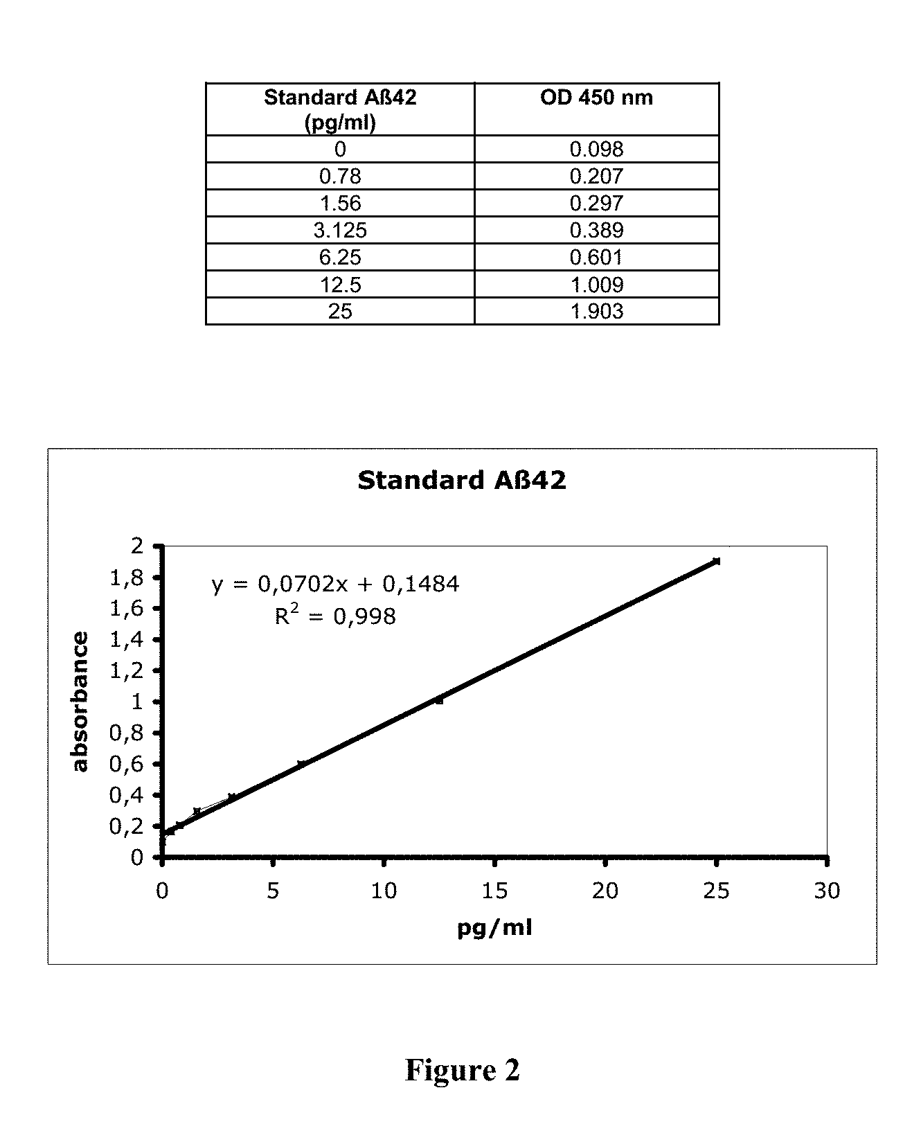
[0043] FIG. 2 shows the standard titration curve of the ELISA sandwich assay wherein the absorbance values at 450 nm are plotted against known concentrations of standard preparations of A.beta.42 (in pg/mL).
DETAILED DESCRIPTION OF THE INVENTION
[0044] The authors of the present invention have surprisingly found that, by using a kit having the components as defined in claim 1, it is possible to determine the levels of A.beta.40 and A.beta.42 with a lower detection limit of less than 0.1 pg/ml. This kit allows a reliable quantification of said molecular species in any sample in any subject and, in particular, in the plasma from subjects suspected of suffering sporadic AD, wherein the plasma concentrations are so low that no reliable measurement has been possible up to date. Accordingly, in a first aspect, the invention relates to a kit for the determination or the detection of a target polypeptide selected from the group of A.beta.42, A.beta.40 and a mixture thereof comprising [0045] (i) a first antibody or combination of antibodies which recognise said target polypeptide [0046] (ii) a second antibody or combination of antibodies which recognise a different region of the target polypeptide than the region recognised by the first antibody or combination of antibodies [0047] (iii) a reagent showing affinity for the second antibody which is coupled to a first member of a binding pair and [0048] (iv) a second member of a binding pair coupled to a detectable tag.
[0049] Without wishing to be bound by any particular theory, it is believed that the enhanced sensitivity is due to the use of the reagent (iii) which allows the signal resulting from the detection antibody to be amplified.
[0050] "A.beta.42", as used herein, relates to a 42 amino acids peptide corresponding to amino acids 672 to 713 (SEQ ID NO:4) and which is produced by the sequential proteolytic cleavage of the amyloid precursor protein (SEQ ID NO:6) by the .beta.- and .gamma.-secretases.
[0051] "A.beta.40", as used herein, relates to a 40 amino acids peptide corresponding to amino acids 672 to 711 (SEQ ID NO:5) and which is produced by the sequential proteolytic cleavage of the amyloid precursor protein (SEQ ID NO:6) by the .beta.- and .gamma.-secretases.
[0052] In the context of the present invention, the first antibody will be referred to as "capture antibody", meaning that this antibody is used to retrieve from a sample all molecular species to which the antibody specifically binds. There is practically no limitation with regard to the type of antibody that can be used as capture antibody as long as it contains at least one antigen binding site specific for A.beta.40 and/or A.beta.42. Therefore, antibodies molecules suitable for use as capture antibodies include [0053] "intact" antibodies which comprise an antigen-binding variable region as well as a light chain constant domain (CL) and heavy chain constant domains, CH1, CH2 and CH3, [0054] "Fab" fragments resulting from the papain digestion of an intact antibody and which comprise a single antigen-binding site and a CL and a CH1 region, [0055] "F(ab').sub.2" fragments resulting from pepsin digestion of an intact antibody and which contain two antigen-binding sites, [0056] "Fab'" fragments contain the constant domain of the light chain and the first constant domain (CH1) of the heavy chain and has one antigen-binding site only. Fab' fragments differ from Fab fragments by the addition of a few residues at the carboxy terminus of the heavy chain CH 1 domain including one or more cysteines from the antibody hinge region. [0057] "Fv" is the minimum antibody fragment which contains a complete antigen-recognition and antigen-binding site. This region consists of a dimer of one heavy chain and one light chain variable domain in tight, non-covalent-association. It is in this configuration that the three hypervariable regions (CDRs) of each variable domain interact to define an antigen-binding site on the surface of the VH-VL dimer. Collectively, the six hypervariable regions confer antigen-binding specificity to the antibody. However, even a single variable domain (or half of an Fv comprising only three hypervariable regions specific for an antigen) has the ability to recognize and bind antigen, although at a lower affinity than the entire binding site. [0058] Single-chain FV or "scFv" antibody fragments comprise the VL and VH, domains of antibody, wherein these domains are present in a single polypeptide chain. Preferably, the VL and VH regions are connected by a polypeptide linker which enables the scFv to form the desired structure for antigen binding. [0059] "Diabodies" comprise a heavy chain variable domain (VH) connected to a light chain variable domain (VL) on the same polypeptide chain (VH-VL) connected by a peptide linker that is too short to allow pairing between the two domains on the same chain. This forces pairing with the complementary domains of another chain and promotes the assembly of a dimeric molecule with two functional antigen binding sites. [0060] "Bispecific antibodies" (BAbs) are single, divalent antibodies (or immunotherapeutically effective fragments thereof) which have two differently specific antigen binding sites. The two antigen sites may be coupled together chemically or by genetic engineering methods known in the art.
[0061] All these antibody fragments can be further modified using conventional techniques known in the art, for example, by using amino acid deletion(s), insertion(s), substitution(s), addition(s), and/or recombination (and/or any other modification(s) (e.g. posttranslational and chemical modifications, such as glycosylation and phosphorylation) known in the art either alone or in combination. Methods for introducing such modifications in the DNA sequence underlying the amino acid sequence of an immunoglobulin chain are well known to the person skilled in the art; see, e.g., Sambrook et al.; Molecular Cloning: A Laboratory Manual; Cold Spring Harbor Laboratory Press, 2.sup.nd edition 1989 and 3rd edition 2001.
[0062] Antibodies suitable as capture antibodies include both polyclonal and monoclonal antibodies. For the production of polyclonal antibodies, various hosts including goats, rabbits, rats, mice, camels, dromedaries, llamas, humans, birds and others may be immunized by injection with a peptide corresponding to a fragment of A.beta.40 or A.beta.42 which has immunogenic properties. Depending on the host species, various adjuvants may be used to increase immunological response. Such adjuvants include, but are not limited to, Freund's, mineral gels such as aluminium hydroxide, and surface active substances such as lysolecithin, polyanions, peptides, oil emulsions, KLH, and dinitrophenol. Among adjuvants used in humans, BCG (bacilli Calmette-Guerin) and Corynebacterium parvum are especially preferable. If the antigen is a peptide, it may be useful to conjugate it to a protein that is immunogenic in the species to be immunized. For example, the antigen can be conjugated to keyhole limpet hemocyanin (KLH), Blue Carrier (hemocyanin isolated from Concholepas concholepas), bovine thyroglobulin, or soybean trypsin inhibitor, using a bifunctional or derivatizing agent, e.g., maleimidobenzoyl sulfosuccinimide ester (conjugation through cysteine residues), N-hydroxysuccinimide (through lysine residues), glutaraldehyde, succinic anhydride or SOCl.sub.2.
[0063] For the production of monoclonal antibodies, conventional techniques can be used. For instance, monoclonal antibodies may be made using the hybridoma method first described by Kohler et al., Nature, 256:495 (1975) using the procedure described in detail in units 11.4 to 11.11 of Ausubel, F. M. et al. (Current Protocols in Molecular Biology, John Wiley & Sons Inc; ring-bound edition, 2003). Alternatively, monoclonal antibodies can be isolated by recombinant DNA procedures from antibody phage libraries generated using the techniques described in McCafferty et al., Nature, 348:552-554 (1990). Clacksoii et al., Nature, 352:624-628 (1991) and Marks et al., J. Mol. Biol., 222:581-597 (1991) describe the isolation of murine and human antibodies, respectively, using phage libraries. Subsequent publications describe the production of high affinity (nM range) human antibodies by chain shuffling (Marks et al., Bio/Technology, 10:779-783 (1992)), as well as combinatorial infection and in vivo recombination as a strategy for constructing very large phage libraries (Waterhouse et al., Nucl. Acids. Res., 21:2265-2266 (1993)). Thus, these techniques are viable alternatives to traditional monoclonal antibody hybridoma techniques for isolation of monoclonal antibodies.
[0064] Polyclonal antibodies can be used directly as an antiserum obtained from immunised hosts after bleeding and removal of the fibrin clot. Monoclonal antibodies can be used directly as the supernatant of the hybridoma culture or as ascites fluid after implantation of the hybridoma in the peritoneal cavity of a suitable host. Alternatively, the immunoglobulin molecules, either polyclonal or monoclonal, can be purified prior to their use by conventional means such as affinity purification using peptides derived from A.beta.40 or A.beta.42, non-denaturing gel purification, HPLC or RP-HPLC, size exclusion, purification on protein A column, or any combination of these techniques.
[0065] In a preferred embodiment, the capture antibodies recognise a region common to A.beta.40 and A.beta.42, so as to allow simultaneous capture of both species with a single antibody species. In principle, any antibody specific for a region common to the sequences of both A.beta.40 and A.beta.42 can be used as capture antibody. Preferred epitopes which can be targeted by the capture antibody include epitopes located within amino acids 1 to 16, 1 to 17, 13 to 28, 15 to 24, 1 to 5 and 1 to 11 of A.beta.40 or A.beta.42. In a more preferred embodiment, the capture antibody is directed against a region in A.beta.40 and/or A.beta.42 which is different from the C-terminal region. In a still more preferred embodiment, the capture antibody is directed against an epitope of the N-terminal region of the A.beta.40 and A.beta.42 peptides. In yet another preferred embodiment, the capture antibody is a monoclonal antibody. In a still more preferred embodiment, the capture antibody recognises a region corresponding to amino acids 1-16 of the A.beta. peptides. In a still more preferred embodiment, the monoclonal antibody used as capture antibody is the 6E10 mAb as has been described in Kim, K. S. (Neuroscience Res. Comm. 1988, 2:121-130).
[0066] The second component of the kit of the invention corresponds to an antibody or combination of antibodies which recognise a different region of the target polypeptide than the region recognised by the first antibody or combination of antibodies. In the context of the present invention, the second antibody will be referred to as "detection antibody", since this antibody will be used to detect the amount of antigen which has been retained by the capture antibody.
[0067] As with the capture antibody, there is practically no limitation with regard to the type of antibody that can be used as detection antibody. However, it will be also understood by the person skilled in the art that the detection antibody (i) must bind to a region of the antigen which is not covered by the capture antibody and (ii) must contain not only the antigen binding site but also an additional region or regions that can be specifically detected by a reagent showing high affinity binding for said antibody, so as to allow detection of the antibody which is bound to the antigen captured by the capture antibody. Preferably, said additional regions which can be specifically bound by said reagent correspond to the constant region of the immunoglobulin molecule.
[0068] Suitable detection antibodies include intact antibodies, Fab, F(ab')2, Fab' and Fv fragments, single-chain FV antibodies, diabodies, bispecific antibodies and the like, wherein these compounds are as defined previously. Similarly as with the case of the capture antibody, the detection antibody can be polyclonal or monoclonal, native or post-translationally modified and pure or enriched in the antigen-binding molecules, using the same procedures as described for the capture antibodies. It will be appreciated by the skilled person that, in order to use the detection antibody, it must be diluted to a suitable working concentration and that said dilution can be routinely determined for each batch of antibody. Moreover, it will be evident that the adequate working dilution will differ depending on whether the antisera is used or rather a purified IgG fraction.
[0069] Typical dilutions are 1/1000, 1/2000, 1/3000, 1/4000, 1/5000, 1/6000, 1/7000, 1/8000, 1/9000, 1/10000 and the like.
[0070] In a preferred embodiment, the detection antibodies comprises any polyclonal antibody selected from the group of [0071] (i) a polyclonal antibody prepared against a peptide corresponding to the C-terminal region of the A.beta.42 peptide which binds specifically to A.beta.42 without giving any substantial cross-reaction with A.beta.40 or A.beta.43 [0072] (ii) a polyclonal antibody prepared against a peptide corresponding to the C-terminal region of the A.beta.40 peptide which binds specifically to A.beta.40 without giving any substantial cross-reaction with A.beta.42, A.beta.39, A.beta.38, A.beta.41 or A.beta.43 [0073] (iii) an antibody that recognises simultaneously the C-terminal region of both A.beta.40 and A.beta.42 or [0074] (iv) a combination of the antibodies under (i) and (ii). In a more preferred embodiment, the peptide corresponding to the C-terminal region of the A.beta.42 peptide used to prepare the A.beta.42-specific antibody is a peptide as defined in SEQ ID NO:1 or SEQ ID NO:2. In another preferred embodiment, the peptide corresponding to the C-terminal region of the A.beta.40 peptide used to prepare the A.beta.40-specific antibody is a peptide as defined in SEQ ID NO:3. Antibodies specific for A.beta.40 and A.beta.42 and methods for their preparation have been described in detail in WO2004024770 and WO2004098631, whose contents are incorporated herein by reference.
[0075] It will be understood to a person of ordinary skilled in the art that the method can be used for detecting A.beta.40, A.beta.42 or both species simultaneously depending on the type of capture and detection antibodies used in the method.
[0076] In order to detect or determine specifically A.beta.40, the capture antibody can be an antibody which recognises the N-terminal region of A.beta.40 (and also of A.beta.42, since both peptides have identical N-terminal regions) and the detection antibody can be an antibody which recognises specifically the C-terminal region of A.beta.40 without giving any cross-reaction with A.beta.42. Alternatively, A.beta.40 can be specifically detected using a capture antibody which recognises the C-terminal region of A.beta.40 without giving any cross-reaction with A.beta.42 and a detection antibody which recognises a region of A.beta.40 which is common to both A.beta.40 and A.beta.42, preferably the N-terminal region of A.beta.42/A.beta.40.
[0077] In order to detect or determine specifically A.beta.42, the capture antibody can be an antibody which recognises the N-terminal region of A.beta.42 (and also of A.beta.40, since both peptides have identical N-terminal regions) and the detection antibody can be an antibody which recognises specifically the C-terminal region of A.beta.42 without giving any cross-reaction with A.beta.40. Alternatively, A.beta.42 can be specifically detected using a capture antibody which recognises the C-terminal region of A.beta.42 and a detection antibody which recognises a region of A.beta.42 which is common to both A.beta.42 and A.beta.40.
[0078] In order to detect or determine simultaneously A.beta.42 and A.beta.40, the capture antibody can be an antibody which recognises the N-terminal region common to A.beta.42 and A.beta.40 and the detection antibody can be a combination of at least two antibodies, wherein the first antibody recognises specifically the C-terminal region of A.beta.42 without giving any cross-reaction with A.beta.40 and the second antibody recognises specifically the C-terminal region of A.beta.40 without giving any cross-reaction with A.beta.42. Alternatively, capture antibody can be an antibody which recognises the N-terminal region common to A.beta.42 and A.beta.40 and the detection antibody can be an antibody that recognises the C-terminal region of both A.beta.40 and A.beta.42. Alternatively, A.beta.42 and A.beta.40 can be simultaneously detected using as capture antibody a mixture of at least two antibodies comprising a first antibody which recognises specifically the C-terminal region of A.beta.42 without giving any cross-reaction with A.beta.40 and a second antibody which recognises specifically the C-terminal region of A.beta.40 without giving any cross-reaction with A.beta.42 and a detection antibody which recognises the N-terminal region common to both A.beta.42 and A.beta.40. Alternatively, A.beta.42 and A.beta.40 can be simultaneously detected using as capture antibody an antibody which recognises the C-terminal region of both and A.beta.42 and a detection antibody which recognises the N-terminal region common to both A.beta.42 and A.beta.40.
[0079] In a preferred embodiment, the first and/or second antibodies or combination of antibodies have been affinity-purified using a polypeptide which comprises the sequence of the polypeptide used for their preparation.
[0080] The third element of the kit corresponds to a reagent which shows affinity for the detection antibody and which is coupled to a first member of a binding pair. The antibody-binding reagent may non-covalently bind to a particular type(s), a particular class(es) and/or a particular subclass(es) of an antibody or antibody fragments. Alternatively, the antibody-binding reagent may non-covalently bind to an antibody specific for a particular antigen. In certain embodiments, the antibody-binding reagent binds non-covalently to the Fc region or to the F(ab) region of the detection antibody. Preferred antibody-binding reagents include protein A, protein G, protein V, protein L, an anti-Fc antibody or antibody-binding fragment and an Fc receptor (FcR) or an antibody-binding fragment thereof. Non-limiting examples of antibodies which can be non-covalently bound to the detection antibody include monoclonal antibodies, polyclonal antibodies, multispecific antibodies, human antibodies, humanized antibodies, chimeric antibodies, single domain antibodies, single chain Fvs (scFv) single chain antibodies, Fab fragments, F(ab') fragments, disulfide-linked Fvs (sdFv), intrabodies, and anti-idiotypic (anti-Id) antibodies, and epitope-binding fragments of any of the above. Non-limiting examples of Fc receptors include Fc.gamma.RI, Fc.gamma.RIIA, Fc.gamma.RIIB, Fc.gamma.RIIC, Fc.gamma.RIIIA.alpha., Fc.gamma.RIIIB, Fc.epsilon.RI.alpha., Fc.epsilon.RI.xi. and Fc.gamma.RIIIA.xi..
[0081] The fourth element of the kit of the invention corresponds to a second member of a binding pair which is coupled to a detectable tag. Suitable binding pairs include [0082] hapten or antigen/antibody, e.g. digoxin and anti-digoxin antibodies [0083] biotin or biotin analogues (e.g. aminobiotin, iminobiotin or desthiobiotin)/avidin or streptavidin, [0084] sugar/lectin, [0085] enzyme and cofactor [0086] folic acid/folate [0087] double stranded oligonucleotides that selectively bind to proteins/, transcription factors. [0088] nucleic acid or nucleic acid analogue/complementary nucleic acid, [0089] receptor/ligand, e.g., steroid hormone receptor/steroid hormone.
[0090] It will be understood that the term "first" and "second" member of a binding pair is relative and that each of the above members can be seen as first or second members of the binding pair. In a preferred embodiment, the first member of a binding pair is biotin or a functionally equivalent variant thereof and the second member of the binding pair is avidin, streptavidin or a functionally equivalent variant thereof.
[0091] In a preferred embodiment, the second member of the binding pair is streptavidin.
[0092] Suitable detectable tags include, without limitation, fluorescent moieties (e.g., fluorescein, rhodamine, phycoerythrin, coumarin, oxazine, resorufin, cyanine and derivatives thereof.), luminescent moieties (e.g., Qdot.TM. nanoparticles supplied by the Quantum Dot Corporation, Palo Alto, Calif.). If the detectable tag is an enzyme, then this enzyme must be capable of generating a detectable signal, for example, upon addition of an activator, substrate, amplifying agent and the like. Enzymes which are suitable as detectable tags for the present invention and the corresponding substrates include: [0093] Alkaline phosphatase: [0094] Chromogenic substrates: Substrates based on p-nitrophenyl phosphate (p-NPP), 5-bromo-4-chloro-3-indolyl phosphate/nitroblue tetrazolium (BCIP/NPT), Fast-Red/naphthol-AS-TS phosphate [0095] Fluorogenic substrates: 4-methylumbelliferyl phosphate (4-MUP), 2-(5'-chloro-2'-phosphoryloxyphenyl)-6-chloro-4-(3H)-quinazolinone (CPPCQ), 3,6-fluorescein diphosphate (3,6-FDP), Fast Blue BB, Fast Red TR, or Fast Red Violet LB diazonium salts [0096] Peroxidases: [0097] Chromogenic substrates based on 2,2-azinobis(3-ethylbenzothiazoline-6-sulfonic acid) (ABTS), o-phenylenediamine (OPT), 3,3',5,5'-tetramethylbenzidine (TMB), o-dianisidine, 5-aminosalicylic acid, 3-dimethylaminobenzoic acid (DMAB) and 3-methyl-2-benzothiazolinehydrazone (MBTH), 3-amino-9-ethylcarbazole (AEC)- and 3,3'-diaminobenzidine tetrahydrochloride (DAB). [0098] Fluorogenic substrates: 4-hydroxy-3-methoxyphenylacetic acid, reduced phenoxazines and reduced benzothiazines, including Amplex.RTM. Red reagent, Amplex UltraRed and reduced dihydroxanthenes. [0099] Glycosidases: [0100] Chromogenic substrates: o-nitrophenyl-.beta.-D-galactoside (o-NPG), p-nitrophenyl-.beta.-D-galactoside and 4-methylumbelliphenyl-.beta.-D-galactoside (MUG) for .beta.-D-galactosidase. [0101] Fluorogenic substrates: resorufin .beta.-D-galactopyranoside, fluorescein digalactoside (FDG), fluorescein diglucuronide, 4-methylumbelliferyl .beta.-D-galactopyranoside, carboxyumbelliferyl .beta.-D-galactopyranoside and fluorinated coumarin (.beta.-D-galactopyranosides. [0102] Oxidoreductases (luciferase): [0103] Luminescent substrates: luciferin.
[0104] In a preferred embodiment, the detectable tag is horseradish peroxidase and the detection reagent is TMB. In a preferred embodiment, the kit further comprises a solid support. As used herein, the term "support" or "surface" refers to a solid phase which is a porous or non-porous water insoluble material that can have any one of a number of shapes, such as strip, rod, particles, including latex particles, magnetic particles, microparticles, beads, membranes, microtiter wells and plastic tubes. In principle, any material is suitable as solid support provided that is able to bind sufficient amounts of the capturing antibody. Thus, the choice of solid phase material is determined based upon desired assay format performance characteristics. Materials suitable for the solid support include polymeric materials, particularly cellulosic materials and materials derived from cellulose, such as fibre containing papers, e.g., filter paper, chromatographic paper, glass fiber paper, etc.; synthetic or modified naturally occurring polymers, such as nitrocellulose, cellulose acetate, poly (vinyl chloride), polyacrylamide, cross linked dextrane, agarose, polyacrylate, polyethylene, polypropylene, poly(4-methylbutene), polystyrene, polymethacrylate, poly(ethylene terephthalate), nylon, polyvinyl butyrate), etc.; either used by themselves or in conjunction with other materials; glass such as, e.g., glass available as bioglass, ceramics, metals, and the like. Non-crosslinked polymers of styrene and carboxylated styrene or styrene functionalized with other active groups such as amino, hydroxyl, halo and the like are preferred. In some instances, copolymers of substituted styrenes with dienes such as butadiene will be used.
[0105] The solid support and the capture antibody may be separately provided in the kit or, alternatively, the support may be delivered already precoated with the capture antibody. In this case, the support may have been treated with a blocking solution after the binding of the capture antibody. If the support is precoated, it is preferred that the support is treated with a concentrated trehalose solution and allowed to dry, in which case the dry trehalose forms a halo on the support. These supports containing the dry trehalose are exceptionably stable and can be stored for up to two years when kept at 4.degree. C. in the dark.
[0106] Additional components of the kit may include: [0107] Means for removing from the patient the sample to be analysed. [0108] Buffers and solutions required for preparing the standard curves of the target peptides. [0109] Buffers and solutions for washing and blocking the solid support during the assay [0110] Buffers and solutions for coating the solid support with the coating antibody [0111] Reagents for developing the coloured or fluorogenic signal from the detectable tag. [0112] Reagents for stopping the formation of the coloured or fluorogenic product from the detectable tag (e.g. 1N H.sub.2SO.sub.4) [0113] Means for maintaining the peptides in an unfolded state (e.g. concentrated guanidinium hydrochloride). [0114] A sample containing a stock solution of the A.beta.40 or A.beta.42 peptides or a combination thereof.
[0115] In a preferred embodiment, the capture antibody is immobilised onto a solid support. The immobilisation can be carried out prior to the binding of the target polypeptide to be detected or once the peptide/protein is bound to the capture antibody. In any, case, if a solid support is used, it is convenient to block the excess of protein binding sites on the carrier prior to the addition of the sample containing the target polypeptide to be determined. Preferably, blocking or quenching of the peptide-binding sites on the support is carried out using the same buffer which is used for washing the complexes after each binding reaction (e.g. 50 mM Tris-HCl, pH 8, PBS or TBS optionally, comprising Tween 20) supplemented with a macromolecular compound (e.g. bovine serum albumin, non-fat dry milk, western blocking reagent, caseine, lactoalbumine, ovoalbumine) in concentrations from about 0.05% to 10%, preferably 1 to 5%, more preferably around 3%. If the support comprising the immobilised capture antibody must be stored for some time, it is preferred that the support is treated with a concentrated trehalose solution and allowed to dry, in which case the dry trehalose forms a halo on the support. These supports containing the dry trehalose are exceptionably stable and can be stored up to two years when kept at 4.degree. C. in the dark.
[0116] The kits of the invention allow the detection or determination with high sensitivity of the polypeptides which are specifically recognised by the first and second antibody components of the kit. Thus, in a further aspect, the invention relates to the use of a kit of the invention to detect a protein or protein in a sample. In a preferred embodiment, the kit is used to detect a peptide selected from the group of A.beta.40, A.beta.42 and the combination thereof in a sample.
[0117] A "sample", as understood in the present invention, includes any one of tissue culture, plasma, serum, saliva, semen, sputum, cerebral spinal fluid (CSF), tears, mucus, sweat, milk, brain extracts and the like. In a preferred embodiment, the sample is a plasma sample. In view of the ability of the kit of the invention to provide high sensitivity determination of the concentrations of A.beta.40 and A.beta.42 in any sample, it can be used for the diagnosis of any disease wherein there is an altered concentration of any of these two peptides in any cell fluid or tissue, in particular, degenerative diseases and, more particularly, neurodegenerative diseases. Non limitative examples of degenerative diseases which may be diagnosed based on the appearance of altered levels of A.beta.40 and/or A.beta.42 include: [0118] bone degenerative disorders such as osteopenia, osteomalacia, osteoporosis, osteomyeloma, osteodystrophy, Paget's disease, osteogenesis imperfecta, bone sclerosis, aplastic bone disorder, humoral hypercalcemic myeloma, multiple myeloma and bone thinning following metastasis. [0119] cartilage degenerative disorders such as Gorham-Stout syndrome; arthritic diseases; osteoarthritis; rheumatoid arthritis; psoriatic arthritis; rheumatoid disease; and brittle bone disease. [0120] muscle degenerative diseases such as muscular dystrophy, muscle atrophy, congestive obstructive pulmonary disease, muscle wasting syndrome, sarcopenia, cachexia. [0121] heart degenerative diseases including cardiac cell death due to ischemia, tissue and organ death due to transplant rejection, hearing loss due to autotoxicity. [0122] retinal degenerative disorders such as retinitis pigmentosa [0123] degenerative diseases of the nervous system such as Alexander disease, Alper's disease, Alzheimer's disease, Amyotrophic lateral sclerosis, Ataxia telangiectasia, Batten disease, Bovine spongiform encephalopathy (BSE), Canavan disease, Cockayne syndrome, Corticobasal degeneration, Creutzfeldt-Jakob disease, Huntington disease, HIV-associated dementia, Kennedy's disease, Krabbe disease, Lewy body dementia, Machado-Joseph disease (Spinocerebellar ataxia type 3), Multiple sclerosis, Multiple System Atrophy, Neuroborreliosis, Parkinson disease, Pelizaeus-Merzbacher Disease, Pick's disease, Primary lateral sclerosis, Prion diseases, Refsum's disease, Sandhoff disease, Schilder's disease, Schizophrenia, Spielmeyer-Vogt-Sjogren-Batten disease (also known as Batten disease), Spinocerebellar ataxia, spinal muscular atrophy, Steele-Richardson-Olszewski disease, Tabes dorsalis. In a preferred embodiment, the neurodegenerative disorder that is diagnosed using the kit of the invention is Alzheimer's disease.
[0124] It will be appreciated that the parameter which might be required to decide about a possible disease is not only absolute A.beta.40 and A.beta.42 concentrations, but also parameters derived from the combination of said values, such as the ratios A.beta.40/A.beta.42 or A.beta.42/A.beta.40, the percentages of A.beta.42 and A.beta.40 over the total A.beta. peptides or the addition of the concentrations of A.beta.42 and A.beta.40.
[0125] In a preferred embodiment, the disease that can be diagnosed is Alzheimer's disease, more particularly, sporadic AD, based on the appearance in a sample of an AD patient of lower concentrations of A.beta.40 and/or A.beta.42 peptides than the amounts of said peptide or peptides in a biological sample from the same origin obtained from a healthy individual.
[0126] In use, the kit of the invention allows to carry out a five step method for detecting the amount of a target polypeptide selected from the group of A.beta.42, A.beta.40 and the combination thereof in a sample. Said method, which is another object of the present invention, comprises the steps of [0127] (i) capturing the target polypeptide present in the sample with a first antibody or combination of antibodies which bind specifically said target polypeptide, [0128] (ii) contacting the immune complexes formed in step (a) with a second antibody or combination of antibodies which recognise a different region of the target polypeptide than the first antibody or combination of antibodies, [0129] (iii) contacting the complexes formed in step (ii) with a reagent which shows affinity for the second antibody and which is coupled to a first member of a binding pair, [0130] (iv) contacting the complexes formed in step (iii) with a second member of a binding pair which is coupled to a detectable tag and [0131] (v) detecting the activity of the enzyme or the fluorescent emission of the compound attached to the second member of the binding pair.
[0132] In a preferred embodiment, the peptides which are detected correspond to non-oligomeric forms of said peptide, more preferably, monomeric forms of either A.beta.40 and A.beta.42.
[0133] The reagents used in each steps of the method have been described in detail above.
[0134] In the first step of the method according to the present invention, the sample which contains A.beta.40 and/or A.beta.42 peptides is contacted with a first antibody so as to form a first immune complex.
[0135] After the first binding step has been carried out, the complexes can be washed to remove any excess of protein/peptide found in the original sample which did not bind to the capture antibody. Preferred washing buffers that can be used in the context of the present invention include any buffer at a pH close to physiological (e.g. 50 mM Tris-HCl) optionally comprising salts (e.g. 150 mM NaCl) and optionally comprising low concentrations of a detergent (e.g. 0.05% Tween-20).
[0136] In a second step, the complexes formed between the capture antibody and the A.beta. peptide or peptides in the sample are then contacted with the second antibody so as to form a "sandwich-type" immune complex.
[0137] After the second step is carried out, the immune complex can be washed to eliminate unspecifically-bound antibodies using essentially the same buffers and procedures as described before.
[0138] In the third step, the method of the invention involves contacting the complexes formed between the captured peptide or peptides and the detection antibody with a reagent which shows affinity for the detection antibody and which is coupled to a first member of a binding pair.
[0139] In a fourth step, the method of the invention involves contacting the complexes formed between the antibody-binding reagent and the detection antibody with a second member of a binding pair which is coupled to a detectable tag. In the fifth step of the method according to the invention, the method involves detecting the detectable tag. In the fifth step of the method according to the invention, the method involves detecting the detectable tag. It will be understood that the detection and/or for quantification of the detectable tag depend on the nature of the tag and are known in the art. When an intact substrate or detectable tag contains a luminescent or dye component, detection can be by visual observation on a UV transiluminator, or by using a UV-based charged coupled device (CCD) camera detection system, a laser-based gel scanner, a xenon-arc-based CCD camera detection system, a Polaroid camera combined with a UV-transiluminator as well as a variety of other devices used for detecting luminescence. When the detectable tag is an enzyme, the fifth step of the method according to the invention involves exposing the immunocomplexes labelled with the tag (e.g., the captured peptide, the detection antibody and the reagent labelled with the detectable tag) to activators, substrates, or amplifying agents of the enzyme used as detectable tag. Well known detectable tags capable of generating a detectable signal include enzyme-labeled antibodies. Exemplary enzymes well known for this purpose include horseradish peroxidase, alkaline phosphatase and glycosidases, including 3-galactosidase, 0-glucosidase and 1-glucuronidase. As an example, a reagent which specifically binds to the detection antibody can be tagged with horseradish peroxidase. Upon formation of a capture moiety-detection antibody-reagent complex, detection can then be performed using any of a wide range of well known substrates for the enzyme used as detectable tags.
[0140] In another aspect, the invention relates to a method for the diagnosis of a neurodegenerative disease which comprises measuring the levels of A.beta.40 and/or A.beta.42 in a sample of the subject suspected of carrying said disease and correlating the concentration of one and/or both peptides in the sample o said subject with respect to the concentration of said peptide or peptides in a sample from a healthy individual with the appearance of the neurodegenerative disorder.
[0141] In a preferred embodiment, the neurodegenerative disease is Alzheimer's disease wherein if the amount of peptides A.beta.40, A.beta.42 or a combination thereof in said simple is lower than the amount of said peptide or peptides in a sample from a healthy individual is indicative that the subject suffers from Alzheimer's disease. Preferably, the determination is carried out in plasma or serum.
[0142] It is preferred to carry out the different steps of the method using in parallel the samples to be determined and a number of samples having increasing and known concentrations of the compound which is to be determined. If A.beta.40 and/or A.beta.42 are to be determined, a standard curve for each peptide must be prepared using increasing concentrations. The standard curve serves the dual purpose of (i) establishing the concentration range wherein the signal increases linearly with the concentration of the target peptide and (ii) determining the concentration of the peptides in the test sample by interpolation of the signal obtained with the test samples in the curve to obtain concentration values. In view of the high sensitivity of the assay of the invention, preferred concentrations of the test samples are e.g. 3.125; 6.25; 12.5; 25; 50; 100 and 200 pg/mL. It will be appreciated that the concentrations of the samples used for obtaining the standard curve will vary for each test substrate. However, determination of the linear range of the assay can be easily determined by the skilled practitioner by conventional means. In view of the higher amounts of A.beta.42 and A.beta.40 peptides in CSF samples as well as in serum samples of patients with familiar forms of AD as compared to serum from patients with sporadic AD, it is preferred that, if the kit of the invention is used for determining A.beta. peptides in samples from CSF or serum from patients with familiar AD, these be diluted so that the final concentrations of A.beta.40/A.beta.42 are found within the range falls within the linear side of the assay.
[0143] The following examples are provided as illustration and should not be construed as limiting the scope of the invention.
EXAMPLES
Example 1
Colorimetric ELISA Sandwich with Biotin-Streptavidin Amplification
[0144] In order to increase the sensitivity, the signal can be amplified using biotin-streptavidin. The plate was coated using the 6E10 mAb capture antibody which recognises amino acids 1-17 in both the amyloid A 40 and in the amyloid A 42 peptide. The coating was carried out at a concentration of 5 .mu.g/ml in 100 mM carbonate/bicarbonate buffer, pH=9.6, overnight at 4.degree. C. The plate was then blocked with 300 .mu.l of a blocking solution (50 mM Tris-HCl, pH 8, 0.2% Tween-20, 0.5% BSA) for 3 h at room temperature with shaking or for 2 h at 37.degree. C. When needed, the plates can be treated, after blocking, with 100 .mu.l of a 50 mM Tris-HCl pH 8 solution containing 20 mg/ml trehalose. The plates were left to evaporate until a white halo characteristic of trehalose appears. The plates so treated could be kept at 4.degree. C. covered with aluminium foil and are stable for two years.
[0145] The samples of the standard curve were prepared from a 200 pg/ml stock solution of the peptides A 40 y A 42 on plates coated with the 6E10 mAb and treated with trehalose. From these solutions, serial dilutions 1:2 in SDB were made so as to give concentrations of 200, 100, 50, 25, 12.5, 6.25 and 3.125 pg/ml. 100 .mu.l of each diluted or undiluted sample is added diluted or undiluted in SDB (1/1,000,000) and incubated overnight at 4.degree. C. (or for 2 h at 37.degree. C.).
[0146] Detection antibody (a polyclonal antibody prepared against a peptide corresponding to the C-terminal region of the A.beta.42 peptide or a polyclonal antibody prepared against a peptide corresponding to the C-terminal region of the A.beta.40 peptide, depending on whether A.beta.42 or A.beta.40 is to be detected) was added diluted in SDB. 100 .mu.l are added to each well and were then incubated for 1 h at room temperature.
[0147] Next, 100 .mu.l of a 1/5000 dilution in SDB of a biotin-labelled anti-rabbit IgG antibody (SIGMA) were then added and incubated for 1 h at room temperature with shaking. Then 100 .mu.l of a 1/4000 dilution in SDB of HRP-coupled Streptavidin (from SIGMA) were added to each well and incubated for 1 h at room temperature.
[0148] For developing the plate, 100 .mu.l of the chromogenic substrate TMB (ZEU Inmunotec). TMB was added and incubated in the dark during 15-30 minutes. As stop solution, 50 .mu.l of 1N H2SO4 were added per well. The absorbance at 450 nm was read in a plate reader Synergy HT (BioTek Instruments).
[0149] Between each of the steps, the plate was washed using an automatic plate washer (Elx50 Bio Tek Instruments) programmed for performing 5 rinses each time. The washing solution contained 50 mM de Tris-HCl pH 8, 0.05% Tween-20 and 150 mM NaCl (filtered before use).
Example 2
Fluorescent ELISA Sandwich Assay
[0150] The plate was coated with 6E10 in bicarbonate buffer (5 .mu.g/ml) overnight at 4.degree. C. The plate was then blocked 3 h at room temperature with shaking (300 .mu.l/well). The test and standard curve samples were then added to the plates and incubated overnight at 4.degree. C. A 1/4000 dilution of the detection antibody (anti-A 40 o anti-A 42 serum) was added to each well and incubated for 1 h at room temperature with shaking. Serial dilutions of the FITC-coupled anti-antibody (dilutions 1/1000, 1/5000, 1/10000) were added and incubated for 1 h at room temperature in the dark. The fluorescence was using an excitation wavelength of 485 nm and an emission wavelength of 528.
[0151] Alternatively, the assay is carried out using the Quanta-Blu (PIERCE) fluorescent substrate, which increases the sensitivity of the ELISA assay. Maximal excitation is 325 nm and maximal emission is 420 nm. It can be detected in the excitation range of 315-340 nm and 370-470 nm emission range. The QuantaBlu Working Solution is prepared by mixing 9 parts of QuantaBlu Substrate Solution with 1 part of QuantaBlu Stable Peroxidase Solution (solution stable for 24 h at room temperature). It can be incubated from 1.5 minutes to 90 minutes at room temperature and can be read stopping the reaction or without stopping (a blue colour is produced).
[0152] The plate is coated with 6E10 mAb in bicarbonate buffer (5 .mu.g/ml) overnight at 4.degree. C. and then blocked for 3 h at room temperature with shaking (300 .mu.l/well). Different standard curves then prepared with the following concentrations of A.beta.42 and A.beta.40 peptides: [0153] 1000, 500, 250, 125, 62.5, 31, 25 and 15.65 pg/mL [0154] 200, 100, 50, 25, 12.5, 6.25 and 3.125 pg/mL [0155] 25, 12.5, 6.25, 3.125, 1.56, 0.78 and 0.39 pg/mL [0156] 10, 5, 2.5, 1.25, 0.625, 0.3125 and 0.156 pg/mL [0157] 5, 2.5, 1.25, 0.625, 0.3125, 0.156 and 0.078 pg/mL [0158] 1, 0.5, 0.25, 0.125, 0.0625, 0.03125 and 0.0156 pg/mL
[0159] The detection antibody (anti-A 40 o anti-A 42 serum) is added (diluted at 1/4000) for 1 h at room temperature with shaking. The HRP-coupled anti-rabbit IgG 1/1000 is then added and incubated for 1 h at room temperature with shaking. For developing the reaction, 100 .mu.l of Quanta-Blue Working Solution and then incubated for 30', 60' and 90' at room temperature in the darkness. The fluorescence is then read (Excitation: 360/40 nm; Emission: 460/40 nm) at 30', 60' and 90' without stopping the reaction or stopping the reaction with STOP solution.
Example 3
Preparation of A.beta.40 and A.beta.42 Standard Curves
[0160] For the preparation of the A.beta.40 standard curve, a lyophilised sample of human A 40 was reconstituted to 10 .mu.g/mL. From the stock solution, the samples were prepared containing the following concentrations (in pg/mL): 25,000 pg/ml, 2,500 pg/ml, 25 pg/ml, 12.5 pg/ml, 6.25 pg/ml, 3.125 pg/ml, 1.56 pg/ml, 0.78 pg/ml. The samples were prepared in the presence of 1 mM of the protease inhibitor AEBSF. The samples were then processed according to the method defined in the previous examples. The results are shown in FIG. 1.
[0161] For the preparation of the A.beta.42 standard curve, a lyophilised sample of human A 42 was reconstituted to 10 .mu.g/mL. From the stock solution, samples were prepared containing the following concentrations (in pg/mL): 25,000 pg/ml, 2,500 pg/ml, 25 pg/ml, 12.5 pg/ml, 6.25 pg/ml, 3.125 pg/ml, 1.56 pg/ml, 0.78 pg/ml. The samples were prepared in the presence of 1 mM of the protease inhibitor AEBSF. The samples were then processed according to the method defined in the previous examples. The results are shown in FIG. 2.
Example 4
Correlation Between AD Diagnosis and A.beta.40/A.beta.42 Levels
[0162] A.beta.40 and A.beta.42 levels were determined using the ELISA sandwich assay described in the previous examples in plasma samples from a cohort of control subjects and from a cohort of patients diagnosed with AD using the minimental state examination (MMSE) score with a cut-off value of 24. The concentrations in pg/mL of A.beta.40 and A.beta.42 are shown in Table 1.
TABLE-US-00001 TABLE 1 AB40 AB42 AB40/AB42 AB42/AB40 AB40 + AB42 % AB40 % AB42 Healthy LSA 31.2 160.1 0.19 5.13 191.30 16.31 83.69 CPB 93.4 159.4 0.59 1.71 252.80 36.95 63.05 CFA 730.5 893.6 0.82 1.22 1624.10 44.98 55.02 VCL 145.0 329.6 0.44 2.27 474.60 30.55 69.45 FLA 430.1 19.6 21.94 0.05 449.70 95.64 4.36 ILS-7 102.9 34.2 3.01 0.33 137.05 75.08 24.92 MPG-8 57.0 59.2 0.96 1.04 116.17 49.07 50.93 PLA-17 29.6 8.7 3.39 0.30 38.27 77.21 22.79 ARL-24 34.0 25.8 1.32 0.76 59.82 56.89 43.11 BGM-28 23.4 7.4 3.16 0.32 30.77 75.95 24.05 AD BEG 12.0 71.5 0.17 5.96 83.50 14.37 85.63 CPG 74.3 136.7 0.54 1.84 211.00 35.21 64.79 VAC 26.7 34.7 0.77 1.30 61.40 43.49 56.51 MTF 55.6 141.7 0.39 2.55 197.30 28.18 71.82 CPG 34.2 28.0 1.22 0.82 62.24 54.95 45.05 MVA-5 17.2 21.9 0.79 1.27 39.05 43.99 56.01 JGS-40 13.6 16.6 0.82 1.22 30.18 45.10 54.90 PTG-35 37.3 24.8 1.50 0.66 62.11 60.07 39.93 CBC-30 11.6 6.1 1.90 0.53 17.63 65.51 34.49 JHC-2 25.4 23.4 1.09 0.92 48.84 52.07 47.93
[0163] Average values were calculated and the results are given in Table 2
TABLE-US-00002 TABLE 2 Healthy AD patients A.beta.40 167.70 30.78 (pg/mL) A.beta.42 169.75 50.53 (pg/mL)
Sequence CWU 1
1
6110PRTArtificial SequencePeptide used to prepare the anti-Abeta42-
specific polyclonal antibody 1Gly Leu Met Val Gly Gly Val Val Ile
Ala1 5 1028PRTArtificial SequencePeptide used to prepare the
anti-Abeta42- specific polyclonal antibody 2Met Val Gly Gly Val Val
Ile Ala1 538PRTArtificial SequencePeptide used to prepare the
anti-Abeta40- specific polyclonal antibody 3Gly Leu Met Val Gly Gly
Val Val1 5442PRTHomo sapiens 4Asp Ala Glu Phe Arg His Asp Ser Gly
Tyr Glu Val His His Gln Lys1 5 10 15Leu Val Phe Phe Ala Glu Asp Val
Gly Ser Asn Lys Gly Ala Ile Ile 20 25 30Gly Leu Met Val Gly Gly Val
Val Ile Ala 35 40540PRTHomo sapiens 5Asp Ala Glu Phe Arg His Asp
Ser Gly Tyr Glu Val His His Gln Lys1 5 10 15Leu Val Phe Phe Ala Glu
Asp Val Gly Ser Asn Lys Gly Ala Ile Ile 20 25 30Gly Leu Met Val Gly
Gly Val Val 35 406770PRTHomo sapiens 6Met Leu Pro Gly Leu Ala Leu
Leu Leu Leu Ala Ala Trp Thr Ala Arg1 5 10 15Ala Leu Glu Val Pro Thr
Asp Gly Asn Ala Gly Leu Leu Ala Glu Pro 20 25 30Gln Ile Ala Met Phe
Cys Gly Arg Leu Asn Met His Met Asn Val Gln 35 40 45Asn Gly Lys Trp
Asp Ser Asp Pro Ser Gly Thr Lys Thr Cys Ile Asp 50 55 60Thr Lys Glu
Gly Ile Leu Gln Tyr Cys Gln Glu Val Tyr Pro Glu Leu65 70 75 80Gln
Ile Thr Asn Val Val Glu Ala Asn Gln Pro Val Thr Ile Gln Asn 85 90
95Trp Cys Lys Arg Gly Arg Lys Gln Cys Lys Thr His Pro His Phe Val
100 105 110Ile Pro Tyr Arg Cys Leu Val Gly Glu Phe Val Ser Asp Ala
Leu Leu 115 120 125Val Pro Asp Lys Cys Lys Phe Leu His Gln Glu Arg
Met Asp Val Cys 130 135 140Glu Thr His Leu His Trp His Thr Val Ala
Lys Glu Thr Cys Ser Glu145 150 155 160Lys Ser Thr Asn Leu His Asp
Tyr Gly Met Leu Leu Pro Cys Gly Ile 165 170 175Asp Lys Phe Arg Gly
Val Glu Phe Val Cys Cys Pro Leu Ala Glu Glu 180 185 190Ser Asp Asn
Val Asp Ser Ala Asp Ala Glu Glu Asp Asp Ser Asp Val 195 200 205Trp
Trp Gly Gly Ala Asp Thr Asp Tyr Ala Asp Gly Ser Glu Asp Lys 210 215
220Val Val Glu Val Ala Glu Glu Glu Glu Val Ala Glu Val Glu Glu
Glu225 230 235 240Glu Ala Asp Asp Asp Glu Asp Asp Glu Asp Gly Asp
Glu Val Glu Glu 245 250 255Glu Ala Glu Glu Pro Tyr Glu Glu Ala Thr
Glu Arg Thr Thr Ser Ile 260 265 270Ala Thr Thr Thr Thr Thr Thr Thr
Glu Ser Val Glu Glu Val Val Arg 275 280 285Glu Val Cys Ser Glu Gln
Ala Glu Thr Gly Pro Cys Arg Ala Met Ile 290 295 300Ser Arg Trp Tyr
Phe Asp Val Thr Glu Gly Lys Cys Ala Pro Phe Phe305 310 315 320Tyr
Gly Gly Cys Gly Gly Asn Arg Asn Asn Phe Asp Thr Glu Glu Tyr 325 330
335Cys Met Ala Val Cys Gly Ser Ala Met Ser Gln Ser Leu Leu Lys Thr
340 345 350Thr Gln Glu Pro Leu Ala Arg Asp Pro Val Lys Leu Pro Thr
Thr Ala 355 360 365Ala Ser Thr Pro Asp Ala Val Asp Lys Tyr Leu Glu
Thr Pro Gly Asp 370 375 380Glu Asn Glu His Ala His Phe Gln Lys Ala
Lys Glu Arg Leu Glu Ala385 390 395 400Lys His Arg Glu Arg Met Ser
Gln Val Met Arg Glu Trp Glu Glu Ala 405 410 415Glu Arg Gln Ala Lys
Asn Leu Pro Lys Ala Asp Lys Lys Ala Val Ile 420 425 430Gln His Phe
Gln Glu Lys Val Glu Ser Leu Glu Gln Glu Ala Ala Asn 435 440 445Glu
Arg Gln Gln Leu Val Glu Thr His Met Ala Arg Val Glu Ala Met 450 455
460Leu Asn Asp Arg Arg Arg Leu Ala Leu Glu Asn Tyr Ile Thr Ala
Leu465 470 475 480Gln Ala Val Pro Pro Arg Pro Arg His Val Phe Asn
Met Leu Lys Lys 485 490 495Tyr Val Arg Ala Glu Gln Lys Asp Arg Gln
His Thr Leu Lys His Phe 500 505 510Glu His Val Arg Met Val Asp Pro
Lys Lys Ala Ala Gln Ile Arg Ser 515 520 525Gln Val Met Thr His Leu
Arg Val Ile Tyr Glu Arg Met Asn Gln Ser 530 535 540Leu Ser Leu Leu
Tyr Asn Val Pro Ala Val Ala Glu Glu Ile Gln Asp545 550 555 560Glu
Val Asp Glu Leu Leu Gln Lys Glu Gln Asn Tyr Ser Asp Asp Val 565 570
575Leu Ala Asn Met Ile Ser Glu Pro Arg Ile Ser Tyr Gly Asn Asp Ala
580 585 590Leu Met Pro Ser Leu Thr Glu Thr Lys Thr Thr Val Glu Leu
Leu Pro 595 600 605Val Asn Gly Glu Phe Ser Leu Asp Asp Leu Gln Pro
Trp His Ser Phe 610 615 620Gly Ala Asp Ser Val Pro Ala Asn Thr Glu
Asn Glu Val Glu Pro Val625 630 635 640Asp Ala Arg Pro Ala Ala Asp
Arg Gly Leu Thr Thr Arg Pro Gly Ser 645 650 655Gly Leu Thr Asn Ile
Lys Thr Glu Glu Ile Ser Glu Val Lys Met Asp 660 665 670Ala Glu Phe
Arg His Asp Ser Gly Tyr Glu Val His His Gln Lys Leu 675 680 685Val
Phe Phe Ala Glu Asp Val Gly Ser Asn Lys Gly Ala Ile Ile Gly 690 695
700Leu Met Val Gly Gly Val Val Ile Ala Thr Val Ile Val Ile Thr
Leu705 710 715 720Val Met Leu Lys Lys Lys Gln Tyr Thr Ser Ile His
His Gly Val Val 725 730 735Glu Val Asp Ala Ala Val Thr Pro Glu Glu
Arg His Leu Ser Lys Met 740 745 750Gln Gln Asn Gly Tyr Glu Asn Pro
Thr Tyr Lys Phe Phe Glu Gln Met 755 760 765Gln Asn 770
D00000
D00001
D00002
S00001
XML
uspto.report is an independent third-party trademark research tool that is not affiliated, endorsed, or sponsored by the United States Patent and Trademark Office (USPTO) or any other governmental organization. The information provided by uspto.report is based on publicly available data at the time of writing and is intended for informational purposes only.
While we strive to provide accurate and up-to-date information, we do not guarantee the accuracy, completeness, reliability, or suitability of the information displayed on this site. The use of this site is at your own risk. Any reliance you place on such information is therefore strictly at your own risk.
All official trademark data, including owner information, should be verified by visiting the official USPTO website at www.uspto.gov. This site is not intended to replace professional legal advice and should not be used as a substitute for consulting with a legal professional who is knowledgeable about trademark law.