METHODS FOR EXPANDING AND ACTIVATING yo T CELLS FOR THE TREATMENT OF CANCER AND RELATED MALIGNANCIES
Dao; Monique ; et al.
U.S. patent application number 16/200308 was filed with the patent office on 2019-06-13 for methods for expanding and activating yo t cells for the treatment of cancer and related malignancies. The applicant listed for this patent is Immatics US, Inc.. Invention is credited to Yannick Bulliard, Monique Dao, Melinda Mata, Aleksandra Nowicka, Steffen Walter.
| Application Number | 20190175650 16/200308 |
| Document ID | / |
| Family ID | 66442446 |
| Filed Date | 2019-06-13 |


View All Diagrams
| United States Patent Application | 20190175650 |
| Kind Code | A1 |
| Dao; Monique ; et al. | June 13, 2019 |
METHODS FOR EXPANDING AND ACTIVATING yo T CELLS FOR THE TREATMENT OF CANCER AND RELATED MALIGNANCIES
Abstract
The present disclosure relates to expansion and activation of T cells. In an aspect, the present disclosure relates to expansion and activation of .gamma..delta. T cells that may be used for transgene expression. In another aspect, the disclosure relates to expansion and activation of .gamma..delta. T cells while depleting .alpha.- and/or .beta.-TCR positive cells. T cell populations comprising expanded .gamma..delta. T cell and depleted or reduced .alpha.- and/or .beta.-TCR positive cells are also provided for by the instant disclosure. The disclosure further provides for methods of using the disclosed T cell populations.
| Inventors: | Dao; Monique; (Houston, TX) ; Walter; Steffen; (Houston, TX) ; Mata; Melinda; (Houston, TX) ; Nowicka; Aleksandra; (Houston, TX) ; Bulliard; Yannick; (Houston, TX) | ||||||||||
| Applicant: |
|
||||||||||
|---|---|---|---|---|---|---|---|---|---|---|---|
| Family ID: | 66442446 | ||||||||||
| Appl. No.: | 16/200308 | ||||||||||
| Filed: | November 26, 2018 |
Related U.S. Patent Documents
| Application Number | Filing Date | Patent Number | ||
|---|---|---|---|---|
| 62591041 | Nov 27, 2017 | |||
| Current U.S. Class: | 1/1 |
| Current CPC Class: | A61K 2035/122 20130101; C12N 15/8509 20130101; C07K 14/55 20130101; A61K 38/2013 20130101; C12N 5/0638 20130101; C12N 2501/2302 20130101; C12N 2501/515 20130101; C12N 5/0636 20130101; A61K 31/675 20130101; C12N 15/62 20130101; C12N 5/0018 20130101; C12N 2501/2315 20130101; A61K 31/663 20130101; C12N 2501/999 20130101; C12N 2501/155 20130101; A61K 35/17 20130101; C07K 16/2809 20130101; A61K 2035/124 20130101; C07K 14/5443 20130101; C12N 2740/10043 20130101; C12N 2501/998 20130101 |
| International Class: | A61K 35/17 20060101 A61K035/17; C12N 5/0783 20060101 C12N005/0783; C12N 5/00 20060101 C12N005/00; C12N 15/62 20060101 C12N015/62; A61K 31/675 20060101 A61K031/675; A61K 38/20 20060101 A61K038/20; C07K 14/55 20060101 C07K014/55; C07K 14/54 20060101 C07K014/54; C07K 16/28 20060101 C07K016/28; A61K 31/663 20060101 A61K031/663; C12N 15/85 20060101 C12N015/85 |
Foreign Application Data
| Date | Code | Application Number |
|---|---|---|
| Nov 27, 2017 | DE | 102017127984.9 |
Claims
1. An in vitro method of activating and expanding .gamma..delta. T cells comprising isolating .gamma..delta. T cells from a blood sample of a human subject, activating the isolated .gamma..delta. T cells in the presence of an aminobisphosphonate, human recombinant interleukin 2 (IL-2), and human recombinant interleukin 15 (IL-15), and expanding the activated .gamma..delta. T cells in the absence of an aminobisphosphonate and in the presence of human recombinant interleukin 2 (IL-2) and human recombinant interleukin 15 (IL-15).
2. The method of claim 1, wherein the .gamma..delta. T cells are isolated from a leukapheresis human sample.
3. The method of claim 1, wherein the aminobisphosphonate comprises pamidronic acid, alendronic acid, zoledronic acid, risedronic acid, ibandronic acid, incadronic acid, a salt thereof and/or a hydrate thereof.
4. The method of claim 1, wherein the aminobisphosphonate is zoledronic acid.
5. The method of claim 1, wherein the activation is in the presence of zoledronic acid and a cytokine composition consisting of IL-2 and IL-15.
6. The method of claim 1, wherein the activation is further in the presence of a Toll-like receptor 2 (TLR2) ligand.
7. The method of claim 6, wherein the TLR2 ligand is selected from Amphotericin B, L-theanine, tannin, and polyphenols.
8. The method of claim 1, wherein the activation is further in the presence of N-acetyl cysteine (NAC).
9. The method of claim 1, wherein the activation is further in the presence of a COX-2 inhibitor.
10. (canceled)
11. The method of claim 1, wherein the activation is in the presence of zoledronic acid at a concentration of about 1 .mu.M to about 10 .mu.M.
12. The method of claim 1, wherein the activation is in the presence of IL-2 at a concentration of about 10 IU/ml to about 100 IU/ml.
13. The method of claim 1, wherein the activation is in the presence of zoledronic acid at a concentration of about 1 .mu.M to about 100 .mu.M, IL-2 at a concentration from about 10 IU/ml to about 200 IU/ml, and IL-15 at a concentration of about 10-500 ng/ml.
14. The method of claim 1, wherein the expansion is in the presence of IL-2 at a concentration from about 10 IU/ml to about 100 IU/ml and/or IL-15 at a concentration of about 50-200 ng/ml.
15. A population of expanded .gamma..delta. T cells prepared by the method of claim 1, wherein the density of the expanded .gamma..delta. T cells is from about 0.1.times.10.sup.6 cells/cm.sup.2 to about 5.times.10.sup.6 cells/cm.sup.2, from about 0.25.times.10.sup.6 cells/cm.sup.2 to about 5.times.10.sup.6 cells/cm.sup.2, from about 0.5.times.10.sup.6 cells/cm.sup.2 to about 5.times.10.sup.6 cells/cm.sup.2, from about 0.5.times.10.sup.6 cells/cm.sup.2 to about 4.times.10.sup.6 cells/cm.sup.2, from about 0.5.times.10.sup.6 cells/cm.sup.2 to about 3.times.10.sup.6 cells/cm.sup.2, from about 0.5.times.10.sup.6 cells/cm.sup.2 to about 2.5.times.10.sup.6 cells/cm.sup.2, from about 0.5.times.10.sup.6 cells/cm.sup.2 to about 2.times.10.sup.6 cells/cm.sup.2, from about 0.6.times.10.sup.6 cells/cm.sup.2 to about 2.times.10.sup.6 cells/cm.sup.2, from about 0.7.times.10.sup.6 cells/cm.sup.2 to about 2.times.10.sup.6 cells/cm.sup.2, from about 0.8.times.10.sup.6 cells/cm.sup.2 to about 2.times.10.sup.6 cells/cm.sup.2, from about 0.9.times.10.sup.6 cells/cm.sup.2 to about 2.times.10.sup.6 cells/cm.sup.2, from about 1.times.10.sup.6 cells/cm.sup.2 to about 2.times.10.sup.6 cells/cm.sup.2, or from about 1.5.times.10.sup.6 cells/cm.sup.2 to about 2.times.10.sup.6 cells/cm.sup.2.
16. A method of treating cancer, comprising administering to a patient in need thereof an effective amount of the engineered .gamma..delta. T cell of claim 27, wherein the cancer is selected from the group consisting of lung cancer, small cell lung cancer, melanoma, liver cancer, breast cancer, uterine cancer, Merkel cell carcinoma, pancreatic cancer, gallbladder cancer, bile duct cancer, colorectal cancer, urinary bladder cancer, non-small cell lung cancer, kidney cancer, leukemia, ovarian cancer, esophageal cancer, brain cancer, gastric cancer, and prostate cancer.
17. (canceled)
18. (canceled)
19. An in vitro method of activating and expanding .gamma..delta. T cells comprising isolating .gamma..delta. T cells from peripheral blood mononuclear cells (PBMC) of a human subject, wherein the isolating comprises contacting the PBMC with anti-.alpha. and anti-.beta. T cell receptor (TCR) antibodies, depleting .alpha.- and/or .beta.-TCR positive cells from the PBMC, activating the isolated .gamma..delta. T cells in the presence of an aminobisphosphonate, human recombinant interleukin 2 (IL-2), and human recombinant interleukin 15 (IL-15), and expanding the activated .gamma..delta. T cells in the absence of an aminobisphosphonate and in the presence of human recombinant interleukin 2 (IL-2) and human recombinant interleukin 15 (IL-15).
20. The method of claim 1, further comprising transducing the activated .gamma..delta. T cells with a recombinant viral vector.
21. (canceled)
22. (canceled)
23. The method of claim 20, wherein the viral vector comprises a RD114TR gene having at least 95% identity to SEQ ID NO: 1 encoding an envelope protein.
24. (canceled)
25. (canceled)
26. (canceled)
27. An engineered .gamma..delta. T cell prepared by the method of claim 20.
28. (canceled)
29. The method of claim 1, wherein the activation is further in the presence of a phosphoantigen selected from the group consisting of (E)-4-hydroxy-3-methyl-but-2-enyl pyrophosphate (HMBPP), isoprenoid pyrophosphates (farnesyl pyrophosphate (FPP), geranylgeranyl pyrophosphate (GGPP), isopentenyl pyrophosphate (IPP), and dimethylallyl diphosphate (DMAPP).
Description
CROSS REFERENCE TO RELATED APPLICATION
[0001] This is an international application under the Patent Cooperation Treaty, which claims the benefit of U.S. Provisional Application Ser. No. 62/591,041, filed Nov. 27, 2017 and German Application 102017127984.9, filed Nov. 27, 2017, the contents of which are herein incorporated by reference in their entirety.
BACKGROUND
1. Field
[0002] The present disclosure relates to expansion and activation of T cells. In an aspect, the present disclosure relates to expansion and activation of .gamma..delta. T cells that may be used for transgene expression. In another aspect, the disclosure relates to expansion and activation of .gamma..delta. T cells while depleting .alpha.- and/or .beta.-TCR positive cells. T cell populations comprising expanded .gamma..delta. T cell and depleted or reduced .alpha.- and/or .beta.-TCR positive cells are also provided for by the instant disclosure. The disclosure further provides for methods of using the disclosed T cell populations.
2. Background
[0003] .gamma..delta. T cells represent a subset of T cells expressing the .gamma..delta. TCR instead of the .alpha..beta. TCR. .gamma..delta. T cells can be divided into two primary subsets--the tissue-bound V82-negative cells and the peripheral circulating V.delta.2positive cells, more specifically V.gamma.9.delta.2. Both subsets have been shown to have anti-viral and anti-tumor activities. Unlike the conventional .alpha..beta. TCR expressing cells, .gamma..delta. TCR-expressing cells recognize their targets independent of the classical MHC I and II. Similar to natural killer (NK) T cells, .gamma..delta. T cells express NKG2D, which binds to the non-classical MHC molecules, i.e., MHC class I polypeptide-related sequence A (MICA) and MHC class I polypeptide-related sequence B (MICB), present on stressed cells and/or tumor cells. .gamma..delta. TCR recognizes a variety of ligands, e.g., stress and/or tumor-related phosphoantigen. .gamma..delta. T cells mediate direct cytolysis of their targets via multiple mechanisms, i.e., TRAIL, FasL, perforin and granzyme secretion. In addition, .gamma..delta. T cells expressing CD16 potentiates antibody-dependent cell mediated cytotoxicity (ADCC).
[0004] A problem of .gamma..delta. T cells, which may be generally present in an amount of only 1 to 5% in peripheral blood, is that the purity and number of the .gamma..delta. T cells sufficient for medical treatment cannot be secured, especially of a small amount of blood is collected and then the cells therefrom are activated and/or proliferated. Increasing the amount of blood collection from a patient to secure the purity and number of the .gamma..delta. T cells sufficient for medical treatment also poses a problem in that it imposes a great burden on the patient.
[0005] To date, clinical trials with autologous V.gamma.9.delta.2 T cells may use a V.gamma.9.delta.2 T cell expansion protocol, which is a 14-day treatment of PBMC with bisphosphonate, i.e., zoledronate and pamidronate, and 100 U/ml IL-2. At best, this process may yield a 100-fold increase in total V.gamma.9.delta.2 T cells within 14 days; thereafter, the expansion rate decreases, coinciding with an increase in cell death. As such, the conventional V.gamma.9.delta.2 expansion protocol may not yield sufficient number of cells to qualify as a commercially viable allogeneic product.
[0006] U.S. Pat. No. 7,749,760 describes a V.gamma.9V.delta.2 T cell proliferation agent containing bisphosphonate, interleukin 2 (IL-2), and interleukin 18 (IL-18).
[0007] U.S. Pat. No. 8,962,313 describes a method for simultaneous proliferation of disease antigen specific cytotoxic T lymphocytes (CTLs) and .gamma..delta. T cells by adding a disease antigen to isolated peripheral blood; and culturing the resultant combination in a culture media containing an interleukin.
[0008] WO 2014/072446 describes a method of inducing IL-2-free proliferation of .gamma..delta. T cells using a combination of a .gamma..delta. T cell activator and IL-33 for use in therapy of infection, cancer, autoimmunity as well as other diseases.
[0009] WO 2016/166544 describes .gamma..delta. T cells may be expanded in the presence of a phosphoantigen isopentenyl pyrophosphate (IPP) and cytokines may be provided in the step of culturing to encourage proliferation of .gamma..delta. T cells and to maintain cellular phenotype of the peripheral blood mononuclear cells.
[0010] U.S. 2011/0158954 describes a method for preparing a .gamma..delta. T cell population, in which the method includes the step of culturing a cell population containing .gamma..delta. T cells, in the presence of (a) fibronectin, a fibronectin fragment or a mixture thereof and (b) an activating factor of .gamma..delta. T cells.
[0011] U.S. 2016/0175358 describes positive and/or negative selection of cell surface markers expressed on the collected .gamma..delta. T cells can be used to directly isolate .gamma..delta. T cells from various sources, e.g., a peripheral blood sample, a cord blood sample, a tumor, a tumor biopsy, a tissue, a lymph, or from an epithelial sample of a subject. .gamma..delta. T cells can be isolated from a complex sample based on positive or negative expression of CD4, CD8, TCR.alpha., TCR.beta., TCR.delta., and other suitable cell surface markers.
[0012] There remains a need for methods that could prepare sufficient number of .gamma..delta. T cells as a commercially viable therapeutic product. A solution to this technical problem is provided by the embodiments characterized in the claims.
BRIEF SUMMARY
[0013] In an aspect, the disclosure provides for a method of activating and/or expanding .gamma..delta. T cells including isolating .gamma..delta. T cells from a blood sample of a human subject, activating the isolated .gamma..delta. T cells in the presence of an aminobisphosphonate and a cytokine compound or composition, and expanding the activated .gamma..delta. T cells in the absence of an aminobisphosphonate and in the presence of a cytokine compound or composition.
[0014] In an aspect, the present application relates to methods of expanding and activating .gamma..delta. T cells, including isolating the .gamma..delta. T cells from peripheral blood mononuclear cells (PBMC) of a human subject, in which the isolation includes contacting the PBMC with anti-.alpha. and anti-.beta. T cell receptor (TCR) antibodies, for example, biotin- or magnetic bead-conjugated anti-.alpha..beta.TCR antibodies, depleting .alpha.- and/or .beta.-TCR positive cells from the PBMC, for example, by using streptavidin-microbeads or magnet, activating the isolated .gamma..delta. T cells in the presence of at least one compound containing aminobisphosphonate and/or phosphoantigen, and at least one cytokine comprising human recombinant interleukin 2 (IL-2), and/or human recombinant interleukin 15 (IL-15), for example, IL-2 alone, IL-15 alone, or a combination of IL-2 and IL-15, and expanding the activated .gamma..delta. T cells in the absence of the at least one compound and in the presence of the at least one cytokine.
[0015] In another aspect, the present disclosure relates to methods of expanding and activating .gamma..delta. T cells, including isolating the .gamma..delta. T cells from leukapheresis of a human subject, in which the isolation includes contacting the leukapheresis product with anti-.alpha. and anti-.beta. T cell receptor (TCR) antibodies, for example, biotin- or magnetic bead-conjugated anti-.alpha..beta.TCR antibodies, depleting .alpha.- and/or .beta.-TCR positive cells from the PBMC, for example, by using streptavidin-microbeads or magnet, activating the isolated .gamma..delta. T cells in the presence of at least one compound containing aminobisphosphonate and/or phosphoantigen, and at least one cytokine comprising IL-2, and/or IL-15, and expanding the activated .gamma..delta. T cells in the absence of the at least one compound and in the presence of the at least one cytokine.
[0016] In yet another aspect, the disclosure provides for a method of activating and expanding .gamma..delta. T cells including isolating .gamma..delta. T cells from a blood sample of a human subject, activating the isolated .gamma..delta. T cells in the presence of an aminobisphosphonate, a Toll-like receptor 2 (TLR2) ligand, and a cytokine composition consisting essentially of IL-2 and IL-15, and expanding the activated .gamma..delta. T cells in the absence of aminobisphosphonate and in the presence of said cytokine composition.
[0017] In an aspect, the disclosure provides for methods of activating T cells by utilizing methodology described herein. In another aspect, the disclosure provides for methods of expanding T cells by utilizing methodology described herein. In yet another aspect, the disclosure provides for methods of activating and expanding T cells by utilizing methodology described herein.
[0018] In an aspect, activation of the isolated .gamma..delta. T cells is in the presence of zoledronic acid, a TLR2 ligand, and a cytokine composition consisting of IL-2 and IL-15. In another aspect, the cytokine composition consists of IL-2 or IL-15. In an aspect, methods described herein are in vitro methods.
[0019] In an aspect, .gamma..delta. T cells are isolated from leukapheresis product, e.g., LeukoPak.RTM., of a subject, for example, a human subject. In another aspect, .gamma..delta. T cells are not isolated from peripheral blood mononuclear cells (PBMC), such as cord blood. In an aspect, the blood sample comprises peripheral blood mononuclear cells (PBMC) and/or leukapheresis product.
[0020] In another aspect, .alpha.- and/or .beta.-TCR positive cells include .alpha..beta. T cells and natural killer T (NKT) cells.
[0021] In a yet another aspect, aminobisphosphonate may be zoledronate, pamidronate, alendronate, risedronate, ibandronate, incadronate, clodronate, etidronate, or neridronate, a salt thereof and/or a hydrate thereof. In yet another aspect, the only aminobisphosphonate utilized in methods described herein is zoledronate.
[0022] In another aspect, phosphoantigen may be (E)-4-hydroxy-3-methyl-but-2-enyl pyrophosphate (HMBPP), isoprenoid pyrophosphates (farnesyl pyrophosphate (FPP), geranylgeranyl pyrophosphate (GGPP), isopentenyl pyrophosphate (IPP), or dimethylallyl diphosphate (DMAPP).
[0023] The disclosure further provides activating .gamma..delta. T cells in the presence of an aminobisphosphonate, IL-2, and IL-15. In yet another aspect, activation is in the presence of an aminobisphosphonate and IL-2 or an aminobisphosphonate and IL-15.
[0024] In an aspect, activation is in the presence of phosphoantigen, IL-2, and IL-15; or in the presence of phosphoantigen and IL-15.
[0025] In another aspect, aminobisphosphonate is zoledronic acid.
[0026] In another aspect, phosphoantigen is IPP.
[0027] In another aspect, an aminobisphosphonate may be at a concentration from about 0.1 .mu.M to about 500 .mu.M, from about 0.1 .mu.M to about 400 .mu.M, from about 0.1 .mu.M to about 300 .mu.M, from about 0.1 .mu.M to about 200 .mu.M, from about 0.1 .mu.M to about 100 .mu.M, from about 0.5 .mu.M to about 100 .mu.M, from about 1 .mu.M to about 500 .mu.M, from about 1 .mu.M to about 400 .mu.M, from about 1 .mu.M to about 300 .mu.M, from about 1 .mu.M to about 200 .mu.M, from about 1 .mu.M to about 100 .mu.M, from about 1 .mu.M to about 90 .mu.M, from about 1 .mu.M to about 80 .mu.M, from about 1 .mu.M to about 70 .mu.M, from about 1 .mu.M to about 60 .mu.M, from about 1 .mu.M to about 50 .mu.M, from about 1 .mu.M to about 40 .mu.M, from about 1 .mu.M to about 30 .mu.M, from about 1 .mu.M to about 20 .mu.M, from about 1 .mu.M to about 15 .mu.M, from about 1 .mu.M to about 10 .mu.M, from about 1 .mu.M to about 9 .mu.M, from about 1 .mu.M to about 8 .mu.M, from about 1 .mu.M to about 7 .mu.M, from about 1 .mu.M to about 6 .mu.M, from about 1 .mu.M to about 5 .mu.M, from about 2 .mu.M to about 5 .mu.M, from about 3 .mu.M to about 5 .mu.M, from about 2 .mu.M to about 5 .mu.M, from about 2 .mu.M to about 10 .mu.M, from about 3 .mu.M to about 8 .mu.M, or from about 4 .mu.M to about 6 .mu.M.
[0028] In another aspect, an aminobisphosphonate may be at a concentration from about 0.1 .mu.M to about 100 .mu.M or from about 1 .mu.M to about 100 .mu.M. In another aspect, an aminobisphosphonate may be at a concentration about 5 .mu.M.
[0029] In yet another aspect, the seeding density of the isolated .gamma..delta. T cells during activation is from about 0.01.times.10.sup.6 cells/cm.sup.2 to about 1.times.10.sup.7 cells/cm.sup.2, from about 0.1.times.10.sup.6 cells/cm.sup.2 to about 5.times.10.sup.6 cells/cm.sup.2, from about 0.25.times.10.sup.6 cells/cm.sup.2 to about 5.times.10.sup.6 cells/cm.sup.2, from about 0.5.times.10.sup.6 cells/cm.sup.2 to about 5.times.10.sup.6 cells/cm.sup.2, from about 0.5.times.10.sup.6 cells/cm.sup.2 to about 4.times.10.sup.6 cells/cm.sup.2, from about 0.5.times.10.sup.6 cells/cm.sup.2 to about 3.times.10.sup.6 cells/cm.sup.2, from about 0.5.times.10.sup.6 cells/cm.sup.2 to about 2.5.times.10.sup.6 cells/cm.sup.2, from about 0.5.times.10.sup.6 cells/cm.sup.2 to about 2.times.10.sup.6 cells/cm.sup.2, from about 0.6.times.10.sup.6 cells/cm.sup.2 to about 2.times.10.sup.6 cells/cm.sup.2, from about 0.7.times.10.sup.6 cells/cm.sup.2 to about 2.times.10.sup.6 cells/cm.sup.2, from about 0.8.times.10.sup.6 cells/cm.sup.2 to about 2.times.10.sup.6 cells/cm.sup.2, from about 0.9.times.10.sup.6 cells/cm.sup.2 to about 2.times.10.sup.6 cells/cm.sup.2, from about 1.times.10.sup.6 cells/cm.sup.2 to about 2.times.10.sup.6 cells/cm.sup.2, or from about 1.5.times.10.sup.6 cells/cm.sup.2 to about 2.times.10.sup.6 cells/cm.sup.2.
[0030] In another aspect, the seeding density of the isolated .gamma..delta. T cells during activation is from about 0.5.times.10.sup.6 cells/cm.sup.2 to about 3.times.10.sup.6 cells/cm.sup.2.
[0031] In an aspect, the concentration of IL-2 during activation and/or expansion is from about 10 IU/ml to about 1000 IU/ml, from about 10 IU/ml to about 500 IU/ml, from about 10 IU/ml to about 400 IU/ml, from about 10 IU/ml to about 300 IU/ml, from about 10 IU/ml to about 200 IU/ml, from about 10 IU/ml to about 150 IU/ml, from about 10 IU/ml to about 100 IU/ml, from about 20 IU/ml to about 100 IU/ml, from about 30 IU/ml to about 100 IU/ml, from about 40 IU/ml to about 100 IU/ml, from about 50 IU/ml to about 100 IU/ml, from about 75 IU/ml to about 125 IU/ml, from about 20 IU/ml to about 80 IU/ml, from about 25 IU/ml to about 100 IU/ml, or from about 50 IU/ml to about 150 IU/ml.
[0032] In another aspect, the concentration of IL-2 during activation and/or expansion is from about 50 IU/ml to about 100 IU/ml. In another aspect, the concentration of IL-15 during activation and/or expansion is from about 10 ng/ml to about 1 .mu.g/ml, from about 10 ng/ml to about 500 ng/ml, from about 10 ng/ml to about 400 ng/ml, from about 10 ng/ml to about 300 ng/ml, from about 10 ng/ml to about 200 ng/ml, from about 10 ng/ml to about 150 ng/ml, from about 10 ng/ml to about 100 ng/ml, from about 20 ng/ml to about 100 ng/ml, from about 30 ng/ml to about 100 ng/ml, from about 40 ng/ml to about 100 ng/ml, from about 50 ng/ml to about 100 ng/ml, from about 60 ng/ml to about 100 ng/ml, from about 20 ng/ml to about 80 ng/ml, from about 30 ng/ml to about 60 ng/ml, or from about 50 ng/ml to about 120 ng/ml.
[0033] In another aspect, the concentration of IL-15 during activation and/or expansion is from about 50 ng/ml to about 100 ng/ml.
[0034] The disclosure further provides for aspects where activation is in the presence of zoledronic acid at a concentration of about 1 .mu.M to about 100 .mu.M, IL-2 at a concentration from about 10 IU/ml to about 200 IU/ml, and IL-15 at a concentration of about 10-500 ng/ml. In yet another aspect, expansion is in the presence of IL-2 at a concentration from about 10 IU/ml to about 100 IU/ml and/or IL-15 at a concentration of about 50-200 ng/ml.
[0035] In another aspect, cytokines may include IL-7, IL-18, and/or IL-21. In an aspect, methods described herein exclude one or more of IL-12, IL-7. IL-21, IL-18, IL-19, IL-33, IL-4, IL-9, and/or IL-23 during expansion, activation, or both expansion and activation.
[0036] In yet another aspect, at least one compound further includes a Toll-like receptor 2 (TLR2) ligand.
[0037] In an aspect, TLR2 ligand is selected from the group consisting of Amphotericin B, L-theanine, tannin, and polyphenols.
[0038] In another aspect, TLR2 ligand is Amphotericin B.
[0039] In yet another aspect, at least one compound further includes N-acetyl cysteine (NAC) and/or glutamine/glutamax.
[0040] In an aspect, the concentration of NAC is from about 1 mM to about 10 mM, about 2 mM to about 8 mM, about 0.5 mM to about 5 mM, or about 0.5 mM to about 2.5 mM
[0041] In another aspect, at least one compound further includes a COX-2 inhibitor.
[0042] In yet another aspect, COX-2 inhibitor is Ibuprofen.
[0043] In an aspect, the expansion is further in the presence of N-acetyl cysteine (NAC) and/or glutamine/glutamax.
[0044] In an aspect, the concentration of NAC is from about 1 mM to about 10 mM.
[0045] In an aspect, a duration of activation is no more than about 7 days, about 10 days, about 12 days, about 14 days, about 16 days, or about 20 days.
[0046] In an aspect, a duration of activation is from 1 day to about 10 days, from 1 day to about 9 days, from 1 day to about 8 days, from 1 day to about 7 days, from 1 day to about 6 days, from 1 day to about 5 days, from 1 day to about 4 days, from about 2 days to about 10 days, from about 2 days to about 8 days, from about 2 days to about 7 days, about 3 days to about 7 days, or about 4 to about 6 days.
[0047] In another aspect, a duration of activation is from about 1 day to about 7 days.
[0048] In an aspect, a duration of expansion is more than about 7 days, about 10 days, about 12 days, about 14 days, about 16 days, about 20 days, about 22 days, about 24 days, about 26 days, about 28 days, or about 30 days. In an aspect, a duration of expansion is from about 7 days to about 30 days, from about 7 days to about 25 days, or from about 10 days to about 20 days.
[0049] In an aspect, the cell density of the .gamma..delta. T cells during expansion is from about 0.1.times.10.sup.6 cells/cm.sup.2 to about 5.times.10.sup.6 cells/cm.sup.2, from about 0.25.times.10.sup.6 cells/cm.sup.2 to about 5.times.10.sup.6 cells/cm.sup.2, from about 0.5.times.10.sup.6 cells/cm.sup.2 to about 5.times.10.sup.6 cells/cm.sup.2, from about 0.5.times.10.sup.6 cells/cm.sup.2 to about 4.times.10.sup.6 cells/cm.sup.2, from about 0.5.times.10.sup.6 cells/cm.sup.2 to about 3.times.10.sup.6 cells/cm.sup.2, from about 0.5.times.10.sup.6 cells/cm.sup.2 to about 2.5.times.10.sup.6 cells/cm.sup.2, from about 0.5.times.10.sup.6 cells/cm.sup.2 to about 2.times.10.sup.6 cells/cm.sup.2, from about 0.6.times.10.sup.6 cells/cm.sup.2 to about 2.times.10.sup.6 cells/cm.sup.2, from about 0.7.times.10.sup.6 cells/cm.sup.2 to about 2.times.10.sup.6 cells/cm.sup.2, from about 0.8.times.10.sup.6 cells/cm.sup.2 to about 2.times.10.sup.6 cells/cm.sup.2, from about 0.9.times.10.sup.6 cells/cm.sup.2 to about 2.times.10.sup.6 cells/cm.sup.2, from about 1.times.10.sup.6 cells/cm.sup.2 to about 2.times.10.sup.6 cells/cm.sup.2, or from about 1.5.times.10.sup.6 cells/cm.sup.2 to about 2.times.10.sup.6 cells/cm.sup.2.
[0050] In another aspect, cytokines used in expansion may further include IL-18, IL-21, and IL-7.
[0051] In an aspect, the present application relates to a population of expanded and activated .gamma..delta. T cells prepared by the method of any one of the aspects described herein.
[0052] In another aspect, the present application relates to the methods of enhancing viral transduction efficiency in .gamma..delta. T cells, including transducing the expanded and activated .gamma..delta. T cells prepared by the method of any one of the first to the sixteenth aspects with a recombinant viral vector.
[0053] In another aspect, the viral vector is a retroviral vector.
[0054] In another aspect, the viral vector is a .gamma.-retroviral vector or a lentiviral vector.
[0055] In yet another aspect, the viral vector expresses CD8 and an .alpha..beta.-TCR.
[0056] In an aspect, the present application relates to engineered .gamma..delta. T cells prepared by the method of the present disclosure.
[0057] In an aspect, the present application relates to a population of expanded .gamma..delta. T cells prepared by the method of the present disclosure, in which the concentration of the expanded .gamma..delta. T cells is at least about 1.times.10.sup.5 cells/ml, at least about 1.times.10.sup.6 cells/ml, at least about 1.times.10.sup.7 cells/ml, at least about 1.times.10.sup.8 cells/ml, or at least about 1.times.10.sup.9 cells/ml.
[0058] In an aspect, the present application relates to a method of treating cancer, comprising administering to a patient in need thereof an effective amount of the engineered .gamma..delta. T cell prepared by the method of the present disclosure.
[0059] In an aspect, the cancer is selected from the group consisting of acute lymphoblastic leukemia, acute myeloid leukemia, adrenocortical carcinoma, AIDS-related cancers, AIDS-related lymphoma, anal cancer, appendix cancer, astrocytomas, neuroblastoma, basal cell carcinoma, bile duct cancer, bladder cancer, bone cancers, brain tumors, such as cerebellar astrocytoma, cerebral astrocytoma/malignant glioma, ependymoma, medulloblastoma, supratentorial primitive neuroectodermal tumors, visual pathway and hypothalamic glioma, breast cancer, bronchial adenomas, Burkitt lymphoma, carcinoma of unknown primary origin, central nervous system lymphoma, cerebellar astrocytoma, cervical cancer, childhood cancers, chronic lymphocytic leukemia, chronic myelogenous leukemia, chronic myeloproliferative disorders, colon cancer, cutaneous T-cell lymphoma, desmoplastic small round cell tumor, endometrial cancer, ependymoma, esophageal cancer, Ewing's sarcoma, germ cell tumors, gallbladder cancer, gastric cancer, gastrointestinal carcinoid tumor, gastrointestinal stromal tumor, gliomas, hairy cell leukemia, head and neck cancer, heart cancer, hepatocellular (liver) cancer, Hodgkin lymphoma, hypopharyngeal cancer, intraocular melanoma, islet cell carcinoma, Kaposi sarcoma, kidney cancer, laryngeal cancer, lip and oral cavity cancer, liposarcoma, liver cancer, lung cancers, such as non-small cell and small cell lung cancer, lymphomas, leukemias, macroglobulinemia, malignant fibrous histiocytoma of bone/osteosarcoma, medulloblastoma, melanomas, mesothelioma, metastatic squamous neck cancer with occult primary, mouth cancer, multiple endocrine neoplasia syndrome, myelodysplastic syndromes, myeloid leukemia, nasal cavity and paranasal sinus cancer, nasopharyngeal carcinoma, neuroblastoma, non-Hodgkin lymphoma, non-small cell lung cancer, oral cancer, oropharyngeal cancer, osteosarcoma/malignant fibrous histiocytoma of bone, ovarian cancer, ovarian epithelial cancer, ovarian germ cell tumor, pancreatic cancer, pancreatic cancer islet cell, paranasal sinus and nasal cavity cancer, parathyroid cancer, penile cancer, pharyngeal cancer, pheochromocytoma, pineal astrocytoma, pineal germinoma, pituitary adenoma, pleuropulmonary blastoma, plasma cell neoplasia, primary central nervous system lymphoma, prostate cancer, rectal cancer, renal cell carcinoma, renal pelvis and ureter transitional cell cancer, retinoblastoma, rhabdomyosarcoma, salivary gland cancer, sarcomas, skin cancers, Merkel cell skin carcinoma, small intestine cancer, soft tissue sarcoma, squamous cell carcinoma, stomach cancer, T-cell lymphoma, throat cancer, thymoma, thymic carcinoma, thyroid cancer, trophoblastic tumor (gestational), cancers of unknown primary site, urethral cancer, uterine sarcoma, vaginal cancer, vulvar cancer, Waldenstrom's macroglobulinemia, and Wilms tumor.
[0060] In an aspect, the cancer is melanoma.
BRIEF DESCRIPTION OF THE DRAWINGS
[0061] FIG. 1A shows depletion of .alpha..beta. T cells from PBMC according to an embodiment of the disclosure. PBMCs were incubated with biotin-conjugated .alpha..beta. TCR antibodies, followed by streptavidin-microbeads per manufacturer protocol. Samples were then passed through LS column to enrich for .alpha..beta. TCR-expressing cells. The column flow-through represents the .alpha..beta. TCR depleted fractions. After overnight culture, the .alpha..beta. TCR-enriched fraction and the .alpha..beta. TCR-depleted fraction were stained with fluorochrome-conjugated .alpha..beta. TCR antibody versus V.gamma.9 antibody, followed by flow cytometry analysis.
[0062] FIGS. 1B and 1C show minimal residual .alpha..beta. T cells in V.gamma.9.delta.2 T cell product according to an embodiment of the disclosure. .alpha..beta. T cells were depleted from PBMC of normal donors (Donor A, Donor B, and Donor C) (FIG. 1B) and (Donor 12 and Donor 13) (FIG. 1C) using commercially available biotinylated anti-.alpha..beta. TCR antibody/streptavidin microbeads. .alpha..beta. T cell-depleted PBMC were cultured with zoledronate/IL-2/IL-15 for 14 days, followed by cell surface staining with respective fluorochrome conjugated antibodies, e.g., anti-.alpha..beta. TCR antibodies and anti-.gamma..delta. TCR antibodies to assess for residual .alpha..beta. T cells and enriched .gamma..delta. T cells by sub-gating on CD3.
[0063] FIG. 1D shows cytokine profiling of V.gamma.9.delta.2 T cells according to an embodiment of the disclosure. V.gamma.9.delta.2 cells were treated with Golgi Stop/Plug (i.e., protein transport inhibitors) for 6 hours prior to cell harvest. Cells were stained for surface V.delta.2 followed by fixation and permeabilization. Staining intracellular TNF-.alpha., IL-17a, and IFN-.gamma. were performed using fluorochrome-conjugated antibodies against TNF-.alpha., IL-17a, and IFN-.gamma..
[0064] FIG. 1E shows depletion of .alpha..beta. T cells from a leukapheresis product, e.g., LeukoPak.RTM., according to another embodiment of the disclosure. White blood cells including dendritic and progenitor cells from leukapheresis product may be incubated with biotin-conjugated .alpha..beta. TCR antibodies, followed by streptavidin-microbeads. Samples were then passed through LS column to enrich for .alpha..beta. TCR-expressing cells. The column flow-through represents the .alpha..beta. TCR depleted fractions. After overnight culture, the .alpha..beta. TCR-depleted fractions were stained with fluorochrome-conjugated .alpha..beta. TCR antibody versus .gamma..delta. TCR antibody, followed by flow cytometry analysis
[0065] FIG. 2 shows effects of molecules on activation of V.gamma.9.delta.2 T cells according to an embodiment of the disclosure. During activation step, aminobisphosphonate, e.g., zoledronate (ZA), or phosphoantigen, e.g., IPP, and cytokines, e.g., IL-2 and/or IL-15, may be present.
[0066] FIG. 3 shows effects of molecules on expansion of V.gamma.9.delta.2 T cells according to an embodiment of the disclosure. During expansion step, cytokines may continue to be present without aminobisphosphonate or phosphoantigen, IPP.
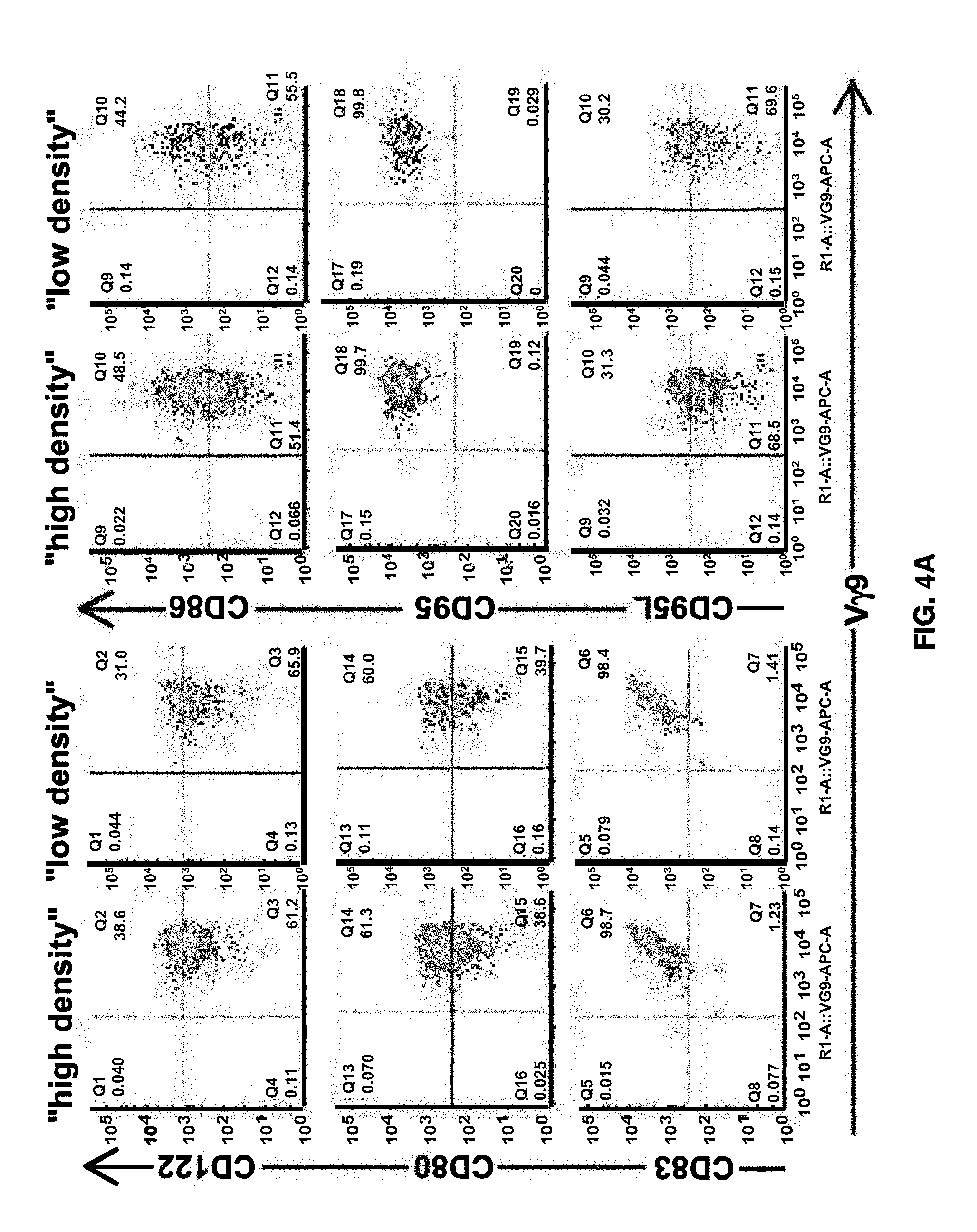
[0067] FIG. 4A shows effects of cell density on T cell markers during expansion according to an embodiment of the disclosure. After activation in the presence of zoledronate, IL-2, and IL-15 for 14 days, V.gamma.9.delta.2 T cells were expanded by homeostatic cytokines, e.g., IL-2 and IL-15, in the absence of zoledronate at high density (e.g., 2.times.10.sup.6 cells/ml) and at low density (e.g., 0.5.times.10.sup.6 cells/ml). T cell markers, e.g., CD122, CD80, CD83, CD86, CD95, and CD95L, were analyzed.
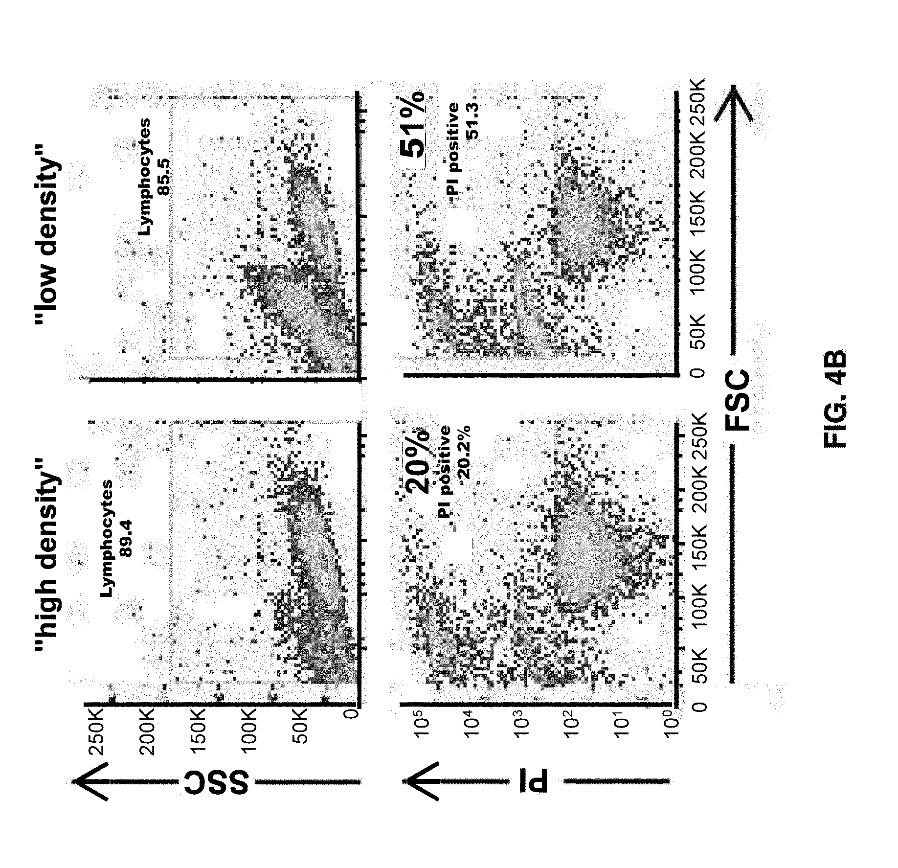
[0068] FIG. 4B shows effects of cell density on cell death during expansion according to an embodiment of the disclosure. After activation in the presence of zoledronate, IL-2, and IL-15 for 14 days, T cells were expanded by homeostatic cytokines, e.g., IL-2 and IL-15, in the absence of zoledronate at high density (e.g., 2.times.10.sup.6 cells/ml) and at low density (e.g., 0.5.times.10.sup.6 cells/ml). Cell death was measured.
[0069] FIG. 5 shows effects of Amphotericin B on V.delta.2 T cells expressing IL-2R.alpha. according to an embodiment of the disclosure. .alpha..beta.-depleted PBMCs were cultured in Activation Medium supplemented with Zoledronate, IL-2, and IL-15 on Day 0. After 48 hours, Amphotericin B was added and 48 hours later, cells were harvested for flow cytometry based analysis of CD25 (or IL-2R.alpha.) surface expression on CD3.sup.+/V.delta.2 T cells.
[0070] FIG. 6 shows effects of Zoledronate (Zometa) on cell expansion according to an embodiment of the disclosure. .gamma..delta. T cell were expanded using Zoledronate (Zometa) in defined medium containing IL-2, IL-15, and Amphotericin B.
[0071] FIG. 7 shows time table for viral transduction into V.gamma.9.delta.2 T cells according to an embodiment of the disclosure. On Day 1, fresh PBMC were depleted of .alpha..beta. T cells and activated with Zoledronate in the presence of IL-2 and IL-15; 24 hours later (on Day 2), cells were transduced using respective viral supernatant at MOI of 1.5; GFP transgene expression was assessed by flow cytometry on Day 7 (i.e., day 5 post-transduction) for transduction efficiency; and on Day 12 (i.e., day 10 post-transduction) for persistent transgene expression.
[0072] FIG. 8A shows viral transgene expression in V.gamma.9.delta.2 T cells using a .gamma.-retroviral vector according to an embodiment of the disclosure. Different envelop protein-expressing viruses, e.g., green fluorescent protein (GFP)-expressing .gamma.-retrovirus (e.g., Gibbon Ape Leukemia Virus (GALV) pseudotype (for example, SEQ ID NO: 4), RD114TR pseudotype (SEQ ID NO: 1), and GFP-expressing lentivirus (e.g., VSV-G pseudotype (for example, SEQ ID NO: 3)) were tested for their transduction efficiency into V.gamma.9.delta.2 T cells at Day 5 and Day 10 post-transduction.
[0073] FIG. 8B shows viral transgene, e.g., CD8.alpha., expression in V.gamma.9.delta.2 T cells transduced by an RD114TR or a VSV-G pseudotyped lentiviral vector according to an embodiment of the disclosure.
[0074] FIG. 9A shows transduction of .gamma..delta. T cells with CD8.alpha..beta. using different transduction enhancers (RetroNectin.RTM. vs. Vectofusin-1e) during the transduction process. .gamma..delta. T cells obtained from 3 donors (Donor 1, Donor 2, and Donor 3) were transduced with a retrovirus encoding CD8.alpha..beta. in the presence of RetroNectin.RTM. (a fibronectin fragment coated onto plates) or VectoFusin-1.RTM. (a soluble cationic peptide), followed by flow cytometry to determine the % of CD8.alpha..beta.+ cells.
[0075] FIG. 9B shows transduction efficiency of V.gamma.9.delta.2 T cells with engineered viruses according to an embodiment of the disclosure. V.gamma.9.delta.2 T cells were transduced without virus (Mock) or transduced with .alpha..beta.-TCR virus alone, with CD8 virus alone, or with .alpha..beta.-TCR virus+CD8 virus. Transduced cells were incubated with TAA/MHC-PE dextramer, anti-CD8 antibody, or NYESO-PE dextramer (negative control), followed by flow cytometry analysis.
[0076] FIG. 10 shows fold-expansion of V.gamma.9.delta.2 T cells transduced with engineered viruses according to an embodiment of the disclosure. V.gamma.9.delta.2 T cells (GD) or .alpha..beta. T cells (AB) were transduced without virus (Mock), with .alpha..beta.-TCR virus (TCR), with CD8 virus (CD8), or with CD8+TCR, followed by measurement of fold expansion from day 7 to day 21 post-transduction.
[0077] FIG. 11A shows engineered V.gamma.9.delta.2 T cells according to an embodiment of the disclosure. V.gamma.9.delta.2 T cells transduced without virus (Mock) or with .alpha..beta.-TCR retrovirus and CD8.alpha..beta. retrovirus (.alpha..beta.-TCR+CD8) were incubated with TAA/MHC complex, followed by flow cytometry analysis to detect V.gamma.9.delta.2 T cells that bind to TAA/MHC complex.
[0078] FIG. 11B shows functional assessments of engineered Vg9d2 T cells according to an embodiment of the disclosure. V.gamma.9.delta.2 T cells transduced without virus (Mock) or with .alpha..beta.-TCR retrovirus and CD8.alpha..beta. retrovirus (.alpha..beta.-TCR+CD8) were incubated with target cells, followed by flow cytometry analysis to detect CD107a, an apoptosis marker.
[0079] FIG. 11C shows functional assessments of engineered Vg9d2 T cells according to another embodiment of the disclosure. V.gamma.9.delta.2 T cells transduced without virus (Mock) or with .alpha..beta.-TCR retrovirus and CD8.alpha..beta. retrovirus (.alpha..beta.-TCR+CD8) were incubated with target cells, followed by flow cytometry analysis to detect IFN-.gamma. release.
[0080] FIG. 11D shows cytolytic activity of engineered Vg9d2 T cells according to an embodiment of the disclosure. V.gamma.9.delta.2 T cells transduced without virus (Mock) or with .alpha..beta.-TCR retrovirus and CD8.alpha..beta. retrovirus (.alpha..beta.-TCR+CD8) were incubated with target cells, followed by flow cytometry analysis to detect apoptotic cells.
[0081] FIG. 11E shows prolonged cytolytic activity of engineered .gamma..delta. T cells according to an embodiment of the disclosure. Cytolytic activity was evaluated in real-time during an 84-hour co-culture assay. Target positive A375-RFP tumor cells were incubated without T cells (tumor cells) or with non-transduced cells (.alpha..beta. T cells and .gamma..delta. T cells), with .alpha..beta. T cells transduced with .alpha..beta.-TCR virus and CD8.alpha..beta. virus (.alpha..beta.TCR+.alpha..beta. T cells), or with .gamma..delta. T cells transduced with .alpha..beta.-TCR virus and CD8.alpha..beta. virus (.alpha..beta.TCR+.gamma..delta. T cells), followed by IncuCyte.RTM. live cell analysis to measure target cell growth.
[0082] FIG. 12A shows a schematic of an engineered virus according to an embodiment of the disclosure. CD8/CD4 chimeric receptor-T2A-truncated CSF1R contains CD8.alpha. extracellular domain linked to CD4 transmembrane and intracellular domain.
[0083] FIG. 12B shows a schematic of an engineered virus according to an embodiment of the disclosure. CD8/CD4 chimeric receptor-T2A-CSF1R/41 BB chimeric receptor contains CSF1R extracellular domain linked downstream from chimeric CD8/CD4 protein.
[0084] FIG. 13 shows allogenic T cell therapy according to an embodiment of the present disclosure. Allogenic T cell therapy may include collecting .gamma..delta. T cells from healthy donors, engineering .gamma..delta. T cells by viral transduction of exogenous genes of interest, such as exogenous TCRs, followed by cell expansion, harvesting the expanded engineered .gamma..delta. T cells, which may be cryopreserved as "off-the-shelf" T-cell products, before infusing into patients.
[0085] FIG. 14 shows .gamma..delta. T cell manufacturing according to an embodiment of the present disclosure. .gamma..delta. T cell manufacturing may include collecting or obtaining white blood cells or PBMC, e.g., leukapheresis product, depleting .alpha..beta. T cells from PBMC or leukapheresis product, followed by activation, transduction, and expansion of .gamma..delta. T cells.
DETAILED DESCRIPTION
[0086] Allogeneic T cell therapy may be based on genetically engineering allogeneic .gamma..delta. T cells to express exogenous TCRs. In addition to the specific tumor recognition via the ectopic TCR, .gamma..delta. T cells may have activity against numerous tumor types as described herein.
[0087] In an aspect, the present disclosure relates to expansion and/or activation of T cells. In another aspect, the present disclosure relates to expansion and/or activation of .gamma..delta. T cells in the absence of agents that bind to epitopes specific to .gamma..delta. TCRs, such as antibodies against .gamma..delta. TCRs. In another aspect, the present disclosure relates to expansion and/or activation of .gamma..delta. T cells that may be used for transgene expression.
[0088] The disclosure further relates to expansion and activation of .gamma..delta. T cells while depleting .alpha.- and/or .beta.-TCR positive cells. T cell populations comprising expanded .gamma..delta. T cell and depleted or reduced .alpha.- and/or .beta.-TCR positive cells are also provided for by the instant disclosure. The disclosure further provides for methods of using the disclosed T cell populations.
[0089] In an aspect, methods for producing large-scale Good Manufacturing Practice (GMP)-grade TCR engineered V.gamma.9.delta.2 T cells are provided herein.
[0090] In the absence of feeder cells, addition of IL-18 to purified .gamma..delta. T cells enhances the expansion of .gamma..delta. T cells with notable increase in the amount of surface high affinity receptor for IL-2 (CD25 or IL-2Ra). Further, Amphotericin B, a Toll-like receptor 2 (TLR2) ligand, can activate .gamma..delta. T cells, CD8.sup.+ T cells, and NK cells and enhance the detection of surface expression of CD25, the high affinity IL-2Ra. Collectively, these observations highlight a critical role of IL-2 signaling in Zoledronate-mediated activation and expansion of V.gamma.9.delta.2 T cells. Thus, to maximize the availability of IL-2 for .gamma..delta. T cell proliferation via IL-2 signaling (or to minimize the sequestration of IL-2 by high number of .alpha..beta. T cells), methods of the present disclosure may include depleting .alpha..beta. T cells from normal PBMC using anti-.alpha..beta. TCR commercially available GMP reagents. As recombinant IL-18 is currently not available as a commercial GMP-reagent, methods of the present disclosure may supplement the culture with low dose Amphotericin B to increase CD25 surface expression to enhance IL-2 binding and signaling, which in turn may enhance IL-2 responsiveness during activation/expansion. In addition, IL-15 may be added because IL-15 has been shown to increase proliferation and survival of V.gamma.9.delta.2 T cells treated with IPP.
[0091] FIG. 13 shows an approach for adoptive allogenic T cell therapy that can deliver "off-the-shelf" T-cell products, such as .gamma..delta. T cell products, for rapid treatment of eligible patients with a specific cancer expressing the target of interest in their tumors. This approach may include collecting .gamma..delta. T cells from healthy donors, engineering .gamma..delta. T cells by viral transduction of exogenous genes of interest, such as exogenous TCRs, followed by cell expansion, harvesting the expanded engineered .gamma..delta. T cells, which may be cryopreserved as "off-the-shelf" T-cell products, before infusing into patients. This approach therefore may eliminate the need for personalized T cell manufacturing.
[0092] To isolate .gamma..delta. T cells, in an aspect, .gamma..delta. T cells may be isolated from a subject or from a complex sample of a subject. In an aspect, a complex sample may be a peripheral blood sample, a cord blood sample, a tumor, a stem cell precursor, a tumor biopsy, a tissue, a lymph, or from epithelial sites of a subject directly contacting the external milieu or derived from stem precursor cells. .gamma..delta. T cells may be directly isolated from a complex sample of a subject, for example, by sorting .gamma..delta. T cells that express one or more cell surface markers with flow cytometry techniques. Wild-type .gamma..delta. T cells may exhibit numerous antigen recognition, antigen-presentation, co-stimulation, and adhesion molecules that can be associated with a .gamma..delta. T cells. One or more cell surface markers, such as specific .gamma..delta. TCRs, antigen recognition, antigen-presentation, ligands, adhesion molecules, or co-stimulatory molecules may be used to isolate wild-type .gamma..delta. T cells from a complex sample. Various molecules associated with or expressed by .gamma..delta. T-cells may be used to isolate .gamma..delta. T cells from a complex sample. In another aspect, the present disclosure provides methods for isolation of mixed population of V.delta.1+, V.delta.2+, V.delta.3+ cells or any combination thereof.
[0093] For example, peripheral blood mononuclear cells can be collected from a subject, for example, with an apheresis machine, including the Ficoll-Paque.TM. PLUS (GE Healthcare) system, or another suitable device/system. .gamma..delta. T-cell(s), or a desired subpopulation of .gamma..delta. T-cell(s), can be purified from the collected sample with, for example, with flow cytometry techniques. Cord blood cells can also be obtained from cord blood during the birth of a subject.
[0094] Positive and/or negative selection of cell surface markers expressed on the collected .gamma..delta. T cells can be used to directly isolate .gamma..delta. T cells, or a population of .gamma..delta. T cells expressing similar cell surface markers from a peripheral blood sample, a cord blood sample, a tumor, a tumor biopsy, a tissue, a lymph, or from an epithelial sample of a subject. For instance, .gamma..delta. T cells can be isolated from a complex sample based on positive or negative expression of CD2, CD3, CD4, CD8, CD24, CD25, CD44, Kit, TCR .alpha., TCR .beta., TCR .alpha., TCR .delta., NKG2D, CD70, CD27, CD30, CD16, CD337 (NKp30), CD336 (NKp46), OX40, CD46, CCR7, and other suitable cell surface markers.
[0095] In an aspect, .gamma..delta. T cells may be isolated from a complex sample that is cultured in vitro. In another aspect, whole PBMC population, without prior depletion of specific cell populations, such as monocytes, .alpha..beta. T-cells, B-cells, and NK cells, can be activated and expanded. In another aspect, enriched .gamma..delta. T cell populations can be generated prior to their specific activation and expansion. In another aspects, activation and expansion of .gamma..delta. T cells may be performed without the presence of native or engineered APCs. In another aspects, isolation and expansion of .gamma..delta. T cells from tumor specimens can be performed using immobilized .gamma..delta. T cell mitogens, including antibodies specific to .gamma..delta. TCR, and other .gamma..delta. TCR activating agents, including lectins. In another aspect, isolation and expansion of .gamma..delta. T cells from tumor specimens can be performed in the absence of .gamma..delta. T cell mitogens, including antibodies specific to .gamma..delta. TCR, and other .gamma..delta. TCR activating agents, including lectins.
[0096] In an aspect, .gamma..delta. T cells are isolated from leukapheresis of a subject, for example, a human subject. In another aspect, .gamma..delta. T cells are not isolated from peripheral blood mononuclear cells (PBMC).
[0097] FIG. 14 shows .gamma..delta. T cell manufacturing according to an embodiment of the present disclosure. This process may include collecting or obtaining white blood cells or PBMC from leukapheresis products. Leukapheresis may include collecting whole blood from a donor and separating the components using an apheresis machine. An apheresis machine separates out desired blood components and returns the rest to the donor's circulation. For instance, white blood cells, plasma, and platelets can be collected using apheresis equipment, and the red blood cells and neutrophils are returned to the donor's circulation. Commercially available leukapheresis products may be used in this process. Another way to obtain white blood cells is to obtain them from the buffy coat. To isolate the buffy coat, whole anticoagulated blood is obtained from a donor and centrifuged. After centrifugation, the blood is separated into the plasma, red blood cells, and buffy coat. The buffy coat is the layer located between the plasma and red blood cell layers. Leukapheresis collections may result in higher purity and considerably increased mononuclear cell content than that achieved by buffy coat collection. The mononuclear cell content possible with leukapheresis may be typically 20 times higher than that obtained from the buffy coat. In order to enrich for mononuclear cells, the use of a Ficoll gradient may be needed for further separation.
[0098] To deplete .alpha..beta. T cells from PBMC, .alpha..beta. TCR-expressing cells may be separated from the PBMC by magnetic separation, e.g., using CliniMACS.RTM. magnetic beads coated with anti-.alpha..beta. TCR antibodies, followed by cryopreserving .alpha..beta. TCR-T cells depleted PBMC. To manufacture "off-the-shelf" T-cell products, cryopreserved .alpha..beta. TCR-T cells depleted PBMC may be thawed and activated in small/mid-scale, e.g., 24 to 4-6 well plates or T75/T175 flasks, or in large scale, e.g., 50 ml-100 liter bags, in the presence of aminobisphosphonate and/or isopentenyl pyrophosphate (IPP) and/or cytokines, e.g., interleukin 2 (IL-2), interleukin 15 (IL-15), and/or interleukin 18 (IL-18), and/or other activators, e.g., Toll-like receptor 2 (TLR2) ligand, for 1-10 days, e.g., 2-6 days.
[0099] In an aspect, the isolated .gamma..delta. T cells can rapidly expand in response to contact with one or more antigens. Some .gamma..delta. T cells, such as V.gamma.9V.delta.2+ T cells, can rapidly expand in vitro in response to contact with some antigens, like prenyl-pyrophosphates, alkyl amines, and metabolites or microbial extracts during tissue culture. Stimulated .gamma..delta. T-cells can exhibit numerous antigen-presentation, co-stimulation, and adhesion molecules that can facilitate the isolation of .gamma..delta. T-cells from a complex sample. .gamma..delta. T cells within a complex sample can be stimulated in vitro with at least one antigen for 1 day, 2 days, 3 days, 4 days, 5 days, 6 days, 7 days, or another suitable period of time. Stimulation of .gamma..delta. T cells with a suitable antigen can expand .gamma..delta. T cell population in vitro.
[0100] Non-limiting examples of antigens that may be used to stimulate the expansion of .gamma..delta. T cells from a complex sample in vitro may include, prenyl-pyrophosphates, such as isopentenyl pyrophosphate (IPP), alkyl-amines, metabolites of human microbial pathogens, metabolites of commensal bacteria, methyl-3-butenyl-1-pyrophosphate (2M3B1 PP), (E)-4-hydroxy-3-methyl-but-2-enyl pyrophosphate (HMB-PP), ethyl pyrophosphate (EPP), farnesyl pyrophosphate (FPP), dimethylallyl phosphate (DMAP), dimethylallyl pyrophosphate (DMAPP), ethyl-adenosine triphosphate (EPPPA), geranyl pyrophosphate (GPP), geranylgeranyl pyrophosphate (GGPP), isopentenyl-adenosine triphosphate (IPPPA), monoethyl phosphate (MEP), monoethyl pyrophosphate (MEPP), 3-formyl-1-butyl-pyrophosphate (TUBAg 1), X-pyrophosphate (TUBAg 2), 3-formyl-1-butyl-uridine triphosphate (TUBAg 3), 3-formyl-1-butyl-deoxythymidine triphosphate (TUBAg 4), monoethyl alkylamines, allyl pyrophosphate, crotoyl pyrophosphate, dimethylallyl-.gamma.-uridine triphosphate, crotoyl-.gamma.-uridine triphosphate, allyl-.gamma.-uridine triphosphate, ethylamine, isobutylamine, sec-butylamine, iso-amylamine and nitrogen containing bisphosphonates.
[0101] Activation and expansion of .gamma..delta. T cells can be performed using activation and co-stimulatory agents described herein to trigger specific .gamma..delta. T cell proliferation and persistence populations. In an aspect, activation and expansion of .gamma..delta. T-cells from different cultures can achieve distinct clonal or mixed polyclonal population subsets. In another aspect, different agonist agents can be used to identify agents that provide specific .gamma..delta. activating signals. In another aspect, agents that provide specific .gamma..delta. activating signals can be different monoclonal antibodies (MAbs) directed against the .gamma..delta. TCRs. In another aspect, companion co-stimulatory agents to assist in triggering specific .gamma..delta. T cell proliferation without induction of cell energy and apoptosis can be used. These co-stimulatory agents can include ligands binding to receptors expressed on .gamma..delta. cells, such as NKG2D, CD161, CD70, JAML, DNAX accessory molecule-1 (DNAM-1), ICOS, CD27, CD137, CD30, HVEM, SLAM, CD122, DAP, and CD28. In another aspect, co-stimulatory agents can be antibodies specific to unique epitopes on CD2 and CD3 molecules. CD2 and CD3 can have different conformation structures when expressed on .alpha..beta. or .gamma..delta. T-cells. In another aspect, specific antibodies to CD3 and CD2 can lead to distinct activation of .gamma..delta. T cells.
[0102] A population of .gamma..delta. T-cell may be expanded ex vivo prior to engineering of the .gamma..delta. T-cell. Non-limiting example of reagents that can be used to facilitate the expansion of a .gamma..delta. T-cell population in vitro may include anti-CD3 or anti-CD2, anti-CD27, anti-CD30, anti-CD70, anti-OX40 antibodies, IL-2, IL-15, IL-12, IL-9, IL-33, IL-18, or IL-21, CD70 (CD27 ligand), phytohaemagglutinin (PHA), concavalin A (ConA), pokeweed (PWM), protein peanut agglutinin (PNA), soybean agglutinin (SBA), lens culinaris agglutinin (LCA), pisum sativum agglutinin (PSA), helix pomatia agglutinin (HPA), vicia graminea Lectin (VGA), or another suitable mitogen capable of stimulating T-cell proliferation.
[0103] The ability of .gamma..delta. T cells to recognize a broad spectrum of antigens can be enhanced by genetic engineering of the .gamma..delta. T cells. In an aspect, .gamma..delta. T cell can be engineered to provide a universal allogeneic therapy that recognizes an antigen of choice in vivo. Genetic engineering of the .gamma..delta. T-cells may include stably integrating a construct expressing a tumor recognition moiety, such as .alpha..delta. TCR, .gamma..delta. TCR, chimeric antigen receptor (CAR), which combines both antigen-binding and T-cell activating functions into a single receptor, an antigen binding fragment thereof, or a lymphocyte activation domain into the genome of the isolated .gamma..delta. T-cell(s), a cytokine (IL-15, IL-12, IL-2. IL-7. IL-21, IL-18, IL-19, IL-33, IL-4, IL-9, IL-23, IL1.beta.) to enhance T-cell proliferation, survival, and function ex vivo and in vivo. Genetic engineering of the isolated .gamma..delta. T-cell may also include deleting or disrupting gene expression from one or more endogenous genes in the genome of the isolated .gamma..delta. T-cells, such as the MHC locus (loci).
[0104] In an aspect, viruses refers to natural occurring viruses as well as artificial viruses. Viruses in accordance to some embodiments of the present disclosure may be either an enveloped or non-enveloped virus. Parvoviruses (such as AAVs) are examples of non-enveloped viruses. In a preferred embodiment, the viruses may be enveloped viruses. In preferred embodiments, the viruses may be retroviruses and in particular lentiviruses. Viral envelope proteins that can promote viral infection of eukaryotic cells may include HIV-1 derived lentiviral vectors (LVs) pseudotyped with envelope glycoproteins (GPs) from the vesicular stomatitis virus (VSV-G), the modified feline endogenous retrovirus (RD114TR), and the modified gibbon ape leukemia virus (GALVTR). These envelope proteins can efficiently promote entry of other viruses, such as parvoviruses, including adeno-associated viruses (AAV), thereby demonstrating their broad efficiency. For example, other viral envelop proteins may be used including Moloney murine leukemia virus (MLV) 4070 env (such as described in Merten et al., J. Virol. 79:834-840, 2005; which is incorporated herein by reference), RD114 env (SEQ ID NO: 2), chimeric envelope protein RD114pro or RDpro (which is an RD114-HIV chimera that was constructed by replacing the R peptide cleavage sequence of RD114 with the HIV-1 matrix/capsid (MA/CA) cleavage sequence, such as described in Bell et al. Experimental Biology and Medicine 2010; 235: 1269-1276; which is incorporated herein by reference), baculovirus GP64 env (such as described in Wang et al. J. Virol. 81:10869-10878, 2007; which is incorporated herein by reference), or GALV env (such as described in Merten et al., J. Virol. 79:834-840, 2005; which is incorporated herein by reference), or derivatives thereof.
[0105] RD114TR
[0106] RD114TR is a chimeric envelope glycoprotein made of the extracellular and transmembrane domains of the feline leukemia virus RD114 and the cytoplasmic tail (TR) of the amphotropic murine leukemia virus envelope. RD114TR pseudotyped vectors can mediate efficient gene transfer into human hematopoietic progenitors and NOD/SCID repopulating cells. Di Nunzio et al., Hum. Gene Ther: 811-820 (2007)), the contents of which are incorporated by reference in their entirety. RD114 pseudotyped vectors can also mediate efficient gene transfer in large animal models. (Neff et al., Mal. Ther. 2:157-159 (2004); Hu et al., Mal. Ther: 611-617 (2003); and Kelly et al., Blood Cells, Molecules, & Diseases 30:132-143 (2003)), the contents of each of these references are incorporated by reference in their entirety.
[0107] The present disclosure may include RD114TR variants having at least about 50%, at least about 60%, at least about 70%, at least about 80%, at least about 90%, at least about 95%, at least about 98%, at least about 99%, or 100% sequence identity to the amino acid sequence of SEQ ID NO: 1 or SEQ ID NO: 5. For example, an RD114TR variant (RD114TRv1 (SEQ ID NO: 5)) having about 96% sequence identity to RD114TR (SEQ ID NO: 1) may be used. In an aspect, the disclosure provides for RD114TR variants having modified amino acid residues. A modified amino acid residue may be selected from an amino acid insertion, deletion, or substitution. In an aspect, a substitution described herein is a conservative amino acid substitution. That is, amino acids of RD114TR may be replaced by other amino acids having similar properties (conservative changes, such as similar hydrophobicity, hydrophilicity, antigenicity, propensity to form or break a-helical structures or 3-sheet structures). Non-limiting examples of conservative substitutions may be found in, for example, Creighton (1984) Proteins. W.H. Freeman and Company, the contents of which are incorporated by reference in their entirety.
[0108] In another aspect, the present disclosure may include variants having at least about 50%, at least about 60%, at least about 70%, at least about 80%, at least about 90%, at least about 95%, at least about 98%, at least about 99%, or 100% sequence identity to the amino acid sequence of SEQ ID NO: 1, 2, 3, 4, or 5.
[0109] In an aspect, conservative substitutions may include those, which are described by Dayhoff in "The Atlas of Protein Sequence and Structure. Vol. 5", Natl. Biomedical Research, the contents of which are incorporated by reference in their entirety. For example, in an aspect, amino acids, which belong to one of the following groups, can be exchanged for one another, thus, constituting a conservative exchange: Group 1: alanine (A), proline (P), glycine (G), asparagine (N), serine (S), threonine (T); Group 2: cysteine (C), serine (S), tyrosine (Y), threonine (T); Group 3: valine (V), isoleucine (I), leucine (L), methionine (M), alanine (A), phenylalanine (F); Group 4: lysine (K), arginine (R), histidine (H); Group 5: phenylalanine (F), tyrosine (Y), tryptophan (W), histidine (H); and Group 6: aspartic acid (D), glutamic acid (E).
[0110] In an aspect, conservative amino acid substitution may include the substitution of an amino acid by another one of the same class, for example, (1) nonpolar: Ala, Val, Leu, Ile, Pro, Met, Phe, Trp; (2) uncharged polar: Gly, Ser, Thr, Cys, Tyr, Asn, Gln; (3) acidic: Asp, Glu; and (4) basic: Lys, Arg, His. Other conservative amino acid substitutions may also be made as follows: (1) aromatic: Phe, Tyr, His; (2) proton donor: Asn, Gln, Lys, Arg, His, Trp; and (3) proton acceptor: Glu, Asp, Thr, Ser, Tyr, Asn, Gln (see, U.S. patent Ser. No. 10/106,805).
[0111] In another aspect, conservative substitutions may be made in accordance with Table A. Methods for predicting tolerance to protein modification may be found in, for example, Guo et al., Proc. Natl. Acad. Sci., USA, 101(25):9205-9210 (2004), the contents of which are incorporated by reference in their entirety.
TABLE-US-00001 TABLE A Conservative Amino Acid Substitutions Amino Acid Substitutions (others are known in the art) Ala Ser, Gly, Cys Arg Lys, Gln, His Asn Gln, His, Glu, Asp Asp Glu, Asn, Gln Cys Ser, Met, Thr Gln Asn, Lys, Glu, Asp, Arg Glu Asp, Asn, Gln Gly Pro, Ala, Ser His Asn, Gln, Lys Ile Leu, Val, Met, Ala Leu Ile, Val, Met, Ala Lys Arg, Gln, His Met Leu, Ile, Val, Ala, Phe Phe Met, Leu, Tyr, Trp, His Ser Thr, Cys, Ala Thr Ser, Val, Ala Trp Tyr, Phe Tyr Trp, Phe, His Val He, Leu, Met, Ala, Thr
[0112] In an aspect, transgene expression for RD114TR-pseudotyped retroviral vector at about 10-day post-transduction is about 20% to about 60% about 30% to about 50%, or about 35% to about 45%. In an aspect, transgene expression for RD114TR-pseudotyped retroviral vector at 10-day post-transduction is about 20% to about 60% about 30% to about 50%, or about 35% to about 45% relative to transgene expression for VSV-G-pseudotyped vectors at day 10 post-transduction of about 5% to about 25%, about 2% to about 20%, about 3% to about 15%, or about 5% to about 12% under the same conditions. In yet another aspect, transgene expression for RD114TR-pseudotyped retroviral vector at 10-day post-transduction is about 40% relative to transgene expression for VSV-G-pseudotyped vectors at day 10 post-transduction of about 3.6%.
[0113] In yet another aspect, transgene expression for RD114TR-pseudotyped retroviral vector at about 5-day post-transduction is about 20% to about 50% about 15% to about 30%, or about 20% to about 30%. In an aspect, transgene expression for RD114TR-pseudotyped retroviral vector at 5-day post-transduction is about 20% to about 50% about 15% to about 30%, or about 20% to about 30% relative to transgene expression for VSV-G-pseudotyped vectors at day 5 post-transduction of about 10% to about 20%, about 15% to about 25%, or about 17.5% to about 20% under the same conditions. In yet another aspect, transgene expression for RD114TR-pseudotyped retroviral vector at 5-day post-transduction is about 24% relative to transgene expression for VSV-G-pseudotyped vectors at day 5 post-transduction of about 19%.
[0114] In another aspect, transgene expression for RD114TR-pseudotyped retroviral vector at 10-day post-transduction is about 2 times, about 3 times, about 4 times, about 5 times, or about 10 times, about 11 times, or about 12 times or more relative to transgene expression for VSV-G-pseudotyped vectors at day 10 post-transduction.
[0115] In an aspect, the disclosure provides for methods of using retrovirus with RD114TR pseudotype (for example, SEQ ID NO: 1) to transduce T cells. In another aspect, T cells are more efficiently transduced by retrovirus with RD114TR pseudotype (for example, SEQ ID NO: 1) as compared to retrovirus with VSV-G pseudotype (for example, SEQ ID NO: 3). In another aspect, a RD114TR envelope is utilized to pseudotype a lentivector, which is then used to transduce T cells with excellent efficiency.
[0116] Engineered .gamma..delta. T-cells may be generated with various methods. For example, a polynucleotide encoding an expression cassette that comprises a tumor recognition, or another type of recognition moiety, can be stably introduced into the .gamma..delta. T-cell by a transposon/transposase system or a viral-based gene transfer system, such as a lentiviral or a retroviral system, or another suitable method, such as transfection, electroporation, transduction, lipofection, calcium phosphate (CaPO4), nanoengineered substances, such as Ormosil, viral delivery methods, including adenoviruses, retroviruses, lentiviruses, adeno-associated viruses, or another suitable method. A number of viral methods have been used for human gene therapy, such as the methods described in WO 1993020221, which is incorporated herein in its entirety. Non-limiting examples of viral methods that can be used to engineer .gamma..delta. T cells may include .gamma.-retroviral, adenoviral, lentiviral, herpes simplex virus, vaccinia virus, pox virus, or adeno-virus associated viral methods.
[0117] FIG. 14 shows the activated T cells may be engineered by transducing with a viral vector, such as RD114TR .gamma.-retroviral vector and RD114TR lentiviral vector, expressing exogenous genes of interest, such as .alpha..beta. TCRs against specific cancer antigen and CD8, into isolated .gamma..delta. T cells. Viral vectors may also contain post-transcriptional regulatory element (PRE), such as Woodchuck PRE (WPRE) to enhance the expression of the transgene by increasing both nuclear and cytoplasmic mRNA levels. One or more regulatory elements including mouse RNA transport element (RTE), the constitutive transport element (CTE) of the simian retrovirus type 1 (SRV-1), and the 5' untranslated region of the human heat shock protein 70 (Hsp70 5'UTR) may also be used and/or in combination with WPRE to increase transgene expression. Transduction may be carried out once or multiple times to achieve stable transgene expression in small scale, e.g., 24 to 4-6 well plates, or mid/large scale for 1/2-5 days, e.g., 1 day.
[0118] RD114TR is a chimeric glycoprotein containing an extracellular and transmembrane domain of feline endogenous virus (RD114) fused to cytoplasmic tail (TR) of murine leukemia virus. In an aspect, transgene expression for RD114TR-pseudotyped retroviral vector at 10-day post-transduction is higher relative to VSV-G-pseudotyped vectors.
[0119] Other viral envelop proteins, such as VSV-G env, MLV 4070 env, RD114 env, chimeric envelope protein RD114pro, baculovirus GP64 env, or GALV env, or derivatives thereof, may also be used.
[0120] In an aspect, engineered (or transduced) .gamma..delta. T cells can be expanded ex vivo without stimulation by an antigen presenting cell or aminobisphosphonate. Antigen reactive engineered T cells of the present disclosure may be expanded ex vivo and in vivo. In another aspect, an active population of engineered .gamma..delta. T cells of the present disclosure may be expanded ex vivo without antigen stimulation by an antigen presenting cell, an antigenic peptide, a non-peptide molecule, or a small molecule compound, such as an aminobisphosphonate but using certain antibodies, cytokines, mitogens, or fusion proteins, such as IL-17 Fc fusion, MICA Fc fusion, and CD70 Fc fusion. Examples of antibodies that can be used in the expansion of a .gamma..delta. T-cell population may include anti-CD3, anti-CD27, anti-CD30, anti-CD70, anti-OX40, anti-NKG2D, or anti-CD2 antibodies, examples of cytokines may include IL-2, IL-15, IL-12, IL-21, IL-18, IL-9, IL-7, and/or IL-33, and examples of mitogens may include CD70 the ligand for human CD27, phytohaemagglutinin (PHA), concavalin A (ConA), pokeweed mitogen (PWM), protein peanut agglutinin (PNA), soybean agglutinin (SBA), lens culinaris agglutinin (LCA), pisum sativum agglutinin (PSA), h pomatia agglutinin (HPA), vicia graminea Lectin (VGA) or another suitable mitogen capable of stimulating T-cell proliferation. In another aspect, a population of engineered .gamma..delta. T cells can be expanded in less than 60 days, less than 48 days, 36 days, less than 24 days, less than 12 days, or less than 6 days.
[0121] In another aspect, the present disclosure provides methods for the ex vivo expansion of a population of engineered .gamma..delta. T-cells for adoptive transfer therapy. Engineered .gamma..delta. T cells of the disclosure may be expanded ex vivo. Engineered .gamma..delta. T cells of the disclosure can be expanded in vitro without activation by APCs, or without co-culture with APCs, and aminophosphates.
[0122] In another aspect, a .gamma..delta. T-cell population can be expanded in vitro in fewer than 36 days, fewer than 35 days, fewer than 34 days, fewer than 33 days, fewer than 32 days, fewer than 31 days, fewer than 30 days, fewer than 29 days, fewer than 28 days, fewer than 27 days, fewer than 26 days, fewer than 25 days, fewer than 24 days, fewer than 23 days, fewer than 22 days, fewer than 21 days, fewer than 20 days, fewer than 19 days, fewer than 18 days, fewer than 17 days, fewer than 16 days, fewer than 15 days, fewer than 14 days, fewer than 13 days, fewer than 12 days, fewer than 11 days, fewer than 10 days, fewer than 9 days, fewer than 8 days, fewer than 7 days, fewer than 6 days, fewer than 5 days, fewer than 4 days, or fewer than 3 days.
[0123] FIG. 14 shows expansion of the transduced or engineered .gamma..delta. T cells may be carried out in the presence of cytokines, e.g., IL-2, IL-15, IL-18, and others, in small/mid-scale, e.g., flasks/G-Rex, or in large scale, e.g., 50 ml-100-liter bags, for 7-35 days, e.g., 14-28 days. The expanded transduced T cell products may then be cryopreserved as "off-the-shelf" T-cell products for infusion into patients.
[0124] Methods of Treatment
[0125] Compositions containing engineered .gamma..delta. T cells described herein may be administered for prophylactic and/or therapeutic treatments. In therapeutic applications, pharmaceutical compositions can be administered to a subject already suffering from a disease or condition in an amount sufficient to cure or at least partially arrest the symptoms of the disease or condition. An engineered .gamma..delta. T-cell can also be administered to lessen a likelihood of developing, contracting, or worsening a condition. Effective amounts of a population of engineered .gamma..delta. T-cells for therapeutic use can vary based on the severity and course of the disease or condition, previous therapy, the subject's health status, weight, and/or response to the drugs, and/or the judgment of the treating physician.
[0126] Engineered .gamma..delta. T cells of the present disclosure can be used to treat a subject in need of treatment for a condition, for example, a cancer described herein.
[0127] A method of treating a condition (e.g., ailment) in a subject with .gamma..delta. T cells may include administering to the subject a therapeutically-effective amount of engineered .gamma..delta. T cells. .gamma..delta. T cells of the present disclosure may be administered at various regimens (e.g., timing, concentration, dosage, spacing between treatment, and/or formulation). A subject can also be preconditioned with, for example, chemotherapy, radiation, or a combination of both, prior to receiving engineered .gamma..delta. T cells of the present disclosure. A population of engineered .gamma..delta. T cells may also be frozen or cryopreserved prior to being administered to a subject. A population of engineered .gamma..delta. T cells can include two or more cells that express identical, different, or a combination of identical and different tumor recognition moieties. For instance, a population of engineered .gamma..delta. T-cells can include several distinct engineered .gamma..delta. T cells that are designed to recognize different antigens, or different epitopes of the same antigen.
[0128] .gamma..delta. T cells of the present disclosure may be used to treat various conditions. In an aspect, engineered .gamma..delta. T cells of the present disclosure may be used to treat a cancer, including solid tumors and hematologic malignancies. Non-limiting examples of cancers include: acute lymphoblastic leukemia, acute myeloid leukemia, adrenocortical carcinoma, AIDS-related cancers, AIDS-related lymphoma, anal cancer, appendix cancer, astrocytomas, neuroblastoma, basal cell carcinoma, bile duct cancer, bladder cancer, bone cancers, brain tumors, such as cerebellar astrocytoma, cerebral astrocytoma/malignant glioma, ependymoma, medulloblastoma, supratentorial primitive neuroectodermal tumors, visual pathway and hypothalamic glioma, breast cancer, bronchial adenomas, Burkitt lymphoma, carcinoma of unknown primary origin, central nervous system lymphoma, cerebellar astrocytoma, cervical cancer, childhood cancers, chronic lymphocytic leukemia, chronic myelogenous leukemia, chronic myeloproliferative disorders, colon cancer, cutaneous T-cell lymphoma, desmoplastic small round cell tumor, endometrial cancer, ependymoma, esophageal cancer, Ewing's sarcoma, germ cell tumors, gallbladder cancer, gastric cancer, gastrointestinal carcinoid tumor, gastrointestinal stromal tumor, gliomas, hairy cell leukemia, head and neck cancer, heart cancer, hepatocellular (liver) cancer, Hodgkin lymphoma, Hypopharyngeal cancer, intraocular melanoma, islet cell carcinoma, Kaposi sarcoma, kidney cancer, laryngeal cancer, lip and oral cavity cancer, liposarcoma, liver cancer, lung cancers, such as non-small cell and small cell lung cancer, lymphomas, leukemias, macroglobulinemia, malignant fibrous histiocytoma of bone/osteosarcoma, medulloblastoma, melanomas, mesothelioma, metastatic squamous neck cancer with occult primary, mouth cancer, multiple endocrine neoplasia syndrome, myelodysplastic syndromes, myeloid leukemia, nasal cavity and paranasal sinus cancer, nasopharyngeal carcinoma, neuroblastoma, non-Hodgkin lymphoma, non-small cell lung cancer, oral cancer, oropharyngeal cancer, osteosarcoma/malignant fibrous histiocytoma of bone, ovarian cancer, ovarian epithelial cancer, ovarian germ cell tumor, pancreatic cancer, pancreatic cancer islet cell, paranasal sinus and nasal cavity cancer, parathyroid cancer, penile cancer, pharyngeal cancer, pheochromocytoma, pineal astrocytoma, pineal germinoma, pituitary adenoma, pleuropulmonary blastoma, plasma cell neoplasia, primary central nervous system lymphoma, prostate cancer, rectal cancer, renal cell carcinoma, renal pelvis and ureter transitional cell cancer, retinoblastoma, rhabdomyosarcoma, salivary gland cancer, sarcomas, skin cancers, Merkel cell skin carcinoma, small intestine cancer, soft tissue sarcoma, squamous cell carcinoma, stomach cancer, T-cell lymphoma, throat cancer, thymoma, thymic carcinoma, thyroid cancer, trophoblastic tumor (gestational), cancers of unknown primary site, urethral cancer, uterine sarcoma, vaginal cancer, vulvar cancer, Waldenstrom's macroglobulinemia, and Wilms tumor.
[0129] In an aspect, engineered .gamma..delta. T cells of the present disclosure may be used to treat an infectious disease. In another aspect, engineered .gamma..delta. T cells of the present disclosure may be used to treat an infectious disease, an infectious disease may be caused a virus. In yet another aspect, engineered .gamma..delta. T cells of the present disclosure may be used to treat an immune disease, such as an autoimmune disease.
[0130] Treatment with .gamma..delta. T cells of the present disclosure may be provided to the subject before, during, and after the clinical onset of the condition. Treatment may be provided to the subject after 1 day, 1 week, 6 months, 12 months, or 2 years after clinical onset of the disease. Treatment may be provided to the subject for more than 1 day, 1 week, 1 month, 6 months, 12 months, 2 years, 3 years, 4 years, 5 years, 6 years, 7 years, 8 years, 9 years, 10 years or more after clinical onset of disease. Treatment may be provided to the subject for less than 1 day, 1 week, 1 month, 6 months, 12 months, or 2 years after clinical onset of the disease. Treatment may also include treating a human in a clinical trial. A treatment can include administering to a subject a pharmaceutical composition comprising engineered .gamma..delta. T cells of the present disclosure.
[0131] In another aspect, administration of engineered .gamma..delta. T cells of the present disclosure to a subject may modulate the activity of endogenous lymphocytes in a subject's body. In another aspect, administration of engineered .gamma..delta. T cells to a subject may provide an antigen to an endogenous T-cell and may boost an immune response. In another aspect, the memory T cell may be a CD4+ T-cell. In another aspect, the memory T cell may be a CD8+ T-cell. In another aspect, administration of engineered .gamma..delta. T cells of the present disclosure to a subject may activate the cytotoxicity of another immune cell. In another aspect, the other immune cell may be a CD8+ T-cell. In another aspect, the other immune cell may be a Natural Killer T-cell. In another aspect, administration of engineered .gamma..delta. T-cells of the present disclosure to a subject may suppress a regulatory T-cell. In another aspect, the regulatory T-cell may be a FOX3+ Treg cell. In another aspect, the regulatory T-cell may be a FOX3 Treg cell. Non-limiting examples of cells whose activity can be modulated by engineered .gamma..delta. T cells of the disclosure may include: hematopoietic stem cells; B cells; CD4; CD8; red blood cells; white blood cells; dendritic cells, including dendritic antigen presenting cells; leukocytes; macrophages; memory B cells; memory T-cells; monocytes; natural killer cells; neutrophil granulocytes; T-helper cells; and T-killer cells.
[0132] During most bone marrow transplants, a combination of cyclophosphamide with total body irradiation may be conventionally employed to prevent rejection of the hematopoietic stem cells (HSC) in the transplant by the subject's immune system. In an aspect, incubation of donor bone marrow with interleukin-2 (IL-2) ex vivo may be performed to enhance the generation of killer lymphocytes in the donor marrow. Interleukin-2 (IL-2) is a cytokine that may be necessary for the growth, proliferation, and differentiation of wild-type lymphocytes. Current studies of the adoptive transfer of .gamma..delta. T-cells into humans may require the co-administration of .gamma..delta. T-cells and interleukin-2. However, both low- and high-dosages of IL-2 can have highly toxic side effects. IL-2 toxicity can manifest in multiple organs/systems, most significantly the heart, lungs, kidneys, and central nervous system. In another aspect, the disclosure provides a method for administrating engineered .gamma..delta. T cells to a subject without the co-administration of a native cytokine or modified versions thereof, such as IL-2, IL-15, IL-12, IL-21. In another aspect, engineered .gamma..delta. T cells can be administered to a subject without co-administration with IL-2. In another aspect, engineered .gamma..delta. T cells may be administered to a subject during a procedure, such as a bone marrow transplant without the co-administration of IL-2.
[0133] Methods of Administration
[0134] One or multiple engineered .gamma..delta. T cell populations may be administered to a subject in any order or simultaneously. If simultaneously, the multiple engineered .gamma..delta. T cell can be provided in a single, unified form, such as an intravenous injection, or in multiple forms, for example, as multiple intravenous infusions, s.c, injections or pills. Engineered .gamma..delta. T-cells can be packed together or separately, in a single package or in a plurality of packages. One or all of the engineered .gamma..delta. T cells can be given in multiple doses. If not simultaneous, the timing between the multiple doses may vary to as much as about a week, a month, two months, three months, four months, five months, six months, or about a year. In another aspect, engineered .gamma..delta. T cells can expand within a subject's body, in vivo, after administration to a subject. Engineered .gamma..delta. T cells can be frozen to provide cells for multiple treatments with the same cell preparation. Engineered .gamma..delta. T cells of the present disclosure, and pharmaceutical compositions comprising the same, can be packaged as a kit. A kit may include instructions (e.g., written instructions) on the use of engineered .gamma..delta. T cells and compositions comprising the same.
[0135] In another aspect, a method of treating a cancer comprises administering to a subject a therapeutically-effective amount of engineered .gamma..delta. T cells, in which the administration treats the cancer. In another embodiments, the therapeutically-effective amount of engineered .gamma..delta. T cells may be administered for at least about 10 seconds, 30 seconds, 1 minute, 10 minutes, 30 minutes, 1 hour, 2 hours, 3 hours, 4 hours, 5 hours, 6 hours, 12 hours, 24 hours, 2 days, 3 days, 4 days, 5 days, 6 days, 1 week, 2 weeks, 3 weeks, 1 month, 2 months, 3 months, 4 months, 5 months, 6 months, or 1 year. In another aspect, the therapeutically-effective amount of the engineered .gamma..delta. T cells may be administered for at least one week. In another aspect, the therapeutically-effective amount of engineered .gamma..delta. T cells may be administered for at least two weeks.
[0136] Engineered .gamma..delta. T-cells described herein can be administered before, during, or after the occurrence of a disease or condition, and the timing of administering a pharmaceutical composition containing an engineered .gamma..delta. T-cell can vary. For example, engineered .gamma..delta. T cells can be used as a prophylactic and can be administered continuously to subjects with a propensity to conditions or diseases in order to lessen a likelihood of the occurrence of the disease or condition. Engineered .gamma..delta. T-cells can be administered to a subject during or as soon as possible after the onset of the symptoms. The administration of engineered .gamma..delta. T cells can be initiated immediately within the onset of symptoms, within the first 3 hours of the onset of the symptoms, within the first 6 hours of the onset of the symptoms, within the first 24 hours of the onset of the symptoms, within 48 hours of the onset of the symptoms, or within any period of time from the onset of symptoms. The initial administration can be via any route practical, such as by any route described herein using any formulation described herein. In another aspect, the administration of engineered .gamma..delta. T cells of the present disclosure may be an intravenous administration. One or multiple dosages of engineered .gamma..delta. T cells can be administered as soon as is practicable after the onset of a cancer, an infectious disease, an immune disease, sepsis, or with a bone marrow transplant, and for a length of time necessary for the treatment of the immune disease, such as, for example, from about 24 hours to about 48 hours, from about 48 hours to about 1 week, from about 1 week to about 2 weeks, from about 2 weeks to about 1 month, from about 1 month to about 3 months. For the treatment of cancer, one or multiple dosages of engineered .gamma..delta. T cells can be administered years after onset of the cancer and before or after other treatments. In another aspect, engineered .gamma..delta. T cells can be administered for at least about 10 minutes, 30 minutes, 1 hour, 2 hours, 3 hours, 4 hours, 5 hours, 6 hours, 12 hours, 24 hours, at least 48 hours, at least 72 hours, at least 96 hours, at least 1 week, at least 2 weeks, at least 3 weeks, at least 4 weeks, at least 1 month, at least 2 months, at least 3 months, at least 4 months, at least 5 months, at least 6 months, at least 7 months, at least 8 months, at least 9 months, at least 10 months, at least 11 months, at least 12 months, at least 1 year, at least 2 years at least 3 years, at least 4 years, or at least 5 years. The length of treatment can vary for each subject.
[0137] Preservation
[0138] In an aspect, .gamma..delta. T cells may be formulated in freezing media and placed in cryogenic storage units such as liquid nitrogen freezers (-196.degree. C.) or ultra-low temperature freezers (-65.degree. C., 80.degree. C., 120.degree. C., or -150.degree. C.) for long-term storage of at least about 1 month, 2 months, 3 months, 4 months, 5 months, 6 months, 1 year, 2 years, 3 years, or at least 5 years. The freeze media can contain dimethyl sulfoxide (DMSO), and/or sodium chloride (NaCl), and/or dextrose, and/or dextran sulfate and/or hydroxyethyl starch (HES) with physiological pH buffering agents to maintain pH between about 6.0 to about 6.5, about 6.5 to about 7.0, about 7.0 to about 7.5, about 7.5 to about 8.0 or about 6.5 to about 7.5. The cryopreserved .gamma..delta. T cells can be thawed and further processed by stimulation with antibodies, proteins, peptides, and/or cytokines as described herein. The cryopreserved .gamma..delta. T-cells can be thawed and genetically modified with viral vectors (including retroviral, adeno-associated virus (AAV), and lentiviral vectors) or non-viral means (including RNA, DNA, e.g., transposons, and proteins) as described herein. The modified .gamma..delta. T cells can be further cryopreserved to generate cell banks in quantities of at least about 1, 5, 10, 100, 150, 200, 500 vials at about at least 101, 102, 103, 104, 105, 106, 107, 108, 109, or at least about 1010 cells per mL in freeze media. The cryopreserved cell banks may retain their functionality and can be thawed and further stimulated and expanded. In another aspect, thawed cells can be stimulated and expanded in suitable closed vessels, such as cell culture bags and/or bioreactors, to generate quantities of cells as allogeneic cell product. Cryopreserved .gamma..delta. T cells can maintain their biological functions for at least about 6 months, 7 months, 8 months, 9 months, 10 months, 11 months, 12 months, 13 months, 15 months, 18 months, 20 months, 24 months, 30 months, 36 months, 40 months, 50 months, or at least about 60 months under cryogenic storage condition. In another aspect, no preservatives may be used in the formulation. Cryopreserved .gamma..delta. T-cells can be thawed and infused into multiple patients as allogeneic off-the-shelf cell product.
[0139] In an aspect, engineered .gamma..delta. T-cell described herein may be present in a composition in an amount of at least 1.times.10.sup.3 cells/ml, at least 2.times.10.sup.3 cells/ml, at least 3.times.10.sup.3 cells/ml, at least 4.times.10.sup.3 cells/ml, at least 5.times.10.sup.3 cells/ml, at least 6.times.10.sup.3 cells/ml, at least 7.times.10.sup.3 cells/ml, at least 8.times.10.sup.3 cells/ml, at least 9.times.10.sup.3 cells/ml, at least 1.times.10.sup.4 cells/ml, at least 2.times.10.sup.4 cells/ml, at least 3.times.10.sup.4 cells/ml, at least 4.times.10.sup.4 cells/ml, at least 5.times.10.sup.4 cells/ml, at least 6.times.10.sup.4 cells/ml, at least 7.times.10.sup.4 cells/ml, at least 8.times.10.sup.4 cells/ml, at least 9.times.10.sup.4 cells/ml, at least 1.times.10.sup.5 cells/ml, at least 2.times.10.sup.5 cells/ml, at least 3.times.10.sup.5 cells/ml, at least 4.times.10.sup.5 cells/ml, at least 5.times.10.sup.5 cells/ml, at least 6.times.10.sup.5 cells/ml, at least 7.times.10.sup.5 cells/ml, at least 8.times.10.sup.5 cells/ml, at least 9.times.10.sup.5 cells/ml, at least 1.times.10.sup.6 cells/ml, at least 2.times.10.sup.6 cells/ml, at least 3.times.10.sup.6 cells/ml, at least 4.times.10.sup.6 cells/ml, at least 5.times.10.sup.6 cells/ml, at least 6.times.10.sup.6 cells/ml, at least 7.times.10.sup.6 cells/ml, at least 8.times.10.sup.6 cells/ml, at least 9.times.10.sup.6 cells/ml, at least 1.times.10.sup.7 cells/ml, at least 2.times.10.sup.7 cells/ml, at least 3.times.10.sup.7 cells/ml, at least 4.times.10.sup.7 cells/ml, at least 5.times.10.sup.7 cells/ml, at least 6.times.10.sup.7 cells/ml, at least 7.times.10.sup.7 cells/ml, at least 8.times.10.sup.7 cells/ml, at least 9.times.10.sup.7 cells/ml, at least 1.times.10.sup.8 cells/ml, at least 2.times.10.sup.8 cells/ml, at least 3.times.10.sup.8 cells/ml, at least 4.times.10.sup.8 cells/ml, at least 5.times.10.sup.8 cells/ml, at least 6.times.10.sup.8 cells/ml, at least 7.times.10.sup.8 cells/ml, at least 8.times.10.sup.8 cells/ml, at least 9.times.10.sup.8 cells/ml, at least 1.times.10.sup.9 cells/ml, or more, from about 1.times.10.sup.3 cells/ml to about at least 1.times.10.sup.8 cells/ml, from about 1.times.10.sup.5 cells/ml to about at least 1.times.10.sup.8 cells/ml, or from about 1.times.10.sup.6 cells/ml to about at least 1.times.10.sup.8 cells/ml.
[0140] To develop viable allogeneic T cell products, e.g., that can be engineered to express tumor antigen specific TCR, e.g., chimeric CD8.alpha.-CD4tm/intracellular protein (FIGS. 12A and 12B), embodiments of the present disclosure may include methods that can maximize the yield of .gamma..delta. T cells while minimizing the presence of residual .alpha..beta. T cells in the final allogeneic products. For example, embodiments of the present disclosure may include methods of expanding and activating .gamma..delta. T cells by depleting .alpha..beta. T cells and supplementing the growth culture with molecules, such as Amphotericin B, N-acetyl cysteine (NAC) (or high dose glutamine/glutamax), IL-2, and/or IL-15.
[0141] In an aspect, methods described herein may be used to produce autologous or allogenic products according to an aspect of the disclosure.
[0142] The present invention may be better understood by reference to the following examples, which are not intended to limit the scope of the claims.
EXAMPLES
Example 1
[0143] Processing a Leukapheresis Product
[0144] A leukapheresis product, e.g., LeukoPak.RTM., may be processed as follows: one end of a LeukoPak.RTM. bag may be swabbed with alcohol swab and cut with razor blade to drain into a flask. The volume may be diluted to between approximately 500 ml with Hank's solution and then aliquoted into 16-29 tubes with 50 ml capacity, 30 ml per tube. The tubes may be spun at 400 g for 30 minutes with no brake and no acceleration. White liquid may be aspirated, and new 50 ml tubes may be filled up halfway and topped off with 25 ml PBS. This procedure may be repeated 2 additional times for a total of 3 washes. Cells may be counted before the last wash using a hemocytometer. The yield may be between 30-60 tubes of 1.times.10.sup.8 cells/tube.
Example 2
[0145] Depleting .alpha..beta. T cells
[0146] FIG. 1A shows depletion of .alpha..beta. T cells from PBMC. Post-ficolled PBMCs were incubated with biotin-conjugated .alpha..beta. TCR antibodies, followed by streptavidin-microbeads per manufacturer protocol. Samples were then passed through a LS column to enrich for .alpha..beta. TCR-expressing cells. The column flow-through represents the .alpha..beta. TCR depleted fractions. After overnight culture, the .alpha..beta. TCR-enriched fractions and the .alpha..beta. TCR-depleted fractions were stained with fluorochrome-conjugated .alpha..beta. TCR antibody versus V.gamma.9 antibody, followed by flow cytometry analysis. The data shows, while the .alpha..beta. TCR-enriched fractions contains almost none (0%) V.gamma.9.delta.2 cells, almost all V.gamma.9.delta.2 cells are enriched (45%) in the .alpha..beta. TCR-depleted fractions.
[0147] Similarly, cells from leukapheresis product may be incubated with biotin-conjugated .alpha..beta. TCR antibodies, followed by streptavidin-microbeads per manufacturer protocol. Samples were then passed through LS column to enrich for .alpha..beta. TCR-expressing cells. The column flow-through represents the .alpha..beta. TCR depleted fractions. After overnight culture, the .alpha..beta. TCR-depleted fractions were stained with fluorochrome-conjugated .alpha..beta. TCR antibody versus .gamma..delta. TCR antibody, followed by flow cytometry analysis. FIG. 1E shows, while the starting cells in leukapheresis product contain minimum (4.14%) V.gamma.9.delta.2 cells, almost all V.gamma.9.delta.2 cells are enriched (95.5%) in the .alpha..beta. TCR-depleted fractions. In an aspect, using the afore-mentioned methods, V.gamma.9.delta.2 cells may be enriched more than about 5%, about 10%, about 15%, about 20%, about 25%, about 30%, about 35%, about 40%, about 45%, about 50%, about 55%, about 60%, about 65%, about 70%, about 75%, about 80%, about 85%, about 90%, 91%, 92%, 93%, 94%, 95%, 96%, 97%, 98%, or 99%.
[0148] FIGS. 1B and 1C show minimal residual .alpha..beta. T cells in V.gamma.9.delta.2T cell product. .alpha..beta. T cells were depleted from PBMC of normal donors (Donor A, Donor B, Donor C, Donor 12, and Donor 13) using biotinylated anti-.alpha..beta. TCR antibody/streptavidin microbeads. .alpha..beta. T cell-depleted PBMC were cultured with zoledronate/IL-2/IL-15 for 14 days, followed by cell surface staining with respective fluorochrome conjugated antibodies, e.g., anti-.alpha..beta. TCR antibodies and anti-.gamma..delta. TCR antibodies to assess for residual .alpha..beta. T cells and enriched .gamma..delta. T cells by sub-gating on CD3. These results show .alpha..beta. T cell-depleted .gamma..delta. T cells were enriched, i.e., 90.1% (Donor A), 78.6% (Donor B), 28.9% (Donor C), 89% (Donor 12), and 74% (Donor 13). In addition, .alpha..beta. T cell-depleted .gamma..delta. T cells contain minimal residual .alpha..beta. T cells, i.e., 0.02% (Donor A), 0.05% (Donor B), 0.2% (Donor C), 0.4% (Donor 12), and 1% (Donor 13). Minimal residual .alpha..beta. T cells is important because, for example, in haploidentical hematopoietic stem cell transplantation (HSCT), residual .alpha..beta. T cells ranging from 0.2-0.6% did not result in chronic graft versus host disease (GVHD), thus, making these .alpha..beta. T cell-depleted .gamma..delta. T cells safe allogeneic products.
[0149] FIG. 1D shows cytokine profiling of V.gamma.9.delta.2 cells. .alpha..beta. T cell-depleted PBMC cultured with zoledronate/IL-2/IL-15 were treated with Golgi Stop/Plug (i.e., protein transport inhibitors) for 6 hours prior to cell harvest. Cells were stained for surface V.delta.2 followed by fixation and permeabilization. Staining intracellular TNF-.alpha., IL-17a, and IFN-.gamma. were performed using fluorochrome-conjugated antibodies against TNF-.alpha., IL-17a, and IFN-.gamma.. These results show TNF-.alpha., IL-17a, and IFN-.gamma. were expressed in 12%, 0.2%, and 4% of V.gamma.9.delta.2 cells, respectively. IL-15-mediated inhibition of IL-17 commitment is shown by low amount of IL-17-producing V.gamma.9.delta.2 T cells, e.g., 0.2%.
Example 3
[0150] Activation and Expansion of .alpha..beta. T Cell-Depleted PBMC
[0151] Maximal T cell activation, proliferation, and survival without commitment to anergy may require three signals: signal 1 elicited through TCR, signal 2 elicited through co-stimulatory molecules, and signal 3 elicited through growth factor signaling.
[0152] FIGS. 2 and 3 show, respectively, activation step and expansion step according to an embodiment of the present disclosure. Activation step and expansion step may be two sequential steps. For example, during activation step (FIG. 2), aminobisphosphonate, e.g., zoledronate (ZA), or phosphoantigen, e.g., IPP, and cytokines, e.g., IL-2 and/or IL-15, may be present. Whereas, during expansion step (FIG. 3), cytokines may continue to be present without aminobisphosphonate or phosphoantigen, e.g., IPP. Activation step (FIG. 2) may occur during the first 14 days, when aminobisphosphonate or phosphoantigen, e.g., IPP, is added and not washed off. Expansion step (FIG. 3) may be from Day 15 onward because, at the end of Day 14, activated cells may be collected by removing all medium, which contains aminobisphosphonate or phosphoantigen, e.g., IPP, and replace with medium with cytokines in the absence of aminobisphosphonate or phosphoantigen, e.g., IPP. In contrast, the conventional protocol for V.gamma.9.delta.2 zoledronate-mediated production often refers Day 1-14 as activation/expansion because, under such conditions, cells could not be kept alive beyond day 14. In the present disclosure, Day 1-14 is referred to activation step because aminobisphosphonate, e.g., zoledronate, or phosphoantigen, e.g., IPP, is present, and Day 15-onward is referred to as expansion step because cells can be kept alive and expanded beyond the conventional 14-day process.
[0153] FIG. 2 shows signal 1 elicited through .gamma..delta. TCR/IPP interaction induced by aminobisphosphonate, which may include pamidronic acid, alendronic acid, zoledronic acid, risedronic acid, ibandronic acid, incadronic acid, a salt thereof and/or a hydrate thereof; or phosphoantigens, e.g., (E)-4-hydroxy-3-methyl-but-2-enyl pyrophosphate (HMBPP), isoprenoid pyrophosphates (farnesyl pyrophosphate (FPP), geranylgeranyl pyrophosphate (GGPP), isopentenyl pyrophosphate (IPP), and dimethylallyl diphosphate (DMAPP)), to activate V.gamma.9.delta.2 T cells for proliferation. For example, Zoledronate (Zoledronic acid (ZA) or Zometa) inhibits the mevalonate pathway in monocytes (Mo), leading to accumulation of phosphoantigens, such as IPP, displayed on monocytes (ZA-Mo) to activate .gamma.9.delta.2 TCR/CD3 and induce proliferation via PKC signaling, thereby serving as signal 1. IPP per se can also act directly on .gamma..delta. T cells by binding to .gamma.9.delta.2 TCR, thereby obviating the need for monocytes.
[0154] FIG. 2 also shows signal 2 elicited through co-stimulatory molecules, such as Amphotericin B (FDA-approved Ambisome), i.e., a TLR2 ligand. For example, Amphotericin B can stimulate .gamma..delta. T cells via two potential mechanisms. First, Amphotericin B, serving as signal 2, can act on .gamma..delta. T cells as a costimulatory molecule to co-stimulate .gamma..delta. T cell activation, proliferation, and IFN-.gamma. production and to reduce the immunosuppressive activity of V82 T cells on .alpha..beta. T cells. Amphotericin B can also act as an enhancer of CD25, which is the high affinity receptor for IL-2 (IL-2Ra), thereby increasing responsiveness to IL-2 to stimulate proliferation via signal 3. Second, Amphotericin B, serving as signal 3, can act on monocyte (ZA-Mo) to promote secretion of IL-18, which can enhance CD25 expression and favor central memory T cells and to promote secretion of IL-15, which can enhance production of effector V.gamma.9.delta.2 T cells and blocking IL-17 committed cells. Suitable substitutes for Amphotericin B may include natural extracts, such as L-theanine from tea, tannin from apple, and polyphenols from cranberry and shitake mushrooms.
[0155] Supplementing V.gamma.9.delta.2 cultures with co-stimulatory molecules, such as CD86 and 41 BBL, enhanced their proliferation. Embodiments of the present disclosure may include use of .alpha..beta. T cell depleted PBMCs or .alpha..beta. T cell depleted leukapheresis product, in which myeloid cells may be the predominant cell population. Myeloid cells uptake Zoledronate, which inhibits the mevalonate pathway, leading to accumulation of IPP, which may be expressed as secreted form or presented as membrane bound form on myeloid cells. Myeloid cells are also excellent antigen presenting cells, expressing various co-stimulatory molecules at different stages of differentiation.
[0156] Furthermore, .gamma..delta. T cells themselves may also express various co-stimulatory molecules. As such, .alpha..beta.-depleted PBMCs or .alpha..beta. T cell depleted leukapheresis product may be cultured at high cell density during Zoledronate treatment to facilitate engagement of co-stimulatory molecules present on myeloid cells as well as the expanding .gamma..delta. T cells, thereby enhancing activation, proliferation, and survival of V.gamma.9.delta.2 T cells. For example, exogenous IL-2 (10-1000 U/ml, preferably 20-500 U/ml, more preferably 50-100 U/ml) may be added to .alpha..beta.-depleted PBMCs or .alpha..beta. T cell depleted leukapheresis product, in which CD4 helper T cells (to secrete IL-2) are among the .alpha..beta. T cells depleted cells, to sustain survival and proliferation during activation and expansion. Exogenous IL-15 (10-1000 ng/ml, preferably 20-500 ng/ml; more preferably 50-100 ng/ml) may be used in high cell density culture to maximize V.gamma.9.delta.2 activation by inhibiting the development of IL-17-producing V.gamma.9.delta.2 T cells, as shown in FIG. 1D (middle panel), enhancing .gamma..delta. T cell proliferation, and promoting the differentiation of naive V.gamma.9.delta.2 T cells to effector cells. High density culture, thus, exploits the reciprocal costimulatory action between .gamma..delta.-.gamma..delta. T cells, which express CD28, CD86, CD83, CD80 on .gamma..delta. T cells. For example, CD28 costimulatory molecule, which interacts with CD86 and/or CD80, can enhance .gamma..delta. T cells survival and proliferation.
[0157] To determine the effects of cell density on T cell phenotypes and cell death, after activation in the presence of zoledronate, IL-2, and IL-15 for 14 days, V.gamma.9.delta.2 T cells were expanded by homeostatic cytokines, e.g., IL-2 and IL-15, in the absence of zoledronate at high density (e.g., 2.times.10.sup.6 cells/ml) and at low density (e.g., 0.5.times.10.sup.6 cells/ml) followed by T cell marker analysis. FIG. 4A shows immunophenotyped markers, e.g., CD122, CD80, CD83, CD86, CD95, and CD95L, were not affected in V.gamma.9.delta.2 T cells by cell density as there is no significant difference between marker expressions in V.gamma.9.delta.2 T cells cultured at high density and at low density. However, FIG. 4B shows a significant reduction in cell death (20%) during expansion at high density as compared with that (51%) at low density. These results suggest that expanding V.gamma.9.delta.2 T cells at high density, e.g., at least 1.times.10.sup.6 cells/ml, may promote cell survival by reducing cell death.
[0158] FIG. 5 shows inclusion of Amphotericin B increases the % of V.delta.2 (i.e., V.gamma.9.delta.2) T cells expressing CD25 (or IL-2Ra). Briefly, .alpha..beta.-depleted PBMCs were cultured in Activation Medium supplemented with Zoledronate, IL-2, and IL-15 on Day 0. After 48 hours, Amphotericin B was added and 48 hours later, cells were harvested for flow cytometry-based analysis of CD25 (or IL-2Ra) surface expression on CD3.sup.+/V.delta.2 T cells. These results show treatment of Zoledronate, IL-2, and IL-15 increases the % of CD25-expressing V.delta.2 T cells to 16%, as compared with that of the untreated .alpha..beta.-depleted PBMCs (0.55%). However, addition of Amphotericin B to Zoledronate, IL-2, and IL-15, increases the % of V.delta.2 T cells from 16% (without Amphotericin B) to 29% (with Amphotericin B). These results show that Amphotericin B can further expand V.delta.2 T cells expanded and activated by Zoledronate, IL-2, and IL-15, via signals 2 and 3.
[0159] FIG. 6 shows .gamma..delta. T cell expansion using Zoledronate (Zometa) in defined medium, which contains IL-2, IL-15, and Amphotericin B. Fold increase in absolute number of .gamma..delta. T cells is 3,350-fold, 11,060-fold, and 31,666-fold for Donor 20 from Day 0 to Day 17, from Day 0 to Day 22, and from Day 0 to Day 29, respectively. Similarly, fold increase in absolute number of .gamma..delta. T cells is 4,633-fold, 12,320-fold, and 32,833-fold for Donor 21 from Day 0 to Day 17, from Day 0 to Day 22, and from Day 0 to Day 29, respectively. In contrast, as noted above, classic V.gamma.9.delta.2 T cell expansion protocol, at best, could yield only a 100-fold increase in total V.gamma.9.delta.2 T cells within 14 days, thereafter, the expansion rate decreases, which may be caused by an increase of cell death. In an aspect, using the afore-mentioned methods, fold increase in absolute number of .gamma..delta. T cells after expansion on Day 29 as compared with that of Day 0 may be from about 1000-fold to about 40,000-fold, from about 3000-fold to about 35,000-fold, from about 5000-fold to about 35,000-fold, from about 6000-fold to about 35,000-fold, from about 7000-fold to about 35,000-fold, from about 8000-fold to 30,000-fold, from about 10,000-fold to about 35,000-fold, from about 15,000-fold to about 35,000-fold, from about 20,000-fold to about 35,000-fold, from about 25,000-fold to about 35,000-fold, from about 30,000-fold to about 35,000-fold, more than about 10,000 fold, more than about 15,000 fold, more than about 20,000 fold, more than about 25,000 fold, more than about 30,000 fold, more than about 40,000 fold, or more than about 40,000 fold.
[0160] Neutrophils (the most abundant leukocytes in the blood) could dampen the growth and survival of .gamma..delta. T cells. Thus, removal or inactivation of neutrophils may promote the growth and survival of .gamma..delta. T cells. To achieve this, neutrophilic proteases, such as proteinase 3, elastase, and cathepsin G, may be used to inhibit (1) neutrophils proliferation, (2) cytokine production, and (3) cytotoxicity of .gamma..delta. T cells via proteolytic cleavage of IL-2 and modulation of butyrophilin 3A1 (CD277). In addition, as observed in polymicrobial septic mice, administration of glutamine and/or N-acetyl cysteine (NAC) reduces the number of neutrophils and increases the percentage of .gamma..delta. T cells. In addition, as observed in LPS-induced acute lung injured rats, glutamine supplementation reduces the rate of apoptosis in 78 T cells, in part by enhancing glutathione (GSH), an antioxidant, synthesis and thus, reducing the damaging impact of free radicals, i.e., telomere erosion. Based on these observations, as shown in FIGS. 2 and 3, embodiments of the present disclosure may also include supplementing the culture with high dose glutamine/glutamax (or low dose N-acetyl cysteine (NAC), e.g., 1-10 mM, preferably 2.5-7 mM) to resist free radical mediated damage and to sustain .gamma..delta. T cell replicative potential, and thereby maximizing culture expansion. NAC or high dose glutamine/glutamax can maintain high GSH intracellular levels and counterbalance high free radicals (reactive oxygen species (ROS)) production from monocytes and neutrophils in the culture. Moreover, as shown in FIG. 2, Ibuprofen (a Cyclo-oxygenase-2 (COX-2) inhibitor) can counteract Amphotericin B-mediated activation of COX-2 in monocytes (ZA-Mo), thereby limiting the accumulation of prostaglandin E2 (PGE2) during co-culture with monocytes. Other COX-2 inhibitors, such as valdecoxib, rofecoxib, celecoxib, may also be used.
Example 4
[0161] TCR Engineering of V.gamma.9.delta.2 T Cells with RD114TR-Pseudotyped Retroviral Vectors
[0162] FIG. 7 shows a time table for viral transduction into V.gamma.9.delta.2 T cells. Fresh leukocytes are depleted of .alpha..beta. T cells and activated with Zoledronate in the presence of IL-2 and IL-15 for a minimum of 1 day or maximum of 7 days. .gamma..delta. T cells can be transduced between 24 to 168 hours after activation using viral supernatant expressing an .alpha..beta. TCR and/or CD8.
[0163] To determine whether V.gamma.9.delta.2 T cells prepared by the methods of the present disclosure are suitable for viral transfection with viruses expressing different envelop proteins, green fluorescent protein (GFP)-expressing .gamma.-retrovirus (e.g., Gibbon Ape Leukemia Virus (GALV) pseudotype (SEQ ID NO: 4) and RD114TR pseudotype (SEQ ID NO: 1)) and GFP-expressing lentivirus (e.g., VSV-G pseudotype (for example, SEQ ID NO: 3)) were tested for their transduction efficiency into these V.gamma.9.delta.2 T cells. In addition, CD8.alpha.-expressing lentivirus (LV) pseudotyped with VSV-G and RD114TR were tested for their transduction efficiency into V.gamma.9.delta.2 T cells.
[0164] FIG. 8A shows, day 5 post-transduction, GFP expression is 12% for GALV pseudotype, 34% for RD114TR pseudotype, and 46% for VSV-G pseudotype. However, day 10 post-transduction, RD114TR pseudotype maintains GFP expression at 40%, whereas GFP expression of GALV pseudotype and VSV-G pseudotype decreased to 7% and 3.6%, respectively. These results suggest that RD114TR-pseudotyped .gamma.-retrovirus may be optimal gene delivery method for stable gene expression in Zoledronate, IL-2, and IL-15-expanded V.gamma.9.delta.2 T cells.
[0165] FIG. 8B shows, day 4 post-transduction, CD8a expression on V.gamma.9.delta.2 T cells ranged from 63.2% (using 2.18 .mu.l LV) up to 95.8% (using 35 .mu.l LV) when V.gamma.9.delta.2 T cells were transduced with LV pseudotyped with RD114TR. On the other hand, CD8a expression on V.gamma.9.delta.2 T cells ranged from 17.9% (using 2.18 .mu.l LV) up to 31.2% (using 35 .mu.l LV) when V.gamma.9.delta.2 T cells were transduced with LV pseudotyped with VSV-G. Although VSV-G is the most often used envelope and RD114TR is commonly used to pseudotype .gamma.-retrovirus, these results indicate that RD114TR pseudotyped lentivirus not only can be used to transduce V.gamma.9.delta.2 T cells but also exhibits higher transduction efficiency into V.gamma.9.delta.2 T cells than VSV-G pseudotyped lentivirus.
Example 5
[0166] Engineering .gamma..delta. T Cells Expressing .alpha..beta.-TCR and CD8.alpha..beta.
[0167] Engineered .gamma..delta. T-cells of the disclosure may be used to treat a subject in need of treatment for a condition. To engineer .gamma..delta. T cells that express .alpha..beta.-TCR specifically binding to a TAA/MHC complex, .alpha..beta.-TCR-expressing .gamma.-retrovirus (.alpha..beta.-TCR virus) was generated. Because .gamma..delta. T cells may not express CD8, .gamma..delta. T cells may need CD8 in addition to .alpha..beta.-TCR to recognize TAA/MHC-I complexes on cell membrane of target cells, e.g., cancer cells. To that end, CD8-expressing .gamma.-retrovirus (CD8 virus) was generated.
[0168] To determine transduction efficiency of V.gamma.9.delta.2 T cells with engineered .gamma.-retroviruses, Zoledronate-activated V.gamma.9.delta.2 T cells were transduced with .alpha..beta.-TCR virus and/or CD8 virus at MOI of 3 in defined medium supplemented with IL-2 and IL-15. Transduction efficiency was measured at 96-hours post-transduction by staining with TAA/MHC-PE dextramer (or negative control NYESO-PE dextramer), followed by CD3, CD8.alpha., and V.delta.2 staining. Acquisition on MacsQuant was followed by analysis gating on CD3 population.
[0169] Transduction may be performed in the presence of transduction enhancers to increase transduction efficiency by physically reducing electrostatic repulsion between the negatively charged cell and the virion and therefore increasing cell-virion interaction. To this end, two transduction enhancers were tested during .gamma..delta. T cell transduction with a retrovirus encoding CD8.alpha..beta.. RetroNectin.RTM. (a fibronectin fragment coated onto plates) and VectoFusin-1.RTM. (a soluble cationic peptide) were tested. FIG. 9A shows, while RetroNectin.RTM. resulted in higher transduction efficiencies (mean 49.7%), VectoFusin-1.RTM. was also able to transduce .gamma..delta. T cells at mean transduction efficiency of 27.5%.
[0170] FIG. 9B shows 71% of V.gamma.9.delta.2 T cells transduced with .alpha..beta.-TCR virus alone and 49% of V.gamma.9.delta.2 T cells transduced with both .alpha..beta.-TCR virus and CD8 virus identified by TAA/MHC-PE dextramer staining, as compared with negative control NYESO-PE dextramer staining, i.e., transduced with .alpha..beta.-TCR virus alone (1.6%) and both .alpha..beta.-TCR virus and CD8 virus (2%). These results indicate .alpha..beta.-TCR, which specifically binds a TAA/MHC complex, was readily presented on cell surface of the V.gamma.9.delta.2 T cells transduced with .alpha..beta.-TCR virus. In addition, 7.6% of V.gamma.9.delta.2 T cells transduced with CD8 virus alone and 6.8% of V.gamma.9.delta.2 T cells transduced with both .alpha..beta.-TCR virus and CD8 virus were identified by CD8a staining, as compared with mock (no virus) (4%) and transduced with .alpha..beta.-TCR virus alone (4.4%). These data show V.gamma.9.delta.2 T cells prepared by Zoledronate, IL-2, and IL-15-mediated activation and expansion can be used to express TAA-specific TCRs and CD8 by viral transduction.
[0171] To determine the fold-expansion of V.gamma.9.delta.2 T cells after viral transduction, V.gamma.9.delta.2 T cells (GD) or .alpha..beta. T cells (AB) were transduced with .alpha..beta.-TCR virus (TCR) and/or CD8 virus (CD8) followed by measurement of fold expansion from day 7 to day 21 post-transduction. FIG. 10 shows, without transduction, V.gamma.9.delta.2 T cells (GD Mock) (1,040-fold) generally have higher fold expansion than .alpha..beta. T cells (AB Mock) (289-fold). After transduction with .alpha..beta.-TCR alone, V.gamma.9.delta.2 T cells (GD+TCR) have 517-fold, which is higher than that of .alpha..beta. T cells (AB+TCR) (211-fold). After transduction with CD8 alone (GD+CD8) or CD8+.alpha..beta.-TCR (GD+CD8+TCR), V.gamma.9.delta.2 T cells have 620-fold and 540-fold expansion, respectively. These results indicate that V.gamma.9.delta.2 T cells possess better capacity for cell expansion than .alpha..beta. T cells, in general, and for viral transduction.
[0172] .alpha..beta.-TCR-expressing V.gamma.9.delta.2 T cells, in which .alpha..beta.-TCR specifically binds to TAA/MHC complex, were generated by transducing V.gamma.9.delta.2 T cells with .alpha..beta.-TCR retrovirus and CD8.alpha..beta. retrovirus. FIG. 11A shows, as compared with V.gamma.9.delta.2 T cells without viral transduction (Mock), 34.9% of V.gamma.9.delta.2 T cells transducing with .alpha..beta.-TCR retrovirus and CD8.alpha..beta. retrovirus (.alpha..beta.-TCR+CD8) stained positive by TAA/MHC-dextramer (TAA/MHC-dex) and anti-CD8 antibody (CD8), indicating the generation of V.gamma.9.delta.2 T cells expressing both .alpha..beta.-TCR and CD8.alpha..beta. on cell surface (.alpha..beta.-TCR+CD8.alpha..beta. engineered Vg9d2 T cells).
[0173] To determine cytolytic activity of engineered V.gamma.9.delta.2 T cells, .alpha..beta.-TCR+CD8.alpha..beta. engineered V.gamma.9.delta.2 T cells were exposed to target cells, e.g., A375 cell line, which is a human malignant melanoma cell line having TAA/MHC complex presented on cell surface. Four functional assays: (1) CD107a degranulation, (2) IFN-.gamma. release, (3) apoptosis of A375 cells after 6 hours, and (4) cytotoxic effect of engineered .gamma..delta. T cells on A375 after long term co-culture.
[0174] The principle of CD107a degranulation assay is based on killing of target cells via a granule-dependent pathway that utilizes pre-formed lytic granules located within the cytoplasm of cytotoxic cells. The lipid bilayer surrounding these granules contains lysosomal associated membrane glycoproteins (LAMPs), including CD107a (LAMP-1). Rapidly upon recognition of target cells via the T cell receptor complex, apoptosis-inducing proteins like granzymes and perforin are released into the immunological synapse, a process referred to as degranulation. Thereby, the transmembrane protein CD107a is exposed to the cell surface and can be stained by specific monoclonal antibodies. FIG. 11B shows, as compared with V.gamma.9.delta.2 T cells without viral transduction (Mock), 23.1% of V.gamma.9.delta.2 T cells transduced with .alpha..beta.-TCR retrovirus and CD8.alpha..beta. retrovirus (.alpha..beta.-TCR+CD8) incubated with target cells, e.g., A375 cells, stained positive by anti-CD107a antibody, indicating that .alpha..beta.-TCR+CD8.alpha..beta. engineered Vg9d2 T cells are cytolytic by carrying out degranulation, when exposed to A375 cells.
[0175] IFN-.gamma. release assays measure the cell mediated response to antigen-presenting cells, e.g., A375 cells, through the levels of IFN-.gamma. released, when TCR of T cells specifically binds to peptide/MHC complex of antigen-presenting cells on cell surface. FIG. 11C shows, as compared with V.gamma.9.delta.2 T cells without viral transduction (Mock), 19.7% of V.gamma.9.delta.2 T cells transduced with .alpha..beta.-TCR retrovirus and CD8.alpha..beta. retrovirus (.alpha..beta.-TCR+CD8) stained positive by anti-IFN-.gamma. antibody, indicating that .alpha..beta.-TCR+CD8.alpha..beta. engineered V.gamma.9.delta.2 T cells are cytolytic by releasing IFN-.gamma., when exposed to A375 cells.
[0176] Cytolytic activity were evaluated at 24 hours post-exposure to A375 cells by gating on apoptosis of non-CD3 T cells, i.e., A375 cells. Apoptosis was assessed by staining the harvested culture with live/dead dye. FIG. 11D shows, as compared with V.gamma.9.delta.2 T cells without viral transduction (Mock), .alpha..beta.-TCR+CD8.alpha..beta. engineered V.gamma.9.delta.2 T cells (.alpha..beta.-TCR+CD8) induced apoptosis in 70% of A375 cells, indicating that .alpha..beta.-TCR+CD8.alpha..beta. engineered V.gamma.9.delta.2 T cells are cytolytic by killing A375 cells.
[0177] Cytolytic activity was also evaluated in real-time during an 84-hour co-culture assay. Non-transduced and .alpha..beta.TCR+CD8.alpha..beta. transduced .gamma..delta. T cells were co-culture with target positive A375-RFP tumor cells at an effector to target ratio of 3:1. Lysis of target positive A375-RFP tumor cells was assessed in real time by IncuCyte.RTM. live cell analysis system (Essen BioScience). Tumor cells alone and non-transduced and .alpha..beta.TCR transduced .alpha..beta. T cells were used as negative and positive controls, respectively. As shown in FIG. 11E, while non-transduced .gamma..delta. T cells showed cytotoxic potential due to intrinsic anti-tumor properties of .gamma..delta. T cells, .alpha..beta.TCR+CD8.alpha..beta. transduced .gamma..delta. T cells showed similar cytotoxic potential as compared to .alpha..beta.TCR transduced .alpha..beta. T cells, indicating that .alpha..beta.TCR+CD8.alpha..beta. transduced .gamma..delta. T cells can be engineered to target and kill tumor cells.
[0178] These data indicate engineered V.gamma.9.delta.2 T cells produced by the methods of the present disclosure are functional and can be used to kill target cells, e.g., cancer cells, in a TAA peptide-specific manner.
[0179] In an aspect, TAA peptides described herein that are capable of use with the methods and embodiments described herein include, for example, those TAA peptides described in U.S. Publication 20160187351, U.S. Publication 20170165335, U.S. Publication 20170035807, U.S. Publication 20160280759, U.S. Publication 20160287687, U.S. Publication 20160346371, U.S. Publication 20160368965, U.S. Publication 20170022251, U.S. Publication 20170002055, U.S. Publication 20170029486, U.S. Publication 20170037089, U.S. Publication 20170136108, U.S. Publication 20170101473, U.S. Publication 20170096461, U.S. Publication 20170165337, U.S. Publication 20170189505, U.S. Publication 20170173132, U.S. Publication 20170296640, U.S. Publication 20170253633, and U.S. Publication 20170260249, the contents of each of these publications and sequence listings described therein are herein incorporated by reference in their entireties.
Example 6
[0180] Engineering .gamma..delta. T Cells Expressing Chimeric Molecules
[0181] .gamma..delta. T cells may be engineered to express a chimeric tumor recognition moiety containing a ligand binding domain derived from NKG2D, NKG2A, NKG2C, NKG2F, LLT1, AICL, CD26, NKRP1, NKp30, NKp44, NKp46, CD244 (2B4), DNAM-1, and NKp80, or an anti-tumor antibody such as anti-Her2neu or anti-EGFR and a signaling domain obtained from CD3-.zeta., Dap 10, CD28, 4-IBB, and CD40L. In some examples, the chimeric receptor binds MICA, MICB, Her2neu, EGFR, mesothelin, CD38, CD20, CD 19, PSA, RON, CD30, CD22, CD37, CD38, CD56, CD33, CD30, CD138, CD123, CD79b, CD70, CD75, CA6, GD2, alpha-fetoprotein (AFP), carcinoembryonic antigen (CEA), CEACAM5, CA-125, MUC-16, 5T4, NaPi2b, ROR1, ROR2, 5T4, PLIF, Her2/Neu, EGFRvIII, GPMNB, LIV-1, glycolipidF77, fibroblast activating protein, PSMA, STEAP-1, STEAP-2, c-met, CSPG4, Nectin-4, VEGFR2, PSCA, folate binding protein/receptor, SLC44A4, Cripto, CTAG1B, AXL, IL-13R, IL-3R, SLTRK6, gp100, MART1, Tyrosinase, SSX2, SSX4, NYESO-1, epithelial tumor antigen (ETA), MAGEA family genes (such as MAGE3A. MAGE4A), KKLC1, mutated ras, prat p53, MHC class I chain-related molecule A (MICA), or MHC class I chain-related molecule B (MICB), HPV, or CMV.
[0182] Other engineered viruses that may be used to transduce .gamma..delta. T cells may include two viruses to express .alpha..beta. TCR and chimeric CD8/CD4 separately. Alternatively, both TAA-specific .alpha..beta. TCR and chimeric CD8/CD4 (with truncated colony stimulating factor 1 receptor (CSF1R)) may be included in a single virus.
[0183] FIG. 12A shows, for example, CD8/CD4 chimeric receptor-T2A-truncated CSF1R, in which CD8.alpha. extracellular domain may be linked to CD4 transmembrane and intracellular domain. CD8.alpha. is required for binding to .alpha..beta. domain of MHC I molecule, in which CD8.beta. is important for palmitoylation, hence, providing proper recruitment of CD8 to lipid rafts for interaction with TCR complex. On the other hand, CD4 functions as monomer and can localize to lipid rafts and, similar to CD8 intracellular domain, CD4 intracellular domain can recruit lymphocyte-specific protein tyrosine kinase (Lck), which can interact with the cytoplasmic tails of the CD4 and CD8 co-receptors on T helper cells and cytotoxic T cells, respectively, to assist signaling from the TCR complex. Therefore, instead of expressing both CD8.alpha. and CD8.beta., a chimeric CD8/CD4 protein that can bind to MHC I molecule and localize to lipid rafts may be generated. Truncated CSF1 receptor (CSF1R) intracellular catalytic domain may be linked downstream from chimeric CD8/CD4 protein used as kill switch, when the function of a chimeric CD8/CD4 protein is no longer needed. CSF1R is not expressed on T cells and is most abundant in myeloid cells. Therefore, turning off CSF1R intracellular catalytic domain-mediated signaling would have minimum effect on T cells, such as .gamma..delta. T cells. To turn off CSF1R signaling, a number of tyrosine kinase inhibitors, e.g., R7155, CYC11645, Ki20227, GW2580, BLZ945, PLX5622, and PLX3397, may be used. Other receptors may be used as kill switch including, but not limited to, truncated TNFR2, truncated ESR1, and/or ESR1/Fas signaling, which may activate estrogen receptor to induce Fas-mediated death of T cells.
[0184] CSF1 can promote M1 (classically activated macrophages) to M2 (alternatively activated macrophages) polarization. In tumor microenvironment, CSF1 can polarize tumor-associated macrophages (TAM) from M1 to M2 type. This may not be desirable because M1 type of TAM has more tumor-killing activity than M2 type of TAM. To exploit the presence of CSF1 in tumor microenvironment and to drive T cell function/persistence and to reduce the availability of CSF1 to polarize TAM, CSF1R extracellular domain that binds CSF1 may be included in chimeric CD8/CD4 protein.
[0185] FIG. 12B shows, for example, CD8/CD4 chimeric receptor-T2A-CSF1R/41 BB chimeric receptor, in which CSF1R extracellular domain is linked downstream from chimeric CD8/CD4 protein so that CSF1R extracellular domain can bind and sequester CSF1 away from macrophages, thus, reducing M1 to M2 polarization. By providing costimulatory molecule, e.g., 41 BB, this fusion protein may promote survival and expansion of T cells, such as .gamma..delta. T cells.
[0186] Advantages of the present disclosure may include (1) use of .gamma..delta. T cells to elicit cytotoxicity against mevalonate-dependent tumors; (2) use of .gamma..delta. T cells as an allogeneic cell engineered to express a tumor antigen specific chimeric antigen receptor (CAR) or .alpha..beta.-TCR with or without deletion of endogenous .gamma..delta. TCR; (3) use of .gamma..delta. T cells as allogeneic immune cells by co-administering with various forms of antibodies or T-cell engagers to treat immune compromised cancer patients; (4) use of .gamma..delta. T cells to enhance maturation of dendritic cells for cancer vaccine; and (5) use of .gamma..delta. T cells as antigen presenting cells for enhancing the activation of cytotoxic CD8 T cells.
[0187] All references cited in this specification are herein incorporated by reference as though each reference was specifically and individually indicated to be incorporated by reference. The citation of any reference is for its disclosure prior to the filing date and should not be construed as an admission that the present disclosure is not entitled to antedate such reference by virtue of prior invention.
[0188] It will be understood that each of the elements described above, or two or more together may also find a useful application in other types of methods differing from the type described above. Without further analysis, the foregoing will so fully reveal the gist of the present disclosure that others can, by applying current knowledge, readily adapt it for various applications without omitting features that, from the standpoint of prior art, fairly constitute essential characteristics of the generic or specific aspects of this disclosure set forth in the appended claims. The foregoing embodiments are presented by way of example only; the scope of the present disclosure is to be limited only by the following claims.
Sequence CWU 1
1
51559PRTArtificial SequenceRD114TR Fusion Protein 1Met Lys Leu Pro
Thr Gly Met Val Ile Leu Cys Ser Leu Ile Ile Val1 5 10 15Arg Ala Gly
Phe Asp Asp Pro Arg Lys Ala Ile Ala Leu Val Gln Lys 20 25 30Gln His
Gly Lys Pro Cys Glu Cys Ser Gly Gly Gln Val Ser Glu Ala 35 40 45Pro
Pro Asn Ser Ile Gln Gln Val Thr Cys Pro Gly Lys Thr Ala Tyr 50 55
60Leu Met Thr Asn Gln Lys Trp Lys Cys Arg Val Thr Pro Lys Ile Ser65
70 75 80Pro Ser Gly Gly Glu Leu Gln Asn Cys Pro Cys Asn Thr Phe Gln
Asp 85 90 95Ser Met His Ser Ser Cys Tyr Thr Glu Tyr Arg Gln Cys Arg
Arg Ile 100 105 110Asn Lys Thr Tyr Tyr Thr Ala Thr Leu Leu Lys Ile
Arg Ser Gly Ser 115 120 125Leu Asn Glu Val Gln Ile Leu Gln Asn Pro
Asn Gln Leu Leu Gln Ser 130 135 140Pro Cys Arg Gly Ser Ile Asn Gln
Pro Val Cys Trp Ser Ala Thr Ala145 150 155 160Pro Ile His Ile Ser
Asp Gly Gly Gly Pro Leu Asp Thr Lys Arg Val 165 170 175Trp Thr Val
Gln Lys Arg Leu Glu Gln Ile His Lys Ala Met Thr Pro 180 185 190Glu
Leu Gln Tyr His Pro Leu Ala Leu Pro Lys Val Arg Asp Asp Leu 195 200
205Ser Leu Asp Ala Arg Thr Phe Asp Ile Leu Asn Thr Thr Phe Arg Leu
210 215 220Leu Gln Met Ser Asn Phe Ser Leu Ala Gln Asp Cys Trp Leu
Cys Leu225 230 235 240Lys Leu Gly Thr Pro Thr Pro Leu Ala Ile Pro
Thr Pro Ser Leu Thr 245 250 255Tyr Ser Leu Ala Asp Ser Leu Ala Asn
Ala Ser Cys Gln Ile Ile Pro 260 265 270Pro Leu Leu Val Gln Pro Met
Gln Phe Ser Asn Ser Ser Cys Leu Ser 275 280 285Ser Pro Phe Ile Asn
Asp Thr Glu Gln Ile Asp Leu Gly Ala Val Thr 290 295 300Phe Thr Asn
Cys Thr Ser Val Ala Asn Val Ser Ser Pro Leu Cys Ala305 310 315
320Leu Asn Gly Ser Val Phe Leu Cys Gly Asn Asn Met Ala Tyr Thr Tyr
325 330 335Leu Pro Gln Asn Trp Thr Arg Leu Cys Val Gln Ala Ser Leu
Leu Pro 340 345 350Asp Ile Asp Ile Asn Pro Gly Asp Glu Pro Val Pro
Ile Pro Ala Ile 355 360 365Asp His Tyr Ile His Arg Pro Lys Arg Ala
Val Gln Phe Ile Pro Leu 370 375 380Leu Ala Gly Leu Gly Ile Thr Ala
Ala Phe Thr Thr Gly Ala Thr Gly385 390 395 400Leu Gly Val Ser Val
Thr Gln Tyr Thr Lys Leu Ser His Gln Leu Ile 405 410 415Ser Asp Val
Gln Val Leu Ser Gly Thr Ile Gln Asp Leu Gln Asp Gln 420 425 430Val
Asp Ser Leu Ala Glu Val Val Leu Gln Asn Arg Arg Gly Leu Asp 435 440
445Leu Leu Thr Ala Glu Gln Gly Gly Ile Cys Leu Ala Leu Gln Glu Lys
450 455 460Cys Cys Phe Tyr Ala Asn Lys Ser Gly Ile Val Arg Asn Lys
Ile Arg465 470 475 480Thr Leu Gln Glu Glu Leu Gln Lys Arg Arg Glu
Ser Leu Ala Ser Asn 485 490 495Pro Leu Trp Thr Gly Leu Gln Gly Phe
Leu Pro Tyr Leu Leu Pro Leu 500 505 510Leu Gly Pro Leu Leu Thr Leu
Leu Leu Ile Leu Thr Ile Gly Pro Cys 515 520 525Val Phe Asn Arg Leu
Val Gln Phe Val Lys Asp Arg Ile Ser Val Val 530 535 540Gln Ala Leu
Val Leu Thr Gln Gln Tyr His Gln Leu Lys Pro Leu545 550
5552662PRTFeline Endogeneous Virus 2Met Lys Pro Pro Ala Gly Met Val
Phe Leu Trp Val Leu Thr Ser Leu1 5 10 15Gly Ala Gly Ile Gly Ala Lys
Ile Val Lys Glu Gly Asn Pro His Gln 20 25 30Val Tyr Thr Leu Thr Trp
Gln Ile Tyr Ser Gln Ser Gly Glu Val Val 35 40 45Trp Glu Val Gln Gly
Asn His Ala Leu Asn Thr Trp Trp Pro Pro Leu 50 55 60Thr Pro Asp Phe
Cys Gln Leu Ala Ala Gly Leu Asp Thr Trp Asp Ile65 70 75 80Pro Ala
Arg Ser Pro Lys Asn Leu Gln Ser Tyr Met Gly Glu Arg Ile 85 90 95Gln
Gln Met Thr Ala His Gly Cys Ser Ser Pro Thr Ala Arg Cys Arg 100 105
110Leu Ala Gln Ala Glu Phe Tyr Val Cys Pro Arg Asp Asn Arg Asp Arg
115 120 125Ala Thr Ala His Arg Cys Gly Gly Tyr Glu Glu Tyr Phe Cys
Ser Ala 130 135 140Trp Gly Cys Glu Thr Thr Gly Asp Ala Tyr Trp Gln
Pro Thr Ser Ser145 150 155 160Trp Asp Leu Ile Thr Ile Thr Arg Gly
Tyr Thr Lys Pro Asp Pro Asp 165 170 175Gly His Thr Cys Tyr Tyr Lys
Lys Gly Thr Glu Gly Tyr His His Trp 180 185 190Ile Ser Pro Leu Ser
Leu Pro Leu Lys Ile Thr Phe Thr Asp Ser Gly 195 200 205Lys Arg Ala
Leu Gly Trp Gln Thr Gly Tyr Thr Trp Gly Leu Arg Trp 210 215 220Tyr
Leu Pro Gly Lys Asp Arg Gly Ile Val Leu Lys Ile Lys Leu Lys225 230
235 240Ile Asp Thr Ile Thr Gln Thr Val Gly Pro Asn Leu Val Leu Ala
Asp 245 250 255Gln Lys Ala Pro Val Gln Leu Ala Ile Pro Val Gln Pro
Pro Arg Ala 260 265 270Pro Thr Gln Thr Pro Gly Ile Asn Pro Val Asn
Ser Thr Leu Ser Pro 275 280 285Ser Leu Gly Tyr Pro Thr Pro Pro Leu
Asp Arg Ala Gln Gly Asp Arg 290 295 300Leu Leu Asn Leu Val Gln Gly
Val Tyr Leu Thr Leu Asn Leu Thr Ala305 310 315 320Pro Asn Gln Thr
Gln Asp Cys Trp Leu Cys Leu Thr Ala Lys Pro Pro 325 330 335Tyr Tyr
Gln Gly Val Ala Ile Ile Gly Asn Phe Thr Asn His Thr Asn 340 345
350Ala Pro Leu Arg Cys Ser Thr Thr Pro Arg His Gly Leu Thr Leu Thr
355 360 365Glu Val Thr Gly His Gly Leu Cys Ile Gly Lys Ile Pro Pro
Ser His 370 375 380Gln Asn Leu Cys Ser Gln Thr Ile Pro Ser Val Gly
Gln Gly Pro Tyr385 390 395 400Tyr Leu Thr Ala Pro Asn Gly Thr Tyr
Trp Val Cys Asn Thr Gly Leu 405 410 415Thr Pro Cys Ile Ser Leu Gln
Val Leu Asn Asn Thr Ala Asp Tyr Cys 420 425 430Ile Leu Ile Glu Leu
Trp Pro Lys Ile Phe Tyr His Asp Ser Glu Tyr 435 440 445Ile Tyr Gly
His Tyr Glu Pro Gly Gly Arg Phe Arg Arg Glu Pro Val 450 455 460Ser
Leu Thr Val Ala Leu Leu Leu Gly Gly Leu Thr Met Gly Ser Leu465 470
475 480Ala Ala Gly Ile Gly Thr Gly Thr Ala Ala Leu Ile Glu Thr Asn
Gln 485 490 495Phe Lys Gln Leu Gln Ile Ala Met His Ser Asp Ile Gln
Ala Leu Glu 500 505 510Glu Ser Ile Ser Ala Leu Glu Arg Ser Leu Thr
Ser Leu Ser Glu Val 515 520 525Val Leu Gln Asn Arg Arg Gly Leu Asp
Leu Leu Phe Leu Gln Glu Gly 530 535 540Gly Leu Cys Ala Ala Leu Lys
Glu Glu Cys Cys Phe Tyr Ala Asp His545 550 555 560Thr Gly Ile Val
Arg Asp Ser Met Ala Lys Leu Arg Glu Arg Leu Lys 565 570 575Gln Arg
Gln Lys Leu Phe Glu Ser Gln Gln Gly Trp Phe Glu Gly Trp 580 585
590Tyr Asn Lys Ser Pro Trp Phe Thr Thr Leu Val Ser Ser Leu Met Gly
595 600 605Pro Leu Ile Leu Leu Leu Leu Ile Leu Met Phe Gly Pro Cys
Ile Leu 610 615 620Asn Arg Leu Val Gln Phe Ile Arg Glu Arg Leu Ser
Val Ile Gln Ala625 630 635 640Leu Val Leu Thr Gln Gln Tyr His Gln
Leu Arg Gln Phe Asp Ala Glu 645 650 655Arg Pro Asp Thr Ile Glu
6603511PRTVesicular Stomatitis Virus 3Met Lys Cys Leu Leu Tyr Leu
Ala Phe Leu Phe Ile Gly Val Asn Cys1 5 10 15Lys Phe Thr Ile Val Phe
Pro His Asn Gln Lys Gly Asn Trp Lys Asn 20 25 30Val Pro Ser Asn Tyr
His Tyr Cys Pro Ser Ser Ser Asp Leu Asn Trp 35 40 45His Asn Asp Leu
Ile Gly Thr Ala Leu Gln Val Lys Met Pro Lys Ser 50 55 60His Lys Ala
Ile Gln Ala Asp Gly Trp Met Cys His Ala Ser Lys Trp65 70 75 80Val
Thr Thr Cys Asp Phe Arg Trp Tyr Gly Pro Lys Tyr Ile Thr His 85 90
95Ser Ile Arg Ser Phe Thr Pro Ser Val Glu Gln Cys Lys Glu Ser Ile
100 105 110Glu Gln Thr Lys Gln Gly Thr Trp Leu Asn Pro Gly Phe Pro
Pro Gln 115 120 125Ser Cys Gly Tyr Ala Thr Val Thr Asp Ala Glu Ala
Val Ile Val Gln 130 135 140Val Thr Pro His His Val Leu Val Asp Glu
Tyr Thr Gly Glu Trp Val145 150 155 160Asp Ser Gln Phe Ile Asn Gly
Lys Cys Ser Asn Tyr Ile Cys Pro Thr 165 170 175Val His Asn Ser Thr
Thr Trp His Ser Asp Tyr Lys Val Lys Gly Leu 180 185 190Cys Asp Ser
Asn Leu Ile Ser Met Asp Ile Thr Phe Phe Ser Glu Asp 195 200 205Gly
Glu Leu Ser Ser Leu Gly Lys Glu Gly Thr Gly Phe Arg Ser Asn 210 215
220Tyr Phe Ala Tyr Glu Thr Gly Gly Lys Ala Cys Lys Met Gln Tyr
Cys225 230 235 240Lys His Trp Gly Val Arg Leu Pro Ser Gly Val Trp
Phe Glu Met Ala 245 250 255Asp Lys Asp Leu Phe Ala Ala Ala Arg Phe
Pro Glu Cys Pro Glu Gly 260 265 270Ser Ser Ile Ser Ala Pro Ser Gln
Thr Ser Val Asp Val Ser Leu Ile 275 280 285Gln Asp Val Glu Arg Ile
Leu Asp Tyr Ser Leu Cys Gln Glu Thr Trp 290 295 300Ser Lys Ile Arg
Ala Gly Leu Pro Ile Ser Pro Val Asp Leu Ser Tyr305 310 315 320Leu
Ala Pro Lys Asn Pro Gly Thr Gly Pro Ala Phe Thr Ile Ile Asn 325 330
335Gly Thr Leu Lys Tyr Phe Glu Thr Arg Tyr Ile Arg Val Asp Ile Ala
340 345 350Ala Pro Ile Leu Ser Arg Met Val Gly Met Ile Ser Gly Thr
Thr Thr 355 360 365Glu Arg Glu Leu Trp Asp Asp Trp Ala Pro Tyr Glu
Asp Val Glu Ile 370 375 380Gly Pro Asn Gly Val Leu Arg Thr Ser Ser
Gly Tyr Lys Phe Pro Leu385 390 395 400Tyr Met Ile Gly His Gly Met
Leu Asp Ser Asp Leu His Leu Ser Ser 405 410 415Lys Ala Gln Val Phe
Glu His Pro His Ile Gln Asp Ala Ala Ser Gln 420 425 430Leu Pro Asp
Asp Glu Ser Leu Phe Phe Gly Asp Thr Gly Leu Ser Lys 435 440 445Asn
Pro Ile Glu Leu Val Glu Gly Trp Phe Ser Ser Trp Lys Ser Ser 450 455
460Ile Ala Ser Phe Phe Phe Ile Ile Gly Leu Ile Ile Gly Leu Phe
Leu465 470 475 480Val Leu Arg Val Gly Ile His Leu Cys Ile Lys Leu
Lys His Thr Lys 485 490 495Lys Arg Gln Ile Tyr Thr Asp Ile Glu Met
Asn Arg Leu Gly Lys 500 505 5104685PRTGibbon ape leukemia virus
4Met Val Leu Leu Pro Gly Ser Met Leu Leu Thr Ser Asn Leu His His1 5
10 15Leu Arg His Gln Met Ser Pro Gly Ser Trp Lys Arg Leu Ile Ile
Leu 20 25 30Leu Ser Cys Val Phe Gly Gly Gly Gly Thr Ser Leu Gln Asn
Lys Asn 35 40 45Pro His Gln Pro Met Thr Leu Thr Trp Gln Val Leu Ser
Gln Thr Gly 50 55 60Asp Val Val Trp Asp Thr Lys Ala Val Gln Pro Pro
Trp Thr Trp Trp65 70 75 80Pro Thr Leu Lys Pro Asp Val Cys Ala Leu
Ala Ala Ser Leu Glu Ser 85 90 95Trp Asp Ile Pro Gly Thr Asp Val Ser
Ser Ser Lys Arg Val Arg Pro 100 105 110Pro Asp Ser Asp Tyr Thr Ala
Ala Tyr Lys Gln Ile Thr Trp Gly Ala 115 120 125Ile Gly Cys Ser Tyr
Pro Arg Ala Arg Thr Arg Met Ala Ser Ser Thr 130 135 140Phe Tyr Val
Cys Pro Arg Asp Gly Arg Thr Leu Ser Glu Ala Arg Arg145 150 155
160Cys Gly Gly Leu Glu Ser Leu Tyr Cys Lys Glu Trp Asp Cys Glu Thr
165 170 175Thr Gly Thr Gly Tyr Trp Leu Ser Lys Ser Ser Lys Asp Leu
Ile Thr 180 185 190Val Lys Trp Asp Gln Asn Ser Glu Trp Thr Gln Lys
Phe Gln Gln Cys 195 200 205His Gln Thr Gly Trp Cys Asn Pro Leu Lys
Ile Asp Phe Thr Asp Lys 210 215 220Gly Lys Leu Ser Lys Asp Trp Ile
Thr Gly Lys Thr Trp Gly Leu Arg225 230 235 240Phe Tyr Val Ser Gly
His Pro Gly Val Gln Phe Thr Ile Arg Leu Lys 245 250 255Ile Thr Asn
Met Pro Ala Val Ala Val Gly Pro Asp Leu Val Leu Val 260 265 270Glu
Gln Gly Pro Pro Arg Thr Ser Leu Ala Leu Pro Pro Pro Leu Pro 275 280
285Pro Arg Glu Ala Pro Pro Pro Ser Leu Pro Asp Ser Asn Ser Thr Ala
290 295 300Leu Ala Thr Ser Ala Gln Thr Pro Thr Val Arg Lys Thr Ile
Val Thr305 310 315 320Leu Asn Thr Pro Pro Pro Thr Thr Gly Asp Arg
Leu Phe Asp Leu Val 325 330 335Gln Gly Ala Phe Leu Thr Leu Asn Ala
Thr Asn Pro Gly Ala Thr Glu 340 345 350Ser Cys Trp Leu Cys Leu Ala
Met Gly Pro Pro Tyr Tyr Glu Ala Ile 355 360 365Ala Ser Ser Gly Glu
Val Ala Tyr Ser Thr Asp Leu Asp Arg Cys Arg 370 375 380Trp Gly Thr
Gln Gly Lys Leu Thr Leu Thr Glu Val Ser Gly His Gly385 390 395
400Leu Cys Ile Gly Lys Val Pro Phe Thr His Gln His Leu Cys Asn Gln
405 410 415Thr Leu Ser Ile Asn Ser Ser Gly Asp His Gln Tyr Leu Leu
Pro Ser 420 425 430Asn His Ser Trp Trp Ala Cys Ser Thr Gly Leu Thr
Pro Cys Leu Ser 435 440 445Thr Ser Val Phe Asn Gln Thr Arg Asp Phe
Cys Ile Gln Val Gln Leu 450 455 460Ile Pro Arg Ile Tyr Tyr Tyr Pro
Glu Glu Val Leu Leu Gln Ala Tyr465 470 475 480Asp Asn Ser His Pro
Arg Thr Lys Arg Glu Ala Val Ser Leu Thr Leu 485 490 495Ala Val Leu
Leu Gly Leu Gly Ile Thr Ala Gly Ile Gly Thr Gly Ser 500 505 510Thr
Ala Leu Ile Lys Gly Pro Ile Asp Leu Gln Gln Gly Leu Thr Ser 515 520
525Leu Gln Ile Ala Ile Asp Ala Asp Leu Arg Ala Leu Gln Asp Ser Val
530 535 540Ser Lys Leu Glu Asp Ser Leu Thr Ser Leu Ser Glu Val Val
Leu Gln545 550 555 560Asn Arg Arg Gly Leu Asp Leu Leu Phe Leu Lys
Glu Gly Gly Leu Cys 565 570 575Ala Ala Leu Lys Glu Glu Cys Cys Phe
Tyr Ile Asp His Ser Gly Ala 580 585 590Val Arg Asp Ser Met Lys Lys
Leu Lys Glu Lys Leu Asp Lys Arg Gln 595 600 605Leu Glu Arg Gln Lys
Ser Gln Asn Trp Tyr Glu Gly Trp Phe Asn Asn 610 615 620Ser Pro Trp
Phe Thr Thr Leu Leu Ser Thr Ile Ala Gly Pro Leu Leu625 630 635
640Leu Leu Leu Leu Leu Leu Ile Leu Gly Pro Cys Ile Ile Asn Lys Leu
645 650 655Val Gln Phe Ile Asn Asp Arg Ile Ser Ala Val Lys Ile Leu
Val Leu 660 665 670Arg Gln Lys Tyr Gln Ala Leu Glu Asn Glu Gly Asn
Leu 675 680 6855565PRTArtificial SequenceRD114TR fusion protein
5Met Lys Leu Pro Thr Gly Met Val Ile Leu Cys Ser Leu Ile Ile Val1 5
10 15Arg Ala Gly Phe Asp Asp Pro Arg Lys Ala Ile Ala Leu Val Gln
Lys 20 25
30Gln His Gly Lys Pro Cys Glu Cys Ser Gly Gly Gln Val Ser Glu Ala
35 40 45Pro Pro Asn Ser Ile Gln Gln Val Thr Cys Pro Gly Lys Thr Ala
Tyr 50 55 60Leu Met Thr Asn Gln Lys Trp Lys Cys Arg Val Thr Pro Lys
Asn Leu65 70 75 80Thr Pro Ser Gly Gly Glu Leu Gln Asn Cys Pro Cys
Asn Thr Phe Gln 85 90 95Asp Ser Met His Ser Ser Cys Tyr Thr Glu Tyr
Arg Gln Cys Arg Ala 100 105 110Asn Asn Lys Thr Tyr Tyr Thr Ala Thr
Leu Leu Lys Ile Arg Ser Gly 115 120 125Ser Leu Asn Glu Val Gln Ile
Leu Gln Asn Pro Asn Gln Leu Leu Gln 130 135 140Ser Pro Cys Arg Gly
Ser Ile Asn Gln Pro Val Cys Trp Ser Ala Thr145 150 155 160Ala Pro
Ile His Ile Ser Asp Gly Gly Gly Pro Leu Asp Thr Lys Arg 165 170
175Val Trp Thr Val Gln Lys Arg Leu Glu Gln Ile His Lys Ala Met His
180 185 190Pro Glu Leu Gln Tyr His Pro Leu Ala Leu Pro Lys Val Arg
Asp Asp 195 200 205Leu Ser Leu Asp Ala Arg Thr Phe Asp Ile Leu Asn
Thr Thr Phe Arg 210 215 220Leu Leu Gln Met Ser Asn Phe Ser Leu Ala
Gln Asp Cys Trp Leu Cys225 230 235 240Leu Lys Leu Gly Thr Pro Thr
Pro Leu Ala Ile Pro Thr Pro Ser Leu 245 250 255Thr Tyr Ser Leu Ala
Asp Ser Leu Ala Asn Ala Ser Cys Gln Ile Ile 260 265 270Pro Pro Leu
Leu Val Gln Pro Met Gln Phe Ser Asn Ser Ser Cys Leu 275 280 285Ser
Ser Pro Phe Ile Asn Asp Thr Glu Gln Ile Asp Leu Gly Ala Val 290 295
300Thr Phe Thr Asn Cys Thr Ser Val Ala Asn Val Ser Ser Pro Leu
Cys305 310 315 320Ala Leu Asn Gly Ser Val Phe Leu Cys Gly Asn Asn
Met Ala Tyr Thr 325 330 335Tyr Leu Pro Gln Asn Trp Thr Gly Leu Cys
Val Gln Ala Ser Leu Leu 340 345 350Pro Asp Ile Asp Ile Ile Pro Gly
Asp Glu Pro Val Pro Ile Pro Ala 355 360 365Ile Asp His Tyr Ile His
Arg Pro Lys Arg Ala Val Gln Phe Ile Pro 370 375 380Leu Leu Ala Gly
Leu Gly Ile Thr Ala Ala Phe Thr Thr Gly Ala Thr385 390 395 400Gly
Leu Gly Val Ser Val Thr Gln Tyr Thr Lys Leu Ser His Gln Leu 405 410
415Ile Ser Asp Val Gln Val Leu Ser Gly Thr Ile Gln Asp Leu Gln Asp
420 425 430Gln Val Asp Ser Leu Ala Glu Val Val Leu Gln Asn Arg Arg
Gly Leu 435 440 445Asp Leu Leu Thr Ala Glu Gln Gly Gly Ile Cys Leu
Ala Leu Gln Glu 450 455 460Lys Cys Cys Phe Tyr Ala Asn Lys Ser Gly
Ile Val Arg Asn Lys Ile465 470 475 480Arg Thr Leu Gln Glu Glu Leu
Gln Lys Arg Arg Glu Ser Leu Ala Ser 485 490 495Asn Pro Leu Trp Thr
Gly Leu Gln Gly Phe Leu Pro Tyr Leu Leu Pro 500 505 510Leu Leu Gly
Pro Leu Leu Thr Leu Leu Leu Ile Leu Thr Ile Gly Pro 515 520 525Cys
Val Phe Ser Arg Leu Met Ala Phe Ile Asn Asp Arg Leu Asn Val 530 535
540Val His Ala Met Val Leu Ala Gln Gln Tyr Gln Ala Leu Lys Ala
Glu545 550 555 560Glu Glu Ala Gln Asp 565
D00000
D00001
D00002
D00003
D00004
D00005
D00006
D00007
D00008
D00009
D00010
D00011
D00012
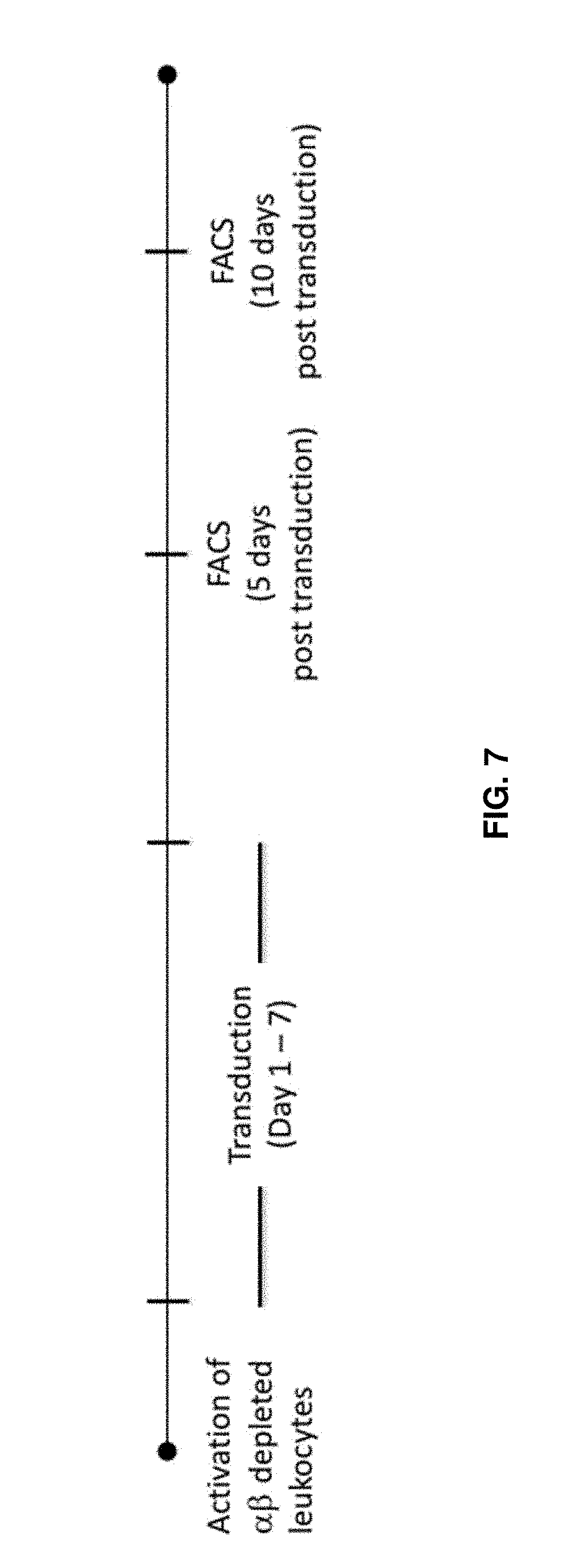
D00013

D00014

D00015

D00016

D00017

D00018

D00019

D00020

D00021

D00022
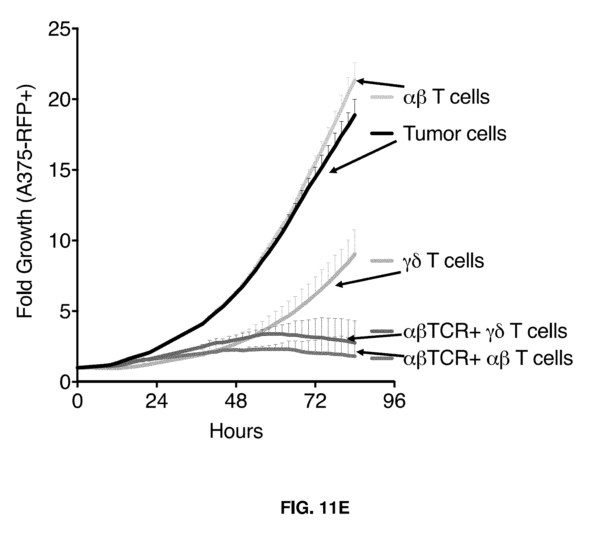
D00023

D00024

D00025

S00001
XML
uspto.report is an independent third-party trademark research tool that is not affiliated, endorsed, or sponsored by the United States Patent and Trademark Office (USPTO) or any other governmental organization. The information provided by uspto.report is based on publicly available data at the time of writing and is intended for informational purposes only.
While we strive to provide accurate and up-to-date information, we do not guarantee the accuracy, completeness, reliability, or suitability of the information displayed on this site. The use of this site is at your own risk. Any reliance you place on such information is therefore strictly at your own risk.
All official trademark data, including owner information, should be verified by visiting the official USPTO website at www.uspto.gov. This site is not intended to replace professional legal advice and should not be used as a substitute for consulting with a legal professional who is knowledgeable about trademark law.