Gene-modified Non-human Animal Expressing Human Gpc3 Polypeptide
JISHAGE; Koichi ; et al.
U.S. patent application number 16/327218 was filed with the patent office on 2019-06-13 for gene-modified non-human animal expressing human gpc3 polypeptide. The applicant listed for this patent is CHUGAI SEIYAKU KABUSHIKI KAISHA. Invention is credited to Hiroshi HINO, Takahiro ISHIGURO, Koichi JISHAGE, Yasuko KINOSHITA.
| Application Number | 20190174731 16/327218 |
| Document ID | / |
| Family ID | 61245019 |
| Filed Date | 2019-06-13 |


View All Diagrams
| United States Patent Application | 20190174731 |
| Kind Code | A1 |
| JISHAGE; Koichi ; et al. | June 13, 2019 |
GENE-MODIFIED NON-HUMAN ANIMAL EXPRESSING HUMAN GPC3 POLYPEPTIDE
Abstract
Genetically modified non-human animals which are deficient in expression of an endogenous GPC3 polypeptide and express a human GPC3 polypeptide at a physiologically adequate level; methods for producing the non-human animals; and methods for evaluating test substances using the non-human animals. Furthermore, methods for evaluating test substances regarding their safety, therapeutic effects on diseases, pharmacokinetics, in vivo distribution, and such, using the non-human animals as models.
| Inventors: | JISHAGE; Koichi; (Shizuoka, JP) ; HINO; Hiroshi; (Shizuoka, JP) ; ISHIGURO; Takahiro; (Tokyo, JP) ; KINOSHITA; Yasuko; (Kanagawa, JP) | ||||||||||
| Applicant: |
|
||||||||||
|---|---|---|---|---|---|---|---|---|---|---|---|
| Family ID: | 61245019 | ||||||||||
| Appl. No.: | 16/327218 | ||||||||||
| Filed: | August 21, 2017 | ||||||||||
| PCT Filed: | August 21, 2017 | ||||||||||
| PCT NO: | PCT/JP2017/029766 | ||||||||||
| 371 Date: | February 21, 2019 |
| Current U.S. Class: | 1/1 |
| Current CPC Class: | C07K 16/2809 20130101; C12N 2015/8518 20130101; A01K 2227/105 20130101; C07K 16/303 20130101; C07K 2317/31 20130101; C07K 14/705 20130101; A01K 2217/206 20130101; C12N 15/85 20130101; A61K 2039/505 20130101; G01N 33/5041 20130101; A01K 67/0278 20130101; A01K 2217/052 20130101; C12N 5/10 20130101; G01N 33/5088 20130101; A01K 2217/072 20130101 |
| International Class: | A01K 67/027 20060101 A01K067/027; G01N 33/50 20060101 G01N033/50; C12N 15/85 20060101 C12N015/85; C07K 16/28 20060101 C07K016/28; C07K 16/30 20060101 C07K016/30 |
Foreign Application Data
| Date | Code | Application Number |
|---|---|---|
| Aug 22, 2016 | JP | 2016-16777 |
Claims
1. A genetically modified non-human animal, wherein the animal is deficient in expression of an endogenous GPC3 polypeptide and expresses a human GPC3 polypeptide.
2. The genetically modified non-human animal of claim 1, wherein the animal expresses the human GPC3 polypeptide at a physiologically adequate level.
3. The genetically modified non-human animal of claim 1 or 2, wherein a copy number of human GPC3 mRNA in total RNA is equivalent to a copy number of non-human animal GPC3 mRNA in total RNA in a wild-type non-human animal.
4. The genetically modified non-human animal of claim 1, wherein the copy number of human GPC3 mRNA in total RNA is equivalent to either one or both of the copy number of monkey GPC3 mRNA in total RNA in a wild-type monkey and the copy number of human GPC3 mRNA in total RNA in a human.
5. The genetically modified non-human animal of claim 1, wherein the animal shows immune tolerance to the human GPC3 polypeptide or a fragment thereof.
6. The genetically modified non-human animal of claim 1, wherein the non-human animal is a non-human mammal, and preferably a rodent.
7. The genetically modified non-human animal of claim 1, wherein the non-human animal is a mouse.
8. A tissue or cell, which is isolated from the genetically modified non-human animal of claim 1.
9. The genetically modified non-human animal of claim 1, wherein the non-human animal comprises a human GPC3 polypeptide-expressing cancer tissue or cancer cell.
10. The genetically modified non-human animal of claim 9, which is for use in screening for a therapeutic agent for cancer.
11. A DNA construct comprising a human GPC3 gene-coding DNA, wherein the DNA comprises an exon-intron structure-comprising nucleotide sequence added to the 5' side and a 3' untranslated region of the non-human animal GPC3 gene added to the 3' side.
12. A knock-in vector, which comprises the DNA construct of claim 11.
13. A method of screening for a therapeutic agent for cancer, the method comprising the steps of: (1) administering an antigen-binding molecule that binds to a human GPC3 polypeptide to the genetically modified non-human animal of any one of claims 1, 2, 4-7, 9, or 10; (2) measuring at least one evaluation index selected from the group consisting of cancer cell proliferation inhibitory effect, safety, pharmacokinetics, and in vivo distribution characteristics in the genetically modified non-human animal to which the test substance has been administered; and (3) selecting the antigen-binding molecule when it is superior in the evaluation index measured in step 2 compared to the evaluation index of a control.
14. The screening method of claim 13, wherein the antigen-binding molecule is an antibody.
15. A method for producing an antibody, wherein the method comprises obtaining information on amino acid sequences of the antibody selected by the screening method of claim 13, and introducing gene(s) coding for the amino acid sequences into a host cell.
Description
TECHNICAL FIELD
[0001] The present invention relates to genetically modified non-human animals expressing a human GPC3 polypeptide and methods for evaluating compounds by using the genetically modified non-human animals expressing the human GPC3 polypeptide.
BACKGROUND ART
[0002] Recently, many therapeutic agents with high specificity to molecular targets, such as therapeutic antibodies, have been developed, and to appropriately carry out preclinical evaluation on them, there is an increasing demand for humanized non-human animals such as humanized mice. Methods for preparing humanized mice include gene targeting methods in which a mouse gene is substituted with a human gene. So far, methods for substituting a human genomic gene for a homologous mouse gene have been reported, but the expression level of the substituting human gene is lower than the expression level of the homologous mouse gene, and the expression is difficult to be regulated (Non-patent Document 1). When a coding sequence of a full-length human gene is inserted into a target mouse gene, the transcribed mRNA will have a structure in which a termination codon (premature termination codon (PTC)) of the inserted human gene is present far upstream of the termination codon of the mouse gene, and the exon-exon junction derived from the mouse gene is present downstream of this premature termination codon. Since this structure is recognized by a nonsense mutation-mediated mRNA decay mechanism (NMD mechanism) and the mRNA undergoes decay, the desired gene expression level cannot be obtained in many cases. As measures against this, for example, there is a report of a method that inserts a sequence called hp7 to the 3' side of a DNA coding for a foreign gene to avoid decay of the foreign gene mRNA by the NMD mechanism, and thereby stably expressing the foreign gene in a mouse (Patent Document 1). Other than hp7, as one method for avoiding NMD, a polyadenylation signal is added immediately downstream of the cDNA sequence of a foreign gene, and this is inserted into a mouse. As a result, the transcribed mRNA has a structure in which a targeted gene-derived exon-exon junction is not produced downstream of the PTC, and thus NMD does not occur. In addition, examples of factors involved in mRNA stability include 3' untranslated regions (3'UTR) and splicing mechanisms. There are reports that a polyadenylation signal present in 3'UTR contributes to mRNA stability (Non-patent Document 2), and that the presence of adenine/uridine-rich elements (Non-patent Document 3) and GU-rich elements (Non-patent Document 4) contribute to protein translation regulation. Furthermore, there are reports that expression levels of genes with no introns, that is, expression levels of mRNAs that do not undergo splicing-out decrease (Non-patent Document 5). Meanwhile, as for a gene having an exon-intron structure, the genome region can be substituted with a desired genome region for insertion when the length of the gene is as short as tens of kilobases. However, when the length of the gene exceeds hundreds kilobases, substitution in the region is difficult (Non-patent Document 5). Thus, there has been no generally effective method for producing non-human animals that suppresses expression of the non-human animal endogenous gene and is capable of expressing a foreign gene at a physiologically adequate level.
[0003] Under such circumstances, there has been no non-human animal which is deficient in expression of the non-human animal endogenous GPC3 polypeptide and is capable of expressing a human GPC3 polypeptide at a physiologically adequate level, and method for production thereof had been unknown.
CITATION LIST
Non-Patent Documents
[0004] [Non-patent Document 1] Proc. Natl. Acad. Sci. U.S.A. 2011 Feb. 8; 108(6):2390-2395.
[0005] [Non-patent Document 2] Int. J. Biochem. Cell. Biol. 2008, 40(11):2384-2396.
[0006] [Non-patent Document 3] J. Cell. Biol. 2008 Apr. 21; 181(2):189-194.
[0007] [Non-patent Document 4] RNA. Biol. 2008. October-December; 5(4):201-207
[0008] [Non-patent Document 5] Proc. Natl. Acad. Sci. U.S.A. 85:836-840.
[0009] [Patent Document]
[0010] [Patent Document 1] WO 2014042251 A1
SUMMARY OF THE INVENTION
Problems to be Solved by the Invention
[0011] The present invention was achieved in view of the above circumstances. An objective of the present invention is to provide genetically modified non-human animals expressing a human GPC3 polypeptide, methods for producing the genetically modified non-human animals, and methods of screening for therapeutic agents for various diseases using the genetically modified non-human animals.
Means for Solving the Problems
[0012] The present inventors have carried out dedicated research on methods for producing non-human animals which are deficient in expression of the non-human animal endogenous GPC3 polypeptide and capable of expressing a human GPC3 polypeptide at a physiologically adequate level. As a result, surprisingly, use of an exon-intron structure sequence and the 3' untranslated region of the wild-type GPC3 gene of a non-human animal enabled generation of a non-human animal that expresses the human GPC3 gene at a physiologically adequate level.
[0013] Furthermore, the present inventors have discovered that the non-human animals which are deficient in expression of the non-human animal endogenous GPC3 polypeptide and capable of expressing a human GPC3 polypeptide at a physiologically adequate level show immune tolerance to human GPC3. That is, the use of the non-human animals as models enables accurate and convenient evaluation of test substances for their safety, therapeutic effects on diseases, pharmacokinetics, in vivo distribution, and such. Furthermore, by using this evaluation system, antibodies having a desired activity can be developed efficiently.
[0014] More specifically, for example, the following invention is provided: [0015] [1] a genetically modified non-human animal, wherein the animal is deficient in expression of an endogenous GPC3 polypeptide and expresses a human GPC3 polypeptide; [0016] [2] the genetically modified non-human animal of [1], wherein the animal expresses the human GPC3 polypeptide at a physiologically adequate level; [0017] [3] the genetically modified non-human animal of [1] or [2], wherein a copy number of human GPC3 mRNA in total RNA is equivalent to a copy number of non-human animal GPC3 mRNA in total RNA in a wild-type non-human animal; [0018] [4] the genetically modified non-human animal of any one of [1] to [3], wherein the mRNA copy number of human GPC3 in total RNA is equivalent to either one or both of mRNA copy number of monkey GPC3 in total RNA in a wild-type monkey and mRNA copy number of human GPC3 in total RNA in a human; [0019] [5] the genetically modified non-human animal of any one of [1] to [4], wherein the animal shows immune tolerance to the human GPC3 polypeptide or a fragment thereof; [0020] [6] the genetically modified non-human animal of any one of [1] to [5], wherein the non-human animal is a non-human mammal, and preferably a rodent; [0021] [7] the genetically modified non-human animal of any one of [1] to [6], wherein the non-human animal is a mouse; [0022] [8] a tissue or cell, which is isolated from the genetically modified non-human animal of any one of [1] to [7]; [0023] [9] the genetically modified non-human animal of any one of [1] to [7], wherein the non-human animal comprises a human GPC3 polypeptide-expressing cancer tissue or cancer cell; [0024] [10] the genetically modified non-human animal of [9], which is for use in screening for a therapeutic agent for cancer; [0025] [11] a DNA construct comprising a human GPC3 gene-coding DNA, wherein the DNA comprises an exon-intron structure-comprising nucleotide sequence added to the 5' side and a 3' untranslated region of the non-human animal GPC3 gene added to the 3' side; [0026] [12] a knock-in vector, which comprises the DNA construct of [11]; [0027] [13] a method of screening for a therapeutic agent for cancer, the method comprising the steps of: [0028] (1) administering an antigen-binding molecule that binds to a human GPC3 polypeptide to the genetically modified non-human animal of any one of [1] to [7], [9], and [10]; [0029] (2) measuring at least one evaluation index selected from the group consisting of cancer cell proliferation inhibitory effect, safety, pharmacokinetics, and in vivo distribution characteristics in the genetically modified non-human animal to which the test substance has been administered; and [0030] (4) selecting the antigen-binding molecule when it is superior in the evaluation index measured in step (3) compared to the evaluation index of a control; [0031] [14] the screening method of [13], wherein the antigen-binding molecule is an antibody; [0032] [15] a method for producing an antibody, wherein the method comprises obtaining information on amino acid sequences of the antibody selected by the screening method of [13] or [14], and introducing gene(s) coding for the amino acid sequences into a host cell; [0033] [16] the method of [15], wherein the host cell is a CHO cell.
[0034] Furthermore, for example, the following invention is provided: [0035] [17] a genetically modified non-human animal comprising a human GPC3 gene-coding DNA, wherein the DNA comprises an exon-intron structure-comprising nucleotide sequence added to the 5' side and a 3' untranslated region of the non-human animal GPC3 gene added to the 3' side, and wherein the DNA is inserted into the same reading frame as that of a non-human animal endogenous GPC3 gene; [0036] [18] the genetically modified non-human animal of [17], wherein the DNA is a cDNA; [0037] [19] the genetically modified non-human animal of [17] or [18], wherein the exon-intron structure-comprising nucleotide sequence is a sequence comprising a beta globin second exon sequence, intron sequence, and third exon sequence; [0038] [20] the genetically modified non-human animal of any one of [17] to [19], wherein the beta globin is a beta globin of the non-human animal; [0039] [21] the genetically modified non-human animal of any one of [17] to [20], wherein the 3' untranslated region of the non-human animal GPC3 gene is a region comprising a polyadenylation signal sequence; [0040] [22] the genetically modified non-human animal of any one of [17] to [21], which shows immune tolerance to a human GPC3 polypeptide or a fragment thereof; [0041] [23] the genetically modified non-human animal of any one of [17] to [22], wherein the non-human animal is a non-human mammal, and preferably a rodent; [0042] [24] the genetically modified non-human animal of any one of [17] to [23], wherein the non-human animal is a mouse; [0043] [25] a tissue or cell, which is isolated from the genetically modified non-human animal of any one of [17] to [24]; [0044] [26] the genetically modified non-human animal of any one of [17] to [24], wherein the non-human animal comprises a human GPC3 polypeptide-expressing cancer tissue or cancer cell; [0045] [27] the genetically modified non-human animal of [26], which is for screening for a therapeutic agent for cancer.
[0046] Furthermore, for example, the following invention is provided: [0047] [28] a DNA construct comprising a human GPC3 gene-coding DNA, wherein the DNA comprises an exon-intron structure-comprising nucleotide sequence added to the 5' side and a 3' untranslated region of the non-human animal GPC3 gene added to the 3' side; [0048] [29] the DNA construct of [28], wherein the DNA is a cDNA; [0049] [30] the DNA construct of [28] or [29], wherein the exon-intron structure-comprising nucleotide sequence is a sequence comprising a beta globin second exon sequence, intron sequence, and third exon sequence; [0050] [31] the DNA construct of any one of [28] to [30], wherein the beta globin is a mouse beta globin; [0051] [32] the DNA construct of any one of [28] to [31], wherein the 3' untranslated region of the non-human animal GPC3 gene is a region comprising a polyadenylation signal sequence; [0052] [33] the DNA construct of any one of [28] to [32], which further comprises recombinase substrate sequences, a drug selection marker, and/or another sequence; [0053] [34] a knock-in vector, which comprise the DNA construct of any one of [28] to [33]; [0054] [35] the knock-in vector of [34], which comprises a nucleotide sequence homologous to the 5'-side upstream region of a non-human animal GPC3 gene target region at the 5' side of the DNA construct, and a nucleotide sequence homologous to the 3'-side downstream region of the non-human animal GPC3 gene target region at the 3' side of the DNA construct; [0055] [36] a non-human animal cell, wherein the knock-in vector of [35] has been introduced; [0056] [37] the non-human animal cell of [36], wherein the cell is an embryonic stem cell (ES cell), an induced pluripotent stem cell (iPS cell), a germline stem cell, or a fertilized egg.
[0057] Furthermore, for example, the following invention is provided: [0058] [38] a method for evaluating therapeutic effects of a test substance on cancer, the method comprising the steps of: [0059] (1) administering a test substance to the genetically modified non-human animal of any one of [1] to [7], [9], [10], [17] to [24], [26], and [27] wherein the animal comprises a human GPC3 polypeptide-expressing cancer tissue or cancer cell; [0060] (2) measuring a cancer cell proliferation inhibitory effect in the genetically modified non-human animal to which the test substance was administered; and [0061] (3) selecting a test substance having a significantly high cancer cell proliferation inhibitory effect measured in (2) compared to that of a control; [0062] [39] a method for evaluating safety of a test substance, the method comprising the steps of: [0063] (1) administering a test substance to the genetically modified non-human animal of any one of [1] to [7], [9], [10], [17] to [24], [26], and [27]; [0064] (2) measuring cytokine release in the genetically modified non-human animal to which the test substance was administered; and [0065] (3) comparing the cytokine level measured in step (2) to the cytokine level of a control, wherein the change in cytokine level indicates the safety risk to be caused; [0066] [40] the method of [39], wherein the non-human animal comprises a human GPC3 polypeptide-expressing cancer tissue or cancer cell; [0067] [41] a method for evaluating pharmacokinetic characteristics of a test substance, the method comprising the steps of: [0068] (1) administering a test substance to the genetically modified non-human animal of any one of [1] to [7], [9], [10], [17] to [24], [26], and [27]; and [0069] (2) measuring the time-dependent changes in blood concentration of the test substance in the genetically modified non-human animal to which the test substance was administered; [0070] [42] the method of [41], wherein the non-human animal comprises a human GPC3 polypeptide-expressing cancer tissue or cancer cell; [0071] [43] a method for evaluating in vivo distribution characteristics of a test substance, the method comprising the steps of: [0072] (1) administering a test substance to the genetically modified non-human animal of any one of [1] to [7], [9], [10], [17] to [24], [26], and [27]; [0073] (2) measuring the in vivo distribution of the test substance in the genetically modified non-human animal to which the test substance was administered; and [0074] (3) indicating the in vivo distribution characteristics of the test substance through localization of the test substance; [0075] [44] the method of [43], wherein the non-human animal comprises a human GPC3 polypeptide-expressing cancer tissue or cancer cell; [0076] [45] the method of any one of [38] to [44], wherein the test substance is an antigen-binding molecule that binds to a human GPC3 polypeptide; [0077] [46] the method of [45], wherein the antigen-binding molecule is an antibody.
[0078] Furthermore, for example, the following invention is provided. [0079] [47] the genetically modified non-human animal of any one of [1] to [10], [17] to [24], [26], and [27], wherein the animal is functionally deficient for at least one or more types of CD3 genes selected from the group consisting of endogenous CD3.epsilon., CD3.delta., and CD3.gamma. in its genome and functionally expresses at least one or more types of human CD3 genes selected from the group consisting of human CD3.epsilon., CD3.delta., and CD3.gamma.; [0080] [48] the genetically modified non-human animal of [47], which functionally expresses human CD3 genes comprising human CD3.quadrature., CD3.quadrature., and CD3.quadrature.; [0081] [49] the genetically modified non-human animal of [47] or [48], wherein a full-length nucleotide sequence of at least one or more types of human CD3 genes selected from the group consisting of human CD3.quadrature., CD3.quadrature., and CD3.quadrature. has been inserted into the genome; [0082] [50] the genetically modified non-human animal of any one of [47] to [49], wherein a T cell receptor derived from the non-human animal and human CD3 molecule(s) form a complex on the cellular membrane of a T cell carried by the non-human animal; [0083] [51] the genetically modified non-human animal of any one of [47] to [50], the animal additionally expressing a human immune checkpoint gene, a human cancer-specific antigen gene, and/or a human immune costimulatory molecule gene; [0084] [52] the genetically modified non-human animal of any one of [47] to [51], wherein the non-human animal is a non-human mammal; [0085] [53] the genetically modified non-human animal of [52], wherein the non-human mammal is a mouse; [0086] [54] the genetically modified non-human animal of any one of [47] to [53], which is for screening for a therapeutic agent for malignant neoplastic disease or autoimmune disease; [0087] [55] the genetically modified non-human animal of [54], which is for screening for a therapeutic agent for malignant neoplastic disease, wherein a cancer cell has been transplanted into the non-human animal; [0088] [56] the genetically modified non-human animal of [55], wherein the cancer cell is a cell derived from lung cancer, gastric cancer, liver cancer, esophageal cancer, or ovarian cancer; [0089] [57] a method of screening for a therapeutic agent for malignant neoplastic disease or autoimmune disease, the method comprising the steps of: [0090] (1) contacting a test substance with the genetically modified non-human animal of any one of [47] to [56], or an organ, tissue, or cell thereof; and [0091] (2) selecting a candidate test substance using as an indicator drug efficacy and/or toxicity of the test substance in the genetically modified non-human animal individual, or the organ, tissue, or cell thereof; [0092] [58] a method of screening for a therapeutic agent for malignant neoplastic disease, the method comprising the steps of: [0093] (1) administering one from a library of antigen-binding molecules as a test substance to a first genetically modified non-human animal of any one of [0094] [47] to [57], the antigen-binding molecules comprising a human CD3-binding domain and a cancer-specific antigen-binding domain; [0095] (2) measuring a cell proliferation inhibitory effect and/or pharmacokinetic characteristics of the test substance on a cell expressing the cancer-specific antigen; and [0096] (3) comparing the cell proliferation inhibitory effect and/or pharmacokinetic characteristics of the test substance with the cell proliferation inhibitory effect and/or pharmacokinetic characteristics of a control antibody administered to a second genetically modified non-human animal which is different from the first non-human animal; [0097] [59] a method for producing a non-human animal expressing a human GPC3 polypeptide and functionally expressing human CD3 genes, the method comprising the steps of: [0098] (1) preparing a genetically modified non-human animal which is deficient in the expression of the endogenous GPC3 polypeptide and expresses a human GPC3 polypeptide; [0099] (2) preparing a genetically modified non-human animal which is functionally deficient in at least one or more types of CD3 genes selected from the group consisting of endogenous CD3.epsilon., CD3.delta., and CD3.gamma. in its genome and functionally expresses at least one or more types of human CD3 genes selected from the group consisting of human CD3.epsilon., CD3.delta., and CD3.gamma.; and [0100] (3) crossing the genetically modified non-human animal expressing the human GPC3 polypeptide with the genetically modified non-human animal functionally expressing the human CD3 genes.
BRIEF DESCRIPTION OF THE DRAWINGS
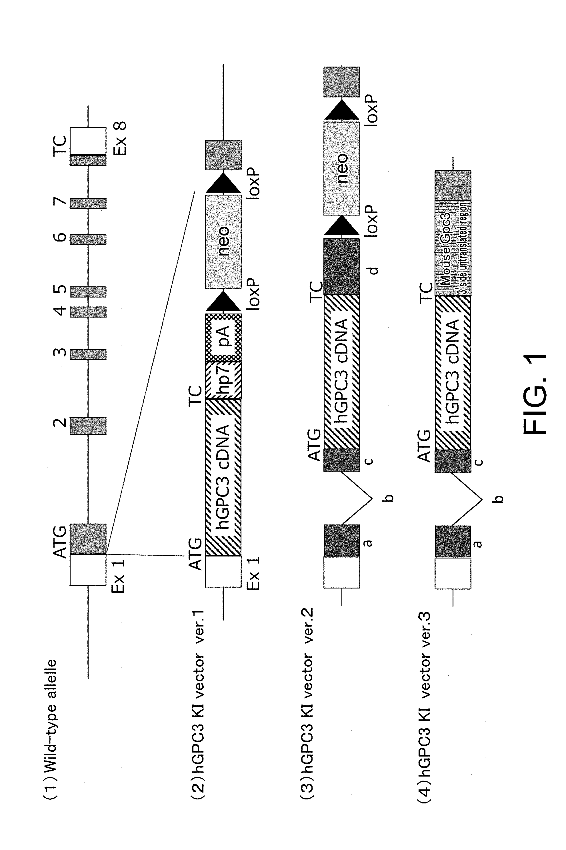
[0101] FIG. 1 schematically presents the relationship between the structure of a genomic DNA of the mouse glypican-3 (mGPC3) gene (1), and the knock-in vectors ver. 1 (2), ver. 2 (3), and ver. 3 (4) to be inserted. Knock-in vector ver. 1 (2) carries a human glypican-3 (hGPC3) cDNA, an hp7 sequence, a polyadenylation signal (pA), and a neomycin-resistance gene (neo) flanked by loPs which are substrate sequences for Cre recombinase. Allowing Cre to act causes site-specific recombination between the loxPs, and the neomycin resistance gene flanked by the loxPs is removed. Knock-in vector ver. 2 (3) carries the approximately 800-nucleotide 5' upstream region of the mGpc3 gene target region; the mouse beta globin second exon, intron, and third exon; hGPC3 cDNA; the polyadenylation signal in the mouse beta globin third exon; loxP sequences; neo gene; and the approximately 800-nucleotide 3' downstream region of the mGpc3 gene target region. Knock-in vector ver. 3 (4) is a vector in which the polyadenylation signal in the mouse beta globin third exon of knock-in vector ver. 2 (3) has been changed to the 3' untranslated region of the mGpc3 gene. In FIG. 1, "a" indicates the mouse beta globin second exon, "b" indicates the mouse beta globin second intron, "c" indicates the mouse beta globin third exon, and "d" indicates the mouse beta globin third exon containing the polyadenylation signal.
[0102] FIG. 2 presents a method of insertion (c) of knock-in vector ver. 1 (b) into a genomic DNA of the mGpc3 gene (a) by homologous recombination. It presents a process of subsequently removing the neomycin resistance gene (neo) flanked by loxPs using recombinase Cre to thereby complete a hGPC3 knock-in allele (d). Neo is removed from knock-in vector ver. 2 by a similar method.
[0103] FIG. 3 presents the representative examples of PCR analyses performed to screen for homologous recombinant ES cells generated with hGPC3 knock-in vector ver. 1 and homologous recombinant ES cells generated with hGPC3 knock-in vector ver. 2 [hGPC3 knock-in vector ver. 1 (a) and hGPC3 knock-in vector ver. 2 (b)], and the representative example of PCR analyses (c) performed to detect founder mice carrying the hGPC3 knock-in vector ver. 3 gene.
[0104] FIG. 4 presents the representative examples of PCR performed to detect neo gene cassette removal in hGPC3 knock-in mice ver. 1 and hGPC3 knock-in mice ver. 2 [hGPC3 knock-in mice ver. 1 (a) and hGPC3 knock-in mice ver. 2 (b)];
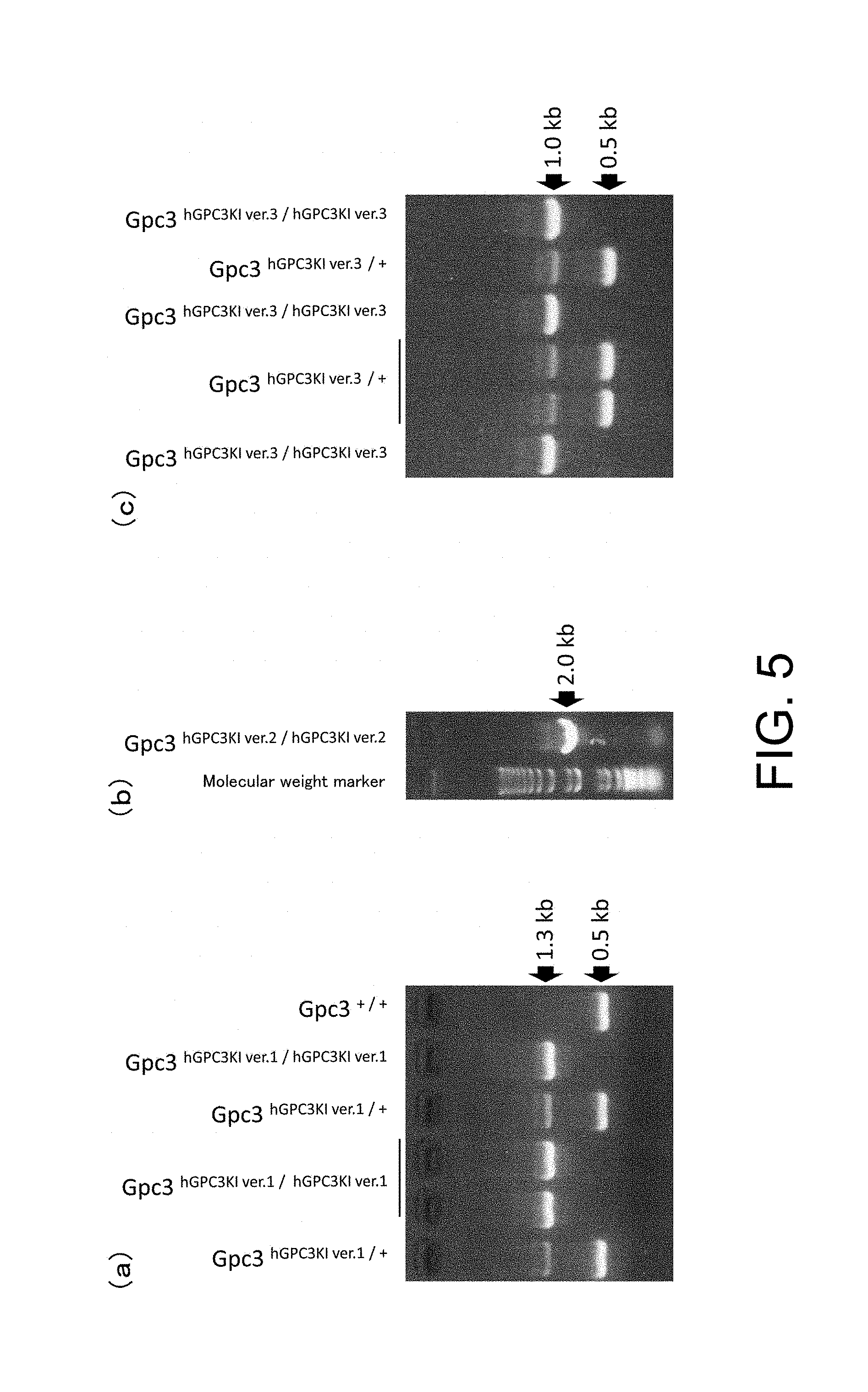
[0105] FIG. 5 presents the representative examples of PCR performed to detect homozygous and hemizygous hGPC3 knock-in mice ver. 1, hGPC3 knock-in mice ver. 2, and hGPC3 knock-in mice ver. 3 [hGPC3 knock-in mice ver. 1 (a), hGPC3 knock-in mice ver. 2 (b), and hGPC3 knock-in mice ver. 3 (c)]. hGPC3KI/hGPC3KI, hGPC3KI/-, hGPC3KI/+, and +/+ indicate homozygous knock-in mouse, hemizygous knock-in mouse, heterozygous knock-in mouse, and wild-type mouse, respectively.
[0106] FIG. 6 presents the Western blotting results for the lungs of hGPC3 knock-in mice ver. 1 and wild-type mice, using an anti-human GPC3 antibody. Lysate concentration refers to total protein weight of the lung lysate applied to each lane.
[0107] FIG. 7 presents the Western blotting results for the lungs of hGPC3 knock-in mice ver. 2, hGPC3 knock-in mice ver. 3, and wild-type mice, using an anti-human GPC3 antibody.
[0108] FIG. 8 shows graphs presenting the GPC3 mRNA expression levels in the lungs of hGPC3 knock-in mice ver. 2 and ver. 3, wild-type mice, and cynomolgus monkeys: (a) presents the results for hGPC3 knock-in mice ver. 2 and ver. 3, and cynomolgus monkeys, using Universal Probe #46; and (b) presents the results for wild-type mice, and ver. 2 and ver. 3, using Universal Probe #28.
[0109] FIG. 9 shows a graph presenting the expression levels of GPC3 mRNA in the lungs of hGPC3 knock-in mice ver. 3 and humans.
[0110] FIG. 10 shows a graph presenting the anti-human GPC3 antibody concentration in blood plasma of wild-type mice and hGPC3 knock-in mice ver. 3 when a human GPC3 protein was administered on days 0, 14, 21, 28, 35, 42, and 49.
[0111] FIG. 11 shows a graph presenting the antitumor effects of the hGPC3_mCD3 antibody in the LLC1/hGPC3-transplanted model using hGPC3 knock-in mice ver. 3.
[0112] FIG. 12 shows a graph presenting the change in hGPC3_mCD3 antibody concentration in blood plasma of hGPC3 knock-in mice ver. 1 and hGPC3 knock-in mice ver. 3, two hours, one day, and seven days after hGPC3_mCD3 antibody administration.
[0113] FIG. 13 shows graphs presenting the time course of the change in plasma concentrations of IFN-gamma, IL-10, IL-17, and IL-2 in wild-type mice, hGPC3 knock-in mice ver. 1, and hGPC3 knock-in mice ver. 3 after hGPC3_mCD3 antibody administration.
[0114] FIG. 14 shows graphs presenting the time course of the change in plasma concentrations of IL-4, IL-6, and TNF in wild-type mice, hGPC3 knock-in mice ver. 1, and hGPC3 knock-in mice ver. 3 after hGPC3_mCD3 antibody administration.
[0115] FIG. 15A presents (1) the structure of a genomic DNA containing mouse Cd3.epsilon., Cd3.delta., and Cd3.gamma. genes, (2) a mouse Cd3 gene modification vector constructed by modifying a bacterial artificial chromosome (BAC) clone containing the whole gene region, (3) the structure of a genomic DNA in which loxP and Rox sequences have been inserted at the target position using the above-mentioned vector, and (4) the structure of a Cd3.epsilon., Cd3.delta., and Cd3.gamma.-gene deficient allele produced by the actions of Cre and Dre recombinases.
[0116] FIG. 15B presents (a) the structures of a BAC clone containing human CD3.epsilon., CD3.delta., and CD3.gamma. genes; (b) 5'-modifying cassette and (c) 3'-modifying cassette, both of which are for modifying the BAC clone; and (d) a human CD3 gene region introduction vector constructed through modifications using those above.
[0117] FIG. 16 presents the representative examples of PCR analyses performed for establishing mouse Cd3 gene-modified ES cells.
[0118] FIG. 17 presents the representative examples of PCR analyses of genotypes of ES cell clones obtained by introducing into mouse Cd3 gene-modified ES cells the human CD3 gene region introduction vector along with a Cre expression vector and a Dre expression vector. FIG. 17A presents the representative examples of PCR results that detect the deficiency of the mouse Cd3 gene region. FIG. 17B presents the representative examples of PCR results that detect the introduction of the human CD3 gene region.
[0119] FIG. 18 presents the representative macroscopic photographs of thymuses collected from each of the established lines of human CD3 gene-substituted mice, Cd3 gene-deficient mice, wild type, and human CD3.epsilon. gene-introduced mice. Thymuses extirpated from 12 to 13-week-old males are shown for the respective genotypes.
[0120] FIG. 19 presents the results of measuring the tissue weights of the spleens and thymuses collected from each of the established lines of human CD3 gene-substituted mice, Cd3 gene-deficient mice, wild-type, and human CD3.epsilon. gene-introduced mice. Ratios of tissue weight per body weight were calculated, and the value obtained for each individual is plotted by a black dot and the mean values are shown by columns.
[0121] FIG. 20 presents the results of examining by RT-PCR the expressions of each of the human CD3 molecules and each of the mouse Cd3 genes in each of the established lines of human CD3 gene-substituted mice, Cd3 gene-deficient mice, wild-type mice, and human CD3.epsilon. gene-introduced (hCD3.epsilon. Tg) mice. Among the established lines of the human CD3 gene-substituted mice, signals specific to hCD3.epsilon., hCD3.delta., and hCD3.gamma. were detected in line numbers 1C3 and 8I12. The signals were not detected in line numbers 3B1 and 2A4.
[0122] FIG. 21 presents the representative examples of immunohistological staining for CD3 performed on the thymus (A) and spleen (B) of each established line of human CD3 gene-substituted mice (1C3, 8I12, and 4HH3). In both tissues, staining was observed only in the T cell zone as in the wild-type mouse. Furthermore, staining was not observed in the Cd3 gene-deficient mice, and this showed that the staining in the human CD3 gene-substituted mice was due to the expression of the introduced human CD3 genes.
[0123] FIG. 22 presents the representative results of analyzing by FACS the abundance ratio of mature T cells in the thymus of each established line of human CD3-substituted mice.
[0124] FIG. 23 presents the mitogen-stimulated cell proliferation activities of spleen cells from the human CD3 gene-substituted mice (1C3), Cd3 gene-deficient mice, and wild-type mice. The established human CD3 gene-substituted mice (1C3) showed 90% of cell proliferation activity compared to the wild type.
[0125] FIG. 24 presents the mitogen-stimulated cytokine production by spleen cells from the human CD3 gene-substituted mice (1C3), Cd3 gene-deficient mice, and wild-type mice. It was shown that cytokine-producing ability that was functionally lost in the Cd3 gene-deficient mice was recovered in the established human CD3 gene-substituted mice (1C3).
[0126] FIGS. 25A to 25D present the anti-CD3 antibody-stimulated cell proliferation activities (FIGS. 11A and 11B) and cytokine productions (FIGS. 11C and 11D) by spleen cells from the human CD3 gene-substituted mice (1C3), Cd3 gene-deficient mice, and wild-type mice. The established human CD3 gene-substituted mice (1C3) responded specifically to the anti-human CD3 antibody stimulation, and showed cell proliferation activity and cytokine production.
[0127] FIG. 25B. See the explanation under FIG. 25A.
[0128] FIG. 25C. See the explanation under FIG. 25A.
[0129] FIG. 25D. See the explanation under FIG. 25A.
[0130] FIG. 26 presents the results of measuring the chicken ovoalbumin (OVA)-specific IgG1 and IgE serum concentrations in each established line of human CD3-substituted mice immunized with OVA. The OVA-specific serum IgG1 and IgE concentrations for each individual are shown as a bar graph. The numbers below the bar graph indicate the individual identification numbers.
[0131] FIG. 27 presents the change in tumor volume in Hepa1-6/HER2 cell-transplanted hCD3 transgenic mouse model to which the HER2_CD3 antibody was administered. The arrow indicates antibody administration (*: P<0.05 (t-test))
[0132] FIG. 28 presents the change in tumor volume in Hepa1-6/hGPC3 cell-transplanted hCD3 transgenic mouse model to which anti-mouse CTLA-4 antibody, anti-mouse PD-1 antibody, or anti-mouse PD-L1 antibody was administered. The arrows indicate antibody administration.
[0133] FIG. 29 presents the change in tumor volume in Colon 38 cell line-transplanted mouse model to which MDX10//TR01H113 or the buffer as a control (in the figure, referred to as "MDX10//TRO01H1333" and "vehicle", respectively) was administered. Each point shows the mean value of tumor volumes for n=5 per group. When the mean values were compared at the final measurement points, significant difference was observed between the two groups, the MDX10//TR01H113-administered group and the buffer-administered group (p=0.0021, Student's t-test).
[0134] FIG. 30 shows the change in tumor volume in Hepa1-6/hGPC3 cell-transplanted human GPC3 knock-in human CD3 gene-substituted mouse model to which the hGPC3_hCD3 antibody was administered. The arrows indicate antibody administration. *: P<0.05 (Steel test, JMP software, SAS institute Inc.)
[0135] FIG. 31 shows the changes in blood plasma cytokine concentration in Hepa1-6/hGPC3 cell-transplanted human GPC3 knock-in human CD3 gene-substituted mouse model on the day before hGPC3_hCD3 antibody administration, and 6 hours and 24 hours after the administration, and 6 hours after the second administration. The broken lines indicate the detection limit.
MODE FOR CARRYING OUT THE INVENTION
1. Definition
[0136] Unless defined otherwise, all technical and scientific terms used herein have the same meaning as commonly understood by one of ordinary skill in the art to which this invention belongs. Although any methods and materials similar or equivalent to those described herein can be used in the practice or testing of the present invention, the specific methods and materials are described herein. All publications referred to herein are incorporated in their entirety by reference into this description.
[0137] Herein, unless a limitation referring to a numerical quantity such as "a single" or "multiple" is specifically used to describe a term, the terms recited herein should not be interpreted as being particularly limited in numerical quantity, and should be understood as terms with the meaning "one or more of".
[0138] "Conservative substitution" takes place within a family of amino acids that are related with regard to their side chains and chemical properties. For example, amino acids can be classified into the groups on the basis of their commonly shared side chain properties: [0139] (1) hydrophobic: norleucine, methionine (Met), alanine (Ala), valine (Val), leucine (Leu), and isoleucine (Ile); [0140] (2) neutral hydrophilic: cysteine (Cys), serine (Ser), threonine (Thr), asparagine (Asn), and glutamine (Gln); [0141] (3) acidic: aspartic acid (Asp) and glutamic acid (Glu); [0142] (4) basic: histidine (His), lysine (Lys), and arginine (Arg); [0143] (5) residues that affect the chain orientation: glycine (Gly) and proline (Pro); and [0144] (6) aromatic: tryptophan (Trp), tyrosine (Tyr), and phenylalanine (Phe).
[0145] Non-conservative substitution refers to an exchange of a member of any one of these classes with a member of a different class. In a non-limiting embodiment, the present invention includes genetically modified non-human animals that express a human GPC3 polypeptide containing a conservative amino acid substitution in an amino acid sequence described herein.
[0146] The term "target gene" includes endogenous genes of a non-human animal which becomes a target for insertion of a foreign gene. The term "target region" includes specific regions of endogenous genes of a non-human animal which becomes a target for insertion of a foreign gene. In a non-limiting embodiment, target regions refer to endogenous gene-containing regions which are adjacent to the 5' side and 3' side of an inserted foreign gene. In another embodiment, a target region refers to regions of an endogenous gene whose expression becomes deleted due to insertion of a foreign gene.
[0147] The term "endogenous" includes substances derived natively from inside organisms, tissues, cells, or such. For example, endogenous nucleic acids or peptides are present inside cells, and refer to nucleic acids or peptides that were not introduced into cells using recombinant engineering techniques.
[0148] The term "foreign" includes substances derived from other organisms, tissues, cells, or such that are different from organisms, tissues, or cells to be targeted. For example, "foreign genes" include genes introduced into non-human animals of the present invention. Foreign genes in the present invention may be used without being limited to a certain organism species from which they are derived, but they are preferably human genes. Furthermore, as foreign genes, reporter genes such as green fluorescent protein (GFP) and 3-galactosidase, and selection marker genes such as drug-resistance genes (such as neomycin-resistance gene) may be used. A combination of two or more genes may also be used as foreign genes. Furthermore, foreign genes may have enhancers that regulate their expression, added to them. The forms of foreign genes are not particularly limited, and may be, for example, cDNA or genomic DNA.
[0149] The term "isolated" refers to being separated from the components of its original environment.
[0150] The term "functionally linked" includes a condition where a gene is linked under conditions that allow the gene to exhibit the function of interest. For example, a nucleic acid sequence coding for a protein may be operably linked to regulatory sequences (for example, promoter, enhancer, silencer sequence, etc.) to retain proper transcriptional regulation. As long as the sequence functions in that manner, the promoter does not have to be contiguously linked to the sequence. Herein, the term "functionally linked" may be used interchangeably with the term "operably linked".
[0151] Examples of the "vector" include genetically engineered plasmid or virus that is derived from a bacteriophage, adenovirus, retrovirus, poxvirus, herpesvirus, or artificial chromosome, but are not limited thereto.
[0152] The term "non-human animal" is not particularly limited as long as it is an animal other than a human, and examples include mouse, rat, guinea pig, hamster, rabbit, goat, cattle, horse, pig, dog, cat, and monkey. The non-human animal is preferably a mammal, an animal classified as a rodent, and more preferably a mouse. Examples of the preferred mouse include C57/BL/6, ICR, and BALB/c, but are not limited thereto.
[0153] Herein, "wild-type" includes having ordinary structure and/or activity found in nature. Wild-type nucleic acid or peptide includes a plurality of different forms and polymorphisms such as allelic mutations. Furthermore, "wild-type" non-human animals include, in some cases, animals having a wild-type GPC3 gene, or more specifically, animals not subjected to genetic engineering of GPC3.
[0154] Herein, the term "antibody" is used in the broadest sense and encompasses various antibody structures so long as they exhibit the desired antigen-binding activity, including but being not limited to monoclonal antibodies, polyclonal antibodies, multispecific antibodies (for example, bispecific antibodies), and antibody fragments.
[0155] An "antibody fragment" refers to a molecule other than an intact antibody that comprises a portion of the intact antibody, which portion binds to an antigen bound by the intact antibody. Examples of an antibody fragment include but are not limited to Fv, Fab, Fab', Fab'-SH, and F(ab')2; diabodies; linear antibodies; single-chain antibody molecules (for example, scFv); and multispecific antibodies formed from antibody fragments.
[0156] Herein, the terms such as "cancer", "carcinoma", "tumor", and "neoplasm" are not differentiated from each other, and are mutually interchangeable, and mean the generally expressed term "cancer".
2. Glypican 3 (GPC3)
[0157] Glypican 3 (GPC3) is a member of a family of heparin sulfate proteoglycans present on cell surfaces. While it is suggested to be possibly involved in cell division during development and proliferation of cancer cells, its function has not yet been clearly elucidated. In a non-limiting embodiment, examples of a GPC3 polypeptide sequence include SEQ ID NO: 1 (human, NCBI RefSeq: NP_004475.1), SEQ ID NO: 2 (monkey, NCBI RefSeq: XP_005594665.1), and SEQ ID NO: 3 (mouse, NCBI RefSeq: NP_057906.2), and examples of the DNA sequence include SEQ ID NO: 4 (human, NCBI RefSeq: NM_004484.3), SEQ ID NO: 5 (monkey, NCBI RefSeq: XM_005594608.2), and SEQ ID NO: 6 (mouse, NCBI RefSeq: NM_016697.3). In a non-limiting embodiment, glypican 3 (GPC3) or GPC3 polypeptide includes a full-length GPC3 polypeptide and a fragment thereof, and they may contain amino acid mutations.
[0158] In a non-limiting embodiment, a "GPC3 gene" is not particularly limited as long as it is a GPC3 polypeptide-coding gene, and may be a genomic DNA or a cDNA. Furthermore, a GPC3 gene includes its polymorphic or mutant forms.
3. Non-Human Animals Expressing a Human GPC3 Polypeptide
[0159] In a non-limiting embodiment, the present invention provides non-human animals expressing a human GPC3 polypeptide which comprise a human GPC3 gene-coding DNA. In another embodiment, non-human animals of the present invention include non-human animals in which the human GPC3 gene-coding DNA has been inserted into the same reading frame as that of an endogenous GPC3 gene which was present in the non-human animal genome. The above-mentioned human GPC3 gene-coding DNA may be a genomic DNA or a cDNA. Preferably, it is a cDNA, and specifically, examples include cDNA containing an amino acid sequence-coding region (CDS). An amino acid sequence-coding region includes a signal sequence.
[0160] In a non-limiting embodiment, the term "same reading frame" means a nucleotide sequence unit of every three bases that are read when a mRNA is translated into a protein. The phrase "inserted into the same reading frame" includes inserting a human GPC3 gene so that the initiation codon ATG of the human GPC3 gene comes to the site of the initiation codon ATG of an endogenous GPC3 gene of the non-human animal. Furthermore, the phrase "inserted into the same reading frame or insert(s)/inserting . . . into the same reading frame" also includes the case where an exon-intron structure sequence is added to the 5' side of the initiation codon ATG of the human GPC3 gene and the human GPC3 gene is inserted so that the initiation codon ATG of the endogenous GPC3 gene of the non-human animal is aligned with the 5' end of the exon-intron structure.
[0161] Thus, the promoter for the endogenous GPC3 gene in the non-human animal is operatively linked to the inserted human GPC3 gene, and the human GPC3 gene becomes expressed in response to activation of the promoter. When such constitution is employed, the foreign human GPC3 is expressed under the endogenous GPC3 expression regulation system; therefore, human GPC3 can be regulated to be expressed at a similar timing and place (tissues) as the endogenous GPC3. GPC3 is a gene also predicted to be involved in development. Therefore, employing the above-mentioned constitution is expected to act to reduce effects of gene modification on the survival and development of the non-human animal.
[0162] In another embodiment, it is preferable to delete a sequence having a number of bases that is not a multiple of three from the sequence following ATG of the endogenous GPC3 gene when inserting the human GPC3 gene, from the viewpoint of eliminating the possibility of retranslation of the disrupted endogenous GPC3 gene in the original reading frame. Furthermore, the human GPC3 gene in the present invention is preferably inserted nowhere else than the exon in which the original translation start site of the endogenous GPC3 gene is present (i.e., homologous recombination has taken place only with the target exon of the endogenous GPC3 gene).
[0163] In a non-limiting embodiment, the human GPC3 gene carried by the non-human animals of the present invention includes human GPC3 genes that are at least 50%, 60%, or 70%, preferably 75% or more, 80% or more, or 85% or more, more preferably 90% or more, 91% or more, 92% or more, 93% or more, 94% or more, 95% or more, 96% or more, 97% or more, 98% or more, or 99% or more homologous to SEQ ID NO: 4. In another embodiment, the human GPC3 gene includes its polymorphic or mutant forms. In another embodiment the human GPC3 gene may be a gene coding for a human GPC3 polypeptide in which amino acid insertions, deletions, or conservative amino-acid substitutions have been made.
[0164] In a non-limiting embodiment, the human GPC3 polypeptide expressed by the non-human animals of the present invention includes human GPC3 polypeptides that are at least 50%, 60%, or 70%, preferably 75% or more, 80% or more, or 85% or more, more preferably 90% or more, 91% or more, 92% or more, 93% or more, 94% or more, 95% or more, 96% or more, 97% or more, 98% or more, or 99% or more homologous to SEQ ID NO: 1. In another embodiment, the human GPC3 gene includes its polymorphic or mutant forms. In another embodiment, in the human GPC3 polypeptide, amino acid insertions, deletions, or conservative amino-acid substitutions may be made. In the present invention, homologies of the nucleotide sequences or amino acid sequences can be determined by a known algorithm. Algorithms for determining the homology among multiple sequences are known. Homology of sequence information is determined in some cases by considering the degeneracy of codons in the comparison among nucleotide sequences or by considering commonalities of different amino acid residues in the comparison among amino acid sequences. In other cases, the homology is determined simply by comparing sequence information without considering these factors. For the latter cases, comparison results are preferably presented as sequence identity.
[0165] In a non-limiting embodiment, the non-human animals of the present invention are deficient in expression of the non-human animal endogenous GPC3 polypeptide and can express the human GPC3 polypeptide at a physiologically adequate level. A human GPC3 gene (including mRNA) inserted into a non-human animal of the present invention or the human GPC3 polypeptide expressed from the gene can be detected by various methods well known to those skilled in the art, such as PCR, Southern blotting, Restriction Fragment Length Polymorphis (RFLP) method, Western blotting, IHC, and ELISA.
[0166] In a non-limiting embodiment, for example, embodiments of "deficient in expression of an endogenous GPC3 polypeptide" are not particularly limited as long as the endogenous GPC3 polypeptide in the non-human animal is not expressed. Specifically, for example, the endogenous GPC3 gene may be deleted (abolished) in a non-human animal genome using knockout technology based on genome editing techniques such as homologous recombination, CRISPR-Cas (CRISPR/Cas9), zinc finger nucleases, or TALEN. Alternatively, a method for completely suppressing the expression of an endogenous GPC3 gene by siRNA and such may be used. Furthermore, as long as an endogenous GPC3 polypeptide is not expressed, a foreign gene may be inserted at the position of the GPC3 gene in the non-human animal genome, for example, using knock-in technology.
[0167] In a non-limiting embodiment, the phrase "express the human GPC3 polypeptide at a physiologically adequate level" includes, for example, the case where an expression level of a human GPC3 polypeptide or a human GPC3 mRNA in a non-human animal is equivalent to any one expression level selected from the expression levels consisting of (i) to (iii) below: [0168] (i) an expression level of a mouse GPC3 polypeptide or a mouse GPC3 mRNA in a wild-type mouse; [0169] (ii) an expression level of a monkey GPC3 polypeptide or a monkey GPC3 mRNA in a wild-type monkey; and [0170] (iii) an expression level of a human GPC3 polypeptide or a human GPC3 mRNA in a human.
[0171] Furthermore, it includes, for example, the case where an expression level of a human GPC3 polypeptide or a human GPC3 mRNA in one or more of organs in a non-human animal is equivalent to any one expression level selected from the expression levels consisting of (i) to (iii) below: [0172] (i) an expression level of a mouse GPC3 polypeptide or a mouse GPC3 mRNA in the organ of a wild-type mouse; [0173] (ii) an expression level of a monkey GPC3 polypeptide or a monkey GPC3 mRNA in the organ of a wild-type monkey; and [0174] (iii) an expression level of a human GPC3 polypeptide or a human GPC3 mRNA in the organ of a human.
[0175] Preferably, the expression level of a human GPC3 polypeptide or a human GPC3 mRNA in one or more of organs in a non-human animal of the present invention is equivalent to the expression level of the human GPC3 polypeptide or the human GPC3 mRNA in the organ of a human. Herein, organs include lungs and trachea, but are not limited thereto. In the present invention, ordinarily, when expression level of one comparate is defined as 100 and expression level of the other is 50% to 150%, for example 70% to 130%, and more specifically 80% to 120%, the expression levels can be regarded as equivalent. Polypeptide and mRNA expression levels are desirably compared among common tissues or cells.
[0176] In a non-limiting embodiment, the GPC3 polypeptide expression level can be presented as the GPC3 polypeptide weight per total protein weight, and the GPC3 mRNA expression level can be presented as the copy number of the GPC3 mRNA in the total RNA. Methods for quantitatively evaluating the copy number of RNA are known. Specifically, by performing amplification reactions, for example by real-time PCR, using standard preparations with known amounts of RNA as the samples, a calibration curve (standard curve) can be drawn on the basis of the number of reaction cycles needed to reach a certain signal intensity. By performing a similar amplification reaction on an RNA sample to be quantified and measuring the number of reaction cycles needed to reach the same signal intensity, the amount of RNA in the sample can be determined through application of the obtained measurement result to the standard curve prepared in advance. Methods for determining the amount of total RNA by electrophoresis or absorbance measurements are known. In the present invention, total RNA may be total mRNA.
[0177] In another embodiment, "expressing/express(es) the human GPC3 polypeptide at a physiologically adequate level" can be determined when human GPC3-binding antibodies are administered to genetically modified non-human animals that express a human GPC3 polypeptide and the pharmacokinetic properties such as half-life in blood of the antibodies are similar to those in the case where the antibodies are administered to humans.
[0178] In a non-limiting embodiment, genetically modified non-human animals that express a human GPC3 polypeptide show immune tolerance to the human GPC3 polypeptide. More specifically, organisms activate their acquired immune response system against antigens that are foreign substances to the organism (itself). Thus, when a human GPC3 polypeptide is administered to or human GPC3 polypeptide-expressing cell is transplanted into a non-human animal, the human GPC3 polypeptide or the human GPC3 polypeptide-expressing cell is recognized as a foreign substance and is eliminated. However, genetically modified non-human animals of the present invention express the human GPC3 polypeptide, and thus, the non-human animals recognize the human GPC3 polypeptide as the animal's own biological component and do not show immune response against the polypeptide. Whether a genetically modified non-human animal of the present invention shows immune tolerance to human GPC3 can be confirmed by various methods known to those skilled in the art, for example, by administering the human GPC3 polypeptide or a fragment thereof to the non-human animal and measuring the anti-human GPC3 antibody titer in the body of the non-human animal. In another embodiment, non-human animals of the present invention which express the human GPC3 polypeptide have a normal immune system.
[0179] In a non-limiting embodiment, whether a non-human animal shows immune tolerance to a human GPC3 polypeptide can be confirmed by administering the human GPC3 polypeptide or a fragment thereof to a non-human animal, and then measuring the antibody titer of the anti-human GPC3 antibody in the plasma of the human animal. In doing so, an adjuvant may be administered together with the human GPC3 polypeptide or the fragment thereof. The antibody titer can be measured, for example, on day 0, day 14, day 21, day 28, day 35, day 42, and/or day 49 counting from the day of administration of the human GPC3 polypeptide or the fragment thereof. For example, when the antibody titer of the anti-human GPC3 antibody in the plasma of the genetically modified non-human animal that expresses the human GPC3 polypeptide is significantly lower than the antibody titer of the anti-human GPC3 antibody of a wild-type non-human animal, the genetically modified non-human animal can be recognized as presenting immune tolerance to the human GPC3 polypeptide.
[0180] In the present invention, that the genetically modified non-human animal is immunologically tolerant to the human GPC3 polypeptide is an advantageous characteristic for evaluating the various properties of antigen-binding molecules against human GPC3 in the animal. For example, one can assume a case where properties of an antigen-binding molecule are evaluated in model animals produced by transplanting human GPC3-expressing cancer cells into the genetically modified non-human animals of the present invention. In this instance, if a host animal shows immune response to human GPC3 and generates antibodies against human GPC3, the generated antibodies may interfere with the evaluation of the effects of the antigen-binding molecule. For example, if immunodeficient animal transplant models are used, there may be no problems associated with host immune response. However, an evaluation system using immunodeficient animals whose immune system has been made deficient does not allow evaluation of, for example, effects of the antigen-binding molecule on the immune system. On the other hand, since the genetically modified non-human animals of the present invention possess the immune system, such limitations will not be present.
4. DNAs and Knock-in Vectors
[0181] In a non-limiting embodiment, the present invention provides DNA constructs for producing non-human animals that express a human GPC3 polypeptide, and provides knock-in vectors carrying the DNA construct and transformed cells into which the knock-in vector has been introduced or progeny cells thereof.
[0182] In a non-limiting embodiment, DNA constructs of the present invention include DNA constructs containing a human GPC3 gene-coding DNA which has an exon-intron structure sequence added to the 5' side and a 3' untranslated region of the non-human animal GPC3 gene added to the 3' side. In another embodiment, the DNA construct of the present invention may further contain recombinase substrate sequences (for example, loxP sequences which are the substrate sequences for Cre), a drug selection marker (for example, the neo gene), and/or other sequences.
[0183] In a non-limiting embodiment, DNA constructs of the present invention include a DNA construct containing a human GPC3 gene-coding DNA which has an exon-intron structure sequence added to the 5' side and a 3' untranslated region of the non-human animal beta globin added to the 3' side. In another embodiment, the DNA construct of the present invention may further contain recombinase substrate sequences (for example, loxP sequences which are the substrate sequences for Cre), a drug selection marker (for example, the neo gene), and/or other sequences.
[0184] In a non-limiting embodiment, the term "exon-intron structure sequence" means a sequence containing both an exon which is not removed by splicing reaction and an intron which is removed by splicing reaction. The exon-intron structure sequence may be any sequence as long as it is a sequence containing both an exon and an intron and in which the introns are removed by splicing. Examples of the exon-intron structure sequence of the present invention include but are not limited to a sequence comprising the beta globin second exon sequence, intron sequence, and third exon sequence.
[0185] In a non-limiting embodiment, the DNA constructs of the present invention include a DNA construct comprising a human GPC3 gene-coding DNA which has the beta globin second exon, intron, and third exon added to the 5' side, and 3' untranslated region of the non-human animal GPC3 gene added to the 3' side. In another embodiment, the DNA constructs of the present invention include a DNA construct comprising a human GPC3 gene-coding DNA which has the beta globin second exon, intron, and third exon added to the 5' side, and which has 3' untranslated region of the non-human animal GPC3 gene, drug resistance marker, and recombinase substrate sequences added to the 3' side. The beta globin is not particularly limited, but is preferably a beta globin derived from the genetically modified non-human animal. For example, when the genetically modified non-human animal is a mouse, the beta globin is preferably a mouse beta globin, but it may be another beta globin (for example, rabbit beta globin). The length of the 3' untranslated region of the non-human animal GPC3 gene added to the 3' side is preferably approximately 800 bp or longer. Meanwhile, in a knock-in vector of the present invention, the human GPC3 gene-coding DNA may ordinarily be a cDNA. Preferably, the human GPC3 cDNA may comprise its coding sequence. For example, the nucleotide sequence of SEQ ID NO: 10 includes the initiation codon (atg) to the stop codon (tga) in the human GPC3 cDNA, and encodes the full-length amino acid sequence of human GPC3 (580 amino acids including the signal sequence).
[0186] In a non-limiting embodiment, the DNA constructs of the present invention include a DNA construct comprising a human GPC3 gene-coding DNA which has the beta globin second exon, intron, and third exon added to the 5' side, and 3' untranslated region of beta globin added to the 3' side. In another embodiment, the DNA constructs of the present invention include a DNA construct comprising a human GPC3 gene-coding DNA which has the beta globin second exon, intron, and third exon added to the 5' side, and which has 3' untranslated region of beta globin of the non-human animal, drug selection marker, and recombinase substrate sequences added to the 3' side. The beta globin is not particularly limited, but is preferably a beta globin derived from the genetically modified non-human animal. For example, when the genetically modified non-human animal is a mouse, the beta globin is preferably a mouse beta globin, but it may be another beta globin (for example, rabbit beta globin).
[0187] Examples of the structures of the above-described DNA constructs may include DNA constructs comprising the following nucleotide sequence: 5'-coding sequence of hGPC3 (SEQ ID NO: 10)--hp7 sequence (SEQ ID NO: 11)-polyadenylation signal (SEQ ID NO: 12)--3'; or 5'-mouse beta globin second exon (SEQ ID NO: 14)--intron (SEQ ID NO: 15), third exon (SEQ ID NO: 16)--coding sequence of hGPC3 gene (SEQ ID NO: 10)--polyadenylation signal in the third exon of mouse beta globin-3'.
[0188] Furthermore, a 5'-side upstream sequence of the mouse GPC3 gene can be positioned in the 5'-side upstream region of the above-mentioned structure. For the 5'-side upstream sequence of the mouse GPC3 gene, for example, the upstream 800 bp of the translation start site can be used. On the other hand, similarly, a 3'-side downstream sequence of the mouse GPC3 gene can be positioned in the 3'-side downstream region of the above-mentioned structure. As the 3'-side downstream sequence of the mouse GPC3 gene, for example, 800 bp of the downstream region of the stop codon can be used.
[0189] As a recombinase that acts sequence specifically in the present invention, recombinases such as Cre, Dre, and Flp may be used. A specific recombinase for a substrate sequence inserted into the genomic region is used. That is, the loxP sequence is used for Cre, the Rox sequence is used for Dre, and the Frt sequence is used for Flp. Here, without being limited to the following, the nucleotide sequence of ATAACTTCGTA TAGCATACATTATACGAAGTTAT (SEQ ID NO: 7) may be used as the "loxP sequence", the nucleotide sequence of TAACTTTAAATAATTGGCATTATTTAAAGTTA (SEQ ID NO: 8) may be used as the "Rox sequence", and the nucleotide sequence of GAAGTTCCTATTCTCTAGAAAGTATAGGAACTTC (SEQ ID NO: 9) may be used as the "Frt sequence".
[0190] As a drug selection marker in the present invention, for example, neomycin-resistance gene (neo), hygromycin B phosphotransferase gene, or such may be used for positive selection, and herpesvirus thymidine kinase gene (HSV-tk), diphtheria toxin A gene, or such may be used for negative selection.
[0191] In a non-limiting embodiment, the phrase "knock-in vector carrying the DNA construct" of the present invention refers to a vector having the ability to insert the above-described DNA construct for producing the non-human animals into the target gene region in a host through homologous recombination; and having a 5' arm (a nucleotide sequence homologous to a nucleotide sequence 5' upstream of the target region) positioned on the 5' side of the DNA construct for producing the non-human animals, and a 3' arm (a nucleotide sequence homologous to a nucleotide sequence 3' downstream of the target region) positioned on the 3' side of the DNA construct. In the present invention, a knock-in vector is constructed so that it inserts the DNA construct for producing the non-human animals into the same reading frame as that of the target gene in a host. In one embodiment, in the knock-in vector, it is preferable that a foreign gene is inserted into the exon containing a translation start site such that the translation start site of the foreign gene comes at the position where the translation start site of the target gene was. In this case, in the knock-in vector, a nucleotide sequence further upstream of the translation start site of the target gene is preferably positioned at the 5' side of the translation start site of the foreign gene. In another embodiment, when an exon-intron structure sequence has been added to the 5' side of a foreign gene in the knock-in vector, the foreign gene is preferably inserted into an exon containing a translation start site such that the 5' end of the exon-intron structure comes at the position where the translation start site of the target gene was. In this case, in the knock-in vector, a nucleotide sequence further upstream from the translation start site of the target gene is preferably positioned at the 5' side upstream of the 5' end of the exon-intron structure.
[0192] Furthermore, the knock-in vectors of the present invention preferably have the ability to replicate in host cells. Such vectors can be constructed, for example, by inserting a DNA for producing the non-human animals into a known vector. The knock-in vectors are not particularly limited as long as they are vectors used in genetic engineering, and examples of known vectors include plasmid vectors, cosmid vectors, bacterial artificial chromosome (BAC) vectors, yeast artificial chromosome (YAC) vectors, retrovirus vectors, lentivirus vectors, and other virus vectors, but are not limited thereto.
[0193] In a non-limiting embodiment, the "transformed cells into which the knock-in vector has been introduced" of the present invention are cells into which the knock-in vector carrying the DNA for producing the non-human animals has been introduced. In another embodiment, the transformed cells of the present invention are cells in which a human GPC3 gene-coding DNA that has an exon-intron structure sequence added to the 5' side and a 3' untranslated region of the non-human animal GPC3 gene added to the 3' side has been inserted into the same reading frame as that of the endogenous GPC3 gene present in the non-human animal genome.
[0194] Furthermore, in another embodiment, the transformed cells of the present invention are cells in which a human GPC3 gene-coding DNA that has an exon-intron structure sequence added to the 5' side and a 3' untranslated region of the non-human animal beta globin added to the 3' side has been inserted into the same reading frame as that of an endogenous GPC3 gene present in the non-human animal genome. The host cells into which the knock-in vector is introduced are cells of the non-human animals, or cells (including a cell population) that can differentiate into cells of the non-human animals. Various cells can be used as such host cells according to the objective, and examples include pluripotent stem cells such as embryonic stem cells (ES cells) and induced pluripotent stem cells (iPS cells), germline stem cells having the ability to differentiate into reproductive cells such as sperm stem cells, or fertilized eggs. The knock-in vector can be introduced into the host cell by known methods such as electroporation. Alternatively, the present invention provides cells transformed with knock-in vectors of the present invention, genetically modified non-human animals that developed from the cells, and model animals produced by transplanting cancer cells to the genetically modified non-human animals or by inducing carcinogenesis in the genetically modified non-human animals. Furthermore, the present invention relates to methods for evaluating properties such as cell proliferation inhibitory effect, safety, or pharmacokinetic characteristics of an antigen-binding substance or a test substance to be evaluated for its therapeutic effects on cancer, using these genetically modified non-human animals and model animals.
5. Methods for Producing a Non-Human Animal that Expresses a Human GPC3 Polypeptide
[0195] In a non-limiting embodiment, the method for producing a non-human animal that expresses a human GPC3 polypeptide includes introducing into a host cell, a knock-in vector carrying a human GPC3 gene-coding DNA. Methods for introducing a knock-in vector carrying a human GPC3 gene-coding DNA is not particularly limited, and known methods can be used appropriately, including microinjection of knock-in vectors into the pronuclei of fertilized eggs; and introduction of knock-in vectors into pluripotent stem cells such as ES cells and iPS cells, and germline stem cells such as sperm stem cells by electroporation, lipofection, viral infection, transformation, and such.
[0196] In a non-limiting embodiment, the knock-in vector is injected into a host cell together with an artificial nuclease such as zinc finger nuclease (ZFN) or transcription activator-like effector nuclease (TALEN), or with clustered regularly interspaced short palindromic repeat (CRISPR)/Cas9, which causes cleavage by binding to a specific target sequence in the target region in the genome.
[0197] In a non-limiting embodiment, a chimeric animal can be obtained by injecting into an early embryo, a pluripotent stem cell into which a knock-in vector has been introduced, through a known method such as microinjection, and transplanting the embryo into a foster parent for development. Furthermore, an individual that is homozygous for the knock-in allele can be yielded from progeny by breeding this chimeric animal.
[0198] In another embodiment, when using germline stem cells, a knock-in animal can be produced by transplanting the cells in which the knock-in vector has been introduced by a known method for target recombination, into the gonad of an animal to allow the cells to be differentiated into germ cells, and mating the animal, or by use of the germ cells collected from the animal (Kanatsu-Shinohara, M. et al. (2008) Biol. Reprod. 79, 1121-1128).
[0199] In another embodiment, when using the fertilized eggs, a knock-in animal can be produced by transplanting the fertilized eggs into which the knock-in vector has been injected together with artificial nucleases or such, into a foster parent for development. The methods using ZFN and TALEN are described in the document (Cui, X. et al. (2011) Nat. Biotechnol. 29, 64-67) and the document (Li, T. et al. (2011) Nucleic Acids Res. 39, 6315-6325), respectively. The method using CRISPR/Cas9 is described in the document (Yang, H. et al. (2013) Cell. in press.).
[0200] Furthermore, in another embodiment, an individual having a knock-in allele can be obtained by injecting the knock-in vector into the testis or ovary of an animal, and directly causing gene recombination in its germ cells by a technique such as electroporation, followed by mating (Niu, Y et al. (2008) J. Genet. Genomics. 35, 701-714).
[0201] In the present invention, the genetically modified non-human animal may be a homozygous animal having the two knock-in alleles on homologous chromosomes, or a hemizygous animal having a single copy of the knock-in allele. In breeding, to stably retain the modified phenotype, homozygous animals are preferred. Since GPC3 is positioned on the X chromosome, females (XX) can be homozygous, but males (XY) will be hemizygous. On the other hand, hemizygous females express human GPC3 by the presence of the knock-in allele, but since they are being deficient in their endogenous GPC3 allele, endogenous GPC3 expression is suppressed.
[0202] Alternatively, the present invention relates to a method for producing non-human animals that express a human GPC3 polypeptide, the method comprising the steps of: [0203] (A) introducing a knock-in vector of the present invention into a stem cell of a non-human animal to incorporate a human GPC3-coding gene into an endogenous GPC3 allele in the stem cell genome of the non-human animal; [0204] (B) transplanting the non-human animal stem cell of step (A) into an early embryo of the same non-human animal; [0205] (C) transplanting the early embryo of step (B) into the uterus of a non-human animal foster parent for development, and obtaining a chimeric animal of a non-human animal having in its somatic cells a genome in which the knock-in vector has been introduced; and [0206] (D) breeding the chimeric animal of step (C) and obtaining from its progeny an individual which is homozygous for the knock-in allele.
[0207] Alternatively, the present invention relates to a method for producing non-human animals that express a human GPC3 polypeptide, the method comprising the steps of: [0208] (A) introducing a knock-in vector of the present invention into a fertilized egg of a non-human animal to incorporate a human GPC3-coding gene into an endogenous GPC3 allele in the fertilized egg genome of the non-human animal; [0209] (B) transplanting the non-human animal fertilized egg of step (A) into the uterus of a non-human animal foster parent for development, and obtaining a non-human animal having in its somatic cells a genome in which the knock-in vector has been introduced; and [0210] (C) breeding the non-human animal of step (B) and obtaining from its progeny an individual which is homozygous for the knock-in allele.
[0211] In the production methods of the present invention, the knock-in vector ordinarily carries a human GPC3 cDNA in a manner that allows the expression, and has an expression cassette containing the human GPC3 cDNA and the expression regulatory region that is bracketed by recombination regions for incorporating the cassette into the endogenous GPC3 allele in the non-human animal genome.
6. Methods for Evaluating Test Substances Using Non-Human Animals that Express a Human GPC3 Polypeptide
[0212] In a non-limiting embodiment, non-human animals of the present invention can be used for various evaluations of test substances for their safety, therapeutic effects on a disease, pharmacokinetics, in vivo distribution, and such. Accordingly, the present invention also provides methods for evaluating a test substance using such non-human animals of the present invention. In another embodiment, the present invention includes non-human animals for use in various evaluations of test substances for their safety, therapeutic effects on a disease, pharmacokinetics, in vivo distribution, or such, and/or screening. Furthermore, in another embodiment, the present invention includes use of the non-human animals of the present invention for use in various evaluations of the test substances for their safety, therapeutic effects on a disease, pharmacokinetics, in vivo distribution, or such, and/or screening.
[0213] Examples of the "test substance" to subject to the evaluation method of the present invention include, but are not particularly limited to, peptides, proteins, non-peptide compounds, synthetic compounds, fermentation products, and cell extracts. The test substance is preferably an antibody against human GPC3. Examples of the antibody include known antibodies described in WO2003/000883, WO2004/022739, WO2006/006693, WO2007/047291, WO2006/046751, WO2009/041062, WO2016/047722, and such. The test substance can be administered to the non-human animal, for example, to the tail vein, subcutaneously, intraperitoneally, orally, transnasally, percutaneously, or transpulmonarily, but not limited thereto.
[0214] Embodiments of evaluation of test substances are exemplified below. For example, a test substance can be evaluated for its safety towards a living body by administering the test substance to the non-human animal of the present invention and measuring the plasma concentration of cytokines (for example, IFN-gamma, IL-10, IL-17, IL-2, IL-4, IL-6, and TNF, but not limited thereto). More specifically, the cytokine level is compared to the level of the same cytokine in a control, and when the two levels are found to be different, safety risk of the test compound is indicated. Here, "safety risk" refers to prediction of a biological reaction that yields some kind of disadvantage in a living body, due to change in cytokine levels. The roles of cytokines, represented by previously exemplified IFN-gamma, IL-10, IL-17, IL-2, IL-4, IL-6, and TNF, in a living body are well known, and a lot of information has been accumulated regarding the relationship between change in the levels of each of the cytokines during disease treatment and biological response. Therefore, those skilled in the art can predict the safety risk suggested by changes in cytokine levels. Here, when the expression levels of the human GPC3 gene in one or more of organs of the non-human animals are equivalent to those of mice, monkeys, or humans, results of safety tests using the non-human animals can be extrapolated to those of mice, monkeys, or humans, and effects on mice, monkeys, or humans can be predicted. In another embodiment, the non-human animals may comprise human GPC3-expressing cancer cells.
[0215] Furthermore, therapeutic effect of a test substance can be evaluated by administering the test substance to the non-human animal of the present invention which carries human GPC3 polypeptide-expressing cancer cells, and determining whether proliferation of the cancer cells is suppressed. Here, therapeutic effect can be rephrased as anti-tumor effect or cytotoxic activity. Whether proliferation of cancer cells comprised in the non-human animal is suppressed can be evaluated by measuring the size of the cancer cells and its change.
[0216] The non-human animals of the present invention which comprise human GPC3 polypeptide-expressing cancer cells include non-human animals in which human GPC3 polypeptide-expressing cancer cells have been transplanted. As described above, in a non-limiting embodiment, a non-human animal that expresses a human GPC3 polypeptide shows immune tolerance to the human GPC3 polypeptide. Accordingly, even when human GPC3 polypeptide-expressing cancer cells are transplanted to the non-human animals expressing the human GPC3 polypeptide of the present invention, the non-human animals do not present immune response to the cancer cells. Therefore, use of the non-human animals of the present invention as models enables a more accurate evaluation of therapeutic effects of test substances.
[0217] In another embodiment, the non-human animals of the present invention which comprise human GPC3 polypeptide-expressing cancer cells are non-human animals of the present invention that have developed cancer. The cancer may have developed spontaneously, or its onset may have been induced artificially. Non-human animal models in which onset of cancer is induced, and methods for their production are known. Specifically, for example, animal models and methods for producing such animal models are known, which models are prepared by inducing insulin resistance and loading high-fat diet in non-human animals, develop pathological conditions that progress from hepatitis to hepatic fibrogenesis and to liver cirrhosis, and develop liver cancer associated with the progress in pathological conditions (WO2011013247 A1). Furthermore, animal models developing pathological conditions that progress from hepatitis to hepatic fibrogenesis and to liver cirrhosis by being fed a choline-deficient L-amino acid-defined high fat diet containing 0.1% methionine (CDAHFD) and methods for producing such models are known (International Journal of Experimental Pathology, 2013 April; 94(2): 93-103). It is also possible to induce onset of liver cancer by continuing to feed CDAHFD to the animal models.
[0218] Therefore, the present invention provides model animals for evaluating therapeutic effects of test compounds on cancer. Model animals for evaluating therapeutic effects on cancer based on the present invention can be produced using the genetically modified non-human animals of the present invention. More specifically, the present invention relates to methods for producing model animals for evaluating therapeutic effects of test compounds on cancer, the method comprising the steps of: [0219] (A) introducing a knock-in vector of the present invention into a stem cell of a non-human animal to incorporate a human GPC3-coding gene into an endogenous GPC3 allele in the stem cell genome of the non-human animal; [0220] (B) transplanting the non-human animal stem cell of step (A) into an early embryo of the same non-human animal; [0221] (C) transplanting the early embryo of step (B) into the uterus of a non-human animal foster parent for development, and obtaining a chimeric animal of a non-human animal having in its somatic cells a genome into which the knock-in vector has been introduced; [0222] (D) breeding the chimeric animal of step (C) and obtaining from its progeny an individual in which the knock-in allele has been made homozygous; and [0223] (E) transplanting cancer cells or inducing onset of cancer in the homozygote individual of step (D) to produce a model animal for evaluation of therapeutic effects on cancer.
[0224] Furthermore, the present invention provides other model animals for evaluating therapeutic effects of test compounds on cancer. Model animals for evaluating therapeutic effects on cancer based on the present invention can be produced using the genetically modified non-human animals of the present invention. More specifically, the present invention relates to methods for producing model animals for evaluating therapeutic effects of test compounds on cancer, the method comprising the steps of: [0225] (A) introducing a knock-in vector of the present invention into a fertilized egg of a non-human animal to incorporate a human GPC3-coding gene into an endogenous GPC3 allele in the fertilized egg genome of the non-human animal; [0226] (B) transplanting the non-human animal fertilized egg of step (A) into the uterus of a non-human animal foster parent for development, and obtaining a non-human animal having in its somatic cells a genome in which the knock-in vector has been introduced; [0227] (C) breeding the non-human animal of step (B) and obtaining from its progeny an individual in which the knock-in allele has been made homozygous; and [0228] (D) transplanting cancer cells or inducing onset of cancer in the homozygote individual of step (C) to produce a model animal for evaluation of therapeutic effects on cancer.
[0229] In a non-limiting embodiment, suitable examples of cancer cells transplanted into non-human animals of the present invention include ovarian cancer cells, prostate cancer cells, breast cancer cells, esophageal cancer cells, kidney cancer cells, uterine cancer cells, liver cancer cells, lung cancer cells, pancreatic cancer cells, stomach cancer cells, bladder cancer cells, or colorectal cancer cells. The term "cancer" as described herein refers not only to epithelial malignant tumors such as ovarian cancer and stomach cancer, but also to non-epithelial malignant tumors including hematopoietic cancers such as chronic lymphocytic leukemia and Hodgkin's lymphoma. Furthermore, the cancer cells for transplantation of the present invention are desirably GPC3-expressing cancer cells to evaluate pharmaceutical agents that target GPC3.
[0230] In the present invention, cancer cells to be transplanted to a genetically modified non-human animal may be cancer cells derived from any animals. Particularly, cancer cells derived from the same species as the non-human animal that will serve as a host are preferred as the cancer cells for transplantation (allogeneic transplantation). Furthermore, by using species for which the strains are managed, such as mice or rats for laboratory use, for the genetically modified non-human animals, cancer cells derived from the same strain, not just from the same species, can be transplanted (isogeneic transplantation). Use of cancer cells having as high genetic similarity as possible in this way for the cancer cells for transplantation can suppress actions not resulting from test compounds, such as immune response of a host against the cells, and enable more specific evaluation of therapeutic effects of the test compounds on cancer. In the present invention, cancer cells can be transplanted subcutaneously and such. Alternatively, by transplantation into a specific tissue, transitional characteristics of the test compounds into tissues can be evaluated.
[0231] Furthermore, effective blood concentration or pharmacokinetic characteristics of the test substance can be evaluated by measuring the blood concentration of a test substance in the non-human animal of the present invention to which the test substance has been administered. Herein, the term "pharmacokinetic characteristics" refers to properties of a test substance in the body of an animal, such as blood half-life, and elimination rate. For example, in a non-limiting embodiment, a substance having a lower effective blood concentration, a longer blood half-life, or a slower elimination rate is determined to have superior pharmacokinetic characteristics. The method for measuring the blood concentration of a test substance is not particularly limited. When the test substance is a protein (including an antibody), the method may be ELISA, and when the test substance is a small molecule compound, the method may be liquid chromatography-mass spectrometry (LC-MS). Regarding the methodology for evaluating pharmacokinetic characteristics based on the blood concentration, see the document by Igawa et al. (2010) Nat. Biotechnol. 28:1203-1207. Use of the above-mentioned methods for evaluating test substances of the present invention enables efficient selection of therapeutic agents which target human GPC3 and have a desired activity. Therefore, the present invention also provides a method for selecting such an antibody.
[0232] Furthermore, the degree to which the test substance reaches the targeted site and the in vivo distribution properties of the test substance can be evaluated by observing the distribution of the test substance in the body of a non-human animal of the present invention to which the test substance has been administered. For example, the distribution of a test substance in a body can be observed by administering a human GPC3-targeting therapeutic agent conjugated with an imaging agent as the test substance to the non-human animal of the present invention to which the human GPC3-expressing cancer cells have been transplanted, and detecting the imaging agent. Radionuclides used for imaging include but are not limited to I-131, I-123, In-111, and Tc-99m for SPECT imaging, and F-18, I-124, Cu-64, Y-86, and such for PET imaging. In a non-limiting embodiment, as a result of detecting the imaging agent, when the test substance shows high degree of accumulation in cancer cells, the test substance is recognized as having high specificity towards cancer cells.
[0233] As a non-limiting embodiment, the present invention provides a method of screening for therapeutic agents for malignant neoplastic disease or autoimmune disease, the method comprising the steps of: [0234] (1) contacting a test substance with the non-human animal of the present invention, or an organ, tissue, or cell thereof; and [0235] (2) selecting a candidate test substance using as an indicator either one or both of drug efficacy and toxicity of the test substance in the non-human animal individual, or the organ, tissue, or cell thereof. In another embodiment, human GPC3-expressing cancer cells may have been transplanted into the non-human animal.
[0236] In a non-limiting embodiment, therapeutic effect (drug efficacy) and/or safety (toxicity) of a test substance in the non-human animal individuals of the present invention, organs, tissues, or cells thereof can be measured to select the test substances found to have drug efficacy or high drug efficacy, or to select the test substances with low or no toxicity. Alternatively, similar measurements can be performed using substances having drug efficacy as a comparative control, and test substances having high drug efficacy or low toxicity compared to that of the control can be selected. Drug efficacy is not particularly limited, and includes for example cell proliferation inhibitory effect, cytotoxicity, or tumor growth inhibitory effect.
[0237] For example, the present invention provides a method of screening for a therapeutic agent for malignant neoplastic disease, the method comprising the steps of: [0238] (1) administering as a test substance an antigen-binding molecule comprising a human GPC3-binding domain to a first non-human animal individual of the present invention into which human GPC3-expressing cancer cells have been transplanted; [0239] (2) measuring either one or both of a cell proliferation inhibitory effect and pharmacokinetic characteristics of the test substance on the cancer cells; and [0240] (3) comparing either one or both of the cell proliferation inhibitory effect and pharmacokinetic characteristics of the test substance with either one or both of the cell proliferation inhibitory effect and pharmacokinetic characteristics of a control antibody administered to a second non-human animal individual of the present invention into which human GPC3-expressing cancer cells have been transplanted and which is different from the first individual. The screening step of the present invention can additionally include the step of (4) selecting a test substance having either one or both of excellent cell proliferation inhibitory effect and pharmacokinetic characteristics based on the results of step (3).
[0241] In the present invention, cell proliferation inhibitory effect and pharmacokinetic characteristics of the control antibody do not necessarily have to be both exceeded at the same time. Rather, by repeating the process of screening for candidates using test substances that have a certain level of recognition for one of the characteristics, the test substances can be efficiently narrowed down as a result, to those having more superior effects with regard to both characteristics.
[0242] Selection refers to using the method for evaluating the test substances of the present invention to efficiently produce anti-human GPC3 antibodies having the desired activities. Therefore, the present invention provides such methods for selecting antibodies.
[0243] Antibodies that efficiently inhibit proliferation of human GPC3-expressing cancer cells can be obtained by determining whether cancer cell line proliferation has been suppressed in a non-human animal to which a human GPC3-expressing cancer cell line has been transplanted and then antibodies against human GPC3 have been administered as a test substance and by selecting antibodies that suppress cancer cell proliferation. Furthermore, antibodies against human GPC3 that have a desired kinetics in the body can be obtained by measuring the blood concentration of the antibodies in non-human animals to which antibodies against human GPC3 have been administered, and by selecting antibodies having the desired blood concentration. When antibodies thus evaluated for the activity and selected are mouse monoclonal antibodies and such, these antibodies can be chimerized or humanized to obtain antibodies with low antigenicity as well as few side effects when administered to humans. Antibodies thus obtained can be used as useful therapeutic agents in the treatment of GPC3-expressing cancer.
[0244] In a non-limiting embodiment, as long as cancer highly expresses GPC3 to be targeted, any cancer falls under cancer to be targeted by the therapeutic agents evaluated, screened, or selected by the above-mentioned method. An example of such a cancer is a cancer selected from among breast cancer, uterine cervical cancer, colorectal cancer, endometrial cancer, head and neck cancer, liver cancer, lung cancer, malignant carcinoid, malignant glioma, malignant lymphoma, malignant melanoma, ovarian cancer, pancreatic cancer, prostate cancer, kidney cancer, skin cancer, stomach cancer, testicular cancer, thyroid cancer, and urothelial carcinoma.
7. CD3
[0245] A T-cell receptor (TCR) complex is an antigen-receptor molecule expressed mainly on T cells. It is composed of antigen-binding TCR.alpha. and .beta. chains (or .delta. and .gamma. chains), and multiple CD3 molecules (CD3-epsilon (.epsilon.), CD3-delta (.delta.), and CD3-gamma (.gamma.)) and CD247 that mainly have a function of transducing signals into cells.
[0246] Generally, regarding the way for describing the gene names, in some biological species, CD3.epsilon., CD3.delta., and CD3.gamma. may be written as CD3.epsilon., CD3D, and CD3G, respectively; however, the gene names of CD3.epsilon., CD3.delta., and CD3.gamma. are used herein independent of the biological species. More specifically, in the present invention, CD3.epsilon. includes CD3E and CD3e, CD3.delta. includes CD3D and CD3d, and CD3.gamma. includes CD3G and CD3g. For example, when denoted herein as "human CD3.epsilon.", this refers to a human CD3 epsilon gene, and when denoted as "mouse Cd3.epsilon.", this refers to a mouse Cd3 epsilon gene. When denoted herein as "human CD3.delta.", this refers to a human CD3 delta gene, and when denoted as "mouse Cd3.delta.", this refers to a mouse Cd3 delta gene. Furthermore, when denoted herein as "human CD3.gamma.", this refers to a human CD3 gamma gene, and when denoted as "mouse Cd3.gamma.", this refers to a mouse Cd3 gamma gene.
[0247] In the present invention, one or more types of CD3.epsilon. selected from the group consisting of human CD3.epsilon., CD3.delta., and CD3.gamma. are not particularly limited, but may be, for example, human CD3.epsilon.. CD3.epsilon. may be two or more types of CD3.epsilon. selected from the group consisting of human CD3.epsilon., CD3.delta., and CD3.gamma., and may be, for example, human CD3.epsilon. and CD3.delta., human CD3.epsilon. and CD3.gamma., or human CD3.delta. and CD3.gamma.. They may also be CD3.epsilon. comprising human CD3.epsilon., CD3.delta., and CD3.gamma., i.e., all three types.
8. Non-Human Animals Functionally Expressing Human CD3 Genes
[0248] The present invention also relates to a genetically modified non-human animal which is functionally deficient in at least one or more types of CD3 genes selected from the group consisting of endogenous CD3.epsilon., CD3.delta., and CD3.gamma. in its genome and functionally expresses at least one or more types of human CD3 genes selected from the group consisting of human CD3.epsilon., CD3.delta., and CD3.gamma..
[0249] In the present invention, the phrase "functionally deficient in an endogenous gene in a genome" is not particularly limited as long as an endogenous target gene (for example, CD3.epsilon., CD3.delta., or CD3.gamma.) in a non-human animal genome does not express its function, and may include an embodiment in which an endogenous target gene (for example, CD3.epsilon., CD3.delta., or CD3.gamma.) in a non-human animal genome or its protein is not expressed. For example, a targeted gene may be deleted (abolished) in a non-human animal genome using knockout technology based on genome editing techniques such as homologous recombination, CRISPR-Cas (CRISPR/Cas9), zinc finger nucleases, or TALEN, or a method for completely suppressing the expression of a targeted gene by siRNA and such may be used. Furthermore, as long as an endogenous target gene is not expressed, a foreign gene may be inserted at the position of the target gene in the non-human animal genome, for example, using knock-in technology.
[0250] In a non-limiting embodiment of the present invention, differentiation and production of mature T cells is inhibited in an animal which is functionally deficient in at least one or more types of CD3 genes selected from the group consisting of endogenous CD3.epsilon., CD3.delta., and CD3.gamma. in its genome, when the animal is not made to express foreign CD3 such as human CD3 gene(s). Herein, mature T cells are, for example, T cells in which either CD4 or CD8 (not both) is positive (referred to as CD4 single positive cells and CD8 single positive cells, respectively). More specifically, in the spleen of an animal which is deficient in at least one or more types of CD3 genes selected from the group consisting of endogenous CD3.epsilon., CD3.delta., and CD3.gamma. in its genome, when the animal does not express foreign CD3 such as human CD3 gene(s), a total of CD4 single positive cells and CD8 single positive cells is for example, 70% or less, preferably 60% or less, more preferably 50% or less, 40% or less, 30% or less, 20% or less, 10% or less, 5% or less, 3% or less, or 1% or less compared to the level of a wild type or the level generally regarded as normal. The developmental stage at which the mature T cells are counted may be any stage as long as it is a stage at which mature T cells are produced in the wild type, and preferably the age when an animal typically reaches adulthood (becomes fertile), for example, 8- to 12-weeks old, such as 12-weeks old, for mice.
[0251] Furthermore, in a non-limiting embodiment of the present invention, antibody production is inhibited in an animal which is deficient in at least one or more types of CD3 genes selected from the group consisting of endogenous CD3.epsilon., CD3.delta., and CD3.gamma. in the genome, when the animal dose not express foreign CD3 such as human CD3 gene(s). Herein, the types of antibodies are not particularly limited, and for example, production of IgG (such as IgG1), and/or production of IgE may be inhibited. Whether antibody production is inhibited can be determined by inoculating a foreign antigen, and observing whether antibodies are produced or measuring the amount of antibodies produced. The foreign antigen is not particularly limited, and antibody production can be confirmed using a desired antigen, for example, chicken ovalbumin (OVA). In an animal which is deficient in at least one or more types of CD3 genes selected from the group consisting of endogenous CD3.epsilon., CD3.delta., and CD3.gamma. in the genome when the animal does not express foreign CD3 such as human CD3 gene(s), the antibody titer is for example, 70% or less, preferably 60% or less, more preferably 60% or less, 50% or less, 40% or less, 30% or less, 20% or less, 10% or less, 5% or less, 3% or less, or 1% or less compared to the level of a wild type or the level generally regarded as normal. The developmental stage at which the antibody production is measured may be any stage as long as it is a stage at which antibodies are produced in the wild type, preferably the age when an animal typically reaches adulthood, for example, 8- to 12-weeks old, such as 12-weeks old, for mice.
[0252] In the present invention, endogenous CD3 gene(s) whose function is to be deleted may be any one or more types selected from the group consisting of CD3.epsilon., CD3.delta., and CD3.gamma., but for example, at least the endogenous CD3.epsilon. gene may be functionally deleted. It is also preferable to functionally delete two or more types thereof, for example, at least the endogenous CD3.epsilon. and CD3.delta., the endogenous CD3.epsilon. and CD3.gamma., or the endogenous CD3.delta. and CD3.gamma. are functionally deleted. Alternatively, all of the endogenous CD3.epsilon., CD3.delta., and CD3.gamma. may be functionally deleted.
[0253] In the present invention, the phrase "functionally express human CD3 gene(s)" refers to the condition where human CD3 molecule(s) is/are expressed on T cells of non-human animals and immunocompetent cells including the T cells maintain normal functions in the non-human animals. Whether the immunocompetent cells maintain normal functions can be determined by using as indicators, for example, the differentiation process of T cells, maturation process of T cells, thymus weight, number of thymocytes, and the ratio of CD4-positive and CD8-positive cells among functional T cells in the spleen, in the non-human animals.
[0254] Preferably, the genetically modified non-human animals of the present invention have an ability of mature T cell differentiation and production. More specifically, in the present invention, functionally expressing human CD3 gene may mean that mature T cells are produced in such animals. For example, when the genetically modified non-human animals of the present invention are mice, preferably, their T cell differentiation and maturation processes are not affected and the thymus weight and the number of thymocytes are equivalent to those of a normal mouse. The number of thymocytes equivalent to that of a normal mouse means that the number of thymocytes in a genetically modified non-human animal is at least 1.times.10.sup.4 cells or more, preferably 1.times.10.sup.5 cells or more, 5.times.10.sup.5 cells or more, 1.times.10.sup.6 cells or more, 5.times.10.sup.6 cells or more, 1.times.10.sup.7 cells or more, or particularly preferably 5.times.10.sup.7 cells or more, and most preferably the number of thymocytes of a genetically modified non-human animal is in the range of 5.times.10.sup.7 to 6.times.10.sup.7 cells.
[0255] Furthermore, the ratio of cells positive for a surface marker for functional T cells, Cd4 or Cd8, is preferably equivalent to that of a normal mouse. Here, being equivalent to the ratios of Cd4- and Cd8-positive cells among mature T cells in a normal mouse means that when evaluated in the spleen, the ratio of Cd4-positive cells to the total number of mature T cells is preferably 10% to 40%, 12% to 38%, 14% to 36%, 16% to 34%, 18% to 32%, and particularly preferably 20% to 30%. Furthermore, the ratio of Cd8-positive cells to the total number of mature T cells is preferably 5% to 30%, 7% to 28%, 9% to 26%, 11% to 24%, 13% to 22%, and particularly preferably 15% to 20%.
[0256] In methods for evaluating the thymus weight, number of thymocytes, and abundance ratios of CD4-positive cells and CD8-positive cells in the periphery of a non-human animal, those skilled in the art can appropriately use, for example, well-known analysis methods, such as the analysis methods described in the following Examples, to conduct the evaluation.
[0257] Immunocompetent cells including T cells of the human CD3-substituted mice preferably have an equivalent cell proliferation ability after mitogen stimulation, as compared to those of wild-type mice. Being equivalent means that after mitogen stimulation the evaluated values of thymidine uptake, bromodeoxyuridine uptake, MTS assay, and 5-(and 6)-carboxyfluorescein diacetate succinimidyl ester (CFSE) assay are preferably 65% to 135%, 70% to 130%, 75% to 125%, 80% to 120%, and particularly preferably 85% to 115% of those of the wild-type.
[0258] In a non-limiting embodiment of the present invention, a genetically modified non-human animal of the present invention has increased ability to produce mature T cells as compared to a control that does not express the human CD3 genes (an animal deficient in the same combination of endogenous CD3 genes as that of the above-mentioned animal). More specifically, a total of CD4 single positive cells and CD8 single positive cells in the spleen is, for example, 1.3-times or more, preferably 1.5-times or more, more preferably 1.6-times or more, 2-times or more, 3-times or more, 4-times or more, 5-times or more, 10-times or more, 20-times or more, 50-times or more, or 100-times or more compared to that of the control. The developmental stage at which mature T cells are counted can be any stage as long as it is a stage at which mature T cells are produced in an animal of the present invention, preferably, the age when an animal typically reaches adulthood, for example, 8- to 12-weeks old, such as 12-weeks old, for mice.
[0259] Furthermore, the genetically modified non-human animals of the present invention have the ability to produce antibodies, for example, against foreign antigens. The types of antibodies are not particularly limited, and for example, it may be IgG (such as IgG1) or IgE. The foreign antigens are not particularly limited, and antibody production can be confirmed using a desired antigen, such as chicken ovalbumin (OVA).
[0260] In a non-limiting embodiment of the present invention, genetically modified non-human animals of the present invention has high antibody-producing ability as compared to a control that does not express the human CD3 genes (an animal deficient in the same combination of endogenous CD3 genes as that of the above-mentioned animals). Herein, the types of antibodies are not particularly limited, and for example, it may be IgG (such as IgG1) or IgE. Whether antibody-producing ability is increased can be determined by, following foreign antigen inoculation, observing whether antibodies are produced or measuring the amount of antibodies produced. The foreign antigen is not particularly limited, and antibody production can be confirmed using a desired antigen, for example, chicken ovalbumin (OVA). The antibody titer in the genetically modified non-human animals of the present invention is, for example, 1.3-times or more, preferably 1.5-times or more, more preferably 1.6-times or more, 2-times or more, 3-times or more, 4-times or more, 5-times or more, 10-times or more, 20-times or more, 50-times or more, or 100-times or more as compared to that of the control animal. The developmental stage at which antibody production is induced can be any stage as long as it is a stage at which antibodies are produced in an animal of the present invention, preferably, the age when an animal typically reaches adulthood, for example, 8- to 12-weeks old, such as 12-weeks old, for mice. The timing for antigen immunization and the timing for antibody measurement can be set appropriately, and for example, immunization can be performed twice with a four-week interval, where an initial immunization is performed by subcutaneously applying 100 .mu.g of an antigen with complete Freund's adjuvant to the dorsal region, four weeks thereafter, a similar immunization is performed by subcutaneously applying the antigen with incomplete Freund's adjuvant to the dorsal region, then blood is collected one week after the second immunization, and the antibody titer can be measured.
[0261] Furthermore, in a non-limiting embodiment of the present invention, the condition where immunocompetent cells including T cells maintain normal functions includes a condition where functions relating to acquired immunity (humoral immunity, and cellular immunity) in the genetically modified non-human animals are kept normal.
[0262] In a non-limiting embodiment of the present invention, a genetically modified non-human animal of the present invention is a genetically modified non-human animal, in which a full-length nucleotide sequence of at least one or more types of human CD3 genes selected from the group consisting of human CD3.epsilon., CD3.delta., and CD3.gamma. is inserted into the genome. Regarding the full-length nucleotide sequences to be inserted into the genome, the one or more types of CD3.epsilon. selected from the group consisting of human CD3.epsilon., CD3.delta., and CD3.gamma. are not particularly limited, and the sequence may be for example, a full-length nucleotide sequence of human CD3.epsilon.. Furthermore, CD3.epsilon. may be two or more types of CD3.epsilon. selected from the group consisting of human CD3.epsilon., CD3.delta., and CD3.gamma. and the sequences may be for example, a full-length nucleotide sequence of human CD3.epsilon. and a full-length nucleotide sequence of CD3.delta., a full-length nucleotide sequence of human CD3.epsilon. and a full-length nucleotide sequence of CD3.gamma., or a full-length nucleotide sequence of human CD3.delta. and a full-length nucleotide sequence of CD3.gamma.. The sequences may also be those of CD3.epsilon. comprising a full-length nucleotide sequence of human CD3.epsilon., a full-length nucleotide sequence of CD3.delta., and a full-length nucleotide sequence of CD3.gamma., i.e., all three types. In a non-limiting embodiment of the present invention, a "full-length nucleotide sequence" of a CD3 gene refers to a nucleotide sequence coding for a CD3 molecule (CD3.epsilon., CD3.delta., or CD3.gamma.) including the extracellular, transmembrane, and cytoplasmic regions, and the nucleotide sequence may also include an expression regulatory region (promoter).
[0263] A non-limiting embodiment of the present invention provides genetically modified non-human animals, in which a non-human animal-derived T cell receptor and human CD3 molecule(s) form a complex on T cells carried by the above-mentioned genetically modified non-human animals.
[0264] Here, CD3.epsilon.forms a dimer with CD3.delta. or CD3.gamma., and then these dimers form a complex with the T cell receptor (TCR) .alpha. chain and the TCR.beta. chain, thereby forming a functional TCR complex. Without being bound to a particular theory, in genetically modified non-human animals of the present invention, human-derived CD3.epsilon., CD3.delta., and/or CD3.gamma. can form a complex together with a non-human animal-derived TCR .alpha. chain and .beta. chain on the T cells of the above-mentioned non-human animals.
[0265] In a non-limiting embodiment of the present invention, the above-mentioned genetically modified non-human animals of the present invention may be genetically modified non-human animals which further express human cancer-specific antigen gene, human immune checkpoint gene, and/or human immune costimulatory molecule gene. A human cancer-specific antigen refers to an antigen expressed by a cancer cell, which enables discrimination between cancer cells and healthy cells, and for example, it includes antigens that are expressed as cells become malignant or abnormal sugar chains that appear on the cell surfaces or on protein molecules when cells become cancerous. An "immune checkpoint" refers to a molecule that is expressed on immunocompetent cells and binds to a ligand to thereby transduce signals to the immunocompetent cells, which signals inhibit the cells' immune response. Examples of the immune checkpoints and their ligands include but are not limited to PD-1, CTLA-4, TIM3, LAG3, PD-L1, PD-L2, BTNL2, B7-H3, B7-H4, CD48, CD80, 2B4, BTLA, CD160, CD60, CD86, and VISTA. An "immune costimulatory molecule" refers to a molecule that is expressed on immunocompetent cells and binds to a ligand to thereby transduce signals that activate immune response caused by the immunocompetent cells. Examples of the immune costimulatory molecules include but are not limited to CD28, ICOS, CD137, CD137L, CD40, CD40L, OX40, OX40L, CD27, CD70, HVEM, LIGHT, RANK, RANKL, CD30, CD153, GITR, and GITRL. The above-mentioned genetically modified animals further expressing human cancer-specific antigen gene, human immune checkpoint gene, and/or human immune costimulatory molecule can be produced by appropriately using a method known to those skilled in the art, such as crossing a genetically modified animal expressing a human CD3 gene with a genetically modified animal expressing a human cancer-specific antigen gene, a human immune checkpoint gene, and/or a human immune costimulatory molecule, but not limited thereto.
[0266] In a non-limiting embodiment, examples of the non-human animals functionally expressing human CD3 genes of the present invention are the genetically modified non-human animals disclosed in WO2017/010423 and WO2016/085889. Examples of the method for producing the non-human animals functionally expressing human CD3 genes are the methods disclosed in WO2017/010423 and WO2016/085889.
9. Non-Human Animals which Express a Human GPC3 Polypeptide and Functionally Express Human CD3 Genes
[0267] The present invention also relates to non-human animals which express a human GPC3 polypeptide and also functionally express human CD3 genes.
[0268] In a non-limiting embodiment, the non-human animal of the present invention which expresses a human GPC3 polypeptide and functionally expresses human CD3 genes is a genetically modified non-human animal which: [0269] (i) is deficient in expression of an endogenous GPC3 polypeptide and expresses a human GPC3 polypeptide; and [0270] (ii) is functionally deficient in at least one or more types of CD3 genes selected from the group consisting of endogenous CD3.epsilon., CD3.delta., and CD3.gamma. in its genome, and functionally expresses at least one or more types of human CD3 genes selected from the group consisting of human CD3.epsilon., human CD3.delta., and human CD3.gamma..
[0271] In a non-limiting embodiment, the non-human animal of the present invention which expresses a human GPC3 polypeptide and functionally expresses human CD3 genes can be obtained by crossing a non-human animal expressing the human GPC3 polypeptide disclosed herein with a non-human animal functionally expressing the human CD3 genes disclosed herein. In doing so, whether the non-human animal of interest is obtained can be determined by crossing a non-human animal expressing the human GPC3 polypeptide with a non-human animal functionally expressing the human CD3 genes, analyzing the genotype of the obtained next generation individual, and confirming the transmission of the human GPC3 knock-in allele, an allele deficient in the non-human CD3 gene region, and an allele carrying the human CD3 gene region.
10. Methods for Evaluating Test Substances Using Non-Human Animals which Express a Human GPC3 Polypeptide and Functionally Express Human CD3 Genes
[0272] As a non-limiting embodiment of the present invention, the following steps of (1) to (2): (1) In a non-limiting embodiment, the non-human animal of the present invention which expresses a human GPC3 polypeptide and functionally expresses human CD3 genes can be used in various evaluations of test substances for their safety, therapeutic effects on a disease, pharmacokinetics, in vivo distribution, or such. Accordingly, the present invention also provides methods for evaluating a test substance using such non-human animals. In another embodiment, the present invention includes a non-human animal for use in various evaluations of test substances for their safety, therapeutic effects on a disease, pharmacokinetics, in vivo distribution, or such, and/or screening. Furthermore, in another embodiment, the present invention includes use of a non-human animal expressing a human GPC3 polypeptide and functionally expressing human CD3 genes for use in various evaluations of test substances for their safety, therapeutic effects on a disease, pharmacokinetics, in vivo distribution, or such, and/or screening.
[0273] The test substances to be used in the evaluation methods are not particularly limited to the following, but are preferably human GPC3-binding peptides, proteins, non-peptide compounds, synthetic compounds, fermentation products, cells, cell extracts, antibodies, or antibody fragments or human CD3-binding peptides, proteins, non-peptide compounds, synthetic compounds, fermentation products, cells, cell extracts, antibodies, or antibody fragments. Preferably, they are human GPC3-binding antibodies or human CD3-binding antibodies, and more preferably multispecific antibodies that bind to human GPC3 and human CD3. Examples of multispecific antibodies that bind to human GPC3 and human CD3 include known antibodies described in WO2016/047722.
[0274] Furthermore, it is possible to carry out various evaluation methods disclosed herein as "methods for evaluating test substances using a non-human animal expressing a human GPC3 polypeptide" by using a non-human animal which expresses a human GPC3 polypeptide and functionally expresses human CD3 genes, as a matter of course.
11. Antigen-Binding Molecules
[0275] "Antigen-binding molecules" as used herein are not particularly limited as long as they are molecules containing an antigen-binding domain, and they may further contain a peptide or protein having a length of approximately five amino acids or more. The molecules are not limited to biologically derived peptides and proteins, and may be, for example, polypeptides comprising an artificially designed sequence. Furthermore, they may be any of naturally-occurring polypeptides, synthetic polypeptides, recombinant polypeptides, or such.
[0276] In a non-limiting embodiment, preferred examples of the antigen-binding molecules include antigen-binding molecules comprising an FcRn-binding domain included in Fc regions of antibodies. As a method for extending the blood half-life of a protein administered to a living organism, the method of adding an antibody FcRn-binding domain to the protein of interest and utilizing the function of FcRn-mediated recycling is well known. In another embodiment, preferred examples of antigen molecules include antibodies.
12. Antibodies
[0277] Herein, an "antibody" refers to a naturally occurring immunoglobulin or an immunoglobulin produced by partial or complete synthesis, or fragments thereof. Antibodies can be isolated from natural sources such as plasma and serum where antibodies are naturally-occurring, or culture supernatants of antibody-producing hybridoma cells. Alternatively, antibodies can be partially or completely synthesized using techniques such as genetic recombination. Suitable examples of the antibodies include antibodies of an immunoglobulin isotype or subclass of such isotype. Known human immunoglobulins include those of the following nine classes (isotypes): IgG1, IgG2, IgG3, IgG4, IgA1, IgA2, IgD, IgE, and IgM. Of these isotypes, antibodies of the present invention can include IgG1, IgG2, IgG3, and IgG4.
[0278] In a non-limiting embodiment, antibodies provided herein are chimeric antibodies. Certain chimeric antibodies are described, for example, in U.S. Pat. No. 4,816,567 and Morrison et al., Proc. Natl. Acad. Sci. USA, 81:6851-6855 (1984). In one example, a chimeric antibody comprises non-human variable regions (for example, variable regions derived from a mouse, rat, hamster, rabbit, or a non-human primate such as a monkey) and human constant regions. In a further example, a chimeric antibody is a "class-switched" antibody in which the class or subclass has been changed from that of the parent antibody. Chimeric antibodies also include antigen-binding fragments thereof.
[0279] In a non-limiting embodiment, antibodies provided herein are humanized antibodies. Humanized antibodies and methods for producing them are reviewed, for example, in Almagro and Fransson, Front. Biosci. 13:1619-1633 (2008), and these antibodies can be produced by various techniques known in the art.
[0280] In a non-limiting embodiment, antibodies provided herein are human antibodies. Human antibodies can be produced by various techniques known in the art. Human antibodies is described generally in van Dijk and van de Winkel, Curr. Opin. Pharmacol. 5: 368-74 (2001) and Lonberg, Curr. Opin. Immunol. 20:450-459 (2008).
[0281] Antibodies of the present invention can be isolated by screening combinatorial libraries for antibodies having one or more of desired activities. For example, various methods are known in the art for generating phage display libraries and screening such libraries for antibodies possessing the desired binding properties. Such methods are reviewed, for example, by Hoogenboom et al. in Methods in Molecular Biology 178:1-37, and O'Brien et al., ed., Human Press, Totowa, N.J., 2001.
[0282] In certain embodiments, antibodies provided herein are multispecific antibodies (for example, bispecific antibodies). Multispecific antibodies are monoclonal antibodies that have binding specificities for at least two different sites. Techniques for producing multispecific antibodies include, but are not limited to, recombinant co-expression of two immunoglobulin heavy chain-light chain pairs having different specificities (see Milstein and Cuello, Nature 305: 537 (1983), WO 93/08829, and Traunecker, et al., EMBO J. 10:3655 (1991)), and "knob-in-hole" technique (see, for example, U.S. Pat. No. 5,731,168). Multispecific antibodies may also be produced by engineering electrostatic steering effects for producing Fc-heterodimeric molecules (WO 2009/089004A1); crosslinking two or more antibodies or fragments (see U.S. Pat. No. 4,676,980, and Brennan et al., Science 229:81 (1985)); using leucine zippers to produce bispecific antibodies (see Kostelny et al., J. Immunol., 148(5):1547-1553 (1992); using "diabody" technology for producing bispecific antibody fragments (see Holliger et al., Proc. Natl. Acad. Sci. USA 90:6444-6448 (1993)); using single-chain Fv (scFv) dimers (see Gruber et al., J. Immunol. 152:5368 (1994)); and preparing trispecific antibodies as described, for example in Tutt et al., J. Immunol. 147:60 (1991).
[0283] Modified antibodies with three or more functional antigen binding sites, including "octopus antibodies" are also included in the antibodies herein (see, for example, US Patent Application No. 2006/0025576-A1).
[0284] The antibody or fragment herein also includes a "dual acting Fab" or "DAF" comprising an antigen binding site that binds to a certain antigen as well as another, different antigen (see, for example, US Patent Application No. 2008/0069820).
[0285] In certain embodiments, the antibody or antibody fragment has a sequence including substitutions (for example, conservative substitutions), insertions, or deletions relative to a reference sequence, and includes post-translational modification of that sequence. Post-translational modifications include but are not limited to a modification of glutamine or glutamic acid in N-terminal of heavy chain or light chain to pyroglutamic acid by pyroglutamylation.
EXAMPLES
[0286] Herein below, the present invention will be specifically described with reference to the Examples, but the present invention is not to be construed as being limited to the following Examples.
Example 1: Production of Human GPC3 Knock-in Mouse
(1) Construction of Knock-in Vector
[0287] For hGPC3 knock-in (hGPC3KI) vector ver. 1, an artificial chromosome (bacterial artificial chromosome (BAC)) clone was used, into which the genomic region of the mouse glypican-3 gene (mGpc3) was cloned. The coding sequence (SEQ ID NO: 10) of the human glypican-3 gene (hGpc3), an hp7 sequence (SEQ ID NO: 11), a polyadenylation signal (SEQ ID NO: 12), loxP sequences (SEQ ID NO: 7), and a neomycin resistance (neo) gene (SEQ ID NO: 13) were introduced to the target region of the Gpc3 gene on the BAC clone through homologous recombination using Red/ET System (Gene Bridge). In the introducing, the translation initiation site of hGPC3 was inserted such that it comes to the very position where the translation initiation site of mGpc3 was. The construct of the vector is shown in FIG. 1(2).
[0288] For hGPC3KI vector ver. 2, the approximately 800-bp 5' upstream region of the mGpc3 gene target region; the mouse beta globin second exon (SEQ ID NO: 14), intron (SEQ ID NO: 15), and third exon (SEQ ID NO: 16); hGPC3 gene-coding sequence; the polyadenylation signal in the third exon of mouse beta globin; loxP sequences; neo gene; and the approximately 800-bp 3' downstream region of the mouse Gpc3 gene target region were inserted into the plasmid vector. The construct of the vector is shown in FIG. 1(3).
[0289] For hGPC3KI vector ver. 3, the approximately 800-bp 5' upstream region of the mGpc3 gene target region; the mouse beta globin second exon, intron, and third exon; hGPC3 gene-coding sequence; 3' untranslated region of the mGpc3 gene (SEQ ID NO: 17); and the approximately 800-bp 3' downstream region of the mGpc3 gene target region were inserted into the plasmid vector. The construct of the vector is shown in FIG. 1(4).
(2) Designing a Zinc Finger Nuclease (ZFN)
[0290] ZFN was designed so that it causes a double strand break (DSB) in the gene near the translation initiation site of the mGpc3 gene. ZFN is a fusion protein in which a protein capable of binding to some specific sequence is linked to a nuclease capable of causing a double strand break in a gene. By binding to a specific sequence, ZFN repeatedly causes DSB at that site. Since DSB induces homologous recombination, repeated DSB at the target region can increase the efficiency of inserting a foreign gene into the target region.
(3) Introduction of hGPC3KI Vector Ver. 1 and hGPC3KI Vector Ver. 2 into ES Cells
[0291] hGPC3KI vector ver. 1 was introduced into embryonic stem (ES) cells of C57BL(B6) by electroporation, and drug resistant clones obtained after selective culture using G418 were screened for homologous recombinants by PCR.
[0292] hGPC3KI vector ver. 2 was introduced into ES cells by electroporation together with a ZFN mRNA or a ZFN plasmid vector that cleaves the target sequence, and drug resistant clones obtained after selective culture using G418 were screened for homologous recombinants by PCR.
[0293] In the screening, a genome extracted from the drug-resistant clone was used as a template for PCR.
[0294] The composition of the PCR reaction solution for the screening of hGPC3KI vector ver. 1 was made up of 1 .mu.L of the sample, 12.5 .mu.L of 2.times.GC buffer, 4 .mu.L of dNTP (2.5 mM), 0.1 .mu.L each of the primers (50 .mu.M each), 0.25 .mu.L of LA Taq (TAKARA), and 7.05 .mu.L of distilled water (25 .mu.L in total). The PCR conditions included preheating at 94.degree. C. for five minutes, 35 cycles of an amplification cycle of 94.degree. C. for 30 seconds, 58.degree. C. for 30 seconds, and 72.degree. C. for five minutes and 30 seconds, and additional heating at 72.degree. C. for seven minutes. The primers used were forward primer [5'-AGATGGCTGCCTGTGACATTTCTGGAAGTGT-3' (SEQ ID NO: 18)] and reverse primer [5'-CTGAATTAGTTCCCTTCTTCGGCTGGATAA-3' (SEQ ID NO: 19)]. Using these primers, amplification of an approximately 5.5-kb band was expected provided that hGPC3KI vector ver. 1 was inserted into the same reading frame as that of the mouse endogenous GPC3 gene.
[0295] The composition of the PCR reaction solution for the screening of hGPC3KI vector ver. 2 was prepared in the same manner as the composition of the PCR reaction solution for the screening of hGPC3KI vector ver. 1. The PCR conditions included preheating at 94.degree. C. for five minutes, 35 cycles of an amplification cycle of 94.degree. C. for 30 seconds, 58.degree. C. for 30 seconds, and 72.degree. C. for three minutes, and additional heating at 72.degree. C. for seven minutes. The primers used were forward primer [5'-ATGAGACCCAGCAAGCTACACGGGCT-3' (SEQ ID NO: 20)] and reverse primer [5'-ACTTGAGCTCCATACTTGCAGACT-3' (SEQ ID NO: 21)]. Using these primers, amplification of an approximately 2-kb band was expected provided that hGPC3KI vector ver. 2 was inserted into the same reading frame as that of the mouse endogenous GPC3 gene.
[0296] In samples of the ES cells in which homologous recombination with hGPC3KI vector ver. 1 occurred, an approximately 5.5-kb band was amplified by the above-mentioned PCR reaction (FIG. 3(a)). Furthermore, in samples of the ES cells in which homologous recombination with hGPC3KI vector ver. 2 occurred, an approximately 2-kb band was amplified (FIG. 3(b)).
(4) Production of hGPC3KI Mouse Ver. 1 and hGPC3KI Mouse Ver. 2
[0297] The homologous recombinant ES clones generated with hGPC3KI vector ver. 1 and hGPC3KI vector ver. 2 were suspended by trypsin treatment, and washed with the ES cell medium. Female BALB/c mice which were subjected to superovulation treatment by administering 5 IU of equine chorionic gonadotropin (eCG) and human chorionic gonadotropin (hCG) intraperitoneally at 48-hour intervals were crossed with male mice of the same strain. The day when a plug was confirmed in a female mouse was regarded as day 0.5. On gestation day 3.5, blastocyst-stage embryos collected by perfusing the uterus were used as host embryos, in which 10 to 15 of the ES cells were injected. The embryos after the injection were transferred into the uterus of ICR recipient females on day 2.5 pseudopregnancy, and their offspring were obtained. Chimeric mice were yielded by injection of the ES cells to the blastocyst-stage embryos. After sexual maturation, the male chimeric mice were crossed with B6-female mice, and transmission of the knock-in allele to the next generation was confirmed by a PCR method. PCR was performed by the method described in Example 1(3). As a result, animals from which an approximately 5.5-kb signal and an approximately 2-kb signal were detected for hGPC3KI mouse ver. 1 and hGPC3KI mouse ver. 2, respectively were yielded, and the knock-in alleles were confirmed to be transmitted to these animals.
(5) Production of hGPC3KI Mouse Ver. 3
[0298] Production of a knock-in mouse using hGPC3KI vector ver. 3 was carried out by introducing KI vector ver. 3 and ZFN into fertilized eggs. KI vector ver. 3 and ZFN mRNA were injected by microinjection into the pronuclei of fertilized eggs collected from B6 mice. Among the fertilized eggs injected with KI vector ver. 3 and ZFN, those that developed to two-cell stage embryos were transferred into the fallopian tubes of ICR (recipient mice). Founder mice carrying the hGPC3KI vector ver. 3 gene were screened for by PCR from among the obtained mice. The PCR reaction composition was prepared in the same manner as that of Example 1(3), and the primers and PCR conditions used were the same as those of the screening for hGPC3KI vector ver. 2 described in Example 1(3). As a result, founder mice from which an approximately 2-kb signal was detected were yielded (FIG. 3(c)). After sexual maturation, the obtained founder mice were crossed with B6-female mice, and transmission of the knock-in allele to the next generation mice was confirmed.
(6) Removal of Neo Gene
[0299] The pronuclei of the fertilized eggs obtained by the breeding of the hGPC3KI mice ver. 1 and hGPC3KI mice ver. 2 that were confirmed to contain the transmitted knock-in allele was microinjected with a Cre recombinase circular expression vector to thereby remove the neo gene cassette. Specifically, transiently expressed Cre induces the recombination between the two loxPs positioned in the knock-in allele to remove the neo gene cassette (see FIG. 2). The fertilized eggs after the microinjection of the Cre expression vector were transferred into the fallopian tubes of ICR female recipients on day 0.5 pseudopregnancy. After 19 days, the offspring were obtained. The removal of the neo gene cassette was confirmed by PCR using a genomic DNA obtained from the offspring after weaning. PCR was performed using a PCR reaction composition prepared in the same manner as the composition in Example 1(3). The PCR conditions included preheating at 94.degree. C. for five minutes, 35 cycles of an amplification cycle of 94.degree. C. for 30 seconds, 58.degree. C. for 30 seconds, and 72.degree. C. for two minutes, and additional heating at 72.degree. C. for seven minutes. The primers used were forward primer [5'-TGATGATGAAG ATGAGTGCATTGGA-3' (SEQ ID NO: 22)] and reverse primer [5'-TGGCCAGAACTA CGGGTCCAGCT-3' (SEQ ID NO: 23)].
[0300] For hGPC3KI mice ver. 1, the amplification product derived from the knock-in allele was detected as a signal of approximately 2.5 kb, whereas for samples of the animals after removal of the neo cassette, a signal of approximately 1 kb was detected (FIG. 4(a)). On the other hand, for hGPC3KI mice ver. 2, the amplification product derived from the knock-in allele was detected as a signal of approximately 3 kb, whereas for samples of the animals after removal of the neo cassette, a signal of approximately 1.6 kb was detected (FIG. 4(b))
(7) Establishment of hGPC3KI Mouse Ver. 1, hGPC3KI Mouse Ver. 2, and hGPC3KI Mouse Ver. 3
[0301] Animals which are homozygous and hemizygous for the knock-in allele were obtained by breeding hGPC3KI mice ver. 1 and hGPC3KI mice ver. 2 in which removal of the neo gene cassette has been confirmed. Similarly, animals which are homozygous and hemizygous for the knock-in allele were obtained by intercrossing hGPC3KI mice ver. 3 in which transmission of the knock-in allele to the next generation has been confirmed. The homozygote and hemizygote were confirmed by PCR. PCR was performed using a PCR reaction composition prepared in the same manner as the composition in Example 1(3).
[0302] The PCR conditions for hGPC3KI mice ver. 1 included preheating at 94.degree. C. for five minutes, 35 cycles of an amplification cycle of 94.degree. C. for 30 seconds, 58.degree. C. for 30 seconds, and 72.degree. C. for 15 seconds, and additional heating at 72.degree. C. for seven minutes. The PCR conditions for hGPC3KI mice ver. 2 included preheating at 94.degree. C. for five minutes, 35 cycles of an amplification cycle of 94.degree. C. for 30 seconds, 58.degree. C. for 30 seconds, and 72.degree. C. for two minutes, and additional heating at 72.degree. C. for seven minutes. The PCR conditions for hGPC3KI mice ver. 3 consisted of preheating at 94.degree. C. for five minutes, 35 cycles of an amplification cycle of 94.degree. C. for 30 seconds, 58.degree. C. for 30 seconds, and 72.degree. C. for 20 seconds, and additional heating at 72.degree. C. for seven minutes.
[0303] The primers used for hGPC3KI mice ver. 1 and hGPC3KI mice ver. 3 were forward primer 1 [5'-AGGGCCCTGAGCCAGTGGTCAGT-3' (SEQ ID NO: 24)], forward primer 2 [5'-TAGCGGCTCCTCTCTTGCTCTGT-3' (SEQ ID NO: 25)] and reverse primer [5'-GCCCGGGGCATGTGGACAGAGTCCCATACT-3' (SEQ ID NO: 26)]. The primers used for hGPC3KI mice ver. 2 were the primers used in screening for hGPC3KI vector ver. 2 in Example 1(3).
[0304] The signal of approximately 1.3 kb derived from the knock-in allele is detected in the homozygous or hemizygous hGPC3KI mouse ver. 1, whereas the signal of approximately 0.5 kb derived from the wild-type allele is not detected therein. This feature was used as an indicator to confirm the homozygote and hemizygote (FIG. 5(a)). On the other hand, hGPC3KI mice ver. 2 were confirmed using a primer for detecting the hemizygote at approximately 2 kb (FIG. 5(b)). The signal of 1 kb derived from the knock-in allele is detected in the homozygous or hemizygous of hGPC3KI mice ver. 3, whereas the signal of 0.5 kb derived from the wild-type allele is not detected therein. This feature was used as an indicator to confirm the homozygote and hemizygote (FIG. 5(c)).
Example 2: Expression Analyses of hGPC3KI Mice
(1) Confirmation of Protein Expression
[0305] Expression of each GPC3 protein was confirmed by Western blotting using lung lysates of hGPC3KI mice and wild-type mice. The lung lysates of hGPC3KI mice and wild-type mice were prepared from one of the two lungs. To prepare samples for SDS-PAGE, a sample buffer (nacalai, catalogue no.) was added to each lung lysate, then heated at 95.degree. C. for five minutes, and diluted to adjust the total amount of protein per lane to 50 .mu.g to 450 .mu.g. After fractionation by SDS-PAGE, proteins were transferred to a membrane. Detection was carried out by using an anti-human GPC3 antibody as the primary antibody, and an HRP-labeled anti-human IgG antibody as the secondary antibody. Tubulin alpha was used as the internal standard of the lysate, and detected using an anti-tubulin alpha antibody as the primary antibody, and an HRP-labeled anti-rat IgG antibody as the secondary antibody.
[0306] For hGPC3KI mice ver. 1, hGPC3 protein signal was hardly detected when using the lung lysate for 50 .mu.g, but by increasing the lysate concentration, the detected signal intensity increased in a lysate concentration-dependent manner (FIG. 6). On the other hand, in lung lysates of hGPC3KI mice ver. 2 and hGPC3KI mice ver. 3, the hGPC3 protein signal was detected at 30 .mu.g (FIG. 7). Since GPC3 usually undergoes processing, proteins with several different molecular weights are present. Among them, when using the anti-human GPC3 antibody used as the primary antibody, the GPC3 protein is detected near approximately 20 kDa and approximately 60 kDa. For the wild-type mice, a weak signal at a size equivalent to that of the positive control was detected near 60 kDa, but there was no signal detected near 20 kDa. Therefore, the signal detected near 60 kDa is very likely to be a nonspecific signal. Comparison ofhGPC3KI ver. 2 and ver. 3 showed that both the 60-kDa and 20-kDa signals indicate higher expression levels for hGPC3KI ver. 2 than for ver. 3. On the other hand, the expression level of hGPC3KI ver. 1 was remarkably lower compared to those of hGPC3KI ver. 2 and ver. 3.
(2) Confirmation of mRNA Expression
[0307] Expression levels of each glypican-3 mRNA was analyzed by real-time PCR using lung total RNAs of hGPC3KI mice, wild-type mice, cynomolgus monkeys, and humans. Total RNAs were prepared from one of the two lungs of hGPC3KI mice and wild-type mice. For cynomolgus monkeys, total RNA of a portion of the bronchiole of the lung was prepared. Furthermore, for comparison of hGPC3KI mice ver. 3 with humans, total RNA samples were prepared by separating the lung of ver. 3 into accessory lobe, anterior lobe, middle lobe, posterior lobe, and the left lung. The amount of each of the obtained total RNA was determined by measuring the absorbance at 260 nm. For human lung total RNA, that purchased from Clontech was used.
[0308] The cDNAs used for comparison of hGPC3KI mice with monkeys (FIG. 8(a)) and wild-type mice with hGPC3KI mice (FIG. 8(b)) were synthesized by reverse transcription reaction using a Super Script III First-Strand Synthesis System for RT-PCR (Invitrogen) and using 2.5 .mu.g each of the total RNAs as templates. The cDNA used for comparison of hGPC3KI mice ver. 3 with humans (FIG. 9) were synthesized by reverse transcription reaction using a Super Script VILO cDNA Synthesis Kit (Invitrogen) and using 0.5 .mu.g each of the total RNAs as templates. Each glypican-3 was detected by real-time PCR using the synthesized cDNA as the template.
[0309] With regard to primers used for detection, for comparison among hGPC3KI mice, monkeys, and humans, primers were designed against the protein coding regions. The combination of primers used were forward primer [5'-GAAACCTTATCCAGCCGAA GA-3' (SEQ ID NO: 27)] and reverse primer [5'-GCAAAGGGTGTCGTTTTCC-3' (SEQ ID NO: 28)]. For comparison of hGPC3KI mice with wild-type mice, primers were designed against the 5' untranslated region of the mGpc3 gene. The combination of primers used were forward primer [5'-CAGGTAGCTGCGAGGAAACT-3' (SEQ ID NO: 29)] and reverse primer [5'-CCGACAGAGCAAGAGAGGAG-3' (SEQ ID NO: 30)]. Analyses were carried out by the absolute quantification method. For comparison of hGPC3KI mice with monkeys and of wild-type mice with hGPC3KI mice, hGPC3 cDNA-inserted plasmids that are adjusted to the range of 2.2.times.10.sup.16 to 2.2.times.10.sup.23 were used to prepare calibration curve; for comparison of hGPC3KI mice ver. 3 with humans, hGPC3 cDNA-inserted plasmids that are adjusted to the range of 1.0.times.10 to 1.0.times.10.sup.8 were used to prepare calibration curve; and the copy number of glypican-3 mRNA in each of the lung tissues was calculated.
[0310] The composition of the real-time PCR reaction solution was made up of 1 .mu.L of the sample, 5 .mu.L of Fast Start Universal Probe Master (Roche), 0.2 .mu.L of Universal Probe (Roche), 0.2 .mu.L each of the primers (10 .mu.M each), and 3.4 .mu.L of distilled water (10 .mu.L in total). Regarding the Universal Probe, Universal Probe #46 (Roche) was used for comparison among hGPC3KI mice, monkeys, and humans, and Universal Probe #28 (Roche) was used for comparison of hGPC3KI mice with wild-type mice.
[0311] The PCR conditions included preheating at 50.degree. C. for two minutes and 95.degree. C. for ten minutes, followed by 50 cycles of an amplification cycle of 95.degree. C. for 15 seconds and 60.degree. C. for one minute.
[0312] When the expression levels of GPC3 mRNA in the lungs of hGPC3KI mice ver. 2 and ver. 3 and cynomolgus monkeys were compared, expression level in ver. 2 was the highest, and the expression levels in ver. 3 and cynomolgus monkeys were nearly equivalent (FIG. 8(a)). The expression level in hGPC3KI mice ver. 3 was shown to be nearly equivalent to that of the wild-type mouse (FIG. 8(b)). Furthermore, since the expression level in hGPC3KI mice ver. 3 and the expression level in the cynomolgus monkeys were nearly equivalent, expression levels in the lungs of ver. 3 and humans were compared. The results showed that the expression in ver. 3 was nearly equivalent to that in humans (FIG. 9).
(3) Comparison of Expression Levels
[0313] Results of determining the protein expression levels showed that the expression levels are higher in hGPC3KI mice ver. 2 and ver. 3 than in ver. 1. Since hGPC3KI mice ver. 2 and ver. 3 have an exon-intron structure inserted therein, they are designed to undergo splicing out (removal of intron by splicing). Accordingly, this contributes to the stability of mRNA, and may have resulted in the stable synthesis of the hGPC3 protein. Furthermore, compared to the mRNA expression level in hGPC3KI mice ver. 2, the expression level in hGPC3KI mice ver. 3 was shown to be closer to the expression level in humans and in monkeys. On the other hand, the expression level in ver. 2 was higher than that in the wild-type. This showed that the expression level of the transgene can be controlled by appropriately selecting the 3' untranslated region (3'UTR) for use in the transgene, and by using the 3'UTR of the mGpc3 gene, transgene expression level can be adjusted to the expression level in humans or monkeys. As described above, mRNA expression level is controlled by various factors, and it is very difficult to produce mice presenting an expression level of interest by a generally applicable fixed method for producing genetically modified mice. Contrary to this, we overcame the difficulties, and were able to establish hGPC3KI mice ver. 3 which present the expression level of interest.
[0314] Since hGPC3KI mice ver. 3 show an expression level closest to the expression level in humans, they were mainly used in the following function analyses.
Example 3: Immune Tolerance to a Human GPC3 Polypeptide
(1) Antigen Immunization
[0315] A soluble human GPC3 protein mixed with an adjuvant (Gerbu adjuvant 10, manufactured by Gerbu GmbH) was administered subcutaneously at 0.1 mg/0.1 mL to hGPC3KI mice ver. 3 and wild-type mice. The day of first administration was defined as day 0, and the administration was carried out on days 14, 21, 28, 35, 42, 49, and 56. The soluble human GPC3 protein was produced according to a method known to those skilled in the art.
(2) Measurement of Antibody Titer
[0316] The day of first administration was defined as day 0, and blood was collected with heparin treatment before each administration to obtain plasma. Concentration of the anti-human GPC3 antibody in plasma was measured by enzyme linked immunosorbent assay (ELISA) where a soluble human GPC3 (shGPC3) was coated. Coating was carried out using 4 .mu.g/mL shGPC3 solution (pH9.6) on a 96-well plate. Each of the wells was washed with a PBS solution containing 0.05% tween20, and the samples were added. Goat HRP-Anti Mouse IgG (Sigma-Aldrich) was used as a secondary antibody, tetramethylbenzene (TMB) substrate solution (SurModics) was used as a coloring reagent, and the reaction was stopped using 1 N sulfuric acid. Using a UV plate reader, absorbances at 450 nm were measured. As a result, increase in anti-human GPC3 antibody titer was observed to be suppressed in hGPC3KI mice ver. 3 in comparison to the wild-type mice, and immune tolerance to human GPC3 was confirmed (FIG. 10). Since hGPC3 becomes an immunogen for wild-type mice, the transplanted human GPC3-expressing cancer cells may be eliminated by the mice's own immune function. On the other hand, in hGPC3KI mice ver. 3 which has established immune tolerance to human GPC3, human GPC3 does not become an endogenous immunological target; therefore, for attempts to evaluate test samples using cancer-bearing models produced by transplanting human GPC3-expressing cancer cells into mice, the test samples' drug efficacies can be evaluated more accurately.
Example 4: Antitumor Studies of Anti-Human GPC3 Anti-Mouse CD3 Bispecific Antibodies Using hGPC3KI Mice Ver. 3
(1) Cell Line
[0317] LLC1 cells (LLC1/hGPC3) forced to express human GPC3 in LLC1 cells (obtainable from ATCC; ATCC number: CRL-1642) were used. LLC1/hGPC3 cells were maintained and passaged in Dulbecco's Modifid Eagle's Medium (manufactured by SIGMA) containing 10% FBS (manufactured by BIONET) and 0.5 mg/mL G418 (manufactured by Nacalai Tesque).
(2) Preparation of LLC1/hGPC3 Engrafted Models
[0318] LLC1/hGPC3 cells were prepared at 1.times.10.sup.7 cells/mL in Dulbecco's Modifid Eagle's Medium (manufactured by SIGMA). A 100 .mu.L portion of this cell suspension (1.times.10.sup.6 cells/mouse) was transplanted subcutaneously in the abdominal region of hGPC3KI mice ver. 3. The tumor volume was calculated using the following equation, and when the tumor volume reached 150-200 mm.sup.3, the model was determined to be established. Tumor volume=long diameter.times.short diameter.times.short diameter/2
(3) Preparation of a Pharmaceutical Agent for Administration
[0319] An hGPC3/mCD3 bispecific antibody (hGPC3_mCD3) was produced by combining an anti-human GPC3 (hGPC3) antibody and an anti-mouse CD3 (mCD3) antibody. The heavy chain set forth in SEQ ID NO: 31 and the light chain set forth in SEQ ID NO: 32 were used for the hGPC3 arm. The heavy chain set forth in SEQ ID NO: 33 and the light chain set forth in SEQ ID NO: 34 were used for the mCD3 arm. The hGPC3_mCD3 antibody was prepared at 0.5 mg/mL (5 mg/kg administration group) and 0.1 mg/mL (1 mg/kg administration group) using PBS(-).
(4) Administration of a Pharmaceutical Agent
[0320] The LLC1/hGPC3 engrafted models prepared in (2) above were grouped according to the tumor volume, and the antibody samples prepared in (3) above were administered at 10 mL/kg through the tail vein. As a negative control, PBS(-) (Vehicle) was similarly administered at 10 mL/kg through the tail vein.
(5) Evaluation of Antitumor Effects
[0321] The antitumor effect of the hGPC3_mCD3 antibody in LLC1/hGPC3 engrafted models was evaluated from the tumor volume at 19 days after transplantation. As a result, tumor growth was found to be significantly inhibited by hGPC3_mCD3 antibody administration. (FIG. 11).
(6) Measurement of Antibody Concentration in Plasma
[0322] Approximately 30 .mu.L of blood was collected with heparin treatment from hGPC3KI mice ver. 3 and ver. 1 two hours, one day, and seven days after administration of the above-mentioned antibody. Plasma was separated from the collected blood by centrifugation. Concentration of the hGPC3_mCD3 antibody in plasma was measured by enzyme linked immunosorbent assay (ELISA) where a soluble human GPC3 (shGPC3) was coated. Coating was carried out using 1 .mu.g/mL shGPC3 solution (pH9.6) on a 96-well plate. Each of the wells was washed with a TBS solution containing 0.05% tween20, and the samples were added.
[0323] Anti-Human Kappa Light chain Goat IgG Biotin (Immuno-Biological Laboratories) was added as a secondary antibody, and then Streptavidin-AP conjugate (Roche Diagnostics) was made to react. Using Blue Phos Microwell phosphatase Substrate System (Kirkegaard & Perry Laboratories) as a coloring reagent, absorbances at 650 nm were measured on a UV plate reader. As a result, the hGPC3_mCD3 antibody showed antigen-dependent elimination in hGPC3KI mice. In ver. 3 highly expressing hGPC3, elimination from plasma was confirmed to be faster than in ver. 1 (FIG. 12).
Example 5: Cytokine Release in hGPC3 mCD3 Antibody Administration
[0324] The hGPC3_mCD3 antibody was prepared as in Example 4, and administered to hGPC3KI mice ver. 1, hGPC3KI mice ver. 3, and wild-type mice. Approximately 30 .quadrature.L of blood was collected with heparin treatment to obtain plasma on the day before antibody administration, and two hours, six hours, 24 hours, and 48 hours after antibody administration. Using the obtained plasma, concentrations of IFN-gamma, IL-10, IL-17A, IL-2, IL-4, IL-6, and TNF were determined using a BDTM cytometric bead array (CBA) mouse Th1/Th2/Th17 cytokine kit (manufactured by BD Bioscience). As a result, hGPC3 antigen-dependent cytokine release was confirmed in hGPC3KI mice ver. 3 and ver. 1, and the level of release was higher in ver. 3 than in ver. 1. On the other hand, since the hGPC3_mCD3 antibody does not bind to mouse GPC3, cytokine release was hardly observed in wild-type mice which do not express hGPC3 (FIGS. 13 and 14). Increase in cytokine is caused by activation of T cells due to binding of the hGPC3_mCD3 antibody to hGPC3 expressed in tissues. Increased cytokine in hGPC3KI mice ver. 3 indicates that the knocked-in hGPC3 gene reflects the original characteristics of GPC3 expression, such as expression on the membrane. Furthermore, when the anti-human GPC3 anti-human CD3 bispecific antibody was administered to monkeys, cytokine increase was observed; therefore, hGPC3KI mice ver. 3 were considered to be mice with which pharmacological action as in humans and monkeys can be seen, and were suggested to be useful for evaluating pharmacological actions in them, and usable in the development of pharmaceutical agents that act specifically to human-derived disease-related molecules.
Example 6: Production of Human CD3 Gene-Substituted Mice
(1) Construction of a Mouse Cd3 Gene Region Modification Vector (FIG. 15A)
[0325] A bacterial artificial chromosome (BAC) clone was used, into which a genomic region where the mouse CD3.epsilon., CD3.delta., and CD3.gamma. genes are positioned had been cloned. A loxP sequence was inserted at the position approximately 3.5 kb 5' upstream of the gene region coding for mouse Cd3.epsilon. in this BAC, and the genome region further upstream was removed leaving approximately 3.1 kb. At that time, the loxP sequence was introduced together with neomycin-resistance (neo) gene cassette and insertion was conducted by homologous recombination using a Red/ET system (GeneBridges). In that case, from among the Escherichia coli clones that grew in a kanamycin-supplemented medium, clones for which polymerase chain reaction (PCR) method resulted in correct amplification were selected. Next, loxP sequence and Rox sequences were placed at 3' downstream of the Cd3.gamma. gene on the BAC. More specifically, the loxP sequence and Rox sequences were introduced along with hygromycin-resistance (Hyg) gene cassette, and insertion was conducted by homologous recombination using a Red/ET system. In that case, from among the Escherichia coli clones that grew in a hygromycin-supplemented medium, clones in which the loxP sequence and Rox sequences were inserted as expected were selected by PCR method. Next, the genomic region 3' downstream of the Hyg gene cassette was removed leaving approximately 3.4 kb.
(2) Introduction of a Mouse Cd3 Gene Region Modification Vector into Mouse Embryonic Stem Cells (ES Cells) (FIG. 15A)
[0326] The above-mentioned mouse Cd3 gene region modification vector was introduced into mouse ES cells (C57BL/6N mouse-derived cells) via electroporation, and after selective culturing with G418, drug-resistant clones were obtained. From these clones, screening for homologous recombinants was performed by a PCR method. For electroporation, 60 .mu.g of the mouse Cd3 gene region modification vector was linearized with NotI or the NotI-untreated circular vector was extracted with phenol/chloroform, precipitated with ethanol, and then dissolved in PBS.
[0327] ES cells used in screening were cultured on a 96-well plate and washed twice using 200 .mu.l of PBS solution per well. Then, the cells were treated at 55.degree. C. for two hours after adding a cell lysis buffer having the following composition (5 .mu.l of 10.times.LA buffer II (TAKARA LA for Taq), 5 .mu.l of 25 mM MgCl.sub.2, 5 .mu.l of 5% NP-40, 2 .mu.l of proteinase K (TAKARA, 20 mg/ml), and 33 .mu.l of distilled water), and subsequently treated at 95.degree. C. for 15 minutes to inactivate proteinase K, to thereby serve as PCR samples.
[0328] The PCR reaction mixture was made up of 1 .mu.l of the sample, 2.5 .mu.l of 10.times.LA buffer II, 2.5 .mu.l of 25 mM MgCl.sub.2, 4 .mu.l of dNTP (2.5 mM), 0.1 .mu.l each of the primers (50 .mu.M each), 0.25 .mu.l of LA Taq (TAKARA), and 14.55 .mu.l of distilled water (25 .mu.l in total). The PCR conditions included preheating at 94.degree. C. for two minutes, 35 cycles of an amplification cycle of 98.degree. C. for ten seconds and 68.degree. C. for 4 minutes 30 seconds, and additional heating at 68.degree. C. for five minutes.
[0329] The following primers were used. The primers were HygF1474 which was positioned within the Hyg gene cassette as a forward primer, and g4989R which was positioned as a reverse primer at the mouse genomic region on the 3' downstream side of the 3' homology arm in the mouse Cd3 gene modification vector (see FIG. 16). In samples of the ES cells in which homologous recombination occurred, an approximately 4-kb band was amplified. HygF1474 (forward) 5'-TATCAGAGCTTGGTTGACGG-3' (SEQ ID NO: 35); and g4989R (reverse) 5'-ACTCGTTGTGGCTTAGAAGCAGTAACAATACC-3' (SEQ ID NO: 36). Furthermore, clones from which amplification signals were obtained using the above-mentioned primer set were subjected to validation using a different primer set. More specifically, e27248F was positioned as a forward primer at the mouse genomic region on the 5' upstream side of the 5' homology arm in the mouse Cd3 gene modification vector, and Neo0635R was positioned as a reverse primer within the Neo gene cassette. In samples of ES cells in which homologous recombination occurred, an approximately 4-kb band was amplified. e27248F (forward) 5'-ACTGTAATCCTAGTA CTTAGGAGGCTGAGG-3' (SEQ ID NO: 37); and Neo0635R (reverse) 5'-AATCCATC TTGTTCAATGGCCGATCC-3' (SEQ ID NO: 38).
(3) Construction of a Human CD3 Gene Region Introduction Vector (FIG. 15B)
[0330] A BAC clone was used, into which a genomic region where the human CD3.epsilon., CD3.delta., and CD3.gamma. genes are positioned had been cloned. A loxP sequence was inserted at 5' upstream of the gene region coding for human CD3.epsilon. in this BAC. At that time, the loxP sequence was introduced along with Hyg gene cassette, and insertion was conducted by homologous recombination using a Red/ET system (GeneBridges). In that case, from among the Escherichia coli clones that grew in a hygromycin-supplemented medium, clones for which PCR method resulted in correct amplification were selected. Next, at 3' downstream of the human CD3.gamma. gene in the BAC, puromycin-resistance (Puro) gene flanked on both ends by Frt sequences was introduced together with Neo gene cassette to position a Rox sequence further downstream, and insertion was conducted by homologous recombination using a Red/ET system. In that case, from among the Escherichia coli clones that grew in a kanamycin-supplemented medium, clones in which the Frt sequences, the Puro gene, the Rox sequence, and the Neo gene were inserted as expected were selected by PCR method.
(4) Introduction of a Human CD3 Gene Region Introduction Vector and a Recombinase Expression Vector into Cd3 Gene Region-Modified Mouse ES Cells
[0331] The human CD3 gene region introduction vector, a Cre recombinase expression vector, and a Dre recombinase expression vector were introduced via electroporation into ES cell clones (1D4, 5H1, 615, and 3A5) in which the loxP sequences and Rox sequences were correctly inserted at the targeted sites of the mouse Cd3 gene region in the above-mentioned step; and after selective culturing with puromycin, the grown ES cell clones were genotyped.
[0332] First, PCR screening was performed for selection of clones in which recombination between the loxP sequences and between the Rox sequences placed at the mouse Cd3 gene region took place by the action of Cre and Dre, and the genomic region from Cd3.epsilon. to Cd3.gamma. was deleted. The ES cells used in screening were cultured on a 96-well plate, washed twice using 200 .mu.l of PBS per well, and treated at 55.degree. C. for two hours after adding a cell lysis buffer having the following composition (5 .mu.l of 10.times.LA buffer II (TAKARA LA for Taq), 5 .mu.l of 25 mM MgCl.sub.2, 5 .mu.l of 5% NP-40, 2 .mu.l of proteinase K (TAKARA, 20 mg/mL), and 33 .mu.l of distilled water), and subsequently treated at 95.degree. C. for 15 minutes to inactivate proteinase K, to thereby serve as PCR samples.
[0333] The PCR reaction mixture was made up of 1 .mu.l of the sample, 2.5 .mu.l of 10.times.LA buffer II, 2.5 .mu.l of 25 mM MgCl.sub.2, 4 .mu.l of dNTP (2.5 mM), 0.1 .mu.l each of the primers (50 .mu.M each), 0.25 .mu.l of LA Taq (TAKARA), and 14.55 .mu.l of distilled water (25 .mu.l in total). The PCR conditions included preheating at 94.degree. C. for two minutes, 35 cycles of an amplification cycle of 98.degree. C. for ten seconds and 68.degree. C. for 4 minutes 30 seconds, and additional heating at 68.degree. C. for five minutes. The following primers were used. The primers were e30230F which was positioned as a forward primer at the genomic region on the 5' upstream side of the mouse Cd3.epsilon. gene, and g1439R which was positioned as a reverse primer at the genomic region on the 3' downstream side of the mouse Cd3.gamma. gene (see FIG. 17A). In samples of the ES cells in which the Cd3 gene region was deleted, an approximately 0.7-kb band was amplified. e30230F (forward) 5'-TAGCAGCCTTCAGA TGAAGAGGTAGGACTC-3' (SEQ ID NO: 39); and g1439R (reverse) 5'-TTGATGTGC CACCTCACTGCTGCACTGG-3' (SEQ ID NO: 40).
[0334] PCR screening was performed for selecting clones in which the human CD3 gene region was introduced from the ES cell clones deficient in the mouse Cd3 gene region. The PCR samples that were used for detecting the deletion of the mouse Cd3 gene region were subjected to the screening. The PCR reaction mixture was made up of 1 .mu.l of the sample, 2.5 .mu.l of 10.times.LA buffer II, 2.5 .mu.l of 25 mM MgCl.sub.2, 4 .mu.l of dNTP (2.5 mM), 0.1 .mu.l each of the primers (50 .mu.M each), 0.25 .mu.l of LA Taq (TAKARA), and 14.55 .mu.l of distilled water (25 .mu.L in total). The PCR conditions included preheating at 94.degree. C. for two minutes, 35 cycles of an amplification cycle of 94.degree. C. for 30 seconds, 58.degree. C. for one minute, and 72.degree. C. for five minutes, and additional heating at 72.degree. C. for five minutes. The following primers were used. The primers were hCD3e_5arm_F2 which was positioned as a forward primer at the genomic region on the 5' upstream side of the human CD3.epsilon. gene, and hCD3e_ex2_R2 which was positioned as a reverse primer within the second exon of the human CD3.epsilon.gene (see FIG. 17B). In samples of the ES cells in which the human CD3 gene region was introduced, an approximately 5.5-kb band was amplified. hCD3e_5 arm_F2 (forward) 5'-AACTGACAATGGGACATCAGCTGA-3' (SEQ ID NO: 41); and hCD3e_ex2_R2 (reverse) 5'-ATGGGACTGTTACTTTACTAAGAT-3' (SEQ ID NO: 42).
(5) Production of Mouse Cd3 Gene-Deficient and Human CD3 Gene-Introduced Mice
[0335] The homologous recombinant ES clones were suspended by trypsin treatment, and washed with the ES cell medium. Female BALB/c mice which were subjected to superovulation treatment by administering 5 IU of equine chorionic gonadotropin (eCG) and human chorionic gonadotropin (hCG) intraperitoneally at 48-hour intervals were crossed with male mice of the same strain. The day when a plug was confirmed in a female mouse was regarded as day 0.5. On gestation day 3.5, blastocyst-stage embryos collected by perfusing the uterus were used as host embryos, in which 10 to 15 of the ES cells were injected. The embryos after the injection were transferred into the uterus of ICR recipient females on Day 2.5 pseudopregnancy, and their offspring were obtained 17 days later. Screening based on the coat color of the offspring obtained by injection of the ES cells to the blastocysts, yielded chimeric mice having a mixture of the recombinant ES cells (black) and the host blastocyst-derived cells (albino). After sexual maturation, the male chimeric mice were crossed with C57BL/6N-female mice, and transmission of the knock-in allele to the next generation was confirmed by a PCR method using the genomic DNA extracted from the tissues of the second-generation mice as the template. PCR was performed by the above-mentioned method used for screening of the ES cells. As a result, individuals from which the human CD3 gene region-specific 5.5-kb signal and the mouse Cd3 gene region deficiency-specific 0.7-kb signal were detected were obtained, and the human CD3 gene region allele and the mouse Cd3 gene region-deficient allele were confirmed to be transmitted to these individuals. Furthermore, breeding of mice having the above-described genotype yielded mouse individuals whose mouse Cd3 gene region is homozygously deleted and which have the human CD3 gene region, that is, human CD3 gene region-substituted mice were obtained. Transgenic mice in which human CD3.epsilon. alone had been introduced (hereinafter, hCD3.epsilon.Tg mice) were produced according to the report by Wang et al. (Wang et al. (1994) PNAS. 91:9402-9406), and they were examined as comparisons in the later experiments.
(6) Thymus Weights and Spleen Weights of Human CD3 Gene-Substituted Mice
[0336] Spleen and thymus were collected from mice (12 to 14-week old, male) and the tissue weights were measured. As shown in FIG. 4, the thymus of the human CD3-substituted mice did not show gross abnormalities. Tissue weight per body weight was calculated for analysis. The body weights and tissue weights (spleen and thymus) were measured for four male mice in each group, and represented as graphs. The tissue weight per body weight ratios were calculated, the values obtained for each individual are plotted by a black dot, and the mean value is shown by a column (FIG. 19). Regarding spleen weight, increasing trend was observed in the Cd3 gene-deficient mice as compared to mice of other genotypes, but no remarkable differences were observed. On the other hand, regarding thymus weight, the Cd3 gene-deficient mice showed decrease down to one third or so as compared to that of the wild-type. In the human CD3 gene-substituted mice produced by introducing a human CD3 gene into the Cd3 gene-deficient mice, recovery of thymus weight was observed, and particularly in the individuals of line no. 1C3, thymus weight was recovered even to the level equivalent to that of the wild-type mice. As reported by Wang et al., thymic atrophy was observed in hCD3.epsilon.Tg mice (Wang et al. (1994) PNAS. 91:9402-9406).
(7) Confirmation of Expressions of Human CD3 and Mouse Cd3 in the Respective Lines of Human CD3 Gene-Substituted Mice
[0337] --Confirmation by RT-PCR Method Using Hemocyte RNA--
[0338] Expressions of human CD3.delta., human CD3.delta., human CD3.gamma., mouse Cd3.delta., mouse Cd3.delta., and mouse Cd3.gamma. were analyzed by RT-PCR using hemocyte RNA. Using a Catrimox-14 RNA Isolation Kit (TaKaRa Bio), total RNA was prepared from blood collected from the dorsal metatarsal vein or the abdominal vena cava. A 1 .mu.g portion each of the total RNAs was used as a template to synthesize cDNAs by performing reverse transcription reactions with a SuperScript III First Strand cDNA Synthesis Kit (Invitrogen) using Oligo dT (20) primers. Human CD3.delta., human CD3.delta., human CD3.gamma., mouse Cd3.delta., mouse Cd3.delta., and mouse Cd3.gamma. were detected by performing PCR using the synthesized cDNAs as templates. Primers for the protein coding regions were designed to detect the expression of all of the genes. Human CD3.epsilon. was detected using the combination of forward primer E0333F (5'-AAGAAATGGGTGGTATTACACAGACACC-3' (SEQ ID NO: 43)) and reverse primer E0912R (5'-TGGGCCAGCGGGAGGCAGTGTTCTCC AGAGG-3' (SEQ ID NO: 44)). Human CD3.delta. was detected using the combination of forward primer D0092F (5'-TAGTTCGGTGACCTGGCTTTATCTACTGG-3' (SEQ ID NO: 45)) and reverse primer D0685R (5'-ATGGCTGCTTCTAGAAGCCACCAGTCT CAGG-3' (SEQ ID NO: 46)). Human CD3.gamma. was detected using the combination of forward primer G0048F (5'-TGCTCCACGCTTTTGCCGGAGGACAG-3' (SEQ ID NO: 47)) and reverse primer G0666R (5'-TAGGAGGAGAACACCTGGACTACTC-3' (SEQ ID NO: 48)). On the other hand, mouse Cd3.epsilon. was detected using the combination of forward primer e0065F (5'-AGCATTCTGAGAGGATGCGGTGGAACAC-3' (SEQ ID NO: 49)) and reverse primer e0699R (5'-TGCTCGGAGGGCTGGATCTGGGTCC ACAG-3' (SEQ ID NO:50)). Mouse Cd36 was detected using the combination of forward primer d055F (5'-TCATCCTGTGGCTTGCCTCTATTTGTTGC-3' (SEQ ID NO: 51)) and reverse primer d651R (5'-TTGCTATGGCACTTTGAGAAACCTCCATC-3' (SEQ ID NO: 52)). Mouse Cd3.gamma. was detected using the combination of forward primer g080F (5'-AATACTT CTACTGGAGAAGCAAAGAG-3' (SEQ ID NO: 53)) and reverse primer g316R (5'-TAGTTGCATTTAGAGGACTTATTATGC-3' (SEQ ID NO: 54)).
[0339] The composition of the PCR reaction solution (25 .mu.l in total) was made up of 1 .mu.l of the sample, 2.5 .mu.l of 10.times.Ex buffer, 2 .mu.l of dNTP (2.5 mM), 0.1 .mu.l each of the primers (50 .mu.M each), 0.25 .mu.l of Ex Taq (TAKARA), and 19.05 .mu.l of distilled water. The PCR conditions for human CD3.delta., human CD3.gamma., mouse Cd3.delta., and mouse Cd3.gamma. included preheating at 94.degree. C. for two minutes, 35 cycles of an amplification cycle of 94.degree. C. for 30 seconds, 60.degree. C. for 30 seconds, and 72.degree. C. for two minutes, and additional heating at 72.degree. C. for five minutes. For human CD3.epsilon. and mouse Cd3.epsilon., the PCR conditions included preheating at 94.degree. C. for two minutes, 40 cycles of an amplification cycle of 94.degree. C. for 30 seconds, 60.degree. C. for 30 seconds, and 72.degree. C. for two minutes, and additional heating at 72.degree. C. for five minutes. PCR primers were designed so that the detected amplification products of human CD3.gamma., human CD3.delta., and human CD3.gamma. will be 580 bp, 594 bp, and 620 bp, respectively, and those of mouse Cd3.gamma., mouse Cd3.delta., and mouse Cd3.gamma. will be 635 bp, 597 bp, and 237 bp, respectively.
[0340] In the Cd3 gene-deficient mice, the respective mouse Cd3 molecule-derived PCR signals were not detected. Only human CD3.gamma., human CD3.delta., and human CD3.gamma. were detected, and none of mouse Cd3.epsilon., mouse Cd3.delta., and mouse Cd3.gamma. was detected from the samples derived from lines 1C3 and 8I12 of the above-mentioned lines among the human CD3 gene-substituted mouse lines (line nos. 1C3, 3B1, 8I12, and 2A4) produced by introducing the human CD3 gene region to the Cd3 gene-deficient mice (FIG. 20). From the samples derived from wild-type mice, human CD3.epsilon., human CD3.delta., and human CD3.gamma. were not detected, and mouse Cd3.epsilon., mouse Cd3.delta., and mouse Cd3.gamma. were detected (FIG. 20). These results confirmed that mice expressing human CD3.epsilon., CD3.delta., and CD3.gamma. instead of mouse Cd3.epsilon., Cd3.delta., and Cd3.gamma. were obtained as designed. Line 4HH3 in FIG. 6 was analyzed in an individual in which the mouse Cd3 allele is a wild-type and the human CD3 gene has been introduced, and the respective human CD3 molecules and the respective mouse Cd3 molecules are both detected. Subsequently, it was cross-bred with Cd3-deficient mice to establish a mouse Cd3 allele-deficient and human CD3 gene-expressing line.
[0341] --Analysis by Immunohistological Staining--
[0342] The tissue distribution was examined using the anti-CD3 antibody as the primary antibody. CD3 staining was not observed in any of the tissues from the Cd3-deficient mice, while CD3-specific staining equivalent to that of wild-type mice was observed for the human CD3-substituted mice produced by introducing the human CD3 genes to the Cd3-deficient mice. More specifically, specific staining was observed in the T cell zones in the thymus (FIG. 21A) and spleen (FIG. 21B). In all tissues, staining was observed only in the T cell zone, similarly to the wild-type mice. Furthermore, staining was not observed in the Cd3 gene-deficient mice, indicating that staining in the human CD3 gene-substituted mice was due to the expression of the introduced human CD3 genes. Furthermore, the detection of CD3.epsilon. in the major organs was the same as in the wild-type, and ectopic staining was not observed (Table 1).
TABLE-US-00001 TABLE 1 hCD3. mCD3KCTG mouse IACUC 14-074 mCD3ko, hCD3TG Line #4HH3 #BI12 #1C3 Animal No. 008-130 010-163 003-91 003-85 003-86 001-60 168 169 97 Gender Organs Findings Date of IHC Staining: A, 2014 Jun. 19; B, 2014 Jun. 25 IHC Staining: CD3 A A A A A A A A A Thymus Atrophy + .+-. - - .+-. - - - - Lymphocyte, cortex ++ +++ +++ +++ +++ +++ +++ +++ +++ Lymphocyte, medulla +++ +++ +++ +++ +++ +++ +++ +++ +++ Other tissues - - - - - - - - - Mesentery Atrophy + + .+-. - .+-. + - + + Lymphocyte, paracortex ++ ++ ++ +++ +++ ++ +++ ++ ++ Lymphocyte, follicle + + + + + + + + + Lymphocyte, medulla + + + + + + + + + Other tissues - - - - - - - - - Ileum Atrophy of GALT + - - - - - - - - Lymphocyte, GALT + .+-. .+-. .+-. ++ + + ++ + Lymphocyte, lamina propria .+-. + + .+-. + + + + + Other tissues - - - - - - - - - Spleen Atrophy .+-. - - - - .+-. - - - Lymphocyte, PALS +++ +++ +++ +++ +++ +++ +++ +++ +++ Lymphocyte, follicle + + + + + + + + + Lymphocyte, red pulp ++ ++ ++ ++ ++ ++ ++ ++ ++ Other tissues - - - - - - - - - Liver Lymphocyte, sinusoid + + + + + + + + + Other tissues - - - - - - - - - Kidney Lymphocyte, interstitium .+-. - .+-. .+-. .+-. .+-. .+-. .+-. .+-. Other tissues - - - - - - - - - Adrenal gland Lymphocyte, interstitium - - - - - .+-. .+-. - .+-. Other tissues - - - - - - - - - Lung Lymphocyte, alveolar wall .+-. - .+-. .+-. .+-. .+-. .+-. .+-. .+-. Other tissues - - - - - - - - - Heart Lymphocyte, interstitium - - .+-. - - - .+-. .+-. .+-. Other tissues - - - - - - - - - Gastrocnemius muscle - - - - - - - - - hCD3 .epsilon. TG mCD3KO C57BL/6N Line #3-1-78 #195 None None Animal No. 195 790 001-63 001-64 001-67 B6-01 B6-02 B6-03 Gender Organs Positive control Findings Date of IHC Staining: A, 2014 Jun. 19; B, 2014 Jun. 25 IHC Staining: CD3 A A A A A B B B Thymus Atrophy + - ++ ++ ++ - - - Lymphocyte, cortex +++ +++ - - - +++ +++ +++ Lymphocyte, medulla +++ +++ - - - +++ +++ +++ Other tissues - - - - - - - - Mesentery NA NA Atrophy ++ ++ ++ - - - Lymphocyte, paracortex - - - +++ +++ +++ Lymphocyte, follicle - - - + + + Lymphocyte, medulla - - - + + + Other tissues - - - - - - Ileum NA NA Atrophy of GALT ++ ++ ++ - - - Lymphocyte, GALT - - - ++ + + Lymphocyte, lamina propria - - - + + .+-. Other tissues - - - - - - Spleen Atrophy - - + + + - - - Lymphocyte, PALS +++ +++ - - - +++ +++ +++ Lymphocyte, follicle + + - - - + + + Lymphocyte, red pulp ++ ++ - - - ++ ++ ++ Other tissues - - - - - - - - Liver Lymphocyte, sinusoid NA NA - - - + + + Other tissues - - - - - - Kidney NA NA Lymphocyte, interstitium - - - .+-. .+-. .+-. Other tissues - - - - - - Adrenal gland NA NA Lymphocyte, interstitium - - - .+-. .+-. .+-. Other tissues - - - - - - Lung NA NA Lymphocyte, alveolar wall - - - .+-. .+-. .+-. Other tissues - - - - - - Heart NA NA Lymphocyte, interstitium - - - .+-. .+-. .+-. Other tissues - - - - - - Gastrocnemius muscle NA NA - - - - - - Findings: -, negative; .+-., very slight; +, slight; ++, moderate; +++, severe IHC Staining: -, negative; .+-., rare; +, occasional; ++, frequent; +++, constant
(8) Evaluation of Abundance Ratio of Mature T Cells in Human CD3 Gene-Substituted Mice
[0343] FACS analyses were performed using spleen cells. Spleens were collected from mice (12 to 14-week old, male), and cells were isolated using 70 .mu.m mesh. Erythrocytes were lysed by adding a hemolytic agent (manufactured by SIGMA). After blocking using an Fc blocking solution, FITC-labeled anti-mouse Cd3 antibody, FITC-labeled anti-human CD3 antibody, APC-labeled anti-mouse Cd4 antibody, and PE-labeled anti-mouse Cd8 antibody were used on 2.times.10.sup.6 cells, and the respective positive cell counts were analyzed by a flow cytometer. It was revealed that the Cd3 gene-deficient mice nearly completely lack in mature T cells, that is, Cd4 and Cd8 single positive cells, while these cells were present in the human CD3 gene-substituted mice at a ratio equivalent to that in the wild-type.
Abundance Ratio of Mature T Cells
TABLE-US-00002 [0344] TABLE 2 Number Experimental group of samples mCd3 hCD3 mCd4 mCd8 Human CD3s-substituded n = 4 ND. 38.8 (.+-.3.1) 19.6 (.+-.0.7) 16.1 (.+-.3.6) mouse #1C3 Human CD3s-substituted n = 2 ND. 29.8, 28.9 15.5, 13.9 17.5, 16.4 mouse #4HH3 Human CD3s-substituted n = 4 ND. 31.5 (.+-.5.4) 15.5 (.+-.3.1) 15.3 (.+-.2.7) mouse #8I12 hCD3E Tg mouse n = 4 19.5 (.+-.3.76) 13.0 (.+-.1.4) 7.4 (.+-.0.6) 7.8 (.+-.0.8) Cd3s-deficient mouse n = 4 ND. ND. 1.8 (.+-.1.3) 2.1 (.+-.0.6) C57BL/6N n = 4 40.4 (.+-.8.42) ND. 20.3 (.+-.6.7) 12.7 (.+-.2.1)
[0345] The table shows the expression ratios of the respective marker-positive cells with respect to the spleen cells (unit were). The mean from four individuals is shown for each the experimental group, except for human CD3s-substituted mice #4HH3, and the expression ratios of two individuals are shown for line #4HH3. (Tne standard deviation is shown in parenthesis.) ND: not detected.
Example 7: Evaluation of Cell Proliferation Ability of Spleen Cells in Human Cd3 Aqueous One-Substituted Mice
[0346] Spleens were collected from mice (12-week old, male), and cells were isolated using 70 .mu.m mesh. Erythrocytes were lysed by adding a hemolytic agent (manufactured by SIGMA). Phytohaemagglutinin (PHA, manufactured by SIGMA) which is a T cell mitogen was added to the isolated spleen cells, and after culturing for five days, MTS assay was carried out using a cell proliferation assay reagent (Cell Titer 96. Aqueous One Solution Reagent, manufactured by Promega). Cell proliferation activities of the respective genotypes are shown as relative proportions by defining the activity without mitogen addition as 1 (FIG. 23).
[0347] As a result, in spleen cells of the Cd3 gene-deficient mice, cell proliferation activity in response to mitogen stimulation tended to decrease compared to that when mitogen was not added, and was approximately 60% of that of wild-type mice. On the other hand, in spleen cells of the human CD3 gene-substituted mice and wild-type mice, mitogen stimulation resulted in increase of cell proliferation activities, and the activities in the human CD3 gene-substituted mice were shown to be approximately 90% of that of the wild-type mice.
Example 8: Evaluation of Cytokine Production in Spleen Cells of Human CD3 Gene-Substituted Mice
[0348] Spleen cells were prepared by collecting spleens from mice, isolating cells using 70 .mu.m mesh, and lysing erythrocytes by adding a hemolytic agent. After culturing the cells for three days in the medium supplemented with phytohaemagglutinin (PHA) which is a T cell mitogen at a final concentration of 1 .mu.g/mL, cytokines produced in the medium were measured (FIG. 24).
[0349] As a result, in spleen cells of the Cd3 gene-deficient mice, mitogen stimulated-cytokine production was not observed, whereas in the human CD3 gene-substituted mice, cytokine production was observed, indicating that the function of producing cytokines was restored.
Example 9: Evaluation of Responsiveness to Human CD3 Antibody in Spleen Cells of Human CD3 Gene-Substituted Mice
[0350] Spleen cells were prepared by collecting spleens from mice, isolating cells using 70 .mu.m mesh, and lysing erythrocytes by adding a hemolytic agent. Cells were seeded into an anti-human CD3 antibody-coated plate, and MTS assay was performed to evaluate the cell proliferation activities after culturing for three days (FIGS. 25A and 25B). Cytokines produced in the culture medium were also measured (FIGS. 25C and 25D).
[0351] The results revealed that human CD3 gene-substituted mice responded specifically to stimulation by anti-human CD3 antibodies to exert cell proliferation activity (FIG. 25B) and cytokine production (FIG. 25D). Those levels were almost the same as the responsiveness of wild-type mice to anti-mouse CD3 antibody stimulation. These results showed that the human CD3 gene-substituted mice express human CD3.epsilon. having normal functions.
Example 10: Evaluation of Immune Function of Human CD3 Gene-Substituted Mice
[0352] (1) Examination of the Ability to Produce Specific Antibodies in Response to Immunization to Foreign Antigen
[0353] For production of specific antibodies against foreign antigens, there must exist functional helper T cells that can bind to antigenic peptides presented together with major histocompatibility complex (MHC) antigens on the surface of antigen-presenting cells such as dendritic cells, and the T cells must have functions of giving instructions to antibody-producing cells to produce appropriate antibodies. Whether the above-mentioned human CD3 gene-substituted mice carry helper T cells having normal functions and produce specific antibodies in response to immunization to foreign antigens was examined. Immunization was carried out using chicken ovalbumin (OVA) as the sensitizing antigen together with Freund's adjuvant. Immunization to OVA was performed twice with a four-week interval. More specifically, the first immunization was performed by subcutaneously applying, 100 .mu.g of OVA per animal with complete Freund's adjuvant to the dorsal region, and four weeks later, similar immunization was performed by subcutaneously applying the antigen with incomplete Freund's adjuvant to the dorsal region. As human CD3 gene-substituted mice, two lines (line nos. 1C3 and 8I12), each of which is derived from a different modified ES cell clone, were selected, and compared to human CD3.epsilon.-overexpressing mice. Furthermore, as controls, wild-type mice and Cd3 gene-deficient mice were selected and similar antigen immunizations were performed.
[0354] One week after the second immunization, the animals were subjected to laparotomy under isoflurane anesthesia, and then euthanized by collecting whole blood and allowing bleeding from the abdominal vena cava. Serum was separated from the collected blood, and the concentrations of OVA-specific IgG1 and OVA-specific IgE were measured (FIG. 26).
[0355] As a result, neither IgG1 type nor IgE type OVA-specific antibodies were detected from the serum of mouse Cd3-deficient mice, whereas OVA-specific IgG1 and IgE were detected in both lines of the human CD3 gene-substituted mice, and their levels were equivalent to those of wild-type mice. These results showed that human CD3 gene-substituted mice have normal ability to produce antibodies in response to foreign antigen immunization.
Example 11: Antitumor Effects of Anti-Human HER2 Anti-Human CD3 Bispecific Antibodies on Human HER2-Expressing Mouse Hepatocellular Carcinoma Cell Line-Engrafted Model
(1) Cell Line
[0356] Hepa1-6 cells forced to express human HER2 (Hepa1-6/HER2) were used. The Hepa1-6/HER2 cells were maintained and passaged in Dulbecco's Modifid Eagle's Medium (manufactured by SIGMA) containing 10% FBS (manufactured by BOVOGEN) and 0.5 mg/mL Zeocin (manufactured by Nacalai Tesque).
(2) Preparation of Hepa1-6/HER2 Engrafted Models
[0357] Hepa1-6/HER2 cells were prepared at 1.times.10.sup.8 cells/mL in Dulbecco's Modifid Eagle's Medium (manufactured by SIGMA) and MATRIGEL. A 100 .mu.L portion of this cell suspension (1.times.10.sup.7 cells/mouse) was transplanted subcutaneously in the abdominal region of mCd3 KO homo, hCD3 Tg-type mouse #1C3 (19-week old). The tumor volume was calculated using the following equation, and when the tumor volume reached 160-234 mm.sup.3, the model was determined to be established.
Tumor volume=long diameter.times.short diameter.times.short diameter/2
(3) Preparation of a Pharmaceutical Agent for Administration
[0358] Bispecific antibody against Her2 and CD3 (HER2_CD3 antibody) was prepared at 0.5 mg/mL using PBS(-) (5 mg/kg administration group). HER2_CD3 antibody (HER2-binding H chain variable region: SEQ ID NOs: 55 and 56; HER2-binding L chain variable region: SEQ ID NOs: 57 and 58; CD3-binding H chain variable region: SEQ ID NOs: 59 and 60; and CD3-binding L chain variable region: SEQ ID NOs: 61 and 62) was prepared according to a method known to those skilled in the art.
(4) Administration of a Pharmaceutical Agent
[0359] The Hepa1-6/HER2 engrafted models prepared in (2) were grouped according to the tumor volume, and the antibody samples prepared in the above-mentioned (3) were administered at 10 mL/kg through the tail vein. As a negative control, PBS(-) (Vehicle) was similarly administered at 10 mL/kg through the tail vein.
(5) Evaluation of Antitumor Effects
[0360] The antitumor effect of the HER2_CD3 antibody in Hepa1-6/HER2 engrafted models was evaluated from the tumor volume at 28 days after transplantation (FIG. 27). JMP (SAS Institute Inc.) was used for statistical analysis, and the statistical analysis was confirmed by Wilcoxon test using the tumor volume on the final day of measurement. (Significance level was 5% on both sides.) As a result, tumor growth was found to be significantly inhibited by HER2_CD3 antibody administration.
Example 12: Antitumor Effects of Immune Checkpoint Inhibitors in Human Glypican 3 (GPC3)-Expressing Mouse Hepatocellular Carcinoma Cell Line-Engrafted Model
(1) Cell Line
[0361] Hepa1-6 cells forced to express human GPC3 (Hepa1-6/hGPC3) were used. The Hepa1-6/hGPC3 cells were maintained and passaged in Dulbecco's Modifid Eagle's Medium (manufactured by SIGMA) containing 10% FBS (manufactured by BOVOGEN) and 0.6 mg/mL G418 (manufactured by Nacalai Tesque).
(2) Preparation of Hepa1-6/hGPC3 Engrafted Model
[0362] Hepa1-6/hGPC3 cells were prepared at 5.times.10.sup.7 cells/mL in Dulbecco's Modifid Eagle's Medium (manufactured by SIGMA) and MATRIGEL. A 200 .mu.L portion of this cell suspension (1.times.10.sup.7 cells/mouse) was transplanted subcutaneously in the abdominal region of mCd3 KO homo, hCD3 Tg-type mouse #1C3 (20-week old). The tumor volume was calculated using the following equation, and when the tumor volume reached 160-300 mm.sup.3, the model was determined to be established.
Tumor volume=long diameter.times.short diameter.times.short diameter/2
(3) Preparation of Pharmaceutical Agents for Administration
[0363] Anti-mouse CTLA-4 antibody (clone: UC10-4F10-11, manufactured by BioXcell), anti-mouse PD-1 antibody (clone: RMPI-14, manufactured by BioXcell), and anti-mouse PD-L1 antibody (clone: 10F.9G2, manufactured by BioXcell), which are immune checkpoint inhibitors, were prepared at 1 mg/mL (administration of 0.2 mg/head) using PBS(-).
(4) Administration of Pharmaceutical Agents
[0364] The Hepa1-6/hGPC3 engrafted models prepared in (2) were grouped according to the tumor volume, and the anti-mouse CTLA-4 antibodies, anti-mouse PD-1 antibodies, and anti-mouse PD-L1 antibodies prepared in the above-mentioned (3) were administered at 0.2 mL/mouse through the tail vein. As a negative control, PBS(-) (Vehicle) was similarly administered through the tail vein. The administration was carried out twice, which were 7 days (day of separation into groups) and 12 days (5 days after separation into groups) after tumor transplantation.
(5) Evaluation of Antitumor Effects
[0365] Antitumor effects in Hepa1-6/hGPC3 engrafted models were evaluated from the changes in tumor volume (FIG. 28). As a result, it was confirmed that tumor growth is inhibited by administration of the immune checkpoint inhibitors.
Example 13: Evaluation of In Vivo Drug Efficacy by Anti-Human CTLA4/Anti-Human CD3 Bispecific Antibody
[0366] 13-1. Expression and Purification of a Bispecific Antibody that Binds Specifically to Human CTLA4 and Human CD3
[0367] Heavy chain variable region MDX10D1H (SEQ ID NO: 63) and light chain variable region MDX10D1L (SEQ ID NO: 64) were used for the anti-human CTLA4 arm. In that case, the constant regions used were heavy chain constant region mF18mN4 (SEQ ID NO: 65), which had been modified so that Fc.gamma. receptor-binding is decreased and the two heavy chains undergo heterologous association, and light chain constant region mkl (SEQ ID NO: 66). These genes were inserted into plasmids for expression in animals.
[0368] Heavy chain variable region TR01H113 (SEQ ID NO: 67) and light chain variable region L0011 (SEQ ID NO: 68) were used for the anti-human CD3 arm. In that case, the constant regions used were heavy chain constant region mFF18mP4 (SEQ ID NO: 69), which had been modified so that Fc.gamma. receptor-binding is decreased and the two heavy chains undergo heterologous association, and light chain constant region mkl (SEQ ID NO: 66). These genes were inserted into plasmids for expression in animals.
[0369] The anti-human CTLA4 antibody and the anti-human CD3 antibody were expressed using the following method. Cells of human embryonic kidney cell-derived FreeStyle 293-F strain (Invitrogen) were suspended in FreeStyle 293 Expression Medium (Invitrogen) at a cell density of 1.33.times.10.sup.6 cells/mL, and plated. The prepared plasmids were introduced into the cells by a lipofection method. The cells were cultured for four days in a CO2 incubator (37.degree. C., 8% CO2, 90 rpm). From the culture supematants, the antibodies were purified using Hi Trap.TM. Protein G HP column (GE Healthcare) by a method known to those skilled in the art. Absorbance of the purified antibody solutions at 280 nm were measured using a spectrophotometer. Concentrations of the purified antibodies were calculated from the obtained measurements using an extinction coefficient calculated by the PACE method (Protein Science (1995) 4: 2411-2423).
[0370] The respective purified homologous forms were mixed in the combinations shown in Table 3 by a method known to those skilled in the art (WO2015/046467) that utilizes charge differences of the constant regions to prepare the bispecific antibodies of interest.
TABLE-US-00003 TABLE 3 No. Clone name Antibody 1 Antibody 2 1 MDX10//TR01H113 MDX10-mF18mN4 TR01H113/L0011- mF18mP4
13-2. Evaluation of Antitumor Effects in Syngeneic Tumor Line-Engrafted Mouse Model
[0371] 13-2-1. Cell Line
[0372] Colon 38 cells transferred from the Japanese Foundation for Cancer Research were used. Colon 38 cells were maintained and passaged in RPMI1640 (manufactured by SIGMA) containing 10% FBS (manufactured by SIGMA).
[0373] 13-2-2. Preparation of Syngeneic Tumor Line-Engrafted Mouse Model
[0374] A human CD3 gene-substituted and human CTLA4 gene-substituted mouse was established by crossing a human CD3 gene-substituted mouse with a human CTLA4 gene-substituted mouse (Blood 2005 106: 3127-3133), and used as a mouse model for evaluating antitumor effects.
[0375] Colon 38 was used as the tumor cell line to be transplanted autologously to mice. The cells were transplanted subcutaneously to the mice, and when the volume of the grafted tumor reached approximately 100 mm.sup.3 or greater, the model was determined to be established. The volume of the grafted tumor was calculated by the following equation
Tumor volume=long diameter.times.short diameter.times.short diameter/2
[0376] 13-2-3. Preparation of Pharmaceutical Agents for Administration
[0377] Anti-human CTLA4/anti-human CD3 bispecific monoclonal antibody (MDX10//TR01H113) prepared in 13-1 was used as the pharmaceutical agent for administration to Colon 38 cell-engrafted models. Histidine buffer (150 mM NaCl/20 mM His-HCl buffer, pH 6.0; hereinafter, simply referred to as buffer) was used as the vehicle, and the antibody was prepared at 1000 .mu.g/mL.
[0378] 13-2-4. Administration of Pharmaceutical Agents
[0379] In the evaluation of the MDX10//TR01H113 antibody using Colon 38 cell-engrafted models, on the 12th day after transplantation, MDX10//TR01H113 was administered at 200 .mu.g/mouse, or for the Control group (vehicle-administered group), the buffer was administered at 0.2 mL/mouse, through the tail vein.
[0380] Details relating to treatment with the pharmaceutical agents are shown in Table 4.
TABLE-US-00004 TABLE 4 Colon 38 cell-engrafted model (MDX10//TR01H113 administration experiment) Num- Method of Day of ber of Pharmaceutical admin- admin- Group animals agent Dose istration istration 1 5 Buffer -- Tail vein 12 days after trans- plantation 2 5 MDX10//TR01H113 200 .mu.g/ Tail vein 12 days mouse after trans- plantation
[0381] 13-2-5. Evaluation of Antitumor Effects
[0382] Antitumor effects were evaluated from the tumor volumes calculated using the equation shown in 8-2-2. JMPTM 11.2.1 (SAS Institute Inc.) was used for statistical analysis.
[0383] As a result, compared to the control group, tumor growth was found to be significantly inhibited by anti-mouse CTLA4/CD3 bispecific monoclonal antibody administration (FIG. 29).
Example 14: Production of Human GPC3 Knock-in Human CD3 Gene-Substituted Mice
[0384] Human GPC3 knock-in mice and human CD3 gene-substituted mice were crossed, and by genotyping of the obtained next-generation animals, mouse strains in which the human GPC3 knock-in allele, mouse Cd3 gene region-deficient allele, and allele having a human CD3 gene region have been transmitted were established.
Example 15: Antitumor Studies of Anti-Human GPC3 Anti-Mouse CD3 Bispecific Antibodies Using Human GPC3-Knock-in Human CD3 Gene-Substituted Mice
(1) Cell Line
[0385] Hepa1-6 cells (Hepa1-6/hGPC3) forced to express human GPC3 in Hepa1-6 cells (obtainable from ATCC; ATCC number: CRL-1830) were used. Hepa1-6/hGPC3 cells were maintained and passaged in Dulbecco's Modifid Eagle's Medium (manufactured by SIGMA) containing 10% FBS (manufactured by BIONET) and 0.6 mg/mL G418 (manufactured by Nacalai Tesque).
(2) Preparation of Hepa1-6/hGPC3 Engrafted Models
[0386] Hepa1-6/hGPC3 cells were prepared at 2.times.10.sup.8 cells/mL in Dulbecco's Modifid Eagle's Medium (manufactured by SIGMA) and an equivalent volume of Matrigel was added (1.times.10.sup.8 cells/mL). A 100 .mu.L portion of this cell suspension (1.times.10.sup.7 cells/mouse) was transplanted subcutaneously in the abdominal region of human GPC3 knock-in human CD3 gene-substituted mice. The tumor volume was calculated using the following equation, and when the tumor volume reached approximately 160-360 mm.sup.3, the model was determined to be established.
Tumor volume=long diameter.times.short diameter.times.short diameter/2
(3) Preparation of a Pharmaceutical Agent for Administration
[0387] Bispecific antibody against human GPC3 and human CD3 (hGPC3_hCD3 antibody) was prepared at 0.5 mg/mL (5 mg/kg administration group), 0.1 mg/mL (1 mg/kg administration group), and 0.02 mg/mL (0.2 mg/kg administration group) using PBS(-). hGPC3_hCD3 antibody was prepared according to a method known to those skilled in the art using heavy chain variable region TR01H113 (SEQ ID NO: 67) and heavy chain constant region E2702sKsc (SEQ ID NO: 70) as the anti-human CD3 arm, heavy chain variable region GCH065 (SEQ ID NO: 71) and heavy chain constant region E2704sEpsc (SEQ ID NO: 72) as the anti-human GPC3 arm, and light chain variable region L0011 (SEQ ID NO: 73) and light chain constant region k0 (SEQ ID NO: 74) as the common light chain.
(4) Administration of a Pharmaceutical Agent
[0388] The Hepa1-6/hGPC3 engrafted models prepared in (2) above were grouped according to the tumor volume, and the antibody samples prepared in the above-mentioned (3) above were administered at 10 mL/kg through the tail vein. As a negative control, PBS(-) (Vehicle) was similarly administered at 10 mL/kg through the tail vein.
(5) Evaluation of Antitumor Effects
[0389] The antitumor effect of the hGPC3_hCD3 antibody in Hepa1-6/hGPC3 engrafted models was evaluated from the tumor volume at 29 days after transplantation. As a result, tumor growth was found to be inhibited by hGPC3_hCD3 antibody administration in a dose-dependent manner (FIG. 30).
(6) Cytokine Release in hGPC3_hCD3 Antibody Administration
[0390] Approximately 30 .mu.L of blood was collected with heparin treatment to obtain plasma on the day before administration, and six hours and 24 hours after the first administration, on the day before the second administration, and six hours after the second administration. Using the obtained plasma samples, concentrations of IFN.gamma., IL-10, IL-17, IL-2, IL-4, IL-6, and TNF were determined using a BDTM cytometric bead array (CBA) mouse Th1/Th2/Th17 cytokine kit 2 (manufactured by BD Bioscience). As a result, hGPC3 antigen-dependent cytokine release was confirmed in human GPC3 knock-in human CD3 gene-substituted mice. However, cytokine release was hardly observed after the second administration (FIG. 31). Increase in cytokine is caused by activation of T cells due to binding of the hGPC3_hCD3 antibody to hGPC3 expressed in tissues. Increased cytokine in human GPC3 knock-in human CD3 gene-substituted mouse indicates that the knocked-in hGPC3 gene reflects the original characteristics of GPC3 expression, such as expression on the membrane, as in the hGPC3KI mice. Furthermore, when the anti-human GPC3 anti-human CD3 bispecific antibody was administered to monkeys, cytokine increase was observed; therefore, human GPC3 knock-in human CD3 gene-substituted mice were also considered to be mice with which pharmacological action as in humans and monkeys can be seen, and since they can be used to evaluate antibodies having a CD3 arm that cross-react with human and monkey, they were suggested to be useful for evaluation of their pharmacological actions, and usable in the development of pharmaceutical agents that act specifically to human-derived disease-related molecules.
INDUSTRIAL APPLICABILITY
[0391] The present invention provides genetically modified non-human animals which are deficient in expression of non-human animal endogenous GPC3 polypeptide and express human GPC3 polypeptide at a physiologically adequate level; methods for producing the non-human animals; and methods for evaluating test substances using the non-human animals. Therefore, the present invention can be used particularly for developing therapeutic agents for a disease including human GPC3 polypeptide-expressing cancer.
Sequence CWU 1
1
741580PRTHomo sapiens 1Met Ala Gly Thr Val Arg Thr Ala Cys Leu Val
Val Ala Met Leu Leu1 5 10 15Ser Leu Asp Phe Pro Gly Gln Ala Gln Pro
Pro Pro Pro Pro Pro Asp 20 25 30Ala Thr Cys His Gln Val Arg Ser Phe
Phe Gln Arg Leu Gln Pro Gly 35 40 45Leu Lys Trp Val Pro Glu Thr Pro
Val Pro Gly Ser Asp Leu Gln Val 50 55 60Cys Leu Pro Lys Gly Pro Thr
Cys Cys Ser Arg Lys Met Glu Glu Lys65 70 75 80Tyr Gln Leu Thr Ala
Arg Leu Asn Met Glu Gln Leu Leu Gln Ser Ala 85 90 95Ser Met Glu Leu
Lys Phe Leu Ile Ile Gln Asn Ala Ala Val Phe Gln 100 105 110Glu Ala
Phe Glu Ile Val Val Arg His Ala Lys Asn Tyr Thr Asn Ala 115 120
125Met Phe Lys Asn Asn Tyr Pro Ser Leu Thr Pro Gln Ala Phe Glu Phe
130 135 140Val Gly Glu Phe Phe Thr Asp Val Ser Leu Tyr Ile Leu Gly
Ser Asp145 150 155 160Ile Asn Val Asp Asp Met Val Asn Glu Leu Phe
Asp Ser Leu Phe Pro 165 170 175Val Ile Tyr Thr Gln Leu Met Asn Pro
Gly Leu Pro Asp Ser Ala Leu 180 185 190Asp Ile Asn Glu Cys Leu Arg
Gly Ala Arg Arg Asp Leu Lys Val Phe 195 200 205Gly Asn Phe Pro Lys
Leu Ile Met Thr Gln Val Ser Lys Ser Leu Gln 210 215 220Val Thr Arg
Ile Phe Leu Gln Ala Leu Asn Leu Gly Ile Glu Val Ile225 230 235
240Asn Thr Thr Asp His Leu Lys Phe Ser Lys Asp Cys Gly Arg Met Leu
245 250 255Thr Arg Met Trp Tyr Cys Ser Tyr Cys Gln Gly Leu Met Met
Val Lys 260 265 270Pro Cys Gly Gly Tyr Cys Asn Val Val Met Gln Gly
Cys Met Ala Gly 275 280 285Val Val Glu Ile Asp Lys Tyr Trp Arg Glu
Tyr Ile Leu Ser Leu Glu 290 295 300Glu Leu Val Asn Gly Met Tyr Arg
Ile Tyr Asp Met Glu Asn Val Leu305 310 315 320Leu Gly Leu Phe Ser
Thr Ile His Asp Ser Ile Gln Tyr Val Gln Lys 325 330 335Asn Ala Gly
Lys Leu Thr Thr Thr Ile Gly Lys Leu Cys Ala His Ser 340 345 350Gln
Gln Arg Gln Tyr Arg Ser Ala Tyr Tyr Pro Glu Asp Leu Phe Ile 355 360
365Asp Lys Lys Val Leu Lys Val Ala His Val Glu His Glu Glu Thr Leu
370 375 380Ser Ser Arg Arg Arg Glu Leu Ile Gln Lys Leu Lys Ser Phe
Ile Ser385 390 395 400Phe Tyr Ser Ala Leu Pro Gly Tyr Ile Cys Ser
His Ser Pro Val Ala 405 410 415Glu Asn Asp Thr Leu Cys Trp Asn Gly
Gln Glu Leu Val Glu Arg Tyr 420 425 430Ser Gln Lys Ala Ala Arg Asn
Gly Met Lys Asn Gln Phe Asn Leu His 435 440 445Glu Leu Lys Met Lys
Gly Pro Glu Pro Val Val Ser Gln Ile Ile Asp 450 455 460Lys Leu Lys
His Ile Asn Gln Leu Leu Arg Thr Met Ser Met Pro Lys465 470 475
480Gly Arg Val Leu Asp Lys Asn Leu Asp Glu Glu Gly Phe Glu Ser Gly
485 490 495Asp Cys Gly Asp Asp Glu Asp Glu Cys Ile Gly Gly Ser Gly
Asp Gly 500 505 510Met Ile Lys Val Lys Asn Gln Leu Arg Phe Leu Ala
Glu Leu Ala Tyr 515 520 525Asp Leu Asp Val Asp Asp Ala Pro Gly Asn
Ser Gln Gln Ala Thr Pro 530 535 540Lys Asp Asn Glu Ile Ser Thr Phe
His Asn Leu Gly Asn Val His Ser545 550 555 560Pro Leu Lys Leu Leu
Thr Ser Met Ala Ile Ser Val Val Cys Phe Phe 565 570 575Phe Leu Val
His 5802580PRTMacaca fascicularis 2Met Ala Gly Thr Val Arg Thr Ala
Cys Leu Val Val Ala Met Leu Leu1 5 10 15Ser Leu Asp Phe Pro Gly Gln
Ala Gln Pro Pro Pro Pro Pro Pro Asp 20 25 30Ala Thr Cys His Gln Val
Arg Ser Phe Phe Gln Arg Leu Gln Pro Gly 35 40 45Leu Lys Trp Val Pro
Glu Thr Pro Val Pro Gly Ser Asp Leu Gln Val 50 55 60Cys Leu Pro Lys
Gly Pro Thr Cys Cys Ser Arg Lys Met Glu Glu Lys65 70 75 80Tyr Gln
Leu Thr Ala Arg Leu Asn Met Glu Gln Leu Leu Gln Ser Ala 85 90 95Ser
Met Glu Leu Lys Phe Leu Ile Ile Gln Asn Ala Ala Val Phe Gln 100 105
110Glu Ala Phe Glu Ile Val Val Arg His Ala Lys Asn Tyr Thr Asn Ala
115 120 125Met Phe Lys Asn Asn Tyr Pro Ser Leu Thr Pro Gln Ala Phe
Glu Phe 130 135 140Val Gly Glu Phe Phe Thr Asp Val Ser Leu Tyr Ile
Leu Gly Ser Asp145 150 155 160Ile Asn Val Asp Asp Met Val Asn Glu
Leu Phe Asp Ser Leu Phe Pro 165 170 175Val Ile Tyr Thr Gln Leu Met
Asn Pro Gly Leu Pro Asp Ser Ala Leu 180 185 190Asp Ile Asn Glu Cys
Leu Arg Gly Ala Arg Arg Asp Leu Lys Val Phe 195 200 205Gly Asn Phe
Pro Lys Leu Ile Met Thr Gln Val Ser Lys Ser Leu Gln 210 215 220Val
Thr Arg Ile Phe Leu Gln Ala Leu Asn Leu Gly Ile Glu Val Ile225 230
235 240Asn Thr Thr Asp His Leu Lys Phe Ser Lys Asp Cys Gly Arg Met
Leu 245 250 255Thr Arg Met Trp Tyr Cys Ser Tyr Cys Gln Gly Leu Met
Met Val Lys 260 265 270Pro Cys Gly Gly Tyr Cys Asn Val Val Met Gln
Gly Cys Met Ala Gly 275 280 285Val Val Glu Ile Asp Lys Tyr Trp Arg
Glu Tyr Ile Leu Ser Leu Glu 290 295 300Glu Leu Val Asn Gly Met Tyr
Arg Ile Tyr Asp Met Glu Asn Val Leu305 310 315 320Leu Gly Leu Phe
Ser Thr Ile His Asp Ser Ile Gln Tyr Val Gln Lys 325 330 335Asn Ala
Gly Lys Leu Thr Thr Thr Ile Gly Lys Leu Cys Ala His Ser 340 345
350Gln Gln Arg Gln Tyr Arg Ser Ala Tyr Tyr Pro Glu Asp Leu Phe Ile
355 360 365Asp Lys Lys Val Leu Lys Val Ala His Val Glu His Glu Glu
Thr Leu 370 375 380Ser Ser Arg Arg Arg Glu Leu Ile Gln Lys Leu Lys
Ser Phe Ile Ser385 390 395 400Phe Tyr Ser Ala Leu Pro Gly Tyr Ile
Cys Ser His Ser Pro Val Ala 405 410 415Glu Asn Asp Thr Leu Cys Trp
Asn Gly Gln Glu Leu Val Glu Arg Tyr 420 425 430Ser Gln Lys Ala Ala
Arg Asn Gly Met Lys Asn Gln Phe Asn Leu His 435 440 445Glu Leu Lys
Met Lys Gly Pro Glu Pro Val Val Ser Gln Ile Ile Asp 450 455 460Lys
Leu Lys His Ile Asn Gln Leu Leu Arg Thr Met Ser Val Pro Lys465 470
475 480Gly Arg Val Leu Asp Lys Asn Leu Asp Glu Glu Gly Phe Glu Ser
Gly 485 490 495Asp Cys Gly Asp Asp Glu Asp Glu Cys Ile Gly Gly Ser
Gly Asp Gly 500 505 510Met Met Lys Val Lys Asn Gln Leu Arg Phe Leu
Ala Glu Leu Ala Tyr 515 520 525Asp Leu Asp Val Asp Asp Val Pro Gly
Asn Asn Gln Gln Ala Thr Pro 530 535 540Lys Asp Asn Glu Ile Ser Thr
Phe His Asn Leu Gly Asn Val His Ser545 550 555 560Pro Leu Lys Leu
Leu Thr Ser Met Ala Ile Ser Val Val Cys Phe Phe 565 570 575Phe Leu
Val His 5803579PRTMus musculus 3Met Ala Gly Thr Val Arg Thr Ala Cys
Leu Leu Val Ala Met Leu Leu1 5 10 15Gly Leu Gly Cys Leu Gly Gln Ala
Gln Pro Pro Pro Pro Pro Asp Ala 20 25 30Thr Cys His Gln Val Arg Ser
Phe Phe Gln Arg Leu Gln Pro Gly Leu 35 40 45Lys Trp Val Pro Glu Thr
Pro Val Pro Gly Ser Asp Leu Gln Val Cys 50 55 60Leu Pro Lys Gly Pro
Thr Cys Cys Ser Arg Lys Met Glu Glu Lys Tyr65 70 75 80Gln Leu Thr
Ala Arg Leu Asn Met Glu Gln Leu Leu Gln Ser Ala Ser 85 90 95Met Glu
Leu Lys Phe Leu Ile Ile Gln Asn Ala Ala Val Phe Gln Glu 100 105
110Ala Phe Glu Ile Val Val Arg His Ala Lys Asn Tyr Thr Asn Ala Met
115 120 125Phe Lys Asn Asn Tyr Pro Ser Leu Thr Pro Gln Ala Phe Glu
Phe Val 130 135 140Gly Glu Phe Phe Thr Asp Val Ser Leu Tyr Ile Leu
Gly Ser Asp Ile145 150 155 160Asn Val Asp Asp Met Val Asn Glu Leu
Phe Asp Ser Leu Phe Pro Val 165 170 175Ile Tyr Thr Gln Met Met Asn
Pro Gly Leu Pro Glu Ser Val Leu Asp 180 185 190Ile Asn Glu Cys Leu
Arg Gly Ala Arg Arg Asp Leu Lys Val Phe Gly 195 200 205Ser Phe Pro
Lys Leu Ile Met Thr Gln Val Ser Lys Ser Leu Gln Val 210 215 220Thr
Arg Ile Phe Leu Gln Ala Leu Asn Leu Gly Ile Glu Val Ile Asn225 230
235 240Thr Thr Asp His Leu Lys Phe Ser Lys Asp Cys Gly Arg Met Leu
Thr 245 250 255Arg Met Trp Tyr Cys Ser Tyr Cys Gln Gly Leu Met Met
Val Lys Pro 260 265 270Cys Gly Gly Tyr Cys Asn Val Val Met Gln Gly
Cys Met Ala Gly Val 275 280 285Val Glu Ile Asp Lys Tyr Trp Arg Glu
Tyr Ile Leu Ser Leu Glu Glu 290 295 300Leu Val Asn Gly Met Tyr Arg
Ile Tyr Asp Met Glu Asn Val Leu Leu305 310 315 320Gly Leu Phe Ser
Thr Ile His Asp Ser Ile Gln Tyr Val Gln Lys Asn 325 330 335Gly Gly
Lys Leu Thr Thr Thr Ile Gly Lys Leu Cys Ala His Ser Gln 340 345
350Gln Arg Gln Tyr Arg Ser Ala Tyr Tyr Pro Glu Asp Leu Phe Ile Asp
355 360 365Lys Lys Ile Leu Lys Val Ala His Val Glu His Glu Glu Thr
Leu Ser 370 375 380Ser Arg Arg Arg Glu Leu Ile Gln Lys Leu Lys Ser
Phe Ile Asn Phe385 390 395 400Tyr Ser Ala Leu Pro Gly Tyr Ile Cys
Ser His Ser Pro Val Ala Glu 405 410 415Asn Asp Thr Leu Cys Trp Asn
Gly Gln Glu Leu Val Glu Arg Tyr Ser 420 425 430Gln Lys Ala Ala Arg
Asn Gly Met Lys Asn Gln Phe Asn Leu His Glu 435 440 445Leu Lys Met
Lys Gly Pro Glu Pro Val Val Ser Gln Ile Ile Asp Lys 450 455 460Leu
Lys His Ile Asn Gln Leu Leu Arg Thr Met Ser Val Pro Lys Gly465 470
475 480Lys Val Leu Asp Lys Ser Leu Asp Glu Glu Gly Leu Glu Ser Gly
Asp 485 490 495Cys Gly Asp Asp Glu Asp Glu Cys Ile Gly Ser Ser Gly
Asp Gly Met 500 505 510Val Lys Val Lys Asn Gln Leu Arg Phe Leu Ala
Glu Leu Ala Tyr Asp 515 520 525Leu Asp Val Asp Asp Ala Pro Gly Asn
Lys Gln His Gly Asn Gln Lys 530 535 540Asp Asn Glu Ile Thr Thr Ser
His Ser Val Gly Asn Met Pro Ser Pro545 550 555 560Leu Lys Ile Leu
Ile Ser Val Ala Ile Tyr Val Ala Cys Phe Phe Phe 565 570 575Leu Val
His42329DNAHomo sapiens 4agccccgccc tgccccgcgc cgccaagcgg
ttcccgccct cgcccagcgc ccaggtagct 60gcgaggaaac ttttgcagcg gctgggtagc
agcacgtctc ttgctcctca gggccactgc 120caggcttgcc gagtcctggg
actgctctcg ctccggctgc cactctcccg cgctctccta 180gctccctgcg
aagcaggatg gccgggaccg tgcgcaccgc gtgcttggtg gtggcgatgc
240tgctcagctt ggacttcccg ggacaggcgc agcccccgcc gccgccgccg
gacgccacct 300gtcaccaagt ccgctccttc ttccagagac tgcagcccgg
actcaagtgg gtgccagaaa 360ctcccgtgcc aggatcagat ttgcaagtat
gtctccctaa gggcccaaca tgctgctcaa 420gaaagatgga agaaaaatac
caactaacag cacgattgaa catggaacag ctgcttcagt 480ctgcaagtat
ggagctcaag ttcttaatta ttcagaatgc tgcggttttc caagaggcct
540ttgaaattgt tgttcgccat gccaagaact acaccaatgc catgttcaag
aacaactacc 600caagcctgac tccacaagct tttgagtttg tgggtgaatt
tttcacagat gtgtctctct 660acatcttggg ttctgacatc aatgtagatg
acatggtcaa tgaattgttt gacagcctgt 720ttccagtcat ctatacccag
ctaatgaacc caggcctgcc tgattcagcc ttggacatca 780atgagtgcct
ccgaggagca agacgtgacc tgaaagtatt tgggaatttc cccaagctta
840ttatgaccca ggtttccaag tcactgcaag tcactaggat cttccttcag
gctctgaatc 900ttggaattga agtgatcaac acaactgatc acctgaagtt
cagtaaggac tgtggccgaa 960tgctcaccag aatgtggtac tgctcttact
gccagggact gatgatggtt aaaccctgtg 1020gcggttactg caatgtggtc
atgcaaggct gtatggcagg tgtggtggag attgacaagt 1080actggagaga
atacattctg tcccttgaag aacttgtgaa tggcatgtac agaatctatg
1140acatggagaa cgtactgctt ggtctctttt caacaatcca tgattctatc
cagtatgtcc 1200agaagaatgc aggaaagctg accaccacta ttggcaagtt
atgtgcccat tctcaacaac 1260gccaatatag atctgcttat tatcctgaag
atctctttat tgacaagaaa gtattaaaag 1320ttgctcatgt agaacatgaa
gaaaccttat ccagccgaag aagggaacta attcagaagt 1380tgaagtcttt
catcagcttc tatagtgctt tgcctggcta catctgcagc catagccctg
1440tggcggaaaa cgacaccctt tgctggaatg gacaagaact cgtggagaga
tacagccaaa 1500aggcagcaag gaatggaatg aaaaaccagt tcaatctcca
tgagctgaaa atgaagggcc 1560ctgagccagt ggtcagtcaa attattgaca
aactgaagca cattaaccag ctcctgagaa 1620ccatgtctat gcccaaaggt
agagttctgg ataaaaacct ggatgaggaa gggtttgaaa 1680gtggagactg
cggtgatgat gaagatgagt gcattggagg ctctggtgat ggaatgataa
1740aagtgaagaa tcagctccgc ttccttgcag aactggccta tgatctggat
gtggatgatg 1800cgcctggaaa cagtcagcag gcaactccga aggacaacga
gataagcacc tttcacaacc 1860tcgggaacgt tcattccccg ctgaagcttc
tcaccagcat ggccatctcg gtggtgtgct 1920tcttcttcct ggtgcactga
ctgcctggtg cccagcacat gtgctgccct acagcaccct 1980gtggtcttcc
tcgataaagg gaaccacttt cttatttttt tctatttttt tttttttgtt
2040atcctgtata cctcctccag ccatgaagta gaggactaac catgtgttat
gttttcgaaa 2100atcaaatggt atcttttgga ggaagataca ttttagtggt
agcatataga ttgtcctttt 2160gcaaagaaag aaaaaaaacc atcaagttgt
gccaaattat tctcctatgt ttggctgcta 2220gaacatggtt accatgtctt
tctctctcac tccctccctt tctatcgttc tctctttgca 2280tggatttctt
tgaaaaaaaa taaattgctc aaataaaaaa aaaaaaaaa 232952566DNAMacaca
fascicularis 5gtagagcggc ggcgaacggg cagcttggct cggctgccgg
gagccaccgc gcgcgctccg 60caccctcctc tcgcactgtc tccgcccggt ccccgcacgc
ggtgccctag tggcccccgc 120cgccctccac gccgcgcccc cgcaccccgc
aggctaccgg ctgcacaacc gccgccgccc 180cctggccgcg cggctcgcct
cgccccgccc cgtctctcct cgctccgccc caccccagtc 240agccccgccc
tgccccgcgc cgccaagcgg ttcccgccct cgcccagcgc ccaggtagct
300gcgaggaaac ttttgcagcg gctgggtagc agcacgtctc ttgctccaca
gggccactgc 360caggcttgcc gagtcctggg actgctctcg ctccggctgc
cactctcctg cgctctccta 420gctccctgcg aagcaggatg gccgggaccg
tgcgcaccgc gtgcttggtg gtggcgatgc 480tgctcagctt ggacttcccc
ggacaggcgc agcccccgcc gccgccgccg gacgccacct 540gtcaccaagt
ccgctccttc ttccagagac tgcagcccgg actcaagtgg gtgcccgaaa
600ctcccgtgcc aggatcagat ttgcaagtat gtctccctaa gggcccaaca
tgctgctcaa 660gaaagatgga agaaaaatac caactaacag cacgattgaa
catggaacag ctgcttcagt 720ctgcaagtat ggagctcaag ttcttaatta
ttcagaatgc tgcagttttc caagaggcct 780ttgaaattgt tgttcgccat
gccaagaact acaccaatgc catgttcaag aacaactacc 840caagcctgac
tccacaagct tttgagtttg tgggtgaatt tttcacagat gtgtctctct
900atatcttggg ttctgacatc aatgtggatg acatggtcaa tgaattgttt
gatagcctgt 960ttccagtcat ctatacccaa ctaatgaacc caggtctgcc
tgattcagcc ttggacatca 1020atgagtgcct ccgaggagca agacgtgacc
tgaaagtatt tgggaacttc cccaagctta 1080ttatgaccca ggtttctaag
tcactgcaag tcactaggat cttccttcag gctctgaatc 1140ttggaattga
agtgatcaac acaaccgatc acctgaagtt cagtaaggac tgtggccgaa
1200tgctcaccag aatgtggtac tgctcttact gccagggact gatgatggtt
aaaccttgtg 1260gtggttactg caatgtggtc atgcaaggct gtatggcagg
tgtggtggag attgacaagt 1320actggagaga atacattctg tctcttgaag
agcttgtgaa tggcatgtac agaatctatg 1380acatggaaaa cgtactgctt
ggtctctttt caacaatcca cgattctatc cagtatgtcc 1440agaagaatgc
aggaaagctg accaccacta ttggcaagtt atgtgcccat tctcaacaac
1500gccaatatag atctgcttat tatcctgaag atctgtttat tgacaagaaa
gtattaaaag 1560ttgctcatgt agaacatgaa gaaaccttat ccagccgaag
aagggaacta attcagaagt 1620tgaagtcttt catcagcttc tatagtgctt
tgcctggcta catctgcagc catagccctg 1680tggcggaaaa cgacaccctt
tgctggaatg gacaagaact cgtggagaga tacagccaaa 1740aggcggcaag
gaatggaatg aaaaaccagt tcaatctcca tgagctgaaa atgaagggcc
1800ctgagccagt agtcagtcaa attattgaca aattgaagca cattaaccag
ctcctgagaa 1860ccatgtctgt gcccaaaggt agagttctgg ataaaaacct
ggatgaggaa gggtttgaaa 1920gtggagactg tggtgatgat gaagatgagt
gcattggagg ctctggtgat ggaatgatga 1980aagtgaagaa tcagctccgc
ttccttgcag aactggccta tgatctggat gtggatgatg 2040tgcctggaaa
caatcagcag gcgactccga aggacaatga gataagcacc tttcacaacc
2100ttgggaacgt tcattccccg ctgaagcttc tcaccagcat ggccatctca
gtggtgtgct 2160tcttcttcct ggtgcactga ctgcctggtg cccagcacat
gtgctgccct acagcaccct 2220gtggtcttcc tcgataaagg gaaccacttt
cttatttttt tctttttttt ttttttatcc 2280tgtatacctc ctccagccat
taagtagaag actaaccatg tgttatgttt ttgaaaatca 2340aatggtatct
tttggaggaa gataaatttt agtggtagta tatagattgt ccttttgcaa
2400agaaaagaaa accatcaagt tgtgccaaat tattttccta tgtttggctg
ctagaacatg 2460gttaccatgt ctttctctct gtcttactcc ctccctttct
atctttcttt ctctctctct 2520ttgcatggat ttctttgaaa aaaaaaataa
attgctcaaa taaaaa 256662272DNAMus musculus 6tgttcccgcc ctcgcccagc
gcccaggtag ctgcgaggaa acttttgcgg cggctgggta 60gcggctcctc tcttgctctg
tcgggctact gccagacttg ctgagtctcg ggaccgctcc 120ggctcttatt
gccactctct cgtgctctcc tcgctccccc aagaagcagg atggccggga
180ccgtgcgcac cgcgtgcttg ctggtggcga tgctgctagg cttgggctgc
ctgggacagg 240cgcagccccc gccgcctcca gacgccacct gtcaccaggt
ccgttctttc ttccagagac 300tgcagcccgg actcaaatgg gttccagaaa
cccctgtacc aggatcagat ttgcaagtat 360gtctccccaa gggcccaaca
tgctgctcaa gaaagatgga agaaaaatac caactaacag 420cacggctgaa
catggaacaa ctgctccagt ctgcgagtat ggaactcaag ttcttaatta
480ttcagaatgc tgcggttttc caagaggcct ttgaaattgt tgttcgccat
gccaagaact 540acaccaacgc catgttcaag aataactacc ccagcctgac
tccacaagct tttgagtttg 600tcggtgaatt tttcacagat gtgtctctct
acatcttggg ttctgatatc aacgtggatg 660atatggtcaa tgaattgttc
gacagcctct ttccagtcat ctacacccag atgatgaacc 720caggcctgcc
tgagtcagtc ttagacatca acgagtgcct ccgaggagca agacgtgacc
780tgaaagtatt tggcagtttc cccaagctta ttatgaccca ggtttccaag
tcactgcaag 840tcactcgaat cttccttcaa gccctgaatc tcggaattga
agtcatcaac actaccgacc 900acctcaagtt tagtaaggac tgtggccgta
tgctcacccg aatgtggtat tgctcttact 960gccagggact gatgatggtt
aagccttgcg gtggttattg caatgtggtc atgcaaggct 1020gtatggctgg
tgtggtggag atcgacaagt actggagaga atacattctg tctcttgaag
1080agctcgtgaa tggcatgtac agaatctacg acatggagaa tgtgctgctc
ggcctctttt 1140ctaccatcca tgattccatc cagtatgtgc agaagaacgg
aggcaagctg accaccacca 1200ttggcaagtt gtgtgcccac tcccagcaac
gccaatatag atctgcttat taccctgaag 1260atctgtttat tgacaagaag
atattaaaag tcgctcatgt cgaacatgaa gaaaccttat 1320ccagccgaag
aagggaactg attcagaaac tgaagtcttt catcaacttc tatagcgctt
1380tgccgggcta catctgcagc catagccccg tggccgaaaa tgataccctg
tgctggaacg 1440gacaagaact tgtggagaga tacagccaga aggcggcaag
gaacgggatg aagaatcagt 1500ttaacctcca tgagctgaaa atgaagggcc
ctgagccggt ggttagccag atcattgaca 1560aactgaagca cattaaccag
ctcctgagaa ccatgtctgt gcccaagggt aaagttctgg 1620ataaaagcct
ggatgaagaa ggacttgaaa gtggagactg cggtgatgat gaagatgaat
1680gcattggaag ctctggtgac gggatggtga aagtgaagaa tcaactgcgc
ttccttgcag 1740aactggccta tgatctggat gtggacgatg ctccggggaa
caagcagcat ggaaatcaga 1800aggacaacga gatcaccacc tctcacagcg
tggggaacat gccgtcccca ctgaagatcc 1860tcatcagtgt ggccatctat
gtggcgtgct tttttttcct ggtgcactga cttgccagcg 1920tccagtgcct
gtgctgccct gcagcacctg tggtccctac agaaagggag ccaccttctt
1980ttttttttct tttttttttt ttttttatct tttatgcctc ctcccaccac
cattaagtag 2040gagactaacc gcgtgttatg ttttcgaaaa tcaaatggta
tctttatgag gatggtaaat 2100tttagtggta ggatagattg tctttttgca
aagaaaaaaa aaaccttcaa gttgtgccaa 2160attattttct tacatttgac
tgttggaaca tggttgtcat gtttccctct tttctctttc 2220tctgcatgga
tttctttgac aaaaaaaaat aaataaacat tcaaataaaa aa
2272734DNABacteriophage P1 7ataacttcgt atagcataca ttatacgaag ttat
34832DNABacteriophage D6 8taactttaaa taattggcat tatttaaagt ta
32934DNASaccharomyces cerevisiae 9gaagttccta ttctctagaa agtataggaa
cttc 34101743DNAHomo sapiens 10atggccggga ccgtgcgcac cgcgtgcttg
gtggtggcga tgctgctcag cttggacttc 60ccgggacagg cgcagccccc gccgccgccg
ccggacgcca cctgtcacca agtccgctcc 120ttcttccaga gactgcagcc
cggactcaag tgggtgccag aaactcccgt gccaggatca 180gatttgcaag
tatgtctccc taagggccca acatgctgct caagaaagat ggaagaaaaa
240taccaactaa cagcacgatt gaacatggaa cagctgcttc agtctgcaag
tatggagctc 300aagttcttaa ttattcagaa tgctgcggtt ttccaagagg
cctttgaaat tgttgttcgc 360catgccaaga actacaccaa tgccatgttc
aagaacaact acccaagcct gactccacaa 420gcttttgagt ttgtgggtga
atttttcaca gatgtgtctc tctacatctt gggttctgac 480atcaatgtag
atgacatggt caatgaattg tttgacagcc tgtttccagt catctatacc
540cagctaatga acccaggcct gcctgattca gccttggaca tcaatgagtg
cctccgagga 600gcaagacgtg acctgaaagt atttgggaat ttccccaagc
ttattatgac ccaggtttcc 660aagtcactgc aagtcactag gatcttcctt
caggctctga atcttggaat tgaagtgatc 720aacacaactg atcacctgaa
gttcagtaag gactgtggcc gaatgctcac cagaatgtgg 780tactgctctt
actgccaggg actgatgatg gttaaaccct gtggcggtta ctgcaatgtg
840gtcatgcaag gctgtatggc aggtgtggtg gagattgaca agtactggag
agaatacatt 900ctgtcccttg aagaacttgt gaatggcatg tacagaatct
atgacatgga gaacgtactg 960cttggtctct tttcaacaat ccatgattct
atccagtatg tccagaagaa tgcaggaaag 1020ctgaccacca ctattggcaa
gttatgtgcc cattctcaac aacgccaata tagatctgct 1080tattatcctg
aagatctctt tattgacaag aaagtattaa aagttgctca tgtagaacat
1140gaagaaacct tatccagccg aagaagggaa ctaattcaga agttgaagtc
tttcatcagc 1200ttctatagtg ctttgcctgg ctacatctgc agccatagcc
ctgtggcgga aaacgacacc 1260ctttgctgga atggacaaga actcgtggag
agatacagcc aaaaggcagc aaggaatgga 1320atgaaaaacc agttcaatct
ccatgagctg aaaatgaagg gccctgagcc agtggtcagt 1380caaattattg
acaaactgaa gcacattaac cagctcctga gaaccatgtc tatgcccaaa
1440ggtagagttc tggataaaaa cctggatgag gaagggtttg aaagtggaga
ctgcggtgat 1500gatgaagatg agtgcattgg aggctctggt gatggaatga
taaaagtgaa gaatcagctc 1560cgcttccttg cagaactggc ctatgatctg
gatgtggatg atgcgcctgg aaacagtcag 1620caggcaactc cgaaggacaa
cgagataagc acctttcaca acctcgggaa cgttcattcc 1680ccgctgaagc
ttctcaccag catggccatc tcggtggtgt gcttcttctt cctggtgcac 1740tga
17431140DNAArtificial Sequencean artificially synthesized sequence
11ggggcgcgtg gtggcggctg cagccgccac cacgcgcccc 4012122DNAUnknownan
artificially synthesized sequence 12gcgggactct ggggttcgaa
taaagaccga ccaagcgacg tctgagagct ccctggcgaa 60ttcggtacca ataaaagagc
tttattttca tgatctgtgt gttggttttt gtgtgcggcg 120cg
122131513DNAEscherichia coli 13ctaccgggta ggggaggcgc ttttcccaag
gcagtctgga gcatgcgctt tagcagcccc 60gctgggcact tggcgctaca caagtggcct
ctggcctcgc acacattcca catccaccgg 120taggcgccaa ccggctccgt
tctttggtgg ccccgtcgcg ccaccttcta ctcctcccct 180agtcaggaag
ttcccccccg ccccgcagct cgcgtcgtgc aggacgtgac aaatggaagt
240agcacgtctc actagtctcg tgcagatgga cagcaccgct gagcaatgga
agcgggtagg 300cctttggggc agcggccaat agcagctttg ctccttcgct
ttctgggctc agaggctggg 360aaggggtggg tccgggggcg ggctcagggg
cgggctcagg ggcggggcgg gcgcccgaag 420gtcctccgga ggcccggcat
tctgcacgct tcaaaagcgc acgtctgccg cgctgttctc 480ctcttcctca
tctccgggcc tttcgacctg cagcagcacg tgttgacaat taatcatcgg
540catagtatat cggcatagta taatacgaca aggtgaggaa ctaaaccatg
ggatcggcca 600ttgaacaaga tggattgcac gcaggttctc cggccgcttg
ggtggagagg ctattcggct 660atgactgggc acaacagacg atcggctgct
ctgatgccgc cgtgttccgg ctgtcagcgc 720aggggcgccc ggttcttttt
gtcaagaccg acctgtccgg tgccctgaat gaactgcagg 780acgaggcagc
gcggctatcg tggctggcca cgacgggcgt tccttgcgca gctgtgctcg
840acgttgtcac tgaagcggga agggactggc tgctattggg cgaagtgccg
gggcaggatc 900tcctgtcatc tcaccttgct cctgccgaga aagtatccat
catggctgat gcaatgcggc 960ggctgcatac gcttgatccg gctacctgcc
cattcgacca ccaagcgaaa catcgcatcg 1020agcgagcacg tactcggatg
gaagccggtc ttgtcgatca ggatgatctg gacgaagagc 1080atcaggggct
cgcgccagcc gaactgttcg ccaggctcaa ggcgcgcatg cccgacggcg
1140aggatctcgt cgtgacccat ggcgatgcct gcttgccgaa tatcatggtg
gaaaatggcc 1200gcttttctgg attcatcgac tgtggccggc tgggtgtggc
ggaccgctat caggacatag 1260cgttggctac ccgtgatatt gctgaagagc
ttggcggcga atgggctgac cgcttcctcg 1320tgctttacgg tatcgccgcc
cccgattcgc agcgcatcgc cttctatcgc cttcttgacg 1380agttcttctg
agcgggactc tggggttcga ataaagaccg accaagcgac gtctgagagc
1440tccctggcga attcggtacc aataaaagag ctttattttc atgatctgtg
tgttggtttt 1500tgtgtgcggc gcg 15131420DNAMus musculus 14tggatcctga
gaacttcagg 2015654DNAMus musculus 15gtgagtctga tgggcacctc
ctgggtttcc ttcccctggc tattctgctc aaccttccta 60tcagaaggaa aggggaagcg
attctaggga gcagtctcca tgactgtgtg tggagtgttg 120acaagagttt
ggatatttta ttctctactc agaatcgctg ctccccctca ctctgttctg
180tgttgtcatt tcctctttct ttggtaagct tttaatttcc agttgcattt
tactaaatta 240attaagctgg ttatttactt cccatcctga tatcagcttc
ccctcctcct ttcctcccag 300tccttctctc tctcctctct ctttctctaa
tcctttcctt tccctcagtt catttcttct 360tctttgatct acttttgttt
gtctttttaa atattgcctt gtaacttact cagaggacaa 420ggaagatatg
tccctgtttc ttctcatagc tctcaagaat agtagcataa ttggctttta
480tgccagggtg acaggggaag aatatatttt acatataaat tctgtttgac
ataggattct 540tataataatt tgtcagcagt ttaaggttgc aaacaaatgt
ctttataaat aagcctgcag 600tatctggtat ttttgctcta cagttatgtt
gatggttctt ccatattccc acag 65416871DNAMus musculus 16ctcctgggca
atcggaattc gcggccgcga tcgtgattgt gctgggccac caccttggca 60aggatttcac
ccccgctgca caggctgcct tccagaaggt ggtggctgga gtggccgctg
120ccctggctca caagtaccac taaaccccct ttcctgctct tgcctgtgaa
caatggttaa 180ttgttcccaa gagagcatct gtcagttgtt ggcaaaatga
taaagacatt tgaaaatctg 240tcttctgaca aataaaaagc atttatttca
ctgcaatgat gttttaaatt atttgtctgt 300gtcatagaag ggtttatgct
aagttttcaa gatacaaaga agtgagggtt caggtctgac 360cttggggaaa
taaatgaatt acacttcaaa tgtatgggac agcaagcagt aagccacaga
420tcctattgcc atgccctaaa cactcagaga aaaattcaac aaatggtttc
atttatacac 480tacattatga ttacatttta tgtaaattat ttgttttttc
tactcttcca cataaatgtc 540tttttttcct cttacctacc cagcacttca
cagttctcaa gccaataatt tttcttttgt 600aaaattacca ttattctcta
aacttttccc tctgtgttta ccaagcaaca ttatttatct 660tttcataaat
cctgttgcct tagacagctc caatagcaat agaggtatga ttaaggagag
720aatagaagtg ccctgtttgt cataccatgc ctgcacagtc aatagtcact
atgagatttc 780aaatggcact ttgcctggga cctttacacc tcacaccata
ctctggcttg agttaggagt 840taagaatgag agaaatataa gatctgtcga c
87117359DNAMus musculus 17cttgccagcg tccagtgcct gtgctgccct
gcagcacctg tggtccctac agaaagggag 60ccaccttctt ttttttttct tttttttttt
ttttttatct tttatgcctc ctcccaccac 120cattaagtag gagactaacc
gcgtgttatg ttttcgaaaa tcaaatggta tctttatgag 180gatggtaaat
tttagtggta ggatagattg tctttttgca aagaaaaaaa aaaccttcaa
240gttgtgccaa attattttct tacatttgac tgttggaaca tggttgtcat
gtttccctct 300tttctctttc tctgcatgga tttctttgac aaaaaaaaat
aaataaacat tcaaataaa 3591831DNAArtificial Sequencean artificially
synthesized sequence 18agatggctgc ctgtgacatt tctggaagtg t
311930DNAArtificial Sequencean artificially synthesized sequence
19ctgaattagt tcccttcttc ggctggataa 302026DNAArtificial Sequencean
artificially synthesized sequence 20atgagaccca gcaagctaca cgggct
262124DNAArtificial Sequencean artificially synthesized sequence
21acttgagctc catacttgca gact 242225DNAArtificial Sequencean
artificially synthesized sequence 22tgatgatgaa gatgagtgca ttgga
252323DNAArtificial Sequencean artificially synthesized sequence
23tggccagaac tacgggtcca gct 232423DNAArtificial Sequencean
artificially synthesized sequence 24agggccctga gccagtggtc agt
232523DNAArtificial Sequencean artificially synthesized sequence
25tagcggctcc tctcttgctc tgt 232630DNAArtificial Sequencean
artificially synthesized sequence 26gcccggggca tgtggacaga
gtcccatact 302721DNAArtificial Sequencean artificially synthesized
sequence 27gaaaccttat ccagccgaag a 212819DNAArtificial Sequencean
artificially synthesized sequence 28gcaaagggtg tcgttttcc
192920DNAArtificial Sequencean artificially synthesized sequence
29caggtagctg cgaggaaact 203020DNAArtificial Sequencean artificially
synthesized sequence 30ccgacagagc aagagaggag 2031443PRTArtificial
Sequencean artificially synthesized sequence 31Gln Val Gln Leu Val
Gln Ser Gly Ala Glu Val Lys Lys Pro Gly Ala1 5 10 15Ser Val Lys Val
Ser Cys Lys Ala Ser Gly Tyr Thr Phe Thr Asp Tyr 20 25 30Glu Met His
Trp Ile Arg Gln Pro Pro Gly Gln Gly Leu Glu Trp Ile 35 40 45Gly Ala
Ile Asp Pro Lys Thr Gly Asp Thr Ala Tyr Ser Gln Lys Phe 50 55 60Lys
Gly Arg Val Thr Leu Thr Ala Asp Lys Ser Thr Ser Thr Ala Tyr65 70 75
80Met Glu Leu Ser Ser Leu Thr Ser Glu Asp Thr Ala Val Tyr Tyr Cys
85 90 95Thr Arg Phe Tyr Ser Tyr Thr Tyr Trp Gly Gln Gly Thr Leu Val
Thr 100 105 110Val Ser Ser Ala Ser Thr Lys Gly Pro Ser Val Phe Pro
Leu Ala Pro 115 120 125Ser Ser Lys Ser Thr Ser Gly Gly Thr Ala Ala
Leu Gly Cys Leu Val 130 135 140Lys Asp Tyr Phe Pro Glu Pro Val Thr
Val Ser Trp Asn Ser Gly Ala145 150 155 160Leu Thr Ser Gly Val His
Thr Phe Pro Ala Val Leu Gln Ser Ser Gly 165 170 175Leu Tyr Ser Leu
Ser Ser Val Val Thr Val Pro Ser Ser Ser Leu Gly 180 185 190Thr Gln
Thr Tyr Ile Cys Asn Val Asn His Lys Pro Ser Asn Thr Lys 195 200
205Val Asp Lys Lys Val Glu Pro Lys Ser Cys Asp Lys Thr His Thr Cys
210 215 220Pro Pro Cys Pro Ala Pro Glu Leu Arg Gly Gly Pro Lys Val
Phe Leu225 230 235 240Phe Pro Pro Lys Pro Lys Asp Thr Leu Met Ile
Ser Arg Thr Pro Glu 245 250 255Val Thr Cys Val Val Val Asp Val Ser
His Glu Asp Pro Glu Val Lys 260 265 270Phe Asn Trp Tyr Val Asp Gly
Val Glu Val His Asn Ala Lys Thr Lys 275 280 285Pro Arg Glu Glu Gln
Tyr Ala Ser Thr Tyr Arg Val Val Ser Val Leu 290 295 300Thr Val Leu
His Gln Asp Trp Leu Asn Gly Lys Glu Tyr Lys Cys Lys305 310 315
320Val Ser Asn Lys Ala Leu Pro Ala Pro Ile Glu Lys Thr Ile Ser Lys
325 330 335Ala Lys Gly Gln Pro Arg Glu Pro Gln Val Tyr Thr Leu Pro
Pro Ser 340 345 350Arg Asp Glu Leu Thr Lys Asn Gln Val Ser Leu Thr
Cys Leu Val Lys 355 360 365Gly Phe Tyr Pro Ser Asp Ile Ala Val Glu
Trp Glu Ser Asn Gly Gln 370 375 380Pro Glu Asn Asn Tyr Lys Thr Thr
Pro Pro Tyr Leu Asp Ser Asp Gly385 390 395 400Ser Phe Phe Leu Tyr
Ser Lys Leu Thr Val Asp Lys Ser Arg Trp Gln 405 410 415Gln Gly Asn
Val Phe Ser Cys Ser Val Met His Glu Ala Leu His Asn 420 425 430His
Tyr Thr Gln Glu Ser Leu Ser Leu Ser Pro 435 44032219PRTArtificial
Sequencean artificially synthesized sequence 32Asp Ile Val Met Thr
Gln Ser Pro Leu Ser Leu Pro Val Thr Pro Gly1 5 10 15Glu Pro Ala Ser
Ile Ser Cys Arg Ser Ser Gln Ser Leu Val His Ser 20 25 30Asn Arg Asn
Thr Tyr Leu His Trp Tyr Gln Gln Lys Pro Gly Gln Ala 35 40 45Pro Arg
Leu Leu Ile Tyr Lys Val Ser Asn Arg Phe Ser Gly Val Pro 50 55 60Asp
Arg Phe Ser Gly Ser Gly Ser Gly Thr Asp Phe Thr Leu Lys Ile65 70 75
80Ser Arg Val Glu Ala Glu Asp Val Gly Val Tyr Tyr Cys Ser Gln Asn
85 90 95Thr His Val Pro Pro Thr Phe Gly Gln Gly Thr Lys Leu Glu Ile
Lys 100 105 110Arg Thr Val Ala Ala Pro Ser Val Phe Ile Phe Pro Pro
Ser Asp Glu 115 120 125Gln Leu Lys Ser Gly Thr Ala Ser Val Val Cys
Leu Leu Asn Asn Phe 130 135 140Tyr Pro Arg Glu Ala Lys Val Gln Trp
Lys Val Asp Asn Ala Leu Gln145 150 155 160Ser Gly Asn Ser Gln Glu
Ser Val Thr Glu Gln Asp Ser Lys Asp Ser 165 170 175Thr Tyr Ser Leu
Ser Ser Thr Leu Thr Leu Ser Lys Ala Asp Tyr Glu 180 185 190Lys His
Lys Val Tyr Ala Cys Glu Val Thr His Gln Gly Leu Ser Ser 195 200
205Pro Val Thr Lys Ser Phe Asn Arg Gly Glu Cys 210
21533444PRTArtificial Sequencean artificially synthesized sequence
33Glu Val Gln Leu Val Glu Ser Gly Gly Gly Leu Val Gln Pro Gly Lys1
5 10 15Ser Leu Lys Leu Ser Cys Glu Ala Ser Gly Phe Thr Phe Ser Gly
Tyr 20 25 30Gly Met His Trp Val Arg Gln Ala Pro Gly Arg Gly Leu Glu
Ser Val 35 40 45Ala Tyr Ile Thr Ser Ser Ser Ile Asn Ile Lys Tyr Ala
Asp Ala Val 50 55
60Lys Gly Arg Phe Thr Val Ser Arg Asp Asn Ala Lys Asn Leu Leu Phe65
70 75 80Leu Gln Met Asn Ile Leu Lys Ser Glu Asp Thr Ala Met Tyr Tyr
Cys 85 90 95Ala Arg Phe Asp Trp Asp Lys Asn Tyr Trp Gly Gln Gly Thr
Met Val 100 105 110Thr Val Ser Ser Ala Ser Thr Lys Gly Pro Ser Val
Phe Pro Leu Ala 115 120 125Pro Ser Ser Lys Ser Thr Ser Gly Gly Thr
Ala Ala Leu Gly Cys Leu 130 135 140Val Lys Asp Tyr Phe Pro Glu Pro
Val Thr Val Ser Trp Asn Ser Gly145 150 155 160Ala Leu Thr Ser Gly
Val His Thr Phe Pro Ala Val Leu Gln Ser Ser 165 170 175Gly Leu Tyr
Ser Leu Ser Ser Val Val Thr Val Pro Ser Ser Ser Leu 180 185 190Gly
Thr Gln Thr Tyr Ile Cys Asn Val Asn His Lys Pro Ser Asn Thr 195 200
205Lys Val Asp Lys Lys Val Glu Pro Lys Ser Cys Asp Lys Thr His Thr
210 215 220Cys Pro Pro Cys Pro Ala Pro Glu Leu Arg Gly Gly Pro Lys
Val Phe225 230 235 240Leu Phe Pro Pro Lys Pro Lys Asp Thr Leu Met
Ile Ser Arg Thr Pro 245 250 255Glu Val Thr Cys Val Val Val Asp Val
Ser His Glu Asp Pro Glu Val 260 265 270Lys Phe Asn Trp Tyr Val Asp
Gly Val Glu Val His Asn Ala Lys Thr 275 280 285Lys Pro Arg Glu Glu
Gln Tyr Ala Ser Thr Tyr Arg Val Val Ser Val 290 295 300Leu Thr Val
Leu His Gln Asp Trp Leu Asn Gly Lys Glu Tyr Lys Cys305 310 315
320Lys Val Ser Asn Lys Ala Leu Pro Ala Pro Ile Glu Lys Thr Ile Ser
325 330 335Lys Ala Lys Gly Gln Pro Arg Glu Pro Gln Val Tyr Thr Leu
Pro Pro 340 345 350Ser Arg Lys Glu Leu Thr Lys Asn Gln Val Ser Leu
Thr Cys Leu Val 355 360 365Lys Gly Phe Tyr Pro Ser Asp Ile Ala Val
Glu Trp Glu Ser Asn Gly 370 375 380Gln Pro Glu Asn Asn Tyr Lys Thr
Thr Pro Pro Tyr Leu Asp Ser Asp385 390 395 400Gly Ser Phe Phe Leu
Tyr Ser Lys Leu Thr Val Asp Lys Ser Arg Trp 405 410 415Gln Gln Gly
Asn Val Phe Ser Cys Ser Val Met His Glu Ala Leu His 420 425 430Asn
His Tyr Thr Gln Lys Ser Leu Ser Leu Ser Pro 435
44034214PRTArtificial Sequencean artificially synthesized sequence
34Asp Ile Gln Met Thr Gln Ser Pro Ser Ser Leu Pro Ala Ser Leu Gly1
5 10 15Asp Arg Val Thr Ile Asn Cys Gln Ala Ser Gln Asp Ile Ser Asn
Tyr 20 25 30Leu Asn Trp Tyr Gln Gln Lys Pro Gly Lys Ala Pro Lys Leu
Leu Ile 35 40 45Tyr Tyr Thr Asn Lys Leu Ala Asp Gly Val Pro Ser Arg
Phe Ser Gly 50 55 60Ser Gly Ser Gly Arg Asp Ser Ser Phe Thr Ile Ser
Ser Leu Glu Ser65 70 75 80Glu Asp Ile Gly Ser Tyr Tyr Cys Gln Gln
Tyr Tyr Asn Tyr Pro Trp 85 90 95Thr Phe Gly Pro Gly Thr Lys Leu Glu
Ile Lys Arg Thr Val Ala Ala 100 105 110Pro Ser Val Phe Ile Phe Pro
Pro Ser Asp Glu Gln Leu Lys Ser Gly 115 120 125Thr Ala Ser Val Val
Cys Leu Leu Asn Asn Phe Tyr Pro Arg Glu Ala 130 135 140Lys Val Gln
Trp Lys Val Asp Asn Ala Leu Gln Ser Gly Asn Ser Gln145 150 155
160Glu Ser Val Thr Glu Gln Asp Ser Lys Asp Ser Thr Tyr Ser Leu Ser
165 170 175Ser Thr Leu Thr Leu Ser Lys Ala Asp Tyr Glu Lys His Lys
Val Tyr 180 185 190Ala Cys Glu Val Thr His Gln Gly Leu Ser Ser Pro
Val Thr Lys Ser 195 200 205Phe Asn Arg Gly Glu Cys
2103520DNAArtificial Sequencean artificially synthesized sequence
35tatcagagct tggttgacgg 203632DNAArtificial Sequencean artificially
synthesized sequence 36actcgttgtg gcttagaagc agtaacaata cc
323730DNAArtificial Sequencean artificially synthesized sequence
37actgtaatcc tagtacttag gaggctgagg 303826DNAArtificial Sequencean
artificially synthesized sequence 38aatccatctt gttcaatggc cgatcc
263930DNAArtificial Sequencean artificially synthesized sequence
39tagcagcctt cagatgaaga ggtaggactc 304028DNAArtificial Sequencean
artificially synthesized sequence 40ttgatgtgcc acctcactgc tgcactgg
284124DNAArtificial Sequencean artificially synthesized sequence
41aactgacaat gggacatcag ctga 244224DNAArtificial Sequencean
artificially synthesized sequence 42atgggactgt tactttacta agat
244328DNAArtificial Sequencean artificially synthesized sequence
43aagaaatggg tggtattaca cagacacc 284431DNAArtificial Sequencean
artificially synthesized sequence 44tgggccagcg ggaggcagtg
ttctccagag g 314529DNAArtificial Sequencean artificially
synthesized sequence 45tagttcggtg acctggcttt atctactgg
294631DNAArtificial Sequencean artificially synthesized sequence
46atggctgctt ctagaagcca ccagtctcag g 314726DNAArtificial Sequencean
artificially synthesized sequence 47tgctccacgc ttttgccgga ggacag
264825DNAArtificial Sequencean artificially synthesized sequence
48taggaggaga acacctggac tactc 254928DNAArtificial Sequencean
artificially synthesized sequence 49agcattctga gaggatgcgg tggaacac
285029DNAArtificial Sequencean artificially synthesized sequence
50tgctcggagg gctggatctg ggtccacag 295129DNAArtificial Sequencean
artificially synthesized sequence 51tcatcctgtg gcttgcctct atttgttgc
295229DNAArtificial Sequencean artificially synthesized sequence
52ttgctatggc actttgagaa acctccatc 295326DNAArtificial Sequencean
artificially synthesized sequence 53aatacttcta ctggagaagc aaagag
265427DNAArtificial Sequencean artificially synthesized sequence
54tagttgcatt tagaggactt attatgc 2755360DNAArtificial Sequencean
artificially synthesized sequenceCDS(1)..(360) 55gaa gtg cag ctg
gtc gag agc ggg ggg ggg ctg gtg cag cca gga gga 48Glu Val Gln Leu
Val Glu Ser Gly Gly Gly Leu Val Gln Pro Gly Gly1 5 10 15tct ctg agg
ctg agt tgc gcc gct tca ggc ttc aac atc aag gac act 96Ser Leu Arg
Leu Ser Cys Ala Ala Ser Gly Phe Asn Ile Lys Asp Thr 20 25 30tac att
cac tgg gtg cga cag gca cca ggg aaa gga ctg gag tgg gtc 144Tyr Ile
His Trp Val Arg Gln Ala Pro Gly Lys Gly Leu Glu Trp Val 35 40 45gcc
cgg atc tat ccc aca aat gga tac act cgg tat gcc gac tcc gtg 192Ala
Arg Ile Tyr Pro Thr Asn Gly Tyr Thr Arg Tyr Ala Asp Ser Val 50 55
60aag ggc aga ttc acc att agc gcc gat acc tcc aaa aac aca gct tac
240Lys Gly Arg Phe Thr Ile Ser Ala Asp Thr Ser Lys Asn Thr Ala
Tyr65 70 75 80ctg cag atg aac agc ctg agg gct gaa gat aca gca gtg
tac tat tgc 288Leu Gln Met Asn Ser Leu Arg Ala Glu Asp Thr Ala Val
Tyr Tyr Cys 85 90 95tct cgc tgg gga ggc gac ggc ttt tac gca atg gat
tat tgg ggc cag 336Ser Arg Trp Gly Gly Asp Gly Phe Tyr Ala Met Asp
Tyr Trp Gly Gln 100 105 110ggg act ctg gtg acc gtc agc tcc 360Gly
Thr Leu Val Thr Val Ser Ser 115 12056120PRTArtificial
SequenceSynthetic Construct 56Glu Val Gln Leu Val Glu Ser Gly Gly
Gly Leu Val Gln Pro Gly Gly1 5 10 15Ser Leu Arg Leu Ser Cys Ala Ala
Ser Gly Phe Asn Ile Lys Asp Thr 20 25 30Tyr Ile His Trp Val Arg Gln
Ala Pro Gly Lys Gly Leu Glu Trp Val 35 40 45Ala Arg Ile Tyr Pro Thr
Asn Gly Tyr Thr Arg Tyr Ala Asp Ser Val 50 55 60Lys Gly Arg Phe Thr
Ile Ser Ala Asp Thr Ser Lys Asn Thr Ala Tyr65 70 75 80Leu Gln Met
Asn Ser Leu Arg Ala Glu Asp Thr Ala Val Tyr Tyr Cys 85 90 95Ser Arg
Trp Gly Gly Asp Gly Phe Tyr Ala Met Asp Tyr Trp Gly Gln 100 105
110Gly Thr Leu Val Thr Val Ser Ser 115 12057321DNAArtificial
Sequencean artificially synthesized sequenceCDS(1)..(321) 57gac att
cag atg act cag agc cct tca agc ctg agt gct tca gtc ggg 48Asp Ile
Gln Met Thr Gln Ser Pro Ser Ser Leu Ser Ala Ser Val Gly1 5 10 15gac
agg gtg aca atc act tgc cgc gca agc cag gat gtc aac acc gct 96Asp
Arg Val Thr Ile Thr Cys Arg Ala Ser Gln Asp Val Asn Thr Ala 20 25
30gtg gca tgg tac cag cag aag cca ggc aaa gca ccc aag ctg ctg atc
144Val Ala Trp Tyr Gln Gln Lys Pro Gly Lys Ala Pro Lys Leu Leu Ile
35 40 45tac agc gcc tcc ttc ctg tat tcc ggg gtg cca tct cgg ttt tct
ggc 192Tyr Ser Ala Ser Phe Leu Tyr Ser Gly Val Pro Ser Arg Phe Ser
Gly 50 55 60agt aga tca ggg acc gac ttc acc ctg aca atc agc tcc ctg
cag ccc 240Ser Arg Ser Gly Thr Asp Phe Thr Leu Thr Ile Ser Ser Leu
Gln Pro65 70 75 80gaa gat ttt gcc aca tac tat tgc cag cag cac tac
acc aca ccc cct 288Glu Asp Phe Ala Thr Tyr Tyr Cys Gln Gln His Tyr
Thr Thr Pro Pro 85 90 95aca ttc ggg cag gga act aaa gtg gag att aag
321Thr Phe Gly Gln Gly Thr Lys Val Glu Ile Lys 100
10558107PRTArtificial SequenceSynthetic Construct 58Asp Ile Gln Met
Thr Gln Ser Pro Ser Ser Leu Ser Ala Ser Val Gly1 5 10 15Asp Arg Val
Thr Ile Thr Cys Arg Ala Ser Gln Asp Val Asn Thr Ala 20 25 30Val Ala
Trp Tyr Gln Gln Lys Pro Gly Lys Ala Pro Lys Leu Leu Ile 35 40 45Tyr
Ser Ala Ser Phe Leu Tyr Ser Gly Val Pro Ser Arg Phe Ser Gly 50 55
60Ser Arg Ser Gly Thr Asp Phe Thr Leu Thr Ile Ser Ser Leu Gln Pro65
70 75 80Glu Asp Phe Ala Thr Tyr Tyr Cys Gln Gln His Tyr Thr Thr Pro
Pro 85 90 95Thr Phe Gly Gln Gly Thr Lys Val Glu Ile Lys 100
10559321DNAArtificial Sequencean artificially synthesized
sequenceCDS(1)..(321) 59gat att cag atg act cag agc cct tct tca ctg
agt gca tca gtg ggc 48Asp Ile Gln Met Thr Gln Ser Pro Ser Ser Leu
Ser Ala Ser Val Gly1 5 10 15gac cga gtc acc atc aca tgc cgg gcc agc
cag gat att aga aac tac 96Asp Arg Val Thr Ile Thr Cys Arg Ala Ser
Gln Asp Ile Arg Asn Tyr 20 25 30ctg aat tgg tat cag cag aag cct ggc
aaa gct cca aag ctg ctg atc 144Leu Asn Trp Tyr Gln Gln Lys Pro Gly
Lys Ala Pro Lys Leu Leu Ile 35 40 45tac tat acc tct agg ctg gag agt
gga gtg cca tca cgc ttc agc gga 192Tyr Tyr Thr Ser Arg Leu Glu Ser
Gly Val Pro Ser Arg Phe Ser Gly 50 55 60tcc gga tct ggg acc gac tac
act ctg acc att agc tcc ctg cag cca 240Ser Gly Ser Gly Thr Asp Tyr
Thr Leu Thr Ile Ser Ser Leu Gln Pro65 70 75 80gaa gat ttc gcc aca
tac tat tgt cag cag gga aac act ctg ccc tgg 288Glu Asp Phe Ala Thr
Tyr Tyr Cys Gln Gln Gly Asn Thr Leu Pro Trp 85 90 95acc ttt gga cag
ggc acc aaa gtg gag atc aag 321Thr Phe Gly Gln Gly Thr Lys Val Glu
Ile Lys 100 10560107PRTArtificial SequenceSynthetic Construct 60Asp
Ile Gln Met Thr Gln Ser Pro Ser Ser Leu Ser Ala Ser Val Gly1 5 10
15Asp Arg Val Thr Ile Thr Cys Arg Ala Ser Gln Asp Ile Arg Asn Tyr
20 25 30Leu Asn Trp Tyr Gln Gln Lys Pro Gly Lys Ala Pro Lys Leu Leu
Ile 35 40 45Tyr Tyr Thr Ser Arg Leu Glu Ser Gly Val Pro Ser Arg Phe
Ser Gly 50 55 60Ser Gly Ser Gly Thr Asp Tyr Thr Leu Thr Ile Ser Ser
Leu Gln Pro65 70 75 80Glu Asp Phe Ala Thr Tyr Tyr Cys Gln Gln Gly
Asn Thr Leu Pro Trp 85 90 95Thr Phe Gly Gln Gly Thr Lys Val Glu Ile
Lys 100 10561366DNAArtificial Sequencean artificially synthesized
sequenceCDS(1)..(366) 61gag gtg cag ctg gtc gaa tcc gga gga gga ctg
gtg cag cca gga gga 48Glu Val Gln Leu Val Glu Ser Gly Gly Gly Leu
Val Gln Pro Gly Gly1 5 10 15agc ctg cga ctg tcc tgc gcc gct agc gga
tac tcc ttt aca ggc tat 96Ser Leu Arg Leu Ser Cys Ala Ala Ser Gly
Tyr Ser Phe Thr Gly Tyr 20 25 30act atg aat tgg gtg cga cag gct ccc
ggg aaa gga ctg gag tgg gtg 144Thr Met Asn Trp Val Arg Gln Ala Pro
Gly Lys Gly Leu Glu Trp Val 35 40 45gca ctg atc aac cct tac aag ggc
gtc agt acc tat aat cag aag ttc 192Ala Leu Ile Asn Pro Tyr Lys Gly
Val Ser Thr Tyr Asn Gln Lys Phe 50 55 60aaa gac cgg ttc acc att tct
gtg gat aag agt aaa aac acc gct tac 240Lys Asp Arg Phe Thr Ile Ser
Val Asp Lys Ser Lys Asn Thr Ala Tyr65 70 75 80ctg cag atg aat agc
ctg aga gca gag gac aca gcc gtg tac tat tgc 288Leu Gln Met Asn Ser
Leu Arg Ala Glu Asp Thr Ala Val Tyr Tyr Cys 85 90 95gca agg agt ggc
tac tat ggg gac tca gat tgg tat ttc gac gtg tgg 336Ala Arg Ser Gly
Tyr Tyr Gly Asp Ser Asp Trp Tyr Phe Asp Val Trp 100 105 110gga cag
ggg acc ctg gtg aca gtc tct agt 366Gly Gln Gly Thr Leu Val Thr Val
Ser Ser 115 12062122PRTArtificial SequenceSynthetic Construct 62Glu
Val Gln Leu Val Glu Ser Gly Gly Gly Leu Val Gln Pro Gly Gly1 5 10
15Ser Leu Arg Leu Ser Cys Ala Ala Ser Gly Tyr Ser Phe Thr Gly Tyr
20 25 30Thr Met Asn Trp Val Arg Gln Ala Pro Gly Lys Gly Leu Glu Trp
Val 35 40 45Ala Leu Ile Asn Pro Tyr Lys Gly Val Ser Thr Tyr Asn Gln
Lys Phe 50 55 60Lys Asp Arg Phe Thr Ile Ser Val Asp Lys Ser Lys Asn
Thr Ala Tyr65 70 75 80Leu Gln Met Asn Ser Leu Arg Ala Glu Asp Thr
Ala Val Tyr Tyr Cys 85 90 95Ala Arg Ser Gly Tyr Tyr Gly Asp Ser Asp
Trp Tyr Phe Asp Val Trp 100 105 110Gly Gln Gly Thr Leu Val Thr Val
Ser Ser 115 12063118PRTArtificial Sequencean artificially
synthesized sequence 63Gln Val Gln Leu Val Glu Ser Gly Gly Gly Val
Val Gln Pro Gly Arg1 5 10 15Ser Leu Arg Leu Ser Cys Ala Ala Ser Gly
Phe Thr Phe Ser Ser Tyr 20 25 30Thr Met His Trp Val Arg Gln Ala Pro
Gly Lys Gly Leu Glu Trp Val 35 40 45Thr Phe Ile Ser Tyr Asp Gly Asn
Asn Lys Tyr Tyr Ala Asp Ser Val 50 55 60Lys Gly Arg Phe Thr Ile Ser
Arg Asp Asn Ser Lys Asn Thr Leu Tyr65 70 75 80Leu Gln Met Asn Ser
Leu Arg Ala Glu Asp Thr Ala Ile Tyr Tyr Cys 85 90 95Ala Arg Thr Gly
Trp Leu Gly Pro Phe Asp Tyr Trp Gly Gln Gly Thr 100 105 110Leu Val
Thr Val Ser Ser 11564108PRTArtificial Sequencean artificially
synthesized sequence 64Glu Ile Val Leu Thr Gln Ser Pro Gly Thr Leu
Ser Leu Ser Pro Gly1 5 10 15Glu Arg Ala Thr Leu Ser Cys Arg Ala Ser
Gln Ser Val Gly Ser Ser 20 25 30Tyr Leu Ala Trp Tyr Gln Gln Lys Pro
Gly Gln Ala Pro Arg Leu Leu 35 40 45Ile Tyr Gly Ala Phe Ser Arg Ala
Thr Gly Ile Pro Asp Arg Phe Ser 50 55
60Gly Ser Gly Ser Gly Thr Asp Phe Thr Leu Thr Ile Ser Arg Leu Glu65
70 75 80Pro Glu Asp Phe Ala Val Tyr Tyr Cys Gln Gln Tyr Gly Ser Ser
Pro 85 90 95Trp Thr Phe Gly Gln Gly Thr Lys Val Glu Ile Lys 100
10565324PRTArtificial Sequencean artificially synthesized sequence
65Ala Lys Thr Thr Pro Pro Ser Val Tyr Pro Leu Ala Pro Gly Ser Ala1
5 10 15Ala Gln Thr Asn Ser Met Val Thr Leu Gly Cys Leu Val Lys Gly
Tyr 20 25 30Phe Pro Glu Pro Val Thr Val Thr Trp Asn Ser Gly Ser Leu
Ser Ser 35 40 45Gly Val His Thr Phe Pro Ala Val Leu Gln Ser Asp Leu
Tyr Thr Leu 50 55 60Ser Ser Ser Val Thr Val Pro Ser Ser Thr Trp Pro
Ser Glu Thr Val65 70 75 80Thr Cys Asn Val Ala His Pro Ala Ser Ser
Thr Lys Val Asp Lys Lys 85 90 95Ile Val Pro Arg Asp Cys Gly Cys Lys
Pro Cys Ile Cys Thr Val Lys 100 105 110Glu Val Ser Lys Val Phe Ile
Phe Pro Pro Lys Pro Lys Asp Val Leu 115 120 125Thr Ile Thr Leu Thr
Pro Lys Val Thr Cys Val Val Val Asp Ile Ser 130 135 140Lys Asp Asp
Pro Glu Val Gln Phe Ser Trp Phe Val Asp Asp Val Glu145 150 155
160Val His Thr Ala Gln Thr Gln Pro Arg Glu Glu Gln Phe Asn Ser Thr
165 170 175Phe Arg Ser Val Ser Glu Leu Pro Ile Met His Gln Asp Trp
Leu Asn 180 185 190Gly Lys Glu Phe Lys Cys Arg Val Asn Ser Ala Ala
Phe Pro Ala Pro 195 200 205Ile Glu Lys Thr Ile Ser Lys Thr Lys Gly
Arg Pro Lys Ala Pro Gln 210 215 220Val Tyr Thr Ile Pro Pro Pro Lys
Glu Gln Met Ala Lys Asp Lys Val225 230 235 240Ser Leu Thr Cys Met
Ile Thr Asp Phe Phe Pro Glu Asp Ile Thr Val 245 250 255Glu Trp Gln
Trp Asn Gly Gln Pro Ala Glu Asn Tyr Lys Asn Thr Gln 260 265 270Pro
Ile Met Asp Thr Asp Gly Ser Tyr Phe Val Tyr Ser Glu Leu Asn 275 280
285Val Gln Lys Ser Asn Trp Glu Ala Gly Asn Thr Phe Thr Cys Ser Val
290 295 300Leu His Glu Gly Leu His Asn His His Thr Glu Lys Ser Leu
Ser His305 310 315 320Ser Pro Gly Lys66107PRTArtificial Sequencean
artificially synthesized sequence 66Arg Ala Asp Ala Ala Pro Thr Val
Ser Ile Phe Pro Pro Ser Ser Glu1 5 10 15Gln Leu Thr Ser Gly Gly Ala
Ser Val Val Cys Phe Leu Asn Asn Phe 20 25 30Tyr Pro Lys Asp Ile Asn
Val Lys Trp Lys Ile Asp Gly Ser Glu Arg 35 40 45Gln Asn Gly Val Leu
Asn Ser Trp Thr Asp Gln Asp Ser Lys Asp Ser 50 55 60Thr Tyr Ser Met
Ser Ser Thr Leu Thr Leu Thr Lys Asp Glu Tyr Glu65 70 75 80Arg His
Asn Ser Tyr Thr Cys Glu Ala Thr His Lys Thr Ser Thr Ser 85 90 95Pro
Ile Val Lys Ser Phe Asn Arg Asn Glu Cys 100 10567122PRTArtificial
Sequencean artificially synthesized sequence 67Gln Val Gln Leu Val
Glu Ser Gly Gly Gly Val Val Gln Pro Gly Gly1 5 10 15Ser Leu Arg Leu
Ser Cys Ala Ala Ser Gly Phe Thr Phe Ser Asn Ala 20 25 30Trp Met His
Trp Val Arg Gln Ala Pro Gly Lys Gly Leu Glu Trp Val 35 40 45Ala Gln
Ile Lys Asp Lys Ser Gln Asn Tyr Ala Thr Tyr Val Ala Glu 50 55 60Ser
Val Lys Gly Arg Phe Thr Ile Ser Arg Ala Asp Ser Lys Asn Ser65 70 75
80Ile Tyr Leu Gln Met Asn Ser Leu Lys Thr Glu Asp Thr Ala Val Tyr
85 90 95Tyr Cys Arg Tyr Val His Tyr Ala Ala Gly Tyr Gly Val Asp Ile
Trp 100 105 110Gly Gln Gly Thr Thr Val Thr Val Ser Ser 115
12068112PRTArtificial Sequencean artificially synthesized sequence
68Asp Ile Val Met Thr Gln Ser Pro Leu Ser Leu Pro Val Thr Pro Gly1
5 10 15Glu Pro Ala Ser Ile Ser Cys Arg Ser Ser Gln Pro Leu Val His
Ser 20 25 30Asn Arg Asn Thr Tyr Leu His Trp Tyr Gln Gln Lys Pro Gly
Gln Ala 35 40 45Pro Arg Leu Leu Ile Tyr Lys Val Ser Asn Arg Phe Ser
Gly Val Pro 50 55 60Asp Arg Phe Ser Gly Ser Gly Ser Gly Thr Asp Phe
Thr Leu Lys Ile65 70 75 80Ser Arg Val Glu Ala Glu Asp Val Gly Val
Tyr Tyr Cys Gly Gln Gly 85 90 95Thr Gln Val Pro Tyr Thr Phe Gly Gln
Gly Thr Lys Leu Glu Ile Lys 100 105 11069324PRTArtificial
Sequencean artificially synthesized sequence 69Ala Lys Thr Thr Pro
Pro Ser Val Tyr Pro Leu Ala Pro Gly Ser Ala1 5 10 15Ala Gln Thr Asn
Ser Met Val Thr Leu Gly Cys Leu Val Lys Gly Tyr 20 25 30Phe Pro Glu
Pro Val Thr Val Thr Trp Asn Ser Gly Ser Leu Ser Ser 35 40 45Gly Val
His Thr Phe Pro Ala Val Leu Gln Ser Asp Leu Tyr Thr Leu 50 55 60Ser
Ser Ser Val Thr Val Pro Ser Ser Thr Trp Pro Ser Glu Thr Val65 70 75
80Thr Cys Asn Val Ala His Pro Ala Ser Ser Thr Lys Val Asp Lys Lys
85 90 95Ile Val Pro Arg Asp Cys Gly Cys Lys Pro Cys Ile Cys Thr Val
Lys 100 105 110Glu Val Ser Lys Val Phe Ile Phe Pro Pro Lys Pro Lys
Asp Val Leu 115 120 125Thr Ile Thr Leu Thr Pro Lys Val Thr Cys Val
Val Val Asp Ile Ser 130 135 140Lys Asp Asp Pro Glu Val Gln Phe Ser
Trp Phe Val Asp Asp Val Glu145 150 155 160Val His Thr Ala Gln Thr
Gln Pro Arg Glu Glu Gln Phe Asn Ser Thr 165 170 175Phe Arg Ser Val
Ser Glu Leu Pro Ile Met His Gln Asp Trp Leu Asn 180 185 190Gly Lys
Glu Phe Lys Cys Arg Val Asn Ser Ala Ala Phe Pro Ala Pro 195 200
205Ile Glu Lys Thr Ile Ser Lys Thr Lys Gly Arg Pro Lys Ala Pro Gln
210 215 220Val Tyr Thr Ile Pro Pro Pro Lys Glu Gln Met Ala Lys Asp
Lys Val225 230 235 240Ser Leu Thr Cys Met Ile Thr Asp Phe Phe Pro
Glu Asp Ile Thr Val 245 250 255Glu Trp Gln Trp Asn Gly Gln Pro Ala
Glu Asn Tyr Lys Asn Thr Gln 260 265 270Pro Ile Met Arg Thr Asp Gly
Ser Tyr Phe Val Tyr Ser Lys Leu Asn 275 280 285Val Gln Lys Ser Asn
Trp Glu Ala Gly Asn Thr Phe Thr Cys Ser Val 290 295 300Leu His Glu
Gly Leu His Asn His His Thr Glu Lys Ser Leu Ser His305 310 315
320Ser Pro Gly Lys70325PRTArtificial SequenceAn artificially
generated sequence 70Ala Ser Thr Lys Gly Pro Ser Val Phe Pro Leu
Ala Pro Cys Ser Arg1 5 10 15Ser Thr Ser Glu Ser Thr Ala Ala Leu Gly
Cys Leu Val Lys Asp Tyr 20 25 30Phe Pro Glu Pro Val Thr Val Ser Trp
Asn Ser Gly Ala Leu Thr Ser 35 40 45Gly Val His Thr Phe Pro Ala Val
Leu Gln Ser Ser Gly Leu Tyr Ser 50 55 60Leu Ser Ser Val Val Thr Val
Pro Ser Ser Ser Leu Gly Thr Lys Thr65 70 75 80Tyr Thr Cys Asn Val
Asp His Lys Pro Ser Asn Thr Lys Val Asp Lys 85 90 95Arg Val Glu Ser
Lys Tyr Gly Pro Pro Cys Pro Pro Cys Pro Ala Pro 100 105 110Glu Phe
Arg Gly Gly Pro Lys Val Phe Leu Phe Pro Pro Lys Pro Lys 115 120
125Asp Thr Leu Met Ile Ser Arg Thr Pro Glu Val Thr Cys Val Val Val
130 135 140Asp Val Ser Gln Glu Asp Pro Glu Val Gln Phe Asn Trp Tyr
Val Asp145 150 155 160Gly Val Glu Val His Asn Ala Lys Thr Lys Pro
Arg Glu Glu Gln Phe 165 170 175Ala Ser Thr Tyr Arg Val Val Ser Val
Leu Thr Val Leu His Gln Asp 180 185 190Trp Leu Asn Gly Lys Glu Tyr
Lys Cys Lys Val Ser Asn Lys Gly Leu 195 200 205Pro Ser Ser Ile Glu
Lys Thr Ile Ser Lys Ala Lys Gly Gln Pro Arg 210 215 220Glu Pro Gln
Val Tyr Thr Leu Pro Pro Ser Gln Lys Glu Met Thr Lys225 230 235
240Asn Gln Val Ser Leu Thr Cys Leu Val Lys Gly Phe Tyr Pro Ser Asp
245 250 255Ile Ala Val Glu Trp Glu Ser Asn Gly Gln Pro Glu Asn Asn
Tyr Lys 260 265 270Thr Thr Pro Pro Val Leu Asp Ser Asp Gly Ser Phe
Phe Leu Tyr Ser 275 280 285Lys Leu Thr Val Asp Lys Ser Arg Trp Gln
Glu Gly Asn Val Phe Ser 290 295 300Cys Ser Val Met His Glu Ala Leu
His Asn His Tyr Thr Gln Lys Ser305 310 315 320Leu Ser Leu Ser Pro
32571115PRTArtificial SequenceAn artificially generated sequence
71Gln Val Gln Leu Val Gln Ser Gly Ala Glu Val Lys Lys Pro Gly Ala1
5 10 15Ser Val Thr Val Ser Cys Lys Ala Ser Gly Tyr Thr Phe Thr Asp
Tyr 20 25 30Glu Met His Trp Ile Arg Gln Pro Pro Gly Glu Gly Leu Glu
Trp Ile 35 40 45Gly Ala Ile Asp Gly Pro Thr Pro Asp Thr Ala Tyr Ser
Glu Lys Phe 50 55 60Lys Gly Arg Val Thr Leu Thr Ala Asp Lys Ser Thr
Ser Thr Ala Tyr65 70 75 80Met Glu Leu Ser Ser Leu Thr Ser Glu Asp
Thr Ala Val Tyr Tyr Cys 85 90 95Thr Arg Phe Tyr Ser Tyr Thr Tyr Trp
Gly Gln Gly Thr Leu Val Thr 100 105 110Val Ser Ser
11572325PRTArtificial SequenceAn artificially generated sequence
72Ala Ser Thr Lys Gly Pro Ser Val Phe Pro Leu Ala Pro Cys Ser Arg1
5 10 15Ser Thr Ser Glu Ser Thr Ala Ala Leu Gly Cys Leu Val Lys Asp
Tyr 20 25 30Phe Pro Glu Pro Val Thr Val Ser Trp Asn Ser Gly Ala Leu
Thr Ser 35 40 45Gly Val His Thr Phe Pro Ala Val Leu Gln Ser Ser Gly
Leu Tyr Ser 50 55 60Leu Ser Ser Val Val Thr Val Pro Ser Ser Ser Leu
Gly Thr Gln Thr65 70 75 80Tyr Thr Cys Asn Val Asp His Lys Pro Ser
Asn Thr Lys Val Asp Lys 85 90 95Arg Val Glu Ser Lys Tyr Gly Pro Pro
Cys Pro Pro Cys Pro Ala Pro 100 105 110Glu Phe Arg Gly Gly Pro Lys
Val Phe Leu Phe Pro Pro Lys Pro Lys 115 120 125Asp Thr Leu Met Ile
Ser Arg Thr Pro Glu Val Thr Cys Val Val Val 130 135 140Asp Val Ser
Gln Glu Asp Pro Glu Val Gln Phe Asn Trp Tyr Val Asp145 150 155
160Gly Val Glu Val His Asn Ala Lys Thr Lys Pro Arg Glu Glu Gln Phe
165 170 175Ala Ser Thr Tyr Arg Val Val Ser Val Leu Thr Val Leu His
Gln Asp 180 185 190Trp Leu Asn Gly Lys Glu Tyr Lys Cys Lys Val Ser
Asn Lys Gly Leu 195 200 205Pro Ser Ser Ile Glu Lys Thr Ile Ser Lys
Ala Lys Gly Gln Pro Arg 210 215 220Glu Pro Gln Val Tyr Thr Leu Pro
Pro Ser Gln Glu Glu Met Thr Lys225 230 235 240Asn Gln Val Ser Leu
Thr Cys Leu Val Lys Gly Phe Tyr Pro Ser Asp 245 250 255Ile Ala Val
Glu Trp Glu Ser Asn Gly Gln Pro Glu Asn Asn Tyr Lys 260 265 270Thr
Thr Pro Pro Val Leu Asp Ser Asp Gly Ser Phe Phe Leu Tyr Ser 275 280
285Lys Leu Thr Val Asp Lys Ser Arg Trp Gln Glu Gly Asn Val Phe Ser
290 295 300Cys Ser Val Met His Glu Ala Leu His Asn His Tyr Thr Gln
Glu Ser305 310 315 320Leu Ser Leu Ser Pro 32573112PRTArtificial
SequenceAn artificially generated sequence 73Asp Ile Val Met Thr
Gln Ser Pro Leu Ser Leu Pro Val Thr Pro Gly1 5 10 15Glu Pro Ala Ser
Ile Ser Cys Arg Ser Ser Gln Pro Leu Val His Ser 20 25 30Asn Arg Asn
Thr Tyr Leu His Trp Tyr Gln Gln Lys Pro Gly Gln Ala 35 40 45Pro Arg
Leu Leu Ile Tyr Lys Val Ser Asn Arg Phe Ser Gly Val Pro 50 55 60Asp
Arg Phe Ser Gly Ser Gly Ser Gly Thr Asp Phe Thr Leu Lys Ile65 70 75
80Ser Arg Val Glu Ala Glu Asp Val Gly Val Tyr Tyr Cys Gly Gln Gly
85 90 95Thr Gln Val Pro Tyr Thr Phe Gly Gln Gly Thr Lys Leu Glu Ile
Lys 100 105 11074107PRTArtificial SequenceAn artificially generated
sequence 74Arg Thr Val Ala Ala Pro Ser Val Phe Ile Phe Pro Pro Ser
Asp Glu1 5 10 15Gln Leu Lys Ser Gly Thr Ala Ser Val Val Cys Leu Leu
Asn Asn Phe 20 25 30Tyr Pro Arg Glu Ala Lys Val Gln Trp Lys Val Asp
Asn Ala Leu Gln 35 40 45Ser Gly Asn Ser Gln Glu Ser Val Thr Glu Gln
Asp Ser Lys Asp Ser 50 55 60Thr Tyr Ser Leu Ser Ser Thr Leu Thr Leu
Ser Lys Ala Asp Tyr Glu65 70 75 80Lys His Lys Val Tyr Ala Cys Glu
Val Thr His Gln Gly Leu Ser Ser 85 90 95Pro Val Thr Lys Ser Phe Asn
Arg Gly Glu Cys 100 105
D00000
D00001
D00002
D00003
D00004
D00005
D00006
D00007
D00008
D00009
D00010
D00011
D00012
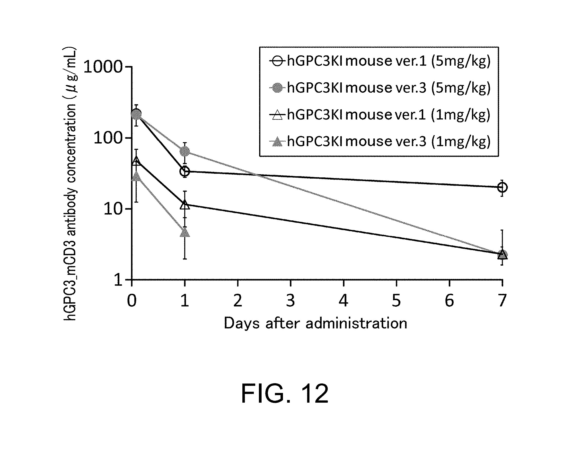
D00013
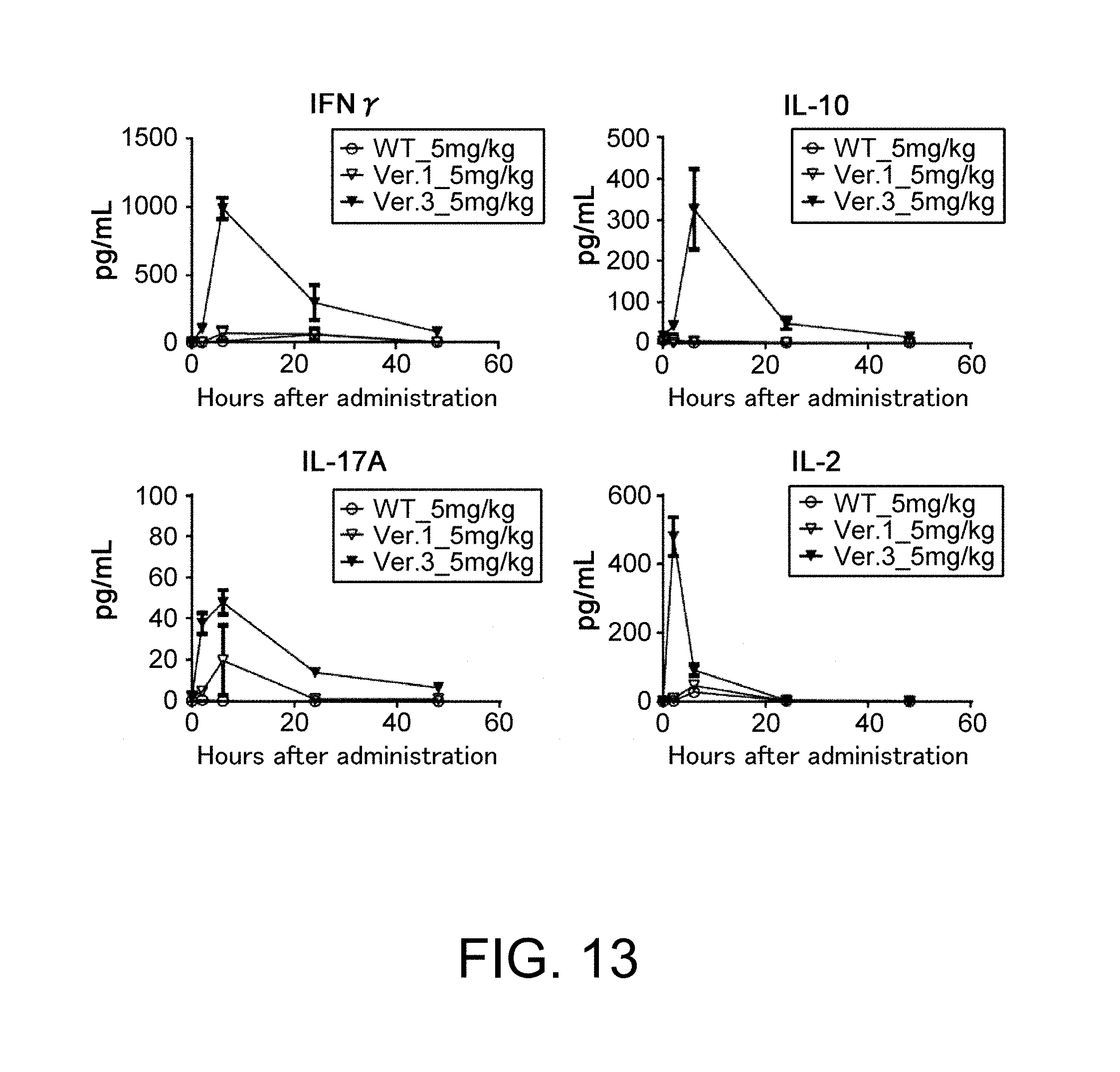
D00014

D00015

D00016
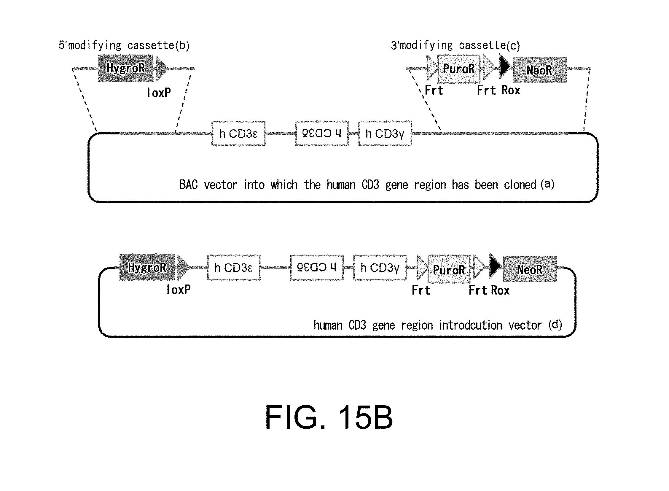
D00017

D00018

D00019

D00020

D00021

D00022

D00023

D00024

D00025

D00026

D00027

D00028

D00029

D00030

D00031

D00032

D00033

D00034

D00035

D00036

S00001
XML
uspto.report is an independent third-party trademark research tool that is not affiliated, endorsed, or sponsored by the United States Patent and Trademark Office (USPTO) or any other governmental organization. The information provided by uspto.report is based on publicly available data at the time of writing and is intended for informational purposes only.
While we strive to provide accurate and up-to-date information, we do not guarantee the accuracy, completeness, reliability, or suitability of the information displayed on this site. The use of this site is at your own risk. Any reliance you place on such information is therefore strictly at your own risk.
All official trademark data, including owner information, should be verified by visiting the official USPTO website at www.uspto.gov. This site is not intended to replace professional legal advice and should not be used as a substitute for consulting with a legal professional who is knowledgeable about trademark law.