Mhc Class Ia Open Conformers
MARROQUIN BELAUNZARAN; Osiris ; et al.
U.S. patent application number 16/323902 was filed with the patent office on 2019-06-06 for mhc class ia open conformers. This patent application is currently assigned to UNIVERSITAT ZURICH. The applicant listed for this patent is UNIVERSITAT BASEL, UNIVERSITAT ZURICH. Invention is credited to Osiris MARROQUIN BELAUNZARAN, Christoph RENNER.
| Application Number | 20190169263 16/323902 |
| Document ID | / |
| Family ID | 59656048 |
| Filed Date | 2019-06-06 |


View All Diagrams
| United States Patent Application | 20190169263 |
| Kind Code | A1 |
| MARROQUIN BELAUNZARAN; Osiris ; et al. | June 6, 2019 |
MHC CLASS IA OPEN CONFORMERS
Abstract
The invention relates to MHC-Ia open conformers as immunomodulatory agents, particularly in the treatment or prevention of cancer. The open conformer comprises or consists of a first and a second monomer, and each monomer comprises a HLA-heavy chain from the MHC-Ia molecules. The open conformer further comprises a protein stabilizing polypeptide sequence and optionally an amino acid linker. Further aspects of the invention provide combination medicaments comprising the MHC-Ia open conformers and immune checkpoint inhibitors. Furthermore, the invention relates to the use of MHC-Ia open conformers as immunomodulators, particularly in diseases where the interaction to diverse immunoregulatory receptors such as KIR3DL1, KIR3DL2, KIR3DL3, LILRB1, LILRB2, and PTPRJ modulates an immune response, and in diseases were the negative modulation of Tregs is a therapeutic strategy, e.g. infectious diseases.
| Inventors: | MARROQUIN BELAUNZARAN; Osiris; (Zurich, CH) ; RENNER; Christoph; (Zurich, CH) | ||||||||||
| Applicant: |
|
||||||||||
|---|---|---|---|---|---|---|---|---|---|---|---|
| Assignee: | UNIVERSITAT ZURICH ZURICH CH UNIVERSITAT BASEL Basel CH |
||||||||||
| Family ID: | 59656048 | ||||||||||
| Appl. No.: | 16/323902 | ||||||||||
| Filed: | August 9, 2017 | ||||||||||
| PCT Filed: | August 9, 2017 | ||||||||||
| PCT NO: | PCT/EP2017/070255 | ||||||||||
| 371 Date: | February 7, 2019 |
| Current U.S. Class: | 1/1 |
| Current CPC Class: | A61P 31/16 20180101; A61K 39/395 20130101; A61P 35/00 20180101; C07K 2319/00 20130101; A61P 31/22 20180101; A61P 31/18 20180101; C07K 14/70539 20130101; A61K 48/00 20130101; Y02A 50/30 20180101; Y02A 50/388 20180101 |
| International Class: | C07K 14/74 20060101 C07K014/74; A61P 35/00 20060101 A61P035/00; A61P 31/18 20060101 A61P031/18; A61P 31/22 20060101 A61P031/22; A61P 31/16 20060101 A61P031/16 |
Foreign Application Data
| Date | Code | Application Number |
|---|---|---|
| Aug 10, 2016 | EP | 16183626.7 |
| Jan 25, 2017 | EP | 17153123.9 |
Claims
1. A method for treatment of cancer or an infectious disease comprising: administering to a subject in need thereof a therapeutically effective amount of an isolated MHC-Ia open conformer, with the proviso that the isolated MHC-Ia open conformer is not a HLA-B27 or a HLA-B57 open conformer, thereby treating the cancer or infectious disease.
2. The method according to claim 1, wherein said isolated MHC-Ia open conformer comprises a first monomer or a first and a second monomer, and each monomer independently of the other monomer is or comprises a HLA heavy chain, with the proviso that the HLA heavy chain is not a HLA-B27 or a HLA-B57 heavy chain.
3. (canceled)
4. The method according to claim 1, wherein the infectious disease is selected from the group consisting of human immunodeficiency virus (HIV) infection, hepatitis A, hepatitis B, hepatitis C, influenza, respiratory syncytial virus (RSV) infection, measles, herpes and yellow fever.
5. The method according to claim 1, wherein the open conformer comprises or consists essentially of one or two HLA heavy chains selected from A25, B58, C08, A30, B53, and C12.
6. A fusion MHC-Ia open conformer, with the proviso that the fusion MHC-Ia open conformer is not a HLA-B27 or a HLA-B57 open conformer, wherein said fusion MHC-Ia open conformer comprises or consists essentially of a first monomer or a first and a second monomer, wherein a. said first monomer, or each of said first and second monomer independently of the other monomer, comprises a HLA heavy chain, and b. wherein said first monomer, or each of said first and second monomer are covalently linked to an Fc polypeptide sequence.
7. The fusion MHC-Ia open conformer according to claim 6, wherein an amino acid linker joins the HLA-heavy chain and the Fc polypeptide sequence.
8. The fusion MHC-Ia open conformer according to claim 6, wherein the first and the second monomer are the same.
9. The fusion MHC-Ia open conformer according to claim 6, wherein the fusion MHC-Ia open conformer additionally comprises a peptide epitope fragment, particularly wherein the first and/or second monomer additionally comprises a peptide epitope fragment.
10. The fusion MHC-Ia open conformer according to claim 6, wherein the HLA-heavy chain only consists of the HLA alpha 1, 2 and 3 domains.
11. The fusion MHC-Ia open conformer according to claim 6, wherein the HLA-heavy chain comprises the transmembrane domain and does not comprise the intracellular domain.
12. The fusion MHC-Ia open conformer according to claim 6, wherein the HLA-heavy chain is selected from A25, B58, C08, A30, B53, and C12.
13. The fusion MHC-Ia open conformer according to claim 6, wherein the Fc domain comprises heavy chain constant regions C.sub.H2 and C.sub.H3 selected from any one of immunoglobulin type G (IgG), type A (IgA), type D (IgD), type E (IgE) or type M (IgM).
14. The fusion MHC-Ia open conformer according to claim 6, wherein the amino acid linker comprises 1 to 50, particularly 5 to 40, more particularly 10 to 30, even more particularly 15 to 25 amino acids, linking the HLA-heavy chain to the Fc domain as one single polypeptide chain.
15. A method for treatment of cancer or an infectious disease selected from the group consisting of human immunodeficiency virus (HIV) infection, hepatitis A, hepatitis B, hepatitis C, influenza, respiratory syncytial virus (RSV) infection, measles, herpes and yellow fever, the method comprising administering to a subject in need thereof a therapeutically effective amount of the fusion MHC-Ia open conformer according to claim 6, thereby treating the cancer or infectious disease.
16. The method of claim 1, wherein the MHC-Ia open conformer is provided by a nucleic acid molecule encoding the MHC-Ia open conformer monomer.
17. The method of claim 16, wherein the nucleic acid is provided by a virus comprising the nucleic acid molecule under control of a promoter sequence operable in a mammalian cell, particularly in a human cell, particularly an adenovirus, adeno-associated virus, a herpes virus or a lentivirus.
18. An in vitro genetically modified host cell comprising the nucleic acid molecule of claim 16.
19. A combination medicament comprising a. an isolated MHC-Ia open conformer or the fusion MHC-Ia open conformer of claim 6, and b. a checkpoint modulatory agent selected from i. a checkpoint inhibitory agent (CPI), particularly wherein said CPI is selected from: an inhibitor of the interaction of CTLA4 with either B7-1 (cd80) and/or B7-2 (cd86), more particularly a polypeptide ligand to CTLA-4 or to cd80 or to cd86; an inhibitor of the interaction of PD-1 with either PD-L1 and/or PD-L2, more particularly a polypeptide ligand to PD-1 or to PD-L1 or to PD-L2; and an inhibitory polypeptide ligand, particularly an antibody, of T cell immunoglobulin and mucin domain-containing 3 (TIM-3); and ii. a checkpoint agonist agent, particularly a checkpoint agonist antibody selected to bind to and activate the tumor necrosis factor receptor 4-1BB, particularly a monoclonal antibody against 4-1BB. particularly wherein said checkpoint modulatory agent is a polypeptide selected from an antibody, an antibody fragment, and an antibody-like molecule, and the polypeptide is selectively reactive to a checkpoint mediator selected from CTLA4, PD-1, CD80, CD86, PD-L1, PD-L2, TIM-3, 4-1BB and 4-1BBL.
Description
[0001] The present invention relates to the use of classical MHC class Ia (MHC-Ia) open conformers, particularly for use in the prophylaxis or treatment of cancer, and for use as immunomodulators.
[0002] Human leukocyte antigens (HLA) belong to the classical major histocompatibility complex (MHC) protein family. The HLA complex helps the immune system distinguish the body's own proteins from proteins made by foreign invaders such as viruses and bacteria. Humans have MHC class I molecules comprising the classical (MHC-Ia) HLA-A, HLA-B, and HLA-C, and the non-classical (MHC-Ib) HLA-E, HLA-F, HLA-G and HLA-H molecules. Both categories are similar in their mechanisms of peptide binding, presentation and induced T-cell responses. The most remarkable feature of the classical MHC-Ia is their high polymorphism, while the non-classical MHC-Ib are usually non-polymorphic and tend to show a more restricted pattern of expression than their MHC-Ia counterparts.
[0003] The HLA nomenclature is given by the particular name of gene locus (e.g. HLA-A) followed by the allele family serological antigen (e.g. HLA-A*02), and allele subtypes assigned in numbers and in the order in which DNA sequences have been determined (e.g. HLA-A*02:01). Alleles that differ only by synonymous nucleotide substitutions (also called silent or non-coding substitutions) within the coding sequence are distinguished by the use of the third set of digits (e.g. HLA-A*02:01:01). Alleles that only differ by sequence polymorphisms in the introns, or in the 5' or 3' untranslated regions that flank the exons and introns, are distinguished by the use of the fourth set of digits (e.g. HLA-A*02:01:01:02L) (FIG. 1).
[0004] A list of MHC-Ia alleles is provided in Table 1. For a complete list of allele subtypes visit the link: http://hla.alleles.org/alleles/class1.html.
[0005] Classical MHC-Ia molecules' principle function is to present peptides as part of the adaptive immune response. MHC-Ia molecules are trimeric structures comprising a membrane-bound heavy chain with three extracellular domains (.alpha.1, .alpha.2 and .alpha.3) that associates non-covalently with .beta.2-microglobulin (.beta.2m) and a small peptide which is derived from self-proteins, viruses or bacteria. The .alpha.1 and .alpha.2 domains are highly polymorphic and form a platform that gives rise to the peptide-binding groove. Juxtaposed to the conserved .alpha.3 domain is a transmembrane domain followed by an intracellular cytoplasmic tail.
[0006] To initiate an immune response classical MHC-Ia molecules present specific peptides to be recognized by TCR (T cell receptor) present on CD8.sup.+ cytotoxic T lymphocytes (CTLs), while NK cell receptors present in natural killer cells (NK) recognize peptide motifs, rather than individual peptides. Under normal physiological conditions, MHC-Ia molecules exist as heterotrimeric complexes in charge of presenting peptides to CD8+ T cells and NK cells, however, MHC-Ia molecules may also be present in cells as free-heavy chains lacking .beta.2-microglobulin and peptide, and are referred to as HLA-open conformers (Arosa et al., Trends in Immunology 2007 March; 28(3):115-23) (FIG. 2). The interaction of HLA-open conformers with T cell receptors and NK cell receptors is independent of the peptide and its function is unknown.
[0007] Open conformers can be expressed at the cell surface of cells and can be detected with antibodies recognizing linear epitopes of HLA molecules without .beta.2m and peptide (e.g. LA45, L31, HCA2 and HC-10). These antibodies have been used to detect the presence of open conformers in diverse autoimmune patients and healthy individuals (Raine et al., Rheumatology 2006; 45:1338-1344). Despite their presence in patients and cell lines little is know of their mode of action. Open conformers have been mostly assessed in Ankylosing spondylitis (AS) +HLA-B27 patients, where HLA-B27 open conformers have been hypothesized to induce autoimmunity, their function in other autoimmune patients has not been yet addressed.
[0008] Here the inventors disclose for the first time that the classical MHC-Ia (HLA-A, HLA-B and HLA-C) family of molecules when present as open conformers (heavy chains without .beta.2m) are useful therapeutics for their immunomodulatory properties and use in the treatment of cancer.
[0009] Cancer is a group of diseases characterized by abnormal cells of the body undergoing uncontrolled and destructive growth. Cancer cells can spread around the body and metastasize to form tumors; this growth pattern is called malignant. Cancer can be treated by surgery, chemotherapy, radiation therapy, hormonal therapy, targeted therapy and immunotherapy. The choice of therapy depends on the type of cancer, the stage of the cancer (how much it has spread), age, health status, and additional personal characteristics. There is no single treatment for cancer, and patients often receive a combination of therapies and palliative care.
[0010] Cancer immunotherapy refers to a diverse set of therapeutic strategies designed to induce the patient's own immune system to fight the tumor, and is based on the insight that the progression of cancer, which involves the accumulation of diverse mutations, is monitored by the immune system. Immunotherapies either stimulate the activities of specific cell components of the immune system or counteract signals produced by cancer cells that suppress immune responses (Mahoney et al., Nat Rev Drug Discov. 2015 August; 14(8):561-84).
[0011] Different type of immune cells are involved in the immune response against cancer. Within this pool of white blood cells (immune contexture), the most notorious cells are: T-cells (cytotoxic CD8+ T-cells, T helper CD4+ cells--Th1, Th2, and Th17 phenotype), regulatory T cells (Tregs), macrophages (M1 type-pro-inflammatory and M2 type-pro-tumoral), myeloid derived suppressor cells (MDSCs), natural killer cells (NK cells), and dendritic cells (DCs). These immune cells can be located in the center of the tumor, in the invasive margin or in the adjacent tertiary lymphoid structures (Fridman et al., Nat. Rev. Cancer. 2012, April: 12, 298-306).
[0012] The density and composition of the immune microenvironment is heterogeneous among patients and tumors. It is now well established that in general the tumor infiltration with M2-phenotype macrophages and myeloid derived suppressor cells (MDSCs) promotes tumor progression, whereas infiltration of cytotoxic CD8+ T-cells, Th1 phenotype cells and M1 type macrophages are often associated with good clinical outcome, and good response to immunotherapy. The clinical impact of other lymphoid and myeloid cell populations is less consistent and seems dependent on the tumor type and stage. The presence of Th17, and NK cells, and the absence/reduction of Treg cells in tumor infiltrates is correlated with good outcome in some cancer indications (Giraldo et al., Current Opinion in Immunology 2014, 27:8-15). A general overview of the balance between leukocyte infiltrates and clinical outcome is reviewed in (Becht et al. Current Opinion in Immunology. 2016, 39:17-13).
[0013] Overall, modulating the immune contexture of tumors favoring the infiltration of M1 type macrophages, cytotoxic CD8 T-cells, and Th1 cells, and/or reducing the infiltration of MDSCs and M2 type macrophages is an promising therapeutic avenue to treat cancer that is explored here with the use of HLA open conformers proteins in diverse cancer indications.
Terms and Definitions
[0014] Amino acid sequences are given from amino to carboxyl terminus. Capital letters for sequence positions refer to L-amino acids in the one-letter code (Stryer, Biochemistry, 3.sup.rd ed. p. 21).
[0015] The term open conformer as used in the present specification refers to an isolated HLA heavy chain molecule not associated to .beta.2-microglobulin either as a monomer or as a dimer (homodimer or heterodimer). Certain embodiments of the open conformers disclosed herein are fusion protein monomers or dimers, wherein the HLA heavy chain is covalently linked to a stabilizing polypeptide region, particularly a crystallizable fragment immunoglobulin domain.
[0016] In the context of the present specification the terms sequence identity and percentage of sequence identity refer to the values determined by comparing two aligned sequences. Methods for alignment of sequences for comparison are well-known in the art. Alignment of sequences for comparison may be conducted by the local homology algorithm of Smith and Waterman, Adv. Appl. Math. 2:482 (1981), by the global alignment algorithm of Needleman and Wunsch, J. Mol. Biol. 48:443 (1970), by the search for similarity method of Pearson and Lipman, Proc. Nat. Acad. Sci. 85:2444 (1988) or by computerized implementations of these algorithms, including, but not limited to: CLUSTAL, GAP, BESTFIT, BLAST, FASTA and TFASTA. Software for performing BLAST analyses is publicly available, e.g., through the National Center for Biotechnology-Information (http://blast.ncbi.nlm.nih.gov/). One example for comparison of amino acid sequences is the BLASTP algorithm that uses the default settings: Expect threshold: 10; Word size: 3; Max matches in a query range: 0; Matrix: BLOSUM62; Gap Costs: Existence 11, Extension 1; Compositional adjustments: Conditional compositional score matrix adjustment. One such example for comparison of nucleic acid sequences is the BLASTN algorithm that uses the default settings: Expect threshold: 10; Word size: 28; Max matches in a query range: 0; Match/Mismatch Scores: 1.-2; Gap costs: Linear. Unless otherwise stated, sequence identity values provided herein refer to the value obtained with the BLAST suite of programs (Altschul et al., J. Mol. Biol. 215:403-410 (1990)) using the above identified default parameters for protein and nucleic acid comparison, respectively.
[0017] In the context of the present specification, the term major histocompatibility complex (MHC) is used in its meaning known in the art of cell biology and immunology; it refers to a cell surface molecule that displays a specific fraction (peptide), also referred to as an epitope, of a protein. There a two major classes of MHC molecules: class I and class II. Within the MHC class I two groups can be distinguished based on their polymorphism: a) the classical (MHC-la) with corresponding polymorphic HLA-A, HLA-B, and HLA-C genes, and b) the non-classical (MHC-Ib) with corresponding less polymorphic HLA-E, HLA-F, HLA-G and HLA-H genes.
[0018] MHC class I heavy chain molecules usually (i.e. when not in open conformer form) occur as an alpha chain linked to a unit of the non-MHC molecule .beta.2-microglobulin. The alpha chain comprises, in direction from the N-terminus to the C-terminus, a signal peptide, three extracellular domains (.alpha.1-3, with .alpha.1 being at the N terminus), a transmembrane region and a C-terminal cytoplasmic tail. The peptide being displayed or presented is held by the peptide-binding groove, in the central region of the .alpha.1/.alpha.2 domains.
[0019] In the context of the present specification, the term .beta.2-microglobulin domain is used in its meaning known in the art of cell biology and biochemistry; it refers to a non-MHC molecule that is part of the MHC class I heterodimer molecule. In other words, it constitutes the 1 chain of the MHC class I heterodimer.
[0020] In the context of the present specification, the term human leukocyte antigen (HLA) is used in its meaning known in the art of cell biology and biochemistry; it refers to gene loci encoding the human MHC class I proteins. The three major classical MHC-Ia genes are HLA-A, HLA-B and HLA-C, and all of these genes have a varying number of alleles (Table 1). Closely related alleles are combined in subgroups of a certain allele. For example the allele HLA-B57 has more than 100 closely related alleles that vary in one or more amino acids, according to the WHO Nomenclature Committee for Factors of the HLA System, labelled HLA-B*57:01:01 to HLA-B*57:82. The full or partial sequence of all known HLA genes and their respective alleles are available to the person skilled in the art in specialist databases such as IMGT/HLA (http://www.ebi.ac.uk/ipd/imgt/hla/).
[0021] In the context of the present specification, the term antibody is used in its meaning known in the art of cell biology and immunology; it refers to whole antibodies including but not limited to immunoglobulin type G (IgG), type A (IgA), type D (IgD), type E (IgE) or type M (IgM), any antigen binding fragment or single chains thereof and related or derived constructs. A whole antibody is a glycoprotein comprising at least two heavy (H) chains and two light (L) chains inter-connected by disulfide bonds. Each heavy chain is comprised of a heavy chain variable region (V.sub.H) and a heavy chain constant region (C.sub.H). The heavy chain constant region is comprised of three domains, C.sub.H1, C.sub.H2 and C.sub.H3. Each light chain is comprised of a light chain variable region (abbreviated herein as V.sub.L) and a light chain constant region (C.sub.L). The light chain constant region is comprised of one domain, C.sub.L. The variable regions of the heavy and light chains contain a binding domain that interacts with an antigen. The constant regions of the antibodies may mediate the binding of the immunoglobulin to host tissues or factors, including various cells of the immune system (e.g., effector cells) and the first component of the classical complement system.
[0022] The term antibody-like molecule in the context of the present specification refers to a molecule capable of specific binding to another molecule or target with high affinity/a Kd.ltoreq.10E-8 mol/l. An antibody-like molecule binds to its target similarly to the specific binding of an antibody. The term antibody-like molecule encompasses a repeat protein, such as a designed ankyrin repeat protein (Molecular Partners, ZOrich), a polypeptide derived from armadillo repeat proteins, a polypeptide derived from leucine-rich repeat proteins and a polypeptide derived from tetratricopeptide repeat proteins.
[0023] The term antibody-like molecule further encompasses a polypeptide derived from protein A domains, a polypeptide derived from fibronectin domain FN3, a polypeptide derived from consensus fibronectin domains, a polypeptide derived from lipocalins, a polypeptide derived from Zinc fingers, a polypeptide derived from Src homology domain 2 (SH2), a polypeptide derived from Src homology domain 3 (SH3), a polypeptide derived from PDZ domains, a polypeptide derived from gamma-crystallin, a polypeptide derived from ubiquitin, a polypeptide derived from a cysteine knot polypeptide and a polypeptide derived from a knottin.
[0024] The term protein A domains derived polypeptide refers to a molecule that is a derivative of protein A and is capable of specifically binding the Fc region and the Fab region of immunoglobulins.
[0025] The term armadillo repeat protein refers to a polypeptide comprising at least one armadillo repeat, wherein an armadillo repeat is characterized by a pair of alpha helices that form a hairpin structure.
[0026] In the context of the present specification, the term crystallizable fragment (Fc) region is used in its meaning known in the art of cell biology and immunology; it refers to a fraction of an antibody comprising two identical heavy chain fragments comprised of a C.sub.H2 and a C.sub.H3 domain, covalently linked by disulfide bonds.
[0027] In the context of the present specification, the term dimer refers to a unit consisting of two subunits.
[0028] In the context of the present specification, the term homodimer refers to a dimer comprised of two subunits that are either identical or are highly similar members of the same class of subunits. One example for a homodimer would be a dimer consisting of two subunits independently selected from the list of HLA alleles. In certain embodiments, homodimers consist of two identical HLA alleles.
[0029] In the context of the present specification, the term amino acid linker refers to a polypeptide of variable length that is used to connect two polypeptides in order to generate a single chain polypeptide. Exemplary embodiments of linkers useful for practicing the invention specified herein are oligopeptide chains consisting of 1, 2, 3, 4, 5, 10, 20, 30, 40 or 50 amino acids. A non-limiting example of an amino acid linker is the polypeptide GGGGSGGGGS (SEQ ID No. 001) that links an HLA-heavy chain polypeptide with an Fc domain.
[0030] In the context of the present specification, the term checkpoint inhibitory agent or checkpoint inhibitory antibody is meant to encompass an agent, particularly a (non-agonist) antibody (or antibody-like molecule) capable of disrupting the signal cascade leading to T cell inhibition after T cell activation as part of what is known in the art the immune checkpoint mechanism. Non-limiting examples of a checkpoint inhibitory agent or checkpoint inhibitory antibody include antibodies to CTLA-4 (Uniprot P16410), PD-1 (Uniprot Q15116), PD-L1 (Uniprot Q9NZQ7), B7H3 (CD276; Uniprot Q5ZPR3), Tim-3, Gal9, VISTA, or Lag3.
[0031] In the context of the present specification, the term checkpoint agonist agent or checkpoint agonist antibody is meant to encompass an agent, particularly but not limited to an antibody (or antibody-like molecule) capable of engaging the signal cascade leading to T cell activation as part of what is known in the art the immune checkpoint mechanism. Non-limiting examples of receptors known to stimulate T cell activation include CD122 and CD137 (4-1BB; Uniprot Q07011). The term checkpoint agonist agent or checkpoint agonist antibody encompasses agonist antibodies to CD137 (4-1BB), CD134 (OX40), CD357 (GITR), CD278 (ICOS), CD27, CD28.
[0032] In the context of the present specification, the term (immune) checkpoint modulatory agent encompasses checkpoint inhibitory agents, checkpoint inhibitory antibodies, checkpoint agonist agents and checkpoint agonist antibodies.
SPECIFIC DESCRIPTION OF THE INVENTION
[0033] The present invention provides MHC-Ia open conformers (HLA-open conformers). MHC-Ia open conformers comprise HLA-A, HLA-B and HLA-C open conformers. In table 1 a list of known MHC-Ia alleles is provided. The present invention does not include MHC-Ia open conformers that comprise a HLA-B27 or a HLA-B57 allele.
[0034] According to a first aspect of the invention an isolated MHC-Ia open conformer is provided, with the proviso that the isolated MHC-Ia open conformer is not a HLA-B27 or a HLA-B57 open conformer.
[0035] In certain embodiments, the isolated MHC-Ia open conformer comprises a first monomer or a first and a second monomer, and each monomer independently of the other monomer comprises a HLA heavy chain.
[0036] According to an alternative to the first aspect of the invention an isolated HLA-A open conformer is provided.
[0037] According to another alternative to the first aspect of the invention an isolated HLA-B open conformer is provided, with the proviso that the isolated HLA-B open conformer is not a HLA-B27 or a HLA-B57 open conformer.
[0038] According to yet another alternative to the first aspect of the invention an isolated HLA-C open conformer is provided.
[0039] According to yet another alternative to the first aspect of the invention an isolated HLA-A open conformer and a HLA-C open conformer is provided.
[0040] According to a second aspect of the invention an isolated MHC-Ia open conformer, with the proviso that the isolated MHC-Ia open conformer is not a HLA-B27 or a HLA-B57 open conformer, is provided: [0041] for use as a medicament, [0042] particularly for use in the treatment or prevention of cancer, or [0043] particularly for use as an immunomodulatory agent, [0044] particularly in a treatment of an infectious disease, [0045] more particularly for use in prevention, treatment or therapy of human immunodeficiency virus (HIV), hepatitis A, B, C, virus (HAV HBV, HCV respectively), influenza virus, Respiratory Syncytial Virus (RSV), measles virus, herpes viruses and/or yellow fever virus.
[0046] According to an alternative to the second aspect of the invention an isolated HLA-A open conformer is provided: [0047] for use as a medicament, [0048] particularly for use in the treatment or prevention of cancer, or [0049] particularly for use as an immunomodulatory agent, [0050] particularly in a treatment of an infectious disease, [0051] more particularly for use in prevention, treatment or therapy of human immunodeficiency virus (HIV), hepatitis A, B, C, virus (HAV HBV, HCV respectively), influenza virus, Respiratory Syncytial Virus (RSV), measles virus, herpes viruses and/or yellow fever virus.
[0052] According to another alternative to the second aspect of the invention an isolated HLA-B open conformer, with the proviso that the isolated HLA-B open conformer is not a HLA-B27 or a HLA-B57 open conformer is provided: [0053] for use as a medicament, [0054] particularly for use in the treatment or prevention of cancer, or [0055] particularly for use as an immunomodulatory agent, [0056] particularly in a treatment of an infectious disease, [0057] more particularly for use in prevention, treatment or therapy of human immunodeficiency virus (HIV), hepatitis A, B, C, virus (HAV HBV, HCV respectively), influenza virus, Respiratory Syncytial Virus (RSV), measles virus, herpes viruses and/or yellow fever virus.
[0058] According to yet another alternative to the second aspect of the invention an isolated HLA-C open conformer is provided: [0059] for use as a medicament, [0060] particularly for use in the treatment or prevention of cancer, or [0061] particularly for use as an immunomodulatory agent. [0062] particularly in a treatment of an infectious disease, [0063] more particularly for use in prevention, treatment or therapy of human immunodeficiency virus (HIV), hepatitis A, B, C, virus (HAV HBV, HCV respectively), influenza virus, Respiratory Syncytial Virus (RSV), measles virus, herpes viruses and/or yellow fever virus.
[0064] The function as an immunomodulatory agent is particularly useful to treat diseases requiring the modification of white blood cell responses, such as infectious diseases. Infectious diseases that can preferably be treated by the present invention include human immunodeficiency virus (HIV) infection, hepatitis A, hepatitis B, hepatitis C, influenza, respiratory syncytial virus (RSV) infection, measles, herpes and yellow fever.
[0065] In certain embodiments of the second aspect of the invention or of any above-mentioned alternative to the second aspect of the invention, the cancer is colon cancer or pancreatic cancer.
[0066] A third aspect of the invention relates to a fusion MHC-Ia open conformer, with the proviso that the fusion MHC-Ia open conformer is not a HLA-B27 or a HLA-B57 open conformer. The fusion MHC-Ia open conformer comprises, or essentially consists of, a first HLA heavy chain monomer or a first and a second HLA heavy chain monomer. Each of these HLA heavy chain monomer independently of the other comprises or essentially consists of a HLA heavy chain. The fusion MHC open conformer additionally comprises an Fc polypeptide sequence.
[0067] In certain embodiments, the HLA monomer sequence is situated at the N terminus of the fusion MHC open conformer, and the Fc construct is located towards the C terminus. In certain embodiments, an amino acid linker joins the HLA-heavy chain and the Fc fragment.
[0068] The fusion MHC-Ia open conformer additionally comprises a polypeptide domain known to metabolically stabilize a polypeptide in vivo. One example of such a stabilizing domain is an Fc (crystallisable fragment) domain of an immunoglobulin, particularly the Fc polypeptide domain of a gamma immunoglobulin. The HLA-heavy chain and the stabilizing domain may optionally be joined by an amino acid linker. An open conformer fusion protein comprising the HLA chain and an immunoglobulin Fc fragment is henceforth termed HLA-Fc open conformer or HLA.sub.2-Fc herein.
[0069] The presence of the Fc domain in the fusion protein facilitates increasing the solubility, stability, avidity, half-life, and from a technological point of view, cost-effective production and purification in mammalian systems (protein A or G purification).
[0070] According to an alternative to the third aspect of the invention a HLA-A open conformer is provided, wherein the HLA-A open conformer comprises a first monomer or a first and a second monomer, and each monomer independently of the other monomer comprises a HLA heavy chain additionally comprising an Fc polypeptide sequence and optionally, an amino acid linker joining the HLA-heavy chain and the Fc fragment.
[0071] According to another alternative to the third aspect of the invention a HLA-B open conformer, with the proviso that the HLA-B open conformer is not a HLA-B27 or a HLA-B57 open conformer is provided, wherein the HLA-B open conformer comprises a first monomer or a first and a second monomer, and each monomer independently of the other monomer comprises a HLA heavy chain additionally comprising an Fc polypeptide sequence and optionally, an amino acid linker joining the HLA-heavy chain and the Fc fragment.
[0072] According to yet another alternative to the third aspect of the invention a HLA-C open conformer is provided, wherein the HLA-C open conformer comprises a first monomer or a first and a second monomer, and each monomer independently of the other monomer comprises a HLA heavy chain additionally comprising an Fc polypeptide sequence and optionally, an amino acid linker joining the HLA-heavy chain and the Fc fragment.
[0073] According to an alternative aspect of the invention a MHC-Ia open conformer monomer (i.e., the HLA-heavy chain unattached to a second HLA-heavy chain polypeptide, and not bound by .beta.2-microglobulin) is provided for use in the treatment or prevention of cancer, or for use as an immunomodulatory agent, with the proviso that the MHC-Ia open conformer monomer is not a HLA-B27 or a HLA-B57 open monomer. In certain embodiments of this aspect, the MHC-Ia monomer additionally comprises a peptide epitope fragment.
[0074] This aspect can be summarized in the following items: [0075] Item 1: An isolated single HLA-heavy chain polypeptide monomer derived from MHC-Ia alleles essentially free of associated .beta.2-microglobulin for use as a medicament, particularly for use in the treatment or prevention of cancer, or for use as an immunomodulatory agent. [0076] Item 2: An isolated single HLA-heavy chain polypeptide monomer derived from the MHC-Ia alleles for use in the treatment or prevention of cancer or as an immunomodulatory agent according to item 1, wherein the monomer additionally comprises a peptide epitope fragment. [0077] Item 3: An isolated single HLA-heavy chain polypeptide monomer derived from the MHC-Ia alleles for use in the treatment or prevention of cancer or as an immunomodulatory agent according to items 1 or 2, wherein the HLA-heavy chain only consists of the HLA-alpha 1, 2 and 3 domains. [0078] Item 4: An isolated single HLA-heavy chain polypeptide monomer derived from the MHC-Ia alleles for use in the treatment or prevention of cancer or as an immunomodulatory agent according to any one of the preceding items, wherein the HLA-heavy chain comprises the transmembrane domain and does not comprise the intracellular domain (cytoplasmic tail). [0079] Item 5: A combination medicament comprising [0080] a. an isolated single HLA-heavy chain polypeptide monomer derived from the MHC-Ia alleles as specified in any one of items 1 to 4, and [0081] b. a checkpoint inhibitory agent, particularly a checkpoint inhibitory antibody, and/or a checkpoint agonist agent, particularly a checkpoint agonist antibody. [0082] Item 6: The combination medicament according to item 5, wherein said checkpoint inhibitory agent is selected from an inhibitor of CTLA4 interaction with CD80 or CD86, and an inhibitor of the interaction of PD-1 with its ligand PD-L1, particularly an antibody against any one of CTLA4, CD80, CD86, PD-1, PD-L1, more particularly a monoclonal antibody against human CTLA4, PD-1, or PD-L1, and/or wherein said checkpoint agonist agent is selected from an agonist antibody or ligand to 4-1BB and/or 4-1BBL (CD137L, Uniprot P41273).
[0083] In certain embodiments of this alternative aspect of the invention, the cancer is colon cancer or pancreatic cancer.
[0084] According to another aspect of the invention a MHC-Ia open conformer protein is provided as an immunomodulatory agent, with the proviso that the MHC-Ia open conformer is not a HLA-B27 or a HLA-B57 open conformer. Without wishing to be bound by theory the inventors believe that particularly its capacity to bind to diverse immunoregulatory receptors present in white blood cells and to modify the proliferation of T cell lymphoma cells is particularly useful.
[0085] Furthermore the use of MHC-Ia open conformers as negative modulator of regulatory T cells (Tregs) is particularly suitable, for use in human diseases where Tregs impair the development of protective immunity, such as cancer and infectious diseases (von Boehmer et al. ibid.).
[0086] According to an alternative to this other aspect of the invention a HLA-A open conformer is provided as an immunomodulatory agent.
[0087] According to another alternative to this other aspect of the invention a HLA-B open conformer is provided as an immunomodulatory agent, with the proviso that the HLA-B open conformer is not a HLA-B27 or a HLA-B57 open conformer.
[0088] According to yet another alternative to this other aspect of the invention a HLA-C open conformer is provided as an immunomodulatory agent.
[0089] In certain embodiments of any one of the aspects of the invention, the HLA-heavy chain comprises the transmembrane domain and does not comprise the intracellular domain (cytoplasmic tail).
[0090] In certain embodiments of any one of the aspects of the invention, the isolated MHC-Ia open conformer or fusion MHC-Ia open conformer consists of two subunits independently selected from the above HLA-alleles. In certain embodiments, homodimers consist of two identical HLA-alleles.
[0091] In certain embodiments of any one of the aspects of the invention, the isolated MHC-Ia open conformer or fusion MHC-Ia open conformer comprise two identical HLA polypeptide chains.
[0092] In certain embodiments, the isolated MHC-Ia open conformer or fusion MHC-Ia open conformer comprises two different HLA polypeptide chains.
[0093] In certain embodiments of any one of the aspects of the invention, the isolated MHC-Ia open conformer or fusion MHC-Ia open conformer additionally comprises a peptide epitope fragment.
[0094] In certain embodiments of any one of the aspects of the invention, a peptide epitope fragment is non-covalently attached to the polypeptide within the antigen presenting domain of the HLA peptide chain.
[0095] In certain embodiments of any one of the aspects of the invention, the first and/or second monomer additionally comprises a peptide epitope fragment.
[0096] In certain embodiments of any one of the aspects of the invention, the fusion MHC-Ia open conformer comprises only the extracellular HLA-alpha 1, HLA-alpha 2 and HLA-alpha 3 domains. In these embodiments, the transmembrane and intracellular domains of the HLA heavy chains are not included in the therapeutic polypeptide of the invention in order to allow its extracellular expression in recombinant cells. The person skilled in the art can easily identify the respective domains even in previously unknown HLA-sequences by pair-wise sequence alignment with annotated HLA-sequences.
[0097] In certain embodiments of any one of the aspects of the invention, the fusion MHC-Ia open conformers comprise an Fc domain. In certain particular embodiments, the Fc domain comprises heavy chain constant regions C.sub.H2 and C.sub.H3 from immunoglobulin type G (IgG), type A (IgA), type D (IgD), type E (IgE) or type M (IgM).
[0098] In certain embodiments of any one of the aspects of the invention, the fusion MHC-Ia open conformers comprise an amino acid linker joining a stabilizing domain, particularly an Fc domain, to the HLA polypeptide. In certain particular embodiments, the amino acid linker comprises 1 to 50 amino acids, particularly 5 to 40 amino acids, more particularly 10 to 30 amino acids, even more particularly 15 to 25 amino acids that link the HLA-heavy chain to the Fc domain as one single polypeptide chain.
[0099] In certain embodiments of any one of the aspects of the invention, the isolated MHC-Ia open conformers or fusion MHC-Ia open conformers, are provided as parenteral dosage form, particularly confectioned for injection. In certain embodiments, the immune checkpoint inhibitor agent or agonist agent is provided as parenteral dosage form, particularly confectioned for injection. In certain embodiments, both the MHC-Ia open conformers and the immune checkpoint inhibitor agent or agonist agent are present in the same administration form.
[0100] In certain embodiments of the third aspect of the invention, the fusion MHC-Ia open conformer is for use as a medicament.
[0101] In certain embodiments of the third aspect of the invention, the fusion MHC-Ia open conformer is for use in the treatment or prevention of cancer, in particular for colon cancer or pancreatic cancer.
[0102] In certain embodiments of the third aspect of the invention, the fusion MHC-Ia open conformer is for use as an immunomodulatory agent, particularly for use as negative modulator of regulatory T cells (Treg). In certain embodiments, the fusion MHC-Ia open conformer is for use in the treatment of infectious diseases. In certain embodiments, the fusion MHC-Ia open conformer is for use in the treatment of human immunodeficiency virus (HIV) infection, hepatitis A, hepatitis B, hepatitis C, influenza, respiratory syncytial virus (RSV) infection, measles, herpes and yellow fever.
[0103] According to a fourth aspect of the invention, a nucleic acid molecule encoding MHC-Ia open conformer monomers, particularly an Fc open conformer monomer, according to the above aspects of the invention is provided for use in the treatment or the therapy of cancer or for use as an immunomodulatory agent, particularly in a treatment of an infectious disease.
[0104] Expression of the open conformer in vivo from the nucleic acid molecule will, after dimerization, lead to the fusion protein polypeptide of the invention. The concept of expressing pharmaceutically active polypeptides from nucleic acids encoding them in the patient's body is well known and may confer significant benefits to the patient.
[0105] According to an alternative to the fourth aspect of the invention a nucleic acid encoding HLA-A open conformers monomers for use in the treatment or the therapy of cancer or for use as an immunomodulatory agent, particularly in a treatment of an infectious disease is provided.
[0106] According to another alternative to the fourth aspect of the invention a nucleic acid encoding HLA-B open conformers monomers for use in the treatment or the therapy of cancer or for use as an immunomodulatory agent, particularly in a treatment of an infectious disease is provided, with the proviso that the HLA-B open conformer is not a HLA-B27 or a HLA-B57 open conformer.
[0107] According to yet another alternative to the fourth aspect of the invention a nucleic acid encoding HLA-C open conformers monomers for use in the treatment or the therapy of cancer or for use as an immunomodulatory agent, particularly in a treatment of an infectious disease is provided.
[0108] In certain embodiments of the fourth aspect of the invention or any above-mentioned alternative thereof, the cancer is colon cancer or pancreatic cancer.
[0109] In certain embodiments, the nucleic acid molecule encodes MHC-Ia open conformers monomers, particularly an Fc open conformer monomer comprising a peptide epitope fragment. In certain embodiments, the nucleic acid molecule encodes MHC-Ia open conformers monomers, particularly an Fc open conformer monomer that comprises only the extracellular HLA-alpha 1, 2 and 3 domains. In certain embodiments, the nucleic acid molecule encodes HLA open conformers monomers, particularly an Fc open conformer monomer that comprises only the extracellular HLA-alpha 1, 2 and 3 domains, and a peptide epitope fragment.
[0110] In certain embodiments, the nucleic acid molecule encodes MHC-Ia open conformers monomers, particularly an Fc open conformer monomer that comprises an amino acid linker and/or an Fc (fragment crystallizable) domain, and is used in the treatment or the therapy of cancer, in particular colon or pancreatic cancer.
[0111] According to an alternative aspect of the invention a recombinant expression vector comprising the nucleic acid molecule according to the fourth aspect of the invention (and its alternative aspects) is provided for use in the treatment or the therapy of cancer, in particular colon or pancreatic cancer.
[0112] In certain embodiments the recombinant expression vector is a plasmid comprising a promoter that is operable in a mammalian cell, particularly in a human cell. The promoter is operably linked to the nucleic acid molecule of the invention.
[0113] According to a fifth aspect of the invention a virus comprising the nucleic acid molecule according to the fourth aspect of the invention (and its alternative aspects) is provided for use in the treatment or the therapy of cancer, in particular colon or pancreatic cancer, or for use as an immunomodulatory agent, particularly in a treatment of an infectious disease. The nucleic acid molecule is under control of a promoter sequence operable in a mammalian cell, particularly in a human cell. In certain embodiments, the virus is an adenovirus, adeno-associated virus, a herpes virus or a lentivirus.
[0114] According to a sixth aspect of the invention an in vitro genetically modified host cell comprising the nucleic acid molecule according to the fourth aspect of the invention (and its alternative aspects) is provided.
[0115] Another aspect of the invention provides for the use of the isolated MHC-Ia open conformers homodimer or MHC-Ia open conformers homodimer according to the first and second aspect of the invention (and their alternatives) in the manufacture of a medicament for the treatment or prevention of cancer, in particular colon or pancreatic cancer.
[0116] According to yet another aspect, the invention provides a method of treatment for cancer, in particular colon or pancreatic cancer, comprising administering an MHC-Ia open conformer according to the first and second aspect of the invention (and their alternative aspects) to a patient in need thereof.
[0117] According to a seventh aspect of the invention, a combination medicament is provided, wherein the combination medicament comprises: [0118] isolated MHC-Ia open conformers or fusion MHC-Ia open conformers, according to any one of the above aspects or embodiments of the invention, and [0119] an immune checkpoint modulatory agent selected from [0120] an immune checkpoint inhibitor agent (CPI) selected from: [0121] an inhibitor of cytotoxic T-lymphocyte-associated protein 4 (CTLA4; also known as CD152) interaction with either B7-1 (CD80) and/or B7-2 (CD86), particularly a polypeptide ligand to CTLA-4 or to cd80 or to cd86 such as for example an antibody, [0122] an inhibitor of the interaction of programmed cell death protein 1 (PD-1; also known as CD279) with its ligand PD-L1 (also known as CD274; UniProt ID: Q9NZQ7) and/or PD-L2 (also known as CD273; Uni Prot ID: Q9BQ51), particularly a polypeptide ligand to PD-1 or to PD-L1 or to PD-L2 such as for example an antibody, and [0123] an inhibitory polypeptide ligand, particularly an antibody, of T cell immunoglobulin and mucin domain-containing 3 (TIM-3), and [0124] a checkpoint agonist agent, particularly a checkpoint agonist antibody selected to bind to and activate the tumor necrosis factor receptor 4-1BB (also known as CD137 or TNFRSF9).
[0125] According to an alternative to the seventh aspect of the invention the isolated MHC-Ia open conformer or fusion MHC-Ia open conformer comprised within the combination medicament is selected from a HLA-A open conformer, a HLA-B open conformer (with the proviso that the HLA-B open conformer is not a HLA-B27 or a HLA-B57 open conformer) or a HLA-C open conformer.
[0126] In certain embodiments, the immune checkpoint inhibitor agent is an inhibitor of interaction of CTLA4 with CD80 or CD86.
[0127] In certain embodiments, the immune checkpoint inhibitor agent is ipilimumab (Yervoy; CAS No. 477202-00-9).
[0128] In certain embodiments, the immune checkpoint inhibitor agent is an inhibitor of interaction of programmed cell death protein 1 (PD-1) with its receptor PD-L1. In certain embodiments, the immune checkpoint inhibitor agent is selected from the clinically available antibody drugs nivolumab (Bristol-Myers Squibb; CAS No 946414-94-4), pembrolizumab (Merck Inc.; CAS No. 1374853-91-4), pidilizumab (CAS No. 1036730-42-3), atezolizumab (Roche AG; CAS No. 1380723-44-3), and Avelumab (Merck KGaA; CAS No. 1537032-82-8).
[0129] In certain embodiments, the immune checkpoint agonist agent is utomilumab (PF-05082566), a fully human IgG2 monoclonal antibody against 4-1BB currently undergoing clinical trials.
[0130] In certain embodiments, the checkpoint modulatory agent is a polypeptide selected from an antibody, an antibody fragment, and an antibody-like molecule, and the polypeptide is selectively reactive to a checkpoint mediator. In certain embodiments, the checkpoint mediator is selected from CTLA4, PD-1, CD80, CD86, PD-L1, and PD-L2, TIM-3, 4-1BB and 4-1BBL.
[0131] In yet another aspect, the invention relates to a method for producing recombinant HLA heavy chain polypeptides. This method is summarized in the following items: [0132] Item A: A method for producing, by methods of recombinant biotechnology, a human HLA heavy chain polypeptide, wherein said method comprises the following steps: [0133] a. Expression step: [0134] i. a HLA-encoding nucleic acid sequence encoding at least the alpha 1 chain, the alpha 2 chain and the alpha 3 chain of a HLA heavy chain under control of a promoter sequence operable in a cell, particularly a eukaryotic cell, more particularly a mammalian cell, and [0135] ii. a .beta.2-microglobulin encoding nucleic acid sequence encoding the human HLA beta 2 microglobulin (UniProt P61769) under control of a promoter sequence operable in said cell (the same cell as in item 1. a.) are co-expressed in a mammalian cell ("production cell line"); [0136] b. Purification step: the resulting HLA-heavy-chain/.beta.2-microglobulin complex is purified from the mammalian cell (the production cell line); [0137] c. Dissociation step: the purified HLA-heavy-chain/.beta.2-microglobulin complex is dissociated under suitable conditions and the HLA heavy chain polypeptides are separated from the .beta.2-microglobulin polypeptides; [0138] d. Refolding step: the separated HLA heavy chain polypeptides are incubated under conditions leading to refolding (of their native tertiary protein structure found in physiologically active HLA open conformer molecules). [0139] Item AA: Item A with the proviso that the human HLA heavy chain polypeptide is neither a B27 heavy chain nor a B57 heavy chain. [0140] Item B: The method for producing a human HLA heavy chain polypeptide according to item A or item AA, wherein the HLA-encoding nucleic acid sequence comprises, from N to C terminus of the encoded polypeptide, the alpha 1 chain, the alpha 2 chain, the alpha 3 chain and a stabilizing sequence. [0141] Item C: The method for producing a human HLA heavy chain polypeptide according to item B, wherein the stabilizing sequence is selected from bovine serum albumin and an immunoglobulin constant fragment (Fc), particularly an immunoglobulin G constant fragment, more particularly an IgG4 Fc. [0142] Item D: The method for producing a human HLA heavy chain polypeptide according to any of the preceding items, wherein the HLA-encoding nucleic acid sequence and the .beta.2-microglobulin encoding nucleic acid sequence are present on the same nucleic acid vector molecule (particularly, a DNA expression plasmid). [0143] Item E: The method for producing a human HLA heavy chain polypeptide according to any of the preceding items A to C, wherein the HLA-encoding nucleic acid sequence and the .beta.2-microglobulin encoding nucleic acid sequence are present on different nucleic acid vector molecules (particularly, different DNA expression plasmids). [0144] Item F: The method of item E, wherein the nucleic acid vector comprising the HLA-encoding nucleic acid sequence is present in approximately 1- to 5-fold excess, particularly 1.5 to 5-fold excess with respect to the nucleic acid vector comprising the .beta.2-microglobulin encoding nucleic acid sequence, particularly in approximately 3-fold excess. [0145] Item G: The method of any of the preceding items, wherein the HLA-encoding nucleic acid sequence comprises an immunoglobulin Fc fragment as a stabilizing sequence and the purification step is effected by adsorbing the recombinant HLA heavy chain polypeptides to a surface linked to protein A. [0146] Item H: The method of any of the preceding items, wherein the dissociation step is effected by treatment under acidic conditions, particularly at approximately pH 2, and dialysis under reductive conditions. [0147] Item I: The method of any of the preceding items, wherein the refolding step is effected by treatment under neutral conditions.
[0148] More specifically pointed at the MHC-Ia open conformers specified herein, the method can be summarized in the following items: [0149] Item A': A method for producing, by methods of recombinant biotechnology, a human HLA-heavy chain polypeptide, wherein said method comprises the following steps: [0150] a. Expression step: [0151] i. a HLA heavy chain-encoding nucleic acid sequence encoding at least the alpha 1 chain, the alpha 2 chain and the alpha 3 chain of a HLA heavy chain under control of a promoter sequence operable in a cell, particularly a eukaryotic cell, more particularly a mammalian cell, and [0152] ii. a .beta.2-microglobulin encoding nucleic acid sequence encoding the human HLA beta 2 microglobulin (UniProt P61769) under control of a promoter sequence operable in said cell (the same cell as in item 1. a.) are co-expressed in a mammalian cell ("production cell line"); [0153] b. Purification step: the resulting HLA-heavy-chain/.beta.2-microglobulin complex is purified from the mammalian cell (the production cell line); [0154] c. Dissociation step: the purified HLA-heavy-chain/.beta.2-microglobulin complex is dissociated under suitable conditions and the HLA heavy chain polypeptides are separated from the .beta.2-microglobulin polypeptides; [0155] d. Refolding step: the separated HLA-heavy chain polypeptides are incubated under conditions leading to refolding (of their native tertiary protein structure found in physiologically active HLA open conformer molecules). [0156] Item AA': Item A' with the proviso that the human HLA heavy chain polypeptide is neither a B27 heavy chain nor a B57 heavy chain. [0157] Item B': The method for producing a human HLA-heavy chain polypeptide according to item A' or item AA', wherein the HLA-encoding nucleic acid sequence comprises, from N to C terminus of the encoded polypeptide, the alpha 1 chain, the alpha 2 chain, the alpha 3 chain and a stabilizing sequence. [0158] Item C': The method for producing a human HLA-heavy chain polypeptide according to item B', wherein the stabilizing sequence is selected from bovine serum albumin and an immunoglobulin constant fragment (Fc), particularly an immunoglobulin G constant fragment, more particularly an IgG4 Fc. [0159] Item D': The method for producing a human HLA-heavy chain polypeptide according to any of the preceding items, wherein the HLA-encoding nucleic acid sequence and the .beta.2-microglobulin encoding nucleic acid sequence are present on the same nucleic acid vector molecule (particularly, a DNA expression plasmid). [0160] Item E': The method for producing a human HLA-heavy chain polypeptide according to any of the preceding items A' to C', wherein the HLA-encoding nucleic acid sequence and the .beta.2-microglobulin encoding nucleic acid sequence are present on different nucleic acid vector molecules (particularly, different DNA expression plasmids). [0161] Item F': The method of item E', wherein the nucleic acid vector comprising the HLA-encoding nucleic acid sequence is present in approximately 1- to 5-fold excess, particularly 1.5 to 5-fold excess with respect to the nucleic acid vector comprising the .beta.2-microglobulin encoding nucleic acid sequence, particularly in approximately 3-fold excess. [0162] Item G': The method of any of the preceding items, wherein the HLA encoding nucleic acid sequence comprises an immunoglobulin Fc fragment as a stabilizing sequence and the purification step is effected by adsorbing the recombinant HLA heavy chain polypeptides to a surface linked to protein A. [0163] Item H': The method of any of the preceding items, wherein the dissociation step is effected by treatment under acidic conditions, particularly at approximately pH 2, and dialysis under reductive conditions. [0164] Item I': The method of any of the preceding items, wherein the refolding step is effected by treatment under neutral conditions.
[0165] Wherever alternatives for single separable features such as, for example, an allele or coding sequence are laid out herein as "embodiments", it is to be understood that such alternatives may be combined freely to form discrete embodiments of the invention disclosed herein.
[0166] The invention is further illustrated by the following examples and figures, from which further embodiments and advantages can be drawn. These examples are meant to illustrate the invention but not to limit its scope.
BRIEF DESCRIPTION OF THE FIGURES
[0167] FIG. 1 shows the nomenclature of MHC class I molecules.
[0168] FIG. 2 shows the schematic representation of HLA-heterotrimers and HLA-open conformers (free-heavy chains). Both forms may exist at the cell surface of antigen presenting cells (APC cells). The inventors propose that the interaction of open conformers with immunoregulatory receptors (KIR's, LIL's, PTPRJ, etc.) is different in affinity and thus modified to induce immune responses that favour anti-tumor immunity.
[0169] FIG. 3 shows the schematic representation of HLA-Fc and .beta.2m DNA cassettes and expression of HLA-.beta.2m-Fc molecules from CHO cells. A) alpha 1, 2 and 3 domains of MHC-Ia heavy chains (HLA-heavy chain) are inserted into a human IgG4-Fc vector cassette; and the human-.beta.2microglobulin inserted in a separate vector cassette. B) Transfections in Chinese hamster ovary cells (CHO) cells are performed using both the HLA-Fc-vector+.beta.2m-vector at a ratio of 1:1 for the extracellular production of the HLA-.beta.2m-Fc protein. Supernatants were collected and HLA-.beta.2m-Fc purified using standard antibody purification protocols. .beta.2m is removed from the HLA-.beta.2m-Fc complex and following HLA-Fc monomers are refolded to form HLA.sub.2-Fc homodimers.
[0170] FIG. 4 shows the separation of .beta.2m from the HLA-.beta.2m-Fc complex and purification and refolding of HLA.sub.2-Fc by SEC. A) Chromatography histogram plot of HLA-.beta.2m-Fc molecules in Urea-Tris-BME denaturing buffer show the dissociation of HLA-Fc-free heavy chains from .beta.2m using Sephacryl S-100 HR columns by SEC. B) and C) SDS-page gels stained with coomassie blue show the presence of .beta.2m before and after SEC. B) shows HLA-B2m-Fc molecules before being separated in SEC, and C) show HLA.sub.2-Fc molecules recovered and re-folded following SEC.
[0171] FIG. 5 shows the interaction HLA.sub.2-Fc (A25.sub.2-Fc, A30.sub.2-Fc, B27.sub.2-Fc, B53.sub.2-Fc, B57.sub.2-Fc, B58.sub.2-Fc, C08.sub.2-Fc and C12.sub.2-Fc) to different immune regulatory receptors of leukocytes populations by enzyme-linked immunosorbent assay (ELISA). A) hu KIR3DL1, B) hu KIR3DL2, and C) hu KIR3DL3 are expressed in NK cells and subpopulations of T cells. D) LILRB1, and E) LILRB2 expressed mostly in myeloid cells, F) PirB (murine homologue to LILRB) and G) PTPRJ (on leukocytes is preferentially expressed in MDSCs cells and activated T cells)
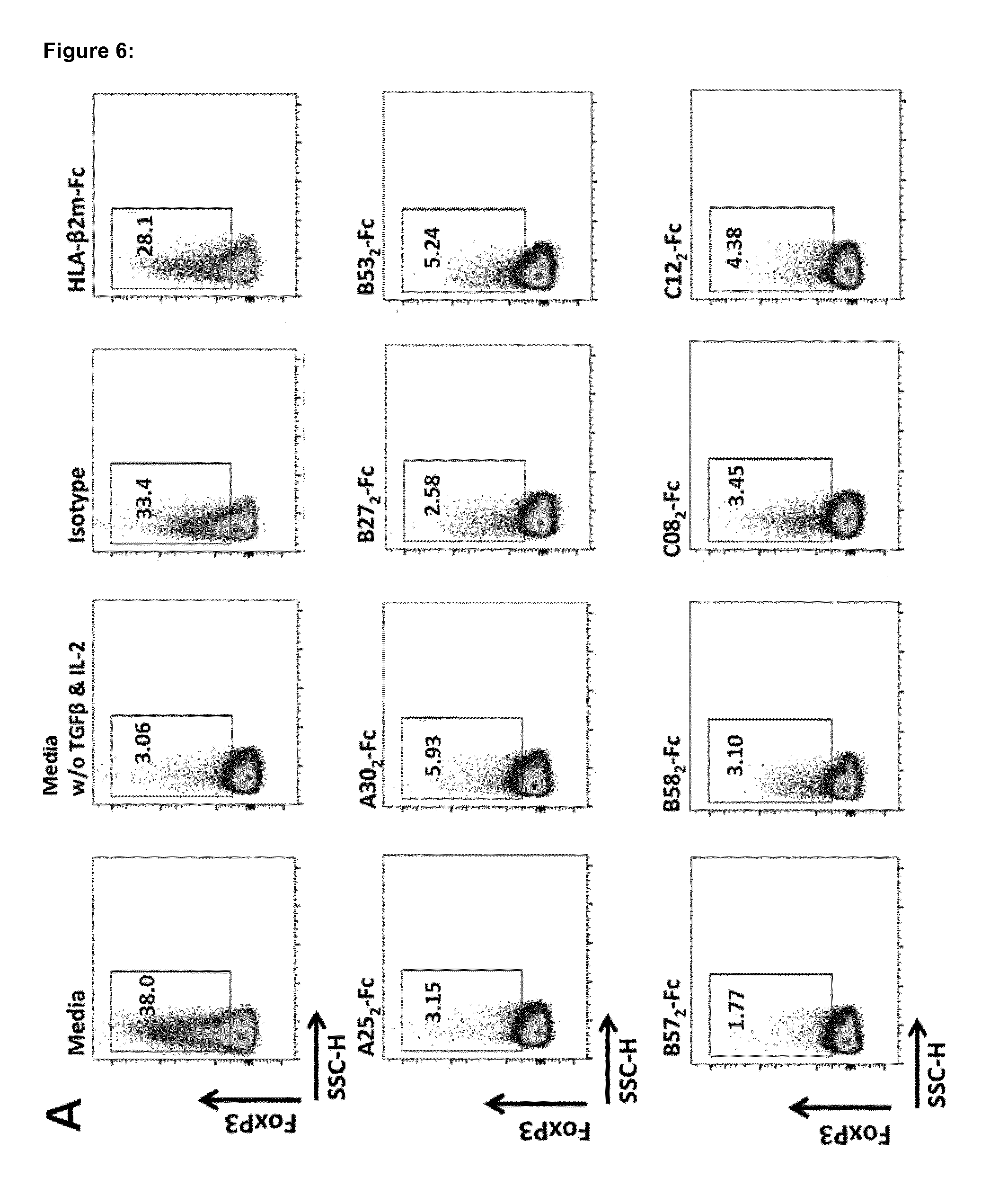
[0172] FIG. 6 shows that HLA.sub.2-Fc molecules (A25.sub.2-Fc, A30.sub.2-Fc, B27.sub.2-Fc, B53.sub.2-Fc, B57.sub.2-Fc, B58.sub.2-Fc, C08.sub.2-Fc, and C12.sub.2-Fc) invariably block mouse CD4.sup.+ T cell conversion into iTreg. Incubation of HLA.sub.2-Fc in a dose dependent manner with naive CD4.sup.+ T cells blocks the conversion to iTregs. A-B) HLA.sub.2-Fc molecules blocks the expression of FoxP3 (differentiation marker of Tregs) in optimal culture conditions for iTreg differentiation (10 .mu.g/mL) Control HLA-.beta.2m-Fc molecules, isotype, media supplemented with TGF.beta. and IL-2 and media w/o supplementation demonstrate the specific influence of HLA.sub.2-Fc on iTreg conversion.
[0173] FIG. 7 shows that HLA.sub.2-Fc (A25.sub.2-Fc, A30.sub.2-Fc, B27.sub.2-Fc, B53.sub.2-Fc, B57.sub.2-Fc, B58.sub.2-Fc, C08.sub.2-Fc and C12.sub.2-Fc) suppresses lymphoma T cells. A-E) suppression assays to determine the proliferation of cells in the presence of HLA.sub.2-Fc molecules or control HLA-.beta.2m-Fc molecules. HLA.sub.2-Fc suppress human (Jurkat) and mouse (EG.7) lymphoma cell lines in a dose dependent manner (.mu.g/200 .mu.L), other cell lines such as Daudi, B cell lymphoma; SK-N-AS, neuroblastoma; and L540, human Hodgkin lymphoma were assessed but not suppression was observed from HLA.sub.2-Fc molecules in optimal culture conditions. Other cell lines such as L428, human Hodgkin lymphoma; L1236, human Hodgkin lymphoma; IMR-5, neuroblastoma; and M130428, Melanoma were also tested but no suppression was observed.
[0174] FIG. 8 shows that HLA.sub.2-Fc (A30.sub.2-Fc, B58.sub.2-Fc, and C08.sub.2-Fc) as monotherapy or in combination with PD-1 antibodies can reduce the size of tumors in the C38 murine syngeneic colon carcinoma model. A) Experimental design of injection time points of colon carcinoma cells (C38) and injection of compounds. B) Mean average tumor volume mm.sup.3 of A30.sub.2-Fc treated groups (n=5). C) Mean average tumor volume mm.sup.3 of B58.sub.2-Fc treated groups (n=5). D) Mean average tumor volume mm.sup.3 of C08.sub.2-Fc treated groups (n=5). The experimental design of injection time points of cells and injection of substances was as follow: vehicle PBS Q3Dx6, isotype (10 mg/Kg) Q3Dx6; HLA.sub.2-Fc (10 mg/Kg) Q3Dx6; PD-1 biwk.times.2 (200 .mu.g); and HLA.sub.2-Fc+PD-1 (Q3Dx6 & biwk.times.2, respectively). Tumor volumes are expressed as mean.+-.SEM and analysed by two-way ANOVA followed by Bonferroni post-hoc analysis, *p<0.05, **p<0.01, n.s.=not significant. Q=days between injections; Dx=number of injections; biwk=twice a week.
[0175] FIG. 9 shows that HLA.sub.2-Fc (B27.sub.2-Fc and B57.sub.2-Fc) in combination with CTLA4 or PD-1 antibodies reduce the size of tumors in the MC38-OVA or C38 murine syngeneic colon carcinoma model. A) Mean average tumor volume mm.sup.3 of B27.sub.2-Fc treated groups (n=6). B) Mean average tumor volume mm.sup.3 of B57.sub.2-Fc treated groups (n=6). The experimental design of injection time points of cells and injection of substances was as follow: vehicle PBS Q3Dx6, isotype (10 mg/Kg) Q3Dx6; HLA.sub.2-Fc (10 mg/Kg) Q3Dx6; CTLA-4 Q3Dx2 (d1=100 .mu.g; d4=50 .mu.g), PD-1 biwk.times.2 (200 .mu.g); HLA.sub.2-Fc+CTLA-4 (Q3Dx6 & Q3Dx2, respectively), and HLA.sub.2-Fc+PD-1 (Q3Dx6 & biwk.times.2, respectively). Tumor volumes are expressed as mean.+-.SEM and analysed by two-way ANOVA followed by Bonferroni post-hoc analysis, **p<0.01, ***p<0.001, n.s.=not significant. Q=days between injections; Dx=number of injections; biwk=twice a week.
[0176] FIG. 10 shows the in vivo study of A25.sub.2-Fc in combination with PD-1 and 4-1BB antibodies in large tumors of the pancreatic Pan02 syngeneic mouse model. A) Mean average tumor volume in mm.sup.3 of A25.sub.2-Fc treated animals (n=6). B) % .DELTA.tumor inhibition of treated mice groups compared to control. The experimental design of injection time points of substances was as follow: isotype (5 mg/Kg) biwk.times.2; A25.sub.2-Fc (5 mg/Kg) biwk.times.2; 4-1BB antibody (1 mg/Kg) biwk.times.2 injections; A25.sub.2-Fc+4-1BB (5 mg/Kg and 1 mg/Kg, respectively) biwk.times.2; PD-1 antibody (5 mg/Kg) biwk.times.2; and A25.sub.2-Fc+PD-1 (5 mg/Kg each) biwk.times.2. Tumor volumes are expressed as mean.+-.SEM and analysed by two-way ANOVA followed by Bonferroni post-hoc analysis *p<0.05. .DELTA.tumor inhibition is calculated from the .DELTA.T/.DELTA.C tumor growth ratio, which represents the growth of the tumor in % from the beginning of the treatment (e.g. 300 mm.sup.3), to the end volume of the treatment (e.g. 1000 mm.sup.3) compared to isotype. biwk=twice a week.
[0177] FIG. 11 shows the immune contexture of Tumor Infiltrating Lymphocytes (TILs) analysis from Pan02 pancreatic cancer mice with large tumors treated with A25.sub.2-Fc, 4-1BB and PD-1 by flow cytometry (continuation of experiment in FIG. 10). Relevant leukocytes analysed infiltrating the tumor: A) CD3+ T cells, CD4+ T cells, Regulatory T cells (Treg), CD8+ T cells, and the CD8+/Treg ratio. B) Granulocytes, Macrophages, Macrophage M1-type, Macrophage M2-type, and Myeloid Derived Suppressor Cells (MDSCs). C) M1/M2 macrophage ratio, Monocytes, Natural killer cells (NK), and Natural Killer T cells (NKT). Leukocytes % are expressed as box plots showing sample maxima and minima, and each group analysed by one-way ANOVA followed by Dunnet post-hoc analysis *p<0.05; **p<0.01; ***p<0.001.
[0178] FIG. 12 shows the immune contexture of blood leukocyte analysis from Pan02 pancreatic cancer mice with large tumors treated with A25.sub.2-Fc, 4-1BB and PD-1 by flow cytometry (continuation of experiment in FIG. 10). Relevant leukocytes analysed present in the blood: A) CD3+ T cells, CD4+ T cells, Regulatory T cells (Treg), CD8+ T cells, and CD8+/Treg ratio. B) Th1 cells (CD4+ T cells IFN.gamma.+), Natural Killer cells (NK), and Natural Killer T cells (NKT). C) Monocytes, Granulocyte-Myeloid Derived suppressor cells (G-MDSCs), and Monocytic-Myeloid Derived Suppressor cells (M-MDSCs). Leukocytes % are expressed as box plots showing sample maxima and minima, and each group analysed by one-way ANOVA followed by Dunnet post-hoc analysis *p<0.05; **p<0.01; ***p<0.001.
[0179] FIG. 13 shows the in vivo study of A30.sub.2-Fc in combination with PD-1 and 4-1BB antibodies in large tumors of the pancreatic Pan02 syngeneic mouse model. A) Mean average tumor volume in mm.sup.3 of A30.sub.2-Fc treated animals (n=6). B) % .DELTA.tumor inhibition of treated mice groups compared to control. The experimental design of injection time points of substances was as follow: isotype (5 mg/Kg) biwk.times.2; A30.sub.2-Fc (5 mg/Kg) biwk.times.2; 4-1BB antibody (1 mg/Kg) biwk.times.2 injections; A30.sub.2-Fc+4-1BB (5 mg/Kg and 1 mg/Kg, respectively) biwk.times.2; PD-1 antibody (5 mg/Kg) biwk.times.2; and A30.sub.2-Fc+PD-1 (5 mg/Kg each) biwk.times.2. Tumor volumes are expressed as mean.+-.SEM and analysed by two-way ANOVA followed by Bonferroni post-hoc analysis. .DELTA.tumor inhibition is calculated from the .DELTA.T/.DELTA.C tumor growth ratio, which represents the growth of the tumor in % from the beginning of the treatment (e.g. 300 mm.sup.3), to the end volume of the treatment (e.g. 1000 mm.sup.3) compared to isotype. biwk=twice a week.
[0180] FIG. 14 shows the immune contexture of Tumor Infiltrating Lymphocytes (TILs) analysis from Pan02 pancreatic cancer mice with large tumors treated with A30.sub.2-Fc, 4-1BB and PD-1 by flow cytometry (continuation of experiment in FIG. 13). Relevant leukocytes analysed infiltrating the tumor: A) CD3+ T cells, CD4+ T cells, Regulatory T cells (Treg), CD8+ T cells, and the CD8+/Treg ratio. B) Granulocytes, Macrophages, Macrophage M1-type, Macrophage M2-type, and Myeloid Derived Suppressor Cells (MDSCs). C) M1/M2 macrophage ratio, Monocytes, Natural killer cells (NK), and Natural Killer T cells (NKT). Leukocytes % are expressed as box plots showing sample maxima and minima, and each group analysed by one-way ANOVA followed by Dunnet post-hoc analysis *p<0.05; **p<0.01; ***p<0.001; ****p<0.0001.
[0181] FIG. 15 shows the immune contexture of blood leukocyte analysis from treated Pan02 pancreatic cancer mice with large tumors treated with A30.sub.2-Fc, 4-1BB and PD-1 by flow cytometry (continuation of experiment in FIG. 13). Relevant leukocytes analysed present in the blood: A) CD3+ T cells, CD4+ T cells, Regulatory T cells (Treg), CD8+ T cells, and CD8+/Treg ratio. B) Th1 cells (CD4+ T cells IFN.gamma.+), Natural Killer cells (NK), and Natural Killer T cells (NKT). C) Monocytes, Granulocyte-Myeloid Derived suppressor cells (G-MDSCs), and Monocytic-Myeloid Derived Suppressor cells (M-MDSCs). Leukocytes % are expressed as box plots showing sample maxima and minima, and each group analysed by one-way ANOVA followed by Dunnet post-hoc analysis *p<0.05; **p<0.01; ***p<0.001; ****p<0.0001.
[0182] FIG. 16 shows the in vivo study of B27.sub.2-Fc in combination with PD-1 and 4-1BB antibodies in large tumors of the pancreatic Pan02 syngeneic mouse model. A) Mean average tumor volume in mm.sup.3 of B27.sub.2-Fc treated animals (n=6). B) % .DELTA.tumor inhibition of treated mice groups compared to control. The experimental design of injection time points of substances was as follow: isotype (5 mg/Kg) biwk.times.2; B27.sub.2-Fc (5 mg/Kg) biwk.times.2; 4-1BB antibody (1 mg/Kg) biwk.times.2 injections; B27.sub.2-Fc+4-1BB (5 mg/Kg and 1 mg/Kg, respectively) biwk.times.2; PD-1 antibody (5 mg/Kg) biwk.times.2; and B27.sub.2-Fc+PD-1 (5 mg/Kg each) biwk.times.2. Tumor volumes are expressed as mean.+-.SEM and analysed by two-way ANOVA followed by Bonferroni post-hoc analysis. .DELTA.tumor inhibition is calculated from the .DELTA.T/.DELTA.C tumor growth ratio, which represents the growth of the tumor in % from the beginning of the treatment (e.g. 300 mm.sup.3), to the end volume of the treatment (e.g. 1000 mm.sup.3) compared to isotype. biwk=twice a week.
[0183] FIG. 17 shows the immune contexture of Tumor Infiltrating Lymphocytes (TILs) analysis from Pan02 pancreatic cancer mice with large tumors treated with B27.sub.2-Fc, 4-1BB and PD-1 by flow cytometry (continuation of experiment in FIG. 16). Relevant leukocytes analysed infiltrating the tumor: A) CD3+ T cells, CD4+ T cells, Regulatory T cells (Treg), CD8+ T cells, and the CD8+/Treg ratio. B) Granulocytes, Macrophages, Macrophage M1-type, Macrophage M2-type, and Myeloid Derived Suppressor Cells (MDSCs). C) M1/M2 macrophage ratio, Monocytes, Natural killer cells (NK), and Natural Killer T cells (NKT). Leukocytes % are expressed as box plots showing sample maxima and minima, and each group analysed by one-way ANOVA followed by Dunnet post-hoc analysis *p<0.05; **p<0.01; ***p<0.001; ****p<0.0001.
[0184] FIG. 18 shows the immune contexture of blood leukocyte analysis from treated Pan02 pancreatic cancer mice with large tumors treated with B27.sub.2-Fc, 4-1BB and PD-1 by flow cytometry (continuation of experiment in FIG. 16). Relevant leukocytes analysed present in the blood: A) CD3+ T cells, CD4+ T cells, Regulatory T cells (Treg), CD8+ T cells, and CD8+/Treg ratio. B) Th1 cells (CD4+ T cells IFN.gamma.+), Natural Killer cells (NK), and Natural Killer T cells (NKT). C) Monocytes, Granulocyte-Myeloid Derived suppressor cells (G-MDSCs), and Monocytic-Myeloid Derived Suppressor cells (M-MDSCs). Leukocytes % are expressed as box plots showing sample maxima and minima, and each group analysed by one-way ANOVA followed by Dunnet post-hoc analysis *p<0.05; **p<0.01; ***p<0.001; ****p<0.0001.
[0185] FIG. 19 shows the in vivo study of B53.sub.2-Fc in combination with PD-1 and 4-1BB antibodies in large tumors of the pancreatic Pan02 syngeneic mouse model. A) Mean average tumor volume in mm.sup.3 of B53.sub.2-Fc treated animals (n=6). B) % .DELTA.tumor inhibition of treated mice groups compared to control. The experimental design of injection time points of substances was as follow: isotype (5 mg/Kg) biwk.times.2; B53.sub.2-Fc (5 mg/Kg) biwk.times.2; 4-1BB antibody (1 mg/Kg) biwk.times.2 injections; B53.sub.2-Fc+4-1BB (5 mg/Kg and 1 mg/Kg, respectively) biwk.times.2; PD-1 antibody (5 mg/Kg) biwk.times.2; and B53.sub.2-Fc+PD-1 (5 mg/Kg each) biwk.times.2. Tumor volumes are expressed as mean.+-.SEM and analysed by two-way ANOVA followed by Bonferroni post-hoc analysis. .DELTA.tumor inhibition is calculated from the .DELTA.T/.DELTA.C tumor growth ratio, which represents the growth of the tumor in % from the beginning of the treatment (e.g. 300 mm.sup.3), to the end volume of the treatment (e.g. 1000 mm.sup.3) compared to isotype. biwk=twice a week.
[0186] FIG. 20 shows the immune contexture of Tumor Infiltrating Lymphocytes (TILs) analysis from Pan02 pancreatic cancer mice with large tumors treated with B53.sub.2-Fc, 4-1BB and PD-1 by flow cytometry (continuation of experiment in FIG. 19). Relevant leukocytes analysed infiltrating the tumor: A) CD3+ T cells, CD4+ T cells, Regulatory T cells (Treg), CD8+ T cells, and the CD8+/Treg ratio. B) Granulocytes, Macrophages, Macrophage M1-type, Macrophage M2-type, and Myeloid Derived Suppressor Cells (MDSCs). C) M1/M2 macrophage ratio, Monocytes, Natural killer cells (NK), and Natural Killer T cells (NKT). Leukocytes % are expressed as box plots showing sample maxima and minima, and each group analysed by one-way ANOVA followed by Dunnet post-hoc analysis *p<0.05; **p<0.01; ***p<0.001; ****p<0.0001.
[0187] FIG. 21 shows the immune contexture of blood leukocyte analysis from treated Pan02 pancreatic cancer mice with large tumors treated with B53.sub.2-Fc, 4-1BB and PD-1 by flow cytometry (continuation of experiment in FIG. 19). Relevant leukocytes analysed present in the blood: A) CD3+ T cells, CD4+ T cells, Regulatory T cells (Treg), CD8+ T cells, and CD8+/Treg ratio. B) Th1 cells (CD4+ T cells IFN.gamma.+), Natural Killer cells (NK), and Natural Killer T cells (NKT). C) Monocytes, Granulocyte-Myeloid Derived suppressor cells (G-MDSCs), and Monocytic-Myeloid Derived Suppressor cells (M-MDSCs). Leukocytes % are expressed as box plots showing sample maxima and minima, and each group analysed by one-way ANOVA followed by Dunnet post-hoc analysis *p<0.05; **p<0.01; ***p<0.001; ****p<0.0001.
[0188] FIG. 22 shows the in vivo study of B57.sub.2-Fc in combination with PD-1 and 4-1BB antibodies in large tumors of the pancreatic Pan02 syngeneic mouse model. A) Mean average tumor volume in mm.sup.3 of B57.sub.2-Fc treated animals (n=6). B) % .DELTA.tumor inhibition of treated mice groups compared to control. The experimental design of injection time points of substances was as follow: isotype (5 mg/Kg) biwk.times.2; B57.sub.2-Fc (5 mg/Kg) biwk.times.2; 4-1BB antibody (1 mg/Kg) biwk.times.2 injections; B57.sub.2-Fc+4-1BB (5 mg/Kg and 1 mg/Kg, respectively) biwk.times.2; PD-1 antibody (5 mg/Kg) biwk.times.2; and B57.sub.2-Fc+PD-1 (5 mg/Kg each) biwk.times.2. Tumor volumes are expressed as mean.+-.SEM and analysed by two-way ANOVA followed by Bonferroni post-hoc analysis. .DELTA.tumor inhibition is calculated from the .DELTA.T/.DELTA.C tumor growth ratio, which represents the growth of the tumor in % from the beginning of the treatment (e.g. 300 mm.sup.3), to the end volume of the treatment (e.g. 1000 mm.sup.3) compared to isotype. biwk=twice a week.
[0189] FIG. 23 shows the immune contexture of Tumor Infiltrating Lymphocytes (TILs) analysis from Pan02 pancreatic cancer mice with large tumors treated with B57.sub.2-Fc, 4-1BB and PD-1 by flow cytometry (continuation of experiment in FIG. 22). Relevant leukocytes analysed infiltrating the tumor: A) CD3+ T cells, CD4+ T cells, Regulatory T cells (Treg), CD8+ T cells, and the CD8+/Treg ratio. B) Granulocytes, Macrophages, Macrophage M1-type, Macrophage M2-type, and Myeloid Derived Suppressor Cells (MDSCs). C) M1/M2 macrophage ratio, Monocytes, Natural killer cells (NK), and Natural Killer T cells (NKT). Leukocytes % are expressed as box plots showing sample maxima and minima, and each group analysed by one-way ANOVA followed by Dunnet post-hoc analysis *p<0.05; **p<0.01; ***p<0.001; ****p<0.0001.
[0190] FIG. 24 shows the immune contexture of blood leukocyte analysis from treated Pan02 pancreatic cancer mice with large tumors treated with B57.sub.2-Fc, 4-1BB and PD-1 by flow cytometry (continuation of experiment in FIG. 22). Relevant leukocytes analysed present in the blood: A) CD3+ T cells, CD4+ T cells, Regulatory T cells (Treg), CD8+ T cells, and CD8+/Treg ratio. B) Th1 cells (CD4+ T cells IFN.gamma.+), Natural Killer cells (NK), and Natural Killer T cells (NKT). C) Monocytes, Granulocyte-Myeloid Derived suppressor cells (G-MDSCs), and Monocytic-Myeloid Derived Suppressor cells (M-MDSCs). Leukocytes % are expressed as box plots showing sample maxima and minima, and each group analysed by one-way ANOVA followed by Dunnet post-hoc analysis *p<0.05; **p<0.01; ***p<0.001; ****p<0.0001.
[0191] FIG. 25 shows the in vivo study of B58.sub.2-Fc in combination with PD-1 and 4-1BB antibodies in large tumors of the pancreatic Pan02 syngeneic mouse model. A) Mean average tumor volume in mm.sup.3 of B58.sub.2-Fc treated animals (n=6). B) % .DELTA.tumor inhibition of treated mice groups compared to control. The experimental design of injection time points of substances was as follow: isotype (5 mg/Kg) biwk.times.2; B58.sub.2-Fc (5 mg/Kg) biwk.times.2; 4-1BB antibody (1 mg/Kg) biwk.times.2 injections; B58.sub.2-Fc+4-1BB (5 mg/Kg and 1 mg/Kg, respectively) biwk.times.2; PD-1 antibody (5 mg/Kg) biwk.times.2; and B58.sub.2-Fc+PD-1 (5 mg/Kg each) biwk.times.2. Tumor volumes are expressed as mean.+-.SEM and analysed by two-way ANOVA followed by Bonferroni post-hoc analysis. .DELTA.tumor inhibition is calculated from the .DELTA.T/.DELTA.C tumor growth ratio, which represents the growth of the tumor in % from the beginning of the treatment (e.g. 300 mm.sup.3), to the end volume of the treatment (e.g. 1000 mm.sup.3) compared to isotype. biwk=twice a week.
[0192] FIG. 26 shows the immune contexture of Tumor Infiltrating Lymphocytes (TILs) analysis from Pan02 pancreatic cancer mice with large tumors treated with B58.sub.2-Fc, 4-1BB and PD-1 by flow cytometry (continuation of experiment in FIG. 25). Relevant leukocytes analysed infiltrating the tumor: A) CD3+ T cells, CD4+ T cells, Regulatory T cells (Treg), CD8+ T cells, and the CD8+/Treg ratio. B) Granulocytes, Macrophages, Macrophage M1-type, Macrophage M2-type, and Myeloid Derived Suppressor Cells (MDSCs). C) M1/M2 macrophage ratio, Monocytes, Natural killer cells (NK), and Natural Killer T cells (NKT). Leukocytes % are expressed as box plots showing sample maxima and minima, and each group analysed by one-way ANOVA followed by Dunnet post-hoc analysis *p<0.05; **p<0.01; ***p<0.001; ****p<0.0001.
[0193] FIG. 27 shows the immune contexture of blood leukocyte analysis from treated Pan02 pancreatic cancer mice with large tumors treated with B58.sub.2-Fc, 4-1BB and PD-1 by flow cytometry (continuation of experiment in FIG. 25). Relevant leukocytes analysed present in the blood: A) CD3+ T cells, CD4+ T cells, Regulatory T cells (Treg), CD8+ T cells, and CD8+/Treg ratio. B) Th1 cells (CD4+ T cells IFN.gamma.+), Natural Killer cells (NK), and Natural Killer T cells (NKT). C) Monocytes, Granulocyte-Myeloid Derived suppressor cells (G-MDSCs), and Monocytic-Myeloid Derived Suppressor cells (M-MDSCs). Leukocytes % are expressed as box plots showing sample maxima and minima, and each group analysed by one-way ANOVA followed by Dunnet post-hoc analysis *p<0.05; **p<0.01; ***p<0.001; ****p<0.0001.
[0194] FIG. 28 shows the in vivo study of C08.sub.2-Fc in combination with PD-1 and 4-1BB antibodies in large tumors of the pancreatic Pan02 syngeneic mouse model. A) Mean average tumor volume in mm.sup.3 of C08.sub.2-Fc treated animals (n=6). B) % .DELTA.tumor inhibition of treated mice groups compared to control. The experimental design of injection time points of substances was as follow: isotype (5 mg/Kg) biwk.times.2; C08.sub.2-Fc (5 mg/Kg) biwk.times.2; 4-1BB antibody (1 mg/Kg) biwk.times.2 injections; C08.sub.2-Fc+4-1BB (5 mg/Kg and 1 mg/Kg, respectively) biwk.times.2; PD-1 antibody (5 mg/Kg) biwk.times.2; and C08.sub.2-Fc+PD-1 (5 mg/Kg each) biwk.times.2. Tumor volumes are expressed as mean.+-.SEM and analysed by two-way ANOVA followed by Bonferroni post-hoc analysis. .DELTA.tumor inhibition is calculated from the .DELTA.T/.DELTA.C tumor growth ratio, which represents the growth of the tumor in % from the beginning of the treatment (e.g. 300 mm.sup.3), to the end volume of the treatment (e.g. 1000 mm.sup.3) compared to isotype. biwk=twice a week.
[0195] FIG. 29 shows the immune contexture of Tumor Infiltrating Lymphocytes (TILs) analysis from Pan02 pancreatic cancer mice with large tumors treated with C08.sub.2-Fc, 4-1BB and PD-1 by flow cytometry (continuation of experiment in FIG. 28). Relevant leukocytes analysed infiltrating the tumor: A) CD3+ T cells, CD4+ T cells, Regulatory T cells (Treg), CD8+ T cells, and the CD8+/Treg ratio. B) Granulocytes, Macrophages, Macrophage M1-type, Macrophage M2-type, and Myeloid Derived Suppressor Cells (MDSCs). C) M1/M2 macrophage ratio, Monocytes, Natural killer cells (NK), and Natural Killer T cells (NKT). Leukocytes % are expressed as box plots showing sample maxima and minima, and each group analysed by one-way ANOVA followed by Dunnet post-hoc analysis *p<0.05; **p<0.01; ***p<0.001; ****p<0.0001.
[0196] FIG. 30 shows the immune contexture of blood leukocyte analysis from treated Pan02 pancreatic cancer mice with large tumors treated with C08.sub.2-Fc, 4-1BB and PD-1 by flow cytometry (continuation of experiment in FIG. 28). Relevant leukocytes analysed present in the blood: A) CD3+ T cells, CD4+ T cells, Regulatory T cells (Treg), CD8+T cells, and CD8+/Treg ratio. B) Th1 cells (CD4+ T cells IFN.gamma.+), Natural Killer cells (NK), and Natural Killer T cells (NKT). C) Monocytes, Granulocyte-Myeloid Derived suppressor cells (G-MDSCs), and Monocytic-Myeloid Derived Suppressor cells (M-MDSCs). Leukocytes % are expressed as box plots showing sample maxima and minima, and each group analysed by one-way ANOVA followed by Dunnet post-hoc analysis *p<0.05; **p<0.01; ***p<0.001; ****p<0.0001.
[0197] FIG. 31 shows the in vivo study of C12.sub.2-Fc in combination with PD-1 and 4-1BB antibodies in large tumors of the pancreatic Pan02 syngeneic mouse model. A) Mean average tumor volume in mm.sup.3 of C12.sub.2-Fc treated animals (n=6). B) % .DELTA.tumor inhibition of treated mice groups compared to control. The experimental design of injection time points of substances was as follow: isotype (5 mg/Kg) biwk.times.2; C12.sub.2-Fc (5 mg/Kg) biwk.times.2; 4-1BB antibody (1 mg/Kg) biwk.times.2 injections; C12.sub.2-Fc+4-1BB (5 mg/Kg and 1 mg/Kg, respectively) biwk.times.2; PD-1 antibody (5 mg/Kg) biwk.times.2; and C12.sub.2-Fc+PD-1 (5 mg/Kg each) biwk.times.2. Tumor volumes are expressed as mean.+-.SEM and analysed by two-way ANOVA followed by Bonferroni post-hoc analysis. .DELTA.tumor inhibition is calculated from the .DELTA.T/.DELTA.C tumor growth ratio, which represents the growth of the tumor in % from the beginning of the treatment (e.g. 300 mm.sup.3), to the end volume of the treatment (e.g. 1000 mm.sup.3) compared to isotype. biwk=twice a week.
[0198] FIG. 32 shows the immune contexture of Tumor Infiltrating Lymphocytes (TILs) analysis from Pan02 pancreatic cancer mice with large tumors treated with C12.sub.2-Fc, 4-1BB and PD-1 by flow cytometry (continuation of experiment in FIG. 31). Relevant leukocytes analysed infiltrating the tumor: A) CD3+ T cells, CD4+ T cells, Regulatory T cells (Treg), CD8+ T cells, and the CD8+/Treg ratio. B) Granulocytes, Macrophages, Macrophage M1-type, Macrophage M2-type, and Myeloid Derived Suppressor Cells (MDSCs). C) M1/M2 macrophage ratio, Monocytes, Natural killer cells (NK), and Natural Killer T cells (NKT). Leukocytes % are expressed as box plots showing sample maxima and minima, and each group analysed by one-way ANOVA followed by Dunnet post-hoc analysis *p<0.05; **p<0.01; ***p<0.001; ****p<0.0001.
[0199] FIG. 33 shows the immune contexture of blood leukocyte analysis from treated Pan02 pancreatic cancer mice with large tumors treated with C12.sub.2-Fc, 4-1BB and PD-1 by flow cytometry (continuation of experiment in FIG. 31). Relevant leukocytes analysed present in the blood: A) CD3+ T cells, CD4+ T cells, Regulatory T cells (Treg), CD8+ T cells, and CD8+/Treg ratio. B) Th1 cells (CD4+ T cells IFN.gamma.+), Natural Killer cells (NK), and Natural Killer T cells (NKT). C) Monocytes, Granulocyte-Myeloid Derived suppressor cells (G-MDSCs), and Monocytic-Myeloid Derived Suppressor cells (M-MDSCs). Leukocytes % are expressed as box plots showing sample maxima and minima, and each group analysed by one-way ANOVA followed by Dunnet post-hoc analysis *p<0.05; **p<0.01; ***p<0.001; ****p<0.0001.
EXAMPLES
[0200] The inventors surprisingly found that MHC-Ia open conformers interact with diverse immunoregulatory cell surface receptors present in NK cells, NKT cells, T cells, macrophages and MDSC cells with unique binding or stronger affinity than their control MHC-Ia heterotrimers. HLA class I-a open conformers can be used as a therapeutics to target diseases were white blood cells impair the development of protective immunity, as is the case of cancer and infectious diseases.
[0201] Additionally, they discovered a novel in vivo mode of action with injections of HLA.sub.2-Fc as monotherapy or combinatorial approaches using checkpoint modulatory agents. HLA.sub.2-Fc therapy alone or in combinatorial therapies can modulate the infiltration of diverse sets of leukocytes into tumors as determined by the increased infiltration of macrophages M1/M2 ratio, increased NK cells, NKT cells, CD3+ T cells, and CD8+ T cells, and reduction of MDSCs.
[0202] Furthermore, they observed that systemically by blood analysis HLA.sub.2-Fc therapy increase the expansion of NKT cells and in some cases Th1 cells, indicating the presence of a biomarker that can be used for therapy efficacy in pre-clinical and clinical settings.
[0203] Interestingly, they also observed that monotherapy with 4-1BB increases systemically the expansion of CD3+, CD4+, CD8+ T cells and Tregs in the blood of animals, indicating a potential side effect of hyper activation of the immune system by 4-1BB. Diverse combinations of HLA.sub.2-Fc+4-1BB reduced significantly the presence of blood CD3+, CD4+, Treg, and CD8+ T cells, indicating a positive combinatorial approach in case of unwanted lymphocyte expansion on the blood of treated patients with agonistic antibodies.
[0204] Overall, the mode of action of MHC-Ia open conformers, particularly when present as fusion proteins comprising an Fc immunoglobulin fragment, alone or in a combinatorial approach with antagonistic/agonistic antibodies is of undoubted relevance as immunomodulatory agents, and can be useful for its translation in the treatment of cancer.
[0205] HLA open conformers can be used as a therapeutic to target diseases where immunomodulation is a therapeutic approach, as is the case of cancer and infectious diseases.
In Vitro Tests
[0206] MHC-Ia open conformers bind to immunoregulatory receptors expressed in diverse types of white blood cells with unique binding or different affinity than their HLA-.beta.2m-Fc control counterparts
[0207] The inventors determined if MHC-Ia open conformers interact with specific immunomodulatory receptors by enzyme-linked immunosorbent assay (ELISA). Results demonstrated that MHC-Ia open conformers interact uniquely to KIR3DL2, and PTPRJ (for exception of HLA-C-.beta.2m-Fc) and display different affinities to KIR3DL1, KIR3DL3, LILRB1, LILRB2, and Pirb immmunoregulatory receptors than their HLA-.beta.2m-Fc control counterparts (FIGS. 5 A-G). This data shows for the first time that MHC classical alleles (HLA-A, HLA-B & HLA-C) (MHC-Ia) have a similar binding pattern to immunoregulatory receptors when they are present as open conformers.
MHC-Ia Open Conformers Blocks Conversion of Murine CD4.sup.+ T Cells into iTregs
[0208] The influence of MHC-Ia molecules on naive CD4.sup.+ T cells for iTreg conversion was analysed with 10 .mu.g/mL of HLA.sub.2-Fc (A25.sub.2-Fc, A30.sub.2-Fc, B27.sub.2-Fc, B53.sub.2-Fc, B57.sub.2-Fc, B58.sub.2-Fc, C08.sub.2-Fc and C12.sub.2-Fc), HLA-.beta.2m controls (A25-.beta.2m-Fc, A30-.beta.2m-Fc, B27-.beta.2m-Fc, B53-.beta.2m-Fc, B57-.beta.2m-Fc, B58-.beta.2m-Fc, C08-.beta.2m-Fc and C12-.beta.2m-Fc), isotype, and PBS, incubated with naive CD4+ T cells in optimal culture conditions for iTreg conversion. MHC-Ia open conformers demonstrated invariably to down modulate the induction of FoxP3 (FIG. 6) and thus conversion of naive CD4+ T cells into iTregs.
MHC-Ia Open Conformers Impair the Proliferation of Leukaemia T Cells.
[0209] The inventors determined the effect of MHC-Ia open conformers (A25.sub.2-Fc, A30.sub.2-Fc, B27.sub.2-Fc, B53.sub.2-Fc, B57.sub.2-Fc, B58.sub.2-Fc, C08.sub.2-Fc and C12.sub.2-Fc) with the blocking of proliferation in different tumor cell lines. Results demonstrated that MHC-Ia open conformers modulate invariably the proliferation of lymphoma T cell lines, when compared to their control counterparts HLA-.beta.2m-Fc (FIG. 7) or isotype IgG4 (data not provided), indicating its potential application to the treatment of lymphoma as a targeted therapy.
In Vivo Tests
[0210] The in vivo proof of concept of MHC-Ia open conformers as immunomodulatory therapeutic molecules for cancer therapy was demonstrated using a validated pre-clinical syngeneic murine C38 and MC38-OVA colon carcinoma models (FIGS. 8 and 9), and in the pancreatic (Pan02) cancer mouse model (FIGS. 10, 13, 16, 19, 22, 25, 28 and 31).
Production of MHC-Ia Open Conformers as a Human Fc Fusion Protein in CHO Cells
[0211] A valid strategy, from a therapeutic point of view, is to produce MHC-Ia open conformers molecules in stable format (Fc fusion), to increase solubility, stability, avidity, half-life, and from a technological point of view, cost-effective production and purification in mammalian systems. HLA-.beta.2m-Fc complex was successfully produced by inserting the alpha 1, 2 and 3 domains of HLA-A25, HLA-A30, HLA-B27, HLA-B53, HLA-B57, HLA-B58, HLA-C08 and HLA-C12 into a human IgG4-Fc vector cassette (FIG. 3A), together with a human-.beta.2m vector, necessary for extracellular production of the HLA-.beta.2m-Fc protein (FIGS. 3A,B). Transfections in Chinese hamster ovary cells (CHO) cells were performed using both the HLA-Fc-vector+.beta.2m-vector at a ratio of 1:1. Supernatants were collected and HLA-.beta.2m-Fc purified using standard antibody purification protocols (Recombinant Protein Purification Handbook, principles and methods. 2009. GE Healthcare, 18-1142-75). Separation of .beta.2m from HLA-Fc free-heavy chains was performed using denaturing conditions by SEC (FIG. 4A), or dialysis (data not shown). Refolding of HLA.sub.2-Fc was assessed using the dilution method in refolding buffer and analysed SDS page (FIGS. 4B,C) or by western blot (data not shown).
Pre-Clinical Combination Therapy Tests of HLA.sub.2-Fc with CTLA4 and PD-I Antibodies in Murine Syngeneic Colon Cancer Models
[0212] The in vivo proof of concept study using HLA.sub.2-Fc (A30.sub.2-Fc, B27.sub.2-Fc, B57.sub.2-Fc, B58.sub.2-Fc, and C08.sub.2-Fc) as immunomodulatory therapeutic molecules was demonstrated in the C38 and MC38-OVA murine colon carcinoma cancer models as monotherapy or in combination with a murine CTLA4 or murine PD-1 antibody.
[0213] Following established protocols C38 or MC38-OVA fragment tumours were subcutaneously injected in the flank of syngeneic mice. Once the tumour reached 60 mm.sup.3 (between 1-2 weeks after transplantation of tumors), mice were distributed according to their tumor volume. A30.sub.2-Fc, B27.sub.2-Fc, B57.sub.2-Fc, B58.sub.2-Fc, and C08.sub.2-Fc was injected i.p. six times every 3.sup.rd day (Q3Dx6), CTLA4 was injected two times (Q3Dx2), and PD-1 injected 4 times twice a week (biwk.times.2) (FIG. 8A).
[0214] Selected HLA.sub.2-Fc can synergize and enhance anti-tumor responses in syngeneic C38 and MC38-OVA colon cancer mouse models (FIGS. 8 & 9) either as monotherapy (C08.sub.2-Fc) (FIG. 8D) or in combination with checkpoint antibodies, such as PD-1+A30.sub.2-Fc (FIG. 8B), B58.sub.2-Fc (FIG. 8C), B57.sub.2-Fc (FIG. 9B) and CTLA4+B27.sub.2-Fc (FIG. 9A).
Pre-Clinical Combination Therapy Tests of HLA.sub.2-Fc with PD-I and 4-1BB Antibodies in Large Tumors of a Murine Syngeneic Pancreatic Cancer Model
[0215] For the pancreas (Pan02) cancer mouse model, following established protocols Pan02 cells were injected at 1.times.10.sup.5 in the right flank of syngeneic mice respectively. Once the tumors had reached 300 mm3 (approximately 3 weeks after injection of cells) mice were statistically distributed according to their tumor volume. To note that large tumors are harder to treat than smaller tumors, but are useful for further analysis of tumor infiltrating lymphocytes (TILs). Furthermore large tumors are closer to a clinical setting where interventions with immunomodulators are performed in large size tumors of patients.
[0216] In pancreas (Pan02) data demonstrated that HLA.sub.2-Fc combination with PD-1 antibody can significantly reduce large Pan02 tumors in combination with A25.sub.2-Fc (FIG. 10A-B), B27.sub.2-Fc (FIG. 16A-B), C08.sub.2-Fc (FIGS. 28A-B), and C12.sub.2-Fc (FIG. 31A-B), whereas PD-1 monotherapy showed no therapeutic effect. Other HLA.sub.2-Fc combinations with PD-1 did not demonstrate statistical significance, however % .DELTA.tumor inhibition was observed in combination B57.sub.2-Fc (FIG. 22). Additionally, combo therapy of HLA.sub.2-Fc with 4-1BB antibody demonstrated to significantly reduce the tumor size or several HLA.sub.2-Fc combo therapies (for exception of A30.sub.2-Fc and C08.sub.2-Fc) when compared to isotype. The most striking tumor reductions (p<0.01) were observed with B53.sub.2-Fc (FIG. 19A-B), B57.sub.2-Fc (FIGS. 22A-B), and B58.sub.2-Fc (FIG. 25A-B). 4-1 BB monotherapy was not significantly different when compared to isotype control. Monotherapy with C08.sub.2-Fc (FIG. 28A-B) showed significant tumor reduction (p<0.01) compared to isotype.
[0217] The tumor immune contexture of pancreas (Pan02) mice demonstrated the influence of HLA.sub.2-Fc therapy towards diverse sets of tumor infiltrating leukocytes as observed with the infiltration of macrophages M1/M2 ratio, increased NK cells, NKT cells, CD3+ T cells, and CD8+ T cells, and reduction of MDSCs, with variations for each HLA.sub.2-Fc as observed in A25.sub.2-Fc (FIG. 11A-C), A30.sub.2-Fc (14A-C), B27.sub.2-Fc (17A-C), B53.sub.2-Fc (20A-C), B57.sub.2-Fc (23A-C), B58.sub.2-Fc (26A-C), C08.sub.2-Fc (29A-C), and C12.sub.2-Fc (32A-C). Systemically analysis of leukocytes from the blood demonstrated only few changes when compared to their control monotherapy counterparts in NKT cells and Th1 cells for some cases, A25.sub.2-Fc (FIG. 12A-C), A30.sub.2-Fc (15A-C), B27.sub.2-Fc (18A-C), B53.sub.2-Fc (21A-C), B57.sub.2-Fc (24A-C), B58.sub.2-Fc (27A-C), C08.sub.2-Fc (30A-C), and C12.sub.2-Fc (33A-C).
CONCLUSION
[0218] The present invention demonstrates for the first time that the family of classical MHC-Ia molecules when produced as heavy chains without .beta.2m (HLA-A, HLA-B and HLA-C open conformers and their corresponding HLA.sub.2-Fc fusion proteins) have immunomodulatory properties that differ from their control HLA-.beta.2m counterparts. Using as non-limiting examples diverse sets of HLA alleles the inventors provide data demonstrating that invariably MHC-Ia molecules, when present as open conformers are immunomodulatory agents with unique properties as demonstrated by the modulation of leukocytes present in the tumor microenvironment and in the blood. Furthermore its use is not only limited to modulatory agents, but also for its use as therapeutics for the treatment of cancer as demonstrated in pre-clinical cancer mouse models of colon cancer and pancreatic cancer either as monotherapy or in combination therapy with checkpoint inhibitor antibodies (e.g. CTLA4 and PD-1) and checkpoint agonistic antibodies (e.g. 4-1BB).
[0219] Interaction of HLA.sub.2-Fc with diverse immunoregulatory receptors (KIR3DL1, KIR3DL2, KIR3DL3, LILRB1, LILRB2, PTPRJ and Pirb) distributed in diverse white blood cells (e.g. NK, NKT, CD4+ T-cells, macrophages and MDSCs) demonstrates that the multitasking nature of the molecules paves a new way of modulating the immune system with HLA open conformers.
[0220] Additionally, HLA.sub.2-Fc molecules demonstrated to block the conversion of naive CD4+ T-cells to iTregs in vitro, pointing out to a mode of action were HLA.sub.2-Fc acts as an immunomodulatory molecule affecting the differentiation and function of iTregs. Targeting iTregs is a strategy for diverse therapeutic indications, such as infectious diseases and cancer.
[0221] Overall, the mode of action of HLA.sub.2-Fc as combinatorial approaches with antagonistic/agonistic antibodies is of undoubted relevance in the treatment of cancer, and correlates to the current clinical need in cancer immunotherapy.
[0222] HLA.sub.2-Fc molecules emerge as a novel class of immunomodulatory drugs. In vitro and in vivo data points to a mechanism were HLA.sub.2-Fc molecules act as a switch-on mechanism for the activation of anti-tumor immunity. Without wishing to be bound by theory, the inventors hypothesize that the interaction of HLA.sub.2-Fc open conformers with diverse immunomodulatory receptors present in NK, T cells, macrophages and MDSCs, and functional modulation of Tregs participate synergistically and exacerbates the immune response.
Materials and Methods
Cell Lines
[0223] In vivo experiments were performed using C38 and MC38-OVA colon carcinoma mouse cell lines.
[0224] In vitro experiment cell lines used were: EL4, mouse T cell lymphoma; EG.7, mouse T cell lymphoma; Jurkat, human T cell lymphoma; L428, human Hodgkin lymphoma; L540, human Hodgkin lymphoma; L1236, human Hodgkin lymphoma; Daudi, B cell lymphoma; IMR-5, neuroblastoma; SK-N-AS, neuroblastoma; and M130428, Melanoma.
Antibodies
[0225] Lymphocytes populations for iTreg conversion experiments were stained with: CD4 (FITC-BD Bioscience), FoxP3+ (efluor 450-eBioscience), CD3 (PE-Cy7-eBioscience), CD45 (PerCP-eBioscience).
[0226] Analysis of tumor infiltrating lymphocytes was performed with the following antibodies: CD45 (Biolegend, clone 30-F11); CD3 (BD Bioscience, clone 145-2C11); CD4 (Biolegend, clone GK1.5), CD8 (BD Bioscience, clone 53-6.7), CD25 (Biolegend, clone PC61), FoxP3 (eBioscience, clone FJK-16s), CD335 (Biolegend, clone 29A1.4), F4/80 (Biolegend, clone BM8), CD11b (Biolegend, clone M1/70), Gr-1 (BD Bioscience, clone RB6-8C5), MHCII I-A/I-E (BD Bioscience, clone 2G9), CD206 (Biolegend, clone C068C2) and L/D stain (eBioscience).
[0227] Analysis of blood leukocytes was performed with the following antibodies: CD45 (Biolegend, clone 30-F11); CD3 (BD Bioscience, clone 145-2C11), CD4 (Biolegend, clone GK1.5), CD8 (BD Bioscience, clone 53-6.7), FoxP3 (eBioscience, clone FJK-16s), T-Bet (BD Bioscience, clone 4B10), CD335 (Biolegend, clone 29A1.4), F4/80 (Biolegend, clone BM8), CD115 (Biolegend, clone AFS98), CD11b (Biolegend, clone M1/70), Ly6G (Biolegend, clone 1A8), Ly6C (Biolegend, clone HK1.4) and L/D stain (eBioscience).
[0228] Checkpoint inhibitor antibodies CTLA4 clone 9H10, PD-1 clone RMP1-14, and agonist antibody 4-1BB clone 3H3 were obtained from Bio X Cell Co.
[0229] HC10 mAb (IgG2a) binding to .beta.2m-free heavy chains of MHC-Ia alleles was a gift from Dr. Hidde Ploegh (MIT, MA).
Production, Purification and Re-Folding of HLA.sub.2-Fc
[0230] Recombinant production of HLA-.beta.2m-Fc (A25-.beta.2m-Fc, A30-.beta.2m-Fc, B2705-.beta.2m-Fc, B53-.beta.2m-Fc, B57-.beta.2m-Fc, B58-.beta.2m-Fc, C08-.beta.2m-Fc & C12-.beta.2m-Fc) was achieved by inserting the alpha 1, 2 and 3 domains of HLAs into a human IgG4-Fc vector (InvivoGen), and the human .beta.2-microglobulin (.beta.2m) in a separate vector. Production of recombinant HLA-.beta.2m-Fc was performed by co-transfection of the HLA-Fc-vector sand .beta.2m-vector into Chinese hamster ovary (CHO) cells. Production of HLA-.beta.2m-Fc was outsourced to Evitria AG.
[0231] Purification of HLA-.beta.2m-Fc constructs was performed using conventional protocols for antibody purification. Production of HLA.sub.2-Fc was performed with the addition of a denaturing step to remove .beta.2m from the HLA-.beta.2m-Fc complex.
[0232] Briefly, the capture step of HLA-.beta.2m-Fc proteins was performed after running supernatants (5 mL/min) through protein-G columns (Amersham Pharmacia). Intermediate purification steps were performed by eluting the selected HLA-.beta.2m-Fc from protein G-columns using elution buffer (100 mM glycine, pH 2.0), and recovering fractions in 8M Urea, 100 mM Tris-HCl pH 8.0. The 1.sup.st Polishing step was to separate HLA-Fc monomers fractions from .beta.2m by either size exclusion chromatography (SEC) using superdex 200 prep grade or Sephacryl S-100 HR (GE Lifescience) with an AKTA system (GE Lifescience), or by dialysis with membranes of 50 KDa pore size (Millipore). The recovered HLA-Fc monomers from both protocols were re-folded by the dilution method after pulsation of the HLA-Fc monomers at 3 times with intervals of 8 hours each in 100 times volume of refolding buffer (50 mM Tris-HCl pH8.5, 500 mM L-Arginine, 1 mM EDTA, 0.15 mM NaCl, 1% Sucrose, 0.01% Tween-20). The 2.sup.nd Polishing step by SEC was performed to remove further impurities and to buffer exchange newly recovered fractions of HLA.sub.2-Fc proteins into dilution buffer (PBS, 1% Sucrose, and 0.01% Tween-20). Purified solutions of HLA.sub.2-Fc proteins (A25.sub.2-Fc, A25.sub.2-Fc, B2705.sub.2-Fc, B53.sub.2-Fc, B57.sub.2-Fc, B58.sub.2-Fc, C08.sub.2-F, C12.sub.2-Fc) were filter sterilized using 0.2 .mu.m membranes (Millipore).
[0233] Fractions HLA-.beta.2m-Fc complexes and HLA.sub.2-Fc were analysed by gradient 4-20% SDS polyacrylamide gel electrophoresis (SDS-PAGE) and western blot using HC10 (specific for HLA-free-heavy chains) antibodies. .beta.2m western blots were performed with and without denaturing conditions (10 mM DTT) (data not shown).
ELISA Assays
[0234] Competition ELISA assays were performed using Maxisorp (Nunc, Switzerland) 96 well plates coated with 10 .mu.g/mL of selected leukocyte receptors (human KIR3DL1, human KIR3DL2, human KIR3DL3, human LILRB1, human LILRB2, human PTPRJ and mouse Pirb) purchased from Creative Biomart. Receptors were incubated for ON 4.degree. C., blocked with 5% milk powder-TBS 2 hrs. HLA.sub.2-Fc selected constructs (A25.sub.2-Fc, A30.sub.2-Fc, B2705.sub.2-Fc, B53.sub.2-Fc, B57.sub.2-Fc, B58.sub.2-Fc, C08.sub.2-F, and C12.sub.2-Fc) and their controls (A25-.beta.2m-Fc, A30-.beta.2m-Fc, B2705-.beta.2m-Fc, B53-.beta.2m-Fc, B57-.beta.2m-Fc, B58-.beta.2m-Fc, C08-.beta.2m-Fc and C12-.beta.2m-Fc) and isotype IgG4 were added at 10 .mu.g/mL for 2 hrs RT. HRP-conjugated antibodies against human Fc were used as detectors.
Flow Cytometry of Leukocytes
[0235] Flow cytometry analysis was performed using a FACS canto II (BD Bioscience) and data were analysed using FlowJo version 7.6.4.
Generation of Tregs
[0236] To induce expression of Foxp3 in murine CD4+ T cells, we harvested spleen cells from C57BL/6 splenocytes and purified (Mouse Naive CD4+ T Cell Isolation Kit-Easy Sep) to obtain CD4+ T naive cells. Cells were then cultured for 96 h at 10.sup.5 cells/200 .mu.L/well in 96-well plates with coated 5 .mu.g/mL anti-CD3mAb (eBioscience), soluble 2 .mu.g/mL anti-CD28 mAb (Biolegend), 10 .mu.g/mL of TGF-.beta.1 (R&D systems) and 100 IU/mL of IL-2 (R&D systems).
iTreg Conversion in the Presence of HLA.sub.2-Fc
[0237] Murine naive CD4+ T cells in optimal culture conditions for iTreg conversion were incubated in the presence at dose concentrations (5 .mu.g/200 .mu.L) of HLA.sub.2-Fc (A25.sub.2-Fc, A30.sub.2-Fc, B2705.sub.2-Fc, B53.sub.2-Fc, B57.sub.2-Fc, B58.sub.2-Fc, C08.sub.2-F, and C12.sub.2-Fc), controls (A25-.beta.2m-Fc, A30-.beta.2m-Fc, B2705-.beta.2m-Fc, B53-.beta.2m-Fc, B57-.beta.2m-Fc, B58-.beta.2m-Fc, C08-.beta.2m-Fc and C12-.beta.2m-Fc) Isotype IgG4, media without differentiation factors and PBS for 72 h. iTreg conversion was measured by flow cytometry.
Proliferation Assay
[0238] Cells were plated in round 96-wells plates at a density of 5.times.10.sup.5 cells/well following the addition of drugs at different concentrations (25, 10, and 5 .mu.g/well) for 1 day. XTT proliferation assay was performed accordingly to the manual instructions (cell proliferation kit II, Roche). Results were obtained with the absorbance of wells at 450 nm using a microtiter plate reader.
In Vivo Treatments
[0239] C38 or MC38-OVA tumour fragments were injected subcutaneously into the right flanks of syngeneic female C57BL/6 mice at week 6. Pan02 cell lines were injected at 1.times.10.sup.5 in the right flank of syngeneic mice C57BL/6 at week 6. Animals were distributed according to their individual tumour volume size and divided into groups displaying no statistical differences between them. For C38 and MC38-OVA experimental treatment began when the tumors had reach .+-.60 mm.sup.3. For pancreas Pan02 experimental treatment began in large tumors of 300 mm.sup.3. Tumour diameters were measured using a caliper, and volume was calculated according to the formula, D/2.times.d.sup.2 where D and d are the longest and shortest diameter of the tumour in mm, respectively.
[0240] The Experimental design of injection of substances was established as follow for colon (C38 and MC38): vehicle (PBS 200 .mu.L) Q3Dx6; isotype (10 mg/Kg) Q3Dx6; HLA.sub.2-Fc (10 mg/Kg) Q3Dx6; anti-CTLA4 Q3Dx2 (1st injection 100 .mu.g and 2.sup.nd injection 50 .mu.g); PD-1 biwk.times.2 (200 .mu.g); HLA.sub.2-Fc+CTLA-4 (Q3Dx6 and Q3Dx2, respectively); HLA.sub.2-Fc+PD-1 (Q3Dx6 and biwk.times.2, respectively). For pancreas (Pan02) the experimental design of injection of substances was as follow: isotype (5 mg/Kg) biwk.times.2; HLA.sub.2-Fc (5 mg/Kg) biwk.times.2; PD-1 (5 mg/Kg) biwk.times.2; 4-1BB (1 mg/Kg) biwk.times.2; HLA.sub.2-Fc+PD-1 biwk.times.2; and HLA.sub.2-Fc+4-1BB biwk.times.2.
[0241] % .DELTA.Inhibition is calculated from the .DELTA.T/.DELTA.C tumor growth ratio, which represents the growth of the tumor in % from the beginning of the treatment (e.g. 300 mm3), to the end volume of the treatment (e.g. 1000 mm3) compared to control using the following formula: Mean % .DELTA.Inhibition=(mean(C)-mean(C0))-(mean(T)-mean(T0))/(mean(C)-mean(C0)- )*100%.
[0242] Where T=treated group value, T0--treated group initial value; C--control group value, C0--control group initial value.
[0243] For the analysis of tumor infiltrating lymphocytes the following gating strategies where used: CD45+ for total leukocytes; CD45+ CD3+ for total T cells; CD45+ CD3+ CD4+ for CD4 T helper cell; CD45+ CD3+ CD8+ for CD8 Cytotoxic T cell; CD45+ CD3+ CD4+ FoxP3+ CD25+ for Treg cell; CD45+ CD3- CD11+ Gr-1+ for MDSCs; CD45+ CD3- CD11+ F4/80+ for Macrophages; CD45+ CD3- CD11+ F4/80+ MHCII+ for M1-type macrophages; CD45+ CD3- CD11+ F4/80+ CD206+ for M2-type macrophages; CD45+ Gr-1- F4/80- CD335+ for NK cells; and CD45+ Gr-1- F4/80- CD335+ CD3+ for NKT cells.
[0244] For the analysis of blood leukocytes the following gating strategies where used: CD45+ for total leukocytes; CD45+ CD3+ for total T cells; CD45+ CD3+ CD4+ for CD4 T helper cell; CD45+ CD3+ CD8+ for CD8 Cytotoxic T cell; CD45+ CD3+ CD4+ FoxP3+ for Treg cell; CD45+ CD3+ CD4+ T-Bet+ for Th1 cells; CD45+ CD3- CD11+ Ly6C+ Ly6G+ for G-MDSCs and M-MDSCs; CD45+ Ly6C- Ly6G- CD335+ for NK cells; and CD45+ Ly6C- Ly6G- CD335+ CD3+ for NKT cells.
[0245] Preparation of tumor and blood samples for flow cytometry were performed using protocols described by eBioscience (https://www.ebioscience.com/media/pdf/best-protocols/cell-preparation-fo- r-flow-cytometry.pdf, accessed Feb. 21, 2017).
TABLE-US-00001 TABLE 1 List of MHC-Ia alleles HLA-A HLA-B HLA-C A*01 B*07 B*53 C*01 A*02 B*08 B*54 C*02 A*03 B*13 B*55 C*03 A*11 B*14 B*56 C*04 A*23 B*15 B*57 C*05 A*24 B*18 B*58 C*06 A*25 B*27 B*59 C*07 A*26 B*35 B*67 C*08 A*29 B*37 B*73 C*12 A*30 B*38 B*78 C*14 A*31 B*39 B*81 C*15 A*32 B*40 B*82 C*16 A*33 B*42 B*83 C*17 A*34 B*44 C*18 A*36 B*46 A*43 B*47 A*66 B*48 A*68 B*49 A*69 B*50 A*74 B*51 A*80 B*52
TABLE-US-00002 TABLE 2 Selected MHC-Ia alleles Sequence identifier (length in aa) Amino acid sequence A*25:01:01 MAVMAPRTLVLLLSGALALTQTWAGSHSMRYFYTSVSRPGRGEPRFIAVGYVD HLA00071 DTQFVRFDSDAASQRMEPRAPWIEQEGPEYWDRNTRNVKAHSQTDRESLRIAL (365 aa) RYYNQSEDGSHTIQRMYGCDVGPDGRFLRGYQQDAYDGKDYIALNEDLRSWTA SEQ ID NO. ADMAAQITQRKWETAHEAEQWRAYLEGRCVEWLRRYLENGKETLQRTDAPKTH 002 MTHHAVSDHEATLRCWALSFYPAEITLTWQRDGEDQTQDTELVETRPAGDGTF QKWASVVVPSGQEQRYTCHVQHEGLPKPLTLRWEPSSQPTIPIVGIIAGLVLF GAVIAGAVVAAVMWRRKSSDRKGGSYSQAASSDSAQGSDMSLTACKV A*30:01:01 MAVMAPRTLLLLLSGALALTQTWAGSHSMRYFSTSVSRPGSGEPRFIAVGYVD HLA00089 DTQFVRFDSDAASQRMEPRAPWIEQERPEYWDQETRNVKAQSQTDRVDLGTLR (365 aa) GYYNQSEAGSHTIQIMYGCDVGSDGRFLRGYEQHAYDGKDYIALNEDLRSWTA SEQ ID NO. ADMAAQITQRKWEAARWAEQLRAYLEGTCVEWLRRYLENGKETLQRTDPPKTH 003 MTHHPISDHEATLRCWALGFYPAEITLTWQRDGEDQTQDTELVETRPAGDGTF QKWAAVVVPSGEEQRYTCHVQHEGLPKPLTLRWELSSQPTIPIVGIIAGLVLL GAVITGAVVAAVMWRRKSSDRKGGSYTQAASSDSAQGSDVSLTACKV B*27:05:02 MRVTAPRTLLLLLWGAVALTETWAGSHSMRYFHTSVSRPGRGEPRFITVGYVD HLA00225 DTLFVRFDSDAASPREEPRAPWIEQEGPEYWDRETQICKAKAQTDREDLRTLL (362 aa) RYYNQSEAGSHTLQNMYGCDVGPDGRLLRGYHQDAYDGKDYIALNEDLSSWTA SEQ ID NO. ADTAAQITQRKWEAARVAEQLRAYLEGECVEWLRRYLENGKETLQRADPPKTH 004 VTHHPISDHEATLRCWALGFYPAEITLTWQRDGEDQTQDTELVETRPAGDRTF QKWAAVVVPSGEEQRYTCHVQHEGLPKPLTLRWEPSSQSTVPIVGIVAGLAVL AVVVIGAVVAAVMCRRKSSGGKGGSYSQAACSDSAQGSDVSLTA B*53:01:01 MRVTAPRTVLLLLWGAVALTETWAGSHSMRYFYTAMSRPGRGEPRFIAVGYVD HLA00364 DTQFVRFDSDAASPRTEPRAPWIEQEGPEYWDRNTQIFKTNTQTYRENLRIAL (362 aa) RYYNQSEAGSHIIQRMYGCDLGPDGRLLRGHDQSAYDGKDYIALNEDLSSWTA SEQ ID NO. ADTAAQITQRKWEAARVAEQLRAYLEGLCVEWLRRYLENGKETLQRADPPKTH 005 VTHHPVSDHEATLRCWALGFYPAEITLTWQRDGEDQTQDTELVETRPAGDRTF QKWAAVVVPSGEEQRYTCHVQHEGLPKPLTLRWEPSSQSTIPIVGIVAGLAVL AVVVIGAVVATVMCRRKSSGGKGGSYSQAASSDSAQGSDVSLTA B*57:01:01 MRVTAPRTVLLLLWGAVALTETWAGSHSMRYFYTAMSRPGRGEPRFIAVGYVD HLA00381 DTQFVRFDSDAASPRMAPRAPWIEQEGPEYWDGETRNMKASAQTYRENLRIAL (362 aa) RYYNQSEAGSHIIQVMYGCDVGPDGRLLRGHDQSAYDGKDYIALNEDLSSWTA SEQ ID NO. ADTAAQITQRKWEAARVAEQLRAYLEGLCVEWLRRYLENGKETLQRADPPKTH 006 VTHHPISDHEATLRCWALGFYPAEITLTWQRDGEDQTQDTELVETRPAGDRTF QKWAAVVVPSGEEQRYTCHVQHEGLPKPLTLRWEPSSQSTVPIVGIVAGLAVL AVVVIGAVVAAVMCRRKSSGGKGGSYSQAACSDSAQGSDVSLTA B*58:01:01:01 MRVTAPRTVLLLLWGAVALTETWAGSHSMRYFYTAMSRPGRGEPRFIAVGYVD HLA00386 DTQFVRFDSDAASPRTEPRAPWIEQEGPEYWDGETRNMKASAQTYRENLRIAL (362 aa) RYYNQSEAGSHIIQRMYGCDLGPDGRLLRGHDQSAYDGKDYIALNEDLSSWTA SEQ ID NO. ADTAAQITQRKWEAARVAEQLRAYLEGLCVEWLRRYLENGKETLQRADPPKTH 007 VTHHPVSDHEATLRCWALGFYPAEITLTWQRDGEDQTQDTELVETRPAGDRTF QKWAAVVVPSGEEQRYTCHVQHEGLPKPLTLRWEPSSQSTIPIVGIVAGLAVL AVVVIGAVVATVMCRRKSSGGKGGSYSQAASSDSAQGSDVSLTA C*08:01:01 MRVMAPRTLILLLSGALALTETWACSHSMRYFYTAVSRPGRGEPRFIAVGYVD HLA00445 DTQFVQFDSDAASPRGEPRAPWVEQEGPEYWDRETQKYKRQAQTDRVSLRNLR (366 aa) GYYNQSEAGSHTLQRMYGCDLGPDGRLLRGYNQFAYDGKDYIALNEDLRSWTA SEQ ID NO. ADTAAQITQRKWEAARTAEQLRAYLEGTCVEWLRRYLENGKKTLQRAEHPKTH 008 VTHHPVSDHEATLRCWALGFYPAEITLTWQRDGEDQTQDTELVETRPAGDGTF QKWAAVVVPSGEEQRYTCHVQHEGLPEPLTLRWGPSSQPTIPIVGIVAGLAVL AVLAVLGAVMAVVMCRRKSSGGKGGSCSQAASSNSAQGSDESLIACKA C*12:02:01 MRVMAPRTLILLLSGALALTETWACSHSMRYFYTAVSRPGRGEPRFIAVGYVD HLA00453 DTQFVRFDSDAASPRGEPRAPWVEQEGPEYWDRETQKYKRQAQADRVSLRNLR (366 aa) GYYNQSEAGSHTLQRMYGCDLGPDGRLLRGYDQSAYDGKDYIALNEDLRSWTA SEQ ID NO. ADTAAQITQRKWEAAREAEQWRAYLEGTCVEWLRRYLENGKETLQRAEHPKTH 009 VTHHPVSDHEATLRCWALGFYPAEITLTWQRDGEDQTQDTELVETRPAGDGTF QKWAAVVVPSGEEQRYTCHVQHEGLPEPLTLRWEPSSQPTIPIVGIVAGLAVL AVLAVLGAVMAVVMCRRKSSGGKGGSCSQAASSNSAQGSDESLIACKA
Sequence CWU 1
1
9110PRTArtificial Sequenceamino acid linker 1Gly Gly Gly Gly Ser
Gly Gly Gly Gly Ser1 5 102365PRTHomo sapiens 2Met Ala Val Met Ala
Pro Arg Thr Leu Val Leu Leu Leu Ser Gly Ala1 5 10 15Leu Ala Leu Thr
Gln Thr Trp Ala Gly Ser His Ser Met Arg Tyr Phe 20 25 30Tyr Thr Ser
Val Ser Arg Pro Gly Arg Gly Glu Pro Arg Phe Ile Ala 35 40 45Val Gly
Tyr Val Asp Asp Thr Gln Phe Val Arg Phe Asp Ser Asp Ala 50 55 60Ala
Ser Gln Arg Met Glu Pro Arg Ala Pro Trp Ile Glu Gln Glu Gly65 70 75
80Pro Glu Tyr Trp Asp Arg Asn Thr Arg Asn Val Lys Ala His Ser Gln
85 90 95Thr Asp Arg Glu Ser Leu Arg Ile Ala Leu Arg Tyr Tyr Asn Gln
Ser 100 105 110Glu Asp Gly Ser His Thr Ile Gln Arg Met Tyr Gly Cys
Asp Val Gly 115 120 125Pro Asp Gly Arg Phe Leu Arg Gly Tyr Gln Gln
Asp Ala Tyr Asp Gly 130 135 140Lys Asp Tyr Ile Ala Leu Asn Glu Asp
Leu Arg Ser Trp Thr Ala Ala145 150 155 160Asp Met Ala Ala Gln Ile
Thr Gln Arg Lys Trp Glu Thr Ala His Glu 165 170 175Ala Glu Gln Trp
Arg Ala Tyr Leu Glu Gly Arg Cys Val Glu Trp Leu 180 185 190Arg Arg
Tyr Leu Glu Asn Gly Lys Glu Thr Leu Gln Arg Thr Asp Ala 195 200
205Pro Lys Thr His Met Thr His His Ala Val Ser Asp His Glu Ala Thr
210 215 220Leu Arg Cys Trp Ala Leu Ser Phe Tyr Pro Ala Glu Ile Thr
Leu Thr225 230 235 240Trp Gln Arg Asp Gly Glu Asp Gln Thr Gln Asp
Thr Glu Leu Val Glu 245 250 255Thr Arg Pro Ala Gly Asp Gly Thr Phe
Gln Lys Trp Ala Ser Val Val 260 265 270Val Pro Ser Gly Gln Glu Gln
Arg Tyr Thr Cys His Val Gln His Glu 275 280 285Gly Leu Pro Lys Pro
Leu Thr Leu Arg Trp Glu Pro Ser Ser Gln Pro 290 295 300Thr Ile Pro
Ile Val Gly Ile Ile Ala Gly Leu Val Leu Phe Gly Ala305 310 315
320Val Ile Ala Gly Ala Val Val Ala Ala Val Met Trp Arg Arg Lys Ser
325 330 335Ser Asp Arg Lys Gly Gly Ser Tyr Ser Gln Ala Ala Ser Ser
Asp Ser 340 345 350Ala Gln Gly Ser Asp Met Ser Leu Thr Ala Cys Lys
Val 355 360 3653365PRTHomo sapiens 3Met Ala Val Met Ala Pro Arg Thr
Leu Leu Leu Leu Leu Ser Gly Ala1 5 10 15Leu Ala Leu Thr Gln Thr Trp
Ala Gly Ser His Ser Met Arg Tyr Phe 20 25 30Ser Thr Ser Val Ser Arg
Pro Gly Ser Gly Glu Pro Arg Phe Ile Ala 35 40 45Val Gly Tyr Val Asp
Asp Thr Gln Phe Val Arg Phe Asp Ser Asp Ala 50 55 60Ala Ser Gln Arg
Met Glu Pro Arg Ala Pro Trp Ile Glu Gln Glu Arg65 70 75 80Pro Glu
Tyr Trp Asp Gln Glu Thr Arg Asn Val Lys Ala Gln Ser Gln 85 90 95Thr
Asp Arg Val Asp Leu Gly Thr Leu Arg Gly Tyr Tyr Asn Gln Ser 100 105
110Glu Ala Gly Ser His Thr Ile Gln Ile Met Tyr Gly Cys Asp Val Gly
115 120 125Ser Asp Gly Arg Phe Leu Arg Gly Tyr Glu Gln His Ala Tyr
Asp Gly 130 135 140Lys Asp Tyr Ile Ala Leu Asn Glu Asp Leu Arg Ser
Trp Thr Ala Ala145 150 155 160Asp Met Ala Ala Gln Ile Thr Gln Arg
Lys Trp Glu Ala Ala Arg Trp 165 170 175Ala Glu Gln Leu Arg Ala Tyr
Leu Glu Gly Thr Cys Val Glu Trp Leu 180 185 190Arg Arg Tyr Leu Glu
Asn Gly Lys Glu Thr Leu Gln Arg Thr Asp Pro 195 200 205Pro Lys Thr
His Met Thr His His Pro Ile Ser Asp His Glu Ala Thr 210 215 220Leu
Arg Cys Trp Ala Leu Gly Phe Tyr Pro Ala Glu Ile Thr Leu Thr225 230
235 240Trp Gln Arg Asp Gly Glu Asp Gln Thr Gln Asp Thr Glu Leu Val
Glu 245 250 255Thr Arg Pro Ala Gly Asp Gly Thr Phe Gln Lys Trp Ala
Ala Val Val 260 265 270Val Pro Ser Gly Glu Glu Gln Arg Tyr Thr Cys
His Val Gln His Glu 275 280 285Gly Leu Pro Lys Pro Leu Thr Leu Arg
Trp Glu Leu Ser Ser Gln Pro 290 295 300Thr Ile Pro Ile Val Gly Ile
Ile Ala Gly Leu Val Leu Leu Gly Ala305 310 315 320Val Ile Thr Gly
Ala Val Val Ala Ala Val Met Trp Arg Arg Lys Ser 325 330 335Ser Asp
Arg Lys Gly Gly Ser Tyr Thr Gln Ala Ala Ser Ser Asp Ser 340 345
350Ala Gln Gly Ser Asp Val Ser Leu Thr Ala Cys Lys Val 355 360
3654362PRTHomo sapiens 4Met Arg Val Thr Ala Pro Arg Thr Leu Leu Leu
Leu Leu Trp Gly Ala1 5 10 15Val Ala Leu Thr Glu Thr Trp Ala Gly Ser
His Ser Met Arg Tyr Phe 20 25 30His Thr Ser Val Ser Arg Pro Gly Arg
Gly Glu Pro Arg Phe Ile Thr 35 40 45Val Gly Tyr Val Asp Asp Thr Leu
Phe Val Arg Phe Asp Ser Asp Ala 50 55 60Ala Ser Pro Arg Glu Glu Pro
Arg Ala Pro Trp Ile Glu Gln Glu Gly65 70 75 80Pro Glu Tyr Trp Asp
Arg Glu Thr Gln Ile Cys Lys Ala Lys Ala Gln 85 90 95Thr Asp Arg Glu
Asp Leu Arg Thr Leu Leu Arg Tyr Tyr Asn Gln Ser 100 105 110Glu Ala
Gly Ser His Thr Leu Gln Asn Met Tyr Gly Cys Asp Val Gly 115 120
125Pro Asp Gly Arg Leu Leu Arg Gly Tyr His Gln Asp Ala Tyr Asp Gly
130 135 140Lys Asp Tyr Ile Ala Leu Asn Glu Asp Leu Ser Ser Trp Thr
Ala Ala145 150 155 160Asp Thr Ala Ala Gln Ile Thr Gln Arg Lys Trp
Glu Ala Ala Arg Val 165 170 175Ala Glu Gln Leu Arg Ala Tyr Leu Glu
Gly Glu Cys Val Glu Trp Leu 180 185 190Arg Arg Tyr Leu Glu Asn Gly
Lys Glu Thr Leu Gln Arg Ala Asp Pro 195 200 205Pro Lys Thr His Val
Thr His His Pro Ile Ser Asp His Glu Ala Thr 210 215 220Leu Arg Cys
Trp Ala Leu Gly Phe Tyr Pro Ala Glu Ile Thr Leu Thr225 230 235
240Trp Gln Arg Asp Gly Glu Asp Gln Thr Gln Asp Thr Glu Leu Val Glu
245 250 255Thr Arg Pro Ala Gly Asp Arg Thr Phe Gln Lys Trp Ala Ala
Val Val 260 265 270Val Pro Ser Gly Glu Glu Gln Arg Tyr Thr Cys His
Val Gln His Glu 275 280 285Gly Leu Pro Lys Pro Leu Thr Leu Arg Trp
Glu Pro Ser Ser Gln Ser 290 295 300Thr Val Pro Ile Val Gly Ile Val
Ala Gly Leu Ala Val Leu Ala Val305 310 315 320Val Val Ile Gly Ala
Val Val Ala Ala Val Met Cys Arg Arg Lys Ser 325 330 335Ser Gly Gly
Lys Gly Gly Ser Tyr Ser Gln Ala Ala Cys Ser Asp Ser 340 345 350Ala
Gln Gly Ser Asp Val Ser Leu Thr Ala 355 3605362PRTHomo sapiens 5Met
Arg Val Thr Ala Pro Arg Thr Val Leu Leu Leu Leu Trp Gly Ala1 5 10
15Val Ala Leu Thr Glu Thr Trp Ala Gly Ser His Ser Met Arg Tyr Phe
20 25 30Tyr Thr Ala Met Ser Arg Pro Gly Arg Gly Glu Pro Arg Phe Ile
Ala 35 40 45Val Gly Tyr Val Asp Asp Thr Gln Phe Val Arg Phe Asp Ser
Asp Ala 50 55 60Ala Ser Pro Arg Thr Glu Pro Arg Ala Pro Trp Ile Glu
Gln Glu Gly65 70 75 80Pro Glu Tyr Trp Asp Arg Asn Thr Gln Ile Phe
Lys Thr Asn Thr Gln 85 90 95Thr Tyr Arg Glu Asn Leu Arg Ile Ala Leu
Arg Tyr Tyr Asn Gln Ser 100 105 110Glu Ala Gly Ser His Ile Ile Gln
Arg Met Tyr Gly Cys Asp Leu Gly 115 120 125Pro Asp Gly Arg Leu Leu
Arg Gly His Asp Gln Ser Ala Tyr Asp Gly 130 135 140Lys Asp Tyr Ile
Ala Leu Asn Glu Asp Leu Ser Ser Trp Thr Ala Ala145 150 155 160Asp
Thr Ala Ala Gln Ile Thr Gln Arg Lys Trp Glu Ala Ala Arg Val 165 170
175Ala Glu Gln Leu Arg Ala Tyr Leu Glu Gly Leu Cys Val Glu Trp Leu
180 185 190Arg Arg Tyr Leu Glu Asn Gly Lys Glu Thr Leu Gln Arg Ala
Asp Pro 195 200 205Pro Lys Thr His Val Thr His His Pro Val Ser Asp
His Glu Ala Thr 210 215 220Leu Arg Cys Trp Ala Leu Gly Phe Tyr Pro
Ala Glu Ile Thr Leu Thr225 230 235 240Trp Gln Arg Asp Gly Glu Asp
Gln Thr Gln Asp Thr Glu Leu Val Glu 245 250 255Thr Arg Pro Ala Gly
Asp Arg Thr Phe Gln Lys Trp Ala Ala Val Val 260 265 270Val Pro Ser
Gly Glu Glu Gln Arg Tyr Thr Cys His Val Gln His Glu 275 280 285Gly
Leu Pro Lys Pro Leu Thr Leu Arg Trp Glu Pro Ser Ser Gln Ser 290 295
300Thr Ile Pro Ile Val Gly Ile Val Ala Gly Leu Ala Val Leu Ala
Val305 310 315 320Val Val Ile Gly Ala Val Val Ala Thr Val Met Cys
Arg Arg Lys Ser 325 330 335Ser Gly Gly Lys Gly Gly Ser Tyr Ser Gln
Ala Ala Ser Ser Asp Ser 340 345 350Ala Gln Gly Ser Asp Val Ser Leu
Thr Ala 355 3606362PRTHomo sapiens 6Met Arg Val Thr Ala Pro Arg Thr
Val Leu Leu Leu Leu Trp Gly Ala1 5 10 15Val Ala Leu Thr Glu Thr Trp
Ala Gly Ser His Ser Met Arg Tyr Phe 20 25 30Tyr Thr Ala Met Ser Arg
Pro Gly Arg Gly Glu Pro Arg Phe Ile Ala 35 40 45Val Gly Tyr Val Asp
Asp Thr Gln Phe Val Arg Phe Asp Ser Asp Ala 50 55 60Ala Ser Pro Arg
Met Ala Pro Arg Ala Pro Trp Ile Glu Gln Glu Gly65 70 75 80Pro Glu
Tyr Trp Asp Gly Glu Thr Arg Asn Met Lys Ala Ser Ala Gln 85 90 95Thr
Tyr Arg Glu Asn Leu Arg Ile Ala Leu Arg Tyr Tyr Asn Gln Ser 100 105
110Glu Ala Gly Ser His Ile Ile Gln Val Met Tyr Gly Cys Asp Val Gly
115 120 125Pro Asp Gly Arg Leu Leu Arg Gly His Asp Gln Ser Ala Tyr
Asp Gly 130 135 140Lys Asp Tyr Ile Ala Leu Asn Glu Asp Leu Ser Ser
Trp Thr Ala Ala145 150 155 160Asp Thr Ala Ala Gln Ile Thr Gln Arg
Lys Trp Glu Ala Ala Arg Val 165 170 175Ala Glu Gln Leu Arg Ala Tyr
Leu Glu Gly Leu Cys Val Glu Trp Leu 180 185 190Arg Arg Tyr Leu Glu
Asn Gly Lys Glu Thr Leu Gln Arg Ala Asp Pro 195 200 205Pro Lys Thr
His Val Thr His His Pro Ile Ser Asp His Glu Ala Thr 210 215 220Leu
Arg Cys Trp Ala Leu Gly Phe Tyr Pro Ala Glu Ile Thr Leu Thr225 230
235 240Trp Gln Arg Asp Gly Glu Asp Gln Thr Gln Asp Thr Glu Leu Val
Glu 245 250 255Thr Arg Pro Ala Gly Asp Arg Thr Phe Gln Lys Trp Ala
Ala Val Val 260 265 270Val Pro Ser Gly Glu Glu Gln Arg Tyr Thr Cys
His Val Gln His Glu 275 280 285Gly Leu Pro Lys Pro Leu Thr Leu Arg
Trp Glu Pro Ser Ser Gln Ser 290 295 300Thr Val Pro Ile Val Gly Ile
Val Ala Gly Leu Ala Val Leu Ala Val305 310 315 320Val Val Ile Gly
Ala Val Val Ala Ala Val Met Cys Arg Arg Lys Ser 325 330 335Ser Gly
Gly Lys Gly Gly Ser Tyr Ser Gln Ala Ala Cys Ser Asp Ser 340 345
350Ala Gln Gly Ser Asp Val Ser Leu Thr Ala 355 3607362PRTHomo
sapiens 7Met Arg Val Thr Ala Pro Arg Thr Val Leu Leu Leu Leu Trp
Gly Ala1 5 10 15Val Ala Leu Thr Glu Thr Trp Ala Gly Ser His Ser Met
Arg Tyr Phe 20 25 30Tyr Thr Ala Met Ser Arg Pro Gly Arg Gly Glu Pro
Arg Phe Ile Ala 35 40 45Val Gly Tyr Val Asp Asp Thr Gln Phe Val Arg
Phe Asp Ser Asp Ala 50 55 60Ala Ser Pro Arg Thr Glu Pro Arg Ala Pro
Trp Ile Glu Gln Glu Gly65 70 75 80Pro Glu Tyr Trp Asp Gly Glu Thr
Arg Asn Met Lys Ala Ser Ala Gln 85 90 95Thr Tyr Arg Glu Asn Leu Arg
Ile Ala Leu Arg Tyr Tyr Asn Gln Ser 100 105 110Glu Ala Gly Ser His
Ile Ile Gln Arg Met Tyr Gly Cys Asp Leu Gly 115 120 125Pro Asp Gly
Arg Leu Leu Arg Gly His Asp Gln Ser Ala Tyr Asp Gly 130 135 140Lys
Asp Tyr Ile Ala Leu Asn Glu Asp Leu Ser Ser Trp Thr Ala Ala145 150
155 160Asp Thr Ala Ala Gln Ile Thr Gln Arg Lys Trp Glu Ala Ala Arg
Val 165 170 175Ala Glu Gln Leu Arg Ala Tyr Leu Glu Gly Leu Cys Val
Glu Trp Leu 180 185 190Arg Arg Tyr Leu Glu Asn Gly Lys Glu Thr Leu
Gln Arg Ala Asp Pro 195 200 205Pro Lys Thr His Val Thr His His Pro
Val Ser Asp His Glu Ala Thr 210 215 220Leu Arg Cys Trp Ala Leu Gly
Phe Tyr Pro Ala Glu Ile Thr Leu Thr225 230 235 240Trp Gln Arg Asp
Gly Glu Asp Gln Thr Gln Asp Thr Glu Leu Val Glu 245 250 255Thr Arg
Pro Ala Gly Asp Arg Thr Phe Gln Lys Trp Ala Ala Val Val 260 265
270Val Pro Ser Gly Glu Glu Gln Arg Tyr Thr Cys His Val Gln His Glu
275 280 285Gly Leu Pro Lys Pro Leu Thr Leu Arg Trp Glu Pro Ser Ser
Gln Ser 290 295 300Thr Ile Pro Ile Val Gly Ile Val Ala Gly Leu Ala
Val Leu Ala Val305 310 315 320Val Val Ile Gly Ala Val Val Ala Thr
Val Met Cys Arg Arg Lys Ser 325 330 335Ser Gly Gly Lys Gly Gly Ser
Tyr Ser Gln Ala Ala Ser Ser Asp Ser 340 345 350Ala Gln Gly Ser Asp
Val Ser Leu Thr Ala 355 3608366PRTHomo sapiens 8Met Arg Val Met Ala
Pro Arg Thr Leu Ile Leu Leu Leu Ser Gly Ala1 5 10 15Leu Ala Leu Thr
Glu Thr Trp Ala Cys Ser His Ser Met Arg Tyr Phe 20 25 30Tyr Thr Ala
Val Ser Arg Pro Gly Arg Gly Glu Pro Arg Phe Ile Ala 35 40 45Val Gly
Tyr Val Asp Asp Thr Gln Phe Val Gln Phe Asp Ser Asp Ala 50 55 60Ala
Ser Pro Arg Gly Glu Pro Arg Ala Pro Trp Val Glu Gln Glu Gly65 70 75
80Pro Glu Tyr Trp Asp Arg Glu Thr Gln Lys Tyr Lys Arg Gln Ala Gln
85 90 95Thr Asp Arg Val Ser Leu Arg Asn Leu Arg Gly Tyr Tyr Asn Gln
Ser 100 105 110Glu Ala Gly Ser His Thr Leu Gln Arg Met Tyr Gly Cys
Asp Leu Gly 115 120 125Pro Asp Gly Arg Leu Leu Arg Gly Tyr Asn Gln
Phe Ala Tyr Asp Gly 130 135 140Lys Asp Tyr Ile Ala Leu Asn Glu Asp
Leu Arg Ser Trp Thr Ala Ala145 150 155 160Asp Thr Ala Ala Gln Ile
Thr Gln Arg Lys Trp Glu Ala Ala Arg Thr 165 170 175Ala Glu Gln Leu
Arg Ala Tyr Leu Glu Gly Thr Cys Val Glu Trp Leu 180 185 190Arg Arg
Tyr Leu Glu Asn Gly Lys Lys Thr Leu Gln Arg Ala Glu His 195 200
205Pro Lys Thr His Val Thr His His Pro Val Ser Asp His Glu Ala Thr
210 215 220Leu Arg Cys Trp Ala Leu Gly Phe Tyr Pro Ala Glu Ile Thr
Leu Thr225 230 235 240Trp Gln Arg Asp Gly Glu Asp Gln Thr Gln Asp
Thr Glu Leu Val Glu 245 250 255Thr Arg Pro Ala Gly Asp Gly
Thr Phe Gln Lys Trp Ala Ala Val Val 260 265 270Val Pro Ser Gly Glu
Glu Gln Arg Tyr Thr Cys His Val Gln His Glu 275 280 285Gly Leu Pro
Glu Pro Leu Thr Leu Arg Trp Gly Pro Ser Ser Gln Pro 290 295 300Thr
Ile Pro Ile Val Gly Ile Val Ala Gly Leu Ala Val Leu Ala Val305 310
315 320Leu Ala Val Leu Gly Ala Val Met Ala Val Val Met Cys Arg Arg
Lys 325 330 335Ser Ser Gly Gly Lys Gly Gly Ser Cys Ser Gln Ala Ala
Ser Ser Asn 340 345 350Ser Ala Gln Gly Ser Asp Glu Ser Leu Ile Ala
Cys Lys Ala 355 360 3659366PRTHomo sapiens 9Met Arg Val Met Ala Pro
Arg Thr Leu Ile Leu Leu Leu Ser Gly Ala1 5 10 15Leu Ala Leu Thr Glu
Thr Trp Ala Cys Ser His Ser Met Arg Tyr Phe 20 25 30Tyr Thr Ala Val
Ser Arg Pro Gly Arg Gly Glu Pro Arg Phe Ile Ala 35 40 45Val Gly Tyr
Val Asp Asp Thr Gln Phe Val Arg Phe Asp Ser Asp Ala 50 55 60Ala Ser
Pro Arg Gly Glu Pro Arg Ala Pro Trp Val Glu Gln Glu Gly65 70 75
80Pro Glu Tyr Trp Asp Arg Glu Thr Gln Lys Tyr Lys Arg Gln Ala Gln
85 90 95Ala Asp Arg Val Ser Leu Arg Asn Leu Arg Gly Tyr Tyr Asn Gln
Ser 100 105 110Glu Ala Gly Ser His Thr Leu Gln Arg Met Tyr Gly Cys
Asp Leu Gly 115 120 125Pro Asp Gly Arg Leu Leu Arg Gly Tyr Asp Gln
Ser Ala Tyr Asp Gly 130 135 140Lys Asp Tyr Ile Ala Leu Asn Glu Asp
Leu Arg Ser Trp Thr Ala Ala145 150 155 160Asp Thr Ala Ala Gln Ile
Thr Gln Arg Lys Trp Glu Ala Ala Arg Glu 165 170 175Ala Glu Gln Trp
Arg Ala Tyr Leu Glu Gly Thr Cys Val Glu Trp Leu 180 185 190Arg Arg
Tyr Leu Glu Asn Gly Lys Glu Thr Leu Gln Arg Ala Glu His 195 200
205Pro Lys Thr His Val Thr His His Pro Val Ser Asp His Glu Ala Thr
210 215 220Leu Arg Cys Trp Ala Leu Gly Phe Tyr Pro Ala Glu Ile Thr
Leu Thr225 230 235 240Trp Gln Arg Asp Gly Glu Asp Gln Thr Gln Asp
Thr Glu Leu Val Glu 245 250 255Thr Arg Pro Ala Gly Asp Gly Thr Phe
Gln Lys Trp Ala Ala Val Val 260 265 270Val Pro Ser Gly Glu Glu Gln
Arg Tyr Thr Cys His Val Gln His Glu 275 280 285Gly Leu Pro Glu Pro
Leu Thr Leu Arg Trp Glu Pro Ser Ser Gln Pro 290 295 300Thr Ile Pro
Ile Val Gly Ile Val Ala Gly Leu Ala Val Leu Ala Val305 310 315
320Leu Ala Val Leu Gly Ala Val Met Ala Val Val Met Cys Arg Arg Lys
325 330 335Ser Ser Gly Gly Lys Gly Gly Ser Cys Ser Gln Ala Ala Ser
Ser Asn 340 345 350Ser Ala Gln Gly Ser Asp Glu Ser Leu Ile Ala Cys
Lys Ala 355 360 365
References
D00000
D00001
D00002
D00003
D00004
D00005
D00006
D00007
D00008
D00009
D00010
D00011
D00012

D00013

D00014
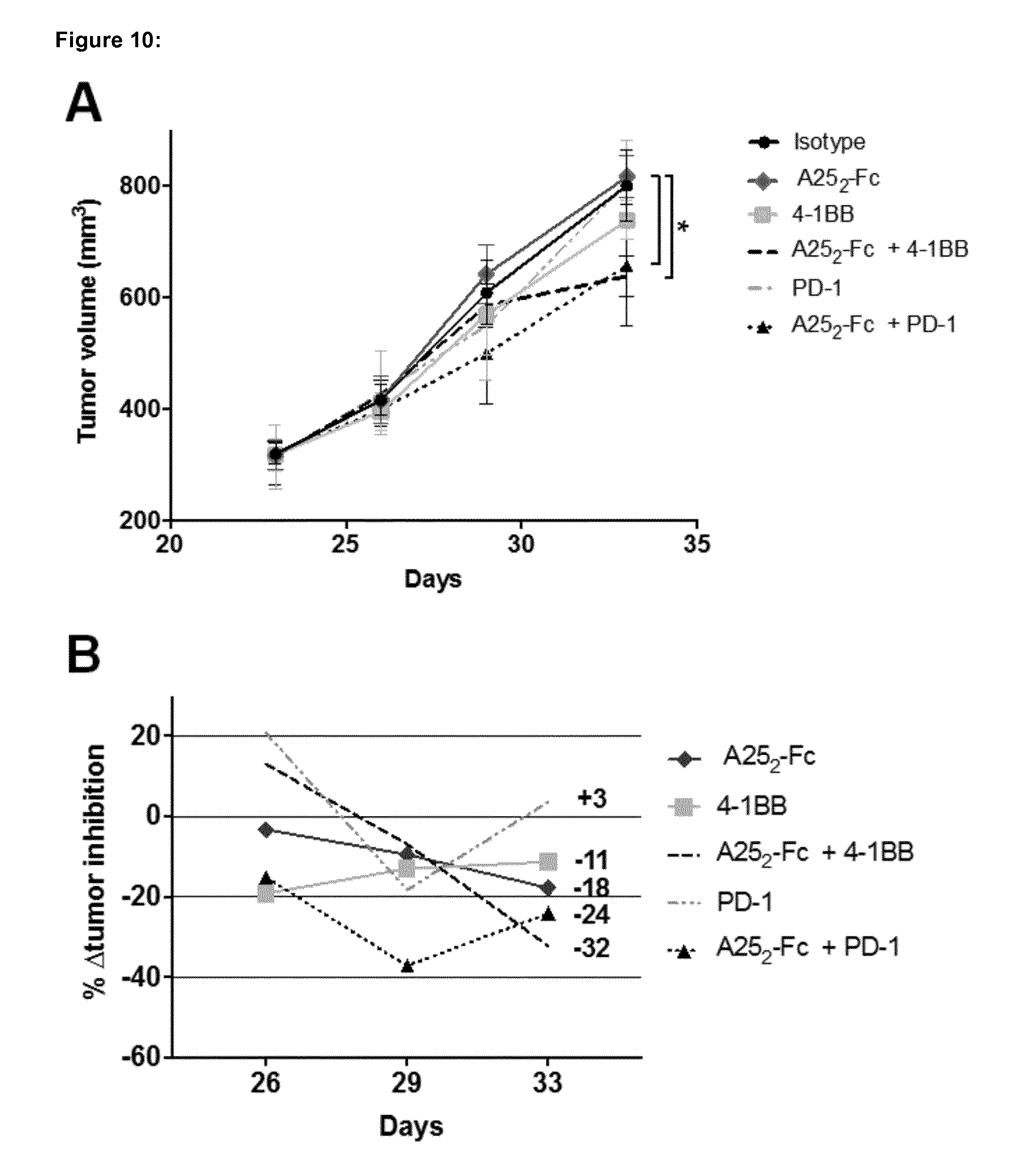
D00015

D00016

D00017

D00018

D00019

D00020

D00021
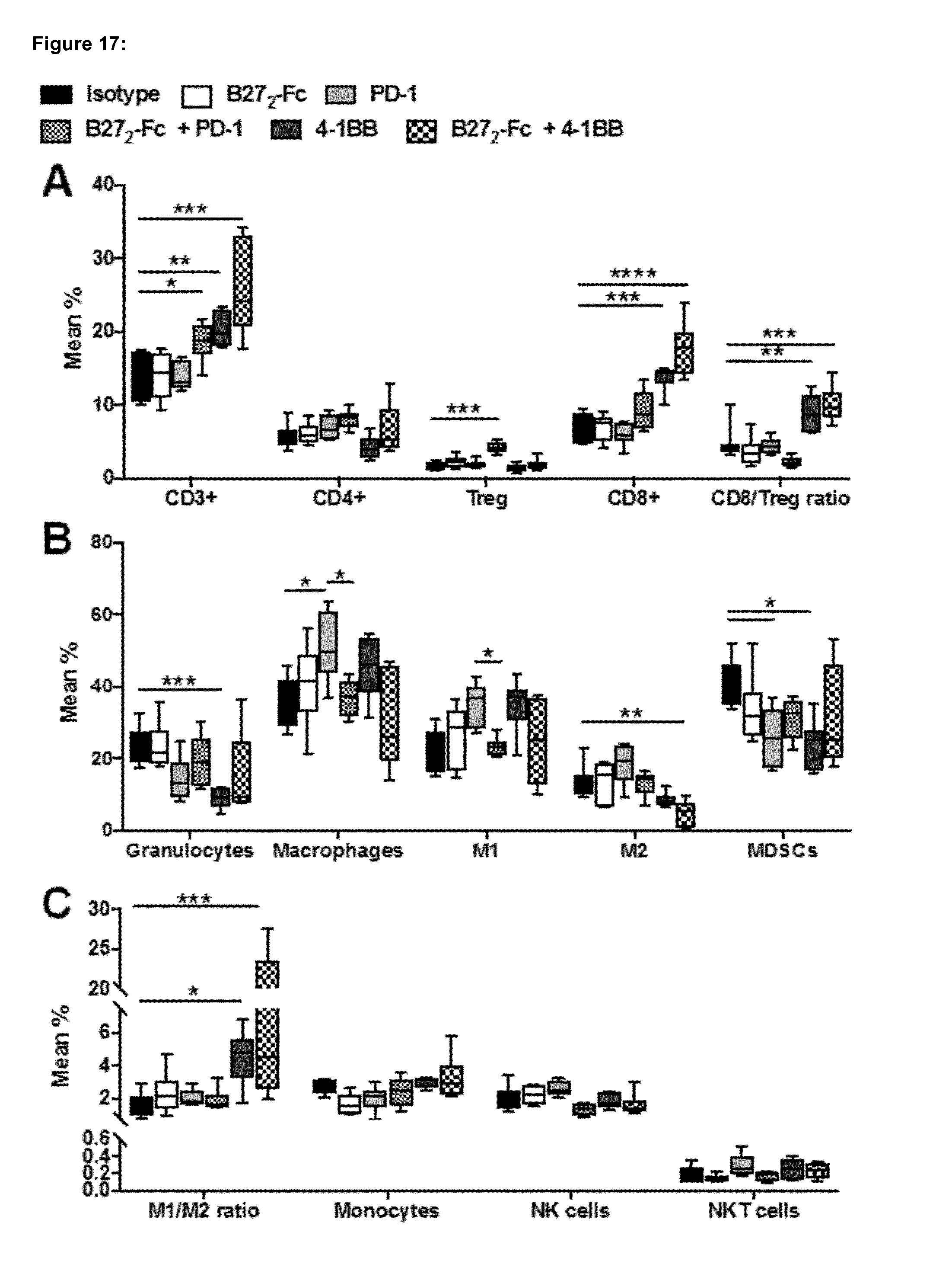
D00022

D00023

D00024

D00025

D00026
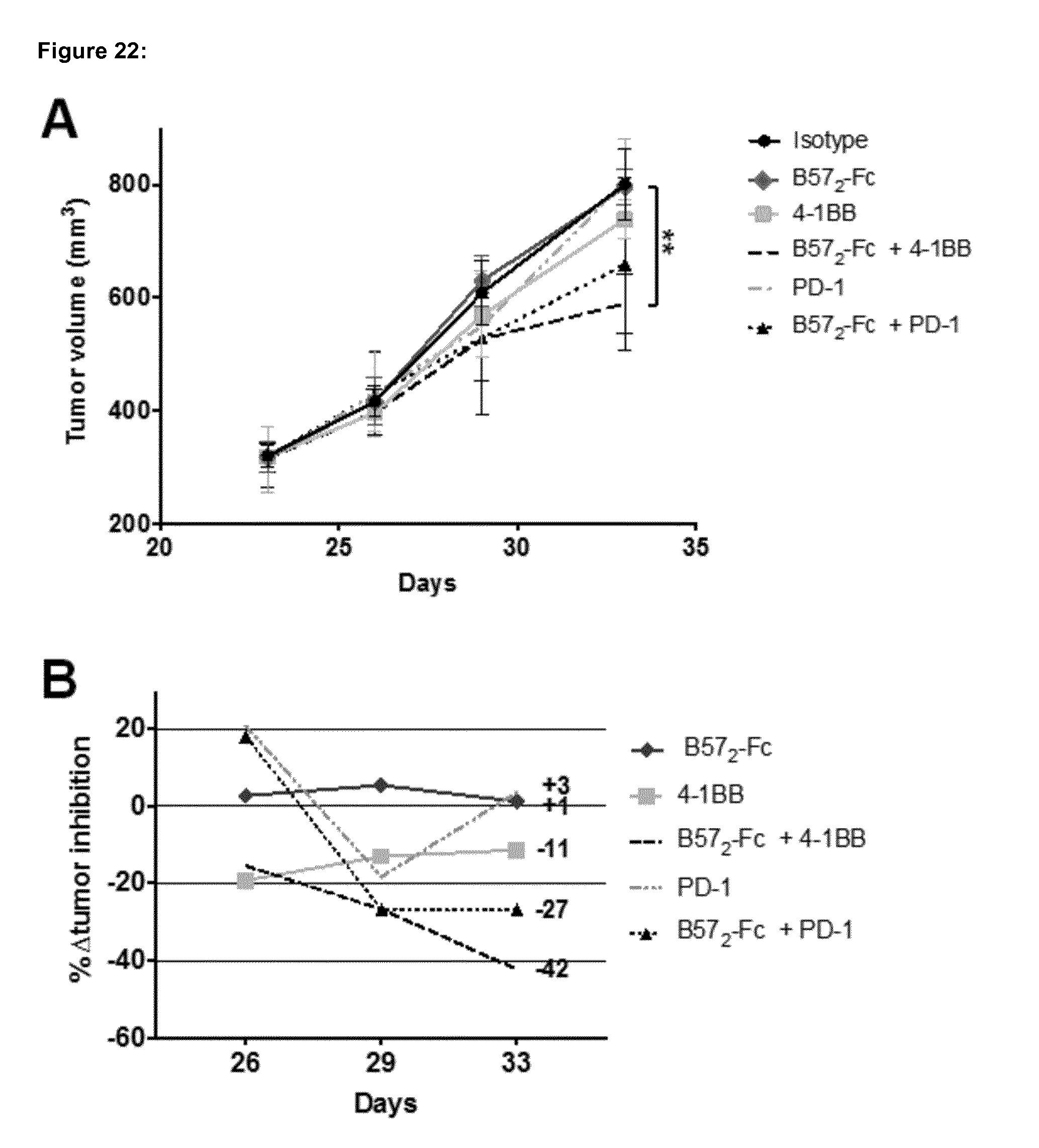
D00027

D00028

D00029

D00030

D00031

D00032

D00033

D00034

D00035

D00036

D00037

S00001
XML
uspto.report is an independent third-party trademark research tool that is not affiliated, endorsed, or sponsored by the United States Patent and Trademark Office (USPTO) or any other governmental organization. The information provided by uspto.report is based on publicly available data at the time of writing and is intended for informational purposes only.
While we strive to provide accurate and up-to-date information, we do not guarantee the accuracy, completeness, reliability, or suitability of the information displayed on this site. The use of this site is at your own risk. Any reliance you place on such information is therefore strictly at your own risk.
All official trademark data, including owner information, should be verified by visiting the official USPTO website at www.uspto.gov. This site is not intended to replace professional legal advice and should not be used as a substitute for consulting with a legal professional who is knowledgeable about trademark law.