System For Skin Cancer Treatment Using Low Intensity Ultrasound
Boer; Miriam Sara ; et al.
U.S. patent application number 16/206142 was filed with the patent office on 2019-06-06 for system for skin cancer treatment using low intensity ultrasound. This patent application is currently assigned to SONIFY BIOSCIENCES, LLC. The applicant listed for this patent is SONIFY BIOSCIENCES, LLC. Invention is credited to Miriam Sara Boer, Daniel Jordan Rogers.
| Application Number | 20190168030 16/206142 |
| Document ID | / |
| Family ID | 53180016 |
| Filed Date | 2019-06-06 |


| United States Patent Application | 20190168030 |
| Kind Code | A1 |
| Boer; Miriam Sara ; et al. | June 6, 2019 |
SYSTEM FOR SKIN CANCER TREATMENT USING LOW INTENSITY ULTRASOUND
Abstract
The present invention relates to a system for skin cancer treatment using low intensity ultrasound. The system comprises an ultrasound transducer, a temperature sensing unit, and a control unit. The temperature sensing unit measures the temperature of the skin being exposed to ultrasound and provides temperature data to the control unit, which controls the ultrasound transducer accordingly.
| Inventors: | Boer; Miriam Sara; (Baltimore, MD) ; Rogers; Daniel Jordan; (Baltimore, MD) | ||||||||||
| Applicant: |
|
||||||||||
|---|---|---|---|---|---|---|---|---|---|---|---|
| Assignee: | SONIFY BIOSCIENCES, LLC Baltimore MD |
||||||||||
| Family ID: | 53180016 | ||||||||||
| Appl. No.: | 16/206142 | ||||||||||
| Filed: | November 30, 2018 |
Related U.S. Patent Documents
| Application Number | Filing Date | Patent Number | ||
|---|---|---|---|---|
| 15037249 | May 17, 2016 | |||
| PCT/US14/63282 | Oct 31, 2014 | |||
| 16206142 | ||||
| 61907748 | Nov 22, 2013 | |||
| Current U.S. Class: | 1/1 |
| Current CPC Class: | A61B 8/0866 20130101; A61B 2017/00084 20130101; A61B 2018/00815 20130101; A61B 2018/00821 20130101; A61N 2007/0034 20130101; A61B 8/085 20130101; A61B 8/0883 20130101; A61N 7/02 20130101; A61N 7/00 20130101; A61N 2007/0082 20130101 |
| International Class: | A61N 7/00 20060101 A61N007/00; A61B 8/08 20060101 A61B008/08; A61N 7/02 20060101 A61N007/02 |
Claims
1. A method of treating skin cancer in a subject comprising (a) scanning a contour of skin containing the skin cancer and generating data of the contour of the skin containing the skin cancer; (b) using the generated data to fabricate a skin contacting surface of an ultrasound transducer having a contour the conforms to the contour of the skin containing the skin cancer; (c) positioning the skin-contacting surface of the ultrasound transducer of the system onto the surface of the skin containing the cancer; and (d) exposing the skin containing the cancer to low intensity ultrasound whereby the frequency, intensity and duration of the ultrasound are selected and controlled to induce a higher death rate of the skin cancer cells than normal skin cells in the focal zone.
2. The method according to claim 1, wherein the skin cancer comprises basal cell carcinoma, squamous cell carcinoma, melanoma, or any combination thereof.
3. The method according to claim 1, wherein the skin cancer comprises melanoma.
4. The method according to claim 1, wherein the subject is a mammal.
5. The method according to claim 4, wherein the mammal is a human.
6. The method according to claim 1 wherein the skin contacting surface of the ultrasound transducer is produced by 3 dimensional ("3D") laser scanning of the surface of the skin containing the cancer.
7. The method according to claim 1 wherein the skin contacting surface is fabricated by molding.
8. The method according to claim 1 wherein the skin contacting surface is fabricated by machining, including at least one of mechanical cutting, LASER cutting, and etching.
9. The method according to claim 1 wherein the skin contacting surface is fabricated by an additive fabrication technology including at least one of 3 dimensional printing and stereolithography.
10. The method according to claim 1 wherein the skin contacting surface is fabricated by at least one of extruding and etching.
11. A method of treating skin cancer in a subject comprising (a) scanning a contour of skin containing the skin cancer and generating data of the contour of the skin containing the skin cancer; (b) using the generated data to select a skin contacting surface of an ultrasound transducer having a contour the conforms to the contour of the skin containing the skin cancer; (c) positioning the skin-contacting surface of the ultrasound transducer of the system onto the surface of the skin containing the cancer; and (d) exposing the skin containing the cancer to low intensity ultrasound whereby the frequency, intensity and duration of the ultrasound are selected and controlled to induce a higher death rate of the skin cancer cells than normal skin cells in the focal zone.
Description
CROSS-REFERENCE TO RELATED APPLICATION
[0001] The present application is a divisional application of U.S. Ser. No. 15/037,249 filed May 17, 2016 which claims priority to and the benefit under 35 U.S.C. .sctn. 371 National Phase Entry Application of International Application No. PCT/US14/63282 filed Oct. 31, 2014, which designates the U.S. and claims benefit under 35 U.S.C. .sctn. 119(e) of U.S. Provisional Application No. 61/907,748 filed Nov. 22, 2013, the contents of each of which are incorporated herein by reference in their entireties.
TECHNICAL FIELD
[0002] The present invention relates generally to a system for skin cancer treatment using low intensity ultrasound.
BACKGROUND
[0003] Skin cancer is the most common form of cancer in the United States. The prevalence of skin cancer is evident by the statistics that one in five Americans will develop skin cancer in the course of a lifetime.
[0004] The current treatment options for skin cancer include excisional surgery, radiation therapy, chemotherapy and immunotherapies, each of which has its own limitations. Excisional surgery is the most common method to treat skin cancer. However, because it is very difficult to remove all cancer cells, the likelihood of the cancer growing back can be high for certain types of cancers. Radiation therapy can result in adverse side effects such as chronic radiation dermatitis, skin atrophy and telangiectasia. Chemotherapy has adverse side effects as well, and its overall efficacy is low due to skin cancer's external presence. Immunotherapies are also plagued by serious side effects, including damages to other healthy organs, fever and severe tiredness. Accordingly, there is a strong need in the art to develop novel systems that can permit new skin cancer treatment.
SUMMARY
[0005] One aspect of the invention relates to a system for skin cancer treatment in a subject using low intensity ultrasound. The system comprises an ultrasound transducer, a temperature sensing unit, and a control unit. The ultrasound transducer has a skin-contacting surface that is pre-formed in order to conform to the skin containing the cancer. The temperature sensing unit can be coupled to the ultrasound transducer, measure the temperature of the skin being treated with ultrasound, and send temperature data to the control unit. A control unit can be coupled to the ultrasound transducer and the temperature sensing unit, receive temperature data from the temperature sensing unit, and control the ultrasound transducer as a function of the temperature data.
[0006] In some embodiments, the control unit controls ultrasound intensity at the focal zone produced by the ultrasound transducer as a function of the temperature data.
[0007] In some embodiments, the temperature sensing unit comprises one or more temperature sensors.
[0008] In some embodiments, the temperature sensor is a contact-type temperature sensor (e.g., a thermistor or a thermocouple) or a non-contact temperature sensor (e.g., an infrared sensor).
[0009] In some embodiments, the ultrasound transducer can produce ultrasound at a frequency between about 27 kHz and 2.2 MHz.
[0010] In some embodiments, the ultrasound transducer can produce ultrasound intensity at the focal zone between about 0.17 W/cm.sup.2 and 5 W/cm.sup.2.
[0011] In some embodiments, the skin-contacting surface of the ultrasound transducer can be produced by a process comprising a 3D laser scan of the surface of the skin containing the cancer.
[0012] In some embodiments, the subject is a mammal.
[0013] In some embodiments, the mammal is a human.
[0014] A related aspect of the invention concerns a method for skin cancer treatment in a subject. The method comprises (1) providing the system for applying low intensity ultrasound to the skin containing the cancer; (2) positioning the skin-contacting surface of an ultrasound transducer of the system onto the surface of the skin containing the cancer; and (3) exposing the skin containing the cancer to low intensity ultrasound. Skin cancers include, for example, basal cell carcinoma, squamous cell carcinoma, and melanoma. In some embodiments, the skin cancer is melanoma.
[0015] These and other capabilities of the invention, along with the invention itself, will be more fully understood after a review of the following figures, detailed description, and claims.
BRIEF DESCRIPTION OF THE DRAWINGS
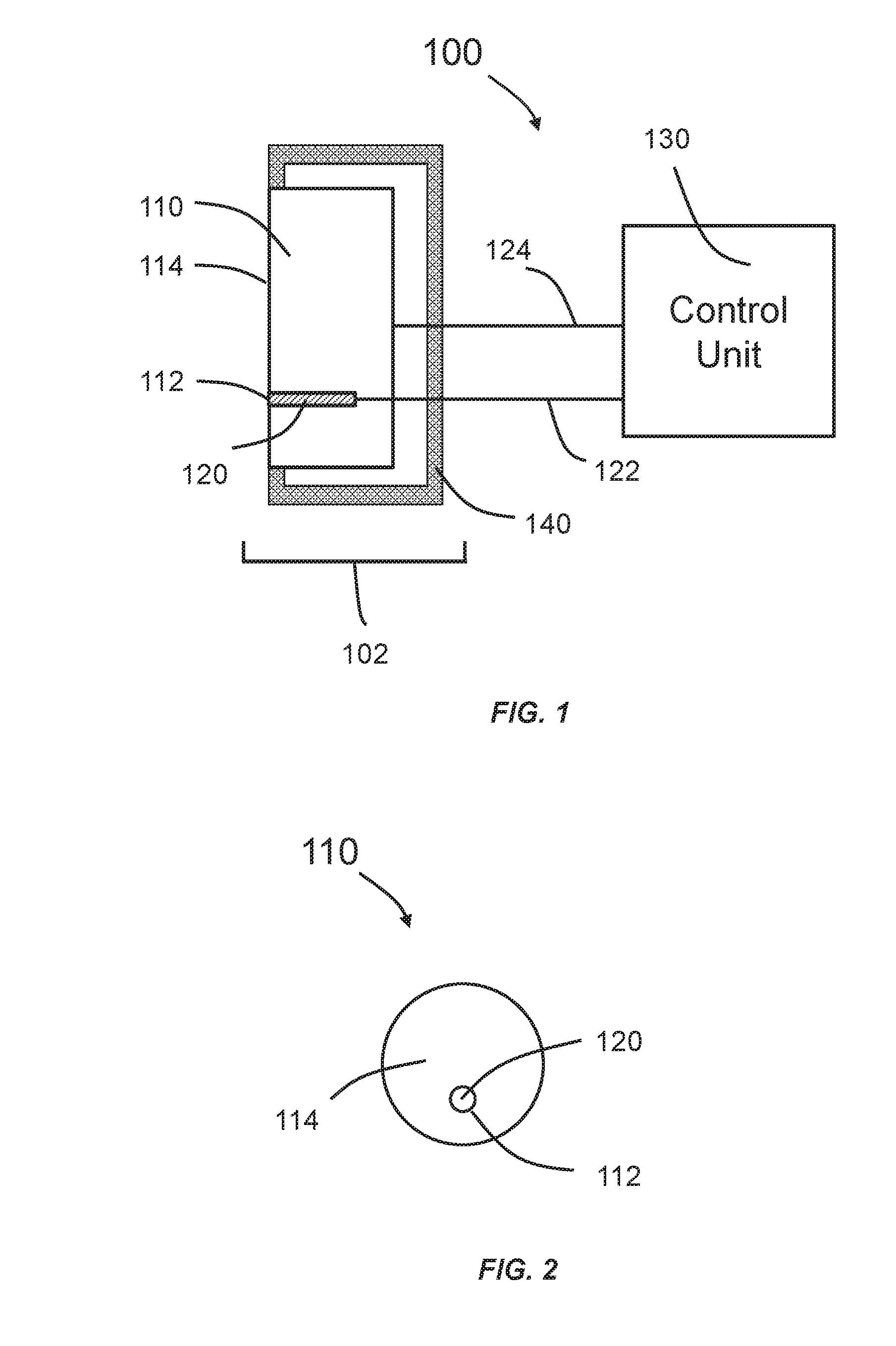
[0016] FIG. 1 is a schematic of a system according to some embodiments of the invention.
[0017] FIG. 2 is a schematic showing a portion of a system according to some embodiments of the invention.
[0018] FIG. 3 is a flow chart describing an exemplary treatment for skin cancer using low intensity ultrasound according to some embodiments of the invention.
DETAILED DESCRIPTION
[0019] The inventor has discovered, inter alia, a survival differential of cells exposed to low intensity ultrasound that correlated with cellular metabolic activities. Specifically, after ultrasound exposure, cells with lower metabolic activities tend to survive better than cells with higher metabolic activities. Metabolic activities for cells can be measured by assays such as the WST-1 cell proliferation assay.
[0020] Without wishing to be bound by theory, skin cancer cells typically have higher metabolic activity than normal skin cells as evident by the fact that skin cancer cells reproduce at a faster rate. Based on the inventor's discovery, skin cancer cells are more susceptible to low intensity ultrasound than normal skin cells. As used herein, the term "low intensity ultrasound" refers to ultrasound having frequency of 20 kHz or higher and an intensity at the focal zone of no more than 75 W/cm.sup.2. As used herein, the term "susceptible" refers to higher percentage of death for skin cancer cells than normal skin cells when exposed to the same low intensity ultrasound. The percentage of death for skin cancer cells can be at least 10%, 20%, 30%, 40%, 50%, 60%, 70%, 80%, or 90% higher than normal skin cells.
[0021] In order to treat skin cancer using ultrasound, a problem arises as how to effectively deliver ultrasound to the cancer for maximal therapeutic effects because skin has contours. Further, the contour can vary for the same subject depending on the location of the skin, or from subject to subject. Provided herein is an ultrasound system to overcome this challenge.
[0022] FIG. 1 is a schematic of a system 100 according to some embodiments of the invention. The system 100 comprises a control unit 130, a transducer unit 102 that includes an ultrasound transducer 110, a temperature sensing unit 120 and optionally a housing 140. The housing 140 can be made of molded plastic or similar materials. The ultrasound transducer 110 can include a skin-contacting surface 114. The temperature sensing unit 120 can be coupled to the ultrasound transducer 110 through a port 112 on the skin-contacting surface 114. The port 112 can be at any location on the skin-contacting surface 114. The port 112 can be a slot or a hole. In some embodiments, the port 112 can comprise electrical connections for the temperature sensing unit 120. The control unit 130 can be connected to the temperature sensing unit 120 and the ultrasound transducer 110 either through a physical connection (e.g., a wire or a cable) or wirelessly (e.g., Bluetooth or radio frequency). It should be noted that the first connection 122 between the control unit 130 and the temperature sensing unit 120 can be the same or different compared to the second connection 124 between the control unit 130 and the ultrasound transducer 110.
[0023] FIG. 2 is a schematic showing a portion of the system 100 that includes the skin-contacting surface 114 according to some embodiments of the invention.
[0024] Technologies of producing ultrasound are well known in the art and are not discussed in detail here. Generally, an ultrasound transducer converts one form of energy to ultrasonic energy. In some embodiments, the ultrasound transducer 110 comprises 1, 2, 3, 4, 5, 6, 7, 8, 9, 10, or more piezoelectric elements, and each piezoelectric element can convert electrical energy to ultrasound. The piezoelectric elements can be arranged in a pre-defined, irregular or regular pattern (e.g., rectangular, circular, octagonal, hexagonal, etc.). The piezoelectric element comprises a piezoelectric material that includes, but is not limited to, quartz, gallium orthophosphate, langasite, lead zirconate titanate, lead titanate, lead metaniobate, barium titanate, potassium niobate, lithium niobate, lithium tantalite, sodium tungstate, zinc oxide, Ba.sub.2NaNb.sub.5O.sub.5, Pb.sub.2KNb.sub.5O.sub.15, sodium potassium niobate, bismuth ferrite, sodium niobate, bismuth titanate, sodium bismuth titanate, polyvinylidene fluoride, or any combination thereof. In some embodiments, the ultrasound transducer 110 comprises a magnetostrictive material, and the magnetostrictive material can produce ultrasound when exposed to a magnetic field. Magnetostrictive materials include, but are not limited to, nickel, Fe--Al alloy, Fe--Ni alloy, Co--Ni alloy, Fe--Co alloy, Co--Fe--V alloy, CoFe.sub.2O.sub.4, NiFe.sub.2O.sub.4, or any combination thereof. The ultrasound transducer can further comprise components such as electrodes, backing, and wear plate. These components are common in ultrasound transducers and should be apparent to those skilled in the art.
[0025] The ultrasound transducer 110 can produce ultrasound having frequency in the range of 20 kHz to 100 MHz, 20 kHz to 50 MHz, 20 kHz to 25 MHz, 20 kHz to 10 MHz, 20 kHz to 5 MHz, 20 kHz to 2.5 MHz, or 27 kHz to 2.2 MHz. In some embodiments, the ultrasound frequency is about 27 kHz. In some embodiments, the ultrasound frequency is about 2.2 MHz. In some embodiments, the ultrasound frequency is not tunable. In some embodiments, the ultrasound frequency can be tuned. The ultrasound intensity at the focal zone can be changed, for example, by varying the magnitude of the voltage applied to the piezoelectric element. The ultrasound intensity at the focal zone is no more than 75 W/cm.sup.2, 50 W/cm.sup.2, 25 W/cm.sup.2, 10 W/cm.sup.2, 5 W/cm.sup.2, 2 W/cm.sup.2, 1 W/cm.sup.2, or 0.5 W/cm.sup.2. In some embodiments, the ultrasound intensity at the focal zone is in the range of about 0.1 to 10 W/cm.sup.2, 0.1 to 5 W/cm.sup.2, 0.5 to 10 W/cm.sup.2, 0.5 to 5 W/cm.sup.2, 1 to 10 W/cm.sup.2, or 1 to 5 W/cm.sup.2. In some embodiments, the ultrasound transducer 110 can produce continuous ultrasound. In some embodiments, the ultrasound transducer 110 can produce pulsed ultrasound.
[0026] During treatment, the skin-contacting surface 114 is in contact with the skin containing the cancer, while the ultrasound emanating from the ultrasound transducer 110 is focused onto the cancer. The skin-contacting surface 114 can be pre-formed to conform to the skin containing the cancer on a subject. Without wishing to be bound by theory, the ability for an ultrasound transducer to conform to the skin allows the ultrasound transducer to deliver ultrasound more effectively to the site of interest, and thus resulting in enhanced therapeutic effects.
[0027] Prior to treatment, the skin-contacting surface 114 of the ultrasound transducer 110 can be produced by a process that uses a 3D scan of the surface of the skin containing the cancer. In some embodiments, the 3D scan is acquired by 3D photography. In some embodiments, the 3D scan is acquired by using a laser scanner. Systems and/or methods for 3D scanning are disclosed in, for example, WO2013126877, WO2004047009, WO2011135341, WO2012083967. Commercial products suitable for 3D skin scanning also exist, such as Antera 3D.TM. and Konica Minolta VIVID 910 3D laser scanner. Once the 3D scan is done, the data acquired (i.e., contour of skin containing the cancer) can then be used to select or fabricate a skin-contacting surface conforming to the skin being scanned. Methods of manufacture can include, but are not limited to, molding (e.g. injection molding), machining (e.g., including mechanical cutting, laser cutting and etching), extruding, embossing, solid free-form fabrication technologies (e.g., three dimensional printing and stereolithography), or any combination thereof. In some embodiments, the skin-contacting surface 114 of the ultrasound transducer 110 can be fabricated by three dimensional printing.
[0028] In some embodiments, the ultrasound transducer 110 can also be used to image the cancer and the surrounding tissue. As used herein, the term "image" or "imaging" refers to the act of producing a 2D or 3D picture of an object. Ultrasound has long been used in medical imaging, for example, in examining various organs (e.g., heart), and monitoring pregnancy.
[0029] The temperature sensing unit 120 measures the temperature of the skin being exposed to ultrasound and transmits temperature data to the control unit 130 through the first connection 122. The temperature measurement can be performed continuously or periodically (e.g., every 1, 2, 3, 4, 5, or more seconds). The temperature sensing unit 120 can comprise one or more temperature sensors. In some embodiments, the temperature sensor can be a contact-type temperature sensor (e.g., a thermistor or a thermocouple). In some embodiments, the temperature sensor can be a non-contact temperature sensor. In some embodiments, the non-contact temperature sensor can be an infrared sensor. In some embodiments, the infrared sensor can include a germanium sensor that is sensitive to electromagnetic radiation with wavelength between 8 um and 14 um. In some embodiments, the temperature sensing unit 120 can include two or more temperature sensors, whereby the two or more temperature sensors can perform differential temperature measurements. Differential temperature measurements measure the temperature difference between two points in a system. The accuracy of the temperature sensor should be better than 5.degree. C., 4.degree. C., 3.degree. C., 2.degree. C., 1.5.degree. C., 1.degree. C. or less.
[0030] The control unit 130 can have at least two main functions. First, the control unit 130 controls the ultrasound transducer 110 by determining the parameters for the ultrasound being produced (e.g., frequency and intensity). The control unit 130 can send control signals to the ultrasound transducer 110 through the second connection 124. Second, the control unit 130 receives temperature data from the temperature sensing unit 120, and based on the temperature data, decides whether to alter the parameters for the ultrasound. By way of example only, if the maximum temperature of the skin receiving ultrasound exposure is below a pre-defined value, the control unit 130 can either keep or increase the intensity. However, if the maximum temperature of the skin receiving ultrasound exposure exceeds a pre-defined value, the control unit 130 terminates the ultrasound exposure or reduces the ultrasound intensity at the focal zone in order to avoid skin damage or undue discomfort (FIG. 3). The pre-defined value can be between 40.degree. C. and 100.degree. C., 40.degree. C. and 90.degree. C., 40.degree. C. and 80.degree. C., 40.degree. C. and 70.degree. C., or 40.degree. C. and 60.degree. C. In some embodiments, a physician/operator will monitor the temperature data and manually increase or reduce the ultrasound intensity at the focal zone, or terminate the ultrasound exposure. In some embodiments, the control unit can comprise computer programs including parameters that are set by a physician/operator. The computer programs can include treatment regimens, which determine the exposure duration and ultrasound characteristics (e.g., frequency and intensity), and change the ultrasound intensity based on the temperature data. The exposure regimens should be subject specific. In some embodiments, the control unit 130 can be coupled to a display module to display the temperature data, ultrasound parameters (e.g., frequency and intensity), and/or treatment regimens.
[0031] In some embodiments, the cancer is in the focal zone of the ultrasound. In some embodiments, the control unit 130 can control the size of the focal zone. Without wishing to be bound by theory, because the ultrasound intensity at the focal zone is determined by dividing the ultrasound energy per unit time at the focal zone by the area of the focal zone, for ultrasound of the same frequency, the larger the focal zone, the lower the intensity is at the focal zone. Therefore, changing the size of the focal zone can be used to change the ultrasound intensity at the focal zone. In some embodiments, the control unit 130 can control the position of the focal zone.
[0032] In some embodiments, the control unit can further comprise a storage module, whereby the temperature data and treatment regimens are stored. As used herein, the "storage module" is intended to include any suitable computing or processing apparatus or other device configured or adapted for storing data or information. Examples of storage devices suitable for use with the present invention include stand-alone computing apparatus; communications networks, including local area networks (LAN), wide area networks (WAN), Internet, Intranet, and Extranet; and local and distributed processing systems. Storage devices also include, but are not limited to: magnetic storage media, such as floppy discs, hard disc storage medium, and magnetic tape; optical storage media such as compact disc, DVD and Blu-ray.TM. discs; electronic storage media such as RAM, ROM, EPROM, EEPROM, solid state storage media and the like; general hard disks and hybrids of these categories such as magnetic/optical storage media.
[0033] The system described herein can be used for skin on any part of the body, including, but is not limited to, scalp, face, neck, arm, hand, torso, leg, or foot.
[0034] A related aspect of the invention concerns a method for skin cancer treatment in a subject using the system described herein. The method comprises (1) providing the system described herein; (2) positioning the skin-contacting surface of an ultrasound transducer of the system onto the surface of the skin containing the cancer; and (3) exposing the skin containing the cancer to low intensity ultrasound. Skin cancers include, for example, basal cell carcinoma, squamous cell carcinoma, and melanoma. In some embodiments, the skin cancer is melanoma.
[0035] In some embodiments, the subject who needs skin cancer treatment is a human or an animal. Usually the animal is a vertebrate such as, but is not limited to a primate, rodent, domestic animal or game animal. Primates include chimpanzees, cynomologous monkeys, spider monkeys, and macaques, e.g., Rhesus. Rodents include mice, rats, woodchucks, ferrets, rabbits and hamsters. Domestic and game animals include cows, horses, pigs, deer, bison, buffalo, feline species, e.g., domestic cat, canine species, e.g., dog, fox, wolf, avian species, e.g., chicken, emu, ostrich, and fish, e.g., trout, catfish and salmon. Patient or subject includes any subset of the foregoing, e.g., all of the above, but excluding one or more groups or species such as humans, primates or rodents. In certain embodiments of the aspects described herein, the subject is a mammal, e.g., a primate, e.g., a human. The terms, "patient" and "subject" are used interchangeably herein. A subject can be male or female. Additionally, a subject can be an infant or a child.
[0036] It should be understood that this invention is not limited to the particular methodology, protocols, and reagents, etc., described herein and as such may vary. The terminology used herein is for the purpose of describing particular embodiments only, and is not intended to limit the scope of the present invention, which is defined solely by the claims.
[0037] As used herein and in the claims, the singular forms include the plural reference and vice versa unless the context clearly indicates otherwise. Other than in the operating examples, or where otherwise indicated, all numbers expressing quantities of ingredients or reaction conditions used herein should be understood as modified in all instances by the term "about."
[0038] All patents and other publications identified are expressly incorporated herein by reference for the purpose of describing and disclosing, for example, the methodologies described in such publications that might be used in connection with the present invention. These publications are provided solely for their disclosure prior to the filing date of the present application. Nothing in this regard should be construed as an admission that the inventors are not entitled to antedate such disclosure by virtue of prior invention or for any other reason. All statements as to the date or representation as to the contents of these documents is based on the information available to the applicants and does not constitute any admission as to the correctness of the dates or contents of these documents.
[0039] Although any known methods, devices, and materials may be used in the practice or testing of the invention, the methods, devices, and materials in this regard are described herein.
[0040] Some embodiments of the invention are listed in the following numbered paragraphs:
Paragraph 1. A system for skin cancer treatment in a subject using low intensity ultrasound, comprising: (a) an ultrasound transducer, wherein the ultrasound transducer has a skin-contacting surface pre-formed to conform to the skin containing the cancer; (b) a temperature sensing unit coupled to the ultrasound transducer, wherein the temperature sensing unit measures the temperature of the skin being treated with ultrasound; and (c) a control unit coupled to the ultrasound transducer and the temperature sensing unit, wherein the control unit receives temperature data from the temperature sensing unit, and wherein the control unit controls the ultrasound transducer as a function of the temperature data. Paragraph 2. The system of paragraph 1, wherein the control unit controls ultrasound intensity at the focal zone produced by the ultrasound transducer as a function of the temperature data. Paragraph 3. The system of paragraph 1 or 2, wherein the temperature sensing unit comprises one or more temperature sensors. Paragraph 4. The system of paragraph 3, wherein the temperature sensor is a contact-type temperature sensor. Paragraph 5. The system of paragraph 4, wherein the contact-type temperature sensor is a thermistor or a thermocouple. Paragraph 6. The system of paragraph 3, wherein the temperature sensor is a non-contact temperature sensor. Paragraph 7. The system of paragraph 6, wherein the non-contact temperature sensor is an infrared sensor. Paragraph 8. The system of any of paragraphs 1 to 7, wherein the ultrasound transducer can produce ultrasound at a frequency between about 27 kHz and 2.2 MHz. Paragraph 9. The system of any of paragraphs 1 to 8, wherein the ultrasound transducer can produce ultrasound intensity at the focal zone between about 0.17 W/cm2 and 5 W/cm2. Paragraph 10. The system of any of paragraphs 1 to 9, wherein skin-contacting surface of the ultrasound transducer is produced by a process comprising a 3D laser scan of the surface of the skin containing the cancer. Paragraph 11. The system of any of paragraphs 1 to 10, wherein the subject is a mammal. Paragraph 12. The system of paragraph 11, wherein the mammal is a human. Paragraph 13. A method of treating skin cancer in a subject comprising (d) providing a system of any of paragraphs 1 to 12; (e) positioning the skin-contacting surface of an ultrasound transducer of the system onto the surface of the skin containing the cancer; and (f) exposing the skin containing the cancer to low intensity ultrasound. Paragraph 14. The method of paragraph 13, wherein the skin cancer comprises basal cell carcinoma, squamous cell carcinoma, melanoma, or any combination thereof. Paragraph 15. The method of paragraph 13, wherein the skin cancer comprises melanoma. Paragraph 16. The method of any of paragraphs 13 to 15, wherein the subject is a mammal. Paragraph 17. The method of paragraph 16, wherein the mammal is a human.
Some Selected Definitions
[0041] Unless stated otherwise, or implicit from context, the following terms and phrases include the meanings provided below. Unless explicitly stated otherwise, or apparent from context, the terms and phrases below do not exclude the meaning that the term or phrase has acquired in the art to which it pertains. The definitions are provided to aid in describing particular embodiments, and are not intended to limit the claimed invention, because the scope of the invention is limited only by the claims. Further, unless otherwise required by context, singular terms shall include pluralities and plural terms shall include the singular.
[0042] As used herein the term "comprising" or "comprises" is used in reference to compositions, methods, and respective component(s) thereof, that are useful to an embodiment, yet open to the inclusion of unspecified elements, whether useful or not.
[0043] The singular terms "a," "an," and "the" include plural referents unless context clearly indicates otherwise. Similarly, the word "or" is intended to include "and" unless the context clearly indicates otherwise.
[0044] Other than in the operating examples, or where otherwise indicated, all numbers expressing quantities of ingredients or reaction conditions used herein should be understood as modified in all instances by the term "about." The term "about" when used in connection with percentages may mean .+-.5% of the value being referred to. For example, about 100 means from 95 to 105.
[0045] Although methods and materials similar or equivalent to those described herein can be used in the practice or testing of this disclosure, suitable methods and materials are described below. The term "comprises" means "includes." The abbreviation, "e.g." is derived from the Latin exempli gratia, and is used herein to indicate a non-limiting example. Thus, the abbreviation "e.g." is synonymous with the term "for example."
[0046] As used herein, the terms "treat," "treatment," "treating," or "amelioration" refer to therapeutic treatments, wherein the object is to reverse, alleviate, ameliorate, inhibit, slow down or stop the progression or severity of a condition associated with a disease or disorder. The term "treating" includes reducing or alleviating at least one adverse effect or symptom of a condition, disease or disorder. Treatment is generally "effective" if one or more symptoms or clinical markers are reduced. Alternatively, treatment is "effective" if the progression of a disease is reduced or halted. That is, "treatment" includes not just the improvement of symptoms or markers, but also slowing of, progress or worsening of symptoms compared to what would be expected in the absence of treatment. Beneficial or desired clinical results include, but are not limited to, alleviation of one or more symptom(s), diminishment of extent of disease, stabilized (i.e., not worsening) state of disease, delay or slowing of disease progression, amelioration or palliation of the disease state, remission (whether partial or total), and/or decreased morbidity or mortality. The term "treatment" of a disease also includes providing relief from the symptoms or side-effects of the disease (including palliative treatment).
[0047] As used herein, the term "focal zone" is defined as an area where the ultrasound energy converges. Typically, the focal zone is the area at which the ultrasound beam is at its narrowest and the ultrasound intensity is the greatest. The ultrasound intensity at the focal zone is determined by dividing the ultrasound energy per unit time at the focal zone by the area of the focal zone. For ultrasound of the same frequency, a user can control the area of the focal zone by varying how tightly the ultrasound is focused.
[0048] As used herein, the term "duration of exposure" refers to the amount of time when skin is continuously exposed to ultrasound.
[0049] Although preferred embodiments have been depicted and described in detail herein, it will be apparent to those skilled in the relevant art that various modifications, additions, substitutions, and the like can be made without departing from the spirit of the invention and these are therefore considered to be within the scope of the invention as defined in the claims which follow. Further, to the extent not already indicated, it will be understood by those of ordinary skill in the art that any one of the various embodiments herein described and illustrated can be further modified to incorporate features shown in any of the other embodiments disclosed herein.
[0050] All patents and other publications identified in the specification and examples are expressly incorporated herein by reference for all purposes. These publications are provided solely for their disclosure prior to the filing date of the present application. Nothing in this regard should be construed as an admission that the inventors are not entitled to antedate such disclosure by virtue of prior invention or for any other reason. All statements as to the date or representation as to the contents of these documents is based on the information available to the applicants and does not constitute any admission as to the correctness of the dates or contents of these documents.
* * * * *
D00000
D00001
D00002
XML
uspto.report is an independent third-party trademark research tool that is not affiliated, endorsed, or sponsored by the United States Patent and Trademark Office (USPTO) or any other governmental organization. The information provided by uspto.report is based on publicly available data at the time of writing and is intended for informational purposes only.
While we strive to provide accurate and up-to-date information, we do not guarantee the accuracy, completeness, reliability, or suitability of the information displayed on this site. The use of this site is at your own risk. Any reliance you place on such information is therefore strictly at your own risk.
All official trademark data, including owner information, should be verified by visiting the official USPTO website at www.uspto.gov. This site is not intended to replace professional legal advice and should not be used as a substitute for consulting with a legal professional who is knowledgeable about trademark law.