Recombinant Polynucleotide Coding For Polypeptide Comprising Reporter Moiety, Substrate Moiety And Destabilizing Moiety, Host Ce
Jung; Hyun Ho ; et al.
U.S. patent application number 16/098278 was filed with the patent office on 2019-05-30 for recombinant polynucleotide coding for polypeptide comprising reporter moiety, substrate moiety and destabilizing moiety, host ce. The applicant listed for this patent is Medytox Inc.. Invention is credited to Hyun Ho Jung, Dong Kyu Lee, Jun Ho Lee, Young Rae Lee, Gi Hyeok Yang.
| Application Number | 20190161745 16/098278 |
| Document ID | / |
| Family ID | 60411350 |
| Filed Date | 2019-05-30 |


| United States Patent Application | 20190161745 |
| Kind Code | A1 |
| Jung; Hyun Ho ; et al. | May 30, 2019 |
RECOMBINANT POLYNUCLEOTIDE CODING FOR POLYPEPTIDE COMPRISING REPORTER MOIETY, SUBSTRATE MOIETY AND DESTABILIZING MOIETY, HOST CELL COMPRISING SAME AND USE OF SAME
Abstract
Provided are a recombinant polynucleotide encoding a polypeptide including a reporter moiety, a substrate moiety, and a destabilization moiety, a host cell including the same, and use thereof to measure the level of a protease by using the recombinant polynucleotide.
| Inventors: | Jung; Hyun Ho; (Seoul, KR) ; Yang; Gi Hyeok; (Chungcheongnam-do, KR) ; Lee; Jun Ho; (Gyeonggi-do, KR) ; Lee; Dong Kyu; (Chungcheongbuk-do, KR) ; Lee; Young Rae; (Chungcheongbuk-do, KR) | ||||||||||
| Applicant: |
|
||||||||||
|---|---|---|---|---|---|---|---|---|---|---|---|
| Family ID: | 60411350 | ||||||||||
| Appl. No.: | 16/098278 | ||||||||||
| Filed: | May 24, 2017 | ||||||||||
| PCT Filed: | May 24, 2017 | ||||||||||
| PCT NO: | PCT/KR2017/005420 | ||||||||||
| 371 Date: | November 1, 2018 |
| Current U.S. Class: | 1/1 |
| Current CPC Class: | C07K 2319/033 20130101; C07K 14/005 20130101; C12Y 113/12007 20130101; C12N 9/52 20130101; C12N 9/0069 20130101; C12N 5/0619 20130101; C07K 14/43595 20130101; C07K 2319/50 20130101; G01N 33/5058 20130101; C12Y 304/24069 20130101; C12N 2770/32031 20130101 |
| International Class: | C12N 9/52 20060101 C12N009/52; C12N 5/0793 20060101 C12N005/0793; G01N 33/50 20060101 G01N033/50; C07K 14/005 20060101 C07K014/005; C12N 9/02 20060101 C12N009/02; C07K 14/435 20060101 C07K014/435 |
Foreign Application Data
| Date | Code | Application Number |
|---|---|---|
| May 4, 2016 | KR | 10-2016-0063722 |
Claims
1. A recombinant polynucleotide comprising a first polynucleotide that encodes a polypeptide that includes: a reporter moiety; a destabilization moiety; and a substrate moiety operatively linking the reporter moiety at an N-terminus to the destabilization moiety at a C-terminus, wherein the substrate moiety comprises a cleavage site of protease activity.
2. The recombinant polynucleotide of claim 1, wherein the protease is a neurotoxin polypeptide, and the neurotoxin polypeptide is botulinum toxin serotype A (BoNT/A), BoNT/B, BoNT/C, BoNT/CD, BoNT/D, BoNT/DC, BoNT/E, BoNT/F, BoNT/FA, BoNT/G, or tetanus neurotoxin (TeNT).
3. The recombinant polynucleotide of claim 1, further comprising a polynucleotide encoding: a bicistronic sequence linked upstream or downstream of the first polynucleotide; and an internal standard reporter operably linked upstream or downstream of the bicistronic sequence.
4. The recombinant polynucleotide of claim 3, wherein the bicistronic sequence is a internal ribosomal entry site (IRES) sequence or a nucleotide sequence that allows ribosome to skip forming a peptide bond.
5. A host cell containing the recombinant polynucleotide of claim 1.
6. The host cell of claim 5, wherein the host cell is a cell that is capable of translocating a neurotoxin polypeptide into a cytoplasm.
7. The host cell of claim 5, wherein the host cell is NT2 cell, SiMa cell, or NG108-15 cell.
8. The host cell of claim 5, wherein the host cell comprises an exogenous polynucleotide encoding a protease, for example, a neurotoxin polypeptide having proteolytic activities.
9. A method of determining proteolytic activities of a neurotoxin polypeptide in a sample, the method comprising contacting the host cell of claim 5 with a sample suspected of containing a neurotoxin polypeptide; and measuring a signal emitted by the reporter moiety in a product obtained by the contacting.
10. The method of claim 9, wherein the recombinant polynucleotide further comprises a polynucleotide encoding a bicistronic sequence linked upstream or downstream of a first polynucleotide and an internal standard reporter operably linked upstream or downstream of the bicistronic sequence.
11. The method of claim 9, further comprising, prior to the contacting, culturing the host cell in a culture medium to differentiate into neuronal cells.
12. The method of claim 11, wherein the host cell is NT2 cell, SiMa cell, or NG108-15 cell.
13. A method of measuring an ability of a host cell in expressing or inhibiting a protease, the method including: introducing a polynucleotide encoding a protease polypeptide into the host cell of claim 5; and culturing the host cell to which the polynucleotide has been introduced and measuring signals emitted by the reporter moiety or the internal standard reporter or a product of the reporter moiety or the internal standard reporter in a culture.
14. The method of claim 13, wherein the culturing is performed in the presence of a test material.
Description
TECHNICAL FIELD
[0001] The present disclosure relates to a recombinant polynucleotide encoding a polypeptide including a reporter moiety, a substrate moiety, and a destabilization moiety, a host cell including the same, and a method of measuring the level of a protease by using the recombinant polynucleotide using the same.
BACKGROUND ART
[0002] Proteases are enzymes that perform proteolysis. Proteases degrade peptide bonds that link amino acids to each other in polypeptide chains by hydrolysis. Proteases may include neurotoxins. Clostridium botulinum and Clostridium tetani produce very potent neurotoxins, for example, botulinum toxin (BoNT) and tetanus toxin (TeNT). These clostridial neurotoxins bind specifically to neuronal cells and inhibit the release of neurotransmitters. Some proteases, such as neurotoxins, not only have very strong toxicity but also are present in very small amounts. Accordingly, there is a need to develop a method of measuring their activity safely and accurately.
[0003] An aspect provides a recombinant polynucleotide including a first polynucleotide that encodes a polypeptide that includes: a reporter moiety; a destabilization moiety; and a substrate moiety operatively linking the reporter moiety to the destabilization moiety, wherein the substrate moiety includes a cleavage site of protease activity.
[0004] Another aspect provides a host cell containing the recombinant polynucleotide.
[0005] Another aspect provides a kit for determining proteolytic activities of a protease polypeptide, the kit including a polypeptide encoded by the recombinant polynucleotide described above, and a detection agent that measures signals emitted by a reporter moiety, an internal standard reporter, or a product thereof.
[0006] Another aspect provides a kit for determining proteolytic activities of a protease polypeptide, the kit including a host cell including the recombinant polynucleotide described above, and a detection agent that measures signals emitted by a reporter moiety, an internal standard reporter, or a product thereof.
[0007] Another aspect provides a method of determining protease activities of a protease polypeptide in a sample, the method including: contacting a polypeptide encoded by the recombinant polynucleotide with a sample suspected of containing a protease polypeptide; and measuring signals emitted by the reporter moiety or a product thereof in a product obtained by the contacting.
[0008] Another aspect provides a method of determining protease activities of a protease polypeptide in a sample, the method including: contacting the host cell with a sample suspected of containing a protease polypeptide; and measuring signals emitted by the reporter moiety, the internal standard reporter, or a product thereof in a product obtained by the contacting.
[0009] Another aspect provides a method of determining characteristics of protease polypeptide in a sample, the method including contacting a polypeptide encoded by the recombinant polynucleotide with the protease polypeptide; measuring signals emitted by the reporter moiety or product thereof in a product obtained by the contacting; and determining, based on measured signals, one or more selected from an onset time of emission of the signals and a duration thereof.
[0010] Another aspect provides a method of determining characteristics of protease polypeptide in a sample, the method including contacting the host cell with the protease polypeptide; measuring signals emitted by a reporter moiety, an internal standard reporter, or a product thereof in a product obtained by the contacting; and determining, based on measured signals, one or more selected from an onset time of emission of the signals and a duration thereof.
TECHNICAL SOLUTION
[0011] A first aspect provides a recombinant polynucleotide including a first polynucleotide that encodes a polypeptide that includes: a reporter moiety, a destabilization moiety; and a substrate moiety operatively linking the reporter moiety to the destabilization moiety, wherein the substrate moiety includes a cleavage site of protease activity.
[0012] Regarding the recombinant polynucleotide, the reporter moiety may be a material that emits a detectable signal or releases a material that emits a detectable signal in a reaction catalyzed thereby (hereinafter also referred to as "product"). The reporter moiety may be selected from a fluorescence protein, .beta.-lactamase, .beta.-galactosidase, alkaline phosphatase, chloramphenicol acetyltransferase, .beta.-glucouronidase, peroxidase, and luciferase. The fluorescence protein may be selected from GFP, YFP, Cittine, CFP, RFP, Kaede, PA-GFP, Emerald, Venus, DsRed, mHoneydew, mBanana, mOrange, tdTomato, mTangerine, mStrawberry, mCherry, mRasberry, mPlum, ZsGreen, and ZsYellow protein.
[0013] Regarding the recombinant polynucleotide, the destabilization moiety may be characterized in reducing the intracellular expression of the first polynucleotide in cells compared to when the destabilization moiety is absent. The expression may be an expression at an mRNA level or a protein level. The destabilization moiety may promote the degradation of mRNA or protein intracellularly expressed by the first polynucleotide in cells. The destabilization moiety may be PEST, CL1, or a fusion protein of PEST and CL1.
[0014] The protease may be originated from bacteria. The bacteria may be of the Clostridium genus. The protease may be a neurotoxin polypeptide. The protease may be an endoprotease. The neurotoxin polypeptide may be a botulinum toxin serotype A (BoNT/A), BoNT/B, BoNT/C, BoNT/D, BoNT/CD, BoNT/DC, BoNT/E, BoNT/F, BoNT/FA, BoNT/G, tetanus neurotoxin (TeNT), or a mutant thereof. The protease may cleave a cleavage site in the substrate moiety. The cleavage site may be a site recognized and cleaved by the protease. An amino acid sequence of each of these botulinum toxin serotypes and a polynucleotide encoding the amino acid sequence are known. The amino acid sequence of each of these botulinum toxin serotypes and the polynucleotide encoding the amino acid sequence may be, for example, those having the sequences of SEQ ID NOS: 1-14 disclosed in WO2004-031355.
[0015] The cleavage site may be a peptide substrate that may be cleaved at a specific cleavage site by a protease. The cleavage site may be, for example, a cleavage site that is recognized and cleaved by endogenous proteases of neurotoxin. The cleavage site of neurotoxin may be naturally occurring or artificially created. The cleavage site of neurotoxin may be derived from a protein that is recognized and cleaved by, for example, BoNT/A protease, BoNT/E protease, or mutants thereof. The protein may be SNAP-25, an isoform thereof, a paralogue, or an ortholog. SNAP-25 may be derived from a human, a rat, a mouse, bovine, danio, carassius, xenopus, torpedo, strongylocentrotus, loligo, lymnaea, or aplysia. SNAP-25 may be SNAP-25A or SNAP-25B.
[0016] In one embodiment, the cleavage site of neurotoxin may be derived from a protein that is recognized and cleaved by, for example, BoNT/B protease, BoNT/D protease, BoNT/F protease, BoNT/G protease, or mutants thereof. The protein may be VAMP, an isoform thereof, a paralogue, or an ortholog. The protein VAMP, an isoform thereof, a paralogue, or an ortholog may be, for example, human or mouse VAMP-1, VAMP-2, VAMP-3/cellubravin, bovine VAMP-2, rat VAMP-2, or VAMP-3, chicken VAMP-1, VAMP-2, or VAMP-3, Torpedo VAMP-1, Strongylocentrotus VAMP, Drosophila sybA, synB, synC, synD, or syn, Hirudo VAMP, Xenopus VAMP-2 or VAMP-3, Canio VAMP-1 or VAMP-2, Loligo VAMP, Lymnaea VAMP, Aplysia VAMP, or Caenorhabditis SNB1-like.
[0017] The cleavage site of neurotoxin may be derived from a protein that is recognized and cleaved by BoNT/C protease, or a mutant thereof. The protein may be Syntaxin or ortholog, paralog, or homolog thereof. The protein may be for example, Human or mouse Syntaxin 1A, Syntaxin 1B1, Syntaxin 2-1, Syntaxin 2-2, Syntaxin 2-3, Syntaxin 3A or Syntaxin 1B2, bovine or rat Syntaxin 1A, Syntaxin 1B1 or Syntaxin 1B2, rat Syntaxin 2 or Rat syntaxin 3, mouse Syntaxin 1A, Syntaxin 1B1, Syntaxin 1B2, Syntaxin 2, Syntaxin 3A, Syntaxin 3B or Syntaxin 3C, chicken Syntaxin 1A or Syntaxin 2; Xenopus Syntaxin IA or Syntaxin 1B, Danio Syntaxin 1A, Syntaxin 1B or Syntaxin 3, Torpedo Syntaxin 1A or Syntaxin 1B, Strongylocentrotus Syntaxin 1A or Syntaxin 1B, Drosophila Syntaxin 1A or Syntaxin 1B, Hirudo Syntaxin 1A or Syntaxin 1B, Loligo Syntaxin 1A or Syntaxin 1B, Lymnaea Syntaxin 1A or Syntaxin 1B, or an ortholog, paralog, or homolog thereof.
[0018] The recombinant polynucleotide may contain a regulatory sequence that allows the first polynucleotide to be expressed in a cell. The recombinant polynucleotide may be an expressible recombinant polynucleotide. The regulatory sequence may be a promoter, an enhancer, a terminator sequence, or a combination thereof. The cell may be able to be bound to and internalized in the protease.
[0019] Regarding the recombinant polynucleotide, the protease may be a neurotoxin polypeptide, and the cell may undergo binding to the neurotoxin polypeptide, internalization of the neurotoxin, releasing the neurotoxin inside the cell, or a combination of the binding, the internalization, and the releasing. The cells may be cells capable of being intoxicated by the neurotoxin of the Clostridium genus. The neurotoxin polypeptide may be a botulinum toxin serotype A (BoNT/A), BoNT/B, BoNT/C, BoNT/D, BoNT/CD, BoNT/DC, BoNT/E, BoNT/F, BoNT/FA, BoNT/G, tetanus neurotoxin (TeNT), or a mutant thereof. The cell may be an endocrine cell or the like. The cell may be a primary neuron, a neuroblastoma cell, or a neuron differentiated from a pluripotent stem cell.
[0020] The recombinant polynucleotide may be a vector. The vector may be an expression vector. The vector may be a viral vector, a plasmid vector, or a linear nucleic acid construct.
[0021] The recombinant polynucleotide may further include a bicistronic sequence linked to upstream or downstream of the first polynucleotide. The bicistronic sequence may be a nucleotide sequence that enables 5'-cap independent translation. The bicistronic sequence may be a nucleotide sequence that allows a ribosome to synthesize two or more polypeptides from an mRNA with one 5'-cap during translation to synthesize a polypeptide from mRNA. The bicistronic sequence may be an internal ribosomal entry site (IRES) sequence or a nucleotide sequence that allows ribosome to skip forming of a peptide bond. The bicistronic sequence that allows the ribosome to skip forming peptide bonds may be a polynucleotide sequence (for example, SEQ ID NOS: 5, 19, 21, or 23) encoding P2A, T2A, E2A, or F2A (for example, SEQ ID NOS: 13, 20, 22, or 24).
[0022] The recombinant polynucleotide may further include a polynucleotide encoding an internal standard reporter operably linked upstream or downstream of the bicistronic sequence. The internal standard reporter may be different from the reporter moiety. The internal standard reporter may be a material that emits a detectable signal or a material that is converted into a material emitting a detectable signal in a reaction catalyzed thereby. The internal standard reporter may be selected from a fluorescence protein, .beta.-lactamase, .beta.-galactosidase, alkaline phosphatase, chloramphenicol acetyltransferase, .beta.-glucouronidase, peroxidase, and luciferase. The fluorescence protein may be selected from GFP, YFP, Citrine, CFP, RFP, Kaede, PA-GFP, Emerald, Venus, DsRed, mHoneydew, mBanana, mOrange, tdTomato, mTangerine, mStrawberry, mCherry, mRasberry, mPlum, ZsGreen, and ZsYellow protein.
[0023] FIG. 1 shows a view illustrating the structure of a recombinant polynucleotide according to an aspect. Referring to A and B of FIG. 1, R represents a nucleotide sequence encoding a reporter moiety, S represents a nucleotide sequence encoding a substrate moiety, and D represents a nucleotide sequence encoding a destabilization moiety. Referring to B of FIG. 1, I represents a nucleotide sequence encoding an internal standard reporter, and B represents a nucleotide sequence encoding a bicistronic sequence. In FIG. 1, the recombinant polynucleotide is described from left to right in the order of 5'->3'. As shown in A of FIG. 1, the recombinant polynucleotide may have the structure of R-S-D. However, this structure is an example only. In one or more embodiments, the recombinant polynucleotide may further include an additional -S- between R and D to encode at least two, for example, at least three or four substrate moieties. In one or more embodiments, the recombinant polynucleotide may further include two or more -D structures, for example, at least three or four destabilization moieties. As shown in A of FIG. 1, the recombinant polynucleotide has the structure of I-B-R-S-D. However, this structure is an example only. In one or more embodiments, the recombinant polynucleotide may further include an additional -I-B- structure linked to 5' position to encode at least two, for example, at least three or four internal standard reporters. The recombinant polynucleotide may have the structure of R-S-D-B-I. However, this structure is an example only. In one or more embodiments, the recombinant polynucleotide may further include an additional -B-I- structure linked to 3' position to contain a nucleotide sequence that encodes at least two, for example, at least three or four internal standard reporters.
[0024] FIG. 2 shows the structure of a recombinant polynucleotide according to an aspect and a reaction between a polypeptide expressed thereby and a protease. Referring to FIG. 2, the polypeptide expressed by the recombinant polynucleotide according to an aspect is cleaved at the cleavage site of a substrate moiety in the presence of a protease and fragmented into a reporter moiety fragment and the remainder.
[0025] FIG. 3 shows a case where a recombinant polynucleotide according to an aspect contacts a protease in a cell. Referring to FIG. 3, the polypeptide expressed by the recombinant polynucleotide according to an aspect is cleaved at the cleavage site of a substrate moiety in the presence of a protease and fragmented into a reporter moiety fragment and the remainder.
[0026] As illustrated in FIG. 3, when a protease is transfected in a cell (top), a polypeptide having the R-S-D structure is cleaved at the S site, and thus, the reporter moiety is separated from the destabilization moiety (D) and stabilized compared to when the destabilization moiety (D) is transfected. Accordingly, signals emitted from the reporter moiety or a material derived therefrom are stronger than when the destabilization moiety (D) is transfected. The bottom part of FIG. 3 shows that when a protease is not present in the cell, the polypeptide having the R-S-D structure retains the destabilization moiety (D), and thus, the level of the polypeptide having the R-S-D structure is lowered by the destabilization moiety (D) and accordingly, a signal measured therefrom is also reduced.
[0027] FIG. 4 shows a view showing a case where a recombinant polynucleotide having a nucleotide sequence encoding an internal standard reporter according to an aspect is in contact with a protease in a cell. Referring to FIG. 4, the polypeptide expressed by the recombinant polynucleotide according to an aspect is cleaved at the cleavage site of a substrate moiety in the presence of a protease and fragmented into a reporter moiety fragment and the remainder.
[0028] As illustrated in FIG. 4, when a protease is transfected in a cell (A), a polypeptide having the R-S-D structure is cleaved at the S site, and thus, the reporter moiety is separated from the destabilization moiety (D) and stabilized compared to when the destabilization moiety (D) is transfected. Accordingly, signals emitted from the reporter moiety or a material derived therefrom are stronger than when the destabilization moiety (D) is transfected. The bottom part (B) of FIG. 4 shows that when a protease is not present in the cell, the polypeptide having the R-S-D structure retains the destabilization moiety (D), and thus, the level of the polypeptide having the R-S-D structure is lowered by the destabilization moiety (D) and accordingly, a signal measured therefrom is also reduced. In FIG. 4, the internal standard reporter protein expressed from the recombinant polynucleotide is expressed in cells regardless of the presence or absence of protease. On the other hand, the R-S-D polypeptide is synthesized by 5'-cap-independent translation by the bicistronic sequence. Thus, signals emitted from the internal standard reporter may be used to standardize the conditions for expression of the recombinant polynucleotide in a host cell.
[0029] A second aspect provides a host cell containing the recombinant polynucleotide. The host cell may be able to translocate the protease into the cell, for example, into the cytoplasm. The host cell may be a cell that may translocate a protease, for example, a neurotoxin polypeptide into a cell, for example, into the cytoplasm. The host cell may bind to, for example, a protease via a receptor and translocate a complex formed thereby into a cell, for example, into the cytoplasm. However, embodiments of the present disclosure should not be construed as limited to any particular translocation mechanism. The host cell may be a cell capable of expressing a polynucleotide encoding a protease, for example, a proteolytic activity neurotoxin polypeptide in the cytoplasm. The host cell may include a polynucleotide encoding a protease, for example, a proteolytic activity neurotoxin polypeptide in the cytoplasm. The polynucleotide encoding a protease, for example, a proteolytic activity neurotoxin polypeptide may be inserted into a chromosome, or extrachromosomal-transfected. The polynucleotide encoding a protease, for example, a proteolytic activity neurotoxin polypeptide may be transiently or permanently expressed. The polynucleotide encoding a protease, for example, a proteolytic activity neurotoxin polypeptide may be in its own form or in a form embedded in a vehicle such as a vector. The polynucleotide may be introduced from outside the host cell. The host cell may be a recombinant cell. The host cell may be an endocrine cell. The cell may be selected from a primary neuron, a neuroblastoma cell, and a neuron differentiated from a pluripotent stem cell.
[0030] The host cell may be able to express the recombinant polynucleotide. The host cell may express, from the recombinant polynucleotide, a polypeptide including a reporter moiety, a destabilization moiety, and a substrate moiety operatively linking the destabilization moiety to the reporter moiety, wherein the substrate moiety has a cleavage site of protease activities, or the polypeptide and the internal standard reporter protein.
[0031] A third aspect provides a kit for determining proteolytic activities of a protease, for example, a neurotoxin polypeptide, the kit including a polypeptide encoded by the recombinant polynucleotide described above and a detection agent that measures signals emitted by a reporter moiety, an internal standard reporter, or a product thereof. Regarding the kit, the reporter may be luciferase, the kit may further include a luciferase substrate, and the detection agent may be used to measure the enzymatic conversion of the luciferase substrate. For example, the detection agent may be a photodetector. The reporter may be a fluorescence protein, and the detection agent may be used to measure fluorescence emitted by the fluorescence protein. For example, the detection agent may be a device that excites the fluorescence protein with excitation light and measures the emitted fluorescence. The internal standard reporter may be different from the reporter moiety. The internal standard reporter may be a luciferase or a fluorescence protein, each being different from the reporter moiety.
[0032] A fourth aspect provides a kit for determining proteolytic activities of a protease, for example, a neurotoxin polypeptide, the kit including a host cell including the recombinant polynucleotide described above and a detection agent that measures signals emitted by a reporter moiety, an internal standard reporter, or a product thereof. Regarding the kit, the reporter may be luciferase, the kit may further include a luciferase substrate, and the detection agent may be used to measure the enzymatic conversion of the luciferase substrate. The reporter may be a fluorescence protein, and the detection agent may be used to measure fluorescence emitted by the fluorescence protein. For example, the detection agent may be a device that excites the fluorescence protein with excitation light and measures the emitted fluorescence. The internal standard reporter may be different from the reporter moiety. The internal standard reporter may be a luciferase or a fluorescence protein, each being different from the reporter moiety.
[0033] A fifth aspect provides a method of determining protease activities in a sample, the method including: contacting a polypeptide encoded by the recombinant polynucleotide with a sample suspected of containing a protease polypeptide; and measuring signals emitted by the reporter moiety or a product thereof in a product obtained by the contacting.
[0034] A sixth aspect provides a method of determining characteristics of a protease polypeptide in a sample, the method including contacting a polypeptide encoded by the recombinant polynucleotide with a protease polypeptide; measuring signals emitted by the reporter moiety or product thereof in a product obtained by the contacting; and determining, based on measured signals, one or more selected from an onset time of signal emission and a time duration of signal emission.
[0035] The methods according to the 5th and 6th aspects each include contacting the polypeptide encoded by the recombinant polynucleotide with a sample suspected of containing a protease polypeptide or a protease. The contacting may be carried out under conditions that allow the cleavage site of protease activities in the polypeptide to degrade. The protease may be a neurotoxin polypeptide.
[0036] The contacting may be performed in a liquid medium. The liquid medium may be conditioned to allow the proteolytic activity of the protease to work. The conditions may include a pH, temperature, a cofactor, a salt concentration, and a combination of these. The liquid medium may be a buffer solution such as a phosphate buffered saline (PBS).
[0037] The methods according to the 5th and 6th aspects include measuring signals emitted by the reporter moiety or a product thereof in a product obtained by the contacting. When the reporter moiety is a polypeptide that emits a detectable signal, signals may be directly measured. For example, when the reporter moiety is a fluorescence protein such as GFP, the fluorescence may be measured. The measuring of the fluorescence may be by irradiating the product obtained by the contacting with excitation light and measuring light emitted from the product obtained by the contacting. The wavelength of the excitation light and emission light may be appropriately selected according to the selected fluorescence protein. When the reporter moiety is a polypeptide that converts to a material that emits a detectable signal in a reaction catalyzed by the reporter moiety, the method may further include adding a substrate required for the catalytic activity of the reporter moiety to the product obtained by the contacting to convert the material into a material that emits a detectable signal. Next, a signal emitted by a material emitting a detectable signal is measured. For example, the reporter moiety is luciferase, and the contacting and the measuring of a signal may be performed to add a luciferase substrate in the reaction solution, and the detecting is performed to measure the enzymatic conversion of the luciferase substrate. The luciferase substrate may be luciferin.
[0038] The methods according to the 5th and 6th aspects may each further include comparing the measured signal with a signal emitted by a control group. The control group may be measured in the same procedure, except that a sample used does not include protease, for example, neurotoxin polypeptide or does include a known concentration of protease for example, neurotoxin polypeptide. Each of the methods may further include determining the level of protease in a sample based on the correlation between a signal obtained by the comparing and the level of protease in the sample.
[0039] The method according to a 6th aspect includes determining at least one of an onset time of signal emission and a time duration of signal emission based on the measured signal. The at least one of an onset time of signal emission and a duration of the emission of the signal may be easily determined by one of ordinary skill in the art based on the measured signals. For example, one of ordinary skill in the art may determine an onset time of emission and a time duration, based on signal values representing protease activities over time, for example, emitted signals. The signal values representing protease activities over time may determine an onset time of emission and a time duration, based on measured values of signals emitted over time. The at least one of an onset time of signal emission and a duration of the emission of the signal may depend on types of protease. When the reporter moiety is a polypeptide that emits a detectable signal, degradation of the cleavage site by protease activities may lead to an increase in the emitted signal and a decrease in the emitted signal when protease activities disappear. For example, when measured under the same conditions, at least one of the onset time of the signal and the time duration of the signal may be changed depending on characteristics of the protease. In one embodiment, the protease may be distinguished from other proteases in terms of, in addition to this relative comparison, the absolute value of at least one of the onset time of the signal emission, and time duration of the signal, or a range thereof. Thus, the determining may include comparing at least one of the onset time of signal emission and the time duration of the signal of a known protease, or determining characteristics of a protease based on an absolute value of at least one of the onset time of signal emission and the time duration of the signal or a range thereof. According to the at least one of the onset time of signal emission and the time duration of the signal, indication to be treated by the protease may vary. Thus, each of the methods may include determining an indication to be treated in a subject by the protease, based on the determined characteristics of the protease.
[0040] A seventh aspect provides a method of determining protease activities in a sample, the method including: contacting the host cell with a sample suspected of containing a protease polypeptide; and measuring signals emitted by the reporter moiety, the internal standard reporter, or a product thereof in a product obtained by the contacting.
[0041] An eighth aspect provides a method of determining characteristics of protease polypeptide in a sample, the method including contacting the host cell with the protease polypeptide; measuring signals emitted by the reporter moiety, the internal standard reporter, or product thereof in a product obtained by the contacting; and determining, based on measured signals, one or more selected from an onset time of emission of the signals and a duration thereof.
[0042] Each of the methods according to the 7th and 8th aspects includes contacting the host cell with a sample suspected of containing a protease polypeptide. The contacting may be carried out under conditions that allow the cleavage site of protease activities to degrade. The protease may be a neurotoxin polypeptide. The contacting may be performed in a liquid medium. The liquid medium may be conditioned to allow the proteolytic activity of the protease to work. The conditions may include a pH, temperature, a cofactor, a salt concentration, and a combination of these. The liquid medium may be a buffer solution such as a phosphate buffered saline (PBS), or a medium in which the host cell is cultured.
[0043] Regarding the methods according to the 7th and 8th aspects, the host cell may be able to translocate the protease into the cell, for example, into the cytoplasm. The host cell may be a cell that may translocate a protease, for example, a neurotoxin polypeptide into a cell, for example, into the cytoplasm. The host cell may bind to, for example, a protease via a receptor and translocate a complex formed thereby into a cell, for example, into the cytoplasm. However, embodiments of the present disclosure should not be construed as limited to any particular translocation mechanism. The host cell may be a cell capable of expressing a polynucleotide encoding a protease, for example, a proteolytic activity neurotoxin polypeptide in the cytoplasm. The host cell may include a polynucleotide encoding a protease, for example, a proteolytic activity neurotoxin polypeptide in the cytoplasm. The polynucleotide encoding a protease, for example, a neurotoxin polypeptide having a proteolytic activity may be inserted into a chromosome, or may be present outside a chromosome. The polynucleotide encoding a protease, for example, a neurotoxin polypeptide having a proteolytic activity may be transiently or stably expressed. The polynucleotide encoding a protease, for example, a proteolytic activity neurotoxin polypeptide may be in its own form or in a form embedded in a vehicle such as a vector. The polynucleotide may be introduced from outside the host cell. The host cell may be a recombinant cell. The host cell may be an endocrine cell. The host cell may be selected from a primary neuron, a neuroblastoma cell, and a neuron differentiated from a pluripotent stem cell. The host cell may be able to express the recombinant polynucleotide. The host cell may express, from the recombinant polynucleotide, a polyp eptide including a reporter moiety, a destabilization moiety, and a substrate moiety operatively linking the destabilization moiety to the reporter moiety, wherein the substrate moiety has a cleavage site of protease activities, or the polypeptide and the internal standard reporter protein.
[0044] The methods according to the 7th and 8th aspects include measuring signals emitted by the reporter moiety or the internal standard reporter or a product thereof in a product obtained by the contacting. When the reporter moiety or the internal standard reporter is a polypeptide that emits a detectable signal, signals may be directly measured. For example, when the reporter moiety is a fluorescence protein such as GFP, the fluorescence may be measured. The measuring of the fluorescence may be by irradiating the product obtained by the contacting with excitation light and measuring light emitted from the product obtained by the contacting. The wavelength of the excitation light and emission light may be appropriately selected according to the selected fluorescence protein. When the reporter moiety or the internal standard reporter is a polypeptide that converts to a material that emits a detectable signal in a reaction catalyzed by the reporter moiety, the method may further include adding a substrate required for the catalytic activity of the reporter moiety to the product obtained by the contacting to convert the material into a material that emits a detectable signal. Next, a signal emitted by a material emitting a detectable signal is measured. For example, the reporter moiety or the internal standard reporter is luciferase, and the contacting and the measuring of a signal may be performed to add a luciferase substrate in the reaction solution, and the detecting is performed to measure the enzymatic conversion of the luciferase substrate. The luciferase substrate may be luciferin. For example, the reporter moiety is luciferase, and the contacting and the measuring of a signal may be performed to add a luciferase substrate in the reaction solution, and the detecting is performed to measure the enzymatic conversion of the luciferase substrate. The luciferase substrate may be luciferin.
[0045] In the methods according to the 7th and 8th aspects, the reporter may be a fluorescence protein, and the measuring may be performed to measure fluorescence emitted by the fluorescence protein.
[0046] The methods according to the 7th and 8th aspects may each further include comparing the measured signal with a signal measured from a control group. The control group may be measured in the same procedure, except that a sample used does not include neurotoxin polypeptide or does include a known concentration of neurotoxin polypeptide. Each of the methods may further include determining the level of neurotoxin polypeptide in a sample based on the correlation between a signal obtained by the comparing and the level of neurotoxin in the sample.
[0047] According to each of the methods according to the 7th and 8th aspects, the recombinant polynucleotide further includes a polynucleotide encoding a bicistronic sequence linked upstream or downstream of a first polynucleotide and an internal standard reporter operably linked upstream or downstream of the bicistronic sequence. In this case, each of the methods may further include measuring a signal emitted by the internal standard reporter. Each of the methods may further include determining the level of a signal emitted by the reporter moiety relative to the signal emitted by the internal standard reporter.
[0048] The method according to the 8th aspect includes determining at least one of an onset time of signal emission and a time duration of signal emission based on measured signals. The at least one of an onset time of signal emission and a time duration of signal emission may be easily determined by one of ordinary skill in the art based on the measured signals. For example, one of ordinary skill in the art may determine signal values representing protease activities over time, for example, an onset time of signal emission and a time duration of signal emission, measured based on emitted signals. The signal values representing protease activities over time, that is, an onset time of signal emission and a time duration of signal emission may be determined based on measurements of emitted signals over time. The at least one of an onset time of signal emission and a time duration of signal emission may depend on types of protease. When the reporter moiety is a polypeptide that emits a detectable signal, degradation of the cleavage site by protease activities may lead to an increase in the emitted signal and a decrease in the emitted signal when protease activities disappear. For example, when measured under the same conditions, one or more of an onset time of signal emission and a time duration of signal emission may be changed depending on characteristics of the protease. In one embodiment, the protease may be distinguished from other proteases in terms of, in addition to this relative comparison, the absolute value of at least one of the onset time of the signal emission, and time duration of the signal, or a range thereof. Thus, the determining may include comparing at least one of the onset time of signal emission and the time duration of the signal of a known protease, or determining characteristics of a protease based on an absolute value of at least one of the onset time of signal emission and the time duration of the signal or a range thereof. According to the at least one of the onset time of signal emission and the time duration of the signal, indication to be treated by the protease may vary. Thus, each of the methods may include determining an indication to be treated in a subject by the protease, based on the determined characteristics of the protease.
[0049] The method according to the 8th aspect may include determining at least one of an onset time of signal emission and a time duration of signal emission based on measured signals, without cellular damage, for example, cell lysis.
[0050] A 9th aspect provides a method of measuring an ability of a host cell in expressing or inhibiting a protease, the method including: introducing a polynucleotide encoding a protease polypeptide into the host cell; and culturing the host cell to which the polynucleotide has been introduced and measuring signals emitted by the reporter moiety or the internal standard reporter or the product thereof in a culture.
[0051] The method includes introducing a polynucleotide encoding a protease polypeptide into the host cell. The host cell and the protease are the same as described above. The introducing may be performed by, for example, transformation, transfection, or transduction. The host cell may be a cell capable of expressing a polynucleotide encoding a protease, for example, a neurotoxin polypeptide having a proteolytic activity in the cytoplasm. The host cell may include a polynucleotide encoding a protease, for example, a neurotoxin polypeptide having proteolytic activity. The polynucleotide encoding a protease, for example, a neurotoxin polypeptide having a proteolytic activity may be inserted into a chromosome, or may be present outside a chromosome. The polynucleotide encoding a protease, for example, a neurotoxin polypeptide having a proteolytic activity may be transiently or stably expressed. The polynucleotide encoding a protease, for example, a proteolytic activity neurotoxin polypeptide may be in its own form or in a form embedded in a vehicle such as a vector. The polynucleotide may be introduced from outside the host cell. The host cell may be a recombinant cell. The host cell may be an endocrine cell. The host cell may be selected from a primary neuron, a neuroblastoma cell, and a neuron differentiated from a pluripotent stem cell.
[0052] The method includes culturing the host cell to which the polynucleotide has been introduced and measuring signals emitted by the reporter moiety or the internal standard reporter or the product thereof in a culture. The culturing may be performed in the presence of a test material. The test material may be a polymer, such as a protein, and a polysaccharide, or a small molecule compound. The test material may be a material that is considered to inhibit or promote the protease. The host cell may be one to which a polynucleotide encoding the candidate test material has been introduced. Thus, the method may be a method of screening for a material that modulates protease activities. The screening method may include comparing a test group signal obtained by using a test material and a control signal obtained by using a control group. The control group may be a positive control group using a material that is known to control protease activities or a negative control group obtained in the same manner as used to the test group except for the candidate test material. The method may include determining whether the candidate test material is a material that modulates protease activities based on comparison results obtained from the comparing operation. In one embodiment, when the test group signal is greater than the signal of the negative control group, it may be determined that the material has an activity of promoting protease activities. In one embodiment, when the test group signal is smaller than the signal of the negative control group, it may be determined that the material has an activity of inhibiting protease activities.
DESCRIPTION OF THE DRAWINGS
[0053] FIG. 1 shows a view illustrating the structure of a recombinant polynucleotide according to an aspect.
[0054] FIG. 2 shows the structure of a recombinant polynucleotide according to an aspect and a reaction between a polypeptide expressed thereby and a protease.
[0055] FIG. 3 shows a case where a recombinant polynucleotide according to an aspect contacts a protease in a cell.
[0056] FIG. 4 shows a view showing a case where a recombinant polynucleotide having a nucleotide sequence encoding an internal standard reporter according to an aspect is in contact with a protease in a cell.
[0057] FIG. 5 shows measurements of activities of a reporter moiety after NG108-15 cells transfected with pFB vector 1 were differentiated into neurons and then intoxicated with botulinum toxin A.
[0058] FIG. 6 shows fluorescent microscopic images of NG108-15 cells transfected with pFB vector 2 after the cells were differentiated into neurons and then intoxicated with botulinum toxin A.
[0059] FIG. 7 shows measurements of activities of a reporter moiety of NG108-15 cells transfected with pFB vector 3 after the cells were differentiated into neurons and then intoxicated with botulinum toxin A.
[0060] FIG. 8 shows measurements of activities of a reporter moiety of SiMa cells transfected with pFB vector 3 after the cells were differentiated into neurons and then intoxicated with botulinum toxin A.
[0061] FIG. 9 shows measurements of activities of a reporter moiety of NT2 cells transfected with pFB vector 3 after the cells were differentiated into neurons and then intoxicated with botulinum toxin A.
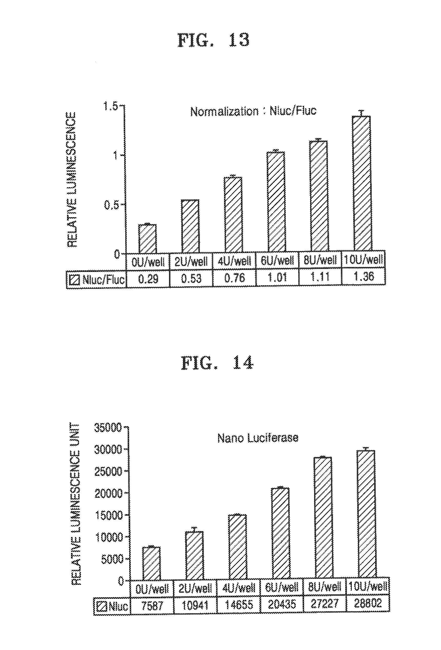
[0062] FIG. 10 shows measurements of activities of a reporter moiety and an internal standard reporter protein each expressed by NG108-15 cells transfected with pFB vector 6 after the cells were differentiated into neurons and then transient-transfected with pCDNA4_BLC vector expressing a light chain of botulinum toxin B.
[0063] FIG. 11 shows measurements of signals emitted by a reporter moiety protein expressed by NT2 cells transfected with pFB vector 2 after the cells are established as a stabilized monoclonal-derived cell line, differentiated into neurons, and then, intoxicated with different concentrations of BoNT/A.
[0064] FIGS. 12 to 14 show measurements of signals emitted by a reporter moiety protein or an internal standard reporter each expressed by NT2 cells transfected with pFB vector 2 after the cells are established as a stabilized monoclonal-derived cell line, differentiated into neurons, and then, intoxicated with different concentrations of BoNT/A.
MODE OF THE INVENTION
[0065] Hereinafter, the present invention will be described in more detail with reference to examples. However, these examples are for illustrative purposes only, and the scope of the present invention is not limited to these examples.
Example 1
[0066] Preparation of recombinant polynucleotide encoding a reporter moiety-substrate moiety-destabilization moiety (hereinafter also referred to as `R-S-D`) polypeptide and cells containing the recombinant polynucleotide
(1) Preparation of Recombinant Polynucleotide Encoding a Reporter Moiety-Substrate Moiety-Destabilization Moiety (Hereinafter Also Referred to As `R-S-D`) Polypeptide
[0067] Two recombinant polynucleotide having different structures were prepared. One is to encode a polypeptide having an R-S-D structure and the other is to encode a polypeptide having an I-B-R-S-D structure. Here, I indicates a nucleotide sequence encoding an internal standard reporter, and B indicates a bicistronic sequence. The recombinant polynucleotide is in the form of a plasmid vector.
[0068] A nucleotide sequence encoding a luminescence or fluorescence protein was used as the internal standard reporter or the reporter moiety. Examples of the sequence encoding the luminescence protein are a nucleotide sequence (for example, SEQ ID NO: 1) encoding a firefly luciferase (FLuc) protein (for example, SEQ ID NO: 9) and a nucleotide sequence (for example, SEQ ID NO: 2) encoding a nano-luciferase (NLuc) protein (SEQ ID NO: 10), and examples of the sequence encoding the fluorescence protein are a nucleotide sequence (SEQ ID NO: 3) encoding an enhanced green fluorescence protein (eGFP) (SEQ ID NO: 11) or a nucleotide sequence (SEQ ID NO: 4) encoding an mCherry protein (SEQ ID NO: 12).
[0069] The bicistronic sequence used herein is a nucleotide sequence (P2A) (SEQ ID NO: 5) encoding a self-cleaving peptide derived from porcine teschovirus-1.
[0070] The substrate moiety of a proteolytic enzyme used herein is, in the case of botulinum toxin type A, a nucleotide sequence (SEQ ID NO: 6) encoding an amino acid sequence (SEQ ID NO: 14) from 145 to 207 of the total of 207 amino acid sequence as a C-terminal region of human SNAP25, or, in the case of botulinum toxin type B, a nucleotide sequence (SEQ ID NO: 17) encoding human VAMP2 (SEQ ID NO: 18).
[0071] The destabilization moiety used herein is a nucleotide sequence (SEQ ID NO: 8) encoding the PEST sequence (SEQ ID NO: 16) and a nucleotide sequence (SEQ ID NO: 7) encoding CLI sequence (SEQ ID NO: 15).
[0072] The recombinant polynucleotide was constructed as follows:
[0073] First, a nucleotide sequence (SEQ ID NO: 1) (hereinafter referred to as `FLuc sequence`) encoding firefly luciferase (FLuc)(SEQ ID NO: 9) was obtained from pGL4.31 vector (Promega), a nucleotide sequence (SEQ ID NO: 2) (hereinafter referred to as `NLuc sequence`) encoding nano-luciferase (NLuc) (SEQ ID NO: 10) was obtained from pNL1.1 vector (Promega), a nucleotide sequence(SEQ ID NO: 3)(hereinafter referred to as `eGFP sequence`) encoding enhanced green fluorescence protein (eGFP) (SEQ ID NO: 11) was obtained from pEGFP-C1 vector (clontech), and a nucleotide sequence (SEQ ID NO: 4)(hereinafter referred to as `mCherry sequence`) encoding mCherry (SEQ ID NO: 12) was obtained from pmCherry-C1 vector. A nucleotide sequence (SEQ ID NO: 6) encoding an amino acid sequence from 145 to 207 of the total of 207 amino acid sequence as a C-terminal region of human SNAP25 (hereinafter referred to as `SNAP25 sequence`), and a nucleotide sequence encoding human VAMP2 (hereinafter referred to as `VAMP2 sequence`) were synthesized by RT-PCR from mRNA of human-derived cell lines. A sequence encoding CL1-PEST polypeptide in which CL1 is fused with PEST (hereinafter referred to as `CL1-PEST sequence`) and a nucleotide sequence encoding a self-cleaving peptide P2A derived from porcine teschovirus-1 (hereinafter referred to as `P2A sequence`) were obtained by gene synthesis.
[0074] Next, recombinant polynucleotides having the structure of R-S-D or I-B-R-S-D were constructed by gene cloning using restriction enzyme using a combination of the sequences, and ligated to the BarnHI/XhoI restriction site of the pCDNA4 vector (Invitrogen) or pFB vector (Agilent) to construct recombinant vectors. For example, a vector (hereinafter referred to as "pCDNA4 vector 1") in which a recombinant polynucleotide consisting of NLuc sequence, SNAP25 sequence, and CLI-PEST sequence is introduced to BamhI/XhoI enzyme site of pCDNA4 vector or a vector (hereinafter referred to as "pFB vector 1") in which a recombinant polynucleotide consisting of NLuc sequence, SNAP25 sequence, and CL1-PEST sequence is introduced to BamHI/XhoI enzyme site of pFB vector were constructed. For example, a vector (hereinafter referred to as "pCDNA4 vector 2") in which a recombinant polynucleotide consisting of mCherry sequence, SNAP25 sequence, and CL1-PEST sequence is introduced to BamH/IXhoI enzyme site of pCDNA4 vector or a vector (hereinafter referred to as "pFB vector 2") in which a recombinant polynucleotide consisting of mCherry sequence, SNAP25 sequence, and CL1-PEST sequence is introduced to BamHI/XhoI enzyme site of pFB vector were constructed. For example, a vector (hereinafter referred to as "pCDNA4 vector 3") in which a recombinant polynucleotide consisting of FLuc sequence, P2a sequence, NLuc sequence, SNAP25 sequence, and CL1-PEST sequence is introduced to BamHI/XhoI enzyme site of pCDNA4 vector or a vector (hereinafter referred to as "pFB vector 3") in which a recombinant polynucleotide consisting of FLuc sequence, P2a sequence, NLuc sequence, SNAP25 sequence, and CL1-PEST sequence is introduced to BamHI/XhoI enzyme site of pFB vector were constructed. A pFB vector (hereinafter referred to as "pFB vector 4") in which a recombinant polynucleotide consisting of FLuc sequence, P2a sequence, NLuc sequence, VAMP2 sequence, and CLI-PEST sequence is introduced to BamH/XhoI enzyme site of pFB vector, was constructed.
[0075] Here, the pCDNA4 vector is a plasmid-derived vector and used to transiently express the recombinant polynucleotides in cells. The pFB vector used herein was a vector derived from moloney murine leukemia virus (MMLV), and used to construct a cell line modified by transfection, that is, a cell line in which the recombinant polynucleotides were introduced into a chromosome.
[0076] The structures of the constructed plasmid vectors and the viral vectors were confirmed by sequencing, and the plasmid vector DNA was purified and isolated to the level for cell culture.
(2) Production of Cells Expressing R-S-D or I-B-R-S-D Polypeptide
[0077] In order to construct a cell line that stably and constantly expressing the vectors constructed in (1), transduction with MMLV-derived viral vector, that is, pFB vector, was performed. The transfection with MMLV-derived viral vector is a widely practiced method in the art, and the reference (Felts Ka et al., Mol Biotechnol. 2002 Sep. 22(1):25-32.). Briefly, HEK-293T cells (ATCC-CRL-3216), a cell for packaging, were seeded in 10 ml of DMEM medium containing 10% fetal bovine serum (FBS) at the population of 1.times.10.sup.7 cells in a 55 cm.sup.2 culture dish, and cultured for 24 hours in a 37.degree. C. and 5% CO.sub.2 incubator, and then, the recombinant polynucleotide pFB vector according to the present invention, that is, pFB vector 1, 2, 3, or 4, and pCMV-gagpol (Cell Biolabs, inc.) and pCMV-VSV-G (Cell Biolabs, inc.) were mixed at a ratio of 3:2:1. At this time, pFB vector was used in an amount of 7.5 .mu.g. The mixtures of this vector was used for transfection with Lipofectamine.TM. 3000 (Invitrogen) and the transduction method was performed according to the manufacturer's instructions. Cells were cultured under the same conditions for 48 hours after transduction. Thus, an infectious packaged pFB vector 1,2,3 or 4-containing virus was produced from the HEK 293T cells.
[0078] Then, the infectious packaged pFB vector 1,2,3 or 4-containing virus was transfected into a target cell to produce a cell line that enables stable, constant expression. The target cell used herein was NG108-15 cell(ATCC-HB-12317), SiMa cell(DSMZ: ACC-164), and NT2 cell(ATCC-CRL-1973). NG108-15 cells are hybrid cells of neuroblastoma of rat and glioma of rat, and a cell line that has been identified as susceptible to botulinum toxin type A through differentiation into neurons (Whitemarsh RC et al., Biochem Biophys Res Commun. 2012 Oct. 19;427(2):426-30). SiMa cells are a cell line that is derived from human neuroblastoma and have been identified as susceptible to botulinum toxin type A through differentiation into neurons (Fernandez-Salas E et al., PLoS One. 2012;7(11):e49516). NT2 cells are an embryonal carcinoma cell line derived from human testicles, have multiple differentiation potential, and may undergo differentiation into neurons under specific conditions, and when differentiated into neurons, the NT2 cells were found to be susceptible to botulinum toxin type A (Tegenge et al., Cell Mol Neurobiol. 2012 Aug;32(6):1021-9).
[0079] The specific procedure of a cell line that expresses a target polypeptide stably is as follows.
[0080] First, a target cell was seeded in a 24-well plate containing 0.5 ml of DMEM medium containing 10% FBS at the population of about 1.times.10.sup.5 cells per well one day before infection, and then, cultured in a 5% CO.sub.2 incubator at a temperature of 37.degree. C. for one day. The culture supernatant of HEK-293T cells collected therefrom was filtered using a 0.45-.mu.m syringe filter to obtain a viral solution in which cells and cell debris were removed. The viral solution was added to wells in which the target cell had been cultured and from which the culture medium had been removed, with a concentration of 1 ml/well (24-well), and the cells were cultured under the same conditions for 6 hours to induce infection. Polybrene was added at a level of 8 mg/ml to increase the efficiency of infection into a target cell. After 6 hours, the medium was replaced with DMEM medium containing 10% FBS as a medium for normal cell culture, and the cells were cultured under the same conditions for 2 days.
[0081] Next, the infected target cell in the 24-well was cultured for 2 days, and then, transferred to a 55 cm.sup.2 culture dish, and cultured after 500 to 1000 .mu.g/ml of geneticin (Gibco), a selective antibiotic of the pFB vector, was added thereto. The appropriate dose of antibiotic was determined based on the titration of the target cell and geneticin. When NT2 cells were the target cell, they were cultured in a culture dish containing 10 ml of cell culture medium in the presence of 800 .mu.g/ml of geneticin for 5 days.
[0082] Next, to form a single cell clone, the target cell was transferred to a 96-well well at the concentration of 2 cells/well. In each well of a 96-well plate containing 100 .mu.l of the same medium used in the culturing in 55 cm.sup.2 culture dish, colonies formed with single cell clones while being cultured under the same conditions for 4 weeks were selected by a cell culture microscope, and the selected colonies were subjected to expansion culture.
[0083] In this process, a pre-selection process may be performed according to the structure of a produced recombinant polynucleotide. That is, when an internal standard reporter protein is a fluorescence protein, the pre-selection process may be performed by using a fluorescence microscopy, or when an internal standard reporter protein is a luminescence protein, the pre-selection process may be performed by luminescent analysis.
[0084] To pre-select a recombinant polynucleotide in which the internal standard reporter protein is not present or to pre-select a monoclonal cell line having an excellent sensitivity from among monoclonal cell lines in which an internal standard reporter protein is present, proteasome inhibitor MG132 (Sigma) was added at a concentration of 10 .mu.M medium, or when the recombinant polynucleotide includes SNAP25 sequence, light chain DNA of botulinum toxin type A was used with Lipofectamine.TM. 3000 (Invitrogen), and when the recombinant polynucleotide includes VAMP2 sequence, light chain DNA of botulinum toxin type B was used with Lipofectamine.TM. 3000 (Invitrogen), and the transduction was performed according to the manufacturer instructions. At this time, BoNT/A LC and BoNT B LC were introduced into the BamHI/XhoI site of pCDA4. In detail, regarding only to cells in which a monoclonal colony was identified while culturing in a 96-well for about 3 to 4 weeks, the same number of cells were seeded seperately in two 96-well plates: one plate for a screening experiment, and the other plate for maintenance culture.
[0085] Monoclone-derived cell lines obtained through these screening procedures were subjected to expansion culture, and stored frozen. The lyophilization was carried out by freezing the general mammalian cells. Briefly, 5.times.10' to 1.times.10.sup.7 cells were diluted in 1 ml of a culture medium containing 5% or 10% DMSO and the diluted cells were placed in a freezing vial. The temperature was lowered to -80.degree. C. and then stored in the gas phase of a liquefied nitrogen tank.
[0086] As a result, cell line NG108-15 cells, SiMa cells, and NT2 cells, which stably, constantly express the pFB vector 1, 2, 3, 4, or 6 containing virus, were established.
(3) Differentiation of Established NG108-15 cells, SiMa cells, and NT2 cells into Neurons and Toxin Intoxication
[0087] NG108-15 cells, SiMa cells, and NT2 cells are neuroblastoma cell lines or embryonal carcinoma cell line, which are known to be susceptible to botulinum toxin when differentiated into neurons. Therefore, these cells were differentiated into neurons according to the following procedure.
[0088] NG108-15 cells were maintenance-cultured in DMEM medium supplemented with 10 (v/v) % FBS in a 5% CO.sub.2 incubator at a temperature of 37.degree. C. For neuronal differentiation, cells were seeded at a concentration of 2.times.10.sup.4 cells/well in each well of a 96-well matrigel (BD science)-coated plate for cell culture, and incubated in a neurobasal medium (Gibco) supplemented with 50 .mu.M of retinoic acid and 25 .mu.M of purmophamine for about 5 days. The neurobasal medium contained B27 (Gibco), Glutamax (Gibco), and non-essential amino acid (Gibco) as supplements in an amount of 1.times. each.
[0089] SiMa cells were maintenance-cultured in DMEM medium supplemented with 10 (v/v) % FBS in a 5% CO.sub.2 incubator at a temperature of 37.degree. C. For neuronal differentiation, cells were seeded at a concentration of 5.times.10.sup.4 cells/well in each well of a 96-well matrigel (BD science)-coated plate for cell culture, and incubated in a serum-free MEM medium (Welgene) supplemented with 50 .mu.M of retinoic acid for about 5 days. The MEM medium contained B27 (Gibco), N2 (Gibco), Glutamax (Gibco), HEPES (Gibco) and non-essential amino acid (Gibco) as supplements in amounts of 1.times. each.
[0090] NT2 cells (also referred to as "NTteraA2") were maintenance-cultured in a-MEM medium (Welgene) supplemented with 10 (v/v) % FBS in a 5% CO2 incubator at a temperature of 37.degree. C. For neuronal differentiation, the cells were seeded at a concentration of 1.times.10.sup.5 cells/ml in each well of a Petri dish and cultured in differentiation medium supplemented with 50 .mu.M retinoic acid for 1 week under the same conditions while the medium was exchanged every 2 to 3 days. For the differentiation medium, DMEM/F12 (Welgene) medium supplemented with 10 (v/v)% FBS was used.
[0091] The cultured cells form spheres, and after 1 week of culturing, the spheres were collected and transferred to a regular cell culture plate having the same area. The cells were cultured, as adherent cells, on the differentiation medium supplemented with 50 .mu.M of retinoic acid for 1 week while the medium was exchanged every 2 to 3 days. Subsequently, the adhered cells were deaggregated and detached by using trypsin, and the number of cells was counted.
[0092] 1.times.10.sup.7 cells were transferred to a 175T flask (Nunc) and cultured in the differentiation medium supplemented with a mitotic inhibitor for 10 days under the same conditions while the medium was exchanged every 2 to 3 days. The mitotic inhibition used herein was 1 .mu.M AraC, 10 .mu.M Uridine, and 10 .mu.M Floxuridine. Cells differentiated into neurons were detached by using trypsin, and the obtained cells were stored frozen. A medium for freezing was used as the medium for the storing frozen, and the freezing was performed by cell freezing method. The differentiated neurons were seeded at the population of 1.times.10.sup.5 cells per well in a 96-well matrigel-coated plate for cell-culture and cultured for 10 days or more in the differentiation medium, and the medium was changed every 2 to 3 days.
(4) Intoxicating of Differentiated Neurons with Botulinum Toxin
[0093] Intoxicating of differentiated neurons with botulinum toxin type A was performed as follows. For intoxicating with a purified toxin protein, the purified toxin protein was diluted to an appropriate concentration in the differentiation medium and then exchanged with the culture medium of the neuron cultured in a 5% CO.sub.2 incubator at a temperature of 37.degree. C. Thereafter, the cells were cultured under the same conditions for 24 hours to induce toxin-intoxication. Then, the medium was replaced with the differentiation medium and the cells were cultured for 72 hours under the same conditions.
[0094] The differentiation medium was added to a commercially available botulinum toxin drug vial (Meditoxin injectable, Neuronox) to suspend lyophilized toxin protein and excipient, and then, the differentiation medium was exchanged with the culture medium of the cells being maintainance-cultured in a 5% CO.sub.2 incubator at a temperature of 37.degree. C. The toxin potency test on final product vials was performed by using a toxin placebo vial to make the amount of excipient treated the same. The toxin placebo vial was prepared by removing only the toxin protein from the final product toxin vial.
[0095] That is, the same amount of the final product toxin vial and the toxin placebo vial were suspended in the same amount, and then, the total amount of the intoxicating medium was treated in the same manner according to each treatment concentration.
(5) Quantitative Analysis of Reporter Moiety or Internal Standard Reporter
[0096] Quantitative analysis of the reporter moiety or internal standard reporter protein expressed from the recombinant polynucleotide was performed as follows.
[0097] Regarding the recombinant polynucleotide according to the present invention, when a luminescence protein was used as a reporter moiety or an internal standard reporter protein, the luminescence protein was quantitatively analyzed by a luminescence assay. When the reporter moiety is NLuc, the Nano-luciferase assay kit (Promega) was used.
[0098] When the reporter moiety was NLuc and the internal standard reporter protein was FLuc, the One-glo dual nano-luciferase assay kit (Promega) was used and the test was performed according to the supplier's manual. The luminescence was measured by using SpectraMax (Molecular Device Inc). Normalization was performed by using a resultant value obtained by dividing the measured NLuc value by the internal standard reporter protein FLuc value.
[0099] Regarding the recombinant polynucleotide according to the present invention, when a fluorescence protein was used as a reporter moiety or an internal standard reporter protein, the fluorescence values were quantitatively analyzed by a fluorescence meter. When the reporter moiety is mCherry and the internal standard reporter protein is eGFP, mCherry was excited at a wavelength of 610 nm and emitted light at a wavelength of 507 nm was measured by using SpectraMax (Molecular Device), a fluorescence meter. For eGFP, eGFP was excited at the wavelength of 488 nm and emitted light at a wavelength of 507 nm was measured. Normalization was performed by using a resultant value obtained by dividing the measured mCherry emission value by the internal standard reporter protein eGFP emission value. In addition, fluorescence values of GFP and RFP may be measured by using an Incucyte device (Essen Bioscience Inc.) to perform normalization. eGFP belongs to a GFP family and mCherry belongs to an RFP family.
(6) Results
[0100] FIG. 5 shows measurements of activities of a reporter moiety after NG108-15 cells transfected with pFB vector 1 were differentiated into neurons and then intoxicated with botulinum toxin A. Referring to FIG. 5, production of NG108-15 cells transfected with pFB vector 1, differentiation into neurons, intoxication with botulinum toxin A, and measurement of reporter moiety activity are the same as described in (3), (4), and (5). Referring to FIG. 5, to measure the activity of nano-luciferase, luminescence of the cells that had been cultured for being intoxicated for the total of 72 hours were measured by using the Nano-luciferase kit. As shown in FIG. 5, the measured activity of the nano-luciferase, which was evaluated by relative luminescence unit, was significantly increased when the cells were intoxicated with 10 nM botulinum toxin A compared with when not intoxicated therewith.
[0101] FIG. 6 shows fluorescent microscopic images of NG108-15 cells transfected with pFB vector 2 after the cells were differentiated into neurons and then intoxicated with botulinum toxin A. Referring to FIGS. 6, the production of NG108-15 cells transfected with pFB vector 2, differentiation into neurons, and intoxication with botulinum toxin A are the same as described in (3) and (4). FIG. 6 shows fluorescence images of cells, cultured for the total of 72 hours for intoxication, obtained by using an Olympus FSX-100 device, which is a fuorescence microscope.
[0102] Referring to FIG. 6, Control is of a control group with 0 nM of a botulinum toxin A, and BoNT/A 1 nM is of a test group with 1 nM of a botulinum toxin A. As shown in FIG. 6, the test group showed a significant increase in the number of cells with red fluorescence.
[0103] FIGS. 5 and 6 show that when the cleavage site of SNAP25 is not cleaved by botulinum toxin A, the polypeptides of the NLuc-SNPA25 substrate moiety-CL1-PEST structure are easily degraded and the relative luminescent unit is low, and when the cleavage site of SNAP25 is cleaved by botulinum toxin A, the polypeptides of the NLuc-SNPA25 substrate moiety-CL1-PEST structure is stabilized from degradation, and the relative luminescent unit is high.
[0104] FIG. 7 shows fluorescent microscopic images of NG108-15 cells transfected with pFB vector 3 after the cells were differentiated into neurons and then intoxicated with botulinum toxin A. Referring to FIG. 7, production of NG108-15 cells transfected with pFB vector 3, differentiation into neurons, intoxication with botulinum toxin A, and measurement of reporter moiety activity are the same as described in (3), (4), and (5). Referring to FIG. 7, the activities of FLuc and NLuc were measured by measuring the luminescence of the cells cultured for intoxication for the total of 72 hours by using a One-glo nano dual luciferase kit.
[0105] As shown in FIG. 7, the relative activity of NLuc with respect to FLuc, which was evaluated by a relative luminescence unit, was significantly increased when the cells were intoxicated with 10 nM botulinum toxin A compared to when not intoxicated therewith. Referring to FIG. 6, BoNT/A is of a control group with 0 nM of a botulinum toxin A, and BoNT/A 1 nM is of a test group with 1 nM of a botulinum toxin A. As shown in FIG. 7, the measured activity of NLuc, which was evaluated by relative luminescence, was significantly increased when the cells were intoxicated with 10 nM botulinum toxin A compared with when not intoxicated therewith.
[0106] FIG. 8 shows fluorescent microscopic images of SiMa cells transfected with pFB vector 3 after the cells were differentiated into neurons and then intoxicated with botulinum toxin A.
[0107] FIG. 9 shows fluorescent microscopic images of NT2 cells transfected with pFB vector 3 after the cells were differentiated into neurons and then intoxicated with botulinum toxin A.
[0108] FIGS. 8 and 9 show results obtained in the same manner as used to obtain results shown in FIG. 7, except that SiMa cells and NT2 cells were used instead of NG108-15 cells. As shown in FIGS. 8 and 9, the measured activity of NLuc, which was evaluated by relative luminescence, was significantly increased when the cells were intoxicated with 10 nM botulinum toxin A compared with when not intoxicated therewith.
[0109] FIG. 10 shows measurements of signals emitted by a reporter moiety protein and an internal standard reporter protein expressed after light chain of BoNT/B (pCDNA4-BLC) was arbitrarily expressed in NG108-15 cells that were induced to be transfected by using pFB vector 4 to stably express a polypeptide. Here, regarding the pCDNA4-BLC vector, Lipofectamine 3000 (Invitrogen) was used and transduction was performed according to the manufacturer's instructions. Cells were cultured under the same conditions for 48 hours after the transduction. Accordingly, the pCDNA4-BLC vector is transiently expressed in NG108-15 cells. In FIG. 10, the vertical axis indicates values obtained by normalizing NLuc values with respect to Flue.
[0110] As shown in FIG. 10, the activity of NLuc measured with respect to FLuc, which was evaluated by relative luminescence, was significantly increased when DNA of pCDNA4-BLC encoding the light chain of 10 .mu.g botulinum toxin B was introduced compared to when not introduced.
[0111] FIG. 11 shows measurements of signals emitted by a reporter moiety protein after cell line NT2 cells transducted with pFB vector 3 and stabilized were intoxicated with a different concentration of BoNT/A. In FIG. 11, BoNT/A API represents an active pharmaceutical ingredient used to manufacture a product containing BoNT/A. Herein, the active pharmaceutical ingredient is used as BoNT/A. BoNT/A API was supplied by Meditox Co., Ltd. pFB vector 3 was transduced by using Lipofectamine.TM. 3000 (Invitrogen) according to the manufacturer's instructions. Cells were cultured under the same conditions for 48 hours after the transduction. Accordingly, pFB vector 3 is transiently expressed in NT2 cells. In FIG. 11, RLU represents a relative luciferase unit. As shown in FIG. 11, the activity of NLuc measured with respect to the FLuc, which was evaluated by relative luminescence unit, is dependent on the concentration of botulinum toxin A at the concentration of 1 pM to 10 nM. This indicates that by measuring RLU, the level of the botulinum toxin in a sample, for example, the level of type A, may be measured.
[0112] FIGS. 12 to 14 show measurements of signals emitted by a reporter moiety and an internal standard reporter protein expressed in NT2 cells that were transfected with pFB vector 3 and stabilized after the cells were intoxicated with different concentrations of BoNT/A-containing final products. Results shown in FIGS. 12 to 14 were obtained in the same manner as described in connection with FIG. 11, except that as BoNT/A, a final product containing BoNT/A was used instead of a active pharmaceutical ingredient used to produce a product containing BoNT/A. Compared to BoNT/A API, the product further contains ingredients such as excipients. FIGS. 12, 13, and 14 show FLuc values, NLuc values, and NLuc/FLuc values, respectively.
[0113] As shown in FIG. 14, the activity of NLuc with respect to FLuc, which was evaluated by relative luminescence unit, was dependent on the concentration of botulinum toxin A at a concentration of 1 U to 10 U. This indicates that by measuring RLU, the level of the botulinum toxin in a sample, for example, the level of type A, may be measured.
INDUSTRIAL APPLICABILITY
[0114] The recombinant polynucleotide according to the first aspect may be used to determine protease activities of a neurotoxin polypeptide in a host cell or sample containing the recombinant polynucleotide.
[0115] The host cell containing the recombinant polynucleotide according to the second aspect may be efficiently used in determining protease activities of a neurotoxin polypeptide in a sample.
[0116] The kit for determining proteolytic activities of a neurotoxin polypeptide according to the third and fourth aspects may be used to determine proteolytic activities of a neurotoxin polypeptide.
[0117] According to the method of determining characteristics of a neurotoxin polypeptide in a sample according to the fifth and seventh aspects, characteristics of a neurotoxin polypeptide may be efficiently determined.
[0118] According to the method of determining characteristics of a neurotoxin polypeptide in a sample according to the sixth and eighth aspects, protease activities of a neurotoxin polypeptide in a sample may be efficiently determined.
[0119] According to the method of measuring the ability of a host cell to express or inhibit a protease according to the ninth aspect, the ability of a host cell to express or inhibit a protease may be efficiently measured or it may be determined whether a test material controls protease activities.
Sequence CWU 1
1
2411653DNAFirefly 1atggaagatg ccaaaaacat taagaagggc ccagcgccat
tctacccact cgaagacggg 60accgccggcg agcagctgca caaagccatg aagcgctacg
ccctggtgcc cggcaccatc 120gcctttaccg acgcacatat cgaggtggac
attacctacg ccgagtactt cgagatgagc 180gttcggctgg cagaagctat
gaagcgctat gggctgaata caaaccatcg gatcgtggtg 240tgcagcgaga
atagcttgca gttcttcatg cccgtgttgg gtgccctgtt catcggtgtg
300gctgtggccc cagctaacga catctacaac gagcgcgagc tgctgaacag
catgggcatc 360agccagccca ccgtcgtatt cgtgagcaag aaagggctgc
aaaagatcct caacgtgcaa 420aagaagctac cgatcataca aaagatcatc
atcatggata gcaagaccga ctaccagggc 480ttccaaagca tgtacacctt
cgtgacttcc catttgccac ccggcttcaa cgagtacgac 540ttcgtgcccg
agagcttcga ccgggacaaa accatcgccc tgatcatgaa cagtagtggc
600agtaccggat tgcccaaggg cgtagcccta ccgcaccgca ccgcttgtgt
ccgattcagt 660catgcccgcg accccatctt cggcaaccag atcatccccg
acaccgctat cctcagcgtg 720gtgccatttc accacggctt cggcatgttc
accacgctgg gctacttgat ctgcggcttt 780cgggtcgtgc tcatgtaccg
cttcgaggag gagctattct tgcgcagctt gcaagactat 840aagattcaat
ctgccctgct ggtgcccaca ctatttagct tcttcgctaa gagcactctc
900atcgacaagt acgacctaag caacttgcac gagatcgcca gcggcggggc
gccgctcagc 960aaggaggtag gtgaggccgt ggccaaacgc ttccacctac
caggcatccg ccagggctac 1020ggcctgacag aaacaaccag cgccattctg
atcacccccg aaggggacga caagcctggc 1080gcagtaggca aggtggtgcc
cttcttcgag gctaaggtgg tggacttgga caccggtaag 1140acactgggtg
tgaaccagcg cggcgagctg tgcgtccgtg gccccatgat catgagcggc
1200tacgttaaca accccgaggc tacaaacgct ctcatcgaca aggacggctg
gctgcacagc 1260ggcgacatcg cctactggga cgaggacgag cacttcttca
tcgtggaccg gctgaagagc 1320ctgatcaaat acaagggcta ccaggtagcc
ccagccgaac tggagagcat cctgctgcaa 1380caccccaaca tcttcgacgc
cggggtcgcc ggcctgcccg acgacgatgc cggcgagctg 1440cccgccgcag
tcgtcgtgct ggaacacggt aaaaccatga ccgagaagga gatcgtggac
1500tatgtggcca gccaggttac aaccgccaag aagctgcgcg gtggtgttgt
gttcgtggac 1560gaggtgccta aaggactgac cggcaagttg gacgcccgca
agatccgcga gattctcatt 1620aaggccaaga agggcggcaa gatcgccgtg tga
16532516DNAArtificial SequenceNano-luciferase 2atggtcttca
cactcgaaga tttcgttggg gactggcgac agacagccgg ctacaacctg 60gaccaagtcc
ttgaacaggg aggtgtgtcc agtttgtttc agaatctcgg ggtgtccgta
120actccgatcc aaaggattgt cctgagcggt gaaaatgggc tgaagatcga
catccatgtc 180atcatcccgt atgaaggtct gagcggcgac caaatgggcc
agatcgaaaa aatttttaag 240gtggtgtacc ctgtggatga tcatcacttt
aaggtgatcc tgcactatgg cacactggta 300atcgacgggg ttacgccgaa
catgatcgac tatttcggac ggccgtatga aggcatcgcc 360gtgttcgacg
gcaaaaagat cactgtaaca gggaccctgt ggaacggcaa caaaattatc
420gacgagcgcc tgatcaaccc cgacggctcc ctgctgttcc gagtaaccat
caacggagtg 480accggctggc ggctgtgcga acgcattctg gcgtga
5163720DNAArtificial SequenceEnhanced GFP 3atggtgagca agggcgagga
gctgttcacc ggggtggtgc ccatcctggt cgagctggac 60ggcgacgtaa acggccacaa
gttcagcgtg tccggcgagg gcgagggcga tgccacctac 120ggcaagctga
ccctgaagtt catctgcacc accggcaagc tgcccgtgcc ctggcccacc
180ctcgtgacca ccctgaccta cggcgtgcag tgcttcagcc gctaccccga
ccacatgaag 240cagcacgact tcttcaagtc cgccatgccc gaaggctacg
tccaggagcg caccatcttc 300ttcaaggacg acggcaacta caagacccgc
gccgaggtga agttcgaggg cgacaccctg 360gtgaaccgca tcgagctgaa
gggcatcgac ttcaaggagg acggcaacat cctggggcac 420aagctggagt
acaactacaa cagccacaac gtctatatca tggccgacaa gcagaagaac
480ggcatcaagg tgaacttcaa gatccgccac aacatcgagg acggcagcgt
gcagctcgcc 540gaccactacc agcagaacac ccccatcggc gacggccccg
tgctgctgcc cgacaaccac 600tacctgagca cccagtccgc cctgagcaaa
gaccccaacg agaagcgcga tcacatggtc 660ctgctggagt tcgtgaccgc
cgccgggatc actctcggca tggacgagct gtacaagtga 7204711DNAArtificial
SequencemCherry 4atggtgagca agggcgagga ggataacatg gccatcatca
aggagttcat gcgcttcaag 60gtgcacatgg agggctccgt gaacggccac gagttcgaga
tcgagggcga gggcgagggc 120cgcccctacg agggcaccca gaccgccaag
ctgaaggtga ccaagggtgg ccccctgccc 180ttcgcctggg acatcctgtc
ccctcagttc atgtacggct ccaaggccta cgtgaagcac 240cccgccgaca
tccccgacta cttgaagctg tccttccccg agggcttcaa gtgggagcgc
300gtgatgaact tcgaggacgg cggcgtggtg accgtgaccc aggactcctc
cctgcaggac 360ggcgagttca tctacaaggt gaagctgcgc ggcaccaact
tcccctccga cggccccgta 420atgcagaaga agaccatggg ctgggaggcc
tcctccgagc ggatgtaccc cgaggacggc 480gccctgaagg gcgagatcaa
gcagaggctg aagctgaagg acggcggcca ctacgacgct 540gaggtcaaga
ccacctacaa ggccaagaag cccgtgcagc tgcccggcgc ctacaacgtc
600aacatcaagt tggacatcac ctcccacaac gaggactaca ccatcgtgga
acagtacgaa 660cgcgccgagg gccgccactc caccggcggc atggacgagc
tgtacaagtg a 711566DNAArtificial SequenceP2A 5ggaagcggag ctactaactt
cagcctgctg aagcaggctg gagacgtgga ggagaaccct 60ggacct
666186DNAArtificial Sequencepartial SNAP25 6gagatggatg agaacctgga
gcaggtgagc ggcatcatcg gaaacctccg ccatatggct 60ctagacatgg gcaatgagat
tgacacccag aatcgccaga tcgacaggat catggagaag 120gctgattcca
acaaaaccag aattgatgaa gccaaccaac gtgcaacaaa gatgctggga 180agtggt
186748DNAArtificial SequenceCL1 7gcttgcaaga actggttcag tagcttaagc
cactttgtga tccacctt 488120DNAArtificial SequencePEST 8agccacggct
tccctcccga ggtggaggag caggccgccg gcaccctgcc catgagctgc 60gcccaggaga
gcggcatgga tagacaccct gctgcttgcg ccagcgccag gatcaacgtc
1209550PRTArtificial SequenceFirefly luciferase 9Met Glu Asp Ala
Lys Asn Ile Lys Lys Gly Pro Ala Pro Phe Tyr Pro1 5 10 15Leu Glu Asp
Gly Thr Ala Gly Glu Gln Leu His Lys Ala Met Lys Arg 20 25 30Tyr Ala
Leu Val Pro Gly Thr Ile Ala Phe Thr Asp Ala His Ile Glu 35 40 45Val
Asp Ile Thr Tyr Ala Glu Tyr Phe Glu Met Ser Val Arg Leu Ala 50 55
60Glu Ala Met Lys Arg Tyr Gly Leu Asn Thr Asn His Arg Ile Val Val65
70 75 80Cys Ser Glu Asn Ser Leu Gln Phe Phe Met Pro Val Leu Gly Ala
Leu 85 90 95Phe Ile Gly Val Ala Val Ala Pro Ala Asn Asp Ile Tyr Asn
Glu Arg 100 105 110Glu Leu Leu Asn Ser Met Gly Ile Ser Gln Pro Thr
Val Val Phe Val 115 120 125Ser Lys Lys Gly Leu Gln Lys Ile Leu Asn
Val Gln Lys Lys Leu Pro 130 135 140Ile Ile Gln Lys Ile Ile Ile Met
Asp Ser Lys Thr Asp Tyr Gln Gly145 150 155 160Phe Gln Ser Met Tyr
Thr Phe Val Thr Ser His Leu Pro Pro Gly Phe 165 170 175Asn Glu Tyr
Asp Phe Val Pro Glu Ser Phe Asp Arg Asp Lys Thr Ile 180 185 190Ala
Leu Ile Met Asn Ser Ser Gly Ser Thr Gly Leu Pro Lys Gly Val 195 200
205Ala Leu Pro His Arg Thr Ala Cys Val Arg Phe Ser His Ala Arg Asp
210 215 220Pro Ile Phe Gly Asn Gln Ile Ile Pro Asp Thr Ala Ile Leu
Ser Val225 230 235 240Val Pro Phe His His Gly Phe Gly Met Phe Thr
Thr Leu Gly Tyr Leu 245 250 255Ile Cys Gly Phe Arg Val Val Leu Met
Tyr Arg Phe Glu Glu Glu Leu 260 265 270Phe Leu Arg Ser Leu Gln Asp
Tyr Lys Ile Gln Ser Ala Leu Leu Val 275 280 285Pro Thr Leu Phe Ser
Phe Phe Ala Lys Ser Thr Leu Ile Asp Lys Tyr 290 295 300Asp Leu Ser
Asn Leu His Glu Ile Ala Ser Gly Gly Ala Pro Leu Ser305 310 315
320Lys Glu Val Gly Glu Ala Val Ala Lys Arg Phe His Leu Pro Gly Ile
325 330 335Arg Gln Gly Tyr Gly Leu Thr Glu Thr Thr Ser Ala Ile Leu
Ile Thr 340 345 350Pro Glu Gly Asp Asp Lys Pro Gly Ala Val Gly Lys
Val Val Pro Phe 355 360 365Phe Glu Ala Lys Val Val Asp Leu Asp Thr
Gly Lys Thr Leu Gly Val 370 375 380Asn Gln Arg Gly Glu Leu Cys Val
Arg Gly Pro Met Ile Met Ser Gly385 390 395 400Tyr Val Asn Asn Pro
Glu Ala Thr Asn Ala Leu Ile Asp Lys Asp Gly 405 410 415Trp Leu His
Ser Gly Asp Ile Ala Tyr Trp Asp Glu Asp Glu His Phe 420 425 430Phe
Ile Val Asp Arg Leu Lys Ser Leu Ile Lys Tyr Lys Gly Tyr Gln 435 440
445Val Ala Pro Ala Glu Leu Glu Ser Ile Leu Leu Gln His Pro Asn Ile
450 455 460Phe Asp Ala Gly Val Ala Gly Leu Pro Asp Asp Asp Ala Gly
Glu Leu465 470 475 480Pro Ala Ala Val Val Val Leu Glu His Gly Lys
Thr Met Thr Glu Lys 485 490 495Glu Ile Val Asp Tyr Val Ala Ser Gln
Val Thr Thr Ala Lys Lys Leu 500 505 510Arg Gly Gly Val Val Phe Val
Asp Glu Val Pro Lys Gly Leu Thr Gly 515 520 525Lys Leu Asp Ala Arg
Lys Ile Arg Glu Ile Leu Ile Lys Ala Lys Lys 530 535 540Gly Gly Lys
Ile Ala Val545 55010171PRTArtificial SequenceNano-luciferase 10Met
Val Phe Thr Leu Glu Asp Phe Val Gly Asp Trp Arg Gln Thr Ala1 5 10
15Gly Tyr Asn Leu Asp Gln Val Leu Glu Gln Gly Gly Val Ser Ser Leu
20 25 30Phe Gln Asn Leu Gly Val Ser Val Thr Pro Ile Gln Arg Ile Val
Leu 35 40 45Ser Gly Glu Asn Gly Leu Lys Ile Asp Ile His Val Ile Ile
Pro Tyr 50 55 60Glu Gly Leu Ser Gly Asp Gln Met Gly Gln Ile Glu Lys
Ile Phe Lys65 70 75 80Val Val Tyr Pro Val Asp Asp His His Phe Lys
Val Ile Leu His Tyr 85 90 95Gly Thr Leu Val Ile Asp Gly Val Thr Pro
Asn Met Ile Asp Tyr Phe 100 105 110Gly Arg Pro Tyr Glu Gly Ile Ala
Val Phe Asp Gly Lys Lys Ile Thr 115 120 125Val Thr Gly Thr Leu Trp
Asn Gly Asn Lys Ile Ile Asp Glu Arg Leu 130 135 140Ile Asn Pro Asp
Gly Ser Leu Leu Phe Arg Val Thr Ile Asn Gly Val145 150 155 160Thr
Gly Trp Arg Leu Cys Glu Arg Ile Leu Ala 165 17011239PRTArtificial
SequenceEnhanced GFP 11Met Val Ser Lys Gly Glu Glu Leu Phe Thr Gly
Val Val Pro Ile Leu1 5 10 15Val Glu Leu Asp Gly Asp Val Asn Gly His
Lys Phe Ser Val Ser Gly 20 25 30Glu Gly Glu Gly Asp Ala Thr Tyr Gly
Lys Leu Thr Leu Lys Phe Ile 35 40 45Cys Thr Thr Gly Lys Leu Pro Val
Pro Trp Pro Thr Leu Val Thr Thr 50 55 60Leu Thr Tyr Gly Val Gln Cys
Phe Ser Arg Tyr Pro Asp His Met Lys65 70 75 80Gln His Asp Phe Phe
Lys Ser Ala Met Pro Glu Gly Tyr Val Gln Glu 85 90 95Arg Thr Ile Phe
Phe Lys Asp Asp Gly Asn Tyr Lys Thr Arg Ala Glu 100 105 110Val Lys
Phe Glu Gly Asp Thr Leu Val Asn Arg Ile Glu Leu Lys Gly 115 120
125Ile Asp Phe Lys Glu Asp Gly Asn Ile Leu Gly His Lys Leu Glu Tyr
130 135 140Asn Tyr Asn Ser His Asn Val Tyr Ile Met Ala Asp Lys Gln
Lys Asn145 150 155 160Gly Ile Lys Val Asn Phe Lys Ile Arg His Asn
Ile Glu Asp Gly Ser 165 170 175Val Gln Leu Ala Asp His Tyr Gln Gln
Asn Thr Pro Ile Gly Asp Gly 180 185 190Pro Val Leu Leu Pro Asp Asn
His Tyr Leu Ser Thr Gln Ser Ala Leu 195 200 205Ser Lys Asp Pro Asn
Glu Lys Arg Asp His Met Val Leu Leu Glu Phe 210 215 220Val Thr Ala
Ala Gly Ile Thr Leu Gly Met Asp Glu Leu Tyr Lys225 230
23512236PRTArtificial SequencemCherry 12Met Val Ser Lys Gly Glu Glu
Asp Asn Met Ala Ile Ile Lys Glu Phe1 5 10 15Met Arg Phe Lys Val His
Met Glu Gly Ser Val Asn Gly His Glu Phe 20 25 30Glu Ile Glu Gly Glu
Gly Glu Gly Arg Pro Tyr Glu Gly Thr Gln Thr 35 40 45Ala Lys Leu Lys
Val Thr Lys Gly Gly Pro Leu Pro Phe Ala Trp Asp 50 55 60Ile Leu Ser
Pro Gln Phe Met Tyr Gly Ser Lys Ala Tyr Val Lys His65 70 75 80Pro
Ala Asp Ile Pro Asp Tyr Leu Lys Leu Ser Phe Pro Glu Gly Phe 85 90
95Lys Trp Glu Arg Val Met Asn Phe Glu Asp Gly Gly Val Val Thr Val
100 105 110Thr Gln Asp Ser Ser Leu Gln Asp Gly Glu Phe Ile Tyr Lys
Val Lys 115 120 125Leu Arg Gly Thr Asn Phe Pro Ser Asp Gly Pro Val
Met Gln Lys Lys 130 135 140Thr Met Gly Trp Glu Ala Ser Ser Glu Arg
Met Tyr Pro Glu Asp Gly145 150 155 160Ala Leu Lys Gly Glu Ile Lys
Gln Arg Leu Lys Leu Lys Asp Gly Gly 165 170 175His Tyr Asp Ala Glu
Val Lys Thr Thr Tyr Lys Ala Lys Lys Pro Val 180 185 190Gln Leu Pro
Gly Ala Tyr Asn Val Asn Ile Lys Leu Asp Ile Thr Ser 195 200 205His
Asn Glu Asp Tyr Thr Ile Val Glu Gln Tyr Glu Arg Ala Glu Gly 210 215
220Arg His Ser Thr Gly Gly Met Asp Glu Leu Tyr Lys225 230
2351322PRTArtificial SequenceP2A 13Gly Ser Gly Ala Thr Asn Phe Ser
Leu Leu Lys Gln Ala Gly Asp Val1 5 10 15Glu Glu Asn Pro Gly Pro
201462PRTArtificial Sequencepartial SNAP25 14Glu Met Asp Glu Asn
Leu Glu Gln Val Ser Gly Ile Ile Gly Asn Leu1 5 10 15Arg His Met Ala
Leu Asp Met Gly Asn Glu Ile Asp Thr Gln Asn Arg 20 25 30Gln Ile Asp
Arg Ile Met Glu Lys Ala Asp Ser Asn Lys Thr Arg Ile 35 40 45Asp Glu
Ala Asn Gln Arg Ala Thr Lys Met Leu Gly Ser Gly 50 55
601516PRTArtificial SequenceCL1 15Ala Cys Lys Asn Trp Phe Ser Ser
Leu Ser His Phe Val Ile His Leu1 5 10 151640PRTArtificial
SequencePEST 16Ser His Gly Phe Pro Pro Glu Val Glu Glu Gln Ala Ala
Gly Thr Leu1 5 10 15Pro Met Ser Cys Ala Gln Glu Ser Gly Met Asp Arg
His Pro Ala Ala 20 25 30Cys Ala Ser Ala Arg Ile Asn Val 35
4017345DNAArtificial SequenceVamp 17atgtcggcta ccgctgccac
cgtcctgcct gccgccccgg ccggcgaggg tggcccccct 60gcacctcctc caaaccttac
tagtaacagg agactgcagc agacgcaggc ccaggtggat 120gaggtggtgg
acatcatgag ggtgaatgtg gacaaggtcc tggagcggga ccagaagttg
180tcggagctgg atgaccgtgc agatgccctc caggcagggg cctcccagtt
tgaaacaagt 240gcagccaagc tcaagcgcaa atactggtgg aaaaacctca
agatgatgat catcttggga 300gtgatctgcg ccatcatcct catcatcatc
atcgtttact tcagc 34518115PRTArtificial SequenceVamp 18Met Ser Ala
Thr Ala Ala Thr Val Leu Pro Ala Ala Pro Ala Gly Glu1 5 10 15Gly Gly
Pro Pro Ala Pro Pro Pro Asn Leu Thr Ser Asn Arg Arg Leu 20 25 30Gln
Gln Thr Gln Ala Gln Val Asp Glu Val Val Asp Ile Met Arg Val 35 40
45Asn Val Asp Lys Val Leu Glu Arg Asp Gln Lys Leu Ser Glu Leu Asp
50 55 60Asp Arg Ala Asp Ala Leu Gln Ala Gly Ala Ser Gln Phe Glu Thr
Ser65 70 75 80Ala Ala Lys Leu Lys Arg Lys Tyr Trp Trp Lys Asn Leu
Lys Met Met 85 90 95Ile Ile Leu Gly Val Ile Cys Ala Ile Ile Leu Ile
Ile Ile Ile Val 100 105 110Tyr Phe Ser 1151963DNAArtificial
SequenceT2A 19ggaaccggag agggcagagg aagtctgcta acatgcgctg
acgtcgagga gaatcctgga 60cct 632021PRTArtificial SequenceT2A 20Gly
Ser Gly Glu Gly Arg Gly Ser Leu Leu Thr Cys Gly Asp Val Glu1 5 10
15Glu Asn Pro Gly Pro 202169DNAArtificial SequenceE2A 21ggaagcggac
agtgtactaa ttatgctctc ttgaaattgg ctggagatgt tgagagcaac 60cctggacct
692223PRTArtificial SequenceE2A 22Gly Ser Gly Gln Cys Thr Asn Tyr
Ala Leu Leu Lys Leu Ala Gly Asp1 5 10 15Val Glu Ser Asn Pro Gly Pro
202375DNAArtificial SequenceF2A 23ggaagcggag tgaaacagac tttgaatttt
gaccttctca agttggcggg agacgtggag 60tgcaaccctg gacct
752425PRTArtificial SequenceF2A 24Gly Ser Gly Val Lys Gln Thr Leu
Asn Phe Asp Leu Leu Lys Leu Ala1 5 10 15Gly Asp Val Glu Ser Asn Pro
Gly Pro 20 25
D00000
D00001
D00002
D00003
D00004
D00005
D00006
D00007
D00008
D00009
D00010
S00001
XML
uspto.report is an independent third-party trademark research tool that is not affiliated, endorsed, or sponsored by the United States Patent and Trademark Office (USPTO) or any other governmental organization. The information provided by uspto.report is based on publicly available data at the time of writing and is intended for informational purposes only.
While we strive to provide accurate and up-to-date information, we do not guarantee the accuracy, completeness, reliability, or suitability of the information displayed on this site. The use of this site is at your own risk. Any reliance you place on such information is therefore strictly at your own risk.
All official trademark data, including owner information, should be verified by visiting the official USPTO website at www.uspto.gov. This site is not intended to replace professional legal advice and should not be used as a substitute for consulting with a legal professional who is knowledgeable about trademark law.