Combination Therapy Using Inhibitors Of Human Growth And Differentiation Factor 15 (gdf-15) And Immune Checkpoint Blockers
WISCHHUSEN; Jorg ; et al.
U.S. patent application number 15/765176 was filed with the patent office on 2019-05-30 for combination therapy using inhibitors of human growth and differentiation factor 15 (gdf-15) and immune checkpoint blockers. The applicant listed for this patent is JULIUS-MAXIMILIANS-UNIVERSITAT WURZBURG. Invention is credited to Reinhard DUMMER, Markus HAAKE, Matthias MEHLING, Tina SCHAFER, Martina SELLE, Jorg WISCHHUSEN.
| Application Number | 20190160169 15/765176 |
| Document ID | / |
| Family ID | 57121227 |
| Filed Date | 2019-05-30 |


View All Diagrams
| United States Patent Application | 20190160169 |
| Kind Code | A1 |
| WISCHHUSEN; Jorg ; et al. | May 30, 2019 |
COMBINATION THERAPY USING INHIBITORS OF HUMAN GROWTH AND DIFFERENTIATION FACTOR 15 (GDF-15) AND IMMUNE CHECKPOINT BLOCKERS
Abstract
The present invention relates to uses of inhibitors of human Growth and Differentiation Factor 15 (GDF-15), and to combined uses of such inhibitors with immune checkpoint blockers, in the treatment of solid cancers.
| Inventors: | WISCHHUSEN; Jorg; (Wurzburg, DE) ; HAAKE; Markus; (Estenfeld, DE) ; DUMMER; Reinhard; (Zurich, CH) ; MEHLING; Matthias; (Basel, CH) ; SCHAFER; Tina; (Wurzburg, DE) ; SELLE; Martina; (Wurzburg, DE) | ||||||||||
| Applicant: |
|
||||||||||
|---|---|---|---|---|---|---|---|---|---|---|---|
| Family ID: | 57121227 | ||||||||||
| Appl. No.: | 15/765176 | ||||||||||
| Filed: | September 30, 2016 | ||||||||||
| PCT Filed: | September 30, 2016 | ||||||||||
| PCT NO: | PCT/EP2016/073520 | ||||||||||
| 371 Date: | March 30, 2018 |
| Current U.S. Class: | 1/1 |
| Current CPC Class: | A61K 45/06 20130101; A61P 35/00 20180101; A61K 2039/55 20130101; C07K 2317/76 20130101; G01N 33/5064 20130101; C07K 2317/34 20130101; A61K 2039/505 20130101; G01N 33/574 20130101; A61K 31/713 20130101; C07K 16/2818 20130101; A61K 39/3955 20130101; C07K 16/22 20130101; A61K 31/7105 20130101; A61K 2039/507 20130101; C07K 16/2878 20130101; G01N 2500/10 20130101; G01N 33/505 20130101; A61K 39/3955 20130101; A61K 2300/00 20130101 |
| International Class: | A61K 39/395 20060101 A61K039/395; A61K 31/7105 20060101 A61K031/7105; C07K 16/28 20060101 C07K016/28; A61K 31/713 20060101 A61K031/713; C07K 16/22 20060101 C07K016/22; A61P 35/00 20060101 A61P035/00 |
Foreign Application Data
| Date | Code | Application Number |
|---|---|---|
| Oct 2, 2015 | GB | 1517531.8 |
| Apr 29, 2016 | GB | 1607801.6 |
Claims
1-65. (canceled)
66. A method of increasing the percentage of CD8+ T-cells in a solid cancer in a patient in need thereof, the method comprising administering to the patient a combination of an hGDF-15 inhibitor and an immune checkpoint blocker.
67. The method of claim 66, wherein the level of hGDF-15 in a blood sample obtained from the patient before treatment is at least 1.2 ng/ml, 1.5 ng/ml, or 1.8 ng/ml.
68. The method of claim 66, wherein the cancer is selected from the group consisting of melanoma, colorectal cancer, prostate cancer, head and neck cancer, urothelial cancer, stomach cancer, oral squamous cell carcinoma, pancreatic cancer, liver cancer, testis cancer, ovarian cancer, endometrial cancer, cervical cancer, brain cancer, breast cancer, gastric cancer, renal cell carcinoma, Ewing's sarcoma, non-small cell lung cancer and small cell lung cancer; optionally wherein the cancer is selected from the group consisting of melanoma, colorectal cancer, prostate cancer, head and neck cancer, urothelial cancer, stomach cancer, oral squamous cell carcinoma, pancreatic cancer, liver cancer, testis cancer, ovarian cancer, endometrial cancer and cervical cancer; optionally wherein the cancer is selected from the group consisting of melanoma, oral squamous cell carcinoma, colorectal cancer, prostate cancer, head and neck cancer, urothelial cancer and stomach cancer; and optionally wherein the cancer is selected from the group consisting of melanoma, oral squamous cell carcinoma, colorectal cancer and prostate cancer.
69. The method of claim 66, wherein the cancer is stage III or stage IV melanoma, optionally wherein the cancer is unresectable stage III melanoma or stage IV melanoma not amenable to local therapy.
70. The method of claim 66, wherein the inhibitor is a monoclonal antibody capable of binding to hGDF-15, or an antigen-binding portion thereof, optionally wherein (i) the binding is binding to a conformational or discontinuous epitope on hGDF-15, and wherein the conformational or discontinuous epitope is comprised by the amino acid sequences of SEQ ID No: 25 and SEQ ID No: 26, and/or (ii) the antibody or antigen-binding portion thereof comprises a heavy chain variable domain which comprises a CDR1 region comprising the amino acid sequence of SEQ ID NO: 3, a CDR2 region comprising the amino acid sequence of SEQ ID NO: 4 and a CDR3 region comprising the amino acid sequence of SEQ ID NO: 5, and wherein the antibody or antigen-binding portion thereof comprises a light chain variable domain which comprises a CDR1 region comprising the amino acid sequence of SEQ ID NO: 6, a CDR2 region comprising the amino acid sequence ser-ala-ser and a CDR3 region comprising the amino acid sequence of SEQ ID NO: 7.
71. The method of claim 66, wherein the inhibitor is a short interfering RNA or an siRNA hairpin construct.
72. The method of claim 66, wherein the immune checkpoint blocker is (a) an anti-PD-1 antibody, optionally selected from the group consisting of nivolumab, pembrolizumab, pidilizumab, and AMP-224; (b) an anti-CTLA4 antibody, optionally ipilimumab; (c) an anti-PD-L1 antibody, optionally selected from the group consisting of BMS-936559, MPDL3280A, MEDI4736, and MSB0010718C; (d) an antibody selected from the group consisting of anti-LAG3, anti-B7H3, anti-TIM3, anti-VISTA, anti-TIGIT, anti-KIR, anti-CD27, and anti-CD137; and/or (e) an IDO inhibitor.
73. The method of claim 66, further comprising administering to the patient polyinosinic:polycytidylic acid.
74. The method of claim 66, further comprising administering to the patient an anti-CD40 antibody.
75. The method of claim 66, wherein the hGDF-15 inhibitor increases the percentage of CD8+ T-cells in the cancer by increasing the adhesion of CD8+ T-cells to endothelial cells and thereby increasing entry of the CD8+ T-cells from the blood stream into the cancer.
76. A method of treating a solid cancer in a patient in need thereof, the method comprising administering to the patient a combination of an hGDF-15 inhibitor and an immune checkpoint blocker.
77. The method of claim 76, wherein the level of hGDF-15 in a blood sample obtained from the patient before treatment is at least 1.2 ng/ml, 1.5 ng/ml, or 1.8 ng/ml.
78. The method of claim 76, wherein the cancer is selected from the group consisting of melanoma, colorectal cancer, prostate cancer, head and neck cancer, urothelial cancer, stomach cancer, oral squamous cell carcinoma, pancreatic cancer, liver cancer, testis cancer, ovarian cancer, endometrial cancer, cervical cancer, brain cancer, breast cancer, gastric cancer, renal cell carcinoma, Ewing's sarcoma, non-small cell lung cancer and small cell lung cancer; optionally wherein the cancer is selected from the group consisting of melanoma, colorectal cancer, prostate cancer, head and neck cancer, urothelial cancer, stomach cancer, oral squamous cell carcinoma, pancreatic cancer, liver cancer, testis cancer, ovarian cancer, endometrial cancer and cervical cancer; optionally wherein the cancer is selected from the group consisting of melanoma, oral squamous cell carcinoma, colorectal cancer, prostate cancer, head and neck cancer, urothelial cancer and stomach cancer; and optionally wherein the cancer is selected from the group consisting of melanoma, oral squamous cell carcinoma, colorectal cancer and prostate cancer.
79. The method of claim 76, wherein the cancer is stage III or stage IV melanoma, optionally wherein the cancer is unresectable stage III melanoma or stage IV melanoma not amenable to local therapy.
80. The method of claim 76, wherein the inhibitor is a monoclonal antibody capable of binding to hGDF-15, or an antigen-binding portion thereof, optionally wherein (i) the binding is binding to a conformational or discontinuous epitope on hGDF-15, and wherein the conformational or discontinuous epitope is comprised by the amino acid sequences of SEQ ID No: 25 and SEQ ID No: 26, and/or (ii) the antibody or antigen-binding portion thereof comprises a heavy chain variable domain which comprises a CDR1 region comprising the amino acid sequence of SEQ ID NO: 3, a CDR2 region comprising the amino acid sequence of SEQ ID NO: 4 and a CDR3 region comprising the amino acid sequence of SEQ ID NO: 5, and wherein the antibody or antigen-binding portion thereof comprises a light chain variable domain which comprises a CDR1 region comprising the amino acid sequence of SEQ ID NO: 6, a CDR2 region comprising the amino acid sequence ser-ala-ser and a CDR3 region comprising the amino acid sequence of SEQ ID NO: 7.
81. The method of claim 76, wherein the inhibitor is a short interfering RNA or an siRNA hairpin construct.
82. The method of claim 76, wherein the immune checkpoint blocker is (a) an anti-PD-1 antibody, optionally selected from the group consisting of nivolumab, pembrolizumab, pidilizumab, and AMP-224; (b) an anti-CTLA4 antibody, optionally ipilimumab; (c) an anti-PD-L1 antibody, optionally selected from the group consisting of BMS-936559, MPDL3280A, MEDI4736, and MSB0010718C; (d) an antibody selected from the group consisting of anti-LAG3, anti-B7H3, anti-TIM3, anti-VISTA, anti-TIGIT, anti-KIR, anti-CD27, and anti-CD137; and/or (e) an IDO inhibitor.
83. The method of claim 76, further comprising administering to the patient polyinosinic:polycytidylic acid.
84. The method of claim 76, further comprising administering to the patient an anti-CD40 antibody.
85. The method of claim 76, wherein the hGDF-15 inhibitor increases the percentage of CD8+ T-cells in the cancer by increasing the adhesion of CD8+ T-cells to endothelial cells and thereby increasing entry of the CD8+ T-cells from the blood stream into the cancer.
Description
CROSS-REFERENCE TO RELATED APPLICATIONS
[0001] This application is a 35 U.S.C. .sctn. 371 filing of International Patent Application No. PCT/EP2016/073520, filed Sep. 30, 2016, which claims priority to Great Britain Patent Application No. 1517531.8, filed Oct. 2, 2015, and Great Britain Patent Application No. 1607801.6, filed Apr. 29, 2016. The entire disclosures of each of the aforementioned applications are incorporated herein by reference in their entirety.
FIELD OF THE INVENTION
[0002] The present invention relates to uses of inhibitors of human Growth and Differentiation Factor 15 (GDF-15), and to combined uses of such inhibitors with immune checkpoint blockers, in the treatment of solid cancers.
BACKGROUND
[0003] To date, many cancers are still areas of unmet medical needs, and accordingly, means to more effectively treat cancer are needed.
[0004] Many types of cancer are known to express growth factors, including factors such as VEGF, PDGF, TGF-.beta. and GDF-15.
[0005] GDF-15, growth and differentiation factor-15, is a divergent member of the TGF-.beta. superfamily. It is a protein which is intracellularly expressed as a precursor, subsequently processed and eventually becomes secreted from the cell into the environment. Both the active, fully processed (mature) form and the precursor of GDF-15 can be found outside cells. The precursor covalently binds via its COOH-terminal amino acid sequence to the extracellular matrix (Bauskin A R et al., Cancer Research 2005) and thus resides on the exterior of a cell. The active, fully processed (mature) form of GDF-15 is soluble and is found in blood sera. Thus, the processed form of GDF-15 may potentially act on any target cell within the body that is connected to the blood circulation, provided that the potential target cell expresses a receptor for the soluble GDF-15 ligand.
[0006] During pregnancy, GDF-15 is found under physiological conditions in the placenta. However, many malignant cancers (especially aggressive brain cancers, melanoma, lung cancer, gastrointestinal tumors, colon cancer, pancreatic cancer, prostate cancer and breast cancer (Mimeault M and Batra S K, J. Cell Physiol 2010)) exhibit increased GDF-15 levels in the tumor as well as in blood serum. Likewise, correlations have been described between high GDF-15 expression and chemoresistance (Huang C Y et al., Clin. Cancer Res. 2009) and between high GDF-15 expression and poor prognosis, respectively (Brown D A et al., Clin. Cancer Res. 2009).
[0007] GDF-15 is expressed in gliomas of different WHO grades as assessed by immunohistochemistry (Roth et al., Clin. Cancer Res. 2010). Further, Roth et al. stably expressed short hairpin RNA-expressing DNA constructs targeting endogenous GDF-15 or control constructs in SMA560 glioma cells. When using these pre-established stable cell lines, they observed that tumor formation in mice bearing GDF-15 knockdown SMA560 cells was delayed compared to mice bearing control constructs.
[0008] Patent applications WO 2005/099746 and WO 2009/021293 relate to an anti-human-GDF-15 antibody (Mab26) capable of antagonizing effects of human GDF-15 (hGDF-15) on tumor-induced weight loss in vivo in mice. Similarly, Johnen H et al. (Nature Medicine, 2007) reported effects of an anti-human-GDF-15 monoclonal antibody on cancer-induced anorexia and weight loss but did not observe any effects of the anti-human-GDF-15 antibody on the size of the tumor formed by the cancer.
[0009] WO 2014/049087 and PCT/EP2015/056654 relate to monoclonal antibodies to hGDF-15 and medical uses thereof.
[0010] A recently developed approach to cancer therapy is the use of immune checkpoint blockers such as inhibitors of human PD-1 and inhibitors of human PD-L1. A rationale behind the use of these immune checkpoint blockers is that by blocking immune checkpoints which prevent the immune system from targeting cancer antigens and the respective cancer cells, an immune response to the cancer may be more effective. While immune checkpoint blockers as well as particular combinations of immune checkpoint blockers have been shown improve patient survival in melanoma patients (Cully M, "Combinations with checkpoint inhibitors at wavefront of cancer immunotherapy.", Nat Rev Drug Discov. 2015 June; 14(6):374-5.), not all melanoma patients exhibited a complete response, and results for many other cancers are yet to be disclosed, still there are reasons (like the mutational burden) which suggest that results in other indications will be less favorable.
[0011] Thus, to date, there is still a need in the art for means to treat cancer more effectively. More particularly, there is still a lack of means that can be used for a more effective cancer immunotherapy.
DESCRIPTION OF THE INVENTION
[0012] The present invention meets the above needs and solves the above problems in the art by providing the embodiments described below:
[0013] In particular, in an effort to identify means to effectively treat cancer, the present inventors have surprisingly found that the likelihood of a response to a treatment with immune checkpoint blockers significantly decreases with increasing hGDF-15 levels in the patient sera. Thus, according to the invention, an inhibitor of hGDF-15 can be used to inhibit the negative effects of hGDF-15 on the patients' responses to the treatment with immune checkpoint blockers, and to improve the patients' responses to the treatment with immune checkpoint blockers.
[0014] Unexpectedly, the inventors have also found that there is an inverse correlation of hGDF-15 with the percentage of CD8.sup.+ T lymphocytes in cancer metastases. This is noteworthy, because the presence of CD8.sup.+ T lymphocytes is specifically required for tumor regression after immune checkpoint inhibition with an anti-PD-1 antibody. Thus, according to the invention, therapeutic inhibition of hGDF-15 can be used to increase the percentage of CD8.sup.+ T lymphocytes in solid cancers including tumor metastases. This increase of CD8.sup.+ T lymphocytes in the solid cancers can favorably be used for therapy, in particular immunotherapy, of the solid cancers. Thus, in a non-limiting aspect of the invention, a particularly favorable therapeutic combination is the combination of an hGDF-15 inhibitor with an immune checkpoint blocker. An advantageous effect of this combination is that inhibition of hGDF-15 will increase the percentage of CD8.sup.+ T lymphocytes in the solid cancers and thereby lead to a synergistic therapeutic effect with immune checkpoint inhibition.
[0015] In an effort to further elucidate how hGDF-15 inhibitors can increase the percentage of CD8.sup.+ T lymphocytes in the solid cancers, the inventors have found that hGDF-15 decreases adhesion of T cells to endothelial cells. Therefore, according to the invention, a treatment with hGDF-15 inhibitors can be used to increase adhesion of T cells including CD8.sup.+ T cells to endothelial cells. Such treatment according to the invention will increase entry of CD8.sup.+ T cells from the blood stream into solid cancers. The increased percentage of CD8.sup.+ T cells in solid cancers, which will result from such treatment with hGDF-15 inhibitors, is advantageous for, and can be used in, cancer therapy, e.g. cancer immunotherapy. Since the entry of CD8.sup.+ T cells into solid cancers and the presence of these CD8.sup.+ T cells in the solid cancers is particularly advantageous for therapeutic approaches using immune checkpoint blockers, a particularly advantageous use of hGDF-15 inhibitors according to the invention is their use in combination with immune checkpoint blockers.
[0016] Thus, the present invention provides improved means for cancer therapy by providing the preferred embodiments described below: [0017] 1. An hGDF-15 inhibitor for use in a method for increasing the percentage of CD8.sup.+ T-cells in a solid cancer in a human patient, wherein the hGDF-15 inhibitor is to be administered to the human patient. [0018] 2. The hGDF-15 inhibitor for use according to item 1, wherein the patient is a patient who has a hGDF-15 serum level of at least 1.2 ng/ml prior to the start of administration of the hGDF-15 inhibitor, wherein the patient is preferably a patient who has a hGDF-15 serum level of at least 1.5 ng/ml prior to the start of administration of the hGDF-15 inhibitor, and wherein the patient is more preferably a patient who has a hGDF-15 serum level of at least 1.8 ng/ml prior to the start of administration of the hGDF-15 inhibitor. [0019] 3. The hGDF-15 inhibitor for use according to any one of items 1 to 2, wherein the cancer is selected from the group consisting of melanoma, colorectal cancer, prostate cancer, head and neck cancer, urothelial cancer, stomach cancer, pancreatic cancer, liver cancer, testis cancer, ovarian cancer, endometrial cancer, cervical cancer, brain cancer, breast cancer, gastric cancer, renal cell carcinoma, Ewing's sarcoma, non-small cell lung cancer and small cell lung cancer, wherein the cancer is preferably selected from the group consisting of melanoma, colorectal cancer, prostate cancer, head and neck cancer, urothelial cancer, stomach cancer, pancreatic cancer, liver cancer, testis cancer, ovarian cancer, endometrial cancer and cervical cancer, and wherein the cancer is more preferably selected from the group consisting of melanoma, colorectal cancer, prostate cancer, head and neck cancer, urothelial cancer and stomach cancer. [0020] 4. The hGDF-15 inhibitor for use according to any one of the preceding items, wherein the cancer is selected from the group consisting of melanoma, oral squamous cell carcinoma, colorectal cancer and prostate cancer. [0021] 5. The hGDF-15 inhibitor for use according to any one of the preceding items, wherein the cancer is melanoma. [0022] 6. The hGDF-15 inhibitor for use according to any of the preceding items, wherein the inhibitor is a monoclonal antibody capable of binding to hGDF-15, or an antigen-binding portion thereof. [0023] 7. The hGDF-15 inhibitor for use according to item 6, wherein the binding is binding to a conformational or discontinuous epitope on hGDF-15, and wherein the conformational or discontinuous epitope is comprised by the amino acid sequences of SEQ ID No: 25 and SEQ ID No: 26. [0024] 8. The hGDF-15 inhibitor for use according to item 6 or 7, wherein the antibody or antigen-binding portion thereof comprises a heavy chain variable domain which comprises a CDR1 region comprising the amino acid sequence of SEQ ID NO: 3, a CDR2 region comprising the amino acid sequence of SEQ ID NO: 4 and a CDR3 region comprising the amino acid sequence of SEQ ID NO: 5, and wherein the antibody or antigen-binding portion thereof comprises a light chain variable domain which comprises a CDR1 region comprising the amino acid sequence of SEQ ID NO: 6, a CDR2 region comprising the amino acid sequence ser-ala-ser and a CDR3 region comprising the amino acid sequence of SEQ ID NO: 7. [0025] 9. The hGDF-15 inhibitor for use according to any of items 1 to 5, wherein the inhibitor is a short interfering RNA or an siRNA hairpin construct. [0026] 10. The hGDF-15 inhibitor for use according to any one of the preceding items, wherein the method is a method for the treatment of cancer. [0027] 11. The hGDF-15 inhibitor for use according to item 10, wherein the method for the treatment of cancer is a method for the treatment of cancer by cancer immunotherapy. [0028] 12. The hGDF-15 inhibitor for use according to any of the preceding items, wherein the method is a method for the treatment of cancer metastases. [0029] 13. The hGDF-15 inhibitor for use according to any of the preceding items, wherein the hGDF-15 inhibitor increases the percentage of CD8.sup.+ T-cells in the cancer by increasing the adhesion of CD8.sup.+ T-cells to endothelial cells and thereby increasing entry of the CD8.sup.+ T-cells from the blood stream into the cancer. [0030] 14. The hGDF-15 inhibitor for use according to any of the preceding items, wherein the use is a use in combination with an immune checkpoint blocker. [0031] 15. The hGDF-15 inhibitor for use according to any of the preceding items, wherein the immune checkpoint blocker is selected from one or more of the following group consisting of: [0032] i) an inhibitor of human PD-1, the inhibitor preferably being a monoclonal antibody capable of binding to human PD-1, or an antigen-binding portion thereof; and [0033] ii) an inhibitor of human PD-L1, the inhibitor preferably being a monoclonal antibody capable of binding to human PD-L1, or an antigen-binding portion thereof. [0034] 16. The hGDF-15 inhibitor for use according to item 15, wherein the immune checkpoint blocker comprises a monoclonal antibody capable of binding to human PD-1, or an antigen-binding portion thereof. [0035] 17. The hGDF-15 inhibitor for use according to item 15 or 16, wherein the immune checkpoint blocker comprises a monoclonal antibody capable of binding to human PD-L1, or an antigen-binding portion thereof. [0036] 18. A composition comprising an hGDF-15 inhibitor and an immune checkpoint blocker. [0037] 19. The composition according to item 18, wherein the hGDF-15 inhibitor is as defined in any one of items 6 to 9. [0038] 20. The composition according to item 18 or 19, wherein the immune checkpoint blocker is as defined in any one of items 15 to 17. [0039] 21. The composition according to any one of items 18 to 20, for use in medicine. [0040] 22. A kit comprising an hGDF-15 inhibitor and at least one immune checkpoint blocker. [0041] 23. The kit according to item 22, wherein the hGDF-15 inhibitor is as defined in any one of items 6 to 9. [0042] 24. The kit according to item 22 or 23, wherein the immune checkpoint blocker is as defined in any one of items 15 to 17. [0043] 25. The kit according to any of the preceding items, wherein the hGDF-15 inhibitor and one or more or all of the immune checkpoint blockers are contained in separate containers or in a single container. [0044] 26. The kit or the composition for use in medicine according to any one of items 21 to 25, for use in a method for treating a solid cancer. [0045] 27. The kit or the composition for use in medicine according to item 26, wherein the method is a method for cancer immunotherapy. [0046] 28. The kit or the composition for use in medicine according to item 27, wherein the cancer is as defined in item 3, 4 or 5. [0047] 29. An hGDF-15 inhibitor for use in a method of treating a solid cancer by an immune checkpoint blocker in a human patient, wherein the hGDF-15 inhibitor is to be administered to the human patient. [0048] 30. The hGDF-15 inhibitor for use according to item 29, wherein the method is a method for cancer immunotherapy. [0049] 31. The hGDF-15 inhibitor for use according to item 29 or 30, wherein the patient is as defined in item 2. [0050] 32. The hGDF-15 inhibitor for use according to any one of items 29 to 31, wherein the cancer is as defined in items 3, 4 or 5. [0051] 33. The hGDF-15 inhibitor for use according to any one of items 29 to 32, wherein the hGDF-15 inhibitor is as defined in any one of items 6 to 9. [0052] 34. The hGDF-15 inhibitor for use according to any one of items 29 to 33, wherein the immune checkpoint blocker is as defined in any one of items 15 to 17. [0053] 35. The hGDF-15 inhibitor for use according to any one of items 29 to 34, wherein the hGDF-15 inhibitor increases the percentage of CD8.sup.+ T-cells in the cancer. [0054] 36. The hGDF-15 inhibitor for use according to item 35, wherein the hGDF-15 inhibitor increases the percentage of CD8.sup.+ T-cells in the cancer by increasing the adhesion of CD8.sup.+ T-cells to endothelial cells or the rolling of CD8.sup.+ T cells on endothelial cells and thereby increasing entry of the CD8.sup.+ T-cells from the blood stream into the cancer. [0055] 37. A combination of an hGDF-15 inhibitor and an immune checkpoint blocker for use in a method of treating a solid cancer in a human patient, wherein the hGDF-15 inhibitor and the immune checkpoint blocker are to be administered to the human patient. [0056] 38. The combination of the hGDF-15 inhibitor and the immune checkpoint blocker for use according to item 36, wherein the method is a method for cancer immunotherapy. [0057] 39. The combination of the hGDF-15 inhibitor and the immune checkpoint blocker for use according to any one of the preceding items, wherein the patient is as defined in item 2. [0058] 40. The combination of the hGDF-15 inhibitor and the immune checkpoint blocker for use according to any one of the preceding items, wherein the cancer is as defined in items 3, 4 or 5. [0059] 41. The combination of the hGDF-15 inhibitor and the immune checkpoint blocker for use according to any one of the preceding items, wherein the hGDF-15 inhibitor is as defined in any one of items 6 to 9. [0060] 42. The combination of the hGDF-15 inhibitor and the immune checkpoint blocker for use according to any one of the preceding items, wherein the immune checkpoint blocker is as defined in any one of items 15 to 17. [0061] 43. The combination of the hGDF-15 inhibitor and the immune checkpoint blocker for use according to any one of the preceding items, wherein the hGDF-15 inhibitor increases the percentage of CD8.sup.+ T-cells in the cancer. [0062] 44. The combination of the hGDF-15 inhibitor and the immune checkpoint blocker for use according to item 43, wherein the hGDF-15 inhibitor increases the percentage of CD8.sup.+ T-cells in the solid cancer by increasing the adhesion of CD8.sup.+ T-cells to endothelial cells, thereby increasing entry of the CD8.sup.+ T-cells from the blood stream into the solid cancer, [0063] and wherein preferably, said increase in adhesion of CD8.sup.+ T-cells to endothelial cells increases the rolling of CD8.sup.+ T cells on endothelial cells such that said entry of the CD8.sup.+ T-cells from the blood stream into the solid cancer is increased. [0064] 45. An in vitro method for determining whether a substance of interest is an hGDF-15 inhibitor, the method comprising: [0065] a) activating endothelial cells; [0066] b) incubating a first sample comprising T-cells in the presence of a solution comprising hGDF-15 and in the presence of the substance of interest; [0067] c) measuring the adhesion of endothelial cells activated in step a) to said T-cells from said first sample to obtain a first adhesion measurement result; and [0068] d) determining, based on the first adhesion measurement result of step c), whether the substance of interest is an hGDF-15 inhibitor. [0069] 46. The method of item 45, wherein the endothelial cells are Human Umbilical Vein Endothelial Cells. [0070] 47. The method according to any one of the preceding items, wherein the endothelial cells are human endothelial cells. [0071] 48. The method according to any one of the preceding items, wherein the endothelial cells are activated by TNF-.alpha. and IFN-.gamma. and wherein in the activating step, TNF-.alpha. and IFN-.gamma. are preferably present at a final concentration of 5-20 ng/ml TNF-.alpha. and 5-20 ng/ml IFN-.gamma. in the medium, more preferably at a final concentration of 10 ng/ml TNF-.alpha. and 10 ng/ml IFN-.gamma. in the medium. [0072] 49. The method according to any one of the preceding items, wherein the substance of interest is a substance capable of binding to hGDF-15, preferably an antibody capable of binding to hGDF-15 or an antigen-binding fragment thereof. [0073] 50. The method according to any one of the preceding items, wherein in step c), said endothelial cells and said T-cells are used in a numeric ratio of 1:2 to 2:1, preferably in a numeric ratio of 1:1. [0074] 51. The method according to any one of the preceding items, wherein during step c), the endothelial cells are present on a coated cell culture surface, preferably on a fibronectin-coated cell culture surface. [0075] 52. The method according to any one of the preceding items, wherein during step b), hGDF-15 is present at a concentration of 50 to 200 ng/ml, preferably at a concentration of 100 ng/ml. [0076] 53. The method according to any one of the preceding items, wherein in step c), adhesion is measured by counting the number of rolling T-cells. [0077] 54. The method according to any one of the preceding items, wherein in step c), adhesion is measured by counting the number of adhering T-cells. [0078] 55. The method according to any one of the preceding items, wherein in step c), adhesion is measured by measuring the rolling speed of the T-cells. [0079] 56. The method according to any one of the preceding items, wherein in step d), the substance of interest is determined to be an hGDF-15 inhibitor if it increases said adhesion. [0080] 57. The method according to any one of the preceding items, wherein in step d), the substance of interest is determined not to be an hGDF-15 inhibitor if it does not increase said adhesion. [0081] 58. The method according to any one of the preceding items, wherein in step b), a second sample is incubated in the presence of said solution comprising hGDF-15 and in the absence of said substance of interest, the second sample comprising T-cells, wherein step c) further comprises measuring the adhesion of endothelial cells activated in step a) to said T-cells from said second sample to obtain a second adhesion measurement result, and wherein in step d), the substance of interest is determined to be an hGDF-15 inhibitor if said first adhesion measurement result is increased compared to said second adhesion measurement result. [0082] 59. The method according to any one of the preceding items, [0083] wherein in step b), a third sample is incubated in the absence of said solution comprising hGDF-15 and in the absence of said substance of interest, the third sample comprising T-cells, [0084] wherein step c) further comprises measuring the adhesion of endothelial cells activated in step a) to said T-cells from third second sample to obtain a third adhesion measurement result, and [0085] wherein in step d), the third adhesion measurement result is used as a reference adhesion measurement result indicating complete hGDF-15 inhibition. [0086] 60. The method according to any one of the preceding items, wherein the T-cells are CD8.sup.+ T-cells. [0087] 61. The method according to any one of items 45-59, wherein the T-cells are pan T-cells. [0088] 62. The method according to any one of the preceding items, wherein the T-cells are human T-cells. [0089] 63. The hGDF-15 inhibitor for use according to any one of items 1-17, wherein the use is a use in combination with polyinosinic:polycytidylic acid, or the combination of the hGDF-15 inhibitor and the immune checkpoint blocker for use according to any one of items 37-44, wherein the combination is a combination with polyinosinic:polycytidylic acid.
[0090] 64. The hGDF-15 inhibitor for use according to any one of items 1-17 and 63, wherein the use is a use in combination with an anti-humanCD40 antibody, preferably a monoclonal anti-humanCD40 antibody, or the combination for use according to any one of items 37-44 and 63, wherein the combination is a combination with an anti-humanCD40 antibody, preferably a monoclonal anti-humanCD40 antibody. [0091] 65. A combination of an hGDF-15 inhibitor and any one of the following: [0092] a) polyinosinic:polycytidylic acid; [0093] b) an anti-humanCD40 antibody, preferably a monoclonal anti-humanCD40 antibody; or [0094] c) polyinosinic:polycytidylic acid and an anti-humanCD40 antibody, preferably a monoclonal anti-humanCD40 antibody, [0095] for use in a method of treating a solid cancer in a human patient, wherein the combination optionally comprises an immune checkpoint blocker.
BRIEF DESCRIPTION OF THE DRAWINGS
[0096] FIG. 1: This Figure shows the GDF-15 serum levels for responders and non-responders to the treatment regimen.
[0097] FIG. 2: This Figure shows the numbers of responders and non-responders in the patient groups having hGDF-15 serum levels of <1.8 ng/ml, 1.8-4.2 ng/ml, and >4.2 ng/ml, respectively.
[0098] FIG. 3: Probability of response to treatment (responder 1) as predicted by the Generalized Linear Model using GDF-15 as continuous predictor. Circles show the data, the curve shows the model. The vertical line indicates the GDF-15 concentration where the probability of treatment response is 0.5.
[0099] FIG. 4: Kaplan-Meier curves for survival in the three groups defined by the GDF-15 serum level (<1.8, 1.8-4.2, >4.2 ng/ml).
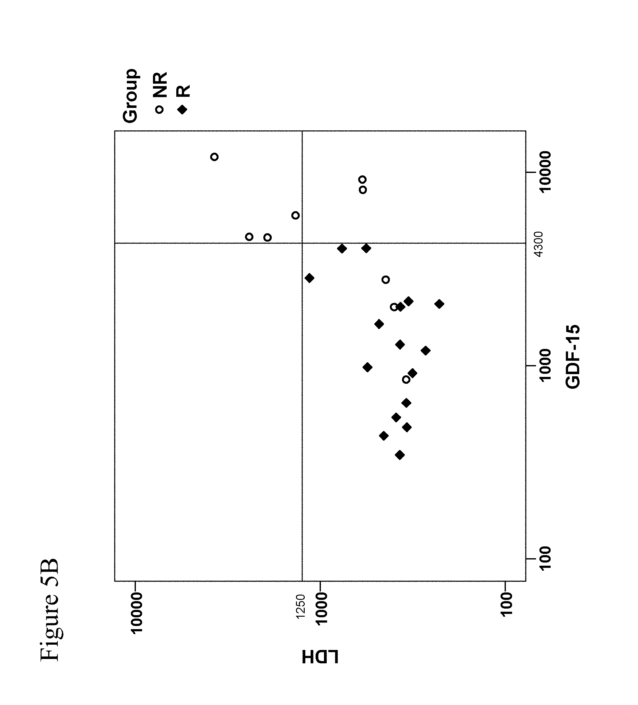
[0100] FIGS. 5A-5B: FIG. 5A: Probability of response to treatment (responder 1) as predicted by the Generalized Linear Model model using LDH as continuous predictor. Circles show the data, the curve shows the model. The vertical line indicates the LDF concentration where the probability of treatment response is 0.5. The patient cohort was identical. However, reliable determination of LDH levels failed in four patients due to hemolysis. FIG. 5B: Graphical representation of responders and non-responders and their respective hGDF-15 and LDH levels. When cut-off values are selected to cover all responders, testing based on GDF-15 allows for identification of 6 (out of 9) non-responders whereas analyses based on LDH levels can only discriminate 4 (out of 9) non-responders. For LDH testing, 4 hemolytic samples had to be excluded which causes loss of data.
[0101] FIG. 6: This Figure shows exemplary tissue sections from melanoma brain metastases having no (upper panel) or high (lower panel) GDF-15 immunoreactivity, which were stained by immunohistochemistry for GDF-15 and for the T-cell marker proteins CD3 and CD8, respectively, as indicated in the Figure. CD3 and CD8-positive cells are indicated by arrows in the high GDF-15 samples. The CD3 and CD8 stainings were made from the same area of serial sections (however not from the identical section).
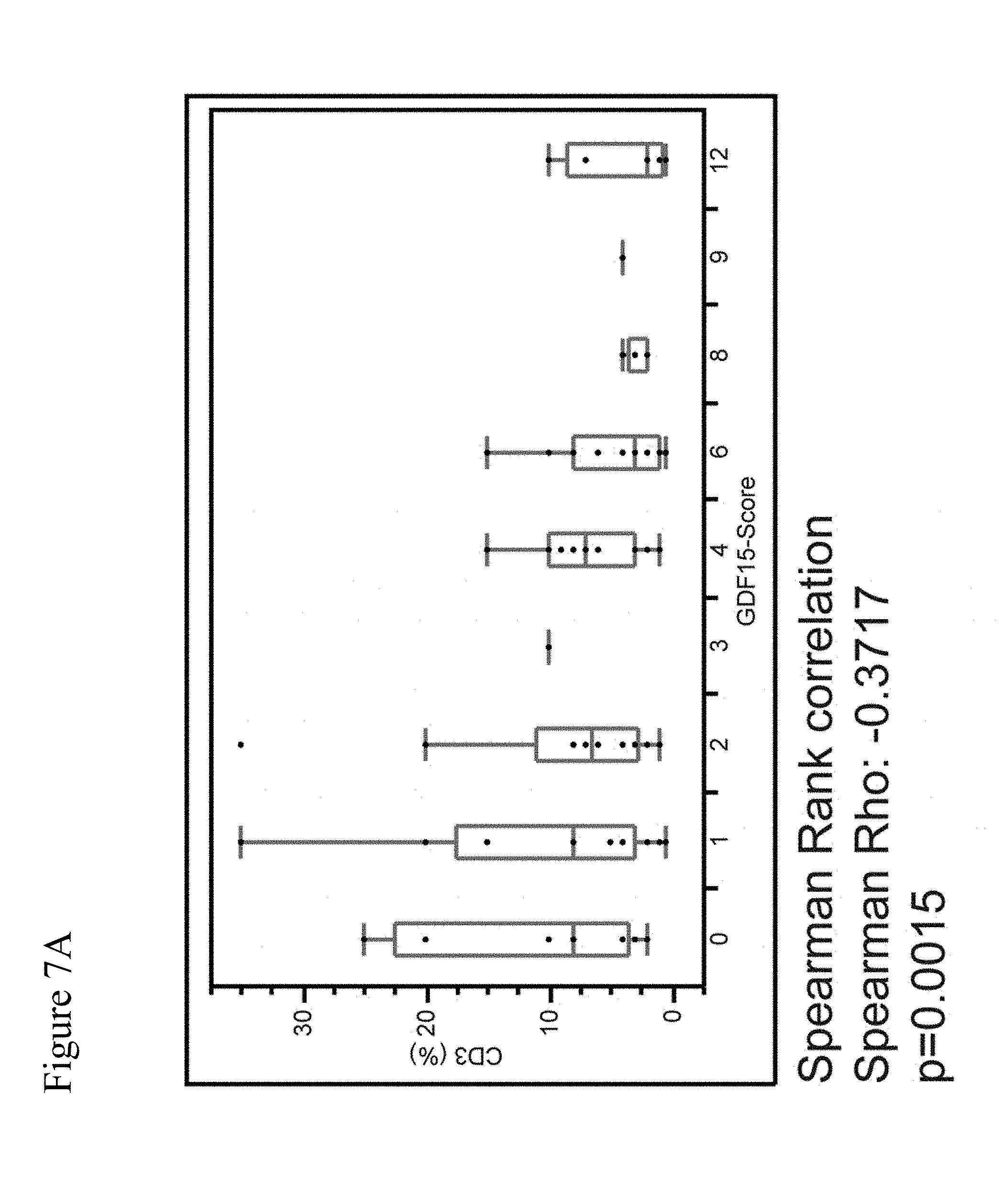
[0102] FIGS. 7A-7B: This Figure shows a plot of the percentage of CD3.sup.+ cells against the GDF-15 score across different melanoma brain metastases (7A) and a plot of the percentage of CD8.sup.+ cells against the GDF-15 score across different melanoma brain metastases (7B).
[0103] FIG. 8: This Figure shows a plot of the GDF-15 score against the percentage of CD8.sup.+ and CD3.sup.+ T cells, respectively, in brain metastases from different tumor entities (melanoma, CRC, RCC, NSCLC and SCLC).
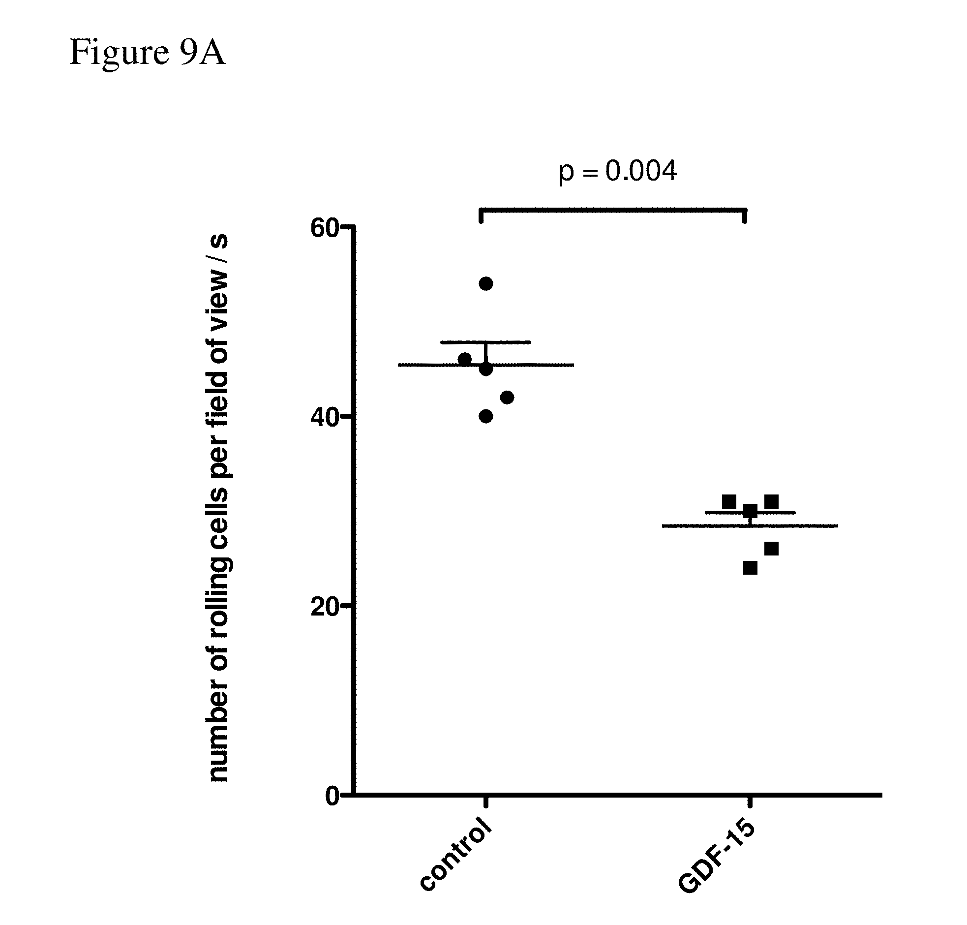
[0104] FIGS. 9A-9D: FIG. 9A shows the number of rolling T cells per field of view per second. Data were obtained from channel #3 ("GDF-15") and channel #2 ("control"). FIG. 9B shows the rolling speed of the T cells (measured in pixels per 0.2 seconds). Data were obtained from channel #3 ("GDF-15") and channel #2 ("control"). FIG. 9C shows the number of adhering cells per field of view. Data were obtained from channel #3 ("GDF-15") and channel #2 ("control"). FIG. 9D shows the number of adhering cells per field of view. Data were obtained from channel #3 ("GDF-15") and channel #2 ("control").
[0105] FIGS. 10A-10B: FIG. 10A shows the number of rolling T cells per field of view per second. Data were obtained from channel #1 (control T cells on unstimulated HUVEC as "neg. control"), channel #2 (control T cells on stimulated HUVEC as "pos. control"), channel #3 ("GDF-15") channel #4 ("UACC 257": T cells cultured in the supernatant of UACC 257 melanoma cells containing secreted GDF-15) and channel #5 ("UACC257+
[0106] anti-hGDF-15": T cells cultured in the supernatant of UACC 257 melanoma cells depleted from secreted GDF-15 with the anti-hGDF-15 antibody B1-23 as an hGDF-15 inhibitor). FIG. 10B: The flow/adhesion assay was conducted as described in Example 3. T-cells were pre-incubated with 100 ng/ml GDF-15 for 1 hour or with 100 ng/ml GDF-15, which was pre-incubated with 10 .mu.g/ml antibody for 1 hour, as indicated. The following Anti-GDF-15 antibodies were used: H1L5 (Humanized B1-23), 01G06 and 03G05 (Humanized Anti-GDF-15 Antibodies Engineered According to Sequences from WO 2014/100689). The results are shown in the Figure, which shows the number of rolling cells per field of view per 20 seconds.
[0107] FIG. 11: C57BL/6J mice were subcutaneously inoculated with 2.times.10.sup.5 colon MC38.sup.tghGDF-15 cells. Treatment with anti GDF-15 antibody (20 mg/kg of body weight) was initiated on day 0 and repeated on days 3, 7, 10, 14, 17, and 21. On day 13, animals bearing similarly sized tumors (100-150 mm.sup.3) were either treated or not with Poly-ICLC (also abbreviated as "Poly-IC") and anti CD40 antibody. Mice rejecting the pre-established tumor were followed for 57 days. Tumor-bearing mice were sacrificed according to the criteria for animal welfare.
[0108] FIG. 12: Cumulative survival in patient groups having GDF-15 levels of <1.5 ng/ml and .gtoreq.1.5 ng/ml, respectively.
[0109] FIG. 13: Cumulative survival in patient groups having high GDF-15 levels (i.e. the 50 patients with the highest GDF-15 levels) and low GDF-15 levels (i.e. the 49 patients with the lowest GDF-15 levels), respectively (median split of the total study cohort).
[0110] FIGS. 14A-14B: hGDF-15 Serum Levels do not Significantly Correlate with the Mutational Burden of the Tumors.
[0111] hGDF-15 mRNA levels in samples from cancer patients were plotted against the number of somatic mutations which were identified in the cancers. The somatic mutations were determined by use of exome sequencing. The data were analyzed by using the UZH webtool from the University Hospital Zurich (Cheng P F et al.: Data mining The Cancer Genome Atlas in the era of precision cancer medicine. Swiss Med Wkly. 2015 Sep. 16; 145:w14183.). FIG. 14A shows a plot for cancer patient data obtained from the Cancer Genome Atlas (TGCA) considering only patients with high-grade malignant melanoma (the Cancer Genome Atlas is described in the reference of Cheng P F et al.: Data mining The Cancer Genome Atlas in the era of precision cancer medicine. Swiss Med Wkly. 2015 Sep. 16; 145:w14183.). GDF-15 expression was evaluated by normalization using the RSEM ("RNA Seq by expectation maximization") software package (Li B and Dewey C N: RSEM: accurate transcript quantification from RNA-Seq data with or without a reference genome. BMC Bioinformatics. 2011 Aug. 4; 12:323. doi: 10.1186/1471-2105-12-323.). FIG. 14B shows a plot for cancer patient data from 40 additional metastatic malignant melanoma patients from the University Hospital Zurich, which were separately analyzed.
[0112] FIG. 15: Immunocytochemistry pictures for CD8a in mice harboring wild-type tumors or tumors overexpressing transgenic (tg) hGDF15 are shown. Tissue sections were stained with anti-CD8a (1:100 dilution; 4SM15 antibody purchased from eBioscience).
DETAILED DESCRIPTION OF THE INVENTION
Definitions and General Techniques
[0113] Unless otherwise defined below, the terms used in the present invention shall be understood in accordance with their common meaning known to the person skilled in the art.
[0114] The term "antibody" as used herein refers to any functional antibody that is capable of specific binding to the antigen of interest, as generally outlined in chapter 7 of Paul, W. E. (Ed.).: Fundamental Immunology 2nd Ed. Raven Press, Ltd., New York 1989, which is incorporated herein by reference. Without particular limitation, the term "antibody" encompasses antibodies from any appropriate source species, including chicken and mammalian such as mouse, goat, non-human primate and human. Preferably, the antibody is a humanized antibody. The antibody is preferably a monoclonal antibody which can be prepared by methods well-known in the art. The term "antibody" encompasses an IgG-1, -2, -3, or -4, IgE, IgA, IgM, or IgD isotype antibody. The term "antibody" encompasses monomeric antibodies (such as IgD, IgE, IgG) or oligomeric antibodies (such as IgA or IgM). The term "antibody" also encompasses--without particular limitations--isolated antibodies and modified antibodies such as genetically engineered antibodies, e.g. chimeric antibodies.
[0115] The nomenclature of the domains of antibodies follows the terms as known in the art. Each monomer of an antibody comprises two heavy chains and two light chains, as generally known in the art. Of these, each heavy and light chain comprises a variable domain (termed V.sub.H for the heavy chain and V.sub.L for the light chain) which is important for antigen binding. These heavy and light chain variable domains comprise (in an N-terminal to C-terminal order) the regions FR1, CDR1, FR2, CDR2, FR3, CDR3, and FR4 (FR, framework region; CDR, complementarity determining region which is also known as hypervariable region). The identification and assignment of the above-mentioned antibody regions within the antibody sequence is generally in accordance with Kabat et al. (Sequences of proteins of immunological interest, U.S. Dept. of Health and Human Services, Public Health Service, National Institutes of Health, Bethesda, Md. 1983), or Chothia et al. (Conformations of immunoglobulin hypervariable regions. Nature. 1989 Dec. 21-28; 342(6252):877-83.), or may be performed by using the IMGT/V-QUEST software described in Giudicelli et al. (IMGTN-QUEST, an integrated software program for immunoglobulin and T cell receptor V-J and V-D-J rearrangement analysis. Nucleic Acids Res. 2004 Jul. 1; 32(Web Server issue):W435-40.), which is incorporated herein by reference. Preferably, the antibody regions indicated above are identified and assigned by using the IMGTN-QUEST software.
[0116] A "monoclonal antibody" is an antibody from an essentially homogenous population of antibodies, wherein the antibodies are substantially identical in sequence (i.e. identical except for minor fraction of antibodies containing naturally occurring sequence modifications such as amino acid modifications at their N- and C-termini). Unlike polyclonal antibodies which contain a mixture of different antibodies directed to numerous epitopes, monoclonal antibodies are directed to the same epitope and are therefore highly specific. The term "monoclonal antibody" includes (but is not limited to) antibodies which are obtained from a monoclonal cell population derived from a single cell clone, as for instance the antibodies generated by the hybridoma method described in Kohler and Milstein (Nature, 1975 Aug. 7; 256(5517):495-7) or Harlow and Lane ("Antibodies: A Laboratory Manual" Cold Spring Harbor Laboratory Press, Cold Spring Harbor, N.Y. 1988). A monoclonal antibody may also be obtained from other suitable methods, including phage display techniques such as those described in Clackson et al. (Nature. 1991 Aug. 15; 352(6336):624-8) or Marks et al. (J Mol Biol. 1991 Dec. 5; 222(3):581-97). A monoclonal antibody may be an antibody that has been optimized for antigen-binding properties such as decreased Kd values, optimized association and dissociation kinetics by methods known in the art. For instance, Kd values may be optimized by display methods including phage display, resulting in affinity-matured monoclonal antibodies. The term "monoclonal antibody" is not limited to antibody sequences from particular species of origin or from one single species of origin. Thus, the meaning of the term "monoclonal antibody" encompasses chimeric monoclonal antibodies such as humanized monoclonal antibodies.
[0117] "Humanized antibodies" are antibodies which contain human sequences and a minor portion of non-human sequences which confer binding specificity to an antigen of interest (e.g. human GDF-15). Typically, humanized antibodies are generated by replacing hypervariable region sequences from a human acceptor antibody by hypervariable region sequences from a non-human donor antibody (e.g. a mouse, rabbit, rat donor antibody) that binds to an antigen of interest (e.g. human GDF-15). In some cases, framework region sequences of the acceptor antibody may also be replaced by the corresponding sequences of the donor antibody. In addition to the sequences derived from the donor and acceptor antibodies, a "humanized antibody" may either contain other (additional or substitute) residues or sequences or not. Such other residues or sequences may serve to further improve antibody properties such as binding properties (e.g. to decrease Kd values) and/or immunogenic properties (e.g. to decrease antigenicity in humans). Non-limiting examples for methods to generate humanized antibodies are known in the art, e.g. from Riechmann et al. (Nature. 1988 Mar. 24; 332(6162):323-7) or Jones et al. (Nature. 1986 May 29-Jun. 4; 321(6069):522-5).
[0118] The term "human antibody" relates to an antibody containing human variable and constant domain sequences. This definition encompasses antibodies having human sequences bearing single amino acid substitutions or modifications which may serve to further improve antibody properties such as binding properties (e.g. to decrease Kd values) and/or immunogenic properties (e.g. to decrease antigenicity in humans). The term "human antibody" excludes humanized antibodies where a portion of non-human sequences confers binding specificity to an antigen of interest.
[0119] An "antigen-binding portion" of an antibody as used herein refers to a portion of an antibody that retains the capability of the antibody to specifically bind to the antigen (e.g. hGDF-15, PD-1 or PD-L1). This capability can, for instance, be determined by determining the capability of the antigen-binding portion to compete with the antibody for specific binding to the antigen by methods known in the art. The antigen-binding portion may contain one or more fragments of the antibody. Without particular limitation, the antigen-binding portion can be produced by any suitable method known in the art, including recombinant DNA methods and preparation by chemical or enzymatic fragmentation of antibodies. Antigen-binding portions may be Fab fragments, F(ab') fragments, F(ab').sub.2 fragments, single chain antibodies (scFv), single-domain antibodies, diabodies or any other portion(s) of the antibody that retain the capability of the antibody to specifically bind to the antigen.
[0120] An "antibody" (e.g. a monoclonal antibody) or an "antigen-binding portion" may have been derivatized or be linked to a different molecule. For example, molecules that may be linked to the antibody are other proteins (e.g. other antibodies), a molecular label (e.g. a fluorescent, luminescent, colored or radioactive molecule), a pharmaceutical and/or a toxic agent. The antibody or antigen-binding portion may be linked directly (e.g. in form of a fusion between two proteins), or via a linker molecule (e.g. any suitable type of chemical linker known in the art).
[0121] As used herein, the terms "binding" or "bind" refer to specific binding to the antigen of interest (e.g. human GDF-15). Preferably, the Kd value is less than 100 nM, more preferably less than 50 nM, still more preferably less than 10 nM, still more preferably less than 5 nM and most preferably less than 2 nM.
[0122] As used herein, an antibody or antigen-binding portion thereof which is "capable to compete" with a second antibody capable of binding to human GDF-15 means that said (first) antibody or antigen-binding portion thereof which is "capable to compete" is capable to reduce the binding of a 10 nM reference solution of the second antibody to human or recombinant human GDF-15 by 50%. Generally, "capable to compete" means that the concentration of the (first) antibody or antigen-binding portion thereof that is needed in order to reduce the binding of the 10 nM reference solution of the second antibody to human or recombinant human GDF-15 by 50% is less than 1000 nM, preferably less than 100 nM and more preferably less than 10 nM. The binding is measured by surface plasmon resonance measurements or by Enzyme-linked Immunosorbent assay (ELISA) measurements, preferably by surface plasmon resonance measurements.
[0123] The term "epitope" as used herein refers to a small portion of an antigen that forms the binding site for an antibody.
[0124] In the context of the present invention, binding or competitive binding of antibodies or their antigen-binding portions to the antigen of interest (e.g. human GDF-15) is preferably measured by using surface plasmon resonance measurements as a reference standard assay, as described below.
[0125] The terms "K.sub.D" or "K.sub.D value" relate to the equilibrium dissociation constant as known in the art. In the context of the present invention, these terms relate to the equilibrium dissociation constant of an antibody with respect to a particular antigen of interest (e.g. human GDF-15) The equilibrium dissociation constant is a measure of the propensity of a complex (e.g. an antigen-antibody complex) to reversibly dissociate into its components (e.g. the antigen and the antibody). For the antibodies according to the invention, K.sub.D values (such as those for the antigen human GDF-15) are preferably determined by using surface plasmon resonance measurements as described below.
[0126] An "isolated antibody" as used herein is an antibody that has been identified and separated from the majority of components (by weight) of its source environment, e.g. from the components of a hybridoma cell culture or a different cell culture that was used for its production (e.g. producer cells such as CHO cells that recombinantly express the antibody). The separation is performed such that it sufficiently removes components that may otherwise interfere with the suitability of the antibody for the desired applications (e.g. with a therapeutic use of the anti-human GDF-15 antibody according to the invention). Methods for preparing isolated antibodies are known in the art and include Protein A chromatography, anion exchange chromatography, cation exchange chromatography, virus retentive filtration and ultrafiltration. Preferably, the isolated antibody preparation is at least 70% pure (w/w), more preferably at least 80% pure (w/w), still more preferably at least 90% pure (w/w), still more preferably at least 95% pure (w/w), and most preferably at least 99% pure (w/w), as measured by using the Lowry protein assay.
[0127] A "diabody" as used herein is a small bivalent antigen-binding antibody portion which comprises a heavy chain variable domain linked to a light chain variable domain on the same polypeptide chain linked by a peptide linker that is too short to allow pairing between the two domains on the same chain. This results in pairing with the complementary domains of another chain and in the assembly of a dimeric molecule with two antigen binding sites. Diabodies may be bivalent and monospecific (such as diabodies with two antigen binding sites for human GDF-15), or may be bivalent and bispecific (e.g. diabodies with two antigen binding sites, one being a binding site for human GDF-15, and the other one being a binding site for a different antigen). A detailed description of diabodies can be found in Holliger P et al. (""Diabodies": small bivalent and bispecific antibody fragments." Proc Natl Acad Sci USA. 1993 Jul. 15; 90(14):6444-8.).
[0128] A "single-domain antibody" (which is also referred to as "Nanobody.TM.") as used herein is an antibody fragment consisting of a single monomeric variable antibody domain. Structures of and methods for producing single-domain antibodies are known from the art, e.g. from Holt L J et al. ("Domain antibodies: proteins for therapy." Trends Biotechnol. 2003 November; 21(11):484-90.), Saerens D et al. ("Single-domain antibodies as building blocks for novel therapeutics." Curr Opin Pharmacol. 2008 October; 8(5):600-8. Epub 2008 Aug. 22.), and Arbabi Ghahroudi M et al. ("Selection and identification of single domain antibody fragments from camel heavy-chain antibodies." FEBS Lett. 1997 Sep. 15; 414(3):521-6.).
[0129] The terms "cancer" and "cancer cell" is used herein in accordance with their common meaning in the art (see for instance Weinberg R. et al.: The Biology of Cancer. Garland Science: New York 2006. 850p.).
[0130] The cancers to the treated according to the present invention are solid cancers. A "solid cancer" is a cancer which forms one or more solid tumors. Such solid cancers forming solid tumors are generally known in the art. The term "solid cancer" encompasses both a primary tumor formed by the cancer and possible secondary tumors, which are also known as metastases. Preferred solid cancers to be treated according to the invention are selected from the group consisting of melanoma, colorectal cancer, prostate cancer, head and neck cancer, urothelial cancer, stomach cancer, pancreatic cancer, liver cancer, testis cancer, ovarian cancer, endometrial cancer, cervical cancer, brain cancer, breast cancer, gastric cancer, renal cell carcinoma, Ewing's sarcoma, non-small cell lung cancer and small cell lung cancer, preferably selected from the group consisting of melanoma, colorectal cancer, prostate cancer, head and neck cancer, urothelial cancer, stomach cancer, pancreatic cancer, liver cancer, testis cancer, ovarian cancer, endometrial cancer and cervical cancer, more preferably selected from the group consisting of melanoma, colorectal cancer, prostate cancer, head and neck cancer, urothelial cancer and stomach cancer, and most preferably selected from the group consisting of melanoma, colorectal cancer and prostate cancer.
[0131] As referred to herein, the term "brain cancer" refers to all brain cancers known in the art. It includes but is not limited to glioma (WHO grade I to IV), astrocytoma, meningioma and medulloblastoma.
[0132] As referred to herein, the term "head and neck cancer" refers to all head and neck cancers known in the art. It includes but is not limited to oesophagus carcinoma, oral squamous cell carcinoma and hypopharyngeal cancer. A particularly preferred head and neck cancer to be treated according to the invention is oral squamous cell carcinoma.
[0133] The term "cancer growth" as used herein relates to any measureable growth of the cancer. For cancers forming solid tumors, "cancer growth" relates to a measurable increase in tumor volume over time. If the cancer has formed only a single tumor, "cancer growth" relates only to the increase in volume of the single tumor. If the cancer has formed multiple tumors such as metastases, "cancer growth" relates to the increase in volume of all measurable tumors. For solid tumors, the tumor volume can be measured by any method known in the art, including magnetic resonance imaging and computed tomography (CT scan).
[0134] Terms such as "treatment of cancer" or "treating cancer" according to the present invention refer to a therapeutic treatment. An assessment of whether or not a therapeutic treatment works can, for instance, be made by assessing whether the treatment inhibits cancer growth in the treated patient or patients. Preferably, the inhibition is statistically significant as assessed by appropriate statistical tests which are known in the art. Inhibition of cancer growth may be assessed by comparing cancer growth in a group of patients treated in accordance with the present invention to a control group of untreated patients, or by comparing a group of patients that receive a standard cancer treatment of the art plus a treatment according to the invention with a control group of patients that only receive a standard cancer treatment of the art. Such studies for assessing the inhibition of cancer growth are designed in accordance with accepted standards for clinical studies, e.g. double-blinded, randomized studies with sufficient statistical power. The term "treating cancer" includes an inhibition of cancer growth where the cancer growth is inhibited partially (i.e. where the cancer growth in the patient is delayed compared to the control group of patients), an inhibition where the cancer growth is inhibited completely (i.e. where the cancer growth in the patient is stopped), and an inhibition where cancer growth is reversed (i.e. the cancer shrinks). Preferably, an assessment of whether or not a therapeutic treatment works can be made based on a classification of responders and non-responders by using the response evaluation criteria in solid tumours, version 1.1 (RECIST v1.1) (Eisenhauer et al.: New response evaluation criteria in solid tumours: revised RECIST guideline (version 1.1). In: Eur. J. Cancer. 45, No. 2, January 2009, pp. 228-47). Alternatively, or additionally, an assessment of whether or not a therapeutic treatment works can be made based on known clinical indicators of cancer progression.
[0135] The treatment of cancer according to the invention can be a first-line therapy, a second-line therapy or a third-line therapy or a therapy that is beyond third-line therapy. The meaning of these terms is known in the art and in accordance with the terminology that is commonly used by the US National Cancer Institute.
[0136] A treatment of cancer according to the present invention does not exclude that additional or secondary therapeutic benefits also occur in patients. For example, an additional or secondary benefit may be an influence on cancer-induced weight loss. However it is understood that the primary treatment for which protection is sought is for treating the cancer itself, any secondary or additional effects only reflect optional, additional advantages of the treatment of cancer growth.
[0137] The term "cancer immunotherapy" is known in the art and generally relates to a treatment of cancer in which the immune system of the patient is used to treat the cancer. Cancer cells harbor genomic mutations which give rise to cancer cell antigens that are specific to the cancer cells and different from the antigens of non-cancerous cells. Thus, in a preferred aspect of cancer immunotherapy in accordance with the present invention, a cancer immunotherapy is a cancer immunotherapy wherein such cancer cell antigens are recognized by the immune system, and wherein cancer cells expressing these antigens are killed by the immune system. In a non-limiting aspect of the invention, such cancer cells expressing these cancer cell antigens can be killed by CD8.sup.+ T-cells of the immune system. A cancer immunotherapy can be assessed by immunomonitoring methods known in the art, e.g. by measuring intracellular IFN-.gamma. expression (e.g. in CD8.sup.+ T-cells and/or NK cells) in blood samples, measuring CD107a cell surface expression (e.g. on CD8.sup.+ T-cells and/or NK cells) in blood samples, measuring intracellular TNF-.alpha. expression (e.g. on leukocytes) in blood samples, intracellular Interleukin-2 expression (e.g. in CD8.sup.+ T-cells and/or in CD4.sup.+ T-cells) in blood samples, CD154 cell surface expression (e.g. in CD8.sup.+ T-cells and/or in CD4.sup.+ T-cells) in blood samples, tetramer or dextramer staining for tumor antigen-specific T cells in blood samples, CTL activity against autologous tumor cells or presence of T cells against neoantigens derived from tumor-specific mutations. Preferred methods to assess cancer immunotherapy are the methods according to Gouttefangeas C et al.: "Flow Cytometry in Cancer Immunotherapy: Applications, Quality Assurance and Future." (2015) In: Cancer Immunology: Translational Medicine from Bench to Bedside (N. Rezaei editor). Springer. Chapter 25: pages 471-486; and the methods according to Van der Burg S H, et al.: "Immunoguiding, the final frontier in the immunotherapy of cancer." (2014) In Cancer Immunotherapy meets oncology (C M Britten, S Kreiter, M. Diken & HG Rammensee eds). Springer International Publishing Switzerland p37-51 ISBN: 978-3-319-05103-1.
[0138] As used herein, a "cancer immunotherapy" optionally encompasses a treatment where in addition to the immune system which is used to treat the cancer, additional mechanisms of cancer treatment are used. For instance, it was previously shown that a hGDF-15 inhibitor can be used for cancer treatment in an mouse model system where the immune system was severely compromised (WO 2014/049087). Thus, according to the present invention, a cancer immunotherapy by hGDF-15 inhibitors in human patients can also encompass additional treatment effects of hGDF-15 inhibitors which are independent from the immune system. Another example of a cancer immunotherapy where additional mechanisms of cancer treatment can be used is a combination therapy with known chemotherapeutic agent(s). Such combination therapy with known chemotherapeutic agent(s) may, for instance, not only include the treatment of cancer in which the immune system is used to treat the cancer but also include a treatment of cancer in which the cancer cells are killed by said chemotherapeutic agent(s) directly.
[0139] As used herein, the term "increasing the percentage of CD8.sup.+ T-cells in a solid cancer" relates to any measurable increase in the percentage of CD8.sup.+ T-cells (i.e. the percentage of CD8.sup.+ T-cells calculated with respect to all cells) in the tumor or tumors formed by the solid cancer. Preferably, the increase is statistically significant as assessed by appropriate statistical tests which are known in the art. An increase in the percentage of CD8.sup.+ T-cells in the tumor or tumors formed by the solid cancer can be determined by known methods for analyses of CD8.sup.+ T-cells in solid tumors. Such methods include analyses of tumor biopsies for CD8.sup.+ T-cells, e.g. analyses of such tumor biopsies by immunohistochemistry using antibodies against CD8 and using a staining for the total number of cells. The increase may be assessed by comparing the percentages of CD8.sup.+ T-cells in tumors of a group of patients treated in accordance with the present invention to a control group of untreated patients, or by comparing a group of patients that receive a standard cancer treatment of the art plus a treatment according to the invention with a control group of patients that only receive a standard cancer treatment of the art.
[0140] As used herein, "CD8.sup.+ T-cells" are preferably cells which endogenously occur in the human patient. hGDF-15 serum levels can be measured by any methods known in the art. For instance, a preferred method of measuring hGDF-15 serum levels is a measurement of hGDF-15 serum levels by Enzyme-Linked Immunosorbent Assay (ELISA) by using antibodies to GDF-15. Such ELISA methods are exemplified in Example 1. Alternatively, hGDF-15 serum levels may be determined by known electrochemiluminesence immunoassays using antibodies to GDF-15. For instance, the Roche Elecsys.RTM. technology can be used for such electrochemiluminesence immunoassays.
[0141] The patient to be treated according to the invention is preferably a patient with elevated hGDF-15 serum levels. The term "elevated hGDF-15 serum levels" as used herein means that the human patient has higher hGDF-15 levels in blood serum prior to administration of the hGDF-15 inhibitor according to the invention, when compared to median hGDF-15 levels in blood sera of healthy human control individuals as a reference.
[0142] The median hGDF-15 serum level of healthy human control individuals is <0.8 ng/ml. The expected range is between 0.2 ng/ml and 1.2 ng/ml in healthy human controls (Reference: Tanno T et al.: "Growth differentiation factor 15 in erythroid health and disease." Curr Opin Hematol. 2010 May; 17(3): 184-190.).
[0143] Thus, in a preferred embodiment of the invention, a patient to be treated according to the invention is a patient who has a hGDF-15 serum level of at least 1.2 ng/ml prior to the start of administration of the hGDF-15 inhibitor, preferably a patient who has a hGDF-15 serum level of at least 1.5 ng/ml prior to the start of administration of the hGDF-15 inhibitor, and more preferably a patient who has a hGDF-15 serum level of at least 1.8 ng/ml prior to the start of administration of the hGDF-15 inhibitor.
[0144] In a further preferred embodiment of the invention, a patient to be treated according to the invention is a patient who has a hGDF-15 serum level of at least 1.2 ng/ml and not more than 12 ng/ml prior to the start of administration of the hGDF-15 inhibitor, preferably a patient who has a hGDF-15 serum level of at least 1.5 ng/ml and not more than 12 ng/ml prior to the start of administration of the hGDF-15 inhibitor, and more preferably a patient who has a hGDF-15 serum level of at least 1.8 ng/ml and not more than 12 ng/ml prior to the start of administration of the hGDF-15 inhibitor.
[0145] In a further embodiment of the invention in accordance with all of the above embodiments, a patient to be treated according to the invention is a patient who has a hGDF-15 serum level of at least 1.2 ng/ml and not more than 10 ng/ml prior to the start of administration of the hGDF-15 inhibitor, preferably a patient who has a hGDF-15 serum level of at least 1.5 ng/ml and not more than 10 ng/ml prior to the start of administration of the hGDF-15 inhibitor, and more preferably a patient who has a hGDF-15 serum level of at least 1.8 ng/ml and not more than 10 ng/ml prior to the start of administration of the hGDF-15 inhibitor.
[0146] In a further embodiment of the invention in accordance with all of the above embodiments, a patient to be treated according to the invention is a patient who has a hGDF-15 serum level of at least 1.2 ng/ml and not more than 8 ng/ml prior to the start of administration of the hGDF-15 inhibitor, preferably a patient who has a hGDF-15 serum level of at least 1.5 ng/ml and not more than 8 ng/ml prior to the start of administration of the hGDF-15 inhibitor, and more preferably a patient who has a hGDF-15 serum level of at least 1.8 ng/ml and not more than 8 ng/ml prior to the start of administration of the hGDF-15 inhibitor.
[0147] In another embodiment, a patient to be treated according to the invention is a patient who has a hGDF-15 serum level of at least 2 ng/ml, at least 2.2 ng/ml, at least 2.4 ng/ml, at least 2.6 ng/ml, at least 2.8 ng/ml, at least 3.0 ng/ml, at least 3.2 ng/ml, at least 3.4 ng/ml, at least 3.6 ng/ml, at least 3.8 ng/ml, at least 4.0 ng/ml, or at least 4.2 ng/ml prior to the start of administration of the hGDF-15 inhibitor. In this embodiment, the patient is preferably a patient who has a hGDF-15 serum level of not more than 12 ng/ml prior to the start of administration of the hGDF-15 inhibitor. More preferably, in this embodiment, the patient is a patient who has a hGDF-15 serum level of not more than 10 ng/ml prior to the start of administration of the hGDF-15 inhibitor. Most preferably, in this embodiment, the patient is a patient who has a hGDF-15 serum level of not more than 8 ng/ml prior to the start of administration of the hGDF-15 inhibitor.
[0148] The term "prior to the start of administration" as used herein means the period of time immediately before administration of the hGDF-15 inhibitor according to the invention. Preferably, the term "prior to the start of administration" means a period of 30 days immediately before administration; most preferably a period of one week immediately before administration.
[0149] The terms "significant", "significantly", etc. as used herein refer to a statistically significant difference between values as assessed by appropriate methods known in the art.
[0150] The hGDF-15 inhibitors and the immune checkpoint blockers used according to the invention can be administered by using methods known in the art. Such methods will be selected by the skilled person based on well-known considerations, including the chemical nature of the respective inhibitor (e.g. depending on whether the inhibitor is a short interfering RNA or an antibody). Administration of known immune checkpoint blockers may be based on known administration schemes of these immune checkpoint blockers. For instance, administration of the immune checkpoint blockers may be based on the administration schemes used in the KEYNOTE-006 trial (C. Robert et al. N Engl J Med 2015; 372:2521-2532).
[0151] In accordance with the present invention, each occurrence of the term "comprising" may optionally be substituted with the term "consisting of".
hGDF-15 Inhibitors to be Used in Accordance with the Invention
[0152] An "hGDF-15 inhibitor" according to the invention can be any molecule which is capable of specifically inhibiting the function of human GDF-15 (hGDF-15).
[0153] A non-limiting example of such an hGDF-15 inhibitor is a molecule which specifically downregulates the expression of hGDF-15 and thereby inhibits hGDF-15 function. For instance, a short interfering RNA or an siRNA hairpin construct can be used to specifically downregulate the expression of hGDF-15 and to inhibit hGDF-15 function. Rules for the design and selection of short interfering RNA and siRNA hairpin construct sequences are known in the art and have for example been reviewed in Jackson and Linsley, Recognizing and avoiding siRNA off-target effects for target identification and therapeutic application, Nat Rev Drug Discov. 2010 January; 9(1):57-67. Short interfering RNAs and siRNA hairpin constructs can be delivered to the human patients by any suitable methods, including viral delivery methods (as, for instance, reviewed in Knoepfel S A et al., "Selection of RNAi-based inhibitors for anti-HIV gene therapy." World J Virol. 2012 Jun. 12; 1(3):79-90.) and other delivery methods such as methods using conjugate groups which facilitate delivery into the cells (as, for instance, reviewed in Kanasty R et al., "Delivery materials for siRNA therapeutics.", Nat Mater. 2013 November; 12(11):967-77.)
[0154] Whether or not a substance of interest is a "hGDF-15 inhibitor" can be determined by using the methods disclosed herein, as detailed in the preferred embodiments. A preferred method in accordance with the preferred embodiments is the method used in Example 3.
[0155] It was previously shown that human GDF-15 protein can be advantageously targeted by a monoclonal antibody (WO2014/049087), and that such antibody has advantageous properties including a high binding affinity to human GDF-15, as demonstrated by an equilibrium dissociation constant of about 790 pM for recombinant human GDF-15 (see Reference Example 1). Thus, in a preferred embodiment in accordance with the invention, the hGDF-15 inhibitor to be used is an antibody capable of binding to hGDF-15, or an antigen-binding portion thereof. Preferably, the antibody is a monoclonal antibody capable of binding to hGDF-15, or an antigen-binding portion thereof.
[0156] Thus, in a more preferred embodiment, the hGDF-15 inhibitor in accordance with the invention is a monoclonal antibody capable of binding to human GDF-15, or an antigen-binding portion thereof, wherein the heavy chain variable domain comprises a CDR3 region comprising the amino acid sequence of SEQ ID NO: 5 or an amino acid sequence at least 90% identical thereto, and wherein the light chain variable domain comprises a CDR3 region comprising the amino acid sequence of SEQ ID NO: 7 or an amino acid sequence at least 85% identical thereto. In this embodiment, preferably, the antibody or antigen-binding portion thereof comprises a heavy chain variable domain which comprises a CDR1 region comprising the amino acid sequence of SEQ ID NO: 3 and a CDR2 region comprising the amino acid sequence of SEQ ID NO: 4, and the antibody or antigen-binding portion thereof comprises a light chain variable domain which comprises a CDR1 region comprising the amino acid sequence of SEQ ID NO: 6, and a CDR2 region comprising the amino acid sequence ser-ala-ser.
[0157] Thus, in a still more preferred embodiment, the hGDF-15 inhibitor in accordance with the invention is a monoclonal antibody capable of binding to human GDF-15, or an antigen-binding portion thereof, wherein the antibody or antigen-binding portion thereof comprises a heavy chain variable domain which comprises a CDR1 region comprising the amino acid sequence of SEQ ID NO: 3, a CDR2 region comprising the amino acid sequence of SEQ ID NO: 4 and a CDR3 region comprising the amino acid sequence of SEQ ID NO: 5, and wherein the antibody or antigen-binding portion thereof comprises a light chain variable domain which comprises a CDR1 region comprising the amino acid sequence of SEQ ID NO: 6, a CDR2 region comprising the amino acid sequence ser-ala-ser and a CDR3 region comprising the amino acid sequence of SEQ ID NO: 7.
[0158] In another embodiment in accordance with the above embodiments of the monoclonal antibody capable of binding to human GDF-15, or an antigen-binding portion thereof, the heavy chain variable domain comprises a region comprising an FR1, a CDR1, an FR2, a CDR2 and an FR3 region and comprising the amino acid sequence of SEQ ID NO: 1 or a sequence 85%, 90%, 91%, 92%, 93%, 94%, 95%, 96%, 97%, 98% or 99% identical thereto, and the light chain variable domain comprises a region comprising an FR1, a CDR1, an FR2, a CDR2 and an FR3 region and comprising the amino acid sequence of SEQ ID NO: 2 or a sequence 85%, 90%, 91%, 92%, 93%, 94%, 95%, 96%, 97%, 98% or 99% identical thereto.
[0159] In a preferred embodiment in accordance with the above embodiments of the monoclonal antibody capable of binding to human GDF-15, or an antigen-binding portion thereof, the antibody is a humanized antibody or an antigen-binding portion thereof. The constant domain of the heavy chain of this monoclonal antibody or antigen-binding portion thereof may comprise the amino acid sequence of SEQ ID No: 29, or an amino acid sequence at least 85%, preferably at least 90%, more preferably at least 95% identical thereto, and the constant domain of the light chain of this monoclonal antibody or antigen-binding portion thereof may comprise the amino acid sequence of SEQ ID No: 32, or an amino acid sequence at least 85%, preferably at least 90%, more preferably at least 95% identical thereto. More preferably, the constant domain of the heavy chain of this monoclonal antibody or antigen-binding portion thereof comprises the amino acid sequence of SEQ ID No: 29, or an amino acid sequence at least 98%, preferably at least 99% identical thereto, and the constant domain of the light chain of this monoclonal antibody or antigen-binding portion thereof comprises the amino acid sequence of SEQ ID No: 32, or an amino acid sequence at least 98%, preferably at least 99% identical thereto. Still more preferably, the constant domain of the heavy chain of this monoclonal antibody or antigen-binding portion thereof comprises the amino acid sequence of SEQ ID No: 29, and the constant domain of the light chain of this monoclonal antibody or antigen-binding portion thereof comprises the amino acid sequence of SEQ ID No: 32. The heavy chain variable domain of this monoclonal antibody or antigen-binding portion thereof may comprise the amino acid sequence of SEQ ID No: 28, or an amino acid sequence at least 90%, preferably at least 95%, more preferably at least 98%, still more preferably at least 99% identical thereto, and the light chain variable domain of this monoclonal antibody or antigen-binding portion thereof may comprise the amino acid sequence of SEQ ID No: 31, or an amino acid sequence at least 90%, preferably at least 95%, more preferably at least 98%, still more preferably at least 99% identical thereto. Most preferably, the heavy chain variable domain of this monoclonal antibody or antigen-binding portion thereof comprises the amino acid sequence of SEQ ID No: 28, and the light chain variable domain of this monoclonal antibody or antigen-binding portion thereof comprises the amino acid sequence of SEQ ID No: 31.
[0160] In another embodiment in accordance with the above embodiments of the monoclonal antibody capable of binding to human GDF-15, or an antigen-binding portion thereof, the heavy chain variable domain comprises a CDR1 region comprising the amino acid sequence of SEQ ID NO: 3 and a CDR2 region comprising the amino acid sequence of SEQ ID NO: 4, and the light chain variable domain comprises a CDR1 region comprising the amino acid sequence of SEQ ID NO: 6 and a CDR2 region comprising the amino acid sequence of SEQ ID NO: 7. In a preferred aspect of this embodiment, the antibody may have CDR3 sequences as defined in any of the embodiments of the invention described above.
[0161] In another embodiment in accordance with the monoclonal antibody capable of binding to human GDF-15, or an antigen-binding portion thereof, the antigen-binding portion is a single-domain antibody (also referred to as "Nanobody.TM."). In one aspect of this embodiment, the single-domain antibody comprises the CDR1, CDR2, and CDR3 amino acid sequences of SEQ ID NO: 3, SEQ ID NO: 4, and SEQ ID NO: 5, respectively. In another aspect of this embodiment, the single-domain antibody comprises the CDR1, CDR2, and CDR3 amino acid sequences of SEQ ID NO: 6, ser-ala-ser, and SEQ ID NO: 7, respectively. In a preferred aspect of this embodiment, the single-domain antibody is a humanized antibody.
[0162] Preferably, the antibodies capable of binding to human GDF-15 or the antigen-binding portions thereof have an equilibrium dissociation constant for human GDF-15 that is equal to or less than 100 nM, less than 20 nM, preferably less than 10 nM, more preferably less than 5 nM and most preferably between 0.1 nM and 2 nM.
[0163] In another embodiment in accordance with the above embodiments of the monoclonal antibody capable of binding to human GDF-15, or an antigen-binding portion thereof, the antibody capable of binding to human GDF-15 or the antigen-binding portion thereof binds to the same human GDF-15 epitope as the antibody to human GDF-15 obtainable from the cell line B1-23 deposited with the Deutsche Sammlung fur Mikroorganismen and Zellkulturen GmbH (DMSZ) under the accession No. DSM ACC3142. As described herein, antibody binding to human GDF-15 in accordance with the present invention is preferably assessed by surface plasmon resonance measurements as a reference standard method, in accordance with the procedures described in Reference Example 1. Binding to the same epitope on human GDF-15 can be assessed similarly by surface plasmon resonance competitive binding experiments of the antibody to human GDF-15 obtainable from the cell line B1-23 and the antibody that is expected to bind to the same human GDF-15 epitope as the antibody to human GDF-15 obtainable from the cell line B1-23.
[0164] In another preferred embodiment, the antibody capable of binding to human GDF-15 or the antigen-binding portion thereof is a monoclonal antibody capable of binding to human GDF-15, or an antigen-binding portion thereof, wherein the heavy chain variable domain comprises the amino acid sequence of SEQ ID NO: 39 or a sequence that is at least 85%, at least 90%, at least 91%, at least 92%, at least 93%, at least 94%, at least 95%, at least 96%, at least 97%, at least 98% or at least 99% identical thereto, and the light chain variable domain comprises the amino acid sequence of SEQ ID NO: 40 or a sequence that is at least 85%, at least 90%, at least 91%, at least 92%, at least 93%, at least 94%, at least 95%, at least 96%, at least 97%, at least 98% or at least 99% identical thereto.
[0165] In another preferred embodiment, the antibody capable of binding to human GDF-15 or the antigen-binding portion thereof is a monoclonal antibody capable of binding to human GDF-15, or an antigen-binding portion thereof, wherein the heavy chain variable domain comprises the amino acid sequence of SEQ ID NO: 41 or a sequence that is at least 85%, at least 90%, at least 91%, at least 92%, at least 93%, at least 94%, at least 95%, at least 96%, at least 97%, at least 98% or at least 99% identical thereto, and the light chain variable domain comprises the amino acid sequence of SEQ ID NO: 42 or a sequence that is at least 85%, at least 90%, at least 91%, at least 92%, at least 93%, at least 94%, at least 95%, at least 96%, at least 97%, at least 98% or at least 99% identical thereto.
[0166] In another preferred embodiment, the antibody capable of binding to human GDF-15 or the antigen-binding portion thereof is a monoclonal antibody capable of binding to human GDF-15, or an antigen-binding portion thereof, wherein the heavy chain variable domain comprises the amino acid sequence of SEQ ID NO: 43 or a sequence that is at least 85%, at least 90%, at least 91%, at least 92%, at least 93%, at least 94%, at least 95%, at least 96%, at least 97%, at least 98% or at least 99% identical thereto, and the light chain variable domain comprises the amino acid sequence of SEQ ID NO: 44 or a sequence that is at least 85%, at least 90%, at least 91%, at least 92%, at least 93%, at least 94%, at least 95%, at least 96%, at least 97%, at least 98% or at least 99% identical thereto.
[0167] In another preferred embodiment, the antibody capable of binding to human GDF-15 or the antigen-binding portion thereof is a monoclonal antibody capable of binding to human GDF-15, or an antigen-binding portion thereof, wherein the heavy chain variable domain comprises the amino acid sequence of SEQ ID NO: 45 or a sequence that is at least 85%, at least 90%, at least 91%, at least 92%, at least 93%, at least 94%, at least 95%, at least 96%, at least 97%, at least 98% or at least 99% identical thereto, and the light chain variable domain comprises the amino acid sequence of SEQ ID NO: 46 or a sequence that is at least 85%, at least 90%, at least 91%, at least 92%, at least 93%, at least 94%, at least 95%, at least 96%, at least 97%, at least 98% or at least 99% identical thereto.
[0168] In another preferred embodiment, the antibody capable of binding to human GDF-15 or the antigen-binding portion thereof is a monoclonal antibody capable of binding to human GDF-15, or an antigen-binding portion thereof, wherein the heavy chain variable domain comprises the amino acid sequence of SEQ ID NO: 47 or a sequence that is at least 85%, at least 90%, at least 91%, at least 92%, at least 93%, at least 94%, at least 95%, at least 96%, at least 97%, at least 98% or at least 99% identical thereto, and the light chain variable domain comprises the amino acid sequence of SEQ ID NO: 48 or a sequence that is at least 85%, at least 90%, at least 91%, at least 92%, at least 93%, at least 94%, at least 95%, at least 96%, at least 97%, at least 98% or at least 99% identical thereto.
[0169] In another preferred embodiment, the antibody capable of binding to human GDF-15 or the antigen-binding portion thereof is a monoclonal antibody capable of binding to human GDF-15, or an antigen-binding portion thereof, wherein the heavy chain variable domain comprises the amino acid sequence of SEQ ID NO: 49 or a sequence that is at least 85%, at least 90%, at least 91%, at least 92%, at least 93%, at least 94%, at least 95%, at least 96%, at least 97%, at least 98% or at least 99% identical thereto, and the light chain variable domain comprises the amino acid sequence of SEQ ID NO: 50 or a sequence that is at least 85%, at least 90%, at least 91%, at least 92%, at least 93%, at least 94%, at least 95%, at least 96%, at least 97%, at least 98% or at least 99% identical thereto.
[0170] In another preferred embodiment, the antibody capable of binding to human GDF-15 or the antigen-binding portion thereof is a monoclonal antibody capable of binding to human GDF-15, or an antigen-binding portion thereof, wherein the heavy chain variable domain comprises the amino acid sequence of SEQ ID NO: 51 or a sequence that is at least 85%, at least 90%, at least 91%, at least 92%, at least 93%, at least 94%, at least 95%, at least 96%, at least 97%, at least 98% or at least 99% identical thereto, and the light chain variable domain comprises the amino acid sequence of SEQ ID NO: 52 or a sequence that is at least 85%, at least 90%, at least 91%, at least 92%, at least 93%, at least 94%, at least 95%, at least 96%, at least 97%, at least 98% or at least 99% identical thereto.
[0171] In another preferred embodiment, the antibody capable of binding to human GDF-15 or the antigen-binding portion thereof is a monoclonal antibody or antigen-binding portion thereof, which is capable to compete with any one of the antibodies capable of binding to human GDF-15 referred to herein for binding to human GDF-15, preferably for binding to recombinant human GDF-15.
[0172] In a very preferred embodiment, the antibody capable of binding to human GDF-15 or the antigen-binding portion thereof is a humanized monoclonal antibody or an antigen-binding portion thereof. For any given non-human antibody sequence in accordance with the invention (i.e. a donor antibody sequence), humanized monoclonal anti-human-GDF-15 antibodies of the invention or antigen-binding portions thereof can be generated in accordance with techniques known in the art, as described above.
[0173] In a very preferred embodiment, the antibody capable of binding to human GDF-15 or the antigen-binding portion thereof is a monoclonal antibody capable of binding to human GDF-15, or an antigen-binding portion thereof, wherein the binding is binding to a conformational or discontinuous epitope on human GDF-15 comprised by the amino acid sequences of SEQ ID No: 25 and SEQ ID No: 26. In a preferred aspect of this embodiment, the antibody or antigen-binding portion thereof is an antibody or antigen-binding portion thereof as defined by the sequences of any one of the above embodiments.
[0174] The antibody capable of binding to human GDF-15 or the antigen-binding portion thereof can be linked to a drug. In non-limiting aspects of this embodiment, the drug can be a known anticancer agent and/or an immune-stimulatory molecule. Known anticancer agents include alkylating agents such as cisplatin, carboplatin, oxaliplatin, mechlorethamine, cyclophosphamide, chlorambucil, and ifosfamide; anti-metabolites such as azathioprine and mercaptopurine; alkaloids such as vinca alkaloids (e.g. vincristine, vinblastine, vinorelbine, and vindesine), taxanes (e.g. paclitaxel, docetaxel) etoposide and teniposide; topoisomerase inhibitors such as camptothecins (e.g. irinotecan and topotecan); cytotoxic antibiotics such as actinomycin, anthracyclines, doxorubicin, daunorubicin, valrubicin, idarubicin, epirubicin, bleomycin, plicamycin and mitomycin; and radioisotopes.
[0175] In a further embodiment in accordance with the above embodiments, the antibody capable of binding to human GDF-15 or the antigen-binding portion thereof is modified by an amino acid tag. Non-limiting examples of such tags include Polyhistidin (His-) tags, FLAG-tag, Hemagglutinin (HA) tag, glycoprotein D (gD) tag, and c-myc tag. Tags may be used for various purposes. For instance, they may be used to assist purification of the antibody capable of binding to human GDF-15 or the antigen-binding portion thereof. Preferably, such tags are present at the C-terminus or N-terminus of the antibody capable of binding to human GDF-15 or the antigen-binding portion thereof.
Immune Checkpoint Blockers to be Used in Accordance with the Invention
[0176] Cancer cells harbor genomic mutations which give rise to cancer cell antigens that are specific to the cancer cells and different from the antigens of non-cancerous cells. Therefore, an intact immune system which is not inhibited should recognize these cancer cell antigens, such that an immune response against these antigens is elicited. However, most cancers have developed immune tolerance mechanisms against these antigens. One class of mechanisms by which cancer cells achieve such immune tolerance is the utilization of immune checkpoints. An "immune checkpoint" as used herein generally means an immunological mechanism by which an immune response can be inhibited. More particularly, an immune checkpoint is a mechanism which is characterized in that a molecule of the immune system (or a group of molecules of the immune system) inhibits the immune response by inhibiting the activation of cells of the immune system. Such molecule (or group of molecules) of the immune system which inhibits (inhibit) the immune response by inhibiting the activation of cells of the immune system is (are) also known as checkpoint molecule(s).
[0177] As used herein, an "immune checkpoint blocker" is a molecule which is capable of blocking an immune checkpoint. While it is understood that an hGDF-15 inhibitor as used according to the invention has effects on the immune system including effects on CD8+ T cells, the term "immune checkpoint blocker" as used herein does not refer to an hGDF-15 inhibitor but means a molecule which is different from an hGDF-15 inhibitor.
[0178] The most common immune checkpoint blockers which are known to date are inhibitors of immune checkpoint molecules such as inhibitors of human PD-1 and inhibitors of human PD-L1. Further immune checkpoint blockers are anti-LAG-3, anti-B7H3, anti-TIM3, anti-VISTA, anti-TIGIT, anti-KIR, anti-CD27, anti-CD137 as well as inhibitors of IDO. Therefore, as used in accordance with the present invention, a preferred form of an immune checkpoint blocker is an inhibitor of an immune checkpoint molecule. Alternatively, an immune checkpoint blocker can be an activator of a co-stimulating signal which overrides the immune checkpoint.
[0179] Methods to measure the potency of immune checkpoint blockers include in vitro binding assays, primary T cell-based cytokine release assays, and in vivo model systems. Additionally, Promega has now developed a commercially available bioluminescent reporter system for PD-1/PD-L1, which is, for instance referred to in Mei Cong, Ph.D. et al.: Advertorial: Novel Bioassay to Assess PD-1/PD-L1 Therapeutic Antibodies in Development for Immunotherapy Bioluminescent Reporter-Based PD-1/PD-L1 Blockade Bioassay. (http://www.genengnews.com/gen-articles/advertorial-novel-bioassay-to-ass- ess-pd-1-pd-l1-therapeutic-antibodies-in-development-for-immun/5511/).
[0180] Preferred immune checkpoint blockers are inhibitors of human PD-1 and inhibitors of human PD-L1. In one preferred embodiment in accordance with all of the embodiments of the invention, the immune checkpoint blocker is not an inhibitor of human CTLA4.
[0181] As used herein, an "inhibitor of human PD-1" can be any molecule which is capable of specifically inhibiting the function of human PD-1. Non-limiting examples of such molecules are antibodies capable of binding to human PD-1 and DARPins (Designed Ankyrin Repeat Proteins) capable of binding to human PD-1. Preferably, the inhibitor of PD-1 to be used in accordance with the invention is an antibody capable of binding to human PD-1, more preferably a monoclonal antibody capable of binding to human PD-1. Most preferably, the monoclonal antibody capable of binding to human PD-1 is selected from the group consisting of nivolumab, pembrolizumab, pidilizumab and AMP-224.
[0182] As used herein, an "inhibitor of human PD-L1" can be any molecule which is capable of specifically inhibiting the function of human PD-L1. Non-limiting examples of such molecules are antibodies capable of binding to human PD-L1 and DARPins (Designed Ankyrin Repeat Proteins) capable of binding to human PD-L1. Preferably, the inhibitor of human PD-L1 to be used in accordance with the invention is an antibody capable of binding to human PD-L1, more preferably a monoclonal antibody capable of binding to human PD-L1. Most preferably, the monoclonal antibody capable of binding to human PD-L1 is selected from the group consisting of BMS-936559, MPDL3280A, MED14736, and MSB0010718C.
Methods and Techniques
[0183] Generally, unless otherwise defined herein, the methods used in the present invention (e.g. cloning methods or methods relating to antibodies) are performed in accordance with procedures known in the art, e.g. the procedures described in Sambrook et al. ("Molecular Cloning: A Laboratory Manual.", 2.sup.nd Ed., Cold Spring Harbor Laboratory Press, Cold Spring Harbor, N.Y. 1989), Ausubel et al. ("Current Protocols in Molecular Biology." Greene Publishing Associates and Wiley Interscience; New York 1992), and Harlow and Lane ("Antibodies: A Laboratory Manual" Cold Spring Harbor Laboratory Press, Cold Spring Harbor, N.Y. 1988), all of which are incorporated herein by reference.
[0184] Binding of antibodies to their respective target proteins can be assessed by methods known in the art. The binding of monoclonal antibodies to their respective targets is preferably assessed by surface plasmon resonance measurements. These measurements are preferably carried out by using a Biorad ProteOn XPR36 system and Biorad GLC sensor chips, as exemplified for anti-human GDF-15 mAb-B1-23 in Reference Example 1.
[0185] Sequence Alignments of sequences according to the invention are performed by using the BLAST algorithm (see Altschul et al. (1990) "Basic local alignment search tool." Journal of Molecular Biology 215. p. 403-410.; Altschul et al.: (1997) Gapped BLAST and PSI-BLAST: a new generation of protein database search programs. Nucleic Acids Res. 25:3389-3402.). Preferably, the following parameters are used: Max target sequences 10; Word size 3; BLOSUM 62 matrix; gap costs: existence 11, extension 1; conditional compositional score matrix adjustment. Thus, when used in connection with sequences, terms such as "identity" or "identical" refer to the identity value obtained by using the BLAST algorithm.
[0186] Monoclonal antibodies according to the invention can be produced by any method known in the art, including but not limited to the methods referred to in Siegel D L ("Recombinant monoclonal antibody technology." Transfus Clin Biol. 2002 January; 9(1):15-22.). In one embodiment, an antibody according to the invention is produced by the hybridoma cell line B1-23 deposited with the Deutsche Sammlung fur Mikroorganismen and Zellkulturen GmbH (DSMZ) at Inhoffenstra.beta.e 7B, 38124 Braunschweig, Germany, under the accession No. DSM ACC3142 under the Budapest treaty. The deposit was filed on Sep. 29, 2011.
[0187] Cell proliferation can be measured by suitable methods known in the art, including (but not limited to) visual microscopy, metabolic assays such as those which measure mitochondrial redox potential (e.g. MTT (344,5-Dimethylthiazol-2-yl)-2,5-diphenyltetrazolium bromide) assay; Resazurin staining which is also known as Alamar Blue.RTM. assay), staining of known endogenous proliferation biomarkers (e.g. Ki-67), and methods measuring cellular DNA synthesis (e.g. BrdU and [.sup.3H]-Thymidine incorporation assays).
[0188] Levels of human GDF-15 (hGDF-15) can be measured by any method known in the art, including measurements of hGDF-15 protein levels by methods including (but not limited to) mass spectrometry for proteins or peptides derived from human GDF-15, Western Blotting using antibodies specific to human GDF-15, flow cytometry using antibodies specific to human GDF-15, strip tests using antibodies specific to human GDF-15, or immunocytochemistry using antibodies specific to human GDF-15. A preferred method of measuring hGDF-15 serum levels is a measurement of hGDF-15 serum levels by Enzyme-Linked Immunosorbent Assay (ELISA) by using antibodies to GDF-15. Such ELISA methods are exemplified in Example 1. Alternatively, hGDF-15 serum levels may be determined by known electrochemiluminesence immunoassays using antibodies to GDF-15. For instance, the Roche Elecsys.RTM. technology can be used for such electrochemiluminesence immunoassays.
Preparation of Compositions of the Invention
[0189] Compositions in accordance with the present invention are prepared in accordance with known standards for the preparation of pharmaceutical compositions.
[0190] For instance, the compositions are prepared in a way that they can be stored and administered appropriately, e.g. by using pharmaceutically acceptable components such as carriers, excipients or stabilizers.
[0191] Such pharmaceutically acceptable components are not toxic in the amounts used when administering the pharmaceutical composition to a patient. The pharmaceutical acceptable components added to the pharmaceutical compositions may depend on the chemical nature of the inhibitors present in the composition (e.g. depend on whether the inhibitors are antibodies, si RNA hairpin constructs or short interfering RNAs), the particular intended use of the pharmaceutical compositions and the route of administration.
[0192] In general, the pharmaceutically acceptable components used in connection with the present invention are used in accordance with knowledge available in the art, e.g. from Remington's Pharmaceutical Sciences, Ed. AR Gennaro, 20th edition, 2000, Williams & Wilkins, Pa., USA.
Therapeutic Methods and Products for Use in these Methods
[0193] The present invention relates to the hGDF-15 inhibitors for the uses as defined above.
[0194] Additionally, and in accordance with these hGDF-15 inhibitors and their uses, the present invention also relates to corresponding therapeutic methods.
[0195] Accordingly, in one embodiment, the invention relates to a method for increasing the percentage of CD8.sup.+ T-cells in a solid cancer in a human patient, the method comprising the step of administering an hGDF-15 inhibitor to the human patient.
[0196] In another embodiment, the invention relates to a method of treating a solid cancer by an immune checkpoint blocker in a human patient, the method comprising a step of administering an hGDF-15 inhibitor to the human patient and a step of administering the immune checkpoint blocker to the human patient.
[0197] Preferred embodiments of these methods are as defined above for the hGDF-15 inhibitors for use according to the invention.
[0198] In another embodiment of the above methods, hGDF-15 inhibitors for use, kits, compositions, or compositions for use, the hGDF-15 inhibitor is the sole ingredient which is pharmaceutically active against cancer.
[0199] In an alternative embodiment of the above methods, hGDF-15 inhibitors for use, kits, compositions, or compositions for use, the hGDF-15 inhibitor and the immune checkpoint blocker are the sole ingredients which are pharmaceutically active against cancer.
[0200] In an alternative embodiment of the above methods, hGDF-15 inhibitors for use, kits, combinations, compositions, or compositions for use, the hGDF-15 inhibitor is used in combination with one or more further ingredients pharmaceutically active against cancer. In one aspect of this embodiment, the one or more further ingredients pharmaceutically active against cancer is a known anticancer agent and/or an immune-stimulatory molecule. Known anticancer agents include but are not limited to alkylating agents such as cisplatin, carboplatin, oxaliplatin, mechlorethamine, cyclophosphamide, chlorambucil, and ifosfamide; anti-metabolites such as azathioprine and mercaptopurine; alkaloids such as vinca alkaloids (e.g. vincristine, vinblastine, vinorelbine, and vindesine), taxanes (e.g. paclitaxel, docetaxel) etoposide and teniposide; topoisomerase inhibitors such as camptothecins (e.g. irinotecan and topotecan); cytotoxic antibiotics such as actinomycin, anthracyclines, doxorubicin, daunorubicin, valrubicin, idarubicin, epirubicin, bleomycin, plicamycin and mitomycin; and radioisotopes. The following ingredients pharmaceutically active against cancer are particularly preferred to be used in combination with the hGDF-15 inhibitor: Immune-stimulatory molecules include anti-LAG-3, anti-B7H3, anti-TIM3, anti-VISTA, anti-TIGIT, anti-KIR, anti-CD27, anti-CD137, anti-Ox40, anti-4-1BB, anti-GITR, anti-CD28, anti-CD40 or IDO-Inhibitors. Furthermore, other antibody treatments like anti-Her2, anti-EGFR, anti-Claudin, or their glyco-optimized successors are also particularly preferred as they will benefit from a combination with the hGDF-15 inhibitor, e.g. due to enhanced immune cell infiltration in the solid cancer caused by the hGDF-15 inhibitor. Likewise, vaccination approaches (e.g. with peptides or dendritic cells) or adoptive cell therapies, tumor-reactive T cells or dendritic cells are also particularly preferred as they will benefit from a combination with the hGDF-15 inhibitor. Furthermore, the following treatments are also particularly preferred as they will synergize with the hGDF-15 inhibitor: [0201] Treatments with antibodies or antibody-like molecules having one or more specificities for tumor and immune cells (e.g. Bites, DARTS, DARPINS, Catumaxomab); [0202] treatments by vaccine-based immunotherapy against tumor associated peptides, for instance with multi-peptide vaccines such as IMA901, ISA203 or with RNA-based vaccines (e.g. CV9104), and/or [0203] treatments with immune cell-activating substances (e.g. FAA derivatives to activate macrophages, or ligands for toll-like receptors such as SLP-AMPLIVANT conjugates).
Combinations for Uses According to the Invention
[0204] The present invention encompasses combinations of an hGDF-15 inhibitor and an immune checkpoint blocker for use in a method of treating a solid cancer in a human patient, wherein the hGDF-15 inhibitor and the immune checkpoint blocker are to be administered to the human patient. These combinations and their preferred embodiments are as defined above.
[0205] The combination of the hGDF-15 inhibitor and the immune checkpoint blocker may either be administered together or separately.
[0206] For instance, in one preferred embodiment, administration of the hGDF-15 inhibitor is to be started prior to the start of administration of the immune checkpoint blocker. This setting advantageously allows to increase the percentage of T-cells, and in particular the percentage of CD8.sup.+ T cells in the solid cancer, such that a subsequent treatment with the immune checkpoint blocker can be more effective due to the increased starting percentage of CD8.sup.+ T cells in the solid cancer.
Kits
[0207] The present invention also provides a kit comprising an hGDF-15 inhibitor and at least one immune checkpoint blocker, as defined above.
[0208] The hGDF-15 inhibitor and one or more or all of the immune checkpoint blockers can be contained in separate containers or in a single container.
[0209] A container as used can be any type of container that is suitable to store the hGDF-15 inhibitor and/or the at least one immune checkpoint blocker. Non-limiting examples of such containers are vials and pre-filled syringes.
[0210] In addition to the hGDF-15 inhibitor and the at least one immune checkpoint blocker, the kit may contain further therapeutic agents. For instance, the kit may contain one or more further ingredients pharmaceutically active against cancer. The one or more further ingredients pharmaceutically active against cancer can be as defined above. Such further ingredients pharmaceutically active against cancer may be used in the methods of the invention together with the hGDF-15 inhibitor and the at least one immune checkpoint blocker.
[0211] Preferably, a kit according to the invention further comprises instructions for use.
Sequences
[0212] The amino acid sequences referred to in the present application are as follows (in an N-terminal to C-terminal order; represented in the one-letter amino acid code):
TABLE-US-00001 SEQ ID No: 1 (Region of the Heavy Chain Variable Domain comprising an FR1, a CDR1, an FR2, a CDR2 and an FR3 region from the Polypeptide Sequence of monoclonal anti-human GDF-15 mAb-B1-23): QVKLQQSGPGILQSSQTLSLTCSFSGFSLSTSGMGVSWIRQPSGKGLEWLAHIYWDDDKRYNPTLKSRLTISK DPSRNQVFLKITSVDTADTATYYC SEQ ID No: 2 (Region of the Light Chain Variable Domain comprising an FR1, a CDR1, an FR2, a CDR2 and an FR3 region from the Polypeptide Sequence of monoclonal anti-human GDF-15 mAb-B1-23): DIVLTQSPKFMSTSVGDRVSVTCKASQNVGTNVAWFLQKPGQSPKALIYSASYRYSGVPDRFTGSGSGTDFT LTISNVQSEDLAEYFC SEQ ID No: 3 (Heavy Chain CDR1 Region Peptide Sequence of monoclonal anti-human GDF-15 mAb-B1-23): GFSLSTSGMG SEQ ID No: 4 (Heavy Chain CDR2 Region Peptide Sequence of monoclonal anti-human GDF-15 mAb-B1-23): IYWDDDK SEQ ID No: 5 (Heavy Chain CDR3 Region Peptide Sequence of monoclonal anti-human GDF-15 mAb-B1-23): ARSSYGAMDY SEQ ID No: 6 (Light Chain CDR1 Region Peptide Sequence of monoclonal anti-human GDF-15 mAb-B1-23): QNVGTN Light Chain CDR2 Region Peptide Sequence of monoclonal anti-human GDF-15 mAb-B1-23: SAS SEQ ID No: 7 (Light Chain CDR3 Region Peptide Sequence of monoclonal anti-human GDF-15 mAb-B1-23): QQYNNFPYT SEQ ID No: 8 (recombinant mature human GDF-15 protein): GSARNGDHCPLGPGRCCRLHTVRASLEDLGWADWVLSPREVQVTMCIGACPSQFRAANMHAQIKTSLHRLK PDTVPAPCCVPASYNPMVLIQKTDTGVSLQTYDDLLAKDCHCI SEQ ID No: 9 (human GDF-15 precursor protein): MPGQELRTVNGSQMLLVLLVLSWLPHGGALSLAEASRASFPGPSELHSEDSRFRELRKRYEDLLTRLRANQS WEDNTDLVPAPAVRILTPEVRLGSGGHLHLRISRAALPEGLPEASRLHRALFRLSPTASRSWDVTRPLRRQL SLARPQAPALHLRLSPPPSQSDQLLAESSSARPQLELHLRPQAARGRRRARARNGDHCPLGPGRCCRLHTV RASLEDLGWADWVLSPREVQVTMCIGACPSQFRAANMHAQIKTSLHRLKPDTVPAPCCVPASYNPMVLIQKT DTGVSLQTYDDLLAKDCHCI SEQ ID No: 10 (human GDF-15 precursor protein + N-terminal and C-terminal GSGS linker): GSGSGSGMPGQELRTVNGSQMLLVLLVLSWLPHGGALSLAEASRASFPGPSELHSEDSRFRELRKRYEDLL TRLRANQSWEDSNTDLVPAPAVRILTPEVRLGSGGHLHLRISRAALPEGLPEASRLHRALFRLSPTASRSWDV TRPLRRQLSLARPQAPALHLRLSPPPSQSDQLLAESSSARPQLELHLRPQAARGRRRARARNGDHCPLGPG RCCRLHTVRASLEDLGWADWVLSPREVQVTMCIGACPSQFRAANMHAQIKTSLHRLKPDTVPAPCCVPASY NPMVLIQKTDTGVSLQTYDDLLAKDCHCIGSGSGSG SEQ ID No: 11 (Flag peptide): DYKDDDDKGG SEQ ID No: 12 (HA peptide): YPYDVPDYAG SEQ ID No: 13 (peptide derived from human GDF-15): ELHLRPQAARGRR SEQ ID No: 14 (peptide derived from human GDF-15): LHLRPQAARGRRR SEQ ID No: 15 (peptide derived from human GDF-15): HLRPQAARGRRRA SEQ ID No: 16 (peptide derived from human GDF-15): LRPQAARGRRRAR SEQ ID No: 17 (peptide derived from human GDF-15): RPQAARGRRRARA SEQ ID No: 18 (peptide derived from human GDF-15): PQAARGRRRARAR SEQ ID No: 19 (peptide derived from human GDF-15): QAARGRRRARARN SEQ ID No: 20 (peptide derived from human GDF-15): MHAQIKTSLHRLK SEQ ID No: 25 (GDF-15 peptide comprising part of the GDF-15 Epitope that binds to B1-23): EVQVTMCIGACPSQFR SEQ ID No: 26 (GDF-15 peptide comprising part of the GDF-15 Epitope that binds to B1-23): TDTGVSLQTYDDLLAKDCHCI
[0213] The nucleic acid sequences referred to in the present application are as follows (in a 5' to 3' order; represented in accordance with the standard nucleic acid code):
TABLE-US-00002 SEQ ID No: 21 (DNA nucleotide sequence encoding the amino acid sequence defined in SEQ ID No: 1): CAAGTGAAGCTGCAGCAGTCAGGCCCTGGGATATTGCAGTCCTCCCAGAC CCTCAGTCTGACTTGTTCTTTCTCTGGGTTTTCACTGAGTACTTCTGGTA TGGGTGTGAGCTGGATTCGTCAGCCTTCAGGAAAGGGTCTGGAGTGGCTG GCACACATTTACTGGGATGATGACAAGCGCTATAACCCAACCCTGAAGAG CCGGCTCACAATCTCCAAGGATCCCTCCAGAAACCAGGTATTCCTCAAGA TCACCAGTGTGGACACTGCAGATACTGCCACATACTACTGT SEQ ID No: 22 (DNA nucleotide sequence encoding the amino acid sequence defined in SEQ ID No: 2): GACATTGTGCTCACCCAGTCTCCAAAATTCATGTCCACATCAGTAGGAGA CAGGGTCAGCGTCACCTGCAAGGCCAGTCAGAATGTGGGTACTAATGTGG CCTGGTTTCTACAGAAACCAGGGCAATCTCCTAAAGCACTTATTTACTCG GCATCCTACCGGTACAGTGGAGTCCCTGATCGCTTCACAGGCAGTGGATC TGGGACAGATTTCACTCTCACCATCAGCAACGTGCAGTCTGAAGACTTGG CAGAGTATTTCTGT SEQ ID No: 23 (DNA nucleotide sequence encoding the amino acid sequence defined in SEQ ID No: 5): GCTCGAAGTTCCTACGGGGCAATGGACTAC SEQ ID No: 24 (DNA nucleotide sequence encoding the amino acid sequence defined in SEQ ID No: 7): CAGCAATATAACAACTTTCCGTACACG
[0214] Further amino acid sequences are as follows (in an N-terminal to C-terminal order; represented in the one-letter amino acid code):
TABLE-US-00003 SEQ ID No: 27 (amino acid sequence of the heavy chain of the H1L5 humanized B1-23 anti-GDF-15 antibody): QITLKESGPTLVKPTQTLTLTCTFSGFSLSTSGMGVSWIRQPPGKGLEWLAHIYWDDDKRYNPTLKSRLTITKD PSKNQVVLTMTNMDPVDTATYYCARSSYGAMDYWGQGTLVTVSSASTKGPSVFPLAPSSKSTSGGTAALGC LVKDYFPEPVTVSWNSGALTSGVHTFPAVLQSSGLYSLSSWTVPSSSLGTQTYICNVNHKPSNTKVDKKVEP KSCDKTHTCPPCPAPELLGGPSVFLFPPKPKDTLMISRTPEVTCVVVDVSHEDPEVKFNWYVDGVEVHNAKT KPREEQYNSTYRVVSVLTVLHQDWLNGKEYKCKVSNKALPAPIEKTISKAKGQPREPQVYTLPPSREEMTKN QVSLTCLVKGFYPSDIAVEWESNGQPENNYKTTPPVLDSDGSFFLYSKLTVDKSRWQQGNVFSCSVMHEAL HNHYTQKSLSLSPGK SEQ ID No: 28 (amino acid sequence of the heavy chain variable domain of the H1L5 humanized B1-23 anti-GDF-15 antibody): QITLKESGPTLVKPTQTLTLTCTFSGFSLSTSGMGVSWIRQPPGKGLEWLAHIYWDDDKRYNPTLKSRLTITKD PSKNQVVLTMTNMDPVDTATYYCARSSYGAMDYWGQGTLVTVSS SEQ ID No: 29 (amino acid sequence of the heavy chain constant domain of the H1L5 humanized B1-23 anti-GDF-15 antibody): ASTKGPSVFPLAPSSKSTSGGTAALGCLVKDYFPEPVTVSWNSGALTSGVHTFPAVLQSSGLYSLSSWIVP SSSLGTQTYICNVNHKPSNTKVDKKVEPKSCDKTHTCPPCPAPELLGGPSVFLFPPKPKDTLMISRTPEVTCV VVDVSHEDPEVKFNWYVDGVEVHNAKTKPREEQYNSTYRVVSVLTVLHQDWLNGKEYKCKVSNKALPAPIE KTISKAKGQPREPQVYTLPPSREEMTKNQVSLTCLVKGFYPSDIAVEWESNGQPENNYKTTPPVLDSDGSFF LYSKLIVDKSRWQQGNVFSCSVMHEALHNHYTQKSLSLSPGK SEQ ID No: 30 (amino acid sequence of the light chain of the H1L5 humanized B1-23 anti-GDF-15 antibody): DIVLTQSPSFLSASVGDRVTITCKASQNVGTNVAWFQQKPGKSPKALIYSASYRYSGVPDRFTGSGSGTEFTL TISSLQPEDFAAYFCQQYNNFPYTFGGGTKLEIKRAPSVFIFPPSDEQLKSGTASVVCLLNNFYPREAKVQWK VDNALQSGNSQESVTEQDSKDSTYSLSSTLTLSKADYEKHKVYACEVTHQGLSSPVTKSFNRGEC SEQ ID No: 31 (amino acid sequence of the light chain variable domain of the H1L5 humanized B1-23 anti-GDF-15 antibody): DIVLTQSPSFLSASVGDRVTITCKASQNVGTNVAWFQQKPGKSPKALIYSASYRYSGVPDRFTGSGSGTEFTL TISSLQPEDFAAYFCQQYNNFPYTFGGGTKLEIKR SEQ ID No: 32 (amino acid sequence of the light chain constant domain of the H1L5 humanized B1-23 anti-GDF-15 antibody): APSVFIFPPSDEQLKSGTASVVCLLNNFYPREAKVQWKVDNALQSGNSQESVTEQDSKDSTYSLSSTLTLSKA DYEKHKVYACEVTHQGLSSPVTKSFNRGEC SEQ ID No: 33 (amino acid sequence of the heavy chain of the chimeric B1-23 anti-GDF- 15 antibody): QVKLQQSGPGILQSSQTLSLTCSFSGFSLSTSGMGVSWIRQPSGKGLEWLAHIYWDDDKRYNPTLKSRLTISK DPSRNQVFLKITSVDTADTATYYCARSSYGAMDYWGQGTSVIVSSASTKGPSVFPLAPSSKSTSGGTAALGC LVKDYFPEPVTVSWNSGALTSGVHTFPAVLQSSGLYSLSSVVTVPSSSLGTQTYICNVNHKPSNTKVDKKVEP KSCDKTHTCPPCPAPELLGGPSVFLFPPKPKDTLMISRTPEVTCVVVDVSHEDPEVKFNWYVDGVEVHNAKT KPREEQYNSTYRVVSVLTVLHQDWLNGKEYKCKVSNKALPAPIEKTISKAKGQPREPQVYTLPPSREEMTKN QVSLTCLVKGFYPSDIAVEWESNGQPENNYKTTPPVLDSDGSFFLYSKLTVDKSRWQQGNVFSCSVMHEAL HNHYTQKSLSLSPGK SEQ ID No: 34 (amino acid sequence of the heavy chain variable domain of the chimeric B1-23 anti-GDF-15 antibody): QVKLQQSGPGILQSSQTLSLTCSFSGFSLSTSGMGVSWIRQPSGKGLEWLAHIYWDDDKRYNPTLKSRLTISK DPSRNQVFLKITSVDTADTATYYCARSSYGAMDYWGQGTSVIVSS SEQ ID No: 35 (amino acid sequence of the heavy chain constant domain of the chimeric B1-23 anti-GDF-15 antibody): ASTKGPSVFPLAPSSKSTSGGTAALGCLVKDYFPEPVTVSWNSGALTSGVHTFPAVLQSSGLYSLSSVVTVP SSSLGTQTYICNVNHKPSNTKVDKKVEPKSCDKTHTCPPCPAPELLGGPSVFLFPPKPKDTLMISRTPEVTCV VVDVSHEDPEVKFNWYVDGVEVHNAKTKPREEQYNSTYRVVSVLTVLHQDWLNGKEYKCKVSNKALPAPIE KTISKAKGQPREPQVYTLPPSREEMTKNQVSLTCLVKGFYPSDIAVEWESNGQPENNYKTTPPVLDSDGSFF LYSKLIVDKSRWQQGNVFSCSVMHEALHNHYTQKSLSLSPGK SEQ ID No: 36 (amino acid sequence of the light chain of the chimeric B1-23 anti-GDF-15 antibody): DIVLTQSPKFMSTSVGDRVSVTCKASQNVGTNVAWFLQKPGQSPKALIYSASYRYSGVPDRFTGSGSGTDFT LTISNVQSEDLAEYFCQQYNNFPYTFGGGTKLEIKRIVAAPSVFIFPPSDEQLKSGTASVVCLLNNFYPREAKV QWKVDNALQSGNSQESVTEQDSKDSTYSLSSTLTLSKADYEKHKVYACEVTHQGLSSPVTKSFNRGEC SEQ ID No: 37 (amino acid sequence of the light chain variable domain of the chimeric B1-23 anti-GDF-15 antibody): DIVLTQSPKFMSTSVGDRVSVTCKASQNVGTNVAWFLQKPGQSPKALIYSASYRYSGVPDRFTGSGSGTDFT LTISNVQSEDLAEYFCQQYNNFPYTFGGGTKLEIKRIVA SEQ ID No: 38 (amino acid sequence of the light chain constant domain of the chimeric B1-23 anti-GDF-15 antibody): APSVFIFPPSDEQLKSGTASVVCLLNNFYPREAKVQWKVDNALQSGNSQESVTEQDSKDSTYSLSSTLTLSKA DYEKHKVYACEVTHQGLSSPVTKSFNRGEC SEQ ID No: 39 (amino acid sequence of the heavy chain variable domain of the 01G06 antibody): QVQLVQSGAEVKKPGASVKVSCKASGYTFTDYNMDWVRQAPGQSLEWMGQINPNNGLIFFNQKFQGRVTL TTDTSTSTAYMELRSLRSDDTAVYYCAREAITTVGAMDYWGQGTLVTVSS SEQ ID No: 40 (amino acid sequence of the light chain variable domain of the 01G06 antibody): DIQMTQSPSSLSASVGDRVTITCRTSENLHNYLAWYQQKPGKSPKLLIYDAKTLADGVPSRFSGSGSGTDY TLTISSLQPEDFATYYCQHFWSDPYTFGQGTKLEIK SEQ ID No: 41 (amino acid sequence of the heavy chain variable domain of the 03G05 antibody): QVQLQQPGAELVKPGASVKLSCKASGYTFTSYWIHWVNQRPGQGLEWIGDINPSNGRSKYNEKFKNKATMT ADKSSNTAYMQLSSLTSEDSAVYYCAREVLDGAMDYWGQGTSVIVSS SEQ ID No: 42 (amino acid sequence of the light chain variable domain of the 03G05 antibody): DIVLTQSPASLAVSLGQRATISCRASESVDNYGISFMNWFQQKPGQPPKLLIYAASNQGSGVPARFSGSGS GTDFSLNIHPMEEDDTAMYFCQQSKEVPWTFGGGSKLEIK SEQ ID No: 43 (amino acid sequence of the heavy chain variable domain of the 04F08 antibody): QVTLKESGPGILQPSQTLSLTCSFSGFSLSTYGMGVTWIRQPSGKGLEWLAHIYWDDDKRYNPSLKSRLTI SKDTSNNQVFLKITSVDTADTATYYCAQTGYSNLFAYWGQGTLVTVSA SEQ ID No: 44 (amino acid sequence of the light chain variable domain of the 04F08 antibody): DIVMTQSQKFMSTSVGDRVSVTCKASQNVGTNVAWYQQKLGQSPKTLIYSASYRYSGVPDRFTGSGSGTDF TLTISNVQSEDLAEYFCQQYNSYPYTFGGGTKLEIK SEQ ID No: 45 (amino acid sequence of the heavy chain variable domain of the 06C11 antibody): QVTLKESGPGILQPSQTLSLTCSFSGFSLNTYGMGVSWIRQPSGKGLEWLAHIYWDDDKRYNPSLKSRLTI SKDASNNRVFLKITSVDTADTATYYCAQRGYDDYWGYWGQGTLVTISA SEQ ID No: 46 (amino acid sequence of the light chain variable domain of the 06C11 antibody): DIVMTQSQKFMSTSVGDRVSVTCKASQNVGTNVAWFQQKPGQSPKALIYSASYRYSGVPDRFTGSGSGTDF ILTISNVQSEDLAEYFCQQYNNYPLTFGAGTKLELK SEQ ID No: 47 (amino acid sequence of the heavy chain variable domain of the 08G01 antibody): EVLLQQSGPEVVKPGASVKIPCKASGYTFTDYNMDWVKQSHGKSLEWIGEINPNNGGTFYNQKFKGKATLT VDKSSSTAYMELRSLTSEDTAVYYCAREAITTVGAMDYWGQGTSVTVSS SEQ ID No: 48 (amino acid sequence of the light chain variable domain of the 08G01 antibody): DIQMTQSPASLSASVGETVTITCRASGNIHNYLAWYQQKQGKSPQLLVYNAKTLADGVPSRFSGSGSGTQY SLKINSLQPEDFGSYYCQHFWSSPYTFGGGTKLEIK SEQ ID No: 49 (amino acid sequence of the heavy chain variable domain of the 14F11 antibody): QVTLKESGPGILQPSQTLSLTCSFSGFSLSTYGMGVGWIRQPSGKGLEWLADIWWDDDKYYNPSLKSRLTI SKDTSSNEVFLKIAIVDTADTATYYCARRGHYSAMDYWGQGTSVTVSS SEQ ID No: 50 (amino acid sequence of the light chain variable domain of the 14F11 antibody): DIVMTQSQKFMSTSVGDRVSVTCKASQNVGTNVAWYQQKPGQSPKALIYSPSYRYSGVPDRFTGSGSGTDF TLTISNVQSEDLAEYFCQQYNSYPHTFGGGTKLEMK SEQ ID No: 51 (amino acid sequence of the heavy chain variable domain of the 17B11 antibody): QVTLKESGPGILQPSQTLSLTCSFSGFSLSTSGMGVSWIRQPSGKGLEWLAHNDWDDDKRYKSSLKSRLTI SKDTSRNQVFLKITSVDTADTATYYCARRVGGLEGYFDYWGQGTTLTVSS SEQ ID No: 52 (amino acid sequence of the light chain variable domain of the 17B11 antibody): DIVLTQSPASLAVSLGQRATISCRASQSVSTSRFSYMHWFQQKPGQAPKLLIKYASNLESGVPARFSGSGS GTDFTLNIHPVEGEDTATYYCQHSWEIPYTFGGGTKLEIK
EXAMPLES
[0215] Reference Examples 1 to 3 exemplify an hGDF-15 inhibitor, which can be used in the compositions, kits, methods and uses according to the invention. This hGDF-15 inhibitor is a monoclonal antibody which is known from WO 2014/049087, which is incorporated herein by reference in its entirety:
Reference Example 1: Generation and Characterization of the GDF-15 Antibody B1-23
[0216] The antibody B1-23 was generated in a GDF-15 knock out mouse. Recombinant human GDF-15 (SEQ ID No: 8) was used as the immunogen.
[0217] The hybridoma cell line B1-23 producing mAb-B1-23 was deposited by the Julius-Maximilians-Universitat Wurzburg, Sanderring 2, 97070 Wurzburg, Germany, with the Deutsche Sammlung fur Mikroorganismen and Zellkulturen GmbH (DMSZ) at Inhoffenstra.beta.e 7B, 38124 Braunschweig, Germany, under the accession No. DSM ACC3142, in accordance with the Budapest Treaty. The deposit was filed on Sep. 29, 2011.
[0218] By means of a commercially available test strip system, B1-23 was identified as an IgG2a (kappa chain) isotype. Using surface plasmon resonance measurements, the dissociation constant (Kd) was determined as follows:
[0219] Binding of the monoclonal anti-human-GDF-15 antibody anti-human GDF-15 mAb-B1-23 according to the invention was measured by employing surface plasmon resonance measurements using a Biorad ProteOn XPR36 system and Biorad GLC sensor chips:
[0220] For preparing the biosensors recombinant mature human GDF-15 protein was immobilized on flow cells 1 and 2. On one flow cell recombinant GDF-15 derived from Baculvirus-transfected insect cells (HighFive insect cells) and on the other recombinant protein derived from expression in E. coli was used. The GLC sensor chip was activated using Sulfo-NHS (N-Hydroxysulfosuccinimide) and EDC (1-Ethyl-3-[3-dimethylaminopropyl]carbodiimide hydrochloride) (Biorad ProteOn Amine Coupling Kit) according to the manufacturer's recommendation, the sensor surface was subsequently loaded with the proteins up to a density of about 600 RU (1 Ru=1 pg mm.sup.-2). The non-reacted coupling groups were then quenched by perfusion with 1M ethanolamine pH 8.5 and the biosensor was equilibrated by perfusing the chip with running buffer (10M HEPES, 150 mM NaCl, 3.4 mM EDTA, 0.005% Tween-20, pH 7.4, referred to as HBS150). As controls two flow cells were used, one empty with no protein coupled and one coupled with an non-physiological protein partner (human Interleukin-5), which was immobilized using the same coupling chemistry and the same coupling density. For interaction measurements anti-human GDF-15 mAb-B1-23 was dissolved in HBS150 and used in six different concentrations as analyte (concentration: 0.4, 0.8, 3, 12, 49 and 98 nM). The analyte was perfused over the biosensor using the one-shot kinetics setup to avoid intermittent regeneration, all measurements were performed at 25.degree. C. and using a flow rate of 100 .mu.l min.sup.-1. For processing the bulk face effect and unspecific binding to the sensor matrix was removed by subtracting the SPR data of the empty flow cell (flow cell 3) from all other SPR data. The resulting sensogram was analyzed using the software ProteOn Manager version 3.0. For analysis of the binding kinetics a 1:1 Langmuir-type interaction was assumed. For the association rate constant a value of 5.4.+-.0.06.times.10.sup.5 M.sup.-1 s.sup.-1 (k.sub.on) and for the dissociation rate constant a value of 4.3.+-.0.03.times.10.sup.-4 s.sup.-1 (k.sub.off) could be determined (values are for the interaction of anti-human GDF-15 mAb-B1-23 with GDF-15 derived from insect cell expression). The equilibrium dissociation constant was calculated using the equation K.sub.D=k.sub.off/k.sub.on to yield a value of about 790 pM. Affinity values for the interaction of GDF-15 derived from E. coli expression and the anti-human GDF-15 mAb-B1-23 differ by less than a factor of 2, rate constants for GDF-15 derived from insect cells and E. coli deviate by about 45% and are thus within the accuracy of SPR measurements and likely do not reflect a real difference in affinity. Under the conditions used the anti-human GDF-15 mAb-B1-23 shows no binding to human interleukin-5 and thus confirms the specificity of the interaction data and the anti-human GDF-15 mAb-B1-23.
[0221] The amino acid sequence of recombinant human GDF-15 (as expressed in Baculovirus-transfected insect cells) is:
TABLE-US-00004 (SEQ ID No: 8) GSARNGDHCP LGPGRCCRLH TVRASLEDLG WADWVLSPRE VQVTMCIGAC PSQFRAANMH AQIKTSLHRL KPDTVPAPCC VPASYNPMVL IQKTDTGVSL QTYDDLLAKD CHCI
[0222] Thus, using surface plasmon resonance measurements, the dissociation constant (Kd) of 790 pM was determined. As a comparison: the therapeutically used antibody Rituximab has a significantly lower affinity (Kd=8 nM).
[0223] It was previously shown that mAb B1-23 inhibits cancer cell proliferation in vitro, and that mAb B1-23 inhibits growth of tumors in vivo (WO2014/049087).
Reference Example 2: mAb B1-23 Recognizes a Conformational or a Discontinuous Epitope of Human GDF-15
[0224] Epitope Mapping: Monoclonal mouse antibody GDF-15 against 13mer linear peptides derived from GDF-15
Antigen: GDF-15:
TABLE-US-00005 [0225] (SEQ ID No: 10) GSGSGSGMPGQELRTVNGSQMLLVLLVLSWLPHGGALSLAEASRASFPGP SELHSEDSRFRELRKRYEDLLTRLRANQSWEDSNTDLVPAPAVRILTPEV RLGSGGHLHLRISRAALPEGLPEASRLHRALFRLSPTASRSWDVTRPLRR QLSLARPQAPALHLRLSPPPSQSDQLLAESSSARPQLELHLRPQAARGRR RARARNGDHCPLGPGRCCRLHTVRASLEDLGWADWVLSPREVQVTMCIGA CPSQFRAANMHAQIKTSLHRLKPDTVPAPCCVPASYNPMVLIQKTDTGVS LQTYDDLLAKDCHCIGSGSGSG (322 amino acids with linker)
[0226] The protein sequence was translated into 13mer peptides with a shift of one amino acid. The C- and N-termini were elongated by a neutral GSGS linker to avoid truncated peptides (bold letters).
Control Peptides:
TABLE-US-00006 [0227] Flag: (SEQ ID No: 13) DYKDDDDKGG, 78 spots; HA: (SEQ ID No: 14) YPYDVPDYAG, 78 spots (each array copy)
Peptide Chip Identifier:
[0228] 000264_01 (10/90, Ala2Asp linker)
Staining Conditions:
[0229] Standard buffer: PBS, pH 7.4+0.05% Tween 20
[0230] Blocking buffer: Rockland blocking buffer MB-070
[0231] Incubation buffer: Standard buffer with 10% Rockland blocking buffer MB-070
[0232] Primary sample: Monoclonal mouse antibody GDF-15 (1 .mu.g/.mu.l): Staining in incubation buffer for 16 h at 4.degree. C. at a dilution of 1:100 and slight shaking at 500 rpm
[0233] Secondary antibody: Goat anti-mouse IgG (H+L) IRDye680, staining in incubation buffer with a dilution of 1:5000 for 30 min at room temperature (RT)
[0234] Control antibodies: Monoclonal anti-HA (12CA5)-LL-Atto 680 (1:1000), monoclonal anti-FLAG(M2)-FluoProbes752 (1:1000); staining in incubation buffer for 1 h at RT
Scanner:
[0235] Odyssey Imaging System, LI-COR Biosciences
[0236] Settings: offset: 1 mm; resolution: 21 .mu.m; intensity green/red: 7/7
Results:
[0237] After 30 min pre-swelling in standard buffer and 30 min in blocking buffer, the peptide array with 10, 12 and 15mer B7H3-derived linear peptides was incubated with secondary goat anti-mouse IgG (H+L) IRDye680 antibody only at a dilution of 1:5000 for 1 h at room temperature to analyze background interactions of the secondary antibody. The PEPperCHIP.RTM. was washed 2.times.1 min with standard buffer, rinsed with dist. water and dried in a stream of air. Read-out was done with Odyssey Imaging System at a resolution of 21 .mu.m and green/red intensities of 7/7: We observed a weak interaction of arginine-rich peptides (ELHLRPQAARGRR (SEQ ID No:15), LHLRPQAARGRRR (SEQ ID No:16), HLRPQAARGRRRA (SEQ ID No:17), LRPQAARGRRRAR (SEQ ID No:18), RPQAARGRRRARA (SEQ ID No:19), PQAARGRRRARAR (SEQ ID No:20) and QAARGRRRARARN (SEQ ID No:21)) that are known as frequent binders, and with the basic peptide MHAQIKTSLHRLK (SEQ ID No:22) due to ionic interactions with the charged antibody dye.
[0238] After pre-swelling for 10 min in standard buffer, the peptide microarray was incubated overnight at 4.degree. C. with monoclonal mouse antibody GDF-15 at a dilution of 1:100. Repeated washing in standard buffer (2.times.1 min) was followed by incubation for 30 min with the secondary antibody at a dilution of 1:5000 at room temperature. After 2.times.10 sec. washing in standard buffer and short rinsing with dist. water, the PEPperCHIP.RTM. was dried in a stream of air. Read-out was done with Odyssey Imaging System at a resolution of 21 .mu.m and green/red intensities of 7/7 before and after staining of control peptides by anti-HA and anti-FLAG(M2) antibodies.
[0239] It was shown that none of the linear 13mer peptides derived from GDF-15 interacted with monoclonal mouse antibody GDF-15 even at overregulated intensities. Staining of Flag and HA control peptides that frame the array, however, gave rise to good and homogeneous spot intensities.
Summary:
[0240] The Epitope Mapping of monoclonal mouse GDF-15 antibody against GDF-15 did not reveal any linear epitope with the 13mer peptides derived from the antigen. According to this finding it is very likely that monoclonal mouse antibody GDF-15 recognizes a conformational or a discontinuous epitope with low affinity of partial epitopes. Due to the obvious absence of any GDF-15 signal above the background staining of the secondary antibody only, quantification of spot intensities with PepSlide.RTM. Analyzer and subsequent peptide annotation were omitted.
Reference Example 3: Structural Identification of Peptide Ligand Epitopes by Mass Spectrometric Epitope Excision and Epitope Extraction
[0241] The epitope of recombinant human GDF-15 which binds to the antibody B1-23 was identified by means of the epitope excision method and epitope extraction method (Suckau et al. Proc Natl Acad Sci USA. 1990 December; 87(24): 9848-9852.; R. Stefanescu et al., Eur. J. Mass Spectrom. 13, 69-75 (2007)).
[0242] For preparation of the antibody column, the antibody B1-23 was added to NHS-activated 6-aminohexanoic acid coupled sepharose. The sepharose-coupled antibody B1-23 was then loaded into a 0.8 ml microcolumn and washed with blocking and washing buffers.
Epitope Extraction Experiment:
[0243] Recombinant human GDF-15 was digested with trypsin for 2 h at 37.degree. C. (in solution), resulting in different peptides, according to the trypsin cleavage sites in the protein. After complete digestion, the peptides were loaded on the affinity column containing the immobilized antibody B1-23. Unbound as well as potentially bound peptides of GDF-15 were used for mass spectrometry analysis. An identification of peptides by means of mass spectrometry was not possible. This was a further indicator that the binding region of GDF-15 in the immune complex B1-23 comprises a discontinuous or conformational epitope. In case of a continuous linear epitope, the digested peptides should bind its interaction partner, unless there was a trypsin cleavage site in the epitope peptide. A discontinuous or conformational epitope could be confirmed by the epitope excision method described in the following part.
Epitope Excision Experiment:
[0244] The immobilized antibody B1-23 on the affinity column was then incubated with recombinant GDF-15 for 2 h. The formed immune complex on the affinity column was then incubated with trypsin for 2 h at 37.degree. C. The cleavage resulted in different peptides derived from the recombinant GDF-15. The immobilized antibody itself is proteolytically stable. The resulting peptides of the digested GDF-15 protein, which were shielded by the antibody and thus protected from proteolytic cleavage, were eluted under acidic conditions (TFA, pH2), collected and identified by mass spectrometry.
[0245] The epitope excision method using MS/MS identification resulted in the following peptides:
TABLE-US-00007 Position in Peptide sequence Mass Ion/Charge EVQVTMCIGACPSQFR 40-55 1769.91 590.50(3+) (SEQ ID No: 25) TDTGVSLQTYDDLLAKDCHCI 94-114 2310,96 771:33(3+) (SEQ ID No: 26)
[0246] The part of human GDF-15, which binds the antibody B1-23, comprises a discontinuous or conformational epitope. Mass spectrometry identified 2 peptides in the GDF-15 protein, which are responsible for the formation of the immune complex. These peptides are restricted to the positions 40-55 (EVQVTMCIGACPSQFR) and 94-114 (TDTGVSLQTYDDLLAKDCHCI) in the GDF-15 amino acid sequence. Thus, these two peptides comprise an epitope of the GDF-15 protein that binds to the antibody B1-23.
[0247] The present invention is illustrated by the following non-limiting Examples:
Example 1
[0248] In human melanoma patients who had received a prior treatment with Ipilimumab (a monoclonal anti-CTLA4 antibody) and failed to show a complete response, and who received a treatment with Pembrolizumab (a monoclonal anti-PD-1 antibody), hGDF-15 Serum Levels correlate with poor treatment response at a time point of four months after the start of the treatment with pembrolizumab.
[0249] The present inventors set out to investigate whether cancer patients receiving immune checkpoint blockers could benefit from an inhibition of hGDF-15. In order to test this possibility, sera from melanoma patients, which had received a prior treatment with Ipilimumab (a monoclonal anti-CTLA4 antibody) and received a treatment with Pembrolizumab (a monoclonal anti-PD-1 antibody) in a clinical study, were analyzed for hGDF-15 serum levels. In order to investigate whether hGDF-15 influences the patients' response to immune checkpoint blockers, the obtained hGDF-15 serum levels were then correlated with the patients' responses. Sera were taken from the patients prior to the treatment with Pembrolizumab.
[0250] The study and the subsequent analyses were conducted as follows:
Inclusion Criteria of the Clinical Study:
[0251] Eligible patients were aged 18 years or older and had histologically or cytologically confirmed unresectable stage III or stage IV melanoma not amenable to local therapy; confirmed disease progression within 24 weeks of the last ipilimumab dose (minimum two doses, 3 mg/kg once every 3 weeks); previous BRAF or MEK inhibitor therapy or both (if BRAFV600 mutant-positive); resolution or improvement of ipilimumab-related adverse events to grade 0-1 and prednisone dose 10 mg/day or less for at least 2 weeks before the first dose of study drug; Eastern Cooperative Oncology Group (ECOG) performance status 0 or 1; measurable disease per Response Evaluation Criteria in Solid Tumors, version 1.1 (RECIST v1.1); and values within the prespecified range for absolute neutrophil count (.gtoreq.1500 cells per mL), platelets (.gtoreq.100 000 cells per mL), haemoglobin (.gtoreq.90 g/L), serum creatinine (<-1.5 upper limit of normal [ULN]), serum total bilirubin (.ltoreq.15 ULN or direct bilirubin .ltoreq.ULN for patients with total bilirubin concentrations >15 ULN), aspartate and alanine aminotransferases (.ltoreq.25 ULN or .ltoreq.5 ULN for patients with liver metastases), international normalised ratio or prothrombin time (.ltoreq.15 ULN if not using anticoagulants), and activated partial thromboplastin time (.ltoreq.15 ULN if not using anticoagulants). Patients had a washout period of at least 4 weeks between the last dose of the most recent therapy and the first dose of pembrolizumab. Patients with known active brain metastases or carcinomatous meningitis, active autoimmune disease, active infection requiring systemic therapy, known history of HIV infection, active hepatitis B virus or hepatitis C virus infection, a history of grade 4 ipilimumab-related adverse events or grade 3 ipilimumab-related adverse events lasting longer than 12 weeks, or previous treatment with any other anti-PD-1 or anti-PD-L1 therapy were excluded from the study.
Treatment of Patients:
[0252] Human melanoma patients which met the inclusion criteria defined above had (with two exceptions) already been treated with ipilimumab (a monoclonal anti-CTLA4 antibody) and failed to show a complete response. Pembrolizumab (a monoclonal anti-PD-1 antibody).was given either at 2 mg/kg of body weight or at 10 mg/kg of body weight. As no dose-dependent differences were observed between the two treatment groups, treated patients were jointly evaluated.
Criteria for Response:
[0253] Responders and Non-responders to the treatment as well as ongoing responses were classified by using the response evaluation criteria in solid tumours, version 1.1 (RECIST v1.1) (Eisenhauer et al.: New response evaluation criteria in solid tumours: revised RECIST guideline (version 1.1). In: Eur. J. Cancer. 45, No. 2, January 2009, pp 228-47).
Analysis of hGDF-15 Serum Levels by Enzyme-Linked Immunosorbent Assay (ELISA):
[0254] Human GDF-15 serum levels were measured by Enzyme-Linked Immunosorbent Assay (ELISA).
Buffers and Reagents:
[0255] Buffered blocking solution: 1% BSA (fraction V pH 7.0, PM) in PBS [0256] Wash solution: PBS-Tween (0.05%) [0257] Standard: human GDF-15 (stock concentration 120 .mu.g/ml, from R&D Systems) [0258] Capture antibody: Human GDF-15 MAb (Clone 147627) from R&D Systems, Mouse IgG2B (catalog [0259] #MAB957, from R&D Systems, stock concentration 360 .mu.g/ml) [0260] Detection antibody: Human GDF-15 Biotinylated Affinity Purified PAb, Goat IgG (catalog #BAF940, from R&D Systems, stock concentration 9 .mu.l/ml) [0261] Streptavidin-HRP (Catalog #DY998, from R&D Systems) [0262] Substrate solution: 10 ml 0.1 M NaOAc pH6.0+100 .mu.l TMB+2 .mu.l H.sub.2O.sub.2 [0263] Stop solution: 1 M H.sub.2SO.sub.4
Analysis Procedure:
1. Plate Preparation:
[0263] [0264] a. The capture antibody was diluted to the working concentration of 2 .mu.g/ml in PBS. A 96-well microplate (Nunc Maxisorp.RTM.) was immediately coated with 50 .mu.l per well of the diluted capture antibody excluding the outer rows (A and H). Rows A and H were filled with buffer to prevent evaporation of the samples during the experiment. The plate was gently tapped to ensure that the bottom of each well was thoroughly covered. The plate was placed in a humid chamber and incubated overnight at room temperature (RT). [0265] b. Each well was aspirated and washed three times with PBS-Tween (0.05%). [0266] c. 150 .mu.l of blocking solution was added to each well, followed by incubation at RT for 1 hour. [0267] d. Each well was aspirated and washed three times with PBS-Tween (0.05%).
2. Assay Procedure:
[0267] [0268] a. Standards were prepared. GDF-15 was diluted in buffered blocking solution to a final concentration of 1 ng/ml (4.17 .mu.l GDF+496 .mu.l buffered blocking solution). 1:2 serial dilutions were made. [0269] b. Duplicate samples 1:20 (6 .mu.l+114 .mu.l buffered blocking solution) were prepared. [0270] c. 50 .mu.l of diluted samples or standards were added per well, followed by incubation for 1 hour at RT.
TABLE-US-00008 [0270] 1 2 3 4 5 6 7 8 9 10 11 12 A 0 0 0 0 0 0 0 0 0 0 0 0 B s1 s2 . . . s12 C s1 s2 . . . s12 D s13 s14 . . . s24 E s13 s14 . . . s24 F St and ard dil uti on s G se rial H 0 0 0 0 0 0 0 0 0 0 0 0
[0271] a. Each well was aspirated and washed three times with PBS-Tween (0.05%). [0272] b. The detection antibody was diluted to a final concentration of 50 ng/ml (56 .mu.l+10 ml blocking buffer). 50 .mu.l of the diluted detection antibody was added to each well, followed by incubation for 1 hour at RT. [0273] c. Each well was aspirated and washed three times with PBS-Tween (0.05%). [0274] d. Streptavidin-HRP was diluted 1:200 (50 .mu.l+10 ml blocking buffer). 50 .mu.L of the working dilution of Streptavidin-HRP was added to each well, followed by incubation for 20 min at RT. [0275] e. Each well was aspirated and washed three times with PBS-Tween (0.05%). [0276] f. The substrate solution was prepared. 50 .mu.L of substrate solution was added to each well, followed by incubation for 20 min at RT. [0277] g. 50 .mu.L of stop solution was added to each well. [0278] h. The optical density of each well was determined immediately, using a microplate reader set to 450 nm.
3. Calculation of GDF-15 Serum Titer:
[0278] [0279] a. Each sample/GDF-15 standard dilution was applied in duplicate. To determine GDF-15 titer, the average of the duplicates was calculated and the background (sample without GDF-15) subtracted. [0280] b. To create a standard curve, values from the linear range were plotted on an X-Y-diagram (X axis: GDF-15 concentration, Y axis: OD450), and a linear curve fit was applied. GDF-15 serum titer of the test samples was calculated by interpolating from the OD450 values of the standard dilutions with known concentration. [0281] c. To calculate the final GDF-15 concentration of the samples, the distinct dilution factor was considered. Samples yielding OD values below or above the standard range were re-analyzed at appropriate dilutions. Comparison of hGDF-15 Serum Levels with Patient Data:
[0282] Next, the measured hGDF-15 serum levels were compared with patient response data obtained from the study.
[0283] FIG. 1 shows the GDF-15 serum levels for responders and non-responders to the treatment regimen. As can be seen from the Figure, most of the non-responders have higher GDF-15 serum levels than all of the responders.
[0284] This result is also reflected in FIG. 2, which shows the numbers of responders and non-responders in the patients having hGDF-15 serum levels of <1.8 ng/ml, 1.8-4.2 ng/ml, and >4.2 ng/ml, respectively.
[0285] These findings suggested that high GDF-15 levels are related to a poor treatment response. Therefore, these findings were tested for their statistical significance:
Statistical Correlation of hGDF-15 Serum Levels with Patient Data:
Data:
[0286] The data analysis was based on a data file containing data from samples from 35 patients containing the columns (variables) Sample designation, GDF-15 (ng/ml), responder/non-responder, days (to death or censoring), and Ongoing (an index variable for ongoing life). The responder/non-responder classification of these data was made at a time point of four months after the start of the treatment with pembrolizumab. As some serum samples had only been obtained shortly before the analysis, response could only by assessed in 29 patients. One partial responder (>30% reduction in tumor size) was rated as responder. For LDH determination, 4 samples had to be excluded due to hemolysis.
Outcome Variables (Endpoints):
[0287] a. Overall survival (time to death). This endpoint is composed of the event indicator for death (1=dead/0=alive), which was derived from the data file, and the time to death or censoring (last time the patient was known to be alive), corresponding to the variable "days". [0288] b. Response to treatment, e.g. whether a patient was a responder or not (coded as 1=responder, 0=nonresponder). Partial responders were considered as responders.
TABLE-US-00009 [0288] Prior Sample GDF-15 responder/ Days since Ipilimumab Ongoing designation (ng/ml) LDH[U/I] non-responder anti PD-1 treatment Response HG12.950 2.010 398 NR 72 x HG13.1002 0.479 340 R 538 x HG13.1012 12.010 3734 NR 71 x HG13.1067 9.173 591 NR 83 x HG13.1069 4.635 2419 NR 53 x HG13.1099 1.285 370 R 693 x x HG13.1202 1.641 480 R 575 x HG13.1341 4.595 1930 NR 15 x HG13.1377 0.539 388 R 269 x HG13.1419 0.914 317 R 617 x HG13.1432 1.195 269 R 611 x x HG13.1458 0.433 453 R 605 x x HG13.1557 4.045 564 R 293 x HG13.1587 0.345 371 R 186 x HG13.1663 1.320 hemolytic R 176 x HG13.516 0.641 342 R 264 x HG13.578 2.841 1143 R 266 x HG13.596 1.085 hemolytic R 772 x x HG13.757 3.310 hemolytic NR 117 x HG13.811 4.029 763 R 596 x x HG14.1080 5.979 1359 NR 43 x HG14.1108 0.979 555 R 206 x x HG14.1147 2.084 227 R 154 x x HG14.1159 2.150 333 R 227 x x HG14.161 0.889 343 108 x x HG14.557 2.014 368 R 317 x x HG14.707 2.783 442 NR 71 x HG14.853 0.846 343 NR 71 x HG14.885 0.874 hemolytic PR 63 x HG15.299 0.412 354 86 x x HG15.47 1.465 475 80 x x HG15.49 3.912 631 93 x x HG15.546 0.358 hemolytic 23 x x HG15.560 2.389 768 21 x x HG15.59 8.122 588 NR 23 x
Data Analysis:
[0289] Overall survival was analysed by Cox proportional hazard survival models. One model was fitted with GDF-15 (ng/ml) as continuous predictor and another model with a grouping variable based on GDF-15 as categorical predictor (groups were: <1.8 ng/ml, 1.8-4.2 ng/ml, >4.2 ng/ml of GDF-15). Altogether, survival data were available from 35 patients.
[0290] Response to treatment (binary variable) was analysed by Generalised Linear Models (GLMs) with binomial error distribution and logit link function (logistic regression). For the response to treatment as assessed by RECIST1.1 criteria after 4 months a model was fitted with GDF-15 (ng/ml) as continuous predictor. Because no patients responded in the group with GDF-15 >4.2 ng/ml, the odds ratio estimate for this group vs. the group with GDF-15<1.8 ng/ml would be very big, with a very wide confidence interval. Instead of fitting another model with the grouping variable based on GDF-15 as categorical predictor, a chi-squared (.chi.2) test was used to compare the groups (testing the equality of the proportion of responders). Because the number of responders/non-responders was sometimes quite small (<5), a sensitivity analysis using Fisher's exact test was done in addition. Patients who had only received anti PD-1 within the last 4 months could not yet be classified as responders or non-responders. Hence, only 29 patients could be evaluated for response to therapy.
[0291] Data analysis was performed using the statistical software package R (R Core Team, 2014, version 3.1.0).
Results:
[0292] Tables 1-2 show the results from the models with GDF-15 as continuous predictor. The hazard for death significantly increased for higher concentrations of GDF-15 (HR >1, Table 1) whereas the probability of response to treatment significantly decreased, as indicated by the odds ratio (OR) (OR<1, Table 2). FIG. 3 shows the corresponding data on responders/non-responders as well as the probability of response to treatment predicted by the model.
[0293] Table 3 shows the result from the Cox proportional hazards model with the group based on GDF-15 as categorical predictor. The group with GDF-15<1.8 ng/ml is used as reference group (not shown in the Table). The two hazard ratios in Table 3 represent the comparison of the group with GDF-15 between 1.8 and 4.2 and the group with GDF-15 >4.2 with the reference group. The hazard for death is increased in both of these groups (compared to the reference group), but to a larger extent in the group with GDF-15 >4.2. FIG. 4 shows the Kaplan-Meier curves for survival in the three groups.
[0294] The proportion of responders differed significantly between the groups (responder 1: .chi..sup.2.sub.df=2=16.04, P=0.0003). This result was confirmed by the results of Fisher's exact test (P=0.0003). The numbers of deaths and responders per group are given in Table 4. Moreover, Table 5 shows some descriptive statistics of the GDF-15 for each group.
TABLE-US-00010 TABLE 1 HR 95% CI z p GDF-15 1.27 [1.10, 1.47] 3.27 0.00109
[0295] Table 1 shows the Hazard ratio (HR) estimates from the Cox proportional hazards model with overall survival (time to death) as outcome variable and GDF-15 as continuous predictor. The analysis included samples from 35 patients.
TABLE-US-00011 TABLE 2 Estimate (OR) 95% CI z p (Intercept) 25.281 [4.219, 364.950] 2.94 0.00324 GDF-15 0.389 [0.159, 0.698] -2.54 0.01120
[0296] Table 2 shows the Odds ratio (OR) estimates from the Generalized Linear Model with response to treatment (responder 1) as outcome variable and GDF-15 as continuous predictor. The analysis included samples from 29 patients.
TABLE-US-00012 TABLE 3 HR 95% CI z p GDF-15-group(1.8, 4.2] 1.54 [0.48, 4.92] 0.73 0.466 GDF-15-group(4.2, 13] 21.52 [5.20, 89.06] 4.24 <0.001
[0297] Table 3 shows Hazard ratio (HR) estimates from the Cox proportional hazards model with overall survival (time to death) as outcome variable and the group based on GDF-15 as categorical predictor. The analysis included samples from 35 patients.
TABLE-US-00013 TABLE 4 Variable Levels n.sub.[0, 1.8] %.sub.[0, 1.8] n.sub.(1.8, 4.2] %.sub.(1.8, 4.2] n.sub.(4.2, 13] %.sub.(4.2, 13] n.sub.all %.sub.all death 0 11 61.1 6 54.5 0 0.0 17 48.6 1 7 38.9 5 45.5 6 100.0 18 51.4 all 18 100.0 11 100.0 6 100.0 35 100.0 responder1 0 1 7.1 3 33.3 6 100.0 10 34.5 1 13 92.9 6 66.7 0 0.0 19 65.5 all 14 100.0 9 100.0 6 100.0 29 100.0
[0298] Table 4 shows the number of deaths and responders (responder1) in the three groups defined by the GDF-15 (<1.8, 1.8-4.2, >4.2 ng/ml).
TABLE-US-00014 TABLE 5 Table 5: The continuous predictor variable GDF-15 (ng/ml) in the three groups defined by the GDF-15 (<1.8, 1.8- 4.2, >4.2 ng/ml). The number of patients (n), the median (x{tilde over ( )}), the mean (x ), the standard deviation (s), the minimum (Min), and the maximum (Max) are shown. Variable Levels n {tilde over (x)} x s Min Max GDF-15 .sup. [0, 1.8] 18 0.9 0.9 0.4 0.3 1.6 (1.8, 4.2] 11 2.8 2.9 0.8 2.0 4.0 (4.2, 13] 6 7.1 7.4 2.9 4.6 12.0 all 35 1.6 2.6 2.7 0.3 12.0
[0299] Next, in order to compare the statistical results obtained for GDF-15 levels, statistical analysis were also performed for the levels of a known prognostic factor lactate dehydrogenase (LDH) in the patient sera:
[0300] Lactate dehydrogenase is considered to be a prognostically relevant marker for solid tumors. This has recently been confirmed by a comprehensive meta-analysis based on a large pool of clinical studies (31,857 patients). A consistent effect of an elevated LDH on OS (HR=1.48, 95% Cl=1.43 to 1.53) was found across all disease subgroups and stages. In addition, there was a trend toward a stronger prognostic value of LDH in metastatic disease compared with non-metastatic disease, which was thought to reflect greater tumor burden. While the exact mechanism remains unknown and may also be related to hypoxia and metabolic reprogramming via a Warburg effect, LDH may be interpreted as reflecting high tumor burden or tumor aggressiveness (Zhang, J., Yao, Y.-H., Li, B.-G., Yang, Q., Zhang, P.-Y., and Wang, H.-T. (2015). Prognostic value of pretreatment serum lactate dehydrogenase level in patients with solid tumors: a systematic review and meta-analysis. Scientific Reports 5, 9800). As serum LDH levels have been incorporated into the staging scheme for melanoma, this parameter is routinely measured during clinical diagnostics by the university reference laboratory.
TABLE-US-00015 TABLE 6 Table 6: GDF-15 and LDH in responders vs. non-responders GDF-15 (ng/ml) LDH (U/l) non- non- Responder responder Responder responder (n = 19) (n = 10) (n = 9) (n = 16) Median 1.2 4.6 371 591 Mean 1.7 5.6 455 1312 Standard deviation 1.2 3.6 218 1108 ttest (2-sided, type 3) 0.012 0.061
[0301] LDH determination failed in 4 blood samples due to hemolysis.
[0302] Table 7 is analogue to Table 2, except that LDH was used as continuous predictor of response to treatment (responder1) instead of GDF-15. The probability of response to treatment marginally significantly decreased with increasing values of LDH (OR<1, p<0.1). FIGS. 5A-5B show the corresponding data on responders/non-responders as well as the probability of response to treatment predicted by the model.
[0303] In order to determine, whether GDF-15 is the better predictor of response to treatment (responder1) than LDH, two additional models were fitted: a model containing both markers as predictors (which automatically only includes patients with measurements on both markers), and a model with GDF-15 as the only predictor but also only using the patients with a measurement of LDH. Then, Akaike's information criterion (AIC) was calculated for all three models (Table 8). A smaller AIC indicates a more efficient model. In fact, the AIC of the model with GDF-15 was smaller than the AIC of the model with LDH as predictor. The model with GDF-15 only even has a smaller AIC than the model with both predictors, indicating that LDH as an additional predictor does not improve the model. Of course, the model with both predictors cannot explain the response to treatment worse, but as a measure of "model efficiency", the AIC penalizes models with predictors that do not improve the model consider-ably and favours simpler models. An alternative model comparison was done by analysis of deviance (similar to analysis of variance but for generalized linear models), i.e., comparing the difference in the deviance explained between a the more complex model with both predictors and both of the simpler models with only one of the predictors (corresponding to a reduction of the model by either LDH or GDF-15). Removing GDF-15 from the more complex model resulted in a significant reduction in the deviance explained (P=0.02) whereas removing LDH did not (P=0.41).
TABLE-US-00016 TABLE 7 Table 7: Odds ratio (OR) estimates from the Generalized Linear Model with response to treatment (responder 1, as defined in file A) as outcome variable and LDH as continuous predictor. The analysis included samples from 25 patients. Estimate (OR) 95% CI z p (Intercept) 9.741 [2.055, 89.308] 2.44 0.0146 LDH 0.997 [0.994, 0.999] -1.79 0.0727
TABLE-US-00017 TABLE 8 Table 8: Model comparison based on Akaike's information criterion (AIC) of which smaller values indicate a more efficient model, df: degrees of freedom. All models included samples from 25 patients. df AIC Model with LDH and GDF-15 3.00 25.10 Model with LDH only 2.00 28.55 Model with GDF-15 only 2.00 23.77
[0304] FIG. 5A shows the probability of response to treatment (responder 1) as predicted by the Generalized Linear Model model using LDH as continuos predictor. Circles show the data, the curve shows the model. The vertical line indicates the LDH concentration where the probability of treatment response is 0.5. The patient cohort was identical. However, reliable determination of LDH levels failed in four patients due to hemolysis. FIG. 5B shows a graphical representation of responders and non-responders and their respective hGDF-15 and LDH levels. When cut-off values are selected to cover all responders, testing based on GDF-15 allows for identification of 6 (out of 9) non-responders whereas analyses based on LDH levels can only discriminate 4 (out of 9) non-responders. For LDH testing, 4 hemolytic samples had to be excluded which causes loss of data.
Summary:
[0305] Taken together, the above statistical results of Example 1 showed that the likelihood of a response to the treatment significantly decreases with increasing hGDF-15 levels in the patient sera. For instance, the odds ratio of 0.389 shown in Table 2 indicates that if hGDF-15 serum levels are increased by 1 ng/ml, the likelihood of a response to the treatment decreases to the 0.389-fold value of the original value, i.e. it decreases by about 60%. If hGDF-15 serum levels are increased by 2 ng/ml, the likelihood of a response to the treatment decreases to the 0.389.times.0.389-fold=0.151-fold value of the original value, i.e. it decreases by about 85%.
[0306] The results of Example 1 suggest that hGDF-15 acts to negatively affect the patients' responses to the treatment with immune checkpoint blockers. Thus, according to the invention, an inhibitor of hGDF-15 will be useful to inhibit the negative effects of hGDF-15 on the patients' responses to the treatment with immune checkpoint blockers, and to improve the patients' responses to the treatment with immune checkpoint blockers not only in melanoma, but in all of the solid cancers referred to herein.
Example 2: GDF-15 Levels Inversely Correlate with CD8.sup.+ Tumor Infiltrating Lymphocytes (TILs) in Metastases of Different Tumor Entities
[0307] In order to identify a mechanism of hGDF-15 that contributes to the negative effect of hGDF-15 on the patients' responses, brain metastases from different solid tumors were analyzed for the expression of hGDF-15 and for the presence of cells of the immune system:
Tissue Specimen and Tissue Processing:
[0308] Formalin-fixed and paraffin-embedded (FFPE) tissue from archived brain metastases was analyzed, which was collected and processed as tissue micro arrays (TMAs). All specimens were obtained either from the UCT tumor bank (Goethe-University, Frankfurt am Main, Germany, member of the German Cancer Consortium (DKTK), Heidelberg, Germany and German Cancer Research Center (DKFZ), Heidelberg, Germany) or from the cancer registry tumor bank ""Blut-und Gewebebank zur Erforschung des malignen Melanoms" (Department of Dermato-oncolgy, University Hospital Tubingen, Germany). Approval for this study was conferred by two independent ethical committees (Ethics committee UCT Frankfurt/Goethe University Frankfurt am Main, Germany: project numbers: GS 4/09; SNO_01-12; Ethics committee University of Tubingen project number: 408/2013B02). In total, 190 patients with brain metastases were investigated including: melanoma (n=98), NSCLC (n=33), breast carcinoma (n=18), RCC (n=10), SCLC (n=7), colorectal carcinoma (n=7), carcinomas which were not otherwise specified (carcinoma NOS n=11) and specimens of rare tumors summarized as others (n=6). Survival data of 155 patients (survival time after tumor resection) were collected, additionally the number of brain metastases in 169 patients and brain metastases size in a subcohort of 55 melanoma patients was analyzed.
Immunohistochemistry:
[0309] Immunohistochemistry for all antibodies was performed using 3 .mu.m thick slides and standard protocols on the automated IHC staining system Discovery XT (RocheNentana, Tucson, Ariz., USA). The following antibodies were used: anti GDF-15 (HPA011191, dilution 1:50, Sigma/Atlas, protocol #730), CD3 (clone A0452, dilution 1:500, DAKO, Glostrup, Denmark), CD8 (clone C8/144B, dilution 1:100, DAKO, Glostrup, Denmark), PD-1 (clone NAT105; dilution 1:50; Abcam, Cambridge, United Kingdom), PD-L1 (E1L3N; dilution 1:200; Cell Signaling, Boston, U.S.A.), FOXP3 (clone 236A/E7; dilution 1:100; eBioscience, San Diego, U.S.A.). Slides were counterstained with hematoxylin and mounted.
Statistical Analyses:
[0310] All samples were scored according to the frequency of positive cells related to all cells (as percentage) on the stained TMA core. For hGDF-15 expression, a score as previously described in detail [21,22] was used: frequency 0-1% score 0; 1-10% score 1; 10-25% score 2; 25-50% score 3; >50% score 4; additionally the frequency score was multiplied with the intensity of staining (1 weak staining, 2 moderate staining, 3 strong staining), finally resulting in the ordinal scaled hGDF-15 score (0, 1, 2, 3, 4, 6, 8, 9, 12). Ordinal scaled variables were compared with non-parametric Wilcoxon/Kruskal-Wallis-Test and Dunn's method to correct for multiple testing. For continuous variables, means were compared between different brain metastases entities using ANOVA, followed by Tukey-Kramer HSD post-hoc Test. For correlation analyses of brain metastases size and marker expression, a linear fit was performed followed by ANOVA, in case of ordinal scaled variables, Spearman's rho correlation analysis was used. A significance level of p<0.05 was set for all statistical analyses.
[0311] All statistical analyses were performed using JMP8 and JMP11 (SAS, Cary, U.S.A.), additional graphics were created with Prism 6 (GraphPad Software, La Jolla, U.S.A.).
Results:
[0312] FIG. 6 shows exemplary tissue sections from melanoma brain metastases having high no (upper panel) or high (lower panel) GDF-15 immunoreactivity, which were stained by immunohistochemistry for GDF-15 and for the T-cell marker proteins CD3 and CD8, respectively, as indicated in the Figure. In the section with no GDF-15 expression, the numerous infiltrating immune cells are seen as dark spots. In the picture showing the metastasis expressing high levels of GDF-15, the scarce infiltrating immune cells are depicted by arrows (CD3 and CD8-positive cells are indicated by arrows). As can be seen from the Figure, it was surprisingly found that in the tissue section with high hGDF-15 immunoreactivity (lower panel), the number of CD3.sup.+ and CD8.sup.+ cells was strongly reduced compared to the tissue section with no hGDF-15 immunoreactivity (upper panel). Of note, other markers stained like PD-L1, PD-1 all showed a positive correlation with the number of tumor-infiltrating CD3.sup.+ and CD8.sup.+ T cells.
[0313] Therefore, it was next analyzed whether there exists an inverse correlation between hGDF-15 levels and the percentage of CD3.sup.+ T cells across different melanoma brain metastases. FIG. 7A shows a plot of the percentage of CD3.sup.+ cells against the GDF-15 score (obtained as described above in the "statistical analyses" section). As indicated in FIG. 7A, there was a statistically significant inverse correlation between the percentage of CD3.sup.+ cells and the GDF-15 score (p=0.0015).
[0314] Similarly, it was also analyzed whether there exists an inverse correlation between hGDF-15 levels and the percentage of CD8.sup.+ T cells across different melanoma brain metastases. FIG. 7B shows a plot of the percentage of CD8.sup.+ cells against the GDF-15 score (obtained as described above in the "statistical analyses" section). As indicated in FIG. 7B, there was a statistically significant inverse correlation between the percentage of CD8.sup.+ cells and the GDF-15 score (p=0.0038).
[0315] Correlating GDF-15 with FOXP3, in contrast, gave no statistically significant result according to Spearman's rank correlation coefficient (rho) test (p=0.8495 across different tumor entities; p=0.2455 when assessing only melanoma metastases).
[0316] Finally, it was also analyzed whether there exists an inverse correlation between hGDF-15 levels and the percentages of CD8.sup.+ and CD3.sup.+ T cells across brain metastases from different tumor entities. FIG. 8 shows a plot of the GDF-15 score against the percentage of CD8.sup.+ and CD3.sup.+ T cells, respectively, in 168 (for CD3) or, respectively, 169 (for CD8) brain metastases from different tumor entities (melanoma, CRC, RCC, breast cancer, NSCLC and SCLC). The plot was obtained as described above in the "statistical analyses" section. As indicated in FIG. 8, there was a statistically significant inverse correlation between the percentage of CD8.sup.+ cells and the GDF-15 score (p=0.0311) as well as a statistically significant inverse correlation between the percentage of CD3.sup.+ cells and the GDF-15 score (p=0.0093). Other markers (PD-L1, PD-1, FOXP3) again showed positive correlations with CD3 and CD8 T cell infiltration.
Summary:
[0317] The above results show that there is not only an inverse correlation of hGDF-15 with the percentage of T-cells expressing the general T-cell marker protein CD3 in the metastases, but also an inverse correlation with the percentage of CD8.sup.+ T lymphocytes in the metastases. This is noteworthy, because the presence of CD8.sup.+ T lymphocytes was previously shown to be specifically required for tumor regression after immune checkpoint inhibition with an anti-PD-1 antibody (Tumeh et al., Nature. 2014 Nov. 27; 515(7528):568-71.).
[0318] Thus, according to the invention, therapeutic inhibition of hGDF-15 can be used to increase the percentage of CD8.sup.+ T lymphocytes in solid tumors including tumor metastases. This increase of CD8.sup.+ T lymphocytes in the solid tumors can be used for therapy of the solid tumors. In a non-limiting aspect of the invention, a particularly favorable therapeutic combination is the combination of an hGDF-15 inhibitor with an immune checkpoint blocker. An advantageous effect of this combination is that inhibition of hGDF-15 will increase the percentage of CD8.sup.+ T lymphocytes in the solid tumors and thereby lead to a synergistic therapeutic effect with immune checkpoint inhibition. The invention can thus be applied to all of the solid tumors as referred to in the preferred embodiments.
Example 3: GDF-15 Decreases Adhesion of T Cells to Endothelial Cells
[0319] The inventors next set out to determine how hGDF-15 affects the percentage of T cells in the solid tumors.
[0320] A step which is required for the invasion of T cells from the blood stream into the tumor tissue is that the T cells must first adhere to the endothelium before they can enter the tumor. In order to simulate this step and to assess whether this step could be affected by hGDF-15, the inventors used a model system which measures the adhesion of T cells to Human Umbilical Vein Endothelial Cells (HUVEC):
T Cell Flow/Adhesion Experiment (on HUVEC):
Day 1:
[0321] a. .mu.-slides VI 0.4 (ibidi GmbH, Germany) were coated with fibronectin (100 .mu.g/mL): 30 .mu.L per loading port. They were incubated for 1 h at 37.degree. C. (or a pre-coated slide was used). [0322] b. Fibronectin was aspirated, followed by a wash with HUVEC medium. [0323] c. HUVECs were trypsinized from a 6-well plate (count: 2.times.10.sup.5/mL (2 mL total)) [0324] d. They were washed and diluted to 1.times.10.sup.6 cells/mL [0325] e. 30 .mu.L of HUVECs were applied in loading ports of the .mu.-slide VI and checked under a microscope [0326] f. The .mu.-slide VI was covered with a lid and incubated at 37.degree. C., 5% CO.sub.2i
Day 2:
[0326] [0327] a. HUVECs were activated with TNF.alpha. (10 ng/mL) and IFN.gamma. (10 ng/mL) in channels 2-5 (see table below): All media were aspirated from the channels and replaced with cytokine-containing pre-warmed media.
Day 3:
[0327] [0328] a. T cells were isolated (negative isolation of pan T cells). [0329] b. T cells were pre-incubated in well 1 (1.times.10.sup.6 cells/mL) with or without GDF-15 (100 ng/mL) for 1 h. [0330] c. HUVECs were pre-incubated in channels 4 and 5 with GDF-15 (100 ng/mL) for 1 h: All medium in loading ports was aspirated, and both loading ports were filled with pre-warmed medium containing GDF15. [0331] d. A stage top incubator next to the microscope was pre-warmed, and a gas-mix was connected (5% CO.sub.2, 16% O.sub.2, 79% N.sub.2). [0332] e. 3.times.50 mL syringes were prepared: [0333] i. T cells (1.times.10.sup.6 cells/mL): 1 mL [0334] ii. T cells GDF15 (1.times.10.sup.6 cells/mL): 1 mL [0335] iii. Medium [0336] f. Syringe 1 was connected to channel 1 (see table below) and the flow was started (0.5 dyn/cm.sup.2: 0.38 mL/min=22.8 mL/h). [0337] g. T cells were flowed for 3 min and in the meantime, 10 fields of view were predefined on the microscope. [0338] h. Each field of view was video-imaged for 5 s. [0339] i. The remaining channels were assessed in analogy to channel 1 (f-h) with the T cell samples as indicated in the table below.
TABLE-US-00018 [0339] Channel # endothelial cells T cells in flow comments 1 HUVEC unstimulated T cells [negative control] 2 HUVEC stimulated T cells [positive control] 3 HUVEC stimulated T cells GDF-15 4 HUVEC stimulated T cells GDF-15 5 HUVEC Stimulated T cells GDF-15 GDF-15
[0340] Recombinant GDF-15 was obtained from Invigate GmbH, Jena, Germany.
Statistical Analysis:
[0341] All data were compared using Mann-Whitney test for testing of non-normally distributed data. Values of p<0.05 were considered to be statistically significant.
Results:
[0342] The results of the experiment are shown in FIGS. 9A-9D. These Figures show analyses of several adhesion parameters, namely [0343] a. the number of rolling T cells per field of view per second (9A; the data were obtained from channel #3 ("GDF-15") and channel #2 ("control")), which reflects a form of moderate adhesion of the T cells to the endothelial cells, [0344] b. the rolling speed of the T cells (measured in pixels per 0.2 seconds) (9B; the data were obtained from channel #3 ("GDF-15") and channel #2 ("control")), which increases with decreasing adhesion between the T cells and the endothelial cells, and [0345] c. the number of adhering cells per field of view (9C; the data were obtained from channel #3 ("GDF-15") and channel #2 ("control"); and 9D).
[0346] As can be seen from FIG. 9C, it was found that treatment of the T cells with hGDF-15 significantly decreases the adhesion to the endothelial cells, as reflected in the number of adhering cells per field of view. Similar results were obtained when analyzing adhesion by counting the numbers of rolling T cells (FIG. 9A). Furthermore, and consistent with the above results, it was found that treatment of the T cells with hGDF-15 significantly increases the rolling speed, indicating a decrease in the interaction time between the T cells and the endothelial cells, and also indicating a reduced adhesion between the T cells and the endothelial cells (FIG. 9B).
[0347] The inventors next analyzed which cells were targeted by hGDF-15 (FIG. 9D). In the sample where only HUVEC were treated with hGDF-15, a moderate decrease in the adhesion of the T cells to the endothelial cells (HUVECs) was observed. In contrast, a strong decrease in the adhesion of the T cells to the endothelial cells (HUVECs) was observed when either only the T cells were treated with hGDF-15, or when both the T cells and the endothelial cells (HUVECs) were treated with hGDF-15. These results indicate that hGDF-15 acts both on the T cells and on the endothelial cells, but they also indicate that the main adhesion effect of hGDF-15 is an effect on the T cells.
[0348] Next the inventors tested whether effects of hGDF-15, which is secreted by tumor cells, on T-cell adhesion could be inhibited with an hGDF-15 inhibitor. In order to test this, the inventors used an hGDF-15-secreting melanoma cell line, UACC257:
T Cell Flow/Adhesion Experiment (on HUVEC) in the Presence or Absence of GDF-15 in Tumor Cell Supernatant:
Day 1:
[0349] a. One .mu.-slide VI 0.4 (ibidi GmbH, Germany; from now on referred to as .mu.-slide) were coated with fibronectin (100 .mu.g/mL): 30 .mu.L per loading port. They were incubated for 1 h at 37.degree. C. (or a pre-coated slide was used). [0350] b. Fibronectin was aspirated, followed by a wash with HUVEC medium. [0351] c. HUVECs were trypsinized from a 6-well plate (count: 2.times.10.sup.5/mL (2 mL total)) [0352] d. They were washed and diluted to 1.times.10.sup.6 cells/mL [0353] e. 30 .mu.L of HUVECs were applied in loading ports of the .mu.-slide and checked under a microscope [0354] f. The .mu.-slide was covered with a lid and incubated at 37.degree. C., 5% CO.sub.2.
Day 2:
[0354] [0355] a. HUVECs were activated with TNF.alpha. (10 ng/mL) and IFN.gamma. (10 ng/mL) in channels 2-5 of the .mu.-slide (see table below): All media were aspirated from the channes and replaced with cytokine-containing pre-warmed media.
Day 3:
[0355] [0356] a. T cells were isolated (negative isolation of pan T cells). [0357] b. In parallel 24 wells of an 96-well ELISA-plate (Nunc maxisorb) were coated with 200 .mu.L anti-GDF-15 (10 .mu.g/mL diluted in PBS), incubated for 45 min and then washed with PBS. [0358] c. To deplete supernatant from the melanoma cell line UACC257 which secrets GDF-15 (data not shown) from GDF-15 the supernatant was incubated in wells of the ELISA-plate (see b.) that were pre-coated with anti-GDF-15. [0359] d. As a control supernatant of the melanoma cell line UACC257 was incubated in wells of the ELISA-plate (see b.) that were not pre-coated with anti-GDF-15. [0360] e. T cells were pre-incubated in a 12-well cell culture plate (1.times.10.sup.6 cells/mL) with GDF-15 (100 ng/mL), without GDF-15, in supernatant of the melanoma cell line UACC257 depleted from GDF-15 (see c.) or in supernatant of the melanoma cell line UACC257 containing GDF-15 (see d.) for 1 h. [0361] f. A stage top incubator next to the microscope was pre-warmed, and a gas-mix was connected (5% CO.sub.2, 16% O.sub.2, 79% N.sub.2). [0362] g. 4.times.2 mL tubes of a microfluidic flow system were prepared: [0363] i. T cells (1.times.10.sup.6 cells/mL): 1 mL [0364] ii. T cells GDF15 (1.times.10.sup.6 cells/mL): 1 mL [0365] iii. T cells UACC 257 (containing GDF-15) [0366] iv. T cells UACC 257 depleted from GDF-15 [0367] h. Tube 1 was connected to channel 1 (see table below) and the flow was started (0.4 mL/min=24 mL/h). [0368] i. T cells were flowed for 3 min and in the meantime, 5 fields of view were predefined on the microscope. [0369] j. Each field of view was video-imaged for 5 s. [0370] k. The remaining channels were assessed in analogy to channel 1 (f-h) with the T cell samples as indicated in the table below.
TABLE-US-00019 [0370] channel # endothelial cells T cells in flow comments 1 HUVEC unstimulated T cells [negative control] 2 HUVEC stimulated T cells [positive control] 3 HUVEC stimulated T cells GDF-15 4 HUVEC stimulated T cells UACC 257 5 HUVEC T cells stimulated UACC 257 depleted from GDF-15 with anti GDF-15
[0371] Recombinant GDF-15 was obtained from Invigate GmbH, Jena, Germany.
Results:
[0372] The results of the experiment are shown in FIG. 10A. This Figure shows analyses of the number of rolling T cells per field of view per second. The data were obtained from channel #1 (control T cells on unstimulated HUVEC as "neg. control"), channel #2 (control T cells on stimulated HUVEC as "pos. control"), channel #3 ("GDF-15") channel #4 ("UACC 257": T cells cultured in the supernatant of UACC 257 melanoma cells containing secreted GDF-15) and channel #5 ("UACC257+anti-hGDF-15": T cells cultured in the supernatant of UACC 257 melanoma cells depleted from secreted GDF-15 with anti GDF-15 B1-23)
[0373] In comparison to T cells flown over unstimulated HUVEC ("neg. control"; median=28 rolling cells per field of view per second) flowing of T cells over stimulated HUVEC ("pos. control") increased the number of rolling cells per field of view per second (median=46). Treatment of the T cells with hGDF-15 substantially decreases the number of rolling cells per field of view per second (median=29). Also, pre-incubation of the T cells with supernatant of the melanoma cell line UACC257 that secretes GDF-15 substantially decreases the number of rolling cells per field of view per second (median=36) as compared to T cells flowing over stimulated HUVEC ("pos. control"). I contrast to this, pre-incubation of the T cells with supernatant of the melanoma cell line UACC257 depleted from secreted GDF-15 with anti GDF-15 B1-23 resulted in numbers of rolling cells per field of view per second (median=45) that were comparable to T cells flowing over stimulated HUVEC ("pos. control").
[0374] Thus, according to the invention, hGDF-15 inhibitors can be used to increase adhesion of T-cells including CD8+ cells to endothelial cells, e.g. in the treatment of solid cancers.
[0375] Furthermore, the above assay provides a simple in vitro system which can be used to determine whether a substance of interest is an hGDF-15 inhibitor.
Summary:
[0376] This example shows that GDF-15, including GDF-15 secreted by tumor cells, decreases adhesion of T cells to endothelial cells. Therefore, according to the invention, a treatment with hGDF-15 inhibitors can be used to increase adhesion of T cells including CD8.sup.+ T cells to endothelial cells. Such treatment will increase entry of T cells including CD8.sup.+ T cells from the blood stream into solid cancers. The increased percentage of CD8.sup.+ T cells in solid cancers, which will result from such treatment with hGDF-15 inhibitors, is advantageous for, and can be used in, cancer therapy, e.g. cancer immunotherapy. Since the entry of CD8.sup.+ T cells into solid cancers and the presence of these CD8.sup.+ T cells in the solid cancers is particularly advantageous for therapeutic approaches using immune checkpoint blockers, a particularly advantageous use of hGDF-15 inhibitors according to the invention is their use in combination with immune checkpoint blockers.
Flow-Adhesion Assay Including Antibody Neutralization by Antibodies H1L5 (Humanized B1-23) and 01G06 and 03G05 (Humanized Anti-GDF-15 Antibodies Engineered According to Sequences According to WO 2014/100689)
[0377] This experiment was performed in order to further confirm the effects observed above, including the finding that hGDF-15 inhibitors can be used to increase T cell adhesion to endothelial cells or the rolling of T cells.
Experimental Procedures:
[0378] The flow/adhesion assay was conducted as described above in the present Example. T-cells were pre-incubated with 100 ng/ml GDF-15 for 1 hour or with 100 ng/ml GDF-15, which was pre-incubated with 10 .mu.g/ml antibody for 1 hour. The following Anti-GDF-15 antibodies were used: H1L5 (Humanized B1-23), 01G06 and 03G05 (Humanized Anti-GDF-15 Antibodies Engineered According to Sequences from WO 2014/100689).
Results:
[0379] The results are shown in FIG. 10B. In comparison to T cells flown over unstimulated HUVEC (negative control), flowing of T cells over stimulated HUVEC (positive control) increased the number of rolling cells per field of view per 20 seconds. Treatment of the T cells with hGDF-15 substantially decreased the number of rolling cells per field of view per 20 seconds. In contrast to this, pre-incubation of the T cells with hGDF-15, which was pre-incubated with the anti GDF-15 antibodies H1L5 (Humanized B1-23), 01G06 or 03G05, resulted in numbers of rolling cells per field of view per 20 seconds that were substantially increased compared to the sample where no anti-GDF-15 antibody was added. This effect was present for all of the tested anti-GDF-15 antibodies and was most pronounced for the H1L5 (Humanized B1-23) antibody, which almost completely reverted the effect of hGDF-15 on the rolling of the T cells.
Conclusions
[0380] Thus, according to the invention, hGDF-15 inhibitors can be used to increase adhesion of T-cells including CD8+ cells to endothelial cells, or the rolling of said T-cells including CD8+ cells. In accordance with the invention, hGDF-15 inhibitors will increase the percentage of CD8+ cells in solid cancers and can be used for the treatment of these cancers. These hGDF-15 inhibitors may be--but are not limited to--any known anti-GDF-15 antibodies such as the antibodies H1L5 (Humanized B1-23), 01G06 and 03G05.
Example 4: Evaluation of Anti-Tumor Efficacy of Test Antibody in Combination with Adjuvant Immunization in Syngeneic MC38.sup.tg hGDF-15+ Tumor-Bearing Mice
[0381] In order to evaluate whether inhibition of human growth & differentiation factor (GDF)-15 can improve the response to an immunotherapy, and in particular a response to an immunotherapy which requires CD8.sup.+ T-cells in the tumor, murine MC38 colon cancer cells were transfected to express human GDF-15 at levels similar to those found in human cancer cell lines. As assessed by enzyme-linked immunosorbent assay (ELISA, R&D Systems, Mouse GDF-15 DuoSet ELISA), MC38 cells did not express detectable levels of murine GDF-15 (detection limit: 7.81 pg/ml).
[0382] On day 0, 9 week-old female C57BL/6J mice (provided by Charles River Laboratories, BP 0109, F 69592 L'Arbresle, Cedex) were anesthetized and subcutaneously inoculated with 2.times.10.sup.5 colon MC38.sup.tg hGDF-15 cells. Treatment with anti GDF-15 antibody (20 mg/kg of body weight, i.e. about 400 .mu.g per mouse in 100 .mu.l of phosphate-buffered saline with 0.5% bovine serum albumin) was initiated on day 0 (about 6 h after tumor cell inoculation) and repeated on days 3, 7, 10, 14, 17, and 21. On day 13, when tumors had reached a volume between 100 and 150 mm.sup.3, animals were randomized across the different treatment groups, and the respective animals were intraperitoneally injected with adjuvant (100 .mu.g Polyinosinic:polycytidylic acid (Poly-ICLC (Hiltonol.RTM., Oncovir, Washington D.C., USA)) and 50 .mu.g of InVivoMAb anti-murine(m)CD40 antibody (clone FGK4.5/FGK45)) in a total volume of 50 .mu.l phosphate-buffered saline.
[0383] Due to its structural similarity to double-stranded RNA, which is present in some viruses and stimulates TLR3, Poly-ICLC simulates an infection. The agonistic anti-CD40 antibody provides an additional signal to antigen-presenting cells. "Licensing" of dendritic cells via stimulation of CD40 supports the activation of antigen-specific CD8.sup.+ T cells. Adjuvant treatment thus serves to induce tumor-specific immune cells in mice kept under specific pathogen-free conditions (Yadav Metal., Nature. 2014 Nov. 27; 515(7528):572-6).
[0384] This adjuvant treatment therefore represents a model system for a cancer immunotherapy which requires immune cells in the tumor, and in particular CD8.sup.+ T-cells in the tumor. Thus, it is a model system which is suitable to further confirm that a treatment with an hGDF-15 inhibitor such as an anti-hGDF-15 antibody synergizes with cancer immunotherapy, including a cancer immunotherapy that requires CD8.sup.+ T-cells in the tumor.
[0385] To summarize, the following animal groups (10 mice per group) were investigated: [0386] vehicle group without adjuvant immunization [0387] group treated with anti hGDF-15 antibody B1-23 without adjuvant immunization [0388] vehicle group with adjuvant immunization [0389] group treated with anti hGDF-15 antibody B1-23 with adjuvant immunization
[0390] Tumor size was measured 3 times per week by caliper-based measurement of tumor length and width.
[0391] Mice were sacrificed once their tumor volume exceeded 2,000 mm.sup.3 as calculated by the formula V=length.times.width.sup.2/2. Likewise, mice were sacrificed when their condition was found to deteriorate beyond the limits commonly accepted for animal welfare (weight loss .gtoreq.15%, loss of mobility, prostrate behavior, bad condition of fur).
[0392] For the surviving mice, the presence of tumors was determined by physical examination until day 57 past tumor inoculation. The results are shown in FIG. 11.
[0393] In a previous study performed by the inventors, it had been shown that a treatment with an anti-GDF-15 antibody alone can be advantageously used to treat cancer but did not completely eradicate the tumors, i.e. did not cure the cancer. Similarly, in FIG. 11, neither the vehicle-treated mice nor the mice which were treated with anti-hGDF-15 alone were cured. The treatment with the adjuvant (i.e. with poly ICLC and the anti-CD40 antibody) cured 3 out of 10 mice. Notably, when the treatment with the adjuvant was combined with the treatment with the anti-hGDF-15 antibody, 8 out of 10 mice were cured. Thus, the treatment with the anti-hGDF-15 antibody strong synergized with the treatment with the adjuvant.
Conclusions:
[0394] The results obtained in this model system further confirm that hGDF-15 inhibitors synergize with cancer immunotherapy, and in particular with cancer immunotherapy that requires the activation of immune cells such as CD8.sup.+ T-cells which then exert the cytotoxic activity in the tumor tissue. The results also further confirm that the increase in the percentage of CD8.sup.+ T-cells in the cancer, which is caused by the uses of hGDF-15 inhibitors according to the invention, can advantageously be used in cancer therapy.
[0395] Under the chosen experimental conditions the murine immune model system has very little time to build-up an antigen-specific CD8.sup.+ T cell response to a fast-growing cancer. Thus, an adjuvant was used to further support the spontaneous immune response in the murine system. In contrast, in human patients, where cancers develop over a longer period of time (e.g. several years), antigen-specific T cells directed against cancer antigens are typically already present at diagnosis, i.e. priming of an immune response usually occurs even before the cancer is diagnosed. These cancer antigen-specific CD8.sup.+ T cells already exist in humans but to a much lesser extent in the murine model system. Therefore, according to the invention, the uses of hGDF-15 inhibitors according to the invention will be even more effective in humans than in the present murine model system. Accordingly, hGDF-15 inhibitors can effectively be used for the treatment of human cancer patients according to the invention, e.g. to increase in the percentage of CD8.sup.+ T-cells in a solid cancer, and they will synergize with other cancer immune therapies in humans, and in particular with cancer immune therapies which require CD8.sup.+ T-cells in the cancer, including cancer immunotherapies with immune checkpoint blockers such as anti-PD-1 and anti-PD-L1 antibodies.
Example 5: GDF-15 Serum Levels Define Survival of Melanoma Patients Treated with Anti PD-1
[0396] The study in this Example was performed in order to further validate the results obtained in the study of Example 1, e.g. the finding that hGDF-15 influences the patients' response to immune checkpoint blockers, by an additional independent study.
[0397] The following terms were used in connection with this study: [0398] "Censored"=The patient was removed from the study cohort when no further follow-up data were available. [0399] "Event"=The patient had died. [0400] "Survival"=The patient was alive at follow-up.
[0401] Patients from the Department of Dermatology, University of Tubingen, Germany, with histologically confirmed melanoma were identified in the Central Malignant Melanoma Registry (CMMR) database which prospectively records patients from more than 60 dermatological centers in Germany. 99 patients, with (a) archived serum samples, (b) available follow-up data, (c) history or presence of loco regional or distant metastasis at the time point of blood draw and (d) experimental treatment with anti PD-1 antibody were selected. The aims and methods of data collection by the CMMR have previously been published in detail (Lasithiotakis, K G et al., Cancer/107/1331-9. 2006). Data obtained for each patient included age, gender, the date of the last follow-up, and the date and cause of death, if applicable. All patients had given written informed consent to have clinical data recorded by the CMMR registry. The institutional ethics committee Tubingen has approved the study (ethic vote 125/2015BO2). Eligible patients were aged 18 years or older and had histologically or cytologically confirmed unresectable stage III or stage IV melanoma not amenable to local therapy and showed disease progression despite having received prior therapies according to the current guidelines. Patients with BRAFV600 mutant tumors had received the recommended first-line or an experimental treatment including BRAF or MEK inhibitor therapy or both. Prior treatment with ipilimumab, if applicable, was considered to have failed when patients had received a minimum of two doses, 3 mg/kg once every 3 weeks, but showed confirmed disease progression within 24 weeks of the last ipilimumab dose. Before administration of anti PD-1, resolution or improvement of ipilimumab-related adverse events to grade 0-1 and prednisone dose 10 mg/day or less was demanded for at least 2 weeks before the first dose of study drug. Eligible patients had Eastern Cooperative Oncology Group (ECOG) performance status 0 or 1; measurable disease per Response Evaluation Criteria in Solid Tumors, version 1.1 (RECIST v1.1); and values within the prespecified range for absolute neutrophil count (.gtoreq.1500 cells per mL), platelets (.gtoreq.100 000 cells per mL), haemoglobin (.gtoreq.90 g/L), serum creatinine (.ltoreq.-15 upper limit of normal [ULN]), serum total bilirubin (.ltoreq.15 ULN or direct bilirubin ULN for patients with total bilirubin concentrations >15 ULN), aspartate and alanine aminotransferases (.ltoreq.25 ULN or .ltoreq.5 ULN for patients with liver metastases), international normalised ratio or prothrombin time (.ltoreq.-15 ULN if not using anticoagulants), and activated partial thromboplastin time (.ltoreq.-15 ULN if not using anticoagulants). Patients had a washout period of at least 4 weeks between the last dose of the most recent therapy and the first dose of pembrolizumab or nivolumab.
Analysis of hGDF-15 Serum Levels by Enzyme-Linked Immunosorbent Assay (ELISA):
[0402] Human GDF-15 serum levels were measured by Enzyme-Linked Immunosorbent Assay (ELISA).
Buffers and Reagents:
[0403] Buffered blocking solution: 1% BSA (fraction V pH 7.0, PM, Pasching, Austria) in PBS [0404] Wash solution: PBS-Tween (0.05%) [0405] Standard: human GDF-15 (stock concentration 120 .mu.g/ml, from R&D Systems) [0406] Capture antibody: Human GDF-15 MAb (Clone 147627) from R&D Systems, Mouse IgG2B (catalog #MAB957, from R&D Systems, stock concentration 360 .mu.g/ml) [0407] Detection antibody: Human GDF-15 Biotinylated Affinity Purified PAb, Goat IgG (catalog #BAF940, from R&D Systems, stock concentration 9 .mu.l/ml) [0408] Streptavidin-HRP (Catalog #DY998, from R&D Systems) [0409] Substrate solution: 10 ml 0.1 M NaOAc pH6.0+100 .mu.l TMB+2 .mu.l H.sub.2O.sub.2 [0410] Stop solution: 1 M H.sub.2SO.sub.4
Analysis Procedure:
1. Plate Preparation:
[0410] [0411] e. The capture antibody was diluted to the working concentration of 2 .mu.g/ml in PBS. A 96-well microplate (Nunc Maxisorp.RTM.) was immediately coated with 50 .mu.l per well of the diluted capture antibody excluding the outer rows (A and H). Rows A and H were filled with buffer to prevent evaporation of the samples during the experiment. The plate was gently tapped to ensure that the bottom of each well was thoroughly covered. The plate was placed in a humid chamber and incubated overnight at room temperature (RT). [0412] f. Each well was aspirated and washed three times with PBS-Tween (0.05%). [0413] g. 150 .mu.l of blocking solution was added to each well, followed by incubation at RT for 1 hour. [0414] h. Each well was aspirated and washed three times with PBS-Tween (0.05%).
2. Assay Procedure:
[0414] [0415] d. Standards were prepared. GDF-15 was diluted in buffered blocking solution to a final concentration of 1 ng/ml (4.17 .mu.l GDF+496 .mu.l buffered blocking solution). 1:2 serial dilutions were made. [0416] e. Duplicate samples 1:20 (6 .mu.l+114 .mu.l buffered blocking solution) were prepared. [0417] f. 50 .mu.l of diluted samples or standards were added per well, followed by incubation for 1 hour at RT.
TABLE-US-00020 [0417] 1 2 3 4 5 6 7 8 9 10 11 12 A 0 0 0 0 0 0 0 0 0 0 0 0 B s1 s2 . . . s12 C s1 s2 . . . s12 D s13 s14 . . . s24 E s13 s14 . . . s24 F St and ard dil uti on s G se rial H 0 0 0 0 0 0 0 0 0 0 0 0
[0418] i. Each well was aspirated and washed three times with PBS-Tween (0.05%). [0419] j. The detection antibody was diluted to a final concentration of 50 ng/ml (56 .mu.l+10 ml blocking buffer). 50 .mu.l of the diluted detection antibody was added to each well, followed by incubation for 1 hour at RT. [0420] k. Each well was aspirated and washed three times with PBS-Tween (0.05%). [0421] l. Streptavidin-HRP was diluted 1:200 (50 .mu.l+10 ml blocking buffer). 50 .mu.L of the working dilution of Streptavidin-HRP was added to each well, followed by incubation for 20 min at RT. [0422] m. Each well was aspirated and washed three times with PBS-Tween (0.05%). [0423] n. The substrate solution was prepared. 50 .mu.L of substrate solution was added to each well, followed by incubation for 20 min at RT. [0424] o. 50 .mu.L of stop solution was added to each well. [0425] p. The optical density of each well was determined immediately, using a microplate reader set to 450 nm.
3. Calculation of GDF-15 Serum Titer:
[0425] [0426] d. Each sample/GDF-15 standard dilution was applied in duplicate. To determine GDF-15 titer, the average of the duplicates was calculated and the background (sample without GDF-15) subtracted. [0427] e. To create a standard curve, values from the linear range were plotted on an X-Y-diagram (X axis: GDF-15 concentration, Y axis: OD450), and a linear curve fit was applied. GDF-15 serum titer of the test samples was calculated by interpolating from the OD450 values of the standard dilutions with known concentration. [0428] f. To calculate the final GDF-15 concentration of the samples, the distinct dilution factor was considered. Samples yielding OD values below or above the standard range were re-analyzed at appropriate dilutions. Comparison of hGDF-15 Serum Levels with Patient Data:
[0429] Next, the measured hGDF-15 serum levels were compared with patient response data obtained from the study.
Statistical Correlation of hGDF-15 Serum Levels with Patient Data:
Data:
[0430] The data analysis was based on a data file containing data from samples from 99 patients containing the columns (variables) Sample designation, GDF-15 (ng/ml), days (to death or censoring), and Ongoing (an index variable for ongoing life).
Outcome Variables (Endpoints):
[0431] a. Overall survival (time to death). This endpoint is composed of the event indicator for death (1=dead/0=alive), which was derived from the data file, and the time to death or censoring (last time the patient was known to be alive), corresponding to the variable "days".
[0432] Response to treatment, e.g. whether a patient was a responder or not (coded as 1=r)
Data Analysis:
[0433] Follow-up time for survival analysis was defined from the date of blood sampling to the last follow-up (i.e. the last information obtained from the patient) or death. All blood samples were taken within days prior to the treatment with the anti-PD1 antibody. For the analysis of OS, patients who were alive at the last follow-up were censored while patients who had died were considered an "event". Cumulative survival probabilities according to Kaplan-Meier were calculated together with 95% confidence intervals (CIs) and compared using two-sided log-rank test statistics. p-values for overall survival were calculated by two-sided log rank statistics. One model was fitted with a grouping variable based on GDF-15 as categorical predictor (groups were: <1.5 ng/ml (n=62), .gtoreq.1.5 ng/ml (n=37) or GDF-15.sup.low (n=49), GDF-15.sup.high (n=50), based on a median split). The resulting Kaplan-Meier curves are shown in FIGS. 12 and 13 where censoring is indicated by vertical lines. Additionally, the following tables contain a summary of the cases (Table 9), patient survival data for patient groups having GDF-15 levels of <1.5 ng/ml and .gtoreq.1.5 ng/ml (Tables 10 and 11) and total statistical comparisons of the patient groups having GDF-15 levels of <1.5 ng/ml and .gtoreq.1.5 ng/ml (Table 12).
TABLE-US-00021 TABLE 9 Summary of Cases Number of Censored Number events H* % Survival GDF-15 < 1.5 ng/ml 62 11 51 82.3% GDF-15 .gtoreq. 1..5 ng/ml 37 18 19 51.4% Total 99 29 70 70.7% *H = event-free
TABLE-US-00022 TABLE 10 Mean and Median for Survival (number of days of survival) Mean.sup.a 95%-Confidence interval Median Esti- Standard lower upper Esti- Standard mate error limit limit mate error <1.5 701.928 44.172 615.350 788.506 n/d. n/d. ng/ml .gtoreq.1.5 381.683 48.882 285.875 477.491 309.000 127.570 ng/ml Total 569.056 44.477 481.882 656.231 n/d. n/d. .sup.aAfter censoring the estimate is limited to the longest known survival. n/d: No median survival data could be calculated due to the presence of >50% survivors in the group.
TABLE-US-00023 TABLE 11 Mean and Median for Duration of Survival (number of days of survival) Median.sup.a 95%-confidence interval lower limit upper limit <1.5 ng/ml n/d. n/d. .gtoreq.1.5 ng/ml 58.963 559.037 Total n/d. n/d. .sup.aAfter censoring the estimate is limited to the longest known survival. n/d: No median survival data could be calculated due to the presence of >50% survivors in the group.
TABLE-US-00024 TABLE 12 Total comparisons Chi-square df* Significance Log Rank (Mantel-Cox) 8.129 1 .004 *df = degrees of freedom Test on equal distribution of survival for different levels of GDF-15 (<1.5 ng/ml, .gtoreq.1.5 ng/ml)
Results and Conclusions:
[0434] The above statistical results of this Example further confirmed the results of Example 1. For instance, it was confirmed that the likelihood of a response to the treatment, as indicated by the survival of the patients, significantly decreases with increasing hGDF-15 levels in the patient sera. For example, Table 12 shows that the survival between the two patient groups having GDF-15 levels of <1.5 ng/ml and .gtoreq.1.5 ng/ml, respectively, was significantly different, as evidenced by a significance level of 0.004. Similarly, Table 9 demonstrates that a higher percentage of patients (82.3%) survived in the group having GDF-15 levels of <1.5 ng/ml, and Tables 10 and 11 and FIGS. 12 and 13 demonstrate that for patients having GDF-15 levels of <1.5 ng/ml, survival times were remarkably longer than in patients having GDF-15 levels of .gtoreq.1.5 ng/ml.
[0435] Thus, the results of this Example further confirm that hGDF-15 acts to negatively affect the patients' responses to the treatment with immune checkpoint blockers. Thus, according to the invention, an inhibitor of hGDF-15 will be useful to inhibit the negative effects of hGDF-15 on the patients' responses to the treatment with immune checkpoint blockers, and to improve the patients' responses to the treatment with immune checkpoint blockers not only in melanoma, but in all of the solid cancers referred to herein.
Example 6: In Human Non-Small Cell Lung Cancer (NSCLC) Patients Treated with an Anti-PD1 Antibody, Median hGDF-15 Serum Levels in Patients with Progressive Disease are Higher than in Patients Showing a Partial Response
[0436] This Example was performed in order to further validate the results obtained in the study of Example 1, e.g. the finding that hGDF-15 influences the patients' response to immune checkpoint blockers, in an additional independent study in a different solid cancer.
Patients:
[0437] NSCLC patients were treated with anti-PD1 antibodies in accordance with the approved drug label of the anti-PD1 antibodies. The patients included patients who were pre-treated with other cancer therapies. Due to the fact that a complete response is rarely observed in NSCLC patients, the patient group included patients showing progressive disease and showing a partial response upon PD-1 treatment, but no patients showing a complete response upon PD-1 treatment.
Serum Samples:
[0438] Serum samples were taken from the patients prior to the treatment with the anti-PD1 antibodies.
Analysis of hGDF-15 Serum Levels by Enzyme-Linked Immunosorbent Assay (ELISA):
[0439] hGDF-15 serum levels in the serum samples were analyzed by Enzyme-Linked Immunosorbent Assay (ELISA), as described in Example 1.
Results:
[0440] hGDF-15 serum levels from 5 patients showing a partial response upon treatment with anti-PD-1, and from 5 patients showing progressive disease upon treatment with anti-PD-1, were obtained. Notably, the median hGDF-15 serum level in the patients showing a partial response was 0.55 ng/ml, whereas the median hGDF-15 serum level in the patients showing progressive disease was 1.56 ng/ml. Thus, the median hGDF-15 serum level in the patients showing a progressive disease was about 2.8-fold higher than in the patients showing a partial response.
Conclusions:
[0441] The results of this Example further confirm that hGDF-15 levels negatively correlate with the patients' response to immune checkpoint blockers. The results of this Example also further confirm that hGDF-15 acts to negatively affect the patients' responses to the treatment with immune checkpoint blockers such as PD-1. Thus, according to the invention, an inhibitor of hGDF-15 will be useful to inhibit the negative effects of hGDF-15 on the patients' responses to the treatment with immune checkpoint blockers, and to improve the patients' responses to the treatment with immune checkpoint blockers not only in melanoma, but also in lung cancers such as NSCLC and in all of the other solid cancers referred to herein.
Example 7: hGDF-15 Serum Levels do not Significantly Correlate with the Mutational Burden of the Tumors
[0442] The mutational burden is a known positive prognostic factor for a response of cancer patients to immune checkpoint blockers. Generally, cancer cells harbor genomic mutations which give rise to cancer cell antigens that are specific to the cancer cells and different from the antigens of non-cancerous cells. A high mutational burden leads to a high number of such cancer cell-specific antigens. In cancers harboring such a high number of cancer cell-specific antigens, the stimulation of the immune response by immune checkpoint blockers is considered to be particularly effective, because more cancer cell-specific antigens are available as target antigens for the immune response.
[0443] In order to further confirm that hGDF-15 is not merely a surrogate marker for the mutational burden of the tumors, and in order to further confirm that a treatment with hGDF-15 inhibitors acts via a mechanism that is independent from the mutational burden of the tumors, hGDF-15 mRNA levels in cancer samples from cancer patients were plotted against the number of somatic mutations which were identified in the cancers. The somatic mutations were determined by use of exome sequencing. The data were analyzed by using the UZH webtool from the University Hospital Zurich (Cheng P F et al.: Data mining The Cancer Genome Atlas in the era of precision cancer medicine. Swiss Med Wkly. 2015 Sep. 16; 145:w14183.) The results are shown in FIGS. 14A-14B. FIG. 14A shows a plot for cancer patient data obtained from the Cancer Genome Atlas (TGCA) considering only patients with high-grade malignant melanoma (the Cancer Genome Atlas is described in the reference of Cheng P F et al.: Data mining The Cancer Genome Atlas in the era of precision cancer medicine. Swiss Med Wkly. 2015 Sep. 16; 145:w14183.). GDF-15 expression was evaluated by normalization using the RSEM ("RNA Seq by expectation maximization") software package (Li B and Dewey C N: RSEM: accurate transcript quantification from RNA-Seq data with or without a reference genome. BMC Bioinformatics. 2011 Aug. 4; 12:323. doi: 10.1186/1471-2105-12-323.). FIG. 14B shows a plot for cancer patient data from 40 additional metastatic malignant melanoma patients from the University Hospital Zurich, which were separately analyzed.
[0444] Notably, both FIGS. 14A and 14B show a p value of 0.5, indicating that there is no significant correlation between the mutational burden in the cancers and the levels of hGDF-15. These results further confirm that hGDF-15 is not merely a surrogate marker for the mutational burden of the tumors, and that a treatment with hGDF-15 inhibitors acts via a mechanism that is independent from the mutational burden of the tumors.
Example 8: CD8.sup.+ T-Cell Infiltration in Wild-Type Tumors or Human GDF-15 (Over)Expressing Tumors
[0445] In a pilot study using either wild-type or human GDF-15 (over)expressing MC38 colon cancer cells implanted in the right flank of immunocompetent syngeneic mice C57BL/6, GDF-15 overexpression was associated with reduced immune cell infiltration. Immunocytochemistry pictures for CD8a in mice sacrificed after 29 days harboring wild-type tumors or tumors overexpressing transgenic (tg) hGDF15 are shown in FIG. 15. As can be seen from the Figure, the wild-type tumors contained more CD8a-positive cells than the tumors overexpressing transgenic (tg) hGDF15.
[0446] These results further support the finding that according to the present invention, hGDF-15 decreases the percentage of CD8.sup.+ T cells in solid cancers, and that conversely, hGDF-15 inhibitors such as anti-GDF-15 antibodies can be used to increase the percentage of CD8.sup.+ T-cells in a solid cancer in a human patient.
INDUSTRIAL APPLICABILITY
[0447] The combinations of inhibitors, the compositions and the kits according to the present invention may be industrially manufactured and sold as products for the claimed methods and uses (e.g. for treating a cancer as defined herein), in accordance with known standards for the manufacture of pharmaceutical products. Accordingly, the present invention is industrially applicable.
REFERENCES
[0448] Arbabi Ghahroudi M et al.: "Selection and identification of single domain antibody fragments from camel heavy-chain antibodies." FEBS Lett. 1997 Sep. 15; 414(3):521-6. [0449] Ausubel et al.: "Current Protocols in Molecular Biology." Greene Publishing Associates and Wiley Interscience; New York 1992. [0450] Bauskin A R et al.: "The propeptide mediates formation of stromal stores of PROMIC-1: role in determining prostate cancer outcome." Cancer Res. 2005 Mar. 15; 65(6):2330-6. [0451] Brown D A et al.: "Macrophage inhibitory cytokine 1: a new prognostic marker in prostate cancer." Clin Cancer Res. 2009 Nov. 1; 15(21):6658-64. [0452] Cheng P F et al.: Data mining The Cancer Genome Atlas in the era of precision cancer medicine. Swiss Med Wkly. 2015 Sep. 16; 145:w14183. [0453] Chothia C et al.: Conformations of immunoglobulin [0454] hypervariable regions. Nature. 1989 Dec. 21-28; 342(6252):877-83. [0455] Clackson T et al.: "Making antibody fragments using phage display libraries." Nature. 1991 Aug. 15; 352(6336):624-8. [0456] Mei Cong, Ph.D. et al.: Advertorial: "Novel Bioassay to Assess PD-1/PD-L1 Therapeutic Antibodies in Development for Immunotherapy Bioluminescent Reporter-Based PD-1/PD-L1 Blockade Bioassay." (http://www.genengnews.com/gen-articles/advertorial-novel-bioassay-to-ass- ess-pd-1-pd-l1-therapeutic-antibodies-in-development-for-immun/5511/). [0457] Cully M: "Combinations with checkpoint inhibitors at wavefront of cancer immunotherapy." Nat Rev Drug Discov. 2015 June; 14(6):374-5. [0458] Eisenhauer et al.: New response evaluation criteria in solid tumours: revised RECIST guideline (version 1.1). Eur. J. Cancer. 45, No. 2, January 2009, pp 228-47. [0459] Giudicelli V et al.: IMGTN-QUEST, an integrated software program for immunoglobulin and T cell receptor V-J and V-D-J rearrangement analysis. Nucleic Acids Res. 2004 Jul. 1; 32(Web Server issue):W435-40. [0460] Gouttefangeas C et al.: "Flow Cytometry in Cancer Immunotherapy: Applications, Quality Assurance and Future." (2015) In: Cancer Immunology: Translational Medicine from Bench to Bedside (N. Rezaei editor). Springer. Chapter 25: pages 471-486; and the methods according to [0461] Harlow and Lane: "Antibodies: A Laboratory Manual" Cold Spring Harbor Laboratory Press, Cold Spring Harbor, N.Y. 1988. [0462] Holliger P et al.: ""Diabodies": small bivalent and bispecific antibody fragments." Proc Natl Acad Sci USA. 1993 Jul. 15; 90(14):6444-8. [0463] Holt L J et al.: "Domain antibodies: proteins for therapy." Trends Biotechnol. 2003 November; 21(11):484-90. [0464] Huang C Y et al.: "Molecular alterations in prostate carcinomas that associate with in vivo exposure to chemotherapy: identification of a cytoprotective mechanism involving growth differentiation factor 15." Clin Cancer Res. 2007 Oct. 1; 13(19):5825-33. [0465] Jackson and Linsley: "Recognizing and avoiding siRNA off-target effects for target identification and therapeutic application." Nat Rev Drug Discov. 2010 January; 9(1):57-67. [0466] Johnen H et al.: "Tumor-induced anorexia and weight loss are mediated by the TGF-beta superfamily cytokine MIC-1." Nat Med. 2007 November; 13(11):1333-40. [0467] Jones P T et al.: "Replacing the complementarity-determining regions in a human antibody with those from a mouse." Nature. 1986 May 29-Jun. 4; 321(6069):522-5. [0468] Kabat et al.: Sequences of proteins of immunological interest, U.S. Dept. of Health and Human Services, Public Health Service, National Institutes of Health, Bethesda, Md. 1983. [0469] Kanasty R et al., "Delivery materials for siRNA therapeutics.", Nat Mater. 2013 November; 12(11):967-77. [0470] Knoepfel S A et al., "Selection of RNAi-based inhibitors for anti-HIV gene therapy." World J Virol. 2012 Jun. 12; 1(3):79-90. [0471] Kohler G and Milstein C: "Continuous cultures of fused cells secreting antibody of predefined specificity." Nature. 1975 Aug. 7; 256(5517):495-7. [0472] Lasithiotakis, K G et al., Cancer/107/1331-9. 2006. [0473] Li B and Dewey C N: RSEM: accurate transcript quantification from RNA-Seq data with or without a reference genome. BMC Bioinformatics. 2011 Aug. 4; 12:323. doi: 10.1186/1471-2105-12-323. [0474] Marks J D et al.: "By-passing immunization. Human antibodies from V-gene libraries displayed on phage." J Mol Biol. 1991 Dec. 5; 222(3):581-97. [0475] Mimeault M and Batra S K: "Divergent molecular mechanisms underlying the pleiotropic functions of macrophage inhibitory cytokine-1 in cancer." J Cell Physiol. 2010 September; 224(3):626-35. [0476] Paul, W. E. (Ed.).: "Fundamental Immunology" 2nd Ed. Raven Press, Ltd., New York 1989. [0477] Remington's Pharmaceutical Sciences, Ed. A R Gennaro, 20th edition, 2000, Williams & Wilkins, P A, USA. [0478] R Core Team (2014). R: A language and environment for statistical computing. R Foundation for Statistical Computing, Vienna, Austria. URL http://www.R-project.org/. [0479] Riechmann L et al.: "Reshaping human antibodies for therapy." Nature. 1988 Mar. 24; 332(6162):323-7. [0480] C. Robert et al. N Engl J Med 2015; 372:2521-2532. [0481] Roth P et al.: "GDF-15 contributes to proliferation and immune escape of malignant gliomas." Clin Cancer Res. 2010 Aug. 1; 16(15):3851-9. [0482] Saerens D et al.: "Single-domain antibodies as building blocks for novel therapeutics." Curr Opin Pharmacol. 2008 October; 8(5):600-8. Epub 2008 Aug. 22. [0483] Sambrook et al.: "Molecular Cloning: A Laboratory Manual.", 2nd Ed., Cold Spring Harbor Laboratory Press, Cold Spring Harbor, N.Y. 1989. [0484] Siegel D L: "Recombinant monoclonal antibody technology." Transfus Clin Biol. 2002 January; 9(1):15-22. [0485] Stefanescu R. et al., Eur. J. Mass Spectrom. 13, 69-75 (2007) [0486] Suckau et al. Proc Natl Acad Sci USA. 1990 December; 87(24): 9848-9852. [0487] Tumeh et al., PD-1 blockade induces responses by inhibiting adaptive immune resistance. Nature. 2014 Nov. 27; 515(7528):568-71. [0488] Van der Burg S H, et al.: "Immunoguiding, the final frontier in the immunotherapy of cancer." (2014) In Cancer Immunotherapy meets oncology (C M Britten, S Kreiter, M. Diken & H G Rammensee eds). Springer International Publishing Switzerland p37-51 ISBN: 978-3-319-05103-1. [0489] Weinberg R. et al.: The Biology of Cancer. Garland Science: New York 2006. 850p. [0490] Yadav M et al.: Predicting immunogenic tumour mutations by combining mass spectrometry and exome sequencing. Nature. 2014 Nov. 27; 515(7528):572-6. [0491] Zhang, J., Yao, Y.-H., Li, B.-G., Yang, Q., Zhang, P.-Y., and Wang, H.-T. (2015). Prognostic value of pretreatment serum lactate dehydrogenase level in patients with solid tumors: a systematic review and meta-analysis. Scientific Reports 5, 9800 [0492] Zhou et al. Growth differentiation factor-15 suppresses maturation and function of dendritic cells and inhibits tumor-specific immune response. PLoS One. 2013 Nov. 13; 8(11):e78618. [0493] WO 2005/099746 [0494] WO 2009/021293 [0495] WO 2014/049087 [0496] PCT/EP2015/056654 [0497] WO 2014/100689
Sequence CWU 1
1
52197PRTMus musculus 1Gln Val Lys Leu Gln Gln Ser Gly Pro Gly Ile
Leu Gln Ser Ser Gln1 5 10 15Thr Leu Ser Leu Thr Cys Ser Phe Ser Gly
Phe Ser Leu Ser Thr Ser 20 25 30Gly Met Gly Val Ser Trp Ile Arg Gln
Pro Ser Gly Lys Gly Leu Glu 35 40 45Trp Leu Ala His Ile Tyr Trp Asp
Asp Asp Lys Arg Tyr Asn Pro Thr 50 55 60Leu Lys Ser Arg Leu Thr Ile
Ser Lys Asp Pro Ser Arg Asn Gln Val65 70 75 80Phe Leu Lys Ile Thr
Ser Val Asp Thr Ala Asp Thr Ala Thr Tyr Tyr 85 90 95Cys288PRTMus
musculus 2Asp Ile Val Leu Thr Gln Ser Pro Lys Phe Met Ser Thr Ser
Val Gly1 5 10 15Asp Arg Val Ser Val Thr Cys Lys Ala Ser Gln Asn Val
Gly Thr Asn 20 25 30Val Ala Trp Phe Leu Gln Lys Pro Gly Gln Ser Pro
Lys Ala Leu Ile 35 40 45Tyr Ser Ala Ser Tyr Arg Tyr Ser Gly Val Pro
Asp Arg Phe Thr Gly 50 55 60Ser Gly Ser Gly Thr Asp Phe Thr Leu Thr
Ile Ser Asn Val Gln Ser65 70 75 80Glu Asp Leu Ala Glu Tyr Phe Cys
85310PRTMus musculus 3Gly Phe Ser Leu Ser Thr Ser Gly Met Gly1 5
1047PRTMus musculus 4Ile Tyr Trp Asp Asp Asp Lys1 5510PRTMus
musculus 5Ala Arg Ser Ser Tyr Gly Ala Met Asp Tyr1 5 1066PRTMus
musculus 6Gln Asn Val Gly Thr Asn1 579PRTMus musculus 7Gln Gln Tyr
Asn Asn Phe Pro Tyr Thr1 58114PRTArtificial Sequencerecombinant
mature human GDF-15 protein 8Gly Ser Ala Arg Asn Gly Asp His Cys
Pro Leu Gly Pro Gly Arg Cys1 5 10 15Cys Arg Leu His Thr Val Arg Ala
Ser Leu Glu Asp Leu Gly Trp Ala 20 25 30Asp Trp Val Leu Ser Pro Arg
Glu Val Gln Val Thr Met Cys Ile Gly 35 40 45Ala Cys Pro Ser Gln Phe
Arg Ala Ala Asn Met His Ala Gln Ile Lys 50 55 60Thr Ser Leu His Arg
Leu Lys Pro Asp Thr Val Pro Ala Pro Cys Cys65 70 75 80Val Pro Ala
Ser Tyr Asn Pro Met Val Leu Ile Gln Lys Thr Asp Thr 85 90 95Gly Val
Ser Leu Gln Thr Tyr Asp Asp Leu Leu Ala Lys Asp Cys His 100 105
110Cys Ile9308PRTHomo sapiens 9Met Pro Gly Gln Glu Leu Arg Thr Val
Asn Gly Ser Gln Met Leu Leu1 5 10 15Val Leu Leu Val Leu Ser Trp Leu
Pro His Gly Gly Ala Leu Ser Leu 20 25 30Ala Glu Ala Ser Arg Ala Ser
Phe Pro Gly Pro Ser Glu Leu His Ser 35 40 45Glu Asp Ser Arg Phe Arg
Glu Leu Arg Lys Arg Tyr Glu Asp Leu Leu 50 55 60Thr Arg Leu Arg Ala
Asn Gln Ser Trp Glu Asp Ser Asn Thr Asp Leu65 70 75 80Val Pro Ala
Pro Ala Val Arg Ile Leu Thr Pro Glu Val Arg Leu Gly 85 90 95Ser Gly
Gly His Leu His Leu Arg Ile Ser Arg Ala Ala Leu Pro Glu 100 105
110Gly Leu Pro Glu Ala Ser Arg Leu His Arg Ala Leu Phe Arg Leu Ser
115 120 125Pro Thr Ala Ser Arg Ser Trp Asp Val Thr Arg Pro Leu Arg
Arg Gln 130 135 140Leu Ser Leu Ala Arg Pro Gln Ala Pro Ala Leu His
Leu Arg Leu Ser145 150 155 160Pro Pro Pro Ser Gln Ser Asp Gln Leu
Leu Ala Glu Ser Ser Ser Ala 165 170 175Arg Pro Gln Leu Glu Leu His
Leu Arg Pro Gln Ala Ala Arg Gly Arg 180 185 190Arg Arg Ala Arg Ala
Arg Asn Gly Asp His Cys Pro Leu Gly Pro Gly 195 200 205Arg Cys Cys
Arg Leu His Thr Val Arg Ala Ser Leu Glu Asp Leu Gly 210 215 220Trp
Ala Asp Trp Val Leu Ser Pro Arg Glu Val Gln Val Thr Met Cys225 230
235 240Ile Gly Ala Cys Pro Ser Gln Phe Arg Ala Ala Asn Met His Ala
Gln 245 250 255Ile Lys Thr Ser Leu His Arg Leu Lys Pro Asp Thr Val
Pro Ala Pro 260 265 270Cys Cys Val Pro Ala Ser Tyr Asn Pro Met Val
Leu Ile Gln Lys Thr 275 280 285Asp Thr Gly Val Ser Leu Gln Thr Tyr
Asp Asp Leu Leu Ala Lys Asp 290 295 300Cys His Cys
Ile30510322PRTArtificial Sequencehuman GDF-15 precursor protein +
N-terminal and C-terminal GSGS linker 10Gly Ser Gly Ser Gly Ser Gly
Met Pro Gly Gln Glu Leu Arg Thr Val1 5 10 15Asn Gly Ser Gln Met Leu
Leu Val Leu Leu Val Leu Ser Trp Leu Pro 20 25 30His Gly Gly Ala Leu
Ser Leu Ala Glu Ala Ser Arg Ala Ser Phe Pro 35 40 45Gly Pro Ser Glu
Leu His Ser Glu Asp Ser Arg Phe Arg Glu Leu Arg 50 55 60Lys Arg Tyr
Glu Asp Leu Leu Thr Arg Leu Arg Ala Asn Gln Ser Trp65 70 75 80Glu
Asp Ser Asn Thr Asp Leu Val Pro Ala Pro Ala Val Arg Ile Leu 85 90
95Thr Pro Glu Val Arg Leu Gly Ser Gly Gly His Leu His Leu Arg Ile
100 105 110Ser Arg Ala Ala Leu Pro Glu Gly Leu Pro Glu Ala Ser Arg
Leu His 115 120 125Arg Ala Leu Phe Arg Leu Ser Pro Thr Ala Ser Arg
Ser Trp Asp Val 130 135 140Thr Arg Pro Leu Arg Arg Gln Leu Ser Leu
Ala Arg Pro Gln Ala Pro145 150 155 160Ala Leu His Leu Arg Leu Ser
Pro Pro Pro Ser Gln Ser Asp Gln Leu 165 170 175Leu Ala Glu Ser Ser
Ser Ala Arg Pro Gln Leu Glu Leu His Leu Arg 180 185 190Pro Gln Ala
Ala Arg Gly Arg Arg Arg Ala Arg Ala Arg Asn Gly Asp 195 200 205His
Cys Pro Leu Gly Pro Gly Arg Cys Cys Arg Leu His Thr Val Arg 210 215
220Ala Ser Leu Glu Asp Leu Gly Trp Ala Asp Trp Val Leu Ser Pro
Arg225 230 235 240Glu Val Gln Val Thr Met Cys Ile Gly Ala Cys Pro
Ser Gln Phe Arg 245 250 255Ala Ala Asn Met His Ala Gln Ile Lys Thr
Ser Leu His Arg Leu Lys 260 265 270Pro Asp Thr Val Pro Ala Pro Cys
Cys Val Pro Ala Ser Tyr Asn Pro 275 280 285Met Val Leu Ile Gln Lys
Thr Asp Thr Gly Val Ser Leu Gln Thr Tyr 290 295 300Asp Asp Leu Leu
Ala Lys Asp Cys His Cys Ile Gly Ser Gly Ser Gly305 310 315 320Ser
Gly1110PRTArtificial SequenceFlag peptide 11Asp Tyr Lys Asp Asp Asp
Asp Lys Gly Gly1 5 101210PRTArtificial SequenceHA peptide 12Tyr Pro
Tyr Asp Val Pro Asp Tyr Ala Gly1 5 101313PRTArtificial
Sequencepeptide derived from human GDF-15 13Glu Leu His Leu Arg Pro
Gln Ala Ala Arg Gly Arg Arg1 5 101413PRTArtificial Sequencepeptide
derived from human GDF-15 14Leu His Leu Arg Pro Gln Ala Ala Arg Gly
Arg Arg Arg1 5 101513PRTArtificial Sequencepeptide derived from
human GDF-15 15His Leu Arg Pro Gln Ala Ala Arg Gly Arg Arg Arg Ala1
5 101613PRTArtificial Sequencepeptide derived from human GDF-15
16Leu Arg Pro Gln Ala Ala Arg Gly Arg Arg Arg Ala Arg1 5
101713PRTArtificial Sequencepeptide derived from human GDF-15 17Arg
Pro Gln Ala Ala Arg Gly Arg Arg Arg Ala Arg Ala1 5
101813PRTArtificial Sequencepeptide derived from human GDF-15 18Pro
Gln Ala Ala Arg Gly Arg Arg Arg Ala Arg Ala Arg1 5
101913PRTArtificial Sequencepeptide derived from human GDF-15 19Gln
Ala Ala Arg Gly Arg Arg Arg Ala Arg Ala Arg Asn1 5
102013PRTArtificial Sequencepeptide derived from human GDF-15 20Met
His Ala Gln Ile Lys Thr Ser Leu His Arg Leu Lys1 5 1021291DNAMus
musculus 21caagtgaagc tgcagcagtc aggccctggg atattgcagt cctcccagac
cctcagtctg 60acttgttctt tctctgggtt ttcactgagt acttctggta tgggtgtgag
ctggattcgt 120cagccttcag gaaagggtct ggagtggctg gcacacattt
actgggatga tgacaagcgc 180tataacccaa ccctgaagag ccggctcaca
atctccaagg atccctccag aaaccaggta 240ttcctcaaga tcaccagtgt
ggacactgca gatactgcca catactactg t 29122264DNAMus musculus
22gacattgtgc tcacccagtc tccaaaattc atgtccacat cagtaggaga cagggtcagc
60gtcacctgca aggccagtca gaatgtgggt actaatgtgg cctggtttct acagaaacca
120gggcaatctc ctaaagcact tatttactcg gcatcctacc ggtacagtgg
agtccctgat 180cgcttcacag gcagtggatc tgggacagat ttcactctca
ccatcagcaa cgtgcagtct 240gaagacttgg cagagtattt ctgt 2642330DNAMus
musculus 23gctcgaagtt cctacggggc aatggactac 302427DNAMus musculus
24cagcaatata acaactttcc gtacacg 272516PRTHomo sapiens 25Glu Val Gln
Val Thr Met Cys Ile Gly Ala Cys Pro Ser Gln Phe Arg1 5 10
152621PRTHomo sapiens 26Thr Asp Thr Gly Val Ser Leu Gln Thr Tyr Asp
Asp Leu Leu Ala Lys1 5 10 15Asp Cys His Cys Ile
2027448PRTArtificial Sequenceamino acid sequence of the heavy chain
of the H1L5 humanized B1-23 anti-GDF-15 antibody 27Gln Ile Thr Leu
Lys Glu Ser Gly Pro Thr Leu Val Lys Pro Thr Gln1 5 10 15Thr Leu Thr
Leu Thr Cys Thr Phe Ser Gly Phe Ser Leu Ser Thr Ser 20 25 30Gly Met
Gly Val Ser Trp Ile Arg Gln Pro Pro Gly Lys Gly Leu Glu 35 40 45Trp
Leu Ala His Ile Tyr Trp Asp Asp Asp Lys Arg Tyr Asn Pro Thr 50 55
60Leu Lys Ser Arg Leu Thr Ile Thr Lys Asp Pro Ser Lys Asn Gln Val65
70 75 80Val Leu Thr Met Thr Asn Met Asp Pro Val Asp Thr Ala Thr Tyr
Tyr 85 90 95Cys Ala Arg Ser Ser Tyr Gly Ala Met Asp Tyr Trp Gly Gln
Gly Thr 100 105 110Leu Val Thr Val Ser Ser Ala Ser Thr Lys Gly Pro
Ser Val Phe Pro 115 120 125Leu Ala Pro Ser Ser Lys Ser Thr Ser Gly
Gly Thr Ala Ala Leu Gly 130 135 140Cys Leu Val Lys Asp Tyr Phe Pro
Glu Pro Val Thr Val Ser Trp Asn145 150 155 160Ser Gly Ala Leu Thr
Ser Gly Val His Thr Phe Pro Ala Val Leu Gln 165 170 175Ser Ser Gly
Leu Tyr Ser Leu Ser Ser Val Val Thr Val Pro Ser Ser 180 185 190Ser
Leu Gly Thr Gln Thr Tyr Ile Cys Asn Val Asn His Lys Pro Ser 195 200
205Asn Thr Lys Val Asp Lys Lys Val Glu Pro Lys Ser Cys Asp Lys Thr
210 215 220His Thr Cys Pro Pro Cys Pro Ala Pro Glu Leu Leu Gly Gly
Pro Ser225 230 235 240Val Phe Leu Phe Pro Pro Lys Pro Lys Asp Thr
Leu Met Ile Ser Arg 245 250 255Thr Pro Glu Val Thr Cys Val Val Val
Asp Val Ser His Glu Asp Pro 260 265 270Glu Val Lys Phe Asn Trp Tyr
Val Asp Gly Val Glu Val His Asn Ala 275 280 285Lys Thr Lys Pro Arg
Glu Glu Gln Tyr Asn Ser Thr Tyr Arg Val Val 290 295 300Ser Val Leu
Thr Val Leu His Gln Asp Trp Leu Asn Gly Lys Glu Tyr305 310 315
320Lys Cys Lys Val Ser Asn Lys Ala Leu Pro Ala Pro Ile Glu Lys Thr
325 330 335Ile Ser Lys Ala Lys Gly Gln Pro Arg Glu Pro Gln Val Tyr
Thr Leu 340 345 350Pro Pro Ser Arg Glu Glu Met Thr Lys Asn Gln Val
Ser Leu Thr Cys 355 360 365Leu Val Lys Gly Phe Tyr Pro Ser Asp Ile
Ala Val Glu Trp Glu Ser 370 375 380Asn Gly Gln Pro Glu Asn Asn Tyr
Lys Thr Thr Pro Pro Val Leu Asp385 390 395 400Ser Asp Gly Ser Phe
Phe Leu Tyr Ser Lys Leu Thr Val Asp Lys Ser 405 410 415Arg Trp Gln
Gln Gly Asn Val Phe Ser Cys Ser Val Met His Glu Ala 420 425 430Leu
His Asn His Tyr Thr Gln Lys Ser Leu Ser Leu Ser Pro Gly Lys 435 440
44528118PRTArtificial Sequenceamino acid sequence of the heavy
chain variable domain of the H1L5 humanized B1-23 anti-GDF-15
antibody 28Gln Ile Thr Leu Lys Glu Ser Gly Pro Thr Leu Val Lys Pro
Thr Gln1 5 10 15Thr Leu Thr Leu Thr Cys Thr Phe Ser Gly Phe Ser Leu
Ser Thr Ser 20 25 30Gly Met Gly Val Ser Trp Ile Arg Gln Pro Pro Gly
Lys Gly Leu Glu 35 40 45Trp Leu Ala His Ile Tyr Trp Asp Asp Asp Lys
Arg Tyr Asn Pro Thr 50 55 60Leu Lys Ser Arg Leu Thr Ile Thr Lys Asp
Pro Ser Lys Asn Gln Val65 70 75 80Val Leu Thr Met Thr Asn Met Asp
Pro Val Asp Thr Ala Thr Tyr Tyr 85 90 95Cys Ala Arg Ser Ser Tyr Gly
Ala Met Asp Tyr Trp Gly Gln Gly Thr 100 105 110Leu Val Thr Val Ser
Ser 11529330PRTArtificial Sequenceamino acid sequence of the heavy
chain constant domain of the H1L5 humanized B1-23 anti-GDF-15
antibody 29Ala Ser Thr Lys Gly Pro Ser Val Phe Pro Leu Ala Pro Ser
Ser Lys1 5 10 15Ser Thr Ser Gly Gly Thr Ala Ala Leu Gly Cys Leu Val
Lys Asp Tyr 20 25 30Phe Pro Glu Pro Val Thr Val Ser Trp Asn Ser Gly
Ala Leu Thr Ser 35 40 45Gly Val His Thr Phe Pro Ala Val Leu Gln Ser
Ser Gly Leu Tyr Ser 50 55 60Leu Ser Ser Val Val Thr Val Pro Ser Ser
Ser Leu Gly Thr Gln Thr65 70 75 80Tyr Ile Cys Asn Val Asn His Lys
Pro Ser Asn Thr Lys Val Asp Lys 85 90 95Lys Val Glu Pro Lys Ser Cys
Asp Lys Thr His Thr Cys Pro Pro Cys 100 105 110Pro Ala Pro Glu Leu
Leu Gly Gly Pro Ser Val Phe Leu Phe Pro Pro 115 120 125Lys Pro Lys
Asp Thr Leu Met Ile Ser Arg Thr Pro Glu Val Thr Cys 130 135 140Val
Val Val Asp Val Ser His Glu Asp Pro Glu Val Lys Phe Asn Trp145 150
155 160Tyr Val Asp Gly Val Glu Val His Asn Ala Lys Thr Lys Pro Arg
Glu 165 170 175Glu Gln Tyr Asn Ser Thr Tyr Arg Val Val Ser Val Leu
Thr Val Leu 180 185 190His Gln Asp Trp Leu Asn Gly Lys Glu Tyr Lys
Cys Lys Val Ser Asn 195 200 205Lys Ala Leu Pro Ala Pro Ile Glu Lys
Thr Ile Ser Lys Ala Lys Gly 210 215 220Gln Pro Arg Glu Pro Gln Val
Tyr Thr Leu Pro Pro Ser Arg Glu Glu225 230 235 240Met Thr Lys Asn
Gln Val Ser Leu Thr Cys Leu Val Lys Gly Phe Tyr 245 250 255Pro Ser
Asp Ile Ala Val Glu Trp Glu Ser Asn Gly Gln Pro Glu Asn 260 265
270Asn Tyr Lys Thr Thr Pro Pro Val Leu Asp Ser Asp Gly Ser Phe Phe
275 280 285Leu Tyr Ser Lys Leu Thr Val Asp Lys Ser Arg Trp Gln Gln
Gly Asn 290 295 300Val Phe Ser Cys Ser Val Met His Glu Ala Leu His
Asn His Tyr Thr305 310 315 320Gln Lys Ser Leu Ser Leu Ser Pro Gly
Lys 325 33030211PRTArtificial Sequenceamino acid sequence of the
light chain of the H1L5 humanized B1-23 anti-GDF-15 antibody 30Asp
Ile Val Leu Thr Gln Ser Pro Ser Phe Leu Ser Ala Ser Val Gly1 5 10
15Asp Arg Val Thr Ile Thr Cys Lys Ala Ser Gln Asn Val Gly Thr Asn
20 25 30Val Ala Trp Phe Gln Gln Lys Pro Gly Lys Ser Pro Lys Ala Leu
Ile 35 40 45Tyr Ser Ala Ser Tyr Arg Tyr Ser Gly Val Pro Asp Arg Phe
Thr Gly 50 55 60Ser Gly Ser Gly Thr Glu Phe Thr Leu Thr Ile Ser Ser
Leu Gln Pro65 70 75 80Glu Asp Phe Ala Ala Tyr Phe Cys Gln Gln Tyr
Asn Asn Phe Pro Tyr 85 90 95Thr Phe Gly Gly Gly Thr Lys Leu Glu Ile
Lys Arg Ala Pro Ser Val 100 105 110Phe Ile Phe Pro Pro Ser Asp Glu
Gln Leu Lys Ser Gly Thr Ala Ser
115 120 125Val Val Cys Leu Leu Asn Asn Phe Tyr Pro Arg Glu Ala Lys
Val Gln 130 135 140Trp Lys Val Asp Asn Ala Leu Gln Ser Gly Asn Ser
Gln Glu Ser Val145 150 155 160Thr Glu Gln Asp Ser Lys Asp Ser Thr
Tyr Ser Leu Ser Ser Thr Leu 165 170 175Thr Leu Ser Lys Ala Asp Tyr
Glu Lys His Lys Val Tyr Ala Cys Glu 180 185 190Val Thr His Gln Gly
Leu Ser Ser Pro Val Thr Lys Ser Phe Asn Arg 195 200 205Gly Glu Cys
21031108PRTArtificial Sequenceamino acid sequence of the light
chain variable domain of the H1L5 humanized B1-23 anti-GDF-15
antibody 31Asp Ile Val Leu Thr Gln Ser Pro Ser Phe Leu Ser Ala Ser
Val Gly1 5 10 15Asp Arg Val Thr Ile Thr Cys Lys Ala Ser Gln Asn Val
Gly Thr Asn 20 25 30Val Ala Trp Phe Gln Gln Lys Pro Gly Lys Ser Pro
Lys Ala Leu Ile 35 40 45Tyr Ser Ala Ser Tyr Arg Tyr Ser Gly Val Pro
Asp Arg Phe Thr Gly 50 55 60Ser Gly Ser Gly Thr Glu Phe Thr Leu Thr
Ile Ser Ser Leu Gln Pro65 70 75 80Glu Asp Phe Ala Ala Tyr Phe Cys
Gln Gln Tyr Asn Asn Phe Pro Tyr 85 90 95Thr Phe Gly Gly Gly Thr Lys
Leu Glu Ile Lys Arg 100 10532103PRTArtificial Sequenceamino acid
sequence of the light chain constant domain of the H1L5 humanized
B1-23 anti-GDF-15 antibody 32Ala Pro Ser Val Phe Ile Phe Pro Pro
Ser Asp Glu Gln Leu Lys Ser1 5 10 15Gly Thr Ala Ser Val Val Cys Leu
Leu Asn Asn Phe Tyr Pro Arg Glu 20 25 30Ala Lys Val Gln Trp Lys Val
Asp Asn Ala Leu Gln Ser Gly Asn Ser 35 40 45Gln Glu Ser Val Thr Glu
Gln Asp Ser Lys Asp Ser Thr Tyr Ser Leu 50 55 60Ser Ser Thr Leu Thr
Leu Ser Lys Ala Asp Tyr Glu Lys His Lys Val65 70 75 80Tyr Ala Cys
Glu Val Thr His Gln Gly Leu Ser Ser Pro Val Thr Lys 85 90 95Ser Phe
Asn Arg Gly Glu Cys 10033448PRTArtificial Sequenceamino acid
sequence of the heavy chain of the chimeric B1-23 anti-GDF-15
antibody 33Gln Val Lys Leu Gln Gln Ser Gly Pro Gly Ile Leu Gln Ser
Ser Gln1 5 10 15Thr Leu Ser Leu Thr Cys Ser Phe Ser Gly Phe Ser Leu
Ser Thr Ser 20 25 30Gly Met Gly Val Ser Trp Ile Arg Gln Pro Ser Gly
Lys Gly Leu Glu 35 40 45Trp Leu Ala His Ile Tyr Trp Asp Asp Asp Lys
Arg Tyr Asn Pro Thr 50 55 60Leu Lys Ser Arg Leu Thr Ile Ser Lys Asp
Pro Ser Arg Asn Gln Val65 70 75 80Phe Leu Lys Ile Thr Ser Val Asp
Thr Ala Asp Thr Ala Thr Tyr Tyr 85 90 95Cys Ala Arg Ser Ser Tyr Gly
Ala Met Asp Tyr Trp Gly Gln Gly Thr 100 105 110Ser Val Thr Val Ser
Ser Ala Ser Thr Lys Gly Pro Ser Val Phe Pro 115 120 125Leu Ala Pro
Ser Ser Lys Ser Thr Ser Gly Gly Thr Ala Ala Leu Gly 130 135 140Cys
Leu Val Lys Asp Tyr Phe Pro Glu Pro Val Thr Val Ser Trp Asn145 150
155 160Ser Gly Ala Leu Thr Ser Gly Val His Thr Phe Pro Ala Val Leu
Gln 165 170 175Ser Ser Gly Leu Tyr Ser Leu Ser Ser Val Val Thr Val
Pro Ser Ser 180 185 190Ser Leu Gly Thr Gln Thr Tyr Ile Cys Asn Val
Asn His Lys Pro Ser 195 200 205Asn Thr Lys Val Asp Lys Lys Val Glu
Pro Lys Ser Cys Asp Lys Thr 210 215 220His Thr Cys Pro Pro Cys Pro
Ala Pro Glu Leu Leu Gly Gly Pro Ser225 230 235 240Val Phe Leu Phe
Pro Pro Lys Pro Lys Asp Thr Leu Met Ile Ser Arg 245 250 255Thr Pro
Glu Val Thr Cys Val Val Val Asp Val Ser His Glu Asp Pro 260 265
270Glu Val Lys Phe Asn Trp Tyr Val Asp Gly Val Glu Val His Asn Ala
275 280 285Lys Thr Lys Pro Arg Glu Glu Gln Tyr Asn Ser Thr Tyr Arg
Val Val 290 295 300Ser Val Leu Thr Val Leu His Gln Asp Trp Leu Asn
Gly Lys Glu Tyr305 310 315 320Lys Cys Lys Val Ser Asn Lys Ala Leu
Pro Ala Pro Ile Glu Lys Thr 325 330 335Ile Ser Lys Ala Lys Gly Gln
Pro Arg Glu Pro Gln Val Tyr Thr Leu 340 345 350Pro Pro Ser Arg Glu
Glu Met Thr Lys Asn Gln Val Ser Leu Thr Cys 355 360 365Leu Val Lys
Gly Phe Tyr Pro Ser Asp Ile Ala Val Glu Trp Glu Ser 370 375 380Asn
Gly Gln Pro Glu Asn Asn Tyr Lys Thr Thr Pro Pro Val Leu Asp385 390
395 400Ser Asp Gly Ser Phe Phe Leu Tyr Ser Lys Leu Thr Val Asp Lys
Ser 405 410 415Arg Trp Gln Gln Gly Asn Val Phe Ser Cys Ser Val Met
His Glu Ala 420 425 430Leu His Asn His Tyr Thr Gln Lys Ser Leu Ser
Leu Ser Pro Gly Lys 435 440 44534118PRTArtificial Sequenceamino
acid sequence of the heavy chain variable domain of the chimeric
B1-23 anti-GDF-15 antibody 34Gln Val Lys Leu Gln Gln Ser Gly Pro
Gly Ile Leu Gln Ser Ser Gln1 5 10 15Thr Leu Ser Leu Thr Cys Ser Phe
Ser Gly Phe Ser Leu Ser Thr Ser 20 25 30Gly Met Gly Val Ser Trp Ile
Arg Gln Pro Ser Gly Lys Gly Leu Glu 35 40 45Trp Leu Ala His Ile Tyr
Trp Asp Asp Asp Lys Arg Tyr Asn Pro Thr 50 55 60Leu Lys Ser Arg Leu
Thr Ile Ser Lys Asp Pro Ser Arg Asn Gln Val65 70 75 80Phe Leu Lys
Ile Thr Ser Val Asp Thr Ala Asp Thr Ala Thr Tyr Tyr 85 90 95Cys Ala
Arg Ser Ser Tyr Gly Ala Met Asp Tyr Trp Gly Gln Gly Thr 100 105
110Ser Val Thr Val Ser Ser 11535330PRTArtificial Sequenceamino acid
sequence of the heavy chain constant domain of the chimeric B1-23
anti-GDF-15 antibody 35Ala Ser Thr Lys Gly Pro Ser Val Phe Pro Leu
Ala Pro Ser Ser Lys1 5 10 15Ser Thr Ser Gly Gly Thr Ala Ala Leu Gly
Cys Leu Val Lys Asp Tyr 20 25 30Phe Pro Glu Pro Val Thr Val Ser Trp
Asn Ser Gly Ala Leu Thr Ser 35 40 45Gly Val His Thr Phe Pro Ala Val
Leu Gln Ser Ser Gly Leu Tyr Ser 50 55 60Leu Ser Ser Val Val Thr Val
Pro Ser Ser Ser Leu Gly Thr Gln Thr65 70 75 80Tyr Ile Cys Asn Val
Asn His Lys Pro Ser Asn Thr Lys Val Asp Lys 85 90 95Lys Val Glu Pro
Lys Ser Cys Asp Lys Thr His Thr Cys Pro Pro Cys 100 105 110Pro Ala
Pro Glu Leu Leu Gly Gly Pro Ser Val Phe Leu Phe Pro Pro 115 120
125Lys Pro Lys Asp Thr Leu Met Ile Ser Arg Thr Pro Glu Val Thr Cys
130 135 140Val Val Val Asp Val Ser His Glu Asp Pro Glu Val Lys Phe
Asn Trp145 150 155 160Tyr Val Asp Gly Val Glu Val His Asn Ala Lys
Thr Lys Pro Arg Glu 165 170 175Glu Gln Tyr Asn Ser Thr Tyr Arg Val
Val Ser Val Leu Thr Val Leu 180 185 190His Gln Asp Trp Leu Asn Gly
Lys Glu Tyr Lys Cys Lys Val Ser Asn 195 200 205Lys Ala Leu Pro Ala
Pro Ile Glu Lys Thr Ile Ser Lys Ala Lys Gly 210 215 220Gln Pro Arg
Glu Pro Gln Val Tyr Thr Leu Pro Pro Ser Arg Glu Glu225 230 235
240Met Thr Lys Asn Gln Val Ser Leu Thr Cys Leu Val Lys Gly Phe Tyr
245 250 255Pro Ser Asp Ile Ala Val Glu Trp Glu Ser Asn Gly Gln Pro
Glu Asn 260 265 270Asn Tyr Lys Thr Thr Pro Pro Val Leu Asp Ser Asp
Gly Ser Phe Phe 275 280 285Leu Tyr Ser Lys Leu Thr Val Asp Lys Ser
Arg Trp Gln Gln Gly Asn 290 295 300Val Phe Ser Cys Ser Val Met His
Glu Ala Leu His Asn His Tyr Thr305 310 315 320Gln Lys Ser Leu Ser
Leu Ser Pro Gly Lys 325 33036214PRTArtificial Sequenceamino acid
sequence of the light chain of the chimeric B1-23 anti-GDF-15
antibody 36Asp Ile Val Leu Thr Gln Ser Pro Lys Phe Met Ser Thr Ser
Val Gly1 5 10 15Asp Arg Val Ser Val Thr Cys Lys Ala Ser Gln Asn Val
Gly Thr Asn 20 25 30Val Ala Trp Phe Leu Gln Lys Pro Gly Gln Ser Pro
Lys Ala Leu Ile 35 40 45Tyr Ser Ala Ser Tyr Arg Tyr Ser Gly Val Pro
Asp Arg Phe Thr Gly 50 55 60Ser Gly Ser Gly Thr Asp Phe Thr Leu Thr
Ile Ser Asn Val Gln Ser65 70 75 80Glu Asp Leu Ala Glu Tyr Phe Cys
Gln Gln Tyr Asn Asn Phe Pro Tyr 85 90 95Thr Phe Gly Gly Gly Thr Lys
Leu Glu Ile Lys Arg Thr Val Ala Ala 100 105 110Pro Ser Val Phe Ile
Phe Pro Pro Ser Asp Glu Gln Leu Lys Ser Gly 115 120 125Thr Ala Ser
Val Val Cys Leu Leu Asn Asn Phe Tyr Pro Arg Glu Ala 130 135 140Lys
Val Gln Trp Lys Val Asp Asn Ala Leu Gln Ser Gly Asn Ser Gln145 150
155 160Glu Ser Val Thr Glu Gln Asp Ser Lys Asp Ser Thr Tyr Ser Leu
Ser 165 170 175Ser Thr Leu Thr Leu Ser Lys Ala Asp Tyr Glu Lys His
Lys Val Tyr 180 185 190Ala Cys Glu Val Thr His Gln Gly Leu Ser Ser
Pro Val Thr Lys Ser 195 200 205Phe Asn Arg Gly Glu Cys
21037111PRTArtificial Sequenceamino acid sequence of the light
chain variable domain of the chimeric B1-23 anti-GDF-15 antibody
37Asp Ile Val Leu Thr Gln Ser Pro Lys Phe Met Ser Thr Ser Val Gly1
5 10 15Asp Arg Val Ser Val Thr Cys Lys Ala Ser Gln Asn Val Gly Thr
Asn 20 25 30Val Ala Trp Phe Leu Gln Lys Pro Gly Gln Ser Pro Lys Ala
Leu Ile 35 40 45Tyr Ser Ala Ser Tyr Arg Tyr Ser Gly Val Pro Asp Arg
Phe Thr Gly 50 55 60Ser Gly Ser Gly Thr Asp Phe Thr Leu Thr Ile Ser
Asn Val Gln Ser65 70 75 80Glu Asp Leu Ala Glu Tyr Phe Cys Gln Gln
Tyr Asn Asn Phe Pro Tyr 85 90 95Thr Phe Gly Gly Gly Thr Lys Leu Glu
Ile Lys Arg Thr Val Ala 100 105 11038103PRTArtificial Sequenceamino
acid sequence of the light chain constant domain of the chimeric
B1-23 anti-GDF-15 antibody 38Ala Pro Ser Val Phe Ile Phe Pro Pro
Ser Asp Glu Gln Leu Lys Ser1 5 10 15Gly Thr Ala Ser Val Val Cys Leu
Leu Asn Asn Phe Tyr Pro Arg Glu 20 25 30Ala Lys Val Gln Trp Lys Val
Asp Asn Ala Leu Gln Ser Gly Asn Ser 35 40 45Gln Glu Ser Val Thr Glu
Gln Asp Ser Lys Asp Ser Thr Tyr Ser Leu 50 55 60Ser Ser Thr Leu Thr
Leu Ser Lys Ala Asp Tyr Glu Lys His Lys Val65 70 75 80Tyr Ala Cys
Glu Val Thr His Gln Gly Leu Ser Ser Pro Val Thr Lys 85 90 95Ser Phe
Asn Arg Gly Glu Cys 10039120PRTArtificial Sequenceamino acid
sequence of the heavy chain variable domain of the 01G06 antibody
39Gln Val Gln Leu Val Gln Ser Gly Ala Glu Val Lys Lys Pro Gly Ala1
5 10 15Ser Val Lys Val Ser Cys Lys Ala Ser Gly Tyr Thr Phe Thr Asp
Tyr 20 25 30Asn Met Asp Trp Val Arg Gln Ala Pro Gly Gln Ser Leu Glu
Trp Met 35 40 45Gly Gln Ile Asn Pro Asn Asn Gly Leu Ile Phe Phe Asn
Gln Lys Phe 50 55 60Gln Gly Arg Val Thr Leu Thr Thr Asp Thr Ser Thr
Ser Thr Ala Tyr65 70 75 80Met Glu Leu Arg Ser Leu Arg Ser Asp Asp
Thr Ala Val Tyr Tyr Cys 85 90 95Ala Arg Glu Ala Ile Thr Thr Val Gly
Ala Met Asp Tyr Trp Gly Gln 100 105 110Gly Thr Leu Val Thr Val Ser
Ser 115 12040107PRTArtificial Sequenceamino acid sequence of the
light chain variable domain of the 01G06 antibody 40Asp Ile Gln Met
Thr Gln Ser Pro Ser Ser Leu Ser Ala Ser Val Gly1 5 10 15Asp Arg Val
Thr Ile Thr Cys Arg Thr Ser Glu Asn Leu His Asn Tyr 20 25 30Leu Ala
Trp Tyr Gln Gln Lys Pro Gly Lys Ser Pro Lys Leu Leu Ile 35 40 45Tyr
Asp Ala Lys Thr Leu Ala Asp Gly Val Pro Ser Arg Phe Ser Gly 50 55
60Ser Gly Ser Gly Thr Asp Tyr Thr Leu Thr Ile Ser Ser Leu Gln Pro65
70 75 80Glu Asp Phe Ala Thr Tyr Tyr Cys Gln His Phe Trp Ser Asp Pro
Tyr 85 90 95Thr Phe Gly Gln Gly Thr Lys Leu Glu Ile Lys 100
10541118PRTArtificial Sequenceamino acid sequence of the heavy
chain variable domain of the 03G05 antibody 41Gln Val Gln Leu Gln
Gln Pro Gly Ala Glu Leu Val Lys Pro Gly Ala1 5 10 15Ser Val Lys Leu
Ser Cys Lys Ala Ser Gly Tyr Thr Phe Thr Ser Tyr 20 25 30Trp Ile His
Trp Val Asn Gln Arg Pro Gly Gln Gly Leu Glu Trp Ile 35 40 45Gly Asp
Ile Asn Pro Ser Asn Gly Arg Ser Lys Tyr Asn Glu Lys Phe 50 55 60Lys
Asn Lys Ala Thr Met Thr Ala Asp Lys Ser Ser Asn Thr Ala Tyr65 70 75
80Met Gln Leu Ser Ser Leu Thr Ser Glu Asp Ser Ala Val Tyr Tyr Cys
85 90 95Ala Arg Glu Val Leu Asp Gly Ala Met Asp Tyr Trp Gly Gln Gly
Thr 100 105 110Ser Val Thr Val Ser Ser 11542111PRTArtificial
Sequenceamino acid sequence of the light chain variable domain of
the 03G05 antibody 42Asp Ile Val Leu Thr Gln Ser Pro Ala Ser Leu
Ala Val Ser Leu Gly1 5 10 15Gln Arg Ala Thr Ile Ser Cys Arg Ala Ser
Glu Ser Val Asp Asn Tyr 20 25 30Gly Ile Ser Phe Met Asn Trp Phe Gln
Gln Lys Pro Gly Gln Pro Pro 35 40 45Lys Leu Leu Ile Tyr Ala Ala Ser
Asn Gln Gly Ser Gly Val Pro Ala 50 55 60Arg Phe Ser Gly Ser Gly Ser
Gly Thr Asp Phe Ser Leu Asn Ile His65 70 75 80Pro Met Glu Glu Asp
Asp Thr Ala Met Tyr Phe Cys Gln Gln Ser Lys 85 90 95Glu Val Pro Trp
Thr Phe Gly Gly Gly Ser Lys Leu Glu Ile Lys 100 105
11043119PRTArtificial Sequenceamino acid sequence of the heavy
chain variable domain of the 04F08 antibody 43Gln Val Thr Leu Lys
Glu Ser Gly Pro Gly Ile Leu Gln Pro Ser Gln1 5 10 15Thr Leu Ser Leu
Thr Cys Ser Phe Ser Gly Phe Ser Leu Ser Thr Tyr 20 25 30Gly Met Gly
Val Thr Trp Ile Arg Gln Pro Ser Gly Lys Gly Leu Glu 35 40 45Trp Leu
Ala His Ile Tyr Trp Asp Asp Asp Lys Arg Tyr Asn Pro Ser 50 55 60Leu
Lys Ser Arg Leu Thr Ile Ser Lys Asp Thr Ser Asn Asn Gln Val65 70 75
80Phe Leu Lys Ile Thr Ser Val Asp Thr Ala Asp Thr Ala Thr Tyr Tyr
85 90 95Cys Ala Gln Thr Gly Tyr Ser Asn Leu Phe Ala Tyr Trp Gly Gln
Gly 100 105 110Thr Leu Val Thr Val Ser Ala 11544107PRTArtificial
Sequenceamino acid sequence of the light chain variable domain of
the 04F08 antibody 44Asp Ile Val Met Thr Gln Ser Gln Lys Phe Met
Ser Thr Ser Val Gly1 5 10 15Asp Arg Val Ser Val Thr Cys Lys Ala Ser
Gln Asn Val Gly Thr Asn 20 25 30Val Ala Trp Tyr Gln Gln Lys Leu Gly
Gln Ser Pro Lys Thr Leu Ile 35 40 45Tyr Ser Ala Ser Tyr Arg Tyr Ser
Gly Val Pro Asp Arg Phe Thr Gly 50
55 60Ser Gly Ser Gly Thr Asp Phe Thr Leu Thr Ile Ser Asn Val Gln
Ser65 70 75 80Glu Asp Leu Ala Glu Tyr Phe Cys Gln Gln Tyr Asn Ser
Tyr Pro Tyr 85 90 95Thr Phe Gly Gly Gly Thr Lys Leu Glu Ile Lys 100
10545119PRTArtificial Sequenceamino acid sequence of the heavy
chain variable domain of the 06C11 antibody 45Gln Val Thr Leu Lys
Glu Ser Gly Pro Gly Ile Leu Gln Pro Ser Gln1 5 10 15Thr Leu Ser Leu
Thr Cys Ser Phe Ser Gly Phe Ser Leu Asn Thr Tyr 20 25 30Gly Met Gly
Val Ser Trp Ile Arg Gln Pro Ser Gly Lys Gly Leu Glu 35 40 45Trp Leu
Ala His Ile Tyr Trp Asp Asp Asp Lys Arg Tyr Asn Pro Ser 50 55 60Leu
Lys Ser Arg Leu Thr Ile Ser Lys Asp Ala Ser Asn Asn Arg Val65 70 75
80Phe Leu Lys Ile Thr Ser Val Asp Thr Ala Asp Thr Ala Thr Tyr Tyr
85 90 95Cys Ala Gln Arg Gly Tyr Asp Asp Tyr Trp Gly Tyr Trp Gly Gln
Gly 100 105 110Thr Leu Val Thr Ile Ser Ala 11546107PRTArtificial
Sequenceamino acid sequence of the light chain variable domain of
the 06C11 antibody 46Asp Ile Val Met Thr Gln Ser Gln Lys Phe Met
Ser Thr Ser Val Gly1 5 10 15Asp Arg Val Ser Val Thr Cys Lys Ala Ser
Gln Asn Val Gly Thr Asn 20 25 30Val Ala Trp Phe Gln Gln Lys Pro Gly
Gln Ser Pro Lys Ala Leu Ile 35 40 45Tyr Ser Ala Ser Tyr Arg Tyr Ser
Gly Val Pro Asp Arg Phe Thr Gly 50 55 60Ser Gly Ser Gly Thr Asp Phe
Ile Leu Thr Ile Ser Asn Val Gln Ser65 70 75 80Glu Asp Leu Ala Glu
Tyr Phe Cys Gln Gln Tyr Asn Asn Tyr Pro Leu 85 90 95Thr Phe Gly Ala
Gly Thr Lys Leu Glu Leu Lys 100 10547120PRTArtificial Sequenceamino
acid sequence of the heavy chain variable domain of the 08G01
antibody 47Glu Val Leu Leu Gln Gln Ser Gly Pro Glu Val Val Lys Pro
Gly Ala1 5 10 15Ser Val Lys Ile Pro Cys Lys Ala Ser Gly Tyr Thr Phe
Thr Asp Tyr 20 25 30Asn Met Asp Trp Val Lys Gln Ser His Gly Lys Ser
Leu Glu Trp Ile 35 40 45Gly Glu Ile Asn Pro Asn Asn Gly Gly Thr Phe
Tyr Asn Gln Lys Phe 50 55 60Lys Gly Lys Ala Thr Leu Thr Val Asp Lys
Ser Ser Ser Thr Ala Tyr65 70 75 80Met Glu Leu Arg Ser Leu Thr Ser
Glu Asp Thr Ala Val Tyr Tyr Cys 85 90 95Ala Arg Glu Ala Ile Thr Thr
Val Gly Ala Met Asp Tyr Trp Gly Gln 100 105 110Gly Thr Ser Val Thr
Val Ser Ser 115 12048107PRTArtificial Sequenceamino acid sequence
of the light chain variable domain of the 08G01 antibody 48Asp Ile
Gln Met Thr Gln Ser Pro Ala Ser Leu Ser Ala Ser Val Gly1 5 10 15Glu
Thr Val Thr Ile Thr Cys Arg Ala Ser Gly Asn Ile His Asn Tyr 20 25
30Leu Ala Trp Tyr Gln Gln Lys Gln Gly Lys Ser Pro Gln Leu Leu Val
35 40 45Tyr Asn Ala Lys Thr Leu Ala Asp Gly Val Pro Ser Arg Phe Ser
Gly 50 55 60Ser Gly Ser Gly Thr Gln Tyr Ser Leu Lys Ile Asn Ser Leu
Gln Pro65 70 75 80Glu Asp Phe Gly Ser Tyr Tyr Cys Gln His Phe Trp
Ser Ser Pro Tyr 85 90 95Thr Phe Gly Gly Gly Thr Lys Leu Glu Ile Lys
100 10549119PRTArtificial Sequenceamino acid sequence of the heavy
chain variable domain of the 14F11 antibody 49Gln Val Thr Leu Lys
Glu Ser Gly Pro Gly Ile Leu Gln Pro Ser Gln1 5 10 15Thr Leu Ser Leu
Thr Cys Ser Phe Ser Gly Phe Ser Leu Ser Thr Tyr 20 25 30Gly Met Gly
Val Gly Trp Ile Arg Gln Pro Ser Gly Lys Gly Leu Glu 35 40 45Trp Leu
Ala Asp Ile Trp Trp Asp Asp Asp Lys Tyr Tyr Asn Pro Ser 50 55 60Leu
Lys Ser Arg Leu Thr Ile Ser Lys Asp Thr Ser Ser Asn Glu Val65 70 75
80Phe Leu Lys Ile Ala Ile Val Asp Thr Ala Asp Thr Ala Thr Tyr Tyr
85 90 95Cys Ala Arg Arg Gly His Tyr Ser Ala Met Asp Tyr Trp Gly Gln
Gly 100 105 110Thr Ser Val Thr Val Ser Ser 11550107PRTArtificial
Sequenceamino acid sequence of the light chain variable domain of
the 14F11 antibody 50Asp Ile Val Met Thr Gln Ser Gln Lys Phe Met
Ser Thr Ser Val Gly1 5 10 15Asp Arg Val Ser Val Thr Cys Lys Ala Ser
Gln Asn Val Gly Thr Asn 20 25 30Val Ala Trp Tyr Gln Gln Lys Pro Gly
Gln Ser Pro Lys Ala Leu Ile 35 40 45Tyr Ser Pro Ser Tyr Arg Tyr Ser
Gly Val Pro Asp Arg Phe Thr Gly 50 55 60Ser Gly Ser Gly Thr Asp Phe
Thr Leu Thr Ile Ser Asn Val Gln Ser65 70 75 80Glu Asp Leu Ala Glu
Tyr Phe Cys Gln Gln Tyr Asn Ser Tyr Pro His 85 90 95Thr Phe Gly Gly
Gly Thr Lys Leu Glu Met Lys 100 10551121PRTArtificial Sequenceamino
acid sequence of the heavy chain variable domain of the 17B11
antibody 51Gln Val Thr Leu Lys Glu Ser Gly Pro Gly Ile Leu Gln Pro
Ser Gln1 5 10 15Thr Leu Ser Leu Thr Cys Ser Phe Ser Gly Phe Ser Leu
Ser Thr Ser 20 25 30Gly Met Gly Val Ser Trp Ile Arg Gln Pro Ser Gly
Lys Gly Leu Glu 35 40 45Trp Leu Ala His Asn Asp Trp Asp Asp Asp Lys
Arg Tyr Lys Ser Ser 50 55 60Leu Lys Ser Arg Leu Thr Ile Ser Lys Asp
Thr Ser Arg Asn Gln Val65 70 75 80Phe Leu Lys Ile Thr Ser Val Asp
Thr Ala Asp Thr Ala Thr Tyr Tyr 85 90 95Cys Ala Arg Arg Val Gly Gly
Leu Glu Gly Tyr Phe Asp Tyr Trp Gly 100 105 110Gln Gly Thr Thr Leu
Thr Val Ser Ser 115 12052111PRTArtificial Sequenceamino acid
sequence of the light chain variable domain of the 17B11 antibody
52Asp Ile Val Leu Thr Gln Ser Pro Ala Ser Leu Ala Val Ser Leu Gly1
5 10 15Gln Arg Ala Thr Ile Ser Cys Arg Ala Ser Gln Ser Val Ser Thr
Ser 20 25 30Arg Phe Ser Tyr Met His Trp Phe Gln Gln Lys Pro Gly Gln
Ala Pro 35 40 45Lys Leu Leu Ile Lys Tyr Ala Ser Asn Leu Glu Ser Gly
Val Pro Ala 50 55 60Arg Phe Ser Gly Ser Gly Ser Gly Thr Asp Phe Thr
Leu Asn Ile His65 70 75 80Pro Val Glu Gly Glu Asp Thr Ala Thr Tyr
Tyr Cys Gln His Ser Trp 85 90 95Glu Ile Pro Tyr Thr Phe Gly Gly Gly
Thr Lys Leu Glu Ile Lys 100 105 110
References
D00000
D00001
D00002
D00003
D00004
D00005
D00006
D00007
D00008
D00009
D00010
D00011
D00012

D00013

D00014
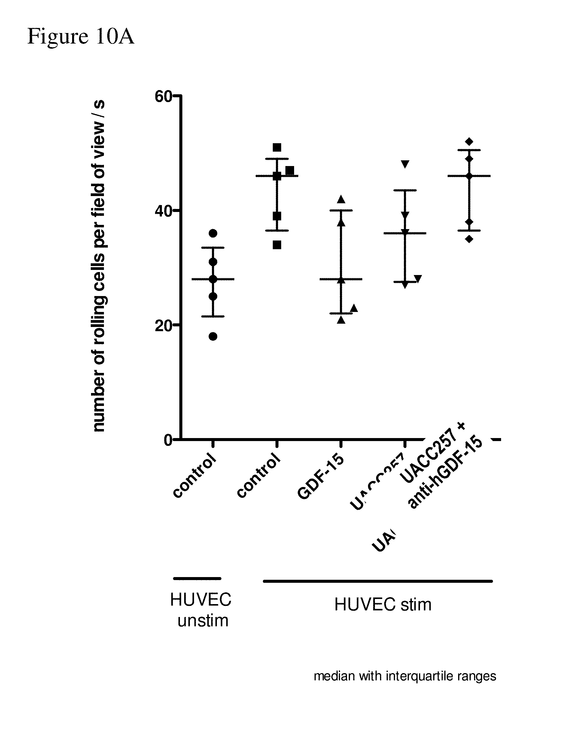
D00015

D00016

D00017

D00018

D00019

D00020

D00021

S00001
XML
uspto.report is an independent third-party trademark research tool that is not affiliated, endorsed, or sponsored by the United States Patent and Trademark Office (USPTO) or any other governmental organization. The information provided by uspto.report is based on publicly available data at the time of writing and is intended for informational purposes only.
While we strive to provide accurate and up-to-date information, we do not guarantee the accuracy, completeness, reliability, or suitability of the information displayed on this site. The use of this site is at your own risk. Any reliance you place on such information is therefore strictly at your own risk.
All official trademark data, including owner information, should be verified by visiting the official USPTO website at www.uspto.gov. This site is not intended to replace professional legal advice and should not be used as a substitute for consulting with a legal professional who is knowledgeable about trademark law.