Cdc-derived Exosomes For Treatment Of Ventricular Tachyarrythmias
Marban; Eduardo ; et al.
U.S. patent application number 16/305011 was filed with the patent office on 2019-05-30 for cdc-derived exosomes for treatment of ventricular tachyarrythmias. This patent application is currently assigned to Cedars-Sinai Medical Center. The applicant listed for this patent is Cedars-Sinai Medical Center. Invention is credited to Eugenio Cingolani, James Dawkins, Eduardo Marban.
| Application Number | 20190160111 16/305011 |
| Document ID | / |
| Family ID | 60479078 |
| Filed Date | 2019-05-30 |


View All Diagrams
| United States Patent Application | 20190160111 |
| Kind Code | A1 |
| Marban; Eduardo ; et al. | May 30, 2019 |
CDC-DERIVED EXOSOMES FOR TREATMENT OF VENTRICULAR TACHYARRYTHMIAS
Abstract
Described herein are compositions and methods related to use of exosomes, including cardiosphere derived cell (CDC)-derived exosomes for treatment and prevention of heart related disease and conditions, such as ventral arrhythmias, such as tachycardias. CDC-derived exosomes delivered by endocardial injection can diminish the total amount of isolated late potentials associated with an isthmus of slow conduction, while reducing the isoelectric interval between late abnormal ventricular activity and decreasing the incidence of inducible ventricular arrhythmias, thereby providing a biological treatment for arrhythmias which otherwise requires therapeutic interventions with adverse effects.
| Inventors: | Marban; Eduardo; (Santa Monica, CA) ; Cingolani; Eugenio; (Marina Del Rey, CA) ; Dawkins; James; (Los Angeles, CA) | ||||||||||
| Applicant: |
|
||||||||||
|---|---|---|---|---|---|---|---|---|---|---|---|
| Assignee: | Cedars-Sinai Medical Center Los Angeles CA |
||||||||||
| Family ID: | 60479078 | ||||||||||
| Appl. No.: | 16/305011 | ||||||||||
| Filed: | June 2, 2017 | ||||||||||
| PCT Filed: | June 2, 2017 | ||||||||||
| PCT NO: | PCT/US2017/035846 | ||||||||||
| 371 Date: | November 27, 2018 |
Related U.S. Patent Documents
| Application Number | Filing Date | Patent Number | ||
|---|---|---|---|---|
| 62345694 | Jun 3, 2016 | |||
| 62504805 | May 11, 2017 | |||
| Current U.S. Class: | 1/1 |
| Current CPC Class: | A61K 35/12 20130101; A61N 1/3956 20130101; A61P 9/06 20180101; A61P 9/10 20180101; A61K 35/34 20130101; A61K 9/0019 20130101 |
| International Class: | A61K 35/34 20060101 A61K035/34; A61P 9/06 20060101 A61P009/06; A61K 9/00 20060101 A61K009/00; A61P 9/10 20060101 A61P009/10 |
Goverment Interests
GOVERNMENT SUPPORT
[0002] This invention was made with government support under R01 HL124074 awarded by the National Institutes of Health. The government has certain rights in the invention.
Claims
1. A method of treating a cardiac arrhythmia, comprising: administering a therapeutically effective amount of a composition comprising extracellular vesicles to a subject afflicted with a cardiac arrhythmia, thereby treating the subject.
2. The method of claim 1, wherein the subject has had a myocardial infarction.
3. The method of claim 1, wherein the subject has an implantable cardioverter-defibrillator (ICD).
4. The method of claim 1, wherein the subject was treated with initial antiarrhythmic drug (AAD) therapy.
5. The method of claim 1, wherein the subject was treated with escalating antiarrhythmic drug (AAD) therapy.
6. The method of claim 1, wherein administering a composition comprises focal delivery at a site of isolated late potentials.
7. The method of claim 1, wherein treating the subject comprises a reduction in the number of isolated late potentials.
8. The method of claim 1, wherein treating the subject comprises a reduction in the isoelectric interval between late abnormal ventricular activity.
9. The method of claim 1, wherein treating the subject comprises a decrease the incidence of inducible ventricular arrhythmias.
10. The method of claim 1, wherein the cardiac arrhythmia comprises ventricular tachycardia.
11. The method of claim 1, wherein the extracellular vesicles are obtained from cardiospheres, cardiosphere-derived cells (CDCs) or newt A1 cell line.
12. A method of improving cardiac performance in a subject, comprising: administering a composition comprising extracellular vesicles to a subject.
13. The method of claim 12, wherein the subject is afflicted with abnormal electrical activity in the heart.
14. The method of claim 12, wherein the subject is afflicted with slow zones of conduction in the heart.
15. The method of claim 12, wherein the subject has heterogeneous areas of scarred myocardium.
16. The method of claim 12, wherein the subject has had a myocardial infarction.
17. The method of claim 12, wherein the subject has an implantable cardioverter-defibrillator (ICD).
18. The method of claim 12, wherein administering a composition comprises focal delivery at a site of isolated late potentials.
19. A method of preventing arrhythmias in a subject comprising: administering a composition comprising extracellular vesicles to a subject.
20. The method of claim 19, preventing arrhythmias in a subject comprises a reduction in the number of isolated late potentials.
21. The method of claim 19, wherein preventing arrhythmias comprises a reduction in the isoelectric interval between late abnormal ventricular activity.
Description
CROSS REFERENCE TO RELATED APPLICATIONS
[0001] This application is the U.S. National Phase of International Application PCT/US2017/035846, filed Jun. 2, 2017, which claims the benefit of priority to U.S. Provisional Application Nos. 62/345,694 and 62/504,805, filed Jun. 3, 2016 and May 11, 2017, respectively. The disclosures of each of the foregoing applications are hereby incorporated by reference in their entireties.
FIELD OF THE INVENTION
[0003] Described herein are methods and compositions related to exosomes, including extracellular vesicles for treatment and prevention of heart related disease and conditions, such as ventral arrhythmias, such as tachycardias.
BACKGROUND
[0004] Acute myocardial infarction (AMI) is experienced by more than 800,000 people in the United States annually and 75% of AMI victims survive 1 year. Approximately 300,000-350,000 of surviving patients die annually due to sudden cardiac death. Post-AMI, residual scarring of heart tissue can lead to ventricular tachycardia, which imparts a substantial risk of death. Ventricular tachycardia (VT) is a ventricular arrhythmia characterized by a fast pulse rate originating below the bundle of his and dissociated from the atria. Ventricular tachycardia may lead to Ventricular fibrillation (VF) which usually causes the person to collapse within seconds, and ends in death within minutes unless prompt corrective measures are instituted (CPR, defibrillation).
[0005] Placement of an implantable cardioverter-defibrillator (ICD) significantly reduces risk of death from VT and are implanted in more than 100,000 patients annually in the United States. Approximately 15% of patients receiving ICDs are initially treated with concomitant antiarrhythmic drug (AAD) therapy. ICDs can be very effective in terminating ventricular tachycardia, but recurrent arrhythmias and ICD shocks may cause impairment in the quality of life and are associated with an increased risk of death, heart failure, and hospitalization. Suppressive therapy, most commonly with AADs can prove problematic as pro-arrythmic, potential long term toxicity and systemic tolerance. Catheter ablation or radio frequency ablation can serve as alternatives to escalation in AAD drug therapy. However, ablation may introduce other adverse effects such as myocardial necrosis, cardiac perforation, bleeding and new arrhythmias. There is a great need in the art to develop therapeutic strategies that can reduce risk from post-MI scarring, VT and VF, and without adverse effects.
[0006] The death of cardiac myocytes is a major cause of myocardial infarct and heart failure, which may be addressed by the potential of cardiac regeneration in adult mammals. Stem cells, such as cardiosphere-derived cells (CDCs) have shown a proven therapeutic benefit by possibly tapping into the aforementioned repair and regeneration mechanisms. In addition, indirect mechanisms are responsible, where cellular exosomes (the lipid bilayer nanovesicles secreted by cells when multivesicular endosomes fuse with the plasma membrane) are central actors in the maintenance, repair and regeneration processes.
[0007] Described herein are compositions and methods related to use of exosomes, including CDC-derived exosomes for treatment and prevention of ventral arrhythmias, including tachycardias. In particular, the Inventors have discovered that extracellular vesicles, such as exosomes secreted from the cardiosphere derived cells (CDCs), are effective in reducing the propensity of the heart to lethal ventricular arrhythmias. The use of CDC-derived exosomes provides a less destructive alternative to radiofrequency ablation or cryoabalation of heart tissue, in patients susceptible to lethal ventricular arrhythmias. Focally injecting exosomes is capable of regrowing healthy heart muscle. Such result terminates the propensity to ventricular arrhythmias in subjects that have suffered a myocardial infarction.
SUMMARY OF THE INVENTION
[0008] Described herein is a method of treating a cardiac arrhythmia, including administering a therapeutically effective amount of a composition including extracellular vesicles to a subject afflicted with a cardiac arrhythmia, thereby treating the subject. In various embodiments, the method includes a subject that had a myocardial infarction. In various embodiments, the method includes subject with an implantable cardioverter-defibrillator (ICD). In various embodiments, the method includes a subject treated with initial antiarrhythmic drug (AAD) therapy. In various embodiments, the method includes a subject treated with escalating antiarrhythmic drug (AAD) therapy. In various embodiments, the method includes administering a composition comprises focal delivery at a site of isolated late potentials. In various embodiments, treating the subject comprises a reduction in the number of isolated late potentials. In various embodiments, treating the subject comprises a reduction in the isoelectric interval between late abnormal ventricular activity. In various embodiments, treating the subject comprises a decrease the incidence of inducible ventricular arrhythmias. In various embodiments, the cardiac arrhythmia comprises ventricular tachycardia. In various embodiments, the extracellular vesicles are obtained from cardiospheres, cardiosphere-derived cells (CDCs) or newt A1 cell line.
[0009] Described herein is a method of improving cardiac performance in a subject, including administering a composition including extracellular vesicles to a subject. In various embodiments, the subject is afflicted with abnormal electrical activity in the heart. In various embodiments, the subject is afflicted with slow zones of conduction in the heart. In various embodiments, the subject has heterogeneous areas of scarred myocardium. In various embodiments, the subject has had a myocardial infarction. In various embodiments, the subject has an implantable cardioverter-defibrillator (ICD). In various embodiments, administering a composition comprises focal delivery at a site of isolated late potentials.
[0010] Also described herein is a method of preventing arrhythmias in a subject including administering a composition comprising extracellular vesicles to a subject. In various embodiments, preventing arrhythmias in a subject comprises a reduction in the number of isolated late potentials. In various embodiments, preventing arrhythmias comprises a reduction in the isoelectric interval between late abnormal ventricular activity.
BRIEF DESCRIPTION OF FIGURES
[0011] FIG. 1. Mechanisms of ventricular tachycardia (VT) are depicted.
[0012] FIG. 2. Electroanatomic Mapping (EAM) demonstrating the presence of isolated late potentials (ILP), slow and fast arms associated with VT.
[0013] FIG. 3. Isolated late potentials and late abnormal ventricular activity as evidencing VTs, late potentials are shown relative to a normal QRS.
[0014] FIG. 4. Conduction during sinus rhythm including timing map and propagation, and voltage is depicted. Late potentials and low voltage areas are indicated.
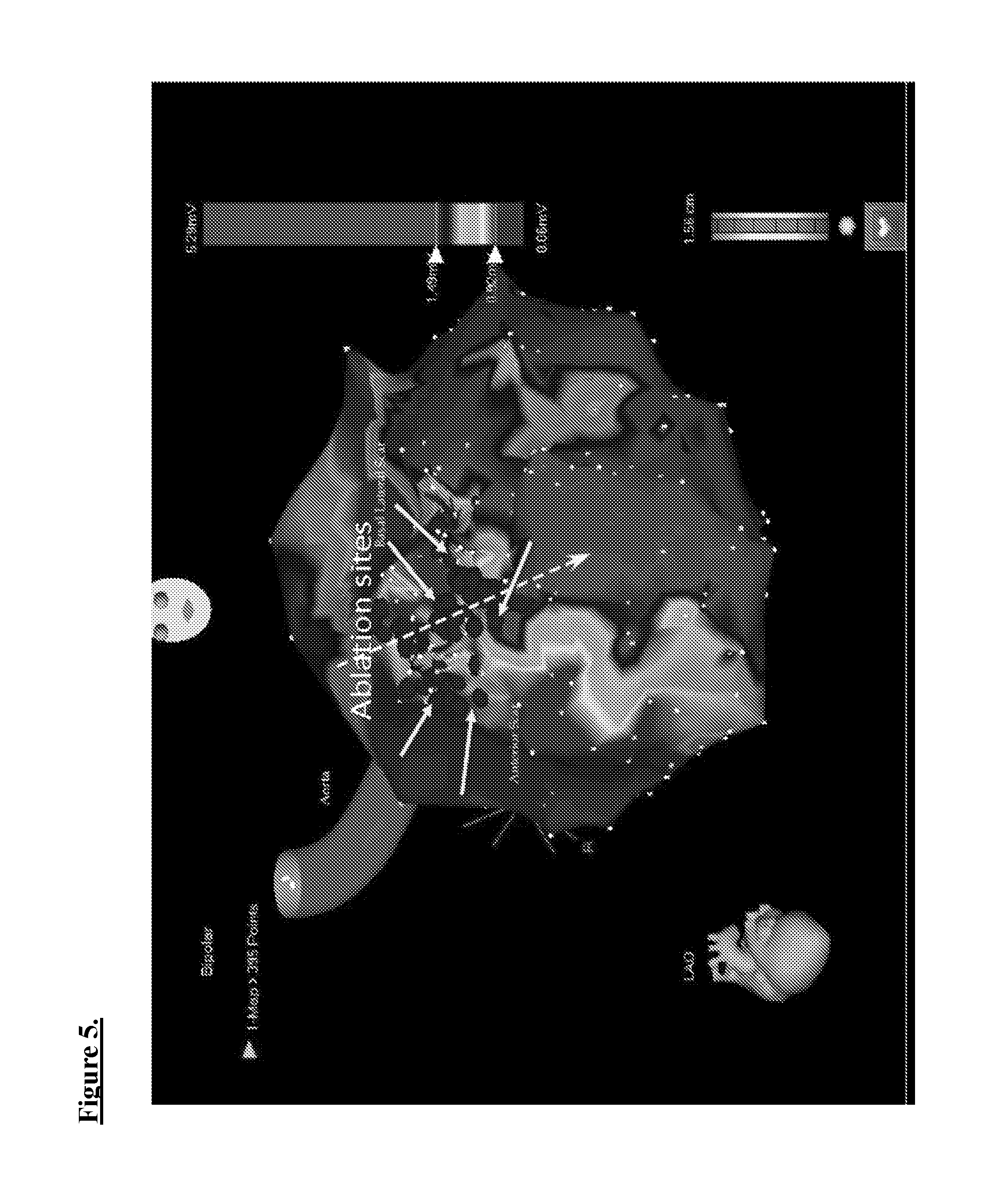
[0015] FIG. 5. Substrate ablation, Areas targeted for ablation are depicted.
[0016] FIG. 6. Study design. Arrthymia inducibility was measured, and electroanatomic mapping (EAM) according to the described timeline.
[0017] FIG. 7. CDC-derived exosomes design. Nanosight particle tracking was used to determine the number of exosomes.
[0018] FIG. 8. Electroanatomic Mapping (EAM). High density mapping and tip positioning as shown, with 64 electrodes, 2.5 mm interelectrode spacing, 0.4 mm2 electrode area). 0.5-1.5 mV identified as scar. <0.5 mV dense, transmural scar.
[0019] FIG. 9. MRI Data (scar). Changes in both full width and mean standard deviation for CDC-derived exosomes (CDCexo) administered, and control are shown. N=5 for CDCexo and control.
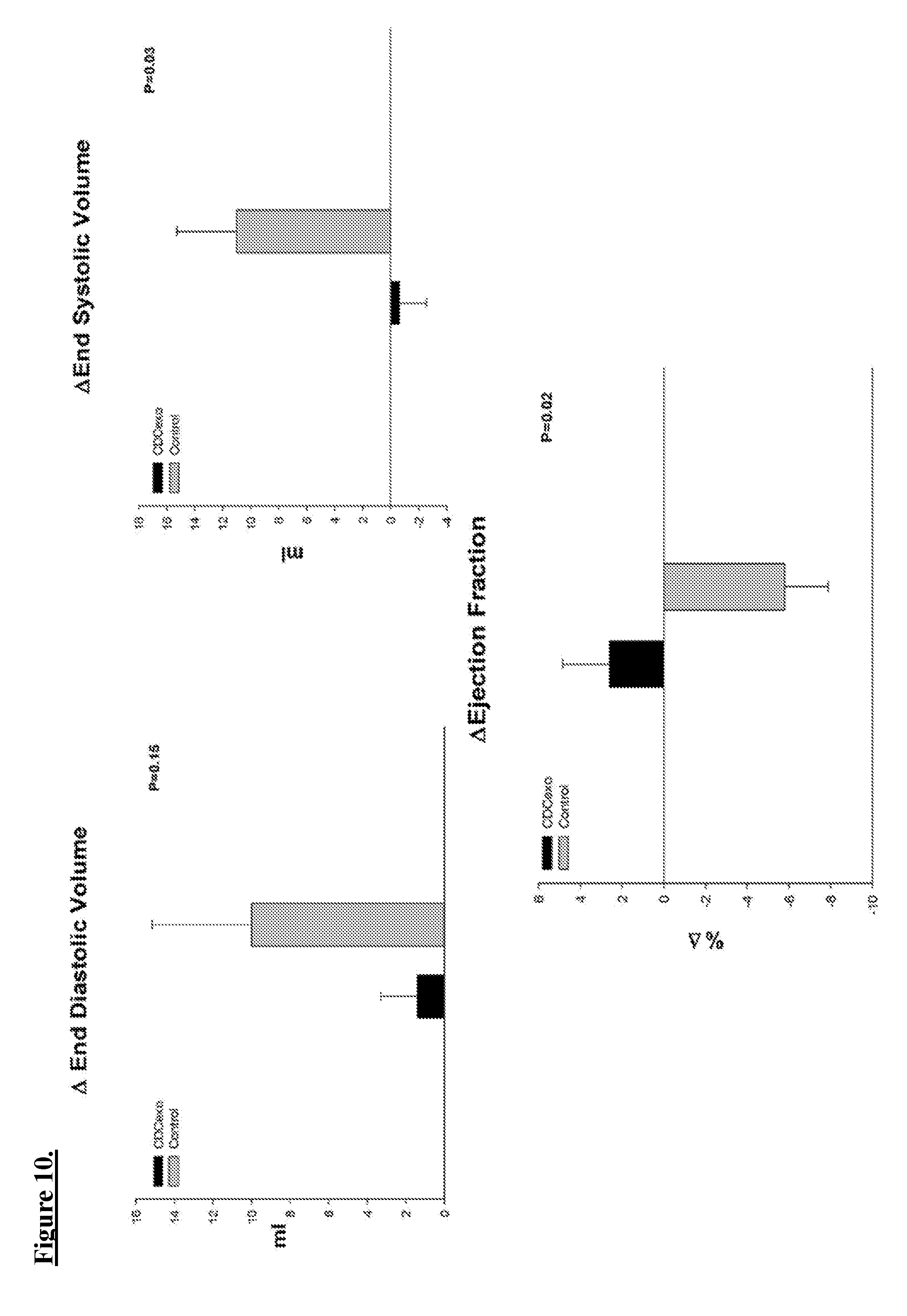
[0020] FIG. 10. MRI Data (function). Changes in end diastolic, systolic and ejection fractions are shown for CDC-derived exosomes (CDCexo) administered, and control are shown. N=5 for CDCexo and control.
[0021] FIG. 11. Arrthymia inducibility. Programmed electrical stimulation (PES) results shown for CDC-derived exosomes administered and control animals.
[0022] FIG. 12. Electroanatomic Mapping (EAM) demonstrating results for change in number and percentage of late potentials as baseline and final measurements for CDC-derived exosomes (CDCexo) administered, and control. N=5 for CDCexo and control.
[0023] FIG. 13. Electroanatomic Mapping (EAM) demonstrating results for change in timing of late potentials and voltage amplitude of fractionated potential for CDC-derived exosomes (CDCexo) administered, and control.
[0024] FIG. 14. Electroanatomic map. Representation of CDC-derived exosomes (CDCexo) administered heart at baseline measurement with septum and lateral wall regions highlighted.
[0025] FIG. 15. Electroanatomic map. Representation of CDC-derived exosomes (CDCexo) administered heart at final measurement.
[0026] FIG. 16. Electroanatomic map. Additional representation of CDC-derived exosomes (CDCexo) administered heart at final measurement.
[0027] FIG. 17. Electroanatomic map. Second Representation of CDC-derived exosomes (CDCexo) administered heart at baseline measurement.
[0028] FIG. 18. Electroanatomic map. Second Representation of CDC-derived exosomes (CDCexo) administered heart at final measurement.
[0029] FIG. 19. Electroanatomic map. Anterosteptal, anterior, anterolateral regions are highlighted.
[0030] FIG. 20. Electroanatomic map. Demonstrating potential measurements in anatomical space.
[0031] FIG. 21. Electroanatomic map.
[0032] FIG. 22. FIG. 22A: Experimental protocol: 17 Yucatan mini pigs had an MI induced by 90-minute balloon occlusion of the proximal 1/3 of the LAD followed by 8 weeks of reperfusion. Cardiac function and scar size was examined by MRI (n=12). Arrhythmia Inducibility was probed by programmed electrical stimulation near the scar border of the LV. If no sustained arrhythmia was induced, PES was repeated at the RV apex. High density 3D electro-anatomic mapping was then performed (Rhythmia, Boston Scientific, Cambridge, Mass.). Inducible animals were then randomly assigned to receive a focal injection of either 7.5 mg of CDC.sub.EXO in 2 mls of IMDM, or 2 mls of IMDM alone. Injections were localized around the arrhythmogenic substrate where late potentials were identified (Rhythmia, Boston Scientific, Cambridge, Mass.), (NOGA, Biosense Webster). MRI, EAM, and PES was repeated 2 weeks later. Animals were then euthanized and the heart was removed en bloc and sectioned for histological analysis (n=5). FIG. 22B, left panel High density EAM (Rhythmia, Boston Scientific, Cambridge, Mass.) of the arrhythmogenic substrate with an identified late potential. FIG. 22B, right panel NOGA (Biosense Webster) guided injection site (Myostar, Biosense Webster) and representative catheter tip-potential.
[0033] FIG. 23. FIG. 23 A, FIG. 23B: Short axis MRI images at end-diastole and end-systole from a vehicle injected control. FIG. 23C, FIG. 23D: A similar short axis view of a pig injected with CDC.sub.EXO. FIG. 23E, FIG. 23F, FIG. 23G: At endpoint, significant improvement and preservation of LV ejection fraction was evident in CDC.sub.EXO pigs while a decrease in EF was seen in the controls, (P=0.01). FIG. 23H: Cardiac output was significantly improved in CDC.sub.EXO pigs relative to controls, P=0.01 FIG. 23I, FIG. 23J: Adverse changes in LV end-diastolic (LVEDV) and end-systolic (LVESV) volumes were observed in the vehicle treated group but not in CDC.sub.EXO pigs (LVESV, P=0.04). Chamber volumes were normalized to body 40.575 surface area (0.121.times.BW.sup..DELTA.0.575).
[0034] FIG. 24. FIG. 24A, FIG. 24B: 4-chamber MRI image of infarcted ventricular myocardium identified by late gadolinium enhancement (LGE). Left) Image pre injection of CDC.sub.EXO with follow up examination on the right. FIG. 24C, FIG. 24D: Representative 3D reconstruction of infarcted left ventricular myocardium pre and post injection with CDC.sub.EXO. FIG. 24E, FIG. 24F, FIG. 24G: Contrasting an increase in scar in control (n=7) animals, there was a significant reduction in scar following focal injection of CDC.sub.EXO (n=7, P=0.009) at endpoint. There was no significant change in LV mass between timepoints in either group (supplement fig #).
[0035] FIG. 25. FIG. 25A, FIG. 25B: Late potential map with a corresponding electrogram tracing from a CDC.sub.EXO treated pig pre- and post-injection. Of note is the isolated activation channel going through the identified substrate suggestive of a potential re-entrant pathway. This early channel is not as evident post treatment. FIG. 25C: There was no change in identifiable late potentials in the control animals following injection however FIG. 25D: there was a significant overall reduction in late potentials in the animals injected with CDC.sub.EXO (P=0.02). The white arrows identify the electrogram tracings pre- and post-treatment with an FIG. 25E: identifiable reduction in timing of the late component of the electrogram post therapy within the same anatomic location (P=0.0004).
[0036] FIG. 26. FIG. 26A, FIG. 26B: Representative electrocardiogram tracings of programmed electrical stimulation (PES) with corresponding extra stimuli in the same animal performed 2 months post MI, and repeated 2 weeks following injection with left) vehicle alone or right) CDC.sub.EXO. 7 inducible pigs allocated to the CDC.sub.EXO group displayed sustained ventricular arrhythmias at baseline, however only 1 pig was inducible at endpoint, demonstrating an 87.5% reduction in sustained inducible arrhythmias (P=0.015 Fisher's exact test). There was no change in arrhythmia Inducibility in the pigs injected with vehicle only. 2 of the control pigs were not inducible at baseline but were inducible at endpoint.
[0037] FIG. 27. FIG. 27A: Ex vivo LGE images of a control vs. a CDC.sub.EXO pig acquired at the same slice position. Note the moth-eaten pattern of gadolinium enhancement identifiable in the pig injected with CDC.sub.EXO whereas there is a transmural gadolinium pattern in the vehicle injected control. FIG. 27B, FIG. 27C: Picrosirius red stained sections of the infarct zone within the left ventricular myocardium revealed significantly reduced areas of fibrosis not identified in border and remote zones in CDC.sub.EXO injected pigs (P=0.02).
[0038] FIG. 28. Following euthanasia, hearts were sectioned into 1 cm slices from apex to base. The second most apical slices were sectioned into 3 anatomical zones including the antero-septal infarct (IZ) and border (BZ) zones, as well a remote zones (RZ) acquired from the posterior wall. Paraffin embedded sections from the IZ, BZ, and RZ were then cut into ten 8 um sections from base to apex stained for DAPI (blue), WGA (green), .alpha.-SA (red), and Ki67 (magenta). Around the densest areas of injection there was a significant number of cells positive for both Ki67 and .alpha.-SA within the infarct zone of CDC.sub.EXO injected pigs compared to controls. Representative images from the infarct zone of a control animal (FIG. 28A) 0.95.+-.0.21 cells/field (.mu.m.sup.2) n=2 and an animal injected with CDC.sub.EXO (FIG. 28B) 3.47.+-.0.19 cells/field (.mu.m.sup.2), (n=3; P=0.0036). (FIG. 28C) Border zone images of a control animal 1.04.+-.0.06 cells/field (.mu.m.sup.2), and (FIG. 28D) an animal injected with CDC.sub.EXO 1.5.+-.0.11 cells/field (.mu.m.sup.2) (P=NS). Remote zone images from a control animal (FIG. 28E) 1.35.+-.0.09% cells/field (.mu.m.sup.2), and an animal injected with CDC.sub.EXO (FIG. 29F) 1.62.+-.0.10 cells/field (.mu.m.sup.2) (P=NS).
[0039] FIG. 29. Simulations with 3D computational models of a porcine heart before and after CDC.sub.EXO treatment. FIG. 29A: Reconstructed 3D models of ventricles before (top) and after (bottom) CDC.sub.EXO treatment. CDC-exosomes were injected in the inferior, anterior septum as outlined by the dotted green circle. Shown are scar and gray zone (GZ) in a transparent view of the ventricles (left) and in LV cutaways (right). FIG. 29B: Mechanisms demonstrating the conversion of the arrhythmogenic ventricular substrate into non-arrhythmogenic following CDC.sub.EXO injection. Transmembrane potential maps of ventricles before (top) and after (bottom) CDC.sub.EXO treatment are shown at three time points. The model ventricles were paced from the right ventricular outflow tract (star). The time instant below each map is counted from the delivery of last pacing stimulus. White arrows indicate direction of electrical propagation.
DETAILED DESCRIPTION
[0040] All references cited herein are incorporated by reference in their entirety as though fully set forth. Unless defined otherwise, technical and scientific terms used herein have the same meaning as commonly understood by one of ordinary skill in the art to which this invention belongs. Allen et al., Remington: The Science and Practice of Pharmacy 22.sup.nd ed., Pharmaceutical Press (Sep. 15, 2012); Hornyak et al., Introduction to Nanoscience and Nanotechnology, CRC Press (2008); Singleton and Sainsbury, Dictionary of Microbiology and Molecular Biology 3.sup.rd ed., revised ed., J. Wiley & Sons (New York, N.Y. 2006); Smith, March's Advanced Organic Chemistry Reactions, Mechanisms and Structure 7.sup.th ed., J. Wiley & Sons (New York, N.Y. 2013); Singleton, Dictionary of DNA and Genome Technology 3.sup.rd ed., Wiley-Blackwell (Nov. 28, 2012); and Green and Sambrook, Molecular Cloning: A Laboratory Manual 4th ed., Cold Spring Harbor Laboratory Press (Cold Spring Harbor, N.Y. 2012), provide one skilled in the art with a general guide to many of the terms used in the present application. For references on how to prepare antibodies, see Greenfield, Antibodies A Laboratory Manual 2.sup.nd ed., Cold Spring Harbor Press (Cold Spring Harbor N.Y., 2013); Kohler and Milstein, Derivation of specific antibody-producing tissue culture and tumor lines by cell fusion, Eur. J. Immunol. 1976 July, 6(7):511-9; Queen and Selick, Humanized immunoglobulins, U.S. Pat. No. 5,585,089 (1996 December); and Riechmann et al., Reshaping human antibodies for therapy, Nature 1988 Mar. 24, 332(6162):323-7.
[0041] One skilled in the art will recognize many methods and materials similar or equivalent to those described herein, which could be used in the practice of the present invention. Indeed, the present invention is in no way limited to the methods and materials described. For purposes of the present invention, the following terms are defined below.
[0042] As used in the description herein and throughout the claims that follow, the meaning of "a," "an," and "the" includes plural reference unless the context clearly dictates otherwise. Also, as used in the description herein, the meaning of "in" includes "in" and "on" unless the context clearly dictates otherwise.
[0043] As described, after acute myocardial infarct, residual scarring of heart tissue can lead to ventricular tachycardia (VT), which imparts a substantial risk of death. As a ventricular arrhythmia, VT is characterized by a fast pulse rate originating below the bundle of his and dissociated from the atria. Current understanding of these mechanisms has resulted in 3 classification (FIG. 1). First, hypoxic cardiomyocytes may become exhibit abnormal automaticity and act as pacemaker cells. Second, triggered activity associated with afterdeploarizations (EAD, DAD). Third, reentrant ventricular tachycardia (VT) is the most common sustained arrhythmia leading to ventricular fibrillation (VF) post MI (FIG. 2). Reentrant VT in post-infarction cardiomyopathy depends on an isthmus of slow conduction near the border or within the infarct that is electrically isolated from the rest of the myocardium. In serious cases, electrical storm, an increasingly common and life-threatening emergency, is characterized by 3 or more sustained VT or VF episodes or appropriate ICD shocks within 24 hours, and despite drawbacks, early stage intervention radiofrequency ablation has been increasingly deployed as therapeutic intervention or prophylactically (including with ICD implantation).
[0044] Previous studies have demonstrated that these corridors of slow conduction within the scar provide the substrate for reentrant VT and often contain isolated late potentials (ILPs) (FIG. 3). VF is associated with a reentrant mechanism. These VT hallmarks of isolated late potentials and late abnormal ventricular activity are associated with bundles of viable myofibers within a zone of slow conduction, fractionated late activity during mid-diastole commonly associated with scar, occurrence when a single wavefront is split by a unidirectional block and normal QRS: 45-55 ms. In ischemic or nonischemic cardiomyopathy, the vulnerable substrate for reentry lies within heterogeneous areas of scarred myocardium. After an acute MI, or as nonischemic cardiomyopathy progresses, structural changes in the heart can lead to scar formation that creates areas of conduction block. However, surviving bundles of exist around the border of a scar. Slow conduction through these regions provides a pathway for electrically stable reentry. Otherwise harmless triggers, such as premature ventricular depolarization, is all that is required to initiate VT.
[0045] As described, current therapies include antiarrhythmic drugs (AAD) drugs such as Amiodarone, Lidocaine, Procainamide etc. Other approaches rely on RF Ablation, including targeted ablation (inducible/stable patients), substrate modification-(non-inducible/unstable patients) 3D mapping and identification of late potentials and area of slow conduction. When subjects have proven refractory to AAD therapy, radio frequency (RF) catheter ablation has been reported as more effective than escalated AAD therapy in reducing the rate of the combined outcome of death at any time or ventricular tachycardia storm or ICD shocks after 30 days. RF ablation has been increasingly deployed as therapeutic intervention or prophylactically (including with ICD implantation). This includes in serious conditions such as electrical storm, characterized by 3 or more sustained VT or VF episodes or appropriate ICD shocks within 24 hours. Many adverse effects are associated with the aforementioned techniques.
Cardiosphere-Derived Cells (CDCs)
[0046] CDCs are a population of cells generated by manipulating cardiospheres, cultured cells initially obtained from heart sample biopsies, subsequently cultured as explants and suspension cultured cardiospheres. For example, CDCs can be generated by plating cardiospheres on a solid surface which is coated with a substance which encourages adherence of cells to a solid surface of a culture vessel, e.g., fibronectin, a hydrogel, a polymer, laminin, serum, collagen, gelatin, or poly-D-lysine, and expanding same as an adherent monolayer culture. CDCs can be repeatedly passaged, e.g., passaged two times or more, according to standard cell culturing methods.
Extracellular Vesicles
[0047] Extracellular vesicles include lipid bilayer structures generated by cells, and include exosomes, microvesicles, membrane particles, membrane vesicles, exosome-like vesicles, ectosomes, ectosome-like vesicles, or exovesicles. Exosomes are vesicles formed via a specific intracellular pathway involving multivesicular bodies or endosomal-related regions of the plasma membrane of a cell. Exosomes can range in size from approximately 20-150 nm in diameter. In some cases, they have a characteristic buoyant density of approximately 1.1-1.2 g/mL, and a characteristic lipid composition. Their lipid membrane is typically rich in cholesterol and contains sphingomyelin, ceramide, lipid rafts and exposed phosphatidylserine. Exosomes express certain marker proteins, such as integrins and cell adhesion molecules, but generally lack markers of lysosomes, mitochondria, or caveolae. In some embodiments, the exosomes contain cell-derived components, such as but not limited to, proteins, DNA and RNA (e.g., microRNA and noncoding RNA). In some embodiments, exosomes can be obtained from cells obtained from a source that is allogeneic, autologous, xenogeneic, or syngeneic with respect to the recipient of the exosomes.
[0048] Certain types of RNA, e.g., microRNA (miRNA), are known to be carried by exosomes. miRNAs function as post-transcriptional regulators, often through binding to complementary sequences on target messenger RNA transcripts (mRNAs), thereby resulting in translational repression, target mRNA degradation and/or gene silencing. For example, miR146a exhibits over a 250-fold increased expression in CDCs, and miR210 is unregulated approximately 30-fold, as compared to the exosomes isolated from normal human dermal fibroblasts.
[0049] Methods for preparing exosomes can include the steps of: culturing cardiospheres or CDCs in conditioned media, isolating the cells from the conditioned media, purifying the exosome by, e.g., sequential centrifugation, and optionally, clarifying the exosomes on a density gradient, e.g., sucrose density gradient. In some instances, the isolated and purified exosomes are essentially free of non-exosome components, such as components of cardiospheres or CDCs. Exosomes can be resuspended in a buffer such as a sterile PBS buffer containing 0.01-1% human serum albumin. The exosomes may be frozen and stored for future use.
[0050] Extracellular vesicles originating from newt A1 cell line (Newt-EVs) are obtained after filtering A1 cell line CM containing EVs through a 10 KDa pore size filter following a similar process as for CDC-EV production. Newt-EVs are a non-cellular, filter sterilized product obtained from newt A1 cells cultured under defined, serum-free conditions. The final product, composed of secreted EVs and concentrated CM, is formulated in PlasmaLyte A and stored frozen. The frozen final product is ready to use for direct subconjunctival injection after thawing.
[0051] Exosomes can be prepared using a commercial kit such as, but not limited to the ExoSpin.TM. Exosome Purification Kit, Invitrogen.RTM. Total Exosome Purification Kit, PureExo.RTM. Exosome Isolation Kit, and ExoCap.TM. Exosome Isolation kit. Methods for isolating exosome from stem cells are found in, e.g., Tan et al., Journal of Extracellular Vesicles, 2:22614 (2013); Ono et al., Sci Signal, 7(332):ra63 (2014) and methods for isolating exosome from cardiosphere-derived cells are found in, e.g., Ibrahim et al., Stem Cell Reports, 2:606-619 (2014), each of which is incorporated by reference herein. Collected exosomes can be concentrated and/or purified using methods known in the art. Specific methodologies include ultracentrifugation, density gradient, HPLC, adherence to substrate based on affinity, or filtration based on size exclusion.
[0052] For example, differential ultracentrifugation has become a leading technique wherein secreted exosomes are isolated from the supernatants of cultured cells. This approach allows for separation of exosomes from nonmembranous particles, by exploiting their relatively low buoyant density. Size exclusion allows for their separation from biochemically similar, but biophysically different microvesicles, which possess larger diameters of up to 1,000 nm. Differences in flotation velocity further allows for separation of differentially sized exosomes. In general, exosome sizes will possess a diameter ranging from 30-200 nm, including sizes of 40-100 nm. Further purification may rely on specific properties of the particular exosomes of interest. This includes, e.g., use of immunoadsorption with a protein of interest to select specific vesicles with exoplasmic or outward orientations.
[0053] Among current methods, e.g., differential centrifugation, discontinuous density gradients, immunoaffinity, ultrafiltration and high performance liquid chromatography (HPLC), differential ultracentrifugation is the most commonly used for exosome isolation. This technique utilizes increasing centrifugal force from 2000.times.g to 10,000.times.g to separate the medium- and larger-sized particles and cell debris from the exosome pellet at 100,000.times.g. Centrifugation alone allows for significant separation/collection of exosomes from a conditioned medium, although it is insufficient to remove various protein aggregates, genetic materials, particulates from media and cell debris that are common contaminants. Enhanced specificity of exosome purification may deploy sequential centrifugation in combination with ultrafiltration, or equilibrium density gradient centrifugation in a sucrose density gradient, to provide for the greater purity of the exosome preparation (flotation density 1.1-1.2 g/mL) or application of a discrete sugar cushion in preparation.
[0054] Importantly, ultrafiltration can be used to purify exosomes without compromising their biological activity. Membranes with different pore sizes--such as 100 kDa molecular weight cut-off (MWCO) and gel filtration to eliminate smaller particles--have been used to avoid the use of a nonneutral pH or non-physiological salt concentration. Currently available tangential flow filtration (TFF) systems are scalable (to >10,000 L), allowing one to not only purify, but concentrate the exosome fractions, and such approaches are less time consuming than differential centrifugation. HPLC can also be used to purify exosomes to homogeneouslysized particles and preserve their biological activity as the preparation is maintained at a physiological pH and salt concentration.
[0055] Other chemical methods have exploited differential solubility of exosomes for precipitation techniques, addition to volume-excluding polymers (e.g., polyethylene glycols (PEGs)), possibly combined additional rounds of centrifugation or filtration. For example, a precipitation reagent, ExoQuick.RTM., can be added to conditioned cell media to quickly and rapidly precipitate a population of exosomes, although re-suspension of pellets prepared via this technique may be difficult. Flow field-flow fractionation (FlFFF) is an elution-based technique that is used to separate and characterize macromolecules (e.g., proteins) and nano- to micro-sized particles (e.g., organelles and cells) and which has been successfully applied to fractionate exosomes from culture media.
[0056] Beyond these techniques relying on general biochemical and biophysical features, focused techniques may be applied to isolate specific exosomes of interest. This includes relying on antibody immunoaffinity to recognizing certain exosome-associated antigens. As described, exosomes further express the extracellular domain of membrane-bound receptors at the surface of the membrane. This presents a ripe opportunity for isolating and segregating exosomes in connections with their parental cellular origin, based on a shared antigenic profile. Conjugation to magnetic beads, chromatography matrices, plates or microfluidic devices allows isolating of specific exosome populations of interest as may be related to their production from a parent cell of interest or associated cellular regulatory state. Other affinity-capture methods use lectins which bind to specific saccharide residues on the exosome surface.
[0057] Described herein are compositions and methods providing significant benefits in the treatment of abnormal cardiac electrical activity or cardiac arrhythmia, including repair or regeneration of damaged or diseased tissues using extracellular vesicles, including exosomes such as CDC-derived exosomes and newt A1 cell line exosomes. Certain supporting techniques are described in, for example, U.S. application Ser. Nos. 11/666,685, 12/622,143, 12/622,106, 14/421,355, PCT App. No. PCT/US2013/054732, PCT/US2015/053853, PCT/US2015/054301 and PCT/US2016/035561, which are fully incorporated by reference herein.
[0058] Described herein is a method of treatment for a heart related disease and/or condition. In various embodiments, the heart related disease and/or condition includes abnormal cardiac electrical activity or cardiac arrhythmia. In various embodiments, the method is for treating abnormal cardiac electrical activity, including administering a composition including extracellular vesicles to a subject, thereby treating the subject. In various embodiments, the method is for treating a cardiac arrhythmia, including administering a composition including extracellular vesicles to a subject, thereby treating the subject. In various embodiments, the method of treatment includes, selecting a subject in need of treatment, administering a composition including extracellular vesicles to the individual, wherein administration of the composition treats the subject. In various embodiments, the subject is afflicted with incessant ventricular tachycardia (VT). In various embodiments, the subject is afflicted with ischaemic heart disease and recurrent implantable cardioverter-defibrillator (ICD) shocks. In various embodiments, the subject has experienced a first episode of sustained VT, afflicted with ischaemic heart disease, without or without an implanted ICD. In various embodiments, the subject is afflicted with electrical storm, including electrical storm arising from ventricular arrhythmias such as VT, ventricular fibrillation (VF), or appropriate ICD shocks. In various embodiments, the subject is afflicted with recurrent electrical storm, including electrical storm arising from ventricular arrhythmias such as VT, ventricular fibrillation (VF), or appropriate implantable cardioverter-defibrillator (ICD) shocks.
[0059] In various embodiments, the cardiac arrhythmia includes extra beats, supraventricular tachycardias, ventricular arrhythmias, and bradyarrhythmias. In various embodiments, the cardiac arrhythmia includes premature atrial contractions and premature ventricular contractions. In various embodiments, the cardiac arrhythmia includes atrial fibrillation, atrial flutter, and paroxysmal supraventricular tachycardia. In various embodiments, the cardiac arrhythmia includes ventricular fibrillation and ventricular tachycardia. In various embodiments, ventricular tachycardia (VT) is monomorphic VT or polymorphic VT. In various embodiments, monomorphic VT is characterized by ventricular activation sequence without any variation in the QRS complexes. In various embodiments, polymorphic VT is characterized by beat-to-beat variations in the QRS complexes.
[0060] In various embodiments, the abnormal cardiac electrical includes sick sinus syndrome, sinus bradycardia, tachycardia-bradycardia syndrome, atrial fibrillation, atrioventricular block, chronotropic incompetence, prolonged QT syndrome, and heart failure.
[0061] In various embodiments, the subject has had a myocardial infarction. In various embodiments, the subject is post-myocardial infarct. In various embodiments, the subject is afflicted with abnormal cardiac electrical activity or cardiac arrhythmia. In various embodiments, the subject was treated with initial antiarrhythmic drug (AAD) therapy. In various embodiments, the subject was treated with escalating AAD therapy. In various embodiments, the method is administered concurrently, or sequential to initial and/or escalating AAD therapy. In various embodiments, AAD therapeutic agents include Amiodarone, Lidocaine, Procainamide, among others. In various embodiments, the subject is refractory to AAD therapy.
[0062] In various embodiments, administration of extracellular vesicles, including exosomes, includes focal delivery at a site of isolated late potentials, isthmus and/or slow zones of conduction. In various embodiments, a site of isolated potential (i.e. arrhythmogenic substrate), isthmus and/or slow zones of conduction has been identified by electrical anatomic mapping. In various embodiments, a 12-lead electrocardiogram has identified a region of interest for electrical anatomic mapping. In various embodiments, administration includes injection in the intra and peri-infarct zone of the left ventricle. In various embodiments, this includes, 3, 4, 5, 6, 7, 8, 9, 10 or more injection at the aforementioned sites. In various embodiments, administration of extracellular vesicles, including exosomes, to the subject occurs through any of known techniques in the art. In some embodiments, this includes percutaneous delivery and/or injection into heart muscle. Additional delivery sites include any one or more compartments of the heart, such as myocardium, associated arterial, venous, and/or ventricular locations. In certain embodiments, administration can include delivery to a tissue or organ site that is the same as the site of diseased and/or dysfunctional tissue. In certain embodiments, administration can include delivery to a tissue or organ site that is different from the site or diseased and/or dysfunctional tissue.
[0063] In various embodiments, the extracellular vesicles are exosomes, microvesicles, membrane particles, membrane vesicles, exosome-like vesicles, ectosomes, ectosome-like vesicles, or exovesicles. In various embodiments, the exosomes are CDC-derived exosomes or newt A1 cell line derived exosomes. In other embodiments, the exosomes include one or more microRNAs. In various embodiments, these microRNAs can include miR-146a, miR22, miR-24, miR-210, miR-150, miR-140-3p, miR-19a, miR-27b, miR-19b, miR-27a, miR-376c, miR-128, miR-320a, miR-143, miR-21, miR-130a, miR-9, miR-185, and/or miR-23a. In several embodiments, the exosomes includes one or more exosomes enriched in at least one of miR-146a, miR-22, miR-24. In other embodiments, the exosomes can include one or more exosomes containing microRNAs. This includes various microRNAs known in the art, such as miR-1469, miR-762, miR-574-3p, miR-574-5p, miR-3197, miR-4281, miR-1976, miR-1307, miR-1224-3p, miR-187, miR-3141, miR-1268, miR-155, miR-122, miR-638, miR-3196, miR-223, miR-4267, miR-1281, miR-885-5p, miR-663, miR-let-7b, miR-29d, miR-144, miR-let-7e 143, miR-lrt-7g, miR-17a, miR-125a-5p, miR-128, miR-720, miR-21, miR-30c, miR-30b, miR-lb.
[0064] In various embodiments, administration of the extracellular vesicles includes administration of a therapeutically effective amount of the extracellular vesicles. In various embodiments, a therapeutically effective amount include an amount capable of altering gene expression in damaged or dysfunctional tissue, improves viability of the damaged tissue, and/or enhances regeneration or production of new tissue in the individual. In various embodiments, the quantities of extracellular vesicles, including exosomes, that are administered to achieved these effects range from 1.times.10.sup.6 to 1.times.10.sup.7, 1.times.10.sup.7 to 1.times.10.sup.8, 1.times.10.sup.8 to 1.times.10.sup.9, 1.times.10.sup.9 to 1.times.10.sup.10, 1.times.10.sup.10 to 1.times.10.sup.11, 1.times.10.sup.11 to 1.times.10.sup.12, 1.times.10.sup.12 or more. In other embodiments, the numbers of exosomes is relative to the number of cells used in a clinically relevant dose for a cell-therapy method. For example, it has been demonstrated that 3 mL/3.times.10.sup.5 human cardiac-derived cells (CDCs), is capable of providing therapeutic benefit in intracoronary administration, and therefore, a quantity of extracellular vesicles, including exosomes, as derived from that number of cells in a clinically relevant dose for a cell-therapy method. In various embodiments, administration can be in repeated doses. For example, defining an effective dose range, dosing regimen and route of administration, may be guided by studies using fluorescently labeled exosomes, and measuring target tissue retention, which can be >10.times., >50.times., or >100.times. background, as measured 5, 10, 15, 30, or 30 or more min as a screening criterion. In certain embodiments, >100.times. background measured at 30 mins is a baseline measurement for a low and high dose that is then assessed for safety and bioactivity (e.g., using MRI endpoints: scar size, global and regional function). In various embodiments, single doses are compared to two, three, four, four or more sequentially-applied doses. In various embodiments, the repeated or sequentially-applied doses are provided for treatment of an acute disease and/or condition. In various embodiments, the repeated or sequentially-applied doses are provided for treatment of a chronic disease and/or condition.
[0065] In some embodiments, the method of treatment results in a reduction in scar mass, reduction in the formation of scar mass, improvements in ejection fraction, reductions in diastolic volume and systolic volume. In other embodiments, this includes a reduction in the number, timing and magnitude of late potentials, or reduction in inducibility of potentials. For example, this includes reducing the isoelectric interval between late abnormal ventricular activity, and decreasing the incidence of inducible ventricular arrhythmias. In other embodiments, the method of treatment results in increases in viable tissue, reduction in scar mass, improvements in wall thickness, regenerative remodeling of injury sites, enhanced angiogenesis, improvements in cardiomyogenic effects, reduction in apoptosis, reduction in fibrosis, and/or decrease in levels of pro-inflammatory cytokines. In various embodiments, the methods of treatment results in reduction of slow conduction zones. In various embodiments, the method of treatment includes assessing one or more of the aforementioned electrophysiological properties.
[0066] In various embodiments, the damaged or dysfunctional tissue is in need of repair, regeneration, or improved function due to an acute event. Acute events include, but are not limited to, trauma such as laceration, crush or impact injury, shock, loss of blood or oxygen flow, infection, chemical or heat exposure, poison or venom exposure, drug overuse or overexposure, and the like. In certain embodiments, the damaged tissue is pulmonary, arterial or capillary tissue, such as the endothelial lining of distal pulmonary arteries. In other embodiments, the damaged tissue is cardiac tissue and the acute event includes a myocardial infarction. In some embodiments, administration of the exosomes results in an increase in cardiac wall thickness in the area subjected to the infarction.
[0067] In other embodiments, damaged or dysfunctional tissue is due to chronic disease, such as for example congestive heart failure, including as conditions secondary to diseases such as emphysema, ischemic heart disease, hypertension, valvular heart disease, connective tissue diseases, HIV infection, liver disease, sickle cell disease, dilated cardiomyopathy, infection such as Schistosomiasis, diabetes, and the like. In various embodiments, the administration can be in repeated doses, such as two, three, four, four or more sequentially-applied doses. In various embodiments, the repeated or sequentially-applied doses are provided for treatment of an acute disease and/or condition. In various embodiments, the repeated or sequentially-applied doses are provided for treatment of a chronic disease and/or condition.
[0068] Described herein is a method of preventing a heart related disease and/or condition. In various embodiments, the heart related disease and/or condition includes abnormal cardiac electrical activity or cardiac arrhythmia. In various embodiments, the method of prevention is for abnormal cardiac electrical activity, includes administering a composition including extracellular vesicles to a subject, thereby preventing abnormal cardiac electrical activity in the subject. In various embodiments, the method of prevention is for a cardiac arrhythmia, including administering a composition including extracellular vesicles to a subject, thereby preventing cardiac arrhythmia in the subject. In various embodiments, administration of extracellular vesicles, including exosomes, includes focal delivery at a site of isolated late potentials (i.e., arhythmogenic substrate), isthmus and/or slow zones of conduction. In various embodiments, a site of isolated potential (i.e., arrhythmogenic substrate), isthmus and/or slow zones of conduction have been identified by electrical anatomic mapping. In various embodiments, a 12-lead electrocardiogram has identified a region of interest for electrical anatomic mapping. In various embodiments, administration includes injection in the intra and pen-infarct zone of the left ventricle. In various embodiments, this includes 3, 4, 5, 6, 7, 8, 9, 10 or more injection at the aforementioned sites. In various embodiments, administration of extracellular vesicles, including exosomes, to the subject occurs through any of known techniques in the art.
[0069] In various embodiments, the subject is afflicted with incessant ventricular tachycardia (VT). In various embodiments, the subject is afflicted with ischaemic heart disease and recurrent implantable cardioverter-defibrillator (ICD) shocks. In various embodiments, the subject has experienced a first episode of sustained VT, afflicted with ischaemic heart disease, without or without an implanted ICD. In various embodiments, the subject is afflicted with electrical storm, including electrical storm arising from ventricular arrhythmias such as VT, ventricular fibrillation (VF), or appropriate ICD shocks. In various embodiments, the subject is refractory to AAD therapy. In various embodiments, the subject is afflicted with a decline in left ventricle function.
[0070] In some embodiments, the method of prevention includes a reduction in the number, timing and magnitude of late potentials, or reduction in inducibility of potentials. For example, this includes reducing the isoelectric interval between late abnormal ventricular activity, and decreasing the incidence of inducible ventricular arrhythmias. In various embodiments, the methods of prevention results in reduction of slow conduction zones. In various embodiments, the method of prevention reduces the incidence and/or recurrence of implantable cardioverter-defibrillator (ICD) shocks, electrical storm. In various embodiments, the method of prevention includes assessing one or more of the aforementioned electrophysiological properties.
[0071] Further described herein is a method of improving cardiac performance in a subject. In various embodiments, the method includes administering a composition including extracellular vesicles to a subject, thereby improving cardiac performance in the subject. In various embodiments, the method of improving cardiac performance includes, selecting a subject afflicted with a heart related disease/condition, administering a composition including extracellular vesicles to a subject, thereby improving cardiac performance in the subject. In various embodiments, the subject has previously suffered myocardial infarct. In various embodiments, the subject is post-myocardial infarct. In various embodiments, the subject is afflicted with abnormal cardiac electrical activity or cardiac arrhythmia. In various embodiments, the subject is afflicted with recurrent electrical storm, including electrical storm arising from ventricular arrhythmias such as ventricular tachycardia (VT), ventricular fibrillation (VF), or appropriate implantable cardioverter-defibrillator (ICD) shocks. In various embodiments, the extracellular vesicles are exosomes, microvesicles, membrane particles, membrane vesicles, exosome-like vesicles, ectosomes, ectosome-like vesicles, or exovesicles. In various embodiments, the exosomes are CDC-derived exosomes or newt A1 cell line derived exosomes. In various embodiments, administration of extracellular vesicles, including exosomes, includes focal delivery at a site of isolated late potentials, isthmus and/or slow zones of conduction. In various embodiments, a site of isolated potential, isthmus and/or slow zones of conduction have been identified by electrical anatomic mapping. In various embodiments, a 12-lead electrocardiogram has identified a region of interest for electrical anatomic mapping. In various embodiments, administration includes injection in the intra and pen-infarct zone of the left ventricle. In various embodiments, this includes 3, 4, 5, 6, 7, 8, 9, 10 or more injection at the aforementioned sites. In various embodiments, administration of extracellular vesicles, including exosomes, to the subject occurs through any of known techniques in the art.
[0072] In some embodiments, improving cardiac performance includes a reduction in scar mass, reduction in the formation of scar mass, improvements in ejection fraction, reductions in diastolic volume and systolic volume. In other embodiments, this includes a reduction in the number, timing and magnitude of late potentials, or reduction in inducibility of potentials. For example, this includes reducing the isoelectric interval between late abnormal ventricular activity, and decreasing the incidence of inducible ventricular arrhythmias. In other embodiments, improving cardiac performance relates to increases in viable tissue, improvements in wall thickness, regenerative remodeling of injury sites, enhanced angiogenesis, improvements in cardiomyogenic effects, reduction in apoptosis, reduction in fibrosis, and/or decrease in levels of pro-inflammatory cytokines. In various embodiments, the methods results in reduction of slow conduction zones. In various embodiments, the method includes assessing one or more of the aforementioned electrophysiological properties.
[0073] In various embodiments, the subject has had a myocardial infarction. In various embodiments, the subject is post-myocardial infarct. In various embodiments, the subject is afflicted with abnormal cardiac electrical activity or cardiac arrhythmia. In other embodiments, improving cardiac performance includes a decrease in the incidence of electrical storm.
[0074] In various embodiments, administration of the extracellular vesicles includes administration of a therapeutically effective amount of the extracellular vesicles. In various embodiments, a therapeutically effective amount include an amount capable of altering gene expression in damaged or dysfunctional tissue, improves viability of the damaged tissue, and/or enhances regeneration or production of new tissue in the individual. In various embodiments, administering a composition includes multiple dosages of the exosomes. In various embodiments, the repeated or sequentially-applied doses are provided for treatment of an acute disease and/or condition. In various embodiments, the repeated or sequentially-applied doses are provided for treatment of a chronic disease and/or condition. In other embodiments, administering a composition includes percutaneous injection. In other embodiments, administering a composition includes injection into heart muscle. In other embodiments, administering a composition includes myocardial infusion. In other embodiments, administering a composition includes use of a intracoronary catheter. In other embodiments, administration a composition includes intra-arterial or intravenous delivery. Additional delivery sites include any one or more compartments of the heart, such as myocardium, associated arterial, venous, and/or ventricular locations. In certain embodiments, administration can include delivery to a tissue or organ site that is the same as the site of diseased and/or dysfunctional tissue. In certain embodiments, administration can include delivery to a tissue or organ site that is different from the site or diseased and/or dysfunctional tissue. In other embodiments, extracellular vesicle, including exosomes, therapy is provided in combination with standard therapy for a disease and/or condition. This may include co-administration of the extracellular vesicle, including exosomes, with a therapeutic agent.
Example 1
Study Design
[0075] Previous data suggest CDC's and CDC-derived exosomes reduce infarct size and fibrosis while exhibiting anti-inflammatory properties. Here the Inventors sought to determine if substrate modification with CDC-derived exosomes could diminish late potentials associated with zones of slow conduction and reduce the incidence of inducible ventricular arrhythmias. Study protocol is depicted in FIG. 6.
[0076] Arrhythmia Inducibility including programmed electrical stimulation (PES). Arrhythmia susceptibility was probed using programmed electrical stimulation (51 train of 8 beats at 350-400 ms+1-4 extra-stimuli to ERP). At the LV infarct border zone, and in healthy tissue near the posterolateral wall. If non inducible from the LV the RV was paced. During follow up, animals were paced at the previous site of induction.
[0077] Electroanatomic Mapping (EAM) High density mapping (Orion, Boston Scientific, Cambridge, Mass.) with 64 electrodes, 2.5 mm interelectrode spacing, 0.4 mm2 electrode area). Use of 0.5-1.5 mV identified as scar. <0.5 mV dense, transmural scar.
[0078] For CDC-derived exosomes dosing, 7.5 mg of CDC-derived exosomes in delivered in 2 ml of IMDM, 6-8 injections of 250 ul-330 ul. Particle tracking is shown in FIG. 7 with sample batch results shown in Table 1.
TABLE-US-00001 TABLE 1 Representative Exosome Batch Particle Results Calculated Batch mg/ml RIPA Sample Mg/ml 11 13.196 300 20 0.19794
Example 2
Results
[0079] Following exosome administration, a striking decrease in scar formation was observed in CDC-derived exosomes animals compared to control a shown in FIG. 9. Functional improvements were also observed in diastolic and systolic volume and ejection fraction as shown in FIG. 10. Programmed electrical stimulation also revealed that CDC exosome administered animals had reduced induction at endpoint compared to controls, as shown in FIG. 11.
Example 3
Additional Results
[0080] Electroanatomic mapping (EAM) revealed the full effect of CDC-derived exosomes administration. Specifically, a dramatic decrease in late potential as observed as a result of CDC-derived exosomes administration in animals compared to controls, as shown in FIG. 12. Moreover, these results were confirmed by observing the increase change in timing and voltage of potentials in control animals, which were both decreased in CDC-derived exosomes administered animals as shown in FIG. 13.
Example 4
Summary
[0081] CDC-derived exosomes delivered by IM endocardial injection can diminish the total amount of isolated late potentials associated with an isthmus of slow conduction, while reducing the isoelectric interval between late abnormal ventricular activity and decreasing the incidence of inducible ventricular arrhythmias in a large animal model of chronic MI.
Example 5
CDC-Exosome Isolation and Characterization
[0082] Human CDCs at fifth passage (from a single non-diseased human donor) were grown until confluence in regular CDC culture media, which was then changed to serum-free media. After 15 days, the exosome rich conditioned media was collected and filtered through a 450 nm filter. Exosomes were then isolated by ultrafiltration by centrifugation followed by overnight precipitation in 25% poly-ethylene glycol (PEG). The media containing PEG was centrifuged for 30 minutes at 2000 g and the pellet containing the exosomes (7.5 mg) was resuspended in 2 ml of IMDM for injection. Protein concentration was measured using the Bradford protein assay, and particle quantification and size was analyzed with a nanoparticle tracking analysis system (NTA, NanoSight Ltd., Amesbury, Wiltshire, United Kingdom).
Example 6
Swine Infarct Model
[0083] Myocardial infarction was induced in 15 female Yucatan mini-pigs (YMPs). Age matched animals of similar size (30-35 kg) were enrolled, facilitating a favorable growth curve over the 2-month experimental protocol. A standard balloon angioplasty catheter (TREK) was advanced distal to the first diagonal branch at the proximal third of the left anterior descending artery. The balloon was inflated for 90 minutes, followed by 8 weeks of reperfusion. Cardiac MRI was performed during week 8, followed by an electrophysiology study, electroanatomic mapping, and an endocardial injection 2-5 days later. 8 weeks following acute injury, adverse ventricular remodeling and QRS complex changes (delayed repolarization) were evident by in all pigs enrolled 8 weeks post MI. This study was performed on a protocol approved by the institutional animal care and use committee at Cedars-Sinai Medical Center.
Example 7
Magnetic Resonance Imaging
[0084] MRI was performed on a 3.0 Tesla MRI scanner (Siemens Magnetom Verio, Erlangen, Germany) 8 weeks following MI, and 2 weeks following delivery of Scar size (scar mass divided by LV mass), left ventricular chamber volumes and LVEF were measured using image processing software (Cvi42, Circle Cardiovascular Imaging Inc., Calgary, Canada). Six-millimeter short-axis slices were acquired from the apex to the mitral valve plane. LV volumes were assessed using ECG-gated, breath-hold, cine steady-state free precession acquisitions. Scar mass and scar size were calculated using delayed contrast-enhanced sequences (acquired 8 min following IV injection of Gadolinium-based contrast agent). The scar area was defined by both the mean 5.times. standard deviation and using the full width at half maximum criterion by including all pixels with >50% maximal signal intensity.
Example 8
Electrophysiology Study
[0085] A quadripolar catheter was connected to an electronic recording/stimulator system (EP Workmate, St. Jude Medical); programmed electrical stimulation (PES) was performed. The catheter was advanced under fluoroscopic guidance and positioned at the left ventricular border zone, and the RV apex respectively. A drive train of 8 beats (51), at 20 mA with 3 second rest time and a pulse width of 2 ms. This was followed by up to 3 extra-stimuli (S2-S4) with progressively decreasing cycle length (-10 ms) until the effective refractory period (ERP) was reached at each location. PES was performed during baseline and follow up exams.
Example 9
Electro Anatomical Mapping and Focal Exosome Delivery
[0086] Activation and voltage mapping was performed using Rhythmia mapping system (Rhythmia, Boston Scientific, Cambridge, Mass.). Electroanatomic mapping (EAM) for injection was performed using NOGA.RTM. EAM system with injection through the Myostar.RTM. catheter. Intracardiac electrograms for analysis were acquired with the Orion mini-basket catheter (Rhythmia, Boston Scientific, Cambridge, Mass.). The Orion is an 8.5 F catheter consisting of a 64-electrode array on a mini-basket containing 8 splines each with 8 electrodes, 0.4 mm.sup.2 with interelectrode spacing of 2.5 mm, center to center. The catheter was advanced through a carotid artery sheath, passed the aortic valve to the left ventricular apex. Local activation was determined based on bipolar and unipolar electrogram morphology and catheter contact with repeatable near-field potentials. Maps were acquired during sinus rhythm. Data acquisition was automated utilizing established acceptance criteria 1) TCL stability (.+-.5 ms); 12 lead ECG morphology match; time stability of a reference electrogram positioned at the RV apex; and beat to beat ECG consistency (>3 beats with similar electrogram morphology and timing; and respiratory stability allowing data acquisition at a constant respiratory phase. Isolated late potentials were identified with a near field amplitude greater than 0.3 mv, occurring after the normal QRS duration of the mini pig (+55 ms) while meeting general beat acceptance criteria described above. Near field electrogram morphology was confirmed with (Insert Rhythmia contact criteria here if not redundant). Late potentials were quantified manually from high density maps by 3 independent reviewers.
[0087] 8 weeks following MI 12 animals were randomized to receive IM injection of vehicle (IMDM, n=6) or of CDC-derived exosomes (CDC.sub.EXO 7.5 mg, n=6). Animals were then followed for 2 weeks where EAM, MRI and PES were repeated prior to sacrifice. EAM of the substrate by NOGA was performed prior to injection. The location of previously identified bipolar ORION catheter tip potentials served as a fluoroscopic reference for the NOGA map. Bipolar map potentials from the Myostar catheter (Myostar.RTM., Biosense Webster, Inc., Diamond Bar, Calif.) confirmed previously identified late potentials. Once identified, 6-9 injections were performed in the intra and pen-infarct zone of the LV.
Example 10
Histology
[0088] Samples from the infarcted, border and remote areas were cut in 4 .mu.m sections after fixation in 10% formalin and paraffin embedding. Slides were then deparaffinized and stained with Picrosirius red for evaluation of collagen deposition. A subset of 5 animals were selected for evaluation of cell proliferation by 5-bromo-2'-deoxyuridine (BrdU). CDC.sub.EXO pigs (n=3), and those who received vehicle only (n=2) were given an IV injection of BrdU (10 mg/kg) q48 hours during the 2 week follow up period. Following sacrifice, tissue was then collected as described above. Following deparaffinization immunohistochemistry (IHC) by confocal microscopy at 63.times. was performed. Slides were stained with alpha-sacromeric actinin (.alpha.-SA), wheat germ agglutination (WGA), BrdU, and 4',6-diamidino-2-phenylindole (DAPI). Cells which were double positive for .alpha.-SA, and BrdU were quantified and evaluated as a ratio of the total .alpha.-SA positive cells. H&E staining was then performed in the same subset of animals to evaluate the non-cardiomyocyte population.
Example 11
Computational Cardiology
[0089] A biophysically-detailed three-dimensional (3D) ventricular model of one of the porcine hearts used in this research was constructed from the contrast-enhanced MRI scans. The ventricular walls were segmented semi-automatically using a previously described methodology. Pixels within the ventricular walls were classified as infarcted, gray zone (GZ), or non-infarcted based on thresholding of signal intensity. Two models of the same porcine heart were constructed, one before and another after cardiosphere treatment, reflecting the different distribution of structural remodeling in the area of injection. Fiber orientation was incorporated in the reconstructed model as described previously. Cellular membrane kinetics and tissue conductivities were assigned using a previously validated methodology. The Inventors applied the Inventors' previously validated protocol to test the inducibility of each ventricular model for sustained arrhythmia from different pacing sites. The simulations were executed using a validated software platform.
Example 12
Statistical Analysis
[0090] Data are presented as means.+-.SEM. A two-tailed t test was used to directly compare an CDC.sub.EXO pigs vs control pigs which received vehicle alone. A Mann-Whitney test was performed to confirm results from data that was not normally distributed. A two-sided Fisher's Exact test to was used during analysis of PES data at both time points.
Example 13
Infarct Size and Systolic Function
[0091] MRI data showed noteworthy evidence of improved systolic function with favorable chamber volume(s) in CDC.sub.EXO pigs relative to controls. There was a strong trend for improved left ventricular ejection fraction in CDC.sub.EXO pigs (Pre: 39.7.+-.2%, Post: 45.3.+-.1.9%, N=7, P=0.07) with deterioration in systolic function in control animals (Pre: 42.3.+-.2.6%, Post: 36.3.+-.1.6%, N=7, P=0.09) (FIG. 23) CDC.sub.EXO treated pigs had a significantly higher ejection fraction at endpoint compared to vehicle treated controls (36.3.+-.1.6% control vs 45.3.+-.1.9% CDC.sub.EXO, P=0.005). The .DELTA. LVEF 2 weeks after delivery, was significantly improved in CDC.sub.EXO pigs relative to controls (-4.7.+-.2.04% control vs 3.16.+-.1.92% CDC.sub.EXO, P=0.01). Cardiac output in pigs receiving injections of CDC.sub.EXO was significantly improved in the CDC.sub.EXO group (-586.+-.264.3 ml/min control, vs 278.1.+-.181 ml/min CDC.sub.EXO, P=0.01). LV end systolic volume (ESV) was improved in CDC.sub.EXO pigs and degenerated in controls (9.+-.3.4 ml, control N=7, vs. -1.1.+-.1.4 ml, CDC.sub.EXO N=7, P=0.01). Chamber dilation evaluated by increases in end-diastolic volume (EDV) were more attenuated in CDC.sub.EXO pigs compared to controls (8.+-.4 ml control, vs. 1.1.+-.1.5 ml, CDC.sub.EXO P=ns). Over the 2-week follow-up period LV mass was increased proportionately in both animal groups. A significant decrease in absolute scar mass was observed in CDC.sub.EXO pigs and not in controls (-3.1.+-.1%, CDC.sub.EXO N=7, 1.1.+-.0.7, N=7, P=0.009) (FIG. 24).
Example 14
Electrophysiology Study
[0092] PES was performed in 15 animals 8 weeks post MI and again 2 weeks following injection of CDC.sub.EXO or IMDM alone as a control. At baseline, a sustained VA was induced in all animals in both groups. However, during the follow up exam 2 weeks later, sustained arrhythmias were identified in only 1 of the 7 CDC.sub.EXO pigs, whereas VA remained evident in all of the vehicle treated animals (P=0.001). Identifiable late potentials from high density mapping were significantly reduced in pigs receiving CDC.sub.EXO (Pre: 18.6.+-.4.8, post: 3.6.+-.2.3, N=6, P=0.02), where there was an increase in late potentials in control animals (Pre: 11.8.+-.3, post 13.8.+-.4.5, N=6, P=NS) (FIG. 25). Furthermore, identifiable isolated late potentials previously observed within the arrhythmogenic substrate were either completely diminished or electrogram signals displayed a much earlier multicomponent morphology between baseline and endpoint maps (CDC.sub.EXO -23.4 ms, N=6 vs. control 9.3 ms, N=6, P=0.0004) FIGS. 26 and 27. Remaining fractionated signals from pigs treated with CDC.sub.EXO did not meet baseline criteria of identified late potentials.
Example 15
Computational Cardiology
[0093] Image analysis of the reconstructed ventricular models before and after cardiosphere-derived cell (CDC)-derived exosome treatment demonstrated similar global changes as reported experimentally. Specifically, the left ventricle (LV) volume increased (30.47 to 32.49 mL) and volume fraction of scar and GZ decreased (scar: 16.57% to 12.69%; GZ: 7.22% to 6.93%). Following CDC-derived exosome treatment, both scar and GZ in the area of injection in the inferior LV septal wall had significantly diminished (FIG. 29a).
[0094] The Inventors used the constructed models to explore the mechanism by which CDC-derived exosome treatment gives rise to decreased arrhythmia inducibility. Before treatment, the model ventricles were inducible for sustained arrhythmia (FIG. 29b, top) following pacing from the right ventricular outflow tract (RVOT), while they were not after CDC-derived exosome treatment (FIG. 29b, bottom). The critical mechanism that gave rise to sustained VT in the non-treated ventricles was the block of the anterior septal wave traveling inferiorly (FIG. 29b, top) at a region of endocardial scar, while other waves propagated undisturbed through the left ventricle (LV) and right ventricle (RV) lateral walls. These waves merged at the apex, managing to propagate superiorly through GZ tissue in the septal infarct zone, returning to the RV and giving rise to sustained reentry. In contrast, in the post-treatment case, the anterior septal wave did not block because the amount of scar was decreased (FIG. 29b, bottom). All propagating wavefronts then converged at the apex, resulting in conduction block. Sustained reentry was not induced in this case
Example 16
Histology
[0095] Collagen deposition evaluated with picrosirius red staining (FIG. 28) showed there was significantly less collagen within the IZ of pigs treated with CDC.sub.EXO compared to vehicle treated controls (33.33.+-.5.064% n=4 vs 56.73.+-.5.819% n=4, P=0.02). There was no difference in % collagen in the BZ, or RZ. Additionally, within the IZ of pigs injected with CDC.sub.EXO there were substantially more identifiable double positive cells (.alpha.-SA+BrdU/.alpha.-SA) within the dense areas of infarcted myocardium, IZ 0.95.+-.0.21 cells/field (.mu.m.sup.2) vs. 3.47.+-.0.19 cells/field (.mu.m.sup.2) (P=0.0036).
[0096] The various methods and techniques described above provide a number of ways to carry out the invention. Of course, it is to be understood that not necessarily all objectives or advantages described may be achieved in accordance with any particular embodiment described herein. Thus, for example, those skilled in the art will recognize that the methods can be performed in a manner that achieves or optimizes one advantage or group of advantages as taught herein without necessarily achieving other objectives or advantages as may be taught or suggested herein. A variety of advantageous and disadvantageous alternatives are mentioned herein. It is to be understood that some preferred embodiments specifically include one, another, or several advantageous features, while others specifically exclude one, another, or several disadvantageous features, while still others specifically mitigate a present disadvantageous feature by inclusion of one, another, or several advantageous features.
[0097] Furthermore, the skilled artisan will recognize the applicability of various features from different embodiments. Similarly, the various elements, features and steps discussed above, as well as other known equivalents for each such element, feature or step, can be mixed and matched by one of ordinary skill in this art to perform methods in accordance with principles described herein. Among the various elements, features, and steps some will be specifically included and others specifically excluded in diverse embodiments.
[0098] Although the invention has been disclosed in the context of certain embodiments and examples, it will be understood by those skilled in the art that the embodiments of the invention extend beyond the specifically disclosed embodiments to other alternative embodiments and/or uses and modifications and equivalents thereof.
[0099] Many variations and alternative elements have been disclosed in embodiments of the present invention. Still further variations and alternate elements will be apparent to one of skill in the art. Among these variations, without limitation, are sources of cardiosphere derived cells (CDCs), the use of alternative sources for CDCs, exosomes derived therefrom, method of isolating, characterizing or altering exosomes produced by such cells, and the particular use of the products created through the teachings of the invention. Various embodiments of the invention can specifically include or exclude any of these variations or elements.
[0100] In some embodiments, the numbers expressing quantities of ingredients, properties such as concentration, reaction conditions, and so forth, used to describe and claim certain embodiments of the invention are to be understood as being modified in some instances by the term "about." Accordingly, in some embodiments, the numerical parameters set forth in the written description and attached claims are approximations that can vary depending upon the desired properties sought to be obtained by a particular embodiment. In some embodiments, the numerical parameters should be construed in light of the number of reported significant digits and by applying ordinary rounding techniques. Notwithstanding that the numerical ranges and parameters setting forth the broad scope of some embodiments of the invention are approximations, the numerical values set forth in the specific examples are reported as precisely as practicable. The numerical values presented in some embodiments of the invention may contain certain errors necessarily resulting from the standard deviation found in their respective testing measurements.
[0101] In some embodiments, the terms "a" and "an" and "the" and similar references used in the context of describing a particular embodiment of the invention (especially in the context of certain of the following claims) can be construed to cover both the singular and the plural. The recitation of ranges of values herein is merely intended to serve as a shorthand method of referring individually to each separate value falling within the range. Unless otherwise indicated herein, each individual value is incorporated into the specification as if it were individually recited herein. All methods described herein can be performed in any suitable order unless otherwise indicated herein or otherwise clearly contradicted by context. The use of any and all examples, or exemplary language (e.g. "such as") provided with respect to certain embodiments herein is intended merely to better illuminate the invention and doses not pose a limitation on the scope of the invention otherwise claimed. No language in the specification should be construed as indicating any non-claimed element essential to the practice of the invention.
[0102] Groupings of alternative elements or embodiments of the invention disclosed herein are not to be construed as limitations. Each group member can be referred to and claimed individually or in any combination with other members of the group or other elements found herein. One or more members of a group can be included in, or deleted from, a group for reasons of convenience and/or patentability. When any such inclusion or deletion occurs, the specification is herein deemed to contain the group as modified thus fulfilling the written description of all Markush groups used in the appended claims.
[0103] Preferred embodiments of this invention are described herein, including the best mode known to the inventors for carrying out the invention. Variations on those preferred embodiments will become apparent to those of ordinary skill in the art upon reading the foregoing description. It is contemplated that skilled artisans can employ such variations as appropriate, and the invention can be practiced otherwise than specifically described herein. Accordingly, many embodiments of this invention include all modifications and equivalents of the subject matter recited in the claims appended hereto as permitted by applicable law. Moreover, any combination of the above-described elements in all possible variations thereof is encompassed by the invention unless otherwise indicated herein or otherwise clearly contradicted by context.
[0104] Furthermore, numerous references have been made to patents and printed publications throughout this specification. Each of the above cited references and printed publications are herein individually incorporated by reference in their entirety.
[0105] In closing, it is to be understood that the embodiments of the invention disclosed herein are illustrative of the principles of the present invention. Other modifications that can be employed can be within the scope of the invention. Thus, by way of example, but not of limitation, alternative configurations of the present invention can be utilized in accordance with the teachings herein. Accordingly, embodiments of the present invention are not limited to that precisely as shown and described.
* * * * *
D00000
D00001
D00002
D00003
D00004
D00005
D00006
D00007
D00008
D00009
D00010
D00011
D00012

D00013

D00014

D00015
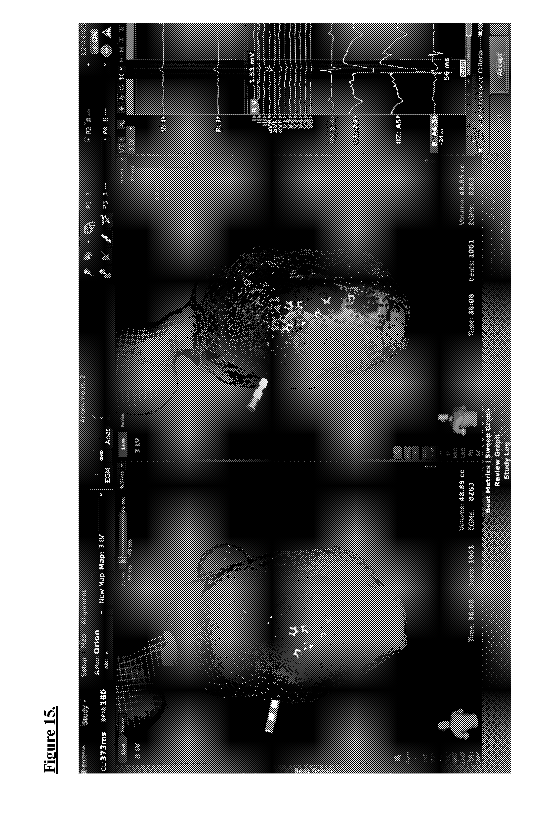
D00016

D00017

D00018

D00019

D00020

D00021

D00022

D00023

D00024

D00025

D00026

D00027

D00028

D00029

D00030

XML
uspto.report is an independent third-party trademark research tool that is not affiliated, endorsed, or sponsored by the United States Patent and Trademark Office (USPTO) or any other governmental organization. The information provided by uspto.report is based on publicly available data at the time of writing and is intended for informational purposes only.
While we strive to provide accurate and up-to-date information, we do not guarantee the accuracy, completeness, reliability, or suitability of the information displayed on this site. The use of this site is at your own risk. Any reliance you place on such information is therefore strictly at your own risk.
All official trademark data, including owner information, should be verified by visiting the official USPTO website at www.uspto.gov. This site is not intended to replace professional legal advice and should not be used as a substitute for consulting with a legal professional who is knowledgeable about trademark law.