Method Of Treating Chronic Lymphatic Leukaemia And/or Systemic Lupus Erythematosus
RENAUDINEAU; Yves ; et al.
U.S. patent application number 16/253041 was filed with the patent office on 2019-05-23 for method of treating chronic lymphatic leukaemia and/or systemic lupus erythematosus. The applicant listed for this patent is Centre Hospitalier Regional et Universitaire de Brest, INSERM, Universite de Bretagne Occidentale - UBO. Invention is credited to Miguel BURGOS, Tinhinane FALI, Olivier MIGNEN, Jacques Olivier PERS, Yves RENAUDINEAU.
| Application Number | 20190154690 16/253041 |
| Document ID | / |
| Family ID | 51398585 |
| Filed Date | 2019-05-23 |


| United States Patent Application | 20190154690 |
| Kind Code | A1 |
| RENAUDINEAU; Yves ; et al. | May 23, 2019 |
METHOD OF TREATING CHRONIC LYMPHATIC LEUKAEMIA AND/OR SYSTEMIC LUPUS ERYTHEMATOSUS
Abstract
A method of treating a patient suffering from chronic lymphatic leukaemia (CLL) and/or systemic lupus erythematosus (SLE) is described. The method includes administering to the patient in need thereof an effective amount of a substance interacting with the fraction of the STIM1 protein localized to the plasma membrane of the cells.
| Inventors: | RENAUDINEAU; Yves; (Brest, FR) ; MIGNEN; Olivier; (Logonna Daoulas, FR) ; BURGOS; Miguel; (Massamagrell (Valencia), ES) ; PERS; Jacques Olivier; (Brest, FR) ; FALI; Tinhinane; (Tizi-Ouzou, DZ) | ||||||||||
| Applicant: |
|
||||||||||
|---|---|---|---|---|---|---|---|---|---|---|---|
| Family ID: | 51398585 | ||||||||||
| Appl. No.: | 16/253041 | ||||||||||
| Filed: | January 21, 2019 |
Related U.S. Patent Documents
| Application Number | Filing Date | Patent Number | ||
|---|---|---|---|---|
| 15424357 | Feb 3, 2017 | |||
| PCT/EP2015/067615 | Jul 31, 2015 | |||
| 16253041 | ||||
| Current U.S. Class: | 1/1 |
| Current CPC Class: | C07K 16/2887 20130101; A61P 37/02 20180101; C07K 2317/34 20130101; G01N 33/57426 20130101; G01N 33/564 20130101; A61P 35/00 20180101; C07K 16/28 20130101; C07K 2317/24 20130101; A61K 2039/507 20130101; A61P 43/00 20180101; G01N 2500/10 20130101; G01N 2800/104 20130101; A61P 35/02 20180101; A61K 39/395 20130101; G01N 2500/04 20130101 |
| International Class: | G01N 33/574 20060101 G01N033/574; G01N 33/564 20060101 G01N033/564; A61K 39/395 20060101 A61K039/395; C07K 16/28 20060101 C07K016/28 |
Foreign Application Data
| Date | Code | Application Number |
|---|---|---|
| Aug 6, 2014 | EP | 14290232.9 |
Claims
1. A method of treating a patient suffering from chronic lymphatic leukaemia (CLL) and/or systemic lupus erythematosus (SLE) comprising administering to the patient in need thereof an effective amount of a substance interacting with the fraction of the STIM1 protein localized to the plasma membrane of the cells.
2. The method according to claim 1, wherein the peptide sequence of the STIM1 protein is the sequence SEQ ID NO: 1.
3. The method of claim 1, wherein the substance is clone Gok/44 anti-STIM1 antibody.
4. The method of claim 1, wherein the clone Gok/44 anti-STIM1 antibody is an antibody directed against a fragment of STIM1 protein of SEQ ID NO: 3.
5. The method of claim 1, wherein the composition further comprises an anti-CD20 antibody.
6. The method of claim 5, wherein the anti-CD20 antibody is selected from the group consisting of ofatumumab, tositumomab, obinutuzumab, ibritumomab, ublituximab, and rituximab.
7. The method of claim 6, wherein the anti-CD20 antibody is rituximab antibody.
8. The method of claim 1, wherein the composition further comprises pharmaceutically acceptable excipients and additives.
9. Use of the isolated fraction of the STIM1 protein localized to the plasma membrane of the cells in a method for screening in vitro candidate molecules for treating chronic lymphatic leukaemia and/or systemic lupus erythematosus, wherein the cells are isolated entire cells.
10. Use according to claim 9, wherein the peptide sequence of the STIM1 protein is the sequence SEQ ID NO: 1.
11. Use according to claim 9, wherein the method of screening uses a technique selected from the group comprising biological screening, and biophysical screening.
12. Use according to claim 10, wherein the method of screening uses a technique selected from the group comprising biological screening, and biophysical screening.
13. Use according to claim 11, wherein screening uses a technique selected from the group comprising immunofluorescence, Western blot, immunoprecipitation, surface plasmon resonance (SPR), flow cytometry, video microscopy, study of calcium flows, enzyme-linked immunosorbent assay (ELISA), and confocal microscopy.
14. A method of identifying, in vitro, substances useful for treating chronic lymphatic leukemia and/or systemic lupus erythematosus, comprising the steps of: (a) providing a sample containing isolated entire cells expressing on their surface a fraction of STIM1 protein localized to the plasma membrane on the cells, (b) screening candidate molecules by interacting the cells with candidate molecules, (c) selecting candidate molecules that binds to the fraction of the STIM1 protein localized to the plasma membrane of the cells without penetrating the cells, thereby identifying substances useful for treating chronic lymphatic leukemia and/or systemic lupus erythematosus.
15. The method according to claim 14, wherein step b) of screening candidate molecules uses a technique selected from the group comprising biological screening and biophysical screening.
16. The method according to claim 14, wherein step b) of screening candidate molecules uses a technique selected from the group comprising immunofluorescence, Western blot, immunoprecipitation, surface plasmon resonance (SPR), flow cytometry, video microscopy, study of calcium flows, enzyme-linked immunosorbent assay (ELISA), and confocal microscopy.
17. The method according to claim 14, wherein the fraction of the STIM1 protein has a sequence of SEQ ID No. 1.
Description
CROSS REFERENCE TO RELATED APPLICATIONS
[0001] This application claims the benefit of earlier filing and priority to U.S. patent application Ser. No. 15/424,357, filed Feb. 3, 2017, which is a U.S. National Stage of PCT International Patent Application No. PCT/EP2015/067615, filed on Jul. 31, 2015 which claims the benefit of and priority to European Application No. 14290232.9 filed on Aug. 6, 2014, the disclosure of all of which is also incorporated herein by reference in its entirety.
TECHNICAL FIELD
[0002] The present invention relates to the use of the membrane fraction of the STIM1 (stromal interaction molecule 1 or GOK) protein in a method of screening, as well as to a substance that interacts with the fraction of the STIM1 protein localized to the plasma membrane for therapeutic use and a pharmaceutical composition comprising at least this substance.
[0003] In the description given hereunder, the references in square brackets ([ ]) refer to the list of references given at the end of the text.
BACKGROUND
[0004] Systemic lupus erythematosus (SLE) and chronic lymphocytic leukaemia (CLL) are still incurable.
[0005] SLE is a heterogeneous disease, of autoimmune origin, characterized by the presence of autoreactive lymphocytes and of antinuclear auto-antibodies (ANA). It is a multisystemic disease, with very varied clinical manifestations. Prevalence varies in different ethnic groups, but is estimated at about 1 in 10000, with a male/female ratio of 10:1. The clinical heterogeneity of this disease reflects its aetiopathogenic complexity, comprising both genetic and environmental factors. SLE may affect all organs. The commonest manifestations are rash, arthritis and fatigue. The most severe manifestations include nephritis, neurological disorders, anemia and thrombocytopenia. More than 90% of patients have ANAs that are considered positive above 1/160th. SLE is a disease with episodic evolution. The aims of the current treatment are: treat the acute episodes that may compromise the vital prognosis, minimize the risks of flare-ups during periods of relative stability and monitor the symptoms which, although not jeopardizing the vital prognosis, affect everyday quality of life.
[0006] Hydroxychloroquine and non-steroidal anti-inflammatories are indicated in the moderate forms of SLE; the corticoids and immunosuppressants are reserved for the most severe forms; the anti-CD20 monoclonal antibody (Rituximab, Mabthera.RTM.) that targets the B lymphocytes (B cells) is currently indicated in patients who are more severely affected and have not responded to the usual treatments ([1]). Despite the improvement in prognosis after the introduction of corticoids and immunosuppressants, SLE continues to have a significant impact on patient morbidity and mortality.
[0007] CLL is a chronic malignant haemopathy that also affects the B cells. These cells play an important role at the immune system level. In the course of CLL, the B cells of CLL are blocked in their life cycle, when they reach maturity, and their production continues. Consequently, these B cells eventually accumulate in the blood, in the ganglia, spleen, liver and bone marrow, which leads to an increase in volume of the secondary lymphatic organs. The treatments currently available against CLL are most often used when the disease is at an advanced stage. The chemotherapeutic products used in the intensive treatment of CLL are chlorambucil used alone, fludarabine used alone, monthly chemotherapy of the CHOP type (combination of four agents: Cyclophosphamide-(H)adryamycin-Oncovin(vincristine)-Prednisone). In terms of targeted therapy, as the leukaemic B cells are CD20+, a monoclonal antibody specifically recognizing this target may be used in the treatment (rituximab, Mabthera.RTM.). Another target is Bruton's tyrosine kinase that is specific for the B cells whose expression is increased in the leukaemic cells. Ibrutinib, being an inhibitor of this enzyme, leads to apoptosis (death) of the leukaemic cells, giving longer remissions, even in the refractory or recurring forms. However, the treatments may give exposure to undesirable effects.
[0008] In the pathology of SLE and CLL, a disturbance of calcium signalling of the B cells in SLE and CLL is described following stimulation of the B-cell antigen receptor (BCR) ([2, 3]).
[0009] In addition to these defects of calcium signalling, the B cells of SLE are characterized by a deficiency of production of interleukin 10 (IL-10), which affects the activity of the regulatory B lymphocytes (Bregs) ([4,5]). This deficiency of activity of the Bregs in SLE leads to less regulation of T lymphocyte (T cell) proliferation, which might again contribute to amplifying the autoimmunity process ([5]).
[0010] The diagnosis and prognosis of CLL and of SLE are based on a compilation of imperfect clinical and biological criteria, hence the need to develop new, more effective criteria.
[0011] In both of these disorders, the B cell represents the main therapeutic target. However, some patients do not respond to the existing treatments.
[0012] There is therefore a real need to offer new therapeutic solutions, which overcome these defects, drawbacks and obstacles of the prior art, and notably involve therapeutic targets that are readily accessible, specific and selective for the affected cells for the disease to be treated.
DESCRIPTION OF THE INVENTION
[0013] The invention makes it possible to respond to these needs by using the fraction localized to the plasma membrane, of the STIM1 protein, a protein involved in the activation and regulation of calcium channels, as therapeutic target of SLE and CLL.
[0014] The applicant has also demonstrated, surprisingly, that any means for direct or indirect control of the activity of the STIM1 protein localized to the plasma membrane may be used for modulating the cellular responses of the B cell, thus supplying a new therapeutic solution in SLE and CLL. The invention thus proposes using, in this context, any tool that modulates (i) expression of the STIM1 protein localized to the plasma membrane, (ii) membrane addressing of this protein, or (iii) the biological activity of this protein at the lymphocyte plasma membrane.
[0015] In the context of the present invention, the following are observed, surprisingly, in the B cells of patients with lupus: [0016] (1) an increase in overall expression of the STIM1 protein and induction of the fraction of STIM1 localized to the plasma membrane, which remains low or even zero in the B cells of controls, [0017] (2) activation of the MAPK pathway (mitogen-activated protein kinases) with phosphorylation of the Erk1/2 kinases (extracellular signal-regulated kinases) in the B cells of SLE possessing the fraction of STIM1 localized to the plasma membrane at rest, in particular in the B cells of SLE at the immature/transitional stage, [0018] (3) an increased constitutive entry of extracellular Ca.sup.2+, and [0019] (4) a correlation between the increase in expression of STIM1, the constitutive entry of extracellular Ca.sup.2+ and the disturbances of activation of the MAPK ERK1/2 pathway that may explain the general activation of the cell, survival of the auto-reactive B cells and therefore the autoimmunity process.
[0020] Regarding the activity of the Bregs of SLE, it is observed surprisingly, for the first time, in the context of the present invention: [0021] (x1) that the deficient regulatory activity of the Bregs is linked to the increase in expression of the STIM1 molecule in the B cells of SLE, [0022] (x2) that the blocking of the STIM1 molecule localized to the plasma membrane, effected specifically by a blocking antibody or non-specifically by an siRNA targeting STIM1 in the B cells of SLE, restores (i) the production of IL-10 by the Bregs of SLE, (ii) inhibition of proliferation of the T cells, and (iii) induction of the regulatory T cells. Blocking of STIM1 localized to the plasma membrane has no effect in healthy controls.
[0023] For the B cells of CLL, the applicant also observed, surprisingly: [0024] (a) that the increase in baseline level of intracytoplasmic calcium was associated with an increase in survival of the B cells of CLL, activation of the MAPK Erk1/2 pathway, nuclear translocation of the transcription factors NFAT2 (Nuclear factor of activated T-cells) and STAT3 (Signal transducer and activator of transcription 3), as well as the synthesis of IL-10 ([5]), [0025] (b) that the increase in the constitutive entry of extracellular Ca.sup.2+ was regulated by the increase in STIM1 protein localized to the plasma membrane. [0026] (c) That the presence of the STIM1 protein at the membrane (group I) or its absence (group II) allowed two groups of patients to be distinguished. [0027] (d) That an anti-STIM1 antibody directed against the STIM1 molecule present at the plasma membrane was capable of greatly reducing the constitutive entry of extracellular Ca.sup.2+ into the B cells of CLL of group I and more weakly into the B cells of CLL of group II, [0028] (e) That the combination of an anti-STIM1 antibody specifically targeting the fraction of the STIM1 protein localized to the plasma membrane with the anti-CD20 antibody (rituximab: RTX), was capable of restoring the apoptotic effect induced by RTX in the B cells of group I patients (presence of the STIM1 protein at the plasma membrane). Addition of the anti-STIM1 antibody has no effect on the apoptotic effect of anti-CD20 of the group II patients (absence of the STIM1 protein at the membrane).
[0029] The invention proposes using the fraction of STIM1 localized to the plasma membrane as a new therapeutic target in SLE and CLL.
[0030] The invention also proposes using modulators of the fraction of STIM1 localized to the plasma membrane in these disorders.
[0031] The invention relates to the use of the fraction of STIM1 localized to the plasma membrane as a therapeutic target in SLE and CLL by modulating its presence or its activity. Thus, the invention relates to, among other things, (i) inhibition of expression of the STIM1 molecule at the plasma membrane of the cells, (ii) inhibition of membrane addressing of this protein, and (iii) inhibition of the biological activity of the STIM1 protein present at the plasma membrane.
[0032] It is proposed, notably in CLL patients resistant to treatment with RTX, to block the activity of STIM1 localized to the plasma membrane by an anti-STIM1 antibody specifically targeting the fraction of the STIM1 protein localized to the plasma membrane. In fact, it is proposed that modulation of the inflows of Ca.sup.2+ depending on the fraction of STIM1 localized to the plasma membrane by the modulators of STIM1 such as an anti-STIM1 Ac would sensitize the cells to apoptosis induced by RTX.
[0033] The invention is advantageous on several points, notably the marker is only present on the affected cells, and not on the healthy cells, which allows a gain in specificity and in selectivity. Moreover, expression by the immune cells of the STIM1 protein at the level of the plasma membrane facilitates the accessibility of this target.
[0034] Thus, a first object of the invention relates to the use of the fraction of the STIM1 protein localized to the plasma membrane of the cells in a method for screening candidate molecules for treating SLE and/or CLL.
[0035] "Fraction of the STIM1 protein localized to the plasma membrane of the cells" means, in the sense of the present invention, the glycosylated fraction of the STIM1 protein localized to the plasma membrane of the cells. The STIM1 protein possesses two glycosylation sites, an asparagine in position 131 and another in position 171. Glycosylation of the STIM1 molecule is a necessary and obligatory process for addressing the STIM1 molecule at the surface of the cell ([7]). This fraction has a molecular weight of about 90.+-.2 kDa, which makes it possible to distinguish it from the non-glycosylated form of STIM1 (84.+-.2 kDa). The two forms are detectable by Western blotting. The human STIM1 molecule (Stromal Interacting Molecule; also called GOK) is a protein with sequence ID NO: 1 corresponding to the Uniprot sequence: Q13586 or NCBI: NP_003147.2. This protein is encoded by the sequence ID NO: 2, corresponding to the NCBI sequence: NM_003156.3 (mRNA transcript). Preferably, the fraction is located on the plasma membrane of intact cells, which means that the plasma membrane is non broken and/or non permeabilized, and advantageously does not allow non-permeant molecules to penetrate the cells.
[0036] "Fraction of the STIM1 protein localized to the plasma membrane" means any biological product resulting from isolation of the STIM1 protein localized to the plasma membrane of the cells. Isolation may be performed by all the means known by a person skilled in the art, for example by using a detergent (for example a non-ionic or ionic surfactant such as Triton X-100 or Triton NI 01; or polyoxyethylene sorbitan esters), after differential centrifugation, or by an immuno-chemical or protein-chemical technique using a step of targeting the membrane proteins (antibody, Thermo scientific sulfo-NHS-SS-biotin), this list not being limiting.
[0037] "Cells" means, in the sense of the present invention, any cell expressing STIM1 at the level of the plasma membrane. Advantageously, the cells are immune cells. They may be, for example, B cells and T cells. Advantageously, the cells are B cells from patients with SLE or CLL. Alternatively, the cells may be transfected with the sequence ID NO: 2 in order to express the STIM1 protein on their plasma membrane. Preferably, the cells are entire cells, in other word intact and/or non broken cells. Such cells are thus not permeabilized. Advantageously, the cells used in the screening method of the invention are intact in order to strictly screen for molecules that modulate the membrane expression of STIM1 and/or that modulate constitutive entry of extracellular Ca.sup.2+. Advantageously, the cells used in the screening method of the invention are intact in order to select molecules that do not penetrate into the cells and that stay at the plasma membrane, due to their specific interaction with the fraction of the STIM1 protein localized to the plasma membrane. In other words, the method of screening allows selecting non permeant molecules, i.e. molecules that do not cross the plasma membrane. The cells are isolated cells, and may be provided for the method of screening of the invention in the form of a sample.
[0038] "Method of screening" means, in the sense of the present invention, any method allowing identification of a substance interacting with the membrane fraction of the STIM1 protein or modulating its membrane expression. It may be any method known by a person skilled in the art, for example biological screening, for example a technique selected from the group comprising immunofluorescence, Western blot, immunoprecipitation, surface plasmon resonance (SPR), flow cytometry, video microscopy, study of calcium flows, enzyme-linked immunosorbent assay (ELISA), and confocal microscopy, or biophysical screening, for example by measuring the variations in intracellular calcium concentration by fluorescence. Advantageously, the method of screening allows identifying substances that interact selectively with the fraction of the STIM1 protein localized to the plasma membrane of the cells, without penetrating the cell. In other words, the method of screening allows identifying non-permeating substances that interact selectively with the fraction of the STIM1 protein localized to the plasma membrane of the cells. The method of screening may be realized in vitro, on a sample containing intact cells expressing on their plasma membrane the STIM1 protein.
[0039] "Candidate molecule" means, in the sense of the present invention, any molecule that interacts with the fraction of the STIM1 protein localized to the plasma membrane of the cells. The interaction may be of the type of fixation of the candidate molecule on the STIM1 protein localized to the plasma membrane of the cells. Alternatively, the interaction may be a modulation of the activity or expression of this protein. Modulation of the activity of the fraction of the STIM1 protein localized to the plasma membrane may be due to a modification of the insertion of STIM1 in the plasma membrane, or to a modification of its interaction with the proteins that are associated with it. Modulation of the activity of the protein may be reflected in a change of the calcium flows such as a change in the constitutive entry of intracellular calcium or a change in calcium influxes activated during stimulation of a receptor such as the calcium influxes dependent on the release of reserves (SOCE, store operated calcium influx). The modulation of expression may be an increase or a decrease in expression of the STIM1 protein localized to the plasma membrane relative to a level measured on the same cell or a comparable cell before application of the candidate molecule. The modulation of expression of the STIM1 protein may for example be linked to transcriptional modifications, epigenetic modifications or a modulation of the glycosylation process that is indispensable for membrane addressing of the STIM1 protein. Advantageously, the selected candidate molecules interact specifically with the fraction of the STIM1 protein localized to the plasma membrane of the cells. As the selected candidate molecules do not cross the plasma membrane, they do not interact with the STIM1 protein localized to the endoplasmic reticulum in the method of screening of the invention.
[0040] An object of the invention is so the use of isolated intact cells expressing on their plasma membrane the STIM1 protein, in a method for in vitro screening candidate molecules useful for treating chronic lymphatic leukemia and/or systemic lupus erythematosus.
[0041] Another object of the invention relates to a method of identifying, in vitro, substances useful for treating chronic lymphatic leukemia and/or systemic lupus erythematosus, comprising the steps of: [0042] (a) providing a sample containing isolated entire cells expressing on their surface the fraction of the STIM1 protein localized to the plasma membrane on the cells, [0043] (b) screening candidate molecules by interacting the cells with candidate molecules, [0044] (c) selecting candidate molecules that binds the fraction of the STIM1 protein localized to the plasma membrane of the cells without penetrating the cells,
[0045] thereby identifying substances useful for treating chronic lymphatic leukemia and/or systemic lupus erythematosus.
[0046] A second object of the invention relates to a substance that interacts with the fraction of the STIM1 protein localized to the plasma membrane of the cells, for use as a medicinal product in the treatment of SLE and/or CLL.
[0047] "Substance" means, in the sense of the present invention, any molecule displaying interaction of the type of fixation to the STIM1 protein localized to the plasma membrane or modulation of the activity or expression of this membrane fraction of the STIM1 protein, as defined above. The substance may be of natural or synthetic origin. It may be a protein produced chemically or by any method of bioengineering, such as purification. The substance may notably be identified by applying the method of screening as defined above. Advantageously, the substance is a not able to cross the plasma membrane and interacts specifically with the fraction of the STIM1 protein localized to the plasma membrane of the cells without penetrating the cells. Advantageously, the substance may decrease or block the activity of the STIM1 protein localized to the plasma membrane of the cells.
[0048] The substance may be for example an antibody directed against an extracellular fragment of the STIM1 protein localized to the plasma membrane of sequence SEQ ID NO: 3. This sequence corresponds to amino acids 23-213 of STIM1. It may be the anti-GOK/STIM1 antibody (Clone: 44, BD Biosciences reference 910954).
[0049] The invention further relates to a pharmaceutical composition comprising at least one substance as defined above. Such a composition may comprise any suitable pharmaceutically acceptable vehicle, comprising for example excipients and additives that facilitate formulation of the substance in preparations that may be used pharmaceutically. The expression "pharmaceutically acceptable" encompasses any vehicle that does not interfere negatively with the efficacy of the substance for treating SLE or CLL, and that is not toxic to the host to whom or to which it is administered. In particular, suitable pharmaceutically acceptable vehicles for a composition according to the invention are vehicles that are suitable in particular for systemic application. Suitable pharmaceutically acceptable vehicles are well known in the prior art and are described for example in Remington Pharmaceutical Sciences (Mack Publishing Company, Easton, USA, 1985), a standard reference text in this field. It may be for example one or more components selected from sodium citrate, polysorbate 80, sodium chloride, sodium hydroxide, hydrochloric acid, and water for injection.
[0050] Advantageously, the composition according to the invention may find application as a medicinal product. Particularly advantageously, the composition of the invention may find application as a medicinal product in the treatment of SLE or of cancer such as CLL.
[0051] The pharmaceutical composition of the invention may comprise any active principle that potentiates the effect of the substance as defined above.
[0052] Moreover, it may be an anti-CD20 antibody or any other molecule associated with the protein complex regulating the calcium channels associated with the STIM1 protein such as the proteins Orai and TRPC. In this case, it may be any anti-CD20 known in human or animal therapy, for example the IDEC-C2B8 antibody (Rituximab, distributed by Hoffman-La Roche in Europe. Drugbank DB00073 (BIOD00014, BTD00014), ofatumumab (Arzera, GlaxoSmithKline), tositumomab (GSK, DB00081, BIOD00085, BTD00085), obinutuzumab (Gazyva, Roche, DB08935, GA101), ibritumomab (Tiuxetan, IDEC Pharmaceuticals, DB00078, BIOD00069, BTD00069), ublituximab (LFB) or AME-133v (Lilly, LY2469298), this list not being limiting.
[0053] Set forth below are various aspects of the present invention.
[0054] Aspect 1: A method of treating a patient suffering from chronic lymphatic leukaemia (CLL) and/or systemic lupus erythematosus (SLE) comprising administering to the patient in need thereof an effective amount of a substance interacting with the fraction of the STIM1 protein localized to the plasma membrane of the cells.
[0055] Aspect 2: The method according to Aspect 1, wherein the peptide sequence of the STIM1 protein is the sequence SEQ ID NO: 1.
[0056] Aspect 3: The method of Aspect 1, wherein the substance is clone Gok/44 anti-STIM1 antibody.
[0057] Aspect 4: The method of Aspect 1, wherein the clone Gok/44 anti-STIM1 antibody is an antibody directed against a fragment of STIM1 protein of SEQ ID NO: 3.
[0058] Aspect 5: The method of Aspect 1, wherein the composition further comprises an anti-CD20 antibody.
[0059] Aspect 6: The method of Aspect 5, wherein the anti-CD20 antibody is selected from the group consisting of ofatumumab, tositumomab, obinutuzumab, ibritumomab, ublituximab, and rituximab.
[0060] Aspect 7: The method of Aspect 6, wherein the anti-CD20 antibody is rituximab antibody.
[0061] Aspect 8: The method of Aspect 1, wherein the composition further comprises pharmaceutically acceptable excipients and additives.
[0062] Aspect 9: Use of the isolated fraction of the STIM1 protein localized to the plasma membrane of the cells in a method for screening in vitro candidate molecules for treating chronic lymphatic leukaemia and/or systemic lupus erythematosus, wherein the cells are isolated entire cells.
[0063] Aspect 10: Use according to Aspect 9, wherein the peptide sequence of the STIM1 protein is the sequence SEQ ID NO: 1.
[0064] Aspect 11: Use according to Aspect 9, wherein the method of screening uses a technique selected from the group comprising biological screening, and biophysical screening.
[0065] Aspect 12: Use according to Aspect 10, wherein the method of screening uses a technique selected from the group comprising biological screening, and biophysical screening.
[0066] Aspect 13: Use according to Aspect 11, wherein screening uses a technique selected from the group comprising immunofluorescence, Western blot, immunoprecipitation, surface plasmon resonance (SPR), flow cytometry, video microscopy, study of calcium flows, enzyme-linked immunosorbent assay (ELISA), and confocal microscopy.
[0067] Aspect 14: A method of identifying, in vitro, substances useful for treating chronic lymphatic leukemia and/or systemic lupus erythematosus, comprising the steps of: [0068] (a) providing a sample containing isolated entire cells expressing on their surface a fraction of STIM1 protein localized to the plasma membrane on the cells, [0069] (b) screening candidate molecules by interacting the cells with candidate molecules, [0070] (c) selecting candidate molecules that binds to the fraction of the STIM1 protein localized to the plasma membrane of the cells without penetrating the cells,
[0071] thereby identifying substances useful for treating chronic lymphatic leukemia and/or systemic lupus erythematosus.
[0072] Aspect 15: The method according to Aspect 14, wherein step b) of screening candidate molecules uses a technique selected from the group comprising biological screening and biophysical screening.
[0073] Aspect 16: The method according to Aspect 14, wherein step b) of screening candidate molecules uses a technique selected from the group comprising immunofluorescence, Western blot, immunoprecipitation, surface plasmon resonance (SPR), flow cytometry, video microscopy, study of calcium flows, enzyme-linked immunosorbent assay (ELISA), and confocal microscopy.
[0074] Aspect 17: The method according to Aspect 14, wherein the fraction of the STIM1 protein has a sequence of SEQ ID No. 1.
[0075] Other advantages may also become apparent to a person skilled in the art on reading the examples given below, illustrated by the appended figures, given for purposes of illustration.
BRIEF DESCRIPTION OF THE FIGURES
[0076] FIG. 1 shows demonstration of membrane STIM1 in the B lymphocytes (B cells) of systemic lupus erythematosus (SLE) and the B cells of chronic lymphocytic leukaemia (CLL) by Western blot (A/B) and flow cytometry (C/D). Fig. A shows demonstration of a band at 90 kDa for the glycosylated fraction of STIM1 by Western blot in the B cells of SLE, versus the control B cells of healthy controls. This glycosylation of the STIM1 protein is indispensable for its insertion in the plasma membrane. Fig. B shows the demonstration by Western blot of a band at 90 kDa for the fraction of STIM1 localized to the membrane in the B cells of CLL expressing membrane STIM1 (mSTIM1+) versus the control CLL B cells not expressing membrane STIM1 (mSTIM1-). Fig. C shows demonstration by flow cytometry for the fraction of STIM1 localized to the membrane in the B cells of SLE expressing membrane STIM1 (mSTIM1+) versus the control B cells of healthy controls not expressing membrane STIM1 (mSTIM1-). Fig. D shows demonstration by flow cytometry for the fraction of STIM1 localized to the membrane in the B cells of CLL expressing membrane STIM1 (mSTIM1+) versus the control CLL B cells not expressing membrane STIM1 (mSTIM1-).
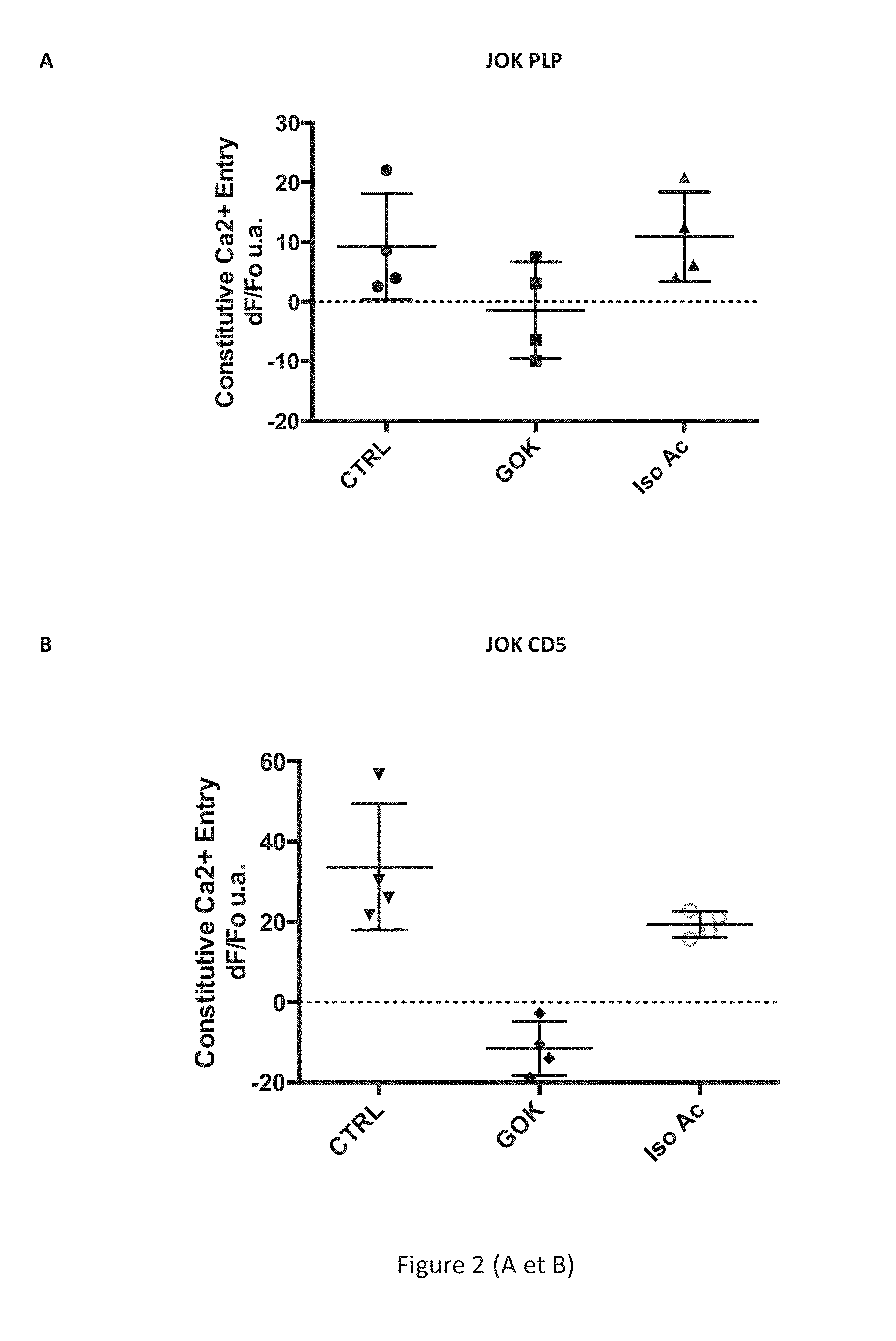
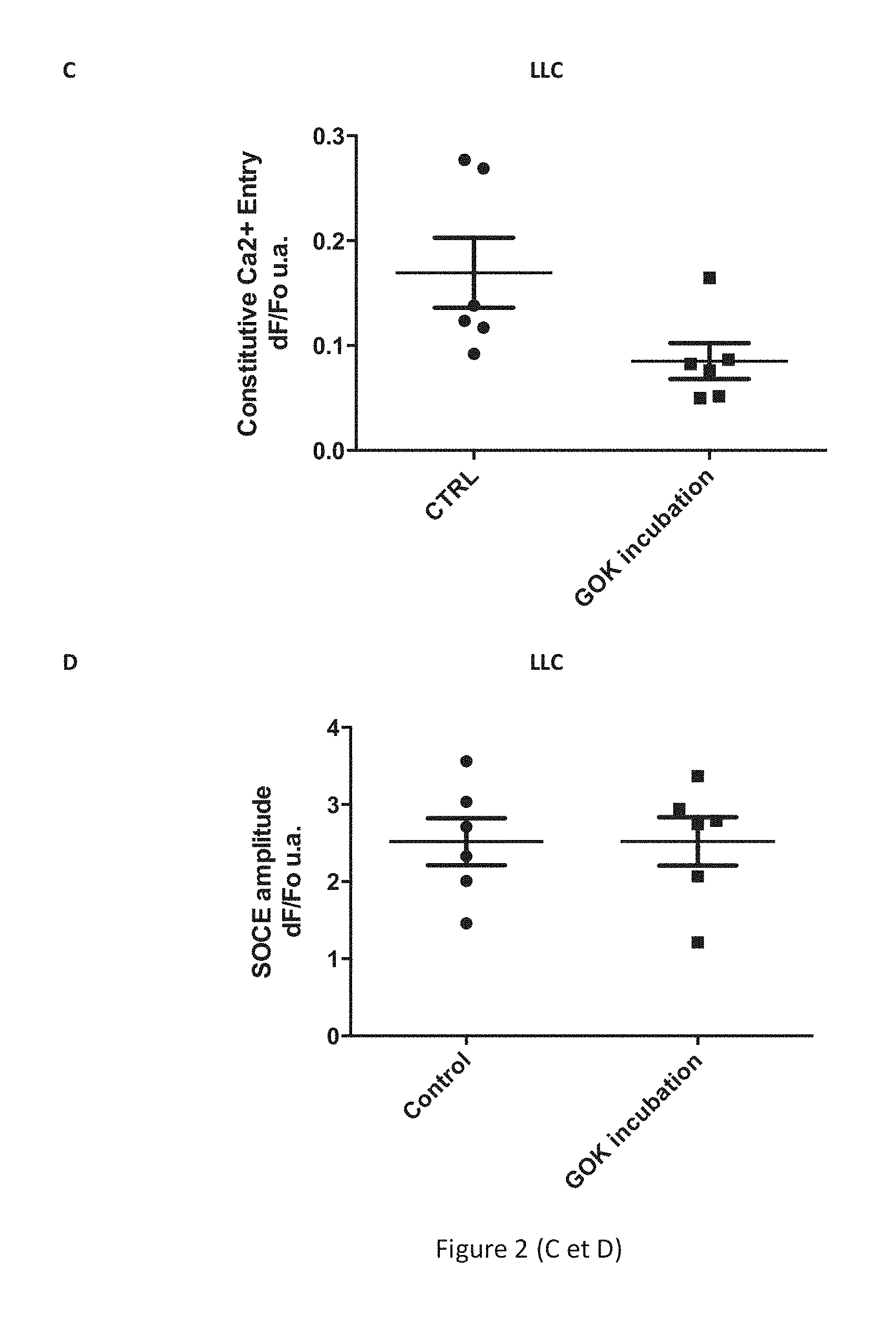
[0077] FIG. 2 shows demonstration of the inhibition of the constitutive calcium influx by an anti-STIM1 antibody (clone Gok/44, BD Biosciences) directed against an extracellular epitope of the STIM1 protein localized to the plasma membrane B cells of the human line JOK PLP ([6]), of the line JOK CD5 ([6]), and B lymphocytes of chronic lymphocytic leukaemia (CLL). Figs. A and B show measurement of the constitutive influx and of the effects of the anti-STIM1 antibody on this constitutive influx (expressed in dF/Fo a.u., arbitrary units) measured in the B lymphocytes of the human lines JOK PLP (A) and JOK CD5 (B) in a multiwell plate using a plate reader for pretreated cells (5 .mu.g/ml of antibody for 60 min) with the control antibody (CTRL, IgG2a isotype, Beckman Coulter) or with the anti-STIM1/GOK antibody. Fig. C shows measurement of the constitutive influx and of the effects of the anti-STIM1 antibody on this influx (as the ratio dF/Fo a.u.) on the B cells of CLL in single cell imaging for pretreated cells (5 .mu.g/ml of antibody for 60 min) with the control antibody (CTRL, IgG2a isotype) or with the anti-STIM1/GOK antibody. Fig. D shows absence of an effect of the anti-STIM1 antibody on the calcium influx dependent on the release of reserves SOCE (store operated calcium influx) induced by thapsigargin (1 .mu.M, Sigma-Aldrich) and measured in B cells of CLL.
[0078] FIG. 3 shows demonstration of the effects of the anti-STIM1 antibody (clone Gok/44) (A) on cellular viability alone or (B) in synergy with the anti-CD20 antibody (rituximab), (C) on constitutive calcium entry (Ca2+) in the B cells of chronic lymphocytic leukaemia (CLL) in patients classified in two groups depending on expression (mSTIM1+) or not (mSTIM1-) of the STIM1 protein at the plasma membrane and (D) on inhibition of the proliferation of T lymphocytes (Breg activity) by the B cells of systemic lupus erythematosus (SLE). Fig. A shows the percentage of live cells after 48 h of culture for the B cells of CLL in the two groups mSTIM1+ or mSTIM1- in the presence of 10 .mu.g/ml of an isotypic control antibody (iso Ab, IgG2a isotype, Beckman Coulter) or in the presence of 10 .mu.g/ml of the anti-STIM1/GOK antibody. Fig. B shows the percentage of cells of CLL alive after 48 h of culture for the B cells of CLL in the two groups mSTIM1+ or mSTIM1- in the presence of 10 .mu.g/ml of an isotypic control antibody (iso Ab, IgG2a isotype, Beckman Coulter), in the presence of 10 .mu.g/ml of rituximab (anti-CD20), or the combination rituximab (10 .mu.g/ml) and anti-STIM1/GOK (10 .mu.g/ml). Fig. C shows the reduction of constitutive entry of Ca.sup.2+ (expressed as the ratio dF/Fo a.u., arbitrary units) in the B cells of CLL of the group that expresses STIM1 at the plasma membrane (mSTIM1+) after pretreatment or not (control without addition) of the cells with 5 .mu.g/ml of anti-STIM1/GOK antibody. Fig. D shows inhibition of proliferation of the cells expressed in percentage by the B cells of SLE in a model of autologous co-culture 1:1 after 4 days in the presence of an anti-STIM1/GOK antibody or of a control without addition.
EXAMPLES
Example 1: Method for Detecting Membrane STIM1
[0079] The B lymphocytes were purified starting from peripheral blood mononuclear cells (PBMC) obtained on a Ficoll gradient after removing the T lymphocytes (rosette technique using sheep red blood cells pretreated with neuraminidase) and monocytes (negative depletion technique, B cell kit without CD43, Stem Cell Technologies). The purity of the CD19-positive B cells was verified by flow cytometry, showing purity above 95%.
[0080] A/B-- Protein analysis of the B cells by Western blot on SDS-PAGE made it possible to distinguish, in addition to the reticular fraction of STIM1 (84.+-.2 kDa), the glycosylated membrane form of STIM1 (90.+-.2 kDa) for the B cells of SLE (A) and for some CLL patients (B, mSTIM1+ group). This protein analysis used an anti-STIM1 clone Gok/44 antibody (BD Biosciences) first, then a peroxidase-linked mouse anti-IgG antibody (GE Healthcare), and finally detection by chemiluminescence (kit ECL advance, GE Healthcare).
[0081] C/D-- Analysis by flow cytometry consisted of incubating the purified B cells with an anti-STIM1 clone Gok/44 antibody (BD Biosciences) for 15 min at 4.degree. C., then, after washing, fixation of the anti-STIM1 antibody was revealed using a fluorescein-linked F(ab')2 mouse anti-IgG antibody (Jackson Laboratories). The membrane labelling of STIM1 in the live cells is determined relative to the isotypic control (IgG2a, Beckman Coulter).
Example 2: Method of Screening Anti-Membrane STIM1 Molecules
[0082] Screening of the molecules modulating the STIM1 fraction localized to the plasma membrane is carried out to a first approximation on human B cell lines JOK that express the STIM1 protein at the plasma membrane. Two types of cells are used: JOK cells stably transfected with an empty vector (JOK PLP) or stably transfected with the CD5 protein ([6]). These cells display a measurable constitutive calcium entry. Screening consists of measuring the effects of the molecules targeting the fraction of STIM1 localized to the plasma membrane of the cells on the constitutive entry of extracellular calcium. The effects of these molecules on calcium entry dependent on the release of reserves SOCE (Store Operated Calcium Entry) are also evaluated in order to determine the effect of the molecules on the influx SOCE of the molecules acting on constitutive calcium entry. The amplitude of these two calcium flows is measured by monitoring the variations in intracellular calcium concentration using a fluorescent probe (Calcium 6, Molecular Devices). The cells are made to adhere in 96-well plates treated with CellTak (BD Biosciences) at a rate of 100 000 cells per well for 45 minutes. The cells are then loaded with the fluorescent probe (Calcium 6, Molecular Devices) by incubation in the presence of this probe for 60 min before measuring the variations in intracellular calcium concentration using a multiwell plate reader of the Flexstation type (Molecular Devices). The cells are put in contact with the test compound at the moment of loading the cells with the fluorescent probe and throughout measurement of the variations in intracellular calcium concentration.
[0083] The measurement of constitutive calcium entry is estimated by removing and then adding the calcium of the extracellular matrix in the absence of any stimulation of the cells. The influx SOCE is activated by treating the cells with thapsigargin, a SERCA pump inhibitor.
[0084] The molecules identified as having an effect on the calcium flows of interest of the JOK cells are then tested on purified B cells obtained from peripheral blood mononuclear cells (PBMC) of control individuals or of CLL patients whose expression level of the STIM1 molecule at the surface of the cells is known and measured by flow cytometry. The effects of the test molecules on the constitutive calcium influx and the influx SOCE are measured as described above by single cell fluorescence imaging. The cells are made to adhere to glass slips coated with CellTAK (BD Biosciences) at a rate of 500 000 cells per slip for 45 min and loaded with the fluorescent probe (Fura2, Molecular probes) for 45 min in the presence of pluronic acid (Sigma Aldrich) before measuring the variations in intracellular calcium concentration using a single cell fluorescence imaging system. The cells are put in contact with the test compound throughout loading of the cells and throughout measurement of the variations in intracellular calcium concentration. The measurement of constitutive calcium entry is estimated by removing and then adding the calcium of the extracellular matrix in the absence of any stimulation of the cells. The influx SOCE is activated by treating the cells with thapsigargin, a SERCA pump inhibitor.
[0085] Inhibition of the constitutive calcium influx by an anti-STIM1 antibody (clone Gok/44, BD Biosciences) directed against an extracellular epitope of the STIM1 protein localized to the plasma membrane of the cells is thus demonstrated. The constitutive influx and the effects of the anti-STIM1 antibody on this influx are measured on the JOK lines in a multiwell plate and a plate reader (FIGS. 2A/B) or on B lymphocytes in single cell imaging (FIGS. 2C/D).
Example 3: Methods for Demonstrating Biological and Anti-Lymphocyte B Activity of the Anti-Membrane STIM1 Molecules (FIG. 3)
[0086] FIG. 3A-- The anti-STIM1 antibody (clone Gok/44, BD Biosciences) used at 10 .mu.g/ml is capable of reducing the survival of the B cells of CLL, which was increased for the CLL of the mSTIM1+ group (presence of the STIM1 protein at the plasma membrane). For this experiment, the cells were cultured for 48 h, then the percentage of live cells (absence of annexin V/propidium iodide labelling, Beckman Coulter) was determined.
[0087] FIG. 3B-- The anti-STIM1 antibody (clone Gok/44) potentiates the action of the anti-CD20 antibody (rituximab, 10 .mu.g/ml) on death of the B cells of CLL of the mSTIM1+ group.
[0088] FIG. 3C-- The effect of the anti-STIM1 antibody (clone Gok/44) involves an effect of the antibody on the constitutive entry of Ca.sup.2+ in the B cells of CLL mSTIM1+.
[0089] FIG. 3D-- The anti-STIM1 antibody (clone Gok/44) restores the capacity of the B cells of SLE for inhibiting proliferation of the cells after 4 days of autologous culture in the presence of stimulation by CpG and anti-CD3/CD28.
LIST OF REFERENCES
[0090] [1] Seret G, Hanrotel C, Bendaoud B, Le Meur Y and Renaudineau Y. Homozygous FCGR3A-158F mutation is associated with delayed B-cell depletion following rituximab but with preserved efficacy in a patient with refractory lupus nephritis. Clin Kidney J (2012) doi: 10.1093/ckj/sfs162. [0091] [2] Nedellec S, Renaudineau Y, Bordron A, Berthou C, Porakishvili N, Lydyard P M, Pers J O, Youinou P. B cell response to surface IgM cross-linking identifies different prognostic groups of B-chronic lymphocytic leukemia patients. J Immunol. (2005)174:3749-56. [0092] [3] Liossis S N, Kovacs B, Dennis G, Kammer G M, Tsokos G C. B cells from patients with systemic lupus erythematosus display abnormal antigen receptor-mediated early signal transduction events. J Clin Invest. (1996) 98:2549-57. [0093] [4] Blair P A, Norena LY, Flores-Borja F, Rawlings D J, Isenberg D A, Ehrenstein M R, Mauri C. CD19(+)CD24(hi)CD38(hi) B cells exhibit regulatory capacity in healthy individuals but are functionally impaired in systemic Lupus Erythematosus patients. Immunity. (2010) 32:129-40. [0094] [5] Lemoine S, Morva A, Youinou P, Jamin C. Human T cells induce their own regulation through activation of B cells. J Autoimmun. (2011) 36:228-38. [0095] [6] Garaud S, Morva A, Lemoine S, Hillion S, Bordron A, Pers J O, Berthou C, Mageed R A, Renaudineau Y, Youinou P. CD5 promotes IL-10 production in chronic lymphocytic leukemia B cells through STAT3 and NFAT2 activation. J Immunol. (2011) 186:4835-44. [0096] [7] Mignen O, Thompson J L, Shuttleworth T J. STIM1 regulates Ca2+ entry via arachidonate-regulated Ca2+-selective (ARC) channels without store depletion or translocation to the plasma membrane. J Physiol. (2007) 579:703-15.
* * * * *
D00001
D00002
D00003
D00004
XML
uspto.report is an independent third-party trademark research tool that is not affiliated, endorsed, or sponsored by the United States Patent and Trademark Office (USPTO) or any other governmental organization. The information provided by uspto.report is based on publicly available data at the time of writing and is intended for informational purposes only.
While we strive to provide accurate and up-to-date information, we do not guarantee the accuracy, completeness, reliability, or suitability of the information displayed on this site. The use of this site is at your own risk. Any reliance you place on such information is therefore strictly at your own risk.
All official trademark data, including owner information, should be verified by visiting the official USPTO website at www.uspto.gov. This site is not intended to replace professional legal advice and should not be used as a substitute for consulting with a legal professional who is knowledgeable about trademark law.